EP1587540B1 - IDENTIFICATION AND ENGINEERING OF ANTIBODIES WITH VARIANT Fc REGIONS AND METHODS OF USING SAME - Google Patents
IDENTIFICATION AND ENGINEERING OF ANTIBODIES WITH VARIANT Fc REGIONS AND METHODS OF USING SAME Download PDFInfo
- Publication number
- EP1587540B1 EP1587540B1 EP04701279.4A EP04701279A EP1587540B1 EP 1587540 B1 EP1587540 B1 EP 1587540B1 EP 04701279 A EP04701279 A EP 04701279A EP 1587540 B1 EP1587540 B1 EP 1587540B1
- Authority
- EP
- European Patent Office
- Prior art keywords
- region
- antibody
- variant
- substitution
- antibodies
- Prior art date
- Legal status (The legal status is an assumption and is not a legal conclusion. Google has not performed a legal analysis and makes no representation as to the accuracy of the status listed.)
- Expired - Lifetime
Links
- 238000000034 method Methods 0.000 title description 65
- 238000006467 substitution reaction Methods 0.000 claims description 315
- 230000004048 modification Effects 0.000 claims description 230
- 238000012986 modification Methods 0.000 claims description 230
- 102100029193 Low affinity immunoglobulin gamma Fc region receptor III-A Human genes 0.000 claims description 197
- 150000001413 amino acids Chemical class 0.000 claims description 197
- 101710099301 Low affinity immunoglobulin gamma Fc region receptor III-A Proteins 0.000 claims description 192
- ROHFNLRQFUQHCH-UHFFFAOYSA-N Leucine Natural products CC(C)CC(N)C(O)=O ROHFNLRQFUQHCH-UHFFFAOYSA-N 0.000 claims description 166
- ROHFNLRQFUQHCH-YFKPBYRVSA-N L-leucine Chemical compound CC(C)C[C@H](N)C(O)=O ROHFNLRQFUQHCH-YFKPBYRVSA-N 0.000 claims description 161
- 230000027455 binding Effects 0.000 claims description 94
- 206010028980 Neoplasm Diseases 0.000 claims description 77
- 102100029205 Low affinity immunoglobulin gamma Fc region receptor II-b Human genes 0.000 claims description 75
- 108010021472 Fc gamma receptor IIB Proteins 0.000 claims description 73
- 201000011510 cancer Diseases 0.000 claims description 62
- 230000010056 antibody-dependent cellular cytotoxicity Effects 0.000 claims description 41
- 238000011282 treatment Methods 0.000 claims description 38
- 239000003814 drug Substances 0.000 claims description 37
- 239000000427 antigen Substances 0.000 claims description 32
- 108091007433 antigens Proteins 0.000 claims description 31
- 102000036639 antigens Human genes 0.000 claims description 31
- 102000005962 receptors Human genes 0.000 claims description 23
- 108020003175 receptors Proteins 0.000 claims description 23
- 229940124597 therapeutic agent Drugs 0.000 claims description 21
- -1 C14 Proteins 0.000 claims description 15
- 239000002246 antineoplastic agent Substances 0.000 claims description 11
- 229940127089 cytotoxic agent Drugs 0.000 claims description 10
- 238000011275 oncology therapy Methods 0.000 claims description 10
- 101150029707 ERBB2 gene Proteins 0.000 claims description 8
- 238000002512 chemotherapy Methods 0.000 claims description 8
- 108060008682 Tumor Necrosis Factor Proteins 0.000 claims description 6
- 102000000852 Tumor Necrosis Factor-alpha Human genes 0.000 claims description 6
- 239000008194 pharmaceutical composition Substances 0.000 claims description 6
- 238000001356 surgical procedure Methods 0.000 claims description 6
- 238000009169 immunotherapy Methods 0.000 claims description 5
- 101000913074 Homo sapiens High affinity immunoglobulin gamma Fc receptor I Proteins 0.000 claims description 4
- 230000000295 complement effect Effects 0.000 claims description 4
- 238000001794 hormone therapy Methods 0.000 claims description 4
- 230000005855 radiation Effects 0.000 claims description 4
- 238000001959 radiotherapy Methods 0.000 claims description 4
- MZOFCQQQCNRIBI-VMXHOPILSA-N (3s)-4-[[(2s)-1-[[(2s)-1-[[(1s)-1-carboxy-2-hydroxyethyl]amino]-4-methyl-1-oxopentan-2-yl]amino]-5-(diaminomethylideneamino)-1-oxopentan-2-yl]amino]-3-[[2-[[(2s)-2,6-diaminohexanoyl]amino]acetyl]amino]-4-oxobutanoic acid Chemical compound OC[C@@H](C(O)=O)NC(=O)[C@H](CC(C)C)NC(=O)[C@H](CCCN=C(N)N)NC(=O)[C@H](CC(O)=O)NC(=O)CNC(=O)[C@@H](N)CCCCN MZOFCQQQCNRIBI-VMXHOPILSA-N 0.000 claims description 3
- 206010006187 Breast cancer Diseases 0.000 claims description 3
- 208000026310 Breast neoplasm Diseases 0.000 claims description 3
- 102100026122 High affinity immunoglobulin gamma Fc receptor I Human genes 0.000 claims description 3
- 206010058467 Lung neoplasm malignant Diseases 0.000 claims description 3
- 210000001744 T-lymphocyte Anatomy 0.000 claims description 3
- 230000003211 malignant effect Effects 0.000 claims description 3
- NFGXHKASABOEEW-UHFFFAOYSA-N 1-methylethyl 11-methoxy-3,7,11-trimethyl-2,4-dodecadienoate Chemical compound COC(C)(C)CCCC(C)CC=CC(C)=CC(=O)OC(C)C NFGXHKASABOEEW-UHFFFAOYSA-N 0.000 claims description 2
- 206010060862 Prostate cancer Diseases 0.000 claims description 2
- 208000000236 Prostatic Neoplasms Diseases 0.000 claims description 2
- 239000002253 acid Substances 0.000 claims description 2
- 239000003937 drug carrier Substances 0.000 claims description 2
- UIWYJDYFSGRHKR-UHFFFAOYSA-N gadolinium atom Chemical compound [Gd] UIWYJDYFSGRHKR-UHFFFAOYSA-N 0.000 claims description 2
- 210000004698 lymphocyte Anatomy 0.000 claims description 2
- 102100025475 Carcinoembryonic antigen-related cell adhesion molecule 5 Human genes 0.000 claims 2
- LAQPKDLYOBZWBT-NYLDSJSYSA-N (2s,4s,5r,6r)-5-acetamido-2-{[(2s,3r,4s,5s,6r)-2-{[(2r,3r,4r,5r)-5-acetamido-1,2-dihydroxy-6-oxo-4-{[(2s,3s,4r,5s,6s)-3,4,5-trihydroxy-6-methyloxan-2-yl]oxy}hexan-3-yl]oxy}-3,5-dihydroxy-6-(hydroxymethyl)oxan-4-yl]oxy}-4-hydroxy-6-[(1r,2r)-1,2,3-trihydrox Chemical compound O[C@H]1[C@H](O)[C@H](O)[C@H](C)O[C@H]1O[C@H]([C@@H](NC(C)=O)C=O)[C@@H]([C@H](O)CO)O[C@H]1[C@H](O)[C@@H](O[C@]2(O[C@H]([C@H](NC(C)=O)[C@@H](O)C2)[C@H](O)[C@H](O)CO)C(O)=O)[C@@H](O)[C@@H](CO)O1 LAQPKDLYOBZWBT-NYLDSJSYSA-N 0.000 claims 1
- 102100030310 5,6-dihydroxyindole-2-carboxylic acid oxidase Human genes 0.000 claims 1
- 102100038080 B-cell receptor CD22 Human genes 0.000 claims 1
- 102100024222 B-lymphocyte antigen CD19 Human genes 0.000 claims 1
- 102100022005 B-lymphocyte antigen CD20 Human genes 0.000 claims 1
- 208000011691 Burkitt lymphomas Diseases 0.000 claims 1
- 102100024217 CAMPATH-1 antigen Human genes 0.000 claims 1
- 108010029697 CD40 Ligand Proteins 0.000 claims 1
- 102100032937 CD40 ligand Human genes 0.000 claims 1
- 108010065524 CD52 Antigen Proteins 0.000 claims 1
- 108010022366 Carcinoembryonic Antigen Proteins 0.000 claims 1
- 201000009030 Carcinoma Diseases 0.000 claims 1
- 206010008342 Cervix carcinoma Diseases 0.000 claims 1
- 102100041003 Glutamate carboxypeptidase 2 Human genes 0.000 claims 1
- 101000773083 Homo sapiens 5,6-dihydroxyindole-2-carboxylic acid oxidase Proteins 0.000 claims 1
- 101000884305 Homo sapiens B-cell receptor CD22 Proteins 0.000 claims 1
- 101000980825 Homo sapiens B-lymphocyte antigen CD19 Proteins 0.000 claims 1
- 101000897405 Homo sapiens B-lymphocyte antigen CD20 Proteins 0.000 claims 1
- 101000892862 Homo sapiens Glutamate carboxypeptidase 2 Proteins 0.000 claims 1
- 101000935040 Homo sapiens Integrin beta-2 Proteins 0.000 claims 1
- 101001057504 Homo sapiens Interferon-stimulated gene 20 kDa protein Proteins 0.000 claims 1
- 101001055144 Homo sapiens Interleukin-2 receptor subunit alpha Proteins 0.000 claims 1
- 101000878605 Homo sapiens Low affinity immunoglobulin epsilon Fc receptor Proteins 0.000 claims 1
- 101000798109 Homo sapiens Melanotransferrin Proteins 0.000 claims 1
- 101000946889 Homo sapiens Monocyte differentiation antigen CD14 Proteins 0.000 claims 1
- 101000934338 Homo sapiens Myeloid cell surface antigen CD33 Proteins 0.000 claims 1
- 101000914484 Homo sapiens T-lymphocyte activation antigen CD80 Proteins 0.000 claims 1
- 108010008212 Integrin alpha4beta1 Proteins 0.000 claims 1
- 102100025390 Integrin beta-2 Human genes 0.000 claims 1
- 108010064600 Intercellular Adhesion Molecule-3 Proteins 0.000 claims 1
- 102100037871 Intercellular adhesion molecule 3 Human genes 0.000 claims 1
- 102100026878 Interleukin-2 receptor subunit alpha Human genes 0.000 claims 1
- 102100038007 Low affinity immunoglobulin epsilon Fc receptor Human genes 0.000 claims 1
- 102100032239 Melanotransferrin Human genes 0.000 claims 1
- 102100035877 Monocyte differentiation antigen CD14 Human genes 0.000 claims 1
- 102100034256 Mucin-1 Human genes 0.000 claims 1
- 108010008707 Mucin-1 Proteins 0.000 claims 1
- 108010063954 Mucins Proteins 0.000 claims 1
- 102000015728 Mucins Human genes 0.000 claims 1
- 102100025243 Myeloid cell surface antigen CD33 Human genes 0.000 claims 1
- 101710160107 Outer membrane protein A Proteins 0.000 claims 1
- 206010033128 Ovarian cancer Diseases 0.000 claims 1
- 206010061535 Ovarian neoplasm Diseases 0.000 claims 1
- 229910019142 PO4 Inorganic materials 0.000 claims 1
- 206010061902 Pancreatic neoplasm Diseases 0.000 claims 1
- 102000007066 Prostate-Specific Antigen Human genes 0.000 claims 1
- 108010072866 Prostate-Specific Antigen Proteins 0.000 claims 1
- LFMFPKKYRXFHHZ-UHFFFAOYSA-N R24 Chemical compound C1=C(Cl)C(C)=CC=C1NC1=NC(N)=C(C=CC=C2)C2=N1 LFMFPKKYRXFHHZ-UHFFFAOYSA-N 0.000 claims 1
- 208000005718 Stomach Neoplasms Diseases 0.000 claims 1
- 102100027222 T-lymphocyte activation antigen CD80 Human genes 0.000 claims 1
- 208000006105 Uterine Cervical Neoplasms Diseases 0.000 claims 1
- 108010019530 Vascular Endothelial Growth Factors Proteins 0.000 claims 1
- 102000005789 Vascular Endothelial Growth Factors Human genes 0.000 claims 1
- ANBQYFIVLNNZCU-CQCLMDPOSA-N alpha-L-Fucp-(1->2)-[alpha-D-GalpNAc-(1->3)]-beta-D-Galp-(1->3)-[alpha-L-Fucp-(1->4)]-beta-D-GlcpNAc-(1->3)-beta-D-Galp Chemical compound O[C@H]1[C@H](O)[C@H](O)[C@H](C)O[C@H]1O[C@H]1[C@H](O[C@H]2[C@@H]([C@@H](O[C@@H]3[C@@H]([C@@H](O)[C@@H](O)[C@@H](CO)O3)NC(C)=O)[C@@H](O)[C@@H](CO)O2)O[C@H]2[C@H]([C@H](O)[C@H](O)[C@H](C)O2)O)[C@@H](NC(C)=O)[C@H](O[C@H]2[C@H]([C@@H](CO)O[C@@H](O)[C@@H]2O)O)O[C@@H]1CO ANBQYFIVLNNZCU-CQCLMDPOSA-N 0.000 claims 1
- CXQCLLQQYTUUKJ-ALWAHNIESA-N beta-D-GalpNAc-(1->4)-[alpha-Neup5Ac-(2->8)-alpha-Neup5Ac-(2->3)]-beta-D-Galp-(1->4)-beta-D-Glcp-(1<->1')-Cer(d18:1/18:0) Chemical compound O[C@@H]1[C@@H](O)[C@H](OC[C@H](NC(=O)CCCCCCCCCCCCCCCCC)[C@H](O)\C=C\CCCCCCCCCCCCC)O[C@H](CO)[C@H]1O[C@H]1[C@H](O)[C@@H](O[C@]2(O[C@H]([C@H](NC(C)=O)[C@@H](O)C2)[C@H](O)[C@@H](CO)O[C@]2(O[C@H]([C@H](NC(C)=O)[C@@H](O)C2)[C@H](O)[C@H](O)CO)C(O)=O)C(O)=O)[C@@H](O[C@H]2[C@@H]([C@@H](O)[C@@H](O)[C@@H](CO)O2)NC(C)=O)[C@@H](CO)O1 CXQCLLQQYTUUKJ-ALWAHNIESA-N 0.000 claims 1
- 210000000481 breast Anatomy 0.000 claims 1
- 201000010881 cervical cancer Diseases 0.000 claims 1
- 102000052116 epidermal growth factor receptor activity proteins Human genes 0.000 claims 1
- 108700015053 epidermal growth factor receptor activity proteins Proteins 0.000 claims 1
- GIVLTTJNORAZON-HDBOBKCLSA-N ganglioside GM2 (18:0) Chemical compound O[C@@H]1[C@@H](O)[C@H](OC[C@H](NC(=O)CCCCCCCCCCCCCCCCC)[C@H](O)\C=C\CCCCCCCCCCCCC)O[C@H](CO)[C@H]1O[C@H]1[C@H](O)[C@@H](O[C@]2(O[C@H]([C@H](NC(C)=O)[C@@H](O)C2)[C@H](O)[C@H](O)CO)C(O)=O)[C@@H](O[C@H]2[C@@H]([C@@H](O)[C@@H](O)[C@@H](CO)O2)NC(C)=O)[C@@H](CO)O1 GIVLTTJNORAZON-HDBOBKCLSA-N 0.000 claims 1
- PFJKOHUKELZMLE-VEUXDRLPSA-N ganglioside GM3 Chemical compound O[C@@H]1[C@@H](O)[C@H](OC[C@@H]([C@H](O)/C=C/CCCCCCCCCCCCC)NC(=O)CCCCCCCCCCCCC\C=C/CCCCCCCC)O[C@H](CO)[C@H]1O[C@H]1[C@H](O)[C@@H](O[C@]2(O[C@H]([C@H](NC(C)=O)[C@@H](O)C2)[C@H](O)[C@H](O)CO)C(O)=O)[C@@H](O)[C@@H](CO)O1 PFJKOHUKELZMLE-VEUXDRLPSA-N 0.000 claims 1
- 150000002270 gangliosides Chemical class 0.000 claims 1
- 206010017758 gastric cancer Diseases 0.000 claims 1
- 230000003054 hormonal effect Effects 0.000 claims 1
- 235000020256 human milk Nutrition 0.000 claims 1
- 210000004251 human milk Anatomy 0.000 claims 1
- 239000002955 immunomodulating agent Substances 0.000 claims 1
- 108010044426 integrins Proteins 0.000 claims 1
- 102000006495 integrins Human genes 0.000 claims 1
- 201000005296 lung carcinoma Diseases 0.000 claims 1
- 208000015486 malignant pancreatic neoplasm Diseases 0.000 claims 1
- 201000001441 melanoma Diseases 0.000 claims 1
- 108010071421 milk fat globule Proteins 0.000 claims 1
- 229940051875 mucins Drugs 0.000 claims 1
- YOHYSYJDKVYCJI-UHFFFAOYSA-N n-[3-[[6-[3-(trifluoromethyl)anilino]pyrimidin-4-yl]amino]phenyl]cyclopropanecarboxamide Chemical compound FC(F)(F)C1=CC=CC(NC=2N=CN=C(NC=3C=C(NC(=O)C4CC4)C=CC=3)C=2)=C1 YOHYSYJDKVYCJI-UHFFFAOYSA-N 0.000 claims 1
- 230000002611 ovarian Effects 0.000 claims 1
- 201000002528 pancreatic cancer Diseases 0.000 claims 1
- 208000008443 pancreatic carcinoma Diseases 0.000 claims 1
- NBIIXXVUZAFLBC-UHFFFAOYSA-K phosphate Chemical compound [O-]P([O-])([O-])=O NBIIXXVUZAFLBC-UHFFFAOYSA-K 0.000 claims 1
- 239000010452 phosphate Substances 0.000 claims 1
- 210000002307 prostate Anatomy 0.000 claims 1
- 201000011549 stomach cancer Diseases 0.000 claims 1
- 101150047061 tag-72 gene Proteins 0.000 claims 1
- 238000002054 transplantation Methods 0.000 claims 1
- 235000001014 amino acid Nutrition 0.000 description 194
- 229940024606 amino acid Drugs 0.000 description 193
- 125000002496 methyl group Chemical group [H]C([H])([H])* 0.000 description 152
- WHUUTDBJXJRKMK-UHFFFAOYSA-N Glutamic acid Natural products OC(=O)C(N)CCC(O)=O WHUUTDBJXJRKMK-UHFFFAOYSA-N 0.000 description 97
- 235000013922 glutamic acid Nutrition 0.000 description 97
- 239000004220 glutamic acid Substances 0.000 description 97
- DCXYFEDJOCDNAF-UHFFFAOYSA-N Asparagine Natural products OC(=O)C(N)CC(N)=O DCXYFEDJOCDNAF-UHFFFAOYSA-N 0.000 description 83
- 235000009582 asparagine Nutrition 0.000 description 83
- 229960001230 asparagine Drugs 0.000 description 83
- DCXYFEDJOCDNAF-REOHCLBHSA-N L-asparagine Chemical group OC(=O)[C@@H](N)CC(N)=O DCXYFEDJOCDNAF-REOHCLBHSA-N 0.000 description 73
- MTCFGRXMJLQNBG-UHFFFAOYSA-N Serine Natural products OCC(N)C(O)=O MTCFGRXMJLQNBG-UHFFFAOYSA-N 0.000 description 69
- 235000004400 serine Nutrition 0.000 description 69
- AGPKZVBTJJNPAG-UHFFFAOYSA-N isoleucine Chemical group CCC(C)C(N)C(O)=O AGPKZVBTJJNPAG-UHFFFAOYSA-N 0.000 description 68
- 229960000310 isoleucine Drugs 0.000 description 68
- AGPKZVBTJJNPAG-WHFBIAKZSA-N L-isoleucine Chemical compound CC[C@H](C)[C@H](N)C(O)=O AGPKZVBTJJNPAG-WHFBIAKZSA-N 0.000 description 66
- MTCFGRXMJLQNBG-REOHCLBHSA-N (2S)-2-Amino-3-hydroxypropansäure Chemical group OC[C@H](N)C(O)=O MTCFGRXMJLQNBG-REOHCLBHSA-N 0.000 description 63
- 229930182817 methionine Natural products 0.000 description 62
- 238000003556 assay Methods 0.000 description 51
- FFEARJCKVFRZRR-BYPYZUCNSA-N L-methionine Chemical group CSCC[C@H](N)C(O)=O FFEARJCKVFRZRR-BYPYZUCNSA-N 0.000 description 49
- 235000003704 aspartic acid Nutrition 0.000 description 49
- CKLJMWTZIZZHCS-REOHCLBHSA-N aspartic acid group Chemical group N[C@@H](CC(=O)O)C(=O)O CKLJMWTZIZZHCS-REOHCLBHSA-N 0.000 description 49
- OQFSQFPPLPISGP-UHFFFAOYSA-N beta-carboxyaspartic acid Natural products OC(=O)C(N)C(C(O)=O)C(O)=O OQFSQFPPLPISGP-UHFFFAOYSA-N 0.000 description 49
- KZSNJWFQEVHDMF-UHFFFAOYSA-N Valine Natural products CC(C)C(N)C(O)=O KZSNJWFQEVHDMF-UHFFFAOYSA-N 0.000 description 44
- 239000004474 valine Substances 0.000 description 44
- 235000014393 valine Nutrition 0.000 description 44
- 102100029204 Low affinity immunoglobulin gamma Fc region receptor II-a Human genes 0.000 description 42
- 108090000765 processed proteins & peptides Proteins 0.000 description 42
- 210000004027 cell Anatomy 0.000 description 41
- 102000004196 processed proteins & peptides Human genes 0.000 description 39
- KZSNJWFQEVHDMF-BYPYZUCNSA-N L-valine Chemical compound CC(C)[C@H](N)C(O)=O KZSNJWFQEVHDMF-BYPYZUCNSA-N 0.000 description 38
- 208000037265 diseases, disorders, signs and symptoms Diseases 0.000 description 38
- 229920001184 polypeptide Polymers 0.000 description 38
- 238000012286 ELISA Assay Methods 0.000 description 37
- 230000001225 therapeutic effect Effects 0.000 description 37
- 108010021468 Fc gamma receptor IIA Proteins 0.000 description 35
- 229940027941 immunoglobulin g Drugs 0.000 description 34
- 230000000694 effects Effects 0.000 description 33
- 239000012636 effector Substances 0.000 description 32
- AYFVYJQAPQTCCC-UHFFFAOYSA-N Threonine Natural products CC(O)C(N)C(O)=O AYFVYJQAPQTCCC-UHFFFAOYSA-N 0.000 description 29
- 239000004473 Threonine Substances 0.000 description 29
- ZDXPYRJPNDTMRX-UHFFFAOYSA-N glutamine Natural products OC(=O)C(N)CCC(N)=O ZDXPYRJPNDTMRX-UHFFFAOYSA-N 0.000 description 29
- 125000000539 amino acid group Chemical group 0.000 description 28
- HNDVDQJCIGZPNO-UHFFFAOYSA-N histidine Natural products OC(=O)C(N)CC1=CN=CN1 HNDVDQJCIGZPNO-UHFFFAOYSA-N 0.000 description 28
- KDXKERNSBIXSRK-UHFFFAOYSA-N Lysine Natural products NCCCCC(N)C(O)=O KDXKERNSBIXSRK-UHFFFAOYSA-N 0.000 description 27
- 239000004472 Lysine Substances 0.000 description 27
- 239000003795 chemical substances by application Substances 0.000 description 27
- 108090000623 proteins and genes Proteins 0.000 description 27
- 108060003951 Immunoglobulin Proteins 0.000 description 26
- 235000004279 alanine Nutrition 0.000 description 26
- 230000006870 function Effects 0.000 description 26
- 102000018358 immunoglobulin Human genes 0.000 description 26
- 201000010099 disease Diseases 0.000 description 25
- QNAYBMKLOCPYGJ-REOHCLBHSA-N L-alanine Chemical compound C[C@H](N)C(O)=O QNAYBMKLOCPYGJ-REOHCLBHSA-N 0.000 description 23
- 235000018102 proteins Nutrition 0.000 description 23
- 102000004169 proteins and genes Human genes 0.000 description 23
- 206010039073 rheumatoid arthritis Diseases 0.000 description 23
- 239000004475 Arginine Chemical group 0.000 description 20
- ODKSFYDXXFIFQN-UHFFFAOYSA-N arginine Chemical group OC(=O)C(N)CCCNC(N)=N ODKSFYDXXFIFQN-UHFFFAOYSA-N 0.000 description 20
- COLNVLDHVKWLRT-QMMMGPOBSA-N L-phenylalanine Chemical compound OC(=O)[C@@H](N)CC1=CC=CC=C1 COLNVLDHVKWLRT-QMMMGPOBSA-N 0.000 description 19
- COLNVLDHVKWLRT-UHFFFAOYSA-N phenylalanine Natural products OC(=O)C(N)CC1=CC=CC=C1 COLNVLDHVKWLRT-UHFFFAOYSA-N 0.000 description 19
- 230000007423 decrease Effects 0.000 description 18
- DHMQDGOQFOQNFH-UHFFFAOYSA-N Glycine Chemical group NCC(O)=O DHMQDGOQFOQNFH-UHFFFAOYSA-N 0.000 description 17
- ZDXPYRJPNDTMRX-VKHMYHEASA-N L-glutamine Chemical group OC(=O)[C@@H](N)CCC(N)=O ZDXPYRJPNDTMRX-VKHMYHEASA-N 0.000 description 17
- 208000015181 infectious disease Diseases 0.000 description 17
- OUYCCCASQSFEME-UHFFFAOYSA-N tyrosine Natural products OC(=O)C(N)CC1=CC=C(O)C=C1 OUYCCCASQSFEME-UHFFFAOYSA-N 0.000 description 17
- 208000023275 Autoimmune disease Diseases 0.000 description 16
- 208000035473 Communicable disease Diseases 0.000 description 16
- 210000001519 tissue Anatomy 0.000 description 16
- 238000012217 deletion Methods 0.000 description 15
- 230000037430 deletion Effects 0.000 description 15
- 239000012634 fragment Substances 0.000 description 15
- 238000000338 in vitro Methods 0.000 description 15
- 230000003993 interaction Effects 0.000 description 15
- 230000001404 mediated effect Effects 0.000 description 15
- 125000003588 lysine group Chemical group [H]N([H])C([H])([H])C([H])([H])C([H])([H])C([H])([H])C([H])(N([H])[H])C(*)=O 0.000 description 14
- 230000035772 mutation Effects 0.000 description 14
- 230000000069 prophylactic effect Effects 0.000 description 14
- OUYCCCASQSFEME-QMMMGPOBSA-N L-tyrosine Chemical compound OC(=O)[C@@H](N)CC1=CC=C(O)C=C1 OUYCCCASQSFEME-QMMMGPOBSA-N 0.000 description 13
- 208000035475 disorder Diseases 0.000 description 13
- 238000003780 insertion Methods 0.000 description 13
- 230000037431 insertion Effects 0.000 description 13
- 125000001360 methionine group Chemical group N[C@@H](CCSC)C(=O)* 0.000 description 13
- 239000000126 substance Substances 0.000 description 13
- 102100021651 SUN domain-containing ossification factor Human genes 0.000 description 12
- 229940079593 drug Drugs 0.000 description 12
- 125000000291 glutamic acid group Chemical group N[C@@H](CCC(O)=O)C(=O)* 0.000 description 12
- 125000000404 glutamine group Chemical group N[C@@H](CCC(N)=O)C(=O)* 0.000 description 12
- 230000013595 glycosylation Effects 0.000 description 12
- 238000006206 glycosylation reaction Methods 0.000 description 12
- 108010087819 Fc receptors Proteins 0.000 description 11
- 102000009109 Fc receptors Human genes 0.000 description 11
- 206010057249 Phagocytosis Diseases 0.000 description 11
- 230000003213 activating effect Effects 0.000 description 11
- 206010003246 arthritis Diseases 0.000 description 11
- 125000000613 asparagine group Chemical group N[C@@H](CC(N)=O)C(=O)* 0.000 description 11
- 230000004054 inflammatory process Effects 0.000 description 11
- 210000001503 joint Anatomy 0.000 description 11
- 230000008782 phagocytosis Effects 0.000 description 11
- 238000012289 standard assay Methods 0.000 description 11
- 206010061218 Inflammation Diseases 0.000 description 10
- ONIBWKKTOPOVIA-UHFFFAOYSA-N Proline Natural products OC(=O)C1CCCN1 ONIBWKKTOPOVIA-UHFFFAOYSA-N 0.000 description 10
- 125000003275 alpha amino acid group Chemical group 0.000 description 10
- 238000005516 engineering process Methods 0.000 description 10
- 230000001965 increasing effect Effects 0.000 description 10
- 235000013930 proline Nutrition 0.000 description 10
- 108020004414 DNA Proteins 0.000 description 9
- WHUUTDBJXJRKMK-VKHMYHEASA-N L-glutamic acid Chemical group OC(=O)[C@@H](N)CCC(O)=O WHUUTDBJXJRKMK-VKHMYHEASA-N 0.000 description 9
- 150000001720 carbohydrates Chemical group 0.000 description 9
- 238000001943 fluorescence-activated cell sorting Methods 0.000 description 9
- 102000037865 fusion proteins Human genes 0.000 description 9
- 108020001507 fusion proteins Proteins 0.000 description 9
- 229940072221 immunoglobulins Drugs 0.000 description 9
- 238000001727 in vivo Methods 0.000 description 9
- 230000002401 inhibitory effect Effects 0.000 description 9
- 230000002265 prevention Effects 0.000 description 9
- 230000002829 reductive effect Effects 0.000 description 9
- 230000004044 response Effects 0.000 description 9
- 238000002965 ELISA Methods 0.000 description 8
- 108091006020 Fc-tagged proteins Proteins 0.000 description 8
- 240000004808 Saccharomyces cerevisiae Species 0.000 description 8
- 206010042674 Swelling Diseases 0.000 description 8
- 230000003915 cell function Effects 0.000 description 8
- 230000001413 cellular effect Effects 0.000 description 8
- 230000009089 cytolysis Effects 0.000 description 8
- 210000000987 immune system Anatomy 0.000 description 8
- 238000012216 screening Methods 0.000 description 8
- 125000003607 serino group Chemical group [H]N([H])[C@]([H])(C(=O)[*])C(O[H])([H])[H] 0.000 description 8
- 238000002198 surface plasmon resonance spectroscopy Methods 0.000 description 8
- 230000008961 swelling Effects 0.000 description 8
- 101000917824 Homo sapiens Low affinity immunoglobulin gamma Fc region receptor II-b Proteins 0.000 description 7
- 241000699666 Mus <mouse, genus> Species 0.000 description 7
- 230000006378 damage Effects 0.000 description 7
- 230000002708 enhancing effect Effects 0.000 description 7
- 230000002519 immonomodulatory effect Effects 0.000 description 7
- 230000001900 immune effect Effects 0.000 description 7
- 238000000099 in vitro assay Methods 0.000 description 7
- 239000000463 material Substances 0.000 description 7
- 210000000056 organ Anatomy 0.000 description 7
- 238000002818 protein evolution Methods 0.000 description 7
- 210000005253 yeast cell Anatomy 0.000 description 7
- 229940124292 CD20 monoclonal antibody Drugs 0.000 description 6
- AOJJSUZBOXZQNB-TZSSRYMLSA-N Doxorubicin Chemical compound O([C@H]1C[C@@](O)(CC=2C(O)=C3C(=O)C=4C=CC=C(C=4C(=O)C3=C(O)C=21)OC)C(=O)CO)[C@H]1C[C@H](N)[C@H](O)[C@H](C)O1 AOJJSUZBOXZQNB-TZSSRYMLSA-N 0.000 description 6
- 239000004471 Glycine Chemical group 0.000 description 6
- FBOZXECLQNJBKD-ZDUSSCGKSA-N L-methotrexate Chemical compound C=1N=C2N=C(N)N=C(N)C2=NC=1CN(C)C1=CC=C(C(=O)N[C@@H](CCC(O)=O)C(O)=O)C=C1 FBOZXECLQNJBKD-ZDUSSCGKSA-N 0.000 description 6
- 241001465754 Metazoa Species 0.000 description 6
- 102100030086 Receptor tyrosine-protein kinase erbB-2 Human genes 0.000 description 6
- 230000004913 activation Effects 0.000 description 6
- 230000008901 benefit Effects 0.000 description 6
- 210000000845 cartilage Anatomy 0.000 description 6
- 238000001114 immunoprecipitation Methods 0.000 description 6
- 239000012678 infectious agent Substances 0.000 description 6
- 229960000485 methotrexate Drugs 0.000 description 6
- 102000039446 nucleic acids Human genes 0.000 description 6
- 108020004707 nucleic acids Proteins 0.000 description 6
- 150000007523 nucleic acids Chemical class 0.000 description 6
- 230000009870 specific binding Effects 0.000 description 6
- 125000002987 valine group Chemical group [H]N([H])C([H])(C(*)=O)C([H])(C([H])([H])[H])C([H])([H])[H] 0.000 description 6
- 108091032973 (ribonucleotides)n+m Proteins 0.000 description 5
- 102000004190 Enzymes Human genes 0.000 description 5
- 108090000790 Enzymes Proteins 0.000 description 5
- 238000012413 Fluorescence activated cell sorting analysis Methods 0.000 description 5
- 101000917826 Homo sapiens Low affinity immunoglobulin gamma Fc region receptor II-a Proteins 0.000 description 5
- 108010073807 IgG Receptors Proteins 0.000 description 5
- 208000002193 Pain Diseases 0.000 description 5
- 125000000637 arginyl group Chemical group N[C@@H](CCCNC(N)=N)C(=O)* 0.000 description 5
- 239000011324 bead Substances 0.000 description 5
- 210000004369 blood Anatomy 0.000 description 5
- 239000008280 blood Substances 0.000 description 5
- 210000000988 bone and bone Anatomy 0.000 description 5
- 238000012512 characterization method Methods 0.000 description 5
- 230000001419 dependent effect Effects 0.000 description 5
- 238000001514 detection method Methods 0.000 description 5
- 238000010494 dissociation reaction Methods 0.000 description 5
- 230000005593 dissociations Effects 0.000 description 5
- 229940088598 enzyme Drugs 0.000 description 5
- 230000001976 improved effect Effects 0.000 description 5
- 208000027866 inflammatory disease Diseases 0.000 description 5
- 125000001909 leucine group Chemical group [H]N(*)C(C(*)=O)C([H])([H])C(C([H])([H])[H])C([H])([H])[H] 0.000 description 5
- 239000003550 marker Substances 0.000 description 5
- 102000040430 polynucleotide Human genes 0.000 description 5
- 108091033319 polynucleotide Proteins 0.000 description 5
- 239000002157 polynucleotide Substances 0.000 description 5
- 125000000341 threoninyl group Chemical group [H]OC([H])(C([H])([H])[H])C([H])(N([H])[H])C(*)=O 0.000 description 5
- 239000013598 vector Substances 0.000 description 5
- YBJHBAHKTGYVGT-ZKWXMUAHSA-N (+)-Biotin Chemical compound N1C(=O)N[C@@H]2[C@H](CCCCC(=O)O)SC[C@@H]21 YBJHBAHKTGYVGT-ZKWXMUAHSA-N 0.000 description 4
- STQGQHZAVUOBTE-UHFFFAOYSA-N 7-Cyan-hept-2t-en-4,6-diinsaeure Natural products C1=2C(O)=C3C(=O)C=4C(OC)=CC=CC=4C(=O)C3=C(O)C=2CC(O)(C(C)=O)CC1OC1CC(N)C(O)C(C)O1 STQGQHZAVUOBTE-UHFFFAOYSA-N 0.000 description 4
- 241000283707 Capra Species 0.000 description 4
- 102000004127 Cytokines Human genes 0.000 description 4
- 108090000695 Cytokines Proteins 0.000 description 4
- HNDVDQJCIGZPNO-YFKPBYRVSA-N L-histidine Chemical group OC(=O)[C@@H](N)CC1=CN=CN1 HNDVDQJCIGZPNO-YFKPBYRVSA-N 0.000 description 4
- AYFVYJQAPQTCCC-GBXIJSLDSA-N L-threonine Chemical group C[C@@H](O)[C@H](N)C(O)=O AYFVYJQAPQTCCC-GBXIJSLDSA-N 0.000 description 4
- 230000004988 N-glycosylation Effects 0.000 description 4
- 101710100968 Receptor tyrosine-protein kinase erbB-2 Proteins 0.000 description 4
- 241000700605 Viruses Species 0.000 description 4
- 230000021736 acetylation Effects 0.000 description 4
- 238000006640 acetylation reaction Methods 0.000 description 4
- RJURFGZVJUQBHK-UHFFFAOYSA-N actinomycin D Natural products CC1OC(=O)C(C(C)C)N(C)C(=O)CN(C)C(=O)C2CCCN2C(=O)C(C(C)C)NC(=O)C1NC(=O)C1=C(N)C(=O)C(C)=C2OC(C(C)=CC=C3C(=O)NC4C(=O)NC(C(N5CCCC5C(=O)N(C)CC(=O)N(C)C(C(C)C)C(=O)OC4C)=O)C(C)C)=C3N=C21 RJURFGZVJUQBHK-UHFFFAOYSA-N 0.000 description 4
- 238000004458 analytical method Methods 0.000 description 4
- 230000015572 biosynthetic process Effects 0.000 description 4
- 235000014633 carbohydrates Nutrition 0.000 description 4
- 230000004540 complement-dependent cytotoxicity Effects 0.000 description 4
- 238000012926 crystallographic analysis Methods 0.000 description 4
- 230000001086 cytosolic effect Effects 0.000 description 4
- 231100000599 cytotoxic agent Toxicity 0.000 description 4
- STQGQHZAVUOBTE-VGBVRHCVSA-N daunorubicin Chemical compound O([C@H]1C[C@@](O)(CC=2C(O)=C3C(=O)C=4C=CC=C(C=4C(=O)C3=C(O)C=21)OC)C(C)=O)[C@H]1C[C@H](N)[C@H](O)[C@H](C)O1 STQGQHZAVUOBTE-VGBVRHCVSA-N 0.000 description 4
- 230000034994 death Effects 0.000 description 4
- 230000003247 decreasing effect Effects 0.000 description 4
- 238000002825 functional assay Methods 0.000 description 4
- 125000000487 histidyl group Chemical group [H]N([H])C(C(=O)O*)C([H])([H])C1=C([H])N([H])C([H])=N1 0.000 description 4
- 238000003018 immunoassay Methods 0.000 description 4
- 239000003446 ligand Substances 0.000 description 4
- 125000005647 linker group Chemical group 0.000 description 4
- 238000007726 management method Methods 0.000 description 4
- 230000007246 mechanism Effects 0.000 description 4
- 150000002482 oligosaccharides Chemical class 0.000 description 4
- 244000052769 pathogen Species 0.000 description 4
- 230000026731 phosphorylation Effects 0.000 description 4
- 238000006366 phosphorylation reaction Methods 0.000 description 4
- 238000000746 purification Methods 0.000 description 4
- 239000012857 radioactive material Substances 0.000 description 4
- 230000028327 secretion Effects 0.000 description 4
- 210000003491 skin Anatomy 0.000 description 4
- 230000000638 stimulation Effects 0.000 description 4
- 238000012916 structural analysis Methods 0.000 description 4
- 208000024891 symptom Diseases 0.000 description 4
- 238000002560 therapeutic procedure Methods 0.000 description 4
- 125000001493 tyrosinyl group Chemical group [H]OC1=C([H])C([H])=C(C([H])=C1[H])C([H])([H])C([H])(N([H])[H])C(*)=O 0.000 description 4
- IAKHMKGGTNLKSZ-INIZCTEOSA-N (S)-colchicine Chemical compound C1([C@@H](NC(C)=O)CC2)=CC(=O)C(OC)=CC=C1C1=C2C=C(OC)C(OC)=C1OC IAKHMKGGTNLKSZ-INIZCTEOSA-N 0.000 description 3
- 241000894006 Bacteria Species 0.000 description 3
- 108010092160 Dactinomycin Proteins 0.000 description 3
- BWGNESOTFCXPMA-UHFFFAOYSA-N Dihydrogen disulfide Chemical compound SS BWGNESOTFCXPMA-UHFFFAOYSA-N 0.000 description 3
- 241000282412 Homo Species 0.000 description 3
- 108010054477 Immunoglobulin Fab Fragments Proteins 0.000 description 3
- 102000001706 Immunoglobulin Fab Fragments Human genes 0.000 description 3
- ONIBWKKTOPOVIA-BYPYZUCNSA-N L-Proline Chemical group OC(=O)[C@@H]1CCCN1 ONIBWKKTOPOVIA-BYPYZUCNSA-N 0.000 description 3
- 101710157723 Low affinity immunoglobulin gamma Fc region receptor II-a Proteins 0.000 description 3
- 102000012750 Membrane Glycoproteins Human genes 0.000 description 3
- 108010090054 Membrane Glycoproteins Proteins 0.000 description 3
- NWIBSHFKIJFRCO-WUDYKRTCSA-N Mytomycin Chemical compound C1N2C(C(C(C)=C(N)C3=O)=O)=C3[C@@H](COC(N)=O)[C@@]2(OC)[C@@H]2[C@H]1N2 NWIBSHFKIJFRCO-WUDYKRTCSA-N 0.000 description 3
- 230000004989 O-glycosylation Effects 0.000 description 3
- KDLHZDBZIXYQEI-UHFFFAOYSA-N Palladium Chemical compound [Pd] KDLHZDBZIXYQEI-UHFFFAOYSA-N 0.000 description 3
- 108010029485 Protein Isoforms Proteins 0.000 description 3
- 102000001708 Protein Isoforms Human genes 0.000 description 3
- JXLYSJRDGCGARV-WWYNWVTFSA-N Vinblastine Natural products O=C(O[C@H]1[C@](O)(C(=O)OC)[C@@H]2N(C)c3c(cc(c(OC)c3)[C@]3(C(=O)OC)c4[nH]c5c(c4CCN4C[C@](O)(CC)C[C@H](C3)C4)cccc5)[C@@]32[C@H]2[C@@]1(CC)C=CCN2CC3)C JXLYSJRDGCGARV-WWYNWVTFSA-N 0.000 description 3
- 230000002159 abnormal effect Effects 0.000 description 3
- 125000003295 alanine group Chemical group N[C@@H](C)C(=O)* 0.000 description 3
- 239000002260 anti-inflammatory agent Substances 0.000 description 3
- 229940121363 anti-inflammatory agent Drugs 0.000 description 3
- 230000003110 anti-inflammatory effect Effects 0.000 description 3
- 230000000890 antigenic effect Effects 0.000 description 3
- 230000001363 autoimmune Effects 0.000 description 3
- 210000003719 b-lymphocyte Anatomy 0.000 description 3
- 230000000052 comparative effect Effects 0.000 description 3
- 229960000975 daunorubicin Drugs 0.000 description 3
- 238000011161 development Methods 0.000 description 3
- 230000018109 developmental process Effects 0.000 description 3
- 238000003745 diagnosis Methods 0.000 description 3
- 229960004679 doxorubicin Drugs 0.000 description 3
- GNBHRKFJIUUOQI-UHFFFAOYSA-N fluorescein Chemical compound O1C(=O)C2=CC=CC=C2C21C1=CC=C(O)C=C1OC1=CC(O)=CC=C21 GNBHRKFJIUUOQI-UHFFFAOYSA-N 0.000 description 3
- 229910052731 fluorine Inorganic materials 0.000 description 3
- 210000001035 gastrointestinal tract Anatomy 0.000 description 3
- 230000002163 immunogen Effects 0.000 description 3
- 230000006698 induction Effects 0.000 description 3
- 230000005764 inhibitory process Effects 0.000 description 3
- 108091008042 inhibitory receptors Proteins 0.000 description 3
- 230000002147 killing effect Effects 0.000 description 3
- 210000004072 lung Anatomy 0.000 description 3
- 230000005291 magnetic effect Effects 0.000 description 3
- 229910021645 metal ion Inorganic materials 0.000 description 3
- 230000001613 neoplastic effect Effects 0.000 description 3
- 125000003729 nucleotide group Chemical group 0.000 description 3
- 238000012552 review Methods 0.000 description 3
- 239000007787 solid Substances 0.000 description 3
- 239000007790 solid phase Substances 0.000 description 3
- 229960003048 vinblastine Drugs 0.000 description 3
- JXLYSJRDGCGARV-XQKSVPLYSA-N vincaleukoblastine Chemical compound C([C@@H](C[C@]1(C(=O)OC)C=2C(=CC3=C([C@]45[C@H]([C@@]([C@H](OC(C)=O)[C@]6(CC)C=CCN([C@H]56)CC4)(O)C(=O)OC)N3C)C=2)OC)C[C@@](C2)(O)CC)N2CCC2=C1NC1=CC=CC=C21 JXLYSJRDGCGARV-XQKSVPLYSA-N 0.000 description 3
- 229960004528 vincristine Drugs 0.000 description 3
- OGWKCGZFUXNPDA-UHFFFAOYSA-N vincristine Natural products C1C(CC)(O)CC(CC2(C(=O)OC)C=3C(=CC4=C(C56C(C(C(OC(C)=O)C7(CC)C=CCN(C67)CC5)(O)C(=O)OC)N4C=O)C=3)OC)CN1CCC1=C2NC2=CC=CC=C12 OGWKCGZFUXNPDA-UHFFFAOYSA-N 0.000 description 3
- OGWKCGZFUXNPDA-XQKSVPLYSA-N vincristine Chemical compound C([N@]1C[C@@H](C[C@]2(C(=O)OC)C=3C(=CC4=C([C@]56[C@H]([C@@]([C@H](OC(C)=O)[C@]7(CC)C=CCN([C@H]67)CC5)(O)C(=O)OC)N4C=O)C=3)OC)C[C@@](C1)(O)CC)CC1=C2NC2=CC=CC=C12 OGWKCGZFUXNPDA-XQKSVPLYSA-N 0.000 description 3
- VYEWZWBILJHHCU-OMQUDAQFSA-N (e)-n-[(2s,3r,4r,5r,6r)-2-[(2r,3r,4s,5s,6s)-3-acetamido-5-amino-4-hydroxy-6-(hydroxymethyl)oxan-2-yl]oxy-6-[2-[(2r,3s,4r,5r)-5-(2,4-dioxopyrimidin-1-yl)-3,4-dihydroxyoxolan-2-yl]-2-hydroxyethyl]-4,5-dihydroxyoxan-3-yl]-5-methylhex-2-enamide Chemical compound N1([C@@H]2O[C@@H]([C@H]([C@H]2O)O)C(O)C[C@@H]2[C@H](O)[C@H](O)[C@H]([C@@H](O2)O[C@@H]2[C@@H]([C@@H](O)[C@H](N)[C@@H](CO)O2)NC(C)=O)NC(=O)/C=C/CC(C)C)C=CC(=O)NC1=O VYEWZWBILJHHCU-OMQUDAQFSA-N 0.000 description 2
- 230000003844 B-cell-activation Effects 0.000 description 2
- 102100023995 Beta-nerve growth factor Human genes 0.000 description 2
- 108010006654 Bleomycin Proteins 0.000 description 2
- 108010047041 Complementarity Determining Regions Proteins 0.000 description 2
- 208000032170 Congenital Abnormalities Diseases 0.000 description 2
- 241000233866 Fungi Species 0.000 description 2
- 108010017080 Granulocyte Colony-Stimulating Factor Proteins 0.000 description 2
- 102100039619 Granulocyte colony-stimulating factor Human genes 0.000 description 2
- 108010017213 Granulocyte-Macrophage Colony-Stimulating Factor Proteins 0.000 description 2
- 102100039620 Granulocyte-macrophage colony-stimulating factor Human genes 0.000 description 2
- 101710154606 Hemagglutinin Proteins 0.000 description 2
- 108010093488 His-His-His-His-His-His Proteins 0.000 description 2
- 101000917858 Homo sapiens Low affinity immunoglobulin gamma Fc region receptor III-A Proteins 0.000 description 2
- 101000917839 Homo sapiens Low affinity immunoglobulin gamma Fc region receptor III-B Proteins 0.000 description 2
- 101001012157 Homo sapiens Receptor tyrosine-protein kinase erbB-2 Proteins 0.000 description 2
- 102000009490 IgG Receptors Human genes 0.000 description 2
- 102100026120 IgG receptor FcRn large subunit p51 Human genes 0.000 description 2
- 101710177940 IgG receptor FcRn large subunit p51 Proteins 0.000 description 2
- 108700005091 Immunoglobulin Genes Proteins 0.000 description 2
- 108010047761 Interferon-alpha Proteins 0.000 description 2
- 102000006992 Interferon-alpha Human genes 0.000 description 2
- 108090000467 Interferon-beta Proteins 0.000 description 2
- 108010002350 Interleukin-2 Proteins 0.000 description 2
- 102000000588 Interleukin-2 Human genes 0.000 description 2
- 102000004889 Interleukin-6 Human genes 0.000 description 2
- 108090001005 Interleukin-6 Proteins 0.000 description 2
- 206010023230 Joint stiffness Diseases 0.000 description 2
- 208000003456 Juvenile Arthritis Diseases 0.000 description 2
- 206010059176 Juvenile idiopathic arthritis Diseases 0.000 description 2
- GQYIWUVLTXOXAJ-UHFFFAOYSA-N Lomustine Chemical compound ClCCN(N=O)C(=O)NC1CCCCC1 GQYIWUVLTXOXAJ-UHFFFAOYSA-N 0.000 description 2
- 108010046938 Macrophage Colony-Stimulating Factor Proteins 0.000 description 2
- 102000007651 Macrophage Colony-Stimulating Factor Human genes 0.000 description 2
- 241000124008 Mammalia Species 0.000 description 2
- 108010025020 Nerve Growth Factor Proteins 0.000 description 2
- 108091028043 Nucleic acid sequence Proteins 0.000 description 2
- 101710093908 Outer capsid protein VP4 Proteins 0.000 description 2
- 101710135467 Outer capsid protein sigma-1 Proteins 0.000 description 2
- 108010004729 Phycoerythrin Proteins 0.000 description 2
- 108010038512 Platelet-Derived Growth Factor Proteins 0.000 description 2
- 102000010780 Platelet-Derived Growth Factor Human genes 0.000 description 2
- 101710176177 Protein A56 Proteins 0.000 description 2
- 206010037660 Pyrexia Diseases 0.000 description 2
- 208000033464 Reiter syndrome Diseases 0.000 description 2
- 108010039491 Ricin Proteins 0.000 description 2
- 108010090804 Streptavidin Proteins 0.000 description 2
- 102000003978 Tissue Plasminogen Activator Human genes 0.000 description 2
- 108090000373 Tissue Plasminogen Activator Proteins 0.000 description 2
- YJQCOFNZVFGCAF-UHFFFAOYSA-N Tunicamycin II Natural products O1C(CC(O)C2C(C(O)C(O2)N2C(NC(=O)C=C2)=O)O)C(O)C(O)C(NC(=O)C=CCCCCCCCCC(C)C)C1OC1OC(CO)C(O)C(O)C1NC(C)=O YJQCOFNZVFGCAF-UHFFFAOYSA-N 0.000 description 2
- VGQOVCHZGQWAOI-UHFFFAOYSA-N UNPD55612 Natural products N1C(O)C2CC(C=CC(N)=O)=CN2C(=O)C2=CC=C(C)C(O)=C12 VGQOVCHZGQWAOI-UHFFFAOYSA-N 0.000 description 2
- 208000036142 Viral infection Diseases 0.000 description 2
- 208000027418 Wounds and injury Diseases 0.000 description 2
- RJURFGZVJUQBHK-IIXSONLDSA-N actinomycin D Chemical compound C[C@H]1OC(=O)[C@H](C(C)C)N(C)C(=O)CN(C)C(=O)[C@@H]2CCCN2C(=O)[C@@H](C(C)C)NC(=O)[C@H]1NC(=O)C1=C(N)C(=O)C(C)=C2OC(C(C)=CC=C3C(=O)N[C@@H]4C(=O)N[C@@H](C(N5CCC[C@H]5C(=O)N(C)CC(=O)N(C)[C@@H](C(C)C)C(=O)O[C@@H]4C)=O)C(C)C)=C3N=C21 RJURFGZVJUQBHK-IIXSONLDSA-N 0.000 description 2
- 238000007792 addition Methods 0.000 description 2
- 238000001042 affinity chromatography Methods 0.000 description 2
- 229940100198 alkylating agent Drugs 0.000 description 2
- 239000002168 alkylating agent Substances 0.000 description 2
- 230000009435 amidation Effects 0.000 description 2
- 238000007112 amidation reaction Methods 0.000 description 2
- VGQOVCHZGQWAOI-HYUHUPJXSA-N anthramycin Chemical compound N1[C@@H](O)[C@@H]2CC(\C=C\C(N)=O)=CN2C(=O)C2=CC=C(C)C(O)=C12 VGQOVCHZGQWAOI-HYUHUPJXSA-N 0.000 description 2
- 239000003242 anti bacterial agent Substances 0.000 description 2
- 230000000340 anti-metabolite Effects 0.000 description 2
- 229940088710 antibiotic agent Drugs 0.000 description 2
- 230000005875 antibody response Effects 0.000 description 2
- 229940100197 antimetabolite Drugs 0.000 description 2
- 239000002256 antimetabolite Substances 0.000 description 2
- 239000003443 antiviral agent Substances 0.000 description 2
- 238000013459 approach Methods 0.000 description 2
- 208000037979 autoimmune inflammatory disease Diseases 0.000 description 2
- 239000011230 binding agent Substances 0.000 description 2
- 238000010256 biochemical assay Methods 0.000 description 2
- 230000008512 biological response Effects 0.000 description 2
- 238000001815 biotherapy Methods 0.000 description 2
- 229960002685 biotin Drugs 0.000 description 2
- 235000020958 biotin Nutrition 0.000 description 2
- 239000011616 biotin Substances 0.000 description 2
- 229960001561 bleomycin Drugs 0.000 description 2
- OYVAGSVQBOHSSS-UAPAGMARSA-O bleomycin A2 Chemical compound N([C@H](C(=O)N[C@H](C)[C@@H](O)[C@H](C)C(=O)N[C@@H]([C@H](O)C)C(=O)NCCC=1SC=C(N=1)C=1SC=C(N=1)C(=O)NCCC[S+](C)C)[C@@H](O[C@H]1[C@H]([C@@H](O)[C@H](O)[C@H](CO)O1)O[C@@H]1[C@H]([C@@H](OC(N)=O)[C@H](O)[C@@H](CO)O1)O)C=1N=CNC=1)C(=O)C1=NC([C@H](CC(N)=O)NC[C@H](N)C(N)=O)=NC(N)=C1C OYVAGSVQBOHSSS-UAPAGMARSA-O 0.000 description 2
- 230000000903 blocking effect Effects 0.000 description 2
- 210000001772 blood platelet Anatomy 0.000 description 2
- 210000004899 c-terminal region Anatomy 0.000 description 2
- 208000035269 cancer or benign tumor Diseases 0.000 description 2
- 230000020411 cell activation Effects 0.000 description 2
- 230000006037 cell lysis Effects 0.000 description 2
- 230000036755 cellular response Effects 0.000 description 2
- 230000008859 change Effects 0.000 description 2
- 239000002738 chelating agent Substances 0.000 description 2
- 238000007385 chemical modification Methods 0.000 description 2
- 208000019069 chronic childhood arthritis Diseases 0.000 description 2
- DQLATGHUWYMOKM-UHFFFAOYSA-L cisplatin Chemical compound N[Pt](N)(Cl)Cl DQLATGHUWYMOKM-UHFFFAOYSA-L 0.000 description 2
- 238000003776 cleavage reaction Methods 0.000 description 2
- 230000001268 conjugating effect Effects 0.000 description 2
- 230000008878 coupling Effects 0.000 description 2
- 238000010168 coupling process Methods 0.000 description 2
- 238000005859 coupling reaction Methods 0.000 description 2
- 238000004132 cross linking Methods 0.000 description 2
- 235000018417 cysteine Nutrition 0.000 description 2
- XUJNEKJLAYXESH-UHFFFAOYSA-N cysteine Natural products SCC(N)C(O)=O XUJNEKJLAYXESH-UHFFFAOYSA-N 0.000 description 2
- 239000002254 cytotoxic agent Substances 0.000 description 2
- 230000003013 cytotoxicity Effects 0.000 description 2
- 231100000135 cytotoxicity Toxicity 0.000 description 2
- 239000002619 cytotoxin Substances 0.000 description 2
- 229960000640 dactinomycin Drugs 0.000 description 2
- CFCUWKMKBJTWLW-UHFFFAOYSA-N deoliosyl-3C-alpha-L-digitoxosyl-MTM Natural products CC=1C(O)=C2C(O)=C3C(=O)C(OC4OC(C)C(O)C(OC5OC(C)C(O)C(OC6OC(C)C(O)C(C)(O)C6)C5)C4)C(C(OC)C(=O)C(O)C(C)O)CC3=CC2=CC=1OC(OC(C)C1O)CC1OC1CC(O)C(O)C(C)O1 CFCUWKMKBJTWLW-UHFFFAOYSA-N 0.000 description 2
- 238000001212 derivatisation Methods 0.000 description 2
- 201000001981 dermatomyositis Diseases 0.000 description 2
- 238000013461 design Methods 0.000 description 2
- 238000000502 dialysis Methods 0.000 description 2
- 230000006806 disease prevention Effects 0.000 description 2
- 238000012377 drug delivery Methods 0.000 description 2
- 210000001508 eye Anatomy 0.000 description 2
- 206010016256 fatigue Diseases 0.000 description 2
- 239000012530 fluid Substances 0.000 description 2
- 230000022244 formylation Effects 0.000 description 2
- 238000006170 formylation reaction Methods 0.000 description 2
- 230000014509 gene expression Effects 0.000 description 2
- 239000008187 granular material Substances 0.000 description 2
- 230000012010 growth Effects 0.000 description 2
- 230000036541 health Effects 0.000 description 2
- 210000003958 hematopoietic stem cell Anatomy 0.000 description 2
- 210000003630 histaminocyte Anatomy 0.000 description 2
- 230000003463 hyperproliferative effect Effects 0.000 description 2
- 208000014674 injury Diseases 0.000 description 2
- 230000004068 intracellular signaling Effects 0.000 description 2
- 229910052740 iodine Inorganic materials 0.000 description 2
- 125000000741 isoleucyl group Chemical group [H]N([H])C(C(C([H])([H])[H])C([H])([H])C([H])([H])[H])C(=O)O* 0.000 description 2
- 201000002215 juvenile rheumatoid arthritis Diseases 0.000 description 2
- 201000005202 lung cancer Diseases 0.000 description 2
- 208000020816 lung neoplasm Diseases 0.000 description 2
- 210000002540 macrophage Anatomy 0.000 description 2
- 239000011572 manganese Substances 0.000 description 2
- 238000004519 manufacturing process Methods 0.000 description 2
- 239000002609 medium Substances 0.000 description 2
- 210000004379 membrane Anatomy 0.000 description 2
- 239000012528 membrane Substances 0.000 description 2
- GLVAUDGFNGKCSF-UHFFFAOYSA-N mercaptopurine Chemical compound S=C1NC=NC2=C1NC=N2 GLVAUDGFNGKCSF-UHFFFAOYSA-N 0.000 description 2
- 230000002503 metabolic effect Effects 0.000 description 2
- 229910052751 metal Inorganic materials 0.000 description 2
- 239000002184 metal Substances 0.000 description 2
- 150000002739 metals Chemical class 0.000 description 2
- CFCUWKMKBJTWLW-BKHRDMLASA-N mithramycin Chemical compound O([C@@H]1C[C@@H](O[C@H](C)[C@H]1O)OC=1C=C2C=C3C[C@H]([C@@H](C(=O)C3=C(O)C2=C(O)C=1C)O[C@@H]1O[C@H](C)[C@@H](O)[C@H](O[C@@H]2O[C@H](C)[C@H](O)[C@H](O[C@@H]3O[C@H](C)[C@@H](O)[C@@](C)(O)C3)C2)C1)[C@H](OC)C(=O)[C@@H](O)[C@@H](C)O)[C@H]1C[C@@H](O)[C@H](O)[C@@H](C)O1 CFCUWKMKBJTWLW-BKHRDMLASA-N 0.000 description 2
- 229960004857 mitomycin Drugs 0.000 description 2
- 239000000178 monomer Substances 0.000 description 2
- 201000006417 multiple sclerosis Diseases 0.000 description 2
- 210000005036 nerve Anatomy 0.000 description 2
- 229940053128 nerve growth factor Drugs 0.000 description 2
- 210000000440 neutrophil Anatomy 0.000 description 2
- 230000009871 nonspecific binding Effects 0.000 description 2
- 239000002773 nucleotide Substances 0.000 description 2
- 230000014207 opsonization Effects 0.000 description 2
- 230000005298 paramagnetic effect Effects 0.000 description 2
- 230000008506 pathogenesis Effects 0.000 description 2
- 230000001717 pathogenic effect Effects 0.000 description 2
- 230000006320 pegylation Effects 0.000 description 2
- 230000002085 persistent effect Effects 0.000 description 2
- 239000013612 plasmid Substances 0.000 description 2
- 229960003171 plicamycin Drugs 0.000 description 2
- 230000008569 process Effects 0.000 description 2
- 125000001500 prolyl group Chemical group [H]N1C([H])(C(=O)[*])C([H])([H])C([H])([H])C1([H])[H] 0.000 description 2
- AQHHHDLHHXJYJD-UHFFFAOYSA-N propranolol Chemical compound C1=CC=C2C(OCC(O)CNC(C)C)=CC=CC2=C1 AQHHHDLHHXJYJD-UHFFFAOYSA-N 0.000 description 2
- 230000006337 proteolytic cleavage Effects 0.000 description 2
- RXWNCPJZOCPEPQ-NVWDDTSBSA-N puromycin Chemical compound C1=CC(OC)=CC=C1C[C@H](N)C(=O)N[C@H]1[C@@H](O)[C@H](N2C3=NC=NC(=C3N=C2)N(C)C)O[C@@H]1CO RXWNCPJZOCPEPQ-NVWDDTSBSA-N 0.000 description 2
- 208000002574 reactive arthritis Diseases 0.000 description 2
- 230000010076 replication Effects 0.000 description 2
- 230000019254 respiratory burst Effects 0.000 description 2
- 230000007017 scission Effects 0.000 description 2
- 239000006152 selective media Substances 0.000 description 2
- 230000011664 signaling Effects 0.000 description 2
- 238000010561 standard procedure Methods 0.000 description 2
- 210000001258 synovial membrane Anatomy 0.000 description 2
- 238000003786 synthesis reaction Methods 0.000 description 2
- WYWHKKSPHMUBEB-UHFFFAOYSA-N tioguanine Chemical compound N1C(N)=NC(=S)C2=C1N=CN2 WYWHKKSPHMUBEB-UHFFFAOYSA-N 0.000 description 2
- 229960000187 tissue plasminogen activator Drugs 0.000 description 2
- MEYZYGMYMLNUHJ-UHFFFAOYSA-N tunicamycin Natural products CC(C)CCCCCCCCCC=CC(=O)NC1C(O)C(O)C(CC(O)C2OC(C(O)C2O)N3C=CC(=O)NC3=O)OC1OC4OC(CO)C(O)C(O)C4NC(=O)C MEYZYGMYMLNUHJ-UHFFFAOYSA-N 0.000 description 2
- 230000009385 viral infection Effects 0.000 description 2
- 230000003612 virological effect Effects 0.000 description 2
- 239000011701 zinc Substances 0.000 description 2
- KJBJPJYEPGYTJX-ZBRNBAAYSA-N (2s)-2-aminobutanedioic acid;(2s)-pyrrolidine-2-carboxylic acid Chemical compound OC(=O)[C@@H]1CCCN1.OC(=O)[C@@H](N)CC(O)=O KJBJPJYEPGYTJX-ZBRNBAAYSA-N 0.000 description 1
- FDKXTQMXEQVLRF-ZHACJKMWSA-N (E)-dacarbazine Chemical compound CN(C)\N=N\c1[nH]cnc1C(N)=O FDKXTQMXEQVLRF-ZHACJKMWSA-N 0.000 description 1
- VSNHCAURESNICA-NJFSPNSNSA-N 1-oxidanylurea Chemical compound N[14C](=O)NO VSNHCAURESNICA-NJFSPNSNSA-N 0.000 description 1
- QFVHZQCOUORWEI-UHFFFAOYSA-N 4-[(4-anilino-5-sulfonaphthalen-1-yl)diazenyl]-5-hydroxynaphthalene-2,7-disulfonic acid Chemical compound C=12C(O)=CC(S(O)(=O)=O)=CC2=CC(S(O)(=O)=O)=CC=1N=NC(C1=CC=CC(=C11)S(O)(=O)=O)=CC=C1NC1=CC=CC=C1 QFVHZQCOUORWEI-UHFFFAOYSA-N 0.000 description 1
- CJIJXIFQYOPWTF-UHFFFAOYSA-N 7-hydroxycoumarin Natural products O1C(=O)C=CC2=CC(O)=CC=C21 CJIJXIFQYOPWTF-UHFFFAOYSA-N 0.000 description 1
- ZCYVEMRRCGMTRW-UHFFFAOYSA-N 7553-56-2 Chemical compound [I] ZCYVEMRRCGMTRW-UHFFFAOYSA-N 0.000 description 1
- 208000030507 AIDS Diseases 0.000 description 1
- 108010066676 Abrin Proteins 0.000 description 1
- 108010022752 Acetylcholinesterase Proteins 0.000 description 1
- 102000012440 Acetylcholinesterase Human genes 0.000 description 1
- 208000026872 Addison Disease Diseases 0.000 description 1
- 108010000239 Aequorin Proteins 0.000 description 1
- 102000002260 Alkaline Phosphatase Human genes 0.000 description 1
- 108020004774 Alkaline Phosphatase Proteins 0.000 description 1
- 102400000068 Angiostatin Human genes 0.000 description 1
- 108010079709 Angiostatins Proteins 0.000 description 1
- 208000031295 Animal disease Diseases 0.000 description 1
- 206010002556 Ankylosing Spondylitis Diseases 0.000 description 1
- 208000006820 Arthralgia Diseases 0.000 description 1
- BSYNRYMUTXBXSQ-UHFFFAOYSA-N Aspirin Chemical compound CC(=O)OC1=CC=CC=C1C(O)=O BSYNRYMUTXBXSQ-UHFFFAOYSA-N 0.000 description 1
- 206010003827 Autoimmune hepatitis Diseases 0.000 description 1
- 241000271566 Aves Species 0.000 description 1
- 108090001008 Avidin Proteins 0.000 description 1
- 102100026189 Beta-galactosidase Human genes 0.000 description 1
- 206010065553 Bone marrow failure Diseases 0.000 description 1
- 206010055113 Breast cancer metastatic Diseases 0.000 description 1
- COVZYZSDYWQREU-UHFFFAOYSA-N Busulfan Chemical compound CS(=O)(=O)OCCCCOS(C)(=O)=O COVZYZSDYWQREU-UHFFFAOYSA-N 0.000 description 1
- 241000282836 Camelus dromedarius Species 0.000 description 1
- OKTJSMMVPCPJKN-UHFFFAOYSA-N Carbon Chemical compound [C] OKTJSMMVPCPJKN-UHFFFAOYSA-N 0.000 description 1
- DLGOEMSEDOSKAD-UHFFFAOYSA-N Carmustine Chemical compound ClCCNC(=O)N(N=O)CCCl DLGOEMSEDOSKAD-UHFFFAOYSA-N 0.000 description 1
- 241000700199 Cavia porcellus Species 0.000 description 1
- 102000000844 Cell Surface Receptors Human genes 0.000 description 1
- 108010001857 Cell Surface Receptors Proteins 0.000 description 1
- 231100000023 Cell-mediated cytotoxicity Toxicity 0.000 description 1
- 206010057250 Cell-mediated cytotoxicity Diseases 0.000 description 1
- 206010008631 Cholera Diseases 0.000 description 1
- VYZAMTAEIAYCRO-BJUDXGSMSA-N Chromium-51 Chemical compound [51Cr] VYZAMTAEIAYCRO-BJUDXGSMSA-N 0.000 description 1
- 206010009900 Colitis ulcerative Diseases 0.000 description 1
- 102100031162 Collagen alpha-1(XVIII) chain Human genes 0.000 description 1
- 208000011231 Crohn disease Diseases 0.000 description 1
- UHDGCWIWMRVCDJ-CCXZUQQUSA-N Cytarabine Chemical compound O=C1N=C(N)C=CN1[C@H]1[C@@H](O)[C@H](O)[C@@H](CO)O1 UHDGCWIWMRVCDJ-CCXZUQQUSA-N 0.000 description 1
- 101710112752 Cytotoxin Proteins 0.000 description 1
- IGXWBGJHJZYPQS-SSDOTTSWSA-N D-Luciferin Chemical compound OC(=O)[C@H]1CSC(C=2SC3=CC=C(O)C=C3N=2)=N1 IGXWBGJHJZYPQS-SSDOTTSWSA-N 0.000 description 1
- 102000053602 DNA Human genes 0.000 description 1
- 230000004543 DNA replication Effects 0.000 description 1
- 230000006820 DNA synthesis Effects 0.000 description 1
- XPDXVDYUQZHFPV-UHFFFAOYSA-N Dansyl Chloride Chemical compound C1=CC=C2C(N(C)C)=CC=CC2=C1S(Cl)(=O)=O XPDXVDYUQZHFPV-UHFFFAOYSA-N 0.000 description 1
- WEAHRLBPCANXCN-UHFFFAOYSA-N Daunomycin Natural products CCC1(O)CC(OC2CC(N)C(O)C(C)O2)c3cc4C(=O)c5c(OC)cccc5C(=O)c4c(O)c3C1 WEAHRLBPCANXCN-UHFFFAOYSA-N 0.000 description 1
- CYCGRDQQIOGCKX-UHFFFAOYSA-N Dehydro-luciferin Natural products OC(=O)C1=CSC(C=2SC3=CC(O)=CC=C3N=2)=N1 CYCGRDQQIOGCKX-UHFFFAOYSA-N 0.000 description 1
- 108010053187 Diphtheria Toxin Proteins 0.000 description 1
- 102000016607 Diphtheria Toxin Human genes 0.000 description 1
- 206010061818 Disease progression Diseases 0.000 description 1
- 206010059866 Drug resistance Diseases 0.000 description 1
- MBYXEBXZARTUSS-QLWBXOBMSA-N Emetamine Natural products O(C)c1c(OC)cc2c(c(C[C@@H]3[C@H](CC)CN4[C@H](c5c(cc(OC)c(OC)c5)CC4)C3)ncc2)c1 MBYXEBXZARTUSS-QLWBXOBMSA-N 0.000 description 1
- 108010079505 Endostatins Proteins 0.000 description 1
- 241000283074 Equus asinus Species 0.000 description 1
- 241000283073 Equus caballus Species 0.000 description 1
- 241000588724 Escherichia coli Species 0.000 description 1
- 208000010201 Exanthema Diseases 0.000 description 1
- 201000006107 Familial adenomatous polyposis Diseases 0.000 description 1
- 102100027286 Fanconi anemia group C protein Human genes 0.000 description 1
- BJGNCJDXODQBOB-UHFFFAOYSA-N Fivefly Luciferin Natural products OC(=O)C1CSC(C=2SC3=CC(O)=CC=C3N=2)=N1 BJGNCJDXODQBOB-UHFFFAOYSA-N 0.000 description 1
- PXGOKWXKJXAPGV-UHFFFAOYSA-N Fluorine Chemical compound FF PXGOKWXKJXAPGV-UHFFFAOYSA-N 0.000 description 1
- GHASVSINZRGABV-UHFFFAOYSA-N Fluorouracil Chemical compound FC1=CNC(=O)NC1=O GHASVSINZRGABV-UHFFFAOYSA-N 0.000 description 1
- 229910052688 Gadolinium Inorganic materials 0.000 description 1
- GYHNNYVSQQEPJS-UHFFFAOYSA-N Gallium Chemical compound [Ga] GYHNNYVSQQEPJS-UHFFFAOYSA-N 0.000 description 1
- 241000287828 Gallus gallus Species 0.000 description 1
- 108700004714 Gelonium multiflorum GEL Proteins 0.000 description 1
- WQZGKKKJIJFFOK-GASJEMHNSA-N Glucose Natural products OC[C@H]1OC(O)[C@H](O)[C@@H](O)[C@@H]1O WQZGKKKJIJFFOK-GASJEMHNSA-N 0.000 description 1
- 201000005569 Gout Diseases 0.000 description 1
- 206010018634 Gouty Arthritis Diseases 0.000 description 1
- 108010026389 Gramicidin Proteins 0.000 description 1
- 208000003807 Graves Disease Diseases 0.000 description 1
- 208000015023 Graves' disease Diseases 0.000 description 1
- 108010051696 Growth Hormone Proteins 0.000 description 1
- 208000030836 Hashimoto thyroiditis Diseases 0.000 description 1
- 229910052689 Holmium Inorganic materials 0.000 description 1
- 101000878602 Homo sapiens Immunoglobulin alpha Fc receptor Proteins 0.000 description 1
- 101000830596 Homo sapiens Tumor necrosis factor ligand superfamily member 15 Proteins 0.000 description 1
- 108010001336 Horseradish Peroxidase Proteins 0.000 description 1
- 241000725303 Human immunodeficiency virus Species 0.000 description 1
- 206010020751 Hypersensitivity Diseases 0.000 description 1
- HEFNNWSXXWATRW-UHFFFAOYSA-N Ibuprofen Chemical compound CC(C)CC1=CC=C(C(C)C(O)=O)C=C1 HEFNNWSXXWATRW-UHFFFAOYSA-N 0.000 description 1
- 108010073816 IgE Receptors Proteins 0.000 description 1
- 102000009438 IgE Receptors Human genes 0.000 description 1
- 102100038005 Immunoglobulin alpha Fc receptor Human genes 0.000 description 1
- 206010062016 Immunosuppression Diseases 0.000 description 1
- 206010022004 Influenza like illness Diseases 0.000 description 1
- 229930010555 Inosine Natural products 0.000 description 1
- UGQMRVRMYYASKQ-KQYNXXCUSA-N Inosine Chemical compound O[C@@H]1[C@H](O)[C@@H](CO)O[C@H]1N1C2=NC=NC(O)=C2N=C1 UGQMRVRMYYASKQ-KQYNXXCUSA-N 0.000 description 1
- 102100026720 Interferon beta Human genes 0.000 description 1
- 102000003996 Interferon-beta Human genes 0.000 description 1
- 102000014150 Interferons Human genes 0.000 description 1
- 108010050904 Interferons Proteins 0.000 description 1
- 102000000589 Interleukin-1 Human genes 0.000 description 1
- 108010002352 Interleukin-1 Proteins 0.000 description 1
- 206010023203 Joint destruction Diseases 0.000 description 1
- 125000000769 L-threonyl group Chemical group [H]N([H])[C@]([H])(C(=O)[*])[C@](O[H])(C([H])([H])[H])[H] 0.000 description 1
- NNJVILVZKWQKPM-UHFFFAOYSA-N Lidocaine Chemical compound CCN(CC)CC(=O)NC1=C(C)C=CC=C1C NNJVILVZKWQKPM-UHFFFAOYSA-N 0.000 description 1
- 108060001084 Luciferase Proteins 0.000 description 1
- 239000005089 Luciferase Substances 0.000 description 1
- DDWFXDSYGUXRAY-UHFFFAOYSA-N Luciferin Natural products CCc1c(C)c(CC2NC(=O)C(=C2C=C)C)[nH]c1Cc3[nH]c4C(=C5/NC(CC(=O)O)C(C)C5CC(=O)O)CC(=O)c4c3C DDWFXDSYGUXRAY-UHFFFAOYSA-N 0.000 description 1
- 229910052765 Lutetium Inorganic materials 0.000 description 1
- 206010025323 Lymphomas Diseases 0.000 description 1
- 102000004083 Lymphotoxin-alpha Human genes 0.000 description 1
- 108090000542 Lymphotoxin-alpha Proteins 0.000 description 1
- PWHULOQIROXLJO-UHFFFAOYSA-N Manganese Chemical compound [Mn] PWHULOQIROXLJO-UHFFFAOYSA-N 0.000 description 1
- 102100028708 Metallothionein-3 Human genes 0.000 description 1
- 102000029749 Microtubule Human genes 0.000 description 1
- 108091022875 Microtubule Proteins 0.000 description 1
- VFKZTMPDYBFSTM-KVTDHHQDSA-N Mitobronitol Chemical compound BrC[C@@H](O)[C@@H](O)[C@H](O)[C@H](O)CBr VFKZTMPDYBFSTM-KVTDHHQDSA-N 0.000 description 1
- 229930192392 Mitomycin Natural products 0.000 description 1
- ZOKXTWBITQBERF-AKLPVKDBSA-N Molybdenum Mo-99 Chemical compound [99Mo] ZOKXTWBITQBERF-AKLPVKDBSA-N 0.000 description 1
- 241001529936 Murinae Species 0.000 description 1
- 241000699660 Mus musculus Species 0.000 description 1
- 206010052904 Musculoskeletal stiffness Diseases 0.000 description 1
- 125000001429 N-terminal alpha-amino-acid group Chemical group 0.000 description 1
- 206010028813 Nausea Diseases 0.000 description 1
- 208000012902 Nervous system disease Diseases 0.000 description 1
- 208000025966 Neurological disease Diseases 0.000 description 1
- 208000015914 Non-Hodgkin lymphomas Diseases 0.000 description 1
- 101710163270 Nuclease Proteins 0.000 description 1
- 239000004677 Nylon Substances 0.000 description 1
- 208000001388 Opportunistic Infections Diseases 0.000 description 1
- 241000283973 Oryctolagus cuniculus Species 0.000 description 1
- 241001494479 Pecora Species 0.000 description 1
- 102000035195 Peptidases Human genes 0.000 description 1
- 108091005804 Peptidases Proteins 0.000 description 1
- 208000031845 Pernicious anaemia Diseases 0.000 description 1
- 108091000080 Phosphotransferase Proteins 0.000 description 1
- 229920000388 Polyphosphate Polymers 0.000 description 1
- 239000004743 Polypropylene Substances 0.000 description 1
- 239000004793 Polystyrene Substances 0.000 description 1
- 229910052777 Praseodymium Inorganic materials 0.000 description 1
- 229910052773 Promethium Inorganic materials 0.000 description 1
- 239000004365 Protease Substances 0.000 description 1
- 102000004022 Protein-Tyrosine Kinases Human genes 0.000 description 1
- 108090000412 Protein-Tyrosine Kinases Proteins 0.000 description 1
- 101000762949 Pseudomonas aeruginosa (strain ATCC 15692 / DSM 22644 / CIP 104116 / JCM 14847 / LMG 12228 / 1C / PRS 101 / PAO1) Exotoxin A Proteins 0.000 description 1
- 201000001263 Psoriatic Arthritis Diseases 0.000 description 1
- 208000036824 Psoriatic arthropathy Diseases 0.000 description 1
- 241000700159 Rattus Species 0.000 description 1
- 108020004511 Recombinant DNA Proteins 0.000 description 1
- 208000009921 Rheumatoid Nodule Diseases 0.000 description 1
- 102000006382 Ribonucleases Human genes 0.000 description 1
- 108010083644 Ribonucleases Proteins 0.000 description 1
- 241000283984 Rodentia Species 0.000 description 1
- 230000018199 S phase Effects 0.000 description 1
- 102000014400 SH2 domains Human genes 0.000 description 1
- 108050003452 SH2 domains Proteins 0.000 description 1
- AUVVAXYIELKVAI-UHFFFAOYSA-N SJ000285215 Natural products N1CCC2=CC(OC)=C(OC)C=C2C1CC1CC2C3=CC(OC)=C(OC)C=C3CCN2CC1CC AUVVAXYIELKVAI-UHFFFAOYSA-N 0.000 description 1
- 229910052772 Samarium Inorganic materials 0.000 description 1
- BUGBHKTXTAQXES-UHFFFAOYSA-N Selenium Chemical compound [Se] BUGBHKTXTAQXES-UHFFFAOYSA-N 0.000 description 1
- 208000021386 Sjogren Syndrome Diseases 0.000 description 1
- 102100038803 Somatotropin Human genes 0.000 description 1
- ZSJLQEPLLKMAKR-UHFFFAOYSA-N Streptozotocin Natural products O=NN(C)C(=O)NC1C(O)OC(CO)C(O)C1O ZSJLQEPLLKMAKR-UHFFFAOYSA-N 0.000 description 1
- NINIDFKCEFEMDL-UHFFFAOYSA-N Sulfur Chemical compound [S] NINIDFKCEFEMDL-UHFFFAOYSA-N 0.000 description 1
- 102000000551 Syk Kinase Human genes 0.000 description 1
- 108010016672 Syk Kinase Proteins 0.000 description 1
- GKLVYJBZJHMRIY-OUBTZVSYSA-N Technetium-99 Chemical compound [99Tc] GKLVYJBZJHMRIY-OUBTZVSYSA-N 0.000 description 1
- WDLRUFUQRNWCPK-UHFFFAOYSA-N Tetraxetan Chemical compound OC(=O)CN1CCN(CC(O)=O)CCN(CC(O)=O)CCN(CC(O)=O)CC1 WDLRUFUQRNWCPK-UHFFFAOYSA-N 0.000 description 1
- ATJFFYVFTNAWJD-UHFFFAOYSA-N Tin Chemical compound [Sn] ATJFFYVFTNAWJD-UHFFFAOYSA-N 0.000 description 1
- YZCKVEUIGOORGS-NJFSPNSNSA-N Tritium Chemical compound [3H] YZCKVEUIGOORGS-NJFSPNSNSA-N 0.000 description 1
- 102100024587 Tumor necrosis factor ligand superfamily member 15 Human genes 0.000 description 1
- 102100031988 Tumor necrosis factor ligand superfamily member 6 Human genes 0.000 description 1
- 108050002568 Tumor necrosis factor ligand superfamily member 6 Proteins 0.000 description 1
- 206010067584 Type 1 diabetes mellitus Diseases 0.000 description 1
- GBOGMAARMMDZGR-UHFFFAOYSA-N UNPD149280 Natural products N1C(=O)C23OC(=O)C=CC(O)CCCC(C)CC=CC3C(O)C(=C)C(C)C2C1CC1=CC=CC=C1 GBOGMAARMMDZGR-UHFFFAOYSA-N 0.000 description 1
- 201000006704 Ulcerative Colitis Diseases 0.000 description 1
- 241000700647 Variola virus Species 0.000 description 1
- 229940122803 Vinca alkaloid Drugs 0.000 description 1
- FHNFHKCVQCLJFQ-NJFSPNSNSA-N Xenon-133 Chemical compound [133Xe] FHNFHKCVQCLJFQ-NJFSPNSNSA-N 0.000 description 1
- 229910052769 Ytterbium Inorganic materials 0.000 description 1
- VWQVUPCCIRVNHF-OUBTZVSYSA-N Yttrium-90 Chemical compound [90Y] VWQVUPCCIRVNHF-OUBTZVSYSA-N 0.000 description 1
- HCHKCACWOHOZIP-UHFFFAOYSA-N Zinc Chemical compound [Zn] HCHKCACWOHOZIP-UHFFFAOYSA-N 0.000 description 1
- 229940022698 acetylcholinesterase Drugs 0.000 description 1
- 229960001138 acetylsalicylic acid Drugs 0.000 description 1
- 229930183665 actinomycin Natural products 0.000 description 1
- 230000009471 action Effects 0.000 description 1
- 230000004721 adaptive immunity Effects 0.000 description 1
- 125000003277 amino group Chemical group 0.000 description 1
- 208000007502 anemia Diseases 0.000 description 1
- 239000004037 angiogenesis inhibitor Substances 0.000 description 1
- MWPLVEDNUUSJAV-UHFFFAOYSA-N anthracene Chemical compound C1=CC=CC2=CC3=CC=CC=C3C=C21 MWPLVEDNUUSJAV-UHFFFAOYSA-N 0.000 description 1
- 229940045799 anthracyclines and related substance Drugs 0.000 description 1
- 230000001772 anti-angiogenic effect Effects 0.000 description 1
- 230000000947 anti-immunosuppressive effect Effects 0.000 description 1
- 230000000840 anti-viral effect Effects 0.000 description 1
- 230000030741 antigen processing and presentation Effects 0.000 description 1
- 238000011203 antimicrobial therapy Methods 0.000 description 1
- 239000003080 antimitotic agent Substances 0.000 description 1
- 230000001640 apoptogenic effect Effects 0.000 description 1
- FZCSTZYAHCUGEM-UHFFFAOYSA-N aspergillomarasmine B Natural products OC(=O)CNC(C(O)=O)CNC(C(O)=O)CC(O)=O FZCSTZYAHCUGEM-UHFFFAOYSA-N 0.000 description 1
- 208000027625 autoimmune inner ear disease Diseases 0.000 description 1
- 210000003651 basophil Anatomy 0.000 description 1
- 230000009286 beneficial effect Effects 0.000 description 1
- 108010005774 beta-Galactosidase Proteins 0.000 description 1
- 230000004071 biological effect Effects 0.000 description 1
- 230000005540 biological transmission Effects 0.000 description 1
- 108091006004 biotinylated proteins Proteins 0.000 description 1
- 229910052797 bismuth Inorganic materials 0.000 description 1
- JCXGWMGPZLAOME-UHFFFAOYSA-N bismuth atom Chemical compound [Bi] JCXGWMGPZLAOME-UHFFFAOYSA-N 0.000 description 1
- 230000017531 blood circulation Effects 0.000 description 1
- 238000009534 blood test Methods 0.000 description 1
- 210000004204 blood vessel Anatomy 0.000 description 1
- RSIHSRDYCUFFLA-DYKIIFRCSA-N boldenone Chemical compound O=C1C=C[C@]2(C)[C@H]3CC[C@](C)([C@H](CC4)O)[C@@H]4[C@@H]3CCC2=C1 RSIHSRDYCUFFLA-DYKIIFRCSA-N 0.000 description 1
- 210000004556 brain Anatomy 0.000 description 1
- 229960002092 busulfan Drugs 0.000 description 1
- 229910052799 carbon Inorganic materials 0.000 description 1
- 125000003178 carboxy group Chemical group [H]OC(*)=O 0.000 description 1
- 231100000357 carcinogen Toxicity 0.000 description 1
- 239000003183 carcinogenic agent Substances 0.000 description 1
- 229960005243 carmustine Drugs 0.000 description 1
- 239000000969 carrier Substances 0.000 description 1
- 229960000590 celecoxib Drugs 0.000 description 1
- RZEKVGVHFLEQIL-UHFFFAOYSA-N celecoxib Chemical compound C1=CC(C)=CC=C1C1=CC(C(F)(F)F)=NN1C1=CC=C(S(N)(=O)=O)C=C1 RZEKVGVHFLEQIL-UHFFFAOYSA-N 0.000 description 1
- 230000022131 cell cycle Effects 0.000 description 1
- 230000032823 cell division Effects 0.000 description 1
- 230000010261 cell growth Effects 0.000 description 1
- 230000004663 cell proliferation Effects 0.000 description 1
- 230000005890 cell-mediated cytotoxicity Effects 0.000 description 1
- 239000001913 cellulose Substances 0.000 description 1
- 229920002678 cellulose Polymers 0.000 description 1
- 210000003169 central nervous system Anatomy 0.000 description 1
- NDAYQJDHGXTBJL-MWWSRJDJSA-N chembl557217 Chemical compound C1=CC=C2C(C[C@H](NC(=O)[C@@H](CC(C)C)NC(=O)[C@H](CC=3C4=CC=CC=C4NC=3)NC(=O)[C@@H](CC(C)C)NC(=O)[C@H](CC=3C4=CC=CC=C4NC=3)NC(=O)[C@@H](CC(C)C)NC(=O)[C@H](CC=3C4=CC=CC=C4NC=3)NC(=O)[C@@H](C(C)C)NC(=O)[C@H](C(C)C)NC(=O)[C@@H](C(C)C)NC(=O)[C@H](C)NC(=O)[C@H](NC(=O)CNC(=O)[C@@H](NC=O)C(C)C)CC(C)C)C(=O)NCCO)=CNC2=C1 NDAYQJDHGXTBJL-MWWSRJDJSA-N 0.000 description 1
- 238000006243 chemical reaction Methods 0.000 description 1
- 230000000973 chemotherapeutic effect Effects 0.000 description 1
- 229960004630 chlorambucil Drugs 0.000 description 1
- JCKYGMPEJWAADB-UHFFFAOYSA-N chlorambucil Chemical compound OC(=O)CCCC1=CC=C(N(CCCl)CCCl)C=C1 JCKYGMPEJWAADB-UHFFFAOYSA-N 0.000 description 1
- 230000001684 chronic effect Effects 0.000 description 1
- 208000037976 chronic inflammation Diseases 0.000 description 1
- 230000006020 chronic inflammation Effects 0.000 description 1
- 208000025302 chronic primary adrenal insufficiency Diseases 0.000 description 1
- 229960004316 cisplatin Drugs 0.000 description 1
- 208000029664 classic familial adenomatous polyposis Diseases 0.000 description 1
- 238000010367 cloning Methods 0.000 description 1
- 229910017052 cobalt Inorganic materials 0.000 description 1
- 239000010941 cobalt Substances 0.000 description 1
- GUTLYIVDDKVIGB-UHFFFAOYSA-N cobalt atom Chemical compound [Co] GUTLYIVDDKVIGB-UHFFFAOYSA-N 0.000 description 1
- ZPUCINDJVBIVPJ-LJISPDSOSA-N cocaine Chemical compound O([C@H]1C[C@@H]2CC[C@@H](N2C)[C@H]1C(=O)OC)C(=O)C1=CC=CC=C1 ZPUCINDJVBIVPJ-LJISPDSOSA-N 0.000 description 1
- 229960001338 colchicine Drugs 0.000 description 1
- 239000002299 complementary DNA Substances 0.000 description 1
- 230000021615 conjugation Effects 0.000 description 1
- 210000002808 connective tissue Anatomy 0.000 description 1
- 239000000599 controlled substance Substances 0.000 description 1
- 230000002596 correlated effect Effects 0.000 description 1
- 230000000875 corresponding effect Effects 0.000 description 1
- 229940111134 coxibs Drugs 0.000 description 1
- 230000002089 crippling effect Effects 0.000 description 1
- 238000012258 culturing Methods 0.000 description 1
- 239000003255 cyclooxygenase 2 inhibitor Substances 0.000 description 1
- 125000000151 cysteine group Chemical group N[C@@H](CS)C(=O)* 0.000 description 1
- 229960000684 cytarabine Drugs 0.000 description 1
- GBOGMAARMMDZGR-TYHYBEHESA-N cytochalasin B Chemical compound C([C@H]1[C@@H]2[C@@H](C([C@@H](O)[C@@H]3/C=C/C[C@H](C)CCC[C@@H](O)/C=C/C(=O)O[C@@]23C(=O)N1)=C)C)C1=CC=CC=C1 GBOGMAARMMDZGR-TYHYBEHESA-N 0.000 description 1
- GBOGMAARMMDZGR-JREHFAHYSA-N cytochalasin B Natural products C[C@H]1CCC[C@@H](O)C=CC(=O)O[C@@]23[C@H](C=CC1)[C@H](O)C(=C)[C@@H](C)[C@@H]2[C@H](Cc4ccccc4)NC3=O GBOGMAARMMDZGR-JREHFAHYSA-N 0.000 description 1
- 231100000409 cytocidal Toxicity 0.000 description 1
- 230000000445 cytocidal effect Effects 0.000 description 1
- 239000000824 cytostatic agent Substances 0.000 description 1
- 230000001085 cytostatic effect Effects 0.000 description 1
- 239000003145 cytotoxic factor Substances 0.000 description 1
- RSIHSRDYCUFFLA-UHFFFAOYSA-N dehydrotestosterone Natural products O=C1C=CC2(C)C3CCC(C)(C(CC4)O)C4C3CCC2=C1 RSIHSRDYCUFFLA-UHFFFAOYSA-N 0.000 description 1
- 230000002939 deleterious effect Effects 0.000 description 1
- 210000004443 dendritic cell Anatomy 0.000 description 1
- 239000005547 deoxyribonucleotide Substances 0.000 description 1
- 125000002637 deoxyribonucleotide group Chemical group 0.000 description 1
- 230000001627 detrimental effect Effects 0.000 description 1
- 229940039227 diagnostic agent Drugs 0.000 description 1
- 239000000032 diagnostic agent Substances 0.000 description 1
- 238000010586 diagram Methods 0.000 description 1
- 230000005750 disease progression Effects 0.000 description 1
- 208000001848 dysentery Diseases 0.000 description 1
- 210000005069 ears Anatomy 0.000 description 1
- 230000001909 effect on DNA Effects 0.000 description 1
- AUVVAXYIELKVAI-CKBKHPSWSA-N emetine Chemical compound N1CCC2=CC(OC)=C(OC)C=C2[C@H]1C[C@H]1C[C@H]2C3=CC(OC)=C(OC)C=C3CCN2C[C@@H]1CC AUVVAXYIELKVAI-CKBKHPSWSA-N 0.000 description 1
- 229960002694 emetine Drugs 0.000 description 1
- AUVVAXYIELKVAI-UWBTVBNJSA-N emetine Natural products N1CCC2=CC(OC)=C(OC)C=C2[C@H]1C[C@H]1C[C@H]2C3=CC(OC)=C(OC)C=C3CCN2C[C@H]1CC AUVVAXYIELKVAI-UWBTVBNJSA-N 0.000 description 1
- 210000003372 endocrine gland Anatomy 0.000 description 1
- 208000028104 epidemic louse-borne typhus Diseases 0.000 description 1
- 230000003628 erosive effect Effects 0.000 description 1
- 210000003743 erythrocyte Anatomy 0.000 description 1
- 229960005542 ethidium bromide Drugs 0.000 description 1
- ZMMJGEGLRURXTF-UHFFFAOYSA-N ethidium bromide Chemical compound [Br-].C12=CC(N)=CC=C2C2=CC=C(N)C=C2[N+](CC)=C1C1=CC=CC=C1 ZMMJGEGLRURXTF-UHFFFAOYSA-N 0.000 description 1
- 125000001495 ethyl group Chemical group [H]C([H])([H])C([H])([H])* 0.000 description 1
- 229960005420 etoposide Drugs 0.000 description 1
- VJJPUSNTGOMMGY-MRVIYFEKSA-N etoposide Chemical compound COC1=C(O)C(OC)=CC([C@@H]2C3=CC=4OCOC=4C=C3[C@@H](O[C@H]3[C@@H]([C@@H](O)[C@@H]4O[C@H](C)OC[C@H]4O3)O)[C@@H]3[C@@H]2C(OC3)=O)=C1 VJJPUSNTGOMMGY-MRVIYFEKSA-N 0.000 description 1
- 201000005884 exanthem Diseases 0.000 description 1
- 238000002474 experimental method Methods 0.000 description 1
- 210000001145 finger joint Anatomy 0.000 description 1
- MHMNJMPURVTYEJ-UHFFFAOYSA-N fluorescein-5-isothiocyanate Chemical compound O1C(=O)C2=CC(N=C=S)=CC=C2C21C1=CC=C(O)C=C1OC1=CC(O)=CC=C21 MHMNJMPURVTYEJ-UHFFFAOYSA-N 0.000 description 1
- 239000011737 fluorine Substances 0.000 description 1
- 229960002949 fluorouracil Drugs 0.000 description 1
- 230000004927 fusion Effects 0.000 description 1
- 229930182830 galactose Natural products 0.000 description 1
- 229910052733 gallium Inorganic materials 0.000 description 1
- 239000000499 gel Substances 0.000 description 1
- 230000004545 gene duplication Effects 0.000 description 1
- 230000002068 genetic effect Effects 0.000 description 1
- 229910052732 germanium Inorganic materials 0.000 description 1
- GNPVGFCGXDBREM-UHFFFAOYSA-N germanium atom Chemical compound [Ge] GNPVGFCGXDBREM-UHFFFAOYSA-N 0.000 description 1
- 239000011521 glass Substances 0.000 description 1
- 239000003862 glucocorticoid Substances 0.000 description 1
- 239000008103 glucose Substances 0.000 description 1
- PCHJSUWPFVWCPO-UHFFFAOYSA-N gold Chemical compound [Au] PCHJSUWPFVWCPO-UHFFFAOYSA-N 0.000 description 1
- 229910052737 gold Inorganic materials 0.000 description 1
- 239000010931 gold Substances 0.000 description 1
- 239000003102 growth factor Substances 0.000 description 1
- 239000000122 growth hormone Substances 0.000 description 1
- 210000004247 hand Anatomy 0.000 description 1
- 210000002216 heart Anatomy 0.000 description 1
- 244000000013 helminth Species 0.000 description 1
- 229940022353 herceptin Drugs 0.000 description 1
- KJZYNXUDTRRSPN-UHFFFAOYSA-N holmium atom Chemical compound [Ho] KJZYNXUDTRRSPN-UHFFFAOYSA-N 0.000 description 1
- 210000004408 hybridoma Anatomy 0.000 description 1
- BHEPBYXIRTUNPN-UHFFFAOYSA-N hydridophosphorus(.) (triplet) Chemical compound [PH] BHEPBYXIRTUNPN-UHFFFAOYSA-N 0.000 description 1
- 229910052739 hydrogen Inorganic materials 0.000 description 1
- 125000002887 hydroxy group Chemical group [H]O* 0.000 description 1
- 125000002349 hydroxyamino group Chemical group [H]ON([H])[*] 0.000 description 1
- 229960001680 ibuprofen Drugs 0.000 description 1
- 230000028993 immune response Effects 0.000 description 1
- 230000003053 immunization Effects 0.000 description 1
- 238000002649 immunization Methods 0.000 description 1
- 229940127121 immunoconjugate Drugs 0.000 description 1
- 230000017555 immunoglobulin mediated immune response Effects 0.000 description 1
- 230000003308 immunostimulating effect Effects 0.000 description 1
- 230000001506 immunosuppresive effect Effects 0.000 description 1
- 229960003444 immunosuppressant agent Drugs 0.000 description 1
- 239000003018 immunosuppressive agent Substances 0.000 description 1
- 230000001771 impaired effect Effects 0.000 description 1
- 230000006872 improvement Effects 0.000 description 1
- 229910052738 indium Inorganic materials 0.000 description 1
- APFVFJFRJDLVQX-UHFFFAOYSA-N indium atom Chemical compound [In] APFVFJFRJDLVQX-UHFFFAOYSA-N 0.000 description 1
- 230000002458 infectious effect Effects 0.000 description 1
- 230000002757 inflammatory effect Effects 0.000 description 1
- 230000028709 inflammatory response Effects 0.000 description 1
- 206010022000 influenza Diseases 0.000 description 1
- 230000004941 influx Effects 0.000 description 1
- 229960003786 inosine Drugs 0.000 description 1
- CDAISMWEOUEBRE-GPIVLXJGSA-N inositol Chemical compound O[C@H]1[C@H](O)[C@@H](O)[C@H](O)[C@H](O)[C@@H]1O CDAISMWEOUEBRE-GPIVLXJGSA-N 0.000 description 1
- 229960000367 inositol Drugs 0.000 description 1
- 229940047124 interferons Drugs 0.000 description 1
- 229940100601 interleukin-6 Drugs 0.000 description 1
- 230000003834 intracellular effect Effects 0.000 description 1
- 238000011835 investigation Methods 0.000 description 1
- 239000011630 iodine Substances 0.000 description 1
- 150000002500 ions Chemical class 0.000 description 1
- 150000002540 isothiocyanates Chemical class 0.000 description 1
- 208000018934 joint symptom Diseases 0.000 description 1
- 210000005067 joint tissue Anatomy 0.000 description 1
- 210000003127 knee Anatomy 0.000 description 1
- 208000032839 leukemia Diseases 0.000 description 1
- 210000000265 leukocyte Anatomy 0.000 description 1
- 229960004194 lidocaine Drugs 0.000 description 1
- 108020001756 ligand binding domains Proteins 0.000 description 1
- 230000000670 limiting effect Effects 0.000 description 1
- 229960002247 lomustine Drugs 0.000 description 1
- 230000001050 lubricating effect Effects 0.000 description 1
- HWYHZTIRURJOHG-UHFFFAOYSA-N luminol Chemical compound O=C1NNC(=O)C2=C1C(N)=CC=C2 HWYHZTIRURJOHG-UHFFFAOYSA-N 0.000 description 1
- 206010025135 lupus erythematosus Diseases 0.000 description 1
- OHSVLFRHMCKCQY-UHFFFAOYSA-N lutetium atom Chemical compound [Lu] OHSVLFRHMCKCQY-UHFFFAOYSA-N 0.000 description 1
- 201000004792 malaria Diseases 0.000 description 1
- 229910052748 manganese Inorganic materials 0.000 description 1
- 238000013507 mapping Methods 0.000 description 1
- 238000005259 measurement Methods 0.000 description 1
- 229960004961 mechlorethamine Drugs 0.000 description 1
- HAWPXGHAZFHHAD-UHFFFAOYSA-N mechlorethamine Chemical compound ClCCN(C)CCCl HAWPXGHAZFHHAD-UHFFFAOYSA-N 0.000 description 1
- 238000002483 medication Methods 0.000 description 1
- 229960001924 melphalan Drugs 0.000 description 1
- SGDBTWWWUNNDEQ-LBPRGKRZSA-N melphalan Chemical compound OC(=O)[C@@H](N)CC1=CC=C(N(CCCl)CCCl)C=C1 SGDBTWWWUNNDEQ-LBPRGKRZSA-N 0.000 description 1
- 229960001428 mercaptopurine Drugs 0.000 description 1
- 108020004999 messenger RNA Proteins 0.000 description 1
- 210000004688 microtubule Anatomy 0.000 description 1
- 229960005485 mitobronitol Drugs 0.000 description 1
- 230000036456 mitotic arrest Effects 0.000 description 1
- 229960001156 mitoxantrone Drugs 0.000 description 1
- KKZJGLLVHKMTCM-UHFFFAOYSA-N mitoxantrone Chemical compound O=C1C2=C(O)C=CC(O)=C2C(=O)C2=C1C(NCCNCCO)=CC=C2NCCNCCO KKZJGLLVHKMTCM-UHFFFAOYSA-N 0.000 description 1
- 238000002156 mixing Methods 0.000 description 1
- 239000003607 modifier Substances 0.000 description 1
- 230000007193 modulation by symbiont of host erythrocyte aggregation Effects 0.000 description 1
- 238000003032 molecular docking Methods 0.000 description 1
- 210000001616 monocyte Anatomy 0.000 description 1
- 230000036457 multidrug resistance Effects 0.000 description 1
- 210000003205 muscle Anatomy 0.000 description 1
- 210000002346 musculoskeletal system Anatomy 0.000 description 1
- 238000002703 mutagenesis Methods 0.000 description 1
- 231100000350 mutagenesis Toxicity 0.000 description 1
- 206010028417 myasthenia gravis Diseases 0.000 description 1
- ZTLGJPIZUOVDMT-UHFFFAOYSA-N n,n-dichlorotriazin-4-amine Chemical compound ClN(Cl)C1=CC=NN=N1 ZTLGJPIZUOVDMT-UHFFFAOYSA-N 0.000 description 1
- OHDXDNUPVVYWOV-UHFFFAOYSA-N n-methyl-1-(2-naphthalen-1-ylsulfanylphenyl)methanamine Chemical compound CNCC1=CC=CC=C1SC1=CC=CC2=CC=CC=C12 OHDXDNUPVVYWOV-UHFFFAOYSA-N 0.000 description 1
- 201000009240 nasopharyngitis Diseases 0.000 description 1
- 210000000822 natural killer cell Anatomy 0.000 description 1
- 230000008693 nausea Effects 0.000 description 1
- 210000005170 neoplastic cell Anatomy 0.000 description 1
- 208000015122 neurodegenerative disease Diseases 0.000 description 1
- 229910052757 nitrogen Inorganic materials 0.000 description 1
- OSTGTTZJOCZWJG-UHFFFAOYSA-N nitrosourea Chemical compound NC(=O)N=NO OSTGTTZJOCZWJG-UHFFFAOYSA-N 0.000 description 1
- 229920001778 nylon Polymers 0.000 description 1
- 229920001542 oligosaccharide Polymers 0.000 description 1
- 229940094443 oxytocics prostaglandins Drugs 0.000 description 1
- 229910052763 palladium Inorganic materials 0.000 description 1
- 210000000496 pancreas Anatomy 0.000 description 1
- 230000007170 pathology Effects 0.000 description 1
- 238000002823 phage display Methods 0.000 description 1
- 230000000144 pharmacologic effect Effects 0.000 description 1
- 102000020233 phosphotransferase Human genes 0.000 description 1
- 230000004983 pleiotropic effect Effects 0.000 description 1
- 108700028325 pokeweed antiviral Proteins 0.000 description 1
- 229920002401 polyacrylamide Polymers 0.000 description 1
- 238000002264 polyacrylamide gel electrophoresis Methods 0.000 description 1
- 239000001205 polyphosphate Substances 0.000 description 1
- 235000011176 polyphosphates Nutrition 0.000 description 1
- 229920001155 polypropylene Polymers 0.000 description 1
- 229920002223 polystyrene Polymers 0.000 description 1
- 239000004800 polyvinyl chloride Substances 0.000 description 1
- 229920000915 polyvinyl chloride Polymers 0.000 description 1
- 238000002600 positron emission tomography Methods 0.000 description 1
- 229910052700 potassium Inorganic materials 0.000 description 1
- PUDIUYLPXJFUGB-UHFFFAOYSA-N praseodymium atom Chemical compound [Pr] PUDIUYLPXJFUGB-UHFFFAOYSA-N 0.000 description 1
- 239000002243 precursor Substances 0.000 description 1
- 125000002924 primary amino group Chemical group [H]N([H])* 0.000 description 1
- 229960004919 procaine Drugs 0.000 description 1
- MFDFERRIHVXMIY-UHFFFAOYSA-N procaine Chemical compound CCN(CC)CCOC(=O)C1=CC=C(N)C=C1 MFDFERRIHVXMIY-UHFFFAOYSA-N 0.000 description 1
- 230000002250 progressing effect Effects 0.000 description 1
- 230000000770 proinflammatory effect Effects 0.000 description 1
- 230000035755 proliferation Effects 0.000 description 1
- VQMWBBYLQSCNPO-UHFFFAOYSA-N promethium atom Chemical compound [Pm] VQMWBBYLQSCNPO-UHFFFAOYSA-N 0.000 description 1
- 229960003712 propranolol Drugs 0.000 description 1
- 150000003180 prostaglandins Chemical class 0.000 description 1
- 230000001681 protective effect Effects 0.000 description 1
- 230000009979 protective mechanism Effects 0.000 description 1
- 230000005180 public health Effects 0.000 description 1
- 229950010131 puromycin Drugs 0.000 description 1
- 230000002285 radioactive effect Effects 0.000 description 1
- 238000003127 radioimmunoassay Methods 0.000 description 1
- 238000002708 random mutagenesis Methods 0.000 description 1
- 206010037844 rash Diseases 0.000 description 1
- 230000036647 reaction Effects 0.000 description 1
- 238000003259 recombinant expression Methods 0.000 description 1
- 230000006798 recombination Effects 0.000 description 1
- 238000005215 recombination Methods 0.000 description 1
- 238000011084 recovery Methods 0.000 description 1
- 230000007115 recruitment Effects 0.000 description 1
- 230000001105 regulatory effect Effects 0.000 description 1
- 210000002345 respiratory system Anatomy 0.000 description 1
- 230000004043 responsiveness Effects 0.000 description 1
- 229910052702 rhenium Inorganic materials 0.000 description 1
- WUAPFZMCVAUBPE-UHFFFAOYSA-N rhenium atom Chemical compound [Re] WUAPFZMCVAUBPE-UHFFFAOYSA-N 0.000 description 1
- PYWVYCXTNDRMGF-UHFFFAOYSA-N rhodamine B Chemical compound [Cl-].C=12C=CC(=[N+](CC)CC)C=C2OC2=CC(N(CC)CC)=CC=C2C=1C1=CC=CC=C1C(O)=O PYWVYCXTNDRMGF-UHFFFAOYSA-N 0.000 description 1
- 229910052703 rhodium Inorganic materials 0.000 description 1
- 239000010948 rhodium Substances 0.000 description 1
- MHOVAHRLVXNVSD-UHFFFAOYSA-N rhodium atom Chemical compound [Rh] MHOVAHRLVXNVSD-UHFFFAOYSA-N 0.000 description 1
- 229960000371 rofecoxib Drugs 0.000 description 1
- RZJQGNCSTQAWON-UHFFFAOYSA-N rofecoxib Chemical compound C1=CC(S(=O)(=O)C)=CC=C1C1=C(C=2C=CC=CC=2)C(=O)OC1 RZJQGNCSTQAWON-UHFFFAOYSA-N 0.000 description 1
- KZUNJOHGWZRPMI-UHFFFAOYSA-N samarium atom Chemical compound [Sm] KZUNJOHGWZRPMI-UHFFFAOYSA-N 0.000 description 1
- 229910052706 scandium Inorganic materials 0.000 description 1
- SIXSYDAISGFNSX-UHFFFAOYSA-N scandium atom Chemical compound [Sc] SIXSYDAISGFNSX-UHFFFAOYSA-N 0.000 description 1
- 231100000241 scar Toxicity 0.000 description 1
- 239000011669 selenium Substances 0.000 description 1
- 229910052711 selenium Inorganic materials 0.000 description 1
- 230000035945 sensitivity Effects 0.000 description 1
- 210000002966 serum Anatomy 0.000 description 1
- 235000021309 simple sugar Nutrition 0.000 description 1
- 150000003384 small molecules Chemical class 0.000 description 1
- 238000002415 sodium dodecyl sulfate polyacrylamide gel electrophoresis Methods 0.000 description 1
- 210000004872 soft tissue Anatomy 0.000 description 1
- 239000000243 solution Substances 0.000 description 1
- 238000010186 staining Methods 0.000 description 1
- 150000003431 steroids Chemical class 0.000 description 1
- 229960001052 streptozocin Drugs 0.000 description 1
- ZSJLQEPLLKMAKR-GKHCUFPYSA-N streptozocin Chemical compound O=NN(C)C(=O)N[C@H]1[C@@H](O)O[C@H](CO)[C@@H](O)[C@@H]1O ZSJLQEPLLKMAKR-GKHCUFPYSA-N 0.000 description 1
- 229910052712 strontium Inorganic materials 0.000 description 1
- CIOAGBVUUVVLOB-UHFFFAOYSA-N strontium atom Chemical compound [Sr] CIOAGBVUUVVLOB-UHFFFAOYSA-N 0.000 description 1
- 239000000758 substrate Substances 0.000 description 1
- 229910052717 sulfur Inorganic materials 0.000 description 1
- 239000011593 sulfur Substances 0.000 description 1
- 239000006228 supernatant Substances 0.000 description 1
- 210000002437 synoviocyte Anatomy 0.000 description 1
- 229940037128 systemic glucocorticoids Drugs 0.000 description 1
- 201000000596 systemic lupus erythematosus Diseases 0.000 description 1
- RCINICONZNJXQF-MZXODVADSA-N taxol Chemical compound O([C@@H]1[C@@]2(C[C@@H](C(C)=C(C2(C)C)[C@H](C([C@]2(C)[C@@H](O)C[C@H]3OC[C@]3([C@H]21)OC(C)=O)=O)OC(=O)C)OC(=O)[C@H](O)[C@@H](NC(=O)C=1C=CC=CC=1)C=1C=CC=CC=1)O)C(=O)C1=CC=CC=C1 RCINICONZNJXQF-MZXODVADSA-N 0.000 description 1
- 229910052713 technetium Inorganic materials 0.000 description 1
- GKLVYJBZJHMRIY-UHFFFAOYSA-N technetium atom Chemical compound [Tc] GKLVYJBZJHMRIY-UHFFFAOYSA-N 0.000 description 1
- NRUKOCRGYNPUPR-QBPJDGROSA-N teniposide Chemical compound COC1=C(O)C(OC)=CC([C@@H]2C3=CC=4OCOC=4C=C3[C@@H](O[C@H]3[C@@H]([C@@H](O)[C@@H]4O[C@@H](OC[C@H]4O3)C=3SC=CC=3)O)[C@@H]3[C@@H]2C(OC3)=O)=C1 NRUKOCRGYNPUPR-QBPJDGROSA-N 0.000 description 1
- 229960001278 teniposide Drugs 0.000 description 1
- 238000012360 testing method Methods 0.000 description 1
- 238000012956 testing procedure Methods 0.000 description 1
- 229960002372 tetracaine Drugs 0.000 description 1
- GKCBAIGFKIBETG-UHFFFAOYSA-N tetracaine Chemical compound CCCCNC1=CC=C(C(=O)OCCN(C)C)C=C1 GKCBAIGFKIBETG-UHFFFAOYSA-N 0.000 description 1
- 229910052716 thallium Inorganic materials 0.000 description 1
- BKVIYDNLLOSFOA-UHFFFAOYSA-N thallium Chemical compound [Tl] BKVIYDNLLOSFOA-UHFFFAOYSA-N 0.000 description 1
- 229940126585 therapeutic drug Drugs 0.000 description 1
- 230000001732 thrombotic effect Effects 0.000 description 1
- 210000001685 thyroid gland Anatomy 0.000 description 1
- 229960003087 tioguanine Drugs 0.000 description 1
- 231100000331 toxic Toxicity 0.000 description 1
- 230000002588 toxic effect Effects 0.000 description 1
- 230000001988 toxicity Effects 0.000 description 1
- 231100000419 toxicity Toxicity 0.000 description 1
- 239000003053 toxin Substances 0.000 description 1
- 231100000765 toxin Toxicity 0.000 description 1
- 230000009261 transgenic effect Effects 0.000 description 1
- 238000011830 transgenic mouse model Methods 0.000 description 1
- 238000011269 treatment regimen Methods 0.000 description 1
- 230000001960 triggered effect Effects 0.000 description 1
- 239000001226 triphosphate Substances 0.000 description 1
- 235000011178 triphosphate Nutrition 0.000 description 1
- UNXRWKVEANCORM-UHFFFAOYSA-N triphosphoric acid Chemical compound OP(O)(=O)OP(O)(=O)OP(O)(O)=O UNXRWKVEANCORM-UHFFFAOYSA-N 0.000 description 1
- 229910052722 tritium Inorganic materials 0.000 description 1
- 210000004881 tumor cell Anatomy 0.000 description 1
- 206010061393 typhus Diseases 0.000 description 1
- ORHBXUUXSCNDEV-UHFFFAOYSA-N umbelliferone Chemical compound C1=CC(=O)OC2=CC(O)=CC=C21 ORHBXUUXSCNDEV-UHFFFAOYSA-N 0.000 description 1
- HFTAFOQKODTIJY-UHFFFAOYSA-N umbelliferone Natural products Cc1cc2C=CC(=O)Oc2cc1OCC=CC(C)(C)O HFTAFOQKODTIJY-UHFFFAOYSA-N 0.000 description 1
- 230000004222 uncontrolled growth Effects 0.000 description 1
- 230000017613 viral reproduction Effects 0.000 description 1
- 230000004580 weight loss Effects 0.000 description 1
- 208000016261 weight loss Diseases 0.000 description 1
- 210000000707 wrist Anatomy 0.000 description 1
- 210000003857 wrist joint Anatomy 0.000 description 1
- NAWDYIZEMPQZHO-UHFFFAOYSA-N ytterbium Chemical compound [Yb] NAWDYIZEMPQZHO-UHFFFAOYSA-N 0.000 description 1
- 229910052725 zinc Inorganic materials 0.000 description 1
Images
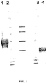
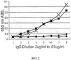
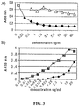
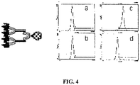

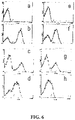
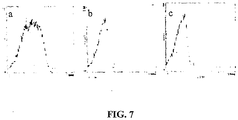

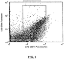
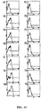
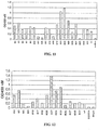


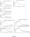
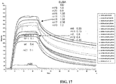
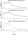
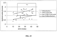

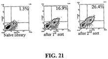

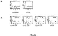
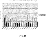
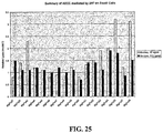
Classifications
-
- C—CHEMISTRY; METALLURGY
- C07—ORGANIC CHEMISTRY
- C07K—PEPTIDES
- C07K14/00—Peptides having more than 20 amino acids; Gastrins; Somatostatins; Melanotropins; Derivatives thereof
- C07K14/435—Peptides having more than 20 amino acids; Gastrins; Somatostatins; Melanotropins; Derivatives thereof from animals; from humans
- C07K14/705—Receptors; Cell surface antigens; Cell surface determinants
- C07K14/70503—Immunoglobulin superfamily
- C07K14/70535—Fc-receptors, e.g. CD16, CD32, CD64 (CD2314/705F)
-
- C—CHEMISTRY; METALLURGY
- C07—ORGANIC CHEMISTRY
- C07K—PEPTIDES
- C07K16/00—Immunoglobulins [IGs], e.g. monoclonal or polyclonal antibodies
- C07K16/005—Immunoglobulins [IGs], e.g. monoclonal or polyclonal antibodies constructed by phage libraries
-
- A—HUMAN NECESSITIES
- A61—MEDICAL OR VETERINARY SCIENCE; HYGIENE
- A61P—SPECIFIC THERAPEUTIC ACTIVITY OF CHEMICAL COMPOUNDS OR MEDICINAL PREPARATIONS
- A61P1/00—Drugs for disorders of the alimentary tract or the digestive system
- A61P1/04—Drugs for disorders of the alimentary tract or the digestive system for ulcers, gastritis or reflux esophagitis, e.g. antacids, inhibitors of acid secretion, mucosal protectants
-
- A—HUMAN NECESSITIES
- A61—MEDICAL OR VETERINARY SCIENCE; HYGIENE
- A61P—SPECIFIC THERAPEUTIC ACTIVITY OF CHEMICAL COMPOUNDS OR MEDICINAL PREPARATIONS
- A61P11/00—Drugs for disorders of the respiratory system
- A61P11/02—Nasal agents, e.g. decongestants
-
- A—HUMAN NECESSITIES
- A61—MEDICAL OR VETERINARY SCIENCE; HYGIENE
- A61P—SPECIFIC THERAPEUTIC ACTIVITY OF CHEMICAL COMPOUNDS OR MEDICINAL PREPARATIONS
- A61P11/00—Drugs for disorders of the respiratory system
- A61P11/06—Antiasthmatics
-
- A—HUMAN NECESSITIES
- A61—MEDICAL OR VETERINARY SCIENCE; HYGIENE
- A61P—SPECIFIC THERAPEUTIC ACTIVITY OF CHEMICAL COMPOUNDS OR MEDICINAL PREPARATIONS
- A61P13/00—Drugs for disorders of the urinary system
- A61P13/02—Drugs for disorders of the urinary system of urine or of the urinary tract, e.g. urine acidifiers
-
- A—HUMAN NECESSITIES
- A61—MEDICAL OR VETERINARY SCIENCE; HYGIENE
- A61P—SPECIFIC THERAPEUTIC ACTIVITY OF CHEMICAL COMPOUNDS OR MEDICINAL PREPARATIONS
- A61P13/00—Drugs for disorders of the urinary system
- A61P13/12—Drugs for disorders of the urinary system of the kidneys
-
- A—HUMAN NECESSITIES
- A61—MEDICAL OR VETERINARY SCIENCE; HYGIENE
- A61P—SPECIFIC THERAPEUTIC ACTIVITY OF CHEMICAL COMPOUNDS OR MEDICINAL PREPARATIONS
- A61P17/00—Drugs for dermatological disorders
- A61P17/02—Drugs for dermatological disorders for treating wounds, ulcers, burns, scars, keloids, or the like
-
- A—HUMAN NECESSITIES
- A61—MEDICAL OR VETERINARY SCIENCE; HYGIENE
- A61P—SPECIFIC THERAPEUTIC ACTIVITY OF CHEMICAL COMPOUNDS OR MEDICINAL PREPARATIONS
- A61P17/00—Drugs for dermatological disorders
- A61P17/06—Antipsoriatics
-
- A—HUMAN NECESSITIES
- A61—MEDICAL OR VETERINARY SCIENCE; HYGIENE
- A61P—SPECIFIC THERAPEUTIC ACTIVITY OF CHEMICAL COMPOUNDS OR MEDICINAL PREPARATIONS
- A61P19/00—Drugs for skeletal disorders
- A61P19/02—Drugs for skeletal disorders for joint disorders, e.g. arthritis, arthrosis
-
- A—HUMAN NECESSITIES
- A61—MEDICAL OR VETERINARY SCIENCE; HYGIENE
- A61P—SPECIFIC THERAPEUTIC ACTIVITY OF CHEMICAL COMPOUNDS OR MEDICINAL PREPARATIONS
- A61P27/00—Drugs for disorders of the senses
- A61P27/02—Ophthalmic agents
-
- A—HUMAN NECESSITIES
- A61—MEDICAL OR VETERINARY SCIENCE; HYGIENE
- A61P—SPECIFIC THERAPEUTIC ACTIVITY OF CHEMICAL COMPOUNDS OR MEDICINAL PREPARATIONS
- A61P29/00—Non-central analgesic, antipyretic or antiinflammatory agents, e.g. antirheumatic agents; Non-steroidal antiinflammatory drugs [NSAID]
-
- A—HUMAN NECESSITIES
- A61—MEDICAL OR VETERINARY SCIENCE; HYGIENE
- A61P—SPECIFIC THERAPEUTIC ACTIVITY OF CHEMICAL COMPOUNDS OR MEDICINAL PREPARATIONS
- A61P31/00—Antiinfectives, i.e. antibiotics, antiseptics, chemotherapeutics
-
- A—HUMAN NECESSITIES
- A61—MEDICAL OR VETERINARY SCIENCE; HYGIENE
- A61P—SPECIFIC THERAPEUTIC ACTIVITY OF CHEMICAL COMPOUNDS OR MEDICINAL PREPARATIONS
- A61P31/00—Antiinfectives, i.e. antibiotics, antiseptics, chemotherapeutics
- A61P31/04—Antibacterial agents
-
- A—HUMAN NECESSITIES
- A61—MEDICAL OR VETERINARY SCIENCE; HYGIENE
- A61P—SPECIFIC THERAPEUTIC ACTIVITY OF CHEMICAL COMPOUNDS OR MEDICINAL PREPARATIONS
- A61P31/00—Antiinfectives, i.e. antibiotics, antiseptics, chemotherapeutics
- A61P31/12—Antivirals
-
- A—HUMAN NECESSITIES
- A61—MEDICAL OR VETERINARY SCIENCE; HYGIENE
- A61P—SPECIFIC THERAPEUTIC ACTIVITY OF CHEMICAL COMPOUNDS OR MEDICINAL PREPARATIONS
- A61P31/00—Antiinfectives, i.e. antibiotics, antiseptics, chemotherapeutics
- A61P31/12—Antivirals
- A61P31/20—Antivirals for DNA viruses
-
- A—HUMAN NECESSITIES
- A61—MEDICAL OR VETERINARY SCIENCE; HYGIENE
- A61P—SPECIFIC THERAPEUTIC ACTIVITY OF CHEMICAL COMPOUNDS OR MEDICINAL PREPARATIONS
- A61P33/00—Antiparasitic agents
-
- A—HUMAN NECESSITIES
- A61—MEDICAL OR VETERINARY SCIENCE; HYGIENE
- A61P—SPECIFIC THERAPEUTIC ACTIVITY OF CHEMICAL COMPOUNDS OR MEDICINAL PREPARATIONS
- A61P35/00—Antineoplastic agents
-
- A—HUMAN NECESSITIES
- A61—MEDICAL OR VETERINARY SCIENCE; HYGIENE
- A61P—SPECIFIC THERAPEUTIC ACTIVITY OF CHEMICAL COMPOUNDS OR MEDICINAL PREPARATIONS
- A61P35/00—Antineoplastic agents
- A61P35/02—Antineoplastic agents specific for leukemia
-
- A—HUMAN NECESSITIES
- A61—MEDICAL OR VETERINARY SCIENCE; HYGIENE
- A61P—SPECIFIC THERAPEUTIC ACTIVITY OF CHEMICAL COMPOUNDS OR MEDICINAL PREPARATIONS
- A61P35/00—Antineoplastic agents
- A61P35/04—Antineoplastic agents specific for metastasis
-
- A—HUMAN NECESSITIES
- A61—MEDICAL OR VETERINARY SCIENCE; HYGIENE
- A61P—SPECIFIC THERAPEUTIC ACTIVITY OF CHEMICAL COMPOUNDS OR MEDICINAL PREPARATIONS
- A61P37/00—Drugs for immunological or allergic disorders
-
- A—HUMAN NECESSITIES
- A61—MEDICAL OR VETERINARY SCIENCE; HYGIENE
- A61P—SPECIFIC THERAPEUTIC ACTIVITY OF CHEMICAL COMPOUNDS OR MEDICINAL PREPARATIONS
- A61P37/00—Drugs for immunological or allergic disorders
- A61P37/02—Immunomodulators
- A61P37/04—Immunostimulants
-
- A—HUMAN NECESSITIES
- A61—MEDICAL OR VETERINARY SCIENCE; HYGIENE
- A61P—SPECIFIC THERAPEUTIC ACTIVITY OF CHEMICAL COMPOUNDS OR MEDICINAL PREPARATIONS
- A61P37/00—Drugs for immunological or allergic disorders
- A61P37/02—Immunomodulators
- A61P37/06—Immunosuppressants, e.g. drugs for graft rejection
-
- A—HUMAN NECESSITIES
- A61—MEDICAL OR VETERINARY SCIENCE; HYGIENE
- A61P—SPECIFIC THERAPEUTIC ACTIVITY OF CHEMICAL COMPOUNDS OR MEDICINAL PREPARATIONS
- A61P37/00—Drugs for immunological or allergic disorders
- A61P37/08—Antiallergic agents
-
- A—HUMAN NECESSITIES
- A61—MEDICAL OR VETERINARY SCIENCE; HYGIENE
- A61P—SPECIFIC THERAPEUTIC ACTIVITY OF CHEMICAL COMPOUNDS OR MEDICINAL PREPARATIONS
- A61P9/00—Drugs for disorders of the cardiovascular system
- A61P9/10—Drugs for disorders of the cardiovascular system for treating ischaemic or atherosclerotic diseases, e.g. antianginal drugs, coronary vasodilators, drugs for myocardial infarction, retinopathy, cerebrovascula insufficiency, renal arteriosclerosis
-
- C—CHEMISTRY; METALLURGY
- C07—ORGANIC CHEMISTRY
- C07K—PEPTIDES
- C07K14/00—Peptides having more than 20 amino acids; Gastrins; Somatostatins; Melanotropins; Derivatives thereof
- C07K14/195—Peptides having more than 20 amino acids; Gastrins; Somatostatins; Melanotropins; Derivatives thereof from bacteria
- C07K14/36—Peptides having more than 20 amino acids; Gastrins; Somatostatins; Melanotropins; Derivatives thereof from bacteria from Actinomyces; from Streptomyces (G)
-
- C—CHEMISTRY; METALLURGY
- C07—ORGANIC CHEMISTRY
- C07K—PEPTIDES
- C07K14/00—Peptides having more than 20 amino acids; Gastrins; Somatostatins; Melanotropins; Derivatives thereof
- C07K14/405—Peptides having more than 20 amino acids; Gastrins; Somatostatins; Melanotropins; Derivatives thereof from algae
-
- C—CHEMISTRY; METALLURGY
- C07—ORGANIC CHEMISTRY
- C07K—PEPTIDES
- C07K16/00—Immunoglobulins [IGs], e.g. monoclonal or polyclonal antibodies
- C07K16/18—Immunoglobulins [IGs], e.g. monoclonal or polyclonal antibodies against material from animals or humans
- C07K16/28—Immunoglobulins [IGs], e.g. monoclonal or polyclonal antibodies against material from animals or humans against receptors, cell surface antigens or cell surface determinants
- C07K16/2803—Immunoglobulins [IGs], e.g. monoclonal or polyclonal antibodies against material from animals or humans against receptors, cell surface antigens or cell surface determinants against the immunoglobulin superfamily
- C07K16/283—Immunoglobulins [IGs], e.g. monoclonal or polyclonal antibodies against material from animals or humans against receptors, cell surface antigens or cell surface determinants against the immunoglobulin superfamily against Fc-receptors, e.g. CD16, CD32, CD64
-
- C—CHEMISTRY; METALLURGY
- C07—ORGANIC CHEMISTRY
- C07K—PEPTIDES
- C07K16/00—Immunoglobulins [IGs], e.g. monoclonal or polyclonal antibodies
- C07K16/18—Immunoglobulins [IGs], e.g. monoclonal or polyclonal antibodies against material from animals or humans
- C07K16/28—Immunoglobulins [IGs], e.g. monoclonal or polyclonal antibodies against material from animals or humans against receptors, cell surface antigens or cell surface determinants
- C07K16/2887—Immunoglobulins [IGs], e.g. monoclonal or polyclonal antibodies against material from animals or humans against receptors, cell surface antigens or cell surface determinants against CD20
-
- C—CHEMISTRY; METALLURGY
- C07—ORGANIC CHEMISTRY
- C07K—PEPTIDES
- C07K16/00—Immunoglobulins [IGs], e.g. monoclonal or polyclonal antibodies
- C07K16/18—Immunoglobulins [IGs], e.g. monoclonal or polyclonal antibodies against material from animals or humans
- C07K16/32—Immunoglobulins [IGs], e.g. monoclonal or polyclonal antibodies against material from animals or humans against translation products of oncogenes
-
- C—CHEMISTRY; METALLURGY
- C07—ORGANIC CHEMISTRY
- C07K—PEPTIDES
- C07K16/00—Immunoglobulins [IGs], e.g. monoclonal or polyclonal antibodies
- C07K16/44—Immunoglobulins [IGs], e.g. monoclonal or polyclonal antibodies against material not provided for elsewhere, e.g. haptens, metals, DNA, RNA, amino acids
-
- G—PHYSICS
- G01—MEASURING; TESTING
- G01N—INVESTIGATING OR ANALYSING MATERIALS BY DETERMINING THEIR CHEMICAL OR PHYSICAL PROPERTIES
- G01N33/00—Investigating or analysing materials by specific methods not covered by groups G01N1/00 - G01N31/00
- G01N33/48—Biological material, e.g. blood, urine; Haemocytometers
- G01N33/50—Chemical analysis of biological material, e.g. blood, urine; Testing involving biospecific ligand binding methods; Immunological testing
- G01N33/68—Chemical analysis of biological material, e.g. blood, urine; Testing involving biospecific ligand binding methods; Immunological testing involving proteins, peptides or amino acids
- G01N33/6854—Immunoglobulins
- G01N33/6857—Antibody fragments
-
- C—CHEMISTRY; METALLURGY
- C07—ORGANIC CHEMISTRY
- C07K—PEPTIDES
- C07K2317/00—Immunoglobulins specific features
- C07K2317/20—Immunoglobulins specific features characterized by taxonomic origin
- C07K2317/24—Immunoglobulins specific features characterized by taxonomic origin containing regions, domains or residues from different species, e.g. chimeric, humanized or veneered
-
- C—CHEMISTRY; METALLURGY
- C07—ORGANIC CHEMISTRY
- C07K—PEPTIDES
- C07K2317/00—Immunoglobulins specific features
- C07K2317/50—Immunoglobulins specific features characterized by immunoglobulin fragments
- C07K2317/52—Constant or Fc region; Isotype
-
- C—CHEMISTRY; METALLURGY
- C07—ORGANIC CHEMISTRY
- C07K—PEPTIDES
- C07K2317/00—Immunoglobulins specific features
- C07K2317/70—Immunoglobulins specific features characterized by effect upon binding to a cell or to an antigen
- C07K2317/71—Decreased effector function due to an Fc-modification
-
- C—CHEMISTRY; METALLURGY
- C07—ORGANIC CHEMISTRY
- C07K—PEPTIDES
- C07K2317/00—Immunoglobulins specific features
- C07K2317/70—Immunoglobulins specific features characterized by effect upon binding to a cell or to an antigen
- C07K2317/72—Increased effector function due to an Fc-modification
-
- C—CHEMISTRY; METALLURGY
- C07—ORGANIC CHEMISTRY
- C07K—PEPTIDES
- C07K2317/00—Immunoglobulins specific features
- C07K2317/70—Immunoglobulins specific features characterized by effect upon binding to a cell or to an antigen
- C07K2317/73—Inducing cell death, e.g. apoptosis, necrosis or inhibition of cell proliferation
- C07K2317/732—Antibody-dependent cellular cytotoxicity [ADCC]
-
- C—CHEMISTRY; METALLURGY
- C07—ORGANIC CHEMISTRY
- C07K—PEPTIDES
- C07K2317/00—Immunoglobulins specific features
- C07K2317/70—Immunoglobulins specific features characterized by effect upon binding to a cell or to an antigen
- C07K2317/76—Antagonist effect on antigen, e.g. neutralization or inhibition of binding
-
- C—CHEMISTRY; METALLURGY
- C07—ORGANIC CHEMISTRY
- C07K—PEPTIDES
- C07K2317/00—Immunoglobulins specific features
- C07K2317/90—Immunoglobulins specific features characterized by (pharmaco)kinetic aspects or by stability of the immunoglobulin
- C07K2317/92—Affinity (KD), association rate (Ka), dissociation rate (Kd) or EC50 value
-
- C—CHEMISTRY; METALLURGY
- C07—ORGANIC CHEMISTRY
- C07K—PEPTIDES
- C07K2319/00—Fusion polypeptide
- C07K2319/70—Fusion polypeptide containing domain for protein-protein interaction
-
- G—PHYSICS
- G01—MEASURING; TESTING
- G01N—INVESTIGATING OR ANALYSING MATERIALS BY DETERMINING THEIR CHEMICAL OR PHYSICAL PROPERTIES
- G01N2333/00—Assays involving biological materials from specific organisms or of a specific nature
- G01N2333/435—Assays involving biological materials from specific organisms or of a specific nature from animals; from humans
- G01N2333/705—Assays involving receptors, cell surface antigens or cell surface determinants
- G01N2333/70503—Immunoglobulin superfamily, e.g. VCAMs, PECAM, LFA-3
- G01N2333/70535—Fc-receptors, e.g. CD16, CD32, CD64 (CD2314/705F)
Definitions
- the present invention relates to molecules, particularly polypeptides, more particularly immunoglobulins (e.g., antibodies), comprising a variant Fc region, wherein said variant Fc region comprises at least one amino acid modification relative to a wild-type Fc region, which variant Fc region binds Fc ⁇ RIIIA and/or FcyRIIA with a greater affinity, relative to a comparable molecule comprising the wild-type Fc region.
- the molecules of the invention are particularly useful in preventing, treating, or ameliorating one or more symptoms associated with a disease, disorder, or infection.
- the molecules of the invention are particularly useful for the treatment or prevention of a disease or disorder where an enhanced efficacy of effector cell function (e.g., ADCC) mediated by Fc ⁇ R is desired, e.g., cancer, infectious disease, and in enhancing the therapeutic efficacy of therapeutic antibodies the effect of which is mediated by ADCC.
- ADCC effector cell function
- the interaction of antibody-antigen complexes with cells of the immune system results in a wide array of responses, ranging from effector functions such as antibody-dependent cytotoxicity, mast cell degranulation, and phagocytosis to immunomodulatory signals such as regulating lymphocyte proliferation and antibody secretion. All these interactions are initiated through the binding of the Fc domain of antibodies or immune complexes to specialized cell surface receptors on hematopoietic cells.
- effector functions such as antibody-dependent cytotoxicity, mast cell degranulation, and phagocytosis
- immunomodulatory signals such as regulating lymphocyte proliferation and antibody secretion. All these interactions are initiated through the binding of the Fc domain of antibodies or immune complexes to specialized cell surface receptors on hematopoietic cells.
- the diversity of cellular responses triggered by antibodies and immune complexes results from the structural heterogeneity of Fc receptors.
- Fc receptors share structurally related ligand binding domains which presumably mediate intracellular signaling.
- the Fc receptors members of the immunoglobulin gene superfamily of proteins, are surface glycoproteins that can bind the Fc portion of immunoglobulin molecules. Each member of the family recognizes immunoglobulins of one or more isotypes through a recognition domain on the ⁇ chain of the Fc receptor. Fc receptors are defined by their specificity for immunoglobulin subtypes. Fc receptors for IgG are referred to as Fc ⁇ R, for IgE as F ⁇ R, and for IgA as FcaR.
- Different accessory cells bear Fc receptors for antibodies of different isotype, and the isotype of the antibody determines which accessory cells will be engaged in a given response (reviewed by Ravetch J.V. et al. 1991, Annu. Rev.
- Each member of this family is an integral membrane glycoprotein, possessing extracellular domains related to a C2-set of immunoglobulin-related domains, a single membrane spanning domain and an intracytoplasmic domain of variable length.
- Fc ⁇ Rs There are three known Fc ⁇ Rs, designated Fc ⁇ RI(CD64), Fc ⁇ RII(CD32), and Fc ⁇ RIII(CD16).
- the three receptors are encoded by distinct genes; however, the extensive homology between the three family members suggest they arose from a common progenitor perhaps by gene duplication.
- Fc ⁇ RII proteins are 40KDa integral membrane glycoproteins which bind only the complexed IgG due to a low affinity for monomeric Ig (10 6 M -1 ). This receptor is the most widely expressed Fc ⁇ R, present on all hematopoietic cells, including monocytes, macrophages, B cells, NK cells, neutrophils, mast cells, and platelets. Fc ⁇ RII has only two immunoglobulin-like regions in its immunoglobulin binding chain and hence a much lower affinity for IgG than Fc ⁇ RI. There are three human Fc ⁇ RII genes (Fc ⁇ RII-A, Fc ⁇ RII-B, FcyRII-C), all of which bind IgG in aggregates or immune complexes.
- the fundamental difference is that the A isoform initiates intracellular signaling leading to cell activation such as phagocytosis and respiratory burst, whereas the B isoform initiates inhibitory signals, e.g., inhibiting B-cell activation.
- Both activating and inhibitory signals are transduced through the Fc ⁇ Rs following ligation. These diametrically opposing functions result from structural differences among the different receptor isoforms.
- Two distinct domains within the cytoplasmic signaling domains of the receptor called i mmunoreceptor t yrosine based a ctivation m otifs (ITAMs) or i mmunoreceptor t yrosine based i nhibitory m otifs (ITIMS) account for the different responses.
- ITAM-containing Fc ⁇ R complexes include Fc ⁇ RI, Fc ⁇ RIIA, Fc ⁇ RIIIA, whereas ITIM-containing complexes only include Fc ⁇ RIIB.
- Fc ⁇ RIIA gene Human neutrophils express the Fc ⁇ RIIA gene.
- Fc ⁇ RIIA clustering via immune complexes or specific antibody cross-linking serves to aggregate ITAMs along with receptor-associated kinases which facilitate ITAM phosphorylation.
- ITAM phosphorylation serves as a docking site for Syk kinase, activation of which results in activation of downstream substrates (e.g., PI 3 K). Cellular activation leads to release of proinflammatory mediators.
- the FcyRIIB gene is expressed on B lymphocytes; its extracellular domain is 96% identical to FcyRIIA and binds IgG complexes in an indistinguishable manner.
- the presence of an ITIM in the cytoplasmic domain of FcyRIIB defines this inhibitory subclass of Fc ⁇ R. Recently the molecular basis of this inhibition was established.
- the ITIM in FcyRIIB When colligated along with an activating Fc ⁇ R, the ITIM in FcyRIIB becomes phosphorylated and attracts the SH2 domain of the inosital polyphosphate 5'-phosphatase (SHIP), which hydrolyzes phosphoinositol messengers released as a consequence of ITAM-containing Fc ⁇ R- mediated tyrosine kinase activation, consequently preventing the influx of intracellular Ca ++ .
- SHIP inosital polyphosphate 5'-phosphatase
- crosslinking of Fc ⁇ RIIB dampens the activating response to Fc ⁇ R ligation and inhibits cellular responsiveness. B cell activation, B cell proliferation and antibody secretion is thus aborted.
- a neoplasm, or tumor is a neoplastic mass resulting from abnormal uncontrolled cell growth which can be benign or malignant. Benign tumors generally remain localized. Malignant tumors are collectively termed cancers.
- malignant generally means that the tumor can invade and destroy neighboring body structures and spread to distant sites to cause death (for review, see Robbins and Angell, 1976, Basic Pathology, 2d Ed., W.B. Saunders Co., Philadelphia, pp. 68-122 ). Cancer can arise in many sites of the body and behave differently depending upon its origin. Cancerous cells destroy the part of the body in which they originate and then spread to other part(s) of the body where they start new growth and cause more destruction.
- Lung and prostate cancer are the top cancer killers for men in the United States.
- Lung and breast cancer are the top cancer killers for women in the United States.
- One in two men in the United States will be diagnosed with cancer at some time during his lifetime.
- One in three women in the United States will be diagnosed with cancer at some time during her lifetime.
- cancer therapy may involve surgery, chemotherapy, hormonal therapy and/or radiation treatment to eradicate neoplastic cells in a patient ( See, for example, Stockdale, 1998, "Principles of Cancer Patient Management", in Scientific American: Medicine, vol. 3, Rubenstein and Federman, eds., Chapter 12, Section IV ).
- cancer therapy could also involve biological therapy or immunotherapy. All of these approaches pose significant drawbacks for the patient.
- Surgery for example, may be contraindicated due to the health of the patient or may be unacceptable to the patient. Additionally, surgery may not completely remove the neoplastic tissue. Radiation therapy is only effective when the neoplastic tissue exhibits a higher sensitivity to radiation than normal tissue, and radiation therapy can also often elicit serious side effects.
- Hormonal therapy is rarely given as a single agent and although can be effective, is often used to prevent or delay recurrence of cancer after other treatments have removed the majority of the cancer cells.
- Biological therapies/immunotherapies are limited in number and may produce side effects such as rashes or swellings, flu-like symptoms, including fever, chills and fatigue, digestive tract problems or allergic reactions.
- chemotherapeutic agents available for treatment of cancer.
- a significant majority of cancer chemotherapeutics act by inhibiting DNA synthesis, either directly, or indirectly by inhibiting the biosynthesis of the deoxyribonucleotide triphosphate precursors, to prevent DNA replication and concomitant cell division ( See, for example, Gilman et al., Goodman and Gilman's: The Pharmacological Basis of Therapeutics, Eighth Ed. (Pergamom Press, New York, 1990 )).
- agents which include alkylating agents, such as nitrosourea, anti-metabolites, such as methotrexate and hydroxyurea, and other agents, such as etoposides, campathecins, bleomycin, doxorubicin, daunorubicin, etc., although not necessarily cell cycle specific, kill cells during S phase because of their effect on DNA replication.
- agents specifically colchicine and the vinca alkaloids, such as vinblastine and vincristine, interfere with microtubule assembly resulting in mitotic arrest.
- Chemotherapy protocols generally involve administration of a combination of chemotherapeutic agents to increase the efficacy of treatment.
- chemotherapeutic agents Despite the availability of a variety of chemotherapeutic agents, chemotherapy has many drawbacks ( See, for example, Stockdale, 1998, "Principles Of Cancer Patient Management” in Scientific American Medicine, vol. 3, Rubenstein and Federman, eds., ch. 12, sect. 10 ). Almost all chemotherapeutic agents are toxic, and chemotherapy causes significant, and often dangerous, side effects, including severe nausea, bone marrow depression, immunosuppression, etc. Additionally, even with administration of combinations of chemotherapeutic agents, many tumor cells are resistant or develop resistance to the chemotherapeutic agents.
- those cells resistant to the particular chemotherapeutic agents used in the treatment protocol often prove to be resistant to other drugs, even those agents that act by mechanisms different from the mechanisms of action of the drugs used in the specific treatment; this phenomenon is termed pleiotropic drug or multidrug resistance.
- drug resistance many cancers prove refractory to standard chemotherapeutic treatment protocols.
- Inflammation is a process by which the body's white blood cells and chemicals protect our bodies from infection by foreign substances, such as bacteria and viruses. It is usually characterized by pain, swelling, warmth and redness of the affected area. Chemicals known as cytokines and prostaglandins control this process, and are released in an ordered and self-limiting cascade into the blood or affected tissues. This release of chemicals increases the blood flow to the area of injury or infection, and may result in the redness and warmth. Some of the chemicals cause a leak of fluid into the tissues, resulting in swelling. This protective process may stimulate nerves and cause pain. These changes, when occurring for a limited period in the relevant area, work to the benefit of the body.
- autoimmune and/or inflammatory disorders the immune system triggers an inflammatory response when there are no foreign substances to fight and the body's normally protective immune system causes damage to its own tissues by mistakenly attacking self.
- autoimmune disorders which affect the body in different ways.
- the brain is affected in individuals with multiple sclerosis
- the gut is affected in individuals with Crohn's disease
- the synovium, bone and cartilage of various joints are affected in individuals with rheumatoid arthritis.
- the autoimmune disorder may affect only one organ or tissue type or may affect multiple organs and tissues.
- Organs and tissues commonly affected by autoimmune disorders include red blood cells, blood vessels, connective tissues, endocrine glands (e.g., the thyroid or pancreas), muscles, joints, and skin.
- autoimmune disorders include, but are not limited to, Hashimoto's thyroiditis, pernicious anemia, Addison's disease, type 1 diabetes, rheumatoid arthritis, systemic lupus erythematosus, dermatomyositis, Sjogren's syndrome, dermatomyositis, lupus erythematosus, multiple sclerosis, autoimmune inner ear disease myasthenia gravis, Reiter's syndrome, Graves disease, autoimmune hepatitis, familial adenomatous polyposis and ulcerative colitis.
- Rheumatoid arthritis and juvenile rheumatoid arthritis are types of inflammatory arthritis.
- Arthritis is a general term that describes inflammation in joints. Some, but not all, types of arthritis are the result of misdirected inflammation.
- other types of arthritis associated with inflammation include the following: psoriatic arthritis, Reiter's syndrome, ankylosing spondylitis arthritis, and gouty arthritis.
- Rheumatoid arthritis is a type of chronic arthritis that occurs in joints on both sides of the body (such as both hands, wrists or knees). This symmetry helps distinguish rheumatoid arthritis from other types of arthritis.
- rheumatoid arthritis may occasionally affect the skin, eyes, lungs, heart, blood or nerves.
- Rheumatoid arthritis affects about 1% of the world's population and is potentially disabling. There are approximately 2.9 million incidences of rheumatoid arthritis in the United States. Two to three times more women are affected than men. The typical age that rheumatoid arthritis occurs is between 25 and 50. Juvenile rheumatoid arthritis affects 71,000 young Americans (aged eighteen and under), affecting six times as many girls as boys.
- Rheumatoid arthritis is an autoimmune disorder where the body's immune system improperly identifies the synovial membranes that secrete the lubricating fluid in the joints as foreign. Inflammation results, and the cartilage and tissues in and around the joints are damaged or destroyed. In severe cases, this inflammation extends to other joint tissues and surrounding cartilage, where it may erode or destroy bone and cartilage and lead to joint deformities. The body replaces damaged tissue with scar tissue, causing the normal spaces within the joints to become narrow and the bones to fuse together. Rheumatoid arthritis creates stiffness, swelling, fatigue, anemia, weight loss, fever, and often, crippling pain.
- Some common symptoms of rheumatoid arthritis include joint stiffness upon awakening that lasts an hour or longer; swelling in a specific finger or wrist joints; swelling in the soft tissue around the joints; and swelling on both sides of the joint. Swelling can occur with or without pain, and can worsen progressively or remain the same for years before progressing.
- the diagnosis of rheumatoid arthritis is based on a combination of factors, including: the specific location and symmetry of painful joints, the presence of joint stiffness in the morning, the presence of bumps and nodules under the skin (rheumatoid nodules), results of X-ray tests that suggest rheumatoid arthritis, and/or positive results of a blood test called the rheumatoid factor.
- rheumatoid factor Many, but not all, people with rheumatoid arthritis have the rheumatoid-factor antibody in their blood.
- the rheumatoid factor may be present in people who do not have rheumatoid arthritis.
- the typical course of the disease is one of persistent but fluctuating joint symptoms, and after about 10 years, 90% of sufferers will show structural damage to bone and cartilage. A small percentage will have a short illness that clears up completely, and another small percentage will have very severe disease with many joint deformities, and occasionally other manifestations of the disease.
- the inflammatory process causes erosion or destruction of bone and cartilage in the joints.
- rheumatoid arthritis there is an autoimmune cycle of persistent antigen presentation, T-cell stimulation, cytokine secretion, synovial cell activation, and joint destruction.
- the disease has a major impact on both the individual and society, causing significant pain, impaired function and disability, as well as costing millions of dollars in healthcare expenses and lost wages. ( See, for example, the NIH website and the NIAID website).
- the first line of treatment of any arthritis is usually anti-inflammatories, such as aspirin, ibuprofen and Cox-2 inhibitors such as celecoxib and rofecoxib.
- anti-inflammatories such as aspirin, ibuprofen and Cox-2 inhibitors such as celecoxib and rofecoxib.
- Stecond line drugs include gold, methotrexate and steroids. Although these are well-established treatments for arthritis, very few patients remit on these lines of treatment alone. Recent advances in the understanding of the pathogenesis of rheumatoid arthritis have led to the use of methotrexate in combination with antibodies to cytokines or recombinant soluble receptors.
- recombinant soluble receptors for tumor necrosis factor (TNF)- ⁇ have been used in combination with methotrexate in the treatment of arthritis.
- TNF tumor necrosis factor
- only about 50% of the patients treated with a combination of methotrexate and anti-TNF-a agents such as recombinant soluble receptors for TNF- ⁇ show clinically significant improvement.
- Many patients remain refractory despite treatment.
- Difficult treatment issues still remain for patients with rheumatoid arthritis.
- Many current treatments have a high incidence of side effects or cannot completely prevent disease progression. So far, no treatment is ideal, and there is no cure. Novel therapeutics are needed that more effectively treat rheumatoid arthritis and other autoimmune disorders.
- the remarkable variety of these pathogens has caused the natural selection of two crucial features of adaptive immunity.
- the characteristic features of each pathogen are its mode of transmission, its mechanism of replication, its pathogenesis or the means by which it causes disease, and the response it elicits.
- Viral diseases of a wide array of tissues including the respiratory tract, CNS, skin, genitourinary tract, eyes, ears, immune system, gastrointestinal tract, and musculoskeletal system, affect a vast number of humans of all ages ( see Table 328-2 In: Wyngaarden and Smith, 1988, Cecil Textbook of Medicine, 18th Ed., W.B. Saunders Co., Philadelphia, pp. 1750-1753 ). Although considerable effort has been invested in the design of effective anti-viral therapies, viral infections continue to threaten the lives of millions of people worldwide.
- the present invention provides an antibody comprising a human IgG1 variant Fc region defined in claim 1.
- the present invention provides a pharmaceutical composition defined in claim 16. Further features of the invention are defined in the dependent claims.
- the present invention is based, in part, on the identification of mutant human IgG1 heavy chain Fc regions, with altered affinities for Fc ⁇ R receptors (e.g., activating Fc ⁇ Rs, inhibitory FcyRs), using a yeast display system. Accordingly, the invention relates to antibodies comprising a variant Fc region, having one or more amino acid modifications (e.g., substitutions, but also including insertions or deletions) in one or more regions, which modifications alter the affinity of the variant Fc region for an Fc ⁇ R.
- amino acid modifications e.g., substitutions, but also including insertions or deletions
- Said one or more amino acid modification increases the affinity of the variant Fc region for Fc ⁇ RIIIA.
- the molecules of the invention further specifically bind Fc ⁇ (via the Fc region) with a lower affinity than a comparable molecule ( i.e., having the same amino acid sequence as the molecule of the invention except for the one or more amino acid modifications in the Fc region) comprising the wild-type Fc region binds Fc ⁇ RIIB.
- the invention encompasses antibodies with variant Fc regions, having one or more amino acid modifications, which modifications increase the affinity of the variant Fc region for Fc ⁇ RIIIA and enhance the affinity of the variant Fc region for Fc ⁇ RIIB relative to a comparable molecule with a wild type Fc region.
- the invention encompasses antibodies with variant Fc regions, having one or more amino acid modifications, which modifications increase the affinity of the variant Fc region for Fc ⁇ RIIIA but do not alter the affinity of the variant Fc regions for FcyRIIB relative to a comparable molecule with a wild type Fc region.
- heterodimers comprising Fc regions refer to molecules where the two Fc chains have the same or different sequences.
- each chain may have one or more different modifications from the other chain.
- one chain may contain the wild-type Fc region and the other chains may comprise one or more modifications.
- molecules comprising a variant Fc region, wherein said variant Fc region comprises at least one amino acid modification relative to a wild type Fc region, which variant Fc region does not bind any Fc ⁇ R or binds with a reduced affinity, relative to a comparable molecule comprising the wild-type Fc region, as determined by standard assays (e.g., in vitro assays) known to one skilled in the art.
- Such molecules may comprise a variant Fc region, wherein said variant Fc region comprises at least one amino acid modification relative to a wild type Fc region, which variant Fc region only binds one Fc ⁇ R, wherein said Fc ⁇ R is Fc ⁇ IIIA.
- Such molecules may comprise a variant Fc region, wherein said variant Fc region comprises at least one amino acid modification relative to a wild type Fc region, which variant Fc region only binds one Fc ⁇ R, wherein said Fc ⁇ R is Fc ⁇ RIIA.
- Such molecules may comprise a variant Fc region, wherein said variant Fc region comprises at least one amino acid modification relative to a wild type Fc region, which variant Fc region only binds one Fc ⁇ R, wherein said Fc ⁇ R is Fc ⁇ RIIB.
- the affinities and binding properties of the antibodies of the invention for an Fc ⁇ R are initially determined using in vitro assays (biochemical or immunological based assays) known in the art for determining Fc-Fc ⁇ R interactions, i.e., specific binding of an Fc region to an Fc ⁇ R including but not limited to ELISA assay, surface plasmon resonance assay, immunoprecipitation assays ( See Section 5.2.1).
- the binding properties of the antibodies of the invention are also characterized by in vitro functional assays for determining one or more Fc ⁇ R mediator effector cell functions ( See Section 5.2.6).
- the antibodies of the invention have similar binding properties in in vivo models (such as those described and disclised herein) as those in in vitro based assays
- the present invention does not exclude antibodies of the invention that do not exhibit the desired phenotype in in vitro based assays but do exhibit the desired phenotype in vivo.
- the invention encompasses an antibody comprising a variant Fc region, wherein said variant Fc region comprises at least one amino acid modification relative to a wild-type Fc region, such that said polypeptide specifically binds Fc ⁇ RIIIA with a greater affinity than a comparable molecule comprising the wild-type Fc region binds Fc ⁇ RIIIA, provided that said variant Fc region does not solely have a substitution at any one of positions 329, 331, or 332, and do not include or are not solely substitution with any one of: alanine at any of positions 256, 290, 298, 312, 333, 334, 359, 360, 326, or 430; a lysine at position 330; a threonine at position 339; a methionine at position 320; a serine at position 326; an asparagine at position 326; an aspartic acid at position 326; a glutamic acid at position 326; a glutamine at position 334;
- the invention encompasses an antibody comprising a variant Fc region, wherein said variant Fc region comprises at least one amino acid modification relative to a wild-type Fc region, such that said polypeptide specifically binds Fc ⁇ RIIA with a greater affinity than a comparable molecule comprising the wild-type Fc region binds Fc ⁇ RIIA, provided that the one or more amino acid modifications do not include or are not solely substitution with an alanine at any of positions 256, 290, 326, 255, 258, 267, 272, 276, 280, 283, 285, 286, 331, 337, 268, 272, or 430; an asparagine at position 268; a glutamine at position 272; a glutamine, serine, or aspartic acid at position 286; a serine at position 290; a methionine, glutamine, glutamic acid, or arginine at position 320; a glutamic acid at position 322; a serine, glutamic acid,
- the invention encompasses an antibody comprising a variant Fc region, wherein said variant Fc region comprises at least one amino acid modification relative to a wild-type Fc region, such that said molecule has an altered affinity for an Fc ⁇ R, provided that said variant Fc region does not have a substitution at positions that make a direct contact with Fc ⁇ R based on crystallographic and structural analysis of Fc-Fc ⁇ R interactions such as those disclosed by Sondermann et al., (2000 Nature, 406: 267-273 ).
- the antibodies of the invention comprising variant Fc regions comprise modification of at least one residue that does not make a direct contact with an Fc ⁇ R based on structural and crystallographic analysis, e.g., is not within the Fc-Fc ⁇ R binding site.
- the invention encompasses an antibody comprising a variant Fc region, wherein said variant Fc region comprises at least one amino acid modification relative to a wild-type Fc region, such that said molecule binds an Fc ⁇ R with an altered affinity relative to a molecule comprising a wild-type Fc region, provided that said at least one amino acid modification do not include or are not solely a substitution at any of positions 255, 256, 258, 267, 268, 269, 270, 272, 276, 278, 280, 283, 285, 286, 289, 290, 292, 293, 294, 295, 296, 298, 300, 301, 303, 305, 307, 309, 312, 320, 322, 326, 329, 330, 332, 331, 333, 334, 335, 337, 338, 339, 340, 359, 360, 373, 376, 416, 419, 430, 434, 435, 437, 438, 439.
- the invention encompasses an antibody comprising a variant Fc region, wherein said variant Fc region comprises at least one amino acid modification relative to a wild-type Fc region, such that said molecule binds an Fc ⁇ R with an altered affinity relative to a molecule comprising a wild-type Fc region, provided that said variant Fc region does not include or are not solely a substitution at any of positions 255, 258, 267, 269, 270, 276, 278, 280, 283, 285, 289, 292, 293, 294, 295, 296, 300, 303, 305, 307, 309, 322, 329, 332, 331, 337, 338, 340, 373, 376, 416, 419, 434, 435, 437, 438, 439 and does not have an alanine at any of positions 256, 290, 298, 312, 333, 334, 359, 360, 326, or 430; a lysine at position 330; a
- the invention encompasses an antibody comprising a variant Fc region, wherein said variant Fc region does not include or are not solely a substitution at any of positions 268, 269, 270, 272, 276, 278, 283, 285, 286, 289, 292, 293, 301, 303, 305, 307, 309, 331, 333, 334, 335, 337, 338, 340, 360, 373, 376, 416, 419, 430, 434, 435, 437, 438 or 439 and does not have a histidine, glutamine, or tyrosine at position 280; a serine, glycine, threonine or tyrosine at position 290, a leucine or isoleucine at position 300; an asparagine at position 294, a proline at position 296; a proline, asparagine, aspartic acid, or valine at position 298; a lysine at position 295.
- the invention encompasses an antibody comprising a variant Fc region, wherein said variant Fc region comprises at least one amino acid modification relative to a wild-type Fc region, such that said molecule binds an Fc ⁇ R with a reduced affnity relative to a molecule comprising a wild-type Fc region provided that said variant Fc region does not have or are not solely have a substitution at any of positions 252, 254, 265, 268, 269, 270, 278, 289, 292, 293, 294, 295, 296, 298, 300, 301, 303, 322, 324, 327, 329, 333, 335, 338, 340, 373, 376, 382, 388, 389, 414, 416, 419, 434, 435, 437, 438, or 439.
- the invention encompasses an antibody comprising a variant Fc region, wherein said variant Fc region comprises at least one amino acid modification relative to a wild-type Fc region, such that said molecule binds an Fc ⁇ R with an enhanced affnity relative to a molecule comprising a wild-type Fc region provided that said variant Fc region does not have or are not solely a substitution at any of positions 280, 283, 285, 286, 290, 294, 295, 298, 300, 301, 305, 307, 309, 312, 315, 331, 333, 334, 337, 340, 360, 378, 398, or 430.
- antibodies of the invention comprise a variant Fc region having one or more amino acid modifications (e.g., substitutions), which modifications increase the affinity of the variant Fc region for Fc ⁇ RIIIA and/or Fc ⁇ RIIA by at least 2-fold, relative to a comparable molecule comprising a wild-type Fc region.
- amino acid modifications e.g., substitutions
- antibodies of the invention comprise a variant Fc region having one or more amino acid modifications (e.g., substitutions), which modifications increase the affinity of the variant Fc region for Fc ⁇ RIIIA and/or Fc ⁇ RIIA by greater than 2-fold, at least 4-fold, at least 5-fold, at least 6-fold, at least 8-fold, or at least 10-fold relative to a comparable molecule comprising a wild-type Fc region.
- amino acid modifications e.g., substitutions
- antibodies of the invention comprising a variant Fc region specifically bind Fc ⁇ RIIIA and/or Fc ⁇ RIIA with at least 65%, at least 75%, at least 85%, at least 95%, at least 100%, at least 150%, at least 200% greater affinity relative to a molecule comprising a wild-type Fc region.
- Such measurements are preferably in vitro assays.
- the invention encompasses antibodies with altered affinities for the activating and/or inhibitory Fc ⁇ receptors.
- the invention contemplates antibodies with variant Fc regions, having one or more amino acid modifications, which modifications increase the affinity of the variant Fc region for Fc ⁇ RIIB but decrease the affinity of the variant Fc region for Fc ⁇ RIIA, relative to a comparable molecule with a wild-type Fc region.
- the invention encompasses antibodies with variant Fc regions, having one or more amino acid modifications, which modifcations decrease the affinity of the variant Fc reion for Fc ⁇ RIIB and also decrease the affinity of the variant Fc regions for Fc ⁇ RIIA relative to a comparable molecule with a wild-type Fc region.
- the invention encompasses antibodies with variant Fc regions, which modifications decrease the affinity of the variant Fc region for Fc ⁇ RIIA but do not alter the affinity of the variant Fc region for Fc ⁇ RIIB relative to a comparable molecule with a wild-type Fc region.
- the antibodies of the invention comprise a variant Fc region, having one or more amino acid modifications (e.g., substitutions), which one or more modifications increase the affinity of the variant Fc region for Fc ⁇ RIIIA and decrease the affinity of the variant Fc region for Fc ⁇ RIIB, relative to a comparable molecule comprising a wild-type Fc region which binds Fc ⁇ RIIIA and Fc ⁇ RIIB with wild-type affinity.
- the one or more amino acid modifications are not a substitution with alanine at any of positions 256, 298, 333, or 334.
- the antibodies of the invention comprise a variant Fc region, having one or more amino acid modifications (e.g., substitutions), which one or more modifications increase the affinity of the variant Fc region for Fc ⁇ RIIA and decrease the affinity of the variant Fc region for Fc ⁇ RIIB, relative to a comparable molecule comprising a wild-type Fc region which binds Fc ⁇ RIIA and Fc ⁇ RIIB with wild-type affinity.
- the one or more amino acid modifications is not a substitution with arginine at position 320.
- the antibodies of the invention with altered affinities for activating and/or inhibitory receptors having variant Fc regions have one or more amino acid modifications, wherein said one or more amino acid modification is a substitution at position 288 with asaparagine, at position 330 with serine and at position 396 with leucine (MgFc10) (See Table 5); or a substitution at position 334 with glutamic acid, at position 359 with asparagine, and at position 366 with serine (MgFc13); or a substitution at position 316 with aspartic acid, at position 378 with valine, and at position 399 with glutamic acid (MgFc27); or a substitution at position 392 with threonine, and at position 396 with leucine (MgFc38); or a substitution at position 221 with glutamic acid, at position 270 with glutamic acid, at position 308 with alanine, at position 311 with histidine, at position 396 with leucine, and at position
- the preferred method for screening and identifying molecules comprising variant Fc regions with altered Fc ⁇ R affinities is yeast surface display technology (for review see Boder and Wittrup, 2000, Methods in Enzymology, 328: 430-444 ).
- yeast surface display is a genetic method whereby polypeptides comprising Fc mutants are expressed on the yeast cell wall in a form accessible for interacting with Fc ⁇ R.
- Yeast surface display of the mutant Fc containing antibodies of the invention may be performed in accordance with any of the techniques known to those skilled in the art or the specific methods described herein.
- mutant Fc fusion proteins with a desirable binding property, e.g., the ability of the mutant Fc fusion protein to bind Fc ⁇ RIIIA with a greater affinity than a comparable polypeptide comprising a wild-type Fc region binds Fc ⁇ RIIIA.
- Yeast cells displaying the mutant Fc fusion proteins can be screened and characterized by any biochemical or immunological based assays known to those skilled in the art for assessing binding interactions. Screening of mutant Fc fusion proteins may be done using one or more biochemical based assays, e.g., an ELISA assay.
- Screening and identifying molecules comprising variant Fc regions with altered Fc ⁇ R affinities may be done using the yeast display technology as described herein in combination with one or more biochemical based assays, preferably in a high throughput manner.
- the one or more biochemical assays can be any assay known in the art for identifying Fc-Fc ⁇ R interaction, i.e., specific binding of an Fc region to an Fc ⁇ R, including, but not limited to, an ELISA assay, surface plasmon resonance assays, immunoprecipitation assay, affinity chromatography, and equilibrium dialysis.
- Screening and identifying molecules comprising variant Fc regions with altered Fc ⁇ R affinities may be done using the yeast display technology as described herein in combination with one or more functional based assays, preferably in a high throughput manner.
- the functional based assays can be any assay known in the art for characterizing one or more Fc ⁇ R mediated effector cell function such as those described herein in Section 5.2.6.
- Non-limiting examples of effector cell functions include but are not limited to, antibody-dependent cell mediated cytotoxicity (ADCC), antibody-dependent phagocytosis, phagocytosis, opsonization, opsonophagocytosis, cell binding, rosetting, C1q binding, and complement dependent cell mediated cytotoxicity.
- Screening and identifying molecules comprising variant Fc regions with altered Fc ⁇ R affinities may be done using the yeast display technology as described herein in combination with one or more biochemical based assays in combination or in parallel with one or more functional based assays, preferably in a high throughput manner.
- a preferred method for measuring the Fc ⁇ R-Fc interaction is an assay developed by the inventors, which allows detection and quantitation of the interaction, despite the inherently weak affinity of the receptor for its ligand, e.g., in the micromolar range for FcyRIIB and Fc ⁇ RIIIA.
- the method involves the formation of an Fc ⁇ R complex (e.g., Fc ⁇ RIIIA, Fc ⁇ RIIB) that has an improved avidity for an Fc region, relative to an uncomplexed Fc ⁇ R.
- a method for producing a tetrameric Fc ⁇ R complex comprising: (i) producing a fusion protein, such that a 15 amino acid AVITAG sequence operably linked to the soluble region of Fc ⁇ R; (ii) biotinylating the protein produced using an E. coli BirA enzyme; (iii) mixing the biotinylated protein produced with streptaividn-phycoerythrin in an appropriate molar ratio, such that a tetrameric Fc ⁇ R complex is formed.
- antibodies comprising Fc regions bind the tetrameric Fc ⁇ R complexes, formed according to the methods described herein, with at least an 8-fold higher affinity than they bind the monomeric uncomplexed Fc ⁇ R.
- the binding of polypeptides comprising Fc regions to the tetrameric Fc ⁇ R complexes may be determined using standard techniques known to those skilled in the art, such as for example, fluorescence activated cell sorting (FACS), radioimmunoassays, ELISA assays, etc.
- the invention provides modified immunoglobulins comprising a variant Fc region with an enhanced affinity for Fc ⁇ RIIIA.
- immunoglobulins include IgG molecules that naturally contain Fc ⁇ R binding regions (e.g., Fc ⁇ RIIIA and/or Fc ⁇ RIIB binding regions), or immunoglobulin derivatives that have been engineered to contain an Fc ⁇ R binding region (e.g., Fc ⁇ RIIIA and/or FcyRIIB binding regions).
- the modified immunoglobulins of the invention include any immunoglobulin molecule that binds, preferably, immunospecifically, i.e., competes off non-specific binding as determined by immunoassays well known in the art for assaying specific antigen-antibody binding, an antigen and contains an Fc ⁇ R binding region (e.g., a Fc ⁇ RIIIA and/or Fc ⁇ RIIB binding region).
- immunospecifically i.e., competes off non-specific binding as determined by immunoassays well known in the art for assaying specific antigen-antibody binding, an antigen and contains an Fc ⁇ R binding region (e.g., a Fc ⁇ RIIIA and/or Fc ⁇ RIIB binding region).
- Such antibodies include, but are not limited to, polyclonal, monoclonal, bi-specific, multi-specific, human, humanized, chimeric antibodies, single chain antibodies, Fab fragments, F(ab') 2 fragments, disulfide-linked Fvs, and fragments containing either a VL or VH domain or even a complementary determining region (CDR) that specifically binds an antigen, in certain cases, engineered to contain or fused to an Fc ⁇ R binding region.
- CDR complementary determining region
- the invention encompasses immunoglobulins comprising a variant Fc region with an enhanced affinity for Fc ⁇ RIIIA such that the immunoglobulin has an enhanced effector function, e.g., antibody dependent cell mediated cytotoxicity.
- the effector function of the antibodies of the invention can be assayed using any assay described herein or known to those skilled in the art.
- immunoglobulins comprising a variant Fc region with an enhanced affinity for Fc ⁇ RIIIA and/or Fc ⁇ RIIA have an enhanced ADCC activity relative to wild-type by at least 2-fold, at least 4-fold, at least 8-fold, at least 10-fold, at least 50-fold, or at least 100-fold.
- human or humanized therapeutic antibodies e.g., tumor specific monoclonal antibodies
- modification e.g., substitution, insertion, deletion
- human or humanized therapeutic antibodies e.g., tumor specific monoclonal antibodies
- human or humanized therapeutic antibodies e.g., tumor specific monoclonal antibodies
- the engineered therapeutic antibodies may further have an enhanced effector function, e.g., enhanced ADCC activity, phagocytosis activity, etc., as determined by standard assays known to those skilled in the art.
- the invention encompasses an engineered humanized monoclonal antibody specific for Her2/neu protooncogene (e.g., Ab4D5 humanized antibody as disclosed in Carter et al., 1992, Proc. Natl. Acad. Sci. USA 89:4285-9 ) comprising modification (e.g., substitution, insertion, deletion) of at least one amino acid residue which modification increases the affinity of the Fc region for Fc ⁇ RIIIA.
- modification of the humanized Her2/neu monoclonal antibody may also further decrease the affinity of the Fc region for Fc ⁇ RIIB.
- the engineered humanized monoclonal antibodies specific for Her2/neu may further have an enhanced effector function as determined by standard assays known in the art and disclosed and exemplified herein.
- the invention encompasses an engineered mouse human chimeric anti-CD20 monoclonal antibody, 2H7 comprising modification (e.g., substitution, insertion, deletion) of at least one amino acid residue which modification increases the affinity of the Fc region for Fc ⁇ RIIIA.
- modification of the anti-CD20 monoclonal antibody, 2H7 may also further decrease the affinity of the Fc region for Fc ⁇ RIIB.
- the engineered anti-CD20 monoclonal antibody, 2H7 may further have an enhanced effector function as determined by standard assays known in the art and disclosed and exemplified herein.
- the invention encompasses an engineered anti-FcyRIIB antibody including but not limited to any of the antibodies disclosed in U.S. Provisional Application No. 60/403,266 filed on August 12, 2002 and U.S. Application No. 10/643,857 filed on August 14, 2003 , having Attorney Docket No. 011183-010-999, comprising modification (e.g., substitution, insertion, deletion) of at least one amino acid residue which modification increases the affinity of the Fc region for Fc ⁇ RIIIA.
- modification e.g., substitution, insertion, deletion
- anti-FcyRIIB antibodies examples include 2B6 monoclonal antibody having ATCC accession number PTA-4591 and 3H7 having ATCC accession number PTA-4592 (deposited at ATCC, 10801 University Boulevard, Manassas, VA 02209-2011.
- modification of the anti-Fc ⁇ RIIB antibody may also further decrease the affinity of the Fc region for Fc ⁇ RIIB.
- the engineered anti-Fc ⁇ RIIB antibody may further have an enhanced effector function as determined by standard assays known in the art and disclosed and exemplified herein.
- the 2B6 monoclonal antibody comprises a modification at position 334 with glutamic acid, at position 359 with asparagine, and at position 366 with serine (MgFc13); or a substitution at position 316 with aspartic acid, at position 378 with valine, and at position 399 with glutamic acid (MgFc27); or a substitution at position 243 with isoleucine, at position 379 with leucine, and at position 420 with valine (MgFc29); or a substitution at position 392 with threonine and at position 396 with leucine (MgFc38); or a substitution at position 221 with glutamic acid, at position 270 with glutamic acid, at position 308 with alanine, at position 311 with histidine, at position 396 with leucine, and at position 402 with aspartic (MgFc42); or a substitution at position 410 with histidine, and at position 396 with leucine (MgFc13); or
- polynucleotides that encode antibody of the invention identified by the methods described herein.
- the polynucleotides encoding the antibodies of the invention may be obtained, and the nucleotide sequence of the polynucleotides determined, by any method known in the art.
- An isolated nucleic acid encoding an antibody of the invention is also described.
- a vector comprising said nucleic acid is also described.
- Host cells containing the vectors or polynucleotides are also described.
- the antibodies of the invention can be produced by any method known to those skilled in the art, in particular, by recombinant expression.
- a method for recombinantly producing an antibody of the invention comprising: (i) culturing in a medium a host cell comprising a nucleic acid encoding said molecule, under conditions suitable for the expression of said molecule; and (ii) recovery of said molecule from said medium.
- the molecules identified in accordance with the methods described herein are useful in preventing, treating, or ameliorating one or more symptoms associated with a disease, disorder, or infection.
- the antibodies of the invention are particularly useful for the treatment or prevention of a disease or disorder where an enhanced efficacy of effector cell function (e.g., ADCC) mediated by Fc ⁇ R is desired, e.g., cancer, infectious disease, and in enhancing the therapeutic efficacy of therapeutic antibodies the effect of which is mediated by ADCC.
- ADCC effector cell function
- a method of treating cancer in a patient having a cancer characterized by a cancer antigen comprising administering a therapeutically effective amount of a therapeutic antibody that binds the cancer antigen, which has been engineered in accordance with the methods described herein.
- a method for treating cancer in a patient having a cancer characterized by a cancer antigen comprising administering a therapeutically effective amount of a therapeutic antibody that specifically binds said cancer antigen, said therapeutic antibody comprising a variant Fc region, wherein said variant Fc region comprises at least one amino acid modification relative to a wild-type Fc region, such that said therapeutic antibody specifically binds Fc ⁇ RIIIA with a greater affinity than the therapeutic antibody comprising the wild-type Fc region binds Fc ⁇ RIIIA, provided that said variant Fc region does not have a substitution at positions 329, 331, or 332, and does not have an alanine at any of positions 256, 290, 298, 312, 333, 334, 359, 360, or 430; a lysine at position 330; a threonine at position 339; a methionine at position 320; a serine at position 326; an asparagine at position 326; an aspartic acid at
- a method for treating cancer in a patient having a cancer characterized by a cancer antigen comprising administering a therapeutically effective amount of a therapeutic antibody that specifically binds a cancer antigen, said therapeutic antibody comprising a variant Fc region, wherein said variant Fc region comprises at least one amino acid modification relative to a wild-type Fc region such that said therapeutic antibody specifically binds Fc ⁇ RIIIA with a greater affinity than a therapeutic antibody comprising the wild-type Fc region binds Fc ⁇ RIIIA, and said therapeutic antibody further specifically binds Fc ⁇ RIIB with a lower affinity than a therapeutic antibody comprising the wild-type Fc region binds Fc ⁇ RIIB, provided that said variant Fc region does not have an alanine at any of positions 256, 298, 333, or 334.
- a method for treating cancer in a patient characterized by a cancer antigen comprising administering a therapeutically effective amount of a therapeutic antibody that specifically binds said cancer antigen and said therapeutic antibody comprises a variant Fc region so that the antibody has an enhanced ADCC activity.
- a method of treating an autoimmune disorder and/or inflammatory disorder in a patient in need thereof comprising administering to said patient a therapeutically effective amount of a molecule comprising a variant Fc region, wherein said variant Fc region comprises at least one amino acid modification relative to a wild type Fc region, such that said molecule specifically binds Fc ⁇ RIIB with a greater affinity than a comparable molecule comprising the wild type Fc region, and said molecule further specifically binds Fc ⁇ RIIIA with a lower affinity than a comparable molecule comprising the wild type Fc region., and said molecule binds an immune complex (e.g., an antigen/antibody complex).
- an immune complex e.g., an antigen/antibody complex
- a method of treating an autoimmune disorder and/or inflammatory disorder further comprising administering one or more additional prophylactic or therapeutic agents, e.g., immunomodulatory agents, anti-inflammatory agents, used for the treatment and/or prevention of such diseases.
- kits for treating or preventing an infectious disease in a subject comprising administering a therapeutically or prophylatically effective amount of one or more antibodies of the invention that bind an infectious agent or cellular receptor therefor.
- Infectious diseases that can be treated or prevented by the antibodies of the invention are caused by infectious agents including but not limited to viruses, bacteria, fungi, protozae, and viruses.
- antibodies of the invention comprising variant Fc regions have an enhanced antibody effector function towards an infectious agent, e.g., a pathogenic protein, relative to a comparable molecule comprising a wild-type Fc region.
- antibodies of the invention enhance the efficacy of treatment of an infectious disease by enhancing phagocytosis and/or opsonization of the infectious agent causing the infectious disease.
- antibodies of the invention enhance the efficacy of treatment of an infectious disease by enhancing ADCC of infected cells causing the infectious disease.
- the antibodies of the invention for use in treatment of cancer may be administered in combination with a therapeutically or prophylactically effective amount of one or additional therapeutic agents known to those skilled in the art for the treatment and/or prevention of an infectious disease.
- a therapeutically or prophylactically effective amount of one or additional therapeutic agents known to those skilled in the art for the treatment and/or prevention of an infectious disease may be administered in combination with antibiotics known to those skilled in the art for the treatment and or prevention of an infectious disease.
- the invention provides pharmaceutical compositions comprising an antibody of the invention, and a pharmaceutically acceptable carrier.
- the invention additionally provides pharmaceutical compositions further comprising one or more additional therapeutic agents, including but not limited to anti-cancer agents, anti-inflammatory agents, immunomodulatory agents.
- the term "Fc region” is used to define a C-terminal region of an IgG heavy chain. Although the boundaries may vary slightly, the human IgG heavy chain Fc region is defined to stretch from Cys226 to the carboxy terminus.
- the Fc region of an IgG comprises two constant domains, CH2 and CH3.
- the CH2 domain of a human IgG Fc region usually extends from amino acids 231 to amino acid 341.
- the CH3 domain of a human IgG Fc region usually extends from amino acids 342 to 447.
- the Fc region of an IgG comprises two constant domains, CH2 and CH3.
- the CH2 domain of a human IgG Fc region (also referred to as "C ⁇ 2" domain) usually extends from amino acid 231-340.
- the CH2 domain is unique in that it is not closely paired with another domain. Rather, two N-linked branched carbohydrate chains are interposed between the two CH2 domains of an intact native IgG.
- the numbering of the residues in an IgG heavy chain is that of the EU index as in Kabat et al., Sequences of Proteins of Immunological Interest, 5th Ed. Public Health Service, NH1, MD (1991 ).
- the "EU index as in Kabat” refers to the numbering of the human IgG1 EU antibody.
- the "hinge region” is generally defined as stretching from Glu216 to Pro230 of human IgG1. Hinge regions of other IgG isotypes may be aligned with the IgG1 sequence by placing the first and last cysteine residues forming inter-heavy chain S-S binds in the same positions.
- antibody refers to monoclonal antibodies, multispecific antibodies, human antibodies, humanized antibodies, synthetic antibodies, chimeric antibodies, polyclonal antibodies, camelized antibodies, single-chain Fvs (scFv), single chain antibodies, Fab fragments, F(ab') fragments, disulfide-linked bispecific Fvs (sdFv), intrabodies, and anti-idiotypic (anti-Id) antibodies (including, e.g., anti-Id and anti-anti-Id antibodies to antibodies of the invention), and epitope-binding fragments of any of the above.
- scFv single-chain Fvs
- Fab fragments single chain antibodies
- F(ab') fragments fragments
- disulfide-linked bispecific Fvs sdFv
- intrabodies and anti-idiotypic (anti-Id) antibodies (including, e.g., anti-Id and anti-anti-Id antibodies to antibodies of the invention), and epitope-binding fragments of any
- antibodies include immunoglobulin molecules and immunologically active fragments of immunoglobulin molecules, i.e., molecules that contain an antigen binding site.
- Immunoglobulin molecules can be of any type (e.g., IgG, IgE, IgM, IgD, IgA and IgY), class (e.g., IgG 1 , IgG 2 , IgG 3 , IgG 4 , IgA 1 and IgA 2 ) or subclass.
- derivative in the context of polypeptides or proteins refers to a polypeptide or protein that comprises an amino acid sequence which has been altered by the introduction of amino acid residue substitutions, deletions or additions.
- derivative also refers to a polypeptide or protein which has been modified, i.e, by the covalent attachment of any type of molecule to the polypeptide or protein.
- an antibody may be modified, e.g., by glycosylation, acetylation, pegylation, phosphorylation, amidation, derivatization by known protecting/blocking groups, proteolytic cleavage, linkage to a cellular ligand or other protein, etc.
- a derivative polypeptide or protein may be produced by chemical modifications using techniques known to those of skill in the art, including, but not limited to specific chemical cleavage, acetylation, formylation, metabolic synthesis of tunicamycin, etc. Further, a derivative polypeptide or protein derivative possesses a similar or identical function as the polypeptide or protein from which it was derived.
- derivative in the context of a non-proteinaceous derivative refers to a second organic or inorganic molecule that is formed based upon the structure of a first organic or inorganic molecule.
- a derivative of an organic molecule includes, but is not limited to, a molecule modified, e.g., by the addition or deletion of a hydroxyl, methyl, ethyl, carboxyl or amine group.
- An organic molecule may also be esterified, alkylated and/or phosphorylated.
- the terms “disorder” and “disease” are used interchangeably to refer to a condition in a subject.
- autoimmune disease is used interchangeably with the term “autoimmune disorder” to refer to a condition in a subject characterized by cellular, tissue and/or organ injury caused by an immunologic reaction of the subject to its own cells, tissues and/or organs.
- inflammatory disease is used interchangeably with the term “inflammatory disorder” to refer to a condition in a subject characterized by inflammation, preferably chronic inflammation.
- Autoimmune disorders may or may not be associated with inflammation.
- inflammation may or may not be caused by an autoimmune disorder.
- certain disorders may be characterized as both autoimmune and inflammatory disorders.
- cancer refers to a neoplasm or tumor resulting from abnormal uncontrolled growth of cells.
- cancer explicitly includes, leukemias and lymphomas.
- cancer refers to a benign tumor, which has remained localized.
- cancer refers to a malignant tumor, which has invaded and destroyed neighboring body structures and spread to distant sites. The cancer is associated with a specific cancer antigen.
- immunomodulatory agent refers to an agent that modulates a host's immune system.
- An immunomodulatory agent may be an immunosuppressant agent.
- An immunomodulatory agent may be an immunostimulatory agent.
- Immunomodulatory agents include, but are not limited to, small molecules, peptides, polypeptides, fusion proteins, antibodies, inorganic molecules, mimetic agents, and organic molecules.
- epitope refers to a fragment of a polypeptide or protein or a non-protein molecule having antigenic or immunogenic activity in an animal, preferably in a mammal, and most preferably in a human.
- An epitope having immunogenic activity is a fragment of a polypeptide or protein that elicits an antibody response in an animal.
- An epitope having antigenic activity is a fragment of a polypeptide or protein to which an antibody immunospecifically binds as determined by any method well-known to one of skill in the art, for example by immunoassays. Antigenic epitopes need not necessarily be immunogenic.
- fragment refers to a peptide or polypeptide comprising an amino acid sequence of at least 5 contiguous amino acid residues, at least 10 contiguous amino acid residues, at least 15 contiguous amino acid residues, at least 20 contiguous amino acid residues, at least 25 contiguous amino acid residues, at least 40 contiguous amino acid residues, at least 50 contiguous amino acid residues, at least 60 contiguous amino residues, at least 70 contiguous amino acid residues, at least contiguous 80 amino acid residues, at least contiguous 90 amino acid residues, at least contiguous 100 amino acid residues, at least contiguous 125 amino acid residues, at least 150 contiguous amino acid residues, at least contiguous 175 amino acid residues, at least contiguous 200 amino acid residues, or at least contiguous 250 amino acid residues of the amino acid sequence of another polypeptide.
- a fragment of a polypeptide may retain at least one function of the polypeptide.
- nucleic acids and “nucleotide sequences” include DNA molecules (e.g., cDNA or genomic DNA), RNA molecules (e.g., mRNA), combinations of DNA and RNA molecules or hybrid DNA/RNA molecules, and analogs of DNA or RNA molecules.
- Such analogs can be generated using, for example, nucleotide analogs, which include, but are not limited to, inosine or tritylated bases.
- Such analogs can also comprise DNA or RNA molecules comprising modified backbones that lend beneficial attributes to the molecules such as, for example, nuclease resistance or an increased ability to cross cellular membranes.
- nucleic acids or nucleotide sequences can be single-stranded, double-stranded, may contain both single-stranded and double-stranded portions, and may contain triple-stranded portions, but preferably is double-stranded DNA.
- a "therapeutically effective amount” refers to that amount of the therapeutic agent sufficient to treat or manage a disease or disorder.
- a therapeutically effective amount may refer to the amount of therapeutic agent sufficient to delay or minimize the onset of disease, e.g., delay or minimize the spread of cancer.
- a therapeutically effective amount may also refer to the amount of the therapeutic agent that provides a therapeutic benefit in the treatment or management of a disease.
- a therapeutically effective amount with respect to a therapeutic agent of the invention means the amount of therapeutic agent alone, or in combination with other therapies, that provides a therapeutic benefit in the treatment or management of a disease.
- prophylactic agent and “prophylactic agents” refer to any agent(s) which can be used in the prevention of a disorder, or prevention of recurrence or spread of a disorder.
- a prophylactically effective amount may refer to the amount of prophylactic agent sufficient to prevent the recurrence or spread of hyperproliferative disease, particularly cancer, or the occurrence of such in a patient, including but not limited to those predisposed to hyperproliferative disease, for example those genetically predisposed to cancer or previously exposed to carcinogens.
- a prophylactically effective amount may also refer to the amount of the prophylactic agent that provides a prophylactic benefit in the prevention of disease.
- a prophylactically effective amount with respect to a prophylactic agent of the invention means that amount of prophylactic agent alone, or in combination with other agents, that provides a prophylactic benefit in the prevention of disease.
- the terms “prevent”, “preventing” and “prevention” refer to the prevention of the recurrence or onset of one or more symptoms of a disorder in a subject resulting from the administration of a prophylactic or therapeutic agent.
- the term “in combination” refers to the use of more than one prophylactic and/or therapeutic agents.
- the use of the term “in combination” does not restrict the order in which prophylactic and/or therapeutic agents are administered to a subject with a disorder.
- a first prophylactic or therapeutic agent can be administered prior to (e.g., 5 minutes, 15 minutes, 30 minutes, 45 minutes, 1 hour, 2 hours, 4 hours, 6 hours, 12 hours, 24 hours, 48 hours, 72 hours, 96 hours, 1 week, 2 weeks, 3 weeks, 4 weeks, 5 weeks, 6 weeks, 8 weeks, or 12 weeks before), concomitantly with, or subsequent to ( e.g., 5 minutes, 15 minutes, 30 minutes, 45 minutes, 1 hour, 2 hours, 4 hours, 6 hours, 12 hours, 24 hours, 48 hours, 72 hours, 96 hours, 1 week, 2 weeks, 3 weeks, 4 weeks, 5 weeks, 6 weeks, 8 weeks, or 12 weeks after) the administration of a second prophylactic or therapeutic agent to a subject with a disorder.
- the present invention relates to antibodies as defined in the appended claims, comprising a variant Fc region, having one or more amino acid modifications (e.g., substitutions, but also including insertions or deletions) in one or more regions, which modifications alter the affinity of the variant Fc region for an Fc ⁇ R.
- amino acid modifications e.g., substitutions, but also including insertions or deletions
- the invention provides antibodies comprising a variant Fc region, wherein said variant Fc region comprises at least one amino acid modification relative to a wild-type Fc region, which variant Fc region binds Fc ⁇ RIIIA with a greater affinity, relative to a comparable molecule, i.e., being the same as said molecule with a variant Fc region but not having the one or more amino acid modifications, comprising the wild-type Fc region as determined by methods known to one skilled in the art for determining Fc-Fc ⁇ R interactions and methods disclosed herein, for example, an ELISA assay or a surface plasmon resonance assay.
- the present disclosure also includes molecules comprising a variant Fc region, wherein said variant Fc region comprises at least one amino acid modification relative to a wild-type Fc region, which variant Fc region binds Fc ⁇ RIIIA with a reduced affinity relative to a comparable molecule comprising the wild-type Fc region.
- the antibodies of the invention further specifically bind Fc ⁇ RIIB (via the Fc region) with a lower affinity than a comparable molecule comprising the wild-type Fc region binds Fc ⁇ RIIB.
- the invention encompasses antibodies comprising a variant Fc region, wherein said variant Fc region comprises at least one amino acid modification relative to a wild-type Fc region, which variant Fc region binds Fc ⁇ RIIIA and Fc ⁇ RIIB with a greater affinity, relative to a comparable molecule comprising the wild-type Fc region.
- the invention encompasses antibodies comprising a variant Fc region, wherein said variant Fc region comprises at least one amino acid modification relative to a wild-type Fc region, which variant Fc region binds Fc ⁇ RIIB with a greater affinity, relative to a comparable molecule comprising the wild-type Fc region.
- the invention encompasses antibodies comprising variant Fc region, wherein said variant Fc region comprises at least one amino acid modification relative to a wild-type Fc region, which variant Fc region binds Fc ⁇ RIIB with a reduced affinity, relative to a comparable molecule comprising the wild-type Fc region.
- molecules comprising a variant Fc region, wherein said variant Fc region comprises at least one amino acid modification relative to a wild type Fc region, which variant Fc region does not show a detectable binding to any Fc ⁇ R (e.g., does not bind Fc ⁇ RIIA, Fc ⁇ RIIB, or Fc ⁇ RIIIA, as determined by, for example, an ELISA assay), relative to a comparable molecule comprising the wild-type Fc region.
- Fc ⁇ R e.g., does not bind Fc ⁇ RIIA, Fc ⁇ RIIB, or Fc ⁇ RIIIA, as determined by, for example, an ELISA assay
- the invention encompasses antibodies comprising a variant Fc region, wherein said variant Fc region comprises at least one amino acid modification relative to a wild type Fc region, which variant Fc region only binds one Fc ⁇ R, wherein said Fc ⁇ R is Fc ⁇ IIIA.
- molecules comprising a variant Fc region, wherein said variant Fc region comprises at least one amino acid modification relative to a wild type Fc region, which variant Fc region only binds one Fc ⁇ R, wherein said Fc ⁇ R is FcyRIIA.
- variant Fc region comprising at least one amino acid modification relative to a wild type Fc region, which variant Fc region only binds one Fc ⁇ R, wherein said Fc ⁇ R is Fc ⁇ RIIB.
- the invention particularly relates to the modification of human or humanized therapeutic antibodies (e.g., tumor specific anti-angiogenic or anti-inflammatory monoclonal antibodies) for enhancing the efficacy of therapeutic antibodies by enhancing, for example, the effector function of the therapeutic antibodies, e.g., enhancing ADCC.
- the affinities and binding properties of the antibodies of the invention for an Fc ⁇ R are initially determined using in vitro assays (biochemical or immunological based assays) known in the art for determining Fc-Fc ⁇ R interactions, i.e., specific binding of an Fc region to an Fc ⁇ R including but not limited to ELISA assay, surface plasmon resonance assay, immunoprecipitation assays ( See Section 5.2.1).
- the binding properties of the antibodies of the invention are also characterized by in vitro functional assays for determining one or more Fc ⁇ R mediator effector cell functions ( See Section 5.2.6).
- the antibodies of the invention have similar binding properties in in vivo models (such as those described and disclosed herein) as those in in vitro based assays
- the present invention does not exclude antibodies of the invention that do not exhibit the desired phenotype in in vitro based assays but do exhibit the desired phenotype in vivo.
- the antibodies of the invention comprising a variant Fc region comprise at least one amino acid modification in the CH3 domain of the Fc region, which is defined as extending from amino acids 342-447. In other embodiments, the antibodies of the invention comprising a variant Fc region comprise at least one amino acid modification in the CH2 domain of the Fc region, which is defined as extending from amino acids 231-341. In some embodiments, the antibodies of the invention comprise at least two amino acid modifications, wherein one modification is in the CH3 region and one modification is in the CH2 region. The invention further encompasses amino acid modification in the hinge region. Antibodies of the invention with one or more amino acid modifications in the CH2 and/or CH3 domains have altered affinities for an Fc ⁇ R as determined using methods described herein or known to one skilled in the art.
- the invention encompasses an antibody comprising a variant Fc region, wherein said variant Fc region comprises at least one amino acid modification relative to a wild-type Fc region, such that said molecule has an altered affinity for an Fc ⁇ R, provided that said variant Fc region does not have a substitution at positions that make a direct contact with Fc ⁇ R based on crystallographic and structural analysis of Fc-Fc ⁇ R interactions such as those disclosed by Sondermann et al., 2000 (Nature, 406: 267-273 ).
- the antibodies of the invention comprising variant Fc regions comprise modification of at least one residue that makes a direct contact with an Fc ⁇ R based on structural and crystallographic analysis.
- the invention encompasses an antibody comprising a variant Fc region, wherein said variant Fc region comprises at least one amino acid modification relative to a wild-type Fc region, such that said molecule binds an Fc ⁇ R with an altered affinity relative to a molecule comprising a wild-type Fc region, provided that said variant Fc region does not have or are not solely a substitution at any of positions 255, 256, 258, 267, 268, 269, 270, 272, 276, 278, 280, 283, 285, 286, 289, 290, 292, 293, 294, 295, 296, 298, 300, 301, 303, 305, 307, 309, 312, 320, 322, 326, 329, 330, 332, 331, 333, 334, 335, 337, 338, 339, 340, 359, 360, 373, 376, 416, 419, 430, 434, 435, 437, 438, 439.
- the invention encompasses an antibody comprising a variant Fc region, wherein said variant Fc region comprises at least one amino acid modification relative to a wild-type Fc region, such that said molecule binds an Fc ⁇ R with an altered affinity relative to a molecule comprising a wild-type Fc region, provided that said variant Fc region does not have or are not solely a substitution at any of positions 255, 258, 267, 269, 270, 276, 278, 280, 283, 285, 289, 292, 293, 294, 295, 296, 300, 303, 305, 307, 309, 322, 329, 332, 331, 337, 338, 340, 373, 376, 416, 419, 434, 435, 437, 438, 439 and does not have an alanine at any of positions 256, 290, 298, 312, 333, 334, 359, 360, 326, or 430; a lysine at position 330; a
- the invention encompasses an antibody comprising a variant Fc region, wherein said variant Fc region does not have or are not solely a substitution at any of positions 268, 269, 270, 272, 276, 278, 283, 285, 286, 289, 292, 293, 301, 303, 305, 307, 309, 331, 333, 334, 335, 337, 338, 340, 360, 373, 376, 416, 419, 430, 434, 435, 437, 438 or 439 and does not have a histidine, glutamine, or tyrosine at position 280; a serine, glycine, threonine or tyrosine at position 290, a leucine or isoleucine at position 300; an asparagine at position 294, a proline at position 296; a proline, asparagine, aspartic acid, or valine at position 298; a lysine at position 295.
- the invention encompasses an antibody comprising a variant Fc region, wherein said variant Fc region comprises at least one amino acid modification relative to a wild-type Fc region, such that said molecule binds an Fc ⁇ R with a reduced affinity relative to a molecule comprising a wild-type Fc region provided that said variant Fc region does not have or are not solely a substitution at any of positions 252, 254, 265, 268, 269, 270, 278, 289, 292, 293, 294, 295, 296, 298, 300, 301, 303, 322, 324, 327, 329, 333, 335, 338, 340, 373, 376, 382, 388, 389, 414, 416, 419, 434, 435, 437, 438, or 439.
- the invention encompasses an antibody comprising a variant Fc region, wherein said variant Fc region comprises at least one amino acid modification relative to a wild-type Fc region, such that said molecule binds an Fc ⁇ R with an enhanced affinity relative to a molecule comprising a wild-type Fc region provided that said variant Fc region does not have or are not solely a substitution at any of positions 280, 283, 285, 286, 290, 294, 295, 298, 300, 301, 305, 307, 309, 312, 315, 331, 333, 334, 337, 340, 360, 378, 398, or 430.
- the antibodies of the invention with altered affinities for activating and/or inhibitory receptors having variant Fc regions have one or more amino acid modifications, wherein said one or more amino acid modification is a substitution at position 288 with asparagine, at position 330 with serine and at position 396 with leucine (MgFc10)(See Table 5); or a substitution at position 334 with glutamic acid, at position 359 with asparagine, and at position 366 with serine (MgFc13); or a substitution at position 316 with aspartic acid, at position 378 with valine, and at position 399 with glutamic acid (MgFc27); or a substitution at position 392 with threonine, and at position 396 with leucine (MgFc38); or a substitution at position 221 with glutamic acid, at position 270 with glutamic acid, at position 308 with alanine, at position 311 with histidine, at position 396 with leucine, and at position 40
- the invention encompasses antibodies comprising a variant Fc region having an amino acid modification at one or more of the following positions: 185, 142, 192, 141, 132, 149, 133, 125, 162, 147, 119, 166, 251, 292, 290, 291, 252, 288, 268, 256, 262, 218, 214, 205,215, 247, 275, 202, 289, 258, 219, 279, 222, 246, 233, 246, 268, 244, 217, 253, 246, 224, 298, 280, 255, 218, 281, 284, 216, 223, 235, 221, 252, 241, 258, 227, 231, 215, 274, 287, 244, 229, 287, 291, 240, 281, 232, 269, 225, 246, 246, 293, 295, 248, 276, 268, 210, 288, 227, 221, 217, 261, 210, 242, 255, 240, 250, 247,
- the invention encompasses antibodies comprising variant Fc regions consisting of or comprising any of the mutations listed in the table below in Table 2. (Note that antibodies comprising variant Fc regions consisting of the single site or double site mutations listed in Table 2 with an asterisk "*" are included as comparative examples.) TABLE 2.
- EXEMPLARY MUTATIONS SINGLE SITE MUTANTS DOUBLE SITE MUTANTS K392R* Q347H, A339V* N315I* S415I, L251F* S132I* K290E, L142P* P396L G285E, P247H* P396H* K409R, S166N* A162V* E334A, K334A* R292L* R292L.
- the invention encompasses antibodies comprising variant Fc regions having more than two amino acid modifications.
- a non-limiting example of such variants is listed in the table below (Table 3).
- the invention encompasses mutations listed in Table 3 which further comprise one or more amino acid modifications such as those disclosed herein. (Note that antibodies comprising variant Fc regions consisting of the combinations of mutations listed in Table 3 with an asterisk "*" are included as comparative examples.) TABLE 3.
- the antibodies of the invention further comprise one or more glycosylation sites, so that one or more carbohydrate moieties are covalently attached to the molecule.
- the antibodies of the invention with one or more glycosylation sites and/or one or more modifications in the Fc region have an enhanced antibody mediated effector function, e.g., enhanced ADCC activity.
- the invention further comprises antibodies comprising one or more modifications of amino acids that are directly or indirectly known to interact with a carbohydrate moiety of the antibody, including but not limited to amino acids at positions 241, 243, 244, 245, 245, 249, 256, 258, 260, 262, 264, 265, 296, 299, and 301.
- Amino acids that directly or indirectly interact with a carbohydrate moiety of an antibody are known in the art, see, e.g., Jefferis et al., 1995 Immunology Letters, 44: 111-7 .
- the invention encompasses antibodies that have been modified by introducing one or more glycosylation sites into one or more sites of the antibodies, preferably without altering the functionality of the antibody, e.g., binding activity to Fc ⁇ R.
- Glycosylation sites may be introduced into the variable and/or constant region of the antibodies of the invention.
- "glycosylation sites” include any specific amino acid sequence in an antibody to which an oligosaccharide (i.e., carbohydrates containing two or more simple sugars linked together) will specifically and covalently attach. Oligosaccharide side chains are typically linked to the backbone of an antibody via either N-or O-linkages.
- N-linked glycosylation refers to the attachment of an oligosaccharide moiety to the side chain of an asparagine residue.
- O-linked glycosylation refers to the attachment of an oligosaccharide moiety to a hydroxyamino acid, e.g., serine, threonine.
- the antibodies of the invention may comprise one or more glycosylation sites, including N-linked and O-linked glycosylation sites. Any glycosylation site for N-linked or O-linked glycosylation known in the art may be used in accordance with the instant invention.
- An exemplary N-linked glycosylation site that is useful in accordance with the methods of the present invention, is the amino acid sequence: Asn-X-Thr/Ser, wherein X may be any amino acid and Thr/Ser indicates a threonine or a serine.
- a site or sites may be introduced into an antibody of the invention using methods well known in the art to which this invention pertains. See, for example, " In Vitro Mutagenesis," Recombinant DNA: A Short Course, J. D. Watson, et al. W.H. Freeman and Company, New York, 1983, chapter 8, pp. 106-116 .
- An exemplary method for introducing a glycosylation site into an antibody of the invention may comprise: modifying or mutating an amino acid sequence of the antibody so that the desired Asn-X-Thr/Ser sequence is obtained.
- the present invention is based, in part, on the identification of mutant human IgG1 heavy chain Fc regions, with altered affinities for different Fc ⁇ R receptors, using a yeast display system. Accordingly, the invention relates to antibodies comprising a variant Fc region, having one or more amino acid modifications (e.g., substitutions, but also including insertions or deletions) in one or more regions, which modifications alter the affinity of the variant Fc region for an Fc ⁇ R.
- amino acid modifications e.g., substitutions, but also including insertions or deletions
- the affinities and binding properties of the antibodies of the invention for an Fc ⁇ R are initially determined using in vitro assays (biochemical or immunological based assays) known in the art for determining Fc-Fc ⁇ R interactions, i.e., specific binding of an Fc region to an Fc ⁇ R including but not limited to ELISA assay, surface plasmon resonance assay, immunoprecipitation assays (See Section 5.2.1).
- the binding properties of the antibodies of the invention are also characterized by in vitro functional assays for determining one or more Fc ⁇ R mediator effector cell functions ( See Section 5.2.6).
- the antibodies of the invention have similar binding properties in in vivo models (such as those described and disclosed herein) as those in in vitro based assays.
- the present invention does not exclude antibodies of the invention that do not exhibit the desired phenotype in in vitro based assays but do exhibit the desired phenotype in vivo.
- the invention encompasses antibodies comprising a variant Fc region, having one or more amino acid modifications (e.g., substitutions) in one or more regions, wherein such modifications alter the affinity of the variant Fc region for an activating Fc ⁇ R.
- antibodies of the invention comprise a variant Fc region, having one or more amino acid modifications (e.g., substitutions) in one or more regions, which modifications increase the affinity of the variant Fc region for Fc ⁇ RIIIA and/or Fc ⁇ RIIA by at least 2-fold, relative to a comparable molecule comprising a wild-type Fc region.
- antibodies of the invention comprise a variant Fc region, having one or more amino acid modifications (e.g., substitutions) in one or more regions, which modifications increase the affinity of the variant Fc region for Fc ⁇ RIIIA and/or Fc ⁇ RIIA by greater than 2 fold, relative to a comparable molecule comprising a wild-type Fc region.
- the one or more amino acid modifications increase the affinity of the variant Fc region for Fc ⁇ RIIIA and/or Fc ⁇ RIIA by at least 3-fold, 4-fold, 5-fold, 6-fold, 8-fold, or 10-fold relative to a comparable molecule comprising a wild-type Fc region.
- the one or more amino acid modifications decrease the affinity of the variant Fc region for Fc ⁇ RIIA by at least 3-fold, 4-fold, 5-fold, 6-fold, 8-fold, or 10-fold relative to a comparable molecule comprising a wild-type Fc region.
- fold increases are preferably determined by an ELISA or surface plasmon resonance assays.
- the one or more amino acid modifications do not include or are not solely a substitution at any one of positions 329, 331, or 322 with any amino acid.
- the one or more amino acid modification do not include or are not solely a substitution with any one of alanine at positions 256, 290, 298, 312, 333, 334, 359, 360, or 430; with lysine at position 330; with threonine at position 339; with methionine at position 320; with serine, asparagine, aspartic acid, or glutamic acid at position 326 with glutamine, glutamic acid, methionine, histidine, valine, or leucine at position 334.
- the one or more amino acid modifications do not include or are not solely a substitution at any of positions 280, 290, 300, 294, or 295.
- the one or more amino acid modification does not include or are not solely a substitution at position 300 with leucine or isoleucine; at position 295 with lysine; at position 294 with asparagine; at position 298 with valine; aspartic acid proline, asparagine, or valine; at position 280 with histidine, glutamine or tyrosine; at position 290 with serine, glycine, threonine or tyrosine.
- the invention encompasses an antibody comprising a variant Fc region, wherein said variant Fc region comprises at least one amino acid modification relative to a wild-type Fc region, such that said polypeptide specifically binds Fc ⁇ RIIA with a greater affinity than a comparable molecule comprising the wild-type Fc region binds Fc ⁇ RIIA, provided that said variant Fc region does not have an alanine at any of positions 256, 290, 326, 255, 258, 267, 272, 276, 280, 283, 285, 286, 331, 337, 268, 272, or 430; an asparagine at position 268; a glutamine at position 272; a glutamine, serine, or aspartic acid at position 286; a serine at position 290; a methionine, glutamine, glutamic acid, or arginine at position 320; a glutamic acid at position322; a serine, glutamic acid, or aspartic acid at position 326
- antibodies of the invention comprise a variant Fc region, having one or more amino acid modifications (e.g., substitutions) in one or more regions, which modifications increase the affinity of the variant Fc region for Fc ⁇ RIIA by at least 2-fold, relative to a comparable molecule comprising a wild-type Fc region.
- antibodies of the invention comprise a variant Fc region, having one or more amino acid modifications (e.g. , substitutions) in one or more regions, which modifications increase the affinity of the variant Fc region for Fc ⁇ RIIA by greater than 2 fold, relative to a comparable molecule comprising a wild-type Fc region.
- the one or more amino acid modifications increase the affinity of the variant Fc region for Fc ⁇ RIIA by at least 3-fold, 4-fold, 5-fold, 6-fold, 8-fold, or 10-fold relative to a comparable molecule comprising a wild-type Fc region
- the invention encompasses antibodies comprising a variant Fc region, having one or more amino acid modifications (e.g., substitutions but also include insertions or deletions), which modifications increase the affinity of the variant Fc region for Fc ⁇ RIIIA and/or Fc ⁇ RIIA by at least 65%, at least 70%, at least 75%, at least 85%, at least 90%, at least 95%, at least 99%, at least 100%, at least 150%, and at least 200%, relative to a comparable molecule comprising a wild-type Fc region.
- amino acid modifications e.g., substitutions but also include insertions or deletions
- the one or more amino acid modifications which increase the affinity of the variant Fc region comprise a substitution at position 347 with histidine, and at position 339 with valine; or a substitution at position 425 with isoleucine and at position 215 with phenylalanine; or a substitution at position 408 with isoleucine, at position 215 with isoleucine, and at position 125 with leucine; or a substitution at position 385 with glutamic acid and at position 247 with histidine; or a substitution at position 348 with methionine, at position 334 with asparagine, at position 275 with isoleucine, at position 202 with methionine, and at position 147 with threonine; or a substitution at position 275 with isoleucine, at position 334 with asparagine, and at position 348 with methionine; or a substitution at position 279 with leucine and at position 395 with serine; or a substitution at position 246 with
- the invention encompasses an antibody comprising a variant Fc region, wherein said variant Fc region comprises a substitution at position 243 with isoleucine and at position 379 with leucine, such that said molecule binds Fc ⁇ RIIIA with about a 1.5 fold higher affinity than a comparable molecule comprising the wild type Fc region binds Fc ⁇ RIIIA, as determined by an ELISA assay.
- the invention encompasses an antibody comprising a variant Fc region, wherein said variant Fc region comprises a substitution at position 288 with asparagine, at position 330 with serine, and at position 396 with leucine, such that said molecule binds Fc ⁇ RIIIA with about a 5 fold higher affinity than a comparable molecule comprising the wild type Fc region binds Fc ⁇ RIIIA, as determined by an ELISA assay.
- the invention encompasses an antibody comprising a variant Fc region, wherein said variant Fc region comprises a substitution at position 243 with leucine and at position 255 with leucine such that said molecule binds Fc ⁇ RIIIA with about a 1 fold higher affinity than a comparable molecule comprising the wild type Fc region binds Fc ⁇ RIIIA, as determined by an ELISA assay.
- the invention encompasses an antibody comprising a variant Fc region, wherein said variant Fc region comprises a substitution at position 334 with glutamic acid, at position 359 with asparagine, and at position 366 with serine, such that said molecule binds Fc ⁇ RIIIA with about a 1.5 fold higher affinity than a comparable molecule comprising the wild type Fc region binds Fc ⁇ RIIIA, as determined by an ELISA assay.
- the invention encompasses an antibody comprising a variant Fc region, wherein said variant Fc region comprises a substitution at position 288 with methionine and at position 334 with glutamic acid, such that said molecule binds Fc ⁇ RIIIA with about a 3 fold higher affinity than a comparable molecule comprising the wild type Fc region binds Fc ⁇ RIIIA, as determined by an ELISA assay.
- the invention encompasses an antibody comprising a variant Fc region, wherein said variant Fc region comprises a substitution at position 316 with aspartic acid, at position 378 with valine, and at position 399 with glutamic acid, such that said molecule binds Fc ⁇ RIIIA with about a 1.5 fold higher affinity than a comparable molecule comprising the wild type Fc region binds Fc ⁇ RIIIA, as determined by an ELISA assay.
- the invention encompasses an antibody comprising a variant Fc region, wherein said variant Fc region comprises a substitution at position 315 with isoleucine, at position 379 with methionine, and at position 399 with glutamic acid, such that said molecule binds Fc ⁇ RIIIA with about a 1 fold higher affinity than a comparable molecule comprising the wild type Fc region binds Fc ⁇ RIIIA, as determined by an ELISA assay.
- the invention encompasses an antibody comprising a variant Fc region, wherein said variant Fc region comprises a substitution at position 243 with isoleucine, at position 379 with leucine, and at position 420 with valine, such that said molecule binds Fc ⁇ RIIIA with about a 2.5 fold higher affinity than a comparable molecule comprising the wild type Fc region binds Fc ⁇ RIIIA, as determined by an ELISA assay.
- the invention encompasses an antibody comprising a variant Fc region, wherein said variant Fc region comprises a substitution at position 247 with leucine, and at position 421 with lysine, such that said molecule binds Fc ⁇ RIIIA with about a 3 fold higher affinity than a comparable molecule comprising the wild type Fc region binds Fc ⁇ RIIIA, as determined by an ELISA assay.
- the invention encompasses an antibody comprising a variant Fc region, wherein said variant Fc region comprises a substitution at position 392 with threonine and at position 396 with leucine such that said molecule binds Fc ⁇ RIIIA with about a 4.5 fold higher affinity than a comparable molecule comprising the wild type Fc region binds Fc ⁇ RIIIA, as determined by an ELISA assay.
- the invention encompasses an antibody comprising a variant Fc region, wherein said variant Fc region comprises a substitution at position 293 with valine, at position 295 with glutamic acid, and at position 327 with threonine, such that said molecule binds Fc ⁇ RIIIA with about a 1.5 fold higher affinity than a comparable molecule comprising the wild type Fc region binds Fc ⁇ RIIIA, as determined by an ELISA assay.
- the invention encompasses an antibody comprising a variant Fc region, wherein said variant Fc region comprises a substitution at position 268 with asparagine and at position 396 with leucine, such that said molecule binds Fc ⁇ RIIIA with about a 2 fold higher affinity than a comparable molecule comprising the wild type Fc region binds Fc ⁇ RIIIA, as determined by an ELISA assay.
- the invention encompasses an antibody comprising a variant Fc region, wherein said variant Fc region comprises a substitution at position 319 with phenylalanine, at position 352 with leucine, and at position 396 with leucine, such that said molecule binds Fc ⁇ RIIIA with about a 2 fold higher affinity than a comparable molecule comprising the wild type Fc region binds Fc ⁇ RIIIA, as determined by an ELISA assay.
- an isolated polypeptide comprising a variant Fc region, wherein said variant Fc region comprises at least one amino acid modification relative to a wild-type Fc region, such that said polypeptide specifically binds Fc ⁇ RIIIA with a greater affinity than a comparable polypeptide comprising the wild-type Fc region, wherein said at least one amino acid modification comprises substitution at position 396 with histidine.
- the invention encompasses an antibody comprising a variant Fc region, wherein said variant Fc region comprises at least one amino acid modification relative to a wild-type Fc region, such that said antibody specifically binds Fc ⁇ RIIIA with a greater affinity than a comparable antibody comprising the wild-type Fc region, wherein said at least one amino acid modification comprises substitution at position 248 with methionine.
- the invention encompasses an antibody comprising a variant Fc region, wherein said variant Fc region comprises at least one amino acid modification relative to a wild-type Fc region, such that said antibody specifically binds Fc ⁇ RIIIA with a similar affinity than a comparable antibody comprising the wild-type Fc region, wherein said at least one amino acid modification comprises substitution at position 392 with arginine.
- the invention encompasses an antibody comprising a variant Fc region, wherein said variant Fc region comprises at least one amino acid modification relative to a wild-type Fc region, such that said antibody specifically binds Fc ⁇ RIIIA with a similar affinity than a comparable antibody comprising the wild-type Fc region, wherein said at least one amino acid modification comprises substitution at position 315 with isoleucine.
- the invention encompasses an antibody comprising a variant Fc region, wherein said variant Fc region comprises at least one amino acid modification relative to a wild-type Fc region, such that said antibody specifically binds Fc ⁇ RIIIA with a similar affinity than a comparable antibody comprising the wild-type Fc region, wherein said at least one amino acid modification comprises substitution at position 132 with isoleucine.
- the invention encompasses an antibody comprising a variant Fc region, wherein said variant Fc region comprises at least one amino acid modification relative to a wild-type Fc region, such that said antibody specifically binds Fc ⁇ RIIIA with a similar affinity than a comparable antibody comprising the wild-type Fc region, wherein said at least one amino acid modification comprises substitution at position 162 with valine.
- the invention encompasses an antibody comprising a variant Fc region, wherein said variant Fc region comprises at least one amino acid modification relative to a wild-type Fc region, such that said antibody specifically binds Fc ⁇ RIIIA with a greater affinity than a comparable antibody comprising the wild-type Fc region, wherein said at least one amino acid modification comprises substitution at position 396 with leucine.
- the invention encompasses an antibody comprising a variant Fc region, wherein said variant Fc region comprises at least one amino acid modification relative to a wild-type Fc region, such that said antibody specifically binds Fc ⁇ RIIIA with a greater affinity than a comparable antibody comprising the wild-type Fc region, wherein said at least one amino acid modification comprises substitution at position 379 with methionine.
- the invention encompasses an antibody comprising a variant Fc region, wherein said variant Fc region comprises at least one amino acid modification relative to a wild-type Fc region, such that said antibody specifically binds Fc ⁇ RIIIA with a greater affinity than a comparable antibody comprising the wild-type Fc region, wherein said at least one amino acid modification comprises substitution at position 219 with tyrosine.
- the invention encompasses an antibody comprising a variant Fc region, wherein said variant Fc region comprises at least one amino acid modification relative to a wild-type Fc region, such that said antibody specifically binds Fc ⁇ RIIIA with a greater affinity than a comparable antibody comprising the wild-type Fc region, wherein said at least one amino acid modification comprises substitution at position 282 with methionine.
- the invention encompasses an antibody comprising a variant Fc region, wherein said variant Fc region comprises at least one amino acid modification relative to a wild-type Fc region, such that said antibody specifically binds Fc ⁇ RIIIA with a greater affinity than a comparable antibody comprising the wild-type Fc region, wherein said at least one amino acid modification comprises substitution at position 401 with valine.
- the invention encompasses an antibody comprising a variant Fc region, wherein said variant Fc region comprises at least one amino acid modification relative to a wild-type Fc region, such that said antibody specifically binds Fc ⁇ RIIIA with a greater affinity than a comparable antibody comprising the wild-type Fc region, wherein said at least one amino acid modification comprises substitution at position 222 with asparagine.
- the invention encompasses an antibody comprising a variant Fc region, wherein said variant Fc region comprises at least one amino acid modification relative to a wild-type Fc region, such that said antibody specifically binds Fc ⁇ RIIIA with a greater affinity than a comparable antibody comprising the wild-type Fc region, wherein said at least one amino acid modification comprises substitution at position 334 with glutamic acid.
- the invention encompasses an antibody comprising a variant Fc region, wherein said variant Fc region comprises at least one amino acid modification relative to a wild-type Fc region, such that said antibody specifically binds Fc ⁇ RIIIA with a greater affinity than a comparable antibody comprising the wild-type Fc region, wherein said at least one amino acid modification comprises substitution at position 377 with phenylalaine.
- the invention encompasses an antibody comprising a variant Fc region, wherein said variant Fc region comprises at least one amino acid modification relative to a wild-type Fc region, such that said antibody specifically binds Fc ⁇ RIIIA with a greater affinity than a comparable antibody comprising the wild-type Fc region, wherein said at least one amino acid modification comprises substitution at position 334 with isoleucine.
- the invention encompasses an antibody comprising a variant Fc region, wherein said variant Fc region comprises at least one amino acid modification relative to a wild-type Fc region, such that said antibody specifically binds Fc ⁇ RIIIA with a greater affinity than a comparable antibody comprising the wild-type Fc region, wherein said at least one amino acid modification comprises substitution at position 247 with leucine.
- the invention encompasses an antibody comprising a variant Fc region, wherein said variant Fc region comprises at least one amino acid modification relative to a wild-type Fc region, such that said antibody specifically binds Fc ⁇ RIIIA with a greater affinity than a comparable antibody comprising the wild-type Fc region, wherein said at least one amino acid modification comprises substitution at position 326 with glutamic acid.
- the invention encompasses an antibody comprising a variant Fc region, wherein said variant Fc region comprises at least one amino acid modification relative to a wild-type Fc region, such that said antibody specifically binds Fc ⁇ RIIIA with a greater affinity than a comparable antibody comprising the wild-type Fc region, wherein said at least one amino acid modification comprises substitution at position 372 with tyrosine.
- the invention encompasses an antibody comprising a variant Fc region, wherein said variant Fc region comprises at least one amino acid modification relative to a wild-type Fc region, such that said antibody specifically binds Fc ⁇ RIIIA with a greater affinity than a comparable antibody comprising the wild-type Fc region, wherein said at least one amino acid modification comprises substitution at position 224 with leucine.
- the invention encompasses an antibody comprising a variant Fc region, wherein said variant Fc region comprises at least one amino acid modification relative to a wild-type Fc region, such that said antibody specifically binds Fc ⁇ RIIIA with a greater affinity than a comparable antibody comprising the wild-type Fc region, wherein said at least one amino acid modification comprises substitution at position 275 with tyrosine.
- the invention encompasses an antibody comprising a variant Fc region, wherein said variant Fc region comprises at least one amino acid modification relative to a wild-type Fc region, such that said antibody specifically binds Fc ⁇ RIIIA with a greater affinity than a comparable antibody comprising the wild-type Fc region, wherein said at least one amino acid modification comprises substitution at position 398 with valine.
- the invention encompasses an antibody comprising a variant Fc region, wherein said variant Fc region comprises at least one amino acid modification relative to a wild-type Fc region, such that said antibody specifically binds Fc ⁇ RIIIA with a greater affinity than a comparable antibody comprising the wild-type Fc region, wherein said at least one amino acid modification comprises substitution at position 334 with asparagine.
- the invention encompasses an antibody comprising a variant Fc region, wherein said variant Fc region comprises at least one amino acid modification relative to a wild-type Fc region, such that said antibody specifically binds Fc ⁇ RIIIA with a greater affinity than a comparable antibody comprising the wild-type Fc region, wherein said at least one amino acid modification comprises substitution at position 400 with proline.
- the invention encompasses an antibody comprising a variant Fc region, wherein said variant Fc region comprises at least one amino acid modification relative to a wild-type Fc region, such that said antibody specifically binds Fc ⁇ RIIIA with a greater affinity than a comparable antibody comprising the wild-type Fc region, wherein said at least one amino acid modification comprises substitution at position 407 with isoleucine.
- the invention encompasses an antibody comprising a variant Fc region, wherein said variant Fc region comprises at least one amino acid modification relative to a wild-type Fc region, such that said antibody specifically binds Fc ⁇ RIIIA with a greater affinity than a comparable antibody comprising the wild-type Fc region, wherein said at least one amino acid modification comprises substitution at position 372 with tyrosine.
- the invention encompasses an antibody comprising a variant Fc region, wherein said variant Fc region comprises at least one amino acid modification relative to a wild-type Fc region, such that said antibody specifically binds Fc ⁇ RIIIA with a similar affinity than a comparable antibody comprising the wild-type Fc region, wherein said at least one amino acid modification comprises substitution at position 366 with asparagine.
- the invention encompasses an antibody comprising a variant Fc region, wherein said variant Fc region comprises at least one amino acid modification relative to a wild-type Fc region, such that said antibody specifically binds Fc ⁇ RIIIA with a reduced affinity than a comparable antibody comprising the wild-type Fc region, wherein said at least one amino acid modification comprises substitution at position 414 with asparagine.
- the invention encompasses an antibody comprising a variant Fc region, wherein said variant Fc region comprises at least one amino acid modification relative to a wild-type Fc region, such that said antibody specifically binds Fc ⁇ RIIIA with a reduced affinity than a comparable antibody comprising the wild-type Fc region, wherein said at least one amino acid modification comprises substitution at position 225 with serine.
- the invention encompasses an antibody comprising a variant Fc region, wherein said variant Fc region comprises at least one amino acid modification relative to a wild-type Fc region, such that said antibody specifically binds Fc ⁇ RIIIA with a reduced affinity than a comparable antibody comprising the wild-type Fc region, wherein said at least one amino acid modification comprises substitution at position 377 with asparagine.
- the invention encompasses an antibody comprising a variant Fc region, wherein said variant Fc region comprises at least one amino acid modification relative to a wild-type Fc region, such that said antibody specifically binds Fc ⁇ RIIIA with about a 2 fold greater affinity than a comparable antibody comprising the wild-type Fc region as determined by an ELISA assay, wherein said at least one amino acid modification comprises substitution at position 379 with methionine.
- the invention encompasses an antibody comprising a variant Fc region, wherein said variant Fc region comprises at least one amino acid modification relative to a wild-type Fc region, such that said antibody specifically binds Fc ⁇ RIIIA with about a 1.5 fold greater affinity than a comparable antibody comprising the wild-type Fc region as determined by an ELISA assay, wherein said at least one amino acid modification comprises substitution at position 248 with methionine.
- the antibodies of the invention have an altered affinity for Fc ⁇ RIIIA and/or FcyRIIA as determined using in vitro assays (biochemical or immunological based assays) known in the art for determining Fc-Fc ⁇ R interactions, i.e., specific binding of an Fc region to an Fc ⁇ R including but not limited to ELISA assay, surface plasmon resonance assay, immunoprecipitation assays (See Section 5.2.1).
- in vitro assays biochemical or immunological based assays
- Fc-Fc ⁇ R interactions i.e., specific binding of an Fc region to an Fc ⁇ R including but not limited to ELISA assay, surface plasmon resonance assay, immunoprecipitation assays (See Section 5.2.1).
- the binding properties of these molecules with altered affinities for activating Fc ⁇ R receptors are also correlated to their activity as determined by in vitro functional assays for determining one or more Fc ⁇ R mediator effector cell functions ( See Section 5.2.6), e.g., molecules with variant Fc regions with enhanced affinity for Fc ⁇ RIIIA have an enhanced ADCC activity.
- the antibodies of the invention that have an altered binding property for an activating Fc receptor, e.g., Fc ⁇ RIIIA in an in vitro assay also have an altered binding property in in vivo models (such as those described and disclosed herein).
- the present invention does not exclude antibodies of the invention that do not exhibit an altered Fc ⁇ R binding in in vitro based assays but do exhibit the desired phenotype in vivo.
- the antibodies of the invention comprise a variant Fc region, having one or more amino acid modifications (i.e., substitutions) in one or more regions, which one or more modifications increase the affinity of the variant Fc region for Fc ⁇ RIIIA and decreases the affinity of the variant Fc region for Fc ⁇ RIIB, relative to a comparable antibody comprising a wild-type Fc region which binds Fc ⁇ RIIIA and Fc ⁇ RIIB with wild-type affinity.
- the one or more amino acid modifications do not include or are not solely a substitution with alanine at any of positions 256, 298, 333, 334, 280, 290, 294, 298, or 296; or a substitution at position 298 with asparagine, valine, aspartic acid, or proline; or a substitution 290 with serine.
- the one or more amino acid modifications increases the affinity of the variant Fc region for Fc ⁇ RIIIA by at least 65%, at least 70%, at least 75%, at least 85%, at least 90%, at least 95%, at least 99%, at least 100%, at least 200%, at least 300%, at least 400% and decreases the affinity of the variant Fc region for Fc ⁇ RIIB by at least 65%, at least 70%, at least 75%, at least 85%, at least 90%, at least 95%, at least 99%, at least 100%, at least 200%, at least 300%, at least 400%.
- the antibody of the invention comprising a variant Fc region with an enhanced affinity for Fc ⁇ RIIIA and a lowered affinity or no affinity for Fc ⁇ RIIB as determined based on an ELISA assay and/or an ADCC based assay using ch-4-4-20 antibody carrying the variant Fc region comprises a substitution at position 379 with methionine; at position 219 with tyrosine; at position 282 with methionine; at position 401 with valine; at position 222 with asparagine; at position 334 with isoleucine; at position 334 with glutamic acid; at position 275 with tyrosine; at position 398 with valine.
- the invention encompasses an antibody comprising a variant Fc region, wherein said variant Fc region comprises at least one amino acid modification relative to a wild-type Fc region, such that said polypeptide specifically binds Fc ⁇ RIIB with about a 3 fold lower affinity than a comparable polypeptide comprising the wild-type Fc region as determined by an ELISA assay, wherein said at least one amino acid modification comprises substitution at position 288 with asparagine, at position 330 with serine, and at position 396 with leucine.
- the invention encompasses an antibody comprising a variant Fc region, wherein said variant Fc region comprises at least one amino acid modification relative to a wild-type Fc region, such that said antibody specifically binds Fc ⁇ RIIB with about a 10-15 fold lower affinity than a comparable polypeptide comprising the wild-type Fc region as determined by an ELISA assay, wherein said at least one amino acid modification comprises substitution at position 316 with aspartic acid, at position 378 with valine, and at position 399 with glutamic acid.
- the invention encompasses an antibody comprising a variant Fc region, wherein said variant Fc region comprises at least one amino acid modification relative to a wild-type Fc region, such that said antibody specifically binds Fc ⁇ RIIB with about a 10 fold lower affinity than a comparable antibody comprising the wild-type Fc region as determined by an ELISA assay, wherein said at least one amino acid modification comprises substitution at position 315 with isoleucine, at position 379 with methionine, and at position 399 with glutamic acid.
- the invention encompasses an antibody comprising a variant Fc region, wherein said variant Fc region comprises at least one amino acid modification relative to a wild-type Fc region, such that said antibody specifically binds Fc ⁇ RIIB with about a 7 fold lower affinity than a comparable antibody comprising the wild-type Fc region as determined by an ELISA assay, wherein said at least one amino acid modification comprises substitution at position 243 with isoleucine, at position 379 with leucine, and at position 420 with valine.
- the invention encompasses an antibody comprising a variant Fc region, wherein said variant Fc region comprises at least one amino acid modification relative to a wild-type Fc region, such that said antibody specifically binds Fc ⁇ RIIB with about a 3 fold lower affinity than a comparable antibody comprising the wild-type Fc region as determined by an ELISA assay, wherein said at least one amino acid modification comprises substitution at position 392 with threonine and at position 396 with leucine.
- the invention encompasses an antibody comprising a variant Fc region, wherein said variant Fc region comprises at least one amino acid modification relative to a wild-type Fc region, such that said antibody specifically binds Fc ⁇ RIIB with about a 5 fold lower affinity than a comparable polypeptide comprising the wild-type Fc region as determined by an ELISA assay, wherein said at least one amino acid modification comprises substitution at position 268 with asparagine and at position 396 with leucine.
- the invention also encompasses an antibody comprising a variant Fc region, wherein said variant Fc region comprises at least one amino acid modification relative to a wild-type Fc region, such that said antibody specifically binds Fc ⁇ RIIB with about a 2 fold lower affinity than a comparable antibody comprising the wild-type Fc region as determined by an ELISA assay, wherein said at least one amino acid modification comprises substitution at position 319 with phenylalanine, at position 352 with leucine, and at position 396 with leucine.
- the invention encompasses antibodies comprising variant Fc regions, having one or more amino acid modifications, which modifications increase the affinity of the variant Fc region for Fc ⁇ RIIIA and Fc ⁇ RIIB by at least 65%, at least 70%, at least 75%, at least 85%, at least 90%, at least 95%, at least 99%, at least 100%, at least 200%, at least 300%, at least 400% and decreases the affinity of the variant Fc region for Fc ⁇ RIIB by at least 65%, at least 70%, at least 75%, at least 85%, at least 90%, at least 95%, at least 99%, at least 100%, at least 200%, at least 300%, at least 400%.
- the antibody of the invention comprising a variant Fc region with an enhanced affinity for Fc ⁇ RIIIA and an enhanced affinity for Fc ⁇ RIIB (as determined based on an ELISA assay and/or an ADCC based assay using ch-4-4-20 antibody carrying the variant Fc region as described herein) comprises a substitution at position 415 with isoleucine and at position 251 with phenylalanine; or a substitution at position 399 with glutamic acid, at position 292 with leucine, and at position 185 with methionine; or a substitution at position 408 with isoleucine, at position 215 with isoleucine, and at position 125 with leucine; or a substitution at position 385 with glutamic acid and at position 247 with histidine; or a substitution at position 348 with methionine, at position 334 with asparagine, at position 275 with isoleucine, at position 202 with methionine and at position 147 with thre
- molecules comprising a variant Fc region, wherein said variant Fc region comprises at least one amino acid modification relative to a wild-type Fc region, which variant Fc region does not bind any Fc ⁇ R, as determined by standard assays known in the art and disclosed herein, relative to a comparable molecule comprising the wild type Fc region.
- the one or more amino acid modifications which abolish binding to all Fc ⁇ Rs may comprise a substitution at position 232 with serine and at position 304 with glycine; or a substitution at position 269 with lysine, at position 290 with asparagine, at position 311 with arginine, and at position 433 with tyrosine; or a substitution at position 252 with leucine; or a substitution at position 216 with aspartic acid, at position 334 with arginine, and at position 375 with isoleucine; or a substitution at position 247 with leucine and at position 406 with phenylalanine, or a substitution at position 335 with asparagine, at position 387 with serine, and at position 435 with glutamine; or a substitution at position 334 with glutamic acid, at position 380 with aspartic acid, and at position 446 with valine; or a substitution at position 303 with isoleucine, at position 369 with phenylalanine, and at position
- the antibodies of the invention with enhanced affinity for Fc ⁇ RIIIA and/or Fc ⁇ RIIA have an enhanced Fc ⁇ R-mediated effector function as determined using ADCC activity assays disclosed herein.
- effector functions that could be mediated by the antibodies of the invention include, but are not limited to, C1q binding, complement-dependent cytotoxicity, antibody-dependent cell mediate cytotoxicity (ADCC), phagocytosis, etc.
- the effector functions of the antibodies of the invention can be assayed using standard methods known in the art, examples of which are disclosed in Section 5.2.6.
- the antibodies of the invention comprising a variant Fc region with enhanced affinity for Fc ⁇ RIIIA and/or FcyRIIA mediate antibody dependent cell mediated cytotoxicity (ADCC) 2- fold more effectively, than an antibody comprising a wild-type Fc region.
- the antibodies of the invention comprising a variant Fc region with enhanced affinity for Fc ⁇ RIIIA and/or FcyRIIA mediate antibody dependent cell mediated cytotoxicity (ADCC) at least 4- fold, at least 8-fold, at least 10-fold, at least 100-fold, at least 1000-fold, at least 10 4 -fold, at least 10 5 -fold more effectively, than an antibody comprising a wild-type Fc region.
- the antibodies of the invention with enhanced affinity for Fc ⁇ RIIIA and/or Fc ⁇ RIIA have altered C1q binding activity.
- the antibodies of the invention with enhanced affinity for Fc ⁇ RIIIA and/or FcyRIIA have at least 2-fold, at least 4- fold, at least 8-fold, at least 10-fold, at least 100-fold, at least 1000-fold, at least 10 4 -fold, at least 10 5 -fold higher C1q binding activity than an antibody comprising a wild-type Fc region.
- the antibodies of the invention with enhanced affinity for Fc ⁇ RIIIA and/or FcyRIIA have altered complement dependent cytotoxicity.
- the antibodies of the invention with enhanced affinity for Fc ⁇ RIIIA and/or FcyRIIA have an enhanced complement dependent cytotoxicity than an antibody comprising a wild-type Fc region.
- the antibodies of the invention with enhanced affinity for Fc ⁇ RIIIA and/or FcyRIIA have at least 2-fold, at least 4- fold, at least 8-fold, at least 10-fold, at least 100-fold, at least 1000-fold, at least 10 4 -fold, at least 10 5 -fold higher complement dependent cytotoxicity than an antibody comprising a wild-type Fc region.
- antibodies of the invention with enhanced affinity for Fc ⁇ RIIIA and/or FcyRIIA have enhanced phagocytosis activity relative to an antibody comprising a wild-type Fc region, as determined by standard assays known to one skilled in the art or disclosed herein.
- the antibodies of the invention with enhanced affinity for Fc ⁇ RIIIA and/or FcyRIIA have at least 2-fold, at least 4- fold, at least 8-fold, at least 10-fold higher phagocytosis activity relative to an antibody comprising a wild-type Fc region.
- the invention encompasses an antibody comprising a variant Fc region with one or more amino acid modifications, with an enhanced affinity for Fc ⁇ RIIIA and/or Fc ⁇ RIIA such that the antibody has an enhanced effector function, e.g. , antibody dependent cell mediated cytotoxicity, or phagocytosis.
- the one or more amino acid modifications which increase the affinity of the variant Fc region for Fc ⁇ RIIIA and/or Fc ⁇ RIIA and increase the ADCC activity of the antibody comprise a substitution at position 379 with methionine; or a substitution at position 243 with isoleucine and at position 379 with leucine; or a substitution at position 288 with asparagine, at position 330 with serine, and at position 396 with leucine; or a substitution at position 243 leucine and at position 255 with leucine; or a substitution at position 334 with glutamic acid, at position 359 with asparagine, and at position 366 with serine; or a substitution at position 288 with methionine and at position 334 with glutamic acid; or a substitution at position 334 with glutamic acid and at position 292 with leucine; or a substitution at position 316 with aspartic acid, at position 378 with valine, and at position 399 with glutamic acid; or a
- the one or more amino acid modifications which increase the ADCC activity of the antibody is any of the mutations listed below, in table 4. TABLE 4. E333A, K334A R292L, K334E V379M S219Y V282M K222N F243I,V379L F243L,R255L,E318K K334I K334E,T359N,T366S K288M, K334E K288N, A330S,P396L K326E G316D,A378V,D399E N315I,V379M,T394M F243I,V379L,G420V E293V,Q295E,A327T Y319F,P352L,P396L K392T,P396L K248M H268N,P396L K290T, N390I, P396L K326I, P396L H268D, P396L K210
- the invention encompasses specific variants of the Fc region that have been identified using the methods described herein from a yeast library of mutants after 2nd-4th-round of sorting are listed in Table 5.
- Table 5 summarizes the various mutants that were identified using the methods described herein. The mutants were assayed using an ELISA assay for determining binding to Fc ⁇ RIIIA and Fc ⁇ RIIB. The mutants were also tested in an ADCC assay, by cloning the Fc variants into a ch 4-4-20 antibody using methods disclosed and exemplified herein. Bolded items refer to experiments, in which the ch4-4-20 were purified prior the ADCC assay.
- the antibody concentration used was in the range 0.5 ⁇ g/mL - 1.0 ⁇ g/mL.
- the antibodies consisting of the mutation or mutations identified with an asterisk ("*") in Table 5 are provided for comparative purposes.
- the invention provides modified antibodies with variant Fc regions, having one or more amino acid modifications, which one or more amino acid modifications increase the affinity of the molecule for Fc ⁇ RIIIA and/or Fc ⁇ RIIA.
- Such antibodies include IgG molecules that naturally contain Fc ⁇ R binding regions (e.g., Fc ⁇ RIIIA and/or Fc ⁇ RIIB binding region), or antibodies that have been engineered to contain an Fc ⁇ R binding region (e.g., Fc ⁇ RIIIA and/or Fc ⁇ RIIB binding region).
- the modified antibodies of the invention include any antibody that binds, preferably, immunospecifically, i.e., competes off non-specific binding as determined by immunoassays well known in the art for assaying specific antigen-antibody binding, an antigen and contains an Fc ⁇ R binding region (e.g., a Fc ⁇ RIIIA and/or Fc ⁇ RIIB binding region).
- an Fc ⁇ R binding region e.g., a Fc ⁇ RIIIA and/or Fc ⁇ RIIB binding region.
- Such antibodies include, but are not limited to, polyclonal, monoclonal, bi-specific, multi-specific, human, humanized, chimeric antibodies, single chain antibodies, Fab fragments, F(ab') 2 fragments, disulfide-linked Fvs, and fragments containing either a VL or VH domain or even a complementary determining region (CDR) that specifically binds an antigen, in certain cases, engineered to contain or fused to an Fc ⁇ R binding region.
- CDR complementary determining region
- the antibodies of the invention comprise portions of an Fc region.
- portion of an Fc region refers to fragments of the Fc region, preferably a portion with effector activity and/or FcgR binding activity (or a comparable region of a mutant lacking such activity).
- the fragment of an Fc region may range in size from 5 amino acids to the entire Fc region minus one amino acids.
- the portion of an Fc region may be missing up to 10, up to 20, up to 30 amino acids from the N-terminus or C-terminus.
- the IgG molecules of the invention are preferably IgG1 subclass of IgGs, but may also be any other IgG subclasses of given animals.
- the IgG class includes IgG1, IgG2, IgG3, and IgG4; and mouse IgG includes IgG1, IgG2a, IgG2b, IgG2c and IgG3.
- the antibodies may be from any animal origin including birds and mammals.
- the antibodies are human, rodent (e.g., mouse and rat), donkey, sheep, rabbit, goat, guinea pig, camel, horse, or chicken.
- "human” antibodies include antibodies having the amino acid sequence of a human immunoglobulin and include antibodies isolated from human immunoglobulin libraries or from animals transgenic for one or more human immunoglobulin and that do not express endogenous immunoglobulins, as described infra and, for example, in U.S. Patent No. 5,939,598 by Kucherlapati et al.
- the antibodies of the present invention may be monospecific, bispecific, trispecific or of greater multispecificity. Multispecific antibodies may be specific for different epitopes of a polypeptide or may be specific for heterologous epitopes, such as a heterologous polypeptide or solid support material. See, e.g., PCT publications WO 93/17715 ; WO 92/08802 ; WO 91/00360 ; WO 92/05793 ; Tutt, et al., J. Immunol., 147:60-69, 1991 ; U.S. Patent Nos. 4,474,893 ; 4,714,681 ; 4,925,648 ; 5,573,920 ; 5,601,819 ; Kostelny et al., J. Immunol., 148:1547-1553, 1992 .
- the antibodies of the invention include derivatives that are otherwise modified, i.e., by the covalent attachment of any type of molecule to the antibody such that covalent attachment does not prevent the antibody from binding antigen and/or generating an anti-idiotypic response.
- the antibody derivatives include antibodies that have been modified, e.g. , by glycosylation, acetylation, pegylation, phosphorylation, amidation, derivatization by known protecting/blocking groups, proteolytic cleavage, linkage to a cellular ligand or other protein, etc. Any of numerous chemical modifications may be carried out by known techniques, including, but not limited to, specific chemical cleavage, acetylation, formylation, metabolic synthesis of tunicamycin, etc. Additionally, the derivative may contain one or more non-classical amino acids.
- a chimeric antibody is a molecule in which different portions of the antibody are derived from different animal species, such as antibodies having a variable region derived from a murine monoclonal antibody and a constant region derived from a human immunoglobulin.
- Methods for producing chimeric antibodies are known in the art. See e.g., Morrison, Science, 229:1202, 1985 ; Oi et al., BioTechniques, 4:214 1986 ; Gillies et al., J. Immunol. Methods, 125:191-202, 1989 ; U.S. Patent Nos.
- Humanized antibodies are antibody molecules from non-human species that bind the desired antigen having one or more complementarity determining regions (CDRs) from the non-human species and framework regions and constant domains from a human immunoglobulin molecule.
- CDRs complementarity determining regions
- framework residues in the human framework regions will be substituted with the corresponding residue from the CDR donor antibody to alter, preferably improve, antigen binding.
- framework substitutions are identified by methods well known in the art, e.g., by modeling of the interactions of the CDR and framework residues to identify framework residues important for antigen binding and sequence comparison to identify unusual framework residues at particular positions. See, e.g., Queen et al., U.S.
- Patent No. 5,585,089 Riechmann et al., Nature, 332:323, 1988 .
- Antibodies can be humanized using a variety of techniques known in the art including, for example, CDR-grafting ( EP 239,400 ; PCT publication WO 91/09967 ; U.S. Patent Nos. 5,225,539 ; 5,530,101 and 5,585,089 ), veneering or resurfacing ( EP 592,106 ; EP 519,596 ; Padlan, Molecular Immunology, 28(4/5):489-498, 1991 ; Studnicka et al., Protein Engineering, 7(6):805-814, 1994 ; Roguska et al., Proc Natl. Acad. Sci. USA, 91:969-973, 1994 ), and chain shuffling ( U.S. Patent No. 5,565,332 ).
- Human antibodies are particularly desirable for therapeutic treatment of human patients.
- Human antibodies can be made by a variety of methods known in the art including phage display methods described above using antibody libraries derived from human immunoglobulin sequences. See U.S. Patent Nos. 4,444,887 and 4,716,111 ; and PCT publications WO 98/46645 ; WO 98/50433 ; WO 98/24893 ; WO 98/16654 ; WO 96/34096 ; WO 96/33735 ; and WO 91/10741 .
- Human antibodies can also be produced using transgenic mice which are incapable of expressing functional endogenous immunoglobulins, but which can express human immunoglobulin genes.
- transgenic mice which are incapable of expressing functional endogenous immunoglobulins, but which can express human immunoglobulin genes.
- human antibodies see Lonberg and Huszar, Int. Rev. Immunol., 13:65-93, 1995 .
- Completely human antibodies which recognize a selected epitope can be generated using a technique referred to as "guided selection.”
- a selected non-human monoclonal antibody e.g. , a mouse antibody
- Jespers et al., Bio/technology, 12:899-903, 1988 is used to guide the selection of a completely human antibody recognizing the same epitope.
- the invention encompasses engineered human or humanized therapeutic antibodies (e.g., tumor specific monoclonal antibodies) in the Fc region, by modification (e.g., substitution, insertion, deletion) of at least one amino acid residue, which modification increases the affinity of the Fc region for Fc ⁇ RIIIA, wherein said at least one amino acid modification comprises a substitution at position 396 with leucine.
- modification e.g., substitution, insertion, deletion
- the invention relates to engineered human or humanized therapeutic antibodies (e.g., tumor specific monoclonal antibodies) in the Fc region, by modification of at least one amino acid residue, which modification increases the affinity of the Fc region for Fc ⁇ RIIIA and further decreases the affinity of the Fc region for Fc ⁇ RIIB, wherein said at least one amino acid modification comprises a substitution at position 396 with leucine.
- the engineered therapeutic antibodies may further have an enhanced effector function, e.g., enhanced ADCC activity, phagocytosis activity, etc., as determined by standard assays known to those skilled in the art.
- the invention encompasses an engineered humanized monoclonal antibody specific for Her2/neu protooncogene (e.g., Ab4D5 humanized antibody as disclosed in Carter et al., 1992, Proc. Natl. Acad. Sci. USA 89:4285-9 ) by modification (e.g., substitution, insertion, deletion) of at least one amino acid residue which modification increases the affinity of the Fc region for FcyRIIIA, wherein said at least one amino acid modification comprises a substitution at position 396 with leucine.
- modification of the humanized Her2/neu monoclonal antibody may also further decrease the affinity of the Fc region for Fc ⁇ RIIB.
- the engineered humanized monoclonal antibodies specific for Her2/neu may further have an enhanced effector function as determined by standard assays known in the art and disclosed and exemplified herein.
- the invention encompasses an engineered mouse human chimeric anti-CD20 monoclonal antibody, 2H7 by modification (e.g., substitution, insertion, deletion) of at least one amino acid residue which modification increases the affinity of the Fc region for FcyRIIIA, wherein said at least one amino acid modification comprises a substitution at position 396 with leucine.
- modification of the anti-CD20 monoclonal antibody, 2H7 may also further decrease the affinity of the Fc region for Fc ⁇ RIIB.
- the engineered anti-CD20 monoclonal antibody, 2H7 may further have an enhanced effector function as determined by standard assays known in the art and disclosed and exemplified herein.
- the invention encompasses an engineered an anti-FcyRIIB antibody including but not limited to any of the antibodies disclosed in U.S. Provisional Application No. 60/403,266 filed on August 12, 2002 and U.S. Application No. 10/643,857 filed on August 14, 2003 , having Attorney Docket No. 011183-010-999, by modification (e.g., substitution, insertion, deletion) of at least one amino acid residue which modification increases the affinity of the Fc region for Fc ⁇ RIIIA, wherein said at least one amino acid modification comprises a substitution at position 396 with leucine.
- modification e.g., substitution, insertion, deletion
- anti-FcyRIIB antibodies examples include 2B6 monoclonal antibody having ATCC accession number PTA-4591 and 3H7 having ATCC accession number PTA-4592 (deposited at 10801 University Boulevard, Manassas, VA 02209-2011.
- modification of the anti-Fc ⁇ RIIB antibody may also further decrease the affinity of the Fc region for Fc ⁇ RIIB.
- the engineered anti-Fc ⁇ RIIB antibody may further have an enhanced effector function as determined by standard assays known in the art and disclosed and exemplified herein.
- the 2B6 monoclonal antibody comprises a modification at position 334 with glutamic acid, at position 359 with asparagine, and at position 366 with serine (MgFcl3); or a substitution at position 316 with aspartic acid, at position 378 with valine, and at position 399 with glutamic acid (MgFc27); or a substitution at position 243 with isoleucine, at position 379 with leucine, and at position 420 with valine (MgFc29); or a substitution at position 392 with threonine and at position 396 with leucine (MgFc38); or a substitution at position 221 with glutamic acid, at position 270 with glutamic acid, at position 308 with alanine, at position 311 with histidine, at position 396 with leucine, and at position 402 with aspartic (MgFc42); or a substitution at position 410 with histidine, and at position 396 with leucine (MgFc42);
- Antibodies of the invention comprising variant Fc regions may be recombinantly fused or chemically conjugated (including both covalently and non-covalently conjugations) to heterologous polypeptides (i.e., an unrelated polypeptide; or portion thereof, preferably at least 10, at least 20, at least 30, at least 40, at least 50, at least 60, at least 70, at least 80, at least 90 or at least 100 amino acids of the polypeptide) to generate fusion proteins.
- heterologous polypeptides i.e., an unrelated polypeptide; or portion thereof, preferably at least 10, at least 20, at least 30, at least 40, at least 50, at least 60, at least 70, at least 80, at least 90 or at least 100 amino acids of the polypeptide
- the fusion does not necessarily need to be direct, but may occur through linker sequences.
- antibodies of the invention comprising variant Fc regions may be conjugated to a therapeutic agent or a drug moiety that modifies a given biological response.
- Therapeutic agents or drug moieties are not to be construed as limited to classical chemical therapeutic agents.
- the drug moiety may be a protein or polypeptide possessing a desired biological activity.
- Such proteins may include, for example, a toxin such as abrin, ricin A, pseudomonas exotoxin ( i.e., PE-40), or diphtheria toxin, ricin, gelonin, and pokeweed antiviral protein, a protein such as tumor necrosis factor, interferons including, but not limited to, ⁇ -interferon (IFN- ⁇ ), ⁇ -interferon (IFN- ⁇ ), nerve growth factor (NGF), platelet derived growth factor (PDGF), tissue plasminogen activator (TPA), an apoptotic agent (e.g., TNF- ⁇ , TNF- ⁇ , AIM I as disclosed in PCT Publication No.
- a toxin such as abrin, ricin A, pseudomonas exotoxin (i.e., PE-40), or diphtheria toxin, ricin, gelonin, and pokeweed antiviral protein
- a protein such as tumor necrosis factor
- a thrombotic agent or an anti-angiogenic agent e.g ., angiostatin or endostatin
- a biological response modifier such as, for example, a lymphokine (e.g., interleukin-1 ("IL- 1"), interleukin-2 ("IL-2”), interleukin-6 (“IL-6”), granulocyte macrophage colony stimulating factor (“GM-CSF”), and granulocyte colony stimulating factor (“G-CSF”), macrophage colony stimulating factor, (“M-CSF”), or a growth factor (e.g., growth hormone (“GH”); proteases, or ribonucleases.
- IL-1 interleukin-1
- IL-2 interleukin-2
- IL-6 interleukin-6
- GM-CSF granulocyte macrophage colony stimulating factor
- G-CSF granulocyte colony stimulating factor
- M-CSF macrophage colony stimulating factor
- GH growth hormone
- Antibodies of the invention can be fused to marker sequences, such as a peptide to facilitate purification.
- the marker amino acid sequence is a hexa-histidine peptide, such as the tag provided in a pQE vector (QIAGEN, Inc., 9259 Eton Avenue, Chatsworth, CA, 91311), among others, many of which are commercially available.
- a pQE vector QIAGEN, Inc., 9259 Eton Avenue, Chatsworth, CA, 91311
- hexa-histidine provides for convenient purification of the fusion protein.
- peptide tags useful for purification include, but are not limited to, the hemagglutinin "HA” tag, which corresponds to an epitope derived from the influenza hemagglutinin protein ( Wilson et al., Cell, 37:767 1984 ) and the "flag” tag ( Knappik et al., Biotechniques, 17(4):754-761, 1994 ).
- HA hemagglutinin
- DNA shuffling may be employed to alter the activities of antibodies of the invention (e.g., antibodies with higher affinities and lower dissociation rates). See, generally, U.S. Patent Nos. 5,605,793 ; 5,811,238 ; 5,830,721 ; 5,834,252 ; and 5,837,458 , and Patten et al., 1997, Curr. Opinion Biotechnol. 8:724-33 ; Harayama, 1998, Trends Biotechnol.
- Antibodies of the invention comprising variant Fc regions, or the nucleic acids encoding the antibodies of the invention, may be further altered by being subjected to random mutagenesis by error-prone PCR, random nucleotide insertion or other methods prior to recombination.
- One or more portions of a polynucleotide encoding an antibody of the invention may be recombined with one or more components, motifs, sections, parts, domains, fragments, etc. of one or more heterologous molecules.
- the present invention also encompasses antibodies of the invention comprising variant Fc regions (i.e., antibodies, polypeptides) conjugated to a diagnostic or therapeutic agent or any other molecule for which serum half-life is desired to be increased and/or targeted to a particular subset of cells.
- the antibodies of the invention can be used diagnostically to, for example, monitor the development or progression of a disease, disorder or infection as part of a clinical testing procedure to, e.g., determine the efficacy of a given treatment regimen. Detection can be facilitated by coupling the antibodies of the invention to a detectable substance.
- detectable substances include various enzymes, prosthetic groups, fluorescent materials, luminescent materials, bioluminescent materials, radioactive materials, positron emitting metals, and nonradioactive paramagnetic metal ions.
- the detectable substance may be coupled or conjugated either directly to the antibodies of the invention or indirectly, through an intermediate (such as, for example, a linker known in the art) using techniques known in the art. See, for example, U.S. Patent No. 4,741,900 for metal ions which can be conjugated to antibodies for use as diagnostics.
- Such diagnosis and detection can be accomplished by coupling the antibodies of the invention to detectable substances including, but not limited to, various enzymes, enzymes including, but not limited to, horseradish peroxidase, alkaline phosphatase, beta-galactosidase, or acetylcholinesterase; prosthetic group complexes such as, but not limited to, streptavidin/biotin and avidin/biotin; fluorescent materials such as, but not limited to, umbelliferone, fluorescein, fluorescein isothiocyanate, rhodamine, dichlorotriazinylamine fluorescein, dansyl chloride or phycoerythrin; luminescent material such as, but not limited to, luminol; bioluminescent materials such as, but not limited to, luciferase, luciferin, and aequorin; radioactive material such as, but not limited to, bismuth ( 213 Bi), carbon ( 14 C), chromium (
- Antibodies of the invention comprising a variant Fc region may be conjugated to a therapeutic moiety such as a cytotoxin (e.g., a cytostatic or cytocidal agent), a therapeutic agent or a radioactive element (e.g., alpha-emitters, gamma-emitters, etc. ) .
- a cytotoxin e.g., a cytostatic or cytocidal agent
- a therapeutic agent e.g., a therapeutic agent
- a radioactive element e.g., alpha-emitters, gamma-emitters, etc.
- Cytotoxins or cytotoxic agents include any agent that is detrimental to cells.
- Examples include paclitaxol, cytochalasin B, gramicidin D, ethidium bromide, emetine, mitomycin, etoposide, tenoposide, vincristine, vinblastine, colchicin, doxorubicin, daunorubicin, dihydroxy anthracin dione, mitoxantrone, mithramycin, actinomycin D, 1-dehydrotestosterone, glucocorticoids, procaine, tetracaine, lidocaine, propranolol, and puromycin and analogs or homologs thereof.
- Therapeutic agents include, but are not limited to, antimetabolites (e.g., methotrexate, 6-mercaptopurine, 6-thioguanine, cytarabine, 5-fluorouracil decarbazine), alkylating agents (e.g., mechlorethamine, thioepa chlorambucil, melphalan, carmustine (BSNU) and lomustine (CCNU), cyclothosphamide, busulfan, dibromomannitol, streptozotocin, mitomycin C, and cisdichlorodiamine platinum (II) (DDP) cisplatin), anthracyclines (e.g., antimetabolites (e.g., methotrexate, 6-mercaptopurine, 6-thioguanine, cytarabine, 5-fluorouracil decarbazine), alkylating agents (e.g., mechlorethamine, thioepa chlorambuci
- daunorubicin (formerly daunomycin) and doxorubicin
- antibiotics e.g. , dactinomycin (formerly actinomycin), bleomycin, mithramycin, and anthramycin (AMC)
- anti-mitotic agents e.g., vincristine and vinblastine.
- an antibody of the invention can be conjugated to therapeutic moieties such as a radioactive materials or macrocyclic chelators useful for conjugating radiometal ions (see above for examples of radioactive materials).
- the macrocyclic chelator is 1,4,7,10-tetraazacyclododecane-N,N',N",N"'-tetraacetic acid (DOTA) which can be attached to the antibody via a linker molecule.
- linker molecules are commonly known in the art and described in Denardo et al., 1998, Clin Cancer Res. 4:2483-90 ; Peterson et al., 1999, Bioconjug. Chem. 10:553 ; and Zimmerman et al., 1999, Nucl. Med. Biol. 26:943-50 .
- the antibody of the invention is an antibody comprising a variant Fc region
- it can be administered with or without a therapeutic moiety conjugated to it, administered alone, or in combination with cytotoxic factor(s) and/or cytokine(s) for use as a therapeutic treatment.
- an antibody of the invention can be conjugated to a second antibody to form an antibody heteroconjugate as described by Segal in U.S. Patent No. 4,676,980 .
- Antibodies of the invention may also be attached to solid supports, which are particularly useful for immunoassays or purification of the target antigen.
- Such solid supports include, but are not limited to, glass, cellulose, polyacrylamide, nylon, polystyrene, polyvinyl chloride or polypropylene.
- Screening and identifying molecules comprising variant Fc regions with altered Fc ⁇ R affinities may be done using the yeast display technology as described herein in combination with one or more biochemical based assays, preferably in a high throughput manner.
- the one or more biochemical assays can be any assay known in the art for identifying Fc-Fc ⁇ R interaction, i.e., specific binding of an Fc region to an Fc ⁇ R, including, but not limited to, an ELISA assay, surface plasmon resonance assays, immunoprecipitation assay, affinity chromatography, and equilibrium dialysis.
- Screening and identifying molecules comprising variant Fc regions with altered Fc ⁇ R affinities may be done using the yeast display technology as described herein in combination with one or more functional based assays, preferably in a high throughput manner.
- the functional based assays can be any assay known in the art for characterizing one or more Fc ⁇ R mediated effector cell functions such as those described herein in Section 5.2.6.
- Non-limiting examples of effector cell functions include but are not limited to, antibody-dependent cell mediated cytotoxicity (ADCC), antibody-dependent phagocytosis, phagocytosis, opsonization, opsonophagocytosis, cell binding, rosetting, C1q binding, and complement dependent cell mediated cytotoxicity.
- Screening and identifying molecules comprising variant Fc regions with altered Fc ⁇ R affinities may be done using the yeast display technology as described herein in combination with one or more biochemical based assays in combination or in parallel with one or more functional based assays, preferably in a high throughput manner.
- the term "specific binding" of an Fc region to an Fc ⁇ R refers to an interaction of the Fc region and a particular Fc ⁇ R which has an affinity constant of at least about 150 nM, in the case of monomeric Fc ⁇ RIIIA and at least about 60 nM in the case of dimeric Fc ⁇ RIIB as determined using, for example, an ELISA or surface plasmon resonance assay (e.g., a BIAcoreTM).
- the affinity constant of an Fc region for monomeric Fc ⁇ RIIIA may be 150 nM, 200 nM or 300nM.
- the affinity constant of an Fc region for dimeric Fc ⁇ RIIB may be 60 nM, 80 nM, 90 nM, or 100 nM.
- Dimeric Fc ⁇ RIIB for use in the methods described herein may be generated using methods known to one skilled in the art.
- the extracellular region of Fc ⁇ RIIB is covalently linked to a heterologous polypeptide which is capable of dimerization, so that the resulting fusion protein is a dimer, e.g., see, U.S. Application No. 60/439,709 filed on January 13, 2003 (Attorney Docket No. 11183-005-888).
- a specific interaction generally is stable under physiological conditions, including, for example, conditions that occur in a living individual such as a human or other vertebrate or invertebrate, as well as conditions that occur in a cell culture such conditions as used for maintaining and culturing mammalian cells or cells from another vertebrate organism or an invertebrate organism.
- Screening for and identifying molecules comprising variant Fc regions and altered Fc ⁇ R affinities may comprise: displaying the molecule comprising a variant Fc region on the yeast surface; and characterizing the binding of the molecule comprising the variant Fc region to a Fc ⁇ R (one or more), using a biochemical assay for determining Fc-Fc ⁇ R interaction, preferably, an ELISA based assay.
- the molecule comprising a variant Fc region has been characterized for its interaction with one or more Fc ⁇ Rs and determined to have an altered affinity for one or more Fc ⁇ Rs, by at least one biochemical based assay, e.g., an ELISA assay, the molecule maybe engineered into a complete immunoglobulin, using standard recombinant DNA technology methods known in the art, and the immunoglobulin comprising the variant Fc region expressed in mammalian cells for further biochemical characterization.
- biochemical based assay e.g., an ELISA assay
- the antibody into which a variant Fc region of the invention is introduced can be any immunoglobulin including, but not limited to, polyclonal antibodies, monoclonal antibodies, bispecific antibodies, multi-specific antibodies, humanized antibodies, and chimeric antibodies.
- a variant Fc region may be introduced into an immunoglobulin specific for a cell surface receptor, a tumor antigen, or a cancer antigen.
- the antibody into which a variant Fc region of the invention is introduced may specifically bind a cancer or tumor antigen for example, including, but not limited to, KS 1/4 pan-carcinoma antigen ( Perez and Walker, 1990, J. Immunol.
- melanoma antigen gp75 Vijayasardahl et al., 1990, J. Exp. Med. 171(4): 1375-1380
- HMW-MAA high molecular weight melanoma antigen
- CEA prostate specific membrane antigen
- CEA carcinoembryonic antigen
- polymorphic epithelial mucin antigen such as: CEA, TAG-72 ( Yokata et al., 1992, Cancer Res. 52: 3402-3408 ), CO17-1A ( Ragnhammar et al., 1993, Int. J. Cancer 53: 751-758 ); GICA 19-9 ( Herlyn et al., 1982, J. Clin. Immunol.
- ganglioside GM2 Livingston et al., 1994, J. Clin. Oncol. 12: 1036-1044
- ganglioside GM3 Hoon et al., 1993, Cancer Res. 53: 5244-5250
- tumor-specific transplantation type of cell-surface antigen such as virally-induced tumor antigens including T-antigen DNA tumor viruses and Envelope antigens of RNA tumor viruses, oncofetal antigen-alpha-fetoprotein such as CEA of colon, bladder tumor oncofetal antigen ( Hellstrom et al., 1985, Cancer. Res.
- differentiation antigen such as human lung carcinoma antigen L6, L20 ( Hellstrom et al., 1986, Cancer Res. 46: 3917-3923 ), antigens of fibrosarcoma, human leukemia T cell antigen-Gp37 ( Bhattacharya-Chatterjee et al., 1988, J. of Immun. 141:1398-1403 ), neoglycoprotein, sphingolipids, breast cancer antigen such as EGFR (Epidermal growth factor receptor), HER2 antigen (p185 HER2 ), polymorphic epithelial mucin (PEM) ( Hilkens et al., 1992, Trends in Bio. Chem. Sci.
- PEM polymorphic epithelial mucin
- malignant human lymphocyte antigen-APO-1 ( Bernhard et al., 1989, Science 245: 301-304 ), differentiation antigen ( Feizi, 1985, Nature 314: 53-57 ) such as I antigen found in fetal erythrocytes, primary endoderm I antigen found in adult erythrocytes, preimplantation embryos, I(Ma) found in gastric adenocarcinomas, M18, M39 found in breast epithelium, SSEA-1 found in myeloid cells, VEP8, VEP9, Myl, VIM-D5, D 1 56-22 found in colorectal cancer, TRA-1-85 (blood group H), C14 found in colonic adenocarcinoma, F3 found in lung adenocarcinoma, AH6 found in gastric cancer, Y hapten, Le y found in embryonal carcinoma cells, TL5 (blood group A), EGF receptor found in A431 cells , E 1 series (blood group B) found in
- a variant Fc region of the invention may be introduced into an anti-fluoresceine monoclonal antibody, 4-4-20 ( Kranz et al., 1982 J. Biol. Chem. 257(12): 6987-6995 ).
- a variant Fc region of the invention may be introduced into a mouse-human chimeric anti-CD20 monoclonal antibody 2H7, which recognizes the CD20 cell surface phosphoprotein on B cells ( Liu et al., 1987, Journal of Immunology, 139: 3521-6 ).
- a variant Fc region of the invention may be introduced into a humanized antibody (Ab4D5) against the human epidermal growth factor receptor 2 (p185 HER2) as described by Carter et al. (1992, Proc. Natl. Acad.
- a variant Fc region of the invention may be introduced into a humanized anti-TAG72 antibody (CC49) ( Sha et al., 1994 Cancer Biother. 9(4): 341-9 ).
- a variant Fc region of the invention may be introduced into Rituxan which is used for treating lymphomas.
- the invention encompasses an engineered anti-FcyRIIB antibody including but not limited to any of the antibodies disclosed in United States Provisional Application No. 60/403,266 filed on August 12, 2002 and U.S. Patent Publication No. 2004-0185045 ( United States Patent No. 7,425,620 ), by modification (e.g., substitution, insertion, deletion) of at least one amino acid residue which modification increases the affinity of the Fc region for Fc ⁇ RIIIA and/or Fc ⁇ RIIA.
- anti-Fc ⁇ RIIB antibodies that may be engineered in accordance with the methods described herein are 2B6 monoclonal antibody having ATCC accession number PTA-4591 and 3H7 having ATCC accession number PTA-4592.
- modification of the anti-FcyRIIB antibody may also further decrease the affinity of the Fc region for Fc ⁇ RIIB.
- the engineered anti-FcyRIIB antibody may further have an enhanced effector function as determined by standard assays known in the art and disclosed and exemplified herein.
- a variant Fc region of the invention is introduced into a therapeutic monoclonal antibody specific for a cancer antigen or cell surface receptor including but not limited to, ErbituxTM (also known as IMC-C225) (ImClone Systems Inc.), a chimerized monoclonal antibody against EGFR; HERCEPTIN® (Trastuzumab) (Genentech, CA) which is a humanized anti-HER2 monoclonal antibody for the treatment of patients with metastatic breast cancer; REOPRO® (abciximab) (Centocor) which is an anti-glycoprotein IIb/IIIa receptor on the platelets for the prevention of clot formation; ZENAPAX® (daclizumab) (Roche Pharmaceuticals, Switzerland) which is an immunosuppressive, humanized anti-CD25 monoclonal antibody for the prevention of acute renal allograft rejection.
- ErbituxTM also known as IMC-C225
- HERCEPTIN® Trastuzumab
- a humanized anti-CD18 F(ab') 2 (Genentech); CDP860 which is a humanized anti-CD 18 F(ab') 2 (Celltech, UK); PRO542 which is an anti-HIV gp120 antibody fused with CD4 (Progenics/Genzyme Transgenics); C14 which is an anti-CD14 antibody (ICOS Pharm); a humanized anti-VEGF IgG1 antibody (Genentech); OVAREXTM which is a murine anti-CA 125 antibody (Altarex); PANOREXTM which is a murine anti-17-IA cell surface antigen IgG2a antibody (Glaxo Wellcome/Centocor); IMC-C225 which is a chimeric anti-EGFR IgG antibody (ImClone System); VITAXINTM which is a humanized anti- ⁇ V ⁇ 3 integrin antibody (Applied Molecular Evolution/MedImmune); Campath 1H/LDP-03 which is a humanized anti-
- the variant Fc regions of the invention an antibody can be further characterized using one or more biochemical assays and/or one or more functional assays, preferably in a high throughput manner.
- the variant Fc regions of the inventions are not introduced into an immunoglobulin and are further characterized using one or more biochemical based assays and/or one or more functional assays, preferably in a high throughput manner.
- the one or more biochemical assays can be any assay known in the art for identifying Fc-Fc ⁇ R interactions, including, but not limited to, an ELISA assay, and surface plasmon resonance-based assay for determining the kinetic parameters of Fc-Fc ⁇ R interaction, e.g., BIAcore assay.
- the one or more functional assays can be any assay known in the art for characterizing one or more Fc ⁇ R mediated effector cell function as known to one skilled in the art or described herein.
- the immunoglobulins comprising the variant Fc regions may be assayed in an ELISA assay for binding to one or more Fc ⁇ Rs, e.g., FcyRIIIA, Fc ⁇ RIIA, Fc ⁇ RIIA; followed by one or more ADCC assays.
- the immunoglobulins comprising the variant Fc regions may be assayed further using a surface plasmon resonance-based assay, e.g., BIAcore. Surface plasmon resonance-based assays are well known in the art, and are further discussed in Section 5.2.7, and exemplified herein in Example 6.8.
- An exemplary high throughput assay for characterizing immunoglobulins comprising variant Fc regions may comprise: introducing a variant Fc region of the invention, e.g., by standard recombinant DNA technology methods, in a 4-4-20 antibody; characterizing the specific binding of the 4-4-20 antibody comprising the variant Fc region to an Fc ⁇ R ( e.g., Fc ⁇ RIIIA, Fc ⁇ RIIB) in an ELISA assay; characterizing the 4-4-20 antibody comprising the variant Fc region in an ADCC assay (using methods disclosed herein) wherein the target cells are opsonized with the 4-4-20 antibody comprising the variant Fc region; the variant Fc region may then be cloned into a second immunoglobulin, e.g., 4D5, 2H7, and that second immunoglobulin characterized in an ADCC assay, wherein the target cells are opsonized with the second antibody comprising the variant Fc region.
- the second antibody comprising the variant Fc region is then further
- the variant Fc region of the invention binds Fc ⁇ RIIIA with a higher affinity than a wild type Fc region as determined in an ELISA assay. Most preferably, a variant Fc region of the invention binds Fc ⁇ RIIIA with a higher affinity and binds Fc ⁇ RIIB with a lower affinity than a wild type Fc region as determined in an ELISA assay.
- the variant Fc region may bind Fc ⁇ RIIIA and/or Fc ⁇ RIIA with at least 2-fold higher, at least 4-fold higher, more preferably at least 6-fold higher, most preferably at least 8 to 10-fold higher affinity than a wild type Fc region binds Fc ⁇ RIIIA and/or Fc ⁇ RIIA and binds Fc ⁇ RIIB with at least 2-fold lower, at least 4-fold lower, more preferably at least 6-fold lower, most preferably at least 8 to 10-fold lower affinity than a wild type Fc region binds Fc ⁇ RIIB as determined in an ELISA assay.
- the immunoglobulin comprising the variant Fc regions may be analyzed at any point using a surface plasmon based resonance based assay, e.g., BIAcore, for defining the kinetic parameters of the Fc-Fc ⁇ R interaction, using methods disclosed herein and known to those of skill in the art.
- a surface plasmon based resonance based assay e.g., BIAcore
- the Kd of a variant Fc region of the invention for binding to a monomeric Fc ⁇ RIIIA and/or Fc ⁇ RIIA as determined by BIAcore analysis is about 100 nM, preferably about 70 nM, most preferably about 40 nM.; and the Kd of the variant Fc region of the invention for binding a dimeric Fc ⁇ RIIB is about 80 nM, about 100 nM, more preferably about 200 nM.
- the immunoglobulin comprising the variant Fc regions may be further characterized in an animal model for interaction with an Fc ⁇ R.
- Preferred animal models for use in the methods described herein are, for example, transgenic mice expressing human Fc ⁇ Rs, e.g., any mouse model described in U.S. Patent No. 5,877,397 .
- Transgenic mice for use in the methods described herein include, but are not limited to, nude knockout Fc ⁇ RIIIA mice carrying human Fc ⁇ RIIIA; nude knockout Fc ⁇ RIIIA mice carrying human Fc ⁇ RIIA; nude knockout FcyRIIIAmice carrying human Fc ⁇ RIIB and human Fc ⁇ RIIIA; nude knockout Fc ⁇ RIIIA mice carrying human Fc ⁇ RIIB and human FcyRIIA.
- An Fc ⁇ R-Fc binding assay was developed for determining the binding of the antibodies of the invention comprising variant Fc regions to Fc ⁇ R, which allowed detection and quantitation of the interaction, despite the inherently weak affinity of the receptor for its ligand, e.g., in the micromolar range for Fc ⁇ RIIB and Fc ⁇ RIIIA.
- the method involves the formation of an Fc ⁇ R complex that has an improved avidity for an Fc region, relative to an uncomplexed Fc ⁇ R.
- the preferred molecular complex is a tetrameric immune complex, comprising: (a) the soluble region of Fc ⁇ R (e.g., the soluble region of Fc ⁇ RIIIA, Fc ⁇ RIIA or Fc ⁇ RIIB); (b) a biotinylated 15 amino acid AVITAG sequence (AVITAG) operably linked to the C-terminus of the soluble region of Fc ⁇ R ( e.g., the soluble region of Fc ⁇ RIIIA, Fc ⁇ RIIA or Fc ⁇ RIIB); and (c) streptavidin-phycoerythrin (SA-PE); in a molar ratio to form a tetrameric Fc ⁇ R complex (preferably in a 5:1 molar ratio).
- AVITAG biotinylated 15 amino acid AVITAG sequence
- the fusion protein may be biotinylated enzymatically, using for example, the E.coli Bir A enzyme, a biotin ligase which specifically biotinylates a lysine residue in the 15 amino acid AVITAG sequence. 85% of the fusion protein may be biotinylated, as determined by standard methods known to those skilled in the art, including but not limited to streptavidin shift assay.
- the biotinylated soluble Fc ⁇ R proteins may be mixed with SA-PE in a IX SA-PE:5X biotinylated soluble Fc ⁇ R molar ratio to form a tetrameric Fc ⁇ R complex.
- antibodies comprising Fc regions bind the tetrameric Fc ⁇ R complexes, formed according to the methods described herein, with at least an 8-fold higher affinity than the monomeric uncomplexed Fc ⁇ R.
- the binding of polypeptides comprising Fc regions to the tetrameric Fc ⁇ R complexes may be determined using standard techniques known to those skilled in the art, such as for example, fluorescence activated cell sorting (FACS), radioimmunoassays, ELISA assays, etc.
- the reagents may be provided in an assay kit, i.e., a packaged combination of reagents for assaying the ability of molecules comprising variant Fc regions to bind Fc ⁇ R tetrameric complexes.
- an assay kit i.e., a packaged combination of reagents for assaying the ability of molecules comprising variant Fc regions to bind Fc ⁇ R tetrameric complexes.
- Other forms of molecular complexes for use in determining Fc-Fc ⁇ R interactions are also contemplated for use in the methods described herein, e.g., fusion proteins formed as described in U.S. Provisional Application 60/439,709, filed on January 13, 2003 .
- the antibodies of the invention comprising variant Fc regions may be prepared by error-prone PCR as exemplified in Example 6, infra ( See Leung et al., 1989, Technique, 1:11 ). It is especially preferred to have error rates of 2-3 bp/Kb for use in the methods described herein. Using error prone PCR a mutation frequency of 2-3 mutations/kb may be obtained.
- Mutagenesis may be performed in accordance with any of the techniques known in the art including, but not limited to, synthesizing an oligonucleotide having one or more modifications within the sequence of the Fc region of an antibody or a polypeptide comprising an Fc region (e.g., the CH2 or CH3 domain) to be modified.
- Site-specific mutagenesis allows the production of mutants through the use of specific oligonucleotide sequences which encode the DNA sequence of the desired mutation, as well as a sufficient number of adjacent nucleotides, to provide a primer sequence of sufficient size and sequence complexity to form a stable duplex on both sides of the deletion junction being traversed.
- a primer of about 30 to about 45 nucleotides or more in length is preferred, with about 10 to about 25 or more residues on both sides of the junction of the sequence being altered.
- a number of such primers introducing a variety of different mutations at one or more positions may be used to generate a library of mutants.
- site-directed mutagenesis is performed by first obtaining a single-stranded vector or melting apart of two strands of a double stranded vector which includes within its sequence a DNA sequence which encodes the desired peptide.
- An oligonucleotide primer bearing the desired mutated sequence is prepared, generally synthetically.
- This primer is then annealed with the single-stranded vector, and subjected to DNA polymerizing enzymes such as T7 DNA polymerase, in order to complete the synthesis of the mutation-bearing strand.
- DNA polymerizing enzymes such as T7 DNA polymerase
- This heteroduplex vector is then used to transform or transfect appropriate cells, such as E. coli cells, and clones are selected which include recombinant vectors bearing the mutated sequence arrangement.
- the technique typically employs a phage vector which exists in both a single stranded and double stranded form.
- Typical vectors useful in site-directed mutagenesis include vectors such as the M13 phage. These phage are readily commercially available and their use is generally well known to those skilled in the art. Double stranded plasmids are also routinely employed in site directed mutagenesis which eliminates the step of transferring the gene of interest from a plasmid to a phage.
- PCRTM with commercially available thermostable enzymes such as Taq DNA polymerase may be used to incorporate a mutagenic oligonucleotide primer into an amplified DNA fragment that can then be cloned into an appropriate cloning or expression vector.
- thermostable enzymes such as Taq DNA polymerase
- thermostable ligase employing a thermostable ligase in addition to a thermostable polymerase may also be used to incorporate a phosphorylated mutagenic oligonucleotide into an amplified DNA fragment that may then be cloned into an appropriate cloning or expression vector (see e.g., Michael, Biotechniques, 16(3):410-2, 1994 ).
- the starting material is the plasmid comprising the desired DNA encoding the protein to be mutated (e.g., the DNA encoding a polypeptide comprising an Fc region).
- the codon(s) in the DNA sequence to be mutated are identified; there must be a unique restriction endonuclease site on each side of the identified mutations site(s). If no such restriction site exits, it may be generated by oligonucleotide directed mutagenesis. After the restriction sites have been introduced into the plasmid, the plasmid is cut at these sites and linearized.
- a double-stranded oligonucleotide encoding the sequence of the DNA between the restriction sites but containing the mutation is synthesized using standard procedures known to those skilled in the art.
- the double stranded oligonucleotide is referred to as the cassette.
- This cassette is designed to have 3' and 5' ends that are compatible with the ends of the linearized plasmid, such that it can be directly ligated to the plasmid.
- sequence variants of the Fc region of an antibody or polypeptides comprising an Fc region can be used.
- recombinant vectors encoding the amino acid sequence of the constant domain of an antibody or a fragment thereof may be treated with mutagenic agents, such as hydroxylamine, to obtain sequence variants.
- the mutagenized library is transformed into a yeast strain, preferably EBY100 (Invitrogen), MATa ura3-52 trpl leu2 ⁇ l his3 ⁇ 200 pep4::HIS3 prb1 ⁇ 1.6R canl GAL::GAL-AGA1 using a standard lithium acetate transformation protocol known to those skilled in the art (ref).
- EBY100 Invitrogen
- MATa ura3-52 trpl leu2 ⁇ l his3 ⁇ 200 pep4::HIS3 prb1 ⁇ 1.6R canl GAL::GAL-AGA1 using a standard lithium acetate transformation protocol known to those skilled in the art (ref).
- antibodies of the invention with desired binding properties e.g., molecules with variant Fc regions with at least one amino acid modification, which modification enhances the affinity of the variant Fc region for Fc ⁇ RIIIA relative to a comparable molecule, comprising a wild-type Fc region
- desired binding properties e.g., molecules with variant Fc regions with at least one amino acid modification, which modification enhances the affinity of the variant Fc region for Fc ⁇ RIIIA relative to a comparable molecule, comprising a wild-type Fc region
- other molecules i.e, therapeutic antibodies
- the preferred method for screening and identifying molecules comprising variant Fc regions with altered Fc ⁇ R affinities is yeast surface display technology (for review see Boder and Wittrup, 2000, Methods in Enzymology, 328: 430-444 ) which addresses the deficiency in the prior art for screening binding interactions of extracellular post-translationally modified proteins.
- yeast surface display is a genetic method whereby polypeptides comprising Fc mutants are expressed on the yeast cell wall in a form accessible for interacting with Fc ⁇ R.
- Yeast surface display of the mutant Fc containing antibodies of the invention may be performed in accordance with any of the techniques known to those skilled in the art.
- the Fc mutant libraries for use in the methods described herein contain at least 10 7 cells, up to 10 9 cells.
- One exemplary method for constructing a Fc library for use in the methods described herein comprises the following: nucleic acids encoding molecules comprising Fc regions are cloned into the multiple cloning site of a vector derived from a yeast replicating vector, e.g., pCT302; such that the Fc encoding nucleic acids are expressed under the control of the GAL1 galactose-inducible promoter and in-frame with a nucleotide sequence encoding Aga2p, the mating agglutinin cell wall protein.
- a yeast replicating vector e.g., pCT302
- Nucleic acids encoding molecules comprising Fc regions may be cloned C-terminal to the Aga2p coding region, such that a Fc-region Aga2p fusion protein is encoded.
- a fusion protein comprising the Aga2p protein and polypeptides comprising Fc regions will be secreted extracellularly and displayed on the cell wall via disulfide linkage to the Aga1p protein, an integral cell wall protein, using the preferred construct.
- the constructs may further comprise nucleotide sequences encoding epitope tags.
- Any epitope tag nucleotide coding sequence known to those skilled in the art can be used, including, but not limited to nucleotide sequences encoding hemagglutinin (HA), c-myc Xpress TAG, His - TAG, or V5TAG.
- HA hemagglutinin
- c-myc Xpress TAG His - TAG
- V5TAG V5TAG
- the presence of the fusion protein on the yeast cell surface may be detected using FACS analysis, confocal fluorescence microscopy or standard immunostaining methods, all of which are known to those skilled in the art.
- the presence of the Fc fusion proteins on the yeast cell surface may be detected using Fc-specific monoclonal antibodies (CH3 specific), including but not limited to IgG1 Fc-specific monoclonal antibody, HP6017 (Sigma), JL512 (Immunotech), and any antibody disclosed in Partridge et al., 1986, Molecular Immunology, 23 (12): 1365-72 .
- Fc-specific monoclonal antibodies CH3 specific
- IgG1 Fc-specific monoclonal antibody HP6017 (Sigma)
- JL512 Immunotech
- the presence of the Fc fusion proteins may be detected by immunofluorescent labeling of epitope tags using techniques known to those skilled in the art.
- nucleotide sequences encoding epitope tags to flank the nucleic acids encoding the Fc fusion proteins, as an internal control, to detect if the fusion proteins are displayed on the cell wall in a partially proteolyzed form.
- the screening of the yeast display libraries using immunological based assays including but not limited to cell based assays, solution based assays, and solid phase based assays, is also described.
- affinity maturation creates novel alleles by randomly recombining individual mutations present in a mutant library, see, e.g., Hawkins et al., 1992, J. Mol. Biol. 226: 889-896 ; Stemmer et al., 1994 Nature, 370: 389-91 . It has been used successfully to increase the affinity of antibodies, T cell receptors and other proteins. The use of mutations that show increased Fc ⁇ R binding as a baseline to construct new mutant libraries with enhanced phenotypes is also described.
- a population of IgG1 Fc mutants enriched by yeast surface display for increased binding to an Fc ⁇ R e.g. , Fc ⁇ RIIIA
- Fc regions can be amplified by PCR using flanking primers that selectively amplify the mutated region of the Fc, which is about ⁇ 700 bp using methods known to one skilled in the art and exemplified or disclosed herein.
- Novel mutants can thus be constructed by reshuffling of mutations in the Fc region for example via DNAseI treatment of the amplified DNA and isolation of fragments using methods such as those disclosed by Stemmer et al., 1994 Proc. Natl. Acad. Sci.
- Fragments can then be religated, PCR amplified with nested primers and cloned into the yeast display vector, e.g., pYD1 using methods known to one skilled in the art.
- the recombined library can then be reselected in the yeast Fc display screen.
- K D decreases, below 10 nM, conditions can be established to allow for further increases in affinity based on the reduction of the off rate of the Fc ⁇ RIIIA ligand from the Fc receptor using methods known in the art such as those disclosed in Boder et al., 1998, Biotechnol. Prog. 14: 55-62 .
- a kinetic screen of the yeast library is also described.
- a kinetic screen may be established by labeling of the Fc displaying cells to saturation with a labeled ligand, e.g., a fluorescent ligand followed by incubation with an excess of non-labeled ligand for a predetermined period. After termination of the reaction by the addition of excess buffer (e.g., IX PBS, 0.5 mg/ml BSA) cells will be analyzed by FACS and sort gates set for selection. After each round of enrichment individual mutants can be tested for fold increases in affinity and sequenced for diversity. The in vitro recombination process can be repeated. The in vitro may be repeated at least 3 times.
- a labeled ligand e.g., a fluorescent ligand
- excess buffer e.g., IX PBS, 0.5 mg/ml BSA
- Selection of the Fc variants of the invention may be done using any Fc ⁇ R including but not limited to polymorphic variants of Fc ⁇ R.
- Selection of the Fc variants may be done using a polymorphic variant of Fc ⁇ RIIIA which contains a phenylalanine at position 158.
- Selection of the Fc variants may be done using a polymorphic variant of Fc ⁇ RIIIA which contains a valine at position 158.
- Fc ⁇ RIIIA 158V displays a higher affinity for IgG1 than 158F and an increased ADCC activity ( see, e.g., Koene et al., 1997, Blood, 90:1109-14 ; Wu et al., 1997, J. Clin. Invest.
- selection of Fc variants of the invention with Fc ⁇ RIIIA 158F allotype may provide for variants that once engineered into therapeutic antibodies will be clinically more efficacious for Fc ⁇ RIIIA 158F homozgous patients.
- yeast libraries based on Fc ⁇ RIIB depletion and Fc ⁇ RIIIA selection are also described, so that Fc mutants are selected that not only have an enhanced affinity for Fc ⁇ RIIIA but also have a reduced affinity for Fc ⁇ RIIB.
- Yeast libraries may be enriched for clones that have a reduced affinity for Fc ⁇ RIIB by sequential depletion methods, for example, by incubating the yeast library with magnetic beads coated with Fc ⁇ RIIB.
- Fc ⁇ RIIB depletion is preferably carried out sequentially so that the library is enriched in clones that have a reduced affinity for Fc ⁇ RIIB.
- the Fc ⁇ RIIB depletion step may result in a population of cells so that only 30%, preferably only 10%, more preferably only 5%, most preferably less than 1% bind Fc ⁇ RIIB.
- Fc ⁇ RIIB depletion may be carried out in at least 3 cycles, at least 4 cycles, at least 6 cycles.
- the Fc ⁇ RIIB depletion step is preferably combined with an Fc ⁇ RIIIA selection step, for example using FACS sorting so that Fc variants with an enhanced affinity for Fc ⁇ RIIIA are selected.
- mutant Fc fusion proteins that are displayed on the yeast surface cell wall, according to the methods described in Section 5.2.3, is described herein. There is described a method for selecting mutant Fc fusion proteins with a desirable binding property, specifically, the ability of the mutant Fc fusion protein to bind Fc ⁇ RIIIA and/or Fc ⁇ RIIA with a greater affinity than a comparable polypeptide comprising a wild-type Fc region binds Fc ⁇ RIIIA and/or Fc ⁇ RIIA.
- mutant Fc fusion proteins with a desirable binding property, specifically, the ability of the mutant Fc fusion protein to bind Fc ⁇ RIIIA and/or Fc ⁇ RIIA with a greater affinity than a comparable polypeptide comprising a wild-type Fc region binds Fc ⁇ RIIIA and/or Fc ⁇ RIIA, and further the ability of the mutant Fc fusion protein to bind Fc ⁇ RIIB with a lower affinity than a comparable polypeptide comprising a wild-type Fc region binds Fc ⁇ RIIB.
- the methods described herein can be used for identifying and screening any mutations in the Fc regions of molecules, with any desired binding characteristic.
- Yeast cells displaying the mutant Fc fusion proteins can be screened and characterized by any biochemical or immunological based assays known to those skilled in the art for assessing binding interactions.
- FACS fluorescence activated cell sorting
- Flow sorters are capable of rapidly examining a large number of individual cells that contain library inserts (e.g., 10-100 million cells per hour) ( Shapiro et al., Practical Flow Cytometry, 1995 ).
- ligand concentration i.e., Fc ⁇ RIIIA tetrameric complex
- kinetic competition time or FACS stringency
- Fc fusion proteins with specific binding properties e.g., higher affinity for Fc ⁇ RIIIA compared to a comparable polypeptide comprising a wild-type Fc region.
- Flow cytometers for sorting and examining biological cells are well known in the art. Known flow cytometers are described, for example, in U.S. Patent Nos. 4,347,935 ; 5,464,581 ; 5,483,469 ; 5,602,039 ; 5,643,796 ; and 6,211,477 .
- Other known flow cytometers are the FACS VantageTM system manufactured by Becton Dickinson and Company, and the COPASTM system manufactured by Union Biometrica.
- Yeast cells may be analyzed by fluorescence activated cell sorting (FACS).
- FACS fluorescence activated cell sorting
- the FACS analysis of the yeast cells may be done in an iterative manner, at least twice, at least three times, or at least 5 times. Between each round of selection cells are regrown and induced so the Fc regions are displayed on the maximum number of yeast cell surfaces. Although not intending to be bound by a particular mode of action, this iterative process helps enrich the population of the cells with a particular phenotype, e.g., high binding to Fc ⁇ RIIIA.
- Screening for Fc variants of the invention may comprise a selection process that has multiple rounds of screening, e.g., at least two rounds of screening.
- Screening for Fc variants that have an enhanced affinity for Fc ⁇ RIIIA may comprise the following steps: in the first round of screening, a library of yeast cells, e.g., a naive library of 10 7 cells is enriched by FACS, preferably in an iterative manner, using for example labeled tetrameric Fc ⁇ RIIIA to select for Fc variants that have an enhanced affinity for Fc ⁇ RIIIA; the variant Fc region that is selected with the desired phenotype, e.g., enhanced binding to Fc ⁇ RIIIA, is then introduced into an antibody, e.g., a 4-4-20 antibody, and the enginereed antibody is assayed using a secondary screen, e.g., ELISA for binding to an Fc ⁇ R.
- a secondary screen e.g., ELISA for binding to an
- a single mutation library may be generated based on the first screen so that the Fc region harbors the variant displaying the enhanced affinity for Fc ⁇ RIIIA; and enriched by FACS using for example labeled monomeric Fc ⁇ RIIIA in both the presence and absence of unlabeled receptor; and the variant Fc region is then introduced into an antibody, e.g., a 4-4-20 antibody, and the enginereed antibody is assayed using a secondary screen, e.g., ELISA for binding to an Fc ⁇ R.
- the secondary screen may further comprise characterizing the antibodies comprising Fc variants in an ADCC or BIAcore based assay using methods disclosed herein.
- FACS screening of the mutant yeast library under equilibrium or kinetic conditions is described.
- an excess of the yeast library carrying Fc mutants is incubated with Fc ⁇ RIIIA, preferably labeled Fc ⁇ RIIIA at a concentration 5-10 fold below the Kd, for at least one hour to allow binding of Fc mutants to Fc ⁇ RIIIA under equilibrium conditions.
- Fc ⁇ RIIIA labeled Fc ⁇ RIIIA at a concentration 5-10 fold below the Kd
- One exemplary method of analyzing the yeast cells expressing mutant Fc fusion proteins with FACS is costaining the cells with Fc ⁇ RIIIA-tetrameric complex which has been labeled with a fluorescent label such as, PE and an anti-Fc antibody, such as F(ab) 2 anti-Fc which has been fluorescently labeled.
- Fluorescence measurements of a yeast library produced according to the methods described herein preferably involves comparisons with controls; for example, yeast cells that lack the insert encoding molecules comprising an Fc region (negative control).
- the flow sorter has the ability not only to measure fluorescence signals in cells at a rapid rate, but also to collect cells that have specified fluorescent properties.
- This feature may be employed to enrich the initial library population for cells expressing Fc fusion proteins with specific binding characteristics, e.g., higher affinity for Fc ⁇ RIIIA compared to a comparable polypeptide comprising a wild-type Fc region.
- Yeast cells may be analyzed by FACS and sort gates established to select for cells that show the highest affinity for Fc ⁇ RIIIA relative to the amount of Fc expression on the yeast cell surface. Four consecutive sorts may be established, wherein the gates for each successive sort is 5.5%, 1%, 0.2%, and 0.1%.
- the yeast display library formed according to the methods described herein be over-sampled by at least 10-fold to improve the probability of isolating rare clones (e.g., analyze ⁇ 10 8 cells from a library of 10 7 clones).
- 2-5 sorts are established to select for cells of the desired phenotype. Sort gates can be established empirically by one skilled in the art.
- Mutant Fc fusion proteins displayed on the yeast cell surface may be screened using solid phase based assays, for example assays using magnetic beads, e.g., supplied by Dynal, preferably in a high through put manner for binding to an Fc ⁇ R, e.g., Fc ⁇ RIIIA.
- Magnetic bead assays may be used to identify mutants with enhanced affinity for Fc ⁇ RIIIA and/or reduced affinity for Fc ⁇ RIIB.
- An exemplary assay to identify mutants with enhanced affinity for Fc ⁇ RIIIA and reduced affinity for Fc ⁇ RIIB may comprise selecting mutants by a sequential solid phase depletion using magnetic beads coated with Fc ⁇ RIIB followed by selection with magnetic beads coated with Fc ⁇ RIIIA.
- one assay may comprise the following steps: incubating the library of yeast cells generated in accordance with the methods described herein with magnetic beads coated with Fc ⁇ RIIB; separating yeast cells bound to beads from the non bound fraction by placing the mixture in a magnetic fieldh, removing the non-bound yeast cells and placing them in a fresh media; binding the yeast cells to beads coated with Fc ⁇ RIIIA, separating yeast cells bound to beads from the non bound fraction by placing the mixture in a magnetic field, removing the non-bound yeast cells; removing the bound cells by rigorous vortexing; growing the recovered cells in glucose containing media; re-inducing in selective media containing galactose. The selection process is repeated at least once. Inserts containing the Fc domain are then amplified using common methodologies known in the art, e.g., PCR, and introduced into an antibody by methods already described for further characterization.
- a non-yeast based system may be used to characterize the binding properties of the antibodies of the invention.
- One exemplary system for characterizing the antibodies of the invention comprises a mammalian expression vector containing the heavy chain of the anti-fluorescein monoclonal antibody 4-4-20, into which the nucleic acids encoding the antibodies of the invention with variant Fc regions are cloned.
- the resulting recombinant clone is expressed in a mammalian host cell line (i.e., human kidney cell line 293H), and the resulting recombinant immunoglobulin is analyzed for binding to Fc ⁇ R using any standard assay known to those in the art, including but not limited to ELISA and FACS.
- Antibodies of the present invention may be characterized in a variety of ways.
- antibodies of the invention comprising modified Fc regions may be assayed for the ability to immunospecifically bind to a ligand, e.g., Fc ⁇ RIIIA tetrameric complex.
- a ligand e.g., Fc ⁇ RIIIA tetrameric complex.
- Such an assay may be performed in solution ( e.g., Houghten, Bio/Techniques, 13:412-421, 1992 ), on beads ( Lam, Nature, 354:82-84, 1991 , on chips ( Fodor, Nature, 364:555-556, 1993 ), on bacteria ( U.S. Patent No. 5,223,409 ), on spores ( U.S. Patent Nos.
- Antibodies of the invention that have been engineered to comprise modified Fc regions (e.g., therapeutic antibodies) or have been identified in the yeast display system to have the desired phenotype ( see Section 5.1) may be assayed for immunospecific binding to an antigen (e.g., cancer antigen and cross-reactivity with other antigens (e.g., Fc ⁇ R) by any method known in the art.
- an antigen e.g., cancer antigen and cross-reactivity with other antigens (e.g., Fc ⁇ R) by any method known in the art.
- Immunoassays which can be used to analyze immunospecific binding and cross-reactivity include, but are not limited to, competitive and non-competitive assay systems using techniques such as western blots, radioimmunoassays, ELISA (enzyme linked immunosorbent assay), "sandwich” immunoassays, immunoprecipitation assays, precipitin reactions, gel diffusion precipitin reactions, immunodiffusion assays, agglutination assays, complement-fixation assays, immunoradiometric assays, fluorescent immunoassays, protein A immunoassays, to name but a few.
- Such assays are routine and well known in the art (see, e.g., Ausubel et al., eds, 1994, Current Protocols in Molecular Biology, Vol. 1, John Wiley & Sons, Inc., New York ).
- the binding affinity of the antibodies of the present invention comprising modified Fc regions to a ligand, e.g., Fc ⁇ R tetrameric complex and the off-rate of the interaction can be determined by competitive binding assays.
- a competitive binding assay is a radioimmunoassay comprising the incubation of labeled ligand, such as tetrameric Fc ⁇ R ( e.g., 3 H or 125 I) with a molecule of interest ( e.g., antibodies of the present invention comprising modified Fc regions) in the presence of increasing amounts of unlabeled ligand, such as tetrameric Fc ⁇ R, and the detection of the molecule bound to the labeled ligand.
- the affinity of the antibody of the present invention for the ligand and the binding off-rates can be determined from the saturation data by scatchard analysis.
- BIAcore kinetic analysis may be used to determine the binding on and off rates of antibodies of the present invention to a ligand such as Fc ⁇ R.
- BIAcore kinetic analysis comprises analyzing the binding and dissociation of a ligand from chips with immobilized molecules (e.g., molecules comprising modified Fc regions) on their surface.
- sequencing reactions Any of a variety of sequencing reactions known in the art can be used to directly sequence the antibodies of the invention comprising variant Fc regions. Examples of sequencing reactions include those based on techniques developed by Maxim and Gilbert (Proc. Natl. Acad. Sci. USA, 74:560, 1977 ) or Sanger (Proc. Natl. Acad. Sci. USA, 74:5463, 1977 ). It is also contemplated that any of a variety of automated sequencing procedures can be utilized ( Bio/Techniques, 19:448, 1995 ), including sequencing by mass spectrometry ( see, e.g., PCT Publication No. WO 94/16101 , Cohen et al., Adv. Chromatogr., 36:127-162, 1996 , and Griffin et al., Appl. Biochem. Biotechnol., 38:147-159, 1993 ).
- the characterization of the antibodies of the invention e.g., an antibody comprising a variant Fc region identified by the yeast display technology described supra; or therapeutic monoclonal antibodies engineered according to the methods described herein, using assays known to those skilled in the art for identifying the effector cell function of the molecules, is described herein.
- characterizing the antibodies of the invention for Fc ⁇ R-mediated effector cell function is described. Examples of effector cell functions that can be assayed include but are not limited to, antibody-dependent cell mediated cytotoxicity, phagocytosis, opsonization, opsonophagocytosis, C1q binding, and complement dependent cell mediated cytotoxicity.
- any cell-based or cell free assay known to those skilled in the art for determining effector cell function activity can be used (For effector cell assays, see Perussia et al., 2000, Methods Mol. Biol. 121: 179-92 ; Baggiolini et al., 1998 Experientia, 44(10): 841-8 ; Lehmann et al., 2000 J. Immunol. Methods, 243(1-2): 229-42 ; Brown EJ. 1994, Methods Cell Biol., 45: 147-64 ; Munn et al., 1990 J. Exp. Med., 172: 231-237 , Abdul-Majid et al., 2002 Scand. J. Immunol. 55: 70-81 ; Ding et al., 1998, Immunity 8:403-411 ).
- the antibodies of the invention can be assayed for Fc ⁇ R-mediated phagocytosis in human monocytes.
- the Fc ⁇ R-mediated phagocytosis of the antibodies of the invention may be assayed in other phagocytes, e.g., neutrophils (polymorphonuclear leuckocytes; PMN); human peripheral blood monocytes, monocyte-derived macrophages, which can be obtained using standard procedures known to those skilled in the art ( e.g., see Brown EJ. 1994, Methods Cell Biol., 45: 147-164 ).
- the function of the antibodies of the invention may be characterized by measuring the ability of THP-1 cells to phagocytose fluoresceinated IgG-opsonized sheep red blood cells (SRBC) by methods previously described ( Tridandapani et al., 2000, J. Biol. Chem. 275: 20480-7 ).
- SRBC phagocytose fluoresceinated IgG-opsonized sheep red blood cells
- an exemplary assay for measuring phagocytosis of the antibodies of the invention comprises of: treating THP-1 cells with an antibody of the invention or with a control antibody that does not bind to Fc ⁇ RIIIA, comparing the activity levels of said cells, wherein a difference in the activities of the cells (e.g., rosetting activity (the number of THP-1 cells binding IgG-coated SRBC), adherence activity (the total number of SRBC bound to THP-1 cells), and phagocytic rate) would indicate the functionality of the antibody of the invention.
- this exemplary assay can be used to assay any of the molecules identified by the methods described herein.
- ADCP antibody-dependent opsonophagocytosis assay
- THP-1 cells express both CD32A and CD64.
- CD64 is a high affinity Fc ⁇ R that is blocked in conducting the ADCP assay in accordance with the methods described.
- the THP-1 cells are preferably blocked with 100 ⁇ g/mL soluble IgG1 or 10% human serum.
- the gate is preferably set on THP-1 cells and median fluorescence intensity is measured.
- the ADCP activity for individual mutants is calculated and reported as a normalized value to the wild type chMab 4-4-20 obtained.
- the opsonized particles are added to THP-1 cells such that the ratio of the opsonized particles to THP-1 cells is 30:1 or 60:1.
- the ADCP assay may be conducted with controls, such as E. coli-FITC in medium, E.
- E. coli-FITC and THP-1 cells (to serve as Fc ⁇ R-independent ADCP activity), E. coli -FITC, THP-1 cells and wild-type 4-4-20 antibody (to serve as Fc ⁇ R-dependent ADCP activity), E coli- FITC, THP-1 cells, 4-4-20 D265A (to serve as the background control for Fc ⁇ R-dependent ADCP activity).
- the antibodies of the invention can be assayed for Fc ⁇ R-mediated ADCC activity in effector cells, e.g., natural killer cells, using any of the standard methods known to those skilled in the art ( See e.g., Perussia et al., 2000, Methods Mol. Biol. 121: 179-92 ).
- An exemplary assay for determining ADCC activity of the antibodies of the invention is based on a 51 Cr release assay comprising of: labeling target cells with [ 51 Cr]Na 2 CrO 4 (this cell-membrane permeable molecule is commonly used for labeling since it binds cytoplasmic proteins and although spontaneously released from the cells with slow kinetics, it is released massively following target cell necrosis); osponizing the target cells with the antibodies of the invention comprising variant Fc regions; combining the opsonized radiolabeled target cells with effector cells in a microtitre plate at an appropriate ratio of target cells to effector cells; incubating the mixture of cells for 16-18 hours at 37°C; collecting supernatants; and analzying radioactivity.
- the antibodies of the invention may be characterized for antibody dependent cellular cytotoxicity (ADCC) see, e.g., Ding et al., Immunity, 1998, 8:403-11 .
- ADCC antibody dependent cellular cytotoxicity
- the effector cells used in the ADCC assays described herein are peripheral blood mononuclear cells (PBMC) that are preferably purified from normal human blood, using standard methods known to one skilled in the art, e.g., using Ficoll-Paque density gradient centrifugation.
- PBMC peripheral blood mononuclear cells
- Preferred effector cells for use in the methods described herein express different Fc ⁇ R activating receptors.
- effector cells THP-1, expressing Fc ⁇ RI, Fc ⁇ RIIA and Fc ⁇ RIIB, and monocyte derived primary macrophages derived from whole human blood expressing both Fc ⁇ RIIIA and Fc ⁇ RIIB, to determine if Fc antibody mutants show increased ADCC activity and phagocytosis relative to wild type IgG1 antibodies.
- THP-1 The human monocyte cell line, activates phagocytosis through expression of the high affinity receptor Fc ⁇ RI and the low affinity receptor Fc ⁇ RIIA ( Fleit et al., 1991, J. Leuk. Biol. 49: 556 ).
- THP-1 cells do not constitutively express Fc ⁇ RIIA or Fc ⁇ RIIB. Stimulation of these cells with cytokines effects the FcR expression pattern ( Pricop et al., 2000 J. Immunol. 166: 531-7 ).
- Growth of THP-1 cells in the presence of the cytokine IL4 induces Fc ⁇ RIIB expression and causes a reduction in Fc ⁇ RIIA and Fc ⁇ RI expression.
- Fc ⁇ RIIB expression can also be enhanced by increased cell density ( Tridandapani et al., 2002, J. Biol Chem. 277: 5082-9 ). In contrast, it has been reported that IFN ⁇ can lead to expression of Fc ⁇ RIIIA ( Pearse et al., 1993 PNAS USA 90: 4314-8 ). The presence or absence of receptors on the cell surface can be determined by FACS using common methods known to one skilled in the art. Cytokine induced expression of Fc ⁇ R on the cell surface provides a system to test both activation and inhibition in the presence of Fc ⁇ RIIB. If THP-1 cells are unable to express the Fc ⁇ RIIB another human monocyte cell line, U937 may be used. These cells have been shown to terminally differentiate into macrophages in the presence of IFN ⁇ and TNF ( Koren et al., 1979, Nature 279: 328-331 ).
- Fc ⁇ R dependent tumor cell killing is mediated by macrophage and NK cells in mouse tumor models ( Clynes et al., 1998, PNAS USA 95: 652-656 ).
- the use of elutriated monocytes from donors as effector cells to analyze the efficiency Fc mutants to trigger cell cytotoxicity of target cells in both phagocytosis and ADCC assays is described.
- Expression patterns of Fc ⁇ RI, Fc ⁇ RIIIA, and Fc ⁇ RIIB are affected by different growth conditions.
- Fc ⁇ R expression from frozen elutriated monocytes, fresh elutriated monocytes, monocytes maintained in 10% FBS, and monocytes cultured in FBS + GM-CSF and or in human serum may be determined using common methods known to those skilled in the art. For example, cells can be stained with Fc ⁇ R specific antibodies and analyzed by FACS to determine FcR profiles. Conditions that best mimic macrophage in vivo Fc ⁇ R expression is then used for the methods described herein.
- Mouse cells may be used, especially when human cells with the right Fc ⁇ R profiles are unable to be obtained.
- the mouse macrophage cell line RAW264.7(ATCC) which can be transfected with human Fc ⁇ RIIIA and stable transfectants isolated using methods known in the art, see, e.g., Ralph et al., J. Immunol. 119: 950-4 ), may be used. Transfectants can be quantitated for Fc ⁇ RIIIA expression by FACS analysis using routine experimentation and high expressors can be used in the ADCC assays described herein. There is also described herein the isolation of spleen peritoneal macrophage expressing human Fc ⁇ R from knockout transgenic mice such as those disclosed herein.
- Lymphocytes may be harvested from peripheral blood of donors (PBM) using a Ficoll-Paque gradient (Pharmacia). Within the isolated mononuclear population of cells the majority of the ADCC activity occurs via the natural killer cells (NK) containing Fc ⁇ RIIIA but not Fc ⁇ RIIB on their surface. Results with these cells indicate the efficacy of the mutants on triggering NK cell ADCC and establish the reagents to test with elutriated monocytes.
- PBM peripheral blood of donors
- NK natural killer cells
- Target cells used in the ADCC assays described herein include, but are not limited to, breast cancer cell lines, e.g., SK-BR-3 with ATCC accession number HTB-30 ( see, e.g., Tremp et al., 1976, Cancer Res. 33-41 ); B-lymphocytes; cells derived from Burkitts lymphoma, e.g., Raji cells with ATCC accession number CCL-86 ( see, e.g., Epstein et al., 1965, J. Natl. Cancer Inst. 34: 231-240 ), and Daudi cells with ATCC accession number CCL-213 ( see, e.g., Klein et al., 1968, Cancer Res. 28: 1300-10 ).
- the target cells must be recognized by the antigen binding site of the immunoglobulin to be assayed.
- the ADCC assay is based on the ability of NK cells to mediate cell death via an apoptotic pathway. NK cells mediate cell death in part by Fc ⁇ RIIIA's recognition of IgG bound to an antigen on a cell surface.
- the ADCC assays used in accordance with the methods described herein may be radioactive based assays or fluorescence based assays.
- the ADCC assay used to characterize the antibodies of the invention comprising variant Fc regions comprises labeling target cells, e.g., SK-BR-3, MCF-7, OVCAR3, Raji, Daudi cells, opsonizing target cells with an antibody that recognizes a cell surface receptor on the target cell via its antigen binding site; combining the labeled opsonized target cells and the effector cells at an appropriate ratio, which can be determined by routine experimentation; harvesting the cells; detecting the label in the supernatant of the lysed target cells, using an appropriate detection scheme based on the label used.
- the target cells may be labeled either with a radioactive label or a fluorescent label, using standard methods known in the art.
- the labels include, but are not limited to, [ 51 Cr]Na 2 CrO 4 ; and the acetoxymethyl ester of the fluorescence enhancing ligand, 2,2':6',2"-terpyridine-6-6"-dicarboxylate (TDA).
- TDA 2,2':6',2"-terpyridine-6-6"-dicarboxylate
- a time resolved fluorimetric assay may be used for measuring ADCC activity against target cells that have been labeled with the acetoxymethyl ester of the fluorescence enhancing ligand, 2,2' :6',2"-terpyridine-6-6"-dicarboxylate (TDA).
- TDA fluorescence enhancing ligand
- Such fluorimetric assays are known in the art, e.g., see, Blomberg et al., 1996, Journal of Immunological Methods, 193: 199-206 .
- target cells are labeled with the membrane permeable acetoxymethyl diester of TDA (bis(acetoxymethyl) 2,2':6',2"-terpyridine-6-6"-dicarboxylate, (BATDA), which rapidly diffuses across the cell membrane of viable cells.
- TDA bis(acetoxymethyl) 2,2':6',2"-terpyridine-6-6"-dicarboxylate, (BATDA)
- BATDA bis(acetoxymethyl) 2,2':6',2"-terpyridine-6-6"-dicarboxylate
- the TDA released from the lysed target cells is chelated with Eu3+ and the fluorescence of the Europium-TDA chelates formed is quantitated in a time-resolved fluorometer (e.g., Victor 1420, Perkin Elmer/Wallac).
- a time-resolved fluorometer e.g., Victor 1420, Perkin Elmer/Wallac.
- the ADCC assay used to characterize the antibodies of the invention comprising variant Fc regions may comprise the following steps: Preferably 4-5x10 6 target cells (e.g., SK-BR-3, MCF-7, OVCAR3, Raji cells) are labeled with bis(acetoxymethyl) 2,2':6',2"-terpyridine-t-6"-dicarboxylate (DELFIA BATDA Reagent, Perkin Elmer/Wallac). For optimal labeling efficiency, the number of target cells used in the ADCC assay should preferably not exceed 5x10 6 . BATDA reagent is added to the cells and the mixture is incubated at 37°C preferably under 5% CO 2 , for at least 30 minutes.
- DELFIA BATDA Reagent bis(acetoxymethyl) 2,2':6',2"-terpyridine-t-6"-dicarboxylate
- the number of target cells used in the ADCC assay should preferably not exceed 5x10 6 .
- the cells are then washed with a physiological buffer, e.g., PBS with 0.125 mM sulfinpyrazole, and media containing 0.125 mM sulfinpyrazole.
- a physiological buffer e.g., PBS with 0.125 mM sulfinpyrazole, and media containing 0.125 mM sulfinpyrazole.
- the labeled target cells are then opsonized (coated) with an antibody of the invention comprising a variant Fc region, including, but not limited to, a polyclonal antibody, a monoclonal antibody, a bispecific antibody, a multi-specific antibody, a humanized antibody, or a chimeric antibody.
- the immunoglobulin comprising a variant Fc region used in the ADCC assay may be specific for a cell surface receptor, a tumor antigen, or a cancer antigen.
- the antibody into which a variant Fc region of the invention is introduced may specifically bind any cancer or tumor antigen, such as those listed in section 5.4. Additionally, the antibody into which a variant Fc region of the invention is introduced may be any therapeutic antibody specific for a cancer antigen, such as those listed in section 5.4.
- the immunoglobulin comprising a variant Fc region used in the ADCC assay may be an anti-fluoresceine monoclonal antibody, 4-4-20 ( Kranz et al., 1982 J. Biol. Chem.
- a mouse-human chimeric anti-CD20 monoclonal antibody 2H7 Liu et al., 1987, Journal of Immunology, 139: 3521-6 ); or a humanized antibody (Ab4D5) against the human epidermal growth factor receptor 2 (p185 HER2) ( Carter et al. (1992, Proc. Natl. Acad. Sci. USA 89: 4285-9 ).
- the target cells in the ADCC assay are chosen according to the antibody into which a variant Fc region of the invention has been introduced so that the immunoglobulin binds a cell surface receptor of the target cell specifically.
- the ADCC assays described herein are performed using more than one engineered antibody, e.g., anti Her2/neu, 4-4-20, 2B6, Rituxan, and 2H7, harboring the Fc variants of the invention.
- the Fc variants of the invention may be introduced into at least 3 antibodies and their ADCC activities may be tested. Although not intending to be bound by a particular mechanism of action, examining at least 3 antibodies in these functional assays will diminish the chance of eliminating a viable Fc mutation erroneously.
- Opsonized target cells are added to effector cells, e.g., PBMC, to produce effector:target ratios of approximately 50:1, 75:1, or 100:1.
- effector cells e.g., PBMC
- the effector:target may be 75:1.
- the effector and target cells are incubated for at least two hours, up to 3.5 hours, at 37°C, under 5% CO 2 .
- Cell supernatants are harvested and added to an acidic europium solution (e.g., DELFIA Europium Solution, Perkin Elmer/Wallac).
- the fluorescence of the Europium-TDA chelates formed is quantitated in a time-resolved fluorometer (e.g., Victor 1420, Perkin Elmer/Wallac).
- Maximal release (MR) and spontaneous release (SR) are determined by incubation of target cells with 1% TX-100 and media alone, respectively.
- Antibody independent cellular cytotoxicity (AICC) is measured by incubation of target and effector cells in the absence of antibody. Each assay is preferably performed in triplicate.
- the mean percentage specific lysis is calculated as: Experimental release (ADCC) - AICC)/(MR-SR) x 100.
- the antibodies of the invention comprising variant Fc regions may also be assayed using any surface plasmon resonance based assays known in the art for characterizing the kinetic parameters of Fc-Fc ⁇ R interaction binding.
- Any SPR instrument commercially available including, but not limited to, BIAcore Instruments, available from Biacore AB (Uppsala, Sweden); IAsys instruments available from Affinity Sensors (Franklin, MA.); IBIS system available from Windsor Scientific Limited (Berks, UK), SPR-CELLIA systems available from Nippon Laser and Electronics Lab (Hokkaido, Japan), and SPR Detector Spreeta available from Texas Instruments (Dallas, TX) can be used in the instant invention.
- SPR based assays involve immobilizing a member of a binding pair on a surface, and monitoring its interaction with the other member of the binding pair in solution in real time.
- SPR is based on measuring the change in refractive index of the solvent near the surface that occurs upon complex formation or dissociation.
- the surface onto which the immobilization occur is the sensor chip, which is at the heart of the SPR technology; it consists of a glass surface coated with a thin layer of gold and forms the basis for a range of specialized surfaces designed to optimize the binding of a molecule to the surface.
- a variety of sensor chips are commercially available especially from the companies listed supra, all of which may be used in the methods described herein.
- sensor chips examples include those available from BIAcore AB, Inc., e.g., Sensor Chip CM5, SA, NTA, and HPA.
- An antibody of the invention may be immobilized onto the surface of a sensor chip using any of the immobilization methods and chemistries known in the art, including but not limited to, direct covalent coupling via amine groups, direct covalent coupling via sulfhydryl groups, biotin attachment to avidin coated surface, aldehyde coupling to carbohydrate groups, and attachment through the histidine tag with NTA chips.
- the kinetic parameters of the binding of antibodies of the invention comprising variant Fc regions, e.g., immunoglobulins comprising variant Fc region, to an Fc ⁇ R may be determined using a BIAcore instrument (e.g., BIAcore instrument 1000, BIAcore Inc., Piscataway, NJ). Any Fc ⁇ R can be used to assess the interaction with the antibodies of the invention comprising variant Fc regions.
- the Fc ⁇ R may be Fc ⁇ RIIIA, preferably a soluble monomeric Fc ⁇ RIIIA.
- the soluble monomeric Fc ⁇ RIIIA may be the extracellular region of Fc ⁇ RIIIA joined to the linker-AVITAG sequence (see, U.S. Provisional Application No.
- the Fc ⁇ R may be Fc ⁇ RIIB, preferably a soluble dimeric Fc ⁇ RIIB.
- the soluble dimeric Fc ⁇ RIIB protein may be prepared in accordance with the methodology described in U.S. Provisional application No. 60/439,709 filed on January 13, 2003 .
- An exemplary assay for determining the kinetic parameters of a molecule comprising a variant Fc region, wherein the molecule is the 4-4-20 antibody, to an Fc ⁇ R using a BIAcore instrument comprises the following: BSA-FITC is immobilized on one of the four flow cells of a sensor chip surface, preferably through amine coupling chemistry such that about 5000 response units (RU) of BSA-FITC is immobilized on the surface.
- RU response units
- dilution series of the receptor in HBS-P buffer (20mM HEPES, 150 mM NaCl, 3mM EDTA, pH 7.5) are injected onto the surface at 100 ⁇ L/min
- Antibody regeneration between different receptor dilutions is carried out preferably by single 5 second injections of 100mM NaHCO 3 pH 9.4; 3M NaCl. Any regeneration technique known in the art is contemplated in the method described herein.
- the resulting binding curves are globally fitted using computer algorithms supplied by the SPR instrument manufacturer, e.g., BIAcore, Inc. (Piscataway, NJ). These algorithms calculate both the K on and K off , from which the apparent equilibrium binding constant, K d is deduced as the ratio of the two rate constants ( i.e., K off /K on ). More detailed treatments of how the individual rate constants are derived can be found in the BIAevaluaion Software Handbook (BIAcore, Inc., Piscataway, NJ). The analysis of the generated data may be done using any method known in the art.
- the kinetic parameters determined using an SPR analysis may be used as a predictive meaure of how an antibody of the invention will function in a functional assay, e.g., ADCC.
- An exemplary method for predicting the efficacy of an antibody of the invention based on kinetic parameters obtained from an SPR analysis may comprise the following: determining the K off values for binding of an antibody of the invention to Fc ⁇ RIIIA and Fc ⁇ RIIB; plotting (1) K off (wt)/K off (mut) for Fc ⁇ RIIIA; (2) K off (mut)/K off (wt) for Fc ⁇ RIIB against the ADCC data. Numbers higher than one show a decreased dissociation rate for Fc ⁇ RIIIA and an increased dissociation rate for Fc ⁇ RIIB relative to wild tyoe; and possess and enhanced ADCC function.
- polynucleotides that encode the antibodies of the invention identified by the methods described herein.
- the polynucleotides encoding the antibodies of the invention may be obtained, and the nucleotide sequence of the polynucleotides determined, by any method known in the art.
- nucleotide sequence of the molecules e.g., antibodies
- nucleotide sequence may be manipulated using methods well known in the art, e.g., recombinant DNA techniques, site directed mutagenesis, PCR, etc.
- one or more of the CDRs may be inserted within framework regions using routine recombinant DNA techniques.
- the framework regions may be naturally occurring or consensus framework regions, and preferably human framework regions (see, e.g., Chothia et al., 1998, J. Mol. Biol. 278: 457-479 for a listing of human framework regions).
- the vector for the production of the molecules may be produced by recombinant DNA technology using techniques well known in the art. Methods which are well known to those skilled in the art can be used to construct expression vectors containing the coding sequences for the antibodies of the invention and appropriate transcriptional and translational control signals. These methods include, for example, in vitro recombinant DNA techniques, synthetic techniques, and in vivo genetic recombination. ( See, for example, the techniques described in Sambrook et al., 1990, Molecular Cloning, A Laboratory Manual, 2d Ed., Cold Spring Harbor Laboratory, Cold Spring Harbor, NY and Ausubel et al. eds., 1998, Current Protocols in Molecular Biology, John Wiley & Sons, NY ).
- An expression vector comprising the nucleotide sequence of an antibody identified by the methods described herein can be transferred to a host cell by conventional techniques (e.g., electroporation, liposomal transfection, and calcium phosphate precipitation) and the transfected cells are then cultured by conventional techniques to produce the antibodies of the invention.
- the expression of the antibodies of the invention may be regulated by a constitutive, an inducible or a tissue, specific promoter.
- the host cells used to express the molecules identified by the methods described herein may be either bacterial cells such as Escherichia coli, or, preferably, eukaryotic cells, especially for the expression of whole recombinant immunoglobulin molecule.
- mammalian cells such as Chinese hamster ovary cells (CHO), in conjunction with a vector such as the major intermediate early gene promoter element from human cytomegalovirus is an effective expression system for immunoglobulins ( Foecking et al., 1998, Gene 45:101 ; Cockett et al., 1990, Bio/Technology 8:2 ).
- host-expression vector systems may be utilized to express the molecules identified by the methods described herein.
- Such host-expression systems represent vehicles by which the coding sequences of the antibodies of the invention may be produced and subsequently purified, but also represent cells which may, when transformed or transfected with the appropriate nucleotide coding sequences, express the antibodies of the invention in situ.
- These include, but are not limited to, microorganisms such as bacteria ( e.g., E. coli and B.
- subtilis transformed with recombinant bacteriophage DNA, plasmid DNA or cosmid DNA expression vectors containing coding sequences for the molecules identified by the methods described herein; yeast (e.g., Saccharomyces Pichia ) transformed with recombinant yeast expression vectors containing sequences encoding the molecules identified by the methods described herein; insect cell systems infected with recombinant virus expression vectors ( e.g., baculovirus) containing the sequences encoding the molecules identified by the methods described herein; plant cell systems infected with recombinant virus expression vectors (e.g., cauliflower mosaic virus (CaMV) and tobacco mosaic virus (TMV) or transformed with recombinant plasmid expression vectors (e.g., Ti plasmid) containing sequences encoding the molecules identified by the methods described herein; or mammalian cell systems (e.g., COS, CHO, BHK, 293, 293T, 3T
- Per C.6 cells human retinal cells developed by Crucell harboring recombinant expression constructs containing promoters derived from the genome of mammalian cells (e.g., metallothionein promoter) or from mammalian viruses (e.g., the adenovirus late promoter; the vaccinia virus 7.5K promoter).
- promoters derived from the genome of mammalian cells (e.g., metallothionein promoter) or from mammalian viruses (e.g., the adenovirus late promoter; the vaccinia virus 7.5K promoter).
- a number of expression vectors may be advantageously selected depending upon the use intended for the molecule being expressed.
- vectors which direct the expression of high levels of fusion protein products that are readily purified may be desirable.
- Such vectors include, but are not limited, to the E. coli expression vector pUR278 ( Ruther et al., 1983, EMBO J. 2:1791 ), in which the antibody coding sequence may be ligated individually into the vector in frame with the lac Z coding region so that a fusion protein is produced; pIN vectors ( Inouye & Inouye, 1985, Nucleic Acids Res.
- pGEX vectors may also be used to express foreign polypeptides as fusion proteins with glutathione S-transferase (GST).
- GST glutathione S-transferase
- fusion proteins are soluble and can easily be purified from lysed cells by adsorption and binding to a matrix glutathione-agarose beads followed by elution in the presence of free gluta-thione.
- the pGEX vectors are designed to include thrombin or factor Xa protease cleavage sites so that the cloned target gene product can be released from the GST moiety.
- Autographa californica nuclear polyhedrosis virus (AcNPV) is used as a vector to express foreign genes.
- the virus grows in Spodoptera frugiperda cells.
- the antibody coding sequence may be cloned individually into non-essential regions (e.g., the polyhedrin gene) of the virus and placed under control of an AcNPV promoter (e.g., the polyhedrin promoter).
- a number of viral-based expression systems may be utilized.
- the antibody coding sequence of interest may be ligated to an adenovirus transcription/translation control complex, e.g., the late promoter and tripartite leader sequence. This chimeric gene may then be inserted in the adenovirus genome by in vitro or in vivo recombination.
- Insertion in a non-essential region of the viral genome will result in a recombinant virus that is viable and capable of expressing the immunoglobulin molecule in infected hosts (e.g., see Logan & Shenk, 1984, Proc. Natl. Acad. Sci. USA 81:355-359 ).
- Specific initiation signals may also be required for efficient translation of inserted antibody coding sequences. These signals include the ATG initiation codon and adjacent sequences. Furthermore, the initiation codon must be in phase with the reading frame of the desired coding sequence to ensure translation of the entire insert.
- These exogenous translational control signals and initiation codons can be of a variety of origins, both natural and synthetic. The efficiency of expression may be enhanced by the inclusion of appropriate transcription enhancer elements, transcription terminators, etc. ( see Bittner et al., 1987, Methods in Enzymol. 153:51-544 ).
- a host cell strain may be chosen which modulates the expression of the inserted sequences, or modifies and processes the gene product in the specific fashion desired. Such modifications (e.g., glycosylation) and processing (e.g., cleavage) of protein products may be important for the function of the protein.
- Different host cells have characteristic and specific mechanisms for the post-translational processing and modification of proteins and gene products. Appropriate cell lines or host systems can be chosen to ensure the correct modification and processing of the foreign protein expressed.
- eukaryotic host cells which possess the cellular machinery for proper processing of the primary transcript, glycosylation, and phosphorylation of the gene product may be used.
- Such mammalian host cells include but are not limited to CHO, VERY, BHK, Hela, COS, MDCK, 293, 293T, 3T3, WI38, BT483, Hs578T, HTB2, BT20 and T47D, CRL7030 and Hs578Bst.
- cell lines which stably express an antibody of the invention may be engineered.
- host cells can be transformed with DNA controlled by appropriate expression control elements (e.g., promoter, enhancer, sequences, transcription terminators, polyadenylation sites, etc. ) , and a selectable marker.
- appropriate expression control elements e.g., promoter, enhancer, sequences, transcription terminators, polyadenylation sites, etc.
- engineered cells may be allowed to grow for 1-2 days in an enriched media, and then are switched to a selective media.
- the selectable marker in the recombinant plasmid confers resistance to the selection and allows cells to stably integrate the plasmid into their chromosomes and grow to form foci which in turn can be cloned and expanded into cell lines.
- This method may advantageously be used to engineer cell lines which express the antibodies of the invention. Such engineered cell lines may be particularly useful in screening and evaluation of compounds that interact directly or indirectly with the antibodies of the invention.
- a number of selection systems may be used, including but not limited to the herpes simplex virus thymidine kinase ( Wigler et al., 1977, Cell 11: 223 ), hypoxanthine-guanine phosphoribosyltransferase ( Szybalska & Szybalski, 1992, Proc. Natl. Acad. Sci. USA 48: 202 ), and adenine phosphoribosyltransferase ( Lowy et al., 1980, Cell 22: 817 ) genes can be employed in tk-, hgprt- or aprt- cells, respectively.
- antimetabolite resistance can be used as the basis of selection for the following genes: dhfr, which confers resistance to methotrexate ( Wigler et al., 1980, Proc. Natl. Acad. Sci. USA 77:357 ; O'Hare et al., 1981, Proc. Natl. Acad. Sci. USA 78: 1527 ); gpt, which confers resistance to mycophenolic acid ( Mulligan & Berg, 1981, Proc. Natl. Acad. Sci.
- the expression levels of an antibody of the invention can be increased by vector amplification (for a review, see Bebbington and Hentschel, The use of vectors based on gene amplification for the expression of cloned genes in mammalian cells in DNA cloning. Vol. 3 (Academic Press, New York, 1987 ).
- vector amplification for a review, see Bebbington and Hentschel, The use of vectors based on gene amplification for the expression of cloned genes in mammalian cells in DNA cloning. Vol. 3 (Academic Press, New York, 1987 ).
- a marker in the vector system expressing an antibody is amplifiable
- increase in the level of inhibitor present in culture of host cell will increase the number of copies of the marker gene. Since the amplified region is associated with the nucleotide sequence of the antibody, production of the antibody will also increase ( Crouse et al., 1983, Mol. Cell. Biol. 3:257 ).
- the host cell may be co-transfected with two expression vectors described herein, the first vector encoding a heavy chain derived polypeptide and the second vector encoding a light chain derived polypeptide.
- the two vectors may contain identical selectable markers which enable equal expression of heavy and light chain polypeptides.
- a single vector may be used which encodes both heavy and light chain polypeptides. In such situations, the light chain should be placed before the heavy chain to avoid an excess of toxic free heavy chain ( Proudfoot, 1986, Nature 322:52 ; Kohler, 1980, Proc. Natl. Acad. Sci. USA 77:2197 ).
- the coding sequences for the heavy and light chains may comprise cDNA or genomic DNA.
- an antibody of the invention may be purified by any method known in the art for purification of polypeptides or antibodies, for example, by chromatography (e.g., ion exchange, affinity, particularly by affinity for the specific antigen after Protein A, and sizing column chromatography), centrifugation, differential solubility, or by any other standard technique for the purification of polypeptides or antibodies.
- chromatography e.g., ion exchange, affinity, particularly by affinity for the specific antigen after Protein A, and sizing column chromatography
- centrifugation e.g., centrifugation, differential solubility, or by any other standard technique for the purification of polypeptides or antibodies.
- the present invention encompasses the antibody of the invention for use in the treatment of a subject. This may involve administering one or more of the antibodies of the invention to an animal, preferably a mammal, and most preferably a human, for preventing, treating, or ameliorating one or more symptoms associated with a disease, disorder, or infection.
- the antibodies of the invention are particularly useful for the treatment or prevention of a disease or disorder where an enhanced efficacy of effector cell function (e.g., ADCC) mediated by Fc ⁇ R is desired.
- the compositions of the invention are particularly useful for the treatment or prevention of primary or metastatic neoplastic disease (i.e., cancer), and infectious diseases.
- Antibodies of the invention may be provided in pharmaceutically acceptable compositions as known in the art or as described herein. As detailed below, the antibodies of the invention can be used in methods of treating or preventing cancer (particularly in passive immunotherapy), autoimmune disease, inflammatory disorders or infectious diseases.
- the antibodies of the invention may also be advantageously utilized in combination with other therapeutic agents known in the art for the treatment or prevention of a cancer, autoimmune disease, inflammatory disorders or infectious diseases.
- Antibodies of the invention may be used in combination with monoclonal or chimeric antibodies, lymphokines, or hematopoietic growth factors (such as, e.g., IL-2, IL-3 and IL-7), which, for example, serve to increase the number or activity of effector cells which interact with the molecules and, increase immune response.
- the antibodies of the invention may also be advantageously utilized in combination with one or more drugs used to treat a disease, disorder, or infection such as, for example anti-cancer agents, anti-inflammatory agents or anti-viral agents, e.g., as detailed in sections 5.4.1.2 and 5.4.2.1 below.
- drugs used to treat a disease, disorder, or infection such as, for example anti-cancer agents, anti-inflammatory agents or anti-viral agents, e.g., as detailed in sections 5.4.1.2 and 5.4.2.1 below.
- the invention encompasses composition for use in the treatment of cancer or metastasis in a subject comprising administering to the subject a therapeutically effective amount of one or more molecules comprising a variant Fc region.
- Antibodies of the invention comprising variant Fc regions can be used to prevent, inhibit or reduce the growth of primary tumors or metastasis of cancerous cells.
- the antibody of the invention comprises a variant Fc that binds Fc ⁇ RIIIA with a greater affinity than a comparable polypeptide comprising a wild type Fc region binds Fc ⁇ RIIIA, wherein optionally said variant Fc region has an enhanced effector function, e.g., ADCC, CDC, phagocytosis, opsonization, etc.
- Such molecules can be used alone to treat or prevent cancer.
- the antibody of the invention comprises a variant Fc region that binds Fc ⁇ RIIIA with a greater affinity than a comparable polypeptide comprising a wild type Fc region binds Fc ⁇ RIIIA, and further binds Fc ⁇ RIIB with a lower affinity than a comparable polypeptide comprising a wild-type Fc region binds Fc ⁇ RIIB, wherein optionally said variant Fc region has an enhanced effector function, e.g., ADCC, CDC, phagocytosis, opsonization, etc.
- Such molecules can also be used alone to treat or prevent cancer.
- the invention encompasses compositions for use in the treatment of cancer in a subject with Fc ⁇ R polymorphisms such as those homozygous for the F ⁇ RIIIA-158V or Fc ⁇ RIIIA-158F alleles.
- the invention encompasses engineered therapeutic antibodies, e.g., tumor specific monoclonal antibodies in accordance with the methods described herein such that the engineered antibodies have enhanced efficacy in patients homozygous for the low affinity allele of Fc ⁇ RIIIA (158F).
- the invention encompasses engineered therapeutic antibodies, e.g., tumor specific monoclonal antibodies in accordance with the methods described herein such that the engineered antibodies have enhanced efficacy in patients homozygous for the high affinity allele of Fc ⁇ RIIIA (158V).
- engineered therapeutic antibodies e.g., tumor specific monoclonal antibodies in accordance with the methods described herein such that the engineered antibodies have enhanced efficacy in patients homozygous for the high affinity allele of Fc ⁇ RIIIA (158V).
- the engineered antibodies of the invention are particularly effective in treating and/or preventing non-Hodgkin's lymphoma (NHL).
- NHL non-Hodgkin's lymphoma
- the engineered antibodies of the invention are therapeutically more effective than current therapeutic regimens for NHL, including but not limited to chemotherapy, and immunotherapy using anti-CD20 mAb, Rituximab.
- the efficacy of anti-CD20 monoclonal antibodies however depends on the Fc ⁇ R polymorphism of the subject ( Carton et al., 2002 Blood, 99: 754-8 ; Weng et al., 2003 J Clin Oncol.21(21):3940-7 ). These receptors are expressed on the surface of the effector cells and mediate ADCC.
- the methods described herein allow engineering anti-CD20 antibodies harboring Fc mutations to enhance their affinity to Fc ⁇ R on effector cells via their altered Fc domains.
- the engineered antibodies of the invention provide better immunotherapy reagents for patients regardless of their Fc ⁇ R polymorphism.
- An exemplary method for determining the efficacy of the engineered anti-CD20 antibodies in a subject may include the following: Plasmids harboring chimeric anti-HER2/neu heavy chain genes with Fc mutations that show substantially increased killing in ADCC can be be used as a backbone to transfer in the variable domain from the Rituximab heavy chain gene. The variable region from the anti-HER2/neu Fc variant is replaced with the variable region from Rituximab.
- Plasmids containing wild type Fc domains or a D265A mutation to abrogate FcR binding, or the anti-CD20 Fc variants are transiently cotransfected with the Rituximab light chain gene into 293H cells, conditioned media and the antibody is purified over a protein G column using routine methods.
- Anti-CD20 mAbs harboring the Fc variants are tested by ADCC using a cultured B cell line to determine the ability of the Fc mutations to enhance ADCC. Standard ADCC is performed using methods disclosed herein. Lymphocytes are harvested from peripheral blood using a Ficoll-Paque gradient (Pharmacia). Target Daudi cells, a B-cell line expressing CD20, are loaded with Europium (PerkinElmer) and incubated with effectors for 4 hrs at 37°C. Released Europium is detected using a fluorescent plate reader (Wallac).
- the resulting ADCC data indicates the efficacy of the Fc variants to trigger NK cell mediated cytotoxicity and establish which anti -CD20 Fc variants can be tested with both patient samples and elutriated monocytes.
- Fc variants showing the greatest potential for enhancing the efficacy of the anti-CD20 antibody are then tested in an ADCC assay using PBMCs from patients. PBMC from healthy donors are used as effector cells.
- In vitro ADCC assays using anti-CD20 variants and Rituximab are performed in primary lymphoma cells from patients with follicular lymphoma. The specific Fc ⁇ R polymorphism of the donors is determined and cataloged using methods known in the art.
- ADCC assay is performed by effector cells from patients with different Fc ⁇ RIIIA and Fc ⁇ RIIA genotypes.
- antibodies of the invention enhance the efficacy of cancer immunotherapy by increasing the potency of the antibody effector function relative to a molecule containing the wild-type Fc region, e.g., ADCC, CDC, phagocytosis, opsonization, etc.
- antibody dependent cellular toxicity and/or phagocytosis of tumor cells is enhanced using the antibodies of the invention with variant Fc regions.
- Antibodies of the invention may enhance the efficacy of immunotherapy cancer treatment by enhancing at least one antibody-mediated effector function.
- an antibody of the invention comprising a variant Fc region enhances the efficacy of immunotherpay treatment by enhancing the complement dependent cascade.
- the antibody of the invention comprising a variant Fc region enhances the efficacy of immunotherapy treatment by enhancing the phagocytosis and/or opsonization of the targeted tumor cells.
- the antibody of the invention comprising a variant Fc region enhances the efficacy of treatment by enhancing antibody-dependent cell-mediated cytotoxicity ("ADCC") in destruction of the targeted tumor cells.
- ADCC antibody-dependent cell-mediated cytotoxicity
- the invention further contemplates engineered therapeutic antibodies (e.g., tumor specific monoclonal antibodies) for enhancing the therapeutic efficacy of the therapeutic antibody, for example, by enhancing the effector function of the therapeutic antibody (e.g., ADCC).
- the therapeutic antibody is a cytotoxic and/or opsonizing antibody. It will be appreciated by one of skill in the art, that once antibodies of the invention with desired binding properties (e.g.
- therapeutic antibodies may be engineered using standard recombinant DNA techniques and any known mutagenesis techniques, as described in Section 5.2.2 to produce engineered therapeutic carrying the identified mutation sites with the desired binding properties.
- any of the therapeutic antibodies listed in Table 6 that have demonstrated therapeutic utility in cancer treatment may be engineered according to the methods described herein, for example, by modifying the Fc region to have an enhanced affinity for Fc ⁇ RIIIA and/or Fc ⁇ RIIA compared to a therapeutic antibody having a wild-type Fc region, and used for the treatment and or prevention of a cancer characterized by a cancer antigen.
- the invention also encompasses other engineered antibodies comprising an Fc region which has therapeutic utility, including but not limited to ENBREL, according to the methodsdescribed herein, in order to enhance the therapeutic efficacy of such polypeptides, for example, by enhancing the effector function of the polypeptide comprising an Fc region.
- ENBREL ENBREL
- IgE allergic asthma & rhinitis IgE IDEC Zevalin (Rituxan + yttrium-90) low grade of follicular, relapsed or refractory, CD20-positive, B-cell NHL and Rituximab-refractory NHL CD20 ImClone Cetuximab + innotecan refractory colorectal carcinoma EGF receptor Cetuximab + cisplatin & radiation newly diagnosed or recurrent head & neck cancer EGF receptor Cetuximab + gemcitabine newly diagnosed metastatic pancreatic carcinoma EGF receptor Cetuximab + cisplatin + 5FU or Taxol recurrent or metastatic head & neck cancer EGF receptor Cetuximab + carboplatin + paclitaxel newly diagnosed non-small cell lung carcinoma EGF receptor Cetuximab + cisplatin head & neck cancer (extensive incurable local-regional disease & distant metasteses) EGF receptor Cetuximab + radiation locally advanced head
- the invention provides a therapeutic antibody for use in the treatment, in a subject, of a cancer characterized by a cancer antigen, wherein the therapeutic antibody binds a cancer antigen and is cytotoxic and has been modified at one or more sites in the Fc region, according to the invention, to bind Fc ⁇ RIIIA with a higher affinity than the parent therapeutic antibody, and/or mediates effector function (e.g., ADCC, phagocytosis) more effectively.
- effector function e.g., ADCC, phagocytosis
- the invention provides a therapeutic antibody for use in the treatment, in a subject, of a cancer characterized by a cancer antigen, wherein the therapeutic antibody binds a cancer antigen and is cytotoxic, and has been engineered according to the invention to bind Fc ⁇ RIIIA with a higher affinity and bind Fc ⁇ RIIB with a lower affinity than the parent therapeutic antibody, and/or mediates effector function (e.g., ADCC, phagocytosis) more effectively.
- the therapeutic antibodies that have been engineered according to the invention are useful for prevention or treatment of cancer, since they have an enhanced cytotoxic activity (e.g., enhanced tumor cell killing and/or enhanced for example, ADCC activity or CDC activity).
- Cancers associated with a cancer antigen may be treated or prevented by administration of a therapeutic antibody that binds a cancer antigen and is cytotoxic, and has been engineered according to the methods described herein to have, for example, an enhanced effector function.
- the therapeutic antibodies engineered according to the methods described herein enhance the antibody-mediated cytotoxic effect of the antibody directed at the particular cancer antigen.
- cancers associated with the following cancer antigens may be treated or prevented by the antibodies and compositions of the invention: KS 1/4 pan-carcinoma antigen ( Perez and Walker, 1990, J. Immunol.
- melanoma antigen gp75 Vijayasardahl et al., 1990, J. Exp. Med. 171(4): 1375-1380
- HMW-MAA high molecular weight melanoma antigen
- CEA carcinoembryonic antigen
- polymorphic epithelial mucin antigen such as: CEA, TAG-72 ( Yokata et al., 1992, Cancer Res. 52:3402-3408 ), CO17-1A ( Ragnhammar et al., 1993, Int. J. Cancer 53:751-758 ); GICA 19-9 ( Herlyn et al., 1982, J. Clin. Immunol.
- ganglioside GM2 Livingston et al., 1994, J. Clin. Oncol. 12:1036-1044
- ganglioside GM3 Hoon et al., 1993, Cancer Res. 53:5244-5250
- tumor-specific transplantation type of cell-surface antigen such as virally-induced tumor antigens including T-antigen DNA tumor viruses and envelope antigens of RNA tumor viruses, oncofetal antigen-alpha-fetoprotein such as CEA of colon, bladder tumor oncofetal antigen ( Hellstrom et al., 1985, Cancer. Res.
- differentiation antigen such as human lung carcinoma antigen L6, L20 ( Hellstrom et al., 1986, Cancer Res. 46:3917-3923 ), antigens of fibrosarcoma, human leukemia T cell antigen-Gp37 ( Bhattacharya-Chatterjee et al., 1988, J. of Immun. 141:1398-1403 ), neoglycoprotein, sphingolipids, breast cancer antigen such as EGFR (Epidermal growth factor receptor), HER2 antigen (p185 HER2 ), polymorphic epithelial mucin (PEM) ( Hilkens et al., 1992, Trends in Bio. Chem. Sci.
- PEM polymorphic epithelial mucin
- malignant human lymphocyte antigen-APO-1 ( Bernhard et al., 1989, Science 245:301-304 ), differentiation antigen ( Feizi, 1985, Nature 314:53-57 ) such as I antigen found in fetal erthrocytes and primary endoderm, I(Ma) found in gastric adencarcinomas, M18 and M39 found in breast epithelium, SSEA-1 found in myeloid cells, VEP8, VEP9, Myl, VIM-D5,and D 1 56-22 found in colorectal cancer, TRA-1-85 (blood group H), C14 found in colonic adenocarcinoma, F3 found in lung adenocarcinoma, AH6 found in gastric cancer, Y hapten, Le y found in embryonal carcinoma cells, TL5 (blood group A), EGF receptor found in A431 cells , E 1 series (blood group B) found in pancreatic cancer, FC10.2 found in embryonal carcinoma cells, gas
- Cancers and related disorders that can be treated by antibodies and compositions of the present invention include, but are not limited to, the following: Leukemias including, but not limited to, acute leukemia, acute lymphocytic leukemia, acute myelocytic leukemias such as myeloblastic, promyelocytic, myelomonocytic, monocytic, erythroleukemia leukemias and myelodysplastic syndrome, chronic leukemias such as but not limited to, chronic myelocytic (granulocytic) leukemia, chronic lymphocytic leukemia, hairy cell leukemia; polycythemia vera; lymphomas such as but not limited to Hodgkin's disease, non-Hodgkin's disease; multiple myelomas such as but not limited to smoldering multiple myeloma, nonsecretory myeloma, osteosclerotic myeloma, plasma cell leukemia, solitary plasmacyto
- cancers include myxosarcoma, osteogenic sarcoma, endotheliosarcoma, lymphangioendotheliosarcoma, mesothelioma, synovioma, hemangioblastoma, epithelial carcinoma, cystadenocarcinoma, bronchogenic carcinoma, sweat gland carcinoma, sebaceous gland carcinoma, papillary carcinoma and papillary adenocarcinomas (for a review of such disorders, see Fishman et al., 1985, Medicine, 2d Ed., J.B. Lippincott Co., Philadelphia and Murphy et al., 1997, Informed Decisions: The Complete Book of Cancer Diagnosis, Treatment, and Recovery, Viking Penguin, Penguin Books U.S.A., Inc., United States of America ).
- the antibodies and compositions of the invention are also useful in the treatment or prevention of a variety of cancers or other abnormal proliferative diseases, including (but not limited to) the following: carcinoma, including that of the bladder, breast, colon, kidney, liver, lung, ovary, pancreas, stomach, prostate, cervix, thyroid and skin; including squamous cell carcinoma; hematopoietic tumors of lymphoid lineage, including leukemia, acute lymphocytic leukemia, acute lymphoblastic leukemia, B-cell lymphoma, T-cell lymphoma, Burketts lymphoma; hematopoietic tumors of myeloid lineage, including acute and chronic myelogenous leukemias and promyelocytic leukemia; tumors of mesenchymal origin, including fibrosarcoma and rhabdomyoscarcoma; other tumors, including melanoma, seminoma, tetratocarcinoma, neuroblasts,
- cancers caused by aberrations in apoptosis would also be treated by the antibodies and compositions of the invention.
- Such cancers may include but not be limited to follicular lymphomas, carcinomas with p53 mutations, hormone dependent tumors of the breast, prostate and ovary, and precancerous lesions such as familial adenomatous polyposis, and myelodysplastic syndromes.
- malignancy or dysproliferative changes such as metaplasias and dysplasias
- hyperproliferative disorders may be treated or prevented by the antibodies and compositions of the invention in the ovary, bladder, breast, colon, lung, skin, pancreas, or uterus.
- sarcoma, melanoma, or leukemia may be treated or prevented by the antibodies and compositions of the invention.
- an antibody for use in treatment as the invention inhibits or reduces the growth of primary tumor or metastasis of cancerous cells by at least 99%, at least 95%, at least 90%, at least 85%, at least 80%, at least 75%, at least 70%, at least 60%, at least 50%, at least 45%, at least 40%, at least 45%, at least 35%, at least 30%, at least 25%, at least 20%, or at least 10% relative to the growth of primary tumor or metastasis in the absence of said antibody of the invention.
- the therapeutic antibody for use in the treatment, in a subject, of a cancer characterized by a cancer antigen in accordance with the invention may further comprise other therapies known to those skilled in the art for the treatment or prevention of cancer, including but not limited to, current standard and experimental chemotherapies, hormonal therapies, biological therapies, immunotherapies, radiation therapies, or surgery.
- the antibodies of the invention may be administered in combination with a therapeutically or prophylactically effective amount of one or more anti-cancer agents, therapeutic antibodies (e.g., antibodies listed in Table 3), or other agents known to those skilled in the art for the treatment and/or prevention of cancer ( See Section 5.4.1.2).
- one or more antibody of the invention for use in treatment is administered to a mammal, preferably a human, concurrently with one or more other therapeutic agents useful for the treatment of cancer.
- the term “concurrently” is not limited to the administration of prophylactic or therapeutic agents at exactly the same time, but rather it is meant that an antibody of the invention and the other agent are administered to a mammal in a sequence and within a time interval such that the antibody of the invention can act together with the other agent to provide an increased benefit than if they were administered otherwise.
- each prophylactic or therapeutic agent e.g., chemotherapy, radiation therapy, hormonal therapy or biological therapy
- each prophylactic or therapeutic agent may be administered at the same time or sequentially in any order at different points in time; however, if not administered at the same time, they should be administered sufficiently close in time so as to provide the desired therapeutic or prophylactic effect.
- Each therapeutic agent can be administered separately, in any appropriate form and by any suitable route.
- the prophylactic or therapeutic agents are administered less than 1 hour apart, at about 1 hour apart, at about 1 hour to about 2 hours apart, at about 2 hours to about 3 hours apart, at about 3 hours to about 4 hours apart, at about 4 hours to about 5 hours apart, at about 5 hours to about 6 hours apart, at about 6 hours to about 7 hours apart, at about 7 hours to about 8 hours apart, at about 8 hours to about 9 hours apart, at about 9 hours to about 10 hours apart, at about 10 hours to about 11 hours apart, at about 11 hours to about 12 hours apart, no more than 24 hours apart or no more than 48 hours apart.
- two or more components are administered within the same patient visit.
- the prophylactic or therapeutic agents are administered at about 2 to 4 days apart, at about 4 to 6 days apart, at about 1 week part, at about 1 to 2 weeks apart, or more than 2 weeks apart.
- the prophylactic or therapeutic agents are administered in a time frame where both agents are still active. One skilled in the art would be able to determine such a time frame by determining the half life of the administered agents.
- the prophylactic or therapeutic agents for use with the invention are cyclically administered to a subject. Cycling therapy involves the administration of a first agent for a period of time, followed by the administration of a second agent and/or third agent for a period of time and repeating this sequential administration. Cycling therapy can reduce the development of resistance to one or more of the therapies, avoid or reduce the side effects of one of the therapies, and/or improves the efficacy of the treatment.
- prophylactic or therapeutic agents are administered in a cycle of less than about 3 weeks, about once every two weeks, about once every 10 days or about once every week.
- One cycle can comprise the administration of a therapeutic or prophylactic agent by infusion over about 90 minutes every cycle, about 1 hour every cycle, about 45 minutes every cycle.
- Each cycle can comprise at least 1 week of rest, at least 2 weeks of rest, at least 3 weeks of rest.
- the number of cycles administered is from about 1 to about 12 cycles, more typically from about 2 to about 10 cycles, and more typically from about 2 to about 8 cycles.
- the therapeutic and prophylactic agents for use with the invention are administered in metronomic dosing regimens, either by continuous infusion or frequent administration without extended rest periods.
- metronomic administration can involve dosing at constant intervals without rest periods.
- the therapeutic agents, in particular cytotoxic agents are used at lower doses.
- dosing regimens encompass the chronic daily administration of relatively low doses for extended periods of time.
- the use of lower doses can minimize toxic side effects and eliminate rest periods.
- the therapeutic and prophylactic agents are delivered by chronic low-dose or continuous infusion ranging from about 24 hours to about 2 days, to about 1 week, to about 2 weeks, to about 3 weeks to about 1 month to about 2 months, to about 3 months, to about 4 months, to about 5 months, to about 6 months.
- the scheduling of such dose regimens can be optimized by the skilled oncologist.
- courses of treatment are administered concurrently to a mammal, i.e., individual doses of the therapeutics are administered separately yet within a time interval such that antibodies of the invention can work together with the other agent or agents.
- one component may be administered one time per week in combination with the other components that may be administered one time every two weeks or one time every three weeks.
- the dosing regimens for the therapeutics are carried out concurrently even if the therapeutics are not administered simultaneously or within the same patient visit.
- the antibodies of the invention and the prophylactic and/or therapeutic agent can act additively or, more preferably, synergistically.
- an antibody of the invention is administered concurrently with one or more therapeutic agents in the same pharmaceutical composition.
- an antibody of the invention is administered concurrently with one or more other therapeutic agents in separate pharmaceutical compositions.
- an antibody of the invention is administered prior to or subsequent to administration of another prophylactic or therapeutic agent.
- the invention contemplates administration of an antibody of the invention in combination with other prophylactic or therapeutic agents by the same or different routes of administration, e.g., oral and parenteral.
- the prophylactic or therapeutic agent when an antibody of the invention is administered concurrently with another prophylactic or therapeutic agent that potentially produces adverse side effects including, but not limited to, toxicity, can advantageously be administered at a dose that falls below the threshold that the adverse side effect is elicited.
- the dosage amounts and frequencies of administration provided herein are encompassed by the terms therapeutically effective and prophylactically effective.
- the dosage and frequency further will typically vary according to factors specific for each patient depending on the specific therapeutic or prophylactic agents administered, the severity and type of cancer, the route of administration, as well as age, body weight, response, and the past medical history of the patient. Suitable regimens can be selected by one skilled in the art by considering such factors and by following, for example, dosages reported in the literature and recommended in the Physician's Desk Reference (56th ed., 2002 ).
- the therapeutic antibody for use in the treatment according to invention further comprises administration of one or more therapeutic agents used for the treatment and/or prevention of cancer.
- angiogenesis inhibitors may be administered in combination with the antibodies of the invention.
- Angiogenesis inhibitors that can be used with the antibodies and compositions of the invention include but are not limited to: Angiostatin (plasminogen fragment); antiangiogenic antithrombin III; Angiozyme; ABT-627; Bay 12-9566; Benefin; Bevacizumab; BMS-275291; cartilage-derived inhibitor (CDI); CAI; CD59 complement fragment; CEP-7055; Col 3; Combretastatin A-4; Endostatin (collagen XVIII fragment); Fibronectin fragment; Gro-beta; Halofuginone; Heparinases; Heparin hexasaccharide fragment; HMV833; Human chorionic gonadotropin (hCG); IM-862; Interferon alpha/beta/
- Anti-cancer agents that can be used in combination with the antibodies of the invention in the various embodiments of the invention, including pharmaceutical compositions of the invention, include, but are not limited to: acivicin; aclarubicin; acodazole hydrochloride; acronine; adozelesin; aldesleukin; altretamine; ambomycin; ametantrone acetate; aminoglutethimide; amsacrine; anastrozole; anthramycin; asparaginase; asperlin; azacitidine; azetepa; azotomycin; batimastat; benzodepa; bicalutamide; bisantrene hydrochloride; bisnafide dimesylate; bizelesin; bleomycin sulfate; brequinar sodium; bropirimine; busulfan; cactinomycin; calusterone; caracemide; carbetimer; carboplatin; carmus
- anti-cancer drugs include, but are not limited to: 20-epi-1,25 dihydroxyvitamin D3; 5-ethynyluracil; abiraterone; aclarubicin; acylfulvene; adecypenol; adozelesin; aldesleukin; ALL-TK antagonists; altretamine; ambamustine; amidox; amifostine; aminolevulinic acid; amrubicin; amsacrine; anagrelide; anastrozole; andrographolide; angiogenesis inhibitors; antagonist D; antagonist G; antarelix; anti-dorsalizing morphogenetic protein-1; antiandrogen, prostatic carcinoma; antiestrogen; antineoplaston; antisense oligonucleotides; aphidicolin glycinate; apoptosis gene modulators; apoptosis regulators; apurinic acid; ara-CDP-DL-PTBA;
- therapeutic antibodies that can be used with the antibodies of the invention include but are not limited to ZENAPAX® (daclizumab) (Roche Pharmaceuticals, Switzerland) which is an immunosuppressive, humanized anti-CD25 monoclonal antibody for the prevention of acute renal allograft rejection; PANOREXTM which is a murine anti-17-IA cell surface antigen IgG2a antibody (Glaxo Wellcome/Centocor); BEC2 which is a murine anti-idiotype (GD3 epitope) IgG antibody (ImClone System); IMC-C225 which is a chimeric anti-EGFR IgG antibody (ImClone System); VITAXINTM which is a humanized anti- ⁇ V ⁇ 3 integrin antibody (Applied Molecular Evolution/MedImmune); Smart M195 which is a humanized anti-CD33 IgG antibody (Protein Design Lab/Kanebo); LYMPHOCIDETM which is a humanized anti-CD22 IgG antibody
- antibodies of the invention comprise a variant Fc region, having one or more amino acid modifications in one or more regions, which modification increases the affinity of the variant Fc region for Fc ⁇ RIIB but decreases the affinity of the variant Fc region for Fc ⁇ RIIA.
- Antibodies of the invention with such binding characteristics are useful in regulating the immune response, e.g., in inhibiting the immune response in connection with autoimmune diseases or inflammatory diseases.
- antibodies of the invention with an enhanced affinity for Fc ⁇ RIIB and a decreased affinity for Fc ⁇ RIIIA and/or Fc ⁇ RIIA may lead to dampening of the activating response to Fc ⁇ R and inhibition of cellular responsiveness.
- a molecule comprising a variant Fc region that is not an immunoglobulin, and comprises at least one amino acid modification which modification increases the affinity of the variant Fc region for Fc ⁇ RIIB relative to a molecule comprising a wild-type Fc region.
- Said molecule may further comprise one or more amino acid modifications, which modifications decreases the affinity of the molecule for an activating Fc ⁇ R.
- the molecule may be a soluble ( i.e., not membrane bound) Fc region.
- Other amino acid modifications within the soluble Fc region which modulate its affinity for various Fc receptors are comtemplated, including those known to one skilled in the art as described herein.
- the molecule e.g., the Fc region comprising at least one or more amino acid modification
- the molecule may be modified using techniques known to one skilled in the art and as described herein to increase the in vivo half life of the Fc region.
- Such molecules have therapeutic utility in treating and/or preventing an autoimmune disorder.
- such molecules with enhanced affinity for Fc ⁇ RIIB will lead to a dampening of the activating receptors and thus a dampening of the immune response and have therapeutic efficacy for treating and/or preventing an autoimmune disorder.
- the one or more amino acid modifications, which increase the affinity of the variant Fc region for Fc ⁇ RIIB but decrease the affinity of the variant Fc region for Fc ⁇ RIIIA may comprise a substitution at position 246 with threonine and at position 396 with histidine; or a substitution at position 268 with aspartic acid and at position 318 with aspartic acid; or a substitution at position 217 with serine, at position 378 with valine, and at position 408 with arginine; or a substitution at position 375 with cysteine and at position 396 with leucine; or a substitution at position 246 with isoleucine and at position 334 with asparagine.
- the one or more amino acid modifications, which increase the affinity of the variant Fc region for Fc ⁇ RIIB but decrease the affinity of the variant Fc region for Fc ⁇ RIIIA may comprise a substitution at position 247 with leucine.
- the one or more amino acid modification, which increases the affinity of the variant Fc region for Fc ⁇ RIIB but decreases the affinity of the variant Fc region for Fc ⁇ RIIIA may comprise a substitution at position 372 with tyrosine.
- the one or more amino acid modification, which increases the affinity of the variant Fc region for Fc ⁇ RIIB but decreases the affinity of the variant Fc region for Fc ⁇ RIIIA may comprise a substitution at position 326 with glutamic acid.
- the one or more amino acid modification, which increases the affinity of the variant Fc region for Fc ⁇ RIIB but decreases the affinity of the variant Fc region for Fc ⁇ RIIIA may comprise a substitution at position 224 with leucine.
- the variant Fc regions that have an enhanced affinity for Fc ⁇ RIIB and a decreased affinity for Fc ⁇ RIIIA and/or Fc ⁇ RIIA relative to a comparable molecule comprising a wild-type Fc region may be used to treat or prevent autoimmune diseases or inflammatory diseases.
- methods of preventing, treating, or managing one or more symptoms associated with an autoimmune or inflammatory disorder in a subject comprising administering to said subject a therapeutically or prophylactically effective amount of one or more molecules of the disclosure with variant Fc regions that have an enhanced affinity for Fc ⁇ RIIB and a decreased affinity for Fc ⁇ RIIIA and or Fc ⁇ RIIA relative to a comparable molecule comprising a wild type Fc region.
- autoimmune disorders examples include, but are not limited to, alopecia areata, ankylosing spondylitis, antiphospholipid syndrome, autoimmune Addison's disease, autoimmune diseases of the adrenal gland, autoimmune hemolytic anemia, autoimmune hepatitis, autoimmune oophoritis and orchitis, autoimmune thrombocytopenia, Behcet's disease, bullous pemphigoid, cardiomyopathy, celiac sprue-dermatitis, chronic fatigue immune dysfunction syndrome (CFIDS), chronic inflammatory demyelinating polyneuropathy, Churg-Strauss syndrome, cicatrical pemphigoid, CREST syndrome, cold agglutinin disease, Crohn's disease, discoid lupus, essential mixed cryoglobulinemia, fibromyalgia-fibromyositis, glomerulonephritis, Graves' disease, Guillain-Bar
- inflammatory disorders include, but are not limited to, asthma, encephilitis, inflammatory bowel disease, chronic obstructive pulmonary disease (COPD), allergic disorders, septic shock, pulmonary fibrosis, undifferentitated spondyloarthropathy, undifferentiated arthropathy, arthritis, inflammatory osteolysis, and chronic inflammation resulting from chronic viral or bacteria infections.
- COPD chronic obstructive pulmonary disease
- some autoimmune disorders are associated with an inflammatory condition.
- an autoimmune disorder there is overlap between what is considered an autoimmune disorder and an inflammatory disorder. Therefore, some autoimmune disorders may also be characterized as inflammatory disorders.
- inflammatory disorders which can be prevented, treated or managed in accordance with the methods of the present disclosure include, but are not limited to, asthma, encephilitis, inflammatory bowel disease, chronic obstructive pulmonary disease (COPD), allergic disorders, septic shock, pulmonary fibrosis, undifferentiated spondyloarthropathy, undifferentiated arthropathy, arthritis, inflammatory osteolysis, and chronic inflammation resulting from chronic viral or bacteria infections.
- COPD chronic obstructive pulmonary disease
- Molecules of the disclosure with variant Fc regions that have an enhanced affinity for Fc ⁇ RIIB and a decreased affinity for Fc ⁇ RIIIA relative to a comparable molecule comprising a wild-type Fc region can also be used to reduce the inflammation experienced by animals, particularly mammals, with inflammatory disorders.
- the molecule may reduce the inflammation in an animal by at least 99%, at least 95%, at least 90%, at least 85%, at least 80%, at least 75%, at least 70%, at least 60%, at least 50%, at least 45%, at least 40%, at least 45%, at least 35%, at least 30%, at least 25%, at least 20%, or at least 10% relative to the inflammation in an animal, which is not administered the said molecule.
- Molecules of the disclosure with variant Fc regions that have an enhanced affinity for Fc ⁇ RIIB and a decreased affinity for Fc ⁇ RIIIA relative to a comparable molecule comprising a wild-type Fc region can also be used to prevent the rejection of transplants.
- any of the antibodies known in the art for the treatment and/or prevention of autoimmune disease or inflammatory disease so that the antibodies comprise a variant Fc region comprising one or more amino acid modifications, which have been identified by the methods described herein to have an enhanced affinity for Fc ⁇ RIIB and a decreased affinity for Fc ⁇ RIIIA relative to a comparable molecule comprising a wild type Fc region.
- a non-limiting example of the antibodies that are used for the treatment or prevention of inflammatory disorders which can be engineered as disclosed herein is presented in Table 7A
- a non-limiting example of the antibodies that are used for the treatment or prevention of autoimmune disorder is presented in Table 7B.
- CD-40 IDEC 131 systemic lupus erythyematous (SLE) anti CD40 humanized IDEC 151 rheumatoid arthritis primatized; anti-CD4 IDEC 152 Asthma primatized; anti-CD23 IDEC 114 Psoriasis primatized anti-CD80 MEDI-507 rheumatoid arthritis; multiple sclerosis Crohn's disease Psoriasis anti-CD2 LDP-02 (anti-b7 mAb) inflammatory bowel disease Chron's disease ulcerative colitis a4b7 integrin receptor on white blood cells (leukocytes) SMART Anti-Gamma Interferon antibody autoimmune disorders Anti-Gamma Interferon Verteportin rheumatoid arthritis MDX-33 blood disorders caused by autoimmune reactions monoclonal antibody against FcRI receptors Idiopathic Thrombocytopenia Purpurea (ITP) autoimmune hemolytic an
- immunomodulatory agents include, but are not limited to, methothrexate, ENBREL, REMICADETM, leflunomide, cyclophosphamide, cyclosporine A, and macrolide antibiotics (e.g., FK506 (tacrolimus)), methylprednisolone (MP), corticosteroids, steriods, mycophenolate mofetil, rapamycin (sirolimus), mizoribine, deoxyspergualin, brequinar, malononitriloamindes (e.g., leflunamide), T cell receptor modulators, and cytokine receptor modulators.
- macrolide antibiotics e.g., FK506 (tacrolimus)
- MP methylprednisolone
- corticosteroids corticosteroids
- steriods mycophenolate mofetil
- mizoribine deoxyspergualin
- Anti-inflammatory agents have exhibited success in treatment of inflammatory and autoimmune disorders and are now a common and a standard treatment for such disorders. Any anti-inflammatory agent well-known to one of skill in the art can be used in the methods disclosed herein.
- Non-limiting examples of anti-inflammatory agents include non-steroidal anti-inflammatory drugs (NSAIDs), steroidal anti-inflammatory drugs, beta-agonists, anticholingeric agents, and methyl xanthines.
- NSAIDs non-steroidal anti-inflammatory drugs
- beta-agonists beta-agonists
- anticholingeric agents include methyl xanthines.
- NSAIDs include, but are not limited to, aspirin, ibuprofen, celecoxib (CELEBREXTM), diclofenac (VOLTARENTM), etodolac (LODINETM), fenoprofen (NALFONTM), indomethacin (INDOCINTM), ketoralac (TORADOLTM), oxaprozin (DAYPROTM), nabumentone (RELAFENTM), sulindac (CLINORILTM), tolmentin (TOLECTINTM), rofecoxib (VIOXXTM), naproxen (ALEVETM, NAPROSYNTM), ketoprofen (ACTRONTM) and nabumetone (RELAFENTM).
- NSAIDs function by inhibiting a cyclooxgenase enzyme (e.g., COX-1 and/or COX-2).
- a cyclooxgenase enzyme e.g., COX-1 and/or COX-2.
- steroidal anti-inflammatory drugs include, but are not limited to, glucocorticoids, dexamethasone (DECADRONTM), cortisone, hydrocortisone, prednisone (DELTASONETM), prednisolone, triamcinolone, azulfidine, and eicosanoids such as prostaglandins, thromboxanes, and leukotrienes.
- Infectious diseases that can be treated or prevented by the molecules as disclosed herein are caused by infectious agents including but not limited to viruses, bacteria, fungi, protozae, and viruses.
- Viral diseases that can be treated or prevented using the molecules as disclosed herein in conjunction with the methods disclosed herein include, but are not limited to, those caused by hepatitis type A, hepatitis type B, hepatitis type C, influenza, varicella, adenovirus, herpes simplex type I (HSV-I), herpes simplex type II (HSV-II), rinderpest, rhinovirus, echovirus, rotavirus, respiratory syncytial virus, papilloma virus, papova virus, cytomegalovirus, echinovirus, arbovirus, huntavirus, coxsackie virus, mumps virus, measles virus, rubella virus, polio virus, small pox, Epstein Barr virus, human immunodeficiency virus type I (HIV-I), human immunodeficiency virus type II (HIV-II), and agents of viral diseases such as viral miningitis, encephalitis, dengue or small pox.
- Bacterial diseases that can be treated or prevented using the molecules as disclosed herein in conjunction with the methods disclosed herein, that are caused by bacteria include, but are not limited to, mycobacteria rickettsia, mycoplasma, neisseria , S. pneumonia, Borrelia burgdorferi (Lyme disease), Bacillus antracis (anthrax), tetanus, streptococcus, staphylococcus, mycobacterium, tetanus, pertissus, cholera, plague, diptheria, chlamydia, S. aureus and legionella.
- Protozoal diseases that can be treated or prevented using the molecules as disclosed herein in conjunction with the methods disclosed herein, that are caused by protozoa include, but are not limited to, leishmania, kokzidioa, trypanosoma or malaria.
- Parasitic diseases that can be treated or prevented using the molecules as disclosed herein in conjunction with the methods as disclosed herein, that are caused by parasites include, but are not limited to, chlamydia and rickettsia.
- Molecules as disclosed herein comprising variant Fc regions have an enhanced antibody effector function towards an infectious agent, e.g., a pathogenic protein, relative to a comparable molecule comprising a wild-type Fc region.
- Molecules as disclosed herein may enhance the efficacy of treatment of an infectious disease by enhancing phagocytosis and/or opsonization of the infectious agent causing the infectious disease.
- Molecules as disclosed herein enhance the efficacy of treatment of an infectious disease by enhancing ADCC of infected cells causing the infectious disease.
- the molecules disclosed herein may be administered in combination with a therapeutically or prophylactically effective amount of one or additional therapeutic agents known to those skilled in the art for the treatment and/or prevention of an infectious disease.
- a therapeutically or prophylactically effective amount of one or additional therapeutic agents known to those skilled in the art for the treatment and/or prevention of an infectious disease is contemplated.
- Antibiotics that can be used in combination with the molecules disclosed herein include, but are not limited to, macrolide (e.g., tobramycin (Tobi®)), a cephalosporin (e.g., cephalexin (Keflex®), cephradine (Velosef®), cefuroxime (Ceftin®), cefprozil (Cefzil®), cefaclor (Ceclor®), cefixime (Suprax®) or cefadroxil (Duricef®)), a clarithromycin ( e.g., clarithromycin (Biaxin®)), an erythromycin (e.g., erythromycin (EMycin®)), a penicillin (e.g., penicillin V (V-Cillin K® or Pen Vee K®)) or a quinolone ( e.g., ofloxacin (Floxin®), ciprofloxacin (Cipro®) or nor
- the molecules disclosed herein can be administered in combination with a therapeutically or prophylactically effective amount of one or more antifungal agents.
- Antifungal agents that can be used in combination with the molecules disclosed herein include but are not limited to amphotericin B, itraconazole, ketoconazole, fluconazole, intrathecal, flucytosine, miconazole, butoconazole, clotrimazole, nystatin, terconazole, tioconazole, ciclopirox, econazole, haloprogrin, naftifine, terbinafine, undecylenate, and griseofuldin.
- the molecules disclosed herein can be administered in combination with a therapeutically or prophylactically effective amount of one or more anti-viral agent.
- Useful anti-viral agents that can be used in combination with the molecules disclosed herein include, but are not limited to, protease inhibitors, nucleoside reverse transcriptase inhibitors, nonnucleoside reverse transcriptase inhibitors and nucleoside analogs.
- antiviral agents include but are not limited to zidovudine, acyclovir, gangcyclovir, vidarabine, idoxuridine, trifluridine, and ribavirin, as well as foscarnet, amantadine, rimantadine, saquinavir, indinavir, amprenavir, lopinavir, ritonavir, the alpha-interferons; adefovir, clevadine, entecavir, pleconaril.
- compositions of the invention to induce an immune response against an antigenic or immunogenic agent, including but not limited to cancer antigens and infectious disease antigens (examples of which are disclosed infra).
- vaccine compositions that comprise one or more antigenic or immunogenic agents to which an immune response is desired, wherein the one or more antigenic or immunogenic agents is coated with a variant antibody of the invention that has an enhanced affinity to Fc ⁇ RIIIA.
- coating an antigenic or immunogenic agent with a variant antibody of the invention that has an enhanced affinity to Fc ⁇ RIIIA enhances the immune response to the desired antigenic or immunogenic agent by inducing humoral and cell-mediated responses.
- the vaccine compositions are particularly effective in eliciting an immune response, preferably a protective immune response against the antigenic or immunogenic agent.
- the antigenic or immunogenic agent in the vaccine compositions may comprise a virus against which an immune response is desired.
- the viruses may be recombinant or chimeric, and are preferably attenuated. Production of recombinant, chimeric, and attenuated viruses may be performed using standard methods known to one skilled in the art.
- a live vaccine may be preferred because multiplication in the host leads to a prolonged stimulus of similar kind and magnitude to that occurring in natural infections, and therefore, confers substantial, long-lasting immunity. Production of such live recombinant virus vaccine formulations may be accomplished using conventional methods involving propagation of the virus in cell culture or in the allantois of the chick embryo followed by purification.
- the recombinant virus may be non-pathogenic to the subject to which it is administered.
- the use of genetically engineered viruses for vaccine purposes may require the presence of attenuation characteristics in these strains.
- the introduction of appropriate mutations (e.g., deletions) into the templates used for transfection may provide the novel viruses with attenuation characteristics.
- specific missense mutations which are associated with temperature sensitivity or cold adaption can be made into deletion mutations. These mutations should be more stable than the point mutations associated with cold or temperature sensitive mutants and reversion frequencies should be extremely low.
- Recombinant DNA technologies for engineering recombinant viruses are known in the art. For example, techniques for modifying negative strand RNA viruses are known in the art, see, e.g., U.S. Patent No. 5,166,057 .
- chimeric viruses with "suicide" characteristics may be constructed for use in the intradermal vaccine formulations. Such viruses would go through only one or a few rounds of replication within the host. When used as a vaccine, the recombinant virus would go through limited replication cycle(s) and induce a sufficient level of immune response but it would not go further in the human host and cause disease.
- inactivated (killed) virus may be formulated. Inactivated vaccine formulations may be prepared using conventional techniques to "kill” the chimeric viruses. Inactivated vaccines are "dead” in the sense that their infectivity has been destroyed. Ideally, the infectivity of the virus is destroyed without affecting its immunogenicity.
- the chimeric virus may be grown in cell culture or in the allantois of the chick embryo, purified by zonal ultracentrifugation, inactivated by formaldehyde or ⁇ -propiolactone, and pooled.
- Completely foreign epitopes including antigens derived from other viral or non-viral pathogens can be engineered into the virus for use in the intradermal vaccine formulations.
- antigens of non-related viruses such as HIV (gp160, gp120, gp41) parasite antigens ( e.g., malaria), bacterial or fungal antigens or tumor antigens can be engineered into the attenuated strain.
- heterologous gene sequences are moieties and peptides that act as biological response modifiers.
- epitopes that induce a protective immune response to any of a variety of pathogens, or antigens that bind neutralizing antibodies may be expressed by or as part of the chimeric viruses.
- heterologous gene sequences that can be constructed into the chimeric viruses include, but are not limited to, influenza and parainfluenza hemagglutinin neuraminidase and fusion glycoproteins such as the HN and F genes of human PIV3.
- Heterologous gene sequences that can be engineered into the chimeric viruses include those that encode proteins with immuno-modulating activities.
- immuno-modulating proteins include, but are not limited to, cytokines, interferon type 1, gamma interferon, colony stimulating factors, interleukin -1, -2, -4, -5, -6, -12, and antagonists of these agents.
- pathogenic cells or viruses preferably attenuated viruses, which express the variant antibody on their surface.
- the vaccine compositions may comprise a fusion polypeptide wherein an antigenic or immunogenic agent is operatively linked to a variant antibody of the invention that has an enhanced affinity for FcyRIIIA.
- Engineering fusion polypeptides for use in the vaccine compositions is performed using routine recombinant DNA technology methods and is within the level of ordinary skill.
- compositions suitable for inducing tolerance in a subject comprises an antigenic or immunogenic agent coated with a variant antibody of the invention, wherein the variant antibody has a higher affinity to Fc ⁇ RIIB.
- compositions are effective in inducing tolerance by activating the Fc ⁇ RIIB mediatated inhibitory pathway.
- the invention provides pharmaceutical compositions comprising antibodies of the invention comprising variant Fc regions.
- methods of treatment, prophylaxis, and amelioration of one or more symptoms associated with a disease, disorder or infection by administering to a subject an effective amount of a fusion antibody or a conjugated antibody of the invention, or a pharmaceutical composition comprising a fusion antibody or a conjugated antibody of the invention.
- an antibody, a fusion protein, or a conjugated antibody is substantially purified (i.e., substantially free from substances that limit its effect or produce undesired side-effects).
- the subject is an animal, preferably a mammal such as non-primate (e.g., cows, pigs, horses, cats, dogs, rats etc. ) and a primate (e.g., monkey such as, a cynomolgous monkey and a human).
- a primate e.g., monkey such as, a cynomolgous monkey and a human.
- the subject is a human.
- the antibody of the invention is from the same species as the subject.
- compositions comprising antibodies of the invention, comprising variant Fc regions, e.g., encapsulation in liposomes, microparticles, microcapsules, recombinant cells capable of expressing the antibody or fusion protein, receptor-mediated endocytosis (See, e.g., Wu and Wu, 1987, J. Biol. Chem. 262:4429-4432 ), construction of a nucleic acid as part of a retroviral or other vector, etc.
- variant Fc regions e.g., encapsulation in liposomes, microparticles, microcapsules, recombinant cells capable of expressing the antibody or fusion protein, receptor-mediated endocytosis (See, e.g., Wu and Wu, 1987, J. Biol. Chem. 262:4429-4432 ), construction of a nucleic acid as part of a retroviral or other vector, etc.
- Methods of administering an antibody of the invention include, but are not limited to, parenteral administration (e.g., intradermal, intramuscular, intraperitoneal, intravenous and subcutaneous), epidural, and mucosal (e.g., intranasal and oral routes).
- parenteral administration e.g., intradermal, intramuscular, intraperitoneal, intravenous and subcutaneous
- epidural e.g., intranasal and oral routes
- mucosal e.g., intranasal and oral routes.
- the antibodies of the invention are administered intramuscularly, intravenously, or subcutaneously.
- the compositions may be administered by any convenient route, for example, by infusion or bolus injection, by absorption through epithelial or mucocutaneous linings (e.g., oral mucosa, rectal and intestinal mucosa, etc. ) and may be administered together with other biologically active agents. Administration can be systemic or local.
- pulmonary administration can also be employed, e.g., by use of an inhaler or nebulizer, and formulation with an aerosolizing agent.
- an inhaler or nebulizer e.g., a pressurized device, a pressurized device, and a pressurized device.
- an aerosolizing agent e.g., a pressurized gas, a pressurized gas, or a pressurized gas.
- the invention also provides that the antibodies of the invention comprising variant Fc regions, are packaged in a hermetically sealed container such as an ampoule or sachette indicating the quantity of antibody.
- the antibodies of the invention are supplied as a dry sterilized lyophilized powder or water free concentrate in a hermetically sealed container and can be reconstituted, e.g., with water or saline to the appropriate concentration for administration to a subject.
- the antibodies of the invention are supplied as a dry sterile lyophilized powder in a hermetically sealed container at a unit dosage of at least 5 mg, more preferably at least 10 mg, at least 15 mg, at least 25 mg, at least 35 mg, at least 45 mg, at least 50 mg, or at least 75 mg.
- the lyophilized antibodies of the invention should be stored at between 2 and 8°C in their original container and the molecules should be administered within 12 hours, preferably within 6 hours, within 5 hours, within 3 hours, or within 1 hour after being reconstituted.
- antibodies of the invention are supplied in liquid form in a hermetically sealed container indicating the quantity and concentration of the molecule, fusion protein, or conjugated molecule.
- the liquid form of the antibodies of the invention are supplied in a hermetically sealed container at least 1 mg/ml, more preferably at least 2.5 mg/ml, at least 5 mg/ml, at least 8 mg/ml, at least 10 mg/ml, at least 15 mg/kg, at least 25 mg/ml, at least 50 mg/ml, at least 100 mg/ml, at least 150 mg/ml, at least 200 mg/ml of the molecules.
- the amount of the composition of the invention which will be effective in the treatment, prevention or amelioration of one or more symptoms associated with a disorder can be determined by standard clinical techniques.
- the precise dose to be employed in the formulation will also depend on the route of administration, and the seriousness of the condition, and should be decided according to the judgment of the practitioner and each patient's circumstances. Effective doses may be extrapolated from dose-response curves derived from in vitro or animal model test systems.
- the dosage administered to a patient is typically 0.0001 mg/kg to 100 mg/kg of the patient's body weight.
- the dosage administered to a patient is between 0.0001 mg/kg and 20 mg/kg, 0.0001 mg/kg and 10 mg/kg, 0.0001 mg/kg and 5 mg/kg, 0.0001 and 2 mg/kg, 0.0001 and 1 mg/kg, 0.0001 mg/kg and 0.75 mg/kg, 0.0001 mg/kg and 0.5 mg/kg, 0.0001 mg/kg to 0.25 mg/kg, 0.0001 to 0.15 mg/kg, 0.0001 to 0.10 mg/kg, 0.001 to 0.5 mg/kg, 0.01 to 0.25 mg/kg or 0.01 to 0.10 mg/kg of the patient's body weight.
- human antibodies have a longer half-life within the human body than antibodies from other species due to the immune response to the foreign polypeptides. Thus, lower dosages of human antibodies and less frequent administration is often possible. Further, the dosage and frequency of administration of antibodies of the invention or fragments thereof may be reduced by enhancing uptake and tissue penetration of the antibodies by modifications such as, for example, lipidation.
- the dosage of the antibodies of the invention administered to a patient are 0.01mg to 1000mg/day, when used as single agent therapy.
- the antibodies of the invention are used in combination with other therapeutic compositions and the dosage administered to a patient are lower than when said molecules are used as a single agent therapy.
- compositions of the invention may be desirable to administer locally to the area in need of treatment; this may be achieved by, for example, and not by way of limitation, local infusion, by injection, or by means of an implant, said implant being of a porous, non-porous, or gelatinous material, including membranes, such as sialastic membranes, or fibers.
- an implant being of a porous, non-porous, or gelatinous material, including membranes, such as sialastic membranes, or fibers.
- care must be taken to use materials to which the molecule does not absorb.
- compositions can be delivered in a vesicle, in particular a liposome (See Langer, Science 249:1527-1533 (1990 ); Treat et al., in Liposomes in the Therapy of Infectious Disease and Cancer, Lopez-Berestein and Fidler (eds.), Liss, New York, pp. 353- 365 (1989 ); Lopez-Berestein, ibid., pp. 3 17-327 ; see generally ibid.).
- a liposome See Langer, Science 249:1527-1533 (1990 ); Treat et al., in Liposomes in the Therapy of Infectious Disease and Cancer, Lopez-Berestein and Fidler (eds.), Liss, New York, pp. 353- 365 (1989 ); Lopez-Berestein, ibid., pp. 3 17-327 ; see generally ibid.).
- compositions can be delivered in a controlled release or sustained release system. Any technique known to one of skill in the art can be used to produce sustained release formulations comprising one or more antibodies of the invention. See, e.g., U.S. Patent No.
- a pump may be used in a controlled release system (See Langer, supra; Sefton, 1987, CRC Crit. Ref. Biomed. Eng. 14:20 ; Buchwald et al., 1980, Surgery 88:507 ; and Saudek et al., 1989, N. Engl. J. Med. 321:574 ).
- polymeric materials can be used to achieve controlled release of antibodies (see e.g., Medical Applications of Controlled Release, Langer and Wise (eds.), CRC Pres., Boca Raton, Florida (1974 ); Controlled Drug Bioavailability, Drug Product Design and Performance, Smolen and Ball (eds.), Wiley, New York (1984 ); Ranger and Peppas, 1983, J., Macromol. Sci. Rev. Macromol. Chem. 23:61 ; See also Levy et al., 1985, Science 228:190 ; During et al., 1989, Ann. Neurol. 25:351 ; Howard et al., 1989, J. Neurosurg. 7 1:105 ); U.S.
- Patent No. 5,679,377 U.S. Patent No. 5,916,597 ; U.S. Patent No. 5,912,015 ; U.S. Patent No. 5,989,463 ; U.S. Patent No. 5,128,326 ; PCT Publication No. WO 99/15154 ; and PCT Publication No. WO 99/20253 ).
- polymers used in sustained release formulations include, but are not limited to, poly(2-hydroxy ethyl methacrylate), poly(methyl methacrylate), poly(acrylic acid), poly(ethyleneco-vinyl acetate), poly(methacrylic acid), polyglycolides (PLG), polyanhydrides, poly(N-vinyl pyrrolidone), poly(vinyl alcohol), polyacrylamide, poly(ethylene glycol), polylactides (PLA), poly(lactide-co-glycolides) (PLGA), and polyorthoesters.
- a controlled release system can be placed in proximity of the therapeutic target (e.g., the lungs), thus requiring only a fraction of the systemic dose (see, e.g., Goodson, in Medical Applications of Controlled Release, supra, vol. 2, pp. 115-138 (1984 )).
- polymeric compositions useful as controlled release implants are used according to Dunn et al. (See U.S. 5,945,155 ). This particular method is based upon the therapeutic effect of the in situ controlled release of the bioactive material from the polymer system. The implantation can generally occur anywhere within the body of the patient in need of therapeutic treatment.
- a non-polymeric sustained delivery system whereby a non-polymeric implant in the body of the subject is used as a drug delivery system.
- the organic solvent of the implant Upon implantation in the body, the organic solvent of the implant will dissipate, disperse, or leach from the composition into surrounding tissue fluid, and the non-polymeric material will gradually coagulate or precipitate to form a solid, microporous matrix (See U.S. 5,888,533 ).
- Controlled release systems are discussed in the review by Langer (1990, Science 249:1527-1533 ). Any technique known to one of skill in the art can be used to produce sustained release formulations comprising one or more therapeutic antibodies of the invention. See, e.g., U.S. Patent No. 4,526,938 ; International Publication Nos. WO 91/05548 and WO 96/20698 ; Ning et al., 1996, Radiotherapy & Oncology 39:179-189 ; Song et al., 1995, PDA Journal of Pharmaceutical Science & Technology 50:372-397 ; Cleek et al., 1997, Pro. Int'l. Symp. Control. Rel. Bioact. Mater. 24:853-854 ; and Lam et al., 1997, Proc. Int'l. Symp. Control Rel. Bioact. Mater. 24:759-760 .
- the nucleic acid can be administered in vivo to promote expression of its encoded antibody, by constructing it as part of an appropriate nucleic acid expression vector and administering it so that it becomes intracellular, e.g., by use of a retroviral vector (See U.S. Patent No.
- a nucleic acid can be introduced intracellularly and incorporated within host cell DNA for expression by homologous recombination.
- the therapeutically or prophylactically effective dosage administered to a subject is typically 0.1 mg/kg to 200 mg/kg of the subject's body weight.
- the dosage administered to a subject is between 0.1 mg/kg and 20 mg/kg of the subject's body weight and more preferably the dosage administered to a subject is between 1 mg/kg to 10 mg/kg of the subject's body weight.
- the dosage and frequency of administration of antibodies of the invention may be reduced also by enhancing uptake and tissue penetration (e.g., into the lung) of the antibodies or fusion proteins by modifications such as, for example, lipidation.
- Treatment of a subject with a therapeutically or prophylactically effective amount of antibodies of the invention can include a single treatment or, preferably, can include a series of treatments.
- a subject is treated with antibodies of the invention in the range of between about 0.1 to 30 mg/kg body weight, one time per week for between about 1 to 10 weeks, preferably between 2 to 8 weeks, more preferably between about 3 to 7 weeks, and even more preferably for about 4, 5, or 6 weeks.
- the pharmaceutical compositions of the invention are administered once a day, twice a day, or three times a day.
- the pharmaceutical compositions are administered once a week, twice a week, once every two weeks, once a month, once every six weeks, once every two months, twice a year or once per year. It will also be appreciated that the effective dosage of the molecules used for treatment may increase or decrease over the course of a particular treatment.
- compositions of the invention include bulk drug compositions useful in the manufacture of pharmaceutical compositions (e.g., impure or non-sterile compositions) and pharmaceutical compositions (i.e., compositions that are suitable for administration to a subject or patient) which can be used in the preparation of unit dosage forms.
- Such compositions comprise a prophylactically or therapeutically effective amount of a prophylactic and/or therapeutic agent disclosed herein or a combination of those agents and a pharmaceutically acceptable carrier.
- compositions of the invention comprise a prophylactically or therapeutically effective amount of one or more antibodies of the invention and a pharmaceutically acceptable carrier.
- the pharmaceutical composition comprises a therapeutically effective amount of one or more antibodies of the invention comprising a variant Fc region, wherein said variant Fc region binds Fc ⁇ RIIIA and/or Fc ⁇ RIIA with a greater affinity than a comparable molecule comprising a wild-type Fc region binds Fc ⁇ RIIIA and/or Fc ⁇ RIIA and/or said variant Fc region mediates an effector function at least 2-fold more effectively than a comparable molecule comprising a wild-type Fc region, and a pharmaceutically acceptable carrier.
- the pharmaceutical composition comprises a therapeutically effective amount of one or more antibodies of the invention comprising a variant Fc region, wherein said variant Fc region binds Fc ⁇ RIIIA with a greater affinity than a comparable molecule comprising a wild-type Fc region binds Fc ⁇ RIIIA, and said variant Fc region binds Fc ⁇ RIIB with a lower affinity than a comparable molecule comprising a wild-type Fc region binds Fc ⁇ RIIB, and/or said variant Fc region mediates an effector function at least 2-fold more effectively than a comparable molecule comprising a wild-type Fc region, and a pharmaceutically acceptable carrier.
- said pharmaceutical compositions further comprise one or more anti-cancer agents.
- the invention also encompasses pharmaceutical compositions comprising a therapeutic antibody (e.g., tumor specific monoclonal antibody) that is specific for a particular cancer antigen, comprising one or more amino acid modifications in the Fc region as determined in accordance with the instant invention, and a pharmaceutically acceptable carrier.
- a therapeutic antibody e.g., tumor specific monoclonal antibody
- a pharmaceutically acceptable carrier comprising one or more amino acid modifications in the Fc region as determined in accordance with the instant invention.
- the term "pharmaceutically acceptable” means approved by a regulatory agency of the Federal or a state government or listed in the U.S. Pharmacopeia or other generally recognized pharmacopeia for use in animals, and more particularly in humans.
- carrier refers to a diluent, adjuvant (e.g., Freund's adjuvant (complete and incomplete), excipient, or vehicle with which the therapeutic is administered.
- Such pharmaceutical carriers can be sterile liquids, such as water and oils, including those of petroleum, animal, vegetable or synthetic origin, such as peanut oil, soybean oil, mineral oil, sesame oil and the like. Water is a preferred carrier when the pharmaceutical composition is administered intravenously.
- Saline solutions and aqueous dextrose and glycerol solutions can also be employed as liquid carriers, particularly for injectable solutions.
- suitable pharmaceutical excipients include starch, glucose, lactose, sucrose, gelatin, malt, rice, flour, chalk, silica gel, sodium stearate, glycerol monostearate, talc, sodium chloride, dried skim milk, glycerol, propylene, glycol, water, ethanol and the like.
- the composition if desired, can also contain minor amounts of wetting or emulsifying agents, or pH buffering agents. These compositions can take the form of solutions, suspensions, emulsion, tablets, pills, capsules, powders, sustained-release formulations and the like.
- compositions of the invention are supplied either separately or mixed together in unit dosage form, for example, as a dry lyophilized powder or water free concentrate in a hermetically sealed container such as an ampoule or sachette indicating the quantity of active agent.
- a hermetically sealed container such as an ampoule or sachette indicating the quantity of active agent.
- the composition is to be administered by infusion, it can be dispensed with an infusion bottle containing sterile pharmaceutical grade water or saline.
- an ampoule of sterile water for injection or saline can be provided so that the ingredients may be mixed prior to administration.
- compositions of the invention can be formulated as neutral or salt forms.
- Pharmaceutically acceptable salts include, but are not limited to those formed with anions such as those derived from hydrochloric, phosphoric, acetic, oxalic, tartaric acids, etc., and those formed with cations such as those derived from sodium, potassium, ammonium, calcium, ferric hydroxides, isopropylamine, triethylamine, 2-ethylamino ethanol, histidine, procaine, etc.
- Nucleic acids comprising sequences encoding antibodies of the invention, may be administered to treat, prevent or ameliorate one or more symptoms associated with a disease, disorder, or infection, by way of gene therapy.
- Gene therapy refers to therapy performed by the administration to a subject of an expressed or expressible nucleic acid.
- the nucleic acids produce their encoded antibody or fusion protein that mediates a therapeutic or prophylactic effect.
- nucleic acids encoding an antibody
- nucleic acids being part of an expression vector that expresses the antibody in a suitable host.
- nucleic acids have promoters, preferably heterologous promoters, operably linked to the antibody coding region, said promoter being inducible or constitutive, and, optionally, tissue-specific.
- Nucleic acid molecules may be used in which the antibody coding sequences and any other desired sequences are flanked by regions that promote homologous recombination at a desired site in the genome, thus providing for intrachromosomal expression of the antibody encoding nucleic acids ( Koller and Smithies, 1989, Proc. Natl. Acad. Sci. USA 86:8932-8935 ; and Zijlstra et al., 1989, Nature 342:435-438 ).
- composition of the invention comprising nucleic acids encoding a fusion protein, said nucleic acids being a part of an expression vector that expresses the fusion protein in a suitable host.
- nucleic acids have promoters, preferably heterologous promoters, operably linked to the coding region of a fusion protein, said promoter being inducible or constitutive, and optionally, tissue-specific.
- Nucleic acid molecules may be used in which the coding sequence of the fusion protein and any other desired sequences are flanked by regions that promote homologous recombination at a desired site in the genome, thus providing for intrachromosomal expression of the fusion protein.
- Delivery of the nucleic acids into a subject may be either direct, in which case the subject is directly exposed to the nucleic acid or nucleic acid-carrying vectors, or indirect, in which case, cells are first transformed with the nucleic acids in vitro, then transplanted into the subject. These two approaches are known, respectively, as in vivo or ex vivo gene therapy.
- the nucleic acid sequences may be directly administered in vivo, where it is expressed to produce the encoded product. This can be accomplished by any of numerous methods known in the art, e.g., by constructing them as part of an appropriate nucleic acid expression vector and administering it so that they become intracellular, e.g., by infection using defective or attenuated retroviral or other viral vectors ( see U.S. Patent No.
- microparticle bombardment e.g., a gene gun; Biolistic, Dupont
- coating with lipids or cell-surface receptors or transfecting agents encapsulation in liposomes, microparticles, or microcapsules, or by administering them in linkage to a peptide which is known to enter the nucleus, by administering it in linkage to a ligand subject to receptor-mediated endocytosis (See, e.g., Wu and Wu, 1987, J. Biol. Chem. 262:4429-4432 ) (which can be used to target cell types specifically expressing the receptors), etc.
- Nucleic acid-ligand complexes can be formed in which the ligand comprises a fusogenic viral peptide to disrupt endosomes, allowing the nucleic acid to avoid lysosomal degradation.
- the nucleic acid can be targeted in vivo for cell specific uptake and expression, by targeting a specific receptor ( See, e.g., PCT Publications WO 92/06180 ; WO 92/22635 ; WO92/20316 ; WO93/14188 ; WO 93/20221 ).
- the nucleic acid can be introduced intracellularly and incorporated within host cell DNA for expression, by homologous recombination ( Koller and Smithies, 1989, Proc. Natl. Acad. Sci. USA 86:8932-8935 ; and Zijlstra et al., 1989, Nature 342:435-438 ).
- Viral vectors that contain nucleic acid sequences encoding an antibody of the invention may be used.
- a retroviral vector can be used (See Miller et al., 1993, Meth. Enzymol. 217:581-599 ). These retroviral vectors contain the components necessary for the correct packaging of the viral genome and integration into the host cell DNA.
- the nucleic acid sequences encoding the antibody or a fusion protein to be used in gene therapy are cloned into one or more vectors, which facilitates delivery of the nucleotide sequence into a subject.
- retroviral vectors More detail about retroviral vectors can be found in Boesen et al., (1994, Biotherapy 6:291-302 ), which describes the use of a retroviral vector to deliver the mdr 1 gene to hematopoietic stem cells in order to make the stem cells more resistant to chemotherapy.
- Other references illustrating the use of retroviral vectors in gene therapy are: Clowes et al., 1994, J. Clin. Invest. 93:644-651 ; Klein et al., 1994, Blood 83:1467-1473 ; Salmons and Gunzberg, 1993, Human Gene Therapy 4:129-141 ; and Grossman and Wilson, 1993, Curr. Opin. in Genetics and Devel. 3:110-114 .
- Adenoviruses are other viral vectors that can be used in gene therapy. Adenoviruses are especially attractive vehicles for delivering genes to respiratory epithelia. Adenoviruses naturally infect respiratory epithelia where they cause a mild disease. Other targets for adenovirus-based delivery systems are liver, the central nervous system, endothelial cells, and muscle. Adenoviruses have the advantage of being capable of infecting non-dividing cells. Kozarsky and Wilson (Current Opinion in Genetics and Development 3:499-503, 1993 , present a review of adenovirus-based gene therapy.
- Adeno-associated virus has also been proposed for use in gene therapy (see, e.g., Walsh et al., 1993, Proc. Soc. Exp. Biol. Med. 204:289-300 and U.S. Patent No. 5,436,146 ).
- Another approach to gene therapy involves transferring a gene to cells in tissue culture by such methods as electroporation, lipofection, calcium phosphate mediated transfection, or viral infection.
- the method of transfer includes the transfer of a selectable marker to the cells.
- the cells are then placed under selection to isolate those cells that have taken up and are expressing the transferred gene. Those cells are then delivered to a subj ect.
- the nucleic acid may be introduced into a cell prior to administration in vivo of the resulting recombinant cell.
- introduction can be carried out by any method known in the art, including but not limited to, transfection, electroporation, microinjection, infection with a viral or bacteriophage vector, containing the nucleic acid sequences, cell fusion, chromosome-mediated gene transfer, microcellmediated gene transfer, spheroplast fusion, etc.
- Numerous techniques are known in the art for the introduction of foreign genes into cells (See, e.g., Loeffler and Behr, 1993, Meth. Enzymol. 217:599-618 , Cohen et al., 1993, Meth. Enzymol.
- the technique should provide for the stable transfer of the nucleic acid to the cell, so that the nucleic acid is expressible by the cell and preferably heritable and expressible by its cell progeny.
- Recombinant blood cells e.g., hematopoietic stem or progenitor cells
- Recombinant blood cells are preferably administered intravenously.
- the amount of cells envisioned for use depends on the desired effect, patient state, etc., and can be determined by one skilled in the art.
- Cells into which a nucleic acid can be introduced for purposes of gene therapy encompass any desired, available cell type, and include but are not limited to epithelial cells, endothelial cells, keratinocytes, fibroblasts, muscle cells, hepatocytes; blood cells such as T lymphocytes, B lymphocytes, monocytes, macrophages, neutrophils, eosinophils, megakaryocytes, granulocytes; various stem or progenitor cells, in particular hematopoietic stem or progenitor cells, e.g., as obtained from bone marrow, umbilical cord blood, peripheral blood, fetal liver, etc.
- the cell used for gene therapy may be autologous to the subject.
- nucleic acid sequences encoding an antibody or a fusion protein may be introduced into the cells such that they are expressible by the cells or their progeny, and the recombinant cells are then administered in vivo for therapeutic effect.
- Stem or progenitor cells may be used. Any stem and/or progenitor cells which can be isolated and maintained in vitro can potentially be used (See e.g., PCT Publication WO 94/08598 ; Stemple and Anderson, 1992, Cell 7 1:973-985 ; Rheinwald, 1980, Meth. Cell Bio. 21A:229 ; and Pittelkow and Scott, 1986, Mayo Clinic Proc. 61:771 ).
- the nucleic acid to be introduced for purposes of gene therapy may comprise an inducible promoter operably linked to the coding region, such that expression of the nucleic acid is controllable by controlling the presence or absence of the appropriate inducer of transcription.
- a pharmaceutical pack or kit comprising one or more containers filled with the antibodies of the invention (i.e., antibodies, polypeptides comprising variant Fc regions). Additionally, one or more other prophylactic or therapeutic agents useful for the treatment of a disease can also be included in the pharmaceutical pack or kit.
- a pharmaceutical pack or kit comprising one or more containers filled with one or more of the ingredients of the pharmaceutical compositions of the invention.
- Optionally associated with such container(s) can be a notice in the form prescribed by a governmental agency regulating the manufacture, use or sale of pharmaceuticals or biological products, which notice reflects approval by the agency of manufacture, use or sale for human administration.
- kits that can be used in the above methods.
- a kit may comprise one or more antibodies of the invention.
- a kit may further comprise one or more other prophylactic or therapeutic agents useful for the treatment of cancer, in one or more containers.
- a kit may further comprise one or more cytotoxic antibodies that bind one or more cancer antigens associated with cancer.
- the other prophylactic or therapeutic agent may be a chemotherapeutic.
- the prophylactic or therapeutic agent may be a biological or hormonal therapeutic.
- compositions, or therapeutic antibodies of the invention are preferably tested in vitro, in a cell culture system, and in an animal model organism, such as a rodent animal model system, for the desired therapeutic activity prior to use in humans.
- assays which can be used to determine whether administration of a specific pharmaceutical composition is desired, include cell culture assays in which a patient tissue sample is grown in culture, and exposed to or otherwise contacted with a pharmaceutical composition of the invention, and the effect of such composition upon the tissue sample is observed.
- the tissue sample can be obtained by biopsy from the patient. This test allows the identification of the therapeutically most effective prophylactic or therapeutic molecule(s) for each individual patient.
- in vitro assays can be carried out with representative cells of cell types involved in an autoimmune or inflammatory disorder (e.g., T cells), to determine if a pharmaceutical composition of the invention has a desired effect upon such cell types.
- Combinations of prophylactic and/or therapeutic agents can be tested in suitable animal model systems prior to use in humans.
- animal model systems include, but are not limited to, rats, mice, chicken, cows, monkeys, pigs, dogs, rabbits, etc. Any animal system well-known in the art may be used.
- Combinations of prophylactic and/or therapeutic agents may be tested in a mouse model system. Such model systems are widely used and well-known to the skilled artisan.
- Prophylactic and/or therapeutic agents can be administered repeatedly. Several aspects of the procedure may vary. Said aspects include the temporal regime of administering the prophylactic and/or therapeutic agents, and whether such agents are administered separately or as an admixture.
- transgenic mice expressing human Fc ⁇ Rs on mouse effector cells, e.g., any mouse model described in U.S. 5,877,396 can be used in the present invention.
- Transgenic mice for use in the methods described include, but are not limited to, mice carrying human FcyRIIIA; mice carrying human Fc ⁇ RIIA; mice carrying human Fc ⁇ RIIB and human Fc ⁇ RIIIA; mice carrying human Fc ⁇ RIIB and human Fc ⁇ RIIA.
- mutations showing the highest levels of activity in the functional assays described above will be tested for use in animal model studies prior to use in humans.
- Antibodies harboring the Fc mutants identified using the methods described herein and tested in ADCC assays including ch4D5 and ch520C9, two anti-Erb-B2 antibodies, and chCC49, an anti-TAG72 antibody, are preferred for use in animal models since they have been used previously in xenograft mouse model ( Hudsiak et al., 1989, Mol. Cell Biol. 9: 1165-72 ; Lewis et al., 1993, Cancer Immunol. Immunother. 37: 255-63 ; Bergman et al., 2001 Clin. Cancer Res.
- Sufficient quantities of antibodies may be prepared for use in animal models using methods described supra, for example using mammalian expression systems and IgG purification methods disclosed and exemplified herein. A typical experiment requires at least about 5.4 mg of mutant antibody. This calculation is based on average quantities of wild type antibody required to protect 8-10 30 g mice following a loading dose of 4 ⁇ g/g and a weekly maintenance dose, 2 ⁇ g/g, for ten weeks.
- tumor cell lines as a source for xenograft tumors, such as SK-BR-3, BT474 and HT29 cells which are derived from patients with breast adenocarcinoma.
- SK-BR-3 cells have both Erb-B2 and the prolactin receptors on their surface.
- the SK-BR-3 cells have been used successfully in both ADCC and xenograft tumor models.
- OVCAR3 cells derived from a human ovarian adenocarcinoma may be used. These cells express the antigen TAG72 on the cell surface and can be used in conjunction with the chCC49 antibody. The use of different antibodies and multiple tumor models will circumvent loss of any specific mutations due to an antibody specific Fc mutant incompatibility.
- Mouse xenograft models may be used for examining efficacy of mouse antibodies generated against a tumor specific target based on the affinity and specificity of the CDR regions of the antibody molecule and the ability of the Fc region of the antibody to elicit an immune response ( Wu et al., 2001, Trends Cell Biol. 11: S2-9 ).
- Transgenic mice expressing human Fc ⁇ Rs on mouse effector cells are unique and are tailor-made animal models to test the efficacy of human Fc-Fc ⁇ R interactions. Pairs of Fc ⁇ RIIIA, Fc ⁇ RIIIB and Fc ⁇ RIIA transgenic mouse lines generated in the lab of Dr. Jeffrey Ravetch (Through a licensing agreement with Rockefeller U.
- Fc mutants showing both enhanced binding to FcyRIIIA and reduced binding to Fc ⁇ RIIB, increased activity in ADCC and phagocytosis assays are tested in animal model experiments.
- the animal model experiments examine the increase in efficacy of Fc mutant bearing antibodies in FcyRIIIA transgenic, nude mCD16A knockout mice compared to a control which has been administered native antibody.
- groups of 8-10 mice are examined using a standard protocol.
- An exemplary animal model experiment may comprise the following steps: in a breast cancer model, ⁇ 2 x 10 6 SK-BR-3 cells are injected subcutaneously on day 1 with 0.1 mL PBS mixed with Matrigel (Becton Dickinson).
- a wild type chimeric antibody and isotype control are administered to establish a curve for the predetermined therapeutic dose, intravenous injection of 4D5 on day 1 with an initial dose of 4 ⁇ g/g followed by weekly injections of 2 ⁇ g/g.
- Tumor volume is monitored for 6-8 weeks to measure progress of the disease. Tumor volume should increase linearly with time in animals injected with the isotype control. In contrast very little tumor growth should occur in the group injected with 4D5.
- Results from the standard dose study are used to set an upper limit for experiments testing the Fc mutants. These studies are done using subtherapeutic doses of the Fc mutant containing antibodies.
- a one tenth dose was used on xenograft models in experiments done in Fc ⁇ RIIB knockout mice, see, Clynes et al., 2000, Nat. Med. 6: 443-6 , with a resultant block in tumor cell growth. Since the mutants of the invention preferrably show an increase in Fc ⁇ RIIIA activation and reduction in Fc ⁇ RIIB binding the mutants are examined at one tenth therapeutic dose. Examination of tumor size at different intervals indicates the efficacy of the antibodies at the lower dose. Statistical analysis of the data using t test provides a way of determining if the data is significant. Fc mutants that show increased efficacy are tested at incrementally lower doses to determine the smallest possible dose as a measure of their efficacy.
- the anti-inflammatory activity of the combination therapies described herein can be determined by using various experimental animal models of inflammatory arthritis known in the art and described in Crofford L.J. and Wilder R.L., "Arthritis and Autoimmunity in Animals", in Arthritis and Allied Conditions: A Textbook of Rheumatology, McCarty et al.(eds.), Chapter 30 (Lee and Febiger, 1993 ). Experimental and spontaneous animal models of inflammatory arthritis and autoimmune rheumatic diseases can also be used to assess the anti-inflammatory activity of the combination therapies. The following are some assays provided as examples, and not by limitation.
- the principle animal models for arthritis or inflammatory disease known in the art and widely used include: adjuvant-induced arthritis rat models, collagen-induced arthritis rat and mouse models and antigen-induced arthritis rat, rabbit and hamster models, all described in Crofford L.J. and Wilder R.L., "Arthritis and Autoimmunity in Animals", in Arthritis and Allied Conditions: A Textbook of Rheumatology, McCarty et al. (eds.), Chapter 30 (Lee and Febiger, 1993 ).
- the anti-inflammatory activity of the combination therapies described herein can be assessed using a carrageenan-induced arthritis rat model.
- Carrageenan-induced arthritis has also been used in rabbit, dog and pig in studies of chronic arthritis or inflammation. Quantitative histomorphometric assessment is used to determine therapeutic efficacy.
- the methods for using such a carrageenan-induced arthritis model is described in Hansra P. et al., "Carrageenan-Induced Arthritis in the Rat," Inflammation, 24(2): 141-155, (2000 ). Also commonly used are zymosan-induced inflammation animal models as known and described in the art.
- the anti-inflammatory activity of the combination therapies can also be assessed by measuring the inhibition of carrageenan-induced paw edema in the rat, using a modification of the method described in Winter C. A. et al., "Carrageenan-Induced Edema in Hind Paw of the Rat as an Assay for Anti-inflammatory Drugs" Proc. Soc. Exp. Biol Med. 111, 544-547, (1962 ).
- This assay has been used as a primary in vivo screen for the anti-inflammatory activity of most NSAIDs, and is considered predictive of human efficacy.
- the anti-inflammatory activity of the test prophylactic or therapeutic agents is expressed as the percent inhibition of the increase in hind paw weight of the test group relative to the vehicle dosed control group.
- animal models for inflammatory bowel disease can also be used to assess the efficacy of the combination therapies described herein ( Kim et al., 1992, Scand. J. Gastroentrol. 27:529-537 ; Strober, 1985, Dig. Dis. Sci. 30(12 Suppl):3S-10S ). Ulcerative cholitis and Crohn's disease are human inflammatory bowel diseases that can be induced in animals.
- Sulfated polysaccharides including, but not limited to amylopectin, carrageen, amylopectin sulfate, and dextran sulfate or chemical irritants including but not limited to trinitrobenzenesulphonic acid (TNBS) and acetic acid can be administered to animals orally to induce inflammatory bowel diseases.
- TNBS trinitrobenzenesulphonic acid
- acetic acid can be administered to animals orally to induce inflammatory bowel diseases.
- Animal models for autoimmune disorders can also be used to assess the efficacy of the combination therapies described herein.
- Animal models for autoimmune disorders such as type 1 diabetes, thyroid autoimmunity, sytemic lupus eruthematosus, and glomerulonephritis have been developed ( Flanders et al., 1999, Autoimmunity 29:235-246 ; Krogh et al., 1999, Biochimie 81:511-515 ; Foster, 1999, Semin. Nephrol. 19:12-24 ).
- any assays known to those skilled in the art can be used to evaluate the prophylactic and/or therapeutic utility of the combinatorial therapies disclosed herein for autoimmune and/or inflammatory diseases.
- Toxicity and efficacy of the prophylactic and/or therapeutic protocols of the instant disclosure can be determined by standard pharmaceutical procedures in cell cultures or experimental animals, e.g., for determining the LD 50 (the dose lethal to 50% of the population) and the ED 50 (the dose therapeutically effective in 50% of the population).
- the dose ratio between toxic and therapeutic effects is the therapeutic index and it can be expressed as the ratio LD 50 /ED 50 .
- Prophylactic and/or therapeutic agents that exhibit large therapeutic indices are preferred. While prophylactic and/or therapeutic agents that exhibit toxic side effects may be used, care should be taken to design a delivery system that targets such agents to the site of affected tissue in order to minimize potential damage to uninfected cells and, thereby, reduce side effects.
- the data obtained from the cell culture assays and animal studies can be used in formulating a range of dosage of the prophylactic and/or therapeutic agents for use in humans.
- the dosage of such agents lies preferably within a range of circulating concentrations that include the ED 50 with little or no toxicity.
- the dosage may vary within this range depending upon the dosage form employed and the route of administration utilized.
- the therapeutically effective dose can be estimated initially from cell culture assays.
- a dose may be formulated in animal models to achieve a circulating plasma concentration range that includes the IC 50 (i.e., the concentration of the test compound that achieves a half-maximal inhibition of symptoms) as determined in cell culture.
- IC 50 i.e., the concentration of the test compound that achieves a half-maximal inhibition of symptoms
- levels in plasma may be measured, for example, by high performance liquid chromatography.
- the anti-cancer activity of the therapies used as described herein also can be determined by using various experimental animal models for the study of cancer such as the SCID mouse model or transgenic mice or nude mice with human xenografts, animal models, such as hamsters, rabbits, etc. known in the art and described in Relevance of Tumor Models for Anticancer Drug Development (1999, eds. Fiebig and Burger ); Contributions to Oncology (1999, Karger ); The Nude Mouse in Oncology Research (1991, eds. Boven and Winograd ); and Anticancer Drug Development Guide (1997 ed. Teicher ).
- various experimental animal models for the study of cancer such as the SCID mouse model or transgenic mice or nude mice with human xenografts, animal models, such as hamsters, rabbits, etc. known in the art and described in Relevance of Tumor Models for Anticancer Drug Development (1999, eds. Fiebig and Burger ); Contributions to Oncology (1999
- Tumor cell lines that can be used as a source for xenograft tumors include but are not limited to, SKBR3 and MCF7 cells, which can be derived from patients with breast adenocarcinoma. These cells have both erbB2 and prolactin receptors. SKBR3 cells have been used routinely in the art as ADCC and xenograft tumor models. Alternatively, OVCAR3 cells derived from a human ovarian adenocarcinoma can be used as a source for xenograft tumors.
- compositions of the invention are preferably tested in vitro, and then in vivo, for the desired therapeutic or prophylactic activity, prior to use in humans.
- Therapeutic agents and methods may be screened using cells of a tumor or malignant cell line. Many assays standard in the art can be used to assess such survival and/or growth; for example, cell proliferation can be assayed by measuring 3 H-thymidine incorporation, by direct cell count, by detecting changes in transcriptional activity of known genes such as proto-oncogenes (e.g., fos, myc) or cell cycle markers; cell viability can be assessed by trypan blue staining, differentiation can be assessed visually based on changes in morphology, decreased growth and/or colony formation in soft agar or tubular network formation in three-dimensional basement membrane or extracellular matrix preparation, etc.
- proto-oncogenes e.g., fos, myc
- cell cycle markers e.g., cell cycle markers
- cell viability can be assessed by trypan blue staining
- Compounds for use in therapy can be tested in suitable animal model systems prior to testing in humans, including but not limited to in rats, mice, chicken, cows, monkeys, rabbits, hamsters, etc., for example, the animal models described above. The compounds can then be used in the appropriate clinical trials.
- any assays known to those skilled in the art can be used to evaluate the prophylactic and/or therapeutic utility of the combinatorial therapies disclosed herein for treatment or prevention of cancer, inflammatory disorder, or autoimmune disease.
- mutant human IgG1 heavy chain Fc regions were screened for modified affinity to different Fc receptors.
- a mutant Fc library was generated by error prone PCR (Genemorph, Stratagene), and then the mutant Fc proteins were fused to the Aga2p cell wall protein, which allowed the fusion protein to be secreted extracellularly and displayed on the yeast cell wall.
- Soluble forms of the human receptors were cloned. Detection of the IgG1 Fc domains on the yeast cell surface, however, is hindered due to the low affinity of Fc ⁇ R for its ligand.
- soluble Fc ⁇ R tetrameric complexes were formed using an AVITAG sequence which could be enzymatically biotinylated and subsequently reacted with streptavidin conjugated to phycoerythrin (SA-PE; Molecular Probes) to form soluble tetrameric Fc ⁇ R complexes.
- ELISA assays confirmed that the soluble Fc ⁇ R tetrameric complexes had a higher avidity for human IgG1 relative to the monomeric Fc ⁇ R.
- Fc fusion proteins on the yeast cell surface also bound the soluble Fc ⁇ R tetrameric complexes as assessed by FACS analysis.
- Fc fusion proteins expressed on the yeast cell surface were differential binding to soluble tetrameric Fc ⁇ R complexes.
- Fc fusion proteins with altered affinities for one or more soluble tetrameric Fc ⁇ R complexes were thus identified and were then incorporated into a complete immunoglobulin and expressed in mammalian cells.
- the mammalian expressed product was used in ELISA assays to confirm the results obtained in the yeast surface display system. Finally, the mutant Fc regions were sequenced to confirm the altered residue(s).
- Soluble Fc ⁇ RIIB and FcyRIIIA were cloned as follows.
- the cDNA clones for the human Fc ⁇ R genes (Fc ⁇ RIIB and Fc ⁇ RIIIA) were obtained (gift from Ravetch lab).
- Soluble region of the Fc ⁇ RIIIA gene (amino acids 7- 203) was amplified by PCR (Table 5), digested with BamHI/HindIII and ligated into the pET25vector (Novagen). This vector was digested with Sall/Notl and a 370 by fragment was gel isolated.
- the vector hu3A (gift from J.
- Ravetch was digested with BamHI/Sall and a 270 by fragment containing the N-terminus of FcyRIIIA was isolated. Both fragments were coligated into pcDNA3.1 cut with BamH/NotI to create pcBNA3-Pc ⁇ PIIIA (amino acids 1- 203).
- the soluble region of Fc ⁇ RIIB (amino acids 33 - 180) was amplified by PCR (Table 5), digested with BglII/HindIII and ligated into pET25b(+) (Novagen). This vector was digested with BamHI/NotI and a 140 bp fragment was gel isolated.
- the vector huRIIb1 (gift from J.
- rFcyR soluble recombinant FcyR
- Recombinant soluble Fc ⁇ RIIIA (rFc ⁇ RIIIA) and recombinant soluble Fc ⁇ RIIB (rFcyRIIB) were purified to homogeneity
- soluble rFc ⁇ RIIIA migrates as a diffuse "fuzzy" band which has been attributed to the high degree of glycosylation normally found on Fc ⁇ RIIIA ( Jefferis, et al., 1995 Immunol Lett. 44, 111-117 ).
- Purified soluble rFc ⁇ RIIIA which was obtained as described above, was analyzed for direct binding against human monomeric or aggregated IgG using an ELISA assay.
- the plate is coated with lOng of soluble rFc ⁇ RIIIA overnight in 1X PBS. Subsequent to coating, the plate is washed three times in 1X PBS/0.1% Tween 20.
- Human IgG either biotinylated monomeric IgG or biotinylated aggregated IgG, is added to the wells at a concentration ranging from 0.03 mg/mL to 2 mg/mL, and allowed to bind to the soluble rFc ⁇ RIIIA. The reaction is carried out for one hour at 37°C.
- the plate is washed again three times with 1X PBS/0.1% Tween 20.
- the binding of human IgG to soluble rFc ⁇ RIIIA is detected with streptavidin horseradish peroxidase conjugate by monitoring the absorbance at 650nm.
- the absorbance at 650nm is proportional to the bound aggregated IgG.
- the soluble region of the human FcRgIIIA gene (amino acids 7-203) was amplified by PCR (Table 1), digested with BamHI/HindIII and ligated into the pET25b(+) (Novagen).
- This vector was digested with SalI/Not1, and a 370 bp fragment was isolated by agarose gel electrophoresis.
- the vector hu3A, (gift from J. Ravetch) was digested with BamHI/SalI, and a 270 bp fragment containing the N-terminus of FcR ⁇ IIIA was isolated. Both fragments were coligated into pcDNA3.1 (Invitrogen), which had been digested with BamH/NotI to create pcDNA3-FcRgIIIA (amino acids 1- 203).
- the soluble region of FcR ⁇ IIB (amino acids 33 - 180) was amplified by PCR (Table I), digested with BglII/HindIII and ligated into pET25b(+) (Novagen). This vector was digested with BamHI/NotI, and a 140 bp fragment was isolated by agarose gel electrophoresis. The vector huRIIb 1 (gift from J. Ravetch) was digested with BamHI/EcoRI, and a 440 by FcR ⁇ IIB N-terminal fragment was isolated.
- the pcDNA3.1 Fc ⁇ RIIIA and Fc ⁇ RIIB constructs were digested with Not I and XbaI (both cut in the vector sequence) and a 86 base pair double stranded oligonucleotide consisting of NotI site at the 5' end and XbaI at the 3' end was ligated into the vector.
- This 86 bp fragment was generated by annealing two 5' phosphorylated reverse complement oligonucleotides (shown in Table 8 as 5' and 3' linker.avitag primers) with the restrictions sites for NotI and XbaI already pre-designed.
- Soluble Fc receptors fused to the 15 amino acid AVITAG sequence (Avidity, CO) ( Schatz P.J., 1993, Biotechology, 11:1138-1143 ) at the C-terminus of the protein cloned into pcDNA3.1 were generated by transiently transfecting 293H cells using Lipofectamine 2000 reagent (Invitrogen, CA). Supernatants were collected from the cultures and soluble FcR proteins were purified by passing the supernatants over an IgG sepharose column. Concentration of the soluble FcR-AVITAG fusion protein was quantitated by absorbance at 280 nm.
- the AVITAG present on the soluble FcR proteins was biotinylated according to the manufacturer's protocol (Avidity, CO) with the E. coli BirA enzyme, a biotin ligase.
- a 1:100 final dilution of a cocktail of protease inhibitors (Sigma catalog #P8849) and 1 mg/ml final concentration of Leupeptin (Sigma L-8511) were added to the mixture to prevent degradation of the proteins.
- the BirA reaction was incubated at room temperature overnight, following which the solution was concentrated using a Biomax 10K-ultrafiltration device (Millipore) by centrifugation at 3500 rpm at 4°C.
- the protein was loaded onto an FPLC Superdex 200 HR 10/30 column (Pharmacia Biotech) in Tris-HCl (20 mM, pH 8.0), 50 mM NaCl to separate the labeled soluble Fc ⁇ R from free biotin.
- Biotinylated protein was incubated with streptavidin (MW 60,000 Daltons) in different ratios. Unbiotinylated protein alone and streptavidin alone are included as controls to determine the extent of biotinylation. The incubation is carried out either on ice for 2 hours or overnight at 4°C. Samples are analyzed on a 4-12% SDS-PAGE Bis-Tris (Invitrogen, CA) with reducing agent and without boiling of the samples.
- Streptavidin bound biotinylated protein migrates as a high molecular weight band.
- the extent of biotinylation is estimated by the amount of monomeric protein left in the sample. Absence of monomeric low molecular weight species and presence of a complex with molecular weight greater than streptavidin alone indicates a high degree of biotinylation.
- Fc ⁇ R tetrameric complexes were performed according to previously established methodologies for MHC class I tetramers (See Busch, D. H. et al., 1998 Immunity 8:353-362 ; Altman, J. D. et al., 1996, Science 274: 94-96 ). The concentration of the biotinylated monomeric FcyR was calculated based on absorbance at 280 nm.
- SA-PE streptavidin-phycoerythrin
- OR has the capacity to bind 4 molecules of biotin.
- a 5:1 molar ratio of monomeric biotinylated FcyR to SA-PE (5X monomeric biotinylated FcyR: 1X SA-PE) was used to ensure an excess of biotinylated protein.
- the calculated molecular weight of SA-PE is 300,000 Daltons, therefore 303 mL of a 1 mg/mL solution of streptavidin-PE has 1 nmole of SA-PE, which was added to 5 nmole of protein. Efficient formation of tetrameric protein requires SA-PE to be added in step-wise increments. Half the amount of SA-PE was added upfront, and the remaining SA-PE was added in small aliquots every 20-30 minutes at 4°C in the dark.
- the intervals for the addition of the remaining SA-PE is flexible.
- the solution was concentrated and loaded over an FPLC size exclusion column as above in phosphate buffered saline, at pH 7.4.
- the fraction that eluted in the void volume with a molecular weight greater than SA-PE alone was collected.
- Protease inhibitors were replenished to prevent protein degradation.
- the solution was concentrated and additional protease inhibitors were added to the final complex for storage.
- the final concentration of the soluble FcyR tetrameric complex was calculated based on the starting concentration of the biotinylated monomeric protein.
- the concentration is estimated to be approximately 1 mg/mL (The losses incurred during concentration are not taken into account as it is difficult to accurately determine how much is lost during each step of the formation of the tetramers. It is also not possible to take an absorbance at 280 nm to measure the concentration due to interference from the PE). Soluble FcyR tetrameric complexes were dispensed in small aliquots at -80°C for long term storage with protease inhibitors. Sodium azide was not added to these preparations as the tetramers were used for screening a yeast display library. On thawing an aliquot, the tetramers were stored at 4°C for up to 1 week.
- 3G8 is a mouse anti-human FcyRIIIA antibody, and for the purpose of this experiment, a chimeric version was used, i.e., the variable region of the antibody is a mouse anti-human FcyRIIIA and the constant region of the heavy and light chains is from the IgG1 human region.
- a chimeric 4.4.20. D265A was also used in this experiment, which is an anti-fluorescein antibody, such that the Fc region contains a mutation at position 265, where an aspartic acid is substituted with alanine in the human IgG1, which results in a reduced binding to Fc ⁇ R.
- This antibody has been characterized prevriously ( See Clynes et al., 2000, Nat. Med. 6: 443-446 ; Shields et al., 2001, J. Biol. Chem., 276: 6591-6604 ). This antibody was used as negative isotype control.
- the antibodies were allowed to bind to Fc ⁇ RIIIA tetramers, by pre-incubation for 1 hour at room temperature. The mixture was then added to the IgG on the washed plate and incubated for and additional hour at room temperature. The plate was washed with buffer and DJ130c (a mouse anti-human Fc ⁇ RIIIA antibody available from DAKO, Denmark; its epitope is distinct from that of the 3G8 antibody) at 1:5000 dilution was added and allowed to incubate for 1 hr. at room temperature in order to detect the bound FcyRIIIA tetramers. Unbound antibodies were washed out with buffer and the bound DJ130c was detected with goat anti-mouse peroxidase (Jackson laboratories).
- DJ130c a mouse anti-human Fc ⁇ RIIIA antibody available from DAKO, Denmark; its epitope is distinct from that of the 3G8 antibody
- This reagent will not detect the human Fc.
- the substrate TMB reagent (BioFx) was added to detect the extent of blocking with 3G8 versus the isotype control and the developed color was read at 650 nm.
- soluble tetrameric Fc ⁇ RIIIA For direct binding of soluble tetrameric Fc ⁇ RIIIA to IgG by ELISA, maxisorb plates were coated with 25 ng IgG as described above.
- the soluble tetrameric Fc ⁇ RIIIA were added from 20 mg/mL to 0.1 mg/mL and the biotinylated monomeric soluble tetrameric FcyRIIIA were added at concentrations ranging from 20 mg/mL to 0.16 mg/mL.
- Detection was the same as above with DJ130c, followed by goat anti-mouse-peroxidase antibody. Color developed with the TMB reagent and the plate was read at 650 nm.
- Soluble Fc ⁇ RIIIA tetrameric complex binds monomeric human IgG via its normal ligand binding site
- Soluble Fc ⁇ RIIIA-AVITAG fusion proteins were generated, isolated, and analyzed as described in the Material and Methods section using an ELISA assay and were shown to have similar properties as the non-AVITAG soluble FcyRIIIA protein (data not shown).
- the fusion proteins were biotinylated, and the tetrameric complexes were generated as described above.
- the soluble Fc ⁇ R tetrameric complex was then assessed for binding its ligand, monomeric human IgG, using an ELISA assay. Analysis by ELISA showed the soluble tetrameric Fc ⁇ R complexes bind monomeric human IgG specifically. As shown in FIG. 3A , binding of soluble tetrameric Fc ⁇ RIIIA to monomeric human IgG is blocked by 3G8, a mouse anti-human Fc ⁇ IIIA monoclonal antibody, as monitored by the absorbance at 650nm. On the other hand, the 4-4-20 monoclonal antibody harboring the D265A mutation was not able to block the binding of soluble tetrameric Fc ⁇ RIIIA to monomeric human IgG ( FIG. 3A ). This experiment thus confirms that binding of the soluble tetrameric Fc ⁇ RIIIA complex occurs through the native ligand binding site.
- Soluble Fc ⁇ RIIIA tetrameric complex binds monomeric human IgG with a greater avidity than monomeric soluble Fc ⁇ RIIIA
- soluble tetrameric FcyRIIIA The direct binding of soluble tetrameric FcyRIIIA to aggregated human IgG was assessed using an ELISA assay and compared to the direct binding of soluble monomeric Fc ⁇ RIIIA to monometic human IgG. As shown in FIG. 3B , soluble tetrameric FcyRIIIA binds human IgG with a higher avidity (8-10 fold) than the soluble monomeric receptor, as monitored by the absorbance at 450 nm.
- soluble FcyRIIIA tetrameric complex The binding of soluble FcyRIIIA tetrameric complex was also assayed using magnetic beads coated with Fc Fragment purified from IgG1 ( FIG. 4 ). Soluble FcyRIIIA tetrameric complex binds to the IgG1 Fc-coated beads, under conditions in which monomer binding is not detected. Specificity of binding was shown by pre-incubating the receptor complex, with an anti-Fc ⁇ RIIIA monoclonal antibody, LNK16, which blocks Fc binding. This assay further confirms that soluble Fc ⁇ RIIIA tetrameric complex binds monomeric IgG through its normal ligand binding site, and the avidity of the receptor is increased due to multiple binding sites within the complex.
- the pYD1 vector (Invitrogen) is derived directly from a yeast replicating vector, pCT302 ( Shusta, et al., 2000 Nat. Biotechnol. 18: 754-759 , that has been successfully used to display T-cell receptors and a number of scFVs.
- This plasmid is centromeric and harbors the TRP1 gene enabling a relatively constant copy number of 1-2 plasmids per cell in a trpl yeast strain.
- Directional cloning into the polylinker places the gene of interest under the control of the GAL1 promoter and in-frame with AGA2.
- Fusion of the IgG Fc domain to the yeast Aga2p results in the extracellular secretion of the Aga2-Fc fusion protein and subsequent display of the Fc protein on the cell wall via disulfide bonding to the yeast Aga 1p protein, which is an integral cell wall protein.
- the CH1-CH3 fragment (amino acids 118-447) was amplified from the heavy chain clone of the MAb B6.2 in the pCINEO vector using a 5' oligo (mcr025;chl(f)) and a 3' oligo (H021) (See Table 8). The fragment was digested with BamHI/NotI and ligated into the pYD1 vector to construct pYD-CH1.
- FIG. 5 shows a schematic presentation of the constructs.
- the CH1-CH3 construct contains the CH1 domain in addition to the hinge-CH2-CH3 domains of the heavy chain, GIF206 contains 6 amino acid residues upstream of the hinge and GIF227 starts within the hinge region at an endogenous proteolytic cleavage site ( Jendeberg et al., 1997 J. Immunol. Meth. 201: 25-34 ).
- Amplification of independent cell populations and induction of Agalp and the Aga2p-Fc fusion proteins were accomplished by growth in glucose, followed by growth in media containing galactose as the primary carbon source for 24-48 hrs at 20°C. Growth in galactose induces expression of the Aga2-Fc fusion proteins via the GAL1 promoter, which subsequently leads to the display of the Fc fusion proteins on the yeast cell surface.
- Fc fusion proteins on the yeast cell surface was analyzed by immunostaining using a PE-conjugated polyclonal F(ab) 2 goat anti-human FcyR and HP6017 (Sigma) antibody (Jackson Immununoresearch Laboratories, Inc.). Fluorescence microscopy shows peripheral staining for the three Fc fusion proteins. The control strain, harboring vector alone, shows little or no staining (data not shown). FACS analysis was used to quantitate the staining ( FIG. 6 ). The yeast strain containing the CH1-CH3 fusion demonstrated the highest percentage of cells stained with both antibodies ( FIG. 6B and F ). The GIF227 construct showed the greatest mean fluorescence intensity ( FIG. 6 , panels C and G).
- the natural context of the Fc and Fc ⁇ R proteins places the receptor on the cell surface and the Fc as the soluble ligand; however, the yeast Fc surface display reverses the geometry of the natural interaction. Detection of the IgGl Fc proteins on the surface of the yeast cell wall is complicated by both the low affinity of the Fc ⁇ R for its ligand and the reverse geometry inherent in the display system. Although the latter point cannot be altered, the avidity of the ligand was improved as explained above by forming soluble FcyR tetrameric complexes, which allows detection of Fc ⁇ R binding to the Fc fusion proteins expressed on the surface yeast cell wall.
- yeast cells expressing different Fc constructs were incubated with the soluble rFc ⁇ RIIIA tetrameric complex and analyzed by FACS.
- Yeast cells harboring pYD-CH1, displaying the wild type CH1-CH3 construct were bound by the soluble rFc ⁇ RIIIA tetrameric complex as shown by FACS analysis.
- the GIF206 and GIF227 strains showed little or no binding to the soluble rFc ⁇ RIIIA tetrameric complex as shown by FACS analysis (data not shown).
- Antibodies against the Fc ⁇ RIIIA ligand binding site blocked binding of the soluble Fc ⁇ RIIIA tetrameric complex to the wild type Fc protein displayed on the yeast cell surface wall, as analyzed by FACS ( FIG. 8 ).
- the binding of soluble Fc ⁇ RIIIA tetrameric complex was blocked by the 3G8 antibody, as well as the LNK16 antibody, another anti-Fc ⁇ RIIIA monoclonal antibody (Advanced Immunological) ( Tam et al., 1996 J. Immunol. 157:, 1576-1581 ) and was not blocked by an irrelevant isotype control.
- a mutant Fc library was constructed using primers flanking the Fc fragment in the Fc-CH1 construct and error-prone PCR (Genemorph, Stratagene).
- the CH1-CH3 insert in vector pYD-CHI was amplified using a mutagenic PCR (Genemorph, Stratagene). Five reactions were carried out using the pYD-upstream and pYD-downstream primers (Invitrogen). The resultant amplified fragment was digested with XHOI/BamHI and ligated into pYD1. The ligation reaction was then transformed into XL10 ultracompetent cells (Stratagene), which resulted in ⁇ 1 x 10 6 transformants, with 80% of the transformants containing inserts.
- Sequence analysis of 28 random plasmids from the library indicated a mutation frequency ⁇ 2-3 mutations/kb with a breakdown of 40% conserved nucleotide changes and 60% of the mutations resulting in amino acid changes.
- the library was transformed into the yeast strain EBY100, MAT ⁇ ura3-52 trp l leu2 ⁇ 1 his3 ⁇ 200 pep4::HIS3 prb1 ⁇ 1.6R can l GAL GAL-AGA 1::URA3 to a high efficiency, ⁇ 3.3 x10 5 transformants/ug, in 30 independent transformation reactions to create a total of ⁇ 10 7 yeast transformants ( Gietz, et al., 1992, Nucleic Acids Res. 20: 1425 ).
- the library was pooled and amplified by growth in glucose.
- ELISA plates (Nunc F96 MaxiSorp Immunoplate) were coated with 50 ml/well of 0.5 mg/ml BSA-FITC in carbonate buffer at 4°C, and allowed to incubate overnight. Plates were washed with 1X PBS/0. 1%Tween 20 (PBST) 3 times. 200 ml/well of PBST/0.5%BSA was added and the plates were incubated for 30 mins at room temperature. Plates were washed three additional times with PBST.
- PBST 1%Tween 20
- Cells were grown in at least 10 mls of HSM-Trp-Ura pH 5.5 with glucose for 16-24 hrs or until OD 600 was greater than 2.0. Cells were spun down at ⁇ 2000 rpm for 5 minutes. Cells were resuspended in an equal volume of HSM-Trp-Ura, pH 7.0 with galactose. In a 125 ml flask, 36 mls of galactose media was added, and inoculated with 9 mls of culture, which was incubated at 20°C with shaking for 24-48 hrs. Growth was monitored by measuring OD 600 at 8-16 hr intervals. Cells were harvested at 2K rpm for 5 minutes, and resuspended in an equal volume of 1XPBS, pH 7.4.
- Equilibrium screen An appropriate amount of cells was incubated while maintaining an excess of ligand. For example, it is preferred to start with a number of cells needed to ensure 10-fold coverage of the library. For the first sort with a library containing 10 7 transformants, 10 8 cells should be used. In fact it is best to start with 10 9 cells to compensate for loss during the staining protocol.
- Incubation was typically done in a 1.5 mL tube in volumes of 20-100 mls for 1 hour at 4°C in the dark on a rotator (incubation buffer: 1XPBS pH7.4;1 mg/ml BSA). Cells were washed once in 500 ml of incubation buffer and spun down at 4K rpm for 2.5 minutes. Cells were resuspended in 100 ml incubation buffer and incubated with the second staining reagent.
- a F(ab) 2 goat anti-hFc F(ab) 2 -FITC antibody Jackson Immunoresearch Laboratories, Inc.
- cells were plated directly on -trp-ura plates to identify individual mutants. This typically recovered ⁇ 200-400 colonies per plate. After collection the cells were placed in 10 mLs of glucose media in a 50 mL conical tube and grown at 30°C. The whole procedure was repeated iteratively.
- a cell containing a mutant Fc that binds better to the soluble Fc ⁇ RIIIA tetrameric complex may express fewer Fc fusion proteins on the yeast cell surface, and this cell will be in the lower left hand corner of the sort gate.
- mutants were subcloned into a mammalian expression vector, containing the heavy chain of the anti-fluorescein monoclonal antibody, 4-4-20 ( Kranz et al., 1982 J.Biol.Chem, 257(12): 6987-6995 ).
- the mutant 4-4-20 heavy chains were transiently coexpressed with the light chain clones in the human kidney cell line (293H). Supernatants were collected and analyzed by ELISA ( FIG. 11 ).
- Table 10 summarizes the mutations that have been identified and their corresponding binding characteristics to Fc ⁇ RIIIA and Fc ⁇ RIIB, as determined by both yeast display based assays and ELISA.
- the symbols represent the following: • corresponds to a 1-fold increase in affinity; + corresponds to a 50% increase in affinity; - corresponds to a 1-fold decrease in affinity; ⁇ corresponds to no change in affinity compared to a comparable molecule comprising a wild-type Fc region.
- PBMC Peripheral blood mononuclear cells
- Ficoll-Paque Pulcoll-Paque
- Ficoll-Paque density gradient centrifugation from normal peripheral human blood (Biowhittaker/Poietics, 1W-406). Blood was shipped the same day at ambient temperature, and diluted 1:1 in PBS and glucose (1g/1L) and layered onto Ficoll in 15 mL conical tubes (3 mL Ficoll; 4 mL PBS/blood) or 50mL conical tubes (15mL: Ficoll; 20mL PBS/blood). Centrifugation was done at 1500 rpm (400 rcf) for 40 minutes at room temperature.
- the PBMC layer was removed (approximately 4-6 mL from 50 mL conical tube) and diluted 1:10 in PBS (which contains no Ca 2+ or Mg 2+ ) in a 50 mL conical tube, and spun for an additional ten minutes at 1200 rpm (250 rcf) at room temperature.
- the supernatant was removed and the pellets were resuspended in 10-12 mL PBS (which contains no Ca 2+ or Mg 2+ ), transferred to 15 mL conical tubes, and spun for another 10 minutes at 1200 rpm at room temperature.
- the supernatant was removed and the pellets were resuspended in a minimum volume (1-2 mL) of media (Isocove's media (IMDM) + 10% fetal bovine serum (FBS), 4 mM Gln, Penicillin/Streptomycin (P/S)).
- IMDM Isocove's media
- FBS fetal bovine serum
- P/S Penicillin/Streptomycin
- Target cell preparation Target cells used in the assay were SK-BR-3 (ATCC Accession number HTB-30; Trempe et al., 1976, Cancer Res. 33-41 ), Raji (ATCC Accession number CCL-86; Epstein et al., 1965, J. Natl. Cancer Inst. 34: 231-40 ), or Daudi cells (ATCC Accession number CCL-213; Klein et al., 1968, Cancer Res.
- the number of target cells used in the assays was about 4-5x10 6 cells and it did not exceed 5x10 6 since labeling efficiency was best with as few as 2x10 6 cells.
- the media was aspirated to 0.5 mL in 15 mL Falcon tubes. 2.5 ⁇ l of BATDA reagent was added and the mixture was incubated at 37°C, 5% CO 2 for 30 minutes. Cells were washed twice in 10mL PBS and 0.125 mM sulfinpyrazole ("SP"; SIGMA S-9509); and twice in 10 mL assay media (cell media + 0.125 mM sulfinpyrazole). Cells were resuspended in 1 mL assay media, counted and diluted.
- Target cells were prepared as described supra, they were opsonized with the appropriate antibodies.
- 50 ⁇ L of 1x10 5 cells/mL were added to 2x concentration of the antibody harboring the Fc variant. Final concentrations were as follows: Ch-4-4-20 final concentration was 0.5-1 ⁇ g/mL; and Ch4D5 final concentration was 30 ng/mL-1 ng/mL.
- Opsonized target cells were added to effector cells to produce an effector:target ratio of 75:1 in the case of the 4-4-20 antibodies with Fc variants.
- effector: target ratio of 50:1 or 75:1 were achieved.
- Effective PBMC gradient for the assay ranges from 100:1 to 1:1.
- Spontaneous release (SR) was measured by adding 100 ⁇ L of assay media to the cells; maximal release (MR) was measured by adding 4% TX-100.
- SR Spontaneous release
- MR maximal release
- Cells were spun down at 200 rpm in a Beckman centrifuge for 1 minute at room temperature at 57 G. Cells were incubated for 3-3.5 hours at 37°C, 5%CO 2 .
- the variant Fc regions were subcloned into a mammalian expression vector, containing the heavy chain of the anti-fluoresceine monoclonal antibody, 4-4-20 ( Kranz et al., 1982 J.Biol.Chem, 257(12): 6987-6995 ).
- the variant 4-4-20 heavy chains were transiently coexpressed with the light chain clones in the human kidney cell line (293H). Supernatants were collected and analyzed using the ADCC assay.
- FIG. 12 shows that ADCC activity of the mutants is concentration-dependent.
- five immunoglobulins with variant Fc regions had an enhanced ADCC activity relative to wild type ch 4-4-20.
- MGFc-27 G316D, A378V, D399E
- MGFc-31 P247L, N421K
- MGFc-10 K288N, A330S, P396L
- MGFc-28 N315I, V379M, T394M
- MGFc-29 F243I, V379L, G420V.
- ADCC assays were also carried out using the same protocol as previously described for the 4-4-20 antibody, however, the variant Fc regions were cloned into a humanized antibody (Ab4D5) which is specific for the human epidermal growth factor receptor 2 (HER2/neu).
- SK-BR-3 cells were used as the target cells that were opsonized with a HER2/neu antibody carrying a variant Fc region.
- HER2/neu is endogenously expressed by the SK-BR-3 cells and therefore present on the surface these cells.
- FIG. 13 shows the ADCC activity of HER2/neu antibodies carrying variant Fc regions.
- Table 12 summarizes the results of ADCC activity of the mutants in the context of the HER2/neu antibody. Normalization was carried out by comparing the concentration of the mutant to the wildtype antibody required for a specific value of percent cell lysis.
- MGFc-5 V379M
- MGFc-9 P243I, V379L
- MGFc-10 K288N, A330S, P396L
- MGFc-13 K334E, T359N, T366S
- MGFc-27 G316D, A378V, D399E mutants that were cloned in to the humanized anti-HER2/neu antibody exhibited a higher % specific lysis of SK-BR-3 cells relative to the wild antibody.
- Table 11 11.
- BIAcore assay Kinetic parameters of the binding of ch4-4-20 antibodies harboring Fc mutants to FcyRIIIA and Fc ⁇ RIIB were analyzed using a BIAcore assay (BIAcore instrument 1000, BIAcore Inc., Piscataway, N.J.).
- the FcyRIIIA used in this assay was a soluble monomeric protein, the extracellular region of FcyRIIIA joined to the linker-AVITAG sequence as described in Section 6.2 supra.
- the Fc ⁇ RIIB used in this assay was a soluble dimeric protein prepared in accordance with the methodology described in U.S. Provisional Application No. 60/439,709 filed on January 13, 2003 . Briefly, the Fc ⁇ RIIB used was the extracellular domain of Fc ⁇ RIIB fused to the hinge-CH2-CH3 domain of human IgG2.
- BSA-FITC (36 ⁇ g/mL in 10mM Acetate Buffer at pH 5.0) was immobilized on one of the four flow cells (flow cell 2) of a sensor chip surface through amine coupling chemistry (by modification of carboxymethyl groups with mixture of NHS/EDC) such that about 5000 response units (RU) of BSA-FITC was immobilized on the surface.
- the unreacted active esters were "capped off” with an injection of 1M Et-NH2.
- ch 4-4-20 antibodies carrying the Fc mutations were passed over the surface by one minute injections of a 20 ⁇ g/mL solution at a 5 ⁇ L/mL flow rate.
- the level of ch-4-4-20 antibodies bound to the surface ranged between 400 and 700 RU.
- dilution series of the receptor (FcyRIIIA and Fc ⁇ RIIB-Fc fusion protein) in HBS-P buffer (10mM HEPES, 150 mM NaCl, .005% Surfactant P20, 3mM EDTA, pH 7.4) were injected onto the surface at 100 ⁇ L/min
- Antibody regeneration between different receptor dilutions was carried out by single 5 second injections of 100mM NaHCO 3 pH 9.4; 3M NaCl.
- the same dilutions of the receptor were also injected over a BSA-FITC surface without any ch-4-4-20 antibody at the beginning and at the end of the assay as reference injections.
- Binding curves for two different concentrations were aligned and responses adjusted to the same level of captured antibodies, and the reference curves were subtracted from the experimental curves. Association and dissociation phases were fitted separately. Dissociation rate constant was obtained for interval 32-34 sec of the dissociation phase; association phase fit was obtained by a 1:1 Langmuir model and base fit was selected on the basis R max and chi 2 criteria.
- FIG. 14 shows the capture of ch 4-4-20 antibodies with mutant Fc regions on the BSA-FTIC-immobilized sensor chip. 6 ⁇ L of antibodies at a concentration of about 20 ⁇ g/mL were injected at 5 ⁇ L/min over the BSA-FITC surface.
- FIG. 15 is a sensogram of real time binding of FcyRIIIA to ch-4-4-20 antibodies carrying variant Fc regions. Binding of FcyRIIIA was analyzed at 200 nM concentration and resonance signal responses were normalized at the level of the response obtained for the wild type ch-4-4-20 antibody.
- FIG. 16 Kinetic parameters for the binding of Fc ⁇ RIIIA to ch-4-4-20 antibodies were obtained by fitting the data obtained at two different Fc ⁇ RIIIA concentrations, 200 and 800 nM ( FIG. 16 ).
- the solid line represents the association fit which was obtained based on the K off values calculated for the dissociation curves in interval 32-34 seconds.
- K D and K off represent the average calculated from the two different Fc ⁇ RIIIA concentrations used.
- FIG. 17 is a sensogram of real time binding of Fc ⁇ RIIB-Fc fusion protein to ch-4-4-20 antibodies carrying variant Fc regions.
- Binding of Fc ⁇ RIIB-Fc fusion protein was analyzed at 200 nM concentration and resonance signal responses were normalized at the level of the response obtained for the wild type ch-4-4-20 antibody.
- Kinetic parameters for the binding of Fc ⁇ RIIB-Fc fusion protein to ch-4-4-20 antibodies were obtained by fitting the data obtained at two different Fc ⁇ RIIB-Fc fusion protein concentrations, 200 and 400 nM ( FIG. 18 ).
- the solid line represents the association fit which was obtained based on the K off calculated for the dissociation curves in interval 32-34 seconds.
- K D and K off represent the average from the two different Fc ⁇ RIIB-Fc fusion protein concentrations used.
- the kinetic parameters (K on and K off ) that were determined from the BIAcore analysis correlated with the binding characteristic of the mutants as determined by an ELISA assay and the functional activity of the mutants as determined in an ADCC assay.
- mutants that had an enhanced ADCC activity relative to the wild-type protein, and had an enhanced binding to FcyRIIIA as determined by an ELISA assay had an improved K off for Fc ⁇ RIIIA ( i.e., a lower K off ). Therefore, a lower K off value for Fc ⁇ RIIIA for a mutant Fc protein relative to a wild type protein may be likely to have an enhanced ADCC function.
- the K off values for Fc ⁇ RIIIA and Fc ⁇ RIIB can be used as predictive measures of how a mutant will behave in a functional assay such as an ADCC assay.
- ratios of K off values for Fc ⁇ RIIIA and Fc ⁇ RIIB-Fc fusion protein of the mutants to the wild type protein were plotted against ADCC data ( FIG 19 ).
- K off values for Fc ⁇ RIIIA the ratio of K off (wt)/ K off (mutant) was plotted against the ADCC data
- K off values for Fc ⁇ RIIB the ratio of K off (mut)/ K off (wt) was plotted against the ADCC data.
- mutant screens were aimed at identifying additional sets of mutants that show improved binding to FcyRIIIA and reduced binding to Fc ⁇ RIIB. Secondary screening of selected Fc variants was performed by ELISA followed by testing for ADCC in the 4-4-20 system. Mutants were than selected primarily based on their ability to mediate ADCC via 4-4-20 using Fluorescein coated SK-BR3 cells as targets and isolated PBMC from human donors as the effector cell population. Fc mutants that showed a relative increase in ADCC, e.g., an enhancement by a factor of 2 were than cloned into anti-HER2/neu or anti-CD20 chAbs and tested in an ADCC assay using the appropriate tumor cells as targets. The mutants were also analyzed by BIAcore and their relative K off were determined.
- Screen 1 Sequential solid phase depletion and selection using Magnetic beads coated with Fc ⁇ RIIB followed by selection with magnetic beads coated with Fc ⁇ RIIIA.
- the aim of this screen was identification of Fc mutants that either no longer bind Fc ⁇ RIIB or show reduced binding to Fc ⁇ RIIB.
- a 10-fold excess of the naive library ( ⁇ 10 7 cells) was incubated with magnetic beads ("My One", Dynal) coated with Fc ⁇ RIIB.
- My One Dynal
- Yeast bound to beads were separated from the non-bound fraction by placing the tube containing the mixture in a magnetic field. Those yeast cells that were not bound to the beads were removed and placed in fresh media. They were next bound to beads that were coated with FcyRIIIA.
- Yeast bound to beads were separated from the nonbound fraction by placing the tube containing the mixture in a magnetic field. Nonbound yeast were removed and the bound cells were removed by vigorous vortexing. The recovered cells were regrown in glucose containing media and reinduced in selective media containing galactose. The selection process was repeated. The final culture was than used to harvest DNA. Inserts containing the Fc domain were amplified by PCR and cloned into 4-4-20. Approximately 90 Fc mutants were screened by 4-4-20 ELISA and ADCC assays and the resultant positive mutants are shown in Table 15. Table 15: Mutants selected by sequential solid phase depletion and selection using Magnetic beads coated with Fc ⁇ RIIB followed by selection with magnetic beads coated with Fc ⁇ RIIIA.
- Screens 2&3 Mutants Selected by FACS, Equilibrium and Kinetic Screening: The first library screen identified a mutation at position 396, changing the amino acid from Proline to Leucine (P396L). This Fc variant showed increased binding to both Fc ⁇ RIIIA and Fc ⁇ RIIB. A second library was constructed using P396L as a base line. PCR mutagenesis was used to generate ⁇ 10 7 mutants each of which contained the P396L mutation and contained additional nucleotide changes. The P396L library was screened using two sets of conditions.
- An equilibrium screen was performed using biotinylated Fc ⁇ RIIIA -linker-avitag as a monomer, using methods already described. Approximately 10-fold excess of library (10 8 cells) was incubated in a 0.5 mL of approximately 7 nM Fc ⁇ RIIIA for 1 hr. The mixture was sorted by FACS, selecting top 1.2% of binders. Selected yeast cells were grown in selective media containing glucose and reinduced in selective media containing galactose. The equilibrium screen was repeated a second time and the sort gate was set to collect the top 0.2% of binders. The selected yeast cells were then grown under selective conditions in glucose. This culture was than used to harvest DNA.
- Inserts containing the Fc domain were amplified by PCR and cloned into the nucleotide sequence encoding 4-4-20 variable domain using methods already described. Approximately 90 Fc mutants were screened by 4-4-20 ELISA and ADCC and the resultant positive mutants are shown in Table 16. Table 16: Mutants selected by FACS using an Equilibrium screen with concentrations of FcRIIIA of approximately 7 nM.
- a kinetic screen was also implemented to identify mutants with improved K off in binding FcyRIIIA
- Conditions were established for screening the P396L library using a strain with the P396L Fc variant displayed on the yeast surface. Briefly cells grown under inducing conditions were incubated with 0.1 ⁇ M biotinylated Fc ⁇ RIIIA - linker-avitag monomer for 1 hr. The cells were washed to remove the labeled ligand. Labeled cells were then incubated for different times with 0.1 ⁇ M unlabeled Fc ⁇ RIIIA-linker-avitag monomer, washed and then stained with SA:PE for FACS analysis ( FIG. 20 ). Cells were also stained with goat anti-human Fc to show that the Fc display was maintained during the experiment.
- the kinetic screen was repeated a second time and the sort gate was set to collect the top 0.2% of binders.
- the nonselcted P396L library was compared to the yeast cells selected for improved binding by FACS ( FIG. 21 ).
- the histograms show the percentage of cells that are costained with both Fc ⁇ RIIIA /PE and goat anti-human Fc/FITC (upper right).
- the selected yeast cells from the second sort were then grown under selective conditions in glucose. This culture was than used to harvest DNA. Inserts containing the Fc domain were amplified by PCR and cloned into the nucleotide sequence encoding 4-4-20 variable domain using methods described above. Approximately 90 Fc mutants were screened by 4-4-20 ELISA and ADCC and the resultant positive mutants are shown in Table 17. Table 17: Mutants selected by FACS using a Kinetic screen using equimolar amounts of unlabeled CD16A for 1 minute.
- Fc variants from Screen 1 showed that the mutations that were selected from the secondary screen had improved binding to both FcyRIIIA and Fc ⁇ RIIB. Therefore, the data suggested that sequential depletion and selection using magnetic beads (solid phase) under the established conditions did not efficiently select for differential binding of Fc ⁇ RIIIA and Fc ⁇ RIIB. Therefore, in order to screen more effectively for mutants that bind Fc ⁇ RIIIA, while having reduced or no binding to Fc ⁇ RIIB, the solid phase Fc ⁇ RIIB depletion step was combined with Fc ⁇ RIIIA selection by FACs sort. This combination identified Fc variants that bind Fc ⁇ RIIIA with greater or equal affinity than wild-type Fc.
- a 10-fold excess of the naive library ( ⁇ 10 7 ) was incubated with magnetic beads coated with Fc ⁇ RIIB. Yeast bound to beads were separated from the non-bound fraction by placing the tube containing the mixture in a magnetic field. Those yeast cells that were not bound to the beads were removed and placed in fresh media and subsequently reinduced in media containing galactose. The Fc ⁇ RIIB depletion by magnetic beads was repeated 5 times. The resulting yeast population was analyzed and found to show greater than 50% cell staining with goat anti-human Fc and a very small percentage of cells were stained with Fc ⁇ RIIIA.
- Fc ⁇ RIIIA was the 158V allotype.
- Yeast cells were analyzed for both Fc ⁇ RIIIA and Fc ⁇ RIIB binding after each sort and compared to binding by wild-type Fc domain ( FIG. 22 ).
- the selected yeast cells from the second sort were then grown under selective conditions in glucose. This culture was then used to harvest DNA. Inserts containing the Fc domain were amplified by PCR and cloned into the nucleotide sequence encoding 4-4-20 variable domain. Approximately 90 Fc mutants were screened by 4-4-20 ELISA and ADCC and the resultant positive mutants are shown in Table 18 (mutants 61-66).
- Fc mutants were selected from the Fc ⁇ RIIB depleted yeast population using biotinylated Fc ⁇ RIIIA158F-linker-avitag monomer as a ligand.
- the sort gate was set to select the top 0.25 percent FcyRIIIA 158F binders.
- the resulting enriched population was analyzed by FACS ( FIG. 23A ).
- Individual clones were then isolated and their binding to different Fc ⁇ Rs were analyzed by FACS ( FIG. 23B ).
- Mutants that were selected in the above screens were then analyzed using a standard ADCC assay to determine the relative rates of lysis mediated by ch4-4-20 harboring the Fc mutants.
- ch4-4-20 antibodies carrying the Fc variants were constructed using methods already described above.
- SK-BR3 cells were used as targets and effector cells were PBMC that were isolated from donors using a Ficoll gradient, as described supra (Section 6.7).
- the ADCC activity results for the mutants are summarized in Table 19.
- Mutants 37, 38, 39, 41, 43 were analyzed using 0.5 ⁇ g/mL ch4-4-20. All other antibodies were tested at 1 ⁇ g/mL. All rates were normalized to wild type ch4-4-20 (IgG1).
- Mutants were additionally cloned into the heavy chain of antitumor monoclonal antibody 4D5 (anti-HER2/neu) and anti-CD20 monoclonal antibody 2H7 by replacing the Fc domain of these monoclonal antibodies.
- 4D5 anti-HER2/neu
- anti-CD20 monoclonal antibody 2H7 by replacing the Fc domain of these monoclonal antibodies.
- These chimeric monoclonal antibodies were expressed and purified and tested in an ADCC assay using standard methods by transient transfection into 293H cells and purification over protein G column.
- the chimeric 4D5 antibodies were tested in an ADCC assay using SK-BR3 cells as targets ( FIG. 24 ), whereas the chimeric 2H7 antibodies were tested in an ADCC assay using Daudi cells as targets ( FIG. 25 ).
- the data displayed are K off values relative to wild type off rates as determined from experiments using the Fc mutants in the ch4-4-20 monoclonal antibody. Relative numbers greater than one indicate a decrease in K off rate. Numbers less than one indicate an increase in off rate.
- Mutants that showed a decrease in off rates for FcyRIIIA were MgFc38 (K392, P396L), MgFc43(Y319F, P352L, P396L), MgFc42(D221E, D270E, V308A, Q311H, P396L, G402D), MgFc43b (K288R, T307A, K344E, P396L), MgFc44 (K334N, P396L), MgFc46 (P217S, P396L), MgFc49 (K261N, K210M, P396L).
Landscapes
- Health & Medical Sciences (AREA)
- Chemical & Material Sciences (AREA)
- Life Sciences & Earth Sciences (AREA)
- Organic Chemistry (AREA)
- Medicinal Chemistry (AREA)
- General Health & Medical Sciences (AREA)
- Immunology (AREA)
- Veterinary Medicine (AREA)
- Nuclear Medicine, Radiotherapy & Molecular Imaging (AREA)
- Chemical Kinetics & Catalysis (AREA)
- Pharmacology & Pharmacy (AREA)
- Animal Behavior & Ethology (AREA)
- General Chemical & Material Sciences (AREA)
- Public Health (AREA)
- Engineering & Computer Science (AREA)
- Molecular Biology (AREA)
- Bioinformatics & Cheminformatics (AREA)
- Biochemistry (AREA)
- Proteomics, Peptides & Aminoacids (AREA)
- Biophysics (AREA)
- Genetics & Genomics (AREA)
- Oncology (AREA)
- Urology & Nephrology (AREA)
- Virology (AREA)
- Communicable Diseases (AREA)
- Pulmonology (AREA)
- Gastroenterology & Hepatology (AREA)
- Biotechnology (AREA)
- Hematology (AREA)
- Biomedical Technology (AREA)
- Cell Biology (AREA)
- Dermatology (AREA)
- Rheumatology (AREA)
- Toxicology (AREA)
- Zoology (AREA)
- Pathology (AREA)
- General Physics & Mathematics (AREA)
- Analytical Chemistry (AREA)
- Physics & Mathematics (AREA)
- Food Science & Technology (AREA)
Description
- The present invention relates to molecules, particularly polypeptides, more particularly immunoglobulins (e.g., antibodies), comprising a variant Fc region, wherein said variant Fc region comprises at least one amino acid modification relative to a wild-type Fc region, which variant Fc region binds FcγRIIIA and/or FcyRIIA with a greater affinity, relative to a comparable molecule comprising the wild-type Fc region. The molecules of the invention are particularly useful in preventing, treating, or ameliorating one or more symptoms associated with a disease, disorder, or infection. The molecules of the invention are particularly useful for the treatment or prevention of a disease or disorder where an enhanced efficacy of effector cell function (e.g., ADCC) mediated by FcγR is desired, e.g., cancer, infectious disease, and in enhancing the therapeutic efficacy of therapeutic antibodies the effect of which is mediated by ADCC.
- The interaction of antibody-antigen complexes with cells of the immune system results in a wide array of responses, ranging from effector functions such as antibody-dependent cytotoxicity, mast cell degranulation, and phagocytosis to immunomodulatory signals such as regulating lymphocyte proliferation and antibody secretion. All these interactions are initiated through the binding of the Fc domain of antibodies or immune complexes to specialized cell surface receptors on hematopoietic cells. The diversity of cellular responses triggered by antibodies and immune complexes results from the structural heterogeneity of Fc receptors. Fc receptors share structurally related ligand binding domains which presumably mediate intracellular signaling.
- The Fc receptors, members of the immunoglobulin gene superfamily of proteins, are surface glycoproteins that can bind the Fc portion of immunoglobulin molecules. Each member of the family recognizes immunoglobulins of one or more isotypes through a recognition domain on the α chain of the Fc receptor. Fc receptors are defined by their specificity for immunoglobulin subtypes. Fc receptors for IgG are referred to as FcγR, for IgE as FεR, and for IgA as FcaR. Different accessory cells bear Fc receptors for antibodies of different isotype, and the isotype of the antibody determines which accessory cells will be engaged in a given response (reviewed by Ravetch J.V. et al. 1991, Annu. Rev. Immunol. 9: 457-92; Gerber J.S. et al. 2001 Microbes and Infection, 3: 131-139; Billadeau D.D. et al. 2002, The Journal of Clinical Investigation, 2(109): 161-1681; Ravetch J.V. et al. 2000, Science, 290: 84-89; Ravetch J.V. et al., 2001 Annu. Rev. Immunol. 19:275-90; Ravetch J.V. 1994, Cell, 78(4): 553-60). The different Fc receptors, the cells that express them, and their isotype specificity is summarized in Table 1 (adapted from Immunobiology: The Immune System in Health and Disease, 4th ed. 1999, Elsevier Science Ltd/Garland Publishing, New York). Shields et al., in "High Resolution Mapping of the Binding Site on Human IgG1 for FcgRI, FcgRII, FcgRIII, and FcRn and Design of IgG1 Variants with Improved Binding to the FcgR" (Journal Of Biological Chemistry, 276 (9), 6591-6604, 2001), described that immunoglobulin G (IgG) Fc receptors play a critical role in linking IgG antibody-mediated immune responses with cellular effector functions, determined a high resolution map of the binding site on human IgG1 for human FcγRI, FcγRIIA, FcγRIIB, FcγRIIIA, and FcRn receptors, and described select IgG1 variants.
- Each member of this family is an integral membrane glycoprotein, possessing extracellular domains related to a C2-set of immunoglobulin-related domains, a single membrane spanning domain and an intracytoplasmic domain of variable length. There are three known FcγRs, designated FcγRI(CD64), FcγRII(CD32), and FcγRIII(CD16). The three receptors are encoded by distinct genes; however, the extensive homology between the three family members suggest they arose from a common progenitor perhaps by gene duplication.
- FcγRII proteins are 40KDa integral membrane glycoproteins which bind only the complexed IgG due to a low affinity for monomeric Ig (106 M-1). This receptor is the most widely expressed FcγR, present on all hematopoietic cells, including monocytes, macrophages, B cells, NK cells, neutrophils, mast cells, and platelets. FcγRII has only two immunoglobulin-like regions in its immunoglobulin binding chain and hence a much lower affinity for IgG than FcγRI. There are three human FcγRII genes (FcγRII-A, FcγRII-B, FcyRII-C), all of which bind IgG in aggregates or immune complexes.
- Distinct differences within the cytoplasmic domains of FcγRII-A and FcγRII-B create two functionally heterogenous responses to receptor ligation. The fundamental difference is that the A isoform initiates intracellular signaling leading to cell activation such as phagocytosis and respiratory burst, whereas the B isoform initiates inhibitory signals, e.g., inhibiting B-cell activation.
- Both activating and inhibitory signals are transduced through the FcγRs following ligation. These diametrically opposing functions result from structural differences among the different receptor isoforms. Two distinct domains within the cytoplasmic signaling domains of the receptor called immunoreceptor tyrosine based activation motifs (ITAMs) or immunoreceptor tyrosine based inhibitory motifs (ITIMS) account for the different responses. The recruitment of different cytoplasmic enzymes to these structures dictates the outcome of the FcγR-mediated cellular responses. ITAM-containing FcγR complexes include FcγRI, FcγRIIA, FcγRIIIA, whereas ITIM-containing complexes only include FcγRIIB.
- Human neutrophils express the FcγRIIA gene. FcγRIIA clustering via immune complexes or specific antibody cross-linking serves to aggregate ITAMs along with receptor-associated kinases which facilitate ITAM phosphorylation. ITAM phosphorylation serves as a docking site for Syk kinase, activation of which results in activation of downstream substrates (e.g., PI3K). Cellular activation leads to release of proinflammatory mediators.
- The FcyRIIB gene is expressed on B lymphocytes; its extracellular domain is 96% identical to FcyRIIA and binds IgG complexes in an indistinguishable manner. The presence of an ITIM in the cytoplasmic domain of FcyRIIB defines this inhibitory subclass of FcγR. Recently the molecular basis of this inhibition was established. When colligated along with an activating FcγR, the ITIM in FcyRIIB becomes phosphorylated and attracts the SH2 domain of the inosital polyphosphate 5'-phosphatase (SHIP), which hydrolyzes phosphoinositol messengers released as a consequence of ITAM-containing FcγR- mediated tyrosine kinase activation, consequently preventing the influx of intracellular Ca++. Thus crosslinking of FcγRIIB dampens the activating response to FcγR ligation and inhibits cellular responsiveness. B cell activation, B cell proliferation and antibody secretion is thus aborted.
TABLE 1. Receptors for the Fc Regions of Immunoglobulin Isotypes Receptor FcγRI (CD64) FcγRII-A (CD32) FcγRII-B2 (CD32) FcγRII-B1 (CD32) FcγRIII (CD16) FcεRI FcαRI (CD89) Binding IgG1 IgG1 IgG1 IgG1 IgG1 IgE IgA1, IgA2 108 M-1 2 x 10⁶ M-1 2 x 10⁶ M-1 2 x 10⁶ M-1 5 x 10⁵ M-1 1010 M-1 107 M-1 Cell Type Macrophages Macrophages Macrophages B cells NK cells Mast cells Macrophages Neutrophils Neutrophils Neutrophils Mast cells Eosinophil Eosinophil Neutrophils Eosinophils Eosinophils Eosinophils Macrophages Basophils Eosinophils Dendritic cells Dendritic cells Neutrophils Mast Cells Platelets Langerhan cells Effect of Ligation Uptake Stimulation Activation of respiratory burst Induction of killing Uptake Granule release Uptake Inhibition of Stimulation No uptake Inhibition of Stimulation Induction of Killing Secretion of granules Uptake Induction of killing - A neoplasm, or tumor, is a neoplastic mass resulting from abnormal uncontrolled cell growth which can be benign or malignant. Benign tumors generally remain localized. Malignant tumors are collectively termed cancers. The term "malignant" generally means that the tumor can invade and destroy neighboring body structures and spread to distant sites to cause death (for review, see Robbins and Angell, 1976, Basic Pathology, 2d Ed., W.B. Saunders Co., Philadelphia, pp. 68-122). Cancer can arise in many sites of the body and behave differently depending upon its origin. Cancerous cells destroy the part of the body in which they originate and then spread to other part(s) of the body where they start new growth and cause more destruction.
- More than 1.2 million Americans develop cancer each year. Cancer is the second leading case of death in the United States and if current trends continue, cancer is expected to be the leading cause of the death by the year 2010. Lung and prostate cancer are the top cancer killers for men in the United States. Lung and breast cancer are the top cancer killers for women in the United States. One in two men in the United States will be diagnosed with cancer at some time during his lifetime. One in three women in the United States will be diagnosed with cancer at some time during her lifetime.
- A cure for cancer has yet to be found. Current treatment options, such as surgery, chemotherapy and radiation treatment, are oftentimes either ineffective or present serious side effects.
- Currently, cancer therapy may involve surgery, chemotherapy, hormonal therapy and/or radiation treatment to eradicate neoplastic cells in a patient (See, for example, Stockdale, 1998, "Principles of Cancer Patient Management", in Scientific American: Medicine, vol. 3, Rubenstein and Federman, eds., ). Recently, cancer therapy could also involve biological therapy or immunotherapy. All of these approaches pose significant drawbacks for the patient. Surgery, for example, may be contraindicated due to the health of the patient or may be unacceptable to the patient. Additionally, surgery may not completely remove the neoplastic tissue. Radiation therapy is only effective when the neoplastic tissue exhibits a higher sensitivity to radiation than normal tissue, and radiation therapy can also often elicit serious side effects. Hormonal therapy is rarely given as a single agent and although can be effective, is often used to prevent or delay recurrence of cancer after other treatments have removed the majority of the cancer cells. Biological therapies/immunotherapies are limited in number and may produce side effects such as rashes or swellings, flu-like symptoms, including fever, chills and fatigue, digestive tract problems or allergic reactions.
- With respect to chemotherapy, there are a variety of chemotherapeutic agents available for treatment of cancer. A significant majority of cancer chemotherapeutics act by inhibiting DNA synthesis, either directly, or indirectly by inhibiting the biosynthesis of the deoxyribonucleotide triphosphate precursors, to prevent DNA replication and concomitant cell division (See, for example, Gilman et al., Goodman and Gilman's: The Pharmacological Basis of Therapeutics, Eighth Ed. (Pergamom Press, New York, 1990)). These agents, which include alkylating agents, such as nitrosourea, anti-metabolites, such as methotrexate and hydroxyurea, and other agents, such as etoposides, campathecins, bleomycin, doxorubicin, daunorubicin, etc., although not necessarily cell cycle specific, kill cells during S phase because of their effect on DNA replication. Other agents, specifically colchicine and the vinca alkaloids, such as vinblastine and vincristine, interfere with microtubule assembly resulting in mitotic arrest. Chemotherapy protocols generally involve administration of a combination of chemotherapeutic agents to increase the efficacy of treatment.
- Despite the availability of a variety of chemotherapeutic agents, chemotherapy has many drawbacks (See, for example, Stockdale, 1998, "Principles Of Cancer Patient Management" in Scientific American Medicine, vol. 3, Rubenstein and Federman, eds., ch. 12, sect. 10). Almost all chemotherapeutic agents are toxic, and chemotherapy causes significant, and often dangerous, side effects, including severe nausea, bone marrow depression, immunosuppression, etc. Additionally, even with administration of combinations of chemotherapeutic agents, many tumor cells are resistant or develop resistance to the chemotherapeutic agents. In fact, those cells resistant to the particular chemotherapeutic agents used in the treatment protocol often prove to be resistant to other drugs, even those agents that act by mechanisms different from the mechanisms of action of the drugs used in the specific treatment; this phenomenon is termed pleiotropic drug or multidrug resistance. Thus, because of drug resistance, many cancers prove refractory to standard chemotherapeutic treatment protocols.
- There is a significant need for alternative cancer treatments, particularly for treatment of cancer that has proved refractory to standard cancer treatments, such as surgery, radiation therapy, chemotherapy, and hormonal therapy. A promising alternative is immunotherapy, in which cancer cells are specifically targeted by cancer antigen-specific antibodies. Major efforts have been directed at harnessing the specificity of the immune response, for example, hybridoma technology has enabled the development of tumor selective monoclonal antibodies (See Green M.C. et al., 2000 Cancer Treat Rev., 26: 269-286; Weiner LM, 1999 Semin Oncol. 26(suppl. 14):43-51), and in the past few years, the Food and Drug Administration has approved the first MAbs for cancer therapy: Rituxin (anti-CD20) for non-Hodgkin's Lymphoma and Herceptin [anti-(c-erb-2/HER-2)] for metastatic breast cancer (Suzanne A. Eccles, 2001, Breast Cancer Res., 3: 86-90). However, the potency of antibody effector function, e.g., to mediate antibody dependent cellular cytotoxicity ("ADCC") is an obstacle to such treatment. Methods to improve the efficacy of such immunotherapy are thus needed.
- Inflammation is a process by which the body's white blood cells and chemicals protect our bodies from infection by foreign substances, such as bacteria and viruses. It is usually characterized by pain, swelling, warmth and redness of the affected area. Chemicals known as cytokines and prostaglandins control this process, and are released in an ordered and self-limiting cascade into the blood or affected tissues. This release of chemicals increases the blood flow to the area of injury or infection, and may result in the redness and warmth. Some of the chemicals cause a leak of fluid into the tissues, resulting in swelling. This protective process may stimulate nerves and cause pain. These changes, when occurring for a limited period in the relevant area, work to the benefit of the body.
- In autoimmune and/or inflammatory disorders, the immune system triggers an inflammatory response when there are no foreign substances to fight and the body's normally protective immune system causes damage to its own tissues by mistakenly attacking self. There are many different autoimmune disorders which affect the body in different ways. For example, the brain is affected in individuals with multiple sclerosis, the gut is affected in individuals with Crohn's disease, and the synovium, bone and cartilage of various joints are affected in individuals with rheumatoid arthritis. As autoimmune disorders progress destruction of one or more types of body tissues, abnormal growth of an organ, or changes in organ function may result. The autoimmune disorder may affect only one organ or tissue type or may affect multiple organs and tissues. Organs and tissues commonly affected by autoimmune disorders include red blood cells, blood vessels, connective tissues, endocrine glands (e.g., the thyroid or pancreas), muscles, joints, and skin. Examples of autoimmune disorders include, but are not limited to, Hashimoto's thyroiditis, pernicious anemia, Addison's disease,
type 1 diabetes, rheumatoid arthritis, systemic lupus erythematosus, dermatomyositis, Sjogren's syndrome, dermatomyositis, lupus erythematosus, multiple sclerosis, autoimmune inner ear disease myasthenia gravis, Reiter's syndrome, Graves disease, autoimmune hepatitis, familial adenomatous polyposis and ulcerative colitis. - Rheumatoid arthritis (RA) and juvenile rheumatoid arthritis are types of inflammatory arthritis. Arthritis is a general term that describes inflammation in joints. Some, but not all, types of arthritis are the result of misdirected inflammation. Besides rheumatoid arthritis, other types of arthritis associated with inflammation include the following: psoriatic arthritis, Reiter's syndrome, ankylosing spondylitis arthritis, and gouty arthritis. Rheumatoid arthritis is a type of chronic arthritis that occurs in joints on both sides of the body (such as both hands, wrists or knees). This symmetry helps distinguish rheumatoid arthritis from other types of arthritis. In addition to affecting the joints, rheumatoid arthritis may occasionally affect the skin, eyes, lungs, heart, blood or nerves.
- Rheumatoid arthritis affects about 1% of the world's population and is potentially disabling. There are approximately 2.9 million incidences of rheumatoid arthritis in the United States. Two to three times more women are affected than men. The typical age that rheumatoid arthritis occurs is between 25 and 50. Juvenile rheumatoid arthritis affects 71,000 young Americans (aged eighteen and under), affecting six times as many girls as boys.
- Rheumatoid arthritis is an autoimmune disorder where the body's immune system improperly identifies the synovial membranes that secrete the lubricating fluid in the joints as foreign. Inflammation results, and the cartilage and tissues in and around the joints are damaged or destroyed. In severe cases, this inflammation extends to other joint tissues and surrounding cartilage, where it may erode or destroy bone and cartilage and lead to joint deformities. The body replaces damaged tissue with scar tissue, causing the normal spaces within the joints to become narrow and the bones to fuse together. Rheumatoid arthritis creates stiffness, swelling, fatigue, anemia, weight loss, fever, and often, crippling pain. Some common symptoms of rheumatoid arthritis include joint stiffness upon awakening that lasts an hour or longer; swelling in a specific finger or wrist joints; swelling in the soft tissue around the joints; and swelling on both sides of the joint. Swelling can occur with or without pain, and can worsen progressively or remain the same for years before progressing.
- The diagnosis of rheumatoid arthritis is based on a combination of factors, including: the specific location and symmetry of painful joints, the presence of joint stiffness in the morning, the presence of bumps and nodules under the skin (rheumatoid nodules), results of X-ray tests that suggest rheumatoid arthritis, and/or positive results of a blood test called the rheumatoid factor. Many, but not all, people with rheumatoid arthritis have the rheumatoid-factor antibody in their blood. The rheumatoid factor may be present in people who do not have rheumatoid arthritis. Other diseases can also cause the rheumatoid factor to be produced in the blood. That is why the diagnosis of rheumatoid arthritis is based on a combination of several factors and not just the presence of the rheumatoid factor in the blood.
- The typical course of the disease is one of persistent but fluctuating joint symptoms, and after about 10 years, 90% of sufferers will show structural damage to bone and cartilage. A small percentage will have a short illness that clears up completely, and another small percentage will have very severe disease with many joint deformities, and occasionally other manifestations of the disease. The inflammatory process causes erosion or destruction of bone and cartilage in the joints. In rheumatoid arthritis, there is an autoimmune cycle of persistent antigen presentation, T-cell stimulation, cytokine secretion, synovial cell activation, and joint destruction. The disease has a major impact on both the individual and society, causing significant pain, impaired function and disability, as well as costing millions of dollars in healthcare expenses and lost wages. (See, for example, the NIH website and the NIAID website).
- Currently available therapy for arthritis focuses on reducing inflammation of the joints with anti-inflammatory or immunosuppressive medications. The first line of treatment of any arthritis is usually anti-inflammatories, such as aspirin, ibuprofen and Cox-2 inhibitors such as celecoxib and rofecoxib. "Second line drugs" include gold, methotrexate and steroids. Although these are well-established treatments for arthritis, very few patients remit on these lines of treatment alone. Recent advances in the understanding of the pathogenesis of rheumatoid arthritis have led to the use of methotrexate in combination with antibodies to cytokines or recombinant soluble receptors. For example, recombinant soluble receptors for tumor necrosis factor (TNF)-α have been used in combination with methotrexate in the treatment of arthritis. However, only about 50% of the patients treated with a combination of methotrexate and anti-TNF-a agents such as recombinant soluble receptors for TNF-α show clinically significant improvement. Many patients remain refractory despite treatment. Difficult treatment issues still remain for patients with rheumatoid arthritis. Many current treatments have a high incidence of side effects or cannot completely prevent disease progression. So far, no treatment is ideal, and there is no cure. Novel therapeutics are needed that more effectively treat rheumatoid arthritis and other autoimmune disorders.
- Infectious agents that cause disease fall into five groups: viruses, bacteria, fungi, protozoa, and helminths (worms). The remarkable variety of these pathogens has caused the natural selection of two crucial features of adaptive immunity. First, the advantage of being able to recognize a wide range of different pathogens has driven the development of receptors on B and T cells of equal or greater diversity. Second, the distinct habitats and life cycles of pathogens have to be countered by a range of distinct effector mechanisms. The characteristic features of each pathogen are its mode of transmission, its mechanism of replication, its pathogenesis or the means by which it causes disease, and the response it elicits.
- The record of human suffering and death caused by smallpox, cholera, typhus, dysentery, malaria, etc. establishes the eminence of the infectious diseases. Despite the outstanding successes in control afforded by improved sanitation, immunization, and antimicrobial therapy, the infectious diseases continue to be a common and significant problem of modern medicine. The most common disease of mankind, the common cold, is an infectious disease, as is the feared modern disease AIDS. Some chronic neurological diseases that were thought formerly to be degenerative diseases have proven to be infectious. There is little doubt that the future will continue to reveal the infectious diseases as major medical problems.
- An enormous number of human and animal diseases result from virulent and opportunistic infections from any of the above mentioned infectious agents (see Belshe (Ed.) 1984 Textbook of Human Virology, PSG Publishing, Littleton, MA).
- One category of infectious diseases are viral infections for example. Viral diseases of a wide array of tissues, including the respiratory tract, CNS, skin, genitourinary tract, eyes, ears, immune system, gastrointestinal tract, and musculoskeletal system, affect a vast number of humans of all ages (see Table 328-2 In: Wyngaarden and Smith, 1988, Cecil Textbook of Medicine, 18th Ed., W.B. Saunders Co., Philadelphia, pp. 1750-1753). Although considerable effort has been invested in the design of effective anti-viral therapies, viral infections continue to threaten the lives of millions of people worldwide. In general, attempts to develop anti-viral drugs have focused on several stages of viral life cycle (See e.g., Mitsuya et al., 1991, FASEB J. 5:2369-2381, discussing HIV). However, a common drawback associated with using of many current anti-viral drugs is their deleterious side effects, such as toxicity to the host or resistance by certain viral strains.
- In a first aspect the present invention provides an antibody comprising a human IgG1 variant Fc region defined in
claim 1. In a second aspect the present invention provides a pharmaceutical composition defined inclaim 16. Further features of the invention are defined in the dependent claims. The present invention is based, in part, on the identification of mutant human IgG1 heavy chain Fc regions, with altered affinities for FcγR receptors (e.g., activating FcγRs, inhibitory FcyRs), using a yeast display system. Accordingly, the invention relates to antibodies comprising a variant Fc region, having one or more amino acid modifications (e.g., substitutions, but also including insertions or deletions) in one or more regions, which modifications alter the affinity of the variant Fc region for an FcγR. Said one or more amino acid modification increases the affinity of the variant Fc region for FcγRIIIA. In a preferred embodiment, the molecules of the invention further specifically bind Fcγ(via the Fc region) with a lower affinity than a comparable molecule (i.e., having the same amino acid sequence as the molecule of the invention except for the one or more amino acid modifications in the Fc region) comprising the wild-type Fc region binds FcγRIIB. In some embodiments, the invention encompasses antibodies with variant Fc regions, having one or more amino acid modifications, which modifications increase the affinity of the variant Fc region for FcγRIIIA and enhance the affinity of the variant Fc region for FcγRIIB relative to a comparable molecule with a wild type Fc region. In other embodiments, the invention encompasses antibodies with variant Fc regions, having one or more amino acid modifications, which modifications increase the affinity of the variant Fc region for FcγRIIIA but do not alter the affinity of the variant Fc regions for FcyRIIB relative to a comparable molecule with a wild type Fc region. - There are also described herein molecules that are homodimers or heterodimers of Fc regions. Heterodimers comprising Fc regions refer to molecules where the two Fc chains have the same or different sequences. In the heterodimeric molecules comprising variant Fc regions, each chain may have one or more different modifications from the other chain. In the heterodimeric molecules comprising variant Fc regions, one chain may contain the wild-type Fc region and the other chains may comprise one or more modifications. Methods of engineering heterodimeric Fc containing molecules are known in the art.
- There are also described herein molecules comprising a variant Fc region, wherein said variant Fc region comprises at least one amino acid modification relative to a wild type Fc region, which variant Fc region does not bind any FcγR or binds with a reduced affinity, relative to a comparable molecule comprising the wild-type Fc region, as determined by standard assays (e.g., in vitro assays) known to one skilled in the art. Such molecules may comprise a variant Fc region, wherein said variant Fc region comprises at least one amino acid modification relative to a wild type Fc region, which variant Fc region only binds one FcγR, wherein said FcγR is FcγIIIA. Such molecules may comprise a variant Fc region, wherein said variant Fc region comprises at least one amino acid modification relative to a wild type Fc region, which variant Fc region only binds one FcγR, wherein said FcγR is FcγRIIA. Such molecules may comprise a variant Fc region, wherein said variant Fc region comprises at least one amino acid modification relative to a wild type Fc region, which variant Fc region only binds one FcγR, wherein said FcγR is FcγRIIB.
- The affinities and binding properties of the antibodies of the invention for an FcγR are initially determined using in vitro assays (biochemical or immunological based assays) known in the art for determining Fc-FcγR interactions, i.e., specific binding of an Fc region to an FcγR including but not limited to ELISA assay, surface plasmon resonance assay, immunoprecipitation assays (See Section 5.2.1). Preferably, the binding properties of the antibodies of the invention are also characterized by in vitro functional assays for determining one or more FcγR mediator effector cell functions (See Section 5.2.6). In most preferred embodiments, the antibodies of the invention have similar binding properties in in vivo models (such as those described and disclised herein) as those in in vitro based assays However, the present invention does not exclude antibodies of the invention that do not exhibit the desired phenotype in in vitro based assays but do exhibit the desired phenotype in vivo.
- In a specific embodiment, the invention encompasses an antibody comprising a variant Fc region, wherein said variant Fc region comprises at least one amino acid modification relative to a wild-type Fc region, such that said polypeptide specifically binds FcγRIIIA with a greater affinity than a comparable molecule comprising the wild-type Fc region binds FcγRIIIA, provided that said variant Fc region does not solely have a substitution at any one of
positions 329, 331, or 332, and do not include or are not solely substitution with any one of: alanine at any ofpositions 256, 290, 298, 312, 333, 334, 359, 360, 326, or 430; a lysine at position 330; a threonine at position 339; a methionine atposition 320; a serine at position 326; an asparagine at position 326; an aspartic acid at position 326; a glutamic acid at position 326; a glutamine at position 334; a glutamic acid at position 334; a methionine at position 334; a histidine at position 334; a valine at position 334; or a leucine at position 334; a lysine at position 335. - In another specific embodiment, the invention encompasses an antibody comprising a variant Fc region, wherein said variant Fc region comprises at least one amino acid modification relative to a wild-type Fc region, such that said polypeptide specifically binds FcγRIIA with a greater affinity than a comparable molecule comprising the wild-type Fc region binds FcγRIIA, provided that the one or more amino acid modifications do not include or are not solely substitution with an alanine at any of
positions 256, 290, 326, 255, 258, 267, 272, 276, 280, 283, 285, 286, 331, 337, 268, 272, or 430; an asparagine at position 268; a glutamine at position 272; a glutamine, serine, or aspartic acid at position 286; a serine atposition 290; a methionine, glutamine, glutamic acid, or arginine atposition 320; a glutamic acid at position 322; a serine, glutamic acid, or aspartic acid at position 326; a lysine at position 330; a glutamine at position 335; or a methionine at position 301. - In a preferred specific embodiment, the invention encompasses an antibody comprising a variant Fc region, wherein said variant Fc region comprises at least one amino acid modification relative to a wild-type Fc region, such that said molecule has an altered affinity for an FcγR, provided that said variant Fc region does not have a substitution at positions that make a direct contact with FcγR based on crystallographic and structural analysis of Fc-FcγR interactions such as those disclosed by Sondermann et al., (2000 Nature, 406: 267-273). Examples of positions within the Fc region that make a direct contact with FcγR are amino acids 234-239 (hinge region), amino acids 265-269 (B/C loop), amino acids 297-299 (C'/E loop), and amino acids 327-332 (F/G) loop. In some embodiments, the antibodies of the invention comprising variant Fc regions comprise modification of at least one residue that does not make a direct contact with an FcγR based on structural and crystallographic analysis, e.g., is not within the Fc-FcγR binding site.
- In another preferred embodiment, the invention encompasses an antibody comprising a variant Fc region, wherein said variant Fc region comprises at least one amino acid modification relative to a wild-type Fc region, such that said molecule binds an FcγR with an altered affinity relative to a molecule comprising a wild-type Fc region, provided that said at least one amino acid modification do not include or are not solely a substitution at any of
positions - In a specific embodiment, the invention encompasses an antibody comprising a variant Fc region, wherein said variant Fc region does not include or are not solely a substitution at any of
positions position 290, a leucine or isoleucine at position 300; an asparagine at position 294, a proline at position 296; a proline, asparagine, aspartic acid, or valine at position 298; a lysine at position 295. In yet another preferred embodiment, the invention encompasses an antibody comprising a variant Fc region, wherein said variant Fc region comprises at least one amino acid modification relative to a wild-type Fc region, such that said molecule binds an FcγR with a reduced affnity relative to a molecule comprising a wild-type Fc region provided that said variant Fc region does not have or are not solely have a substitution at any ofpositions positions 280, 283, 285, 286, 290, 294, 295, 298, 300, 301, 305, 307, 309, 312, 315, 331, 333, 334, 337, 340, 360, 378, 398, or 430. - In a specific embodiment, antibodies of the invention comprise a variant Fc region having one or more amino acid modifications (e.g., substitutions), which modifications increase the affinity of the variant Fc region for FcγRIIIA and/or FcγRIIA by at least 2-fold, relative to a comparable molecule comprising a wild-type Fc region. In certain embodiments, antibodies of the invention comprise a variant Fc region having one or more amino acid modifications (e.g., substitutions), which modifications increase the affinity of the variant Fc region for FcγRIIIA and/or FcγRIIA by greater than 2-fold, at least 4-fold, at least 5-fold, at least 6-fold, at least 8-fold, or at least 10-fold relative to a comparable molecule comprising a wild-type Fc region. In other embodiments of the invention, antibodies of the invention comprising a variant Fc region specifically bind FcγRIIIA and/or FcγRIIA with at least 65%, at least 75%, at least 85%, at least 95%, at least 100%, at least 150%, at least 200% greater affinity relative to a molecule comprising a wild-type Fc region. Such measurements are preferably in vitro assays.
- The invention encompasses antibodies with altered affinities for the activating and/or inhibitory Fcγ receptors. In particular, the invention contemplates antibodies with variant Fc regions, having one or more amino acid modifications, which modifications increase the affinity of the variant Fc region for FcγRIIB but decrease the affinity of the variant Fc region for FcγRIIA, relative to a comparable molecule with a wild-type Fc region. In other embodiments, the invention encompasses antibodies with variant Fc regions, having one or more amino acid modifications, which modifcations decrease the affinity of the variant Fc reion for FcγRIIB and also decrease the affinity of the variant Fc regions for FcγRIIA relative to a comparable molecule with a wild-type Fc region. In yet other embodiments, the invention encompasses antibodies with variant Fc regions, which modifications decrease the affinity of the variant Fc region for FcγRIIA but do not alter the affinity of the variant Fc region for FcγRIIB relative to a comparable molecule with a wild-type Fc region.
- In a specific embodiment, the antibodies of the invention comprise a variant Fc region, having one or more amino acid modifications (e.g., substitutions), which one or more modifications increase the affinity of the variant Fc region for FcγRIIIA and decrease the affinity of the variant Fc region for FcγRIIB, relative to a comparable molecule comprising a wild-type Fc region which binds FcγRIIIA and FcγRIIB with wild-type affinity. In a certain embodiment, the one or more amino acid modifications are not a substitution with alanine at any of positions 256, 298, 333, or 334.
- In another specific embodiment, the antibodies of the invention comprise a variant Fc region, having one or more amino acid modifications (e.g., substitutions), which one or more modifications increase the affinity of the variant Fc region for FcγRIIA and decrease the affinity of the variant Fc region for FcγRIIB, relative to a comparable molecule comprising a wild-type Fc region which binds FcγRIIA and FcγRIIB with wild-type affinity. In a certain embodiment, the one or more amino acid modifications is not a substitution with arginine at
position 320. - In most preferred embodiments, the antibodies of the invention with altered affinities for activating and/or inhibitory receptors having variant Fc regions, have one or more amino acid modifications, wherein said one or more amino acid modification is a substitution at position 288 with asaparagine, at position 330 with serine and at position 396 with leucine (MgFc10) (See Table 5); or a substitution at position 334 with glutamic acid, at position 359 with asparagine, and at position 366 with serine (MgFc13); or a substitution at position 316 with aspartic acid, at position 378 with valine, and at position 399 with glutamic acid (MgFc27); or a substitution at position 392 with threonine, and at position 396 with leucine (MgFc38); or a substitution at position 221 with glutamic acid, at position 270 with glutamic acid, at position 308 with alanine, at position 311 with histidine, at position 396 with leucine, and at position 402 with aspartic acid (MgFc42); or a substitution at position 240 with alanine, and at position 396 with leucine (MgFc52); or a substitution at position 410 with histidine, and at position 396 with leucine (MgFc53); or a substitution at position 243 with leucine, at position 305 with isoleucine, at position 378 with aspartic acid, at position 404 with serine, and at position 396 with leucine (MgFc54); or a substitution at position 255 with isoleucine, and at position 396 with leucine (MgFc55); or a substitution at position 370 with glutamic acid and at position 396 with leucine (MgFc59), wherein said at least one amino acid modification comprises a substitution at position 396 with leucine.
- The preferred method for screening and identifying molecules comprising variant Fc regions with altered FcγR affinities (e.g., enhanced FcγRIIIA affinity) is yeast surface display technology (for review see Boder and Wittrup, 2000, Methods in Enzymology, 328: 430-444). Specifically, the yeast surface display is a genetic method whereby polypeptides comprising Fc mutants are expressed on the yeast cell wall in a form accessible for interacting with FcγR. Yeast surface display of the mutant Fc containing antibodies of the invention may be performed in accordance with any of the techniques known to those skilled in the art or the specific methods described herein.
- There is described herein a method for selecting mutant Fc fusion proteins with a desirable binding property, e.g., the ability of the mutant Fc fusion protein to bind FcγRIIIA with a greater affinity than a comparable polypeptide comprising a wild-type Fc region binds FcγRIIIA. Yeast cells displaying the mutant Fc fusion proteins can be screened and characterized by any biochemical or immunological based assays known to those skilled in the art for assessing binding interactions. Screening of mutant Fc fusion proteins may be done using one or more biochemical based assays, e.g., an ELISA assay.
- Screening and identifying molecules comprising variant Fc regions with altered FcγR affinities (e.g., enhanced FcγRIIIA affinity) may be done using the yeast display technology as described herein in combination with one or more biochemical based assays, preferably in a high throughput manner. The one or more biochemical assays can be any assay known in the art for identifying Fc-FcγR interaction, i.e., specific binding of an Fc region to an FcγR, including, but not limited to, an ELISA assay, surface plasmon resonance assays, immunoprecipitation assay, affinity chromatography, and equilibrium dialysis. Screening and identifying molecules comprising variant Fc regions with altered FcγR affinities (e.g., enhanced FcγRIIIA affinity) may be done using the yeast display technology as described herein in combination with one or more functional based assays, preferably in a high throughput manner. The functional based assays can be any assay known in the art for characterizing one or more FcγR mediated effector cell function such as those described herein in Section 5.2.6. Non-limiting examples of effector cell functions that can be used in accordance with the methods described herein, include but are not limited to, antibody-dependent cell mediated cytotoxicity (ADCC), antibody-dependent phagocytosis, phagocytosis, opsonization, opsonophagocytosis, cell binding, rosetting, C1q binding, and complement dependent cell mediated cytotoxicity. Screening and identifying molecules comprising variant Fc regions with altered FcγR affinities (e.g., enhanced FcγRIIIA affinity) may be done using the yeast display technology as described herein in combination with one or more biochemical based assays in combination or in parallel with one or more functional based assays, preferably in a high throughput manner.
- A preferred method for measuring the FcγR-Fc interaction is an assay developed by the inventors, which allows detection and quantitation of the interaction, despite the inherently weak affinity of the receptor for its ligand, e.g., in the micromolar range for FcyRIIB and FcγRIIIA. The method involves the formation of an FcγR complex (e.g., FcγRIIIA, FcγRIIB) that has an improved avidity for an Fc region, relative to an uncomplexed FcγR. There is also described herein a method for producing a tetrameric FcγR complex, wherein said tetrameric complex has an enhanced affinity for an Fc region, relative to the affinity of a monomeric FcγR for the Fc region, said method comprising: (i) producing a fusion protein, such that a 15 amino acid AVITAG sequence operably linked to the soluble region of FcγR; (ii) biotinylating the protein produced using an E. coli BirA enzyme; (iii) mixing the biotinylated protein produced with streptaividn-phycoerythrin in an appropriate molar ratio, such that a tetrameric FcγR complex is formed.
- In a preferred embodiment of the invention, antibodies comprising Fc regions bind the tetrameric FcγR complexes, formed according to the methods described herein, with at least an 8-fold higher affinity than they bind the monomeric uncomplexed FcγR. The binding of polypeptides comprising Fc regions to the tetrameric FcγR complexes may be determined using standard techniques known to those skilled in the art, such as for example, fluorescence activated cell sorting (FACS), radioimmunoassays, ELISA assays, etc.
- There is also described herein the use of the immune complexes formed according to the methods described above for determining the functionality of molecules comprising an Fc region in cell-based or cell-free assays.
- The invention provides modified immunoglobulins comprising a variant Fc region with an enhanced affinity for FcγRIIIA. Such immunoglobulins include IgG molecules that naturally contain FcγR binding regions (e.g., FcγRIIIA and/or FcγRIIB binding regions), or immunoglobulin derivatives that have been engineered to contain an FcγR binding region (e.g., FcγRIIIA and/or FcyRIIB binding regions). The modified immunoglobulins of the invention include any immunoglobulin molecule that binds, preferably, immunospecifically, i.e., competes off non-specific binding as determined by immunoassays well known in the art for assaying specific antigen-antibody binding, an antigen and contains an FcγR binding region (e.g., a FcγRIIIA and/or FcγRIIB binding region). Such antibodies include, but are not limited to, polyclonal, monoclonal, bi-specific, multi-specific, human, humanized, chimeric antibodies, single chain antibodies, Fab fragments, F(ab')2 fragments, disulfide-linked Fvs, and fragments containing either a VL or VH domain or even a complementary determining region (CDR) that specifically binds an antigen, in certain cases, engineered to contain or fused to an FcγR binding region.
- In certain embodiment, the invention encompasses immunoglobulins comprising a variant Fc region with an enhanced affinity for FcγRIIIA such that the immunoglobulin has an enhanced effector function, e.g., antibody dependent cell mediated cytotoxicity. The effector function of the antibodies of the invention can be assayed using any assay described herein or known to those skilled in the art. In some embodiments, immunoglobulins comprising a variant Fc region with an enhanced affinity for FcγRIIIA and/or FcγRIIA have an enhanced ADCC activity relative to wild-type by at least 2-fold, at least 4-fold, at least 8-fold, at least 10-fold, at least 50-fold, or at least 100-fold.
- There is described herein the engineering of human or humanized therapeutic antibodies (e.g., tumor specific monoclonal antibodies) in the Fc region by modification (e.g., substitution, insertion, deletion) of one or more amino acid residues, which modifications modulate the affinity of the therapeutic antibody for an FcγR activating receptor and/or an FcγR inhibitory receptor. There is also described herein the engineering of human or humanized therapeutic antibodies (e.g., tumor specific monoclonal antibodies) in the Fc region by modification of one or more amino acid residues, which modifications increase the affinity of the Fc region for FcγRIIIA and/or FcγRIIA. Further, there is described the engineering of human or humanized therapeutic antibodies (e.g., tumor specific monoclonal antibodies) in the Fc region by modification of one or more amino acid residues, which modification increases the affinity of the Fc region for FcγRIIIA and/or FcγRIIA and further decreases the affinity of the Fc region for FcγRIIB. The engineered therapeutic antibodies may further have an enhanced effector function, e.g., enhanced ADCC activity, phagocytosis activity, etc., as determined by standard assays known to those skilled in the art.
- In a specific embodiment, the invention encompasses an engineered humanized monoclonal antibody specific for Her2/neu protooncogene (e.g., Ab4D5 humanized antibody as disclosed in Carter et al., 1992, Proc. Natl. Acad. Sci. USA 89:4285-9) comprising modification (e.g., substitution, insertion, deletion) of at least one amino acid residue which modification increases the affinity of the Fc region for FcγRIIIA. In another specific embodiment, modification of the humanized Her2/neu monoclonal antibody may also further decrease the affinity of the Fc region for FcγRIIB. In yet another specific embodiment, the engineered humanized monoclonal antibodies specific for Her2/neu may further have an enhanced effector function as determined by standard assays known in the art and disclosed and exemplified herein.
- In another specific embodiment, the invention encompasses an engineered mouse human chimeric anti-CD20 monoclonal antibody, 2H7 comprising modification (e.g., substitution, insertion, deletion) of at least one amino acid residue which modification increases the affinity of the Fc region for FcγRIIIA. In another specific embodiment, modification of the anti-CD20 monoclonal antibody, 2H7 may also further decrease the affinity of the Fc region for FcγRIIB. In yet another specific embodiment, the engineered anti-CD20 monoclonal antibody, 2H7 may further have an enhanced effector function as determined by standard assays known in the art and disclosed and exemplified herein.
- In another specific embodiment, the invention encompasses an engineered anti-FcyRIIB antibody including but not limited to any of the antibodies disclosed in
U.S. Provisional Application No. 60/403,266 filed on August 12, 2002 U.S. Application No. 10/643,857 filed on August 14, 2003 position 270 with glutamic acid, at position 308 with alanine, at position 311 with histidine, at position 396 with leucine, and at position 402 with aspartic (MgFc42); or a substitution atposition 410 with histidine, and at position 396 with leucine (MgFc53); or a substitution at position 243 with leucine, at position 305 with isoleucine, at position 378 with aspartic acid, at position 404 with serine, and at position 396 with leucine (MgFc54); or a substitution at position 255 with isoleucine, and at position 396 with leucine (MgFc55); or a substitution at position 370 with glutamic acid, and at position 396 with leucine (MgFc59). - There are also described herein polynucleotides that encode antibody of the invention identified by the methods described herein. The polynucleotides encoding the antibodies of the invention may be obtained, and the nucleotide sequence of the polynucleotides determined, by any method known in the art. An isolated nucleic acid encoding an antibody of the invention is also described. A vector comprising said nucleic acid is also described. Host cells containing the vectors or polynucleotides are also described.
- Methods for the production of the antibodies of the invention are also described. The antibodies of the invention can be produced by any method known to those skilled in the art, in particular, by recombinant expression. There is described herein a method for recombinantly producing an antibody of the invention, said method comprising: (i) culturing in a medium a host cell comprising a nucleic acid encoding said molecule, under conditions suitable for the expression of said molecule; and (ii) recovery of said molecule from said medium.
- The molecules identified in accordance with the methods described herein are useful in preventing, treating, or ameliorating one or more symptoms associated with a disease, disorder, or infection. The antibodies of the invention are particularly useful for the treatment or prevention of a disease or disorder where an enhanced efficacy of effector cell function (e.g., ADCC) mediated by FcγR is desired, e.g., cancer, infectious disease, and in enhancing the therapeutic efficacy of therapeutic antibodies the effect of which is mediated by ADCC.
- There is described herein a method of treating cancer in a patient having a cancer characterized by a cancer antigen, said method comprising administering a therapeutically effective amount of a therapeutic antibody that binds the cancer antigen, which has been engineered in accordance with the methods described herein. There is described herein a method for treating cancer in a patient having a cancer characterized by a cancer antigen, said method comprising administering a therapeutically effective amount of a therapeutic antibody that specifically binds said cancer antigen, said therapeutic antibody comprising a variant Fc region, wherein said variant Fc region comprises at least one amino acid modification relative to a wild-type Fc region, such that said therapeutic antibody specifically binds FcγRIIIA with a greater affinity than the therapeutic antibody comprising the wild-type Fc region binds FcγRIIIA, provided that said variant Fc region does not have a substitution at
positions 329, 331, or 332, and does not have an alanine at any ofpositions 256, 290, 298, 312, 333, 334, 359, 360, or 430; a lysine at position 330; a threonine at position 339; a methionine atposition 320; a serine at position 326; an asparagine at position 326; an aspartic acid at position 326; a glutamic acid at position 326; a glutamine at position 334; a glutamic acid at position 334; a methionine at position 334; a histidine at position 334; a valine at position 334; or a leucine at position 334. There is also described herein a method for treating cancer in a patient having a cancer characterized by a cancer antigen, said method comprising administering a therapeutically effective amount of a therapeutic antibody that specifically binds a cancer antigen, said therapeutic antibody comprising a variant Fc region, wherein said variant Fc region comprises at least one amino acid modification relative to a wild-type Fc region such that said therapeutic antibody specifically binds FcγRIIIA with a greater affinity than a therapeutic antibody comprising the wild-type Fc region binds FcγRIIIA, and said therapeutic antibody further specifically binds FcγRIIB with a lower affinity than a therapeutic antibody comprising the wild-type Fc region binds FcγRIIB, provided that said variant Fc region does not have an alanine at any of positions 256, 298, 333, or 334. There is also described herein a method for treating cancer in a patient characterized by a cancer antigen, said method comprising administering a therapeutically effective amount of a therapeutic antibody that specifically binds said cancer antigen and said therapeutic antibody comprises a variant Fc region so that the antibody has an enhanced ADCC activity. - There is described herein a method of treating an autoimmune disorder and/or inflammatory disorder in a patient in need thereof, said method comprising administering to said patient a therapeutically effective amount of a molecule comprising a variant Fc region, wherein said variant Fc region comprises at least one amino acid modification relative to a wild type Fc region, such that said molecule specifically binds FcγRIIB with a greater affinity than a comparable molecule comprising the wild type Fc region, and said molecule further specifically binds FcγRIIIA with a lower affinity than a comparable molecule comprising the wild type Fc region., and said molecule binds an immune complex (e.g., an antigen/antibody complex). There is also described a method of treating an autoimmune disorder and/or inflammatory disorder further comprising administering one or more additional prophylactic or therapeutic agents, e.g., immunomodulatory agents, anti-inflammatory agents, used for the treatment and/or prevention of such diseases.
- There are described herein methods for treating or preventing an infectious disease in a subject comprising administering a therapeutically or prophylatically effective amount of one or more antibodies of the invention that bind an infectious agent or cellular receptor therefor. Infectious diseases that can be treated or prevented by the antibodies of the invention are caused by infectious agents including but not limited to viruses, bacteria, fungi, protozae, and viruses.
- According to one aspect of the invention, antibodies of the invention comprising variant Fc regions have an enhanced antibody effector function towards an infectious agent, e.g., a pathogenic protein, relative to a comparable molecule comprising a wild-type Fc region. In a specific embodiment, antibodies of the invention enhance the efficacy of treatment of an infectious disease by enhancing phagocytosis and/or opsonization of the infectious agent causing the infectious disease. In another specific embodiment, antibodies of the invention enhance the efficacy of treatment of an infectious disease by enhancing ADCC of infected cells causing the infectious disease.
- In some embodiments, the antibodies of the invention for use in treatment of cancer may be administered in combination with a therapeutically or prophylactically effective amount of one or additional therapeutic agents known to those skilled in the art for the treatment and/or prevention of an infectious disease. The use of the antibodies of the invention in combination with antibiotics known to those skilled in the art for the treatment and or prevention of an infectious disease is also contemplated.
- The invention provides pharmaceutical compositions comprising an antibody of the invention, and a pharmaceutically acceptable carrier. The invention additionally provides pharmaceutical compositions further comprising one or more additional therapeutic agents, including but not limited to anti-cancer agents, anti-inflammatory agents, immunomodulatory agents.
- As used herein, the term "Fc region" is used to define a C-terminal region of an IgG heavy chain. Although the boundaries may vary slightly, the human IgG heavy chain Fc region is defined to stretch from Cys226 to the carboxy terminus. The Fc region of an IgG comprises two constant domains, CH2 and CH3. The CH2 domain of a human IgG Fc region usually extends from amino acids 231 to amino acid 341. The CH3 domain of a human IgG Fc region usually extends from amino acids 342 to 447. The Fc region of an IgG comprises two constant domains, CH2 and CH3. The CH2 domain of a human IgG Fc region (also referred to as "Cγ2" domain) usually extends from amino acid 231-340. The CH2 domain is unique in that it is not closely paired with another domain. Rather, two N-linked branched carbohydrate chains are interposed between the two CH2 domains of an intact native IgG.
- Throughout the present specification, the numbering of the residues in an IgG heavy chain is that of the EU index as in Kabat et al., Sequences of Proteins of Immunological Interest, 5th Ed. Public Health Service, NH1, MD (1991). The "EU index as in Kabat" refers to the numbering of the human IgG1 EU antibody.
- The "hinge region" is generally defined as stretching from Glu216 to Pro230 of human IgG1. Hinge regions of other IgG isotypes may be aligned with the IgG1 sequence by placing the first and last cysteine residues forming inter-heavy chain S-S binds in the same positions.
- As used herein, the terms "antibody" and "antibodies" refer to monoclonal antibodies, multispecific antibodies, human antibodies, humanized antibodies, synthetic antibodies, chimeric antibodies, polyclonal antibodies, camelized antibodies, single-chain Fvs (scFv), single chain antibodies, Fab fragments, F(ab') fragments, disulfide-linked bispecific Fvs (sdFv), intrabodies, and anti-idiotypic (anti-Id) antibodies (including, e.g., anti-Id and anti-anti-Id antibodies to antibodies of the invention), and epitope-binding fragments of any of the above. In particular, antibodies include immunoglobulin molecules and immunologically active fragments of immunoglobulin molecules, i.e., molecules that contain an antigen binding site. Immunoglobulin molecules can be of any type (e.g., IgG, IgE, IgM, IgD, IgA and IgY), class (e.g., IgG1, IgG2, IgG3, IgG4, IgA1 and IgA2) or subclass.
- As used herein, the term "derivative" in the context of polypeptides or proteins refers to a polypeptide or protein that comprises an amino acid sequence which has been altered by the introduction of amino acid residue substitutions, deletions or additions. The term "derivative" as used herein also refers to a polypeptide or protein which has been modified, i.e, by the covalent attachment of any type of molecule to the polypeptide or protein. For example, but not by way of limitation, an antibody may be modified, e.g., by glycosylation, acetylation, pegylation, phosphorylation, amidation, derivatization by known protecting/blocking groups, proteolytic cleavage, linkage to a cellular ligand or other protein, etc. A derivative polypeptide or protein may be produced by chemical modifications using techniques known to those of skill in the art, including, but not limited to specific chemical cleavage, acetylation, formylation, metabolic synthesis of tunicamycin, etc. Further, a derivative polypeptide or protein derivative possesses a similar or identical function as the polypeptide or protein from which it was derived.
- As used herein, the term "derivative" in the context of a non-proteinaceous derivative refers to a second organic or inorganic molecule that is formed based upon the structure of a first organic or inorganic molecule. A derivative of an organic molecule includes, but is not limited to, a molecule modified, e.g., by the addition or deletion of a hydroxyl, methyl, ethyl, carboxyl or amine group. An organic molecule may also be esterified, alkylated and/or phosphorylated.
- As used herein, the terms "disorder" and "disease" are used interchangeably to refer to a condition in a subject. In particular, the term "autoimmune disease" is used interchangeably with the term "autoimmune disorder" to refer to a condition in a subject characterized by cellular, tissue and/or organ injury caused by an immunologic reaction of the subject to its own cells, tissues and/or organs. The term "inflammatory disease" is used interchangeably with the term "inflammatory disorder" to refer to a condition in a subject characterized by inflammation, preferably chronic inflammation. Autoimmune disorders may or may not be associated with inflammation. Moreover, inflammation may or may not be caused by an autoimmune disorder. Thus, certain disorders may be characterized as both autoimmune and inflammatory disorders.
- As used herein, the term "cancer" refers to a neoplasm or tumor resulting from abnormal uncontrolled growth of cells. As used herein, cancer explicitly includes, leukemias and lymphomas. In some embodiments, cancer refers to a benign tumor, which has remained localized. In other embodiments, cancer refers to a malignant tumor, which has invaded and destroyed neighboring body structures and spread to distant sites. The cancer is associated with a specific cancer antigen.
- As used herein, the term "immunomodulatory agent" and variations thereof refer to an agent that modulates a host's immune system. An immunomodulatory agent may be an immunosuppressant agent. An immunomodulatory agent may be an immunostimulatory agent. Immunomodulatory agents include, but are not limited to, small molecules, peptides, polypeptides, fusion proteins, antibodies, inorganic molecules, mimetic agents, and organic molecules.
- As used herein, the term "epitope" refers to a fragment of a polypeptide or protein or a non-protein molecule having antigenic or immunogenic activity in an animal, preferably in a mammal, and most preferably in a human. An epitope having immunogenic activity is a fragment of a polypeptide or protein that elicits an antibody response in an animal. An epitope having antigenic activity is a fragment of a polypeptide or protein to which an antibody immunospecifically binds as determined by any method well-known to one of skill in the art, for example by immunoassays. Antigenic epitopes need not necessarily be immunogenic.
- As used herein, the term "fragment" refers to a peptide or polypeptide comprising an amino acid sequence of at least 5 contiguous amino acid residues, at least 10 contiguous amino acid residues, at least 15 contiguous amino acid residues, at least 20 contiguous amino acid residues, at least 25 contiguous amino acid residues, at least 40 contiguous amino acid residues, at least 50 contiguous amino acid residues, at least 60 contiguous amino residues, at least 70 contiguous amino acid residues, at least contiguous 80 amino acid residues, at least contiguous 90 amino acid residues, at least contiguous 100 amino acid residues, at least contiguous 125 amino acid residues, at least 150 contiguous amino acid residues, at least contiguous 175 amino acid residues, at least contiguous 200 amino acid residues, or at least contiguous 250 amino acid residues of the amino acid sequence of another polypeptide. A fragment of a polypeptide may retain at least one function of the polypeptide.
- As used herein, the terms "nucleic acids" and "nucleotide sequences" include DNA molecules (e.g., cDNA or genomic DNA), RNA molecules (e.g., mRNA), combinations of DNA and RNA molecules or hybrid DNA/RNA molecules, and analogs of DNA or RNA molecules. Such analogs can be generated using, for example, nucleotide analogs, which include, but are not limited to, inosine or tritylated bases. Such analogs can also comprise DNA or RNA molecules comprising modified backbones that lend beneficial attributes to the molecules such as, for example, nuclease resistance or an increased ability to cross cellular membranes. The nucleic acids or nucleotide sequences can be single-stranded, double-stranded, may contain both single-stranded and double-stranded portions, and may contain triple-stranded portions, but preferably is double-stranded DNA.
- As used herein, a "therapeutically effective amount" refers to that amount of the therapeutic agent sufficient to treat or manage a disease or disorder. A therapeutically effective amount may refer to the amount of therapeutic agent sufficient to delay or minimize the onset of disease, e.g., delay or minimize the spread of cancer. A therapeutically effective amount may also refer to the amount of the therapeutic agent that provides a therapeutic benefit in the treatment or management of a disease. Further, a therapeutically effective amount with respect to a therapeutic agent of the invention means the amount of therapeutic agent alone, or in combination with other therapies, that provides a therapeutic benefit in the treatment or management of a disease.
- As used herein, the terms "prophylactic agent" and "prophylactic agents" refer to any agent(s) which can be used in the prevention of a disorder, or prevention of recurrence or spread of a disorder. A prophylactically effective amount may refer to the amount of prophylactic agent sufficient to prevent the recurrence or spread of hyperproliferative disease, particularly cancer, or the occurrence of such in a patient, including but not limited to those predisposed to hyperproliferative disease, for example those genetically predisposed to cancer or previously exposed to carcinogens. A prophylactically effective amount may also refer to the amount of the prophylactic agent that provides a prophylactic benefit in the prevention of disease. Further, a prophylactically effective amount with respect to a prophylactic agent of the invention means that amount of prophylactic agent alone, or in combination with other agents, that provides a prophylactic benefit in the prevention of disease.
- As used herein, the terms "prevent", "preventing" and "prevention" refer to the prevention of the recurrence or onset of one or more symptoms of a disorder in a subject resulting from the administration of a prophylactic or therapeutic agent.
- As used herein, the term "in combination" refers to the use of more than one prophylactic and/or therapeutic agents. The use of the term "in combination" does not restrict the order in which prophylactic and/or therapeutic agents are administered to a subject with a disorder. A first prophylactic or therapeutic agent can be administered prior to (e.g., 5 minutes, 15 minutes, 30 minutes, 45 minutes, 1 hour, 2 hours, 4 hours, 6 hours, 12 hours, 24 hours, 48 hours, 72 hours, 96 hours, 1 week, 2 weeks, 3 weeks, 4 weeks, 5 weeks, 6 weeks, 8 weeks, or 12 weeks before), concomitantly with, or subsequent to (e.g., 5 minutes, 15 minutes, 30 minutes, 45 minutes, 1 hour, 2 hours, 4 hours, 6 hours, 12 hours, 24 hours, 48 hours, 72 hours, 96 hours, 1 week, 2 weeks, 3 weeks, 4 weeks, 5 weeks, 6 weeks, 8 weeks, or 12 weeks after) the administration of a second prophylactic or therapeutic agent to a subject with a disorder.
-
-
FIG. 1 SDS-PAGE ANALYSIS OF RECOMBINANT SOLUBLE FcγR
The purity of recombinant soluble FcγR proteins was assessed by 10% polyacrylamide gel electrophoresis. The gels were stained with Coomassie blue. Lane 1: purified recombinant soluble FcγRIIIA; Lane 2: molecular weight marker; Lane 3: molecular weight marker; Lane 4: purified recombinant soluble FcγRIIB. The dashes refer to the molecular weight of the markers, from top to bottom, they correspond to a molecular weight of 98, 50, 36, and 22 KDa respectively. -
FIG. 2 ELISA ASSAY OF RECOMBINANT SOLUBLE FcγR
The direct binding of purified recombinant soluble FcγRIIIA to aggregated and monomeric IgG was determined using an ELISA assay. Binding of aggregated biotinylated IgG (■) and biotinylated monomeric IgG (◆) is compared. 3G8 antibody blocks the binding of aggregated biotinylated IgG to FcγRIIIA (X). -
FIGs. 3 A and B CHARACTERIZATION OF FcγRIIIA TETRAMERIC COMPLEX USING AN ELISA ASSAY- A. Soluble tetrameric FcγRIIIA complex binds soluble monomeric human IgG specifically. Binding of soluble tetrameric FcγRIIIA to human IgG is blocked by 3G8 (◆), a mouse anti-FcγIIIA monoclonal antibody; the 4-4-20 monoclonal antibody harboring the D265A mutation was not able to block the binding of soluble tetrameric FcγRIIIA to aggregated human IgG (Δ).
- B. Binding of soluble tetrameric FcγRIIIA complex to soluble monomeric human IgG (■) is compared to the binding of monomeric soluble FcγRIIIA human IgG (□).
-
FIGs. 4 A and B CHARACTERIZATION OF FcγRIIIA TETRAMERIC COMPLEX USING A MAGNETIC BEAD ASSAY- A.
FIG. 4A : FcγRIIIA Complex: two FcγRIIIA(filled shape) are joined by a monoclonal antibody DJ130c (1st Ab); the anti-mouse F(ab)2 is conjugated to PE (circle). - B. FACS analysis of FcγRIIIA bound to Fc coated beads: (a) beads alone; (b) complex without FcγRIIIA; (c) complex with FcγRIIIA; (d) complex with FcγRIIIA and LNK16.
- A.
-
FIG. 5 SCHEMATIC PRESENTATION OF Fc CONTAINING CONSTRUCTS
A schematic diagram of the IgG1 Fc domains cloned into pYD1 is presented. The open box represents the hinge-CH2-CH3 domains; parallel vertical lines represent the CH1 domain. In the case of the GIF206 and 227 constructs; the N-terminal amino acids are shown. The underlined residues correspond to the hinge region; the * represents the Xpress epitope tag; hatched boxes represent the Gly4-Ser linker, and the stippled boxes represent the Aga2p gene. -
FIGs. 6A-H FACS ANALYSIS OF THE Fc FUSION PROTEINS ON THE YEAST CELL WALL
Cells were incubated with either a PE-conjugated polyclonal goat anti-human Fc antibody (FIGS. 6A-6D ) or with HP6017 (Sigma), a mouse anti-human IgG1 Fc (CH3) specific monoclonal antibody (FIGS. 6E-6H ). A and E represent vector alone; Panels B and F represent the CH1-CH3 construct; Panels C and G represent the GIF227; and Panels D and H represent the GIF 206 construct. -
FIGs. 7A-C BINDING OF SOLUBLE TETRAMERIC FcγRIIIA TO THE SURFACE DISPLAYED Fc FUSION PROTEINS
Cells containing pYD1-CH1 (A); pYD-CH1-D265A (B); and pYD vector (C) were grown under conditions to express Aga2p fusion proteins on the cell surface. Cells were incubated with FcγRIIIA at 0.15 mM, 7.5 mM, and 7.5 mM, respectively, and analyzed by FACS. -
FIG. 8 CHARACTERIZATION OF THE BINDING OF SOLUBLE TETRAMERIC FcγRIIIA TO THE SURFACE DISPLAYED Fc FUSION PROTEINS
Binding of FcγRIIIA tetrameric complex to Fc fusion proteins on the yeast cell surface was analyzed. PE-conjugated FcγRIIIA tetrameric complexes were pre-incubated with different concentrations of 3G8 (◆), LNK (A) or an irrelevant isotype control (■), and subsequently incubated with the yeast cells. Cells were analyzed by FACS for PE fluorescence. The percent cells that bound the FcγRIIIA tetrameric complex were plotted on the y-axis. -
FIG. 9 EXAMPLE OF SORT GATE FOR SELECTING Fc MUTANTS WITH INCREASED BINDING TO FcγRIIIA
Cells were stained with PE-conjugated FcγRIIIA tetrameric complexes (y-axis) and anti-Fc-FITC conjugated antibody (x-axis). Boxed area represents sort gate set to select ∼1.0% of the cell population. -
FIGs. 10A-N FACS ANALYSIS OF SOME OF THE Fc MUTANTS IDENTIFIED HAVING AN INCREASED AFFINITY FOR FcγRIIIA TETRAMERIC COMPLEXES
Individual clones harboring the pYD-CH1 plasmid containing independent Fc mutations were amplified in selective media containing glucose, induced for Fc expression in selective media containing galactose, and subsequently analyzed by FACs.FIGS. 10A and B represent cells harboring wild-type Fc;FIGS. 10C and D representmutant # 5;FIGS. 10E and F representmutant # 20;FIGS. 10G and H representmutant # 21;FIGS. 10 I and J representmutant # 24;FIGS. 10K and L representmutant # 25;FIGS. 10M and N representmutant # 27. Cells were stained with FcγRIIIA tetrameric complex (FIGS. 10 A, C, E, G, I , K, and M) or FcγRIIB tetrameric complex (FIGS. 10B , D, F, H, J, L, and N). -
FIGs. 11A-B CHARACTERIZATION OF Fc MUTANTS IN THE 4-4-20 MONOCLONAL ANTIBODY BY ELISA
Fc domains from the pYD-CH1 plasmids were cloned into the heavy chain of the chimeric 4-4-20 monoclonal antibody. The 4-4-20 monoclonal antibody was expressed in 293 cells and supernatants were collected. ELISA plates were coated with fluoresceine conjugated BSA to capture the chimeric 4-4-20 mutant antibodies. FcγRIIIA (Panel A) and FcγRIIB (Panel B) receptors were then coated onto the ELISA plates to which the 4-4-20 monoclonal antibodies had been absorbed in order to determine the relative affinities of the variant receptors to the Fc domains.Mutants # 15 and # 29 were non-binding isolates included as controls. -
FIG. 12 ADCC ACTIVITY OF MUTANTS IN THE 4-4-20 MONOCLONAL ANTIBODY
4-4-20 antibodies containing mutant Fc regions were assessed for their ADCC activity, and compared to the ADCC activity of a wild type 4-4-20 antibody. The mutants analyzed are as follows: MGFc-10 (K288N, A330S, P396L), MGFc-26 (D265A), MGFc-27 (G316D, A378V, D399E), MGFc28 (N315I, A379M, D399E), MGFc29 (F243I, V379L, G420V), MGFc30 (F275V), MGFc-31 (P247L, N421K), MGFc-32 (D280E, S354F, A431D, L441I), MGFc-33 (K317N, F423 deleted), MGFc-34 (F241L, E258G), MGFc-35 (R255Q, K326E), MGFc-36 (K218R, G281D, G385R) -
FIGs. 13 A and B ADCC ACTIVITY OF MUTANTS IN THE HER2/NEU HUMANIZED MONOCLONAL ANTIBODY- A. Humanized HER2/neu monoclonal antibodies containing mutant Fc regions were assessed for their ADCC activity and compared to the ADCC activity of a wild type Her2/neu antibody. The mutants analyzed are as follows: MGFc-5 (V379M), MGFc-9 (F243I, V379L), MGFc-10 (K288N, A330S, P396L), MGFc-13 (K334E, T359N, T366S), MGFc-27 (G316D, A378V, D399E).
- B. ADCC activity of additional mutants in the context of the humanized Her2/neu monoclonal antibody MGFc-37 (K248M), MGFc-39 (E293V Q295E, A327T), MGFc-38 (K392T, P396L), MGFc-41 (H268N, P396L), MGFc-23 (K334E, R292L), MGFc-44, MGFc-45.
-
FIG. 14 CAPTURE OF CH 4-4-20 ANTIBODY ON BSA-FITC SURFACE
6 µL of antibody at a concentration of approximately 20 µg/mL was injected at 5µL/min over a BSA- fluoroscein isothiocyanate (FITC) surface. BIAcore sensogram of the binding of ch 4-4-20 antibodies with mutant Fc regions on the surface of the BSA-FITC immobilized sensor ship is shown. The marker was set on wild-type captured antibody response. -
FIG. 15 SENSOGRAM OF REAL TIME BIDING OF FcγRIIIA TO CH 4-4-20 ANTIBODIES CARRYING VARIANT Fc REGIONS
Binding of FcγRIIIA to ch-4-4-20 antibodies carrying variant Fc regions was analyzed at 200 nM concentration. Responses were normalized at the level of ch-4-4-20 antibody obtained for wild-type. -
FIG. 16 ANALYSIS OF KINETIC PARAMETERS OF FcγRIIIA BINDING TO ANTIBODIES CARRYING VARIANT Fc REGIONS
Kinetic parameters for FcγRIIIA binding to antibodies carrying variant Fc regions were obtained by generating separate best fit curves for 200 nM and 800 nM. Solid line indicates an association fit which was obtained based on the koff values calculated for the dissociation curves in the 32-34 sec interval. Kd and koff values represent the average from two concentrations. -
FIG. 17 SENSOGRAM OF REAL TIME BINDING OF FcyRIIB-Fc FUSION PROTEINS TO ANTIBODIES CARRYING VARIANT Fc REGIONS
Binding of FcyRIIB-Fc fusion proteins to ch-4-4-20 antibodies carrying variant Fc regions was analyzed at 200 nM concentration. Responses were normalized at the level of ch-4-4-20 antibody obtained for wild type. -
FIG. 18 ANALYSIS OF KINETIC PARAMETERS FcγRIIB-Fc FUSION PROTEINS TO ANTIBODIES CARRYING VARIANT Fc REGIONS
Kinetic parameters for FcyRIIB-Fc binding to antibodies carrying variant Fc regions were obtained by generating separate best fit curves for 200 nM and 800 nM. Solid line indicates an association fit which was obtained based on the koff values calculated for the dissociation curves in the 32-34 sec. interval. Kd and Koff values represent the average from two concentrations. -
FIG. 19 RATIOS OF Koff (WT)/Koff (MUT) FOR FcγRIIIA-Fc PLOTTED AGAINST ADCC DATA
Numbers higher than one show a decreased dissociation rate for FcγRIIIA binding and increased dissociation rate for FcγRIIB-Fc binding relative to wild-type. Mutants in the box have lower off rate for FcγRIIIA binding and higher off rate for FcγRIIB-Fc binding. -
FIG. 20 COMPETITION WITH UNLABELED FcγRIIIA
A kinetic screen was implemented to identify Fc region mutants with improved Koff rates for binding FcγRIIIA. A library of Fc region variants containing P396L mutation was incubated with 0.1 µM biotinylated FcγRIIIA-Linker-Avitag for one hour and then washed. Subsequently 0.8 µM unlabeled FcγRIIIA was incubated with the labeled yeast for different time points. Yeast was spun down and unlabeled FcγRIIIA was removed. Receptor bound yeast was stained with SA (streptavidin):PE (phycoerythrin) for FACS analysis. -
FIG. 21 FACS ANALYSIS BASED ON THE KINETIC SCREEN
Based on the calculated Koff from the data presented inFIG. 20 , a one minute time point selection was chosen. A 10-fold excess of library was incubated with 0.1 µM biotinylated FcyRIIIA-Linker-Avitag monomer; cells were washed and incubated with unlabeled ligand for one minute; then washed and labeled with SA:PE. The cells were then sorted by FACS, selecting the top 0.3% binders. The nonselcted P396L library was compared to the yeast cells selected for improved binding by FACS. The histograms show the percentage of cells that are costained with both FcγRIIIA /PE and goat anti-human Fc/FITC (upper right). -
FIG. 22 SELECTION BASED ON SOLID PHASE DEPLETION OF FcγRIIB Fc BINDERS
The P396L library was screened based on FcγRIIB depletion and FcγRIIIA selection using magnetic beads. The FcyRIIB depletion by magnetic beads was repeated 5 times. The resulting yeast population was analyzed and found to show greater than 50% cell staining with goat anti-human Fc and a very small percentage of cells stained with FcγRIIIA. Subsequently cells were selected twice by FACS using 0.1 µM biotinylated FcγRIIIA linker-avitag. Yeast cells were analyzed for both FcγRIIIA and FcγRIIB binding after each sort and compared to wild type binding. -
FIG. 23 RELATIVE RATES OF SKBR3 TARGET CELL LYSIS MEDIATED BYCHIMERIC 4D5 HARBORING FC MUTANTS
Relative rated of lysis was calculated for each Fc mutant tested. Lysis rates for 4D5 antibody with Fc mutants were divided by the rate of lysis mediated by wild type 4D5 antibody. Data from at least 2 independent assays were averaged and plotted on the histogram. For each Fc mutant data from two different antibody concentrations are shown. The antibody concentrations were chosen to flank the point along the curve at which lysis was ∼50%. -
FIG. 24 RELATIVE RATES OF DAUDI CELL LYSIS MEDIATED BY CHIMERIC 2H7 HARBORING FC MUTANTS
Relative rated of lysis was calculated for each Fc mutant tested Lysis rates for 2H7 antibody with Fc mutants were divided by the rate of lysis mediated by wild type 2H7 antibody. Data from at least 1-2 independent assays were averaged and plotted on the histogram. For each Fc mutant data from two different antibody concentrations are shown the antibody concentrations were chosen based on the point along the curve at which lysis was ∼50%. - The present invention relates to antibodies as defined in the appended claims, comprising a variant Fc region, having one or more amino acid modifications (e.g., substitutions, but also including insertions or deletions) in one or more regions, which modifications alter the affinity of the variant Fc region for an FcγR. More particularly the invention provides antibodies comprising a variant Fc region, wherein said variant Fc region comprises at least one amino acid modification relative to a wild-type Fc region, which variant Fc region binds FcγRIIIA with a greater affinity, relative to a comparable molecule, i.e., being the same as said molecule with a variant Fc region but not having the one or more amino acid modifications, comprising the wild-type Fc region as determined by methods known to one skilled in the art for determining Fc-FcγR interactions and methods disclosed herein, for example, an ELISA assay or a surface plasmon resonance assay. The present disclosure also includes molecules comprising a variant Fc region, wherein said variant Fc region comprises at least one amino acid modification relative to a wild-type Fc region, which variant Fc region binds FcγRIIIA with a reduced affinity relative to a comparable molecule comprising the wild-type Fc region. In a preferred embodiment, the antibodies of the invention further specifically bind FcγRIIB (via the Fc region) with a lower affinity than a comparable molecule comprising the wild-type Fc region binds FcγRIIB. In some embodiments, the invention encompasses antibodies comprising a variant Fc region, wherein said variant Fc region comprises at least one amino acid modification relative to a wild-type Fc region, which variant Fc region binds FcγRIIIA and FcγRIIB with a greater affinity, relative to a comparable molecule comprising the wild-type Fc region. In other embodiments, the invention encompasses antibodies comprising a variant Fc region, wherein said variant Fc region comprises at least one amino acid modification relative to a wild-type Fc region, which variant Fc region binds FcγRIIB with a greater affinity, relative to a comparable molecule comprising the wild-type Fc region. In other embodiments, the invention encompasses antibodies comprising variant Fc region, wherein said variant Fc region comprises at least one amino acid modification relative to a wild-type Fc region, which variant Fc region binds FcγRIIB with a reduced affinity, relative to a comparable molecule comprising the wild-type Fc region.
- There are described herein molecules comprising a variant Fc region, wherein said variant Fc region comprises at least one amino acid modification relative to a wild type Fc region, which variant Fc region does not show a detectable binding to any FcγR (e.g., does not bind FcγRIIA, FcγRIIB, or FcγRIIIA, as determined by, for example, an ELISA assay), relative to a comparable molecule comprising the wild-type Fc region.
- In a specific embodiment, the invention encompasses antibodies comprising a variant Fc region, wherein said variant Fc region comprises at least one amino acid modification relative to a wild type Fc region, which variant Fc region only binds one FcγR, wherein said FcγR is FcγIIIA. There are also described molecules comprising a variant Fc region, wherein said variant Fc region comprises at least one amino acid modification relative to a wild type Fc region, which variant Fc region only binds one FcγR, wherein said FcγR is FcyRIIA. There are also described molecules comprising a variant Fc region, wherein said variant Fc region comprises at least one amino acid modification relative to a wild type Fc region, which variant Fc region only binds one FcγR, wherein said FcγR is FcγRIIB. The invention particularly relates to the modification of human or humanized therapeutic antibodies (e.g., tumor specific anti-angiogenic or anti-inflammatory monoclonal antibodies) for enhancing the efficacy of therapeutic antibodies by enhancing, for example, the effector function of the therapeutic antibodies, e.g., enhancing ADCC.
- The affinities and binding properties of the antibodies of the invention for an FcγR are initially determined using in vitro assays (biochemical or immunological based assays) known in the art for determining Fc-FcγR interactions, i.e., specific binding of an Fc region to an FcγR including but not limited to ELISA assay, surface plasmon resonance assay, immunoprecipitation assays (See Section 5.2.1). Preferably, the binding properties of the antibodies of the invention are also characterized by in vitro functional assays for determining one or more FcγR mediator effector cell functions (See Section 5.2.6). In most preferred embodiments, the antibodies of the invention have similar binding properties in in vivo models (such as those described and disclosed herein) as those in in vitro based assays However, the present invention does not exclude antibodies of the invention that do not exhibit the desired phenotype in in vitro based assays but do exhibit the desired phenotype in vivo.
- In some embodiments, the antibodies of the invention comprising a variant Fc region comprise at least one amino acid modification in the CH3 domain of the Fc region, which is defined as extending from amino acids 342-447. In other embodiments, the antibodies of the invention comprising a variant Fc region comprise at least one amino acid modification in the CH2 domain of the Fc region, which is defined as extending from amino acids 231-341. In some embodiments, the antibodies of the invention comprise at least two amino acid modifications, wherein one modification is in the CH3 region and one modification is in the CH2 region. The invention further encompasses amino acid modification in the hinge region. Antibodies of the invention with one or more amino acid modifications in the CH2 and/or CH3 domains have altered affinities for an FcγR as determined using methods described herein or known to one skilled in the art.
- In a preferred specific embodiment, the invention encompasses an antibody comprising a variant Fc region, wherein said variant Fc region comprises at least one amino acid modification relative to a wild-type Fc region, such that said molecule has an altered affinity for an FcγR, provided that said variant Fc region does not have a substitution at positions that make a direct contact with FcγR based on crystallographic and structural analysis of Fc-FcγR interactions such as those disclosed by Sondermann et al., 2000 (Nature, 406: 267-273). Examples of positions within the Fc region that make a direct contact with FcγR are amino acids 234-239 (hinge region), amino acids 265-269 (B/C loop), amino acids 297-299 (C'/E loop), and amino acids 327-332 (F/G) loop. In some embodiments, the antibodies of the invention comprising variant Fc regions comprise modification of at least one residue that makes a direct contact with an FcγR based on structural and crystallographic analysis.
- In another preferred embodiment, the invention encompasses an antibody comprising a variant Fc region, wherein said variant Fc region comprises at least one amino acid modification relative to a wild-type Fc region, such that said molecule binds an FcγR with an altered affinity relative to a molecule comprising a wild-type Fc region, provided that said variant Fc region does not have or are not solely a substitution at any of
positions - In a specific embodiment, the invention encompasses an antibody comprising a variant Fc region, wherein said variant Fc region does not have or are not solely a substitution at any of
positions position 290, a leucine or isoleucine at position 300; an asparagine at position 294, a proline at position 296; a proline, asparagine, aspartic acid, or valine at position 298; a lysine at position 295. In yet another preferred embodiment, the invention encompasses an antibody comprising a variant Fc region, wherein said variant Fc region comprises at least one amino acid modification relative to a wild-type Fc region, such that said molecule binds an FcγR with a reduced affinity relative to a molecule comprising a wild-type Fc region provided that said variant Fc region does not have or are not solely a substitution at any ofpositions positions 280, 283, 285, 286, 290, 294, 295, 298, 300, 301, 305, 307, 309, 312, 315, 331, 333, 334, 337, 340, 360, 378, 398, or 430. - In most preferred embodiments, the antibodies of the invention with altered affinities for activating and/or inhibitory receptors having variant Fc regions, have one or more amino acid modifications, wherein said one or more amino acid modification is a substitution at position 288 with asparagine, at position 330 with serine and at position 396 with leucine (MgFc10)(See Table 5); or a substitution at position 334 with glutamic acid, at position 359 with asparagine, and at position 366 with serine (MgFc13); or a substitution at position 316 with aspartic acid, at position 378 with valine, and at position 399 with glutamic acid (MgFc27); or a substitution at position 392 with threonine, and at position 396 with leucine (MgFc38); or a substitution at position 221 with glutamic acid, at position 270 with glutamic acid, at position 308 with alanine, at position 311 with histidine, at position 396 with leucine, and at position 402 with aspartic acid (MgFc42); or a substitution at position 240 with alanine, and at position 396 with leucine (MgFc52); or a substitution at position 410 with histidine, and at position 396 with leucine (MgFc53); or a substitution at position 243 with leucine, at position 305 with isoleucine, at position 378 with aspartic acid, at position 404 with serine, and at position 396 with leucine (MgFc54); or a substitution at position 255 with isoleucine, and at position 396 with leucine (MgFc55); or a substitution at position 370 with glutamic acid and at position 396 with leucine (MgFc59), wherein at least one amino acid modification comprises a substitution at position 396 with leucine.
- In some embodiments, the invention encompasses antibodies comprising a variant Fc region having an amino acid modification at one or more of the following positions: 185, 142, 192, 141, 132, 149, 133, 125, 162, 147, 119, 166, 251, 292, 290, 291, 252, 288, 268, 256, 262, 218, 214, 205,215, 247, 275, 202, 289, 258, 219, 279, 222, 246, 233, 246, 268, 244, 217, 253, 246, 224, 298, 280, 255, 218, 281, 284, 216, 223, 235, 221, 252, 241, 258, 227, 231, 215, 274, 287, 244, 229, 287, 291, 240, 281, 232, 269, 225, 246, 246, 293, 295, 248, 276, 268, 210, 288, 227, 221, 217, 261, 210, 242, 255, 240, 250, 247, 258, 246, 282, 219, 225, 270, 263, 272, 292, 233, 247, 254, 243, 347, 339, 392, 399, 301, 315, 383, 396, 385, 348, 333, 334, 310, 337, 371, 359, 366, 359, 379, 330, 318, 395, 319, 380, 305, 309, 335, 370, 378, 394, 386, 377, 358, 384, 397,372, 326, 320, 375, 327, 381, 354, 385, 335, 387, 353, 375, 383, 397, 345, 375, 389, 335, 394, 316, 399, 315, 394, 382, 390, 369, 377, 304, 323, 313, 388, 339, 317, 365, 367, 340, 311, 312, 398, 343, 352, 362, 303, 308, 327, 307, 344, 328, 393, 355, 360, 306, 361, 355, 415, 408, 409, 407, 424, 401, 402, 435, 421, 431, 441, 440, 435, 431, 442, 400, 422, 406, 411, 422, 433, 406, 423, 420, 412, 447, 443, 414, 433, 428, 446, 402, 419, 410, 404, 427, 417, 433, 436, 438, 416. Preferably such mutations result in molecules that have an altered affinity for an FcγR and/or has an altered effector cell mediated function as determined using methods disclosed and exemplified herein and known to one skilled in the art.
- The invention encompasses antibodies comprising variant Fc regions consisting of or comprising any of the mutations listed in the table below in Table 2. (Note that antibodies comprising variant Fc regions consisting of the single site or double site mutations listed in Table 2 with an asterisk "*" are included as comparative examples.)
TABLE 2. EXEMPLARY MUTATIONS SINGLE SITE MUTANTS DOUBLE SITE MUTANTS K392R* Q347H, A339V* N315I* S415I, L251F* S132I* K290E, L142P* P396L G285E, P247H* P396H* K409R, S166N* A162V* E334A, K334A* R292L* R292L. K334E* T359N* K288N, A330S* T366S* R255L, E318K* V379L* F243L, E318K* K288N* V279L, P395S* A330S* K246T, Y319F* F243L* F243I, V379L* E318K* K288M, K334E* V379M* K334E, E308D* S219Y* E233D, K334E* V282M* K246T, P396H* D401V* H268D, E318D* K222N* K246I, K334N* K334I* K320E, K326E* K334E* S375C, P396L I377F* K288N, K326N* P247L* P247L, N421K* F372Y* S298N, W381R* K326E* R255Q, K326E* H224L* V284A, F372L* F275Y* T394M. V397M* L398V* P247L, E389G* K334N* K290T, G371D* S400P* P247L, L398Q* S407I* P247L, I377F* F372Y* K326E, G385E* T366N* S298N, S407R* K414N* E258D, N384K* M352L* F241L, E258G* T225S* K370N, S440N* I377N* K317N, F423-DELETED* K248M* P227S, K290E* R292G* K334E, E380D* S298N* P291S, P353Q* D270E* V240I, V281M* E233G* P232S, S304G* P247L, L406F* D399E, M428L* L251F, F372L* D399E, G402D* D399E, M428L* K392T, P396L H268N, P396L K326I, P396L H268D, P396L K210M, P396L L358P, P396L K334N, P396L V379M, P396L P227S, P396L P217S, P396L Q419H, P396L K370E, P396L L242F, P396L R255L, P396L V240A, P396L T250A, P396L P247S, P396L L410H, P396L Q419L, P396L V427A, P396L E258D, P396L N384K, P396L V323I, P396L P244H, P396L V305L, P396L S400F, P396L V303I, P396L A330V, Q419H* V263Q, E272D* K326E, A330T* - In yet other embodiments, the invention encompasses antibodies comprising variant Fc regions having more than two amino acid modifications. A non-limiting example of such variants is listed in the table below (Table 3). The invention encompasses mutations listed in Table 3 which further comprise one or more amino acid modifications such as those disclosed herein. (Note that antibodies comprising variant Fc regions consisting of the combinations of mutations listed in Table 3 with an asterisk "*" are included as comparative examples.)
TABLE 3. EXEMPLARY COMBINATION VARIANTS D399E, R292L, V185M* R301C, M252L, S192T* P291S, K288E, H268L, A141V* S383N, N384K, T256N, V262L, K218E, R214I, K205E, F149Y, K133M* S408I, V215I, V125L* G385E, P247H* V348M, K334N, F275I, Y202M, K147T* H310Y, T289A, Y407V, E258D* R292L, P396L, T359N F275I, K334N, V348M* F243L. R255L, E318K* K334E, T359N, T366S* T256S, V305I, K334E, N390S* T335N, K370E, A378V, T394M, S424L* K334E, T359N, T366S, Q386R* K288N, A330S, P396L P244H, L358M, V379M, N384K, V397M* P217S, A378V, S408R* P247L, I253N, K334N* D312E, K327N, I378S* D280E, S354F, A431D, L441I* K218R, G281D, G385R* P247L, A330T, S440G* T355N, P387S, H435Q* P247L, A431V, S442F* P343S, P353L, S375I, S383N* E216D, E345K, S375I* K288N,A330S,P396L K222N, T335N, K370E, A378V, T394M* G316D, A378V, D399E* N315I, V379M, T394M* K326Q, K334E, T359N, T366S* A378V, N390I, V422I* V282E, V369I, L406F* V397M, T411A, S415N* T223I, T256S, L406F* L235P, V382M, S304G, V3051, V323I* P247L, W313R, E388G* D221Y,M252I,A330G,A339T,T359N,V422I,H433L* F243I,V379L,G420V* A231V,Q386H,V412M* T215P,K274N,A287G,K334N,L365V,P396L P244A,K326I,C367R,S375I,K447T* R301H, K340E,D399E* C229Y,A287T,V379M,P396L,L443V E269K,K290N,Q311R,H433Y* E216D,K334R,S375I* T335N,P387S,H435Q* K246I,Q362H,K370E* K334E,E380D,G446V* V303I,V369F,M428L* K246E,V284M,V308A* E293V,Q295E,A327T* Y319F,P352L,P396L D221E, D270E, V308A, Q311H, P396L, G402D K290T, N390I, P396L K288R, T307A, K344E,P396L V273I, K326E, L328I, P396L K326I, S408N, P396L K261N, K210M, P396L F243L, V305I, A378D, F404S, P396L K290E, V369A, T393A, P396L K210N, K222I, K320M, P396L P217S, V305I, I309L, N390H, P396L K246N, Q419R, P396L P217A, T359A, P396L V215I, K290V, P396L F275L, Q362H, N384K, P396L A330V, H433Q, V427M* V263Q, E272D, Q419H* N276Y, T393N, W417R* V282L, A330V, H433Y, T436R* V284M, S298N, K334E, R355W* A330V, G427M, K438R* S219T, T225K, D270E, K360R* K222E, V263Q, S298N* E233G, P247S, L306P* S219T, T225K, D270E* S254T, A330V, N361D, P243L* V284M, S298N, K334E, R355W R416T* - In some embodiments, the antibodies of the invention further comprise one or more glycosylation sites, so that one or more carbohydrate moieties are covalently attached to the molecule. Preferably, the antibodies of the invention with one or more glycosylation sites and/or one or more modifications in the Fc region have an enhanced antibody mediated effector function, e.g., enhanced ADCC activity. In some embodiments, the invention further comprises antibodies comprising one or more modifications of amino acids that are directly or indirectly known to interact with a carbohydrate moiety of the antibody, including but not limited to amino acids at
positions 241, 243, 244, 245, 245, 249, 256, 258, 260, 262, 264, 265, 296, 299, and 301. Amino acids that directly or indirectly interact with a carbohydrate moiety of an antibody are known in the art, see, e.g., Jefferis et al., 1995 Immunology Letters, 44: 111-7. - The invention encompasses antibodies that have been modified by introducing one or more glycosylation sites into one or more sites of the antibodies, preferably without altering the functionality of the antibody, e.g., binding activity to FcγR. Glycosylation sites may be introduced into the variable and/or constant region of the antibodies of the invention. As used herein, "glycosylation sites" include any specific amino acid sequence in an antibody to which an oligosaccharide (i.e., carbohydrates containing two or more simple sugars linked together) will specifically and covalently attach. Oligosaccharide side chains are typically linked to the backbone of an antibody via either N-or O-linkages. N-linked glycosylation refers to the attachment of an oligosaccharide moiety to the side chain of an asparagine residue. O-linked glycosylation refers to the attachment of an oligosaccharide moiety to a hydroxyamino acid, e.g., serine, threonine. The antibodies of the invention may comprise one or more glycosylation sites, including N-linked and O-linked glycosylation sites. Any glycosylation site for N-linked or O-linked glycosylation known in the art may be used in accordance with the instant invention. An exemplary N-linked glycosylation site that is useful in accordance with the methods of the present invention, is the amino acid sequence: Asn-X-Thr/Ser, wherein X may be any amino acid and Thr/Ser indicates a threonine or a serine. Such a site or sites may be introduced into an antibody of the invention using methods well known in the art to which this invention pertains. See, for example, " In Vitro Mutagenesis," Recombinant DNA: A Short Course, J. D. Watson, et al. W.H. Freeman and Company, New York, 1983, . An exemplary method for introducing a glycosylation site into an antibody of the invention may comprise: modifying or mutating an amino acid sequence of the antibody so that the desired Asn-X-Thr/Ser sequence is obtained.
- There are also described herein methods of modifying the carbohydrate content of an antibody of the invention by adding or deleting a glycosylation site. Methods for modifying the carbohydrate content of antibodies are well known in the art, see, e.g.,
U.S. Patent No. 6,218,149 ;EP 0 359 096 B1US 2002/0028486 ;WO 03/035835 U.S. Publication No. 2003/0115614 ;U.S. Patent No. 6,218,149 ;U.S. Patent No. 6,472,511 . There are also described methods of modifying the carbohydrate content of an antibody of the invention by deleting one or more endogenous carbohydrate moieties of the antibody. For instance, shifting the glycosylation site of the Fc region of an antibody, by modifying positions adjacent to 297, is envisaged. Modifying position 296 so that position 296 and not position 297 is glycosylated is envisaged. - The present invention is based, in part, on the identification of mutant human IgG1 heavy chain Fc regions, with altered affinities for different FcγR receptors, using a yeast display system. Accordingly, the invention relates to antibodies comprising a variant Fc region, having one or more amino acid modifications (e.g., substitutions, but also including insertions or deletions) in one or more regions, which modifications alter the affinity of the variant Fc region for an FcγR. The affinities and binding properties of the antibodies of the invention for an FcγR are initially determined using in vitro assays (biochemical or immunological based assays) known in the art for determining Fc-FcγR interactions, i.e., specific binding of an Fc region to an FcγR including but not limited to ELISA assay, surface plasmon resonance assay, immunoprecipitation assays (See Section 5.2.1). Preferably, the binding properties of the antibodies of the invention are also characterized by in vitro functional assays for determining one or more FcγR mediator effector cell functions (See Section 5.2.6). In most preferred embodiments, the antibodies of the invention have similar binding properties in in vivo models (such as those described and disclosed herein) as those in in vitro based assays. However, the present invention does not exclude antibodies of the invention that do not exhibit the desired phenotype in in vitro based assays but do exhibit the desired phenotype in vivo.
- The invention encompasses antibodies comprising a variant Fc region, having one or more amino acid modifications (e.g., substitutions) in one or more regions, wherein such modifications alter the affinity of the variant Fc region for an activating FcγR. In some embodiments, antibodies of the invention comprise a variant Fc region, having one or more amino acid modifications (e.g., substitutions) in one or more regions, which modifications increase the affinity of the variant Fc region for FcγRIIIA and/or FcγRIIA by at least 2-fold, relative to a comparable molecule comprising a wild-type Fc region. In another specific embodiment, antibodies of the invention comprise a variant Fc region, having one or more amino acid modifications (e.g., substitutions) in one or more regions, which modifications increase the affinity of the variant Fc region for FcγRIIIA and/or FcγRIIA by greater than 2 fold, relative to a comparable molecule comprising a wild-type Fc region. In other embodiments of the invention the one or more amino acid modifications increase the affinity of the variant Fc region for FcγRIIIA and/or FcγRIIA by at least 3-fold, 4-fold, 5-fold, 6-fold, 8-fold, or 10-fold relative to a comparable molecule comprising a wild-type Fc region. In yet other embodiments of the invention the one or more amino acid modifications decrease the affinity of the variant Fc region for FcγRIIA by at least 3-fold, 4-fold, 5-fold, 6-fold, 8-fold, or 10-fold relative to a comparable molecule comprising a wild-type Fc region. Such fold increases are preferably determined by an ELISA or surface plasmon resonance assays. In a specific embodiment, the one or more amino acid modifications do not include or are not solely a substitution at any one of
positions 329, 331, or 322 with any amino acid. In certain embodiments, the one or more amino acid modification do not include or are not solely a substitution with any one of alanine atpositions 256, 290, 298, 312, 333, 334, 359, 360, or 430; with lysine at position 330; with threonine at position 339; with methionine atposition 320; with serine, asparagine, aspartic acid, or glutamic acid at position 326 with glutamine, glutamic acid, methionine, histidine, valine, or leucine at position 334. In another specific embodiment, the one or more amino acid modifications do not include or are not solely a substitution at any ofpositions 280, 290, 300, 294, or 295. In another more specific embodiment, the one or more amino acid modification does not include or are not solely a substitution at position 300 with leucine or isoleucine; at position 295 with lysine; at position 294 with asparagine; at position 298 with valine; aspartic acid proline, asparagine, or valine; at position 280 with histidine, glutamine or tyrosine; atposition 290 with serine, glycine, threonine or tyrosine. - In another specific embodiment, the invention encompasses an antibody comprising a variant Fc region, wherein said variant Fc region comprises at least one amino acid modification relative to a wild-type Fc region, such that said polypeptide specifically binds FcγRIIA with a greater affinity than a comparable molecule comprising the wild-type Fc region binds FcγRIIA, provided that said variant Fc region does not have an alanine at any of
positions 256, 290, 326, 255, 258, 267, 272, 276, 280, 283, 285, 286, 331, 337, 268, 272, or 430; an asparagine at position 268; a glutamine at position 272; a glutamine, serine, or aspartic acid at position 286; a serine atposition 290; a methionine, glutamine, glutamic acid, or arginine atposition 320; a glutamic acid at position322; a serine, glutamic acid, or aspartic acid at position 326; a lysine at position 330; a glutamine at position 335; or a methionine at position 301. In a specific embodiment, antibodies of the invention comprise a variant Fc region, having one or more amino acid modifications (e.g., substitutions) in one or more regions, which modifications increase the affinity of the variant Fc region for FcγRIIA by at least 2-fold, relative to a comparable molecule comprising a wild-type Fc region. In another specific embodiment, antibodies of the invention comprise a variant Fc region, having one or more amino acid modifications (e.g., substitutions) in one or more regions, which modifications increase the affinity of the variant Fc region for FcγRIIA by greater than 2 fold, relative to a comparable molecule comprising a wild-type Fc region. In other embodiments of the invention the one or more amino acid modifications increase the affinity of the variant Fc region for FcγRIIA by at least 3-fold, 4-fold, 5-fold, 6-fold, 8-fold, or 10-fold relative to a comparable molecule comprising a wild-type Fc region - In a specific embodiment, the invention encompasses antibodies comprising a variant Fc region, having one or more amino acid modifications (e.g., substitutions but also include insertions or deletions), which modifications increase the affinity of the variant Fc region for FcγRIIIA and/or FcγRIIA by at least 65%, at least 70%, at least 75%, at least 85%, at least 90%, at least 95%, at least 99%, at least 100%, at least 150%, and at least 200%, relative to a comparable molecule comprising a wild-type Fc region.
- In a specific embodiment, the one or more amino acid modifications which increase the affinity of the variant Fc region comprise a substitution at position 347 with histidine, and at position 339 with valine; or a substitution at position 425 with isoleucine and at position 215 with phenylalanine; or a substitution at position 408 with isoleucine, at position 215 with isoleucine, and at position 125 with leucine; or a substitution at position 385 with glutamic acid and at position 247 with histidine; or a substitution at position 348 with methionine, at position 334 with asparagine, at position 275 with isoleucine, at position 202 with methionine, and at position 147 with threonine; or a substitution at position 275 with isoleucine, at position 334 with asparagine, and at position 348 with methionine; or a substitution at position 279 with leucine and at position 395 with serine; or a substitution at position 246 with threonine and at position 319 with phenylalanine; or a substitution at position 243 with isoleucine and at position 379 with leucine; or a substitution at position 243 with leucine, at position 255 with leucine and at position 318 with lysine; or a substitution at position 334 with glutamic acid, at position 359 with asparagine, and at position 366 with serine; or a substitution at position 288 with methionine and at position 334 with glutamic acid; or a substitution at position 334 with glutamic acid and at position 380 with aspartic acid; or a substitution at position 256 with serine, at position 305 with isoleucine, at position 334 with glutamic acid and at position 390 with serine; or a substitution at position 335 with asparagine, at position 370 with glutamic acid, at position 378 with valine, at position 394 with methionine, and at position 424 with leucine; or a substitution at position 233 with aspartic acid and at position 334 with glutamic acid; or a substitution at position 334 with glutamic acid, at position 359 with asparagine, at position 366 with serine, and at position 386 with arginine; or a substitution at position 268 with aspartic acid and at position 318 with aspartic acid; or a substitution at position 288 with asparagine, at position 330 with serine, and at position 396 with leucine; or a substitution at position 244 with histidine, at position 358 with methionine, at position 379 with methionine, at position 384 with lysine and at position 397 with methionine; or a substitution at position 217 with serine, at position 378 with valine, and at position 408 with arginine; or a substitution at position 247 with leucine, at position 253 with asparagine, and at position 334 with asparagine; or a substitution at position 246 with isoleucine, and at position 334 with asparagine; or a substitution at position 320 with glutamic acid and at position 326 with glutamic acid; or a substitution at position 375 with cysteine and at position 396 with leucine. Examples of other amino acid substitutions that results in an enhanced affinity for FcγRIIIA in vitro are disclosed below and summarized in Table 4.
- The invention encompasses an antibody comprising a variant Fc region, wherein said variant Fc region comprises a substitution at position 243 with isoleucine and at position 379 with leucine, such that said molecule binds FcγRIIIA with about a 1.5 fold higher affinity than a comparable molecule comprising the wild type Fc region binds FcγRIIIA, as determined by an ELISA assay. In a specific embodiment, the invention encompasses an antibody comprising a variant Fc region, wherein said variant Fc region comprises a substitution at position 288 with asparagine, at position 330 with serine, and at position 396 with leucine, such that said molecule binds FcγRIIIA with about a 5 fold higher affinity than a comparable molecule comprising the wild type Fc region binds FcγRIIIA, as determined by an ELISA assay. In a specific embodiment, the invention encompasses an antibody comprising a variant Fc region, wherein said variant Fc region comprises a substitution at position 243 with leucine and at position 255 with leucine such that said molecule binds FcγRIIIA with about a 1 fold higher affinity than a comparable molecule comprising the wild type Fc region binds FcγRIIIA, as determined by an ELISA assay. In a specific embodiment, the invention encompasses an antibody comprising a variant Fc region, wherein said variant Fc region comprises a substitution at position 334 with glutamic acid, at position 359 with asparagine, and at position 366 with serine, such that said molecule binds FcγRIIIA with about a 1.5 fold higher affinity than a comparable molecule comprising the wild type Fc region binds FcγRIIIA, as determined by an ELISA assay. In a specific embodiment, the invention encompasses an antibody comprising a variant Fc region, wherein said variant Fc region comprises a substitution at position 288 with methionine and at position 334 with glutamic acid, such that said molecule binds FcγRIIIA with about a 3 fold higher affinity than a comparable molecule comprising the wild type Fc region binds FcγRIIIA, as determined by an ELISA assay. In a specific embodiment, the invention encompasses an antibody comprising a variant Fc region, wherein said variant Fc region comprises a substitution at position 316 with aspartic acid, at position 378 with valine, and at position 399 with glutamic acid, such that said molecule binds FcγRIIIA with about a 1.5 fold higher affinity than a comparable molecule comprising the wild type Fc region binds FcγRIIIA, as determined by an ELISA assay. In a specific embodiment, the invention encompasses an antibody comprising a variant Fc region, wherein said variant Fc region comprises a substitution at position 315 with isoleucine, at position 379 with methionine, and at position 399 with glutamic acid, such that said molecule binds FcγRIIIA with about a 1 fold higher affinity than a comparable molecule comprising the wild type Fc region binds FcγRIIIA, as determined by an ELISA assay. In a specific embodiment, the invention encompasses an antibody comprising a variant Fc region, wherein said variant Fc region comprises a substitution at position 243 with isoleucine, at position 379 with leucine, and at position 420 with valine, such that said molecule binds FcγRIIIA with about a 2.5 fold higher affinity than a comparable molecule comprising the wild type Fc region binds FcγRIIIA, as determined by an ELISA assay. In a specific embodiment, the invention encompasses an antibody comprising a variant Fc region, wherein said variant Fc region comprises a substitution at position 247 with leucine, and at position 421 with lysine, such that said molecule binds FcγRIIIA with about a 3 fold higher affinity than a comparable molecule comprising the wild type Fc region binds FcγRIIIA, as determined by an ELISA assay. In a specific embodiment, the invention encompasses an antibody comprising a variant Fc region, wherein said variant Fc region comprises a substitution at position 392 with threonine and at position 396 with leucine such that said molecule binds FcγRIIIA with about a 4.5 fold higher affinity than a comparable molecule comprising the wild type Fc region binds FcγRIIIA, as determined by an ELISA assay. In a specific embodiment, the invention encompasses an antibody comprising a variant Fc region, wherein said variant Fc region comprises a substitution at position 293 with valine, at position 295 with glutamic acid, and at position 327 with threonine, such that said molecule binds FcγRIIIA with about a 1.5 fold higher affinity than a comparable molecule comprising the wild type Fc region binds FcγRIIIA, as determined by an ELISA assay. In a specific embodiment, the invention encompasses an antibody comprising a variant Fc region, wherein said variant Fc region comprises a substitution at position 268 with asparagine and at position 396 with leucine, such that said molecule binds FcγRIIIA with about a 2 fold higher affinity than a comparable molecule comprising the wild type Fc region binds FcγRIIIA, as determined by an ELISA assay. In a specific embodiment, the invention encompasses an antibody comprising a variant Fc region, wherein said variant Fc region comprises a substitution at position 319 with phenylalanine, at position 352 with leucine, and at position 396 with leucine, such that said molecule binds FcγRIIIA with about a 2 fold higher affinity than a comparable molecule comprising the wild type Fc region binds FcγRIIIA, as determined by an ELISA assay.
- There is also described herein as an example an isolated polypeptide comprising a variant Fc region, wherein said variant Fc region comprises at least one amino acid modification relative to a wild-type Fc region, such that said polypeptide specifically binds FcγRIIIA with a greater affinity than a comparable polypeptide comprising the wild-type Fc region, wherein said at least one amino acid modification comprises substitution at position 396 with histidine. In a specific embodiment, the invention encompasses an antibody comprising a variant Fc region, wherein said variant Fc region comprises at least one amino acid modification relative to a wild-type Fc region, such that said antibody specifically binds FcγRIIIA with a greater affinity than a comparable antibody comprising the wild-type Fc region, wherein said at least one amino acid modification comprises substitution at position 248 with methionine. The invention encompasses an antibody comprising a variant Fc region, wherein said variant Fc region comprises at least one amino acid modification relative to a wild-type Fc region, such that said antibody specifically binds FcγRIIIA with a similar affinity than a comparable antibody comprising the wild-type Fc region, wherein said at least one amino acid modification comprises substitution at position 392 with arginine. The invention encompasses an antibody comprising a variant Fc region, wherein said variant Fc region comprises at least one amino acid modification relative to a wild-type Fc region, such that said antibody specifically binds FcγRIIIA with a similar affinity than a comparable antibody comprising the wild-type Fc region, wherein said at least one amino acid modification comprises substitution at position 315 with isoleucine. The invention encompasses an antibody comprising a variant Fc region, wherein said variant Fc region comprises at least one amino acid modification relative to a wild-type Fc region, such that said antibody specifically binds FcγRIIIA with a similar affinity than a comparable antibody comprising the wild-type Fc region, wherein said at least one amino acid modification comprises substitution at position 132 with isoleucine. The invention encompasses an antibody comprising a variant Fc region, wherein said variant Fc region comprises at least one amino acid modification relative to a wild-type Fc region, such that said antibody specifically binds FcγRIIIA with a similar affinity than a comparable antibody comprising the wild-type Fc region, wherein said at least one amino acid modification comprises substitution at position 162 with valine. The invention encompasses an antibody comprising a variant Fc region, wherein said variant Fc region comprises at least one amino acid modification relative to a wild-type Fc region, such that said antibody specifically binds FcγRIIIA with a greater affinity than a comparable antibody comprising the wild-type Fc region, wherein said at least one amino acid modification comprises substitution at position 396 with leucine. The invention encompasses an antibody comprising a variant Fc region, wherein said variant Fc region comprises at least one amino acid modification relative to a wild-type Fc region, such that said antibody specifically binds FcγRIIIA with a greater affinity than a comparable antibody comprising the wild-type Fc region, wherein said at least one amino acid modification comprises substitution at position 379 with methionine. The invention encompasses an antibody comprising a variant Fc region, wherein said variant Fc region comprises at least one amino acid modification relative to a wild-type Fc region, such that said antibody specifically binds FcγRIIIA with a greater affinity than a comparable antibody comprising the wild-type Fc region, wherein said at least one amino acid modification comprises substitution at position 219 with tyrosine. The invention encompasses an antibody comprising a variant Fc region, wherein said variant Fc region comprises at least one amino acid modification relative to a wild-type Fc region, such that said antibody specifically binds FcγRIIIA with a greater affinity than a comparable antibody comprising the wild-type Fc region, wherein said at least one amino acid modification comprises substitution at position 282 with methionine. The invention encompasses an antibody comprising a variant Fc region, wherein said variant Fc region comprises at least one amino acid modification relative to a wild-type Fc region, such that said antibody specifically binds FcγRIIIA with a greater affinity than a comparable antibody comprising the wild-type Fc region, wherein said at least one amino acid modification comprises substitution at position 401 with valine. The invention encompasses an antibody comprising a variant Fc region, wherein said variant Fc region comprises at least one amino acid modification relative to a wild-type Fc region, such that said antibody specifically binds FcγRIIIA with a greater affinity than a comparable antibody comprising the wild-type Fc region, wherein said at least one amino acid modification comprises substitution at position 222 with asparagine. The invention encompasses an antibody comprising a variant Fc region, wherein said variant Fc region comprises at least one amino acid modification relative to a wild-type Fc region, such that said antibody specifically binds FcγRIIIA with a greater affinity than a comparable antibody comprising the wild-type Fc region, wherein said at least one amino acid modification comprises substitution at position 334 with glutamic acid. The invention encompasses an antibody comprising a variant Fc region, wherein said variant Fc region comprises at least one amino acid modification relative to a wild-type Fc region, such that said antibody specifically binds FcγRIIIA with a greater affinity than a comparable antibody comprising the wild-type Fc region, wherein said at least one amino acid modification comprises substitution at position 377 with phenylalaine. The invention encompasses an antibody comprising a variant Fc region, wherein said variant Fc region comprises at least one amino acid modification relative to a wild-type Fc region, such that said antibody specifically binds FcγRIIIA with a greater affinity than a comparable antibody comprising the wild-type Fc region, wherein said at least one amino acid modification comprises substitution at position 334 with isoleucine. The invention encompasses an antibody comprising a variant Fc region, wherein said variant Fc region comprises at least one amino acid modification relative to a wild-type Fc region, such that said antibody specifically binds FcγRIIIA with a greater affinity than a comparable antibody comprising the wild-type Fc region, wherein said at least one amino acid modification comprises substitution at position 247 with leucine. The invention encompasses an antibody comprising a variant Fc region, wherein said variant Fc region comprises at least one amino acid modification relative to a wild-type Fc region, such that said antibody specifically binds FcγRIIIA with a greater affinity than a comparable antibody comprising the wild-type Fc region, wherein said at least one amino acid modification comprises substitution at position 326 with glutamic acid. The invention encompasses an antibody comprising a variant Fc region, wherein said variant Fc region comprises at least one amino acid modification relative to a wild-type Fc region, such that said antibody specifically binds FcγRIIIA with a greater affinity than a comparable antibody comprising the wild-type Fc region, wherein said at least one amino acid modification comprises substitution at position 372 with tyrosine. The invention encompasses an antibody comprising a variant Fc region, wherein said variant Fc region comprises at least one amino acid modification relative to a wild-type Fc region, such that said antibody specifically binds FcγRIIIA with a greater affinity than a comparable antibody comprising the wild-type Fc region, wherein said at least one amino acid modification comprises substitution at position 224 with leucine.
- The invention encompasses an antibody comprising a variant Fc region, wherein said variant Fc region comprises at least one amino acid modification relative to a wild-type Fc region, such that said antibody specifically binds FcγRIIIA with a greater affinity than a comparable antibody comprising the wild-type Fc region, wherein said at least one amino acid modification comprises substitution at position 275 with tyrosine. The invention encompasses an antibody comprising a variant Fc region, wherein said variant Fc region comprises at least one amino acid modification relative to a wild-type Fc region, such that said antibody specifically binds FcγRIIIA with a greater affinity than a comparable antibody comprising the wild-type Fc region, wherein said at least one amino acid modification comprises substitution at position 398 with valine. The invention encompasses an antibody comprising a variant Fc region, wherein said variant Fc region comprises at least one amino acid modification relative to a wild-type Fc region, such that said antibody specifically binds FcγRIIIA with a greater affinity than a comparable antibody comprising the wild-type Fc region, wherein said at least one amino acid modification comprises substitution at position 334 with asparagine. The invention encompasses an antibody comprising a variant Fc region, wherein said variant Fc region comprises at least one amino acid modification relative to a wild-type Fc region, such that said antibody specifically binds FcγRIIIA with a greater affinity than a comparable antibody comprising the wild-type Fc region, wherein said at least one amino acid modification comprises substitution at
position 400 with proline. The invention encompasses an antibody comprising a variant Fc region, wherein said variant Fc region comprises at least one amino acid modification relative to a wild-type Fc region, such that said antibody specifically binds FcγRIIIA with a greater affinity than a comparable antibody comprising the wild-type Fc region, wherein said at least one amino acid modification comprises substitution at position 407 with isoleucine. The invention encompasses an antibody comprising a variant Fc region, wherein said variant Fc region comprises at least one amino acid modification relative to a wild-type Fc region, such that said antibody specifically binds FcγRIIIA with a greater affinity than a comparable antibody comprising the wild-type Fc region, wherein said at least one amino acid modification comprises substitution at position 372 with tyrosine. The invention encompasses an antibody comprising a variant Fc region, wherein said variant Fc region comprises at least one amino acid modification relative to a wild-type Fc region, such that said antibody specifically binds FcγRIIIA with a similar affinity than a comparable antibody comprising the wild-type Fc region, wherein said at least one amino acid modification comprises substitution at position 366 with asparagine. The invention encompasses an antibody comprising a variant Fc region, wherein said variant Fc region comprises at least one amino acid modification relative to a wild-type Fc region, such that said antibody specifically binds FcγRIIIA with a reduced affinity than a comparable antibody comprising the wild-type Fc region, wherein said at least one amino acid modification comprises substitution at position 414 with asparagine. The invention encompasses an antibody comprising a variant Fc region, wherein said variant Fc region comprises at least one amino acid modification relative to a wild-type Fc region, such that said antibody specifically binds FcγRIIIA with a reduced affinity than a comparable antibody comprising the wild-type Fc region, wherein said at least one amino acid modification comprises substitution at position 225 with serine. The invention encompasses an antibody comprising a variant Fc region, wherein said variant Fc region comprises at least one amino acid modification relative to a wild-type Fc region, such that said antibody specifically binds FcγRIIIA with a reduced affinity than a comparable antibody comprising the wild-type Fc region, wherein said at least one amino acid modification comprises substitution at position 377 with asparagine. - In a specific embodiment, the invention encompasses an antibody comprising a variant Fc region, wherein said variant Fc region comprises at least one amino acid modification relative to a wild-type Fc region, such that said antibody specifically binds FcγRIIIA with about a 2 fold greater affinity than a comparable antibody comprising the wild-type Fc region as determined by an ELISA assay, wherein said at least one amino acid modification comprises substitution at position 379 with methionine. In another specific embodiment, the invention encompasses an antibody comprising a variant Fc region, wherein said variant Fc region comprises at least one amino acid modification relative to a wild-type Fc region, such that said antibody specifically binds FcγRIIIA with about a 1.5 fold greater affinity than a comparable antibody comprising the wild-type Fc region as determined by an ELISA assay, wherein said at least one amino acid modification comprises substitution at position 248 with methionine.
- In some embodiments, the antibodies of the invention have an altered affinity for FcγRIIIA and/or FcyRIIA as determined using in vitro assays (biochemical or immunological based assays) known in the art for determining Fc-FcγR interactions, i.e., specific binding of an Fc region to an FcγR including but not limited to ELISA assay, surface plasmon resonance assay, immunoprecipitation assays (See Section 5.2.1). Preferably, the binding properties of these molecules with altered affinities for activating FcγR receptors are also correlated to their activity as determined by in vitro functional assays for determining one or more FcγR mediator effector cell functions (See Section 5.2.6), e.g., molecules with variant Fc regions with enhanced affinity for FcγRIIIA have an enhanced ADCC activity. In most preferred embodiments, the antibodies of the invention that have an altered binding property for an activating Fc receptor, e.g., FcγRIIIA in an in vitro assay also have an altered binding property in in vivo models (such as those described and disclosed herein). However, the present invention does not exclude antibodies of the invention that do not exhibit an altered FcγR binding in in vitro based assays but do exhibit the desired phenotype in vivo.
- In a specific embodiment, the antibodies of the invention comprise a variant Fc region, having one or more amino acid modifications (i.e., substitutions) in one or more regions, which one or more modifications increase the affinity of the variant Fc region for FcγRIIIA and decreases the affinity of the variant Fc region for FcγRIIB, relative to a comparable antibody comprising a wild-type Fc region which binds FcγRIIIA and FcγRIIB with wild-type affinity. In a certain embodiment, the one or more amino acid modifications do not include or are not solely a substitution with alanine at any of
positions 256, 298, 333, 334, 280, 290, 294, 298, or 296; or a substitution at position 298 with asparagine, valine, aspartic acid, or proline; or asubstitution 290 with serine. In certain embodiments, the one or more amino acid modifications increases the affinity of the variant Fc region for FcγRIIIA by at least 65%, at least 70%, at least 75%, at least 85%, at least 90%, at least 95%, at least 99%, at least 100%, at least 200%, at least 300%, at least 400% and decreases the affinity of the variant Fc region for FcγRIIB by at least 65%, at least 70%, at least 75%, at least 85%, at least 90%, at least 95%, at least 99%, at least 100%, at least 200%, at least 300%, at least 400%. - In a specific embodiment, the antibody of the invention comprising a variant Fc region with an enhanced affinity for FcγRIIIA and a lowered affinity or no affinity for FcγRIIB, as determined based on an ELISA assay and/or an ADCC based assay using ch-4-4-20 antibody carrying the variant Fc region comprises a substitution at position 275 with isoleucine, at position 334 with asparagine, and at position 348 with methionine; or a substitution at position 279 with leucine and at position 395 with serine; or a substitution at position 246 with threonine and at position 319 with phenylalanine; or a substitution at position 243 with leucine, at position 255 with leucine, and at position 318 with lysine; or a substitution at position 334 with glutamic acid, at position 359 with asparagine and at position 366 with serine; or a substitution at position 334 with glutamic acid and at position 380 with aspartic acid; or a substitution at position 256 with serine, at position 305 with isoleucine, at position 334 with glutamic acid, and at position 390 with serine; or a substitution at position 335 with asparagine, at position 370 with glutamic acid, at position 378 with valine, at position 394 with methionine and at position 424 with leucine; or a substitution at position 233 with aspartic acid and at position 334 with glutamic acid; or a substitution at position 334 with glutamic acid, at position 359 with asparagine, at position 366 with serine and at position 386 with arginine; or a substitution at position 312 with glutamic acid, at position 327 with asparagine, and at position 378 with serine; or a substitution at position 288 with asparagine and at position 326 with asparagine; or a substitution at position 247 with leucine and at position 421 with lysine; or a substitution at position 298 with asparagine and at position 381 with arginine; or a substitution at position 280 with glutamic acid, at position 354 with phenylalanine, at position 431 with aspartic acid, and at position 441 with isoleucine; or a substitution at position 255 with glutamine and at position 326 with glutamic acid; or a substitution at position 218 with arginine, at position 281 with aspartic acid and at position 385 with arginine; or a substitution at position 247 with leucine, at position 330 with threonine and at position 440 with glycine; or a substitution at position 284 with alanine and at position 372 with leucine; or a substitution at position 335 with asparagine, as position 387 with serine and at position 435 with glutamine; or a substitution at position 247 with leucine, at position 431 with valine and at position 442 with phenylalanine.
- In a specific embodiment, the antibody of the invention comprising a variant Fc region with an enhanced affinity for FcγRIIIA and a lowered affinity or no affinity for FcγRIIB as determined based on an ELISA assay and/or an ADCC based assay using ch-4-4-20 antibody carrying the variant Fc region comprises a substitution at position 379 with methionine; at position 219 with tyrosine; at position 282 with methionine; at position 401 with valine; at position 222 with asparagine; at position 334 with isoleucine; at position 334 with glutamic acid; at position 275 with tyrosine; at position 398 with valine.
- The invention encompasses an antibody comprising a variant Fc region, wherein said variant Fc region comprises at least one amino acid modification relative to a wild-type Fc region, such that said polypeptide specifically binds FcγRIIB with about a 3 fold lower affinity than a comparable polypeptide comprising the wild-type Fc region as determined by an ELISA assay, wherein said at least one amino acid modification comprises substitution at position 288 with asparagine, at position 330 with serine, and at position 396 with leucine. The invention encompasses an antibody comprising a variant Fc region, wherein said variant Fc region comprises at least one amino acid modification relative to a wild-type Fc region, such that said antibody specifically binds FcγRIIB with about a 10-15 fold lower affinity than a comparable polypeptide comprising the wild-type Fc region as determined by an ELISA assay, wherein said at least one amino acid modification comprises substitution at position 316 with aspartic acid, at position 378 with valine, and at position 399 with glutamic acid. The invention encompasses an antibody comprising a variant Fc region, wherein said variant Fc region comprises at least one amino acid modification relative to a wild-type Fc region, such that said antibody specifically binds FcγRIIB with about a 10 fold lower affinity than a comparable antibody comprising the wild-type Fc region as determined by an ELISA assay, wherein said at least one amino acid modification comprises substitution at position 315 with isoleucine, at position 379 with methionine, and at position 399 with glutamic acid. The invention encompasses an antibody comprising a variant Fc region, wherein said variant Fc region comprises at least one amino acid modification relative to a wild-type Fc region, such that said antibody specifically binds FcγRIIB with about a 7 fold lower affinity than a comparable antibody comprising the wild-type Fc region as determined by an ELISA assay, wherein said at least one amino acid modification comprises substitution at position 243 with isoleucine, at position 379 with leucine, and at position 420 with valine. The invention encompasses an antibody comprising a variant Fc region, wherein said variant Fc region comprises at least one amino acid modification relative to a wild-type Fc region, such that said antibody specifically binds FcγRIIB with about a 3 fold lower affinity than a comparable antibody comprising the wild-type Fc region as determined by an ELISA assay, wherein said at least one amino acid modification comprises substitution at position 392 with threonine and at position 396 with leucine. The invention encompasses an antibody comprising a variant Fc region, wherein said variant Fc region comprises at least one amino acid modification relative to a wild-type Fc region, such that said antibody specifically binds FcγRIIB with about a 5 fold lower affinity than a comparable polypeptide comprising the wild-type Fc region as determined by an ELISA assay, wherein said at least one amino acid modification comprises substitution at position 268 with asparagine and at position 396 with leucine. The invention also encompasses an antibody comprising a variant Fc region, wherein said variant Fc region comprises at least one amino acid modification relative to a wild-type Fc region, such that said antibody specifically binds FcγRIIB with about a 2 fold lower affinity than a comparable antibody comprising the wild-type Fc region as determined by an ELISA assay, wherein said at least one amino acid modification comprises substitution at position 319 with phenylalanine, at position 352 with leucine, and at position 396 with leucine.
- The invention encompasses antibodies comprising variant Fc regions, having one or more amino acid modifications, which modifications increase the affinity of the variant Fc region for FcγRIIIA and FcγRIIB by at least 65%, at least 70%, at least 75%, at least 85%, at least 90%, at least 95%, at least 99%, at least 100%, at least 200%, at least 300%, at least 400% and decreases the affinity of the variant Fc region for FcγRIIB by at least 65%, at least 70%, at least 75%, at least 85%, at least 90%, at least 95%, at least 99%, at least 100%, at least 200%, at least 300%, at least 400%. In a specific embodiment, the antibody of the invention comprising a variant Fc region with an enhanced affinity for FcγRIIIA and an enhanced affinity for FcγRIIB (as determined based on an ELISA assay and/or an ADCC based assay using ch-4-4-20 antibody carrying the variant Fc region as described herein) comprises a substitution at position 415 with isoleucine and at position 251 with phenylalanine; or a substitution at position 399 with glutamic acid, at position 292 with leucine, and at position 185 with methionine; or a substitution at position 408 with isoleucine, at position 215 with isoleucine, and at position 125 with leucine; or a substitution at position 385 with glutamic acid and at position 247 with histidine; or a substitution at position 348 with methionine, at position 334 with asparagine, at position 275 with isoleucine, at position 202 with methionine and at position 147 with threonine; or a substitution at position 268 with aspartic acid and at position 318 with aspartic acid; or a substitution at position 288 with asparagine, at position 330 with serine and at position 396 with leucine; or a substitution at position 244 with histidine, at position 358 with methionine, at position 379 with methionine, at position 384 with lysine and at position 397 with methionine; or a substitution at position 217 with serine, at position 378 with valine, and at position 408 with arginine; or a substitution at position 247 with leucine, at position 253 with asparagine, and at position 334 with asparagine; or a substitution at position 246 with isoleucine and at position 334 with asparagine; or a substitution at position 320 with glutamic acid and at position 326 with glutamic acid; or a substitution at positoion 375 with cysteine and at position 396 with leucine; or a substitution at position 343 with serine, at position 353 with leucine, at position 375 with isoleucine, at position 383 with asparagine; or a substitution at position 394 with methionine and at position 397 with methionine; or a substitution at position 216 with aspartic acid, at position 345 with lysine and at position 375 with isoleucine; or a substitution at position 288 with asparagine, at position 330 with serine, and at position 396 with leucine; or a substitution at position 247 with leucine and at position 389 with glycine; or a substitution at position 222 with asparagine, at position 335 with asparagine, at position 370 with glutamic acid, at position 378 with valine and at position 394 with methionine; or a substitution at position 316 with aspartic acid, at position 378 with valine and at position 399 with glutamic acid; or a substitution at position 315 with isoleucine, at position 379 with methionine, and at position 394 with methionine; or a substitution at position 290 with threonine and at position 371 with aspartic acid; or a substitution at position 247 with leucine and at position 398 with glutamine; or a substitution at position 326 with glutamine; at position 334 with glutamic acid, at position 359 with asparagine, and at position 366 with serine; or a substitution at position 247 with leucine and at position 377 with phenylalanine; or a substitution at position 378 with valine, at position 390 with isoleucine and at position 422 with isoleucine; or a substitution at position 326 with glutamic acid and at position 385 with glutamic acid; or a substitution at position 282 with glutamic acid, at position 369 with isoleucine and at position 406 with phenylalanine; or a substitution at position 397 with methionine; at position 411 with alanine and at position 415 with asparagine; or a substitution at position 223 with isoleucine, at position 256 with serine and at position 406 with phenylalanine; or a substitution at position 298 with asparagine and at position 407 with arginine; or a substitution at position 246 with arginine, at position 298 with asparagine, and at position 377 with phenylalanine; or a substitution at position 235 with proline, at position 382 with methionine, at position 304 with glycine, at position 305 with isoleucine, and at position 323 with isoleucine; or a substitution at position 247 with leucine, at position 313 with arginine, and at position 388 with glycine; or a substitution at position 221 with tyrosine, at position 252 with isoleucine, at position 330 with glycine, at position 339 with threonine, at position 359 with asparagine, at position 422 with isoleucine, and at position 433 with leucine; or a substitution at position 258 with aspartic acid, and at position 384 with lysine; or a substitution at position 241 with leucine and at position 258 with glycine; or a substitution at position 370 with asparagine and at position 440 with asparagine; or a substitution at position 317 with asparagine and a deletion at position 423; or a substitution at position 243 with isoleucine, at position 379 with leucine and at position 420 with valine; or a substitution at position 227 with serine and at position 290 with glutamic acid; or a substitution at position 231 with valine, at position 386 with histidine, and at position 412 with methionine; or a substitution at positions 215 with proline, at position 274 with asparagine, at position 287 with glycine, at position 334 with asparagine, at position 365 with valine and at position 396 with leucine; or a substitution at position 293 with valine, at position 295 with glutamic acid and at position 327 with threonine; or a substitution at position 319 with phenylalanine, at position 352 with leucine, and at position 396 with leucine; or a substitution at position 392 with threonine and at position 396 with leucine; at a substitution at position 268 with asparagine and at position 396 with leucine; or a substitution at position 290 with threonine, at position 390 with isoleucine, and at position 396 with leucine; or a substitution at position 326 with isoleucine and at position 396 with leucine; or a substitution at position 268 with aspartic acid and at position 396 with leucine; or a substitution at position 210 with methionine and at position 396 with leucine; or a substitution at position 358 with proline and at position 396 with leucine; or a substitution at position 288 with arginine, at position 307 with alanine, at position 344 with glutamic acid, and at position 396 with leucine; or a substitution at position 273 with isoleucine, at position 326 with glutamic acid, at position 328 with isoleucine and at position 396 with leucine; or a substitution at position 326 with isoleucine, at position 408 with asparagine and at position 396 with leucine; or a substitution at position 334 with asparagine and at position 396 with leucine; or a substitution at position 379 with methionine and at position 396 with leucine; or a substitution at position 227 with serine and at position 396 with leucine; or a substitution at position 217 with serine and at position 396 with leucine; or a substitution at position 261 with asparagine, at position 210 with methionine and at position 396 with leucine; or a substitution at position 419 with histidine and at position 396 with leucine; or a substitution at position 370 with glutamic acid and at position 396 with leucine; or a substitution at position 242 with phenylalanine and at position 396 with leucine; or a substitution at position 255 with leucine and at position 396 with leucine; or a substitution at position 240 with alanine and at position 396 with leucine; or a substitution at position 250 with serine and at position 396 with leucine; or a substitution at position 247 with serine and at position 396 with leucine; or a substitution at position 410 with histidine and at position 396 with leucine; or a substitution at position 419 with leucine and at position 396 with leucine; or a substitution at position 427 with alanine and at position 396 with leucine; or a substitution at position 258 with aspartic acid and at position 396 with leucine; or a substitution at position 384 with lysine and at position 396 with leucine; or a substitution at position 323 with isoleucine and at position 396 with leucine; or a substitution at position 244 with histidine and at position 396 with leucine; or a substitution at position 305 with leucine and at position 396 with leucine; or a substitution at position 400 with phenylalanine and at position 396 with leucine; or a substitution at position 303 with isoleucine and at position 396 with leucine; or a substitution at position 243 with leucine, at position 305 with isoleucine, at position 378 with aspartic acid, at position 404 with serine and at position 396 with leucine; or a substitution at position 290 with glutamic acid, at position 369 with alanine, at position 393 with alanine and at position 396 with leucine; or a substitution at position 210 with asparagine, at position 222 with isoleucine, at position 320 with methionine and at position 396 with leucine; or a substitution at position 217 with serine, at position 305 with isoleucine, at position 309 with leucine, at position 390 with histidine and at position 396 with leucine; or a substitution at position 246 with asparagine; at position 419 with arginine and at position 396 with leucine; or a substitution at position 217 with alanine, at position 359 with alanine and at position 396 with leucine; or a substitution at position 215 with isoleucine, at position 290 with valine and at position 396 with leucine; or a substitution at position 275 with leucine, at position 362 with histidine, at position 384 with lysine and at position 396 with leucine; or a substitution at position 334 with asparagine; or a substitution at position 400 with proline; or a substitution at position 407 with isoleucine; or a substitution at position 372 with tyrosine; or a substitution at position 366 with asparagine; or a substitution at position 414 with asparagine; or a substitution at position 352 with leucine; or a substitution at position 225 with serine; or a substitution at position 377 with asparagine; or a substitution at position 248 with methionine.
- There are also described herein molecules comprising a variant Fc region, wherein said variant Fc region comprises at least one amino acid modification relative to a wild-type Fc region, which variant Fc region does not bind any FcγR, as determined by standard assays known in the art and disclosed herein, relative to a comparable molecule comprising the wild type Fc region. The one or more amino acid modifications which abolish binding to all FcγRs may comprise a substitution at position 232 with serine and at position 304 with glycine; or a substitution at position 269 with lysine, at
position 290 with asparagine, at position 311 with arginine, and at position 433 with tyrosine; or a substitution at position 252 with leucine; or a substitution at position 216 with aspartic acid, at position 334 with arginine, and at position 375 with isoleucine; or a substitution at position 247 with leucine and at position 406 with phenylalanine, or a substitution at position 335 with asparagine, at position 387 with serine, and at position 435 with glutamine; or a substitution at position 334 with glutamic acid, atposition 380 with aspartic acid, and at position 446 with valine; or a substitution at position 303 with isoleucine, at position 369 with phenylalanine, and at position 428 with leucine; or a substitution at position 251 with phenylalanine and at position 372 with leucine; or a substitution at position 246 with glutamic acid, at position 284 with methionine and at position 308 with alanine; or a substitution at position 399 with glutamic acid and at position 402 with aspartic acid; or a substitution at position 399 with glutamic acid and at position 428 with leucine. - In a specific embodiment, the antibodies of the invention with enhanced affinity for FcγRIIIA and/or FcγRIIA have an enhanced FcγR-mediated effector function as determined using ADCC activity assays disclosed herein. Examples of effector functions that could be mediated by the antibodies of the invention include, but are not limited to, C1q binding, complement-dependent cytotoxicity, antibody-dependent cell mediate cytotoxicity (ADCC), phagocytosis, etc. The effector functions of the antibodies of the invention can be assayed using standard methods known in the art, examples of which are disclosed in Section 5.2.6. In a specific embodiment, the antibodies of the invention comprising a variant Fc region with enhanced affinity for FcγRIIIA and/or FcyRIIA mediate antibody dependent cell mediated cytotoxicity (ADCC) 2- fold more effectively, than an antibody comprising a wild-type Fc region. In other embodiments, the antibodies of the invention comprising a variant Fc region with enhanced affinity for FcγRIIIA and/or FcyRIIA mediate antibody dependent cell mediated cytotoxicity (ADCC) at least 4- fold, at least 8-fold, at least 10-fold, at least 100-fold, at least 1000-fold, at least 104-fold, at least 105-fold more effectively, than an antibody comprising a wild-type Fc region. In another specific embodiment, the antibodies of the invention with enhanced affinity for FcγRIIIA and/or FcγRIIA have altered C1q binding activity. In some embodiments, the antibodies of the invention with enhanced affinity for FcγRIIIA and/or FcyRIIA have at least 2-fold, at least 4- fold, at least 8-fold, at least 10-fold, at least 100-fold, at least 1000-fold, at least 104-fold, at least 105-fold higher C1q binding activity than an antibody comprising a wild-type Fc region. In yet another specific embodiment, the antibodies of the invention with enhanced affinity for FcγRIIIA and/or FcyRIIA have altered complement dependent cytotoxicity. In yet another specific embodiment, the antibodies of the invention with enhanced affinity for FcγRIIIA and/or FcyRIIA have an enhanced complement dependent cytotoxicity than an antibody comprising a wild-type Fc region. In some embodiments, the antibodies of the invention with enhanced affinity for FcγRIIIA and/or FcyRIIA have at least 2-fold, at least 4- fold, at least 8-fold, at least 10-fold, at least 100-fold, at least 1000-fold, at least 104-fold, at least 105-fold higher complement dependent cytotoxicity than an antibody comprising a wild-type Fc region.
- In other embodiments, antibodies of the invention with enhanced affinity for FcγRIIIA and/or FcyRIIA have enhanced phagocytosis activity relative to an antibody comprising a wild-type Fc region, as determined by standard assays known to one skilled in the art or disclosed herein. In some embodiments, the antibodies of the invention with enhanced affinity for FcγRIIIA and/or FcyRIIA have at least 2-fold, at least 4- fold, at least 8-fold, at least 10-fold higher phagocytosis activity relative to an antibody comprising a wild-type Fc region.
- In a specific embodiment, the invention encompasses an antibody comprising a variant Fc region with one or more amino acid modifications, with an enhanced affinity for FcγRIIIA and/or FcγRIIA such that the antibody has an enhanced effector function, e.g., antibody dependent cell mediated cytotoxicity, or phagocytosis. In a specific embodiment, the one or more amino acid modifications which increase the affinity of the variant Fc region for FcγRIIIA and/or FcγRIIA and increase the ADCC activity of the antibody comprise a substitution at position 379 with methionine; or a substitution at position 243 with isoleucine and at position 379 with leucine; or a substitution at position 288 with asparagine, at position 330 with serine, and at position 396 with leucine; or a substitution at position 243 leucine and at position 255 with leucine; or a substitution at position 334 with glutamic acid, at position 359 with asparagine, and at position 366 with serine; or a substitution at position 288 with methionine and at position 334 with glutamic acid; or a substitution at position 334 with glutamic acid and at position 292 with leucine; or a substitution at position 316 with aspartic acid, at position 378 with valine, and at position 399 with glutamic acid; or a substitution at position 315 with isoleucine, at position 379 with methionine, and at position 399 with glutamic acid; or a substitution at position 243 with isoleucine, at position 379 with leucine, and at position 420 with valine; or a substitution at position 247 with leucine and at position 421 with lysine; or a substitution at position 248 with methionine; or a substitution at position 392 with threonine and at position 396 with leucine; or a substitution at position 293 with valine, at position 295 with glutamic acid, and at position 327 with threonine; or a substitution at position 268 with asapragine and at position 396 with leucine; or a substitution at position 319 with phenylalanine, at position 352 with leucine, and at position 396 with leucine.
- In another specific embodiment, the one or more amino acid modifications which increase the ADCC activity of the antibody is any of the mutations listed below, in table 4.
TABLE 4. E333A, K334A R292L, K334E V379M S219Y V282M K222N F243I,V379L F243L,R255L,E318K K334I K334E,T359N,T366S K288M, K334E K288N, A330S,P396L K326E G316D,A378V,D399E N315I,V379M,T394M F243I,V379L,G420V E293V,Q295E,A327T Y319F,P352L,P396L K392T,P396L K248M H268N,P396L K290T, N390I, P396L K326I, P396L H268D, P396L K210M, P396L L358P, P396L K288R, T307A, K344E,P396L V273I, K326E, L328I, P396L K326I, S408N, P396L K334N, P396L V379M, P396L P227S, P396L P217S, P396L K261N, K210M, P396L Q419H, P396L K370E, P396L L242F, P396L F243L, V305I, A378D, F404S, P396L R255L, P396L V240A, P396L T250S, P396L P247S, P396L K290E, V369A, T393A, P396L K210N, K222I, K320M, P396L L410H, P396L Q419L,P396L V427A, P396L P217S, V305I, I309L, N390H, P396L E258D, P396L N384K, P396L V323I, P396L K246N, Q419R, P396L P217A, T359A, P396L P244H, P396L V215I, K290V, P396L F275L, Q362H, N384K, P396L V305L, P396L S400F, P396L V303I, P396L - The invention encompasses specific variants of the Fc region that have been identified using the methods described herein from a yeast library of mutants after 2nd-4th-round of sorting are listed in Table 5. Table 5 summarizes the various mutants that were identified using the methods described herein. The mutants were assayed using an ELISA assay for determining binding to FcγRIIIA and FcγRIIB. The mutants were also tested in an ADCC assay, by cloning the Fc variants into a ch 4-4-20 antibody using methods disclosed and exemplified herein. Bolded items refer to experiments, in which the ch4-4-20 were purified prior the ADCC assay. The antibody concentration used was in the range 0.5 µg/mL - 1.0 µg/mL. The antibodies consisting of the mutation or mutations identified with an asterisk ("*") in Table 5 are provided for comparative purposes.
TABLE 5: MUTATIONS IDENTIFIED IN THE Fc REGION Mutations Domain Binding to FcγRIIIA (ELISA) Binding to FcγRIIB (ELISA) 4-4-20 ADCC pYD-CH1 library FACS screen with 3A tetramer Q347H; A339V* CH3 ↑0.5x NT S415I; L251F* CH2,CH3 ↑0.5x ↑.75x 0.82 K392R* CH3 N/C NT D399E; R292L; V185M* CH1,CH2,CH3 N/C ↑0.5x 0.65 0.9 K290E; L142P* CH1, CH2 N/C NT R301C; M252L; S192T* CH1,CH2 ↓.5x NT P291S; K288E; H268L: A141V* CH1,CH2 ↓.5x NT N315I* CH2 N/C ↑.75x S132I* CH1 N/C NT S383N; N384K; T256N; V262L; K218E; R214I; K205E; F149Y; K133M* All ↑0.5x NT S408I; V215I; V125L* CH1,CH2,CH3 ↑0.5x ↑.75x 0.62 P396L CH3 ↑1x ↑1x 0.55 G385E; P247H; * CH2, CH3 ↑1x ↑.75x 0.44 P396H* CH3 ↑1x ↑1x 0.58 A162V* CH1 N/C NT V348M; K334N; F275I; Y202M; K147T* CH1,CH2,CH3 ↑0.5x ↑.75x 0.33 H310Y; T289A; G337E* CH2 ↑.5x NT S119F; G371S; Y407V; E258D* CH1,CH2,CH3 N/C N/C 0.29 K409R; S166N* CH1, CH3 N/C NT in vitro Site Directed mutants R292L* CH2 NT NT 0.82 T359N* CH3 NT NT 1.06 T366S* CH3 NT NT 0.93 E333A, K334A* CH2 NT NT 1.41 R292L, K334E* CH2 NT NT 1.41; 1.64 R292L, P396L, T359N CH2, CH3 NT NT 0.89; 1.15 V379L* CH3 NT NT 0.83 K288N* CH2 NT NT 0.78 A330S* CH2 NT NT 0.52 F243L* CH2 NT NT 0.38 E318K* CH2 NT NT 0.86 K288N, A330S* CH2 NT NT 0.08 R255L, E318K* CH2 NT NT 0.82 F243L, E318K* CH2 NT NT 0.07 Mutants in 4-4-20 mini-library Increased FcγRIIIA binding, decreased or no change to FcγRIIB binding V379M* CH3 ↑2x N/C 1.47 S219Y* Hinge ↑1x ↓ or N/B 1.28 V282M* CH2 ↑1x ↓ or N/B 1.25; 1 F275I, K334N,V348M* CH2 ↑0.5x N/C D401V* CH3 ↑0.5x N/C V279L,P395S* CH2 ↑ 1x N/C K222N* Hinge ↑ 1x ↓or N/B 1.33; 0.63 K246T,Y319F* CH2 ↑ 1x N/C F243I,V379L* CH2,CH3 ↑1.5x ↓ or N/B 1.86; 1.35 F243L,R255L,E318K* CH2 ↑ 1x ↓ or N/B 1.81; 1.45 K334I* CH2 ↑ 1x N/C 2.1; 1.97 K334E,T359N,T366S* CH2,CH3 ↑1.5x N/C 1.49; 1.45 K288M, K334E* CH2 ↑ 3x ↓ or N/B 1.61; 1.69 K334E,E380D* CH2,CH3 ↑1.5x N/C T256S,V305I, K334E,N390S* CH2,CH3 ↑1.5x N/C K334E* CH2 ↑2.5x N/C 1.75; 2.18 T335N,K370E,A378V,T394M,S424L* CH2,CH3 ↑0.5x N/C E233D,K334E* CH2 ↑1.5x N/C 0.94; 1.02 K334E, T359N, T366S, Q386R* CH2 ↑ 1x N/C Increased Binding to FcγIIIA and FcγRIIB K246T,P396H* CH2,CH3 ↑ 1x ↑ 2.5x H268D,E318D* CH2 ↑1.5x ↑ 5x K288N, A330S,P396L CH2,CH3 ↑ 5x ↑ 3x 2.34; 1.66; 2.54 I377F* CH3 ↑1.5x ↑0.5x P244H,L358M, V379M,N384K,V397M* CH2,CH3 ↑1.75x ↑1.5x P217S, A378V,S408R* Hinge, CH3 ↑ 2x ↑4.5x P247L, I253N, K334N* CH2 ↑ 3x ↑ 2.5x P247L* CH2 ↑0.5x ↑ 4x 0.91; 0.84 F372Y* CH3 ↑0.75x ↑5.5x 0.88; 0.59 K326E* CH2 ↑ 2x ↑ 3.5x 1.63; 2 K246I, K334N* CH2 ↑0.5x ↑ 4x 0.66; 0.6 K320E,K326E* CH2 ↑ 1x ↑ 1x H224L* Hinge ↑0.5x ↑ 5x 0.55; 0.53 S375C,P396L CH3 ↑1.5x ↑4.5x Increased FcγRIIIA binding, decreased or no change to FcγRIIB binding D312E,K327N,I378S* CH2,CH3 ↑0.5x N/C K288N, K326N* CH2 ↑ 1x N/C F275Y* CH2 ↑ 3x N/C 0.64 P247L,N421K* CH2,CH3 ↑ 3x N/C 2.0 S298N,W381R* CH2,CH3 ↑ 2x N/C D280E,S354F,A431D,L441I* CH2,CH3 ↑ 3x N/C 0.62 R255Q,K326E* CH2 ↑ 2x N/C 0.79 K218R,G281D,G385R* H,CH2,CH3 ↑3.5x N/C 0.67 L398V* CH3 ↑1.5x N/C P247L,A330T,S440G* CH2,CH3 ↑0.75x ↓ 0.25x V284A,F372L* CH2,CH3 1x N/C T335N,P387S,H435Q* CH2,CH3 1.25x N/C P247L,A431V,S442F* CH2,CH3 1x N/C Increased Binding to FcγRIIIA and FcγRIIB P343S,P353L,S375I,S383N* CH3 ↑ 0.5x ↑ 6x T394M,V397M* CH3 ↑0.5x ↑ 3x E216D,E345K,S375I* H, CH2,CH3 ↑ 0.5x ↑ 4x K334N, * CH2 ↑0.5x ↑ 2x K288N,A330S,P396L CH2,CH3 ↑0.5x ↑ 9x P247L,E389G* CH2,CH3 ↑1.5x ↑ 9x K222N,T335N,K370E,A378V,T394M* H, CH2,CH3 ↑ 1x ↑ 7x G316D,A378V,D399E* CH2,CH3 ↑1.5x ↑ 14x 2.24 N315I,V379M,T394M* CH2,CH3 ↑ 1x ↑ 9x 1.37 K290T,G371D,* CH2,CH3 ↑ 0.25x ↑ 6x P247L,L398Q* CH2,CH3 ↑ 1.25x ↑ 10x K326Q,K334E,T359N,T366S* CH2,CH3 ↑ 1.5x ↑ 5x S400P* CH3 ↑ 1x ↑ 6x P247L,I377F* CH2,CH3 ↑ 1x ↑ 5x A378V,N390I,V422I* CH3 ↑ 0.5x ↑ 5x K326E,G385E* CH2,CH3 ↑0.5x ↑15x V282E,V369I,L406F* CH2,CH3 ↑ 0.5x ↑ 7x V397M,T411A,S415N* CH3 ↑ 0.25x ↑5x T223I,T256S,L406F* H, CH2,CH3 ↑ 0.25x ↑ 6x S298N,S407R* CH2,CH3 ↑0.5x ↑ 7x K246R,S298N,I377F* CH2,CH3 ↑ 1x ↑ 5x S407I* CH3 ↑ 0.5x ↑4x F372Y* CH3 ↑0.5x ↑4x L235P,V382M,S304G,V305I,V323I* CH2,CH3 ↑ 2x ↑ 2x P247L,W313R,E388G* CH2,CH3 ↑1.5x ↑1x D221Y,M252I,A330G,A339T,T359N,V422I,H433L* H, CH2,CH3 ↑2.5x ↑ 6x E258D,N384K* CH2,CH3 ↑1.25x ↑4x F241L,E258G* CH2 ↑ 2x ↑ 2.5x -0.08 K370N,S440N* CH3 ↑1x ↑ 3.5x K317N,F423-deleted* CH2,CH3 ↑ 2.5x ↑ 7x 0.18 F243I,V379L,G420V* CH2,CH3 ↑ 2.5x ↑3.5x 1.35 P227S,K290E* H, CH2 ↑ 1x ↑ 0.5x A231V,Q386H,V412M* CH2,CH3 ↑1.5x ↑ 6x T215P,K274N,A287G,K334N,L365V,P396L H, CH2,CH3 ↑2x ↑ 4x Increased Binding to FcγRIIB but not FcγRIIIA K334E,E380D* CH2,CH3 N/C ↑4.5x T366N* CH3 N/C ↑ 5x P244A,K326I,C367R,S375I,K447T* CH2,CH3 N/C ↑ 3x C229Y,A287T,V379M,P396L,L443V H, CH2,CH3 ↓ 0.25x ↑10x Decreased binding to FcγRIIIA and FcγRIIB R301H, K340E,D399E* CH2,CH3 ↓ 0.50x ↓ 0.25x K414N* CH3 ↓ 0.25x N/B P291S,P353Q* CH2,CH3 ↓ 0.50x ↓ 0.25x V240I, V281M* CH2 ↓ 0.25x ↓ 0.25x P232S, S304G* CH2 N/B N/B E269K,K290N,Q311R,H433Y* CH2,CH3 N/B N/B M352L* CH3 N/B N/B E216D,K334R,S375I* H, CH2,CH3 N/B N/B P247L,L406F* CH2,CH3 N/B N/B T335N,P387S,H435Q* CH2,CH3 N/B N/B T225S* CH2 ↓ 0.25x ↓ 0.50x D399E,M428L* CH3 ↓ 0.50x ↓ 0.50x K246I,Q362H,K370E* CH2,CH3 N/B ↓ 0.50x K334E,E380D,G446V* CH2,CH3 N/B N/B I377N* CH3 ↓ 0.50x N/B V303I,V369F,M428L* CH2,CH3 N/B N/B L251F,F372L* CH2,CH3 N/B N/B K246E,V284M,V308A* CH2,CH3 N/B N/B D399E,G402D* CH3 N/B N/B D399E,M428L* CH3 N/B N/B FcγRIIB depletion/FcγRIIIA selection: Naive Fc library. E293V,Q295E,A327T* CH2 ↑0.4x ↓ or N/B 4.29 Y319F,P352L,P396L CH2,CH3 ↑3.4x ↑2x 1.09 K392T,P396L CH3 ↑ 4.5x ↑ 2.5x 3.07 K248M* CH2 ↑0.4x ↓ or N/B 4.03 H268N,P396L CH2,CH3 ↑ 2.2x ↑ 4.5x 2.24 Solution competition 40X FcγRIIB-G2:P396L Library D221E, D270E, V308A, Q311H, P396L, G402D ↑3.6x ↑0.1x 3.17 Equilibrium Screen: 0.8 µM FcγRIIIA monomer: P396L library K290T, N390I, P396L CH2, CH3 ↑2.8x ↑ 6.1x 1.93 K326I, P396L CH2, CH3 ↑2.9x ↑ 5.9x 1.16 H268D, P396L CH2, CH3 ↑3.8x ↑13.7x 2.15 K210M, P396L CH1, CH3 ↑1.9x ↑ 4.6x 2.02 L358P, P396L CH3 ↑1.9x ↑ 4.2x 1.58 K288R, T307A, K344E,P396L CH2, CH3 ↑ 4.1x ↑ 2.3x 3.3 V273I, K326E, L328I, P396L CH2, CH3 ↑ 1.3x ↑10.8x 0.78 K326I, S408N, P396L CH2, CH3 ↑4x ↑ 9.3x 1.65 K334N, P396L CH2, CH3 ↑3.1x ↑ 3x 2.43 V379M, P396L CH3 ↑1.9x ↑5.6x 2.01 P227S, P396L CH2, CH3 ↑1.5x ↑ 4x 2.01 P217S, P396L H, CH3 ↑1.6x ↑4.5x 2.04 K261N, K210M, P396L CH2, CH3 ↑ 2x ↑ 4.2x 2.06 Kinetic Screen: O.8 µM, 1' with cold 8 µM FcγRIIIA: P396L Library term is M, P396L CH3 ↑1.9x ↑ 7.2x 3.09 Q419H, P396L CH3 ↑ 2x ↑ 6.9x 2.24 K370E, P396L CH3 ↑2x ↑6.6x 2.47 L242F, P396L CH2, CH3 ↑ 2.5x ↑ 4.1x 2.4 F243L, V305I, A378D, F404S, P396L CH2, CH3 ↑1.6x ↑5.4x 3.59 R255L, P396L CH2, CH3 ↑1.8x ↑ 6x 2.79 V240A, P396L CH2, CH3 ↑ 1.3x ↑ 4.2x 2.35 T250S, P396L CH2, CH3 ↑ 1.5x ↑6.8x 1.60 P247S, P396L CH2,CH3 ↑ 1.2x ↑ 4.2x 2.10 K290E, V369A, T393A, P396L CH2,CH3 ↑1.3x ↑ 6.7x 1.55 K210N, K222I, K320M, P396L H, CH2,CH3 ↑ 2.7x ↑ 8.7x 1.88 L410H, P396L CH3 ↑ 1.7x ↑ 4.5x 2.00 Q419L,P396L CH3 ↑ 2.2x ↑ 6.1x 1.70 V427A, P396L CH3 ↑ 1.9x ↑4.7x 1.67 P217S, V305I, I309L, N390H, P396L H, CH2,CH3 ↑2x ↑ 7x 1.54 E258D, P396L CH2,CH3 ↑ 1.9x ↑ 4.9x 1.54 N384K, P396L CH3 ↑ 2.2x ↑5.2x 1.49 V323I, P396L CH2,CH3 ↑ 1.1x ↑ 8.2x 1.29 K246N, Q419R, P396L CH2,CH3 ↑1.1x ↑ 4.8x 1.10 P217A, T359A, P396L H,CH2,CH3 ↑1.5x ↑ 4.8x 1.17 P244H, P396L CH2,CH3 ↑2.5x ↑ 4x 1.40 V215I, K290V, P396L H,CH2,CH3 ↑2.2x ↑ 4.6x 1.74 F275L, Q362H, N384K, P396L CH2,CH3 ↑ 2.2x ↑ 3.7x 1.51 V305L, P396L CH2,CH3 ↑1.3x ↑ 5.5x 1.50 S400F, P396L CH3 ↑1.5x ↑4.7x 1.19 V303I, P396L CH3 ↑1.1x ↑ 4x 1.01 FcγRIIB depletion FcγRIIIA 158V solid phase selection: Naive Library A330V, H433Q, V427M* CH2,CH3 NT NT NT V263Q, E272D, Q419H* CH2,CH3 NT NT NT N276Y, T393N, W417R* CH2,CH3 NT NT NT V282L, A330V, H433Y, T436R* CH2,CH3 NT NT NT A330V, Q419H* CH2,CH3 NT NT NT V284M, S298N, K334E, R355W* CH2,CH3 NT NT NT A330V, G427M, K438R* CH2,CH3 NT NT NT S219T, T225K, D270E, K360R* CH2,CH3 NT NT NT K222E, V263Q, S298N* CH2 NT NT NT V263Q, E272D* CH2 NT NT NT R292G* CH2 NT NT NT S298N* CH2 NT NT NT E233G, P247S, L306P* CH2 NT NT NT D270E* CH2 NT NT NT S219T, T225K, D270E* CH2 NT NT NT K326E, A330T* CH2 NT NT NT E233G* CH2 NT NT NT S254T, A330V, N361D, P243L* CH2,CH3 NT NT NT FcγRIIB depletion FcγRIIIA 158F solid phase selection:Naïve Library 158F by FACS top 0.2% V284M, S298N, K334E, R355W R416T* CH2,CH3 NT NT - In preferred embodiments, the invention provides modified antibodies with variant Fc regions, having one or more amino acid modifications, which one or more amino acid modifications increase the affinity of the molecule for FcγRIIIA and/or FcγRIIA. Such antibodies include IgG molecules that naturally contain FcγR binding regions (e.g., FcγRIIIA and/or FcγRIIB binding region), or antibodies that have been engineered to contain an FcγR binding region (e.g., FcγRIIIA and/or FcγRIIB binding region). The modified antibodies of the invention include any antibody that binds, preferably, immunospecifically, i.e., competes off non-specific binding as determined by immunoassays well known in the art for assaying specific antigen-antibody binding, an antigen and contains an FcγR binding region (e.g., a FcγRIIIA and/or FcγRIIB binding region). Such antibodies include, but are not limited to, polyclonal, monoclonal, bi-specific, multi-specific, human, humanized, chimeric antibodies, single chain antibodies, Fab fragments, F(ab')2 fragments, disulfide-linked Fvs, and fragments containing either a VL or VH domain or even a complementary determining region (CDR) that specifically binds an antigen, in certain cases, engineered to contain or fused to an FcγR binding region.
- In some embodiments, the antibodies of the invention comprise portions of an Fc region. As used herein the term "portion of an Fc region" refers to fragments of the Fc region, preferably a portion with effector activity and/or FcgR binding activity (or a comparable region of a mutant lacking such activity). The fragment of an Fc region may range in size from 5 amino acids to the entire Fc region minus one amino acids. The portion of an Fc region may be missing up to 10, up to 20, up to 30 amino acids from the N-terminus or C-terminus.
- The IgG molecules of the invention are preferably IgG1 subclass of IgGs, but may also be any other IgG subclasses of given animals. For example, in humans, the IgG class includes IgG1, IgG2, IgG3, and IgG4; and mouse IgG includes IgG1, IgG2a, IgG2b, IgG2c and IgG3.
- The antibodies may be from any animal origin including birds and mammals. Preferably, the antibodies are human, rodent (e.g., mouse and rat), donkey, sheep, rabbit, goat, guinea pig, camel, horse, or chicken. As used herein, "human" antibodies include antibodies having the amino acid sequence of a human immunoglobulin and include antibodies isolated from human immunoglobulin libraries or from animals transgenic for one or more human immunoglobulin and that do not express endogenous immunoglobulins, as described infra and, for example, in
U.S. Patent No. 5,939,598 by Kucherlapati et al. - The antibodies of the present invention may be monospecific, bispecific, trispecific or of greater multispecificity. Multispecific antibodies may be specific for different epitopes of a polypeptide or may be specific for heterologous epitopes, such as a heterologous polypeptide or solid support material. See, e.g.,
PCT publications WO 93/17715 WO 92/08802 WO 91/00360 WO 92/05793 U.S. Patent Nos. 4,474,893 ;4,714,681 ;4,925,648 ;5,573,920 ;5,601,819 ; Kostelny et al., J. Immunol., 148:1547-1553, 1992. - The antibodies of the invention include derivatives that are otherwise modified, i.e., by the covalent attachment of any type of molecule to the antibody such that covalent attachment does not prevent the antibody from binding antigen and/or generating an anti-idiotypic response. For example, but not by way of limitation, the antibody derivatives include antibodies that have been modified, e.g., by glycosylation, acetylation, pegylation, phosphorylation, amidation, derivatization by known protecting/blocking groups, proteolytic cleavage, linkage to a cellular ligand or other protein, etc. Any of numerous chemical modifications may be carried out by known techniques, including, but not limited to, specific chemical cleavage, acetylation, formylation, metabolic synthesis of tunicamycin, etc. Additionally, the derivative may contain one or more non-classical amino acids.
- For some uses, including in vivo use of antibodies in humans and in vitro detection assays, it may be preferable to use chimeric, humanized, or human antibodies. A chimeric antibody is a molecule in which different portions of the antibody are derived from different animal species, such as antibodies having a variable region derived from a murine monoclonal antibody and a constant region derived from a human immunoglobulin. Methods for producing chimeric antibodies are known in the art. See e.g., Morrison, Science, 229:1202, 1985; Oi et al., BioTechniques, 4:214 1986; Gillies et al., J. Immunol. Methods, 125:191-202, 1989;
U.S. Patent Nos. 5,807,715 ;4,816,567 ; and4,816,397 . Humanized antibodies are antibody molecules from non-human species that bind the desired antigen having one or more complementarity determining regions (CDRs) from the non-human species and framework regions and constant domains from a human immunoglobulin molecule. Often, framework residues in the human framework regions will be substituted with the corresponding residue from the CDR donor antibody to alter, preferably improve, antigen binding. These framework substitutions are identified by methods well known in the art, e.g., by modeling of the interactions of the CDR and framework residues to identify framework residues important for antigen binding and sequence comparison to identify unusual framework residues at particular positions. See, e.g.,Queen et al., U.S. Patent No. 5,585,089 ; Riechmann et al., Nature, 332:323, 1988. Antibodies can be humanized using a variety of techniques known in the art including, for example, CDR-grafting (EP 239,400 PCT publication WO 91/09967 U.S. Patent Nos. 5,225,539 ;5,530,101 and5,585,089 ), veneering or resurfacing (EP 592,106 EP 519,596 U.S. Patent No. 5,565,332 ). - Completely human antibodies are particularly desirable for therapeutic treatment of human patients. Human antibodies can be made by a variety of methods known in the art including phage display methods described above using antibody libraries derived from human immunoglobulin sequences. See
U.S. Patent Nos. 4,444,887 and4,716,111 ; andPCT publications WO 98/46645 WO 98/50433 WO 98/24893 WO 98/16654 WO 96/34096 WO 96/33735 WO 91/10741 - Human antibodies can also be produced using transgenic mice which are incapable of expressing functional endogenous immunoglobulins, but which can express human immunoglobulin genes. For an overview of this technology for producing human antibodies, see Lonberg and Huszar, Int. Rev. Immunol., 13:65-93, 1995. For a detailed discussion of this technology for producing human antibodies and human monoclonal antibodies and protocols for producing such antibodies, see, e.g.,
PCT publications WO 98/24893 WO 92/01047 WO 96/34096 WO 96/33735 European Patent No. 0 598 877 ;U.S. Patent Nos. 5,413,923 ;5,625,126 ;5,633,425 ;5,569,825 ;5,661,016 ;5,545,806 ;5,814,318 ;5,885,793 ;5,916,771 ; and5,939,598 . In addition, companies such as Abgenix, Inc. (Freemont, CA), Medarex (NJ) and Genpharm (San Jose, CA) can be engaged to provide human antibodies directed against a selected antigen using technology similar to that described above. - Completely human antibodies which recognize a selected epitope can be generated using a technique referred to as "guided selection." In this approach a selected non-human monoclonal antibody, e.g., a mouse antibody, is used to guide the selection of a completely human antibody recognizing the same epitope (Jespers et al., Bio/technology, 12:899-903, 1988).
- The invention encompasses engineered human or humanized therapeutic antibodies (e.g., tumor specific monoclonal antibodies) in the Fc region, by modification (e.g., substitution, insertion, deletion) of at least one amino acid residue, which modification increases the affinity of the Fc region for FcγRIIIA, wherein said at least one amino acid modification comprises a substitution at position 396 with leucine. In another embodiment, the invention relates to engineered human or humanized therapeutic antibodies (e.g., tumor specific monoclonal antibodies) in the Fc region, by modification of at least one amino acid residue, which modification increases the affinity of the Fc region for FcγRIIIA and further decreases the affinity of the Fc region for FcγRIIB, wherein said at least one amino acid modification comprises a substitution at position 396 with leucine. The engineered therapeutic antibodies may further have an enhanced effector function, e.g., enhanced ADCC activity, phagocytosis activity, etc., as determined by standard assays known to those skilled in the art.
- In a specific embodiment, the invention encompasses an engineered humanized monoclonal antibody specific for Her2/neu protooncogene (e.g., Ab4D5 humanized antibody as disclosed in Carter et al., 1992, Proc. Natl. Acad. Sci. USA 89:4285-9) by modification (e.g., substitution, insertion, deletion) of at least one amino acid residue which modification increases the affinity of the Fc region for FcyRIIIA, wherein said at least one amino acid modification comprises a substitution at position 396 with leucine. In another specific embodiment, modification of the humanized Her2/neu monoclonal antibody may also further decrease the affinity of the Fc region for FcγRIIB. In yet another specific embodiment, the engineered humanized monoclonal antibodies specific for Her2/neu may further have an enhanced effector function as determined by standard assays known in the art and disclosed and exemplified herein.
- In another specific embodiment, the invention encompasses an engineered mouse human chimeric anti-CD20 monoclonal antibody, 2H7 by modification (e.g., substitution, insertion, deletion) of at least one amino acid residue which modification increases the affinity of the Fc region for FcyRIIIA, wherein said at least one amino acid modification comprises a substitution at position 396 with leucine. In another specific embodiment, modification of the anti-CD20 monoclonal antibody, 2H7 may also further decrease the affinity of the Fc region for FcγRIIB. In yet another specific embodiment, the engineered anti-CD20 monoclonal antibody, 2H7 may further have an enhanced effector function as determined by standard assays known in the art and disclosed and exemplified herein.
- In another specific embodiment, the invention encompasses an engineered an anti-FcyRIIB antibody including but not limited to any of the antibodies disclosed in
U.S. Provisional Application No. 60/403,266 filed on August 12, 2002 U.S. Application No. 10/643,857 filed on August 14, 2003 - Antibodies of the invention comprising variant Fc regions may be recombinantly fused or chemically conjugated (including both covalently and non-covalently conjugations) to heterologous polypeptides (i.e., an unrelated polypeptide; or portion thereof, preferably at least 10, at least 20, at least 30, at least 40, at least 50, at least 60, at least 70, at least 80, at least 90 or at least 100 amino acids of the polypeptide) to generate fusion proteins. The fusion does not necessarily need to be direct, but may occur through linker sequences.
- Further, antibodies of the invention comprising variant Fc regions may be conjugated to a therapeutic agent or a drug moiety that modifies a given biological response. Therapeutic agents or drug moieties are not to be construed as limited to classical chemical therapeutic agents. For example, the drug moiety may be a protein or polypeptide possessing a desired biological activity. Such proteins may include, for example, a toxin such as abrin, ricin A, pseudomonas exotoxin (i.e., PE-40), or diphtheria toxin, ricin, gelonin, and pokeweed antiviral protein, a protein such as tumor necrosis factor, interferons including, but not limited to, α-interferon (IFN-α), β-interferon (IFN-β), nerve growth factor (NGF), platelet derived growth factor (PDGF), tissue plasminogen activator (TPA), an apoptotic agent (e.g., TNF-α, TNF-β, AIM I as disclosed in
PCT Publication No. WO 97/33899 PCT Publication No. WO 97/34911 PCT Publication No. WO 99/23105 - Antibodies of the invention can be fused to marker sequences, such as a peptide to facilitate purification. In preferred embodiments, the marker amino acid sequence is a hexa-histidine peptide, such as the tag provided in a pQE vector (QIAGEN, Inc., 9259 Eton Avenue, Chatsworth, CA, 91311), among others, many of which are commercially available. As described in Gentz et al., 1989, Proc. Natl. Acad. Sci. USA, 86:821-824, for instance, hexa-histidine provides for convenient purification of the fusion protein. Other peptide tags useful for purification include, but are not limited to, the hemagglutinin "HA" tag, which corresponds to an epitope derived from the influenza hemagglutinin protein (Wilson et al., Cell, 37:767 1984) and the "flag" tag (Knappik et al., Biotechniques, 17(4):754-761, 1994).
- Additional fusion proteins may be generated through the techniques of gene-shuffling, motif-shuffling, exon-shuffling, and/or codon-shuffling (collectively referred to as "DNA shuffling"). DNA shuffling may be employed to alter the activities of antibodies of the invention (e.g., antibodies with higher affinities and lower dissociation rates). See, generally,
U.S. Patent Nos. 5,605,793 ;5,811,238 ;5,830,721 ;5,834,252 ; and5,837,458 , and Patten et al., 1997, Curr. Opinion Biotechnol. 8:724-33; Harayama, 1998, Trends Biotechnol. 16:76; Hansson, et al., 1999, J. Mol. Biol. 287:265; and Lorenzo and Blasco, 1998, BioTechniques 24:308. Antibodies of the invention comprising variant Fc regions, or the nucleic acids encoding the antibodies of the invention, may be further altered by being subjected to random mutagenesis by error-prone PCR, random nucleotide insertion or other methods prior to recombination. One or more portions of a polynucleotide encoding an antibody of the invention, may be recombined with one or more components, motifs, sections, parts, domains, fragments, etc. of one or more heterologous molecules. - The present invention also encompasses antibodies of the invention comprising variant Fc regions (i.e., antibodies, polypeptides) conjugated to a diagnostic or therapeutic agent or any other molecule for which serum half-life is desired to be increased and/or targeted to a particular subset of cells. The antibodies of the invention can be used diagnostically to, for example, monitor the development or progression of a disease, disorder or infection as part of a clinical testing procedure to, e.g., determine the efficacy of a given treatment regimen. Detection can be facilitated by coupling the antibodies of the invention to a detectable substance. Examples of detectable substances include various enzymes, prosthetic groups, fluorescent materials, luminescent materials, bioluminescent materials, radioactive materials, positron emitting metals, and nonradioactive paramagnetic metal ions. The detectable substance may be coupled or conjugated either directly to the antibodies of the invention or indirectly, through an intermediate (such as, for example, a linker known in the art) using techniques known in the art. See, for example,
U.S. Patent No. 4,741,900 for metal ions which can be conjugated to antibodies for use as diagnostics. Such diagnosis and detection can be accomplished by coupling the antibodies of the invention to detectable substances including, but not limited to, various enzymes, enzymes including, but not limited to, horseradish peroxidase, alkaline phosphatase, beta-galactosidase, or acetylcholinesterase; prosthetic group complexes such as, but not limited to, streptavidin/biotin and avidin/biotin; fluorescent materials such as, but not limited to, umbelliferone, fluorescein, fluorescein isothiocyanate, rhodamine, dichlorotriazinylamine fluorescein, dansyl chloride or phycoerythrin; luminescent material such as, but not limited to, luminol; bioluminescent materials such as, but not limited to, luciferase, luciferin, and aequorin; radioactive material such as, but not limited to, bismuth (213Bi), carbon (14C), chromium (51Cr), cobalt (57Co), fluorine (18F), gadolinium (153Gd, 159Gd), gallium (68Ga, 67Ga), germanium (68Ge), holmium (166Ho), indium (115In, 113In, 112In, 111In), iodine (131I, 125I, 123I, 121I), lanthanium (140La), lutetium (177Lu), manganese (54Mn), molybdenum (99Mo), palladium (103Pd), phosphorous (32P), praseodymium (142Pr), promethium (149Pm), rhenium (186Re, 188Re), rhodium (105Rh), ruthemium (97Ru), samarium (153Sm), scandium (47Sc), selenium (75Se), strontium (85Sr), sulfur (35S), technetium (99Tc), thallium (201Ti), tin (113Sn, 117Sn), tritium (3H), xenon (133Xe), ytterbium (169Yb, 175Yb), yttrium (90Y), zinc (65Zn); positron emitting metals using various positron emission tomographies, and nonradioactive paramagnetic metal ions. - Antibodies of the invention comprising a variant Fc region may be conjugated to a therapeutic moiety such as a cytotoxin (e.g., a cytostatic or cytocidal agent), a therapeutic agent or a radioactive element (e.g., alpha-emitters, gamma-emitters, etc.). Cytotoxins or cytotoxic agents include any agent that is detrimental to cells. Examples include paclitaxol, cytochalasin B, gramicidin D, ethidium bromide, emetine, mitomycin, etoposide, tenoposide, vincristine, vinblastine, colchicin, doxorubicin, daunorubicin, dihydroxy anthracin dione, mitoxantrone, mithramycin, actinomycin D, 1-dehydrotestosterone, glucocorticoids, procaine, tetracaine, lidocaine, propranolol, and puromycin and analogs or homologs thereof. Therapeutic agents include, but are not limited to, antimetabolites (e.g., methotrexate, 6-mercaptopurine, 6-thioguanine, cytarabine, 5-fluorouracil decarbazine), alkylating agents (e.g., mechlorethamine, thioepa chlorambucil, melphalan, carmustine (BSNU) and lomustine (CCNU), cyclothosphamide, busulfan, dibromomannitol, streptozotocin, mitomycin C, and cisdichlorodiamine platinum (II) (DDP) cisplatin), anthracyclines (e.g., daunorubicin (formerly daunomycin) and doxorubicin), antibiotics (e.g., dactinomycin (formerly actinomycin), bleomycin, mithramycin, and anthramycin (AMC), and anti-mitotic agents (e.g., vincristine and vinblastine).
- Moreover, an antibody of the invention can be conjugated to therapeutic moieties such as a radioactive materials or macrocyclic chelators useful for conjugating radiometal ions (see above for examples of radioactive materials). In certain embodiments, the macrocyclic chelator is 1,4,7,10-tetraazacyclododecane-N,N',N",N"'-tetraacetic acid (DOTA) which can be attached to the antibody via a linker molecule. Such linker molecules are commonly known in the art and described in Denardo et al., 1998, Clin Cancer Res. 4:2483-90; Peterson et al., 1999, Bioconjug. Chem. 10:553; and Zimmerman et al., 1999, Nucl. Med. Biol. 26:943-50.
- Techniques for conjugating such therapeutic moieties to antibodies are well known; see, e.g., Arnon et al., "Monoclonal Antibodies For Immunotargeting Of Drugs In Cancer Therapy", in Monoclonal Antibodies And Cancer Therapy, Reisfeld et al. (eds.), 1985, pp. 243-56, Alan R. Liss, Inc.); Hellstrom et al., "Antibodies For Drug Delivery", in Controlled Drug Delivery (2nd Ed.), Robinson et al. (eds.), 1987, pp. 623-53, Marcel Dekker, Inc.); Thorpe, "Antibody Carriers Of Cytotoxic Agents In Cancer Therapy: A Review", in Monoclonal Antibodies '84: Biological And Clinical Applications, Pinchera et al. (eds.), 1985, pp. 475-506); "Analysis, Results, And Future Prospective Of The Therapeutic Use Of Radiolabeled Antibody In Cancer Therapy", in Monoclonal Antibodies For Cancer Detection And Therapy, Baldwin et al. (eds.), 1985, pp. 303-16, Academic Press; and Thorpe et al., Immunol. Rev., 62:119-58, 1982.
- In one embodiment, where the antibody of the invention is an antibody comprising a variant Fc region, it can be administered with or without a therapeutic moiety conjugated to it, administered alone, or in combination with cytotoxic factor(s) and/or cytokine(s) for use as a therapeutic treatment. Alternatively, an antibody of the invention can be conjugated to a second antibody to form an antibody heteroconjugate as described by Segal in
U.S. Patent No. 4,676,980 . Antibodies of the invention may also be attached to solid supports, which are particularly useful for immunoassays or purification of the target antigen. Such solid supports include, but are not limited to, glass, cellulose, polyacrylamide, nylon, polystyrene, polyvinyl chloride or polypropylene. - Screening and identifying molecules comprising variant Fc regions with altered FcγR affinities (e.g., enhanced FcγRIIIA affinity) may be done using the yeast display technology as described herein in combination with one or more biochemical based assays, preferably in a high throughput manner. The one or more biochemical assays can be any assay known in the art for identifying Fc-FcγR interaction, i.e., specific binding of an Fc region to an FcγR, including, but not limited to, an ELISA assay, surface plasmon resonance assays, immunoprecipitation assay, affinity chromatography, and equilibrium dialysis. Screening and identifying molecules comprising variant Fc regions with altered FcγR affinities (e.g., enhanced FcγRIIIA affinity) may be done using the yeast display technology as described herein in combination with one or more functional based assays, preferably in a high throughput manner. The functional based assays can be any assay known in the art for characterizing one or more FcγR mediated effector cell functions such as those described herein in Section 5.2.6. Non-limiting examples of effector cell functions that can be used in accordance with the methods described herein, include but are not limited to, antibody-dependent cell mediated cytotoxicity (ADCC), antibody-dependent phagocytosis, phagocytosis, opsonization, opsonophagocytosis, cell binding, rosetting, C1q binding, and complement dependent cell mediated cytotoxicity. Screening and identifying molecules comprising variant Fc regions with altered FcγR affinities (e.g., enhanced FcγRIIIA affinity) may be done using the yeast display technology as described herein in combination with one or more biochemical based assays in combination or in parallel with one or more functional based assays, preferably in a high throughput manner.
- The term "specific binding" of an Fc region to an FcγR refers to an interaction of the Fc region and a particular FcγR which has an affinity constant of at least about 150 nM, in the case of monomeric FcγRIIIA and at least about 60 nM in the case of dimeric FcγRIIB as determined using, for example, an ELISA or surface plasmon resonance assay (e.g., a BIAcore™). The affinity constant of an Fc region for monomeric FcγRIIIA may be 150 nM, 200 nM or 300nM. The affinity constant of an Fc region for dimeric FcγRIIB may be 60 nM, 80 nM, 90 nM, or 100 nM. Dimeric FcγRIIB for use in the methods described herein may be generated using methods known to one skilled in the art. Typically, the extracellular region of FcγRIIB is covalently linked to a heterologous polypeptide which is capable of dimerization, so that the resulting fusion protein is a dimer, e.g., see,
U.S. Application No. 60/439,709 filed on January 13, 2003 - Screening for and identifying molecules comprising variant Fc regions and altered FcγR affinities may comprise: displaying the molecule comprising a variant Fc region on the yeast surface; and characterizing the binding of the molecule comprising the variant Fc region to a FcγR (one or more), using a biochemical assay for determining Fc-FcγR interaction, preferably, an ELISA based assay. Once the molecule comprising a variant Fc region has been characterized for its interaction with one or more FcγRs and determined to have an altered affinity for one or more FcγRs, by at least one biochemical based assay, e.g., an ELISA assay, the molecule maybe engineered into a complete immunoglobulin, using standard recombinant DNA technology methods known in the art, and the immunoglobulin comprising the variant Fc region expressed in mammalian cells for further biochemical characterization. The antibody into which a variant Fc region of the invention is introduced (e.g., replacing the Fc region of the immunoglobulin) can be any immunoglobulin including, but not limited to, polyclonal antibodies, monoclonal antibodies, bispecific antibodies, multi-specific antibodies, humanized antibodies, and chimeric antibodies. A variant Fc region may be introduced into an immunoglobulin specific for a cell surface receptor, a tumor antigen, or a cancer antigen. The antibody into which a variant Fc region of the invention is introduced may specifically bind a cancer or tumor antigen for example, including, but not limited to,
KS 1/4 pan-carcinoma antigen (Perez and Walker, 1990, J. Immunol. 142: 3662-3667; Bumal, 1988, Hybridoma 7(4): 407-415), ovarian carcinoma antigen (CA125) (Yu et al., 1991, Cancer Res. 51(2): 468-475), prostatic acid phosphate (Tailor et al., 1990, Nucl. Acids Res. 18(16): 4928), prostate specific antigen (Henttu and Vihko, 1989, Biochem. Biophys. Res. Comm. 160(2): 903-910; Israeli et al., 1993, Cancer Res. 53: 227-230), melanoma-associated antigen p97 (Estin et al., 1989, J. Natl. Cancer Instit. 81(6): 445-446), melanoma antigen gp75 (Vijayasardahl et al., 1990, J. Exp. Med. 171(4): 1375-1380), high molecular weight melanoma antigen (HMW-MAA) (Natali et al., 1987, Cancer 59: 55-63; Mittelman et al., 1990, J. Clin. Invest. 86: 2136-2144), prostate specific membrane antigen, carcinoembryonic antigen (CEA) (Foon et al., 1994, Proc. Am. Soc. Clin. Oncol. 13: 294), polymorphic epithelial mucin antigen, human milk fat globule antigen, colorectal tumor-associated antigens such as: CEA, TAG-72 (Yokata et al., 1992, Cancer Res. 52: 3402-3408), CO17-1A (Ragnhammar et al., 1993, Int. J. Cancer 53: 751-758); GICA 19-9 (Herlyn et al., 1982, J. Clin. Immunol. 2: 135), CTA-1 and LEA, Burkitt's lymphoma antigen-38.13, CD19 (Ghetie et al., 1994, Blood 83: 1329-1336), human B-lymphoma antigen-CD20 (Reff et al., 1994, Blood 83:435-445), CD33 (Sgouros et al., 1993, J. Nucl. Med. 34:422-430), melanoma specific antigens such as ganglioside GD2 (Saleh et al., 1993, J. Immunol., 151, 3390-3398), ganglioside GD3 (Shitara et al., 1993, Cancer Immunol. Immunother. 36:373-380), ganglioside GM2 (Livingston et al., 1994, J. Clin. Oncol. 12: 1036-1044), ganglioside GM3 (Hoon et al., 1993, Cancer Res. 53: 5244-5250), tumor-specific transplantation type of cell-surface antigen (TSTA) such as virally-induced tumor antigens including T-antigen DNA tumor viruses and Envelope antigens of RNA tumor viruses, oncofetal antigen-alpha-fetoprotein such as CEA of colon, bladder tumor oncofetal antigen (Hellstrom et al., 1985, Cancer. Res. 45:2210-2188), differentiation antigen such as human lung carcinoma antigen L6, L20 (Hellstrom et al., 1986, Cancer Res. 46: 3917-3923), antigens of fibrosarcoma, human leukemia T cell antigen-Gp37 (Bhattacharya-Chatterjee et al., 1988, J. of Immun. 141:1398-1403), neoglycoprotein, sphingolipids, breast cancer antigen such as EGFR (Epidermal growth factor receptor), HER2 antigen (p185HER2), polymorphic epithelial mucin (PEM) (Hilkens et al., 1992, Trends in Bio. Chem. Sci. 17:359), malignant human lymphocyte antigen-APO-1 (Bernhard et al., 1989, Science 245: 301-304), differentiation antigen (Feizi, 1985, Nature 314: 53-57) such as I antigen found in fetal erythrocytes, primary endoderm I antigen found in adult erythrocytes, preimplantation embryos, I(Ma) found in gastric adenocarcinomas, M18, M39 found in breast epithelium, SSEA-1 found in myeloid cells, VEP8, VEP9, Myl, VIM-D5, D156-22 found in colorectal cancer, TRA-1-85 (blood group H), C14 found in colonic adenocarcinoma, F3 found in lung adenocarcinoma, AH6 found in gastric cancer, Y hapten, Ley found in embryonal carcinoma cells, TL5 (blood group A), EGF receptor found in A431 cells , E1 series (blood group B) found in pancreatic cancer, FC10.2 found in embryonal carcinoma cells, gastric adenocarcinoma antigen, CO-514 (blood group Lea) found in Adenocarcinoma, NS-10 found in adenocarcinomas, CO-43 (blood group Leb), G49 found in EGF receptor of A431 cells, MH2 (blood group ALeb/Ley) found in colonic adenocarcinoma, 19.9 found in colon cancer, gastric cancer mucins, T5A7 found in myeloid cells, R24 found in melanoma, 4.2, GD3, D1.1, OFA-1, GM2, OFA-2, GD2, and M1:22:25:8 found in embryonal carcinoma cells, and SSEA-3 and SSEA-4 found in 4 to 8-cell stage embryos. The antigen may be a T cell receptor derived peptide from a Cutaneous Tcell Lymphoma (see, Edelson, 1998, The Cancer Journal 4:62). - A variant Fc region of the invention may be introduced into an anti-fluoresceine monoclonal antibody, 4-4-20 (Kranz et al., 1982 J. Biol. Chem. 257(12): 6987-6995). A variant Fc region of the invention may be introduced into a mouse-human chimeric anti-CD20 monoclonal antibody 2H7, which recognizes the CD20 cell surface phosphoprotein on B cells (Liu et al., 1987, Journal of Immunology, 139: 3521-6). A variant Fc region of the invention may be introduced into a humanized antibody (Ab4D5) against the human epidermal growth factor receptor 2 (p185 HER2) as described by Carter et al. (1992, Proc. Natl. Acad. Sci. USA 89: 4285-9). A variant Fc region of the invention may be introduced into a humanized anti-TAG72 antibody (CC49) (Sha et al., 1994 Cancer Biother. 9(4): 341-9). A variant Fc region of the invention may be introduced into Rituxan which is used for treating lymphomas.
- In another specific embodiment, the invention encompasses an engineered anti-FcyRIIB antibody including but not limited to any of the antibodies disclosed in
United States Provisional Application No. 60/403,266 filed on August 12, 2002 U.S. Patent Publication No. 2004-0185045 (United States Patent No. 7,425,620 ), by modification (e.g., substitution, insertion, deletion) of at least one amino acid residue which modification increases the affinity of the Fc region for FcγRIIIA and/or FcγRIIA. Examples of anti-FcγRIIB antibodies that may be engineered in accordance with the methods described herein are 2B6 monoclonal antibody having ATCC accession number PTA-4591 and 3H7 having ATCC accession number PTA-4592. In another specific embodiment, modification of the anti-FcyRIIB antibody may also further decrease the affinity of the Fc region for FcγRIIB. In yet another specific embodiment, the engineered anti-FcyRIIB antibody may further have an enhanced effector function as determined by standard assays known in the art and disclosed and exemplified herein. In some embodiments, a variant Fc region of the invention is introduced into a therapeutic monoclonal antibody specific for a cancer antigen or cell surface receptor including but not limited to, Erbitux™ (also known as IMC-C225) (ImClone Systems Inc.), a chimerized monoclonal antibody against EGFR; HERCEPTIN® (Trastuzumab) (Genentech, CA) which is a humanized anti-HER2 monoclonal antibody for the treatment of patients with metastatic breast cancer; REOPRO® (abciximab) (Centocor) which is an anti-glycoprotein IIb/IIIa receptor on the platelets for the prevention of clot formation; ZENAPAX® (daclizumab) (Roche Pharmaceuticals, Switzerland) which is an immunosuppressive, humanized anti-CD25 monoclonal antibody for the prevention of acute renal allograft rejection. Other examples are a humanized anti-CD18 F(ab')2 (Genentech); CDP860 which is a humanized anti-CD 18 F(ab')2 (Celltech, UK); PRO542 which is an anti-HIV gp120 antibody fused with CD4 (Progenics/Genzyme Transgenics); C14 which is an anti-CD14 antibody (ICOS Pharm); a humanized anti-VEGF IgG1 antibody (Genentech); OVAREX™ which is a murine anti-CA 125 antibody (Altarex); PANOREX™ which is a murine anti-17-IA cell surface antigen IgG2a antibody (Glaxo Wellcome/Centocor); IMC-C225 which is a chimeric anti-EGFR IgG antibody (ImClone System); VITAXIN™ which is a humanized anti-αVβ3 integrin antibody (Applied Molecular Evolution/MedImmune); Campath 1H/LDP-03 which is a humanized anti CD52 IgG1 antibody (Leukosite); Smart M195 which is a humanized anti-CD33 IgG antibody (Protein Design Lab/Kanebo); RITUXAN™ which is a chimeric anti-CD20 IgG1 antibody (IDEC Pharm/Genentech, Roche/Zettyaku); LYMPHOCIDE™ which is a humanized anti-CD22 IgG antibody (Immunomedics); Smart ID10 which is a humanized anti-HLA antibody (Protein Design Lab); ONCOLYM™ (Lym-1) is a radiolabelled murine anti-HLA DR antibody (Techniclone); anti-CD11a is a humanized IgG1 antibody (Genetech/Xoma); ICM3 is a humanized anti-ICAM3 antibody (ICOS Pharm); IDEC-114 is a primatized anti-CD80 antibody (IDEC Pharm/Mitsubishi); ZEVALIN™ is a radiolabelled murine anti-CD20 antibody (IDEC/Schering AG); IDEC-131 is a humanized anti-CD40L antibody (IDEC/Eisai); IDEC-151 is a primatized anti-CD4 antibody (IDEC); IDEC-152 is a primatized anti-CD23 antibody (IDEC/Seikagaku); SMART anti-CD3 is a humanized anti-CD3 IgG (Protein Design Lab); 5G1.1 is a humanized anti-complement factor 5 (C5) antibody (Alexion Pharm); IDEC-151 is a primatized anti-CD4 IgG1 antibody (IDEC Pharm/SmithKline Beecham); MDX-CD4 is a human anti-CD4 IgG antibody (Medarex/Eisai/Genmab); CDP571 is a humanized anti-TNF-a IgG4 antibody (Celltech); LDP-02 is a humanized anti-α4β7 antibody (LeukoSite/Genentech); OrthoClone OKT4A is a humanized anti-CD4 IgG antibody (Ortho Biotech); ANTOVA™ is a humanized anti-CD40L IgG antibody (Biogen); ANTEGREN™ is a humanized anti-VLA-4 IgG antibody (Elan); MDX-33 is a human anti-CD64 (FcγR) antibody (Medarex/Centeon);; rhuMab-E25 is a humanized anti-IgE IgG1 antibody (Genentech/Norvartis/Tanox Biosystems); IDEC-152 is a primatized anti-CD23 antibody (IDEC Pharm); ABX-CBL is a murine anti CD-147 IgM antibody (Abgenix); BTI-322 is a rat anti-CD2 IgG antibody (Medimmune/Bio Transplant); Orthoclone/OKT3 is a murine anti-CD3 IgG2a antibody (ortho Biotech); SIMULECT™ is a chimeric anti-CD25 IgG1 antibody (Novartis Pharm); LDP-01 is a humanized anti-β2-integrin IgG antibody (LeukoSite); Anti-LFA-1 is a murine anti CD18 F(ab')2 (Pasteur-Merieux/Immunotech); CAT-152 is a human anti-TGF-β2 antibody (Cambridge Ab Tech); and Corsevin M is a chimeric anti-Factor VII antibody (Centocor). - The variant Fc regions of the invention an antibody can be further characterized using one or more biochemical assays and/or one or more functional assays, preferably in a high throughput manner. Alternatively the variant Fc regions of the inventions are not introduced into an immunoglobulin and are further characterized using one or more biochemical based assays and/or one or more functional assays, preferably in a high throughput manner. The one or more biochemical assays can be any assay known in the art for identifying Fc-FcγR interactions, including, but not limited to, an ELISA assay, and surface plasmon resonance-based assay for determining the kinetic parameters of Fc-FcγR interaction, e.g., BIAcore assay. The one or more functional assays can be any assay known in the art for characterizing one or more FcγR mediated effector cell function as known to one skilled in the art or described herein. The immunoglobulins comprising the variant Fc regions may be assayed in an ELISA assay for binding to one or more FcγRs, e.g., FcyRIIIA, FcγRIIA, FcγRIIA; followed by one or more ADCC assays. The immunoglobulins comprising the variant Fc regions may be assayed further using a surface plasmon resonance-based assay, e.g., BIAcore. Surface plasmon resonance-based assays are well known in the art, and are further discussed in Section 5.2.7, and exemplified herein in Example 6.8.
- An exemplary high throughput assay for characterizing immunoglobulins comprising variant Fc regions may comprise: introducing a variant Fc region of the invention, e.g., by standard recombinant DNA technology methods, in a 4-4-20 antibody; characterizing the specific binding of the 4-4-20 antibody comprising the variant Fc region to an FcγR (e.g., FcγRIIIA, FcγRIIB) in an ELISA assay; characterizing the 4-4-20 antibody comprising the variant Fc region in an ADCC assay (using methods disclosed herein) wherein the target cells are opsonized with the 4-4-20 antibody comprising the variant Fc region; the variant Fc region may then be cloned into a second immunoglobulin, e.g., 4D5, 2H7, and that second immunoglobulin characterized in an ADCC assay, wherein the target cells are opsonized with the second antibody comprising the variant Fc region. The second antibody comprising the variant Fc region is then further analyzed using an ELISA-based assay to confirm the specific binding to an FcγR.
- The variant Fc region of the invention binds FcγRIIIA with a higher affinity than a wild type Fc region as determined in an ELISA assay. Most preferably, a variant Fc region of the invention binds FcγRIIIA with a higher affinity and binds FcγRIIB with a lower affinity than a wild type Fc region as determined in an ELISA assay. The variant Fc region may bind FcγRIIIA and/or FcγRIIA with at least 2-fold higher, at least 4-fold higher, more preferably at least 6-fold higher, most preferably at least 8 to 10-fold higher affinity than a wild type Fc region binds FcγRIIIA and/or FcγRIIA and binds FcγRIIB with at least 2-fold lower, at least 4-fold lower, more preferably at least 6-fold lower, most preferably at least 8 to 10-fold lower affinity than a wild type Fc region binds FcγRIIB as determined in an ELISA assay.
- The immunoglobulin comprising the variant Fc regions may be analyzed at any point using a surface plasmon based resonance based assay, e.g., BIAcore, for defining the kinetic parameters of the Fc-FcγR interaction, using methods disclosed herein and known to those of skill in the art. Preferably, the Kd of a variant Fc region of the invention for binding to a monomeric FcγRIIIA and/or FcγRIIA as determined by BIAcore analysis is about 100 nM, preferably about 70 nM, most preferably about 40 nM.; and the Kd of the variant Fc region of the invention for binding a dimeric FcγRIIB is about 80 nM, about 100 nM, more preferably about 200 nM.
- The immunoglobulin comprising the variant Fc regions may be further characterized in an animal model for interaction with an FcγR. Preferred animal models for use in the methods described herein are, for example, transgenic mice expressing human FcγRs, e.g., any mouse model described in
U.S. Patent No. 5,877,397 . Transgenic mice for use in the methods described herein include, but are not limited to, nude knockout FcγRIIIA mice carrying human FcγRIIIA; nude knockout FcγRIIIA mice carrying human FcγRIIA; nude knockout FcyRIIIAmice carrying human FcγRIIB and human FcγRIIIA; nude knockout FcγRIIIA mice carrying human FcγRIIB and human FcyRIIA. - An FcγR-Fc binding assay was developed for determining the binding of the antibodies of the invention comprising variant Fc regions to FcγR, which allowed detection and quantitation of the interaction, despite the inherently weak affinity of the receptor for its ligand, e.g., in the micromolar range for FcγRIIB and FcγRIIIA. The method involves the formation of an FcγR complex that has an improved avidity for an Fc region, relative to an uncomplexed FcγR. The preferred molecular complex is a tetrameric immune complex, comprising: (a) the soluble region of FcγR (e.g., the soluble region of FcγRIIIA, FcγRIIA or FcγRIIB); (b) a biotinylated 15 amino acid AVITAG sequence (AVITAG) operably linked to the C-terminus of the soluble region of FcγR (e.g., the soluble region of FcγRIIIA, FcγRIIA or FcγRIIB); and (c) streptavidin-phycoerythrin (SA-PE); in a molar ratio to form a tetrameric FcγR complex (preferably in a 5:1 molar ratio). The fusion protein may be biotinylated enzymatically, using for example, the E.coli Bir A enzyme, a biotin ligase which specifically biotinylates a lysine residue in the 15 amino acid AVITAG sequence. 85% of the fusion protein may be biotinylated, as determined by standard methods known to those skilled in the art, including but not limited to streptavidin shift assay. The biotinylated soluble FcγR proteins may be mixed with SA-PE in a IX SA-PE:5X biotinylated soluble FcγR molar ratio to form a tetrameric FcγR complex.
- In a preferred embodiment of the invention, antibodies comprising Fc regions bind the tetrameric FcγR complexes, formed according to the methods described herein, with at least an 8-fold higher affinity than the monomeric uncomplexed FcγR. The binding of polypeptides comprising Fc regions to the tetrameric FcγR complexes may be determined using standard techniques known to those skilled in the art, such as for example, fluorescence activated cell sorting (FACS), radioimmunoassays, ELISA assays, etc.
- There is described herein the use of the immune complexes formed according to the methods described above, for determining the functionality of molecules comprising an Fc region in cell-based or cell-free assays.
- As a matter of convenience, the reagents may be provided in an assay kit, i.e., a packaged combination of reagents for assaying the ability of molecules comprising variant Fc regions to bind FcγR tetrameric complexes. Other forms of molecular complexes for use in determining Fc-FcγR interactions are also contemplated for use in the methods described herein, e.g., fusion proteins formed as described in
U.S. Provisional Application 60/439,709, filed on January 13, 2003 - An initial library of molecules comprising variant Fc regions is produced using any random based mutagenesis techniques known in the art. It will be appreciated by one of skill in the art that amino acid sequence variants of Fc regions may be obtained by any mutagenesis technique known to those skilled in the art. Some of these techniques are briefly described herein, however, it will be recognized that alternative procedures may produce an equivalent result. The antibodies of the invention comprising variant Fc regions may be prepared by error-prone PCR as exemplified in Example 6, infra (See Leung et al., 1989, Technique, 1:11). It is especially preferred to have error rates of 2-3 bp/Kb for use in the methods described herein. Using error prone PCR a mutation frequency of 2-3 mutations/kb may be obtained.
- Mutagenesis may be performed in accordance with any of the techniques known in the art including, but not limited to, synthesizing an oligonucleotide having one or more modifications within the sequence of the Fc region of an antibody or a polypeptide comprising an Fc region (e.g., the CH2 or CH3 domain) to be modified. Site-specific mutagenesis allows the production of mutants through the use of specific oligonucleotide sequences which encode the DNA sequence of the desired mutation, as well as a sufficient number of adjacent nucleotides, to provide a primer sequence of sufficient size and sequence complexity to form a stable duplex on both sides of the deletion junction being traversed. Typically, a primer of about 30 to about 45 nucleotides or more in length is preferred, with about 10 to about 25 or more residues on both sides of the junction of the sequence being altered. A number of such primers introducing a variety of different mutations at one or more positions may be used to generate a library of mutants.
- The technique of site-specific mutagenesis is well known in the art, as exemplified by various publications (see, e.g., Kunkel et al., Methods Enzymol., 154:367-82, 1987). In general, site-directed mutagenesis is performed by first obtaining a single-stranded vector or melting apart of two strands of a double stranded vector which includes within its sequence a DNA sequence which encodes the desired peptide. An oligonucleotide primer bearing the desired mutated sequence is prepared, generally synthetically. This primer is then annealed with the single-stranded vector, and subjected to DNA polymerizing enzymes such as T7 DNA polymerase, in order to complete the synthesis of the mutation-bearing strand. Thus, a heteroduplex is formed wherein one strand encodes the original non-mutated sequence and the second strand bears the desired mutation. This heteroduplex vector is then used to transform or transfect appropriate cells, such as E. coli cells, and clones are selected which include recombinant vectors bearing the mutated sequence arrangement. As will be appreciated, the technique typically employs a phage vector which exists in both a single stranded and double stranded form. Typical vectors useful in site-directed mutagenesis include vectors such as the M13 phage. These phage are readily commercially available and their use is generally well known to those skilled in the art. Double stranded plasmids are also routinely employed in site directed mutagenesis which eliminates the step of transferring the gene of interest from a plasmid to a phage.
- Alternatively, the use of PCR™ with commercially available thermostable enzymes such as Taq DNA polymerase may be used to incorporate a mutagenic oligonucleotide primer into an amplified DNA fragment that can then be cloned into an appropriate cloning or expression vector. See, e.g., Tomic et al., Nucleic Acids Res., 18(6): 1656, 1987, and Upender et al., Biotechniques, 18(1):29-30, 32, 1995, for PCR™ - mediated mutagenesis procedures. PCR™ employing a thermostable ligase in addition to a thermostable polymerase may also be used to incorporate a phosphorylated mutagenic oligonucleotide into an amplified DNA fragment that may then be cloned into an appropriate cloning or expression vector (see e.g., Michael, Biotechniques, 16(3):410-2, 1994).
- Another method for preparing variants for use in the invention, is cassette mutagenesis based on the technique described by Wells et al. (1985, Gene, 34: 315). The starting material is the plasmid comprising the desired DNA encoding the protein to be mutated (e.g., the DNA encoding a polypeptide comprising an Fc region). The codon(s) in the DNA sequence to be mutated are identified; there must be a unique restriction endonuclease site on each side of the identified mutations site(s). If no such restriction site exits, it may be generated by oligonucleotide directed mutagenesis. After the restriction sites have been introduced into the plasmid, the plasmid is cut at these sites and linearized. A double-stranded oligonucleotide encoding the sequence of the DNA between the restriction sites but containing the mutation is synthesized using standard procedures known to those skilled in the art. The double stranded oligonucleotide is referred to as the cassette. This cassette is designed to have 3' and 5' ends that are compatible with the ends of the linearized plasmid, such that it can be directly ligated to the plasmid.
- Other methods known to those of skill in the art for producing sequence variants of the Fc region of an antibody or polypeptides comprising an Fc region can be used. For example, recombinant vectors encoding the amino acid sequence of the constant domain of an antibody or a fragment thereof may be treated with mutagenic agents, such as hydroxylamine, to obtain sequence variants.
- Once a mutant library is produced according to the methods described, the mutagenized library is transformed into a yeast strain, preferably EBY100 (Invitrogen), MATa ura3-52 trpl leu2Δl his3Δ200 pep4::HIS3 prb1Δ1.6R canl GAL::GAL-AGA1 using a standard lithium acetate transformation protocol known to those skilled in the art (ref).
- It will be appreciated by one of skill in the art, that once antibodies of the invention with desired binding properties (e.g., molecules with variant Fc regions with at least one amino acid modification, which modification enhances the affinity of the variant Fc region for FcγRIIIA relative to a comparable molecule, comprising a wild-type Fc region) have been identified (See Section 5.1 and Table 2) according to the methods described herein, other molecules (i.e, therapeutic antibodies) may be engineered using standard recombinant DNA techniques and any known mutagenesis techniques, as described in this section to produce engineered molecules carrying the identified mutation sites.
- The preferred method for screening and identifying molecules comprising variant Fc regions with altered FcγR affinities (i.e., enhanced FcγRIIIA affinity and/or FcγRIIA) is yeast surface display technology (for review see Boder and Wittrup, 2000, Methods in Enzymology, 328: 430-444) which addresses the deficiency in the prior art for screening binding interactions of extracellular post-translationally modified proteins. Specifically, the yeast surface display is a genetic method whereby polypeptides comprising Fc mutants are expressed on the yeast cell wall in a form accessible for interacting with FcγR. Yeast surface display of the mutant Fc containing antibodies of the invention may be performed in accordance with any of the techniques known to those skilled in the art. See U.S. Patent No.'s
6,423,538 ;6,114,147 ; and6,300,065 . See Boder et al., 1997 Nat. Biotechnol., 15:553-7; Boder et al., 1998 Biotechnol. Prog., 14:55-62; Boder et al., 2000 Methods Enzymol., 328:430-44; Boder et al., 2000 Proc. Natl. Acad. Sci. U.S.A., 2000, 97:10701-5; Shusta et al., 1998 Nat. Biotechnol., 1998, 16:773-7; Shusta et al., 1999 J. Mol. Biol., 292:949-56; Shusta et al., 1999 Curr. Opin. Biotechnol., 10:117-22; Shusta et al., 2000 Nat. Biotechnol., 18:754-9; Wittrup et al., 1994 Ann. N.Y. Acad. Sci., 745:321-30; Wittrup et al., 1994 Cytometry, 16:206-13; Wittrup, 1995 Curr. Opin. Biotechnol., 6:203-8; Wittrup, 1999 Trends Biotechnol., 17:423-4; Wittrup, 2000 Nat. Biotechnol., 18:1039-40; Wittrup, 2001 Curr. Opin. Biotechnol., 12:395-9. - There are described herein methods for constructing an Fc mutant library in yeast for displaying molecules comprising Fc regions, which have been mutated as described in Section 5.2.2. Prefereably, the Fc mutant libraries for use in the methods described herein contain at least 107 cells, up to 109 cells. One exemplary method for constructing a Fc library for use in the methods described herein comprises the following: nucleic acids encoding molecules comprising Fc regions are cloned into the multiple cloning site of a vector derived from a yeast replicating vector, e.g., pCT302; such that the Fc encoding nucleic acids are expressed under the control of the GAL1 galactose-inducible promoter and in-frame with a nucleotide sequence encoding Aga2p, the mating agglutinin cell wall protein. Nucleic acids encoding molecules comprising Fc regions may be cloned C-terminal to the Aga2p coding region, such that a Fc-region Aga2p fusion protein is encoded. A fusion protein comprising the Aga2p protein and polypeptides comprising Fc regions will be secreted extracellularly and displayed on the cell wall via disulfide linkage to the Aga1p protein, an integral cell wall protein, using the preferred construct. The constructs may further comprise nucleotide sequences encoding epitope tags. Any epitope tag nucleotide coding sequence known to those skilled in the art can be used, including, but not limited to nucleotide sequences encoding hemagglutinin (HA), c-myc Xpress TAG, His - TAG, or V5TAG. The presence of the fusion protein on the yeast cell surface may be detected using FACS analysis, confocal fluorescence microscopy or standard immunostaining methods, all of which are known to those skilled in the art. The presence of the Fc fusion proteins on the yeast cell surface may be detected using Fc-specific monoclonal antibodies (CH3 specific), including but not limited to IgG1 Fc-specific monoclonal antibody, HP6017 (Sigma), JL512 (Immunotech), and any antibody disclosed in Partridge et al., 1986, Molecular Immunology, 23 (12): 1365-72. The presence of the Fc fusion proteins may be detected by immunofluorescent labeling of epitope tags using techniques known to those skilled in the art. It is particularly useful in the methods described herein, to use nucleotide sequences encoding epitope tags to flank the nucleic acids encoding the Fc fusion proteins, as an internal control, to detect if the fusion proteins are displayed on the cell wall in a partially proteolyzed form.
- The screening of the yeast display libraries using immunological based assays including but not limited to cell based assays, solution based assays, and solid phase based assays, is also described.
- The identification of Fc mutants with altered FcγR affinities using affinity maturation methods which are known to those skilled in the art is also described. Briefly, affinity maturation creates novel alleles by randomly recombining individual mutations present in a mutant library, see, e.g., Hawkins et al., 1992, J. Mol. Biol. 226: 889-896; Stemmer et al., 1994 Nature, 370: 389-91. It has been used successfully to increase the affinity of antibodies, T cell receptors and other proteins. The use of mutations that show increased FcγR binding as a baseline to construct new mutant libraries with enhanced phenotypes is also described. Using the methods described herein, a population of IgG1 Fc mutants enriched by yeast surface display for increased binding to an FcγR, e.g., FcγRIIIA, may be selected. Following DNA preparation, Fc regions can be amplified by PCR using flanking primers that selectively amplify the mutated region of the Fc, which is about ∼700 bp using methods known to one skilled in the art and exemplified or disclosed herein. Novel mutants can thus be constructed by reshuffling of mutations in the Fc region for example via DNAseI treatment of the amplified DNA and isolation of fragments using methods such as those disclosed by Stemmer et al., 1994 Proc. Natl. Acad. Sci. USA 91: 10747-51. Fragments can then be religated, PCR amplified with nested primers and cloned into the yeast display vector, e.g., pYD1 using methods known to one skilled in the art. The recombined library can then be reselected in the yeast Fc display screen. As the KD decreases, below 10 nM, conditions can be established to allow for further increases in affinity based on the reduction of the off rate of the FcγRIIIA ligand from the Fc receptor using methods known in the art such as those disclosed in Boder et al., 1998, Biotechnol. Prog. 14: 55-62. A kinetic screen of the yeast library is also described. A kinetic screen may be established by labeling of the Fc displaying cells to saturation with a labeled ligand, e.g., a fluorescent ligand followed by incubation with an excess of non-labeled ligand for a predetermined period. After termination of the reaction by the addition of excess buffer (e.g., IX PBS, 0.5 mg/ml BSA) cells will be analyzed by FACS and sort gates set for selection. After each round of enrichment individual mutants can be tested for fold increases in affinity and sequenced for diversity. The in vitro recombination process can be repeated. The in vitro may be repeated at least 3 times.
- Selection of the Fc variants of the invention may be done using any FcγR including but not limited to polymorphic variants of FcγR. Selection of the Fc variants may be done using a polymorphic variant of FcγRIIIA which contains a phenylalanine at position 158. Selection of the Fc variants may be done using a polymorphic variant of FcγRIIIA which contains a valine at position 158.
FcγRIIIA 158V displays a higher affinity for IgG1 than 158F and an increased ADCC activity (see, e.g., Koene et al., 1997, Blood, 90:1109-14; Wu et al., 1997, J. Clin. Invest. 100: 1059-70); this residue in fact directly interacts with the lower hinge region of IgG1 as recently shown by IgG1-FcγRIIIA co-crystallization studies, see, e.g., Sonderman et al., 2000, Nature, 100: 1059-70. Studies have shown that in some cases therapeutic antibodies have improved efficacy in FcγRIIIA-158V homozygous patients. For example, humanized anti-CD20 monoclonal antibody Rituximab was therapeutically more effective in FcγRIIIA158V homozygous patients compared toFcγRIIIA 158F homozygous patients (See, e.g., Cartron et al., 2002 Blood, 99(3): 754-8). Although not intending to be bound by a particular mechanism of action, selection of Fc variants of the invention withFcγRIIIA 158F allotype may provide for variants that once engineered into therapeutic antibodies will be clinically more efficacious forFcγRIIIA 158F homozgous patients. - The screening of yeast libraries based on FcγRIIB depletion and FcγRIIIA selection is also described, so that Fc mutants are selected that not only have an enhanced affinity for FcγRIIIA but also have a reduced affinity for FcγRIIB. Yeast libraries may be enriched for clones that have a reduced affinity for FcγRIIB by sequential depletion methods, for example, by incubating the yeast library with magnetic beads coated with FcγRIIB. FcγRIIB depletion is preferably carried out sequentially so that the library is enriched in clones that have a reduced affinity for FcγRIIB. The FcγRIIB depletion step may result in a population of cells so that only 30%, preferably only 10%, more preferably only 5%, most preferably less than 1% bind FcγRIIB. FcγRIIB depletion may be carried out in at least 3 cycles, at least 4 cycles, at least 6 cycles. The FcγRIIB depletion step is preferably combined with an FcγRIIIA selection step, for example using FACS sorting so that Fc variants with an enhanced affinity for FcγRIIIA are selected.
- The characterization of the mutant Fc fusion proteins that are displayed on the yeast surface cell wall, according to the methods described in Section 5.2.3, is described herein. There is described a method for selecting mutant Fc fusion proteins with a desirable binding property, specifically, the ability of the mutant Fc fusion protein to bind FcγRIIIA and/or FcγRIIA with a greater affinity than a comparable polypeptide comprising a wild-type Fc region binds FcγRIIIA and/or FcγRIIA. There is also described herein a method for selecting mutant Fc fusion proteins with a desirable binding property, specifically, the ability of the mutant Fc fusion protein to bind FcγRIIIA and/or FcγRIIA with a greater affinity than a comparable polypeptide comprising a wild-type Fc region binds FcγRIIIA and/or FcγRIIA, and further the ability of the mutant Fc fusion protein to bind FcγRIIB with a lower affinity than a comparable polypeptide comprising a wild-type Fc region binds FcγRIIB. It will be appreciated by one skilled in the art, that the methods described herein can be used for identifying and screening any mutations in the Fc regions of molecules, with any desired binding characteristic.
- Yeast cells displaying the mutant Fc fusion proteins can be screened and characterized by any biochemical or immunological based assays known to those skilled in the art for assessing binding interactions.
- Preferably, fluorescence activated cell sorting (FACS), using any of the techniques known to those skilled in the art, is used for screening the mutant Fc fusion proteins displayed on the yeast cell surface for binding FcγRIIIA, preferably the FcγRIIIA tetrameric complex, or optionally FcγRIIB. Flow sorters are capable of rapidly examining a large number of individual cells that contain library inserts (e.g., 10-100 million cells per hour) (Shapiro et al., Practical Flow Cytometry, 1995). Additionally, specific parameters used for optimization including, but not limited to, ligand concentration (i.e., FcγRIIIA tetrameric complex), kinetic competition time, or FACS stringency may be varied in order to select for the cells which display Fc fusion proteins with specific binding properties, e.g., higher affinity for FcγRIIIA compared to a comparable polypeptide comprising a wild-type Fc region. Flow cytometers for sorting and examining biological cells are well known in the art. Known flow cytometers are described, for example, in
U.S. Patent Nos. 4,347,935 ;5,464,581 ;5,483,469 ;5,602,039 ;5,643,796 ; and6,211,477 . Other known flow cytometers are the FACS Vantage™ system manufactured by Becton Dickinson and Company, and the COPAS™ system manufactured by Union Biometrica. - Yeast cells may be analyzed by fluorescence activated cell sorting (FACS). The FACS analysis of the yeast cells may be done in an iterative manner, at least twice, at least three times, or at least 5 times. Between each round of selection cells are regrown and induced so the Fc regions are displayed on the maximum number of yeast cell surfaces. Although not intending to be bound by a particular mode of action, this iterative process helps enrich the population of the cells with a particular phenotype, e.g., high binding to FcγRIIIA.
- Screening for Fc variants of the invention may comprise a selection process that has multiple rounds of screening, e.g., at least two rounds of screening. Screening for Fc variants that have an enhanced affinity for FcγRIIIA may comprise the following steps: in the first round of screening, a library of yeast cells, e.g., a naive library of 107 cells is enriched by FACS, preferably in an iterative manner, using for example labeled tetrameric FcγRIIIA to select for Fc variants that have an enhanced affinity for FcγRIIIA; the variant Fc region that is selected with the desired phenotype, e.g., enhanced binding to FcγRIIIA, is then introduced into an antibody, e.g., a 4-4-20 antibody, and the enginereed antibody is assayed using a secondary screen, e.g., ELISA for binding to an FcγR. In the second round of screening, a single mutation library may be generated based on the first screen so that the Fc region harbors the variant displaying the enhanced affinity for FcγRIIIA; and enriched by FACS using for example labeled monomeric FcγRIIIA in both the presence and absence of unlabeled receptor; and the variant Fc region is then introduced into an antibody, e.g., a 4-4-20 antibody, and the enginereed antibody is assayed using a secondary screen, e.g., ELISA for binding to an FcγR. The secondary screen may further comprise characterizing the antibodies comprising Fc variants in an ADCC or BIAcore based assay using methods disclosed herein.
- FACS screening of the mutant yeast library under equilibrium or kinetic conditions is described. When the screening is performed under equilibrium conditions, an excess of the yeast library carrying Fc mutants is incubated with FcγRIIIA, preferably labeled FcγRIIIA at a concentration 5-10 fold below the Kd, for at least one hour to allow binding of Fc mutants to FcγRIIIA under equilibrium conditions. When the screening is performed under kinetic conditions, the mutant yeast library is incubated with labeled FcγRIIIA; the cells are then incubated with equimolar unlabeled FcγRIIIA for a pre-selected time, bound FcγRIIIA is then monitored.
- One exemplary method of analyzing the yeast cells expressing mutant Fc fusion proteins with FACS is costaining the cells with FcγRIIIA-tetrameric complex which has been labeled with a fluorescent label such as, PE and an anti-Fc antibody, such as F(ab)2 anti-Fc which has been fluorescently labeled. Fluorescence measurements of a yeast library produced according to the methods described herein preferably involves comparisons with controls; for example, yeast cells that lack the insert encoding molecules comprising an Fc region (negative control). The flow sorter has the ability not only to measure fluorescence signals in cells at a rapid rate, but also to collect cells that have specified fluorescent properties. This feature may be employed to enrich the initial library population for cells expressing Fc fusion proteins with specific binding characteristics, e.g., higher affinity for FcγRIIIA compared to a comparable polypeptide comprising a wild-type Fc region. Yeast cells may be analyzed by FACS and sort gates established to select for cells that show the highest affinity for FcγRIIIA relative to the amount of Fc expression on the yeast cell surface. Four consecutive sorts may be established, wherein the gates for each successive sort is 5.5%, 1%, 0.2%, and 0.1%. It is preferred that the yeast display library formed according to the methods described herein be over-sampled by at least 10-fold to improve the probability of isolating rare clones (e.g., analyze ∼108 cells from a library of 107 clones). Alternatively, 2-5 sorts are established to select for cells of the desired phenotype. Sort gates can be established empirically by one skilled in the art.
- Mutant Fc fusion proteins displayed on the yeast cell surface may be screened using solid phase based assays, for example assays using magnetic beads, e.g., supplied by Dynal, preferably in a high through put manner for binding to an FcγR, e.g., FcγRIIIA. Magnetic bead assays may be used to identify mutants with enhanced affinity for FcγRIIIA and/or reduced affinity for FcγRIIB. An exemplary assay to identify mutants with enhanced affinity for FcγRIIIA and reduced affinity for FcγRIIB may comprise selecting mutants by a sequential solid phase depletion using magnetic beads coated with FcγRIIB followed by selection with magnetic beads coated with FcγRIIIA. For example one assay may comprise the following steps: incubating the library of yeast cells generated in accordance with the methods described herein with magnetic beads coated with FcγRIIB; separating yeast cells bound to beads from the non bound fraction by placing the mixture in a magnetic fieldh, removing the non-bound yeast cells and placing them in a fresh media; binding the yeast cells to beads coated with FcγRIIIA, separating yeast cells bound to beads from the non bound fraction by placing the mixture in a magnetic field, removing the non-bound yeast cells; removing the bound cells by rigorous vortexing; growing the recovered cells in glucose containing media; re-inducing in selective media containing galactose. The selection process is repeated at least once. Inserts containing the Fc domain are then amplified using common methodologies known in the art, e.g., PCR, and introduced into an antibody by methods already described for further characterization.
- A non-yeast based system may be used to characterize the binding properties of the antibodies of the invention. One exemplary system for characterizing the antibodies of the invention comprises a mammalian expression vector containing the heavy chain of the anti-fluorescein monoclonal antibody 4-4-20, into which the nucleic acids encoding the antibodies of the invention with variant Fc regions are cloned. The resulting recombinant clone is expressed in a mammalian host cell line (i.e., human kidney cell line 293H), and the resulting recombinant immunoglobulin is analyzed for binding to FcγR using any standard assay known to those in the art, including but not limited to ELISA and FACS.
- Antibodies of the present invention may be characterized in a variety of ways. In particular, antibodies of the invention comprising modified Fc regions may be assayed for the ability to immunospecifically bind to a ligand, e.g., FcγRIIIA tetrameric complex. Such an assay may be performed in solution (e.g., Houghten, Bio/Techniques, 13:412-421, 1992), on beads (Lam, Nature, 354:82-84, 1991, on chips (Fodor, Nature, 364:555-556, 1993), on bacteria (
U.S. Patent No. 5,223,409 ), on spores (U.S. Patent Nos. 5,571,698 ;5,403,484 ; and5,223,409 ), on plasmids (Cull et al., Proc. Natl. Acad. Sci. USA, 89:1865-1869, 1992) or on phage (Scott and Smith, Science, 249:386-390, 1990; Devlin, Science, 249:404-406, 1990; Cwirla et al., Proc. Natl. Acad. Sci. USA, 87:6378-6382, 1990; and Felici, J. Mol. Biol., 222:301-310, 1991). Molecules that have been identified to immunospecifically bind to an ligand, e.g., FcγRIIIA can then be assayed for their specificity and affinity for the ligand. - Antibodies of the invention that have been engineered to comprise modified Fc regions (e.g., therapeutic antibodies) or have been identified in the yeast display system to have the desired phenotype (see Section 5.1) may be assayed for immunospecific binding to an antigen (e.g., cancer antigen and cross-reactivity with other antigens (e.g., FcγR) by any method known in the art. Immunoassays which can be used to analyze immunospecific binding and cross-reactivity include, but are not limited to, competitive and non-competitive assay systems using techniques such as western blots, radioimmunoassays, ELISA (enzyme linked immunosorbent assay), "sandwich" immunoassays, immunoprecipitation assays, precipitin reactions, gel diffusion precipitin reactions, immunodiffusion assays, agglutination assays, complement-fixation assays, immunoradiometric assays, fluorescent immunoassays, protein A immunoassays, to name but a few. Such assays are routine and well known in the art (see, e.g., Ausubel et al., eds, 1994, Current Protocols in Molecular Biology, Vol. 1, John Wiley & Sons, Inc., New York).
- The binding affinity of the antibodies of the present invention comprising modified Fc regions to a ligand, e.g., FcγR tetrameric complex and the off-rate of the interaction can be determined by competitive binding assays. One example of a competitive binding assay is a radioimmunoassay comprising the incubation of labeled ligand, such as tetrameric FcγR (e.g., 3H or 125I) with a molecule of interest (e.g., antibodies of the present invention comprising modified Fc regions) in the presence of increasing amounts of unlabeled ligand, such as tetrameric FcγR, and the detection of the molecule bound to the labeled ligand. The affinity of the antibody of the present invention for the ligand and the binding off-rates can be determined from the saturation data by scatchard analysis.
- BIAcore kinetic analysis may be used to determine the binding on and off rates of antibodies of the present invention to a ligand such as FcγR. BIAcore kinetic analysis comprises analyzing the binding and dissociation of a ligand from chips with immobilized molecules (e.g., molecules comprising modified Fc regions) on their surface.
- Any of a variety of sequencing reactions known in the art can be used to directly sequence the antibodies of the invention comprising variant Fc regions. Examples of sequencing reactions include those based on techniques developed by Maxim and Gilbert (Proc. Natl. Acad. Sci. USA, 74:560, 1977) or Sanger (Proc. Natl. Acad. Sci. USA, 74:5463, 1977). It is also contemplated that any of a variety of automated sequencing procedures can be utilized (Bio/Techniques, 19:448, 1995), including sequencing by mass spectrometry (see, e.g.,
PCT Publication No. WO 94/16101 - The characterization of the antibodies of the invention (e.g., an antibody comprising a variant Fc region identified by the yeast display technology described supra; or therapeutic monoclonal antibodies engineered according to the methods described herein), using assays known to those skilled in the art for identifying the effector cell function of the molecules, is described herein. In particular, characterizing the antibodies of the invention for FcγR-mediated effector cell function is described. Examples of effector cell functions that can be assayed include but are not limited to, antibody-dependent cell mediated cytotoxicity, phagocytosis, opsonization, opsonophagocytosis, C1q binding, and complement dependent cell mediated cytotoxicity. Any cell-based or cell free assay known to those skilled in the art for determining effector cell function activity can be used (For effector cell assays, see Perussia et al., 2000, Methods Mol. Biol. 121: 179-92; Baggiolini et al., 1998 Experientia, 44(10): 841-8; Lehmann et al., 2000 J. Immunol. Methods, 243(1-2): 229-42; Brown EJ. 1994, Methods Cell Biol., 45: 147-64; Munn et al., 1990 J. Exp. Med., 172: 231-237, Abdul-Majid et al., 2002 Scand. J. Immunol. 55: 70-81; Ding et al., 1998, Immunity 8:403-411).
- The antibodies of the invention can be assayed for FcγR-mediated phagocytosis in human monocytes. Alternatively, the FcγR-mediated phagocytosis of the antibodies of the invention may be assayed in other phagocytes, e.g., neutrophils (polymorphonuclear leuckocytes; PMN); human peripheral blood monocytes, monocyte-derived macrophages, which can be obtained using standard procedures known to those skilled in the art (e.g., see Brown EJ. 1994, Methods Cell Biol., 45: 147-164). The function of the antibodies of the invention may be characterized by measuring the ability of THP-1 cells to phagocytose fluoresceinated IgG-opsonized sheep red blood cells (SRBC) by methods previously described (Tridandapani et al., 2000, J. Biol. Chem. 275: 20480-7). For example, an exemplary assay for measuring phagocytosis of the antibodies of the invention comprising variant Fc regions with enhanced affinities for FcγRIIIA, comprises of: treating THP-1 cells with an antibody of the invention or with a control antibody that does not bind to FcγRIIIA, comparing the activity levels of said cells, wherein a difference in the activities of the cells (e.g., rosetting activity (the number of THP-1 cells binding IgG-coated SRBC), adherence activity (the total number of SRBC bound to THP-1 cells), and phagocytic rate) would indicate the functionality of the antibody of the invention. It can be appreciated by one skilled in the art that this exemplary assay can be used to assay any of the molecules identified by the methods described herein.
- Another exemplary assay for determining the phagocytosis of the antibodies of the invention is an antibody-dependent opsonophagocytosis assay (ADCP) which can comprise the following: coating a target bioparticle such as Escherichia coli-labeled FITC (Molecular Probes) or Staphylococcus aureus-FITC with (i) wild-type 4-4-20 antibody, an antibody to fluorescein (See Bedzyk et al., 1989, J. Biol. Chem, 264(3): 1565-1569), as the control antibody for FcγR-dependent ADCP; or (ii) 4-4-20 antibody harboring the D265A mutation that knocks out binding to FcγRIII, as a background control for FcγR-dependent ADCP (iii) 4-4-20 antibody carrying variant Fc regions identified by the methods described herein and produced as exemplified in Example 6.6; and forming the opsonized particle; adding any of the osponized particles described (i-iii) to THP-1 effector cells (a monocytic cell line available from ATCC) in a 60:1 ratio to allow FcγR-mediated phagocytosis to occur; preferably incubating the cells and E. coli-FITC/antibody at 37°C for 1.5 hour; adding trypan blue after incubation (preferably at room temperature for 2-3 min.) to the cells to quench the fluorescence of the bacteria that are adhered to the outside of the cell surface without being internalized; transfering cells into a FACS buffer (e.g., 0.1%, BSA in PBS, 0.1%, sodium azide), analyzing the fluorescence of the THP1 cells using FACS (e.g., BD FACS Calibur). Preferably, the THP-1 cells used in the assay are analyzed by FACS for expression of FcγR on the cell surface. THP-1 cells express both CD32A and CD64. CD64 is a high affinity FcγR that is blocked in conducting the ADCP assay in accordance with the methods described. The THP-1 cells are preferably blocked with 100 µg/mL soluble IgG1 or 10% human serum. To analyze the extent of ADCP, the gate is preferably set on THP-1 cells and median fluorescence intensity is measured. The ADCP activity for individual mutants is calculated and reported as a normalized value to the wild type chMab 4-4-20 obtained. The opsonized particles are added to THP-1 cells such that the ratio of the opsonized particles to THP-1 cells is 30:1 or 60:1. The ADCP assay may be conducted with controls, such as E. coli-FITC in medium, E. coli-FITC and THP-1 cells (to serve as FcγR-independent ADCP activity), E. coli-FITC, THP-1 cells and wild-type 4-4-20 antibody (to serve as FcγR-dependent ADCP activity), E coli-FITC, THP-1 cells, 4-4-20 D265A (to serve as the background control for FcγR-dependent ADCP activity).
- The antibodies of the invention can be assayed for FcγR-mediated ADCC activity in effector cells, e.g., natural killer cells, using any of the standard methods known to those skilled in the art (See e.g., Perussia et al., 2000, Methods Mol. Biol. 121: 179-92). An exemplary assay for determining ADCC activity of the antibodies of the invention is based on a 51Cr release assay comprising of: labeling target cells with [51Cr]Na2CrO4 (this cell-membrane permeable molecule is commonly used for labeling since it binds cytoplasmic proteins and although spontaneously released from the cells with slow kinetics, it is released massively following target cell necrosis); osponizing the target cells with the antibodies of the invention comprising variant Fc regions; combining the opsonized radiolabeled target cells with effector cells in a microtitre plate at an appropriate ratio of target cells to effector cells; incubating the mixture of cells for 16-18 hours at 37°C; collecting supernatants; and analzying radioactivity. The cytotoxicity of the antibodies of the invention can then be determined, for example using the following formula: % lysis = (experimental cpm - target leak cpm)/(detergent lysis cpm - target leak cpm) x 100%. Alternatively, % lysis =(ADCC-AICC)/(maximum release-spontaneous release). Specific lysis can be calculated using the formula: specific lysis = % lysis with the antibodies of the invention - % lysis in the absence of the antibodies of the invention. A graph can be generated by varying either the target: effector cell ratio or antibody concentration.
- The antibodies of the invention may be characterized for antibody dependent cellular cytotoxicity (ADCC) see, e.g., Ding et al., Immunity, 1998, 8:403-11.
- Preferably, the effector cells used in the ADCC assays described herein are peripheral blood mononuclear cells (PBMC) that are preferably purified from normal human blood, using standard methods known to one skilled in the art, e.g., using Ficoll-Paque density gradient centrifugation. Preferred effector cells for use in the methods described herein express different FcγR activating receptors. There are described effector cells, THP-1, expressing FcγRI, FcγRIIA and FcγRIIB, and monocyte derived primary macrophages derived from whole human blood expressing both FcγRIIIA and FcγRIIB, to determine if Fc antibody mutants show increased ADCC activity and phagocytosis relative to wild type IgG1 antibodies.
- The human monocyte cell line, THP-1, activates phagocytosis through expression of the high affinity receptor FcγRI and the low affinity receptor FcγRIIA (Fleit et al., 1991, J. Leuk. Biol. 49: 556). THP-1 cells do not constitutively express FcγRIIA or FcγRIIB. Stimulation of these cells with cytokines effects the FcR expression pattern (Pricop et al., 2000 J. Immunol. 166: 531-7). Growth of THP-1 cells in the presence of the cytokine IL4 induces FcγRIIB expression and causes a reduction in FcγRIIA and FcγRI expression. FcγRIIB expression can also be enhanced by increased cell density (Tridandapani et al., 2002, J. Biol Chem. 277: 5082-9). In contrast, it has been reported that IFNγ can lead to expression of FcγRIIIA (Pearse et al., 1993 PNAS USA 90: 4314-8). The presence or absence of receptors on the cell surface can be determined by FACS using common methods known to one skilled in the art. Cytokine induced expression of FcγR on the cell surface provides a system to test both activation and inhibition in the presence of FcγRIIB. If THP-1 cells are unable to express the FcγRIIB another human monocyte cell line, U937 may be used. These cells have been shown to terminally differentiate into macrophages in the presence of IFNγ and TNF (Koren et al., 1979, Nature 279: 328-331).
- FcγR dependent tumor cell killing is mediated by macrophage and NK cells in mouse tumor models (Clynes et al., 1998, PNAS USA 95: 652-656). The use of elutriated monocytes from donors as effector cells to analyze the efficiency Fc mutants to trigger cell cytotoxicity of target cells in both phagocytosis and ADCC assays is described. Expression patterns of FcγRI, FcγRIIIA, and FcγRIIB are affected by different growth conditions. FcγR expression from frozen elutriated monocytes, fresh elutriated monocytes, monocytes maintained in 10% FBS, and monocytes cultured in FBS + GM-CSF and or in human serum may be determined using common methods known to those skilled in the art. For example, cells can be stained with FcγR specific antibodies and analyzed by FACS to determine FcR profiles. Conditions that best mimic macrophage in vivo FcγR expression is then used for the methods described herein.
- Mouse cells may be used, especially when human cells with the right FcγR profiles are unable to be obtained. The mouse macrophage cell line RAW264.7(ATCC), which can be transfected with human FcγRIIIA and stable transfectants isolated using methods known in the art, see, e.g., Ralph et al., J. Immunol. 119: 950-4), may be used. Transfectants can be quantitated for FcγRIIIA expression by FACS analysis using routine experimentation and high expressors can be used in the ADCC assays described herein. There is also described herein the isolation of spleen peritoneal macrophage expressing human FcγR from knockout transgenic mice such as those disclosed herein.
- Lymphocytes may be harvested from peripheral blood of donors (PBM) using a Ficoll-Paque gradient (Pharmacia). Within the isolated mononuclear population of cells the majority of the ADCC activity occurs via the natural killer cells (NK) containing FcγRIIIA but not FcγRIIB on their surface. Results with these cells indicate the efficacy of the mutants on triggering NK cell ADCC and establish the reagents to test with elutriated monocytes.
- Target cells used in the ADCC assays described herein include, but are not limited to, breast cancer cell lines, e.g., SK-BR-3 with ATCC accession number HTB-30 (see, e.g., Tremp et al., 1976, Cancer Res. 33-41); B-lymphocytes; cells derived from Burkitts lymphoma, e.g., Raji cells with ATCC accession number CCL-86 (see, e.g., Epstein et al., 1965, J. Natl. Cancer Inst. 34: 231-240), and Daudi cells with ATCC accession number CCL-213 (see, e.g., Klein et al., 1968, Cancer Res. 28: 1300-10). The target cells must be recognized by the antigen binding site of the immunoglobulin to be assayed.
- The ADCC assay is based on the ability of NK cells to mediate cell death via an apoptotic pathway. NK cells mediate cell death in part by FcγRIIIA's recognition of IgG bound to an antigen on a cell surface. The ADCC assays used in accordance with the methods described herein may be radioactive based assays or fluorescence based assays. The ADCC assay used to characterize the antibodies of the invention comprising variant Fc regions comprises labeling target cells, e.g., SK-BR-3, MCF-7, OVCAR3, Raji, Daudi cells, opsonizing target cells with an antibody that recognizes a cell surface receptor on the target cell via its antigen binding site; combining the labeled opsonized target cells and the effector cells at an appropriate ratio, which can be determined by routine experimentation; harvesting the cells; detecting the label in the supernatant of the lysed target cells, using an appropriate detection scheme based on the label used. The target cells may be labeled either with a radioactive label or a fluorescent label, using standard methods known in the art. For example the labels include, but are not limited to, [51Cr]Na2CrO4; and the acetoxymethyl ester of the fluorescence enhancing ligand, 2,2':6',2"-terpyridine-6-6"-dicarboxylate (TDA).
- A time resolved fluorimetric assay may be used for measuring ADCC activity against target cells that have been labeled with the acetoxymethyl ester of the fluorescence enhancing ligand, 2,2' :6',2"-terpyridine-6-6"-dicarboxylate (TDA). Such fluorimetric assays are known in the art, e.g., see, Blomberg et al., 1996, Journal of Immunological Methods, 193: 199-206. Briefly, target cells are labeled with the membrane permeable acetoxymethyl diester of TDA (bis(acetoxymethyl) 2,2':6',2"-terpyridine-6-6"-dicarboxylate, (BATDA), which rapidly diffuses across the cell membrane of viable cells. Intracellular esterases split off the ester groups and the regenerated membrane impermeable TDA molecule is trapped inside the cell. After incubation of effector and target cells, e.g., for at least two hours, up to 3.5 hours, at 37°C, under 5% CO2, the TDA released from the lysed target cells is chelated with Eu3+ and the fluorescence of the Europium-TDA chelates formed is quantitated in a time-resolved fluorometer (e.g., Victor 1420, Perkin Elmer/Wallac).
- The ADCC assay used to characterize the antibodies of the invention comprising variant Fc regions may comprise the following steps: Preferably 4-5x10⁶ target cells (e.g., SK-BR-3, MCF-7, OVCAR3, Raji cells) are labeled with bis(acetoxymethyl) 2,2':6',2"-terpyridine-t-6"-dicarboxylate (DELFIA BATDA Reagent, Perkin Elmer/Wallac). For optimal labeling efficiency, the number of target cells used in the ADCC assay should preferably not exceed 5x10⁶. BATDA reagent is added to the cells and the mixture is incubated at 37°C preferably under 5% CO2, for at least 30 minutes. The cells are then washed with a physiological buffer, e.g., PBS with 0.125 mM sulfinpyrazole, and media containing 0.125 mM sulfinpyrazole. The labeled target cells are then opsonized (coated) with an antibody of the invention comprising a variant Fc region, including, but not limited to, a polyclonal antibody, a monoclonal antibody, a bispecific antibody, a multi-specific antibody, a humanized antibody, or a chimeric antibody. The immunoglobulin comprising a variant Fc region used in the ADCC assay may be specific for a cell surface receptor, a tumor antigen, or a cancer antigen. The antibody into which a variant Fc region of the invention is introduced may specifically bind any cancer or tumor antigen, such as those listed in section 5.4. Additionally, the antibody into which a variant Fc region of the invention is introduced may be any therapeutic antibody specific for a cancer antigen, such as those listed in section 5.4. The immunoglobulin comprising a variant Fc region used in the ADCC assay may be an anti-fluoresceine monoclonal antibody, 4-4-20 (Kranz et al., 1982 J. Biol. Chem. 257(12): 6987-6995) a mouse-human chimeric anti-CD20 monoclonal antibody 2H7 (Liu et al., 1987, Journal of Immunology, 139: 3521-6); or a humanized antibody (Ab4D5) against the human epidermal growth factor receptor 2 (p185 HER2) (Carter et al. (1992, Proc. Natl. Acad. Sci. USA 89: 4285-9). The target cells in the ADCC assay are chosen according to the antibody into which a variant Fc region of the invention has been introduced so that the immunoglobulin binds a cell surface receptor of the target cell specifically. Preferably, the ADCC assays described herein are performed using more than one engineered antibody, e.g., anti Her2/neu, 4-4-20, 2B6, Rituxan, and 2H7, harboring the Fc variants of the invention. The Fc variants of the invention may be introduced into at least 3 antibodies and their ADCC activities may be tested. Although not intending to be bound by a particular mechanism of action, examining at least 3 antibodies in these functional assays will diminish the chance of eliminating a viable Fc mutation erroneously.
- Opsonized target cells are added to effector cells, e.g., PBMC, to produce effector:target ratios of approximately 50:1, 75:1, or 100:1. When the immunoglobulin comprising a variant Fc region has the variable domain of 4-4-20, the effector:target may be 75:1. The effector and target cells are incubated for at least two hours, up to 3.5 hours, at 37°C, under 5% CO2. Cell supernatants are harvested and added to an acidic europium solution (e.g., DELFIA Europium Solution, Perkin Elmer/Wallac). The fluorescence of the Europium-TDA chelates formed is quantitated in a time-resolved fluorometer (e.g., Victor 1420, Perkin Elmer/Wallac). Maximal release (MR) and spontaneous release (SR) are determined by incubation of target cells with 1% TX-100 and media alone, respectively. Antibody independent cellular cytotoxicity (AICC) is measured by incubation of target and effector cells in the absence of antibody. Each assay is preferably performed in triplicate. The mean percentage specific lysis is calculated as: Experimental release (ADCC) - AICC)/(MR-SR) x 100.
- The antibodies of the invention comprising variant Fc regions may also be assayed using any surface plasmon resonance based assays known in the art for characterizing the kinetic parameters of Fc-FcγR interaction binding. Any SPR instrument commercially available including, but not limited to, BIAcore Instruments, available from Biacore AB (Uppsala, Sweden); IAsys instruments available from Affinity Sensors (Franklin, MA.); IBIS system available from Windsor Scientific Limited (Berks, UK), SPR-CELLIA systems available from Nippon Laser and Electronics Lab (Hokkaido, Japan), and SPR Detector Spreeta available from Texas Instruments (Dallas, TX) can be used in the instant invention. For a review of SPR-based technology see Mullet et al., 2000, Methods 22: 77-91; Dong et al., 2002, Review in Mol. Biotech., 82: 303-23; Fivash et al., 1998, Current Opinion in Biotechnology 9: 97-101; Rich et al., 2000, Current Opinion in Biotechnology 11: 54-61. Additionally, any of the SPR instruments and SPR based methods for measuring protein-protein interactions described in U.S. Patent No.'s
6,373,577 ;6,289,286 ;5,322,798 ;5,341,215 ;6,268,125 are contemplated in the methods described herein. - Briefly, SPR based assays involve immobilizing a member of a binding pair on a surface, and monitoring its interaction with the other member of the binding pair in solution in real time. SPR is based on measuring the change in refractive index of the solvent near the surface that occurs upon complex formation or dissociation. The surface onto which the immobilization occur is the sensor chip, which is at the heart of the SPR technology; it consists of a glass surface coated with a thin layer of gold and forms the basis for a range of specialized surfaces designed to optimize the binding of a molecule to the surface. A variety of sensor chips are commercially available especially from the companies listed supra, all of which may be used in the methods described herein. Examples of sensor chips include those available from BIAcore AB, Inc., e.g., Sensor Chip CM5, SA, NTA, and HPA. An antibody of the invention may be immobilized onto the surface of a sensor chip using any of the immobilization methods and chemistries known in the art, including but not limited to, direct covalent coupling via amine groups, direct covalent coupling via sulfhydryl groups, biotin attachment to avidin coated surface, aldehyde coupling to carbohydrate groups, and attachment through the histidine tag with NTA chips.
- The kinetic parameters of the binding of antibodies of the invention comprising variant Fc regions, e.g., immunoglobulins comprising variant Fc region, to an FcγR may be determined using a BIAcore instrument (e.g.,
BIAcore instrument 1000, BIAcore Inc., Piscataway, NJ). Any FcγR can be used to assess the interaction with the antibodies of the invention comprising variant Fc regions. The FcγR may be FcγRIIIA, preferably a soluble monomeric FcγRIIIA. For example, the soluble monomeric FcγRIIIA may be the extracellular region of FcγRIIIA joined to the linker-AVITAG sequence (see,U.S. Provisional Application No. 60/439,498, filed on January 9, 2003 U.S. Provisional Application No. 60/456,041 filed on March 19, 2003 U.S. Provisional application No. 60/439,709 filed on January 13, 2003 - An exemplary assay for determining the kinetic parameters of a molecule comprising a variant Fc region, wherein the molecule is the 4-4-20 antibody, to an FcγR using a BIAcore instrument comprises the following: BSA-FITC is immobilized on one of the four flow cells of a sensor chip surface, preferably through amine coupling chemistry such that about 5000 response units (RU) of BSA-FITC is immobilized on the surface. Once a suitable surface is prepared, 4-4-20 antibodies carrying the Fc mutations are passed over the surface, preferably by one minute injections of a 20 µg/mL solution at a 5 µL/mL flow rate. The level of 4-4-20 antibodies bound to the surface ranges between 400 and 700 RU. Next, dilution series of the receptor (FcγRIIA and FcγRIIB-Fc fusion protein) in HBS-P buffer (20mM HEPES, 150 mM NaCl, 3mM EDTA, pH 7.5) are injected onto the surface at 100 µL/min Antibody regeneration between different receptor dilutions is carried out preferably by single 5 second injections of 100mM NaHCO3 pH 9.4; 3M NaCl. Any regeneration technique known in the art is contemplated in the method described herein.
- Once an entire data set is collected, the resulting binding curves are globally fitted using computer algorithms supplied by the SPR instrument manufacturer, e.g., BIAcore, Inc. (Piscataway, NJ). These algorithms calculate both the Kon and Koff, from which the apparent equilibrium binding constant, Kd is deduced as the ratio of the two rate constants (i.e., Koff/Kon). More detailed treatments of how the individual rate constants are derived can be found in the BIAevaluaion Software Handbook (BIAcore, Inc., Piscataway, NJ). The analysis of the generated data may be done using any method known in the art. For a review of the various methods of interpretation of the kinetic data generated see Myszka, 1997, Current Opinion in Biotechnology 8: 50-7; Fisher et al., 1994, Current Opinion in Biotechnology 5: 389-95; O'Shannessy, 1994, Current Opinion in Biotechnology, 5:65-71; Chaiken et al., 1992, Analytical Biochemistry, 201: 197-210; Morton et al., 1995, Analytical Biochemistry 227: 176-85; O'Shannessy et al., 1996, Analytical Biochemistry 236: 275-83.
- The kinetic parameters determined using an SPR analysis, e.g., BIAcore, may be used as a predictive meaure of how an antibody of the invention will function in a functional assay, e.g., ADCC. An exemplary method for predicting the efficacy of an antibody of the invention based on kinetic parameters obtained from an SPR analysis may comprise the following: determining the Koff values for binding of an antibody of the invention to FcγRIIIA and FcγRIIB; plotting (1) Koff(wt)/Koff (mut) for FcγRIIIA; (2) Koff (mut)/Koff (wt) for FcγRIIB against the ADCC data. Numbers higher than one show a decreased dissociation rate for FcγRIIIA and an increased dissociation rate for FcγRIIB relative to wild tyoe; and possess and enhanced ADCC function.
- There are also described herein polynucleotides that encode the antibodies of the invention identified by the methods described herein. The polynucleotides encoding the antibodies of the invention may be obtained, and the nucleotide sequence of the polynucleotides determined, by any method known in the art.
- Once the nucleotide sequence of the molecules (e.g., antibodies) that are identified by the methods described herein is determined, the nucleotide sequence may be manipulated using methods well known in the art, e.g., recombinant DNA techniques, site directed mutagenesis, PCR, etc. (see, for example, the techniques described in Sambrook et al., 2001, Molecular Cloning, A Laboratory Manual, 3rd Ed., Cold Spring Harbor Laboratory, Cold Spring Harbor, NY; and Ausubel et al., eds., 1998, Current Protocols in Molecular Biology, John Wiley & Sons, NY), to generate, for example, antibodies having a different amino acid sequence, for example by generating amino acid substitutions, deletions, and/or insertions.
- When the nucleic acids encode antibodies, one or more of the CDRs may be inserted within framework regions using routine recombinant DNA techniques. The framework regions may be naturally occurring or consensus framework regions, and preferably human framework regions (see, e.g., Chothia et al., 1998, J. Mol. Biol. 278: 457-479 for a listing of human framework regions).
- Human libraries or any other libraries available in the art, can be screened by standard techniques known in the art, to clone the nucleic acids encoding the antibodies of the invention.
- Once a nucleic acid sequence encoding antibodies of the invention has been obtained, the vector for the production of the molecules may be produced by recombinant DNA technology using techniques well known in the art. Methods which are well known to those skilled in the art can be used to construct expression vectors containing the coding sequences for the antibodies of the invention and appropriate transcriptional and translational control signals. These methods include, for example, in vitro recombinant DNA techniques, synthetic techniques, and in vivo genetic recombination. (See, for example, the techniques described in Sambrook et al., 1990, Molecular Cloning, A Laboratory Manual, 2d Ed., Cold Spring Harbor Laboratory, Cold Spring Harbor, NY and Ausubel et al. eds., 1998, Current Protocols in Molecular Biology, John Wiley & Sons, NY).
- An expression vector comprising the nucleotide sequence of an antibody identified by the methods described herein can be transferred to a host cell by conventional techniques (e.g., electroporation, liposomal transfection, and calcium phosphate precipitation) and the transfected cells are then cultured by conventional techniques to produce the antibodies of the invention. The expression of the antibodies of the invention may be regulated by a constitutive, an inducible or a tissue, specific promoter.
- The host cells used to express the molecules identified by the methods described herein may be either bacterial cells such as Escherichia coli, or, preferably, eukaryotic cells, especially for the expression of whole recombinant immunoglobulin molecule. In particular, mammalian cells such as Chinese hamster ovary cells (CHO), in conjunction with a vector such as the major intermediate early gene promoter element from human cytomegalovirus is an effective expression system for immunoglobulins (Foecking et al., 1998, Gene 45:101; Cockett et al., 1990, Bio/Technology 8:2).
- A variety of host-expression vector systems may be utilized to express the molecules identified by the methods described herein. Such host-expression systems represent vehicles by which the coding sequences of the antibodies of the invention may be produced and subsequently purified, but also represent cells which may, when transformed or transfected with the appropriate nucleotide coding sequences, express the antibodies of the invention in situ. These include, but are not limited to, microorganisms such as bacteria (e.g., E. coli and B. subtilis) transformed with recombinant bacteriophage DNA, plasmid DNA or cosmid DNA expression vectors containing coding sequences for the molecules identified by the methods described herein; yeast (e.g., Saccharomyces Pichia) transformed with recombinant yeast expression vectors containing sequences encoding the molecules identified by the methods described herein; insect cell systems infected with recombinant virus expression vectors (e.g., baculovirus) containing the sequences encoding the molecules identified by the methods described herein; plant cell systems infected with recombinant virus expression vectors (e.g., cauliflower mosaic virus (CaMV) and tobacco mosaic virus (TMV) or transformed with recombinant plasmid expression vectors (e.g., Ti plasmid) containing sequences encoding the molecules identified by the methods described herein; or mammalian cell systems (e.g., COS, CHO, BHK, 293, 293T, 3T3 cells, lymphotic cells (see
U.S. 5,807,715 ), Per C.6 cells (human retinal cells developed by Crucell) harboring recombinant expression constructs containing promoters derived from the genome of mammalian cells (e.g., metallothionein promoter) or from mammalian viruses (e.g., the adenovirus late promoter; the vaccinia virus 7.5K promoter). - In bacterial systems, a number of expression vectors may be advantageously selected depending upon the use intended for the molecule being expressed. For example, when a large quantity of such a protein is to be produced, for the generation of pharmaceutical compositions of an antibody, vectors which direct the expression of high levels of fusion protein products that are readily purified may be desirable. Such vectors include, but are not limited, to the E. coli expression vector pUR278 (Ruther et al., 1983, EMBO J. 2:1791), in which the antibody coding sequence may be ligated individually into the vector in frame with the lac Z coding region so that a fusion protein is produced; pIN vectors (Inouye & Inouye, 1985, Nucleic Acids Res. 13:3101-3109; Van Heeke & Schuster, 1989, J. Biol. Chem. 24:5503-5509); and the like. pGEX vectors may also be used to express foreign polypeptides as fusion proteins with glutathione S-transferase (GST). In general, such fusion proteins are soluble and can easily be purified from lysed cells by adsorption and binding to a matrix glutathione-agarose beads followed by elution in the presence of free gluta-thione. The pGEX vectors are designed to include thrombin or factor Xa protease cleavage sites so that the cloned target gene product can be released from the GST moiety.
- In an insect system, Autographa californica nuclear polyhedrosis virus (AcNPV) is used as a vector to express foreign genes. The virus grows in Spodoptera frugiperda cells. The antibody coding sequence may be cloned individually into non-essential regions (e.g., the polyhedrin gene) of the virus and placed under control of an AcNPV promoter (e.g., the polyhedrin promoter).
- In mammalian host cells, a number of viral-based expression systems may be utilized. In cases where an adenovirus is used as an expression vector, the antibody coding sequence of interest may be ligated to an adenovirus transcription/translation control complex, e.g., the late promoter and tripartite leader sequence. This chimeric gene may then be inserted in the adenovirus genome by in vitro or in vivo recombination. Insertion in a non-essential region of the viral genome (e.g., region E1 or E3) will result in a recombinant virus that is viable and capable of expressing the immunoglobulin molecule in infected hosts (e.g., see Logan & Shenk, 1984, Proc. Natl. Acad. Sci. USA 81:355-359). Specific initiation signals may also be required for efficient translation of inserted antibody coding sequences. These signals include the ATG initiation codon and adjacent sequences. Furthermore, the initiation codon must be in phase with the reading frame of the desired coding sequence to ensure translation of the entire insert. These exogenous translational control signals and initiation codons can be of a variety of origins, both natural and synthetic. The efficiency of expression may be enhanced by the inclusion of appropriate transcription enhancer elements, transcription terminators, etc. (see Bittner et al., 1987, Methods in Enzymol. 153:51-544).
- In addition, a host cell strain may be chosen which modulates the expression of the inserted sequences, or modifies and processes the gene product in the specific fashion desired. Such modifications (e.g., glycosylation) and processing (e.g., cleavage) of protein products may be important for the function of the protein. Different host cells have characteristic and specific mechanisms for the post-translational processing and modification of proteins and gene products. Appropriate cell lines or host systems can be chosen to ensure the correct modification and processing of the foreign protein expressed. To this end, eukaryotic host cells which possess the cellular machinery for proper processing of the primary transcript, glycosylation, and phosphorylation of the gene product may be used. Such mammalian host cells include but are not limited to CHO, VERY, BHK, Hela, COS, MDCK, 293, 293T, 3T3, WI38, BT483, Hs578T, HTB2, BT20 and T47D, CRL7030 and Hs578Bst.
- For long-term, high-yield production of recombinant proteins, stable expression is preferred. For example, cell lines which stably express an antibody of the invention may be engineered. Rather than using expression vectors which contain viral origins of replication, host cells can be transformed with DNA controlled by appropriate expression control elements (e.g., promoter, enhancer, sequences, transcription terminators, polyadenylation sites, etc.), and a selectable marker. Following the introduction of the foreign DNA, engineered cells may be allowed to grow for 1-2 days in an enriched media, and then are switched to a selective media. The selectable marker in the recombinant plasmid confers resistance to the selection and allows cells to stably integrate the plasmid into their chromosomes and grow to form foci which in turn can be cloned and expanded into cell lines. This method may advantageously be used to engineer cell lines which express the antibodies of the invention. Such engineered cell lines may be particularly useful in screening and evaluation of compounds that interact directly or indirectly with the antibodies of the invention.
- A number of selection systems may be used, including but not limited to the herpes simplex virus thymidine kinase (Wigler et al., 1977, Cell 11: 223), hypoxanthine-guanine phosphoribosyltransferase (Szybalska & Szybalski, 1992, Proc. Natl. Acad. Sci. USA 48: 202), and adenine phosphoribosyltransferase (Lowy et al., 1980, Cell 22: 817) genes can be employed in tk-, hgprt- or aprt- cells, respectively. Also, antimetabolite resistance can be used as the basis of selection for the following genes: dhfr, which confers resistance to methotrexate (Wigler et al., 1980, Proc. Natl. Acad. Sci. USA 77:357; O'Hare et al., 1981, Proc. Natl. Acad. Sci. USA 78: 1527); gpt, which confers resistance to mycophenolic acid (Mulligan & Berg, 1981, Proc. Natl. Acad. Sci. USA 78: 2072); neo, which confers resistance to the aminoglycoside G-418 Clinical Pharmacy 12: 488-505; Wu and Wu, 1991, 3:87-95; Tolstoshev, 1993, Ann. Rev. Pharmacol. Toxicol. 32:573-596; Mulligan, 1993, Science 260:926-932; and Morgan and Anderson, 1993, Ann. Rev. Biochem. 62:191-217; May, 1993, TIB TECH 11(5):155-215). Methods commonly known in the art of recombinant DNA technology which can be used are described in Ausubel et al. (eds.), 1993, Current Protocols in Molecular Biology, John Wiley & Sons, NY; Kriegler, 1990, Gene Transfer and Expression, A Laboratory Manual, Stockton Press, NY; and in
Chapters 12 and 13, Dracopoli et al. (eds), 1994, Current Protocols in Human Genetics, John Wiley & Sons, NY - The expression levels of an antibody of the invention can be increased by vector amplification (for a review, see Bebbington and Hentschel, The use of vectors based on gene amplification for the expression of cloned genes in mammalian cells in DNA cloning. Vol. 3 (Academic Press, New York, 1987). When a marker in the vector system expressing an antibody is amplifiable, increase in the level of inhibitor present in culture of host cell will increase the number of copies of the marker gene. Since the amplified region is associated with the nucleotide sequence of the antibody, production of the antibody will also increase (Crouse et al., 1983, Mol. Cell. Biol. 3:257).
- The host cell may be co-transfected with two expression vectors described herein, the first vector encoding a heavy chain derived polypeptide and the second vector encoding a light chain derived polypeptide. The two vectors may contain identical selectable markers which enable equal expression of heavy and light chain polypeptides. Alternatively, a single vector may be used which encodes both heavy and light chain polypeptides. In such situations, the light chain should be placed before the heavy chain to avoid an excess of toxic free heavy chain (Proudfoot, 1986, Nature 322:52; Kohler, 1980, Proc. Natl. Acad. Sci. USA 77:2197). The coding sequences for the heavy and light chains may comprise cDNA or genomic DNA.
- Once an antibody of the invention has been recombinantly expressed, it may be purified by any method known in the art for purification of polypeptides or antibodies, for example, by chromatography (e.g., ion exchange, affinity, particularly by affinity for the specific antigen after Protein A, and sizing column chromatography), centrifugation, differential solubility, or by any other standard technique for the purification of polypeptides or antibodies.
- The present invention encompasses the antibody of the invention for use in the treatment of a subject. This may involve administering one or more of the antibodies of the invention to an animal, preferably a mammal, and most preferably a human, for preventing, treating, or ameliorating one or more symptoms associated with a disease, disorder, or infection. The antibodies of the invention are particularly useful for the treatment or prevention of a disease or disorder where an enhanced efficacy of effector cell function (e.g., ADCC) mediated by FcγR is desired. The compositions of the invention are particularly useful for the treatment or prevention of primary or metastatic neoplastic disease (i.e., cancer), and infectious diseases. Antibodies of the invention may be provided in pharmaceutically acceptable compositions as known in the art or as described herein. As detailed below, the antibodies of the invention can be used in methods of treating or preventing cancer (particularly in passive immunotherapy), autoimmune disease, inflammatory disorders or infectious diseases.
- The antibodies of the invention may also be advantageously utilized in combination with other therapeutic agents known in the art for the treatment or prevention of a cancer, autoimmune disease, inflammatory disorders or infectious diseases. Antibodies of the invention may be used in combination with monoclonal or chimeric antibodies, lymphokines, or hematopoietic growth factors (such as, e.g., IL-2, IL-3 and IL-7), which, for example, serve to increase the number or activity of effector cells which interact with the molecules and, increase immune response. The antibodies of the invention may also be advantageously utilized in combination with one or more drugs used to treat a disease, disorder, or infection such as, for example anti-cancer agents, anti-inflammatory agents or anti-viral agents, e.g., as detailed in sections 5.4.1.2 and 5.4.2.1 below.
- The invention encompasses composition for use in the treatment of cancer or metastasis in a subject comprising administering to the subject a therapeutically effective amount of one or more molecules comprising a variant Fc region.
- Antibodies of the invention comprising variant Fc regions can be used to prevent, inhibit or reduce the growth of primary tumors or metastasis of cancerous cells. In one embodiment, the antibody of the invention comprises a variant Fc that binds FcγRIIIA with a greater affinity than a comparable polypeptide comprising a wild type Fc region binds FcγRIIIA, wherein optionally said variant Fc region has an enhanced effector function, e.g., ADCC, CDC, phagocytosis, opsonization, etc. Such molecules can be used alone to treat or prevent cancer. In another embodiment, the antibody of the invention comprises a variant Fc region that binds FcγRIIIA with a greater affinity than a comparable polypeptide comprising a wild type Fc region binds FcγRIIIA, and further binds FcγRIIB with a lower affinity than a comparable polypeptide comprising a wild-type Fc region binds FcγRIIB, wherein optionally said variant Fc region has an enhanced effector function, e.g., ADCC, CDC, phagocytosis, opsonization, etc. Such molecules can also be used alone to treat or prevent cancer.
- In some embodiments, the invention encompasses compositions for use in the treatment of cancer in a subject with FcγR polymorphisms such as those homozygous for the FγRIIIA-158V or FcγRIIIA-158F alleles. In some embodiments, the invention encompasses engineered therapeutic antibodies, e.g., tumor specific monoclonal antibodies in accordance with the methods described herein such that the engineered antibodies have enhanced efficacy in patients homozygous for the low affinity allele of FcγRIIIA (158F). In other embodiments, the invention encompasses engineered therapeutic antibodies, e.g., tumor specific monoclonal antibodies in accordance with the methods described herein such that the engineered antibodies have enhanced efficacy in patients homozygous for the high affinity allele of FcγRIIIA (158V).
- In some embodiments, the engineered antibodies of the invention are particularly effective in treating and/or preventing non-Hodgkin's lymphoma (NHL). The engineered antibodies of the invention are therapeutically more effective than current therapeutic regimens for NHL, including but not limited to chemotherapy, and immunotherapy using anti-CD20 mAb, Rituximab. The efficacy of anti-CD20 monoclonal antibodies however depends on the FcγR polymorphism of the subject (Carton et al., 2002 Blood, 99: 754-8; Weng et al., 2003 J Clin Oncol.21(21):3940-7). These receptors are expressed on the surface of the effector cells and mediate ADCC. High affinity alleles, of the low affinity activating receptors, improve the effector cells' ability to mediate ADCC. The methods described herein allow engineering anti-CD20 antibodies harboring Fc mutations to enhance their affinity to FcγR on effector cells via their altered Fc domains. The engineered antibodies of the invention provide better immunotherapy reagents for patients regardless of their FcγR polymorphism.
- An exemplary method for determining the efficacy of the engineered anti-CD20 antibodies in a subject may include the following: Plasmids harboring chimeric anti-HER2/neu heavy chain genes with Fc mutations that show substantially increased killing in ADCC can be be used as a backbone to transfer in the variable domain from the Rituximab heavy chain gene. The variable region from the anti-HER2/neu Fc variant is replaced with the variable region from Rituximab. Plasmids containing wild type Fc domains or a D265A mutation to abrogate FcR binding, or the anti-CD20 Fc variants are transiently cotransfected with the Rituximab light chain gene into 293H cells, conditioned media and the antibody is purified over a protein G column using routine methods.
- Anti-CD20 mAbs harboring the Fc variants are tested by ADCC using a cultured B cell line to determine the ability of the Fc mutations to enhance ADCC. Standard ADCC is performed using methods disclosed herein. Lymphocytes are harvested from peripheral blood using a Ficoll-Paque gradient (Pharmacia). Target Daudi cells, a B-cell line expressing CD20, are loaded with Europium (PerkinElmer) and incubated with effectors for 4 hrs at 37°C. Released Europium is detected using a fluorescent plate reader (Wallac). The resulting ADCC data indicates the efficacy of the Fc variants to trigger NK cell mediated cytotoxicity and establish which anti -CD20 Fc variants can be tested with both patient samples and elutriated monocytes. Fc variants showing the greatest potential for enhancing the efficacy of the anti-CD20 antibody are then tested in an ADCC assay using PBMCs from patients. PBMC from healthy donors are used as effector cells. In vitro ADCC assays using anti-CD20 variants and Rituximab are performed in primary lymphoma cells from patients with follicular lymphoma. The specific FcγR polymorphism of the donors is determined and cataloged using methods known in the art. ADCC assay is performed by effector cells from patients with different FcγRIIIA and FcγRIIA genotypes.
- According to an aspect of the invention, antibodies of the invention comprising variant Fc regions enhance the efficacy of cancer immunotherapy by increasing the potency of the antibody effector function relative to a molecule containing the wild-type Fc region, e.g., ADCC, CDC, phagocytosis, opsonization, etc. In a specific embodiment, antibody dependent cellular toxicity and/or phagocytosis of tumor cells is enhanced using the antibodies of the invention with variant Fc regions. Antibodies of the invention may enhance the efficacy of immunotherapy cancer treatment by enhancing at least one antibody-mediated effector function. In one particular embodiment, an antibody of the invention comprising a variant Fc region enhances the efficacy of immunotherpay treatment by enhancing the complement dependent cascade. In another embodiment of the invention, the antibody of the invention comprising a variant Fc region enhances the efficacy of immunotherapy treatment by enhancing the phagocytosis and/or opsonization of the targeted tumor cells. In another embodiment of the invention, the antibody of the invention comprising a variant Fc region enhances the efficacy of treatment by enhancing antibody-dependent cell-mediated cytotoxicity ("ADCC") in destruction of the targeted tumor cells.
- The invention further contemplates engineered therapeutic antibodies (e.g., tumor specific monoclonal antibodies) for enhancing the therapeutic efficacy of the therapeutic antibody, for example, by enhancing the effector function of the therapeutic antibody (e.g., ADCC). Preferably the therapeutic antibody is a cytotoxic and/or opsonizing antibody. It will be appreciated by one of skill in the art, that once antibodies of the invention with desired binding properties (e.g., molecules with variant Fc regions with at least one amino acid modification, which modification enhances the affinity of the variant Fc region for FcγRIIIA and/or FcγRIIA relative to a comparable molecule, comprising a wild-type Fc region) have been identified (See Section 5.2 and Table 5) according to the methods described herein, therapeutic antibodies may be engineered using standard recombinant DNA techniques and any known mutagenesis techniques, as described in Section 5.2.2 to produce engineered therapeutic carrying the identified mutation sites with the desired binding properties. Any of the therapeutic antibodies listed in Table 6 that have demonstrated therapeutic utility in cancer treatment, may be engineered according to the methods described herein, for example, by modifying the Fc region to have an enhanced affinity for FcγRIIIA and/or FcγRIIA compared to a therapeutic antibody having a wild-type Fc region, and used for the treatment and or prevention of a cancer characterized by a cancer antigen.
- The invention also encompasses other engineered antibodies comprising an Fc region which has therapeutic utility, including but not limited to ENBREL, according to the methodsdescribed herein, in order to enhance the therapeutic efficacy of such polypeptides, for example, by enhancing the effector function of the polypeptide comprising an Fc region.
TABLE 6. THERAPEUTIC ANTIBODIES THAT CAN BE ENGINEERED ACCORDING TO THE METHODS DESCRIBED HEREIN Company Product Disease Target Abgenix ABX-EGF Cancer EGF receptor AltaRex OvaRex ovarian cancer tumor antigen CA125 BravaRex metastatic cancers tumor antigen MUC1 Antisoma Theragyn (pemtumomabytrrium-90) ovarian cancer PEM antigen Therex breast cancer PEM antigen Boehringer Ingelheim Blvatuzumab head & neck cancer CD44 Centocor/J&J Panorex Colorectal cancer 17-1A ReoPro PTCA gp IIIb/IIIa ReoPro Acute MI gp IIIb/IIIa ReoPro Ischemic stroke gp IIIb/IIIa Corixa Bexocar NHL CD20 CRC Technology MAb, idiotypic 105AD7 colorectal cancer vaccine gp72 Crucell Anti-EpCAM cancer Ep-CAM Cytoclonal MAb, lung cancer non-small cell lung cancer NA Genentech Herceptin metastatic breast cancer HER-2 Herceptin early stage breast cancer HER-2 Rituxan Relapsed/refract ory low-grade or follicular NHL CD20 Rituxan intermediate & CD20 MAb-VEGF high-grade NHL NSCLC, metastatic VEGF MAb-VEGF Colorectal cancer, metastatic VEGF AMD Fab age-related macular degeneration CD18 E-26 (2nd gen. IgE) allergic asthma & rhinitis IgE IDEC Zevalin (Rituxan + yttrium-90) low grade of follicular, relapsed or refractory, CD20-positive, B-cell NHL and Rituximab-refractory NHL CD20 ImClone Cetuximab + innotecan refractory colorectal carcinoma EGF receptor Cetuximab + cisplatin & radiation newly diagnosed or recurrent head & neck cancer EGF receptor Cetuximab + gemcitabine newly diagnosed metastatic pancreatic carcinoma EGF receptor Cetuximab + cisplatin + 5FU or Taxol recurrent or metastatic head & neck cancer EGF receptor Cetuximab + carboplatin + paclitaxel newly diagnosed non-small cell lung carcinoma EGF receptor Cetuximab + cisplatin head & neck cancer (extensive incurable local-regional disease & distant metasteses) EGF receptor Cetuximab + radiation locally advanced head & neck carcinoma EGF receptor BEC2 + Bacillus Calmette Guerin small cell lung carcinoma mimics ganglioside GD3 BEC2 + Bacillus Calmette Guerin melanoma mimics ganglioside GD3 IMC-1C11 colorectal cancer with liver metasteses VEGF -receptor ImmonoGen nuC242-DM1 Colorectal, gastric, and pancreatic cancer nuC242 ImmunoMedics LymphoCide Non-Hodgkins lymphoma CD22 LymphoCide Non-Hodgkins lymphoma CD22 CEA-Cide metastatic solid tumors CEA CEA-Cide Y-90 metastatic solid tumors CEA CEA-Scan (Tc-99m-labeled arcitumomab) colorectal cancer (radioimaging) CEA CEA-Scan (Tc-99m-labeled arcitumomab) Breast cancer (radioimaging) CEA CEA-Scan (Tc-99m-labeled arcitumomab) lung cancer (radioimaging) CEA CEA-Scan (Tc-99m-labeled arcitumomab) intraoperative tumors (radio imaging) CEA LeukoScan (Tc-99m-labeled sulesomab) soft tissue infection (radioimaging) CEA LymphoScan (Tc-99m-labeled) lymphomas (radioimaging) CD22 AFP-Scan (Tc-99m-labeled) liver 7 gem-cell cancers (radioimaging) AFP Intracel HumaRAD-HN (+ yttrium-90) head & neck cancer NA HumaSPECT colorectal imaging NA Medarex MDX-101 (CTLA-4) Prostate and other cancers CTLA-4 MDX-210 (her-2 overexpression) Prostate cancer HER-2 MDX-210/MAK Cancer HER-2 MedImmune Vitaxin Cancer ανβ3 Merck KGaA MAb 425 Various cancers EGF receptor IS-IL-2 Various cancers Ep-CAM Millennium Campath (alemtuzumab) chronic lymphocytic leukemia CD52 NeoRx CD20-streptavidin (+ biotin-yttrium 90) Non-Hodgkins lymphoma CD20 Avidicin (albumin + NRLU13) metastatic cancer NA Peregrine Oncolym (+ iodine-131) Non-Hodgkins lymphoma HLA-DR 10 beta Cotara (+ iodine-131) unresectable malignant glioma DNA-associated proteins Pharmacia Corporation C215 (+ staphylococcal enterotoxin) pancreatic cancer NA MAb, lung/kidney cancer lung & kidney cancer NA nacolomab tafenatox (C242 + staphylococcal enterotoxin) colon & pancreatic cancer NA Protein Design Labs Nuvion T cell malignancies CD3 SMART M195 AML CD33 SMART 1D10 NHL HLA-DR antigen Titan CEAVac colorectal cancer, advanced CEA TriGem metastatic melanoma & small cell lung cancer GD2-ganglioside TriAb metastatic breast cancer MUC-1 Trilex CEAVac colorectal cancer, advanced CEA TriGem metastatic melanoma & small cell lung cancer GD2-ganglioside TriAb metastatic breast cancer MUC-1 Viventia Biotech NovoMAb-G2 radiolabeled Non-Hodgkins lymphoma NA Monopharm C colorectal & pancreatic carcinoma SK-1 antigen GlioMAb-H (+ gelonin toxin) gliorna, melanoma & neuroblastoma NA Xoma Rituxan Relapsed/refract ory low-grade or follicular NHL CD20 Rituxan intermediate & high-grade NHL CD20 ING-1 Adenomcarcino ma Ep-CAM - Accordingly, the invention provides a therapeutic antibody for use in the treatment, in a subject, of a cancer characterized by a cancer antigen, wherein the therapeutic antibody binds a cancer antigen and is cytotoxic and has been modified at one or more sites in the Fc region, according to the invention, to bind FcγRIIIA with a higher affinity than the parent therapeutic antibody, and/or mediates effector function (e.g., ADCC, phagocytosis) more effectively. In another embodiment, the invention provides a therapeutic antibody for use in the treatment, in a subject, of a cancer characterized by a cancer antigen, wherein the therapeutic antibody binds a cancer antigen and is cytotoxic, and has been engineered according to the invention to bind FcγRIIIA with a higher affinity and bind FcγRIIB with a lower affinity than the parent therapeutic antibody, and/or mediates effector function (e.g., ADCC, phagocytosis) more effectively. The therapeutic antibodies that have been engineered according to the invention are useful for prevention or treatment of cancer, since they have an enhanced cytotoxic activity (e.g., enhanced tumor cell killing and/or enhanced for example, ADCC activity or CDC activity).
- Cancers associated with a cancer antigen may be treated or prevented by administration of a therapeutic antibody that binds a cancer antigen and is cytotoxic, and has been engineered according to the methods described herein to have, for example, an enhanced effector function. In one particular embodiment, the therapeutic antibodies engineered according to the methods described herein enhance the antibody-mediated cytotoxic effect of the antibody directed at the particular cancer antigen. For example, but not by way of limitation, cancers associated with the following cancer antigens may be treated or prevented by the antibodies and compositions of the invention:
KS 1/4 pan-carcinoma antigen (Perez and Walker, 1990, J. Immunol. 142:32-37; Bumal, 1988, Hybridoma 7(4):407-415), ovarian carcinoma antigen (CA125) (Yu et al., 1991, Cancer Res. 51(2):48-475), prostatic acid phosphate (Tailor et al., 1990, Nucl. Acids Res. 18(1):4928), prostate specific antigen (Henttu and Vihko, 1989, Biochem. Biophys. Res. Comm. 10(2):903-910; Israeli et al., 1993, Cancer Res. 53:227-230), melanoma-associated antigen p97 (Estin et al., 1989, J. Natl. Cancer Instit. 81(6):445-44), melanoma antigen gp75 (Vijayasardahl et al., 1990, J. Exp. Med. 171(4): 1375-1380), high molecular weight melanoma antigen (HMW-MAA) (Natali et al., 1987, Cancer 59:55-3; Mittelman et al., 1990, J. Clin. Invest. 86:2136-2144)), prostate specific membrane antigen, carcinoembryonic antigen (CEA) (Foon et al., 1994, Proc. Am. Soc. Clin. Oncol. 13:294), polymorphic epithelial mucin antigen, human milk fat globule antigen, Colorectal tumor-associated antigens such as: CEA, TAG-72 (Yokata et al., 1992, Cancer Res. 52:3402-3408), CO17-1A (Ragnhammar et al., 1993, Int. J. Cancer 53:751-758); GICA 19-9 (Herlyn et al., 1982, J. Clin. Immunol. 2:135), CTA-1 and LEA, Burkitt's lymphoma antigen-38.13, CD19 (Ghetie et al., 1994, Blood 83:1329-1336), human B-lymphoma antigen-CD20 (Reff et al., 1994, Blood 83:435-445), CD33 (Sgouros et al., 1993, J. Nucl. Med. 34:422-430), melanoma specific antigens such as ganglioside GD2 (Saleh et al., 1993, J.Immunol., 151, 3390-3398), ganglioside GD3 (Shitara et al., 1993, Cancer Immunol. Immunother. 36:373-380), ganglioside GM2 (Livingston et al., 1994, J. Clin. Oncol. 12:1036-1044), ganglioside GM3 (Hoon et al., 1993, Cancer Res. 53:5244-5250), tumor-specific transplantation type of cell-surface antigen (TSTA) such as virally-induced tumor antigens including T-antigen DNA tumor viruses and envelope antigens of RNA tumor viruses, oncofetal antigen-alpha-fetoprotein such as CEA of colon, bladder tumor oncofetal antigen (Hellstrom et al., 1985, Cancer. Res. 45:2210-2188), differentiation antigen such as human lung carcinoma antigen L6, L20 (Hellstrom et al., 1986, Cancer Res. 46:3917-3923), antigens of fibrosarcoma, human leukemia T cell antigen-Gp37 (Bhattacharya-Chatterjee et al., 1988, J. of Immun. 141:1398-1403), neoglycoprotein, sphingolipids, breast cancer antigen such as EGFR (Epidermal growth factor receptor), HER2 antigen (p185HER2), polymorphic epithelial mucin (PEM) (Hilkens et al., 1992, Trends in Bio. Chem. Sci. 17:359), malignant human lymphocyte antigen-APO-1 (Bernhard et al., 1989, Science 245:301-304), differentiation antigen (Feizi, 1985, Nature 314:53-57) such as I antigen found in fetal erthrocytes and primary endoderm, I(Ma) found in gastric adencarcinomas, M18 and M39 found in breast epithelium, SSEA-1 found in myeloid cells, VEP8, VEP9, Myl, VIM-D5,and D156-22 found in colorectal cancer, TRA-1-85 (blood group H), C14 found in colonic adenocarcinoma, F3 found in lung adenocarcinoma, AH6 found in gastric cancer, Y hapten, Ley found in embryonal carcinoma cells, TL5 (blood group A), EGF receptor found in A431 cells , E1 series (blood group B) found in pancreatic cancer, FC10.2 found in embryonal carcinoma cells, gastric adenocarcinoma, CO-514 (blood group Lea) found in adenocarcinoma, NS-10 found in adenocarcinomas, CO-43 (blood group Leb), G49, EGF receptor, (blood group ALeb/Ley) found in colonic adenocarcinoma, 19.9 found in colon cancer, gastric cancer mucins, T5A7 found in myeloid cells, R24 found in melanoma, 4.2, GD3, D1.1, OFA-1, GM2, OFA-2, GD2, M1:22:25:8 found in embryonal carcinoma cells and SSEA-3, SSEA-4 found in 4-8-cell stage embryos. In another embodiment, the antigen is a T cell receptor derived peptide from a cutaneous T cell lymphoma (see Edelson, 1998, The Cancer Journal 4:62). - Cancers and related disorders that can be treated by antibodies and compositions of the present invention include, but are not limited to, the following: Leukemias including, but not limited to, acute leukemia, acute lymphocytic leukemia, acute myelocytic leukemias such as myeloblastic, promyelocytic, myelomonocytic, monocytic, erythroleukemia leukemias and myelodysplastic syndrome, chronic leukemias such as but not limited to, chronic myelocytic (granulocytic) leukemia, chronic lymphocytic leukemia, hairy cell leukemia; polycythemia vera; lymphomas such as but not limited to Hodgkin's disease, non-Hodgkin's disease; multiple myelomas such as but not limited to smoldering multiple myeloma, nonsecretory myeloma, osteosclerotic myeloma, plasma cell leukemia, solitary plasmacytoma and extramedullary plasmacytoma; Waldenström's macroglobulinemia; monoclonal gammopathy of undetermined significance; benign monoclonal gammopathy; heavy chain disease; bone and connective tissue sarcomas such as but not limited to bone sarcoma, osteosarcoma, chondrosarcoma, Ewing's sarcoma, malignant giant cell tumor, fibrosarcoma of bone, chordoma, periosteal sarcoma, soft-tissue sarcomas, angiosarcoma (hemangiosarcoma), fibrosarcoma, Kaposi's sarcoma, leiomyosarcoma, liposarcoma, lymphangiosarcoma, neurilemmoma, rhabdomyosarcoma, synovial sarcoma; brain tumors including but not limited to, glioma, astrocytoma, brain stem glioma, ependymoma, oligodendroglioma, nonglial tumor, acoustic neurinoma, craniopharyngioma, medulloblastoma, meningioma, pineocytoma, pineoblastoma, primary brain lymphoma; breast cancer including, but not limited to, adenocarcinoma, lobular (small cell) carcinoma, intraductal carcinoma, medullary breast cancer, mucinous breast cancer, tubular breast cancer, papillary breast cancer, Paget's disease, and inflammatory breast cancer; adrenal cancer, including but not limited to, pheochromocytom and adrenocortical carcinoma; thyroid cancer such as but not limited to papillary or follicular thyroid cancer, medullary thyroid cancer and anaplastic thyroid cancer; pancreatic cancer, including but not limited to, insulinoma, gastrinoma, glucagonoma, vipoma, somatostatin-secreting tumor, and carcinoid or islet cell tumor; pituitary cancers including but not limited to, Cushing's disease, prolactin-secreting tumor, acromegaly, and diabetes insipius; eye cancers including but not limited to, ocular melanoma such as iris melanoma, choroidal melanoma, and cilliary body melanoma, and retinoblastoma; vaginal cancers, including but not limited to, squamous cell carcinoma, adenocarcinoma, and melanoma; vulvar cancer, including but not limited to, squamous cell carcinoma, melanoma, adenocarcinoma, basal cell carcinoma, sarcoma, and Paget's disease; cervical cancers including but not limited to, squamous cell carcinoma, and adenocarcinoma; uterine cancers including but not limited to, endometrial carcinoma and uterine sarcoma; ovarian cancers including but not limited to, ovarian epithelial carcinoma, borderline tumor, germ cell tumor, and stromal tumor; esophageal cancers including but not limited to, squamous cancer, adenocarcinoma, adenoid cyctic carcinoma, mucoepidermoid carcinoma, adenosquamous carcinoma, sarcoma, melanoma, plasmacytoma, verrucous carcinoma, and oat cell (small cell) carcinoma; stomach cancers including but not limited to, adenocarcinoma, fungating (polypoid), ulcerating, superficial spreading, diffusely spreading, malignant lymphoma, liposarcoma, fibrosarcoma, and carcinosarcoma; colon cancers; rectal cancers; liver cancers including but not limited to hepatocellular carcinoma and hepatoblastoma, gallbladder cancers including but not limited to, adenocarcinoma; cholangiocarcinomas including but not limited to, pappillary, nodular, and diffuse; lung cancers including but not limited to, non-small cell lung cancer, squamous cell carcinoma (epidermoid carcinoma), adenocarcinoma, large-cell carcinoma and small-cell lung cancer; testicular cancers including but not limited to, germinal tumor, seminoma, anaplastic, classic (typical), spermatocytic, nonseminoma, embryonal carcinoma, teratoma carcinoma, choriocarcinoma (yolk-sac tumor), prostate cancers including but not limited to, adenocarcinoma, leiomyosarcoma, and rhabdomyosarcoma; penal cancers; oral cancers including but not limited to, squamous cell carcinoma; basal cancers; salivary gland cancers including but not limited to, adenocarcinoma, mucoepidermoid carcinoma, and adenoidcystic carcinoma; pharynx cancers including but not limited to, squamous cell cancer, and verrucous; skin cancers including but not limited to, basal cell carcinoma, squamous cell carcinoma and melanoma, superficial spreading melanoma, nodular melanoma, lentigo malignant melanoma, acral lentiginous melanoma; kidney cancers including but not limited to, renal cell cancer, adenocarcinoma, hypernephroma, fibrosarcoma, transitional cell cancer (renal pelvis and/ or uterer); Wilms' tumor; bladder cancers including but not limited to, transitional cell carcinoma, squamous cell cancer, adenocarcinoma, carcinosarcoma. In addition, cancers include myxosarcoma, osteogenic sarcoma, endotheliosarcoma, lymphangioendotheliosarcoma, mesothelioma, synovioma, hemangioblastoma, epithelial carcinoma, cystadenocarcinoma, bronchogenic carcinoma, sweat gland carcinoma, sebaceous gland carcinoma, papillary carcinoma and papillary adenocarcinomas (for a review of such disorders, see Fishman et al., 1985, Medicine, 2d Ed., J.B. Lippincott Co., Philadelphia and Murphy et al., 1997, Informed Decisions: The Complete Book of Cancer Diagnosis, Treatment, and Recovery, Viking Penguin, Penguin Books U.S.A., Inc., United States of America).
- Accordingly, the antibodies and compositions of the invention are also useful in the treatment or prevention of a variety of cancers or other abnormal proliferative diseases, including (but not limited to) the following: carcinoma, including that of the bladder, breast, colon, kidney, liver, lung, ovary, pancreas, stomach, prostate, cervix, thyroid and skin; including squamous cell carcinoma; hematopoietic tumors of lymphoid lineage, including leukemia, acute lymphocytic leukemia, acute lymphoblastic leukemia, B-cell lymphoma, T-cell lymphoma, Burketts lymphoma; hematopoietic tumors of myeloid lineage, including acute and chronic myelogenous leukemias and promyelocytic leukemia; tumors of mesenchymal origin, including fibrosarcoma and rhabdomyoscarcoma; other tumors, including melanoma, seminoma, tetratocarcinoma, neuroblastoma and glioma; tumors of the central and peripheral nervous system, including astrocytoma, neuroblastoma, glioma, and schwannomas; tumors of mesenchymal origin, including fibrosafcoma, rhabdomyoscarama, and osteosarcoma; and other tumors, including melanoma, xenoderma pegmentosum, keratoactanthoma, seminoma, thyroid follicular cancer and teratocarcinoma. It is also contemplated that cancers caused by aberrations in apoptosis would also be treated by the antibodies and compositions of the invention. Such cancers may include but not be limited to follicular lymphomas, carcinomas with p53 mutations, hormone dependent tumors of the breast, prostate and ovary, and precancerous lesions such as familial adenomatous polyposis, and myelodysplastic syndromes. In specific embodiments, malignancy or dysproliferative changes (such as metaplasias and dysplasias), or hyperproliferative disorders, may be treated or prevented by the antibodies and compositions of the invention in the ovary, bladder, breast, colon, lung, skin, pancreas, or uterus. In other specific embodiments, sarcoma, melanoma, or leukemia may be treated or prevented by the antibodies and compositions of the invention.
- In a specific embodiment, an antibody for use in treatment as the invention (e.g., an antibody comprising a variant Fc region, or a therapeutic monoclonal antibody engineered according to the methods described herein) inhibits or reduces the growth of primary tumor or metastasis of cancerous cells by at least 99%, at least 95%, at least 90%, at least 85%, at least 80%, at least 75%, at least 70%, at least 60%, at least 50%, at least 45%, at least 40%, at least 45%, at least 35%, at least 30%, at least 25%, at least 20%, or at least 10% relative to the growth of primary tumor or metastasis in the absence of said antibody of the invention.
- The therapeutic antibody for use in the treatment, in a subject, of a cancer characterized by a cancer antigen in accordance with the invention may further comprise other therapies known to those skilled in the art for the treatment or prevention of cancer, including but not limited to, current standard and experimental chemotherapies, hormonal therapies, biological therapies, immunotherapies, radiation therapies, or surgery. In some embodiments, the antibodies of the invention may be administered in combination with a therapeutically or prophylactically effective amount of one or more anti-cancer agents, therapeutic antibodies (e.g., antibodies listed in Table 3), or other agents known to those skilled in the art for the treatment and/or prevention of cancer (See Section 5.4.1.2).
- In certain embodiments, one or more antibody of the invention for use in treatment is administered to a mammal, preferably a human, concurrently with one or more other therapeutic agents useful for the treatment of cancer. The term "concurrently" is not limited to the administration of prophylactic or therapeutic agents at exactly the same time, but rather it is meant that an antibody of the invention and the other agent are administered to a mammal in a sequence and within a time interval such that the antibody of the invention can act together with the other agent to provide an increased benefit than if they were administered otherwise. For example, each prophylactic or therapeutic agent (e.g., chemotherapy, radiation therapy, hormonal therapy or biological therapy) may be administered at the same time or sequentially in any order at different points in time; however, if not administered at the same time, they should be administered sufficiently close in time so as to provide the desired therapeutic or prophylactic effect. Each therapeutic agent can be administered separately, in any appropriate form and by any suitable route. In various embodiments, the prophylactic or therapeutic agents are administered less than 1 hour apart, at about 1 hour apart, at about 1 hour to about 2 hours apart, at about 2 hours to about 3 hours apart, at about 3 hours to about 4 hours apart, at about 4 hours to about 5 hours apart, at about 5 hours to about 6 hours apart, at about 6 hours to about 7 hours apart, at about 7 hours to about 8 hours apart, at about 8 hours to about 9 hours apart, at about 9 hours to about 10 hours apart, at about 10 hours to about 11 hours apart, at about 11 hours to about 12 hours apart, no more than 24 hours apart or no more than 48 hours apart. In preferred embodiments, two or more components are administered within the same patient visit.
- In other embodiments, the prophylactic or therapeutic agents are administered at about 2 to 4 days apart, at about 4 to 6 days apart, at about 1 week part, at about 1 to 2 weeks apart, or more than 2 weeks apart. In preferred embodiments, the prophylactic or therapeutic agents are administered in a time frame where both agents are still active. One skilled in the art would be able to determine such a time frame by determining the half life of the administered agents.
- In certain embodiments, the prophylactic or therapeutic agents for use with the invention are cyclically administered to a subject. Cycling therapy involves the administration of a first agent for a period of time, followed by the administration of a second agent and/or third agent for a period of time and repeating this sequential administration. Cycling therapy can reduce the development of resistance to one or more of the therapies, avoid or reduce the side effects of one of the therapies, and/or improves the efficacy of the treatment.
- In certain embodiments, prophylactic or therapeutic agents are administered in a cycle of less than about 3 weeks, about once every two weeks, about once every 10 days or about once every week. One cycle can comprise the administration of a therapeutic or prophylactic agent by infusion over about 90 minutes every cycle, about 1 hour every cycle, about 45 minutes every cycle. Each cycle can comprise at least 1 week of rest, at least 2 weeks of rest, at least 3 weeks of rest. The number of cycles administered is from about 1 to about 12 cycles, more typically from about 2 to about 10 cycles, and more typically from about 2 to about 8 cycles.
- In yet other embodiments, the therapeutic and prophylactic agents for use with the invention are administered in metronomic dosing regimens, either by continuous infusion or frequent administration without extended rest periods. Such metronomic administration can involve dosing at constant intervals without rest periods. Typically the therapeutic agents, in particular cytotoxic agents, are used at lower doses. Such dosing regimens encompass the chronic daily administration of relatively low doses for extended periods of time. In preferred embodiments, the use of lower doses can minimize toxic side effects and eliminate rest periods. In certain embodiments, the therapeutic and prophylactic agents are delivered by chronic low-dose or continuous infusion ranging from about 24 hours to about 2 days, to about 1 week, to about 2 weeks, to about 3 weeks to about 1 month to about 2 months, to about 3 months, to about 4 months, to about 5 months, to about 6 months. The scheduling of such dose regimens can be optimized by the skilled oncologist.
- In other embodiments, courses of treatment are administered concurrently to a mammal, i.e., individual doses of the therapeutics are administered separately yet within a time interval such that antibodies of the invention can work together with the other agent or agents. For example, one component may be administered one time per week in combination with the other components that may be administered one time every two weeks or one time every three weeks. In other words, the dosing regimens for the therapeutics are carried out concurrently even if the therapeutics are not administered simultaneously or within the same patient visit.
- When used in combination with other prophylactic and/or therapeutic agents, the antibodies of the invention and the prophylactic and/or therapeutic agent can act additively or, more preferably, synergistically. In one embodiment, an antibody of the invention is administered concurrently with one or more therapeutic agents in the same pharmaceutical composition. In another embodiment, an antibody of the invention is administered concurrently with one or more other therapeutic agents in separate pharmaceutical compositions. In still another embodiment, an antibody of the invention is administered prior to or subsequent to administration of another prophylactic or therapeutic agent. The invention contemplates administration of an antibody of the invention in combination with other prophylactic or therapeutic agents by the same or different routes of administration, e.g., oral and parenteral. In certain embodiments, when an antibody of the invention is administered concurrently with another prophylactic or therapeutic agent that potentially produces adverse side effects including, but not limited to, toxicity, the prophylactic or therapeutic agent can advantageously be administered at a dose that falls below the threshold that the adverse side effect is elicited.
- The dosage amounts and frequencies of administration provided herein are encompassed by the terms therapeutically effective and prophylactically effective. The dosage and frequency further will typically vary according to factors specific for each patient depending on the specific therapeutic or prophylactic agents administered, the severity and type of cancer, the route of administration, as well as age, body weight, response, and the past medical history of the patient. Suitable regimens can be selected by one skilled in the art by considering such factors and by following, for example, dosages reported in the literature and recommended in the Physician's Desk Reference (56th ed., 2002).
- In a specific embodiment, the therapeutic antibody for use in the treatment according to invention further comprises administration of one or more therapeutic agents used for the treatment and/or prevention of cancer. In one embodiment, angiogenesis inhibitors may be administered in combination with the antibodies of the invention. Angiogenesis inhibitors that can be used with the antibodies and compositions of the invention include but are not limited to: Angiostatin (plasminogen fragment); antiangiogenic antithrombin III; Angiozyme; ABT-627; Bay 12-9566; Benefin; Bevacizumab; BMS-275291; cartilage-derived inhibitor (CDI); CAI; CD59 complement fragment; CEP-7055; Col 3; Combretastatin A-4; Endostatin (collagen XVIII fragment); Fibronectin fragment; Gro-beta; Halofuginone; Heparinases; Heparin hexasaccharide fragment; HMV833; Human chorionic gonadotropin (hCG); IM-862; Interferon alpha/beta/gamma; Interferon inducible protein (IP-10); Interleukin-12; Kringle 5 (plasminogen fragment); Marimastat; Metalloproteinase inhibitors (TIMPs); 2-Methoxyestradiol; MMI 270 (CGS 27023A); MoAb IMC-1C11; Neovastat; NM-3; Panzem; PI-88; Placental ribonuclease inhibitor; Plasminogen activator inhibitor; Platelet factor-4 (PF4); Prinomastat; Prolactin 16kD fragment; Proliferin-related protein (PRP); PTK 787/ZK 222594; Retinoids; Solimastat; Squalamine; SS 3304; SU 5416; SU6668; SU11248; Tetrahydrocortisol-S; tetrathiomolybdate; thalidomide; Thrombospondin-1 (TSP-1); TNP-470; Transforming growth factor-beta (TGF-b); Vasculostatin; Vasostatin (calreticulin fragment); ZD6126; ZD 6474; farnesyl transferase inhibitors (FTI); and bisphosphonates.
- Anti-cancer agents that can be used in combination with the antibodies of the invention in the various embodiments of the invention, including pharmaceutical compositions of the invention, include, but are not limited to: acivicin; aclarubicin; acodazole hydrochloride; acronine; adozelesin; aldesleukin; altretamine; ambomycin; ametantrone acetate; aminoglutethimide; amsacrine; anastrozole; anthramycin; asparaginase; asperlin; azacitidine; azetepa; azotomycin; batimastat; benzodepa; bicalutamide; bisantrene hydrochloride; bisnafide dimesylate; bizelesin; bleomycin sulfate; brequinar sodium; bropirimine; busulfan; cactinomycin; calusterone; caracemide; carbetimer; carboplatin; carmustine; carubicin hydrochloride; carzelesin; cedefingol; chlorambucil; cirolemycin; cisplatin; cladribine; crisnatol mesylate; cyclophosphamide; cytarabine; dacarbazine; dactinomycin; daunorubicin hydrochloride; decitabine; dexormaplatin; dezaguanine; dezaguanine mesylate; diaziquone; docetaxel; doxorubicin; doxorubicin hydrochloride; droloxifene; droloxifene citrate; dromostanolone propionate; duazomycin; edatrexate; eflornithine hydrochloride; elsamitrucin; enloplatin; enpromate; epipropidine; epirubicin hydrochloride; erbulozole; esorubicin hydrochloride; estramustine; estramustine phosphate sodium; etanidazole; etoposide; etoposide phosphate; etoprine; fadrozole hydrochloride; fazarabine; fenretinide; floxuridine; fludarabine phosphate; fluorouracil; flurocitabine; fosquidone; fostriecin sodium; gemcitabine; gemcitabine hydrochloride; hydroxyurea; idarubicin hydrochloride; ifosfamide; ilmofosine; interleukin II (including recombinant interleukin II, or rIL2), interferon alfa-2a; interferon alfa-2b; interferon alfa-nl ; interferon alfa-n3; interferon beta-I a; interferon gamma-I b; iproplatin; irinotecan hydrochloride; lanreotide acetate; letrozole; leuprolide acetate; liarozole hydrochloride; lometrexol sodium; lomustine; losoxantrone hydrochloride; masoprocol; maytansine; mechlorethamine hydrochloride; megestrol acetate; melengestrol acetate; melphalan; menogaril; mercaptopurine; methotrexate; methotrexate sodium; metoprine; meturedepa; mitindomide; mitocarcin; mitocromin; mitogillin; mitomalcin; mitomycin; mitosper; mitotane; mitoxantrone hydrochloride; mycophenolic acid; nocodazole; nogalamycin; ormaplatin; oxisuran; paclitaxel; pegaspargase; peliomycin; pentamustine; peplomycin sulfate; perfosfamide; pipobroman; piposulfan; piroxantrone hydrochloride; plicamycin; plomestane; porfimer sodium; porfiromycin; prednimustine; procarbazine hydrochloride; puromycin; puromycin hydrochloride; pyrazofurin; riboprine; rogletimide; safingol; safingol hydrochloride; semustine; simtrazene; sparfosate sodium; sparsomycin; spirogermanium hydrochloride; spiromustine; spiroplatin; streptonigrin; streptozocin; sulofenur; talisomycin; tecogalan sodium; tegafur; teloxantrone hydrochloride; temoporfin; teniposide; teroxirone; testolactone; thiamiprine; thioguanine; thiotepa; tiazofurin; tirapazamine; toremifene citrate; trestolone acetate; triciribine phosphate; trimetrexate; trimetrexate glucuronate; triptorelin; tubulozole hydrochloride; uracil mustard; uredepa; vapreotide; verteporfin; vinblastine sulfate; vincristine sulfate; vindesine; vindesine sulfate; vinepidine sulfate; vinglycinate sulfate; vinleurosine sulfate; vinorelbine tartrate; vinrosidine sulfate; vinzolidine sulfate; vorozole; zeniplatin; zinostatin; zorubicin hydrochloride. Other anti-cancer drugs include, but are not limited to: 20-epi-1,25 dihydroxyvitamin D3; 5-ethynyluracil; abiraterone; aclarubicin; acylfulvene; adecypenol; adozelesin; aldesleukin; ALL-TK antagonists; altretamine; ambamustine; amidox; amifostine; aminolevulinic acid; amrubicin; amsacrine; anagrelide; anastrozole; andrographolide; angiogenesis inhibitors; antagonist D; antagonist G; antarelix; anti-dorsalizing morphogenetic protein-1; antiandrogen, prostatic carcinoma; antiestrogen; antineoplaston; antisense oligonucleotides; aphidicolin glycinate; apoptosis gene modulators; apoptosis regulators; apurinic acid; ara-CDP-DL-PTBA; arginine deaminase; asulacrine; atamestane; atrimustine; axinastatin 1; axinastatin 2; axinastatin 3; azasetron; azatoxin; azatyrosine; baccatin III derivatives; balanol; batimastat; BCR/ABL antagonists; benzochlorins; benzoylstaurosporine; beta lactam derivatives; beta-alethine; betaclamycin B; betulinic acid; bFGF inhibitor; bicalutamide; bisantrene; bisaziridinylspermine; bisnafide; bistratene A; bizelesin; breflate; bropirimine; budotitane; buthionine sulfoximine; calcipotriol; calphostin C; camptothecin derivatives; canarypox IL-2; capecitabine; carboxamide-amino-triazole; carboxyamidotriazole; CaRest M3; CARN 700; cartilage derived inhibitor; carzelesin; casein kinase inhibitors (ICOS); castanospermine; cecropin B; cetrorelix; chlorlns; chloroquinoxaline sulfonamide; cicaprost; cis-porphyrin; cladribine; clomifene analogues; clotrimazole; collismycin A; collismycin B; combretastatin A4; combretastatin analogue; conagenin; crambescidin 816; crisnatol; cryptophycin 8; cryptophycin A derivatives; curacin A; cyclopentanthraquinones; cycloplatam; cypemycin; cytarabine ocfosfate; cytolytic factor; cytostatin; dacliximab; decitabine; dehydrodidemnin B; deslorelin; dexamethasone; dexifosfamide; dexrazoxane; dexverapamil; diaziquone; didemnin B; didox; diethylnorspermine; dihydro-5-azacytidine; dihydrotaxol, 9-; dioxamycin; diphenyl spiromustine; docetaxel; docosanol; dolasetron; doxifluridine; droloxifene; dronabinol; duocarmycin SA; ebselen; ecomustine; edelfosine; edrecolomab; eflornithine; elemene; emitefur; epirubicin; epristeride; estramustine analogue; estrogen agonists; estrogen antagonists; etanidazole; etoposide phosphate; exemestane; fadrozole; fazarabine; fenretinide; filgrastim; finasteride; flavopiridol; flezelastine; fluasterone; fludarabine; fluorodaunorunicin hydrochloride; forfenimex; formestane; fostriecin; fotemustine; gadolinium texaphyrin; gallium nitrate; galocitabine; ganirelix; gelatinase inhibitors; gemcitabine; glutathione inhibitors; hepsulfam; heregulin; hexamethylene bisacetamide; hypericin; ibandronic acid; idarubicin; idoxifene; idramantone; ilmofosine; ilomastat; imidazoacridones; imiquimod; immunostimulant peptides; insulin-like growth factor-1 receptor inhibitor; interferon agonists; interferons; interleukins; iobenguane; iododoxorubicin; ipomeanol, 4-; iroplact; irsogladine; isobengazole; isohomohalicondrin B; itasetron; jasplakinolide; kahalalide F; lamellarin-N triacetate; lanreotide; leinamycin; lenograstim; lentinan sulfate; leptolstatin; letrozole; leukemia inhibiting factor; leukocyte alpha interferon; leuprolide+estrogen+progesterone; leuprorelin; levamisole; liarozole; linear polyamine analogue; lipophilic disaccharide peptide; lipophilic platinum compounds; lissoclinamide 7; lobaplatin; lombricine; lometrexol; lonidamine; losoxantrone; lovastatin; loxoribine; lurtotecan; lutetium texaphyrin; lysofylline; lytic peptides; maitansine; mannostatin A; marimastat; masoprocol; maspin; matrilysin inhibitors; matrix metalloproteinase inhibitors; menogaril; merbarone; meterelin; methioninase; metoclopramide; MIF inhibitor; mifepristone; miltefosine; mirimostim; mismatched double stranded RNA; mitoguazone; mitolactol; mitomycin analogues; mitonafide; mitotoxin fibroblast growth factor-saporin; mitoxantrone; mofarotene; molgramostim; monoclonal antibody, human chorionic gonadotrophin; monophosphoryl lipid A+myobacterium cell wall sk; mopidamol; multiple drug resistance gene inhibitor; multiple tumor suppressor 1-based therapy; mustard anticancer agent; mycaperoxide B; mycobacterial cell wall extract; myriaporone; N-acetyldinaline; N-substituted benzamides; nafarelin; nagrestip; naloxone+pentazocine; napavin; naphterpin; nartograstim; nedaplatin; nemorubicin; neridronic acid; neutral endopeptidase; nilutamide; nisamycin; nitric oxide modulators; nitroxide antioxidant; nitrullyn; O6-benzylguanine; octreotide; okicenone; oligonucleotides; onapristone; ondansetron; ondansetron; oracin; oral cytokine inducer; ormaplatin; osaterone; oxaliplatin; oxaunomycin; paclitaxel; paclitaxel analogues; paclitaxel derivatives; palauamine; palmitoylrhizoxin; pamidronic acid; panaxytriol; panomifene; parabactin; pazelliptine; pegaspargase; peldesine; pentosan polysulfate sodium; pentostatin; pentrozole; perflubron; perfosfamide; perillyl alcohol; phenazinomycin; phenylacetate; phosphatase inhibitors; picibanil; pilocarpine hydrochloride; pirarubicin; piritrexim; placetin A; placetin B; plasminogen activator inhibitor; platinum complex; platinum compounds; platinum-triamine complex; porfimer sodium; porfiromycin; prednisone; propyl bis-acridone; prostaglandin J2; proteasome inhibitors; protein A-based immune modulator; protein kinase C inhibitor; protein kinase C inhibitors, microalgal; protein tyrosine phosphatase inhibitors; purine nucleoside phosphorylase inhibitors; purpurins; pyrazoloacridine; pyridoxylated hemoglobin polyoxyethylene conjugate; raf antagonists; raltitrexed; ramosetron; ras farnesyl protein transferase inhibitors; ras inhibitors; ras-GAP inhibitor; retelliptine demethylated; rhenium Re 186 etidronate; rhizoxin; ribozymes; RII retinamide; rogletimide; rohitukine; romurtide; roquinimex; rubiginone B1; ruboxyl; safingol; saintopin; SarCNU; sarcophytol A; sargramostim; Sdi 1 mimetics; semustine; senescence derived inhibitor 1; sense oligonucleotides; signal transduction inhibitors; signal transduction modulators; single chain antigen binding protein; sizofiran; sobuzoxane; sodium borocaptate; sodium phenylacetate; solverol; somatomedin binding protein; sonermin; sparfosic acid; spicamycin D; spiromustine; splenopentin; spongistatin 1; squalamine; stem cell inhibitor; stem-cell division inhibitors; stipiamide; stromelysin inhibitors; sulfinosine; superactive vasoactive intestinal peptide antagonist; suradista; suramin; swainsonine; synthetic glycosaminoglycans; tallimustine; tamoxifen methiodide; tauromustine; tazarotene; tecogalan sodium; tegafur; tellurapyrylium; telomerase inhibitors; temoporfin; temozolomide; teniposide; tetrachlorodecaoxide; tetrazomine; thaliblastine; thiocoraline; thrombopoietin; thrombopoietin mimetic; thymalfasin; thymopoietin receptor agonist; thymotrinan; thyroid stimulating hormone; tin ethyl etiopurpurin; tirapazamine; titanocene bichloride; topsentin; toremifene; totipotent stem cell factor; translation inhibitors; tretinoin; triacetyluridine; triciribine; trimetrexate; triptorelin; tropisetron; turosteride; tyrosine kinase inhibitors; tyrphostins; UBC inhibitors; ubenimex; urogenital sinus-derived growth inhibitory factor; urokinase receptor antagonists; vapreotide; variolin B; vector system, erythrocyte gene therapy; velaresol; veramine; verdins; verteporfin; vinorelbine; vinxaltine; vitaxin; vorozole; zanoterone; zeniplatin; zilascorb; and zinostatin stimalamer. Preferred additional anti-cancer drugs are 5-fluorouracil and leucovorin.
- Examples of therapeutic antibodies that can be used with the antibodies of the invention include but are not limited to ZENAPAX® (daclizumab) (Roche Pharmaceuticals, Switzerland) which is an immunosuppressive, humanized anti-CD25 monoclonal antibody for the prevention of acute renal allograft rejection; PANOREX™ which is a murine anti-17-IA cell surface antigen IgG2a antibody (Glaxo Wellcome/Centocor); BEC2 which is a murine anti-idiotype (GD3 epitope) IgG antibody (ImClone System); IMC-C225 which is a chimeric anti-EGFR IgG antibody (ImClone System); VITAXIN™ which is a humanized anti-αVβ3 integrin antibody (Applied Molecular Evolution/MedImmune); Smart M195 which is a humanized anti-CD33 IgG antibody (Protein Design Lab/Kanebo); LYMPHOCIDE™ which is a humanized anti-CD22 IgG antibody (Immunomedics); ICM3 is a humanized anti-ICAM3 antibody (ICOS Pharm); IDEC-114 is a primatied anti-CD80 antibody (IDEC Pharm/Mitsubishi); IDEC-131 is a humanized anti-CD40L antibody (IDEC/Eisai); IDEC-151 is a primatized anti-CD4 antibody (IDEC); IDEC-152 is a primatized anti-CD23 antibody (IDEC/Seikagaku); SMART anti-CD3 is a humanized anti-CD3 IgG (Protein Design Lab); 5G1.1 is a humanized anti-complement factor 5 (C5) antibody (Alexion Pharm); D2E7 is a humanized anti-TNF-a antibody (CAT/BASF); CDP870 is a humanized anti-TNF-a Fab fragment (Celltech); IDEC-151 is a primatized anti-CD4 IgG1 antibody (IDEC Pharm/SmithKline Beecham); MDX-CD4 is a human anti-CD4 IgG antibody (Medarex/Eisai/Genmab); CDP571 is a humanized anti-TNF-a IgG4 antibody (Celltech); LDP-02 is a humanized anti-α4β7 antibody (LeukoSite/Genentech); OrthoClone OKT4A is a humanized anti-CD4 IgG antibody (Ortho Biotech); ANTOVA™ is a humanized anti-CD40L IgG antibody (Biogen); ANTEGREN™ is a humanized anti-VLA-4 IgG antibody (Elan); and CAT-152 is a human anti-TGF-β2 antibody (Cambridge Ab Tech). Other examples of therapeutic antibodies that can be used in accordance with the invention are presented in Table 6.
- In some embodiments, antibodies of the invention comprise a variant Fc region, having one or more amino acid modifications in one or more regions, which modification increases the affinity of the variant Fc region for FcγRIIB but decreases the affinity of the variant Fc region for FcγRIIA. Antibodies of the invention with such binding characteristics are useful in regulating the immune response, e.g., in inhibiting the immune response in connection with autoimmune diseases or inflammatory diseases. Although not intending to be bound by any mechanism of action, antibodies of the invention with an enhanced affinity for FcγRIIB and a decreased affinity for FcγRIIIA and/or FcγRIIA may lead to dampening of the activating response to FcγR and inhibition of cellular responsiveness.
- There is described a molecule comprising a variant Fc region that is not an immunoglobulin, and comprises at least one amino acid modification which modification increases the affinity of the variant Fc region for FcγRIIB relative to a molecule comprising a wild-type Fc region. Said molecule may further comprise one or more amino acid modifications, which modifications decreases the affinity of the molecule for an activating FcγR. The molecule may be a soluble (i.e., not membrane bound) Fc region. Other amino acid modifications within the soluble Fc region which modulate its affinity for various Fc receptors are comtemplated, including those known to one skilled in the art as described herein. The molecule (e.g., the Fc region comprising at least one or more amino acid modification) may be modified using techniques known to one skilled in the art and as described herein to increase the in vivo half life of the Fc region. Such molecules have therapeutic utility in treating and/or preventing an autoimmune disorder. Although not intending to be bound by any mechanism of actions, such molecules with enhanced affinity for FcγRIIB will lead to a dampening of the activating receptors and thus a dampening of the immune response and have therapeutic efficacy for treating and/or preventing an autoimmune disorder.
- The one or more amino acid modifications, which increase the affinity of the variant Fc region for FcγRIIB but decrease the affinity of the variant Fc region for FcγRIIIA may comprise a substitution at position 246 with threonine and at position 396 with histidine; or a substitution at position 268 with aspartic acid and at position 318 with aspartic acid; or a substitution at position 217 with serine, at position 378 with valine, and at position 408 with arginine; or a substitution at position 375 with cysteine and at position 396 with leucine; or a substitution at position 246 with isoleucine and at position 334 with asparagine. The one or more amino acid modifications, which increase the affinity of the variant Fc region for FcγRIIB but decrease the affinity of the variant Fc region for FcγRIIIA may comprise a substitution at position 247 with leucine. The one or more amino acid modification, which increases the affinity of the variant Fc region for FcγRIIB but decreases the affinity of the variant Fc region for FcγRIIIA may comprise a substitution at position 372 with tyrosine. The one or more amino acid modification, which increases the affinity of the variant Fc region for FcγRIIB but decreases the affinity of the variant Fc region for FcγRIIIA may comprise a substitution at position 326 with glutamic acid. The one or more amino acid modification, which increases the affinity of the variant Fc region for FcγRIIB but decreases the affinity of the variant Fc region for FcγRIIIA may comprise a substitution at position 224 with leucine.
- The variant Fc regions that have an enhanced affinity for FcγRIIB and a decreased affinity for FcγRIIIA and/or FcγRIIA relative to a comparable molecule comprising a wild-type Fc region, may be used to treat or prevent autoimmune diseases or inflammatory diseases. There are also described herein methods of preventing, treating, or managing one or more symptoms associated with an autoimmune or inflammatory disorder in a subject, comprising administering to said subject a therapeutically or prophylactically effective amount of one or more molecules of the disclosure with variant Fc regions that have an enhanced affinity for FcγRIIB and a decreased affinity for FcγRIIIA and or FcγRIIA relative to a comparable molecule comprising a wild type Fc region.
- There are also described herein methods for preventing, treating, or managing one or more symptoms associated with an inflammatory disorder in a subject further comprising, administering to said subject a therapeutically or prophylactically effective amount of one or more anti-inflammatory agents. There are also described herein methods for preventing, treating, or managing one or more symptoms associated with an autoimmune disease further comprising, administering to said subject a therapeutically or prophylactically effective amount of one or more immunomodulatory agents. Section 5.4.2.1 provides non-limiting examples of anti-inflammatory agents and immunomodulatory agents.
- Examples of autoimmune disorders that may be treated by administering the molecules of the present disclosure include, but are not limited to, alopecia areata, ankylosing spondylitis, antiphospholipid syndrome, autoimmune Addison's disease, autoimmune diseases of the adrenal gland, autoimmune hemolytic anemia, autoimmune hepatitis, autoimmune oophoritis and orchitis, autoimmune thrombocytopenia, Behcet's disease, bullous pemphigoid, cardiomyopathy, celiac sprue-dermatitis, chronic fatigue immune dysfunction syndrome (CFIDS), chronic inflammatory demyelinating polyneuropathy, Churg-Strauss syndrome, cicatrical pemphigoid, CREST syndrome, cold agglutinin disease, Crohn's disease, discoid lupus, essential mixed cryoglobulinemia, fibromyalgia-fibromyositis, glomerulonephritis, Graves' disease, Guillain-Barre, Hashimoto's thyroiditis, idiopathic pulmonary fibrosis, idiopathic thrombocytopenia purpura (ITP), IgA neuropathy, juvenile arthritis, lichen planus, lupus erthematosus, Ménière's disease, mixed connective tissue disease, multiple sclerosis, type 1 or immune-mediated diabetes mellitus, myasthenia gravis, pemphigus vulgaris, pernicious anemia, polyarteritis nodosa, polychrondritis, polyglandular syndromes, polymyalgia rheumatica, polymyositis and dermatomyositis, primary agammaglobulinemia, primary biliary cirrhosis, psoriasis, psoriatic arthritis, Raynauld's phenomenon, Reiter's syndrome, Rheumatoid arthritis, sarcoidosis, scleroderma, Sjögren's syndrome, stiff-man syndrome, systemic lupus erythematosus, lupus erythematosus, takayasu arteritis, temporal arteristis/ giant cell arteritis, ulcerative colitis, uveitis, vasculitides such as dermatitis herpetiformis vasculitis, vitiligo, and Wegener's granulomatosis. Examples of inflammatory disorders include, but are not limited to, asthma, encephilitis, inflammatory bowel disease, chronic obstructive pulmonary disease (COPD), allergic disorders, septic shock, pulmonary fibrosis, undifferentitated spondyloarthropathy, undifferentiated arthropathy, arthritis, inflammatory osteolysis, and chronic inflammation resulting from chronic viral or bacteria infections. As described herein in Section 2.2.2, some autoimmune disorders are associated with an inflammatory condition. Thus, there is overlap between what is considered an autoimmune disorder and an inflammatory disorder. Therefore, some autoimmune disorders may also be characterized as inflammatory disorders. Examples of inflammatory disorders which can be prevented, treated or managed in accordance with the methods of the present disclosure include, but are not limited to, asthma, encephilitis, inflammatory bowel disease, chronic obstructive pulmonary disease (COPD), allergic disorders, septic shock, pulmonary fibrosis, undifferentiated spondyloarthropathy, undifferentiated arthropathy, arthritis, inflammatory osteolysis, and chronic inflammation resulting from chronic viral or bacteria infections.
- Molecules of the disclosure with variant Fc regions that have an enhanced affinity for FcγRIIB and a decreased affinity for FcγRIIIA relative to a comparable molecule comprising a wild-type Fc region can also be used to reduce the inflammation experienced by animals, particularly mammals, with inflammatory disorders. The molecule may reduce the inflammation in an animal by at least 99%, at least 95%, at least 90%, at least 85%, at least 80%, at least 75%, at least 70%, at least 60%, at least 50%, at least 45%, at least 40%, at least 45%, at least 35%, at least 30%, at least 25%, at least 20%, or at least 10% relative to the inflammation in an animal, which is not administered the said molecule.
- Molecules of the disclosure with variant Fc regions that have an enhanced affinity for FcγRIIB and a decreased affinity for FcγRIIIA relative to a comparable molecule comprising a wild-type Fc region can also be used to prevent the rejection of transplants.
- There are also described herein the engineering of any of the antibodies known in the art for the treatment and/or prevention of autoimmune disease or inflammatory disease, so that the antibodies comprise a variant Fc region comprising one or more amino acid modifications, which have been identified by the methods described herein to have an enhanced affinity for FcγRIIB and a decreased affinity for FcγRIIIA relative to a comparable molecule comprising a wild type Fc region. A non-limiting example of the antibodies that are used for the treatment or prevention of inflammatory disorders which can be engineered as disclosed herein is presented in Table 7A, and a non-limiting example of the antibodies that are used for the treatment or prevention of autoimmune disorder is presented in Table 7B.
TABLE 7A: ANTIBODIES FOR INFLAMMATORY DISEASES AND AUTOIMMUNE DISEASES THAT CAN BE ENGINEERED. Antibody Name Target Antigen Product Type Isotype Sponsors Indication 5G1.1 Complement (C5) Humanized IgG Alexion Pharm Inc Rheumatoid Arthritis 5G1.1 Complement (C5) Humanized IgG Alexion Pharm Inc SLE 5G1.1 Complement (C5) Humanized IgG Alexion Pharm Inc Nephritis 5G1.1-SC Complement (C5) Humanized ScFv Alexion Pharm Inc Cardiopulmonary Bypass 5G1.1-SC Complement (C5) Humanized ScFv Alexion Pharm Inc Myocardial Infarction 5G1.1-SC Complement (C5) Humanized ScFv Alexion Pharm Inc Angioplasty ABX-CBL CBL Human Abgenix Inc GvHD ABX-CBL CD147 Murine IgG Abgenix Inc Allograft rejection ABX-IL8 IL-8 Human IgG2 Abgenix Inc Psoriasis ANTEGREN TM VLA-4 Humanized IgG Athena/Elan Multiple Sclerosis Anti-CD11a CD11a Humanized IgG1 Genentech Inc/Xoma Psoriasis Anti-CD18 CD18 Humanized Fab'2 Genentech Inc Myocardial infarction Anti-LFA1 CD18 Murine Fab'2 Pasteur-Merieux/ Immunotech Allograft rejection ANTOVA™ anti-CD40L antibody CD40L Humanized IgG Biogen Allograft rejection ANTOVA™ anti-CD40L antibody CD40L Humanized IgG Biogen SLE BTI-322 CD2 Rat IgG Medimmune Inc GvHD, Psoriasis CDP571 TNF-alpha Humanized IgG4 Celltech Crohn's CDP571 TNF-alpha Humanized IgG4 Celltech Rheumatoid Arthritis CDP850 E-selectin Humanized Celltech Psoriasis Corsevin M Fact VII Chimeric Centocor Anticoagulant D2E7 TNF-alpha Human CAT/BASF Rheumatoid Arthritis Hu23F2G CD11/18 Humanized ICOS Pharm Inc Multiple Sclerosis Hu23F2G CD11/18 Humanized IgG ICOS Pharm Inc Stroke IC14 CD14 ICOS Pharm Inc Toxic shock ICM3 ICAM-3 Humanized ICOS Pharm Inc Psoriasis IDEC-114 CD80 Primatised IDEC Pharm/Mitsu bishi Psoriasis IDEC-131 CD40L Humanized IDEC Pharm/Eisai SLE IDEC-131 CD40L Humanized IDEC Pharm/Eisai Multiple Sclerosis IDEC-151 CD4 Primatised IgG1 IDEC Pharm/Glaxo SmithKline Rheumatoid Arthritis IDEC-152 CD23 Primatised IDEC Pharm Asthma/Allergy Infliximab TNF-alpha Chimeric IgG1 Centocor Rheumatoid Arthritis Infliximab TNF-alpha Chimeric IgG1 Centocor Crohn's LDP-01 beta2-integrin Humanized IgG Millennium Inc (LeukoSite Inc.) Stroke LDP-01 beta2-integrin Humanized IgG Millennium Inc (LeukoSite Inc.) Allograft rejection LDP-02 alpha4beta7 Humanized Millennium Inc (LeukoSite Inc.) Ulcerative Colitis MAK-195F TNF alpha Murine Fab'2 Knoll Pharm, BASF Toxic shock MDX-33 CD64 (FcR) Human Medarex/Cen teon Autoimmune haematogical disorders MDX-CD4 CD4 Human IgG Medarex/Eis ai/ Genmab Rheumatoid Arthritis MEDI-507 CD2 Humanized Medimmune Inc Psoriasis MEDI-507 CD2 Humanized Medimmune Inc GvHD OKT4A CD4 Humanized IgG Ortho Biotech Allograft rejection OrthoClone OKT4A CD4 Humanized IgG Ortho Biotech Autoimmune disease Orthoclone/ anti-CD3 OKT3 CD3 Murine mIgG2 a Ortho Biotech Allograft rejection RepPro/ Abciximab gpIIbIIIa Chimeric Fab Centocor/Lill y Complications of coronary angioplasty rhuMab-E25 IgE Humanized IgG1 Genentech/N ovartis/Tano x Biosystems Asthma/Allergy SB-240563 IL5 Humanized GlaxoSmith Kline Asthma/Allergy SB-240683 IL-4 Humanized GlaxoSmith Kline Asthma/Allergy SCH55700 IL-5 Humanized Celltech/Sch ering Asthma/Allergy Simulect CD25 Chimeric IgG1 Novartis Pharm Allograft rejection SMART CD3 Humanized Protein Autoimmune a-CD3 Design Lab disease SMART a-CD3 CD3 Humanized Protein Design Lab Allograft rejection SMART a-CD3 CD3 Humanized IgG Protein Design Lab Psoriasis Zenapax CD25 Humanized IgG1 Protein Design Lab/Hoffman -La Roche Allograft rejection TABLE 7B: ANTIBODIES FOR AUTOIMMUNE DISORDERS THAT CAN BE ENGINEERED Antibody Indication Target Antigen ABX-RB2 antibody to CBL antigen on T cells, B cells and NK cells fully human antibody from the Xenomouse 5c8 (Anti CD-40 ligand antibody) Phase II trials were halted in Oct. 99 examine "adverse events" CD-40 IDEC 131 systemic lupus erythyematous (SLE) anti CD40 humanized IDEC 151 rheumatoid arthritis primatized; anti-CD4 IDEC 152 Asthma primatized; anti-CD23 IDEC 114 Psoriasis primatized anti-CD80 MEDI-507 rheumatoid arthritis; multiple sclerosis Crohn's disease Psoriasis anti-CD2 LDP-02 (anti-b7 mAb) inflammatory bowel disease Chron's disease ulcerative colitis a4b7 integrin receptor on white blood cells (leukocytes) SMART Anti-Gamma Interferon antibody autoimmune disorders Anti-Gamma Interferon Verteportin rheumatoid arthritis MDX-33 blood disorders caused by autoimmune reactions monoclonal antibody against FcRI receptors Idiopathic Thrombocytopenia Purpurea (ITP) autoimmune hemolytic anemia MDX-CD4 treat rheumatoid arthritis and other autoimmunity monoclonal antibody against CD4 receptor molecule VX-497 autoimmune disorders inhibitor of inosine monophosphate dehydrogenase multiple sclerosis rheumatoid arthritis (enzyme needed to make new RNA and DNA inflammatory bowel disease lupus used in production of nucleotides needed for lymphocyte psoriasis proliferation) VX-740 rheumatoid arthritis inhibitor of ICE interleukin-1 beta (converting enzyme controls pathways leading to aggressive immune response) VX-745 specific to inflammation inhibitor of P38MAP kinase involved in chemical signalling of immune response mitogen activated protein kinase onset and progression of inflammation Enbrel (etanercept) targets TNF (tumor necrosis factor) IL-8 fully human monoclonal antibody against IL-8 (interleukin 8) Apogen MP4 recombinant antigen selectively destroys disease associated T-cells induces apoptosis T-cells eliminated by programmed cell death no longer attack body's own cells specific apogens target specific T-cells - There are also described herein methods of treatment for autoimmune diseases and inflammatory diseases comprising administration of the molecules with variant Fc regions having an enhanced affinity for FcγRIIB and a decreased affinity for FcγRIIIA and/or FcγRIIA in conjunction with other treatment agents. Examples of immunomodulatory agents include, but are not limited to, methothrexate, ENBREL, REMICADE™, leflunomide, cyclophosphamide, cyclosporine A, and macrolide antibiotics (e.g., FK506 (tacrolimus)), methylprednisolone (MP), corticosteroids, steriods, mycophenolate mofetil, rapamycin (sirolimus), mizoribine, deoxyspergualin, brequinar, malononitriloamindes (e.g., leflunamide), T cell receptor modulators, and cytokine receptor modulators.
- Anti-inflammatory agents have exhibited success in treatment of inflammatory and autoimmune disorders and are now a common and a standard treatment for such disorders. Any anti-inflammatory agent well-known to one of skill in the art can be used in the methods disclosed herein. Non-limiting examples of anti-inflammatory agents include non-steroidal anti-inflammatory drugs (NSAIDs), steroidal anti-inflammatory drugs, beta-agonists, anticholingeric agents, and methyl xanthines. Examples of NSAIDs include, but are not limited to, aspirin, ibuprofen, celecoxib (CELEBREX™), diclofenac (VOLTAREN™), etodolac (LODINE™), fenoprofen (NALFON™), indomethacin (INDOCIN™), ketoralac (TORADOL™), oxaprozin (DAYPRO™), nabumentone (RELAFEN™), sulindac (CLINORIL™), tolmentin (TOLECTIN™), rofecoxib (VIOXX™), naproxen (ALEVE™, NAPROSYN™), ketoprofen (ACTRON™) and nabumetone (RELAFEN™). Such NSAIDs function by inhibiting a cyclooxgenase enzyme (e.g., COX-1 and/or COX-2). Examples of steroidal anti-inflammatory drugs include, but are not limited to, glucocorticoids, dexamethasone (DECADRON™), cortisone, hydrocortisone, prednisone (DELTASONE™), prednisolone, triamcinolone, azulfidine, and eicosanoids such as prostaglandins, thromboxanes, and leukotrienes.
- There are also described herein methods for treating or preventing an infectious disease in a subject comprising administering a therapeutically or prophylatically effective amount of one or more molecules disclosed herein. Infectious diseases that can be treated or prevented by the molecules as disclosed herein are caused by infectious agents including but not limited to viruses, bacteria, fungi, protozae, and viruses.
- Viral diseases that can be treated or prevented using the molecules as disclosed herein in conjunction with the methods disclosed herein include, but are not limited to, those caused by hepatitis type A, hepatitis type B, hepatitis type C, influenza, varicella, adenovirus, herpes simplex type I (HSV-I), herpes simplex type II (HSV-II), rinderpest, rhinovirus, echovirus, rotavirus, respiratory syncytial virus, papilloma virus, papova virus, cytomegalovirus, echinovirus, arbovirus, huntavirus, coxsackie virus, mumps virus, measles virus, rubella virus, polio virus, small pox, Epstein Barr virus, human immunodeficiency virus type I (HIV-I), human immunodeficiency virus type II (HIV-II), and agents of viral diseases such as viral miningitis, encephalitis, dengue or small pox.
- Bacterial diseases that can be treated or prevented using the molecules as disclosed herein in conjunction with the methods disclosed herein, that are caused by bacteria include, but are not limited to, mycobacteria rickettsia, mycoplasma, neisseria , S. pneumonia, Borrelia burgdorferi (Lyme disease), Bacillus antracis (anthrax), tetanus, streptococcus, staphylococcus, mycobacterium, tetanus, pertissus, cholera, plague, diptheria, chlamydia, S. aureus and legionella.
- Protozoal diseases that can be treated or prevented using the molecules as disclosed herein in conjunction with the methods disclosed herein, that are caused by protozoa include, but are not limited to, leishmania, kokzidioa, trypanosoma or malaria.
- Parasitic diseases that can be treated or prevented using the molecules as disclosed herein in conjunction with the methods as disclosed herein, that are caused by parasites include, but are not limited to, chlamydia and rickettsia.
- Molecules as disclosed herein comprising variant Fc regions have an enhanced antibody effector function towards an infectious agent, e.g., a pathogenic protein, relative to a comparable molecule comprising a wild-type Fc region. Molecules as disclosed herein may enhance the efficacy of treatment of an infectious disease by enhancing phagocytosis and/or opsonization of the infectious agent causing the infectious disease. Molecules as disclosed herein enhance the efficacy of treatment of an infectious disease by enhancing ADCC of infected cells causing the infectious disease.
- The molecules disclosed herein may be administered in combination with a therapeutically or prophylactically effective amount of one or additional therapeutic agents known to those skilled in the art for the treatment and/or prevention of an infectious disease. The use of the molecules disclosed herein in combination with antibiotics known to those skilled in the art for the treatment and or prevention of an infectious disease is contemplated. Antibiotics that can be used in combination with the molecules disclosed herein include, but are not limited to, macrolide (e.g., tobramycin (Tobi®)), a cephalosporin (e.g., cephalexin (Keflex®), cephradine (Velosef®), cefuroxime (Ceftin®), cefprozil (Cefzil®), cefaclor (Ceclor®), cefixime (Suprax®) or cefadroxil (Duricef®)), a clarithromycin (e.g., clarithromycin (Biaxin®)), an erythromycin (e.g., erythromycin (EMycin®)), a penicillin (e.g., penicillin V (V-Cillin K® or Pen Vee K®)) or a quinolone (e.g., ofloxacin (Floxin®), ciprofloxacin (Cipro®) or norfloxacin (Noroxin®)),aminoglycoside antibiotics (e.g., apramycin, arbekacin, bambermycins, butirosin, dibekacin, neomycin, neomycin, undecylenate, netilmicin, paromomycin, ribostamycin, sisomicin, and spectinomycin), amphenicol antibiotics (e.g., azidamfenicol, chloramphenicol, florfenicol, and thiamphenicol), ansamycin antibiotics (e.g., rifamide and rifampin), carbacephems (e.g., loracarbef), carbapenems (e.g., biapenem and imipenem), cephalosporins (e.g., cefaclor, cefadroxil, cefamandole, cefatrizine, cefazedone, cefozopran, cefpimizole, cefpiramide, and cefpirome), cephamycins (e.g., cefbuperazone, cefmetazole, and cefminox), monobactams (e.g., aztreonam, carumonam, and tigemonam), oxacephems (e.g., flomoxef, and moxalactam), penicillins (e.g., amdinocillin, amdinocillin pivoxil, amoxicillin, bacampicillin, benzylpenicillinic acid, benzylpenicillin sodium, epicillin, fenbenicillin, floxacillin, penamccillin, penethamate hydriodide, penicillin o-benethamine, penicillin 0, penicillin V, penicillin V benzathine, penicillin V hydrabamine, penimepicycline, and phencihicillin potassium), lincosamides (e.g., clindamycin, and lincomycin), amphomycin, bacitracin, capreomycin, colistin, enduracidin, enviomycin, tetracyclines (e.g., apicycline, chlortetracycline, clomocycline, and demeclocycline), 2,4-diaminopyrimidines (e.g., brodimoprim), nitrofurans (e.g., furaltadone, and furazolium chloride), quinolones and analogs thereof (e.g., cinoxacin,, clinafloxacin, flumequine, and grepagloxacin), sulfonamides (e.g., acetyl sulfamethoxypyrazine, benzylsulfamide, noprylsulfamide, phthalylsulfacetamide, sulfachrysoidine, and sulfacytine), sulfones (e.g., diathymosulfone, glucosulfone sodium, and solasulfone), cycloserine, mupirocin and tuberin.
- The molecules disclosed herein can be administered in combination with a therapeutically or prophylactically effective amount of one or more antifungal agents. Antifungal agents that can be used in combination with the molecules disclosed herein include but are not limited to amphotericin B, itraconazole, ketoconazole, fluconazole, intrathecal, flucytosine, miconazole, butoconazole, clotrimazole, nystatin, terconazole, tioconazole, ciclopirox, econazole, haloprogrin, naftifine, terbinafine, undecylenate, and griseofuldin.
- The molecules disclosed herein can be administered in combination with a therapeutically or prophylactically effective amount of one or more anti-viral agent. Useful anti-viral agents that can be used in combination with the molecules disclosed herein include, but are not limited to, protease inhibitors, nucleoside reverse transcriptase inhibitors, nonnucleoside reverse transcriptase inhibitors and nucleoside analogs. Examples of antiviral agents include but are not limited to zidovudine, acyclovir, gangcyclovir, vidarabine, idoxuridine, trifluridine, and ribavirin, as well as foscarnet, amantadine, rimantadine, saquinavir, indinavir, amprenavir, lopinavir, ritonavir, the alpha-interferons; adefovir, clevadine, entecavir, pleconaril.
- There is described herein the using of a composition of the invention to induce an immune response against an antigenic or immunogenic agent, including but not limited to cancer antigens and infectious disease antigens (examples of which are disclosed infra). There are described herein vaccine compositions that comprise one or more antigenic or immunogenic agents to which an immune response is desired, wherein the one or more antigenic or immunogenic agents is coated with a variant antibody of the invention that has an enhanced affinity to FcγRIIIA. Although not intending to be bound by a particular mechanism of action, coating an antigenic or immunogenic agent with a variant antibody of the invention that has an enhanced affinity to FcγRIIIA, enhances the immune response to the desired antigenic or immunogenic agent by inducing humoral and cell-mediated responses. The vaccine compositions are particularly effective in eliciting an immune response, preferably a protective immune response against the antigenic or immunogenic agent.
- The antigenic or immunogenic agent in the vaccine compositions may comprise a virus against which an immune response is desired. The viruses may be recombinant or chimeric, and are preferably attenuated. Production of recombinant, chimeric, and attenuated viruses may be performed using standard methods known to one skilled in the art. A live recombinant viral vaccine or an inactivated recombinant viral vaccine to be formulatedas described herein. A live vaccine may be preferred because multiplication in the host leads to a prolonged stimulus of similar kind and magnitude to that occurring in natural infections, and therefore, confers substantial, long-lasting immunity. Production of such live recombinant virus vaccine formulations may be accomplished using conventional methods involving propagation of the virus in cell culture or in the allantois of the chick embryo followed by purification.
- The recombinant virus may be non-pathogenic to the subject to which it is administered. In this regard, the use of genetically engineered viruses for vaccine purposes may require the presence of attenuation characteristics in these strains. The introduction of appropriate mutations (e.g., deletions) into the templates used for transfection may provide the novel viruses with attenuation characteristics. For example, specific missense mutations which are associated with temperature sensitivity or cold adaption can be made into deletion mutations. These mutations should be more stable than the point mutations associated with cold or temperature sensitive mutants and reversion frequencies should be extremely low. Recombinant DNA technologies for engineering recombinant viruses are known in the art. For example, techniques for modifying negative strand RNA viruses are known in the art, see, e.g.,
U.S. Patent No. 5,166,057 . - Alternatively, chimeric viruses with "suicide" characteristics may be constructed for use in the intradermal vaccine formulations. Such viruses would go through only one or a few rounds of replication within the host. When used as a vaccine, the recombinant virus would go through limited replication cycle(s) and induce a sufficient level of immune response but it would not go further in the human host and cause disease. Alternatively, inactivated (killed) virus may be formulated. Inactivated vaccine formulations may be prepared using conventional techniques to "kill" the chimeric viruses. Inactivated vaccines are "dead" in the sense that their infectivity has been destroyed. Ideally, the infectivity of the virus is destroyed without affecting its immunogenicity. In order to prepare inactivated vaccines, the chimeric virus may be grown in cell culture or in the allantois of the chick embryo, purified by zonal ultracentrifugation, inactivated by formaldehyde or β-propiolactone, and pooled.
- Completely foreign epitopes, including antigens derived from other viral or non-viral pathogens can be engineered into the virus for use in the intradermal vaccine formulations. For example, antigens of non-related viruses such as HIV (gp160, gp120, gp41) parasite antigens (e.g., malaria), bacterial or fungal antigens or tumor antigens can be engineered into the attenuated strain.
- Virtually any heterologous gene sequence may be constructed into the chimeric viruses for use in the intradermal vaccine formulations. Preferably, heterologous gene sequences are moieties and peptides that act as biological response modifiers. Preferably, epitopes that induce a protective immune response to any of a variety of pathogens, or antigens that bind neutralizing antibodies may be expressed by or as part of the chimeric viruses. For example, heterologous gene sequences that can be constructed into the chimeric viruses include, but are not limited to, influenza and parainfluenza hemagglutinin neuraminidase and fusion glycoproteins such as the HN and F genes of human PIV3. Heterologous gene sequences that can be engineered into the chimeric viruses include those that encode proteins with immuno-modulating activities. Examples of immuno-modulating proteins include, but are not limited to, cytokines,
interferon type 1, gamma interferon, colony stimulating factors, interleukin -1, -2, -4, -5, -6, -12, and antagonists of these agents. - There are also described herein pathogenic cells or viruses, preferably attenuated viruses, which express the variant antibody on their surface.
- The vaccine compositions may comprise a fusion polypeptide wherein an antigenic or immunogenic agent is operatively linked to a variant antibody of the invention that has an enhanced affinity for FcyRIIIA. Engineering fusion polypeptides for use in the vaccine compositions is performed using routine recombinant DNA technology methods and is within the level of ordinary skill.
- There are described herein methods to induce tolerance in a subject by administering a composition of the invention. Preferably a composition suitable for inducing tolerance in a subject, comprises an antigenic or immunogenic agent coated with a variant antibody of the invention, wherein the variant antibody has a higher affinity to FcγRIIB. Although not intending to be bound by a particular mechanism of action, such compositions are effective in inducing tolerance by activating the FcγRIIB mediatated inhibitory pathway.
- The invention provides pharmaceutical compositions comprising antibodies of the invention comprising variant Fc regions. There are described herein methods of treatment, prophylaxis, and amelioration of one or more symptoms associated with a disease, disorder or infection by administering to a subject an effective amount of a fusion antibody or a conjugated antibody of the invention, or a pharmaceutical composition comprising a fusion antibody or a conjugated antibody of the invention. In a preferred aspect, an antibody, a fusion protein, or a conjugated antibody, is substantially purified (i.e., substantially free from substances that limit its effect or produce undesired side-effects). In a specific embodiment, the subject is an animal, preferably a mammal such as non-primate (e.g., cows, pigs, horses, cats, dogs, rats etc.) and a primate (e.g., monkey such as, a cynomolgous monkey and a human). In a preferred embodiment, the subject is a human. In yet another preferred embodiment, the antibody of the invention is from the same species as the subject.
- Various delivery systems are known and can be used to administer a composition comprising antibodies of the invention, comprising variant Fc regions, e.g., encapsulation in liposomes, microparticles, microcapsules, recombinant cells capable of expressing the antibody or fusion protein, receptor-mediated endocytosis (See, e.g., Wu and Wu, 1987, J. Biol. Chem. 262:4429-4432), construction of a nucleic acid as part of a retroviral or other vector, etc. Methods of administering an antibody of the invention include, but are not limited to, parenteral administration (e.g., intradermal, intramuscular, intraperitoneal, intravenous and subcutaneous), epidural, and mucosal (e.g., intranasal and oral routes). In a specific embodiment, the antibodies of the invention are administered intramuscularly, intravenously, or subcutaneously. The compositions may be administered by any convenient route, for example, by infusion or bolus injection, by absorption through epithelial or mucocutaneous linings (e.g., oral mucosa, rectal and intestinal mucosa, etc.) and may be administered together with other biologically active agents. Administration can be systemic or local. In addition, pulmonary administration can also be employed, e.g., by use of an inhaler or nebulizer, and formulation with an aerosolizing agent. See, e.g.,
U.S. Patent Nos. 6,019,968 ;5,985, 320 ;5,985,309 ;5,934,272 ;5,874,064 ;5,855,913 ;5,290,540 ; and4,880,078 ; andPCT Publication Nos. WO 92/19244 WO 97/32572 WO 97/44013 WO 98/31346 WO 99/66903 - The invention also provides that the antibodies of the invention comprising variant Fc regions, are packaged in a hermetically sealed container such as an ampoule or sachette indicating the quantity of antibody. In one embodiment, the antibodies of the invention are supplied as a dry sterilized lyophilized powder or water free concentrate in a hermetically sealed container and can be reconstituted, e.g., with water or saline to the appropriate concentration for administration to a subject. Preferably, the antibodies of the invention are supplied as a dry sterile lyophilized powder in a hermetically sealed container at a unit dosage of at least 5 mg, more preferably at least 10 mg, at least 15 mg, at least 25 mg, at least 35 mg, at least 45 mg, at least 50 mg, or at least 75 mg. The lyophilized antibodies of the invention should be stored at between 2 and 8°C in their original container and the molecules should be administered within 12 hours, preferably within 6 hours, within 5 hours, within 3 hours, or within 1 hour after being reconstituted. In an alternative embodiment, antibodies of the invention are supplied in liquid form in a hermetically sealed container indicating the quantity and concentration of the molecule, fusion protein, or conjugated molecule. Preferably, the liquid form of the antibodies of the invention are supplied in a hermetically sealed container at least 1 mg/ml, more preferably at least 2.5 mg/ml, at least 5 mg/ml, at least 8 mg/ml, at least 10 mg/ml, at least 15 mg/kg, at least 25 mg/ml, at least 50 mg/ml, at least 100 mg/ml, at least 150 mg/ml, at least 200 mg/ml of the molecules.
- The amount of the composition of the invention which will be effective in the treatment, prevention or amelioration of one or more symptoms associated with a disorder can be determined by standard clinical techniques. The precise dose to be employed in the formulation will also depend on the route of administration, and the seriousness of the condition, and should be decided according to the judgment of the practitioner and each patient's circumstances. Effective doses may be extrapolated from dose-response curves derived from in vitro or animal model test systems.
- For antibodies encompassed by the invention, the dosage administered to a patient is typically 0.0001 mg/kg to 100 mg/kg of the patient's body weight. Preferably, the dosage administered to a patient is between 0.0001 mg/kg and 20 mg/kg, 0.0001 mg/kg and 10 mg/kg, 0.0001 mg/kg and 5 mg/kg, 0.0001 and 2 mg/kg, 0.0001 and 1 mg/kg, 0.0001 mg/kg and 0.75 mg/kg, 0.0001 mg/kg and 0.5 mg/kg, 0.0001 mg/kg to 0.25 mg/kg, 0.0001 to 0.15 mg/kg, 0.0001 to 0.10 mg/kg, 0.001 to 0.5 mg/kg, 0.01 to 0.25 mg/kg or 0.01 to 0.10 mg/kg of the patient's body weight. Generally, human antibodies have a longer half-life within the human body than antibodies from other species due to the immune response to the foreign polypeptides. Thus, lower dosages of human antibodies and less frequent administration is often possible. Further, the dosage and frequency of administration of antibodies of the invention or fragments thereof may be reduced by enhancing uptake and tissue penetration of the antibodies by modifications such as, for example, lipidation.
- In one embodiment, the dosage of the antibodies of the invention administered to a patient are 0.01mg to 1000mg/day, when used as single agent therapy. In another embodiment the antibodies of the invention are used in combination with other therapeutic compositions and the dosage administered to a patient are lower than when said molecules are used as a single agent therapy.
- In a specific embodiment, it may be desirable to administer the pharmaceutical compositions of the invention locally to the area in need of treatment; this may be achieved by, for example, and not by way of limitation, local infusion, by injection, or by means of an implant, said implant being of a porous, non-porous, or gelatinous material, including membranes, such as sialastic membranes, or fibers. Preferably, when administering an antibody of the invention, care must be taken to use materials to which the molecule does not absorb.
- In another embodiment, the compositions can be delivered in a vesicle, in particular a liposome (See Langer, Science 249:1527-1533 (1990); Treat et al., in Liposomes in the Therapy of Infectious Disease and Cancer, Lopez-Berestein and Fidler (eds.), Liss, New York, pp. 353- 365 (1989); Lopez-Berestein, ibid., pp. 3 17-327; see generally ibid.).
- In yet another embodiment, the compositions can be delivered in a controlled release or sustained release system. Any technique known to one of skill in the art can be used to produce sustained release formulations comprising one or more antibodies of the invention. See, e.g.,
U.S. Patent No. 4,526,938 ;PCT publication WO 91/05548 PCT publication WO 96/20698 U.S. Patent No. 5,679,377 ;U.S. Patent No. 5,916,597 ;U.S. Patent No. 5,912,015 ;U.S. Patent No. 5,989,463 ;U.S. Patent No. 5,128,326 ;PCT Publication No. WO 99/15154 PCT Publication No. WO 99/20253 Dunn et al. (See U.S. 5,945,155 ). This particular method is based upon the therapeutic effect of the in situ controlled release of the bioactive material from the polymer system. The implantation can generally occur anywhere within the body of the patient in need of therapeutic treatment. In another embodiment, a non-polymeric sustained delivery system is used, whereby a non-polymeric implant in the body of the subject is used as a drug delivery system. Upon implantation in the body, the organic solvent of the implant will dissipate, disperse, or leach from the composition into surrounding tissue fluid, and the non-polymeric material will gradually coagulate or precipitate to form a solid, microporous matrix (SeeU.S. 5,888,533 ). - Controlled release systems are discussed in the review by Langer (1990, Science 249:1527-1533). Any technique known to one of skill in the art can be used to produce sustained release formulations comprising one or more therapeutic antibodies of the invention. See, e.g.,
U.S. Patent No. 4,526,938 ;International Publication Nos. WO 91/05548 WO 96/20698 - Where the composition is a nucleic acid encoding an antibody, the nucleic acid can be administered in vivo to promote expression of its encoded antibody, by constructing it as part of an appropriate nucleic acid expression vector and administering it so that it becomes intracellular, e.g., by use of a retroviral vector (See
U.S. Patent No. 4,980,286 ), or by direct injection, or by use of microparticle bombardment (e.g., a gene gun; Biolistic, Dupont), or coating with lipids or cell-surface receptors or transfecting agents, or by administering it in linkage to a homeobox-like peptide which is known to enter the nucleus (See e.g., Joliot et al., 1991, Proc. Natl. Acad. Sci. USA 88:1864-1868), etc. Alternatively, a nucleic acid can be introduced intracellularly and incorporated within host cell DNA for expression by homologous recombination. - For antibodies, the therapeutically or prophylactically effective dosage administered to a subject is typically 0.1 mg/kg to 200 mg/kg of the subject's body weight. Preferably, the dosage administered to a subject is between 0.1 mg/kg and 20 mg/kg of the subject's body weight and more preferably the dosage administered to a subject is between 1 mg/kg to 10 mg/kg of the subject's body weight. The dosage and frequency of administration of antibodies of the invention may be reduced also by enhancing uptake and tissue penetration (e.g., into the lung) of the antibodies or fusion proteins by modifications such as, for example, lipidation.
- Treatment of a subject with a therapeutically or prophylactically effective amount of antibodies of the invention can include a single treatment or, preferably, can include a series of treatments. In a preferred example, a subject is treated with antibodies of the invention in the range of between about 0.1 to 30 mg/kg body weight, one time per week for between about 1 to 10 weeks, preferably between 2 to 8 weeks, more preferably between about 3 to 7 weeks, and even more preferably for about 4, 5, or 6 weeks. In other embodiments, the pharmaceutical compositions of the invention are administered once a day, twice a day, or three times a day. In other embodiments, the pharmaceutical compositions are administered once a week, twice a week, once every two weeks, once a month, once every six weeks, once every two months, twice a year or once per year. It will also be appreciated that the effective dosage of the molecules used for treatment may increase or decrease over the course of a particular treatment.
- The compositions of the invention include bulk drug compositions useful in the manufacture of pharmaceutical compositions (e.g., impure or non-sterile compositions) and pharmaceutical compositions (i.e., compositions that are suitable for administration to a subject or patient) which can be used in the preparation of unit dosage forms. Such compositions comprise a prophylactically or therapeutically effective amount of a prophylactic and/or therapeutic agent disclosed herein or a combination of those agents and a pharmaceutically acceptable carrier. Preferably, compositions of the invention comprise a prophylactically or therapeutically effective amount of one or more antibodies of the invention and a pharmaceutically acceptable carrier.
- In one particular embodiment, the pharmaceutical composition comprises a therapeutically effective amount of one or more antibodies of the invention comprising a variant Fc region, wherein said variant Fc region binds FcγRIIIA and/or FcγRIIA with a greater affinity than a comparable molecule comprising a wild-type Fc region binds FcγRIIIA and/or FcγRIIA and/or said variant Fc region mediates an effector function at least 2-fold more effectively than a comparable molecule comprising a wild-type Fc region, and a pharmaceutically acceptable carrier. In another embodiment, the pharmaceutical composition comprises a therapeutically effective amount of one or more antibodies of the invention comprising a variant Fc region, wherein said variant Fc region binds FcγRIIIA with a greater affinity than a comparable molecule comprising a wild-type Fc region binds FcγRIIIA, and said variant Fc region binds FcγRIIB with a lower affinity than a comparable molecule comprising a wild-type Fc region binds FcγRIIB, and/or said variant Fc region mediates an effector function at least 2-fold more effectively than a comparable molecule comprising a wild-type Fc region, and a pharmaceutically acceptable carrier. In another embodiment, said pharmaceutical compositions further comprise one or more anti-cancer agents.
- The invention also encompasses pharmaceutical compositions comprising a therapeutic antibody (e.g., tumor specific monoclonal antibody) that is specific for a particular cancer antigen, comprising one or more amino acid modifications in the Fc region as determined in accordance with the instant invention, and a pharmaceutically acceptable carrier.
- In a specific embodiment, the term "pharmaceutically acceptable" means approved by a regulatory agency of the Federal or a state government or listed in the U.S. Pharmacopeia or other generally recognized pharmacopeia for use in animals, and more particularly in humans. The term "carrier" refers to a diluent, adjuvant (e.g., Freund's adjuvant (complete and incomplete), excipient, or vehicle with which the therapeutic is administered. Such pharmaceutical carriers can be sterile liquids, such as water and oils, including those of petroleum, animal, vegetable or synthetic origin, such as peanut oil, soybean oil, mineral oil, sesame oil and the like. Water is a preferred carrier when the pharmaceutical composition is administered intravenously. Saline solutions and aqueous dextrose and glycerol solutions can also be employed as liquid carriers, particularly for injectable solutions. Suitable pharmaceutical excipients include starch, glucose, lactose, sucrose, gelatin, malt, rice, flour, chalk, silica gel, sodium stearate, glycerol monostearate, talc, sodium chloride, dried skim milk, glycerol, propylene, glycol, water, ethanol and the like. The composition, if desired, can also contain minor amounts of wetting or emulsifying agents, or pH buffering agents. These compositions can take the form of solutions, suspensions, emulsion, tablets, pills, capsules, powders, sustained-release formulations and the like.
- Generally, the ingredients of compositions of the invention are supplied either separately or mixed together in unit dosage form, for example, as a dry lyophilized powder or water free concentrate in a hermetically sealed container such as an ampoule or sachette indicating the quantity of active agent. Where the composition is to be administered by infusion, it can be dispensed with an infusion bottle containing sterile pharmaceutical grade water or saline. Where the composition is administered by injection, an ampoule of sterile water for injection or saline can be provided so that the ingredients may be mixed prior to administration.
- The compositions of the invention can be formulated as neutral or salt forms. Pharmaceutically acceptable salts include, but are not limited to those formed with anions such as those derived from hydrochloric, phosphoric, acetic, oxalic, tartaric acids, etc., and those formed with cations such as those derived from sodium, potassium, ammonium, calcium, ferric hydroxides, isopropylamine, triethylamine, 2-ethylamino ethanol, histidine, procaine, etc.
- Nucleic acids comprising sequences encoding antibodies of the invention, may be administered to treat, prevent or ameliorate one or more symptoms associated with a disease, disorder, or infection, by way of gene therapy. Gene therapy refers to therapy performed by the administration to a subject of an expressed or expressible nucleic acid. The nucleic acids produce their encoded antibody or fusion protein that mediates a therapeutic or prophylactic effect.
- Any of the methods for gene therapy available in the art can be used. Exemplary methods are described below.
- For general reviews of the methods of gene therapy, see Goldspiel et al., 1993, Clinical Pharmacy 12:488-505; Wu and Wu, 1991, Biotherapy 3:87-95; Tolstoshev, 1993, Ann. Rev. Pharmacol. Toxicol. 32:573-596; Mulligan, Science 260:926-932 (1993); and Morgan and Anderson, 1993, Ann. Rev. Biochem. 62:191-217; May, 1993, TIBTECH 11(5):155-215. Methods commonly known in the art of recombinant DNA technology which can be used are described in Ausubel et al. (eds.), Current Protocols in Molecular Biology, John Wiley & Sons, NY (1993); and Kriegler, Gene Transfer and Expression, A Laboratory Manual, Stockton Press, NY (1990).
- There is described a composition comprising nucleic acids encoding an antibody, said nucleic acids being part of an expression vector that expresses the antibody in a suitable host. In particular, such nucleic acids have promoters, preferably heterologous promoters, operably linked to the antibody coding region, said promoter being inducible or constitutive, and, optionally, tissue-specific. Nucleic acid molecules may be used in which the antibody coding sequences and any other desired sequences are flanked by regions that promote homologous recombination at a desired site in the genome, thus providing for intrachromosomal expression of the antibody encoding nucleic acids (Koller and Smithies, 1989, Proc. Natl. Acad. Sci. USA 86:8932-8935; and Zijlstra et al., 1989, Nature 342:435-438).
- There is described a composition of the invention comprising nucleic acids encoding a fusion protein, said nucleic acids being a part of an expression vector that expresses the fusion protein in a suitable host. In particular, such nucleic acids have promoters, preferably heterologous promoters, operably linked to the coding region of a fusion protein, said promoter being inducible or constitutive, and optionally, tissue-specific. Nucleic acid molecules may be used in which the coding sequence of the fusion protein and any other desired sequences are flanked by regions that promote homologous recombination at a desired site in the genome, thus providing for intrachromosomal expression of the fusion protein.
- Delivery of the nucleic acids into a subject may be either direct, in which case the subject is directly exposed to the nucleic acid or nucleic acid-carrying vectors, or indirect, in which case, cells are first transformed with the nucleic acids in vitro, then transplanted into the subject. These two approaches are known, respectively, as in vivo or ex vivo gene therapy.
- The nucleic acid sequences may be directly administered in vivo, where it is expressed to produce the encoded product. This can be accomplished by any of numerous methods known in the art, e.g., by constructing them as part of an appropriate nucleic acid expression vector and administering it so that they become intracellular, e.g., by infection using defective or attenuated retroviral or other viral vectors (see
U.S. Patent No. 4,980,286 ), or by direct injection of naked DNA, or by use of microparticle bombardment (e.g., a gene gun; Biolistic, Dupont), or coating with lipids or cell-surface receptors or transfecting agents, encapsulation in liposomes, microparticles, or microcapsules, or by administering them in linkage to a peptide which is known to enter the nucleus, by administering it in linkage to a ligand subject to receptor-mediated endocytosis (See, e.g., Wu and Wu, 1987, J. Biol. Chem. 262:4429-4432) (which can be used to target cell types specifically expressing the receptors), etc. Nucleic acid-ligand complexes can be formed in which the ligand comprises a fusogenic viral peptide to disrupt endosomes, allowing the nucleic acid to avoid lysosomal degradation. The nucleic acid can be targeted in vivo for cell specific uptake and expression, by targeting a specific receptor (See, e.g.,PCT Publications WO 92/06180 WO 92/22635 WO92/20316 WO93/14188 WO 93/20221 - Viral vectors that contain nucleic acid sequences encoding an antibody of the invention (e.g., an antibody or a fusion protein) may be used. For example, a retroviral vector can be used (See Miller et al., 1993, Meth. Enzymol. 217:581-599). These retroviral vectors contain the components necessary for the correct packaging of the viral genome and integration into the host cell DNA. The nucleic acid sequences encoding the antibody or a fusion protein to be used in gene therapy are cloned into one or more vectors, which facilitates delivery of the nucleotide sequence into a subject. More detail about retroviral vectors can be found in Boesen et al., (1994, Biotherapy 6:291-302), which describes the use of a retroviral vector to deliver the
mdr 1 gene to hematopoietic stem cells in order to make the stem cells more resistant to chemotherapy. Other references illustrating the use of retroviral vectors in gene therapy are: Clowes et al., 1994, J. Clin. Invest. 93:644-651; Klein et al., 1994, Blood 83:1467-1473; Salmons and Gunzberg, 1993, Human Gene Therapy 4:129-141; and Grossman and Wilson, 1993, Curr. Opin. in Genetics and Devel. 3:110-114. - Adenoviruses are other viral vectors that can be used in gene therapy. Adenoviruses are especially attractive vehicles for delivering genes to respiratory epithelia. Adenoviruses naturally infect respiratory epithelia where they cause a mild disease. Other targets for adenovirus-based delivery systems are liver, the central nervous system, endothelial cells, and muscle. Adenoviruses have the advantage of being capable of infecting non-dividing cells. Kozarsky and Wilson (Current Opinion in Genetics and Development 3:499-503, 1993, present a review of adenovirus-based gene therapy. Bout et al., (Human Gene Therapy, 5:3-10, 1994) demonstrated the use of adenovirus vectors to transfer genes to the respiratory epithelia of rhesus monkeys. Other instances of the use of adenoviruses in gene therapy can be found in Rosenfeld et al., 1991, Science 252:431-434; Rosenfeld et al., 1992, Cell 68:143-155; Mastrangeli et al., 1993, J. Clin. Invest. 91:225-234;
PCT Publication WO94/12649 - Adeno-associated virus (AAV) has also been proposed for use in gene therapy (see, e.g.,Walsh et al., 1993, Proc. Soc. Exp. Biol. Med. 204:289-300 and
U.S. Patent No. 5,436,146 ). - Another approach to gene therapy involves transferring a gene to cells in tissue culture by such methods as electroporation, lipofection, calcium phosphate mediated transfection, or viral infection. Usually, the method of transfer includes the transfer of a selectable marker to the cells. The cells are then placed under selection to isolate those cells that have taken up and are expressing the transferred gene. Those cells are then delivered to a subj ect.
- The nucleic acid may be introduced into a cell prior to administration in vivo of the resulting recombinant cell. Such introduction can be carried out by any method known in the art, including but not limited to, transfection, electroporation, microinjection, infection with a viral or bacteriophage vector, containing the nucleic acid sequences, cell fusion, chromosome-mediated gene transfer, microcellmediated gene transfer, spheroplast fusion, etc. Numerous techniques are known in the art for the introduction of foreign genes into cells (See, e.g., Loeffler and Behr, 1993, Meth. Enzymol. 217:599-618, Cohen et al., 1993, Meth. Enzymol. 217:618-644; and Clin. Pharma. Ther. 29:69-92, 1985) and may be used, provided that the necessary developmental and physiological functions of the recipient cells are not disrupted. The technique should provide for the stable transfer of the nucleic acid to the cell, so that the nucleic acid is expressible by the cell and preferably heritable and expressible by its cell progeny.
- The resulting recombinant cells can be delivered to a subject by various methods known in the art. Recombinant blood cells (e.g., hematopoietic stem or progenitor cells) are preferably administered intravenously. The amount of cells envisioned for use depends on the desired effect, patient state, etc., and can be determined by one skilled in the art.
- Cells into which a nucleic acid can be introduced for purposes of gene therapy encompass any desired, available cell type, and include but are not limited to epithelial cells, endothelial cells, keratinocytes, fibroblasts, muscle cells, hepatocytes; blood cells such as T lymphocytes, B lymphocytes, monocytes, macrophages, neutrophils, eosinophils, megakaryocytes, granulocytes; various stem or progenitor cells, in particular hematopoietic stem or progenitor cells, e.g., as obtained from bone marrow, umbilical cord blood, peripheral blood, fetal liver, etc.
- The cell used for gene therapy may be autologous to the subject.
- Where recombinant cells are used in gene therapy, nucleic acid sequences encoding an antibody or a fusion protein may be introduced into the cells such that they are expressible by the cells or their progeny, and the recombinant cells are then administered in vivo for therapeutic effect. Stem or progenitor cells may be used. Any stem and/or progenitor cells which can be isolated and maintained in vitro can potentially be used (See e.g.,
PCT Publication WO 94/08598 - The nucleic acid to be introduced for purposes of gene therapy may comprise an inducible promoter operably linked to the coding region, such that expression of the nucleic acid is controllable by controlling the presence or absence of the appropriate inducer of transcription.
- There is described herein a pharmaceutical pack or kit comprising one or more containers filled with the antibodies of the invention (i.e., antibodies, polypeptides comprising variant Fc regions). Additionally, one or more other prophylactic or therapeutic agents useful for the treatment of a disease can also be included in the pharmaceutical pack or kit. There is also described a pharmaceutical pack or kit comprising one or more containers filled with one or more of the ingredients of the pharmaceutical compositions of the invention. Optionally associated with such container(s) can be a notice in the form prescribed by a governmental agency regulating the manufacture, use or sale of pharmaceuticals or biological products, which notice reflects approval by the agency of manufacture, use or sale for human administration.
- There are described kits that can be used in the above methods. A kit may comprise one or more antibodies of the invention. A kit may further comprise one or more other prophylactic or therapeutic agents useful for the treatment of cancer, in one or more containers. A kit may further comprise one or more cytotoxic antibodies that bind one or more cancer antigens associated with cancer. The other prophylactic or therapeutic agent may be a chemotherapeutic. The prophylactic or therapeutic agent may be a biological or hormonal therapeutic.
- Several aspects of the pharmaceutical compositions, or therapeutic antibodies of the invention are preferably tested in vitro, in a cell culture system, and in an animal model organism, such as a rodent animal model system, for the desired therapeutic activity prior to use in humans. For example, assays which can be used to determine whether administration of a specific pharmaceutical composition is desired, include cell culture assays in which a patient tissue sample is grown in culture, and exposed to or otherwise contacted with a pharmaceutical composition of the invention, and the effect of such composition upon the tissue sample is observed. The tissue sample can be obtained by biopsy from the patient. This test allows the identification of the therapeutically most effective prophylactic or therapeutic molecule(s) for each individual patient. in vitro assays can be carried out with representative cells of cell types involved in an autoimmune or inflammatory disorder (e.g., T cells), to determine if a pharmaceutical composition of the invention has a desired effect upon such cell types.
- Combinations of prophylactic and/or therapeutic agents can be tested in suitable animal model systems prior to use in humans. Such animal model systems include, but are not limited to, rats, mice, chicken, cows, monkeys, pigs, dogs, rabbits, etc. Any animal system well-known in the art may be used. Combinations of prophylactic and/or therapeutic agents may be tested in a mouse model system. Such model systems are widely used and well-known to the skilled artisan. Prophylactic and/or therapeutic agents can be administered repeatedly. Several aspects of the procedure may vary. Said aspects include the temporal regime of administering the prophylactic and/or therapeutic agents, and whether such agents are administered separately or as an admixture.
- Preferred animal models for use in the methods described herein are, for example, transgenic mice expressing human FcγRs on mouse effector cells, e.g., any mouse model described in
U.S. 5,877,396 can be used in the present invention. Transgenic mice for use in the methods described include, but are not limited to, mice carrying human FcyRIIIA; mice carrying human FcγRIIA; mice carrying human FcγRIIB and human FcγRIIIA; mice carrying human FcγRIIB and human FcγRIIA. - Preferably, mutations showing the highest levels of activity in the functional assays described above will be tested for use in animal model studies prior to use in humans. Antibodies harboring the Fc mutants identified using the methods described herein and tested in ADCC assays, including ch4D5 and ch520C9, two anti-Erb-B2 antibodies, and chCC49, an anti-TAG72 antibody, are preferred for use in animal models since they have been used previously in xenograft mouse model (Hudsiak et al., 1989, Mol. Cell Biol. 9: 1165-72; Lewis et al., 1993, Cancer Immunol. Immunother. 37: 255-63; Bergman et al., 2001 Clin. Cancer Res. 7: 2050-6; Johnson et al., 1995, Anticancer Res. 1387-93). Sufficient quantities of antibodies may be prepared for use in animal models using methods described supra, for example using mammalian expression systems and IgG purification methods disclosed and exemplified herein. A typical experiment requires at least about 5.4 mg of mutant antibody. This calculation is based on average quantities of wild type antibody required to protect 8-10 30 g mice following a loading dose of 4 µg/g and a weekly maintenance dose, 2 µg/g, for ten weeks. There are described tumor cell lines as a source for xenograft tumors, such as SK-BR-3, BT474 and HT29 cells which are derived from patients with breast adenocarcinoma. These cells have both Erb-B2 and the prolactin receptors on their surface. The SK-BR-3 cells have been used successfully in both ADCC and xenograft tumor models. In other assays OVCAR3 cells derived from a human ovarian adenocarcinoma may be used. These cells express the antigen TAG72 on the cell surface and can be used in conjunction with the chCC49 antibody. The use of different antibodies and multiple tumor models will circumvent loss of any specific mutations due to an antibody specific Fc mutant incompatibility.
- Mouse xenograft models may be used for examining efficacy of mouse antibodies generated against a tumor specific target based on the affinity and specificity of the CDR regions of the antibody molecule and the ability of the Fc region of the antibody to elicit an immune response (Wu et al., 2001, Trends Cell Biol. 11: S2-9). Transgenic mice expressing human FcγRs on mouse effector cells are unique and are tailor-made animal models to test the efficacy of human Fc-FcγR interactions. Pairs of FcγRIIIA, FcγRIIIB and FcγRIIA transgenic mouse lines generated in the lab of Dr. Jeffrey Ravetch (Through a licensing agreement with Rockefeller U. and Sloan Kettering Cancer center) can be used such as those listed in the Table below.
Table 5: Mice Strains Strain Background Human FcR Nude / CD16A KO none Nude / CD16A KO FcγRIIIA Nude / CD16A KO FcγR IIA Nude / CD16A KO FcγR IIA and IIIA Nude / CD32B KO none Nude / CD32B KO FcγR IIB - Preferably Fc mutants showing both enhanced binding to FcyRIIIA and reduced binding to FcγRIIB, increased activity in ADCC and phagocytosis assays are tested in animal model experiments. The animal model experiments examine the increase in efficacy of Fc mutant bearing antibodies in FcyRIIIA transgenic, nude mCD16A knockout mice compared to a control which has been administered native antibody. Preferably, groups of 8-10 mice are examined using a standard protocol. An exemplary animal model experiment may comprise the following steps: in a breast cancer model, ∼2 x 10⁶ SK-BR-3 cells are injected subcutaneously on
day 1 with 0.1 mL PBS mixed with Matrigel (Becton Dickinson). Initially a wild type chimeric antibody and isotype control are administered to establish a curve for the predetermined therapeutic dose, intravenous injection of 4D5 onday 1 with an initial dose of 4 µg/g followed by weekly injections of 2 µg/g. Tumor volume is monitored for 6-8 weeks to measure progress of the disease. Tumor volume should increase linearly with time in animals injected with the isotype control. In contrast very little tumor growth should occur in the group injected with 4D5. Results from the standard dose study are used to set an upper limit for experiments testing the Fc mutants. These studies are done using subtherapeutic doses of the Fc mutant containing antibodies. A one tenth dose was used on xenograft models in experiments done in FcγRIIB knockout mice, see, Clynes et al., 2000, Nat. Med. 6: 443-6, with a resultant block in tumor cell growth. Since the mutants of the invention preferrably show an increase in FcγRIIIA activation and reduction in FcγRIIB binding the mutants are examined at one tenth therapeutic dose. Examination of tumor size at different intervals indicates the efficacy of the antibodies at the lower dose. Statistical analysis of the data using t test provides a way of determining if the data is significant. Fc mutants that show increased efficacy are tested at incrementally lower doses to determine the smallest possible dose as a measure of their efficacy. - The anti-inflammatory activity of the combination therapies described herein can be determined by using various experimental animal models of inflammatory arthritis known in the art and described in Crofford L.J. and Wilder R.L., "Arthritis and Autoimmunity in Animals", in Arthritis and Allied Conditions: A Textbook of Rheumatology, McCarty et al.(eds.), Chapter 30 (Lee and Febiger, 1993). Experimental and spontaneous animal models of inflammatory arthritis and autoimmune rheumatic diseases can also be used to assess the anti-inflammatory activity of the combination therapies. The following are some assays provided as examples, and not by limitation.
- The principle animal models for arthritis or inflammatory disease known in the art and widely used include: adjuvant-induced arthritis rat models, collagen-induced arthritis rat and mouse models and antigen-induced arthritis rat, rabbit and hamster models, all described in Crofford L.J. and Wilder R.L., "Arthritis and Autoimmunity in Animals", in Arthritis and Allied Conditions: A Textbook of Rheumatology, McCarty et al. (eds.), Chapter 30 (Lee and Febiger, 1993).
- The anti-inflammatory activity of the combination therapies described herein can be assessed using a carrageenan-induced arthritis rat model. Carrageenan-induced arthritis has also been used in rabbit, dog and pig in studies of chronic arthritis or inflammation. Quantitative histomorphometric assessment is used to determine therapeutic efficacy. The methods for using such a carrageenan-induced arthritis model is described in Hansra P. et al., "Carrageenan-Induced Arthritis in the Rat," Inflammation, 24(2): 141-155, (2000). Also commonly used are zymosan-induced inflammation animal models as known and described in the art.
- The anti-inflammatory activity of the combination therapies can also be assessed by measuring the inhibition of carrageenan-induced paw edema in the rat, using a modification of the method described in Winter C. A. et al., "Carrageenan-Induced Edema in Hind Paw of the Rat as an Assay for Anti-inflammatory Drugs" Proc. Soc. Exp. Biol Med. 111, 544-547, (1962). This assay has been used as a primary in vivo screen for the anti-inflammatory activity of most NSAIDs, and is considered predictive of human efficacy. The anti-inflammatory activity of the test prophylactic or therapeutic agents is expressed as the percent inhibition of the increase in hind paw weight of the test group relative to the vehicle dosed control group.
- Additionally, animal models for inflammatory bowel disease can also be used to assess the efficacy of the combination therapies described herein (Kim et al., 1992, Scand. J. Gastroentrol. 27:529-537; Strober, 1985, Dig. Dis. Sci. 30(12 Suppl):3S-10S). Ulcerative cholitis and Crohn's disease are human inflammatory bowel diseases that can be induced in animals. Sulfated polysaccharides including, but not limited to amylopectin, carrageen, amylopectin sulfate, and dextran sulfate or chemical irritants including but not limited to trinitrobenzenesulphonic acid (TNBS) and acetic acid can be administered to animals orally to induce inflammatory bowel diseases.
- Animal models for autoimmune disorders can also be used to assess the efficacy of the combination therapies described herein. Animal models for autoimmune disorders such as
type 1 diabetes, thyroid autoimmunity, sytemic lupus eruthematosus, and glomerulonephritis have been developed (Flanders et al., 1999, Autoimmunity 29:235-246; Krogh et al., 1999, Biochimie 81:511-515; Foster, 1999, Semin. Nephrol. 19:12-24). - Further, any assays known to those skilled in the art can be used to evaluate the prophylactic and/or therapeutic utility of the combinatorial therapies disclosed herein for autoimmune and/or inflammatory diseases.
- Toxicity and efficacy of the prophylactic and/or therapeutic protocols of the instant disclosure can be determined by standard pharmaceutical procedures in cell cultures or experimental animals, e.g., for determining the LD50 (the dose lethal to 50% of the population) and the ED50 (the dose therapeutically effective in 50% of the population). The dose ratio between toxic and therapeutic effects is the therapeutic index and it can be expressed as the ratio LD50/ED50. Prophylactic and/or therapeutic agents that exhibit large therapeutic indices are preferred. While prophylactic and/or therapeutic agents that exhibit toxic side effects may be used, care should be taken to design a delivery system that targets such agents to the site of affected tissue in order to minimize potential damage to uninfected cells and, thereby, reduce side effects.
- The data obtained from the cell culture assays and animal studies can be used in formulating a range of dosage of the prophylactic and/or therapeutic agents for use in humans. The dosage of such agents lies preferably within a range of circulating concentrations that include the ED50 with little or no toxicity. The dosage may vary within this range depending upon the dosage form employed and the route of administration utilized. For any agent used in the method described herein, the therapeutically effective dose can be estimated initially from cell culture assays. A dose may be formulated in animal models to achieve a circulating plasma concentration range that includes the IC50 (i.e., the concentration of the test compound that achieves a half-maximal inhibition of symptoms) as determined in cell culture. Such information can be used to more accurately determine useful doses in humans. Levels in plasma may be measured, for example, by high performance liquid chromatography.
- The anti-cancer activity of the therapies used as described herein also can be determined by using various experimental animal models for the study of cancer such as the SCID mouse model or transgenic mice or nude mice with human xenografts, animal models, such as hamsters, rabbits, etc. known in the art and described in Relevance of Tumor Models for Anticancer Drug Development (1999, eds. Fiebig and Burger); Contributions to Oncology (1999, Karger); The Nude Mouse in Oncology Research (1991, eds. Boven and Winograd); and Anticancer Drug Development Guide (1997 ed. Teicher).
- Preferred animal models for determining the therapeutic efficacy of the antibodies of the invention are mouse xenograft models. Tumor cell lines that can be used as a source for xenograft tumors include but are not limited to, SKBR3 and MCF7 cells, which can be derived from patients with breast adenocarcinoma. These cells have both erbB2 and prolactin receptors. SKBR3 cells have been used routinely in the art as ADCC and xenograft tumor models. Alternatively, OVCAR3 cells derived from a human ovarian adenocarcinoma can be used as a source for xenograft tumors.
- The compositions of the invention are preferably tested in vitro, and then in vivo, for the desired therapeutic or prophylactic activity, prior to use in humans. Therapeutic agents and methods may be screened using cells of a tumor or malignant cell line. Many assays standard in the art can be used to assess such survival and/or growth; for example, cell proliferation can be assayed by measuring 3H-thymidine incorporation, by direct cell count, by detecting changes in transcriptional activity of known genes such as proto-oncogenes (e.g., fos, myc) or cell cycle markers; cell viability can be assessed by trypan blue staining, differentiation can be assessed visually based on changes in morphology, decreased growth and/or colony formation in soft agar or tubular network formation in three-dimensional basement membrane or extracellular matrix preparation, etc.
- Compounds for use in therapy can be tested in suitable animal model systems prior to testing in humans, including but not limited to in rats, mice, chicken, cows, monkeys, rabbits, hamsters, etc., for example, the animal models described above. The compounds can then be used in the appropriate clinical trials.
- Further, any assays known to those skilled in the art can be used to evaluate the prophylactic and/or therapeutic utility of the combinatorial therapies disclosed herein for treatment or prevention of cancer, inflammatory disorder, or autoimmune disease.
- Using a yeast display system, mutant human IgG1 heavy chain Fc regions were screened for modified affinity to different Fc receptors. In particular, a mutant Fc library was generated by error prone PCR (Genemorph, Stratagene), and then the mutant Fc proteins were fused to the Aga2p cell wall protein, which allowed the fusion protein to be secreted extracellularly and displayed on the yeast cell wall.
- Soluble forms of the human receptors (FcγRIIIA and FcγRIIB) were cloned. Detection of the IgG1 Fc domains on the yeast cell surface, however, is hindered due to the low affinity of FcγR for its ligand. In order to circumvent this limitation, soluble FcγR tetrameric complexes were formed using an AVITAG sequence which could be enzymatically biotinylated and subsequently reacted with streptavidin conjugated to phycoerythrin (SA-PE; Molecular Probes) to form soluble tetrameric FcγR complexes. ELISA assays confirmed that the soluble FcγR tetrameric complexes had a higher avidity for human IgG1 relative to the monomeric FcγR. Fc fusion proteins on the yeast cell surface also bound the soluble FcγR tetrameric complexes as assessed by FACS analysis.
- The differential binding of the Fc fusion proteins expressed on the yeast cell surface to soluble tetrameric FcγR complexes was monitored by a FACS analysis. Fc fusion proteins with altered affinities for one or more soluble tetrameric FcγR complexes were thus identified and were then incorporated into a complete immunoglobulin and expressed in mammalian cells. The mammalian expressed product was used in ELISA assays to confirm the results obtained in the yeast surface display system. Finally, the mutant Fc regions were sequenced to confirm the altered residue(s).
- Soluble FcγRIIB and FcyRIIIA were cloned as follows. The cDNA clones for the human FcγR genes (FcγRIIB and FcγRIIIA) were obtained (gift from Ravetch lab). Soluble region of the FcγRIIIA gene (amino acids 7- 203) was amplified by PCR (Table 5), digested with BamHI/HindIII and ligated into the pET25vector (Novagen). This vector was digested with Sall/Notl and a 370 by fragment was gel isolated. The vector hu3A, (gift from J. Ravetch) was digested with BamHI/Sall and a 270 by fragment containing the N-terminus of FcyRIIIA was isolated. Both fragments were coligated into pcDNA3.1 cut with BamH/NotI to create pcBNA3-PcγPIIIA (amino acids 1- 203). The soluble region of FcγRIIB (amino acids 33 - 180) was amplified by PCR (Table 5), digested with BglII/HindIII and ligated into pET25b(+) (Novagen). This vector was digested with BamHI/NotI and a 140 bp fragment was gel isolated. The vector huRIIb1 (gift from J. Ravetch) was digested with BamHI/EcoRI and a 440 bp N-terminal FcγRIIB fragment was isolated. Both of these fragments were coligated into pcDNA3.1 cut with BamHI/Notl to create pcDNA3-FcyRIIB (amino acids 1- 180). Recombinant clones were transfected into 293H cells, supernatants were collected from cell cultures, and soluble recombinant FcyR (rFcyR) proteins were purified on an IgG sepharose column.
- Subsequent to expression and purification of the recombinant soluble FcγR proteins on an IgG sepharose column, the purity and apparent molecular weight of the recombinant purified soluble receptor proteins were determined by SDS-PAGE. As shown in
FIG. 1 , soluble rFcγRIIIA (FIG. 1 , lane 1) had the expected apparent molecular weight of ∼ 35KDa and soluble rFcγRIIB (FIG. 1 , lane 4) had the expected apparent molecular weight of ∼20KDa. As shown inFIG. 1 , soluble rFcγRIIIA migrates as a diffuse "fuzzy" band which has been attributed to the high degree of glycosylation normally found on FcγRIIIA (Jefferis, et al., 1995 Immunol Lett. 44, 111-117). - Purified soluble rFcγRIIIA, which was obtained as described above, was analyzed for direct binding against human monomeric or aggregated IgG using an ELISA assay. The plate is coated with lOng of soluble rFcγRIIIA overnight in 1X PBS. Subsequent to coating, the plate is washed three times in 1X PBS/0.1
% Tween 20. Human IgG, either biotinylated monomeric IgG or biotinylated aggregated IgG, is added to the wells at a concentration ranging from 0.03 mg/mL to 2 mg/mL, and allowed to bind to the soluble rFcγRIIIA. The reaction is carried out for one hour at 37°C. The plate is washed again three times with 1X PBS/0.1% Tween 20. The binding of human IgG to soluble rFcγRIIIA is detected with streptavidin horseradish peroxidase conjugate by monitoring the absorbance at 650nm. The absorbance at 650nm is proportional to the bound aggregated IgG. - In a blocking ELISA experiment, the ability of an FcγRIIIA monoclonal antibody, 3G8, a mouse anti-FcγRIIIA antibody (Pharmingen), to block the binding of the receptor to aggregated IgG is monitored. The washing and incubation conditions were the same as described above, except that prior to IgG addition, a 5-fold molar excess of 3G8 was added and allowed to incubate for 30 minutes at 37°C.
- Purified, recombinant soluble FcγRIIIA binds aggregated IgG specifically
- The direct binding of purified recombinant soluble FcγRIIIA to aggregated and monomeric IgG was tested using an ELISA assay (
FIG. 2 ). At an IgG concentration of 2 µg/ml, strong binding to the aggregated IgG was observed. Hhowever, at a similar concentration, no binding was detected to the monomeric IgG. The binding to aggregated IgGwas blocked by 3G8, a mouse anti-FcγRIIIA monoclonal antibody that blocks the ligand binding site, indicating that the aggregated IgG binding is via that of the normal FcγRIIIA ligand binding site (FIG. 2 ). Soluble rFcγRIIB was also characterized and shown to bind to IgG with similar characteristics as the soluble rFcγRIIIA (data not shown). - To generate soluble FcyR tetrameric complexes, the soluble region of the human FcRgIIIA gene (amino acids 7-203) was amplified by PCR (Table 1), digested with BamHI/HindIII and ligated into the pET25b(+) (Novagen). This vector was digested with SalI/Not1, and a 370 bp fragment was isolated by agarose gel electrophoresis. The vector hu3A, (gift from J. Ravetch) was digested with BamHI/SalI, and a 270 bp fragment containing the N-terminus of FcRγIIIA was isolated. Both fragments were coligated into pcDNA3.1 (Invitrogen), which had been digested with BamH/NotI to create pcDNA3-FcRgIIIA (amino acids 1- 203).
- The soluble region of FcRγIIB (amino acids 33 - 180) was amplified by PCR (Table I), digested with BglII/HindIII and ligated into pET25b(+) (Novagen). This vector was digested with BamHI/NotI, and a 140 bp fragment was isolated by agarose gel electrophoresis. The vector huRIIb1 (gift from J. Ravetch) was digested with BamHI/EcoRI, and a 440 by FcRγIIB N-terminal fragment was isolated. Both of these fragments were coligated into pcDNA3.1, which had been digested with BamHI/Notl to create pcDNA3-FcRγIIB (amino acids 1-180). Subsequently, the linker-AVITAG sequence was fused to the C-terminus of both FcγRIIIA and FcγRIIB. To generate the FcγRIIIA- linker-avitag and FcγRIIB- linker-avitag constructs, the pcDNA3.1 FcγRIIIA and FcγRIIB constructs were digested with Not I and XbaI (both cut in the vector sequence) and a 86 base pair double stranded oligonucleotide consisting of NotI site at the 5' end and XbaI at the 3' end was ligated into the vector. This 86 bp fragment was generated by annealing two 5' phosphorylated reverse complement oligonucleotides (shown in Table 8 as 5' and 3' linker.avitag primers) with the restrictions sites for NotI and XbaI already pre-designed. Equal volumes of each primer at 100 ng per ul were mixed and the DNA heated to 90 °C for 15 minutes and cooled at room temperature for an hour to anneal. This created a doublestranded DNA fragment ready to be ligated to the pcDNA3. 1-FcγRIIIA and FcγRIIB constructs digested with the respective enzymes. Therefore, the pcDNA3.1-FcRγIIIA-linker-AVITAG and pcDNA3.1-FcRγIIB-linker-AVITAG, were constructed.
TABLE 8: PRIMERS USED FOR CONSTRUCTION OF FcγR AND IgG VECTORS Oligomer Sequence 5' linker.avitag 
(SEQ. ID NO. 1) 3' linker.avitag 
(SEQ. ID NO. 2) FcRIIIA left G TTG GAT CCT CCA ACT GCT CTG CTA CTT CTA GTT T (SEQ. ID NO. 3) FcRIIIA right GAA AAG CTT AAA GAA TGA TGA GAT GGT TGA CAC T (SEQ. ID NO. 4) FcRIIBright GAA GTC GAC AAT GAT CCC CAT TGG TGA AGA G (SEQ. ID NO. 5) FcRIIBleft G TTA GAT CTT GCT GTG CTA TTC CTG GCT CC (SEQ. ID NO. 6) IgG1 right ATA GTC GAC CAC TGA TTT ACC CGG AGA (SEQ. ID NO. 7) IgGlleft GGAA TTC AAC ACC AAG GTG GAC AAG AAA GTT (SEQ. ID NO. 8) mcr025;chl (f') AAA GGATCC GCG AGC TCA GCC TCC ACC AAG G (SEQ. ID NO. 9) H021 GTCTGCTCGAAGCATTAACC (SEQ. ID NO. 10) - Soluble Fc receptors (FcγR) fused to the 15 amino acid AVITAG sequence (Avidity, CO) (Schatz P.J., 1993, Biotechology, 11:1138-1143) at the C-terminus of the protein cloned into pcDNA3.1 were generated by transiently transfecting 293H cells using Lipofectamine 2000 reagent (Invitrogen, CA). Supernatants were collected from the cultures and soluble FcR proteins were purified by passing the supernatants over an IgG sepharose column. Concentration of the soluble FcR-AVITAG fusion protein was quantitated by absorbance at 280 nm. The AVITAG present on the soluble FcR proteins was biotinylated according to the manufacturer's protocol (Avidity, CO) with the E. coli BirA enzyme, a biotin ligase. A 1:100 final dilution of a cocktail of protease inhibitors (Sigma catalog #P8849) and 1 mg/ml final concentration of Leupeptin (Sigma L-8511) were added to the mixture to prevent degradation of the proteins. The BirA reaction was incubated at room temperature overnight, following which the solution was concentrated using a Biomax 10K-ultrafiltration device (Millipore) by centrifugation at 3500 rpm at 4°C. The protein was loaded onto an
FPLC Superdex 200HR 10/30 column (Pharmacia Biotech) in Tris-HCl (20 mM, pH 8.0), 50 mM NaCl to separate the labeled soluble FcγR from free biotin. - Approximately 80-85% of the protein was biotinylated by the BirA enzyme (Avidity, CO). The streptavidin-shift assay was used to determine the extent of biotinylation of the protein. Biotinylated protein was incubated with streptavidin (MW 60,000 Daltons) in different ratios. Unbiotinylated protein alone and streptavidin alone are included as controls to determine the extent of biotinylation. The incubation is carried out either on ice for 2 hours or overnight at 4°C. Samples are analyzed on a 4-12% SDS-PAGE Bis-Tris (Invitrogen, CA) with reducing agent and without boiling of the samples. Streptavidin bound biotinylated protein migrates as a high molecular weight band. The extent of biotinylation is estimated by the amount of monomeric protein left in the sample. Absence of monomeric low molecular weight species and presence of a complex with molecular weight greater than streptavidin alone indicates a high degree of biotinylation.
- Formation of FcγR tetrameric complexes was performed according to previously established methodologies for MHC class I tetramers (See Busch, D. H. et al., 1998 Immunity 8:353-362; Altman, J. D. et al., 1996, Science 274: 94-96). The concentration of the biotinylated monomeric FcyR was calculated based on absorbance at 280 nm. One molecule of streptavidin-phycoerythrin (SA-PE) (Molecular Probes, OR) has the capacity to bind 4 molecules of biotin. A 5:1 molar ratio of monomeric biotinylated FcyR to SA-PE (5X monomeric biotinylated FcyR: 1X SA-PE) was used to ensure an excess of biotinylated protein. The calculated molecular weight of SA-PE is 300,000 Daltons, therefore 303 mL of a 1 mg/mL solution of streptavidin-PE has 1 nmole of SA-PE, which was added to 5 nmole of protein. Efficient formation of tetrameric protein requires SA-PE to be added in step-wise increments. Half the amount of SA-PE was added upfront, and the remaining SA-PE was added in small aliquots every 20-30 minutes at 4°C in the dark. The intervals for the addition of the remaining SA-PE is flexible. After the addition of SA-PE was complete, the solution was concentrated and loaded over an FPLC size exclusion column as above in phosphate buffered saline, at pH 7.4. The fraction that eluted in the void volume with a molecular weight greater than SA-PE alone was collected. Protease inhibitors were replenished to prevent protein degradation. The solution was concentrated and additional protease inhibitors were added to the final complex for storage. The final concentration of the soluble FcyR tetrameric complex was calculated based on the starting concentration of the biotinylated monomeric protein. For example, if 500 µg of biotinylated protein was used to make the tetrameric complex and the final concentrated tetramers were in a volume of 500 µL, the concentration is estimated to be approximately 1 mg/mL (The losses incurred during concentration are not taken into account as it is difficult to accurately determine how much is lost during each step of the formation of the tetramers. It is also not possible to take an absorbance at 280 nm to measure the concentration due to interference from the PE). Soluble FcyR tetrameric complexes were dispensed in small aliquots at -80°C for long term storage with protease inhibitors. Sodium azide was not added to these preparations as the tetramers were used for screening a yeast display library. On thawing an aliquot, the tetramers were stored at 4°C for up to 1 week.
- An ELISA was used to characterize the tetrameric FcyR complexes. Maxisorb F96 well plate (Nunc) was coated with 25 ng of human IgG in PBS buffer, and incuvated overnight at 4 °C. The plates were washed with PBS/0.5% BSA/0.1% Tween 20 (wash and diluent buffer) before adding the combination of FcyRIIIA tetramers and test antibodies to determine blocking with 3G8, a mouse anti-human FcyRIIIA antibody as described below: The blocking step was performed as follows: soluble FcyRIIIA tetramers at a fixed 0.5 mg/ml final concentration were pre-incubated with antibodies for 1 h at room temperature in buffer, PBS/0.5% BSA/0.1
% Tween 20. The final concentrations of the antibodies ranged from 60 mg/mL to 0.25 mg/mL. 3G8 is a mouse anti-human FcyRIIIA antibody, and for the purpose of this experiment, a chimeric version was used, i.e., the variable region of the antibody is a mouse anti-human FcyRIIIA and the constant region of the heavy and light chains is from the IgG1 human region. A chimeric 4.4.20. D265A was also used in this experiment, which is an anti-fluorescein antibody, such that the Fc region contains a mutation at position 265, where an aspartic acid is substituted with alanine in the human IgG1, which results in a reduced binding to FcγR. This antibody has been characterized prevriously (See Clynes et al., 2000, Nat. Med. 6: 443-446; Shields et al., 2001, J. Biol. Chem., 276: 6591-6604). This antibody was used as negative isotype control. - The antibodies were allowed to bind to FcγRIIIA tetramers, by pre-incubation for 1 hour at room temperature. The mixture was then added to the IgG on the washed plate and incubated for and additional hour at room temperature. The plate was washed with buffer and DJ130c (a mouse anti-human FcγRIIIA antibody available from DAKO, Denmark; its epitope is distinct from that of the 3G8 antibody) at 1:5000 dilution was added and allowed to incubate for 1 hr. at room temperature in order to detect the bound FcyRIIIA tetramers. Unbound antibodies were washed out with buffer and the bound DJ130c was detected with goat anti-mouse peroxidase (Jackson laboratories). This reagent will not detect the human Fc. After washing out the unbound peroxidase-conjugated antibody, the substrate, TMB reagent (BioFx), was added to detect the extent of blocking with 3G8 versus the isotype control and the developed color was read at 650 nm.
- For direct binding of soluble tetrameric FcγRIIIA to IgG by ELISA, maxisorb plates were coated with 25 ng IgG as described above. The soluble tetrameric FcγRIIIA were added from 20 mg/mL to 0.1 mg/mL and the biotinylated monomeric soluble tetrameric FcyRIIIA were added at concentrations ranging from 20 mg/mL to 0.16 mg/mL. Detection was the same as above with DJ130c, followed by goat anti-mouse-peroxidase antibody. Color developed with the TMB reagent and the plate was read at 650 nm.
- Soluble FcγRIIIA-AVITAG fusion proteins were generated, isolated, and analyzed as described in the Material and Methods section using an ELISA assay and were shown to have similar properties as the non-AVITAG soluble FcyRIIIA protein (data not shown). The fusion proteins were biotinylated, and the tetrameric complexes were generated as described above.
- The soluble FcγR tetrameric complex was then assessed for binding its ligand, monomeric human IgG, using an ELISA assay. Analysis by ELISA showed the soluble tetrameric FcγR complexes bind monomeric human IgG specifically. As shown in
FIG. 3A , binding of soluble tetrameric FcγRIIIA to monomeric human IgG is blocked by 3G8, a mouse anti-human FcγIIIA monoclonal antibody, as monitored by the absorbance at 650nm. On the other hand, the 4-4-20 monoclonal antibody harboring the D265A mutation was not able to block the binding of soluble tetrameric FcγRIIIA to monomeric human IgG (FIG. 3A ). This experiment thus confirms that binding of the soluble tetrameric FcγRIIIA complex occurs through the native ligand binding site. - The direct binding of soluble tetrameric FcyRIIIA to aggregated human IgG was assessed using an ELISA assay and compared to the direct binding of soluble monomeric FcγRIIIA to monometic human IgG. As shown in
FIG. 3B , soluble tetrameric FcyRIIIA binds human IgG with a higher avidity (8-10 fold) than the soluble monomeric receptor, as monitored by the absorbance at 450 nm. - The binding of soluble FcyRIIIA tetrameric complex was also assayed using magnetic beads coated with Fc Fragment purified from IgG1 (
FIG. 4 ). Soluble FcyRIIIA tetrameric complex binds to the IgG1 Fc-coated beads, under conditions in which monomer binding is not detected. Specificity of binding was shown by pre-incubating the receptor complex, with an anti-FcγRIIIA monoclonal antibody, LNK16, which blocks Fc binding. This assay further confirms that soluble FcγRIIIA tetrameric complex binds monomeric IgG through its normal ligand binding site, and the avidity of the receptor is increased due to multiple binding sites within the complex. - The pYD1 vector (Invitrogen) is derived directly from a yeast replicating vector, pCT302 (Shusta, et al., 2000 Nat. Biotechnol. 18: 754-759, that has been successfully used to display T-cell receptors and a number of scFVs. This plasmid is centromeric and harbors the TRP1 gene enabling a relatively constant copy number of 1-2 plasmids per cell in a trpl yeast strain. Directional cloning into the polylinker places the gene of interest under the control of the GAL1 promoter and in-frame with AGA2. Fusion of the IgG Fc domain to the yeast Aga2p results in the extracellular secretion of the Aga2-Fc fusion protein and subsequent display of the Fc protein on the cell wall via disulfide bonding to the yeast Aga 1p protein, which is an integral cell wall protein.
- In order to optimize the display levels, different fragments from the IgG1 heavy chain were amplified by PCR and cloned into pYD1. Specifically, the Fc region of the IgG1 heavy chain (allotype IG1m(a); amino acids 206- 447) was amplified by PCR (Table 1) from the IMAGE clone 182740, digested with EcoRI/SaII and ligated into the pYD1 vector (Invitrogen). The initial clone from IMAGE contained a deletion of a single nucleotide at position 319 which was corrected by in vitro site directed mutagenesis to construct pYD-GIF206 (Quickchange, Stratagene).
- The CH1-CH3 fragment (amino acids 118-447) was amplified from the heavy chain clone of the MAb B6.2 in the pCINEO vector using a 5' oligo (mcr025;chl(f)) and a 3' oligo (H021) (See Table 8). The fragment was digested with BamHI/NotI and ligated into the pYD1 vector to construct pYD-CH1.
-
FIG. 5 , shows a schematic presentation of the constructs. The CH1-CH3 construct contains the CH1 domain in addition to the hinge-CH2-CH3 domains of the heavy chain, GIF206 contains 6 amino acid residues upstream of the hinge and GIF227 starts within the hinge region at an endogenous proteolytic cleavage site (Jendeberg et al., 1997 J. Immunol. Meth. 201: 25-34). - Constructs containing the Aga2p-Fc fusion proteins and a control vector, pYDI, lacking any insert, were transformed into the yeast strain EBY100 (Invitrogen), MATa ura3-52 trpl leu2Δl his3Δ200 pep4::HIS3 prb1Δ1.6R can1 GAL::GAL-AGA1, using a standard lithium acetate yeast transformation protocol (Gietz et al., 1992 Nucleic Acids Res. 20: 1425) Subsequently, tryptophan prototrophs were selected on defined media. Amplification of independent cell populations and induction of Agalp and the Aga2p-Fc fusion proteins were accomplished by growth in glucose, followed by growth in media containing galactose as the primary carbon source for 24-48 hrs at 20°C. Growth in galactose induces expression of the Aga2-Fc fusion proteins via the GAL1 promoter, which subsequently leads to the display of the Fc fusion proteins on the yeast cell surface.
- Expression of Fc fusion proteins on the yeast cell surface was analyzed by immunostaining using a PE-conjugated polyclonal F(ab)2 goat anti-human FcyR and HP6017 (Sigma) antibody (Jackson Immununoresearch Laboratories, Inc.). Fluorescence microscopy shows peripheral staining for the three Fc fusion proteins. The control strain, harboring vector alone, shows little or no staining (data not shown). FACS analysis was used to quantitate the staining (
FIG. 6 ). The yeast strain containing the CH1-CH3 fusion demonstrated the highest percentage of cells stained with both antibodies (FIG. 6B and F ). The GIF227 construct showed the greatest mean fluorescence intensity (FIG. 6 , panels C and G). - The natural context of the Fc and FcγR proteins places the receptor on the cell surface and the Fc as the soluble ligand; however, the yeast Fc surface display reverses the geometry of the natural interaction. Detection of the IgGl Fc proteins on the surface of the yeast cell wall is complicated by both the low affinity of the FcγR for its ligand and the reverse geometry inherent in the display system. Although the latter point cannot be altered, the avidity of the ligand was improved as explained above by forming soluble FcyR tetrameric complexes, which allows detection of FcγR binding to the Fc fusion proteins expressed on the surface yeast cell wall.
- To characterize the binding of soluble tetrameric FcγR complexes to the surface displayed Fc fusion proteins, yeast cells expressing different Fc constructs were incubated with the soluble rFcγRIIIA tetrameric complex and analyzed by FACS. Yeast cells harboring pYD-CH1, displaying the wild type CH1-CH3 construct were bound by the soluble rFcγRIIIA tetrameric complex as shown by FACS analysis. The GIF206 and GIF227 strains, however, showed little or no binding to the soluble rFcγRIIIA tetrameric complex as shown by FACS analysis (data not shown).
- Mutations in the Fc region that block binding to the FcγRs have been identified (Shields et al., 2001; JBiol.Chem. 276: 6591-6604). One of these mutations, D265A, was incorporated into pYD-CH1 and this mutant was expressed on the yeast cell surface. These cells were incubated with the soluble FcyRIIIA tetrameric complex using a high concentration of ligand (0.15 mM of Fc; 7.5 mM of D265A) FACS analysis indicated that soluble FcγRIIIA tetrameric complex bound to wild type Fc (
FIG. 7A ) but soluble FcγRIIIA tetrameric complex did not bind to the D265A-Fc mutant indicating that FcγR is interacting with the normal FcR binding site in the lower hinge-CH2 region (FIG. 7B ). - Antibodies against the FcγRIIIA ligand binding site blocked binding of the soluble FcγRIIIA tetrameric complex to the wild type Fc protein displayed on the yeast cell surface wall, as analyzed by FACS (
FIG. 8 ). The binding of soluble FcγRIIIA tetrameric complex was blocked by the 3G8 antibody, as well as the LNK16 antibody, another anti-FcγRIIIA monoclonal antibody (Advanced Immunological) (Tam et al., 1996 J. Immunol. 157:, 1576-1581) and was not blocked by an irrelevant isotype control. Therefore, binding of soluble FcγRIIIA tetrameric complex to the Fc proteins displayed on the yeast cell surface occurs through the normal ligand binding site. The limited binding of the FcγRIIIA tetrameric complex indicates that a subpopulation of cells have a correctly folded Fc that is accessible to FcγR. There are numerous reasons why only a subpopulation of cells may be able to bind the ligand, for example, they may be at different stages of cell cycle or the fusion proteins may not have been exported. - In order to determine the dissociation constant of the FcγRIIIA-tetramer binding to the Fc fusion proteins on the yeast cell surface, the binding of a range of FcγRIIIA tetrameric complex was analyzed using FACS. FcγRIIIA tetrameric complex was titrated at concentrations of 1.4 µM to 0.0006 µM. Using the mean fluorescence intensity as a measure of binding affinity and nonlinear regression analysis, the KD was determined to be 0.006 µM (+/- 0.001) (data not shown).
- A mutant Fc library was constructed using primers flanking the Fc fragment in the Fc-CH1 construct and error-prone PCR (Genemorph, Stratagene). The CH1-CH3 insert in vector pYD-CHI was amplified using a mutagenic PCR (Genemorph, Stratagene). Five reactions were carried out using the pYD-upstream and pYD-downstream primers (Invitrogen). The resultant amplified fragment was digested with XHOI/BamHI and ligated into pYD1. The ligation reaction was then transformed into XL10 ultracompetent cells (Stratagene), which resulted in ∼1 x 10⁶ transformants, with 80% of the transformants containing inserts.
- Sequence analysis of 28 random plasmids from the library indicated a mutation frequency ∼2-3 mutations/kb with a breakdown of 40% conserved nucleotide changes and 60% of the mutations resulting in amino acid changes.
- The library was transformed into the yeast strain EBY100, MATα ura3-52 trp l leu2Δ1 his3Δ200 pep4::HIS3 prb1Δ1.6R can l GAL GAL-AGA 1::URA3 to a high efficiency, ∼3.3 x10⁵ transformants/ug, in 30 independent transformation reactions to create a total of ∼107 yeast transformants (Gietz, et al., 1992, Nucleic Acids Res. 20: 1425). The library was pooled and amplified by growth in glucose.
- ELISA plates (Nunc F96 MaxiSorp Immunoplate) were coated with 50 ml/well of 0.5 mg/ml BSA-FITC in carbonate buffer at 4°C, and allowed to incubate overnight. Plates were washed with 1X PBS/0. 1%Tween 20 (PBST) 3 times. 200 ml/well of PBST/0.5%BSA was added and the plates were incubated for 30 mins at room temperature. Plates were washed three additional times with PBST. 50 ml/well of 1:4 diluted 4-4-20 antibody (approximately 3 mg/mL which would lead to a final concentration of 0.7-0.8 mg/well) either wild type or containing an Fc mutant, was added from conditional medium in PBST/0.5%BSA and allowed to incubate for 2 hrs at room temperature. Plates were washed with PBST three times. Purified, biotinylated monomeric FcyRIIIA at 3 mg/ml (in PBST/0.5%BSA) was added (50 µl/well) to the plates and allowed to incubate for 1.5 hours at room temperature. Plates were washed with PBST three times. 50 ml/well of a 1:5000 dilution of Streptavidin-HRP(Pharmacia, RPN 123v) in PBST/0.5%BSA was added and the plates were incubated for 30 minutes at room temperature. Plates were washed with PBST three times. 80 ml/well of TMB reagent (BioFX) was then added to the plates, and allowed to incubate for 10-15 minutes at room temperature in a dark place. The reactions were finally stopped by adding 40 ml/well of stop solution (0.18 M sulfuric acid). Plates were then monitored for absorbance at 450 nm. After the first screen, the interesting candidates were further confirmed by serial titration of 4-4-20-Fc mutants in the immuno-complex based binding ELISA. A few modifications were made in this ELISA. For coating the plates, 2 mg/ml BSA-FITC was used. Based on IgG quantitation results, diluted 4-4-20Fc (wild type or mutants) from conditional medium was added to a final concentration of 1, 0.5, 0.25, 0.125, 0.063, and 0 mg/ml in PBST-/0.5% BSA.
- Cells were grown in at least 10 mls of HSM-Trp-Ura pH 5.5 with glucose for 16-24 hrs or until OD600 was greater than 2.0. Cells were spun down at ∼2000 rpm for 5 minutes. Cells were resuspended in an equal volume of HSM-Trp-Ura, pH 7.0 with galactose. In a 125 ml flask, 36 mls of galactose media was added, and inoculated with 9 mls of culture, which was incubated at 20°C with shaking for 24-48 hrs. Growth was monitored by measuring OD600 at 8-16 hr intervals. Cells were harvested at 2K rpm for 5 minutes, and resuspended in an equal volume of 1XPBS, pH 7.4.
- Equilibrium screen: An appropriate amount of cells was incubated while maintaining an excess of ligand. For example, it is preferred to start with a number of cells needed to ensure 10-fold coverage of the library. For the first sort with a library containing 107 transformants, 108 cells should be used. In fact it is best to start with 109 cells to compensate for loss during the staining protocol.
- Incubation was typically done in a 1.5 mL tube in volumes of 20-100 mls for 1 hour at 4°C in the dark on a rotator (incubation buffer: 1XPBS pH7.4;1 mg/ml BSA). Cells were washed once in 500 ml of incubation buffer and spun down at 4K rpm for 2.5 minutes. Cells were resuspended in 100 ml incubation buffer and incubated with the second staining reagent. For Fc-CH1, a F(ab)2 goat anti-hFc F(ab)2-FITC antibody (Jackson Immunoresearch Laboratories, Inc.) can be used to stain for CH1 expression. Staining was done with 1 mL for 30 minutes. Cells were washed additionally in 500 mL of incubation buffer and spun down at 4K rpm for 2.5 minutes, resuspended in 1
mL IX PBS 1 mg/mL BSA and analyzed by FACS. - Typical equilibrium screen sort gates and number of cells collected are shown in Table 9.
TABLE 9. SORT GATES AND NUMBER OF CELLS SORTED Sort Gate total cells screened cells collected 1st 5% 108 5x10 62nd 1% 107 1x10 53rd 0.2% 107 2x10 44th 0.2% 107 2x10⁴ - After the 3rd and 4th sorts, cells were plated directly on -trp-ura plates to identify individual mutants. This typically recovered ∼200-400 colonies per plate. After collection the cells were placed in 10 mLs of glucose media in a 50 mL conical tube and grown at 30°C. The whole procedure was repeated iteratively.
- After induction in galactose media, cells were harvested and co-stained with soluble FcγRIIIA tetrameric complex-PE labeled and F(ab2) of mouse anti-human Fc-FITC labeled (Jackson Immunoresearch Laboratories, Inc.). Cells were analyzed by FACS and sort gates were used to select the cells that showed the highest affinity for the soluble FcγRIIIA tetrameric complex relative to the amount of Fc expression on the cell surface (
FIG. 9 ). For example, a cell containing a mutant Fc that binds better to the soluble FcγRIIIA tetrameric complex may express fewer Fc fusion proteins on the yeast cell surface, and this cell will be in the lower left hand corner of the sort gate. - Four consecutive sorts were done to enrich for those mutants that showed the highest affinity for the soluble FcγRIIIA tetrameric complex. The gates for each successive sort were 5.5%, 1%, 0.2% and 0.1%. After the last sort, cells were plated onto selective media and individual colonies were isolated. Each individual colony represented a clonal population of cells harboring a single Fc mutant within the Aga2-Fc fusion protein. Initially 32 independent colonies were picked and tested by FACS for binding to soluble FcγRIIIA tetrameric complex (
FIG. 10 ). Eighteen mutants showed an increase in binding intensity as measured by the percentage of cells bound by soluble FcyRIIIA tetrameric complex and the mean fluorescence intensity of the bound cells. - Mutations showing an increase in binding to FcyRIIIA were also tested for binding to soluble FcγRIIB tetrameric complex (
FIG. 10 ). Most mutations that lead to an increase in binding to the soluble FcyRIIIA tetrameric complex also resulted in detection of FcγRIIB tetrameric complex staining (FIG. 10 ). Based on both previous physical and genetic data, some mutations that increase binding to FcyRIIIA, are expected to also increase binding to FcγRIIB (Shields et al., 2001, JBiol.Chem. 276: 6591-6604; Sondermann et al., 2000, Nature 406: 267-273). - Isolation and analysis of mutations in the yeast system allows for fast identification of novel mutant alleles. The use of a heterologous system to isolate mutations could result in the identification of mutations that enhance binding through an alteration that results in misfolding or alteration in glycosylation that is specific to yeast. To analyze the Fc mutations in an immunoglobulin molecule that is produced in human cells, the mutants were subcloned into a mammalian expression vector, containing the heavy chain of the anti-fluorescein monoclonal antibody, 4-4-20 (Kranz et al., 1982 J.Biol.Chem, 257(12): 6987-6995). The mutant 4-4-20 heavy chains were transiently coexpressed with the light chain clones in the human kidney cell line (293H). Supernatants were collected and analyzed by ELISA (
FIG. 11 ). - According to the ELISA assay, the majority of the mutants that were identified as having an enhanced affinity for the soluble monomeric FcγRIIIA complex, in the secondary FACS analysis, also showed an increase in binding to the soluble FcγRIIIA tetrameric complex when present in the Fc region of the 4-4-20 monoclonal antibody produced in the human cell line (
FIG. 11A ). Two mutants,number 16 andnumber 19, however, showed a decrease in binding to the soluble FcyRIIIA monomeric complex. - Table 10, summarizes the mutations that have been identified and their corresponding binding characteristics to FcγRIIIA and FcγRIIB, as determined by both yeast display based assays and ELISA. In Table 6, the symbols represent the following: • corresponds to a 1-fold increase in affinity; + corresponds to a 50% increase in affinity; - corresponds to a 1-fold decrease in affinity; → corresponds to no change in affinity compared to a comparable molecule comprising a wild-type Fc region.
TABLE 10: MUTATIONS IDENTIFIED AND BINDING CHARACTERISTICS Clone # Mutation sites Domain IIIA binding IIB binding 4 A339V, Q347H CH2, CH3 + + 5 L251P, S415I CH2, CH3 + + 7 Aga2p-T43I Note: This is a mutation in Aga2P that enhances display. Aga2p- T43I 8 V185M, K218N, R292L, D399E CH1 ,hinge, CH2, CH3 no change - 12 K290E, L142P CH1,CH2 + not tested 16 A141V, H268L, K288E, P291S CH1,CH2 - not tested 19 L133M, P150Y, K205E, S383N, N384K CH1,CH2,CH3 - not tested 21 P396L CH3 • •+ 25 P396H CH3 ••• •• 6 K392R CH3 no change no change 15 R301C, M252L, S192T CH1,CH2 - not tested 17 N315I CH2 no change not tested 18 S132I CH1 no change not tested 26 A162V CH1 no change not tested 27 V348M, K334N, F275I, Y202M, K147T CH1,Ch2 + + 29 H310Y, T289A, G337E CH2 - not tested 30 S119F, G371S, Y407N, E258D CH1,CH2,CH3 + no change 31 K409R, S166N CH1,CH3 no change not tested 20 S408I, V215I, V125I CH1 ,hinge, CH3 + no change 24 G385E, P247H CH2, CH3 ••• + 16 V379M CH3 •• no change 17 S219Y Hinge • - 18 V282M CH2 • - 31 F275I, K334N, V348M CH2 + no change 35 D401V CH3 + no change 37 V280L, P395S CH2 + - 40 K222N Hinge • no change 41 K246T, Y319F CH2 • no change 42 F243I, V379L CH2,CH3 •+ - 43 K334E CH2 •+ - 44 K246T, P396H CH2,CH3 • ••+ 45 H268D, E318D CH2 •+ ••••• 49 K288N, A330S, P396L CH2,CH3 ••••• ••• 50 F243L, R255L, E318K CH2 • - 53 K334E, T359N, T366S CH2,CH3 • no change 54 I377F CH3 •+ + 57 K334I CH2 • no change 58 P244H, L358M, V379M, N384K, V397M CH2,CH3 •+ •+ 59 K334E, T359N, T366S (independent isolate) CH2,CH3 •+ no change 61 I377F (independent isolate) CH3 ••• ••+ 62 P247L CH2 •• ••+ 64 P217S, A378V, S408R Hinge, CH3 •• ••••+ 65 P247L, I253N, K334N CH2 ••• ••+ 66 K288M, K334E CH2 ••• - 67 K334E, E380D CH2,CH3 •+ - 68 P247L (independent isolate) CH2 + •••• 69 T256S, V305I, K334E, N390S CH2,CH3 •+ no change 70 K326E CH2 •+ ••+ 71 F372Y CH3 + •••••+ 72 K326E (independent isolate) CH2 + •• 74 K334E, T359N, T366S (independent isolate) CH2,CH3 •• no change 75 K334E (independent isolate) CH2 ••+ no change 76 P396L (independent isolate) CH3 •+ no change 78 K326E (independent isolate) CH2 •• •••+ 79 K246I, K334N CH2 • •••• 80 K334E (independent isolate) CH2 • no change 81 T335N, K370E, A378, T394M, S424L CH2,CH3 • no change 82 K320E, K326E CH2 • • 84 H224L Hinge • ••••• 87 S375C, P396L CH3 •+ ••••+ 89 E233D, K334E CH2 •+ no change 91 K334E (independent isolate) CH2 • no change 92 K334E (independent isolate) CH2 • no change 94 K334E, T359N, T366S, Q386R CH2 • no change - Analysis of soluble FcγRIIB tetrameric complex binding shows that 7 out of the 8 mutants that showed an increase in binding to the soluble FcγRIIIA tetrameric complex also had an increased binding to the soluble FcγRIIB tetrameric complex (
FIG. 11B ). One mutant,number 8, showed a decrease in binding to the soluble FcγRIIB tetrameric complex. Three of the mutants show no difference in binding to either the soluble FcγRIIIA tetrameric complex or the soluble FcγRIIB tetrameric complex, possibly due to mutations that result in yeast specific alterations. - Effector cell preparation: Peripheral blood mononuclear cells (PBMC) were purified by Ficoll-Paque (Pharmacia, 17-1440-02) Ficoll-Paque density gradient centrifugation from normal peripheral human blood (Biowhittaker/Poietics, 1W-406). Blood was shipped the same day at ambient temperature, and diluted 1:1 in PBS and glucose (1g/1L) and layered onto Ficoll in 15 mL conical tubes (3 mL Ficoll; 4 mL PBS/blood) or 50mL conical tubes (15mL: Ficoll; 20mL PBS/blood). Centrifugation was done at 1500 rpm (400 rcf) for 40 minutes at room temperature. The PBMC layer was removed (approximately 4-6 mL from 50 mL conical tube) and diluted 1:10 in PBS (which contains no Ca2+ or Mg2+) in a 50 mL conical tube, and spun for an additional ten minutes at 1200 rpm (250 rcf) at room temperature. The supernatant was removed and the pellets were resuspended in 10-12 mL PBS (which contains no Ca2+ or Mg2+), transferred to 15 mL conical tubes, and spun for another 10 minutes at 1200 rpm at room temperature. The supernatant was removed and the pellets were resuspended in a minimum volume (1-2 mL) of media (Isocove's media (IMDM) + 10% fetal bovine serum (FBS), 4 mM Gln, Penicillin/Streptomycin (P/S)). The resuspended PBMC were diluted to the appropriate volume for the ADCC assay; two fold dilutions were done in an ELISA 96 well plate (Nunc F96 MaxiSorp Immunoplate). The yield of PBMC was approximately 3-5x10⁷ cells per 40-50 mL of whole blood.
- Target cell preparation: Target cells used in the assay were SK-BR-3 (ATCC Accession number HTB-30; Trempe et al., 1976, Cancer Res. 33-41), Raji (ATCC Accession number CCL-86; Epstein et al., 1965, J. Natl. Cancer Inst. 34: 231-40), or Daudi cells (ATCC Accession number CCL-213; Klein et al., 1968, Cancer Res. 28: 1300-10) (resuspended in 0.5 mL IMDM media) and they were labeled with europium chelate bis(acetoxymethyl) 2,2" :6',2"
terpyridine 6,6' dicarboxylate (BATDA reagent; Perkin Elmer DELFIA reagent; C136-100). K562 cells (ATCC Accession number CCL-243) were used as control cells for NK activity. The Daudi and Raji cells were spun down; the SK-BR-3 cells were trypsinized for 2-5 minutes at 37°C, 5% CO2 and the media was neutralized prior to being spun down at 200-350 G. The number of target cells used in the assays was about 4-5x10⁶ cells and it did not exceed 5x10⁶ since labeling efficiency was best with as few as 2x10⁶ cells. Once the cells were spun down, the media was aspirated to 0.5 mL in 15 mL Falcon tubes. 2.5 µl of BATDA reagent was added and the mixture was incubated at 37°C, 5% CO2 for 30 minutes. Cells were washed twice in 10mL PBS and 0.125 mM sulfinpyrazole ("SP"; SIGMA S-9509); and twice in 10 mL assay media (cell media + 0.125 mM sulfinpyrazole). Cells were resuspended in 1 mL assay media, counted and diluted. - When SK-BR-3 cells were used as target cells after the first PBS/SP wash, the PBS/SP was aspirated and 500 µg/mL of FITC was added (PIERCE 461110) in IMDM media containing SP, Gln, and P/S and incubated for 30 minutes at 37°C, 5% CO2. Cells were washed twice with assay media; resuspended in 1 mL assay media, counted and diluted.
- Antibody Opsonization: Once target cells were prepared as described supra, they were opsonized with the appropriate antibodies. In the case of Fc variants, 50 µL of 1x10⁵ cells/mL were added to 2x concentration of the antibody harboring the Fc variant. Final concentrations were as follows: Ch-4-4-20 final concentration was 0.5-1 µg/mL; and Ch4D5 final concentration was 30 ng/mL-1 ng/mL.
- Opsonized target cells were added to effector cells to produce an effector:target ratio of 75:1 in the case of the 4-4-20 antibodies with Fc variants. In the case of the Ch4D5 antibodies with Fc variants, effector: target ratio of 50:1 or 75:1 were achieved. Effective PBMC gradient for the assay ranges from 100:1 to 1:1. Spontaneous release (SR) was measured by adding 100 µL of assay media to the cells; maximal release (MR) was measured by adding 4% TX-100. Cells were spun down at 200 rpm in a Beckman centrifuge for 1 minute at room temperature at 57 G. Cells were incubated for 3-3.5 hours at 37°C, 5%CO2. After incubation, the cells were spun at 1000 rpm in a Beckman centrifuge (about 220xg) for five minutes at 10°C. 20 µl of supernatant was collected; 200µL of Eu solution was added and the mixture was shaken for 15 minutes at room temperature at 120 rpm on a rotary shaker. The fluorescence was quantitated in a time resolved fluormeter (Victor 1420, Perkin Elmer)
- As described above, the variant Fc regions were subcloned into a mammalian expression vector, containing the heavy chain of the anti-fluoresceine monoclonal antibody, 4-4-20 (Kranz et al., 1982 J.Biol.Chem, 257(12): 6987-6995). The variant 4-4-20 heavy chains were transiently coexpressed with the light chain clones in the human kidney cell line (293H). Supernatants were collected and analyzed using the ADCC assay.
FIG. 12 shows that ADCC activity of the mutants is concentration-dependent. As summarized in Table 8, five immunoglobulins with variant Fc regions had an enhanced ADCC activity relative to wild type ch 4-4-20. The five mutants were as follows: MGFc-27 (G316D, A378V, D399E); MGFc-31 (P247L, N421K); MGFc-10 (K288N, A330S, P396L); MGFc-28 (N315I, V379M, T394M); MGFc-29 (F243I, V379L, G420V). - Additional 4-4-20 immunoglobulins with variant Fc regions were assayed for their ADCC activity relative to a 4-4-20 immunoglobulin with a wild-type Fc region. These results are summarized in Table 11.
- ADCC assays were also carried out using the same protocol as previously described for the 4-4-20 antibody, however, the variant Fc regions were cloned into a humanized antibody (Ab4D5) which is specific for the human epidermal growth factor receptor 2 (HER2/neu). In this case, SK-BR-3 cells were used as the target cells that were opsonized with a HER2/neu antibody carrying a variant Fc region. HER2/neu is endogenously expressed by the SK-BR-3 cells and therefore present on the surface these cells.
FIG. 13 shows the ADCC activity of HER2/neu antibodies carrying variant Fc regions. Table 12 summarizes the results of ADCC activity of the mutants in the context of the HER2/neu antibody. Normalization was carried out by comparing the concentration of the mutant to the wildtype antibody required for a specific value of percent cell lysis. - As shown in
FIG. 13A , MGFc-5 (V379M), MGFc-9 (P243I, V379L), MGFc-10 (K288N, A330S, P396L), MGFc-13 (K334E, T359N, T366S), and MGFc-27 (G316D, A378V, D399E) mutants that were cloned in to the humanized anti-HER2/neu antibody exhibited a higher % specific lysis of SK-BR-3 cells relative to the wild antibody.Table 11. SUMMARY OF ADCC ACTIVITY OF MUTANTS Fc Variant ADCC Label Ref Amino Acid Variation 1 µg/ml 0.5 µg/ml % specific lysis Normalized % specific lysis Normalized MgFc-27 2C4 G316D, A378V, D399E 33% 2.24 22% 3.60 MgFc-31 3B9 P247L, N421K 30% 2.05 17% 2.90 MgFc-10 1E1 K288N, A330S, P396L 24% 1.66 10% 1.67 MgFc-28 2C5 N315I, V379M, T394M 20% 1.37 10% 1.69 MgFc-29 3D11 F243I, V379L, G420V 20% 1.35 7% 1.17 ch4-4-20 (P54008) 15% 1.00 6% 1.00 MgFc-35 3D2 R255Q, K326E 11% 0.79 3% 0.53 MgFc-36 3D3 K218R, G281D, G385R 10% 0.67 5% 0.78 MgFc-30 3A8 F275Y 9% 0.64 2% 0.37 MgFc-32 3C8 D280E, S354F, A431D, L441I 9% 0.62 4% 0.75 MgFc-33 3C9 K317N, F423deleted 3% 0.18 -1% -0.22 MgFc-34 3C10 F241L, E258G -1% -0.08 -4% -0.71 MgFc-26 D265A 1% 0.08 -3% -0.45 Table 12: SUMMARY OF MUTANTS Fc Variant Amino Acid changes FcR3A, KD/Koff FcR2B, KD/Koff ELISA IIIA binding ELISA IIB binding Phagocytosis (mutant/WT) 4-4-20 ADCC (mutant/wt ) Anti- HER2 ADCC (mutant/wt) Wt none 198/0.170 94/.094 1 1 1 1 1 MGFc 5 V379M 160/0.167 70/0.10 2X N/C 0.86 2.09 1.77 MGFc 9 P243I, V379L 99.7/0.105 120/0.113 1.5X reduced ? 2.25 2.04 MGFc 10 K288N, A330S, P396L 128/0.115 33.4/0.050 5X 3X 1.2 2.96 2.50 MGFc 11 F243L, R255L 90/0.075 74.7/0.09 1x reduced 0.8 2.38 1.00 MGFc13 K334E, T359N, T366S 55.20.128 72/0.11 1.5X N/C [ 1.57 3.67 MGFc 14 K288M, K334E 75.4/0.1 95.6/0.089 3X reduced [ 1.74 MGFc 23 K334E, R292L 70.2/0.105 108/0.107 [ 2.09 1.6 MGFc 27 G316D, A378V, D399E 72/0.117 46/0.06 1.5X 14X 1.4 3.60 6.88 MGFc 28 N315I, A379M, D399E 1X 9X 1.37 1.69 1.00 MGFc 29 P243I, V379L, G420V 108/0.082 93.4/.101 2.5X 7X 0.93 1.17 1.00 MGFc 31 P247L, N421K 62/0.108 66/0.065 3X N/C 1.35 2.90 1.00 MGFc 37 K248M 154/0.175 100/0.091 1.4X reduced 0.98 3.83 0.67 MGFc 38 K392T, P396L 84/0.104 50/0.041 4.5X 2.5X 1.4 3.07 2.50 MGFc 39 E293V, Q295E, A327T 195/0.198 86/0.074 1.4X reduced 1.5 4.29 0.50 MGFc 40 K248M 180/0.186 110/0.09 1.4X reduced 1.14 4.03 MGFc 41 H268N, P396L 178/0.159 46.6/0.036 2.2X 4.5X 1.96 2.24 0.67 MGFc 43 Y319F, P352L, P396L 125/0.139 55.7/0.041 3.5X 2X 1.58 1.09 - Kinetic parameters of the binding of ch4-4-20 antibodies harboring Fc mutants to FcyRIIIA and FcγRIIB were analyzed using a BIAcore assay (
BIAcore instrument 1000, BIAcore Inc., Piscataway, N.J.). The FcyRIIIA used in this assay was a soluble monomeric protein, the extracellular region of FcyRIIIA joined to the linker-AVITAG sequence as described in Section 6.2 supra. The FcγRIIB used in this assay was a soluble dimeric protein prepared in accordance with the methodology described inU.S. Provisional Application No. 60/439,709 filed on January 13, 2003 - BSA-FITC (36 µg/mL in 10mM Acetate Buffer at pH 5.0) was immobilized on one of the four flow cells (flow cell 2) of a sensor chip surface through amine coupling chemistry (by modification of carboxymethyl groups with mixture of NHS/EDC) such that about 5000 response units (RU) of BSA-FITC was immobilized on the surface. Following this, the unreacted active esters were "capped off" with an injection of 1M Et-NH2. Once a suitable surface was prepared, ch 4-4-20 antibodies carrying the Fc mutations were passed over the surface by one minute injections of a 20 µg/mL solution at a 5 µL/mL flow rate. The level of ch-4-4-20 antibodies bound to the surface ranged between 400 and 700 RU. Next, dilution series of the receptor (FcyRIIIA and FcγRIIB-Fc fusion protein) in HBS-P buffer (10mM HEPES, 150 mM NaCl, .005% Surfactant P20, 3mM EDTA, pH 7.4) were injected onto the surface at 100 µL/min Antibody regeneration between different receptor dilutions was carried out by single 5 second injections of 100mM NaHCO3 pH 9.4; 3M NaCl.
- The same dilutions of the receptor were also injected over a BSA-FITC surface without any ch-4-4-20 antibody at the beginning and at the end of the assay as reference injections.
- Once an entire data set was collected, the resulting binding curves were globally fitted using computer algorithms supplied by the manufacturer, BIAcore, Inc. (Piscataway, NJ). These algorithms calculate both the Kon and Koff, from which the apparent equilibrium binding constant, KD is deduced as the ratio of the two rate constants (i.e., Koff/Kon). More detailed treatments of how the individual rate constants are derived can be found in the BIAevaluaion Software Handbook (BIAcore, Inc., Piscataway, NJ).
- Binding curves for two different concentrations (200 nM and 800 nM for FcγRIIIA and 200 nM and 400nM for FcγRIIB fusion protein) were aligned and responses adjusted to the same level of captured antibodies, and the reference curves were subtracted from the experimental curves. Association and dissociation phases were fitted separately. Dissociation rate constant was obtained for interval 32-34 sec of the dissociation phase; association phase fit was obtained by a 1:1 Langmuir model and base fit was selected on the basis Rmax and chi2 criteria.
-
FIG. 14 shows the capture of ch 4-4-20 antibodies with mutant Fc regions on the BSA-FTIC-immobilized sensor chip. 6 µL of antibodies at a concentration of about 20 µg/mL were injected at 5 µL/min over the BSA-FITC surface.FIG. 15 is a sensogram of real time binding of FcyRIIIA to ch-4-4-20 antibodies carrying variant Fc regions. Binding of FcyRIIIA was analyzed at 200 nM concentration and resonance signal responses were normalized at the level of the response obtained for the wild type ch-4-4-20 antibody. Kinetic parameters for the binding of FcγRIIIA to ch-4-4-20 antibodies were obtained by fitting the data obtained at two different FcγRIIIA concentrations, 200 and 800 nM (FIG. 16 ). The solid line represents the association fit which was obtained based on the Koff values calculated for the dissociation curves in interval 32-34 seconds. KD and Koff represent the average calculated from the two different FcγRIIIA concentrations used.FIG. 17 is a sensogram of real time binding of FcγRIIB-Fc fusion protein to ch-4-4-20 antibodies carrying variant Fc regions. Binding of FcγRIIB-Fc fusion protein was analyzed at 200 nM concentration and resonance signal responses were normalized at the level of the response obtained for the wild type ch-4-4-20 antibody. Kinetic parameters for the binding of FcγRIIB-Fc fusion protein to ch-4-4-20 antibodies were obtained by fitting the data obtained at two different FcγRIIB-Fc fusion protein concentrations, 200 and 400 nM (FIG. 18 ). The solid line represents the association fit which was obtained based on the Koff calculated for the dissociation curves in interval 32-34 seconds. KD and Koff represent the average from the two different FcγRIIB-Fc fusion protein concentrations used. - The kinetic parameters (Kon and Koff) that were determined from the BIAcore analysis correlated with the binding characteristic of the mutants as determined by an ELISA assay and the functional activity of the mutants as determined in an ADCC assay. Specifically, as seen in Table 13, mutants that had an enhanced ADCC activity relative to the wild-type protein, and had an enhanced binding to FcyRIIIA as determined by an ELISA assay had an improved Koff for FcγRIIIA (i.e., a lower Koff). Therefore, a lower Koff value for FcγRIIIA for a mutant Fc protein relative to a wild type protein may be likely to have an enhanced ADCC function. On the other hand, as seen in Table 14, mutants that had an enhanced ADCC activity relative to the wild-type protein, and had a reduced binding for FcγRIIB-Fc fusion protein as determined by an ELISA assay had a higher Koff for FcγRIIB -Fc fusion protein.
- Thus, the Koff values for FcγRIIIA and FcγRIIB can be used as predictive measures of how a mutant will behave in a functional assay such as an ADCC assay. In fact, ratios of Koff values for FcγRIIIA and FcγRIIB-Fc fusion protein of the mutants to the wild type protein were plotted against ADCC data (
FIG 19 ). Specifically, in the case of Koff values for FcγRIIIA, the ratio of Koff (wt)/ Koff (mutant) was plotted against the ADCC data; and in the case of Koff values for FcγRIIB, the ratio of Koff (mut)/ Koff (wt) was plotted against the ADCC data. Numbers higher than one (1) show a decreased dissociation rate for FcγRIIIA and an increased dissociation rate for FcγRIIB -Fc relative to wild type. Mutants that fall within the indicated box have a lower off rate for FcγRIIIA binding and a higher off-rate for FcγRIIB -Fc binding, and possess an enhanced ADCC function.Table 13. Kinetic parameters of FcRIIIa binding to ch4-4-20Ab obtained by "separate fit" of 200nM and 800nM binding curves Ch4-4-20Ab BIAcore Kd, nM Kon 1/Ms Koff, 1/s ELISA,OD ADCC, % Wt(0225) 319 6.0 x 10⁵ 0.170 0.5 17.5 Mut11(0225) 90 8.22x10⁵ 0.075 0.37 32 Mut5(0225) 214 8.2 x 10⁵ 0.172 0.75 26 Mut6(0225) 264 6.67 x 10⁵ 0.175 0.6 23 Mut8(0225) 234 8.3 x 10⁵ 0.196 0.5 22 Mut10(0225) 128 9.04x10⁵ 0.115 1.0 41 Mut12(0225) 111 1.04x 10⁶ 0.115 1.0 37 Mut15(0225) 67.9 1.97 x 10⁶ 0.133 1.0 15 Mut16(0225) 84.8 1.60 x 10⁶ 0.133 1.0 15 Mut18(0225) 92 1.23 x 10⁶ 0.112 1.0 28 Mut25(0225) 48.6 2.05 x 10⁶ 0.1 1.0 41 Mut14(0225) 75.4 1.37 x 10⁶ 0.1 1.1 28 Mut17(0225) 70.5 1.42 x 10⁶ 0.1 1.25 30 Mut19(0225) 100 1.20 x 10⁶ 0.120 0.75 11 Mut20(0225) 71.5 1.75 x 10⁶ 0.126 0.5 10 Mut23(0225) 70.2 1.43x 10⁶ 0.105 1.25 25 Table 14. Kinetic parameters of FcRIIB-Fc binding to wild type and mutant ch4-4-20Ab obtained by "separate fit" of 200 nM and 800 nM binding curves. Ch4-4-20Ab BIAcore Kd, nM Kon 1/Ms Koff, 1/s ELISA,OD ADCC, % Wt(0225) 61.4 0.085 0.4 17.5 Mut11(0225) 82.3 0.1 0.08 32 Mut5(0225) 50 0.057 0.6 26 Mut6(0225) 66.5 0.060 0.35 23 Mut8(0225) 44.2 0.068 0.25 22 Mut10(0225) 41.3 0.05 1.2 41 Mut12(0225) 40.1 0.051 0.4 37 Mut15(0225) 37.8 0.040 1.55 15 Mut16(0225) 40 0.043 1.55 15 Mut18(0225) 51.7 0.043 1.25 28 Mut25(0225) 0.112 0.08 41 Mut14(0225) 95.6 0.089 0.13 28 Mut17(0225) 55.3 0.056 0.38 30 Mut19(0225) 45.3 0.046 1.0 11 Mut20(0225) 24.1 0.028 0.8 10 Mut23(0225) 108 0.107 0.1 25 - The following mutant screens were aimed at identifying additional sets of mutants that show improved binding to FcyRIIIA and reduced binding to FcγRIIB. Secondary screening of selected Fc variants was performed by ELISA followed by testing for ADCC in the 4-4-20 system. Mutants were than selected primarily based on their ability to mediate ADCC via 4-4-20 using Fluorescein coated SK-BR3 cells as targets and isolated PBMC from human donors as the effector cell population. Fc mutants that showed a relative increase in ADCC, e.g., an enhancement by a factor of 2 were than cloned into anti-HER2/neu or anti-CD20 chAbs and tested in an ADCC assay using the appropriate tumor cells as targets. The mutants were also analyzed by BIAcore and their relative Koff were determined.
- Screen 1: Sequential solid phase depletion and selection using Magnetic beads coated with FcγRIIB followed by selection with magnetic beads coated with FcγRIIIA. The aim of this screen was identification of Fc mutants that either no longer bind FcγRIIB or show reduced binding to FcγRIIB. A 10-fold excess of the naive library (∼107 cells) was incubated with magnetic beads ("My One", Dynal) coated with FcγRIIB. Yeast bound to beads were separated from the non-bound fraction by placing the tube containing the mixture in a magnetic field. Those yeast cells that were not bound to the beads were removed and placed in fresh media. They were next bound to beads that were coated with FcyRIIIA. Yeast bound to beads were separated from the nonbound fraction by placing the tube containing the mixture in a magnetic field. Nonbound yeast were removed and the bound cells were removed by vigorous vortexing. The recovered cells were regrown in glucose containing media and reinduced in selective media containing galactose. The selection process was repeated. The final culture was than used to harvest DNA. Inserts containing the Fc domain were amplified by PCR and cloned into 4-4-20. Approximately 90 Fc mutants were screened by 4-4-20 ELISA and ADCC assays and the resultant positive mutants are shown in Table 15.
Table 15: Mutants selected by sequential solid phase depletion and selection using Magnetic beads coated with FcγRIIB followed by selection with magnetic beads coated with FcγRIIIA. Mutant Amino Acid changes MgFc37 K248M MgFc38 K392T, P396L MgFc39 E293V, Q295E, A327T MgFc41 H268N, P396LN MgFc43 Y319F, P352L, P396L D221E, D270E, V308A, Q311H, P396L, MgFc42 G402D - Screens 2&3: Mutants Selected by FACS, Equilibrium and Kinetic Screening: The first library screen identified a mutation at position 396, changing the amino acid from Proline to Leucine (P396L). This Fc variant showed increased binding to both FcγRIIIA and FcγRIIB. A second library was constructed using P396L as a base line. PCR mutagenesis was used to generate ∼107 mutants each of which contained the P396L mutation and contained additional nucleotide changes. The P396L library was screened using two sets of conditions.
- An equilibrium screen was performed using biotinylated FcγRIIIA -linker-avitag as a monomer, using methods already described. Approximately 10-fold excess of library (108 cells) was incubated in a 0.5 mL of approximately 7 nM FcγRIIIA for 1 hr. The mixture was sorted by FACS, selecting top 1.2% of binders. Selected yeast cells were grown in selective media containing glucose and reinduced in selective media containing galactose. The equilibrium screen was repeated a second time and the sort gate was set to collect the top 0.2% of binders. The selected yeast cells were then grown under selective conditions in glucose. This culture was than used to harvest DNA. Inserts containing the Fc domain were amplified by PCR and cloned into the nucleotide sequence encoding 4-4-20 variable domain using methods already described. Approximately 90 Fc mutants were screened by 4-4-20 ELISA and ADCC and the resultant positive mutants are shown in Table 16.
Table 16: Mutants selected by FACS using an Equilibrium screen with concentrations of FcRIIIA of approximately 7 nM. Mutant Amino Acid changes MgFc43b K288R, T307A, K344E, P396L MgFc44 K334N, P396L MgFc46 P217S,P396L MgFc47 K210M, P396L MgFc48 V379M, P396L MgFc49 K261N, K210M, P396L MgFc60 P217S, P396L - A kinetic screen was also implemented to identify mutants with improved Koff in binding FcyRIIIA Conditions were established for screening the P396L library using a strain with the P396L Fc variant displayed on the yeast surface. Briefly cells grown under inducing conditions were incubated with 0.1 µM biotinylated FcγRIIIA - linker-avitag monomer for 1 hr. The cells were washed to remove the labeled ligand. Labeled cells were then incubated for different times with 0.1 µM unlabeled FcγRIIIA-linker-avitag monomer, washed and then stained with SA:PE for FACS analysis (
FIG. 20 ). Cells were also stained with goat anti-human Fc to show that the Fc display was maintained during the experiment. - Based on the competition study it was determined that a 1 minute incubation resulted in approximately 50% loss of cell staining. This time point was chosen for the kinetic screen using the P396L library. Approximately 10-fold excess of library (108 cells) was incubated with 0.1 µM biotinylated FcγRIIIA-linker-avitag monomer in a 0.5 mL volume. Cells were washed and then incubated for 1 minute with unlabeled ligand. Subsequently the cells were washed and labeled with SA:PE. The mixture was sorted by FACS, selecting the top 0.3% of binders. Selected yeast cells were grown in selective media containing glucose and reinduced in selective media containing galactose. The kinetic screen was repeated a second time and the sort gate was set to collect the top 0.2% of binders. The nonselcted P396L library was compared to the yeast cells selected for improved binding by FACS (
FIG. 21 ). The histograms show the percentage of cells that are costained with both FcγRIIIA /PE and goat anti-human Fc/FITC (upper right). - The selected yeast cells from the second sort were then grown under selective conditions in glucose. This culture was than used to harvest DNA. Inserts containing the Fc domain were amplified by PCR and cloned into the nucleotide sequence encoding 4-4-20 variable domain using methods described above. Approximately 90 Fc mutants were screened by 4-4-20 ELISA and ADCC and the resultant positive mutants are shown in Table 17.
Table 17: Mutants selected by FACS using a Kinetic screen using equimolar amounts of unlabeled CD16A for 1 minute. Mutants Amino Acid changes MgFc50 P247S, P396L MgFc51 Q419H, P396L MgFc52 V240A, P396L MgFc53 L410H, P396L MgFc54 F243L, V305I, A378D, F404S, P396L MgFc55 R2551, P396L MgFc57 L242F, P396L MgFc59 K370E, P396L - Analysis of Fc variants from
Screen 1 showed that the mutations that were selected from the secondary screen had improved binding to both FcyRIIIA and FcγRIIB. Therefore, the data suggested that sequential depletion and selection using magnetic beads (solid phase) under the established conditions did not efficiently select for differential binding of FcγRIIIA and FcγRIIB. Therefore, in order to screen more effectively for mutants that bind FcγRIIIA, while having reduced or no binding to FcγRIIB, the solid phase FcγRIIB depletion step was combined with FcγRIIIA selection by FACs sort. This combination identified Fc variants that bind FcγRIIIA with greater or equal affinity than wild-type Fc. - A 10-fold excess of the naive library (∼107) was incubated with magnetic beads coated with FcγRIIB. Yeast bound to beads were separated from the non-bound fraction by placing the tube containing the mixture in a magnetic field. Those yeast cells that were not bound to the beads were removed and placed in fresh media and subsequently reinduced in media containing galactose. The FcγRIIB depletion by magnetic beads was repeated 5 times. The resulting yeast population was analyzed and found to show greater than 50% cell staining with goat anti-human Fc and a very small percentage of cells were stained with FcγRIIIA. These cells were then selected twice by a FACS sort using 0.1 µM biotinylated FcγRIIIA linker-avitag (data not shown). The FcγRIIIA was the 158V allotype. Yeast cells were analyzed for both FcγRIIIA and FcγRIIB binding after each sort and compared to binding by wild-type Fc domain (
FIG. 22 ). - The selected yeast cells from the second sort were then grown under selective conditions in glucose. This culture was then used to harvest DNA. Inserts containing the Fc domain were amplified by PCR and cloned into the nucleotide sequence encoding 4-4-20 variable domain. Approximately 90 Fc mutants were screened by 4-4-20 ELISA and ADCC and the resultant positive mutants are shown in Table 18 (mutants 61-66).
Table 18: Mutants selected by magnetic bead depletion using beads coated with CD32B and final selection by FACS using FcγRIIIA 158Valine or 158Phenylalanine Mutants Amino Acid Changes MgFc61 A330V MgFc62 R292G MgFc63 S298N, K360R, N361D MgFc64 E233G MgFc65 N276Y MgFc66 A330V, V427M MgFc67 V284M, S298N, K334E, R355W, R416T - Screening of Fc mutants using the 158F allele of FcγRIIIA: Two different alleles of FcγRIIIA receptor exist that have different binding affinities for the IgG1 Fc domain (Koene et al., 1997, Blood 90: 1109-1114; Wu et al., 1997, J. Clin. Invest. 100: 1059-70). The 158F allele binds to the Fc domain with a binding constant 5-10 fold lower than the 158V allele. Previously all of the Fc screens using yeast display were done using the high binding 158V allele as a ligand. In this experiment, Fc mutants were selected from the FcγRIIB depleted yeast population using biotinylated FcγRIIIA158F-linker-avitag monomer as a ligand. The sort gate was set to select the top 0.25
percent FcyRIIIA 158F binders. The resulting enriched population was analyzed by FACS (FIG. 23A ). Individual clones were then isolated and their binding to different FcγRs were analyzed by FACS (FIG. 23B ). Analysis of individual clones from the population rsulted in the identification of a single mutant harboring 5 mutations MgFc67 (V284M, S298N, K334E, R355W, R416S), which had an enhanced binding to FcγRIIIA and a reduced binding to FcγRIIB. - Mutants that were selected in the above screens were then analyzed using a standard ADCC assay to determine the relative rates of lysis mediated by ch4-4-20 harboring the Fc mutants. ch4-4-20 antibodies carrying the Fc variants were constructed using methods already described above. SK-BR3 cells were used as targets and effector cells were PBMC that were isolated from donors using a Ficoll gradient, as described supra (Section 6.7). The ADCC activity results for the mutants are summarized in Table 19.
- As seen in Table 19, mutants isolated using the above primary and secondary screens based on FcγRIIB depletion and FcγRIIIA selection showed enhanced ADCC activity relative to wild-type.
Table 19: Analysis of ADCC mediated by 4-4-20 anti-Fluorescein antibody on SKBR3 cells coated with fluorescein. Mutant Amino Acid Change Relative rate of lysis MgFc37 K248M 3.83 MgFc38 K392T, P396L 3.07 MgFc39 E293V, Q295E, A327T 4.29 MgFc41 H268N, P396LN 2.24 MgFc43 Y319F, P352L, P396L 1.09 D221E, D270E, V308A, Q311H, P396L, MgFc42 G402D 3.17 MgFc43b K288R, T307A, K344E, P396L 3.3 MgFc44 K334N, P396L 2.43 MgFc46 P217S, P396L 2.04 MgFc47 K210M, P396L 2.02 MgFc48 V379M, P396L 2.01 MgFc49 K261N, K210M, P396L 2.06 MgFc50 P247S, P396L 2.1 MgFc51 Q419H, P396L 2.24 MgFc52 V240A, P396L 2.35 MgFc53 L410H, P396L 2 MgFc54 F243L, V305I, A378D, F404S, P396L 3.59 MgFc55 R2551, P396L 2.79 MgFc57 L242F, P396L 2.4 MgFc59 K370E, P396L 2.47 MgFc60 P217S, P396L 1.44 -
Mutants - Mutants were additionally cloned into the heavy chain of antitumor monoclonal antibody 4D5 (anti-HER2/neu) and anti-CD20 monoclonal antibody 2H7 by replacing the Fc domain of these monoclonal antibodies. These chimeric monoclonal antibodies were expressed and purified and tested in an ADCC assay using standard methods by transient transfection into 293H cells and purification over protein G column. The chimeric 4D5 antibodies were tested in an ADCC assay using SK-BR3 cells as targets (
FIG. 24 ), whereas the chimeric 2H7 antibodies were tested in an ADCC assay using Daudi cells as targets (FIG. 25 ). - Secondary Screen of Mutants via BIAcore: Mutants that were selected in the above screens were then analyzed by BIAcore to determine the kinetic parameters for binding FcγRIIIA(158V) and FcγRIIB. The method used was similar to that disclosed in Section 6.8, supra.
- The data displayed are Koff values relative to wild type off rates as determined from experiments using the Fc mutants in the ch4-4-20 monoclonal antibody. Relative numbers greater than one indicate a decrease in Koff rate. Numbers less than one indicate an increase in off rate.
- Mutants that showed a decrease in off rates for FcyRIIIA were MgFc38 (K392, P396L), MgFc43(Y319F, P352L, P396L), MgFc42(D221E, D270E, V308A, Q311H, P396L, G402D), MgFc43b (K288R, T307A, K344E, P396L), MgFc44 (K334N, P396L), MgFc46 (P217S, P396L), MgFc49 (K261N, K210M, P396L). Mutants that showed a decrease in off rate for FcγRIIB were, MgFc38(K392, P396L), MgFc39 (E293V, Q295E, A327T), MgFc43 (K288R, T307A, K344E, P396L), MgFc44 (K334N, P396L). The Biacore data is summarized in Table 20.
Table 20: BIAcore data. Fc mutant AA residues FcγRIIIA158V (Koff WT/Mut) FcγRIIB (Koff WT/Mut) MgFc37 K248M 0.977 1.03 MgFc38 K392T, P396L 1.64 2.3 MgFc39 E293V, Q295E, A327T 0.86 1.3 MgFc41 H268N, P396LN 0.92 1.04 MgFc43 Y319F, P352L, P396L 1.23 2.29 MgFc42 D221E, D270E, V308A, Q311H, P396L, G402D 1.38 MgFc43b K288R, T307A, K344E, P396L 1.27 0.89 MgFc44 K334N, P396L 1.27 1.33 MgFc46 P217S, P396L 1.17 0.95 MgFc47 K210M, P396L MgFc48 V379M, P396L MgFc49 K261N, K210M, P396L 1.29 0.85 MgFc50 P247S, P396L MgFc51 Q419H, P396L MgFc52 V240A, P396L MgFc53 L410H, P396L MgFc54 F243L, V305I, A378D, F404S, P396L MgFc55 R2551, P396L MgFc57 L242F, P396L MgFc59 K370E, P396L MgFc60 P217S, P396L MgFc61 A330V 1 0.61 MgFc62 R292G 1 0.67 MgFc63 S298N, K360R, N361D 1 0.67 MgFc64 E233G 1 0.54 MgFc65 N276Y 1 0.64 MgFc66 A330V, G427M, 1 0.62 MgFc67 V284M, S298N, K334E, R355W, R416T - The invention described and claimed herein is not to be limited in scope by the specific embodiments herein disclosed since these embodiments are intended as illustration of several aspects of the invention. The scope of the invention is defined by the appended claims.
-
- <110> MacroGenics, Inc.
- <120> IDENTIFICATION AND ENGINEERING OF
ANTIBODIES WITH VARIANT Fc REGIONS AND METHODS OF USING SAME - <130> 11183-004-228
- <140> to be assigned
<141> - <150>
60/439,498
<151> 2003-01-09 - <150>
60/456, 041
<151> 2003-03-19 - <150>
60/514,549
<151> 2003-10-23 - <160> 10
- <170> FastSEQ for Windows Version 4.0
- <210> 1
<211> 86
<212> DNA
<213> Artificial Sequence - <220>
<223> Primer: 5' linker.avitag - <400> 1

- <210> 2
<211> 86
<212> DNA
<213> Artificial Sequence - <220>
<223> Primer: 3' linker.avitag - <400> 2

- <210> 3
<211> 35
<212> DNA
<213> Artificial Sequence - <220>
<223> Primer: FcR3A left - <400> 3
gttggatcct ccaactgctc tgctacttct agttt 35 - <210> 4
<211> 34
<212> DNA
<213> Artificial Sequence - <220>
<223> Primer: FcR3A Right - <400> 4
gaaaagctta aagaatgatg agatggttga cact 34 - <210> 5
<211> 31
<212> DNA
<213> Artificial Sequence - <220>
<223> Primer: FcR2B right - <400> 5
gaagtcgaca atgatcccca ttggtgaagag 31 - <210> 6
<211> 30
<212> DNA
<213> Artificial Sequence - <220>
<223> Primer: FcR2B left - <400> 6
gttagatctt gctgtgctat tcctggctcc 30 - <210> 7
<211> 27
<212> DNA
<213> Artificial Sequence - <220>
<223> Primer: IgG1 right - <400> 7
atagtcgacc actgatttac ccggaga 27 - <210> 8
<211> 31
<212> DNA
<213> Artificial Sequence - <220>
<223> Primer: IgG1 left - <400> 8
ggaattcaac accaaggtgg acaagaaagtt 31 - <210> 9
<211> 31
<212> DNA
<213> Artificial Sequence - <220>
<223> Primer: mcr025;chl (f') - <400> 9
aaaggatccg cgagctcagc ctccaccaagg 31 - <210> 10
<211> 20
<212> DNA
<213> Artificial Sequence - <220>
<223> Primer: H021 - <400> 10
gtctgctcga agcattaacc 20
Claims (18)
- An antibody comprising a human IgG1 variant Fc region, wherein said variant Fc region comprises at least one amino acid modification relative to a wild-type Fc region, such that said antibody specifically binds FcγRIIIA with a greater affinity than a comparable antibody comprising the wild-type Fc region; and wherein said at least one amino acid modification comprises a substitution at position 396 with leucine, wherein said numbering is according to the EU index as in Kabat.
- The antibody of claim 1, wherein said variant Fc region differs from said wild-type Fc region in comprising additional amino acid modification(s) of: 242F; 244H; 268D; 268N; 3261; 334N; 370E; 384K; 392T; 419H; or 419L.
- The antibody of claim 1, wherein said variant Fc region differs from said wild-type Fc region in comprising additional amino acid modification(s) selected from the group consisting of:(1) 221E; 270E; 308A, 311H and 402D;(2) 288R; 307A; and 344E; and(3) 210M and 261N.
- The antibody of claim 1, wherein said variant Fc region of said antibody also specifically binds FcγRIIB with an altered affinity relative to said comparable antibody comprising said wild-type Fc region.
- The antibody of any of claims 1-4, wherein said antibody mediates enhanced antibody dependent cell mediated cytotoxicity at least 2-fold more effectively relative to said comparable antibody comprising said wild-type Fc region.
- The antibody of any of claims 1-5, wherein said antibody is a monoclonal antibody, a humanized antibody, or a human antibody.
- The antibody of claim 6, wherein said antibody is a humanized antibody.
- The antibody of any of claims 1-7, wherein said antibody comprises a variable domain which binds to FcγRIIB.
- The antibody of any of claims 1-8, wherein said antibody additionally comprises an antigen-binding region that is capable of specifically binding to a cancer antigen.
- The antibody of claim 9, wherein said cancer antigen is α4β7, αVβ3 integrin, β2-integrin, 4.2, 17-IA, 19.9, AH6, ALeb, anti-glycoprotein IIB/IIIa receptor, Burkitt's lymphoma antigen-38.13, carcinoembryonic antigen, C14, CA125, CD2, CD3, CD4, CD11a, CD14, CD18, CD19, CD20, CD22, CD23, CD25, CD33, CD40L, CD52, CD64, CD80, CD-147, CEA, CO17-1A, Complement Factor 5, CTA-1, D1.1, D156-22, E1, EGFR, F3, G49, ganglioside GD2, ganglioside GD3, ganglioside GM2, ganglioside GM3, gastric cancer mucins, GD2, GD3, GICA 19-9, HER2/neu, high molecular weight melanoma antigen, HIV gp120, HLA, human lung carcinoma antigen L6, human milk fat globule antigen, ICAM3, KS 1/4 pan-carcinoma antigen, L20, LEA, Lea, Leb, Ley, M18, M39, malignant human lymphocyte antigen-APO-1, melanoma antigen gp75, melanoma-associated antigen p97, MH2, Myl, NS-10, OFA-1, OFA-2, polymorphic epithelial mucin antigen, prostate specific antigen, prostate specific membrane antigen, prostatic acid phosphate, R24, SSEA-1 , T cell antigen-Gp37, T5A7, TAG-72, TNF-α, TGF-β2, TRA-1-85, tumor-specific transplantation type of cell-surface antigen, VEGF, VEP8, VEP9, VIM-D5, VLA-4, or Y hapten.
- The antibody of any of claims 9 or 10 for use in the treatment, in a subject, of a cancer characterized by said cancer antigen.
- The antibody for the use of claim 11 for said treatment of cancer, wherein said cancer is breast, ovarian, prostate, cervical, or pancreatic cancer.
- The antibody for the use of any of claims 11 or 12 for said treatment of cancer further comprising the administration of one or more additional cancer therapies.
- The antibody for the use of claim 13 for said treatment of cancer, wherein said additional cancer therapy is selected from the group consisting of chemotherapy, immunotherapy, radiation therapy, hormonal therapy, and surgery.
- The antibody for the use of any of claims 11-14 for said treatment of cancer, wherein said subject is a human.
- A pharmaceutical composition comprising a therapeutically effective amount of the antibody of any of claims 1-10 and a pharmaceutically acceptable carrier.
- The pharmaceutical composition of claim 16, further comprising one or more additional anti-cancer agents.
- The pharmaceutical composition of claim 17, wherein said one or more anti-cancer agents is a chemotherapeutic agent, a radiation therapeutic agent, a hormonal therapeutic agent, or an immunotherapeutic agent.
Priority Applications (1)
| Application Number | Priority Date | Filing Date | Title |
|---|---|---|---|
| EP10187696A EP2368578A1 (en) | 2003-01-09 | 2004-01-09 | Identification and engineering of antibodies with variant Fc regions and methods of using same |
Applications Claiming Priority (7)
| Application Number | Priority Date | Filing Date | Title |
|---|---|---|---|
| US43949803P | 2003-01-09 | 2003-01-09 | |
| US439498P | 2003-01-09 | ||
| US45604103P | 2003-03-19 | 2003-03-19 | |
| US456041P | 2003-03-19 | ||
| US51454903P | 2003-10-23 | 2003-10-23 | |
| US514549P | 2003-10-23 | ||
| PCT/US2004/000643 WO2004063351A2 (en) | 2003-01-09 | 2004-01-09 | IDENTIFICATION AND ENGINEERING OF ANTIBODIES WITH VARIANT Fc REGIONS AND METHODS OF USING SAME |
Related Child Applications (1)
| Application Number | Title | Priority Date | Filing Date |
|---|---|---|---|
| EP10187696A Division-Into EP2368578A1 (en) | 2003-01-09 | 2004-01-09 | Identification and engineering of antibodies with variant Fc regions and methods of using same |
Publications (3)
| Publication Number | Publication Date |
|---|---|
| EP1587540A2 EP1587540A2 (en) | 2005-10-26 |
| EP1587540A4 EP1587540A4 (en) | 2008-09-10 |
| EP1587540B1 true EP1587540B1 (en) | 2021-09-15 |
Family
ID=32719210
Family Applications (2)
| Application Number | Title | Priority Date | Filing Date |
|---|---|---|---|
| EP10187696A Withdrawn EP2368578A1 (en) | 2003-01-09 | 2004-01-09 | Identification and engineering of antibodies with variant Fc regions and methods of using same |
| EP04701279.4A Expired - Lifetime EP1587540B1 (en) | 2003-01-09 | 2004-01-09 | IDENTIFICATION AND ENGINEERING OF ANTIBODIES WITH VARIANT Fc REGIONS AND METHODS OF USING SAME |
Family Applications Before (1)
| Application Number | Title | Priority Date | Filing Date |
|---|---|---|---|
| EP10187696A Withdrawn EP2368578A1 (en) | 2003-01-09 | 2004-01-09 | Identification and engineering of antibodies with variant Fc regions and methods of using same |
Country Status (8)
| Country | Link |
|---|---|
| US (5) | US7355008B2 (en) |
| EP (2) | EP2368578A1 (en) |
| JP (3) | JP2006524039A (en) |
| AU (1) | AU2004204494B2 (en) |
| CA (1) | CA2512729C (en) |
| ES (1) | ES2897506T3 (en) |
| IL (1) | IL169603A (en) |
| WO (1) | WO2004063351A2 (en) |
Families Citing this family (688)
| Publication number | Priority date | Publication date | Assignee | Title |
|---|---|---|---|---|
| US20030108548A1 (en) * | 1993-06-01 | 2003-06-12 | Bluestone Jeffrey A. | Methods and materials for modulation of the immunosuppressive activity and toxicity of monoclonal antibodies |
| US7129061B1 (en) * | 1996-08-07 | 2006-10-31 | Biogen Idec Ma Inc. | Tumor necrosis factor related ligand |
| MXPA01007170A (en) | 1999-01-15 | 2002-07-30 | Genentech Inc | Polypeptide variants with altered effector function. |
| US7183387B1 (en) | 1999-01-15 | 2007-02-27 | Genentech, Inc. | Polypeptide variants with altered effector function |
| US6737056B1 (en) | 1999-01-15 | 2004-05-18 | Genentech, Inc. | Polypeptide variants with altered effector function |
| BR0213761A (en) * | 2001-10-25 | 2005-04-12 | Genentech Inc | Compositions, pharmaceutical preparation, industrialized article, mammalian treatment method, host cell, method for producing a glycoprotein and use of the composition |
| US20040002587A1 (en) * | 2002-02-20 | 2004-01-01 | Watkins Jeffry D. | Fc region variants |
| US8188231B2 (en) | 2002-09-27 | 2012-05-29 | Xencor, Inc. | Optimized FC variants |
| US20070148171A1 (en) * | 2002-09-27 | 2007-06-28 | Xencor, Inc. | Optimized anti-CD30 antibodies |
| US20080199471A1 (en) * | 2002-03-01 | 2008-08-21 | Bernett Matthew J | Optimized cd40 antibodies and methods of using the same |
| US20040132101A1 (en) * | 2002-09-27 | 2004-07-08 | Xencor | Optimized Fc variants and methods for their generation |
| US20080260731A1 (en) * | 2002-03-01 | 2008-10-23 | Bernett Matthew J | Optimized antibodies that target cd19 |
| US20080254027A1 (en) * | 2002-03-01 | 2008-10-16 | Bernett Matthew J | Optimized CD5 antibodies and methods of using the same |
| US7662925B2 (en) | 2002-03-01 | 2010-02-16 | Xencor, Inc. | Optimized Fc variants and methods for their generation |
| US7317091B2 (en) | 2002-03-01 | 2008-01-08 | Xencor, Inc. | Optimized Fc variants |
| US20040067532A1 (en) | 2002-08-12 | 2004-04-08 | Genetastix Corporation | High throughput generation and affinity maturation of humanized antibody |
| ATE536188T1 (en) * | 2002-08-14 | 2011-12-15 | Macrogenics Inc | FCGAMMARIIB-SPECIFIC ANTIBODIES AND METHODS OF USE THEREOF |
| US8187593B2 (en) * | 2002-08-14 | 2012-05-29 | Macrogenics, Inc. | FcγRIIB specific antibodies and methods of use thereof |
| US8193318B2 (en) * | 2002-08-14 | 2012-06-05 | Macrogenics, Inc. | FcγRIIB specific antibodies and methods of use thereof |
| US8044180B2 (en) * | 2002-08-14 | 2011-10-25 | Macrogenics, Inc. | FcγRIIB specific antibodies and methods of use thereof |
| US8530627B2 (en) * | 2002-08-14 | 2013-09-10 | Macrogenics, Inc. | FcγRIIB specific antibodies and methods of use thereof |
| US8946387B2 (en) | 2002-08-14 | 2015-02-03 | Macrogenics, Inc. | FcγRIIB specific antibodies and methods of use thereof |
| US8968730B2 (en) * | 2002-08-14 | 2015-03-03 | Macrogenics Inc. | FcγRIIB specific antibodies and methods of use thereof |
| WO2004029207A2 (en) | 2002-09-27 | 2004-04-08 | Xencor Inc. | Optimized fc variants and methods for their generation |
| US20060235208A1 (en) * | 2002-09-27 | 2006-10-19 | Xencor, Inc. | Fc variants with optimized properties |
| DE10254601A1 (en) | 2002-11-22 | 2004-06-03 | Ganymed Pharmaceuticals Ag | Gene products differentially expressed in tumors and their use |
| US7355008B2 (en) | 2003-01-09 | 2008-04-08 | Macrogenics, Inc. | Identification and engineering of antibodies with variant Fc regions and methods of using same |
| US7960512B2 (en) * | 2003-01-09 | 2011-06-14 | Macrogenics, Inc. | Identification and engineering of antibodies with variant Fc regions and methods of using same |
| US8084582B2 (en) | 2003-03-03 | 2011-12-27 | Xencor, Inc. | Optimized anti-CD20 monoclonal antibodies having Fc variants |
| US20090010920A1 (en) | 2003-03-03 | 2009-01-08 | Xencor, Inc. | Fc Variants Having Decreased Affinity for FcyRIIb |
| US8388955B2 (en) * | 2003-03-03 | 2013-03-05 | Xencor, Inc. | Fc variants |
| US20070275460A1 (en) * | 2003-03-03 | 2007-11-29 | Xencor.Inc. | Fc Variants With Optimized Fc Receptor Binding Properties |
| US9051373B2 (en) | 2003-05-02 | 2015-06-09 | Xencor, Inc. | Optimized Fc variants |
| TWI353991B (en) | 2003-05-06 | 2011-12-11 | Syntonix Pharmaceuticals Inc | Immunoglobulin chimeric monomer-dimer hybrids |
| EP1626993B1 (en) * | 2003-05-09 | 2015-03-11 | Duke University | Cd20-specific antibodies and methods of employing same |
| CA2528551A1 (en) * | 2003-06-13 | 2005-01-13 | Biogen Idec Ma Inc. | Aglycosyl anti-cd154 (cd40 ligand) antibodies and uses thereof |
| AU2004266159A1 (en) | 2003-08-22 | 2005-03-03 | Biogen Idec Ma Inc. | Improved antibodies having altered effector function and methods for making the same |
| US8101720B2 (en) * | 2004-10-21 | 2012-01-24 | Xencor, Inc. | Immunoglobulin insertions, deletions and substitutions |
| US9714282B2 (en) | 2003-09-26 | 2017-07-25 | Xencor, Inc. | Optimized Fc variants and methods for their generation |
| GB0324368D0 (en) * | 2003-10-17 | 2003-11-19 | Univ Cambridge Tech | Polypeptides including modified constant regions |
| CA2544865C (en) | 2003-11-05 | 2019-07-09 | Glycart Biotechnology Ag | Cd20 antibodies with increased fc receptor binding affinity and effector function |
| WO2005063815A2 (en) * | 2003-11-12 | 2005-07-14 | Biogen Idec Ma Inc. | Fcϝ receptor-binding polypeptide variants and methods related thereto |
| CA2545603A1 (en) | 2003-11-12 | 2005-05-26 | Biogen Idec Ma Inc. | Neonatal fc receptor (fcrn)-binding polypeptide variants, dimeric fc binding proteins and methods related thereto |
| US20050142133A1 (en) * | 2003-12-03 | 2005-06-30 | Xencor, Inc. | Optimized proteins that target the epidermal growth factor receptor |
| EP1697741A4 (en) | 2003-12-04 | 2008-02-13 | Xencor Inc | Methods of generating variant proteins with increased host string content and compositions thereof |
| WO2005077981A2 (en) * | 2003-12-22 | 2005-08-25 | Xencor, Inc. | Fc POLYPEPTIDES WITH NOVEL Fc LIGAND BINDING SITES |
| KR101149242B1 (en) * | 2004-01-12 | 2012-05-25 | 어플라이드 몰리큘라 에볼류션, 인코포레이티드 | Fc region variants |
| EP2053062A1 (en) | 2004-03-24 | 2009-04-29 | Xencor, Inc. | Immunoglobin variants outside the Fc region |
| WO2005115452A2 (en) * | 2004-04-16 | 2005-12-08 | Macrogenics, Inc. | Fcϝriib-specific antibodies and methods of use thereof |
| EP1761563A4 (en) | 2004-05-10 | 2008-05-14 | Macrogenics Inc | HUMANIZED Fc gamma RIIB SPECIFIC ANTIBODIES AND METHODS OF USE THEREOF |
| DE102004024617A1 (en) | 2004-05-18 | 2005-12-29 | Ganymed Pharmaceuticals Ag | Differentially expressed in tumors gene products and their use |
| US20150010550A1 (en) | 2004-07-15 | 2015-01-08 | Xencor, Inc. | OPTIMIZED Fc VARIANTS |
| AU2005287406B2 (en) | 2004-07-26 | 2011-08-18 | Biogen Ma Inc. | Anti-CD154 antibodies |
| EP1776384B1 (en) * | 2004-08-04 | 2013-06-05 | Mentrik Biotech, LLC | Variant fc regions |
| JP2008510007A (en) * | 2004-08-16 | 2008-04-03 | メディミューン,インコーポレーテッド | Eph receptor Fc variants with enhanced antibody-dependent cellular cytotoxic activity |
| US20060046961A1 (en) * | 2004-09-02 | 2006-03-02 | Mckay William F | Controlled and directed local delivery of anti-inflammatory compositions |
| WO2006031994A2 (en) * | 2004-09-14 | 2006-03-23 | Xencor, Inc. | Monomeric immunoglobulin fc domains |
| CA2587766A1 (en) * | 2004-11-10 | 2007-03-01 | Macrogenics, Inc. | Engineering fc antibody regions to confer effector function |
| EP2845865A1 (en) | 2004-11-12 | 2015-03-11 | Xencor Inc. | Fc variants with altered binding to FcRn |
| US8546543B2 (en) | 2004-11-12 | 2013-10-01 | Xencor, Inc. | Fc variants that extend antibody half-life |
| US8367805B2 (en) * | 2004-11-12 | 2013-02-05 | Xencor, Inc. | Fc variants with altered binding to FcRn |
| US20070135620A1 (en) * | 2004-11-12 | 2007-06-14 | Xencor, Inc. | Fc variants with altered binding to FcRn |
| US8802820B2 (en) * | 2004-11-12 | 2014-08-12 | Xencor, Inc. | Fc variants with altered binding to FcRn |
| WO2006066078A2 (en) * | 2004-12-15 | 2006-06-22 | Magrogenics, Inc. | Fcϝriib-specific antibodies and methods of use thereof |
| ATE500276T1 (en) * | 2004-12-30 | 2011-03-15 | Us Gov Health & Human Serv | HUMANIZED COL-1 ANTIBODIES WITH SUBSTITUTED RESIDIES ON THE SCAFFOLD AND THEIR APPLICATION |
| WO2006076594A2 (en) * | 2005-01-12 | 2006-07-20 | Xencor, Inc. | Antibodies and fc fusion proteins with altered immunogenicity |
| KR101569300B1 (en) | 2005-02-07 | 2015-11-13 | 로슈 글리카트 아게 | Antigen binding molecules that bind egfr, vectors encoding same, and uses thereof |
| JP5651285B2 (en) | 2005-02-15 | 2015-01-07 | デューク ユニバーシティ | Anti-CD19 antibodies and use in oncology |
| WO2006089095A2 (en) | 2005-02-17 | 2006-08-24 | Biogen Idec Ma Inc. | Treating neurological disorders |
| CN101146909A (en) * | 2005-03-25 | 2008-03-19 | 格黎卡特生物技术股份公司 | Antigen binding molecules directed to MCSP and having increased Fc receptor binding affinity and effector function |
| US20110123440A1 (en) * | 2005-03-29 | 2011-05-26 | Genevieve Hansen | Altered Antibody FC Regions and Uses Thereof |
| WO2006105338A2 (en) * | 2005-03-31 | 2006-10-05 | Xencor, Inc. | Fc VARIANTS WITH OPTIMIZED PROPERTIES |
| US9963510B2 (en) * | 2005-04-15 | 2018-05-08 | Macrogenics, Inc. | Covalent diabodies and uses thereof |
| US9284375B2 (en) * | 2005-04-15 | 2016-03-15 | Macrogenics, Inc. | Covalent diabodies and uses thereof |
| US11254748B2 (en) | 2005-04-15 | 2022-02-22 | Macrogenics, Inc. | Covalent diabodies and uses thereof |
| ES2707152T3 (en) * | 2005-04-15 | 2019-04-02 | Macrogenics Inc | Covalent diabodies and uses thereof |
| US20090215639A1 (en) * | 2005-04-26 | 2009-08-27 | Bioren, Inc. | Method of Producing Human IgG Antibodies with Enhanced Effector Functions |
| US8008443B2 (en) * | 2005-04-26 | 2011-08-30 | Medimmune, Llc | Modulation of antibody effector function by hinge domain engineering |
| EP1885755A4 (en) | 2005-05-05 | 2009-07-29 | Univ Duke | Anti-cd19 antibody therapy for autoimmune disease |
| SI1879573T1 (en) | 2005-05-10 | 2013-04-30 | Incyte Corporation Experimental Station | Modulators of indoleamine 2,3-dioxygenase and methods of using the same |
| JP5339901B2 (en) | 2005-05-10 | 2013-11-13 | バイオジェン・アイデック・エムエイ・インコーポレイテッド | Treatment and evaluation of inflammatory injury |
| LT2460831T (en) | 2005-05-27 | 2016-12-12 | Biogen Ma Inc. | Tweak binding antibodies |
| WO2006138219A2 (en) | 2005-06-13 | 2006-12-28 | Biogen Idec Ma Inc. | Methods of diagnosis / prognosis of inflammatory conditions |
| JP2009500344A (en) * | 2005-07-01 | 2009-01-08 | メディミューン,エルエルシー | An integrated approach to manufacturing multidomain protein therapeutics |
| SG163615A1 (en) * | 2005-07-11 | 2010-08-30 | Macrogenics Inc | Methods for the treatment of autoimmune disorders using immunosuppressive monoclonal antibodies with reduced toxicity |
| ES2530265T3 (en) | 2005-07-21 | 2015-02-27 | Genmab A/S | Binding potency assays of an antibody drug substance to an FC receptor |
| EP2573114B1 (en) * | 2005-08-10 | 2016-03-30 | MacroGenics, Inc. | Identification and engineering of antibodies with variant Fc regions and methods of using same |
| RU2482132C2 (en) * | 2005-08-26 | 2013-05-20 | Роше Гликарт Аг | Modified antigen-binding molecules with changed cell signal activity |
| AU2006299429B2 (en) * | 2005-10-03 | 2012-02-23 | Xencor, Inc. | Fc variants with optimized Fc receptor binding properties |
| CA2625998C (en) | 2005-10-06 | 2015-12-01 | Xencor, Inc. | Optimized anti-cd30 antibodies |
| US7790655B2 (en) | 2005-10-14 | 2010-09-07 | Medimmune, Llc | Cell display of antibody libraries |
| ES2417147T3 (en) * | 2005-10-21 | 2013-08-06 | Revo Biologics, Inc. | Antibodies with enhanced antibody dependent cell cytotoxicity activity, methods for their production and use |
| AU2006312148B2 (en) * | 2005-11-07 | 2012-04-12 | The Rockefeller University | Reagents, methods and systems for selecting a cytotoxic antibody or variant thereof |
| MY149159A (en) | 2005-11-15 | 2013-07-31 | Hoffmann La Roche | Method for treating joint damage |
| EP1790664A1 (en) | 2005-11-24 | 2007-05-30 | Ganymed Pharmaceuticals AG | Monoclonal antibodies against claudin-18 for treatment of cancer |
| US20090181016A1 (en) * | 2005-11-30 | 2009-07-16 | University Of Southern California | FCgamma POLYMORPHISMS FOR PREDICTING DISEASE AND TREATMENT OUTCOME |
| FR2894982A1 (en) * | 2005-12-16 | 2007-06-22 | Lab Francais Du Fractionnement | Preparation of antibodies selective for activating Fc receptors, useful for treatment of tumors and viral or bacterial infections, by replacing specific histidine residues in the Fc region of a monoclonal antibody |
| CA2638811A1 (en) * | 2006-02-03 | 2007-08-16 | Medimmune, Llc | Protein formulations |
| EP2540741A1 (en) | 2006-03-06 | 2013-01-02 | Aeres Biomedical Limited | Humanized anti-CD22 antibodies and their use in treatment of oncology, transplantation and autoimmune disease |
| JP2009529331A (en) * | 2006-03-10 | 2009-08-20 | マクロジェニクス,インコーポレーテッド | Identification and engineering modification of antibodies having mutated heavy chains and methods of use thereof |
| BRPI0710011A2 (en) * | 2006-04-14 | 2011-08-02 | Trubion Pharmaceuticals Inc | binding proteins comprising immunoglobulin articulation and fc regions endowed with altered effector functions |
| US20100080794A1 (en) * | 2006-04-14 | 2010-04-01 | Takashi Tsuji | Mutant polypeptide having effector function |
| WO2008105886A2 (en) * | 2006-05-26 | 2008-09-04 | Macrogenics, Inc. | HUMANIZED FCγRIIB-SPECIFIC ANTIBODIES AND METHODS OF USE THEREOF |
| MX2008015771A (en) | 2006-06-14 | 2009-01-27 | Macrogenics Inc | Methods for the treatment of autoimmune disorders using monoclonal antibodies with reduced toxicity. |
| CA2656224C (en) * | 2006-06-26 | 2018-01-09 | Macrogenics, Inc. | Combination of fc.gamma.riib antibodies and cd20-specific antibodies and methods of use thereof |
| WO2008019199A2 (en) | 2006-06-26 | 2008-02-14 | Macrogenics, Inc. | FCγRIIB-SPECIFIC ANTIBODIES AND METHODS OF USE THEREOF |
| US20100166741A1 (en) * | 2006-07-13 | 2010-07-01 | Genentech , Inc. | Altered br-3 binding polypeptides |
| CL2007002070A1 (en) | 2006-07-14 | 2008-02-08 | Ac Immune S A Genentech Inc | CHEMERIC OR HUMANIZED ANTIBODY, OR FRAGMENTS OF THEM, WHICH SPECIFICALLY ADHER TO AT LEAST AN EPITHOPE IN THE BETA-AMYLOID PROTEIN; NUCLEIC ACID MOLECULA THAT CODIFIES IT; COMPOSITION THAT UNDERSTANDS IT; YOUR USE TO TREAT NURSING |
| AR062223A1 (en) | 2006-08-09 | 2008-10-22 | Glycart Biotechnology Ag | MOLECULES OF ADHESION TO THE ANTIGEN THAT ADHER TO EGFR, VECTORS THAT CODE THEM, AND THEIR USES OF THESE |
| AU2007285976B2 (en) * | 2006-08-14 | 2011-08-18 | Xencor, Inc | Optimized antibodies that target CD19 |
| MX2009002414A (en) | 2006-09-08 | 2009-05-20 | Medimmune Llc | Humanized anti-cd19 antibodies and their use in treatment of oncology, transplantation and autoimmune disease. |
| US20100297103A1 (en) | 2006-09-14 | 2010-11-25 | Medical & Biological Laboratories Co., Ltd. | Antibody having enhanced adcc activity and method for production thereof |
| WO2008036688A2 (en) | 2006-09-18 | 2008-03-27 | Xencor, Inc. | Optimized antibodies that target hm1.24 |
| US20080112961A1 (en) * | 2006-10-09 | 2008-05-15 | Macrogenics, Inc. | Identification and Engineering of Antibodies with Variant Fc Regions and Methods of Using Same |
| US8652466B2 (en) * | 2006-12-08 | 2014-02-18 | Macrogenics, Inc. | Methods for the treatment of disease using immunoglobulins having Fc regions with altered affinities for FcγRactivating and FcγRinhibiting |
| EP2094302A4 (en) * | 2006-12-21 | 2010-12-08 | Macrogenics Inc | Methods for the treatment of lada and other adult-onset autoimmune diabetes using immunosuppressive monoclonal antibodies with reduced toxicity |
| EP2109686A2 (en) | 2007-01-18 | 2009-10-21 | University Of Southern California | Genetic markers for predicting responsiveness to combination therapy |
| US7960139B2 (en) | 2007-03-23 | 2011-06-14 | Academia Sinica | Alkynyl sugar analogs for the labeling and visualization of glycoconjugates in cells |
| WO2008118324A2 (en) | 2007-03-26 | 2008-10-02 | Macrogenics, Inc. | Composition and method of treating cancer with an anti-uroplakin ib antibody |
| ES2593484T3 (en) * | 2007-03-29 | 2016-12-09 | Genmab A/S | Bispecific antibodies and their production methods |
| AU2008247819B2 (en) * | 2007-05-01 | 2013-02-14 | Research Development Foundation | Immunoglobulin Fc libraries |
| AU2008247382B2 (en) | 2007-05-07 | 2014-06-05 | Medimmune, Llc | Anti-ICOS antibodies and their use in treatment of oncology, transplantation and autoimmune disease |
| MX2009012341A (en) | 2007-05-14 | 2010-02-17 | Medimmune Llc | Methods of reducing eosinophil levels. |
| BRPI0812478A2 (en) * | 2007-06-12 | 2017-05-16 | Ac Immune Sa | chimeric antibody or fragment thereof, or humanized antibody or fragment thereof, nucleic acid molecule, expression vector, cell, composition, methods of preventing, treating or alleviating the effects of one or more diseases, of diagnosing a disease or condition associated with amyloid in an individual, determining the extent of amyloidogenic plaque burden in an individual's tissue and / or body fluids, disaggregating preformed beta-amyloid fibers and preventing abet-induced neuron degradation, test, chain variable region, and cell lineage |
| US8613923B2 (en) | 2007-06-12 | 2013-12-24 | Ac Immune S.A. | Monoclonal antibody |
| US8048420B2 (en) | 2007-06-12 | 2011-11-01 | Ac Immune S.A. | Monoclonal antibody |
| US7580304B2 (en) * | 2007-06-15 | 2009-08-25 | United Memories, Inc. | Multiple bus charge sharing |
| EP2158221B1 (en) | 2007-06-21 | 2018-08-29 | MacroGenics, Inc. | Covalent diabodies and uses thereof |
| US20110077383A1 (en) * | 2007-07-03 | 2011-03-31 | Medimmune, Llc | Hinge domain engineering |
| WO2009018386A1 (en) | 2007-07-31 | 2009-02-05 | Medimmune, Llc | Multispecific epitope binding proteins and uses thereof |
| PE20140196A1 (en) | 2007-08-09 | 2014-03-19 | Boehringer Ingelheim Int | ANTI-CD37 ANTIBODIES |
| EP3753947A1 (en) | 2007-09-14 | 2020-12-23 | Adimab, LLC | Rationally designed, synthetic antibody libraries and uses therefor |
| US8877688B2 (en) | 2007-09-14 | 2014-11-04 | Adimab, Llc | Rationally designed, synthetic antibody libraries and uses therefor |
| UA101167C2 (en) * | 2007-10-05 | 2013-03-11 | Дженентек, Инк. | Pharmaceutical composition useful for the treatment of ocular disease |
| CN101998863B (en) * | 2007-10-05 | 2015-09-16 | 基因技术公司 | The purposes of anti-amyloid beta antibody in oculopathy |
| AU2008311367B2 (en) | 2007-10-05 | 2014-11-13 | Ac Immune S.A. | Use of anti-amyloid beta antibody in ocular diseases |
| JP5427348B2 (en) * | 2007-10-12 | 2014-02-26 | 株式会社医学生物学研究所 | Antibodies with improved effector function |
| BRPI0816506A2 (en) * | 2007-10-18 | 2014-10-14 | Unilever Nv | "METHOD FOR PRODUCING A FOAMING AGENT" |
| JP5620106B2 (en) * | 2007-10-24 | 2014-11-05 | 株式会社糖鎖工学研究所 | Polypeptide having enhanced effector function |
| MX2010004298A (en) | 2007-10-30 | 2010-05-03 | Univ Indiana Res & Tech Corp | Compounds exhibiting glucagon antagonist and glp-1 agonist activity. |
| WO2009117030A2 (en) * | 2007-12-19 | 2009-09-24 | Macrogenics, Inc. | Improved compositions for the prevention and treatment of smallpox |
| JP5571567B2 (en) | 2007-12-21 | 2014-08-13 | メディミューン リミテッド | Binding member for interleukin-4 receptor α (IL-4Rα) -173 |
| US8092804B2 (en) | 2007-12-21 | 2012-01-10 | Medimmune Limited | Binding members for interleukin-4 receptor alpha (IL-4Rα)-173 |
| HUE024903T2 (en) * | 2007-12-26 | 2016-02-29 | Xencor Inc | Fc variants with altered binding to fcrn |
| KR20100107501A (en) | 2008-01-18 | 2010-10-05 | 메디뮨 엘엘씨 | Cysteine engineered antibodies for site-specific conjugation |
| WO2009110846A1 (en) * | 2008-03-05 | 2009-09-11 | Gyros Patent Ab | Method for determination of affinity and kinetic constants |
| US20100112139A1 (en) * | 2008-03-28 | 2010-05-06 | Conopco, Inc., D/B/A Unilever | Foaming Agents Comprising Hydrophobin |
| EP2247304B1 (en) | 2008-04-02 | 2016-05-25 | MacroGenics, Inc. | Her2/neu-specific antibodies and methods of using same |
| KR101614494B1 (en) | 2008-04-02 | 2016-04-22 | 마크로제닉스, 인크. | Bcr-complex-specific antibodies and methods of using same |
| EP2282770B1 (en) | 2008-06-04 | 2018-03-07 | MacroGenics, Inc. | Antibodies with altered binding to fcrn and methods of using same |
| MX2010012695A (en) | 2008-06-17 | 2011-03-15 | Univ Indiana Res & Tech Corp | Glucagon analogs exhibiting enhanced solubility and stability in physiological ph buffers. |
| PA8830501A1 (en) | 2008-06-17 | 2010-07-27 | Univ Indiana Res & Tech Corp | GLUCAGON / GLP-1 RECEIVER CO-AGONISTS |
| EA020326B9 (en) | 2008-06-17 | 2015-03-31 | Индиана Юниверсити Рисерч Энд Текнолоджи Корпорейшн | Gip-based mixed agonists for treatment of metabolic disorders and obesity |
| NO2824100T3 (en) | 2008-07-08 | 2018-07-21 | ||
| EP2318832B1 (en) | 2008-07-15 | 2013-10-09 | Academia Sinica | Glycan arrays on ptfe-like aluminum coated glass slides and related methods |
| EP2927244A1 (en) | 2008-09-19 | 2015-10-07 | MedImmune, LLC | Antibodies directed to DLL4 and uses thereof |
| RU2536937C2 (en) * | 2008-10-14 | 2014-12-27 | Дженентек, Инк. | Versions of immunoglobulin and their application |
| JP2012505645A (en) * | 2008-10-16 | 2012-03-08 | ユニリーバー・ナームローゼ・ベンノートシヤープ | Hydrophobin solution containing defoamer |
| US8298533B2 (en) | 2008-11-07 | 2012-10-30 | Medimmune Limited | Antibodies to IL-1R1 |
| ES2719496T3 (en) | 2008-11-12 | 2019-07-10 | Medimmune Llc | Antibody formulation |
| AR074777A1 (en) | 2008-12-19 | 2011-02-09 | Glaxo Group Ltd | PROTEINS OF UNION TO ANTIGEN |
| JP5755566B2 (en) | 2008-12-19 | 2015-07-29 | インディアナ ユニバーシティー リサーチ アンド テクノロジー コーポレーションIndiana University Research And Technology Corporation | Amide insulin prodrug |
| HUE043084T2 (en) | 2008-12-19 | 2019-08-28 | Macrogenics Inc | Covalent diabodies and uses thereof |
| CA2748158A1 (en) | 2008-12-23 | 2010-07-01 | Astrazeneca Ab | Targeted binding agents directed to .alpha.5.beta.1 and uses thereof |
| EP2382238A1 (en) | 2008-12-31 | 2011-11-02 | Biogen Idec MA Inc. | Anti-lymphotoxin antibodies |
| US9238878B2 (en) * | 2009-02-17 | 2016-01-19 | Redwood Bioscience, Inc. | Aldehyde-tagged protein-based drug carriers and methods of use |
| CN102448482A (en) | 2009-03-27 | 2012-05-09 | 范安德尔研究所 | Parathyroid hormone peptides and parathyroid hormone-related protein peptides and methods of use |
| WO2010112413A1 (en) | 2009-03-31 | 2010-10-07 | Roche Glycart Ag | Treatment of cancer with a humanized anti-egfr igg1 antibody and irinotecan |
| EP2417156B1 (en) | 2009-04-07 | 2015-02-11 | Roche Glycart AG | Trivalent, bispecific antibodies |
| CA2704702C (en) * | 2009-06-02 | 2018-06-12 | Unilever Plc | Aerated baked products |
| WO2010148089A1 (en) | 2009-06-16 | 2010-12-23 | Indiana University Research And Technology Corporation | Gip receptor-active glucagon compounds |
| US9676845B2 (en) | 2009-06-16 | 2017-06-13 | Hoffmann-La Roche, Inc. | Bispecific antigen binding proteins |
| WO2011005481A1 (en) | 2009-06-22 | 2011-01-13 | Medimmune, Llc | ENGINEERED Fc REGIONS FOR SITE-SPECIFIC CONJUGATION |
| EP2448972A4 (en) * | 2009-06-30 | 2012-11-28 | Res Dev Foundation | Immunoglobulin fc polypeptides |
| KR101962476B1 (en) | 2009-07-03 | 2019-03-26 | 아비펩 피티와이 리미티트 | Immuno-conjugates and methods for producing them |
| BR112012004094A2 (en) | 2009-08-24 | 2016-03-08 | Amunix Operating Inc | coagulation factor vii compositions and methods for making and using them |
| RU2570554C2 (en) | 2009-08-31 | 2015-12-10 | Роше Гликарт Аг | Affinity-matured humanised anti-cea monoclonal antibodies |
| WO2011028952A1 (en) | 2009-09-02 | 2011-03-10 | Xencor, Inc. | Compositions and methods for simultaneous bivalent and monovalent co-engagement of antigens |
| CN104945509A (en) | 2009-09-16 | 2015-09-30 | 弗·哈夫曼-拉罗切有限公司 | Coiled coil and/or tether containing protein complexes and uses thereof |
| ES2672121T3 (en) | 2009-10-07 | 2018-06-12 | Macrogenics, Inc. | Polypeptides containing Fc region that have an enhanced effector function due to alterations in the degree of fucosylation, and methods for their use |
| SG10201912571XA (en) | 2009-11-02 | 2020-02-27 | Univ Washington | Therapeutic nuclease compositions and methods |
| ES2646863T3 (en) | 2009-11-24 | 2017-12-18 | Medimmune Limited | B7-H1 specific binding agents |
| US11377485B2 (en) | 2009-12-02 | 2022-07-05 | Academia Sinica | Methods for modifying human antibodies by glycan engineering |
| US10087236B2 (en) | 2009-12-02 | 2018-10-02 | Academia Sinica | Methods for modifying human antibodies by glycan engineering |
| WO2011075393A2 (en) | 2009-12-18 | 2011-06-23 | Indiana University Research And Technology Corporation | Glucagon/glp-1 receptor co-agonists |
| EP2516462B1 (en) | 2009-12-23 | 2015-05-06 | Avipep Pty Ltd | Immuno-conjugates and methods for producing them |
| US8362210B2 (en) | 2010-01-19 | 2013-01-29 | Xencor, Inc. | Antibody variants with enhanced complement activity |
| KR20120123443A (en) | 2010-01-27 | 2012-11-08 | 인디애나 유니버시티 리서치 앤드 테크놀로지 코퍼레이션 | Glucagon antagonist - gip agonist conjugates and composition for the treatment of metabolic disorders and obesity |
| EP2528947A4 (en) | 2010-01-28 | 2013-09-18 | Glaxo Group Ltd | Cd127 binding proteins |
| WO2011098424A2 (en) | 2010-02-09 | 2011-08-18 | Glaxo Group Limited | Treatment of a metabolic disorder |
| CN102933231B (en) | 2010-02-10 | 2015-07-29 | 伊缪诺金公司 | CD20 antibody and uses thereof |
| CN102782131B (en) * | 2010-03-02 | 2015-08-05 | 协和发酵麒麟株式会社 | Modified antibodies composition |
| UA108227C2 (en) | 2010-03-03 | 2015-04-10 | ANTIGENCY PROTEIN | |
| US8802091B2 (en) | 2010-03-04 | 2014-08-12 | Macrogenics, Inc. | Antibodies reactive with B7-H3 and uses thereof |
| LT2542256T (en) | 2010-03-04 | 2019-10-25 | Macrogenics Inc | Antibodies reactive with b7-h3, immunologically active fragments thereof and uses thereof |
| ES2575160T3 (en) | 2010-03-15 | 2016-06-24 | The Board Of Trustees Of The University Of Illinois | Inhibitors of the interactions that bind the alpha subunit of beta integrin-protein G |
| TW201138821A (en) | 2010-03-26 | 2011-11-16 | Roche Glycart Ag | Bispecific antibodies |
| RU2012145183A (en) * | 2010-03-29 | 2014-05-10 | Займворкс, Инк. | ANTIBODIES WITH AN INCREASED OR REDUCED EFFECTIVE FUNCTION |
| WO2011130332A1 (en) | 2010-04-12 | 2011-10-20 | Academia Sinica | Glycan arrays for high throughput screening of viruses |
| WO2011143209A1 (en) | 2010-05-13 | 2011-11-17 | Indiana University Research And Technology Corporation | Glucagon superfamily peptides exhibiting nuclear hormone receptor activity |
| AR081556A1 (en) | 2010-06-03 | 2012-10-03 | Glaxo Group Ltd | HUMANIZED ANTIGEN UNION PROTEINS |
| EP2582719B1 (en) | 2010-06-16 | 2016-08-10 | Indiana University Research and Technology Corporation | Single chain insulin agonists exhibiting high activity at the insulin receptor |
| US8946147B2 (en) | 2010-06-24 | 2015-02-03 | Indiana University Research And Technology Corporation | Amide-based insulin prodrugs |
| CN103179979A (en) | 2010-06-24 | 2013-06-26 | 印第安纳大学研究及科技有限公司 | Amide based glucagon superfamily peptide prodrugs |
| EP2591099B1 (en) | 2010-07-09 | 2020-11-18 | Bioverativ Therapeutics Inc. | Chimeric clotting factors |
| EP2409712A1 (en) | 2010-07-19 | 2012-01-25 | International-Drug-Development-Biotech | Anti-CD19 antibody having ADCC and CDC functions and improved glycosylation profile |
| WO2012009760A1 (en) | 2010-07-20 | 2012-01-26 | Cephalon Australia Pty Ltd | Anti-il-23 heterodimer specific antibodies |
| RU2598711C2 (en) | 2010-07-22 | 2016-09-27 | Те Риджентс Оф Те Юниверсити Оф Калифорния | Anti-tumour antigen antibody and methods of application |
| WO2012016173A2 (en) | 2010-07-30 | 2012-02-02 | Ac Immune S.A. | Safe and functional humanized antibodies |
| SG187682A1 (en) | 2010-08-02 | 2013-03-28 | Macrogenics Inc | Covalent diabodies and uses thereof |
| JP5841149B2 (en) | 2010-08-13 | 2016-01-13 | ロシュ グリクアート アーゲー | Anti-tenascin CA2 antibody and method of use |
| AU2011288412A1 (en) | 2010-08-13 | 2013-02-21 | Medimmune Limited | Monomeric polypeptides comprising variant Fc regions and methods of use |
| MA34519B1 (en) | 2010-08-13 | 2013-09-02 | Roche Glycart Ag | ANTI-FAP ANTIBODIES AND METHODS OF USE |
| WO2012022734A2 (en) | 2010-08-16 | 2012-02-23 | Medimmune Limited | Anti-icam-1 antibodies and methods of use |
| JP5758004B2 (en) | 2010-08-24 | 2015-08-05 | エフ.ホフマン−ラ ロシュ アーゲーF. Hoffmann−La Roche Aktiengesellschaft | Bispecific antibodies comprising Fv fragments stabilized by disulfides |
| EP2611928B1 (en) | 2010-09-03 | 2016-04-27 | Academia Sinica | Anti-c-met antibody and methods of use thereof |
| WO2012032080A1 (en) | 2010-09-07 | 2012-03-15 | F-Star Biotechnologische Forschungs- Und Entwicklungsges.M.B.H | Stabilised human fc |
| WO2012034039A2 (en) | 2010-09-10 | 2012-03-15 | Apexigen, Inc. | Anti-il-1 beta antibodies and methods of use |
| HUE031956T2 (en) | 2010-09-27 | 2017-08-28 | Morphosys Ag | Anti-cd38 antibody and lenalidomide or bortezomib for the treatment of multiple myeloma and nhl |
| UA112062C2 (en) | 2010-10-04 | 2016-07-25 | Бьорінгер Інгельхайм Інтернаціональ Гмбх | CD33-Binding Agent |
| RU2658504C9 (en) | 2010-11-30 | 2018-08-21 | Чугаи Сейяку Кабусики Кайся | Antigen-binding molecule, that is capable of multiple binding with a lot of antigenic molecules |
| US9029502B2 (en) | 2010-12-20 | 2015-05-12 | The Regents Of The University Of Michigan | Inhibitors of the epidermal growth factor receptor-heat shock protein 90 binding interaction |
| MA34885B1 (en) | 2010-12-22 | 2014-02-01 | Indiana Unversity Res And Technology Corp | GLUCAGON ANALOGS HAVING A GIP RECEPTOR ACTIVITY |
| EP2655413B1 (en) | 2010-12-23 | 2019-01-16 | F.Hoffmann-La Roche Ag | Polypeptide-polynucleotide-complex and its use in targeted effector moiety delivery |
| AU2012205301B2 (en) | 2011-01-14 | 2017-01-05 | Redwood Bioscience, Inc. | Aldehyde-tagged immunoglobulin polypeptides and method of use thereof |
| US20120258073A1 (en) | 2011-02-10 | 2012-10-11 | Christian Gerdes | Immunotherapy |
| MX342034B (en) | 2011-02-28 | 2016-09-12 | Hoffmann La Roche | Monovalent antigen binding proteins. |
| BR112013019975A2 (en) | 2011-02-28 | 2017-08-01 | Hoffmann La Roche | "Antigen binding proteins, pharmaceutical composition, use of an antigen binding protein, method for treating a patient and method for preparing an antigen binding protein, nucleic acid, vector and host cell" |
| MX347590B (en) | 2011-03-02 | 2017-05-03 | Roche Glycart Ag | Cea antibodies. |
| MX342810B (en) | 2011-03-03 | 2016-10-13 | Apexigen Inc | Anti-il-6 receptor antibodies and methods of use. |
| AU2012225246B2 (en) | 2011-03-10 | 2016-01-21 | Omeros Corporation | Generation of anti-FN14 monoclonal antibodies by ex-vivo accelerated antibody evolution |
| US20140112926A1 (en) | 2011-03-16 | 2014-04-24 | Amgen Inc. | Fc VARIANTS |
| SG193554A1 (en) | 2011-03-29 | 2013-11-29 | Roche Glycart Ag | Antibody fc variants |
| DK2697257T3 (en) | 2011-04-13 | 2017-01-30 | Bristol Myers Squibb Co | FC FUSION PROTEINS INCLUDING UNKNOWN LINKERS OR EVENTS |
| DK2699264T3 (en) | 2011-04-20 | 2018-06-25 | Medimmune Llc | ANTIBODIES AND OTHER MOLECULES BINDING B7-H1 AND PD-1 |
| US10654916B2 (en) | 2011-04-21 | 2020-05-19 | The Regents Of The University Of California, A California Corporation | Compositions and methods for the treatment of neuromyelitis optica |
| CA2833019A1 (en) | 2011-04-22 | 2012-10-26 | Emergent Product Development Seattle, Llc | Prostate-specific membrane antigen binding proteins and related compositions and methods |
| CN103635488B (en) | 2011-04-29 | 2016-12-14 | 埃派斯进有限公司 | Anti-CD 40 antibodies and using method thereof |
| NZ616989A (en) | 2011-04-29 | 2016-03-31 | Univ Washington | Therapeutic nuclease compositions and methods |
| UA111612C2 (en) | 2011-05-21 | 2016-05-25 | Макродженікс, Інк. | DOMAINS WHICH ARE CONNECTED TO DEIMMUNIZED SERUM |
| SG10201509588TA (en) | 2011-05-21 | 2015-12-30 | Macrogenics Inc | CD3-Binding Molecules Capable Of Binding To Human And Non-Human CD3 |
| CN106432506A (en) | 2011-05-24 | 2017-02-22 | 泽恩格尼亚股份有限公司 | Multivalent and monovalent multispecific complexes and their uses |
| WO2012170969A2 (en) | 2011-06-10 | 2012-12-13 | Biogen Idec Ma Inc. | Pro-coagulant compounds and methods of use thereof |
| LT2723367T (en) | 2011-06-22 | 2017-08-25 | Indiana University Research And Technology Corporation | Glucagon/glp-1 receptor co-agonists |
| DK2728002T3 (en) * | 2011-06-30 | 2022-04-04 | Chugai Pharmaceutical Co Ltd | HETERODIMERIZED POLYPEPTIDE |
| US9738707B2 (en) | 2011-07-15 | 2017-08-22 | Biogen Ma Inc. | Heterodimeric Fc regions, binding molecules comprising same, and methods relating thereto |
| EP2742067A4 (en) | 2011-08-12 | 2015-03-04 | Omeros Corp | Anti-fzd10 monoclonal antibodies and methods for their use |
| JP6120848B2 (en) | 2011-08-15 | 2017-04-26 | メディミューン,エルエルシー | Anti-B7-H4 antibody and use thereof |
| UY34317A (en) | 2011-09-12 | 2013-02-28 | Genzyme Corp | T cell antireceptor antibody (alpha) / ß |
| WO2013039954A1 (en) | 2011-09-14 | 2013-03-21 | Sanofi | Anti-gitr antibodies |
| KR102225422B1 (en) | 2011-09-30 | 2021-03-08 | 다나-파버 캔서 인스티튜트 인크. | Therapeutic peptides |
| CN104114577A (en) | 2011-09-30 | 2014-10-22 | 特瓦制药澳大利亚私人有限公司 | Antibodies against TL1a and uses thereof |
| BR112014010774A2 (en) | 2011-11-02 | 2017-06-13 | Apexigen Inc | anti-kdr antibodies and methods of use |
| CA2854806A1 (en) | 2011-11-07 | 2013-05-16 | Medimmune, Llc | Multispecific and multivalent binding proteins and uses thereof |
| BR112014007124A2 (en) | 2011-11-17 | 2017-06-13 | Univ Indiana Res & Tech Corp | superfamily of gluxagon peptides that exhibit glucocorticoid receptor activity |
| JP6483442B2 (en) | 2011-12-05 | 2019-03-13 | エックス−ボディ インコーポレイテッド | PDGF receptor beta-binding polypeptide |
| JP6392123B2 (en) | 2011-12-20 | 2018-09-19 | インディアナ ユニバーシティー リサーチ アンド テクノロジー コーポレーションIndiana University Research And Technology Corporation | CTP-based insulin analog for diabetes treatment |
| WO2013096291A2 (en) | 2011-12-20 | 2013-06-27 | Medimmune, Llc | Modified polypeptides for bispecific antibody scaffolds |
| US11147852B2 (en) | 2011-12-23 | 2021-10-19 | Pfizer Inc. | Engineered antibody constant regions for site-specific conjugation and methods and uses therefor |
| MY201293A (en) | 2012-01-12 | 2024-02-15 | Bioverativ Therapeutics Inc | Chimeric factor viii polypeptides and uses thereof |
| WO2013117647A1 (en) | 2012-02-07 | 2013-08-15 | Innate Pharma | Mica binding agents |
| WO2013119966A2 (en) | 2012-02-10 | 2013-08-15 | Genentech, Inc. | Single-chain antibodies and other heteromultimers |
| JP6383666B2 (en) | 2012-02-15 | 2018-08-29 | バイオベラティブ セラピューティクス インコーポレイテッド | Recombinant factor VIII protein |
| RS63870B1 (en) | 2012-02-15 | 2023-01-31 | Bioverativ Therapeutics Inc | Factor viii compositions and methods of making and using same |
| SG11201405162YA (en) | 2012-03-28 | 2014-09-26 | Sanofi Sa | Antibodies to bradykinin b1 receptor ligands |
| US10130714B2 (en) | 2012-04-14 | 2018-11-20 | Academia Sinica | Enhanced anti-influenza agents conjugated with anti-inflammatory activity |
| US20150065690A1 (en) * | 2012-04-27 | 2015-03-05 | Bioatla, Llc | Modified antibody regions and uses thereof |
| WO2013169657A1 (en) | 2012-05-07 | 2013-11-14 | Sanofi | Methods for preventing biofilm formation |
| WO2013167153A1 (en) | 2012-05-09 | 2013-11-14 | Ganymed Pharmaceuticals Ag | Antibodies useful in cancer diagnosis |
| WO2013175276A1 (en) | 2012-05-23 | 2013-11-28 | Argen-X B.V | Il-6 binding molecules |
| CN107964042B (en) | 2012-05-30 | 2022-04-19 | 中外制药株式会社 | Target tissue specific antigen binding molecules |
| EP2863940A4 (en) | 2012-06-08 | 2016-08-10 | Biogen Ma Inc | Chimeric clotting factors |
| EP2858659B1 (en) | 2012-06-08 | 2019-12-25 | Bioverativ Therapeutics Inc. | Procoagulant compounds |
| JP6628966B2 (en) | 2012-06-14 | 2020-01-15 | 中外製薬株式会社 | Antigen binding molecule containing an altered Fc region |
| AR091477A1 (en) | 2012-06-21 | 2015-02-04 | Univ Indiana Res & Tech Corp | GLUCAGON ANALOGS PRESENTING GIP RECEIVER ACTIVITY |
| TW201402611A (en) | 2012-06-21 | 2014-01-16 | Univ Indiana Res & Tech Corp | Incretin receptor ligand polypeptide Fc-region fusion polypeptides and conjugates with altered Fc-effector function |
| BR112014031671A2 (en) | 2012-06-21 | 2018-08-07 | Hoffmann La Roche | glucagon analogs exhibiting gip receptor activity |
| JP6203838B2 (en) | 2012-06-27 | 2017-09-27 | エフ.ホフマン−ラ ロシュ アーゲーF. Hoffmann−La Roche Aktiengesellschaft | Method for selecting and creating tailor-made highly selective and multispecific targeting entities comprising at least two different binding entities and uses thereof |
| WO2014004549A2 (en) | 2012-06-27 | 2014-01-03 | Amgen Inc. | Anti-mesothelin binding proteins |
| WO2014001325A1 (en) | 2012-06-27 | 2014-01-03 | F. Hoffmann-La Roche Ag | Method for making antibody fc-region conjugates comprising at least one binding entity that specifically binds to a target and uses thereof |
| US10023628B2 (en) | 2012-07-06 | 2018-07-17 | Bioverativ Therapeutics Inc. | Cell line expressing single chain factor VIII polypeptides and uses thereof |
| DK2882450T3 (en) | 2012-07-11 | 2020-02-24 | Bioverativ Therapeutics Inc | FACTOR VIII COMPLEX WITH XTEN AND VON WILLEBRAND FACTOR PROTEIN AND USES THEREOF |
| CN104428315B (en) | 2012-07-13 | 2017-09-29 | 罗氏格黎卡特股份公司 | Bispecific anti-vegf/antibody of anti-ANG 2 and its application in treatment Ocular Vessels disease |
| EP4063391A1 (en) | 2012-07-25 | 2022-09-28 | Celldex Therapeutics, Inc. | Anti-kit antibodies and uses thereof |
| WO2014020056A1 (en) | 2012-08-02 | 2014-02-06 | F. Hoffmann-La Roche Ag | METHOD FOR PRODUCING SOLUBLE FcR AS Fc-FUSION WITH INERT IMMUNOGLOBULIN Fc-REGION AND USES THEREOF |
| EP2882777B1 (en) | 2012-08-07 | 2018-10-10 | Roche Glycart AG | Composition comprising two antibodies engineered to have reduced and increased effector function |
| AU2013306098A1 (en) | 2012-08-18 | 2015-02-12 | Academia Sinica | Cell-permeable probes for identification and imaging of sialidases |
| KR20210084688A (en) | 2012-08-24 | 2021-07-07 | 추가이 세이야쿠 가부시키가이샤 | FcγRIIb-specific Fc region variant |
| PT2895513T (en) | 2012-09-12 | 2018-10-08 | Genzyme Corp | Fc containing polypeptides with altered glycosylation and reduced effector function |
| US9790268B2 (en) | 2012-09-12 | 2017-10-17 | Genzyme Corporation | Fc containing polypeptides with altered glycosylation and reduced effector function |
| US20150239977A1 (en) * | 2012-09-27 | 2015-08-27 | Massachusetts Institute Of Technology | Cd20- and egfr-binding proteins with enhanced stability |
| CA2888763A1 (en) | 2012-10-30 | 2014-05-08 | Apexigen, Inc. | Anti-cd40 antibodies and methods of use |
| CN105073776B (en) | 2012-11-13 | 2019-04-12 | 拜恩科技股份公司 | For treating the preparation of the Cancerous disease of expression tight junction protein |
| PT2925350T (en) | 2012-12-03 | 2019-03-25 | Bristol Myers Squibb Co | Enhancing anti-cancer activity of immunomodulatory fc fusion proteins |
| NZ708033A (en) | 2012-12-10 | 2020-03-27 | Biogen Ma Inc | Anti-blood dendritic cell antigen 2 antibodies and uses thereof |
| EP2934576A1 (en) | 2012-12-19 | 2015-10-28 | The Johns Hopkins University | Anti-human b7-h4 antibodies and their uses |
| BR112015014621A2 (en) | 2012-12-21 | 2017-10-03 | Amplimmune Inc | ANTI-H7CR ANTIBODIES |
| EP2940135B9 (en) | 2012-12-27 | 2021-09-08 | Chugai Seiyaku Kabushiki Kaisha | Heterodimerized polypeptide |
| US10717965B2 (en) | 2013-01-10 | 2020-07-21 | Gloriana Therapeutics, Inc. | Mammalian cell culture-produced neublastin antibodies |
| TWI635098B (en) | 2013-02-01 | 2018-09-11 | 再生元醫藥公司 | Antibodies comprising chimeric constant domains |
| CN105531289B (en) | 2013-02-01 | 2019-02-22 | 树突细胞生物科技有限公司 | Anti- CD83 antibody and application thereof |
| ES2645634T3 (en) | 2013-02-12 | 2017-12-07 | Bristol-Myers Squibb Company | High pH protein refolding methods |
| EP3744728A1 (en) | 2013-02-12 | 2020-12-02 | Bristol-Myers Squibb Company | Tangential flow filtration based protein refolding methods |
| CN105263319A (en) | 2013-02-13 | 2016-01-20 | 法国化学与生物科技实验室 | Proteins with modified glycosylation and methods of production thereof |
| JP2016508515A (en) | 2013-02-13 | 2016-03-22 | ラボラトワール フランセ デュ フラクショヌマン エ デ ビオテクノロジーLaboratoire Francais du Fractionnement et des Biotechnologies | Highly galactosylated anti-TNF-α antibody and use thereof |
| DK2956482T3 (en) | 2013-02-14 | 2017-10-16 | Innate Pharma | TREATMENT OF PERFECT T cell lymphoma |
| HRP20231183T1 (en) | 2013-02-15 | 2024-01-05 | Bioverativ Therapeutics Inc. | Optimized factor viii gene |
| EP3521312B1 (en) | 2013-02-20 | 2021-04-07 | Innate Pharma | A compound that specifically binds to kir3dl2 for use in the treatment of peripheral t cell lymphoma |
| US9487587B2 (en) | 2013-03-05 | 2016-11-08 | Macrogenics, Inc. | Bispecific molecules that are immunoreactive with immune effector cells of a companion animal that express an activating receptor and cells that express B7-H3 and uses thereof |
| IL275376B2 (en) | 2013-03-11 | 2024-01-01 | Genzyme Corp | Hyperglycosylated binding polypeptides |
| CN105189561B (en) | 2013-03-14 | 2021-02-26 | 宏观基因有限公司 | Bispecific molecules immunoreactive with immune effector cells expressing activating receptors and antigens expressed by virus-infected cells and uses thereof |
| CN105451767B (en) | 2013-03-15 | 2019-10-18 | 泽恩格尼亚股份有限公司 | Multivalence and monovalent polyspecific compound and application thereof |
| WO2014144466A1 (en) | 2013-03-15 | 2014-09-18 | Biogen Idec Ma Inc. | Anti-alpha v beta 6 antibodies and uses thereof |
| US10035860B2 (en) | 2013-03-15 | 2018-07-31 | Biogen Ma Inc. | Anti-alpha V beta 6 antibodies and uses thereof |
| CA2903546A1 (en) | 2013-03-15 | 2014-09-25 | Biogen Ma Inc. | Treatment and prevention of acute kidney injury using anti-alpha v beta 5 antibodies |
| SG10201707600XA (en) | 2013-03-15 | 2017-11-29 | Biogen Ma Inc | Factor ix polypeptide formulations |
| WO2014144791A2 (en) | 2013-03-15 | 2014-09-18 | Dana-Farber Cancer Institute, Inc. | Therapeutic peptides |
| FR3003171B1 (en) * | 2013-03-15 | 2015-04-10 | Lab Francais Du Fractionnement | NOVEL MEDICAMENTS COMPRISING AN ENHANCED ANTIBODY COMPOSITION IN MAJORITY LOAD ENFORCEMENT |
| KR102049990B1 (en) | 2013-03-28 | 2019-12-03 | 삼성전자주식회사 | Fusion protein comprising anti-c-Met antibody and VEGF binding fragment |
| EP3783017A1 (en) | 2013-04-02 | 2021-02-24 | Chugai Seiyaku Kabushiki Kaisha | Fc region variant |
| JP6682426B2 (en) | 2013-05-24 | 2020-04-15 | メディミューン,エルエルシー | Anti-B7-H5 antibody and use thereof |
| WO2014194293A1 (en) | 2013-05-30 | 2014-12-04 | Amplimmune, Inc. | Improved methods for the selection of patients for pd-1 or b7-h4 targeted therapies, and combination therapies thereof |
| RU2655439C2 (en) | 2013-05-31 | 2018-05-28 | Займворкс Инк. | Heteromultimers with reduced or silenced effector function |
| US10086054B2 (en) | 2013-06-26 | 2018-10-02 | Academia Sinica | RM2 antigens and use thereof |
| WO2014210564A1 (en) | 2013-06-27 | 2014-12-31 | Academia Sinica | Glycan conjugates and use thereof |
| BR112015032763A2 (en) | 2013-07-05 | 2017-11-07 | H Lee Moffitt Cancer Ct & Res | soluble cd33 to treat myelodysplastic syndromes (mds) |
| WO2015021423A2 (en) | 2013-08-08 | 2015-02-12 | Biogen Idec Ma Inc. | Purification of chimeric fviii molecules |
| UA116479C2 (en) | 2013-08-09 | 2018-03-26 | Макродженікс, Інк. | Bi-specific monovalent fc diabodies that are capable of binding cd32b and cd79b and uses thereof |
| TR201819812T4 (en) | 2013-08-09 | 2019-01-21 | Toray Industries | Pharmaceutical Composition for Cancer Treatment and / or Prevention Purpose |
| US11384149B2 (en) | 2013-08-09 | 2022-07-12 | Macrogenics, Inc. | Bi-specific monovalent Fc diabodies that are capable of binding CD32B and CD79b and uses thereof |
| UA118267C2 (en) | 2013-08-13 | 2018-12-26 | Санофі | Antibodies to plasminogen activator inhibitor-1 (pai-1) and uses thereof |
| TWI592426B (en) | 2013-08-13 | 2017-07-21 | 賽諾菲公司 | Antibodies to plasminogen activator inhibitor-1 (pai-1) and uses thereof |
| US10548953B2 (en) | 2013-08-14 | 2020-02-04 | Bioverativ Therapeutics Inc. | Factor VIII-XTEN fusions and uses thereof |
| PL3036320T3 (en) | 2013-08-19 | 2021-11-02 | Biogen Ma Inc. | Control of protein glycosylation by culture medium supplementation and cell culture process parameters |
| EP2840091A1 (en) | 2013-08-23 | 2015-02-25 | MacroGenics, Inc. | Bi-specific diabodies that are capable of binding gpA33 and CD3 and uses thereof |
| EP2839842A1 (en) | 2013-08-23 | 2015-02-25 | MacroGenics, Inc. | Bi-specific monovalent diabodies that are capable of binding CD123 and CD3 and uses thereof |
| US9782476B2 (en) | 2013-09-06 | 2017-10-10 | Academia Sinica | Human iNKT cell activation using glycolipids with altered glycosyl groups |
| US9481729B2 (en) | 2013-09-11 | 2016-11-01 | The University Of Hong Kong | Anti-HER2 and anti-IGF-IR BI-specific antibodies and uses thereof |
| PL3702373T3 (en) | 2013-09-13 | 2022-12-05 | Beigene Switzerland Gmbh | Anti-pd1 antibodies and their use as therapeutics and diagnostics |
| WO2015048330A2 (en) | 2013-09-25 | 2015-04-02 | Biogen Idec Ma Inc. | On-column viral inactivation methods |
| WO2015050959A1 (en) | 2013-10-01 | 2015-04-09 | Yale University | Anti-kit antibodies and methods of use thereof |
| EP3733244A1 (en) | 2013-10-02 | 2020-11-04 | Medlmmune, LLC | Neutralizing anti-influenza a antibodies and uses thereof |
| EP3055329B1 (en) | 2013-10-11 | 2018-06-13 | F. Hoffmann-La Roche AG | Multispecific domain exchanged common variable light chain antibodies |
| EP3929301A1 (en) * | 2013-10-14 | 2021-12-29 | SynAffix B.V. | Glycoengineered antibody, antibody-conjugate and methods for their preparation |
| WO2015057939A1 (en) | 2013-10-18 | 2015-04-23 | Biogen Idec Ma Inc. | Anti-s1p4 antibodies and uses thereof |
| US10988745B2 (en) | 2013-10-31 | 2021-04-27 | Resolve Therapeutics, Llc | Therapeutic nuclease-albumin fusions and methods |
| WO2015070014A1 (en) | 2013-11-08 | 2015-05-14 | Biogen Idec Ma Inc. | Procoagulant fusion compound |
| US10745463B2 (en) | 2013-11-18 | 2020-08-18 | University Of Maryland, Baltimore | Hyper-glycosylated antibodies with selective Fc receptor binding |
| KR102339724B1 (en) | 2013-11-28 | 2021-12-17 | 씨에스엘 리미티드 | Method of Treating Nephropathy |
| AU2014358191B2 (en) | 2013-12-04 | 2020-12-24 | Chugai Seiyaku Kabushiki Kaisha | Antigen-binding molecules, the antigen-binding activity of which varies according to the concentration of compounds, and libraries of said molecules |
| AU2014360273A1 (en) | 2013-12-06 | 2016-06-02 | Dana-Farber Cancer Institute, Inc. | Therapeutic peptides |
| US8980273B1 (en) | 2014-07-15 | 2015-03-17 | Kymab Limited | Method of treating atopic dermatitis or asthma using antibody to IL4RA |
| US8986691B1 (en) | 2014-07-15 | 2015-03-24 | Kymab Limited | Method of treating atopic dermatitis or asthma using antibody to IL4RA |
| KR102466794B1 (en) | 2013-12-18 | 2022-11-11 | 씨에스엘 리미티드 | Method of treating wounds |
| JP2017500017A (en) | 2013-12-20 | 2017-01-05 | バイオジェン・エムエイ・インコーポレイテッドBiogen MA Inc. | Use of perfusion seed cultures to improve biopharmaceutical fed-batch production capacity and product quality |
| WO2015095684A1 (en) | 2013-12-20 | 2015-06-25 | Indiana University Research And Technology Corporation | Lipidated incretin receptor ligand human immunoglobulin fc-region fusion polypeptides |
| BR112016014810B1 (en) | 2013-12-24 | 2023-12-26 | Argenx Bvba | FCRN ANTAGONISTS AND METHODS OF USE |
| AU2015204646B2 (en) | 2014-01-10 | 2020-08-27 | Bioverativ Therapeutics Inc. | Factor VIII chimeric proteins and uses thereof |
| EP3094352B1 (en) | 2014-01-16 | 2020-09-23 | Academia Sinica | Compositions and methods for treatment and detection of cancers |
| US10150818B2 (en) | 2014-01-16 | 2018-12-11 | Academia Sinica | Compositions and methods for treatment and detection of cancers |
| WO2015112597A1 (en) * | 2014-01-21 | 2015-07-30 | Belmont Biosciences, Inc. | Variants of igg fc with limited amine groups that retain functional properties |
| CN106604740A (en) | 2014-02-14 | 2017-04-26 | 宏观基因有限公司 | Improved methods for treatment of vascularizing cancers |
| KR102345173B1 (en) | 2014-03-14 | 2021-12-29 | 이나뜨 파르마 에스.에이. | Humanized antibodies with increased stability |
| AU2015229009B2 (en) | 2014-03-14 | 2019-08-22 | Dana-Farber Cancer Institute, Inc. | Vaccine compositions and methods for restoring NKG2D pathway function against cancers |
| TWI701042B (en) | 2014-03-19 | 2020-08-11 | 美商再生元醫藥公司 | Methods and antibody compositions for tumor treatment |
| CN116333148A (en) | 2014-03-19 | 2023-06-27 | 建新公司 | Site-specific glycoengineering of targeting modules |
| RU2723940C2 (en) | 2014-03-21 | 2020-06-18 | Экс-Боди, Инк. | Bispecific antigen-binding polypeptides |
| DK3122869T3 (en) | 2014-03-24 | 2019-09-09 | Biogen Ma Inc | PROCEDURES FOR REDUCING GLUTAMINE DEPRESSION IN MAMMAL CULTURE CULTURE |
| WO2015148915A1 (en) | 2014-03-27 | 2015-10-01 | Academia Sinica | Reactive labelling compounds and uses thereof |
| US10118969B2 (en) | 2014-05-27 | 2018-11-06 | Academia Sinica | Compositions and methods relating to universal glycoforms for enhanced antibody efficacy |
| KR20220151036A (en) | 2014-05-27 | 2022-11-11 | 아카데미아 시니카 | Anti-cd20 glycoantibodies and uses thereof |
| AU2015267045B2 (en) | 2014-05-27 | 2021-02-25 | Academia Sinica | Anti-HER2 glycoantibodies and uses thereof |
| CA2950577A1 (en) | 2014-05-27 | 2015-12-03 | Academia Sinica | Fucosidase from bacteroides and methods using the same |
| KR102494193B1 (en) | 2014-05-28 | 2023-01-31 | 아카데미아 시니카 | Anti-tnf-alpha glycoantibodies and uses thereof |
| AP2016009586A0 (en) | 2014-05-29 | 2016-11-30 | Macrogenics Inc | Tri-specific binding molecules and methods of use thereof |
| ES2755395T3 (en) | 2014-06-06 | 2020-04-22 | Bristol Myers Squibb Co | Glucocorticoid-Induced Tumor Necrosis Factor Receptor (GITR) Antibodies and Uses thereof |
| JP6822849B2 (en) | 2014-06-27 | 2021-01-27 | イナート・ファルマ・ソシエテ・アノニムInnate Pharma Pharma S.A. | Multispecific NKp46 binding protein |
| EP3160994A2 (en) | 2014-06-27 | 2017-05-03 | Innate Pharma | Multispecific antigen binding proteins |
| EP3160478A4 (en) | 2014-06-30 | 2018-05-16 | Bioverativ Therapeutics Inc. | Optimized factor ix gene |
| KR102130600B1 (en) | 2014-07-03 | 2020-07-08 | 베이진 엘티디 | Anti-PD-L1 Antibodies and Their Use as Therapeutics and Diagnostics |
| KR102524920B1 (en) | 2014-07-22 | 2023-04-25 | 아폴로믹스 인코포레이티드 | Anti-pd-1 antibodies |
| WO2016014725A1 (en) | 2014-07-22 | 2016-01-28 | The University Of Notre Dame Du Lac | Molecular constructs and uses thereof |
| US9764122B2 (en) | 2014-07-25 | 2017-09-19 | Warsaw Orthopedic, Inc. | Drug delivery device and methods having an occluding member |
| US9775978B2 (en) | 2014-07-25 | 2017-10-03 | Warsaw Orthopedic, Inc. | Drug delivery device and methods having a retaining member |
| EP3177649B1 (en) | 2014-08-05 | 2024-02-28 | Apollomics Inc. | Anti-pd-l1 antibodies |
| WO2016030488A1 (en) | 2014-08-27 | 2016-03-03 | Innate Pharma | Treatment of celiac disease |
| TW201617368A (en) | 2014-09-05 | 2016-05-16 | 史坦森特瑞斯公司 | Novel anti-MFI2 antibodies and methods of use |
| FR3025515B1 (en) | 2014-09-05 | 2016-09-09 | Lab Francais Du Fractionnement | PROCESS FOR PURIFYING MONOCLONAL ANTIBODY |
| EP3191500A4 (en) | 2014-09-08 | 2018-04-11 | Academia Sinica | HUMAN iNKT CELL ACTIVATION USING GLYCOLIPIDS |
| JP2018500272A (en) | 2014-09-26 | 2018-01-11 | バイエル ファーマ アクチエンゲゼルシャフト | Stabilized adrenomedullin derivatives and uses thereof |
| MX2017004076A (en) | 2014-09-29 | 2017-07-04 | Univ Duke | Bispecific molecules comprising an hiv-1 envelope targeting arm. |
| CA2964123C (en) | 2014-10-09 | 2023-09-05 | Genzyme Corporation | Glycoengineered antibody drug conjugates |
| CA3203273A1 (en) | 2014-10-14 | 2016-04-21 | Halozyme, Inc. | Compositions of adenosine deaminase-2 (ada2), variants thereof and methods of using same |
| WO2016059602A2 (en) | 2014-10-16 | 2016-04-21 | Glaxo Group Limited | Methods of treating cancer and related compositions |
| MA41685A (en) | 2014-10-17 | 2017-08-22 | Biogen Ma Inc | COPPER SUPPLEMENT FOR THE REGULATION OF GLYCOSYLATION IN A MAMMAL CELL CULTURE PROCESS |
| CA2959841A1 (en) | 2014-10-23 | 2016-04-28 | Innate Pharma | Treatment of cancers using anti-nkg2a agents |
| MA40835A (en) | 2014-10-23 | 2017-08-29 | Biogen Ma Inc | ANTI-GPIIB / IIIA ANTIBODIES AND THEIR USES |
| AU2015336931B2 (en) | 2014-10-23 | 2021-04-29 | Kira Biotech Pty Limited | CD83 binding proteins and uses thereof |
| MA40864A (en) | 2014-10-31 | 2017-09-05 | Biogen Ma Inc | HYPOTAURINE, GABA, BETA-ALANINE AND CHOLINE FOR THE REGULATION OF THE ACCUMULATION OF RESIDUAL BY-PRODUCTS IN MAMMAL CELL CULTURE PROCESSES |
| MA40861A (en) | 2014-10-31 | 2017-09-05 | Biogen Ma Inc | ANTI-GLYCOPROTEIN IIB / IIIA ANTIBODIES |
| PL3221359T3 (en) | 2014-11-17 | 2020-11-16 | Regeneron Pharmaceuticals, Inc. | Methods for tumor treatment using cd3xcd20 bispecific antibody |
| EP3220900B1 (en) | 2014-11-21 | 2020-09-23 | University of Maryland, Baltimore | Targeted structure-specific particulate delivery systems |
| PL3221346T3 (en) | 2014-11-21 | 2021-03-08 | Bristol-Myers Squibb Company | Antibodies comprising modified heavy constant regions |
| US10100129B2 (en) | 2014-11-21 | 2018-10-16 | Bristol-Myers Squibb Company | Antibodies against CD73 and uses thereof |
| PL3227332T3 (en) | 2014-12-03 | 2020-06-15 | F. Hoffmann-La Roche Ag | Multispecific antibodies |
| AU2015369683B2 (en) | 2014-12-23 | 2020-12-10 | Bristol-Myers Squibb Company | Antibodies to TIGIT |
| US10495645B2 (en) | 2015-01-16 | 2019-12-03 | Academia Sinica | Cancer markers and methods of use thereof |
| US9975965B2 (en) | 2015-01-16 | 2018-05-22 | Academia Sinica | Compositions and methods for treatment and detection of cancers |
| EP3248005B1 (en) | 2015-01-24 | 2020-12-09 | Academia Sinica | Novel glycan conjugates and methods of use thereof |
| EP3250611B1 (en) | 2015-01-26 | 2021-04-21 | The University of Chicago | Car t-cells recognizing cancer-specific il 13r-alpha2 |
| EP3250609A4 (en) | 2015-01-26 | 2018-07-11 | The University of Chicago | Il13ra alpha 2 binding agents and use thereof in cancer treatment |
| CN107614521B (en) * | 2015-01-30 | 2022-02-08 | 台湾地区“中央研究院” | Universal carbohydrate compositions and methods for enhancing antibody efficacy |
| CA2976236A1 (en) | 2015-02-09 | 2016-08-18 | Research Development Foundation | Engineered immunoglobulin fc polypeptides displaying improved complement activation |
| US20160264669A1 (en) | 2015-03-09 | 2016-09-15 | Argen-X N.V. | Methods of reducing serum levels of fc-containing agents using fcrn antagonists |
| WO2016149695A1 (en) | 2015-03-19 | 2016-09-22 | Duke University | HIV-1 NEUTRALIZING ANTIBODIES AND USES THEREOF (CD4bs ANTIBODIES) |
| EP3271022A4 (en) | 2015-03-19 | 2018-10-31 | Duke University | Hiv-1 neutralizing antibodies and uses thereof |
| WO2016149698A2 (en) | 2015-03-19 | 2016-09-22 | Duke University | Hiv-1 neutralizing antibodies and uses thereof (v3 antibodies) |
| US11071783B2 (en) | 2015-03-19 | 2021-07-27 | Duke University | HIV-1 neutralizing antibodies and uses thereof |
| EP3277725B1 (en) | 2015-03-30 | 2020-11-25 | Regeneron Pharmaceuticals, Inc. | Heavy chain constant regions with reduced binding to fc gamma receptors |
| FR3034420A1 (en) | 2015-03-31 | 2016-10-07 | Lab Francais Du Fractionnement | ANTI-CD303 MONOCLONAL ANTIBODIES |
| AR104368A1 (en) | 2015-04-03 | 2017-07-19 | Lilly Co Eli | ANTI-CD20- / ANTI-BAFF BIESPECTIFIC ANTIBODIES |
| FR3035879A1 (en) * | 2015-05-07 | 2016-11-11 | Lab Francais Du Fractionnement | MUTANTS FC WITH MODIFIED FUNCTIONAL ACTIVITY |
| SG10202008304TA (en) | 2015-05-29 | 2020-10-29 | Bristol Myers Squibb Co | Antibodies against ox40 and uses thereof |
| TWI773646B (en) | 2015-06-08 | 2022-08-11 | 美商宏觀基因股份有限公司 | Lag-3-binding molecules and methods of use thereof |
| TW201709929A (en) | 2015-06-12 | 2017-03-16 | 宏觀基因股份有限公司 | Combination therapy for the treatment of cancer |
| EP3313881A1 (en) | 2015-06-23 | 2018-05-02 | Innate Pharma | Multispecific nk engager proteins |
| CA2990511A1 (en) | 2015-06-23 | 2016-12-29 | Innate Pharma | Multispecific antigen binding proteins |
| FR3038517B1 (en) * | 2015-07-06 | 2020-02-28 | Laboratoire Francais Du Fractionnement Et Des Biotechnologies | USE OF MODIFIED FC FRAGMENTS IN IMMUNOTHERAPY |
| MX2018001227A (en) | 2015-07-30 | 2018-03-26 | Macrogenics Inc | Pd-1-binding molecules and methods use thereof. |
| MX2018001497A (en) | 2015-08-03 | 2018-05-15 | Bioverativ Therapeutics Inc | Factor ix fusion proteins and methods of making and using same. |
| WO2017046746A1 (en) | 2015-09-15 | 2017-03-23 | Acerta Pharma B.V. | Therapeutic combinations of a btk inhibitor and a gitr binding molecule, a 4-1bb agonist, or an ox40 agonist |
| WO2017053469A2 (en) | 2015-09-21 | 2017-03-30 | Aptevo Research And Development Llc | Cd3 binding polypeptides |
| EP3356415B1 (en) | 2015-09-29 | 2024-05-01 | Amgen Inc. | Asgr inhibitors for reduzing cholesterol levels |
| WO2017062619A2 (en) | 2015-10-08 | 2017-04-13 | Macrogenics, Inc. | Combination therapy for the treatment of cancer |
| MA44334A (en) | 2015-10-29 | 2018-09-05 | Novartis Ag | ANTIBODY CONJUGATES INCLUDING A TOLL-TYPE RECEPTOR AGONIST |
| SG11201803817PA (en) | 2015-11-19 | 2018-06-28 | Bristol Myers Squibb Co | Antibodies against glucocorticoid-induced tumor necrosis factor receptor (gitr) and uses thereof |
| US10076650B2 (en) | 2015-11-23 | 2018-09-18 | Warsaw Orthopedic, Inc. | Enhanced stylet for drug depot injector |
| BR112018003984A2 (en) | 2015-12-09 | 2018-09-25 | Hoffmann La Roche | antibodies |
| EP3178848A1 (en) | 2015-12-09 | 2017-06-14 | F. Hoffmann-La Roche AG | Type ii anti-cd20 antibody for reducing formation of anti-drug antibodies |
| WO2017106061A1 (en) | 2015-12-14 | 2017-06-22 | Macrogenics, Inc. | Bispecific molecules having immunoreactivity with pd-1 and ctla-4, and methods of use thereof |
| SI3411478T1 (en) | 2016-02-01 | 2022-10-28 | Bioverativ Therapeutics Inc. | Optimized factor viii genes |
| CN108699156A (en) | 2016-03-01 | 2018-10-23 | 豪夫迈·罗氏有限公司 | The outstanding trastuzumab in shore difficult to understand and Rituximab variant of ADCP with reduction |
| BR112018067368A2 (en) | 2016-03-04 | 2019-01-15 | Bristol-Myers Squibb Company | anti-cd73 antibody combination therapy |
| WO2017156192A1 (en) | 2016-03-08 | 2017-09-14 | Academia Sinica | Methods for modular synthesis of n-glycans and arrays thereof |
| CA3016765A1 (en) | 2016-03-15 | 2017-09-21 | Innate Pharma | Anti-mica antibodies |
| MX2018011219A (en) | 2016-03-16 | 2019-01-10 | Merrimack Pharmaceuticals Inc | Engineered trail for cancer therapy. |
| US11291721B2 (en) | 2016-03-21 | 2022-04-05 | Marengo Therapeutics, Inc. | Multispecific and multifunctional molecules and uses thereof |
| SG10202009423QA (en) | 2016-03-28 | 2020-11-27 | Incyte Corp | Pyrrolotriazine compounds as tam inhibitors |
| MX2018012433A (en) | 2016-04-15 | 2019-03-01 | Macrogenics Inc | Novel b7-h3 binding molecules, antibody drug conjugates thereof and methods of use thereof. |
| CN109071634A (en) | 2016-04-26 | 2018-12-21 | R.P.谢勒技术有限责任公司 | Antibody coupling matter and its preparation and application |
| CN109476742B (en) | 2016-05-09 | 2023-04-14 | 百时美施贵宝公司 | TL1A antibodies and uses thereof |
| CN109071640B (en) | 2016-05-11 | 2022-10-18 | 豪夫迈·罗氏有限公司 | Modified anti-tenascin antibodies and methods of use |
| AU2017268198C1 (en) | 2016-05-16 | 2020-11-26 | Takeda Pharmaceutical Company Limited | Anti-Factor IX Padua antibodies |
| WO2018220099A1 (en) | 2017-06-02 | 2018-12-06 | F. Hoffmann-La Roche Ag | Type ii anti-cd20 antibody and anti-cd20/cd3 bispecific antibody for treatment of cancer |
| EP3252078A1 (en) | 2016-06-02 | 2017-12-06 | F. Hoffmann-La Roche AG | Type ii anti-cd20 antibody and anti-cd20/cd3 bispecific antibody for treatment of cancer |
| US10549081B2 (en) | 2016-06-23 | 2020-02-04 | Warsaw Orthopedic, Inc. | Drug delivery device and methods having a retaining member |
| CN109790526A (en) | 2016-07-01 | 2019-05-21 | 分解治疗有限责任公司 | The two histone-nuclease fusion bodies and method of optimization |
| EP3481393B1 (en) | 2016-07-05 | 2021-04-14 | Beigene, Ltd. | Combination of a pd-1 antagonist and a raf inhibitor for treating cancer |
| MY200602A (en) | 2016-07-14 | 2024-01-04 | Bristol Myers Squibb Co | Antibodies against tim3 and uses thereof |
| EP3487880A1 (en) | 2016-07-25 | 2019-05-29 | Biogen MA Inc. | Anti-hspa5 (grp78) antibodies and uses thereof |
| US11858980B2 (en) | 2016-08-02 | 2024-01-02 | Visterra, Inc. | Engineered polypeptides and uses thereof |
| US11447542B2 (en) | 2016-08-05 | 2022-09-20 | Medimmune, Llc | Anti-O2 antibodies and uses thereof |
| US10669344B2 (en) | 2016-08-12 | 2020-06-02 | Janssen Biotech, Inc. | Engineered antibodies and other Fc-domain containing molecules with enhanced agonism and effector functions |
| CN110087680B (en) | 2016-08-19 | 2024-03-19 | 百济神州有限公司 | Treatment of cancer using combination products comprising BTK inhibitors |
| CN109963868B (en) | 2016-08-22 | 2023-11-14 | 醣基生医股份有限公司 | Antibodies, binding fragments, and methods of use |
| US10981976B2 (en) | 2016-08-31 | 2021-04-20 | University Of Rochester | Human monoclonal antibodies to human endogenous retrovirus K envelope (HERV-K) and use thereof |
| EP3515478B1 (en) | 2016-09-21 | 2024-02-28 | Nextcure, Inc. | Antibodies for siglec-15 and methods of use thereof |
| EP4360714A2 (en) | 2016-09-21 | 2024-05-01 | Nextcure, Inc. | Antibodies for siglec-15 and methods of use thereof |
| EP3519431A1 (en) | 2016-09-28 | 2019-08-07 | Cohbar Inc. | Therapeutic mots-c related peptides |
| KR102562519B1 (en) | 2016-10-14 | 2023-08-02 | 젠코어 인코포레이티드 | Bispecific Heterodimeric Fusion Proteins Comprising IL-15/IL-15Rα FC-Fusion Proteins and PD-1 Antibody Fragments |
| CA3039686A1 (en) | 2016-10-19 | 2018-04-26 | Medimmune, Llc | Anti-o1 antibodies and uses thereof |
| KR20230173745A (en) | 2016-10-21 | 2023-12-27 | 이나뜨 파르마 에스.에이. | Treatment with anti-kir3dl2 agents |
| MX2019004779A (en) | 2016-10-28 | 2019-08-12 | Toray Industries | Pharmaceutical composition for cancer treatment and/or prevention. |
| US11249082B2 (en) | 2016-10-29 | 2022-02-15 | University Of Miami | Zika virus assay systems |
| TWI788307B (en) | 2016-10-31 | 2023-01-01 | 美商艾歐凡斯生物治療公司 | Engineered artificial antigen presenting cells for tumor infiltrating lymphocyte expansion |
| US10434261B2 (en) | 2016-11-08 | 2019-10-08 | Warsaw Orthopedic, Inc. | Drug pellet delivery system and method |
| CN110662770A (en) | 2016-11-23 | 2020-01-07 | 比奥维拉迪维治疗股份有限公司 | Bispecific antibodies that bind factor IX and factor X |
| MX2019006444A (en) | 2016-12-02 | 2019-10-30 | Bioverativ Therapeutics Inc | Methods of treating hemophilic arthropathy using chimeric clotting factors. |
| BR112019011198A2 (en) | 2016-12-02 | 2019-12-17 | Bioverativ Therapeutics Inc | methods of inducing immune tolerance to coagulation factors |
| WO2018104893A1 (en) | 2016-12-06 | 2018-06-14 | Glaxosmithkline Intellectual Property Development Limited | Alpha4-beta7 antibodies with incrased fcrn binding and/or half-life |
| EP3559032A1 (en) | 2016-12-23 | 2019-10-30 | Innate Pharma | Heterodimeric antigen binding proteins |
| US20180230218A1 (en) | 2017-01-04 | 2018-08-16 | Immunogen, Inc. | Met antibodies and immunoconjugates and uses thereof |
| TW201837168A (en) | 2017-01-06 | 2018-10-16 | 美商艾歐凡斯生物治療公司 | Expansion of tumor infiltrating lymphocytes (TILS) with tumor necrosis factor receptor superfamily (TNFRSF) agonists and therapeutic combinations of TILS and TNFRSF agonists |
| EP3565586A1 (en) | 2017-01-06 | 2019-11-13 | Iovance Biotherapeutics, Inc. | Expansion of tumor infiltrating lymphocytes with potassium channel agonists and therapeutic uses thereof |
| WO2018137681A1 (en) | 2017-01-25 | 2018-08-02 | Beigene, Ltd. | Crystalline forms of (s) -7- (1- (but-2-ynoyl) piperidin-4-yl) -2- (4-phenoxyphenyl) -4, 5, 6, 7-tetrahy dropyrazolo [1, 5-a] pyrimidine-3-carboxamide, preparation, and uses thereof |
| US20200291089A1 (en) | 2017-02-16 | 2020-09-17 | Elstar Therapeutics, Inc. | Multifunctional molecules comprising a trimeric ligand and uses thereof |
| CA3051839A1 (en) | 2017-02-17 | 2018-08-23 | Bristol-Myers Squibb Company | Antibodies to alpha-synuclein and uses thereof |
| WO2018152326A1 (en) | 2017-02-17 | 2018-08-23 | Denali Therapeutics Inc. | Engineered transferrin receptor binding polypeptides |
| WO2018156892A1 (en) | 2017-02-23 | 2018-08-30 | Adrx, Inc. | Peptide inhibitors of transcription factor aggregation |
| WO2018160683A1 (en) | 2017-02-28 | 2018-09-07 | Seattle Genetics, Inc. | Cysteine mutated antibodies for conjugation |
| US10767164B2 (en) | 2017-03-30 | 2020-09-08 | The Research Foundation For The State University Of New York | Microenvironments for self-assembly of islet organoids from stem cells differentiation |
| JP6886854B2 (en) * | 2017-04-13 | 2021-06-16 | シスメックス株式会社 | Information acquisition method for test substances |
| US20230140818A1 (en) | 2017-04-27 | 2023-05-04 | The University Of Hong Kong | Use of hcn inhibitors for treatment of cancer |
| AR111651A1 (en) | 2017-04-28 | 2019-08-07 | Novartis Ag | CONJUGATES OF ANTIBODIES THAT INCLUDE TOLL TYPE RECEIVER AGONISTS AND COMBINATION THERAPIES |
| CN110832070A (en) | 2017-05-10 | 2020-02-21 | 艾欧凡斯生物治疗公司 | Expansion of liquid tumor-derived tumor infiltrating lymphocytes and therapeutic uses thereof |
| EP3625251A1 (en) | 2017-05-15 | 2020-03-25 | University Of Rochester | Broadly neutralizing anti-influenza monoclonal antibody and uses thereof |
| EP3630833A1 (en) | 2017-05-25 | 2020-04-08 | Bristol-Myers Squibb Company | Antibodies comprising modified heavy constant regions |
| WO2018222901A1 (en) | 2017-05-31 | 2018-12-06 | Elstar Therapeutics, Inc. | Multispecific molecules that bind to myeloproliferative leukemia (mpl) protein and uses thereof |
| CN110997693A (en) | 2017-06-07 | 2020-04-10 | 阿德克斯公司 | Tau aggregation inhibitors |
| JP2020525411A (en) | 2017-06-26 | 2020-08-27 | ベイジーン リミテッド | Immunotherapy for hepatocellular carcinoma |
| MA49517A (en) | 2017-06-30 | 2020-05-06 | Xencor Inc | TARGETED HETERODIMERIC FC FUSION PROTEINS CONTAINING IL-15 / IL-15RA AND AREAS OF ANTIGEN BINDING |
| WO2019011918A1 (en) | 2017-07-10 | 2019-01-17 | International - Drug - Development - Biotech | Treatment of b cell malignancies using afucosylated pro-apoptotic anti-cd19 antibodies in combination with anti cd20 antibodies or chemotherapeutics |
| US20210163986A1 (en) | 2017-08-09 | 2021-06-03 | Bioverativ Therapeutics Inc. | Nucleic acid molecules and uses thereof |
| WO2019035938A1 (en) | 2017-08-16 | 2019-02-21 | Elstar Therapeutics, Inc. | Multispecific molecules that bind to bcma and uses thereof |
| WO2019036725A2 (en) | 2017-08-18 | 2019-02-21 | Adrx, Inc. | Tau aggregation peptide inhibitors |
| WO2019040674A1 (en) | 2017-08-22 | 2019-02-28 | Sanabio, Llc | Soluble interferon receptors and uses thereof |
| KR20200045520A (en) | 2017-09-07 | 2020-05-04 | 오거스타 유니버시티 리서치 인스티튜트, 인크. | Antibodies to programmed cell death protein 1 |
| EP3694552A1 (en) | 2017-10-10 | 2020-08-19 | Tilos Therapeutics, Inc. | Anti-lap antibodies and uses thereof |
| US20200325232A1 (en) | 2017-11-21 | 2020-10-15 | Innate Pharma | Multispecific antigen binding proteins |
| EP3714041A1 (en) | 2017-11-22 | 2020-09-30 | Iovance Biotherapeutics, Inc. | Expansion of peripheral blood lymphocytes (pbls) from peripheral blood |
| WO2019108795A1 (en) | 2017-11-29 | 2019-06-06 | Beigene Switzerland Gmbh | Treatment of indolent or aggressive b-cell lymphomas using a combination comprising btk inhibitors |
| MX2020005981A (en) | 2017-12-08 | 2020-08-24 | Argenx Bvba | Use of fcrn antagonists for treatment of generalized myasthenia gravis. |
| EP3498293A1 (en) | 2017-12-15 | 2019-06-19 | Institut National De La Sante Et De La Recherche Medicale (Inserm) | Treatment of monogenic diseases with an anti-cd45rc antibody |
| JP2021508104A (en) | 2017-12-15 | 2021-02-25 | アイオバンス バイオセラピューティクス,インコーポレイテッド | Systems and methods for determining beneficial administration of tumor-infiltrating lymphocytes and their use, as well as beneficial administration of tumor-infiltrating lymphocytes and their use. |
| CN111727203B (en) | 2017-12-19 | 2024-04-26 | 瑟罗泽恩奥普瑞汀公司 | WNT-substituted molecules and uses thereof |
| WO2019126401A1 (en) | 2017-12-19 | 2019-06-27 | Surrozen, Inc. | Anti-lrp5/6 antibodies and methods of use |
| US11802154B2 (en) | 2017-12-20 | 2023-10-31 | Alexion Pharmaceuticals, Inc. | Humanized anti-CD200 antibodies and uses thereof |
| US20210087267A1 (en) | 2017-12-20 | 2021-03-25 | Alexion Pharmaceuticals, Inc. | Liquid formulations of anti-cd200 antibodies |
| FR3076294B1 (en) | 2017-12-29 | 2022-01-28 | Lab Francais Du Fractionnement | METHOD FOR PURIFYING ANTIBODIES FROM RAW MILK |
| BR112020013921A2 (en) * | 2018-01-10 | 2020-12-01 | Denali Therapeutics Inc. | transferrin receptor binding polypeptides and uses of these |
| CN111886255A (en) | 2018-01-12 | 2020-11-03 | 百时美施贵宝公司 | anti-TIM 3 antibodies and uses thereof |
| US11078283B2 (en) | 2018-01-17 | 2021-08-03 | Apexigen, Inc. | Anti-PD-L1 antibodies and methods of use |
| SG11202007114VA (en) | 2018-02-01 | 2020-08-28 | Bioverativ Therapeutics Inc | Use of lentiviral vectors expressing factor viii |
| MA51875A (en) | 2018-02-13 | 2020-12-23 | Iovance Biotherapeutics Inc | TUMOR-INFILTRATING LYMPHOCYTES (TIL) EXPANSION WITH A2A ADENOSINE RECEPTOR ANTAGONISTS AND THERAPEUTIC COMBINATIONS OF TIL AND ADENOSINE A2A RECEPTOR ANTAGONISTS |
| CR20200441A (en) | 2018-02-27 | 2021-03-15 | Incyte Corp | Imidazopyrimidines and triazolopyrimidines as a2a / a2b inhibitors |
| WO2019169229A1 (en) | 2018-03-01 | 2019-09-06 | Nextcure, Inc. | Klrg1 binding compositions and methods of use thereof |
| US20210238280A1 (en) | 2018-03-14 | 2021-08-05 | Elstar Therapeutics, Inc. | Multifunctional molecules that bind to calreticulin and uses thereof |
| WO2019178364A2 (en) | 2018-03-14 | 2019-09-19 | Elstar Therapeutics, Inc. | Multifunctional molecules and uses thereof |
| CN111971304A (en) | 2018-03-21 | 2020-11-20 | 戊瑞治疗有限公司 | Antibodies binding to VISTA at acidic pH |
| US11242393B2 (en) | 2018-03-23 | 2022-02-08 | Bristol-Myers Squibb Company | Antibodies against MICA and/or MICB and uses thereof |
| MX2020009975A (en) | 2018-03-28 | 2020-10-12 | Bristol Myers Squibb Co | Interleukin-2/interleukin-2 receptor alpha fusion proteins and methods of use. |
| CA3095719A1 (en) | 2018-03-30 | 2019-10-03 | Toray Industries, Inc. | Pharmaceutical composition for treatment and/or prevention of cancer |
| TWI790370B (en) | 2018-04-02 | 2023-01-21 | 美商必治妥美雅史谷比公司 | Anti-trem-1 antibodies and uses thereof |
| WO2019204665A1 (en) | 2018-04-18 | 2019-10-24 | Xencor, Inc. | Pd-1 targeted heterodimeric fusion proteins containing il-15/il-15ra fc-fusion proteins and pd-1 antigen binding domains and uses thereof |
| CN112437777A (en) | 2018-04-18 | 2021-03-02 | Xencor股份有限公司 | TIM-3 targeting heterodimeric fusion proteins comprising an IL-15/IL-15RA Fc fusion protein and a TIM-3 antigen binding domain |
| US11958895B2 (en) | 2018-05-03 | 2024-04-16 | University Of Rochester | Anti-influenza neuraminidase monoclonal antibodies and uses thereof |
| MX2020012376A (en) | 2018-05-18 | 2021-03-09 | Incyte Corp | Fused pyrimidine derivatives as a2a / a2b inhibitors. |
| JP2021523878A (en) | 2018-05-18 | 2021-09-09 | バイオベラティブ セラピューティクス インコーポレイテッド | How to treat hemophilia A |
| WO2019234576A1 (en) | 2018-06-03 | 2019-12-12 | Lamkap Bio Beta Ltd. | Bispecific antibodies against ceacam5 and cd47 |
| US20210238289A1 (en) | 2018-06-04 | 2021-08-05 | Biogen Ma Inc. | Anti-vla-4 antibodies having reduced effector function |
| MX2020013036A (en) | 2018-06-05 | 2021-02-26 | Amgen Inc | Modulating antibody dependent cellular phagocytosis. |
| US11830582B2 (en) | 2018-06-14 | 2023-11-28 | University Of Miami | Methods of designing novel antibody mimetics for use in detecting antigens and as therapeutic agents |
| CN112955464A (en) | 2018-06-20 | 2021-06-11 | 因赛特公司 | anti-PD-1 antibodies and uses thereof |
| US11241438B2 (en) | 2018-06-29 | 2022-02-08 | Incyte Corporation | Formulations of an AXL/MER inhibitor |
| US11845797B2 (en) | 2018-07-03 | 2023-12-19 | Marengo Therapeutics, Inc. | Anti-TCR antibody molecules and uses thereof |
| WO2020010117A2 (en) | 2018-07-03 | 2020-01-09 | Bristol-Myers Squibb Company | Fgf21 formulations |
| MX2021000009A (en) | 2018-07-09 | 2021-03-09 | Five Prime Therapeutics Inc | Antibodies binding to ilt4. |
| WO2020014306A1 (en) | 2018-07-10 | 2020-01-16 | Immunogen, Inc. | Met antibodies and immunoconjugates and uses thereof |
| PE20210687A1 (en) | 2018-07-11 | 2021-04-08 | Bristol Myers Squibb Co | ANTIBODIES FROM BINDING TO SIGHT TO ACID PH |
| CA3108799A1 (en) | 2018-08-09 | 2020-02-13 | Bioverativ Therapeutics Inc. | Nucleic acid molecules and uses thereof for non-viral gene therapy |
| JPWO2020045545A1 (en) * | 2018-08-29 | 2021-08-12 | 中外製薬株式会社 | Method of suppressing antibody half molecule and homodimer formation of antibody half molecule |
| TW202031273A (en) | 2018-08-31 | 2020-09-01 | 美商艾歐凡斯生物治療公司 | Treatment of nsclc patients refractory for anti-pd-1 antibody |
| AU2019331024A1 (en) | 2018-08-31 | 2021-03-18 | Regeneron Pharmaceuticals, Inc. | Dosing strategy that mitigates cytokine release syndrome for CD3/C20 bispecific antibodies |
| MX2021002792A (en) | 2018-09-11 | 2021-05-12 | Amgen Inc | Methods of modulating antibody-dependent cell-mediated cytotoxicity. |
| EP3863722A2 (en) | 2018-10-10 | 2021-08-18 | Tilos Theapeutics, Inc. | Anti-lap antibody variants and uses thereof |
| BR112021006783A2 (en) | 2018-10-12 | 2021-07-13 | Xencor, Inc. | targeted IL-15/r¿ heterodimeric fc fusion protein, nucleic acid composition, expression vector composition, host cell, and, targeted and treatment methods of producing IL-15/r¿ heterodimeric fc fusion protein of a cancer. |
| WO2020086408A1 (en) | 2018-10-26 | 2020-04-30 | The United States Of America, As Represented By The Secretary, Department Of Health And Human Services | A high-yield perfusion-based transient gene expression bioprocess |
| JP2022512899A (en) | 2018-11-05 | 2022-02-07 | アイオバンス バイオセラピューティクス,インコーポレイテッド | Treatment of NSCLC patients refractory to anti-PD-1 antibody |
| JP2022513653A (en) | 2018-11-28 | 2022-02-09 | ブリストル-マイヤーズ スクイブ カンパニー | Antibodies containing modified heavy chain constant regions |
| WO2020118011A1 (en) | 2018-12-06 | 2020-06-11 | Alexion Pharmaceuticals, Inc. | Anti-alk2 antibodies and uses thereof |
| US20220026445A1 (en) | 2018-12-07 | 2022-01-27 | Georgia Tech Research Corporation | Antibodies that bind to natively folded myocilin |
| CN113438961A (en) | 2018-12-20 | 2021-09-24 | Xencor股份有限公司 | Targeting heterodimeric Fc fusion proteins containing IL-15/IL-15R α and NKG2D antigen binding domains |
| SG11202106686PA (en) | 2019-01-04 | 2021-07-29 | Resolve Therapeutics Llc | Treatment of sjogren's disease with nuclease fusion proteins |
| BR112021014106A2 (en) | 2019-01-22 | 2021-10-13 | Bristol-Myers Squibb Company | ANTIBODIES AGAINST IL-7R ALPHA SUBUNIT AND USES THEREOF |
| AU2020210710A1 (en) | 2019-01-22 | 2021-07-29 | Innate Pharma | Treatment of T cell lymphoma |
| KR20210119448A (en) | 2019-01-23 | 2021-10-05 | 앙세파 | CD31 competitor (COMPETITOR) and uses thereof |
| JP2022518814A (en) | 2019-01-28 | 2022-03-16 | コーバー、インコーポレイテッド | Therapeutic peptide |
| TWI829857B (en) | 2019-01-29 | 2024-01-21 | 美商英塞特公司 | Pyrazolopyridines and triazolopyridines as a2a / a2b inhibitors |
| SG11202108398YA (en) | 2019-02-01 | 2021-08-30 | Novarock Biotherapeutics Ltd | Anti-claudin 18 antibodies and methods of use thereof |
| EP3927744A1 (en) | 2019-02-21 | 2021-12-29 | Marengo Therapeutics, Inc. | Multifunctional molecules that bind to t cell related cancer cells and uses thereof |
| CN114127113A (en) | 2019-02-21 | 2022-03-01 | 马伦戈治疗公司 | Multifunctional molecules binding to calreticulin and uses thereof |
| SG11202109033XA (en) | 2019-02-21 | 2021-09-29 | Marengo Therapeutics Inc | Multifunctional molecules that bind to t cells and uses thereof to treat autoimmune disorders |
| CA3130508A1 (en) | 2019-02-21 | 2020-08-27 | Marengo Therapeutics, Inc. | Antibody molecules that bind to nkp30 and uses thereof |
| SG11202109122SA (en) | 2019-02-21 | 2021-09-29 | Marengo Therapeutics Inc | Anti-tcr antibody molecules and uses thereof |
| KR20210136050A (en) | 2019-03-01 | 2021-11-16 | 이오반스 바이오테라퓨틱스, 인크. | Expansion of tumor-infiltrating lymphocytes from liquid tumors and their therapeutic use |
| AU2020253455A1 (en) | 2019-04-03 | 2021-11-04 | Genzyme Corporation | Anti-alpha beta TCR binding polypeptides with reduced fragmentation |
| GB2589049C (en) | 2019-04-11 | 2024-02-21 | argenx BV | Anti-IgE antibodies |
| SG11202112010RA (en) | 2019-06-07 | 2021-12-30 | Argenx Bvba | PHARMACEUTICAL FORMULATIONS OF FcRn INHIBITORS SUITABLE FOR SUBCUTANEOUS ADMINISTRATION |
| JP2022537369A (en) | 2019-06-18 | 2022-08-25 | バイエル アクチェンゲゼルシャフト | Adrenomedullin-analogs and their use for long-term stabilization |
| WO2021011681A1 (en) | 2019-07-15 | 2021-01-21 | Bristol-Myers Squibb Company | Antibodies against human trem-1 and uses thereof |
| US20220372139A1 (en) | 2019-07-15 | 2022-11-24 | Bristol-Myers Squibb Company | Anti-trem-1 antibodies and uses thereof |
| AU2020319875A1 (en) | 2019-08-01 | 2022-02-17 | Incyte Corporation | A dosing regimen for an IDO inhibitor |
| JP2022548292A (en) | 2019-09-19 | 2022-11-17 | ブリストル-マイヤーズ スクイブ カンパニー | Antibodies that bind VISTA at acidic pH |
| WO2021067389A1 (en) | 2019-09-30 | 2021-04-08 | Bioverativ Therapeutics Inc. | Lentiviral vector formulations |
| TW202128757A (en) | 2019-10-11 | 2021-08-01 | 美商建南德克公司 | Pd-1 targeted il-15/il-15ralpha fc fusion proteins with improved properties |
| US20220395553A1 (en) | 2019-11-14 | 2022-12-15 | Cohbar, Inc. | Cxcr4 antagonist peptides |
| EP3831849A1 (en) | 2019-12-02 | 2021-06-09 | LamKap Bio beta AG | Bispecific antibodies against ceacam5 and cd47 |
| US11897950B2 (en) | 2019-12-06 | 2024-02-13 | Augusta University Research Institute, Inc. | Osteopontin monoclonal antibodies |
| EP4084821A4 (en) | 2020-01-03 | 2024-04-24 | Marengo Therapeutics Inc | Multifunctional molecules that bind to cd33 and uses thereof |
| AU2020417813A1 (en) | 2020-01-03 | 2022-08-25 | Incyte Corporation | Combination therapy comprising A2A/A2B and PD-1/PD-L1 inhibitors |
| BR112022013554A2 (en) | 2020-01-08 | 2022-09-06 | argenx BV | METHODS TO TREAT PEMPHIGUUS DISORDERS |
| TW202140553A (en) | 2020-01-13 | 2021-11-01 | 美商威特拉公司 | Antibody molecules to c5ar1 and uses thereof |
| WO2021146383A1 (en) | 2020-01-17 | 2021-07-22 | BioLegend, Inc. | Anti-tlr7 agents and compositions and methods for making and using the same |
| US20230087600A1 (en) | 2020-02-06 | 2023-03-23 | Bristol-Myers Squibb Company | Il-10 and uses thereof |
| WO2021174034A1 (en) | 2020-02-28 | 2021-09-02 | Genzyme Corporation | Modified binding polypeptides for optimized drug conjugation |
| CN115697343A (en) | 2020-03-06 | 2023-02-03 | 因赛特公司 | Combination therapy comprising AXL/MER and PD-1/PD-L1 inhibitors |
| JPWO2021182572A1 (en) | 2020-03-12 | 2021-09-16 | ||
| EP4119160A4 (en) | 2020-03-12 | 2024-04-03 | Toray Industries | Medicament for treatment and/or prevention of cancer |
| EP4119157A4 (en) | 2020-03-12 | 2024-03-27 | Toray Industries | Medicament for treatment and/or prevention of cancer |
| BR112022018166A2 (en) | 2020-03-12 | 2022-10-25 | Toray Industries | DRUG FOR TREATMENT AND/OR PREVENTION OF CANCER, AGENTS THAT INCREASE THE DRUG EFFECTIVENESS OF A PHARMACEUTICAL COMPOSITION AND METHOD FOR TREATMENT AND/OR PREVENTION OF CANCER |
| BR112022018157A2 (en) | 2020-03-12 | 2022-10-25 | Toray Industries | DRUG FOR THE TREATMENT AND/OR PREVENTION OF CANCER, AGENTS THAT INCREASE THE EFFICACY OF A DRUG AND METHOD FOR THE TREATMENT AND/OR PREVENTION OF CANCER |
| KR20220161337A (en) | 2020-04-01 | 2022-12-06 | 쿄와 기린 가부시키가이샤 | antibody composition |
| JP2023519930A (en) | 2020-04-01 | 2023-05-15 | ユニバーシティ オブ ロチェスター | Monoclonal Antibodies Against Hemagglutinin (HA) and Neuraminidase (NA) of Influenza H3N2 Virus |
| US20230272056A1 (en) | 2020-04-09 | 2023-08-31 | Merck Sharp & Dohme Llc | Affinity matured anti-lap antibodies and uses thereof |
| AU2021261420A1 (en) | 2020-04-24 | 2022-12-01 | Marengo Therapeutics, Inc. | Multifunctional molecules that bind to T cell related cancer cells and uses thereof |
| US11634477B2 (en) | 2020-04-28 | 2023-04-25 | The Rockefeller University | Neutralizing anti-SARS-CoV-2 antibodies and methods of use thereof |
| WO2021231732A1 (en) | 2020-05-15 | 2021-11-18 | Bristol-Myers Squibb Company | Antibodies to garp |
| US20230355722A1 (en) | 2020-06-29 | 2023-11-09 | Resolve Therapeutics, Llc | Treatment of sjogren’s syndrome with nuclease fusion proteins |
| CA3190755A1 (en) | 2020-08-26 | 2022-03-03 | Andreas Loew | Multifunctional molecules that bind to calreticulin and uses thereof |
| KR20230074144A (en) | 2020-08-26 | 2023-05-26 | 마렝고 테라퓨틱스, 인크. | Antibody molecules that bind to NKp30 and uses thereof |
| AU2021333779A1 (en) | 2020-08-26 | 2023-04-13 | Marengo Therapeutics, Inc. | Methods of detecting TRBC1 or TRBC2 |
| EP4225330A1 (en) | 2020-10-06 | 2023-08-16 | Iovance Biotherapeutics, Inc. | Treatment of nsclc patients with tumor infiltrating lymphocyte therapies |
| WO2022076606A1 (en) | 2020-10-06 | 2022-04-14 | Iovance Biotherapeutics, Inc. | Treatment of nsclc patients with tumor infiltrating lymphocyte therapies |
| WO2022093640A1 (en) | 2020-10-30 | 2022-05-05 | BioLegend, Inc. | Anti-nkg2c agents and compositions and methods for making and using the same |
| WO2022093641A1 (en) | 2020-10-30 | 2022-05-05 | BioLegend, Inc. | Anti-nkg2a agents and compositions and methods for making and using the same |
| TW202227481A (en) | 2020-11-04 | 2022-07-16 | 美國洛克菲勒大學 | Neutralizing anti-sars-cov-2 antibodies |
| US20220241411A1 (en) | 2020-11-30 | 2022-08-04 | Incyte Corporation | Combination therapy with an anti-cd19 antibody and parsaclisib |
| WO2022115120A1 (en) | 2020-11-30 | 2022-06-02 | Incyte Corporation | Combination therapy with an anti-cd19 antibody and parsaclisib |
| US11802150B2 (en) | 2020-12-02 | 2023-10-31 | GlaxoSmith Kline Intellectual Property Development Limited | IL-7 binding proteins and their use in medical therapy |
| WO2022125941A1 (en) | 2020-12-11 | 2022-06-16 | Iovance Biotherapeutics, Inc. | Treatment of cancer patients with tumor infiltrating lymphocyte therapies in combination with braf inhibitors and/or mek inhibitors |
| JP2023554395A (en) | 2020-12-17 | 2023-12-27 | アイオバンス バイオセラピューティクス,インコーポレイテッド | Treatment with tumor-infiltrating lymphocyte therapy in combination with CTLA-4 and PD-1 inhibitors |
| CA3202473A1 (en) | 2020-12-17 | 2022-06-23 | Friedrich Graf Finckenstein | Treatment of cancers with tumor infiltrating lymphocytes |
| KR20230121772A (en) | 2020-12-18 | 2023-08-21 | 람카프 바이오 베타 엘티디. | Bispecific antibodies to CEACAM5 and CD47 |
| US20240059763A1 (en) | 2020-12-18 | 2024-02-22 | Zhuhai Trinomab Pharmaceutical Co., Ltd. | Respiratory syncytial virus-specific binding molecule |
| CA3207066A1 (en) | 2020-12-29 | 2022-07-07 | Incyte Corporation | Combination therapy comprising a2a/a2b inhibitors, pd-1/pd-l1 inhibitors, and anti-cd73 antibodies |
| TW202242085A (en) | 2020-12-31 | 2022-11-01 | 美商艾歐凡斯生物治療公司 | Devices and processes for automated production of tumor infiltrating lymphocytes |
| JP2024502863A (en) | 2021-01-13 | 2024-01-23 | ビステラ, インコーポレイテッド | Humanized complement 5A receptor 1 antibody and method of use thereof |
| WO2022155324A1 (en) | 2021-01-15 | 2022-07-21 | The Rockefeller University | Neutralizing anti-sars-cov-2 antibodies |
| EP4284919A1 (en) | 2021-01-29 | 2023-12-06 | Iovance Biotherapeutics, Inc. | Methods of making modified tumor infiltrating lymphocytes and their use in adoptive cell therapy |
| WO2022165434A2 (en) * | 2021-02-01 | 2022-08-04 | The General Hospital Corporation | Fc-enhanced antibodies for prevention and treatment of ebola virus infection |
| WO2022187741A2 (en) | 2021-03-05 | 2022-09-09 | Iovance Biotherapeutics, Inc. | Tumor storage and cell culture compositions |
| TW202304480A (en) | 2021-03-19 | 2023-02-01 | 美商艾歐凡斯生物治療公司 | Methods for tumor infiltrating lymphocyte (til) expansion related to cd39/cd69 selection and gene knockout in tils |
| TW202305118A (en) | 2021-03-23 | 2023-02-01 | 美商艾歐凡斯生物治療公司 | Cish gene editing of tumor infiltrating lymphocytes and uses of same in immunotherapy |
| EP4314253A2 (en) | 2021-03-25 | 2024-02-07 | Iovance Biotherapeutics, Inc. | Methods and compositions for t-cell coculture potency assays and use with cell therapy products |
| CA3207652A1 (en) | 2021-03-26 | 2022-09-29 | Stephanie Cornen | Cytokine anchors for nkp46-binding nk cell engager proteins |
| JP2024514530A (en) | 2021-04-02 | 2024-04-02 | ザ リージェンツ オブ ザ ユニバーシティ オブ カリフォルニア | Antibodies against truncated CDCP1 and uses thereof |
| GB2623199A (en) | 2021-04-08 | 2024-04-10 | Marengo Therapeutics Inc | Multifunctional molecules binding to TCR and uses thereof |
| WO2022225981A2 (en) | 2021-04-19 | 2022-10-27 | Iovance Biotherapeutics, Inc. | Chimeric costimulatory receptors, chemokine receptors, and the use of same in cellular immunotherapies |
| EP4326271A1 (en) | 2021-04-23 | 2024-02-28 | F. Hoffmann-La Roche AG | Prevention or mitigation of nk cell engaging agent-related adverse effects |
| WO2022235867A2 (en) | 2021-05-06 | 2022-11-10 | The Rockefeller University | Neutralizing anti-sars- cov-2 antibodies and methods of use thereof |
| EP4340850A1 (en) | 2021-05-17 | 2024-03-27 | Iovance Biotherapeutics, Inc. | Pd-1 gene-edited tumor infiltrating lymphocytes and uses of same in immunotherapy |
| CN117396513A (en) | 2021-05-28 | 2024-01-12 | 葛兰素史密斯克莱知识产权发展有限公司 | Combination therapy for the treatment of cancer |
| CN113322223B (en) * | 2021-06-03 | 2023-09-12 | 重庆市畜牧科学院 | Selenium-enriched yeast genetically engineered bacterium, surface display system thereof and construction method thereof |
| WO2022258691A1 (en) | 2021-06-09 | 2022-12-15 | Innate Pharma | Multispecific proteins binding to nkg2d, a cytokine receptor, a tumour antigen and cd16a |
| WO2022258678A1 (en) | 2021-06-09 | 2022-12-15 | Innate Pharma | Multispecific proteins binding to nkp30, a cytokine receptor, a tumour antigen and cd16a |
| WO2022258662A1 (en) | 2021-06-09 | 2022-12-15 | Innate Pharma | Multispecific proteins binding to nkp46, a cytokine receptor, a tumour antigen and cd16a |
| KR20240019786A (en) | 2021-06-09 | 2024-02-14 | 이나뜨 파르마 에스.에이. | Multispecific antibody that binds to CD20, NKP46, CD16 and conjugated to IL-2 |
| EP4360648A1 (en) | 2021-06-23 | 2024-05-01 | Toray Industries, Inc. | Medicament for treatment and/or prevention of cancer |
| KR20240024803A (en) | 2021-06-23 | 2024-02-26 | 도레이 카부시키가이샤 | Medicines for the treatment and/or prevention of cancer |
| WO2023004074A2 (en) | 2021-07-22 | 2023-01-26 | Iovance Biotherapeutics, Inc. | Method for cryopreservation of solid tumor fragments |
| AU2022316644A1 (en) | 2021-07-27 | 2024-02-29 | Toray Industries, Inc. | Medicament for treatment and/or prevention of cancer |
| WO2023008462A1 (en) | 2021-07-27 | 2023-02-02 | 東レ株式会社 | Medicament for treatment and/or prevention of cancer |
| WO2023010060A2 (en) | 2021-07-27 | 2023-02-02 | Novab, Inc. | Engineered vlrb antibodies with immune effector functions |
| CA3227698A1 (en) | 2021-07-27 | 2023-02-02 | Toray Industries, Inc. | Medicament for treatment and/or prevention of cancer |
| TW202327631A (en) | 2021-07-28 | 2023-07-16 | 美商艾歐凡斯生物治療公司 | Treatment of cancer patients with tumor infiltrating lymphocyte therapies in combination with kras inhibitors |
| JPWO2023033129A1 (en) | 2021-09-03 | 2023-03-09 | ||
| IL311333A (en) | 2021-09-09 | 2024-05-01 | Iovance Biotherapeutics Inc | Processes for generating til products using pd-1 talen knockdown |
| WO2023049862A1 (en) | 2021-09-24 | 2023-03-30 | Iovance Biotherapeutics, Inc. | Expansion processes and agents for tumor infiltrating lymphocytes |
| CA3233953A1 (en) | 2021-10-05 | 2023-04-13 | Matthew Bruce | Combination therapies for treating cancer |
| TW202331735A (en) | 2021-10-27 | 2023-08-01 | 美商艾歐凡斯生物治療公司 | Systems and methods for coordinating manufacturing of cells for patient-specific immunotherapy |
| AU2022388729A1 (en) | 2021-11-10 | 2024-05-16 | Iovance Biotherapeutics, Inc. | Methods of expansion treatment utilizing cd8 tumor infiltrating lymphocytes |
| US20230357392A1 (en) | 2021-12-22 | 2023-11-09 | Morphosys Ag | Treatment Paradigm for an Anti-CD19 Antibody Therapy |
| WO2023139107A1 (en) | 2022-01-18 | 2023-07-27 | argenx BV | Galectin-10 antibodies |
| WO2023147399A1 (en) | 2022-01-27 | 2023-08-03 | The Rockefeller University | Broadly neutralizing anti-sars-cov-2 antibodies targeting the n-terminal domain of the spike protein and methods of use thereof |
| WO2023147488A1 (en) | 2022-01-28 | 2023-08-03 | Iovance Biotherapeutics, Inc. | Cytokine associated tumor infiltrating lymphocytes compositions and methods |
| WO2023147486A1 (en) | 2022-01-28 | 2023-08-03 | Iovance Biotherapeutics, Inc. | Tumor infiltrating lymphocytes engineered to express payloads |
| WO2023196877A1 (en) | 2022-04-06 | 2023-10-12 | Iovance Biotherapeutics, Inc. | Treatment of nsclc patients with tumor infiltrating lymphocyte therapies |
| WO2023201369A1 (en) | 2022-04-15 | 2023-10-19 | Iovance Biotherapeutics, Inc. | Til expansion processes using specific cytokine combinations and/or akti treatment |
| WO2023220608A1 (en) | 2022-05-10 | 2023-11-16 | Iovance Biotherapeutics, Inc. | Treatment of cancer patients with tumor infiltrating lymphocyte therapies in combination with an il-15r agonist |
| WO2023227641A1 (en) | 2022-05-27 | 2023-11-30 | Glaxosmithkline Intellectual Property Development Limited | Use of tnf-alpha binding proteins and il-7 binding proteins in medical treatment |
| WO2023240109A1 (en) | 2022-06-07 | 2023-12-14 | Regeneron Pharmaceuticals, Inc. | Multispecific molecules for modulating t-cell activity, and uses thereof |
| WO2023240124A1 (en) | 2022-06-07 | 2023-12-14 | Regeneron Pharmaceuticals, Inc. | Pseudotyped viral particles for targeting tcr-expressing cells |
| WO2023242371A1 (en) | 2022-06-15 | 2023-12-21 | argenx BV | Ph-dependent hsa-binding molecules and methods of use |
| WO2023242351A1 (en) | 2022-06-16 | 2023-12-21 | Lamkap Bio Beta Ag | Combination therapy of bispecific antibodies against ceacam5 and cd47 and bispecific antibodies against ceacam5 and cd3 |
| WO2024011114A1 (en) | 2022-07-06 | 2024-01-11 | Iovance Biotherapeutics, Inc. | Devices and processes for automated production of tumor infiltrating lymphocytes |
| WO2024020051A1 (en) | 2022-07-19 | 2024-01-25 | BioLegend, Inc. | Anti-cd157 antibodies, antigen-binding fragments thereof and compositions and methods for making and using the same |
| WO2024020579A1 (en) | 2022-07-22 | 2024-01-25 | Bristol-Myers Squibb Company | Antibodies binding to human pad4 and uses thereof |
| WO2024030758A1 (en) | 2022-08-01 | 2024-02-08 | Iovance Biotherapeutics, Inc. | Chimeric costimulatory receptors, chemokine receptors, and the use of same in cellular immunotherapies |
| WO2024040114A2 (en) | 2022-08-18 | 2024-02-22 | BioLegend, Inc. | Anti-axl antibodies, antigen-binding fragments thereof and methods for making and using the same |
| WO2024050524A1 (en) | 2022-09-01 | 2024-03-07 | University Of Georgia Research Foundation, Inc. | Compositions and methods for directing apolipoprotein l1 to induce mammalian cell death |
| WO2024062076A1 (en) | 2022-09-21 | 2024-03-28 | Domain Therapeutics | Anti-ccr8 monoclonal antibodies and their therapeutic use |
| WO2024062072A2 (en) | 2022-09-21 | 2024-03-28 | Domain Therapeutics | Anti-ccr8 monoclonal antibodies and their therapeutic use |
| US20240101691A1 (en) | 2022-09-21 | 2024-03-28 | Sanofi Biotechnology | Humanized anti-il-1r3 antibody and methods of use |
| WO2024062082A1 (en) | 2022-09-21 | 2024-03-28 | Domain Therapeutics | Anti-ccr8 monoclonal antibodies and their therapeutic use |
| WO2024072893A1 (en) | 2022-09-28 | 2024-04-04 | Incyte Corporation | Anti-pd-1/lag-3 bispecific antibodies and uses thereof |
| US20240166750A1 (en) | 2022-10-25 | 2024-05-23 | Ablynx N.V. | GLYCOENGINEERED Fc VARIANT POLYPEPTIDES WITH ENHANCED EFFECTOR FUNCTION |
| WO2024098024A1 (en) | 2022-11-04 | 2024-05-10 | Iovance Biotherapeutics, Inc. | Expansion of tumor infiltrating lymphocytes from liquid tumors and therapeutic uses thereof |
| WO2024098027A1 (en) | 2022-11-04 | 2024-05-10 | Iovance Biotherapeutics, Inc. | Methods for tumor infiltrating lymphocyte (til) expansion related to cd39/cd103 selection |
Family Cites Families (200)
| Publication number | Priority date | Publication date | Assignee | Title |
|---|---|---|---|---|
| US4179337A (en) * | 1973-07-20 | 1979-12-18 | Davis Frank F | Non-immunogenic polypeptides |
| US4347935A (en) | 1979-05-16 | 1982-09-07 | The United States Of America As Represented By The United States Department Of Energy | Method and apparatus for electrostatically sorting biological cells |
| US4444887A (en) | 1979-12-10 | 1984-04-24 | Sloan-Kettering Institute | Process for making human antibody producing B-lymphocytes |
| US4714681A (en) | 1981-07-01 | 1987-12-22 | The Board Of Reagents, The University Of Texas System Cancer Center | Quadroma cells and trioma cells and methods for the production of same |
| US4474893A (en) | 1981-07-01 | 1984-10-02 | The University of Texas System Cancer Center | Recombinant monoclonal antibodies |
| DE3378250D1 (en) | 1982-04-22 | 1988-11-24 | Ici Plc | Continuous release formulations |
| US4716111A (en) | 1982-08-11 | 1987-12-29 | Trustees Of Boston University | Process for producing human antibodies |
| US4741900A (en) | 1982-11-16 | 1988-05-03 | Cytogen Corporation | Antibody-metal ion complexes |
| GB8308235D0 (en) | 1983-03-25 | 1983-05-05 | Celltech Ltd | Polypeptides |
| US4816567A (en) | 1983-04-08 | 1989-03-28 | Genentech, Inc. | Recombinant immunoglobin preparations |
| US4752601A (en) * | 1983-08-12 | 1988-06-21 | Immunetech Pharmaceuticals | Method of blocking immune complex binding to immunoglobulin FC receptors |
| US5807715A (en) | 1984-08-27 | 1998-09-15 | The Board Of Trustees Of The Leland Stanford Junior University | Methods and transformed mammalian lymphocyte cells for producing functional antigen-binding protein including chimeric immunoglobulin |
| US5128326A (en) | 1984-12-06 | 1992-07-07 | Biomatrix, Inc. | Drug delivery systems based on hyaluronans derivatives thereof and their salts and methods of producing same |
| US4980286A (en) | 1985-07-05 | 1990-12-25 | Whitehead Institute For Biomedical Research | In vivo introduction and expression of foreign genetic material in epithelial cells |
| US4676980A (en) | 1985-09-23 | 1987-06-30 | The United States Of America As Represented By The Secretary Of The Department Of Health And Human Services | Target specific cross-linked heteroantibodies |
| US6548640B1 (en) | 1986-03-27 | 2003-04-15 | Btg International Limited | Altered antibodies |
| GB8607679D0 (en) | 1986-03-27 | 1986-04-30 | Winter G P | Recombinant dna product |
| US5225539A (en) | 1986-03-27 | 1993-07-06 | Medical Research Council | Recombinant altered antibodies and methods of making altered antibodies |
| US5985599A (en) * | 1986-05-29 | 1999-11-16 | The Austin Research Institute | FC receptor for immunoglobulin |
| US4954617A (en) | 1986-07-07 | 1990-09-04 | Trustees Of Dartmouth College | Monoclonal antibodies to FC receptors for immunoglobulin G on human mononuclear phagocytes |
| JP3101690B2 (en) * | 1987-03-18 | 2000-10-23 | エス・ビィ・2・インコーポレイテッド | Modifications of or for denatured antibodies |
| US4880078A (en) | 1987-06-29 | 1989-11-14 | Honda Giken Kogyo Kabushiki Kaisha | Exhaust muffler |
| WO1989007142A1 (en) | 1988-02-05 | 1989-08-10 | Morrison Sherie L | Domain-modified constant region antibodies |
| FR2626882B1 (en) * | 1988-02-08 | 1991-11-08 | Ire Celltarg Sa | VINCA DERIVATIVE CONJUGATES COMPRISING A DETERGENT CHAIN IN POSITION C-3 |
| US4861579A (en) | 1988-03-17 | 1989-08-29 | American Cyanamid Company | Suppression of B-lymphocytes in mammals by administration of anti-B-lymphocyte antibodies |
| KR0149012B1 (en) | 1988-05-27 | 1998-08-17 | 쇼트보르그 레기날드 데 | Human fc-gamma receptor ñ |
| US4925648A (en) | 1988-07-29 | 1990-05-15 | Immunomedics, Inc. | Detection and treatment of infectious and inflammatory lesions |
| US5601819A (en) | 1988-08-11 | 1997-02-11 | The General Hospital Corporation | Bispecific antibodies for selective immune regulation and for selective immune cell binding |
| US5223409A (en) | 1988-09-02 | 1993-06-29 | Protein Engineering Corp. | Directed evolution of novel binding proteins |
| US5576184A (en) * | 1988-09-06 | 1996-11-19 | Xoma Corporation | Production of chimeric mouse-human antibodies with specificity to human tumor antigens |
| DE68928427T2 (en) | 1988-09-15 | 1998-06-04 | Univ Columbia | Modified carbohydrate antibodies and method of manufacture and use |
| US5530101A (en) | 1988-12-28 | 1996-06-25 | Protein Design Labs, Inc. | Humanized immunoglobulins |
| CA2062795A1 (en) | 1989-06-29 | 1990-12-30 | Michael W. Fanger | Bispecific reagents for aids therapy |
| GB8916400D0 (en) * | 1989-07-18 | 1989-09-06 | Dynal As | Modified igg3 |
| US5413923A (en) | 1989-07-25 | 1995-05-09 | Cell Genesys, Inc. | Homologous recombination for universal donor cells and chimeric mammalian hosts |
| US5166057A (en) | 1989-08-28 | 1992-11-24 | The Mount Sinai School Of Medicine Of The City University Of New York | Recombinant negative strand rna virus expression-systems |
| US5436146A (en) | 1989-09-07 | 1995-07-25 | The Trustees Of Princeton University | Helper-free stocks of recombinant adeno-associated virus vectors |
| WO1991005548A1 (en) | 1989-10-10 | 1991-05-02 | Pitman-Moore, Inc. | Sustained release composition for macromolecular proteins |
| EP0550436A1 (en) | 1989-11-06 | 1993-07-14 | Alkermes Controlled Therapeutics, Inc. | Protein microspheres and methods of using them |
| GB8928874D0 (en) | 1989-12-21 | 1990-02-28 | Celltech Ltd | Humanised antibodies |
| DK0710719T3 (en) | 1990-01-12 | 2007-07-09 | Amgen Fremont Inc | Generation of xenogenic antibodies |
| FI85768C (en) | 1990-07-04 | 1992-05-25 | Valtion Teknillinen | FOERFARANDE FOER UTFOERNING AV YTPLASMONRESONANSMAETNING SAMT I FOERFARANDET ANVAENDBAR GIVARE. |
| GB9015198D0 (en) | 1990-07-10 | 1990-08-29 | Brien Caroline J O | Binding substance |
| US5661016A (en) | 1990-08-29 | 1997-08-26 | Genpharm International Inc. | Transgenic non-human animals capable of producing heterologous antibodies of various isotypes |
| US5545806A (en) | 1990-08-29 | 1996-08-13 | Genpharm International, Inc. | Ransgenic non-human animals for producing heterologous antibodies |
| US5877397A (en) | 1990-08-29 | 1999-03-02 | Genpharm International Inc. | Transgenic non-human animals capable of producing heterologous antibodies of various isotypes |
| US5633425A (en) | 1990-08-29 | 1997-05-27 | Genpharm International, Inc. | Transgenic non-human animals capable of producing heterologous antibodies |
| EP0814159B1 (en) | 1990-08-29 | 2005-07-27 | GenPharm International, Inc. | Transgenic mice capable of producing heterologous antibodies |
| US5625126A (en) | 1990-08-29 | 1997-04-29 | Genpharm International, Inc. | Transgenic non-human animals for producing heterologous antibodies |
| US5814318A (en) | 1990-08-29 | 1998-09-29 | Genpharm International Inc. | Transgenic non-human animals for producing heterologous antibodies |
| CA2092323A1 (en) | 1990-10-01 | 1992-04-02 | George Y. Wu | Targeting viruses and cells for selective internalization by cells |
| AU667460B2 (en) | 1990-10-05 | 1996-03-28 | Medarex, Inc. | Targeted immunostimulation with bispecific reagents |
| US5364930A (en) * | 1990-10-16 | 1994-11-15 | Northwestern University | Synthetic C1q peptide fragments |
| ATE160379T1 (en) | 1990-10-29 | 1997-12-15 | Chiron Corp | BISPECIFIC ANTIBODIES, METHOD FOR THE PRODUCTION THEREOF AND USES THEREOF |
| GB9105245D0 (en) | 1991-03-12 | 1991-04-24 | Lynxvale Ltd | Binding molecules |
| AU665758B2 (en) | 1991-04-26 | 1996-01-18 | Surface Active Limited | Novel antibodies, and methods for their use |
| DE69232706T2 (en) | 1991-05-01 | 2002-11-28 | Jackson H M Found Military Med | METHOD FOR TREATING INFECTIOUS RESPIRATORY DISEASES |
| WO1992020316A2 (en) | 1991-05-14 | 1992-11-26 | University Of Connecticut | Targeted delivery of genes encoding immunogenic proteins |
| EP0519596B1 (en) | 1991-05-17 | 2005-02-23 | Merck & Co. Inc. | A method for reducing the immunogenicity of antibody variable domains |
| EP0587738B1 (en) | 1991-06-05 | 2000-08-23 | University Of Connecticut | Targeted delivery of genes encoding secretory proteins |
| DE69110032T2 (en) | 1991-06-08 | 1995-12-21 | Hewlett Packard Gmbh | Method and device for determining and / or determining the concentration of biomolecules. |
| US6800738B1 (en) * | 1991-06-14 | 2004-10-05 | Genentech, Inc. | Method for making humanized antibodies |
| US5637481A (en) * | 1993-02-01 | 1997-06-10 | Bristol-Myers Squibb Company | Expression vectors encoding bispecific fusion proteins and methods of producing biologically active bispecific fusion proteins in a mammalian cell |
| US5223408A (en) * | 1991-07-11 | 1993-06-29 | Genentech, Inc. | Method for making variant secreted proteins with altered properties |
| US5565332A (en) | 1991-09-23 | 1996-10-15 | Medical Research Council | Production of chimeric antibodies - a combinatorial approach |
| AU2605592A (en) * | 1991-10-15 | 1993-04-22 | Atrix Laboratories, Inc. | Polymeric compositions useful as controlled release implants |
| PT1024191E (en) | 1991-12-02 | 2008-12-22 | Medical Res Council | Production of anti-self antibodies from antibody segment repertoires and displayed on phage |
| WO1993014188A1 (en) | 1992-01-17 | 1993-07-22 | The Regents Of The University Of Michigan | Targeted virus |
| CA2452130A1 (en) | 1992-03-05 | 1993-09-16 | Francis J. Burrows | Methods and compositions for targeting the vasculature of solid tumors |
| US5912015A (en) | 1992-03-12 | 1999-06-15 | Alkermes Controlled Therapeutics, Inc. | Modulated release from biocompatible polymers |
| AU3940293A (en) | 1992-04-03 | 1993-11-08 | Alexander T. YOUNG | Gene therapy using targeted viral vectors |
| EP0640094A1 (en) | 1992-04-24 | 1995-03-01 | The Board Of Regents, The University Of Texas System | Recombinant production of immunoglobulin-like domains in prokaryotic cells |
| JPH06105020B2 (en) | 1992-06-09 | 1994-12-21 | ホッペ・アーゲー | Latch and lockup system |
| US5639641A (en) | 1992-09-09 | 1997-06-17 | Immunogen Inc. | Resurfacing of rodent antibodies |
| JP3789930B2 (en) | 1992-10-09 | 2006-06-28 | アドバンスド ティシュー サイエンシズ,インコーポレーテッド | Liver spare cells |
| US5736137A (en) * | 1992-11-13 | 1998-04-07 | Idec Pharmaceuticals Corporation | Therapeutic application of chimeric and radiolabeled antibodies to human B lymphocyte restricted differentiation antigen for treatment of B cell lymphoma |
| EP0911413A3 (en) | 1992-12-03 | 2000-11-15 | Genzyme Corporation | Minimal adenovirus-based gene therapy vector |
| WO1994016101A2 (en) | 1993-01-07 | 1994-07-21 | Koester Hubert | Dna sequencing by mass spectrometry |
| US5934272A (en) | 1993-01-29 | 1999-08-10 | Aradigm Corporation | Device and method of creating aerosolized mist of respiratory drug |
| EP0682710B1 (en) | 1993-02-10 | 2003-10-29 | Unilever Plc | Isolation process using immobilized proteins with specific binding capacities |
| US5877396A (en) | 1993-04-23 | 1999-03-02 | Sloan Kettering Institute For Cancer Research | Mice mutant for functional Fc receptors and method of treating autoimmune diseases |
| WO1994029351A2 (en) | 1993-06-16 | 1994-12-22 | Celltech Limited | Antibodies |
| AU7516694A (en) * | 1993-07-30 | 1995-02-28 | Affymax Technologies N.V. | Biotinylation of proteins |
| US5483469A (en) | 1993-08-02 | 1996-01-09 | The Regents Of The University Of California | Multiple sort flow cytometer |
| US5464581A (en) | 1993-08-02 | 1995-11-07 | The Regents Of The University Of California | Flow cytometer |
| GB9316989D0 (en) | 1993-08-16 | 1993-09-29 | Lynxvale Ltd | Binding molecules |
| NZ276943A (en) * | 1993-11-10 | 1998-02-26 | Schering Corp Substituted For | Alpha-interferon conjugated to a non-antigenic polymer (preferably a polyalkylene oxide) and its preparation |
| US5834252A (en) | 1995-04-18 | 1998-11-10 | Glaxo Group Limited | End-complementary polymerase reaction |
| US5605793A (en) | 1994-02-17 | 1997-02-25 | Affymax Technologies N.V. | Methods for in vitro recombination |
| US5837458A (en) | 1994-02-17 | 1998-11-17 | Maxygen, Inc. | Methods and compositions for cellular and metabolic engineering |
| US6132764A (en) | 1994-08-05 | 2000-10-17 | Targesome, Inc. | Targeted polymerized liposome diagnostic and treatment agents |
| US5602039A (en) | 1994-10-14 | 1997-02-11 | The University Of Washington | Flow cytometer jet monitor system |
| US5643796A (en) | 1994-10-14 | 1997-07-01 | University Of Washington | System for sensing droplet formation time delay in a flow cytometer |
| DE69630514D1 (en) | 1995-01-05 | 2003-12-04 | Univ Michigan | SURFACE-MODIFIED NANOPARTICLES AND METHOD FOR THEIR PRODUCTION AND USE |
| US6019968A (en) | 1995-04-14 | 2000-02-01 | Inhale Therapeutic Systems, Inc. | Dispersible antibody compositions and methods for their preparation and use |
| ATE390933T1 (en) | 1995-04-27 | 2008-04-15 | Amgen Fremont Inc | HUMAN ANTIBODIES AGAINST IL-8 DERIVED FROM IMMUNIZED XENOMICES |
| EP0823941A4 (en) | 1995-04-28 | 2001-09-19 | Abgenix Inc | Human antibodies derived from immunized xenomice |
| US6410690B1 (en) | 1995-06-07 | 2002-06-25 | Medarex, Inc. | Therapeutic compounds comprised of anti-Fc receptor antibodies |
| EP0850051A2 (en) | 1995-08-31 | 1998-07-01 | Alkermes Controlled Therapeutics, Inc. | Composition for sustained release of an agent |
| US5736152A (en) | 1995-10-27 | 1998-04-07 | Atrix Laboratories, Inc. | Non-polymeric sustained release delivery system |
| US6750334B1 (en) | 1996-02-02 | 2004-06-15 | Repligen Corporation | CTLA4-immunoglobulin fusion proteins having modified effector functions and uses therefor |
| US5942328A (en) | 1996-02-29 | 1999-08-24 | International Business Machines Corporation | Low dielectric constant amorphous fluorinated carbon and method of preparation |
| DK0885002T3 (en) | 1996-03-04 | 2011-08-22 | Penn State Res Found | Materials and methods for enhancing cellular internalization |
| AU5711196A (en) | 1996-03-14 | 1997-10-01 | Human Genome Sciences, Inc. | Apoptosis inducing molecule i |
| DE69731289D1 (en) | 1996-03-18 | 2004-11-25 | Univ Texas | IMMUNGLOBULIN-LIKE DOMAIN WITH INCREASED HALF-VALUE TIMES |
| CA2248868A1 (en) | 1996-03-22 | 1997-09-25 | Human Genome Sciences, Inc. | Apoptosis inducing molecule ii |
| US5834597A (en) | 1996-05-20 | 1998-11-10 | Protein Design Labs, Inc. | Mutated nonactivating IgG2 domains and anti CD3 antibodies incorporating the same |
| US5874064A (en) | 1996-05-24 | 1999-02-23 | Massachusetts Institute Of Technology | Aerodynamically light particles for pulmonary drug delivery |
| US5855913A (en) | 1997-01-16 | 1999-01-05 | Massachusetts Instite Of Technology | Particles incorporating surfactants for pulmonary drug delivery |
| US5985309A (en) | 1996-05-24 | 1999-11-16 | Massachusetts Institute Of Technology | Preparation of particles for inhalation |
| US6300065B1 (en) | 1996-05-31 | 2001-10-09 | Board Of Trustees Of The University Of Illinois | Yeast cell surface display of proteins and uses thereof |
| US6699658B1 (en) | 1996-05-31 | 2004-03-02 | Board Of Trustees Of The University Of Illinois | Yeast cell surface display of proteins and uses thereof |
| JP2001506967A (en) | 1996-08-02 | 2001-05-29 | ブリストル―マイヤーズ・スクイブ・カンパニー | Methods for suppressing immunoglobulin-induced toxicity as a result of the use of immunoglobulins in therapy and in vivo diagnosis |
| US7147851B1 (en) * | 1996-08-15 | 2006-12-12 | Millennium Pharmaceuticals, Inc. | Humanized immunoglobulin reactive with α4β7 integrin |
| US6025485A (en) * | 1997-02-14 | 2000-02-15 | Arcaris, Inc. | Methods and compositions for peptide libraries displayed on light-emitting scaffolds |
| US5916771A (en) | 1996-10-11 | 1999-06-29 | Abgenix, Inc. | Production of a multimeric protein by cell fusion method |
| US6339069B1 (en) * | 1996-10-15 | 2002-01-15 | Elan Pharmaceuticalstechnologies, Inc. | Peptide-lipid conjugates, liposomes and lipsomal drug delivery |
| GB9623820D0 (en) | 1996-11-16 | 1997-01-08 | Secr Defence | Surface plasma resonance sensor |
| WO1998023289A1 (en) | 1996-11-27 | 1998-06-04 | The General Hospital Corporation | MODULATION OF IgG BINDING TO FcRn |
| EP0942968B1 (en) | 1996-12-03 | 2008-02-27 | Amgen Fremont Inc. | Fully human antibodies that bind EGFR |
| EP0954282B1 (en) | 1997-01-16 | 2005-01-19 | Massachusetts Institute Of Technology | Preparation of particles for inhalation |
| US6090381A (en) | 1997-02-11 | 2000-07-18 | Immunomedics, Inc. | Stimulation of an immune response with antibodies labeled with the . .alpha-galactosyl epitope |
| US6277375B1 (en) * | 1997-03-03 | 2001-08-21 | Board Of Regents, The University Of Texas System | Immunoglobulin-like domains with increased half-lives |
| PL193780B1 (en) | 1997-04-14 | 2007-03-30 | Micromet Ag | Novel method for the production of anti-human antigen receptors and uses thereof |
| US20030207346A1 (en) | 1997-05-02 | 2003-11-06 | William R. Arathoon | Method for making multispecific antibodies having heteromultimeric and common components |
| US6235883B1 (en) | 1997-05-05 | 2001-05-22 | Abgenix, Inc. | Human monoclonal antibodies to epidermal growth factor receptor |
| DE19721700C1 (en) | 1997-05-23 | 1998-11-19 | Deutsches Krebsforsch | Mutant OKT3 antibody |
| US5989463A (en) | 1997-09-24 | 1999-11-23 | Alkermes Controlled Therapeutics, Inc. | Methods for fabricating polymer-based controlled release devices |
| WO1999019362A1 (en) | 1997-10-15 | 1999-04-22 | Medarex, Inc. | BISPECIFIC MOLECULES DIRECTED TO TUMOR ASSOCIATED GLYCOPROTEIN-72 AND Fc RECEPTOR |
| SE512663C2 (en) | 1997-10-23 | 2000-04-17 | Biogram Ab | Active substance encapsulation process in a biodegradable polymer |
| CA2248971A1 (en) * | 1997-10-31 | 1999-04-30 | Kyowa Hakko Kogyo Co., Ltd. | .beta.1, 3-n-acetylglucosaminyltransferase, dna encoding it and its use |
| EP1992633A1 (en) | 1997-11-03 | 2008-11-19 | Human Genome Sciences, Inc. | VEGI, an inhibitor of angiogenesis and tumor growth |
| IL137919A0 (en) | 1998-02-17 | 2001-10-31 | Medarex Inc | Treating and diagnosing macrophage-mediated diseases using fc receptor ligands |
| EP1060194A1 (en) | 1998-02-25 | 2000-12-20 | Lexigen Pharmaceuticals Corp. | Enhancing the circulating half-life of antibody-based fusion proteins |
| US6211477B1 (en) | 1998-02-26 | 2001-04-03 | Becton Dickinson And Company | Electrostatic deceleration system for flow cytometer |
| US6455263B2 (en) * | 1998-03-24 | 2002-09-24 | Rigel Pharmaceuticals, Inc. | Small molecule library screening using FACS |
| US6242195B1 (en) * | 1998-04-02 | 2001-06-05 | Genentech, Inc. | Methods for determining binding of an analyte to a receptor |
| PT1068241E (en) * | 1998-04-02 | 2007-11-19 | Genentech Inc | Antibody variants and fragments thereof |
| US6528624B1 (en) * | 1998-04-02 | 2003-03-04 | Genentech, Inc. | Polypeptide variants |
| US6194551B1 (en) * | 1998-04-02 | 2001-02-27 | Genentech, Inc. | Polypeptide variants |
| EP0953639A1 (en) | 1998-04-30 | 1999-11-03 | Boehringer Ingelheim International GmbH | FAPalpha-specific antibody with improved producibility |
| GB9809951D0 (en) | 1998-05-08 | 1998-07-08 | Univ Cambridge Tech | Binding molecules |
| JP2002518663A (en) | 1998-05-20 | 2002-06-25 | グラッフィニティ ファルマシューティカル デザイン ゲーエムベーハー | SPR sensor for simultaneous measurement of many samples existing in liquid state |
| US6289286B1 (en) | 1998-05-29 | 2001-09-11 | Biacore Ab | Surface regeneration of biosensors and characterization of biomolecules associated therewith |
| ES2198922T3 (en) | 1998-06-24 | 2004-02-01 | Advanced Inhalation Research, Inc. | LARGE POROUS PARTICLES ISSUED BY AN INHALER. |
| US6696550B2 (en) | 1998-07-23 | 2004-02-24 | Millennium Pharmaceuticals, Inc. | Humanized anti-CCR2 antibodies and methods of use therefor |
| CA2341029A1 (en) | 1998-08-17 | 2000-02-24 | Abgenix, Inc. | Generation of modified molecules with increased serum half-lives |
| US7315786B2 (en) * | 1998-10-16 | 2008-01-01 | Xencor | Protein design automation for protein libraries |
| JP3504166B2 (en) * | 1998-11-25 | 2004-03-08 | 株式会社新川 | Flip chip bonding equipment |
| EP1006183A1 (en) | 1998-12-03 | 2000-06-07 | Max-Planck-Gesellschaft zur Förderung der Wissenschaften e.V. | Recombinant soluble Fc receptors |
| US6737056B1 (en) | 1999-01-15 | 2004-05-18 | Genentech, Inc. | Polypeptide variants with altered effector function |
| MXPA01007170A (en) * | 1999-01-15 | 2002-07-30 | Genentech Inc | Polypeptide variants with altered effector function. |
| SI2857516T1 (en) | 2000-04-11 | 2017-09-29 | Genentech, Inc. | Multivalent antibodies and uses therefor |
| JP2003531149A (en) | 2000-04-13 | 2003-10-21 | ザ・ロツクフエラー・ユニバーシテイ | Enhancement of antibody-derived immune response |
| EP1294904A1 (en) | 2000-06-30 | 2003-03-26 | Vlaams Interuniversitair Instituut voor Biotechnologie vzw. | Heterodimeric fusion proteins |
| US6946292B2 (en) | 2000-10-06 | 2005-09-20 | Kyowa Hakko Kogyo Co., Ltd. | Cells producing antibody compositions with increased antibody dependent cytotoxic activity |
| EP2357187A1 (en) | 2000-12-12 | 2011-08-17 | MedImmune, LLC | Molecules with extended half-lives, compositions and uses thereof |
| JP4229704B2 (en) | 2001-04-18 | 2009-02-25 | ダイアックス、コープ | Fc region polypeptide binding molecule |
| DE60230736D1 (en) | 2001-04-30 | 2009-02-26 | Lilly Co Eli | HUMANIZED ANTIBODIES WHICH RECOGNIZE THE BETA AMYLOID PEPTIDE &x9; |
| US6972324B2 (en) | 2001-05-18 | 2005-12-06 | Boehringer Ingelheim Pharmaceuticals, Inc. | Antibodies specific for CD44v6 |
| EP1439857B1 (en) * | 2001-10-12 | 2009-02-25 | Schering Corporation | USE OF BISPECIFIC ANTIBODIES BINDING TO THE ACTIVATING RECEPTOR FcEpsilonRI AND TO THE INHIBITING RECEPTOR OX2Ra (CD200Ra) TO REGULATE IMMUNE RESPONSES |
| BR0213761A (en) | 2001-10-25 | 2005-04-12 | Genentech Inc | Compositions, pharmaceutical preparation, industrialized article, mammalian treatment method, host cell, method for producing a glycoprotein and use of the composition |
| AU2003206264A1 (en) | 2002-02-07 | 2003-09-02 | Vereniging Voor Christelijk Wetenschappelijk Onderwijs | MODULATING TOLERANCE BY MODULATING FcGammaRIIB RECEPTOR SIGNALLING |
| US20040002587A1 (en) | 2002-02-20 | 2004-01-01 | Watkins Jeffry D. | Fc region variants |
| US20040132101A1 (en) * | 2002-09-27 | 2004-07-08 | Xencor | Optimized Fc variants and methods for their generation |
| US7317091B2 (en) * | 2002-03-01 | 2008-01-08 | Xencor, Inc. | Optimized Fc variants |
| US7662925B2 (en) * | 2002-03-01 | 2010-02-16 | Xencor, Inc. | Optimized Fc variants and methods for their generation |
| WO2003074679A2 (en) | 2002-03-01 | 2003-09-12 | Xencor | Antibody optimization |
| US8188231B2 (en) | 2002-09-27 | 2012-05-29 | Xencor, Inc. | Optimized FC variants |
| KR101115797B1 (en) | 2002-08-01 | 2012-07-27 | 이뮤노메딕스, 인코오포레이티드 | Alpha-fetoprotein Immu31 antibodies and fusion proteins and methods of use thereof |
| US8187593B2 (en) | 2002-08-14 | 2012-05-29 | Macrogenics, Inc. | FcγRIIB specific antibodies and methods of use thereof |
| ATE536188T1 (en) * | 2002-08-14 | 2011-12-15 | Macrogenics Inc | FCGAMMARIIB-SPECIFIC ANTIBODIES AND METHODS OF USE THEREOF |
| WO2004029207A2 (en) | 2002-09-27 | 2004-04-08 | Xencor Inc. | Optimized fc variants and methods for their generation |
| US7355008B2 (en) * | 2003-01-09 | 2008-04-08 | Macrogenics, Inc. | Identification and engineering of antibodies with variant Fc regions and methods of using same |
| US7960512B2 (en) | 2003-01-09 | 2011-06-14 | Macrogenics, Inc. | Identification and engineering of antibodies with variant Fc regions and methods of using same |
| DE10303664A1 (en) | 2003-01-23 | 2004-08-12 | Nemod Immuntherapie Ag | Detection molecules for the treatment and detection of tumors |
| US20090010920A1 (en) * | 2003-03-03 | 2009-01-08 | Xencor, Inc. | Fc Variants Having Decreased Affinity for FcyRIIb |
| AR044388A1 (en) * | 2003-05-20 | 2005-09-07 | Applied Molecular Evolution | CD20 UNION MOLECULES |
| US6930093B2 (en) * | 2003-07-10 | 2005-08-16 | Valeant Research & Development | Use of ribofuranose derivatives against inflammatory bowel diseases |
| WO2005018669A1 (en) | 2003-08-18 | 2005-03-03 | Macrogenics, Inc. | Fcϝriib-specific antibodies and methods of use thereof |
| KR101149242B1 (en) | 2004-01-12 | 2012-05-25 | 어플라이드 몰리큘라 에볼류션, 인코포레이티드 | Fc region variants |
| WO2005115452A2 (en) | 2004-04-16 | 2005-12-08 | Macrogenics, Inc. | Fcϝriib-specific antibodies and methods of use thereof |
| EP1761563A4 (en) | 2004-05-10 | 2008-05-14 | Macrogenics Inc | HUMANIZED Fc gamma RIIB SPECIFIC ANTIBODIES AND METHODS OF USE THEREOF |
| KR20180091967A (en) | 2004-07-22 | 2018-08-16 | 제넨테크, 인크. | Her2 antibody composition |
| EP1776384B1 (en) | 2004-08-04 | 2013-06-05 | Mentrik Biotech, LLC | Variant fc regions |
| US7662926B2 (en) * | 2004-09-02 | 2010-02-16 | Genentech, Inc. | Anti-Fc-gamma receptor antibodies, bispecific variants and uses therefor |
| US7655229B2 (en) | 2004-09-02 | 2010-02-02 | Chan Andrew C | Anti-FC-gamma RIIB receptor antibody and uses therefor |
| ZA200701783B (en) | 2004-09-02 | 2009-10-28 | Genentech Inc | Anti-Fc-gamma RIIB receptor antibody and uses therefor |
| CA2587766A1 (en) * | 2004-11-10 | 2007-03-01 | Macrogenics, Inc. | Engineering fc antibody regions to confer effector function |
| WO2006066078A2 (en) | 2004-12-15 | 2006-06-22 | Magrogenics, Inc. | Fcϝriib-specific antibodies and methods of use thereof |
| ES2707152T3 (en) | 2005-04-15 | 2019-04-02 | Macrogenics Inc | Covalent diabodies and uses thereof |
| EP2573114B1 (en) | 2005-08-10 | 2016-03-30 | MacroGenics, Inc. | Identification and engineering of antibodies with variant Fc regions and methods of using same |
| AU2006312148B2 (en) | 2005-11-07 | 2012-04-12 | The Rockefeller University | Reagents, methods and systems for selecting a cytotoxic antibody or variant thereof |
| JP2009529331A (en) | 2006-03-10 | 2009-08-20 | マクロジェニクス,インコーポレーテッド | Identification and engineering modification of antibodies having mutated heavy chains and methods of use thereof |
| WO2008105886A2 (en) * | 2006-05-26 | 2008-09-04 | Macrogenics, Inc. | HUMANIZED FCγRIIB-SPECIFIC ANTIBODIES AND METHODS OF USE THEREOF |
| CA2656224C (en) | 2006-06-26 | 2018-01-09 | Macrogenics, Inc. | Combination of fc.gamma.riib antibodies and cd20-specific antibodies and methods of use thereof |
| WO2008019199A2 (en) | 2006-06-26 | 2008-02-14 | Macrogenics, Inc. | FCγRIIB-SPECIFIC ANTIBODIES AND METHODS OF USE THEREOF |
| BRPI0712953B8 (en) | 2006-06-30 | 2021-05-25 | Novo Nordisk As | anti-nkg2a antibodies, their use, and pharmaceutical composition |
| EP2234641B1 (en) | 2008-01-03 | 2015-08-19 | Genmab A/S | Monoclonal antibodies against cd32b |
| KR101614494B1 (en) | 2008-04-02 | 2016-04-22 | 마크로제닉스, 인크. | Bcr-complex-specific antibodies and methods of using same |
-
2004
- 2004-01-09 US US10/754,922 patent/US7355008B2/en active Active
- 2004-01-09 EP EP10187696A patent/EP2368578A1/en not_active Withdrawn
- 2004-01-09 JP JP2006500901A patent/JP2006524039A/en active Pending
- 2004-01-09 AU AU2004204494A patent/AU2004204494B2/en not_active Ceased
- 2004-01-09 CA CA2512729A patent/CA2512729C/en not_active Expired - Lifetime
- 2004-01-09 ES ES04701279T patent/ES2897506T3/en not_active Expired - Lifetime
- 2004-01-09 EP EP04701279.4A patent/EP1587540B1/en not_active Expired - Lifetime
- 2004-01-09 WO PCT/US2004/000643 patent/WO2004063351A2/en active Search and Examination
-
2005
- 2005-07-07 IL IL169603A patent/IL169603A/en active IP Right Grant
-
2008
- 2008-01-30 US US12/022,772 patent/US8192737B2/en active Active
- 2008-01-30 US US12/022,762 patent/US8003774B2/en active Active
-
2010
- 2010-12-28 JP JP2010292632A patent/JP2011121954A/en active Pending
-
2011
- 2011-07-13 US US13/182,209 patent/US9028815B2/en not_active Expired - Lifetime
-
2014
- 2014-10-02 JP JP2014203774A patent/JP6389093B2/en not_active Expired - Fee Related
-
2015
- 2015-04-08 US US14/681,491 patent/US20150259396A1/en not_active Abandoned
Also Published As
| Publication number | Publication date |
|---|---|
| EP1587540A4 (en) | 2008-09-10 |
| CA2512729C (en) | 2014-09-16 |
| US9028815B2 (en) | 2015-05-12 |
| US20080131435A1 (en) | 2008-06-05 |
| ES2897506T3 (en) | 2022-03-01 |
| JP2006524039A (en) | 2006-10-26 |
| WO2004063351A3 (en) | 2005-03-31 |
| JP2011121954A (en) | 2011-06-23 |
| US8003774B2 (en) | 2011-08-23 |
| IL169603A (en) | 2016-08-31 |
| US8192737B2 (en) | 2012-06-05 |
| US20110305714A1 (en) | 2011-12-15 |
| EP2368578A1 (en) | 2011-09-28 |
| JP2015044817A (en) | 2015-03-12 |
| AU2004204494A1 (en) | 2004-07-29 |
| CA2512729A1 (en) | 2004-07-29 |
| US20150259396A1 (en) | 2015-09-17 |
| US7355008B2 (en) | 2008-04-08 |
| JP6389093B2 (en) | 2018-09-12 |
| AU2004204494B2 (en) | 2011-09-29 |
| IL169603A0 (en) | 2007-07-04 |
| US20080138344A1 (en) | 2008-06-12 |
| EP1587540A2 (en) | 2005-10-26 |
| WO2004063351A2 (en) | 2004-07-29 |
| US20050037000A1 (en) | 2005-02-17 |
Similar Documents
| Publication | Publication Date | Title |
|---|---|---|
| EP1587540B1 (en) | IDENTIFICATION AND ENGINEERING OF ANTIBODIES WITH VARIANT Fc REGIONS AND METHODS OF USING SAME | |
| CA2573659C (en) | Identification and engineering of antibodies with variant fc regions and methods of using same | |
| CA2618681C (en) | Identification and engineering of antibodies with variant fc regions and methods of using same | |
| AU2005335714B2 (en) | Engineering Fc antibody regions to confer effector function | |
| WO2007106707A2 (en) | Identification and engineering of antibodies with variant heavy chains and methods of using same | |
| WO2008140603A2 (en) | METHODS FOR THE TREATMENT OF DISEASE USING IMMUNOGLOBULINS HAVING FC REGIONS WITH ALTERED AFFINITIES FOR FCγR ACTIVATING AND FCγR INHIBITING | |
| IL180549A (en) | Antibodies with variant fc regions and methods of using same | |
| AU2011265460B2 (en) | Identification and engineering of antibodies with variant Fc regions and methods of using same |
Legal Events
| Date | Code | Title | Description |
|---|---|---|---|
| PUAI | Public reference made under article 153(3) epc to a published international application that has entered the european phase |
Free format text: ORIGINAL CODE: 0009012 |
|
| 17P | Request for examination filed |
Effective date: 20050808 |
|
| AK | Designated contracting states |
Kind code of ref document: A2 Designated state(s): AT BE BG CH CY CZ DE DK EE ES FI FR GB GR HU IE IT LI LU MC NL PT RO SE SI SK TR |
|
| AX | Request for extension of the european patent |
Extension state: AL LT LV MK |
|
| DAX | Request for extension of the european patent (deleted) | ||
| REG | Reference to a national code |
Ref country code: HK Ref legal event code: DE Ref document number: 1085911 Country of ref document: HK |
|
| A4 | Supplementary search report drawn up and despatched |
Effective date: 20080811 |
|
| 17Q | First examination report despatched |
Effective date: 20081006 |
|
| RAP1 | Party data changed (applicant data changed or rights of an application transferred) |
Owner name: MACROGENICS, INC. |
|
| APBK | Appeal reference recorded |
Free format text: ORIGINAL CODE: EPIDOSNREFNE |
|
| APBN | Date of receipt of notice of appeal recorded |
Free format text: ORIGINAL CODE: EPIDOSNNOA2E |
|
| APAF | Appeal reference modified |
Free format text: ORIGINAL CODE: EPIDOSCREFNE |
|
| APBR | Date of receipt of statement of grounds of appeal recorded |
Free format text: ORIGINAL CODE: EPIDOSNNOA3E |
|
| RAP1 | Party data changed (applicant data changed or rights of an application transferred) |
Owner name: MACROGENICS, INC. |
|
| APBT | Appeal procedure closed |
Free format text: ORIGINAL CODE: EPIDOSNNOA9E |
|
| STAA | Information on the status of an ep patent application or granted ep patent |
Free format text: STATUS: EXAMINATION IS IN PROGRESS |
|
| REG | Reference to a national code |
Ref country code: DE Ref legal event code: R079 Ref document number: 602004055021 Country of ref document: DE Free format text: PREVIOUS MAIN CLASS: A61K0039395000 Ipc: A61P0011000000 |
|
| GRAP | Despatch of communication of intention to grant a patent |
Free format text: ORIGINAL CODE: EPIDOSNIGR1 |
|
| STAA | Information on the status of an ep patent application or granted ep patent |
Free format text: STATUS: GRANT OF PATENT IS INTENDED |
|
| RIC1 | Information provided on ipc code assigned before grant |
Ipc: A61P 11/00 20060101AFI20210223BHEP Ipc: A61P 27/02 20060101ALI20210223BHEP Ipc: A61P 17/02 20060101ALI20210223BHEP Ipc: A61P 29/00 20060101ALI20210223BHEP Ipc: A61P 17/06 20060101ALI20210223BHEP Ipc: A61P 19/02 20060101ALI20210223BHEP |
|
| INTG | Intention to grant announced |
Effective date: 20210326 |
|
| GRAS | Grant fee paid |
Free format text: ORIGINAL CODE: EPIDOSNIGR3 |
|
| GRAA | (expected) grant |
Free format text: ORIGINAL CODE: 0009210 |
|
| STAA | Information on the status of an ep patent application or granted ep patent |
Free format text: STATUS: THE PATENT HAS BEEN GRANTED |
|
| REG | Reference to a national code |
Ref country code: DE Ref legal event code: R081 Ref document number: 602004055021 Country of ref document: DE Owner name: MACROGENICS, INC., ROCKVILLE, US Free format text: FORMER OWNER: MACROGENICS, INC., ROCKVILLE, MD., US |
|
| AK | Designated contracting states |
Kind code of ref document: B1 Designated state(s): AT BE BG CH CY CZ DE DK EE ES FI FR GB GR HU IE IT LI LU MC NL PT RO SE SI SK TR |
|
| REG | Reference to a national code |
Ref country code: GB Ref legal event code: FG4D Ref country code: CH Ref legal event code: EP |
|
| REG | Reference to a national code |
Ref country code: DE Ref legal event code: R096 Ref document number: 602004055021 Country of ref document: DE |
|
| REG | Reference to a national code |
Ref country code: IE Ref legal event code: FG4D |
|
| REG | Reference to a national code |
Ref country code: AT Ref legal event code: REF Ref document number: 1430075 Country of ref document: AT Kind code of ref document: T Effective date: 20211015 |
|
| REG | Reference to a national code |
Ref country code: NL Ref legal event code: MP Effective date: 20210915 |
|
| PG25 | Lapsed in a contracting state [announced via postgrant information from national office to epo] |
Ref country code: SE Free format text: LAPSE BECAUSE OF FAILURE TO SUBMIT A TRANSLATION OF THE DESCRIPTION OR TO PAY THE FEE WITHIN THE PRESCRIBED TIME-LIMIT Effective date: 20210915 Ref country code: FI Free format text: LAPSE BECAUSE OF FAILURE TO SUBMIT A TRANSLATION OF THE DESCRIPTION OR TO PAY THE FEE WITHIN THE PRESCRIBED TIME-LIMIT Effective date: 20210915 Ref country code: BG Free format text: LAPSE BECAUSE OF FAILURE TO SUBMIT A TRANSLATION OF THE DESCRIPTION OR TO PAY THE FEE WITHIN THE PRESCRIBED TIME-LIMIT Effective date: 20211215 |
|
| PGFP | Annual fee paid to national office [announced via postgrant information from national office to epo] |
Ref country code: FR Payment date: 20211217 Year of fee payment: 19 Ref country code: IE Payment date: 20211209 Year of fee payment: 19 Ref country code: GB Payment date: 20211206 Year of fee payment: 19 |
|
| REG | Reference to a national code |
Ref country code: AT Ref legal event code: MK05 Ref document number: 1430075 Country of ref document: AT Kind code of ref document: T Effective date: 20210915 |
|
| PG25 | Lapsed in a contracting state [announced via postgrant information from national office to epo] |
Ref country code: GR Free format text: LAPSE BECAUSE OF FAILURE TO SUBMIT A TRANSLATION OF THE DESCRIPTION OR TO PAY THE FEE WITHIN THE PRESCRIBED TIME-LIMIT Effective date: 20211216 |
|
| REG | Reference to a national code |
Ref country code: ES Ref legal event code: FG2A Ref document number: 2897506 Country of ref document: ES Kind code of ref document: T3 Effective date: 20220301 |
|
| PG25 | Lapsed in a contracting state [announced via postgrant information from national office to epo] |
Ref country code: AT Free format text: LAPSE BECAUSE OF FAILURE TO SUBMIT A TRANSLATION OF THE DESCRIPTION OR TO PAY THE FEE WITHIN THE PRESCRIBED TIME-LIMIT Effective date: 20210915 |
|
| PGFP | Annual fee paid to national office [announced via postgrant information from national office to epo] |
Ref country code: DE Payment date: 20211130 Year of fee payment: 19 |
|
| PG25 | Lapsed in a contracting state [announced via postgrant information from national office to epo] |
Ref country code: SK Free format text: LAPSE BECAUSE OF FAILURE TO SUBMIT A TRANSLATION OF THE DESCRIPTION OR TO PAY THE FEE WITHIN THE PRESCRIBED TIME-LIMIT Effective date: 20210915 Ref country code: RO Free format text: LAPSE BECAUSE OF FAILURE TO SUBMIT A TRANSLATION OF THE DESCRIPTION OR TO PAY THE FEE WITHIN THE PRESCRIBED TIME-LIMIT Effective date: 20210915 Ref country code: PT Free format text: LAPSE BECAUSE OF FAILURE TO SUBMIT A TRANSLATION OF THE DESCRIPTION OR TO PAY THE FEE WITHIN THE PRESCRIBED TIME-LIMIT Effective date: 20220117 Ref country code: NL Free format text: LAPSE BECAUSE OF FAILURE TO SUBMIT A TRANSLATION OF THE DESCRIPTION OR TO PAY THE FEE WITHIN THE PRESCRIBED TIME-LIMIT Effective date: 20210915 Ref country code: EE Free format text: LAPSE BECAUSE OF FAILURE TO SUBMIT A TRANSLATION OF THE DESCRIPTION OR TO PAY THE FEE WITHIN THE PRESCRIBED TIME-LIMIT Effective date: 20210915 Ref country code: CZ Free format text: LAPSE BECAUSE OF FAILURE TO SUBMIT A TRANSLATION OF THE DESCRIPTION OR TO PAY THE FEE WITHIN THE PRESCRIBED TIME-LIMIT Effective date: 20210915 |
|
| PGFP | Annual fee paid to national office [announced via postgrant information from national office to epo] |
Ref country code: IT Payment date: 20211213 Year of fee payment: 19 Ref country code: ES Payment date: 20220204 Year of fee payment: 19 |
|
| REG | Reference to a national code |
Ref country code: DE Ref legal event code: R097 Ref document number: 602004055021 Country of ref document: DE |
|
| PLBE | No opposition filed within time limit |
Free format text: ORIGINAL CODE: 0009261 |
|
| STAA | Information on the status of an ep patent application or granted ep patent |
Free format text: STATUS: NO OPPOSITION FILED WITHIN TIME LIMIT |
|
| PG25 | Lapsed in a contracting state [announced via postgrant information from national office to epo] |
Ref country code: DK Free format text: LAPSE BECAUSE OF FAILURE TO SUBMIT A TRANSLATION OF THE DESCRIPTION OR TO PAY THE FEE WITHIN THE PRESCRIBED TIME-LIMIT Effective date: 20210915 |
|
| 26N | No opposition filed |
Effective date: 20220616 |
|
| PG25 | Lapsed in a contracting state [announced via postgrant information from national office to epo] |
Ref country code: SI Free format text: LAPSE BECAUSE OF FAILURE TO SUBMIT A TRANSLATION OF THE DESCRIPTION OR TO PAY THE FEE WITHIN THE PRESCRIBED TIME-LIMIT Effective date: 20210915 Ref country code: MC Free format text: LAPSE BECAUSE OF FAILURE TO SUBMIT A TRANSLATION OF THE DESCRIPTION OR TO PAY THE FEE WITHIN THE PRESCRIBED TIME-LIMIT Effective date: 20210915 |
|
| REG | Reference to a national code |
Ref country code: CH Ref legal event code: PL |
|
| REG | Reference to a national code |
Ref country code: BE Ref legal event code: MM Effective date: 20220131 |
|
| PG25 | Lapsed in a contracting state [announced via postgrant information from national office to epo] |
Ref country code: LU Free format text: LAPSE BECAUSE OF NON-PAYMENT OF DUE FEES Effective date: 20220109 |
|
| PG25 | Lapsed in a contracting state [announced via postgrant information from national office to epo] |
Ref country code: BE Free format text: LAPSE BECAUSE OF NON-PAYMENT OF DUE FEES Effective date: 20220131 |
|
| PG25 | Lapsed in a contracting state [announced via postgrant information from national office to epo] |
Ref country code: LI Free format text: LAPSE BECAUSE OF NON-PAYMENT OF DUE FEES Effective date: 20220131 Ref country code: CH Free format text: LAPSE BECAUSE OF NON-PAYMENT OF DUE FEES Effective date: 20220131 |
|
| REG | Reference to a national code |
Ref country code: DE Ref legal event code: R119 Ref document number: 602004055021 Country of ref document: DE |
|
| GBPC | Gb: european patent ceased through non-payment of renewal fee |
Effective date: 20230109 |
|
| PG25 | Lapsed in a contracting state [announced via postgrant information from national office to epo] |
Ref country code: GB Free format text: LAPSE BECAUSE OF NON-PAYMENT OF DUE FEES Effective date: 20230109 Ref country code: DE Free format text: LAPSE BECAUSE OF NON-PAYMENT OF DUE FEES Effective date: 20230801 |
|
| PG25 | Lapsed in a contracting state [announced via postgrant information from national office to epo] |
Ref country code: FR Free format text: LAPSE BECAUSE OF NON-PAYMENT OF DUE FEES Effective date: 20230131 |
|
| PG25 | Lapsed in a contracting state [announced via postgrant information from national office to epo] |
Ref country code: IT Free format text: LAPSE BECAUSE OF NON-PAYMENT OF DUE FEES Effective date: 20230109 Ref country code: IE Free format text: LAPSE BECAUSE OF NON-PAYMENT OF DUE FEES Effective date: 20230109 |
|
| REG | Reference to a national code |
Ref country code: ES Ref legal event code: FD2A Effective date: 20240229 |
|
| PG25 | Lapsed in a contracting state [announced via postgrant information from national office to epo] |
Ref country code: HU Free format text: LAPSE BECAUSE OF FAILURE TO SUBMIT A TRANSLATION OF THE DESCRIPTION OR TO PAY THE FEE WITHIN THE PRESCRIBED TIME-LIMIT; INVALID AB INITIO Effective date: 20040109 |
|
| PG25 | Lapsed in a contracting state [announced via postgrant information from national office to epo] |
Ref country code: ES Free format text: LAPSE BECAUSE OF NON-PAYMENT OF DUE FEES Effective date: 20230110 |
|
| PG25 | Lapsed in a contracting state [announced via postgrant information from national office to epo] |
Ref country code: ES Free format text: LAPSE BECAUSE OF NON-PAYMENT OF DUE FEES Effective date: 20230110 Ref country code: CY Free format text: LAPSE BECAUSE OF FAILURE TO SUBMIT A TRANSLATION OF THE DESCRIPTION OR TO PAY THE FEE WITHIN THE PRESCRIBED TIME-LIMIT Effective date: 20210915 |