EP1499894B1 - Device and method to simultaneously detect different antibodies and antigens in clinical alimentary and environmental samples - Google Patents
Device and method to simultaneously detect different antibodies and antigens in clinical alimentary and environmental samples Download PDFInfo
- Publication number
- EP1499894B1 EP1499894B1 EP03720873A EP03720873A EP1499894B1 EP 1499894 B1 EP1499894 B1 EP 1499894B1 EP 03720873 A EP03720873 A EP 03720873A EP 03720873 A EP03720873 A EP 03720873A EP 1499894 B1 EP1499894 B1 EP 1499894B1
- Authority
- EP
- European Patent Office
- Prior art keywords
- small cylinders
- microplate
- microwells
- grill
- rod
- Prior art date
- Legal status (The legal status is an assumption and is not a legal conclusion. Google has not performed a legal analysis and makes no representation as to the accuracy of the status listed.)
- Expired - Lifetime
Links
- 239000000427 antigen Substances 0.000 title claims abstract description 60
- 108091007433 antigens Proteins 0.000 title claims abstract description 60
- 102000036639 antigens Human genes 0.000 title claims abstract description 60
- 238000000034 method Methods 0.000 title claims abstract description 37
- 230000007613 environmental effect Effects 0.000 title description 5
- 238000012360 testing method Methods 0.000 claims abstract description 29
- 238000002965 ELISA Methods 0.000 claims abstract description 15
- 239000003153 chemical reaction reagent Substances 0.000 claims description 29
- 238000011534 incubation Methods 0.000 claims description 19
- 239000007790 solid phase Substances 0.000 claims description 19
- 108090000790 Enzymes Proteins 0.000 claims description 7
- 102000004190 Enzymes Human genes 0.000 claims description 7
- 239000003463 adsorbent Substances 0.000 claims description 3
- 238000003556 assay Methods 0.000 claims 2
- 239000003086 colorant Substances 0.000 claims 1
- 150000001875 compounds Chemical class 0.000 claims 1
- 239000003547 immunosorbent Substances 0.000 claims 1
- 238000001356 surgical procedure Methods 0.000 claims 1
- 239000000523 sample Substances 0.000 abstract description 48
- 230000002745 absorbent Effects 0.000 abstract 1
- 239000002250 absorbent Substances 0.000 abstract 1
- 238000004140 cleaning Methods 0.000 description 17
- 238000004458 analytical method Methods 0.000 description 10
- 239000012071 phase Substances 0.000 description 9
- 238000006243 chemical reaction Methods 0.000 description 8
- 238000009826 distribution Methods 0.000 description 8
- 239000013642 negative control Substances 0.000 description 8
- 239000012528 membrane Substances 0.000 description 7
- 230000015572 biosynthetic process Effects 0.000 description 6
- 239000007788 liquid Substances 0.000 description 6
- 238000012545 processing Methods 0.000 description 5
- 235000013305 food Nutrition 0.000 description 4
- 239000008267 milk Substances 0.000 description 4
- 210000004080 milk Anatomy 0.000 description 4
- 235000013336 milk Nutrition 0.000 description 4
- 239000013641 positive control Substances 0.000 description 4
- 241001465754 Metazoa Species 0.000 description 3
- 239000003146 anticoagulant agent Substances 0.000 description 3
- 229940127219 anticoagulant drug Drugs 0.000 description 3
- 239000008280 blood Substances 0.000 description 3
- 210000004369 blood Anatomy 0.000 description 3
- 238000007865 diluting Methods 0.000 description 3
- 238000001035 drying Methods 0.000 description 3
- 239000000463 material Substances 0.000 description 3
- 239000004033 plastic Substances 0.000 description 3
- 102000004169 proteins and genes Human genes 0.000 description 3
- 108090000623 proteins and genes Proteins 0.000 description 3
- 230000009467 reduction Effects 0.000 description 3
- 230000035945 sensitivity Effects 0.000 description 3
- 230000001235 sensitizing effect Effects 0.000 description 3
- 102000002260 Alkaline Phosphatase Human genes 0.000 description 2
- 108020004774 Alkaline Phosphatase Proteins 0.000 description 2
- 239000004677 Nylon Substances 0.000 description 2
- 239000004793 Polystyrene Substances 0.000 description 2
- 206010070834 Sensitisation Diseases 0.000 description 2
- PPBRXRYQALVLMV-UHFFFAOYSA-N Styrene Chemical compound C=CC1=CC=CC=C1 PPBRXRYQALVLMV-UHFFFAOYSA-N 0.000 description 2
- 230000036541 health Effects 0.000 description 2
- 230000006872 improvement Effects 0.000 description 2
- 229920001778 nylon Polymers 0.000 description 2
- 230000007170 pathology Effects 0.000 description 2
- 229920002223 polystyrene Polymers 0.000 description 2
- 210000003296 saliva Anatomy 0.000 description 2
- 239000000126 substance Substances 0.000 description 2
- 230000009897 systematic effect Effects 0.000 description 2
- 239000003053 toxin Substances 0.000 description 2
- 231100000765 toxin Toxicity 0.000 description 2
- 108700012359 toxins Proteins 0.000 description 2
- 238000013518 transcription Methods 0.000 description 2
- 230000035897 transcription Effects 0.000 description 2
- ITZMJCSORYKOSI-AJNGGQMLSA-N APGPR Enterostatin Chemical compound C[C@H](N)C(=O)N1CCC[C@H]1C(=O)NCC(=O)N1[C@H](C(=O)N[C@@H](CCCN=C(N)N)C(O)=O)CCC1 ITZMJCSORYKOSI-AJNGGQMLSA-N 0.000 description 1
- 241000894006 Bacteria Species 0.000 description 1
- BVKZGUZCCUSVTD-UHFFFAOYSA-M Bicarbonate Chemical compound OC([O-])=O BVKZGUZCCUSVTD-UHFFFAOYSA-M 0.000 description 1
- BVKZGUZCCUSVTD-UHFFFAOYSA-L Carbonate Chemical compound [O-]C([O-])=O BVKZGUZCCUSVTD-UHFFFAOYSA-L 0.000 description 1
- 101710098119 Chaperonin GroEL 2 Proteins 0.000 description 1
- 239000000020 Nitrocellulose Substances 0.000 description 1
- 101000588258 Taenia solium Paramyosin Proteins 0.000 description 1
- 239000005862 Whey Substances 0.000 description 1
- 102000007544 Whey Proteins Human genes 0.000 description 1
- 108010046377 Whey Proteins Proteins 0.000 description 1
- 230000009471 action Effects 0.000 description 1
- 239000012790 adhesive layer Substances 0.000 description 1
- 238000004873 anchoring Methods 0.000 description 1
- 238000013459 approach Methods 0.000 description 1
- 230000001580 bacterial effect Effects 0.000 description 1
- 238000003287 bathing Methods 0.000 description 1
- 239000011324 bead Substances 0.000 description 1
- 239000011230 binding agent Substances 0.000 description 1
- 238000006555 catalytic reaction Methods 0.000 description 1
- 238000004040 coloring Methods 0.000 description 1
- 230000001143 conditioned effect Effects 0.000 description 1
- 238000011161 development Methods 0.000 description 1
- 239000012470 diluted sample Substances 0.000 description 1
- 239000012153 distilled water Substances 0.000 description 1
- 230000002255 enzymatic effect Effects 0.000 description 1
- 238000006911 enzymatic reaction Methods 0.000 description 1
- 238000007654 immersion Methods 0.000 description 1
- 238000011081 inoculation Methods 0.000 description 1
- 239000002184 metal Substances 0.000 description 1
- 244000005700 microbiome Species 0.000 description 1
- 229920001220 nitrocellulos Polymers 0.000 description 1
- 230000003287 optical effect Effects 0.000 description 1
- 244000052769 pathogen Species 0.000 description 1
- 238000002360 preparation method Methods 0.000 description 1
- 238000011160 research Methods 0.000 description 1
- 230000029058 respiratory gaseous exchange Effects 0.000 description 1
- 210000002966 serum Anatomy 0.000 description 1
- 239000007787 solid Substances 0.000 description 1
- 239000000758 substrate Substances 0.000 description 1
- 238000010998 test method Methods 0.000 description 1
- XLYOFNOQVPJJNP-UHFFFAOYSA-N water Chemical compound O XLYOFNOQVPJJNP-UHFFFAOYSA-N 0.000 description 1
- 239000002676 xenobiotic agent Substances 0.000 description 1
- 230000002034 xenobiotic effect Effects 0.000 description 1
Images


Classifications
-
- G—PHYSICS
- G01—MEASURING; TESTING
- G01N—INVESTIGATING OR ANALYSING MATERIALS BY DETERMINING THEIR CHEMICAL OR PHYSICAL PROPERTIES
- G01N33/00—Investigating or analysing materials by specific methods not covered by groups G01N1/00 - G01N31/00
- G01N33/48—Biological material, e.g. blood, urine; Haemocytometers
- G01N33/50—Chemical analysis of biological material, e.g. blood, urine; Testing involving biospecific ligand binding methods; Immunological testing
- G01N33/53—Immunoassay; Biospecific binding assay; Materials therefor
- G01N33/543—Immunoassay; Biospecific binding assay; Materials therefor with an insoluble carrier for immobilising immunochemicals
- G01N33/54366—Apparatus specially adapted for solid-phase testing
Abstract
Description
- This invention refers to a method to simultaneously detect different antibodies and antigens. Said method is based upon the use of a device which allows the introduction of the solid phase of an immunoenzimatic reaction directly into the sample to be examined, thus inverting the procedure of the first step in the execution of the immunoenzimatic methods and in those of ELISA (Enzyme Linked ImmunoSorbent Assay), which generally foresee that the sample be distributed among the microwells of microplates, on the surface of which the single type (solid phase), reagent, antigen or antibody is adsorbed.
- In the proposed method the solid phase carries diverse adsorbed reagents (antigens and antibodies) for various simultaneous analyses and is represented by the surfaces of small protruding cylinders from a rod, which is introduced directly into the sample and later, after incubation, placed on a screen for microplates or microstrips, which is moved from the microwells of a first microplate, which contains the conjugate, to a microplate (or microstrip) where the chromogenous sublayer is distributed. This last, once removed from the screen that holds the small cylinders, then passes through a spectrophotometer for a reading: this allows the use of equipment that is already on sale and is normally used in laboratories for analysis and does not constitute, therefore, an economic obstacle to the wider distribution of the innovation.
- The small cylinders can be single and assembled on the probes, according to the analytical necessities, or those of research, or they can be set out in numbers of 4 or 8 or 12 for each rod, according to specific panels that respond to the principal diagnostic exigencies (
Fig. 1 ). - The versatility of the proposed invention makes the method of various types of approach, both experimental and routine, extremely flexible and adaptable, and permits the slimming down and optimisation of laboratory practises in the medical, veterinary, alimentary, and industrial fields that foresee the use of immunoenzimatic tests and ELISA.
- The methods and the device used up to the present date for the detecting of antibodies and antigens, that foresee the use of immunoenzimatic tests and ELISA, present some very delicate steps that condition the analytic result and which need particular care. These are:
- Cleaning the solid phase, constituted of microwells of small dimension;
- Drying the same solid phase;
- The different times of contact of the reagents that are manually distributed in the microwells, with the consequent possibility of systematic errors.
- Furthermore, these tests are conditioned by:
- Processing times for the samples that suffer in the time necessary for the distribution of the various samples among the microplates and for the transcription of the identifying codes;
- The volume of samples on which to execute the analysis, which limits the sensitivity of the test, and which cannot be varied because it depends on the number of the microwells of the microplates;
- The necessity to execute the various analyses for a single sample in different microplates.
- To overcome these inconveniences, several solutions have been proposed, among them those described in the following patent documents:
DE 4120139 ,EP-A1-0301141 ,EP-A-0087899 . - In
DE 4120139 microwells are made on a microplate, arranged in diverse parallel lines and columns. A secondary cover for the microplate creates a structural support that binds the antigen or antibody, also disposed along lines and columns corresponding to the geometry of the microwells, so that by covering the microplate with the supporting cover they penetrate perfectly into the microwells. In each dimple of the individual microplates a sample to be analysed is placed by pipette. The cover that holds the supporting structure for the antigen again covers the microplate, and it is allowed to incubate for sufficient time to allow the formation of the immunocomplex. Once formed, the immunocomplex remains stuck to the distal part (the beads) of the support. The first cleaning of the supports occurs inside the microwells. The entire cover, together with the supporting structures for the antigen, is transferred onto another microplate, in which the conjugate antispecies has been distributed, and it is left to incubate once again. If the immunocomplex is present, it will bind to it. A second cleaning is performed. The cover is once again transferred onto a third microplate, in which the chromogenous sublayer has been distributed. Finally, it is put aside to incubate to permit the chromogenous action to happen. - The samples to be analysed must be quickly deposited singularly onto the first microplate.
- In
EP- A1- 0301141 , a variety of specific antibodies for different antigens can be simultaneously determined in a single clinical sample. - The invention consists of:
- a support structure that is preferably plastic, provided with a group of openings ellipsoidal in shape;
- a porous membrane of immobilised nitrocellulose on said support, on which the different antigens are sprayed;
- binders (double adhesive layers) to immobilise said membrane on the supporting member;
- a first container, containing the diluted sample of serum/whey to be analysed;
- a second container containing the conjugated antispecies;
- a third container that contains the chromogenous sublayer.
- The sustaining member, on the membrane of which the different antigens are adsorbed, is inserted into the first container that holds the sample to be analysed. It is allowed to incubate for five minutes, during which time the formation of the immunocomplex that remains adhered to the membrane takes place. There follows a phase of cleaning the support member with distilled water, to eliminate the non bound residues, and later the support is inserted into a second container in which the conjugate alkaline-phosphatase antispecies has been distributed. There then follows an ulterior incubation, during which the conjugate binds to the immunocomplex (said aggregate, a conjugate-immunocomplex, will always remain adhered to the support). The third cleaning then takes place. In the final phase the supporting structure is inserted into a third container that contains the chromogenous substrate. The alkaline phosphatase (an enzyme of the conjugate) converts the soluble chromogenous sublayer into an insoluble coloured product that is deposed onto the porous membrane. The products so bound are visible in the form of coloured marks on the porous membrane. The absence of coloured markings on the membrane indicates the absence of specific antibodies in the clinical sample.
- In
EP-A- 008799 - Some of the methods analysed do not resolve the problem of the distribution of the samples among microplates and the transcription of the identification codes, with a notable loss of time and with the possibility of error, while others do not consent the execution of the analyses for the search for antigens in the samples.
- Another disadvantage is represented by the fact that the incubation time starts only from the moment when the microwells in the microplates have been filled. To overcome these inconveniences and disadvantages, and to optimise the analytical procedure, the present device and method for simultaneous detecting of different antibodies and antigens is proposed, which, by way of example but not limited to, is used for:
- A) - the simultaneous search for different antibodies in:
- mass samples of milk;
- individual blood samples (animal or human) with an anticoagulant;
- samples of saliva
- egg samples;
- B) - the simultaneous search for different antigens (micro-organisms of interest in the medical or veterinary fields or their toxins, xenobiotic substances etc.) in:
- pathology samples;
- food samples;
- environmental samples;
- C) - the simultaneous search for different antibodies and different antigens in:
- Mass samples of milk;
- individual samples of blood (animal or human) with an anticoagulant;
- saliva samples
- egg samples;
- pathology samples.
- The method of the present invention is realised through the phases as follows:
- The solid phase is constituted by the surfaces of the small cylinders of plastic material that are suitable for use in immunoenzimatics (polystyrene, nylon, acrilonitric styrene etc.) end is sensitised using the current methods which foresee the contact of the surface to be sensitised with the protein reagent (antibody or antigen) in a tampon of carbonate/bicarbonate of pH = 9:5 and incubation for a night at refrigerator temperature.
- The substantial difference between this sensitising procedure in respect to the classic method consists in the fact that the containers utilised for sensitising the small cylinders are filled with different reagents (whilst in the classic method all the microwells of a microplate are sensitised with the same reagent) in such a way that each small cylinder from the same probe brings diverse adsorbed reagents to the surface. For example, if one uses a microplate with 96 small microwells for the sensitisation of the small cylinders carried by 12 rods, each with 8 small cylinders, then the microplate would be charged in the following manner:
- line A: reagent A in all 12 microwells
- line B: reagent B in all 12 microwells
- line C: reagent C in all 12 microwells
- line D: reagent D in all 12 microwells
- line E: reagent E in all 12 microwells
- line F: reagent F in all 12 microwells
- line G: reagent G in all 12 microwells
- line H: no reagent = negative control
- In this way, at the end of the procedure for the sensitising of the solid phase, each of the 12 rods with 8 small cylinders is useful for carrying out 7 different tests plus a negative control contemporarily, and each rod will have an identical sequence.
- The solid phase prepared in this way is conserved in a refrigerator until the moment of analysis.
- The sample (for example blood with an anticoagulant, or milk) is collected in bottles or test-tubes (in which is placed an equal quantity of diluting liquid) into which a rod is immerged - in a specific predisposed lodging on the inside of the lid or cover that then closes the sample's container - bringing the small cylinders where the antigens are adsorbed towards the sample to be examined for antibodies (and/or to ascertain the presence of antibodies directed towards antigens): each small cylinder holds a different antigen (or a different monoclonal antibody) and can be distinguished by a particular colouring (
Fig. 2 ); one small cylinder is not sensitised with any antigen and acts as a negative control (the probes can be assembled in such a way as to contain: -an antigen plus the negative control; - up to 7 antigens plus the negative control, as shown in the illustration; - up to 11 antigens plus the negative control, inverting the direction of the microplates and using rods with 12 small cylinders). - The cover or lid for the samples' containers is also furnished, on the outside, with a specific holder for the card bearing the sample's identification code: said card is placed on the cover at the moment the sample is taken and, at the time of the sample's processing, is taken from the cover and put onto the rod. The time involved in the laboratory for transporting the samples is used as a period of incubation for the formation of the immunocomplex, if there should be specific antibodies present in the sample when they confront the adsorbed antigens in the small cylinders (and/or the specific antigens for the monoclonal antibodies adsorbed in the small cylinders).
- Should the sample prove positive for one or more antigens (or antibodies) present on the rod that has been inserted, the immunocomplex that forms adheres to the surface of the respective small cylinder by the specific antigen (or antibody): in fact, the reaction makes use of the adsorbment of the antigens (or antibodies) at the solid phase, represented by the small cylinders. This solid phase that detains the immunocomplex goes through further steps.
- On arrival at the laboratory, the container holding the sample is opened, the rod bearing the small cylinders is removed, and provided with its own card bearing the identification code for the sample, then placed, together with the other rods from different samples, on a specific holder in the form of a grill for microplates (
Fig. 3 ); in the case of a single sample it will be placed on a support for microstrips. - A thorough cleaning takes place, bathing the small cylinders with a special solution and the cleaning liquid is left to drip dry, placing the extremity of the small cylinder onto blotting paper (it is sufficient to place it onto the paper). In this manner any traces of matter adhering to the surface of the small cylinders is removed, since it is not specifically bound to them, while the antibodies (or the antigens) working in the formation of the immunocomplex remain adhered to the specific antigens (or antibodies) adsorbed by the small cylinders surface.
- The conjugate anti-species is dispensed among the microwells of a microplate or microstrip that has not adsorbed any kind of reagent, and is immediately covered by the holder bearing the rods, in such a way that the small cylinders dip into the reagent and the colour of the small cylinders corresponds to a coloured strip born on the side of the microplate or the microstrip holder (
Fig. 4 ,Fig. 5 ), should one want to use holders marked by colour (in the case of the search for the antigens in the sample under examination, the conjugate will be constituted of an enzyme bound to a monoclonal or polyclonal antibody directed at an epitope of the antigen that is different to the one that binds to the monoclonal antibody adsorbed onto the small cylinders' surfaces). - It is left to incubate (generally 30' at +37°C).
- During the incubation period the conjugate, that is the antibody bound to an enzymatic protein capable of catalysing the chromogenic reaction in the presence of the specific sublayer, binds specifically to the immunocomplex whenever this has been formed during the preceding incubatory phase with the rod inserted in the sample to be examined. Should the sample be positive then the conjugate will also remain adhered to the surface of the small cylinders and will be transported with them in the final microplate.
- After incubation the holder bearing the rods is taken away and a further cleaning and drying takes place, to eliminate the reagents that do not adhere specifically to the small cylinders'surfaces, while the antigen/ antibody/conjugate complex (or that of the antibody/antigen/conjugate complex) remains firmly adhered.
- The holder bearing the rods is placed onto another, final, microplate or microstrip, where the chromogenous sublayer has been dispensed.
- It is left to incubate (generally 10-15 minutes at room temperature), to allow the catalysed reaction of the enzyme bound to the conjugate that brings to the development of a coloured substance (chromogenous reaction) to take place, the quantity of which is proportional to the quantity of the immunocomplex that adheres to the small cylinders and the optical density of which is measurable by spectrophotometer. The chromogenous reaction is simply blocked by lifting the supports; that is, by extracting the small cylinders that hold the conjugate adsorbed with the enzyme (
Fig. 6 ). - A spectrophotometer reading of the microplate or microstrip is taken.
- The same procedure is followed for the probes with 4 or 6 or 12 small cylinders (
Fig. 7 ). - Should one want to carry out the method exclusively in a laboratory, omitting to insert the rod bearing the small cylinders directly into the sample and collection container to be analysed, one can proceed to the distribution of the samples into the microwells (into which an opportune quantity of diluting liquid can eventually be placed) of a microplate that has not been sensitised by any reagent, by using a micropipette. In this case as many replicates of the sample must be distributed as there are small cylinders for the rods that will successively be inserted into the microwells containing the sample, and the same direction of placing the rods inserted into the grill must be observed (for example, in the case of an 8 small cylinder rod, in a microplate 12 samples are distributed, each one replicated 8 times, that is in the 8 microwells that form a column). Once the distribution of the samples among the microplates has taken place, the grill containing the rods bearing the small cylinders is placed, in such a way that they dip into the samples to be analysed and it is left to incubate at a suitable temperature for the necessary time. At the end of the incubatory period one proceeds to the cleaning of he small cylinders, and immerges them into the microwells of the microplate that contain the specific conjugates and they are allowed to incubate at a suitable temperature for the necessary time. One then proceeds to a further phase of cleaning the small cylinders and their immersion into the microwells of the microplate that contain the chromogenous sublayer, they are left to incubate for the necessary time at a suitable temperature, the grill containing the probes with the small cylinders is lifted, and one proceeds to take a spectrophotometer reading.
- The execution time for this type of test in a laboratory is about 50 minutes in total, while the classic method takes decidedly longer, thanks to the necessity of transferring a proportional amount of the sample onto the microplate, transcribe the identification codes for the samples in the order of their distribution on the microplate, allowing the samples to incubate with the adsorbed antigen at the solid phase constituted by the walls of the microwells of the microplate, everything to be multiplied by a number of times equal to the number of tests to be carried out for the same sample.
- Two cleanings occur, as with the classic method.
- The incubatory periods, following the addition of the conjugate and after the addition of the chromogenous sublayer, are equal to those of the classic method.
- An extra microplate is necessary with respect to the classic method, non-sensitised by any type of reagent and therefore of very low cost.
- The cost of the rods bearing the small cylinders onto which the antigens (or antibodies) are adsorbed is comparable to that of the microplates sensitised with antigens or antibodies.
- The cost of the holders for the rods bearing the small cylinders should only be considered an initial cost, such as a cost for laboratory material (just as for test-tube racks).
- The advantages offered by the proposed invention are distinguished in general advantages - that is, in the improvements to the quality of the classic immunoenzimatic method - and in advantages of a specific kind; that is, relative to specific applications in the field of health.
-
- The optimisation of the cleaning procedures during the solid phase derives from the possibility of investing the small cylinders with a flow of cleaning solution far more efficient in the removal of non-specific reagents capable of altering the reaction;
- The optimisation of the drying procedures during the solid phase and consequently the conformity of the small cylinders, that allows the liquid in which they had been previously immersed to drip easily;
- The reduction of systematic errors deriving from the contact of each sample of the microplate with the reagents at the same time;
- The reduction in the processing times for the sample is made possible by using the transport time to the laboratory as a first-step period of incubation;
- the reduction in the processing times for the sample is, furthermore, tied to the abolition of one step in the procedure, given that the solid phase, to which the conjugate is eventually anchored, is extracted from the final microplate, making the inoculation of the solution that blocks the chromogenous enzymatic reaction unnecessary;
- an improvement to the test's sensitivity derives from the possibility of increasing the quantity of the sample to be analysed according to will, without increasing the quantities of reagents necessary to carry out the tests: the quantity of the sample under exam is separated, therefore, from the quantity of the reaction, and this brings about an increase in the sensitivity of the tests, above all in mass milk samples, in food samples, and in environmental samples (in the case of food and environmental samples, the possibility of predisposing monoclonal antibodies capable of reacting with the un-denatured bacterial antigens should be studied: this would make it possible to use the proposed method in a greater quantity of samples compared to that used in the classic immunoenzimatic method, thus avoiding the need to fall back on the enriching phase - the release of the analytical report 24 hours prior to that of the classical method - or to eliminate the phases of pre-enrichment and enrichment, with the release of the analytical report 48 hours ahead of the classical method; it remains to be carefully evaluated, however, the significance to be attributed to the positive reactions that are not preceded by a phase of growth of bacteria).
-
- The possibility of processing battery samples, that is at the same time for the entire range of tests to be carried out (breathing panel, enteric panel, panel of animal health during weaning, pathogens and toxins in foodstuffs etc.) responds in a congruous manner to the needs of diagnostic serology and of food and environmental control, that rarely foresee one single type of test per sample, allowing the release of the complete analytical report at the end of one single test;
- The solid phase, constituted by the small cylinders, can be planned to allow the assembly of the individual small cylinders at any time, according to the diagnostic needs that occur on each occasion.
- In the case of carrying out mixed tests in the search for different antigens in the same sample, together with a search for different antibodies or not, it should be noted that while the same conjugate anti-species can be used in the search for any type of antibody, in the search for the antigens a specific conjugate is used for each type of antigen: in the first microplate used in the laboratory on the arrival of the sample, a specific conjugate is placed on each line or column to show up specific antigens; to make this distribution easier, the bottom of the microwells of the microplate can be coloured, given that this microplate does not go through a spectrophotometer reading.
- Other characteristics and advantages of the invention will appear clear from the following description of several methods for constructing the invention, given only as non-limiting examples in the
figures 1,2 ,3 ,4 ,5 ,6 ,7 , and8 . -
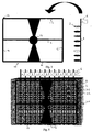
Figure 1 represents a metal or plastic rod (1), from which protrude the equally distanced small cylinders (2) of solid adsorbent material, such polystyrene or nylon for example. The small cylinders (2) can also be single and in that case they are fixed by means of joints (3) to the notches (4) made in the rod (1). The system of fixing joint (3) to notch (4) allows assembly at will of the small cylinders (2) to the rod (1). - In
figure 1 there are eight of the small cylinders (2) set out on the rod, but they could also be four or twelve. -
Figure 2 shows a rod (1), where the small cylinders (2) have all been previously sensitised with a different protein reagent (antibody or antigen), which is immersed into the container (5a) for the sample for the search for antigens and antibodies. - The rod has a label (6) that is detached and inserted into the lid of the sample's container (5b).
- The transport time for the sample's container (5) with the rod (1) furnished with small cylinders (2) is used as incubation time for the formation of the immunocomplex on each individual small cylinder (2).
-
Figure 3 shows how, at the end of the incubation period, each rod (1) with the small cylinders (2) with labels (6) is placed on a grill (10) formed by at least three parallel horizontal sides (11, 12, and 13) and with at least two parallel vertical sides (14 and 15) and with a handle (16) for lifting and/or transport. - For the housing of the rods, twelve notches are available, equally distanced on the horizontal sides (11, 12, and 13) and eight notches equally distanced on the vertical sides (14 and 15).
- To indicate the direction for loading the rods onto the grill (10) a coloured button (16) is displayed on it.
- On the grill (10) the rods (1) are laid out according to the coloured button (16). In
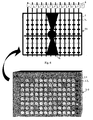
figure 4 the rods (1) are placed in the notches present on the parallel horizontal sides (11, 12, and 13), and a microplate (20) is shown, that has ninety-six microwells (21) laid out in twelve columns numbered from 1 to 12 and eight lines indicated from A to H. The lines are also distinguished by differently coloured small squares (22). - The grill (10) is of a shape and size sufficient to consent the loading of the rods (1) in such a way as to make the position of the small cylinders (2) coincide with that of the same microwells (21) disposed on the microplate (20), as illustrated in
figure 5 . -
Figure 6 shows the grill (10) that is lifted from the microplate (20) for cleaning. - In
figure 7 a rod (1a) is shown, with four small cylinders (2) and with a label (6). The rod (1a) in the same figure is laid on the twelve columns of the microplate (20) from one side to the other, doubling the microplates capacity to carry out analyses on double the number of samples. Consequently the small coloured squares (22) that will be symmetrically repeated are modified on the microplate. - As illustrated in
figure 8 , the method lends itself to the creation of kits for the examination of samples in the field or in laboratories (medical or veterinary), thanks to the simplification of the procedures for the distribution of the sample, the cleaning at the solid phase, and to the possibility to carry out simultaneously the detecting of more antibodies and/or antigens. In this case the spectrophotometer reading can be substituted by a visible reading of the results of the test. -
Figure 8 represents the steps to carry out for an analysis of a single rod (1a) with four small cylinders (2). -
Step 1. Taking the sample and introducing the rod into the container (5) (eventually graduated and already containing the diluting solution). -
Step 2. Incubation at room temperature. -
Step 3. Three passages in test-tubes (7) containing the cleaning liquid. -
Step 4. The introduction into a microstrip (25a) that already contains the specific conjugates. -
Step 5. Incubation at room temperature. -
Step 6. Three passages in test-tubes (7) containing the cleaning liquid. - Step7. The introduction into a microstrip (25b) that already contains the chromogenous sublayer.
-
Step 8. Incubation at room temperature. -
Step 9. The visible reading of the results. - On each microplate (25a) or (25b) made from a single column of microwells, that is a microstrip, the antibodies or antigens to be sought are indicated by specific small coloured squares (22) - for example, antigen A, antigen B, monoclonal antibody, no reagent (negative control) - the colour of which corresponds to that of the small cylinders (2).
- For each microplate or series of microplates or microstrips, the positive control is prepared by previously placing a rod in incubation - the small cylinders of this rod are immersed in microwells that contain the specific reagents for the positive control of the test (antigens and antibodies) - for the necessary period and at a suitable temperature (the small cylinder that has not been sensitised by any reagent is inserted into a dimple without any specific reagent and serves as an ulterior negative control); the rods on which the immunocomplex has formed can eventually be conserved, ready for use.
- The positive control rod is then inserted into the grill in which the rods extracted from the samples are loaded, and is examined at the same time as these rods, and with them follows the above described test procedures.
- The invention, it must be noted, is not limited in use to the examples given in the illustrations, but can be modified and perfected by anyone skilled in the art without breaking patent.
Claims (11)
- Method to simultaneously detect different antibodies and antigens via immunoenzimatic tests and ELISA (Enzyme Linked ImmnoSorbent Assay), characterised by the fact that the solid phase on which the immunocomplex forms is constituted by small cylinders (2) disposed on a rod (1) that the small cylinders placed on the rod are sensitised with different reagents, immerging them into the microwells of a microplate; that the small cylinders of each rod once sensitised, said rod is inserted into a container (5a) with the sample to be analysed; that once the incubation period for the sample in the container has terminated, each rod is placed on a support; that the small cylinders of each rod on the support are cleaned; that the small cylinders of each rod on the support are inserted into the microwells of a microplate, containing specific conjugates at a suitable temperature for a period of incubation; that, once the incubation terminates, the small cylinders on the support are lifted from the microplate and cleaned; that the small cylinders from each rod on the support are inserted into the microwells of a microplate, containing chromogenous sublayer at a suitable temperature for a period of incubation; that the small cylinders of each rod on the support are extracted and the results are read.
- Method to simultaneously detect different antibodies and antigens via the immunoenzimatic tests and ELISA (Enzyme Linked ImmunoSorbent Assay) according to claim 1, characterised by the fact that container for the sample to be analysed is constituted by a microplate that has not been sensitised and that on such a microplate the grill, furnished with rods bearing the small cylinders is placed, for a suitable incubation period at the necessary temperature.
- Device to simultaneously detect different antibodies and antigens via immunoenzimatic tests and ELISA (Enzyme Linked ImmunoSorbent Assay) constituted by small adsorbent cylinders (2) on which the immunocomplexes form, characterised by the fact that said small adsorbent cylinders are blocked at a modular distance on a rod (1); that said rod carries a label (6) to identify the sample under examination; that said rod bearing the small cylinders is placed onto a support; that said support with said rod with the small cylinders is positioned above a microplate furnished with microwells placed at modular distances, that said small cylinders projecting from the rod held by the support are placed at the same modular distance at which the microwells are placed; that said small cylinders, when the support is placed above the microplate, penetrate into the microwells filled with the specific conjugates and later with the chromogenous compound.
- Device to simultaneously detect different antibodies and antigens via immunoenzimatic tests and ELISA (Enzyme Linked ImmunoSorbent Assay) according to claim 3 characterised by the fact that said support is a grill formed of at least two parallel horizontal sides and by at least two vertical parallel sides, that said grill has a handle for transport and lifting, that on said horizontal and vertical sides there are notches for situating the rods, and that on said grill there is a coloured button that indicates the direction of loading.
- Device to simultaneously detect different antibodies and antigens via immunoenzimatic tests and ELISA (Enzyme Linked ImmunoSorbent Assay) according to claim 4 characterised by the fact that said grill receives twelve rods each with eight small cylinders, that said grill is positioned onto a microplate with ninety-six microwells positioned in twelve columns and eight lines at the modular distance of the small cylinders supported by the grill; that the small cylinders penetrate into said microwells present on said microplate.
- Device to simultaneously detect different antibodies and antigens via immunoenzimatic tests and ELISA (Enzyme Linked ImmunoSorbent Assay) according to claim 4 characterised by the fact that said grill receives eight rods each with 12 small cylinders, that said grill is placed onto a microplate with ninety-six microwells arranged in eight columns and twelve lines at the modular distance of the small cylinders supported by the grill; that the small cylinders penetrate into said microwells present on said microplate.
- Device to simultaneously detect different antibodies and antigens via immunoenzimatic tests and ELISA (Enzyme Linked ImmunoSorbent Essay) according to claim 4 characterised by the fact that said grill receives twenty four rods each with four small cylinders, that said rods are arranged symmetrically on said grill; that said grill is positioned on a microplate with ninety-six microwells arranged in twelve columns and eight lines at the modular distance of the small cylinders supported by the grill; that the small cylinders penetrate into the said microwells present on the said microplate.
- Device to simultaneously detect different antibodies and antigens via immunoenzimatic tests and ELISA (Enzyme Linked ImmunoSorbent Assay) according to claim 4 characterised by the fact that said grill receives sixteen rods each with six small cylinders, that said rods are arranged symmetrically on said grill; that said grill is placed onto a microplate with ninety-six microwells arranged in twelve columns and eight lines at the modular distance of the small cylinders supported by the grill; that the small cylinders penetrate said microwells present on said microplate.
- Device to simultaneously detect different antibodies and antigens via immunoenzimatic tests and ELISA (Enzyme Linked ImmunoSorbent Assay) according to claim 3 characterised by the fact that said rod carries small cylinders, that said microplate is a microstrip in which microwells are present; that said small cylinders penetrate into said microwells present in the microstrip; that said microwells are distinguished by small coloured squares, the colours of which correspond to those of the small cylinders of the rod.
- Device to simultaneously detect different antibodies and antigens via immunoenzimatic tests and ELISA (Enzyme Linked ImmonoSorbent Assay) according to claim 3 characterised by the fact that said rod bearing the small cylinders has a place to position the card bearing the identification code of the sample, and taken from which the card can be inserted into a specific holder on the cover or lid of the container for samples and that said cover or lid also has an external site for the card identifying the sample.
- Device to simultaneously detect different antibodies and antigens via immunoenzimatic tests and ELISA (Enzyme Linked ImmunoSorbent Assay) according to claims 3 and 9 characterised by the fact that the rods, the small cylinders, the containers for samples and the microstrips are constructed entirely for the carrying out of the test in the field or in non-specialist surgeries or laboratories.
Applications Claiming Priority (3)
| Application Number | Priority Date | Filing Date | Title |
|---|---|---|---|
| ITCZ20020002 | 2002-04-11 | ||
| IT2002CZ000002A ITCZ20020002A1 (en) | 2002-04-11 | 2002-04-11 | DEVICE AND METHOD FOR SIMULTANEOUS DETECTION OF DIFFERENT ANTIBODIES AND ANTIGENS IN CLINICAL, FOOD AND ENVIRONMENTAL SAMPLES |
| PCT/IT2003/000218 WO2003085401A1 (en) | 2002-04-11 | 2003-04-09 | Device and method to simultaneously detect different antibodies and antigens in clinical alimentary and environmental samples |
Publications (2)
| Publication Number | Publication Date |
|---|---|
| EP1499894A1 EP1499894A1 (en) | 2005-01-26 |
| EP1499894B1 true EP1499894B1 (en) | 2009-02-25 |
Family
ID=28687149
Family Applications (1)
| Application Number | Title | Priority Date | Filing Date |
|---|---|---|---|
| EP03720873A Expired - Lifetime EP1499894B1 (en) | 2002-04-11 | 2003-04-09 | Device and method to simultaneously detect different antibodies and antigens in clinical alimentary and environmental samples |
Country Status (9)
| Country | Link |
|---|---|
| US (2) | US7510687B2 (en) |
| EP (1) | EP1499894B1 (en) |
| CN (1) | CN100476434C (en) |
| AT (1) | ATE423972T1 (en) |
| AU (1) | AU2003224447A1 (en) |
| DE (1) | DE60326336D1 (en) |
| ES (1) | ES2323951T3 (en) |
| IT (1) | ITCZ20020002A1 (en) |
| WO (1) | WO2003085401A1 (en) |
Families Citing this family (33)
| Publication number | Priority date | Publication date | Assignee | Title |
|---|---|---|---|---|
| ITCZ20020002A1 (en) * | 2002-04-11 | 2003-10-13 | Parco Scient E Tecnologico Del | DEVICE AND METHOD FOR SIMULTANEOUS DETECTION OF DIFFERENT ANTIBODIES AND ANTIGENS IN CLINICAL, FOOD AND ENVIRONMENTAL SAMPLES |
| WO2012076623A1 (en) | 2010-12-08 | 2012-06-14 | Intervet International B.V. | A method to quantify the amount of a biological substance in a sample and a kit for performing the method |
| ITCS20110012A1 (en) * | 2011-04-21 | 2012-10-22 | Uni Degli Studi Del Molise | ANALYTICAL COMPETITION METHOD BETWEEN 2 SOLID PHASES FOR THE SIMULTANEOUS DETECTION OF DIFFERENT CELLULAR OR MOLECULAR MARKERS, DEVICE CONSTITUTED BY MICROPLATE OR MICROSTRIP WITH EXTENDED SHAPES FOR THE EXECUTION OF SUCH METHOD AND RELAT. |
| WO2012143912A1 (en) | 2011-04-21 | 2012-10-26 | Università Degli Studi Del Molise | Device, method and kit for the detection of different markers in different cellular or molecular types and their quantification |
| US10598666B2 (en) | 2012-03-08 | 2020-03-24 | Glaxosmithkline Biologicals Sa | In vitro potency assay for protein-based meningococcal vaccines |
| WO2014189843A1 (en) | 2013-05-20 | 2014-11-27 | Board Of Trustees Of The University Of Arkansas | Gep5 model for multiple myeloma |
| WO2014198836A1 (en) * | 2013-06-14 | 2014-12-18 | Hahn-Schickard-Gesellschaft für angewandte Forschung e.V. | Elisa system and related methods |
| EP2835178B1 (en) | 2013-08-06 | 2017-04-12 | Yantai AusBio Laboratories Co., Ltd. | Centrifuge and method for centrifuging a reaction vessel unit |
| EP2982439B1 (en) | 2014-08-06 | 2017-10-11 | Yantai AusBio Laboratories Co., Ltd. | Reagent carrier unit with coupling section to permit pipettor arm attachment and handling |
| WO2017044691A1 (en) * | 2015-09-09 | 2017-03-16 | Advantage Allergy Services, Llc | Systems and methods for testing and treatment of allergies |
| ES2926513T3 (en) | 2016-07-29 | 2022-10-26 | Juno Therapeutics Inc | Methods for Assessing the Presence or Absence of Replication-Competent Virus |
| CN106990233B (en) * | 2017-05-27 | 2019-05-07 | 于宁 | A kind of convex bubble plate of immune detection |
| CN107389933B (en) * | 2017-06-14 | 2019-07-23 | 杨华卫 | A kind of biochip |
| CN107418874B (en) * | 2017-06-14 | 2021-05-07 | 杨华卫 | Biological chip |
| CN107340388B (en) * | 2017-06-14 | 2019-07-23 | 杨华卫 | A kind of biomolecule detecting method |
| JP2020526194A (en) | 2017-06-29 | 2020-08-31 | ジュノー セラピューティクス インコーポレイテッド | Mouse model for assessing toxicity associated with immunotherapeutic agents |
| BR112020001719A2 (en) | 2017-07-29 | 2020-07-21 | Juno Therapeutics Inc | reagents for cell expansion that express recombinant receptors |
| MA50057A (en) | 2017-09-01 | 2020-07-08 | Juno Therapeutics Inc | GENE EXPRESSION AND ASSESSMENT OF A RISK OF DEVELOPING TOXICITY FOLLOWING CELL THERAPY |
| SG11202003657VA (en) | 2017-11-01 | 2020-05-28 | Juno Therapeutics Inc | Process for producing a t cell composition |
| US10191036B1 (en) | 2018-03-22 | 2019-01-29 | NUB4U, Inc. | System for detecting and removing biological analytes in fluids |
| EP3877054B1 (en) | 2018-11-06 | 2023-11-01 | Juno Therapeutics, Inc. | Process for producing genetically engineered t cells |
| KR102151648B1 (en) * | 2018-12-03 | 2020-09-03 | 광운대학교 산학협력단 | Microfluidic adapter |
| MX2021012607A (en) | 2019-04-17 | 2022-03-11 | Alpine Immune Sciences Inc | Methods and uses of variant icos ligand (icosl) fusion proteins. |
| US20220249637A1 (en) | 2019-06-12 | 2022-08-11 | Juno Therapeutics, Inc. | Combination therapy of a cell-mediated cytotoxic therapy and an inhibitor of a prosurvival bcl2 family protein |
| WO2021035194A1 (en) | 2019-08-22 | 2021-02-25 | Juno Therapeutics, Inc. | Combination therapy of a t cell therapy and an enhancer of zeste homolog 2 (ezh2) inhibitor and related methods |
| WO2021146349A1 (en) | 2020-01-13 | 2021-07-22 | Aspen Neuroscience, Inc. | Method of differentiating neural cells and related compositions and methods of use |
| KR20230022868A (en) | 2020-05-13 | 2023-02-16 | 주노 쎄러퓨티크스 인코퍼레이티드 | Method for producing donor-batch cells expressing a recombinant acceptor |
| WO2021260186A1 (en) | 2020-06-26 | 2021-12-30 | Juno Therapeutics Gmbh | Engineered t cells conditionally expressing a recombinant receptor, related polynucleotides and methods |
| WO2023004370A1 (en) | 2021-07-21 | 2023-01-26 | Aspen Neuroscience, Inc. | Aav-based modulation of gba1 and related compositions and uses thereof |
| US20230081881A1 (en) | 2021-07-21 | 2023-03-16 | Aspen Neuroscience, Inc. | Transposon-based modulation of gba1 and related compositions and uses thereof |
| WO2023004371A1 (en) | 2021-07-21 | 2023-01-26 | Aspen Neuroscience, Inc. | Methods of differentiating neural cells and predicting engraftment thereof and related compositions |
| US20230377685A1 (en) | 2022-04-15 | 2023-11-23 | Aspen Neuroscience, Inc. | Methods of classifying the differentiation state of cells and related compositions of differentiated cells |
| WO2023230581A1 (en) | 2022-05-25 | 2023-11-30 | Celgene Corporation | Methods of manufacturing t cell therapies |
Family Cites Families (20)
| Publication number | Priority date | Publication date | Assignee | Title |
|---|---|---|---|---|
| US4025391A (en) * | 1974-06-15 | 1977-05-24 | Director Of National Food Research Institute | Preparation of bead-shaped immobilized enzyme |
| AT343822B (en) * | 1976-08-20 | 1978-06-26 | Immuno Ag | RADIOIMMUNOLOGICAL METHOD AND EQUIPMENT FOR DETERMINING ANTIGENES |
| DE3368534D1 (en) * | 1982-03-03 | 1987-02-05 | Becton Dickinson Co | Multi-well screening assembly for immunoassay procedures |
| DE3224324A1 (en) | 1982-06-30 | 1984-01-05 | Basf Ag, 6700 Ludwigshafen | TWO-STAGE PROCESS FOR PRODUCING THERMOPLASTIC POLYURETHANE ELASTOMERS |
| GB2175304B (en) * | 1985-05-17 | 1988-10-26 | Mitsubishi Petrochemical Co | Method of preparing l-malic acid |
| US5250412A (en) * | 1987-03-23 | 1993-10-05 | Diamedix Corporation | Swab device and method for collecting and analyzing a sample |
| US4891321A (en) * | 1987-10-21 | 1990-01-02 | Hubscher Thomas T | Apparatus for performing determinations of immune reactants in biological fluids |
| US5409611A (en) * | 1988-03-24 | 1995-04-25 | Terrapin Technoogies, Inc. | Method to identify analyte-binding ligands |
| US5417923A (en) * | 1991-04-24 | 1995-05-23 | Pfizer Inc. | Assay tray assembly |
| DE4120139A1 (en) * | 1991-06-19 | 1992-12-24 | Bundesamt Fuer Wehrtechnik U B | Time-saving method for fixed immunoassays in diagnostics - comprises covering pin-surfaces with the substrate to be measured and immersing the pins in plate with complementary cavities |
| SE9203320D0 (en) * | 1992-11-06 | 1992-11-06 | Pharmacia Lkb Biotech | A METHOD OF PROCESSING NUCLEIC ACID SAMPLES |
| SE9402518D0 (en) * | 1994-07-18 | 1994-07-18 | Pharmacia Biotech Ab | Processing system |
| GB2295152A (en) * | 1994-11-18 | 1996-05-22 | Pfizer Ltd | Preparation of a library of compounds by solid-phase synthesis |
| EP0781997A1 (en) * | 1995-12-27 | 1997-07-02 | FUJIREBIO Inc. | Instrument for simplified immunoassay and simplified immunoassay method using the instrument |
| US5951783A (en) * | 1998-05-15 | 1999-09-14 | Bio-Tek Holdings, Inc. | Universal washing apparatus for microtiter plate and the like |
| US7070740B1 (en) * | 2000-09-28 | 2006-07-04 | Beckman Coulter, Inc. | Method and apparatus for processing biomolecule arrays |
| US6942836B2 (en) * | 2001-10-16 | 2005-09-13 | Applera Corporation | System for filling substrate chambers with liquid |
| ITCZ20020002A1 (en) * | 2002-04-11 | 2003-10-13 | Parco Scient E Tecnologico Del | DEVICE AND METHOD FOR SIMULTANEOUS DETECTION OF DIFFERENT ANTIBODIES AND ANTIGENS IN CLINICAL, FOOD AND ENVIRONMENTAL SAMPLES |
| JP2004045104A (en) * | 2002-07-09 | 2004-02-12 | Futaba Corp | Microplate |
| US7578978B2 (en) * | 2003-10-28 | 2009-08-25 | BIOMéRIEUX, INC. | Carrier for holding test samples |
-
2002
- 2002-04-11 IT IT2002CZ000002A patent/ITCZ20020002A1/en unknown
-
2003
- 2003-04-09 CN CNB038100290A patent/CN100476434C/en not_active Expired - Fee Related
- 2003-04-09 AT AT03720873T patent/ATE423972T1/en not_active IP Right Cessation
- 2003-04-09 AU AU2003224447A patent/AU2003224447A1/en not_active Abandoned
- 2003-04-09 EP EP03720873A patent/EP1499894B1/en not_active Expired - Lifetime
- 2003-04-09 ES ES03720873T patent/ES2323951T3/en not_active Expired - Lifetime
- 2003-04-09 WO PCT/IT2003/000218 patent/WO2003085401A1/en not_active Application Discontinuation
- 2003-04-09 DE DE60326336T patent/DE60326336D1/en not_active Expired - Lifetime
-
2004
- 2004-10-08 US US10/711,847 patent/US7510687B2/en not_active Expired - Fee Related
-
2007
- 2007-10-15 US US11/872,334 patent/US20080090262A1/en not_active Abandoned
Also Published As
| Publication number | Publication date |
|---|---|
| CN1650167A (en) | 2005-08-03 |
| EP1499894A1 (en) | 2005-01-26 |
| ATE423972T1 (en) | 2009-03-15 |
| ES2323951T3 (en) | 2009-07-28 |
| CN100476434C (en) | 2009-04-08 |
| WO2003085401A1 (en) | 2003-10-16 |
| ITCZ20020002A1 (en) | 2003-10-13 |
| US20060078954A1 (en) | 2006-04-13 |
| DE60326336D1 (en) | 2009-04-09 |
| US20080090262A1 (en) | 2008-04-17 |
| US7510687B2 (en) | 2009-03-31 |
| AU2003224447A1 (en) | 2003-10-20 |
Similar Documents
| Publication | Publication Date | Title |
|---|---|---|
| EP1499894B1 (en) | Device and method to simultaneously detect different antibodies and antigens in clinical alimentary and environmental samples | |
| EP0204109B1 (en) | A self-contained reagent package device for an assay | |
| US5116576A (en) | Device for analytical determinations | |
| JP5185335B2 (en) | Biochips for archiving biological samples and clinical analysis | |
| JPH04290961A (en) | Device for effecting rapid and easy manual assay | |
| JPS58501521A (en) | automatic immunoassay system | |
| CN104024853A (en) | Sensor cartridge for detecting component of at least one sample | |
| CZ301093B6 (en) | Method for detecting analyte in liquid dairy product and testing device for performing this method | |
| EP1667780B1 (en) | Method of detecting multiple analytes | |
| JP3628709B2 (en) | Test kit and its use | |
| EP0359550B1 (en) | Analytic reader device | |
| EP0324603A2 (en) | Improved solid phase enzyme immunoassay test apparatus and process for detection antibodies | |
| WO2014168580A1 (en) | Double-chamber bi-directional reverse flow device | |
| EP0320752A1 (en) | Analytical reagent mixing apparatus for performing sequential analytical reactions | |
| EP0194789A2 (en) | Apparatus and method for multiple simultaneous assay | |
| JP2004177321A (en) | Method and apparatus for detecting trace amount | |
| NO860937L (en) | DIAGNOSTICS FOR ASSAY PROVISIONS. | |
| KR20040047144A (en) | Microvolume detecting method and device | |
| US20040005627A1 (en) | Microvolume detecting method and device | |
| JPS61280567A (en) | Device and method for majority simultaneous assay | |
| US20110218117A1 (en) | Enhanced Immunosorbent Spot Test Device and Method of Using Same |
Legal Events
| Date | Code | Title | Description |
|---|---|---|---|
| PUAI | Public reference made under article 153(3) epc to a published international application that has entered the european phase |
Free format text: ORIGINAL CODE: 0009012 |
|
| 17P | Request for examination filed |
Effective date: 20041029 |
|
| AK | Designated contracting states |
Kind code of ref document: A1 Designated state(s): AT BE BG CH CY CZ DE DK EE ES FI FR GB GR HU IE IT LI LU MC NL PT RO SE SI SK TR |
|
| AX | Request for extension of the european patent |
Extension state: AL LT LV MK |
|
| GRAP | Despatch of communication of intention to grant a patent |
Free format text: ORIGINAL CODE: EPIDOSNIGR1 |
|
| GRAS | Grant fee paid |
Free format text: ORIGINAL CODE: EPIDOSNIGR3 |
|
| GRAA | (expected) grant |
Free format text: ORIGINAL CODE: 0009210 |
|
| AK | Designated contracting states |
Kind code of ref document: B1 Designated state(s): AT BE BG CH CY CZ DE DK EE ES FI FR GB GR HU IE IT LI LU MC NL PT RO SE SI SK TR |
|
| REG | Reference to a national code |
Ref country code: GB Ref legal event code: FG4D |
|
| REG | Reference to a national code |
Ref country code: CH Ref legal event code: EP |
|
| REG | Reference to a national code |
Ref country code: IE Ref legal event code: FG4D |
|
| REF | Corresponds to: |
Ref document number: 60326336 Country of ref document: DE Date of ref document: 20090409 Kind code of ref document: P |
|
| REG | Reference to a national code |
Ref country code: SE Ref legal event code: TRGR |
|
| REG | Reference to a national code |
Ref country code: ES Ref legal event code: FG2A Ref document number: 2323951 Country of ref document: ES Kind code of ref document: T3 |
|
| PG25 | Lapsed in a contracting state [announced via postgrant information from national office to epo] |
Ref country code: SI Free format text: LAPSE BECAUSE OF FAILURE TO SUBMIT A TRANSLATION OF THE DESCRIPTION OR TO PAY THE FEE WITHIN THE PRESCRIBED TIME-LIMIT Effective date: 20090225 Ref country code: FI Free format text: LAPSE BECAUSE OF FAILURE TO SUBMIT A TRANSLATION OF THE DESCRIPTION OR TO PAY THE FEE WITHIN THE PRESCRIBED TIME-LIMIT Effective date: 20090225 |
|
| PGFP | Annual fee paid to national office [announced via postgrant information from national office to epo] |
Ref country code: AT Payment date: 20090527 Year of fee payment: 7 |
|
| PG25 | Lapsed in a contracting state [announced via postgrant information from national office to epo] |
Ref country code: BE Free format text: LAPSE BECAUSE OF FAILURE TO SUBMIT A TRANSLATION OF THE DESCRIPTION OR TO PAY THE FEE WITHIN THE PRESCRIBED TIME-LIMIT Effective date: 20090225 |
|
| PG25 | Lapsed in a contracting state [announced via postgrant information from national office to epo] |
Ref country code: DK Free format text: LAPSE BECAUSE OF FAILURE TO SUBMIT A TRANSLATION OF THE DESCRIPTION OR TO PAY THE FEE WITHIN THE PRESCRIBED TIME-LIMIT Effective date: 20090225 Ref country code: EE Free format text: LAPSE BECAUSE OF FAILURE TO SUBMIT A TRANSLATION OF THE DESCRIPTION OR TO PAY THE FEE WITHIN THE PRESCRIBED TIME-LIMIT Effective date: 20090225 Ref country code: CZ Free format text: LAPSE BECAUSE OF FAILURE TO SUBMIT A TRANSLATION OF THE DESCRIPTION OR TO PAY THE FEE WITHIN THE PRESCRIBED TIME-LIMIT Effective date: 20090225 Ref country code: PT Free format text: LAPSE BECAUSE OF FAILURE TO SUBMIT A TRANSLATION OF THE DESCRIPTION OR TO PAY THE FEE WITHIN THE PRESCRIBED TIME-LIMIT Effective date: 20090812 |
|
| PGFP | Annual fee paid to national office [announced via postgrant information from national office to epo] |
Ref country code: CH Payment date: 20090605 Year of fee payment: 7 |
|
| PG25 | Lapsed in a contracting state [announced via postgrant information from national office to epo] |
Ref country code: RO Free format text: LAPSE BECAUSE OF FAILURE TO SUBMIT A TRANSLATION OF THE DESCRIPTION OR TO PAY THE FEE WITHIN THE PRESCRIBED TIME-LIMIT Effective date: 20090225 Ref country code: SK Free format text: LAPSE BECAUSE OF FAILURE TO SUBMIT A TRANSLATION OF THE DESCRIPTION OR TO PAY THE FEE WITHIN THE PRESCRIBED TIME-LIMIT Effective date: 20090225 |
|
| PGFP | Annual fee paid to national office [announced via postgrant information from national office to epo] |
Ref country code: GB Payment date: 20090601 Year of fee payment: 7 |
|
| PLBE | No opposition filed within time limit |
Free format text: ORIGINAL CODE: 0009261 |
|
| STAA | Information on the status of an ep patent application or granted ep patent |
Free format text: STATUS: NO OPPOSITION FILED WITHIN TIME LIMIT |
|
| NLV4 | Nl: lapsed or anulled due to non-payment of the annual fee |
Effective date: 20091101 |
|
| PG25 | Lapsed in a contracting state [announced via postgrant information from national office to epo] |
Ref country code: BG Free format text: LAPSE BECAUSE OF FAILURE TO SUBMIT A TRANSLATION OF THE DESCRIPTION OR TO PAY THE FEE WITHIN THE PRESCRIBED TIME-LIMIT Effective date: 20090525 |
|
| 26N | No opposition filed |
Effective date: 20091126 |
|
| PG25 | Lapsed in a contracting state [announced via postgrant information from national office to epo] |
Ref country code: NL Free format text: LAPSE BECAUSE OF NON-PAYMENT OF DUE FEES Effective date: 20091101 |
|
| PG25 | Lapsed in a contracting state [announced via postgrant information from national office to epo] |
Ref country code: IE Free format text: LAPSE BECAUSE OF NON-PAYMENT OF DUE FEES Effective date: 20090409 Ref country code: MC Free format text: LAPSE BECAUSE OF NON-PAYMENT OF DUE FEES Effective date: 20090430 |
|
| PG25 | Lapsed in a contracting state [announced via postgrant information from national office to epo] |
Ref country code: GR Free format text: LAPSE BECAUSE OF FAILURE TO SUBMIT A TRANSLATION OF THE DESCRIPTION OR TO PAY THE FEE WITHIN THE PRESCRIBED TIME-LIMIT Effective date: 20090526 |
|
| REG | Reference to a national code |
Ref country code: CH Ref legal event code: PL |
|
| GBPC | Gb: european patent ceased through non-payment of renewal fee |
Effective date: 20100409 |
|
| PG25 | Lapsed in a contracting state [announced via postgrant information from national office to epo] |
Ref country code: AT Free format text: LAPSE BECAUSE OF NON-PAYMENT OF DUE FEES Effective date: 20100409 |
|
| PG25 | Lapsed in a contracting state [announced via postgrant information from national office to epo] |
Ref country code: DE Free format text: LAPSE BECAUSE OF NON-PAYMENT OF DUE FEES Effective date: 20101103 Ref country code: LI Free format text: LAPSE BECAUSE OF NON-PAYMENT OF DUE FEES Effective date: 20100430 Ref country code: CH Free format text: LAPSE BECAUSE OF NON-PAYMENT OF DUE FEES Effective date: 20100430 |
|
| PG25 | Lapsed in a contracting state [announced via postgrant information from national office to epo] |
Ref country code: GB Free format text: LAPSE BECAUSE OF NON-PAYMENT OF DUE FEES Effective date: 20100409 |
|
| PG25 | Lapsed in a contracting state [announced via postgrant information from national office to epo] |
Ref country code: LU Free format text: LAPSE BECAUSE OF NON-PAYMENT OF DUE FEES Effective date: 20090409 |
|
| PG25 | Lapsed in a contracting state [announced via postgrant information from national office to epo] |
Ref country code: HU Free format text: LAPSE BECAUSE OF FAILURE TO SUBMIT A TRANSLATION OF THE DESCRIPTION OR TO PAY THE FEE WITHIN THE PRESCRIBED TIME-LIMIT Effective date: 20090826 |
|
| PG25 | Lapsed in a contracting state [announced via postgrant information from national office to epo] |
Ref country code: TR Free format text: LAPSE BECAUSE OF FAILURE TO SUBMIT A TRANSLATION OF THE DESCRIPTION OR TO PAY THE FEE WITHIN THE PRESCRIBED TIME-LIMIT Effective date: 20090225 |
|
| REG | Reference to a national code |
Ref country code: DE Ref legal event code: R073 Ref document number: 60326336 Country of ref document: DE |
|
| PG25 | Lapsed in a contracting state [announced via postgrant information from national office to epo] |
Ref country code: CY Free format text: LAPSE BECAUSE OF FAILURE TO SUBMIT A TRANSLATION OF THE DESCRIPTION OR TO PAY THE FEE WITHIN THE PRESCRIBED TIME-LIMIT Effective date: 20090225 |
|
| REG | Reference to a national code |
Ref country code: DE Ref legal event code: R073 Ref document number: 60326336 Country of ref document: DE Ref country code: DE Ref legal event code: R074 Ref document number: 60326336 Country of ref document: DE |
|
| REG | Reference to a national code |
Ref country code: DE Ref legal event code: R074 Ref document number: 60326336 Country of ref document: DE Effective date: 20111017 |
|
| PGFP | Annual fee paid to national office [announced via postgrant information from national office to epo] |
Ref country code: SE Payment date: 20120425 Year of fee payment: 10 |
|
| PGFP | Annual fee paid to national office [announced via postgrant information from national office to epo] |
Ref country code: ES Payment date: 20120423 Year of fee payment: 10 |
|
| PGRI | Patent reinstated in contracting state [announced from national office to epo] |
Ref country code: DE Effective date: 20111014 |
|
| REG | Reference to a national code |
Ref country code: SE Ref legal event code: EUG |
|
| PG25 | Lapsed in a contracting state [announced via postgrant information from national office to epo] |
Ref country code: SE Free format text: LAPSE BECAUSE OF NON-PAYMENT OF DUE FEES Effective date: 20130410 |
|
| REG | Reference to a national code |
Ref country code: ES Ref legal event code: FD2A Effective date: 20140609 |
|
| PG25 | Lapsed in a contracting state [announced via postgrant information from national office to epo] |
Ref country code: ES Free format text: LAPSE BECAUSE OF NON-PAYMENT OF DUE FEES Effective date: 20130410 |
|
| REG | Reference to a national code |
Ref country code: FR Ref legal event code: PLFP Year of fee payment: 13 |
|
| PGFP | Annual fee paid to national office [announced via postgrant information from national office to epo] |
Ref country code: DE Payment date: 20150630 Year of fee payment: 13 |
|
| PGFP | Annual fee paid to national office [announced via postgrant information from national office to epo] |
Ref country code: IT Payment date: 20150430 Year of fee payment: 13 Ref country code: FR Payment date: 20150430 Year of fee payment: 13 |
|
| REG | Reference to a national code |
Ref country code: DE Ref legal event code: R119 Ref document number: 60326336 Country of ref document: DE |
|
| REG | Reference to a national code |
Ref country code: FR Ref legal event code: ST Effective date: 20161230 |
|
| PG25 | Lapsed in a contracting state [announced via postgrant information from national office to epo] |
Ref country code: FR Free format text: LAPSE BECAUSE OF NON-PAYMENT OF DUE FEES Effective date: 20160502 |
|
| PG25 | Lapsed in a contracting state [announced via postgrant information from national office to epo] |
Ref country code: IT Free format text: LAPSE BECAUSE OF NON-PAYMENT OF DUE FEES Effective date: 20160409 |
|
| PG25 | Lapsed in a contracting state [announced via postgrant information from national office to epo] |
Ref country code: DE Free format text: LAPSE BECAUSE OF NON-PAYMENT OF DUE FEES Effective date: 20161101 |