EP1444267B1 - Affinity enhancement agents - Google Patents
Affinity enhancement agents Download PDFInfo
- Publication number
- EP1444267B1 EP1444267B1 EP02770573.0A EP02770573A EP1444267B1 EP 1444267 B1 EP1444267 B1 EP 1444267B1 EP 02770573 A EP02770573 A EP 02770573A EP 1444267 B1 EP1444267 B1 EP 1444267B1
- Authority
- EP
- European Patent Office
- Prior art keywords
- binding protein
- diabody
- amino acid
- polypeptide
- binding
- Prior art date
- Legal status (The legal status is an assumption and is not a legal conclusion. Google has not performed a legal analysis and makes no representation as to the accuracy of the status listed.)
- Expired - Lifetime
Links
- 108090000765 processed proteins & peptides Proteins 0.000 claims description 108
- 102000004196 processed proteins & peptides Human genes 0.000 claims description 85
- 230000027455 binding Effects 0.000 claims description 82
- 229920001184 polypeptide Polymers 0.000 claims description 82
- 102000023732 binding proteins Human genes 0.000 claims description 61
- 108091008324 binding proteins Proteins 0.000 claims description 61
- 206010028980 Neoplasm Diseases 0.000 claims description 46
- 102000012406 Carcinoembryonic Antigen Human genes 0.000 claims description 34
- 108010022366 Carcinoembryonic Antigen Proteins 0.000 claims description 34
- 239000003814 drug Substances 0.000 claims description 34
- 239000000427 antigen Substances 0.000 claims description 27
- 125000003275 alpha amino acid group Chemical group 0.000 claims description 26
- 102000036639 antigens Human genes 0.000 claims description 26
- 108091007433 antigens Proteins 0.000 claims description 26
- 238000000034 method Methods 0.000 claims description 26
- 229940124597 therapeutic agent Drugs 0.000 claims description 25
- 210000004369 blood Anatomy 0.000 claims description 24
- 239000008280 blood Substances 0.000 claims description 24
- 239000000032 diagnostic agent Substances 0.000 claims description 20
- 229940039227 diagnostic agent Drugs 0.000 claims description 20
- 108020004707 nucleic acids Proteins 0.000 claims description 19
- 102000039446 nucleic acids Human genes 0.000 claims description 19
- 150000007523 nucleic acids Chemical class 0.000 claims description 19
- 208000037265 diseases, disorders, signs and symptoms Diseases 0.000 claims description 13
- 239000013612 plasmid Substances 0.000 claims description 12
- 239000002299 complementary DNA Substances 0.000 claims description 11
- 108091028043 Nucleic acid sequence Proteins 0.000 claims description 10
- 230000009870 specific binding Effects 0.000 claims description 10
- 125000000539 amino acid group Chemical group 0.000 claims description 9
- 238000003745 diagnosis Methods 0.000 claims description 8
- 208000035475 disorder Diseases 0.000 claims description 7
- 108020004635 Complementary DNA Proteins 0.000 claims description 6
- 201000010099 disease Diseases 0.000 claims description 6
- 201000011510 cancer Diseases 0.000 claims description 5
- 229940079593 drug Drugs 0.000 claims description 5
- 238000002360 preparation method Methods 0.000 claims description 5
- 208000023275 Autoimmune disease Diseases 0.000 claims description 3
- 208000024172 Cardiovascular disease Diseases 0.000 claims description 3
- 208000035473 Communicable disease Diseases 0.000 claims description 3
- 108090000695 Cytokines Proteins 0.000 claims description 3
- 102000004127 Cytokines Human genes 0.000 claims description 3
- 238000012258 culturing Methods 0.000 claims description 3
- 239000003102 growth factor Substances 0.000 claims description 3
- 239000005556 hormone Substances 0.000 claims description 3
- 229940088597 hormone Drugs 0.000 claims description 3
- 208000027866 inflammatory disease Diseases 0.000 claims description 3
- 239000003053 toxin Substances 0.000 claims description 3
- 231100000765 toxin Toxicity 0.000 claims description 3
- 108700012359 toxins Proteins 0.000 claims description 3
- 239000000562 conjugate Substances 0.000 claims description 2
- 108091026890 Coding region Proteins 0.000 description 86
- RAXXELZNTBOGNW-UHFFFAOYSA-N imidazole Natural products C1=CNC=N1 RAXXELZNTBOGNW-UHFFFAOYSA-N 0.000 description 42
- 125000005647 linker group Chemical group 0.000 description 41
- 108090000623 proteins and genes Proteins 0.000 description 38
- 238000003752 polymerase chain reaction Methods 0.000 description 32
- 235000001014 amino acid Nutrition 0.000 description 30
- 229940024606 amino acid Drugs 0.000 description 27
- 238000002347 injection Methods 0.000 description 27
- 239000007924 injection Substances 0.000 description 27
- 102000004169 proteins and genes Human genes 0.000 description 27
- 150000001413 amino acids Chemical class 0.000 description 25
- 239000013598 vector Substances 0.000 description 24
- 101100136076 Aspergillus oryzae (strain ATCC 42149 / RIB 40) pel1 gene Proteins 0.000 description 23
- 101150040383 pel2 gene Proteins 0.000 description 23
- 101150050446 pelB gene Proteins 0.000 description 23
- 235000018102 proteins Nutrition 0.000 description 23
- 210000001519 tissue Anatomy 0.000 description 21
- 108010076504 Protein Sorting Signals Proteins 0.000 description 20
- 210000004027 cell Anatomy 0.000 description 20
- 241000588724 Escherichia coli Species 0.000 description 19
- 230000035772 mutation Effects 0.000 description 19
- 239000013615 primer Substances 0.000 description 18
- HNDVDQJCIGZPNO-UHFFFAOYSA-N histidine Natural products OC(=O)C(N)CC1=CN=CN1 HNDVDQJCIGZPNO-UHFFFAOYSA-N 0.000 description 17
- 238000004458 analytical method Methods 0.000 description 12
- 239000012634 fragment Substances 0.000 description 12
- 238000006467 substitution reaction Methods 0.000 description 12
- 238000004128 high performance liquid chromatography Methods 0.000 description 11
- 238000001597 immobilized metal affinity chromatography Methods 0.000 description 11
- 230000008685 targeting Effects 0.000 description 11
- 125000003178 carboxy group Chemical group [H]OC(*)=O 0.000 description 10
- 239000012614 Q-Sepharose Substances 0.000 description 9
- 210000003734 kidney Anatomy 0.000 description 9
- 108020004414 DNA Proteins 0.000 description 8
- XSQUKJJJFZCRTK-UHFFFAOYSA-N Urea Chemical compound NC(N)=O XSQUKJJJFZCRTK-UHFFFAOYSA-N 0.000 description 8
- 239000000872 buffer Substances 0.000 description 8
- 210000004185 liver Anatomy 0.000 description 8
- 238000000746 purification Methods 0.000 description 8
- 241001529936 Murinae Species 0.000 description 7
- 230000003302 anti-idiotype Effects 0.000 description 7
- 238000001155 isoelectric focusing Methods 0.000 description 7
- 210000004072 lung Anatomy 0.000 description 7
- 238000002415 sodium dodecyl sulfate polyacrylamide gel electrophoresis Methods 0.000 description 7
- 229910000162 sodium phosphate Inorganic materials 0.000 description 7
- 210000000952 spleen Anatomy 0.000 description 7
- ROHFNLRQFUQHCH-UHFFFAOYSA-N Leucine Natural products CC(C)CC(N)C(O)=O ROHFNLRQFUQHCH-UHFFFAOYSA-N 0.000 description 6
- 108091034117 Oligonucleotide Proteins 0.000 description 6
- MTCFGRXMJLQNBG-UHFFFAOYSA-N Serine Natural products OCC(N)C(O)=O MTCFGRXMJLQNBG-UHFFFAOYSA-N 0.000 description 6
- FAPWRFPIFSIZLT-UHFFFAOYSA-M Sodium chloride Chemical compound [Na+].[Cl-] FAPWRFPIFSIZLT-UHFFFAOYSA-M 0.000 description 6
- 238000010367 cloning Methods 0.000 description 6
- 238000010276 construction Methods 0.000 description 6
- 238000010828 elution Methods 0.000 description 6
- 239000013604 expression vector Substances 0.000 description 6
- 239000000203 mixture Substances 0.000 description 6
- 125000002924 primary amino group Chemical group [H]N([H])* 0.000 description 6
- 210000002784 stomach Anatomy 0.000 description 6
- 238000002965 ELISA Methods 0.000 description 5
- ROHFNLRQFUQHCH-YFKPBYRVSA-N L-leucine Chemical compound CC(C)C[C@H](N)C(O)=O ROHFNLRQFUQHCH-YFKPBYRVSA-N 0.000 description 5
- 108010002747 Pfu DNA polymerase Proteins 0.000 description 5
- 101100113084 Schizosaccharomyces pombe (strain 972 / ATCC 24843) mcs2 gene Proteins 0.000 description 5
- 239000003795 chemical substances by application Substances 0.000 description 5
- 239000000539 dimer Substances 0.000 description 5
- 239000000284 extract Substances 0.000 description 5
- 230000006698 induction Effects 0.000 description 5
- 210000002429 large intestine Anatomy 0.000 description 5
- 230000014759 maintenance of location Effects 0.000 description 5
- 230000004044 response Effects 0.000 description 5
- 238000003998 size exclusion chromatography high performance liquid chromatography Methods 0.000 description 5
- 210000000813 small intestine Anatomy 0.000 description 5
- 230000001225 therapeutic effect Effects 0.000 description 5
- QFVHZQCOUORWEI-UHFFFAOYSA-N 4-[(4-anilino-5-sulfonaphthalen-1-yl)diazenyl]-5-hydroxynaphthalene-2,7-disulfonic acid Chemical compound C=12C(O)=CC(S(O)(=O)=O)=CC2=CC(S(O)(=O)=O)=CC=1N=NC(C1=CC=CC(=C11)S(O)(=O)=O)=CC=C1NC1=CC=CC=C1 QFVHZQCOUORWEI-UHFFFAOYSA-N 0.000 description 4
- 239000003155 DNA primer Substances 0.000 description 4
- DHMQDGOQFOQNFH-UHFFFAOYSA-N Glycine Chemical compound NCC(O)=O DHMQDGOQFOQNFH-UHFFFAOYSA-N 0.000 description 4
- QPCDCPDFJACHGM-UHFFFAOYSA-N N,N-bis{2-[bis(carboxymethyl)amino]ethyl}glycine Chemical compound OC(=O)CN(CC(O)=O)CCN(CC(=O)O)CCN(CC(O)=O)CC(O)=O QPCDCPDFJACHGM-UHFFFAOYSA-N 0.000 description 4
- AYFVYJQAPQTCCC-UHFFFAOYSA-N Threonine Natural products CC(O)C(N)C(O)=O AYFVYJQAPQTCCC-UHFFFAOYSA-N 0.000 description 4
- 239000004473 Threonine Substances 0.000 description 4
- 239000007983 Tris buffer Substances 0.000 description 4
- 238000005571 anion exchange chromatography Methods 0.000 description 4
- 239000004202 carbamide Substances 0.000 description 4
- 238000005119 centrifugation Methods 0.000 description 4
- 238000001514 detection method Methods 0.000 description 4
- 238000004519 manufacturing process Methods 0.000 description 4
- 239000008188 pellet Substances 0.000 description 4
- 230000002797 proteolythic effect Effects 0.000 description 4
- 230000000717 retained effect Effects 0.000 description 4
- AJPJDKMHJJGVTQ-UHFFFAOYSA-M sodium dihydrogen phosphate Chemical compound [Na+].OP(O)([O-])=O AJPJDKMHJJGVTQ-UHFFFAOYSA-M 0.000 description 4
- 239000000243 solution Substances 0.000 description 4
- LENZDBCJOHFCAS-UHFFFAOYSA-N tris Chemical compound OCC(N)(CO)CO LENZDBCJOHFCAS-UHFFFAOYSA-N 0.000 description 4
- HKZAAJSTFUZYTO-LURJTMIESA-N (2s)-2-[[2-[[2-[[2-[(2-aminoacetyl)amino]acetyl]amino]acetyl]amino]acetyl]amino]-3-hydroxypropanoic acid Chemical compound NCC(=O)NCC(=O)NCC(=O)NCC(=O)N[C@@H](CO)C(O)=O HKZAAJSTFUZYTO-LURJTMIESA-N 0.000 description 3
- 102000053602 DNA Human genes 0.000 description 3
- 108090000790 Enzymes Proteins 0.000 description 3
- 102000004190 Enzymes Human genes 0.000 description 3
- QNAYBMKLOCPYGJ-REOHCLBHSA-N L-alanine Chemical compound C[C@H](N)C(O)=O QNAYBMKLOCPYGJ-REOHCLBHSA-N 0.000 description 3
- AGPKZVBTJJNPAG-WHFBIAKZSA-N L-isoleucine Chemical compound CC[C@H](C)[C@H](N)C(O)=O AGPKZVBTJJNPAG-WHFBIAKZSA-N 0.000 description 3
- KZSNJWFQEVHDMF-BYPYZUCNSA-N L-valine Chemical compound CC(C)[C@H](N)C(O)=O KZSNJWFQEVHDMF-BYPYZUCNSA-N 0.000 description 3
- 241000699660 Mus musculus Species 0.000 description 3
- 241000699670 Mus sp. Species 0.000 description 3
- 108020004511 Recombinant DNA Proteins 0.000 description 3
- KZSNJWFQEVHDMF-UHFFFAOYSA-N Valine Natural products CC(C)C(N)C(O)=O KZSNJWFQEVHDMF-UHFFFAOYSA-N 0.000 description 3
- JLCPHMBAVCMARE-UHFFFAOYSA-N [3-[[3-[[3-[[3-[[3-[[3-[[3-[[3-[[3-[[3-[[3-[[5-(2-amino-6-oxo-1H-purin-9-yl)-3-[[3-[[3-[[3-[[3-[[3-[[5-(2-amino-6-oxo-1H-purin-9-yl)-3-[[5-(2-amino-6-oxo-1H-purin-9-yl)-3-hydroxyoxolan-2-yl]methoxy-hydroxyphosphoryl]oxyoxolan-2-yl]methoxy-hydroxyphosphoryl]oxy-5-(5-methyl-2,4-dioxopyrimidin-1-yl)oxolan-2-yl]methoxy-hydroxyphosphoryl]oxy-5-(6-aminopurin-9-yl)oxolan-2-yl]methoxy-hydroxyphosphoryl]oxy-5-(6-aminopurin-9-yl)oxolan-2-yl]methoxy-hydroxyphosphoryl]oxy-5-(6-aminopurin-9-yl)oxolan-2-yl]methoxy-hydroxyphosphoryl]oxy-5-(6-aminopurin-9-yl)oxolan-2-yl]methoxy-hydroxyphosphoryl]oxyoxolan-2-yl]methoxy-hydroxyphosphoryl]oxy-5-(5-methyl-2,4-dioxopyrimidin-1-yl)oxolan-2-yl]methoxy-hydroxyphosphoryl]oxy-5-(4-amino-2-oxopyrimidin-1-yl)oxolan-2-yl]methoxy-hydroxyphosphoryl]oxy-5-(5-methyl-2,4-dioxopyrimidin-1-yl)oxolan-2-yl]methoxy-hydroxyphosphoryl]oxy-5-(5-methyl-2,4-dioxopyrimidin-1-yl)oxolan-2-yl]methoxy-hydroxyphosphoryl]oxy-5-(6-aminopurin-9-yl)oxolan-2-yl]methoxy-hydroxyphosphoryl]oxy-5-(6-aminopurin-9-yl)oxolan-2-yl]methoxy-hydroxyphosphoryl]oxy-5-(4-amino-2-oxopyrimidin-1-yl)oxolan-2-yl]methoxy-hydroxyphosphoryl]oxy-5-(4-amino-2-oxopyrimidin-1-yl)oxolan-2-yl]methoxy-hydroxyphosphoryl]oxy-5-(4-amino-2-oxopyrimidin-1-yl)oxolan-2-yl]methoxy-hydroxyphosphoryl]oxy-5-(6-aminopurin-9-yl)oxolan-2-yl]methoxy-hydroxyphosphoryl]oxy-5-(4-amino-2-oxopyrimidin-1-yl)oxolan-2-yl]methyl [5-(6-aminopurin-9-yl)-2-(hydroxymethyl)oxolan-3-yl] hydrogen phosphate Polymers Cc1cn(C2CC(OP(O)(=O)OCC3OC(CC3OP(O)(=O)OCC3OC(CC3O)n3cnc4c3nc(N)[nH]c4=O)n3cnc4c3nc(N)[nH]c4=O)C(COP(O)(=O)OC3CC(OC3COP(O)(=O)OC3CC(OC3COP(O)(=O)OC3CC(OC3COP(O)(=O)OC3CC(OC3COP(O)(=O)OC3CC(OC3COP(O)(=O)OC3CC(OC3COP(O)(=O)OC3CC(OC3COP(O)(=O)OC3CC(OC3COP(O)(=O)OC3CC(OC3COP(O)(=O)OC3CC(OC3COP(O)(=O)OC3CC(OC3COP(O)(=O)OC3CC(OC3COP(O)(=O)OC3CC(OC3COP(O)(=O)OC3CC(OC3COP(O)(=O)OC3CC(OC3COP(O)(=O)OC3CC(OC3COP(O)(=O)OC3CC(OC3CO)n3cnc4c(N)ncnc34)n3ccc(N)nc3=O)n3cnc4c(N)ncnc34)n3ccc(N)nc3=O)n3ccc(N)nc3=O)n3ccc(N)nc3=O)n3cnc4c(N)ncnc34)n3cnc4c(N)ncnc34)n3cc(C)c(=O)[nH]c3=O)n3cc(C)c(=O)[nH]c3=O)n3ccc(N)nc3=O)n3cc(C)c(=O)[nH]c3=O)n3cnc4c3nc(N)[nH]c4=O)n3cnc4c(N)ncnc34)n3cnc4c(N)ncnc34)n3cnc4c(N)ncnc34)n3cnc4c(N)ncnc34)O2)c(=O)[nH]c1=O JLCPHMBAVCMARE-UHFFFAOYSA-N 0.000 description 3
- 238000001042 affinity chromatography Methods 0.000 description 3
- 235000004279 alanine Nutrition 0.000 description 3
- 230000015572 biosynthetic process Effects 0.000 description 3
- 239000003153 chemical reaction reagent Substances 0.000 description 3
- 239000013599 cloning vector Substances 0.000 description 3
- 230000002860 competitive effect Effects 0.000 description 3
- LOKCTEFSRHRXRJ-UHFFFAOYSA-I dipotassium trisodium dihydrogen phosphate hydrogen phosphate dichloride Chemical compound P(=O)(O)(O)[O-].[K+].P(=O)(O)([O-])[O-].[Na+].[Na+].[Cl-].[K+].[Cl-].[Na+] LOKCTEFSRHRXRJ-UHFFFAOYSA-I 0.000 description 3
- BPHPUYQFMNQIOC-NXRLNHOXSA-N isopropyl beta-D-thiogalactopyranoside Chemical compound CC(C)S[C@@H]1O[C@H](CO)[C@H](O)[C@H](O)[C@H]1O BPHPUYQFMNQIOC-NXRLNHOXSA-N 0.000 description 3
- 238000011580 nude mouse model Methods 0.000 description 3
- 239000002953 phosphate buffered saline Substances 0.000 description 3
- 230000002285 radioactive effect Effects 0.000 description 3
- 238000011160 research Methods 0.000 description 3
- 210000003705 ribosome Anatomy 0.000 description 3
- 239000011780 sodium chloride Substances 0.000 description 3
- 239000001488 sodium phosphate Substances 0.000 description 3
- 239000013638 trimer Substances 0.000 description 3
- RYFMWSXOAZQYPI-UHFFFAOYSA-K trisodium phosphate Chemical compound [Na+].[Na+].[Na+].[O-]P([O-])([O-])=O RYFMWSXOAZQYPI-UHFFFAOYSA-K 0.000 description 3
- 239000004474 valine Substances 0.000 description 3
- NFGXHKASABOEEW-UHFFFAOYSA-N 1-methylethyl 11-methoxy-3,7,11-trimethyl-2,4-dodecadienoate Chemical compound COC(C)(C)CCCC(C)CC=CC(C)=CC(=O)OC(C)C NFGXHKASABOEEW-UHFFFAOYSA-N 0.000 description 2
- 229920000936 Agarose Polymers 0.000 description 2
- 239000004475 Arginine Substances 0.000 description 2
- 241000672609 Escherichia coli BL21 Species 0.000 description 2
- WHUUTDBJXJRKMK-UHFFFAOYSA-N Glutamic acid Natural products OC(=O)C(N)CCC(O)=O WHUUTDBJXJRKMK-UHFFFAOYSA-N 0.000 description 2
- 239000004471 Glycine Substances 0.000 description 2
- 102000017727 Immunoglobulin Variable Region Human genes 0.000 description 2
- 108010067060 Immunoglobulin Variable Region Proteins 0.000 description 2
- CSNNHWWHGAXBCP-UHFFFAOYSA-L Magnesium sulfate Chemical compound [Mg+2].[O-][S+2]([O-])([O-])[O-] CSNNHWWHGAXBCP-UHFFFAOYSA-L 0.000 description 2
- WDLRUFUQRNWCPK-UHFFFAOYSA-N Tetraxetan Chemical group OC(=O)CN1CCN(CC(O)=O)CCN(CC(O)=O)CCN(CC(O)=O)CC1 WDLRUFUQRNWCPK-UHFFFAOYSA-N 0.000 description 2
- 238000005349 anion exchange Methods 0.000 description 2
- 102000025171 antigen binding proteins Human genes 0.000 description 2
- 108091000831 antigen binding proteins Proteins 0.000 description 2
- ODKSFYDXXFIFQN-UHFFFAOYSA-N arginine Natural products OC(=O)C(N)CCCNC(N)=N ODKSFYDXXFIFQN-UHFFFAOYSA-N 0.000 description 2
- 210000001185 bone marrow Anatomy 0.000 description 2
- 230000001419 dependent effect Effects 0.000 description 2
- 238000013461 design Methods 0.000 description 2
- 238000009826 distribution Methods 0.000 description 2
- 230000000694 effects Effects 0.000 description 2
- 239000012149 elution buffer Substances 0.000 description 2
- 238000002474 experimental method Methods 0.000 description 2
- 239000013613 expression plasmid Substances 0.000 description 2
- 235000013922 glutamic acid Nutrition 0.000 description 2
- 239000004220 glutamic acid Substances 0.000 description 2
- 239000001963 growth medium Substances 0.000 description 2
- 230000001900 immune effect Effects 0.000 description 2
- 238000001727 in vivo Methods 0.000 description 2
- 229910052738 indium Inorganic materials 0.000 description 2
- APFVFJFRJDLVQX-UHFFFAOYSA-N indium atom Chemical compound [In] APFVFJFRJDLVQX-UHFFFAOYSA-N 0.000 description 2
- 239000002198 insoluble material Substances 0.000 description 2
- 230000003993 interaction Effects 0.000 description 2
- 239000000463 material Substances 0.000 description 2
- 230000000813 microbial effect Effects 0.000 description 2
- 239000002773 nucleotide Substances 0.000 description 2
- 125000003729 nucleotide group Chemical group 0.000 description 2
- 210000001322 periplasm Anatomy 0.000 description 2
- 230000008569 process Effects 0.000 description 2
- 238000011363 radioimmunotherapy Methods 0.000 description 2
- 239000011347 resin Substances 0.000 description 2
- 229920005989 resin Polymers 0.000 description 2
- 108091008146 restriction endonucleases Proteins 0.000 description 2
- 230000007928 solubilization Effects 0.000 description 2
- 238000005063 solubilization Methods 0.000 description 2
- 238000010561 standard procedure Methods 0.000 description 2
- 231100000331 toxic Toxicity 0.000 description 2
- 230000002588 toxic effect Effects 0.000 description 2
- 210000004881 tumor cell Anatomy 0.000 description 2
- PNDPGZBMCMUPRI-HVTJNCQCSA-N 10043-66-0 Chemical compound [131I][131I] PNDPGZBMCMUPRI-HVTJNCQCSA-N 0.000 description 1
- RAZLJUXJEOEYAM-UHFFFAOYSA-N 2-[bis[2-(2,6-dioxomorpholin-4-yl)ethyl]azaniumyl]acetate Chemical compound C1C(=O)OC(=O)CN1CCN(CC(=O)O)CCN1CC(=O)OC(=O)C1 RAZLJUXJEOEYAM-UHFFFAOYSA-N 0.000 description 1
- DCXYFEDJOCDNAF-UHFFFAOYSA-N Asparagine Natural products OC(=O)C(N)CC(N)=O DCXYFEDJOCDNAF-UHFFFAOYSA-N 0.000 description 1
- 241000894006 Bacteria Species 0.000 description 1
- 206010051779 Bone marrow toxicity Diseases 0.000 description 1
- 238000001712 DNA sequencing Methods 0.000 description 1
- 241000206602 Eukaryota Species 0.000 description 1
- NTYJJOPFIAHURM-UHFFFAOYSA-N Histamine Chemical class NCCC1=CN=CN1 NTYJJOPFIAHURM-UHFFFAOYSA-N 0.000 description 1
- 241000282412 Homo Species 0.000 description 1
- 102000008394 Immunoglobulin Fragments Human genes 0.000 description 1
- 108010021625 Immunoglobulin Fragments Proteins 0.000 description 1
- DCXYFEDJOCDNAF-REOHCLBHSA-N L-asparagine Chemical compound OC(=O)[C@@H](N)CC(N)=O DCXYFEDJOCDNAF-REOHCLBHSA-N 0.000 description 1
- FFEARJCKVFRZRR-BYPYZUCNSA-N L-methionine Chemical compound CSCC[C@H](N)C(O)=O FFEARJCKVFRZRR-BYPYZUCNSA-N 0.000 description 1
- 101710128782 Liver carboxylesterase Proteins 0.000 description 1
- 239000004472 Lysine Substances 0.000 description 1
- KDXKERNSBIXSRK-UHFFFAOYSA-N Lysine Natural products NCCCCC(N)C(O)=O KDXKERNSBIXSRK-UHFFFAOYSA-N 0.000 description 1
- 241000124008 Mammalia Species 0.000 description 1
- 241000699666 Mus <mouse, genus> Species 0.000 description 1
- 108700026244 Open Reading Frames Proteins 0.000 description 1
- 241000283973 Oryctolagus cuniculus Species 0.000 description 1
- 108010021757 Polynucleotide 5'-Hydroxyl-Kinase Proteins 0.000 description 1
- 102000008422 Polynucleotide 5'-hydroxyl-kinase Human genes 0.000 description 1
- 108010034546 Serratia marcescens nuclease Proteins 0.000 description 1
- 229930006000 Sucrose Natural products 0.000 description 1
- CZMRCDWAGMRECN-UGDNZRGBSA-N Sucrose Chemical compound O[C@H]1[C@H](O)[C@@H](CO)O[C@@]1(CO)O[C@@H]1[C@H](O)[C@@H](O)[C@H](O)[C@@H](CO)O1 CZMRCDWAGMRECN-UGDNZRGBSA-N 0.000 description 1
- QAOWNCQODCNURD-UHFFFAOYSA-L Sulfate Chemical compound [O-]S([O-])(=O)=O QAOWNCQODCNURD-UHFFFAOYSA-L 0.000 description 1
- 101710137500 T7 RNA polymerase Proteins 0.000 description 1
- 108010006785 Taq Polymerase Proteins 0.000 description 1
- HSANJBZMPJBTRT-UHFFFAOYSA-N acetic acid;1,4,7,10-tetrazacyclododecane Chemical group CC(O)=O.CC(O)=O.CC(O)=O.CC(O)=O.C1CNCCNCCNCCN1 HSANJBZMPJBTRT-UHFFFAOYSA-N 0.000 description 1
- 230000002411 adverse Effects 0.000 description 1
- 239000012491 analyte Substances 0.000 description 1
- 238000010171 animal model Methods 0.000 description 1
- 239000003242 anti bacterial agent Substances 0.000 description 1
- 230000000259 anti-tumor effect Effects 0.000 description 1
- 229940088710 antibiotic agent Drugs 0.000 description 1
- 230000005875 antibody response Effects 0.000 description 1
- 229960001230 asparagine Drugs 0.000 description 1
- 235000009582 asparagine Nutrition 0.000 description 1
- 230000001580 bacterial effect Effects 0.000 description 1
- 230000004888 barrier function Effects 0.000 description 1
- 239000012148 binding buffer Substances 0.000 description 1
- 238000001815 biotherapy Methods 0.000 description 1
- 231100000366 bone marrow toxicity Toxicity 0.000 description 1
- 210000004899 c-terminal region Anatomy 0.000 description 1
- 238000004364 calculation method Methods 0.000 description 1
- 238000004113 cell culture Methods 0.000 description 1
- 239000013592 cell lysate Substances 0.000 description 1
- 230000001413 cellular effect Effects 0.000 description 1
- 230000008859 change Effects 0.000 description 1
- 229960005091 chloramphenicol Drugs 0.000 description 1
- WIIZWVCIJKGZOK-RKDXNWHRSA-N chloramphenicol Chemical compound ClC(Cl)C(=O)N[C@H](CO)[C@H](O)C1=CC=C([N+]([O-])=O)C=C1 WIIZWVCIJKGZOK-RKDXNWHRSA-N 0.000 description 1
- FDJOLVPMNUYSCM-WZHZPDAFSA-L cobalt(3+);[(2r,3s,4r,5s)-5-(5,6-dimethylbenzimidazol-1-yl)-4-hydroxy-2-(hydroxymethyl)oxolan-3-yl] [(2r)-1-[3-[(1r,2r,3r,4z,7s,9z,12s,13s,14z,17s,18s,19r)-2,13,18-tris(2-amino-2-oxoethyl)-7,12,17-tris(3-amino-3-oxopropyl)-3,5,8,8,13,15,18,19-octamethyl-2 Chemical compound [Co+3].N#[C-].N([C@@H]([C@]1(C)[N-]\C([C@H]([C@@]1(CC(N)=O)C)CCC(N)=O)=C(\C)/C1=N/C([C@H]([C@@]1(CC(N)=O)C)CCC(N)=O)=C\C1=N\C([C@H](C1(C)C)CCC(N)=O)=C/1C)[C@@H]2CC(N)=O)=C\1[C@]2(C)CCC(=O)NC[C@@H](C)OP([O-])(=O)O[C@H]1[C@@H](O)[C@@H](N2C3=CC(C)=C(C)C=C3N=C2)O[C@@H]1CO FDJOLVPMNUYSCM-WZHZPDAFSA-L 0.000 description 1
- 230000009137 competitive binding Effects 0.000 description 1
- 230000000295 complement effect Effects 0.000 description 1
- 150000001875 compounds Chemical class 0.000 description 1
- 239000003636 conditioned culture medium Substances 0.000 description 1
- 239000000470 constituent Substances 0.000 description 1
- 238000004132 cross linking Methods 0.000 description 1
- 230000003247 decreasing effect Effects 0.000 description 1
- 230000007812 deficiency Effects 0.000 description 1
- 238000010586 diagram Methods 0.000 description 1
- 239000010432 diamond Substances 0.000 description 1
- 238000010494 dissociation reaction Methods 0.000 description 1
- 230000005593 dissociations Effects 0.000 description 1
- 231100000371 dose-limiting toxicity Toxicity 0.000 description 1
- 239000003937 drug carrier Substances 0.000 description 1
- 230000009977 dual effect Effects 0.000 description 1
- 239000003480 eluent Substances 0.000 description 1
- 238000005516 engineering process Methods 0.000 description 1
- 238000000605 extraction Methods 0.000 description 1
- MHMNJMPURVTYEJ-UHFFFAOYSA-N fluorescein-5-isothiocyanate Chemical compound O1C(=O)C2=CC(N=C=S)=CC=C2C21C1=CC=C(O)C=C1OC1=CC(O)=CC=C21 MHMNJMPURVTYEJ-UHFFFAOYSA-N 0.000 description 1
- 230000002068 genetic effect Effects 0.000 description 1
- 238000010353 genetic engineering Methods 0.000 description 1
- ZDXPYRJPNDTMRX-UHFFFAOYSA-N glutamine Natural products OC(=O)C(N)CCC(N)=O ZDXPYRJPNDTMRX-UHFFFAOYSA-N 0.000 description 1
- 125000003630 glycyl group Chemical group [H]N([H])C([H])([H])C(*)=O 0.000 description 1
- 239000000710 homodimer Substances 0.000 description 1
- 239000012535 impurity Substances 0.000 description 1
- 238000011534 incubation Methods 0.000 description 1
- 238000001802 infusion Methods 0.000 description 1
- 230000005764 inhibitory process Effects 0.000 description 1
- 238000001361 intraarterial administration Methods 0.000 description 1
- 238000007918 intramuscular administration Methods 0.000 description 1
- 238000007912 intraperitoneal administration Methods 0.000 description 1
- 238000007913 intrathecal administration Methods 0.000 description 1
- 230000002601 intratumoral effect Effects 0.000 description 1
- 238000001990 intravenous administration Methods 0.000 description 1
- 229960000310 isoleucine Drugs 0.000 description 1
- AGPKZVBTJJNPAG-UHFFFAOYSA-N isoleucine Natural products CCC(C)C(N)C(O)=O AGPKZVBTJJNPAG-UHFFFAOYSA-N 0.000 description 1
- UOEFDXYUEPHESS-QMGXLNLGSA-N isopropyl beta-D-galactopyranoside Chemical compound CC(C)O[C@@H]1O[C@H](CO)[C@H](O)[C@H](O)[C@H]1O UOEFDXYUEPHESS-QMGXLNLGSA-N 0.000 description 1
- -1 isotopes Substances 0.000 description 1
- 229960000318 kanamycin Drugs 0.000 description 1
- 229930027917 kanamycin Natural products 0.000 description 1
- SBUJHOSQTJFQJX-NOAMYHISSA-N kanamycin Chemical compound O[C@@H]1[C@@H](O)[C@H](O)[C@@H](CN)O[C@@H]1O[C@H]1[C@H](O)[C@@H](O[C@@H]2[C@@H]([C@@H](N)[C@H](O)[C@@H](CO)O2)O)[C@H](N)C[C@@H]1N SBUJHOSQTJFQJX-NOAMYHISSA-N 0.000 description 1
- 229930182823 kanamycin A Natural products 0.000 description 1
- 230000002147 killing effect Effects 0.000 description 1
- 238000002372 labelling Methods 0.000 description 1
- 125000001909 leucine group Chemical group [H]N(*)C(C(*)=O)C([H])([H])C(C([H])([H])[H])C([H])([H])[H] 0.000 description 1
- 239000012139 lysis buffer Substances 0.000 description 1
- 229910052943 magnesium sulfate Inorganic materials 0.000 description 1
- 239000002609 medium Substances 0.000 description 1
- 229910052751 metal Inorganic materials 0.000 description 1
- 239000002184 metal Substances 0.000 description 1
- 229930182817 methionine Natural products 0.000 description 1
- 239000000178 monomer Substances 0.000 description 1
- 229920002113 octoxynol Polymers 0.000 description 1
- 238000006384 oligomerization reaction Methods 0.000 description 1
- 210000000056 organ Anatomy 0.000 description 1
- 230000005298 paramagnetic effect Effects 0.000 description 1
- 230000001575 pathological effect Effects 0.000 description 1
- 230000035515 penetration Effects 0.000 description 1
- 229960003330 pentetic acid Drugs 0.000 description 1
- 230000010412 perfusion Effects 0.000 description 1
- 230000004962 physiological condition Effects 0.000 description 1
- 239000008057 potassium phosphate buffer Substances 0.000 description 1
- 238000002953 preparative HPLC Methods 0.000 description 1
- 238000000159 protein binding assay Methods 0.000 description 1
- 239000013014 purified material Substances 0.000 description 1
- 239000012217 radiopharmaceutical Substances 0.000 description 1
- 229940121896 radiopharmaceutical Drugs 0.000 description 1
- 230000002799 radiopharmaceutical effect Effects 0.000 description 1
- 239000011541 reaction mixture Substances 0.000 description 1
- 238000012552 review Methods 0.000 description 1
- 150000003384 small molecules Chemical class 0.000 description 1
- 239000007787 solid Substances 0.000 description 1
- 238000000527 sonication Methods 0.000 description 1
- 239000003381 stabilizer Substances 0.000 description 1
- 238000003756 stirring Methods 0.000 description 1
- 239000011550 stock solution Substances 0.000 description 1
- 238000007920 subcutaneous administration Methods 0.000 description 1
- 239000000126 substance Substances 0.000 description 1
- 239000005720 sucrose Substances 0.000 description 1
- 229910021653 sulphate ion Inorganic materials 0.000 description 1
- 239000000725 suspension Substances 0.000 description 1
- 238000003786 synthesis reaction Methods 0.000 description 1
- 238000002560 therapeutic procedure Methods 0.000 description 1
- 150000003588 threonines Chemical class 0.000 description 1
- 125000000341 threoninyl group Chemical group [H]OC([H])(C([H])([H])[H])C([H])(N([H])[H])C(*)=O 0.000 description 1
- 239000003981 vehicle Substances 0.000 description 1
- 239000011534 wash buffer Substances 0.000 description 1
- 239000012130 whole-cell lysate Substances 0.000 description 1
Images


Classifications
-
- A—HUMAN NECESSITIES
- A61—MEDICAL OR VETERINARY SCIENCE; HYGIENE
- A61K—PREPARATIONS FOR MEDICAL, DENTAL OR TOILETRY PURPOSES
- A61K38/00—Medicinal preparations containing peptides
- A61K38/16—Peptides having more than 20 amino acids; Gastrins; Somatostatins; Melanotropins; Derivatives thereof
- A61K38/17—Peptides having more than 20 amino acids; Gastrins; Somatostatins; Melanotropins; Derivatives thereof from animals; from humans
- A61K38/18—Growth factors; Growth regulators
-
- A—HUMAN NECESSITIES
- A61—MEDICAL OR VETERINARY SCIENCE; HYGIENE
- A61K—PREPARATIONS FOR MEDICAL, DENTAL OR TOILETRY PURPOSES
- A61K45/00—Medicinal preparations containing active ingredients not provided for in groups A61K31/00 - A61K41/00
- A61K45/06—Mixtures of active ingredients without chemical characterisation, e.g. antiphlogistics and cardiaca
-
- A—HUMAN NECESSITIES
- A61—MEDICAL OR VETERINARY SCIENCE; HYGIENE
- A61K—PREPARATIONS FOR MEDICAL, DENTAL OR TOILETRY PURPOSES
- A61K38/00—Medicinal preparations containing peptides
- A61K38/16—Peptides having more than 20 amino acids; Gastrins; Somatostatins; Melanotropins; Derivatives thereof
- A61K38/17—Peptides having more than 20 amino acids; Gastrins; Somatostatins; Melanotropins; Derivatives thereof from animals; from humans
- A61K38/19—Cytokines; Lymphokines; Interferons
-
- A—HUMAN NECESSITIES
- A61—MEDICAL OR VETERINARY SCIENCE; HYGIENE
- A61K—PREPARATIONS FOR MEDICAL, DENTAL OR TOILETRY PURPOSES
- A61K39/00—Medicinal preparations containing antigens or antibodies
- A61K39/395—Antibodies; Immunoglobulins; Immune serum, e.g. antilymphocytic serum
-
- A—HUMAN NECESSITIES
- A61—MEDICAL OR VETERINARY SCIENCE; HYGIENE
- A61P—SPECIFIC THERAPEUTIC ACTIVITY OF CHEMICAL COMPOUNDS OR MEDICINAL PREPARATIONS
- A61P29/00—Non-central analgesic, antipyretic or antiinflammatory agents, e.g. antirheumatic agents; Non-steroidal antiinflammatory drugs [NSAID]
-
- A—HUMAN NECESSITIES
- A61—MEDICAL OR VETERINARY SCIENCE; HYGIENE
- A61P—SPECIFIC THERAPEUTIC ACTIVITY OF CHEMICAL COMPOUNDS OR MEDICINAL PREPARATIONS
- A61P31/00—Antiinfectives, i.e. antibiotics, antiseptics, chemotherapeutics
-
- A—HUMAN NECESSITIES
- A61—MEDICAL OR VETERINARY SCIENCE; HYGIENE
- A61P—SPECIFIC THERAPEUTIC ACTIVITY OF CHEMICAL COMPOUNDS OR MEDICINAL PREPARATIONS
- A61P35/00—Antineoplastic agents
-
- A—HUMAN NECESSITIES
- A61—MEDICAL OR VETERINARY SCIENCE; HYGIENE
- A61P—SPECIFIC THERAPEUTIC ACTIVITY OF CHEMICAL COMPOUNDS OR MEDICINAL PREPARATIONS
- A61P37/00—Drugs for immunological or allergic disorders
- A61P37/02—Immunomodulators
- A61P37/06—Immunosuppressants, e.g. drugs for graft rejection
-
- A—HUMAN NECESSITIES
- A61—MEDICAL OR VETERINARY SCIENCE; HYGIENE
- A61P—SPECIFIC THERAPEUTIC ACTIVITY OF CHEMICAL COMPOUNDS OR MEDICINAL PREPARATIONS
- A61P9/00—Drugs for disorders of the cardiovascular system
-
- C—CHEMISTRY; METALLURGY
- C07—ORGANIC CHEMISTRY
- C07K—PEPTIDES
- C07K16/00—Immunoglobulins [IGs], e.g. monoclonal or polyclonal antibodies
-
- C—CHEMISTRY; METALLURGY
- C07—ORGANIC CHEMISTRY
- C07K—PEPTIDES
- C07K16/00—Immunoglobulins [IGs], e.g. monoclonal or polyclonal antibodies
- C07K16/18—Immunoglobulins [IGs], e.g. monoclonal or polyclonal antibodies against material from animals or humans
- C07K16/28—Immunoglobulins [IGs], e.g. monoclonal or polyclonal antibodies against material from animals or humans against receptors, cell surface antigens or cell surface determinants
- C07K16/30—Immunoglobulins [IGs], e.g. monoclonal or polyclonal antibodies against material from animals or humans against receptors, cell surface antigens or cell surface determinants from tumour cells
- C07K16/3007—Carcino-embryonic Antigens
-
- C—CHEMISTRY; METALLURGY
- C07—ORGANIC CHEMISTRY
- C07K—PEPTIDES
- C07K16/00—Immunoglobulins [IGs], e.g. monoclonal or polyclonal antibodies
- C07K16/44—Immunoglobulins [IGs], e.g. monoclonal or polyclonal antibodies against material not provided for elsewhere, e.g. haptens, metals, DNA, RNA, amino acids
-
- A—HUMAN NECESSITIES
- A61—MEDICAL OR VETERINARY SCIENCE; HYGIENE
- A61K—PREPARATIONS FOR MEDICAL, DENTAL OR TOILETRY PURPOSES
- A61K39/00—Medicinal preparations containing antigens or antibodies
- A61K2039/505—Medicinal preparations containing antigens or antibodies comprising antibodies
-
- C—CHEMISTRY; METALLURGY
- C07—ORGANIC CHEMISTRY
- C07K—PEPTIDES
- C07K2317/00—Immunoglobulins specific features
- C07K2317/20—Immunoglobulins specific features characterized by taxonomic origin
- C07K2317/24—Immunoglobulins specific features characterized by taxonomic origin containing regions, domains or residues from different species, e.g. chimeric, humanized or veneered
-
- C—CHEMISTRY; METALLURGY
- C07—ORGANIC CHEMISTRY
- C07K—PEPTIDES
- C07K2317/00—Immunoglobulins specific features
- C07K2317/60—Immunoglobulins specific features characterized by non-natural combinations of immunoglobulin fragments
- C07K2317/62—Immunoglobulins specific features characterized by non-natural combinations of immunoglobulin fragments comprising only variable region components
- C07K2317/622—Single chain antibody (scFv)
-
- C—CHEMISTRY; METALLURGY
- C07—ORGANIC CHEMISTRY
- C07K—PEPTIDES
- C07K2317/00—Immunoglobulins specific features
- C07K2317/60—Immunoglobulins specific features characterized by non-natural combinations of immunoglobulin fragments
- C07K2317/62—Immunoglobulins specific features characterized by non-natural combinations of immunoglobulin fragments comprising only variable region components
- C07K2317/626—Diabody or triabody
-
- C—CHEMISTRY; METALLURGY
- C07—ORGANIC CHEMISTRY
- C07K—PEPTIDES
- C07K2319/00—Fusion polypeptide
Definitions
- the present invention is related to a multivalent, multi-specific binding protein comprising a histamine-succinyl-glycyl (HSG) binding site having an affinity towards molecules containing a HSG moiety and a carcinoembryonic antigen (CEA) binding site having an affinity towards CEA, wherein said HSG antigen binding site comprises a first and second polypeptide segments that must associate with each other to form said HSG antigen binding site; a host cell comprising a plasmid encoding the multivalent, multi-specific binding protein; a method of producing a binding protein, wherein the method comprises culturing the host cell; the multivalent, multi-specific binding protein for use in the treatment of a disease; the multivalent, multi-specific binding protein for use in diagnosis of a disease; and the use of the multivalent, multi-specific binding protein in the preparation of a medicament for detecting or treating a human disorder.
- HSG histamine-succinyl-glycyl
- CEA carcinoe
- Man-made binding proteins in particular monoclonal antibodies and engineered antibodies or antibody fragments, have been tested widely and shown to be of value in detection and treatment of various human disorders, including cancers, autoimmune diseases, infectious diseases, inflammatory diseases, and cardiovascular diseases [ Filpula and McGuire, Exp. Opin. Ther. Patents (1999) 9: 231-245 ].
- antibodies labeled with radioactive isotopes have been tested to visualize tumors after injection to a patient using detectors available in the art.
- the clinical utility of an antibody or an antibody-derived agent is primarily dependent on its ability to bind to a specific targeted antigen.
- Selectivity is essential for delivering a diagnostic or therapeutic agent, such as isotopes, drugs, enzymes, toxins, cytokines, hormones, growth factors, or conjugated derivatives thereof, to a target location during the detection and treatment phases of a human disorder, particularly if the diagnostic or therapeutic agent is toxic to normal tissue in the body.
- a diagnostic or therapeutic agent such as isotopes, drugs, enzymes, toxins, cytokines, hormones, growth factors, or conjugated derivatives thereof
- the essential parameters in the detection and treatment techniques are the amount of the injected dose specifically localized at the site(s) where target cells are present and the uptake ratio, i.e., the ratio of the concentration of specifically bound antibody to that of the radioactivity present in surrounding normal tissues.
- the uptake ratio i.e., the ratio of the concentration of specifically bound antibody to that of the radioactivity present in surrounding normal tissues.
- an antibody When an antibody is injected into the blood stream, it passes through a number of compartments as it is metabolized and excreted. The antibody must be able to locate and bind to the target cell antigen while passing through the rest of the body.
- Factors that control antigen targeting include antigen location, antigen density, antigen accessibility, cellular composition of pathologic tissue, and the pharmokinetics of the targeting antibodies.
- Other factors that specifically affect tumor targeting by antibodies include expression of the target antigens, both in tumor and other tissues, and bone marrow toxicity resulting from the slow blood-clearance of the radiolabeled antibodies.
- the amount of targeting antibodies accreted by the targeted tumor cells is influenced by the vascularization and barriers to antibody penetration of tumors, as well as intratumoral pressure.
- Non-specific uptake by non-target organs such as the liver, kidneys or bone-marrow is another major limitation of the technique, especially for radioimmunotherapy, where irradiation of the bone marrow often causes the dose-limiting toxicity.
- AES Affinity Enhancement System
- the AES requires a radiolabeled bivalent hapten and an anti-tumor/anti-hapten bispecific antibody that recognizes both the target tumor and the radioactive hapten.
- the technique involves injecting the bispecific antibody into the patient and allowing the bispecific antibody to localize at the target tumor. After a sufficient amount of time for the unbound antibody to clear from the blood stream, the radiolabeled hapten is administered.
- the hapten binds to the antibody-antigen complex located at the site of the target cell to obtain diagnostic or therapeutic benefits.
- the unbound hapten clears the body.
- Barbet mentions the possibility that a bivalent hapten may crosslink with a bispecific antibody, when the latter is bound to the tumor surface. As a result, the radiolabeled complex is more stable and stays at the tumor for a longer period of time.
- Bispecific antibodies prepared by chemically crosslinking two different Fab' fragments have been employed successfully, along with applicable bivalent haptens, to validate the utility of the AES for improved tumor targeting both in animal models and in human patients.
- bispecific antibodies by recombinant DNA technology to assess whether such engineered antibodies have merits for the AES.
- an antibody-based agent that exhibits enhanced uptake at targeted antigens, fast clearance from the blood, and optimal protection of normal tissues and cells from toxic pharmaceuticals.
- a multivalent, multi-specific binding protein comprising a histamine-succinyl-glycyl (HSG) binding site having an affinity towards molecules containing a HSG moiety and a carcinoembryonic antigen (CEA) binding site having an affinity towards CEA, wherein said HSG antigen binding site comprises a first and second polypeptide segments that must associate with each other to form said HSG antigen binding site, wherein said first polypeptide segment comprises a heavy chain variable region comprising an amino acid sequence selected from the group consisting of and said second polypeptide segment comprises a light chain variable region comprising an amino acid sequence selected from the group consisting of
- said first polypeptide segment comprises a heavy chain variable region comprising an amino acid sequence of and said second polypeptide segment comprises a light chain variable region comprising an amino acid sequence of
- said first polypeptide segment comprises a heavy chain variable region comprising an amino acid sequence of and said second polypeptide segment comprises a light chain variable region comprising an amino acid sequence of
- said CEA antigen binding site comprises a third and fourth polypeptide segments that must associate with each other to form said CEA antigen binding site.
- said third polypeptide segment comprises a VH polypeptide of hMN14 MAb of Fig. 29
- said fourth polypeptide segment comprises a VK polypeptide of hMN14 MAb of Fig. 29 .
- first and third polypeptide segments are connected by a first linker and said second and fourth polypeptide segments are connected by a second linker.
- said first linker and said second linker each comprise five or less amino acid residues.
- said binding protein is a diabody.
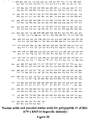
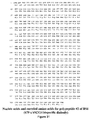
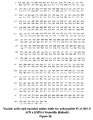
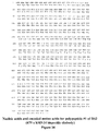
- said diabody is selected from the group consisting of a diabody comprising amino acid sequences shown in Fig. 30 and Fig. 31 , a diabody comprising amino acid sequences shown in Fig. 32 and Fig. 33 , a diabody comprising amino acid sequences shown in Fig. 34 and Fig. 35 , and a diabody comprising amino acid sequences shown in Fig. 37 and 38 .
- said first, second, third and fourth polypeptide segments are each encoded by a first, second, third and fourth cDNA, respectively.
- said first cDNA comprises nucleotide sequences shown in Fig. 30 and Fig. 31
- said second cDNA comprises nucleotide sequences shown in Fig. 32 and Fig. 33
- said third cDNA comprises nucleotide sequences shown in Fig. 34 and 35
- said fourth cDNA comprises nucleotide sequences shown in Fig. 37 and 38 .
- said first and third cDNAs are contained on a single nucleic acid molecule, or wherein said second and fourth cDNAs are contained on a single nucleic acid molecule, or wherein said first, second, third and fourth cDNAs are on a single expression cassette, wherein preferably said expression cassette is contained in a plasmid.
- the problem underlying the present invention is solved in a second aspect by a host cell comprising a plasmid as defined in the above embodiment of the first aspect.
- the problem underlying the present invention is solved in a third aspect by a method of producing a binding protein, comprising culturing the host cell of the second aspect in a suitable medium, and separating said binding protein from said media.
- a binding protein of the first aspect for use in treatment of a disease wherein the treatment comprises delivering a therapeutic agent, or a combination of the therapeutic agent and a diagnostic agent to a target, comprising:
- a binding protein of the first aspect for use in diagnosis of disease comprising:
- said carrier molecule binds to more than one binding site of the binding protein.
- said therapeutic agent is selected from the group comprising, drugs, toxins, cytokines, hormones, growth factors, conjugates, and radionuclides.
- the problem underlying the present invention is solved in a sixth aspect by the use of a binding protein of the first aspect in the preparation of a medicament for detecting or treating a human disorder, wherein the binding protein of the first aspect is administered to a subject; a sufficient amount of time is waited for an amount of the non-bound binding protein to clear the subject's blood stream; and a carrier molecule comprising a diagnostic agent, a therapeutic agent, or a combination thereof, that binds to a binding site of the binding protein is administered to the subject.
- said human disorder is selected from the group comprising cancer, autoimmune diseases, infectious diseases, cardiovascular diseases, and inflammatory diseases.
- This invention also relates to a multivalent, multi-specific binding protein comprising at least one binding site for a hapten moiety and at least one binding site for a target antigen as defined in the claims.
- the hapten is connected to a small molecule that carries a diagnostic agent and/or a therapeutic agent.
- the present disclosure further relates to bispecific diabodies that bind with hapten moieties and target antigens and to recombinant vectors useful for the expression of these functional diabodies in a microbial host.
- whole antibodies are composed of one or more copies of an Y-shaped unit that contains four polypeptides chains.
- Two chains are identical copies of a polypeptide, referred to as the heavy chain, and two chains are identical copies of a polypeptide, referred to as the light chain.
- the two heavy chains are linked together by one or more disulfide bonds and each light chain is linked to one of the heavy chains by one disulfide bond.
- Each chain has a N-terminal variable domains, referred to as V H and V L for the heavy and the light chains, respectively, and the non-covalent association of a pair of V H and V L , referred to as the Fv fragment, forms one antigen-binding site.
- ScFvs with linkers greater than 12 amino acid residues in length allow interaction between the V H and V L domains on the same chain and generally form a mixture of monomers, dimers (termed diabodies) and small amounts of higher mass multimers, [ Kortt et al., Eur. J. Biochem. (1994) 221: 151-157 ].
- ScFvs with linkers of 5 or less amino acid residues prohibit intramolecular pairing of the V H and V L domains on the same chain, forcing pairing with V H and V L domains on a different chain.
- Linkers between 3- and 12-residues form predominantly dimers [ Atwell et al., Protein Engineering (1999) 12: 597-604 ]. With linkers between 0 and 2 residues, trimeric (termed triabodies), tetrameric (termed tetrabodies) or higher oligomeric structures of scFvs are in favor; however, the exact patterns of oligomerization appear to depend on the composition as well as the orientation of the V-domains, in addition to the linker length.
- scFvs of the anti-neuraminidase antibody NC10 formed predominantly trimers (V H to V L orientation) or tetramers (V L to V H orientation) with 0-residue linkers [ Dolezal et al., Protein Engineering (2000) 13: 565-574 ].
- V H to V L orientation formed predominantly diabodies [ Atwell et al., Protein Engineering (1999) 12: 597-604 ]; in contrast, the V L to V H orientation formed a mixture of tetramers, trimers, dimers, and higher mass multimers [ Dolezal et al., Protein Engineering (2000) 13: 565-574 ].
- the 0-residue linker formed exclusively trimers and the 1-residue linker formed exclusively tetramers [ Le Gall et al., FEBS Letters (1999) 453: 164-168 ].
- bispecific diabodies are heterodimers of two different scFvs, each scFv consisting of the V H domain from one antibody connected by a short linker to the V L domain of another antibody.
- the bispecific tandab is a homodimer of two polypeptides, each containing four variable domains of two different antibodies (V H1 , V L1 , V H2 , V L2 ) linked in an orientation to facilitate the formation of two potential binding sites for each of the two different specificities upon self-association.
- Methods of constructing scFvs are disclosed in U.S. 4,946,778 (1990 ) and U.S. 5,132,405 (1992 ).
- Methods of producing scFv-based agents of multivalency and multispecificity as described above are disclosed in U.S. 5,837,242 (1998 ), U.S. 5,844,094 (1998 ) and WO 98/44001 (1998 ) for bispecific diabodies, and in PCT/DE99/01350 for tandem diabodies.
- multispecific and multivalent antigen-binding proteins from V H and V L domains are disclosed in U.S. 5,989,830 and U.S. 6,239,259 .
- Such multivalent and multispecific antigen-binding proteins are obtained by expressing a discistronic vector which encodes two polypeptide chains, with one polypeptide chain consisting of two or more V H domains (from the same or different antibodies) connected in series by a peptide linker and the other polypeptide chain consisting of complementary V L domains connected in series by a peptide linker.
- the present disclosure utilizes two monoclonal antibodies, 679 and hMN14, and two point mutations of 679, (679-V H (I3Q) and 679-V K (C101S)), to produce antigen specific diabodies.
- a bispecific diabody is produced from hMN14 and h679, which is obtained by grafting the CDRs of 679 onto a framework of amino acid residues found in human antibodies.
- the murine monoclonal antibody designated 679 (an IgG1, K) binds with high affinity to molecules containing the moiety histamine-succinyl-glycyl (HSG) ( Morel et al., Molecular Immunology, 27, 995-1000, 1990 ).
- V H and V K The nucleotide sequence pertaining to the variable domains (V H and V K ) of 679 has been determined (Qu et al., unpublished results).
- V K is one of two isotypes of the antibody light chains, V L .
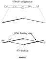
- the design of the gene construct (679-scFv-L5) for expressing a 679 diabody possesses the following features: 1) The carboxyl terminal end of V H is linked to the amino terminal end of V K by the peptide linker Gly-Gly-Gly-Gly-Ser (G 4 S). The use of the G 4 S peptide linker enables the secreted polypeptide to dimerize into a diabody, forming two binding sites for HSG.
- Six histidine (His) residues are added to the carboxyl terminus to allow purification by IMAC.
- the DNA coding sequence and the corresponding encoded amino acids for 679-scFv-L5 are contained in Figure 25 (Seq IDs).
- the DNA coding sequence and the corresponding encoded amino acids for 679-I3Q are contained in Figure 26 (Seq IDs).
- the DNA coding sequence and the corresponding encoded amino acids for 679-C101S are contained in Figure 27 (Seq IDs).
- Figure 1 also includes a stick figure drawing of the mature polypeptide after proteolytic removal of the pelB leader peptide and a stick figure drawing of a 679 diabody, including the HSG binding sites.
- 679 Two site-directed point mutations were made to increase the amount of 679 diabodies in soluble extracts. Specifically, converting residue 3 in the 679V H sequence from Ile to Gln (I3Q), or residue 101 in the 679V K sequence from Cys to Ser (C101S), or both (I3Q/C101S), resulted in at least a ten-fold increase in soluble expression levels. Moreover, 679 can be humanized or fully human to help avoid an adverse response to the murine antibody.
- hMN14 is a humanized monoclonal antibody (Mab) that binds specifically to CEA ( Shevitz et al, J. Nucl. Med., Supp., 34, 217P, 1993 ; U.S. 6,254,868 (2001 )). While the original Mabs were murine, humanized antibody reagents are now utilized to reduce the human anti-mouse antibody response. The variable regions of this antibody were engineered into an expression construct (hMN14-scFv-L5) in a similar fashion to 679-scFv-L5 as described in Example 1.
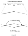
- the design of the gene construct (hMN14-scFv-L5) for expressing an hMN14 diabody possesses the following features: 1) The carboxyl terminal end of V H is linked to the amino terminal end of V K by the peptide linker Gly-Gly-Gly-Gly-Ser (G 4 S). The use of the G 4 S peptide linker enables the secreted polypeptide to dimerize into a diabody, forming two binding sites for CEA. 2) A pelB leader sequence precedes the V H gene to facilitate the transport of the polypeptide to the periplasmic space of E. coli.
- Figure 29 shows a stick figure drawing of the mature polypeptide following proteolytic removal of the pelB leader peptide, and a stick figure drawing of a hMN14 diabody, including CEA binding sites.
- Di-cistronic expression vectors were constructed through a series of sub-cloning procedures outlined in Figures 8 and 9 and described in Example 6.
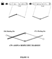
- the di-cistronic expression cassette for bispecific hMN14x679 diabody is shown schematically in Figure 10 .
- the expression cassette may be contained in a plasmid, which is a small, doublestranded DNA forming an extra-chromosomal self-replicating genetic element in many bacteria and some eukaryotes and is widely used in genetic engineering as a cloning vector.
- a cloning vector is a DNA molecule that can replicate on its own in a microbial host cell.
- This invention describes a vector that expresses bispecific diabodies.
- a host cell accepts a vector for reproduction and the vector replicates each time the host cell divides.
- a commonly used host cell is Escherichia Coli (E. Coli), however, other host cells are available.
- the di-cistronic cassette as shown in Figure 10 When the di-cistronic cassette as shown in Figure 10 is expressed in E. coli, some of the polypeptides fold and spontaneously form soluble bispecific diabodies.
- the bispecific diabody shown in Figure 10 forms one binding site having high affinity for HSG and one binding site having high affinity for CEA.
- the carboxyl terminal end of the V H segment of the 679 MAb is connected to the amino terminal end of the V K segment of the hMN14 MAb by a five amino acid residue linker
- the carboxyl terminal end of the V H segment of the hMN14 MAb is connected to the amino terminal end of the V K segment of the 679 MAb by the same five amino acid residue linker.
- Three variants of 679 x hMN14 bispecific diabodies have been produced and tested.
- BS1 is composed of the wild-type sequences for both 679 and hMN14 variable regions.
- BS1.5 incorporates the 679V H I3Q mutation.
- BS2 incorporates both the 679V H I3Q and the 679V K C101S mutations.
- bispecific diabodies are for pre-targeting CEA positive tumors for subsequent specific delivery of therapeutic radioisotopes carried by HSG containing peptides.
- These diabodies bind selectively to targeted antigens and when combined with a bivalent di-HSG hapten allow for increased affinity and a longer residence time at the desired location. Moreover, non-antigen bound diabodies are cleared from the body quickly and exposure of normal tissues is minimized.
- Delivering a diagnostic or a therapeutic agent to a target for diagnosis or treatment by means as defined in the claims includes administering a patient with the binding protein, waiting a sufficient amount of time for an amount of the non-binding protein to clear the patient's blood stream, and administering a diagnostic or therapeutic agent that binds to a binding site of the binding protein. Diagnosis further requires the step of detecting the bound proteins with known techniques.
- the diagnostic or therapeutic carrier molecule comprises a diagnostically or therapeutically efficient agent, a linking moiety, and one or more hapten moieties. The hapten moieties are positioned to permit simultaneous binding of the hapten moieties with the binding protein.
- Administration of the binding protein and diagnostic or therapeutic agents of the present invention to a mammal may be intravenous, intraarterial, intraperitoneal, intramuscular, subcutaneous, intrapleural, intrathecal, by perfusion through a regional catheter, or by direct intralesional injection.
- the administration may be by continuous infusion or by single or multiple boluses.
- the unmixed diagnostic or therapeutic agent and bispecific antibody may be provided as a kit for human therapeutic and diagnostic use in a pharmaceutically acceptable injection vehicle, preferably phosphate-buffered saline (PBS) at physiological pH and concentration.
- a pharmaceutically acceptable injection vehicle preferably phosphate-buffered saline (PBS) at physiological pH and concentration.
- PBS phosphate-buffered saline
- the preparation preferably will be sterile, especially if it is intended for use in humans.
- Optional components of such kits would normally be containers of stabilizers, buffers, labeling reagents, radioisotopes, paramagnetic compounds, second antibody for enhanced clearance, and conventional syringes, columns, vials and the like.
- Standard recombinant DNA methods were used to obtain 679-scFv-L5 as follows.
- a plasmid containing the V H sequence of 679 was used as the template for polymerase chain reaction (PCR) using Pfu polymerase and the two oligonucleotide primers specified below:
- the left PCR primer contains a 5' NcoI restriction site.
- the right PCR primer contains the sequence for a 5 amino acid residue linker (G 4 S) and a BamHI restriction site.
- the PCR product was digested with NcoI and BamHI and ligated in frame with the pelB leader sequence into NcoI/BamHI digested pET-26b vector (Novagen) to generate 679V H L5-pET26.
- a plasmid containing the V K sequence of 679 was used as the template for PCR using Pfu polymerase and the two oligonucleotide primers specified below:
- the left and right PCR primers contain BamHI and XhoI restriction sites, respectively.
- the PCR product was digested with XhoI and BamHI and ligated (in frame with the 679V H , G 4 S linker, and 6His sequences) into the XhoI/BamHI digested 679V H L5-pET26 to generate the expression construct 679-scFv-L5.
- the DNA sequence of the inserted gene confirmed that the V H and V K sequences were identical to those of the original cDNA clones and the sequences of the ligation sites and linker regions were as designed.
- the gene construct, 679-scFv-L5, is illustrated in Figure 1 .
- Competent E. coli BL21(P-Lys-S) cells were transformed with 679-scFv-L5 by standard methods. Cultures were shaken in 2xYT media supplemented with 100 ⁇ g/ml kanamycin sulphate and 34 ⁇ g/ml chloramphenicol and grown at 37°C to OD 600 of 1.6 - 1.8. An equal volume of room temperature 2xYT media supplemented with antibiotics and 0.8M sucrose was added to the cultures, which were then transferred to 20°C. After 30 minutes at 20°C, expression was induced by the addition of 40 ⁇ M IPTG and continued at 20°C for 15-18 hours.
- 679 diabody was examined in (1) cell culture conditioned media, (2) soluble proteins extracted under non-denaturing conditions from the cell pellet following centrifugation, and (3) insoluble material remained in the pellet following several cycles of extraction and centrifugation.
- Soluble proteins were extracted from bacterial cell pellets as follows. Pellets were frozen and thawed then re-suspended in lysis buffer (2% Triton-X 100; 300mM NaCl; 10mM imidazole; 5mM MgSO 4 ; 25 units/ml benzonase; 50mM NaH 2 PO 4 , pH 8.0) using an amount equal to 1 % of the culture volume. The suspension was homogenized by sonication , clarified by centrifugation, and loaded onto Ni-NTA IMAC columns.
- the columns After being washed with buffer containing 20mM imidazole, the columns were eluted with 100mM imidazole buffer (100mM imidazole; 50mM NaCl; 25mM Tris, pH 7.5) and the eluate obtained was further purified on a Q-Sepharose column.
- the insoluble material was solubilized in denaturing Ni-NTA binding buffer (8M urea; 10mM imidazole; 0.1M NaH 2 PO 4 ; 10mM Tris, pH 8.0) and mixed with 1ml of Ni-NTA agarose (Qiagen, inc.). The mixture was rocked at room temperature for 1 hour then the resin was washed once with 50ml of the same buffer and loaded onto a column. The column was washed with 20ml of the same buffer followed by 20ml of wash buffer (8M urea; 20mM imidazole; 0.1M NaH 2 PO 4 ; 10mM Tris, pH 8.0). Bound proteins were eluted with 5ml of denaturing elution buffer (8M urea; 250mM imidazole; 0.1M NaH 2 PO 4 ; 10mM Tris, pH 8.0).
- Two site-directed point mutations were made to increase the amount of 679 diabodies in soluble extracts. Specifically, converting residue 3 in the 679V H sequence from Ile to Gln (I3Q), or residue 101 in the 679V K sequence from Cys to Ser (C101S), or both (I3Q/C101S), resulted in at least a ten-fold increase in soluble expression levels.
- the mutations were introduced in synthetic oligonucleotides used for PCR.
- the V H -I3Q mutation was incorporated in the oligonucleotide primer depicted below:
- This primer was paired with 679V H -Right (Example 1) to generate the V H -I3Q mutant by PCR from 679-scFv-L5 template using Pfu polymerase.
- the 679V K -C101S mutation was incorporated in the oligonucleotide primer specified below:
- This primer was paired with 679-V K Left (Example 1) to generate 679V K -C101S mutant by PCR from 679-scFv-L5 template using Pfu polymerase.
- the PCR products were cloned into pET26b following the same procedure as described above in Example 1.
- Expression levels in the soluble fractions were estimated by BIAcore analysis using a HSG coupled sensor chip.
- the expression levels of I3Q, C101S, or I3Q/C101S mutant 679 diabody were about 10 ug/L as compared to about 1 ug/L for the wild type.
- hMN14-scFv-L5 Standard recombinant DNA methods were used to obtain hMN14-scFv-L5 as follows.
- the hMN14 V H and V K sequences were amplified from a vector constructed for expressing hMN14 Fab' ( Leung et al., Cancer Research, Supp., 55, 5968s-5972s, 1995 ) by PCR with Pfu polymerase.
- the hMN14V H sequence was amplified using the oligonucleotide primers specified below:
- the left PCR primer contains a 5' NcoI restriction site.
- the right PCR primer contains a sequence for a 5 amino acid residue linker (G 4 S) and a BamHI restriction site.
- the PCR product was digested with NcoI and BamHI and ligated, in frame with the pelB leader sequence, into NcoI/BamHI digested pET-26b vector to generate hMN14V H L5-pET26.
- the hMN14V K sequence was amplified using the oligonucleotide primers specified below:
- the left and right PCR primers contain BamHI and XhoI restriction sites, respectively.
- the PCR product was digested with XhoI and BamHI and ligated, in frame with the hMN14V H , G 4 S linker and 6His sequences, into the XhoI/BamHI digested hMNI4V H L5-pET26 construct to generate the expression construct hMN14-scFv-L5.
- the DNA sequence of this construct was verified by automated DNA sequencing.
- the gene construct, hMN14-scFv-L5, is illustrated in Figure 3 .
- the hMN14-scFv-L5 construct was used to transform BL21(P-LysS) E. coli. Culture conditions, induction, and purification were carried out similar to those described for the 679 diabody in Example 1, except that the hMN14 diabody was purified by affinity chromatography, instead of Q-Sepharose anion exchange chromatography, via binding to an anti-id antibody immobilized on Affi-gel. Soluble proteins that bound and eluted from Ni-NTA resin were loaded on a WI2 anti-idiotype affinity column. The column was washed with PBS and the product was eluted with 0.1M Glycine; 0.1M NaCl, pH 2.5 and neutralized immediately.
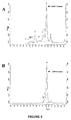
- hMN14scFv The very similar retention time of hMN14scFv indicates that it exists in solution as a dimer or diabody since the calculated molecular weight of the monomeric hMN14scFv is 26kDa.
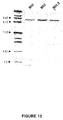
- SDS-PAGE gel analysis in Figure 5A shows a single band of the predicted Mr at 26kDa
- the isoelectric focusing (IEF) gel analysis in Figure 5B yields a band with pI of 8.2, close to the calculated pI of 7.9.
- a competitive ELISA showed that the hMN14 diabody is functionally active and displays excellent binding properties.
- FIG. 7 shows the percentage of the injected dose that is associated with the tumor and with normal tissues, such as liver, spleen, kidney, lungs, blood, stomach, small intestine, and large intestine, at 48 hours after the injection. The amount of the injected dose in each normal tissue is very low when compared to the amount in the tumor.
- Table 1 summarizes the relative amounts of activity found in normal tissues compared to that in the tumor at 24, 48 and 72 hours.
- Table 1 Tumor to non-tumor ratios 24hrs 48hrs 72hrs Tumor 1.00 1.00 1.00 Liver 22.47 31.85 28.32 Spleen 25.41 39.51 41.03 Kidney 9.12 12.12 10.54 Lung 15.49 25.70 31.75 Blood 9.84 17.32 21.80 Stmach 9.98 17.50 23.13 sm. Int. 37.23 65.60 50.58 Lg. Int. 35.87 66.54 45.66
- Example 6 - 679 x hMN14 bispecific diabody (BS1, BS1.5 and BS2)
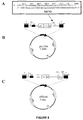
- pET-ER a new vector (pET-ER) was generated by the addition of a multiple cloning site, MCS2, shown in Figure 8A , into the pET-26b vector, shown in Figure 8B .
- MCS2 multiple cloning site
- the duplex structure, MCS2, was ligated into the BlpI restriction site of pET-26b to generate the pET-ER vector as illustrated in Figure 8C .
- This vector facilitates the construction of di-cistronic expression cassettes and allows for stoichiometric expression of two heterologous polypeptides in a single E. coli cell.
- the di-cistronic expression vectors were constructed through a series of sub-cloning procedures that are outlined in Figure 9 .
- the V K sequences of 679-scFv-L5 and hMN14-scFv-L5 were exchanged by excision with BamHI and XhoI to generate two intermediate constructs in pET26b.
- a DNA fragment containing the sequence 679V H -L5-hMN14V K excised from a pET26b construct with NcoI and XhoI, was ligated into the same restriction sites in pET-ER vector to generate an intermediate clone (679V H -L5-hMN14V K - pET-ER).
- a 900bp DNA fragment which includes a ribosomal binding site in addition to the coding sequence for polypeptide 2 (below), was excised from hMN14V H -L5-679V K - pET26b with XbaI and BlpI. This fragment was ligated into the SpeI and BlpI restriction sites of 679V H -L5-hMN14V K - pET-ER to create the final bispecific expression constructs.
- the di-cistronic expression cassette for bispecific hMN14x679 diabody is shown schematically in Figure 10 .
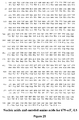
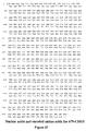
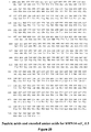
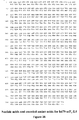
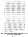
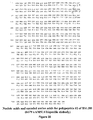
- the DNA coding sequence of nucleic acids and the corresponding encoded amino acids for the first and second polypeptide sequences of BS1, BS1.5, and BS2 are contained in Figures, 30 & 31 , 32 & 33 , and 34 & 35 (Seq IDs), respectively.
- the di-cistronic expression cassette codes for two polypeptides that are arranged as follows:
- BS1.5-transformed E. coli (BL21-pLysS) cultures expressed 0.5 mg of soluble bispecific diabody per liter of culture. From 5L induction, 2.4 mg of highly purified BS1.5 diabody was isolated following the procedures similar to those described in Example 1. Soluble cell extracts were loaded onto a 4ml of Ni-NTA agarose column (Qiagen), which was washed with 20 bed volumes of 10mM imidazole buffer and 5 bed volumes of 20mM imidazole buffer. The diabody was eluted from the IMAC column in 15 ml of 100mM imidazole elution buffer.
- the two polypeptides essentially co-migrate, since their calculated MWs are 26.5 kDa and 27.2 kDa.
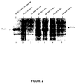
- BS1, BS1.5 and BS2 each shows the presence of a single band with a pI of approximately 8.3, which is close to the predicted pI of 7.9 for the three bispecific diabodies.
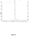
- the binding kinetics of BS1.5 was evaluated by BIAcore using a low density HSG-coupled sensor chip. Binding sensograms were obtained for BS1.5 concentrations from 0 to 54 nM and the resulting data were analyzed with the BIAcore BiaEvaluation software using 1:1 Langmuir binding model, yielding an association constant of the interaction, K d , of 2.4 nM for the binding of BS1.5 to immobilized HSG.
- Figure 14 shows the BIAcore binding curves at various concentrations of BS1.5.
- a chemically prepared 679 x hMN14 F(ab') 2 conjugate yields a K d of 1.55 nM.
- BS1.5 The binding of BS1.5 to CEA was demonstrated by competitive ELISA. Microtiter plates were coated with 0.5 ⁇ g/well with soluble CEA (Scripps Laboratories). BS1.5 at concentrations ranging from 4 - 500 nM were allowed to compete for CEA binding with HRP-conjugated hMN14 IgG (1nM). BS1.5 shows a competitive binding curve similar to that of the 679 x hMN14 F(ab') 2 chemical conjugate. These data indicate that the BS1.5 has a CEA binding affinity similar to the parental hMN14 antibody. The bispecific binding properties of BS1.5 was also analyzed by BIAcore with a high-density HSG-coupled biosensor chip.
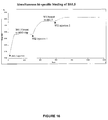
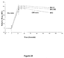
- BS1.5 was pre-bound to the sensor chip before injection of an anti-idiotype MAb designated WI2 that is highly specific for hMN14. Soluble CEA was also used in place of WI2 and gave similar results. As shown in Figure 16 , injection of 60 ng of BS1.5 gave a relative response of 620 RU. Subsequent injection of 400ng of WI2 increased the signal by 400 RU. Binding approached saturation with a second WI2 injection (400ng), as a total of 520 RU were added to the 620RU signal of BS1.5. Injection of WI2 following pre-binding with 679 F(ab') 2 or without pre-binding yielded a negligible response. These data demonstrate that BS1.5 has the capability of binding HSG and CEA simultaneously.
- BS1 and BS2 each differ from BS1.5 by single point mutations in the 679 component of the diabody. Some of the properties of these molecules are summarized in Table 2. ELISA experiments demonstrate that each of these proteins exhibits similar CEA binding properties, which is not surprising given that the hMN14 component of the diabody is consistent among the three diabodies. Further, BS1 and BS2 are demonstrated by BIAcore analysis to be bispecific and capable of binding to CEA and HSG simultaneously.
- BS1.5 includes the 679V H I3Q mutation that is not included in BS1, which is composed entirely of the wildtype sequences. This mutation doubles the yield of soluble diabody that is expressed without compromising the binding affinity for HSG.
- BS2 includes the additional 679V K C101S mutation as well as the 679V H I3Q. With this second change, soluble BS2 is expressed at twice the level of BS1.5, however, the binding affinity for HSG decreased measurably.
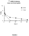
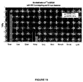
- the tumors were enriched appreciably with 131 I-BS1.5 as compared to normal tissues, such as liver, spleen, kidney, lungs, blood, stomach, small intestine, and large intestine, as illustrated in Figure 18 .
- Pre-targeting experiments were performed with 12 or 24 hour clearance times following injection of BS1.5 (unlabeled).
- IMP241 a peptide containing two HSG groups and a DOTA moiety, was loaded with 111 Indium and injected in BS1.5 pre-targeted mice. The bio-distribution of the 111 In-IMP241 was examined at 3 and 24 hours after injection.
- Figure 19 shows the activity in the tumor and normal tissues in pre-targeted mice with 12 hour clearance.
- a humanized version of 679-based diabody has been generated that exhibits HSG binding affinity comparable to the murine forms.
- the strategy employed was to retain all CDR residues and those residues known to interact with the CDR residues while substituting only those residues of the mouse frameworks that are not found in the database of human frameworks at corresponding positions. In such cases if more than one amino acid residue of the human frameworks is known for the same position, the most common one is selected for humanization.
- amino acid sequence for each of the framework regions of m679V H or m679V K were used to query the NCBI database and aligned with human antibody (h-Ab) sequences.
- Most amino acid residues of the murine 679 frameworks are identical with some or all of the human frameworks in the database at corresponding positions and therefore they are conserved for h679.
- amino acid residues of the murine 679 frameworks that are not found in any of the human frameworks they are substituted with the most common residue found in the homologous h-Abs at the corresponding positions.
- a residue in a particular position is likely to interact with the CDRs or to be involved in the V H and V K association ( E.A. Padlan, Molecular Immunology, 31, 169-217, 1994 )
- the residue in m679 is retained in h679.
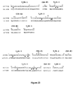
- Figure 20 shows an alignment of m679 and humanized h679.
- the Kabat numbering system is used and framework regions (FR) as well as CDRs are indicated. Arrows signify amino acid substitutions.
- human sequences with high levels of sequence identity were compared to m679.
- V H framework region 1 V H FR-1
- V H framework region 2 V H FR-2
- V H framework region 3 V H FR-3
- V H framework regions 4 V H FR-4
- V K framework region 1 V K FR-1
- This region has considerable variability amongst the h-Abs.
- the m679 amino acids at 20 of the 23 positions in V K FR-1 are acceptable for h-Abs.
- the following substitutions were made at three positions with the most common h-Ab amino acid for the respective positions: threonine (T) for serine (S) at V K -5; arginine (R) for lysine (K) at V K -18; and leucine (L) for methionine (M) at V K -21. These positions are not known to be involved in V H -V K or CDR contacts.
- V K framework region 2 V K FR-2
- This short region resembles the human sequences and is acceptable as is.
- V K framework region 3 V K ER-3
- V K framework regions 4 V K FR-4
- This short region resembles the human sequences and is acceptable as is.
- the new frameworks contain all residues found in h-Abs, except two, namely, leucine at position V H -37, which is retained due to its involvement in the V H and V K contact, and threonine at position V H -110, which is retained because of technical reasons.
- oligonucleotide PCR primers which together contain 12 of the 13 mutations described above to convert m679scFv into h679 diabody, were synthesized and used to generate 4 PCR products.
- the mutant sequences were amplified from the 679scFv-L5 plasmid construct using Taq polymerase. Restriction sites were engineered into the primers to allow ligation of the PCR products while conserving the encoded amino acid sequence.
- the sequences, coding regions, restriction sites and specific mutations contained on each of the primers are summarized in Table 4. The relative location of the primers and the PCR products are shown schematically in Figure 21 .
- the PCR products were each cloned into the PCR cloning vector pGemT (Promega). Through several rounds of sub-cloning using standard methods, the four PCR sequences were assembled and added to the first 120 nucleotides of 679V H I3Q to generate the h679scFv-L5-pGemT construct. From this construct the V H and V K domains were transferred together into the pET26b expression vector for h679 diabody or individually to make fully humanized bi-specific diabodies. The sub-cloning process is described in detail below. Table 4.
- a plasmid clone containing the 679V H -I3Q mutation (679V H I3Q-pGemT) was digested with the restriction enzymes BspEI (base pair 121) and PstI (in pGemT vector 3' of the insert), leaving the first 121 base pairs of 679V H I3Q with the vector.
- This vector fragment was ligated with PCR product A that was digested with XmaI (5' end) and Pst I (3' end) to generate construct A. It is important to note that the BspEI-XmaI ligation destroys both sites as each of these restriction enzymes was used in subsequent steps.
- PCR product B was cloned into pGem T and screened for clones in the T7 orientation.
- the B fragment was excised from the pGemT clone with PstI and ligated into the PstI site of construct A. Clones were screened for proper insert orientation for construct B.
- PCR product C was cloned into pGem T and screened for clones in the T7 orientation.
- the C fragment was excised from the pGemT clone with XmaI and NdeI (vector site) and then ligated into construct B that was digested with the same enzymes.
- PCR product D was cloned into pGem T and screened for clones in the T7 orientation.
- the D fragment was excised from the pGemT clone with BspEI and NdeI and then ligated into construct C that was digested with the same enzymes.
- the h679scFv-L5 sequence was excised from the pGemT construct with NcoI and XhoI and ligated into similarly digested pET26b vector. This construct was used to transform BL21(P-LysS) E. coli. Culture conditions, induction, and purification were carried out similar to those described for the m679 diabody in Example 2. Expression levels in the soluble fractions were estimated by BIAcore analysis using a HSG coupled sensor chip. The expression level of h679 diabody was 50 ⁇ g/L as compared to 1 ug/L for the wild type m679 diabody or 10 ug/L for m679I3Q diabody. The h679 diabody displayed comparable binding properties to the m679I3Q diabody with BIAcore analysis.
- the h679V H and h679V K domains were incorporated into the pET-ER vector with the V H and V K of hMN14 to make the fully humanized BS1.5H bispecific diabody construct.
- the di-cistronic expression vector was constructed through a series of sub-cloning procedures that are outlined in Figure 9 . Initially, the V K sequences of h679-scFv-L5 and hMN14-scFv-L5 were exchanged by excision with BamHI and XhoI to generate two intermediate constructs in pET26b.
- the di-cistronic expression cassette codes for two polypeptides that are arranged as follows:
- the BS1.5H construct was used to transform E. coli (BL21-pLysS) cells.
- the recombinant BS1.5H protein was expressed and purified as described in Example 6.
- the level of soluble protein expression was 0.55 mg/L, about 10% higher than BS1.5.
- Size exclusion HPLC analysis of the purified BS1.5H yielded a single protein peak at 10.16 minutes ( Fig 22 ).
- BS2 had a retention time of 10.04 minutes under identical conditions, indicating that BS1.5H polypeptides exclusively form diabodies.
- the bispecific (CEA/HSG) binding properties of BS1.5H were confirmed by BIAcore analysis ( Fig 23 ).
- BS1.5H was pre-bound to a HSG-coupled sensor chip before injection of WI2 (hMN14-specific anti-idiotype MAb). As shown in Figure 23 , injection of 60 ng of BS1.5H gave a relative response of 660 RU. Subsequent injection of 1 ⁇ g of WI2 increased the signal by 760 RU. Injection of WI2 following pre-binding with 679 F(ab') 2 or without pre-binding yielded a negligible response. These data demonstrate that BS1.5H has the capability of binding HSG and CEA simultaneously. BS1.5H differs from BS1.5 by the humanization of the 679 moiety, which was accomplished by substitutions of 13 amino acid residues.
- the therapeutic agents may take the following form:
- the therapeutic agent comprises at least two haptens which are covalently linked via a peptidic, proteinaceous or non-proteinaceous moiety.
- haptens are fluorescein isothiocyanate, vitamin B-12, DTPA (diethylenetriaminepentaacetic acid) and DOTA (1,4,7,10-tetraazacyclododecanetetraacetic acid) residues.
- This example relates to the preparation of carboxylesterase-DTPA conjugate.
- Two vials of rabbit liver carboxylesterase (about 8.5 mg protein content/vial) were reconstituted with 2.3 mL of 50 mM potassium phosphate buffer pH 7.5, and the solution was made 4.2 mM in DTPA using 0.1 mL of a 0.1 M stock solution of DTPA pH 6.7.
- the pH of the resultant solution was adjusted to be in the 7.7-7.8 range, and then reacted with 10 mg of cyclic DTPA dianhydride.
- the reaction mixture was passed through two successive SEC columns equilibrated in 0.1 M sodium phosphate pH 7.3.
- the eluate was further purified by preparative HPLC on a TSK G3000SW column using 0.2 M sodium phosphate pH 6.8, at 4 mL/min flow, as the eluent.
- the purified conjugate was made 0.1 M in sodium phosphate pH 6.8, and concentrated.
- the DTPA-to-carboxylesterase molar substitution ratio determined by a metal-binding assay, was in the range of 2.95-to-1 to 4.43-to-1.
Landscapes
- Health & Medical Sciences (AREA)
- Chemical & Material Sciences (AREA)
- Life Sciences & Earth Sciences (AREA)
- Medicinal Chemistry (AREA)
- Organic Chemistry (AREA)
- Immunology (AREA)
- General Health & Medical Sciences (AREA)
- Pharmacology & Pharmacy (AREA)
- Veterinary Medicine (AREA)
- Public Health (AREA)
- Animal Behavior & Ethology (AREA)
- Proteomics, Peptides & Aminoacids (AREA)
- Engineering & Computer Science (AREA)
- Bioinformatics & Cheminformatics (AREA)
- Genetics & Genomics (AREA)
- Nuclear Medicine, Radiotherapy & Molecular Imaging (AREA)
- General Chemical & Material Sciences (AREA)
- Chemical Kinetics & Catalysis (AREA)
- Molecular Biology (AREA)
- Epidemiology (AREA)
- Biochemistry (AREA)
- Biophysics (AREA)
- Oncology (AREA)
- Cell Biology (AREA)
- Gastroenterology & Hepatology (AREA)
- Zoology (AREA)
- Transplantation (AREA)
- Heart & Thoracic Surgery (AREA)
- Rheumatology (AREA)
- Communicable Diseases (AREA)
- Pain & Pain Management (AREA)
- Cardiology (AREA)
- Microbiology (AREA)
- Mycology (AREA)
- Peptides Or Proteins (AREA)
- Preparation Of Compounds By Using Micro-Organisms (AREA)
- Medicines That Contain Protein Lipid Enzymes And Other Medicines (AREA)
- Micro-Organisms Or Cultivation Processes Thereof (AREA)
- Medicinal Preparation (AREA)
Description
- The present invention is related to a multivalent, multi-specific binding protein comprising a histamine-succinyl-glycyl (HSG) binding site having an affinity towards molecules containing a HSG moiety and a carcinoembryonic antigen (CEA) binding site having an affinity towards CEA, wherein said HSG antigen binding site comprises a first and second polypeptide segments that must associate with each other to form said HSG antigen binding site; a host cell comprising a plasmid encoding the multivalent, multi-specific binding protein; a method of producing a binding protein, wherein the method comprises culturing the host cell; the multivalent, multi-specific binding protein for use in the treatment of a disease; the multivalent, multi-specific binding protein for use in diagnosis of a disease; and the use of the multivalent, multi-specific binding protein in the preparation of a medicament for detecting or treating a human disorder.
- Man-made binding proteins, in particular monoclonal antibodies and engineered antibodies or antibody fragments, have been tested widely and shown to be of value in detection and treatment of various human disorders, including cancers, autoimmune diseases, infectious diseases, inflammatory diseases, and cardiovascular diseases [Filpula and McGuire, Exp. Opin. Ther. Patents (1999) 9: 231-245]. For example, antibodies labeled with radioactive isotopes have been tested to visualize tumors after injection to a patient using detectors available in the art. The clinical utility of an antibody or an antibody-derived agent is primarily dependent on its ability to bind to a specific targeted antigen. Selectivity is essential for delivering a diagnostic or therapeutic agent, such as isotopes, drugs, enzymes, toxins, cytokines, hormones, growth factors, or conjugated derivatives thereof, to a target location during the detection and treatment phases of a human disorder, particularly if the diagnostic or therapeutic agent is toxic to normal tissue in the body.
- The major limitations of antibody systems are discussed in Goldenberg, The American Journal of Medicine (1993) 94: 298-299. The essential parameters in the detection and treatment techniques are the amount of the injected dose specifically localized at the site(s) where target cells are present and the uptake ratio, i.e., the ratio of the concentration of specifically bound antibody to that of the radioactivity present in surrounding normal tissues. When an antibody is injected into the blood stream, it passes through a number of compartments as it is metabolized and excreted. The antibody must be able to locate and bind to the target cell antigen while passing through the rest of the body. Factors that control antigen targeting include antigen location, antigen density, antigen accessibility, cellular composition of pathologic tissue, and the pharmokinetics of the targeting antibodies. Other factors that specifically affect tumor targeting by antibodies include expression of the target antigens, both in tumor and other tissues, and bone marrow toxicity resulting from the slow blood-clearance of the radiolabeled antibodies.
- The amount of targeting antibodies accreted by the targeted tumor cells is influenced by the vascularization and barriers to antibody penetration of tumors, as well as intratumoral pressure. Non-specific uptake by non-target organs such as the liver, kidneys or bone-marrow is another major limitation of the technique, especially for radioimmunotherapy, where irradiation of the bone marrow often causes the dose-limiting toxicity.
- One suggested solution, referred to as the "Affinity Enhancement System" (AES), is a technique especially designed to overcome the deficiencies of tumor targeting by antibodies carrying diagnostic or therapeutic radioisotopes [
U.S. 5,256,395 (1993 ), Barbet et al., Cancer Biotherapy & Radiopharmaceuticals (1999) 14: 153-166]. The AES requires a radiolabeled bivalent hapten and an anti-tumor/anti-hapten bispecific antibody that recognizes both the target tumor and the radioactive hapten. The technique involves injecting the bispecific antibody into the patient and allowing the bispecific antibody to localize at the target tumor. After a sufficient amount of time for the unbound antibody to clear from the blood stream, the radiolabeled hapten is administered. The hapten binds to the antibody-antigen complex located at the site of the target cell to obtain diagnostic or therapeutic benefits. The unbound hapten clears the body. Barbet mentions the possibility that a bivalent hapten may crosslink with a bispecific antibody, when the latter is bound to the tumor surface. As a result, the radiolabeled complex is more stable and stays at the tumor for a longer period of time. - Bispecific antibodies prepared by chemically crosslinking two different Fab' fragments have been employed successfully, along with applicable bivalent haptens, to validate the utility of the AES for improved tumor targeting both in animal models and in human patients. However, there remains a need in the art for production of bispecific antibodies by recombinant DNA technology to assess whether such engineered antibodies have merits for the AES. Specifically, there remains a need for an antibody-based agent that exhibits enhanced uptake at targeted antigens, fast clearance from the blood, and optimal protection of normal tissues and cells from toxic pharmaceuticals.
- International patent application
WO 03/057829 - International patent application
99/66951 - US patent
US 5,256,395 is related to affinity enhancement immunological reagents for in vivo detection and killing of specific target cells. - De Boisferon H et al. (Bioconjugate Chem, vol. 11, 2000, pp. 452-460) report on enhanced targeting specificity to tumor cells by simultaneous recognition of two antigens.
- US patent
US 5,837,242 is related to multivalent and multispecific binding proteins, their manufacture and use. - Janevik-Ivanovska E et al. (Bioconjugate Chemistry, vol. 8, no. 4, 1997, pp. 526-533) report on bivalent hapten-bearing peptides designed for Iodine-131 pretargeted radioimmunotherapy.
- Morel A et al. (Molecular Immunology, vol. 27, no. 10, 1990, pp. 995-1000) report on recognition of imidazole and histamine derivatives by monoclonal antibodies.
- The problem underlying the present invention is solved by the subject matter of the attached independent claims. Preferred embodiments may be taken from the attached dependent claims.
- More specifically, the problem underlying the present invention is solved in a first aspect by a multivalent, multi-specific binding protein comprising a histamine-succinyl-glycyl (HSG) binding site having an affinity towards molecules containing a HSG moiety and a carcinoembryonic antigen (CEA) binding site having an affinity towards CEA, wherein said HSG antigen binding site comprises a first and second polypeptide segments that must associate with each other to form said HSG antigen binding site, wherein
said first polypeptide segment comprises a heavy chain variable region comprising an amino acid sequence selected from the group consisting of and said second polypeptide segment comprises a light chain variable region comprising an amino acid sequence selected from the group consisting of


- In an embodiment of the first aspect said first polypeptide segment comprises a heavy chain variable region comprising an amino acid sequence ofand said second polypeptide segment comprises a light chain variable region comprising an amino acid sequence of


- In an embodiment of the first aspect said first polypeptide segment comprises a heavy chain variable region comprising an amino acid sequence ofand said second polypeptide segment comprises a light chain variable region comprising an amino acid sequence of


- In an embodiment of the first aspect said CEA antigen binding site comprises a third and fourth polypeptide segments that must associate with each other to form said CEA antigen binding site.
- In an embodiment of the first aspect said third polypeptide segment comprises a VH polypeptide of hMN14 MAb of
Fig. 29 , and said fourth polypeptide segment comprises a VK polypeptide of hMN14 MAb ofFig. 29 . - In an embodiment of the first aspect said first and third polypeptide segments are connected by a first linker and said second and fourth polypeptide segments are connected by a second linker.
- In an embodiment of the first aspect said first linker and said second linker each comprise five or less amino acid residues.
- In an embodiment of the first aspect said binding protein is a diabody.
- In an embodiment of the first aspect said diabody is selected from the group consisting of a diabody comprising amino acid sequences shown in
Fig. 30 andFig. 31 , a diabody comprising amino acid sequences shown inFig. 32 andFig. 33 , a diabody comprising amino acid sequences shown inFig. 34 andFig. 35 , and a diabody comprising amino acid sequences shown inFig. 37 and38 . - In an embodiment of the first aspect said first, second, third and fourth polypeptide segments are each encoded by a first, second, third and fourth cDNA, respectively.
- In an embodiment of the first aspect said first cDNA comprises nucleotide sequences shown in
Fig. 30 andFig. 31 , wherein said second cDNA comprises nucleotide sequences shown inFig. 32 andFig. 33 , wherein said third cDNA comprises nucleotide sequences shown inFig. 34 and35 , and wherein said fourth cDNA comprises nucleotide sequences shown inFig. 37 and38 . - In an embodiment of the first aspect said first and third cDNAs are contained on a single nucleic acid molecule, or wherein said second and fourth cDNAs are contained on a single nucleic acid molecule, or wherein said first, second, third and fourth cDNAs are on a single expression cassette, wherein preferably said expression cassette is contained in a plasmid.
- More specifically, the problem underlying the present invention is solved in a second aspect by a host cell comprising a plasmid as defined in the above embodiment of the first aspect.
- More specifically, the problem underlying the present invention is solved in a third aspect by a method of producing a binding protein, comprising culturing the host cell of the second aspect in a suitable medium, and separating said binding protein from said media.
- More specifically, the problem underlying the present invention is solved in a fourth aspect by a binding protein of the first aspect for use in treatment of a disease, wherein the treatment comprises delivering a therapeutic agent, or a combination of the therapeutic agent and a diagnostic agent to a target, comprising:
- a) administering the binding protein to a subject;
- b) waiting a sufficient amount of time for an amount of the non-bound binding protein to clear the subject's blood stream; and
- c) administering to said subject a carrier molecule comprising the therapeutic agent or a combination of the therapeutic agent and a diagnostic agent, that binds to a binding site of the binding protein.
- More specifically, the problem underlying the present invention is solved in a fifth aspect by a binding protein of the first aspect for use in diagnosis of disease, wherein the diagnosis comprises delivering a diagnostic agent or a combination of the diagnostic agent and a therapeutic agent to a target, comprising:
- a) administering the binding protein to a subject;
- b) waiting a sufficient amount of time for an amount of the non-bound binding protein to clear the subject's blood stream; and
- c) administering to said subject a carrier molecule comprising the diagnostic agent or a combination of the diagnostic agent and a therapeutic agent, that binds to a binding site of the binding protein.
- In an embodiment of the fourth and fifth aspect said carrier molecule binds to more than one binding site of the binding protein.
- In an embodiment of the fourth aspect said therapeutic agent is selected from the group comprising, drugs, toxins, cytokines, hormones, growth factors, conjugates, and radionuclides.
- More specifically, the problem underlying the present invention is solved in a sixth aspect by the use of a binding protein of the first aspect in the preparation of a medicament for detecting or treating a human disorder, wherein the binding protein of the first aspect is administered to a subject; a sufficient amount of time is waited for an amount of the non-bound binding protein to clear the subject's blood stream; and a carrier molecule comprising a diagnostic agent, a therapeutic agent, or a combination thereof, that binds to a binding site of the binding protein is administered to the subject.
- In an embodiment of the sixth aspect said human disorder is selected from the group comprising cancer, autoimmune diseases, infectious diseases, cardiovascular diseases, and inflammatory diseases.
-
-
Figure 1 shows a schematic representation of the 679 single chain Fv (scFv) polypeptide that is synthesized in E. coli from the 679-scFv-L5 expression plasmid and forms a 679 diabody. The gene construct for the un-processed polypeptide contains the pelB signal peptide, 679VH and VK coding sequences coupled by a 5 amino acid linker, Gly-Gly-Gly-Gly-Ser (G4S), and the carboxyl terminal six histidine (His) affinity tag. The figure also shows a stick figure drawing of the mature polypeptide after proteolytic removal of the pelB leader peptide and a stick figure drawing of a 679 diabody, including the HSG binding sites. -
Figure 2 shows a SDS-PAGE gel stained with Coomassie blue that is used to analyze the expression of 679 scFv from 679scFv-L5-transformed E. coli BL21 p-LysS cultures: lanes 1- 5, induced with isopropyl-β-D-galactopyranoside (IPTG) overnight at 20°C;lanes lane 3, the culture media was concentrated 10-fold. Soluble (lanes 4 and 6) and insoluble (lanes 5 and 7) proteins were fractionated by centrifugation of cell lysates (lane 2). 679scFv was purified from the insoluble fraction by Immobilized Metal Affinity Chromatography (IMAC) following solubilization in 8M urea (lane 1). -
Figure 3 shows a schematic representation of the hMN14scFv polypeptide that is synthesized in E. coli from the hMN14-scFv-L5 expression plasmid and forms a hMN14 diabody. The gene construct for the un-processed polypeptide contains the pelB signal peptide, hMN14VH and VK coding sequences coupled by a 5 amino acid linker, and thecarboxyl terminal 6 histidine affinity tag. The figure also shows a stick figure drawing of the mature polypeptide following proteolytic removal of the pelB leader peptide, and a stick figure drawing of a hymn14 diabody, including CEA binding sites. -
Figure 4 shows size-exclusion High Performance Liquid Chromatography (HPLC) analysis of purified hMN14 diabody. Figure A is the HPLC elution profile of IMAC-purified hMN14 diabody. The HPLC elution peaks of hMN14 diabody in figures A and B are identified with an arrow. Figure B is the HPLC elution profile of hMN14 diabody purified by WI2 anti-idiotype affinity chromatography. The *9.75 indicated on the x-axis of figure B is the HPLC retention time (9.75 min.) of control hMN14-Fab'-S-NEM (MW ~ 50 KDa). -
Figure 5 shows reducing SDS-PAGE gel stained with Coomassie blue (figure A). The gel illustrates the purity of the hMN14 diabody samples following IMAC purification and WI2 anti-idiotype affinity chromatography. The positions of the Mr standards and the hMN14scFv polypeptide are indicated with arrows.Lane 1 of figure A contains IMAC-purified hMN14 diabody.Lane 2 of the same figure contains affinity purified hMN14 diabody. Figure B is an isoelectric focusing (IEF) gel. The positions of pI standards and hMN14 diabody are indicated with arrows.Lane 1 of Figure B contains the hMN14 Fab'-S-NEM used as a standard.Lane 2 of the same figure contains the WI2 purified hMN14 diabody.Lane 3 contains the unbound flow through fraction from the WI2 affinity column and shows the proteins that are removed by this process. -
Figure 6 shows the levels of 131I-hMN14 diabody observed in a tumor and the blood over the first 96 hours after injection of the diabody. The concentration of 131I-hMN14 diabody, measured as the percentage of the injected dose per gram of tissue (%ID/g), is plotted vs. time. Solid squares mark the data points for tumor samples and open boxes mark those of blood samples. -
Figure 7 shows the biodistribution of 131I-hMN14 diabody 48 hours after injection in tumors and normal tissues, including liver, spleen, kidney, lungs, blood, stomach, small intestine, and large intestine. The concentration of 131I-hMN14 diabody is displayed as the percentage of the injected dose per gram of tissue (%ID/g). -
Figure 8 shows a schematic representation of the creation of the pET-ER vector. Figure A illustrates the double stranded DNA sequence of MCS2. Restriction sites are indicated above the sequence. MCS2 was ligated into the BlpI restriction site of pET26b vector shown in Figure B. Figure C shows the diagram of pET-ER vector, including the MCS2 sequence. -
Figure 9 shows a schematic representation of the steps involved in the generation of constructs used for expression of three 679xhMN14 bispecific diabody variants represented by BS1, BS1.5 and BS2. -
Figure 10 shows a schematic representation of the di-cistronic expression cassette in the pET-ER vector and also stick figures of the two heterologous polypeptides as synthesized and the formation of 679 x hMN14 bispecific diabodies. The di-cistronic cassette codes for a single RNA message generated from T7 RNA polymerase via the T7 promoter. This message contains two ribosomal binding sites (RBS) and the coding sequences for the two heterologous polypeptides. Stick figure drawings show the two mature heterologous polypeptides, 679VH(G4S)hMN14VK(Left) and hMN14VH(G4S)679VK (Right) that are synthesized from the di-cistronic expression cassettes. The 679 x hMN14 bispecific diabody (BS1, BS1.5 or BS2) is represented as a stick figure drawing and is formed from the pairing of the heterologous polypeptides. -
Figure 11 shows a size-exclusion HPLC analysis of BS1.5 after purification. The HPLC elution peak of BS1.5 is at 9.22 min. Soluble proteins from an induced 5L culture were purified by Ni-NTA IMAC followed by Q-Sepharose anion exchange chromatography. The flow through fraction of the Q-Sepharose column was injected for HPLC analysis. -
Figure 12 shows a reducing SDS-PAGE gel stained with Coomassie blue and used to analyze the purification of BS2. Arrows indicate the positions of the Mr standards and the BS2 polypeptide constituents, 679VH-hMN14VK and hMN14VH-679VK. Soluble proteins from an induced 5L culture were loaded on a 4 ml Ni-NTA column. The column was washed/eluted with a buffer containing 40mM imidazole (lane 3) and then eluted in two fractions with 100mM imidazole (lanes 1 and 2). Impurities in the 40mM imidazole eluate were removed by passing the eluate over a Q-Sepharose anion exchange column (lane 4). -
Figure 13 shows the purity ofBS 1, BS2 and BS1.5 through an IEF gel. These three diabodies were purified from soluble protein extracts by Ni-NTA IMAC followed by Q-Sepharose anion exchange chromatography. The positions of pI markers are indicated by arrows and the samples are identified above the lanes. -
Figure 14 shows BIAcore binding curves obtained for various concentrations of BS1.5 using a low-density HSG-coupled sensor chip. These data were used for calculation of the on-rates and off-rates. -
Figure 15 is a graphical representation of the results of a competitive enzyme-linked immunosorbent assay (ELISA). HRP-conjugated hMN14 IgG (1 nM) was mixed with either BS1.5 or chemically linked 679 x hMN14 F (ab')2 at concentrations ranging from 4 - 500 nM, prior to incubation in CEA-coated (0.5 µg/well) wells. The % inhibition is plotted vs. nM concentration of sample. -
Figure 16 is a BIAcore sensorgram showing bispecific binding properties of BS1.5 for HSG and WI2. BS1.5 (60ng) was loaded on a high-density HSG-coupled sensor chip and two 400ng injections of the hMN14-binding anti-idiotype MAb, WI2, were allowed to bind to the immobilized BS1.5. Arrows indicate injection times. -
Figure 17 shows the levels of 131I-BS1.5 diabody in the tumor and the blood over the first 96 hours after injection of the diabody. The concentration of 131I-BS1.5 diabody, measured as the percentage of the injected dose per gram of tissue (%ID/g), is plotted vs. time. Diamonds mark the data points for tumor samples and filled circles mark those of blood samples. -
Figure 18 shows the biodistribution of 131I-BS1.5 diabody after 12 and 24 hours post injection in tumor and normal tissue, including liver, spleen, kidney, lungs, blood, stomach, small intestine, and large intestine. The concentration of 131I-BS1.5 was measured as the percentage of the injected dose per gram of tissue (%ID/g). -
Figure 19 shows the biodistribution of 111In-IMP241 peptide in tumor bearing mice pretargeted with BS1.5. GW39 tumor-bearing nude mice were injected with BS1.5 diabody. After 12 hours of clearance, the 111Indium-labeled IMP241 peptide was injected. Radioactivity in the tumor and in normal tissues, including liver, spleen, kidney, lungs, blood, stomach, small intestine, and large intestine, was measured at 3 and 24 hours post injection of 111In-IMP241. The concentration of 111In-IMP241 was measured as the percentage of the injected dose per gram of tissue (%ID/g). -
Figure 20 shows an alignment of murine (m) and humanized (h) 679 VH and VK amino acid sequences using the Kabat numbering scheme. Amino acid substitutions made during humanization are indicated with arrowheads. The CDR and framework regions are indicated. -
Figure 21 shows the relative locations of the PCR primers used for humanization of 679scFv-L5. Arrows signify the primers. The intermediate PCR products are also shown (A,B,C and D). All numbering represent nucleic acid positions in 679scFv-L5. -
Figure 22 shows size-exclusion HPLC analysis of the BS1.5H after purification. The HPLC elution peak of BS1.5H is at 10.16 min. Soluble proteins from an induced 5L culture were purified by Ni-NTA IMAC followed by Q-Sepharose anion exchange chromatography. The flow through fraction of the Q-Sepharose column was injected for HPLC analysis. -
Figure 23 is a BIAcore sensorgram showing bispecific binding properties of BS1.5H for HSG and WI2. BS1.5H (60ng) was loaded on a high-density HSG-coupled sensor chip and a 1µg injection of the hMN14-binding anti-idiotype MAb, WI2, was allowed to bind to the immobilized BS1.5H. Arrows indicate injection times. -
Figure 24 shows the comparison of BIAcore binding curves between BS1.5H, BS1.5 and BS2. Similar amounts of the bispecific diabodies were injected on a low density HSG-coupled sensor chip and the resulting binding curves were superimposed. -
Figure 25 is the coding sequence of nucleic acids and encoded amino acids for 679-scFv-L5. 1-66 is the coding sequence for the pelB leader peptide. 70-426 is the coding sequence for 679VH. 427-441 is the coding sequence for the linker peptide (GGGGS) 442-780 is the coding sequence for 679VK. 787-804 is the coding sequence for the 6 histidine affinity tag. -
Figure 26 is the coding sequence of nucleic acids and encoded amino acids for 679-I3Q. 1-66 is the coding sequence for the pelB leader peptide. 70-426 is the coding sequence for 679 VH (I3Q). 427-441 is the coding sequence for the linker peptide (GGGGS). 442-780 is the coding sequence for 679 VK. 787-804 is the coding sequence for the 6 histidine affinity tag. -
Figure 27 is the coding sequence of nucleic acids and encoded amino acids for 679-C101S. 1-66 is the coding sequence for the pelB leader peptide. 70-426 is the coding sequence for 679 VH. 427-441 is the coding sequence for the linker peptide (GGGGS). 442-780 is the coding sequence for 679 VK (C101S). 787-804 is the coding sequence for the 6 histidine affinity tag. -
Figure 28 is the coding sequence and encoded amino acids for 679 I3Q/C101S. -
Figure 29 is the coding sequence of nucleic acids and encoded amino acids for hMN14-scFv-L5. 1-66 is the coding sequence for the pelB leader peptide. 70-423 is the coding sequence for hMN14 VH. 424-438 is the coding sequence for the linker peptide (GGGGS). 439-759 is the coding sequence for hMN14 VK. 766-783 is the coding sequence for the 6 histidine affinity tag. -
Figure 30 is the coding sequence of nucleic acids and encoded amino acids forpolypeptide # 1 of BS1 (679 x hMN14 bispecific diabody: variant 1). 1-66 is the coding sequence for the pelB leader peptide. 70-426 is the coding sequence for 679 VH. 427-441 is the coding sequence for the linker peptide (GGGGS). 442-762 is the coding sequence for hMN14 VK. 769-786 is the coding sequence for the 6 histidine affinity tag. -
Figure 31 is the coding sequence of nucleic acids and encoded amino acids forpolypeptide # 2 of BS1 (679 x hMN14 bispecific diabody: variant 1). 1-66 is the coding sequence for the pelB leader peptide. 70-423 is the coding sequence for hMN14 VH. 424-438 is the coding sequence for the linker peptide (GGGGS). 439-777 is the coding sequence for 679 VK. 784-801 is the coding sequence for the 6 histidine affinity tag. -
Figure 32 is the coding sequence of nucleic acids and encoded amino acids forpolypeptide # 1 of BS1.5 (679 x hMN14 bispecific diabody: variant 2). 1-66 is the coding sequence for the pelB leader peptide. 70-426 is the coding sequence for 679 VH (I3Q). 427-441 is the coding sequence for the linker peptide (GGGGS). 442-762 is the coding sequence for hMN14 VK. 769-786 is the coding sequence for the 6 histidine affinity tag. -
Figure 33 is the coding sequence of nucleic acids and encoded amino acids forpolypeptide # 2 of BS1.5 (679 x hMN14 bispecific diabody: variant 2). 1-66 is the coding sequence for the pelB leader peptide. 70-423 is the coding sequence for hMN14VH. 424-438 is the coding sequence for the linker peptide (GGGGS). 439-777 is the coding sequence for 679VK. 784-801 is the coding sequence for the 6 histidine affinity tag. -
Figure 34 is the coding sequence of nucleic acids and encoded amino acids forpolypeptide # 1 of BS2 (679 x hMN14 bispecific diabody: variant 3). 1-66 is the coding sequence for the pelB leader peptide. 70-426 is the coding sequence for 679VH (I3Q). 427-441 is the coding sequence for the linker peptide (GGGGS). 442-762 is the coding sequence for hMN14VK. 769-786 is the coding sequence for the 6 histidine affinity tag. -
Figure 35 is the coding sequence of nucleic acids and encoded amino acids forpolypeptide # 2 of BS2 (679 x hMN14 bispecific diabody: variant 3). 1-66 is the coding sequence for the pelB leader peptide. 70-423 is the coding sequence for hMN14VH. 424-438 is the coding sequence for the linker peptide (GGGGS). 439-777 is the coding sequence for 679VK C101S. 784-801 is the coding sequence for the 6 histidine affinity tag. -
Figure 36 is the coding sequence of nucleic acids and encoded amino acids for h679-scFv-L5. 1-66 is the coding sequence for the pelB leader peptide. 70-426 is the coding sequence for h679VH. 427-441 is the coding sequence for the linker peptide (GGGGS). 442-780 is the coding sequence for h679VK. 787-804 is the coding sequence for the 6 histidine affinity tag. -
Figure 37 is the coding sequence of nucleic acids and encoded amino acids forpolypeptide # 1 of BS1.5H (h679 x hMN14 bispecific diabody). 1-66 is the coding sequence for the pelB leader peptide. 70-426 is the coding sequence for h679VH. 427-441 is the coding sequence for the linker peptide (GGGGS). 442-762 is the coding sequence for hMN14VK. 769-786 is the coding sequence for the 6 histidine affinity tag. -
Figure 38 is the coding sequence of nucleic acids and encoded amino acids forpolypeptide # 2 of BS1.5H (h679 x hMN14 bispecific diabody). 1-66 is the coding sequence for the pelB leader peptide. 70-423 is the coding sequence for hMN14VH. 424-438 is the coding sequence for the linker peptide (GGGGS). 439-777 is the coding sequence for h679VK C101S. 784-801 is the coding sequence for the 6 histidine affinity tag. - This invention also relates to a multivalent, multi-specific binding protein comprising at least one binding site for a hapten moiety and at least one binding site for a target antigen as defined in the claims. The hapten is connected to a small molecule that carries a diagnostic agent and/or a therapeutic agent. The present disclosure further relates to bispecific diabodies that bind with hapten moieties and target antigens and to recombinant vectors useful for the expression of these functional diabodies in a microbial host.
- Structurally, whole antibodies are composed of one or more copies of an Y-shaped unit that contains four polypeptides chains. Two chains are identical copies of a polypeptide, referred to as the heavy chain, and two chains are identical copies of a polypeptide, referred to as the light chain. The two heavy chains are linked together by one or more disulfide bonds and each light chain is linked to one of the heavy chains by one disulfide bond. Each chain has a N-terminal variable domains, referred to as VH and VL for the heavy and the light chains, respectively, and the non-covalent association of a pair of VH and VL, referred to as the Fv fragment, forms one antigen-binding site.
- Discrete Fv fragments are prone to dissociation at low protein concentrations and under physiological conditions [Glockshuber et al., Biochemistry (1990) 29: 1362-1367], and therefore are not of much practical use. To improve stability and enhance potential utility, recombinant single-chain Fv (scFv) fragments have been produced and studied extensively, in which the C-terminal of the VH domain (or VL) is joined to the N-terminal of the VL domain (or VH) via a peptide linker of variable length. [For a recent review, see Hudson and Kortt, J. Immunological methods (1999) 231: 177-189].
- ScFvs with linkers greater than 12 amino acid residues in length (for example, 15-or 18-residue linkers) allow interaction between the VH and VL domains on the same chain and generally form a mixture of monomers, dimers (termed diabodies) and small amounts of higher mass multimers, [Kortt et al., Eur. J. Biochem. (1994) 221: 151-157]. ScFvs with linkers of 5 or less amino acid residues, however, prohibit intramolecular pairing of the VH and VL domains on the same chain, forcing pairing with VH and VL domains on a different chain. Linkers between 3- and 12-residues form predominantly dimers [Atwell et al., Protein Engineering (1999) 12: 597-604]. With linkers between 0 and 2 residues, trimeric (termed triabodies), tetrameric (termed tetrabodies) or higher oligomeric structures of scFvs are in favor; however, the exact patterns of oligomerization appear to depend on the composition as well as the orientation of the V-domains, in addition to the linker length. For example, scFvs of the anti-neuraminidase antibody NC10 formed predominantly trimers (VH to VL orientation) or tetramers (VL to VH orientation) with 0-residue linkers [Dolezal et al., Protein Engineering (2000) 13: 565-574]. For scFvs constructed from NC10 with 1-and 2-residue linkers, the VH to VL orientation formed predominantly diabodies [Atwell et al., Protein Engineering (1999) 12: 597-604]; in contrast, the VL to VH orientation formed a mixture of tetramers, trimers, dimers, and higher mass multimers [Dolezal et al., Protein Engineering (2000) 13: 565-574]. For scFvs constructed from the anti-CD19 antibody HD37 in the VH to VL orientation, the 0-residue linker formed exclusively trimers and the 1-residue linker formed exclusively tetramers [Le Gall et al., FEBS Letters (1999) 453: 164-168].
- As the non-covalent association of two or more identical scFv molecules can form functional diabodies, triabodies and tetrabodies, which are multivalent but monospecific, a similar association of two or more different scFv molecules, if constructed properly, may form functional multispecific scFv multimers. Bispecific diabodies are heterodimers of two different scFvs, each scFv consisting of the VH domain from one antibody connected by a short linker to the VL domain of another antibody. Several bispecific diabodies have been made using a di-cistronic expression vector that contains in one cistron a recombinant gene construct comprising VH1-linker-VL2 and in the other cistron a second recombinant gene construct comprising VH2-linker-VL1. [See Holliger et al., Proc. Natl. Acad. Sci. USA (1993) 90: 6444-6448; Atwell et al., Molecular Immunology (1996) 33: 1301-1302; Holliger et al., Nature Biotechnology (1997) 15: 632-631; Helfrich et al., Int. J. Cancer (1998) 76: 232-239; Kipriyanov et al., Int. J. Cancer (1998) 77: 763-772; Holiger et al., Cancer Research (1999) 59: 2909-2916]. More recently, a tetravalent tandem diabody (termed tandab) with dual specificity has also been reported [Cochlovius et al., Cancer Research (2000) 60: 4336-4341]. The bispecific tandab is a homodimer of two polypeptides, each containing four variable domains of two different antibodies (VH1, VL1, VH2, VL2) linked in an orientation to facilitate the formation of two potential binding sites for each of the two different specificities upon self-association. Methods of constructing scFvs are disclosed in
U.S. 4,946,778 (1990 ) andU.S. 5,132,405 (1992 ). Methods of producing scFv-based agents of multivalency and multispecificity as described above are disclosed inU.S. 5,837,242 (1998 ),U.S. 5,844,094 (1998 ) andWO 98/44001 (1998 PCT/DE99/01350 for tandem diabodies. - Alternative methods of manufacturing multispecific and multivalent antigen-binding proteins from VH and VL domains are disclosed in
U.S. 5,989,830 andU.S. 6,239,259 . Such multivalent and multispecific antigen-binding proteins are obtained by expressing a discistronic vector which encodes two polypeptide chains, with one polypeptide chain consisting of two or more VH domains (from the same or different antibodies) connected in series by a peptide linker and the other polypeptide chain consisting of complementary VL domains connected in series by a peptide linker. - The present disclosure utilizes two monoclonal antibodies, 679 and hMN14, and two point mutations of 679, (679-VH (I3Q) and 679-VK(C101S)), to produce antigen specific diabodies. In addition, a bispecific diabody is produced from hMN14 and h679, which is obtained by grafting the CDRs of 679 onto a framework of amino acid residues found in human antibodies. The murine monoclonal antibody designated 679 (an IgG1, K) binds with high affinity to molecules containing the moiety histamine-succinyl-glycyl (HSG) (Morel et al., Molecular Immunology, 27, 995-1000, 1990). The nucleotide sequence pertaining to the variable domains (VH and VK) of 679 has been determined (Qu et al., unpublished results). VK is one of two isotypes of the antibody light chains, VL. As depicted in
Figure 1 , the design of the gene construct (679-scFv-L5) for expressing a 679 diabody possesses the following features: 1) The carboxyl terminal end of VH is linked to the amino terminal end of VK by the peptide linker Gly-Gly-Gly-Gly-Ser (G4S). The use of the G4S peptide linker enables the secreted polypeptide to dimerize into a diabody, forming two binding sites for HSG. 2) A pelB leader signal peptide sequence precedes the VH gene to facilitate the transport of the polypeptide to the periplasmic space of E. coli. 3) Six histidine (His) residues are added to the carboxyl terminus to allow purification by IMAC. The DNA coding sequence and the corresponding encoded amino acids for 679-scFv-L5 are contained inFigure 25 (Seq IDs). The DNA coding sequence and the corresponding encoded amino acids for 679-I3Q are contained inFigure 26 (Seq IDs). The DNA coding sequence and the corresponding encoded amino acids for 679-C101S are contained inFigure 27 (Seq IDs).Figure 1 also includes a stick figure drawing of the mature polypeptide after proteolytic removal of the pelB leader peptide and a stick figure drawing of a 679 diabody, including the HSG binding sites. - Two site-directed point mutations were made to increase the amount of 679 diabodies in soluble extracts. Specifically, converting
residue 3 in the 679VH sequence from Ile to Gln (I3Q), or residue 101 in the 679VK sequence from Cys to Ser (C101S), or both (I3Q/C101S), resulted in at least a ten-fold increase in soluble expression levels. Moreover, 679 can be humanized or fully human to help avoid an adverse response to the murine antibody. - hMN14 is a humanized monoclonal antibody (Mab) that binds specifically to CEA (Shevitz et al, J. Nucl. Med., Supp., 34, 217P, 1993;
U.S. 6,254,868 (2001 )). While the original Mabs were murine, humanized antibody reagents are now utilized to reduce the human anti-mouse antibody response. The variable regions of this antibody were engineered into an expression construct (hMN14-scFv-L5) in a similar fashion to 679-scFv-L5 as described in Example 1. As depicted inFigure 3 , the design of the gene construct (hMN14-scFv-L5) for expressing an hMN14 diabody possesses the following features: 1) The carboxyl terminal end of VH is linked to the amino terminal end of VK by the peptide linker Gly-Gly-Gly-Gly-Ser (G4S). The use of the G4S peptide linker enables the secreted polypeptide to dimerize into a diabody, forming two binding sites for CEA. 2) A pelB leader sequence precedes the VH gene to facilitate the transport of the polypeptide to the periplasmic space of E. coli. 3) Six histidine (His) residues are added to the carboxyl terminus to allow purification by IMAC. The DNA coding sequence and the corresponding encoded amino acids for hMN14-scFv-L5 are contained inFigure 29 (Seq IDs).Figure 3 also shows a stick figure drawing of the mature polypeptide following proteolytic removal of the pelB leader peptide, and a stick figure drawing of a hMN14 diabody, including CEA binding sites. - Di-cistronic expression vectors were constructed through a series of sub-cloning procedures outlined in
Figures 8 and9 and described in Example 6. The di-cistronic expression cassette for bispecific hMN14x679 diabody is shown schematically inFigure 10 . The expression cassette may be contained in a plasmid, which is a small, doublestranded DNA forming an extra-chromosomal self-replicating genetic element in many bacteria and some eukaryotes and is widely used in genetic engineering as a cloning vector. A cloning vector is a DNA molecule that can replicate on its own in a microbial host cell. This invention describes a vector that expresses bispecific diabodies. A host cell accepts a vector for reproduction and the vector replicates each time the host cell divides. A commonly used host cell is Escherichia Coli (E. Coli), however, other host cells are available. - When the di-cistronic cassette as shown in
Figure 10 is expressed in E. coli, some of the polypeptides fold and spontaneously form soluble bispecific diabodies. The bispecific diabody shown inFigure 10 forms one binding site having high affinity for HSG and one binding site having high affinity for CEA. - In this instance, the carboxyl terminal end of the VH segment of the 679 MAb is connected to the amino terminal end of the VK segment of the hMN14 MAb by a five amino acid residue linker, and the carboxyl terminal end of the VH segment of the hMN14 MAb is connected to the amino terminal end of the VK segment of the 679 MAb by the same five amino acid residue linker. Three variants of 679 x hMN14 bispecific diabodies have been produced and tested. BS1 is composed of the wild-type sequences for both 679 and hMN14 variable regions. BS1.5 incorporates the 679VH I3Q mutation. BS2 incorporates both the 679VH I3Q and the 679VK C101S mutations. The DNA coding sequences and the corresponding encoded amino acids for the two polypeptides of BS1, BS1.5, and BS2 are contained in
Figures 30 &31 ,32 & 33 , and34 & 35 (Seq IDs), respectively. Additionally, a bispecific diabody of h679xhMN14 has been constructed and named BS1.5H (SeeFigures 37 &38 ). - The ultimate use of these bispecific diabodies is for pre-targeting CEA positive tumors for subsequent specific delivery of therapeutic radioisotopes carried by HSG containing peptides. These diabodies bind selectively to targeted antigens and when combined with a bivalent di-HSG hapten allow for increased affinity and a longer residence time at the desired location. Moreover, non-antigen bound diabodies are cleared from the body quickly and exposure of normal tissues is minimized.
- Delivering a diagnostic or a therapeutic agent to a target for diagnosis or treatment by means as defined in the claims includes administering a patient with the binding protein, waiting a sufficient amount of time for an amount of the non-binding protein to clear the patient's blood stream, and administering a diagnostic or therapeutic agent that binds to a binding site of the binding protein. Diagnosis further requires the step of detecting the bound proteins with known techniques. The diagnostic or therapeutic carrier molecule comprises a diagnostically or therapeutically efficient agent, a linking moiety, and one or more hapten moieties. The hapten moieties are positioned to permit simultaneous binding of the hapten moieties with the binding protein.
- Administration of the binding protein and diagnostic or therapeutic agents of the present invention to a mammal may be intravenous, intraarterial, intraperitoneal, intramuscular, subcutaneous, intrapleural, intrathecal, by perfusion through a regional catheter, or by direct intralesional injection. When administering the binding moiety by injection, the administration may be by continuous infusion or by single or multiple boluses.
- The unmixed diagnostic or therapeutic agent and bispecific antibody may be provided as a kit for human therapeutic and diagnostic use in a pharmaceutically acceptable injection vehicle, preferably phosphate-buffered saline (PBS) at physiological pH and concentration. The preparation preferably will be sterile, especially if it is intended for use in humans. Optional components of such kits would normally be containers of stabilizers, buffers, labeling reagents, radioisotopes, paramagnetic compounds, second antibody for enhanced clearance, and conventional syringes, columns, vials and the like.
- The examples below are illustrative of embodiments of the current invention and should not be used, in any way, to limit the scope of the claims.
- Standard recombinant DNA methods were used to obtain 679-scFv-L5 as follows. A plasmid containing the VH sequence of 679 was used as the template for polymerase chain reaction (PCR) using Pfu polymerase and the two oligonucleotide primers specified below:
- 679VH-Left 5'-TCAGCCATGGAAGTGATCCTGGTGGAGTCAGGGGGAGACT-3'
- 679VH-Right (G4S) 5'-TGAGGATCCGCCACCTCCTGAGGAGACGGAGACCGTGGTC-3'
- The left PCR primer contains a 5' NcoI restriction site. The right PCR primer contains the sequence for a 5 amino acid residue linker (G4S) and a BamHI restriction site. The PCR product was digested with NcoI and BamHI and ligated in frame with the pelB leader sequence into NcoI/BamHI digested pET-26b vector (Novagen) to generate 679VHL5-pET26.
- A plasmid containing the VK sequence of 679 was used as the template for PCR using Pfu polymerase and the two oligonucleotide primers specified below:
- 679VK-Left 5'-CTGAGGATCCGACATTGTGATGTCACAATCT-3'
- 679VK-Right 5'- ATCCTCGAGCCGTTTCAGCTCCAGCTTGGT-3'
- The left and right PCR primers contain BamHI and XhoI restriction sites, respectively. The PCR product was digested with XhoI and BamHI and ligated (in frame with the 679VH, G4S linker, and 6His sequences) into the XhoI/BamHI digested 679VHL5-pET26 to generate the expression construct 679-scFv-L5. The DNA sequence of the inserted gene confirmed that the VH and VK sequences were identical to those of the original cDNA clones and the sequences of the ligation sites and linker regions were as designed. The gene construct, 679-scFv-L5, is illustrated in
Figure 1 . - Competent E. coli BL21(P-Lys-S) cells were transformed with 679-scFv-L5 by standard methods. Cultures were shaken in 2xYT media supplemented with 100µg/ml kanamycin sulphate and 34µg/ml chloramphenicol and grown at 37°C to OD600 of 1.6 - 1.8. An equal volume of room temperature 2xYT media supplemented with antibiotics and 0.8M sucrose was added to the cultures, which were then transferred to 20°C. After 30 minutes at 20°C, expression was induced by the addition of 40µM IPTG and continued at 20°C for 15-18 hours.
- The expression of 679 diabody was examined in (1) cell culture conditioned media, (2) soluble proteins extracted under non-denaturing conditions from the cell pellet following centrifugation, and (3) insoluble material remained in the pellet following several cycles of extraction and centrifugation.
- Soluble proteins were extracted from bacterial cell pellets as follows. Pellets were frozen and thawed then re-suspended in lysis buffer (2% Triton-
X 100; 300mM NaCl; 10mM imidazole; 5mM MgSO4; 25 units/ml benzonase; 50mM NaH2PO4, pH 8.0) using an amount equal to 1 % of the culture volume. The suspension was homogenized by sonication , clarified by centrifugation, and loaded onto Ni-NTA IMAC columns. After being washed with buffer containing 20mM imidazole, the columns were eluted with 100mM imidazole buffer (100mM imidazole; 50mM NaCl; 25mM Tris, pH 7.5) and the eluate obtained was further purified on a Q-Sepharose column. - The insoluble material was solubilized in denaturing Ni-NTA binding buffer (8M urea; 10mM imidazole; 0.1M NaH2PO4; 10mM Tris, pH 8.0) and mixed with 1ml of Ni-NTA agarose (Qiagen, inc.). The mixture was rocked at room temperature for 1 hour then the resin was washed once with 50ml of the same buffer and loaded onto a column. The column was washed with 20ml of the same buffer followed by 20ml of wash buffer (8M urea; 20mM imidazole; 0.1M NaH2PO4; 10mM Tris, pH 8.0). Bound proteins were eluted with 5ml of denaturing elution buffer (8M urea; 250mM imidazole; 0.1M NaH2PO4; 10mM Tris, pH 8.0).
- As shown by the results of reducing SDS-PAGE in
Figure 2 , a robust induction was evident in the whole cell lysate (lane 2), which displayed a predominant band corresponding to a protein of approximately 28 kD (the predicted MW for 679 scFv). However, virtually all of the induced protein was present in the insoluble fraction (lane 5), and none was detected in the 10x concentrated culture media (lane 3). The induced protein was purified from the insoluble fraction following solubilization and elution of the bound material off a Ni-NTA column under denaturing conditions (lane 1). The soluble extract contained a trace amount of HSG-binding material, estimated to be about 1 ug per liter of culture by BIAcore analysis. - Two site-directed point mutations were made to increase the amount of 679 diabodies in soluble extracts. Specifically, converting
residue 3 in the 679VH sequence from Ile to Gln (I3Q), or residue 101 in the 679VK sequence from Cys to Ser (C101S), or both (I3Q/C101S), resulted in at least a ten-fold increase in soluble expression levels. The mutations were introduced in synthetic oligonucleotides used for PCR. The VH-I3Q mutation was incorporated in the oligonucleotide primer depicted below: - 679VH I3Q-Left 5'- CCATGGAAGTGCAGCTGGTGGAGTCAGGG-3'
- This primer was paired with 679VH-Right (Example 1) to generate the VH-I3Q mutant by PCR from 679-scFv-L5 template using Pfu polymerase.
- The 679VK-C101S mutation was incorporated in the oligonucleotide primer specified below:
- 679VK C101S-Right

- This primer was paired with 679-VK Left (Example 1) to generate 679VK-C101S mutant by PCR from 679-scFv-L5 template using Pfu polymerase. The PCR products were cloned into pET26b following the same procedure as described above in Example 1.
- Expression levels in the soluble fractions were estimated by BIAcore analysis using a HSG coupled sensor chip. The expression levels of I3Q, C101S, or I3Q/
C101S mutant 679 diabody were about 10 ug/L as compared to about 1 ug/L for the wild type. - Standard recombinant DNA methods were used to obtain hMN14-scFv-L5 as follows. The hMN14 VH and VK sequences were amplified from a vector constructed for expressing hMN14 Fab' (Leung et al., Cancer Research, Supp., 55, 5968s-5972s, 1995) by PCR with Pfu polymerase. The hMN14VH sequence was amplified using the oligonucleotide primers specified below:
- hMN14VH-Left 5'- CGTACCATGGAGGTCCAACTGGTGGAGA-3'
- hMN14VH-Right (G4S) 5'-CATAGGATCCACCGCCTCCGGAGACGGTGACCGGGGT-3'
- The left PCR primer contains a 5' NcoI restriction site. The right PCR primer contains a sequence for a 5 amino acid residue linker (G4S) and a BamHI restriction site. The PCR product was digested with NcoI and BamHI and ligated, in frame with the pelB leader sequence, into NcoI/BamHI digested pET-26b vector to generate hMN14VHL5-pET26. The hMN14VK sequence was amplified using the oligonucleotide primers specified below:
- hMN14VK-Left 5'- CTGAGGATCCGACATCCAGCTGACCCAGAG-3'
- hMNI4VK-Right 5'-GCTACTCGAGACGTTTGATTTCCACCTTGG-3'
- The left and right PCR primers contain BamHI and XhoI restriction sites, respectively. The PCR product was digested with XhoI and BamHI and ligated, in frame with the hMN14VH, G4S linker and 6His sequences, into the XhoI/BamHI digested hMNI4VHL5-pET26 construct to generate the expression construct hMN14-scFv-L5. The DNA sequence of this construct was verified by automated DNA sequencing. The gene construct, hMN14-scFv-L5, is illustrated in
Figure 3 . - The hMN14-scFv-L5 construct was used to transform BL21(P-LysS) E. coli. Culture conditions, induction, and purification were carried out similar to those described for the 679 diabody in Example 1, except that the hMN14 diabody was purified by affinity chromatography, instead of Q-Sepharose anion exchange chromatography, via binding to an anti-id antibody immobilized on Affi-gel. Soluble proteins that bound and eluted from Ni-NTA resin were loaded on a WI2 anti-idiotype affinity column. The column was washed with PBS and the product was eluted with 0.1M Glycine; 0.1M NaCl, pH 2.5 and neutralized immediately.
- Although most of the hMN14scFv expressed was present as insoluble protein, approximately 1.5 mg/L culture of soluble hMN14scFv was purified from the soluble fraction. As shown by size-exclusion high performance liquid chromatography (HPLC), a predominant peak was observed in
Figures 4A and 4B at 9.8 min for the IMAC purified as well as the affinity purified material. The retention time of hMN14 Fab', which has a molecular weight of approximately 50 kDa, was 9.75 minutes as indicated on the x-axis ofFigure 4B . The very similar retention time of hMN14scFv indicates that it exists in solution as a dimer or diabody since the calculated molecular weight of the monomeric hMN14scFv is 26kDa. SDS-PAGE gel analysis inFigure 5A shows a single band of the predicted Mr at 26kDa, and the isoelectric focusing (IEF) gel analysis inFigure 5B yields a band with pI of 8.2, close to the calculated pI of 7.9. A competitive ELISA showed that the hMN14 diabody is functionally active and displays excellent binding properties. - Nude mice bearing the CEA positive GW39 tumor were injected with 131I-labeled hMN14 diabody and the biodistribution was analyzed at various times post injection. While a significant amount of the diabody remained associated with the tumor for more than 96 hours, much of the free diabody cleared the blood rapidly as illustrated in
Figure 6 .Figure 7 shows the percentage of the injected dose that is associated with the tumor and with normal tissues, such as liver, spleen, kidney, lungs, blood, stomach, small intestine, and large intestine, at 48 hours after the injection. The amount of the injected dose in each normal tissue is very low when compared to the amount in the tumor. Table 1 summarizes the relative amounts of activity found in normal tissues compared to that in the tumor at 24, 48 and 72 hours.Table 1. Tumor to non-tumor ratios 24hrs 48hrs 72hrs Tumor 1.00 1.00 1.00 Liver 22.47 31.85 28.32 Spleen 25.41 39.51 41.03 Kidney 9.12 12.12 10.54 Lung 15.49 25.70 31.75 Blood 9.84 17.32 21.80 Stmach 9.98 17.50 23.13 sm. Int. 37.23 65.60 50.58 Lg. Int. 35.87 66.54 45.66 - Before proceeding to expression vectors that direct the synthesis of bispecific diabodies capable of binding to both HSG and CEA, a new vector (pET-ER) was generated by the addition of a multiple cloning site, MCS2, shown in
Figure 8A , into the pET-26b vector, shown inFigure 8B . Two complimentary oligonucleotides were synthesized and phosphorylated with T4 polynucleotide kinase. The oligonucleotides were mixed in equal molar concentrations, heated to 95°C then allowed to anneal as the mixture was slowly cooled to room temperature. The duplex structure, MCS2, was ligated into the BlpI restriction site of pET-26b to generate the pET-ER vector as illustrated inFigure 8C . This vector facilitates the construction of di-cistronic expression cassettes and allows for stoichiometric expression of two heterologous polypeptides in a single E. coli cell. - The di-cistronic expression vectors were constructed through a series of sub-cloning procedures that are outlined in
Figure 9 . Initially, the VK sequences of 679-scFv-L5 and hMN14-scFv-L5 were exchanged by excision with BamHI and XhoI to generate two intermediate constructs in pET26b. A DNA fragment containing thesequence 679VH-L5-hMN14VK, excised from a pET26b construct with NcoI and XhoI, was ligated into the same restriction sites in pET-ER vector to generate an intermediate clone (679VH-L5-hMN14VK- pET-ER). A 900bp DNA fragment, which includes a ribosomal binding site in addition to the coding sequence for polypeptide 2 (below), was excised from hMN14VH-L5-679VK- pET26b with XbaI and BlpI. This fragment was ligated into the SpeI and BlpI restriction sites of 679VH-L5-hMN14VK- pET-ER to create the final bispecific expression constructs. The di-cistronic expression cassette for bispecific hMN14x679 diabody is shown schematically inFigure 10 . The DNA coding sequence of nucleic acids and the corresponding encoded amino acids for the first and second polypeptide sequences of BS1, BS1.5, and BS2 are contained inFigures, 30 &31 ,32 & 33 , and34 & 35 (Seq IDs), respectively. The di-cistronic expression cassette codes for two polypeptides that are arranged as follows: -
Polypeptide 1 Pel B; 679VH; GGGGS linker; hMN14VK; 6 His -
Polypeptide 2 Pel B; hMN14VH; GGGGS linker; 679VK; 6His - When this cassette is expressed in E. coli, some of the polypeptides fold and spontaneously form soluble bispecific diabodies. The bispecific diabody, having four polypeptides interacting with each other, is shown in
Figure 10 . In this instance, the carboxyl terminal end of the VH segment of the 679 MAb is connected to the amino terminal end of the VK segment of the hMN14 MAb by a five amino acid residue linker, and the carboxyl terminal end of the VH segment of the hMN14 MAb is connected to the amino terminal end of the VK segment of the 679 MAb by the same five amino acid residue linker. Each chain forms one half of the 679xhMN14 diabody. The three constructs for expression of 679 x hMN14 bispecific diabodies, BS1, BS1.5, and BS2 were expressed and purified as described for 679scFv in Example 1. The results are described in detail below for BS1.5. - Following IPTG induction, BS1.5-transformed E. coli (BL21-pLysS) cultures expressed 0.5 mg of soluble bispecific diabody per liter of culture. From 5L induction, 2.4 mg of highly purified BS1.5 diabody was isolated following the procedures similar to those described in Example 1. Soluble cell extracts were loaded onto a 4ml of Ni-NTA agarose column (Qiagen), which was washed with 20 bed volumes of 10mM imidazole buffer and 5 bed volumes of 20mM imidazole buffer. The diabody was eluted from the IMAC column in 15 ml of 100mM imidazole elution buffer. The eluate was directly passed over a 4-ml Q-Sepharose anion exchange column and the highly purified BS1.5 was collected in the flow through fraction. HPLC analysis showed a single peak illustrated in
Figure 11 with a retention time of 9.2 minutes demonstrating that the two heterologous polypeptides, 679VH-GGGGS-hMN14VK and hMN14VH-GGGGS-679VK, exclusively form a dimer or diabody. The purity of the three 679xhMN14 bispecific diabodies was further demonstrated by reducing SDS-PAGE and IEF. A single protein band is seen inFigure 12 at approximately 27 kDa in a Coomassie blue-stained SDS-PAGE gel for BS2. The two polypeptides essentially co-migrate, since their calculated MWs are 26.5 kDa and 27.2 kDa. On IEF gel, as shown inFigure 13 , BS1, BS1.5 and BS2 each shows the presence of a single band with a pI of approximately 8.3, which is close to the predicted pI of 7.9 for the three bispecific diabodies. - The binding kinetics of BS1.5 was evaluated by BIAcore using a low density HSG-coupled sensor chip. Binding sensograms were obtained for BS1.5 concentrations from 0 to 54 nM and the resulting data were analyzed with the BIAcore BiaEvaluation software using 1:1 Langmuir binding model, yielding an association constant of the interaction, Kd, of 2.4 nM for the binding of BS1.5 to immobilized HSG.
Figure 14 shows the BIAcore binding curves at various concentrations of BS1.5. Using the same method, a chemically prepared 679 x hMN14 F(ab')2 conjugate yields a Kd of 1.55 nM. The binding properties for BS1.5 as compared to BS1 and BS2 are summarized in Table 2. A lower Kd suggests a higher affinity to the antigen. BS1.5 has the lowest Kd and therefore exhibits the greatest affinity to HSG. Kd is a measure of the ratio of the off rate and on rate constants, K off and K on , where Kd=K off /K on.Table 2. Properties of bispecific diabodies Kd k on (1/Ms) k off (1/s) Expression BS1 4.7nM 2.12e5 1.01e-3 0.25 mg/L BS1.5 2.5nM 4.05e5 1.01e-3 0.5 mg/L BS2 10.6nM 3.58e5 3.81e-3 1.0 mg/L - The binding of BS1.5 to CEA was demonstrated by competitive ELISA. Microtiter plates were coated with 0.5 µg/well with soluble CEA (Scripps Laboratories). BS1.5 at concentrations ranging from 4 - 500 nM were allowed to compete for CEA binding with HRP-conjugated hMN14 IgG (1nM). BS1.5 shows a competitive binding curve similar to that of the 679 x hMN14 F(ab')2 chemical conjugate. These data indicate that the BS1.5 has a CEA binding affinity similar to the parental hMN14 antibody. The bispecific binding properties of BS1.5 was also analyzed by BIAcore with a high-density HSG-coupled biosensor chip. BS1.5 was pre-bound to the sensor chip before injection of an anti-idiotype MAb designated WI2 that is highly specific for hMN14. Soluble CEA was also used in place of WI2 and gave similar results. As shown in
Figure 16 , injection of 60 ng of BS1.5 gave a relative response of 620 RU. Subsequent injection of 400ng of WI2 increased the signal by 400 RU. Binding approached saturation with a second WI2 injection (400ng), as a total of 520 RU were added to the 620RU signal of BS1.5. Injection of WI2 following pre-binding with 679 F(ab')2 or without pre-binding yielded a negligible response. These data demonstrate that BS1.5 has the capability of binding HSG and CEA simultaneously. - BS1 and BS2 each differ from BS1.5 by single point mutations in the 679 component of the diabody. Some of the properties of these molecules are summarized in Table 2. ELISA experiments demonstrate that each of these proteins exhibits similar CEA binding properties, which is not surprising given that the hMN14 component of the diabody is consistent among the three diabodies. Further, BS1 and BS2 are demonstrated by BIAcore analysis to be bispecific and capable of binding to CEA and HSG simultaneously. BS1.5 includes the 679VHI3Q mutation that is not included in BS1, which is composed entirely of the wildtype sequences. This mutation doubles the yield of soluble diabody that is expressed without compromising the binding affinity for HSG. BS2 includes the additional 679VK C101S mutation as well as the 679VH I3Q. With this second change, soluble BS2 is expressed at twice the level of BS1.5, however, the binding affinity for HSG decreased measurably.
- The potential of these bispecific diabodies for use as pre-targeting CEA positive tumors for subsequent specific delivery of therapeutic radioisotopes carried by HSG containing peptides is demonstrated by BS1.5. Nude mice bearing GW39 (CEA positive) tumors were pre-targeted with BS1.5. Initially, the bio-distribution was followed with 131I-labeled BS1.5. The results are shown in
Figure 17 . The diabody rapidly accumulated in the tumor within one hour and slowly cleared. The diabody also accumulated in the blood within one hour, however, significant blood clearance occurred within 8 to 12 hours. At 12 and 24 hour clearance times, the tumors were enriched appreciably with 131I-BS1.5 as compared to normal tissues, such as liver, spleen, kidney, lungs, blood, stomach, small intestine, and large intestine, as illustrated inFigure 18 . Pre-targeting experiments were performed with 12 or 24 hour clearance times following injection of BS1.5 (unlabeled). IMP241, a peptide containing two HSG groups and a DOTA moiety, was loaded with 111Indium and injected in BS1.5 pre-targeted mice. The bio-distribution of the 111In-IMP241 was examined at 3 and 24 hours after injection.Figure 19 shows the activity in the tumor and normal tissues in pre-targeted mice with 12 hour clearance. Substantial radioactivity accumulates in tumors within 3 hours with only minimal loss after 24 hours. Small amounts of radioactivity was detected in all normal tissues besides the kidney at both time points, suggesting that the diabody is specific to the tumor and radioactive isotopes, but avoids uptake into normal tissues. The tumor to non-tumor ratios of 111In-IMP241 are summarized in Table 3.Table 3. Tumor to non-tumor ratios for 111In-IMP241 after BS1.5 injection and 12- hour clearance. 3 hrs post 111I- IMP241 24 hrs post 111I-IMP241 Tumor 1.00 1.00 Kidney 3.30 4.96 Liver 82.34 60.66 Spleen 179.23 67.79 Lung 28.57 74.75 Blood 154.78 157.18 Stomach 494.84 328.03 Sm. Int. 132.72 184.14 Lg Int. 34.60 172.47 - A humanized version of 679-based diabody has been generated that exhibits HSG binding affinity comparable to the murine forms. The strategy employed was to retain all CDR residues and those residues known to interact with the CDR residues while substituting only those residues of the mouse frameworks that are not found in the database of human frameworks at corresponding positions. In such cases if more than one amino acid residue of the human frameworks is known for the same position, the most common one is selected for humanization.
- The amino acid sequence for each of the framework regions of m679VH or m679VK were used to query the NCBI database and aligned with human antibody (h-Ab) sequences. Most amino acid residues of the murine 679 frameworks are identical with some or all of the human frameworks in the database at corresponding positions and therefore they are conserved for h679. For those amino acid residues of the murine 679 frameworks that are not found in any of the human frameworks, they are substituted with the most common residue found in the homologous h-Abs at the corresponding positions. However, if a residue in a particular position is likely to interact with the CDRs or to be involved in the VH and VK association (E.A. Padlan, Molecular Immunology, 31, 169-217, 1994), the residue in m679 is retained in h679.
-
Figure 20 shows an alignment of m679 and humanized h679. The Kabat numbering system is used and framework regions (FR) as well as CDRs are indicated. Arrows signify amino acid substitutions. For all of the considerations below, human sequences with high levels of sequence identity were compared to m679. - All but one of the m679 VHFR-1 amino acids is commonly found in the h-Abs and were therefore left unchanged in h679. At position VH-3, glutamine (Q), which is always in this position in the h-Abs, was substituted for isoleucine (I), which is not found in the h-Abs. The VHI3Q substitution has previously been introduced into both m679 diabodies and bi-specific diabodies and was found to increase the solubility levels of expressed products.
- This region is small yet divergent. Residues found in three positions in VHFR-2 of m679 are not found in h-Abs. In m679, leucine (L) is in position VH-37, which in h-Abs is almost always valine. However, the leucine was retained in h679 because this position is known to be strongly involved in VH to VK association and often is in contact with CDR residues. Positions VH 42 and 44 are always glycine in the h-Abs and do not contact the VK or CDRs. Therefore, glutamic acid (E) at VH-42 and arginine (R) at VH-44 were each substituted with glycine.
- Substitutions at three of 32 positions in VHFR-3 made this region of h679 entirely humanized. None of the three positions are known to be involved in VH-VK or CDR contact so the following substitutions were made with the most common h-Ab amino acid for the respective positions; serine (S) for asparagine (N) at VH-77; alanine (A) for serine (S) at VH-84; and glutamic acid (E) for alanine (A) at VH-85.
- Substitution of threonine (T) for serine (S) at VH-110 would make this region completely humanized. However, for technical reasons, we chose to keep T in the VHFR-4 of h679.
- This region has considerable variability amongst the h-Abs. The m679 amino acids at 20 of the 23 positions in VKFR-1 are acceptable for h-Abs. The following substitutions were made at three positions with the most common h-Ab amino acid for the respective positions: threonine (T) for serine (S) at VK-5; arginine (R) for lysine (K) at VK-18; and leucine (L) for methionine (M) at VK-21. These positions are not known to be involved in VH-VK or CDR contacts.
- This short region resembles the human sequences and is acceptable as is.
- This large (31 amino acids) region requires four substitutions for complete humanization. Serine (S), always found in h-Abs at VK-63, replaced threonine (T). Leucine (L), always found in h-Abs at VK-78, replaced valine (V). Alanine (A), usually found in h-Abs at VK-80, replaced serine (S). Valine (V), always found in h-Abs at VK-83, replaced leucine (L). None of these positions are known to be involved in VH-VK or CDR contacts.
- This short region resembles the human sequences and is acceptable as is.
- With a total of only 13 amino acid substitutions made in the VH and VK frameworks of m679 as described above, the new frameworks contain all residues found in h-Abs, except two, namely, leucine at position VH-37, which is retained due to its involvement in the VH and VK contact, and threonine at position VH-110, which is retained because of technical reasons.
- Eight oligonucleotide PCR primers, which together contain 12 of the 13 mutations described above to convert m679scFv into h679 diabody, were synthesized and used to generate 4 PCR products. The mutant sequences were amplified from the 679scFv-L5 plasmid construct using Taq polymerase. Restriction sites were engineered into the primers to allow ligation of the PCR products while conserving the encoded amino acid sequence. The sequences, coding regions, restriction sites and specific mutations contained on each of the primers are summarized in Table 4. The relative location of the primers and the PCR products are shown schematically in
Figure 21 . The PCR products were each cloned into the PCR cloning vector pGemT (Promega). Through several rounds of sub-cloning using standard methods, the four PCR sequences were assembled and added to the first 120 nucleotides of 679VHI3Q to generate the h679scFv-L5-pGemT construct. From this construct the VH and VK domains were transferred together into the pET26b expression vector for h679 diabody or individually to make fully humanized bi-specific diabodies. The sub-cloning process is described in detail below.Table 4. PCR Primers for humanization of 679scFv-L5 Primer Base Pairs Restriction Site Mutations Sequence A-Left 121 - 150 Xma I VH-E42G 
VH-R44G A-Right 212 - 247 Pst I VH-N77S 

B-Left 241 - 284 Pst I VH-S84A 
VH-A85E B-Right 365 - 421 Xma I VK-S5T 
C-Left 414 - 455 Xma I VK-K18R 
VK-M21L C-Right 565 - 595 Bsp EI VK-T63S 
D-Left 588 - 659 Bsp EI VK-V78L 
VK-S80A VK-L83V D-Right 687 -717 Xho I None 
- A plasmid clone containing the 679VH-I3Q mutation (679VHI3Q-pGemT) was digested with the restriction enzymes BspEI (base pair 121) and PstI (in pGemT vector 3' of the insert), leaving the first 121 base pairs of 679VHI3Q with the vector. This vector fragment was ligated with PCR product A that was digested with XmaI (5' end) and Pst I (3' end) to generate construct A. It is important to note that the BspEI-XmaI ligation destroys both sites as each of these restriction enzymes was used in subsequent steps.
- PCR product B was cloned into pGem T and screened for clones in the T7 orientation. The B fragment was excised from the pGemT clone with PstI and ligated into the PstI site of construct A. Clones were screened for proper insert orientation for construct B.
- PCR product C was cloned into pGem T and screened for clones in the T7 orientation. The C fragment was excised from the pGemT clone with XmaI and NdeI (vector site) and then ligated into construct B that was digested with the same enzymes.
- PCR product D was cloned into pGem T and screened for clones in the T7 orientation. The D fragment was excised from the pGemT clone with BspEI and NdeI and then ligated into construct C that was digested with the same enzymes.
- The h679scFv-L5 sequence was excised from the pGemT construct with NcoI and XhoI and ligated into similarly digested pET26b vector. This construct was used to transform BL21(P-LysS) E. coli. Culture conditions, induction, and purification were carried out similar to those described for the m679 diabody in Example 2. Expression levels in the soluble fractions were estimated by BIAcore analysis using a HSG coupled sensor chip. The expression level of h679 diabody was 50 µg/L as compared to 1 ug/L for the wild type m679 diabody or 10 ug/L for m679I3Q diabody. The h679 diabody displayed comparable binding properties to the m679I3Q diabody with BIAcore analysis.
- Using the methods described in Example 6, the h679VH and h679VK domains were incorporated into the pET-ER vector with the VH and VK of hMN14 to make the fully humanized BS1.5H bispecific diabody construct. The di-cistronic expression vector was constructed through a series of sub-cloning procedures that are outlined in
Figure 9 . Initially, the VK sequences of h679-scFv-L5 and hMN14-scFv-L5 were exchanged by excision with BamHI and XhoI to generate two intermediate constructs in pET26b. A DNA fragment containing the sequence h679VH-L5-hMN14VK, excised from a pET26b construct with NcoI and XhoI, was ligated into the same restriction sites in pET-ER vector to generate an intermediate clone (h679VH-L5-hMN14VK- pET-ER). A 900bp DNA fragment, which includes a ribosomal binding site in addition to the coding sequence for polypeptide 2 (below), was excised from hMN14VH-L5-h679VK- pET26b with XbaI and BlpI. This fragment was ligated into the SpeI and BlpI restriction sites of h679VH-L5-hMN14VK-pET-ER to create the final bispecific expression construct, BS1.5H. The di-cistronic expression cassette codes for two polypeptides that are arranged as follows: -
Polypeptide 1 Pel B; h679VH; GGGGS linker; hMN14VK; 6 His -
Polypeptide 2 Pel B; hMN14VH; GGGGS linker; h679VK; 6His - When this cassette is expressed in E. coli, some of the polypeptides fold and spontaneously form soluble bispecific diabodies.
- The BS1.5H construct was used to transform E. coli (BL21-pLysS) cells. The recombinant BS1.5H protein was expressed and purified as described in Example 6. The level of soluble protein expression was 0.55 mg/L, about 10% higher than BS1.5. Size exclusion HPLC analysis of the purified BS1.5H yielded a single protein peak at 10.16 minutes (
Fig 22 ). Comparatively, BS2 had a retention time of 10.04 minutes under identical conditions, indicating that BS1.5H polypeptides exclusively form diabodies. The bispecific (CEA/HSG) binding properties of BS1.5H were confirmed by BIAcore analysis (Fig 23 ). BS1.5H was pre-bound to a HSG-coupled sensor chip before injection of WI2 (hMN14-specific anti-idiotype MAb). As shown inFigure 23 , injection of 60 ng of BS1.5H gave a relative response of 660 RU. Subsequent injection of 1 µg of WI2 increased the signal by 760 RU. Injection of WI2 following pre-binding with 679 F(ab')2 or without pre-binding yielded a negligible response. These data demonstrate that BS1.5H has the capability of binding HSG and CEA simultaneously. BS1.5H differs from BS1.5 by the humanization of the 679 moiety, which was accomplished by substitutions of 13 amino acid residues. To determine if the HSG binding affinity was affected by these changes, BIAcore binding curves for HSG binding of BS1.5H were compared with those of BS1.5 and BS2. As exemplified inFigure 24 , the off rates for BS1.5H were very similar to those of BS1.5 and not BS2, which has lower HSG binding affinity. This was consistently the case over a range of analyte concentrations, demonstrating that the HSG binding affinity was largely unaffected by the humanization. - The therapeutic agents may take the following form:
- (Drug)m-Linking Moiety-(Hapten)n
- The therapeutic agent comprises at least two haptens which are covalently linked via a peptidic, proteinaceous or non-proteinaceous moiety. Non-limiting examples of haptens are fluorescein isothiocyanate, vitamin B-12, DTPA (diethylenetriaminepentaacetic acid) and DOTA (1,4,7,10-tetraazacyclododecanetetraacetic acid) residues.
- This example relates to the preparation of carboxylesterase-DTPA conjugate. Two vials of rabbit liver carboxylesterase (about 8.5 mg protein content/vial) were reconstituted with 2.3 mL of 50 mM potassium phosphate buffer pH 7.5, and the solution was made 4.2 mM in DTPA using 0.1 mL of a 0.1 M stock solution of DTPA pH 6.7. The pH of the resultant solution was adjusted to be in the 7.7-7.8 range, and then reacted with 10 mg of cyclic DTPA dianhydride. After 1 h of stirring at the room temperature, the reaction mixture was passed through two successive SEC columns equilibrated in 0.1 M sodium phosphate pH 7.3. The eluate was further purified by preparative HPLC on a TSK G3000SW column using 0.2 M sodium phosphate pH 6.8, at 4 mL/min flow, as the eluent. The purified conjugate was made 0.1 M in sodium phosphate pH 6.8, and concentrated. The DTPA-to-carboxylesterase molar substitution ratio, determined by a metal-binding assay, was in the range of 2.95-to-1 to 4.43-to-1.
Claims (20)
- A multivalent, multi-specific binding protein comprising a histamine-succinyl-glycyl (HSG) binding site having an affinity towards molecules containing a HSG moiety and a carcinoembryonic antigen (CEA) binding site having an affinity towards CEA, wherein said HSG antigen binding site comprises a first and second polypeptide segments that must associate with each other to form said HSG antigen binding site, wherein
said first polypeptide segment comprises a heavy chain variable region comprising an amino acid sequence selected from the group consisting of and said second polypeptide segment comprises a light chain variable region comprising an amino acid sequence selected from the group consisting of


- The multivalent, multi-specific binding protein according to claim 1, wherein said first polypeptide segment comprises a heavy chain variable region comprising an amino acid sequence ofand said second polypeptide segment comprises a light chain variable region comprising an amino acid sequence of


- The multivalent, multi-specific binding protein according to claim 1, wherein said first polypeptide segment comprises a heavy chain variable region comprising an amino acid sequence ofand said second polypeptide segment comprises a light chain variable region comprising an amino acid sequence of


- The binding protein of claim 1, wherein said CEA antigen binding site comprises a third and fourth polypeptide segments that must associate with each other to form said CEA antigen binding site.
- The binding protein of claim 4, wherein said third polypeptide segment comprises a VH polypeptide of hMN14 MAb of Fig. 29, and said fourth polypeptide segment comprises a VK polypeptide of hMN14 MAb of Fig. 29.
- The binding protein of claim 1, wherein said first and third polypeptide segments are connected by a first linker and said second and fourth polypeptide segments are connected by a second linker.
- The binding protein of claim 6, wherein said first linker and said second linker each comprise five or less amino acid residues.
- The binding protein of claim 1, wherein said binding protein is a diabody.
- The binding protein of claim 8, wherein said diabody is selected from the group consisting of a diabody comprising amino acid sequences shown in Fig. 30 and Fig. 31, a diabody comprising amino acid sequences shown in Fig. 32 and Fig. 33, a diabody comprising amino acid sequences shown in Fig. 34 and Fig. 35, and a diabody comprising amino acid sequences shown in Fig. 37 and 38.
- The binding protein of claim 6, wherein said first, second, third and fourth polypeptide segments are each encoded by a first, second, third and fourth cDNA, respectively.
- The binding protein of claim 10, wherein said first cDNA comprises nucleotide sequences shown in Fig. 30 and Fig. 31, wherein said second cDNA comprises nucleotide sequences shown in Fig. 32 and Fig. 33, wherein said third cDNA comprises nucleotide sequences shown in Fig. 34 and 35, and wherein said fourth cDNA comprises nucleotide sequences shown in Fig. 37 and 38.
- The binding protein of claim 10, wherein said first and third cDNAs are contained on a single nucleic acid molecule, or wherein said second and fourth cDNAs are contained on a single nucleic acid molecule, or wherein said first, second, third and fourth cDNAs are on a single expression cassette, wherein preferably said expression cassette is contained in a plasmid.
- A host cell comprising a plasmid as defined in claim 12.
- A method of producing a binding protein, comprising culturing the host cell of claim 13 in a suitable medium, and separating said binding protein from said media.
- The binding protein of claim 1 for use in treatment of a disease, wherein the treatment comprises delivering a therapeutic agent, or a combination of the therapeutic agent and a diagnostic agent to a target, comprising:a) administering the binding protein to a subject;b) waiting a sufficient amount of time for an amount of the non-bound binding protein to clear the subject's blood stream; andc) administering to said subject a carrier molecule comprising the therapeutic agent or a combination of the therapeutic agent and a diagnostic agent, that binds to a binding site of the binding protein.
- The binding protein of claim 1 for use in diagnosis of disease, wherein the diagnosis comprises delivering a diagnostic agent or a combination of the diagnostic agent and a therapeutic agent to a target, comprising:a) administering the binding protein to a subject;b) waiting a sufficient amount of time for an amount of the non-bound binding protein to clear the subject's blood stream; andc) administering to said subject a carrier molecule comprising the diagnostic agent or a combination of the diagnostic agent and a therapeutic agent, that binds to a binding site of the binding protein.
- The binding protein for use of claims 15 and 16, wherein said carrier molecule binds to more than one binding site of the binding protein.
- The binding protein for use of claim 15, wherein said therapeutic agent is selected from the group comprising, drugs, toxins, cytokines, hormones, growth factors, conjugates, and radionuclides.
- The use of a binding protein of claim 1 in the preparation of a medicament for detecting or treating a human disorder, wherein the binding protein of claim 1 is administered to a subject; a sufficient amount of time is waited for an amount of the non-bound binding protein to clear the subject's blood stream; and a carrier molecule comprising a diagnostic agent, a therapeutic agent, or a combination thereof, that binds to a binding site of the binding protein is administered to the subject.
- The use of claim 19, wherein said human disorder is selected from the group comprising cancer, autoimmune diseases, infectious diseases, cardiovascular diseases, and inflammatory diseases.
Applications Claiming Priority (5)
| Application Number | Priority Date | Filing Date | Title |
|---|---|---|---|
| US32883501P | 2001-10-15 | 2001-10-15 | |
| US328835P | 2001-10-15 | ||
| US34188101P | 2001-12-21 | 2001-12-21 | |
| US341881P | 2001-12-21 | ||
| PCT/US2002/032717 WO2003033653A2 (en) | 2001-10-15 | 2002-10-15 | Affinity enhancement agents |
Publications (3)
| Publication Number | Publication Date |
|---|---|
| EP1444267A2 EP1444267A2 (en) | 2004-08-11 |
| EP1444267A4 EP1444267A4 (en) | 2009-12-02 |
| EP1444267B1 true EP1444267B1 (en) | 2016-08-10 |
Family
ID=26986533
Family Applications (1)
| Application Number | Title | Priority Date | Filing Date |
|---|---|---|---|
| EP02770573.0A Expired - Lifetime EP1444267B1 (en) | 2001-10-15 | 2002-10-15 | Affinity enhancement agents |
Country Status (13)
| Country | Link |
|---|---|
| US (1) | US20030113333A1 (en) |
| EP (1) | EP1444267B1 (en) |
| JP (1) | JP4443923B2 (en) |
| KR (1) | KR101027889B1 (en) |
| CN (1) | CN1604910A (en) |
| AU (1) | AU2002335808B2 (en) |
| BR (1) | BR0213284A (en) |
| CA (1) | CA2463616C (en) |
| IL (1) | IL161417A0 (en) |
| MX (1) | MXPA04003533A (en) |
| PL (1) | PL374426A1 (en) |
| RU (1) | RU2004114878A (en) |
| WO (1) | WO2003033653A2 (en) |
Families Citing this family (9)
| Publication number | Priority date | Publication date | Assignee | Title |
|---|---|---|---|---|
| US6962702B2 (en) * | 1998-06-22 | 2005-11-08 | Immunomedics Inc. | Production and use of novel peptide-based agents for use with bi-specific antibodies |
| US6346454B1 (en) * | 1999-01-12 | 2002-02-12 | Agere Systems Guardian Corp. | Method of making dual damascene interconnect structure and metal electrode capacitor |
| EP1487879B1 (en) * | 2002-03-01 | 2012-12-26 | Immunomedics, Inc. | Bispecific antibody point mutations for enhancing rate of clearance |
| AU2004232928A1 (en) * | 2003-04-22 | 2004-11-04 | Ibc Pharmaceuticals | Polyvalent protein complex |
| EP1638510B1 (en) * | 2003-07-01 | 2015-09-02 | Immunomedics, Inc. | Multivalent carriers of bi-specific antibodies |
| AU2006218454B2 (en) | 2005-03-03 | 2011-11-17 | Immunomedics, Inc. | Humanized L243 antibodies |
| CN102083850B (en) | 2008-04-21 | 2015-08-12 | 加利福尼亚大学董事会 | Selectivity high-affinity polydentate ligand and preparation method thereof |
| SG177560A1 (en) * | 2009-07-06 | 2012-03-29 | Hoffmann La Roche | Bi-specific digoxigenin binding antibodies |
| US9264453B2 (en) | 2013-11-08 | 2016-02-16 | At&T Mobility Ii Llc | Front end processor for short message service centers |
Family Cites Families (19)
| Publication number | Priority date | Publication date | Assignee | Title |
|---|---|---|---|---|
| US4816567A (en) * | 1983-04-08 | 1989-03-28 | Genentech, Inc. | Recombinant immunoglobin preparations |
| US5618920A (en) * | 1985-11-01 | 1997-04-08 | Xoma Corporation | Modular assembly of antibody genes, antibodies prepared thereby and use |
| US4946778A (en) * | 1987-09-21 | 1990-08-07 | Genex Corporation | Single polypeptide chain binding molecules |
| US6121424A (en) * | 1991-11-25 | 2000-09-19 | Enzon, Inc. | Multivalent antigen-binding proteins |
| FR2604092B1 (en) | 1986-09-19 | 1990-04-13 | Immunotech Sa | IMMUNOREACTIVES FOR TARGETING ANIMAL CELLS FOR VISUALIZATION OR DESTRUCTION IN VIVO |
| US5132405A (en) * | 1987-05-21 | 1992-07-21 | Creative Biomolecules, Inc. | Biosynthetic antibody binding sites |
| US5258498A (en) * | 1987-05-21 | 1993-11-02 | Creative Biomolecules, Inc. | Polypeptide linkers for production of biosynthetic proteins |
| US5530101A (en) * | 1988-12-28 | 1996-06-25 | Protein Design Labs, Inc. | Humanized immunoglobulins |
| GB8928874D0 (en) * | 1989-12-21 | 1990-02-28 | Celltech Ltd | Humanised antibodies |
| US6096289A (en) * | 1992-05-06 | 2000-08-01 | Immunomedics, Inc. | Intraoperative, intravascular, and endoscopic tumor and lesion detection, biopsy and therapy |
| EP1550729B1 (en) * | 1992-09-25 | 2009-05-27 | Avipep Pty Limited | Target binding polypeptide comprising an IG-like VL domain linked to an IG-like VH domain |
| AU690528B2 (en) | 1992-12-04 | 1998-04-30 | Medical Research Council | Multivalent and multispecific binding proteins, their manufacture and use |
| US5874540A (en) * | 1994-10-05 | 1999-02-23 | Immunomedics, Inc. | CDR-grafted type III anti-CEA humanized mouse monoclonal antibodies |
| US5989830A (en) * | 1995-10-16 | 1999-11-23 | Unilever Patent Holdings Bv | Bifunctional or bivalent antibody fragment analogue |
| WO1997034632A1 (en) * | 1996-03-20 | 1997-09-25 | Immunomedics, Inc. | Glycosylated humanized b-cell specific antibodies |
| EP0894135B1 (en) * | 1996-04-04 | 2004-08-11 | Unilever Plc | Multivalent and multispecific antigen-binding protein |
| EP1089766B1 (en) * | 1998-06-22 | 2010-03-17 | Immunomedics, Inc. | Use of bi-specific antibodies for pre-targeting diagnosis and therapy |
| US6962702B2 (en) * | 1998-06-22 | 2005-11-08 | Immunomedics Inc. | Production and use of novel peptide-based agents for use with bi-specific antibodies |
| KR20040091616A (en) * | 2001-12-26 | 2004-10-28 | 이뮤노메딕스, 인코오포레이티드 | Methods of generating multispecific, multivalent agents from VH and VL domains |
-
2002
- 2002-10-15 PL PL02374426A patent/PL374426A1/en unknown
- 2002-10-15 BR BRPI0213284-2A patent/BR0213284A/en not_active IP Right Cessation
- 2002-10-15 RU RU2004114878/13A patent/RU2004114878A/en not_active Application Discontinuation
- 2002-10-15 JP JP2003536383A patent/JP4443923B2/en not_active Expired - Fee Related
- 2002-10-15 WO PCT/US2002/032717 patent/WO2003033653A2/en active Application Filing
- 2002-10-15 MX MXPA04003533A patent/MXPA04003533A/en not_active Application Discontinuation
- 2002-10-15 CN CNA028250710A patent/CN1604910A/en active Pending
- 2002-10-15 CA CA2463616A patent/CA2463616C/en not_active Expired - Fee Related
- 2002-10-15 US US10/270,071 patent/US20030113333A1/en not_active Abandoned
- 2002-10-15 EP EP02770573.0A patent/EP1444267B1/en not_active Expired - Lifetime
- 2002-10-15 IL IL16141702A patent/IL161417A0/en unknown
- 2002-10-15 AU AU2002335808A patent/AU2002335808B2/en not_active Ceased
- 2002-10-15 KR KR1020047005675A patent/KR101027889B1/en not_active IP Right Cessation
Non-Patent Citations (2)
| Title |
|---|
| JANEVIK-IVANOVSKA E ET AL: "BIVALENT HAPTEN-BEARING PEPTIDES DESIGNED FOR IODINE-131 PRETARGETED RADIOIMMUNOTHERAPY", BIOCONJUGATE CHEMISTRY, ACS, WASHINGTON, DC, US, vol. 8, no. 4, 1 January 1997 (1997-01-01), pages 526 - 533, XP000929131, ISSN: 1043-1802, DOI: 10.1021/BC970083H * |
| MOREL A ET AL: "Recognition of imidazole and histamine derivatives by monoclonal antibodies", MOLECULAR IMMUNOLOGY, PERGAMON, GB, vol. 27, no. 10, 1 October 1990 (1990-10-01), pages 995 - 1000, XP024781220, ISSN: 0161-5890, [retrieved on 19901001], DOI: 10.1016/0161-5890(90)90122-G * |
Also Published As
| Publication number | Publication date |
|---|---|
| WO2003033653A3 (en) | 2003-11-13 |
| CA2463616A1 (en) | 2003-04-24 |
| KR20050036873A (en) | 2005-04-20 |
| US20030113333A1 (en) | 2003-06-19 |
| AU2002335808B2 (en) | 2009-08-27 |
| RU2004114878A (en) | 2005-04-20 |
| KR101027889B1 (en) | 2011-04-07 |
| BR0213284A (en) | 2006-05-23 |
| JP4443923B2 (en) | 2010-03-31 |
| EP1444267A4 (en) | 2009-12-02 |
| IL161417A0 (en) | 2004-09-27 |
| CN1604910A (en) | 2005-04-06 |
| PL374426A1 (en) | 2005-10-17 |
| WO2003033653A2 (en) | 2003-04-24 |
| MXPA04003533A (en) | 2005-06-20 |
| CA2463616C (en) | 2011-07-12 |
| JP2005518344A (en) | 2005-06-23 |
| EP1444267A2 (en) | 2004-08-11 |
| WO2003033653B1 (en) | 2004-02-12 |
Similar Documents
| Publication | Publication Date | Title |
|---|---|---|
| CN112646031B (en) | Anti-4-1 BB nanobody and application thereof | |
| Kipriyanov et al. | Bispecific tandem diabody for tumor therapy with improved antigen binding and pharmacokinetics | |
| US20030162709A1 (en) | Methods of generating multispecific, multivalent agents from VH and VL domains | |
| CA2463672A1 (en) | Direct targeting binding proteins | |
| US9701754B1 (en) | Covalent disulfide-linked diabodies and uses thereof | |
| US10738115B2 (en) | Humanized antibodies transmigrating the blood-brain barrier and uses thereof | |
| WO2002008293A2 (en) | Multivalent target binding protein | |
| CA2522819A1 (en) | Polyvalent protein complex | |
| JP7458567B2 (en) | C-MET binder | |
| US10906973B2 (en) | Antibody variants transmigrating the blood-brain barrier and uses thereof | |
| US20230001004A1 (en) | Cys80 conjugated immunoglobulins | |
| EP4130046A1 (en) | Platform for constructing multispecific antibody | |
| EP1444267B1 (en) | Affinity enhancement agents | |
| IL303474A (en) | Anti-TSLP nanobodies and their applications | |
| AU2002335808A1 (en) | Affinity enhancement agents | |
| EP2953975B1 (en) | Immuno imaging agent for use with antibody-drug conjugate therapy | |
| WO2023031644A1 (en) | Anti-fibroblast activation protein (fap) single domain antibodies and uses thereof | |
| CN117279950A (en) | Antibody targeting IL-18BP and application thereof | |
| CN117915950A (en) | Multispecific antibody and application thereof | |
| CN115536749A (en) | Trispecific antibodies, methods of making and uses thereof |
Legal Events
| Date | Code | Title | Description |
|---|---|---|---|
| PUAI | Public reference made under article 153(3) epc to a published international application that has entered the european phase |
Free format text: ORIGINAL CODE: 0009012 |
|
| 17P | Request for examination filed |
Effective date: 20040517 |
|
| AK | Designated contracting states |
Kind code of ref document: A2 Designated state(s): AT BE BG CH CY CZ DE DK EE ES FI FR GB GR IE IT LI LU MC NL PT SE SK TR |
|
| AX | Request for extension of the european patent |
Extension state: AL LT LV MK RO SI |
|
| A4 | Supplementary search report drawn up and despatched |
Effective date: 20091104 |
|
| RIC1 | Information provided on ipc code assigned before grant |
Ipc: C12N 15/63 20060101ALI20091029BHEP Ipc: C12N 15/85 20060101AFI20091029BHEP Ipc: C12N 5/12 20060101ALI20091029BHEP Ipc: A61K 39/395 20060101ALI20091029BHEP Ipc: C07K 16/00 20060101ALI20091029BHEP Ipc: C07K 16/30 20060101ALI20091029BHEP Ipc: A61K 45/06 20060101ALI20091029BHEP |
|
| 17Q | First examination report despatched |
Effective date: 20101108 |
|
| REG | Reference to a national code |
Ref country code: DE Ref legal event code: R079 Ref document number: 60248249 Country of ref document: DE Free format text: PREVIOUS MAIN CLASS: C07K0016000000 Ipc: C07K0016440000 |
|
| RIC1 | Information provided on ipc code assigned before grant |
Ipc: C07K 16/30 20060101ALI20160315BHEP Ipc: A61K 45/06 20060101ALI20160315BHEP Ipc: C07K 16/44 20060101AFI20160315BHEP Ipc: A61K 39/00 20060101ALI20160315BHEP |
|
| GRAP | Despatch of communication of intention to grant a patent |
Free format text: ORIGINAL CODE: EPIDOSNIGR1 |
|
| INTG | Intention to grant announced |
Effective date: 20160428 |
|
| RIN1 | Information on inventor provided before grant (corrected) |
Inventor name: ROSSI, EDMUND Inventor name: CHANG, CHIEN-HSING KEN |
|
| GRAS | Grant fee paid |
Free format text: ORIGINAL CODE: EPIDOSNIGR3 |
|
| GRAA | (expected) grant |
Free format text: ORIGINAL CODE: 0009210 |
|
| AK | Designated contracting states |
Kind code of ref document: B1 Designated state(s): AT BE BG CH CY CZ DE DK EE ES FI FR GB GR IE IT LI LU MC NL PT SE SK TR |
|
| REG | Reference to a national code |
Ref country code: GB Ref legal event code: FG4D |
|
| REG | Reference to a national code |
Ref country code: CH Ref legal event code: EP Ref country code: AT Ref legal event code: REF Ref document number: 818958 Country of ref document: AT Kind code of ref document: T Effective date: 20160815 |
|
| REG | Reference to a national code |
Ref country code: IE Ref legal event code: FG4D |
|
| REG | Reference to a national code |
Ref country code: DE Ref legal event code: R096 Ref document number: 60248249 Country of ref document: DE |
|
| REG | Reference to a national code |
Ref country code: FR Ref legal event code: PLFP Year of fee payment: 15 |
|
| REG | Reference to a national code |
Ref country code: NL Ref legal event code: MP Effective date: 20160810 |
|
| REG | Reference to a national code |
Ref country code: AT Ref legal event code: MK05 Ref document number: 818958 Country of ref document: AT Kind code of ref document: T Effective date: 20160810 |
|
| PG25 | Lapsed in a contracting state [announced via postgrant information from national office to epo] |
Ref country code: FI Free format text: LAPSE BECAUSE OF FAILURE TO SUBMIT A TRANSLATION OF THE DESCRIPTION OR TO PAY THE FEE WITHIN THE PRESCRIBED TIME-LIMIT Effective date: 20160810 Ref country code: IT Free format text: LAPSE BECAUSE OF FAILURE TO SUBMIT A TRANSLATION OF THE DESCRIPTION OR TO PAY THE FEE WITHIN THE PRESCRIBED TIME-LIMIT Effective date: 20160810 Ref country code: NL Free format text: LAPSE BECAUSE OF FAILURE TO SUBMIT A TRANSLATION OF THE DESCRIPTION OR TO PAY THE FEE WITHIN THE PRESCRIBED TIME-LIMIT Effective date: 20160810 |
|
| PG25 | Lapsed in a contracting state [announced via postgrant information from national office to epo] |
Ref country code: PT Free format text: LAPSE BECAUSE OF FAILURE TO SUBMIT A TRANSLATION OF THE DESCRIPTION OR TO PAY THE FEE WITHIN THE PRESCRIBED TIME-LIMIT Effective date: 20161212 Ref country code: ES Free format text: LAPSE BECAUSE OF FAILURE TO SUBMIT A TRANSLATION OF THE DESCRIPTION OR TO PAY THE FEE WITHIN THE PRESCRIBED TIME-LIMIT Effective date: 20160810 Ref country code: AT Free format text: LAPSE BECAUSE OF FAILURE TO SUBMIT A TRANSLATION OF THE DESCRIPTION OR TO PAY THE FEE WITHIN THE PRESCRIBED TIME-LIMIT Effective date: 20160810 Ref country code: BE Free format text: LAPSE BECAUSE OF NON-PAYMENT OF DUE FEES Effective date: 20161031 Ref country code: GR Free format text: LAPSE BECAUSE OF FAILURE TO SUBMIT A TRANSLATION OF THE DESCRIPTION OR TO PAY THE FEE WITHIN THE PRESCRIBED TIME-LIMIT Effective date: 20161111 Ref country code: SE Free format text: LAPSE BECAUSE OF FAILURE TO SUBMIT A TRANSLATION OF THE DESCRIPTION OR TO PAY THE FEE WITHIN THE PRESCRIBED TIME-LIMIT Effective date: 20160810 |
|
| PG25 | Lapsed in a contracting state [announced via postgrant information from national office to epo] |
Ref country code: EE Free format text: LAPSE BECAUSE OF FAILURE TO SUBMIT A TRANSLATION OF THE DESCRIPTION OR TO PAY THE FEE WITHIN THE PRESCRIBED TIME-LIMIT Effective date: 20160810 |
|
| REG | Reference to a national code |
Ref country code: DE Ref legal event code: R097 Ref document number: 60248249 Country of ref document: DE |
|
| PG25 | Lapsed in a contracting state [announced via postgrant information from national office to epo] |
Ref country code: DK Free format text: LAPSE BECAUSE OF FAILURE TO SUBMIT A TRANSLATION OF THE DESCRIPTION OR TO PAY THE FEE WITHIN THE PRESCRIBED TIME-LIMIT Effective date: 20160810 Ref country code: BE Free format text: LAPSE BECAUSE OF FAILURE TO SUBMIT A TRANSLATION OF THE DESCRIPTION OR TO PAY THE FEE WITHIN THE PRESCRIBED TIME-LIMIT Effective date: 20160810 Ref country code: CZ Free format text: LAPSE BECAUSE OF FAILURE TO SUBMIT A TRANSLATION OF THE DESCRIPTION OR TO PAY THE FEE WITHIN THE PRESCRIBED TIME-LIMIT Effective date: 20160810 Ref country code: BG Free format text: LAPSE BECAUSE OF FAILURE TO SUBMIT A TRANSLATION OF THE DESCRIPTION OR TO PAY THE FEE WITHIN THE PRESCRIBED TIME-LIMIT Effective date: 20161110 Ref country code: SK Free format text: LAPSE BECAUSE OF FAILURE TO SUBMIT A TRANSLATION OF THE DESCRIPTION OR TO PAY THE FEE WITHIN THE PRESCRIBED TIME-LIMIT Effective date: 20160810 |
|
| REG | Reference to a national code |
Ref country code: CH Ref legal event code: PL |
|
| PLBE | No opposition filed within time limit |
Free format text: ORIGINAL CODE: 0009261 |
|
| STAA | Information on the status of an ep patent application or granted ep patent |
Free format text: STATUS: NO OPPOSITION FILED WITHIN TIME LIMIT |
|
| 26N | No opposition filed |
Effective date: 20170511 |
|
| REG | Reference to a national code |
Ref country code: IE Ref legal event code: MM4A |
|
| PG25 | Lapsed in a contracting state [announced via postgrant information from national office to epo] |
Ref country code: LI Free format text: LAPSE BECAUSE OF NON-PAYMENT OF DUE FEES Effective date: 20161031 Ref country code: CH Free format text: LAPSE BECAUSE OF NON-PAYMENT OF DUE FEES Effective date: 20161031 |
|
| PG25 | Lapsed in a contracting state [announced via postgrant information from national office to epo] |
Ref country code: LU Free format text: LAPSE BECAUSE OF NON-PAYMENT OF DUE FEES Effective date: 20161015 |
|
| REG | Reference to a national code |
Ref country code: FR Ref legal event code: PLFP Year of fee payment: 16 |
|
| PG25 | Lapsed in a contracting state [announced via postgrant information from national office to epo] |
Ref country code: IE Free format text: LAPSE BECAUSE OF NON-PAYMENT OF DUE FEES Effective date: 20161015 |
|
| PGFP | Annual fee paid to national office [announced via postgrant information from national office to epo] |
Ref country code: FR Payment date: 20171024 Year of fee payment: 16 Ref country code: DE Payment date: 20171019 Year of fee payment: 16 |
|
| PGFP | Annual fee paid to national office [announced via postgrant information from national office to epo] |
Ref country code: GB Payment date: 20171019 Year of fee payment: 16 |
|
| PG25 | Lapsed in a contracting state [announced via postgrant information from national office to epo] |
Ref country code: CY Free format text: LAPSE BECAUSE OF FAILURE TO SUBMIT A TRANSLATION OF THE DESCRIPTION OR TO PAY THE FEE WITHIN THE PRESCRIBED TIME-LIMIT Effective date: 20160810 |
|
| PG25 | Lapsed in a contracting state [announced via postgrant information from national office to epo] |
Ref country code: TR Free format text: LAPSE BECAUSE OF FAILURE TO SUBMIT A TRANSLATION OF THE DESCRIPTION OR TO PAY THE FEE WITHIN THE PRESCRIBED TIME-LIMIT Effective date: 20160810 Ref country code: MC Free format text: LAPSE BECAUSE OF FAILURE TO SUBMIT A TRANSLATION OF THE DESCRIPTION OR TO PAY THE FEE WITHIN THE PRESCRIBED TIME-LIMIT Effective date: 20160810 |
|
| REG | Reference to a national code |
Ref country code: DE Ref legal event code: R119 Ref document number: 60248249 Country of ref document: DE |
|
| GBPC | Gb: european patent ceased through non-payment of renewal fee |
Effective date: 20181015 |
|
| PG25 | Lapsed in a contracting state [announced via postgrant information from national office to epo] |
Ref country code: DE Free format text: LAPSE BECAUSE OF NON-PAYMENT OF DUE FEES Effective date: 20190501 |
|
| PG25 | Lapsed in a contracting state [announced via postgrant information from national office to epo] |
Ref country code: FR Free format text: LAPSE BECAUSE OF NON-PAYMENT OF DUE FEES Effective date: 20181031 |
|
| PG25 | Lapsed in a contracting state [announced via postgrant information from national office to epo] |
Ref country code: GB Free format text: LAPSE BECAUSE OF NON-PAYMENT OF DUE FEES Effective date: 20181015 |