EP1417965A1 - C-type lectin binding molecules, identification and uses thereof - Google Patents
C-type lectin binding molecules, identification and uses thereof Download PDFInfo
- Publication number
- EP1417965A1 EP1417965A1 EP02079665A EP02079665A EP1417965A1 EP 1417965 A1 EP1417965 A1 EP 1417965A1 EP 02079665 A EP02079665 A EP 02079665A EP 02079665 A EP02079665 A EP 02079665A EP 1417965 A1 EP1417965 A1 EP 1417965A1
- Authority
- EP
- European Patent Office
- Prior art keywords
- sign
- binding
- glycoconjugate
- cell
- type lectin
- Prior art date
- Legal status (The legal status is an assumption and is not a legal conclusion. Google has not performed a legal analysis and makes no representation as to the accuracy of the status listed.)
- Withdrawn
Links
Images


Classifications
-
- A—HUMAN NECESSITIES
- A61—MEDICAL OR VETERINARY SCIENCE; HYGIENE
- A61K—PREPARATIONS FOR MEDICAL, DENTAL OR TOILETRY PURPOSES
- A61K31/00—Medicinal preparations containing organic active ingredients
- A61K31/70—Carbohydrates; Sugars; Derivatives thereof
- A61K31/7028—Compounds having saccharide radicals attached to non-saccharide compounds by glycosidic linkages
-
- A—HUMAN NECESSITIES
- A61—MEDICAL OR VETERINARY SCIENCE; HYGIENE
- A61K—PREPARATIONS FOR MEDICAL, DENTAL OR TOILETRY PURPOSES
- A61K47/00—Medicinal preparations characterised by the non-active ingredients used, e.g. carriers or inert additives; Targeting or modifying agents chemically bound to the active ingredient
- A61K47/50—Medicinal preparations characterised by the non-active ingredients used, e.g. carriers or inert additives; Targeting or modifying agents chemically bound to the active ingredient the non-active ingredient being chemically bound to the active ingredient, e.g. polymer-drug conjugates
- A61K47/51—Medicinal preparations characterised by the non-active ingredients used, e.g. carriers or inert additives; Targeting or modifying agents chemically bound to the active ingredient the non-active ingredient being chemically bound to the active ingredient, e.g. polymer-drug conjugates the non-active ingredient being a modifying agent
- A61K47/54—Medicinal preparations characterised by the non-active ingredients used, e.g. carriers or inert additives; Targeting or modifying agents chemically bound to the active ingredient the non-active ingredient being chemically bound to the active ingredient, e.g. polymer-drug conjugates the non-active ingredient being a modifying agent the modifying agent being an organic compound
- A61K47/549—Sugars, nucleosides, nucleotides or nucleic acids
-
- A—HUMAN NECESSITIES
- A61—MEDICAL OR VETERINARY SCIENCE; HYGIENE
- A61P—SPECIFIC THERAPEUTIC ACTIVITY OF CHEMICAL COMPOUNDS OR MEDICINAL PREPARATIONS
- A61P33/00—Antiparasitic agents
-
- A—HUMAN NECESSITIES
- A61—MEDICAL OR VETERINARY SCIENCE; HYGIENE
- A61P—SPECIFIC THERAPEUTIC ACTIVITY OF CHEMICAL COMPOUNDS OR MEDICINAL PREPARATIONS
- A61P35/00—Antineoplastic agents
-
- A—HUMAN NECESSITIES
- A61—MEDICAL OR VETERINARY SCIENCE; HYGIENE
- A61P—SPECIFIC THERAPEUTIC ACTIVITY OF CHEMICAL COMPOUNDS OR MEDICINAL PREPARATIONS
- A61P37/00—Drugs for immunological or allergic disorders
- A61P37/02—Immunomodulators
- A61P37/04—Immunostimulants
-
- Y—GENERAL TAGGING OF NEW TECHNOLOGICAL DEVELOPMENTS; GENERAL TAGGING OF CROSS-SECTIONAL TECHNOLOGIES SPANNING OVER SEVERAL SECTIONS OF THE IPC; TECHNICAL SUBJECTS COVERED BY FORMER USPC CROSS-REFERENCE ART COLLECTIONS [XRACs] AND DIGESTS
- Y02—TECHNOLOGIES OR APPLICATIONS FOR MITIGATION OR ADAPTATION AGAINST CLIMATE CHANGE
- Y02A—TECHNOLOGIES FOR ADAPTATION TO CLIMATE CHANGE
- Y02A50/00—TECHNOLOGIES FOR ADAPTATION TO CLIMATE CHANGE in human health protection, e.g. against extreme weather
- Y02A50/30—Against vector-borne diseases, e.g. mosquito-borne, fly-borne, tick-borne or waterborne diseases whose impact is exacerbated by climate change
Definitions
- the invention relates to the field of immunology.
- the invention in particular relates to the role of dendritic cells in immune responses and to pathogens that are able to capitalize thereon.
- DC Dendritic cells
- APC professional antigen presenting cells
- MHC Major Histocompatability Complex
- DC naive T cells into Th1 cells
- Th2-mediated responses are generated by DC to eliminate pathogens residing extracellularly 3 .
- DC play an important role in both innate and cellular immune responses against tumors antigens as well as pathogens such as viral, bacterial, fungal and parasitic infections 1;5 .
- Knowledge about cell-surface receptors on DC that are involved in recognition of pathogens is only starting to emerge, and include Toll-like receptors (TLR) 6;7 and C-type lectins 8 .
- TLR Toll-like receptors
- TLR recognize specific pathogen-derived components, such as lipoproteins, lipopolysaccharides and bacterial DNA, and relay this information through intracellular signaling cascades leading to the production of regulatory cytokines and upregulation of MHC and costimulatory molecules that lead to activation/maturation of DC 6 .
- C-type lectins recognize pathogen-derived carbohydrate structures and upon binding internalize pathogens for antigen processing and presentation to T cells 8-10 .
- conserved amino acid residues in the carbohydrate recognition domain (CRD) are involved in calcium binding and sugar specificity 11 .
- a growing number of C-type lectins are described to be specifically expressed by DC.
- DC-specific C-type lectin DC-SIGN dendritic-cell specific ICAM-3 grabbing nonintegrin, CD209
- MR mannose receptor
- C-type lectins like DC-SIGN can interact with carbohydrate-bearing self glycoproteins (ICAM-2 and ICAM-3) to mediate cellular adhesion processes 14;15 .
- a C-type lectin can comprise specificity for more than one type of glycoconjugate.
- This knowledge is useful for a variety of purposes.
- lectins are instrumental in mediating pathogen binding and the presentation of antigens thereof in dendritic cells. This binding property of C-type lectins is utilized by a number of pathogens to at least in part facilitate infection of an individual.
- Knowledge of glycoconjugate specificity of such lectins therefore provides an entrance to the development of medicaments capable of interfering with the capacity of carbohydrates present on pathogens to interact with the lectin and thereby at least in part interfere with the infection or the severity thereof in the exposed individual.
- the invention therefore provides a method for at least in part inhibiting the binding of a ligand to a C-type lectin or a carbohydrate binding part thereof, comprising contacting said C-type lectin with an isolated and/or recombinant glycoconjugate comprising at least two mannose residues in ⁇ 1,2 linkage or a glycoconjugate comprising a fucose residue or a derivative or multimer thereof.
- a glycoconjugate comprising at least two mannose residues in ⁇ 1,2 linkage or a glycoconjugate comprising a fucose residue or a derivative or multimer thereof for at least in part inhibiting the binding of a ligand to a C-type lectin or a carbohydrate-binding part thereof.
- This method may be used for instance to study the exact binding properties of the lectin.
- the method is also of use in identifying compounds capable of interfering with an ability of said C-type lectin to bind to pathogens.
- said method further comprises a cell comprising said C-type lectin.
- said cell is an antigen presenting cell, more preferably a dendritic cell or macrophage.
- said C-type lectin comprises DC-SIGN, L-SIGN and/or DC-SIGNR.
- DC-SIGNR and L-SIGN are predominantly expressed on macrophage/endothelial cells, on lymph node and on liver sinusoidal endothelial cells and DC-SIGN is expressed predominantly on dendritic cells.
- DC-SIGNR and L-SIGN share the remarkable binding and signaling effects that ManLAM has upon binding to dendritic cells via DC-SIGN.
- DC-SIGN further comprises a particular specificity for glyconjugates comprising a fucose residue or a derivative or multimer thereof
- DC-SIGNR and L-SIGN are lacking such particular specificity.
- glycoconjugate comprising a fucose residue is meant a glycoconjugate comprising at least one fucose residue that is ⁇ 1, 3 or ⁇ -1,4-linked to the glycoconjugate.
- the linking moiety preferably consists of N-acetylglucosamine.
- the linking moiety is preferably coupled to an oligosaccharide or glycan, which in turn may be part of a larger structure (or carrier molecule) comprising subsequent glycoconjugates comprising a fucose residues, or other compounds such as mannose in any configuration.
- a derivative of said glycoconjugate comprising a fucose residue comprises the same C-type lectin binding activity in kind not necessarily in amount.
- a derivative may be generated through modification of the fucose residue.
- said fucose is a terminal fucose, i.e. linked via only one ⁇ 1, 3 or ⁇ -1,4 linkage to another molecule or fucose.
- Preferred examples of glycoconjugates comprising a fucose residue are Lewis bloodgroup antigens Le x , Le y , Le a , Le b and/or LDNF.
- C-type lectin binding parts, derivatives and/or analogous of the lewis bloodgroup antigens are of course also within the scope of the invention. Different modifications have different effects on the lectin binding properties.
- a derivative of a glycoconjugate comprising a fucose residue thus comprises a modification wherein said modification allows binding of said glycoconjugate to said C-type lectin.
- Glycoconjugates comprising multiple fucose residues in the configuration of a lewis bloodgroup antigen typically have a higher binding affinity than a glycoconjugate comprising a single fucose residue.
- glycoconjugate comprising at least two mannose residues in ⁇ 1,2 linkage
- a glycoconjugate comprising at least two mannose residues wherein said mannose residues are linked to each other via an ⁇ 1,2 linkage and wherein one of the mannose residues is linked to the glycoconjugate.
- the linking moiety preferably but not necessarily comprises mannose in ⁇ -1,5 linkage and arabinan or ⁇ -D-arabinose.
- the linking moiety is preferably coupled to an oligosaccharide or glycan, which in turn may be part of a larger structure (or carrier molecule) comprising subsequent glycoconjugates comprising at least two mannose residues in ⁇ 1,2 linkage, or other compounds such as fucose in any configuration.
- a derivative of said glycoconjugate comprising at least two mannose residues in ⁇ 1,2 linkage comprises the same C-type lectin binding activity in kind not necessarily in amount.
- a derivative may be generated through modification of one or both of the mannose residues. Such modifications may be generated in various ways.
- said at least two mannose residues in ⁇ 1,2 linkage are terminal residues, i.e. linked via only linkage to another molecule or mannose.
- glycoconjugate comprising at least two mannose residues in ⁇ 1,2 linkage, comprises two or three mannose residues linked to each other via respectively one and two ⁇ 1,2 linkages.
- a particularly preferred example of a of glycoconjugates comprising at least two mannose residues in ⁇ 1,2 linkage is ManLAM.
- ManLAM comprises a mannose-capped lipoarabinomannan, such as derivable from the cell wall component of Mycobacterium.
- the mannose-capped lipoarabinomannan is preferably linked to an oligosaccharide or glycan, which in turn may be part of a larger structure (or carrier molecule) comprising subsequent mannose-capped lipoarabinomannans or other compounds such as fucose in any configuration.
- C-type lectin binding parts, derivatives and/or analogous of ManLAM are of course also within the scope of the invention. Different modifications have different effects on the lectin binding properties.
- Glycoconjugates comprising two or more sets of at least two mannose residues in ⁇ 1,2 linkage typically have a higher binding affinity than a glycoconjugate comprising only one set of at least two mannose residues in ⁇ 1,2 linkage.
- Carrier molecules may further comprise a large variety of different molecules including peptide, protein, lipid, polysaccharide and also synthetic molecules not expressly mentioned here.
- the carrier molecule(s) may of course be part of an even larger structure such as a biological or artificial surface, a virus, a bacterium or a eukaryotic cell.
- Binding of the ligand to the C-type lectin can be inhibited by interfering with binding site on the ligand, with binding site of the C-type lectin or both.
- Interference with the binding site of the ligand can be done using a ligand binding molecule capable of specifically binding to the C-type lectin binding part on the ligand.
- a proteinaceous molecule such as a carbohydrate specific antibody preferably specific for a glycoconjugate comprising at least two mannose residues in ⁇ 1,2 linkage or a glyconjugates comprising a fucose residue or a derivative or multimer thereof.
- the experimental part describes non-limiting examples of such antibodies.
- Nonlimiting examples of antibodies comprising a specificity for a glyconjugates comprising a fucose residue or a derivative or multimer thereof are SMLDN1.1 or SMFG4.1, 6H3 or SMLDN1.1.
- the ligand binding molecule can also be a C-type lectin, preferably a soluble derivative thereof, comprising specificity for a glycoconjugate comprising at least two mannose residues in ⁇ 1,2 linkage or a glyconjugates comprising a fucose residue or a derivative or multimer thereof.
- a non-limiting example of a soluble C-type lectin of the invention is a soluble DC-SIGN-Fc chimeric molecule comprising amino acid residues 64-404 fused at the C-terminus to a human IgG1-Fc fragment as described for instance in Geijtenbeek et al (Geijtenbeek et al 2002, J. Biol. Chem. 277:11314-11320).
- soluble C-type lectins comprising one or more of the provided specificities can be generated in various ways (for instance by derivation from DC-SIGNR or L-SIGN). Now that the present invention describes one chimeric molecule others can be generated, for instance as described in Fawcett J et al (Fawcett J et al 1992, Nature 360:481-4). Interference with the binding site on the C-type lectin can of course also be done in various ways. Interference with the binding site on the C-type lectin can be done using a C-type lectin binding molecule capable of specifically binding to the ligand binding part on the C-type lectin.
- glycoconjugate comprising at least two mannose residues in ⁇ 1,2 linkage or a glycoconjugate comprising a fucose residue or a derivative or multimer thereof, wherein said glycoconjugate or both may be part of a larger structure.
- Binding can of course further be inhibited by sterically hindering the binding of the ligand to the C-type lectin. For this it is not absolutely required that the interfering molecule itself binds to the association site of the carbohydrate and the C-type lectin. Binding can also be interfered with successfully steric hindrance by binding of the interfering molecule in the vicinity of the site of association.
- the ligand may be any type of structure capable of binding to said C-type lectin. Usually this will comprise a proteinaceous molecule or a carbohydrate.
- the ligand may be an antibody. It is preferred, but not strictly necessary, that the ligand comprises mannosilated glycans such as cell wall component of Mycobacterium ManLAM, or a mannose derivative, lipophosphoglycan such as derived from Leishmania, or a glycoconjugate comprising at least two mannose residues in ⁇ 1,2 linkage or a glycoconjugate comprising a fucose residue such as SEA or CD66 or a derivative or multimer thereof.
- mannosilated glycans such as cell wall component of Mycobacterium ManLAM, or a mannose derivative, lipophosphoglycan such as derived from Leishmania, or a glycoconjugate comprising at least two mannose residues in ⁇ 1,2 linkage or a glycoconjugate comprising a fucose residue such
- the ligand can have instead of, or in addition to, a glycoconjugate comprising at least two mannose residues in ⁇ 1,2 linkage or a glyconjugate comprising a fucose or a derivative or multimer thereof, different glyconjugates capable of binding to the same on the C-type lectin.
- the ligand preferably comprises an antigen.
- the ligand preferably comprises a (tumor) antigen, a pathogen and/or a cell associated receptor.
- An antigen is here used to include peptides or glycolipids and derivatives thereof capable of being presented in the context of MHC class I or class II, or CD1b.
- At least some C-type lectins, and particularly those expressed on dendritic cells are involved in the process of antigen uptake and presentation thereof by antigen presenting cells.
- An early step in this process is the capture of the antigen by such lectins.
- the method of the invention can be favorably used to at least in part diminish the potency of an immune response against said antigen.
- the ligand comprises a pathogen or a C-type lectin binding part thereof.
- pathogens comprise carbohydrates capable of binding to C-type lectins of the invention.
- pathogens or parts thereof can bind to said C-type lectins through a mannose containing glycoconjugate such as a glycoconjugate comprising at least two mannose residues in ⁇ 1,2 linkage or a glyconjugates comprising a fucose or derivative or multimer thereof.
- This binding at least in part facilitates infection of an individual with said pathogen.
- Interference with the binding of the pathogen or part thereof to the C-type lectin can thus at least in part aid an individual in combating an infection by the pathogen and in cases even prevent the establishment of a clinically visible symptoms thereof.
- the pathogen can be a virus, a fungus, a (myco)bacterium and/or a parasite.
- the pathogen preferably comprises a glycoconjugate comprising at least two mannose residues in ⁇ 1,2 linkage or a glycoconjugate comprising a fucose residue or a derivative or multimer thereof.
- preferred pathogens are human immunodeficiency virus, a mycobacterial infection, a helicobacter, a leishmania, a schistosoma, a klebsiella, a herpes simplex virus or an ebola virus.
- Inhibition of binding of the specifically mentioned pathogens can be achieved using a glycoconjugate comprising at least two mannose residues in ⁇ 1,2 linkage or a glycoconjugate comprising a fucose residue or a derivative or multimer thereof.
- inhibition of binding is achieved using a ligand (pathogen or part thereof) binding molecule capable of specifically binding to the C-type lectin binding part on the ligand.
- a proteinaceous molecule such as a carbohydrate specific antibody preferably specific for a mannose carbohydrate such as a glycoconjugate comprising at least two mannose residues in ⁇ 1,2 linkage or a glyconjugate comprising a fucose residue or a derivative or multimer thereof.
- the experimental part describes non-limiting examples of such antibodies.
- the ligand binding molecule can also be a C-type lectin, preferably a soluble derivative thereof, comprising specificity for a mannose carbohydrate such as a glycoconjugate comprising at least two mannose residues in ⁇ 1,2 linkage or a glyconjugates comprising a fucose residue or a derivative or multimer thereof.
- a non-limiting example of a soluble C-type lectin of the invention is a soluble DC-SIGN-Fc as mentioned above.
- infections of an individual with said pathogens can at least in part be prevented or treated by administering to said individual a mannose carbohydrate such as a glycoconjugate comprising at least two mannose residues in ⁇ 1,2 linkage or a glyconjugates comprising a fucose residue or a derivative or multimer thereof.
- a ligand binding molecule of the invention is administered to said individual.
- This embodiment can also be used to at least in part inhibit the binding of the pathogen to a C-type lectin expressing cell thereby at least in part inhibiting the contamination or spread of the pathogen in the body of a patient, as well as inhibiting DC maturation and cell adhesion, as is also described below.
- C-type lectins not only recognize carbohydrate profiles on pathogens but also interact with self glycoproteins to mediate cellular processes such as differentation and migration.
- a method of the invention can therefore also be used to interfere with the interaction with one or more self-glycoproteins and thereby be used to at least in part inhibit the cellular processes that the mentioned C-type lectins are involved in.
- This can be done by providing the carbohydrates structures provided by the invention (i.e. mannose containing glycojugate such as a glycoconjugate comprising at least two mannose residues in ⁇ 1,2 linkage or a glycoconjugate comprising a fucose residue or derivative or multimer thereof).
- mannose containing glycojugate such as a glycoconjugate comprising at least two mannose residues in ⁇ 1,2 linkage or a glycoconjugate comprising a fucose residue or derivative or multimer thereof.
- this is achieved using a ligand binding molecule of the invention.
- the method is used to at least in part inhibit binding of said C-type lectin to a self-glycoprotein preferably a receptor present on the outer membrane of a cell.
- a receptor preferably a receptor present on the outer membrane of a cell.
- said receptor comprises ICAM-2, ICAM-3, CD166 or CD66 or a functional part, derivative and/or analogue thereof.
- CD 166 and CD66 are present on a subset of NK cells and granulocytes.
- interaction of these cells with a cell comprising a mentioned C-type lectin can be at least in part inhibited using a specific binding partner for a C-type lectin mentioned above.
- interaction of preferably dendritic cells and said subset of NK cells and granulocytes can be interfered with.
- said C-type lectin comprises DC-SIGN.
- a mannose containing glycoconjugate such as a glycoconjugate comprising at least two mannose residues in ⁇ 1,2 linkage or a glycoconjugate comprising a fucose residue or a derivative or multimer thereof, can be advantageously used.
- the cellular interaction of DC with granulocytes or NK cells is essential in the innate immune response. In particular granulocytes are involved early ingestion of pathogens and may attract and stimulate DC to participate in pathogen recognition and presentation to T cells. In contrast NK cells are well know to be involved in killing of infected cells.
- NK cells can kill or activate DC that allow lysis of pathogen captured DC or maturation of pathogen captured DC to enhance T cell stimulation and immune response.
- the cellular interaction of granulocytes and NK cells with DC is to enhance immunity against pathogens or eliminate pathogen infected DC.
- the invention provides a method for modulating the stimulating immune effect of a dendritic cell that is activated via a Toll-like receptor signaling pathway, said method comprising contacting said dendritic cell with an isolated and/or recombinant specific C-type lectin binding molecule.
- said C-type lectin comprises DC-SIGN and said specific C-type lectin binding compound comprises a glycoconjugate comprising at least two mannose residues in ⁇ 1,2 linkage or a glycoconjugate comprising a fucose residue or a derivative, a combination or multimer thereof and preferably, a glycoconjugate comprising at least two mannose residues in ⁇ 1,2 linkage, preferably ManLAM.
- an active immune system comprising a number of dendritic cells participating in the activity can be dampened through by providing the dendritic cell with a glycoconjugate comprising at least two mannose residues in ⁇ 1,2 linkage or an analogously acting glycoconjugate.
- a glycoconjugate comprising at least two mannose residues in ⁇ 1,2 linkage or an analogously acting glycoconjugate.
- said over-active immune system involves graft versus host disease, host versus graft disease and the various auto-immune diseases.
- an immune system dampened through the discussed mechanism can be stimulated by providing dendritic cells with a specific C-type lectin binding molecule capable of interfering with the binding of a glycoconjugate comprising at least two mannose residues in ⁇ ,1,2 linkage or analogously acting glycoconjugate.
- ManLAM at least in amounts active locally, is tolerated by individuals considering that active amounts of ManLAM are secreted by mycobacterium infected cells in the body.
- the dampened system can be stimulated by using a ligand binding molecule of the invention, preferably a proteinaceous molecule such as a carbohydrate specific antibody preferably specific for any mannose containing glycoconjugate such as ManLAM or a glyconjugates comprising a fucose residue or a derivative or multimer thereof.
- a ligand binding molecule of the invention preferably a proteinaceous molecule such as a carbohydrate specific antibody preferably specific for any mannose containing glycoconjugate such as ManLAM or a glyconjugates comprising a fucose residue or a derivative or multimer thereof.
- said antibody comprises a specificity for a mannose residue in ⁇ 1,2 linkage with another mannose.
- the experimental part describes non-limiting examples of such antibodies.
- the ligand binding molecule can also be a C-type lectin, preferably a soluble derivative thereof, comprising specificity for a glycoconjugate comprising at least two mannose residues in ⁇ 1,2 linkage or a glyconjugates comprising a fucose residue or a derivative or multimer thereof.
- a C-type lectin preferably a soluble derivative thereof, comprising specificity for a glycoconjugate comprising at least two mannose residues in ⁇ 1,2 linkage or a glyconjugates comprising a fucose residue or a derivative or multimer thereof.
- AraLAM Another non-limiting example of such an interfering molecule. This embodiment is useful to combat (chronic) diseases wherein at least part of the phenotype of the disease is due to dampening of the immune system through the mentioned C-type lectin pathway.
- Preferred examples of such diseases are infections by a human immunodeficiency virus, a mycobacterium, a helicobacter, a leishmania, a schistosoma, a klebsiella, a herpes simplex virus or an ebola virus
- Immature DC are highly efficient in antigen capture and processing, whereas mature DC are specialized in the naive T cell activation necessary for cellular immune responses. Immature DC mature in response to specific 'danger' signals such as bacterial components (LPS) or inflammatory cytokines (TNFa, PGE2).
- LPS bacterial components
- TNFa inflammatory cytokines
- This dampening of DC-activation can be at least in part prevented by at least in part inhibiting binding of particularly a glycoconjugate comprising at least two mannose residues in ⁇ 1,2 linkage, but also analogously acting glycoconjugates to C-type lectins on the immature dendritic cells.
- the invention thus provides a method for determining whether a compound is capable of modulating an activation state of a dendritic cell comprising providing said dendritic cell with a compound capable of specifically binding to a c-type lectin and determining whether a Toll-like receptor signaling pathway in said dendritic cell is modulated.
- a method for modulating the activity of a Toll-like receptor signaling pathway in a cell comprising contacting said cell with an isolated and/or recombinant C-type lectin binding molecule.
- said C-type binding molecule comprises a glycoconjugate comprising a mannose, a fucose residue or a derivative, a combination or multimer thereof.
- Activity can be modulated upward by at least in part preventing simultaneous stimulation of C-type lectin with a glycoconjugate comprising at least two mannose residues in ⁇ 1,2 linkage or analogously acting compound.
- Activity is modulated downward by simultaneously providing said a glycoconjugate comprising at least two mannose residues in ⁇ 1,2 linkage or analogously acting compound.
- Analogously acting compounds comprise the same Toll-like receptor signaling interfering capacity as ManLAM in kind not necessarily in amount.
- a preferred analogously acting compound comprises a glycoconjugate comprising a fucose residue or a derivative or multimer thereof.
- a further level of control is possible using C-type lectin binding compounds that at least in part interfere with the binding capacity of a glycoconjugate comprising at least two mannose residues in ⁇ 1,2 linkage or an analogously acting compound.
- activity of the Toll-like receptor pathway can be modulated upward (in the presence of the proper Toll-like receptor ligand) even in the presence of a glycoconjugate comprising at least two mannose residues in ⁇ 1,2 linkage or analogously acting compound.
- the latter feature is of importance in cases where the immune system has problems combating an infection via the Toll-like receptor pathway, particularly when said infection is accompanied with secretion of a glycoconjugate comprising at least two mannose residues in ⁇ 1,2 linkage or analogously acting compounds, such as mycobacteria and in particular M. tuberculosis and M. Bovis.
- the invention further provides a method for stimulating maturation of a dendritic cell that is contacted with a Toll-like receptor ligand and a glycoconjugate comprising at least two mannose residues in ⁇ 1,2 linkage or analogously acting compound, said method comprising providing said dendritic cell with a C-type lectin binding molecule capable of blocking the binding of said glycoconjugate to said C-type lectin.
- C-type lectin binding molecules capable of at least in part inhibiting the binding of a glycoconjugate comprising at least two mannose residues in ⁇ 1,2 linkage or analogously acting compounds are for instance C-type lectin binding antibody or a functional part, derivative and/or analogue thereof, with a binding specificity that blocks or covers the ManLAM binding site on said C-type lectin.
- said C-type lectin comprises DC-SIGN.
- said antibody is a DC-SIGN specific antibody.
- a suitable example of such an antibody is AZN-D1, AZN-D2 or AZN-D3 or a human or humanized analogue comprising the same binding specificity in kind not necessarily in amount.
- the invention provides a method for stimulating maturation of a dendritic cell that is contacted with a Toll-like receptor ligand and a glycoconjugate comprising at least two mannose residues in ⁇ ,1,2 linkage or analogously acting compound, said method comprising providing said dendritic cell with a ligand binding molecule of the invention, thereby at least in part preventing binding of said a glycoconjugate comprising at least two mannose residues in ⁇ ,1,2 linkage or analogously acting compound to the dendritic cell.
- An important aspect of the invention is concerned with the use of a glycoconjugate comprising at least two mannose residues in ⁇ 1,2 linkage or a glycoconjugate comprising a fucose residue or a derivative or multimer thereof, a ligand binding molecule of the invention and/or a C-type lectin binding molecule of the invention for the preparation of a medicament.
- Such medicaments may be used for the treatment of an immune system associated disease or the treatment of an acquired disease.
- said acquired disease comprises an infection with human immunodeficiency virus, mycobacteria, a fungus, a helicobacter, a leishmania, a schistosoma, a klebsiella, a herpes simplex virus or an ebola virus.
- C-type lectins on antigen presenting cells it is within the scope of the present invention to stimulate immune responses in an individual by providing antigen through a C-type lectin receptor on the antigen presenting cell.
- the antigen comprises a a glycoconjugate comprising at least two mannose residues in ⁇ 1,2 linkage or glycoconjugate comprising a fucose residue or derivative or multimer thereof.
- simultaneous activation of the Toll-like receptor signaling pathway is at least in part prevented.
- a glycoconjugate comprising an antigen and a glycoconjugate comprising at least two mannose residues in ⁇ 1,2 linkage and/or a fucose residue or a derivative or multimer thereof for use for the preparation of a vaccine.
- the vaccine may be preventive or curative.
- the antigen may be derived from any source. as long as it is capable of being presented through major histocompatibility complex I, complex II or C1b.
- said antigen comprises a tumor antigen.
- the presence of the mentioned carbohydrates on tumor cells facilitates antigen capture by DC to enhance antigen presentation and as a result immune activation.
- the immune response against tumor antigen lacking the mentioned carbohydrates can stimulated significantly by providing the antigen with one or more of the mentioned carbohydrates thereby stimulating DC uptake and thus the immune response against the antigen. This aspect is useful in the preparation of vaccines.
- the invention provides the use of ligand binding molecule and/or a c-type lectin binding molecule of the invention for the preparation of a vaccine.
- a dampening of the immune system due to the effect of a glycoconjugate comprising at least two mannose residues in ⁇ 1,2 linkage or analogously acting compounds is reduced by providing a glycoconjugate or antibody capable of at least in part inhibiting the binding of a glycoconjugate comprising at least two mannose residues in ⁇ 1,2 linkage or analogously acting compound to their C-type lectin receptor.
- the antibody preferably comprises SMLDN1.1, SMFG4.1, 6H3 or SMLDN1.1.
- antigen present in said vaccine or provided separately is more effective in stimulating or boosting an immune response in the presence of a glycoconjugate comprising at least two mannose residues in ⁇ 1,2 linkage or analogously acting compound.
- a glycoconjugate comprising at least two mannose residues in ⁇ 1,2 linkage or analogously acting compound.
- said vaccine is used to stimulate an antigen specific immune response in said individual.
- the medicament or vaccine is used for the treatment of an individual suffering from a cancer, an autoimmune disease or a transplantation related disease.
- antibody refers to antibodies derived from humans or other animals.
- the antibody is preferably produced outside the body.
- the antibody can also be generated or selected using artificial systems such as phage display selection. An antibody having no natural counterpart is therefore well within the scope of the present invention.
- Antibodies as used herein also include fragments thereof capable of binding to the same target, such as FAB fragments or even smaller parts.
- the antigen binding part of an antibody of the invention may also be grafted onto another type of molecule to provide that molecule with a binding specificity as provided for in the invention. Modification of the antibody to include human or humanized versions thereof with the same binding specificity in kind not necessarily in amount are of course also in the scope of the invention. Also included are single chain fragments and variants thereof.
- proteinaceous molecule refers to a peptide, a poly-peptide, protein and the like with or without modifications. Such modifications may be synthetic and/or provided for by a biological system. The latter including for instance post-translation modification such as glycosylation.
- Antibodies and proteins The following monoclonal antibodies (Mab) were used: anti-CD107a (Lamp-1; MabH4A3, BD Pharmingen)., anti-MR (Clone 19, BD Pharmingen), CD11b (bear-1) 25 , CD11c (SHCL3) 26 , anti-DC-SIGN (AZN-D1, AZN-D2 14 , CSRD 10 , blocking CD11a (NKI-L15), anti-ICAM-1 (Rek-1), anti-ICAM-2(12A2), and anti-ICAM-3 (AZN-IC3/1, icr-2), activating CD18 (KIM185) and the PE/FITC-conjugated antibodies CD25, CD69, CD80, CD86, HLA-DR (BD Pharmingen), and CD83 and CD56 (Beckman Coulter).
- anti-glycan monoclonal antibodies were used: the anti-LDN-DF MAb 114-5B1-A 16 , the anti-LDN MAb SMLDN1.1 17 , the anti-LDN-F MAb SMLDNF1 10 and the anti-Le x MAb CB10 27 , the anti-Lex mAbs SMLDN1.1 and SMFG4.1 and6H3 (anti-Lex).
- the mAb (CLB-gran/10) was used to stain and immunoprecipitate CD16.
- the neoglycoprotein HSA-Le x containing approx. 20-25 oligosaccharide chains per HSA molecule, was from Isosep AB, Tullinge, Sweden.
- the neoglycoprotein BSA-LDN-DF (approx. 12 oligosaccharide chains per molecule BSA), and BSA-LDN-F (approx. 3-4 oligosaccharides per molecule BSA), were synthesized enzymatically as described by van Remoortere et al 16 , and Nyame et al 17 , respectively.
- Le x -PAA-biotin containing Le x multivalently coupled to biotinylated polyacrylamide was from Syntesome, Kunststoff, Germany.
- Mannan purified from Saccheromyces cerevisiae (50 ⁇ g/ml) and recombinant gp120 (0.50 ⁇ g/ml) were obtained from Sigma and the Aidsresource Foundation, respectively.
- Purified mannose capped lipoarabinomannan (manLAM) from Mycobacterium tuberculosis, and non-capped LAM (araLAM) from M. smegmatis were kindly provided by Dr. J. Belisle, Colorado State University through NIH, NIAID contract NO1 AI-75320.
- Soluble egg antige (SEA) was kindly provided by Dr. A. K. Nyame, Oklahoma University HSC, USA).
- Purified lipophosphoglycan from Leishmania mexicana was kindly donated by Dr. M Wiese, Bernard Rickt Inst. Tropical Medicine Hamburg, Germany.
- Purified lipopolysaccharide of Helicobacter pylori was obtained from M. Monteiro, NRC, Ottawa, Canada.
- a sonicate of bacterial cells of a clinical isolate of M. tuberculosis was donated by A. Kolk, KIT, Royal Trop. Inst, Amsterdam.
- Clinical isolates of Helicobacter pylori, Escherichia coli, Klebsiella pneumoniae, Pseudomonas aeruginosa and Staphylococcus aureus were obtained from VUMC Hospital, Amsterdam.
- DC-SIGN-Fc consists of the extracellular portion of DC-SIGN (amino acid residues 64-404) fused at the C-terminus to a human IgG1-Fc fragment.
- DC-SIGN-Fc was produced in Chinese Hamster Ovary K1 cells by co-transfection of DC-SIGN-Sig-pIgG1 Fc (20 ⁇ g) and pEE14 (5 ⁇ g) vector.
- DC-SIGN-Fc concentrations in the supernatant were determined by an anti-IgG1 Fc ELISA.
- the DC-SIGN-Fc binding assay was performed as follows. Glycoconjugates and sonicated mycobacteria were coated onto ELISA plates at 5 ⁇ g/well; intact bacterial cells were coated at 5x10 7 /ml ; coating took place for 18 hours at room temperature, followed by blocking with 1% BSA for 30 min, at 37°C in TSM (20mM Tris-HCl pH 7.4 containing 150mM NaCl, 2 mM CaCl 2 and 2mM MgCl 2 ). Soluble DC-SIGN-Fc (approx.
- Immature DC were cultured from monocytes in the presence of IL-4 and GM-CSF (500 and 800 U/ml, respectively; Schering-Plough, Brussels, Belgium 28 . At day 7 the phenotype of the cultured DC was confirmed by flow cytometric analysis.
- the DC expressed high levels of MHC class I and II, ⁇ M ⁇ 2 (CD11b), ⁇ X ⁇ 2 (CD11c) and ICAM-1, moderate levels of LFA-1 and CD80, and low levels of CD14.
- K562 transfectants expressing wild-type DC-SIGN were generated by transfection of K562 cells with 10 ⁇ g pRc/CMV-DC-SIGN plasmid by electroporation as previously described.
- Stable transfectants of K562 expressing ICAM-3 were obtained by electroporation of pCDM8-ICAM-3 and pGK-HYG.
- K562 cells were cultured on RPMI 10% FCS
- K562-ICAM-3 cells were cultured on RPMI 10% FCS : Iscove's 5% FCS 3:1 containing 0.5 mg/ml hygromycine to maintain ICAM-3 expression.
- Naive NK cells were isolated from buffy coats of healthy donors. Briefly, the PBMC fraction obtained through Ficoll centrifugation was sequentially depleted for CD14 + cells, and CD3 + , CD4 + , and CD20 + cells using MACS sorting.
- CD14 + cells were depleted by CD14 microbeads (Miltenyi Biotec) on an LS column (Miltenyi Biotec), and CD3 + , CD4 + , and CD20 + cells were labeled by the mAbs T3B ( ⁇ CD3), CLB-T4 ( ⁇ CD4), MEM97 ( ⁇ CD20), and thereafter depleted by goat- ⁇ -mouse microbeads (Miltenyi Biotec) on an LD column (Miltenyi Biotec).
- NK cells were routinely tested for the NK cell markers CD16 (75% to 90% expression), CD56 (80 to 95% expression), the non-lineage markers CD3, CD4, CD14 and CD20 (all less than 1% expression), and the early activation marker CD69 (15% expression on non-activated NK cells and 75% expression on 1-day IL-2-activated NK cells).
- CD56 dim and CD56 bright NK cell populations were isolated from MACS obtained NK cells by FACS sorting on low and high CD56 expression, respectively. CD56 dim and CD56 bright NK cell populations were over 95% pure as assessed by flow cytometry staining for CD16 and CD56.
- Immature DC (2x10 6 cells/ml) were cultured for 24 hours in the presence of IL-4 (500 U/ml, Schering-Plough, Brussels, Belgium), GM-CSF (800 U/m; Schering-Plough, Brussels, Belgium) and either LPS (10 ng/ml) or LAM glycolipids (15 ⁇ g/ml).
- LAM glycolipids were obtained from J.
- Fluorescent bead adhesion assay Carboxylate-modified TransFluorSpheres (488/645 nm, 1.0 ⁇ m; Molecular Probes, Eugene, OR) were coated with HIV-1 gp120 and ICAM-3 as described 14 . Streptavidin was covalently coupled to the beads as described and streptavidin-coated beads were incubated with biotinylated PAA-linked glycoconjugates (50 pMol; Syntesome, Kunststoff, Germany). The fluorescent bead adhesion assay was performed as described 14 .
- Ligand-coated fluorescent beads (20 beads/cell) were added to the cells for 45 minutes at 37°C, washed and analyzed by flow cytometry (FACScan, Becton Dickinson, Oxnard, CA), by measuring the percentage of cells that had bound fluorescent beads.
- LAM-coated beads were generated by coating incubating streptavidin-coated beads were incubated with biotinylated F(ab')2 fragment goat anti-mouse IgG (6 ⁇ g/ml; Jackson Immunoresearch) followed by an overnight incubation with mouse-anti-LAM antibody (F30.5) at 4°C. The beads were washed and incubated with 250 ng/ml purified glycolipid LAM (obtained from J.
- SEA-coated beads were generated by incubating the streptavidin-coated beads with biotinylated F(ab')2 fragment of goat anti-mouse IgG (6 ⁇ g/ml; Jackson Immunoresearch), followed by an overnight incubation at 4°C with anti-LDN MAb, or anti-LDN-DF MAb. The beads were washed and incubated with 1 ⁇ g/ml SEA overnight at 4°C.
- 50x10 3 cells were pre-incubated in adhesion buffer (20 mM Tris-HCl pH 8.0, 150 mM NaCl, 1mM CaCl2, 2 mM MgCl2, 0.5% BSA) with or without blocking MAbs (20 ⁇ g/ml) or mannan (50 ⁇ g/ml) for 10 minutes at room temperature.
- Ligand-coated fluorescent beads (20 beads/cell) were added to the cells and the suspension was incubated for 45 minutes at 37°C. Cells were washed and adhesion was determined using flow cytometry (FACScan, Becton Dickinson, Oxnard, CA), by measuring the percentage of cells that had bound fluorescent beads. HIV-1 gp120 fluorescent beads were prepared as described previously 19 .
- DC-SIGN-Fc adhesion Cells were incubated with DC-SIGN-Fc for 30 minutes at 37°C under saturating conditions (concentration: 10 ⁇ g/ml), and subsequently with FITC-conjugated goat- ⁇ -human secondary antibodies to monitor adhesion of DC-SIGN-Fc. Before incubation with cells, DC-SIGN-Fc was preincubated for 10 min at RT either with medium, aDC-SIGN (AZN-D1, 50 ⁇ g/ml), mannan (50 ⁇ g/ml), or EGTA (10 mM) to determine specificity of DC-SIGN-Fc adhesion. DC-SIGN-Fc adhesion was determined by flow cytometry (FACS Calibur, Beckman Coulter).
- M. bovis BCG Pulsteur
- M. tuberculosis H37Ra strains were gifts from A. Kolk (Royal Tropical Institute, Amsterdam).
- M. bovis BCG was cultured in vitro using Middelbrook 7H9 broth supplemented with 0.05% Tween 80 and albumin-dextrose-catalase.
- the glycolipids ManLAM and AraLAM were obtained from J. Belisle, Colorado State University and the NIH (contract NO1 AI-75320).
- DC were infected with mycobacteria by co-culturing them at an appropriate multiplicity of infection (MOI) as indicated in the figure legends.
- MOI multiplicity of infection
- Fluorescent mycobacterial binding assay Capture and internalization of mycobacteria by cells was evaluated using fluorescein isothiocyanate (FITC)-conjugated M. bovis BCG. Bacteria (10 9 /ml) were labeled by incubation of 0.5 mg FITC per ml in phosphate buffered saline (pH 7.4) at room temperature for 1 hour. The FITC-pulsed bacteria were washed three times to remove unbound FITC. Capture was determined by measuring the percentage of cells that bound FITC-conjugated bacteria using flow cytometry (FACScalibur, Becton Dickinson Immunocytometry, San Jose, CA).
- flow cytometry FACScalibur, Becton Dickinson Immunocytometry, San Jose, CA.
- Phagocytosis was determined using a fluorescence-quenching technique as reported previously 29 .
- quenching of non-internalized membrane-bound FITC-conjugated M. bovis BCG was achieved by treating the cells with 0.05% trypan blue for 5 minutes.
- H. pylori binding was assessed by labeling the bacteria with FITC and binding to DC was investigated similarly to the bead assay.
- Cytokine production For the detection of cytokines, culture supernatants were harvested at day 1 and frozen at -80°C until analysis. The supernatants were analyzed for the presence of IL-10 and IL-12p40 by ELISA (Biosource International, CA).
- SEA Defucosylation of SEA.
- the degree of defucosylation of the antigens was assessed by their ability to bind MAbs specifically recognizing the fucosylated glycan epitopes LDN-DF and Le x , whereas the integrety of other, non-fucosylated, glycan epitopes was assessed by measuring the reactivity with anti-LDN MAb.
- SEA were separated by SDS-PAGE under reducing conditions on a 12,5% polyacrylamide gel, using the Mini-Protean II system (BioRad), and proteins visualized by silver-staining.
- For Western blotting proteins were transferred onto a nitrocellulose membrane (Schleicher and Schuell). The membrane was blocked in a solution of 5% BSA in TSM for 2 h followed by incubation in 2 ⁇ g/ml DC-SIGN-Fc in TSM buffer containing 1% BSA for 1 h. After washing, the membrane was subsequently incubated for 1 h in peroxidase conjugated goat anti-human IgG1 and reactive bands were visualized by detection with CN/DAB substrate (Pierce).
- Cytotoxicity The standard 4-hour 51 Cr release assay was used to assess NK cell-mediated cytotoxicity. Briefly, 1 * 10 6 target cells were labeled by 100 ⁇ Ci 51 Cr for 1 hour at 37°C, extensively washed to remove free 51 Cr, resuspended at 2500 cells/well (iDC and mDC) or 1000 cells/well (K562 and K562-DC-SIGN), and incubated with NK cells for 4 hours at 37°C at the indicated ratios and under the indicated conditions. After 4 hours scintillation liquid (PerkinElmer) was added to supernatants, and 51 Cr release was determined on a micro-b counter (PerkinElmer).
- NK cell-mediated DC maturation Resting and activated NK cells were obtained by overnight incubation on medium and IL-2 (1000 U/ml), respectively. Thereafter, resting and activated NK cells were incubated overnight with immature DC in a 96 wells U-bottom plate (Costar) on RPMI 10% FCS. NK cells were preincubated for 10 min. at RT with medium or blocking ⁇ LFA-1 mAbs (NKI-L15, 50 ⁇ g/ml), whereas DC were preincubated with medium or blocking ⁇ DC-SIGN mAbs (AZN-D2, 50 ⁇ g/ml). As a positive control DC were incubated overnight in the presence of LPS (2 ⁇ g/ml).
- DC-induced NK cell activation Resting NK cells were incubated for 2 days with immature or mature DC (obtained by LPS maturation) in a 96 wells U-bottom plate (Costar) on RPMI 10% FCS. LFA-1-dependent DC-induced NK cell activation was determined by a 10 min. preincubation of NK cells with ⁇ LFA-1 mAbs (NKI-L15, 50 ⁇ g/ml), whereas dependency on DC-SIGN was assessed by a 10 min. preincubation of DC with anti-DC-SIGN mAbs (AZN-D1 and AZN-D2, 50 ⁇ g/ml).
- NK cells were incubated for 2 days with IL-2 (1000 U/ml). NK cell activation was assessed by flow cytometry (FACS Calibur, Beckman Coulter) for the early activation marker CD69 by FITC-conjugated mAbs.
- NK cells were surface iodinated with 1 mCi 125 I, or biotinylated and subsequently lysed in lysisbuffer (1% Triton-X -100, 10 mM TEA, 150 mM NaCl, 1 mM CaCl 2 , 1 mM MgCl 2 , 1 mM PMSF, 20 ⁇ g/ml trypsin inhibitor, 20 ⁇ g/ml leupeptin, and 20 ⁇ g/ml aprotinin).
- lysisbuffer 1% Triton-X -100, 10 mM TEA, 150 mM NaCl, 1 mM CaCl 2 , 1 mM MgCl 2 , 1 mM PMSF, 20 ⁇ g/ml trypsin inhibitor, 20 ⁇ g/ml leupeptin, and 20 ⁇ g/ml aprotinin).
- DC-SIGN ligands, ICAM-2, ICAM-3, and LFA-1 were immunoprecipitated from prot A-precleared NK cell lysate by prot A beads covalently linked to DC-SIGN-Fc, ⁇ ICAM-2 (12A2), ⁇ ICAM-3 (AZN-IC-3/1), and ⁇ LFA-1 (NKI-L15).
- Immunoprecipitates were reduced in sample buffer (containing 4% SDS and 5% b-mercaptoethanol), heated for 5 min at 95°C, and run on a 5-15% gradient polyacrylamide gel (SDS-PAGE).
- DC Dendritic cells
- C-type lectins expressed by DC function as pathogen-recognition receptors; yet their specificity for carbohydrate structures on pathogens is not fully understood.
- carbohydrate specificity of DC-SIGN/CD209 the recently documented HIV-1 receptor on DC. Our studies show that DC-SIGN binds with high affinity to both synthetic mannose- and fucose- containing glycoconjugates.
- DC-SIGN-Fc binds to purified yeast-derived mannan and the high mannose containing HIV-1 gp 120, but also to less complex mannose-containing glycoconjugates i.e. mannose and ⁇ 1->3, ⁇ 1->6mannotriose (Table I, Figure 1).
- DC-SIGN binds to Lewis blood group antigens (Le x , Le y , Le a , Le b ), glycan comprising at least one terminal fucose ⁇ 1,3 or ⁇ 1,4-linked to N-acetylglucosamine (Le x , Le y Le a , Le b , LDNF), or ⁇ -1,2 to Galactose (Le y , Le b ) (Table I, Figure 1).
- DC-SIGN binds with much higher affinity to the fucose-containing carbohydrate Le x than to mannotriose.
- the binding activity of DC-SIGN-Fc to these glycan structures was specific, since anti-DC-SIGN antibodies blocked the interaction (Figure 1c).
- DC-SIGN-Fc exhibits a similar carbohydrate recognition profile as cell-surface expressed DC-SIGN
- both DC-SIGN transfectants and monocyte-derived DC were studied for carbohydrate binding activity using a fluorescent beads adhesion assay with different glycoconjugates ( ⁇ 1->3, ⁇ 1->6mannotriose, Le x and sulfo-Le a ) ( Figure 2).
- DC-SIGN expressed by K562 transfectants bound similarly to the glycoconjugates as DC-SIGN-Fc and the binding was completely inhibited by anti-DC-SIGN antibodies ( Figure 2).
- DC-SIGN recognizes a wider range of glycan structures, including Lewis blood group antigens, than hitherto realized.
- DC-SIGN may be an important receptor for recognition of novel biologically relevant targets expressed by the host, or alternatively by human pathogens.
- the mannose-capped surface glycan, lipoarabinomannan of Mycobacterium tuberculosis probably contains the recognition site for DC-SIGN. This is further supported by the fact that DC-SIGN also bound to the mannose-capped surface lipophosphoglycan (LPG) expressed by an unicellular parasite that causes leishmaniasis (Figure 3). Binding of DC-SIGN to Leishmania was reported very recently 30 but we demonstrate here that LPG is the structure on Leishmania that is recognized by DC-SIGN ( Figure 3).
- Example 3 DC-SIGN interacts with M. tuberculosis through ManLAM glycolipids
- M. bovis BCG is a tuberculosis strain that is almost nonpathogenic yet retains the immunological properties of tuberculosis.
- DC-SIGN-Fc interacted specifically with both M. tuberculosis H37Ra and M. bovis BCG, since the interaction was inhibited with blocking DC-SIGN-specific antibodies ( Figure 6a).
- LAM mycobacterial lipoarabinomannan
- LAM comprises a mannose-rich polysaccharide-core, containing highly branched arabinofuranosyl side chains, and a GPI anchor ( Figure 6b).
- LAM isolated from M. tuberculosis contains mannose-residues consisting exclusively of mono-, di- and trimers of ⁇ -D-mannoses directly linked to the arabinofuranosyl-termini and is called ManLAM, whereas LAM isolated from the fast growing M.
- K562 transfectants stably expressing DC-SIGN to investigate the binding of cell-surface-expressed DC-SIGN to both M. bovis BCG and the mycobacterial component ManLAM. These cells do not express the previously reported mycobacterial receptors Mannose Receptor (MR), CD11b and CD11c ( Figure 7a). K562 transfectants express high levels of DC-SIGN ( Figure 7a) and bind strongly to both M. bovis BCG and ManLAM, in contrast to mock transfected K562 cells ( Figure 7b). The interaction is blocked by DC-SIGN-specific antibodies ( Figure 7b). The interaction of DC-SIGN with both M.
- bovis BCG and ManLAM is similar to that of the other DC-SIGN ligands ICAM-3 and HIV-1 ( Figure 7b).
- cellular DC-SIGN specifically binds to both M. bovis BCG and ManLAM, as was observed with recombinant DC-SIGN-Fc ( Figure 6c).
- the C-type lectin domain of DC-SIGN contains two calcium ions, and the amino acid residues that are in close contact with Ca 2+ at site 2 (Glu 347 , Asn 349 , Glu 354 and Asn 365 ) form the core of the ligand binding site 23 .
- Glu 347 into Gln (E347Q), or Asn 349 and Asn 365 into Asp, resulted in complete loss of binding to whole mycobacteria and ManLAM ( Figure 7b and data not shown), similarly as was shown previously for both ICAM-3 and HIV-1 gp 120 ( Figure 7b).
- the Ca 2+ at site 1 coordinates the correct positioning of the primary binding site, and loss of this Ca 2+ by mutating Asp 320 , Glu 324 (E324A), Asn 350 or Asp 355 into Ala residues resulted in complete loss of both M. bovis BCG and ManLAM binding ( Figure 7b and results not shown).
- DC-SIGN for its cellular ligand ICAM-3 is distinct from that of HTV-1 gp120, since a specific mutation in DC-SIGN (V351G) abrogated ICAM-3, but not HIV-1 gp120 binding ( Figure 7c). Strikingly, the DC-SIGN V351G mutant also interacts with M. bovis BCG as well as ManLAM ( Figure 7c), demonstrating that both HIV-1 and mycobacteria bind similarly to DC-SIGN at a distinct site from the cellular ligand ICAM-3. The similar binding of both M.
- DC-SIGN specifically interacts with ManLAM on whole mycobacteria. Binding of fluorescent beads coated with distinct neoglycoproteins of ManLAM that consist of Arabinose, Arabinose- ⁇ 1,5 mannose, Arabinose- ⁇ 1,5Man- ⁇ 1,2 Man, Ara- ⁇ 1,5Man- ⁇ 1,2Man- ⁇ 1,2Man or Ara6 demonstrate that DC-SIGN particularly recognizes in ManLam a di-mannose (Man ⁇ 1,2 component) or mannosetriose (Man ⁇ 1,2 component) ( Figure 7D).
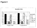
- Immature DC express, besides high levels of DC-SIGN, high levels of the receptors MR, CD11b and CD11c ( Figure 8a), which have previously been reported to mediate binding of mycobacteria by macrophages. We used blocking antibodies against these receptors to evaluate their contributions to ManLAM binding by DC. Immature DC bind strongly to ManLAM, but not to AraLAM, and the interaction was inhibited by the DC-SIGN-specific antibody, but strikingly not by any of the antibodies against MR, CD11b or CD11c ( Figure 8b).
- DC-SIGN is a major receptor for M. bovis BCG, since the antibodies against DC-SIGN strongly inhibited the infection of immature DC with M. bovis BCG ( Figure 8c).
- Antibodies against MR, CD11b and CD11c did not inhibit the infection, whereas the C-type lectin inhibitor mannan blocked the infection to a similar level as the DC-SIGN antibodies ( Figure 8c).
- Example 6 DC-SIGN facilitates capture and internalization of M. bovis BCG by immature DC through binding of the mycobacterial cell-wall component ManLAM.
- Immature DC are highly phagocytosing cells and indeed within 45 minutes more than 90% of the dendritic cells that bound M. bovis BCG, had internalized the mycobacteria ( Figure 8e). Similar as was observed for the binding of M. bovis BCG by DC ( Figure 8c), phagocytosis of M. bovis BCG is partly blocked by antibodies against DC-SIGN whereas both anti-MR antibodies and the MR-ligand mannose-BSA did not inhibit the observed phagocytosis ( Figure 8e). These results demonstrate that DC-SIGN facilitates capture and internalization of M. bovis BCG by immature DC through binding of the mycobacterial cell-wall component ManLAM.
- Example 7 Mycobacteria and ManLAM are internalized by DC-SIGN and targeted to lysosomes
- DC-SIGN can function as an antigen receptor that internalizes antigens and targets them to lysosomal compartments for presentation on MHC Class II 10 . Therefore, the fate of the captured M. bovis BCG by immature DC was followed by immuno-fluorescence analyses. Immature DC were incubated with FITC-conjugated M. bovis BCG for 2 hours and both DC-SIGN and Lamp-1 were stained ( Figure 9a). The observed co-localization of DC-SIGN with FITC-conjugated M. bovis BCG further supports a role for DC-SIGN in the capture and internalization of mycobacteria ( Figure 9a).
- Phagocytosed mycobacteria are targeted to the lysosomes since the internalized FITC-conjugated mycobacteria co-localize with Lamp-1 staining ( Figure 9a).
- ManLAM was also captured and internalized by DC-SIGN on immature DC since ManLAM staining co-localized with DC-SIGN ( Figure 9a) whereas AraLAM was not internalized by DC (results not shown).
- Internalized ManLAM co-localized with the lysosomal marker LAMP-1/CD107a in immature DC ( Figure 9a) indicating that internalized ManLAM is targeted to lysosomes.
- both whole mycobacteria and the cell-wall component ManLAM are similarly internalized by immature DC through DC-SIGN, supporting the results that DC-SIGN interacts with ManLAM on mycobacteria.
- Example 8 ManLAM changes the cytokine production by DC through DC-SIGN
- ManLAM is present not only a mycobacterial cell-wall component but is also secreted from phagosomes following macrophage ingestion of M. tuberculosis 31 . Potentially, mycobacteria within infected macrophages can influence bystander immune cells and modulate the immune response through secretion of ManLAM.
- the cytokine IL-10 is a potent immunosuppressive factor induced in macrophages by some intracellular bacteria to dampen down host immune responses and promote their survival 32 .
- Immature DC are highly efficient in antigen capture and processing, whereas mature DC are specialized in the naive T cell activation necessary for cellular immune responses. Immature DC mature in response to specific 'danger' signals such as bacterial components (LPS) or inflammatory cytokines (TNFa, PGE2).
- LPS bacterial components
- TNFa inflammatory cytokines
- Acute mycobacterial infections represent sites of inflammation that attract and induce DC maturation through the presence of maturation components. Therefore, we investigated the effect of ManLAM and AraLAM in combination with the stimulatory bacterial LPS.
- Toll-like receptor-4 (TLR4) interaction with LPS generates intracellular signaling, most notably via the transcription factor NFkB, that results in DC activation/maturation. Indeed, DC efficiently mature in the presence of LPS alone ( Figure 11b). Strikingly, this LPS-induced activation is inhibited in the presence of ManLAM, since the expression levels of the activation markers CD80, CD83 and CD86 were considerably lower than those of LPS-activated DC ( Figure 11b).
- Example 10 M. bovis BCG-induced DC maturation is inhibited by ManLAM.
- M. bovis BCG infection of immature DC results in DC maturation, as demonstrated by the increased expression of MHC class II and the co-stimulatory molecules CD80, CD83 and CD86 after M. bovis BCG infection ( Figure 12a).
- ManLAM binding to DC-SIGN prevented M. bovis BCG-induced DC maturation, since immature DC attracted to sites of mycobacterial infection will encounter both secreted ManLAM and intact mycobacteria. Strikingly, the M.
- bovis BCG (Figure 11c), since the co-stimulatory molecules, CD80, CD83 and CD86, are expressed at similar levels on both infected DC and AraLAM-treated infected DC. This indicates that the DC-SIGN-ManLAM interaction blocks the maturation of DC induced by LPS as well as M. bovis BCG.
- Example 11 S. mansoni soluble egg antigens (SEA) bind to human immature dendritic cells (DC) through interaction with DC-SIGN.
- SEA mansoni soluble egg antigens
- SEA is a mixture of glycoproteins, containing many immunogenic glycan antigens (3, 36). Major glycan antigens present in SEA, and the recognition of these antigens by anti-glycan MAbs, are depicted in Figure 13.
- Example 12 A sub-fraction of SEA contains high affinity ligands for DC-SIGN.
- SEA contains many different glycoproteins
- SEA glycoproteins were separated by SDS-PAGE and analyzed by Western blotting with DC-SIGN-Fc and anti-glycan antibodies reactive with Le x and LDN-DF, respectively.
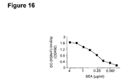
- SEA a major protein of approximately 70-80 kD, and two minor high-molecular proteins showed interaction with soluble DC-SIGN-Fc ( Figure 17).
- DC-SIGN binds to ⁇ 3-fucosylated glycans.
- the LDN-DF epitope contains terminal ⁇ 2-linked fucose.
- the results thus indicate that the that the glycosidase treatment specifically removed part of the ⁇ 3-fucose moieties present on SEA.
- the loss in a3/4-fucose residues in SEA upon treatment with the Xanthomonas ⁇ 1,3/ ⁇ 1,4-fucosidase also resulted in a 25% loss of binding of DC-SIGN-Fc (results not shown).
- DC-SIGN-Fc does not bind BSA-LDN-DF in which the ⁇ 3-fucose is capped with an ⁇ 2-fucose ( Figure 18).
- the binding is fucose-dependent, since no binding was observed to neoglycoproteins carrying Gal ⁇ 1,4GlcNAc (LN) or GalNAc ⁇ 1,4GlcNAc (LDN).
- the binding to Le x and LDNF by DC-SIGN is mediated through its CRD, since binding was inhibited by anti-DC-SIGN and EDTA.
- DC-SIGN strongly recognizes the ⁇ 3-fucosylated trisaccharide Le x and most likely interacts with ⁇ 3-fucosylated glycans on SEA such as Le x and/or LDNF.
- Example 14 The amino acid residue Val 351 within DC-SIGN is crucial for binding of DC-SIGN to both SEA and Le x
- the C-type lectin domain of DC-SIGN binds two Ca 2+ ions and those amino acid residues in close contact with Ca 2+ at site 2 (Glu 347 , Asn 349 , Glu 354 and Asn 365 ) or with Ca 2+ at site 1 (Asp 320 , Glu 324 , Asn 350 and Asp 355 ) are essential for ligand binding 37 . Because the results above indicated that the ⁇ 3-fucosylated trisaccharide Le x on SEA may function as a ligand for DC-SIGN, we investigated the binding properties of these antigens to K562 cell transfectants expressing mutated forms of DC-SIGN.
- Example 15 L-SIGN does not interact with DC-SIGN binding SEA or Lewis X
- L-SIGN an adhesion receptor that resembles DC-SIGN by recognizing ICAM-2, ICAM-3 and HIV-gp120, contains a CRD that is nearly identical to that of DC-SIGN 38 , and both receptors recognize high-mannose-type N-glycans.
- Our results above demonstrated a novel binding activity of DC-SIGN to fucosylated glycans and showed that Val 351 in DC-SIGN is essential for this binding activity.
- a Ser is present instead of Val ( Figure 19a), raising the question whether L-SIGN can recognize SEA and Le x .
- Example 16 Novel carbohydrate specificity predicts and confirms novel cellular counter-structures for DC-SIGN; DC-SIGN recognizes Le x carbohydrates on CD66a present on granulocytes to mediate DC-granulocyte interactions
- DC-SIGN is known to interact with ICAM-2 and ICAM-3, however the glycan ligands on these molecules have not yet been identified.
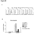
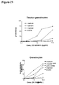
- the blood group antigen Le x (CD15) is expressed by gastric mucosal epithelial cells, and by polymorphonuclear leukocytes (granulocytes) ( Figure 20a); indeed DC-SIGN-Fc-coated beads bind strongly to granulocytes ( Figure 20b). Moreover, DC-SIGN-Fc also binds strongly to granulocytes ( Figure 21).
- the binding is specific for DC-SIGN since it can be inhibited by anti-DC-SIGN antibodies, mannan and depletion of calcium by EGTA but also by an anti-Le x antiobidy indicating that the cellular interaction is Le x dependent ( Figure 20 and 21).
- an antibody against Le x specifically reduces the observed adhesion of DC-SIGN to CD66a demonstrating that Le x carbohydrates indeed participate in the interaction ( Figure 20).
- Other glycan structures such as Le y and Le b can also be involved since the observed block is not complete. This demonstrates that granulocytes express a cell-surface glycoprotein that expresses a glycan structure that is recognized by DC-SIGN.
- DC-SIGN may also mediate binding of DC to tumor cells, since Le y expression is increased on many carcinomas including ovary, pancreas, prostate, breast, colon and non-small cell lung cancers 24 , while sulfo-Le a is present on certain tumors that express mucins.
- these results indicate that recognition of distinct carbohydrate structures by DC-SIGN may allow DC mediated cell adhesion to T cells, endothelial cells, PMNs as well as to tumor cells.
- DC-SIGN-Fc does not bind the cellular DC-SIGN counter-structures ICAM-2 or ICAM-3 from granulocytes but binds to a novel glycoprotein with a weight of 66kD protein that is only present on granulocytes ( Figure 22a).
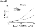
- Example 17 DC-SIGN binds strongly to a subset of cells in peripheral blood.
- DC-SIGN-Fc specifically bound to the CD56 dim CD16 + NK population and the interaction could be inhibited by anti-DC-SIGN antibodies ( Figure 26).
- ICAM-1-Fc did not interact with the CD56 dim CD16 + NK population, demonstrating that the interaction of DC-SIGN-Fc is specific and not mediated by the Fc tag.
- Both NK cell populations were able to specifically interact with ICAM-1-Fc after activation of their ⁇ 2 integrin LFA-1 ( Figure 26).
- DC-SIGN-Fc specifically bound to the CD56 dim CD16 + NK population and the interaction could be inhibited by anti-DC-SIGN antibodies
- DC-SIGN may bind another novel ligands on the CD56 dim CD16 + NK population.
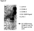
- immuno-precipitation reveals that DC-SIGN-Fc binds, besides ICAM-2, also a molecule with molecular weight of 166 kDa ( Figure 27).
- This ligand could be CD 166 or CD 16, which is heavily glycosylated and only expressed on NK cells.
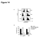
- Example 18 DC-SIGN mediates DC-NK cell interaction and is involved in the lysis of immature DC by NK cells.
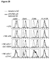
- DC-SIGN did not inhibit DC maturation by NK cells ( Figure 28) but was strongly involved in the NK-mediated immature DC lysis ( Figure 29), since antibodies against DC-SIGN inhibited the NK-mediated lysis of DC.
- Figure 29 shows that DC-SIGN regulates the interaction of immature DC with NK cell by binding a 166 kDa protein on CD56 dim CD16 + NK cells.
- This DC-SIGN-NK cell interaction also strongly enhances NK-mediated lysis of cell-lines transfected with DC-SIGN ( Figure 30), whereas antibodies against DC-SIGN inhibit the lysis.
- DC-SIGN may be important in the interaction of DC with NK cells and that inhibition of this interaction prevents NK-mediated lysis.
- DC-SIGN specifically interacts with HIV-1 39 and recently it was published that DC-SIGN binds to Ebola virus 40 . Therefore, we investigated whether DC-SIGN and its homologue L-SIGN can interact with other viruses, that contain glycosylated envelope proteins. Strikingly, DC-SIGN-Fc binds strongly to Herpes simplex virus (HSV)-1 and -2, and this interaction is specically inhibited by antibodies against DC-SIGN ( Figure 31). Further analysis demonstrates that DC-SIGN interact with the HSV glycoprotein gB ( Figure 32) since DC-SIGN expressed by DC and transfectants binds strongly to gB-coated beads.
- HSV Herpes simplex virus
- DC-SIGN is specifically expressed by DC and L-SIGN is expressed by Liver sinusoidal endothelial cells (LSEC) and some macrophage populations.
- LSEC Liver sinusoidal endothelial cells
- FIG 35 shows that the interaction of DC with HSV is mediated by DC-SIGN and this C-type lectin could be important in the infection of DC by these viruses.
- these viruses target DC-SIGN on DC not only to infect these DC but also to evade the immune response by modulating DC function as we demonstrated for mycobacteria and HIV-1.
Landscapes
- Health & Medical Sciences (AREA)
- Life Sciences & Earth Sciences (AREA)
- Chemical & Material Sciences (AREA)
- Veterinary Medicine (AREA)
- Pharmacology & Pharmacy (AREA)
- Public Health (AREA)
- General Health & Medical Sciences (AREA)
- Animal Behavior & Ethology (AREA)
- Medicinal Chemistry (AREA)
- Epidemiology (AREA)
- Molecular Biology (AREA)
- Immunology (AREA)
- Nuclear Medicine, Radiotherapy & Molecular Imaging (AREA)
- General Chemical & Material Sciences (AREA)
- Chemical Kinetics & Catalysis (AREA)
- Bioinformatics & Cheminformatics (AREA)
- Engineering & Computer Science (AREA)
- Organic Chemistry (AREA)
- Biochemistry (AREA)
- Tropical Medicine & Parasitology (AREA)
- Peptides Or Proteins (AREA)
- Medicines That Contain Protein Lipid Enzymes And Other Medicines (AREA)
- Medicines Containing Material From Animals Or Micro-Organisms (AREA)
- Medicines Containing Antibodies Or Antigens For Use As Internal Diagnostic Agents (AREA)
- Immobilizing And Processing Of Enzymes And Microorganisms (AREA)
Abstract
C-type lectins are involved in the binding of many different types of
carbohydrates. Considering their diversity in kind and expression of different
types of cells, the influence of such binding is very diverse and dependent
among others on the type of cell, the environment of the cell and the type of
carbohydrate bound. In the present invention new carbohydrate specificities of
C-type lectins are disclosed. Interference with this binding property has uses
in the prevention of pathogen binding and also in influencing signaling
pathways in the C-type lectin containing cell, particularly in Toll like receptor
expressing cells such as dendritic cells. Also provided is the use of the
carbohydrate specificity to enhance antigen presentation by antigen presenting
cells and to manipulate migration of C-type lectin containing cells and the
interaction of C-type lectin expressing cells with cellular ligands on
neighboring cells.
Description
The invention relates to the field of immunology. The invention in
particular relates to the role of dendritic cells in immune responses and to
pathogens that are able to capitalize thereon.
Dendritic cells (DC) are professional antigen presenting cells (APC)
that induce cellular immunity upon pathogen recognition. These cells are
therefore important in the defense against many pathogens 1;2;3. Immature DC
are seeded throughout peripheral tissues to act as sentinels against invading
pathogens4. Upon pathogen capture, DC are activated, process pathogens for
antigen presentation on Major Histocompatability Complex (MHC) class II
molecules, and migrate to the secondary lymphoid organs where they activate
naive T cells to initiate adaptive immune responses1;3;4. Depending on the
pathogen that is recognized by the DC, differentiation of naive T cells into Th1
cells is triggered by DC in response to intracellular microbes, whereas Th2-mediated
responses are generated by DC to eliminate pathogens residing
extracellularly 3. Thus DC play an important role in both innate and cellular
immune responses against tumors antigens as well as pathogens such as viral,
bacterial, fungal and parasitic infections1;5. Knowledge about cell-surface
receptors on DC that are involved in recognition of pathogens is only starting
to emerge, and include Toll-like receptors (TLR) 6;7 and C-type lectins 8. TLR
recognize specific pathogen-derived components, such as lipoproteins,
lipopolysaccharides and bacterial DNA, and relay this information through
intracellular signaling cascades leading to the production of regulatory
cytokines and upregulation of MHC and costimulatory molecules that lead to
activation/maturation of DC6. In contrast, C-type lectins recognize pathogen-derived
carbohydrate structures and upon binding internalize pathogens for
antigen processing and presentation to T cells8-10. In classical calcium-dependent
lectins, conserved amino acid residues in the carbohydrate
recognition domain (CRD) are involved in calcium binding and sugar
specificity 11. A growing number of C-type lectins are described to be
specifically expressed by DC. For most of these lectins detailed knowledge
about pathogen-targets as well as cellular ligands if any, including the identity
of the carbohydrate structure they recognize, is lacking8. The DC-specific C-type
lectin DC-SIGN (dendritic-cell specific ICAM-3 grabbing nonintegrin,
CD209) is involved in binding of the HIV-1 envelope glycoprotein by DC to
enhance infection of T cells 12, while the mannose receptor (MR) is involved in
recognition of mycobacteria and Fungi/Protozoa 13. Some C-type lectins like
DC-SIGN can interact with carbohydrate-bearing self glycoproteins (ICAM-2
and ICAM-3) to mediate cellular adhesion processes 14;15.
In one aspect of the present invention it was found that a C-type
lectin can comprise specificity for more than one type of glycoconjugate. This
knowledge is useful for a variety of purposes. For instance, lectins are
instrumental in mediating pathogen binding and the presentation of antigens
thereof in dendritic cells. This binding property of C-type lectins is utilized by
a number of pathogens to at least in part facilitate infection of an individual.
Knowledge of glycoconjugate specificity of such lectins therefore provides an
entrance to the development of medicaments capable of interfering with the
capacity of carbohydrates present on pathogens to interact with the lectin and
thereby at least in part interfere with the infection or the severity thereof in
the exposed individual. In one embodiment the invention therefore provides a
method for at least in part inhibiting the binding of a ligand to a C-type lectin
or a carbohydrate binding part thereof, comprising contacting said C-type
lectin with an isolated and/or recombinant glycoconjugate comprising at least
two mannose residues in α1,2 linkage or a glycoconjugate comprising a fucose
residue or a derivative or multimer thereof. Also provided is the use of a
glycoconjugate comprising at least two mannose residues in α1,2 linkage or a
glycoconjugate comprising a fucose residue or a derivative or multimer thereof
for at least in part inhibiting the binding of a ligand to a C-type lectin or a
carbohydrate-binding part thereof. This method may be used for instance to
study the exact binding properties of the lectin. The method is also of use in
identifying compounds capable of interfering with an ability of said C-type
lectin to bind to pathogens. In a preferred embodiment said method further
comprises a cell comprising said C-type lectin. Preferably said cell is an
antigen presenting cell, more preferably a dendritic cell or macrophage. In the
presence of such cell, the effect of such inhibition may be compared to the
function of the antigen presenting cell, for instance on its capacity to present
antigen and/or the production of lymphokines and/or cytokines. In a preferred
embodiment said C-type lectin comprises DC-SIGN, L-SIGN and/or DC-SIGNR.
Whereas DC-SIGNR and L-SIGN are predominantly expressed on
macrophage/endothelial cells, on lymph node and on liver sinusoidal
endothelial cells and DC-SIGN is expressed predominantly on dendritic cells.
DC-SIGNR and L-SIGN share the remarkable binding and signaling effects
that ManLAM has upon binding to dendritic cells via DC-SIGN. However,
whereas DC-SIGN further comprises a particular specificity for glyconjugates
comprising a fucose residue or a derivative or multimer thereof, DC-SIGNR
and L-SIGN are lacking such particular specificity. In the present invention
there is thus a preference for the C-type lectin DC-SIGN.
With the term "glycoconjugate comprising a fucose residue" is meant a
glycoconjugate comprising at least one fucose residue that is α1, 3 or α-1,4-linked
to the glycoconjugate. The linking moiety preferably consists of N-acetylglucosamine.
The linking moiety is preferably coupled to an
oligosaccharide or glycan, which in turn may be part of a larger structure (or
carrier molecule) comprising subsequent glycoconjugates comprising a fucose
residues, or other compounds such as mannose in any configuration. A
derivative of said glycoconjugate comprising a fucose residue comprises the
same C-type lectin binding activity in kind not necessarily in amount. A
derivative may be generated through modification of the fucose residue. Such
modifications may be generated in various ways. Preferably, said fucose is a
terminal fucose, i.e. linked via only one α1, 3 or α-1,4 linkage to another
molecule or fucose. Preferred examples of glycoconjugates comprising a fucose
residue are Lewis bloodgroup antigens Lex, Ley, Lea, Leb and/or LDNF. C-type
lectin binding parts, derivatives and/or analogous of the lewis bloodgroup
antigens are of course also within the scope of the invention. Different
modifications have different effects on the lectin binding properties. Sialylation
of Lex (yielding sialyl-Lex, a L-, E- and P-selectin ligand) completely abrogates
the recognition by DC-SIGN, indicating that DC-SIGN has a carbohydrate
specificity that is distinct from that of the selectins that mediate leukocyte
rolling. Sulfation reduces the binding affinity of DC-SIGN for Lex, as well as
Lea (Figure 2a). A derivative of a glycoconjugate comprising a fucose residue
thus comprises a modification wherein said modification allows binding of said
glycoconjugate to said C-type lectin. Glycoconjugates comprising multiple
fucose residues in the configuration of a lewis bloodgroup antigen typically
have a higher binding affinity than a glycoconjugate comprising a single fucose
residue.
With the term "glycoconjugate comprising at least two mannose residues in
α1,2 linkage" is meant a glycoconjugate comprising at least two mannose
residues wherein said mannose residues are linked to each other via an α1,2
linkage and wherein one of the mannose residues is linked to the
glycoconjugate. The linking moiety preferably but not necessarily comprises
mannose in α-1,5 linkage and arabinan or β-D-arabinose. The linking moiety is
preferably coupled to an oligosaccharide or glycan, which in turn may be part
of a larger structure (or carrier molecule) comprising subsequent
glycoconjugates comprising at least two mannose residues in α1,2 linkage, or
other compounds such as fucose in any configuration. A derivative of said
glycoconjugate comprising at least two mannose residues in α1,2 linkage
comprises the same C-type lectin binding activity in kind not necessarily in
amount. A derivative may be generated through modification of one or both of
the mannose residues. Such modifications may be generated in various ways.
Preferably, said at least two mannose residues in α1,2 linkage are terminal
residues, i.e. linked via only linkage to another molecule or mannose. In a
preferred embodiment glycoconjugate comprising at least two mannose
residues in α1,2 linkage, comprises two or three mannose residues linked to
each other via respectively one and two α1,2 linkages. A particularly preferred
example of a of glycoconjugates comprising at least two mannose residues in
α1,2 linkage is ManLAM. ManLAM comprises a mannose-capped
lipoarabinomannan, such as derivable from the cell wall component of
Mycobacterium. The mannose-capped lipoarabinomannan is preferably linked
to an oligosaccharide or glycan, which in turn may be part of a larger structure
(or carrier molecule) comprising subsequent mannose-capped
lipoarabinomannans or other compounds such as fucose in any configuration.
C-type lectin binding parts, derivatives and/or analogous of ManLAM are of
course also within the scope of the invention. Different modifications have
different effects on the lectin binding properties. Glycoconjugates comprising
two or more sets of at least two mannose residues in α1,2 linkage typically
have a higher binding affinity than a glycoconjugate comprising only one set of
at least two mannose residues in α1,2 linkage.
Carrier molecules may further comprise a large variety of different molecules
including peptide, protein, lipid, polysaccharide and also synthetic molecules
not expressly mentioned here. The carrier molecule(s) may of course be part of
an even larger structure such as a biological or artificial surface, a virus, a
bacterium or a eukaryotic cell.
Binding of the ligand to the C-type lectin can be inhibited by interfering with
binding site on the ligand, with binding site of the C-type lectin or both.
Interference with the binding site of the ligand can be done using a ligand
binding molecule capable of specifically binding to the C-type lectin binding
part on the ligand. This is preferably done with a proteinaceous molecule such
as a carbohydrate specific antibody preferably specific for a glycoconjugate
comprising at least two mannose residues in α1,2 linkage or a glyconjugates
comprising a fucose residue or a derivative or multimer thereof. The
experimental part describes non-limiting examples of such antibodies. Nonlimiting
examples of antibodies comprising a specificity for a glyconjugates
comprising a fucose residue or a derivative or multimer thereof are SMLDN1.1
or SMFG4.1, 6H3 or SMLDN1.1. The ligand binding molecule can also be a C-type
lectin, preferably a soluble derivative thereof, comprising specificity for a
glycoconjugate comprising at least two mannose residues in α1,2 linkage or a
glyconjugates comprising a fucose residue or a derivative or multimer thereof.
A non-limiting example of a soluble C-type lectin of the invention is a soluble
DC-SIGN-Fc chimeric molecule comprising amino acid residues 64-404 fused
at the C-terminus to a human IgG1-Fc fragment as described for instance in
Geijtenbeek et al (Geijtenbeek et al 2002, J. Biol. Chem. 277:11314-11320).
However, soluble C-type lectins comprising one or more of the provided
specificities can be generated in various ways (for instance by derivation from
DC-SIGNR or L-SIGN). Now that the present invention describes one chimeric
molecule others can be generated, for instance as described in Fawcett J et al
(Fawcett J et al 1992, Nature 360:481-4).
Interference with the binding site on the C-type lectin can of course also be done in various ways. Interference with the binding site on the C-type lectin can be done using a C-type lectin binding molecule capable of specifically binding to the ligand binding part on the C-type lectin. This is preferably done with a proteinaceous molecule such as a C-type lectin specific antibody or with a glycoconjugate comprising at least two mannose residues in α1,2 linkage or a glycoconjugate comprising a fucose residue or a derivative or multimer thereof, wherein said glycoconjugate or both may be part of a larger structure.
Interference with the binding site on the C-type lectin can of course also be done in various ways. Interference with the binding site on the C-type lectin can be done using a C-type lectin binding molecule capable of specifically binding to the ligand binding part on the C-type lectin. This is preferably done with a proteinaceous molecule such as a C-type lectin specific antibody or with a glycoconjugate comprising at least two mannose residues in α1,2 linkage or a glycoconjugate comprising a fucose residue or a derivative or multimer thereof, wherein said glycoconjugate or both may be part of a larger structure.
Binding can of course further be inhibited by sterically hindering the binding
of the ligand to the C-type lectin. For this it is not absolutely required that the
interfering molecule itself binds to the association site of the carbohydrate and
the C-type lectin. Binding can also be interfered with successfully steric
hindrance by binding of the interfering molecule in the vicinity of the site of
association.
The ligand may be any type of structure capable of binding to said C-type
lectin. Usually this will comprise a proteinaceous molecule or a carbohydrate.
The ligand may be an antibody. It is preferred, but not strictly necessary, that
the ligand comprises mannosilated glycans such as cell wall component of
Mycobacterium ManLAM, or a mannose derivative, lipophosphoglycan such as
derived from Leishmania, or a glycoconjugate comprising at least two mannose
residues in α1,2 linkage or a glycoconjugate comprising a fucose residue such
as SEA or CD66 or a derivative or multimer thereof. The ligand can have
instead of, or in addition to, a glycoconjugate comprising at least two mannose
residues in α1,2 linkage or a glyconjugate comprising a fucose or a derivative
or multimer thereof, different glyconjugates capable of binding to the same on
the C-type lectin.
The ligand preferably comprises an antigen. The ligand preferably comprises a (tumor) antigen, a pathogen and/or a cell associated receptor. An antigen is here used to include peptides or glycolipids and derivatives thereof capable of being presented in the context of MHC class I or class II, or CD1b. At least some C-type lectins, and particularly those expressed on dendritic cells are involved in the process of antigen uptake and presentation thereof by antigen presenting cells. An early step in this process is the capture of the antigen by such lectins. With a method of the invention it is possible to at least in part interfere with this initial step in the process of antigen presentation. Considering the importance of antigen presentation in the initiation and maintenance of antigen specific immune responses, it is clear that the method of the invention can be favorably used to at least in part diminish the potency of an immune response against said antigen.
In a particularly preferred embodiment the ligand comprises a pathogen or a C-type lectin binding part thereof. It has been found that many different pathogens comprise carbohydrates capable of binding to C-type lectins of the invention. In particular it has been found that pathogens or parts thereof can bind to said C-type lectins through a mannose containing glycoconjugate such as a glycoconjugate comprising at least two mannose residues in α1,2 linkage or a glyconjugates comprising a fucose or derivative or multimer thereof. This binding at least in part facilitates infection of an individual with said pathogen. Interference with the binding of the pathogen or part thereof to the C-type lectin can thus at least in part aid an individual in combating an infection by the pathogen and in cases even prevent the establishment of a clinically visible symptoms thereof. The latter is useful in (passive or active) vaccination strategies. The pathogen can be a virus, a fungus, a (myco)bacterium and/or a parasite. The pathogen preferably comprises a glycoconjugate comprising at least two mannose residues in α1,2 linkage or a glycoconjugate comprising a fucose residue or a derivative or multimer thereof. Examples of preferred pathogens are human immunodeficiency virus, a mycobacterial infection, a helicobacter, a leishmania, a schistosoma, a klebsiella, a herpes simplex virus or an ebola virus. Inhibition of binding of the specifically mentioned pathogens can be achieved using a glycoconjugate comprising at least two mannose residues in α1,2 linkage or a glycoconjugate comprising a fucose residue or a derivative or multimer thereof. In a particularly preferred embodiment inhibition of binding is achieved using a ligand (pathogen or part thereof) binding molecule capable of specifically binding to the C-type lectin binding part on the ligand. This is preferably done with a proteinaceous molecule such as a carbohydrate specific antibody preferably specific for a mannose carbohydrate such as a glycoconjugate comprising at least two mannose residues in α1,2 linkage or a glyconjugate comprising a fucose residue or a derivative or multimer thereof. The experimental part describes non-limiting examples of such antibodies. The ligand binding molecule can also be a C-type lectin, preferably a soluble derivative thereof, comprising specificity for a mannose carbohydrate such as a glycoconjugate comprising at least two mannose residues in α1,2 linkage or a glyconjugates comprising a fucose residue or a derivative or multimer thereof. A non-limiting example of a soluble C-type lectin of the invention is a soluble DC-SIGN-Fc as mentioned above. Thus infections of an individual with said pathogens can at least in part be prevented or treated by administering to said individual a mannose carbohydrate such as a glycoconjugate comprising at least two mannose residues in α1,2 linkage or a glyconjugates comprising a fucose residue or a derivative or multimer thereof. In a preferred embodiment a ligand binding molecule of the invention is administered to said individual. This embodiment can also be used to at least in part inhibit the binding of the pathogen to a C-type lectin expressing cell thereby at least in part inhibiting the contamination or spread of the pathogen in the body of a patient, as well as inhibiting DC maturation and cell adhesion, as is also described below.
C-type lectins not only recognize carbohydrate profiles on pathogens but also
interact with self glycoproteins to mediate cellular processes such as
differentation and migration. A method of the invention can therefore also be
used to interfere with the interaction with one or more self-glycoproteins and
thereby be used to at least in part inhibit the cellular processes that the
mentioned C-type lectins are involved in. This can be done by providing the
carbohydrates structures provided by the invention (i.e. mannose containing
glycojugate such as a glycoconjugate comprising at least two mannose residues
in α1,2 linkage or a glycoconjugate comprising a fucose residue or derivative or
multimer thereof). Preferably, this is achieved using a ligand binding molecule
of the invention. Thus in another preferred embodiment the method is used to
at least in part inhibit binding of said C-type lectin to a self-glycoprotein
preferably a receptor present on the outer membrane of a cell. Preferably said
receptor comprises ICAM-2, ICAM-3, CD166 or CD66 or a functional part,
derivative and/or analogue thereof. CD 166 and CD66 are present on a subset
of NK cells and granulocytes. Thus interaction of these cells with a cell
comprising a mentioned C-type lectin can be at least in part inhibited using a
specific binding partner for a C-type lectin mentioned above. Thus interaction
of preferably dendritic cells and said subset of NK cells and granulocytes can
be interfered with. Preferably, said C-type lectin comprises DC-SIGN. As
specific binding partner of said C-type lectin a mannose containing
glycoconjugate such as a glycoconjugate comprising at least two mannose
residues in α1,2 linkage or a glycoconjugate comprising a fucose residue or a
derivative or multimer thereof, can be advantageously used. The cellular
interaction of DC with granulocytes or NK cells is essential in the innate
immune response. In particular granulocytes are involved early ingestion of
pathogens and may attract and stimulate DC to participate in pathogen
recognition and presentation to T cells. In contrast NK cells are well know to
be involved in killing of infected cells. NK cells can kill or activate DC that
allow lysis of pathogen captured DC or maturation of pathogen captured DC to
enhance T cell stimulation and immune response. The cellular interaction of
granulocytes and NK cells with DC is to enhance immunity against pathogens
or eliminate pathogen infected DC.
In another aspect the invention provides a method for modulating
the stimulating immune effect of a dendritic cell that is activated via a Toll-like
receptor signaling pathway, said method comprising contacting said
dendritic cell with an isolated and/or recombinant specific C-type lectin
binding molecule. Preferably, said C-type lectin comprises DC-SIGN and said
specific C-type lectin binding compound comprises a glycoconjugate comprising
at least two mannose residues in α1,2 linkage or a glycoconjugate comprising a
fucose residue or a derivative, a combination or multimer thereof and
preferably, a glycoconjugate comprising at least two mannose residues in α1,2
linkage, preferably ManLAM. In combination with Toll-like receptor signaling
ManLAM binding to DC-SIGN on a dendritic cell induces the production of
significant amounts of IL-10 and reduces DC-activation/maturation. The
production of IL-10 and reduction of activation/maturation of DC lead to a
dampening of immune responses. Thus by providing a glycoconjugate
comprising at least two mannose residues in α1,2 linkage or an analogously
acting glycoconjugate to a dendritic cell, this cell can be induced to dampen the
immune response at least in its direct vicinity. Thus an active immune system
comprising a number of dendritic cells participating in the activity can be
dampened through by providing the dendritic cell with a glycoconjugate
comprising at least two mannose residues in α1,2 linkage or an analogously
acting glycoconjugate. This embodiment is useful in situations where an
individual is suffering from or at risk of suffering from an over-active immune
system. Preferably, said over-active immune system involves graft versus host
disease, host versus graft disease and the various auto-immune diseases.
Alternatively, an immune system dampened through the discussed
mechanism can be stimulated by providing dendritic cells with a specific C-type
lectin binding molecule capable of interfering with the binding of a
glycoconjugate comprising at least two mannose residues in α,1,2 linkage or
analogously acting glycoconjugate. ManLAM, at least in amounts active
locally, is tolerated by individuals considering that active amounts of ManLAM
are secreted by mycobacterium infected cells in the body. Alternatively, the
dampened system can be stimulated by using a ligand binding molecule of the
invention, preferably a proteinaceous molecule such as a carbohydrate specific
antibody preferably specific for any mannose containing glycoconjugate such as
ManLAM or a glyconjugates comprising a fucose residue or a derivative or
multimer thereof. Preferably, said antibody comprises a specificity for a
mannose residue in α1,2 linkage with another mannose. The experimental
part describes non-limiting examples of such antibodies. The ligand binding
molecule can also be a C-type lectin, preferably a soluble derivative thereof,
comprising specificity for a glycoconjugate comprising at least two mannose
residues in α1,2 linkage or a glyconjugates comprising a fucose residue or a
derivative or multimer thereof. Another non-limiting example of such an
interfering molecule is AraLAM. This embodiment is useful to combat (chronic)
diseases wherein at least part of the phenotype of the disease is due to
dampening of the immune system through the mentioned C-type lectin
pathway. Preferred examples of such diseases are infections by a human
immunodeficiency virus, a mycobacterium, a helicobacter, a leishmania, a
schistosoma, a klebsiella, a herpes simplex virus or an ebola virus
Immature DC are highly efficient in antigen capture and processing, whereas
mature DC are specialized in the naive T cell activation necessary for cellular
immune responses. Immature DC mature in response to specific 'danger'
signals such as bacterial components (LPS) or inflammatory cytokines (TNFa,
PGE2). In the present invention it was found that a glycoconjugate comprising
a mannose, a fucose residue or a derivative, a combination or multimer
thereof, does not induce DC maturation. In contrast to LPS that triggers
TLR4, no up-regulated expression of the activation markers CD80, CD83,
CD86 or HLA-DR was observed. However, when the maturation of immature
dendritic cells was studied in the situation when the cells are also activated
via a Toll-like receptor pathway, a strong inhibition of Toll-like receptor
induced maturation was observed, particularly when the dendritic cells were
exposed to ManLAM. Such inhibition thus dampens the response of Toll-like
receptor pathway stimulated dendritic cells and thereby the capacity of an
individual to cope with pathogens that are combated via activation of the Toll-like
receptor pathway. This dampening of DC-activation can be at least in part
prevented by at least in part inhibiting binding of particularly a glycoconjugate
comprising at least two mannose residues in α1,2 linkage, but also analogously
acting glycoconjugates to C-type lectins on the immature dendritic cells. The
invention thus provides a method for determining whether a compound is
capable of modulating an activation state of a dendritic cell comprising
providing said dendritic cell with a compound capable of specifically binding to
a c-type lectin and determining whether a Toll-like receptor signaling pathway
in said dendritic cell is modulated. Further provided is a method for
modulating the activity of a Toll-like receptor signaling pathway in a cell,
wherein said cell comprises a Toll-like receptor and a C-type lectin, said
method comprising contacting said cell with an isolated and/or recombinant C-type
lectin binding molecule. Preferably said C-type binding molecule
comprises a glycoconjugate comprising a mannose, a fucose residue or a
derivative, a combination or multimer thereof. Activity can be modulated
upward by at least in part preventing simultaneous stimulation of C-type
lectin with a glycoconjugate comprising at least two mannose residues in α1,2
linkage or analogously acting compound. Activity is modulated downward by
simultaneously providing said a glycoconjugate comprising at least two
mannose residues in α1,2 linkage or analogously acting compound.
Analogously acting compounds comprise the same Toll-like receptor signaling
interfering capacity as ManLAM in kind not necessarily in amount. A
preferred analogously acting compound comprises a glycoconjugate comprising
a fucose residue or a derivative or multimer thereof. A further level of control
is possible using C-type lectin binding compounds that at least in part
interfere with the binding capacity of a glycoconjugate comprising at least two
mannose residues in α1,2 linkage or an analogously acting compound. In this
way activity of the Toll-like receptor pathway can be modulated upward (in the
presence of the proper Toll-like receptor ligand) even in the presence of a
glycoconjugate comprising at least two mannose residues in α1,2 linkage or
analogously acting compound. The latter feature is of importance in cases
where the immune system has problems combating an infection via the Toll-like
receptor pathway, particularly when said infection is accompanied with
secretion of a glycoconjugate comprising at least two mannose residues in α1,2
linkage or analogously acting compounds, such as mycobacteria and in
particular M. tuberculosis and M. Bovis. Thus the invention further provides a
method for stimulating maturation of a dendritic cell that is contacted with a
Toll-like receptor ligand and a glycoconjugate comprising at least two mannose
residues in α1,2 linkage or analogously acting compound, said method
comprising providing said dendritic cell with a C-type lectin binding molecule
capable of blocking the binding of said glycoconjugate to said C-type lectin. C-type
lectin binding molecules capable of at least in part inhibiting the binding
of a glycoconjugate comprising at least two mannose residues in α1,2 linkage
or analogously acting compounds are for instance C-type lectin binding
antibody or a functional part, derivative and/or analogue thereof, with a
binding specificity that blocks or covers the ManLAM binding site on said C-type
lectin. Preferable said C-type lectin comprises DC-SIGN. Thus in a
preferred embodiment said antibody is a DC-SIGN specific antibody. A
suitable example of such an antibody is AZN-D1, AZN-D2 or AZN-D3 or a
human or humanized analogue comprising the same binding specificity in kind
not necessarily in amount. In a preferred embodiment the invention provides a
method for stimulating maturation of a dendritic cell that is contacted with a
Toll-like receptor ligand and a glycoconjugate comprising at least two mannose
residues in α,1,2 linkage or analogously acting compound, said method
comprising providing said dendritic cell with a ligand binding molecule of the
invention, thereby at least in part preventing binding of said a glycoconjugate
comprising at least two mannose residues in α,1,2 linkage or analogously
acting compound to the dendritic cell.
An important aspect of the invention is concerned with the use of a
glycoconjugate comprising at least two mannose residues in α1,2 linkage or a
glycoconjugate comprising a fucose residue or a derivative or multimer thereof,
a ligand binding molecule of the invention and/or a C-type lectin binding
molecule of the invention for the preparation of a medicament. Such
medicaments may be used for the treatment of an immune system associated
disease or the treatment of an acquired disease. Preferably, said acquired
disease comprises an infection with human immunodeficiency virus,
mycobacteria, a fungus, a helicobacter, a leishmania, a schistosoma, a
klebsiella, a herpes simplex virus or an ebola virus.
Considering the natural role of C-type lectins on antigen presenting
cells it is within the scope of the present invention to stimulate immune
responses in an individual by providing antigen through a C-type lectin
receptor on the antigen presenting cell. This can of course be achieved when
the antigen comprises a a glycoconjugate comprising at least two mannose
residues in α1,2 linkage or glycoconjugate comprising a fucose residue or
derivative or multimer thereof. In this embodiment it is preferred that
simultaneous activation of the Toll-like receptor signaling pathway is at least
in part prevented. Provided is thus a glycoconjugate comprising an antigen
and a glycoconjugate comprising at least two mannose residues in α1,2 linkage
and/or a fucose residue or a derivative or multimer thereof for use for the
preparation of a vaccine. The vaccine may be preventive or curative. The
antigen may be derived from any source. as long as it is capable of being
presented through major histocompatibility complex I, complex II or C1b. In a
preferred embodiment said antigen comprises a tumor antigen. The presence
of the mentioned carbohydrates on tumor cells facilitates antigen capture by
DC to enhance antigen presentation and as a result immune activation. On the
other hand, the immune response against tumor antigen lacking the
mentioned carbohydrates can stimulated significantly by providing the antigen
with one or more of the mentioned carbohydrates thereby stimulating DC
uptake and thus the immune response against the antigen. This aspect is
useful in the preparation of vaccines.
In another embodiment the invention provides the use of ligand
binding molecule and/or a c-type lectin binding molecule of the invention for
the preparation of a vaccine. A dampening of the immune system due to the
effect of a glycoconjugate comprising at least two mannose residues in α1,2
linkage or analogously acting compounds is reduced by providing a
glycoconjugate or antibody capable of at least in part inhibiting the binding of
a glycoconjugate comprising at least two mannose residues in α1,2 linkage or
analogously acting compound to their C-type lectin receptor. The antibody
preferably comprises SMLDN1.1, SMFG4.1, 6H3 or SMLDN1.1. Thus antigen
present in said vaccine or provided separately is more effective in stimulating
or boosting an immune response in the presence of a glycoconjugate
comprising at least two mannose residues in α1,2 linkage or analogously
acting compound. This is particularly important in patients suffering from a
mycobacterial infection, such as but not limited to M. tuberculosis or M. bovis.
Preferably, said vaccine is used to stimulate an antigen specific immune
response in said individual. In a preferred embodiment the medicament or
vaccine is used for the treatment of an individual suffering from a cancer, an
autoimmune disease or a transplantation related disease.
As used herein the term "antibody" refers to antibodies derived from
humans or other animals. The antibody is preferably produced outside the
body. The antibody can also be generated or selected using artificial systems
such as phage display selection. An antibody having no natural counterpart is
therefore well within the scope of the present invention. Antibodies as used
herein also include fragments thereof capable of binding to the same target,
such as FAB fragments or even smaller parts. The antigen binding part of an
antibody of the invention may also be grafted onto another type of molecule to
provide that molecule with a binding specificity as provided for in the
invention. Modification of the antibody to include human or humanized
versions thereof with the same binding specificity in kind not necessarily in
amount are of course also in the scope of the invention. Also included are
single chain fragments and variants thereof.
As used herein the term proteinaceous molecule refers to a peptide,
a poly-peptide, protein and the like with or without modifications. Such
modifications may be synthetic and/or provided for by a biological system. The
latter including for instance post-translation modification such as
glycosylation.
Where the invention is described for use in humans, the invention is
also functional in other animals for instance farm-animals and pets. These
medical and vaccine uses and methods of treatment are therefore also part of
the invention.
DC-SIGN binds as strongly to SEA as to HIV-1 gp120, as was determined by an ELISA based assay using soluble DC-SIGN-Fc. SEA were coated at a concentration of 5 µg/ml, and HIV-1 gp120 at a concentration of 1 µg/ml.
Specificity was determined by measuring binding in the presence of the anti-DC-SIGN blocking antibody AZN-D1 (20 µg/ml), or EDTA (5 mM).
Binding of soluble DC-SIGN-Fc to different concentrations SEA was measured by an anti-IgG-Fc ELISA.
SEA were separated by SDS-PAGE gelelectrophoresis on a 12.5% polyacrylamide gel.
The interaction of DC-SIGN with granulocytes is further investigated by titration of DC-SIGN-Fc and determining the binding by measuring the bound DC-SIGN-Fc using FITC-conjugated Goat-anti-human Fc antibodies. The interaction is blocked by antibodies against DC-SIGN (AZN-D1), and mannan and EGTA (A) and by anti-Lex antibodies (3H3, B).
293T cells are transfected with pIG-CD66a-Fc, pIG-ICAM-3-Fc and pIG (mock). Supernatants are harvested and beads are coated with the Fc chimeras (CD66a-Fc, ICAM-3-Fc and mock). Immature DC and DC-SIGN-transfected K562 cells are incubated with beads and the adhesion is determined in the presence of antibodies against DC-SIGN and β2 integrins, and mannan and EGTA.
The binding of DC-SIGN-Fc to peripheral blood lymphocyte (PBL) subsets is determined by flow cytometry in the presence of antibodies against DC-SIGN. PBL subsets are distinguished by staining with CD3 and CD56, and adhesion is determined by triple staining with DC-SIGN-Fc.
NK cells are surface labelled with radioactive iodine. The cell lysate is incubated over night at 4°C with Protein A beads coated with DC-SIGN-Fc or control antibodies against ICAM-2, ICAM-3 and LFA-1. The beads are washed and the immuno-precipitated product is analysed on SDS-PAGE and visualised by autoradiography.
Immature DC are co-cultured with NK cells for 24 hours in the presence of antibodies against DC-SIGN (AZN-D1) and LFA-1 (NKI-L19). The maturation is determined by measuring the expression of CD80, CD83, CD86 and HLA-DR. with NK cells O/N at 37°C. As a control, DC are matured with LPS (10 ng/ml).
A.DC-SIGN transfectants are incubated with HSV envelope glycoprotein-coated beads and DC-SIGN binding is determined in the presence of antibodies against DC-SIGN.
B L-SIGN strongly binds HSV envelope glycoprotein gB.
L-SIGN transfectants are incubated with HCV envelope glycoprotein-coated beads and DC-SIGN binding is determined in the presence of antibodies against DC-SIGN.
Antibodies and proteins. The following monoclonal antibodies (Mab) were used:
anti-CD107a (Lamp-1; MabH4A3, BD Pharmingen)., anti-MR (Clone 19, BD
Pharmingen), CD11b (bear-1)25, CD11c (SHCL3)26, anti-DC-SIGN (AZN-D1,
AZN-D214, CSRD10, blocking CD11a (NKI-L15), anti-ICAM-1 (Rek-1), anti-ICAM-2(12A2),
and anti-ICAM-3 (AZN-IC3/1, icr-2), activating CD18
(KIM185) and the PE/FITC-conjugated antibodies CD25, CD69, CD80, CD86,
HLA-DR (BD Pharmingen), and CD83 and CD56 (Beckman Coulter). Biotin-conjugated
CD3 and CD16 (BD Pharmingen ). CLB-T4 (αCD4) was a kind gift
Of René van Lier. MEM-157 (αCD16) and MEM-97 (αCD20) were kindly
provided by Vaclav Horejsi. ICAM-1 mAb CBR-IC-1/3, ICAM-2 mAb CBR-IC-2/1,
and ICAM-3 mAb CBR-IC-3/2 were obtained through the Human
Leukocyte Differentiation Antigen Workshop. Several anti-glycan monoclonal
antibodies (MAbs) were used: the anti-LDN-DF MAb 114-5B1-A16, the anti-LDN
MAb SMLDN1.117, the anti-LDN-F MAb SMLDNF110 and the anti-Lex
MAb CB1027 , the anti-Lex mAbs SMLDN1.1 and SMFG4.1 and6H3 (anti-Lex).
The mAb (CLB-gran/10) was used to stain and immunoprecipitate CD16.
The neoglycoprotein HSA-Lex, containing approx. 20-25
oligosaccharide chains per HSA molecule, was from Isosep AB, Tullinge,
Sweden. The neoglycoprotein BSA-LDN-DF (approx. 12 oligosaccharide chains
per molecule BSA), and BSA-LDN-F (approx. 3-4 oligosaccharides per molecule
BSA), were synthesized enzymatically as described by van Remoortere et al16,
and Nyame et al17 , respectively. Lex-PAA-biotin, containing Lex multivalently
coupled to biotinylated polyacrylamide was from Syntesome, Munich,
Germany.
Mannan purified from Saccheromyces cerevisiae (50 µg/ml) and
recombinant gp120 (0.50 µg/ml) were obtained from Sigma and the
Aidsresource Foundation, respectively. Purified mannose capped
lipoarabinomannan (manLAM) from Mycobacterium tuberculosis, and non-capped
LAM (araLAM) from M. smegmatis were kindly provided by Dr. J.
Belisle, Colorado State University through NIH, NIAID contract NO1 AI-75320.
Soluble egg antige (SEA) was kindly provided by Dr. A. K. Nyame,
Oklahoma University HSC, USA). Purified lipophosphoglycan from
Leishmania mexicana was kindly donated by Dr. M Wiese, Bernard Nocht
Inst. Tropical Medicine Hamburg, Germany. Purified lipopolysaccharide of
Helicobacter pylori was obtained from M. Monteiro, NRC, Ottawa, Canada. A
sonicate of bacterial cells of a clinical isolate of M. tuberculosis was donated by
A. Kolk, KIT, Royal Trop. Inst, Amsterdam. Clinical isolates of Helicobacter
pylori, Escherichia coli, Klebsiella pneumoniae, Pseudomonas aeruginosa and
Staphylococcus aureus were obtained from VUMC Hospital, Amsterdam.
Synthetic glycoconjugates were obtained from Syntesome, Munich, Germany,
and comprise mono- and oligosaccharides multivalently linked to a
biotinylated polyacrylamide carrier, MW 40.000.
Soluble DC-SIGN-Fc adhesion assay. DC-SIGN-Fc consists of the extracellular portion of DC-SIGN (amino acid residues 64-404) fused at the C-terminus to a human IgG1-Fc fragment. DC-SIGN-Fc was produced in Chinese Hamster Ovary K1 cells by co-transfection of DC-SIGN-Sig-pIgG1 Fc (20 µg) and pEE14 (5 µg) vector. DC-SIGN-Fc concentrations in the supernatant were determined by an anti-IgG1 Fc ELISA. The DC-SIGN-Fc binding assay was performed as follows. Glycoconjugates and sonicated mycobacteria were coated onto ELISA plates at 5 µg/well; intact bacterial cells were coated at 5x10⁷/ml ; coating took place for 18 hours at room temperature, followed by blocking with 1% BSA for 30 min, at 37°C in TSM (20mM Tris-HCl pH 7.4 containing 150mM NaCl, 2 mM CaCl2 and 2mM MgCl2). Soluble DC-SIGN-Fc (approx. 2 µg/ml in TSM buffer) was added and the adhesion was performed for 120 min. at RT. Unbound DC-SIGN-Fc was washed away and binding was determined by an anti-IgG1 Fc ELISA using a peroxidase conjugate of goat anti-human Fc. Specificity was determined in the presence of either 20 µg/ml blocking antibodies, 50 µg/ml mannan or 5mM EGTA.
Soluble DC-SIGN-Fc adhesion assay. DC-SIGN-Fc consists of the extracellular portion of DC-SIGN (amino acid residues 64-404) fused at the C-terminus to a human IgG1-Fc fragment. DC-SIGN-Fc was produced in Chinese Hamster Ovary K1 cells by co-transfection of DC-SIGN-Sig-pIgG1 Fc (20 µg) and pEE14 (5 µg) vector. DC-SIGN-Fc concentrations in the supernatant were determined by an anti-IgG1 Fc ELISA. The DC-SIGN-Fc binding assay was performed as follows. Glycoconjugates and sonicated mycobacteria were coated onto ELISA plates at 5 µg/well; intact bacterial cells were coated at 5x10⁷/ml ; coating took place for 18 hours at room temperature, followed by blocking with 1% BSA for 30 min, at 37°C in TSM (20mM Tris-HCl pH 7.4 containing 150mM NaCl, 2 mM CaCl2 and 2mM MgCl2). Soluble DC-SIGN-Fc (approx. 2 µg/ml in TSM buffer) was added and the adhesion was performed for 120 min. at RT. Unbound DC-SIGN-Fc was washed away and binding was determined by an anti-IgG1 Fc ELISA using a peroxidase conjugate of goat anti-human Fc. Specificity was determined in the presence of either 20 µg/ml blocking antibodies, 50 µg/ml mannan or 5mM EGTA.
Cells. Immature DC were cultured from monocytes in the presence of IL-4 and
GM-CSF (500 and 800 U/ml, respectively; Schering-Plough, Brussels,
Belgium28. At day 7 the phenotype of the cultured DC was confirmed by flow
cytometric analysis. The DC expressed high levels of MHC class I and II, αMβ2
(CD11b), αXβ2 (CD11c) and ICAM-1, moderate levels of LFA-1 and CD80, and
low levels of CD14. K562 transfectants expressing wild-type DC-SIGN were
generated by transfection of K562 cells with 10 µg pRc/CMV-DC-SIGN plasmid
by electroporation as previously described.
Stable transfectants of K562 expressing ICAM-3 were obtained by
electroporation of pCDM8-ICAM-3 and pGK-HYG. K562 cells were cultured on
RPMI 10% FCS, whereas K562-ICAM-3 cells were cultured on RPMI 10% FCS
: Iscove's 5% FCS 3:1 containing 0.5 mg/ml hygromycine to maintain ICAM-3
expression.
Naive NK cells were isolated from buffy coats of healthy donors.
Briefly, the PBMC fraction obtained through Ficoll centrifugation was
sequentially depleted for CD14+ cells, and CD3+, CD4+, and CD20+ cells using
MACS sorting. CD14+ cells were depleted by CD14 microbeads (Miltenyi
Biotec) on an LS column (Miltenyi Biotec), and CD3+, CD4+, and CD20+ cells
were labeled by the mAbs T3B (αCD3), CLB-T4 (αCD4), MEM97 (αCD20), and
thereafter depleted by goat-α-mouse microbeads (Miltenyi Biotec) on an LD
column (Miltenyi Biotec). Thus obtained NK cells were routinely tested for the
NK cell markers CD16 (75% to 90% expression), CD56 (80 to 95% expression),
the non-lineage markers CD3, CD4, CD14 and CD20 (all less than 1%
expression), and the early activation marker CD69 (15% expression on non-activated
NK cells and 75% expression on 1-day IL-2-activated NK cells).
CD56dim and CD56bright NK cell populations were isolated from MACS obtained
NK cells by FACS sorting on low and high CD56 expression, respectively.
CD56dim and CD56bright NK cell populations were over 95% pure as assessed by
flow cytometry staining for CD16 and CD56.
DC activation. Immature DC (2x10⁶ cells/ml) were cultured for 24 hours in the
presence of IL-4 (500 U/ml, Schering-Plough, Brussels, Belgium), GM-CSF
(800 U/m; Schering-Plough, Brussels, Belgium) and either LPS (10 ng/ml) or
LAM glycolipids (15 µg/ml). The effect of LAM on LPS-induced activation was
determined by pre-incubating immature DC (300.000 cells) with AZN-D2 (40
µg/ml) for 30 minutes, and subsequently with LPS in the presence of LAM (15
µg/ml) for 18 hours. LAM glycolipids were obtained from J. Belisle (Colorado
State University and the NIH (contract NO1 AI-75320)) and contained <5
ng/mg endotoxin. Activation was determined by cell-surface expression of
MHC class II (HLA-DR) and the co-stimulatory molecules CD80, CD83 and
CD86 using PE-conjugated antibodies.
Fluorescent bead adhesion assay. Carboxylate-modified TransFluorSpheres
(488/645 nm, 1.0 µm; Molecular Probes, Eugene, OR) were coated with HIV-1
gp120 and ICAM-3 as described14. Streptavidin was covalently coupled to the
beads as described and streptavidin-coated beads were incubated with
biotinylated PAA-linked glycoconjugates (50 pMol; Syntesome, Munich,
Germany). The fluorescent bead adhesion assay was performed as described14.
Ligand-coated fluorescent beads (20 beads/cell) were added to the cells for 45
minutes at 37°C, washed and analyzed by flow cytometry (FACScan, Becton
Dickinson, Oxnard, CA), by measuring the percentage of cells that had bound
fluorescent beads. LAM-coated beads were generated by coating incubating
streptavidin-coated beads were incubated with biotinylated F(ab')2 fragment
goat anti-mouse IgG (6 µg/ml; Jackson Immunoresearch) followed by an
overnight incubation with mouse-anti-LAM antibody (F30.5) at 4°C. The beads
were washed and incubated with 250 ng/ml purified glycolipid LAM (obtained
from J. Belisle, Colorado State University and the NIH (contract NO1 AI-75320))
overnight at 4°C. SEA-coated beads were generated by incubating the
streptavidin-coated beads with biotinylated F(ab')2 fragment of goat anti-mouse
IgG (6 µg/ml; Jackson Immunoresearch), followed by an overnight
incubation at 4°C with anti-LDN MAb, or anti-LDN-DF MAb. The beads were
washed and incubated with 1µg/ml SEA overnight at 4°C. Essentially, 50x103
cells were pre-incubated in adhesion buffer (20 mM Tris-HCl pH 8.0, 150 mM
NaCl, 1mM CaCl2, 2 mM MgCl2, 0.5% BSA) with or without blocking MAbs
(20 µg/ml) or mannan (50 µg/ml) for 10 minutes at room temperature. Ligand-coated
fluorescent beads (20 beads/cell) were added to the cells and the
suspension was incubated for 45 minutes at 37°C. Cells were washed and
adhesion was determined using flow cytometry (FACScan, Becton Dickinson,
Oxnard, CA), by measuring the percentage of cells that had bound fluorescent
beads. HIV-1 gp120 fluorescent beads were prepared as described previously19.
DC-SIGN-Fc adhesion. Cells were incubated with DC-SIGN-Fc for 30 minutes
at 37°C under saturating conditions (concentration: 10 µg/ml), and
subsequently with FITC-conjugated goat-α-human secondary antibodies to
monitor adhesion of DC-SIGN-Fc. Before incubation with cells, DC-SIGN-Fc
was preincubated for 10 min at RT either with medium, aDC-SIGN (AZN-D1,
50 µg/ml), mannan (50 µg/ml), or EGTA (10 mM) to determine specificity of
DC-SIGN-Fc adhesion. DC-SIGN-Fc adhesion was determined by flow
cytometry (FACS Calibur, Beckman Coulter).
Mycobacteria. Both the M. bovis BCG (Pasteur) and the M. tuberculosis H37Ra
strains were gifts from A. Kolk (Royal Tropical Institute, Amsterdam). M.
bovis BCG was cultured in vitro using Middelbrook 7H9 broth supplemented
with 0.05% Tween 80 and albumin-dextrose-catalase. The glycolipids ManLAM
and AraLAM were obtained from J. Belisle, Colorado State University and the
NIH (contract NO1 AI-75320). DC were infected with mycobacteria by co-culturing
them at an appropriate multiplicity of infection (MOI) as indicated in
the figure legends.
Fluorescent mycobacterial binding assay. Capture and internalization of
mycobacteria by cells was evaluated using fluorescein isothiocyanate (FITC)-conjugated
M. bovis BCG. Bacteria (109/ml) were labeled by incubation of 0.5
mg FITC per ml in phosphate buffered saline (pH 7.4) at room temperature for
1 hour. The FITC-pulsed bacteria were washed three times to remove unbound
FITC. Capture was determined by measuring the percentage of cells that
bound FITC-conjugated bacteria using flow cytometry (FACScalibur, Becton
Dickinson Immunocytometry, San Jose, CA). Phagocytosis was determined
using a fluorescence-quenching technique as reported previously29. In brief,
quenching of non-internalized membrane-bound FITC-conjugated M. bovis
BCG was achieved by treating the cells with 0.05% trypan blue for 5 minutes. H. pylori binding was assessed by labeling the bacteria with FITC and binding
to DC was investigated similarly to the bead assay.
Cytokine production. For the detection of cytokines, culture supernatants were
harvested at day 1 and frozen at -80°C until analysis. The supernatants were
analyzed for the presence of IL-10 and IL-12p40 by ELISA (Biosource
International, CA).
Defucosylation of SEA. SEA were defucosylated by incubation of the antigens
at 100°C for 1 hour in 0.1M TFA. After neutralization, the defucosylated
antigen was coated to ELISA plates. Enzymatic defucosylation was performed
by incubating the SEA in 50 mM sodiumphosphate pH=5.0 with α1,3/4-fucosidase
(0.4 mU/µg SEA) (Calbiochem) overnight at 37°C. The degree of
defucosylation of the antigens was assessed by their ability to bind MAbs
specifically recognizing the fucosylated glycan epitopes LDN-DF and Lex,
whereas the integrety of other, non-fucosylated, glycan epitopes was assessed
by measuring the reactivity with anti-LDN MAb.
SDS-PAGE, Western blotting and silver staining. SEA were separated by SDS-PAGE
under reducing conditions on a 12,5% polyacrylamide gel, using the
Mini-Protean II system (BioRad), and proteins visualized by silver-staining.
For Western blotting proteins were transferred onto a nitrocellulose membrane
(Schleicher and Schuell). The membrane was blocked in a solution of 5% BSA
in TSM for 2 h followed by incubation in 2 µg/ml DC-SIGN-Fc in TSM buffer
containing 1% BSA for 1 h. After washing, the membrane was subsequently
incubated for 1 h in peroxidase conjugated goat anti-human IgG1 and reactive
bands were visualized by detection with CN/DAB substrate (Pierce).
Cytotoxicity. The standard 4-hour 51Cr release assay was used to assess NK
cell-mediated cytotoxicity. Briefly, 1*106 target cells were labeled by 100 µCi 51Cr for 1 hour at 37°C, extensively washed to remove free 51Cr, resuspended
at 2500 cells/well (iDC and mDC) or 1000 cells/well (K562 and K562-DC-SIGN),
and incubated with NK cells for 4 hours at 37°C at the indicated ratios
and under the indicated conditions. After 4 hours scintillation liquid
(PerkinElmer) was added to supernatants, and 51Cr release was determined on
a micro-b counter (PerkinElmer).
NK cell-mediated DC maturation. Resting and activated NK cells were
obtained by overnight incubation on medium and IL-2 (1000 U/ml),
respectively. Thereafter, resting and activated NK cells were incubated
overnight with immature DC in a 96 wells U-bottom plate (Costar) on RPMI
10% FCS. NK cells were preincubated for 10 min. at RT with medium or
blocking αLFA-1 mAbs (NKI-L15, 50 µg/ml), whereas DC were preincubated
with medium or blocking αDC-SIGN mAbs (AZN-D2, 50 µg/ml). As a positive
control DC were incubated overnight in the presence of LPS (2 µg/ml).
Maturation of DC was assessed by flow cytometry (FACS Scan, Beckman
Coulter) for the maturation markers CD80, CD83, CD86, and HLA-DR by PE-conjugated
or FITC-conjugated mAbs.
DC-induced NK cell activation. Resting NK cells were incubated for 2 days
with immature or mature DC (obtained by LPS maturation) in a 96 wells U-bottom
plate (Costar) on RPMI 10% FCS. LFA-1-dependent DC-induced NK
cell activation was determined by a 10 min. preincubation of NK cells with
αLFA-1 mAbs (NKI-L15, 50 µg/ml), whereas dependency on DC-SIGN was
assessed by a 10 min. preincubation of DC with anti-DC-SIGN mAbs (AZN-D1
and AZN-D2, 50 µg/ml). As a positive control NK cells were incubated for 2
days with IL-2 (1000 U/ml). NK cell activation was assessed by flow cytometry
(FACS Calibur, Beckman Coulter) for the early activation marker CD69 by
FITC-conjugated mAbs.
Immunoprecipitation. NK cells were surface iodinated with 1 mCi 125I, or
biotinylated and subsequently lysed in lysisbuffer (1% Triton-X -100, 10 mM
TEA, 150 mM NaCl, 1 mM CaCl2, 1 mM MgCl2, 1 mM PMSF, 20 µg/ml trypsin
inhibitor, 20 µg/ml leupeptin, and 20 µg/ml aprotinin). DC-SIGN ligands,
ICAM-2, ICAM-3, and LFA-1 were immunoprecipitated from prot A-precleared
NK cell lysate by prot A beads covalently linked to DC-SIGN-Fc, αICAM-2
(12A2), αICAM-3 (AZN-IC-3/1), and αLFA-1 (NKI-L15). Immunoprecipitates
were reduced in sample buffer (containing 4% SDS and 5% b-mercaptoethanol),
heated for 5 min at 95°C, and run on a 5-15% gradient
polyacrylamide gel (SDS-PAGE).
Alternatively, specific anti-bodies against CD66acd or ICAM-3 or LFA-1 were coated in an ELISA plate, subsequently lysates were added and incubated for 1 hr at 4C to retract the specific proteins. DC-SIGNFc was incubated for binding activity to the specific proteins and stained with Goat anti human Fc PO and developed.
Alternatively, specific anti-bodies against CD66acd or ICAM-3 or LFA-1 were coated in an ELISA plate, subsequently lysates were added and incubated for 1 hr at 4C to retract the specific proteins. DC-SIGNFc was incubated for binding activity to the specific proteins and stained with Goat anti human Fc PO and developed.
Dendritic cells (DC) are instrumental in handling pathogens for
processing and presentation to T cells, thus eliciting an appropriate immune
response. C-type lectins expressed by DC function as pathogen-recognition
receptors; yet their specificity for carbohydrate structures on pathogens is not
fully understood. Here we analyzed the carbohydrate specificity of DC-SIGN/CD209,
the recently documented HIV-1 receptor on DC. Our studies
show that DC-SIGN binds with high affinity to both synthetic mannose- and
fucose- containing glycoconjugates. These carbohydrate structures are
abundantly expressed by pathogens as demonstrated by the affinity of DC-SIGN
for natural surface glycans of the human pathogens Mycobacterium
tuberculosis, Helicobacter pylori, Leishmania mexicana and Schistosoma
mansoni. Strikingly, these pathogens target DC-SIGN to infect DC but also to
modulate the immune responses mediated by DC.
This analysis expands our knowledge on the carbohydrate and
pathogen-specificity of DC-SIGN, and identifies this lectin to be central in
pathogen-DC interactions.
For in vitro binding studies we generated a chimeric protein of DC-SIGN
with a human IgG1 Fc tag, which we used to screen in an ELISA format
for reactivity with a panel of synthetic glycoconjugates containing mannose or
fucose residues and their derivatives in multimeric form.
As reported earlier, DC-SIGN-Fc binds to purified yeast-derived
mannan and the high mannose containing HIV-1 gp 120, but also to less
complex mannose-containing glycoconjugates i.e. mannose and α1->3,
α1->6mannotriose (Table I, Figure 1). Strikingly, DC-SIGN binds to Lewis
blood group antigens (Lex, Ley, Lea, Leb), glycan comprising at least one
terminal fucose α1,3 or α1,4-linked to N-acetylglucosamine (Lex, Ley Lea, Leb,
LDNF), or α-1,2 to Galactose (Ley, Leb) (Table I, Figure 1). Sialylation of Lex
(yielding sialyl-Lex, a L-, E- and P-selectin ligand) completely abrogates the
recognition by DC-SIGN, indicating that DC-SIGN has a carbohydrate
specificity that is distinct from that of the selectins that mediate leukocyte
rolling. Sulfation reduced the binding affinity of DC-SIGN for Lex, as well as
Lea (Figure 1c). To compare in more detail the affinity of DC-SIGN binding to
Lex and α1>3, α1>6-mannotriose, titration studies were performed with the
different DC-SIGN binding glycoconjugates (Figure 1c). Strikingly, DC-SIGN
binds with much higher affinity to the fucose-containing carbohydrate Lex than
to mannotriose. The binding activity of DC-SIGN-Fc to these glycan structures
was specific, since anti-DC-SIGN antibodies blocked the interaction (Figure
1c).
To determine whether DC-SIGN-Fc exhibits a similar carbohydrate
recognition profile as cell-surface expressed DC-SIGN, both DC-SIGN
transfectants and monocyte-derived DC were studied for carbohydrate binding
activity using a fluorescent beads adhesion assay with different
glycoconjugates (α1->3, α1->6mannotriose, Lex and sulfo-Lea) (Figure 2).
Indeed, DC-SIGN expressed by K562 transfectants bound similarly to the
glycoconjugates as DC-SIGN-Fc and the binding was completely inhibited by
anti-DC-SIGN antibodies (Figure 2). Even though DC express many other C-type
lectins on their cell surface, our data demonstrate that the
glycoconjugates containing Lex and α1->3, α1->6mannotriose are
preferentially bound by DC-SIGN. The interaction is specific since anti-DC-SIGN
antibodies almost completely inhibited the binding activity. This
illustrates that DC-SIGN is the major receptor on DC for these carbohydrate
structures. Binding of sulfo-Lea to DC could only be partially blocked by anti-DC-SIGN
antibodies indicating that other C-type lectins on DC compete with
DC-SIGN for binding of sulfo-Lea . Our data show that DC-SIGN recognizes a
wider range of glycan structures, including Lewis blood group antigens, than
hitherto realized. Thus, DC-SIGN may be an important receptor for
recognition of novel biologically relevant targets expressed by the host, or
alternatively by human pathogens.
We subsequently investigated the binding of DC-SIGN to human
pathogens that express mannose- or fucose-containing glycans. The gram-negative
Helicobacter pylori bacterium, which induces peptic ulcers and gastric
carcinoma20, and the worm parasite (the causal agent of schistosomiasis) both
express Lex 21. In H. pylori, Lex is present on surface-located
lipopolysaccharide, while in S. mansoni Lex is expressed by all stages of the
parasite, including soluble egg antigen (SEA)21. Binding of DC-SIGN-Fc to
Lex-positive H. pylori lysate and to extract of S. mansoni was strong and was
completely inhibited by anti-DC-SIGN antibodies (Figure 3); When analysisng
two different LPS forms from Klebsiella one containing mannose cap and one
without (Len1 and Len111 respectively) (Figure 3B). DC-SIGN expressed by
DC also bound to purified LPS of H. pylori (Figure 4). Binding of DC to both H.
pylori cells and purified LPS could be completely blocked with anti-DC-SIGN
Mab (Figure 4). Also glycans present in Mycobacterium tuberculosis, the
causative agent of tuberculosis, are bound by DC-SIGN (Figure 3). The
mannose-capped surface glycan, lipoarabinomannan of Mycobacterium
tuberculosis probably contains the recognition site for DC-SIGN. This is
further supported by the fact that DC-SIGN also bound to the mannose-capped
surface lipophosphoglycan (LPG) expressed by an unicellular parasite that
causes leishmaniasis (Figure 3). Binding of DC-SIGN to Leishmania was
reported very recently30 but we demonstrate here that LPG is the structure on
Leishmania that is recognized by DC-SIGN (Figure 3). No binding of DC-SIGN
to three clinically relevant Gram-negative bacterial human pathogens
(Escherichia coli, Klebsiella pneumoniae and Pseudomonas aeruginosa) was
observed nor to Gram-positive Staphylococcus aureus. However other clinical
isolates of Klebsiella that contain a mannose capped LPS bind DC-SIGN.
These findings indicate that binding of DC-SIGN to pathogens is selective, and
that the carbohydrate specificity of DC-SIGN governs a broader pathogen
recognition than only viruses such as HIV-1 and Ebola virus12,22.
Whole mycobacteria such as the M. tuberculosis H37Ra strain and
M. bovis bacillus Calmette-Guérin (BCG) were coated and the interaction of
DC-SIGN with these pathogens was analyzed using the DC-SIGN-Fc binding
assay. M. bovis BCG is a tuberculosis strain that is almost nonpathogenic yet
retains the immunological properties of tuberculosis. DC-SIGN-Fc interacted
specifically with both M. tuberculosis H37Ra and M. bovis BCG, since the
interaction was inhibited with blocking DC-SIGN-specific antibodies (Figure
6a). M. smegmatis, an avirulent strain that does not contain a Mannose cap,
did not bind DC-SIGN (data not shown).
Moreover, an irrelevant Fc chimera, ICAM-3-Fc, did not interact with the mycobacteria (Figure 6a). The interaction is mediated by the C-type lectin domain of DC-SIGN, since binding to both M. tuberculosis and M. bovis BCG was inhibited by EGTA, mannan and the DC-SIGN-specific antibody AZN-D1 that recognizes the lectin domain23 (Figure 6a).
Moreover, an irrelevant Fc chimera, ICAM-3-Fc, did not interact with the mycobacteria (Figure 6a). The interaction is mediated by the C-type lectin domain of DC-SIGN, since binding to both M. tuberculosis and M. bovis BCG was inhibited by EGTA, mannan and the DC-SIGN-specific antibody AZN-D1 that recognizes the lectin domain23 (Figure 6a).
We next investigated the binding of DC-SIGN to purified
mycobacterial lipoarabinomannan (LAM), since DC-SIGN has a high affinity
for mannose-containing carbohydrates and LAM is the major mannose-containing
component of the mycobacterial cell-wall31. LAM comprises a
mannose-rich polysaccharide-core, containing highly branched
arabinofuranosyl side chains, and a GPI anchor (Figure 6b). LAM isolated
from M. tuberculosis contains mannose-residues consisting exclusively of
mono-, di- and trimers of α-D-mannoses directly linked to the
arabinofuranosyl-termini and is called ManLAM, whereas LAM isolated from
the fast growing M. smegmatis is not mannose-capped and is called AraLAM31
(Figure 6b). Strikingly, purified ManLAM was efficiently bound by DC-SIGN,
in contrast to AraLAM (Figure 6c), demonstrating that DC-SIGN specifically
interacts with the mono-, di- and trimers of α-D-mannoses of ManLAM. Even
at high concentrations, DC-SIGN did not bind AraLAM, demonstrating a high
specificity for ManLAM and its mannose-cap (Figure 6d). The interaction of
DC-SIGN to ManLAM is specific, since the binding was inhibited by antibodies
against DC-SIGN, whereas an irrelevant Fc chimera did not interact with
ManLAM (Figure 6c). DC-SIGN interacts similarly with both ManLAM and
whole M. bovis BCG indicating that DC-SIGN binds mycobacteria through
ManLAM (Figure 6a and c).
We used K562 transfectants stably expressing DC-SIGN to
investigate the binding of cell-surface-expressed DC-SIGN to both M. bovis
BCG and the mycobacterial component ManLAM. These cells do not express
the previously reported mycobacterial receptors Mannose Receptor (MR),
CD11b and CD11c (Figure 7a). K562 transfectants express high levels of DC-SIGN
(Figure 7a) and bind strongly to both M. bovis BCG and ManLAM, in
contrast to mock transfected K562 cells (Figure 7b). The interaction is blocked
by DC-SIGN-specific antibodies (Figure 7b). The interaction of DC-SIGN with
both M. bovis BCG and ManLAM is similar to that of the other DC-SIGN
ligands ICAM-3 and HIV-1 (Figure 7b). Thus, cellular DC-SIGN specifically
binds to both M. bovis BCG and ManLAM, as was observed with recombinant
DC-SIGN-Fc (Figure 6c).
The C-type lectin domain of DC-SIGN contains two calcium ions,
and the amino acid residues that are in close contact with Ca2+ at site 2
(Glu347, Asn349, Glu354 and Asn365) form the core of the ligand binding site23.
Changing in DC-SIGN either Glu347 into Gln (E347Q), or Asn349 and Asn365
into Asp, resulted in complete loss of binding to whole mycobacteria and
ManLAM (Figure 7b and data not shown), similarly as was shown previously
for both ICAM-3 and HIV-1 gp 120 (Figure 7b). The Ca2+ at site 1, the so-called
auxiliary site, coordinates the correct positioning of the primary binding site,
and loss of this Ca2+ by mutating Asp320, Glu324 (E324A), Asn350 or Asp355 into
Ala residues resulted in complete loss of both M. bovis BCG and ManLAM
binding (Figure 7b and results not shown).
Recently, we demonstrated that the binding site of DC-SIGN for its
cellular ligand ICAM-3 is distinct from that of HTV-1 gp120, since a specific
mutation in DC-SIGN (V351G) abrogated ICAM-3, but not HIV-1 gp120
binding (Figure 7c). Strikingly, the DC-SIGN V351G mutant also interacts
with M. bovis BCG as well as ManLAM (Figure 7c), demonstrating that both
HIV-1 and mycobacteria bind similarly to DC-SIGN at a distinct site from the
cellular ligand ICAM-3. The similar binding of both M. bovis BCG and
ManLAM to DC-SIGN mutants further supports our findings that DC-SIGN
specifically interacts with ManLAM on whole mycobacteria. Binding of
fluorescent beads coated with distinct neoglycoproteins of ManLAM that
consist of Arabinose, Arabinose-α1,5 mannose, Arabinose- α1,5Man- α1,2
Man, Ara- α1,5Man- α1,2Man- α1,2Man or Ara6 demonstrate that DC-SIGN
particularly recognizes in ManLam a di-mannose (Man α1,2 component) or
mannosetriose (Man α1,2 component) (Figure 7D).
Immature DC express, besides high levels of DC-SIGN, high levels
of the receptors MR, CD11b and CD11c (Figure 8a), which have previously
been reported to mediate binding of mycobacteria by macrophages. We used
blocking antibodies against these receptors to evaluate their contributions to
ManLAM binding by DC. Immature DC bind strongly to ManLAM, but not to
AraLAM, and the interaction was inhibited by the DC-SIGN-specific antibody,
but strikingly not by any of the antibodies against MR, CD11b or CD11c
(Figure 8b). Both EGTA and the C-type lectin-specific inhibitor mannan block
ManLAM binding by DC to a similar extent as the DC-SIGN-specific
antibodies, demonstrating that DC-SIGN is the primary ManLAM-binding C-type
lectin on immature DC that binds ManLAM (Figure 8b).
The major contribution of DC-SIGN to the interaction of immature
DC to ManLAM prompted us to investigate whether DC-SIGN could be
important in the interaction of immature DC to whole mycobacteria. Immature
DC strongly interacted with M. bovis BCG (Figure 8c). Strikingly, DC-SIGN is
a major receptor for M. bovis BCG, since the antibodies against DC-SIGN
strongly inhibited the infection of immature DC with M. bovis BCG (Figure
8c). Antibodies against MR, CD11b and CD11c did not inhibit the infection,
whereas the C-type lectin inhibitor mannan blocked the infection to a similar
level as the DC-SIGN antibodies (Figure 8c). Moreover, the MR-ligand
mannose-BSA did not inhibit the interaction of DC with M. bovis BCG (Figure
8c) demonstrating that the C-type lectin domains of MR are not involved in M.
bovis BCG infection of DC. Both the anti-MR antibody and mannose-BSA are
inhibitors of MR-function since they block binding of another MR-ligand
dextran to DC (results not shown). These results demonstrate that DC-SIGN is
the primary C-type lectin on DC that functions as a receptor for M. bovis BCG.
Other non-lectin receptors may participate in the interaction since the
infection was not completely inhibited by antibodies against DC-SIGN (Figure
8c).
Next we investigated whether DC-SIGN mediates internalization of
M. bovis BCG using trypan blue to quench surface FITC-conjugated
mycobacteria. Mock transfected K562 cells did not phagocytose M. bovis BCG,
whereas 50% of the K562 transfectant, expressing DC-SIGN, had internalized
M. bovis BCG within 45 minutes (Figure 8d). Both mannan and antibodies
against DC-SIGN blocked the internalization of M. bovis BCG (Figure 8d).
Immature DC are highly phagocytosing cells and indeed within 45
minutes more than 90% of the dendritic cells that bound M. bovis BCG, had
internalized the mycobacteria (Figure 8e). Similar as was observed for the
binding of M. bovis BCG by DC (Figure 8c), phagocytosis of M. bovis BCG is
partly blocked by antibodies against DC-SIGN whereas both anti-MR
antibodies and the MR-ligand mannose-BSA did not inhibit the observed
phagocytosis (Figure 8e). These results demonstrate that DC-SIGN facilitates
capture and internalization of M. bovis BCG by immature DC through binding
of the mycobacterial cell-wall component ManLAM.
Recently, we have demonstrated that DC-SIGN can function as an
antigen receptor that internalizes antigens and targets them to lysosomal
compartments for presentation on MHC Class II10 . Therefore, the fate of the
captured M. bovis BCG by immature DC was followed by immuno-fluorescence
analyses. Immature DC were incubated with FITC-conjugated M. bovis BCG
for 2 hours and both DC-SIGN and Lamp-1 were stained (Figure 9a). The
observed co-localization of DC-SIGN with FITC-conjugated M. bovis BCG
further supports a role for DC-SIGN in the capture and internalization of
mycobacteria (Figure 9a). Phagocytosed mycobacteria are targeted to the
lysosomes since the internalized FITC-conjugated mycobacteria co-localize
with Lamp-1 staining (Figure 9a). Similarly, ManLAM was also captured and
internalized by DC-SIGN on immature DC since ManLAM staining co-localized
with DC-SIGN (Figure 9a) whereas AraLAM was not internalized by
DC (results not shown). Internalized ManLAM co-localized with the lysosomal
marker LAMP-1/CD107a in immature DC (Figure 9a) indicating that
internalized ManLAM is targeted to lysosomes. Thus, both whole mycobacteria
and the cell-wall component ManLAM are similarly internalized by immature
DC through DC-SIGN, supporting the results that DC-SIGN interacts with
ManLAM on mycobacteria.
Recently, we demonstrated that binding of soluble ligands or
antibodies to DC-SIGN triggers internalization of the DC-SIGN-ligand
complex to the late endosomes/lysosomes, and results in down-regulation of
DC-SIGN from the surface10. Therefore, we investigated whether DC-SIGN is
internalized after ManLAM binding by measuring the cell-surface expression
of DC-SIGN after ManLAM binding using a specific antibody against DC-SIGN.
Indeed, binding of ManLAM, but not AraLAM, to DC-SIGN results in
down-regulation of DC-SIGN (Figure 9b), demonstrating that DC-SIGN on DC
binds ManLAM and mediates the internalization of ManLAM to CD107a+
lysosomes. Moreover, other mycobacteria-receptors such as MR, CD11b and
CD11c were not downregulated (Figure 9b). Thus, ManLAM binding to DC-SIGN
triggers internalization of the DC-SIGN/ManLAM complex, and might
enable antigen processing of ManLAM by DC.
ManLAM is present not only a mycobacterial cell-wall component
but is also secreted from phagosomes following macrophage ingestion of M.
tuberculosis31. Potentially, mycobacteria within infected macrophages can
influence bystander immune cells and modulate the immune response through
secretion of ManLAM. The cytokine IL-10 is a potent immunosuppressive
factor induced in macrophages by some intracellular bacteria to dampen down
host immune responses and promote their survival32. We investigated the
influence of ManLAM binding to DC-SIGN in IL-10 production by DC.
ManLAM alone did not induce IL-10 production by immature DC (Figure 10a).
Strikingly, ManLAM, but not AraLAM, strongly induced IL-10 production by
DC, when they received simultaneously an activation signal, such as LPS
(Figure 10a). This IL-10 induction was completely inhibited by DC-SIGN-specific
antibodies to the level of LPS-activated DC alone (Figure 10a). The
findings that only ManLAM could induce IL-10 production, which could be
blocked by DC-SIGN-specific antibodies indicates that the IL-10 induction is
specific for the ManLAM/DC-SIGN interaction. Antibodies against DC-SIGN
alone did not induce IL-10 production by LPS-activated DC (Figure 10a),
demonstrating that ligation of DC-SIGN alone is not sufficient for IL-10
induction. Thus, the binding of ManLAM to DC-SIGN triggers intracellular
signals that induce IL-10 production by DC, indicating that mycobacteria
target DC-SIGN to suppress the immune response and promote their survival
in the host. Both immature and LPS-activated DC, alone or in combination
with ManLAM, produced very low amounts of IL-12p70 (<5 pg/ml). Infection of
immature DC with M bovis BCG induced a strong production of IL-10 that
was not inhibited by antibodies against DC-SIGN (Figure 10b). No differences
were observed in the presence of ManLAM (Figure 10b). These results suggest
that mycobacteria induce IL-10 production by direct infection as well as by
secreting ManLAM.
Immature DC are highly efficient in antigen capture and processing,
whereas mature DC are specialized in the naive T cell activation necessary for
cellular immune responses. Immature DC mature in response to specific
'danger' signals such as bacterial components (LPS) or inflammatory cytokines
(TNFa, PGE2). We investigated the effect of ManLAM on the maturation of
DC. Neither ManLAM nor AraLAM induced DC maturation, since both
ManLAM and AraLAM, in contrast to LPS that triggers TLR4, did not up-regulate
expression of the activation markers CD80, CD83, CD86 or HLA-DR
(Figure 11a).
Acute mycobacterial infections represent sites of inflammation that
attract and induce DC maturation through the presence of maturation
components. Therefore, we investigated the effect of ManLAM and AraLAM in
combination with the stimulatory bacterial LPS. Toll-like receptor-4 (TLR4)
interaction with LPS generates intracellular signaling, most notably via the
transcription factor NFkB, that results in DC activation/maturation. Indeed,
DC efficiently mature in the presence of LPS alone (Figure 11b). Strikingly,
this LPS-induced activation is inhibited in the presence of ManLAM, since the
expression levels of the activation markers CD80, CD83 and CD86 were
considerably lower than those of LPS-activated DC (Figure 11b). The observed
inhibition of DC activation/maturation is specific for ManLAM, since AraLAM
did not inhibit DC activation (Figure 11b). This is further supported by the
ability of antibodies against DC-SIGN, that inhibit ManLAM binding, to
completely restore LPS-induced maturation in the presence of ManLAM
(Figure 11b). These results indicate that ManLAM binding to DC-SIGN
generates intracellular signals that interfere with the TLR4-mediated
activation of DC. Moreover, this process is specific for the ManLAM-DC-SIGN
interaction and DC-SIGN ligation alone is not sufficient since antibodies
against DC-SIGN did not block LPS-induced DC activation/maturation (Figure
11b).
Both M. tuberculosis and M. bovis BCG are able to induce DC
maturation through their cell-wall components via TLR2- and TLR4-mediated
signaling33-36. Indeed, M. bovis BCG infection of immature DC results in DC
maturation, as demonstrated by the increased expression of MHC class II and
the co-stimulatory molecules CD80, CD83 and CD86 after M. bovis BCG
infection (Figure 12a). Next, we investigated whether ManLAM binding to DC-SIGN
prevented M. bovis BCG-induced DC maturation, since immature DC
attracted to sites of mycobacterial infection will encounter both secreted
ManLAM and intact mycobacteria. Strikingly, the M. bovis BCG-induced DC
maturation is inhibited strongly by ManLAM (Figure 12b). The expression of
MHC class II, CD80, CD83 and CD86 on M. bovis BCG-infected DC in the
presence of ManLAM was considerably lower than on M. bovis BCG-infected
DC (Figure 12b). Moreover, the maturation was mostly restored when DC were
pre-incubated with the blocking DC-SIGN-specific antibody (Figure 12b),
demonstrating that the ManLAM interaction with DC-SIGN prevents DC
maturation by M. bovis BCG. Moreover, AraLAM did not block the DC
maturation by M. bovis BCG (Figure 11c), since the co-stimulatory molecules,
CD80, CD83 and CD86, are expressed at similar levels on both infected DC
and AraLAM-treated infected DC. This indicates that the DC-SIGN-ManLAM
interaction blocks the maturation of DC induced by LPS as well as M. bovis
BCG.
Because DC are central in directing Th1-Th2 responses, we searched
for a cell-surface receptor expressed on DC that interacts with S. mansoni
SEA. To detect binding of SEA to human immature DC, a fluorescent bead
adhesion assay was developed. Fluorescent beads were pre-coated with
monoclonal antibodies (MAbs) to SEA glycan antigens and then used to
capture SEA. The conjugated beads were allowed to interact with DC. SEA is
a mixture of glycoproteins, containing many immunogenic glycan antigens (3,
36). Major glycan antigens present in SEA, and the recognition of these
antigens by anti-glycan MAbs, are depicted in Figure 13. To capture SEA on
the fluorescent beads, we used MAbs against GalNAcβ1,4G1cNAc (LDN) and
GalNAcβ1,4(Fucα1,2Fucα1,3)GlcNAc (LDN-DF) (Figure 13), both epitopes that
occur on many glycoconjugates within SEA and which are absent on DC.
Strong binding of SEA to DC was observed with SEA coated fluorescent beads
that were coupled via anti-LDN MAb as well as via anti-LDN-DF MAb (Figure
14b). Binding of SEA to DC was comparable in strength to the binding of HIV-1
gp120 to DC. Because the C-type lectins DC-SIGN and the MR, that are
expressed by immature DC (Figure 14a), are potential receptors for the
recognition of glycan antigens, we investigated whether antibodies directed
against the C-type lectin CRD of these molecules, or mannan hapten could
inhibit binding of SEA to the DC. The binding of SEA to DC was strongly
blocked both by the anti-DC-SIGN MAb AZN-D1, that binds to the CRD of DC-SIGN
and by mannan, but not by the anti-MR MAb clone 19 (Figure 14b).
These data indicate that DC-SIGN is not only a pathogen receptor on DC for
HIV-1, but also for Schistosome SEA.
To further analyze the binding properties of DC-SIGN to S. mansoni
SEA, we investigated binding of soluble chimeric DC-SIGN-Fc to SEA. In an
ELISA based assay DC-SIGN-Fc showed efficient binding to wells coated with
SEA (Figure 15a). Binding of DC-SIGN-Fc to SEA is mediated by the CRD of
DC-SIGN, since the interaction was completely inhibited by the anti-DC-SIGN
antibody AZN-D1, or EGTA that removes Ca2+ ions that are essential for
carbohydrate binding. Binding of DC-SIGN-Fc to SEA is likely to be high
affinity, since binding was observed at very low coating concentrations of SEA
(Figure 16). Because SEA contains many different glycoproteins, we
investigated whether a subset of glycoproteins within SEA interact with DC-SIGN.
SEA glycoproteins were separated by SDS-PAGE and analyzed by
Western blotting with DC-SIGN-Fc and anti-glycan antibodies reactive with
Lex and LDN-DF, respectively. Among the proteins present in SEA a major
protein of approximately 70-80 kD, and two minor high-molecular proteins
showed interaction with soluble DC-SIGN-Fc (Figure 17). Remarkably, some
glycoproteins of similar apparent molecular weight as those reacting with DC-SIGN-Fc
bound to MAbs specific for Lex and LDN-DF glycans.
Many glycoproteins of SEA are heavily fucosylated (3, 36). Because
it has been reported that DC-SIGN may exhibit binding to both mannose and
fucose, and Western blotting of SEA suggested that Lex and/or LDN-DF
glycans are present on SEA proteins with similar apparent MW as the SEA
proteins binding to DC-SIGN, we explored whether defucosylation of SEA
affects its recognition by DC-SIGN-Fc. Treatment of SEA with the
Xanthomonas α1,3/1,4-fucosidase resulted in 50% decreased reactivity of anti-Lex
MAb to SEA, whereas no loss in reactivity was observed, as expected, with
anti-LDN-DF Mab. The LDN-DF epitope contains terminal α2-linked fucose.
The results thus indicate that the that the glycosidase treatment specifically
removed part of the α3-fucose moieties present on SEA. The loss in a3/4-fucose
residues in SEA upon treatment with the Xanthomonas α1,3/α1,4-fucosidase
also resulted in a 25% loss of binding of DC-SIGN-Fc (results not shown).
These data suggest that α1,3/α1,4-linked fucose residues may be important for
binding of soluble DC-SIGN-Fc to SEA.
Next we investigated whether one or more of the fucosylated SEA
antigens Lex, LDNF or LDN-DF (see Figure 13) may function as ligand(s) for
DC-SIGN on SEA. We analyzed the potential of DC-SIGN-Fc to bind to
neoglycoproteins containing these glycan antigens in ELISA. The results show
that DC-SIGN-Fc strongly interacts with the neoglycoprotein HSA-Lex and to
a lesser extent with BSA-LDNF, which both carry a terminal α3-fucose. By
contrast, DC-SIGN-Fc does not bind BSA-LDN-DF in which the α3-fucose is
capped with an α2-fucose (Figure 18). The binding is fucose-dependent, since
no binding was observed to neoglycoproteins carrying Galβ1,4GlcNAc (LN) or
GalNAcβ1,4GlcNAc (LDN). The binding to Lex and LDNF by DC-SIGN is
mediated through its CRD, since binding was inhibited by anti-DC-SIGN and
EDTA. In conclusion, these results indicate that DC-SIGN strongly recognizes
the α3-fucosylated trisaccharide Lex and most likely interacts with α3-fucosylated
glycans on SEA such as Lex and/or LDNF.
The C-type lectin domain of DC-SIGN binds two Ca2+ ions and those
amino acid residues in close contact with Ca2+ at site 2 (Glu347, Asn349, Glu354
and Asn365) or with Ca2+ at site 1 (Asp320, Glu324, Asn350 and Asp355) are
essential for ligand binding37. Because the results above indicated that the α3-fucosylated
trisaccharide Lex on SEA may function as a ligand for DC-SIGN,
we investigated the binding properties of these antigens to K562 cell
transfectants expressing mutated forms of DC-SIGN. Mutation of either E324 to
Ala, or E347 to Gln in DC-SIGN (Figure 19a) resulted in complete loss of
interaction with both SEA and Lex, indicating that binding to these antigens is
mediated through the primary ligand-binding site and is Ca2+ dependent
(Figure 19b). This is similar to results reported for binding of DC-SIGN to
HIV-1 gp120 37.
It has recently been demonstrated that a specific mutation in the
CRD of DC-SIGN (V351G) allows binding of HIV-1 gp120, but abrogates
binding to ICAM-3, a T-cell ligand that has been shown previously to interact
with DC-SIGN. Our results indicate that the DC-SIGN V351G mutant does not
bind to either SEA or Lex, whereas binding to HIV-1 was still observed,
indicating that Val351 is essential for binding to both SEA and Lex, but not to
HIV-1 (Figure 19b). To determine whether the SEA binding site overlaps with
the ICAM-3 or HIV-1 binding sites on DC-SIGN, we explored whether SEA
inhibits the interaction of DC-SIGN to ICAM-3 and HIV-1 gp120 in a DC-binding
assay. The results demonstrate that SEA can inhibit the interaction
between DC and ICAM-3 as effectively as the anti-DC-SIGN MAb, whereas
binding of DC-SIGN to HIVgp120 was only partially inhibited by SEA (Figure
19c). These results demonstrate that the SEA binding site on DC-SIGN may
resemble the ICAM-3 binding site and may partly overlap the binding site for
HIV-1 gp120.
L-SIGN, an adhesion receptor that resembles DC-SIGN by
recognizing ICAM-2, ICAM-3 and HIV-gp120, contains a CRD that is nearly
identical to that of DC-SIGN38, and both receptors recognize high-mannose-type
N-glycans. Our results above demonstrated a novel binding activity of
DC-SIGN to fucosylated glycans and showed that Val351 in DC-SIGN is
essential for this binding activity. At the corresponding position in the CRD of
L-SIGN a Ser is present instead of Val (Figure 19a), raising the question
whether L-SIGN can recognize SEA and Lex. Binding of L-SIGN to these
antigens was investigated by an adhesion assay where fluorescent beads
containing SEA and Lex-PAA, respectively, were incubated with transfected
K562 cells expressing recombinant DC-SIGN and L-SIGN (Figure 19e). K562
cells expressing L-SIGN (K-L-SIGN) did not interact with SEA nor with Lex-PAA,
but showed interaction with HIV-1 gp120. K562 cells expressing DC-SIGN
(K-DC-SIGN) bound to SEA and Lex, as expected (Figure 19b). These
data show that the two highly related receptors DC-SIGN and L-SIGN have
distinct carbohydrate binding specificity and pathogen-recognition features.
DC-SIGN is known to interact with ICAM-2 and ICAM-3, however
the glycan ligands on these molecules have not yet been identified. The blood
group antigen Lex (CD15) is expressed by gastric mucosal epithelial cells, and
by polymorphonuclear leukocytes (granulocytes) (Figure 20a); indeed DC-SIGN-Fc-coated
beads bind strongly to granulocytes (Figure 20b). Moreover,
DC-SIGN-Fc also binds strongly to granulocytes (Figure 21). The binding is
specific for DC-SIGN since it can be inhibited by anti-DC-SIGN antibodies,
mannan and depletion of calcium by EGTA but also by an anti-Lex antiobidy
indicating that the cellular interaction is Lex dependent (Figure 20 and 21).
Moreover, an antibody against Lex specifically reduces the observed adhesion
of DC-SIGN to CD66a demonstrating that Lex carbohydrates indeed
participate in the interaction (Figure 20). Other glycan structures such as Ley
and Leb can also be involved since the observed block is not complete. This
demonstrates that granulocytes express a cell-surface glycoprotein that
expresses a glycan structure that is recognized by DC-SIGN.
Moreover, we predict that based on the carbohydrate recognition
pattern of DC-SIGN (Figure 1), DC-SIGN may also mediate binding of DC to
tumor cells, since Ley expression is increased on many carcinomas including
ovary, pancreas, prostate, breast, colon and non-small cell lung cancers24,
while sulfo-Lea is present on certain tumors that express mucins. Thus, these
results indicate that recognition of distinct carbohydrate structures by DC-SIGN
may allow DC mediated cell adhesion to T cells, endothelial cells, PMNs
as well as to tumor cells.
Immuno-precipitation studies revealed that DC-SIGN-Fc does not
bind the cellular DC-SIGN counter-structures ICAM-2 or ICAM-3 from
granulocytes but binds to a novel glycoprotein with a weight of 66kD protein
that is only present on granulocytes (Figure 22a). The fact that DC-SIGN
binds Lex carbohydrates and the fact that granulocytes express high levels of
CD66a, a Lex bearing cell-surface receptor with a molecular weight of 66kD
(Figure 22b and c), prompted us to examine binding of CD66a-Fc binding to
DC-SIGN expressing transfectants and immature DC that express DC-SIGN.
Strong binding of CD66a to DC-SIGN expressed by both DC and DC-SIGN
transfectants was observed (Figure 23c), similar as was observed for ICAM-3
binding (Figure 23b). Un-coated beads did not interact with these cells (Figure
23a). The interaction of CD66a with DC-SIGN seemed stronger than the
previously described ICAM-3-DC-SIGN interaction. These data indicate that
CD66a is a novel cellular counter-structure of DC-SIGN and may play an
important role in mediating DC-granulocyte cellular interactions that play a
role in innate immune responses.
To determine whether DC-SIGN preferentially interacts with naive
T cells expressing high levels of ICAM-3, as was previously postulated, we
started to investigate DC-SIGN binding to total PBL population of cells.
Strikingly, DC-SIGN-Fc interacted with a CD3- cell population that were
CD56+ and belonged to NK cell subset (Figure 24). Moreover, the interaction
was concentration dependent (Figure 25) and could be blocked with antibodies
against DC-SIGN (Figure 24 and 25). The CD56+ NK cells in blood can be
divided into two cell populations: CD56dimCD16+ and CD56highCD16- NK cells
(Figure 26). DC-SIGN-Fc specifically bound to the CD56dimCD16+ NK
population and the interaction could be inhibited by anti-DC-SIGN antibodies
(Figure 26). ICAM-1-Fc did not interact with the CD56dimCD16+ NK
population, demonstrating that the interaction of DC-SIGN-Fc is specific and
not mediated by the Fc tag. Both NK cell populations were able to specifically
interact with ICAM-1-Fc after activation of their β2 integrin LFA-1 (Figure
26). Thus, these results indicate that DC interact with the CD56dimCD16+ NK
cells through DC-SIGN. Strikingly, both NK populations have similar levels of
ICAM-2 and ICAM-3, indicating that DC-SIGN may bind another novel
ligands on the CD56dimCD16+ NK population. Indeed, immuno-precipitation
reveals that DC-SIGN-Fc binds, besides ICAM-2, also a molecule with
molecular weight of 166 kDa (Figure 27). This ligand could be CD 166 or CD 16,
which is heavily glycosylated and only expressed on NK cells.
To investigate whether the interaction of DC-SIGN with NK cells is
relevant in NK mediated immune response we invested both the lysis of DC by
NK cells and the maturation of DC by NK cells; two processes that have been
recently identified to play an important role in innate immunity.
DC-SIGN did not inhibit DC maturation by NK cells (Figure 28) but
was strongly involved in the NK-mediated immature DC lysis (Figure 29),
since antibodies against DC-SIGN inhibited the NK-mediated lysis of DC.
These data indicate that DC-SIGN regulates the interaction of immature DC
with NK cell by binding a 166 kDa protein on CD56dimCD16+ NK cells. This
DC-SIGN-NK cell interaction also strongly enhances NK-mediated lysis of cell-lines
transfected with DC-SIGN (Figure 30), whereas antibodies against DC-SIGN
inhibit the lysis. These results indicate that DC-SIGN may be important
in the interaction of DC with NK cells and that inhibition of this interaction
prevents NK-mediated lysis.
Previous results demonstrated that DC-SIGN specifically interacts
with HIV-139 and recently it was published that DC-SIGN binds to Ebola
virus40. Therefore, we investigated whether DC-SIGN and its homologue L-SIGN
can interact with other viruses, that contain glycosylated envelope
proteins. Strikingly, DC-SIGN-Fc binds strongly to Herpes simplex virus
(HSV)-1 and -2, and this interaction is specically inhibited by antibodies
against DC-SIGN (Figure 31). Further analysis demonstrates that DC-SIGN
interact with the HSV glycoprotein gB (Figure 32) since DC-SIGN expressed
by DC and transfectants binds strongly to gB-coated beads. This interaction is
blocked with anti-DC-SIGN antibodies (Figure 32). DC-SIGN is specifically
expressed by DC and L-SIGN is expressed by Liver sinusoidal endothelial cells
(LSEC) and some macrophage populations. These data indicate that both DC-SIGN
and L-SIGN could be involved in the interaction of HSV with DC and
LSEC, respectively. Indeed, DC strongly bind to HSV-1 gB and the
interactions are mediated by DC-SIGN, since antibodies against DC-SIGN
block this interaction (Figure 35). Thus, the interaction of DC with HSV is
mediated by DC-SIGN and this C-type lectin could be important in the
infection of DC by these viruses. Moreover, these data could suggest that these
viruses target DC-SIGN on DC not only to infect these DC but also to evade
the immune response by modulating DC function as we demonstrated for
mycobacteria and HIV-1.
- 28. Sallusto, F. and A. Lanzavecchia. 1994. Efficient presentation of soluble
antigen by cultured human dendritic cells is maintained by
granulocyte/macrophage colony-stimulating factor plus
interleukin 4 and downregulated by tumor necrosis factor alpha. J Exp Med 179:1109-18. - 29. Hed, J., G. Hallden, S.G. Johansson, and P. Larsson. 1987. The use of fluorescence quenching in flow cytofluorometry to measure the attachment and ingestion phases in phagocytosis in peripheral blood without prior cell separation. J Immunol Methods 101:119-25.
- 30. Colmenares, M., A. Puig-Kroger, O.M. Pello, A.L. Corbi, and L. Rivas. 2002. Dendritic Cell (DC)-specific Intercellular Adhesion Molecule 3 (ICAM-3)-grabbing Nonintegrin (DC-SIGN, CD209), a C-type Surface Lectin in Human DCs, Is a Receptor for Leishmania Amastigotes. J Biol Chem 277:36766-9.
- 31. Chatterjee, D. and K.H. Khoo. 1998. Mycobacterial lipoarabinomannan: an extraordinary lipoheteroglycan with profound physiological effects. Glycobiology 8:113-20.
- 32. Redpath, S., P. Ghazal, and N.R. Gascoigne. 2001. Hijacking and exploitation of IL-10 by intracellular pathogens. Trends Microbiol 9:86-92.
- 33. Demangel, C., A.G. Bean, E. Martin, C.G. Feng, A.T. Kamath, and W.J. Britton. 1999. Protection against aerosol Mycobacterium tuberculosis infection using Mycobacterium bovis Bacillus Calmette Guerin-infected dendritic cells. Eur J Immunol 29:1972-9.
- 34. Henderson, R.A., S.C. Watkins, and J.L. Flynn. 1997. Activation of human dendritic cells following infection with Mycobacterium tuberculosis. J Immunol 159:635-43.
- 35. Inaba, K., M. Inaba, M. Naito, and R.M. Steinman. 1993. Dendritic cell progenitors phagocytose particulates, including bacillus Calmette-Guerin organisms, and sensitize mice to mycobacterial antigens in vivo. J Exp Med 178:479-88.
- 36. Tsuji, S., M. Matsumoto, O. Takeuchi, S. Akira, I. Azuma, A. Hayashi, K. Toyoshima, and T. Seya. 2000. Maturation of human dendritic cells by cell wall skeleton of Mycobacterium bovis bacillus Calmette-Guerin: involvement of toll-like receptors. Infect Immun 68:6883-90.
- 37. Geijtenbeek, T.B., G.C. van Duijnhoven, S.J. van Vliet, E. Krieger, G.
Vriend, C.G. Figdor, and Y. van Kooyk. 2002. Identification of different binding
sites in the dendritic cell- specific receptor DC-SIGN for
intercellular adhesion molecule 3 and HIV- 1. J Biol Chem 277:11314-20. - 38. Bashirova, A.A., T.B.H. Geijtenbeek, G.C.F. van Duijnhoven, S.J. van Vliet, J.B. Eilering, M.P. Martin, L. Wu, T.D. Martin, N. Viebig, P.A. Knolle, V.N. KewalRamani, Y. van Kooyk, and M. Carrington. 2001. A dendritic cell-specific intercellular adhesion molecule 3-grabbing nonintegrin (dc-sign)-related protein is highly expressed on human liver sinusoidal endothelial cells and promotes hiv-1 infection. J Exp Med 193:671-8.
- 39.
Geijtenbeek 2000 as provided elsewhere in this list. - 40. Alvarez, C.P., F. Lasala, J. Carrillo, O. Muniz, A.L. Corbi, and R. Delgado. 2002. C-type lectins DC-SIGN and L-SIGN mediate cellular entry by Ebola virus in cis and in trans. J Virol 76:6841-4.
Claims (52)
- A method for at least in part inhibiting binding of a C-type lectin or a carbohydrate-binding part thereof to a ligand of said C-type lectin, comprising providing a binding molecule capable of specifically blocking binding of a glycoconjugate comprising at least two mannose residues in α1,2 linkage or glycoconjugate comprising a fucose residue or a derivative or multimer thereof, to said C-type lectin.
- A method according to claim 1, wherein said binding molecule is specific for a glycoconjugate comprising at least two mannose residues in α1,2 linkage or a glycoconjugate comprising a fucose residue or a derivative or multimer.
- A method according to claim 1 or claim 2, further comprising a cell comprising said C-type lectin.
- A method according to claim 3, wherein said cell comprises an antigen presenting cell.
- A method according to claim 4, wherein said cell comprises a dendritic cell or a macrophage.
- A method according to any one of claims 1-5, wherein said C-type lectin comprises DC-SIGN, L-SIGN and/or DC-SIGNR, or a DC-SIGN homologue.
- A method according to any one of claims 1-6, wherein said fucose is linked to an aniomer and wherein said linkage allows binding of said glycoconjugate to said C-type lectin.
- A method according to any one of claims 1-7, wherein said glycoconjugate comprising a fucose residue comprises Lewis bloodgroup antigen, Lex, Ley, Lea, Leb or LDNF or a C-type lectin binding part, derivative and/or analogue thereof.
- A method according to any one of claims 1-8, wherein said ligand comprises a (tumor) antigen, a pathogen and/or a cell associated receptor.
- A method according to claim 9, wherein said cell associated receptor comprises ICAM-2, ICAM-3, CD166 or CD66 or a functional part, derivative and/or analogue thereof.
- A method according to claim 9, wherein said pathogen comprises a virus, a (myco)bacterium, a fungus or a parasite.
- A method according to claim 11, wherein said pathogen comprises a human immunodeficiency virus, a helicobacter, a leishmania, a schistosoma, a klebsiella, a herpes simplex virus or an ebola virus.
- Use of a glycoconjugate comprising at least two mannose residues in α1,2 linkage or a glycoconjugate comprising a fucose residue or a derivative or multimer thereof for at least in part inhibiting the binding of a ligand to a C-type lectin or a lectin-binding part thereof.
- Use of a specific binding partner of a C-type lectin for at least in part inhibiting binding of a cell comprising said C-type lectin to an NK-cell, a granulocyte, a T cell or a tumor cell.
- Use of carbohydrate binding molecule specific for a glycoconjugate comprising at least two mannose residues in α1,2 linkage or a fucose residue or a derivative or multimer thereof binding molecule, for at least in part inhibiting the binding of said glycojungate to a C-type lectin.
- A use according to claim 15, wherein said carbohydrate binding molecule comprises an antibody or a soluble derivative of said C-type lectin.
- A use according to claim 15 or claim 16, wherein said C-type lectin is present on a cell.
- A use according to claim 14 or claim 17, wherein said cell is a dendritic cell or a macrophage.
- A use according to any one of claims 13-19, wherein said binding partner of said C-type lectin comprises a glycoconjugate comprising at least two mannose residues in α1,2 linkage or a glycoconjugate comprising a fucose residue or a derivative or multimer thereof.
- A method for modulating the activity of a Toll-like receptor signaling pathway in a cell, wherein said cell comprises a Toll-like receptor and a C-type lectin, said method comprising providing a binding molecule capable of specifically blocking binding of a glycoconjugate comprising at least two mannose residues in α1,2 linkage or glycoconjugate comprising a fucose residue or a derivative or multimer thereof, to said C-type lectin.
- A method according to claim 21, wherein said binding molecule is specific for a glycoconjugate comprising at least two mannose residues in α1,2 linkage or a glycoconjugate comprising a fucose residue or a derivative or multimer.
- A method according to claim 20, wherein said binding molecule is a C-type lectin binding molecule comprising a glycoconjugate comprising a mannose, a fucose residue or a derivative, a combination or multimer thereof.
- A method according to claim 22, wherein said C-type binding molecule comprises a glycoconjugate comprising a mannose or a derivative, or multimer thereof.
- A method according to claim 23, wherein said C-type binding molecule comprises a glycoconjugate comprising at least two mannose residues in α1,2 linkage or analogously acting compound.
- A method according to any one of claims 20-24, wherein said cell is contacted with a ligand for said Toll-like receptor.
- A method for stimulating maturation of a dendritic cell that is contacted with a Toll-like receptor ligand and a glycoconjugate comprising a mannose, a fucose residue or a derivative, a combination or multimer thereof, said method comprising providing said dendritic cell with a binding molecule capable of blocking the binding of said glycoconjugate to said C-type lectin.
- A method according to claim 26, wherein said dendritic cell is provided with a binding molecule specific for a glycoconjugate comprising at least two mannose residues in α1,2 linkage or a glycoconjugate comprising a fucose residue or a derivative or multimer.
- A method according to claim 26 or claim 27, wherein said binding molecule comprises an antibody or a functional part, derivative and/or analogue thereof.
- A method according to claim 26 wherein said antibody is a C-type lectin specific antibody.
- Use of a glycoconjugate comprising mannose or a fucose residue or a derivative or multimer thereof for the preparation of a medicament.
- Use of a binding molecule specific for a glycoconjugate comprising at least two mannose residues in α1,2 linkage or a glyconjugates comprising a fucose residue or derivative or multimer thereof for the preparation of a medicament.
- A use according to claim 31 or claim 32, for the preparation of a medicament for the treatment of an immune system associated disease.
- A use according to claim 31 or claim 32, for the preparation of a medicament for the treatment of an acquired disease.
- A use according to claim 33, for the treatment of an individual suffering from an infection with human immunodeficiency virus, mycobacteria, a helicobacter, a leishmania, a schistosoma, a klebsiella, a herpes simplex virus, or an ebola virus.
- Use of a glycoconjugate comprising and antigen and a fucose residue or a derivative or multimer thereof, for the preparation of a vaccine.
- A use according to claim 35, for stimulating an antigen specific immune response in said individual.
- A use according to any one of claims 30-36, for the treatment of an individual suffering from a cancer, an autoimmune disease or a transplantation related disease.
- A method for determining whether a compound is capable of modulating an activation state of a dendritic cell comprising providing said dendritic cell with a compound capable of specifically binding to a c-type lectin and determining whether a Toll-like receptor signaling pathway in said dendritic cell is modulated.
- Use of a glycoconjugate comprising a mannose or a fucose for separating a DC-SIGN positive cell from a DC-SIGN negative cell.
- Use of a DC-SIGN or a carbohydrate binding part, derivative and or analogue thereof for purifying a molecule comprising a glycoconjugate comprising at least two mannose residues in α1,2 linkage or glycoconjugate comprising a fucose residue or a derivative or multimer thereof.
- A water soluble proteinaceous molecule comprising a carbohydrate binding part of a C-type lectin.
- A water soluble proteinaceous molecule according to claim 41, comprising a carbohydrate binding part of DC-SIGN.
- A water soluble proteinaceous molecule according to claim 42, further comprising a part of an immunoglobulin.
- An antibody comprising a binding specificity for a carbohydrate binding part of DC-SIGN or a functional part, derivative and/or analogue thereof.
- An antibody comprising a binding specificity for a glycoconjugate comprising a fucose residue or a glycoconjugate comprising a mannose residue.
- An antibody according to claim 45, comprising no binding specificity for said glycoconjugate in the absence of said fucose or mannose residue.
- An antibody according to claim 45 or claim 46, wherein said antibody comprises a binding specificity for at least two mannose residues in α1,2 linkage.
- An antibody according to any one of claims 44-47, wherein said antibody is herein identified as SMLDN1.1, SMFG4.1, 6H3, AZN-D1, AZN-D2 or AZN-D3 or a functional part, derivative and/or analogue thereof.
- A human or humanized antibody comprising an antigen binding part of an antibody according to claim 48.
- Use of an antibody according to any one of claims 44-49 or a watersoluble proteinaceous molecule comprising a carbohydrate binding part of a C-type lectin for the preparation of a medicament.
- Use according to claim 50, for the treatment of an infection with a pathogen, preferably, of human immunodeficiency virus, a mycobacterium, a fungus, a helicobacter, a leishmania, a schistosoma, a klebsiella, a herpes simplex virus or an ebola virus.
- Use of an antibody according to any one of claims 44-49 or a watersoluble proteinaceous molecule comprising a carbohydrate binding part of a C-type lectin for at least in part preventing binding of a glycoconjugate comprising at least two mannose residues in α1,2 linkage or a glycoconjugate comprising a fucose residue to the C-type lectin DC-SIGN.
Priority Applications (10)
| Application Number | Priority Date | Filing Date | Title |
|---|---|---|---|
| EP02079665A EP1417965A1 (en) | 2002-11-07 | 2002-11-07 | C-type lectin binding molecules, identification and uses thereof |
| EP03772961.3A EP1569660B1 (en) | 2002-11-07 | 2003-11-07 | C-type lectin binding molecules, identification and uses thereof |
| PT37729613T PT1569660E (en) | 2002-11-07 | 2003-11-07 | C-type lectin binding molecules, identification and uses thereof |
| US10/533,981 US8309097B2 (en) | 2002-11-07 | 2003-11-07 | C-type lectin binding molecules, identification and uses thereof |
| PCT/NL2003/000781 WO2004041292A2 (en) | 2002-11-07 | 2003-11-07 | C-type lectin binding molecules, identification and uses thereof |
| ES03772961.3T ES2554355T3 (en) | 2002-11-07 | 2003-11-07 | Type C lectin binding molecules, identification and uses thereof |
| AU2003279623A AU2003279623A1 (en) | 2002-11-07 | 2003-11-07 | C-type lectin binding molecules, identification and uses thereof |
| HUE03772961A HUE025582T2 (en) | 2002-11-07 | 2003-11-07 | C-type lectin binding molecules, identification and uses thereof |
| DK03772961.3T DK1569660T3 (en) | 2002-11-07 | 2003-11-07 | C-TYPE LECTIN BINDING MOLECULES, IDENTIFICATION AND APPLICATIONS THEREOF. |
| CY20151101062T CY1117093T1 (en) | 2002-11-07 | 2015-11-25 | TYPE C LECTION CONNECTION MOTORS, IDENTIFICATION AND USE OF THESE |
Applications Claiming Priority (1)
| Application Number | Priority Date | Filing Date | Title |
|---|---|---|---|
| EP02079665A EP1417965A1 (en) | 2002-11-07 | 2002-11-07 | C-type lectin binding molecules, identification and uses thereof |
Publications (1)
| Publication Number | Publication Date |
|---|---|
| EP1417965A1 true EP1417965A1 (en) | 2004-05-12 |
Family
ID=32103982
Family Applications (2)
| Application Number | Title | Priority Date | Filing Date |
|---|---|---|---|
| EP02079665A Withdrawn EP1417965A1 (en) | 2002-11-07 | 2002-11-07 | C-type lectin binding molecules, identification and uses thereof |
| EP03772961.3A Expired - Lifetime EP1569660B1 (en) | 2002-11-07 | 2003-11-07 | C-type lectin binding molecules, identification and uses thereof |
Family Applications After (1)
| Application Number | Title | Priority Date | Filing Date |
|---|---|---|---|
| EP03772961.3A Expired - Lifetime EP1569660B1 (en) | 2002-11-07 | 2003-11-07 | C-type lectin binding molecules, identification and uses thereof |
Country Status (9)
| Country | Link |
|---|---|
| US (1) | US8309097B2 (en) |
| EP (2) | EP1417965A1 (en) |
| AU (1) | AU2003279623A1 (en) |
| CY (1) | CY1117093T1 (en) |
| DK (1) | DK1569660T3 (en) |
| ES (1) | ES2554355T3 (en) |
| HU (1) | HUE025582T2 (en) |
| PT (1) | PT1569660E (en) |
| WO (1) | WO2004041292A2 (en) |
Cited By (4)
| Publication number | Priority date | Publication date | Assignee | Title |
|---|---|---|---|---|
| WO2006084327A1 (en) * | 2005-02-09 | 2006-08-17 | Apollo Life Sciences Limited | A molecule and chimeric molecules thereof |
| WO2007089150A1 (en) * | 2006-02-03 | 2007-08-09 | Academisch Medisch Centrum Bij De Universiteit Van Amsterdam | Means and methods for influencing interactions between dc-sign and dc-sign ligands |
| EP2279410A2 (en) * | 2008-04-22 | 2011-02-02 | The Rockefeller University | Methods of identifying anti-inflammatory compounds |
| WO2022108897A1 (en) * | 2020-11-20 | 2022-05-27 | Vanderbilt University | Use of microvirin in the identification of mycobacterium tuberculosis mannose-capped lipoarabinomannan |
Families Citing this family (15)
| Publication number | Priority date | Publication date | Assignee | Title |
|---|---|---|---|---|
| US6200328B1 (en) | 1998-05-01 | 2001-03-13 | Sub Q, Incorporated | Device and method for facilitating hemostasis of a biopsy tract |
| SE9904581D0 (en) * | 1999-12-15 | 1999-12-15 | A & Science Invest Ab | A novel helicobacter pylori-binding substance and its use |
| US9050352B2 (en) | 2003-10-16 | 2015-06-09 | Stephen John Ralph | Immunomodulating compositions and uses therefor |
| AU2011211337B2 (en) * | 2003-10-16 | 2014-10-02 | Cancure Limited | Immunomodulating compositions and uses therefor |
| WO2005044861A1 (en) * | 2003-10-31 | 2005-05-19 | Wyeth Holdings Corporation | Polysaccharides of helicobacter pylori |
| CA2623197A1 (en) * | 2005-09-22 | 2007-03-29 | Prosci Incorporated | Glycosylated polypeptides produced in yeast mutants and methods of use thereof |
| AT503387B1 (en) * | 2006-03-27 | 2008-05-15 | Univ Wien | PHARMACEUTICAL COMPOSITION FOR PREVENTING INFECTIONS |
| WO2008007941A1 (en) * | 2006-07-10 | 2008-01-17 | Vereniging Voor Christelijk Wetenschappelijk Onderwijs | Gainac specific binding molecules and uses thereof |
| SG182577A1 (en) | 2010-01-19 | 2012-08-30 | Harvard College | Engineered opsonin for pathogen detection and treatment |
| EP2734843A2 (en) | 2011-07-18 | 2014-05-28 | President and Fellows of Harvard College | Engineered microbe-targeting molecules and uses thereof |
| AU2013225823A1 (en) | 2012-02-29 | 2014-09-18 | President And Fellows Of Harvard College | Rapid antibiotic susceptibility testing |
| WO2014144325A1 (en) | 2013-03-15 | 2014-09-18 | President And Fellows Of Harvard College | Methods and compositions for improving detection and/or capture of a target entity |
| JP6649250B2 (en) | 2013-05-21 | 2020-02-19 | プレジデント・アンド・フェロウズ・オブ・ハーバード・カレッジ | Engineered heme binding constructs and uses thereof |
| US10435457B2 (en) | 2015-08-06 | 2019-10-08 | President And Fellows Of Harvard College | Microbe-binding molecules and uses thereof |
| US11197910B1 (en) * | 2020-08-19 | 2021-12-14 | Vitruviae LLC | Fusion proteins for the diagnosis, prophylaxis and treatment of infectious diseases |
Family Cites Families (6)
| Publication number | Priority date | Publication date | Assignee | Title |
|---|---|---|---|---|
| US6841543B1 (en) * | 1996-01-31 | 2005-01-11 | President And Fellows Of Harvard College | Methods of inhibiting production of T helper type 2 cytokines in human immune cells |
| WO1998039027A2 (en) * | 1997-03-05 | 1998-09-11 | John Wayne Cancer Institute | Sialyl lewis antigens as targets for immunotherapy |
| DK0969873T3 (en) * | 1997-03-27 | 2006-10-09 | Pasteur Institut | Multiple carbohydrate-containing glycopeptide antigens, vaccine containing them, and use thereof |
| EP1046651A1 (en) * | 1999-04-19 | 2000-10-25 | Koninklijke Universiteit Nijmegen | Composition and method for modulating dendritic cell-T interaction |
| WO2001054716A2 (en) * | 2000-01-27 | 2001-08-02 | Sidney Kimmel Cancer Center | Genetically engineered tumor cell vaccines |
| US6949629B2 (en) * | 2002-03-13 | 2005-09-27 | Aspenbio, Inc. | Methods for purifying selected CEA family member proteins |
-
2002
- 2002-11-07 EP EP02079665A patent/EP1417965A1/en not_active Withdrawn
-
2003
- 2003-11-07 DK DK03772961.3T patent/DK1569660T3/en active
- 2003-11-07 US US10/533,981 patent/US8309097B2/en not_active Expired - Fee Related
- 2003-11-07 ES ES03772961.3T patent/ES2554355T3/en not_active Expired - Lifetime
- 2003-11-07 PT PT37729613T patent/PT1569660E/en unknown
- 2003-11-07 AU AU2003279623A patent/AU2003279623A1/en not_active Abandoned
- 2003-11-07 EP EP03772961.3A patent/EP1569660B1/en not_active Expired - Lifetime
- 2003-11-07 WO PCT/NL2003/000781 patent/WO2004041292A2/en not_active Application Discontinuation
- 2003-11-07 HU HUE03772961A patent/HUE025582T2/en unknown
-
2015
- 2015-11-25 CY CY20151101062T patent/CY1117093T1/en unknown
Non-Patent Citations (5)
Cited By (8)
| Publication number | Priority date | Publication date | Assignee | Title |
|---|---|---|---|---|
| WO2006084327A1 (en) * | 2005-02-09 | 2006-08-17 | Apollo Life Sciences Limited | A molecule and chimeric molecules thereof |
| WO2007089150A1 (en) * | 2006-02-03 | 2007-08-09 | Academisch Medisch Centrum Bij De Universiteit Van Amsterdam | Means and methods for influencing interactions between dc-sign and dc-sign ligands |
| WO2007089133A1 (en) * | 2006-02-03 | 2007-08-09 | Academisch Medisch Centrum Bij De Universiteit Van Amsterdam | Means and methods for influencing interactions between dc-sign and dc-sign ligands |
| EP2279410A2 (en) * | 2008-04-22 | 2011-02-02 | The Rockefeller University | Methods of identifying anti-inflammatory compounds |
| CN102037356A (en) * | 2008-04-22 | 2011-04-27 | 洛克菲勒大学 | Methods of identifying anti-inflammatory compounds |
| EP2279410A4 (en) * | 2008-04-22 | 2011-06-29 | Univ Rockefeller | Methods of identifying anti-inflammatory compounds |
| US9134310B2 (en) | 2008-04-22 | 2015-09-15 | The Rockefeller University | Methods of identifying anti-inflammatory compounds |
| WO2022108897A1 (en) * | 2020-11-20 | 2022-05-27 | Vanderbilt University | Use of microvirin in the identification of mycobacterium tuberculosis mannose-capped lipoarabinomannan |
Also Published As
| Publication number | Publication date |
|---|---|
| EP1569660A2 (en) | 2005-09-07 |
| US8309097B2 (en) | 2012-11-13 |
| HUE025582T2 (en) | 2016-04-28 |
| ES2554355T3 (en) | 2015-12-18 |
| DK1569660T3 (en) | 2015-12-07 |
| PT1569660E (en) | 2015-12-07 |
| EP1569660B1 (en) | 2015-08-26 |
| AU2003279623A1 (en) | 2004-06-07 |
| WO2004041292A3 (en) | 2004-07-29 |
| WO2004041292A2 (en) | 2004-05-21 |
| US20060104975A1 (en) | 2006-05-18 |
| AU2003279623A8 (en) | 2004-06-07 |
| CY1117093T1 (en) | 2017-04-05 |
Similar Documents
| Publication | Publication Date | Title |
|---|---|---|
| EP1417965A1 (en) | C-type lectin binding molecules, identification and uses thereof | |
| van Gisbergen et al. | Interactions of DC-SIGN with Mac-1 and CEACAM1 regulate contact between dendritic cells and neutrophils | |
| Mariño et al. | Targeting galectin-driven regulatory circuits in cancer and fibrosis | |
| JP4989820B2 (en) | Compositions and methods for modulating dendritic cell-T cell interactions | |
| Koppel et al. | Identification of the mycobacterial carbohydrate structure that binds the C-type lectins DC-SIGN, L-SIGN and SIGNR1 | |
| JP2021118699A (en) | Glycan-interacting compounds and methods of use | |
| Raulf et al. | The C-type lectin receptor CLEC12A recognizes plasmodial hemozoin and contributes to cerebral malaria development | |
| US20170369570A1 (en) | Compositions and methods for cancer immunotherapy | |
| Bobak et al. | C1q enhances the phagocytosis of Cryptococcus neoformans blastospores by human monocytes. | |
| EP2714053B1 (en) | Opsonized beta-glucan preparations and methods | |
| US20130108624A1 (en) | Cryptic glycan markers and applications thereof | |
| Ernst et al. | Phagocytosis of bacteria and bacterial pathogenicity | |
| Azad et al. | Mutation in the DC-SIGN cytoplasmic triacidic cluster motif markedly attenuates receptor activity for phagocytosis and endocytosis of mannose-containing ligands by human myeloid cells | |
| Favoretto et al. | High molecular weight components containing N-linked oligosaccharides of Ascaris suum extract inhibit the dendritic cells activation through DC-SIGN and MR | |
| Dantas et al. | Low pH impairs complement-dependent cytotoxicity against IgG-coated target cells | |
| Bohnsack et al. | Fibronectin-enhanced phagocytosis of an alternative pathway activator by human culture-derived macrophages is mediated by the C4b/C3b complement receptor (CR1). | |
| Gijzen et al. | Relevance of DC-SIGN in DC-induced T cell proliferation | |
| US20090297479A1 (en) | Dc-hil conjugates for treatment of t-cell disorders | |
| KR20220058560A (en) | SSEA-4 binding member | |
| Kim | DC-SIGNs | |
| Singh | Glycan-Lectin duet, Novel strategies for DC-targeting | |
| Kim | Sialic Acid-Binding Ig-Like Lectins (Siglecs) | |
| Kim | Glycans in Glycoimmunology | |
| Vandenbroucke-Grauls et al. | Cutting Edge: Carbohydrate Profiling | |
| Perdicchio | Effects of sialic acids in immunity |
Legal Events
| Date | Code | Title | Description |
|---|---|---|---|
| PUAI | Public reference made under article 153(3) epc to a published international application that has entered the european phase |
Free format text: ORIGINAL CODE: 0009012 |
|
| AK | Designated contracting states |
Kind code of ref document: A1 Designated state(s): AT BE BG CH CY CZ DE DK EE ES FI FR GB GR IE IT LI LU MC NL PT SE SK TR |
|
| AX | Request for extension of the european patent |
Extension state: AL LT LV MK RO SI |
|
| AKX | Designation fees paid | ||
| REG | Reference to a national code |
Ref country code: DE Ref legal event code: 8566 |
|
| STAA | Information on the status of an ep patent application or granted ep patent |
Free format text: STATUS: THE APPLICATION IS DEEMED TO BE WITHDRAWN |
|
| 18D | Application deemed to be withdrawn |
Effective date: 20041113 |