TECHNICAL FIELD
-
This invention relates to modified antibodies
containing two or more H chain V regions and two or more L
chain V regions of a monoclonal antibody which show an
agonist activity by crosslinking a cell surface molecule(s)
or intracellular molecule(s). The modified antibodies have
an agonist activity of transducing a signal into cells by
crosslinking a cell surface molecule(s) and are useful as a
medicine for various purposes.
BACKGROUND ART
-
JP-A 9-295999 discloses the preparation of a
specific monoclonal antibody using a splenic stromal cell
line as a sensitizing antigen aiming at developing specific
antibodies that can recognize the aforementioned splenic
stromal cells and the preparation of novel monoclonal
antibodies that recognize mouse Integrin Associated Protein
(mouse IAP) as an antigen. JP-A. 9-295999 also discloses
that the monoclonal antibodies are capable of inducing
apoptosis of myeloid cells.
-
WO99/1297 discloses monoclonal antibodies whose
antigen is human Integrin Associated Protein (hereinafter
referred to as human IAP; amino acid sequence and nucleotide
sequence thereof are described in J. Cell Biol., 123, 485-496,
1993; see also Journal of Cell Science, 108, 3419-3425,
1995) and which are capable of inducing apoptosis of human
nucleated blood cells (myeloid cell and lymphocyte) having
said human IAP. These monoclonal antibodies are referred to
antibody MABL-1 and antibody MABL-2, and hybridomas
producing these antibodies are also referred to MABL-1 (FERM
BP-6100) and MABL-2 (FERM BP-6101), respectively.
-
Japanese Patent Application 11-63557 describes the
preparation of single chain Fvs having single chain Fv
regions from the monoclonal antibodies whose antigen is
human IAP. The single chain Fvs are capable of inducing
apoptosis of nucleated blood cells having human IAP.
-
The monoclonal antibody recognizing IAP as an
antigen induces apoptosis of nucleated blood cells having
human IAP, but it also causes hemagglutination in vitro. It
indicates that the administration of a large amount of the
monoclonal antibody recognizing IAP as an antigen may result
in a side effect such as hemagglutination.
-
The inventors made intensive research for
utilizing the monoclonal antibodies against human IAP as
therapeutic agent of blood diseases and obtained single
chain Fvs having the single chain Fv region capable of
inducing apoptosis of nucleated blood cells having human
IAP.
-
On the other hand modified antibodies, especially
antibodies with lowered molecular size, for example, single
chain Fvs were developed to improve permeability into
tissues and tumors by lowering molecular size and to produce
by a recombinant method. Recently the dimers of single chain
Fvs, especially bispecific-dimers have been used for
crosslinking cells. Typical examples of such dimers are
hetero-dimers of single chain Fvs recognizing antigens of
cancer cells and antigens of host cells like NK cells and
neutrophils (Kipriyanov et al., Int. J. Cancer, 77, 9763-9772,
1998). They were produced by construction technique of
single chain Fv as modified antibodies, which are more
effective in treating cancers by inducing intercellular
crosslinking. It has been thought that the intercellular
crosslinking is induced by antibodies and their fragments
(e.g. Fab fragment), bispecific modified antibodies and even
dimers of single chain Fvs, which are monospecific.
-
As antibodies capable of transducing a signal by
crosslinking a cell surface molecule(s), there are known an
antibody against EPO receptor involved in cell
differentiation and proliferation (JP-A 2000-95800), an
antibody against MuSK receptor (Xie et al., Nature Biotech.
15, 768-771, 1997) and others. However there have been no
reports on modified antibodies with lowered molecular size.
-
Noticing that single chain Fv monomers derived
from antibody MABL-1 and antibody MARL-2 do not induce
apoptosis of cells while single chain Fv dimers induce
apoptosis of cells having IAP, the inventors discovered that
they crosslink (dimerize) IAP receptor on cell surface,
thereby a signal is transduced into the cells and, as a
result, apoptosis is induced. This suggests that
monospecific single chain Fv dimers crosslink a cell surface
molecule(s) (e.g. receptor) and transduce a signal like a
ligand, thereby serving as an agonist.
Focusing on the intercellular crosslinking, it was
discovered that the above-mentioned single chain Fv dimers
do not cause hemagglutination while the above-mentioned
monoclonal antibodies do. The same result was also observed
with single chain bivalent antibodies (single chain
polypeptides containing two H chain V regions and two L
chain V regions). This suggests that monoclonal antibodies
may form intercellular crosslinking while modified
antibodies like single chain Fv dimers and single chain
bivalent antibodies crosslink a cell surface molecule(s) but
do not form intercellular crosslinking.
-
Based on those observations the inventors have
newly discovered that modified antibodies such as single
chain Fv dimers and single chain bivalent antibodies
crosslink a cell surface molecule(s) or intercellular
molecule(s) of the same cell, in addition to known
intercellular crosslinking, and are suitable as a ligand to
the molecule(s) (especially as a ligand which mimics the
action of natural ligand).
-
Discovering further that an antibody molecule
(whole IgG) can be modified into single chain Fv dimers,
single chain bivalent antibodies and the like which
crosslink a cell surface molecule(s), thereby reducing side
effects caused by intercellular crosslinking and providing
new medicines inducing only desired effect on the cell, the
inventors completed the invention. The modified antibodies
of the invention have remarkably high activity compared with
natural ligands such as TPO, EPO or G-CSF, or whole
antibodies (IgG) having the same V region as the modified
antibodies. They have an improved permeability into tissues
due to the lowered molecular size compared with antibody
molecules and the lack of constant regions.
DISCLOSURE OF INVENTION
-
An object of this invention is to provide low
molecular-sized agonist modified antibodies which contain
two or more H chain V regions and two or more L chain V
regions of monoclonal antibodies and have an agonist action
by crosslinking a cell surface molecule(s) or intracellular
molecule(s).
-
Therefore, this invention relates the modified
antibodies which contain two or more H chain V regions and
two or more L chain V regions, preferably 2 to 6 each,
especially preferably 2 to 4 each, most preferably two each,
and show an agonist activity by crosslinking a cell surface
molecule(s) or intracellular molecule(s).
-
The "modified antibodies" in the specification mean
any substances which contain two or more H chain V regions
and two or more L chain V regions, wherein said V regions
are combined directly or via linker through covalent bond or
non-covalent bond. For example, polypeptides and compounds
produced by combining each V region of antibody through a
peptide linker or a chemical crosslinking agent and the like.
Two or more H chain V regions and two or more L chain V
regions used in the invention can be derived from the same
antibody or from different antibodies.
-
Preferable examples of modified antibodies of the
invention are multimers such as dimers, trimers or tetramers
of single chain Fv containing an H chain V region and an L
chain V region, or single chain polypeptides containing two
or more H chain V regions and two or more L chain V regions.
When the modified antibodies of the invention are multimers
of single chain Fv such as dimers, trimers, tetramers and
the like containing an H chain V region and an L chain V
region, it is preferable that the H chain V region and L
chain V region existing in the same chain are not associated
to form an antigen-binding site.
-
More preferable examples are dimers of the single chain
Fv which contains an H chain V region and an L chain V
region, or a single chain polypeptide containing two H chain
V regions and two L chain V regions. The H chain V region
and L chain V region are connected preferably through a
linker in the modified antibodies.
-
"Agonist action" in the specification means a
biological action occurring in the cell(s) into which a
signal is transduced by crosslinking a cell surface
molecule(s) or intracellular molecule(s), for example,
apoptosis induction, cell proliferation induction, cell
differentiation induction, cell division induction or cell
cycle regulation action.
-
ED50 of the agonist action in the invention is
determined by known methods for measuring agonist action.
Examples are to detect agonist specific cell death or cell
proliferation, to detect expression of proteins specific to
cell differentiation (e.g. specific antigens) or to measure
a kinase activity specific to cell cycle. ED50 is a dose
needed for achieving 50% reaction of the maximum activity
set as 100% in the dose-reaction curve.
-
Preferable modified antibodies of the invention have an
agonist action (ED50) equivalent to or better than that of
an antibody having the same antigen-binding region as the
modified antibody, namely the whole antibody like IgG
(hereinafter "parent antibody" ) having the same pair of H
chain V region and L chain V region as the pair of H chain V
region and L chain V region forming antigen-biding region of
the modified antibody. More preferable are those having an
agonist action (ED50) more than two times higher than that
of parent antibody, further preferably more than 5 times,
most preferably more than 10 times. The invention includes
modified antibodies with an agonist action containing H
chain V region and L chain V region forming the same
antigen-binding region as parent antibody which binds to
target cell surface molecule(s) or intracellular molecule(s)
but has no agonist action to the molecule.
-
The compounds containing two or more H chain V regions
and two or more L chain V regions of the invention can be
any compounds which contain two or more H chain V regions
and two or more L chain V regions of antibody and show an
agonist action (ED50) equivalent to or better than that of a
natural ligand binding to a cell surface molecule(s) or
intracellular molecule(s). Preferable are those having an
agonist action (ED50) more than two times higher than that
of a natural ligand, more preferably more than 5 times, most
preferably more than 10 times.
-
The "compounds" mentioned here include not only
modified antibodies of the invention but also any compounds
containing two or more, preferably from 2 to 6, more
preferably from 2 to 4, most preferably 2 antigen-binding
regions such as whole antibodies or F(ab')2.
-
The modified antibodies or compounds of the invention
containing two or more H chain V regions and two or more L
chain V regions of antibody have preferably no substantial
intercellular adhesion action. When the H chain V region and
L chain V region of the modified antibodies of the invention
are derived from the same antibody, those are preferable
with an intercellular adhesion action (ED50) not more than
1/10 compared with the original antibody.
-
ED50 of intercellular adhesion action in the invention
is determined by known methods for measuring agonist action,
for example, by the measurement of agglomeration action of
cells expressing said cell surface molecule such as
hemagglutination test.
-
The invention relates to DNAs which code for the
modified antibodies.
-
The invention relates to animal cells or microorganisms
which produce the modified antibodies.
-
The invention relates to use of the modified antibody
as an agonist.
-
The invention relates to a method of transducing a
signal into cells by crosslinking cell surface molecule or
intracellular molecule using the modified antibody and
thereby inducing an agonist action of cells such as
apoptosis induction, cell proliferation induction, cell
differentiation induction, cell division induction or cell
cycle regulation action.
-
The invention relates to a medicine containing the
modified antibody.
-
The invention relates to use of the modified antibody
as a medicine.
-
The invention relates to a method of screening or
measuring the modified antibody, which contains two or more
H chain V regions and two or more L chain V regions of
antibody and shows an agonist action by crosslinking cell
surface molecule or intracellular molecule, that comprises
1) to prepare a modified antibody containing two or more H
chain V regions and two or more L chain V regions of
antibody and binding specifically to said molecule, 2) to
contact the modified antibody with cells expressing said
molecule and 3) to measure an agonist action which occurs in
the cells caused by crosslinking said molecule. The method
of measurement is useful for the quality control in
producing the modified antibodies of the invention as a
medicine and other purposes.
-
The above-mentioned single chain Fv dimer includes a
dimer by non-covalent bond, a dimer by a covalent bond
through a crosslinking radical and a dimer through a
crosslinking reagent (an antibody, an antibody fragment, or
bivalent modified antibody). Conventional crosslinking
radicals used for crosslinking peptides can be used as the
crosslinking radicals to form the dimers. Examples are
disulfide crosslinking by cysteine residue, other
crosslinking radicals such as C4 - C10 alkylene (e.g.
tetramethylene, pentamethylene, hexamethylene,
heptamethylene and octamethylene, etc.) or C4 - C10
alkenylene (cis/trans -3-butenylene, cis/trans-2-pentenylene,
cis/trans-3-pentenylene, cis/trans-3-hexenylene, etc.).
-
Moreover, the crosslinking reagent which can combine
with a single chain Fv is, for example, an amino acid
sequence which can optionally be introduced into Fv, for
example, an antibody against FLAG sequence and the like or a
fragment thereof, or a modified antibody originated from the
antibody, for example, single chain Fv.
-
The invention also relates to a method of inducing an
agonist action to cells by administering the first ligand
and the second ligand which combine with a cell surface
molecule(s) or intracellular molecule(s), and administering
a substance which combine with the first and the second
ligands and crosslink the first and second ligands. The
first ligand and the second ligand can be any things which
contain a biding site to said molecule and can induce an
agonist action by being crosslinked. Preferable examples are
monovalent modified antibodies, such as the same or
different single chain Fv monomer, a fragment of antibody
etc. The substance to crosslink the above-mentioned ligand
can be any things that induce an agonist action to the cells
by crosslinking the first ligand and the second ligand.
Preferable examples are antibodies, fragments of antibodies,
(Fab)2 or bivalent modified antibodies. Examples of bivalent
antibodies are (Fab)2, dimers of single chain Fv containing
one H chain V region and one L chain V region and single
chain polypeptides containing two H chain V regions and two
L chain V regions. The method is effective for exploring
receptors that transduce a signal into cells by crosslinking,
is expected to be employed for DDS to deliver a medicine to
target cells and is also useful as a drug administration
system which suppresses side effect and allows a medicine to
become effective at desired time and for desired period.
-
The modified antibodies of this invention can be any
things which contain L chain V region and H chain V region
of antibody (e.g. antibody MABL- 1, antibody MABL-2,
antibody 12B5, antibody 12E10 etc.) and which specifically
recognize the cell surface molecule(s) or intracellular
molecule(s), for example, a protein (a receptor or a protein
involved in signal transduction), or a sugar chain of the
above-mentioned protein or of a cell membrane protein and
crosslink said cell surface molecule(s), thereby transduce a
signal into cells. Modified antibodies in which a part of
amino acid sequence of V region has been altered are
included.
-
Depending upon the characteristics of cell surface
molecule or intracellular molecule to be combined, for
example, the structure of molecule or the action mechanism,
the modified antibodies can be mono-specific or multi-specific
like bi-specific. When the modified antibody is
combined with a receptor molecule which homodimerizes and
transduces a signal into the cells (e.g. erythropoietin
receptor, thrombopoietin receptor, G-CSF receptor, SCF
receptor, EGF receptor, IAP(CD47) and the like), mono-specific
modified antibody is preferable. When it is
combined with a receptor molecule which heterodimerizes and
transduces a signal into the cells (e.g. IL-6 receptor, LIF
receptor, IL-11 receptor), bi-specific modified antibody is
preferable. When it is combined with a receptor molecule
which heterotrimerizes and transduces a signal into the
cells (e.g. IL-2 receptor, CNTF receptor, OSM receptor),
tri-specific modified antibody is preferable. A method for
producing bi-specific single chain Fv dimers is described in
WO9413804 and the like.
-
The present invention also relates to modified
antibodies whose H chain V region and/or L chain V region is
H chain V region derived from human antibody and/or L chain
V region derived from human antibody. The H chain V region
and/or L chain V region derived from human antibody can be
obtained by screening human nomoclonal antibody's library as
described in WO99/10494. The H chain V region and L chain V
region derived from human monoclonal antibodies are also
included.
-
The present invention further relates to modified
antibodies whose H chain V regions and/or L chain V regions
are humanized H chain V regions and/or humanized L chain V
regions. Specifically, the humanized modified antibodies
consist of the humanized L chain V region which comprises
framework regions (FR) derived from an L chain V region of
human monoclonal antibody and complementarity determining
regions (hereinafter "CDR") derived from an L chain V region
of non-human mammalian (e.g. mouse, rat, bovine, sheep, ape)
monoclonal antibody and/or the humanized H chain V region
which comprises FR derived from an H chain V region of human
monoclonal antibody and CDR derived from an H chain V region
of non-human mammalian (e.g. mouse, rat, bovine, sheep, ape)
monoclonal antibody. In this case, the amino acid sequence
of CDR and FR may be partially altered, e.g. deleted,
replaced or added.
-
H chain V regions and/or L chain V regions of the
modified antibodies of the invention can be H chain V
regions and/or L chain V regions derived from monoclonal
antibodies of animals other than human (such as mouse, rat,
bovine, sheep, ape, chicken and the like). In this case, the
amino acid sequence of CDR and FR may be partially altered,
e.g. deleted, replaced or added.
-
The invention also relates to DNAs encoding the
various modified antibodies as mentioned above and genetic
engineering techniques for producing recombinant vectors
comprising the DNAs.
-
The invention also relates to host cells
transformed with the recombinant vectors. Examples of host
cells are animal cells such as human cells, mouse cells or
the like and microorganisms such as E. coli, Bacillus
subtilis, yeast or the like.
-
The invention relates to a process for producing
the modified antibodies, which comprises culturing the
above-mentioned hosts and extracting the modified antibodies
from the culture thereof.
-
The present invention further relates to a process
for producing a dimer of the single chain Fv which comprises
culturing host animal cells producing the single chain Fv in
a serum-free medium to secrete the single chain Fv into the
medium and isolating the dimer of the single chain Fv formed
in the medium.
-
The present invention also relates to the use of
the modified antibodies as an agonist. That is, it relates
to the signal-transduction agonist which comprises as an
active ingredient the modified antibody obtained as
mentioned above. Since the modified antibodies used in the
invention are those that crosslink a cell surface
molecule(s) or intracellular molecule(s) and induce signal
transduction, the molecule can be any molecule that is
oligomerized, e.g. dimerized, by combining with the ligand
and thereby transduce a signal into cells.
-
Such cell surface molecule includes hormone
receptors and cytokine receptors. The hormone receptor
includes, for example, estrogen receptor. The cytokine
receptor and the like include hematopoietic factor receptor,
lymphokine receptor, growth factor receptor, differentiation
control factor receptor and the like. Examples of cytokine
receptors are erythropoietin (EPO) receptor, thrombopoietin
(TPO) receptor, granulocyte colony stimulating factor (G-CSF)
receptor, macrophage colony stimulating factor (M-CSF)
receptor, granular macrophage colony stimulating factor (GM-CSF)
receptor, tumor necrosis factor (TNF) receptor,
interleukin-1 (IL-1) receptor, interleukin-2 (IL-2)
receptor, interleukin-3 (IL-3) receptor, interleukin-4 (IL-4)
receptor, interleukin-5 (IL-5) receptor, interleukin-6
(IL-6) receptor, interleukin-7 (IL-7) receptor, interleukin-9
(IL-9) receptor, interleukin-10 (IL-10) receptor,
interleukin-11 (IL-11) receptor, interleukin-12 (IL-12)
receptor, interleukin-13 (IL-13) receptor, interleukin-15
(IL-15) receptor, interferon-alpha (IFN-alpha) receptor,
interferon-beta (IFN-beta) receptor, interferon-gamma (IFN-gamma)
receptor, growth hormone (GH) receptor, insulin
receptor, blood stem cell proliferation factor (SCF)
receptor, vascular endothelial growth factor (VEGF)
receptor, epidermal cell growth factor (EGF) receptor, nerve
growth factor (NGF) receptor, fibroblast growth factor (FGF)
receptor, platelet-derived growth factor (PDGF) receptor,
transforming growth factor-beta (TGF-beta) receptor,
leukocyte migration inhibitory factor (LIF) receptor,
ciliary neurotrophic factor (CNTF) receptor, oncostatin M
(OSM) receptor, Notch family receptor and the like.
-
The intracellular surface molecule includes TAK1,
TAB1 and the like. TAK1 and TAB1 act in signal transduction
pathway of TGF-β, activate MAP kinase by forming hetero-dimer
and transduce a series of signals. Many cancer cells
have mutation of TGF-β receptor, which represses the growth
of cancer, and, therefore, the signal of TGF-β is not
transduced. The modified antibodies, which can transduce a
signal by crosslinking TAK1 and TAB1, can induce the signal
of TGF-β through an agonistic action by combining with
TAK1/TAB1. Such modified antibodies of the invention can
inhibit the growth of TGF-β resistant cancer cells and
provide a new method for cancer therapy. Other examples of
intracellular molecule are transcription factor E2F homo-dimer
and E2F/DP1 hetero-dimer having cell proliferation
action. The modified antibodies of the invention can induce
an agonist action also on those molecules, and therefore can
be used for the treatment of various cell-proliferation-related
diseases. The modified antibodies of the invention
can induce an agonist action by crosslinking intracellular
factor involved in apoptosis-induction-related signal
transduction and therefore can induce apoptosis cell death
of cancer cells or autoimmune-disease-related cells.
-
To achieve the interaction of the modified
antibodies of the invention with intracellular molecule,
peptides with cell-membrane-permeation-ability (e.g.
Pegelin, Penetratin) can be used to transport the modified
antibodies into the cells (Martine Mazel et al, Doxorubicin-peptide
conjugates overcome multidrug resistance. AntiCancer
Drugs 2001, 12, Dccrossi D. et al., The third helix
of the antennapedia homeodomain translocates through
biological membranes, J. Biol. Chem. 1994, 269, 10444-10450).
-
Therefore, the pharmaceutical preparations
containing the agonist modified antibody as an active
ingredient are useful as preventives and/or remedies etc.
for various diseases such as cancers, inflammation, hormone
disorders, blood diseases and autoimmune diseases.
-
Oligomers which can be formed by receptor proteins
can be homo-oligomers or hetero-oligomers, and any oligomers
such as dimers, trimers and tetramers. It is known for
example that erythropoietin receptor, thrombopoietin
receptor, G-CSF receptor, SCF receptor, EGF receptor and the
like form homo-dimers, that IL-6 receptor, LIF receptor and
IL-11 receptor form hetero-dimers and that IL-2 receptor,
CNTF receptor, OSM receptor form hetero-trimers.
-
The modified antibodies of the present invention
comprise two or more H chain V regions and two or more L
chain V regions derived from monoclonal antibodies. The
structure of the modified antibodies may be a dimer of
single chain Fv comprising one H chain V region and one L
chain V region or a polypeptide comprising two H chain V
regions and two L chain V regions. In the modified
antibodies of the invention, the V regions of H chain and L
chain are preferably linked through a peptide linker which
consists of one or more amino acids. The resulting modified
antibodies contain variable regions of antibodies and bind
to the antigen with the same specificity as that of the
original monoclonal antibodies.
H chain V region
-
In the present invention, the H chain V region
derived from an antibody recognizes a cell surface
molecule(s) or intracellular molecule(s), for example, a
protein (a receptor or a signal-transduction-related
protein) or a sugar chain of the protein or on cell membrane
and oligomerizes, for example, dimerizes through
crosslinking said molecule, and thereby transduces a signal
into the cells. The H chain V region of the invention
includes H chain V regions derived from a mammal (e.g.
human, mouse, rat, bovine, sheep, ape etc.) and H chain V
regions having partially modified amino acid sequences of
the H chain V regions. More preferable is a humanized H
chain V region containing FR of H chain V region of a human
monoclonal antibody and CDR of H chain V region of a mouse
monoclonal antibody. Also preferable is an H chain V region
having an amino acid sequence derived from a human, which
can be produced by recombination technique. The H chain V
region of the invention may be a fragment of aforementioned
H chain V region, which fragment preserves the antigen
binding capacity.
L chain V region
-
In the present invention, the L chain V region
recognizes a cell surface molecule(s) or intracellular
molecule(s), for example, a protein (a receptor or a signal-transduction-related
protein) or a sugar chain of the
protein or on cell membrane and oligomerizes, for example,
dimerizes through crosslinking said molecule, and thereby
transduces a signal into the cells. The L chain V region of
the invention includes L chain V regions derived from a
mammal (e.g. human, mouse, rat, bovine, sheep, ape etc.) and
L chain V regions having partially modified amino acid
sequences of the L chain V regions. More preferable is a
humanized L chain V region containing FR of L chain V region
of human monoclonal antibody and CDR of L chain V region of
mouse monoclonal antibodies. Also preferable is an L chain V
region having an amino acid sequence derived from a human
antibody, which can be produced by recombination technique.
The L chain V regions of the invention may be fragments of L
chain V region, which fragments preserve the antigen binding
capacity.
Complementarity determining region (CDR)
-
Each V region of L chain and H chain forms an
antigen-binding site. The variable region of the L and H
chains is composed of comparatively conserved four common
framework regions linked to three hypervariable regions or
complementarity determining regions (CDR) (Kabat, E.A. et
al., "Sequences of Protein of Immunological Interest", US
Dept. Health and Human Services, 1983).
-
Major portions in the four framework regions (FRs)
form β-sheet structures and thus three CDRs form a loop.
CDRs may form a part of the β-sheet structure in certain
cases. The three CDRs are held sterically close position to
each other by FR, which contributes to the formation of the
antigen-binding site together with three CDRs.
-
These CDRs can be identified by comparing the
amino acid sequence of V region of the obtained antibody
with known amino acid sequences of V regions of known
antibodies according to the empirical rule in Kabat, E.A. et
al., "Sequences of Protein of Immunological Interest".
Single chain Fv
-
A single chain Fv is a polypeptide monomer
comprising an H chain V region and an L chain V region
linked each other which are derived from monoclonal
antibodies. The resulting single chain Fvs contain variable
regions of the parent monoclonal antibodies and preserve the
complementarity determining region thereof, and therefore
the single chain Fvs bind to the antigen by the same
specificity as that of the parent monoclonal antibodies (JP-Appl.
11-63557). A part of the variable region and/or CDR of
the single chain Fv of the invention or a part of the amino
acid sequence thereof may be partially altered, for example,
deleted, replaced or added. The H chain V region and L chain
V region composing the single chain Fv of the invention are
mentioned before and may be linked directly or through a
linker, preferably a peptide linker. The constitution of the
single chain Fv may be [H chain V region]-[L chain V region]
or [L chain V region]-[H chain V region]. In the present
invention, it is possible to make the single chain Fv to
form a dimer, a trimer or a tetramer, from which the
modified antibody of the invention can be formed.
Single chain modified antibody
-
The single chain modified antibodies of the
present invention comprising two or more H chain V regions
and two or more L chain V regions, preferably each two to
four, especially preferable each two, comprise two or more H
chain V regions and L chain V regions as mentioned above.
Each region of the peptide should be arranged such that the
modified single chain antibody forms a specific steric
structure, concretely mimicking a steric structure formed by
the dimer of single chain Fv. For instance, the V regions
are arranged in the order of the following manner:
- [H chain V region]-[L chain V region]-[H chain V region]-[L
chain V region]; or
- [L chain V region]-[H chain V region]-[L chain V region]-[H
chain V region],
wherein these regions are connected through a peptide
linker, respectively.-
Linker
-
In this invention, the linkers for the connection
between the H chain V region and the L chain V region may be
any peptide linker which can be introduced by the genetic
engineering procedure or any linker chemically synthesized.
For instance, linkers disclosed in literatures, e.g. Protein
Engineering, 9(3), 299-305, 1996 may be used in the
invention. These linkers can be the same or different in
the same molecule. If peptide linkers are required, the
following are cited as example linkers:
- Ser
- Gly-Ser
- Gly-Gly-Ser
- Ser-Gly-Gly
- Gly-Gly-Gly-Ser
- Ser-Gly-Gly-Gly
- Gly-Gly-Gly-Gly-Ser
- Ser-Gly-Gly-Gly-Gly
- Gly-Gly-Gly-Gly-Gly-Ser
- Ser-Gly-Gly-Gly-Gly-Gly
- Gly-Gly-Gly-Gly-Gly-Gly-Ser
- Ser-Gly-Gly-Gly-Gly-Gly-Gly
- (Gly-Gly-Gly-Gly-Ser)n and
- (Ser-Gly-Gly-Gly-Gly)n
wherein n is an integer not less than one. Preferable
length of the linker peptide varies dependent upon the
receptor to be the antigen, in the case of single chain Fvs,
the range of 1 to 20 amino acids is normally preferable. In
the case of single chain modified antibodies comprising two
or more H chain V regions and two or more L chain V regions,
the peptide linkers connecting those forming the same
antigen binding site comprising [H chain V region]-[L chain
V region] (or [L chain V region]-[H chain V region]) have
lengths of 1 - 30 amino acids, preferably 1 - 20 amino
acids, more preferably 3 - 18 amino acids. The peptide
linkers connecting those not forming the same antigen biding
site comprising [H chain V region]-[L chain V region] or ([L
chain V region]-[H chain V region]) have lengths of 1 - 40
amino acids, preferably 3 - 30 amino acids, more preferably
5 - 20 amino acids. The method for introducing those linkers
will be described in the explanation for DNA construction
coding for modified antibodies of the invention.-
-
The chemically synthesized linkers, i.e. the
chemical crosslinking agents, according to the invention can
be any linkers conventionally employed for the linkage of
peptides. Examples of the linkers may include N-hydroxy
succinimide (NHS), disuccinimidyl suberate (DSS),
bis (sulfosuccinimidyl) suberate (BS3), dithiobis (succinimidyl
propionate) (DSP), dithiobis(sulfosuccinimidyl propionate)
(DTSSP), ethylene glycolbis(succinimidyl succinate) (EGS),
ethylene glycolbis(sulfosuccinimidyl succinate) (sulfo-EGS),
disuccinimidyl tartrate (DST), disulfosuccinimidyl tartrate
(sulfo-DST), bis[2-(succinimido oxycarbonyloxy)ethyl]sulfone
(BSOCOES), bis[2-(sulfosuccinimido oxycarbonyloxy)
ethyl]sulfone (sulfo-BSOCOES) or the like. These are
commercially available. It is preferable for the chemically
synthesized linkers to have the length equivalent to that of
peptide linkers.
-
To form a dimer of the single chain Fv it is
preferable to select a linker suitable to dimerize in the
solution such as culture medium more than 20%, preferably
more than 50%, more preferably more than 80%, most
preferably more than 90% of the single chain Fv produced in
the host cells. Specifically, preferable is a linker
composed of 2 to 12 amino acids, preferably 3 to 10 amino
acids or other linkers corresponding thereto.
Preparation of modified antibodies
-
The modified antibodies can be produced by
connecting, through the aforementioned linker, an H chain V
region and an L chain V region derived from known or novel
monoclonal antibodies specifically binding to a cell surface
molecule(s). As examples of the single chain Fvs are cited
MABL1-scFv and MABL2-scFv comprising the H chain V region
and the L chain V region derived from the antibody MABL-1
and the antibody MABL-2, respectively. As examples of the
single chain polypeptides comprising two H chain V regions
and two L chain V regions are cited MABL1-sc(Fv)2 and MABL2-sc(Fv)2
comprising the H chain V region and the L chain V
region derived from the aforementioned antibodies.
-
For the preparation of the polypeptide, a signal
peptide may be attached to N-terminal of the polypeptide if
the polypeptide is desired to be a secretory peptide. A
well-known amino acid sequence useful for the purification
of polypeptide such as the FLAG sequence may be attached for
the efficient purification of the polypeptide. In this case
a dimer can be formed by using anti-FLAG antibody.
-
For the preparation of the modified antibody of
the invention, it is necessary to obtain a DNA, i.e. a DNA
encoding the single chain Fv or a DNA encoding reconstructed
single chain polypeptide. These DNAs, especially for MABL1-scFv,
MABL2-scFv, MABL1-sc(Fv)2 and/or MABL2-SC(Fv)2 are
obtainable from the DNAs encoding the H chain V region and
the L chain V region derived from said Fv. They are also
obtainable by polymerase chain reaction (PCR) method using
those DNA as a template and amplifying the part of DNA
contained therein encoding desired amino acid sequence with
the aid of a pair of primers corresponding to both ends
thereof.
-
In the case where each V region having partially
modified amino acid sequence is desired, the V regions in
which one or some amino acids are modified, i.e. deleted,
replaced or added can be obtained by a procedure known in
the art using PCR. A part of the amino acid sequence in the
V region is preferably modified by the PCR known in the art
in order to prepare the modified antibody which is
sufficiently active against the specific antigen.
-
For the determination of primers for the PCR
amplification, it is necessary to decide the type of the H
chain and L chain of the desired antibodies. In the case of
antibody MABL-1 and the antibody MABL-2 it has been
reported, however, that the antibody MABL-1 has κ type L
chains and γ1 type H chains and the antibody MABL-2 has κ
type L chains and γ2a type H chains (JP-Appl. 11-63557). For
the PCR amplification of the DNA encoding the H chain and L
chain of the antibody MABL-1 and/or the antibody MABL-2,
primers described in Jones, S.T. et al., Bio/Technology, 9,
88-89, 1991 may be employed.
-
For the amplification of the L chain V regions of
the antibody MABL-1 and the antibody MABL-2 by PCR, 5'-end
and 3'-end oligonucleotide primers are decided as
aforementioned. In the same manner, 5'-end and 3'-end
oligonucleotide primers are decided for the amplification of
the H chain V regions of the antibody MABL-1 and the
antibody MABL-2.
-
In embodiments of the invention, the 5'-end
primers which contain a sequence "GANTC" providing the
restriction enzyme Hinf I recognition site at the
neighborhood of 5'-terminal thereof are used and the 3'-end
primers which contain a nucleotide sequence "CCCGGG"
providing the XmaI recognition site at the neighborhood of
5'-terminal thereof are used. Other restriction enzyme
recognition site may be used instead of these sites as long
as they are used for subcloning a desired DNA fragment into
a cloning vector.
-
Specifically designed PCR primers are employed to
provide suitable nucleotide sequences at 5'-end and 3'-end
of the cDNAs encoding the V regions of the antibodies MABL-1
and MABL-2 so that the cDNAs are readily inserted into an
expression vector and appropriately function in the
expression vector (e.g. this invention devises to increase
translation efficiency by inserting Kozak sequence). The V
regions of the antibodies MABL-1 and MABL-2 obtained by
amplifying by PCR using these primers are inserted into HEF
expression vector containing the desired human C region (see
WO92/19759). The cloned DNAs can be sequenced by using any
conventional process, for example, by the automatic DNA
sequencer (Applied Biosystems).
-
A linker such as a peptide linker can be
introduced into the modified antibody of the invention in
the following manner. Primers which have partially
complementary sequence with the primers for the H chain V
regions and the L chain V regions as described above and
which code for the N-terminal or the C-terminal of the
linker are designed. Then, the PCR procedure can be carried
out using these primers to prepare a DNA encoding the
peptide linker having desired amino acid sequence and
length. The DNAs encoding the H chain V region and the L
chain V region can be connected through the resulting DNA to
produce the DNA encoding the modified antibody of the
invention which has the desired peptide linker. Once the
DNA encoding one of the modified antibodies is prepared, the
DNAs encoding the modified antibodies with or without the
desired peptide linker can readily be produced by designing
various primers for the linker and then carrying out the PCR
using the primers and the aforementioned DNA as a template.
-
Each V region of the modified antibody of the
present invention can be humanized by using conventional
techniques (e.g. Sato, K. et al., Cancer Res., 53, 1-6
(1993)). Once a DNA encoding each of humanized Fvs is
prepared, a humanized single chain Fv, a fragment of the
humanized single chain Fv, a humanized monoclonal antibody
and a fragment of the humanized monoclonal antibody can
readily be produced according to conventional methods.
Preferably, amino acid sequences of the V regions thereof
may be partially modified, if necessary.
-
Furthermore, a DNA derived from other mammalian
origin, for example a DNA encoding each of V regions of
human antibody, can be produced in the same manner as used
to produce DNA encoding the H chain V region and the L chain
V region derived from mouse by conventional methods as
mentioned in the above. The resulting DNA can be used to
prepare an H chain V region and an L chain V region of other
mammal, especially derived from human antibody, a single
chain Fv derived from human and a fragment thereof, and a
monoclonal antibody of human origin and a fragment thereof.
-
When the modified antibodies of the invention is
bi-specific modified antibodies, they can be produced by
known methods (for example, the method described in
WO9413804).
-
As mentioned above, when the aimed DNAs encoding
the V regions of the modified antibodies and the V regions
of the humanized modified antibodies are prepared, the
expression vectors containing them and hosts transformed
with the vectors can be obtained according to conventional
methods. Further, the hosts can be cultured according to a
conventional method to produce the reconstructed single
chain Fv, the reconstructed humanized single chain Fv, the
humanized monoclonal antibodies and fragments thereof. They
can be isolated from cells or a medium and can be purified
into a homogeneous mass. For this purpose any isolation and
purification methods conventionally used for proteins, e.g.
chromatography, ultra-filtration, salting-out and dialysis,
may be employed in combination, if necessary, without
limitation thereto.
-
When the reconstructed single chain Fv of the
present invention is produced by culturing an animal cell
such as COS7 cells or CHO cells, preferably CHO cells, in a
serum-free medium, the dimer of said single chain Fv formed
in the medium can be stably recovered and purified in a high
yield. Thus purified dimer can be stably preserved for a
long period. The serum-free medium employed in the invention
may be any medium conventionally used for the production of
a recombinant protein without limit thereto.
-
For the production of the modified antibodies of
the present invention, any expression systems can be
employed, for example, eukaryotic cells such as animal
cells, e.g., established mammalian cell lines, filamentous
fungi and yeast, and prokaryotic cells such as bacterial
cells e.g., E. coli. Preferably, the modified antibodies of
the invention are expressed in mammalian cells, for example
COS7 cells or CHO cells.
-
In these cases, conventional promoters useful for
the expression in mammalian cells can be used. Preferably,
human cytomegalovirus (HCMV) immediate early promoter is
used. Expression vectors containing the HCMV promoter
include HCMV-VH-HCγ 1, HCMV-VL-HCK and the like which are
derived from pSV2neo (WO92/19759).
-
Additionally, other promoters for gene expression
in mammal cell which may be used in the invention include
virus promoters derived form retrovirus, polyoma virus,
adenovirus and simian virus 40 (SV40) and promoters derived
from mammal such as human polypeptide-chain elongation
factor-1α (HEF-1α). SV40 promoter can easily be used
according to the method of Mulligan, R.C., et al. (Nature
277, 108-114 (1979)) and HEF-1α promoter can also be used
according to the methods of Mizushima, S. et al. (Nucleic
Acids Research, 18, 5322 (1990)).
-
Replication origin (ori) which can be used in the
invention includes ori derived from SV40, polyoma virus,
adenovirus, bovine papilloma virus (BPV) and the like. An
expression vector may contain, as a selection marker,
phosphotransferase APH (3') II or I (neo) gene, thymidine
kinase (TK) gene, E. coli xanthine-guanine phosphoribosyl
transferase (Ecogpt) gene or dihydrofolate reductase (DHFR)
gene.
-
The antigen-binding activity of the modified
antibody prepared in the above can be evaluated by a
conventional method such as radio immunoassay (RIA), enzyme-linked
immunosorbent assay (ELISA) or surface plasmon
resonance. It can also be evaluated using the binding-inhibitory
ability of original antibodies as an index, for
example in terms of the absence or presence of
concentration-dependent inhibition of the binding of said
monoclonal antibody to the antigen.
-
More in detail, animal cells transformed with an
expression vector containing a DNA encoding the modified
antibody of the invention, e.g., COS7 cells or CHO cells,
are cultured. The cultured cells and/or the supernatant of
the medium or the modified antibody purified from them are
used to determine the binding to antigen. As a control is
used a supernatant of the culture medium in which cells
transformed only with the expression vector were cultured.
In the case of an antigen, for example, the antibody MABL-1
and the antibody MABL-2, a test sample of the modified
antibody of the invention or the supernatant of the control
is added to mouse leukemia cell line, L1210 cells,
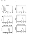
expressing human IAP and then an assay such as the flow
cytometry is carried out to evaluate the antigen-binding
activity.
-
In vitro evaluation of the signal transduction
effect (apoptosis-inducing effect in the cases of the
antibody MABL-1 and the antibody MABL-2) is performed in the
following manner: A test sample of the above modified
antibody is added to the cells which are expressing the
antibody or cells into which the gene for the antibody has
been introduced, and is evaluated by the change caused by
the signal transduction, for example, whether cell death is
induced in a manner specific to the human IAP-antigen, using
conventional methods.
-
In vivo evaluation of the apoptosis-inducing
effect, for example, in the case where the modified antibody
recognizes human IAP (e.g. modified antibodies derived from
the antibody MABL-1 and the antibody MABL-2) is carried out
in the following manner: A mouse model of human myeloma is
prepared. To the mice is intravenously administered the
monoclonal antibody or the modified antibody of the
invention, which induces apoptosis of nucleated blood cells
having IAP. To mice of a control group is administered PBS
alone. The induction of apoptosis is evaluated in terms of
antitumor effect based on the change of human IgG content in
serum of the mice and their survival time.
-
As mentioned above the modified antibodies of the
invention can be obtained by preparing modified antibodies
which contain two or more H chain V regions and two or more
L chain V regions and specifically bind to target cell
surface molecule or intracellular molecule and screening the
modified antibodies by in vivo or in vitro evaluation as
mentioned in the above.
-
The modified antibodies of the invention, which
comprises two or more H chain V regions and two or more L
chain V regions, preferably each two to four, more
preferably each two, may be a dimer of the single chain Fv
comprising one H chain V region and one L chain V region, or
a single chain polypeptide in which two or more H chain V
regions and two or more L chain V regions are connected. It
is considered that owing to such construction the peptide
mimics three dimensional structure of a natural ligand and
therefore retains an excellent antigen-binding property and
agonist activity.
-
The modified antibodies of the invention have a
remarkably lowered molecular size compared with antibody
molecule (whole IgG), and, therefore, a superior
permeability into tissues and tumors and a higher activity
than original agonist monoclonal antibodies. Therefore,
proper selection of the parent antibody makes it possible to
transduce various signals into cells and to induce various
actions in the cells such as apoptosis induction, cell
proliferation induction, cell differentiation induction,
cell division induction or cell cycle regulation action. The
pharmaceutical preparations containing them are useful for
treating diseases curable by inducing signal transduction,
for example cancers, inflammation, hormone disorders,
autoimmune diseases as well as blood dyscrasia, for example,
leukemia, malignant lymphoma, aplastic anemia,
myelodysplasia syndrome and polycythemia vera. It is further
expected that the antibody of the invention can be used as a
contrast agent by RI-labeling. The effect can be enhanced by
attaching to a RI-compound or a toxin.
BEST MODE FOR WORKING THE INVENTION
-
The present invention will concretely be
illustrated in reference to the following examples, which in
no way limit the scope of the invention.
-
For illustrating the production process of the
modified antibodies of the invention, examples of producing
single chain Fvs are shown below. Mouse antibodies against
human IAP, MABL-1 and MABL-2 were used in the examples of
producing the modified antibodies. Hybridomas MABL-1 and
MABL-2 producing them respectively were internationally
deposited as FERM BP-6100 and FERM BP-6101 with the National
Institute of Bioscience and Human Technology, Agency of
Industrial Science and Technology, Minister of International
Trade and Industry (1-3 Higasi 1-chome, Tsukuba-shi,
Ibaraki-ken, Japan), an authorized depository for
microorganisms, on September 11, 1997.
Examples
Example 1 (Cloning of DNAs encoding V region of mouse
monoclonal antibodies to human IAP)
-
DNAs encoding variable regions of the mouse
monoclonal antibodies to human IAP, MABL-1 and MABL-2, were
cloned as follows.
1.1 Preparation of messenger RNA (mRNA)
-
mRNAs of the hybridomas MABL-1 and MABL-2 were
obtained by using mRNA Purification Kit (Pharmacia Biotech).
1.2 Synthesis of double-stranded cDNA
-
Double-stranded cDNA was synthesized from about 1
µg of the mRNA using Marathon cDNA Amplification Kit
(CLONTECH) and an adapter was linked thereto.
1.3 PCR Amplification of genes encoding variable regions of
an antibody by
-
PCR was carried out using Thermal Cycler (PERKIN
ELMER).
(1) Amplification of a gene coding for L chain V region of
MABL-1
-
Primers used for the PCR method are Adapter
Primer-1 (CLONTECH) shown in SEQ ID No. 1, which hybridizes
to a partial sequence of the adapter, and MKC (Mouse Kappa
Constant) primer (Bio/Technology, 9, 88-89, 1991) shown in
SEQ ID No. 2, which hybridizes to the mouse kappa type L
chain V region.
-
50 µl of the PCR solution contains 5 µl of 10 ×
PCR Buffer II, 2 mM MgCl2, 0.16 mM dNTPs (dATP, dGTP, dCTP
and dTTP), 2.5 units of a DNA polymerase, AmpliTaq Gold
(PERKIN ELMER), 0.2 µM of the adapter primer of SEQ ID No.
1, 0.2 µM of the MKC primer of SEQ ID No. 2 and 0.1 µg of
the double-stranded cDNA derived from MABL-1. The solution
was preheated at 94°C of the initial temperature for 9
minutes and then heated at 94°C for 1 minute, at 60°C for 1
minute and at 72°C for 1 minute 20 seconds in order. This
temperature cycle was repeated 35 times and then the
reaction mixture was further heated at 72°C for 10 minutes.
(2) Amplification of cDNA encoding H chain V region of MABL-1
-
The Adapter Primer-1 shown in SEQ ID No. 1 and
MHC-γ1 (Mouse Heavy Constant) primer (Bio/Technology, 9, 88-89,
1991) shown in SEQ ID No. 3 were used as primers for
PCR.
-
The amplification of cDNA was performed according
to the method of the amplification of the L chain V region
gene, which was described in Example 1.3-(1), except for
using 0.2 µM of the MHC-γ1 primer instead of 0.2 µM of the
MKC primer.
(3) Amplification of cDNA encoding L chain V region of MABL-2
-
The Adapter Primer-1 of SEQ ID No. 1 and the MKC
primer of SEQ ID No. 2 were used as primers for PCR.
-
The amplification of cDNA was carried out
according to the method of the amplification of the L chain
V region gene of MABL-1 which was described in Example 1.3-(1),
except for using 0.1 µg of the double-stranded cDNA
derived from MABL-2 instead of 0.1 µg of the double-stranded
cDNA from MABL-1.
(4) Amplification of cDNA encoding H chain V region of MABL-2
-
The Adapter Primer-1 of SEQ ID No. 1 and MHC-γ2a
primer (Bio/Technology, 9, 88-89, 1991) shown in SEQ ID No.
4 were used as primers for PCR.
-
The amplification of cDNA was performed according
to the method of the amplification of the L chain V region
gene, which was described in Example 1.3-(3), except for
using 0.2 µM of the MHC-γ2a primer instead of 0.2 µM of the
MKC primer.
1.4 Purification of PCR products
-
The DNA fragment amplified by PCR as described
above was purified using the QIAquick PCR Purification Kit
(QIAGEN) and dissolved in 10 mM Tris-HCl (pH 8.0) containing
1 mM EDTA.
1.5 Ligation and Transformation
-
About 140 ng of the DNA fragment comprising the
gene encoding the mouse kappa type L chain V region derived
from MABL-1 as prepared above was ligated with 50 ng of
pGEM-T Easy vector (Promega) in the reaction buffer
comprising 30 mM Tris-HCl (pH 7.8), 10 mM MgCl2, 10 mM
dithiothreitol, 1 mM ATP and 3 units of T4 DNA Ligase
(Promega) at 15°C for 3 hours.
-
Then, 1 µl of the reaction mixture was added to 50
µl of E. coli DH5α competent cells (Toyobo Inc.) and the
cells were stored on ice for 30 minutes, incubated at 42°C
for 1 minute and stored on ice for 2 minutes again. 100 µl
of SOC medium (GIBCO BRL) was added. The cells of E. coli
were plated on LB (Molecular Cloning: A Laboratory Manual,
Sambrook et al., Cold Spring Harbor Laboratory Press, 1989)
agar medium containing 100 µg/ml of ampicillin (SIGMA) and
cultured at 37°C overnight to obtain the transformant of E.
coli.
-
The transformant was cultured in 3 ml of LB medium
containing 50 µg/ml of ampicillin at 37°C overnight and the
plasmid DNA was prepared from the culture using the QIAprep
Spin Miniprep Kit (QIAGEN).
-
The resulting plasmid comprising the gene encoding
the mouse kappa type L chain V region derived from the
hybridoma MABL-1 was designated as pGEM-M1L.
-
According to the same manner as described above, a
plasmid comprising the gene encoding the mouse H chain V
region derived from the hybridoma MABL-1 was prepared from
the purified DNA fragment and designated as pGEM-M1H.
-
A plasmid comprising the gene encoding the mouse
kappa type L chain V region derived from the hybridoma MABL-2
was prepared from the purified DNA fragment and designated
as pGEM-M2L.
-
A plasmid comprising the gene encoding the mouse H
chain V region derived from the hybridoma MABL-2 was
prepared from the purified DNA fragment and designated as
pGEM-M2H.
Example 2 (DNA Sequencing)
-
The nucleotide sequence of the cDNA encoding
region in the aforementioned plasmids was determined using
Auto DNA Sequencer (Applied Biosystem) and ABI PRISM Dye
Terminator Cycle Sequencing Ready Reaction Kit (Applied
Biosystem) according to the manufacturer's protocol.
-
The nucleotide sequence of the gene encoding the L
chain V region from the mouse antibody MABL-1, which is
included in the plasmid pGEM-M1L, is shown in SEQ ID No. 5.
-
The nucleotide sequence of the gene encoding the H
chain V region from the mouse antibody MABL-1, which is
included in the plasmid pGEM-M1H, is shown in SEQ ID No. 6.
-
The nucleotide sequence of the gene encoding the L
chain V region from the mouse antibody MABL-2, which is
included in the plasmid pGEM-M2L, is shown in SEQ ID No. 7.
-
The nucleotide sequence of the gene encoding the H
chain V region from the mouse antibody MABL-2, which is
included in the plasmid pGEM-M2H, is shown in SEQ ID No. 8.
Example 3 (Determination of CDR)
-
The V regions of L chain and H chain generally
have a similarity in their structures and each four
framework regions therein are linked by three hypervariable
regions, i.e., complementarity determining regions (CDR). An
amino acid sequence of the framework is relatively well
conserved, while an amino acid sequence of CDR has extremely
high variation (Kabat, E.A., et al., "Sequences of Proteins
of Immunological Interest", US Dept. Health and Human
Services, 1983).
-
On the basis of these facts, the amino acid
sequences of the variable regions from the mouse monoclonal
antibodies to human IAP were applied to the database of
amino acid sequences of the antibodies made by Kabat et al.
to investigate the homology. The CDR regions were determined
based on the homology as shown in Table 1.
| Plasmid | SEQ ID No. | CDR(1) | CDR(2) | CDR(3) |
| pGEM-M1L | 5 | 43-58 | 74-80 | 113-121 |
| pGEM-M1H | 6 | 50-54 | 69-85 | 118-125 |
| pGEM-M2L | 7 | 43-58 | 74-80 | 113-121 |
| pGEM-M2H | 8 | 50-54 | 69-85 | 118-125 |
Example 4 (Identification of Cloned cDNA Expression
(Preparation of Chimera MABL-1 antibody and Chimera MABL-2
antibody.)
4.1 Preparation of vectors expressing chimera MABL-1
antibody
-
cDNA clones, pGEM-M1L and pGEM-M1H, encoding the V
regions of the L chain and the H chain of the mouse antibody
MABL-1, respectively, were modified by the PCR method and
introduced into the HEF expression vector (WO92/19759) to
prepare vectors expressing chimera MABL-1 antibody.
-
A forward primer MLS (SEQ ID No. 9) for the L
chain V region and a forward primer MHS (SEQ ID No. 10) for
the H chain V region were designed to hybridize to a DNA
encoding the beginning of the leader sequence of each V
region and to contain the Kozak consensus sequence (J. Mol.
Biol., 196, 947-950, 1987) and HindIII restriction enzyme
site. A reverse primer MLAS (SEQ ID No. 11) for the L chain
V region and a reverse primer MHAS (SEQ ID No. 12) for the H
chain V region were designed to hybridize to a DNA encoding
the end of the J region and to contain the splice donor
sequence and BamHI restriction enzyme site.
-
100 µl of a PCR solution comprising 10 µl of 10 ×
PCR Buffer II, 2 mM MgCl2, 0.16 mM dNTPs (dATP, dGTP, dCTP
and dTTP), 5 units of DNA polymerase AmpliTaq Gold, 0.4 µM
each of primers and 8 ng of the template DNA (pGEM-M1L or
pGEM-M1H) was preheated at 94°C of the initial temperature
for 9 minutes and then heated at 94°C for 1 minute, at 60°C
for 1 minute and at 72°C for 1 minute 20 seconds in order.
This temperature cycle was repeated 35 times and then the
reaction mixture was further heated at 72°C for 10 minutes.
-
The PCR product was purified using the QIAquick
PCR Purification Kit (QIAGEN) and then digested with HindIII
and BamHI. The product from the L chain V region was cloned
into the HEF expression vector, HEF-κ and the product from
the H chain V region was cloned into the HEF expression
vector, HEF-γ. After DNA sequencing, plasmids containing a
DNA fragment with a correct DNA sequence are designated as
HEF-M1L and HEF-M1H, respectively.
4.2 Preparation of vectors expressing chimera MABL-2
antibodies
-
Modification and cloning of cDNA were performed in
the same manner described in Example 4.1 except for using
pGEM-M2L and pGEM-M2H as template DNA instead of pGEM-M1L
and pGEM-M1H. After DNA sequencing, plasmids containing DNA
fragments with correct DNA sequences are designated as HEF-M2L
and HEF-M2H, respectively.
4.3 Transfection to COS7 cells
-
The aforementioned expression vectors were tested
in COS7 cells to observe the transient expression of the
chimera MABL-1 and MABL-2 antibodies.
(1) Transfection with genes for the chimera MABL-1 antibody
-
COS7 cells were co-transformed with the HEF-M1L
and HEF-M1H vectors by electroporation using the Gene Pulser
apparatus (BioRad). Each DNA (10 µg) and 0.8 ml of PBS with
1 × 10⁷ cells/ml were added to a cuvette. The mixture was
treated with pulse at 1.5 kV, 25 µF of electric capacity.
-
After the restoration for 10 minutes at a room
temperature, the electroporated cells were transferred into
DMEM culture medium (GIBCO BRL) containing 10% γ-globulin-free
fetal bovine serum. After culturing for 72 hours, the
supernatant was collected, centrifuged to remove cell
fragments and recovered.
(2) Transfection with genes coding for the chimera MABL-2
antibody
-
The co-transfection to COS7 cells with the genes
coding for the chimera MABL-2 antibody was carried out in
the same manner as described in Example 4.3-(1) except for
using the HEF-M2L and HEF-M2H vectors instead of the HEF-M1L
and HEF-M1H vectors. The supernatant was recovered in the
same manner.
4.4 Flow cytometry
-
Flow cytometry was performed using the
aforementioned culture supernatant of COS7 cells to measure
binding to the antigen. The culture supernatant of the COS7
cells expressing the chimera MABL-1 antibody or the COS7
cells expressing the chimera MABL-2 antibody, or human IgG
antibody (SIGMA) as a control was added to 4 × 10⁵ cells of
mouse leukemia cell line L1210 expressing human IAP and
incubated on ice. After washing, the FITC-labeled anti-human
IgG antibody (Cappel) was added thereto. After incubating
and washing, the fluorescence intensity thereof was measured
using the FACScan apparatus (BECTON DICKINSON).
-
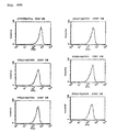
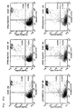
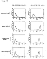
Since the chimera MABL-1 and MABL-2 antibodies
were specifically bound to L1210 cells expressing human IAP,
it is confirmed that these chimera antibodies have proper
structures of the V regions of the mouse monoclonal
antibodies MABL-1 and MABL-2, respectively (Figs. 1-3).
Example 5 (Preparation of reconstructed Single chain Fv
(scFv) of the antibody MABL-1 and antibody MABL-2)
5.1 Preparation of reconstructed single chain Fv of antibody
MABL-1
-
The reconstructed single chain Fv of antibody
MABL-1 was prepared as follows. The H chain V region and the
L chain V of antibody MABL-1, and a linker were respectively
amplified by the PCR method and were connected to produce
the reconstructed single chain Fv of antibody MABL-1. The
production method is illustrated in Fig. 4. Six primers (A-F)
were employed for the production of the single chain Fv
of antibody MABL-1. Primers A, C and E have a sense sequence
and primers B, D and F have an antisense sequence.
-
The forward primer VHS for the H chain V region
(Primer A, SEQ ID No. 13) was designed to hybridize to a DNA
encoding the N-terminal of the H chain V region and to
contain NcoI restriction enzyme recognition site. The
reverse primer VHAS for H chain V region (Primer B, SEQ ID
No. 14) was designed to hybridize to a DNA coding the C-terminal
of the H chain V region and to overlap with the
linker.
-
The forward primer LS for the linker (Primer C,
SEQ ID No. 15) was designed to hybridize to a DNA encoding
the N-terminal of the linker and to overlap with a DNA
encoding the C-terminal of the H chain V region. The reverse
primer LAS for the linker (Primer D, SEQ ID No. 16) was
designed to hybridize to a DNA encoding the C-terminal of
the linker and to overlap with a DNA encoding the N-terminal
of the L chain V region.
-
The forward primer VLS for the L chain V region
(Primer E, SEQ ID No. 17) was designed to hybridize to a DNA
encoding the C-terminal of the linker and to overlap with a
DNA encoding the N-terminal of the L chain V region. The
reverse primer VLAS-FLAG for L chain V region (Primer F, SEQ
ID No. 18) was designed to hybridize to a DNA encoding the
C-terminal of the L chain V region and to have a sequence
encoding the FLAG peptide (Hopp. T. P. et al.,
Bio/Technology, 6, 1204-1210, 1988), two stop codons and
EcoRI restriction enzyme recognition site.
-
In the first PCR step, three reactions, A-B, C-D
and E-F, were carried out and PCR products thereof were
purified. Three PCR products obtained from the first PCR
step were assembled by their complementarity. Then, the
primers A and F were added and the full length DNA encoding
the reconstructed single chain Fv of antibody MABL-1 was
amplified (Second PCR). In the first PCR, the plasmid pGEM-M1H
encoding the H chain V region of antibody MABL-1 (see
Example 2), a plasmid pSC-DP1 which comprises a DNA sequence
encoding a linker region comprising: Gly Gly Gly Gly Ser Gly
Gly Gly Gly Ser Gly Gly Gly Gly Ser (SEQ ID No. 19) (Huston,
J.S., et al., Proc. Natl. Acad. Sci. USA, 85, 5879-5883,
1988) and the plasmid pGEM-M1L encoding the L chain V region
of antibody MABL-1 (see Example 2) were employed as
template, respectively.
-
50 µl of the solution for the first PCR step
comprises 5 µl of 10 × PCR Buffer II, 2 mM MgCl2, 0.16 mM
dNTPs, 2.5 units of DNA polymerase, AmpliTaq Gold (PERKIN
ELMER), 0.4 µM each of primers and 5 ng each of template
DNA. The PCR solution was preheated at 94°C of the initial
temperature for 9 minutes and then heated at 94°C for 1
minute, at 65°C for 1 minute and at 72°C for 1 minute and 20
seconds in order. This temperature cycle was repeated 35
times and then the reaction mixture was further heated at
72°C for 7 minutes.
-
The PCR products A-B (371bp), C-D (63bp) and E-F
(384bp) were purified using the QIAquick PCR Purification
Kit (QIAGEN) and were assembled in the second PCR. In the
second PCR, 98 µl of a PCR solution comprising 120 ng of the
first PCR product A-B, 20 ng of the PCR product C-D and 120
ng of the PCR product E-F, 10 µl of 10 × PCR Buffer II, 2mM
MgCl2, 0.16 mM dNTPs, 5 units of DNA polymerase AmpliTaq
Gold (PERKIN ELMER) was preheated at 94°C of the initial
temperature for 8 minutes and then heated at 94°C for 2
minutes, at 65°C for 2 minutes and at 72°C for 2 minutes in
order. This temperature cycle was repeated twice and then
0.4 µM each of primers A and F were added into the reaction,
respectively. The mixture was preheated at 94°C of the
initial temperature for 1 minutes and then heated at 94°C
for 1 minute, at 65°C for 1 minute and at 72°C for 1 minute
and 20 seconds in order. This temperature cycle was repeated
35 times and then the reaction mixture was further heated at
72°C for 7 minutes.
-
A DNA fragment of 843 bp produced by the second
PCR was purified and digested by NcoI and EcoRI. The
resultant DNA fragment was cloned into pSCFVT7 vector. The
expression vector pSCFVT7 contains a pelB signal sequence
suitable for E. coli periplasmic expression system (Lei,
S.P., et al., J. Bacteriology, 169, 4379-4383, 1987). After
the DNA sequencing, the plasmid containing the DNA fragment
encoding correct amino acid sequence of the reconstructed
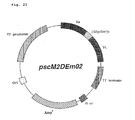
single chain Fv of antibody MABL-1 is designated as "pscM1"
(see Fig. 5). The nucleotide sequence and the amino acid
sequence of the reconstructed single chain Fv of antibody
MABL-1 contained in the plasmid pscM1 are shown in SEQ ID
No. 20.
-
The pscM1 vector was modified by the PCR method to
prepare a vector expressing the reconstructed single chain
Fv of antibody MABL-1 in mammalian cells. The resultant DNA
fragment was introduced into pCHO1 expression vector. This
expression vector, pCHO1, was constructed by digesting DHFR-ΔE-rvH-PM1-f
(WO92/19759 with EcoRI and SmaI to eliminate
the antibody gene and connecting the EcoRI-NotI-BamHI
Adapter (Takara Shuzo) thereto.
-
As a forward primer for PCR, Sal-VHS primer shown
in SEQ ID No. 21 was designed to hybridize to a DNA encoding
the N-terminal of the H chain V region and to contain SalI
restriction enzyme recognition site. As a reverse primer for
PCR, FRH1anti primer shown in SEQ ID No. 22 was designed to
hybridize to a DNA encoding the end of the first framework
sequence.
-
100 µl of PCR solution comprising 10 µl of 10 ×
PCR Buffer II, 2 mM MgCl2, 0.16 mM dNTPs, 5 units of the DNA
polymerase, AmpliTaq Gold, 0.4 µl M each of primer and 8 ng
of the template DNA (pscM1) was preheated at 95°C of the
initial temperature for 9 minutes and then heated at 95°C
for 1 minute, at 60°C for 1 minute and at 72°C for 1 minute
and 20 seconds in order. This temperature cycle was repeated
35 times and then the reaction mixture was further heated at
72°C for 7 minutes.
-
The PCR product was purified using the QIAquick
PCR Purification Kit (QIAGEN) and digested by SalI and MboII
to obtain a DNA fragment encoding the N-terminal of the
reconstructed single chain Fv of antibody MABL-1 The pscM1
vector was digested by MboII and EcoRI to obtain a DNA
fragment encoding the C-terminal of the reconstructed single
chain Fv of antibody MABL-1. The SalI-MboII DNA fragment and
the MboII-EcoRI DNA fragment were cloned into pCHO1-Igs
vector. After DNA sequencing, the plasmid comprising the
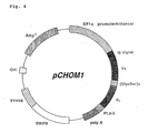
desired DNA sequence was designated as "pCHOM1" (see Fig.
6). The expression vector, pCHO1-Igs, contains a mouse IgG1
signal sequence suitable for the secretion-expression system
in mammalian cells (Nature, 322, 323-327, 1988). The
nucleotide sequence and the amino acid sequence of the
reconstructed single chain Fv of antibody MABL-1 contained
in the plasmid pCHOM1 are shown in SEQ ID No. 23.
5.2 Preparation of reconstructed single chain Fv of
antibody MABL-2
-
The reconstructed single chain Fv of antibody
MABL-2 was prepared in accordance with the aforementioned
Example 5.1. Employed in the first PCR step were plasmid
pGEM-M2H encoding the H chain V region of MABL-2 (see
Example 2) instead of pGEM-M1H and plasmid pGEM-M2L encoding
the L chain V region of MABL-2 (see Example 2) instead of
pGEM-M1L, to obtain a plasmid pscM2 which comprises a DNA
fragment encoding the desired amino acid sequence of the
single chain Fv of antibody MABL-2. The nucleotide sequence
and the amino acid sequence of the reconstructed single
chain Fv of antibody MABL-2 contained in the plasmid pscM2
are shown in SEQ ID No. 24.
-
The pscM2 vector was modified by the PCR method to
prepare a vector, pCHOM2, for the expression in mammalian
cells which contains the DNA fragment encoding the correct
amino acid sequence of reconstructed the single chain Fv of.
antibody MABL-2. The nucleotide sequence and the amino acid
sequence of the reconstructed single chain Fv of antibody
MABL-2 contained in the plasmid pCHOM2 are shown in SEQ ID
No. 25.
5.3 Transfection to COS7 cells
-
The pCHOM2 vector was tested in COS7 cells to
observe the transient expression of the reconstructed single
chain Fv of antibody MABL-2.
-
The COS7 cells were transformed with the pCHOM2
vector by electroporation using the Gene Pulser apparatus
(BioRad). The DNA (10 µg) and 0.8 ml of PBS with 1 × 10⁷
cells/ml were added to a cuvette. The mixture was treated
with pulse at 1.5 kV, 25 µF of electric capacity.
-
After the restoration for 10 minutes at a room
temperature, the electroporated cells were transferred into
IMDM culture medium (GIBCO BRL) containing 10% fetal bovine
serum. After culturing for 72 hours, the supernatant was
collected, centrifuged to remove cell fragments and
recovered.
5.4 Detection of the reconstructed single chain Fv of
antibody MABL-2 in culture supernatant of COS7 cells
-
The existence of the single chain Fv of antibody
MABL-2 in the culture supernatant of COS7 cells which had
been transfected with the pCHOM2 vector was confirmed by the
Western Blotting method.
-
The culture supernatant of COS7 cells transfected
with the pCHOM2 vector and the culture supernatant of COS7
cells transfected with the pCHO1 as a control were subjected
to SDS electrophoresis and transferred to REINFORCED NC
membrane (Schleicher & Schuell). The membrane was blocked
with 5% skim milk (Morinaga Nyu-gyo), washed with 0.05%
Tween 20-PBS and mixed with an anti-FLAG antibody (SIGMA).
The membrane was incubated at room temperature, washed and
mixed with alkaline phosphatase-conjugated mouse IgG
antibody (Zymed). After incubating and washing at room
temperature, the substrate solution (Kirkegaard Perry
Laboratories) was added to develop color (Fig. 7).
-
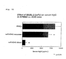
A FLAG-peptide-specific protein was detected only
in the culture supernatant of the pCHOM2 vector-introduced
COS7 cells and thus it is confirmed that the reconstructed
single chain Fv of antibody MABL-2 was secreted in this
culture supernatant.
5.5 Flow cytometry
-
Flow cytometry was performed using the
aforementioned COS7 cells culture supernatant to measure the
binding to the antigen. The culture supernatant of the COS7
cells expressing the reconstructed single chain Fv of
antibody MABL-2 or the culture supernatant of COS7 cells
transformed with pCHO1 vector as a control was added to 2 ×
105 cells of the mouse leukemia cell line L1210 expressing
human Integrin Associated Protein (IAP) or the cell line
L1210 transformed with pCOS1 as a control. After incubating
on ice and washing, the mouse anti-FLAG antibody (SIGMA) was
added. Then the cells were incubated and washed. Then, the
FITC labeled anti-mouse IgG antibody (BECTON DICKINSON) was
added thereto and the cells were incubated and washed again.
Subsequently, the fluorescence intensity was measured using
the FACScan apparatus (BECTON DICKINSON).
-
Since the single chain Fv of antibody MABL-2 was
specifically bound to L1210 cells expressing human IAP, it
is confirmed that the reconstructed single chain Fv of
antibody MABL-2 has an affinity to human Integrin Associated
Protein (IAP) (see Figs. 8-11).
5.6 Competitive ELISA
-
The binding activity of the reconstructed single
chain Fv of antibody MABL-2 was measured based on the
inhibiting activity against the binding of mouse monoclonal
antibodies to the antigen.
-
The anti-FLAG antibody adjusted to 1 µg/ml was
added to each well on 96-well plate and incubated at 37°C
for 2 hours. After washing, blocking was performed with 1%
BSA-PBS. After incubating and washing at a room temperature,
the culture supernatant of COS7 cells into which the
secretion-type human IAP antigen gene (SEQ ID No. 26) had
been introduced was diluted with PBS into twofold volume and
added to each well. After incubating and washing at a room
temperature, a mixture of 50 µl of the biotinized MABL-2
antibody adjusted to 100 ng/ml and 50 µl of sequentially
diluted supernatant of the COS7 cells expressing the
reconstructed single chain Fv of antibody MABL-2 were added
into each well. After incubating and washing at a room
temperature, the alkaline phosphatase-conjugated
streptoavidin (Zymed) was added into each well. After
incubating and washing at a room temperature, the substrate
represent living cells and dots in the right-lower region
represent cells at the early stage of apoptosis and dots in
the right-upper region represent cells at the late stage of
apoptosis. The results show that the reconstructed single
chain Fv of antibody MABL-2 (MABL2-scFv) remarkably induced
cell death of L1210 cells specific to human IAP antigen
(Figs. 13-16) and that the reconstructed single chain Fv
also induced remarkable cell death of CCRF-CEM cells in
comparison with the control (Figs. 17-18).
5.8 Expression of MABL-2 derived single chain Fv in CHO
cells
-
CHO cells were transfected with the pCHOM2 vector
to establish a CHO cell line which constantly expresses the
single chain Fv (polypeptide) derived from the antibody
MABL-2.
-
CHO cells were transformed with the pCHOM2 vector
by the electroporation using the Gene Pulser apparatus
(BioRad). A mixture of DNA (10 µg) and 0.7 ml of PBS with
CHO cells (1 × 10⁷ cells/ml) was added to a cuvette. The
mixture was treated with pulse at 1.5 kV, 25 µF of electric
capacity. After the restoration for 10 minutes at a room
temperature, the electroporated cells were transferred into
nucleic acid free α-MEM medium (GIBCO BRL) containing 10%
fetal bovine serum and cultured. The expression of desired
protein in the resultant clones was confirmed by SDS-PAGE
and a clone with a high expression level was selected as a
cell line producing the single chain Fv derived from the
solution (SIGMA) was added and absorbance of the reaction
mixture in each well was measured at 405 nm.
-
The results revealed that the reconstructed single
chain Fv of antibody MABL-2 (MABL2-scFv) evidently inhibited
concentration-dependently the binding of the mouse antibody
MABL-2 to human IAP antigen in comparison with the culture
supernatant of the PCHO1-introduced COS7 cells as a control
(Fig. 12). Accordingly, it is suggested that the
reconstructed single chain Fv of antibody MABL-2 has the
correct structure of each of the V regions from the mouse
monoclonal antibody MABL-2.
5.7 Apoptosis-inducing Effect in vitro
-
An apoptosis-inducing action of the reconstructed
single chain Fv of antibody MABL-2 was examined by Annexin-V
staining (Boehringer Mannheim) using the L1210 cells
transfected with human IAP gene, the L1210 cells transfected
with the pCOS1 vector as a control and CCRF-CEM cells.
-
To each 1 × 10⁵ cells of the above cells was added
the culture supernatant of the COS7 cells expressing the
reconstructed single chain Fv of antibody MABL-2 or the
culture supernatant of COS7 cells transfected with the pCHO1
vector as a control at 50% final concentration and the
mixtures were cultured for 24 hours. Then, the Annexin-V
staining was performed and the fluorescence intensity was
measured using the FACScan apparatus (BECTON DICKINSON).
-
Results of the Annexin-V staining are shown in
Figs. 13-18, respectively. Dots in the left-lower region
antibody MABL-2. The cell line was cultured in serum-free
medium CHO-S-SFM II (GIBCO BRL) containing 10 nM
methotrexate (SIGMA). Then, the culture supernatant was
collected, centrifuged to remove cell fragments and
recovered.
5.9 Purification of MABL-2 derived single chain Fv produced
in CHO cells
-
The culture supernatant of the CHO cell line
expressing the single chain Fv obtained in Example 5.8 was
concentrated up to twenty times using a cartridge for the
artificial dialysis (PAN130SF, ASAHI MEDICALS). The
concentrated solution was stored at -20°C and thawed on
purification.
-
Purification of the single chain Fv from the
culture supernatant of the CHO cells was performed using
three kinds of chromatography, i.e., Blue-sepharose, a
hydroxyapatite and a gel filtration.
(1) Blue-sepharose column chromatography
-
The concentrated supernatant was diluted to ten
times with 20 mM acetate buffer (pH 6.0) and centrifuged to
remove insoluble materials (10000 × rpm, 30 minutes). The
supernatant was applied onto a Blue-sepharose column (20 ml)
equilibrated with the same buffer. After washing the column
with the same buffer, proteins adsorbed in the column were
eluted by a stepwise gradient of NaCl in the same buffer,
0.1, 0.2, 0.3, 0.5 and up to 1.0 M. The pass-through
fraction and each eluted fraction were analyzed by SDS-PAGE.
The fractions in which the single chain Fv were confirmed
(the fractions eluted at 0.1 to 0.3M NaCl) were pooled and
concentrated up to approximately 20 times using CentriPrep-10
(AMICON).
(2) Hydroxyapatite
-
The concentrated solution obtained in (1) was
diluted to 10 times with 10 mM phosphate buffer (pH 7.0) and
applied onto the hydroxyapatite column (20 ml, BIORAD). The
column was washed with 60 ml of 10 mM phosphate buffer (pH
7.0). Then, proteins adsorbed in the column were eluted by a
linear gradient of sodium phosphate buffer up to 200 mM (see
Fig. 19). The analysis of each fraction by SDS-PAGE
confirmed the single chain Fv in fraction A and fraction B.
(3) Gel filtration
-
Each of fractions A and B in (2) was separately
concentrated with CentriPrep-10 and applied onto TSKgel
G3000SWG column (21.5 × 600 mm) equilibrated with 20 mM
acetate buffer (pH 6.0) containing 0.15 M NaCl.
Chromatograms are shown in Fig. 20. The analysis of the
fractions by SDS-PAGE confirmed that both major peaks (AI
and BI) are of desired single chain Fv. In the gel
filtration analysis, the fraction A was eluted at 36 kDa of
apparent molecular weight and the fraction B was eluted at
76 kDa. The purified single chain Fvs (AI, BI) were analyzed
with 15% SDS polyacrylamide gel. Samples were treated in the
absence or presence of a reductant and the electrophoresis
was carried out in accordance with the Laemmli's method.
Then the protein was stained with Coomassie Brilliant Blue.
As shown in Fig. 21, both AI and BI gave a single band at 35
kDa of apparent molecular weight, regardless of the absence
or presence of the reductant. From the above, it is
concluded that AI is a monomer of the single chain Fv and BI
is a non-covalently bound dimer of the single chain Fv. The
gel filtration analysis of the fractions AI and BI with
TSKgel G3000SW column (7.5 × 60 mm) revealed that a peak of
the monomer is detected only in the fraction AI and a peak
of the dimer is detected only in the fraction BI (Fig. 22).
The dimer fraction (fraction BI) accounted for 4 period of
total single chain Fvs. More than 90% of the dimer in the
dimer fraction was stably preserved for more than a month at
4°C.
5.10 Construction of vector expressing single chain Fv
derived from antibody MABL-2 in E. coli cell
-
The pscM2 vector was modified by the PCR method to
prepare a vector effectively expressing the single chain Fv
from the antibody MABL-2 in E. coli cells. The resultant DNA
fragment was introduced into pSCFVT7 expression vector.
-
As a forward primer for PCR, Nde-VHSm02 primer
shown in SEQ ID No. 27 was designed to hybridize to a DNA
encoding the N-terminal of the H chain V region and to
contain a start codon and NdeI restriction enzyme
recognition site. As a reverse primer for PCR, VLAS primer
shown in SEQ ID No. 28 was designed to hybridize to a DNA
encoding the C-terminal of the L chain V region and to
contain two stop codons and EcoRI restriction enzyme
recognition site. The forward primer, Nde-VHSm02, comprises
five point mutations in the part hybridizing to the DNA
encoding the N-terminal of the H chain V region for the
effective expression in E. coli.
-
100 µl of a PCR solution comprising 10 µl of 10 x
PCR Buffer # 1, 1 mM MgCl2, 0.2 mM dNTPs, 5 units of KOD DNA
polymerase (all from TOYOBO), 1 µM of each primer and 100 ng
of a template DNA (pscM2) was heated at 98°C for 15 seconds,
at 65°C for 2 seconds and at 74°C for 30 seconds in order.
This temperature cycle was repeated 25 times.
-
The PCR product was purified using the QIAquick
PCR Purification Kit (QIAGEN) and digested by NdeI and
EcoRI, and then the resulting DNA fragment was cloned into
pSCFVT7 vector, from which pelB signal sequence had been
eliminated by the digestion with NdeI and EcoRI. After DNA
sequencing, the resulting plasmid comprising a DNA fragment
with the desired DNA sequence is designated as "pscM2DEm02"
(see Fig. 23). The nucleotide sequence and the amino acid
sequence of the single chain Fv derived from the antibody
MABL-2 contained in the plasmid pscM2DEm02 are shown in SEQ
ID No. 29.
5.11 Expression of single chain Fv derived from antibody
MABL-2 in E. coli cells
-
E. coli BL21(DE3)pLysS (STRATAGENE) was
transformed with pscM2DEm02 vector to obtain a strain of E.
coli expressing the single chain Fv derived from antibody
MABL-2. The resulting clones were examined for the
expression of the desired protein using SDS-PAGE, and a
clone with a high expression level was selected as a strain
producing the single chain Fv derived from antibody MABL-2.
5.12 Purification of single chain Fv derived from antibody
MABL-2 produced in E.coli
-
A single colony of E. coli obtained by the
transformation was cultured in 3 ml of LB medium at 28°C for
7 hours and then in 70 ml of LB medium at 28°C overnight.
This pre-culture was transplanted to 7 L of LB medium and
cultured at 28°C with stirring at 300 rpm using the Jarfermenter.
When an absorbance of the medium reached
O.D.=1.5, the bacteria were induced with 1 mM IPTG and then
cultured for 3 hours.
-
The culture medium was centrifuged (10000 × g, 10
minutes) and the precipitated bacteria were recovered. To
the bacteria was added 50 mM Tris-HCl buffer (pH 8.0)
containing 5 mM EDTA, 0.1 M NaCl and 1% Triton X-100 and the
bacteria were disrupted by ultrasonication (out put: 4, duty
cycle: 70%, 1 minute × 10 times). The suspension of
disrupted bacteria was centrifuged (12000 x g, 10 minutes)
to precipitate inclusion body. Isolated inclusion body was
mixed with 50 mM Tris-HCl buffer (pH 8.0) containing 5 mM
EDTA, 0.1 M NaCl and 4% Triton X-100, treated by
ultrasonication (out put: 4, duty cycle: 50%, 30 seconds × 2
times) again and centrifuged (12000 × g, 10 minutes) to
isolate the desired protein as precipitate and to remove
containment proteins included in the supernatant.
-
The inclusion body comprising the desired protein
was lysed in 50 mM Tris-HCl buffer (pH 8.0) containing 6 M
Urea, 5 mM EDTA and 0.1 M NaCl and applied onto Sephacryl S-300
gel filtration column (5 × 90 cm, Amersharm Pharmacia)
equilibrated with 50 mM Tris-HCl buffer (pH 8.0) containing
4M Urea, 5 mM EDTA, 0.1 M NaCl and 10 mM mercaptoethanol at
a flow rate of 5 ml/minutes to remove associated single
chain Fvs with high-molecular weight. The obtained fractions
were analyzed with SDS-PAGE and the fractions with high
purity of the protein were diluted with the buffer used in
the gel filtration up to O.D280=0.25. Then, the fractions
were dialyzed three times against 50 mM Tris-HCl buffer (pH
8.0) containing 5 mM EDTA, 0.1 M NaCl, 0.5 M Arg, 2 mM
glutathione in the reduced form and 0.2 mM glutathione in
the oxidized form in order for the protein to be refolded.
Further, the fraction was dialyzed three times against 20 mM
acetate buffer (pH 6.0) containing 0.15 M NaCl to exchange
the buffer.
-
The dialysate product was applied onto Superdex
200 pg gel filtration column (2.6 × 60 cm, Amersharm
Pharmacia) equilibrated with 20 mM acetate buffer (pH 6.0)
containing 0.15 M NaCl to remove a small amount of high
molecular weight protein which was intermolecularly
crosslinked by S-S bonds. As shown in Fig. 24, two peaks,
major and sub peaks, were eluted after broad peaks which are
expectedly attributed to an aggregate with a high molecular
weight. The analysis by SDS-PAGE (see Fig. 21) and the
elution positions of the two peaks in the gel filtration
analysis suggest that the major peak is of the monomer of
the single chain Fv and the sub peak is of the non-covalently
bound dimer of the single chain Fv. The non-covalently
bound dimer accounted for 4 percent of total
single chain Fvs.
5.13 Apoptosis-inducing activity in vitro of single chain Fv
derived from antibody MABL-2
-
An apoptosis-inducing action of the single chain
Fv from antibody MABL-2 (MABL2-scFv) produced by the CHO
cells and E. coli was examined according to two protocols by
Annexin-V staining (Boehringer Mannheim) using the L1210
cells (hIAP/L1210) into which human IAP gene had been
introduced.
-
In the first protocol sample antibodies at the
final concentration of 3 µg/ml were added to 5 × 10⁴ cells
of hIAP/L1210 cell line and cultured for 24 hours. Sample
antibodies, i.e., the monomer and the dimer of the single
chain Fv of MABL-2 from the CHO cells obtained in Example
5.9, the monomer and the dimer of the single chain Fv of
MABL-2 from E. coli obtained in Example 5.12, and the mouse
IgG antibody as a control were analyzed. After culturing,
the Annexin-V staining was carried out and the fluorescence
intensity thereof was measured using the FACScan apparatus
(BECTON DICKINSON).
-
In the second protocol sample antibodies at the
final concentration of 3 µg/ml were added to 5 × 10⁴ cells
of hIAP/L1210 cell line, cultured for 2 hours and mixed with
anti-FLAG antibody (SIGMA) at the final concentration of 15
µg/ml and further cultured for 22 hours. Sample antibodies
of the monomer of the single chain Fv of MABL-2 from the CHO
cells obtained in Example 5.9 and the mouse IgG antibody as
a control were analyzed. After culturing, the Annexin-V
staining was carried out and the fluorescence intensity
thereof was measured using the FACScan apparatus.
-
Results of the analysis by the Annexin-V staining
are shown in Figs. 25-31. The results show that the dimers
of the single chain Fv polypeptide of MABL-2 produced in the
CHO cells and E. coli remarkably induced cell death (Figs.
26, 27) in comparison with the control (Fig. 25), while no
apoptosis-inducing action was observed in the monomers of
the single chain Fv polypeptide of MABL-2 produced in the
CHO cells and E. coli (Figs. 28, 29). When anti-FLAG
antibody was used together, the monomer of the single chain
Fv polypeptide derived from antibody MABL-2 produced in the
CHO cells induced remarkably cell death (Fig. 31) in
comparison with the control (Fig. 30).
5.14 Antitumor effect of the monomer and the dimer of
scFv/CHO polypeptide with a model mouse of human myeloma
(1) Quantitative measurement of human IgG in mouse serum
-
Measurement of human IgG (M protein) produced by
human myeloma cell and contained in mouse serum was carried
out by the following ELISA. 100 µL of goat anti-human IgG
antibody (BIOSOURCE, Lot#7902) diluted to 1 µg/mL with 0.1%
bicarbonate buffer (pH 9.6) was added to each well on 96
wells plate (Nunc) and incubated at 4°C overnight so that
the antibody was immobilized. After blocking, 100 µL of the
stepwisely diluted mouse serum or human IgG (CAPPEL,
Lot#00915) as a standard was added to each well and
incubated for 2 hours at a room temperature. After washing,
100 µL of alkaline phosphatase-labeled anti-human IgG
antibody (BIOSOURCE, Lot#6202) which had been diluted to
5000 times was added, and incubation was carried out for 1
hour at a room temperature. After washing, a substrate
solution was added. After incubation, absorbance at 405 nm
was measured using the MICROPLATE READER Model 3550
(BioRad). The concentration of human IgG in the mouse serum
was calculated based on the calibration curve obtained from
the absorbance values of human IgG as the standard.
(2) Preparation of antibodies for administration
-
The monomer and the dimer of the scFv/CHO
polypeptide were respectively diluted to 0.4 mg/mL or 0.25
mg/mL with sterile filtered PBS(-) on the day of
administration to prepare samples for the administration.
(3) Preparation of a mouse model of human myeloma
-
A mouse model of human myeloma was prepared as
follows. KPMM2 cells passaged in vivo (JP-Appl. 7-236475) by
SCID mouse (Japan Clare) were suspended in RPMI1640 medium
(GIBCO-BRL) containing 10% fetal bovine serum (GIBCO-BRL)
and adjusted to 3 × 10⁷ cells/mL. 200 µL of the KPMM2 cell
suspension (6 × 10⁶ cells/mouse) was transplanted to the
SCID mouse (male, 6 week-old) via caudal vein thereof, which
had been subcutaneously injected with the asialo GM1
antibody (WAKO JUNYAKU, 1 vial dissolved in 5 mL) a day
before the transplantation.
(4) Administration of antibodies
-
The samples of the antibodies prepared in (2), the
monomer (250 µL) and the dimer (400 µL), were administered
to the model mice of human myeloma prepared in (3) via
caudal vein thereof. The administration was started from
three days after the transplantation of KPMM2 cells and was
carried out twice a day for three days. As a control, 200 µL
of sterile filtered PBS(-) was likewise administered twice a
day for three days via caudal vein. Each group consisted of
seven mice.
(5) Evaluation of antitumor effect of the monomer and the
dimer of scFv/CHO polypeptide with the model mouse of human
myeloma
-
The antitumor effect of the monomer and the dimer
of scFv/CHO polypeptide with the model mice of human myeloma
was evaluated in terms of the change of human IgG (M
protein) concentration in the mouse serum and survival time
of the mice. The change of human IgG concentration was
determined by measuring it in the mouse serum collected at
24 days after the transplantation of KPMM2 cells by ELISA
described in the above (1). The amount of serum human IgG (M
protein) in the serum of the PBS(-)-administered group
(control) increased to about 8500 µg/mL, whereas the amount
of human IgG of the scFv/CHO dimer-administered group was
remarkably low, that is, as low as one-tenth or less than
that of the control group. Thus, the results show that the
dimer of scFv/CHO strongly inhibits the growth of the KPMM2
cells (Fig. 32). As shown in Fig. 33, a remarkable
elongation of the survival time was observed in the scFv/CHO
dimer-administered group in comparison with the PBS(-)-administered
group.
-
From the above, it is confirmed that the dimer of
scFv/CHO has an antitumor effect for the human myeloma model
mice. It is considered that the antitumor effect of the
dimer of scFv/CHO, the modified antibody of the invention,
results from the apoptosis-inducing action of the modified
antibody.
5.15 Hemagglutination Test
-
Hemagglutination test and determination of
hemagglutination were carried out in accordance with
"Immuno-Biochemical Investigation", Zoku-Seikagaku Jikken
Koza, edited by the Biochemical Society of Japan, published
by Tokyo Kagaku Dojin.
-
Blood was taken from a healthy donor using
heparin-treated syringes and washed with PBS(-) three times;
and then erythrocyte suspension with a final concentration
of 2% in PBS(-) was prepared. Test samples were the antibody
MABL-2, the monomer and the dimer of the single chain Fv
polypeptide produced by the CHO cells, and the monomer and
the dimer of the single chain Fv polypeptide produced by
E.
coli, and the control was mouse IgG (ZYMED). For the
investigation of the hemagglutination effect, round bottom
96-well plates available from Falcon were used. 50 µL per
well of the aforementioned antibody samples and 50 µL of the
2% erythrocyte suspension were added and mixed in the well.
After incubation for 2 hours at 37°C, the reaction mixtures
were stored at 4°C overnight and the hemagglutination
thereof was determined. As a control, 50 µL per well of
PBS(-) was used and the hemagglutination test was carried
out in the same manner. The mouse IgG and antibody MABL-2
were employed at 0.01, 0.1, 1.0, 10.0 or 100.0 µg/mL of the
final concentration of the antibodies. The single chain Fvs
were employed at 0.004, 0.04, 0.4, 4.0, 40.0 or 80.0 µg/mL
of the final concentration and further at 160.0 µg/mL only
in the case of the dimer of the polypeptide produced by
E.
coli. Results are shown in the Table 2. In the case of
antibody MABL-2, the hemagglutination was observed at a
concentration of more than 0.1 µg/mL, whereas no
hemagglutination was observed for both the monomer and the
dimer of the single chain Fv.
| Hemagglutination Test |
| | Control | 0.01 | 0.1 | 1 | 10 | 100 | µg/mL |
| mlgG | - | - | - | - | - | - |
| MABL-2 (intact) | - | - | + | +++ | +++ | ++ |
| | Control | 0.004 | 0.04 | 0.4 | 4 | 40 | 80 | µg/mL |
| scFv/CHO monomer | - | - | - | - | - | - | - |
| scFv/CHO-dimer | - | - | - | - | - | - | - |
| | Control | 0.004 | 0.04 | 0.4 | 4 | 40 | 80 | 160 | µg/mL |
| scFv/E.coli monomer | - | - | - | - | - | - | - |
| scFv/E.coli dimer | - | - | - | - | - | - | - | - |
Example 6 Modified antibody sc(Fv)2 comprising two H chain V
regions and two L chain V regions and antibody MABL-2 scFvs
having linkers with different length
6.1 Construction of plasmid expressing antibody MABL-2
sc(Fv)2
-
For the preparation of a plasmid expressing the
modified antibody [sc(Fv)2] which comprises two H chain V
regions and two L chain V regions derived from the antibody
MABL-2, the aforementioned pCHOM2, which comprises the DNA
encoding scFv derived from the MABL-2 described above, was
modified by the PCR method as mentioned below and the
resulting DNA fragment was introduced into pCHOM2.
-
Primers employed for the PCR are EF1 primer (SEQ
ID NO: 30) as a sense primer, which is designed to hybridize
to a DNA encoding EF1α, and an antisense primer (SEQ ID NO:
19), which is designed to hybridize to the DNA encoding C-terminal
of the L chain V region and to contain a DNA
sequence coding for a linker region, and VLLAS primer
containing SalI restriction enzyme recognition site (SEQ ID
NO 31).
-
100 µl of the PCR solution comprises 10 µl of 10 ×
PCR Buffer #l, 1 mM MgCl2, 0.2 mM dNTPs (dATP, dGTP, dCTP
and dTTP), 5 units of KOD DNA polymerase (Toyobo, Inc.), 1
µM of each primer and 100 ng of the template DNA (pCHOM2).
The PCR solution was heated at 94°C for 30 seconds, at 50°C
for 30 seconds and at 74°C for 1 minute in order. This
temperature cycle was repeated 30 times.
-
The PCR product was purified using the QIAquick
PCR Purification Kit (QIAGEN) and digested by SalI. The
resultant DNA fragment was cloned into pBluescript KS+
vector (Toyobo, Inc.). After DNA sequencing, a plasmid
comprising the desired DNA sequence was digested by SalI and
the obtained DNA fragment was connected using Rapid DNA
Ligation Kit(BOEHRINGER MANNHEIM) to pCHOM2 digested by
SalI. After DNA sequencing, a plasmid comprising the desired
DNA sequence is designated as "pCHOM2(Fv)2" (see Fig. 34).
The nucleotide sequence and the amino acid sequence of the
antibody MABL-2 sc(Fv)2 region contained in the plasmid
pCHOM2(Fv)2 are shown in SEQ ID No. 32.
6.2 Preparation of Plasmid expressing antibody MABL-2 scFvs
having linkers with various length
-
The scFvs containing linkers with different length
and the V regions which are designed in the order of [H
chain]-[L chain] (hereinafter "HL") or [L chain]-[H chain]
(hereinafter "LH") were prepared using, as a template, cDNAs
encoding the H chain and the L chain derived from the MABL-2
as mentioned below.
-
To construct HL type scFv the PCR procedure was
carried out using pCHOM2(Fv)2 as a template. In the PCR
step, a pair of CFHL-F1 primer (SEW ID NO: 33) and CFHL-R2
primer (SEQ ID NO: 34) or a pair of CFHL-F2 primer (SEQ ID
NO: 35) and CFHL-R1 primer (SEQ ID NO: 36) and KOD
polymerase were employed. The PCR procedure was carried out
by repeating 30 times the temperature cycle consisting of
94°C for 30 seconds, 60°C for 30 seconds and 72°C for 1
minute in order to produce a cDNA for the H chain containing
a leader sequence at 5'-end or a cDNA for the L chain
containing FLAG sequence at 3'-end thereof. The resultant
cDNAs for the H chain and the L chain were mixed and PCR was
carried out by repeating 5 times the temperature cycle
consisting of 94°C for 30 seconds, 60°C for 30 seconds and
72°C for 1 minute in order using the mixture as templates
and the KOD polymerase. To the reaction mixture were added
CFHL-F1 and CFHL-R1 primers and then the PCR reaction was
performed by repeating 30 times of the aforementioned
temperature cycle to produce a cDNA for HL-0 type without a
linker.
-
To construct LH type scFv, the PCR reaction was
carried out using, as a template, pGEM-M2L and pGEM-M2H
which contain cDNAs encoding the L chain V region and the H
chain V region from the antibody MABL-2, respectively (see
JP- Appl. 11-63557). A pair of T7 primer (SEQ ID NO: 37) and
CFLH-R2 primer(SEQ ID NO: 38) or a pair of CFLH-F2 primer
(SEQ ID NO: 39) and CFLH-R1 (SEQ ID NO: 40) and the KOD
polymerase (Toyobo Inc.) were employed. The PCR reaction was
performed by repeating 30 times the temperature cycle
consisting of 94°C for 30 seconds, 60°C for 30 seconds and
72°C for 1 minute in sequential order to produce a cDNA of
an L chain containing a leader sequence at 5'-end or a cDNA
of an H chain containing FLAG sequence at 3'-end thereof.
The resultant cDNAs of the L chain and the H chain were
mixed and PCR was carried out using this mixture as
templates and the KOD polymerase by repeating 5 times the
temperature cycle consisting of 94°C for 30 seconds, 60°C
for 30 seconds and 72°C for 1 minute in order. To the
reaction mixture were added T7 and CFLH-R1 primers and the
reaction was performed by repeating 30 times of the
aforementioned temperature cycle. The reaction product was
used as a template and PCR was carried out using a pair of
CFLH-F4 primer (SEQ ID NO: 41) and CFLH-R1 primer by
repeating 30 times the temperature cycle consisting of 94°C
for 30 seconds, 60°C for 30 seconds and 72°C for 1 minute in
order to produce a cDNA of LH-0 type without a linker.
-
The resultant cDNAs of LH-0 and HL-0 types were
digested by EcoRI and BamHI restriction enzymes (Takara
Shuzo) and the digested cDNAs were introduced into an
expression plasmid INPEP4 for mammalian cells using Ligation
High (Toyobo Inc.), respectively. Competent E. coli JM109
(Nippon Gene) was transformed with each plasmid and the
desired plasmids were isolated from the transformed E. coli
using QIAGEN Plasmid Maxi Kit (QUIAGEN). Thus plasmids
pCF2LH-0 and pCF2HL-0 were prepared.
-
To construct the expression plasmids of HL type
containing linkers with different size, pCF2HL-0, as a
template, and CFHL-X3 (SEQ ID NO: 42), CFHL-X4 (SEQ ID NO:
43), CFHL-X5 (SEQ ID NO: 44), CFHL-X6 (SEQ ID NO: 45) or
CFHL-X7 (SEQ ID NO: 46), as a sense primer, and BGH-1 (SEQ
ID NO: 47) primer, as an antisense primer, which is
complementary with the vector sequence were employed. PCR
reaction was carried out using the KOD polymerase by
repeating 30 times the temperature cycle consisting of 94°C
for 30 seconds, 60°C for 30 seconds and 72°C for 1 minute in
order and the reaction products were digested by restriction
enzymes XhoI and BamHI (Takara Shuzo). The digested
fragments were introduced between XhoI and BamHI sites in
the pCF2HL-0 using Ligation High (Toyobo Inc.),
respectively. Competent E. coli JM109 was transformed with
each plasmid and the desired plasmids were isolated from the
transformed E. coli by using Qiagen Plasmid Maxi kit. Thus
expression plasmids pCF2HL-3, pCF2HL-4, pCF2HL-5, pCF2HL-6
and pCF2HL-7 were prepared.
-
To construct expression plasmid for the transient
expression in COS7 cells the plasmids pCF2HL-0, pCF2HL-3,
pCF2HL-4, pCF2HL-5, pCF2HL-6 and pCF2HL-7 were digested by
restriction enzymes EcoRI and BamHI (Takara Shuzo) and the
resultant fragments of approximately 800 bp were purified
with agarose gel electrophoresis. The obtained fragments
were introduced between EcoRI and BamHI sites in an
expression plasmid pCOS1 for the expression in mammalian
cells by using Ligation High (Toyobo Inc.), respectively.
Competent E. coli DH5α (Toyobo Inc.) was transformed with
each plasmid and the desired plasmids were isolated from the
transformed E. coli using Qiagen Plasmid Maxi kit. Thus the
expression plasmids CF2HL-0/pCOS1, CF2HL-3/pCOS1, CF2HL-4/pCOS1,
CF2HL-5/pCOS1, CF2HL-6/pCOS1 and CF2HL-7/pCOS1 were
prepared.
-
As a typical example of these plasmids, the
construction of the plasmid CF2HL-0/pCOS1 is illustrated in
Fig. 35 and the nucleotide sequence and the amino acid
sequence of MABL2-scFv <HL-0> contained in the plasmid are
shown in SEQ ID No. 48. Nucleotide sequences and amino acid
sequences of the linker regions in these plasmids are also
shown in Fig. 36.
-
To construct the expression plasmids of LH type
containing linkers with different size, pCF2LH-0, as a
template, and CFLH-X3 (SEQ ID NO: 49), CFLH-X4 (SEQ ID NO:
50), CFLH-X5 (SEQ ID NO: 51), CFLH-X6 (SEQ ID NO: 52) or
CFLH-X7 (SEQ ID NO: 53), as a sense primer, and BGH-1
primer, as an antisense primer, which is complementary with
the vector sequence were employed. PCR reaction was carried
out using the KOD polymerase by repeating 30 times the
temperature cycle consisting of 94°C for 30 seconds, 60°C
for 30 seconds and 72°C for 1 minute in order and the
reaction products were digested by restriction enzymes XhoI
and BamHI. The digested fragments were introduced into the
pCF2LH-0 between XhoI and BamHI sites using Ligation High,
respectively. Competent E. coli DH5α (Toyobo Inc.) was
transformed with each plasmid and the desired plasmids were
isolated from the transformed E. coli using Qiagen Plasmid
Maxi kit. Thus expression plasmids pCF2LH-3, pCF2LH-4,
pCF2LH-5, pCF2LH-6 and pCF2LH-7 were prepared.
-
To construct expression plasmid for the transient
expression in COS7 cells the plasmids pCF2LH-0, pCF2LH-3,
pCF2LH-4, pCF2LH-5, pCF2LH-6 and pCF2LH-7 were digested by
restriction enzymes EcoRI and BamHI (Takara Shuzo) and the
resultant fragments of approximately 800 bp were purified
with agarose gel electrophoresis. The obtained fragments
were introduced between XhoI and BamHI sites in an
expression plasmid pCOS1 for the expression in mammalian
cells by using the Ligation High, respectively. Competent E.
coli DH5α (Toyobo Inc.) was transformed with each plasmid
and the desired plasmids were isolated from the transformed
E. coli using the Qiagen Plasmid Maxi kit. Consequently, the
expression plasmids CF2LH-0/pCOS1, CF2LH-3/pCOS1, CF2LH-4/pCOS1,
CF2LH-5/pCOS1, CF2LH-6/pCOS1 and CF2LH-7/pCOS1 were
prepared.
-
As a typical example of these plasmids, the
construction of the plasmid CF2LH-0/pCOS1 is illustrated in
Fig. 37 and the nucleotide sequence and the amino acid
sequence of MABL2-scFv <LH-0> contained in the plasmid are
shown in SEQ ID No. 54. Nucleotide sequences and amino acid
sequences of the linker regions in these plasmids are also
shown in Fig. 38.
6.3 Expression of scFvs and sc(Fv)2 in COS7 cells
(1) Preparation of culture supernatant using serum-containing
culture medium
-
The HL type and LH type of scFvs and sc(Fv)2 were
transiently expressed in COS7 cells (JCRB9127, Japan Health
Sciences Foundation). COS7 cells were subcultured in DMEM
media (GIBCO BRL) containing 10% fetal bovine serum
(HyClone) at 37°C in carbon dioxide atmosphere incubator.
The COS7 cells were transfected with CF2HL-0, 3 ∼ 7/pCOS1,
or CF2LH-0, 3 ∼ 7/pCOS1 prepared in Example 6.2 or
pCHOM2(Fv)2 vectors by electroporation using the Gene Pulser
apparatus (BioRad). The DNA (10 µg) and 0.25 ml of 2 × 10⁷
cells/ml in DMEM culture medium containing 10% FBS and 5 mM
BES (SIGMA) were added to a cuvette. After standing for 10
minutes the mixtures were treated with pulse at 0.17kV,
950µF of electric capacity. After the restoration for 10
minutes at room temperature, the electroporated cells were
transferred into the DMEM culture medium (10%FBS) in 75 cm3
flask. After culturing for 72 hours, the culture supernatant
was collected and centrifuged to remove cell fragments. The
culture supernatant was subjected to the filtration using
0.22 µm bottle top filter (FALCON) to obtain the culture
supernatant (hereinafter "CM").
(2) Preparation of culture supernatant using serum-free
culture medium
-
Cells transfected in the same manner as (1) were
transferred to the DMEM medium (10% FBS) in 75 cm3 flask and
cultured overnight. After the culture, the supernatant was
discarded and the cells were washed with PBS and then added
to CHO-S-SFM II medium (GIBCO BRL). After culturing for 72
hours, the culture supernatant was collected, centrifuged to
remove cell fragments and filtered using 0.22 µm bottle top
filter (FALCON) to obtain CM.
6.4 Detection of scFvs and sc(Fv)2 in CM of COS7
-
The various MABL2-scFVs and sc(Fv)2 in CM of COS7
prepared in the aforementioned Example 6.3 (2) were detected
by Western Blotting method.
-
Each CM of COS7 was subjected to SDS-PAGE
electrophoresis and transferred to REINFORCED NC membrane
(Schleicher & Schuell). The membrane was blocked with 5%
skim milk (Morinaga Nyu-gyo) and washed with TBS. Then an
anti-FLAG antibody (SIGMA) was added thereto. The membrane
was incubated at room temperature and washed. A peroxidase
labeled mouse IgG antibody (Jackson Immuno Research) was
added. After incubating and washing at room temperature, the
substrate solution (Kirkegaard Perry Laboratories) was added
to develop color (Fig. 39).
6.5 Flow cytometry
-
Flow cytometry was performed using the culture
supernatants of COS7 cells prepared in Example 6.3 (1) to
measure the binding of the MABL2-scFVs and sc(Fv)2 to human
Integrin Associated Protein (IAP) antigen. The culture
supernatants to be tested or a culture supernatant of COS7
cells as a control was added to 2 × 10⁵ cells of the mouse
leukemia cell line L1210 expressing human IAP. After
incubating on ice and washing, 10 µg/mL of the mouse anti-FLAG
antibody (SIGMA) was added and then the cells were
incubated and washed. Then, the FITC labeled anti-mouse IgG
antibody (BECTON DICKINSON) was added thereto and the cells
were incubated and washed again. The fluorescence intensity
was measured using the FACScan apparatus (BECTON DICKINSON).
The results of the flow cytometry show that the MABL2-scFvs
having linkers with different length and the sc(Fv)2 in the
culture supernatants of COS7 have high affinity to human IAP
(see Figs. 40a and 40b).
6.6 Apoptosis-inducing Effect in vitro
-
An apoptosis-inducing action of the culture
supernatants of COS7 prepared in Example 6.3 (1) was
examined by Annexin-V staining (Boehringer Mannheim) using
the L1210 cells transfected with human IAP gene
(hIAP/L1210).
-
To 5 × 10⁴ cells of the hIAP/L1210 cells were
added the culture supernatants of COS7 cells transfected
with each vectors or a culture supernatant of COS7 cells as
a control at 10% of the final concentration and the mixtures
were cultured for 24 hours. Then, the Annexin-V/PI staining
was performed and the fluorescence intensity was measured
using the FACScan apparatus (BECTON DICKINSON). The results
revealed that scFvs <HL3, 4, 6, 7, LH3, 4, 6, 7> and sc(Fv)2
in CM of COS7 induced remarkable cell death of hIAP/L1210
cells. These results are shown in Fig. 41.
6.7 Construction of vectors for the expression of scFvs and
sc(Fv)2 in CHO cells
-
To isolate and purify MABL2-scFvs and sc(Fv)2 from
culture supernatant, the expression vectors for expressing
in CHO cells were constructed as below.
-
The EcoRI-BamHI fragments of pCF2HL-0, 3 ∼ 7, and
pCF2LH-0, 3 ∼ 7 prepared in Example 6.2 were introduced
between EcoRI and BamHI sites in an expression vector pCHO1
for CHO cells using the Ligation High. Competent E. coli
DH5α was transformed with them. The plasmids were isolated
from the transformed E. coli using QIAGEN Plasmid Midi kit
(QIAGEN) to prepare expression plasmids pCHOM2HL-0, 3 ∼ 7,
and pCHOM2LH-0, 3 ∼ 7.
6.8 Production of CHO cells expressing MABL2-scFvs <HL-0, 3
∼ 7>, MABL2-scFvs <LH-0, 3 ∼ 7> and sc(Fv)2 and preparation
of the culture supernatants thereof
-
CHO cells were transformed with each of the
expression plasmids pCHOM2HL-0, 3 ∼ 7, and pCHOM2LH-0, 3 ∼
7, constructed in Example 6.7 and pCHOM2(Fv)2 vector to
prepare the CHO cells constantly expressing each modified
antibody. As a typical example thereof, the production of
the CHO cells constantly expressing MABL2-scFv <HL-5> or
sc(Fv)2 is illustrated as follows.
-
The expression plasmids pCHOM2HL-5 and pCHOM2(Fv)2
were linearized by digesting with a restriction enzyme PvuI
and subjected to transfection to CHO cells by
electroporation using Gene Pulser apparatus (BioRad). The
DNA (10 µg) and 0.75 ml of PBS with 1 × 10⁷ cells/ml were
added to a cuvette and treated with pulse at 1.5 kV, 25 µF
of electric capacity. After the restoration for 10 minutes
at room temperature, the electroporated cells were
transferred into nucleic acid-containing α-MEM culture
medium (GIBCO BRL) containing 10% fetal bovine serum and
cultured. After culturing overnight, the supernatant was
discarded. The cells were washed with PBS and added to
nucleic acid-free α-MEM culture medium (GIBCO BRL)
containing 10% fetal bovine serum. After culturing for two
weeks, the cells were cultured in a medium containing 10 nM
(final concentration) methotrexate (SIGMA), then 50 nM and
100 nM methotrexate. The resultant cells were cultured in
serum-free CHO-S-SFM II medium (GIBCO BRL) in a roller
bottle. The culture supernatant was collected, centrifuged
to remove cell fragments and filtered using a filter with
0.22 µm of pore size to obtain CM, respectively.
-
According to the above, CHO cells which constantly
express MABL2-scFvs <HL-0, -3, -4, -6, -7> and <LH-0, -3,-4,
-5, -6, -7> and CMs thereof were obtained.
6.9 Purification of dimer of MABL2-scFv <HL-5> and sc(Fv)2
-
The MABL2-scFv <HL-5> and the sc(Fv)2 were
purified from CMs prepared in Example 6.8 by two types of
purification method as below.
<Purification Method 1>
-
HL-5 and sc(Fv)2 were purified by the anti-FLAG
antibody affinity column chromatography utilizing the FLAG
sequence located at C-terminal of the polypeptides and by
gel filtration. One liter of CM as obtained in 6.8 was
applied onto a column (7.9ml) prepared with anti-FLAG M2
Affinity gel (SIGMA) equilibrated with 50 mM Tris-HCl buffer
(TBS, pH 7.5) containing 150 mM NaCl. After washing the
column with TBS, the scFv was eluted by 0.1 M glycine-HCl
buffer, pH 3.5. The resultant fractions were analyzed by
SDS-PAGE and the elution of the scFv was confirmed. The scFv
fraction was mixed with Tween 20 up to 0.01% of the final
concentration and concentrated using Centricon-10
(MILIPORE). The concentrate was applied onto TSKgel G3000SWG
column (7.5 × 600 mm) equilibrated with 20 mM acetate buffer
(pH 6.0) containing 150 mM NaCl and 0.01% Tween 20. At 0.4
mL/minute of the flow rate, the scFv was detected by the
absorption at 280 nm. The HL-5 was eluted as the major
fraction in the position of the dimer and the sc(Fv)2 was
eluted in the position of the monomer.
<Purification Method 2>
-
HL-5 and sc(Fv)2 were purified using three steps
comprising ion exchange chromatography, hydroxyapatite and
gel filtration. In the ion exchange chromatography, Q
sepharose fast flow column (Pharmacia) was employed for HL-5
and SP-sepharose fast flow column was employed for sc(Fv)2.
In and after the second step, HL-5 and sc(Fv)2 were
processed by the same procedure.
First step for HL-5
-
CM of HL-5 was diluted to two times with 20 mM
Tris-HCl buffer (pH 9.0) containing 0.02% Tween 20 and then
the pH was adjusted to 9.0 with 1 M Tris. The solution was
applied onto Q Sepharose fast flow column equilibrated with
20 mM Tris-HCl buffer (pH 8.5) containing 0.02% Tween 20. A
polypeptide adsorbed to the column was eluted by a linear
gradient of NaCl in the same buffer, from 0.1 to 0.55 M.
Monitoring the eluted fractions by SDS-PAGE, the fractions
containing HL-5 were collected and subjected to
hydroxyapatite of the second step.
First step for sc(Fv)2
-
CM of the sc(Fv)2 was diluted to two times with
20mM acetate buffer (pH 5.5) containing 0.02% Tween 20 and
its pH was adjusted to 5.5 with 1 M acetic acid. The
solution was applied onto a SP-Sepharose fast flow column
equilibrated with 20 mM acetate buffer (pH 5.5) containing
0.02% Tween 20. A polypeptide adsorbed to the column was
eluted by a linear gradient of NaCl in the buffer, from 0 to
0.5 M. Monitoring the eluted fractions by SDS-PAGE, the
fractions containing the sc(Fv)2 were collected and
subjected to hydroxyapatite of the second step.
Second step: Hydroxyapatite chromatography of HL-5 and
sc(Fv)2
-
The fractions of HL-5 and sc(Fv)2 obtained in the
first step were separately applied onto the hydroxyapatite
column (Type I, BIORAD) equilibrated with 10 mM phosphate
buffer containing 0.02% Tween 20, pH 7.0. After washing the
column with the same buffer, polypeptides adsorbed to the
column were eluted by a linear gradient of the phosphate
buffer up to 0.5 M. Monitoring the eluted fractions by SDS-EAGE,
the fractions containing the desired polypeptides were
collected.
Third step: Gel filtration of HL-5 and sc(Fv)2
-
Each fraction obtained at the second step was
separately concentrated with CentriPrep-10 (MILIPORE) and
applied onto a Superdex 200 column (2.6 × 60 cm, Pharmacia)
equilibrated with 20 mM acetate buffer (pH 6.0) containing
0.02% Tween 20 and 0.15 M NaCl. HL-5 was eluted in the
position of the dimer, and sc(Fv)HL-5 and sc(Fv)2 were
eluted in the position of the monomer as a major peek
respectively.
-
Since the monomer of HL-5 was hardly detected by
both purification methods, it is proved that the dimers of
single chain Fvs are formed in high yields when the linker
for the single chain Fv contains around 5 amino acids.
Furthermore, the dimer of HL-5 and the sc(Fv)2 were stably
preserved for a month at 4°C after the purification.
6.10
Evaluation
of
the
binding
activity
of
purified
dimer
of
scFv <HL-5> and sc(Fv)2 against antigen
-
Flow cytometry was performed using the purified
dimer of MABL2-scFv <HL-5> and the purified sc(Fv)2 in order
to evaluate the binding to human Integrin Associated Protein
(IAP) antigen. 10µg/ml of the purified dimer of MABL2-scFv
<HL-5>, the purified sc(Fv)2, the antibody MABL-2 as a
positive control or a mouse IgG (Zymed) as a negative
control was added to 2 × 10⁵ cells of the mouse leukemia
cell line L1210 expressing human IAP (hIAP/L1210) or the
cell line L1210 transformed with pCOS1 (pCOS1/L1210) as a
control. After incubating on ice and washing, 10µg/mL of the
mouse anti-FLAG antibody (SIGMA) was added and then the
cells were incubated and washed. FITC labeled anti-mouse IgG
antibody (BECTON DICKINSON) was added thereto and the cells
were incubated and washed again. Then the fluorescence
intensity was measured using the FACScan apparatus (BECTON
DICKINSON).
-
Since the purified dimer of MABL2-scFv <HL-5> and
the purified sc(Fv)2 were specifically bound to hIAP/L1210
cells, it is confirmed that the dimer of scFv <HL-5> and the
sc(Fv)2 have high affinity to human IAP (see Fig. 42).
6.11 Apoptosis-inducing activity in vitro of purified dimer
of scFv <HL-5> and sc(Fv)2
-
An apoptosis-inducing action of the purified dimer
of MABL2-scFv <HL-5> and the purified sc(Fv)2 were examined
by Annexin-V staining (Boehringer Mannheim) using the L1210
cells (hIAP/L1210) in which human IAP gene had been
introduced and cells of human leukemic cell line CCRF-CEM.
-
Different concentrations of the purified dimer of
MABL2-scFv <HL-5>, the purified MABL2-sc(Fv)2, the antibody
MABL-2 as a positive control or a mouse IgG as a negative
control were added to 5 × 10⁴ cells of hIAP/L1210 cell line
or 1 × 10⁵ cells of CCRF-CEM cell line. After culturing for
24 hours, the Annexin-V staining was carried out and the
fluorescence intensity thereof was measured using the
EACScan apparatus (BECTON DICKINSON). As a result the dimer
of MABL2-scFv <HL-5> and the MABL2-sc(Fv)2 remarkably
induced cell death of hHIAP/L1210 and CCRF-CEM in
concentration-dependent manner (see Fig. 43). As a result it
was shown that the dimer of MABL2-scFv <HL-5> and MABL2-sc(Fv)2,
had improved efficacy of inducing apoptosis
compared with original antibody MABL-2.
6.12 Hemagglutination Test of the purified dimer of scFv
<HL-5> and the sc(Fv)2
-
Hemagglutination test was carried out using
different concentrations of the purified dimer of scFv <HL-5>
and the purified sc(Fv)2 in accordance with Example 5.15.
-
The hemagglutination was observed with the
antibody MABL-2 as a positive control, whereas no
hemagglutination was observed with both the single chain
antibody MABL2-SC(Fv)
2 and the MABL2-scFv <HL-5>. Further,
there was no substantial difference in the hemagglutination
between two buffers employed with the antibody MABL-2. These
results are shown in Table 3.
6.13 Antitumor effect of the purified dimer of scFv <HL-5>
and the sc(Fv)2
for a model mouse of human myeloma
-
The antitumor effects were tested for the dimer of
scFv <HL-5> and the sc(Fv)2 prepared and purified in
Examples 6.8 and 6.9. The test was performed by using the
mouse model for human myeloma produced in Example 5.1 and
determining the amount of M protein produced by human
myeloma cells in the mouse serum using ELISA and examining
survival time of the mice. Then, the antitumor effects of
the dimer of scFv <HL-5> and the sc(Fv)2 were evaluated in
terms of the change of the amount of M protein in the mouse
serum and the survival time of the mice.
-
In the test, the HL-5 and the sc(Fv)2 were
employed as a solution at 0.01, 0.1 or 1 mg/mL in vehicle
consisting of 150 mM NaCl, 0.02% Tween and 20 mM acetate
buffer, pH 6.0 and administered to the mice at 0.1, 1 or 10
mg/kg of dosage. Control group of mice were administered
only with the vehicle.
-
The mouse serum was gathered 26 days after the
transplantation of the human myeloma cells and the amount of
M protein in the serum was measured using ELISA according to
Example 5.14. As a result, the amount of M protein in the
serum of both mice groups administered with HL-5, the dimer
and the sc(Fv)2 decreased in dose-dependent manner (see Fig.
44). Furthermore, a significant elongation of the survival
time was observed in both groups administered with the HL-5
(Fig. 45) and with the sc(Fv)2 (Fig. 46) in comparison with
the control group administered with the vehicle. These
results show that the HL-5 and the sc(Fv)2 of the invention
have excellent antitumor effect in vivo.
Example 7
Single chain Fv comprising H chain V region and L chain V
region of human antibody 12B5 against human MPL
-
A DNA encoding V regions of human monoclonal antibody
12B5 against human MPL was constructed as follows:
7.1 Construction of a gene encoding H chain V region of 12B5
-
The gene encoding H chain V region of human antibody
12B5 binding to human MPL was designed by connecting the
nucleotide sequence of the gene thereof (SEQ ID NO: 55) at
the 5'-end to the leader sequence (SEQ ID NO: 56) originated
from human antibody gene (Eur. J. Immunol. 1996; 26: 63-69).
The designed nucleotide sequence was divided into four
oligonucleotides having overlapping sequences of 15 bp each
(12B5VH-1, 12B5VH-2, 12B5VH-3, 12B5VH-4). 12B5VH-1 (SEQ ID
NO: 57) and 12B5VH-3 (SEQ ID NO: 59) were synthesized in the
sense direction, and 12B5VH-2 (SEQ ID NO: 58) and 12B5VH-4
(SEQ ID NO: 60) in the antisense direction, respectively.
After assembling each synthesized oligonucleotide by
respective complementarity, the outside primers (12B5VH-S
and 12B5VH-A) were added to amplify the full length of the
gene. 12B5VH-S (SEQ ID NO: 61) was designed to hybridize to
5'-end of the leader sequence by the forward primer and to
have Hind III restriction enzyme recognition site and Kozak
sequence, and 12B5VH-A (SEQ ID NO: 62) was designed to
hybridize to the nucleotide sequence encoding C-terminal of
H chain V region by the reverse primer and to have a splice
donor sequence and BamHI restriction enzyme recognition site,
respectively.
-
100µl of the PCR solution containing 10µl of 10 x PCR
Gold Buffer II, 1.5mM MgCl2, 0.08mM dNTPs (dATP, dGTP, dCTP,
dTTP), 5 units of DNA-polymerase AmpliTaq Gold (all by
PERKIN ELMER) and each 2.5 p mole of each synthesized
oligonucleotide (12B5VH-1 to -4) was heated at 94°C of the
initial temperature for 9 minutes, at 94°C for 2 minutes, at
55°C for 2 minutes and 72°C for 2 minutes. After repeating
the cycle two times each 100 pmole of external primer
12B5VH-S and 12B5VH-A was added. The mixture was subjected
to the cycle consisting of at 94°C for 30 seconds, at 55°C
for 30 seconds and 72°C for 1 minute 35 times and heated at
72°C for further 5 minutes.
-
The PCR product was purified by 1.5% low-melting-temperature
agarose gel (Sigma), digested by restriction
enzymes BamHI and Hind III, and cloned into expression
vector HEF-gγ1 for human H chain. After determining the DNA
sequence the plasmid containing the correct DNA sequence was
named HEF-12B5H-gγ1.
-
The HEF-12B5H-gγ1 was digested by restriction enzymes
EcoRI and BamHI to produce the gene encoding 12B5VH which
was then cloned into an expression vector pCOS-Fd for human
Fab H chain to produce pFd-12B5H. The expression vector for
human Fab H chain was constructed by amplifying the DNA (SEQ
ID NO: 63) containing the intron region existing between the
genes encoding human antibody H chain V region and the
constant region, and the gene encoding a part of the
constant region of human H chain by PCR, and inserting the
PCR product into animal cell expression vector pCOS1. The
human H chain constant region was amplified for the gene
under the same conditions mentioned above using as the
template HEF-gγ1, as the forward primer G1CH1-S (SEQ ID NO:
64) which was designed to hybridize to 5'-end sequence of
intron 1 and to have restriction enzyme recognition sites
EcoRI and BamHI and as the reverse primer G1CH1-A (SEQ ID
NO: 65) which was designed to hybridize to 3'-end DNA of
human H chain constant region CH1 domain and to have a
sequence encoding a part of hinge region, two stop codons
and restriction enzyme recognition site Bg1 II.
-
The nucleotide sequence and amino acid sequence of the
reconstructed 12B5H chain variable region which were
included in plasmids HEF-12B5H-gγ1 and pFd-12B5H are shown
in SEQ ID NO: 66.
7.2 Construction of the gene encoding 12B5 L chain V region
-
The gene encoding L chain V region of human antibody
12B5 binding to human MPL was designed by connecting the
nucleotide sequence of gene (SEQ ID NO: 67) at the 5'-end to
the leader sequence (SEQ ID NO: 68) originated from human
antibody gene 3D6 (Nuc. Acid Res. 1990: 18; 4927). In the
same way as mentioned above the designed nucleotide sequence
was divided into four oligonucleotides having overlapping
sequences of 15 bp each (12B5VL-1, 12B5VL-2, 12B5VL-3,
12B5VL-4) and synthesized respectively. 12B5VL-1 (SEQ ID
NO: 69) and 12B5VL-3 (SEQ ID NO: 71) had sense sequences,
end 12B5VL-2 (SEQ ID NO: 70) and 12B5VL-4 (SEQ ID NO: 72)
had antisense sequences, respectively. Each of the
synthesized oligonucleotides was assembled by respective
complementarity and mixed with the external primer (12B5VL-S
and 12B5VL-A) to amplify the full length of the gene.
12B5VL-S (SEQ ID NO: 73) was designed to hybridize to 5'-end
of the leader sequence by the forward primer and to have
Hind III restriction enzyme recognition site and Kozak
sequence. 12B5VL-A (SEQ ID NO: 74) was designed to
hybridize to the nucleotide sequence encoding C-terminal of
L chain V region by the reverse primer and to have a splice
donor sequence and BamHI restriction enzyme recognition site.
-
Performing the PCR as mentioned above, the PCR product
was purified by 1.5% low-melting-temperature agarose gel
(Sigma), digested by restriction enzymes BamHI and Hind III,
and cloned into an expression vector HEF-gκ for human L
chain. After determining the DNA sequence the plasmid
containing the correct DNA sequence was named HEF-12B5L-gκ.
The nucleotide sequence and amino acid sequence of the
reconstructed 12B5 L chain V region which were included in
plasmid HEF-12B5L-gκ are shown in SEQ ID NO:75.
7.3 Production of reconstructed 12B5 single chain Fv (scFv)
-
The reconstructed 12B5 antibody single chain Fv was
designed to be in the order of 12B5VH-linker-12B5VL and to
have FLAG sequence (SEQ ID NO: 76) at C-terminal to
facilitate the detection and purification. The
reconstructed 12B5 single chain Fv (sc12B5) was constructed
using a linker sequence consisting of 15 amino acids
represented by (Gly4Ser)3.
(1) Production of the reconstructed 12B5 single chain Fv
using the linker sequence consisting of 15 amino acids
-
The gene encoding the reconstructed 12B5 antibody
single chain Fv, which contained the linker sequence
consisting of 15 amino acids, was constructed by connecting
12B5 H chain V region, linker region and 12B5 L chain V
region which was amplified by PCR respectively. This method
is schematically shown in Fig. 47. Six PCR primers (A-F)
were used for production of the reconstructed 12B5 single
chain Fv. Primers A, C, and E had sense sequences, and
primers B, D, and F had antisense sequences.
-
The forward primer 12B5-S (Primer A, SEQ ID NO: 77)
for H chain V region was designed to hybridize to 5'-end of
H chain leader sequence and to have EcoRI restriction enzyme
recognition site. The reverse primer HuVHJ3 (Primer B, SEQ
ID NO: 78) for H chain V region was designed to hybridize to
DNA encoding C-terminal of H chain V region.
-
The forward primer RHuJH3 (Primer C, SEQ ID NO: 79)
for the linker was designed to hybridize to DNA encoding the
N-terminal of the linker and to overlap DNA encoding the C-terminal
of H chain V region. The reverse primer RHuVK1
(Primer D, SEQ ID NO: 80) for the linker was designed to
hybridize to DNA encoding the C-terminal of the linker and
overlap DNA encoding the N-terminal of L chain V region.
-
The forward primer HuVK1.2 (Primer E, SEQ ID NO: 81)
for L chain V region was designed to hybridize to DNA
encoding the N-terminal of L chain V region. The reverse
primer 12B5F-A for L chain V region (Primer F, SEQ ID NO:
82) was designed to hybridize to DNA encoding C-terminal of
L chain V region and to have the sequence encoding FLAG
peptide (Hopp, T. P. et al., Bio/Technology, 6, 1204-1210,
1988), two transcription stop codons and NotI restriction
enzyme recognition site.
-
In the first PCR step, three reactions A-B, C-D, and
E-F were performed, and the three PCR products obtained from
the first step PCR were assembled by respective
complementarity. After adding primers A and F the full
length DNA encoding the reconstructed 12B5 single chain Fv
having the linker consisting of 15 amino acids was amplified
(the second PCR). In the first step PCR, the plasmid HEF-12B5H-gγ1
(see Example 7. 1) encoding the reconstructed 12B5
H chain V region, pSCFVT7-hM21 (humanized ONS-M21 antibody)
(Ohtomo et al., Anticancer Res. 18 (1998), 4311-4316)
containing DNA (SEQ ID NO: 83) encoding the linker region
consisting of Gly Gly Gly Gly Ser Gly Gly Gly Gly Ser Gly
Gly Gly Gly Ser (Huston et al., Proc. Natl. Acad. Sci. USA,
85, 5879-5883, 1988) and the plasmid HEF-12B5L-gκ (see
Example 7. 2) encoding the reconstructed 12B5 L chain V
region were used as templates, respectively.
-
50µl of PCR solution for the first step contained 5µl
of 10 x PCR Gold Buffer II, 1.5mM MgCl2, 0.08mM dNTPs, 5
units of DNA polymerase AmpliTaq Gold (all by PERKIN ELMER),
each 100 pmole of each primer and 100ng of each template DNA.
The PCR solution was heated at 94°C of the initial
temperature for 9 minutes, at 94 for 30 seconds, 55°C for 30
seconds and 72°C for 1 minute. After repeating the cycle 35
times the reaction mixture was further heated 72°C for 5
minutes.
-
The PCR products A-B, C-D, and E-F were assembled by
the second PCR. PCR mixture solution for the second step of
98µl containing as the template 1µl of the first PCR product
A-B, 0.5µl of PCR product C-D and 1µl of PCR product E-F,
10µl of 10 x PCR Gold Buffer II, 1.5mM MgCl2, 0.08mM dNTPs,
5 units of DNA polymerase AmpliTaq Gold (all by PERKIN
ELMER) was heated at 94°C of the initial temperature for 9
minutes, at 94°C for 2 minutes, at 65°C for 2 minutes and
72°C for 2 minutes. After repeating the cycle two times,
each 100 pmole of each of primers A and F were added. After
repeating the cycle consisting of at 94°C for 30 seconds,
55°C for 30 seconds and 72°C for 1 minute 35 times, the
reaction mixture was heated at 72°C for 5 minutes.
-
The DNA fragments produced by the second PCR were
purified using 1.5% low-melting-temperature agarose gel,
digested by EcoRI and NotI, and cloned into pCHO1 vector and
pCOS1 vector (Japanese Patent Application No. 8-255196).
The expression vector pCHO1 was a vector constructed by
deleting the antibody gene from DHFR-ΔE-rvH-PM1-f (see
WO92/19759) by EcoRI and SmaI digestion, and connecting to
EcoRI-NotI-BamHI Adaptor (TAKARA SHUZO). After determining
the DNA sequence the plasmids containing the DNA fragment
encoding the correct amino acid sequence of reconstructed
12B5 single chain Fv were named pCHO-sc12B5 and pCOS-sc12B5.
The nucleotide sequence and amino acid sequence of the
reconstructed 12B5 single chain Fv included in the plasmids
pCHO-sc12B5 and pCOS-sc12B5 are shown in SEQ ID NO: 84.
7.4 Expression of antibody 12B5 (IgG, Fab) and single chain
Fv polypeptide by animal cell
-
Antibody 12B5 (IgG, Fab) and single chain Fv derived
from antibody 12B5 were expressed by using COS-7 cells or
CHO cells.
-
The transient expression using COS-7 cells was
performed as follows. The transfection was performed by
electroporation method using Gene Pulser equipment (BioRad).
For the expression of antibody 12B5 (IgG) each 10µg of the
above-mentioned expression vector HEF-12B5H-gγ1 and HEF-12
B5L-gκ were added, for the expression of 12B5Fab fragment
each 10µg of pFd-12B5H and HEF-12B5L-gκ were added and for
the expression of single chain Fv 10µg of pCOS-sc12B5 was
added to COS-7 cells (1×10⁷ cells/ml) suspended in 0.8ml of
PBS. The mixture kept in a cuvette was treated by pulse at
the capacity of 1.5kV, 25µFD. After recovering for 10
minutes in a room temperature the electroporated cells were
added to DMEM culture medium (GIBCO BRL) containing 10%
bovine fetal serum cultivated. After cultivating overnight
the cells were washed once by PBS, added to serum-free
medium CHO-S-SFM.II and cultivated for 2 days. The culture
medium was centrifuged to remove cell debris and filtered
with 0.22µm filter to prepare the culture supernatant.
-
To establish a stable expression CHO cell line for the
single chain Fv (polypeptide) derived from antibody 12B5,
the expression vector pCHO-sc12B5 was introduced into CHO
cells as follows.
-
The expression vector was introduced into CHO cells by
electroporation method using Gene Pulser equipment (BioRad).
Linearized DNA (100µg) obtained by digestion with
restriction enzyme PvuI and CHO cells (1x10⁷ cells /ml)
suspended in 0.8 ml of PBS were mixed in a cuvette, left
stationary on ice for 10 minutes and treated with pulse at
the capacity of 1.5kV, 25µFD. After recovering for 10
minutes at a room temperature the electroporated cells were
added to CHO-S-SFM II (GIBCO BRL) containing 10% bovine
fetal serum and cultivated. After cultivating for 2 days
the cultivation was continued in CHO-S-SFM II (GIBCO BRL)
containing 5nM methotrexate (SIGMA) and 10% bovine fetal
serum. From thus obtained clones a clone with high
expression rate was selected as the production cell line for
12B5 single chain Fv. After cultivating in serum-free
medium CHO-S-SFM II (GIBCO BRL) containing 5nM methotrexate
(SIGMA), the culture supernatant was obtained by centrifugal
separation of cell debris.
7.5 Purification of single chain Fv derived from 12B5
produced by CHO cells
-
The culture supernatant of CHO cell line expressing
12B5 single chain Fv obtained in 7.4 was purified by anti-FLAG
antibody column and gel filtration column.
(1) Anti-FLAG antibody column
-
The culture supernatant was added to anti-FLAG M2 affinity
gel (SIGMA) equilibrated by PBS. After washing the column
by the same buffer the proteins adsorbed to the column were
eluted by 0.1M glycine-HCl buffer (pH 3.5). The eluted
fractions were immediately neutralized by adding 1M Tris-HCl
buffer (pH 8.0). The eluted fractions were analyzed by SDS-PAGE
and the fraction which was confirmed to contain the
single chain Fv was concentrated using Centricon-10
(MILLIPORE).
(2) Gel filtration
-
The concentrated solution obtained in (1) was added to
Superdex200 column (10x300mm, AMERSHAM PHARMACIA)
equilibrated by PBS containing 0.01% Tween20.
-
The product sc12B5 was eluted in two peaks (A, B) (see Fig.
48). The fractions A and B were analyzed using the 14%-SDS-polyacrylamide
gel. The sample was processed by
electrophoresis in the presence and absence of a reducing
agent according to Laemmli method, and stained by Coomassie
Brilliant Blue after the electrophoresis. As shown in Fig.
49 the fractions A and B, regardless of the presence of the
reducing agent or its absence, produced a single band having
an apparent molecular weight of about 31 kD. When the
fractions A and B were analyzed by gel filtration using
Superdex200 PC 3.2/30 (3.2x300mm, AMERSHAM PHARMACIA), the
fraction A produced an eluted product at an apparent
molecular weight of about 44 kD and the fraction B produced
at 22kD (see Fig. 50a and b). The results show that the
fraction A is the non-covalent bond dimer of sc12B5 single
chain Fv, and B is the monomer.
7.6 Measurement of TPO-like agonist activity of various
single chain Fvs
-
The TPO-like activity of anti-MPL single chain antibody was
evaluated by measuring the proliferation activity to Ba/F3
cells (BaF/mpl) expressing human TPO receptor (MPL). After
washing BaF/Mpl cells two times by RPMI1640 culture medium
(GIBCO) containing 10% bovine fetal serum (GIBCO), the cells
were suspended in the culture medium at cell density of
5x10⁵ cells/ml. The anti-MPL single chain antibody and
human TPO (R&D Systems) was diluted with the culture medium,
respectively. 50µl of the cell suspension and 50µl of the
diluted antibody or human TPO were added in 96-well
microplate (flat bottom) (Falcon), and cultivated in CO2
incubator (CO2 concentration: 5%) for 24 hours. After the
incubation 10µl of WST-8 reagent (reagent for measuring the
number of raw cells SF: Nacalai Tesque) was added and the
absorbance was immediately measured at measurement
wavelength of 450nm and at refference wavelength of 620nm
using fluorescence absorbency photometer SPECTRA Fluor
(TECAN). After incubating in CO2 incubator (CO2
concentration: 5%) for 2 hours, the absorbance at 450nm of
measurement wavelength and 620nm of refference wavelength
was again measured using SPECTRA Fluor. Since WST-8 reagent
developed the color reaction depending upon the number of
live cells at wavelength of 450nm, the proliferation
activity of BaF/Mpl based on the change of absorbance in 2
hours was evaluated by ED 50 calculated as follows. In the
proliferation reaction curve wherein the absorbance was
plotted on the ordinate against the antibody concentration
on the abscissa, the absorbance at the plateau was set 100%
reaction rate. Obtaining an approximation formula by
straight line approximation method based on the plotted
values close to 50% reaction rate, the antibody
concentration of 50% reaction rate was calculated and
adopted as ED 50.
-
The results of the agonist activity to MPL measured by using
culture supernatants of COS-7 cells expressing various 12B5
antibody molecules showed as illustrated in Fig. 51 that
12B5IgG having bivalent antigen-binding site increased the
absorbance in concentration-dependent manner and had TPO-like
agonist activity (ED50; 29nM), while the agonist
activity of 12B5Fab having monovalent antigen-biding site
was very weak (ED50; 34,724nM). On the contrary the single
chain Fv (sc12B5) having monovalent antigen-binding site
like Fab showed strong agonist activity at a level that ED50
was 75nM. However it has been known that variable regions
of H chain and L chain of the single chain Fv are associated
through non-covalent bond and, therefore, each variable
region is dissociated in a solution and can be associated
with variable region of other molecule to form multimers
like dimers. When the molecular weight of sc12B5 purified
by gel filtration was measured, it was confirmed that that
there were molecules recognized to be monomer and dimer (see
Fig. 48). Then monomer sc12B5 and dimer sc12B5 were
isolated (see Fig. 50) and measured for the agonist activity
to MPL. As shown in Figs. 51 and 52, ED50 of sc12B5 monomer
was 4438.7nM, which confirmed that the agonist activity was
reduced compared with the result using culture supernatant
of COS-7 cells. On the contrary single chain Fv (sc12B5
dimer) having bivalent antigen-binding site showed about
400-fold stronger agonist activity (ED50; 10.1nM) compared
with monovalent sc12B5. Furthermore, the bivalent single
chain Fv showed the agonist activity equivalent to or higher
than the agonist activity of human TPO and 12B5IgG.
Example 8
Construction of a gene encoding the variable region of human
antibody 12E10 against human MPL
-
A DNA encoding variable region of human monoclonal
antibody 12E10 against human MPL was constructed as follows:
8.1 Construction of a gene encoding 12E10 H chain V region
-
The nucleotide sequence SEQ ID NO:86 was designed as a
gene encoding H chain V region of human antibody 12E10
binding to human MPL on the basis of the amino acid sequence
described in WO99/10494 (SEQ ID NO:85). The full length of
nucleotide sequence was designed by connecting to its 5'-end
the leader sequence (SEQ ID NO:87) derived from human
antibody gene (GenBank accession No. AF062252). The designed
nucleotide sequence was divided into four oligonucleotides
having overlapping sequences of 15 bp each (12E10VH1,
12E10VH2, 12E10VH3, 12E10VH4). 12E10VH1 (SEQ ID NO: 88) and
12E10VH3 (SEQ ID NO: 90) were synthesized in the sense
direction, end 12E10VH2 (SEQ ID NO: 89) end 12E10VH4 (SEQ ID
NO: 91) in the antisense direction, respectively. After
assembling each synthesized oligonucleotide by respective
complementarity, the external primers (12E10VHS and
12E10VHA) were added to amplify the full length of the gene.
12E10VHS (SEQ ID NO: 92) was designed to hybridize to 5'-end
of the leader sequence by the forward primer and to have
Hind III restriction enzyme recognition site and Kozak
sequence, and 12E10VHA (SEQ ID NO: 93) was designed to
hybridize to the nucleotide sequence encoding C-terminal of
H chain V region by the reverse primer and to have a splice
donor sequence and BamHI restriction enzyme recognition site,
respectively.
-
100µl of the PCR solution containing 10µl of 10 x PCR
Gold Buffer II, 1.5mM MgCl2, 0.08mM dNTPs (dATP, dGTP, dCTP,
dTTP), 5 units of DNA-polymerase AmpliTaq Gold (all by
PERKIN ELMER) and each 2.5pmole of each synthesized
oligonucleotide (12B5VH-1 to -4) was heated at 94°C of the
initial temperature for 9 minutes, at 94°C for 2 minutes, at
55°C for 2 minutes and 72°C for 2 minutes. After repeating
the cycle two times each 100 pmole of external primer
12E10VHS and 12E10VHA were added. The mixture was subjected
to the cycle consisting of at 94°C for 30 seconds, at 55°C
for 30 seconds and 72°C for 1 minute 35 times and heated at
72°C for further 5 minutes.
-
The PCR product was purified by 1.5% low-melting-temperature
agarose gel (Sigma), digested by restriction
enzymes BamHI and Hind III, and cloned into a human H chain
expression vector HEF-gγ1. After determining the DNA
sequence the plasmid containing the correct DNA sequence was
named HEF-12E10H-gγ1.
-
The HEF-12E10H-gγ1 was digested by restriction enzymes
EcoRI and BamHI to produce the gene encoding 12E10VH and
then cloned into a human Fab H chain expression vector pCOS-Fd
to produce pFd-12E10H. The human Fab H chain expression
vector was constructed by amplifying the DNA (SEQ ID NO: 63)
containing the intron region existing between the genes
encoding human antibody H chain V region and the constant
region, and the gene encoding a part of the human H chain
constant region by PCR, and inserting the PCR product into
animal cell expression vector pCOS1. The human H chain
constant region was amplified for the gene under the same
conditions mentioned above using as the template HEF-gγ1, as
the forward primer G1CH1-S (SEQ ID NO: 64) which was
designed to hybridize to 5'-end sequence of intron 1 and to
have restriction enzyme recognition sites EcoRI and BamHI
and as the reverse primer G1CH1-A (SEQ ID NO: 65) which was
designed to hybridize to 3'-end DNA of human H chain
constant region CH1 domain and to have a sequence encoding a
part of hinge region, two stop codons and restriction enzyme
recognition site Bg1 II.
-
The nucleotide sequence and amino acid sequence of the
reconstructed 12E10 H chain variable region which were
included in plasmids HEF-12E10H-gγ1 and pFd-12E10H are shown
in SEQ ID NO: 94.
8.2 Construction of a gene encoding 12E10 L chain V region
-
The nucleotide sequence SEQ ID NO:96 was designed as a
gene encoding L chain V region of human antibody 12E10
binding to human MPL on the basis of the amino acid sequence
described in WO99/10494 (SEQ ID NO:95). It was further
designed by connecting to its 5'-end the leader sequence
(SEQ ID NO: 97) derived from human antibody gene (Mol.
Immunol. 1992; 29: 1515-1518). In the same way as mentioned
above the designed nucleotide sequence was divided into four
oligonucleotides having overlapping sequences of 15 bp each
(12E10VL1, 12E10VL2, 12E10VL3, 12E10VL4) and synthesized
respectively. 12E10VL1 (SEQ ID NO: 98) and 12E10VL3 (SEQ ID
NO: 100) had sense sequences, and 12E10VL2 (SEQ ID NO: 99)
and 12E10VL4 (SEQ ID NO: 101) had antisense sequences,
respectively. Each of the synthesized oligonucleotides was
assembled by respective complementarity and mixed with the
external primers (12E10VLS and 12E10VLA) to amplify the full
length of the gene. 12E10VLS (SEQ ID NO: 102) was designed
to hybridize to 5'-end of the leader sequence by the forward
primer and to have EcoRI restriction enzyme recognition site
and Kozak sequence. 12E10VLA (SEQ ID NO: 103) was designed
to hybridize to the nucleotide sequence encoding C-terminal
of L chain V region by the reverse primer and to have a BlnI
restriction enzyme recognition site.
-
Performing the PCR as mentioned above, the PCR product
was purified by 1.5% low-melting-temperature agarose gel
(Sigma), digested by restriction enzymes EcoRI and BlnI, and
cloned into pUC19 containing a gene for human lambda chain
constant region. After determining the DNA sequence the
plasmid containing the correct DNA sequence was digested by
EcoRI to produce a gene encoding 12E10 L chain V region and
human lambda chain constant region and then inserted in
expression vector pCOS1. The plasmid having 12E10 L chain
gene (SEQ ID NO: 104) was named pCOS-12E10L
8.3 Production of reconstructed 12E10 single chain Fv
-
The reconstructed 12E10 antibody single chain Fv was
designed to be in the order of 12E10VH-linker-12E10VL and to
have FLAG sequence (SEQ ID NO: 105) at C-terminal to
facilitate the detection and purification. The reconstructed
12E10 chain Fvs (sc12E10 and db12E10) were constructed using
a linker sequence consisting of 15 amino acids represented
by (Gly4Ser)3 or 5 amino acids represented by (Gly4Ser)1.
(1) Production of the reconstructed 12E10 single chain Fv
using the linker sequence consisting of 5 amino acids
-
The gene encoding the reconstructed 12E10 single chain
Fv, which contained the linker sequence consisting of 5
amino acids, was constructed by introducing the nucleotide
sequence for the linker (Gly4Ser)1 to 3'-end of the gene
encoding 12E10 H chain V region and to 5'-end of the gene
encoding 12E10 L chain V region, amplifying thus obtained
respective gene by PCR and connecting the amplified genes.
Four PCR primers (A-D) were used to produce the
reconstructed 12E10 single chain Fv. Primers A and C had
sense sequences, and primers B and D had antisense sequences.
-
The forward primer for H chain V region was 12E10S
(Primer A, SEQ ID NO: 106). The reverse primer DB2 (Primer B,
SEQ ID NO: 107) for H chain V region was designed to
hybridize to DNA encoding C-terminal of H chain V region and
to have the nucleotide sequence encoding the linker
(Gly4Ser)1 and the nucleotide sequence encoding N-terminal
of L chain V region.
-
The forward primer DB1 (Primer C, SEQ ID NO: 108) for
L chain V region was designed to hybridize to DNA encoding
the N-terminal of L chain V region and to have the
nucleotide sequence encoding the linker (Gly4Ser)1 and the
nucleotide sequence encoding C-terminal of H chain V region.
The reverse primer 12E10FA (Primer D, SEQ ID NO: 109) for L
chain V region was designed to hybridize to DNA encoding the
C-terminal of L chain V region and to have the nucleotide
sequence encoding FLAG and NotI restriction enzyme
recognition site.
-
In the first PCR step, two reactions A-B and C-D were
performed, and the two PCR products obtained from the first
step PCR were assembled by respective complementarity.
After adding primers A and D the full length DNA encoding
the reconstructed 12E10 single chain Fv having the linker
consisting of 5 amino acids was amplified (the second PCR).
In the first step PCR, the plasmid HEF-12E10H-gγ1 (see
Example 8. 1) encoding the reconstructed 12E10 H chain V
region and pCOS-12E10L (see Example 8.1) encoding the
reconstructed 12E10 L chain V region were used as templates,
respectively.
-
50µl of the first step PCR solution contained 5µl of 10
x PCR Gold Buffer II, 1.5mM MgCl2, 0.08mM dNTPs, 5 units of
DNA polymerase AmpliTaq Gold (by PERKIN ELMER), each 100
pmole of each primer and 100ng of each template DNA. The PCR
solution was heated at 94°C of the initial temperature for 9
minutes, at 94 for 30 seconds, 55°C for 30 seconds and 72°C
for 1 minute. After repeating the cycle 35 times the
reaction mixture was further heated at 72°C for 5 minutes.
-
The PCR products A-B (429bp) and C-D (395bp) were
assembled by the second PCR. The second step PCR mixture
solution (98µl) containing 1µl each of the first PCR product
A-B and C-D as templates, 100 pmole each of each primer,
10µl of 10 x PCR Gold Buffer II, 1.5mM MgCl2, 0.08mM dNTPs
and 5 units of DNA polymerase AmpliTaq Gold (by PERKIN
ELMER) was reacted under the same conditions as mentioned
above.
-
The DNA fragment of 795bp produced by the second PCR
was purified using 1.5% low-melting-temperature agarose gel,
digested by EcoRI and NotI, and cloned into pCHO1 vector or
pCOS1 vector. The expression vector pCHO1 was a vector
constructed by deleting the antibody gene from DHFR-ΔE-RVH-PM1-f
(see WO92/19759) by EcoRI and SmaI digestion, and
connecting to EcoRI-NotI-BamHI Adaptor (TAKARA SHUZO).
After determining the DNA sequence the plasmids containing
the DNA fragment encoding the correct amino acid sequence of
reconstructed 12B5 single chain Fv were named pCHO-db12E10
and pCOS-db12E10. The nucleotide sequence and amino acid
sequence of the reconstructed 12E10 single chain Fv included
in the plasmids pCHO-db12E10 and pCOS-db12-E10 are shown in
SEQ ID NO: 110.
(2) Production of the reconstructed 12E10 single chain Fv
using the linker sequence consisting of 15 amino acids
-
The gene encoding the reconstructed 12E10 antibody
single chain Fv, which contained the linker sequence
consisting of 15 amino acids, was constructed by introducing
the nucleotide sequence for the linker (Gly4Ser)3 to 3'-end
of the gene encoding 12E10 H chain V region and to 5'-end of
the gene encoding 12E10 L chain V region, amplifying thus
obtained respective gene by PCR and connecting the amplified
genes. Four PCR primers (A-D) were used for production of
the reconstructed 12E10 single chain Fv. Primers A and C had
sense sequences, and primers B and D had antisense sequences.
-
The forward primer for H chain V region was 12E10S
(Primer A, SEQ ID NO: 106). The reverse primer sc4.3 (Primer
B, SEQ ID NO: 111) for H chain V region was designed to
hybridize to DNA encoding C-terminal of H chain V region and
to have the nucleotide sequence encoding the linker
(Gly4Ser)3 and the nucleotide sequence encoding N-terminal
of L chain V region.
-
The forward primer sc1.3 (Primer C, SEQ ID NO: 112)
for L chain V region was designed to hybridize to DNA
encoding the N-terminal of L chain V region and to have the
nucleotide sequence encoding the linker (Gly4Ser)3 and the
nucleotide sequence encoding C-terminal of H chain V region.
The reverse primer 12E10FA (Primer D, SEQ ID NO: 109) for L
chain V region was designed to hybridize to DNA encoding the
C-terminal of L chain V region and to have the nucleotide
sequence encoding FLAG and NotI restriction enzyme
recognition site.
-
In the first PCR step, two reactions A-B and C-D were
performed, and the two PCR products obtained from the first
step PCR were assembled by respective complementarity.
After adding primers A and D the full length DNA encoding
the reconstructed 12E10 single chain Fv having the linker
consisting of 15 amino acids was amplified (the second PCR).
In the first step PCR, the plasmid pCOS-db12E10 (see Example
8. 1(1)) encoding the reconstructed 12E10 single chain Fv
was used as template.
-
50µl of the first step PCR solution contained 5µl of 10
x ExTaq Buffer, 0.4mM dNTPs, 2.5 units of DNA polymerase
TaKaRa ExTaq (by TAKARA), each 100 pmole of each primer and
10ng of each template DNA. The PCR solution was heated at
94°C of the initial temperature for 30 seconds, at 94 for 15
seconds and 72°C for 2 minute, and the cycle was repeated 5
times. After repeating 28 times the cycle of at 94°C for 15
seconds and at 70°C for 2 minutes, the reaction mixture was
further heated at 72°C for 5 minutes.
-
The PCR products A-B (477bp) and C-D (447bp) were
assembled by the second PCR. The second step PCR mixture
solution (98µl) containing 1µl each of the first PCR
products A-B and C-D as templates, 100 pmole each of each
primer A and D, 5µl of 10 x ExTaq Buffer, 0.4mM dNTPs, 2.5
units of DNA polymerase TaKaRa ExTaq (by TAKARA) was reacted
under the same conditions as mentioned above.
-
The DNA fragment of 825bp produced by the second PCR
was purified using 1.0% low-melting-temperature agarose gel,
digested by EcoRI and NotI. Thus obtained DNA fragment was
cloned into pCHO1 vector or pCOS1 vector. After determining
the DNA sequence the plasmids containing the DNA fragment
encoding the correct amino acid sequence of reconstructed
12E10 single chain Fv were named pCHO-sc12E10 and pCOS-sc12E10.
The nucleotide sequence and amino acid sequence of
the reconstructed 12E10 single chain Fv included in the
plasmids pCHO-sc12E10 and pCOS-sc12E10 are shown in SEQ ID
NO: 113.
8.4 Expression of antibody 12E10 (IgG, Fab) and single
chain Fv polypeptide by animal cell
-
Antibody 12E10 (IgG, Fab) and single chain Fv derived
from antibody 12E10 (linker sequence 5 amino acids, 15 amino
acids) were expressed by using COS-7 cells or CHO cells.
-
The transient expression using COS-7 cells was
performed as follows. The transfection was performed by
electroporation method using Gene Pulser II equipment
(BioRad). For the expression of antibody 12E10 (IgG) each
10µg of the above-mentioned expression vector HEF-12E10H-gγ1
and pCOS-12E10L were added, for the expression of 12E10Fab
fragment each 10µg of pFd-12E10H and pCOS-12E10L were added
and for the expression of single chain Fv of pCOS-sc12E10
(10µg) or pCOS-db12E10 (10µg) was added to COS-7 cells
(1×10⁷ cells/ml) suspended in 0.8ml of PBS. The mixture
kept in a cuvette was treated by pulse at the capacity of
1.5kV, 25µFD. After recovering for 10 minutes in a room
temperature the electroporated cells were added to DMEM
medium (GIBCO BRL) containing 10% bovine fetal serum and
cultivated. After cultivating overnight the cells were
washed once by PBS, added to serum-free medium CHO-S-SFM II
(GIBCO BRL) and cultivated for 3 days. The culture
supernatant was centrifuged to remove cell debris and
filtered with 0.22µm filter.
-
To establish a stable expression CHO cell line for the
single chain Fv (polypeptide) derived from antibody 12E10,
the expression vector pCHO-sc12E10 or pCHO-ds12E10 was
introduced into CHO cells respectively.
-
Each expression vector was introduced into CHO cells
by electroporation method using Gene Pulser II equipment
(BioRad). Linearized DNA (100µg) obtained by digestion with
restriction enzyme PvuI and CHO cells (1x10⁷ cells /ml)
suspended in 0.8 ml of PBS were mixed in a cuvette, left
stationary on ice for 10 minutes and treated with pulse at
the capacity of 1.5kV, 25µFD. After recovering for 10
minutes at a room temperature the electroporated cells were
added to CHO-S-SFM II medium (GIBCO BRL) containing 10%
dialyzed bovine fetal serum and nucleic acid and cultivated.
After cultivating for 2 days the cultivation was continued
in nucleic acid-free CHO-S-SFM II medium (GIBCO BRL)
containing 10% dialyzed bovine fetal serum. From thus
obtained clones a clone with high expression rate was
selected as the production cell line for 12E10 single chain
Fv. After cultivating in serum-free CHO-S-SFM II medium
(GIBCO BRL), the culture supernatant was centrifuged to
remove cell debris and filtered with 0.22µm filter.
8.5 Purification of single chain Fv derived from 12E10
produced by CHO cells
-
The culture supernatants produced by CHO cell lines
expressing 12E10 single chain Fvs (sc12E10, db12E10)
obtained in Example 8.4 were purified by anti-FLAG antibody
column and gel filtration column respectively to produce
purified single chain Fvs.
(1) Purification with anti-FLAG antibody column
-
Each culture supernatant (sc12E10 db12E10) was added to
anti-FLAG M2 affinity gel column (SIGMA) equilibrated by
50mM Tris-HCl buffer (pH7.4) containing 150mM NaCl. After
washing the column by the same buffer the proteins adsorbed
to the column were eluted by 100mM glycine buffer (pH 3.5).
The eluted fractions were immediately neutralized by adding
1M Tris-HCl buffer (pH 8.0) and analyzed by SDS-PAGE. The
fraction which was confirmed to contain the single chain Fv
was pooled and concentrated about 20-fold using Centricon-10
(AMICON).
(2) Gel filtration
-
The concentrated solution obtained in (1) was added to
Superdex200 column HR (10x300mm, AMERSHAM PHARMACIA)
equilibrated by PBS containing 0.01% Tween20. Chlomatograms
were shown in Fig. 53 and 54. The product sc12E10 was eluted
in two peaks (A, B) (see Fig. 53). The product db12E10 was
eluted in two peaks (C, D) (see Fig. 54). Each peak
fraction was collected, treated in the presence and absence
of a reducing agent, processed by electrophoresis according
to Laemmli method and stained by Coomassie Brilliant Blue
after the electrophoresis. As shown in Fig. 55 the all of
fractions A, B, C and D, regardless of the presence or
absence of the reducing agent, produced a single band having
an apparent molecular weight of about 31 kD. When these
fractions were analyzed by gel filtration using Superdex200
HR, the fraction A produced a product eluted at an apparent
molecular weight of about 20 kD, the fraction B at 42kD (see
Fig. 56), fraction C at 69kD and fraction D at 41kD (see Fig.
57). The results suggest that sc12E10-derive fraction A is
the non-covalent bond dimer of single chain Fv and the
fraction B is the monomer of single chain Fv, and the
db12E10-derived fraction C is the non-covalent bond trimer
of single chain Fv and D is non-covalent bond dimer of
single chain Fv.
8.6 Measurement of TPO-like agonist activity of various
single chain Fvs
-
The TPO-like activity of anti-mpl single chain
antibody was evaluated by measuring the proliferation
activity to Ba/F3 cells (BaF/mpl) expressing human TPO
receptor (MPL).
-
After washing BaF/mpl cells two times by RPMI1640
medium (GIBCO) containing 1% bovine fetal serum (GIBCO), the
cells were suspended in the medium at cell density of 5x10⁵
cells/mL. The anti-MPL single chain antibody or human TPO
(R&D Systems) was diluted with the medium, respectively.
50µl of the cell suspension and 50µl of the diluted antibody
or human TPO were added in 96-well microplate (flat bottom)
(Corning), and cultivated in CO2 incubator (CO2
concentration: 5%) for 24 hours. After the incubation 10µl
of WST-8 reagent (reagent for measuring the number of raw
cells SF: Nacalai Tesque) was added and the absorbance was
immediately measured at measurement wavelength of 450nm and
at reference wavelength of 655nm using absorbency photometer
Benchmark Plus (BioRad). After incubating in CO2 incubator
(CO2 concentration: 5%) for 2 hours, the absorbance at 450nm
of measurement wavelength and 655nm of reference wavelength
was again measured using Benchmark Plus. Since WST-8 reagent
developed the color reaction depending upon the number of
live cells at wavelength of 450nm, the proliferation
activity of BaF/mpl was evaluated based on the change of
absorbance in 2 hours.
-
The agonist activity to MPL measured by using
culture supernatants of COS-7 cells expressing various 12E10
antibody molecules are shown in Fig. 58. Single chain Fvs
having the 5-amino-acid-linker (ds12E10 and the 15-amino-acid-linker
(sc12E10) increased the absorbance in
concentration-dependent manner, showing TPO-like agonist
activity (ED50; 9pM and 51pM respectively), while 12E10IgG
and 12E10Fab had no activity.
-
It has been known that H chain and L chain of the
single chain Fv are associated not only within a molecule
but also between molecules to form multimers such as dimer.
When the culture supernatants of CHO cells expressing single
chain Fvs of 12E10 were gel filtrated and tested for agonist
activity on MPL. The results were shown in Fig. 59. The
dimer, which was contained in sc12E10 in a small amount,
showed about 5000-fold stronger TPO-like agonist activity
(sc12E10 dimer, ED50; 1.9pM) compared with the monomer
(sc12E10 monomer, ED50; >10nM). The activity was higher than
that of TPO (ED50; 27pM). The dimer of db12E10 (db12E10
dimer, ED50;2.0pM) showed strong activity comparable to that
of sc12E10 dimer. db12E10 trimer (ED50; 7.4pM), which was
presumed to be a trimer from molecular weight obtained by
gel filtration, showed a high activity which is lower than
that of db12E10 dimer. Those results suggest that it is
important for the activity of agonist antibody 12E10 that
the antigen-binding site is bivalent (dimer). Considering
the fact that 12E10 IgG had no activity, other factors than
being bivalent are presumed to be important such as the
Location of antigen-binding site, the distance or the angle.
EXPLANATION OF DRAWINGS
-
- Fig. 1 shows the result of flow cytometry,
illustrating that human IgG antibody does not bind to L1210
cells expressing human IAP (hIAP/L1210).
- Fig. 2 shows the result of flow cytometry,
illustrating that the chimera MABL-1 antibody specifically
binds to L1210 cells expressing human IAP (hIAP/L1210).
- Fig. 3 shows the result of flow cytometry,
illustrating that the chimera MABL-2 antibody specifically
binds to L1210 cells expressing human IAP (hIAP/L1210).
- Fig. 4 schematically illustrates the process for
producing the single chain Fv according to the present
invention.
- Fig. 5 illustrates a structure of an expression
plasmid which can be used to express a DNA encoding the
single chain Fv of the invention in E. coli.
- Fig. 6 illustrates a structure of an expression
plasmid which is used to express a DNA encoding the single
chain Fv of the invention in mammalian cells.
- Fig. 7 shows the result of western blotting in
Example 5.4. From the left, a molecular weight marker (which
indicates 97.4, 66, 45, 31, 21.5 and 14.5 kDa from the top),
the culture supernatant of pCHO1-introduced COS7 cells and
- the culture supernatant of pCHOM2-introduced COS7 cells. It
illustrates that the reconstructed single chain Fv of the
antibody MABL-2 (arrow) is contained in the culture
supernatant of the pCHOM2-introduced cells.
- Fig. 8 shows the result of flow cytometry,
illustrating that an antibody in the culture supernatant of
pCH01/COS7 cell as a control does not bind to pCOS1/L1210
cell as a control.
- Fig. 9 shows the result of flow cytometry,
illustrating that an antibody in the culture supernatant of
MABL2-scFv/COS7 cells does not bind to pCOS1/L1210 cells as
a control.
- Fig. 10 shows the result of flow cytometry,
illustrating that an antibody in the culture supernatant of
pCOS1/COS7 cells as a control does not bind to hIAP/L1210
cells.
- Fig. 11 shows the result of flow cytometry,
illustrating that an antibody in the culture supernatant of
MABL2-scFv/COS7 cells specifically binds to hIAP/L1210
cells.
- Fig. 12 shows the result of the competitive ELISA
in Example 5.6, wherein the binding activity of the single
chain Fv of the invention (MABL2-scFv) to the antigen is
demonstrated in terms of the inhibition of binding of the
mouse monoclonal antibody MABL-2 to the antigen as an index,
in comparison with the culture supernatant of pCHO1/COS7
cells as a control.
- Fig. 13 shows the results of the apoptosis-inducing
effect in Example 5.7, illustrating that the
antibody in the culture supernatant of pCH01/COS7 cells as a
control does not induce the apoptosis of pCOS1/L1210 cells
as a control.
- Fig. 14 shows the results of the apoptosis-inducing
effect in Example 5.7, illustrating that the
antibody in the culture supernatant of MABL2-scFv/COS7 cells
does not induce apoptosis of pCOS1/L1210 cells as a control.
- Fig. 15 shows the results of the apoptosis-inducing
effect in Example 5.7, illustrating that the
antibody in the culture supernatant of pCHO1/COS7 cells as a
control does not induce apoptosis of hIAP/L1210 cells.
- Fig. 16 shows the results of the apoptosis-inducing
effect in Example 5.7, illustrating that the
antibody in the culture supernatant of MABL2-scFv/COS7 cells
specifically induces apoptosis of hIAP/L1210 cells.
- Fig. 17 shows the results of the apoptosis-inducing
effect in Example 5.7, illustrating that the
antibody in the culture supernatant of pCHO1/COS7 cells as a
control does not induce apoptosis of CCRF-CEM cells (at 50%
of the final concentration).
- Fig. 18 shows the results of the apoptosis-inducing
effect in Example 5.7, illustrating that the
antibody in the culture supernatant of MABL2-scFv/COS7 cells
specifically induces apoptosis of CCRF-CEM cells (at 50% of
the final concentration).
- Fig. 19 shows the chromatogram obtained in the
purification of the single chain Fv derived form the
antibody MABL-2 produced by the CHO cells in Example 5.9,
illustrating that fraction A and fraction B were obtained as
the major peaks when the fraction from Blue-sepharose
column was purified with hydroxyapatite column.
- Fig. 20 shows the results of purification by gel
filtration of fraction A and fraction B obtained in Example
5.9-(2), illustrating that the major peaks (AI and BI,
respectively) were eluted from fraction A at approximately
36 kD of the apparent molecular weight and from fraction B
at approximately 76 kD.
- Fig. 21 is the analysis on SDS-PAGE of the
fractions obtained in the purification of the single chain
Fv derived from the antibody MABL-2 produced by the CHO
cells in Example 5.9, illustrating that a single band of
approximately 35 kD of molecular weight was observed in both
fractions.
- Fig. 22 shows the results of analysis of fractions
AI and BI obtained by gel filtration in the purification of
the single chain Fv derived from the antibody MABL-2
produced by the CHO cells, wherein fraction AI comprises
monomer and fraction BI comprises dimer.
- Fig. 23 illustrates a structure of an expression
plasmid which can be used to express a DNA encoding the
single chain Fv of the invention in E. coli.
- Fig. 24 shows the results of purification on the
gel filtration column of crude products of the single chain
Fv polypeptide derived from the antibody MABL-2 produced by
E. coli obtained in Example 5.12, wherein each peak
indicates monomer or dimer, respectively, of the single
chain Fv produced by E. coli.
- Fig. 25 shows the results of the apoptosis-inducing
effect in Example 5.13, illustrating that mouse IgG
antibody as a control does not induce apoptosis of
hIAP/L1210 cells (the final concentration of 3 µg/ml).
- Fig. 26 shows the results of the apoptosis-inducing
effect in Example 5.13, illustrating that the dimer
of MABL2-scFv produced by the CHO cells remarkably induces
apoptosis of hIAP/L1210 cells (the final concentration of 3
µg/ml).
- Fig. 27 shows the results of the apoptosis-inducing
effect in Example 5.13, illustrating that the dimer
of MABL2-scFv produced by E. coli remarkably induces
apoptosis of hIAP/L1210 cells (the final concentration of 3
µg/ml).
- Fig. 28 shows the results of the apoptosis-inducing
effect in Example 5.13, illustrating that apoptosis
induction to hIAP/L1210 cells by the MABL2-scFv monomer
produced by the CHO cells is the same level as that of the
control (the final concentration of 3 µg/ml).
- Fig. 29 shows the results of the apoptosis-inducing
effect in Example 5.13, illustrating that apoptosis
induction to hIAP/L1210 cells of the MABL2-scFv monomer
produced by E. coli is the same level as that of control
(the final concentration of 3 µg/ml).
- Fig. 30 shows the results of the apoptosis-inducing
effect in Example 5.13, illustrating that mouse IgG
antibody used as a control does not induce apoptosis of
hIAP/L1210 cells even when anti-FLAG antibody is added (the
final concentration of 3 µg/ml).
- Fig. 31 shows the results of the apoptosis-inducing
effect in Example 5.13, illustrating that MABL2-scFv
monomer produced by the CHO cells remarkably induces
apoptosis of hIAP/L1210 cells when anti-FLAG antibody is
added (the final concentration of 3 µg/ml).
- Fig. 32 shows the results of quantitative
measurement of human IgG in the serum of a human myeloma
cell line KPMM2-transplanted mouse, indicating amounts of
human IgG produced by the human myeloma cells in the mouse.
It illustrates that the dimer of scFv/CHO remarkably
inhibited growth of the KPMM2 cells.
- Fig. 33 shows the survival time of the mouse after
the transplantation of tumor, illustrating that the scFv/CHO
dimer-administered group elongated remarkably the survival
time.
- Fig. 34 illustrates a structure of an expression
plasmid which expresses a modified antibody [sc(Fv)2]
comprising two H chain V regions and two L chain V regions
derived from the antibody MABL-2.
- Fig. 35 illustrates a structure of a plasmid which
expresses a scFv (HL type) wherein the V regions are linked
in the manner of [H chain]-[L chain] without a peptide
linker.
- Fig. 36 illustrates a structure of the HL-type
polypeptide and amino acid sequences of peptide linkers.
- Fig. 37 illustrates a structure of a plasmid which
expresses a scFv (LH type) wherein the V regions are linked
in the manner of [L chain]-[H chain] without a peptide
linker.
- Fig. 38 illustrates a structure of the LH-type
polypeptide and amino acid sequences of peptide linkers.
- Fig. 39 shows the results of the western blotting
in Example 6.4, illustrating that the modified antibody
sc(FV)2 comprising two H chain V regions and two L chain V
regions, and the MABL2-scFv having peptide linkers with
different length are expressed.
- Figs. 40a and 40b show the results of flow
cytometry using the culture supernatant of COS7 cells
prepared in Example 6.3 (1), illustrating that the MABL2-scFv
and sc(Fv)2 having peptide linkers with different
length have high affinities against human IAP.
- Fig. 41 shows the results of the apoptosis-inducing
effect in Example 6.6, illustrating that the scFv
<HL3, 4, 6, 7, LH3, 4, 6 and 7> and the sc(Fv)2 remarkably
induce cell death of hIAP/L1210 cells.
- Fig. 42 shows the results of the evaluation of
antigen binding capacity in Example 6.10, illustrating that
the dimer of scFv <HL5> and sc(Fv)2 have high affinities
against human IAP.
- Fig. 43 shows the results of the in vitro
apoptosis-inducing effect in Example 6.11, illustrating that
the dimer of scFv <HL5> and the sc(Fv)2 induce apoptosis of
hIAP/L1210 cells and CCRF-CEM cells in concentration-dependent
manner.
- Fig. 44 shows the results of the quantitative
measurement of M protein produced by a human myeloma cell
line KPMM2 in the serum of the human myeloma cell-transplanted
mouse. It illustrates that the dimer of scFv
<HL5> and the sc(Fv)2 remarkably inhibited growth of the
KPMM2 cells.
- Fig. 45 shows the survival time (days) of mice
after the transplantation of tumor, illustrating that the
survival time of the scFv <HL5> administrated-group was
remarkably prolonged.
- Fig. 46 shows the survival time (days) of mice
after the transplantation of tumor, illustrating that the
survival time of the sc(Fv)2 administrated-group was
remarkably prolonged.
- Fig. 47 is a scheme showing the method for
constructing DNA fragment encoding the reconstructed 12B5
single chain Fv containing the linker sequence consisting of
15 amino acids and the structure thereof.
- Fig. 48 shows the purification result of each 12B5
single chain Fv by gel filtration obtained in Example 7. 5
(1), illustrating that sc12B5 was divided into two peaks
(fractions A and B).
- Fig. 49 shows the analytical result of each
fraction A and B by SDS-PAGE performed in Example 7. 5 (2).
- Fig. 50 shows the analytical result of each
fraction A and B by Superdex200 column performed in Example
7. 5 (2), illustrating that the major peak of fraction A was
eluted at an apparent molecular weight of about 44 kD shown
in (a) and that the major peak of fraction B was eluted at
an apparent molecular weight of about 22kD shown in (b).
- Fig. 51 shows the measurement result of the TPO-like
agonist activity of sc12B5 and antibody 12B5 (IgG, Fab),
illustrating that 12B5IgG and monovalent single chain Fv
(sc12B5) showed TPO-like agonist activity in concentration-dependent
manner.
- Fig. 52 shows the measurement result of TOP-like
agonist activity of sc12B5 monomer and dimer, illustrating
that single chain Fv (sc12B5 dimer) having bivalent antigen-binding
site had agonist activity about 400-fold higher than
monovalent sc12B5 and that the efficacy is equivalent to or
higher than human TPO.
- Fig. 53 shows the purification result of obtained
sc12E10 single chain antibody by gel filtration
chromatography using Superdex200HR column, illustrating that
12E10sc3 was divided into two peaks (fractions A and B).
- Fig. 54 shows the purification result of obtained
db12E10 single chain antibody by gel filtration
chromatography using Superdex200HR column, illustrating that
12E10sc3 was divided into two peaks (fractions C and D).
- Fig. 55.shows SDS-PAGE analysis of fractions A and
B (sc12E10) and fractions C and D (db12E10) under the
reductive or non-reductive condition.
- Fig. 56 shows the analytical result of fractions A
and B by gel filtration chromatography using Superdex200HR
column, illustrating (1) the major peak of fraction A was
eluted at an apparent molecular weight of about 42 kD and
(2) the major peak of fraction B was eluted at an apparent
molecular weight of about 20kD.
- Fig. 57 shows the analytical result of fractions C
and D by gel filtration chromatography using Superdex200HR
column, illustrating (1) the major peak of fraction C was
eluted at an apparent molecular weight of about 69 kD and
(2) the major peak of fraction B was eluted at an apparent
molecular weight of about 41kD.
- Fig. 58 is a graph showing the agonist activity of
various 12E10 antibody molecules on MPL, illustrating that
single chain Fvs (sc12E10, db12E10) showed TPO-like agonist
activity while 12E10 IgG and 12E10 Fab did not.
- Fig. 59 is a graph showing the agonist activity of
monomer and dimer of sc12E10 and dimer and trimer of db12E10
on MPL, illustrating that dimer of sc12E10 and dimer and
trimer of db12E10 showed TPO-like agonist activity higher
than TPO.
-
INDUSTRIAL APPLICABILITY
-
The modified antibodies of the invention have an
agonist action capable of transducing a signal into cells by
crosslinking a cell surface molecule(s) and are advantageous
in that the permeability to tissues and tumors is high due
to the lowered molecular size compared with the parent
antibody molecule (whole IgG). The present invention
provides the modified antibodies which have remarkably high
agonist activity compared with natural ligands such as TPO
and the parent antibody (whole IgG). Even if the parent
antibody has no agonist activity, modified antibodies with a
higher agonist activity compared with natural ligands can be
provided. This is attributable to that the modified
antibodies are in a shape closer to a ligand as compared
with original antibodies. Therefore the modified antibodies
can be used as signal-transducing agonists to achieve
apoptosis induction, cell proliferation induction, cell
differentiation induction, cell division induction or cell
cycle regulation action. The modification of antibody
molecule to the modified antibody according to the invention
results in the reduction of side effects caused by
intercellular crosslinking and provides novel medicines
inducing only required action by crosslinking a cell surface
molecule(s). Medical preparations containing as active
ingredient the modified antibody of the invention are useful
as preventives and/or remedies for cancers, inflammation,
hormone disorders, autoimmune diseases and blood diseases,
for example, leukemia, malignant lymphoma, aplastic anemia,
myelodysplasia syndrome and polycythemia vera.