EP1313406B1 - Methods and apparatus for transpericardial left atrial appendage closure - Google Patents
Methods and apparatus for transpericardial left atrial appendage closure Download PDFInfo
- Publication number
- EP1313406B1 EP1313406B1 EP00957904A EP00957904A EP1313406B1 EP 1313406 B1 EP1313406 B1 EP 1313406B1 EP 00957904 A EP00957904 A EP 00957904A EP 00957904 A EP00957904 A EP 00957904A EP 1313406 B1 EP1313406 B1 EP 1313406B1
- Authority
- EP
- European Patent Office
- Prior art keywords
- atrial appendage
- left atrial
- shaft
- closure
- grasping tool
- Prior art date
- Legal status (The legal status is an assumption and is not a legal conclusion. Google has not performed a legal analysis and makes no representation as to the accuracy of the status listed.)
- Expired - Lifetime
Links
Images


Classifications
-
- A—HUMAN NECESSITIES
- A61—MEDICAL OR VETERINARY SCIENCE; HYGIENE
- A61B—DIAGNOSIS; SURGERY; IDENTIFICATION
- A61B17/00—Surgical instruments, devices or methods, e.g. tourniquets
- A61B17/12—Surgical instruments, devices or methods, e.g. tourniquets for ligaturing or otherwise compressing tubular parts of the body, e.g. blood vessels, umbilical cord
- A61B17/128—Surgical instruments, devices or methods, e.g. tourniquets for ligaturing or otherwise compressing tubular parts of the body, e.g. blood vessels, umbilical cord for applying or removing clamps or clips
- A61B17/1285—Surgical instruments, devices or methods, e.g. tourniquets for ligaturing or otherwise compressing tubular parts of the body, e.g. blood vessels, umbilical cord for applying or removing clamps or clips for minimally invasive surgery
-
- A—HUMAN NECESSITIES
- A61—MEDICAL OR VETERINARY SCIENCE; HYGIENE
- A61B—DIAGNOSIS; SURGERY; IDENTIFICATION
- A61B17/00—Surgical instruments, devices or methods, e.g. tourniquets
- A61B17/00234—Surgical instruments, devices or methods, e.g. tourniquets for minimally invasive surgery
-
- A—HUMAN NECESSITIES
- A61—MEDICAL OR VETERINARY SCIENCE; HYGIENE
- A61B—DIAGNOSIS; SURGERY; IDENTIFICATION
- A61B17/00—Surgical instruments, devices or methods, e.g. tourniquets
- A61B17/12—Surgical instruments, devices or methods, e.g. tourniquets for ligaturing or otherwise compressing tubular parts of the body, e.g. blood vessels, umbilical cord
- A61B17/12009—Implements for ligaturing other than by clamps or clips, e.g. using a loop with a slip knot
- A61B17/12013—Implements for ligaturing other than by clamps or clips, e.g. using a loop with a slip knot for use in minimally invasive surgery, e.g. endoscopic surgery
-
- A—HUMAN NECESSITIES
- A61—MEDICAL OR VETERINARY SCIENCE; HYGIENE
- A61B—DIAGNOSIS; SURGERY; IDENTIFICATION
- A61B17/00—Surgical instruments, devices or methods, e.g. tourniquets
- A61B17/00234—Surgical instruments, devices or methods, e.g. tourniquets for minimally invasive surgery
- A61B2017/00238—Type of minimally invasive operation
- A61B2017/00243—Type of minimally invasive operation cardiac
- A61B2017/00247—Making holes in the wall of the heart, e.g. laser Myocardial revascularization
-
- A—HUMAN NECESSITIES
- A61—MEDICAL OR VETERINARY SCIENCE; HYGIENE
- A61B—DIAGNOSIS; SURGERY; IDENTIFICATION
- A61B17/00—Surgical instruments, devices or methods, e.g. tourniquets
- A61B17/00234—Surgical instruments, devices or methods, e.g. tourniquets for minimally invasive surgery
- A61B2017/00292—Surgical instruments, devices or methods, e.g. tourniquets for minimally invasive surgery mounted on or guided by flexible, e.g. catheter-like, means
- A61B2017/003—Steerable
-
- A—HUMAN NECESSITIES
- A61—MEDICAL OR VETERINARY SCIENCE; HYGIENE
- A61B—DIAGNOSIS; SURGERY; IDENTIFICATION
- A61B17/00—Surgical instruments, devices or methods, e.g. tourniquets
- A61B17/00234—Surgical instruments, devices or methods, e.g. tourniquets for minimally invasive surgery
- A61B2017/00353—Surgical instruments, devices or methods, e.g. tourniquets for minimally invasive surgery one mechanical instrument performing multiple functions, e.g. cutting and grasping
-
- A—HUMAN NECESSITIES
- A61—MEDICAL OR VETERINARY SCIENCE; HYGIENE
- A61B—DIAGNOSIS; SURGERY; IDENTIFICATION
- A61B17/00—Surgical instruments, devices or methods, e.g. tourniquets
- A61B17/00491—Surgical glue applicators
- A61B2017/00504—Tissue welding
-
- A—HUMAN NECESSITIES
- A61—MEDICAL OR VETERINARY SCIENCE; HYGIENE
- A61B—DIAGNOSIS; SURGERY; IDENTIFICATION
- A61B17/00—Surgical instruments, devices or methods, e.g. tourniquets
- A61B17/0057—Implements for plugging an opening in the wall of a hollow or tubular organ, e.g. for sealing a vessel puncture or closing a cardiac septal defect
- A61B2017/00575—Implements for plugging an opening in the wall of a hollow or tubular organ, e.g. for sealing a vessel puncture or closing a cardiac septal defect for closure at remote site, e.g. closing atrial septum defects
- A61B2017/00632—Occluding a cavity, i.e. closing a blind opening
-
- A—HUMAN NECESSITIES
- A61—MEDICAL OR VETERINARY SCIENCE; HYGIENE
- A61B—DIAGNOSIS; SURGERY; IDENTIFICATION
- A61B17/00—Surgical instruments, devices or methods, e.g. tourniquets
- A61B17/34—Trocars; Puncturing needles
- A61B17/3417—Details of tips or shafts, e.g. grooves, expandable, bendable; Multiple coaxial sliding cannulas, e.g. for dilating
- A61B17/3421—Cannulas
- A61B2017/3445—Cannulas used as instrument channel for multiple instruments
-
- A—HUMAN NECESSITIES
- A61—MEDICAL OR VETERINARY SCIENCE; HYGIENE
- A61B—DIAGNOSIS; SURGERY; IDENTIFICATION
- A61B18/00—Surgical instruments, devices or methods for transferring non-mechanical forms of energy to or from the body
- A61B2018/00315—Surgical instruments, devices or methods for transferring non-mechanical forms of energy to or from the body for treatment of particular body parts
- A61B2018/00345—Vascular system
- A61B2018/00351—Heart
- A61B2018/00392—Transmyocardial revascularisation
Definitions
- the present invention relates generally to a medical apparatus.
- the present invention relates to a device for the minimally invasive closure of a left atrial appendage of the heart.
- Atrial fibrillation is a common cardiac rhythm disorder affecting a population of approximately 2.5 million patients in the United States alone. Atrial fibrillation results from a number of different causes and is characterized by a rapid chaotic heart beat. In addition to the risks associated with a disordered heart beat, patients with atrial fibrillation also have an increased risk of stroke. It has been estimated that approximately 75,000 atrial fibrillation patients each year suffer a stroke related to that condition. It appears that strokes in these patients result from emboli many of which may originate from the left atrial appendage. The irregular heart beat causes blood to pool in the left atrial appendage, allowing clots to accumulate over time.
- clot may dislodge from the left atrial appendage and may enter the cranial circulation causing a stroke, the coronary circulation causing a myocardial infarction, the peripheral circulation causing limb ischemia, as well as other vascular beds.
- left atrial appendage As an alternative to drug therapy, surgical procedures for closing the left atrial appendage have been proposed. Most commonly, the left atrial appendage has been closed or removed in open surgical procedures, typically where the heart has stopped and the chest opened through the sternum. Because of the significant risk and trauma of such procedures, left atrial appendage removal occurs almost exclusively when the patient's chest is opened for other procedures, such as coronary artery bypass or valve surgery.
- U. S. Patent No. 5,306,234 to Johnson describes a thoracoscopic procedure where access to the pericardial space over the heart is achieved using a pair of intercostal penetrations (i. e., penetrations between the patient's ribs) to establish both visual and surgical access. While such procedures may be performed while the heart remains beating, they still require deflation of the patient's lung and that the patient be placed under full anesthesia.
- U. S. Patent No. 5,865,791, to Whayne et al . describes a transvascular approach for closing the left atrial appendage. Access is gained via the venous system, typically through a femoral vein, a right internal jugular vein, or a subclavian vein, where a catheter is advanced in an antegrade direction to the right atrium. The intra-atrial septum is then penetrated, and the catheter passed into the left atrium. The catheter is then positioned in the vicinity of the left atrial appendage which is then fused closed, e. g., using radiofrequency energy, other electrical energy, thermal energy, surgical adhesives, or the like. Whayne et al.
- a thoracoscopic procedure where the pericardium is penetrated through the rib cage and a lasso placed to tie off the neck of the left atrial appendage.
- Other fixation means described include sutures, staples, shape memory wires, biocompatible adhesives, tissue ablation, and the like.
- the transvascular approach suggested by Whayne et al. is advantageous in that it avoids the need to penetrate the patient's chest but suffers from the need to penetrate the intra-atrial septum, may not provide definitive closure, requires entry into the left atrial appendage which may dislodge clot and requires injury to the endocardial surface which may promote thrombus formation.
- a thoracoscopic approach which is also suggested by Whayne et al. suffers from the same problems as the thoracoscopic approach suggested by Johnson.
- US 6,071,281 discloses a device able to isolate the left atrial appendage of a human heart by means of a thoracostomy operation.
- the apparatus comprises a shaft of a malleable material that a physician can bend into a shape corresponding to the bodily structure to be acted upon and a closing element which can be a surgical clamp having electrodes on one side or a lasso catheter.
- WO 00/16850 discloses an apparatus for creating lesions in the heart wall for the treatment of atrial fibrillation by means of an ablation probe having a plurality of electrodes for creating a lesion.
- This probe comprises a flexible shaft dimensioned to allow introduction through a small incision in the chest (in a subxiphoid location) having at least one lumen.
- the ablating device comprises an expanding element, such as a balloon to move the ablating element into contact with the tissue to be ablated.
- US 5,908,429 finally discloses a general tissue ligation instrument assembly having a grasping member disposed at its distal end and a contractible literature loop formed of a length of filamentous ligature material.
- the multiple instruments comprised in this assembly are disposed in different and distinct channels of a single barrel structure.
- Such methods and procedures will preferably be capable of being performed on patients who have received only a local anesthetic and whose hearts have not been stopped. It would be further desirable to provide methods and procedures which approach the left atrial appendage without the need to perform a thoracotomy (penetration through the intracostal space) or the need to perform a transeptal penetration and/or perform the procedure within the left atrium or left atrial appendage. More specifically, it would be preferable to provide methods and procedures which permitted access to the pericardial space from the xiphoid region of a patient's chest.
- the present invention provides device for closing a left atrial appendage of a heart through a sub-xiphoid approach in accordance with independent claim 1. Preferred embodiments of the invention are reflected in the dependent claims.
- the present disclosure provides alternative and improved methods and apparatus for closing a left atrial appendage of a patient, particularly a patient at risk of occlusive stroke resulting from emboli released from the left atrial appendage.
- the most likely patient population for the procedures will be patients suffering from atrial fibrillation which can result in clot and thrombus generation in the left atrial appendage, as described above.
- the methods and apparatus of the present disclosure permit procedures to be performed on a conscious sedated patient, often in an ambulatory surgical setting where the patient may be released shortly after the procedure is completed.
- the methods and apparatus of the present disclosure eliminates the need for a large incision and division of the sternum, i. e., median sternotomy.
- the present disclosure further eliminates the need to take down (deflate) a lung to access a left atrial appendage, as is usually required in thoracoscopic procedures performed via intracostal access.
- the methods of the present disclosure will be performed in a minimally invasive manner, i.e., where access to the pericardial space overlying the patient's left atrial appendage is accomplished through percutaneous penetrations through the patient's skin.
- the present disclosure relies on a "sub-xiphoid" approach where the percutaneous penetration is first made beneath the rib cage, preferably between the xiphoid and adjacent costal cartilage, and an atrial appendage closure tool advanced through the penetration, over the epicardial surface (in the pericardial space) to reach a location adjacent to the exterior of the left atrial appendage.
- the closure tool can then be used to close the left atrial appendage to prevent the formation of clot and the release of emboli from the atrium.
- Closure can be effected in a variety of ways. It is presently preferred to position a loop of material, such as suture, wire, mesh, tape, or the like, over the appendage and cinch the loop tighter to close the interior of the appendage.
- a variety of alternative closure techniques would also find use, including suturing (using remotely actuated suturing instruments), stapling, clipping, fusing, gluing, clamping, riveting, or the like.
- Such closure will generally be intended to be permanent, i.e., it will remain indefinitely after the closure tool is removed, but in some instances could be reversible, i.e., the left atrial appendage could be reopened on a subsequent procedure.
- a method for closing a left atrial appendage of a patient's heart comprises positioning a closure instrument through a percutaneous passage beneath the rib cage, over an epicardial surface, and adjacent to the left atrial appendage.
- the left atrial appendage is then closed, usually using one of the techniques described above.
- the positioning step may comprise making an incision usually between a costal cartilage and a xiphoid of the patient, establishing a tract beneath the rib cage.
- the incision may be made superficial to the xiphoid or sternum after which a tract is made through the rib cage to the pericardial space, and will preferably include placing an access sheath through the incision into the pericardial space.
- the incision may be made using a scalpel or other conventional surgical tool, but could also be made using a trocar and cannula assembly, such as those used in laparoscopic surgery, where the trocar could then be removed leaving the cannula in place as the sheath of the present disclosure.
- Use of a trocar and cannula may be less preferred, however, since there is an increased risk of injuring the heart if the trocar and cannula assembly is introduced in a blind fashion.
- a closure instrument is then introduced through the sheath into the pericardial space, and over an epicardial surface to the exterior of the left atrial appendage, as described above.
- a distal end of the tool will be introduced into an atrioventricular groove which lies just beneath the atrial appendage.
- advancement and positioning can be performed under conventional imaging techniques, such as fluoroscopic imaging.
- the closure tool will include or be compatible with imaging scopes which may be introduced through the tool.
- imaging scopes will be particularly useful during the closure procedure where the left atrial appendage is manipulated as described in more detail below. In such instances, it will frequently be desirable to introduce a saline or other clear fluid into the pericardial space to facilitate viewing.
- closure may be effected by any of the techniques described above, including looping, suturing, stapling, clipping, fusing, clamping, riveting, or the like.
- the closure will be directed at the base region of the left atrial appendage.
- closing the appendage may further comprise grasping the exterior of the left atrial appendage prior to the actual closing step. Grasping will typically be performed with the conventional grasping tool.
- a preferred closure technique is to first grasp the exterior of the left atrial appendage with a grasping tool and subsequently advance a closure loop over the tool on to the exterior of the appendage. A closure loop may then be cinched or otherwise closed or allowed to close, and the tools removed.
- the device comprises a shaft having a proximal end and a distal end, where the distal end is adapted to percutaneously enter the pericardial space, be advanced over an epicardial surface, and then approach the exterior of the left atrial appendage.
- the shaft has a length in the range from 10 cm to 40cm, a width in the range from 2 mm to 20 mm, and a thickness in the range from 1 mm to 10 mm.
- the shaft will be curved over its length to be compatible with the curvature of the heart.
- the shaft may include a means to alter the curvature to accommodate variations in anatomy.
- the device includes a crescent-shaped cross-section to also conform to the shape of the exterior of the heart.
- the device will carry a mechanism or means for closing the left atrial appendage when the distal end of the shaft is positioned adjacent to the appendage.
- the closure mechanism will be introducable through one or more lumens formed in the shaft.
- the distal end of the shaft will be configured to lie within the atrioventricular groove of the heart, and at least one lumen through the shaft will have an exit port spaced inwardly from the distal end of the shaft by a distance in the range from 0.5 cm to 5 cm. In this way, the port will be positioned properly to access the free end of the atrial appendage for performing the closing procedures.
- the shaft may have one or more additional lumens (for a total of two, three, or more lumens through the shaft) in order to provide additional capabilities, including introduction and use of a viewing scope, infusion and perfusion of fluids, particularly the infusion of saline to facilitate viewing.
- the lumens can be used to introduce an anesthetic agent, such as lidocaine, in order to reduce pain or to introduce an anti-arrhythmic agent to reduce myocardial irritability.
- kits including the closure devices just described.
- the kits will further include instructions for use according to the methods described above, and optionally further include packaging for holding all components of the kit together.
- the kits may include the access sheath which is placed through the percutaneous penetration tracks as the pericardial space.
- the access sheath may be in the form of a trocar and cannula assembly, although this will usually not be preferred.
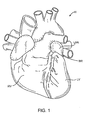
- Fig. 1 is an anterior view of a heart illustrating the right ventricle RV, the left ventricle LV, and the left atrial appendage LAA.
- the methods and apparatus of the present disclosure are intended to place a closure structure over or otherwise close off the base region BR of the left atrial appendage. By closing off the base region BR, the exchange of materials between the left atrial appendage LAA and the left atrium LA will be stopped. Thus, the release of emboli from the left atrial appendage into the left atrium will be stopped.
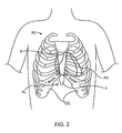
- the heart is located within the pericardial space PS located beneath the patient's rib cage RC.
- the sternum S is located in the center of the rib cage RC and terminates at its lower end in the xiphoid X.
- the costal cartilage CC On either side of the xiphoid are the costal cartilage CC, and the percutaneous access points for performing the procedures of the present disclosure will be located beneath the rib cage RC, and preferably between the xiphoid X and an adjacent costal cartilage CC, preferably at the access location AL shown by a broken line.
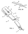
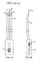
- FIG. 3A An exemplary tool 10 for performing the methods of the present disclosure is illustrated in Figs. 3, 3A , and 4A-4C .
- the tool comprises a shaft 12 having a distal end 14 and a proximal end 16.
- a handle 18 is preferably attached to the proximal end of the shaft, and the shaft will have a curved profile in its axial direction (as best seen in Fig. 4B ) and a crescent-shaped cross-section, as best seen in Fig. 4C .
- the preferred dimensions of the shaft are set forth above.
- the shaft has three lumens 20, 22, and 24.
- a first lumen 20 is used for introducing a closure tool (which may be any of the closure tools described above), while the second and third lumens (22 and 24, respectively) are used for introducing a viewing scope and fluids, such as saline or other clear fluids for improving visualization of the region surrounding the left atrial appendage.
- the first lumen 20 can still be used for a grasper, while either of the second lumen 22 and/or third lumen 24 may be used for introducing alternative closure devices, such as clip appliers, riveting devices, fusing devices, suturing devices, stapling devices, or the like.
- either or both of the lumens 22 and 24 may be used to advance a clip over the left atrial appendage as the appendage is being grasped by a grasper, such as the one shown in Fig. 3 .
- the closure tool may have any of a wide variety of designs, the presently preferred tool is shown in Fig. 3A .
- the tool comprises a grasper 30 and a capture loop 32.
- Capture loop 32 is attached to a manipulation wire 34 which permits the loop 32 to be advanced over the forward end of the grasper to encircle and close the left atrial appendage, as will be described in more detail below.
- the grasping tool 32 may be manipulated using a thumb guide 40, while the capture loop 32 may be manipulated using a second thumb guide 42, both of which are located on the handle 18.
- the exit ports are located proximally of the distal end 14 of the shaft 12.
- the shaft is generally thinned in the region between the exit ports and the distal tip, facilitating the introduction of the distal tip into the atrioventricular groove, as described in more detail below.
- the exit ports are located a sufficient distance behind the distal tip of the shaft so that they will be generally located adjacent to the free end of the left atrial appendage when the tip is located in the atrioventricular groove.
- the methods of the present disclosure may be performed in an ambulatory surgical setting.
- a sedated patient is taken to a facility having fluoroscopic imaging capabilities.
- the area overlying the xiphoid and adjacent costal cartilage is prepared and draped using standard techniques.
- a local anesthetic is then administered and a skin incision, usually about 2 cm in length made, at the area shown in Fig. 2 .
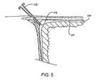
- the percutaneous penetration passes beneath the costal cartilage, and a sheath 100 ( Fig. 5 ) is introduced into the pericardial space PS.
- the pericardial space PS is then irrigated with saline, preferably with a saline-lidocaine solution to provide additional anesthesia and reduce the risk of irritating the heart.
- the closure device 10 is then introduced through the sheath 100 into the pericardial space and advanced over the epicardium to the atrioventricular groove AVG (as shown in Fig. 6A and Fig. 6B ).
- the grasping tool 30 is then advanced distally from the tool 10 so that it can grasp the free end of the left atrial appendage LAA, as shown in Fig. 6C .
- a slight tension can be applied on the left atrial appendage LAA as the capture loop 32 is advanced over the grasper 30 ( Fig. 6D ), and on to the left atrial appendage LAA, as shown in Fig. 6E .
- the loop may then be cinched, as shown in Fig.
- a portion of the parietal pericardium may be further separated from the epicardial surface and the left atrial appendage prior to closing the appendage.
- Increasing the distance between the parietal and visceral pericardium, i.e., the pericardial space creates a working and viewing space that facilitates subsequent manipulation and closure of the atrial appendage.
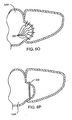
- a modified closure device 100 having an additional lumen 102 is introduced so that its distal end 104 enters the atrioventricular groove AVG, as described previously.
- a balloon expander 110 may then be introduced through the lumen 102, and the balloon expanded to raise the pericardium, as shown in Fig.6I .
- the grasper 30 (or other closure instrument) may then be introduced through other lumens, as previously described.
- the working space created by the balloon greatly simplifies manipulation and positioning of the graspers 30 so that they can be used to capture the atrial appendage and close it as described previously. Further separating the parietal and visceral pericardia to create the working space is a particular advantage when a viewing scope is introduced to the working area to facilitate manipulation of the grasper 30 and any other tools which may be used.
- the closure tool 10 is illustrated in a method for introducing a clip 200 in accordance with the principles of the present disclosure.
- the closure tool 10 is introduced to the left atrial appendage LAA as described in above in connection with Figs. 6A and 6B .
- the clip 200 may be introduced through any of the available lumens in the device, typically using a pusher 202.
- the clip 200 will be configured so that it opens as it emerges from the closure tool 10 and can be advanced over the free distal end of the left atrial appendage LAA, as shown in Fig. 6L .
- the clip 200 may then be closed over the appendage, as shown in Fig. 6N .
- the clip 200 may be self-closing or may require a mechanical or heat-actuated closure mechanism. Once in place, as shown in Fig. 6N , the closure tool 10 can be removed. Frequently, it will be desirable to introduce multiple clips 200, as shown in Fig. 6O . Alternatively, a larger clip 208 can be introduced transversely over the left atrial appendage LAA, as shown in Fig. 6P .
- the clip 300 has a generally U-shaped profile, as best seen in Fig. 7A , optionally having a serpentine or zig-zag profile on at least one of the legs of the clip. As illustrated, a series of peaks and valleys 302 is provided on an "upper" leg of the clip.
- the clip 300 further includes a hinge region 304 which has a narrowed width to facilitate introduction through a introducer catheter 400, as shown in Fig. 8 .
- Introducer catheter 400 has an I-shaped lumen 402 which receives the clip 300 so that the upper leg and lower leg of the clip are held in an open configuration in upper and lower tracks of the lumen, as described below in connection with Figs. 9A-9C .
- the catheter 400 may include a radiopaque marker 404 to permit orientation under fluoroscopic imaging (so the position can confirm that the clip is in the proper vertical orientation when being placed).
- a pusher 408 is provided having an I-shaped distal end 410 which is received in the I-shaped lumen 402 in order to advance and eject the clip from the catheter.
- the clip 300 is held in the lumen 402 of catheter 400 with the legs of the clip held open.
- a pusher 408 can be advanced so that end 410 engages the hinge region304 of the clip, allowing it to be advanced out of the distal end of the catheter, as shown in Fig. 9B .
- the clip 300 As the clip 300 emerges, it remains in an open configuration so that it can be advanced over a free distal end of the left atrial appendage LAA, as shown in Fig. 9B .
- the clip will close over the left atrial appendage LAA to hold the appendage closed in accordance with the principles of the present disclosure.
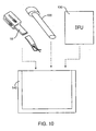
- kits according to the present disclosure comprise a closure tool, such as closure tool 10 described above.
- the kits may comprise an access sheath 120 and will include instructions for use IFU setting forth any of the methods described above.
- all components of the kit will be packaged together in an enclosure 140, such as a pouch, tray, box, tube, or other conventional surgical package capable of maintaining the components in a sterile condition.
- an enclosure 140 such as a pouch, tray, box, tube, or other conventional surgical package capable of maintaining the components in a sterile condition.
- any kit containing instructions for use setting forth the methods of the present disclosure will be part of the present disclosure. Whether or not the kits include a closure device which is similar to Fig. 10 is not necessary.
Description
- The present invention relates generally to a medical apparatus.
- More particularly, the present invention relates to a device for the minimally invasive closure of a left atrial appendage of the heart.
- Atrial fibrillation is a common cardiac rhythm disorder affecting a population of approximately 2.5 million patients in the United States alone. Atrial fibrillation results from a number of different causes and is characterized by a rapid chaotic heart beat. In addition to the risks associated with a disordered heart beat, patients with atrial fibrillation also have an increased risk of stroke. It has been estimated that approximately 75,000 atrial fibrillation patients each year suffer a stroke related to that condition. It appears that strokes in these patients result from emboli many of which may originate from the left atrial appendage. The irregular heart beat causes blood to pool in the left atrial appendage, allowing clots to accumulate over time. From time to time, clot may dislodge from the left atrial appendage and may enter the cranial circulation causing a stroke, the coronary circulation causing a myocardial infarction, the peripheral circulation causing limb ischemia, as well as other vascular beds.
- Significant efforts have been made to reduce the risk of stroke in patients suffering from atrial fibrillation. Most commonly, those patients are treated with blood thinning agents, such as coumadin, to reduce the risk of clot formation. While such treatment can significantly reduce the risk of stroke, it also increases the risk of bleeding and for that reason is inappropriate for many atrial fibrillation patients.
- As an alternative to drug therapy, surgical procedures for closing the left atrial appendage have been proposed. Most commonly, the left atrial appendage has been closed or removed in open surgical procedures, typically where the heart has stopped and the chest opened through the sternum. Because of the significant risk and trauma of such procedures, left atrial appendage removal occurs almost exclusively when the patient's chest is opened for other procedures, such as coronary artery bypass or valve surgery.
- For that reason, alternative procedures which do not require opening of the patient's chest, i.e., a large median sternotomy, have been proposed.
U. S. Patent No. 5,306,234 to Johnson describes a thoracoscopic procedure where access to the pericardial space over the heart is achieved using a pair of intercostal penetrations (i. e., penetrations between the patient's ribs) to establish both visual and surgical access. While such procedures may be performed while the heart remains beating, they still require deflation of the patient's lung and that the patient be placed under full anesthesia. - Furthermore, placement of a chest tube is typically required to reinflate the lung, often requiring a hospitalization for a couple of days.
-
U. S. Patent No. 5,865,791, to Whayne et al . describes a transvascular approach for closing the left atrial appendage. Access is gained via the venous system, typically through a femoral vein, a right internal jugular vein, or a subclavian vein, where a catheter is advanced in an antegrade direction to the right atrium. The intra-atrial septum is then penetrated, and the catheter passed into the left atrium. The catheter is then positioned in the vicinity of the left atrial appendage which is then fused closed, e. g., using radiofrequency energy, other electrical energy, thermal energy, surgical adhesives, or the like. Whayne et al. further describes a thoracoscopic procedure where the pericardium is penetrated through the rib cage and a lasso placed to tie off the neck of the left atrial appendage. Other fixation means described include sutures, staples, shape memory wires, biocompatible adhesives, tissue ablation, and the like. The transvascular approach suggested by Whayne et al. is advantageous in that it avoids the need to penetrate the patient's chest but suffers from the need to penetrate the intra-atrial septum, may not provide definitive closure, requires entry into the left atrial appendage which may dislodge clot and requires injury to the endocardial surface which may promote thrombus formation. A thoracoscopic approach which is also suggested by Whayne et al. suffers from the same problems as the thoracoscopic approach suggested by Johnson. -
US 6,071,281 discloses a device able to isolate the left atrial appendage of a human heart by means of a thoracostomy operation. The apparatus comprises a shaft of a malleable material that a physician can bend into a shape corresponding to the bodily structure to be acted upon and a closing element which can be a surgical clamp having electrodes on one side or a lasso catheter. -
WO 00/16850 -
US 5,908,429 finally discloses a general tissue ligation instrument assembly having a grasping member disposed at its distal end and a contractible literature loop formed of a length of filamentous ligature material. In particular, the multiple instruments comprised in this assembly are disposed in different and distinct channels of a single barrel structure. - For all these reasons, it would be desirable to provide improved and alternative methods and procedures for performing minimally invasive closure of the left atrial appendage. Such methods and procedures will preferably be capable of being performed on patients who have received only a local anesthetic and whose hearts have not been stopped. It would be further desirable to provide methods and procedures which approach the left atrial appendage without the need to perform a thoracotomy (penetration through the intracostal space) or the need to perform a transeptal penetration and/or perform the procedure within the left atrium or left atrial appendage. More specifically, it would be preferable to provide methods and procedures which permitted access to the pericardial space from the xiphoid region of a patient's chest. In addition to the improved and alternative methods and procedures, it would be desirable to provide specialized instruments, devices, and systems for accessing a region over a patient's left atrial appendage from a sub-xiphoid access point to permit closure of the left atrial appendage.
- At least some of these objectives will be met by the inventions described herein below.
-
U. S. Patent Nos. 5,306,234 ,5,865,791 ,6,071,281 and5,908,429 andWO 00/16850 -
U. S. Patent No. 3,496,932 andPCT publication WO 98/05289 - The present invention provides device for closing a left atrial appendage of a heart through a sub-xiphoid approach in accordance with independent claim 1. Preferred embodiments of the invention are reflected in the dependent claims.
- The claimed invention can be better understood in view of the embodiments described hereinafter. In general, the described embodiments describe preferred embodiments of the invention. The attentive reader will note, however, that some aspects of the described embodiments extend beyond the scope of the claims. To the respect that the described embodiments indeed extend beyond the scope of the claims, the described embodiments are to be considered supplementary background information and do not constitute definitions of the invention per se. This also holds for the subsequent "Brief description of the Drawings" as well as the "Description of the Specific Embodiments".
- The present disclosure provides alternative and improved methods and apparatus for closing a left atrial appendage of a patient, particularly a patient at risk of occlusive stroke resulting from emboli released from the left atrial appendage. The most likely patient population for the procedures will be patients suffering from atrial fibrillation which can result in clot and thrombus generation in the left atrial appendage, as described above. The methods and apparatus of the present disclosure permit procedures to be performed on a conscious sedated patient, often in an ambulatory surgical setting where the patient may be released shortly after the procedure is completed. In particular, the methods and apparatus of the present disclosure eliminates the need for a large incision and division of the sternum, i. e., median sternotomy. The present disclosure further eliminates the need to take down (deflate) a lung to access a left atrial appendage, as is usually required in thoracoscopic procedures performed via intracostal access.
- The methods of the present disclosure will be performed in a minimally invasive manner, i.e., where access to the pericardial space overlying the patient's left atrial appendage is accomplished through percutaneous penetrations through the patient's skin. Rather than passing through the rib cage, as with prior thoracoscopic techniques, the present disclosure relies on a "sub-xiphoid" approach where the percutaneous penetration is first made beneath the rib cage, preferably between the xiphoid and adjacent costal cartilage, and an atrial appendage closure tool advanced through the penetration, over the epicardial surface (in the pericardial space) to reach a location adjacent to the exterior of the left atrial appendage. The closure tool can then be used to close the left atrial appendage to prevent the formation of clot and the release of emboli from the atrium.
- Closure can be effected in a variety of ways. It is presently preferred to position a loop of material, such as suture, wire, mesh, tape, or the like, over the appendage and cinch the loop tighter to close the interior of the appendage. A variety of alternative closure techniques would also find use, including suturing (using remotely actuated suturing instruments), stapling, clipping, fusing, gluing, clamping, riveting, or the like. Such closure will generally be intended to be permanent, i.e., it will remain indefinitely after the closure tool is removed, but in some instances could be reversible, i.e., the left atrial appendage could be reopened on a subsequent procedure.
- Thus, a method according to the present disclosure for closing a left atrial appendage of a patient's heart comprises positioning a closure instrument through a percutaneous passage beneath the rib cage, over an epicardial surface, and adjacent to the left atrial appendage. The left atrial appendage is then closed, usually using one of the techniques described above. The positioning step may comprise making an incision usually between a costal cartilage and a xiphoid of the patient, establishing a tract beneath the rib cage. Alternatively the incision may be made superficial to the xiphoid or sternum after which a tract is made through the rib cage to the pericardial space, and will preferably include placing an access sheath through the incision into the pericardial space. The incision may be made using a scalpel or other conventional surgical tool, but could also be made using a trocar and cannula assembly, such as those used in laparoscopic surgery, where the trocar could then be removed leaving the cannula in place as the sheath of the present disclosure. Use of a trocar and cannula may be less preferred, however, since there is an increased risk of injuring the heart if the trocar and cannula assembly is introduced in a blind fashion.
- A closure instrument is then introduced through the sheath into the pericardial space, and over an epicardial surface to the exterior of the left atrial appendage, as described above. Preferably, a distal end of the tool will be introduced into an atrioventricular groove which lies just beneath the atrial appendage.
- Preferably, once the closure tool has been introduced, advancement and positioning can be performed under conventional imaging techniques, such as fluoroscopic imaging. Often, the closure tool will include or be compatible with imaging scopes which may be introduced through the tool. The use of imaging scopes will be particularly useful during the closure procedure where the left atrial appendage is manipulated as described in more detail below. In such instances, it will frequently be desirable to introduce a saline or other clear fluid into the pericardial space to facilitate viewing.
- Once the closure tool is properly positioned, closure may be effected by any of the techniques described above, including looping, suturing, stapling, clipping, fusing, clamping, riveting, or the like. Preferably, the closure will be directed at the base region of the left atrial appendage. Optionally, closing the appendage may further comprise grasping the exterior of the left atrial appendage prior to the actual closing step. Grasping will typically be performed with the conventional grasping tool. As described below, a preferred closure technique is to first grasp the exterior of the left atrial appendage with a grasping tool and subsequently advance a closure loop over the tool on to the exterior of the appendage. A closure loop may then be cinched or otherwise closed or allowed to close, and the tools removed.
- A variety of specific instruments, devices, and systems may be devised for performing the methods of the present disclosure. An exemplary device for closing a left atrial appendage according to the methods of the present disclosure is described in detail in the descriptions that follow. The device comprises a shaft having a proximal end and a distal end, where the distal end is adapted to percutaneously enter the pericardial space, be advanced over an epicardial surface, and then approach the exterior of the left atrial appendage. Preferably, the shaft has a length in the range from 10 cm to 40cm, a width in the range from 2 mm to 20 mm, and a thickness in the range from 1 mm to 10 mm. Usually, the shaft will be curved over its length to be compatible with the curvature of the heart. The shaft may include a means to alter the curvature to accommodate variations in anatomy. Similarly, the device includes a crescent-shaped cross-section to also conform to the shape of the exterior of the heart. The device will carry a mechanism or means for closing the left atrial appendage when the distal end of the shaft is positioned adjacent to the appendage. Usually, the closure mechanism will be introducable through one or more lumens formed in the shaft. In a particularly preferred configuration, the distal end of the shaft will be configured to lie within the atrioventricular groove of the heart, and at least one lumen through the shaft will have an exit port spaced inwardly from the distal end of the shaft by a distance in the range from 0.5 cm to 5 cm. In this way, the port will be positioned properly to access the free end of the atrial appendage for performing the closing procedures. In addition, the shaft may have one or more additional lumens (for a total of two, three, or more lumens through the shaft) in order to provide additional capabilities, including introduction and use of a viewing scope, infusion and perfusion of fluids, particularly the infusion of saline to facilitate viewing. Optionally, the lumens can be used to introduce an anesthetic agent, such as lidocaine, in order to reduce pain or to introduce an anti-arrhythmic agent to reduce myocardial irritability.
- The present disclosure still further comprises kits including the closure devices just described. The kits will further include instructions for use according to the methods described above, and optionally further include packaging for holding all components of the kit together. Additionally, the kits may include the access sheath which is placed through the percutaneous penetration tracks as the pericardial space. The access sheath may be in the form of a trocar and cannula assembly, although this will usually not be preferred.
-
-
Fig. 1 is an anterior view of a heart illustrating the position of the left atrial appendage relative to the remaining structures of the heart. -
Fig. 2 shows the position of the heart in an associated chest cavity and illustrates a preferred percutaneous access site for performing the methods of the present disclosure. -
Fig. 3 is a perspective view of an exemplary closure tool useful for performing the methods of the present disclosure. -
Figs. 4A-4C are orthogonal views of the closure device ofFig. 3 . -
Fig. 5 illustrates an access sheath placed percutaneously into a pericardial space using a sub-xiphoid approach beneath the rib cage as is preferred in the methods of the present disclosure. -
Figs. 6A-6G illustrate use of the exemplary tool ofFig. 3 in performing the closure of a left atrial appendage according to the methods of the present disclosure. -
Figs. 6K-6N illustrate an alternative protocol for use of the exemplary tool ofFig. 3 in performing the closure of a left atrial appendage according to the methods of the present disclosure. -
Figs. 6O and 6P illustrate alternative clip placement patterns for closing the left atrial appendage according to the methods of the present disclosure. -
Fig. 7A-7C illustrate an exemplary clip which may be used in performing the closure methods of the present disclosure. -
Fig. 8 illustrates a clip insertion tool useful for placing the clip ofFigs. 7A-7C according to the methods of the present disclosure. -
Figs. 9A-9C are cross-sectional views of the insertion tool ofFig. 8 used in placing the clip ofFig. 7A-7C over a left atrial appendage according to the methods of the present disclosure. -
Fig. 10 illustrates an exemplary kit including a closure device and optional components according to the present disclosure. -
Fig. 1 is an anterior view of a heart illustrating the right ventricle RV, the left ventricle LV, and the left atrial appendage LAA. The methods and apparatus of the present disclosure are intended to place a closure structure over or otherwise close off the base region BR of the left atrial appendage. By closing off the base region BR, the exchange of materials between the left atrial appendage LAA and the left atrium LA will be stopped. Thus, the release of emboli from the left atrial appendage into the left atrium will be stopped. - Referring now to
Fig. 2 , the heart is located within the pericardial space PS located beneath the patient's rib cage RC. The sternum S is located in the center of the rib cage RC and terminates at its lower end in the xiphoid X. On either side of the xiphoid are the costal cartilage CC, and the percutaneous access points for performing the procedures of the present disclosure will be located beneath the rib cage RC, and preferably between the xiphoid X and an adjacent costal cartilage CC, preferably at the access location AL shown by a broken line. - An
exemplary tool 10 for performing the methods of the present disclosure is illustrated inFigs. 3, 3A , and4A-4C . The tool comprises ashaft 12 having adistal end 14 and aproximal end 16. Ahandle 18 is preferably attached to the proximal end of the shaft, and the shaft will have a curved profile in its axial direction (as best seen inFig. 4B ) and a crescent-shaped cross-section, as best seen inFig. 4C . The preferred dimensions of the shaft are set forth above. - In the illustrated embodiment, the shaft has three
lumens first lumen 20 is used for introducing a closure tool (which may be any of the closure tools described above), while the second and third lumens (22 and 24, respectively) are used for introducing a viewing scope and fluids, such as saline or other clear fluids for improving visualization of the region surrounding the left atrial appendage. In alternative embodiments, thefirst lumen 20 can still be used for a grasper, while either of thesecond lumen 22 and/orthird lumen 24 may be used for introducing alternative closure devices, such as clip appliers, riveting devices, fusing devices, suturing devices, stapling devices, or the like. In a particular embodiment shown below, either or both of thelumens Fig. 3 . - While the closure tool may have any of a wide variety of designs, the presently preferred tool is shown in
Fig. 3A . The tool comprises agrasper 30 and acapture loop 32.Capture loop 32 is attached to amanipulation wire 34 which permits theloop 32 to be advanced over the forward end of the grasper to encircle and close the left atrial appendage, as will be described in more detail below. The graspingtool 32 may be manipulated using athumb guide 40, while thecapture loop 32 may be manipulated using asecond thumb guide 42, both of which are located on thehandle 18. - The
lumens exit ports Fig. 4A . The exit ports are located proximally of thedistal end 14 of theshaft 12. The shaft is generally thinned in the region between the exit ports and the distal tip, facilitating the introduction of the distal tip into the atrioventricular groove, as described in more detail below. The exit ports are located a sufficient distance behind the distal tip of the shaft so that they will be generally located adjacent to the free end of the left atrial appendage when the tip is located in the atrioventricular groove. - The methods of the present disclosure may be performed in an ambulatory surgical setting. Typically, a sedated patient is taken to a facility having fluoroscopic imaging capabilities. The area overlying the xiphoid and adjacent costal cartilage, is prepared and draped using standard techniques. A local anesthetic is then administered and a skin incision, usually about 2 cm in length made, at the area shown in
Fig. 2 . The percutaneous penetration passes beneath the costal cartilage, and a sheath 100 (Fig. 5 ) is introduced into the pericardial space PS. The pericardial space PS is then irrigated with saline, preferably with a saline-lidocaine solution to provide additional anesthesia and reduce the risk of irritating the heart. Theclosure device 10 is then introduced through thesheath 100 into the pericardial space and advanced over the epicardium to the atrioventricular groove AVG (as shown inFig. 6A and Fig. 6B ). The graspingtool 30 is then advanced distally from thetool 10 so that it can grasp the free end of the left atrial appendage LAA, as shown inFig. 6C . A slight tension can be applied on the left atrial appendage LAA as thecapture loop 32 is advanced over the grasper 30 (Fig. 6D ), and on to the left atrial appendage LAA, as shown inFig. 6E . The loop may then be cinched, as shown inFig. 6F , and thetool 10 withdrawn leaving the closure loop in place, as shown inFig. 6G . The interior of the left atrial appendage LAA is thus isolated from the interior of the left atrium so that thrombus and other emboli cannot be released into blood circulation. - Optionally, a portion of the parietal pericardium may be further separated from the epicardial surface and the left atrial appendage prior to closing the appendage. Increasing the distance between the parietal and visceral pericardium, i.e., the pericardial space, creates a working and viewing space that facilitates subsequent manipulation and closure of the atrial appendage. As shown in
Figs. 6H-6J , a modifiedclosure device 100 having anadditional lumen 102 is introduced so that itsdistal end 104 enters the atrioventricular groove AVG, as described previously. Aballoon expander 110 may then be introduced through thelumen 102, and the balloon expanded to raise the pericardium, as shown inFig.6I . The grasper 30 (or other closure instrument) may then be introduced through other lumens, as previously described. The working space created by the balloon greatly simplifies manipulation and positioning of thegraspers 30 so that they can be used to capture the atrial appendage and close it as described previously. Further separating the parietal and visceral pericardia to create the working space is a particular advantage when a viewing scope is introduced to the working area to facilitate manipulation of thegrasper 30 and any other tools which may be used. - Referring now to
Figs. 6K-6N , theclosure tool 10 is illustrated in a method for introducing aclip 200 in accordance with the principles of the present disclosure. Theclosure tool 10 is introduced to the left atrial appendage LAA as described in above in connection withFigs. 6A and 6B . Once in place, theclip 200 may be introduced through any of the available lumens in the device, typically using apusher 202. Theclip 200 will be configured so that it opens as it emerges from theclosure tool 10 and can be advanced over the free distal end of the left atrial appendage LAA, as shown inFig. 6L . Theclip 200 may then be closed over the appendage, as shown inFig. 6N . Theclip 200 may be self-closing or may require a mechanical or heat-actuated closure mechanism. Once in place, as shown inFig. 6N , theclosure tool 10 can be removed. Frequently, it will be desirable to introducemultiple clips 200, as shown inFig. 6O . Alternatively, alarger clip 208 can be introduced transversely over the left atrial appendage LAA, as shown inFig. 6P . - Referring now to
Figs. 7A-7C , anexemplary clip 300 for use in the methods of the present disclosure will be described. Theclip 300 has a generally U-shaped profile, as best seen inFig. 7A , optionally having a serpentine or zig-zag profile on at least one of the legs of the clip. As illustrated, a series of peaks andvalleys 302 is provided on an "upper" leg of the clip. Theclip 300 further includes ahinge region 304 which has a narrowed width to facilitate introduction through aintroducer catheter 400, as shown inFig. 8 .Introducer catheter 400 has an I-shapedlumen 402 which receives theclip 300 so that the upper leg and lower leg of the clip are held in an open configuration in upper and lower tracks of the lumen, as described below in connection withFigs. 9A-9C . Optionally, thecatheter 400 may include aradiopaque marker 404 to permit orientation under fluoroscopic imaging (so the position can confirm that the clip is in the proper vertical orientation when being placed). Apusher 408 is provided having an I-shapeddistal end 410 which is received in the I-shapedlumen 402 in order to advance and eject the clip from the catheter. - Referring now to
Figs. 9A-9C , theclip 300 is held in thelumen 402 ofcatheter 400 with the legs of the clip held open. Apusher 408 can be advanced so thatend 410 engages the hinge region304 of the clip, allowing it to be advanced out of the distal end of the catheter, as shown inFig. 9B . As theclip 300 emerges, it remains in an open configuration so that it can be advanced over a free distal end of the left atrial appendage LAA, as shown inFig. 9B . Once theclip 300 is fully advanced and released from thecatheter 400, as shown inFig. 9C , the clip will close over the left atrial appendage LAA to hold the appendage closed in accordance with the principles of the present disclosure. - Referring now to
Fig. 10 , kits according to the present disclosure comprise a closure tool, such asclosure tool 10 described above. Optionally, the kits may comprise anaccess sheath 120 and will include instructions for use IFU setting forth any of the methods described above. Usually, all components of the kit will be packaged together in anenclosure 140, such as a pouch, tray, box, tube, or other conventional surgical package capable of maintaining the components in a sterile condition. It will be appreciated that any kit containing instructions for use setting forth the methods of the present disclosure will be part of the present disclosure. Whether or not the kits include a closure device which is similar toFig. 10 is not necessary. - While the above is a complete description of the preferred embodiments of the disclosure, various alternatives, modifications, and equivalents may be used.
- Therefore, the above description should not be taken as limiting the scope of the disclosure which is defined by the appended claims.
Claims (7)
- A device for closing a left atrial appendage of the heart through a sub-xiphoid approach, said device comprising:a shaft (12) having an elongated longitudinal body including a proximal end (16) and a distal end (14), the shaft (12) having a lumen (20); anda closing element, the closing element including:a grasping tool (30) configured with a retracted position when not in use and an extended position when in use, the retracted position being where the grasping tool (30) is configured to be inside the shaft (12), the extended position being where the grasping tool (30) is configured to be axially and distally advanced from the proximal end (16) of the shaft (12) and outside the shaft (12), the grasping tool (30) including one member and another member, the members being movable toward and away from each other, where the grasping tool (30) is configured to temporarily grasp the left atrial appendage when the members are moved toward each other; anda closure loop (32) configured with a retracted position when not in use and an extended position when in use, the retracted position being where the closure loop (32) is configured to be within the lumen (20) of the shaft (12), the extended position being where the closure loop (32) is configured to be axially and distally advanced from the proximal end (16) of the shaft (12) and outside the lumen (20) of the shaft (12), the closure loop (32) is configured to advance over the grasping tool (30) and around the left atrial appendage, and the closure loop (32) is configured to be cinched to close the left atrial appendage;
characterised in thatthe shaft has a crescent-shaped cross-section. - The device as in claim 1, wherein the grasping tool (30) and the closure loop (32) are arranged in the same lumen of the shaft (12).
- The device as in claim 1, wherein the shaft (12) includes a plurality of lumens (20, 22, 24).
- The device as in claim 1, wherein the shaft (12) has a means to alter its curvature.
- The device as in claim 1, wherein the closure loop (32) is configured to permanently close the left atrial appendage.
- The device as in claim 1, wherein the grasping tool (30) is only a two-member jaw-like construction, the two members of the grasping tool (30) being movable toward and away from each other, where the grasping tool (30) is configured to temporarily grasp the left atrial appendage when the two members are moved toward each other.
- A system for closing a left atrial appendage of a heart through a sub-xiphoid approach, said system comprising:a sub-xiphoid device according to claim 1; andan expandable member configured to be disposed at a location on a epicardial surface within a pericardial space, the expandable member being expandable between the epicardial surface and a parietal pericardium.
Applications Claiming Priority (1)
| Application Number | Priority Date | Filing Date | Title |
|---|---|---|---|
| PCT/US2000/023727 WO2002017809A1 (en) | 2000-08-29 | 2000-08-29 | Methods and apparatus for transpericardial left atrial appendage closure |
Publications (3)
| Publication Number | Publication Date |
|---|---|
| EP1313406A1 EP1313406A1 (en) | 2003-05-28 |
| EP1313406A4 EP1313406A4 (en) | 2006-09-13 |
| EP1313406B1 true EP1313406B1 (en) | 2010-06-16 |
Family
ID=21741719
Family Applications (1)
| Application Number | Title | Priority Date | Filing Date |
|---|---|---|---|
| EP00957904A Expired - Lifetime EP1313406B1 (en) | 2000-08-29 | 2000-08-29 | Methods and apparatus for transpericardial left atrial appendage closure |
Country Status (4)
| Country | Link |
|---|---|
| EP (1) | EP1313406B1 (en) |
| JP (1) | JP4671582B2 (en) |
| DE (1) | DE60044569D1 (en) |
| WO (1) | WO2002017809A1 (en) |
Families Citing this family (35)
| Publication number | Priority date | Publication date | Assignee | Title |
|---|---|---|---|---|
| US7318833B2 (en) | 2001-12-19 | 2008-01-15 | Nmt Medical, Inc. | PFO closure device with flexible thrombogenic joint and improved dislodgement resistance |
| US9241695B2 (en) | 2002-03-25 | 2016-01-26 | W.L. Gore & Associates, Inc. | Patent foramen ovale (PFO) closure clips |
| CA2488337A1 (en) | 2002-06-05 | 2003-12-18 | Nmt Medical, Inc. | Patent foramen ovale (pfo) closure device with radial and circumferential support |
| ES2626981T3 (en) | 2002-12-09 | 2017-07-26 | W.L. Gore & Associates, Inc. | Septal closure device |
| US8480706B2 (en) | 2003-07-14 | 2013-07-09 | W.L. Gore & Associates, Inc. | Tubular patent foramen ovale (PFO) closure device with catch system |
| US7678123B2 (en) | 2003-07-14 | 2010-03-16 | Nmt Medical, Inc. | Tubular patent foramen ovale (PFO) closure device with catch system |
| US9861346B2 (en) | 2003-07-14 | 2018-01-09 | W. L. Gore & Associates, Inc. | Patent foramen ovale (PFO) closure device with linearly elongating petals |
| ES2295932T3 (en) | 2003-09-12 | 2008-04-16 | Nmt Medical, Inc. | DEVICE TO AVOID THE FORMATION OF THROMBES IN THE LEFT ATRIAL APPENDIX. |
| ES2661071T3 (en) | 2003-12-04 | 2018-03-27 | Boston Scientific Scimed, Inc. | Supply system for a left atrial appendage containment device |
| CA2558247A1 (en) | 2004-03-03 | 2005-10-06 | Nmt Medical, Inc. | Delivery/recovery system for septal occluder |
| US7806846B2 (en) | 2004-03-30 | 2010-10-05 | Nmt Medical, Inc. | Restoration of flow in LAA via tubular conduit |
| US8308760B2 (en) | 2004-05-06 | 2012-11-13 | W.L. Gore & Associates, Inc. | Delivery systems and methods for PFO closure device with two anchors |
| CA2563298A1 (en) | 2004-05-07 | 2005-11-24 | Nmt Medical, Inc. | Catching mechanisms for tubular septal occluder |
| US7837619B2 (en) * | 2005-08-19 | 2010-11-23 | Boston Scientific Scimed, Inc. | Transeptal apparatus, system, and method |
| US7998095B2 (en) | 2005-08-19 | 2011-08-16 | Boston Scientific Scimed, Inc. | Occlusion device |
| US8062309B2 (en) | 2005-08-19 | 2011-11-22 | Boston Scientific Scimed, Inc. | Defect occlusion apparatus, system, and method |
| US9259267B2 (en) | 2005-09-06 | 2016-02-16 | W.L. Gore & Associates, Inc. | Devices and methods for treating cardiac tissue |
| US7972359B2 (en) | 2005-09-16 | 2011-07-05 | Atritech, Inc. | Intracardiac cage and method of delivering same |
| EP1962695A1 (en) | 2005-12-22 | 2008-09-03 | NMT Medical, Inc. | Catch members for occluder devices |
| US8870913B2 (en) | 2006-03-31 | 2014-10-28 | W.L. Gore & Associates, Inc. | Catch system with locking cap for patent foramen ovale (PFO) occluder |
| EP2015681B1 (en) | 2006-05-03 | 2018-03-28 | Datascope Corp. | Tissue closure device |
| US9005242B2 (en) | 2007-04-05 | 2015-04-14 | W.L. Gore & Associates, Inc. | Septal closure device with centering mechanism |
| US9138562B2 (en) | 2007-04-18 | 2015-09-22 | W.L. Gore & Associates, Inc. | Flexible catheter system |
| US20130165967A1 (en) | 2008-03-07 | 2013-06-27 | W.L. Gore & Associates, Inc. | Heart occlusion devices |
| US8956389B2 (en) | 2009-06-22 | 2015-02-17 | W. L. Gore & Associates, Inc. | Sealing device and delivery system |
| US20120029556A1 (en) | 2009-06-22 | 2012-02-02 | Masters Steven J | Sealing device and delivery system |
| US9770232B2 (en) | 2011-08-12 | 2017-09-26 | W. L. Gore & Associates, Inc. | Heart occlusion devices |
| US10828019B2 (en) | 2013-01-18 | 2020-11-10 | W.L. Gore & Associates, Inc. | Sealing device and delivery system |
| WO2015077356A1 (en) | 2013-11-19 | 2015-05-28 | Wheeler William K | Fastener applicator with interlock |
| US9808230B2 (en) | 2014-06-06 | 2017-11-07 | W. L. Gore & Associates, Inc. | Sealing device and delivery system |
| EP3459469A1 (en) | 2017-09-23 | 2019-03-27 | Universität Zürich | Medical occluder device |
| CA3094709A1 (en) | 2018-03-28 | 2019-10-03 | Datascope Corp. | Device for atrial appendage exclusion |
| WO2020179045A1 (en) * | 2019-03-07 | 2020-09-10 | オリンパス株式会社 | Medical device |
| WO2020179044A1 (en) * | 2019-03-07 | 2020-09-10 | オリンパス株式会社 | Medical device |
| WO2021059273A2 (en) | 2019-09-26 | 2021-04-01 | Universitat Zurich | Left atrial appendage occlusion devices |
Citations (1)
| Publication number | Priority date | Publication date | Assignee | Title |
|---|---|---|---|---|
| US5908429A (en) * | 1997-05-01 | 1999-06-01 | Yoon; Inbae | Methods of anatomical tissue ligation |
Family Cites Families (16)
| Publication number | Priority date | Publication date | Assignee | Title |
|---|---|---|---|---|
| US4865037A (en) * | 1987-11-13 | 1989-09-12 | Thomas J. Fogarty | Method for implanting automatic implantable defibrillator |
| US5071428A (en) * | 1989-09-08 | 1991-12-10 | Ventritex, Inc. | Method and apparatus for providing intrapericardial access and inserting intrapericardial electrodes |
| US5632761A (en) * | 1991-05-29 | 1997-05-27 | Origin Medsystems, Inc. | Inflatable devices for separating layers of tissue, and methods of using |
| US5452733A (en) * | 1993-02-22 | 1995-09-26 | Stanford Surgical Technologies, Inc. | Methods for performing thoracoscopic coronary artery bypass |
| ATE149312T1 (en) * | 1991-11-19 | 1997-03-15 | Origin Medsystems Inc | ENDOSCOPIC INFLATABLE RETRACTION DEVICES FOR SEPARATING TISSUE LAYERS |
| US5540711A (en) * | 1992-06-02 | 1996-07-30 | General Surgical Innovations, Inc. | Apparatus and method for developing an anatomic space for laparoscopic procedures with laparoscopic visualization |
| US6161543A (en) * | 1993-02-22 | 2000-12-19 | Epicor, Inc. | Methods of epicardial ablation for creating a lesion around the pulmonary veins |
| US5306234A (en) * | 1993-03-23 | 1994-04-26 | Johnson W Dudley | Method for closing an atrial appendage |
| SE9303253D0 (en) * | 1993-10-05 | 1993-10-05 | Siemens Elema Ab | Instruments for peephole surgery |
| US5681278A (en) * | 1994-06-23 | 1997-10-28 | Cormedics Corp. | Coronary vasculature treatment method |
| US6132438A (en) * | 1995-06-07 | 2000-10-17 | Ep Technologies, Inc. | Devices for installing stasis reducing means in body tissue |
| US5827216A (en) * | 1995-06-07 | 1998-10-27 | Cormedics Corp. | Method and apparatus for accessing the pericardial space |
| US6237605B1 (en) * | 1996-10-22 | 2001-05-29 | Epicor, Inc. | Methods of epicardial ablation |
| US5961440A (en) * | 1997-01-02 | 1999-10-05 | Myocor, Inc. | Heart wall tension reduction apparatus and method |
| US6071281A (en) * | 1998-05-05 | 2000-06-06 | Ep Technologies, Inc. | Surgical method and apparatus for positioning a diagnostic or therapeutic element within the body and remote power control unit for use with same |
| JP4636683B2 (en) * | 1997-10-10 | 2011-02-23 | ボストン サイエンティフィック リミテッド | Soft tissue coagulation probe |
-
2000
- 2000-08-29 JP JP2002522787A patent/JP4671582B2/en not_active Expired - Lifetime
- 2000-08-29 WO PCT/US2000/023727 patent/WO2002017809A1/en active Application Filing
- 2000-08-29 EP EP00957904A patent/EP1313406B1/en not_active Expired - Lifetime
- 2000-08-29 DE DE60044569T patent/DE60044569D1/en not_active Expired - Lifetime
Patent Citations (1)
| Publication number | Priority date | Publication date | Assignee | Title |
|---|---|---|---|---|
| US5908429A (en) * | 1997-05-01 | 1999-06-01 | Yoon; Inbae | Methods of anatomical tissue ligation |
Also Published As
| Publication number | Publication date |
|---|---|
| JP4671582B2 (en) | 2011-04-20 |
| EP1313406A4 (en) | 2006-09-13 |
| WO2002017809A1 (en) | 2002-03-07 |
| DE60044569D1 (en) | 2010-07-29 |
| EP1313406A1 (en) | 2003-05-28 |
| JP2004520090A (en) | 2004-07-08 |
Similar Documents
| Publication | Publication Date | Title |
|---|---|---|
| EP1313406B1 (en) | Methods and apparatus for transpericardial left atrial appendage closure | |
| US9724105B2 (en) | Methods and apparatus for transpericardial left atrial appendage closure | |
| JP4987861B2 (en) | Papillary muscle attachment for left ventricular reduction | |
| US10524791B2 (en) | Left atrial appendage devices and methods | |
| US5452733A (en) | Methods for performing thoracoscopic coronary artery bypass | |
| US7938827B2 (en) | Cardiac valve leaflet attachment device and methods thereof | |
| JP5256206B2 (en) | Diaphragm entry for posterior surgical access | |
| US20050149069A1 (en) | Left atrial appendage devices and methods | |
| EP3884993A1 (en) | Device for valve regurgitation surgery and cardiac pacemaker lead fixation | |
| US20080275437A1 (en) | Connector device for electrophysiology probe | |
| JP2013116351A (en) | Diaphragm entry for posterior surgical access | |
| JP2016172000A (en) | Diaphragm entry for rear area surgical access |
Legal Events
| Date | Code | Title | Description |
|---|---|---|---|
| PUAI | Public reference made under article 153(3) epc to a published international application that has entered the european phase |
Free format text: ORIGINAL CODE: 0009012 |
|
| 17P | Request for examination filed |
Effective date: 20030228 |
|
| AK | Designated contracting states |
Designated state(s): AT BE CH CY DE DK ES FI FR GB GR IE IT LI LU MC NL PT SE |
|
| AX | Request for extension of the european patent |
Extension state: AL LT LV MK RO SI |
|
| RAP1 | Party data changed (applicant data changed or rights of an application transferred) |
Owner name: KAPLAN, AARON, V. |
|
| RIN1 | Information on inventor provided before grant (corrected) |
Inventor name: KAPLAN, AARON, V. |
|
| RBV | Designated contracting states (corrected) |
Designated state(s): DE FR GB |
|
| RIN1 | Information on inventor provided before grant (corrected) |
Inventor name: BAJOR, JORDAN T. Inventor name: KAPLAN, AARON V. |
|
| A4 | Supplementary search report drawn up and despatched |
Effective date: 20060814 |
|
| RIC1 | Information provided on ipc code assigned before grant |
Ipc: A61B 17/00 20060101AFI20060808BHEP |
|
| 17Q | First examination report despatched |
Effective date: 20000205 |
|
| GRAP | Despatch of communication of intention to grant a patent |
Free format text: ORIGINAL CODE: EPIDOSNIGR1 |
|
| GRAS | Grant fee paid |
Free format text: ORIGINAL CODE: EPIDOSNIGR3 |
|
| GRAA | (expected) grant |
Free format text: ORIGINAL CODE: 0009210 |
|
| AK | Designated contracting states |
Kind code of ref document: B1 Designated state(s): DE FR GB |
|
| REF | Corresponds to: |
Ref document number: 60044569 Country of ref document: DE Date of ref document: 20100729 Kind code of ref document: P |
|
| PLBE | No opposition filed within time limit |
Free format text: ORIGINAL CODE: 0009261 |
|
| STAA | Information on the status of an ep patent application or granted ep patent |
Free format text: STATUS: NO OPPOSITION FILED WITHIN TIME LIMIT |
|
| 26N | No opposition filed |
Effective date: 20110317 |
|
| REG | Reference to a national code |
Ref country code: DE Ref legal event code: R097 Ref document number: 60044569 Country of ref document: DE Effective date: 20110316 |
|
| REG | Reference to a national code |
Ref country code: FR Ref legal event code: PLFP Year of fee payment: 17 |
|
| REG | Reference to a national code |
Ref country code: FR Ref legal event code: PLFP Year of fee payment: 18 |
|
| REG | Reference to a national code |
Ref country code: FR Ref legal event code: PLFP Year of fee payment: 19 |
|
| PGFP | Annual fee paid to national office [announced via postgrant information from national office to epo] |
Ref country code: FR Payment date: 20190711 Year of fee payment: 20 Ref country code: DE Payment date: 20190813 Year of fee payment: 20 |
|
| PGFP | Annual fee paid to national office [announced via postgrant information from national office to epo] |
Ref country code: GB Payment date: 20190830 Year of fee payment: 20 |
|
| REG | Reference to a national code |
Ref country code: DE Ref legal event code: R071 Ref document number: 60044569 Country of ref document: DE |
|
| REG | Reference to a national code |
Ref country code: GB Ref legal event code: PE20 Expiry date: 20200828 |
|
| PG25 | Lapsed in a contracting state [announced via postgrant information from national office to epo] |
Ref country code: GB Free format text: LAPSE BECAUSE OF EXPIRATION OF PROTECTION Effective date: 20200828 |