EP1279404A1 - Use of HIV-1 tat, fragments or derivatives thereof, to target or to activate antigen-presenting cells, to deliver cargo molecules for vaccination or to treat other diseases - Google Patents
Use of HIV-1 tat, fragments or derivatives thereof, to target or to activate antigen-presenting cells, to deliver cargo molecules for vaccination or to treat other diseases Download PDFInfo
- Publication number
- EP1279404A1 EP1279404A1 EP01118114A EP01118114A EP1279404A1 EP 1279404 A1 EP1279404 A1 EP 1279404A1 EP 01118114 A EP01118114 A EP 01118114A EP 01118114 A EP01118114 A EP 01118114A EP 1279404 A1 EP1279404 A1 EP 1279404A1
- Authority
- EP
- European Patent Office
- Prior art keywords
- tat
- hiv
- cells
- antigens
- fragments
- Prior art date
- Legal status (The legal status is an assumption and is not a legal conclusion. Google has not performed a legal analysis and makes no representation as to the accuracy of the status listed.)
- Withdrawn
Links
- 239000012634 fragment Substances 0.000 title claims abstract description 47
- 208000037265 diseases, disorders, signs and symptoms Diseases 0.000 title claims abstract description 44
- 201000010099 disease Diseases 0.000 title claims abstract description 43
- 241000713772 Human immunodeficiency virus 1 Species 0.000 title claims abstract description 40
- 210000000612 antigen-presenting cell Anatomy 0.000 title claims abstract description 40
- 238000002255 vaccination Methods 0.000 title claims abstract description 36
- 101710149951 Protein Tat Proteins 0.000 claims abstract description 560
- 210000004027 cell Anatomy 0.000 claims abstract description 162
- 239000000427 antigen Substances 0.000 claims abstract description 117
- 108091007433 antigens Proteins 0.000 claims abstract description 117
- 102000036639 antigens Human genes 0.000 claims abstract description 117
- 210000002889 endothelial cell Anatomy 0.000 claims abstract description 72
- 241000725303 Human immunodeficiency virus Species 0.000 claims abstract description 67
- 206010028980 Neoplasm Diseases 0.000 claims abstract description 55
- 210000004443 dendritic cell Anatomy 0.000 claims abstract description 48
- 208000035473 Communicable disease Diseases 0.000 claims abstract description 42
- 230000001225 therapeutic effect Effects 0.000 claims abstract description 42
- 210000002540 macrophage Anatomy 0.000 claims abstract description 40
- 230000028993 immune response Effects 0.000 claims abstract description 35
- 230000002757 inflammatory effect Effects 0.000 claims abstract description 35
- 208000030507 AIDS Diseases 0.000 claims abstract description 32
- 230000002491 angiogenic effect Effects 0.000 claims abstract description 31
- 238000011282 treatment Methods 0.000 claims abstract description 28
- 239000002671 adjuvant Substances 0.000 claims abstract description 26
- 230000003449 preventive effect Effects 0.000 claims abstract description 24
- 150000001875 compounds Chemical class 0.000 claims abstract description 21
- 230000006870 function Effects 0.000 claims abstract description 18
- 238000000034 method Methods 0.000 claims abstract description 11
- 230000001575 pathological effect Effects 0.000 claims abstract description 3
- 108090000623 proteins and genes Proteins 0.000 claims description 53
- 102000004169 proteins and genes Human genes 0.000 claims description 52
- 102000004127 Cytokines Human genes 0.000 claims description 46
- 108090000695 Cytokines Proteins 0.000 claims description 46
- 108010044426 integrins Proteins 0.000 claims description 46
- 102000006495 integrins Human genes 0.000 claims description 46
- 208000031886 HIV Infections Diseases 0.000 claims description 43
- 108090000765 processed proteins & peptides Proteins 0.000 claims description 41
- 208000015181 infectious disease Diseases 0.000 claims description 35
- 102000004196 processed proteins & peptides Human genes 0.000 claims description 27
- 230000037361 pathway Effects 0.000 claims description 26
- 238000004519 manufacturing process Methods 0.000 claims description 25
- 230000001404 mediated effect Effects 0.000 claims description 24
- 210000001744 T-lymphocyte Anatomy 0.000 claims description 22
- 238000001727 in vivo Methods 0.000 claims description 22
- 238000000338 in vitro Methods 0.000 claims description 18
- 108020004414 DNA Proteins 0.000 claims description 17
- 230000003834 intracellular effect Effects 0.000 claims description 17
- 241000700605 Viruses Species 0.000 claims description 16
- 108091028043 Nucleic acid sequence Proteins 0.000 claims description 15
- 125000003275 alpha amino acid group Chemical group 0.000 claims description 15
- 239000002245 particle Substances 0.000 claims description 15
- 244000052769 pathogen Species 0.000 claims description 14
- 208000007766 Kaposi sarcoma Diseases 0.000 claims description 11
- 239000003814 drug Substances 0.000 claims description 11
- 241000713340 Human immunodeficiency virus 2 Species 0.000 claims description 10
- 206010061218 Inflammation Diseases 0.000 claims description 10
- 230000004054 inflammatory process Effects 0.000 claims description 10
- 210000004072 lung Anatomy 0.000 claims description 10
- 201000004792 malaria Diseases 0.000 claims description 10
- 239000000203 mixture Substances 0.000 claims description 10
- 239000002773 nucleotide Substances 0.000 claims description 10
- 125000003729 nucleotide group Chemical group 0.000 claims description 10
- 229960005486 vaccine Drugs 0.000 claims description 10
- 210000000481 breast Anatomy 0.000 claims description 9
- 210000001072 colon Anatomy 0.000 claims description 9
- 241000222120 Candida <Saccharomycetales> Species 0.000 claims description 8
- 241000187479 Mycobacterium tuberculosis Species 0.000 claims description 8
- 208000027866 inflammatory disease Diseases 0.000 claims description 8
- 239000002105 nanoparticle Substances 0.000 claims description 8
- 210000004881 tumor cell Anatomy 0.000 claims description 8
- 206010060862 Prostate cancer Diseases 0.000 claims description 7
- 208000000236 Prostatic Neoplasms Diseases 0.000 claims description 7
- 238000007918 intramuscular administration Methods 0.000 claims description 7
- 239000002502 liposome Substances 0.000 claims description 7
- 239000011859 microparticle Substances 0.000 claims description 7
- 230000009885 systemic effect Effects 0.000 claims description 7
- YBJHBAHKTGYVGT-ZKWXMUAHSA-N (+)-Biotin Chemical compound N1C(=O)N[C@@H]2[C@H](CCCCC(=O)O)SC[C@@H]21 YBJHBAHKTGYVGT-ZKWXMUAHSA-N 0.000 claims description 6
- 102000019034 Chemokines Human genes 0.000 claims description 6
- 108010012236 Chemokines Proteins 0.000 claims description 6
- 230000002519 immonomodulatory effect Effects 0.000 claims description 6
- 230000033115 angiogenesis Effects 0.000 claims description 5
- 230000001772 anti-angiogenic effect Effects 0.000 claims description 5
- 229940124599 anti-inflammatory drug Drugs 0.000 claims description 5
- 230000000840 anti-viral effect Effects 0.000 claims description 5
- 210000004369 blood Anatomy 0.000 claims description 5
- 239000008280 blood Substances 0.000 claims description 5
- 238000006243 chemical reaction Methods 0.000 claims description 5
- XUJNEKJLAYXESH-UHFFFAOYSA-N cysteine Natural products SCC(N)C(O)=O XUJNEKJLAYXESH-UHFFFAOYSA-N 0.000 claims description 5
- 229940043239 cytotoxic antineoplastic drug Drugs 0.000 claims description 5
- -1 eccipients Substances 0.000 claims description 5
- 241001505345 Human T-cell lymphotropic virus type 2b Species 0.000 claims description 4
- 241001465754 Metazoa Species 0.000 claims description 4
- 208000026935 allergic disease Diseases 0.000 claims description 4
- 210000000170 cell membrane Anatomy 0.000 claims description 4
- 230000009826 neoplastic cell growth Effects 0.000 claims description 4
- 239000013612 plasmid Substances 0.000 claims description 4
- 239000000243 solution Substances 0.000 claims description 4
- 239000000126 substance Substances 0.000 claims description 4
- 210000001519 tissue Anatomy 0.000 claims description 4
- 239000013598 vector Substances 0.000 claims description 4
- 201000004681 Psoriasis Diseases 0.000 claims description 3
- 230000001154 acute effect Effects 0.000 claims description 3
- 230000001580 bacterial effect Effects 0.000 claims description 3
- 229960002685 biotin Drugs 0.000 claims description 3
- 235000020958 biotin Nutrition 0.000 claims description 3
- 239000011616 biotin Substances 0.000 claims description 3
- 238000001514 detection method Methods 0.000 claims description 3
- 239000013604 expression vector Substances 0.000 claims description 3
- 208000006454 hepatitis Diseases 0.000 claims description 3
- 231100000283 hepatitis Toxicity 0.000 claims description 3
- ITZMJCSORYKOSI-AJNGGQMLSA-N APGPR Enterostatin Chemical compound C[C@H](N)C(=O)N1CCC[C@H]1C(=O)NCC(=O)N1[C@H](C(=O)N[C@@H](CCCN=C(N)N)C(O)=O)CCC1 ITZMJCSORYKOSI-AJNGGQMLSA-N 0.000 claims description 2
- 206010000830 Acute leukaemia Diseases 0.000 claims description 2
- 102000002260 Alkaline Phosphatase Human genes 0.000 claims description 2
- 108020004774 Alkaline Phosphatase Proteins 0.000 claims description 2
- 206010065452 Angiodermatitis Diseases 0.000 claims description 2
- 208000028185 Angioedema Diseases 0.000 claims description 2
- 201000001320 Atherosclerosis Diseases 0.000 claims description 2
- 241000894006 Bacteria Species 0.000 claims description 2
- 208000035143 Bacterial infection Diseases 0.000 claims description 2
- 241000701022 Cytomegalovirus Species 0.000 claims description 2
- 206010012689 Diabetic retinopathy Diseases 0.000 claims description 2
- 102000004190 Enzymes Human genes 0.000 claims description 2
- 108090000790 Enzymes Proteins 0.000 claims description 2
- 208000010412 Glaucoma Diseases 0.000 claims description 2
- 208000024869 Goodpasture syndrome Diseases 0.000 claims description 2
- 241000589989 Helicobacter Species 0.000 claims description 2
- 208000001688 Herpes Genitalis Diseases 0.000 claims description 2
- 208000004898 Herpes Labialis Diseases 0.000 claims description 2
- 206010020751 Hypersensitivity Diseases 0.000 claims description 2
- 208000004575 Infectious Arthritis Diseases 0.000 claims description 2
- 108010028921 Lipopeptides Proteins 0.000 claims description 2
- 206010067152 Oral herpes Diseases 0.000 claims description 2
- 206010031252 Osteomyelitis Diseases 0.000 claims description 2
- 241001631646 Papillomaviridae Species 0.000 claims description 2
- 208000030852 Parasitic disease Diseases 0.000 claims description 2
- 206010038933 Retinopathy of prematurity Diseases 0.000 claims description 2
- 241000607142 Salmonella Species 0.000 claims description 2
- 206010039491 Sarcoma Diseases 0.000 claims description 2
- 206010039710 Scleroderma Diseases 0.000 claims description 2
- 208000021386 Sjogren Syndrome Diseases 0.000 claims description 2
- 241000191940 Staphylococcus Species 0.000 claims description 2
- 108010090804 Streptavidin Proteins 0.000 claims description 2
- 241000194017 Streptococcus Species 0.000 claims description 2
- 201000009594 Systemic Scleroderma Diseases 0.000 claims description 2
- 206010042953 Systemic sclerosis Diseases 0.000 claims description 2
- 206010043376 Tetanus Diseases 0.000 claims description 2
- 206010047115 Vasculitis Diseases 0.000 claims description 2
- 208000036142 Viral infection Diseases 0.000 claims description 2
- 230000007815 allergy Effects 0.000 claims description 2
- 229940037003 alum Drugs 0.000 claims description 2
- 230000001745 anti-biotin effect Effects 0.000 claims description 2
- 208000022362 bacterial infectious disease Diseases 0.000 claims description 2
- 210000000988 bone and bone Anatomy 0.000 claims description 2
- 201000011510 cancer Diseases 0.000 claims description 2
- 201000003984 candidiasis Diseases 0.000 claims description 2
- 239000000969 carrier Substances 0.000 claims description 2
- 210000000845 cartilage Anatomy 0.000 claims description 2
- 208000024207 chronic leukemia Diseases 0.000 claims description 2
- 239000006071 cream Substances 0.000 claims description 2
- 239000003085 diluting agent Substances 0.000 claims description 2
- 206010014665 endocarditis Diseases 0.000 claims description 2
- 210000000750 endocrine system Anatomy 0.000 claims description 2
- 210000001035 gastrointestinal tract Anatomy 0.000 claims description 2
- 201000004946 genital herpes Diseases 0.000 claims description 2
- 206010066435 human herpesvirus 8 infection Diseases 0.000 claims description 2
- 201000001371 inclusion conjunctivitis Diseases 0.000 claims description 2
- 201000006747 infectious mononucleosis Diseases 0.000 claims description 2
- 206010022000 influenza Diseases 0.000 claims description 2
- 210000004185 liver Anatomy 0.000 claims description 2
- 206010025135 lupus erythematosus Diseases 0.000 claims description 2
- 230000001926 lymphatic effect Effects 0.000 claims description 2
- 230000001613 neoplastic effect Effects 0.000 claims description 2
- 239000002674 ointment Substances 0.000 claims description 2
- 201000008482 osteoarthritis Diseases 0.000 claims description 2
- 210000001672 ovary Anatomy 0.000 claims description 2
- 210000000496 pancreas Anatomy 0.000 claims description 2
- 230000004962 physiological condition Effects 0.000 claims description 2
- 239000006187 pill Substances 0.000 claims description 2
- 239000000843 powder Substances 0.000 claims description 2
- 206010036784 proctocolitis Diseases 0.000 claims description 2
- 238000000746 purification Methods 0.000 claims description 2
- 206010039073 rheumatoid arthritis Diseases 0.000 claims description 2
- 201000000306 sarcoidosis Diseases 0.000 claims description 2
- 201000001223 septic arthritis Diseases 0.000 claims description 2
- 230000001568 sexual effect Effects 0.000 claims description 2
- 210000003491 skin Anatomy 0.000 claims description 2
- 208000017520 skin disease Diseases 0.000 claims description 2
- 210000004872 soft tissue Anatomy 0.000 claims description 2
- 239000007921 spray Substances 0.000 claims description 2
- 239000000758 substrate Substances 0.000 claims description 2
- 239000000725 suspension Substances 0.000 claims description 2
- 208000006379 syphilis Diseases 0.000 claims description 2
- 239000003826 tablet Substances 0.000 claims description 2
- 206010044325 trachoma Diseases 0.000 claims description 2
- 201000008827 tuberculosis Diseases 0.000 claims description 2
- 208000019206 urinary tract infection Diseases 0.000 claims description 2
- 210000004291 uterus Anatomy 0.000 claims description 2
- 230000002792 vascular Effects 0.000 claims description 2
- 230000009385 viral infection Effects 0.000 claims description 2
- 230000003612 virological effect Effects 0.000 claims description 2
- 239000008194 pharmaceutical composition Substances 0.000 claims 12
- 241000607626 Vibrio cholerae Species 0.000 claims 1
- 229940118696 vibrio cholerae Drugs 0.000 claims 1
- 230000037314 wound repair Effects 0.000 claims 1
- 230000035800 maturation Effects 0.000 abstract description 14
- 230000001965 increasing effect Effects 0.000 abstract description 12
- 230000008685 targeting Effects 0.000 abstract description 6
- 238000009826 distribution Methods 0.000 abstract description 5
- 230000003213 activating effect Effects 0.000 abstract description 4
- 238000003745 diagnosis Methods 0.000 abstract description 3
- 230000000069 prophylactic effect Effects 0.000 abstract description 2
- 230000001681 protective effect Effects 0.000 abstract description 2
- 235000018102 proteins Nutrition 0.000 description 50
- 239000000872 buffer Substances 0.000 description 44
- 210000001616 monocyte Anatomy 0.000 description 28
- 239000002609 medium Substances 0.000 description 26
- 230000000694 effects Effects 0.000 description 23
- 238000011534 incubation Methods 0.000 description 22
- 230000004044 response Effects 0.000 description 22
- 230000014509 gene expression Effects 0.000 description 19
- 239000013642 negative control Substances 0.000 description 18
- 102000005962 receptors Human genes 0.000 description 18
- 108020003175 receptors Proteins 0.000 description 18
- 108060008682 Tumor Necrosis Factor Proteins 0.000 description 16
- 102000000852 Tumor Necrosis Factor-alpha Human genes 0.000 description 16
- 108010031318 Vitronectin Proteins 0.000 description 16
- 102100035140 Vitronectin Human genes 0.000 description 16
- 102100037362 Fibronectin Human genes 0.000 description 15
- 108010067306 Fibronectins Proteins 0.000 description 15
- 230000005764 inhibitory process Effects 0.000 description 15
- 230000004071 biological effect Effects 0.000 description 12
- 238000002474 experimental method Methods 0.000 description 12
- 230000003053 immunization Effects 0.000 description 12
- MZOFCQQQCNRIBI-VMXHOPILSA-N (3s)-4-[[(2s)-1-[[(2s)-1-[[(1s)-1-carboxy-2-hydroxyethyl]amino]-4-methyl-1-oxopentan-2-yl]amino]-5-(diaminomethylideneamino)-1-oxopentan-2-yl]amino]-3-[[2-[[(2s)-2,6-diaminohexanoyl]amino]acetyl]amino]-4-oxobutanoic acid Chemical compound OC[C@@H](C(O)=O)NC(=O)[C@H](CC(C)C)NC(=O)[C@H](CCCN=C(N)N)NC(=O)[C@H](CC(O)=O)NC(=O)CNC(=O)[C@@H](N)CCCCN MZOFCQQQCNRIBI-VMXHOPILSA-N 0.000 description 11
- 108010065805 Interleukin-12 Proteins 0.000 description 11
- 102000013462 Interleukin-12 Human genes 0.000 description 11
- 231100000673 dose–response relationship Toxicity 0.000 description 11
- 238000002649 immunization Methods 0.000 description 11
- 210000004698 lymphocyte Anatomy 0.000 description 11
- 210000004980 monocyte derived macrophage Anatomy 0.000 description 11
- 239000012679 serum free medium Substances 0.000 description 11
- 238000010186 staining Methods 0.000 description 11
- 230000000735 allogeneic effect Effects 0.000 description 10
- 230000003647 oxidation Effects 0.000 description 10
- 238000007254 oxidation reaction Methods 0.000 description 10
- 238000011533 pre-incubation Methods 0.000 description 10
- 241000699670 Mus sp. Species 0.000 description 9
- 238000000684 flow cytometry Methods 0.000 description 9
- 210000000265 leukocyte Anatomy 0.000 description 9
- 230000036962 time dependent Effects 0.000 description 9
- 108091003079 Bovine Serum Albumin Proteins 0.000 description 8
- 102000001326 Chemokine CCL4 Human genes 0.000 description 8
- 108010055165 Chemokine CCL4 Proteins 0.000 description 8
- 102000001327 Chemokine CCL5 Human genes 0.000 description 8
- 108010055166 Chemokine CCL5 Proteins 0.000 description 8
- 102000006354 HLA-DR Antigens Human genes 0.000 description 8
- 108010058597 HLA-DR Antigens Proteins 0.000 description 8
- 230000000139 costimulatory effect Effects 0.000 description 8
- 229960000814 tetanus toxoid Drugs 0.000 description 8
- 102000001902 CC Chemokines Human genes 0.000 description 7
- 108010040471 CC Chemokines Proteins 0.000 description 7
- 108700012434 CCL3 Proteins 0.000 description 7
- 101150013553 CD40 gene Proteins 0.000 description 7
- 102100035793 CD83 antigen Human genes 0.000 description 7
- 241000282693 Cercopithecidae Species 0.000 description 7
- 102000000013 Chemokine CCL3 Human genes 0.000 description 7
- 101000946856 Homo sapiens CD83 antigen Proteins 0.000 description 7
- 108700018351 Major Histocompatibility Complex Proteins 0.000 description 7
- 102100040245 Tumor necrosis factor receptor superfamily member 5 Human genes 0.000 description 7
- 230000006698 induction Effects 0.000 description 7
- 230000003993 interaction Effects 0.000 description 7
- 239000013641 positive control Substances 0.000 description 7
- 230000035755 proliferation Effects 0.000 description 7
- 230000020382 suppression by virus of host antigen processing and presentation of peptide antigen via MHC class I Effects 0.000 description 7
- 101000914484 Homo sapiens T-lymphocyte activation antigen CD80 Proteins 0.000 description 6
- 102100027222 T-lymphocyte activation antigen CD80 Human genes 0.000 description 6
- 238000004458 analytical method Methods 0.000 description 6
- 230000011748 cell maturation Effects 0.000 description 6
- 210000001151 cytotoxic T lymphocyte Anatomy 0.000 description 6
- 239000012091 fetal bovine serum Substances 0.000 description 6
- 230000002779 inactivation Effects 0.000 description 6
- 230000001939 inductive effect Effects 0.000 description 6
- 230000002401 inhibitory effect Effects 0.000 description 6
- 238000011081 inoculation Methods 0.000 description 6
- 239000003446 ligand Substances 0.000 description 6
- 108090000978 Interleukin-4 Proteins 0.000 description 5
- 101100256737 Neurospora crassa (strain ATCC 24698 / 74-OR23-1A / CBS 708.71 / DSM 1257 / FGSC 987) hlm-1 gene Proteins 0.000 description 5
- 230000005867 T cell response Effects 0.000 description 5
- 230000004913 activation Effects 0.000 description 5
- 238000000799 fluorescence microscopy Methods 0.000 description 5
- 238000010212 intracellular staining Methods 0.000 description 5
- 230000007246 mechanism Effects 0.000 description 5
- 210000003819 peripheral blood mononuclear cell Anatomy 0.000 description 5
- 230000003389 potentiating effect Effects 0.000 description 5
- 230000008569 process Effects 0.000 description 5
- 230000002829 reductive effect Effects 0.000 description 5
- IAZDPXIOMUYVGZ-UHFFFAOYSA-N Dimethylsulphoxide Chemical compound CS(C)=O IAZDPXIOMUYVGZ-UHFFFAOYSA-N 0.000 description 4
- 101710177291 Gag polyprotein Proteins 0.000 description 4
- 108010047852 Integrin alphaVbeta3 Proteins 0.000 description 4
- 241000282560 Macaca mulatta Species 0.000 description 4
- 101710125418 Major capsid protein Proteins 0.000 description 4
- 241000283973 Oryctolagus cuniculus Species 0.000 description 4
- 108010047620 Phytohemagglutinins Proteins 0.000 description 4
- 239000006146 Roswell Park Memorial Institute medium Substances 0.000 description 4
- 230000030741 antigen processing and presentation Effects 0.000 description 4
- 239000003636 conditioned culture medium Substances 0.000 description 4
- 230000004069 differentiation Effects 0.000 description 4
- 230000012010 growth Effects 0.000 description 4
- 230000000242 pagocytic effect Effects 0.000 description 4
- 210000005259 peripheral blood Anatomy 0.000 description 4
- 239000011886 peripheral blood Substances 0.000 description 4
- 239000002953 phosphate buffered saline Substances 0.000 description 4
- 230000001885 phytohemagglutinin Effects 0.000 description 4
- 230000003114 pinocytic effect Effects 0.000 description 4
- 230000010076 replication Effects 0.000 description 4
- 230000028327 secretion Effects 0.000 description 4
- 102100035875 C-C chemokine receptor type 5 Human genes 0.000 description 3
- 101710149870 C-C chemokine receptor type 5 Proteins 0.000 description 3
- 238000011510 Elispot assay Methods 0.000 description 3
- 108010002350 Interleukin-2 Proteins 0.000 description 3
- 102000015696 Interleukins Human genes 0.000 description 3
- 108010063738 Interleukins Proteins 0.000 description 3
- 230000029662 T-helper 1 type immune response Effects 0.000 description 3
- 238000003556 assay Methods 0.000 description 3
- 230000008859 change Effects 0.000 description 3
- 238000012258 culturing Methods 0.000 description 3
- 230000001472 cytotoxic effect Effects 0.000 description 3
- 230000006735 deficit Effects 0.000 description 3
- 230000001419 dependent effect Effects 0.000 description 3
- 239000012636 effector Substances 0.000 description 3
- 238000005516 engineering process Methods 0.000 description 3
- MHMNJMPURVTYEJ-UHFFFAOYSA-N fluorescein-5-isothiocyanate Chemical compound O1C(=O)C2=CC(N=C=S)=CC=C2C21C1=CC=C(O)C=C1OC1=CC(O)=CC=C21 MHMNJMPURVTYEJ-UHFFFAOYSA-N 0.000 description 3
- 238000001943 fluorescence-activated cell sorting Methods 0.000 description 3
- 230000004927 fusion Effects 0.000 description 3
- 210000002865 immune cell Anatomy 0.000 description 3
- 210000001165 lymph node Anatomy 0.000 description 3
- NIQQIJXGUZVEBB-UHFFFAOYSA-N methanol;propan-2-one Chemical compound OC.CC(C)=O NIQQIJXGUZVEBB-UHFFFAOYSA-N 0.000 description 3
- 230000009696 proliferative response Effects 0.000 description 3
- 230000003248 secreting effect Effects 0.000 description 3
- 210000002966 serum Anatomy 0.000 description 3
- 230000000638 stimulation Effects 0.000 description 3
- 238000006467 substitution reaction Methods 0.000 description 3
- 239000006228 supernatant Substances 0.000 description 3
- JGVWCANSWKRBCS-UHFFFAOYSA-N tetramethylrhodamine thiocyanate Chemical compound [Cl-].C=12C=CC(N(C)C)=CC2=[O+]C2=CC(N(C)C)=CC=C2C=1C1=CC=C(SC#N)C=C1C(O)=O JGVWCANSWKRBCS-UHFFFAOYSA-N 0.000 description 3
- 230000029812 viral genome replication Effects 0.000 description 3
- 102100031650 C-X-C chemokine receptor type 4 Human genes 0.000 description 2
- 101100152433 Caenorhabditis elegans tat-1 gene Proteins 0.000 description 2
- 238000002965 ELISA Methods 0.000 description 2
- 241000588724 Escherichia coli Species 0.000 description 2
- 241000282412 Homo Species 0.000 description 2
- 101000922348 Homo sapiens C-X-C chemokine receptor type 4 Proteins 0.000 description 2
- 241000714259 Human T-lymphotropic virus 2 Species 0.000 description 2
- 102000014150 Interferons Human genes 0.000 description 2
- 108010050904 Interferons Proteins 0.000 description 2
- 108010002352 Interleukin-1 Proteins 0.000 description 2
- 102000000589 Interleukin-1 Human genes 0.000 description 2
- 241000282553 Macaca Species 0.000 description 2
- 102000009571 Macrophage Inflammatory Proteins Human genes 0.000 description 2
- 108010009474 Macrophage Inflammatory Proteins Proteins 0.000 description 2
- 108010067787 Proteoglycans Proteins 0.000 description 2
- 102000016611 Proteoglycans Human genes 0.000 description 2
- 239000012980 RPMI-1640 medium Substances 0.000 description 2
- 241000713311 Simian immunodeficiency virus Species 0.000 description 2
- CDBYLPFSWZWCQE-UHFFFAOYSA-L Sodium Carbonate Chemical compound [Na+].[Na+].[O-]C([O-])=O CDBYLPFSWZWCQE-UHFFFAOYSA-L 0.000 description 2
- 239000004473 Threonine Substances 0.000 description 2
- AYFVYJQAPQTCCC-UHFFFAOYSA-N Threonine Natural products CC(O)C(N)C(O)=O AYFVYJQAPQTCCC-UHFFFAOYSA-N 0.000 description 2
- 239000005557 antagonist Substances 0.000 description 2
- 230000000903 blocking effect Effects 0.000 description 2
- 230000020411 cell activation Effects 0.000 description 2
- 230000024245 cell differentiation Effects 0.000 description 2
- 230000001413 cellular effect Effects 0.000 description 2
- 239000012228 culture supernatant Substances 0.000 description 2
- 239000013024 dilution buffer Substances 0.000 description 2
- VHJLVAABSRFDPM-QWWZWVQMSA-N dithiothreitol Chemical compound SC[C@@H](O)[C@H](O)CS VHJLVAABSRFDPM-QWWZWVQMSA-N 0.000 description 2
- 230000002121 endocytic effect Effects 0.000 description 2
- 230000002708 enhancing effect Effects 0.000 description 2
- 238000003114 enzyme-linked immunosorbent spot assay Methods 0.000 description 2
- 239000001963 growth medium Substances 0.000 description 2
- 230000036039 immunity Effects 0.000 description 2
- 229940079322 interferon Drugs 0.000 description 2
- 230000009545 invasion Effects 0.000 description 2
- 230000000366 juvenile effect Effects 0.000 description 2
- 230000004807 localization Effects 0.000 description 2
- 230000033001 locomotion Effects 0.000 description 2
- 230000001589 lymphoproliferative effect Effects 0.000 description 2
- 230000005012 migration Effects 0.000 description 2
- 238000013508 migration Methods 0.000 description 2
- 210000004940 nucleus Anatomy 0.000 description 2
- 230000001717 pathogenic effect Effects 0.000 description 2
- 230000023603 positive regulation of transcription initiation, DNA-dependent Effects 0.000 description 2
- 238000012545 processing Methods 0.000 description 2
- 230000001737 promoting effect Effects 0.000 description 2
- 230000009467 reduction Effects 0.000 description 2
- 238000000926 separation method Methods 0.000 description 2
- 238000013207 serial dilution Methods 0.000 description 2
- 210000000952 spleen Anatomy 0.000 description 2
- 229940104230 thymidine Drugs 0.000 description 2
- 230000004614 tumor growth Effects 0.000 description 2
- 230000003827 upregulation Effects 0.000 description 2
- 238000005406 washing Methods 0.000 description 2
- OBYNJKLOYWCXEP-UHFFFAOYSA-N 2-[3-(dimethylamino)-6-dimethylazaniumylidenexanthen-9-yl]-4-isothiocyanatobenzoate Chemical compound C=12C=CC(=[N+](C)C)C=C2OC2=CC(N(C)C)=CC=C2C=1C1=CC(N=C=S)=CC=C1C([O-])=O OBYNJKLOYWCXEP-UHFFFAOYSA-N 0.000 description 1
- IXQPRUQVJIJUEB-UHFFFAOYSA-N 7-(diethylamino)-n-[2-(2,5-dioxopyrrol-1-yl)ethyl]-2-oxochromene-3-carboxamide Chemical compound O=C1OC2=CC(N(CC)CC)=CC=C2C=C1C(=O)NCCN1C(=O)C=CC1=O IXQPRUQVJIJUEB-UHFFFAOYSA-N 0.000 description 1
- ODKSFYDXXFIFQN-UHFFFAOYSA-N Arginine Chemical compound OC(=O)C(N)CCCNC(N)=N ODKSFYDXXFIFQN-UHFFFAOYSA-N 0.000 description 1
- 102100031151 C-C chemokine receptor type 2 Human genes 0.000 description 1
- 101710149815 C-C chemokine receptor type 2 Proteins 0.000 description 1
- 102100024167 C-C chemokine receptor type 3 Human genes 0.000 description 1
- 101710149862 C-C chemokine receptor type 3 Proteins 0.000 description 1
- 102100036301 C-C chemokine receptor type 7 Human genes 0.000 description 1
- 101100381481 Caenorhabditis elegans baz-2 gene Proteins 0.000 description 1
- 108010001857 Cell Surface Receptors Proteins 0.000 description 1
- VYZAMTAEIAYCRO-UHFFFAOYSA-N Chromium Chemical compound [Cr] VYZAMTAEIAYCRO-UHFFFAOYSA-N 0.000 description 1
- 238000009007 Diagnostic Kit Methods 0.000 description 1
- 102100025012 Dipeptidyl peptidase 4 Human genes 0.000 description 1
- 241001453233 Doodia media Species 0.000 description 1
- 238000008157 ELISA kit Methods 0.000 description 1
- 102000010834 Extracellular Matrix Proteins Human genes 0.000 description 1
- 108010037362 Extracellular Matrix Proteins Proteins 0.000 description 1
- 108010054218 Factor VIII Proteins 0.000 description 1
- 102000001690 Factor VIII Human genes 0.000 description 1
- 238000012413 Fluorescence activated cell sorting analysis Methods 0.000 description 1
- 108010017213 Granulocyte-Macrophage Colony-Stimulating Factor Proteins 0.000 description 1
- 102100039620 Granulocyte-macrophage colony-stimulating factor Human genes 0.000 description 1
- 229920002971 Heparan sulfate Polymers 0.000 description 1
- HTTJABKRGRZYRN-UHFFFAOYSA-N Heparin Chemical compound OC1C(NC(=O)C)C(O)OC(COS(O)(=O)=O)C1OC1C(OS(O)(=O)=O)C(O)C(OC2C(C(OS(O)(=O)=O)C(OC3C(C(O)C(O)C(O3)C(O)=O)OS(O)(=O)=O)C(CO)O2)NS(O)(=O)=O)C(C(O)=O)O1 HTTJABKRGRZYRN-UHFFFAOYSA-N 0.000 description 1
- 102000008949 Histocompatibility Antigens Class I Human genes 0.000 description 1
- 108010088652 Histocompatibility Antigens Class I Proteins 0.000 description 1
- 101000716065 Homo sapiens C-C chemokine receptor type 7 Proteins 0.000 description 1
- 101000908391 Homo sapiens Dipeptidyl peptidase 4 Proteins 0.000 description 1
- 101001027128 Homo sapiens Fibronectin Proteins 0.000 description 1
- 101000946889 Homo sapiens Monocyte differentiation antigen CD14 Proteins 0.000 description 1
- 241000598436 Human T-cell lymphotropic virus Species 0.000 description 1
- 241000701044 Human gammaherpesvirus 4 Species 0.000 description 1
- 108010074328 Interferon-gamma Proteins 0.000 description 1
- 102000008070 Interferon-gamma Human genes 0.000 description 1
- 241000124008 Mammalia Species 0.000 description 1
- 206010027476 Metastases Diseases 0.000 description 1
- 102100035877 Monocyte differentiation antigen CD14 Human genes 0.000 description 1
- 241001602730 Monza Species 0.000 description 1
- 241000581002 Murex Species 0.000 description 1
- 241001529936 Murinae Species 0.000 description 1
- 208000034038 Pathologic Neovascularization Diseases 0.000 description 1
- 206010057249 Phagocytosis Diseases 0.000 description 1
- 239000012979 RPMI medium Substances 0.000 description 1
- 101100372762 Rattus norvegicus Flt1 gene Proteins 0.000 description 1
- 108020004511 Recombinant DNA Proteins 0.000 description 1
- 241000580858 Simian-Human immunodeficiency virus Species 0.000 description 1
- 230000006044 T cell activation Effects 0.000 description 1
- 230000033540 T cell apoptotic process Effects 0.000 description 1
- 241000607598 Vibrio Species 0.000 description 1
- 238000010521 absorption reaction Methods 0.000 description 1
- 230000021736 acetylation Effects 0.000 description 1
- 238000006640 acetylation reaction Methods 0.000 description 1
- 230000009471 action Effects 0.000 description 1
- 239000012190 activator Substances 0.000 description 1
- 239000002870 angiogenesis inducing agent Substances 0.000 description 1
- 230000001640 apoptogenic effect Effects 0.000 description 1
- 230000003190 augmentative effect Effects 0.000 description 1
- 210000003719 b-lymphocyte Anatomy 0.000 description 1
- 230000008827 biological function Effects 0.000 description 1
- 230000005540 biological transmission Effects 0.000 description 1
- 230000015572 biosynthetic process Effects 0.000 description 1
- 210000004204 blood vessel Anatomy 0.000 description 1
- 229940098773 bovine serum albumin Drugs 0.000 description 1
- 238000004113 cell culture Methods 0.000 description 1
- 230000003915 cell function Effects 0.000 description 1
- 238000002701 cell growth assay Methods 0.000 description 1
- 239000002771 cell marker Substances 0.000 description 1
- 230000004663 cell proliferation Effects 0.000 description 1
- 238000001516 cell proliferation assay Methods 0.000 description 1
- 230000036755 cellular response Effects 0.000 description 1
- 230000007541 cellular toxicity Effects 0.000 description 1
- 238000010382 chemical cross-linking Methods 0.000 description 1
- 239000005482 chemotactic factor Substances 0.000 description 1
- OJYGBLRPYBAHRT-IPQSZEQASA-N chloralose Chemical compound O1[C@H](C(Cl)(Cl)Cl)O[C@@H]2[C@@H](O)[C@@H]([C@H](O)CO)O[C@@H]21 OJYGBLRPYBAHRT-IPQSZEQASA-N 0.000 description 1
- 229910052804 chromium Inorganic materials 0.000 description 1
- 239000011651 chromium Substances 0.000 description 1
- 238000003501 co-culture Methods 0.000 description 1
- 230000001276 controlling effect Effects 0.000 description 1
- 235000018417 cysteine Nutrition 0.000 description 1
- 125000000151 cysteine group Chemical class N[C@@H](CS)C(=O)* 0.000 description 1
- 230000009089 cytolysis Effects 0.000 description 1
- 231100000433 cytotoxic Toxicity 0.000 description 1
- 238000011161 development Methods 0.000 description 1
- 230000018109 developmental process Effects 0.000 description 1
- LOKCTEFSRHRXRJ-UHFFFAOYSA-I dipotassium trisodium dihydrogen phosphate hydrogen phosphate dichloride Chemical compound P(=O)(O)(O)[O-].[K+].P(=O)(O)([O-])[O-].[Na+].[Na+].[Cl-].[K+].[Cl-].[Na+] LOKCTEFSRHRXRJ-UHFFFAOYSA-I 0.000 description 1
- BFMYDTVEBKDAKJ-UHFFFAOYSA-L disodium;(2',7'-dibromo-3',6'-dioxido-3-oxospiro[2-benzofuran-1,9'-xanthene]-4'-yl)mercury;hydrate Chemical compound O.[Na+].[Na+].O1C(=O)C2=CC=CC=C2C21C1=CC(Br)=C([O-])C([Hg])=C1OC1=C2C=C(Br)C([O-])=C1 BFMYDTVEBKDAKJ-UHFFFAOYSA-L 0.000 description 1
- 230000003828 downregulation Effects 0.000 description 1
- 229940079593 drug Drugs 0.000 description 1
- 238000012377 drug delivery Methods 0.000 description 1
- 230000012202 endocytosis Effects 0.000 description 1
- 230000007717 exclusion Effects 0.000 description 1
- 230000010222 extracellular calcium influx Effects 0.000 description 1
- 210000002744 extracellular matrix Anatomy 0.000 description 1
- 239000000284 extract Substances 0.000 description 1
- 229960000301 factor viii Drugs 0.000 description 1
- 239000012894 fetal calf serum Substances 0.000 description 1
- 239000012997 ficoll-paque Substances 0.000 description 1
- 239000007850 fluorescent dye Substances 0.000 description 1
- 238000009472 formulation Methods 0.000 description 1
- 108091006104 gene-regulatory proteins Proteins 0.000 description 1
- 102000034356 gene-regulatory proteins Human genes 0.000 description 1
- 239000003365 glass fiber Substances 0.000 description 1
- 239000003102 growth factor Substances 0.000 description 1
- 229960002897 heparin Drugs 0.000 description 1
- 229920000669 heparin Polymers 0.000 description 1
- 229920001519 homopolymer Polymers 0.000 description 1
- 229960000900 human factor viii Drugs 0.000 description 1
- 230000037417 hyperactivation Effects 0.000 description 1
- 230000001900 immune effect Effects 0.000 description 1
- 210000000987 immune system Anatomy 0.000 description 1
- 238000010166 immunofluorescence Methods 0.000 description 1
- 230000002163 immunogen Effects 0.000 description 1
- 230000005847 immunogenicity Effects 0.000 description 1
- 230000016784 immunoglobulin production Effects 0.000 description 1
- 230000001506 immunosuppresive effect Effects 0.000 description 1
- 230000002458 infectious effect Effects 0.000 description 1
- 230000000977 initiatory effect Effects 0.000 description 1
- 229960003130 interferon gamma Drugs 0.000 description 1
- 229940117681 interleukin-12 Drugs 0.000 description 1
- 230000019734 interleukin-12 production Effects 0.000 description 1
- 238000001990 intravenous administration Methods 0.000 description 1
- 230000002147 killing effect Effects 0.000 description 1
- 230000003902 lesion Effects 0.000 description 1
- 230000000670 limiting effect Effects 0.000 description 1
- 230000005923 long-lasting effect Effects 0.000 description 1
- 125000003588 lysine group Chemical group [H]N([H])C([H])([H])C([H])([H])C([H])([H])C([H])([H])C([H])(N([H])[H])C(*)=O 0.000 description 1
- 230000002934 lysing effect Effects 0.000 description 1
- 241001515942 marmosets Species 0.000 description 1
- 201000006512 mast cell neoplasm Diseases 0.000 description 1
- 208000006971 mastocytoma Diseases 0.000 description 1
- 238000005259 measurement Methods 0.000 description 1
- 239000012528 membrane Substances 0.000 description 1
- 102000006240 membrane receptors Human genes 0.000 description 1
- 230000009401 metastasis Effects 0.000 description 1
- 239000011325 microbead Substances 0.000 description 1
- 230000003641 microbiacidal effect Effects 0.000 description 1
- 239000003226 mitogen Substances 0.000 description 1
- 108010032806 molgramostim Proteins 0.000 description 1
- 230000000877 morphologic effect Effects 0.000 description 1
- 230000016379 mucosal immune response Effects 0.000 description 1
- 230000035772 mutation Effects 0.000 description 1
- 210000000633 nuclear envelope Anatomy 0.000 description 1
- 108020004707 nucleic acids Proteins 0.000 description 1
- 102000039446 nucleic acids Human genes 0.000 description 1
- 150000007523 nucleic acids Chemical class 0.000 description 1
- 230000030648 nucleus localization Effects 0.000 description 1
- 230000009963 pathologic angiogenesis Effects 0.000 description 1
- 230000008823 permeabilization Effects 0.000 description 1
- 230000008782 phagocytosis Effects 0.000 description 1
- 239000000825 pharmaceutical preparation Substances 0.000 description 1
- 230000010399 physical interaction Effects 0.000 description 1
- 230000008884 pinocytosis Effects 0.000 description 1
- 108010089520 pol Gene Products Proteins 0.000 description 1
- 229920001184 polypeptide Polymers 0.000 description 1
- 238000002360 preparation method Methods 0.000 description 1
- 230000002265 prevention Effects 0.000 description 1
- 230000000861 pro-apoptotic effect Effects 0.000 description 1
- 230000002035 prolonged effect Effects 0.000 description 1
- 230000018883 protein targeting Effects 0.000 description 1
- 230000017854 proteolysis Effects 0.000 description 1
- 230000007115 recruitment Effects 0.000 description 1
- 230000003362 replicative effect Effects 0.000 description 1
- 238000011160 research Methods 0.000 description 1
- 229920006395 saturated elastomer Polymers 0.000 description 1
- 229910000029 sodium carbonate Inorganic materials 0.000 description 1
- 230000001629 suppression Effects 0.000 description 1
- 125000000341 threoninyl group Chemical group [H]OC([H])(C([H])([H])[H])C([H])(N([H])[H])C(*)=O 0.000 description 1
- 230000000699 topical effect Effects 0.000 description 1
- 231100000331 toxic Toxicity 0.000 description 1
- 230000002588 toxic effect Effects 0.000 description 1
- 230000005945 translocation Effects 0.000 description 1
- 239000002753 trypsin inhibitor Substances 0.000 description 1
- 210000003606 umbilical vein Anatomy 0.000 description 1
- 241001529453 unidentified herpesvirus Species 0.000 description 1
- 241000712461 unidentified influenza virus Species 0.000 description 1
- 206010055031 vascular neoplasm Diseases 0.000 description 1
- 239000013603 viral vector Substances 0.000 description 1
- 108010047303 von Willebrand Factor Proteins 0.000 description 1
- 102100036537 von Willebrand factor Human genes 0.000 description 1
- 230000029663 wound healing Effects 0.000 description 1
Images


Classifications
-
- C—CHEMISTRY; METALLURGY
- C07—ORGANIC CHEMISTRY
- C07K—PEPTIDES
- C07K14/00—Peptides having more than 20 amino acids; Gastrins; Somatostatins; Melanotropins; Derivatives thereof
- C07K14/005—Peptides having more than 20 amino acids; Gastrins; Somatostatins; Melanotropins; Derivatives thereof from viruses
-
- A—HUMAN NECESSITIES
- A61—MEDICAL OR VETERINARY SCIENCE; HYGIENE
- A61K—PREPARATIONS FOR MEDICAL, DENTAL OR TOILETRY PURPOSES
- A61K39/00—Medicinal preparations containing antigens or antibodies
- A61K39/39—Medicinal preparations containing antigens or antibodies characterised by the immunostimulating additives, e.g. chemical adjuvants
-
- A—HUMAN NECESSITIES
- A61—MEDICAL OR VETERINARY SCIENCE; HYGIENE
- A61P—SPECIFIC THERAPEUTIC ACTIVITY OF CHEMICAL COMPOUNDS OR MEDICINAL PREPARATIONS
- A61P1/00—Drugs for disorders of the alimentary tract or the digestive system
-
- A—HUMAN NECESSITIES
- A61—MEDICAL OR VETERINARY SCIENCE; HYGIENE
- A61P—SPECIFIC THERAPEUTIC ACTIVITY OF CHEMICAL COMPOUNDS OR MEDICINAL PREPARATIONS
- A61P1/00—Drugs for disorders of the alimentary tract or the digestive system
- A61P1/04—Drugs for disorders of the alimentary tract or the digestive system for ulcers, gastritis or reflux esophagitis, e.g. antacids, inhibitors of acid secretion, mucosal protectants
-
- A—HUMAN NECESSITIES
- A61—MEDICAL OR VETERINARY SCIENCE; HYGIENE
- A61P—SPECIFIC THERAPEUTIC ACTIVITY OF CHEMICAL COMPOUNDS OR MEDICINAL PREPARATIONS
- A61P11/00—Drugs for disorders of the respiratory system
-
- A—HUMAN NECESSITIES
- A61—MEDICAL OR VETERINARY SCIENCE; HYGIENE
- A61P—SPECIFIC THERAPEUTIC ACTIVITY OF CHEMICAL COMPOUNDS OR MEDICINAL PREPARATIONS
- A61P13/00—Drugs for disorders of the urinary system
- A61P13/02—Drugs for disorders of the urinary system of urine or of the urinary tract, e.g. urine acidifiers
-
- A—HUMAN NECESSITIES
- A61—MEDICAL OR VETERINARY SCIENCE; HYGIENE
- A61P—SPECIFIC THERAPEUTIC ACTIVITY OF CHEMICAL COMPOUNDS OR MEDICINAL PREPARATIONS
- A61P15/00—Drugs for genital or sexual disorders; Contraceptives
-
- A—HUMAN NECESSITIES
- A61—MEDICAL OR VETERINARY SCIENCE; HYGIENE
- A61P—SPECIFIC THERAPEUTIC ACTIVITY OF CHEMICAL COMPOUNDS OR MEDICINAL PREPARATIONS
- A61P17/00—Drugs for dermatological disorders
-
- A—HUMAN NECESSITIES
- A61—MEDICAL OR VETERINARY SCIENCE; HYGIENE
- A61P—SPECIFIC THERAPEUTIC ACTIVITY OF CHEMICAL COMPOUNDS OR MEDICINAL PREPARATIONS
- A61P17/00—Drugs for dermatological disorders
- A61P17/06—Antipsoriatics
-
- A—HUMAN NECESSITIES
- A61—MEDICAL OR VETERINARY SCIENCE; HYGIENE
- A61P—SPECIFIC THERAPEUTIC ACTIVITY OF CHEMICAL COMPOUNDS OR MEDICINAL PREPARATIONS
- A61P19/00—Drugs for skeletal disorders
-
- A—HUMAN NECESSITIES
- A61—MEDICAL OR VETERINARY SCIENCE; HYGIENE
- A61P—SPECIFIC THERAPEUTIC ACTIVITY OF CHEMICAL COMPOUNDS OR MEDICINAL PREPARATIONS
- A61P19/00—Drugs for skeletal disorders
- A61P19/02—Drugs for skeletal disorders for joint disorders, e.g. arthritis, arthrosis
-
- A—HUMAN NECESSITIES
- A61—MEDICAL OR VETERINARY SCIENCE; HYGIENE
- A61P—SPECIFIC THERAPEUTIC ACTIVITY OF CHEMICAL COMPOUNDS OR MEDICINAL PREPARATIONS
- A61P25/00—Drugs for disorders of the nervous system
-
- A—HUMAN NECESSITIES
- A61—MEDICAL OR VETERINARY SCIENCE; HYGIENE
- A61P—SPECIFIC THERAPEUTIC ACTIVITY OF CHEMICAL COMPOUNDS OR MEDICINAL PREPARATIONS
- A61P27/00—Drugs for disorders of the senses
- A61P27/02—Ophthalmic agents
-
- A—HUMAN NECESSITIES
- A61—MEDICAL OR VETERINARY SCIENCE; HYGIENE
- A61P—SPECIFIC THERAPEUTIC ACTIVITY OF CHEMICAL COMPOUNDS OR MEDICINAL PREPARATIONS
- A61P27/00—Drugs for disorders of the senses
- A61P27/02—Ophthalmic agents
- A61P27/06—Antiglaucoma agents or miotics
-
- A—HUMAN NECESSITIES
- A61—MEDICAL OR VETERINARY SCIENCE; HYGIENE
- A61P—SPECIFIC THERAPEUTIC ACTIVITY OF CHEMICAL COMPOUNDS OR MEDICINAL PREPARATIONS
- A61P29/00—Non-central analgesic, antipyretic or antiinflammatory agents, e.g. antirheumatic agents; Non-steroidal antiinflammatory drugs [NSAID]
-
- A—HUMAN NECESSITIES
- A61—MEDICAL OR VETERINARY SCIENCE; HYGIENE
- A61P—SPECIFIC THERAPEUTIC ACTIVITY OF CHEMICAL COMPOUNDS OR MEDICINAL PREPARATIONS
- A61P3/00—Drugs for disorders of the metabolism
- A61P3/08—Drugs for disorders of the metabolism for glucose homeostasis
- A61P3/10—Drugs for disorders of the metabolism for glucose homeostasis for hyperglycaemia, e.g. antidiabetics
-
- A—HUMAN NECESSITIES
- A61—MEDICAL OR VETERINARY SCIENCE; HYGIENE
- A61P—SPECIFIC THERAPEUTIC ACTIVITY OF CHEMICAL COMPOUNDS OR MEDICINAL PREPARATIONS
- A61P31/00—Antiinfectives, i.e. antibiotics, antiseptics, chemotherapeutics
-
- A—HUMAN NECESSITIES
- A61—MEDICAL OR VETERINARY SCIENCE; HYGIENE
- A61P—SPECIFIC THERAPEUTIC ACTIVITY OF CHEMICAL COMPOUNDS OR MEDICINAL PREPARATIONS
- A61P31/00—Antiinfectives, i.e. antibiotics, antiseptics, chemotherapeutics
- A61P31/04—Antibacterial agents
-
- A—HUMAN NECESSITIES
- A61—MEDICAL OR VETERINARY SCIENCE; HYGIENE
- A61P—SPECIFIC THERAPEUTIC ACTIVITY OF CHEMICAL COMPOUNDS OR MEDICINAL PREPARATIONS
- A61P31/00—Antiinfectives, i.e. antibiotics, antiseptics, chemotherapeutics
- A61P31/04—Antibacterial agents
- A61P31/06—Antibacterial agents for tuberculosis
-
- A—HUMAN NECESSITIES
- A61—MEDICAL OR VETERINARY SCIENCE; HYGIENE
- A61P—SPECIFIC THERAPEUTIC ACTIVITY OF CHEMICAL COMPOUNDS OR MEDICINAL PREPARATIONS
- A61P31/00—Antiinfectives, i.e. antibiotics, antiseptics, chemotherapeutics
- A61P31/12—Antivirals
-
- A—HUMAN NECESSITIES
- A61—MEDICAL OR VETERINARY SCIENCE; HYGIENE
- A61P—SPECIFIC THERAPEUTIC ACTIVITY OF CHEMICAL COMPOUNDS OR MEDICINAL PREPARATIONS
- A61P31/00—Antiinfectives, i.e. antibiotics, antiseptics, chemotherapeutics
- A61P31/12—Antivirals
- A61P31/14—Antivirals for RNA viruses
- A61P31/16—Antivirals for RNA viruses for influenza or rhinoviruses
-
- A—HUMAN NECESSITIES
- A61—MEDICAL OR VETERINARY SCIENCE; HYGIENE
- A61P—SPECIFIC THERAPEUTIC ACTIVITY OF CHEMICAL COMPOUNDS OR MEDICINAL PREPARATIONS
- A61P31/00—Antiinfectives, i.e. antibiotics, antiseptics, chemotherapeutics
- A61P31/12—Antivirals
- A61P31/14—Antivirals for RNA viruses
- A61P31/18—Antivirals for RNA viruses for HIV
-
- A—HUMAN NECESSITIES
- A61—MEDICAL OR VETERINARY SCIENCE; HYGIENE
- A61P—SPECIFIC THERAPEUTIC ACTIVITY OF CHEMICAL COMPOUNDS OR MEDICINAL PREPARATIONS
- A61P31/00—Antiinfectives, i.e. antibiotics, antiseptics, chemotherapeutics
- A61P31/12—Antivirals
- A61P31/20—Antivirals for DNA viruses
- A61P31/22—Antivirals for DNA viruses for herpes viruses
-
- A—HUMAN NECESSITIES
- A61—MEDICAL OR VETERINARY SCIENCE; HYGIENE
- A61P—SPECIFIC THERAPEUTIC ACTIVITY OF CHEMICAL COMPOUNDS OR MEDICINAL PREPARATIONS
- A61P33/00—Antiparasitic agents
-
- A—HUMAN NECESSITIES
- A61—MEDICAL OR VETERINARY SCIENCE; HYGIENE
- A61P—SPECIFIC THERAPEUTIC ACTIVITY OF CHEMICAL COMPOUNDS OR MEDICINAL PREPARATIONS
- A61P35/00—Antineoplastic agents
-
- A—HUMAN NECESSITIES
- A61—MEDICAL OR VETERINARY SCIENCE; HYGIENE
- A61P—SPECIFIC THERAPEUTIC ACTIVITY OF CHEMICAL COMPOUNDS OR MEDICINAL PREPARATIONS
- A61P37/00—Drugs for immunological or allergic disorders
- A61P37/02—Immunomodulators
-
- A—HUMAN NECESSITIES
- A61—MEDICAL OR VETERINARY SCIENCE; HYGIENE
- A61P—SPECIFIC THERAPEUTIC ACTIVITY OF CHEMICAL COMPOUNDS OR MEDICINAL PREPARATIONS
- A61P37/00—Drugs for immunological or allergic disorders
- A61P37/02—Immunomodulators
- A61P37/04—Immunostimulants
-
- A—HUMAN NECESSITIES
- A61—MEDICAL OR VETERINARY SCIENCE; HYGIENE
- A61P—SPECIFIC THERAPEUTIC ACTIVITY OF CHEMICAL COMPOUNDS OR MEDICINAL PREPARATIONS
- A61P37/00—Drugs for immunological or allergic disorders
- A61P37/08—Antiallergic agents
-
- A—HUMAN NECESSITIES
- A61—MEDICAL OR VETERINARY SCIENCE; HYGIENE
- A61P—SPECIFIC THERAPEUTIC ACTIVITY OF CHEMICAL COMPOUNDS OR MEDICINAL PREPARATIONS
- A61P5/00—Drugs for disorders of the endocrine system
- A61P5/14—Drugs for disorders of the endocrine system of the thyroid hormones, e.g. T3, T4
-
- A—HUMAN NECESSITIES
- A61—MEDICAL OR VETERINARY SCIENCE; HYGIENE
- A61P—SPECIFIC THERAPEUTIC ACTIVITY OF CHEMICAL COMPOUNDS OR MEDICINAL PREPARATIONS
- A61P9/00—Drugs for disorders of the cardiovascular system
-
- A—HUMAN NECESSITIES
- A61—MEDICAL OR VETERINARY SCIENCE; HYGIENE
- A61P—SPECIFIC THERAPEUTIC ACTIVITY OF CHEMICAL COMPOUNDS OR MEDICINAL PREPARATIONS
- A61P9/00—Drugs for disorders of the cardiovascular system
- A61P9/10—Drugs for disorders of the cardiovascular system for treating ischaemic or atherosclerotic diseases, e.g. antianginal drugs, coronary vasodilators, drugs for myocardial infarction, retinopathy, cerebrovascula insufficiency, renal arteriosclerosis
-
- A—HUMAN NECESSITIES
- A61—MEDICAL OR VETERINARY SCIENCE; HYGIENE
- A61K—PREPARATIONS FOR MEDICAL, DENTAL OR TOILETRY PURPOSES
- A61K39/00—Medicinal preparations containing antigens or antibodies
- A61K2039/51—Medicinal preparations containing antigens or antibodies comprising whole cells, viruses or DNA/RNA
- A61K2039/515—Animal cells
- A61K2039/5154—Antigen presenting cells [APCs], e.g. dendritic cells or macrophages
-
- A—HUMAN NECESSITIES
- A61—MEDICAL OR VETERINARY SCIENCE; HYGIENE
- A61K—PREPARATIONS FOR MEDICAL, DENTAL OR TOILETRY PURPOSES
- A61K39/00—Medicinal preparations containing antigens or antibodies
- A61K2039/51—Medicinal preparations containing antigens or antibodies comprising whole cells, viruses or DNA/RNA
- A61K2039/53—DNA (RNA) vaccination
-
- A—HUMAN NECESSITIES
- A61—MEDICAL OR VETERINARY SCIENCE; HYGIENE
- A61K—PREPARATIONS FOR MEDICAL, DENTAL OR TOILETRY PURPOSES
- A61K39/00—Medicinal preparations containing antigens or antibodies
- A61K2039/555—Medicinal preparations containing antigens or antibodies characterised by a specific combination antigen/adjuvant
- A61K2039/55511—Organic adjuvants
- A61K2039/55516—Proteins; Peptides
-
- C—CHEMISTRY; METALLURGY
- C12—BIOCHEMISTRY; BEER; SPIRITS; WINE; VINEGAR; MICROBIOLOGY; ENZYMOLOGY; MUTATION OR GENETIC ENGINEERING
- C12N—MICROORGANISMS OR ENZYMES; COMPOSITIONS THEREOF; PROPAGATING, PRESERVING, OR MAINTAINING MICROORGANISMS; MUTATION OR GENETIC ENGINEERING; CULTURE MEDIA
- C12N2740/00—Reverse transcribing RNA viruses
- C12N2740/00011—Details
- C12N2740/10011—Retroviridae
- C12N2740/16011—Human Immunodeficiency Virus, HIV
- C12N2740/16311—Human Immunodeficiency Virus, HIV concerning HIV regulatory proteins
- C12N2740/16322—New viral proteins or individual genes, new structural or functional aspects of known viral proteins or genes
-
- Y—GENERAL TAGGING OF NEW TECHNOLOGICAL DEVELOPMENTS; GENERAL TAGGING OF CROSS-SECTIONAL TECHNOLOGIES SPANNING OVER SEVERAL SECTIONS OF THE IPC; TECHNICAL SUBJECTS COVERED BY FORMER USPC CROSS-REFERENCE ART COLLECTIONS [XRACs] AND DIGESTS
- Y02—TECHNOLOGIES OR APPLICATIONS FOR MITIGATION OR ADAPTATION AGAINST CLIMATE CHANGE
- Y02A—TECHNOLOGIES FOR ADAPTATION TO CLIMATE CHANGE
- Y02A50/00—TECHNOLOGIES FOR ADAPTATION TO CLIMATE CHANGE in human health protection, e.g. against extreme weather
- Y02A50/30—Against vector-borne diseases, e.g. mosquito-borne, fly-borne, tick-borne or waterborne diseases whose impact is exacerbated by climate change
Definitions
- the present invention relates to the use of biologically active HIV-1 Tat, fragments or derivatives thereof, to target and/or to activate antigen-presenting cells, and/or to deliver cargo molecules for preventive or therapeutic vaccination and/or to treat infectious diseases, inflammatory and angiogenic diseases, and tumors.
- the invention relates to methods for immunizing or treating humans or other mammals against infection by one or more pathogens, against inflammatory and angiogenic diseases or tumors, and is particularly concerned with the use of a biologically active HIV-1 Tat protein, fragments or derivatives thereof, as an antigen, and/or as an adjuvant and/or as a delivery system capable of targeting antigen presenting cells, including dendritic cells, macrophages and endothelial cells, and of delivering across the outer cell and nuclear membranes cargo molecules for preventive or therapeutic or diagnostic applications.
- Tat is a regulatory protein of human immunodeficiency virus type 1 (HIV-1) produced very early after infection and essential for virus gene expression, replication and infectivity (Arya 1985; Fisher 1986; Chang 1995). During acute infection of T cells by HIV, Tat is also released in the extracellular milieu and taken-up by neighbour cells (Frankel 1988; Ensoli 1990; Ensoli 1993; Chang 1997) where, according to the concentration, can increase virus infectivity.
- HIV-1 human immunodeficiency virus type 1
- Extracellular HIV-1 Tat protein is also responsible for the increased frequency and aggressiveness of Kaposi's sarcoma (KS) a vascular tumor particularly frequent in HIV-infected individuals (Friedman-Kien 1981; Safai 1985).
- KS Kaposi's sarcoma
- Tat cooperates with angiogenic and inflammatory cytokines that are highly expressed in KS patients (Samaniego 1998; Ensoli 1994) in inducing new blood vessels formation (angiogenesis) and the growth and locomotion of spindle shaped cells of endothelial cell origin (KS cells) and of activated endothelial cells (Barillari 1992; Albini 1995; Ensoli 1994).
- Tat acts also as a chemotactic factor for these cell types as well as for monocytes and dendritic cells (DC) (Albini 1995; Benelli 1998; Lafrenie 1996; Mitola 1997).
- Tat-specific immune response is present in HIV-1 infected subjects and simian immunodeficiency virus (SIV)-infected monkeys, and correlates inversely with progression to the symptomatic stage of the infection (Reiss 1990; Venet 1992; Rodman 1993; Froebel 1994; Re 1995; Van Baalen 1997; Zagury 1998; Addo 2001).
- SIV simian immunodeficiency virus
- Tat protein (often of unknown biological activity) or peptides encompassing the basic region of Tat have been shown to enter many different cell types (Frankel 1988; Mann 1991; Ensoli 1993; Chang 1997; Fawell 1994; Moy 1996; Kim 1997).
- the highly basic charge of Tat residues 48-57 in fact, enables the protein to bind to heparan sulphate proteoglycans (HSPG) that are present on the membrane of all cell types (Chang 1997; Rusnati 1998). After release from acutely infected cells, a fraction of extracellular Tat binds, through its basic residues, to the HSPG (Chang 1997).
- Tat Upon the binding of its basic region to cell surface HSPG, Tat is internalised through a receptor-independent pathway (Frankel 1988; Rusnati 1998; Tyagi 2001). However, this internalisation mechanism requires high (micromolar) concentrations of Tat, occurs with any cell type and it is not sequence-specific. In fact, it has been shown that mutations of this region, which do not change its basic charge, do not affect the properties of the Tat basic region (Barillari 1999b). Similarly, the substitution of the Tat basic region with that of HIV rev or other genes does not change Tat properties.
- the basic region of Tat has been shown to be very similar to the argininrich region carried by the members of the small family of proteins known as "penetratins", that are all capable of entering many cell types (Derossi 1998). In fact, arginin homopolymers have been shown to enter cells even more efficiently than Tat basic region (Derossi 1998).
- Tat basic region The property of the Tat basic region of being internalized by cells has been exploited to deliver foreign proteins to a variety of cell types (Fawel 1994; Wender 2000; and WO 0119393).
- foreign proteins have been conjugated or fused to the Tat basic region which has been used as a carrier for the protein to be transduced (Fawel 1994; Wender 2000; and WO 0119393).
- Tat basic region cannot be used for selective targeting, delivery and/or uptake of Tat by specific primary cell types, including antigen presenting cells (APC).
- APC antigen presenting cells
- APC initiate and drive the type of immune response upon encountering foreign molecules (Bancherau 1998; Bell 1999).
- Typical APC include monocyte-derived DC (MDDC), T cell blasts (TCB), B-Iymphoblastoid cell lines (BLCL) and monocytes-macrophages (Bancherau 1998; Bell 1999).
- MDDC monocyte-derived DC
- TB T cell blasts
- BLCL B-Iymphoblastoid cell lines
- monocytes-macrophages Bancherau 1998; Bell 1999.
- inflammatory cytokines when activated by inflammatory cytokines also endothelial cells acquire APC functions (Pober 1988).
- IL-1 interleukin
- TNF tumor necrosis factor
- IFN interferon
- Tat has been reported to induce both monocytes/macrophages and lymphocytes to secrete IL-10 (Masood 1994; Badou 2000), while inhibiting IL-12 production in monocytes (Ito 1998). Finally, exposure of APC to Tat has been reported to impair their capability to organize cell clusters and to properly activate T cells (Mei 1997).
- Tat profoundly impairs also T cell functions including suppression of responses to mitogens anti-CD3 or specific antigens (Viscidi 1989; Benjouad 1993; Subramanyam 1993; Chirmule 1995; Wrenger 1996; Wrenger 1997; Zagury 1998), T cell hyperactivation (Ott 1997; Li 1997), and T cell apoptosis (Westendorp 1995; Li 1995; McCloskey 1997).
- Tat inoculation of biologically active Tat has been reported to be immunosuppressive in vivo (Cohen 1999).
- Part of the effects of Tat on the immune system have been related to upregulation by Tat of the chemokines receptors CCR5 and CXCR4 (Huang 1998; Secchiero 1999), or the direct interaction of Tat with the chemokine receptors CCR2 and CCR3 (Albini 1998a) or with other receptors including CD26 (Gutheil 1994), Flt-1 (Mitola 1997), KDR (Albini 1996; Morini 2000), that are expressed by immune cells, as well as by endothelial cells.
- Tat is expected to drive a Th-2 type of immune response and/or to interfere with or abolish proper APC function and T cell activation.
- Tat acts not only as an antigen but also as an adjuvant with potent immunomodulatory properties.
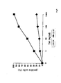
- Tat RGD sequence is key for the internalisation of active Tat by these cells through the ⁇ 5 ⁇ 1 and ⁇ v ⁇ 3 integrin receptors.
- antibodies or competitor ligands blocking these integrins completely abolish or greatly reduce the uptake of picomolar-nanomolar concentrations of Tat, respectively. This uptake is very rapid, is dose-, cell maturation/differentiation- and time-dependent. Even more unexpectedly, we did not obtain similar results with other APC including monocytes, T cell blasts, or B cell blasts or non-activated endothelial cells.
- Tat is taken up only at much higher concentrations (micromolar range), through its basic region, by a non-receptor-mediated pathway (Frankel 1988; Mann 1991; Rusnati 1998; Tyagi 2001), this internalization pathway occurs with any cell type, and it is not maturation/differentiation-dependent.
- Tat in its biologically active form is absolutely required to observe all the above novel effects which do not occur when Tat is oxidized and inactivated.
- Tat has 7 cysteines and it is extremely sensitive to oxidation which, when occurring, causes the loss of native protein conformation and consequent loss of biological activity (Frankel 1989). Therefore, Tat is likely to lose its native conformation and activity when purified with procedures that are not specifically designed at maintaining this protein in its native form.
- the inventor believes that full-length, wild type, biologically active Tat from any HIV variant or its fragments or derivates containing the RGD region can be used as a potent carrier for the selective delivery of molecules to specific cell types expressing the integrins recognized by the Tat RGD region (Barillari 1993, 1999a and 199b; Ensoli 1994).
- the inventor believes that the capability of biologically active Tat or its fragments or derivatives containing the RGD sequence of targeting these APC and of driving Th-1 type cellular responses will offer a unique opportunity to induce a potent immune response against not only Tat but also against other antigens delivered by or with Tat.
- biologically active Tat is proposed as antigen, adjuvant and deliver system of different antigens or combinations of antigens for vaccination against different infectious diseases (not only HIV/AIDS) and tumors, or for multivalent vaccination against one or more infectious diseases.
- biologically active Tat can be used to carry to DC, endothelial cells and macrophages any kind of active molecules, including antigens or drugs for the treatment of infectious, inflammatory and angiogenic diseases and tumors, differently from our previous patent application where this use was not described.
- biologically active Tat for the diagnosis and intervention of infections, inflammation, angiogenesis and tumor growth and metastasis as a diagnostic tool to identify in blood and tissues cells expressing RGD-binding integrins.
- biologically active Tat is claimed as a module which comprises different functions contemporarily, i.e. it is an antigen, an adjuvant and a carrier to deliver active compounds to specific tissues.
- This unexpected properties make biologically active Tat suitable for different applications in different infectious diseases (not only AIDS), inflammatory and angiogenic diseases and tumors.
- biologically active Tat, fragments or derivatives thereof, containing the RGD sequence acts with at least one of the following actions: as antigen, and/or adjuvant and/or delivery system for specific APC, including DC, activated endothelial cells and macrophages and claims that it can be exploited for preventive and therapeutic vaccination and/or drug delivery for the prevention and treatment of HIV/AIDS, other infectious diseases, inflammatory and angiogenic diseases and tumors, but also as a diagnostic tool to identify DC, activated EC and macrophages in vivo.
- HIV-1 Tat biologically active HIV-1 Tat, fragments or derivatives thereof, alone or combined with one or more other HIV antigens, (including Rev, Nef, Gag, Pol, Env, Vpu, Vpx, Vpr, or Vif) and/or with other antigens (for example, but not limited to, antigens from intracellular pathogens such as viruses, mycobacterium tuberculosis, candida, malaria, or from tumor cells such as those from lung, colon, breast, prostatic cancer) in the form of peptides, proteins or DNA encoding them, to target in vitro and in vivo antigen-presenting cells expressing RGD-binding integrin receptors, including dendritic cells, endothelial cells and macrophages, for preventive and therapeutic vaccination or treatment against HIV/AIDS, other infectious diseases and tumors.
- HIV antigens including Rev, Nef, Gag, Pol, Env, Vpu, Vpx, Vpr, or Vif
- antigens for example, but not
- Another object is the use of biologically active HIV-1 Tat, fragments or derivatives thereof, as adjuvant to activate or enhance in vitro and in vivo the antigen-presenting function of cells expressing RGD-binding integrin receptors including dendritic cells, endothelial cells and macrophages and to induce Th-1 type immune responses against HIV/AIDS, other infectious diseases and tumors.
- Another object is the use of biologically active Tat, fragments or derivatives thereof, to deliver in vitro and in vivo one or more HIV antigens and/or other antigens (as defined in the above) to antigen-presenting cells expressing RGD-binding integrin receptors, including dendritic cells, endothelial cells and macrophages in order to induce immune responses for preventive and therapeutic vaccination or treatment of HIV/AIDS, other infectious diseases, inflammatory and angiogenic diseases and tumors.
- Another object is the use of biologically active Tat, fragments or derivatives thereof, to deliver in vitro and in vivo one or more HIV antigens and/or others antigens (as defined in the above) to antigen-presenting cells expressing RGD-binding integrin receptors, including dendritic cells, endothelial cells and macrophages or to deliver intracellularly or to the cell membrane therapeutic compounds (such as, but not limited to, antiviral compounds, anti-inflammatory drugs, anti-angiogenic molecules, cytotoxic anti-tumor drugs or immunomodulating molecules such as, for example chemokines or cytokines, or antibodies) with or without the presence of support particles (such as, but not limited to, microparticles, nanoparticles, liposomes and other particulated delivery systems such as the ones described in Suiter 1991 and Takeuchi, 2001) for preventive and therapeutic vaccination or treatment of HIV/AIDS, other infectious diseases, inflammatory and angiogenic diseases and tumors.
- HIV antigens and/or others antigens
- Another object is the use of biologically active Tat, fragments or derivatives thereof, fused to other proteins or peptides or support particles (as defined in the above) to deliver in vitro and in vivo HIV and other antigens (as defined in the above) or therapeutic compounds (as defined in the above) to antigen presenting cells expressing RGD-binding integrin receptors including dendritic cells, endothelial cells and macrophages for combined preventive and therapeutic vaccination or treatment of HIV/AIDS, other infectious diseases, inflammatory and angiogenic diseases and tumors.
- Another object is the use of biologically active Tat, fragments or derivatives thereof, to target in vitro and in vivo antigen-presenting cells and other cell types capable of taking up Tat via the integrin-mediated pathway, and/or other uptake pathways conferring a selective uptake, in order to deliver antigens therapeutic or molecules (as defined in the above) for preventive and therapeutic vaccination or treatment of HIV/AIDS, other infectious diseases, inflammatory and angiogenic diseases and tumors.
- Another object is the use of biologically active Tat, fragments or derivatives thereof, alone or combined with other antigens (as defined in the above), adjuvants (such as, but not limited to, Alum, RIBI, ICOMS, CpG sequence, Lipopeptides) or therapeutic molecules or support particles (as defined in the above) administered by the parenteral (subcute, intramuscular, intradermic) or mucosal (vaginal, rectal, oral, nasal) or topic route for preventive and therapeutic vaccination or treatment against HIV/AIDS, other infectious diseases inflammatory and angiogenic diseases and tumors.
- adjuvants such as, but not limited to, Alum, RIBI, ICOMS, CpG sequence, Lipopeptides
- therapeutic molecules or support particles as defined in the above administered by the parenteral (subcute, intramuscular, intradermic) or mucosal (vaginal, rectal, oral, nasal) or topic route for preventive and therapeutic vaccination or treatment against HIV/AIDS, other infectious diseases inflammatory and
- Another object is the use of biologically active Tat, fragments or derivatives thereof to deliver in vitro and in vivo antigens (as defined in the above) or therapeutic molecules (as defined in the above) within or attached to support particles (as defined in the above), to antigen-presenting cells expressing RGD-binding integrin receptors including dendritic cells, endothelial cells and macrophages, for preventive and therapeutic vaccination or treatment against HIV/AIDS, other infectious diseases, inflammatory and angiogenic diseases and tumors.
- Another object is the use of biologically active Tat, fragments or derivatives thereof, to deliver in vitro and in vivo expression vectors including plasmid DNA and bacterical or virus vectors expressing one or more HIV antigens and/or other antigens (as defined in the above), in the presence or absence of support particles (as defined in the above), to antigen presenting cells expressing RGD-binding integrin receptors, including dendritic cells, endothelial cells and macrophages for preventive and therapeutic vaccination or treatment against HIV/AIDS, other infectious diseases, inflammatory and angiogenic diseases and tumors.
- Another object is the use of tat DNA or biologically active Tat protein, fragments or derivatives thereof, fused or combined with DNA coding for other HIV antigens and/or other antigens (as defined in the above), with or without support particles (as defined in the above), for combined preventive and therapeutic vaccination or treatment of HIV/AIDS, other infectious diseases, inflammatory and angiogenic diseases and tumors.
- Another object is biologically active HIV Tat or tat DNA, fragments or derivative thereof, combined or fused with antigens (as defined in the above), therapeutic molecules (as defined in the above), adjuvants (as defined in the above), or support particles (as defined in the above) such combination or fusion being defined as the association by means of chemical or physical interactions, or any other interactions, in any combination, such as, for example, but not limited to, the absorption of Tat and a DNA plasmid on nanoparticles; the inclusion of Tat and a synthetic drug in the same pharmaceutical preparation; the association of Tat or a fragment or a derivative thereof with a peptide by chemical crosslinking or by other means; the fusion of Tat, fragment or derivative thereof, with another protein or another peptide upon their expression in bacteria or eucariotic cells through chimeric DNA, where the DNA sequences encoding for the above polypeptides have been fused together using recombinant DNA technologies.
- Another object is the use of biologically active Tat protein, tat DNA, fragments or derivates thereof, as in the above for vaccination or therapeutic treatment by the parentheral (intradermic, intramuscular, subcute), mucosal (oral, nasal, vaginal, rectal) or topic route.
- Another object is the use of biologically active Tat, fragments or derivatives thereof to identify DC, macrophages and/or activated endothelial cells for diagnostic purposes, in HIV/AIDS, other infectious diseases, inflammatory and angiogenic diseases and tumors.
- Tat peptides from any HIV variant (HIV-1, HIV-2 and other types and subtypes) comprising, alone or associated, the RGD domain (aa 73 to 86 in the HTLV-IIIB, clone BH-10; aa 74 to 84; aa 75 to 83; aa 76 to 82; aa 77 to 81; aa 77 to 82; aa 77 to 83; aa 76 to 83); the cystein-rich domain (aa 22 to 37 in the HTLV-IIIB, clone BH-10); the basic domain (aa 48 to 61 in the HTLV-IIIB, clone BH-10), combined or not with other HIV-1 Tat peptides including the core domain (aa 38 to 47 in the HTLV-IIB, clone BH-10) and/or the amminoterminal region (aa 1 to 20 in the HTLV-IIIB
- fragments of biologically active Tat are defined as nucleotide sequences from any HIV variant (HIV-1, HIV-2 and other types and subtypes) comprising, alone or associated, the RGD domain (sequence coding for aa 73 to 86 in the HTLV-IIIB, clone BH-10; sequence coding for aa 74 to 84 in the HTLV-IIIB, clone BH-10; sequence coding for aa 75 to 83 in the HTLV-IIIB, clone BH-10; sequence coding for aa 76 to 82 in the HTLV-IIIB, clone BH-10; sequence coding for aa 77 to 81 in the HTLV-IIIB, clone BH-10; sequence coding for aa 77 to 82 in the HTLV-IIIB, clone BH-10; sequence coding for aa 77 to 83 in the HTLV-IIIB, clone BH-10; sequence
- Another object are fragments of Tat from any HIV variant (HIV-1, HIV-2 and other HIV types and subtypes) that comprise one or more T-cell epitopes in their amino acid sequences (HTLV-IIIB, clone BH-10 or 89.6).
- Another object are fragments of Tat from any HIV variant (HIV-1, HIV-2 and other HIV types and subtypes) that comprise one or more T-cell epitopes in their nucleotide sequences (HTLV-IIIB, clone BH-10 or 89.6).
- Tat mutants of the HTLV-IIIB, clone BH-10, variant selected among that ones having the following nucleotide sequences, or part of them: Nucleotide sequence of cys22 mutant and nucleotide sequence of lys41.
- Another object of the present invention is the use of Tat protein acting and combined as above described to produce medicaments to cure affections in the group of infectious diseases, inflammatory and angiogenic diseases, tumors.
- Another object of the invention is the use of Tat protein acting and combined as above described to produce a diagnostic kit.
- the biologically active HIV-1 Tat is defined as a protein capable of 1) entering and localizing in the nuclei of activated endothelial cells or DC, 2) activating the proliferation, migration and invasion of KS cells and cytokine-activated endothelial cells, 3) activating virus replication when added to infected cells as measured by a) the rescue of Tat-defective proviruses in HLM-1 cells after the addition of exogenous protein and/or b) the transactivation of HIV-1 gene expression in cells transfected with a HIV-1 promoter-reporter plasmid, and 4) inducing in mice the development of KS-like lesions in the presence of angiogenic factors or inflammatory cytokines.
- biologically active Tat as a module with at least one or more of the following features: antigen, adjuvant and delivery system.
- Tat protein is oxidized and/or inactivated, it is not suitable for the purposes of the present invention. In fact, only biologically active, but not oxidized or inactivated, Tat protein is very efficiently, rapidly and selectively taken up by MDDC, macrophages and cytokine-activated endothelial cells, in a dose-, time-, and maturation/differentiation-dependent fashion. Uptake of Tat occurs by at least two pathways depending upon the concentration of the protein.
- Tat At picomolar-nanomolar (0.01-1000 ng/ml) Tat concentrations, uptake of Tat is mostly mediated by the ⁇ 5 ⁇ 1 and ⁇ v ⁇ 3 receptors through the interaction with the RGD sequence of the protein, while at higher concentrations of Tat an integrin-independent pathway, mediated by the binding of Tat basic region to HSPG, is predominant. Efficient uptake of Tat is observed only with these APC and not with TCB, BLCL, monocytes or non-activated endothelial cells.
- Tat Upon uptake, Tat induces maturation and activation of MDDC including an increase of the expression of MHC and costimulatory molecules, and production of Th-1 cytokines (TNF- ⁇ , IL-12) and ⁇ chemokines [Rantes, macrophage inflammatory protein (MIP)-1 ⁇ , MIP-1 ⁇ ]. All these effects are lost when Tat is oxidized and inactivated. Further, active Tat, but not its oxidized counterpart, enhances both allogeneic and antigen-specific presentation by MDDC, increasing T cell-specific immune responses against heterologous antigens.
- active Tat favours its own presentation and that of other antigens and the induction of specific immune responses, and can also be used to deliver active molecules to these cells.
- active Tat functions as an antigen, adjuvant and delivery system capable of inducing Th-1 immune responses against both itself and other antigens by a specific targeting of the most efficient antigen-presenting cells and by driving specific immune responses.
- Tat can selectively deliver to these cells active molecules. Therefore, biologically active Tat can be used not only as an antigen but also as a Th-1 type adjuvant for other antigens to induce immune responses against pathogens or tumors, and as a delivery system for DC, macrophages and activated endothelial cells or to identify these cells during infections, angiogenesis, inflammatory diseases and tumors.
- Tat is used as the active principle to induce immune responses, to adjuvate the immune responses to other antigens and to deliver active molecules to specific antigen presenting cells including DC, macrophages and cytokine-activated endothelial cells for single or combined prophylactic or therapeutic vaccination or treatment or diagnosis of HIV/AIDS, other infectious diseases, inflammatory and angiogenic diseases and tumors.
- specific antigen presenting cells including DC, macrophages and cytokine-activated endothelial cells
- the inventor first found that MDDC have the specific capability of taking up very efficiently soluble active Tat at picomolar-nanomolar concentrations and found that this is a very rapid process with a peak after 5 to 10 min, depending on the concentration of Tat given to the cells.
- the uptake of active Tat by TCB or BLCL is very poor, requiring micromolar concentrations of Tat (10 ⁇ g/ml) and much longer periods of time of incubation and, even under these conditions, most Tat remains bound to the cell surface and does not enter cells.
- active Tat is also rapidly processed by MDDC, as indicated by the reduction of the intracellular staining after 30 min of incubation.
- Mature MDDC are able to take up Tat 10- to 100-fold more efficiently than immature cells, as indicated by the values of intracellular staining observed with low concentrations of Tat (100 ng/ml) as compared to those observed with 1 or 10 ⁇ g/ml of Tat with immature cells.
- Tat uptake requires a native conformation and full biological activity of the protein.
- oxidation and inactivation of Tat by exposure to light and air abolishes or markedly reduces (by approximately 100-fold) the uptake observed with active Tat by MDDC.
- the type of uptake by MDDC observed with oxidized Tat is similar or identical to that of native Tat with TCB and BLCL.
- the inventor found that the uptake of low concentrations of Tat is mediated by specific integrins ( ⁇ 5 ⁇ 1, ⁇ v ⁇ 3) binding to the RGD region of Tat through a receptor-mediated endocytic pathway, whereas at higher Tat concentration the uptake is mediated by the Tat basic region binding HSPG.
- Monocytes are not efficient in taking up Tat, whereas macrophages take up Tat more efficiently with a dose and time kinetic closer to that of DC indicating that differentiation of monocytes to either DC or macrophages induces selective and efficient mechanisms of Tat uptake.
- endothelial cells activated by IFN ⁇ , IL-1 ⁇ and TNF- ⁇ bind and take up native Tat very efficiently and in fashion similar to MDDC. Tat uptake by activated endothelial cells also occurs via the
- RGD domain of Tat that binds the ⁇ v ⁇ 3 and ⁇ 5 ⁇ 1 integrins [the classical receptors for vitronectin (VN) and fibronectin (FN)] and at high Tat concentration via the basic region of Tat that binds HSPG of the cell surface and extracellular matrix.
- integrin antagonists block uptake of pico-nanomolar but not higher Tat concentrations.
- uptake of picomolar-nanomolar concentrations of Tat is inhibited by the Tat peptide containing the RGD sequence, while uptake of higher concentrations of Tat is inhibited by the peptide encompassing the basic region of the protein.
- This second pathway may be identical to the receptor-independent pathway observed previously (Allen 2000; Barillari 1999b), via binding to a low affinity site, such as the interaction of the basic region of Tat with cell surface HSPG.
- Tat RGD region selectively targets cells expressing RGD-binding integrins receptors such as DC, endothelial cells and macrophages. Further, via this specialized pathway active Tat is very efficiently taken up.
- the inventor refers to a pathway that requires 1) at least one (RGD) or two Tat domains (basic and RGD), 2) the biological activity of the protein, 3) the native conformation of the protein and 4) integrin membrane receptors.
- RGD at least one
- TCB TCB
- BLCL BLCL
- monocytes or non-activated endothelial cells indicating that Tat can target and deliver antigens or cargos to specific cell types that are key for the immune response against pathogens and tumors or to treat inflammatory and angiogenic diseases or tumors.
- the inventor stresses that according to her, biologically active Tat, but not oxidized Tat, upon binding to specific integrin receptors enters DC, endothelial cells and macrophages and even more importantly, promotes MDDC maturation and function.
- active Tat induces a dose-dependent enhancement of the surface expression of human leukocyte antigens (HLA)-ABC, HLA-DR, CD40, CD80, CD86 and CD83 on MDDC. This effect is observed with native but not with oxidized and inactivated Tat.
- HLA human leukocyte antigens
- active Tat induces a dose-dependent increase of the production of both IL-12 and TNF- ⁇ , cytokines essential for driving a Th-1 type response (Romagnani 1997), and of the ⁇ -chemokines RANTES, MIP-1 ⁇ and MIP-1 ⁇ which are key players in the effector phase of the lymphocyte response (Moser 2001). Importantly, in both cases the levels are comparable to those induced by the known activator LPS. Again, oxidized and inactivated Tat does not induce these effects. Active Tat also enhances the antigen presenting function of MDDC augmenting the proliferative response of T cells to allogeneic and recall antigens.
- Th-1 responses and CTL controls infections by intracellular pathogens as well as tumor growth
- native Tat protein or tat DNA can be exploited to drive or to increase Th-1 immune responses and CTL activity also against other HIV antigens or non-HIV antigens to support an effective and long-lasting immunity for preventive or therapeutic vaccination as well as to deliver active molecules to DC, activated endothelial cells and macrophages for the treatment of infectious diseases, inflammatory and angiogenic diseases or tumors.
- activated endothelial cells as well as macrophages and DC, are present in inflammatory and angioproliferative diseases, tumors and infectious diseases representing specific target for Tat which can deliver to them antigens, inhibitory compounds or any molecule useful for preventive or therapeutic vaccination or treatment or for diagnostic purposes.
- HIV-1 Tat biologically active HIV-1 Tat, derivates or fragments thereof, alone or combined with other molecules including proteins, peptides, nucleic acids or support particles, can be used:
- HIV-1 Tat protein in its biologically active form has the following amino acid sequence (SEQ ID NO. 2): and HIV-2 Tat protein in its biologically active form, has the following amminoacid sequence, in which the RGD sequence has been inserted, for example, but not limited to, in position between amminoacid 92 and 93 of the original sequence (SEQ ID NO. 100): or the threonine at position 105 has been deleted (SEQ ID NO. 102): and any other Tat variant of any HIV-1 and HIV-2 subtype with a sequence homology greater than 50% to SEQ ID NO. 2, 100 or 102; preferably greater than 60%, more preferably greater than 70%, more preferably greater than 80%, more preferably greater than 90%. and at picomolar to nanomolar concentrations (from 0.01 ng/ml to 1 ⁇ g/ml, preferably 0.1 ng/ml to 100 ng/ml) is able to verify at least one of the following points:
- points (i) or (ii), preferably both, should be verified, more preferably point (i) or (ii) or both in combination with point (iii) a) and/or (iii) b) should be verified. The best results will be obtained when all (i) to (vi) points are verified.
- HIV-1 tat DNA refers to the following nucleotide sequence (SEQ ID NO. 1): and HIV-2 tat DNA refers to the following nucleotide sequence in which the sequence encoding the RGD has been inserted, for example, but not limited to, between nucleotides 276 and 277 of the original sequence (SEQ ID NO. 99): or the sequence ACC at position 322-324 encoding a threonine has been deleted (SEQ ID NO. 101): and any other variant of any HIV type and subtype with a sequence homology greater than 40% to SEQ ID NO. 1, 99, or 101 preferably greater than 50%, preferably greater than 60%, more preferably greater than 70%, more preferably greater than 80%, more preferably greater than 90%.
- fragment of biologically active Tat are defined as Tat peptides, which correspond to the DNA sequence, from any HIV variant (HIV-1, HIV-2 and other types and subtypes) containing, alone or associated: the RGD domain (sequence coding for aa 73 to 86 in the HTLV-IIIB, clone BH-10, as a reference, SEQ ID NO. 3 and corresponding amminoacid sequence, as a reference, SEQ ID NO.4; sequence coding for aa 74 to 84, SEQ ID NO. 5 and corresponding amminoacidic sequence, as a reference, SEQ ID NO. 6; sequence coding for aa 75 to 83, SEQ ID NO.
- HIV-1 Tat peptides including the core domain (sequence coding for aa 38 to 47 in the HTLV-IIB, clone BH-10, as a reference, SEQ ID NO. 23 and corresponding amminoacidic sequence, as a reference, SEQ ID NO. 24) and/or the amminoterminal region (sequence coding for aa 1 to 20 in the HTLV-IIIB, clone BH-10, as a reference, SEQ ID NO. 25 and corresponding amminoacidic sequence, as a reference, SEQ ID NO. 26).
- core domain sequence coding for aa 38 to 47 in the HTLV-IIB, clone BH-10, as a reference, SEQ ID NO. 23 and corresponding amminoacidic sequence, as a reference, SEQ ID NO. 24
- amminoterminal region sequence coding for aa 1 to 20 in the HTLV-IIIB, clone BH-10, as a reference,
- T-cell epitopes according to the invention are preferably the following, in their nucleotide sequence and corresponding amino acid sequences (HTLV-IIIB, clone BH-10 or 89.6, as a reference):
- Epitope 1 (aa 1-20):
- “derivatives of Tat” include Tat mutants of the HTLV-IIIB, clone BH-10 variant, selected among the ones having the following:
- infectious diseases include those caused by human or animal herpesviruses, hepatitis viruses, Mycobacterium Tuberculosis, Malaria plasmodia, Candida, Lysteria, Influenza virus and other infections caused in humans or animals by other intracellular or extracellular pathogens, including but not limited to, sexual infectious diseases, endocarditis, urinary tract infections, osteomyelitis, cutaneous infections, or streptococcus and staphylococcus infections, pneumococcus infections, tetanus, meningococcus infections, tuberculosis, malaria, candidosis, infections by Helicobacter, salmonella, syphilis, herpetic infections (including varicella, mononucleosis and Epstein-Barr-derived infections, human herpesvirus-8 infection, cytomegalovirus, herpes labialis and genitalis), hepatitis virus infection (A, B, C, D, G), papilloma
- inflammatory diseases are defined as an allergy or inflammation associated or not with a viral, bacterial or parasitic infection, including but not limited to immune-mediated cutaneous diseases, Lupus erythematous systemic, rheumatoid arthritis, systemic sclerosis, dermatomiositis, Sjögren syndrome, Good pasture syndrome, vasculitis, sarcoidosis, osteoarthrosis, infectious arthritis, psoriasis, Chron disease, rectocolitis ulcerosus, tyroiditis, scleroderma, allergic diseases.
- immune-mediated cutaneous diseases Lupus erythematous systemic, rheumatoid arthritis, systemic sclerosis, dermatomiositis, Sjögren syndrome, Good pasture syndrome, vasculitis, sarcoidosis, osteoarthrosis, infectious arthritis, psoriasis, Chron disease, rectocolitis ulcerosus, tyroiditis, scleroderma, allergic diseases.
- angiogenic diseases are defined as non-neoplastic angioproliferative diseases including diabetic retinopathy, retrolental fibroplasia, trachoma, vascular glaucoma, psoriasis, immune inflammation, non-immune inflammation, atherosclerosis, excessive wound healing, angiodermatitis, colon angiodisplasia, angioedema and angiofybromas.
- tumors are defined as benign and malignant tumors including tumors of soft tissues, bones, cartilages and blood, such as, but not limited to, Kaposi's sarcoma and other neoplasia of the skin, lung, breast, gut, liver, pancreas, endocrine system, uterus, ovary, sarcomas, acute and chronic leukemia, and neoplasia of lymphatic cells.
- therapeutic compounds are defined as other antigens (proteins, peptides or DNA) or active molecules
- support particles are defined as, but not limited to, microparticles, nanoparticles, liposomes and other particulated delivery systems.
- HIV antigens includes Rev, Nef, Gag, Pol, Env, Vpu, Vpx, Vpr, Vif from any HIV variant (HIV-1, HIV-2 and other types and subtypes).
- non-HIV antigens or other antigens includes any molecule or moiety recognized by immune cells with the exclusion of HIV antigens.
- Tat according to the invention can be used to treat: infectious diseases, inflammatory diseases, angiogenic diseases, tumors.
- the Tat combinations as in the above can be mixed with adjuvants, diluents, eccipients, carriers and other substances known in the art to make medicaments for the scopes expressed.
- medicaments can be obtained in form of tablets, pills, sprays, injectable solutions, suspensions, powders, creams, ointments, for parenteral (subcute, intramuscular, intradermic), mucosal (vaginal, rectal, oral, nasal) administration or topical route with routes depending on the kind of disesase and type of formulation.
- kits can be devised for scopes of identification and/or isoiation/purification of such cells in blood and tissues from pathological or physiological conditions including infections, inflammation, angiogenesis and tumors.
- kits may advantageously comprise: the Tat module including biologically active Tat, fragments or derivatives thereof, combined or fused to a tag such as, but not limited to, a fluorescent or color chromophor, a reactive molecule such as, for example, an enzyme, an antibody, or a chemical or a peptide that can be detected by means of its reactive properties such as, for example, the conversion of a substrate to its color insoluble forms by alkaline-phosphatase, the reaction of biotin with streptavidin, or biotin with anti-biotin antibodies.
- a tag such as, but not limited to, a fluorescent or color chromophor, a reactive molecule such as, for example, an enzyme, an antibody, or a chemical or a peptide that can be detected by means of its reactive properties such as, for example, the conversion of a substrate to its color insoluble forms by alkaline-phosphatase, the reaction of biotin with streptavidin, or biotin with anti-biotin antibodies.
- Active Tat is efficiently taken up by MDDC but not by TCB and BLCL
- Fig. 1 shows the results of a representative donor whose levels of Tat uptake represented the median of the values obtained from 14 donors tested.
- Tat uptake by MDDC was very efficient and occurred in a dose-and time-dependent fashion (Fig. 1A). Uptake was already evident with the lowest dose of Tat utilized (0.1 ng/ml). Regardless of the Tat concentration tested, the level of staining always peaked after 5 min of incubation and was reduced after 60 min, likely due to the processing of the protein.
- Immature MDDC take up antigens by phagocytosis and pinocytosis (Bankereau 1998; Bell 1999). Mature DC lose these activities while acquiring strong antigen presentation capability.
- MDDC were induced to maturate with LPS.
- mature MDDC expressed higher levels of HLA-DR, CD83 and CD86 surface markers. Both immature or mature cells were then used for the uptake experiments.
- Tat uptake was highly increased by MDDC maturation at all protein concentrations tested (Fig. 2A). In fact, incubation of mature MDDC with low Tat concentrations gave levels of intracellular staining similar to those observed in immature cells with the highest doses of Tat (Fig. 1A and 2A).
- Tat contains 7 cysteins, and is extremely sensitive to oxidation which causes conformational changes and loss of biological activity.
- the protein was exposed to air and light, the loss of biological activity tested (Table I), and active or inactive Tat protein were then compared in the uptake experiments.
- Tat must have native conformation and biological activity for efficient uptake by MDDC.
- Biological activity of the native and oxidized protein was then tested (at doses from 500 to 10,000 ng / ml) by measuring the capacity of rescuing the replication of Tat-defective HIV provirus in the HLM-1 cell line (Ensoli 1993; Chang 1997) or of inducing CAT activity in the HL3T1 cell line containing the LTR-CAT construct (Felber 1988). Values of the p24 (pg / ml) from culture supernatants of HLM-1 cells and CAT activity from HL3T1 cell extracts (% acetylation / 100 ⁇ g of protein) are reported.
- Tat uptake by MDDC is inhibited by both anti- ⁇ 5 ⁇ 1 and anti- ⁇ v ⁇ 3 monoclonal antibodies and the block is complete in the presence of both antibodies (Fig. 3 A, B).
- ⁇ 5 ⁇ 1 and ⁇ v ⁇ 3 integrins mediate Tat entry in MDDC.
- This pathway of uptake is predominant at picomolar-nanomolar concentrations of the protein, whereas at higher Tat concentrations block of Tat entry is still evident but is not complete (fig. 3A and B).
- Active Tat is efficiently taken up in a dose- and time-dependent fashion by both MDM and MDDC but not by monocytes.
- monocytes Since MDDC derive from monocytes and since monocytes and MDM are known to efficiently present antigens to lymphocytes, uptake of active Tat from these cells was analysed and compared to that of MDDC from the same donors. Monocytes were purified from peripheral blood and induced to differentiate to MDDC or MDM (as described in Figure 1 and Figure 5). As shown in Figure 5, monocytes took up active Tat only at the highest doses (45% and 67% at 1,000 and 10,000 ng/ml, respectively) with evidence of binding of the protein to cell surface (19% and 33%, respectively, on non-permeabilized cells).
- MDM took up Tat more efficiently than monocytes, with staining detectable at relatively low doses (32% and 43% at 10 ng/ml and 100 ng/ml, respectively) up to the highest dose of Tat tested (72% at 10,000 ng/ml) when surface binding of the protein (30% on non-permeabilized cells) was observed.
- MDDC from the same donor took up Tat more efficiently with specific staining detected already at the lowest dose tested (0.1 ng/ml) than monocytes and MDM both at 10 min and 30 min (22% and 13%, respectively).
- 80% and 73% of MDDC were positive after 10 and 30 min of exposure to Tat, respectively.
- HUVEC To analyze the uptake of active Tat by endothelial cells, HUVEC were or not activated with inflammatory cytokines and used in uptake studies with rhodaminated Tat protein. After rhodamination Tat was active since it was still capable of promoting KS cell proliferation at the same concentration range as unlabelled Tat. This indicates that the attachment of the fluorescent label did not compromise its biological function. Cells were exposed to a wide range of Tat concentrations. In addition, to be consistent with uptake inhibition experiments (see below), cells were preincubated at 4° C for 2 hours with medium without fetal calf serum. This preincubation does not affect the subsequent uptake of rhodaminated Tat.
- rhodaminated Tat With rhodaminated Tat, the uptake and translocation of the protein to the nucleus or nucleoli of activated HUVEC (IC-HUVEC) became detectable within 15 minutes of incubation with 10 ng/ml rhodaminated Tat (Fig. 6).
- the intracellular Tat activity increased in a dose-dependent (Fig. 6) and time-dependent manner (Fig. 7 and 8).
- Rhodaminated BSA or buffer (negative controls) showed little or no uptake (Fig. 6, 7, 8).
- MDM possess specific mechanisms to take up Tat very efficiently.
- IC-HUVEC To verify whether the uptake of rhodaminated Tat protein by IC-HUVEC was occurring via a specific and saturable pathway(s), IC-HUVEC were incubated with 1 ⁇ g/ml unlabelled Tat prior to incubation with rhodaminated Tat at concentrations ranging from 10 ng/ml to 1 ⁇ g/ml. This procedure almost totally inhibited the uptake of rhodaminated Tat (Fig. 11 and Table II) with fluorescence levels comparable to those of the negative control (BSA). This indicates that uptake of Tat protein by IC-HUVEC is specific and saturable, suggesting that a receptor(s) is involved in this process. Table II.
- Tat uptake by IC-HUVEC was mediated by the same integrins identified with MDDC
- inhibition experiments were performed by preincubating IC-HUVEC with the physiological ligands for these receptors, FN or VN, or by preincubating the cells with monoclonal antibodies directed against the RGD binding regions of the ⁇ 5 ⁇ 1 and ⁇ v ⁇ 3 receptors. The cells were then incubated with rhodaminated Tat at concentrations ranging from 10 ng/ml to 1 ⁇ g/ml.
- IC-HUVEC were incubated with 1 ⁇ g/ml rhodaminated Tat.
- a very intense fluorescence signal was seen in the cells within 15 minutes (Fig. 6 panel D, Fig. 8 panel A and Fig. 14 panel B).
- prior treatment of the cells with monoclonal antibodies against ⁇ 5 ⁇ 1 (Fig. 14 panel C) or ⁇ v ⁇ 3 (Fig. 14 panel D) showed some inhibition of Tat uptake, but lower than that observed with 10 or 100 ng/ml of Tat.
- Tat domains mediating uptake of active Tat
- Tat domains mediating uptake of active Tat Competitor Rhodaminated Tat (ng/ml) Fluorescence intensity Buffer 0 - Buffer 10 ++ (11-24) Tat 10 ++ (46-60) Tat 10 ++ (65-80) Tat 10 - Buffer 100 +++ (11-24) Tat 100 +++ (46-60) Tat 100 +++ (65-80) Tat 100 -/+ Buffer 1000 ++++ (11-24) Tat 1000 ++++ (46-60) Tat 1000 ++ (65-80) Tat 1000 +++ IC-HUVEC were incubated on a rotating device for 2 h at 4°C with an excess (10 ⁇ g / ml) of (11-24)Tat, (46-60)Tat or (65-80)Tat in serum free medium.
- Peptide resuspension buffer PBS-0.1%BSA
- rhodaminated Tat was added to the cells at 10, 100 and 1000 ng / ml and cells were kept for 15' at 37°C in a CO 2 incubator. Cells were then fixed in ice-cold acetonemethanol (1:1). The uptake and the cellular distribution of Tat were observed and photographed using fluorescence microscopy, as described in the legend to figure 6.
- Tat enhances the expression of HLA and costimulatory molecules on MDDC A.
- Donor Code Medium (MFI) Buffer (MFI) % increase vs control Tat ⁇ g/ml LPS 1 5 20 HLA-ABC B 19 392 332 55.4 55.7 78.3 74.0 B 24 291 309 6.1 49.2 49.2 140.9 B 26 304 297 -4.0 -4.7 15.8 32.6 B 38 130 127 31.5 54.3 63.8 168.5 B 53 276 272 -8.5 -6.3 2.2 48.9 B 55 251 259 2.7 11.2 13.9 9.2 HLA-DR B 19 145 192 1.0 -1.0 27.1 84.8 B 24 164 143 11.9 60.8 10.5 48.8 B 26 175 177 15.8 23.2 60.5 60.6 B 38 464 366 62.3 98.1 94.5 85.6 B 40 1040 1127 27.9 38.9 34.6 58.9 B 43 415 408 18.6 27.
- Cells were exposed for 18 h to native or oxidized and inactivated Tat, reconstitution buffer, complete medium, or LPS (10 ⁇ g / ml), stained with the following fluorochrome-conjugated monoclonal antibodies: FITC- or PE-conjugated IgG isotypes, FITC-conjugated anti- CD14 and -HLA-DR (Becton-Dickinson), FITC-conjugated anti-CD40, -CD80, -CD83, PE-conjugated anti-CD86 (Pharmingen, San Diego, CA) and then analyzed by flow cytometry.
- fluorochrome-conjugated monoclonal antibodies FITC- or PE-conjugated IgG isotypes
- FITC-conjugated anti- CD14 and -HLA-DR Becton-Dickinson
- FITC-conjugated anti-CD40, -CD80, -CD83 PE-con
- panel A the expression of the surface molecules on MDDC from 10 different donors is reported as the percentage increase of the mean fluorescence intensity (MFI) as compared to the MFI of cells incubated with Tat buffer (for Tat) or medium (for LPS).
- MFI mean fluorescence intensity
- panel B Data with oxidized versus native Tat (20 ⁇ g / ml) from a representative donor are shown in panel B.
- MDDC cultured with LPS were used as the positive control for the induction of HLA and costimulatory molecules.
- MDDC cultured in presence of native or oxidized Tat were always viable, not differing from those treated with medium or reconstitution buffer (data not shown).
- Incubation with active Tat induced a dose-dependent increase of the levels of IL-12 and TNF ⁇ , reaching, at the highest dose of Tat, an increase of 23-fold (p ⁇ 0.02) for IL-12 and 20-fold (p ⁇ 0.03) for TNF- ⁇ as compared to cells treated with buffer alone.
- Tat markedly enhanced, in a dose-dependent fashion, the production of RANTES (10-fold, p ⁇ 0.02), MIP-1 ⁇ (97-fold, p ⁇ 0.005) 0.005) and MIP-1 ⁇ • (15-fold, p ⁇ 0.01).
- the reconstitution buffer had no effects, whereas LPS (positive control) markedly enhanced the production of both cytokines and ⁇ -chemokines.
- oxidized and inactivated Tat did not increase the production of IL-12 or TNF- ⁇ .
- native and active Tat increases the production and secretion of Th-1 cytokines and ⁇ chemokines by MDDC.
- Active Tat enhances the presentation of heterologous antigens by MDDC and specific T cell responses
- the effect of active Tat on the antigen-specific presenting capacity of MDDC was evaluated by transiently treating the cells with the protein, reconstitution buffer or complete medium, and culturing them together with autologous lymphocytes in the presence of the recall antigen TT.
- untreated MDDC induced TT-specific proliferation of autologous lymphocytes in 2 out of the 3 donors analyzed (S.I. 15.4 and 7.3, respectively) and this effect was enhanced by their treatment with active Tat (S.I. 27.9 and 12.5, respectively), but not with reconstitution buffer (S.I. 13 and 5.9, respectively).
- Tat-treated MDDC did not induce lymphocyte proliferation to TT in the subject who did not respond to it.
- Tat can boost specific T cell responses to other antigens.
- Mucosal immunization with biologically active Tat alone or combined with other antigens or adjuvants in mice induces mucosal immunity and T cell responses against itself as well as against other antigens.
- mice by the nasal route with Tat alone or in the presence of a mucosal adjuvant is expected to induce mucosal immunization characterized by the induction of specific secretory IgA response locally and at distinct mucosal sites and a systemic immunization characterized by the presence of both humoral IgG and Th-1 type T cell immune responses including proliferative response to Tat, IFN ⁇ secretion and CTL responses determined as the killing of target cells or by IFN ⁇ -Elispot assays with cells from spleen and/or lymph nodes. Because of its potent adjuvant activity Tat is expected to increase both mucosal and systemic immune response to a nominal antigen when co-administered mucosally.
- intranasal immunization of Balb/c mice (age 8-12 weeks) with biologically active Tat protein (5 to 10 ⁇ g) with or without adjuvant for 3 to 4 times in 21-45 days will generate a Tat-specific systemic and mucosal immune response, with high titers of serum anti-Tat IgG (titers: ⁇ 1:10 5 ) and secretory IgA (vaginal titers: ⁇ 1:10 2 ). Further, it is expected that at least IgG, detected at the highest titers, will have the capability to neutralize biologically active Tat, as assessed by the rescue assay with HLM-1 cells.
- T-helper responses to Tat as assessed by lymphoproliferative response to biologically active Tat, are expected. Further, this helper response is expected to be of Th-1 type as assessed by secretion of IFN ⁇ but not IL-4 after specific stimulation with Tat in an Elispot assay or by ELISA measurement of IFN ⁇ and IL-4 content in the cell culture supernatant.
- anti-Tat CTLs are expected to be detected when lymphocytes from spleen or lymph nodes are cultured for 6 days with pools of Tat peptides (9-mers overlapping by 4 residues) and in the presence of exogenous IL-2.
- a specific lysis ⁇ 20% is foreseen when p815 murine mastocytoma cells are pulsed with the pools of Tat peptides (target) for 3-4 hours prior to addition to effector cells.
- secretion of IFN ⁇ by CD8 T cells in response to Tat protein and/or peptides thereof is expected in a standard Elispot assay.
- intramuscular immunization (3 inoculations in 6 weeks time period) of juvenile rhesus monkeys ( Macaca mulatta ) with DNA of HIV-1 tat (0.5 mg) alone or associated with HIV-1 env and SIV mac239 gag DNA (0.5 mg each) followed by 2 intramuscular boosts with either biologically active Tat protein (25 ⁇ g) and ISCOMs alone or associated with 25 ⁇ g of HIV-1 Env and 25 ⁇ g of SIV mac239 Gag proteins, is expected to generate a specific Th-1 immune response, including induction of specific antibodies, lymphoproliferative responses and CTL assessed by Elispot for IFN ⁇ , IL-2 and IL-4 to biologically active Tat protein and/or peptides thereof alone, or to biologically active Tat protein and/or peptides thereof and to HIV-1 Env and SIV mac239 Gag proteins and/or peptides thereof.
Landscapes
- Health & Medical Sciences (AREA)
- Life Sciences & Earth Sciences (AREA)
- Chemical & Material Sciences (AREA)
- Medicinal Chemistry (AREA)
- General Health & Medical Sciences (AREA)
- Organic Chemistry (AREA)
- Veterinary Medicine (AREA)
- Animal Behavior & Ethology (AREA)
- General Chemical & Material Sciences (AREA)
- Pharmacology & Pharmacy (AREA)
- Chemical Kinetics & Catalysis (AREA)
- Public Health (AREA)
- Nuclear Medicine, Radiotherapy & Molecular Imaging (AREA)
- Engineering & Computer Science (AREA)
- Bioinformatics & Cheminformatics (AREA)
- Virology (AREA)
- Immunology (AREA)
- Communicable Diseases (AREA)
- Oncology (AREA)
- Molecular Biology (AREA)
- Diabetes (AREA)
- Pulmonology (AREA)
- Ophthalmology & Optometry (AREA)
- Endocrinology (AREA)
- Biophysics (AREA)
- Genetics & Genomics (AREA)
- Epidemiology (AREA)
- Heart & Thoracic Surgery (AREA)
- Cardiology (AREA)
- Mycology (AREA)
- Rheumatology (AREA)
- Microbiology (AREA)
- Proteomics, Peptides & Aminoacids (AREA)
- Tropical Medicine & Parasitology (AREA)
- Urology & Nephrology (AREA)
- Biochemistry (AREA)
- Gastroenterology & Hepatology (AREA)
- Physical Education & Sports Medicine (AREA)
- Dermatology (AREA)
- Neurology (AREA)
Abstract
The present invention concerns a method for prophylactic and/or therapeutic vaccination and/or treatment and/or diagnosis of HIV/AIDS, other infectious diseases, inflammatory and angiogenic diseases and tumours which utilizes a biologically active HIV-1 Tat protein, fragments or derivates thereof, as a module with one or more of the following features: antigen, adjuvant and targeting-delivery system to specific antigen-presenting cells including dendritic cells, endothelial cells and macrophages. In particular, it is claimed that Tat can be used only in its biologically active form as an antigen, alone or combined with one or more other antigens, to prime or to boost protective immune responses against itself as well as other antigens and/or to deliver these antigen(s) as well as active compounds to dendritic cells, endothelial cells and macrophages, due to its capability of targeting these APC and of activating their maturation and functions and of increasing Th-1 type immune responses as an adjuvant. Therefore, due to these characteristics and to the distribution of these cells in the body (during physiological and pathological disorders), biologically active Tat, fragments or derivates thereof containing the RGD region, can be used for preventive, therapeutic and/or diagnostic purposes for HIV/AIDS, other infectious diseases, inflammatory and angiogenic diseases and tumors.
Description
- The present invention relates to the use of biologically active HIV-1 Tat, fragments or derivatives thereof, to target and/or to activate antigen-presenting cells, and/or to deliver cargo molecules for preventive or therapeutic vaccination and/or to treat infectious diseases, inflammatory and angiogenic diseases, and tumors.
- In particular the invention relates to methods for immunizing or treating humans or other mammals against infection by one or more pathogens, against inflammatory and angiogenic diseases or tumors, and is particularly concerned with the use of a biologically active HIV-1 Tat protein, fragments or derivatives thereof, as an antigen, and/or as an adjuvant and/or as a delivery system capable of targeting antigen presenting cells, including dendritic cells, macrophages and endothelial cells, and of delivering across the outer cell and nuclear membranes cargo molecules for preventive or therapeutic or diagnostic applications.
- Tat is a regulatory protein of human immunodeficiency virus type 1 (HIV-1) produced very early after infection and essential for virus gene expression, replication and infectivity (Arya 1985; Fisher 1986; Chang 1995). During acute infection of T cells by HIV, Tat is also released in the extracellular milieu and taken-up by neighbour cells (Frankel 1988; Ensoli 1990; Ensoli 1993; Chang 1997) where, according to the concentration, can increase virus infectivity. Specifically, upon uptake Tat can enhance, in infected cells, virus gene expression and replication (Frankel 1988; Ensoli 1993; Chang 1997), and, in uninfected cells, the expression of the β-chemokines receptors CCR5 and CXCR4 favouring transmission of both macrophage and T lymphocyte-tropic HIV-1 strains (Huang 1998; Secchiero 1999). Extracellular HIV-1 Tat protein is also responsible for the increased frequency and aggressiveness of Kaposi's sarcoma (KS) a vascular tumor particularly frequent in HIV-infected individuals (Friedman-Kien 1981; Safai 1985). In particular, previous work from our and other groups indicated that Tat cooperates with angiogenic and inflammatory cytokines that are highly expressed in KS patients (Samaniego 1998; Ensoli 1994) in inducing new blood vessels formation (angiogenesis) and the growth and locomotion of spindle shaped cells of endothelial cell origin (KS cells) and of activated endothelial cells (Barillari 1992; Albini 1995; Ensoli 1994). In particular, our data have shown that biologically active Tat binds through its RGD region the integrin receptors α5β1 and αvβ3 and that this interaction mediates the adhesion, growth and locomotion induced by Tat on KS cells and endothelial cells activated by inflammatory cytokines (Barillari 1993; Barillari 1999a and 1999b). In addition, Tat acts also as a chemotactic factor for these cell types as well as for monocytes and dendritic cells (DC) (Albini 1995; Benelli 1998; Lafrenie 1996; Mitola 1997).
- Consistent with these findings, the immune response to Tat has been shown to play a key role in controlling the progression of AIDS and AIDS-associated diseases. In fact, a Tat-specific immune response is present in HIV-1 infected subjects and simian immunodeficiency virus (SIV)-infected monkeys, and correlates inversely with progression to the symptomatic stage of the infection (Reiss 1990; Venet 1992; Rodman 1993; Froebel 1994; Re 1995; Van Baalen 1997; Zagury 1998; Addo 2001). Moreover, vaccination with biologically active Tat protein or tat DNA induces protection against SHIV89.6P virus replication and disease onset which correlates with the presence of Th-1 responses including specific cytotoxic T lymphocytes (CTLs) (Cafaro 1999; Cafaro 2000; Cafaro 2001, and PCT WO99/27958). The same protection data have been more recently observed with a tat-rev vaccine delivered with viral vectors in macaques (Osterhaus 2001). In contrast, a limited containment of the infection has been observed in monkeys vaccinated with inactivated Tat or Tat peptides, in which antibodies and T helper specific responses but no CTLs nor Th-1 responses had been induced (Goldstein 2000; Pauza 2000). Again, the repeated intradermal (i.d.) inoculation of monkeys with native and active Tat protein alone (in the absence of any adjuvant) at low doses (5-6 µg) selectively induced a Th-1 response and specific CTLs in the absence of any significant antibody production (Cafaro 1999 and PCT WO99/27958). These immunological results were recently confirmed in a new vaccination protocol in which native Tat alone was repeatedly inoculated i.d. in 4 monkeys (unpublished data), and are comparable to those induced by i.m. vaccination with tat DNA in a published (Cafaro 2001 and PCT WO99/27958) and in an ongoing study. Similarly, recent work performed in SIV-infected macaques indicate that anti-Tat CTLs are key to control early virus replication after primary infection and exert a selective immune pressure on the virus leading to the appearance of slowly replicating, less pathogenic escape mutants (Allen 2000). Finally, Tat is presented with major histocompatibility complex (MHC) class I antigen (Moy 1996; Kim 1997), hence inducing anti-Tat CTL (Cafaro 1999).
- Micromolar concentrations of recombinant Tat protein (often of unknown biological activity) or peptides encompassing the basic region of Tat have been shown to enter many different cell types (Frankel 1988; Mann 1991; Ensoli 1993; Chang 1997; Fawell 1994; Moy 1996; Kim 1997). The highly basic charge of Tat residues 48-57, in fact, enables the protein to bind to heparan sulphate proteoglycans (HSPG) that are present on the membrane of all cell types (Chang 1997; Rusnati 1998). After release from acutely infected cells, a fraction of extracellular Tat binds, through its basic residues, to the HSPG (Chang 1997). This protects extracellular Tat from proteolytic degradation, as previously found for several growth factors (reviewed in Raines and Ross, 1992). Upon the binding of its basic region to cell surface HSPG, Tat is internalised through a receptor-independent pathway (Frankel 1988; Rusnati 1998; Tyagi 2001). However, this internalisation mechanism requires high (micromolar) concentrations of Tat, occurs with any cell type and it is not sequence-specific. In fact, it has been shown that mutations of this region, which do not change its basic charge, do not affect the properties of the Tat basic region (Barillari 1999b). Similarly, the substitution of the Tat basic region with that of HIV rev or other genes does not change Tat properties. In this regard, the basic region of Tat has been shown to be very similar to the argininrich region carried by the members of the small family of proteins known as "penetratins", that are all capable of entering many cell types (Derossi 1998). In fact, arginin homopolymers have been shown to enter cells even more efficiently than Tat basic region (Derossi 1998).
- The property of the Tat basic region of being internalized by cells has been exploited to deliver foreign proteins to a variety of cell types (Fawel 1994; Wender 2000; and WO 0119393). To this purpose, foreign proteins have been conjugated or fused to the Tat basic region which has been used as a carrier for the protein to be transduced (Fawel 1994; Wender 2000; and WO 0119393). However, the inventor believes that due to the ubiquitous expression of HSPG, Tat basic region cannot be used for selective targeting, delivery and/or uptake of Tat by specific primary cell types, including antigen presenting cells (APC).
- APC initiate and drive the type of immune response upon encountering foreign molecules (Bancherau 1998; Bell 1999). Typical APC include monocyte-derived DC (MDDC), T cell blasts (TCB), B-Iymphoblastoid cell lines (BLCL) and monocytes-macrophages (Bancherau 1998; Bell 1999). In addition, when activated by inflammatory cytokines also endothelial cells acquire APC functions (Pober 1988). Among these inflammatory cytokines, interleukin (IL)-1, tumor necrosis factor (TNF) and interferon (IFN)γ are key for endothelial cell activation (Pober 1988). Exposure to these cytokines increases in endothelial cells the expression of α5β1 and αvβ3, that are among the several cell surface receptors binding Tat (Barillari 1993; Fiorelli 1999; Benelli 1998; Kolson 1993; Sabatier 1991; Vogel 1993; Boykins 1999; Ganju 1998; Milani 1996; Mitola 1997 and 2000; Weeks 1993; Albini 1996 and 1998; Chang 1997; Lafrenie 1996; Morini 2000; Rusnati 1998). Among all these APC, DC are the most efficient APC and are key to the induction of immune responses against infections and tumors (Banchereau 1998; Bell 1999). Their function is associated with a high expression of MHC and costimulatory molecules (CD40, CD80, CD86) and with the production of cytokines known to activate T lymphocytes, and β-chemokines. Upon encountering the antigens, DC undergo a maturation process characterized by an increase of costimulatory molecules expression and by a reduction of their phagocytic and pinocytic capability (Banchereau 1998; Bell 1999). Further, due to the upregulation of the homing receptor CCR7 and to the downregulation of CCR5, mature DC migrate to lymph nodes where they present antigens to T lymphocytes (Banchereau 1998; Bell 1999).
- Prior art indicates that the addition of Tat protein to DC blocks in these cells the extracellular calcium influx, the production of interleukin-12, and the uptake of apoptotic bodies (Zocchi 1997; Rubartelli 1997). As a result, it is predicted that profound impairment of important DC functions including antigen uptake, processing and presentation and induction of Th-1 responses should occur. Further, impairment of phagolysosomal fusion has been reported in peripheral blood monocytes upon exposure to Tat, suggesting impairment in this cell type of both microbicidal and antigen processing (and presentation) functions (Pittis 1996). Moreover, Tat has been reported to induce both monocytes/macrophages and lymphocytes to secrete IL-10 (Masood 1994; Badou 2000), while inhibiting IL-12 production in monocytes (Ito 1998). Finally, exposure of APC to Tat has been reported to impair their capability to organize cell clusters and to properly activate T cells (Mei 1997). Moreover, prior art indicates that Tat profoundly impairs also T cell functions including suppression of responses to mitogens anti-CD3 or specific antigens (Viscidi 1989; Benjouad 1993; Subramanyam 1993; Chirmule 1995; Wrenger 1996; Wrenger 1997; Zagury 1998), T cell hyperactivation (Ott 1997; Li 1997), and T cell apoptosis (Westendorp 1995; Li 1995; McCloskey 1997).
- Further, inoculation of biologically active Tat has been reported to be immunosuppressive in vivo (Cohen 1999). Part of the effects of Tat on the immune system have been related to upregulation by Tat of the chemokines receptors CCR5 and CXCR4 (Huang 1998; Secchiero 1999), or the direct interaction of Tat with the chemokine receptors CCR2 and CCR3 (Albini 1998a) or with other receptors including CD26 (Gutheil 1994), Flt-1 (Mitola 1997), KDR (Albini 1996; Morini 2000), that are expressed by immune cells, as well as by endothelial cells.
- Therefore, according to this previous art Tat is expected to drive a Th-2 type of immune response and/or to interfere with or abolish proper APC function and T cell activation.
- By contrast, our data, object of this patent application, indicate that biologically active Tat activates, rather than inhibiting, APC function and induces, rather than suppressing, Th-1 type immune responses against itself as well as other antigens. Specifically, our data show that Tat acts not only as an antigen but also as an adjuvant with potent immunomodulatory properties. These results are related with another unexpected finding, namely, that Tat is taken up very efficiently at picomolar-nanomolar concentrations by APC, including DC, macrophages and endothelial cells activated by inflammatory cytokines, through an integrin-receptor-mediated pathway. We have found, in fact, that Tat RGD sequence is key for the internalisation of active Tat by these cells through the α5β1 and αvβ3 integrin receptors. In fact, antibodies or competitor ligands blocking these integrins completely abolish or greatly reduce the uptake of picomolar-nanomolar concentrations of Tat, respectively. This uptake is very rapid, is dose-, cell maturation/differentiation- and time-dependent. Even more unexpectedly, we did not obtain similar results with other APC including monocytes, T cell blasts, or B cell blasts or non-activated endothelial cells. Therefore, these findings are completely novel since prior art indicates that Tat is taken up only at much higher concentrations (micromolar range), through its basic region, by a non-receptor-mediated pathway (Frankel 1988; Mann 1991; Rusnati 1998; Tyagi 2001), this internalization pathway occurs with any cell type, and it is not maturation/differentiation-dependent.
- Further, we have found that Tat in its biologically active form is absolutely required to observe all the above novel effects which do not occur when Tat is oxidized and inactivated. In fact, Tat has 7 cysteines and it is extremely sensitive to oxidation which, when occurring, causes the loss of native protein conformation and consequent loss of biological activity (Frankel 1989). Therefore, Tat is likely to lose its native conformation and activity when purified with procedures that are not specifically designed at maintaining this protein in its native form. Although established concepts in the field claim that biologically active Tat is toxic (Gallo 1999; Sabatier 1991; Kolson 1993; Westendrop 1995; Purvis 1995), by contrast, the highly purified, biologically active preparations of recombinant Tat utilized by the inventor has no cytotoxic nor pro-apoptotic effects on endothelial cells, DC, macrophages, other cell type tested, nor in vivo in mice or monkeys (Ensoli 1994; Barillari 1999a; Zauli 1993, 1995a and 1995b; Cafaro 1999, 2000 and 2001).
- Thus, the inventor believes that full-length, wild type, biologically active Tat from any HIV variant or its fragments or derivates containing the RGD region can be used as a potent carrier for the selective delivery of molecules to specific cell types expressing the integrins recognized by the Tat RGD region (Barillari 1993, 1999a and 199b; Ensoli 1994). Given the very large amount and ubiquitous distribution of DC, macrophages and endothelial cells in the human body, the inventor believes that the capability of biologically active Tat or its fragments or derivatives containing the RGD sequence of targeting these APC and of driving Th-1 type cellular responses will offer a unique opportunity to induce a potent immune response against not only Tat but also against other antigens delivered by or with Tat.
- These cell types are recruited and activated in infections, pathologic angiogenesis, inflammatory diseases and tumors, representing a specific target for Tat. This hypothesis is strongly supported by the successful previous work of the inventor with biologically active Tat as a vaccine to control HIV replication and to block disease onset (Cafaro 1999;
Cafaro 2000; Cafaro 2001, and PCT WO99/27958) as opposed to inactivated Tat protein (Goldstein 2000; Pauza 2000). - The present patent application is different and innovative as compared to our previous patent application WO 99/27958 in many aspects. First, the above mentioned application claimed biologically active Tat or Tat encoding DNA to be effective as a vaccine against HIV/AIDS. Here, biologically active Tat is proposed as antigen, adjuvant and deliver system of different antigens or combinations of antigens for vaccination against different infectious diseases (not only HIV/AIDS) and tumors, or for multivalent vaccination against one or more infectious diseases. Secondly, since low (picomolar-nanomolar) amounts of biologically active Tat specifically target DC, activated endothelial cells and macrophages (an unexpected property at the time when said patent application was filed), it is here proposed to use active Tat or its fragments or derivatives containing the RGD region as a carrier to deliver active molecules to these APC for preventive, therapeutic or diagnostic applications. Thirdly, here we claim to use biologically active Tat as an adjuvant to drive T-cell mediated immune responses against different antigens. In particular, we claim that the immunogenicity of poorly immunogenic antigens, such as those expressed by certain intracellular pathogens as well as tumor cells, is greatly enhanced by combining or fusing them with biologically active Tat or its fragments or derivatives containing the RGD region.
- Fourthly, biologically active Tat can be used to carry to DC, endothelial cells and macrophages any kind of active molecules, including antigens or drugs for the treatment of infectious, inflammatory and angiogenic diseases and tumors, differently from our previous patent application where this use was not described. Finally, we claim to use biologically active Tat for the diagnosis and intervention of infections, inflammation, angiogenesis and tumor growth and metastasis as a diagnostic tool to identify in blood and tissues cells expressing RGD-binding integrins.
- In summary, the most important innovative aspect which makes the difference with the prior art is that here biologically active Tat is claimed as a module which comprises different functions contemporarily, i.e. it is an antigen, an adjuvant and a carrier to deliver active compounds to specific tissues. This unexpected properties make biologically active Tat suitable for different applications in different infectious diseases (not only AIDS), inflammatory and angiogenic diseases and tumors.
- Thus, the inventor believes that biologically active Tat, fragments or derivatives thereof, containing the RGD sequence, acts with at least one of the following actions: as antigen, and/or adjuvant and/or delivery system for specific APC, including DC, activated endothelial cells and macrophages and claims that it can be exploited for preventive and therapeutic vaccination and/or drug delivery for the prevention and treatment of HIV/AIDS, other infectious diseases, inflammatory and angiogenic diseases and tumors, but also as a diagnostic tool to identify DC, activated EC and macrophages in vivo.
- It is an object of the present invention the use of biologically active HIV-1 Tat, fragments or derivatives thereof, alone or combined with one or more other HIV antigens, (including Rev, Nef, Gag, Pol, Env, Vpu, Vpx, Vpr, or Vif) and/or with other antigens (for example, but not limited to, antigens from intracellular pathogens such as viruses, mycobacterium tuberculosis, candida, malaria, or from tumor cells such as those from lung, colon, breast, prostatic cancer) in the form of peptides, proteins or DNA encoding them, to target in vitro and in vivo antigen-presenting cells expressing RGD-binding integrin receptors, including dendritic cells, endothelial cells and macrophages, for preventive and therapeutic vaccination or treatment against HIV/AIDS, other infectious diseases and tumors.
- Another object is the use of biologically active HIV-1 Tat, fragments or derivatives thereof, as adjuvant to activate or enhance in vitro and in vivo the antigen-presenting function of cells expressing RGD-binding integrin receptors including dendritic cells, endothelial cells and macrophages and to induce Th-1 type immune responses against HIV/AIDS, other infectious diseases and tumors.
- Another object is the use of biologically active Tat, fragments or derivatives thereof, to deliver in vitro and in vivo one or more HIV antigens and/or other antigens (as defined in the above) to antigen-presenting cells expressing RGD-binding integrin receptors, including dendritic cells, endothelial cells and macrophages in order to induce immune responses for preventive and therapeutic vaccination or treatment of HIV/AIDS, other infectious diseases, inflammatory and angiogenic diseases and tumors.
- Another object is the use of biologically active Tat, fragments or derivatives thereof, to deliver in vitro and in vivo one or more HIV antigens and/or others antigens (as defined in the above) to antigen-presenting cells expressing RGD-binding integrin receptors, including dendritic cells, endothelial cells and macrophages or to deliver intracellularly or to the cell membrane therapeutic compounds (such as, but not limited to, antiviral compounds, anti-inflammatory drugs, anti-angiogenic molecules, cytotoxic anti-tumor drugs or immunomodulating molecules such as, for example chemokines or cytokines, or antibodies) with or without the presence of support particles (such as, but not limited to, microparticles, nanoparticles, liposomes and other particulated delivery systems such as the ones described in Speiser 1991 and Takeuchi, 2001) for preventive and therapeutic vaccination or treatment of HIV/AIDS, other infectious diseases, inflammatory and angiogenic diseases and tumors.
- Another object is the use of biologically active Tat, fragments or derivatives thereof, fused to other proteins or peptides or support particles (as defined in the above) to deliver in vitro and in vivo HIV and other antigens (as defined in the above) or therapeutic compounds (as defined in the above) to antigen presenting cells expressing RGD-binding integrin receptors including dendritic cells, endothelial cells and macrophages for combined preventive and therapeutic vaccination or treatment of HIV/AIDS, other infectious diseases, inflammatory and angiogenic diseases and tumors.
- Another object is the use of biologically active Tat, fragments or derivatives thereof, to target in vitro and in vivo antigen-presenting cells and other cell types capable of taking up Tat via the integrin-mediated pathway, and/or other uptake pathways conferring a selective uptake, in order to deliver antigens therapeutic or molecules (as defined in the above) for preventive and therapeutic vaccination or treatment of HIV/AIDS, other infectious diseases, inflammatory and angiogenic diseases and tumors.
- Another object is the use of biologically active Tat, fragments or derivatives thereof, alone or combined with other antigens (as defined in the above), adjuvants (such as, but not limited to, Alum, RIBI, ICOMS, CpG sequence, Lipopeptides) or therapeutic molecules or support particles (as defined in the above) administered by the parenteral (subcute, intramuscular, intradermic) or mucosal (vaginal, rectal, oral, nasal) or topic route for preventive and therapeutic vaccination or treatment against HIV/AIDS, other infectious diseases inflammatory and angiogenic diseases and tumors.
- Another object is the use of biologically active Tat, fragments or derivatives thereof to deliver in vitro and in vivo antigens (as defined in the above) or therapeutic molecules (as defined in the above) within or attached to support particles (as defined in the above), to antigen-presenting cells expressing RGD-binding integrin receptors including dendritic cells, endothelial cells and macrophages, for preventive and therapeutic vaccination or treatment against HIV/AIDS, other infectious diseases, inflammatory and angiogenic diseases and tumors.
- Another object is the use of biologically active Tat, fragments or derivatives thereof, to deliver in vitro and in vivo expression vectors including plasmid DNA and bacterical or virus vectors expressing one or more HIV antigens and/or other antigens (as defined in the above), in the presence or absence of support particles (as defined in the above), to antigen presenting cells expressing RGD-binding integrin receptors, including dendritic cells, endothelial cells and macrophages for preventive and therapeutic vaccination or treatment against HIV/AIDS, other infectious diseases, inflammatory and angiogenic diseases and tumors.
- Another object is the use of tat DNA or biologically active Tat protein, fragments or derivatives thereof, fused or combined with DNA coding for other HIV antigens and/or other antigens (as defined in the above), with or without support particles (as defined in the above), for combined preventive and therapeutic vaccination or treatment of HIV/AIDS, other infectious diseases, inflammatory and angiogenic diseases and tumors.
- Another object is biologically active HIV Tat or tat DNA, fragments or derivative thereof, combined or fused with antigens (as defined in the above), therapeutic molecules (as defined in the above), adjuvants (as defined in the above), or support particles (as defined in the above) such combination or fusion being defined as the association by means of chemical or physical interactions, or any other interactions, in any combination, such as, for example, but not limited to, the absorption of Tat and a DNA plasmid on nanoparticles; the inclusion of Tat and a synthetic drug in the same pharmaceutical preparation; the association of Tat or a fragment or a derivative thereof with a peptide by chemical crosslinking or by other means; the fusion of Tat, fragment or derivative thereof, with another protein or another peptide upon their expression in bacteria or eucariotic cells through chimeric DNA, where the DNA sequences encoding for the above polypeptides have been fused together using recombinant DNA technologies.
- Another object is the use of biologically active Tat protein, tat DNA, fragments or derivates thereof, as in the above for vaccination or therapeutic treatment by the parentheral (intradermic, intramuscular, subcute), mucosal (oral, nasal, vaginal, rectal) or topic route.
- Another object is the use of biologically active Tat, fragments or derivatives thereof to identify DC, macrophages and/or activated endothelial cells for diagnostic purposes, in HIV/AIDS, other infectious diseases, inflammatory and angiogenic diseases and tumors.
- Another object are fragments of biologically active Tat, defined as Tat peptides from any HIV variant (HIV-1, HIV-2 and other types and subtypes) comprising, alone or associated, the RGD domain (
aa 73 to 86 in the HTLV-IIIB, clone BH-10; aa 74 to 84; aa 75 to 83; aa 76 to 82;aa 77 to 81;aa 77 to 82;aa 77 to 83; aa 76 to 83); the cystein-rich domain (aa 22 to 37 in the HTLV-IIIB, clone BH-10); the basic domain (aa 48 to 61 in the HTLV-IIIB, clone BH-10), combined or not with other HIV-1 Tat peptides including the core domain (aa 38 to 47 in the HTLV-IIB, clone BH-10) and/or the amminoterminal region (aa 1 to 20 in the HTLV-IIIB, clone BH-10). - Another object are fragments of biologically active Tat are defined as nucleotide sequences from any HIV variant (HIV-1, HIV-2 and other types and subtypes) comprising, alone or associated, the RGD domain (sequence coding for aa 73 to 86 in the HTLV-IIIB, clone BH-10; sequence coding for aa 74 to 84 in the HTLV-IIIB, clone BH-10; sequence coding for aa 75 to 83 in the HTLV-IIIB, clone BH-10; sequence coding for aa 76 to 82 in the HTLV-IIIB, clone BH-10; sequence coding for aa 77 to 81 in the HTLV-IIIB, clone BH-10; sequence coding for aa 77 to 82 in the HTLV-IIIB, clone BH-10; sequence coding for aa 77 to 83 in the HTLV-IIIB, clone BH-10; sequence coding for aa 76 to 83 in the HTLV-IIIB, clone BH-10) the cystein-rich domain (sequence coding for aa 22 to 37 in the HTLV-IIIB, clone BH-10), the basic domain (sequence coding for aa 48 to 61 in the HTLV-IIIB, clone BH-10), combined or not with other HIV-1 Tat peptides including the core domain (sequence coding for aa 38 to 47 in the HTLV-IIB, clone BH-10) and/or the amminoterminal region (sequence coding for aa 1 to 20 in the HTLV-IIIB, clone BH-10).
- Another object are fragments of Tat from any HIV variant (HIV-1, HIV-2 and other HIV types and subtypes) that comprise one or more T-cell epitopes in their amino acid sequences (HTLV-IIIB, clone BH-10 or 89.6).
- Another object are fragments of Tat from any HIV variant (HIV-1, HIV-2 and other HIV types and subtypes) that comprise one or more T-cell epitopes in their nucleotide sequences (HTLV-IIIB, clone BH-10 or 89.6).
- Another object are derivatives of Tat which comprise Tat mutants of the HTLV-IIIB, clone BH-10, variant, selected among that ones having the following nucleotide sequences, or part of them: Nucleotide sequence of cys22 mutant and nucleotide sequence of lys41.
- Another object are derivatives of Tat which comprise Tat mutants of the HTLV-IIIB, clone BH-10, variant, selected among that ones having the following aminoacid sequences, or part of them: Amino acid sequence of cys22 mutant and amino acid sequence of lys41.
- Another object of the present invention is the use of Tat protein acting and combined as above described to produce medicaments to cure affections in the group of infectious diseases, inflammatory and angiogenic diseases, tumors.
- Another object of the invention is the use of Tat protein acting and combined as above described to produce a diagnostic kit.
- Further objects will be evident from the detailed description of the invention.
-
- Figure 1. Biologically active Tat is efficiently and selectively taken up in a dose-and time-dependent fashion by MDDC, but not by BLCL or TCB.
- Figure 2. Uptake of biologically active Tat by MDDC increases with cell maturation and it is lost by oxidation/inactivation of the protein.
- Figure 3. Anti-α5β1 and anti-αvβ3 antibodies block uptake of active Tat by MDDC.
- Figure 4. Fibronectin and vitronectin block uptake of active Tat by MDDC.
- Figure 5. Active Tat is efficiently taken up in a dose- and time-dependent fashion by macrophages but not by monocytes.
- Figure 6. Rhodaminated Tat is taken up in a dose-dependent fashion by cytokine-activated endothelial cells but not by non-activated cells .
- Figure 7. Time-course of uptake of 100 ng/ml rhodaminated Tat by cytokine-activated endothelial cells.
- Figure 8. Time-course of uptake of 1 µg/ml rhodaminated Tat by cytokine-activated endothelial cells.
- Figure 9. Dose-response analysis of uptake of active Tat protein by cytokine-activated endothelial cells versus non activated cells.
- Figure 10. Dose- and time-course analysis of uptake of active Tat by cytokine-activated endothelial cells.
- Figure 11. Inhibition of uptake of 10 ng/ml rhodaminated Tat by cytokine-activated endothelial cells with unlabelled Tat protein.
- Figure 12. Inhibition of uptake of 10 ng/ml rhodaminated Tat by cytokine-activated endothelial cells with anti-α5β1 and anti-αvβ3 antibodies.
- Figure 13. Inhibition of uptake of 100 ng/ml rhodaminated Tat by cytokine-activated endothelial cells with anti-α5β1 and anti-αvβ3 antibodies.
- Figure 14. Uptake of 1 µg/ml rhodaminated Tat is partially inhibited by anti-α5β1 and anti-αvβ3 antibodies when cells are incubated with Tat for 15 minutes.
- Figure 15. Uptake of 1 µg/ml rhodaminated Tat is not inhibited by anti-integrin antibodies when cells are incubated with Tat for 60 minutes.
- Figure 16. Active Tat enhances the production of the cytokines IL-12, and TNF-α and of the β-chemokines MIP-1α, MIP-1β and RANTES by MDDC.
- Figure 17. Active Tat enhances allogeneic antigen presentation by MDDC.
- Figure 18. Active Tat increases tetanus toxoid (TT)-specific presentation by MDDC to primed PBL enhancing specific T cell responses.
- According to WO 99/27958 the biologically active HIV-1 Tat is defined as a protein capable of 1) entering and localizing in the nuclei of activated endothelial cells or DC, 2) activating the proliferation, migration and invasion of KS cells and cytokine-activated endothelial cells, 3) activating virus replication when added to infected cells as measured by a) the rescue of Tat-defective proviruses in HLM-1 cells after the addition of exogenous protein and/or b) the transactivation of HIV-1 gene expression in cells transfected with a HIV-1 promoter-reporter plasmid, and 4) inducing in mice the development of KS-like lesions in the presence of angiogenic factors or inflammatory cytokines.
- Based on new data, briefly described below, which are considered novel and unexpected as compared to the previous art, we claim to use biologically active Tat as a module with at least one or more of the following features: antigen, adjuvant and delivery system.
- If Tat protein is oxidized and/or inactivated, it is not suitable for the purposes of the present invention. In fact, only biologically active, but not oxidized or inactivated, Tat protein is very efficiently, rapidly and selectively taken up by MDDC, macrophages and cytokine-activated endothelial cells, in a dose-, time-, and maturation/differentiation-dependent fashion. Uptake of Tat occurs by at least two pathways depending upon the concentration of the protein. At picomolar-nanomolar (0.01-1000 ng/ml) Tat concentrations, uptake of Tat is mostly mediated by the α5β1 and αvβ3 receptors through the interaction with the RGD sequence of the protein, while at higher concentrations of Tat an integrin-independent pathway, mediated by the binding of Tat basic region to HSPG, is predominant. Efficient uptake of Tat is observed only with these APC and not with TCB, BLCL, monocytes or non-activated endothelial cells. Upon uptake, Tat induces maturation and activation of MDDC including an increase of the expression of MHC and costimulatory molecules, and production of Th-1 cytokines (TNF-α, IL-12) and β chemokines [Rantes, macrophage inflammatory protein (MIP)-1α, MIP-1β]. All these effects are lost when Tat is oxidized and inactivated. Further, active Tat, but not its oxidized counterpart, enhances both allogeneic and antigen-specific presentation by MDDC, increasing T cell-specific immune responses against heterologous antigens. Thus, due to its capacity to target and efficiently enter specific antigen presenting cells, to enhance their functions and to drive Th-1 specific immune responses, active Tat favours its own presentation and that of other antigens and the induction of specific immune responses, and can also be used to deliver active molecules to these cells.
- Based on these new data and on the capacity of Tat to be able to perform the activities (i) to (vi) hereinafter specified, we claim that active Tat functions as an antigen, adjuvant and delivery system capable of inducing Th-1 immune responses against both itself and other antigens by a specific targeting of the most efficient antigen-presenting cells and by driving specific immune responses. At the same time, Tat can selectively deliver to these cells active molecules. Therefore, biologically active Tat can be used not only as an antigen but also as a Th-1 type adjuvant for other antigens to induce immune responses against pathogens or tumors, and as a delivery system for DC, macrophages and activated endothelial cells or to identify these cells during infections, angiogenesis, inflammatory diseases and tumors.
- According to the present invention Tat is used as the active principle to induce immune responses, to adjuvate the immune responses to other antigens and to deliver active molecules to specific antigen presenting cells including DC, macrophages and cytokine-activated endothelial cells for single or combined prophylactic or therapeutic vaccination or treatment or diagnosis of HIV/AIDS, other infectious diseases, inflammatory and angiogenic diseases and tumors. For the ease of the presentation the several aspects of the invention will be described separately as follows.
- The inventor first found that MDDC have the specific capability of taking up very efficiently soluble active Tat at picomolar-nanomolar concentrations and found that this is a very rapid process with a peak after 5 to 10 min, depending on the concentration of Tat given to the cells. In contrast, the uptake of active Tat by TCB or BLCL is very poor, requiring micromolar concentrations of Tat (10 µg/ml) and much longer periods of time of incubation and, even under these conditions, most Tat remains bound to the cell surface and does not enter cells. Further, active Tat is also rapidly processed by MDDC, as indicated by the reduction of the intracellular staining after 30 min of incubation.
- Mature MDDC are able to take up Tat 10- to 100-fold more efficiently than immature cells, as indicated by the values of intracellular staining observed with low concentrations of Tat (100 ng/ml) as compared to those observed with 1 or 10 µg/ml of Tat with immature cells. In addition, Tat uptake requires a native conformation and full biological activity of the protein. In fact, oxidation and inactivation of Tat by exposure to light and air abolishes or markedly reduces (by approximately 100-fold) the uptake observed with active Tat by MDDC. Interestingly, the type of uptake by MDDC observed with oxidized Tat is similar or identical to that of native Tat with TCB and BLCL.
- Taken together these data indicate that active Tat targets MDDC and that the selective and efficient uptake of Tat by MDDC is not mediated by the high pino/phagocytic activity of these cells, but it requires specialized uptake pathways that are selectively expressed by immature MDDC and at higher levels by mature MDDC. Further, the different uptake observed at low versus high concentrations of Tat with MDDC indicates the presence of at least 2 different uptake pathways, the first one very efficient occurring at picomolar-nanomolar Tat concentrations and the other one at higher (micromolar) Tat concentrations. In fact, the inventor found that the uptake of low concentrations of Tat is mediated by specific integrins (α5β1, αvβ3) binding to the RGD region of Tat through a receptor-mediated endocytic pathway, whereas at higher Tat concentration the uptake is mediated by the Tat basic region binding HSPG. Monocytes are not efficient in taking up Tat, whereas macrophages take up Tat more efficiently with a dose and time kinetic closer to that of DC indicating that differentiation of monocytes to either DC or macrophages induces selective and efficient mechanisms of Tat uptake. Similarly, the inventor found that endothelial cells activated by IFNγ, IL-1β and TNF-α, but not non-activated cells, bind and take up native Tat very efficiently and in fashion similar to MDDC. Tat uptake by activated endothelial cells also occurs via the
- RGD domain of Tat that binds the αvβ3 and α5β1 integrins [the classical receptors for vitronectin (VN) and fibronectin (FN)] and at high Tat concentration via the basic region of Tat that binds HSPG of the cell surface and extracellular matrix. Also for endothelial cells, integrin antagonists block uptake of pico-nanomolar but not higher Tat concentrations. Similarly, uptake of picomolar-nanomolar concentrations of Tat is inhibited by the Tat peptide containing the RGD sequence, while uptake of higher concentrations of Tat is inhibited by the peptide encompassing the basic region of the protein.
- Thus, at least two pathways of Tat uptake exist: the first is via binding of the RGD region to integrins which is used by pico-nanomolar concentrations of active Tat and is blocked by integrin competitors, whereas the second pathway of Tat uptake is a relatively lower affinity pathway, is not blocked by the integrin antagonists, becomes predominant only when the concentrations of exogenous Tat are high and/or the time of incubation of the cells with Tat is prolonged, is blocked by the Tat basic peptide and occurs with all cell types. This second pathway may be identical to the receptor-independent pathway observed previously (
Allen 2000; Barillari 1999b), via binding to a low affinity site, such as the interaction of the basic region of Tat with cell surface HSPG. Thus, Tat RGD region selectively targets cells expressing RGD-binding integrins receptors such as DC, endothelial cells and macrophages. Further, via this specialized pathway active Tat is very efficiently taken up. - By this specialized pathway the inventor refers to a pathway that requires 1) at least one (RGD) or two Tat domains (basic and RGD), 2) the biological activity of the protein, 3) the native conformation of the protein and 4) integrin membrane receptors. In contrast, little or no uptake is observed with TCB, BLCL and monocytes or non-activated endothelial cells, indicating that Tat can target and deliver antigens or cargos to specific cell types that are key for the immune response against pathogens and tumors or to treat inflammatory and angiogenic diseases or tumors.
- The inventor stresses that according to her, biologically active Tat, but not oxidized Tat, upon binding to specific integrin receptors enters DC, endothelial cells and macrophages and even more importantly, promotes MDDC maturation and function. In fact, active Tat induces a dose-dependent enhancement of the surface expression of human leukocyte antigens (HLA)-ABC, HLA-DR, CD40, CD80, CD86 and CD83 on MDDC. This effect is observed with native but not with oxidized and inactivated Tat. In addition, active Tat induces a dose-dependent increase of the production of both IL-12 and TNF-α, cytokines essential for driving a Th-1 type response (Romagnani 1997), and of the β-chemokines RANTES, MIP-1α and MIP-1β which are key players in the effector phase of the lymphocyte response (Moser 2001). Importantly, in both cases the levels are comparable to those induced by the known activator LPS. Again, oxidized and inactivated Tat does not induce these effects. Active Tat also enhances the antigen presenting function of MDDC augmenting the proliferative response of T cells to allogeneic and recall antigens. Taken together these properties indicate that active Tat is not only an antigen but also a potent T cell adjuvant and delivery system to specific cells. The inventor believes that this feature is of a fundamental importance in inducing a specific type (Th-1) of immune response and in increasing this response against heterologous antigens. This also explains the reasons of why vaccination with the inactivated Tat induces different immune responses in vivo which are not protective. Since the induction of Th-1 responses and CTL controls infections by intracellular pathogens as well as tumor growth, the data presented indicate that native Tat protein or tat DNA can be exploited to drive or to increase Th-1 immune responses and CTL activity also against other HIV antigens or non-HIV antigens to support an effective and long-lasting immunity for preventive or therapeutic vaccination as well as to deliver active molecules to DC, activated endothelial cells and macrophages for the treatment of infectious diseases, inflammatory and angiogenic diseases or tumors. In fact, activated endothelial cells, as well as macrophages and DC, are present in inflammatory and angioproliferative diseases, tumors and infectious diseases representing specific target for Tat which can deliver to them antigens, inhibitory compounds or any molecule useful for preventive or therapeutic vaccination or treatment or for diagnostic purposes.
- Thus, according to the inventor, biologically active HIV-1 Tat, derivates or fragments thereof, alone or combined with other molecules including proteins, peptides, nucleic acids or support particles, can be used:
- as adjuvant to activate or to enhance in vitro and in vivo the antigen-presenting function of cells expressing RGD-binding integrin receptors including DC, activated endothelial cells and macrophages and to induce Th-1 type immune responses against HIV/AIDS, other infectious diseases and tumors.
- to deliver in vitro and in vivo one or more HIV antigens and/or other antigens or active compounds to antigen-presenting cells expressing RGD-binding integrin receptors, including DC, endothelial cells and macrophages in order to induce immune responses for preventive and therapeutic vaccination or treatment of HIV/AIDS, other infectious diseases, inflammatory and angiogenic diseases and tumors.
- According to the present invention, HIV-1 Tat protein in its biologically active form, has the following amino acid sequence (SEQ ID NO. 2):and HIV-2 Tat protein in its biologically active form, has the following amminoacid sequence, in which the RGD sequence has been inserted, for example, but not limited to, in position between

amminoacid or the threonine at position 105 has been deleted (SEQ ID NO. 102): and any other Tat variant of any HIV-1 and HIV-2 subtype with a sequence homology greater than 50% to SEQ ID NO. 2, 100 or 102; preferably greater than 60%, more preferably greater than 70%, more preferably greater than 80%, more preferably greater than 90%.
and at picomolar to nanomolar concentrations (from 0.01 ng/ml to 1 µg/ml, preferably 0.1 ng/ml to 100 ng/ml) is able to verify at least one of the following points: - (i) being selectively and efficiently taken up by antigen-presenting cells including DC, endothelial cells and macrophages at picomolar-nanomolar concentrations (0.01 to 1000 ng/ml) and within 5-10 minutes of exposure to cells.
- (ii) promoting the migration (recruitment), invasion and growth of cytokine-activated endothelial cells at picomolar-nanomolar concentrations.
- (iii) transactivating HIV gene expression or of rescuing Tat-defective HIV proviruses at nanomolar concentrations.
- (iv) activating the maturation and antigen presenting function of DC.
- (v) increasing T-cell mediated immune responses, including T-helper and/or cytotoxic activity against itself or heterologous antigens after addition to antigen-presenting cells.
- (vi) increasing T-cell mediated immune responses, including T-helper and/or cytotoxic activity against itself or heterologous antigens by addition to effector cells.
- Preferably points (i) or (ii), preferably both, should be verified, more preferably point (i) or (ii) or both in combination with point (iii) a) and/or (iii) b) should be verified. The best results will be obtained when all (i) to (vi) points are verified.
- According to the present invention, HIV-1 tat DNA refers to the following nucleotide sequence (SEQ ID NO. 1):
 and HIV-2 tat DNA refers to the following nucleotide sequence in which the sequence encoding the RGD has been inserted, for example, but not limited to, between nucleotides 276 and 277 of the original sequence (SEQ ID NO. 99):
 or the sequence ACC at position 322-324 encoding a threonine has been deleted (SEQ ID NO. 101):
 and any other variant of any HIV type and subtype with a sequence homology greater than 40% to SEQ ID NO. 1, 99, or 101 preferably greater than 50%, preferably greater than 60%, more preferably greater than 70%, more preferably greater than 80%, more preferably greater than 90%.
- According to the present invention, "fragments of biologically active Tat" are defined as Tat peptides, which correspond to the DNA sequence, from any HIV variant (HIV-1, HIV-2 and other types and subtypes) containing, alone or associated: the RGD domain (sequence coding for
aa 73 to 86 in the HTLV-IIIB, clone BH-10, as a reference, SEQ ID NO. 3 and corresponding amminoacid sequence, as a reference, SEQ ID NO.4; sequence coding for aa 74 to 84, SEQ ID NO. 5 and corresponding amminoacidic sequence, as a reference, SEQ ID NO. 6; sequence coding for aa 75 to 83, SEQ ID NO. 7 and corresponding amminoacidic sequence, as a reference, SEQ ID NO. 8; sequence coding for aa 76 to 82, SEQ ID NO. 9 and corresponding amminoacidic sequence, as a reference, SEQ ID NO.10; sequence coding foraa 77 to 81, SEQ ID NO. 11 and corresponding amminoacidic sequence, as a reference, SEQ ID NO. 12; sequence coding foraa 77 to 82, SEQ ID NO. 13 and corresponding amminoacidic sequence, as a reference, SEQ ID NO. 14; sequence coding foraa 77 to 83, SEQ ID NO. 15 and corresponding amminoacidic sequence, as a reference, SEQ ID NO. 16; sequence coding for aa 76 to 83, SEQ ID NO. 17 and corresponding amminoacidic sequence, as a reference, SEQ ID NO. 18); the cystein-rich domain (sequence coding foraa 22 to 37 in the HTLV-IIIB, clone BH-10, as a reference, SEQ ID NO. 19 and corresponding amminoacidic sequence, as a reference, SEQ ID NO. 20); the basic domain (sequence coding foraa 48 to 61 in the HTLV-IIIB clone BH-10, as a reference, SEQ ID NO. 21 and corresponding amminoacidic sequence, as a reference, SEQ ID NO. 22), combined or not with other HIV-1 Tat peptides including the core domain (sequence coding foraa 38 to 47 in the HTLV-IIB, clone BH-10, as a reference, SEQ ID NO. 23 and corresponding amminoacidic sequence, as a reference, SEQ ID NO. 24) and/or the amminoterminal region (sequence coding foraa 1 to 20 in the HTLV-IIIB, clone BH-10, as a reference, SEQ ID NO. 25 and corresponding amminoacidic sequence, as a reference, SEQ ID NO. 26).


- T-cell epitopes according to the invention are preferably the following, in their nucleotide sequence and corresponding amino acid sequences (HTLV-IIIB, clone BH-10 or 89.6, as a reference):
-

-

-

-

-

-

-

-

-

-

-

-

-

-

-

-

-

-

-

-

-

-

-

-

-

-

-

-

-

-

-

-


-

-

- According to the present invention, "derivatives of Tat" include Tat mutants of the HTLV-IIIB, clone BH-10 variant, selected among the ones having the following:
-

-

-

-

- Are considered within the scope of the present invention:
- DNA sequences having homology of at least 60% with the DNA sequences described in the above, preferably with homology of at least 70%, more preferably of at least 80%, more preferably of at least 90%.
- aminoacid sequences having homology of at least 40% with the aminoacid sequences described in the above, preferably with homology of at least 50%, preferably with homology of at least 60%, preferably with homology of at least 70%, more preferably of at least 80%, more preferably of at least 90%.
- nucleotide sequences different from the ones described, that codify for the above amino acid sequences or for different sequences containing conservative substitutions.
- amino acid sequences different from the above mentioned sequences and containing conservative substitutions.
- The inventor considers the underlined parts of the sequences mentioned in the above as particularly relevant for the scopes of the invention.
- According to the present invention, "Infectious diseases" include those caused by human or animal herpesviruses, hepatitis viruses, Mycobacterium Tuberculosis, Malaria plasmodia, Candida, Lysteria, Influenza virus and other infections caused in humans or animals by other intracellular or extracellular pathogens, including but not limited to, sexual infectious diseases, endocarditis, urinary tract infections, osteomyelitis, cutaneous infections, or streptococcus and staphylococcus infections, pneumococcus infections, tetanus, meningococcus infections, tuberculosis, malaria, candidosis, infections by Helicobacter, salmonella, syphilis, herpetic infections (including varicella, mononucleosis and Epstein-Barr-derived infections, human herpesvirus-8 infection, cytomegalovirus, herpes labialis and genitalis), hepatitis virus infection (A, B, C, D, G), papilloma virus-derived infections, influenza, lysteria, vibrio cholarae.
- According to the present invention, "inflammatory diseases" are defined as an allergy or inflammation associated or not with a viral, bacterial or parasitic infection, including but not limited to immune-mediated cutaneous diseases, Lupus erythematous systemic, rheumatoid arthritis, systemic sclerosis, dermatomiositis, Sjögren syndrome, Good pasture syndrome, vasculitis, sarcoidosis, osteoarthrosis, infectious arthritis, psoriasis, Chron disease, rectocolitis ulcerosus, tyroiditis, scleroderma, allergic diseases.
- According to the present invention, "angiogenic diseases" are defined as non-neoplastic angioproliferative diseases including diabetic retinopathy, retrolental fibroplasia, trachoma, vascular glaucoma, psoriasis, immune inflammation, non-immune inflammation, atherosclerosis, excessive wound healing, angiodermatitis, colon angiodisplasia, angioedema and angiofybromas.
- According to the present invention, "tumors" are defined as benign and malignant tumors including tumors of soft tissues, bones, cartilages and blood, such as, but not limited to, Kaposi's sarcoma and other neoplasia of the skin, lung, breast, gut, liver, pancreas, endocrine system, uterus, ovary, sarcomas, acute and chronic leukemia, and neoplasia of lymphatic cells.
- According to the present invention, "therapeutic compounds" are defined as other antigens (proteins, peptides or DNA) or active molecules
- According to the present invention, "support particles" are defined as, but not limited to, microparticles, nanoparticles, liposomes and other particulated delivery systems.
- According to the present invention, "one or more HIV antigens" includes Rev, Nef, Gag, Pol, Env, Vpu, Vpx, Vpr, Vif from any HIV variant (HIV-1, HIV-2 and other types and subtypes).
- According to the present invention, "non-HIV antigens or other antigens" includes any molecule or moiety recognized by immune cells with the exclusion of HIV antigens.
- Tat according to the invention can be used to treat: infectious diseases, inflammatory diseases, angiogenic diseases, tumors.
- The Tat combinations as in the above can be mixed with adjuvants, diluents, eccipients, carriers and other substances known in the art to make medicaments for the scopes expressed. Such medicaments can be obtained in form of tablets, pills, sprays, injectable solutions, suspensions, powders, creams, ointments, for parenteral (subcute, intramuscular, intradermic), mucosal (vaginal, rectal, oral, nasal) administration or topical route with routes depending on the kind of disesase and type of formulation.
- As it regards the use of Tat to target in vitro and in vivo antigen-presenting cells, kits can be devised for scopes of identification and/or isoiation/purification of such cells in blood and tissues from pathological or physiological conditions including infections, inflammation, angiogenesis and tumors. Such kits may advantageously comprise: the Tat module including biologically active Tat, fragments or derivatives thereof, combined or fused to a tag such as, but not limited to, a fluorescent or color chromophor, a reactive molecule such as, for example, an enzyme, an antibody, or a chemical or a peptide that can be detected by means of its reactive properties such as, for example, the conversion of a substrate to its color insoluble forms by alkaline-phosphatase, the reaction of biotin with streptavidin, or biotin with anti-biotin antibodies.
-
- FIGURE 1. Biologically active Tat is efficiently and selectively taken up in a dose and time-dependent fashion by MDDC, but not by BLCL or TCB. A, MDDC were obtained from peripheral blood monocytes of 14 different healthy human donors according to established methods (Fanales Belasio 1997). Briefly, peripheral blood mononuclear cells (PBMC) were isolated by density gradient separation (Ficoll-Paque Research Grade, Pharmacia Biotech, Uppsala, Sweden). Monocytes were further purified by incubation with anti-CD14-coated microbeads (Miltenyi Biotec, Bergisch Gladbach, Germany), followed by sorting with a magnetic device (MiniMacs Separation Unit, Miltenyi) according to the manufacturer's instructions. The purity of monocytes was always > 95%, as assessed by flow cytometry (FACScan, Becton Dickinson, S. Jose, CA). Monocytes were induced to differentiate to DC (MDDC) by 6 days culture in complete medium in the presence of GM-CSF (200 ng/ml) (Leucomax, Novartis, Origgio, Italy) and IL-4 (100 ng/ml) (Peprotech, London, UK). Differentiation to DC was assessed by morphologic observation and by the detection of specific surface markers (HLA-DR, CD86, CD83, CD40, CD80) by flow cytometry. MDDC were then cultured at the density of 2×10⁵ per ml in complete medium in the presence of serial concentrations (0.1 to 10,000 ng/ml) of active HIV-1 Tat or with reconstitution buffer or medium alone (negative controls) for 5, 10, 30 or 60 min at 37°C in the dark. Tat protein was expressed from E. coli and purified as previously described (Ensoli 1993; Chang 1997; Ensoli 1994) and tested prior to use by cell growth assays and HIV-LTR transactivation, as described (Ensoli 1993; Chang 1997; Ensoli 1994). Tat protein was resuspended in degassed phosphate buffered saline, 0.1% bovine serum albumin (PBS-BSA). Precautions were taken to avoid oxidation and loss of activity of Tat as described elsewhere (Ensoli 1993; Chang 1997; Ensoli 1994). Cells were then washed with cold medium and treated for 10 min at 37°C with trypsin-EDTA (Life-Technologies, Paisley, UK) to remove any externally bound protein. After fixation and permeabilization, MDDC were stained with affinity-purified rabbit polyclonal anti-Tat IgG antibodies (Ensoli 1993; Chang 1997; Ensoli 1994) or rabbit IgG control antibodies (ICN Biomedicals, Opera, Italy), followed by FITC-conjugated anti-rabbit Ig (Pierce, Rockford, IL). Fluorescence was analysed by flow cytometry and results expressed as the percentage of positive cells as compared to isotype-stained samples. To demonstrate the specific intracytoplasmatic localization of the protein, staining with anti-Tat antibodies was always performed also with non-permeabilized MDDC. The percentage of positive cells (as compared to isotype stained samples) is reported into the boxes. Data shown are from one representative donor out of 14 tested, whose levels of Tat uptake were the closest to the median of the values observed with all donors tested at both 10 min (49%, 52%, 49%, 70%, 94%, 98% positive cells for 0.1, 1, 10, 100, 1000 and 10,000 ng/ml) and 30 min (49%, 45%, 54%, 65%, 95%, 98% positive cells for 0.1, 1, 10, 100, 1000 and 10,000 ng/ml), respectively. B , MDDC were incubated with the active Tat protein (10 to 1000 ng/ml) for 10 or 30 min, as reported above and both permeabilized or non permeabilized cells were analyzed by FACS to demonstrate specific uptake of Tat. C , BLCL (circles) were generated by culturing human PBMC from 2 healthy donors for 2 h in the presence of supernatants from the Epstein-Barr virus producer B95-8 marmoset cell line, and further expansion for at least 4 weeks as described earlier (Micheletti 1999). TCB (triangles) were obtained by stimulation of human PBMC from 4 healthy donors with phytohemagglutinin (PHA) 1 µg/ml (Murex Diagnostics, Chatillon, France) for 3 days and further expansion for 2 weeks in complete medium supplemented with rlL-2 10 IU/ml (Becton Dickinson Labware, Bedford, MA) as described earlier (Micheletti 1999). BLCL and TCB were cultured at 5×10⁵/ml in complete medium in the presence of active Tat at concentrations ranging from 100 to 10,000 ng/ml, reconstitution buffer or medium alone for 30 or 60 min at 37°C in the dark and stained for intracellular Tat detection as reported above. Data are compared with those of MDDC (squares) cultured for the same times and with the same doses of the protein and chosen as a representative example since the levels of Tat uptake with this donor were very close to the median from 11 different donors tested (54%, range 17-91%, 65%, range 27-91 %, and 95%, range 83-99%, of positive cells, at 10, 100 and 1,000 ng/ml of Tat, after 30 min of culture, respectively).
- FIGURE 2. Uptake of biologically active Tat by MDDC increases with cell maturation and it is lost by oxidation and inactivation of the protein. A, MDDC were or were not induced to maturation with LPS for 18 h, and then incubated for 10 and 30 min in the presence of the active Tat protein (1 to 1000 ng/ml) as reported above. The data shown are from a donor who had lower uptake levels than the median values of all donors tested and that was chosen because well illustrates the increase of Tat uptake induced by cell maturation. B, MDDC were incubated for 10 min in the presence of active or oxidized (by exposure to light and air for 18 h) Tat protein (10 to 1000 ng/ml) and processed as reported above. The biological activity of native versus oxidized Tat used in the experiments is reported in Table I.
- FIGURE 3. Anti-α5β1 and anti-αvβ3 antibodies block uptake of active Tat by MDDC. MDDC were incubated (5×10⁵/ml) in complete medium [RPMI 15% fetal bovine serum (FBS)] in the presence of monoclonal antibodies directed against the α5β1 and αvβ3 integrins (10 • g/ml, Chemicon, Temecula, CA), alone or combined, for 2 hrs at 4°C. Subsequently, active Tat protein was added at doses ranging from 0.1 to 1000 ng/ml, for 10 min at 37°C in the dark. After washing with cold complete medium, cells were processed and stained as described in Figure 1. Data from a representative donor out of three are shown. A , Flow cytometric analysis of MDDC stained with anti-Tat antibodies. The percentage of positive cells (as compared to isotype stained samples) is reported into the boxes. The uptake of Tat is detected at all doses of the protein (up to 99% at 1000 ng/ml), whereas it is inhibited by preincubation of the cells with anti-• 5• 1 or anti-• v• 3 antibodies. The presence of both antibodies completely abolishes the uptake of picomolar concentrations of Tat and strongly reduces it up to the highest dose tested (21%). B , the same data are represented in a dot-plot to better appreciate the inhibitory effects of antibodies to •5• 1 and •v• 3 integrins on Tat uptake by MDDC.
- FIGURE 4. Fibronectin and vitronectin block the uptake of active Tat by MDDC. MDDC (5×10⁵/ml) were incubated in complete medium (RPMI 15% FBS) in the presence of human plasma-derived FN or VN (25 • g/ml, Sigma-Aldrich, Stenheim, Germany), for 2 hrs at 4°C. Tat protein was then added at doses ranging from 10 to 1000 ng/ml, for 10 min at 37°C in the dark. After washing with cold complete medium, cells were processed and stained as described in Figure 1. Data from a representative donor out of three are shown. A , Flow cytometric analysis of MDDC stained with anti-Tat antibodies. The percentage of positive cells (as compared to isotype stained samples) is reported into the boxes. The uptake of Tat, is detected at all doses of the protein (up to 99% at 1000 ng/ml). When cells are pre-incubated with FN or VN the uptake is greatly reduced and completely abolished at the lowest doses of Tat (1% and 2%, respectively, at 10 ng/ml). B, the same data are represented in a dot plot to better appreciate the inhibitory effects of FN and VN on Tat uptake by MDDC.
-
FIGURE 5. Active Tat is efficiently taken up in a dose- and time-dependent fashion by macrophages but not by monocytes. A, Peripheral blood monocytes, enriched from PBMC upon 2 hrs of adherence on plastic plate wells, were incubated with Tat at doses ranging from 0.1 to 10,000 ng/ml for 60 min at 37°C in the dark and processed and stained as described for MDDC in figure 1. A , Flow cytometric analysis of cells stained with anti-Tat antibodies are shown. The percentage of positive cells (as compared to isotype stained samples) is reported into the boxes. A significant uptake of Tat is detected only at the highest doses (45% and 67% at 1000 and 10,000 ng/ml, respectively) with some levels of protein attached to the cell surface (19% and 33% at 1000 and 10,000 ng/ml on non-permeabilized cells, respectively). B, Six days-old monocyte-derived macrophages (MDM) were incubated with Tat at doses ranging from 0.1 to 10,000 ng/ml for 60 min at 37°C in the dark and processed and stained as described for MDDC in figure 1. The uptake of Tat is detected in the range from 10 to 10,000 ng/ml, with a proportion of positive cells ranging from 32% to 72%, respectively. However, at the highest dose (10,000 ng/ml) a substantial amount of the Tat protein is localized at the cell surface, as demonstrated by the 30% staining of non-permeabilized MDM. C , six days-old MDDC from the same donor were incubated with Tat at doses ranging from 0.1 to 1000 ng/ml for 10 and 30 min at 37°C in the dark and processed and stained as described in figure 1. The uptake of Tat is detected at all the concentrations of the protein at levels higher than those from monocytes and macrophages from the same donor ( 80% and 73% at the dose of 1000 ng/ml after 10 and 30 min, respectively) without any detected binding to the cell membrane. FIGURE 6. Rhodaminated active Tat is taken up in a dose-dependent fashion by cytokine-activated endothelial cells. Human umbilical vein endothelial cells (HUVEC) were activated by culturing them for 5-6 days in the presence of conditioned media from CD4+ cells transformed by the human T-lymphotropic virus type-II (HTLV-II) or stimulated with PHA, as described previously (Ensoli 1990; Barillari 1992 and 1993; Fiorelli 1999). These conditioned media contain the same inflammatory cytokines (IC) including IL-1, TNF and interferon γ (IFNγ), which activate endothelial cells during inflammation or reparative processes. In particular, IL-1, TNF and/or IFNγ induce in endothelial cells the expression of the integrin receptors α5β1 and αvβ3 (Barillari 1993; Fiorelli 1999).
After 5-6 days of culture, HUVEC were suspended by trypsinization, washed with trypsin inhibitors, plated on 8 well chamber slides (Nunc Inc. Naperville, IL) at 5×10⁴ cells/well and cultured in RPMI medium (Life Technologies, Eragny, France) containing 15% FBS, in the absence of conditioned media, for 18 hrs. After that time, cells were washed with serum free RPMI and then cultured at 37°C in a CO2 incubator for 15' in serum-free RPMI containing serial dilutions of biologically active Tat protein which was rhodaminated at lysine residues essentially as described (Mann 1991). Briefly, 50 µg recombinant Tat (2 mg/ml), was brought to pH 9.0 by the addition of 2.5 µl of 1 M Na2CO3. Then, 2.5 µl of 1 mg/ml tetramethylrhodamine isothiocyanate (TRITC, Chemical Co., St. Louis, MO) in dimethylsulfoxide (DMSO) was added and the reaction allowed to proceed for 8 hr at 4°C. Unreacted TRITC was quenched by the addition of 2.5 µl of 0.5 MNH4Cl, the pH was lowered to 7.0, using 1 M HCI, and the rhodaminated Tat was dialyzed against two changes of 50 nM Tris-HCI, pH 7.0, 1 mM dithiothreitol (DTT) to remove the quenched TRITC. BSA or PBS, rhodaminated in the same way, were used as negative controls. Rhodaminated Tat was tested for KS cells growth activity as described (Ensoli, 1990) to ensure that biological activity was maintained. Tat resuspension buffer (PBS-0.1% BSA) was rhodaminated in the same way and employed as negative control. HUVEC were then fixed in ice-cold acetone-methanol (1:1). The uptake and the intracellular distribution of Tat were observed and photographed using a fluorescence microscopy. Results were evaluated by comparing the fluorescence of sample with the negative control and scored from - (negative) to ++++ (highly positive) on the amount of uptake without prior knowledge of sample code.
Incubation were with: buffer (PBS-0.1% BSA), panel A;Tat 10 ng/ml, panel B;Tat 100 ng/ml, panel C;Tat 1 µg/ml, panel D. FIGURE 7.Time-course analysis of uptake of 100 ng/ml rhodaminated active Tat by cytokine-activated endothelial cells. HUVEC were activated and plated on 8-well chamber slides as described in the legend to Figure 6. Cells were then exposed to 100 ng/ml of rhodaminated Tat for the periods of time indicated below. BSA (fraction V, Sigma) was rhodaminated in the same way as for Tat and employed as negative control. Incubations were with:Tat 100 ng/ml, 15' exposure, panel A; BSA, 15' exposure, panel B;Tat 100 ng/ml, 60' exposure, panel C; BSA, 60' exposure, panel D. FIGURE 8. Time-course analysis of uptake of 1 µg/ml rhodaminated active Tat by cytokine-activated endothelial cells. Cytokine-activated HUVEC were treated as described in Figure 6, then incubated with 1µg/ml rhodaminated Tat for 15 minutes, panel A; 30 minutes, panel B; 60 minutes, panel C; 120 minutes, panel D. FIGURE 9. Dose-response analysis of uptake of active Tat protein by cytokine-activated endothelial cells versus non activated cells. HUVEC (7×10⁴ cells/well in 6-well gelatinized plates) were grown in complete medium as described above (legend to Fig. 6) and incubated for 5 days in the presence (IC-EC) or in the absence (EC) of combined recombinant inflammatory cytokines (IFNγ 10 U/ml, IL-1β 5 ng/ml, TNF-α 2 ng/ml). Cells were then washed twice and incubated in 1 ml of RPMI 1640 containing 15% FCS in the presence or absence of serial dilutions of active Tat protein (0.01 to 1000 ng/ml) or Tat dilution buffer (PBS containing 0.1% BSA) for 10' at 37 °C in the dark. Cells were then washed with cold RPMI 1640, trypsinized, fixed for 10' at 4°C with FACS lysing solution (Becton Dickinson), exposed for 30' at 4°C to permeabilizing solution (Becton Dickinson), stained with rabbit anti-Tat antibodies or rabbit IgG control antibodies and analyzed by FACS, as described for MDDC (legend to Fig. 1). Uptake of native Tat is increased in cytokine-activated cells as compared to non-activated cells. The percentage of positive cells after 10' incubation with increasing concentration of native Tat protein in a representative experiment out of 3 performed is shown. FIGURE 10. Dose- and time-course analysis of uptake of active Tat by cytokine activated endothelial cells. Cells were activated and uptake experiments performed as described in Fig. 9. Incubation were for 5', 10', 30' or 60'. In addition, to demonstrate the specific intracytoplasmatic localization of the protein, staining with anti-Tat antibodies was also performed with non-permeabilized HUVEC. Uptake of native Tat by activated endothelial cells is time- and dose-dependent, and is already detected at concentrations as low as 0.01 ng/ml. The percentage of positive cells (as compared to isotype antibodies stained samples) is reported into the boxes. Data are from one representative experiment out of 4 performed. FIGURE 11. Inhibition of uptake of 10 ng/ml rhodaminated active Tat by cytokine-activated endothelial cells with unlabelled Tat protein. Cytokine-activated HUVEC were treated as described in Figure 6, preincubated in serum free medium, containing buffer or 1 µg/ml unlabelled Tat, and then incubated for 15 minutes at 37°C with 10 ng/ml rhodaminated Tat or rhodaminated BSA.- Panel A, preincubated with buffer, incubated with BSA.
- Panel B, preincubated with buffer, incubated with Tat.
- Panel C, preincubated with 1 µg/ml unlabelled Tat, incubated with Tat.
- Panel A, preincubated with buffer, incubated with BSA.
- Panels B, preincubated with buffer, incubated with Tat.
- Panel C, preincubated with anti-α5 and anti-β1 monoclonal antibodies, incubated with Tat.
- Panel D, preincubated with anti-αv and anti-β3 monoclonal antibodies, incubated with Tat.
- Panel E, preincubated with anti-FVIII antibodies (control antibodies), incubated with Tat.
IC-HUVEC were treated as described in Figure 6, then preincubated in serum free medium, containing buffer or antibodies, then incubated for 15 minutes at 37°C with 100 ng/ml rhodaminated Tat or rhodaminated BSA.- Panel A, preincubated with buffer, incubated with BSA.
- Panel B, preincubated with buffer, incubated with Tat.
- Panel C, preincubated with monoclonal antibodies against the α and β chains of the α5β1 integrin and incubated with Tat.
- Panel D, preincubated with monoclonal antibodies against the α and β chains of the αvβ3 integrin and incubated with Tat.
- Panel E, preincubated with anti-human factor VIII antibodies (control antibodies), incubated with Tat.
- Panel A, preincubated with buffer, incubated with BSA.
- Panel B, preincubated with buffer, incubated with Tat.
- Panel C, preincubated with anti-α5 and anti-β1 monoclonal antibodies, incubated with Tat.
- Panel D, preincubated with anti-αv and anti-β3 monoclonal antibodies, incubated with Tat.
- Panel A, preincubated with buffer, incubated with Tat.
- Panel B, preincubated with anti-α5 and anti-β1 monoclonal antibodies, incubated with Tat.
- Panel C, preincubated with anti-αv and anti-β3 monoclonal antibodies, incubated with Tat.
- The following examples are given to better illustrate the invention and are not to be considered as limiting the scope thereof.
- The uptake of active Tat by MDDC, TCB and BLCL was evaluated by intracellular immunofluorescence in flow cytometry, using a specific affinity-purified polyclonal antibody on permeabilized cells. Fig. 1 shows the results of a representative donor whose levels of Tat uptake represented the median of the values obtained from 14 donors tested. Tat uptake by MDDC was very efficient and occurred in a dose-and time-dependent fashion (Fig. 1A). Uptake was already evident with the lowest dose of Tat utilized (0.1 ng/ml). Regardless of the Tat concentration tested, the level of staining always peaked after 5 min of incubation and was reduced after 60 min, likely due to the processing of the protein. However, uptake of Tat remained high (98%) up to 60 min of incubation at the highest dose of Tat (10 µg/ml) utilized. No staining was observed with cells incubated in medium alone or reconstitution buffer (Fig. 1A). In addition, the Tat detected was almost entirely intracellular since no staining was observed after 10 min or 30 min of incubation of Tat with non-permeabilized cells (Fig. 1B). Similar dose- and time-kinetic of Tat uptake by MDDC was reproducibly observed with different protein lots.
- In contrast to MDDC, uptake of active Tat by TCB and BLCL was much less efficient. In fact, little or no specific intracellular staining was observed with both cell types at concentrations of Tat up to 10 µg/ml after 30 or 60 min of incubation with values, at the highest dose of Tat, much lower than those obtained with MDDC (0-15% and 3-10% for BLCL and TCB, respectively, versus 98%) (Fig. 1C). Thus, efficient uptake of active Tat is a selective feature of MDDC.
- Immature MDDC take up antigens by phagocytosis and pinocytosis (Bankereau 1998; Bell 1999). Mature DC lose these activities while acquiring strong antigen presentation capability. To verify whether cell maturation affects the uptake of native Tat, MDDC were induced to maturate with LPS. As compared to immature MDDC, mature MDDC expressed higher levels of HLA-DR, CD83 and CD86 surface markers. Both immature or mature cells were then used for the uptake experiments. Tat uptake was highly increased by MDDC maturation at all protein concentrations tested (Fig. 2A). In fact, incubation of mature MDDC with low Tat concentrations gave levels of intracellular staining similar to those observed in immature cells with the highest doses of Tat (Fig. 1A and 2A).
- Tat contains 7 cysteins, and is extremely sensitive to oxidation which causes conformational changes and loss of biological activity. To verify the role of conformation and biological activity of Tat in the uptake process by MDCC, the protein was exposed to air and light, the loss of biological activity tested (Table I), and active or inactive Tat protein were then compared in the uptake experiments.
- As shown in Fig. 2B, no staining was observed with oxidized Tat up to 1 µg/ml of the protein, and, at this concentration, only a very low Tat specific staining was detected as compared to native Tat. Thus, Tat must have native conformation and biological activity for efficient uptake by MDDC.
- Since MDDC maturation, that is associated with a reduced pino/phagocytic activity, increases Tat uptake, whereas oxidation and inactivation of the protein greatly reduces it to levels comparable to those observed with BLCL and TCB, it is conceivable that uptake of native Tat by MDDC is mediated by specific receptors and entry mechanisms that are not related to the pino/phagocytic activity of the cells, but are increased upon MDDC maturation.
Table I. Oxidation of Tat abolishes its biological activity Tat (ng/ml) Cell line Buffer 500 1,000 2,500 5,000 10,000 HLM-1 14 434 491 971 1507 5028 native 19 21 22 40 34 117 oxidized HL3T1 0 29 34 79 81 187 native 0 3 2 2 4 6 oxidized Tat was oxidized by exposure to air and light for 18 h. Biological activity of the native and oxidized protein was then tested (at doses from 500 to 10,000 ng/ml) by measuring the capacity of rescuing the replication of Tat-defective HIV provirus in the HLM-1 cell line (Ensoli 1993; Chang 1997) or of inducing CAT activity in the HL3T1 cell line containing the LTR-CAT construct (Felber 1988). Values of the p24 (pg/ml) from culture supernatants of HLM-1 cells and CAT activity from HL3T1 cell extracts (% acetylation/100 µg of protein) are reported. - To verify whether uptake of native Tat by MDDC follows a specific receptor-mediated endocytic pathway, experiments were carried out with specific monoclonal antibodies or competitor ligands directed against the integrin receptors α5β1 and αvβ3. These receptors were chosen among the several receptors binding Tat on different cell types, since they are highly expressed by activated endothelial cells and KS cells that proliferate, migrate and adhere in the presence of Tat and this is mediated by the binding of the RGD region of Tat to the α5β1 and αvβ3 integrins. As shown in Figure 3 A, B, Tat uptake by MDDC is inhibited by both anti-α5β1 and anti-αvβ3 monoclonal antibodies and the block is complete in the presence of both antibodies (Fig. 3 A, B). The natural ligands for these receptors, namely FN and VN, block Tat uptake in a similar fashion (Fig. 4 A, B).
- Thus, α5β1 and αvβ3 integrins mediate Tat entry in MDDC. This pathway of uptake is predominant at picomolar-nanomolar concentrations of the protein, whereas at higher Tat concentrations block of Tat entry is still evident but is not complete (fig. 3A and B). This indicates that MDDC use at least two pathways for Tat uptake, the first one occurring at low Tat concentrations (picomolar-nanomolar) is an integrin-mediated endocytosis, whereas at higher Tat concentration a low affinity cell surface interaction is also present.
- Since MDDC derive from monocytes and since monocytes and MDM are known to efficiently present antigens to lymphocytes, uptake of active Tat from these cells was analysed and compared to that of MDDC from the same donors. Monocytes were purified from peripheral blood and induced to differentiate to MDDC or MDM (as described in Figure 1 and Figure 5). As shown in Figure 5, monocytes took up active Tat only at the highest doses (45% and 67% at 1,000 and 10,000 ng/ml, respectively) with evidence of binding of the protein to cell surface (19% and 33%, respectively, on non-permeabilized cells). MDM took up Tat more efficiently than monocytes, with staining detectable at relatively low doses (32% and 43% at 10 ng/ml and 100 ng/ml, respectively) up to the highest dose of Tat tested (72% at 10,000 ng/ml) when surface binding of the protein (30% on non-permeabilized cells) was observed. MDDC from the same donor took up Tat more efficiently with specific staining detected already at the lowest dose tested (0.1 ng/ml) than monocytes and MDM both at 10 min and 30 min (22% and 13%, respectively). At the highest dose (1000 ng/ml) 80% and 73% of MDDC were positive after 10 and 30 min of exposure to Tat, respectively. At all the doses of the protein MDDC and MDM took up Tat markedly more efficiently than monocytes. Since both MDDC and MDM differentiate from monocytes, these data indicate that cell differentiation provides the cells with specific Tat protein targeting and uptake mechanisms.
- To analyze the uptake of active Tat by endothelial cells, HUVEC were or not activated with inflammatory cytokines and used in uptake studies with rhodaminated Tat protein. After rhodamination Tat was active since it was still capable of promoting KS cell proliferation at the same concentration range as unlabelled Tat. This indicates that the attachment of the fluorescent label did not compromise its biological function. Cells were exposed to a wide range of Tat concentrations. In addition, to be consistent with uptake inhibition experiments (see below), cells were preincubated at 4° C for 2 hours with medium without fetal calf serum. This preincubation does not affect the subsequent uptake of rhodaminated Tat. With rhodaminated Tat, the uptake and translocation of the protein to the nucleus or nucleoli of activated HUVEC (IC-HUVEC) became detectable within 15 minutes of incubation with 10 ng/ml rhodaminated Tat (Fig. 6). The intracellular Tat activity increased in a dose-dependent (Fig. 6) and time-dependent manner (Fig. 7 and 8). Rhodaminated BSA or buffer (negative controls) showed little or no uptake (Fig. 6, 7, 8).
- To verify these data with cold active Tat, experiments were repeated by intracellular staining and FACS analysis (Fig. 9, 10). Uptake of native Tat was much more efficient by IC-HUVEC as compared to non activated cells (Fig. 9) and doses as low as 0.01 ng/ml of Tat were taken up very rapidly by the cells in a dose- and time-dependent fashion with kinetics similar or identical to those of MDDC (Fig. 10). No staining was observed with non-permeabilized cells indicating that all or most of the Tat was taken up by the cells. Thus, MDDC, IC-HUVEC and
- MDM possess specific mechanisms to take up Tat very efficiently.
- To verify whether the uptake of rhodaminated Tat protein by IC-HUVEC was occurring via a specific and saturable pathway(s), IC-HUVEC were incubated with 1 µg/ml unlabelled Tat prior to incubation with rhodaminated Tat at concentrations ranging from 10 ng/ml to 1 µg/ml. This procedure almost totally inhibited the uptake of rhodaminated Tat (Fig. 11 and Table II) with fluorescence levels comparable to those of the negative control (BSA). This indicates that uptake of Tat protein by IC-HUVEC is specific and saturable, suggesting that a receptor(s) is involved in this process.
Table II. Inhibition of uptake of 100 ng/ml and 1 µg/ml rhodaminated active Tat by IC-
HUVEC after preincubation of the cells with 1 µg/ml of unlabelled TatPreincubation Incubation with Rhodaminated
TatUptake of
Rhodaminated TatSerum free medium 100 ng/ml +++ 1 µg/ml unlabelled Tat 100 ng/ml +/- Serum free medium 1 µg/ml ++++ 1 µg/ml unlabelled Tat 1 µg/ml +/- IC-HUVEC were preincubated for 2 hours with serum free medium in the absence or presence of 1 µg/ml unlabelled active Tat and then incubated with 100 ng/ml or 1 µg/ml rhodaminated Tat for 60 minutes. Tat uptake was visualized by fluorescence microscopy as described in the legend to Figure 6. Negative controls (+/- uptake) were preincubation with serum free medium, followed by incubation with rhodaminated BSA. - To determine whether Tat uptake by IC-HUVEC was mediated by the same integrins identified with MDDC, inhibition experiments were performed by preincubating IC-HUVEC with the physiological ligands for these receptors, FN or VN, or by preincubating the cells with monoclonal antibodies directed against the RGD binding regions of the α5β1 and αvβ3 receptors. The cells were then incubated with rhodaminated Tat at concentrations ranging from 10 ng/ml to 1 µg/ml.
- The uptake and nuclear localization of 10 ng/ml of rhodaminated Tat was inhibited by prior treatment of the cells with FN or VN both employed at 100 ng/ml (Table III). Similarly, uptake of 10 to 100 ng/ml of Tat was inhibited by prior treatment of the cells with monoclonal antibodies directed against the RGD binding regions of both the FN receptor, α5β1, and the VN receptor, αvβ3 (Fig. 12, 13). The intensity of cellular fluorescence was reduced to the levels seen with negative controls (Fig. 12, 13). These results indicate that uptake of picomolar concentrations of Tat is mediated by the same integrins recognized with MDDC. In contrast, no inhibition was observed by prior incubation of IC-HUVEC with monoclonal antibodies directed against the factor VIII, used as negative control (Fig. 12, 13), thus indicating that inhibition of Tat uptake by anti-integrin antibodies was specific.
Table III. Inhibition of uptake of 10 ng/ml rhodaminated active Tat by IC-HUVEC by
preincubation of the cells with an excess of FN or VNPreincubation Uptake of Rhodaminated Tat Serum free medium ++++ 100 ng/ml FN +/- 100 ng/ml VN +/- IC-HUVEC were preincubated for 2 hours with serum free medium in the absence or in the presence of human FN or VN (Roche, Monza, Italy), and then incubated with 10 ng/ml rhodaminated Tat for 60 minutes. Tat uptake was visualized by fluorescence microscopy as described in the legend to figure 6. Negative controls (+/- uptake) were preincubation with serum free medium, followed by incubation with rhodaminated BSA. - To determine the involvement of integrins in the uptake of higher (nanomolar-micromolar) concentrations of active Tat, IC-HUVEC were incubated with 1 µg/ml rhodaminated Tat. A very intense fluorescence signal was seen in the cells within 15 minutes (Fig. 6 panel D, Fig. 8 panel A and Fig. 14 panel B). At this time of incubation, prior treatment of the cells with monoclonal antibodies against α5β1 (Fig. 14 panel C) or αvβ3 (Fig. 14 panel D) showed some inhibition of Tat uptake, but lower than that observed with 10 or 100 ng/ml of Tat. When IC-HUVEC were incubated with 1 µg/ml of rhodaminated Tat for longer periods of time (60 minutes), prior treatment of the cells with the anti-α5β1 or anti αvβ3 monoclonal antibodies did not inhibit Tat uptake (Fig. 15). These results indicated that, at this concentration and periods of incubation of Tat, the integrin-dependent uptake is saturated and another pathway of Tat uptake is predominant.
- To verify which domain of Tat is responsible for the uptake of picomolar-nanomolar versus micromolar concentrations of the protein, blocking experiments were performed by preincubating IC-HUVEC with the Tat peptides spanning the RGD region (Tat 65-80) and the basic region (Tat 46-60) (Table IV). The Tat peptide 11-24 was used as a negative control. When rhodaminated Tat was added to the cells uptake of picomolar concentrations of Tat was inhibited by the Tat peptide 65-80 but not by Tat 46-60 or 11-24 (Table IV). In contrast, at high concentrations of extracellular Tat (1 µg/ml) uptake was not inhibited by Tat 65-80 while the Tat 46-60 had some inhibitory effects. This confirms that uptake of picomolar-nanomolar concentrations of Tat is mediated by Tat RGD region interacting with the α5β1 and αvβ3 integrins. In contrast, uptake of high (nano- to micromolar) concentrations of Tat is mediated by Tat basic region via interaction with heparin-bound proteoglycans. Thus, the RGD region is key for efficient Tat uptake, whereas the basic region participates in uptake of high Tat concentrations, however it lacks specificity of targeting since it occurs with any cell type.
Table IV. Tat domains mediating uptake of active Tat Competitor Rhodaminated Tat (ng/ml) Fluorescence
intensityBuffer 0 - Buffer 10 ++ (11-24) Tat 10 ++ (46-60) Tat 10 ++ (65-80) Tat 10 - Buffer 100 +++ (11-24) Tat 100 +++ (46-60) Tat 100 +++ (65-80) Tat 100 -/+ Buffer 1000 ++++ (11-24) Tat 1000 ++++ (46-60) Tat 1000 ++ (65-80) Tat 1000 +++ IC-HUVEC were incubated on a rotating device for 2 h at 4°C with an excess (10 µg/ml) of (11-24)Tat, (46-60)Tat or (65-80)Tat in serum free medium. Peptide resuspension buffer (PBS-0.1%BSA) was employed as a negative control. After preincubation with competitor peptides, rhodaminated Tat was added to the cells at 10, 100 and 1000 ng/ml and cells were kept for 15' at 37°C in a CO 2 incubator. Cells were then fixed in ice-cold acetonemethanol (1:1). The uptake and the cellular distribution of Tat were observed and photographed using fluorescence microscopy, as described in the legend to figure 6. - To evaluate the effect of the active Tat protein on MDDC activation and maturation, the surface expression of MHC, HLA-ABC and HLA-DR and of the costimulatory molecules CD40, CD80, CD86 and CD83 was analyzed by flow cytometry on cells cultured for 18 h in the presence of the protein, complete medium, reconstitution buffer, or LPS (positive control). Experimental data obtained with 10 different donors indicated that Tat induces a dose-dependent enhancement of the expression of MHC and co-stimulatory molecules in the absence of any cellular toxicity (Table VA). A marked increase of the mean fluorescence intensity (MFI) was observed for HLA-ABC (3/6 donors, average 37%), for HLA-DR (10/10, average 49%), for CD40 (6/10, average 35%), for CD80 (8/8, average 50%), for CD83 (9/10, average 164%) and for CD86 (10/10, average 140%). Reconstitution buffer or medium alone did not change the expression levels of the molecules analyzed. Of note, oxidation and inactivation of Tat (Table I) markedly reduced the capacity of the protein to upregulate MHC and costimulatory molecules on MDDC (Table VB). Thus, only active Tat promotes the activation and maturation of MDDC.
Table V. Active (A) but not oxidized and inactivated (B) Tat enhances the expression of
HLA and costimulatory molecules on MDDCA. Donor
CodeMedium
(MFI)Buffer
(MFI)% increase vs control
Tat µg/mlLPS 1 5 20 HLA-ABC B 19 392 332 55.4 55.7 78.3 74.0 B 24 291 309 6.1 49.2 49.2 140.9 B 26 304 297 -4.0 -4.7 15.8 32.6 B 38 130 127 31.5 54.3 63.8 168.5 B 53 276 272 -8.5 -6.3 2.2 48.9 B 55 251 259 2.7 11.2 13.9 9.2 HLA-DR B 19 145 192 1.0 -1.0 27.1 84.8 B 24 164 143 11.9 60.8 10.5 48.8 B 26 175 177 15.8 23.2 60.5 60.6 B 38 464 366 62.3 98.1 94.5 85.6 B 40 1040 1127 27.9 38.9 34.6 58.9 B 43 415 408 18.6 27.7 ND ND B 44 687 692 5.1 11.3 31.4 ND B 45 847 797 14.3 23.7 40.4 ND B 53 471 493 11.0 30.8 94.7 94.5 B 55 313 303 38.6 47.2 46.9 30.7 CD40 B 19 53 51 23.5 21.6 19.6 77.4 B 24 60 58 19.0 36.2 41.4 103.3 B 26 42 43 4.7 11.6 16.3 45.2 B 38 70 62 45.2 62.9 95.2 64.3 B 40 85 76 38.2 59.2 60.5 76.5 B 43 48 48 -2.1 -4.2 -6.3 ND B 44 69 68 2.9 16.2 14.7 ND B45 87 76 1.3 15.8 18.4 ND B 53 82 80 20.0 38.8 52.5 75.6 B 55 34 36 5.6 22.2 38.9 14.7 CD80 B 19 8 8 62.5 50.0 37.5 350.0 B 24 16 16 12.5 37.5 43.8 143.8 B 26 10 9 44.4 22.2 44.4 90.0 B 38 21 19 68.4 84.2 100.0 109.5 B 40 27 23 43.5 73.9 69.6 63.0 B 43 9 8 12.5 25.0 25.0 ND B 44 18 18 0.0 11.1 27.8 ND B 45 21 21 0.0 23.8 52.4 ND CD83 B 19 7 6 66.7 100.0 166.7 442.9 B 24 9 7 57.1 257.1 128.6 200.0 B 26 8 9 0.0 -11.1 55.6 62.5 B 38 5 6 33.3 150.0 233.3 520.0 B 40 12 10 100.0 190.0 310.0 233.3 B 43 8 9 0.0 11.1 22.2 ND B 44 9 13 -23.1 -38.5 7.7 ND B 45 7 10 -30.0 0.0 90.0 ND B 53 3 6 0.0 33.3 150.0 433.3 B 55 6 5 40.0 180.0 480.0 ND CD86 B 19 81 76 82.9 76.3 69.7 133.3 B 24 35 41 17.1 48.8 7.3 77.1 B 26 128 129 4.7 25.6 48.1 55.5 B 38 95 103 129.2 220.8 266.7 288.0 B 40 25 24 68.0 95.1 88.3 144.0 B 43 75 74 16.2 35.1 41.9 ND B 44 70 75 -4.0 26.7 80.0 ND B 45 51 50 44.0 70.0 166.0 ND B 53 15 14 35.7 85.7 535.7 693.3 B 55 37 40 72.5 80.0 97.5 32.4 B. Medium
(MFI)Buffer
(MFI)Tat
(% increase vs control)HLA-DR 313 303 46.9 Native 10.6 Oxidized CD40 34 36 38.9 Native 8.3 Oxidized CD83 6 5 480.0 Native 40.0 Oxidized CD86 37 40 97.5 Native 15.0 Oxidized % increase of HLA-DR or CD antigens expression induced by Tat or LPS as compared to HLA and costimulatory molecules on MDDC buffer or medium, respectively.
Cells were exposed for 18 h to native or oxidized and inactivated Tat, reconstitution buffer, complete medium, or LPS (10 µg/ml), stained with the following fluorochrome-conjugated monoclonal antibodies: FITC- or PE-conjugated IgG isotypes, FITC-conjugated anti- CD14 and -HLA-DR (Becton-Dickinson), FITC-conjugated anti-CD40, -CD80, -CD83, PE-conjugated anti-CD86 (Pharmingen, San Diego, CA) and then analyzed by flow cytometry. In panel A the expression of the surface molecules on MDDC from 10 different donors is reported as the percentage increase of the mean fluorescence intensity (MFI) as compared to the MFI of cells incubated with Tat buffer (for Tat) or medium (for LPS).
Data with oxidized versus native Tat (20 µg/ml) from a representative donor are shown in panel B. MDDC cultured with LPS were used as the positive control for the induction of HLA and costimulatory molecules.
MDDC cultured in presence of native or oxidized Tat were always viable, not differing from those treated with medium or reconstitution buffer (data not shown). - To evaluate the effects of active Tat on DC activation, the production of the cytokines IL-12 and TNF-α, known to activate immune cells and to induce Th-1 type responses (Romagnani 1997), and of the β-chemokines RANTES, MIP-1α and MIP-1β, that are known mediators of immune responses (Moser 2001), was assessed by ELISA in the supernatants of cells cultured for 18 h with the protein, reconstitution buffer (negative control) or LPS (positive control) (Fig. 16). Incubation with active Tat induced a dose-dependent increase of the levels of IL-12 and TNFα, reaching, at the highest dose of Tat, an increase of 23-fold (p<0.02) for IL-12 and 20-fold (p<0.03) for TNF-α as compared to cells treated with buffer alone. Similarly, Tat markedly enhanced, in a dose-dependent fashion, the production of RANTES (10-fold, p<0.02), MIP-1α (97-fold, p<0.005) 0.005) and MIP-1β• (15-fold, p<0.01). The reconstitution buffer had no effects, whereas LPS (positive control) markedly enhanced the production of both cytokines and β-chemokines.
- In contrast to the effects of native Tat protein, oxidized and inactivated Tat did not increase the production of IL-12 or TNF-α. Thus, only native and active Tat increases the production and secretion of Th-1 cytokines and β chemokines by MDDC.
- To evaluate the effect of Tat on the antigen presenting capacity of MDDC, cells were exposed to the Tat protein, reconstitution buffer, complete medium or LPS (positive control), and cultured with allogeneic PBL at serial cell to cell ratios (Fig. 17). This assay was chosen because, although not specific for a given antigen, it provides adequate information on the overall antigen presenting function of MDDC. Untreated MDDC induced some levels of proliferation of allogeneic lymphocytes, depending on the number of APC used. However, the proliferative response of allogeneic PBL was significantly enhanced by MDDC pulsed with Tat (3.3-fold, p<0.01, at the highest DC/PBL ratio), reaching levels similar to those induced by LPS (3.8-fold, p<0.005 at the same cell to cell ratio). In contrast, no enhancement of allogeneic lymphocyte proliferation was observed by treatment of MDDC with reconstitution buffer (Fig. 17). Thus, native Tat increases DC presentation function.
- The effect of active Tat on the antigen-specific presenting capacity of MDDC was evaluated by transiently treating the cells with the protein, reconstitution buffer or complete medium, and culturing them together with autologous lymphocytes in the presence of the recall antigen TT. As shown in Fig. 18, untreated MDDC induced TT-specific proliferation of autologous lymphocytes in 2 out of the 3 donors analyzed (S.I. 15.4 and 7.3, respectively) and this effect was enhanced by their treatment with active Tat (S.I. 27.9 and 12.5, respectively), but not with reconstitution buffer (S.I. 13 and 5.9, respectively). Tat-treated MDDC did not induce lymphocyte proliferation to TT in the subject who did not respond to it. Thus, Tat can boost specific T cell responses to other antigens.
- Immunization of mice by the nasal route with Tat alone or in the presence of a mucosal adjuvant is expected to induce mucosal immunization characterized by the induction of specific secretory IgA response locally and at distinct mucosal sites and a systemic immunization characterized by the presence of both humoral IgG and Th-1 type T cell immune responses including proliferative response to Tat, IFNγ secretion and CTL responses determined as the killing of target cells or by IFNγ-Elispot assays with cells from spleen and/or lymph nodes. Because of its potent adjuvant activity Tat is expected to increase both mucosal and systemic immune response to a nominal antigen when co-administered mucosally. In particular it is foreseen that intranasal immunization of Balb/c mice (age 8-12 weeks) with biologically active Tat protein (5 to 10 µg) with or without adjuvant for 3 to 4 times in 21-45 days will generate a Tat-specific systemic and mucosal immune response, with high titers of serum anti-Tat IgG (titers: ≥1:105) and secretory IgA (vaginal titers: ≥1:102). Further, it is expected that at least IgG, detected at the highest titers, will have the capability to neutralize biologically active Tat, as assessed by the rescue assay with HLM-1 cells.
- In addition, T-helper responses to Tat, as assessed by lymphoproliferative response to biologically active Tat, are expected. Further, this helper response is expected to be of Th-1 type as assessed by secretion of IFNγ but not IL-4 after specific stimulation with Tat in an Elispot assay or by ELISA measurement of IFNγ and IL-4 content in the cell culture supernatant.
- Finally, anti-Tat CTLs are expected to be detected when lymphocytes from spleen or lymph nodes are cultured for 6 days with pools of Tat peptides (9-mers overlapping by 4 residues) and in the presence of exogenous IL-2. In a classical 4-5 hours chromium release assay a specific lysis ≥20% is foreseen when p815 murine mastocytoma cells are pulsed with the pools of Tat peptides (target) for 3-4 hours prior to addition to effector cells. Moreover, as alternative evidence of anti-Tat CTL activity, secretion of IFNγ by CD8 T cells in response to Tat protein and/or peptides thereof is expected in a standard Elispot assay. As far as it concerns the adjuvanticity of biologically active Tat, it is foreseen that intranasal immunization of Balb/c mice with a
nominal antigen 3 times in 45 days together with 5-10 µg biologically active Tat will generate a better immune response as compared to that of mice immunized with the nominal antigen alone. In particular, substantially higher titers of serum IgG and secretory IgA from lung and vaginal lavage as well as increased T-helper and CTL responses (as assessed by Elispot for IFNγ) to the nominal antigen are expected in mice immunized with the antigens and Tat as compared to mice immunized with the antigen alone. - Systemic immunization of monkeys with the Tat-DNA expressing vector alone or associated with DNA expressing other antigens is expected to induce a T cell immune response and IFNγ Elispot (Th-1 type) against itself and to increase Th-1 responses against other antigens as compared to vaccination with other antigens in the absence of Tat. Furthermore, the association of Tat with other HIV antigens such as Env, Gag and Pol is expected to provide better protection than vaccination with Tat alone upon challenge with the pathogenic SHIV89.6P.
- In particular, intramuscular immunization (3 inoculations in 6 weeks time period) of juvenile rhesus monkeys (Macaca mulatta) with DNA of HIV-1 tat (0.5 mg) alone or associated with HIV-1 env and SIVmac239 gag DNA (0.5 mg each) followed by 2 intramuscular boosts with either biologically active Tat protein (25 µg) and ISCOMs alone or associated with 25 µg of HIV-1 Env and 25 µg of SIVmac239 Gag proteins, is expected to generate a specific Th-1 immune response, including induction of specific antibodies, lymphoproliferative responses and CTL assessed by Elispot for IFNγ, IL-2 and IL-4 to biologically active Tat protein and/or peptides thereof alone, or to biologically active Tat protein and/or peptides thereof and to HIV-1 Env and SIVmac239 Gag proteins and/or peptides thereof.
- Further, given the immunodominance of Env and Gag/Pol antigens as compared to Tat and the inherent adjuvanticity of Tat itself, it is predicted that immunization with the latter will need to be performed at least twice prior to initiation of vaccination with the former antigens to obtain a comparable immune response to all immunogens. Thus, it is foreseen that vaccination (9 inoculations in 36 weeks time period) of juvenile rhesus monkeys (Macaca mulatta) with DNA of HIV-1 tat (0.5 mg) alone or associated (starting from the 3rd inoculation) with 0.5 mg each of HIV-1 env and SIVmac239 gag/pol DNA (7 inoculations in 30 weeks time period) followed by 2 intramuscular boosts with either biologically active Tat protein (25 µg) and ISCOMs alone or associated with 25 µg each of HIV-1 Env and of SIVmac239 Gag/Pol proteins and ISCOMs will generate a stronger and broader (as compared to the former and shorter immunization schedule) specific immune response to all the vaccine antigens, especially in terms of CTL responses, as assessed by Elispot for IFNγ, IL-2 and IL-4 to biologically active Tat protein and/or peptides thereof alone, or to biologically active Tat protein and/or peptides thereof and to HIV-1 Env and SIVmac239 Gag proteins and/or peptides thereof.
- Finally it is foreseen that vaccination against Tat, Env and Gag/Pol will confer better control of infection, as compared to animals vaccinated with Tat or Gag/Pol alone, upon intravenous challenge with SHIV 89.6P.
-
- Addo, M. M., et al. Proc. Natl. Acad. Sci. USA 98:1781, 2001.
- Albini, A., et al. Proc. Natl. Acad. Sci. USA 92:4838, 1995.
- Albini, A., et al. Nat. Med. 2:1371, 1996.
- Albini, A., et al. Proc. Natl. Acad. Sci. USA 95:13153, 1998a.
- Albini, A., et al. J. Biol. Chem. 273:15895, 1998b.
- Allen, T. M., et al. Nature 407:386, 2000.
- Arya, S. K., et al. Science 229:69, 1985.
- Badou, A., et al. J. Virol. 74:10551, 2000.
- Banchereau, J., and R. M. Steinman. Nature 392:245, 1998.
- Barillari, G., et al. J. Immunol. 149:3727, 1992.
- Barillari, G., et al. Proc. Natl. Acad. Sci. USA 90:7941, 1993.
- Barillari, G., et al. J. Immunol. 163:1929, 1999a.
- Barillari, G., et al. Blood 94:663, 1999b.
- Bell, D., et al. Adv. Immunol. 72:255, 1999.
- Benelli, R., et al. AIDS 12:261, 1998.
- Benjouad, A., et al. FEBS Lett. 319:119, 1993.
- Boykins, R. A., et al. J. Immunol. 163:15, 1999.
- Cafaro, A., et al. Nat. Med. 5:643, 1999.
- Cafaro, A., et al. J. Med. Primatol. 29:193, 2000.
- Cafaro, A., et al. Vaccine 19:2862, 2001.
- Chang, H. C., et al. AIDS 11:1421, 1997.
- Chang, H-K., et al. J. Biomed. Sci. 2:189, 1995.
- Chirmule, N., et al. J. Virol. 69:492, 1995.
- Cohen, S. S., et al. Proc. Natl. Acad. Sci. USA 96:10842, 1999. Derossi, D., et al. Trends Cell. Biol. 8:84, 1998.
- Ensoli, B., et al. Nature 345:84, 1990.
- Ensoli, B., et al. J. Virol. 67:277, 1993.
- Ensoli, B., et al. Nature 371:674, 1994.
- Fanales-Belasio, E., et al. J. Immunol. 159:2203, 1997.
- Fawell, S., et al. Proc. Natl. Acad. Sci. USA 91:664, 1994.
- Felber, B. K., and G. N. Pavlakis. Science 239:184, 1988.
- Fiorelli, V., et al. J. Immunol. 162:1165, 1999.
- Fisher, A. G., et al. Nature 320:367, 1986.
- Frankel, A. D., and C. O. Pabo. Cell 55:1189, 1988.
- Frankel, A. D., et al. Proc. Natl. Acad. Sci. USA 86:7397, 1989.
- Friedman-Kien, A. E. J. Am. Acad. Dermatol. 5:468, 1981.
- Froebel, K. S., et al. AIDS Res. Hum.
Retroviruses 10 suppl. 2:S83, 1994. - Gallo, R. C. Proc. Natl. Acad. Sci. USA 96:8324, 1999.
- Ganju, R. K., et al. J. Virol. 72:6131, 1998.
- Goldstein, G., et al. Vaccine 18:2789, 2000.
- Gutheil, W. G., et al. Proc. Natl. Acad. Sci. USA 91:6594, 1994.
- Huang, L., et al. J. Virol. 72:8952, 1998.
- Ito, M., et al. AIDS Res. Hum. Retroviruses 14:845, 1998.
- Kim, D. T., et al. J. Immunol. 159:1666, 1997.
- Kolson, D. L., et al. AIDS Res. Hum. Retroviruses 9:677, 1993.
- Lafrenie, R. M., et al. J. Immunol. 157:974, 1996.
- Li, C. J., et al. Science 268:429, 1995.
- Li, C. J., et al. Proc. Natl. Acad. Sci. USA 94:8116, 1997.
- Mann, D. A., and A. D. Frankel. EMBO J. 10:1733, 1991.
- Masood, R., et al. Biochem. Biophys. Res. Commun. 202:374, 1994.
- McCloskey, T. W., et al. J. Immunol. 158:1014, 1997.
- Micheletti, F., et al. Eur. J. Immunol. 29:2579,1999.
- Milani, D., et al. J. Biol. Chem. 271:22961, 1996.
- Mitola, S., et al. J. Virol. 74:344, 2000.
- Mitola, S., et al. Blood 90:1365, 1997.
- Morini, M., et al. Biochem. Biophys. Res. Commun. 273:267, 2000.
- Moser, B., and P. Loetscher. Nat. Immunol. 2:123, 2001.
- Moy, P., et al. Mol. Biotechnol. 6:105, 1996.
- Osterhaus, A. D., et al. Vaccine 17:2713, 1999.
- Ott, M., et al. Science 275:1481, 1997.
- Pauza, C. D., et al. Proc. Natl. Acad. Sci. USA 97:3515, 2000.
- Pittis, M. G., et al. Viral. Immunol. 9:169, 1996.
- Pober, J. S. In "Endothelial cell", Una S. Ryan (ed.) C. R. C. Press Inc., Boca Raton, Florida, 1998.
- Poggi, A., et al. J. Biol. Chem. 273:7205, 1998.
- Purvis, S. F., et al. AIDS Res. Hum. Retroviruses 11:443, 1995.
- Raines, E. W., and R. Ross. J. Cell. Biol. 116:533, 1992.
- Re, M. C., et al. J. Acquir. Immune Defic. Syndr. Hum. Retrovirol. 10:408, 1995.
- Reiss, P., et al. J. Med. Virol. 30:163, 1990.
- Rodman, T. C., et al. Proc. Natl. Acad. Sci. USA 90:7719, 1993.
- Romagnani, S. Immunol. Today 18:263, 1997.
- Rubartelli, A., et al. Eur. J. Immunol. 27:1893, 1997.
- Rusnati, M., et al. J. Biol. Chem. 273:16027, 1998.
- Sabatier, J. M., et al. J. Virol. 65:961, 1991.
- Safai, B., et al. Ann. Intern. Med. 103:744, 1985.
- Samaniego, F., et al. Am. J. Pathol. 152:433, 1998.
- Secchiero, P., et al. J. Immunol. 162:2427, 1999.
- Speiser, P.P. Methods Find Exp. Clin. Pharmacol. 13:337, 1991
- Subramanyam, M., et al. J. Immunol. 150:2544, 1993.
- Takeuchi, H., et al. Adv. Drug Deliv. Rev. 47:39, 2001
- Tyagi, M., et al. J. Biol. Chem. 276:3254, 2001.
- van Baalen, C. A., et al. J. Gen. Virol. 78:1913, 1997.
- Venet, A., et al. J. Immunol. 148:2899, 1992.
- Viscidi, R. P., et al. Science 246:1606, 1989.
- Vogel, B. E., et al. J. Cell. Biol. 121:461, 1993.
- Weeks, B. S., et al. J. Biol. Chem. 268:5279, 1993.
- Wender, P. A., et al. Proc. Natl. Acad. Sci. USA 97:13003, 2000.
- Westendorp, M. O., et al. Nature 375:497, 1995.
- Wrenger, S., et al. FEBS Lett. 383:145, 1996.
- Wrenger, S., et al. J. Biol. Chem. 272:30283, 1997.
- Wu, M. X. and S. F. Schlossman. Proc. Natl. Acad. Sci. USA 94:13832, 1997.
- Zagury, D., et al. Proc. Natl. Acad. Sci. USA 95:3851, 1998.
- Zagury, J. F., et al. J. Hum. Virol. 1:282, 1998.
- Zauli, G., et al. Cancer Res. 53:4481, 1993.
- Zauli, G., et al. J. Acquir. Immune Defic. Syndr. Hum. Retrovirol. 10:306, 1995a.
- Zauli, G., et al. Blood 86:3823, 1995b.
- Zocchi, M. R., et al. AIDS 11:1227, 1997.


Claims (55)
- Use of biologically active HIV-1 Tat, fragments or derivatives thereof, to target in vitro and in vivo antigen-presenting cells expressing RGD-binding integrin receptors, including dendritic cells, endothelial cells and macrophages.
- Use according to claim 1 wherein Tat is combined with at least one or more different antigens in the form of peptides, proteins or DNA encoding them.
- Use according to claim 2 wherein the antigen is selected among HIV antigens, including Rev, Nef, Gag, Pol, Env, Vpu, Vpx, Vpr, or Vif and/or among antigens from intracellular pathogens such as viruses, mycobacterium tuberculosis, candida, malaria, or from tumor cells such as those from lung, colon, breast, prostatic cancer.
- Use of biologically active HIV-1 Tat, fragments or derivatives thereof, as adjuvant to activate or enhance in vitro and in vivo the antigen-presenting function of cells expressing RGD-binding integrin receptors including dendritic cells, endothelial cells and macrophages and to induce Th-1 type immune responses against HIV/AIDS, other infectious diseases and tumors.
- Use of biologically active Tat, fragments or derivatives thereof, to deliver in vitro and in vivo at least one or more antigens to antigen-presenting cells expressing RGD-binding integrin receptors, including dendritic cells, endothelial cells and macrophages in order to induce an immune response.
- Use according to claim 5 wherein Tat is fused to another compound selected among proteins, peptides or support particles such as microparticles, nanoparticles, liposomes and other particulated delivery systems.
- Use according to claims 5-6 wherein the antigen is selected among HIV antigens, including Rev, Nef, Gag, Pol, Env, Vpu, Vpx, Vpr, or Vif and/or among antigens from intracellular pathogens such as viruses, mycobacterium tuberculosis, candida, malaria, or from tumor cells such as those from lung, colon, breast, prostatic cancer.
- Use according to claims 5-7 to deliver at least one or more therapeutic compounds selected among antiviral compounds, anti-inflammatory drugs, anti-angiogenic molecules, cytotoxic anti-tumor drugs, immunomodulating molecules such as chemokines or cytokines, antibodies and corresponding mixtures.
- Use of biologically active Tat, fragments or derivatives thereof, to deliver in vitro and in vivo intracellularly or to the cell membrane at least one or more therapeutic compounds.
- Use according to claim 9 wherein the therapeutic compounds are selected among: antiviral compounds, anti-inflammatory drugs, anti-angiogenic molecules, cytotoxic anti-tumor drugs, immunomodulating molecules such as chemokines or cytokines, antibodies and corresponding mixtures.
- Use according to claim 9 wherein the therapeutic compounds are fused or combined with support particles selected among: microparticles, nanoparticles, liposomes and other particulated delivery systems and mixtures thereof.
- Use of biologically active Tat, fragments or derivatives thereof, to target in vitro and in vivo antigen-presenting cells and other cell types capable of taking up Tat via the integrin-mediated pathway and/or other uptake pathways conferring a selective uptake in order to deliver antigens and/or therapeutic compounds.
- Use according to claim 12 wherein the antigen is selected among HIV antigens, including Rev, Nef, Gag, Pol, Env, Vpu, Vpx, Vpr, or Vif and/or among antigens from intracellular pathogens such as viruses, mycobacterium tuberculosis, candida, malaria, or from tumor cells such as those from lung, colon, breast, prostatic cancer.
- Use according to claims 12-13 to deliver at least one or more therapeutic compounds selected among antiviral compounds, anti-inflammatory drugs, anti-angiogenic molecules, cytotoxic anti-tumor drugs, immunomodulating molecules such as chemokines or cytokines, antibodies and corresponding mixtures.
- Use of biologically active Tat, fragments or derivatives thereof, to deliver in vitro and in vivo expression vectors to antigen presenting cells expressing RGD-binding integrin receptors, including dendritic cells, endothelial cells and macrophages.
- Use according to claim 15 wherein the expression vector is selected among plasmid DNA, bacterial or virus vectors expressing one or more antigens.
- Use according to claims 15-16 wherein the antigen is selected among HIV antigens, including Rev, Nef, Gag, Pol, Env, Vpu, Vpx, Vpr, or Vif and/or among antigens from intracellular pathogens such as viruses, mycobacterium tuberculosis, candida, malaria, or from tumor cells such as those from lung, colon, breast, prostatic cancer.
- Use according to claim 17 wherein the antigen is in the presence of support particles selected among: microparticles, nanoparticles, liposomes and other particulated delivery systems and mixtures thereof.
- Use according to the previous claims to manufacture a vaccine for preventive and therapeutic vaccination against HIV/AIDS and other infectious diseases
- Use according to the previous claims to manufacture a vaccine for preventive and therapeutic vaccination against tumors
- Use according to the previous claims to manufacture a medicament for the treatment of HIV/AIDS and other infectious diseases.
- Use according to the previous claims to manufacture a medicament for treating tumors.
- Use according to the previous claims to manufacture a medicament for treating inflammatory and angiogenic diseases.
- Use of biologically active Tat, fragments or derivatives thereof, to identify DC, macrophages and/or activated endothelial cells for diagnostic purposes, in HIV/AIDS, other infectious diseases, inflammatory and angiogenic diseases and tumors.
- Use of tat DNA, fragments or derivatives thereof, to manufacture a vaccine for preventive and therapeutic vaccination against infectious diseases or tumors.
- Use of tat DNA, fragments or derivatives thereof, to manufacture a medicament for the treatment of a disease selected among: HIV/AIDS and other infectious diseases, inflammatory diseases, angiogenic diseases, tumors.
- Use of tat DNA, fragments or derivatives thereof, fused or combined with DNA coding for other antigens to manufacture a vaccine for preventive and therapeutic vaccination against HIV/AIDS, other infectious diseases, and tumors.
- Use according to claims 25-27 wherein tat DNA is in combination with one or more antigens selected among HIV antigens, including Rev, Nef, Gag, Pol, Env, Vpu, Vpx, Vpr, or Vif and/or among antigens from intracellular pathogens such as viruses, mycobacterium tuberculosis, candida, malaria, or from tumor cells such as those from lung, colon, breast, prostatic cancer.
- Use according to claims 25-28 wherein tat DNA is in combination with support particles selected among: microparticles, nanoparticles, liposomes and other particulated delivery systems and mixtures thereof.
- Use according to the previous claims wherein tat DNA comprises the nucleotide sequence according to SEQ ID NO. 1, 99, or 101.
- Use according to the previous claims wherein Tat comprises the amino acid sequence according to SEQ ID NO. 2,100, or 102.
- Use according to the previous claims wherein the fragments of biologically active Tat or tat DNA are selected among Tat peptides or corresponding tat DNA comprising, alone or associated, the RGD domain, the cystein-rich domain, the basic domain.
- Use according to claim 32 wherein the fragments are combined with other HIV-1 Tat peptides or corresponding tat DNA comprising the core domain: aa 38 to 47 in the HTLV-IIB, clone BH-10, and/or the amminoterminal region aa 1 to 20 in the HTLV-IIIB, clone BH-10.
- Use according to claims 32-33 wherein the RGD domain comprises: aa 73 to 86 in the HTLV-IIIB, clone BH-10, aa 74 to 84, aa 75 to 83, aa 76 to 82, aa 77 to 81, aa 77 to 82, aa 77 to 83, aa 76 to 83; the cystein-rich domain comprises: aa 22 to 37 in the HTLV-IIIB, clone BH-10; the basic domain comprises: aa 48 to 61 in the HTLV-IIIB, clone BH-10, and all their corresponding nucleotide sequences.
- Use according to claims 1-32 wherein the fragments of biologically active Tat or tat DNA are selected among any HIV variant (HIV-1, HIV-2 and other HIV types and subtypes) that contain one or more T-cell epitopes in their amino acid sequences or corresponding nucleotide sequence HTLV-IIIB, clone BH-10 or 89.6.
- Use according to claims 1-32 wherein the derivatives of Tat or tat DNA comprise Tat mutants of the HTLV-IIIB, clone BH-10, variant, selected among that ones comprising the amino acid sequence or corresponding nucleotide sequences, of cys22 and/or lys41.
- Use of nucleotide sequences and/or corresponding peptide sequences identified as seq. ID. 1 to 102 to manufacture a vaccine for preventive and therapeutic vaccination against infectious diseases or tumors.
- Use of nucleotide sequences and/or corresponding peptide sequences identified as seq. ID. 1 to 102 to manufacture a medicament for the treatment of a disease selected among: HIV/AIDS and other infectious diseases, inflammatory diseases, angiogenic diseases, tumors.
- Use of nucleotide sequences and/or corresponding peptide sequences having homology of at least 40% with to the sequences identified as seq. ID. 1 to 102 to manufacture a vaccine for preventive and therapeutic vaccination against infectious diseases or tumors.
- Use of nucleotide sequences and/or corresponding peptide sequences having homology of at least 40% with to the sequences identified as seq. ID. 1 to 102 to manufacture a medicament for the treatment of a disease selected among: HIV/AIDS and other infectious diseases, inflammatory diseases, angiogenic diseases, tumors.
- Pharmaceutical composition comprising as active principle an effective amount of biologically active HIV Tat or tat DNA, fragments or derivative thereof, combined or fused with at least one of the following: antigens, therapeutic compounds, adjuvants, support particles, for preventive and therapeutic vaccination against HIV/AIDS, other infectious diseases and tumors or for the treatment of a disease selected among: HIV/AIDS and other infectious diseases, inflammatory and angiogenic diseases, tumors.
- Pharmaceutical composition according to claim 40 wherein the antigen is selected among HIV antigens, including Rev, Nef, Gag, Pol, Env, Vpu, Vpx, Vpr, or Vif and/or among antigens from intracellular pathogens such as viruses, mycobacterium tuberculosis, candida, malaria, or from tumor cells such as those from lung, colon, breast, prostatic cancer.
- Pharmaceutical composition according to claim 40 wherein the therapeutic compound is selected among antiviral compounds, anti-inflammatory drugs, anti-angiogenic molecules, cytotoxic anti-tumor drugs, immunomodulating molecules such as chemokines or cytokines, antibodies and corresponding mixtures.
- Pharmaceutical composition according to claim 40 wherein the support particles are selected among: microparticles, nanoparticles, liposomes and other particulated delivery systems and mixtures thereof.
- Pharmaceutical composition according to claim 40 wherein the adjuvant is selected among Alum, RIBI, ICOMS, CpG sequence, Lipopeptides and corresponding mixtures.
- Pharmaceutical composition according to claim 40 wherein the infectious disease is selected among those infections caused by human or animal viruses, bacteria or other intracellular and extracellular pathogens, including sexual infectious diseases, endocarditis, urinary tract infections, osteomyelitis, cutaneous infections, or streptococcus and staphylococcus infections, pneumococcus infections, tetanus, meningococcus infections, tuberculosis, malaria, candidosis, infections by Helicobacter, salmonella, syphilis, herpetic infections, including varicella, mononucleosis and Epstein-Barr-derived infections, human herpesvirus-8 infection, cytomegalovirus, herpes labialis and genitalis, hepatitis virus infection (A, B, C, D, G), papilloma virus-derived infections, influenza, lysteria, vibrio cholerae.
- Pharmaceutical composition according to claim 40 wherein the inflammatory disease is an allergy or inflammation associated or not with a viral, bacterial or parasitic infection, including immune-mediated cutaneous diseases, Lupus erythematous systemic, rheumatoid arthritis, systemic sclerosis, dermatomiositis, Sjögren syndrome, Goodpasture syndrome, vasculitis, sarcoidosis, osteoarthrosis, infectious arthritis, psoriasis, Chron disease, rectocolitis ulcerosus, tyroiditis, scleroderma, allergic diseases.
- Pharmaceutical composition according to claim 40 wherein the angiogenic disease is selected among non-neoplastic angioproliferative diseases including diabetic retinopathy, retrolental fibroplasia, trachoma, vascular glaucoma, immune inflammation, non-immune inflammation, atherosclerosis, excessive wound repair, angiodermatitis, colon angiodisplasia, angioedema and angiofybromas.
- Pharmaceutical composition according to claim 40 wherein the tumor is selected among benign and malignant tumors including tumors of soft tissues, bones, cartilages and blood, such as, but not limited to, Kaposi's sarcoma and other neoplasia of the skin, lung, breast, gut, liver, pancreas, endocrine system, uterus, ovary, sarcomas, acute and chronic leukemia, and neoplasia of lymphatic cells.
- Pharmaceutical composition according to claims 40-49 further comprising adjuvants, diluents, eccipients, carriers.
- Pharmaceutical composition according to claims 40-49 in the form of tablets, pills, sprays, injectable solutions, suspensions, powders, creams, ointments.
- Pharmaceutical composition according to claims 40-49 administered by the parenteral: subcute, intramuscular, intradermic; or mucosal: vaginal, rectal, oral, nasal; or topic route.
- Kit to target in vitro and in vivo antigen-presenting cells, for scopes of identification and/or isoiation/purification of such cells in blood and tissues from pathological or physiological conditions including infections, inflammation, angiogenesis and tumors, said kit comprising: the Tat module combined or fused to a tag that can be detected by means of its reactive properties.
- Kit according to claim 53 wherein the Tat module comprises biologically active Tat, fragments or derivatives thereof; the tag is selected among a fluorescent or color chromophor, a reactive molecule such as an enzyme, an antibody, or a chemical or a peptide.
- Kit according to claim 52-54 wherein the detection is carried out by methods such as the conversion of a substrate to its color insoluble forms by alkaline-phosphatase or the reaction between biotin and streptavidin or with anti-biotin antibodies.
Priority Applications (20)
| Application Number | Priority Date | Filing Date | Title |
|---|---|---|---|
| EP01118114A EP1279404A1 (en) | 2001-07-26 | 2001-07-26 | Use of HIV-1 tat, fragments or derivatives thereof, to target or to activate antigen-presenting cells, to deliver cargo molecules for vaccination or to treat other diseases |
| NZ531364A NZ531364A (en) | 2001-07-26 | 2002-07-26 | Use of biologically active HIV-1 Tat, fragments or derivatives thereof, to target and/or to activate antigen-presenting cells, and/or to deliver cargo molecules for preventive or therapeutic vaccination and/or to treat other diseases |
| AT02767265T ATE510558T1 (en) | 2001-07-26 | 2002-07-26 | USE OF BIOLOGICALLY ACTIVE HIV-1 TAT, FRAGMENTS OR DERIVATIVES THEREOF, TO DETERMINE AND/OR ACTIVATE ANTIGEN-PRESENTING CELLS, AND/OR TO DELIVER CARGO MOLECULES FOR VACCINATION AND/OR FOR THE TREATMENT OF OTHER DISEASES |
| JP2003515259A JP5134183B2 (en) | 2001-07-26 | 2002-07-26 | Biologically active HIV- for targeting and / or activating antigen-presenting cells and / or delivering cargo molecules for the purpose of prophylactic or therapeutic vaccination and / or treatment of other diseases Use of 1TAT, fragments or derivatives thereof |
| EP02767265A EP1425035B1 (en) | 2001-07-26 | 2002-07-26 | Use of biologically active hiv-1 tat, fragments or derivatives thereof, to target and/or to activate antigen-presenting cells, and/or to deliver cargo molecules for preventive or therapeutic vaccination and/or to treat other diseases |
| AU2002331350A AU2002331350B2 (en) | 2001-07-26 | 2002-07-26 | Use of biologically active HIV-1 Tat, fragments or derivatives thereof, to target and/or to activate antigen-presenting cells, and/or to deliver cargo molecules for preventive or therapeutic vaccination and/or to treat other diseases |
| PL02368243A PL368243A1 (en) | 2001-07-26 | 2002-07-26 | Use of biologically active hiv-1 tat, fragments or derivatives thereof, to target and/or to activate antigen-presenting cells, and/or to deliver cargo molecules for preventive or therapeutic vaccination and/or to treat other diseases |
| EA200400227A EA006850B1 (en) | 2001-07-26 | 2002-07-26 | Use of biologically active hiv-1 tat, fragments or derivatives thereof, to target and/or to activate antigen-presenting cells, and/or to deliver cargo molecules for preventive or therapeutic vaccination and/or to treat other diseases |
| IL16001502A IL160015A0 (en) | 2001-07-26 | 2002-07-26 | Use of biologically active hiv-1 tat, fragments or derivatives thereof, to target and/or to activate antigen-presenting cells, and/or to deliver cargo molecules for preventive or therapeutic vaccination and/or to treat other diseases |
| APAP/P/2004/002986A AP2004002986A0 (en) | 2001-07-26 | 2002-07-26 | Use of biologically active HIV-1 TAT, fragments or derivatives thereof, to target and/or to activate antigen-presenting cells, and/or to deliver cargo molecules for preventive or therapeutic vaccination and/or to treat other diseases |
| CA2455227A CA2455227C (en) | 2001-07-26 | 2002-07-26 | Use of biologically active hiv-1 tat, fragments or derivates thereof, to target and/or to activate antigen-presenting cells, and/or to deliver cargo molecules for preventive or therapeutic vaccination and/or to treat other diseases |
| HU0400660A HUP0400660A2 (en) | 2001-07-26 | 2002-07-26 | Use of biologically active hiv-1-tat, fragments or derivatives thereof, to target and/or to active antigen-presenting cells, and/or to deliver cargo molecules for preventive or therapeutic vaccination and/or to treat other diseases |
| CNB028146581A CN1318091C (en) | 2001-07-26 | 2002-07-26 | Use of biologically active HIV-1 Tat, fragments or derivatives thereof for preventing or therapeutic vaccination and/or treating other diseases |
| OA1200400015A OA12701A (en) | 2001-07-26 | 2002-07-26 | Use of biologically active HIV-1 TAT, fragments orderivatives thereof, to target and/or to activate antigen-presenting cells, and/or to deliver cargo molecules for preventive or therapeutic vaccinati on and/or to treat other diseases. |
| US10/485,180 US7811573B2 (en) | 2001-07-26 | 2002-07-26 | Compositions of antigens bound to HIV-1 Tat, fragments or derivatives thereof |
| BR0211474-7A BR0211474A (en) | 2001-07-26 | 2002-07-26 | Use of biologically active hiv-1 tat, fragments or derivatives thereof, to target and / or activate antigen presenting cells, and / or to provide charge molecules for vaccination and / or preventive or therapeutic treatment of other diseases. |
| ES02767265T ES2367255T3 (en) | 2001-07-26 | 2002-07-26 | USE OF TAT OF HIV-1 BIOLOGICALLY ACTIVE, FRAGMENTS OR DERIVATIVES OF THE SAME, TO ADDRESS AND / OR ACTIVATE ANTIGEN PRESENTATION CELLS, AND / OR TO SUPPLY MOLECULES CHARGE FOR PREVENTIVE OR THERAPEUTIC OR THERAPEUTIC OR VACCINE AND /. |
| DK02767265.8T DK1425035T3 (en) | 2001-07-26 | 2002-07-26 | Use of biologically active HIV-1 Tat, fragments or derivatives thereof for targeting and / or activating antigen-presenting cells and / or for delivering cargo molecules for preventative or therapeutic vaccination and / or for treatment. |
| PCT/EP2002/008377 WO2003009867A1 (en) | 2001-07-26 | 2002-07-26 | Use of biologically active hiv-1 tat, fragments or derivates thereof, to target and/or to activate antigen-presenting cells, and/or to deliver cargo molecules for preventive or therapeutic vaccination and/or to treat other diseases |
| MXPA04000287A MXPA04000287A (en) | 2001-07-26 | 2002-07-26 | Use of biologically active hiv-1 tat, fragments or derivatives thereof, to target and/or to activate antigen-presenting cells, and/or to deliver cargo molecules for preventive or therapeutic vaccination and/or to treat other diseases. |
Applications Claiming Priority (1)
| Application Number | Priority Date | Filing Date | Title |
|---|---|---|---|
| EP01118114A EP1279404A1 (en) | 2001-07-26 | 2001-07-26 | Use of HIV-1 tat, fragments or derivatives thereof, to target or to activate antigen-presenting cells, to deliver cargo molecules for vaccination or to treat other diseases |
Publications (1)
| Publication Number | Publication Date |
|---|---|
| EP1279404A1 true EP1279404A1 (en) | 2003-01-29 |
Family
ID=8178141
Family Applications (2)
| Application Number | Title | Priority Date | Filing Date |
|---|---|---|---|
| EP01118114A Withdrawn EP1279404A1 (en) | 2001-07-26 | 2001-07-26 | Use of HIV-1 tat, fragments or derivatives thereof, to target or to activate antigen-presenting cells, to deliver cargo molecules for vaccination or to treat other diseases |
| EP02767265A Expired - Lifetime EP1425035B1 (en) | 2001-07-26 | 2002-07-26 | Use of biologically active hiv-1 tat, fragments or derivatives thereof, to target and/or to activate antigen-presenting cells, and/or to deliver cargo molecules for preventive or therapeutic vaccination and/or to treat other diseases |
Family Applications After (1)
| Application Number | Title | Priority Date | Filing Date |
|---|---|---|---|
| EP02767265A Expired - Lifetime EP1425035B1 (en) | 2001-07-26 | 2002-07-26 | Use of biologically active hiv-1 tat, fragments or derivatives thereof, to target and/or to activate antigen-presenting cells, and/or to deliver cargo molecules for preventive or therapeutic vaccination and/or to treat other diseases |
Country Status (19)
| Country | Link |
|---|---|
| US (1) | US7811573B2 (en) |
| EP (2) | EP1279404A1 (en) |
| JP (1) | JP5134183B2 (en) |
| CN (1) | CN1318091C (en) |
| AP (1) | AP2004002986A0 (en) |
| AT (1) | ATE510558T1 (en) |
| AU (1) | AU2002331350B2 (en) |
| BR (1) | BR0211474A (en) |
| CA (1) | CA2455227C (en) |
| DK (1) | DK1425035T3 (en) |
| EA (1) | EA006850B1 (en) |
| ES (1) | ES2367255T3 (en) |
| HU (1) | HUP0400660A2 (en) |
| IL (1) | IL160015A0 (en) |
| MX (1) | MXPA04000287A (en) |
| NZ (1) | NZ531364A (en) |
| OA (1) | OA12701A (en) |
| PL (1) | PL368243A1 (en) |
| WO (1) | WO2003009867A1 (en) |
Cited By (21)
| Publication number | Priority date | Publication date | Assignee | Title |
|---|---|---|---|---|
| WO2005039631A1 (en) * | 2003-10-10 | 2005-05-06 | Istituto Superiore Di Sanita | Vaccines containing the hiv tat protein as an adjuvant for the enhancement of cytotoxic t-cell responses |
| EP2106803A1 (en) * | 2008-04-04 | 2009-10-07 | Fondazione Centro San Raffaele del Monte Tabor | Method to design and uses of overlapping peptides for monitoring T-cell responses in HIV patients |
| WO2009139004A3 (en) * | 2008-05-14 | 2010-01-21 | Jawaharlal Nehru Centre For Advanced Scientific Research | Tat dna sequences, gene constructs, vaccine and processes thereof |
| US7927580B2 (en) | 2004-03-16 | 2011-04-19 | Nanirx, Inc. | Tat-based immunomodulatory compositions and methods of their discovery and use |
| US8562998B2 (en) | 2008-10-12 | 2013-10-22 | President And Fellows Of Harvard College | Targeting of antigen presenting cells with immunonanotherapeutics |
| US8637028B2 (en) | 2008-10-12 | 2014-01-28 | President And Fellows Of Harvard College | Adjuvant incorporation in immunonanotherapeutics |
| US8709483B2 (en) | 2006-03-31 | 2014-04-29 | Massachusetts Institute Of Technology | System for targeted delivery of therapeutic agents |
| US8906381B2 (en) | 2008-10-12 | 2014-12-09 | Massachusetts Institute Of Technology | Immunonanotherapeutics that provide IGG humoral response without T-cell antigen |
| US8932595B2 (en) | 2008-10-12 | 2015-01-13 | Massachusetts Institute Of Technology | Nicotine immunonanotherapeutics |
| US9080014B2 (en) | 2006-05-15 | 2015-07-14 | Massachusetts Institute Of Technology | Polymers for functional particles |
| WO2015162192A1 (en) * | 2014-04-24 | 2015-10-29 | Janssen Sciences Ireland Uc | Use of a hiv derived accessory protein for the reactivation of latent hiv |
| US9206239B2 (en) | 2009-03-23 | 2015-12-08 | Pin Pharma, Inc. | Treatment of cancers with immunostimulatory HIV Tat derivative polypeptides |
| US9217129B2 (en) | 2007-02-09 | 2015-12-22 | Massachusetts Institute Of Technology | Oscillating cell culture bioreactor |
| US9267937B2 (en) | 2005-12-15 | 2016-02-23 | Massachusetts Institute Of Technology | System for screening particles |
| US9333179B2 (en) | 2007-04-04 | 2016-05-10 | Massachusetts Institute Of Technology | Amphiphilic compound assisted nanoparticles for targeted delivery |
| US9381477B2 (en) | 2006-06-23 | 2016-07-05 | Massachusetts Institute Of Technology | Microfluidic synthesis of organic nanoparticles |
| US9428557B2 (en) | 2008-02-06 | 2016-08-30 | Istituto Superiore Di Sanita | Process for the production of biologically active HIV-1 tat protein |
| US9474717B2 (en) | 2007-10-12 | 2016-10-25 | Massachusetts Institute Of Technology | Vaccine nanotechnology |
| US9492400B2 (en) | 2004-11-04 | 2016-11-15 | Massachusetts Institute Of Technology | Coated controlled release polymer particles as efficient oral delivery vehicles for biopharmaceuticals |
| US9663556B2 (en) | 2013-10-04 | 2017-05-30 | Pin Pharma, Inc. | Treatment of cancers with immunostimulatory HIV tat derivative polypeptides |
| EP3981426A4 (en) * | 2019-06-06 | 2023-06-07 | Denka Company Limited | Adjuvant based on peptide nucleic acid |
Families Citing this family (26)
| Publication number | Priority date | Publication date | Assignee | Title |
|---|---|---|---|---|
| GB0405480D0 (en) | 2004-03-11 | 2004-04-21 | Istituto Superiore Di Sanito | Novel tat complexes,and vaccines comprising them |
| CN100429228C (en) * | 2005-06-17 | 2008-10-29 | 中国人民解放军第三军医大学第三附属医院 | Recombination human A20 protein and its uses |
| WO2008021295A2 (en) * | 2006-08-14 | 2008-02-21 | Thymon, L.L.C. | Compositions and methods for the treatment and prophylaxis of multiple strains and subtypes of hiv-1 |
| CN101980738A (en) | 2008-02-07 | 2011-02-23 | 华盛顿大学 | Circumferential aerosol device |
| CN102782136A (en) * | 2010-02-18 | 2012-11-14 | 爱默蕾大学 | Vectors expressing HIV antigens and GM-CSF and related methods for generating an immune response |
| US20110287045A1 (en) * | 2010-05-20 | 2011-11-24 | Zongdong Li | Enhanced immune response against infections |
| BR122021002471B8 (en) | 2011-03-03 | 2022-10-25 | Impel Neuropharma Inc | NASAL DRUG DISTRIBUTION DEVICE |
| RU2741249C2 (en) | 2011-05-09 | 2021-01-22 | Импел Ньюрофарма Инк. | Tips for nasal drug delivery |
| CN104736553B (en) * | 2012-09-13 | 2018-10-19 | 日内瓦大学 | Cell-penetrating peptides |
| CN103880961A (en) * | 2012-12-19 | 2014-06-25 | 邱书奇 | Construction of immune tolerance inducing fusion peptide |
| CA2909954C (en) | 2013-04-28 | 2021-03-23 | Impel Neuropharma, Inc. | Medical unit dose container |
| CA2998182A1 (en) | 2015-09-10 | 2017-03-16 | Impel Neuropharma, Inc. | In-line nasal delivery device |
| JP7150611B2 (en) | 2016-01-08 | 2022-10-11 | ジオバックス・インコーポレイテッド | Compositions and methods for eliciting an immune response against tumor-associated antigens |
| CN105535937B (en) * | 2016-03-11 | 2018-03-13 | 刘红 | A kind of hypodermic injection situ-gel for being used to treat rheumatoid arthritis |
| WO2018050225A1 (en) * | 2016-09-15 | 2018-03-22 | Yu Di | T-cell immunotherapy |
| US11311612B2 (en) | 2017-09-19 | 2022-04-26 | Geovax, Inc. | Compositions and methods for generating an immune response to treat or prevent malaria |
| KR102131898B1 (en) * | 2017-10-31 | 2020-07-08 | 주식회사 노벨티노빌리티 | Bispecific antibody targeting scf and galectin-1, and use thereof |
| EP3713628A4 (en) | 2017-11-21 | 2021-08-18 | Impel Neuropharma Inc. | Intranasal device with dip tube |
| JP2021503988A (en) | 2017-11-21 | 2021-02-15 | インペル ニューロファーマ インコーポレイテッド | Intranasal device with inlet interface |
| CA3088942C (en) | 2018-01-05 | 2023-01-03 | Impel Neuropharma, Inc. | Intranasal delivery of dihydroergotamine by precision olfactory device |
| KR20200118034A (en) | 2018-01-05 | 2020-10-14 | 임펠 뉴로파마 인코포레이티드 | Intranasal delivery of olanzapine by a precision olfactory organ device |
| JP2021532098A (en) | 2018-07-19 | 2021-11-25 | インペル ニューロファーマ インコーポレイテッド | Respiratory tract delivery of levodopa and dopa decarboxylase inhibitors for the treatment of Parkinson's disease |
| CN113473967B (en) | 2019-01-03 | 2022-09-09 | 英倍尔制药公司 | Transnasal drug delivery device |
| JP7317379B2 (en) * | 2019-01-04 | 2023-07-31 | 国立大学法人京都大学 | Examination method for ulcerative colitis and primary sclerosing cholangitis |
| BR112021023049A8 (en) | 2019-05-17 | 2022-10-18 | Impel Neuropharma Inc | SINGLE USE NASAL DISPENSING DEVICE |
| CN111450082A (en) * | 2020-04-26 | 2020-07-28 | 天津大学 | Synthesis method of nanoparticles for treating systemic lupus erythematosus |
Citations (3)
| Publication number | Priority date | Publication date | Assignee | Title |
|---|---|---|---|---|
| WO1999027958A2 (en) * | 1997-12-01 | 1999-06-10 | Istituto Superiore Di Sanita' | Hiv-1 tat, or derivatives thereof for prophylactic and therapeutic vaccination |
| WO2000078969A1 (en) * | 1999-06-21 | 2000-12-28 | The Government Of The United States Of America, As Represented By The Secretary, Department Of Health And Human Services | Hiv tat peptides and multiple peptide conjugate system |
| WO2001019393A1 (en) * | 1999-09-13 | 2001-03-22 | Cornell Research Foundation, Inc. | Delivering to eucaryotic cells bacterial proteins that are secreted via type iii secretion systems |
Family Cites Families (8)
| Publication number | Priority date | Publication date | Assignee | Title |
|---|---|---|---|---|
| GB9720585D0 (en) * | 1997-09-26 | 1997-11-26 | Smithkline Beecham Biolog | Vaccine |
| FI105105B (en) * | 1998-02-27 | 2000-06-15 | Finnish Immunotechnology Ltd O | A self-replicating DNA vector for immunization against HIV |
| ATE306938T1 (en) | 1999-06-29 | 2005-11-15 | Glaxosmithkline Biolog Sa | USE OF CPG AS AN ADJUVANT FOR HIV VACCINE |
| CA2398269C (en) | 1999-08-12 | 2013-04-09 | David I. Cohen | Non-immunosuppressant hiv tat |
| CZ20022643A3 (en) | 2000-01-31 | 2003-02-12 | Smithkline Beecham Biologicals S. A. | Pharmaceutical preparation |
| WO2001054717A1 (en) | 2000-01-31 | 2001-08-02 | Vic Jira | Vaccine composition, process and methods |
| US6544780B1 (en) | 2000-06-02 | 2003-04-08 | Genphar, Inc. | Adenovirus vector with multiple expression cassettes |
| AT410635B (en) | 2000-10-18 | 2003-06-25 | Cistem Biotechnologies Gmbh | VACCINE COMPOSITION |
-
2001
- 2001-07-26 EP EP01118114A patent/EP1279404A1/en not_active Withdrawn
-
2002
- 2002-07-26 EA EA200400227A patent/EA006850B1/en not_active IP Right Cessation
- 2002-07-26 OA OA1200400015A patent/OA12701A/en unknown
- 2002-07-26 WO PCT/EP2002/008377 patent/WO2003009867A1/en active IP Right Grant
- 2002-07-26 MX MXPA04000287A patent/MXPA04000287A/en not_active Application Discontinuation
- 2002-07-26 AT AT02767265T patent/ATE510558T1/en not_active IP Right Cessation
- 2002-07-26 AU AU2002331350A patent/AU2002331350B2/en not_active Ceased
- 2002-07-26 CA CA2455227A patent/CA2455227C/en not_active Expired - Fee Related
- 2002-07-26 CN CNB028146581A patent/CN1318091C/en not_active Expired - Fee Related
- 2002-07-26 EP EP02767265A patent/EP1425035B1/en not_active Expired - Lifetime
- 2002-07-26 US US10/485,180 patent/US7811573B2/en not_active Expired - Fee Related
- 2002-07-26 AP APAP/P/2004/002986A patent/AP2004002986A0/en unknown
- 2002-07-26 BR BR0211474-7A patent/BR0211474A/en not_active IP Right Cessation
- 2002-07-26 NZ NZ531364A patent/NZ531364A/en not_active IP Right Cessation
- 2002-07-26 JP JP2003515259A patent/JP5134183B2/en not_active Expired - Fee Related
- 2002-07-26 PL PL02368243A patent/PL368243A1/en unknown
- 2002-07-26 DK DK02767265.8T patent/DK1425035T3/en active
- 2002-07-26 ES ES02767265T patent/ES2367255T3/en not_active Expired - Lifetime
- 2002-07-26 HU HU0400660A patent/HUP0400660A2/en unknown
- 2002-07-26 IL IL16001502A patent/IL160015A0/en not_active IP Right Cessation
Patent Citations (3)
| Publication number | Priority date | Publication date | Assignee | Title |
|---|---|---|---|---|
| WO1999027958A2 (en) * | 1997-12-01 | 1999-06-10 | Istituto Superiore Di Sanita' | Hiv-1 tat, or derivatives thereof for prophylactic and therapeutic vaccination |
| WO2000078969A1 (en) * | 1999-06-21 | 2000-12-28 | The Government Of The United States Of America, As Represented By The Secretary, Department Of Health And Human Services | Hiv tat peptides and multiple peptide conjugate system |
| WO2001019393A1 (en) * | 1999-09-13 | 2001-03-22 | Cornell Research Foundation, Inc. | Delivering to eucaryotic cells bacterial proteins that are secreted via type iii secretion systems |
Non-Patent Citations (3)
| Title |
|---|
| ADDO MARYLYN M ET AL: "The HIV-1 regulatory proteins Tat and Rev are frequently targeted by cytotoxic T lymphocytes derived from HIV-1-infected individuals.", PROCEEDINGS OF THE NATIONAL ACADEMY OF SCIENCES OF THE UNITED STATES, vol. 98, no. 4, 13 February 2001 (2001-02-13), February 13, 2001, pages 1781 - 1786, XP002195205, ISSN: 0027-8424 * |
| BARILLARI GIOVANNI ET AL: "The Tat protein of human immunodeficiency virus type-1 promotes vascular cell growth and locomotion by engaging the alpha5beta1 and alphavbeta3 Integrins and by mobilizing sequestered basic fibroblast growth factor.", BLOOD, vol. 94, no. 2, July 1999 (1999-07-01), pages 663 - 672, XP002195204, ISSN: 0006-4971 * |
| KIM D T ET AL: "Introduction of soluble proteins into the MHC class I pathway by conjugation to an HIV tat peptide", JOURNAL OF IMMUNOLOGY, THE WILLIAMS AND WILKINS CO. BALTIMORE, US, vol. 159, no. 4, 15 August 1997 (1997-08-15), pages 1666 - 1668, XP002083064, ISSN: 0022-1767 * |
Cited By (37)
| Publication number | Priority date | Publication date | Assignee | Title |
|---|---|---|---|---|
| WO2005039631A1 (en) * | 2003-10-10 | 2005-05-06 | Istituto Superiore Di Sanita | Vaccines containing the hiv tat protein as an adjuvant for the enhancement of cytotoxic t-cell responses |
| US7927580B2 (en) | 2004-03-16 | 2011-04-19 | Nanirx, Inc. | Tat-based immunomodulatory compositions and methods of their discovery and use |
| US9492400B2 (en) | 2004-11-04 | 2016-11-15 | Massachusetts Institute Of Technology | Coated controlled release polymer particles as efficient oral delivery vehicles for biopharmaceuticals |
| US9267937B2 (en) | 2005-12-15 | 2016-02-23 | Massachusetts Institute Of Technology | System for screening particles |
| US8709483B2 (en) | 2006-03-31 | 2014-04-29 | Massachusetts Institute Of Technology | System for targeted delivery of therapeutic agents |
| US8802153B2 (en) | 2006-03-31 | 2014-08-12 | Massachusetts Institute Of Technology | System for targeted delivery of therapeutic agents |
| US9688812B2 (en) | 2006-05-15 | 2017-06-27 | Massachusetts Institute Of Technology | Polymers for functional particles |
| US9080014B2 (en) | 2006-05-15 | 2015-07-14 | Massachusetts Institute Of Technology | Polymers for functional particles |
| US9381477B2 (en) | 2006-06-23 | 2016-07-05 | Massachusetts Institute Of Technology | Microfluidic synthesis of organic nanoparticles |
| US9217129B2 (en) | 2007-02-09 | 2015-12-22 | Massachusetts Institute Of Technology | Oscillating cell culture bioreactor |
| US9333179B2 (en) | 2007-04-04 | 2016-05-10 | Massachusetts Institute Of Technology | Amphiphilic compound assisted nanoparticles for targeted delivery |
| US11547667B2 (en) | 2007-10-12 | 2023-01-10 | Massachusetts Institute Of Technology | Vaccine nanotechnology |
| US9474717B2 (en) | 2007-10-12 | 2016-10-25 | Massachusetts Institute Of Technology | Vaccine nanotechnology |
| US10736848B2 (en) | 2007-10-12 | 2020-08-11 | Massachusetts Institute Of Technology | Vaccine nanotechnology |
| US9539210B2 (en) | 2007-10-12 | 2017-01-10 | Massachusetts Institute Of Technology | Vaccine nanotechnology |
| US9526702B2 (en) | 2007-10-12 | 2016-12-27 | Massachusetts Institute Of Technology | Vaccine nanotechnology |
| US9428557B2 (en) | 2008-02-06 | 2016-08-30 | Istituto Superiore Di Sanita | Process for the production of biologically active HIV-1 tat protein |
| EP2242766B1 (en) * | 2008-02-06 | 2017-09-06 | Istituto Superiore di Sanità | Process for the production of biologically active hiv-1 tat protein |
| WO2009121845A2 (en) * | 2008-04-04 | 2009-10-08 | Fondazione Centro San Raffaele Del Monte Tabor | Overlapping peptides from variable antigens, t cell populations and uses thereof |
| EP2106803A1 (en) * | 2008-04-04 | 2009-10-07 | Fondazione Centro San Raffaele del Monte Tabor | Method to design and uses of overlapping peptides for monitoring T-cell responses in HIV patients |
| WO2009121845A3 (en) * | 2008-04-04 | 2010-04-22 | Fondazione Centro San Raffaele Del Monte Tabor | Method to design and uses of overlapping peptides for monitoring t-cell responses in hiv patients |
| WO2009139004A3 (en) * | 2008-05-14 | 2010-01-21 | Jawaharlal Nehru Centre For Advanced Scientific Research | Tat dna sequences, gene constructs, vaccine and processes thereof |
| US9439922B2 (en) | 2008-05-14 | 2016-09-13 | Jawaharlal Nehru Centre For Advanced Scientific Research | Tat DNA sequences, gene constructs, vaccine and processes thereof |
| US8932595B2 (en) | 2008-10-12 | 2015-01-13 | Massachusetts Institute Of Technology | Nicotine immunonanotherapeutics |
| US8562998B2 (en) | 2008-10-12 | 2013-10-22 | President And Fellows Of Harvard College | Targeting of antigen presenting cells with immunonanotherapeutics |
| US8637028B2 (en) | 2008-10-12 | 2014-01-28 | President And Fellows Of Harvard College | Adjuvant incorporation in immunonanotherapeutics |
| US9439859B2 (en) | 2008-10-12 | 2016-09-13 | Massachusetts Institute Of Technology | Adjuvant incorporation in immunoanotherapeutics |
| US9233072B2 (en) | 2008-10-12 | 2016-01-12 | Massachusetts Institute Of Technology | Adjuvant incorporation in immunonanotherapeutics |
| US8906381B2 (en) | 2008-10-12 | 2014-12-09 | Massachusetts Institute Of Technology | Immunonanotherapeutics that provide IGG humoral response without T-cell antigen |
| RU2497542C2 (en) * | 2008-10-12 | 2013-11-10 | Массачусетс Инститьют Оф Текнолоджи | Targeting of antigen-presenting cells of immunonanotherapy drugs |
| US9308280B2 (en) | 2008-10-12 | 2016-04-12 | Massachusetts Institute Of Technology | Targeting of antigen presenting cells with immunonanotherapeutics |
| US9206239B2 (en) | 2009-03-23 | 2015-12-08 | Pin Pharma, Inc. | Treatment of cancers with immunostimulatory HIV Tat derivative polypeptides |
| US10159707B2 (en) | 2013-10-04 | 2018-12-25 | Pin Pharma, Inc. | Treatment of cancers with immunostimulatory HIV Tat derivative polypeptides |
| US9663556B2 (en) | 2013-10-04 | 2017-05-30 | Pin Pharma, Inc. | Treatment of cancers with immunostimulatory HIV tat derivative polypeptides |
| WO2015162192A1 (en) * | 2014-04-24 | 2015-10-29 | Janssen Sciences Ireland Uc | Use of a hiv derived accessory protein for the reactivation of latent hiv |
| US11098085B2 (en) | 2014-04-24 | 2021-08-24 | Janssen Sciences Ireland Unlimited Company | Use of a HIV derived accessory protein for the reactivation of latent HIV |
| EP3981426A4 (en) * | 2019-06-06 | 2023-06-07 | Denka Company Limited | Adjuvant based on peptide nucleic acid |
Also Published As
| Publication number | Publication date |
|---|---|
| EP1425035A1 (en) | 2004-06-09 |
| US7811573B2 (en) | 2010-10-12 |
| ATE510558T1 (en) | 2011-06-15 |
| IL160015A0 (en) | 2004-06-20 |
| OA12701A (en) | 2006-06-23 |
| ES2367255T3 (en) | 2011-10-31 |
| NZ531364A (en) | 2005-12-23 |
| CA2455227A1 (en) | 2003-02-06 |
| EA006850B1 (en) | 2006-04-28 |
| DK1425035T3 (en) | 2011-08-29 |
| AP2004002986A0 (en) | 2004-03-31 |
| PL368243A1 (en) | 2005-03-21 |
| EP1425035B1 (en) | 2011-05-25 |
| US20050036985A1 (en) | 2005-02-17 |
| CN1318091C (en) | 2007-05-30 |
| CN1533284A (en) | 2004-09-29 |
| BR0211474A (en) | 2004-07-13 |
| MXPA04000287A (en) | 2005-03-07 |
| AU2002331350B2 (en) | 2008-06-12 |
| HUP0400660A2 (en) | 2005-01-28 |
| EA200400227A1 (en) | 2004-06-24 |
| WO2003009867A1 (en) | 2003-02-06 |
| JP2005505520A (en) | 2005-02-24 |
| CA2455227C (en) | 2012-01-24 |
| JP5134183B2 (en) | 2013-01-30 |
Similar Documents
| Publication | Publication Date | Title |
|---|---|---|
| US7811573B2 (en) | Compositions of antigens bound to HIV-1 Tat, fragments or derivatives thereof | |
| AU2002331350A1 (en) | Use of biologically active HIV-1 Tat, fragments or derivatives thereof, to target and/or to activate antigen-presenting cells, and/or to deliver cargo molecules for preventive or therapeutic vaccination and/or to treat other diseases | |
| Fanales-Belasio et al. | Native HIV-1 Tat protein targets monocyte-derived dendritic cells and enhances their maturation, function, and antigen-specific T cell responses | |
| AU762893B2 (en) | HIV-1 Tat, or derivatives thereof for prophylactic and therapeutic vaccination | |
| Porgador et al. | Bone marrow-generated dendritic cells pulsed with a class I-restricted peptide are potent inducers of cytotoxic T lymphocytes. | |
| Moris et al. | Dendritic cells and HIV-specific CD4+ T cells: HIV antigen presentation, T-cell activation, and viral transfer | |
| JP5270500B2 (en) | Methods for transporting exogenous proteins to the cytosol and uses thereof | |
| JP2008539751A (en) | Novel compositions and uses thereof | |
| JP2008531530A (en) | Pharmaceutical composition comprising an HIV epitope and the same | |
| Nardelli et al. | Cellular immune responses induced by in vivo priming with a lipid-conjugated multimeric antigen peptide. | |
| HU210605A9 (en) | Immunostimulation | |
| JP2006188513A (en) | Cd8+ cell-injurious t-lymphocyte epitope peptide and application thereof | |
| AU2007249937A1 (en) | HIV-1 immunogenic compositions | |
| MXPA00005325A (en) | Hiv-1 tat, or derivatives thereof for prophylactic and therapeutic vaccination |
Legal Events
| Date | Code | Title | Description |
|---|---|---|---|
| PUAI | Public reference made under article 153(3) epc to a published international application that has entered the european phase |
Free format text: ORIGINAL CODE: 0009012 |
|
| AK | Designated contracting states |
Designated state(s): AT BE CH CY DE DK ES FI FR GB GR IE IT LI LU MC NL PT SE TR |
|
| AX | Request for extension of the european patent |
Extension state: AL LT LV MK RO SI |
|
| AKX | Designation fees paid | ||
| REG | Reference to a national code |
Ref country code: DE Ref legal event code: 8566 |
|
| STAA | Information on the status of an ep patent application or granted ep patent |
Free format text: STATUS: THE APPLICATION IS DEEMED TO BE WITHDRAWN |
|
| 18D | Application deemed to be withdrawn |
Effective date: 20030730 |