EP1256352A2 - Immunomodulatory peptides - Google Patents
Immunomodulatory peptides Download PDFInfo
- Publication number
- EP1256352A2 EP1256352A2 EP02076371A EP02076371A EP1256352A2 EP 1256352 A2 EP1256352 A2 EP 1256352A2 EP 02076371 A EP02076371 A EP 02076371A EP 02076371 A EP02076371 A EP 02076371A EP 1256352 A2 EP1256352 A2 EP 1256352A2
- Authority
- EP
- European Patent Office
- Prior art keywords
- nucleic acid
- peptide
- cell
- sequence
- mammal
- Prior art date
- Legal status (The legal status is an assumption and is not a legal conclusion. Google has not performed a legal analysis and makes no representation as to the accuracy of the status listed.)
- Withdrawn
Links
- 108090000765 processed proteins & peptides Proteins 0.000 title claims abstract description 379
- 102000004196 processed proteins & peptides Human genes 0.000 title claims description 194
- 230000002519 immonomodulatory effect Effects 0.000 title description 14
- 108700018351 Major Histocompatibility Complex Proteins 0.000 claims abstract description 28
- 230000020382 suppression by virus of host antigen processing and presentation of peptide antigen via MHC class I Effects 0.000 claims abstract description 28
- 238000002360 preparation method Methods 0.000 claims abstract description 13
- 108090000144 Human Proteins Proteins 0.000 claims abstract description 5
- 102000003839 Human Proteins Human genes 0.000 claims abstract description 5
- 125000003275 alpha amino acid group Chemical group 0.000 claims abstract 10
- 150000007523 nucleic acids Chemical class 0.000 claims description 90
- 108020004707 nucleic acids Proteins 0.000 claims description 89
- 102000039446 nucleic acids Human genes 0.000 claims description 89
- 210000004027 cell Anatomy 0.000 claims description 85
- 230000002163 immunogen Effects 0.000 claims description 56
- 108090000623 proteins and genes Proteins 0.000 claims description 53
- 102000004169 proteins and genes Human genes 0.000 claims description 50
- 210000001744 T-lymphocyte Anatomy 0.000 claims description 40
- 210000002472 endoplasmic reticulum Anatomy 0.000 claims description 40
- 229920001184 polypeptide Polymers 0.000 claims description 39
- 238000000034 method Methods 0.000 claims description 38
- 241000124008 Mammalia Species 0.000 claims description 33
- 239000013612 plasmid Substances 0.000 claims description 33
- 101000924577 Homo sapiens Adenomatous polyposis coli protein Proteins 0.000 claims description 27
- 239000002502 liposome Substances 0.000 claims description 22
- 230000032258 transport Effects 0.000 claims description 22
- 108091054438 MHC class II family Proteins 0.000 claims description 21
- 102000018713 Histocompatibility Antigens Class II Human genes 0.000 claims description 20
- 108010076504 Protein Sorting Signals Proteins 0.000 claims description 19
- 210000001163 endosome Anatomy 0.000 claims description 16
- 102000043131 MHC class II family Human genes 0.000 claims description 15
- 108010027412 Histocompatibility Antigens Class II Proteins 0.000 claims description 14
- 108091054437 MHC class I family Proteins 0.000 claims description 11
- 102000043129 MHC class I family Human genes 0.000 claims description 10
- 238000001727 in vivo Methods 0.000 claims description 10
- 239000003795 chemical substances by application Substances 0.000 claims description 9
- 230000001524 infective effect Effects 0.000 claims description 9
- 210000003712 lysosome Anatomy 0.000 claims description 9
- 230000001868 lysosomic effect Effects 0.000 claims description 9
- 239000013598 vector Substances 0.000 claims description 9
- 241000700605 Viruses Species 0.000 claims description 8
- 201000010099 disease Diseases 0.000 claims description 8
- 208000037265 diseases, disorders, signs and symptoms Diseases 0.000 claims description 8
- 239000012634 fragment Substances 0.000 claims description 8
- 238000000338 in vitro Methods 0.000 claims description 8
- 241000725303 Human immunodeficiency virus Species 0.000 claims description 7
- 241001465754 Metazoa Species 0.000 claims description 7
- 102000008949 Histocompatibility Antigens Class I Human genes 0.000 claims description 5
- 239000003981 vehicle Substances 0.000 claims description 5
- 241000701044 Human gammaherpesvirus 4 Species 0.000 claims description 4
- 239000003937 drug carrier Substances 0.000 claims description 4
- 102000055691 human APC Human genes 0.000 claims description 4
- 230000021633 leukocyte mediated immunity Effects 0.000 claims description 4
- 238000004519 manufacturing process Methods 0.000 claims description 4
- 241001430294 unidentified retrovirus Species 0.000 claims description 4
- 241000186227 Corynebacterium diphtheriae Species 0.000 claims description 3
- 241000700721 Hepatitis B virus Species 0.000 claims description 3
- 241000222722 Leishmania <genus> Species 0.000 claims description 3
- 241000712079 Measles morbillivirus Species 0.000 claims description 3
- 241000186359 Mycobacterium Species 0.000 claims description 3
- 241000224016 Plasmodium Species 0.000 claims description 3
- 241000711798 Rabies lyssavirus Species 0.000 claims description 3
- 241000710799 Rubella virus Species 0.000 claims description 3
- 241000242678 Schistosoma Species 0.000 claims description 3
- 230000001419 dependent effect Effects 0.000 claims description 3
- 239000003814 drug Substances 0.000 claims description 3
- 241000701161 unidentified adenovirus Species 0.000 claims description 3
- 241000712461 unidentified influenza virus Species 0.000 claims description 3
- 230000003213 activating effect Effects 0.000 claims 9
- 108091028043 Nucleic acid sequence Proteins 0.000 claims 5
- 108010088652 Histocompatibility Antigens Class I Proteins 0.000 claims 4
- 201000005702 Pertussis Diseases 0.000 claims 2
- 239000003085 diluting agent Substances 0.000 claims 2
- 239000008194 pharmaceutical composition Substances 0.000 claims 2
- 239000013603 viral vector Substances 0.000 claims 2
- 230000000903 blocking effect Effects 0.000 description 21
- 102100034540 Adenomatous polyposis coli protein Human genes 0.000 description 16
- 108010074032 HLA-A2 Antigen Proteins 0.000 description 15
- 102000025850 HLA-A2 Antigen Human genes 0.000 description 15
- 230000028993 immune response Effects 0.000 description 14
- 108010058597 HLA-DR Antigens Proteins 0.000 description 12
- 102000006354 HLA-DR Antigens Human genes 0.000 description 12
- 239000000427 antigen Substances 0.000 description 12
- 108091007433 antigens Proteins 0.000 description 12
- 102000036639 antigens Human genes 0.000 description 12
- 108010021083 hen egg lysozyme Proteins 0.000 description 12
- 229960005486 vaccine Drugs 0.000 description 12
- 150000001413 amino acids Chemical group 0.000 description 11
- 108010028930 invariant chain Proteins 0.000 description 11
- 239000000203 mixture Substances 0.000 description 10
- 244000052769 pathogen Species 0.000 description 10
- 230000004044 response Effects 0.000 description 10
- 108010093013 HLA-DR1 Antigen Proteins 0.000 description 9
- 239000002253 acid Substances 0.000 description 9
- 230000008685 targeting Effects 0.000 description 9
- 238000004458 analytical method Methods 0.000 description 8
- 230000003834 intracellular effect Effects 0.000 description 8
- 230000001717 pathogenic effect Effects 0.000 description 8
- 210000000612 antigen-presenting cell Anatomy 0.000 description 7
- 230000015556 catabolic process Effects 0.000 description 7
- 210000004408 hybridoma Anatomy 0.000 description 7
- 230000003053 immunization Effects 0.000 description 7
- 238000004366 reverse phase liquid chromatography Methods 0.000 description 7
- 238000002255 vaccination Methods 0.000 description 7
- QTBSBXVTEAMEQO-UHFFFAOYSA-N Acetic acid Chemical compound CC(O)=O QTBSBXVTEAMEQO-UHFFFAOYSA-N 0.000 description 6
- 108020004414 DNA Proteins 0.000 description 6
- 239000000872 buffer Substances 0.000 description 6
- 238000006731 degradation reaction Methods 0.000 description 6
- 239000000833 heterodimer Substances 0.000 description 6
- 125000002924 primary amino group Chemical group [H]N([H])* 0.000 description 6
- 238000013519 translation Methods 0.000 description 6
- QKNYBSVHEMOAJP-UHFFFAOYSA-N 2-amino-2-(hydroxymethyl)propane-1,3-diol;hydron;chloride Chemical compound Cl.OCC(N)(CO)CO QKNYBSVHEMOAJP-UHFFFAOYSA-N 0.000 description 5
- 108700028369 Alleles Proteins 0.000 description 5
- HVLSXIKZNLPZJJ-TXZCQADKSA-N HA peptide Chemical compound C([C@@H](C(=O)N[C@@H](CC(O)=O)C(=O)N[C@@H](C(C)C)C(=O)N1[C@@H](CCC1)C(=O)N[C@@H](CC(O)=O)C(=O)N[C@@H](CC=1C=CC(O)=CC=1)C(=O)N[C@@H](C)C(O)=O)NC(=O)[C@H]1N(CCC1)C(=O)[C@@H](N)CC=1C=CC(O)=CC=1)C1=CC=C(O)C=C1 HVLSXIKZNLPZJJ-TXZCQADKSA-N 0.000 description 5
- 108010067802 HLA-DR alpha-Chains Proteins 0.000 description 5
- 241000700618 Vaccinia virus Species 0.000 description 5
- 206010046865 Vaccinia virus infection Diseases 0.000 description 5
- 210000003719 b-lymphocyte Anatomy 0.000 description 5
- 238000002474 experimental method Methods 0.000 description 5
- 238000000605 extraction Methods 0.000 description 5
- 125000001165 hydrophobic group Chemical group 0.000 description 5
- 210000000987 immune system Anatomy 0.000 description 5
- 238000002649 immunization Methods 0.000 description 5
- 230000014759 maintenance of location Effects 0.000 description 5
- 238000004949 mass spectrometry Methods 0.000 description 5
- 239000000463 material Substances 0.000 description 5
- 230000001404 mediated effect Effects 0.000 description 5
- 108020004999 messenger RNA Proteins 0.000 description 5
- 238000002415 sodium dodecyl sulfate polyacrylamide gel electrophoresis Methods 0.000 description 5
- 230000001225 therapeutic effect Effects 0.000 description 5
- 208000007089 vaccinia Diseases 0.000 description 5
- 241000699666 Mus <mouse, genus> Species 0.000 description 4
- DTQVDTLACAAQTR-UHFFFAOYSA-N Trifluoroacetic acid Chemical compound OC(=O)C(F)(F)F DTQVDTLACAAQTR-UHFFFAOYSA-N 0.000 description 4
- 230000030741 antigen processing and presentation Effects 0.000 description 4
- 238000003556 assay Methods 0.000 description 4
- 238000005119 centrifugation Methods 0.000 description 4
- 230000010189 intracellular transport Effects 0.000 description 4
- 230000007246 mechanism Effects 0.000 description 4
- 239000012528 membrane Substances 0.000 description 4
- 241000894007 species Species 0.000 description 4
- 238000002560 therapeutic procedure Methods 0.000 description 4
- WTBFLCSPLLEDEM-JIDRGYQWSA-N 1,2-dioleoyl-sn-glycero-3-phospho-L-serine Chemical compound CCCCCCCC\C=C/CCCCCCCC(=O)OC[C@H](COP(O)(=O)OC[C@H](N)C(O)=O)OC(=O)CCCCCCC\C=C/CCCCCCCC WTBFLCSPLLEDEM-JIDRGYQWSA-N 0.000 description 3
- 108091006112 ATPases Proteins 0.000 description 3
- WEVYAHXRMPXWCK-UHFFFAOYSA-N Acetonitrile Chemical compound CC#N WEVYAHXRMPXWCK-UHFFFAOYSA-N 0.000 description 3
- 102000057290 Adenosine Triphosphatases Human genes 0.000 description 3
- -1 CD75 Proteins 0.000 description 3
- 241000282412 Homo Species 0.000 description 3
- 108090000526 Papain Proteins 0.000 description 3
- 108010033276 Peptide Fragments Proteins 0.000 description 3
- 102000007079 Peptide Fragments Human genes 0.000 description 3
- 108010022233 Plasminogen Activator Inhibitor 1 Proteins 0.000 description 3
- 102100039418 Plasminogen activator inhibitor 1 Human genes 0.000 description 3
- 239000004365 Protease Substances 0.000 description 3
- 108010033576 Transferrin Receptors Proteins 0.000 description 3
- 102000007238 Transferrin Receptors Human genes 0.000 description 3
- 238000002835 absorbance Methods 0.000 description 3
- 230000015572 biosynthetic process Effects 0.000 description 3
- 230000002939 deleterious effect Effects 0.000 description 3
- 238000010790 dilution Methods 0.000 description 3
- 239000012895 dilution Substances 0.000 description 3
- MWRBNPKJOOWZPW-CLFAGFIQSA-N dioleoyl phosphatidylethanolamine Chemical compound CCCCCCCC\C=C/CCCCCCCC(=O)OCC(COP(O)(=O)OCCN)OC(=O)CCCCCCC\C=C/CCCCCCCC MWRBNPKJOOWZPW-CLFAGFIQSA-N 0.000 description 3
- VHJLVAABSRFDPM-QWWZWVQMSA-N dithiothreitol Chemical compound SC[C@@H](O)[C@H](O)CS VHJLVAABSRFDPM-QWWZWVQMSA-N 0.000 description 3
- 238000011156 evaluation Methods 0.000 description 3
- 230000006870 function Effects 0.000 description 3
- 230000001965 increasing effect Effects 0.000 description 3
- 208000015181 infectious disease Diseases 0.000 description 3
- 238000002347 injection Methods 0.000 description 3
- 239000007924 injection Substances 0.000 description 3
- 239000011159 matrix material Substances 0.000 description 3
- 239000013642 negative control Substances 0.000 description 3
- 210000003463 organelle Anatomy 0.000 description 3
- 229940055729 papain Drugs 0.000 description 3
- 235000019834 papain Nutrition 0.000 description 3
- 230000037361 pathway Effects 0.000 description 3
- 230000008569 process Effects 0.000 description 3
- 238000012545 processing Methods 0.000 description 3
- 230000001681 protective effect Effects 0.000 description 3
- 102000005962 receptors Human genes 0.000 description 3
- 108020003175 receptors Proteins 0.000 description 3
- 230000009467 reduction Effects 0.000 description 3
- 238000000926 separation method Methods 0.000 description 3
- 238000012163 sequencing technique Methods 0.000 description 3
- 238000012360 testing method Methods 0.000 description 3
- 210000001519 tissue Anatomy 0.000 description 3
- 238000001890 transfection Methods 0.000 description 3
- 230000001131 transforming effect Effects 0.000 description 3
- JKMHFZQWWAIEOD-UHFFFAOYSA-N 2-[4-(2-hydroxyethyl)piperazin-1-yl]ethanesulfonic acid Chemical compound OCC[NH+]1CCN(CCS([O-])(=O)=O)CC1 JKMHFZQWWAIEOD-UHFFFAOYSA-N 0.000 description 2
- 241000283690 Bos taurus Species 0.000 description 2
- DHMQDGOQFOQNFH-UHFFFAOYSA-N Glycine Chemical compound NCC(O)=O DHMQDGOQFOQNFH-UHFFFAOYSA-N 0.000 description 2
- 239000007995 HEPES buffer Substances 0.000 description 2
- 108010062347 HLA-DQ Antigens Proteins 0.000 description 2
- 108010046732 HLA-DR4 Antigen Proteins 0.000 description 2
- 108090000581 Leukemia inhibitory factor Proteins 0.000 description 2
- 102000004058 Leukemia inhibitory factor Human genes 0.000 description 2
- 241000699670 Mus sp. Species 0.000 description 2
- 206010028980 Neoplasm Diseases 0.000 description 2
- 101710160107 Outer membrane protein A Proteins 0.000 description 2
- 238000012408 PCR amplification Methods 0.000 description 2
- FAPWRFPIFSIZLT-UHFFFAOYSA-M Sodium chloride Chemical compound [Na+].[Cl-] FAPWRFPIFSIZLT-UHFFFAOYSA-M 0.000 description 2
- 230000024932 T cell mediated immunity Effects 0.000 description 2
- 206010052779 Transplant rejections Diseases 0.000 description 2
- 150000007513 acids Chemical class 0.000 description 2
- 230000004913 activation Effects 0.000 description 2
- 230000002238 attenuated effect Effects 0.000 description 2
- 230000001363 autoimmune Effects 0.000 description 2
- 238000012512 characterization method Methods 0.000 description 2
- HVYWMOMLDIMFJA-DPAQBDIFSA-N cholesterol Chemical compound C1C=C2C[C@@H](O)CC[C@]2(C)[C@@H]2[C@@H]1[C@@H]1CC[C@H]([C@H](C)CCCC(C)C)[C@@]1(C)CC2 HVYWMOMLDIMFJA-DPAQBDIFSA-N 0.000 description 2
- 238000004587 chromatography analysis Methods 0.000 description 2
- 239000002299 complementary DNA Substances 0.000 description 2
- 210000004748 cultured cell Anatomy 0.000 description 2
- 230000003247 decreasing effect Effects 0.000 description 2
- 229960003964 deoxycholic acid Drugs 0.000 description 2
- KXGVEGMKQFWNSR-LLQZFEROSA-N deoxycholic acid Chemical compound C([C@H]1CC2)[C@H](O)CC[C@]1(C)[C@@H]1[C@@H]2[C@@H]2CC[C@H]([C@@H](CCC(O)=O)C)[C@@]2(C)[C@@H](O)C1 KXGVEGMKQFWNSR-LLQZFEROSA-N 0.000 description 2
- KXGVEGMKQFWNSR-UHFFFAOYSA-N deoxycholic acid Natural products C1CC2CC(O)CCC2(C)C2C1C1CCC(C(CCC(O)=O)C)C1(C)C(O)C2 KXGVEGMKQFWNSR-UHFFFAOYSA-N 0.000 description 2
- 239000003599 detergent Substances 0.000 description 2
- 238000010494 dissociation reaction Methods 0.000 description 2
- 230000005593 dissociations Effects 0.000 description 2
- 238000009826 distribution Methods 0.000 description 2
- 238000004520 electroporation Methods 0.000 description 2
- 238000010828 elution Methods 0.000 description 2
- 230000002121 endocytic effect Effects 0.000 description 2
- 238000005516 engineering process Methods 0.000 description 2
- 239000000284 extract Substances 0.000 description 2
- 102000013361 fetuin Human genes 0.000 description 2
- 108060002885 fetuin Proteins 0.000 description 2
- 239000000499 gel Substances 0.000 description 2
- 230000002068 genetic effect Effects 0.000 description 2
- 108010012686 hen egg lysozyme peptide (46-61) Proteins 0.000 description 2
- 238000004128 high performance liquid chromatography Methods 0.000 description 2
- 229910052739 hydrogen Inorganic materials 0.000 description 2
- 239000001257 hydrogen Substances 0.000 description 2
- 230000008105 immune reaction Effects 0.000 description 2
- 230000036039 immunity Effects 0.000 description 2
- 230000003308 immunostimulating effect Effects 0.000 description 2
- 230000003993 interaction Effects 0.000 description 2
- 238000010253 intravenous injection Methods 0.000 description 2
- 150000002500 ions Chemical class 0.000 description 2
- 230000000670 limiting effect Effects 0.000 description 2
- 150000002632 lipids Chemical class 0.000 description 2
- 201000004792 malaria Diseases 0.000 description 2
- 230000003228 microsomal effect Effects 0.000 description 2
- 201000006417 multiple sclerosis Diseases 0.000 description 2
- 239000008188 pellet Substances 0.000 description 2
- 230000017854 proteolysis Effects 0.000 description 2
- 238000000746 purification Methods 0.000 description 2
- 230000006798 recombination Effects 0.000 description 2
- 238000005215 recombination Methods 0.000 description 2
- 230000003362 replicative effect Effects 0.000 description 2
- 238000011160 research Methods 0.000 description 2
- 206010039073 rheumatoid arthritis Diseases 0.000 description 2
- 210000002966 serum Anatomy 0.000 description 2
- 238000001542 size-exclusion chromatography Methods 0.000 description 2
- 238000010561 standard procedure Methods 0.000 description 2
- 230000000638 stimulation Effects 0.000 description 2
- 238000006467 substitution reaction Methods 0.000 description 2
- 239000006228 supernatant Substances 0.000 description 2
- 238000013518 transcription Methods 0.000 description 2
- 230000035897 transcription Effects 0.000 description 2
- 238000011282 treatment Methods 0.000 description 2
- 238000009966 trimming Methods 0.000 description 2
- 238000012795 verification Methods 0.000 description 2
- 230000003612 virological effect Effects 0.000 description 2
- 108091032973 (ribonucleotides)n+m Proteins 0.000 description 1
- FPYJSJDOHRDAMT-KQWNVCNZSA-N 1h-indole-5-sulfonamide, n-(3-chlorophenyl)-3-[[3,5-dimethyl-4-[(4-methyl-1-piperazinyl)carbonyl]-1h-pyrrol-2-yl]methylene]-2,3-dihydro-n-methyl-2-oxo-, (3z)- Chemical compound C=1C=C2NC(=O)\C(=C/C3=C(C(C(=O)N4CCN(C)CC4)=C(C)N3)C)C2=CC=1S(=O)(=O)N(C)C1=CC=CC(Cl)=C1 FPYJSJDOHRDAMT-KQWNVCNZSA-N 0.000 description 1
- SXGZJKUKBWWHRA-UHFFFAOYSA-N 2-(N-morpholiniumyl)ethanesulfonate Chemical compound [O-]S(=O)(=O)CC[NH+]1CCOCC1 SXGZJKUKBWWHRA-UHFFFAOYSA-N 0.000 description 1
- CFBILACNYSPRPM-UHFFFAOYSA-N 2-amino-2-(hydroxymethyl)propane-1,3-diol;2-[[1,3-dihydroxy-2-(hydroxymethyl)propan-2-yl]amino]acetic acid Chemical compound OCC(N)(CO)CO.OCC(CO)(CO)NCC(O)=O CFBILACNYSPRPM-UHFFFAOYSA-N 0.000 description 1
- 238000010600 3H thymidine incorporation assay Methods 0.000 description 1
- WOVKYSAHUYNSMH-RRKCRQDMSA-N 5-bromodeoxyuridine Chemical compound C1[C@H](O)[C@@H](CO)O[C@H]1N1C(=O)NC(=O)C(Br)=C1 WOVKYSAHUYNSMH-RRKCRQDMSA-N 0.000 description 1
- 206010067484 Adverse reaction Diseases 0.000 description 1
- 102100038910 Alpha-enolase Human genes 0.000 description 1
- 102100022749 Aminopeptidase N Human genes 0.000 description 1
- 208000031295 Animal disease Diseases 0.000 description 1
- 108010008150 Apolipoprotein B-100 Proteins 0.000 description 1
- 102100040202 Apolipoprotein B-100 Human genes 0.000 description 1
- 208000023275 Autoimmune disease Diseases 0.000 description 1
- WOVKYSAHUYNSMH-UHFFFAOYSA-N BROMODEOXYURIDINE Natural products C1C(O)C(CO)OC1N1C(=O)NC(=O)C(Br)=C1 WOVKYSAHUYNSMH-UHFFFAOYSA-N 0.000 description 1
- 241000588832 Bordetella pertussis Species 0.000 description 1
- 108700031361 Brachyury Proteins 0.000 description 1
- 125000001433 C-terminal amino-acid group Chemical group 0.000 description 1
- 108010001789 Calcitonin Receptors Proteins 0.000 description 1
- 102100038520 Calcitonin receptor Human genes 0.000 description 1
- OYPRJOBELJOOCE-UHFFFAOYSA-N Calcium Chemical compound [Ca] OYPRJOBELJOOCE-UHFFFAOYSA-N 0.000 description 1
- 108010052495 Calgranulin B Proteins 0.000 description 1
- 102100032378 Carboxypeptidase E Human genes 0.000 description 1
- 108010058255 Carboxypeptidase H Proteins 0.000 description 1
- 102000014914 Carrier Proteins Human genes 0.000 description 1
- 102000003902 Cathepsin C Human genes 0.000 description 1
- 108090000267 Cathepsin C Proteins 0.000 description 1
- 102000004178 Cathepsin E Human genes 0.000 description 1
- 108090000611 Cathepsin E Proteins 0.000 description 1
- 108090000613 Cathepsin S Proteins 0.000 description 1
- 102100035654 Cathepsin S Human genes 0.000 description 1
- 102000000844 Cell Surface Receptors Human genes 0.000 description 1
- 108010001857 Cell Surface Receptors Proteins 0.000 description 1
- 108091006146 Channels Proteins 0.000 description 1
- 102000034573 Channels Human genes 0.000 description 1
- 108010009685 Cholinergic Receptors Proteins 0.000 description 1
- 108090000317 Chymotrypsin Proteins 0.000 description 1
- 108091026890 Coding region Proteins 0.000 description 1
- 108010027644 Complement C9 Proteins 0.000 description 1
- 102100031037 Complement component C9 Human genes 0.000 description 1
- 102100030886 Complement receptor type 1 Human genes 0.000 description 1
- 102100026515 Cytochrome P450 2S1 Human genes 0.000 description 1
- 102000016899 Cytochrome-B(5) Reductase Human genes 0.000 description 1
- 108010028689 Cytochrome-B(5) Reductase Proteins 0.000 description 1
- 102000004163 DNA-directed RNA polymerases Human genes 0.000 description 1
- 238000002965 ELISA Methods 0.000 description 1
- 101710121417 Envelope glycoprotein Proteins 0.000 description 1
- 101900197878 Epstein-Barr virus Major capsid protein Proteins 0.000 description 1
- 108010027279 Facilitative Glucose Transport Proteins Proteins 0.000 description 1
- 102000018711 Facilitative Glucose Transport Proteins Human genes 0.000 description 1
- 108010054218 Factor VIII Proteins 0.000 description 1
- 102000001690 Factor VIII Human genes 0.000 description 1
- 101710113436 GTPase KRas Proteins 0.000 description 1
- 239000004471 Glycine Substances 0.000 description 1
- 102100028971 HLA class I histocompatibility antigen, C alpha chain Human genes 0.000 description 1
- 102100036242 HLA class II histocompatibility antigen, DQ alpha 2 chain Human genes 0.000 description 1
- 108010041379 HLA-A*30 antigen Proteins 0.000 description 1
- 108010034115 HLA-A29 antigen Proteins 0.000 description 1
- 108010014597 HLA-B44 Antigen Proteins 0.000 description 1
- 108010075326 HLA-B51 Antigen Proteins 0.000 description 1
- 108010066359 HLA-Bw62 Proteins 0.000 description 1
- 108010052199 HLA-C Antigens Proteins 0.000 description 1
- 108010010378 HLA-DP Antigens Proteins 0.000 description 1
- 102000015789 HLA-DP Antigens Human genes 0.000 description 1
- 108010053491 HLA-DR beta-Chains Proteins 0.000 description 1
- 108010051539 HLA-DR2 Antigen Proteins 0.000 description 1
- 108010064885 HLA-DR3 Antigen Proteins 0.000 description 1
- 108010001041 HLA-DR7 Antigen Proteins 0.000 description 1
- 108010086066 HLA-DR8 antigen Proteins 0.000 description 1
- 101710154606 Hemagglutinin Proteins 0.000 description 1
- 108010054147 Hemoglobins Proteins 0.000 description 1
- 102000001554 Hemoglobins Human genes 0.000 description 1
- 241000238631 Hexapoda Species 0.000 description 1
- 238000011993 High Performance Size Exclusion Chromatography Methods 0.000 description 1
- 101000756632 Homo sapiens Actin, cytoplasmic 1 Proteins 0.000 description 1
- 101000757160 Homo sapiens Aminopeptidase N Proteins 0.000 description 1
- 101000727061 Homo sapiens Complement receptor type 1 Proteins 0.000 description 1
- 101000930801 Homo sapiens HLA class II histocompatibility antigen, DQ alpha 2 chain Proteins 0.000 description 1
- 101000840257 Homo sapiens Immunoglobulin kappa constant Proteins 0.000 description 1
- 101000738771 Homo sapiens Receptor-type tyrosine-protein phosphatase C Proteins 0.000 description 1
- 101000854908 Homo sapiens WD repeat-containing protein 11 Proteins 0.000 description 1
- 101000760175 Homo sapiens Zinc finger protein 35 Proteins 0.000 description 1
- 102100029572 Immunoglobulin kappa constant Human genes 0.000 description 1
- 108010008212 Integrin alpha4beta1 Proteins 0.000 description 1
- 108010042918 Integrin alpha5beta1 Proteins 0.000 description 1
- 102000012334 Integrin beta4 Human genes 0.000 description 1
- 108010022238 Integrin beta4 Proteins 0.000 description 1
- 102100037872 Intercellular adhesion molecule 2 Human genes 0.000 description 1
- 101710148794 Intercellular adhesion molecule 2 Proteins 0.000 description 1
- 108010086140 Interferon alpha-beta Receptor Proteins 0.000 description 1
- 102000007438 Interferon alpha-beta Receptor Human genes 0.000 description 1
- 102000008070 Interferon-gamma Human genes 0.000 description 1
- 108010074328 Interferon-gamma Proteins 0.000 description 1
- 102000014150 Interferons Human genes 0.000 description 1
- 108010050904 Interferons Proteins 0.000 description 1
- FFEARJCKVFRZRR-BYPYZUCNSA-N L-methionine Chemical compound CSCC[C@H](N)C(O)=O FFEARJCKVFRZRR-BYPYZUCNSA-N 0.000 description 1
- 208000004554 Leishmaniasis Diseases 0.000 description 1
- 206010024229 Leprosy Diseases 0.000 description 1
- 102000009112 Mannose-Binding Lectin Human genes 0.000 description 1
- 108010087870 Mannose-Binding Lectin Proteins 0.000 description 1
- 108010052285 Membrane Proteins Proteins 0.000 description 1
- 108050006599 Metalloproteinase inhibitor 1 Proteins 0.000 description 1
- 102100039364 Metalloproteinase inhibitor 1 Human genes 0.000 description 1
- 102100026262 Metalloproteinase inhibitor 2 Human genes 0.000 description 1
- 108050006602 Metalloproteinase inhibitor 2 Proteins 0.000 description 1
- 108010059724 Micrococcal Nuclease Proteins 0.000 description 1
- 101001082628 Mus musculus H-2 class II histocompatibility antigen gamma chain Proteins 0.000 description 1
- 102100038895 Myc proto-oncogene protein Human genes 0.000 description 1
- 101710135898 Myc proto-oncogene protein Proteins 0.000 description 1
- 102000003505 Myosin Human genes 0.000 description 1
- 108060008487 Myosin Proteins 0.000 description 1
- 125000000729 N-terminal amino-acid group Chemical group 0.000 description 1
- 229930193140 Neomycin Natural products 0.000 description 1
- 108091034117 Oligonucleotide Proteins 0.000 description 1
- 241000283973 Oryctolagus cuniculus Species 0.000 description 1
- 101710093908 Outer capsid protein VP4 Proteins 0.000 description 1
- 101710135467 Outer capsid protein sigma-1 Proteins 0.000 description 1
- 108010022181 Phosphopyruvate Hydratase Proteins 0.000 description 1
- 108091000080 Phosphotransferase Proteins 0.000 description 1
- 102000004257 Potassium Channel Human genes 0.000 description 1
- 101710176177 Protein A56 Proteins 0.000 description 1
- 102100032420 Protein S100-A9 Human genes 0.000 description 1
- 102100037422 Receptor-type tyrosine-protein phosphatase C Human genes 0.000 description 1
- 102000018968 Salivary Cystatins Human genes 0.000 description 1
- 108010026774 Salivary Cystatins Proteins 0.000 description 1
- 229920002684 Sepharose Polymers 0.000 description 1
- 238000012300 Sequence Analysis Methods 0.000 description 1
- 101710172814 Sodium channel protein Proteins 0.000 description 1
- 102100021696 Syncytin-1 Human genes 0.000 description 1
- 230000006052 T cell proliferation Effects 0.000 description 1
- 108010069102 Thromboxane-A synthase Proteins 0.000 description 1
- 201000005485 Toxoplasmosis Diseases 0.000 description 1
- 101710150448 Transcriptional regulator Myc Proteins 0.000 description 1
- 102000004338 Transferrin Human genes 0.000 description 1
- 108090000901 Transferrin Proteins 0.000 description 1
- 101710164125 Tubulin alpha-1 chain Proteins 0.000 description 1
- 101710108834 Tubulin alpha-1A chain Proteins 0.000 description 1
- 101710112367 Tubulin alpha-1B chain Proteins 0.000 description 1
- 101710117197 Tubulin alpha-4A chain Proteins 0.000 description 1
- 102100025239 Tubulin alpha-4A chain Human genes 0.000 description 1
- 206010067584 Type 1 diabetes mellitus Diseases 0.000 description 1
- 241000700647 Variola virus Species 0.000 description 1
- 241000251539 Vertebrata <Metazoa> Species 0.000 description 1
- 102000003970 Vinculin Human genes 0.000 description 1
- 108090000384 Vinculin Proteins 0.000 description 1
- 108010067390 Viral Proteins Proteins 0.000 description 1
- 108020000999 Viral RNA Proteins 0.000 description 1
- 102100020705 WD repeat-containing protein 11 Human genes 0.000 description 1
- 102100024672 Zinc finger protein 35 Human genes 0.000 description 1
- UZQJVUCHXGYFLQ-AYDHOLPZSA-N [(2s,3r,4s,5r,6r)-4-[(2s,3r,4s,5r,6r)-4-[(2r,3r,4s,5r,6r)-4-[(2s,3r,4s,5r,6r)-3,5-dihydroxy-6-(hydroxymethyl)-4-[(2s,3r,4s,5s,6r)-3,4,5-trihydroxy-6-(hydroxymethyl)oxan-2-yl]oxyoxan-2-yl]oxy-3,5-dihydroxy-6-(hydroxymethyl)oxan-2-yl]oxy-3,5-dihydroxy-6-(hy Chemical compound O([C@H]1[C@H](O)[C@@H](CO)O[C@H]([C@@H]1O)O[C@H]1[C@H](O)[C@@H](CO)O[C@H]([C@@H]1O)O[C@H]1CC[C@]2(C)[C@H]3CC=C4[C@@]([C@@]3(CC[C@H]2[C@@]1(C=O)C)C)(C)CC(O)[C@]1(CCC(CC14)(C)C)C(=O)O[C@H]1[C@@H]([C@@H](O[C@H]2[C@@H]([C@@H](O[C@H]3[C@@H]([C@@H](O[C@H]4[C@@H]([C@@H](O[C@H]5[C@@H]([C@@H](O)[C@H](O)[C@@H](CO)O5)O)[C@H](O)[C@@H](CO)O4)O)[C@H](O)[C@@H](CO)O3)O)[C@H](O)[C@@H](CO)O2)O)[C@H](O)[C@@H](CO)O1)O)[C@@H]1O[C@H](CO)[C@@H](O)[C@H](O)[C@H]1O UZQJVUCHXGYFLQ-AYDHOLPZSA-N 0.000 description 1
- JLCPHMBAVCMARE-UHFFFAOYSA-N [3-[[3-[[3-[[3-[[3-[[3-[[3-[[3-[[3-[[3-[[3-[[5-(2-amino-6-oxo-1H-purin-9-yl)-3-[[3-[[3-[[3-[[3-[[3-[[5-(2-amino-6-oxo-1H-purin-9-yl)-3-[[5-(2-amino-6-oxo-1H-purin-9-yl)-3-hydroxyoxolan-2-yl]methoxy-hydroxyphosphoryl]oxyoxolan-2-yl]methoxy-hydroxyphosphoryl]oxy-5-(5-methyl-2,4-dioxopyrimidin-1-yl)oxolan-2-yl]methoxy-hydroxyphosphoryl]oxy-5-(6-aminopurin-9-yl)oxolan-2-yl]methoxy-hydroxyphosphoryl]oxy-5-(6-aminopurin-9-yl)oxolan-2-yl]methoxy-hydroxyphosphoryl]oxy-5-(6-aminopurin-9-yl)oxolan-2-yl]methoxy-hydroxyphosphoryl]oxy-5-(6-aminopurin-9-yl)oxolan-2-yl]methoxy-hydroxyphosphoryl]oxyoxolan-2-yl]methoxy-hydroxyphosphoryl]oxy-5-(5-methyl-2,4-dioxopyrimidin-1-yl)oxolan-2-yl]methoxy-hydroxyphosphoryl]oxy-5-(4-amino-2-oxopyrimidin-1-yl)oxolan-2-yl]methoxy-hydroxyphosphoryl]oxy-5-(5-methyl-2,4-dioxopyrimidin-1-yl)oxolan-2-yl]methoxy-hydroxyphosphoryl]oxy-5-(5-methyl-2,4-dioxopyrimidin-1-yl)oxolan-2-yl]methoxy-hydroxyphosphoryl]oxy-5-(6-aminopurin-9-yl)oxolan-2-yl]methoxy-hydroxyphosphoryl]oxy-5-(6-aminopurin-9-yl)oxolan-2-yl]methoxy-hydroxyphosphoryl]oxy-5-(4-amino-2-oxopyrimidin-1-yl)oxolan-2-yl]methoxy-hydroxyphosphoryl]oxy-5-(4-amino-2-oxopyrimidin-1-yl)oxolan-2-yl]methoxy-hydroxyphosphoryl]oxy-5-(4-amino-2-oxopyrimidin-1-yl)oxolan-2-yl]methoxy-hydroxyphosphoryl]oxy-5-(6-aminopurin-9-yl)oxolan-2-yl]methoxy-hydroxyphosphoryl]oxy-5-(4-amino-2-oxopyrimidin-1-yl)oxolan-2-yl]methyl [5-(6-aminopurin-9-yl)-2-(hydroxymethyl)oxolan-3-yl] hydrogen phosphate Polymers Cc1cn(C2CC(OP(O)(=O)OCC3OC(CC3OP(O)(=O)OCC3OC(CC3O)n3cnc4c3nc(N)[nH]c4=O)n3cnc4c3nc(N)[nH]c4=O)C(COP(O)(=O)OC3CC(OC3COP(O)(=O)OC3CC(OC3COP(O)(=O)OC3CC(OC3COP(O)(=O)OC3CC(OC3COP(O)(=O)OC3CC(OC3COP(O)(=O)OC3CC(OC3COP(O)(=O)OC3CC(OC3COP(O)(=O)OC3CC(OC3COP(O)(=O)OC3CC(OC3COP(O)(=O)OC3CC(OC3COP(O)(=O)OC3CC(OC3COP(O)(=O)OC3CC(OC3COP(O)(=O)OC3CC(OC3COP(O)(=O)OC3CC(OC3COP(O)(=O)OC3CC(OC3COP(O)(=O)OC3CC(OC3COP(O)(=O)OC3CC(OC3CO)n3cnc4c(N)ncnc34)n3ccc(N)nc3=O)n3cnc4c(N)ncnc34)n3ccc(N)nc3=O)n3ccc(N)nc3=O)n3ccc(N)nc3=O)n3cnc4c(N)ncnc34)n3cnc4c(N)ncnc34)n3cc(C)c(=O)[nH]c3=O)n3cc(C)c(=O)[nH]c3=O)n3ccc(N)nc3=O)n3cc(C)c(=O)[nH]c3=O)n3cnc4c3nc(N)[nH]c4=O)n3cnc4c(N)ncnc34)n3cnc4c(N)ncnc34)n3cnc4c(N)ncnc34)n3cnc4c(N)ncnc34)O2)c(=O)[nH]c1=O JLCPHMBAVCMARE-UHFFFAOYSA-N 0.000 description 1
- 102000034337 acetylcholine receptors Human genes 0.000 description 1
- 230000013564 activation of immune response Effects 0.000 description 1
- 108010013985 adhesion receptor Proteins 0.000 description 1
- 102000019997 adhesion receptor Human genes 0.000 description 1
- 239000002671 adjuvant Substances 0.000 description 1
- 230000006838 adverse reaction Effects 0.000 description 1
- 125000000539 amino acid group Chemical group 0.000 description 1
- 230000002788 anti-peptide Effects 0.000 description 1
- 230000000890 antigenic effect Effects 0.000 description 1
- 238000013459 approach Methods 0.000 description 1
- 230000005784 autoimmunity Effects 0.000 description 1
- 238000000376 autoradiography Methods 0.000 description 1
- 230000008901 benefit Effects 0.000 description 1
- 108091008324 binding proteins Proteins 0.000 description 1
- 239000012888 bovine serum Substances 0.000 description 1
- 229950004398 broxuridine Drugs 0.000 description 1
- 239000007975 buffered saline Substances 0.000 description 1
- 210000004899 c-terminal region Anatomy 0.000 description 1
- 239000011575 calcium Substances 0.000 description 1
- 229910052791 calcium Inorganic materials 0.000 description 1
- 239000001506 calcium phosphate Substances 0.000 description 1
- 229910000389 calcium phosphate Inorganic materials 0.000 description 1
- 235000011010 calcium phosphates Nutrition 0.000 description 1
- 125000003178 carboxy group Chemical group [H]OC(*)=O 0.000 description 1
- 238000000423 cell based assay Methods 0.000 description 1
- 230000010261 cell growth Effects 0.000 description 1
- 238000001516 cell proliferation assay Methods 0.000 description 1
- 230000005859 cell recognition Effects 0.000 description 1
- 238000002144 chemical decomposition reaction Methods 0.000 description 1
- 238000006243 chemical reaction Methods 0.000 description 1
- 235000012000 cholesterol Nutrition 0.000 description 1
- 229960002376 chymotrypsin Drugs 0.000 description 1
- 238000003776 cleavage reaction Methods 0.000 description 1
- 239000000470 constituent Substances 0.000 description 1
- 238000010276 construction Methods 0.000 description 1
- 239000000356 contaminant Substances 0.000 description 1
- 238000012258 culturing Methods 0.000 description 1
- 230000009089 cytolysis Effects 0.000 description 1
- 238000003795 desorption Methods 0.000 description 1
- 238000011161 development Methods 0.000 description 1
- 230000018109 developmental process Effects 0.000 description 1
- 206010012601 diabetes mellitus Diseases 0.000 description 1
- 238000000502 dialysis Methods 0.000 description 1
- 230000029087 digestion Effects 0.000 description 1
- 230000003828 downregulation Effects 0.000 description 1
- 238000010218 electron microscopic analysis Methods 0.000 description 1
- 238000002330 electrospray ionisation mass spectrometry Methods 0.000 description 1
- 239000003623 enhancer Substances 0.000 description 1
- 230000002255 enzymatic effect Effects 0.000 description 1
- DEFVIWRASFVYLL-UHFFFAOYSA-N ethylene glycol bis(2-aminoethyl)tetraacetic acid Chemical compound OC(=O)CN(CC(O)=O)CCOCCOCCN(CC(O)=O)CC(O)=O DEFVIWRASFVYLL-UHFFFAOYSA-N 0.000 description 1
- 230000001747 exhibiting effect Effects 0.000 description 1
- 229960000301 factor viii Drugs 0.000 description 1
- 230000008175 fetal development Effects 0.000 description 1
- 238000005194 fractionation Methods 0.000 description 1
- 238000001641 gel filtration chromatography Methods 0.000 description 1
- 238000010353 genetic engineering Methods 0.000 description 1
- RQFCJASXJCIDSX-UUOKFMHZSA-N guanosine 5'-monophosphate Chemical compound C1=2NC(N)=NC(=O)C=2N=CN1[C@@H]1O[C@H](COP(O)(O)=O)[C@@H](O)[C@H]1O RQFCJASXJCIDSX-UUOKFMHZSA-N 0.000 description 1
- 238000003306 harvesting Methods 0.000 description 1
- HYXXBINWGPYLTQ-DMWLTYJSSA-N hel 46-61 Chemical compound NC(=N)NCCC[C@@H](C(O)=O)NC(=O)[C@H](CO)NC(=O)[C@H](CC(N)=O)NC(=O)[C@H]([C@@H](C)CC)NC(=O)[C@H](CCC(N)=O)NC(=O)[C@H](CC(C)C)NC(=O)[C@H]([C@@H](C)CC)NC(=O)CNC(=O)[C@@H](NC(=O)[C@H](CC(O)=O)NC(=O)[C@@H](NC(=O)[C@H](CO)NC(=O)CNC(=O)[C@H](CC(O)=O)NC(=O)[C@@H](NC(=O)[C@@H](N)CC(N)=O)[C@@H](C)O)[C@@H](C)O)CC1=CC=C(O)C=C1 HYXXBINWGPYLTQ-DMWLTYJSSA-N 0.000 description 1
- 239000000185 hemagglutinin Substances 0.000 description 1
- 210000000003 hoof Anatomy 0.000 description 1
- 210000005260 human cell Anatomy 0.000 description 1
- 230000002209 hydrophobic effect Effects 0.000 description 1
- 230000008073 immune recognition Effects 0.000 description 1
- 230000016784 immunoglobulin production Effects 0.000 description 1
- 238000001114 immunoprecipitation Methods 0.000 description 1
- 238000010348 incorporation Methods 0.000 description 1
- 238000011534 incubation Methods 0.000 description 1
- 230000001939 inductive effect Effects 0.000 description 1
- 206010022000 influenza Diseases 0.000 description 1
- 208000037797 influenza A Diseases 0.000 description 1
- 230000002401 inhibitory effect Effects 0.000 description 1
- 108010044426 integrins Proteins 0.000 description 1
- 102000006495 integrins Human genes 0.000 description 1
- 229940079322 interferon Drugs 0.000 description 1
- 229960003130 interferon gamma Drugs 0.000 description 1
- 108010085650 interferon gamma receptor Proteins 0.000 description 1
- 102000010681 interleukin-8 receptors Human genes 0.000 description 1
- 108010038415 interleukin-8 receptors Proteins 0.000 description 1
- 210000005061 intracellular organelle Anatomy 0.000 description 1
- 238000001990 intravenous administration Methods 0.000 description 1
- 238000011835 investigation Methods 0.000 description 1
- 230000001788 irregular Effects 0.000 description 1
- 238000004989 laser desorption mass spectroscopy Methods 0.000 description 1
- 210000000265 leukocyte Anatomy 0.000 description 1
- 238000011068 loading method Methods 0.000 description 1
- 230000033001 locomotion Effects 0.000 description 1
- 210000004698 lymphocyte Anatomy 0.000 description 1
- 230000002132 lysosomal effect Effects 0.000 description 1
- 238000012423 maintenance Methods 0.000 description 1
- 239000003550 marker Substances 0.000 description 1
- MYWUZJCMWCOHBA-VIFPVBQESA-N methamphetamine Chemical compound CN[C@@H](C)CC1=CC=CC=C1 MYWUZJCMWCOHBA-VIFPVBQESA-N 0.000 description 1
- 229930182817 methionine Natural products 0.000 description 1
- 238000002156 mixing Methods 0.000 description 1
- 208000030194 mouth disease Diseases 0.000 description 1
- 229960004927 neomycin Drugs 0.000 description 1
- HEGSGKPQLMEBJL-RKQHYHRCSA-N octyl beta-D-glucopyranoside Chemical compound CCCCCCCCO[C@@H]1O[C@H](CO)[C@@H](O)[C@H](O)[C@H]1O HEGSGKPQLMEBJL-RKQHYHRCSA-N 0.000 description 1
- 230000005868 ontogenesis Effects 0.000 description 1
- 235000015927 pasta Nutrition 0.000 description 1
- 230000007030 peptide scission Effects 0.000 description 1
- 230000000144 pharmacologic effect Effects 0.000 description 1
- 102000020233 phosphotransferase Human genes 0.000 description 1
- 230000004983 pleiotropic effect Effects 0.000 description 1
- 108020001213 potassium channel Proteins 0.000 description 1
- 230000003389 potentiating effect Effects 0.000 description 1
- 238000001556 precipitation Methods 0.000 description 1
- 239000002243 precursor Substances 0.000 description 1
- 230000002028 premature Effects 0.000 description 1
- 230000002265 prevention Effects 0.000 description 1
- 230000035755 proliferation Effects 0.000 description 1
- 230000005664 protein glycosylation in endoplasmic reticulum Effects 0.000 description 1
- 238000001742 protein purification Methods 0.000 description 1
- 239000001397 quillaja saponaria molina bark Substances 0.000 description 1
- 238000011084 recovery Methods 0.000 description 1
- 238000004064 recycling Methods 0.000 description 1
- 230000001105 regulatory effect Effects 0.000 description 1
- 238000012552 review Methods 0.000 description 1
- 229930182490 saponin Natural products 0.000 description 1
- 150000007949 saponins Chemical class 0.000 description 1
- 230000007017 scission Effects 0.000 description 1
- 238000012216 screening Methods 0.000 description 1
- 238000002864 sequence alignment Methods 0.000 description 1
- 230000035939 shock Effects 0.000 description 1
- 239000011780 sodium chloride Substances 0.000 description 1
- 239000000243 solution Substances 0.000 description 1
- 238000010183 spectrum analysis Methods 0.000 description 1
- 210000000952 spleen Anatomy 0.000 description 1
- 239000007921 spray Substances 0.000 description 1
- 230000004936 stimulating effect Effects 0.000 description 1
- 239000012134 supernatant fraction Substances 0.000 description 1
- 239000000725 suspension Substances 0.000 description 1
- 238000003786 synthesis reaction Methods 0.000 description 1
- 230000002194 synthesizing effect Effects 0.000 description 1
- 229940124597 therapeutic agent Drugs 0.000 description 1
- 239000012581 transferrin Substances 0.000 description 1
- 210000003956 transport vesicle Anatomy 0.000 description 1
- QORWJWZARLRLPR-UHFFFAOYSA-H tricalcium bis(phosphate) Chemical compound [Ca+2].[Ca+2].[Ca+2].[O-]P([O-])([O-])=O.[O-]P([O-])([O-])=O QORWJWZARLRLPR-UHFFFAOYSA-H 0.000 description 1
- 238000000108 ultra-filtration Methods 0.000 description 1
- 238000011144 upstream manufacturing Methods 0.000 description 1
- 108010047303 von Willebrand Factor Proteins 0.000 description 1
- 102100036537 von Willebrand factor Human genes 0.000 description 1
- 229960001134 von willebrand factor Drugs 0.000 description 1
- XLYOFNOQVPJJNP-UHFFFAOYSA-N water Substances O XLYOFNOQVPJJNP-UHFFFAOYSA-N 0.000 description 1
- AFVLVVWMAFSXCK-UHFFFAOYSA-N α-cyano-4-hydroxycinnamic acid Chemical compound OC(=O)C(C#N)=CC1=CC=C(O)C=C1 AFVLVVWMAFSXCK-UHFFFAOYSA-N 0.000 description 1
Images

Classifications
-
- C—CHEMISTRY; METALLURGY
- C07—ORGANIC CHEMISTRY
- C07K—PEPTIDES
- C07K14/00—Peptides having more than 20 amino acids; Gastrins; Somatostatins; Melanotropins; Derivatives thereof
- C07K14/435—Peptides having more than 20 amino acids; Gastrins; Somatostatins; Melanotropins; Derivatives thereof from animals; from humans
- C07K14/705—Receptors; Cell surface antigens; Cell surface determinants
- C07K14/70503—Immunoglobulin superfamily
- C07K14/70539—MHC-molecules, e.g. HLA-molecules
-
- C—CHEMISTRY; METALLURGY
- C07—ORGANIC CHEMISTRY
- C07K—PEPTIDES
- C07K14/00—Peptides having more than 20 amino acids; Gastrins; Somatostatins; Melanotropins; Derivatives thereof
- C07K14/435—Peptides having more than 20 amino acids; Gastrins; Somatostatins; Melanotropins; Derivatives thereof from animals; from humans
- C07K14/46—Peptides having more than 20 amino acids; Gastrins; Somatostatins; Melanotropins; Derivatives thereof from animals; from humans from vertebrates
- C07K14/47—Peptides having more than 20 amino acids; Gastrins; Somatostatins; Melanotropins; Derivatives thereof from animals; from humans from vertebrates from mammals
-
- A—HUMAN NECESSITIES
- A61—MEDICAL OR VETERINARY SCIENCE; HYGIENE
- A61K—PREPARATIONS FOR MEDICAL, DENTAL OR TOILETRY PURPOSES
- A61K38/00—Medicinal preparations containing peptides
-
- A—HUMAN NECESSITIES
- A61—MEDICAL OR VETERINARY SCIENCE; HYGIENE
- A61K—PREPARATIONS FOR MEDICAL, DENTAL OR TOILETRY PURPOSES
- A61K39/00—Medicinal preparations containing antigens or antibodies
-
- Y—GENERAL TAGGING OF NEW TECHNOLOGICAL DEVELOPMENTS; GENERAL TAGGING OF CROSS-SECTIONAL TECHNOLOGIES SPANNING OVER SEVERAL SECTIONS OF THE IPC; TECHNICAL SUBJECTS COVERED BY FORMER USPC CROSS-REFERENCE ART COLLECTIONS [XRACs] AND DIGESTS
- Y02—TECHNOLOGIES OR APPLICATIONS FOR MITIGATION OR ADAPTATION AGAINST CLIMATE CHANGE
- Y02A—TECHNOLOGIES FOR ADAPTATION TO CLIMATE CHANGE
- Y02A50/00—TECHNOLOGIES FOR ADAPTATION TO CLIMATE CHANGE in human health protection, e.g. against extreme weather
- Y02A50/30—Against vector-borne diseases, e.g. mosquito-borne, fly-borne, tick-borne or waterborne diseases whose impact is exacerbated by climate change
-
- Y—GENERAL TAGGING OF NEW TECHNOLOGICAL DEVELOPMENTS; GENERAL TAGGING OF CROSS-SECTIONAL TECHNOLOGIES SPANNING OVER SEVERAL SECTIONS OF THE IPC; TECHNICAL SUBJECTS COVERED BY FORMER USPC CROSS-REFERENCE ART COLLECTIONS [XRACs] AND DIGESTS
- Y10—TECHNICAL SUBJECTS COVERED BY FORMER USPC
- Y10S—TECHNICAL SUBJECTS COVERED BY FORMER USPC CROSS-REFERENCE ART COLLECTIONS [XRACs] AND DIGESTS
- Y10S424/00—Drug, bio-affecting and body treating compositions
- Y10S424/81—Drug, bio-affecting and body treating compositions involving autoimmunity, allergy, immediate hypersensitivity, delayed hypersensitivity, immunosuppression, immunotolerance, or anergy
-
- Y—GENERAL TAGGING OF NEW TECHNOLOGICAL DEVELOPMENTS; GENERAL TAGGING OF CROSS-SECTIONAL TECHNOLOGIES SPANNING OVER SEVERAL SECTIONS OF THE IPC; TECHNICAL SUBJECTS COVERED BY FORMER USPC CROSS-REFERENCE ART COLLECTIONS [XRACs] AND DIGESTS
- Y10—TECHNICAL SUBJECTS COVERED BY FORMER USPC
- Y10S—TECHNICAL SUBJECTS COVERED BY FORMER USPC CROSS-REFERENCE ART COLLECTIONS [XRACs] AND DIGESTS
- Y10S530/00—Chemistry: natural resins or derivatives; peptides or proteins; lignins or reaction products thereof
- Y10S530/806—Antigenic peptides or proteins
Definitions
- the field of the invention is major histocompatibility complex (MHC) antigens.
- MHC major histocompatibility complex
- MHC class II antigens are cell surface receptors that orchestrate all specific immune responses in vertebrates. Humans possess three distinct MHC class II isotypes: DR, for which approximately 70 different allotypes are known; DQ, for which 33 different allotypes are known; and DP, for which 47 different allotypes are known. Each individual bears two to four DR alleles, two DQ alleles, and two DP alleles.
- MHC receptors both class I and class II participate in the obligate first step of immune recognition by binding small protein fragments (peptides) derived from pathogens or other non-host sources, and presenting these peptides to the regulatory cells (T cells) of the immune system. In the absence of MHC presentation, T cells are incapable of recognizing pathogenic material.
- T cells are termed antigen presenting cells (APC). APCs ingest pathogenic organisms and other foreign materials by enveloping them in endosomic vesicles, then subjecting them to enzymatic and chemical degradation.
- MHC class II antigens are expressed on the surface of APCs as a trimolecular complex composed of an ⁇ chain, a ⁇ chain, and a processed peptide. Like most polypeptides that are expressed on the cell surface, both a and ⁇ chains contain short signal sequences at their NH 2 termini which target them to the endoplasmic reticulum (ER). Within the ER the class II ⁇ / ⁇ chain complex associates with an additional protein termed the invariant chain (Ii). Association with Ii is proposed to block the premature acquisition of peptides (by blocking the peptide binding cleft of the MHC heterodimer), promote stable ⁇ / ⁇ interaction, and direct subsequent intracellular trafficking of the complex to endosomal vesicles.
- Ii invariant chain
- Ii is removed by a process involving proteolysis; this exposes the peptide binding cleft, thus allowing peptides present in the endosome to bind to the MHC molecule.
- the class II/ peptide complex is transported from the endosomes to the cell surface where it becomes accessible to T-cell recognition and subsequent activation of immune responses.
- Class II MHC molecules bind not only to peptides derived from exogenous (ingested) proteins, but also to those produced by degradation of endogenous (self) proteins.
- the amount of each species of peptide which binds class II is determined by its local concentration and its relative binding affinity for the given class II binding groove, with the various allotypes displaying different peptide-binding specificities.
- the mammalian immune system is "tolerized”, or taught not to react, to self-peptides.
- the stability and maintenance of this system is critical for ensuring that an animal does not generate an immune response against self.
- a breakdown of this system gives rise to autoimmune conditions such as diabetes, rheumatoid arthritis and multiple sclerosis.
- Current technologies intended to manipulate the immune system into reestablishing proper nonresponsiveness include protocols involving the intravenous delivery of synthetic, high affinity binding peptides as blocking peptides.
- Vaccination can generate protective immunity against a pathogenic organism by stimulating an antibody-mediated and/or a T cell-mediated response.
- Most of the current vaccination strategies still use relatively crude preparations, such as attenuated or inactivated viruses. These vaccines often generate both antibody- and cell-mediated immunity, and do not allow one to modulate the type of immune response generated. Moreover, in many diseases the generation of the wrong type of response can result in an exacerbated disease state.
- HLA-DR1, HLA-DR2, HLA-DR3, HLA-DR4, HLA-DR7, and HLA-DR8 have been characterized. These peptides were found to be predominantly derived from self proteins rather than foreign proteins.
- Several self peptide families have been identified with the unexpected property of degenerate binding: that is, a given self-peptide will bind to a number of HLA-DR allotypes. This observation runs counter to the widely-accepted view of MHC class II function, which dictates that each allotype binds a different set of peptides.
- short peptides modelled on the high-affinity immunomodulating self peptides of the invention are introduced into the APCs of a patient.
- Tissue typing to determine the particular class II alleles expressed by the patient may be unnecessary, as the peptides of the invention are bound by multiple class II isotypes.
- a peptide binds to the class II molecules with high affinity, thereby blocking the binding of immunogenic peptides which are responsible for the immune reaction characteristic of the disease condition. Because the blocking peptides of the invention are self peptides with the exact carboxy and amino termini tolerized during ontogeny, they are immunologically inert and will not induce an immune response which may complicate treatment using non-self blocking peptides.
- the peptides of the invention may be introduced into APCs directly, e.g., by intravenous injection of a solution containing one or more of the peptides.
- the APCs may be provided with a means of synthesizing large quantities of the blocking peptides intracellularly.
- Recombinant genes that encode ER and/or endosomal targeting signals fused to blocking peptide sequences are linked to appropriate expression control sequences and introduced into APCs. Once in the cell, these genes direct the expression of the hybrid peptides.
- Peptides targeted to the ER will bind class II ⁇ and ⁇ chains as they are translated and assembled into heterodimers.
- the presence of high affinity binding peptides within the ER will prevent association of the ⁇ / ⁇ complex with invariant chain, and thus interfere with intracellular trafficking.
- the class II molecule/blocking peptide complex may subsequently be expressed on the cell surface, but would not elicit an immune response since T cells are tolerized to this complex early in development.
- the use of peptides tagged with ER retention signals may also prevent the peptide-complexed class II molecules from leaving the ER.
- the recombinant peptide may be tagged with an endosomal targeting signal which directs it to the endosomal compartment after synthesis, thereby also skewing the ratio of endogenously-processed peptide to blocking peptide in the endosome and favoring binding of the high affinity blocking peptide to any class II molecules which did not bind it in the ER. It may be advantageous, for any individual patient, to employ one or more ER-directed peptides in combination with one or more endosome-directed peptide, so that ⁇ - ⁇ complexes which are not filled in the ER with peptides of the invention are then blocked in the endocytic pathway. The end result again is cell surface expression of a non-immunogenic class II/peptide complex.
- class II nonrestricted high affinity binding peptide coupled to an intracellular delivery system permits the specific down-regulation of class II restricted immune responses without invoking the pleiotropic adverse reactions associated with the current pharmacological strategies.
- Successful application of these technologies will constitute a significant advance towards the treatment of autoimmune disease and prevention of transplant rejection.
- the intracellular delivery system of the invention can also be utilized in a novel method of vaccination of an animal, e.g., a human patient or a commercially significant mammal such as a cow which is susceptible to diseases such as hoof and mouth disease.
- a system can be tailored to generate the type of immune response required in a given situation by adjustments in the following: (a) peptide specificity for class I or class II MHC; (b) peptide/protein length and/or sequence, and (c) using specific tags for organelle targeting.
- the system of the invention ensures that peptides are produced only within cells, and are not present outside the cells where they could stimulate antibody production by contact with B cells.
- T cell-mediated immunity This limits the immune response generated by such a vaccine to T cell-mediated immunity, thereby preventing either an inappropriate or potentially deleterious response as might be observed with standard vaccines targeting the organisms which cause, for example, HIV, malaria, leprosy, and leishmaniasis.
- this exclusively T cell-mediated immune response can be class I or class II-based, or both, depending upon the length and character of the immunogenic peptides: MHC class I molecules are known to bind preferentially to peptides 8 to 10 residues in length, while class II molecules bind with high affinity to peptides that range from 12 to 25 residues long.
- Immunization and therapy according to the invention can employ a purified preparation of a peptide of the invention, i.e., a peptide which includes an amino acid sequence identical to that of a segment of a naturally-occurring human protein (i.e., a "self protein"), such segment being of 10 to 30 residues in length, wherein the peptide binds to a human MHC class II allotype, and preferably binds to at least two distinct MHC class II allotypes (e.g., any of the approximately 70 known DR allotypes, approximately 47 known DP allotypes, or approximately 33 known DQ allotypes).
- the portion of the peptide corresponding to the self protein segment is herein termed a "self peptide".
- purified preparation is meant a preparation at least 50% (by weight) of the polypeptide constituents of which consists of the peptide of the invention.
- the peptide of the invention constitutes at least 60% (more preferably at least 80%) of the purified preparation.
- the naturally-occurring human protein is preferably HLA-A2 (as broadly defined below), HLA-A29, HLA-A30, HLA-B44, HLA-B51, HLA-Bw62, HLA-C, HLA-DP ⁇ -chain, HLA-DQ ⁇ -chain, HLA-DQ ⁇ -chain, HLA-DQ3.2 ⁇ -chain, HLA-DR ⁇ -chain, HLA-DR ⁇ -chain, HLA-DR4 ⁇ -chain, invariant chain (Ii), Ig kappa chain, Ig kappa chain C region, Ig heavy chain, Na + /K + ATPase, potassium channel protein, sodium channel protein, calcium release channel protein, complement C9, glucose-transport protein, CD35, CD45, CD75, vinculin, calgranulin B, kinase C ⁇ -chain, integrin ⁇ -4 gp150, hemoglobin, tubulin ⁇ -1 chain, myosin ⁇ -he
- the self peptide preferably conforms to the following motif: at a first reference position (I) at or within 12 residues of the amino terminal residue of the segment, a positively charged residue (i.e., Lys, Arg, or His) or a large hydrophobic residue (i.e., Phe, Trp, Leu, Ile, Met, Tyr, or Pro; and at position I+5, a hydrogen bond donor residue (i.e., Tyr, Asn, Gln, Cys, Asp, Glu, Arg, Ser, Trp, or Thr).
- a positively charged residue i.e., Lys, Arg, or His
- a large hydrophobic residue i.e., Phe, Trp, Leu, Ile, Met, Tyr, or Pro
- a hydrogen bond donor residue i.e., Tyr, Asn, Gln, Cys, Asp, Glu, Arg, Ser, Trp, or Thr.
- the peptide may also be characterized as having, at positions I+9, I+1, and/or I-1, a hydrophobic residue (i.e., Phe, Trp, Leu, Ile, Met, Pro, Ala, Val, or Tyr) (+ denotes positions to the right, or toward the carboxy terminus, and - denotes positions to the left, or toward the amino terminus.)
- a typical peptide of the invention will include a sequence corresponding to residues 31-40 (i.e., TQFVRFDSDA; SEQ ID NO: 149) or residues 106-115 (i.e., DWRFLRGYHQ; SEQ ID NO: 150) of HLA-A2, or residues 107-116 (i.e., RMATPLLMQA; SEQ ID NO: 151) of Ii, or a sequence essentially identical to any one of the sequences set forth in Tables 1-10 below.
- the therapeutic and immunization methods of the invention can also employ a nucleic acid molecule (RNA or DNA) encoding a peptide of the invention, but encoding less than all of the entire sequence of the self protein.
- the nucleic acid preferably encodes no substantial portion of the self protein other than the specified self peptide which binds to a MHC class II molecule, although it may optionally include a signal peptide or other trafficking sequence which was derived from the self protein (or from another protein).
- a trafficking sequence is an amino acid sequence which functions to control intracellular trafficking (directed movement from organelle to organelle or to the cell surface) of a polypeptide to which it is attached.
- Such trafficking sequences might traffic the polypeptide to ER, a lysosome, or an endosome, and include signal peptides (the amino terminal sequences which direct proteins into the ER during translation), ER retention peptides such as KDEL (SEQ ID NO: 152); and lysosome-targeting peptides such as KFERQ (SEQ ID NO: 153), QREFK (SEQ ID NO: 154), and other pentapeptides having Q flanked on one side by four residues selected from K, R, D, E, F, I, V, and L.
- signal peptides the amino terminal sequences which direct proteins into the ER during translation
- ER retention peptides such as KDEL (SEQ ID NO: 152)
- lysosome-targeting peptides such as KFERQ (SEQ ID NO: 153), QREFK (SEQ ID NO: 154), and other pentapeptides having Q flanked on one side by four residue
- the signal peptide encoded by the nucleic acid of the invention may include only a portion (e.g., at least ten amino acid residues) of the specified 25 residue sequence, provided that portion is sufficient to cause trafficking of the polypeptide to the ER.
- the nucleic acid of the invention encodes a second self peptide and a second trafficking sequence (which may be identical to or different than the first self peptide and first trafficking sequence), and it may encode additional self peptides and trafficking sequences as well.
- the self peptide sequence (or a plurality of self peptide sequences arranged in tandem) is linked by a peptide bond to a substantially intact Ii polypeptide, which then carries the self peptide sequence along as it traffics the class II molecule from ER to endosome.
- the nucleic acid of the invention may also contain expression control sequences (defined as transcription and translation start signals, promoters, and enhancers which permit and/or optimize expression of the coding sequence with which they are associated) and/or genomic nucleic acid of a phage or a virus, such as an attenuated or non-replicative, non-virulent form of vaccinia virus, adenovirus, Epstein-Barr virus, or a retrovirus.
- expression control sequences defined as transcription and translation start signals, promoters, and enhancers which permit and/or optimize expression of the coding sequence with which they are associated
- genomic nucleic acid of a phage or a virus such as an attenuated or non-replicative, non-virulent form of vaccinia virus, adenovirus, Epstein-Barr virus, or a retrovirus.
- the peptides and nucleic acids of the invention may be prepared for therapeutic use by suspending them directly in a pharmaceutically acceptable carrier, or by encapsulating them in liposomes, immune-stimulating complexes (ISCOMS), or the like. Such preparations are useful for inhibiting an immune response in a human patient, by contacting a plurality of the patient's APCs with the therapeutic preparation and thereby introducing the peptide or nucleic acid into the APCs.
- ICOMS immune-stimulating complexes
- a cell e.g., a tissue culture cell or a cell, such as a B cell or APC, within a human
- a cultured cell containing the nucleic acid of the invention may be used to manufacture the peptide of the invention, in a method which involves culturing the cell under conditions permitting expression of the peptide from the nucleic acid molecule.
- the invention includes a method of identifying a potential immunomodulating peptide, in a method including the steps of:
- Also within the invention is a method of identifying a potential immunomodulating peptide, which method includes the steps of:
- the therapeutic methods of the invention solve certain problems associated with prior art methods involving intravenous injection of synthetic peptides: (1) because of allelic specificity, a peptide capable of binding with high affinity to all, or even most, of the different class II allotypes expressed within the general population had not previously been identified; (2) the half-lives of peptides delivered intravenously are generally very low, necessitating repeated administration with the associated high level of inconvenience and cost; (3) this type of delivery approach requires that the blocking peptide displace the naturally-occurring peptide occupying the binding cleft of a class II molecule while the latter is on the cell surface, which is now believed to be a very inefficient process; and (4) if the blocking peptide utilized is itself immunogenic, it may promote deleterious immune responses in some patients.
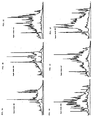
- Figs. 1A-1F are chromatographic analyses of the peptide pools extracted from papain digested HLA-DR1, DR2, DR3, DR4, DR7, and DR8, respectively, illustrating the peptide repertoire of each HLA-DR as detected by UV absorbance.
- the UV absorbance for both 210 nm and 277 nm is shown at a full scale absorbance of 500 mAU with a retention window between 16 minutes and 90 minutes (each mark represents 2 minutes).
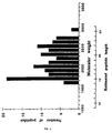
- Fig. 2 is a representative mass spectrometric analysis of the size distribution of isolated HLA-DR1 bound peptides.
- the determined peptide masses in groups of 100 mass units were plotted against the number of isolated peptides identified by mass spectrometry.
- Peptide length was calculated by dividing the experimental mass by an average amino acid mass of 118 daltons.
- Fig. 3A is a representation of a minigene of the invention (SEQ ID NO: 147), in which the HLA-DR ⁇ chain leader peptide is linked to the amino terminus of a 15-residue blocking peptide fragment of human invariant chain Ii.
- Fig. 3B is a representation of a second minigene of the invention (SEQ ID NO: 148), in which the HLA-DR ⁇ chain leader peptide is linked to the amino terminus of a 24-residue blocking peptide fragment of human invariant chain Ii.
- HLA-DR molecules were purified from homozygous, Epstein-Barr virus-transformed, human B lymphoblastoid lines: DR1 from LG-2 cells, DR2 from MST cells, DR3 from WT20 cells, DR4 from Priess cells, DR7 from Mann cells, and DR8 from 23.1 cells. All of these cell lines are publicly available. Cell growth, harvest conditions and protein purification were as previously described (Gorga, J. et al., 1991).
- Detergent soluble HLA-DR was bound to a LB3.1-protein A sepharose column (Gorga et al., id ) and eluted with 100 mM glycine, pH 11.5. Following elution, the sample was immediately neutralized by the addition of Tris-HCl and then dialyzed against 10mM Tris-HCl, 0.1% deoxycholic acid (DOC).
- DOC deoxycholic acid
- the transmembrane domain of the DR molecules was removed by papain digestion, and the resulting watersoluble molecule further purified by gel filtration chromatography on an S-200 column equilibrated in 10mM Tris-HCl, pH 8.0.
- the purified DR samples were concentrated by ultrafiltration, yield determined by BCA assay, and analyzed by SDS polyacrylamide gel electrophoresis.
- Water-soluble, immunoaffinity-purified class II molecules were further purified by high-performance size exclusion chromatography (SEC), in 25 mM N-morpholino ethane sulfonic acid (MES) pH 6.5 and a flowrate of 1 ml/min., to remove any residual small molecular weight contaminants.
- SEC high-performance size exclusion chromatography
- MES N-morpholino ethane sulfonic acid
- MES N-morpholino ethane sulfonic acid
- the peptide pool was separated from the class II molecule after centrifugation through the Centricon concentrator, with the flow-through containing the previously bound peptides.
- the collected acid-extracted peptide pool was concentrated in a Savant Speed-Vac to a volume of 50 ⁇ l prior to HPLC separation.
- Peptides were separated on a microbore C-18 reversed-phase chromatography (RFC) column (Vydac) utilizing the following non-linear gradient protocol at a constant flowrate of 0.15 ml/min.: 0-63 min. 5%-33% buffer B; 63-95 min. 33%-60% buffer B; 95-105 min 60%-80% buffer B, where buffer A was 0.06% trifluoroacetic acid/water and buffer B was 0.055% trifluoroacetic acid/acetonitrile.
- RRC reversed-phase chromatography
- Chromatographic analysis was monitored at multiple UV wavelengths (210, 254, 277, and 292 nm) simultaneously, permitting spectrophotometric evaluation prior to mass and sequence analyses. Shown in Fig.1 are chromatograms for each of the six DR peptide pools analyzed. Collected fractions were subsequently analyzed by mass spectrometry and Edman sequencing.
- the spectrophotometric evaluation of the peptides during RPC provides valuable information regarding amino acid composition (contribution of aromatic amino acids) and is used as a screening method for subsequent characterization.
- Appropriate fractions collected during the RPC separation were next analyzed using a Finnegan-MAT LaserMat matrix-assisted laser-desorption mass spectrometer (MALD-MS) to determine the individual mass values for the predominant peptides.
- MALD-MS Finnegan-MAT LaserMat matrix-assisted laser-desorption mass spectrometer
- Between 1%-4% of the collected fraction was mixed with matrix (1 ⁇ l ⁇ -Cyano-4-hydroxycinnamic acid) to achieve mass determination of extracted peptides.
- the result of this analysis for HLA-DR1 is shown in Fig. 2.
- the HLA-DR1 used in this study was papain solubilized to enable the material to be used both for crystallographic and bound peptide analyses.
- the peptides bound to DR1 were acid extracted and fractionated using RPC (Fig. 1).
- the absence of any detectable peptidic material following a second extraction/RPC separation verified quantitative peptide extraction.
- Amino acid analysis (ABI 420A/130A derivatizer/HPLC) of extracted peptide pools demonstrated a 70-80% yield, assuming total occupancy of purified DR1 with a molar equivalent of bound peptides corresponding to the size distribution determined by mass spectrometry (see Fig. 2).
- the RPC profiles obtained from DR1 extractions of multiple independent preparations were reproducible.
- Matrix-assisted laser desorption mass spectrometry was used to identify 111 species of unique mass contained within the eluted peptide pool of DR1 with an average size of 18 and a mode of 15 residues (Fig. 2). Over 500 additional mass species present within the molecular weight range of 13-25 residues were detected; however, the signal was not sufficient to assign individual masses with confidence. Multiple species of varying mass were detected in fractions corresponding to single RPC peaks indicating co-elution of peptides.
- HLA-A2 protein is intended to include HLA-A2 protein itself, as well as any naturally occurring protein which contains a ten or greater amino acid long region of >80% homology with an HLA-DR-binding peptide derived from HLA-A2.
- HLA-A2 peptide similarly refers to peptides from any HLA-A2 protein, as broadly defined herein.
- the other four peptides identified in the DR1 studies were derived from two self proteins, transferrin receptor and the Na + /K + ATPase, and one exogenous protein, bovine serum fetuin (a protein present in the serum used to fortify the medium which bathes the cells). Each of these peptides occupied only 0.3-0.6% of the total DR1 population, significantly less than either the HLA-A2 or the Ii peptides. It is known that class II molecules en route to the cell surface intersect the pathway of incoming endocytic vesicles. Both recycling membrane proteins and endocytosed exogenous protein travel this common pathway.
- the HLA-A2, transferrin receptor, Na + /K + ATPase and bovine fetuin derived peptides would all encounter DR1 in a similar manner.
- Ii associates with nascent class II molecules in the endoplasmic reticulum (ER) (Jones et al., Mol. Immunol. 16:51-60 (1978)), preventing antigen binding until the class II/Ii complex arrives at an endocytic compartment (Roche and Cresswell, Nature 345:615-618 (1990)), where Ii undergoes proteolysis (Thomas et al., J. Immunol. 140:2670-2675 (1988); Roche and Cresswell, Proc. Natl. Acad. Sci. USA 88:3150-3154 (1991)), thus allowing peptide binding to proceed.
- the Ii peptides bound to DR1 were generated at this step.
- Synthetic peptides corresponding to five of the peptides reported in Table 1 were made and their relative binding affinities to DR1 determined.
- the influenza A hemagglutinin peptide (HA) 307-319 (SEQ ID NO: 24) has been previously described as a high affinity, HLA-DR1 restricted peptide (Roche and Cresswell, J. Immunol. 144:1849-1856 (1990); Rothbard et al., Cell 52:515-523 (1988)), and was thus chosen as the control peptide.
- these peptides were found to confer resistance to SDS-induced ⁇ - ⁇ chain dissociation of "empty" DR1 when analyzed by SDS-PAGE, indicative of stable peptide binding (Sadegh-Nasseri and Germain, Nature 353:167-170 (1991); Dornmair et al., Cold Spring Harbor Symp. Quant. Biol. 54:409-415 (1989); Springer et al., J. Biol. Chem. 252:6201-6207 (1977)).
- a putative DR1 binding motif based on the sequence alignments of the core epitopes (the minimum length) of certain naturally processed peptides is shown in Table 3.
- the peptides listed in this table include those determined herein for HLA-DR1, as well as a number of peptides identified by others and known to bind DR1 (reference #6 in this table being O'Sullivan et al., J. Immunol. 145:1799-1808, 1990; reference #17, Roche & Cresswell, J. Immunol. 144:1849-1856, 1990; reference #25, Guttinger et al., Intern. Immunol. 3:899-906, 1991; reference #27, Guttinger et al. EMBO J.
- the key residues proposed in the motif are as follows: a positively charged group is located at the first position, referred to here as the index position for orientation (I); a hydrogen bond donor is located at I+5; and a hydrophobic residue is at I+9. In addition, a hydrophobic residue is often found at I+1 and/or I-1. Every naturally processed peptide sequenced from DR1 conforms to this motif (with the exception of the HLA-A2 peptide 103-116 (SEQ ID NO: 3) that lacks residue I+9).
- the putative motif is not placed in a defined position with respect to the first amino acid and because of the irregular length of bound peptides, it is impossible to deduce a motif from sequencing of peptide pools, as was done for class I molecules (Falk et al., Nature 351:290-296 (1991)).
- the Ii 96-110 peptide (SEQ ID NO: 25), a negative control peptide used in binding experiments, has the I and I+5 motif residues within its sequence, but is missing eight additional amino acids found in Ii 105-118 (SEQ ID NO: 16) (Table 3C).
- class II peptides are larger and display a high degree of heterogeneity both in length and the site of terminal truncation, implying that the mechanisms of processing for class I and class II peptides are substantially different.
- class II processing is a stochastic event and that a DR allotype may bind peptides of different lengths from a complex random mixture.
- the heterogeneity observed may be solely due to protection of bound peptides from further degradation.
- class II molecules would play an active role in antigen processing (as previously proposed (Donermeyer and Allen, J. Immunol. 142:1063-1068 (1989)) by protecting the bound peptides from complete degradation.
- the predominance of 15mers bound to DR1 (as detected by both the MALD-MS and the yields of sequenced peptides) could be the result of trimming of bound peptides.
- Table 10 gives sequences of peptides eluted from DR4 and DR11 molecules expressed in cells from a human spleen. These data demonstrate the great prevalence of self peptides bound, compared to exogenous peptides. The data also show that the A2 and Ii peptides occur repeatedly. In addition, certain of the Tables include peptides that appear to derive from viral proteins, such as Epstein-Barr virus major capsid protein, which are likely to be present in the cells studied.
- Overlapping synthetic oligonucleotides were used to generate the leader peptide/blocking peptide mini-genes illustrated in Fig. 3 by PCR amplification from human HLA-DR ⁇ and invariant chain cDNA templates. These mini-genes encode the Ii peptide fragments KMRMATPLLMQALPM (or Ii 15 ; SEQ ID NO: 15) and LPKPPKPVSKMMATPLLMQALPM (or Ii 24 ; SEQ ID NO: 7).
- the resulting constructs were cloned into pGEM-2 (Promega Corp.) to form the plasmids pGEM-2- ⁇ -Ii 15 and pGEM-2- ⁇ -Ii 24 , with an upstream T7 promoter for use in the in vitro transcription/translation system described below.
- each mini-gene was subsequently subcloned from the pGEM-2 derivatives into a transfection vector, pH ⁇ actin-1-neo (Gunning et al., (1987) Proc. Natl. Acad. Sci. U.S.A. 84 :4831), to form the plasmids pH ⁇ actin- ⁇ -Ii 15 and pH ⁇ actin- ⁇ -Ii 24 .
- the inserted mini-genes are thus expressed in vivo from the constitutive/strong human ⁇ actin promoter.
- the mini-genes were subcloned from the pGEM-2 derivatives into the vaccinia virus recombination vector pSC11 (S.
- Intracellular trafficking signals added to peptides.
- Short amino acid sequences can act as signals to target proteins to specific intracellular compartments.
- hydrophobic signal peptides are found at the amino terminus of proteins destined for the ER, while the sequence KFERQ (SEQ ID NO: 153) (and other closely related sequences) is known to target intracellular polypeptides to lysosomes, while other sequences target polypeptides to endosomes.
- the peptide sequence KDEL (SEQ ID NO: 152) has been shown to act as a retention signal for the ER.
- Each of these signal peptides, or a combination thereof, can be used to traffic the immunomodulating peptides of the invention as desired.
- a construct encoding a given immunomodulating peptide linked to an ER-targeting signal peptide would direct the peptide to the ER, where it would bind to the class II molecule as it is assembled, preventing the binding of intact Ii which is essential for trafficking.
- a construct can be made in which an ER retention signal on the peptide would help prevent the class II molecule from ever leaving the ER.
- a peptide of the invention is targeted to the endosomic compartment, this would ensure that large quantities of the peptide are present when invariant chain is replaced by processed peptides, thereby increasing the likelihood that the peptide incorporated into the class II complex is the high-affinity peptides of the invention rather than naturally-occurring, potentially immunogenic peptides.
- the likelihood of peptides of the invention being available incorporation into class II can be increased by linking the peptides to an intact Ii polypeptide sequence.
- the hybrid Ii Since Ii is known to traffic class II molecules to the endosomes, the hybrid Ii would carry one or more copies of the peptide of the invention along with the class II molecule; once in the endosome, the hybrid Ii would be degraded by normal endosomal processes to yield both multiple copies of the peptide of the invention or molecules similar to it, and an open class II binding cleft.
- DNAs encoding immunomodulatory peptides containing targeting signals will be generated by PCR or other standard genetic engineering or synthetic techniques, and the ability of these peptides to associate with DR molecules will be analyzed in vitro and in vivo, as described below.
- the invariant chain prevents class II molecules from binding peptides in the ER and may contribute to heterodimer formation. Any mechanism that prevents this association would increase the effectiveness of class II blockade. Therefore, a peptide corresponding to the site on Ii which binds to the class II heterodimer, or corresponding to the site on either the ⁇ or ⁇ subunit of the heterodimer which binds to Ii, could be used to prevent this association and thereby disrupt MHC class II function.
- Cell free extracts are used routinely for expressing eukaryotic proteins (Krieg, P. & Melton, D. (1984) Nucl. Acids Res. 12 , 7057; Pelham, H. and Jackson, R. (1976) Eur. J. Biochem. 67 , 247).
- Specific mRNAs are transcribed from DNA vectors containing viral RNA polymerase promoters (Melton, D. et al. (1984) Nucl. Acids Res. 12 , 7035), and added to micrococcal nuclease-treated cell extracts. The addition of 35 S methionine and amino acids initiates translation of the exogenous mRNA, resulting in labeled protein.
- Proteins may be subsequently analyzed by SDS-PAGE and detected by autoradiography. Processing events such as signal peptide cleavage and core glycosylation are initiated by the addition of microsomal vesicles during translation (Walter, P. and Blobel, G. (1983), Meth. Enzymol., 96 , 50), and these events are monitored by the altered mobility of the proteins in SDS-PAGE gels.
- DR1 ⁇ - and ⁇ -chain and invariant chain peptide constructs described above are transcribed into mRNAs, which will be translated in the presence of mammalian microsomal membranes. Association of the DR heterodimer with Ii is determined by immunoprecipitation with antisera to DR and Ii.
- Human EBV-transformed B cell lines LG-2 and HOM-2 (homozygous for HLA-DR1) and the mouse B cell hybridoma LK35.2 are transfected with either 50 ⁇ g of linearized pH ⁇ actin- ⁇ -Ii 15 or pH ⁇ actin- ⁇ -Ii 24 or (as a control) pH ⁇ actin-1-neo by electroporation (150mV, 960 ⁇ F, 0.2cm cuvette gap). Following electroporation, the cells are cultured in G418-free medium until total recovery (approximately 4 days). Each population is then placed under G418 selection until neomycin-expressing resistant populations of transfectants are obtained (approximately 1-2 months). The resistant populations are subcloned by limiting dilution and the clonality of stable transfectants determined by PCR amplification of blocking peptide mRNA expression.
- Stable transfectants of LG-2 and HOM-2 carrying blocking peptide mini-genes or negative control vectors are grown in large-scale culture conditions until 20 grams of pelleted cell mass is obtained.
- the HLA-DR expressed by each transfectant is purified, and the bound peptide repertoire (both from within the cell and from the cell surface) analyzed as described above. Successful demonstration of a reduction in the total bound peptide diversity will be conclusive evidence of intracellular delivery of immuno-modulatory peptides.
- a second cell-based assay utilizes stable transfectants of LK35.2 cells carrying blocking peptide mini-genes or negative control vectors; these cells are used as APCs in T cell proliferation assays.
- Each transfectant is cultured for 24 hours in the presence of different dilutions of hen egg lysozyme (HEL) and HEL-specific T cell hybridomas.
- HEL hen egg lysozyme
- the relative activation of the T cells present in each assay is determined using the publicly available lymphokine dependent cell line CTLL2 in a 3 H-thymidine incorporation assay (Vignali et al. (1992) J.E.M.
- TK - cell line 143 ATCC
- vaccinia virus strain WR, TK +
- pSC11- ⁇ -Ii 15 or pSC11- ⁇ -Ii 24 or pSC11 is introduced into the infected cells by calcium phosphate precipitation.
- TK - recombinants are selected for with bromodeoxyuridine at 25 ⁇ g/ml.
- Recombinant plaques are screened by PCR for the presence of mini-gene DNA.
- Recombinant virus is cloned by three rounds of limiting dilution to generate pure clonal viral stocks.
- vaccinia viruses encoding mini-genes or vector alone will be used to infect large-scale cultures of the human EBV transformed B cell lines LG-2 and HOM-2.
- the HLA-DR is purified and the bound peptide repertoire analyzed as described above. A reduction of the complexity of the bound peptide population and a significant increase in the relative amount of Ii peptides bound are conclusive evidence that vaccinia can deliver blocking peptides to human APCs.
- Liposomes have been successfully used as drug carriers and more recently in safe and potent adjuvant strategies for malaria vaccination in humans (Fries et al. (1992), Proc. Natl. Acad. Sci. USA 89:358). Encapsulated liposomes have been shown to incorporate soluble proteins and deliver these antigens to cells for both in vitro and in vivo CD8 + mediated CTL response (Reddy et al., J. Immunol. 148:1585-1589, 1992; and Collins et al., J. Immunol. 148:3336-3341, 1992). Thus, liposomes may be used as a vehicle for delivering synthetic peptides into APCs.
- Liposomes are prepared by standard detergent dialysis or dehydration-rehydration methods.
- dioleoylphosphatidylethanolamine (DOPE) and palmitoylhomocystein (PHC) are utilized, while dioleoylphospatidylcholine (DOPC) and dioleoylphosphatidylserine (DOPS) are used for the preparation of acid-resistant liposomes.
- DOPE dioleoylphosphatidylethanolamine
- PLC palmitoylhomocystein
- DOPC dioleoylphospatidylcholine
- DOPS dioleoylphosphatidylserine
- HBS HEPES buffered saline
- the lipid suspensions are solubilized by the addition of 0.1 ml of 1 M octylglucoside in HBS.
- the peptides to be entrapped are added to 0.2 ml of 0.6 mM peptide in 20% HBS. The mixture is then frozen, lyophilized overnight, and rehydrated.
- liposomes will be treated with chymotrypsin to digest any surface-bound peptide.
- Liposome delivery to EBV-transformed cell lines (as described above) will be accomplished by 12-16 hour incubation at 37°C.
- HLA-DR will be purified from the liposome treated cells and bound peptide analyzed as above.
- the liposomes are formulated with the DNA mini-gene constructs of the invention, and used to deliver the constructs into APCs either in vitro or in vivo.
- ISCOMS are negatively charged cage-like structures of 30-40nm in size formed spontaneously on mixing cholesterol and Quil A (saponin).
- Protective immunity has been generated in a variety of experimental models of infection, including toxoplasmosis and Epstein-Barr virus-induced tumors, using ISCOMS as the delivery vehicle for antigens (Mowat and Donachie) Immunology Today 12:383-385, 1991.
- Doses of antigen as low as 1 ⁇ g encapsulated in ISCOMS have been found to produce class I mediated CTL responses, where either purified intact HIV-1-IIIB gp 160 envelope glycoprotein or influenza hemagglutinin is the antigen (Takahashi et al., Nature 344:873-875, 1990).
- Peptides are delivered into tissue culture cells using ISCOMS in a manner and dosage similar to that described above for liposomes; the class II peptide binding of delivered peptides are then determined by extraction and characterization as described above. ISCOM-delivered peptides of the invention which are effectively utilized by cultured cells are then tested in animals or humans.
- ISCOMS In addition to delivery of the therapeutic synthetic peptides, ISCOMS could be constituted to deliver the mini-gene constructs to APCs, and thus serve as an alternative to the above-outlined vaccinia strategy.
- the delivery systems of the invention may alternatively be used as a novel means of vaccination, in order to stimulate a portion of the immune system of an animal.
- the delivery system is employed to deliver, into appropriate cells, DNA constructs which express immunogenic, pathogen-derived peptides intended to stimulate an immune response against a specific pathogen. Because the antigenic peptide is produced inside the target cell itself, the vaccine method of the invention ensures that there is no circulating free antigen available to stimulate antibody formation and thereby induce potentially deleterious or inappropriate immunological reactions.
- the immune response stimulated by vaccines of the invention is, because the vaccines are targeted solely to APC's, limited to the T cell mediated response, in contrast to standard vaccine protocols which result in a more generalized immune response. Although some of the peptide-presenting APC's will initially be lysed by host T cells, such lysis will be limited because, inter alia, the virus-based vaccine is non-replicative, i.e., each carrier virus can infect only one cell.
- the model antigen that will be used to perfect and test the system of the invention is hen egg lysozyme (HEL). It is arguably the most well characterized protein for antigen presentation studies, to which there are numerous monoclonal antibodies and class I- and class II-restricted mouse T cell clones and hybridomas.
- the primary epitopes that will be studied are the peptide HEL 34-45, as both monoclonal antibodies and CD4+ T cell hybridomas are available, and peptide HEL 46-61, as both class I and class II-restricted T cell clones and hybridomas have been raised and are publicly available. These two sequences are thus proven immunogenic epitopes.
- constructs are introduced into appropriate APCs, e.g., LK35.2 cells, by means of a conventional eukaryotic transfection or one of the delivery vehicles discussed above (e.g., vaccinia, liposomes, or ISCOMS).
- LK35.2 cells which possess the mouse MHC Class II restriction molecules IA k and IE k , transfected with each of the constructs are tested for their ability to stimulate the appropriate class I and class II-restricted T cell hybridomas and clones using standard techniques. Whether class I stimulation is observed will depend on whether peptide trimming can occur in the ER, in order to produce an 8-10-mer suitable for binding to class I molecules.
- constructs are ineffective for class I stimulation, they can be modified in order to produce a more effective peptide for class I binding. If these constructs prove to be less effective for class II-restricted responses, they can be tagged with endosomal and/or lysosomal targeting sequences as discussed in Section V.
- transfectants i.p. and/or s.c. into C3H/Balb/c Fl mice, or by injection of the construct incorporated into an appropriate delivery vehicle (e.g., liposome, ISCOMS, retrovirus, vaccinia).
- an appropriate delivery vehicle e.g., liposome, ISCOMS, retrovirus, vaccinia.
- Optimal protocols and doses for such immunizing injections can be determined by one of ordinary skill in the art, given the disclosures provided herein.
- Efficiency of immunization can be tested by standard methods such as (a) proliferation of class II-restricted T cells in response to HEL pulsed APCs, (b) CTL response to 51 Cr-labeled targets, and (c) serum antibody titre as determined by ELISA.
- constructs encoding peptides with useful immunizing potential can be incorporated into the system.
- Such peptides can be identified by standard means now used to identify immunogenic epitopes on pathogen-derived proteins.
- candidate peptides for immunization may be determined from antibody and T cell analysis of animals infected with a particular pathogen.
- the peptides used for vaccination should ideally be those which are presented with the highest frequency and efficiency upon infection. This could best be determined by using the procedures outlined in the experimental section above to extract and characterize the peptides bound by MHC class II molecules from infected cells.
Abstract
Description
- This application is a continuation-in-part of copending USSN 07/925,460, filed August 11, 1992. The invention was made in the course of research funded in part by the U.S. Government under NIH Grant 5R35-CA47554; the U.S. Government therefore has certain rights in the invention.
- The field of the invention is major histocompatibility complex (MHC) antigens.
- Major histocompatibility complex (MHC) class II antigens are cell surface receptors that orchestrate all specific immune responses in vertebrates. Humans possess three distinct MHC class II isotypes: DR, for which approximately 70 different allotypes are known; DQ, for which 33 different allotypes are known; and DP, for which 47 different allotypes are known. Each individual bears two to four DR alleles, two DQ alleles, and two DP alleles.
- MHC receptors (both class I and class II) participate in the obligate first step of immune recognition by binding small protein fragments (peptides) derived from pathogens or other non-host sources, and presenting these peptides to the regulatory cells (T cells) of the immune system. In the absence of MHC presentation, T cells are incapable of recognizing pathogenic material. Cells that express MHC class II receptors are termed antigen presenting cells (APC). APCs ingest pathogenic organisms and other foreign materials by enveloping them in endosomic vesicles, then subjecting them to enzymatic and chemical degradation. Foreign proteins which are ingested by APCs are partially degraded or "processed" to yield a mixture of peptides, some of which are bound by MHC class II molecules that are en route to the surface. Once on the cell surface, MHC-bound peptides are available for T cell recognition.
- MHC class II antigens are expressed on the surface of APCs as a trimolecular complex composed of an α chain, a β chain, and a processed peptide. Like most polypeptides that are expressed on the cell surface, both a and β chains contain short signal sequences at their NH2 termini which target them to the endoplasmic reticulum (ER). Within the ER the class II α/β chain complex associates with an additional protein termed the invariant chain (Ii). Association with Ii is proposed to block the premature acquisition of peptides (by blocking the peptide binding cleft of the MHC heterodimer), promote stable α/β interaction, and direct subsequent intracellular trafficking of the complex to endosomal vesicles. In the endosomes, Ii is removed by a process involving proteolysis; this exposes the peptide binding cleft, thus allowing peptides present in the endosome to bind to the MHC molecule. The class II/ peptide complex is transported from the endosomes to the cell surface where it becomes accessible to T-cell recognition and subsequent activation of immune responses. Class II MHC molecules bind not only to peptides derived from exogenous (ingested) proteins, but also to those produced by degradation of endogenous (self) proteins. The amount of each species of peptide which binds class II is determined by its local concentration and its relative binding affinity for the given class II binding groove, with the various allotypes displaying different peptide-binding specificities.
- Early during fetal development, the mammalian immune system is "tolerized", or taught not to react, to self-peptides. The stability and maintenance of this system is critical for ensuring that an animal does not generate an immune response against self. A breakdown of this system gives rise to autoimmune conditions such as diabetes, rheumatoid arthritis and multiple sclerosis. Current technologies intended to manipulate the immune system into reestablishing proper nonresponsiveness include protocols involving the intravenous delivery of synthetic, high affinity binding peptides as blocking peptides.
- Vaccination can generate protective immunity against a pathogenic organism by stimulating an antibody-mediated and/or a T cell-mediated response. Most of the current vaccination strategies still use relatively crude preparations, such as attenuated or inactivated viruses. These vaccines often generate both antibody- and cell-mediated immunity, and do not allow one to modulate the type of immune response generated. Moreover, in many diseases the generation of the wrong type of response can result in an exacerbated disease state.
- In the work disclosed herein, naturally processed peptides bound to six of the some 70 known human MHC class II DR allotypes (HLA-DR1, HLA-DR2, HLA-DR3, HLA-DR4, HLA-DR7, and HLA-DR8) have been characterized. These peptides were found to be predominantly derived from self proteins rather than foreign proteins. Several self peptide families have been identified with the unexpected property of degenerate binding: that is, a given self-peptide will bind to a number of HLA-DR allotypes. This observation runs counter to the widely-accepted view of MHC class II function, which dictates that each allotype binds a different set of peptides. Furthermore, many if not all of the self-peptides disclosed herein bind to the class II molecules with relatively high affinity. These three characteristics--(1) self rather than foreign, (2) degeneracy, and (3) high affinity binding--suggest a novel means for therapeutic intervention in disease conditions characterized by autoreactivity, such as Type I diabetes, rheumatoid arthritis, and multiple sclerosis. In addition, such therapy could be used to reduce transplant rejection.
- In the therapeutic methods of the invention, short peptides modelled on the high-affinity immunomodulating self peptides of the invention (which preferably are nonallelically restricted) are introduced into the APCs of a patient. Tissue typing to determine the particular class II alleles expressed by the patient may be unnecessary, as the peptides of the invention are bound by multiple class II isotypes. It may be useful to employ a "cocktail" of peptides, where complete degeneracy is lacking for individual peptides, i.e., where peptides binds to fewer than all allotypes; the cocktail provides overlapping binding specificity. Once in the APC, a peptide binds to the class II molecules with high affinity, thereby blocking the binding of immunogenic peptides which are responsible for the immune reaction characteristic of the disease condition. Because the blocking peptides of the invention are self peptides with the exact carboxy and amino termini tolerized during ontogeny, they are immunologically inert and will not induce an immune response which may complicate treatment using non-self blocking peptides.
- The peptides of the invention may be introduced into APCs directly, e.g., by intravenous injection of a solution containing one or more of the peptides. Alternatively, the APCs may be provided with a means of synthesizing large quantities of the blocking peptides intracellularly. Recombinant genes that encode ER and/or endosomal targeting signals fused to blocking peptide sequences are linked to appropriate expression control sequences and introduced into APCs. Once in the cell, these genes direct the expression of the hybrid peptides. Peptides targeted to the ER will bind class II α and β chains as they are translated and assembled into heterodimers. The presence of high affinity binding peptides within the ER will prevent association of the α/β complex with invariant chain, and thus interfere with intracellular trafficking. The class II molecule/blocking peptide complex may subsequently be expressed on the cell surface, but would not elicit an immune response since T cells are tolerized to this complex early in development. The use of peptides tagged with ER retention signals may also prevent the peptide-complexed class II molecules from leaving the ER. Alternatively, the recombinant peptide may be tagged with an endosomal targeting signal which directs it to the endosomal compartment after synthesis, thereby also skewing the ratio of endogenously-processed peptide to blocking peptide in the endosome and favoring binding of the high affinity blocking peptide to any class II molecules which did not bind it in the ER. It may be advantageous, for any individual patient, to employ one or more ER-directed peptides in combination with one or more endosome-directed peptide, so that α-β complexes which are not filled in the ER with peptides of the invention are then blocked in the endocytic pathway. The end result again is cell surface expression of a non-immunogenic class II/peptide complex.
- The use of a class II nonrestricted high affinity binding peptide coupled to an intracellular delivery system permits the specific down-regulation of class II restricted immune responses without invoking the pleiotropic adverse reactions associated with the current pharmacological strategies. Successful application of these technologies will constitute a significant advance towards the treatment of autoimmune disease and prevention of transplant rejection.
- The intracellular delivery system of the invention can also be utilized in a novel method of vaccination of an animal, e.g., a human patient or a commercially significant mammal such as a cow which is susceptible to diseases such as hoof and mouth disease. Such a system can be tailored to generate the type of immune response required in a given situation by adjustments in the following: (a) peptide specificity for class I or class II MHC; (b) peptide/protein length and/or sequence, and (c) using specific tags for organelle targeting. The system of the invention ensures that peptides are produced only within cells, and are not present outside the cells where they could stimulate antibody production by contact with B cells. This limits the immune response generated by such a vaccine to T cell-mediated immunity, thereby preventing either an inappropriate or potentially deleterious response as might be observed with standard vaccines targeting the organisms which cause, for example, HIV, malaria, leprosy, and leishmaniasis. Furthermore, this exclusively T cell-mediated immune response can be class I or class II-based, or both, depending upon the length and character of the immunogenic peptides: MHC class I molecules are known to bind preferentially to peptides 8 to 10 residues in length, while class II molecules bind with high affinity to peptides that range from 12 to 25 residues long.
- Immunization and therapy according to the invention can employ a purified preparation of a peptide of the invention, i.e., a peptide which includes an amino acid sequence identical to that of a segment of a naturally-occurring human protein (i.e., a "self protein"), such segment being of 10 to 30 residues in length, wherein the peptide binds to a human MHC class II allotype, and preferably binds to at least two distinct MHC class II allotypes (e.g., any of the approximately 70 known DR allotypes, approximately 47 known DP allotypes, or approximately 33 known DQ allotypes). The portion of the peptide corresponding to the self protein segment is herein termed a "self peptide". By "purified preparation" is meant a preparation at least 50% (by weight) of the polypeptide constituents of which consists of the peptide of the invention. In preferred embodiments, the peptide of the invention constitutes at least 60% (more preferably at least 80%) of the purified preparation. The naturally-occurring human protein is preferably HLA-A2 (as broadly defined below), HLA-A29, HLA-A30, HLA-B44, HLA-B51, HLA-Bw62, HLA-C, HLA-DP β-chain, HLA-DQ α-chain, HLA-DQ β-chain, HLA-DQ3.2 β-chain, HLA-DR α-chain, HLA-DR β-chain, HLA-DR4 β-chain, invariant chain (Ii), Ig kappa chain, Ig kappa chain C region, Ig heavy chain, Na+/K+ ATPase, potassium channel protein, sodium channel protein, calcium release channel protein, complement C9, glucose-transport protein, CD35, CD45, CD75, vinculin, calgranulin B, kinase C ζ-chain, integrin β-4 gp150, hemoglobin, tubulin α-1 chain, myosin β-heavy chain, α-enolase, transferrin, transferrin receptor, fibronectin receptor α-chain, acetylcholine receptor, interleukin-8 receptor, interferon α-receptor, interferon γ-receptor, calcitonin receptor, LAM (lymphocyte activation marker) Blast-1, LAR (leukocyte antigen-related) protein, LIF (leukemia inhibitory factor) receptor, 4F2 cell-surface antigen (a cell-surface antigen involved in normal and neoplastic growth) heavy chain, cystatin SN, VLA-4 (a cell surface heterodimer in the integrin superfamily of adhesion receptors), PAI-1 (plasminogen activator inhibitor-1), IP-30 (interferon-γ induced protein), ICAM-2, carboxypeptidase E, thromboxane-A synthase, NADH-cytochrome-b5 reductase, c-myc transforming protein, K-ras transforming protein, MET kinase-related transforming protein, interferon-induced guanylate-binding protein, mannose-binding protein, apolipoprotein B-100, cathepsin C, cathepsin E, cathepsin S, Factor VIII, von Willebrand factor, metalloproteinase inhibitor 1 precursor, metalloproteinase inhibitor 2, plasminogen activator inhibitor-1, or heat shock cognate 71 kD protein; it may be an MHC class I or II antigen protein or any other human protein which occurs at the cell surface of APCs. The self peptide preferably conforms to the following motif: at a first reference position (I) at or within 12 residues of the amino terminal residue of the segment, a positively charged residue (i.e., Lys, Arg, or His) or a large hydrophobic residue (i.e., Phe, Trp, Leu, Ile, Met, Tyr, or Pro; and at position I+5, a hydrogen bond donor residue (i.e., Tyr, Asn, Gln, Cys, Asp, Glu, Arg, Ser, Trp, or Thr). In addition, the peptide may also be characterized as having, at positions I+9, I+1, and/or I-1, a hydrophobic residue (i.e., Phe, Trp, Leu, Ile, Met, Pro, Ala, Val, or Tyr) (+ denotes positions to the right, or toward the carboxy terminus, and - denotes positions to the left, or toward the amino terminus.) A typical peptide of the invention will include a sequence corresponding to residues 31-40 (i.e., TQFVRFDSDA; SEQ ID NO: 149) or residues 106-115 (i.e., DWRFLRGYHQ; SEQ ID NO: 150) of HLA-A2, or residues 107-116 (i.e., RMATPLLMQA; SEQ ID NO: 151) of Ii, or a sequence essentially identical to any one of the sequences set forth in Tables 1-10 below.
- The therapeutic and immunization methods of the invention can also employ a nucleic acid molecule (RNA or DNA) encoding a peptide of the invention, but encoding less than all of the entire sequence of the self protein. The nucleic acid preferably encodes no substantial portion of the self protein other than the specified self peptide which binds to a MHC class II molecule, although it may optionally include a signal peptide or other trafficking sequence which was derived from the self protein (or from another protein). A trafficking sequence is an amino acid sequence which functions to control intracellular trafficking (directed movement from organelle to organelle or to the cell surface) of a polypeptide to which it is attached. Such trafficking sequences might traffic the polypeptide to ER, a lysosome, or an endosome, and include signal peptides (the amino terminal sequences which direct proteins into the ER during translation), ER retention peptides such as KDEL (SEQ ID NO: 152); and lysosome-targeting peptides such as KFERQ (SEQ ID NO: 153), QREFK (SEQ ID NO: 154), and other pentapeptides having Q flanked on one side by four residues selected from K, R, D, E, F, I, V, and L. An example of a signal peptide that is useful in the invention is a signal peptide substantially identical to that of an MHC subunit such as class II α or β; e.g., the signal peptide of MHC class II α is contained in the sequence MAISGVPVLGFFIIAVLMSAQESWA (SEQ ID NO: 155). The signal peptide encoded by the nucleic acid of the invention may include only a portion (e.g., at least ten amino acid residues) of the specified 25 residue sequence, provided that portion is sufficient to cause trafficking of the polypeptide to the ER. In preferred embodiments, the nucleic acid of the invention encodes a second self peptide and a second trafficking sequence (which may be identical to or different than the first self peptide and first trafficking sequence), and it may encode additional self peptides and trafficking sequences as well. In still another variation on this aspect of the invention, the self peptide sequence (or a plurality of self peptide sequences arranged in tandem) is linked by a peptide bond to a substantially intact Ii polypeptide, which then carries the self peptide sequence along as it traffics the class II molecule from ER to endosome.
- The nucleic acid of the invention may also contain expression control sequences (defined as transcription and translation start signals, promoters, and enhancers which permit and/or optimize expression of the coding sequence with which they are associated) and/or genomic nucleic acid of a phage or a virus, such as an attenuated or non-replicative, non-virulent form of vaccinia virus, adenovirus, Epstein-Barr virus, or a retrovirus.
- The peptides and nucleic acids of the invention may be prepared for therapeutic use by suspending them directly in a pharmaceutically acceptable carrier, or by encapsulating them in liposomes, immune-stimulating complexes (ISCOMS), or the like. Such preparations are useful for inhibiting an immune response in a human patient, by contacting a plurality of the patient's APCs with the therapeutic preparation and thereby introducing the peptide or nucleic acid into the APCs.
- Also within the invention is a cell (e.g., a tissue culture cell or a cell, such as a B cell or APC, within a human) containing the nucleic acid molecule of the invention. A cultured cell containing the nucleic acid of the invention may be used to manufacture the peptide of the invention, in a method which involves culturing the cell under conditions permitting expression of the peptide from the nucleic acid molecule.
- Disclosed herein is a method of identifying a nonallelically restricted immunomodulating peptide, which method includes the steps of:
- (a) fractionating a mixture of peptides eluted from a first MHC class II allotype;
- (b) identifying a self peptide from this mixture; and
- (c) testing whether the self peptide binds to a second MHC class II allotype, such binding being an indication that the self peptide is a nonallelically restricted immunomodulating peptide.
-
- In further embodiments, the invention includes a method of identifying a potential immunomodulating peptide, in a method including the steps of:
- (a) providing a cell expressing MHC class II molecules on its surface;
- (b) introducing into the cell a nucleic acid encoding a candidate peptide; and
- (c) determining whether the proportion of class II molecules which are bound to the candidate peptide is increased in the presence of the nucleic acid compared to the proportion bound in the absence of the nucleic acid, such an increase being an indication that the candidate peptide is a potential immunomodulating peptide.
-
- Also within the invention is a method of identifying a potential immunomodulating peptide, which method includes the steps of:
- (a) providing a cell expressing MHC class II molecules on its surface;
- (b) introducing into the cell a nucleic acid encoding a candidate peptide; and
- (c) determining whether the level of MHC class II molecules on the surface of the cell is decreased in the presence of the nucleic acid compared to the level of MHC class II molecules in the absence of the nucleic acid, such a decrease being an indication that the candidate peptide is a potential immunomodulating peptide. Also included in the invention is a method of identifying a nonallelically restricted immunostimulating peptide, which method includes the steps of:
- (a) providing a cell bearing a first MHC class I or class II allotype, such cell being infected with a pathogen (e.g., an infective agent which causes human or animal disease, such as human immunodeficiency virus (HIV), hepatitis B virus, measles virus, rubella virus, influenza virus, rabies virus, Corynebacterium diphtheriae, Bordetella pertussis, Plasmodium spp., Schistosoma spp., Leishmania spp., Trypanasoma spp., or Mycobacterium lepre);
- (b) eluting a mixture of peptides bound to the cell's first MHC allotype;
- (c) identifying a candidate peptide from the mixture, such candidate peptide being a fragment of a protein from the pathogen; and
- (d) testing whether the candidate peptide binds to a second MHC allotype, such binding being an indication that the candidate peptide is a nonallelically restricted immunostimulating peptide. A nucleic acid encoding such an immunogenic fragment of a protein of a pathogen can be used in a method of inducing an immune response in a human patient, which method involves introducing the nucleic acid into an APC of the patient.
-
- The therapeutic methods of the invention solve certain problems associated with prior art methods involving intravenous injection of synthetic peptides: (1) because of allelic specificity, a peptide capable of binding with high affinity to all, or even most, of the different class II allotypes expressed within the general population had not previously been identified; (2) the half-lives of peptides delivered intravenously are generally very low, necessitating repeated administration with the associated high level of inconvenience and cost; (3) this type of delivery approach requires that the blocking peptide displace the naturally-occurring peptide occupying the binding cleft of a class II molecule while the latter is on the cell surface, which is now believed to be a very inefficient process; and (4) if the blocking peptide utilized is itself immunogenic, it may promote deleterious immune responses in some patients.
- Other features and advantages of the invention will be apparent from the following detailed description, and from the claims.
- The drawings are first briefly described.
- Figs. 1A-1F are chromatographic analyses of the peptide pools extracted from papain digested HLA-DR1, DR2, DR3, DR4, DR7, and DR8, respectively, illustrating the peptide repertoire of each HLA-DR as detected by UV absorbance. The UV absorbance for both 210 nm and 277 nm is shown at a full scale absorbance of 500 mAU with a retention window between 16 minutes and 90 minutes (each mark represents 2 minutes).
- Fig. 2 is a representative mass spectrometric analysis of the size distribution of isolated HLA-DR1 bound peptides. The determined peptide masses in groups of 100 mass units were plotted against the number of isolated peptides identified by mass spectrometry. Peptide length was calculated by dividing the experimental mass by an average amino acid mass of 118 daltons.
- Fig. 3A is a representation of a minigene of the invention (SEQ ID NO: 147), in which the HLA-DRα chain leader peptide is linked to the amino terminus of a 15-residue blocking peptide fragment of human invariant chain Ii.
- Fig. 3B is a representation of a second minigene of the invention (SEQ ID NO: 148), in which the HLA-DRα chain leader peptide is linked to the amino terminus of a 24-residue blocking peptide fragment of human invariant chain Ii.
- HLA-DR molecules were purified from homozygous, Epstein-Barr virus-transformed, human B lymphoblastoid lines: DR1 from LG-2 cells, DR2 from MST cells, DR3 from WT20 cells, DR4 from Priess cells, DR7 from Mann cells, and DR8 from 23.1 cells. All of these cell lines are publicly available. Cell growth, harvest conditions and protein purification were as previously described (Gorga, J. et al., 1991). Briefly, 200 grams of each cell type was resuspended in 10mM Tris-HCl, 1mM dithiothreitol (DTT), 0.1mM phenylmethylsulfonylflouride (PMSF), pH 8.0, and lysed in a Thomas homogenizer. The nuclei were removed by centrifugation at 4000xg for 5 min and the pellets washed and repelleted until the supernatants were clear. All the supernatants were pooled and the membrane fraction harvested by centrifugation at 175,000xg for 40 min. The pellets were then resuspended in 10 mM Tris-HCl, 1mM DTT, 1mM PMSF, 4% NP-40. The unsolubilized membrane material was removed by centrifugation at 175,000xg for 2 hours, and the NP-40 soluble supernatant fraction used in immunoaffinity purification.
- Detergent soluble HLA-DR was bound to a LB3.1-protein A sepharose column (Gorga et al., id) and eluted with 100 mM glycine, pH 11.5. Following elution, the sample was immediately neutralized by the addition of Tris-HCl and then dialyzed against 10mM Tris-HCl, 0.1% deoxycholic acid (DOC). The LB3.1 monoclonal antibody recognizes a conformational determinant present on the nonpolymorphic HLA-DRα chain, and thus recognizes all allotypes of HLA-DR.
- The transmembrane domain of the DR molecules was removed by papain digestion, and the resulting watersoluble molecule further purified by gel filtration chromatography on an S-200 column equilibrated in 10mM Tris-HCl, pH 8.0. The purified DR samples were concentrated by ultrafiltration, yield determined by BCA assay, and analyzed by SDS polyacrylamide gel electrophoresis.
- Water-soluble, immunoaffinity-purified class II molecules were further purified by high-performance size exclusion chromatography (SEC), in 25 mM N-morpholino ethane sulfonic acid (MES) pH 6.5 and a flowrate of 1 ml/min., to remove any residual small molecular weight contaminants. Next, Centricon microconcentrators (molecular weight cutoff 10,000 daltons) (Amicon Corp.) were sequentially washed using SEC buffer and 10% acetic acid prior to spin-concentration of the protein sample (final volume between 100-200 µl). Peptide pools were extracted from chosen class II alleles by the addition of 1 ml of 10% acetic acid for 15 minutes at 70°C. These conditions are sufficient to free bound peptide from class II molecules, yet mild enough to avoid peptide degradation. The peptide pool was separated from the class II molecule after centrifugation through the Centricon concentrator, with the flow-through containing the previously bound peptides.
- The collected acid-extracted peptide pool was concentrated in a Savant Speed-Vac to a volume of 50 µl prior to HPLC separation. Peptides were separated on a microbore C-18 reversed-phase chromatography (RFC) column (Vydac) utilizing the following non-linear gradient protocol at a constant flowrate of 0.15 ml/min.: 0-63 min. 5%-33% buffer B; 63-95 min. 33%-60% buffer B; 95-105 min 60%-80% buffer B, where buffer A was 0.06% trifluoroacetic acid/water and buffer B was 0.055% trifluoroacetic acid/acetonitrile. Chromatographic analysis was monitored at multiple UV wavelengths (210, 254, 277, and 292 nm) simultaneously, permitting spectrophotometric evaluation prior to mass and sequence analyses. Shown in Fig.1 are chromatograms for each of the six DR peptide pools analyzed. Collected fractions were subsequently analyzed by mass spectrometry and Edman sequencing.
- The spectrophotometric evaluation of the peptides during RPC provides valuable information regarding amino acid composition (contribution of aromatic amino acids) and is used as a screening method for subsequent characterization. Appropriate fractions collected during the RPC separation were next analyzed using a Finnegan-MAT LaserMat matrix-assisted laser-desorption mass spectrometer (MALD-MS) to determine the individual mass values for the predominant peptides. Between 1%-4% of the collected fraction was mixed with matrix (1µl α-Cyano-4-hydroxycinnamic acid) to achieve mass determination of extracted peptides. The result of this analysis for HLA-DR1 is shown in Fig. 2. Next, chosen peptide samples were sequenced by automated Edman degradation microsequencing using an ABI 477A protein sequencer (Applied Biosystems) with carboxy-terminal verification provided by mass spectral analysis using the Finnigan-MAT TSQ 700 triple quadruple mass spectrometer equipped with an electro-spray ion source. This parallel analysis ensures complete identity of peptide composition and sequence. Peptide alignment with protein sequences stored in the SWISS-PROT database was performed using the PASTA computer database search program. Set forth in Tables 1-10 are the results of this sequence analysis for each of the DR molecules studied.
- The HLA-DR1 used in this study was papain solubilized to enable the material to be used both for crystallographic and bound peptide analyses. The peptides bound to DR1 were acid extracted and fractionated using RPC (Fig. 1). The absence of any detectable peptidic material following a second extraction/RPC separation verified quantitative peptide extraction. Amino acid analysis (ABI 420A/130A derivatizer/HPLC) of extracted peptide pools demonstrated a 70-80% yield, assuming total occupancy of purified DR1 with a molar equivalent of bound peptides corresponding to the size distribution determined by mass spectrometry (see Fig. 2). The RPC profiles obtained from DR1 extractions of multiple independent preparations were reproducible. Furthermore, profiles from either detergent-soluble or papain-solubilized DR1 were equivalent. To confirm that the peptides were in fact identical in detergent-soluble and papain-digested DR1, mass spectrometry and Edman sequencing analyses were performed and revealed identical masses and sequences for analogous fractions from the two preparations.
- Matrix-assisted laser desorption mass spectrometry (MALD-MS) was used to identify 111 species of unique mass contained within the eluted peptide pool of DR1 with an average size of 18 and a mode of 15 residues (Fig. 2). Over 500 additional mass species present within the molecular weight range of 13-25 residues were detected; however, the signal was not sufficient to assign individual masses with confidence. Multiple species of varying mass were detected in fractions corresponding to single RPC peaks indicating co-elution of peptides. To characterize these peptides further, samples were analyzed in parallel on a triple quadruple mass spectrometer equipped with an electrospray ion source (ESI-MS) and by automated Edman degradation microsequencing (Lane et al., J. Prot. Chem. 10:151-160 (1991)). Combining these two techniques permits crucial verification of both the N- and C-terminal amino acids of peptides contained in single fractions. The sequence and mass data acquired for twenty peptides isolated from DR1 are listed in Table 1. All the identified peptides aligned with complete identity to regions of proteins stored in the SWISS-PROT database.
- Surprisingly, sixteen of the twenty sequenced DR1-bound peptides were 100% identical to regions of the self proteins HLA-A2 and class II-associated invariant chain (Ii), representing at least 26% of the total extracted peptide mass. These isolated peptides varied in length and were truncated at both the N- and C-termini, suggesting that: 1) antigen processing occurs from both ends after binding to DR1, or 2) class II molecules bind antigen from a pool of randomly generated peptides. The yields from the peptide microsequencing indicated that HLA-A2 (Fig. 1) and Ii each represents at least 13% of the total DR1-bound peptides.
- An additional surprising finding concerned a peptide which, although bound to HLA-DR and 100% homologous with HLA-A2 peptide, was derived from a cell which does not express HLA-A2 protein. Evidently this peptide is derived from a protein containing a region homologous with a region of HLA-A2 protein. Thus, for purposes of this specification, the term "HLA-A2 protein" is intended to include HLA-A2 protein itself, as well as any naturally occurring protein which contains a ten or greater amino acid long region of >80% homology with an HLA-DR-binding peptide derived from HLA-A2. An "HLA-A2 peptide" similarly refers to peptides from any HLA-A2 protein, as broadly defined herein.
- The other four peptides identified in the DR1 studies were derived from two self proteins, transferrin receptor and the Na+/K+ ATPase, and one exogenous protein, bovine serum fetuin (a protein present in the serum used to fortify the medium which bathes the cells). Each of these peptides occupied only 0.3-0.6% of the total DR1 population, significantly less than either the HLA-A2 or the Ii peptides. It is known that class II molecules en route to the cell surface intersect the pathway of incoming endocytic vesicles. Both recycling membrane proteins and endocytosed exogenous protein travel this common pathway. Hence, the HLA-A2, transferrin receptor, Na+/K+ ATPase and bovine fetuin derived peptides would all encounter DR1 in a similar manner. Ii associates with nascent class II molecules in the endoplasmic reticulum (ER) (Jones et al., Mol. Immunol. 16:51-60 (1978)), preventing antigen binding until the class II/Ii complex arrives at an endocytic compartment (Roche and Cresswell, Nature 345:615-618 (1990)), where Ii undergoes proteolysis (Thomas et al., J. Immunol. 140:2670-2675 (1988); Roche and Cresswell, Proc. Natl. Acad. Sci. USA 88:3150-3154 (1991)), thus allowing peptide binding to proceed. Presumably, the Ii peptides bound to DR1 were generated at this step.
- Synthetic peptides corresponding to five of the peptides reported in Table 1 were made and their relative binding affinities to DR1 determined. The influenza A hemagglutinin peptide (HA) 307-319 (SEQ ID NO: 24) has been previously described as a high affinity, HLA-DR1 restricted peptide (Roche and Cresswell, J. Immunol. 144:1849-1856 (1990); Rothbard et al., Cell 52:515-523 (1988)), and was thus chosen as the control peptide. "Empty" DR1 purified from insect cells expressing recombinant DR1 cDNA was used in the binding experiments because of its higher binding capacity and 10-fold faster association kinetics than DR1 isolated from human cells (Stern and Wiley, Cell 68:465-477 (1992)). All the synthetic peptides were found to compete well (Ki < 100 nM) against the HA peptide (Table 2). At first approximation, the Ii 106-119 peptide (SEQ ID NO: 156) had the highest affinity of all the competitor peptides measured, equivalent to that determined for the control HA peptide. In addition to the Ki determinations, these peptides were found to confer resistance to SDS-induced α-β chain dissociation of "empty" DR1 when analyzed by SDS-PAGE, indicative of stable peptide binding (Sadegh-Nasseri and Germain, Nature 353:167-170 (1991); Dornmair et al., Cold Spring Harbor Symp. Quant. Biol. 54:409-415 (1989); Springer et al., J. Biol. Chem. 252:6201-6207 (1977)). Neither of the two control peptides, β2m 52-64 (SEQ ID NO: 26) nor Ii 96-110 (SEQ ID NO: 25), was able to either confer resistance to SDS-induced chain dissociation of DR1 or compete with HA 307-319 (SEQ ID NO: 24) for binding to DR1; both of these peptides lack the putative binding motif reported in this study (see below).
- A putative DR1 binding motif based on the sequence alignments of the core epitopes (the minimum length) of certain naturally processed peptides is shown in Table 3. The peptides listed in this table include those determined herein for HLA-DR1, as well as a number of peptides identified by others and known to bind DR1 (reference #6 in this table being O'Sullivan et al., J. Immunol. 145:1799-1808, 1990; reference #17, Roche & Cresswell, J. Immunol. 144:1849-1856, 1990;
reference # 25, Guttinger et al., Intern. Immunol. 3:899-906, 1991; reference #27, Guttinger et al. EMBO J. 7:2555-2558, 1988; and reference #28, Harris et al., J. Immunol. 148:2169-2174, 1992). The key residues proposed in the motif are as follows: a positively charged group is located at the first position, referred to here as the index position for orientation (I); a hydrogen bond donor is located at I+5; and a hydrophobic residue is at I+9. In addition, a hydrophobic residue is often found at I+1 and/or I-1. Every naturally processed peptide sequenced from DR1 conforms to this motif (with the exception of the HLA-A2 peptide 103-116 (SEQ ID NO: 3) that lacks residue I+9). Because the putative motif is not placed in a defined position with respect to the first amino acid and because of the irregular length of bound peptides, it is impossible to deduce a motif from sequencing of peptide pools, as was done for class I molecules (Falk et al., Nature 351:290-296 (1991)). The Ii 96-110 peptide (SEQ ID NO: 25), a negative control peptide used in binding experiments, has the I and I+5 motif residues within its sequence, but is missing eight additional amino acids found in Ii 105-118 (SEQ ID NO: 16) (Table 3C). - A sequence comparison of 35 previously described DR1-binding synthetic peptides (O'Sullivan et al., J. Immunol. 145:1799-1808 (1990); Guttinger et al., Intern. Immunol. 3:899-906 (1991); Hill et al., J. Immunol. 147:189-197 (1991); Guttinger et al., EMBO J. 7:2555-2558 (1988); Harris et al., J. Immunol. 148:2169-2174 (1992)) also supports this motif. Of the 35 synthetic peptides, 21 (60%) have the precise motif, nine (30%) contain a single shift at either I or I+9, and the remaining five (10%) have a single substitution at I (Table 3B and C). Interestingly, in the latter peptides, a positive charge at I is always replaced by a large hydrophobic residue (Table 8C); a pocket has been described in class I molecules that can accommodate this precise substitution (Latron et al., Proc. Natl. Acad. Sci. USA 88:11325-11329 (1991)). Contributions by the other eight amino acids within the motif or the length of the peptide have not been fully evaluated and may compensate for shifted/missing residues in those peptides exhibiting binding. Evaluation of the remaining 117 non-DR1 binding peptides cited in those studies (which peptides are not included in Table 3) indicates that 99 (85%) of these peptides do not contain the DR1 motif proposed herein. Of the remaining 18 peptides (15%) that do not bind to DR1 but which do contain the motif, 6 (5%) are known to bind to other DR allotypes; the remaining 12 peptides may have unfavorable interactions at other positions which interfere with binding.
- In contrast to the precise N-terminal cleavages observed in the previous study of six peptides bound to the mouse class II antigen termed I-Ab and five bound to mouse I-Eb (Rudensky et al., Nature 3563:622-627 (1991)), the peptides bound to DR1 are heterogeneous at both the N- and C-termini. In contrast to peptides bound to class I molecules, which are predominantly nonamers (Van Bleek and Nathenson, Nature 348:213-216 (1990); Rotzschke et al., Nature 348:252-254 (1990); Jardetzky et al., Nature 353:326-329 (1991); Hunt et al., Science 255:1261-1263 (1992)), class II peptides are larger and display a high degree of heterogeneity both in length and the site of terminal truncation, implying that the mechanisms of processing for class I and class II peptides are substantially different. Furthermore, the present results suggest that class II processing is a stochastic event and that a DR allotype may bind peptides of different lengths from a complex random mixture. The heterogeneity observed may be solely due to protection of bound peptides from further degradation. Thus, class II molecules would play an active role in antigen processing (as previously proposed (Donermeyer and Allen, J. Immunol. 142:1063-1068 (1989)) by protecting the bound peptides from complete degradation. Alternatively, the predominance of 15mers bound to DR1 (as detected by both the MALD-MS and the yields of sequenced peptides) could be the result of trimming of bound peptides. In any event, the absence of detectable amounts of peptides shorter than 13 and longer than 25 residues suggests that there are length constraints intrinsic either to the mechanism of peptide binding or to antigen processing. The predominance of peptides bound to DR1 that are derived from endogenously synthesized proteins, and particularly MHC-related proteins, may result from the evolution of a mechanism for presentation of self peptides in connection with the generation of self tolerance.
- The sequences of naturally processed peptides eluted from each of DR2, DR3, DR4, DR7 and DR8 are shown in Tables 4-8, respectively. In addition to those peptides shown in Table 4, it has been found that DR2 binds to long fragments of HLA-DR2a β-chain and HLA-DR2b β-chain, corresponding to residues 1-126 or 127 of each of those proteins. Presumably, only a short segment of those long fragments is actually bound within the groove of DR2, with the remainder of each fragment protruding from one or both ends of the groove. Table 9 gives sequences of DR1 from another cell line which does not have wild-type Ar, but which has bound A2-like peptides. Table 10 gives sequences of peptides eluted from DR4 and DR11 molecules expressed in cells from a human spleen. These data demonstrate the great prevalence of self peptides bound, compared to exogenous peptides. The data also show that the A2 and Ii peptides occur repeatedly. In addition, certain of the Tables include peptides that appear to derive from viral proteins, such as Epstein-Barr virus major capsid protein, which are likely to be present in the cells studied.
- In order to prepare genetic constructs for in vivo administration of genes encoding immunomodulatory peptides of the invention, the following procedure is carried out.
- Overlapping synthetic oligonucleotides were used to generate the leader peptide/blocking peptide mini-genes illustrated in Fig. 3 by PCR amplification from human HLA-DRα and invariant chain cDNA templates. These mini-genes encode the Ii peptide fragments KMRMATPLLMQALPM (or Ii15; SEQ ID NO: 15) and LPKPPKPVSKMMATPLLMQALPM (or Ii24; SEQ ID NO: 7). The resulting constructs were cloned into pGEM-2 (Promega Corp.) to form the plasmids pGEM-2-α-Ii15 and pGEM-2-α-Ii24, with an upstream T7 promoter for use in the in vitro transcription/translation system described below.
- For in vivo expression, each mini-gene was subsequently subcloned from the pGEM-2 derivatives into a transfection vector, pHβactin-1-neo (Gunning et al., (1987) Proc. Natl. Acad. Sci. U.S.A. 84:4831), to form the plasmids pHβactin-α-Ii15 and pHβactin-α-Ii24. The inserted mini-genes are thus expressed in vivo from the constitutive/strong human β actin promoter. In addition, the mini-genes were subcloned from the pGEM-2 derivatives into the vaccinia virus recombination vector pSC11 (S. Chakrabarti et al. (1985) Mol. Cell. Biol. 5, 3403-3409) to form the plasmids pSC11-α-Ii15 and pSC11-α-Ii24. Following recombination into the viral genome the inserted mini-genes are expressed from the strong vaccinia P7.5 promoter.
- Short amino acid sequences can act as signals to target proteins to specific intracellular compartments. For example, hydrophobic signal peptides are found at the amino terminus of proteins destined for the ER, while the sequence KFERQ (SEQ ID NO: 153) (and other closely related sequences) is known to target intracellular polypeptides to lysosomes, while other sequences target polypeptides to endosomes. In addition, the peptide sequence KDEL (SEQ ID NO: 152) has been shown to act as a retention signal for the ER. Each of these signal peptides, or a combination thereof, can be used to traffic the immunomodulating peptides of the invention as desired. For example, a construct encoding a given immunomodulating peptide linked to an ER-targeting signal peptide would direct the peptide to the ER, where it would bind to the class II molecule as it is assembled, preventing the binding of intact Ii which is essential for trafficking. Alternatively, a construct can be made in which an ER retention signal on the peptide would help prevent the class II molecule from ever leaving the ER. If instead a peptide of the invention is targeted to the endosomic compartment, this would ensure that large quantities of the peptide are present when invariant chain is replaced by processed peptides, thereby increasing the likelihood that the peptide incorporated into the class II complex is the high-affinity peptides of the invention rather than naturally-occurring, potentially immunogenic peptides. The likelihood of peptides of the invention being available incorporation into class II can be increased by linking the peptides to an intact Ii polypeptide sequence. Since Ii is known to traffic class II molecules to the endosomes, the hybrid Ii would carry one or more copies of the peptide of the invention along with the class II molecule; once in the endosome, the hybrid Ii would be degraded by normal endosomal processes to yield both multiple copies of the peptide of the invention or molecules similar to it, and an open class II binding cleft. DNAs encoding immunomodulatory peptides containing targeting signals will be generated by PCR or other standard genetic engineering or synthetic techniques, and the ability of these peptides to associate with DR molecules will be analyzed in vitro and in vivo, as described below.
- It is proposed that the invariant chain prevents class II molecules from binding peptides in the ER and may contribute to heterodimer formation. Any mechanism that prevents this association would increase the effectiveness of class II blockade. Therefore, a peptide corresponding to the site on Ii which binds to the class II heterodimer, or corresponding to the site on either the α or β subunit of the heterodimer which binds to Ii, could be used to prevent this association and thereby disrupt MHC class II function.
- Cell free extracts are used routinely for expressing eukaryotic proteins (Krieg, P. & Melton, D. (1984) Nucl. Acids Res. 12, 7057; Pelham, H. and Jackson, R. (1976) Eur. J. Biochem. 67, 247). Specific mRNAs are transcribed from DNA vectors containing viral RNA polymerase promoters (Melton, D. et al. (1984) Nucl. Acids Res. 12, 7035), and added to micrococcal nuclease-treated cell extracts. The addition of 35S methionine and amino acids initiates translation of the exogenous mRNA, resulting in labeled protein. Proteins may be subsequently analyzed by SDS-PAGE and detected by autoradiography. Processing events such as signal peptide cleavage and core glycosylation are initiated by the addition of microsomal vesicles during translation (Walter, P. and Blobel, G. (1983), Meth. Enzymol., 96, 50), and these events are monitored by the altered mobility of the proteins in SDS-PAGE gels.
- The ability of peptides containing a signal peptide sequence to be accurately processed and to compete with invariant chain for class II binding in the ER are assayed in the in vitro system described above. Specifically, DR1 α- and β-chain and invariant chain peptide constructs described above are transcribed into mRNAs, which will be translated in the presence of mammalian microsomal membranes. Association of the DR heterodimer with Ii is determined by immunoprecipitation with antisera to DR and Ii. Addition of mRNA encoding the peptide of the invention to the translation reaction should result in a decreased level of coimmunoprecipitated Ii, and the concomitant appearance of coimmunoprecipitated peptide, as determined by SDS-PAGE on TRIS-Tricine gels. These experiments will provide a rapid assay system for determining the potential usefulness of a given blocking peptide as a competitor for Ii chain binding in the ER. Those peptides of the invention which prove to be capable of competing successfully with Ii in this cell-free assay can then be tested in intact cells, as described below.
- Human EBV-transformed B cell lines LG-2 and HOM-2 (homozygous for HLA-DR1) and the mouse B cell hybridoma LK35.2 are transfected with either 50µg of linearized pHβactin-α-Ii15 or pHβactin-α-Ii24 or (as a control) pHβactin-1-neo by electroporation (150mV, 960µF, 0.2cm cuvette gap). Following electroporation, the cells are cultured in G418-free medium until total recovery (approximately 4 days). Each population is then placed under G418 selection until neomycin-expressing resistant populations of transfectants are obtained (approximately 1-2 months). The resistant populations are subcloned by limiting dilution and the clonality of stable transfectants determined by PCR amplification of blocking peptide mRNA expression.
- Stable transfectants of LG-2 and HOM-2 carrying blocking peptide mini-genes or negative control vectors are grown in large-scale culture conditions until 20 grams of pelleted cell mass is obtained. The HLA-DR expressed by each transfectant is purified, and the bound peptide repertoire (both from within the cell and from the cell surface) analyzed as described above. Successful demonstration of a reduction in the total bound peptide diversity will be conclusive evidence of intracellular delivery of immuno-modulatory peptides.
- A second cell-based assay utilizes stable transfectants of LK35.2 cells carrying blocking peptide mini-genes or negative control vectors; these cells are used as APCs in T cell proliferation assays. Each transfectant is cultured for 24 hours in the presence of different dilutions of hen egg lysozyme (HEL) and HEL-specific T cell hybridomas. The relative activation of the T cells present in each assay (as measured by lymphokine production) is determined using the publicly available lymphokine dependent cell line CTLL2 in a 3H-thymidine incorporation assay (Vignali et al. (1992) J.E.M. 175:925-932), Successful demonstration of a reduction in the ability of blocking peptide expressing transfectants to present HEL to specific T cell hybridomas will be conclusive evidence of intracellular delivery of immuno-modulatory peptides. Cells of the human TK- cell line 143 (ATCC) are infected with vaccinia virus (strain WR, TK+) (ATCC), and two hours postinfection, pSC11-α-Ii15 or pSC11-α-Ii24 or pSC11 is introduced into the infected cells by calcium phosphate precipitation. TK- recombinants are selected for with bromodeoxyuridine at 25µg/ml. Recombinant plaques are screened by PCR for the presence of mini-gene DNA. Recombinant virus is cloned by three rounds of limiting dilution to generate pure clonal viral stocks.
- In experiments analogous to the transfection experiments described above, recombinant vaccinia viruses encoding mini-genes or vector alone will be used to infect large-scale cultures of the human EBV transformed B cell lines LG-2 and HOM-2. Following infection, the HLA-DR is purified and the bound peptide repertoire analyzed as described above. A reduction of the complexity of the bound peptide population and a significant increase in the relative amount of Ii peptides bound are conclusive evidence that vaccinia can deliver blocking peptides to human APCs.
- The same recombinant vaccinia viruses encoding mini-genes or vector will be used to infect mice experiencing experimentally-induced autoimmunity. A number of such models are known and are referred in Kronenberg, Cell 65:537-542 (1991).
- Liposomes have been successfully used as drug carriers and more recently in safe and potent adjuvant strategies for malaria vaccination in humans (Fries et al. (1992), Proc. Natl. Acad. Sci. USA 89:358). Encapsulated liposomes have been shown to incorporate soluble proteins and deliver these antigens to cells for both in vitro and in vivo CD8+ mediated CTL response (Reddy et al., J. Immunol. 148:1585-1589, 1992; and Collins et al., J. Immunol. 148:3336-3341, 1992). Thus, liposomes may be used as a vehicle for delivering synthetic peptides into APCs.
- Harding et al. (Cell (1991) 64, 393-401) have demonstrated that the targeting of liposome-delivered antigen to either of two intracellular class II-loading compartments, early endosomes and/or lysosomes, can be accomplished by varying the membrane composition of the liposome: acid-sensitive liposomes were found to target their contents to early endosomes, while acid-resistant liposomes were found to deliver their contents to lysosomes. Thus, the peptides of the invention will be incorporated into acid-sensitive liposomes where delivery to endosomes is desired, and into acid-resistant liposomes for delivery to lysosomes.
- Liposomes are prepared by standard detergent dialysis or dehydration-rehydration methods. For acid-sensitive liposomes, dioleoylphosphatidylethanolamine (DOPE) and palmitoylhomocystein (PHC) are utilized, while dioleoylphospatidylcholine (DOPC) and dioleoylphosphatidylserine (DOPS) are used for the preparation of acid-resistant liposomes. 10-5 mol of total lipid (DOPC/DOPS or DOPE/PHC at 4:1 mol ratios) are dried, hydrated in 0.2 ml of HEPES buffered saline (HBS) (150 mM NaCl, 1 mM EGTA, 10mM HEPES pH 7.4) and sonicated. The lipid suspensions are solubilized by the addition of 0.1 ml of 1 M octylglucoside in HBS. The peptides to be entrapped are added to 0.2 ml of 0.6 mM peptide in 20% HBS. The mixture is then frozen, lyophilized overnight, and rehydrated. These liposomes will be treated with chymotrypsin to digest any surface-bound peptide. Liposome delivery to EBV-transformed cell lines (as described above) will be accomplished by 12-16 hour incubation at 37°C. HLA-DR will be purified from the liposome treated cells and bound peptide analyzed as above.
- Alternatively, the liposomes are formulated with the DNA mini-gene constructs of the invention, and used to deliver the constructs into APCs either in vitro or in vivo.
- Human immunization will be carried out under the protocol approved by both The Johns Hopkins University Joint Committee for Clinical Investigation and the Human Subject Research Review Board of the Office of the Surgeon General of the U.S. Army (Fries et al. (1992), Proc. Natl. Acad. Sci. U.S.A. 89:358-362), using dosages described therein, or other dosages described in the literature for liposome-based delivery of therapeutic agents.
- ISCOMS are negatively charged cage-like structures of 30-40nm in size formed spontaneously on mixing cholesterol and Quil A (saponin). Protective immunity has been generated in a variety of experimental models of infection, including toxoplasmosis and Epstein-Barr virus-induced tumors, using ISCOMS as the delivery vehicle for antigens (Mowat and Donachie) Immunology Today 12:383-385, 1991. Doses of antigen as low as 1µg encapsulated in ISCOMS have been found to produce class I mediated CTL responses, where either purified intact HIV-1-IIIB gp 160 envelope glycoprotein or influenza hemagglutinin is the antigen (Takahashi et al., Nature 344:873-875, 1990). Peptides are delivered into tissue culture cells using ISCOMS in a manner and dosage similar to that described above for liposomes; the class II peptide binding of delivered peptides are then determined by extraction and characterization as described above. ISCOM-delivered peptides of the invention which are effectively utilized by cultured cells are then tested in animals or humans.
- In addition to delivery of the therapeutic synthetic peptides, ISCOMS could be constituted to deliver the mini-gene constructs to APCs, and thus serve as an alternative to the above-outlined vaccinia strategy.
- In addition to using the above-described intracellular delivery systems to deliver nonimmunogenic self peptides with the specific aim of down-modulating the immune system (thus alleviating autoimmune conditions), the delivery systems of the invention may alternatively be used as a novel means of vaccination, in order to stimulate a portion of the immune system of an animal. In the latter context, the delivery system is employed to deliver, into appropriate cells, DNA constructs which express immunogenic, pathogen-derived peptides intended to stimulate an immune response against a specific pathogen. Because the antigenic peptide is produced inside the target cell itself, the vaccine method of the invention ensures that there is no circulating free antigen available to stimulate antibody formation and thereby induce potentially deleterious or inappropriate immunological reactions. The immune response stimulated by vaccines of the invention is, because the vaccines are targeted solely to APC's, limited to the T cell mediated response, in contrast to standard vaccine protocols which result in a more generalized immune response. Although some of the peptide-presenting APC's will initially be lysed by host T cells, such lysis will be limited because, inter alia, the virus-based vaccine is non-replicative, i.e., each carrier virus can infect only one cell.
- The model antigen that will be used to perfect and test the system of the invention is hen egg lysozyme (HEL). It is arguably the most well characterized protein for antigen presentation studies, to which there are numerous monoclonal antibodies and class I- and class II-restricted mouse T cell clones and hybridomas. The primary epitopes that will be studied are the peptide HEL 34-45, as both monoclonal antibodies and CD4+ T cell hybridomas are available, and peptide HEL 46-61, as both class I and class II-restricted T cell clones and hybridomas have been raised and are publicly available. These two sequences are thus proven immunogenic epitopes. Initially, four constructs encoding different polypeptides are analyzed: (a) whole, secreted HEL, (B) HEL 34-45, (c) HEL 46-61, and (d) HEL 34-61. The last three include a signal sequence known to be cleaved in these cells, e.g., IAk (MPRSRALILGVLALTTMLSLCGG; SEQ ID NO: ), which would result in targeting to the ER. All constructs are then subcloned into pHβApr-1 neo. The methodology for making these constructs is similar to that outlined above. The constructs are introduced into appropriate APCs, e.g., LK35.2 cells, by means of a conventional eukaryotic transfection or one of the delivery vehicles discussed above (e.g., vaccinia, liposomes, or ISCOMS). LK35.2 cells, which possess the mouse MHC Class II restriction molecules IAk and IEk, transfected with each of the constructs are tested for their ability to stimulate the appropriate class I and class II-restricted T cell hybridomas and clones using standard techniques. Whether class I stimulation is observed will depend on whether peptide trimming can occur in the ER, in order to produce an 8-10-mer suitable for binding to class I molecules. If these constructs are ineffective for class I stimulation, they can be modified in order to produce a more effective peptide for class I binding. If these constructs prove to be less effective for class II-restricted responses, they can be tagged with endosomal and/or lysosomal targeting sequences as discussed in Section V.
- The effectiveness of targeting signals used to direct immunogenic peptides to particular intracellular organelles would be monitored using electron microscopic analysis of immunogold stained sections of the various transfectants. Rabbit anti-peptide antisera would be produced and affinity purified for this application. In addition, monoclonal antibody HF10, which recognizes HEL 34-45, will be used.
- Once a construct is defined that can be effectively presented by transfectants in vitro, its effectiveness in vivo will be determined. This can be tested by injection of the transfectants i.p. and/or s.c. into C3H/Balb/c Fl mice, or by injection of the construct incorporated into an appropriate delivery vehicle (e.g., liposome, ISCOMS, retrovirus, vaccinia). Optimal protocols and doses for such immunizing injections can be determined by one of ordinary skill in the art, given the disclosures provided herein. Efficiency of immunization can be tested by standard methods such as (a) proliferation of class II-restricted T cells in response to HEL pulsed APCs, (b) CTL response to 51Cr-labeled targets, and (c) serum antibody titre as determined by ELISA.
- Once the details of the vaccine delivery system of the invention are optimized, constructs encoding peptides with useful immunizing potential can be incorporated into the system. Such peptides can be identified by standard means now used to identify immunogenic epitopes on pathogen-derived proteins. For example, candidate peptides for immunization may be determined from antibody and T cell analysis of animals infected with a particular pathogen. In order to obtain a protective and effective anamnestic response, the peptides used for vaccination should ideally be those which are presented with the highest frequency and efficiency upon infection. This could best be determined by using the procedures outlined in the experimental section above to extract and characterize the peptides bound by MHC class II molecules from infected cells. Given allelic restriction of immunogenic peptides (in contrast to the observed degenerate binding of self peptides of invention), a mini-gene encoding several immunogenic peptides will probably be required to provide a vaccine useful for the entire population. Vaccine administration and dosage are as currently employed to smallpox vaccination.


Claims (64)
- A plasmid for use in a method of activating a T cell in a mammal to enable the T cell to recognise an immunogenic peptide expressed on the surface of a cell, in which the plasmid comprises a DNA sequence containing an expression control sequence operably linked to a sequence encoding the immunogenic peptide, in which the immunogenic peptide consists of an amino acid sequence capable of binding to an MHC class I or II molecule of an APC of the mammal, and in which the expression control sequence is capable of causing expression of the sequence encoding the immunogenic peptide, the method comprising the steps of introducing the plasmid into a mammal and expressing the DNA sequence within a cell of the mammal such that the immunogenic peptide is presented by an MHC class I or II molecule to a T cell of the mammal, thereby activating said T cell.
- A nucleic acid for use in a method of eliciting a T cell-mediated immune response in a mammal, in which the nucleic acid contains an expression control sequence operably linked to a DNA sequence encoding a polypeptide, the polypeptide consisting of an immunogenic peptide linked by a peptide bond to a trafficking sequence that is capable of directing the immunogenic peptide into the endoplasmic reticulum, a lysosome, or an endosome of a cell, wherein said immunogenic peptide consists of an amino acid sequence capable of binding to an MHC class I molecule of an APC of the mammal, and wherein the expression control sequence is capable of causing expression of the sequence encoding the polypeptide, the method comprising the steps of introducing the nucleic acid into a mammal and expressing the nucleic acid within a cell of the mammal such that the immunogenic peptide is presented by the MHC class I molecule to a T cell of the mammal, thereby eliciting a T cell-mediated immune response in the mammal.
- A nucleic acid for use in a method of activating a T cell in a mammal to enable the T cell to recognise an immunogenic peptide expressed on the surface of a cell, the nucleic acid containing an expression control sequence operably linked to a sequence encoding a polypeptide consisting of an immunogenic peptide linked by a peptide bond to a trafficking sequence that is capable of directing the immunogenic peptide into the endoplasmic reticulum, a lysosome, or an endosome of a cell, in which said immunogenic peptide consists of an amino acid sequence capable of binding to an MHC class II molecule of an APC of the mammal, and in which the expression control sequence is capable of causing expression of the sequence encoding the polypeptide, the method comprising the steps of introducing the nucleic acid into a mammal and expressing the nucleic acid within a cell of the mammal such that the immunogenic peptide is presented by the MHC class II molecule to a T cell of the mammal, thereby activating said T cell.
- A nucleic acid for use in a method of activating a T cell in a mammal to enable the T cell to recognise an immunogenic peptide expressed on the surface of a cell, the nucleic acid containing an expression control sequence operably linked to a sequence encoding a polypeptide consisting of a trafficking sequence linked by a peptide bond to a plurality of immunogenic peptides arranged in a tandem array, wherein (1) said trafficking sequence is capable of directing the tandem array of immunogenic peptides into the endoplasmic reticulum, a lysosome, or an endosome of a cell, (2) each of the immunogenic peptides consists of an amino acid sequence which is capable of binding to an MHC class I or II molecule of an APC of the mammal, and (3) the expression control sequence is capable of causing expression of the DNA sequence, the method comprising the steps of introducing the nucleic acid into a mammal and expressing the nucleic acid within a cell of the mammal such that at least one of the immunogenic peptides is presented by the MHC class I or II molecule to a T cell of the mammal, thereby activating said T cell.
- A nucleic acid for use in a method of activating a T cell in a mammal to enable the T cell to recognise an immunogenic peptide expressed on the surface of a cell, the nucleic acid containing an expression control sequence operably linked to a sequence encoding a polypeptide consisting of a plurality of immunogenic peptides arranged in tandem, in which each of the immunogenic peptides consists of an amino acid sequence which is capable of binding to an MHC class I or II molecule of an APC of the mammal, and in which the expression control sequence is capable of causing expression of the sequence encoding the polypeptide, the method comprising the steps of introducing the nucleic acid into a mammal and expressing the nucleic acid within a cell of the mammal such that at least one of the immunogenic peptides is presented by the MHC class I or II molecule to a T cell of the mammal, thereby activating said T cell.
- A nucleic acid according to any of Claims 2 to 5 for a use as specified therein, in which the nucleic acid is a plasmid.
- A nucleic acid according to any of Claims 2 to 5 for a use as specified therein, in which the nucleic acid comprises a viral vector.
- A nucleic acid according to any of Claims 2 to 5 for a use as specified therein, in which the nucleic acid comprises a vector selected from the group consisting of a phage, a retrovirus, an adenovirus, and an Epstein-Barr virus.
- A nucleic acid according to any of Claims 2, 3 and 4 for a use as specified therein, in which the trafficking sequence is SEQ ID NO: 155.
- A nucleic acid according to any of Claims 2, 3 and 4 for a use as specified therein, in which the trafficking sequence is a portion of SEQ ID NO: 155 sufficient to cause trafficking of the polypeptide to the endoplasmic reticulum.
- A plasmid or a nucleic acid according to any of Claims 1, 2 and 4 to 10 for a use as specified therein, in which said immunogenic peptide has a length and sequence which enables it to bind to an MHC Class I molecule.
- A plasmid or a nucleic acid according to any of Claims 1,3 and 4 to 10 for a use as specified therein, in which said immunogenic peptide has a length and sequence which enables it to bind to an MHC Class II molecule.
- A plasmid or a nucleic acid according to any of claims 1 to 12 for use as specified therein, in which said immunogenic peptide is at least 8 residues in length.
- A plasmid or nucleic acid sequence according to claim 13 when dependent on any of claims 1, 3, 10 and 12 in which said immunogenic peptide is 10-30 residues in length.
- A plasmid or nucleic acid according to claim 13 when dependent on any of claims 1, 2 and 4 to 11 in which said immunogenic peptide is 8-10 residues in length.
- A plasmid or a nucleic acid according to any of claims 1 to 15 for a use as specified therein, in which the mammal is a human.
- A plasmid or a nucleic acid according to any of claims 1 to 16 for a use as specified therein, in which the immunogenic peptide has the amino acid sequence of a protein segment which is naturally processed and presented by a human APC.
- A plasmid or a nucleic acid according to any of claims 1 to 17 for a use as specified therein, in which the plasmid or nucleic acid is administered to the APC in vivo.
- A plasmid or a nucleic acid according to any of claims 1 to 18 for a use as specified therein, in which the plasmid or nucleic acid is administered to the APC in vitro.
- A plasmid or a nucleic acid according to any of claims 1 to 19 for a use as specified therein, in which the plasmid or nucleic acid is provided in a delivery vehicle.
- A plasmid or a nucleic acid according to any of claims 1 to 20 or a use as specified therein, in which the plasmid or nucleic acid is administered to the mammal in a liposome or in an ISCOM.
- A plasmid or a nucleic acid according to any of claims 1 to 21 for a use as specified therein, in which the plasmid or nucleic acid is introduced into a cell of the mammal in vivo.
- A plasmid or a nucleic acid according to any of claims 1 to 22 for a use as specified therein, in which the plasmid or nucleic acid is introduced into the mammal in an ex vivo-transfected cell.
- A plasmid or a nucleic acid according to any of claims 1 to 23 for a use as specified therein, in which the immunogenic peptide is a fragment of a protein that is naturally produced by an infective agent which causes a mammalian disease.
- A plasmid or a nucleic acid according to Claim 24 for a use as specified therein, in which the infective agent is a virus.
- A plasmid or a nucleic acid according to Claim 24 or 25 for a use as specified therein, in which the infective agent is human immunodeficiency virus (HIV), hepatitis B virus, measles virus, rubella virus, influenza virus, rabies virus, Corynebacterium diphtheriae, Bordatalla pertussis, Plasmodium spp., Schistosoma spp., Leishmania spp., Trypanasoma spp., or Mycobacterium lepre.
- A pharmaceutical composition comprising a plasmid or nucleic acid according to any of claims I to 26 and a pharmaceutically acceptable carrier or diluent.
- A method for the preparation of a pharmaceutical composition according to Claim 27 in which the method comprises bringing a plasmid or nucleic acid according to any of Claims 1 to 26 into association with a pharmaceutically acceptable carrier or diluent.
- The use of a plasmid as specified in Claim 1 or a nucleic acid as specified in any of Claims, 2, 3 and 4 for the manufacture of a medicament for activating a T cell in a mammal to enable the T cell to recognise an immunogenic peptide expressed on the surface of a cell.
- The use of a nucleic acid as specified in Claim 2 for the manufacture of a medicament for eliciting a T cell mediated immune response in a mammal.
- A nucleic acid encoding a polypeptide consisting of a trafficking sequence linked by a peptide bond to an immunogenic peptide, said immunogenic peptide corresponding to the sequence of a peptide that is naturally processed and presented by a human APC and having a length and a sequence permitting said immunogenic peptide to bind to an MHC class I or II molecule.
- The nucleic acid of claim 31 wherein the immunogenic peptide comprises the amino acid sequence of a portion of a protein naturally produced by an infective agent which causes disease in an animal.
- The nucleic acid according to claim 32 wherein the animal is a human.
- The nucleic acid of claim 32 or 33 wherein the infective agent is a virus.
- The nucleic acid of claim 32 or 33 wherein said infective agent is human immunodeficiency virus (HIV), hepatitis B virus, measles virus, rubella virus, influenza virus, rabies virus, Corynebacterium diphtheriae, Bordetalla pertussis, Plasmodium spp., Schistosoma spp., Leishmania spp., Trypanasoma spp., or Mycobacterium lepre.
- The nucleic acid of any of claims 31 to 35 said nucleic acid being within a mammalian APC.
- A liposome containing the nucleic acid of any of claims 31 to 35.
- An immune-stimulating complex (ISCOM) containing the nucleic acid of any of claims 31 to 35.
- The nucleic acid according to any of claims 31-38 wherein the nucleic acid is a plasmid.
- The nucleic acid according to any of claims 31-38 wherein the nucleic acid comprises a viral vector.
- The nucleic acid according to claim 40 wherein the nucleic acid comprises a vector selected from the group consisting of a phage, a retrovirus, an adenovirus, and an Epstein-Barr virus
- The nucleic acid according to any of claims 31 to 41 wherein the immunogenic peptide is 8-10 residues in length.
- The nucleic acid according to any of claims 31 to 41 wherein the immunogenic peptide is 10-30 residues in length.
- The nucleic acid according to any of claims31 to 43wherein the trafficking sequence traffics the polypeptide to endoplasmic reticulum (ER).
- The nucleic acid of claim 44 wherein the trafficking sequence is SEQ ID NO: 155.
- The nucleic acid of claim 44 wherein the trafficking sequence is a portion of SEQ ID NO: 155 sufficient to cause trafficking of the polypeptide to ER.
- The nucleic acid according to any of claims31 to 43wherein the trafficking sequence traffics the polypeptide to an endosome or a lysosome.
- A polypeptide consisting of a first peptide linked by a peptide bond to a second peptide, the first peptide being a trafficking sequence, and the second peptide being an immunogenic peptide consisting of the amino acid sequence of a protein segment which is naturally processed and presented by a human APC, said immunogenic peptide having a length and sequence permitting the immunogenic peptide to bind to an MHC class I or II molecule.
- The polypeptide of claim 48 wherein the immunogenic peptide is a fragment of a protein naturally produced by an infective agent which causes mammalian disease.
- The polypeptide of claim 49 wherein the infective agent is a virus.
- The polypeptide according to any of claims 48-50 wherein the immunogenic peptide is at least 8 residues in length.
- The polypeptide according to any of claims 48-50 wherein the immunogenic peptideis 10 to 30 residues in length.
- The polypeptide according to any of claims 48-52 wherein the MHC molecule is a class I MHC molecule.
- The polypeptide according to any of claims 48-52 wherein the MHC molecule is a class II MHC molecule.
- The polypeptide according to any of claims 48-54 wherein said first peptide traffics said second peptide to the endoplasmic reticulum (ER).
- The polypeptide according to any of claims48-55 wherein said first peptide is the signal peptide of an MHC subunit.
- The polypeptide of claim 56 wherein said MHC subunit is MHC class II α or β subunit.
- The polypeptide of claim 49 wherein said first peptide traffics said second peptide to the endoplasmic reticulum (ER).
- The polypeptide of claim 49 wherein said first peptide is the signal peptide of an MHC subunit.
- The polypeptide of claim 59 wherein said MHC subunit is MHC class II α or β subunit.
- The polypeptide of claim 50 wherein said first peptide traffics said second peptide to the endoplasmic reticulum (ER).
- The polypeptide of claim 50 wherein said first peptide is the signal peptide of an MHC subunit.
- The polypeptide of claim 62 wherein said MHC subunit is MHC class II α or β subunit.
- A purified preparation of a peptide consisting essentially of an amino acid sequence identical to that of a naturally processed segment of a naturally-occurring human protein, said segment being of 10 to 30 residues in length, inclusive, wherein said peptide is capable of binding to a human major histocompatibility complex (MHC) class II molecule.
Applications Claiming Priority (5)
| Application Number | Priority Date | Filing Date | Title |
|---|---|---|---|
| US92546092A | 1992-08-11 | 1992-08-11 | |
| US925460 | 1992-08-11 | ||
| US08/077,255 US6696061B1 (en) | 1992-08-11 | 1993-06-15 | Immunomodulatory peptides |
| US77255 | 1993-06-15 | ||
| EP93921177A EP0671926B1 (en) | 1992-08-11 | 1993-08-11 | Immunomodulatory peptides |
Related Parent Applications (1)
| Application Number | Title | Priority Date | Filing Date |
|---|---|---|---|
| EP93921177A Division EP0671926B1 (en) | 1992-08-11 | 1993-08-11 | Immunomodulatory peptides |
Publications (2)
| Publication Number | Publication Date |
|---|---|
| EP1256352A2 true EP1256352A2 (en) | 2002-11-13 |
| EP1256352A3 EP1256352A3 (en) | 2004-03-31 |
Family
ID=25451779
Family Applications (1)
| Application Number | Title | Priority Date | Filing Date |
|---|---|---|---|
| EP02076371A Withdrawn EP1256352A3 (en) | 1992-08-11 | 1993-08-11 | Immunomodulatory peptides |
Country Status (4)
| Country | Link |
|---|---|
| US (1) | US6696061B1 (en) |
| EP (1) | EP1256352A3 (en) |
| JP (1) | JP3491896B2 (en) |
| WO (1) | WO1994004171A1 (en) |
Families Citing this family (40)
| Publication number | Priority date | Publication date | Assignee | Title |
|---|---|---|---|---|
| EP0689551B1 (en) * | 1993-03-17 | 2008-02-20 | THE GOVERNMENT OF THE UNITED STATES OF AMERICA, as represented by THE SECRETARY, DEPARTMENT OF HEALTH AND HUMAN SERVICES | Immunogenic chimeras comprising nucleic acid sequences encoding endoplasmic reticulum signal sequence peptides and at least one other peptide, and their uses in vaccines and disease treatments |
| WO1995003067A1 (en) * | 1993-07-21 | 1995-02-02 | Khavinson Vladimir Khatskelevi | Pharmaceutical with immunomodulating activity |
| US5858689A (en) * | 1993-07-22 | 1999-01-12 | Ludwig Institute For Cancer Research | Isolated peptides derived from the gage tumor rejection antigen precursor and uses thereof |
| DE4413938A1 (en) * | 1994-04-21 | 1995-11-02 | Gerhild Dr Wildner | Peptides as a therapeutic agent for autoimmune diseases |
| US5935797A (en) * | 1994-06-16 | 1999-08-10 | Stanford University | Interaction of MHC Class II proteins with members of the PCNA family of proteins |
| US6509165B1 (en) * | 1994-07-08 | 2003-01-21 | Trustees Of Dartmouth College | Detection methods for type I diabetes |
| US5948763A (en) * | 1995-06-07 | 1999-09-07 | New York University | Peptides and pharmaceutical compositions thereof for treatment of disorders or diseases associated with abnormal protein folding into amyloid or amyloid-like deposits |
| FR2735984B1 (en) * | 1995-06-30 | 1997-09-19 | Inst Nat Sante Rech Med | VACCINE AGAINST INFECTIOUS AGENTS HAVING AN INTRACELLULAR PHASE, COMPOSITION FOR THE TREATMENT AND PREVENTION OF HIV INFECTIONS, ANTIBODIES AND DIAGNOSTIC METHOD |
| DE69634567T2 (en) * | 1995-08-29 | 2006-02-16 | Anges Mg Inc., Toyonaka | A MEDICAMENT CONTAINING HGF GEN |
| AU7690896A (en) * | 1995-09-14 | 1997-04-01 | Universitat Tubingen | Recombinant polypeptide based on the primary sequence of the invariant chain with at least one primary sequence of a specific t-cell epitope or a protein derivative and nucleic acids coding for this recombinant polypeptide |
| US6270795B1 (en) | 1995-11-09 | 2001-08-07 | Microbiological Research Authority | Method of making microencapsulated DNA for vaccination and gene therapy |
| EP0965336A1 (en) | 1995-11-09 | 1999-12-22 | Microbiological Research Authority | Microencapsulated DNA for gene therapy |
| EP0923604A1 (en) * | 1996-04-12 | 1999-06-23 | Astra Aktiebolag | Cysteine-containing or methioine-containing peptides with immunomodulatory effects |
| US5853719A (en) | 1996-04-30 | 1998-12-29 | Duke University | Methods for treating cancers and pathogen infections using antigen-presenting cells loaded with RNA |
| US6911526B2 (en) * | 1996-07-22 | 2005-06-28 | The Trustees Of Columbia University In The City Of New York | Compounds that inhibit the interaction between signal-transducing proteins and the GLGF (PDZ/DHR) domain and uses thereof |
| SE9603463D0 (en) * | 1996-09-23 | 1996-09-23 | Astra Ab | New compounds |
| SE9603468D0 (en) * | 1996-09-23 | 1996-09-23 | Astra Ab | New compounds |
| US5783567A (en) * | 1997-01-22 | 1998-07-21 | Pangaea Pharmaceuticals, Inc. | Microparticles for delivery of nucleic acid |
| US6183746B1 (en) * | 1997-10-09 | 2001-02-06 | Zycos Inc. | Immunogenic peptides from the HPV E7 protein |
| US5972339A (en) * | 1997-11-13 | 1999-10-26 | The General Hospital Corporation | Method of eliciting anti-HIV-1 helper T cell responses |
| AU2481399A (en) * | 1998-01-29 | 1999-08-16 | Government Of The United States Of America, As Represented By The Secretary Of The Department Of Health And Human Services, The | Variant peptide ligands that selectively induce apoptosis |
| WO1999047641A1 (en) * | 1998-03-20 | 1999-09-23 | Genzyme Corporation | Compositions and methods for gene-based vaccines to provoke t cell responses |
| GB9810236D0 (en) | 1998-05-13 | 1998-07-08 | Microbiological Res Authority | Improvements relating to encapsulation of bioactive agents |
| US6406719B1 (en) | 1998-05-13 | 2002-06-18 | Microbiological Research Authority | Encapsulation of bioactive agents |
| WO1999065531A1 (en) | 1998-06-18 | 1999-12-23 | Johns Hopkins University School Of Medicine | Polymers for delivery of nucleic acids |
| IL140592A0 (en) | 1998-07-23 | 2002-02-10 | Harvard College | Synthetic peptides and methods of use for autoimmune disease therapies |
| FR2788698B1 (en) | 1999-01-27 | 2001-03-30 | Commissariat Energie Atomique | PEPTIDES AND PROTEINS SUITABLE FOR DESENSITIZING ALLERGIC SUBJECTS TO BEE VENOM AND COMPOSITIONS CONTAINING THEM |
| US6562943B1 (en) | 1999-04-21 | 2003-05-13 | Zycos, Inc. | Peptide epitopes recognized by disease promoting CD4+ T lymphocytes |
| FR2806727A1 (en) * | 2000-03-23 | 2001-09-28 | Pf Medicament | MOLECULE OF PHARMACEUTICAL INTEREST COMPRISING IN THE N-TERMINAL END A GLUTAMIC ACID OR GLUTAMINE IN THE FORM OF A PHYSIOLOGICALLY ACCEPTABLE ACID ADDITION SALT |
| IL157073A0 (en) * | 2001-01-24 | 2004-02-08 | Harvard College | Therapeutic peptides for demyelinating conditions |
| CA2486545A1 (en) * | 2001-09-17 | 2003-03-27 | Nalan Utku | Peptides capable of modulating immune response |
| IL148401A0 (en) * | 2002-02-26 | 2002-09-12 | Hadasit Med Res Service | Hsp70-derived peptides and uses thereof in the diagnosis and treatment of autoimmune diseases |
| US8343502B2 (en) | 2002-12-16 | 2013-01-01 | Globeimmune, Inc. | Yeast-based vaccines as immunotherapy |
| WO2004058157A2 (en) | 2002-12-16 | 2004-07-15 | Globeimmune, Inc. | Yeast-based vaccines as immunotherapy |
| DE10313819A1 (en) * | 2003-03-24 | 2004-10-07 | Immatics Biotechnologies Gmbh | Tumor-associated peptides binding to MHC molecules |
| WO2004089986A1 (en) * | 2003-04-14 | 2004-10-21 | Stichting Voor De Technische Wetenschappen | Antimicrobial peptide from transferrin family |
| GB0609121D0 (en) * | 2006-05-09 | 2006-06-21 | Univ Birmingham | Peptide Therapy |
| CN101820907B (en) * | 2007-08-15 | 2013-05-01 | 切尔卡西亚有限公司 | Peptide with multiple epitopes |
| WO2017197244A1 (en) * | 2016-05-13 | 2017-11-16 | Mbl International Corp. | Peptide exchange system and method |
| CN113045627B (en) * | 2021-03-18 | 2022-07-12 | 中国药科大学 | Antibacterial polypeptide SA-2 and preparation method and application thereof |
Family Cites Families (6)
| Publication number | Priority date | Publication date | Assignee | Title |
|---|---|---|---|---|
| DE2860875D1 (en) | 1977-09-28 | 1981-10-15 | Nat Res Dev | Immunological preparations incorporating mhc antigens and their production |
| US4681760A (en) | 1985-04-17 | 1987-07-21 | The Board Of Trustees Of The Leland Stanford Junior University | Method of conferring immunotolerance to a specific antigen |
| US5723128A (en) * | 1987-01-30 | 1998-03-03 | The Board Of Trustees Of The Leland Stanford Junior University | Cytotoxic T-cell lymphocyte ("CTL") activity regulation by class I MHC peptides |
| US5260422A (en) | 1988-06-23 | 1993-11-09 | Anergen, Inc. | MHC conjugates useful in ameliorating autoimmunity |
| US5130297A (en) | 1988-06-23 | 1992-07-14 | Anergen, Inc. | Conjugates useful in ameliorating autoimmunity MHC-II-peptide |
| EP0689551B1 (en) | 1993-03-17 | 2008-02-20 | THE GOVERNMENT OF THE UNITED STATES OF AMERICA, as represented by THE SECRETARY, DEPARTMENT OF HEALTH AND HUMAN SERVICES | Immunogenic chimeras comprising nucleic acid sequences encoding endoplasmic reticulum signal sequence peptides and at least one other peptide, and their uses in vaccines and disease treatments |
-
1993
- 1993-06-15 US US08/077,255 patent/US6696061B1/en not_active Expired - Lifetime
- 1993-08-11 JP JP50637794A patent/JP3491896B2/en not_active Expired - Fee Related
- 1993-08-11 WO PCT/US1993/007545 patent/WO1994004171A1/en active IP Right Grant
- 1993-08-11 EP EP02076371A patent/EP1256352A3/en not_active Withdrawn
Non-Patent Citations (1)
| Title |
|---|
| No Search * |
Also Published As
| Publication number | Publication date |
|---|---|
| EP1256352A3 (en) | 2004-03-31 |
| JP3491896B2 (en) | 2004-01-26 |
| US6696061B1 (en) | 2004-02-24 |
| JPH08504177A (en) | 1996-05-07 |
| WO1994004171A1 (en) | 1994-03-03 |
Similar Documents
| Publication | Publication Date | Title |
|---|---|---|
| EP0671926B1 (en) | Immunomodulatory peptides | |
| EP1256352A2 (en) | Immunomodulatory peptides | |
| WO1994004171A9 (en) | Immunomodulatory peptides | |
| WO1994004557A1 (en) | Immunomodulatory peptides | |
| Carbone et al. | Induction of cytotoxic T lymphocytes by primary in vitro stimulation with peptides. | |
| US5583031A (en) | Empty major histocompatibility class II heterodimers | |
| Maryanski et al. | Synthetic peptides as antigens and competitors in recognition by H-2-restricted cytolytic T cells specific for HLA. | |
| EP1704868A1 (en) | HLA binding peptides and their uses | |
| CA2182889C (en) | Peptides recognized by melanoma-specific cytotoxic lymphocytes, and uses therefor | |
| US8647865B2 (en) | Promiscuous PAP CD4 T cell epitopes | |
| Arnold et al. | Transgenic mice expressing a soluble foreign H-2 class I antigen are tolerant to allogeneic fragments presented by self class I but not to the whole membrane-bound alloantigen. | |
| US5000952A (en) | Polypeptide pertussis toxin vaccine | |
| EP1089757B1 (en) | Hla binding peptides and their uses | |
| Harris et al. | Permissive recognition of a mycobacterial T-cell epitope: localization of overlapping epitope core sequences recognized in association with multiple major histocompatibility complex class II IA molecules. | |
| AU764550B2 (en) | Isolated peptides which bind to HLA-B35 molecules | |
| Rees et al. | The effect of lipoylation on CD4 T-cell recognition of the 19,000 MW Mycobacterium tuberculosis antigen. | |
| US20040141995A1 (en) | MHC class I-restricted and MHC class II-restricted EBNA1 peptides | |
| US20110236967A1 (en) | Promiscuous her-2/neu cd4 t cell epitopes | |
| JP2003289887A (en) | Immunomodulatory peptide | |
| US20170073390A1 (en) | Method for Identifying and Validating Dominant T Helper Cell Epitopes Using an HLA-DM-Assisted Class II Binding Assay | |
| AU741558B2 (en) | Tgfbeta rii receptor nucleic sequence, coded peptide, and uses | |
| MXPA98007702A (en) | Location peptides of locus hla-a2.1 and its u | |
| Grueneberg | The interaction of HLA-DM with conventional MHC class II molecules | |
| WO1997039020A2 (en) | Antigenic sequences of a sperm protein and immunocontraceptive methods | |
| MXPA98007706A (en) | Peptides of union to locus a of human leukocytes and its u |
Legal Events
| Date | Code | Title | Description |
|---|---|---|---|
| PUAI | Public reference made under article 153(3) epc to a published international application that has entered the european phase |
Free format text: ORIGINAL CODE: 0009012 |
|
| 17P | Request for examination filed |
Effective date: 20020408 |
|
| AC | Divisional application: reference to earlier application |
Ref document number: 671926 Country of ref document: EP |
|
| AK | Designated contracting states |
Kind code of ref document: A2 Designated state(s): DE FR GB IT |
|
| PUAL | Search report despatched |
Free format text: ORIGINAL CODE: 0009013 |
|
| AK | Designated contracting states |
Kind code of ref document: A3 Designated state(s): DE FR GB IT |
|
| RIC1 | Information provided on ipc code assigned before grant |
Ipc: 7A 61K 38/02 B Ipc: 7A 61K 38/10 B Ipc: 7A 61K 38/08 B Ipc: 7A 61K 38/17 A |
|
| AKX | Designation fees paid |
Designated state(s): DE FR GB IT |
|
| 17Q | First examination report despatched |
Effective date: 20050530 |
|
| RAP3 | Party data changed (applicant data changed or rights of an application transferred) |
Owner name: PRESIDENT AND FELLOWS OF HARVARD COLLEGE |
|
| STAA | Information on the status of an ep patent application or granted ep patent |
Free format text: STATUS: THE APPLICATION IS DEEMED TO BE WITHDRAWN |
|
| 18D | Application deemed to be withdrawn |
Effective date: 20051011 |