-
The present invention relates to the use of a HER2 protein or a nucleic acid
coding therefor as a target for the modulation of the mitogen-activated
protein (MAP) kinase pathway. Further, the use of a PYK2 protein and a
nucleic acid coding therefor as a target for the modulation of the MAP
kinase pathway is described. By inhibiting HER3 kinase activity, the
phosphorylation of PYK2 and thus the stimulation of the MAP kinase
pathway is inhibited. The present invention is preferably suitable for
applications, particularly diagnostic or medical applications, wherein an
inhibition of the MAP kinase pathway is desired. Thus, the invention relates
to novel methods for diagnosing, treating or preventing MAP kinase
associated disorders such as tumors.
-
Glioblastoma multiforme, the most malignant tumor in the primary central
nervous system, arises from neoplastic transformation of glioblasts, type 1
and type 2 astrocytes. Developmentally, glioblasts migrate out of the
subventricular zone of the brain into developing white matter, differentiating
and proliferating en route (1, 2). This inherent ability of astrocytes to
migrate represents a key feature of glioma malignancy, when transformed
cells invade the surrounding tissue.
-
Among mitogens and survival factors that are involved in activation of
astrocyte migration, e.g. TGF-α, TGF-β, b-FGF and EGF, neuregulins have
a potent effect on proliferation and differentiation by activating the MAPK
pathway through SHC and phosphatidylinositol-3-OH-kinase (PI3-K) (3, 4).
Four neuregulins, NRG1 to NRG4, comprise a family of structurally related
glycoproteins that are produced by proteolytic processing of transmembrane
precursors (5-11). The multitude of NRG1 isoforms, which include neu
differentiation factor (NDF), neuronal acetylcholine receptor-inducing activity
protein (ARIA), glial growth factor (GGF), sensorimotor-derived factor
(SMDF) and the heregulins (HRGs) (12), reflects their multiple growth- and
differentiation-regulating activities in a variety of different biological
systems.
-
Expression of HRGs was detected in the central and peripheral nervous
systems (13), where they exert biological activities at neuronal muscle and
neuronal Schwann cell junctions, respectively. HRGs represent ligands for
the receptor protein tyrosine kinase (RPTK) erbB-family members HER3
(erbB3) and HER4 (erbB4). The HER family also includes HER1 (EGFR) and
HER2/neu. HER3 represents a RPTK whose kinase activity is presumably
impaired due to two mutations in the kinase domain (14). Transmission of
the mitogenic signal involves binding of HRG either to HER3 or to HER4,
which in turn heterodimerize with HER2 and become transphosphorylated
at their C-terminus by activated HER2 (4, 15). Signalling molecules PI3-K,
SHC and GRB7 bind to the phosphorylated C-terminus of HER3 and mediate
the mitogenic signal to the Ras/Raf pathway (16). The HER2/HER3 complex
possesses the highest mitogenicity among HER heterodimers, presumably
due to its redirection to the recycling pathway after ligang binding, instead
of being degraded like HER1 (17).
-
A recently identified member of the focal adhesion kinase family PYK2, also
designated as FAK2, CAK-β, RAFTK or CADTK, was shown to be a link in
MAPK activation induced by G protein-coupled receptors (18, 19, 20).
Phosphorylation of PYK2 leads to recruitment of Src-family kinases and to
activation of extracellular signal-regulated kinases (ERKs). PYK2 is
predominantly expressed in the central nervous system and in cells and
tissues derived from hematopoietic lineage, where it is mainly diffused
throughout the cytoplasm and concentrated in the perinuclear region (21).
PYK2 can be activated by a variety of stimuli that increase intracellular
calcium levels (22), and also by stress factors (e.g hyperosmotic shock, UV,
tumor necrosis factor α), thereby inducing Jun N-terminal kinase (23, 24).
However, the molecular details of the PYK2 activation mechanism are
unknown.
-
In this study we examined the role of PYK2 tyrosine phosphorylation in
human glioma cell lines upon HRG stimulation. We investigated the
mechanism by which PYK2 becomes phosphorylated, its role in the MAPK
pathway and subsequent effects on tumor invasion. We show that a kinase
activity of HER3 directly phosphorylates PYK2, which in turn amplifies
mitogenic signals mediated by the MAPK pathway. Our data suggest a
pivotal role for PYK2 as a regulator of the invasive capacity of glioma cells.
-
Thus, a first aspect of the present invention relates to the use of a HER3
protein as a target for the modulation of the MAP kinase pathway.
-
A further aspect of the present invention relates to the use of a nucleic acid
encoding a HER3 protein or a nucleic acid complementary thereto as a
target for the modulation of the MAP kinase pathway.
-
A third aspect of the present invention relates to the use of a PYK2 protein
as a target for the modulation of the MAP kinase pathway.
-
A fourth aspect of the present invention relates to the use of a nucleic acid
encoding a PYK2 protein or a nucleic acid complementary thereto as a
target for the modulation of MAP kinase activity.
-
A fifth aspect of the present invention relates to a method for identifying
novel modulators of MAP kinase pathway activity by screening for
substances capable of inhibiting HER3 phosphorylation and/or HER3 kinase
activity.
-
A sixth aspect of the present invention relates to a method for identifying
novel modulators of MAP kinase pathway activity by screening for
substances capable of inhibiting PYK2 phosphorylation and/or PYK2 kinase
activity.
-
The terms "HER3" or "PYK2" proteins as used in the present application
particularly encompass mammalian proteins such as proteins from man,
mouse, rat, hamster, monkey, pig, etc. Especially preferred is a HER3
protein comprising:
- a) the amino acid sequence as shown in Genbank Accession No.
M34309 and published in (51) or
- b) an amino acid sequence having an identity of at least 80%,
particularly of at least 90% and more particularly of at least 95%
thereto, wherein the amino acid sequence identity may be
determined by a suitable computer program such as GCG or BLAST.
-
-
Further especially preferred is a PKY2 protein comprising:
- a) the amino acid sequence as shown in Genbank Accession No.
U33284 and published in (18) or
- b) an amino acid sequence having an identity of at least 80%,
particularly of at least 90% and more particularly of at least 95%
thereto, wherein the amino acid sequence identity may be
determined by a suitable computer program such as GCG.
-
-
Furthermore, the terms "HER3" and "PYK2" protein encompass
recombinant derivatives or variants thereof as well as fragments thereof
having biological activity. These derivatives, variants and fragments may
be obtained as expression products from allelic variant genes or from
recombinantly altered, e.g. modified or truncated genes and/or as products
of proteolytic cleavage. The term "biological activity" in context with HER3
preferably comprises a kinase activity, e.g. a direct kinase activity for
PYK2, or the capability of acting as an inhibitor, e.g. a competitive inhibitor
of native HER3 having reduced or abolished kinase activity. Particularly
important residues for HER3 kinase activity are tyrosine residues Y1257,
Y1270 and/or Y1288. Thus, HER3 analogs wherein these residues have
been deleted or replaced by other amino acid residues may be used as
inhibitors of native HER3. In context with PYK2 the term "biological
activity" preferably comprises the capability of being phosphorylated by
HER3 and acting as a stimulator of MAP kinase pathway or the capability
of acting as an inhibitor, e.g. as a competitive inhibitor for the MAP kinase
stimulation having reduced or abolished kinase activity. A particularly
important residue for PYK2 kinase activity is lysine (K) at position 457
(ATP-binding site). Such derivatives, variants and fragments are obtainable
by recombinant expression of corresponding nucleic acids in a suitable host
cell and obtaining the resulting expression products by known methods.
The activity of the resulting expression products may be determined
according to the methods described in the present application, particularly
in the examples section.
-
The HER3 protein is encoded by a nucleic acid, which may be a DNA or an
RNA. Preferably, the nucleic acid comprises:
- a) the nucleic acid sequence as shown in Genbank Accession No.
M34309 or complementary thereto,
- b) a nucleic acid sequence corresponding to the sequence of (a) within
the scope of degeneracy of the genetic code or
- c) a nucleic acid sequence hybridizing under stringent conditions with
the sequence of a) and/or b).
-
-
The PYK2 protein is encoded by a nucleic acid, which may be a DNA or an
RNA. Preferably, the nucleic acid comprises:
- a) the nucleic acid sequence as shown in Genbank Accession No.
U33284 or complementary thereto,
- b) a nucleic acid sequence corresponding to the sequence of (a) within
the scope of degeneracy of the genetic code or
- c) a nucleic acid sequence hybridizing under stringent conditions with
the sequence of a) and/or b).
-
-
The term "hybridization under stringent conditions" according to the
present application is used as described in Sambrook et al., Molecular
Cloning, A Laboratory Manual, Cold Spring Harbor, Laboratory Press
(1989), 1.101-1.104. Consequently, hybridization under stringent
conditions occurs when a positive hybridization signal is still detected after
washing for 1 h with 1 x SSC and 0.1% SDS at 55°C, preferably at 62°C
and most preferably 68°C, in particular for 1 h in 0.2 x SSC and 0.1%
SDS at 55°C, preferably at 62°C and most preferably at 68°C. A
nucleotide sequence hybridizing under such washing conditions with a
sequence as shown in the sequence listing or a complementary nucleotide
sequence or a sequence within the scope of degeneracy of the genetic
code is encompassed by the present invention.
-
The nucleic acid molecules of the invention may be recombinant nucleic
acid molecules generated by recombinant methods, e.g. by known
amplification procedures such as PCR. On the other hand, the nucleic acid
molecules can also be chemically synthesized nucleic acids. Preferably, the
nucleic acid molecules are present in a vector, which may be any
prokaryotic or eukaryotic vector, on which the nucleic acid sequence is
present preferably under control of a suitable expression signal, e.g.
promoter, operator, enhancer etc. Examples for prokaryotic vectors are
chromosomal vectors such as bacteriophages and extrachromosomal
vectors such as plasmids, wherein circular plasmid vectors are preferred.
Examples for eukaryotic vectors are yeast vectors or vectors suitable for
higher cells, e.g. insect cells or mammalian cells, plasmids or viruses.
-
The native HER3 protein is capable of directly phosphorylating PYK2 and
thereby stimulating the mitogenic activity mediated by the MAP kinase
pathway. Thus, an inhibition of HER3 phosphorylation may lead to an
inhibition of the MAP kinase pathway. Thus, a preferred embodiment of the
present invention comprises reducing the amount and/or activity of a HER3
protein in a target cell or a target organism. This reduced amount and/or
activity of HER3 may be accomplished by administering a HER3 inhibitor,
particularly an inhibitor of the HER3 kinase activity. This inhibitor may be
a low molecular substance or an anti HER3 antibody, e.g. a polyclonal
antiserum, a monoclonal antibody, antibody fragments, recombinant
antibodies etc. In a further preferred embodiment the invention comprises
reducing the expression of HER3 in a target cell or a target organism. This
reduction may be accomplished, e.g. by inhibiting transcription or
translation of a native HER3 gene, e.g. by administering suitable antisense
nucleic acid molecules.
-
Due to this biological activity, HER3 is a suitable target for the manufacture
of agents for the diagnosis, prevention or treatment of a MAP kinase
pathway associated disorder, particularly a MAP kinase pathway
overactivity associated disorder. More preferably, HER3 is a target for the
diagnosis, prevention or treatment of a PYK2 phosphorylation associated
disorder. This disorder may be a hyperproliferative disease, which may be
selected from inflammatory processes and tumors such as breast cancer,
acute myeloid leukemia (AML) and particularly gliomas. Most preferably,
the present invention comprises an inhibition of HER3 kinase activity in
order to inhibit tumor invasion particularly in gliomas.
-
According to the present invention it was found that phosphorylation of
the PYK2 protein turns on and amplifies mitogenicity mediated by the MAP
kinase pathway. Thus, an inhibiton of PYK2 protein, particularly an
inhibition of PYK2 phosphorylation may lead to an inhibition of the MAP
kinase pathway. This inhibition may be accomplished by administering an
inhibitor of PYK2, which may be a low molecular weight substance or an
anti-PYK2 antibody as described above, or a HER3 analog capable of
inhibiting the kinase activity of native HER3. Alternatively, the inhibition
may be accomplished by administering a nucleic acid, e.g. an antisense
nucleic acid. Thus, the amount and/or activity of PYK2 in a target cell or a
target organism may be reduced and/or the expression of PYK2 in a target
cell or in a target organism may be reduced. In an especially preferred
embodiment a mutated PYK2 protein or nucleic acid coding therefor is
administered, wherein said mutated PYK2 protein exhibits an at least
partial loss of phosphorylation and/or kinase activity.
-
The administration of HER3 and/or PYK2 inhibitors is preferably in the form
of a pharmaceutical composition which additionally comprises suitable
pharmaceutically acceptable carriers or diluents. The composition may be
an injectable solution, a suspension, a cream, an ointment, a tablet, etc.
The composition is suitable for diagnostic or medical, e.g. preventive or
therapeutic applications, particularly in the field of cancer. The dosage and
mode of administration route depends on the type and severity of the
disorder to be treated and may be determined readily by a skilled
practician.
-
For example, the administration of antibodies may be carried out according
to known protocols, e.g. as described in (52). The administration in form of
nucleic acids may also be carried out in form of known protocols, such as
described in (53).
-
The administration of HER3 and/or PYK2 inhibitors may be combined with
the administration of other active agents, particularly anti-tumor agents,
e.g. cytotoxic substances and MAP kinase inhibitors such as PD98059 and
UO126.
-
Still a further embodiment of the present invention is a method of
identifying novel modulators of MAP kinase pathway activity comprising
screening for substances capable of inhibiting HER3 phosphorylation and/or
HER3 kinase activity. HER3 inhibitors are preferably selected from anti-HER3
antibodies and low molecular weight compounds. Additionally, the
present invention provides a method for identifying novel modulators of
MAP kinase pathway activity comprising screening for substances capable
of inhibiting PYK2 phosphorylation and/or PYK2 kinase activity. PYK2
inhibitors are preferably selected from low molecular weight substances.
-
The screening method may be a high-throughput screening assay, wherein
a plurality of substances is tested in parallel. The screening assay may be
a cellular assay or a molecular assay, wherein an interaction of a substance
to be tested with HER3 and/or PYK2 phosphorylation or kinase activity is
determined. The proteins may be provided in a cellular system, preferably
a cellular system overexpressing HER3 and/or PYK2, HER3 and/or PYK2
containing cell fractions or substantially isolated and purified HER3 and/or
PYK2 proteins or fragments thereof, wherein the proteins are capable of
being phosphorylated and/or capable of kinase activity. Any active
substance identified by this method, e.g. any substance which has
inhibitory activity, may be used as a pharmaceutical agent or as a lead
structure, which is further modified to improve pharmaceutical properties.
It should be noted that any pharmaceutical use of a substance, which is
identified by the method of the present invention, or any modified
substance, which results from a lead structure identified by the method of
the present invention, is encompassed by the subject matter of the claims.
-
The present invention is explained in more detail in the following figures
and examples.
Figure Legends
-
Figure 1 Effects of a c-src inhibitor PP1 and a HER2 inhibitor AG825 on
PYK2 tyrosine phosphorylation. a, Tyrosine phosphorylation of PYK2 is
independent of c-src upon HRG stimulation, in contrast to IONO
stimulation. SF767 gliomas were pretreated with 5 µM PP1 for 30 minutes
and stimulated either with 5 µg ml-1 Heregulin (HRG, left panel) or 5 µM
lonomycin (IONO, right panel) for 20 min and 5 min, respectively. b, PYK2
coprecipitation with HER3 depends on the HER2 kinase activity, and
tyrosine phosphorylation of PYK2 is proportional to its binding to HER3.
SF767 gliomas were pretreated with 10 µM AG825 for 1 hour and
stimulated with 5 µg ml-1 Heregulin for 20 min (HRG). Cell lysates were
subjected to immunoprecipitation (IP) using polyclonal anti-PYK2 (α-PYK2)
or monoclonal anti-HER3 (α-HER3) antibodies. Tyrosine phosphorylation
level was analysed by western blotting (WB) with monoclonal anti-phosphotyrosine
antibody (α-4G10) (a, upper panels, and b, upper panel).
Equal loading of proteins was checked by reblotting with α-PYK2 and α-HER3
antibodies, respectively (a, lower panels, and b, middle and lower
panels). PYK2 coprecipitating with HER3 was detected by probing the
membrane with α-PYK2 antibody (b, middle panel, lanes 1-4). Unstimulated
cells are indicated by NS.
-
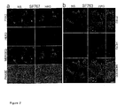
Figure 2 Localization of PYK2 and HER3 in SF763 and SF767 glioma cell
lines. a, b, In SF767 (a) and in SF763 cells (b), PYK2 shows a punctated
distribution throughout the cytoplasm, and is enriched in the perinuclear
region and in some prominent cell protrusions (green). HER3 (red) is largely
colocalized, as shown by overlapping distributions of the two stains in
most puncta (b, insets) and in larger aggregates (yellow). Colocalization is
independent of stimulation by HRG. Cells were fixed and immunostained
against PYK2 (green) and HER3 (red), either unstimulated (NS) or following
stimulation with 5 µg ml-1 Heregulin for 20 min (HRG). Optical sections
obtained by confocal laser scanning microscopy are shown. Scale bar
represents 10 µm.
-
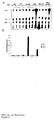
Figure. 3 Association of PYK2 with the C-terminal domain of HER3. a, b, c,
HEK293 fibroblasts were either transfected with combinations of wild-type
proteins (HER2, HER3, PYK2) and their dominant-negative variants (HER2-KM,
HER3-KM, PYK2-KM) (a), with wild-type HER2 and PYK2 combined
with wild-type HER3 or its truncated construct HER3ΔCT (b), or with wild-type
HER2 and PYK2 and add-back mutants of HER3 (c), as indicated.
Tyrosine phosphorylation of PYK2 is dependent on HER2 and HER3 kinase
activity (a), and on binding to the C-terminal domain of HER3 (b).
Coprecipitated HER3 is indicated by an arrow. PYK2 activation is
dependent on Y1257, Y1270 and Y1288 in the C-terminal domain of HER3
(c). PYK2 was expressed tagged at its C-terminus with the vesicular
somatitis virus glycoprotein (VSV). Cells were stimulated with 5 µg ml-1
Heregulin for 20 min (HRG), lysed and subjected to immunoprecipitation
with monoclonal anti-VSV antibody (α-VSV). Immunocomplexes were
analysed by western blotting (WB) with a monoclonal anti-phosphotyrosine
antibody (α-4G10, upper panels). Equal loading of proteins was determined
by reblotting with α-VSV antibody (lower panels).
-
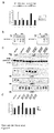
Figure 4 Phosphorylation of GST-PYK2-CT by HER3 upon HRG stimulation.
a, b, SF767 gliomas were either stimulated with 5 µg ml-1 Heregulin for 20
min (HRG) or with 1 µM Phorbol-12-myristate-13-acetate for 10 min (PMA)
(a), or were pretreated with 100 nM Wortmannin for 30 min (WT) (b). PMA
stimulation was used as a negative control. Note that kinase activity of
HER3 is under 1 % of the corresponding HER2 activity when using MBP as
a substrate, in contrast to GST-PYK2-CT (a). Upon HRG stimulation,
phosphorylation of GST-PYK2-CT by HER3 is upregulated, in contrast to
HER2 activity. Influence of WT is negligible, thus excluding involvement of
PI3-K in PYK2 phosphorylation (b). c, HEK293 fibroblasts were transfected
with the combinations of wild-type proteins (HER2, HER3) and their
dominant-negative variants (HER2-KM, HER3-KM) as indicated, and
stimulated with 5 µg ml-1 Heregulin for 20 min (HRG). Only homodimers of
HER3 and heterodimers of HER3 with HER2 induced an increased GST-PYK2-CT
phosphorylation (c, upper panel). Heterodimerization of HER3
with HER2 leads to a stronger phosphorylation of the substrate, indicating
that HER2 is important for HER3 activation (c and d). Transphosphorylation
of HER3 by HER2 was checked by probing the membrane with an anti-phosphotyrosine
antibody α-4G10 (c, upper middle panel). Coprecipitation
of HER2 with HER3 was excluded by probing the membrane with anti-HER2
antibody α-HER2 (c, lower middle panel). Equal loading of proteins
was checked by probing with anti-HER3 antibody (α-HER3) (c, lower
panel). Phosphorylated GST-PYK2-CT is indicated by an arrow. d,
Quantification of the kinase activity shown in the upper panel of Fig. 4c.
-
Figure 5 PYK2 mediates mitogenicity upon HRG stimulation. a, b, SF767
gliomas were pretreated either with 10 µM AG825 for 1 hour or with 100
nM Wortmannin for 30 min (WT), and then stimulated with 5 µg ml-1
Heregulin for 20 min (HRG). Tyrosine phosphorylation of SHC was elevated
by HRG and attenuated by pretreatment with AG825, but not fully
abrogated (a). The same holds also for ERK-2 activity, when cells were
pretreated either with AG825 or with WT (b). Cell lysates were used for
immunoprecipitation with polyclonal anti-SHC (α-SHC) (a), or polyclonal
anti-Erk-2 (α-ERK-2) antibodies (b). α-SHC-immunocomplexes were blotted
with a monoclonal anti-phosphotyrosine antibody (α-4G10) (a), whereas α-ERK-2
immunocomplexes were subjected to MAP-kinase assays (b).
Phosphorylated MBP is indicated by an arrow. c, Tetracyclin-inducible
pheochromocytoma PC12 cells, either stably expressing PYK2-KM (Tet-),
or only endogenous PYK2 (Tet +), were pretreated either with 100 nm
Wortmannin for 30 min (WT), or 10 µM AG825 for 1 hour prior to
stimulation with 5 µg ml-1 Heregulin for 20 min (HRG). Basal ERK-2 activity
is independent of HER2 and PI3-K, whereas the HRG-stimulated ERK-2
activity is dependent on HER2,PI3-K and PYK2. Overexpression of PYK2-KM
leads to a general attenuation of ERK-2 activity (compare Tet- with
Tet + bands). Equal loading of proteins was checked by probing with anti-ERK-2
antibody (α-ERK-2). Phosphorylated MBP is indicated by an arrow.
d, Quantification of the ERK-2 kinase activity shown in Fig. 5c.
-
Figure 6 PYK2 enhances PI3-K activity upon HRG stimulation. a,
Tetracyclin-inducible pheochromocytoma PC12 cells, either stably
expressing PYK2-KM (Tet-) or only endogenous PYK2 (Tet +), were
pretreated either with 100 nm Wortmannin for 30 min (WT), or 10 µM
AG825 for 1 hour prior to stimulation with 5 µg ml-1 Heregulin for 20 min
(HRG). Lysates were subjected to α-4G10 immunoprecipitation and PI3-K
assays were performed (see Methods section). PI3-K activity is strongly
dependent on PYK2 upon HRG stimulation, and is diminished by AG825.
Phosphorylated Phosphatidylinositol is indicated. b, Quantification of the
PI3-K kinase activity shown in Fig. 6a.
-
Figure 7 PYK2-KM inhibits tumor invasion upon HRG stimulation. a, C6
gliomas were retrovirally infected with either a control vector pLXSN
(mock), PYK2, dominant negative PYK2 mutant PYK2-KM, or pretreated
with a MEK1 inhibitor PD98059 (25 µM) for 30 min, and tumor invasion
assays were performed (see Methods section). b, Invasion is supressed to
the same extent by PD98059 and by overexpression of PYK2-KM
(p>0.95). c, SF767 gliomas were retrovirally infected with pLXSN or
PYK2-KM. d, Tumor invasion is supressed by overexpresssion of PYK2-KM
in SF767 (p<0.008), and also in SF763 cell line (p<0.005), as shown by
using the same assay. Representative bright-field micrographs of cells that
migrated through the 8 µm filters in 16 h are shown. Scale bars represent
100 µm (a) and 50 µm (c).
-
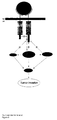
Figure 8 Role of PYK2 in HER2/HER3 signalling. Model indicates a novel
signal transduction pathway, which leads from HRG stimulation to MAPK
activation and induces tumor invasion. For details, see discussion. TM
indicates the transmembrane domain, JM the juxtamembrane region.
Arrows with an encircled B or P indicate binding and phosphorylation,
respectively.
Examples
1. Methods
1.1 Materials and general methods
-
Media were purchased from Gibco, fetal bovine serum (FBS) and horse
serum from Sigma. Hybond ECL membranes and γ-32P-ATP were purchased
from Amersham, PP1, AG825 (ref. 45), Wortmannin (WT), PD98059 and
lonomycin (IONO) from Calbiochem. Antibodies raised against following
proteins were used: PYK2 (polyclonal goat antibody N19, Santa Cruz, and
polyclonal rabbit antibody (pAb) Upstate Biotechnology, Inc. (UBI)), ERK2
(pAb C14, K23, Signal Transduction), SHC (pAb (ref. 46), mAb, Affiniti),
HER3 (monoclonal mouse antibody (mAb) 2F12, UBI), p85 (mAb UB93-3,
UBI), VSV (mAb P5D4, Roche Diagnostics), and phosphotyrosine (mAb
4G10, UBI). HRP-coupled secondary antibodies were purchased from
Biorad, flourochrome-coupled secondary antibodies from Molecular Probes.
Transwell chambers (0.3 cm2, 8 µm) were purchased from Costar. Growth
Factor Reduced Matrigel (GFRM) was purchased from Collaborative
Biomedical Products. Thin-layer Chromatography plates (Silica Gel 60)
precoated with oxalate were from Merck. Recombinant human GST-HRG
fusion protein (HRG) and GST-PYK2-CT were produced in E. coli and
purified as described (4) or using standard methods. Cell lines HEK293
(ATCC CRL-1573), rat C6 (ATCC CCL-107) and PC12 (ATCC CRL-1721)
and human SF763 (Sugen Inc.), SF767 (Sugen Inc.) and PhoenixA (ATCC
SD-3443) were cultured according to the supplier's protocol. Tetracyclin-inducible
PC12 system stably expressing PYK2-KM (Tet-off) was described
previously (25).
1.2 Immunofluorescence studies and confocal microscopy
-
Briefly, SF763 and SF767 (3x10⁵ cells) were grown on coverslips and
starved for 24 h. After stimulation with 5 µg/ml HRG for 20 min, cells were
fixed with 3.7% formaldehyd and permeabilized with 0.2% saponin
(Sigma) in 3% BSA (Sigma). Blocking was performed with 3% BSA for 1 h.
-
PYK2 and HER3 proteins were labeled with the indicated primary
antibodies and stained using a fluorochrome-coupled donkey anti-goat α-488
secondary antibody for PYK2, and TRITC-coupled rabbit anti-mouse
secondary antibody for HER3 (Molecular Probes). Confocal microscopy was
performed using an LSM 410 microscope (Zeiss) as described (47).
1.3 Plasmid constructs and site-directed mutagenesis
-
pcDNA3.1-PYK2-VSV and pcDNA3.1-PYK2-KM-VSV constructs were
generated using the pRK5 constructs and standard methods. PYK2-KM
was generated as described (25). GST-PYK2-CT was generated by using
the pRK5 construct and amplifying the C-terminus of PYK2 by PCR
(positions 716-1009). The fragment was subcloned into the procaryotic
expression vector pGEX-5X1 (Pharmacia). Tyrosine to phenylalanine
mutations in HER3 were performed using the pcDNA3.1-HER3 construct
and the QuickChange site-directed mutagenesis kit (Stratagene) according
to the manufacturers protocol. Correct incorporation of the mutations was
verified by DNA sequencing.
1.4 Transient overexpression of PYK2, PYK2-KM, HER2, HER2-KM, HER3,
and HER3-KM proteins in eukaryotic cells
-
The HEK293 cell system was used for transient protein expression.
HEK293 cells were maintained in DMEM supplemented with 10% FCS,
penicillin and streptomycin (100 IU/ml) at 7.5% CO2 and 37°C.
Transfections were carried out using a modified calcium phosphate method
(48). Briefly, 2.5x10⁵ cells were incubated overnight in 3 ml of growth
medium. 1 µg of supercoiled DNA was mixed with 0.25 M CaCl2 solution
in a final volume of 400 µl. The mixture was added to the same volume of
2x transfection buffer (50 mM BES, pH 6.95, 280 mM NaCI, 1.5 mM
Na2HPO4) and incubated for 15 min at room temperature before it was
added dropwise to the cells. After incubation for 12 h at 37°C under 3%
CO2, the medium was removed, cells were washed twice with PBS and
were then starved for 24 h in DMEM supplemented with 0.1 % FCS.
1.5 Western Immunoblotting
-
SF763, SF767 or transfected HEK293 cells were either left untreated or
were pretreated with PP1 (10 µM), AG825 (10 µM), Wortmannin (WT)
(100 nM) and PD98059 (25 µM) for 30-60 min following stimulation with
5 µg/ ml recombinant human HRG for 20 min or with 5 µM IONO for 5 min
at 37°C. Upon HRG or IONO stimulation, the cells were lysed on ice in a
lysis buffer (50 mM HEPES pH 7.5, containing 150 mM NaCI, 1 mM EDTA,
10% (v/v) glycerol, 1% (v/v) Triton X-100, 1 mM sodium fluoride, 1 mM
phenylmethylsulfonyl fluoride, 1 mM sodium orthovanadate, 1 mM β-glycerolphosphate,
10 mg/ml aprotinin). Crude lysates were centrifuged at
12500 g for 20 min at 4°C. For immunoprecipitations, the appropriate
antiserum and 30 µl of protein A-Sepharose (Pharmacia) was added to the
cleared lysate and incubated for 3 h at 4°C. Immunoprecipitates were
washed with a washing buffer (20 mM HEPES pH 7.5, containing 150 mM
NaCI, 1 mM EDTA, 1 mM Sodiumflouride 10% (v/v) glycerol, 1% (v/v)
Triton X-100). Sample buffer containing SDS and 2-mercaptoethanol was
added and the samples were denaturated by heating at 95°C for 4 min.
-
Proteins were fractionated by SDS-PAGE and electrophoretically transferred
to nitrocellulose filters. For immunoblot analysis, nitrocellulose filters were
first incubated with mouse monoclonal or rabbit polyclonal primary
antibodies for 3 h at 4°C. Next, a HRP-coupled goat anti-mouse or goat
anti-rabbit secondary antibody was added (Biorad), followed by an
enhanced chemoluminescence (ECL) substrate reaction (Amersham). The
substrate reaction was detected on Kodak X-Omat film. Filters that were
used more than once with different antibodies were stripped according to
the manufacturer's protocol, blocked and reprobed.
1.6 Generation of recombinant retroviruses and retrovirus-mediated gene
transfer
-
Briefly, pLXSN-PYK2 and pLXSN-PYK2-KM were generated by cloning an
EcoRI-XhoI fragment from pRK5 carrying the cDNAs of WT PYK2 and
kinase-inactive PYK2, K457M (PYK2-KM), respectively, into pLXSN.
Amphotrophic virus titer, which was generated by transient transfection of
retrovirus expression plasmids into the virus producer cell line PhoenixA
(ATCC), was determined by infecting NIH-3T3 cells with serial dilutions of
retrovirus-containing, cell-free PhoenixA supernatants and counting the
number of G418-resistant colonies. The titers were approximately 1x10⁶
cfu/ml both for PYK2 and PYK2-KM virus supernatants. Subconfluent C6,
SF763 and SF767 cells (9x10⁵ cells) were incubated with supernatants of
cells releasing high titers of pLXSN-PYK2 or pLXSN-PYK2-KM viruses
(1x10⁶ G418 cfu/ml) for 24 h in the presence of Polybrene (4 mg/ml,
Aldrich).
1.7 In-vitro-kinase assay
-
MAP-kinase and PI3-kinase assays were performed as described previously
(49, 50).
1.8 Tumor invasion assay
-
Tumor invasion assay was performed as described previously (31). Briefly,
3x10⁵ cells were plated on transwell chambers precoated with 100 µg
GFRM. Conditioned NIH-3T3 medium was used as a chemoattractant. Cells
were stimulated with 5 µg/ml HRG during the experiment. Following 16 h
of incubation, non-invading cells were removed with cotton swabs,
whereas invading cells were fixed, stained with Crystal violet and counted
under bright-field illumination using an Axiovert135 inverted microscope
(Zeiss). Counts from 4 filters for each strain were pooled and compared
among different strains using the two-tailed t-test.
2. Results
2.1 Tyrosine-phosphorylation of PYK2 is dependent on HER2 and HER3
-
PYK2 gets tyrosine-phosphorylated in human glioma cell line SF767 upon
stimulation by HRG (Fig. 1a). In order to evaluate the mechanism of HRG-induced
PYK2 tyrosine-phosphorylation, we inhibited two candidate protein
tyrosine kinases, c-src and HER2. It has previously been reported that c-src
kinase associates with HER2 after HRG-stimulation and phosphorylates
PYK2 upon GPCR stimulation (20). C-src-inhibition with PP1 prior to HRG
stimulation indicates that c-src does not mediate PYK2 tyrosine-phosphorylation
after HRG treatment. In contrast, stimulation by
lonomycin, which leads to an influx of Ca2+-ions analogously to a GPCR
stimulation (25), induces a tyrosine-phosphorylation of PYK2 that is
dependent on c-src activity (Fig. 1a, left vs. right panel, lanes 3 and 4).
-
In the breast carcinoma cell line MDA-MB-435 it has been shown that
HRG-induced activation of HER2, which is mediated by heterodimerization
between HER2 and HER3, leads to tyrosine-phosphorylation of PYK2 (26).
A tyrosine phosphorylated protein of Mr = 113 kDa, which we identified as
PYK2, coprecipitates with HER3 in SF767 cells prior to stimulation with
HRG (Fig. 1b, upper and middle panels, lanes 1-4). In contrast,
precipitation of HER2 reveales no association with PYK2. Upon HRG-stimulation
tyrosine-phosphorylation of PYK2 increases (Fig. 1b, upper
panel, lanes 5 and 7), but is attenuated in presence of HER2 inhibitor
AG825 (Fig. 1b, upper panel, lanes 6 and 8), indicating that tyrosine-phosphorylation
of PYK2 is dependent on the HER2 kinase activity. The
amount of PYK2 that coprecipitates with HER3 is not elevated by HRG-stimulation
(Fig. 1b, middle panel, lanes 1 and 3), but decreases after
addition of AG825 (Fig. 1b, middle panel, lanes 2 and 4). The same results
were also obtained in the glioma cell line SF763, and suggest a
constitutive association of PYK2 with HER3, which is dependent on the
HER2 kinase.
-
To further analyse a cellular colocalization of PYK2 and HER3, we
performed immunoflourescence studies in SF763 and SF767 cell lines
using a laser scanning confocal microscope (Fig. 2). In unstimulated cells
PYK2 is mainly localized to the perinuclear cytoplasm in a punctuated
pattern, and distribution of HER3 is largely coincident. Upon stimulation
with HRG, the colocalization of PYK2 with HER3 remained unchanged.
Thus, immunofluorescence studies confirmed a constitutive, HRG-independent
association between PYK2 and HER3.
2.2 PYK2 associates with the intracellular region of HER3
-
We used an ectopic overexpression system to investigate in detail how
tyrosine-phosphorylation of PYK2 depends on binding to HER3. HEK293
fibroblasts were used to express either wild-type HER2, HER3 and PYK2 or
dominant-negative mutant constructs HER2-KM, HER3-KM and PYK2-KM,
where the lysine critical for ATP-binding was exchanged to alanine,
rendering the kinase inactive. Tyrosine-phosphorylation of PYK2 was
elevated upon HRG-stimulation of cells expressing all the wild-type
constructs (Fig. 3a, lanes 1 and 2). However, in cells expressing HER3-KM
(Fig. 3a, lanes 3 and 4) or HER2-KM (Fig. 3a, lanes 5 and 6), HRG-stimulation
failed to induce PYK2 tyrosine-phosphorylation. This
observation is consistent with the data from glioma cell lines (Fig. 1b),
where inhibition of HER2 abrogated PYK2 activation, but further implies
that HRG-induced PYK2 activation is dependent on functional kinase
activities of HER2 and HER3.
-
Next we used a mutant of HER3 with a C-terminal deletion (HER3ΔCT) to
analyse the contribution of the C-terminal domain to HRG-induced PYK2
tyrosine-phosphorylation (Fig. 3b). We observed a coprecipitating protein
of Mr = 180 kDa in PYK2 immunocomplexes, which was phosphorylated
and confirmed to be HER3 (Fig. 3b, lanes 1 and 2). The deletion mutant of
HER3 abrogated the tyrosine-phosphorylation of PYK2 and also
coprecipitation of HER3, indicating that PYK2 associates with the C-terminal
region of HER3 (Fig. 3b, lanes 3 and 4). The intracellular domain
of HER3 harbours 13 phosphorylation sites that are presumably
transphosphorylated by HER2 after HRG-stimulation. The tyrosines Y1035,
Y1178, Y1203, Y1241, Y1257 and Y1270 are potential docking sites for
the src-homology 2 (SH2) domains of the regulatory domain p85 of PI3-K
(27), whereas Y1309 is a binding site for SHC (28). To identify the
putative binding sites for PYK2 on the C-terminal domain of HER3, we
used 13 add-back mutants, replacing all tyrosine residues to phenylalanines
and exchanging each one back to a tyrosine. We performed overexpression
experiments in HEK293 fibroblasts, using wildtype PYK2 and HER2, and
single add-back mutants of HER3 (Fig. 3c). Using this approach, we
identified three tyrosine residues Y1257, Y1270 and Y1288, which are
critical for elevated PYK2 tyrosine-phosphorylation upon HRG-stimulation
(Fig. 3c, lanes 13-18). These observations show that the HRG-induced
stimulation of PYK2 tyrosine-phosphorylation depends on its binding to
Y1257, Y1270 and Y1288 in the C-terminal domain of HER3.
2.3 Tyrosine-phosphorylation of PYK2 is dependent on HER3 kinase
activity
-
It has been implied that, in contrast to HER2, kinase activity of HER3 is
impaired (29), although HER3 can bind ATP and its analog TNP-ATP (30).
To identify the kinase which is responsible for the PYK2 tyrosine-phosphorylation
upon HRG-stimulation, we conducted in vitro kinase
assays, precipitating either HER2 or HER3, using myelin basic protein
(MBP) and a GST-fusion protein of the C-terminal region of PYK2 (GST-PYK2-CT)
as substrates (Fig. 4a). Upon stimulation of SF767 cells either
with HRG, or with Phorbol-12-myristate-13-acetate (PMA), MBP became
phosphorylated by HER2, but not by HER3 (Fig. 4a, white bars).
Surprisingly, however, GST-PYK2-CT became phosphorylated by HER3 in
a HRG-stimulation-dependent way, but not by HER2 (Fig. 4a, black bars).
As it has been shown that PI3-K binds to the cytoplasmic tail of HER3, we
investigated a potential role of PI3-K in PYK2 phosphorylation by
precipitating either HER2 or HER3 in the presence or absence of PI3-K-inhibitor
Wortmannin (WT) (Fig. 4b). The results indicate that PI3-K is not
responsible for the direct phosphorylation of GST-PYK2-CT. Consistent
with this finding, precipitation of PYK2 under the same experimental
conditions showed that its elevated tyrosine-phosphorylation upon HRG-stimulation
is independent of PI3-K. Taken together, these data suggest
that HER3 is the kinase which phosphorylates the C-terminal region of
PYK2.
-
To verify that HER3 directly phosphorylates PYK2, we overexpressed HER2
and HER3 either separately, or together in combinations of wild-type
constructs and dominant-negative mutants in HEK293 cells (Fig. 4c).
Receptor-immunocomplexes were subjected to in vitro kinase assay and
revealed that, after HRG-stimulation, HER3 phosphorylates GST-PYK2-CT,
whereas HER2 does not (Fig. 4c, upper panel, lanes 3, 4, 5 and 6 vs. lanes
1 and 2). HER3 homodimers also phosphorylated GST-PYK2-CT, but to a
lesser extent compared to transactivated HER3 (Fig. 4c, upper panel, lanes
3 and 4). To show that HER3 is transphosphorylated by HER2 upon HRG-stimulation,
we probed with monoclonal phosphotyrosine antibody α-4G10
(Fig. 4c, middle upper panel). We also show that there was no significant
coprecipitation of HER2 in the HER3 immunocomplex under our assay
conditions, confirming that HER2 is not the kinase which phosphorylates
GST-PYK2-CT (Fig. 4c, middle lower panel). We conclude that HER3
directly phosphorylates PYK2 upon HRG-stimulation.
2.4 PYK2 amplifies mitogenicity of the HER2/HER3 signalling pathway
-
Upon stimulation of HER3 and HER2, PI3-K and SHC bind to the C-terminus
of HER3 and mediate mitogenicity through the Ras/Raf pathway (15). To
test the influence of HER2 and PI3-K on MAPK activation, we added their
specific inhibitors AG825 and Wortmannin (WT), respectively, to SF767
cells prior to HRG-stimulation. Then we precipitated SHC or performed
MAP-kinase assays. HRG-stimulated tyrosine-phosphorylation of SHC and
ERK-2 activity were diminished, but not fully abrogated by inhibition of
HER2 (Fig. 5a, 5b, left panel). The analogous experiment using WT for
inhibition of PI3-K revealed that ERK-2 activity was reduced by WT (Fig. 5b,
right panel). These findings indicate that HRG-induced mitogenicity only
partially depends on HER2 and PI3-K.
-
To characterize in more detail the role of PYK2 in signalling downstream of
HER2/HER3, we used a tetracyclin-inducible system (Tet-off) in
pheochromocytoma cell line PC12. PC12 cells are rich in PYK2, so that in
the presence of Tet endogenous PYK2 is predominantly expressed,
whereas its removal leads to overexpression of dominant-negative PYK2,
PYK2-KM. We inhibited either HER2 or PI3-K with AG825 and WT,
respectively, prior to stimulation with HRG, precipitated ERK-2 and
subjected the immunocomplexes to MAP-kinase assays (Fig. 5c). Basal
ERK-2 activity was not influenced by AG825 and WT, but was abrogated
by PYK2-KM expression. HRG-stimulated ERK-2 activity, however, was
attenuated by the two inhibitors, and also abrogated by PYK2-KM
expression. These findings are consistent with the results obtained in
SF767 (Fig. 5b). Taken together, these results indicate that the constitutive
ERK-2 activity depends on PYK2, and is independent of HER2 and PI3-K,
whereas HRG-stimulated ERK-2 activity depends on HER2 and PI3-K, and
also on PYK2.
-
In addition to its role in cell proliferation and in prevention of apoptosis, an
influence of PI3-K on carcinoma invasion has previously been shown (31).
We therefore investigated the potency of PYK2 and its dominant-negative
mutant PYK2-KM to regulate PI3-K activation upon HRG-stimulation. Using
the Tet-off system in PC12 cells we subjected cell lysates to PI3-K assays,
where we observed a PYK2-dependent PI3-K activation upon HRG-stimulation
(Fig. 6, upper panel, lanes 1 vs. 2 with 7 and 8). Inhibition of
HER2 kinase activity did not fully abrogate PI3-K activity, indicating a
HER2-independent mechanism of PI3-K activation (Fig. 6, upper panel,
lanes 8 and 12). These results imply an important role of PYK2 in
mediating mitogenicity to the MAPK signalling pathway, and in PI3-K
activation upon HRG-stimulation.
2.5 PYK2-KM inhibits tumor invasion by blocking mitogenicity of the
HER2/HER3 signalling pathway
-
Gliomas represent a highly malignant brain tumor phenotype with a poor
prognosis (32). It has been shown that PI3-K links α6β4-integrin signalling
to invasive behaviour of breast tumor cells (31). Further, it has been
reported that activation of MAPK through α6β4-integrin signalling is
relevant to invasion, due to its importance in migration and its ability to
phosphorylate myosin light chain kinase (33). Using C6 gliomas as a model
system for tumor invasion (34), we tested whether the dominant-negative
mutant of PYK2, PYK2-KM, can inhibit tumor invasion by blocking the
MAPK pathway. We retrovirally infected the cells with PYK2-KM prior to
stimulation with HRG, and also pretreated the cells with the MEK1 inhibitor
PD98059 (Fig. 7a). MEK1- inhibition strongly attenuated invasiveness and
a comparable abrogation of the invasive phenotype was observed upon
infection of cells with PYK2-KM. The mitogenic signal of the HER2/HER3
dimer seems to be downregulated by PYK2-KM, however, overexpression
of PYK2 in C6 cells did not alter their invasive phenotype (Fig. 7b). PYK2
expression in C6 cells is comparably weaker than in SF763 or SF767 cells,
but this does not seem to interfere with their invasive potency. We also
tested glioma cell lines SF763 and SF767 in the tumor invasion assay,
after viral infection with the PYK2-KM construct. Again, a strong inhibition
of the invasive behaviour of tumor cells by PYK2-KM was observed (Fig.
7d). These results demonstrate that PYK2 can mediate mitogenicity
through the MAPK pathway, which plays an important role in the invasive
behaviour of gliomas upon HRG-stimulation.
3. Discussion
-
Cytoplasmic protein tyrosine kinase PYK2 is at the convergence point of
transduction pathways that transmit signals from stimulated integrins, G
protein-coupled receptors and PTK receptors to downstream effectors. An
important stimulus that activates PYK2 is HRG (25). Both PYK2 and HRG
are predominantly expressed in the central nervous system, and the genes
coding for the two proteins are localized in the close proximity to each
other on the chromosome 8 (34). HRG is a promiscuous ligand for HER3
and HER4, members of the erbB family of RPTKs, and the erbB signalling
module represents one of the most potent inducers of mitogenicity (35).
Binding of HRG leads to formation of HER2/HER3 and HER2/HER4
heterodimers, thereby activating HER2 which transphosphorylates HER3 or
HER4 (35). Signalling molecules SHC and PI3-K are known to bind to the C-terminal
region of HER3 and to promote mitogenicity (4, 15, 35). These
pieces of information, obtained in several model systems, prompted us to
explore an HRG-stimulated signalling pathway involving HER2/HER3 and
PYK2 in glioblastoma cell lines which are devoid of HER4. Based on the
presented data, we propose a model in which PYK2 is phosphorylated by
HER3 upon HRG stimulation, and induces invasiveness through the MAPK
pathway (Fig.8).
-
Immunoprecipitation assays indicate a constitutive association of PYK2
with HER3, which is promoted by HER2 activity (Fig.1).
Immunofluorescence studies confirmed the constitutive assocation,
showing that the two proteins co-localize in a punctuated pattern
throughout the cytoplasm independent of HRG stimulation (Fig.2). It is
known that HER3 is internalized through the clathrin-mediated endocytotic
pathway (17). Similar punctuated distributions have recently been shown
for several proteins associated with this pathway, e.g. mHip1r and EGFR
(36, 37). Centripetal movement of the clathrin-coated vesicles towards the
perincular region, which occurs on the time scale of several minutes, has
been directly demonstrated by using a GFP-chlatrin fusion in Dictyostelium
and COS-1 cells (38, 39). A prolonged activation state of HER3/PYK2
complexes within endosomes during recycling would enable recurrent
association of other signalling molecules and thus serve to amplify the
initiating signal. This prolonged accessibility of HER3/PYK2 complexes and
their transport towards the site of MAPK activity could explain the
exceptionally strong mitogenicity of HER2/HER3 heterodimers, compared to
other members of the erbB family (35). Indeed, it has been shown that
HER2/HER3 heterodimers are getting recycled, whereas HER1-containing
dimers are degraded via ubiquitination pathway (17).
-
Although association of PYK2 with HER3 and its recycling appear to be
HRG-independent, tyrosine phosphorylation of PYK2 is induced by HRG
stimulation. Our evaluation of the mechanism by which PYK2 is activated
upon HRG stimulation shows that intact kinase activity of HER3 is critical
for PYK2 activation (Fig.3). Specifically, tyrosine residues Y1257, Y1270
and/or Y1288 in the C-terminal region of HER3 are shown to be important
for PYK2 activation. Finally, in vitro kinase assays showed that HER3
directly phosphorylates GST-PYK2-CT (Fig.4). By inhibition experiments,
we could exclude HER2, c-src and PI3-K as proteins that directly
phosphorylate PYK2. The HER3 kinase activity has possibly not been
unraveled until now because previous studies used artificial substrates
(28).
-
We show a negative effect of dominant negative PYK2, PYK2-KM, on
MAPK activation, demonstrating that mitogenicity depends on PYK2
activity (Fig.5). It has been shown that cells overexpressing PYK2 exhibit
elevated tyrosyl phosphorylated SHC and subsequent ERK-2 activity (40).
We did not observe a direct interaction between PYK2 and SHC, but it has
been proposed recently that SHC associates with PYK2 through GRB2 in
platelets dependent on αIIbβ3 integrin, thus linking extracellular signal to
the Ras/Raf pathway (41). It is possible that GRF2 binds to activated
PYK2, leading to subsequent tyrosine phosphorylation of SHC, which
contributes to increased mitogenicity. We also show that PYK2-KM
attenuates PI3-K activity (Fig.6). HER3 harbours six potential docking sites
for the SH2 domain of the PI3-K subunit p85, and the one proline-rich
sequence that forms a consensus binding site for the SH3 domain of p85,
all potentially contributing to an association of HER3 with p85 (26). Also,
a constitutive association between PYK2 and p85 in platelets was reported
(42), where one YXXM motif in PYK2 could serve for binding to the SH2
domain of p85. Indeed, immunoprecipitation of p85 revealed HRG-dependent
association of tyrosyl phosphorylated proteins of Mr = 113 kDa
and 180 kDa. These proteins were identified as PYK2 and HER3,
suggesting that HER3, PYK2 and PI3-K are constituents of a multiprotein
complex.
-
In our model we propose that PYK2 is a key element in transmitting HRG-induced
mitogenicity. Part of the singalling from PYK2 to ERK-2 seems to
be transmitted through PI3-K and SHC, but there is also a more direct
pathway (Fig.8). The PYK2-dependent ERK-2 activation also seems to be
partly independent of HER2 (Fig.5). These findings suggest that PYK2 is
involved in the control of multiple downstream effectors, which in turn all
influence the MAPK pathway (Fig.8).
-
We show that the dominant PYK2 mutant PYK2-KM suppresses tumor
invasiveness in three glioma cell lines (Fig.7). This result correlates with its
influence on ERK-2 activity (Fig.6). Also, PYK2-KM abrogated invasiveness
to the same extent as inhibition of MEK1. These results strongly indicate
that PYK2 regulates invasiveness in gliomas through the MAPK pathway.
It has been shown that ERK activity can regulate myosin phosphorylation,
leading to actin-myosin association and cell contraction of the ECM (43),
and that ERK can facilitate cell invasion and protect cells from apoptosis
(44). Increased MAPK activity showed in our case an increased invasive
behavior. Taken together, we show for the first time that PYK2 is a direct
substrate of HER3, potentiates PI3-K activity and enhances mitogenicity
through ERK2 and in gliomas, leading to a strongly invasive phenotype.
Literature
-
- 1. Altman, J. Proliferation and migration of undifferentiated precursor
cells in the rat during postnatal gliogenesis. Exp. Neurol. 15, 263-278
(1966).
- 2. Goldman, J.E. Regulation of oligodendrocyte differentiation. Trends
Neurosci. 15, 359-362 (1992).
- 3. Faber-Elman, A., Solomon, A., Abraham, J.A., Marikovsky, M. &
Schwartz, M. Involvement of wound-associated factors in rat brain
astrocyte migratory response to axonal injury. In vitro simulation. J. Clin.
Invest. 97, 162-171 (1996).
- 4. Wallasch, C. et al. Heregulin-dependent regulation of HER2/neu
oncogenic signaling by heterodimerization with HER3. EMBO J. 14, 4267-4275
(1995).
- 5. Busfield, S.J. et al. Characterization of a neuregulin-related gene,
Don-1, that is highly expressed in restricted regions of the cerebellum and
hippocampus. Mol. Cell. Biol. 17, 4007-4014 (1997).
- 6. Carraway, K.L. III et al. Neuregulin-2, a new ligand of ErbB3/ErbB4-receptor
tyrosine kinases. Nature 387, 512-516 (1997).
- 7. Chang, H., Riese, D.J., II, Gilbert, W., Stern, D.F. & McMahan, U.J.
Ligands for ErbB-family receptors encoded by a neuregulin-like gene. Nature
387, 509-512. (1997).
- 8. Fischbach, G.D. & Rosen, K.M. ARIA: a neuromuscular junction
neuregulin. Annu. Rev. Neurosci. 20, 429-458 (1997).
- 9. Zhang, D. et al. Neuregulin-3 (NRG3): a novel neural tissue-enriched
protein that binds and activates ErbB4. Proc. Natl. Acad. Sci. USA 94,
9562-9567 (1997).
- 10. Ishiguro, H. et al. Structure and function of a novel ErbB ligand,
NTAK. Nihon Shinkei Seishin Yakarigaku Zasshi 18, 137-142 (1998).
- 11. Harari, D. et al. Neuregulin-4: a novel growth factor that acts through
the ErbB-4 receptor tyrosine kinase. Oncogene 18, 2681-2689 (1999).
- 12. Holmes, W.E. et al. Identification of heregulin, a specific activator of
p185erbB2. Science 256, 1205-1210 (1992).
- 13. Orr-Urteger, A. et al. Neural expression and chromosomal mapping
of Neu dedifferentiation factor to 8p12-p21. Proc. Natl. Acad. Sci. USA 90,
1867-1871 (1993).
- 14. Plowman, G.D. et al. Molecular cloning and expression of an
additional epidermal growth factor receptor-related gene. Proc. Natl. Acad.
Sci. USA 87, 4905-4909 (1990).
- 15. Alroy, I. & Yarden Y. The ErbB signaling network in embryogenesis
and oncogenesis: signal diversification through combinatorial ligand-receptor
interactions. FEBS 410, 83-86 (1997).
- 16. Fiddes, R.J. et al. Analysis of Grb7 recruitment by heregulin-activated
erbB receptors reveals a novel target selectivity for erbB3. J. Biol. Chem.
273, 7717-7724 (1998).
- 17. Ceresa, B.P. & Schmid, S.L. Regulation of signal transduction by
endocytosis. Curr. Opin. Cell. Biol. 12, 204-210 (2000).
- 18. Lev, S. et al. Protein tyrosine kinase PYK2 involved in Ca2+-induced
regulation of ion channel and MAP kinase functions. Nature 376, 737-745
(1995).
- 19. Girault, J.A., Costa, A., Derkinderen, P., Studler, J.M. & Toutant, M.
FAK and PYK2/CAKbeta in the nervous system: a link between neuronal
activity, plasticity and survival? Trends Neurosci. 22, 257-263 (1999).
- 20. Dikic, I., Tokiwa, G., Lev, S., Courtneidge, S.A. & Schlessinger, J.
A role for Pyk2 and Src in linking G-protein-coupled receptors with MAP
kinase activation. Nature 383, 547-50 (1996).
- 21. Andreev, J. et al. Identification of a new Pyk2 target protein with Arf-GAP
activity. Mol. Cell. Biol. 19, 2338-2350 (1999).
- 22. Avraham, H., Park, S.-Y., Schinkmann, K. & Avraham, S.
RAFTK/PYK2-mediated cellular signalling. Cell. Signal. 12, 123-133(2000).
- 23. Yu, H. et al. Activation of a Novel Calcium-dependent Protein-tyrosine
Kinase. J. Biol. Chem. 271, 29993-29998 (1996).
- 24. Tokiwa, G., Dikic, I., Lev, S. & Schlessinger, J. Activation of Pyk2
by stress signals and coupling with JNK signaling pathway. Science 273,
792-794 (1996).
- 25. Zwick, E., Wallasch, C., Daub, H. & Ullrich, A. Distinct Calcium-dependent
pathways of epidermal growth factor receptor transactivation
and PYK2 tyrosine phosphorylation in PC12 cells. J. Biol. Chem. 274,
20989-20996 (1999).
- 26. Zrihan-Licht, S. et al. RAFTK/Pyk2 tyrosine kinase mediates the
association of p190 RhoGAP with RasGAP and is involved in breast cancer
cell invasion. Oncogene 19, 1318-1328 (2000).
- 27. Hellyer, N.J., Cheng, K. & Koland, J.G. ErbB3 (HER3) interaction with
the p85 regulatory subunit of phosphoinositide 3-kinase. Biochem J. 333,
757-763, (1998).
- 28. Prigent, S.A. & Gullick, W.J. Identification of c-erbB-3 binding sites
for phosphatidylinositol 3'-kinase and SHC using an EGF receptor/c-erbB-3
chimera. EMBO J. 13, 2381-2841 (1994).
- 29. Guy, P.M., Platko, J.V., Cantley, L.C., Cerione, R.A. & Carraway,
K.L.III. Insect cell-expressed p180erbB3 possesses an impaired tyrosine
kinase activity. Proc. Natl. Sci. USA 91, 8132-8136 (1994).
- 30. Sierke, S.L., Cheng, K., Kim, H.-H. & Koland, J.G. Biochemical
characterization of the protein kinase homology domain of the ErbB3 (HER3)
receptor protein. Biochem. J. 322, 757-763 (1997).
- 31. Shaw, L.M., Rabinovitz, I., Wang, H.H., Toker, A., & Mercurio, A.M.
Activation of phosphoinositide 3-OH kinase by the alpha6beta4 integrin
promotes carcinoma invasion. Cell 91, 949-60 (1997).
- 32. Berens, M.L. & Giese, A. Biology and oncology of invasive glioma
cells. Neoplasia 3, 208-219 (1999).
- 33. Klemke, R.L. et al. Regulation of cell motility by mitogen-activated
protein kinase. J Cell. Biol. 137, 481-492 (1997).
- 34. Kaye, A.H., Morstyn, G., Gardner, I. & Pyke, K. Development of a
xenograft glioma model in mouse brain. Cancer Res. 46, 1367-1373
(1986).
- 35. Inazawa, J. et al. Precise localization of the human gene encoding
cell adhesion kinase β (CAKβ/PYK2) to chromosome 8 at p21.1 by
fluorescence in situ hybridization. Hum. Genet. 98, 508-510 (1996).
- 36. Klapper, L.N., Kirschbaum, M.H., Sela, M. & Yarden, Y. Biochemical
and clinical implications of the ErbB/HER signaling network of growth factor
receptors. Adv Cancer Res. 77, 25-79 (2000).
- 37. Engqvist-Goldstein, Å.E.Y., Kessels, M.M., Chopra, V.S., Hayden,
M.R. & Drubin, D.G. An actin-binding protein of the sla/huntington
interacting protein 1 family is a novel component of clathrin-coated pits and
vesicles. J. Cell Biol. 147, 1503-1518 (1999).
- 38. Sorkina, T., Bild, A., Tebar, F. & Sorkin, A. Clathrin, adaptors and
eps15 in endosomes containing activated epidermal growth factor
receptors. J. Cell. Sci. 112, 317-327 (1999).
- 39. Damer, C.K. & O'Halloran, T.J. Spatially regulated recruitment of
clathrin to the plasma membrane during capping and cell translocation. Mol.
Biol. Cell 11, 2151-2159 (2000).
- 40. Gaidarov, I., Santini, F., Warren, R.A. & Keen, J.H. Spatial control of
coated-pit dynamics in living cells. Nature Cell. Biol. 1, 1-7 (1999).
- 41. Ohmori, T., Yatomi, Y., Asazuma, N., Satoh, K. & Ozaki, Y.
Involvement of proline-rich tyrosine kinase 2 in platelet activation: tyrosine
phosphorylation mostly dependent on αIIbβ3 integrin and protein kinase C,
translocation to the cytoskeleton and association with Shc through Grb2.
Biochem. J. 347, 561-569 (2000).
- 42. Sayed, M.R., Sheid, M.P., Stevens, C.M. & Durino, V. Thrombin-stimulated
phosphatidylinositol3-kinase activity in platelets is associated
with activation of PYK2 tyrosine kinase: Activation of both enzymes is
aggregation independent. J. Cell. Physiol. 183, 314-320 (2000).
- 43. Brunet, A. A. et al. Akt promotes cell survival by phosphorylating and
inhibiting a forkhead transcription factor. Cell 96, 857-868 (1999).
- 44. Nguyen, D.H. et al. Myosin light chain kinase functions downstream
of Ras/ERK to promote migration of urokinase-type plasminogen activator-stimulated
cells in an integrin-selective manner. J. Cell. Biol. 146, 149-164
(1999).
- 45. Chun-Ming, T. et al. Enhancement of chemosensitivity by Tyrphostin
AG825 in high-p185neu expressing non-small cell lung cancer cells. Cancer
Res. 56, 1068-1074 (1996).
- 46. Seedorf, K. et al. Dynamin binds to SH3 domains of phospholipase
C gamma and GRB-2. J. Biol. Chem. 269, 16009-16014 (1994).
- 47. Weber, I. et al. Cytokinesis mediated through the recruitment of
cortexillins into the cleavage furrow. EMBO J. 18, 586-594 (1999).
- 48. Chen, C. & Okayama, H. High efficiency transformation pf
mammalian cells by plasmid DNA. Mol. Cell. Biol. 7, 2745-2752 (1987).
- 49. Alessi, D.R. et al. Assay and expression of mitogen-activated protein
kinase, MAP kinase kinase, and Raf. Methods Enzymol. 255, 279-290
(1995).
- 50. Morgan, S.J., Smith, A.D. & Parker, P. Purification and
characterization of bovine brain type I phosphatidylinositol kinase. Eur J
Biochem. 191,761-767 (1990).
- 51. Plowman, G.D. et al. Molecular Cloning and expression of another
epidermal growth factor receptor-related gene. Proc. Natl. Acad. Sci. USA
87, 4905-4909 (1990).
- 52. Baselga, J. Current and planned clinical trials with trastuzumab.
Semin. Oncol. 27, 27-32 (2000).
- 53. Monia, B.P. Antisense approaches for the treatment of cancer.
Cancer Invest. 18, 635-650 (2000).
-