-
The invention relates to assessing perfusion failure of a patient by measuring
the pH of the mucosa tissue in the upper digestive/respiratory tract of the
patient.
-
Perfusion is defined as the flow of blood through the body to organs or tissues
to supply nutrients and oxygen. Perfusion failure occurs when the flow of
blood through the body is disrupted. Specifically, perfusion failure may be
caused by bacteria and infection, by hemorrhage, or by coronary syndromes,
such as myocardial infarction. Perfusion failure leads to the progressive
deterioration of the cardiovascular functions of the body. If the perfusion
failure can be corrected by the appropriate therapy, the perfusion failure is
referred to as refractory shock. However, if the perfusion failure is irreversible
and ultimately lethal, the perfusion failure is referred to as irreversible shock.
Methods have been developed to detect and correct perfusion failure with the
appropriate therapy during refractory shock, before the patient's condition
deteriorates to irreversible shock.
-
Perfusion failure can be detected by measuring systemic levels of blood
gases. Systemic levels of blood gases provide an indication of perfusion
failure throughout the body. One method of determining perfusion failure by
measuring systemic levels of blood gases is to perform an arterial blood gas
(ABG) analysis of the patient's blood. The ABG analysis produces five main
measurements, including arterial pH, the arterial partial pressure of oxygen
(PaO2), the arterial partial pressure of carbon dioxide (PaCO2), oxygen
saturation (SO2), and bicarbonate (HCO3 -) concentration. Although the ABG
analysis is considered the most accurate way of detecting perfusion failure, it
has several drawbacks. First, since the patient's blood must be drawn, the
ABG analysis is highly invasive. Second, the ABG analysis only gives
information intermittently when blood is drawn from the patient and then
analyzed. Third, there is a substantial delay between the time the patient's
blood is drawn and the time the ABG analysis results are available.
-
Another method of determining perfusion failure by measuring systemic levels
of blood gases is the use of a pulmonary artery catheter. For this method, a
catheter is inserted directly into the pulmonary artery to take hemodynamic
measurements, such as cardiac output, pulmonary artery occlusion pressure,
and mixed venous oxygen saturation, along with blood gas measurements,
such as arterial oxygenation, global oxygen delivery, and oxygen
consumption. Even though these measurements provide useful information
about systemic perfusion failure, the patient's condition may deteriorate
considerably before changes in the systemic levels of blood gases reflect
inadequate perfusion. Moreover, pulmonary artery catheters are highly
invasive and have been associated with increased mortality rates, due to
unintended arrhythmias, hemorrhage, thromboembolism, sepsis, and
endocardial damage.
-
Perfusion failure can also be detected by measuring the by-products of
anaerobic metabolism in bodily tissues. The by-products of anaerobic
metabolism are not carried away from bodily tissues as quickly during low
blood flow states as during normal blood flow states. The build-up of by-products
results in tissue acidosis, which is reflected in a decrease in the pH
level of the tissue. Accordingly, a decrease in tissue pH correlates to
perfusion failure.
-
One method of determining perfusion failure by measuring the by-products of
anaerobic metabolism is gastric tonometry. Gastric tonometry takes
advantage of the fact that during perfusion failure blood flow is directed away
from the gastrointestinal (GI) tract to organs such as the heart and the brain.
Gastric tonometry involves inserting a catheter down a nasal gastric tube into
the patient's stomach. The catheter contains a gas-permeable silicone
balloon filled with saline. While in the patient's stomach, the saline in the
balloon is allowed to equilibrate with carbon dioxide, a by-product of
anaerobic metabolism. The catheter is removed from the patient's stomach,
and the saline from the balloon is analyzed for its carbon dioxide level.
Gastric tonometry provides accurate information about tissue-level perfusion
failure, but it is a highly invasive procedure that requires a significant length of
time for the saline in the balloon to equilibrate with the carbon dioxide in the
stomach.
-
Another less-invasive method of detecting perfusion failure by measuring the
by-products of anaerobic metabolism is disclosed in U.S. Patent No.
6,055,447. The '447 Patent discloses a method and apparatus for assessing
impairment of blood circulation of a patient by measuring the partial pressure
of carbon dioxide in the patient's upper digestive/respiratory tract.
-
During perfusion failure, the body automatically directs blood flow to the
organs that need continuous blood flow to survive and away from the organs
that do not need continuous blood flow to survive. As a result, the body
directs blood flow to organs such as the heart, kidney, liver, adrenal glands,
and brain, and away from the organs of the GI tract. With blood flow being
directed away from the GI tract, the GI tract is the first portion of the body to
suffer from inadequate tissue perfusion or ischemia. The term ischemia is
defined as a decrease in the blood supply to a bodily organ, tissue, or part
caused by constriction or obstruction of the blood vessels. Accordingly, if
ischemia in the GI tract can be detected before systemic perfusion failure
occurs, the clinician can take steps to prevent the patient from deteriorating
into irreversible shock.
-
In general, ischemia can be detected in tissues by measuring the pH level of
the tissue. During perfusion failure, lactic acid, a by-product of anaerobic
metabolism, is not carried away from tissues as quickly as during normal
perfusion. The lactic acid builds up in the tissue, increasing the acidosis of
the tissue. This increase in the acidosis of the tissue is reflected in a
decrease in the pH of the tissue. The pH of the tissue is determined directly
based on the level of H+ ions in the tissue. The pH of the tissue is determined
directly from H+ ion concentration by the following equation:
pH = - log [H+]
-
The pH of the tissue can also be calculated indirectly from the partial pressure
of carbon dioxide by the Henderson-Hasselbalch equation:
pH = 6.1 + (log[HCO3 -]/α PCO2)
where 6.1 is the logarithm of a carbonic acid constant; [HCO3 -] is the
bicarbonate concentration of blood; α is a constant that relates the partial
pressure of carbon dioxide to the concentration of carbon dioxide; and PCO2 is
the partial pressure of carbon dioxide.
-
In the GI tract, ischemia can be detected by determining intramucosal pH
(pHi). The mucosa layer is the most proximal of the four layers of the GI tract.
Below the mucosa layer is the submucosa layer. The arterial supply for GI
tract perfusion flows through arterioles in the submucosa layer and through
branches of the arterioles into the folds and projections, called villus, of the
mucosa layer. Due to the flow of blood through the villus of the mucosa layer,
ischemia of the GI tract can be detected by measuring the adequacy of
perfusion in the tissue of the mucosa layer.
-
Intramucosal pH (pHi) can be determined indirectly by measuring the partial
pressure of carbon dioxide in the mucosa tissue and then calculating the pH
using the Henderson-Hasselbalch equation as follows:
pHi = 6.1 + (log[HCO3 -]/α mucosal PCO2)
where pHi is the calculated intramucosal pH; 6.1 is the logarithm of a carbonic
acid constant; [HCO3 -] is the mucosal bicarbonate concentration, which is
assumed to be equal to the arterial bicarbonate concentration; α is a constant
that relates the mucosal partial pressure of carbon dioxide to the mucosal
concentration of carbon dioxide; and mucosal PCO2 is the partial pressure of
carbon dioxide in the mucosa tissue.
-
Detecting ischemia by measuring the partial pressure of carbon dioxide and
then calculating intramucosal pH with the Henderson-Hasselbalch equation is
an indirect and possibly inaccurate method of detecting perfusion failure. The
Henderson-Hasselbalch equation depends on two assumptions: first, that the
intramucosal bicarbonate concentration is equal to the arterial bicarbonate
concentration, and second, that the arterial bicarbonate concentration is
constant. However, these assumptions are inaccurate during states of very
low perfusion. Specifically, during partial or total GI tract ischemia, the
mucosal bicarbonate concentration may be significantly lower than the arterial
bicarbonate concentration. Moreover, the arterial bicarbonate concentration
fluctuates significantly in very low perfusion states. As a result, the calculated
intramucosal pH may be lower than the actual intramucosal pH. If the
calculated intramucosal pH is lower than the actual intramucosal pH,
perfusion failure may be indicated when perfusion failure is not actually
occurring.
-
Thus, a need exists for a minimally invasive method of providing information
about tissue-level perfusion failure based on a direct measurement of the
tissue pH level, rather than on an indirect or highly invasive determination of
tissue perfusion.
-
In order to avoid the inaccuracies associated with making a measurement of
the partial pressure of carbon dioxide and then indirectly determining the pH
level based on assumptions regarding the bicarbonate concentration, the
present invention measures the H+ ion concentration of the mucosa tissue
directly. The pH is determined directly from the H+ ion concentration for a
more accurate result than when pH is calculated from the partial pressure of
carbon dioxide.
-
Accordingly, the invention is embodied in a device for assessing perfusion
failure of a patient, including a probe for contacting the mucosa tissue in the
upper digestive/respiratory tract of a patient, and a sensor coupled to the
probe for directly detecting the pH of the mucosa tissue. Preferably, the
sensor is either an ion-selective, field-effect transistor or an electrochemical
sensor. Preferably, the sensor is not encapsulated within a permeable
membrane. The device includes a holder for the probe to secure the probe to
the patient.
-
In another embodiment, the device includes a pH sensor and a second sensor
for acquiring an end-tidal carbon-dioxide partial pressure measurement. The
end-tidal carbon-dioxide partial-pressure measurement is used as a reference
measurement to increase the accuracy of the pH measurement.
-
In still another embodiment, the device includes additional sensors or sensor
platforms for detecting not only pH and end-tidal carbon-dioxide
partial-pressure, but also for detecting blood chemistry data and saliva
chemistry data.
-
The invention is also embodied in a new method of assessing tissue perfusion
in a patient. The method includes the acts of providing a probe capable of
measuring pH, placing the probe in contact with the mucosa tissue in the
patient's upper digestive/respiratory tract, obtaining a pH measurement of the
mucosa tissue, and converting the pH measurement into an indicator to a
clinician representing the level of perfusion failure.
-
In another embodiment, the method includes the acts of providing a sensor
capable of measuring end-tidal carbon-dioxide partial-pressure, placing the
probe in the patient's upper digestive/respiratory tract, obtaining an end-tidal
carbon-dioxide partial-pressure measurement, and comparing the pH
measurement to the end-tidal carbon-dioxide partial-pressure measurement in
order to increase the accuracy of the pH measurement.
-
In still another embodiment, the method includes the acts of providing
additional sensors or sensor platforms for detecting not only pH and end-tidal
carbon-dioxide partial-pressure, but also for detecting blood-chemistry data
and saliva-chemistry data.
-
Embodiments of the invention will now be described, by way of example, with
reference to the accompanying drawings, in which:
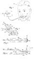
- FIG. 1 illustrates a holder for a probe attached to a patient's mouth and
coupled to a display unit.
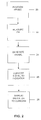
- FIG. 2 is a flow chart illustrating one preferred embodiment of the method of
the invention.
- FIG. 3 is a schematic illustrating the electrochemical embodiment of the
sensor.
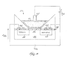
- FIG. 4 is a schematic illustrating the ion-selective, field-effect transistor
embodiment of the sensor.
- FIG. 5 is a perspective view of the holder of FIG. 1.
- FIG. 6 is a top view of the holder of FIG. 1.
- FIG. 7 is a side view of the holder of FIG. 1.
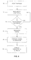
- FIG. 8 is a flow chart illustrating another preferred embodiment of the method
of the invention.
-
-
Before one embodiment of the invention is explained in full detail, it is to be
understood that the invention is not limited in its application to the details of
construction and the arrangement of components set forth in the following
description or illustrated in the following drawings. The invention is capable of
other embodiments and of being practiced or of being carried out in various
ways. Also, it is to be understood that the phraseology and terminology used
herein is for the purpose of description and should not be regarded as limiting.
The use of "including" and "comprising" and variations thereof herein is
meant to encompass the items listed thereafter and equivalents thereof as
well as additional items.
-
FIG. 1 illustrates a device 11 embodying the invention. The device 11
includes a probe 12, a sensor 30 (shown in FIG. 7) coupled to the probe 12, a
holder 14 coupled to the probe 12, and a display unit 16 coupled to the probe
12. The display unit 16 may be a stand-alone unit dedicated solely to
providing an indicator of tissue perfusion levels to a clinician. Alternatively,
the display unit 16 may be part of a complete patient monitoring system.
-
Before the preferred embodiments of the sensor 30 are described in detail, it
should be understood that the use of any sensor to detect the pH of the
mucosa tissue in the upper digestive/respiratory tract in order to assess
perfusion failure is considered within the scope of the invention.
-
In the most preferred embodiment of the invention, an ion-selective, field-effect
transistor (ISFET) is used to detect the pH of the mucosa tissue.
ISFETs are steady-state devices similar to metal-oxide semiconductor field-effect
transistors (MOSFET). MOSFETs are composed of two diodes
separated by a gate region. The gate is a thin insulator, usually silicon
dioxide, upon which a metallic material is deposited. Voltage applied to the
gate generates an electric field, and thus current flows between the drain and
the source. ISFETs are similar to MOSFETS, except that the metal gate
region is replaced with an ion-selective layer. Ions traveling through the ion-selective
layer generate an electric field, and thus current flows between the
drain and source.
-
FIG. 4 is a schematic illustration of one embodiment of the sensor 30. The
sensor 30 is an ISFET and includes a gate 52, an insulator 54, an ion-selective
surface 56, a drain 58, a source 60, a reference electrode 62, and a
plurality of metal contacts 64. The ion-selective surface 56 and the reference
electrode 62 are in contact with the mucosa tissue 66. The reference
electrode 62 keeps a constant voltage potential Vgs against the mucosa tissue
66, independent of the mucosa tissue composition. As H+ ions pass through
the ion-selective surface 56, a voltage potential is developed at the ion-selective
surface 56 in response to the H+ ion concentration of the mucosa
tissue 66. The ion-selective surface 56 is not encapsulated by any permeable
membranes, rather the surface itself is ion-selective. The voltage potential Vds
developed in response to the H+ ion concentration modulates the current
between the drain 58 and the source 60. As the voltage potential Vds across
the gate changes, the ISFET current Ids flows. The voltage potential Vds
correlates to the pH of the mucosa tissue.
-
FIG. 3 is a schematic illustration of another suitable sensor 30. The sensor 30
shown in FIG. 3 is an electrochemical sensor. The electrochemical sensor 30
includes a voltmeter 32 coupled between a reference electrode 34 and a
silver/silver-chloride reference wire 36. A shielded lead 44 connects the
reference wire 36 to the voltmeter 32. The reference wire 36 is immersed
within a buffer solution 38 with a constant pH level. Preferably, the buffer
solution 38 is hydrochloric acid (HCI).
-
A pH-sensitive glass membrane 40 encapsulates the reference wire 36 and
the buffer solution 38. Preferably, the glass membrane 40 is not
encapsulated within an ion-selective permeable membrane. Ion-selective
permeable membranes are not necessary in order to measure H+ ion
concentration, as they are necessary to measure carbon dioxide
concentration and other ion concentrations. Rather than having an ion-selective
permeable membrane, the glass membrane composition itself is ion-selective.
Preferably, the ion-selective composition of the glass membrane 40
is SiO2 or Na2O.
-
The pH-sensitive glass membrane 40 and the reference electrode 34 are in
contact with the mucosa tissue, represented by sample 42, which has an
unknown pH. The reference electrode 34 keeps a constant voltage potential
against the sample 42, independent of the sample composition. In response
to the difference in pH of the buffer solution 38 and the sample 42, a direct
voltage is developed between the inside and outside of the glass membrane
40. The voltage is caused by an ion exchange at each surface of the glass
membrane 40 between the metal ions of the glass and the H+ ions of the
solutions. The approach of H+ ions to the outside of the membrane 40 causes
the silicate structure of the glass to conduct a positive charge into the buffer
solution 38 inside the membrane 40. The ion exchange across the glass
membrane 40 is controlled by the concentration of H+ ions in each solution,
and thus, the pH of each solution. The change in the voltage potential
between the reference electrode 34 and the reference wire 36 is sensed by
the voltmeter 32. If the concentration of H+ ions in both solutions is the same,
the potential difference across the glass membrane and the output of the
voltmeter 32 is zero volts. Thus, the output of the voltmeter 32 correlates to
the pH of the mucosa tissue, represented by sample 42.
-
In another preferred embodiment, the device 11 includes a sensor for
acquiring an end-tidal carbon-dioxide partial-pressure (PetCO2) measurement.
-
A PetCO2 sensor suitable for use in the present invention is disclosed in U.S.
Patent Application serial number 09/477,914 entitled "Low Cost Main Stream
Gas Analyzer System".
-
In still another preferred embodiment of the invention, the device 11 includes
additional sensors or sensor platforms for acquiring blood-chemistry and/or
saliva-chemistry data from the patient. In order to implement the additional
sensors into a platform sensor package, preferably one or more ISFETs
capable of detecting multiple species of ions and molecules are fabricated
onto one silicon chip.
-
Preferably, the additional sensors are capable of detecting the blood
chemistry data that is normally gathered in an arterial blood gas (ABG)
analysis. The additional sensors are preferably capable of detecting at least
one of pH, the partial pressure of oxygen (PO2), the partial pressure of carbon
dioxide (PCO2), bicarbonate, hematocrit, hemoglobin, oxygen saturation (SO2),
electrolyte concentration, and metabolite concentration. The additional
sensors are preferably capable of detecting electrolytes including at least one
of sodium, potassium, calcium, and chloride. The additional sensors are
preferably capable of detecting metabolites including at least one of glucose,
lactate, creatinine, and urea. The additional sensors are also preferably
capable of detecting saliva chemistry data including at least one of
cholesterol, lactate, electrolytes, illegal or abused drugs, glucose, bone
markers, cystic fibrosis, HIV, and pregnancy.
-
FIGS. 5-7 illustrate the preferred embodiment of the holder 14. Before the
preferred embodiment of the holder 14 is described in detail, it should be
understood that the holder 14 could be constructed in any configuration and of
any material capable of placing the sensor or sensors in contact with the
mucosa tissue in the patient's upper digestive/respiratory tract.
-
Referring to FIGS. 5-7, the holder 14 preferably includes an inner holder
portion 90 and an outer holder portion 92 coupled to the inner holder portion
90 by a resilient connecting member 102. The inner holder portion 90
includes an outer surface 93 and a groove 94 formed in the outer surface 93.
The probe 12 is positioned within the groove 94. The inner holder portion 90
also includes a first end 96 and a second end 98. The first end 96 is a
projection for use by the clinician to grasp the holder 14. The second end 98
is a gradually downward-sloping projection that is positioned within the
patient's mouth, preferably in contact with the patient's cheek. As illustrated
in FIG. 7, the sensor 30 is coupled to the second end 98. The sensor 30 is
positioned to contact the mucosa tissue within the patient's mouth.
-
The outer holder portion 92 includes a first end 104 and a second end 106.
The first end 104 is a projection for use by the clinician in conjunction with the
first end 96 of the inner holder portion 92 to grasp the holder 14. The second
end 106 is a gradually upward-sloping projection that is positioned outside the
patient's mouth, preferably in contact with the cheek-area of the patient's face.
The space 108 between the second end 98 of the inner holder portion 90 and
the second end 106 of the outer holder portion 92 is such that the holder 14
remains secured to the patient's cheek.
-
Preferably, the holder 14 is constructed from a soft, pliable material that easily
conforms to the patient's anatomy while remaining rigid enough to secure the
holder 14 to the patient's face. Preferably, the material for the holder 14 is
biocompatible.
-
FIG. 2 is a flow chart illustrating the method of the invention. Referring to
FIGS. 1 and 2, the holder 14 coupled to the probe 12 is positioned 20 on the
patient. Preferably, the holder 14 is attached to the patient's cheek, and when
in the appropriate position, presents the sensor 30 in contact with the oral
mucosa tissue of the patient 10. In other embodiments (not shown), the
holder 14 may be attached to the patient's lip or under the patient's tongue.
Referring to FIGS. 5-7, in order to attach the holder 14, the clinician grasps
the holder 14 by the first end 96 of the inner holder portion 90 and by the first
end 104 of the outer holder portion 92. The clinician then positions the
second end 98 of the inner holder portion 90 within the patient's mouth,
preferably so that the sensor 30 is in contact with the mucosa tissue inside the
patient's cheek. At the same time the clinician positions the second end 106
of the outer holder portion 92 outside the patient's mouth, preferably so that
the second end 106 is in contact with the cheek area of the patient's face.
-
Still referring to FIGS. 1 and 2, the sensor 30 within the probe 12 measures
22 the pH by detecting the H+ ion concentration of the mucosa tissue in the
patient's mouth. The sensor 30 generates 24 an electrical signal in response
to the detected pH. The signal is converted 26 into an indicator of the level of
perfusion. The indicator of the level of perfusion is displayed 28 to a clinician
on display unit 16.
-
FIG. 8 is a flow chart illustrating another preferred embodiment of the method
of the invention including the additional step of acquiring a PetCO2
measurement. Generally, mucosa tissue pH correlates to arterial perfusion,
while PetCO2 correlates to pulmonary perfusion. In order to compare the
arterial and pulmonary levels of perfusion failure, the mucosa tissue pH
measurement is compared to the PetCO2 measurement. Thus, the comparison
between the pH and PetCO2 measurements helps to provide a more accurate
assessment of systemic perfusion failure.
-
Referring to FIGS. 1 and 8, the holder 14 coupled to the probe 12 is
positioned 70 on the patient 10. Preferably, probe 12 includes both a pH
sensor 30 and a PetCO2 sensor (not shown). Preferably, the holder 14 is
attached to the patient's cheek. In other embodiments (not shown), the holder
14 may be attached to the patient's lip or under the patient's tongue. The pH
sensor 30 measures 72 the pH of the mucosa tissue, and the PetCO2 sensor
measures 72 the PetCO2 level of the air expired by the patient. The PetCO2
measurement is compared to the pH measurement and used as a reference
to determine the accuracy of the pH measurement. If the mucosa tissue pH
measurement is significantly different from the PetCO2 measurement, which
may occur in very low perfusion states, the pH measurement can be taken
again 72 or the PetCO2 measurement can be relied upon to indicate the level of
perfusion failure. Once the pH measurement is accurate, the sensors
generate 76 an electrical signal in response to the detected pH and PetCO2.
The signal is converted 78 into an indicator of the level of perfusion. The
indicator of the level of perfusion is displayed 80 to a clinician on display unit
16.
-
For completeness, various aspects of the invention are set out in the following
numbered clauses:
- 1. A device for assessing perfusion failure of a patient (10), the device
comprising:
- a probe (12) for contacting mucosa tissue in the upper
respiratory/digestive tract of the patient (10); and
- a sensor (30) coupled to the probe (12) for directly detecting a pH
measurement of the mucosa tissue and for generating an electrical signal in
response to the detected pH measurement.
- 2. The device of clause 1 wherein the sensor (30) is coupled to a
steady-state device.
- 3. The device of clause 2 wherein the steady-state device is an ion-selective,
field-effect transistor.
- 4. The device of clause 1 wherein the sensor (30) is an electrochemical
sensor.
- 5. The device of clause 1 wherein the sensor (30) is not encapsulated
within a permeable membrane.
- 6. The device of clause 1 and further comprising a second sensor for
acquiring an end-tidal carbon-dioxide partial-pressure measurement.
- 7. The device of clause 6 wherein the pH measurement is compared to
the end-tidal carbon-dioxide partial-pressure measurement.
- 8. The device of clause 7 wherein the end-tidal carbon-dioxide partial-pressure
measurement is used as a reference to increase the accuracy of the
pH measurement.
- 9. The device of clause 6 wherein the pH measurement correlates to
arterial perfusion and the end-tidal carbon-dioxide partial-pressure
measurement correlates to pulmonary perfusion.
- 10. The device of clause 6 wherein the sensor (30) for acquiring the pH
measurement and the second sensor for acquiring the end-tidal carbon-dioxide
partial-pressure measurement are coupled to the same probe (12).
- 11. The device of clause 1 and further comprising a reference electrode
(54) connected to the sensor (30) and in contact with the mucosa tissue.
- 12. The device of clause 11 wherein the reference electrode (34) is a
silver/silver-chloride electrode.
- 13. The device of clause 1 and further comprising a holder (14) for the
probe (12) to secure the probe (12) to the patient (10).
- 14. The device of clause 13 wherein the holder (14) has an inner holder
portion (90) and an outer holder portion (92), and wherein the inner holder
portion (90) is coupled to the probe (12)and is placed inside the patient's
mouth in contact with the oral mucosa and the outer holder portion (92) is
placed outside the patient's mouth.
- 15. The device of clause 14 wherein the inner holder portion (90) is placed
inside the patient's mouth in contact with the oral mucosa of the patient's
cheek.
- 16. The device of clause 14 wherein the inner holder portion (90) is placed
inside the patient's mouth in contact with the oral mucosa of the patient's lip.
- 17. The device of clause 13 wherein the holder (14) is placed under the
patient's tongue.
- 18. The device of clause 1 wherein the sensor (30) detects the chemistry
of the patient's saliva and generates an electrical signal in response to the
detected saliva chemistry.
- 19. A device for assessing perfusion failure of a patient, the device
comprising:
- a probe (12) for contacting mucosa tissue in the upper
respiratory/digestive tract of the patient (10);
- a sensor (30) coupled to the probe (12) for directly acquiring a pH
measurement of the mucosa tissue;
- a reference electrode (34) connected to the sensor (30) and in contact
with the mucosa tissue so that an electrical potential correlating to a level of
tissue perfusion is created between the sensor (30) and the reference
electrode (34).
- 20. The device of clause 19 wherein the sensor (30) is coupled to a
steady-state device.
- 21. The device of clause 20 wherein the steady-state device is an ion-selective,
field-effect transistor.
- 22. The device of clause 19 wherein the sensor (30) is an electrochemical
sensor.
- 23. The device of clause 19 wherein the sensor (30) is not encapsulated
with a permeable membrane.
- 24. The device of clause 19 wherein the reference electrode (34) is a
silver/silver-chloride electrode.
- 25. The device of clause 19 and further comprising a second sensor for
acquiring an end-tidal carbon-dioxide partial-pressure measurement.
- 26. The device of clause 25 wherein the pH measurement is compared to
the end-tidal carbon-dioxide partial-pressure measurement.
- 27. The device of clause 26 wherein the end-tidal carbon-dioxide partial-pressure
measurement is used as a reference to increase the accuracy of the
pH measurement.
- 28. The device of clause 25 wherein the pH measurement correlates to
arterial perfusion and the end-tidal carbon-dioxide partial-pressure
measurement correlates to pulmonary perfusion.
- 29. The device of clause 25 wherein the sensor (30)for acquiring the pH
measurement and the second sensor for acquiring the end-tidal carbon-dioxide
partial-pressure measurement are coupled to the same probe (12).
- 30. The device of clause 19 and further comprising a holder (14) for the
probe (12) to secure the probe (12) to the patient (10).
- 31. The device of clause 30 wherein the holder (14) has an inner holder
portion (90) and an outer holder portion (92), and wherein the inner holder
portion (90) is coupled to the probe (12) and is placed inside the patient's
mouth in contact with the oral mucosa and the outer holder portion (92) is
placed outside the patient's mouth.
- 32. The device of clause 31 wherein the inner holder portion (90) is placed
inside the patient's mouth in contact with the oral mucosa of the patient's
cheek.
- 33. The device of clause 31 wherein the inner holder portion (90) is placed
inside the patient's mouth in contact with the oral mucosa of the patient's lip.
- 34. The device of clause 31 wherein the holder (14) is placed under the
patient's tongue.
- 35. The device of clause 19 wherein the sensor (30) detects the chemistry
of the patient's saliva and generates an electrical signal in response to the
detected saliva chemistry.
- 36. A method of assessing tissue perfusion in a patient, the method
comprising the acts of:
- providing a probe (12) capable of measuring pH;
- placing the probe (12) in contact with mucosa tissue in the upper
digestive/respiratory tract of the patient (10);
- obtaining a pH measurement of the mucosa tissue; and
- converting the pH measurement into an indicator to a clinician
representing a level of tissue perfusion.
- 37. The method of clause 36 wherein the probe (12) includes a sensor (30)
for measuring pH.
- 38. The method of clause 36 wherein the sensor (30) is coupled to a
steady-state device.
- 39. The method of clause 38 wherein the steady-state device is an ion-selective,
field-effect transistor.
- 40. The method of clause 36 wherein the sensor (30) is an electrochemical
sensor.
- 41. The method of clause 36 and further comprising the acts of
providing a probe (12) capable of measuring end-tidal carbon-dioxide
partial-pressure;
placing the probe (12) in the upper digestive/respiratory tract; and
obtaining an end-tidal carbon-dioxide partial-pressure measurement. - 42. The method of clause 41 and further comprising the act of comparing
the pH measurement to the end-tidal carbon-dioxide partial-pressure
measurement.
- 43. The method of clause 42 wherein the end-tidal carbon-dioxide partial-pressure
measurement is compared to the pH measurement as a reference to
increase the accuracy of the pH measurement.
- 44. The method of clause 41 wherein the pH measurement correlates to
arterial perfusion and the end-tidal carbon-dioxide partial-pressure
measurement correlates to pulmonary perfusion.
- 45. The method of clause 41 wherein the probe (12) capable of measuring
pH and the probe (12) capable of measuring end-tidal carbon-dioxide partial-pressure
are the same probe (12).
- 46. The method of clause 36 wherein the act of obtaining a pH
measurement of the mucosa tissue further comprises the act of generating an
electrical signal indicative of the pH of the mucosa tissue.
- 47. The method of clause 36 and further comprising the acts of introducing
a holder (14) coupled to the probe (12) into the patient's upper
digestive/respiratory tract and securing the holder (14) to the patient (10).
- 48. The method of clause 47 wherein the holder (14) has an inner holder
portion (90) coupled to the probe (12) and an outer holder portion (92).
- 49. The method of clause 48 and further comprising the acts of placing the
inner holder portion (90) inside the patient's mouth and placing the outer
holder portion outside the patient's mouth.
- 50. The method of clause 49 wherein the act of placing the inner holder
portion (90) inside the patient's mouth further comprises the act of placing the
inner holder portion (90) in contact with the oral mucosa of the patient's
cheek.
- 51. The method of clause 49 wherein the act of placing the inner holder
portion (90) inside the patient's mouth further comprises the act of placing the
inner holder portion (90) in contact with the oral mucosa of the patient's lip.
- 52. The method of clause 49 wherein the act of placing the inner holder
portion (90) inside the patient's mouth further comprises the act of placing the
inner holder portion (90) in contact with the oral mucosa under the patient's
tongue.
- 53. The method of clause 36 and further comprising
providing a probe (12) capable of measuring the patient's saliva
chemistry;
placing the probe (12) in contact with the patient's saliva;
obtaining a saliva-chemistry measurement; and
converting the saliva-chemistry measurement into an indicator to a
clinician. - 54. The method of clause 53 wherein the probe (12) capable of measuring
pH and the probe (12) capable of measuring the patient's saliva chemistry are
the same probe (12).
- 55. A device for assessing the blood chemistry of a patient, the device
comprising:
- a probe (12) for contacting mucosa tissue in the upper
respiratory/digestive tract of the patient (10);
- a sensor (30) coupled to the probe (12) for detecting blood-chemistry
data for the mucosa tissue and for generating a signal in response to the
detected blood-chemistry data.
- 56. The device of clause 55 wherein the blood-chemistry data is the data
gathered for an arterial blood gas analysis.
- 57. The device of clause 55 wherein the sensor (30) is coupled to a
steady-state device.
- 58. The device of clause 57 wherein the steady-state device is an ion-selective,
field-effect transistor.
- 59. The device of clause 55 wherein the sensor (30) is an electrochemical
sensor.
- 60. The device of clause 55 and further comprising a second sensor for
acquiring an end-tidal carbon-dioxide partial-pressure measurement.
- 61. The device of clause 60 wherein the pH measurement is compared to
the end-tidal carbon-dioxide partial-pressure measurement.
- 62. The device of clause 61 wherein the end-tidal carbon-dioxide partial-pressure
measurement is compared to the pH measurement as a reference to
increase the accuracy of the pH measurement.
- 63. The device of clause 60 wherein the pH measurement correlates to
arterial perfusion and the end-tidal carbon-dioxide partial-pressure
measurement correlates to pulmonary perfusion.
- 64. The device of clause 60 wherein the sensor (30) for acquiring the
blood-chemistry data and the second sensor for acquiring the end-tidal
carbon-dioxide partial-pressure measurement are included in the same probe
(12).
- 65. The device of clause 55 wherein the sensor (30) detects the chemistry
of the patient's saliva and generates an electrical signal in response to the
detected saliva chemistry.
- 66. A device for assessing the blood chemistry of a patient (10), the device
comprising:
- a probe (12) for contacting mucosa tissue in the upper
respiratory/digestive tract of the patient (10);
- a first sensor (80) coupled to the probe for detecting the data gathered
for an arterial blood gas analysis and for generating a signal in response to
the detected arterial blood gas data; and
- a second sensor coupled to the probe (12) for acquiring an end-tidal
carbon-dioxide partial-pressure measurement.
- 67. The device of clause 66 wherein the first sensor (30) is coupled to a
steady-state device.
- 68. The device of clause 67 wherein the steady-state device is an ion-selective,
field-effect transistor.
- 69. The device of clause 66 wherein the first sensor (30) is an
electrochemical sensor.
- 70. The device of clause 66 wherein the data gathered for an arterial blood
gas analysis includes a pH measurement.
- 71. The device of clause 70 wherein the pH measurement is compared to
the end-tidal carbon-dioxide partial-pressure measurement.
- 72. The device of clause 71 wherein the end-tidal carbon-dioxide partial-pressure
measurement is compared to the pH measurement as a reference to
increase the accuracy of the pH measurement.
- 73. The device of clause 70 wherein the pH measurement correlates to
arterial perfusion and the end-tidal carbon-dioxide partial-pressure
measurement correlates to pulmonary perfusion.
- 74. The device of clause 66 wherein the first sensor (30) detects the
chemistry of the patient's saliva and generates an electrical signal in response
to the detected saliva chemistry.
- 75. A method of assessing tissue perfusion in a patient, the method
comprising the acts of:
- providing a probe (12) capable of measuring pH and end-tidal carbon-dioxide
partial-pressure;
- placing the probe (12) in the patient's upper digestive/respiratory tract;
obtaining a pH measurement and an end-tidal carbon-dioxide partial-pressure
measurement;
- generating an electrical signal indicative of the pH measurement and the
end-tidal carbon-dioxide partial-pressure measurement; and
- converting the electrical signal into an indicator to a clinician representing
a level of tissue perfusion.
- 76. The method of clause 75 wherein the probe (12) includes a sensor (30)
for measuring pH.
- 77. The method of clause 76 wherein the sensor (30) is coupled to a
steady-state device.
- 78. The method of clause 77 wherein the steady state device is an ion-selective,
field-effect transistor.
- 79. The method of clause 76 wherein the sensor (30) is an electrochemical
sensor.
- 80. The method of clause 75 and further comprising the act of comparing
the pH measurement to the end-tidal carbon-dioxide partial-pressure
measurement.
- 81. The method of clause 80 wherein the end-tidal carbon-dioxide partial-pressure
measurement is compared to the pH measurement as a reference to
increase the accuracy of the pH measurement.
- 82. The method of clause 75 wherein the pH measurement correlates to
arterial perfusion and the end-tidal carbon-dioxide partial-pressure
measurement correlates to pulmonary perfusion.
- 83. The method of clause 75 wherein the probe (12) is capable of
measuring the patient's saliva chemistry.
- 84. The method of clause 83 and further comprising
placing the probe (12) in contact with the patient's saliva;
obtaining a saliva-chemistry measurement;
generating an electrical signal indicative of the saliva-chemistry
measurement; and
converting the saliva-chemistry measurement into an indicator to a
clinician. - 85. A patient monitor comprising:
- a processing and display unit (16); and
- a device (11) for assessing perfusion failure of the patient, the device
including a probe (12) for contacting mucosa tissue in the upper
respiratory/digestive tract of the patient and a sensor (30) coupled to the
probe (12) for directly detecting a pH measurement of the mucosa tissue and
for generating an electrical signal in response to the detected pH
measurement.
- 86. The device of clause 85 wherein the sensor (30) is coupled to a
steady-state device.
- 87. The device of clause 86 wherein the steady-state device is an ion-selective,
field-effect transistor.
- 88. The device of clause 85 wherein the sensor (30) is an electrochemical
sensor.
- 89. The device of clause 85 wherein the sensor (30) is not encapsulated
within a permeable membrane.
- 90. The device of clause 85 and further comprising a second sensor for
acquiring an end-tidal carbon-dioxide partial-pressure measurement.
- 91. The device of clause 90 wherein the pH measurement is compared to
the end-tidal carbon-dioxide partial-pressure measurement.
- 92. The device of clause 91 wherein the end-tidal carbon-dioxide
partial-pressure measurement is used as a reference to increase the accuracy
of the pH measurement.
- 93. The device of clause 90 wherein the pH measurement correlates to
arterial perfusion and the end-tidal carbon-dioxide partial-pressure
measurement correlates to pulmonary perfusion.
- 94. The device of clause 90 wherein the sensor (30) for acquiring the pH
measurement and the second sensor for acquiring the end-tidal carbon-dioxide
partial-pressure measurement are coupled to the same probe.
- 95. The device of clause 85 and further comprising a reference electrode
(34) connected to the sensor and in contact with the patient's mucosa tissue.
- 96. The device of clause 95 wherein the reference electrode (34) is a
silver/silver-chloride electrode.
- 97. The device of clause 85 and further comprising a holder (14) for the
probe (12) to secure the probe (12) to the patient.
- 98. The device of clause 97 wherein the holder (14) has an inner holder
portion (90) and an outer holder portion (92), and wherein the inner holder
portion (90) is coupled to the probe (12) and is placed inside the patient's
mouth in contact with the oral mucosa and the outer holder portion (92) is
placed outside the patient's mouth.
- 99. The device of clause 98 wherein the inner holder portion (90) is placed
inside the patient's mouth in contact with the oral mucosa of the patient's
cheek.
- 100. The device of clause 98 wherein the inner holder portion (90) is placed
inside the patient's mouth in contact with the oral mucosa of the patient's lip.
- 101. The device of clause 97 wherein the holder (14) is placed under the
patient's tongue.
- 102. The device of clause 85 wherein the sensor (30) detects the chemistry
of the patient's saliva and generates an electrical signal in response to the
detected saliva chemistry.
-