EP1019026B1 - Multilamellar coalescence vesicles (mlcv) containing biologically active compounds - Google Patents
Multilamellar coalescence vesicles (mlcv) containing biologically active compounds Download PDFInfo
- Publication number
- EP1019026B1 EP1019026B1 EP98950869A EP98950869A EP1019026B1 EP 1019026 B1 EP1019026 B1 EP 1019026B1 EP 98950869 A EP98950869 A EP 98950869A EP 98950869 A EP98950869 A EP 98950869A EP 1019026 B1 EP1019026 B1 EP 1019026B1
- Authority
- EP
- European Patent Office
- Prior art keywords
- mlcvs
- biologically active
- active compound
- vesicles
- suvs
- Prior art date
- Legal status (The legal status is an assumption and is not a legal conclusion. Google has not performed a legal analysis and makes no representation as to the accuracy of the status listed.)
- Expired - Lifetime
Links
Images


Classifications
-
- A—HUMAN NECESSITIES
- A61—MEDICAL OR VETERINARY SCIENCE; HYGIENE
- A61K—PREPARATIONS FOR MEDICAL, DENTAL OR TOILETRY PURPOSES
- A61K9/00—Medicinal preparations characterised by special physical form
- A61K9/10—Dispersions; Emulsions
- A61K9/127—Liposomes
- A61K9/1277—Processes for preparing; Proliposomes
- A61K9/1278—Post-loading, e.g. by ion or pH gradient
-
- A—HUMAN NECESSITIES
- A61—MEDICAL OR VETERINARY SCIENCE; HYGIENE
- A61K—PREPARATIONS FOR MEDICAL, DENTAL OR TOILETRY PURPOSES
- A61K9/00—Medicinal preparations characterised by special physical form
- A61K9/10—Dispersions; Emulsions
- A61K9/127—Liposomes
- A61K9/1277—Processes for preparing; Proliposomes
-
- Y—GENERAL TAGGING OF NEW TECHNOLOGICAL DEVELOPMENTS; GENERAL TAGGING OF CROSS-SECTIONAL TECHNOLOGIES SPANNING OVER SEVERAL SECTIONS OF THE IPC; TECHNICAL SUBJECTS COVERED BY FORMER USPC CROSS-REFERENCE ART COLLECTIONS [XRACs] AND DIGESTS
- Y10—TECHNICAL SUBJECTS COVERED BY FORMER USPC
- Y10S—TECHNICAL SUBJECTS COVERED BY FORMER USPC CROSS-REFERENCE ART COLLECTIONS [XRACs] AND DIGESTS
- Y10S977/00—Nanotechnology
- Y10S977/902—Specified use of nanostructure
- Y10S977/904—Specified use of nanostructure for medical, immunological, body treatment, or diagnosis
- Y10S977/906—Drug delivery
- Y10S977/907—Liposome
Definitions
- the present invention is directed to a method of producing multilamellar coalescence vesicles (MLCVs) which contain a high incorporation of biologically active compounds, using small unilamellar vesicles (SUVs) and large unilamellar vesicles (LUVs) without steps involving multiple freeze-thawing cycles, using organic solvents or dehydration of the vesicles.
- MLCVs multilamellar coalescence vesicles
- the present invention also is directed to the MLCVs produced by the present method.
- These MLCVs possess advantageous properties of containing higher amounts of surface and total biologically active compounds, without the use of human serum albumin (HSA), as compared to prior art multilamellar vesicles (MLVs).
- HSA human serum albumin
- Liposomes are known to be useful as carriers of biologically active compounds which facilitate the delivery of these compounds to the body. Liposomes have been evaluated as potential drug delivery systems to introduce biologically active compounds into cells. See Poznansky and Juliano, Pharmacol. Rev. 36, 277-336 (1984); B. E. Ryman et al., Essays in Biochemistry, 16, 49 (1980). Several routes of administration have been used for the administration of liposomes, for example, intravenous, subcutaneous, intraperitoneal, and oral delivery. See Gregoriadis and Allison, eds., Liposomes in Biological Systems, John Wiley & Sons, New York (1980) at pages 153-178.
- liposomal delivery is the change in tissue distribution and binding properties as compared to the free forms of the bioactive ingredient, resulting in an enhanced therapeutic index and decreased toxicity.
- decreased nephrotoxicity has been associated with the use of liposomes containing amphotericin B or cyclosporin A.
- liposomes containing amphotericin B or cyclosporin A See G. Lopez-Berestein, Ann. Int. Med., 105, 130 (1985) and Hsieh et al., Transplantation Proceedings, Vol. XVII, 1397-1400 (1985).
- reduced cardiotoxicity and nephrotoxicity are associated with liposomes containing doxorubicin and cisplatin, respectively, as compared to the free forms of the drugs. See Rahman et al., Cancer Res., 42, 1817 (1982); and Forssen et al., Cancer Res., 43, 546 (1983).
- phospholipid dispersions can spontaneously reform, in the presence of water, into closed membrane systems. Electron microscopy reveals that these closed structures are made of a number of concentric bilayers or lamellae composed of phospholipid molecules, and are known as liposomes.
- the usefulness of liposomes as a model membrane system arises from the fact that, as the dry phospholipids undergo a defined sequence of molecular rearrangements, there is an opportunity for an unrestricted entry of hydrophilic solutes into the interlamellae space. Similarly, sequestration of hydrophobic solutes occurs within the hydrophobic bilayers. The result is a delivery system that can contain varying amounts of cytokines or other biologically active compounds, depending on the type of interaction between the solute and the phospholipid assembly.
- MLVs containing a biologically active compound Many methods have been proposed for the preparation of liposomes.
- the classical method of making prior art liposomes, MLVs containing a biologically active compound is to mix a lipid in an organic solvent, remove the solvent from the solution, leaving a residue, suspend the residue in a buffer containing a biologically active compound, agitate and homogenize the suspension until the MLVs which contain the biologically active compound are formed, and isolate the resulting MLVs.
- MLVs containing a biologically active compound is to mix a lipid in an organic solvent, remove the solvent from the solution, leaving a residue, suspend the residue in a buffer containing a biologically active compound, agitate and homogenize the suspension until the MLVs which contain the biologically active compound are formed, and isolate the resulting MLVs.
- lipids of the desired composition in solution with an organic solvent, are dried in the form of a thin film on the walls of a round-bottomed flask.
- a biologically active compound can be included in the film at this stage.
- the dry film is hydrated by adding a suitable aqueous phase and gently swirling the flask.
- a hydrophilic biologically active compound an aqueous solution containing the biologically active compound is used for hydration. MLVs are formed by this procedure.
- MLVs are produced and used in medical applications, a major problem in the manufacture of MLVs is the use of organic solvents to dissolve the lipids. Further, many biologically active compounds are incompatible with organic solvents, and the removal of organic solvents from these preparations is difficult and tedious. Additionally, to form MLVs with high entrapment of biologically active compound, the solution must be subjected to repeated freeze-thawing cycles. Large scale freeze-thawing is difficult to carry out, especially repeated freeze-thawing cycles. Large scale freeze-thawing is difficult to carry out, especially under sterile conditions. Further, to produce MLVs under sterile conditions, it is necessary to sterilize the lipid prior to placing it into solution. This sterilization process may result in the breakdown of the lipids, resulting in the formation of by-products.
- WO97/29769 discloses a vaccine comprising a liposome preparation including at least one B-cell, malignancy-associated antigen, IL-2, alone or in combination with at least one cytokine, and at least one type of lipid molecule.
- the process of the present invention provides a method of producing MLCVs, which has none of the limitations of the prior art methods, and several advantages over the prior art methods.
- the process of the present invention allows the improved entrapment of solutes.
- These solutes can be biologically active compounds or any compound, such as HSA, mannitol, or glycerol, which can be entrapped by the liposomes, MLCVs, of the present invention.
- additional biologically active compounds useful in the present invention are pharmaceutical peptides, proteins, antigens and drugs or any biologically active compound that can be incorporated into a liposome for delivery to a subject.
- the present process results in the production of MLCVs without the use of organic solvents while supplying the means for sterilizing the lipid in an aseptic process by filter sterilization.
- the process of the present invention can be used easily to produce a small scale production run as well as a large production run while maintaining a simple manufacturing scheme.
- the MLCVs of the present invention arc unique structurally in that they possess a varying degree of partially coalesced vesicles in addition to numerous lamellae.
- the present method produces MLCVs that possess a consistent size and consistent distribution of biologically active compound with less variability than obtained using the prior art methods.
- MLCVs made by the method of the present invention contain a greater amount of biologically active compound as a result of enhanced entrapment.
- the present MLCVs possess a greater amount of surface biologically active compound and exhibit a greater recovery of biologically active compound than the prior art MLVs. Therefore, the present method results in enhanced recovery and incorporation of biologically active compounds as compared to the prior art.
- recovery is defined as the percent of output over input; that is, the amount of the biologically active compound that is present in products and unreacted starting materials with the remainder being lost in processing.
- incorporation as used in the present invention is defined as the percent of output that is entrapped in liposomes and is no longer free.
- the process of the present invention is referred to as a coalescence process because the produced MLCVs are produced as a result of rupturing and resealing of bilayers accompanied by leakage of internal contents.
- This process differs from vesicle fusion in which the combining of vesicles is accompanied by the mixing of internal contents with no or minimal leakage. See Gingell, D. and Ginsberg, L. (1978), In: Membrane Fusion (Poste, G. & Nicolson, G.L., eds.), pp.791-833, Elsevier/North-Holland Biomedical Press, NY.; Szoka, F.
- the MLCVs of the present invention have sizes (average diameters) of 1000 to 5000 nm.
- the average size of the MLCVs of the present invention may be as small as at least 100 nm or greater; e.g., at least 200 nm or greater.
- Prior art methods which use fusion of SUVs below the phase transition temperature result in unilamellar vesicles having average diameters of less than 100 nm.
- Liposomes of an average diameter greater than 100 nm have a greater accumulation in the lung, liver, spleen and lymph nodes than smaller liposomes. These liposomes with an average diameter greater than 100 nm are often used to target organs for prophylactic and therapeutic treatment. See Jackson (1981), Drug Med. Disp. 9: 535-540.
- the method of the present invention is directed to producing multilamellar coalescence vesicles (MLCVs) containing a biologically active compound by hydrating at least one powdered lipid in an aqueous buffer at a temperature above the phase transition temperature of the highest melting lipid to form multilamellar vesicles (MLVs), reducing the size of the MLVs to 20 - 400 nm to produce small unilamellar vesicles (SUVs) or large unilamellar vesicles (LUVs) or a mixture thereof, and incubating the SUVs, LUVs or mixture thereof with at least one biologically active compound in an aqueous solution at 0 constant temperature above the freezing point of said aqueous solution to form MLCVs containing said at least one biologically active compound, wherein said formed MCLVs have an average diameter of at least 100 mm; wherein said method is performed without the use of an organic solvent, a freeze-thawing step or a dehydration step
- the size reducing step of the method comprises exposing the MLVs to a high shear force which is effected by one or more of sonication, homogenization or extrusion.
- the method further includes sterile filtering the SUVs, LUVs or mixture thereof prior to mixing the SUVs, LUVs or mixture thereof with the biologically active compound(s).
- the method of the present invention discloses the production of MLCVs containing increased amounts of biologically active compounds. These MLCVs have an average diameter of at least 100 nm or greater, preferably in the range of 1000 to 5000 nm, and are produced without the use of steps involving freeze-thawing, organic solvents or dehydration.
- the method of the present invention includes the steps of hydrating powdered lipid(s) with an appropriate buffer in a temperature jacketed mixing vessel at a temperature above the phase transition temperature of the highest melting lipid. Then, the size of the vesicles is reduced from the micron range to a 20-400 nm range.
- These vesicles are small unilamellar vesicles (SUVs) or large unilamellar vesicles (LUV) or a mixture thereof, and are produced by known standard methods that utilize high shearing forces, such as sonication, homogenization or extrusion.
- Homogenizers particularly useful in the present invention are high pressure homogenizers, such as those manufactured by Gaulin Rannie or Microfluidics.
- This latter step is also carried out at a temperature above the phase transition temperature of the lipid mixture.
- the resulting SUVs and/or LUVs are then sterile filtered into a mixing vessel which is also maintained at a temperature above the phase transition temperature of the highest melting lipid.
- the biologically active compound(s) and any necessary excipients are added through a sterilizing filter and mixed while dropping the temperature to the phase transition temperature of the highest melting lipid, or maintaining the temperature, preferably between the pretransition and main transition temperature range.
- the incubation may also be performed below the pretransition, below the subtransition or above the main transition temperature of the lipid system.
- the mixture is incubated for a period of time from minutes up to days. During this time the SUVs and/or LUVs are coalesced to form larger vesicles, MLCVs, which are generally have 100nm or greater average diameter.
- the present invention is directed to a novel method of producing MLCVs containing increased amounts of biologically active compounds.
- the method of the present invention produces MLCVs without the use of steps involving freeze-thawing, organic solvents or dehydration.
- the first step of the present method involves direct hydration of a powdered lipid or mixture of lipids with the appropriate buffer in a temperature jacketed mixing vessel.
- appropriate buffers are phosphate buffered saline (PBS), acetate, or citrate with a pH between 2 and 12, preferably between 5 and 9.
- PBS phosphate buffered saline
- the hydration is preferably performed above the phase transition temperature of the highest melting lipid, and the hydrated suspension is mixed well.
- the next step involves size reduction, preferably to 20-70 nm comprising SUVs and/or LUVs, but in any case, below about 400 nm, preferably below 200 nm. This size reduction is performed using standard means, such as sonication, including bath or probe sonication, homogenization or extrusion.
- the size reduction is performed above the phase transition temperature of the highest melting lipid employed. If a mixture of lipids is used, the phase transition temperature of the highest melting lipid would be used.
- the resultant SUVs and/or LUVs are then sterile filtered (0.22 micron filter) into a sterile reaction vessel equipped with a mixing device and a temperature jacket to maintain the temperature above the phase transition temperature of the highest melting lipid. These LUVs are sufficiently deformable that larger sizes can squeeze through a sterile filter.
- one or more pharmaceutical(s) along with any other necessary excipients, such as HSA, mannitol, and glycerol, are added through a sterilizing filter.
- This mixture is continually mixed while dropping the temperature to the phase transition temperature of the highest melting lipid, or preferably between its pretransition and main transition temperature.
- the incubation temperature may be below the pretransition temperature, below the subtraction temperature or above the main transition temperature.
- This mixture is then incubated for an extended period of time, but it can be incubated from minutes to hours, to possibly days, with optional continuous or intermittent mixing.
- the SUVs and/or LUVs coalesce to form large vesicles, MLCVs, typically between 1000 and 5000 nm, but the MLCVs can be as small as 100 nm in average diameter, and are multilamellar, with entrapped pharmaceuticals.
- the MLCVs of the present invention are unique structurally in that they possess a varying degree of partially coalesced vesicles in addition to numerous lamellae.
- the preferred lipids are saturated lecithins, such as dimyristoyl phospharidylcholine (DMPC); dipalmitoyl phosphatidylcholine (DPPC); distearoyl phosphatidylcholine (DSPC); saturated phosphatidylglycerols, such as dimyristoyl phosphatidylglycerol (DMPG), dipalmitoyl phosphatidylglycerol (DPPG), distearoyl phosphatidylglycerol (DSPG); saturated phosphatidic acids, saturated phosphatidylethanolamines or mixtures of the above lipids.
- Unsaturated lipids may also be employed, such as egg phosphatidylcholine (EPC) and dioleoyl phosphatidylcholine (DOPC).
- the temperature of incubation may be at the pretransition temperature or the main transition temperature of the highest melting lipid used, although it could also be below the pretransition temperature or above the main transition temperature.
- Fusion is typically to unilamellar vesicles of 70-95 nm.
- the fusion product is also capable of entrapping solute. See McConnell et al. Fusion at the phase transition was observed in one case to be similar to that obtained below the pretransition temperature. See Gaber and Sheridan (1982). The entrapment was not established and the kinetics of fusion was quite slow, taking up to weeks to occur.
- the lipid used in the present method preferably should be saturated and may have any head group, such as phosphatidylcholine, phosphatidylglycerol, phosphatidic acid, phosphatidylethanolamine, or phosphatidylserine.
- head group such as phosphatidylcholine, phosphatidylglycerol, phosphatidic acid, phosphatidylethanolamine, or phosphatidylserine.
- mixtures of lipids with respect to both head group and chain length can be used. Mixtures with lipids that alone do not form liposomes, such as cholesterol or fatty acids, can be employed in this process.
- the lipid concentration should be between 1 mg/mL to 400 mg/mL, preferably for most applications between 100 mg/mL and 250 mg/mL. Saturated lecithins are preferred lipids for use in the present method.
- the biologically active compound useful in the present invention can be any of the known biologically active compounds that can be entrapped in the liposomes and whose rate of diffusion out of the liposomes is not significantly greater than the rate of deterioration of liposomes in the body of the recipient.
- the biologically active compounds may have properties that enhance vesicle coalescence. These substances would act directly on the SUVs and/or LUVs by destabilizing their bilayer structure. A simple measurement of increased turbidity, such as illustrated in Example 1, permits ready identification of substances having this property of enhancing vesicle coalescence.
- the biologically active compounds can be selected from proteins, peptides, antigens, antibiotics, hormones, immunological activators, cytokines, lymphokines, polynucleotides, and other drugs.
- Specific examples of such compounds are IL-2, interferon, granulocyte-macrophage colony stimulating factor (GMCSF), insulin, growth hormone, epidermal growth factor, calcitonin, gentamicin, and antigens derived from bacteria, parasites, viruses or rickettsia, tumor antigens, allergens, poison or venom.
- IL-2 is commercially available as recombinant T-cell growth factor (human IL-2, recombinant; T3267) or as a preparation derived from cultured rat splenocytes (TO892) from Sigma Chemical Co. (St. Louis, MO). Recombinant IL-2 may also be obtained from Genzyme (Boston, MA) or R & D Systems (Minneapolis, MN).
- Other lymphokines known and available in the art also can be used in the present invention. These include interleukin-4 (IL-4), interleukin-6 (IL-6), interferon alpha and interferon gamma. It is envisioned that these lymphokines can be used alone, in sequence, or in combination, such as co-entrapment in the liposome (e.g., IL-2 and IL-6).
- MLCVs according to the present invention are characterized by several features that distinguish them from MLVs made by prior art processes.
- One particularly salient feature is the highly uniform distribution of biologically active compound among the MLCVs.
- a high proportion, often 30%, of the liposomes are substantially free of biologically active compound.
- the MLCVs of the invention when seen in freeze-fracture electron micrographs, normally have a low percentage of vesicles that are substantially free of biologically active compound, generally less than 30%, preferably less than 20%, often less than 10% and even less than 5% or even less than 2% of vesicles that are substantially free of biologically active compound as indicated by the absence of bulges.
- MLCVs according to the invention have a higher proportion of the biologically active material on the vesicle surface than prior art MLVs.
- the MLCVs of the invention permit a higher proportion of the entrapped biologically active compound to retain its biological activity compared to MLVs made by other methods.
- the MLCVs of the invention contain in the range of 10-100% greater amount of biologically active compound than liposomes produced by the prior art methods using an organic solvent, a freeze-thawing step or a dehydration step.
- the MLCVs preferably contain at least 10% greater amount of biologically active compound than the prior art liposomes, preferably at least 20% greater amount biologically active compound, more preferably at least 30% greater amount biologically active compound, more preferably at least 40% greater amount biologically active compound, more preferably at least 50% greater amount biologically active compound, more preferably at least 80% greater amount biologically active compound, and more preferably at least 100% greater amount biologically active compound and even more than 100% greater amount biologically active compound.
- DMPC dimyristoyl phosphatidylcholine
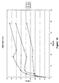
- Fig. 1A shows the results without HSA and Fig. 1B shows the results with HSA in the liposome preparations. Coalescence occurs at all the temperatures examined, with maximal effects at 19°C between the pretransition and main transition temperatures of DMPC.
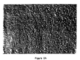
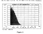
- DMPC SUVs obtained by bath sonication can be seen (Fig. 2A) to be homogeneous and around 25 nm in diameter.
- DMPC SUVs at 200 mg/mL were mixed with IL-2 (final mass ratio 250/1) at 19°C and incubated at 19 °C.
- IL-2 final mass ratio 250/1
- aliquots were rapidly frozen between thin copper planchets in preparation for freeze-fracture.
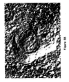
- most of the replica looked like the control SUVs, but regions, as in Fig. 2B, revealed the start of SUV coalescence to MLCVs.
- Example 1 The MLCVs of Example 1 were analyzed for the presence of IL-2, using the cytotoxic T-lymphocyte line (CTLL) assay as described in Clin. Pharmacokinet. 27 (1) 19-31 (1994). Activity was 2.17 x 10 6 IU/mL, a 89% recovery with greater than 95% incorporation.
- CTL cytotoxic T-lymphocyte line
- DMPC SUVs were formed in the presence of 10 mM 8-aminonapthalene-1,3,6-trisulfonic acid (ANTS) and 32 mM p -xylylene- bis -pyridinium bromide (DPX). At these concentrations the DPX quenches the ANTS.
- the SUVs were washed by dialysis and incubated in the presence of IL-2 (DMPC/IL-2 at 250/l, mass ratio) at different temperatures.
- leakage of the water-soluble ANTS and DPX occurs during the process of forming MLCVs, indicating that the process is primarily coalescence not fusion. Measurements were performed on a QM-1 spectrofluorimeter (Photon Technology International, South Brunswick, NJ) with the excitation at 354 nm and the emission at 370-600 nm.
- MLCVs were formed by incubating DMPC SUV with IL-2 at a DMPC/IL-2 mass ratio of 25/1. The incubation was performed at 19°C and for two days. As can be seen in Figure 5, the MLCVs contain SUVs within the lamellae that have not completely coalesced. This illustrates the earlier statement that MLCVs of the invention are unique structurally in that they possess a varying degree of partially coalesced vesicles in addition to numerous lamellae.
- LUVs of DMPC were made by extrusion through polycarbonate filters (Nuclepore, Coming) in an extrusion device (Lipex, Vancouver, BC, Canada).
- the filters employed were 0.1 and 0.2 microns giving rise to 89 nm and 130 nm LUVs, respectively.
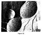
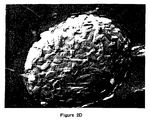
- Figure 8A shows an electron micrograph of the 130 nm vesicles. After 24 hours incubation at 19°C, many LUVs remained, although MLCVs were also present (Figure 8B). These figures reveal the presence of incompletely or partially coalesced LUVs within the structure of the MLCV. Similar results were obtained with the 89 nm LUVs.
- This experiment shows the scale-up of the coalescence of DMPC SUV in the presence of IL-2 and HSA.
- a three liter vessel was used to make a 750 mL batch.
- SUVs were made from MLVs using a Microfluidics homogenizer. The mixture was incubated overnight between the pre transition and main transition temperatures of DMPC. The resultant liposomes were large, with a 2600 nm mean diameter and an activity of 1.08 x 10 6 IU/mL (68% recovery) and 98 % incorporation. This product was stable with respect to IL-2 activity for greater than nine months.
- lipids other than DMPC were also employed in the MLCV coalescence process. Specifically, the effect of chain length, chain unsaturation, and charge were examined. The % coalescence is the total lipid minus the lipid remaining in the supernatant following centrifugation at 39,000g for 30 minutes. Table 1 illustrates the ability of other lipids to entrap IL-2 by the MLCV coalescence method, although not as efficiently as DMPC. Light microscopy reveals large liposomes for both the DPPC and DMPC/DMPG products, but large aggregates for the EPC product.
- DMPC SUVs were incubated overnight in the presence of IL-2 and either IgG or IgM isolated from lymphoma patients. The initial concentration of lipid was varied between 40 mg/mL and 185 mg/mL. Samples were incubated overnight at 19°C and then frozen for storage until assayed. Results shown below in Table 2 indicate good incorporation of antigen and IL-2.
- Table 2 indicate good incorporation of antigen and IL-2.
- the data illustrate that a different protein can be entrapped in the MLCVs and that this protein does not interfere with the coalescence process. It also is noted that samples that are frozen for storage give substantially the same results before and after freezing.
- MLCVs were prepared with and without HSA as in Example 1 employing a 24 hour incubation at 19°C.
- MLVs containing IL-2 were prepared with and without HSA by the method of Anderson and Sorenson (1994), hereinafter referred to as the MLV method.
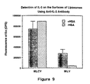
- the MLCVs and MLVs were then examined for surface IL-2.
- the primary antibody, rabbit antihuman IL-2 (Endogen) was added to washed MLCVs or MLVs at 6 ⁇ g/mL, and then washed in 0.2 % skimmed milk to remove unbound antibodies, to which goat anti-rabbit IgG-biotin (Southern Biotechnology) was added.
- a greater surface labeling is obtained for the MLCVs, particularly without HSA.
- High levels of antibody binding to the liposome surface by the MLV method requires HSA during processing.
- This result also could indicate a different surface IL-2 orientation for MLCVs compared to MLVs, that is process dependent.
- IL-2 activity as measured by CTLL revealed a recovery of activity for the MLCVs of greater than 90% while the MLVs revealed a recovery of activity of around 50%.
- the MLCV process of the present invention yields liposomes with a higher surface IL-2 and total IL-2 content without the need for HSA compared to liposomes produced by the MLV method.
Abstract
Description
| Different Lipids Employed in the SUV Coalescence Process | ||||
| Lipid (# of experiments) | incubation T (°C) | % recovery of IL-2 bioactivity | % incorp. of IL-2 bioactivity | % coalescence |
| DMPC (2) | 4 | 37 | 93 | 71 |
| DMPC (2) | 19 | 59 | 97 | 94 |
| DMPC | 23 | 64 | 93 | 34 |
| DMPC | 38 | 43 | 88 | 25 |
| | 4 | 70 | 73 | 88 |
| | 19 | 56 | 73 | 82 |
| DPPC | 38 | 55 | 83 | 93 |
| DPPC | 55 | 11 | 88 | 97 |
| DMPC/DMPG 9:1 (1) | 19 | 66 | 97 | |
| DMPC/DMPG 5:1 | 19 | 107 | 94 | |
| DMPC/Chol. 2:1 | 4 | 37 | 69 | |
| DMPC/Chol. 2:1 | 19 | 43 | 63 | |
| DMPC/Chol. 2:1 | 23 | 38 | 38 | |
| DMPC/Chol. 2:1 | 38 | 64 | 47 | |
| | 4 | 40 | 82 | |
| | 19 | 31 | 81 | |
| | 4 | 77 | 12 | |
| | 19 | 70 | 24 | |
| EPC | 38 | 8 | 45 | |
| EPC/Chol, 2:1 | 4 | 62 | 49 | |
| EPC/Chol, 2:1 | 19 | 69 | 28 | |
| EPC/Chol, 2:1 | 38 | 4 | 45 |
| Incorporation of Antigens and IL-2 into MLCVs | ||||
| Lipid Concentration (mg/mL) | Immunoglobulin Incorporation | IL-2 Incorporation | ||
| (µg/mL) | % | (x106 x IU/mL) | | |
| Immunoglobulin M | ||||
| 40 | 401 | 5.3 | 2.2 | 85 |
| 80 | 385 | 13.2 | 1.9 | 80 |
| 158 | 371 | 22.2 | 3.0 | 83 |
| 185 | 266 | 31.1 | 1.8 | 90 |
| | ||||
| 40 | 130 | 4.6 | 1.5 | 86 |
| 80 | 155 | 6.8 | 1.9 | 78 |
| 158 | 146 | 19.7 | 2.4 | 88 |
| 185 | 74 | 16.5 | 1.8 | 94 |
Claims (26)
- A method for producing mulitilamellar coalescence vesicles (MLCVs) containing a biologically active compound, said method comprising:wherein said method is performed without the use of an organic solvent, a freeze-thawing step or a dehydration step.hydrating at least one powdered lipid in an aqueous buffer at a temperature above the phase transition temperature of the highest melting lipid to form multilamellar vesicles (MLVs);reducing the size of the MLVs to 20-400 nm to produce small unilamellar vesicles (SUVs) or large unilamellar vesicles (LUVs) or a mixture thereof; andincubating the SUVs, LUVs or mixture thereof with at least one biologically active compound in an aqueous solution at a constant temperature above the freezing point of said aqueous solution to form MLCVs containing said at least one biologically active compound, wherein said formed MLCVs have an average diameter of at least 100 nm;
- A method of claim 1 wherein the constant temperature is between 4 and 55°C.
- A method of claim 1 wherein the constant temperature is selected from the group consisting of : 4°C, 19°C, 23°C, 38°C and 55°C.
- A method of claim 3 wherein the constant temperature is 19°C.
- A method according to any preceding wherein at least one powdered lipid is selected from the group consisting of the following: DMPC, DPPC, DOPC and EPC.
- A method according to claim 5 wherein at least one lipid is DMPC.
- The method of any preceding claim wherein said reducing step comprises exposing the MLVs to a high shear force.
- The method of claim 7, where said high shear force is effected by one or more of sonication, homogenization or extrusion.
- The method of claim 1, wherein said method further comprises sterile filtering the SUVs, LUVs or combination thereof prior to mixing the SUVs, LUVs or mixture thereof with the biologically active compound or compounds.
- The method of claim 1, wherein said biologically active compound is a protein or peptide having coalescence properties.
- The method of claim 10, wherein said biologically active compound is a cytokine.
- The method of claim 11, wherein said cytokine is interleukin-2 (IL-2).
- The method of claim 1, wherein a mixture of at least two biologically active compounds are incorporated into said MLCVs
- The method of claim 13, wherein each component of said mixture of biologically active compounds is selected from the group consisting of an immunoglobulin, a tumor antigen, a cytokine and a polynucleotide.
- Multilamellar coalescence vesicles (MLCVs) containing at least one biologically active compound, produced by the method of any of claims 1 to 6.
- The MLCVs of claim 15, wherein said MLCVs have an average diameter of 1000-5000 nm.
- The MLCVs of claim 15, wherein said MLCVs contain between 10-100% greater amount of biologically active compound than liposomes produced by methods using an organic solvent, a freeze-thawing step or a dehydration step.
- The MLCVs of claim 15, wherein said MLCVs contain at least 20% greater amount of biologically active compound than liposomes produced by methods using an organic solvent, a freeze-thawing step or a dehydration step.
- The MLCVs of claim 15, wherein said MLCVs contain at least 50% greater amount of biologically active compound than liposomes produced by methods using an organic solvent, a freeze-thawing step or a dehydration step.
- The MLCVs of claim 15, wherein said MLCVs contain at least 100% greater amount of biologically active compound than liposomes produced by methods using an organic solvent, a freeze-thawing step or a dehydration step.
- MLCVs produced according to the method of any of claims 1 to 6 comprising lipid and at least one biologically active compound, wherein less than 30% of the vesicles are substantially free of biologically active compound.
- The MLCVs of claim 21 wherein less than 20% of the vesicles are substantially free of biologically active compound.
- The MLCVs of claim 21, wherein less than 10% of the vesicles are substantially free of biologically active compound.
- The MLCVs of claim 21, wherein less than 5% of the vesicles are substantially free of biologicallly active compound.
- The MLCVs of claim 21, wherein less than 2% of the vesicles are substantially free of biologically active compound.
- MLCVs produced according to the method of any of claims 1 to 6 comprising lipid and at least one biologically active compound, wherein at least a portion of the vesicles contain partially coalesced SUVs and/or LUVs in the interior of the lamellae.
Priority Applications (1)
| Application Number | Priority Date | Filing Date | Title |
|---|---|---|---|
| EP04078003A EP1523977A1 (en) | 1997-10-01 | 1998-10-01 | Multilamellar coalescence vesicles (MLCV) containing biologically active compounds |
Applications Claiming Priority (3)
| Application Number | Priority Date | Filing Date | Title |
|---|---|---|---|
| US6060697P | 1997-10-01 | 1997-10-01 | |
| US60606P | 1997-10-01 | ||
| PCT/US1998/020780 WO1999016426A2 (en) | 1997-10-01 | 1998-10-01 | Multilamellar coalescence vesicles (mlcv) containing biologically active compounds |
Related Child Applications (2)
| Application Number | Title | Priority Date | Filing Date |
|---|---|---|---|
| EP04078003A Division EP1523977A1 (en) | 1997-10-01 | 1998-10-01 | Multilamellar coalescence vesicles (MLCV) containing biologically active compounds |
| EP04078003.3 Division-Into | 2004-11-01 |
Publications (2)
| Publication Number | Publication Date |
|---|---|
| EP1019026A2 EP1019026A2 (en) | 2000-07-19 |
| EP1019026B1 true EP1019026B1 (en) | 2005-01-12 |
Family
ID=22030591
Family Applications (1)
| Application Number | Title | Priority Date | Filing Date |
|---|---|---|---|
| EP98950869A Expired - Lifetime EP1019026B1 (en) | 1997-10-01 | 1998-10-01 | Multilamellar coalescence vesicles (mlcv) containing biologically active compounds |
Country Status (9)
| Country | Link |
|---|---|
| US (1) | US6544549B1 (en) |
| EP (1) | EP1019026B1 (en) |
| JP (1) | JP2001517693A (en) |
| AT (1) | ATE286717T1 (en) |
| AU (1) | AU745000B2 (en) |
| CA (1) | CA2305533C (en) |
| DE (1) | DE69828624T2 (en) |
| IN (1) | IN190388B (en) |
| WO (1) | WO1999016426A2 (en) |
Cited By (1)
| Publication number | Priority date | Publication date | Assignee | Title |
|---|---|---|---|---|
| CN102654505A (en) * | 2012-05-16 | 2012-09-05 | 江苏省原子医学研究所 | Time-resolved fluorescence immunoassay reagent kit used for detecting interleukin (IL)-2-human serum albumin (HSA) and detection method thereof |
Families Citing this family (20)
| Publication number | Priority date | Publication date | Assignee | Title |
|---|---|---|---|---|
| EP1857112B1 (en) * | 2005-03-09 | 2013-05-15 | Sunstar Inc. | Anticancer composition comprising liposomes containing phytosterols |
| US7662405B2 (en) * | 2005-08-09 | 2010-02-16 | The Research Foundation Of State University Of New York | Compositions and methods of preparation of liposomal microparticulate IL-12 |
| US20140271782A1 (en) | 2013-03-15 | 2014-09-18 | Dermazone Solutions, Inc. | Method for preparing nanolipids with encapsulated alcohol |
| US8597678B2 (en) | 2005-12-30 | 2013-12-03 | Dermazone Solutions, Inc. | Nanolipidic particles |
| WO2010059253A2 (en) | 2008-11-24 | 2010-05-27 | Massachusets Institute Of Technology | Methods and compositions for localized agent delivery |
| US20100323018A1 (en) * | 2009-06-17 | 2010-12-23 | Massachusetts Institute Of Technology | Branched DNA/RNA monomers and uses thereof |
| US20100324124A1 (en) * | 2009-06-17 | 2010-12-23 | Massachusetts Institute Of Technology | Compositions and methods relating to DNA-based particles |
| US9149432B2 (en) | 2010-03-19 | 2015-10-06 | Massachusetts Institute Of Technology | Lipid vesicle compositions and methods of use |
| US20110229556A1 (en) * | 2010-03-19 | 2011-09-22 | Massachusetts Institute Of Technology | Lipid-coated polymer particles for immune stimulation |
| CA2793604C (en) | 2010-03-19 | 2015-10-06 | Massachusetts Institute Of Technology | Lipid vesicle compositions and methods of use |
| US10639367B2 (en) * | 2012-11-02 | 2020-05-05 | Cytuvax | Composition comprising cytokine macro-aggregates |
| US9770415B2 (en) * | 2012-11-27 | 2017-09-26 | Southwest Research Institute | Delivery substrates from aligned polymer biomaterials for tissue repair |
| JP7097667B2 (en) | 2013-09-27 | 2022-07-08 | マサチューセッツ インスティテュート オブ テクノロジー | Carrier-free bioactive protein nanostructures |
| AU2016256979B2 (en) | 2015-05-04 | 2021-01-28 | Versantis AG | Method for preparing transmembrane pH-gradient vesicles |
| CN116212048A (en) | 2015-08-12 | 2023-06-06 | 麻省理工学院 | Cell surface coupling of nanoparticles |
| AU2017283480A1 (en) | 2016-06-13 | 2019-01-24 | Torque Therapeutics, Inc. | Methods and compositions for promoting immune cell function |
| EP3678701A4 (en) | 2017-09-05 | 2021-12-01 | Torque Therapeutics, Inc. | Therapeutic protein compositions and methods of making and using the same |
| US20200078316A1 (en) | 2018-09-06 | 2020-03-12 | NuVessl, Inc. | Method of Using Cannabinoids Encapsulated in Phospholipid Carriers for Transmucosal and Transdermal Administration |
| US11458092B2 (en) | 2018-12-14 | 2022-10-04 | NuVessl, Inc. | Composition with enhanced passenger molecule loading |
| WO2021158996A1 (en) * | 2020-02-05 | 2021-08-12 | University Of Florida Research Foundation, Incorporated | Rna-loaded nanoparticles and use thereof for the treatment of cancer |
Family Cites Families (2)
| Publication number | Priority date | Publication date | Assignee | Title |
|---|---|---|---|---|
| JPH04356421A (en) | 1991-03-06 | 1992-12-10 | Green Cross Corp:The | Fat spherule composition containing prostaglandins |
| JP3948751B2 (en) * | 1996-02-16 | 2007-07-25 | バイオミラ・ユーエスエイ・インコーポレイテッド | B-cell malignant disease vaccine |
-
1998
- 1998-01-01 IN IN2930DE1998 patent/IN190388B/en unknown
- 1998-10-01 US US09/164,350 patent/US6544549B1/en not_active Expired - Lifetime
- 1998-10-01 EP EP98950869A patent/EP1019026B1/en not_active Expired - Lifetime
- 1998-10-01 DE DE69828624T patent/DE69828624T2/en not_active Expired - Lifetime
- 1998-10-01 JP JP2000513564A patent/JP2001517693A/en active Pending
- 1998-10-01 AU AU96800/98A patent/AU745000B2/en not_active Ceased
- 1998-10-01 CA CA002305533A patent/CA2305533C/en not_active Expired - Fee Related
- 1998-10-01 WO PCT/US1998/020780 patent/WO1999016426A2/en active IP Right Grant
- 1998-10-01 AT AT98950869T patent/ATE286717T1/en not_active IP Right Cessation
Cited By (1)
| Publication number | Priority date | Publication date | Assignee | Title |
|---|---|---|---|---|
| CN102654505A (en) * | 2012-05-16 | 2012-09-05 | 江苏省原子医学研究所 | Time-resolved fluorescence immunoassay reagent kit used for detecting interleukin (IL)-2-human serum albumin (HSA) and detection method thereof |
Also Published As
| Publication number | Publication date |
|---|---|
| WO1999016426A3 (en) | 1999-06-17 |
| EP1019026A2 (en) | 2000-07-19 |
| WO1999016426A2 (en) | 1999-04-08 |
| ATE286717T1 (en) | 2005-01-15 |
| AU9680098A (en) | 1999-04-23 |
| US6544549B1 (en) | 2003-04-08 |
| IN190388B (en) | 2003-07-26 |
| CA2305533A1 (en) | 1999-04-08 |
| DE69828624T2 (en) | 2005-12-22 |
| DE69828624D1 (en) | 2005-02-17 |
| CA2305533C (en) | 2009-12-01 |
| JP2001517693A (en) | 2001-10-09 |
| AU745000B2 (en) | 2002-03-07 |
Similar Documents
| Publication | Publication Date | Title |
|---|---|---|
| EP1019026B1 (en) | Multilamellar coalescence vesicles (mlcv) containing biologically active compounds | |
| EP0524968B1 (en) | Heterovesicular liposomes | |
| US5616334A (en) | Low toxicity drug-lipid systems | |
| US6406713B1 (en) | Methods of preparing low-toxicity drug-lipid complexes | |
| EP0713388B1 (en) | A method for high loading of vesicles with biopolymeric substances | |
| JP3676976B2 (en) | Control of the amount of drug encapsulated in multivesicular liposomes | |
| EP0460720B1 (en) | A method of extruding liposomes | |
| JP3571717B2 (en) | Particles, method for producing the particles and use thereof | |
| US5246707A (en) | Sustained release delivery of water-soluble bio-molecules and drugs using phospholipid-coated microcrystals, microdroplets and high-concentration liposomes | |
| USRE35338E (en) | Sustained release delivery of water soluble bio-molecules and drugs using phosphokipid-coated microcrystals, microdroplets and high-concentration lipsomes | |
| EP0349579B1 (en) | Lipid vesicles formed of surfactants and steroids | |
| US6066331A (en) | Method for preparation of vesicles loaded with biological structures, biopolymers and/or oligomers | |
| JPH08505882A (en) | Method for preparing liposome and method for encapsulating substance | |
| EP0280503B9 (en) | Multivesicular liposomes having a biologically active substance encapsulated therein in the presence of a hydrochloride | |
| AU622405B2 (en) | Low toxicity drug-lipid systems | |
| US5576017A (en) | Heterovesicular liposomes | |
| US5173219A (en) | Uniform spherical multilamellar liposomes of defined and adjustable size distribution | |
| EP1523977A1 (en) | Multilamellar coalescence vesicles (MLCV) containing biologically active compounds | |
| EP0713387B1 (en) | A method for preparation of vesicles loaded with biological structures, biopolymers and/or oligomers | |
| CA2168656C (en) | A method for preparation of vesicles loaded with biological structures, biopolymers and/or oligomers | |
| CN100431525C (en) | Production method of liposome suspended liquid and products thereof | |
| US20020119170A1 (en) | Low toxicity drug-lipid systems | |
| JPH10236946A (en) | Improved production of double liposome pharmaceutical preparation | |
| Stricker | Phospholipid Liposomes as Drug Carriers: Preparation and Properties |
Legal Events
| Date | Code | Title | Description |
|---|---|---|---|
| PUAI | Public reference made under article 153(3) epc to a published international application that has entered the european phase |
Free format text: ORIGINAL CODE: 0009012 |
|
| 17P | Request for examination filed |
Effective date: 20000414 |
|
| AK | Designated contracting states |
Kind code of ref document: A2 Designated state(s): AT BE CH CY DE DK ES FI FR GB GR IE IT LI LU MC NL PT SE |
|
| 17Q | First examination report despatched |
Effective date: 20020724 |
|
| GRAP | Despatch of communication of intention to grant a patent |
Free format text: ORIGINAL CODE: EPIDOSNIGR1 |
|
| GRAS | Grant fee paid |
Free format text: ORIGINAL CODE: EPIDOSNIGR3 |
|
| GRAA | (expected) grant |
Free format text: ORIGINAL CODE: 0009210 |
|
| AK | Designated contracting states |
Kind code of ref document: B1 Designated state(s): AT BE CH CY DE DK ES FI FR GB GR IE IT LI LU MC NL PT SE |
|
| PG25 | Lapsed in a contracting state [announced via postgrant information from national office to epo] |
Ref country code: NL Free format text: LAPSE BECAUSE OF FAILURE TO SUBMIT A TRANSLATION OF THE DESCRIPTION OR TO PAY THE FEE WITHIN THE PRESCRIBED TIME-LIMIT Effective date: 20050112 Ref country code: LI Free format text: LAPSE BECAUSE OF FAILURE TO SUBMIT A TRANSLATION OF THE DESCRIPTION OR TO PAY THE FEE WITHIN THE PRESCRIBED TIME-LIMIT Effective date: 20050112 Ref country code: FI Free format text: LAPSE BECAUSE OF FAILURE TO SUBMIT A TRANSLATION OF THE DESCRIPTION OR TO PAY THE FEE WITHIN THE PRESCRIBED TIME-LIMIT Effective date: 20050112 Ref country code: CH Free format text: LAPSE BECAUSE OF FAILURE TO SUBMIT A TRANSLATION OF THE DESCRIPTION OR TO PAY THE FEE WITHIN THE PRESCRIBED TIME-LIMIT Effective date: 20050112 Ref country code: BE Free format text: LAPSE BECAUSE OF FAILURE TO SUBMIT A TRANSLATION OF THE DESCRIPTION OR TO PAY THE FEE WITHIN THE PRESCRIBED TIME-LIMIT Effective date: 20050112 Ref country code: AT Free format text: LAPSE BECAUSE OF FAILURE TO SUBMIT A TRANSLATION OF THE DESCRIPTION OR TO PAY THE FEE WITHIN THE PRESCRIBED TIME-LIMIT Effective date: 20050112 |
|
| REG | Reference to a national code |
Ref country code: GB Ref legal event code: FG4D |
|
| REG | Reference to a national code |
Ref country code: CH Ref legal event code: EP |
|
| REF | Corresponds to: |
Ref document number: 69828624 Country of ref document: DE Date of ref document: 20050217 Kind code of ref document: P |
|
| REG | Reference to a national code |
Ref country code: IE Ref legal event code: FG4D |
|
| PG25 | Lapsed in a contracting state [announced via postgrant information from national office to epo] |
Ref country code: SE Free format text: LAPSE BECAUSE OF FAILURE TO SUBMIT A TRANSLATION OF THE DESCRIPTION OR TO PAY THE FEE WITHIN THE PRESCRIBED TIME-LIMIT Effective date: 20050412 Ref country code: DK Free format text: LAPSE BECAUSE OF FAILURE TO SUBMIT A TRANSLATION OF THE DESCRIPTION OR TO PAY THE FEE WITHIN THE PRESCRIBED TIME-LIMIT Effective date: 20050412 |
|
| PG25 | Lapsed in a contracting state [announced via postgrant information from national office to epo] |
Ref country code: ES Free format text: LAPSE BECAUSE OF FAILURE TO SUBMIT A TRANSLATION OF THE DESCRIPTION OR TO PAY THE FEE WITHIN THE PRESCRIBED TIME-LIMIT Effective date: 20050423 |
|
| RAP2 | Party data changed (patent owner data changed or rights of a patent transferred) |
Owner name: BIOMIRA INC. |
|
| REG | Reference to a national code |
Ref country code: GB Ref legal event code: 732E |
|
| NLT2 | Nl: modifications (of names), taken from the european patent patent bulletin |
Owner name: BIOMIRA INC. |
|
| NLV1 | Nl: lapsed or annulled due to failure to fulfill the requirements of art. 29p and 29m of the patents act | ||
| REG | Reference to a national code |
Ref country code: CH Ref legal event code: PL |
|
| PG25 | Lapsed in a contracting state [announced via postgrant information from national office to epo] |
Ref country code: CY Free format text: LAPSE BECAUSE OF FAILURE TO SUBMIT A TRANSLATION OF THE DESCRIPTION OR TO PAY THE FEE WITHIN THE PRESCRIBED TIME-LIMIT Effective date: 20051001 |
|
| PG25 | Lapsed in a contracting state [announced via postgrant information from national office to epo] |
Ref country code: MC Free format text: LAPSE BECAUSE OF NON-PAYMENT OF DUE FEES Effective date: 20051031 Ref country code: LU Free format text: LAPSE BECAUSE OF NON-PAYMENT OF DUE FEES Effective date: 20051031 |
|
| REG | Reference to a national code |
Ref country code: FR Ref legal event code: TP |
|
| PLBE | No opposition filed within time limit |
Free format text: ORIGINAL CODE: 0009261 |
|
| STAA | Information on the status of an ep patent application or granted ep patent |
Free format text: STATUS: NO OPPOSITION FILED WITHIN TIME LIMIT |
|
| ET | Fr: translation filed | ||
| 26N | No opposition filed |
Effective date: 20051013 |
|
| PG25 | Lapsed in a contracting state [announced via postgrant information from national office to epo] |
Ref country code: PT Free format text: LAPSE BECAUSE OF NON-PAYMENT OF DUE FEES Effective date: 20050612 |
|
| PG25 | Lapsed in a contracting state [announced via postgrant information from national office to epo] |
Ref country code: GR Free format text: LAPSE BECAUSE OF NON-PAYMENT OF DUE FEES Effective date: 20050112 |
|
| PGFP | Annual fee paid to national office [announced via postgrant information from national office to epo] |
Ref country code: IE Payment date: 20091013 Year of fee payment: 12 |
|
| PGFP | Annual fee paid to national office [announced via postgrant information from national office to epo] |
Ref country code: IT Payment date: 20091016 Year of fee payment: 12 Ref country code: GB Payment date: 20091015 Year of fee payment: 12 Ref country code: FR Payment date: 20091027 Year of fee payment: 12 |
|
| PGFP | Annual fee paid to national office [announced via postgrant information from national office to epo] |
Ref country code: DE Payment date: 20091207 Year of fee payment: 12 |
|
| GBPC | Gb: european patent ceased through non-payment of renewal fee |
Effective date: 20101001 |
|
| PG25 | Lapsed in a contracting state [announced via postgrant information from national office to epo] |
Ref country code: FR Free format text: LAPSE BECAUSE OF NON-PAYMENT OF DUE FEES Effective date: 20101102 |
|
| REG | Reference to a national code |
Ref country code: FR Ref legal event code: ST Effective date: 20110630 |
|
| REG | Reference to a national code |
Ref country code: IE Ref legal event code: MM4A |
|
| REG | Reference to a national code |
Ref country code: DE Ref legal event code: R119 Ref document number: 69828624 Country of ref document: DE Effective date: 20110502 |
|
| PG25 | Lapsed in a contracting state [announced via postgrant information from national office to epo] |
Ref country code: GB Free format text: LAPSE BECAUSE OF NON-PAYMENT OF DUE FEES Effective date: 20101001 |
|
| PG25 | Lapsed in a contracting state [announced via postgrant information from national office to epo] |
Ref country code: IE Free format text: LAPSE BECAUSE OF NON-PAYMENT OF DUE FEES Effective date: 20101001 |
|
| PG25 | Lapsed in a contracting state [announced via postgrant information from national office to epo] |
Ref country code: IT Free format text: LAPSE BECAUSE OF NON-PAYMENT OF DUE FEES Effective date: 20101001 |
|
| PG25 | Lapsed in a contracting state [announced via postgrant information from national office to epo] |
Ref country code: DE Free format text: LAPSE BECAUSE OF NON-PAYMENT OF DUE FEES Effective date: 20110502 |