EP0966988A1 - Apparatus and method for interfering with pathological cells survival - Google Patents
Apparatus and method for interfering with pathological cells survival Download PDFInfo
- Publication number
- EP0966988A1 EP0966988A1 EP98830381A EP98830381A EP0966988A1 EP 0966988 A1 EP0966988 A1 EP 0966988A1 EP 98830381 A EP98830381 A EP 98830381A EP 98830381 A EP98830381 A EP 98830381A EP 0966988 A1 EP0966988 A1 EP 0966988A1
- Authority
- EP
- European Patent Office
- Prior art keywords
- fields
- elf
- modulating
- intensity
- generating
- Prior art date
- Legal status (The legal status is an assumption and is not a legal conclusion. Google has not performed a legal analysis and makes no representation as to the accuracy of the status listed.)
- Withdrawn
Links
Images

Classifications
-
- A—HUMAN NECESSITIES
- A61—MEDICAL OR VETERINARY SCIENCE; HYGIENE
- A61N—ELECTROTHERAPY; MAGNETOTHERAPY; RADIATION THERAPY; ULTRASOUND THERAPY
- A61N2/00—Magnetotherapy
- A61N2/02—Magnetotherapy using magnetic fields produced by coils, including single turn loops or electromagnets
Definitions
- the present invention generally relates to a apparatus for interfering with pathological cells survival.
- the invention relates to a microbiological method carried out by such apparatus for interfering with pathological cells survival, in particular cells affected by cancer and other diseases caused by alterations in the mechanism of cell survival.
- the interference is induced by means of static (S) and extremely low frequency electromagnetic (ELF) fields produced by the apparatus.
- S static
- ELF extremely low frequency electromagnetic
- S and ELF Magnetic Static fields and Extremely Low Frequency electromagnetic fields are hereinafter referred to also as S and ELF, respectively.
- S and ELF Magnetic Static fields and Extremely Low Frequency electromagnetic fields
- SELF fields any possible combination of different sequences of S and/or ELF fields and , such as S fields followed by ELF fields, ELF fields followed by S fields, S and ELF field together, as well as the presence of S or ELF fields alone, will hereinafter be referred to also as SELF fields.
- ELF Extremely Low Frequency
- Another physical interaction mechanism is related to the possibility of influencing the kinetics of appropriate cell signalling pathways of the cell (including calcium metabolism) through a field direct effect on electron-spin motion of atoms and molecules with unpaired electrons. This influencing may affect the recombination ratio of a spin correlated free radical pair and consequently on redox signalling [ 12 Grundler 1992; 13 Polk 1992; 14 Walleczek and Budingher 1992; 15 Adey 1993].
- Naturally occurring free radicals have an oxygen- or nitrogen-based unpaired electron such as superoxide anion, hydroxyl radical and nitric oxide.
- ROS Reactive Oxygen Species
- RNS Reactive Nitrogen Species
- ROS Reactive Oxygen Species
- RNS Reactive Nitrogen Species
- Apoptosis is a morphologically distinct form of programmed cell death that plays a major role during development, homeostasis, and in many diseases including cancer, acquired immunodeficiency syndrome, and neurodegenerative disorders. Apoptosis occurs through the activation of a cell-intrinsic suicide program. The basic genetic mechanism of apoptosis appears to be present in essentially all mammalian cells at all times, but the activation of this suicide program is regulated by many different signals that originate from both the intracellular and the extracellular environment.
- the p53 gene is receiving much attention.
- This gene which encodes a transcription factor and is common in many human cancers, mediates the cellular responses to some environmental damage.
- the p53 protein either can temporarily stop cell division, so that the cell can repair altered DNA, or can pilot the cell to an apoptotic death.
- lymphocytes from normal patients respond differently than lymphocytes from Down's syndrome, AIDS and chronic lymphocytic leukemia patients when exposed to ELF fields (previously with mitogen).
- Signal Transduction is an operational term that connotes the translation of genetic information into signalling cascades that allow the cell to for example interpret and respond to external stimuli and/or duplicate itself.
- alterations in the cell survival contribute to the pathogenesis of a number of human diseases, including cancer, viral infections, autoimmune diseases, neurodegenerative disorders, and AIDS. Treatments designed to specifically alter the apoptotic threshold may have the potentiality to change the natural progression of some of these diseases [ 22 Thompson, 1995].
- the fields are of very high intensity, much higher than what people are allowed to be exposed by the safety standards, and may produce heating thus damaging normal tissues and cells.
- the former and other objects are reached by the method for interfering with pathological cells survival according to the invention whose characteristic is to apply to living pathological cells (i.e. cancer cells and other diseases caused by alterations in the mechanism of cell survival) non thermal SELF magnetic fields to induce apoptosis in a selective way.
- living pathological cells i.e. cancer cells and other diseases caused by alterations in the mechanism of cell survival
- non thermal SELF magnetic fields to induce apoptosis in a selective way.
- SELF fields are to be considered as different sequences of S and/or ELF fields, i.e. S fields followed by ELF fields, ELF fields followed by S fields, S and ELF field together, as well as the presence of S or ELF fields alone.
- SELF fields interfere with cell signalling sustaining cell pathological behaviour inside pathological cells, i.e. on redox signalling through free radicals, thus inducing apoptosis through a modification of p53 gene expression.
- SELF fields selectively induce apoptosis in pathological cells (i.e. cancer cells) may be related to the alteration in ionic concentrations at the binding sites as well as in the corresponding enzyme activity of the pathological cell compared with the normal one.
- SELF fields can induce a signal programmed cell death (apoptosis), in vitro and in vivo, without causing any adverse effect.
- SELF non thermal fields can be potentially used for treatment of cells affected by many diseases like viral infections, AIDS, autoimmune diseases, etc., where the alteration of cell survival contributes to their pathogenesis.
- an apparatus for selectively interfering with pathological cells survival i.e. inducing apoptosis in pathological cells
- S static magnetic
- ELF electromagnetic extremely low frequency
- Means are provided for modulating the S fields associated to the S fields generating means and varying the intensity of the S fields from 1 to 30 mT.
- Means are also provided for modulating the ELF fields alone or associated to the S fields modulating means and imposing to the ELF fields a frequency between 1 and 1000 Hz with intensity comprised between 1 and 30 mT.
- the ELF fields Preferably have a frequency between 10 and 100 Hz.
- the S and ELF fields may have an intensity variation after predetermined periods, such as in particular periods comprised between 1 to 40 minutes.
- the intensity of the S and ELF fields is set by the respective modulating means of the apparatus between 1 and 10 mT and the intensity ratio between the S fields and ELF fields is comprised between 0,5 and 2,5.
- At least a portion of the working environment is defined by walls permeable to the S and ELF fields. At least a portion of the working environment is also advantageously adjacent to a first and a second coil respectively and the means for modulating supplying to the coils DC and AC current respectively.
- FIG. 4 Another embodiment of the apparatus according to the invention (fig. 4) used for interfering with pathological cells survival both in vitro and in vivo has two Helmoltz coils 43 and 44 located coaxial to each other at the opposite sides of the working environment 41.
- An amplifier 46 is used between the modulator 45 and the coils 43 and 44, through a shunt element 47, which is also connected to a personal computer 49.
- SELF non thermal fields can be used for interfering with pathological cells survival, such as cells affected by cancer, viral infections, autoimmune diseases, neurodegenerative disorders, AIDS, etc., and are characterised by having intensity comprised between 1 and 30 mT.
- the SELF fields are different sequences of S and/or ELF fields, i.e. S fields followed by ELF fields, ELF fields followed by S fields, S and ELF field together, as well as the presence of S or ELF fields alone, said ELF fields having a field frequency comprised between 1 and 1000 Hz.
- SELF non thermal fields can be used for biotechnological genes modifications, such as in particular for modification of mutant p53 gene, and their characteristic is that said SELF non thermal fields have intensity comprised between 1 and 30 mT.
- the method can be carried out alone or in combination with chemicals.
- the flasks were held between two coils connected with a circuit providing DC and AC currents up to 100 Hertz.
- the temperature was continuously monitored and maintained at 37 ⁇ 0,2 °C.
- the exposure duration was 20 minutes for each experiment and the SELF field was maintained constant . After 3 hours the cells were treated with May- Grunwald-Giemsa. Apoptosis was assessed by counting the number of apoptotic nuclei per 10 high power fields (HPF) by using an optic microscope.
- HPF high power fields
- the amount of induced apoptosis was evaluated by the ratio between the number of apoptotic cells found in the exposure group and the number of apoptotic cells found in the shame-exposed group, that is the group not exposed to the magnetic fields according to the invention.
- Table 1 reports the results obtained in different exposure conditions.
- exposure conditions SELF field composition frequency (Hz) field intensity (Static + ELF rms) mT apoptosis ratio
- a S (static) - (0.5 + 0) 1 B S - (1 + 0) 1 C S - (2 + 0) 1.2 D S - (3 + 0) 2 E S - (4 + 0) 2,3 F S - (10 + 0) 2.2 G S - (20 + 0) 2.2 H S - (30 + 0) 2.3 I ELF 16 (0 + 3) 2.2 L ELF 33 (0 + 3) 2.2 M ELF 50 (0 + 3) 2.1 N ELF 50 (0 + 7) 2,1 O ELF 66 (0 + 3) 2.2 P ELF 83 (0 + 3) 2.3 Q ELF 100 (0 + 3) 2.1 R S + ELF 50 (4 + 3) 2.1 S S + ELF 50 50% of time (3 + 1) 2.2 50% of time (4,5 + 1,5)
- apoptosis doesn't depend upon SELF field frequency. In other words during the life time of the mechanism operating the biological effect (apoptosis) the ELF field is seen as essentially constant. This means that between the two hypothesised mechanism, free- radicals (occurring in a time scale of nano- to microsecond) and ion resonance-like mechanisms, the free radical one is playing the role [ 26 Scaiano, 1994, 27 Engstrom, 1997].
- SELF magnetic fields were verified exposing three cell lines. Two lines were malignant, human colon adenocarcinoma cells (WiDr) and human breast cancer cells (MCF-7). The normal cell line was human lung fibroblast (MRC-5).
- each cell line was grown in confluent monolayers in T25 flasks.
- the experimental protocol was the same as in example 1.
- Six flasks (3 exposed and three shame-exposed ) for each cell line were exposed for 20 minutes.
- Apoptosis was evaluated after 3 hours.
- the exposure conditions used were the R type of Table 1.
- nude mice (nu/nu) bearing subcutaneous tumour masses were used to assess the influence of SELF magnetic fields on tumour growth inhibition.
- mice were randomly assigned to 2 experimental groups, formed by 12 exposed to the SELF exposure condition which gave the best results among the four exposure conditions used in the previous experiment (exposure condition number 4), and 12 shame-exposed.
- mice of both experiments were divided into experimental groups after the tumor masses for each animal were palpable.
- mice were exposed for 70 minutes, once a day, for 5 days a week, for 4 weeks. During the exposure each mouse was put in a single box made of Plexiglas held between two coils connected to a circuit providing DC and AC current up to 100 Hz respectively.
- mice were kept under specific pathogen free conditions and supplied with "ad libitum" diet. All the tests were conducted in accordance with the protocol issued by N.I.H. (US National Institute of Health) and N.C.I. (US National Cancer Institute).
- the tumor masses were measured twice a week and their volume calculated in mm 3 according to the formula: [(major diameter) x (minor diameter squared)] / 2 .
- organs for each animal were extracted for histologic examination to assess the treatment toxicity: brain, heart, kidneys, liver, lungs, axillary and inguinal limphonodes, mediastinal limphonodes, ovaries, skin, spleen, bone marrow, subcutaneous tissue (site of tumoral cell line implantation) as well as blood tests.
- mice 12 extracted tumor mass volume 1139 ⁇ 509 cm 3 1914 ⁇ 793 cm 3 extracted tumor mass weight 1.4 ⁇ 0.7 g 2.1 ⁇ 0.6 g apoptosis (assessed in 50% of mice only) 72.5 ⁇ 9.3 37.0 ⁇ 7.4 p53 35.6 ⁇ 6.7 78.1 ⁇ 16.7 proliferative index 0.34 ⁇ 0.08 0.45 ⁇ 0.07 mitosis 24.1 ⁇ 10.9 47.7 ⁇ 10.1
- the ultrastructural analysis by electron microscope showed in the tumor cells of exposed animals many cellular alterations: presence of apoptotic bodies and condensed cromatine near the nuclear membrane characteristic of apoptotic events.
- Non neoplastic cells i.e. epithelial and stromal cells
- Non neoplastic cells showed no differences between exposed and shame-exposed animals in agreement with the absence of toxicity found in 12 normal organs examined in each animal.
- mice After the cell inoculation 2 groups of mice were randomly formed respectively of 16 animals exposed and 17 shame-exposed. The mice of the former group were exposed 70 minutes once a day, for 5 days a week, for their entire life beginning after 24 hours after the tumor inoculation.
- mice were maintained under specific pathogen free condition supplied with "ad libitum" diet. All the tests were conducted in accordance with protocol issued by N.I.H. and N.C.I.
- the antitumor effectiveness of the treatment was evaluated by using the N.C.I. formula: ratio between exposed and shame-exposed animals of the average animal life span. This average was evaluated summing for each experimental group the time of survival divided by the number of animals. The effectiveness is obtained when the N.C.I. formula gives as result an index equal or greater than 1.25.
- Table 5 reports for each experimental group, the number of living animals at different times (days) from the beginning of experiment.
- living mice exposed/shame-exp. (days) 16/16 (48) 16/15 (73) 15/14 (76) 14/14 (84) 13/14 (87) 12/14 (88) living mice exposed/shame-exp. (days) 12/13 (97) 12/12 (107) 10/12 (109) 10/10 (114) 10/9 (115) 9/8 (125) living mice exposed/shame-exp. (days) 9/7 (149) 8/6 (153) 8/5 (155) 8/4 (157) 7/4 (163) 7/3 (173) living mice exposed/shame-exp.
Landscapes
- Health & Medical Sciences (AREA)
- Public Health (AREA)
- Veterinary Medicine (AREA)
- Nuclear Medicine, Radiotherapy & Molecular Imaging (AREA)
- Radiology & Medical Imaging (AREA)
- Life Sciences & Earth Sciences (AREA)
- Animal Behavior & Ethology (AREA)
- Biomedical Technology (AREA)
- Engineering & Computer Science (AREA)
- General Health & Medical Sciences (AREA)
- Medicines That Contain Protein Lipid Enzymes And Other Medicines (AREA)
- Pharmaceuticals Containing Other Organic And Inorganic Compounds (AREA)
- Medicines Containing Material From Animals Or Micro-Organisms (AREA)
- Micro-Organisms Or Cultivation Processes Thereof (AREA)
- Magnetic Treatment Devices (AREA)
- Electrotherapy Devices (AREA)
Abstract
A method and an apparatus for interfering with pathological cells
survival, i.e. inducing apoptosis on living pathological cells, by using
magnetic fields without adversely affecting normal cells. Static (S) and
extremely low frequency (ELF) magnetic fields are used having intensity
comprised between 1 and 30 mT. In particular SELF fields are used which
are different sequences of S and/or ELF fields, i.e. S fields followed
by ELF fields, ELF fields followed by S fields, S and ELF field
together, as well as the presence of S or ELF fields alone, said ELF
fields having a field frequency comprised between 1 and 1000 Hz. An
apparatus for carrying out the method comprises means for generating
static magnetic (S) fields crossing a working environment and/or means
for generating electromagnetic extremely low frequency (ELF) fields over
the working environment in addition to the S fields. Means are provided
for modulating the S fields and varying the intensity of the S fields
from 1 to 30 mT. Means may also be provided for modulating the ELF fields
associated to the ELF fields generating means and imposing to the ELF
fields a frequency between 1 and 1000 Hz with intensity comprised between
1 and 30 mT.
Description
- The present invention generally relates to a apparatus for interfering with pathological cells survival.
- In addition, the invention relates to a microbiological method carried out by such apparatus for interfering with pathological cells survival, in particular cells affected by cancer and other diseases caused by alterations in the mechanism of cell survival.
- In particular, the interference is induced by means of static (S) and extremely low frequency electromagnetic (ELF) fields produced by the apparatus.
- Magnetic Static fields and Extremely Low Frequency electromagnetic fields are hereinafter referred to also as S and ELF, respectively. Moreover, any possible combination of different sequences of S and/or ELF fields and , such as S fields followed by ELF fields, ELF fields followed by S fields, S and ELF field together, as well as the presence of S or ELF fields alone, will hereinafter be referred to also as SELF fields.
- It is known that pericellular fields and currents induced by an Extremely Low Frequency (ELF) electromagnetic field, whose frequency range is from 1 Hz to 300 Hz and perhaps up to 1000 Hz induce within the cell certain membrane electrochemical events which are important for primary biologic signal transduction and amplification processes.
- These biochemically mediated events then produce cytoplasmic second messaggers and internal effectors such as free Ca++ and protein phosphorylases (kinases) which in turn trigger certain changes in the biosynthesis of macromolecules as well as carry out alterations in cellular growth differentiation and functional properties [1M. Blank, 1993].
- Further, the possibility that S and ELF fields affect the DNA synthesis, DNA integrity, transcription and translation has been documented [2Liboff 1984, 3Tofani 1995, 4Goodman 1991, 5Phillips 1992].
- A possible physical mechanism to account for some of the experimental findings is the direct effect on ions (i.e. Ca++) or to ligand binding at the cell membrane [6Liboff 1985, 7Chiabrera 1985, 8Lednev 1991, 9Blanchard 1994].
- The possibility of influencing variations of Ca++ metabolism may lead to cell apoptosis (programmed cell death) [10Preston, 11Trump 1997].
- Another physical interaction mechanism is related to the possibility of influencing the kinetics of appropriate cell signalling pathways of the cell (including calcium metabolism) through a field direct effect on electron-spin motion of atoms and molecules with unpaired electrons. This influencing may affect the recombination ratio of a spin correlated free radical pair and consequently on redox signalling [12Grundler 1992; 13Polk 1992; 14Walleczek and Budingher 1992; 15Adey 1993].
- The possibility for low level, non thermal (with intensity up to 30 mT) S and ELF magnetic fields to influence in vitro the kinetics and efficacy of radical pair reactions is known from magnetochemistry [16Steiner 1989].
- Naturally occurring free radicals have an oxygen- or nitrogen-based unpaired electron such as superoxide anion, hydroxyl radical and nitric oxide. These Reactive Oxygen Species (ROS) and Reactive Nitrogen Species (RNS) can target proteins providing an obvious mechanistic explanation for free radicals-mediated signalling events. These events may influence growth factors, ion transport (i.e. Ca++ channels), transcription, apoptosis [17Lander 1997].
- Apoptosis is a morphologically distinct form of programmed cell death that plays a major role during development, homeostasis, and in many diseases including cancer, acquired immunodeficiency syndrome, and neurodegenerative disorders. Apoptosis occurs through the activation of a cell-intrinsic suicide program. The basic genetic mechanism of apoptosis appears to be present in essentially all mammalian cells at all times, but the activation of this suicide program is regulated by many different signals that originate from both the intracellular and the extracellular environment.
- Among all the genes involved in apoptosis regulation, the p53 gene is receiving much attention. This gene, which encodes a transcription factor and is common in many human cancers, mediates the cellular responses to some environmental damage. The p53 protein either can temporarily stop cell division, so that the cell can repair altered DNA, or can pilot the cell to an apoptotic death.
- Published data support that p53 results in apoptosis through a three step process: 1) transcriptional induction of redox-related genes: 2) the formation of reactive oxygen species and 3) the oxidative degradation of mitochondria components, culminating in cell death [18Polyak 1997] .
- Moreover, published data are supporting the idea that pathological cells answer differently than normal cells to ELF fields stimuli. According to 19Cadossi [1992], lymphocytes from normal patients respond differently than lymphocytes from Down's syndrome, AIDS and chronic lymphocytic leukemia patients when exposed to ELF fields (previously with mitogen).
- It is also recognised that Ca++ influx across the membrane is influenced by ELF fields in leukemic lymphocytes but not in normal lymphocytes [20Walleczek, 1996].
- With reference to chemotherapy all efforts are devoted to the target of inducing cell apoptosis in vivo instead of killing them, through Signal Transduction Directed Therapy (STDT) of cancer [21Levin, 1998].
- Signal Transduction is an operational term that connotes the translation of genetic information into signalling cascades that allow the cell to for example interpret and respond to external stimuli and/or duplicate itself. Recent evidence suggests that alterations in the cell survival contribute to the pathogenesis of a number of human diseases, including cancer, viral infections, autoimmune diseases, neurodegenerative disorders, and AIDS. Treatments designed to specifically alter the apoptotic threshold may have the potentiality to change the natural progression of some of these diseases [22Thompson, 1995].
- High intensity electrical, electromagnetic and magnetic fields have been used to destroy pathological cells.
- In 23US4665898 an apparatus is described in which animals having malignant cells are treated by means of a high intensity pulsed magnetic field, in order to neutralize/destroy malignant cells in a selective way. This apparatus produces magnetic thermal fields having intensity comprised between 1 Tesla up to 10 Tesla and reversing polarity in the
range 5÷1000 Kilohertz. In the preferred embodiment the magnetic field intensity is set between 1 and 50 Tesla and in particular, in the examples, it is set at 5 Tesla and 8 Kilohertz up to 18 Tesla and 250 Kilohertz. - Different ELF, thermal, continuous or pulsed fields, have been used for anti-cancer therapy in vitro [24Narita, 1997; 25Raylman, 1996].
- In these cases the fields are of very high intensity, much higher than what people are allowed to be exposed by the safety standards, and may produce heating thus damaging normal tissues and cells.
- It is an object of the present invention to provide a method for interfering with cell survival (i.e. inducing apoptosis) of living pathological cells (i.e. cancer cells) by using magnetic fields without adversely affecting normal cells.
- It is another object of the invention to provide an apparatus for interfering with pathological cells survival.
- The former and other objects are reached by the method for interfering with pathological cells survival according to the invention whose characteristic is to apply to living pathological cells (i.e. cancer cells and other diseases caused by alterations in the mechanism of cell survival) non thermal SELF magnetic fields to induce apoptosis in a selective way.
- For the purposes of the invention SELF fields are to be considered as different sequences of S and/or ELF fields, i.e. S fields followed by ELF fields, ELF fields followed by S fields, S and ELF field together, as well as the presence of S or ELF fields alone.
- The concept underlying the method according to the invention is that SELF fields interfere with cell signalling sustaining cell pathological behaviour inside pathological cells, i.e. on redox signalling through free radicals, thus inducing apoptosis through a modification of p53 gene expression.
- The reason why SELF fields selectively induce apoptosis in pathological cells (i.e. cancer cells) may be related to the alteration in ionic concentrations at the binding sites as well as in the corresponding enzyme activity of the pathological cell compared with the normal one.
- For these reasons SELF fields can induce a signal programmed cell death (apoptosis), in vitro and in vivo, without causing any adverse effect.
- For the same reasons SELF non thermal fields can be potentially used for treatment of cells affected by many diseases like viral infections, AIDS, autoimmune diseases, etc., where the alteration of cell survival contributes to their pathogenesis.
- According to the invention, an apparatus for selectively interfering with pathological cells survival (i.e. inducing apoptosis in pathological cells) in vitro and in vivo has the characteristic of comprising means for generating static magnetic (S) fields crossing a working environment and means for generating electromagnetic extremely low frequency (ELF) fields in the working environment alone or in addition to the S fields.
- Means are provided for modulating the S fields associated to the S fields generating means and varying the intensity of the S fields from 1 to 30 mT.
- Means are also provided for modulating the ELF fields alone or associated to the S fields modulating means and imposing to the ELF fields a frequency between 1 and 1000 Hz with intensity comprised between 1 and 30 mT. Preferably the ELF fields have a frequency between 10 and 100 Hz. Moreover, the S and ELF fields may have an intensity variation after predetermined periods, such as in particular periods comprised between 1 to 40 minutes.
- According to a particular aspect of the invention in the apparatus when the S fields are added to the ELF fields SELF fields are obtained with intensity ratio S/ELF comprised between 0,5 and 5.
- In a particular embodiment of the invention the intensity of the S and ELF fields is set by the respective modulating means of the apparatus between 1 and 10 mT and the intensity ratio between the S fields and ELF fields is comprised between 0,5 and 2,5.
- At least a portion of the working environment is defined by walls permeable to the S and ELF fields. At least a portion of the working environment is also advantageously adjacent to a first and a second coil respectively and the means for modulating supplying to the coils DC and AC current respectively.
- Several embodiments of the apparatus are shown in the attached drawings, given as an example and are not limitative.
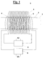
- In figure 1 the working environment is indicated as 1
and the wall as 2. The first and second coils are given
the
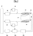
reference numbers boxes - In figure 2 a different embodiment of the apparatus,
used for interfering with pathological cells survival both
in vitro and in vivo has two
coils Variable transformers electric network 27. Switchable diode bridges 28 may be provided for to change the AC supply to the coils. ADC transformer 29a, arectifier 29b as well as atimer 29c may also be provided for supplying twoplates 29 so that an up to 20kV/m static (or low frequency variable up to 1000 Hz) electric field, and preferably about 6 kV/m, may be created in the working environment 21 within preferred intervals, according to the experimental conditions. - In figure 3 a further embodiment is shown of the
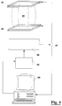
apparatus used for interfering with pathological cells
survival in vitro has a SELF modulator 35 (1-100 Hz) and
two
coils environment 31. Anamplifier 36 is used between the modulator 35 and thecoils environment 31 either an S or an ELF magnetic field. -
- Another embodiment of the apparatus according to the invention (fig. 4) used for interfering with pathological cells survival both in vitro and in vivo has two Helmoltz coils 43 and 44 located coaxial to each other at the opposite sides of the working
environment 41. Anamplifier 46 is used between the modulator 45 and thecoils shunt element 47, which is also connected to apersonal computer 49. - According to the microbiological method aspect of the invention SELF non thermal fields can be used for interfering with pathological cells survival, such as cells affected by cancer, viral infections, autoimmune diseases, neurodegenerative disorders, AIDS, etc., and are characterised by having intensity comprised between 1 and 30 mT. The SELF fields are different sequences of S and/or ELF fields, i.e. S fields followed by ELF fields, ELF fields followed by S fields, S and ELF field together, as well as the presence of S or ELF fields alone, said ELF fields having a field frequency comprised between 1 and 1000 Hz.
- According to another method aspect SELF non thermal fields can be used for biotechnological genes modifications, such as in particular for modification of mutant p53 gene, and their characteristic is that said SELF non thermal fields have intensity comprised between 1 and 30 mT.
- The method can be carried out alone or in combination with chemicals.
- The method according to the invention will now be described in more detail by way of specific examples.
- In this experiment the capability of inducing apoptosis by SELF magnetic field as a function of field intensity and frequency was studied in vitro.
- Human colon adenocarcinoma cell line (WiDr) grown in confluent monolayers in T25 flasks was used for the experiment. For each
exposure condition 6 flasks containing each about 10 millions cells were used, 3 exposed and 3 shame-exposed (i.e. not exposed). - During the exposure the flasks were held between two coils connected with a circuit providing DC and AC currents up to 100 Hertz. The temperature was continuously monitored and maintained at 37 ± 0,2 °C.
- The exposure duration was 20 minutes for each experiment and the SELF field was maintained constant . After 3 hours the cells were treated with May- Grunwald-Giemsa. Apoptosis was assessed by counting the number of apoptotic nuclei per 10 high power fields (HPF) by using an optic microscope.
- The amount of induced apoptosis was evaluated by the ratio between the number of apoptotic cells found in the exposure group and the number of apoptotic cells found in the shame-exposed group, that is the group not exposed to the magnetic fields according to the invention.
- Table 1 reports the results obtained in different exposure conditions.
exposure conditions SELF field composition frequency (Hz) field intensity (Static + ELF rms) mT apoptosis ratio A S (static) - (0.5 + 0) 1 B S - (1 + 0) 1 C S - (2 + 0) 1.2 D S - (3 + 0) 2 E S - (4 + 0) 2,3 F S - (10 + 0) 2.2 G S - (20 + 0) 2.2 H S - (30 + 0) 2.3 I ELF 16 (0 + 3) 2.2 L ELF 33 (0 + 3) 2.2 M ELF 50 (0 + 3) 2.1 N ELF 50 (0 + 7) 2,1 O ELF 66 (0 + 3) 2.2 P ELF 83 (0 + 3) 2.3 Q ELF 100 (0 + 3) 2.1 R S + ELF 50 (4 + 3) 2.1 S S + ELF 50 50% of time (3 + 1) 2.2 50% of time (4,5 + 1,5) - All the results were statistically highly significant (at the t Student test). From Table 1 we can see that the apoptosis effect appears at 2 mT and doubles starting from 3 mT.
- Another important finding is that apoptosis doesn't depend upon SELF field frequency. In other words during the life time of the mechanism operating the biological effect (apoptosis) the ELF field is seen as essentially constant. This means that between the two hypothesised mechanism, free- radicals (occurring in a time scale of nano- to microsecond) and ion resonance-like mechanisms, the free radical one is playing the role [26Scaiano, 1994, 27Engstrom, 1997].
- In this experiment the selective effect of SELF magnetic fields was verified exposing three cell lines. Two lines were malignant, human colon adenocarcinoma cells (WiDr) and human breast cancer cells (MCF-7). The normal cell line was human lung fibroblast (MRC-5).
- As in the example 1 each cell line was grown in confluent monolayers in T25 flasks. The experimental protocol was the same as in example 1. Six flasks (3 exposed and three shame-exposed ) for each cell line were exposed for 20 minutes. Apoptosis was evaluated after 3 hours. The exposure conditions used were the R type of Table 1.
- The results are reported in Table 2.
cell line apoptosis ratio WiDr 2.1 MCF-7 1.4 MRC-5 1 - As shown in Table 2 only cancer cells reported an apoptosis increment statistically highly significant, whereas the normal cell line didn't. The difference in percentage of apoptosis between the two cancer cell lines was expected due to the two different duplication times. In fact WiDr duplicates faster than MCF-7. The results were evaluated at t Student test.
- In this example nude mice (nu/nu) bearing subcutaneous tumour masses were used to assess the influence of SELF magnetic fields on tumour growth inhibition.
- Each mouse was inoculated subcutaneously with 10 million human colon adenocarcinoma cells (WiDr). Two experiments were successively carried out.
- In the first experiment, 36 female mice were randomly assigned to 4 experimental groups, each formed by 6 exposed and 3 shame-exposed for a total of 24 animals exposed to 4 different SELF magnetic fields and 12 shame-exposed.
- A Static Electric Field up to 6 kV/m was also applied to eventually take advantage of the different electrical behaviour between tumoral and normal tissues [28Thornton, 1984; 29Barsamian, 1987]
- In the
second experiment 24 female mice were randomly assigned to 2 experimental groups, formed by 12 exposed to the SELF exposure condition which gave the best results among the four exposure conditions used in the previous experiment (exposure condition number 4), and 12 shame-exposed. - All the mice of both experiments were divided into experimental groups after the tumor masses for each animal were palpable.
- The animals were exposed for 70 minutes, once a day, for 5 days a week, for 4 weeks. During the exposure each mouse was put in a single box made of Plexiglas held between two coils connected to a circuit providing DC and AC current up to 100 Hz respectively.
- Nude mice were kept under specific pathogen free conditions and supplied with "ad libitum" diet. All the tests were conducted in accordance with the protocol issued by N.I.H. (US National Institute of Health) and N.C.I. (US National Cancer Institute).
- The tumor masses were measured twice a week and their volume calculated in mm3 according to the formula:
- After 4 weeks the animals were sacrificed and autopsied. Tumor masses were extracted, weighed and measured. Portions of tumors were used for different analysis, i.e.
- immunoistochemical: Ki-67 antigen for proliferative index, p-53 antigen for the expression of p-53 gene;
- hystopathological: hematossilina-eosin staining for the assessment of number of mitosis;
- ultrastructural: electron microscopy;
- nucleic acid hybridisation: Tunel method for apoptosis evaluation.
- In addition the following organs for each animal were extracted for histologic examination to assess the treatment toxicity: brain, heart, kidneys, liver, lungs, axillary and inguinal limphonodes, mediastinal limphonodes, ovaries, skin, spleen, bone marrow, subcutaneous tissue (site of tumoral cell line implantation) as well as blood tests.
- The obtained results are reported in Table 3 for the first experiment and in Table 4 for the second.
exposure conditions 1 2 3 4 shame-exposed exposure duration (min) 70 70 70 70 - time averaged field intensity (Static + ELF rms) in mT 3 3 4 6 - field variation in mT (min-max) Static; [min-max] ELF (4-6) [2-2] (1.5-4) [1-1] (2-5) [1.5-3.5] (2-5) [1.5-3.5] - constant field time duration (min-max) in minutes (5-15) (5-20) (5-15) (5-20) - time % with co-presence of Static and ELF fields 0% 50% 50% 100% - S/ELF ratio (min-max) - (0,5-5) (0,5-5) (0,5-5) - time % with Static field alone 50% 50% 50% 0% - number of mice 6 6 6 6 12 extracted tumor mass volume (mm3) 1323 ± 304 1450 ± 288 920 ± 540 650 ± 205 1492 ± 559 extract tumor mass weight (g) 1.54 ± 0.22 1.6 ± 0.39 0.98 ± 0.56 0.96 ± 0.25 1.6 ± 0.5 number of apoptotic cells per 10 HPF 98 ± 23 115 ± 20 129 ± 25 129 ± 26 40 ± 17 p53 expression per 10 HPF 35.1 ± 0.11 43.8 ± 0.16 38.2 ± 0.06 28.7 ± 0.14 73.2 ± 0.14 exposure conditions 4 (see tab. 3) shame exposed number of mice 12 12 extracted tumor mass volume 1139 ± 509 cm3 1914 ± 793 cm3 extracted tumor mass weight 1.4 ± 0.7 g 2.1 ± 0.6 g apoptosis (assessed in 50% of mice only) 72.5 ± 9.3 37.0 ± 7.4 p53 35.6± 6.7 78.1±16.7 proliferative index 0.34 ± 0.08 0.45 ± 0.07 mitosis 24.1 ± 10.9 47.7 ± 10.1 - The data reported in tables 3 and 4 show that SELF fields have an inhibitory tumor growth effect in vivo. This effect, found in both experiments, was statistically highly significant (in the first experiment, mostly for the exposure condition 4) at the Dunnet and t Student tests respectively.
- At the histologic examination of 12 organs for each animal for all groups no differences were found between exposed and shame-exposed mice. No differences were also found in the blood tests. These findings prove the absence of toxicity related to the SELF fields treatment.
- The ultrastructural analysis by electron microscope showed in the tumor cells of exposed animals many cellular alterations: presence of apoptotic bodies and condensed cromatine near the nuclear membrane characteristic of apoptotic events.
- In addition a consistent result is represented by morphological modifications, increase of number and dimensions of mitochondria as well as number of nucleoli, presence of many vacuoles inside the cytoplasm. Non neoplastic cells (i.e. epithelial and stromal cells) showed no differences between exposed and shame-exposed animals in agreement with the absence of toxicity found in 12 normal organs examined in each animal.
- The increment in apoptosis as well as the decrement in p53 gene expression found in exposed mice tumors (see tables 3 and 4) are statistically highly significant (t Student test)
- Results reported in Table 3 and 4 are in agreement with those obtained in vitro and shown in Tables 1 and 2.
- The effect induced by the SELF magnetic fields on p53 expression enforces the apoptosis results and is in agreement with the hypothesised biophysical mechanism (i.e. free radical recombination) by which the SELF fields have an anti-tumor effect through formation of reactive oxygen species and the degradation of mithocondrial components.
- In this experiment nude mice (nu/nu) previously subcutaneous inoculated with 10 million human colon adenocarcinoma cells (WiDr) were exposed to study the animal survival.
- After the
cell inoculation 2 groups of mice were randomly formed respectively of 16 animals exposed and 17 shame-exposed. The mice of the former group were exposed 70 minutes once a day, for 5 days a week, for their entire life beginning after 24 hours after the tumor inoculation. - The exposure conditions were the same of the experiment the results which are reported in Table 4.
- As in the previous example, the mice were maintained under specific pathogen free condition supplied with "ad libitum" diet. All the tests were conducted in accordance with protocol issued by N.I.H. and N.C.I.
- The antitumor effectiveness of the treatment was evaluated by using the N.C.I. formula: ratio between exposed and shame-exposed animals of the average animal life span. This average was evaluated summing for each experimental group the time of survival divided by the number of animals. The effectiveness is obtained when the N.C.I. formula gives as result an index equal or greater than 1.25.
- Table 5 reports for each experimental group, the number of living animals at different times (days) from the beginning of experiment.
living mice exposed/shame-exp. (days) 16/16 (48) 16/15 (73) 15/14 (76) 14/14 (84) 13/14 (87) 12/14 (88) living mice exposed/shame-exp. (days) 12/13 (97) 12/12 (107) 10/12 (109) 10/10 (114) 10/9 (115) 9/8 (125) living mice exposed/shame-exp. (days) 9/7 (149) 8/6 (153) 8/5 (155) 8/4 (157) 7/4 (163) 7/3 (173) living mice exposed/shame-exp. (days) 6/3 (183) 6/2 (192) 6/0 (194) 5/0 (195) 4/0 (203) 3/0 (257) living mice exposed/shame-exp. (days) 2/0 (276) 1/0 (323) 0*/0 *sacrificed (326) - The N.C.I. formula applied to the results reported in Table 5 gives an index equal to 1.31, that is greater than 1.25 . After 194
days 6 exposed mice were alive whereas all shame exposed mice were dead. - The foregoing description of specific embodiments will so fully reveal the invention according to the conceptual point of view, so that others, by applying current knowledge, will be able to modify and/or adapt for various applications such embodiments without further research and without departing from the invention, and it is therefore to be understood that such adaptations and modifications will have to be considered as equivalent to the specific embodiments. The means and the materials to realise the different functions described herein could have a different nature without, for this reason, departing from the field of the invention. It is to be understood that the phraseology or terminology employed herein is for the purpose of description and not of limitation .
-
- 1 Blank M (1993):"Electricity and Magnetism in Biology and Medicine". The First World Congress for Electricity and Magnetism in Biology and Medicine, Orlando, Florida.
- 2 Liboff AR, Williams T Jr, Strong DM and Wistar R. Jr. (1984):"Time-Varying Magnetic Fields: Effect on DNA Synthesis". Science, Vol. 223, pp 818-820.
- 3 Tofani S, Ferrara A, Anglesio L, Gilli G
(1995):"Evidence for genotoxic effects of resonant ELF
magnetic fields". Bioelectrochemistry and
Bioenergetics 36, pp 9-13. - 4 Goodman R , Shirley-Henderson A (1991): "Transcription and Translation in Cells exposed to Extremely Low Frequency Electromagnetic Fields" Bioelectrochem. Bioenerg. 25, pp. 335-355.
- 5 Phillips jl, Haggren w, Thomas WJ, Ishida-Jones T and Adey WR (1992):"Magnetic field-induced changes in specific gene transcription". Biochimica et Biophysica Acta 1132, pp 140-144.
- 6 Liboff AR (1985): Cyclotron resonance in membrane transport. In Chiabrera A, Nicolini C., Schwan HP (eds): "Interactions Between Electromagnetic Fields and Cells". New York: Plenum Press, pp 281-296.
- 7 Chiabrera A., Grattarola M., Viviani R. (1984):
"Interaction between electromagnetic fields and cells:
Microelectrophoretic effect on ligands and surface
receptors".
Bioelectromagnetics 5, pp173-191. - 8 Lednev VV (1991): "Possible mechanism for the influence of weak magnetic fields on biological systems". Bioelectromagnetics 12, pp 71-75.
- 9 Blanchard JP, Blackman CF (1994):"Clarification and application of an ion parametric resonance model for magnetic field interactions with biological systems. Bioelectromagnetics 15, pp217-238.
- 10 Preston GA, Barrett JC, Biermann JA and Murphy Elizabeth (1997): "Effects of Alterations in Calcium Homeostasis on Apoptosis during Neoplastic Progression", Cancer Research 57, pp. 537-542.
- 11 Trump BF, Berezesky IK, Chang SH and Phelps PC (1997):"The Pathways of Cell Death: Oncosis, Apoptosis, and Necrosis". Toxicologic Pathology Vol. 25, n. 1, pp.82-87.
- 12 Grundler W, Kaiser F, Keilmann F, Walleczek J (1992): "Mechanisms of electromagnetic interaction with cellular systems". Naturwissenschaften 79, pp. 551-559.
- 13 Polk C (1992):"Dosimetry of extremely-low-frequency
magnetic fields".
Bioelectromagnetics Suppl 1, pp. 209-235 - 14 Walleczek J, Budinger TF (1992): "Pulsed magnetic field effects on calcium signalling in lymphocytes: Dependence on cell status and field intensity". FEBS Lett 314, pp 351-355.
- 15 Adey WR (1993):Electromagnetics in biology and medicine. In Matsumoto H (ed): "Modern Radio Science", New York: Oxford University Press, pp 227-245.
- 16 Steiner UE and Ulrich T (1989):"Magnetic Field Effects in Chemical Kinetics and Related Phenomena". Chem. Rev. 89, pp. 51-147.
- 17 Lander HM (1997):" An essential role for free radicals and derived species in signal transduction". The FASEB Journal 11, pp118-124.
- 18 Polyak K, Xia Y, Zweier JL, Kinzier KW and Volgestein B (1997):"A model for p53-induced apoptosis". Nature Vol. 389, pp. 300-305.
- 19 Cadossi R, Bersani F, Cossarizza A, Zucchini P, Emilia G, Torelli G and Claudio Franceschi (1992):"Lymphocytes and low-frequency electromagnetic fields". The FASEB Journal Vol. 6, pp.2667-2674.
- 20 Walleczeck J (1996):"Electromagnetic Field Effects on Cellular Signal Transduction and Free Radical Mechanisms". Abstract Book XXVth General Assembly of the International Union of Radio Science-Lille-France, p. 547.
- 21 Levin VA (1998):"Signal Transduction Directed Therapy: Fact or Fantasy?" Abstract Book (EL 5) of the Eight International Congress on Anti-Cancer Treatment, February 3rd-6th 1998, Paris, France.
- 22 Thompson C.B. (1995):"Apoptosis in the pathogenesis and treatment of diseases" Science Vol. 267, p. 1456-1462
- 23 Costa JL and Hofmann GA (1987): "Malignancy treatment" U.S. patent 4,665,898.
- 24 Narita K, Hanakawa K, Kasahara T, Hisamitsu T, Asano K (1997):"Induction of apoptotic cell death in human leukemic cell line, HL-60, by extremely low frequency electric magnetic fields: analysis of the possible mechanisms in vitro". In vivo 111(4), pp. 329-335.
- 25 Raylman RR, Clavo AC, Wahl RL (1996):"Exposure to Strong Static Magnetic Field Slow the Growth of Human Cancer Cells In Vitro". Bioelectromagnetics 17, pp. 358-363.
- 26 Scaiano JC, Mohtat N, Cozens FL, McLean J and Thansandote (1994):"Application of the Radical Pair Mechanism to Free Radicals I Organized Systems: Can the Effects of 60 Hz Be Predicted From Studies Under Static Fields?" Bioelectromagnetics 15, pp.549-554.
- 27 Engstrom S (1997):"What is the Time of Magnetic Field Interaction in Biological Systems?". Bioelectromagnetics 18, pp. 244-249.
- 28 B.S. Thornton (1984): "Inversion of raman spectra of living cells indicates dielectric structure related to energy control", in Physics Letters, Vol. 106A, pp. 198-202.
- 29 S.T. Barsamian (1987): "Dielectric origin of living cells", in Biophysical Aspects of Cancer, Charles University Prague, pp. 152-159
-
Claims (14)
- Apparatus for selectively interfering with pathological cells survival (i.e. inducing apoptosis) in vitro and in vivo characterised in that it comprises:means for generating static magnetic (S) fields crossing a working environment,means for generating electromagnetic extremely low frequency (ELF) fields over said working environment in addition to said S fields;means for modulating said S fields associated to said means for generating S fields, said means for modulating said S fields varying the intensity of said S fields from 1 to 30 mT;means for modulating said ELF fields associated to said means for generating ELF fields, said means for modulating said ELF fields imposing to said ELF fields a frequency between 1 and 1000 Hz with intensity comprised between 1 and 30 mT.
- Apparatus for selectively interfering with pathological cells survival (i.e. inducing apoptosis) in vitro and in vivo characterised in that it comprises:means for generating static magnetic (S) fields crossing a working environment,means for modulating said S fields associated to said generating means, said S fields modulating means varying the intensity of said S fields from 1 to 30 mT;
- Apparatus for selectively interfering with pathological cells survival (i.e. inducing apoptosis) in vitro and in vivo characterised in that it comprises:means for generating electromagnetic extremely low frequency (ELF) fields over said working environment;means for modulating said ELF fields associated to said means for generating, said means for modulating said ELF fields imposing to said ELF fields a frequency between 1 and 1000 Hz with intensity comprised between 1 and 30 mT.
- Apparatus according to claim 1 or 2, wherein said means for modulating said S fields cause to said S fields an intensity variation after predetermined periods.
- Apparatus according to claims 1 or 3, wherein said means for modulating said ELF fields impose to said ELF fields a frequency between 10 and 100 Hz.
- Apparatus according to the previous claims, wherein said S fields are added to said ELF fields obtaining SELF fields with ratio S/ELF comprised between 0,1 and 5.
- Apparatus according to claim 6, wherein the intensity of said S and ELF fields is set by said modulating means between 1 and 10 mT and the ratio between said S fields and ELF fields is comprised between 0,5 and 2,5.
- Apparatus according to the previous claims wherein at least a portion of said working environment is defined by walls permeable to said fields.
- Apparatus according to the previous claims, wherein said means for generating said S and/or ELF fields comprise at least a first and a second coil respectively surrounding at least a portion of said working environment, said means for modulating providing to said coils DC and/or AC current respectively.
- Apparatus according to the claims from 1 to 8, wherein said means for generating said S and/or ELF fields comprise at least a first and a second coil coaxial to each other, said working environment being placed between said first and a second coil and said means for modulating providing to said coils DC and/or AC current respectively.
- Apparatus according to the previous claims, wherein means are provided for creating through said working environment a static electric field, or a low frequency variable electric field up to 1000 Hz, having intensity up to 20 kV/m.
- The use of SELF non thermal fields for selectively interfering with pathological cells survival, such as in particular cells affected by cancer, viral infections, autoimmune diseases, neurodegenerative disorders, AIDS, etc., characterised in that said SELF non thermal fields have intensity comprised between 1 and 30 mT, said SELF fields being different sequences of S and/or ELF fields, i.e. S fields followed by ELF fields, ELF fields followed by S fields, S and ELF field together, as well as the presence of S or ELF fields alone, said ELF fields having a field frequency comprised between 1 and 1000 Hz.
- The use of SELF non thermal fields for biotechnological genes modifications, such as in particular for modification of mutant p53 gene, characterised in that said SELF non thermal fields have intensity comprised between 1 and 30 mT, said SELF fields being different sequences of S and/or ELF fields, i.e. S fields followed by ELF fields, ELF fields followed by S fields, S and ELF field together, as well as the presence of S or ELF fields alone, said ELF fields having a field frequency comprised between 1 and 1000 Hz.
- The use of SELF non thermal fields according to claims 12 or 13, wherein chemical substances are used in addition to the SELF fields.
Priority Applications (13)
| Application Number | Priority Date | Filing Date | Title |
|---|---|---|---|
| EP98830381A EP0966988A1 (en) | 1998-06-24 | 1998-06-24 | Apparatus and method for interfering with pathological cells survival |
| DE69929197T DE69929197T2 (en) | 1998-06-24 | 1999-06-23 | DEVICE FOR BREAKING THE SURVIVAL OF PATHOLOGICAL CELLS |
| AU50284/99A AU769400B2 (en) | 1998-06-24 | 1999-06-23 | Apparatus and method for interfering with pathological cells survival processes |
| CA2335606A CA2335606C (en) | 1998-06-24 | 1999-06-23 | Apparatus and method for interfering with pathological cells survival processes |
| EP99934533A EP1091786B1 (en) | 1998-06-24 | 1999-06-23 | Apparatus for interfering with pathological cells survival processes |
| CN99809990A CN1313778A (en) | 1998-06-24 | 1999-06-23 | Apparatus and method for interfering with pathological cells survival processes |
| RU2001102086/14A RU2245728C2 (en) | 1998-06-24 | 1999-06-23 | Method and device for inducing pathological cells and tissues apoptosis |
| BR9911554-9A BR9911554A (en) | 1998-06-24 | 1999-06-23 | Apparatus to selectively interfere with pathological cell survival processes in vitro and in vivo, and use of non-thermal self fields. |
| AT99934533T ATE314115T1 (en) | 1998-06-24 | 1999-06-23 | DEVICE FOR DISRUPTING THE SURVIVAL OF PATHOLOGICAL CELLS |
| JP2000555670A JP2002518146A (en) | 1998-06-24 | 1999-06-23 | Apparatus and method for disrupting the survival process of abnormal cells |
| ES99934533T ES2258845T3 (en) | 1998-06-24 | 1999-06-23 | APPARATUS TO INTERFER IN THE SURVIVAL PROCESSES OF PATHOLOGICAL CELLS. |
| PCT/EP1999/004385 WO1999066987A1 (en) | 1998-06-24 | 1999-06-23 | Apparatus and method for interfering with pathological cells survival processes |
| US11/168,090 US8192969B2 (en) | 1998-06-24 | 2005-06-28 | Apparatus and method for interfering with pathological cells survival processes |
Applications Claiming Priority (1)
| Application Number | Priority Date | Filing Date | Title |
|---|---|---|---|
| EP98830381A EP0966988A1 (en) | 1998-06-24 | 1998-06-24 | Apparatus and method for interfering with pathological cells survival |
Publications (1)
| Publication Number | Publication Date |
|---|---|
| EP0966988A1 true EP0966988A1 (en) | 1999-12-29 |
Family
ID=8236690
Family Applications (2)
| Application Number | Title | Priority Date | Filing Date |
|---|---|---|---|
| EP98830381A Withdrawn EP0966988A1 (en) | 1998-06-24 | 1998-06-24 | Apparatus and method for interfering with pathological cells survival |
| EP99934533A Expired - Lifetime EP1091786B1 (en) | 1998-06-24 | 1999-06-23 | Apparatus for interfering with pathological cells survival processes |
Family Applications After (1)
| Application Number | Title | Priority Date | Filing Date |
|---|---|---|---|
| EP99934533A Expired - Lifetime EP1091786B1 (en) | 1998-06-24 | 1999-06-23 | Apparatus for interfering with pathological cells survival processes |
Country Status (12)
| Country | Link |
|---|---|
| US (1) | US8192969B2 (en) |
| EP (2) | EP0966988A1 (en) |
| JP (1) | JP2002518146A (en) |
| CN (1) | CN1313778A (en) |
| AT (1) | ATE314115T1 (en) |
| AU (1) | AU769400B2 (en) |
| BR (1) | BR9911554A (en) |
| CA (1) | CA2335606C (en) |
| DE (1) | DE69929197T2 (en) |
| ES (1) | ES2258845T3 (en) |
| RU (1) | RU2245728C2 (en) |
| WO (1) | WO1999066987A1 (en) |
Cited By (8)
| Publication number | Priority date | Publication date | Assignee | Title |
|---|---|---|---|---|
| WO2001056656A1 (en) * | 2000-02-02 | 2001-08-09 | The Catholic University Of America | Electromagnetic fields used in cancer and other therapies |
| WO2002036198A1 (en) * | 2000-11-03 | 2002-05-10 | Hergen Hansen | Magnetic field therapy device |
| EP1364679A3 (en) * | 2002-05-21 | 2004-02-25 | de la Cal, Antonio Madronero | Device for generating multiple magnetic fields used in magnetotherapy, and magneto acupuncture |
| US7587230B2 (en) | 2000-02-02 | 2009-09-08 | The Catholic University Of America | Method of using magnetic fields to uniformly induce electric fields for therapeutic purposes |
| US8517908B2 (en) | 2003-03-07 | 2013-08-27 | Neuronetics, Inc. | Reducing discomfort caused by electrical stimulation |
| IT201600083775A1 (en) * | 2016-08-09 | 2018-02-09 | Torino Politecnico | Apparatus for the determination and application of electromagnetic fields to influence cell growth in vitro |
| WO2018172863A1 (en) * | 2017-02-07 | 2018-09-27 | Santi Tofani | Apparatus for treating pathological cells |
| US10413745B2 (en) | 2003-03-07 | 2019-09-17 | Neuronetics, Inc. | Reducing discomfort caused by electrical stimulation |
Families Citing this family (13)
| Publication number | Priority date | Publication date | Assignee | Title |
|---|---|---|---|---|
| JP2003534886A (en) * | 2000-06-09 | 2003-11-25 | ローソン・リサーチ・インスティチュート | Magnetic and electric field shielding equipment |
| MXPA04003115A (en) * | 2004-04-01 | 2005-10-06 | Canedo Dorantes Luis | Electromagnetic apparatus for the treatment of lesions associated with inadequate blood perfusion, partial denervation, tissue loss, pain, oedema, inflammation and infection. |
| US7857746B2 (en) * | 2004-10-29 | 2010-12-28 | Nueronetics, Inc. | System and method to reduce discomfort using nerve stimulation |
| US20100286469A1 (en) * | 2006-06-14 | 2010-11-11 | Ivan Rampl | Device for attenuating cellular metabolism |
| EP1974769A1 (en) * | 2007-03-27 | 2008-10-01 | Boris Pasche | Electronic system for influencing cellular functions in a warm-blooded mammalian subject |
| RU2447910C2 (en) * | 2009-10-01 | 2012-04-20 | Борис Александрович Гарилевич | Physiotherapeutic and rehabilitation apparatus |
| CN102350021A (en) * | 2011-10-12 | 2012-02-15 | 无锡同春新能源科技有限公司 | Health-care device for reducing blood viscosity utilizing electromagnetic field generated by solar energy photovoltaic |
| CN102350020A (en) * | 2011-10-12 | 2012-02-15 | 无锡同春新能源科技有限公司 | Health-care device for reducing blood viscosity utilizing electromagnetic field generated by wind power generation |
| RO128805B1 (en) * | 2012-03-21 | 2014-03-28 | Bogdan-Constantin Vlădilă | Equipment for local application of an extremely low frequency electromagnetic field in the buccal cavity |
| WO2014085751A1 (en) * | 2012-11-29 | 2014-06-05 | Micromed Scientia, Inc. | Shielding of magnetic field as a medical therapy |
| JP2020535901A (en) * | 2017-10-03 | 2020-12-10 | ザクリトエ・アクツィオニェルノエ・オブシェスティヴォ・”イーシー−リーシング” | Broadband electromagnetic resonators for therapeutic treatment of lesions of body tissue, medical devices for therapeutic treatment, and methods of therapeutic treatment |
| US11850440B2 (en) | 2019-08-22 | 2023-12-26 | University Of Iowa Research Foundation | Therapeutic systems using magnetic fields |
| EP3755416A1 (en) | 2018-02-20 | 2020-12-30 | University Of Iowa Research Foundation | Therapeutic systems using magnetic and electric fields |
Citations (8)
| Publication number | Priority date | Publication date | Assignee | Title |
|---|---|---|---|---|
| US4665898A (en) | 1984-05-23 | 1987-05-19 | Maxwell Laboratories, Inc. | Malignancy treatment |
| DE3911393A1 (en) * | 1989-04-07 | 1990-10-11 | Werner Kraus | Method and apparatus for culturing skin |
| DE4036770A1 (en) * | 1989-11-15 | 1991-05-16 | Life Resonances Inc | METHOD AND APPARATUS FOR CANCER TREATMENT |
| US5156587A (en) * | 1983-09-01 | 1992-10-20 | Montone Liber J | Method for treating malignant cells |
| DE4122380A1 (en) * | 1991-07-05 | 1993-01-07 | Kraus Werner | Carcinoma therapy appts. for treatment of tumours - uses low frequency magnetic alternating field with field strength of at least 6 milli-tesla in region of tumour to be treated |
| WO1996039493A1 (en) * | 1995-06-06 | 1996-12-12 | U.S. Environmental Protection Agency | Method and apparatus for altering ionic interactions with chemicals and chemical processes using magnetic fields |
| WO1997004830A1 (en) * | 1995-07-28 | 1997-02-13 | Gray James R | Use of a polarizing field to modify the efficacy of a bioactive agent |
| US5691324A (en) * | 1994-01-14 | 1997-11-25 | Sandyk; Reuven | Methods useful for the treatment of neurological and mental disorders related to deficient serotonin neurotransmission and impaired pineal melatonin functions |
Family Cites Families (1)
| Publication number | Priority date | Publication date | Assignee | Title |
|---|---|---|---|---|
| US5157587A (en) * | 1990-12-24 | 1992-10-20 | Motorola | Sealing arrangement |
-
1998
- 1998-06-24 EP EP98830381A patent/EP0966988A1/en not_active Withdrawn
-
1999
- 1999-06-23 AT AT99934533T patent/ATE314115T1/en not_active IP Right Cessation
- 1999-06-23 CA CA2335606A patent/CA2335606C/en not_active Expired - Fee Related
- 1999-06-23 BR BR9911554-9A patent/BR9911554A/en not_active Application Discontinuation
- 1999-06-23 RU RU2001102086/14A patent/RU2245728C2/en not_active IP Right Cessation
- 1999-06-23 WO PCT/EP1999/004385 patent/WO1999066987A1/en active IP Right Grant
- 1999-06-23 EP EP99934533A patent/EP1091786B1/en not_active Expired - Lifetime
- 1999-06-23 DE DE69929197T patent/DE69929197T2/en not_active Expired - Lifetime
- 1999-06-23 CN CN99809990A patent/CN1313778A/en active Pending
- 1999-06-23 AU AU50284/99A patent/AU769400B2/en not_active Ceased
- 1999-06-23 ES ES99934533T patent/ES2258845T3/en not_active Expired - Lifetime
- 1999-06-23 JP JP2000555670A patent/JP2002518146A/en active Pending
-
2005
- 2005-06-28 US US11/168,090 patent/US8192969B2/en not_active Expired - Lifetime
Patent Citations (8)
| Publication number | Priority date | Publication date | Assignee | Title |
|---|---|---|---|---|
| US5156587A (en) * | 1983-09-01 | 1992-10-20 | Montone Liber J | Method for treating malignant cells |
| US4665898A (en) | 1984-05-23 | 1987-05-19 | Maxwell Laboratories, Inc. | Malignancy treatment |
| DE3911393A1 (en) * | 1989-04-07 | 1990-10-11 | Werner Kraus | Method and apparatus for culturing skin |
| DE4036770A1 (en) * | 1989-11-15 | 1991-05-16 | Life Resonances Inc | METHOD AND APPARATUS FOR CANCER TREATMENT |
| DE4122380A1 (en) * | 1991-07-05 | 1993-01-07 | Kraus Werner | Carcinoma therapy appts. for treatment of tumours - uses low frequency magnetic alternating field with field strength of at least 6 milli-tesla in region of tumour to be treated |
| US5691324A (en) * | 1994-01-14 | 1997-11-25 | Sandyk; Reuven | Methods useful for the treatment of neurological and mental disorders related to deficient serotonin neurotransmission and impaired pineal melatonin functions |
| WO1996039493A1 (en) * | 1995-06-06 | 1996-12-12 | U.S. Environmental Protection Agency | Method and apparatus for altering ionic interactions with chemicals and chemical processes using magnetic fields |
| WO1997004830A1 (en) * | 1995-07-28 | 1997-02-13 | Gray James R | Use of a polarizing field to modify the efficacy of a bioactive agent |
Non-Patent Citations (1)
| Title |
|---|
| TOFANI S;, FERRARA A, ANGLESIO L, GILLI G: "evidence for genotoxic effects of resonant elf magnetic fields", BIOELECTROCHEMISTRY AND BIOENERGETICS, no. 36, 1995, pages 9-13, XP002084038 * |
Cited By (15)
| Publication number | Priority date | Publication date | Assignee | Title |
|---|---|---|---|---|
| EP2198920A1 (en) * | 2000-02-02 | 2010-06-23 | The Catholic University Of America | Electromagnetic fields used in cancer and other therapies |
| US8014846B2 (en) | 2000-02-02 | 2011-09-06 | The Catholic University Of America | Methods of using magnetic fields to uniformly induce electric fields for therapeutic purposes |
| WO2001056656A1 (en) * | 2000-02-02 | 2001-08-09 | The Catholic University Of America | Electromagnetic fields used in cancer and other therapies |
| US6853864B2 (en) | 2000-02-02 | 2005-02-08 | Catholic University Of America, The | Use of electromagnetic fields in cancer and other therapies |
| US7367988B1 (en) | 2000-02-02 | 2008-05-06 | The Catholic University Of America | Use of electromagnetic fields in cancer and other therapies |
| US7587230B2 (en) | 2000-02-02 | 2009-09-08 | The Catholic University Of America | Method of using magnetic fields to uniformly induce electric fields for therapeutic purposes |
| EP2198919A1 (en) * | 2000-02-02 | 2010-06-23 | The Catholic University Of America | Electromagnetic fields used in cancer and other therapies |
| WO2002036198A1 (en) * | 2000-11-03 | 2002-05-10 | Hergen Hansen | Magnetic field therapy device |
| EP1364679A3 (en) * | 2002-05-21 | 2004-02-25 | de la Cal, Antonio Madronero | Device for generating multiple magnetic fields used in magnetotherapy, and magneto acupuncture |
| US8517908B2 (en) | 2003-03-07 | 2013-08-27 | Neuronetics, Inc. | Reducing discomfort caused by electrical stimulation |
| US8864641B2 (en) | 2003-03-07 | 2014-10-21 | Neuronetics, Inc. | Reducing discomfort caused by electrical stimulation |
| US10413745B2 (en) | 2003-03-07 | 2019-09-17 | Neuronetics, Inc. | Reducing discomfort caused by electrical stimulation |
| IT201600083775A1 (en) * | 2016-08-09 | 2018-02-09 | Torino Politecnico | Apparatus for the determination and application of electromagnetic fields to influence cell growth in vitro |
| WO2018029569A1 (en) * | 2016-08-09 | 2018-02-15 | Politecnico Di Torino | Apparatus and method for the determination and the application of electromagnetic fields for influencing in vitro cell growth |
| WO2018172863A1 (en) * | 2017-02-07 | 2018-09-27 | Santi Tofani | Apparatus for treating pathological cells |
Also Published As
| Publication number | Publication date |
|---|---|
| BR9911554A (en) | 2001-03-20 |
| US20050267535A1 (en) | 2005-12-01 |
| CA2335606A1 (en) | 1999-12-29 |
| ES2258845T3 (en) | 2006-09-01 |
| EP1091786B1 (en) | 2005-12-28 |
| CN1313778A (en) | 2001-09-19 |
| AU5028499A (en) | 2000-01-10 |
| DE69929197D1 (en) | 2006-02-02 |
| US8192969B2 (en) | 2012-06-05 |
| JP2002518146A (en) | 2002-06-25 |
| EP1091786A1 (en) | 2001-04-18 |
| ATE314115T1 (en) | 2006-01-15 |
| AU769400B2 (en) | 2004-01-22 |
| RU2245728C2 (en) | 2005-02-10 |
| WO1999066987A1 (en) | 1999-12-29 |
| DE69929197T2 (en) | 2007-01-11 |
| CA2335606C (en) | 2012-01-17 |
Similar Documents
| Publication | Publication Date | Title |
|---|---|---|
| US8192969B2 (en) | Apparatus and method for interfering with pathological cells survival processes | |
| Schoenbach et al. | Bioelectrics-new applications for pulsed power technology | |
| Vižintin et al. | Effect of interphase and interpulse delay in high-frequency irreversible electroporation pulses on cell survival, membrane permeabilization and electrode material release | |
| Shankayi et al. | The effect of pulsed magnetic field on the molecular uptake and medium conductivity of leukemia cell | |
| Meijer et al. | Favourable and Unfavourable EMF frequency patterns in Cancer: perspectives for improved therapy and prevention | |
| Rannug et al. | A rat liver foci promotion study with 50-Hz magnetic fields | |
| Muehsam et al. | The sensitivity of cells and tissues to exogenous fields: effects of target system initial state | |
| Shankayi et al. | Optimization of electric pulse amplitude and frequency in vitro for low voltage and high frequency electrochemotherapy | |
| US11364390B2 (en) | Apparatus for treating pathological cells | |
| Oda et al. | Magnetic field exposure saves rat cerebellar granule neurons from apoptosis in vitro | |
| US9421370B2 (en) | System for diagnosing and treatment of pancreas, other tissues and organs and other medical conditions | |
| Ali et al. | Solid Ehrlich tumor growth treatment by magnetic waves | |
| Yadegari Dehkordi et al. | Endocytosis induction by high-pulsed magnetic fields to overcome cell membrane barrier and improve chemotherapy efficiency | |
| Koziorowska et al. | The impact of electromagnetic fields with frequency of 50 Hz on metabolic activity of cells in vitro | |
| Grys et al. | Decreasing the thresholds for electroporation by sensitizing cells with local cationic anesthetics and substances that decrease the surface negative electric charge | |
| IL303111A (en) | System and methods for treating cancer cells with alternating polarity magnetic fields | |
| Santini et al. | A static magnetic field does not affect the dielectric properties of chick embryo myoblast membranes | |
| Barassi et al. | Quantum Medicine and the Immune System | |
| Tekam et al. | Therapy in Stroke | |
| CN1121881C (en) | Magnetogyric electrochemical apparatus for treating tumor | |
| Tekam et al. | Emerging role of electromagnetic field therapy in stroke | |
| Zhang et al. | Mitosis interference of K-Ras driven lung cancer cells by magnetic stimulation | |
| Singh et al. | Mutagenic potential of benzo (A) pyrene and N-nitrosodiethylamine is not affected by 50-Hz sinusoidal electromagnetic field | |
| Shckorbatov | The Application of Pulsed Electric Fields and Other Types of Electromagnetic Radiation in Therapy of Cancer | |
| Krutáková et al. | Analysis of electromagnetic field effect on cell plasma membrane potential |
Legal Events
| Date | Code | Title | Description |
|---|---|---|---|
| PUAI | Public reference made under article 153(3) epc to a published international application that has entered the european phase |
Free format text: ORIGINAL CODE: 0009012 |
|
| AK | Designated contracting states |
Kind code of ref document: A1 Designated state(s): AT BE CH CY DE DK ES FI FR GB GR IE IT LI LU MC NL PT SE |
|
| AX | Request for extension of the european patent |
Free format text: AL;LT;LV;MK;RO;SI |
|
| AKX | Designation fees paid | ||
| REG | Reference to a national code |
Ref country code: DE Ref legal event code: 8566 |
|
| STAA | Information on the status of an ep patent application or granted ep patent |
Free format text: STATUS: THE APPLICATION IS DEEMED TO BE WITHDRAWN |
|
| 18D | Application deemed to be withdrawn |
Effective date: 20000630 |