EP0938063B1 - Method of two-dimensional imaging of structures for medical diagnosis - Google Patents
Method of two-dimensional imaging of structures for medical diagnosis Download PDFInfo
- Publication number
- EP0938063B1 EP0938063B1 EP99200378A EP99200378A EP0938063B1 EP 0938063 B1 EP0938063 B1 EP 0938063B1 EP 99200378 A EP99200378 A EP 99200378A EP 99200378 A EP99200378 A EP 99200378A EP 0938063 B1 EP0938063 B1 EP 0938063B1
- Authority
- EP
- European Patent Office
- Prior art keywords
- voxel
- image
- data record
- voxels
- dimensional
- Prior art date
- Legal status (The legal status is an assumption and is not a legal conclusion. Google has not performed a legal analysis and makes no representation as to the accuracy of the status listed.)
- Expired - Lifetime
Links
Images

Classifications
-
- G—PHYSICS
- G06—COMPUTING; CALCULATING OR COUNTING
- G06T—IMAGE DATA PROCESSING OR GENERATION, IN GENERAL
- G06T7/00—Image analysis
- G06T7/0002—Inspection of images, e.g. flaw detection
- G06T7/0012—Biomedical image inspection
-
- G—PHYSICS
- G06—COMPUTING; CALCULATING OR COUNTING
- G06T—IMAGE DATA PROCESSING OR GENERATION, IN GENERAL
- G06T5/00—Image enhancement or restoration
- G06T5/20—Image enhancement or restoration by the use of local operators
-
- G—PHYSICS
- G06—COMPUTING; CALCULATING OR COUNTING
- G06T—IMAGE DATA PROCESSING OR GENERATION, IN GENERAL
- G06T7/00—Image analysis
- G06T7/10—Segmentation; Edge detection
- G06T7/12—Edge-based segmentation
-
- G—PHYSICS
- G06—COMPUTING; CALCULATING OR COUNTING
- G06T—IMAGE DATA PROCESSING OR GENERATION, IN GENERAL
- G06T2207/00—Indexing scheme for image analysis or image enhancement
- G06T2207/10—Image acquisition modality
- G06T2207/10072—Tomographic images
- G06T2207/10081—Computed x-ray tomography [CT]
-
- G—PHYSICS
- G06—COMPUTING; CALCULATING OR COUNTING
- G06T—IMAGE DATA PROCESSING OR GENERATION, IN GENERAL
- G06T2207/00—Indexing scheme for image analysis or image enhancement
- G06T2207/30—Subject of image; Context of image processing
- G06T2207/30004—Biomedical image processing
- G06T2207/30101—Blood vessel; Artery; Vein; Vascular
Description
Die Erfindung betrifft ein Verfahren für die medizinische Diagnostik zur
zweidimensionalen Abbildung von in einem Objekt enthaltenen Strukturen,
mit den Schritten:
Dabei wird als Voxel ein Volumenelement in dem dreidimensionalen Untersuchungsbereich bezeichnet und als Pixel ein Bildelement des zweidimensionalen Bildes. Als Voxel-Bildwert wird der einem Voxel zugeordnete Zahlenwert bezeichnet, der eine physikalische Größe in diesem Voxel kennzeichnet, z.B die Absorption von Röntgenstrahlung oder die Kernmagnetisierung.In this case, a voxel is a volume element in the three-dimensional Examined area and as a pixel pixel of the two-dimensional image. The voxel image value is assigned to a voxel Denoting a physical value in this voxel, For example, the absorption of X-rays or the nuclear magnetization.
Außerdem bezieht sich die Erfindung auf ein System zur Durchführung des Verfahrens sowie auf ein entsprechendes Bildverarbeitungsverfahren.In addition, the invention relates to a system for carrying out the Method and to a corresponding image processing method.
Aus der Veröffentlichung CAR '96, Paris, pp 260-265, (1996) ist ein derartiges Verfahren bekannt. Nachteilig daran ist, daß die zweidimensionalen Bilder als Folge der Filterung dem Betrachter einen gänzlich ungewohnten Bildeindruck vermitteln.From the publication CAR '96, Paris, pp 260-265, (1996) is such Known method. The disadvantage of this is that the two-dimensional images as a result the filtering give the viewer a completely unfamiliar image impression.
Weiterhin ist es aus der Zeitschrift "Systems and Computers in Japan", Vol. 25, No. 2, 1994, pp. 67-80, bekannt, für Zwecke der medizinischen Diagnostik aus einem dreidimensionalen Datensatz ein zweidimensionales Bild durch eine sogenannte MIP (Maximum Intensity Projection) abzuleiten. Bei einer MIP wird von den Bildwerten der Voxel, die in dem zweidimensionalen Bild auf dasselbe Pixel projiziert werden, der größte herangezogen. Eine gute Darstellung ist mit diesem Verfahren nicht möglich, wenn der Untersuchungsbereich außer den abzubildenden Strukturen weitere Strukturen enthält, deren Bildwerte in dem gleichen Bereich liegen wie die Bildwerte der abzubildenden Strukturen. Um dieses Problem zu lösen, werden bei dem bekannten Verfahren die Bereiche, die die gesuchten Strukturen enthalten, mittels einer automatisch erstellten Bildmaske segmentiert. Eine solche automatische Segmentierung, bei der anhand der Bildwerte darüber entschieden wird, ob ein Voxel einen Teil der Struktur beinhaltet oder nicht, ist aber nur ungenau möglich; eine manuelle Segmentierung wäre demgegenüber sehr aufwendig.Furthermore, it is from the magazine "Systems and Computers in Japan", Vol. 25, No. 2, 1994, pp. 67-80, known for medical diagnostic purposes a three-dimensional data set a two-dimensional picture by a Derive MIP (Maximum Intensity Projection). At a MIP is of the image values of the voxels that in the two-dimensional image are at the same pixel projected, the largest used. A good illustration is with this Procedure not possible if the examination area except the one to be imaged Structures contains more structures, their image values in the same area lie like the image values of the structures to be imaged. To solve this problem, In the known method, the areas which are the structures sought contained, segmented by means of an automatically created image mask. Such automatic segmentation, when decided based on the image values above whether or not a voxel contains part of the structure is only inaccurately possible; a manual segmentation would be very expensive.
Aufgabe der vorliegenden Erfindung ist es, ein Verfahren der eingangs genannten Art so auszugestalten, daß die abzubildenden Strukturen nicht verändert werden, daß aber unerwünschte Strukturen in dem zweidimensionalen Bild unterdrückt werden.Object of the present invention is a method of the aforementioned Art to design so that the structures to be imaged are not changed, that but unwanted structures in the two-dimensional image are suppressed.
Diese Aufgabe wird durch die folgenden Schritte gelöst:
Durch geeignete Wahl des Filterverfahrens kann den Voxeln, die die abzubildenden Strukturen darstellen, ein anderer (z.B. größerer) Voxel-Bildwert zugeordnet werden als den Voxeln, die unerwünschte Strukturen enthalten, die sich den abzubildenden Strukturen in dem Bild störend überlagern könnten. Bei der Erfindung wird diese Filterung jedoch nur zur Identifizierung von Voxeln benutzt, die mit einiger Wahrscheinlichkeit zu der abzubildenden Struktur gehören. Für die Abbildung dieser Struktur werden die Original-Bildwerte (aus dem ersten Datensatz) herangezogen, die diesen Voxeln zugeordnet sind. Infolgedessen lassen sich die abzubildenden und die unerwünschten Strukturen voneinander trennen, wobei die abzubildenden Strukturen mit den Original-Bildwerten (bzw.- Grauwerten) wiedergegeben werden, so daß das zweidimensionale Bild hinsichtlich dieser Strukturen dem Benutzer den gewohnten Bildeindruck vermittelt.By a suitable choice of the filtering process, the voxels that are to be imaged can be imaged Represent structures that are assigned a different (e.g., larger) voxel image value as the voxels that contain unwanted structures that are to be imaged Structures in the image could interfere annoyingly. In the invention, this However, filtering is only used to identify voxels that are associated with some Probability to belong to the structure to be imaged. For the picture of this Structure, the original image values (from the first data set) are used, which are associated with these voxels. As a result, can be imaged and separating the unwanted structures from each other, with the images to be imaged Structures with the original image values (or gray values) are reproduced, so that the two-dimensional image with respect to these structures the user conveys the usual image impression.
Bei der Auswahl der abzubildenden Voxel gemäß Merkmal d) gibt es verschiedene
Möglichkeiten. Die Auswahl nach Anspruch 2 entspricht einer MIP, während die
Auswahl nach Anspruch 3 einer CVP (Closest Vessel Projection) entspricht. Bei der
CVP werden jeweils die am dichtesten beim Betrachter befindlichen Voxel der
Struktur (z.B. eines Gefäßsystems) abgebildet.When selecting the voxels to be mapped according to feature d) there are different ones
Options. The selection according to
Die Ausgestaltung nach Anspruch 4 führt dazu, daß der nicht von den Strukturen bedeckte Hintergrund unterdrückt wird und in dem Bild z.B. schwarz abgebildet wird. Eine anatomische Orientierungsmöglichkeit kann sich dabei gemäß Anspruch 5 ergeben.The embodiment according to claim 4 results in that not of the structures covered background is suppressed and in the image e.g. shown in black becomes. An anatomical orientation possibility can thereby according to claim 5 result.
Der erste Datensatz kann auf unterschiedliche Weise akquiriert werden, z.B. mittels MR (MR = Magnetresonanz) gemäß Anspruch 6 oder durch eine Röntgen-CT gemäß Anspruch 7.The first data record can be acquired in different ways, e.g. by means of MR (MR = magnetic resonance) according to claim 6 or by an X-ray CT according to claim 7.
Ein Bildverarbeitungsverfahren, das aus dem ersten Satz das gewünschte zweidimensionale Bild ableitet, ist in Anspruch 8 angegeben, und Anspruch 9 beschreibt eine Anordnung zur Durchführung des erfindungsgemäßen Verfahrens.An image processing method that from the first sentence the desired derives two-dimensional image is set forth in claim 8, and claim 9 describes an arrangement for carrying out the method according to the invention.
Die Erfindung wird nachstehend anhand der Zeichnungen näher erläutert. Es zeigen:
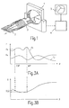
Fig. 1 stellt einen Röntgen-Computertomographen 1 dar, mit dem ein auf einer
verfahrbaren Tischplatte 2 befindlicher Patient 3 untersucht werden kann. Ein
dreidimensionaler Bereich des Patienten kann dabei entweder als Folge paralleler
benachbarter Schichtaufnahmen untersucht werden oder mittels eines "helical scan",
wobei die Tischplatte kontinuierlich in ihrer Längsrichtung (z) verschoben wird.
Dabei wird ein erster Datensatz I(x,y,z) akquiriert, der die Absorption der
Röntgenstrahlung in den Voxeln des Untersuchungsbereichs darstellt. Dieser
Datensatz wird in einem Bildverarbeitungsrechner 4 gespeichert und verarbeitet, und
das dabei erzeugte zweidimensionale Bild wird auf einem Monitor 5 dargestellt.Fig. 1 illustrates an
Die Erfindung ist auch bei einem anderen Untersuchungssystem anwendbar, das es gestattet, einen Datensatz zu akquirieren, der einen bestimmten physikalischen Parameter in einem dreidimensionalen Untersuchungsbereich charakterisiert, z.B. ein Ultraschallgerät oder ein MR-Gerät.The invention is also applicable to another examination system that it allows to acquire a dataset that has a specific physical Characterized in a three-dimensional examination area, e.g. an ultrasound device or an MR device.
Die im Bildverarbeitungsrechner 4 erfolgende Bildverarbeitung wird im folgenden
anhand der Fig. 2 näher erläutert. Den ersten Schritt 101 bildet die geschilderte
Akquisition des ersten Datensatzes I(x,y,z), der - im Falle eines
Röntgen-Computertomographen - die Absorption der Röntgenstrahlung als Funktion
des Ortes x,y,z definiert.The image processing performed in the image processing computer 4 will be described below
explained in more detail with reference to FIG. 2. The
Der nächste Schritt 102 besteht darin, die abzubildenden Strukturen durch eine
Filterung hervorzuheben und auf diese Weise aus dem ersten Satz I(x,y,z) einen
zweiten Datensatz F(x,y,z) zu erzeugen. Die Filterung muß dabei auf die
darzustellenden Strukturen ausgerichtet sein. Wenn beispielsweise ein
(gegebenenfalls mit Kontrastmittel gefülltes) Gefäßsystem im Körper des Patienten
oder in seinen Schädel implantierte Elektroden abgebildet werden sollen, muß dieses
Filterverfahren so beschaffen sein, daß es linienförmige Strukturen hervorheben
kann. Solche Filterverfahren sind bekannt, z.B. aus CVRMed and MRCAS, Lecture
Notes in Computer Science 1205 (Springer Verlag, Berlin,1997) pp 233-242.The
Der Voxel-Bildwert F(x,y,z), der einem Voxel x,y,z durch das Filterverfahren zugeordnet wird, ist ein Maß für die Wahrscheinlichkeit, daß das zugehörige Voxel zu der interessierenden Struktur gehört. Im folgenden soll angenommen werden, daß ein großer Voxel-Bildwert einer hohen Wahrscheinlichkeit für die Zugehörigkeit zu der interessierenden Struktur entspricht.The voxel image value F (x, y, z) corresponding to a voxel x, y, z by the filtering process is a measure of the probability that the associated voxel belongs to the structure of interest. In the following it shall be assumed that a large voxel image value of a high probability of belonging to corresponds to the structure of interest.
Im nächsten Schritt 103 werden die Parameter einer Projektion definiert, mit der der
durch den ersten bzw. zweiten Datensatz definierte dreidimensionale
Untersuchungsbereich in ein zweidimensionales Bild überführt werden soll. Im
einfachsten Fall kann es sich dabei um eine Parallel-Projektion handeln,
vorzugsweise mit einer zu einer der Koordinatenrichtungen x, y und z parallel
verlaufenden Projektionsrichtung. Jedoch ist ebenso eine Zentral-Projektion möglich
oder aber auch eine Projektion, bei der die Projektionsstrahlen gekrümmt sind.In the
Außerdem werden im Schritt 103 die Voxel bestimmt, die - bei der gewählten
Projektionsgeometrie - auf dasselbe Pixel in dem zweidimensionalen Bild (gestrichelt
angedeutet) projiziert werden. Zur Veranschaulichung ist in dem Block 103 ein
Projektionsstrahl p mit einigen der darauf befindlichen Voxeln angedeutet, der in
einem ebenfalls auf dem Projektionsstrahl befindlichen Pixel des zweidimensionalen
Bildes endet. Es erfolgt nun eine Auswahl mindestens eines dieser Voxel - z.B. v1 -
in Abhängigkeit von seinem Voxel-Bildwert F(x,y,z) in dem zweiten Datensatz. In addition, in
Die Auswahl ist in Fig. 3a erläutert, worin der Verlauf der Voxel-Bildwerte F(p) aus dem zweiten Datensatz F(x,y,z) für die jeweils auf dem gleichen Projektionsstrahl p befindlichen und auf dasselbe Pixel projizierten Voxel in zwei verschiedenen Kurven F1, F2 (für zwei verschiedene Projektionsstrahlen) dargestellt ist. Eine der Möglichkeiten für die Auswahl besteht darin, das Voxel mit dem größten Voxel-Bildwert, d.h. mit der größten Wahrscheinlichkeit, daß dieses Voxel zu der interessierenden Struktur gehört, auszuwählen, was - bezogen auf den zweiten Datensatz F(x,y,z) - einer MIP entspricht. Eine andere Möglichkeit besteht darin, das Voxel auszuwählen, das als erstes auf dem Projektionsstrahl p einen Voxel-Bildwert F(p) oberhalb eines Schwellwertes Fs aufweist; dies entspricht in bezug auf den zweiten Datensatz einer CVP. Es sind aber auch andere Möglichkeiten der Auswahl denkbar.The selection is explained in FIG. 3a, in which the course of the voxel image values F (p) from the second record F (x, y, z) for each on the same Projection beam p and projected on the same pixel voxels in two different curves F1, F2 (for two different projection beams) is. One of the choices for choosing is to use the voxel with the largest voxel image value, i. with the greatest probability that this voxel belongs to the structure of interest to choose what - based on the second record F (x, y, z) - corresponds to a MIP. Another possibility exists in selecting the voxel, the first on the projection beam p Voxel image value F (p) above a threshold Fs; this corresponds to relative to the second record of a CVP. But there are others too Possibilities of selection conceivable.
Im Schritt 104 wird mit Hilfe der ausgewählten Voxel ein zweidimensionales Bild B1
erzeugt. Dabei wird der Bildwert für die Pixel des Bildes B1 aus den zuvor
ausgewählten Voxeln abgeleitet. Dabei wird jedoch nicht der Voxel-Bildwert F(p)
herangezogen, der diesem Voxel im zweiten Datensatz F zugeordnet ist und
aufgrund dessen es ausgewählt wurde, sondern der Voxel-Bildwert I(p), der im
ersten Datensatz für dieses Voxel gespeichert ist, so daß es in dem Bild B1
originalgetreu wiedergegeben wird. Dieser Voxel-Bildwert kann durchaus kleiner
sein als die Bildwerte der anderen Voxel, die auf dem gleichen Projektionsstrahl
liegen.In
Dies zeigt auch Fig. 3b, die den räumlichen Verlauf der Voxel-Bildwerte I(p) entlang des Projektionsstrahls p darstellt, dem in Fig 3a die Kurve F1 zugeordnet ist. Es wird der Voxel-Bildwert I(p) herangezogen, der an derselben Position entlang des Projektionsstrahls p liegt wie das Maximum im zweiten Datensatz - bzw. wie der erste oberhalb des Schwellwertes Fs (Fig. 3a) liegende Wert. This is also shown in FIG. 3b, which shows the spatial progression of the voxel image values I (p). along the projection beam p, assigned to the curve F1 in Fig 3a is. The voxel image value I (p), which is at the same position, is used of the projection beam p is like the maximum in the second data set - or how the first value above the threshold value Fs (FIG. 3a).
Das erfindungsgemäße Verfahren führt dazu, daß störende Strukturen im Untersuchungsbereich, die bei einer Projektion normalerweise den interessierenden Strukturen überlagert werden, ausgeblendet werden, weil ihnen im zweiten Datensatz F niedrige Bildwerte zugeordnet sind. Trotz der dafür erforderlichen Filterung des Original-Datensatzes erscheinen die abzubildenden Strukturen in dem Bild B1 mit ihren originalen Kontrastverhältnissen, weil nicht der gefilterte Datensatz F für die ausgewählten Voxel herangezogen wird, sondern der originale (ungefilterte bzw. erste) Datensatz I.The method according to the invention leads to the fact that interfering structures in the examination area, which are normally superimposed on the structures of interest in a projection, are masked out because they are assigned low image values in the second data set F. Despite the filtering required for the original data set, the structures to be imaged appear in the image B 1 with their original contrast ratios, because it is not the filtered data set F that is used for the selected voxels but the original (unfiltered or first) data set I.
Wenn das erfindungsgemäße Verfahren auch für die Pixel des Bildes B1 angewandt
würde, auf die kein Voxel aus den interessierenden Strukturen projiziert wird
(Bildhintergrund), würde der Bildwert von einem Voxel abgeleitet, das entlang des
Projektionsstrahls p z.B. den größten Wert F(x,y,z) aufweist. Dies könnte dazu
führen, daß sich in dem Bild außerhalb der Strukturen ein mehr oder weniger
zufälliger Bildhintergrund ergeben würde, der die Bildinterpretation erschweren
könnte. Um die dadurch möglichen Bildartefakte zu vermeiden, kann die Auswahl
des für das zweidimensionale Bild B1 herangezogenen Voxels (Block 103)
folgendermaßen modifiziert werden:
Es entsteht dadurch zwar ein ruhiger, einheitlicher Bildhintergrund, jedoch kann
unter Umständen die anatomische Orientierung für den Arzt erschwert sein. Um hier
die Orientierung zu erleichtern, können das zweidimensionale Bild B1 und ein für
dieselbe Projektionsgeometrie errechnetes Standard-Projektionsbild B2, in dem jedem
Pixel z.B. der Mittelwert der Bildwerte für die Voxel auf dem diesem Pixel
zugeordneten Projektionsstrahl p zugeordnet wird und das daher einen ähnlichen
Charakter hat wie eine Röntgenaufnahme, addiert werden gemäß der Beziehung
Das erfindungsgemäße Verfahren ist geeignet, beispielsweise in einem Schädel implantierte Elektroden - frei von Überlagerungen durch die Schädelknochen - darzustellen. Es können aber auch mit einem Kontrastmittel gefüllte Herzkranzgefäße dargestellt werden, ohne daß sich dieser Darstellung die mit Kontrastmittel gefüllten Herzkammern überlagern. In beiden Fällen werden also linienförmige Strukturen dargestellt, doch sind auch andere Strukturen in dieser Weise darstellbar, wenn sie mittels eines geeigneten Filterverfahrens hervorgehoben werden können.The method according to the invention is suitable, for example in a skull implanted electrodes - free of interference from the skull bones - display. It can also be filled with a contrast agent Coronary arteries are shown without this representation with the Superimposed contrast media filled heart chambers. In both cases so linear structures are shown, but are also other structures in this Represented manner when highlighted by means of a suitable filtering method can be.
Im Ausführungsbeispiel wurden die Bildwerte für die Pixel des zweidimensionalen Bildes B1 stets nur aus dem Voxel-Bildwert von je einem Voxel abgeleitet. Es ist jedoch auch möglich, mehrere Voxel auszuwählen und deren Voxel-Bildwerte heranzuziehen, z.B. die n Voxel mit dem größten Bildwert (wobei n eine ganze Zahl größer als 1 ist), oder die n entlang des Projektionsstrahls p aufeinander folgenden Voxel, deren Voxel-Bildwerte den größten Summenwert erreichen. Dadurch können Bildstörungen durch Rauschen oder dergl. verringert werden.In the exemplary embodiment, the image values for the pixels of the two-dimensional image B 1 were always derived only from the voxel image value of one voxel each. However, it is also possible to select several voxels and to use their voxel image values, eg the n voxels with the largest image value (where n is an integer greater than 1), or the n along the projection beam p successive voxels whose voxels Image values reach the highest sum value. This can reduce noise due to noise or the like.
Claims (9)
- A medical diagnostic method for the two-dimensional imaging of structures present in an object, which method includes the steps of:a) acquiring a first data record (I(x,y,z)) from the object in order to define voxel image values for the voxels within a three-dimensional examination zone,b) generating a second data record (F(x,y,z)) from the first data record by a filtering operation which emphasizes the structures,
characterized by the following steps:c) determining voxels (x,y,z) that are projected onto the same pixel (u,v) in a two-dimensional image,d) selecting at least one of these voxels (x,y,z) in dependence on its voxel image value (F(x,y,z)) in the second data record,e) generating the two-dimensional image (B1(u,v)) which image value for the pixel (u,v) is derived from the voxel image value (I(x,y,z)) of the selected voxel in the first data record. - A method as claimed in Claim 1,
characterized in that in step d) the voxel is selected whose voxel image value has an extreme value, notably a maximum, in the second data record. - A method as claimed in Claim 1,
characterized in that the voxel selected in step d) is the first voxel whose voxel image value in the second data record exceeds or falls short of a predefinable threshold value. - A method as claimed in Claim 1,
characterized in that a constant image value is assigned to a pixel (u,v) in the two-dimensional image (B1(u, v) if the voxel image values (F(x,y,z)) of the voxels projected onto this pixel do not exceed or fall short of a selectable threshold value in the second data record. - A method as claimed in Claim 1,
characterized in that a projection image (B2(u,v)) is derived from the first data record and that this projection image and the two-dimensional image (B1(u,v)) are superposed in an additive manner. - A method as claimed in Claim 1,
characterized in that the first data record is acquired by means of an MR method. - A method as claimed in Claim 1,
characterized in that the first data record is acquired by means of an X-ray CT method. - A method of two-dimensional imaging of structures in an object for medical diagnosis which object is defined by a first data record (I(x, y, z)) with voxel image values for the voxels within a three-dimensional examination zone, which method includes the following steps:b) generating a second data record (F(x, y, z)) from the first data record by a filtering operation which emphasizes the structures,
characterized by the following steps:c) determining voxels (x,y,z) which are projected onto the same pixel (u,v) in a two-dimensional image,d) selecting at least one of these voxels (x,y,z) in dependence on its voxel image value (F(x,y,z)) in the second data record,e) generating the two-dimensional image (B(u, v)) where the image value for the pixel (u, v) is derived from the voxel image value (I(x, y, z)) of the selected voxel in the first data record. - A medical diagnostic system for the two-dimensional imaging of structures present in an object, including means for the acquisition of a first data record (I(x, y, z)) from the object in order to define voxel image values for the voxels within a three-dimensional examination zone, and means for generating a second data record (F(x, y, z)) from the first data record by a filtering operation which emphasizes the structures,
characterized by:means for determining voxels (x, y, z) which are projected onto the same pixel (u, v) in a two-dimensional image,means for selecting at least one of these voxels in dependence on its voxel image value (F(x, y, z)) in the second data record,means for generating a two-dimensional image (B1(u,v)) where the image value for the pixel (u,v) is derived from the voxel image value (I(x,y,z)) of the selected voxel in the first data record.
Applications Claiming Priority (2)
| Application Number | Priority Date | Filing Date | Title |
|---|---|---|---|
| DE19806728A DE19806728A1 (en) | 1998-02-18 | 1998-02-18 | Process for two-dimensional imaging of structures for medical diagnostics |
| DE19806728 | 1998-02-18 |
Publications (3)
| Publication Number | Publication Date |
|---|---|
| EP0938063A2 EP0938063A2 (en) | 1999-08-25 |
| EP0938063A3 EP0938063A3 (en) | 2000-01-26 |
| EP0938063B1 true EP0938063B1 (en) | 2005-02-16 |
Family
ID=7858141
Family Applications (1)
| Application Number | Title | Priority Date | Filing Date |
|---|---|---|---|
| EP99200378A Expired - Lifetime EP0938063B1 (en) | 1998-02-18 | 1999-02-09 | Method of two-dimensional imaging of structures for medical diagnosis |
Country Status (4)
| Country | Link |
|---|---|
| US (1) | US6205350B1 (en) |
| EP (1) | EP0938063B1 (en) |
| JP (1) | JPH11313802A (en) |
| DE (2) | DE19806728A1 (en) |
Families Citing this family (21)
| Publication number | Priority date | Publication date | Assignee | Title |
|---|---|---|---|---|
| AU2001251539A1 (en) * | 2000-04-11 | 2001-10-23 | Cornell Research Foundation Inc. | System and method for three-dimensional image rendering and analysis |
| US6705404B2 (en) * | 2001-09-10 | 2004-03-16 | Gordon F. Bosley | Open well plunger-actuated gas lift valve and method of use |
| US20030095696A1 (en) * | 2001-09-14 | 2003-05-22 | Reeves Anthony P. | System, method and apparatus for small pulmonary nodule computer aided diagnosis from computed tomography scans |
| US6728334B1 (en) | 2001-10-24 | 2004-04-27 | Cornell Research Foundation, Inc. | Automatic detection of pulmonary nodules on volumetric computed tomography images using a local density maximum algorithm |
| US20040127840A1 (en) * | 2002-03-04 | 2004-07-01 | Steve Gara | Blood separation apparatus and method of using the same |
| US7499578B2 (en) | 2002-10-18 | 2009-03-03 | Cornell Research Foundation, Inc. | System, method and apparatus for small pulmonary nodule computer aided diagnosis from computed tomography scans |
| WO2004047029A1 (en) * | 2002-11-21 | 2004-06-03 | Philips Intellectual Property & Standards Gmbh | Method and apparatus for visualizing a sequence of volume images |
| US8045770B2 (en) * | 2003-03-24 | 2011-10-25 | Cornell Research Foundation, Inc. | System and method for three-dimensional image rendering and analysis |
| JP4090970B2 (en) * | 2003-09-09 | 2008-05-28 | ジーイー・メディカル・システムズ・グローバル・テクノロジー・カンパニー・エルエルシー | Radiation tomography apparatus, radiation tomography method, image generation apparatus, and image generation method |
| US7672424B2 (en) * | 2004-10-08 | 2010-03-02 | Koninklijke Philips Electronics N.V. | Image reconstruction with voxel dependent interpolation |
| JP4212564B2 (en) | 2005-02-28 | 2009-01-21 | ザイオソフト株式会社 | Image processing method and image processing program |
| US7873194B2 (en) * | 2006-10-25 | 2011-01-18 | Rcadia Medical Imaging Ltd. | Method and system for automatic analysis of blood vessel structures and pathologies in support of a triple rule-out procedure |
| US7940970B2 (en) * | 2006-10-25 | 2011-05-10 | Rcadia Medical Imaging, Ltd | Method and system for automatic quality control used in computerized analysis of CT angiography |
| US7860283B2 (en) | 2006-10-25 | 2010-12-28 | Rcadia Medical Imaging Ltd. | Method and system for the presentation of blood vessel structures and identified pathologies |
| US7940977B2 (en) * | 2006-10-25 | 2011-05-10 | Rcadia Medical Imaging Ltd. | Method and system for automatic analysis of blood vessel structures to identify calcium or soft plaque pathologies |
| US7983459B2 (en) | 2006-10-25 | 2011-07-19 | Rcadia Medical Imaging Ltd. | Creating a blood vessel tree from imaging data |
| US20090029116A1 (en) * | 2007-07-27 | 2009-01-29 | David Harruff | Customizable container identification device |
| JP5806448B2 (en) * | 2009-05-13 | 2015-11-10 | 株式会社東芝 | Nuclear medicine imaging apparatus, image processing apparatus, and image processing method |
| JP6362420B2 (en) | 2014-05-26 | 2018-07-25 | キヤノン株式会社 | Subject information acquisition apparatus, subject information acquisition method, and program |
| CN106256326A (en) * | 2015-06-19 | 2016-12-28 | 通用电气公司 | The generation system and method for computed tomography sectioning image |
| JP6556300B2 (en) * | 2018-06-26 | 2019-08-07 | キヤノン株式会社 | Subject information acquisition apparatus, subject information acquisition method, and program |
Family Cites Families (4)
| Publication number | Priority date | Publication date | Assignee | Title |
|---|---|---|---|---|
| US4831528A (en) * | 1987-11-09 | 1989-05-16 | General Electric Company | Apparatus and method for improvement of 3D images derived from tomographic data |
| US5226113A (en) * | 1989-10-30 | 1993-07-06 | General Electric Company | Method and apparatus for volumetric projection rendering using reverse ray casting |
| JP3483929B2 (en) * | 1994-04-05 | 2004-01-06 | 株式会社日立製作所 | 3D image generation method |
| US6100862A (en) * | 1998-04-20 | 2000-08-08 | Dimensional Media Associates, Inc. | Multi-planar volumetric display system and method of operation |
-
1998
- 1998-02-18 DE DE19806728A patent/DE19806728A1/en not_active Withdrawn
-
1999
- 1999-02-09 EP EP99200378A patent/EP0938063B1/en not_active Expired - Lifetime
- 1999-02-09 DE DE59911626T patent/DE59911626D1/en not_active Expired - Lifetime
- 1999-02-16 JP JP11037498A patent/JPH11313802A/en not_active Withdrawn
- 1999-02-17 US US09/251,681 patent/US6205350B1/en not_active Expired - Lifetime
Also Published As
| Publication number | Publication date |
|---|---|
| DE19806728A1 (en) | 1999-08-19 |
| JPH11313802A (en) | 1999-11-16 |
| DE59911626D1 (en) | 2005-03-24 |
| EP0938063A3 (en) | 2000-01-26 |
| EP0938063A2 (en) | 1999-08-25 |
| US6205350B1 (en) | 2001-03-20 |
Similar Documents
| Publication | Publication Date | Title |
|---|---|---|
| EP0938063B1 (en) | Method of two-dimensional imaging of structures for medical diagnosis | |
| DE102005038940B4 (en) | Method for filtering tomographic 3D representations after reconstruction of volume data | |
| DE19613342A1 (en) | Automatic image evaluation process | |
| DE3738636C2 (en) | ||
| DE60224770T2 (en) | Method and apparatus for noise reduction in computer tomographs | |
| DE102005012654B4 (en) | Method and computed tomography system for generating tomographic images of an object | |
| DE3735519C2 (en) | Binary space interpolation | |
| EP0996090A2 (en) | Method for the processing of an input image | |
| DE102005058217B4 (en) | Method and system for computer-aided detection of high-contrast objects in tomographic images | |
| DE102004004295A1 (en) | Method for image data acquisition and evaluation with a tomography device | |
| DE102007046514A1 (en) | Method for detecting and marking contrast medium in blood vessels of the lung using a CT examination and image evaluation unit of a CT system | |
| DE102007013570A1 (en) | Method for noise reduction in digital images with locally different and directional noise | |
| DE2945057A1 (en) | METHOD FOR REDUCING IMAGE ERRORS IN LAYER IMAGES OF A THREE-DIMENSIONAL OBJECT PRODUCED WITH THE AID OF Pervasive RADIATION | |
| DE10229113A1 (en) | Process for gray value-based image filtering in computer tomography | |
| DE102006005804A1 (en) | Method for noise reduction in tomographic image data sets | |
| EP1209622A2 (en) | Method and device for registering images | |
| DE19705599A1 (en) | X-ray imaging process with a series of exposures from different perspectives | |
| EP1843296A1 (en) | Method for reproducible creation of views of tomographic image data | |
| DE60311249T2 (en) | ORGANIC REPROJECTION | |
| DE19529636C2 (en) | Method for MR imaging of several layers and arrangement for carrying out the method | |
| DE19634821A1 (en) | Method and device for reducing image artifacts | |
| DE19625863C2 (en) | Image reconstruction method for a spiral tomograph | |
| DE19842944B4 (en) | Method for reconstructing a three-dimensional image of an object scanned in the course of a tomosynthesis | |
| DE69732615T2 (en) | Method and device for simplified filtering of scan data in a video system | |
| DE102008045633A1 (en) | Computer tomography (CT) method for improved display of multi-energy CT exposures/photographs applies contrast media along a system axis |
Legal Events
| Date | Code | Title | Description |
|---|---|---|---|
| PUAI | Public reference made under article 153(3) epc to a published international application that has entered the european phase |
Free format text: ORIGINAL CODE: 0009012 |
|
| AK | Designated contracting states |
Kind code of ref document: A2 Designated state(s): DE FR GB NL |
|
| AX | Request for extension of the european patent |
Free format text: AL;LT;LV;MK;RO;SI |
|
| PUAL | Search report despatched |
Free format text: ORIGINAL CODE: 0009013 |
|
| RAP3 | Party data changed (applicant data changed or rights of an application transferred) |
Owner name: KONINKLIJKE PHILIPS ELECTRONICS N.V. Owner name: PHILIPS CORPORATE INTELLECTUAL PROPERTY GMBH |
|
| AK | Designated contracting states |
Kind code of ref document: A3 Designated state(s): AT BE CH CY DE DK ES FI FR GB GR IE IT LI LU MC NL PT SE |
|
| AX | Request for extension of the european patent |
Free format text: AL;LT;LV;MK;RO;SI |
|
| 17P | Request for examination filed |
Effective date: 20000726 |
|
| AKX | Designation fees paid |
Free format text: DE FR GB NL |
|
| RAP1 | Party data changed (applicant data changed or rights of an application transferred) |
Owner name: KONINKLIJKE PHILIPS ELECTRONICS N.V. Owner name: PHILIPS CORPORATE INTELLECTUAL PROPERTY GMBH |
|
| RAP1 | Party data changed (applicant data changed or rights of an application transferred) |
Owner name: KONINKLIJKE PHILIPS ELECTRONICS N.V. Owner name: PHILIPS INTELLECTUAL PROPERTY & STANDARDS GMBH |
|
| GRAP | Despatch of communication of intention to grant a patent |
Free format text: ORIGINAL CODE: EPIDOSNIGR1 |
|
| GRAS | Grant fee paid |
Free format text: ORIGINAL CODE: EPIDOSNIGR3 |
|
| GRAA | (expected) grant |
Free format text: ORIGINAL CODE: 0009210 |
|
| AK | Designated contracting states |
Kind code of ref document: B1 Designated state(s): DE FR GB NL |
|
| PG25 | Lapsed in a contracting state [announced via postgrant information from national office to epo] |
Ref country code: NL Free format text: LAPSE BECAUSE OF FAILURE TO SUBMIT A TRANSLATION OF THE DESCRIPTION OR TO PAY THE FEE WITHIN THE PRESCRIBED TIME-LIMIT Effective date: 20050216 Ref country code: GB Free format text: LAPSE BECAUSE OF FAILURE TO SUBMIT A TRANSLATION OF THE DESCRIPTION OR TO PAY THE FEE WITHIN THE PRESCRIBED TIME-LIMIT Effective date: 20050216 |
|
| REG | Reference to a national code |
Ref country code: GB Ref legal event code: FG4D Free format text: NOT ENGLISH |
|
| REF | Corresponds to: |
Ref document number: 59911626 Country of ref document: DE Date of ref document: 20050324 Kind code of ref document: P |
|
| NLV1 | Nl: lapsed or annulled due to failure to fulfill the requirements of art. 29p and 29m of the patents act | ||
| GBV | Gb: ep patent (uk) treated as always having been void in accordance with gb section 77(7)/1977 [no translation filed] |
Effective date: 20050216 |
|
| PLBE | No opposition filed within time limit |
Free format text: ORIGINAL CODE: 0009261 |
|
| STAA | Information on the status of an ep patent application or granted ep patent |
Free format text: STATUS: NO OPPOSITION FILED WITHIN TIME LIMIT |
|
| ET | Fr: translation filed | ||
| 26N | No opposition filed |
Effective date: 20051117 |
|
| REG | Reference to a national code |
Ref country code: FR Ref legal event code: D6 |
|
| REG | Reference to a national code |
Ref country code: DE Ref legal event code: R081 Ref document number: 59911626 Country of ref document: DE Owner name: KONINKLIJKE PHILIPS N.V., NL Free format text: FORMER OWNERS: PHILIPS INTELLECTUAL PROPERTY & STANDARDS GMBH, 20099 HAMBURG, DE; KONINKLIJKE PHILIPS ELECTRONICS N.V., EINDHOVEN, NL Effective date: 20140327 Ref country code: DE Ref legal event code: R081 Ref document number: 59911626 Country of ref document: DE Owner name: PHILIPS GMBH, DE Free format text: FORMER OWNERS: PHILIPS INTELLECTUAL PROPERTY & STANDARDS GMBH, 20099 HAMBURG, DE; KONINKLIJKE PHILIPS ELECTRONICS N.V., EINDHOVEN, NL Effective date: 20140327 Ref country code: DE Ref legal event code: R081 Ref document number: 59911626 Country of ref document: DE Owner name: PHILIPS GMBH, DE Free format text: FORMER OWNER: PHILIPS INTELLECTUAL PROPERTY &, KONINKLIJKE PHILIPS ELECTRONICS, , NL Effective date: 20140327 Ref country code: DE Ref legal event code: R081 Ref document number: 59911626 Country of ref document: DE Owner name: KONINKLIJKE PHILIPS N.V., NL Free format text: FORMER OWNER: PHILIPS INTELLECTUAL PROPERTY &, KONINKLIJKE PHILIPS ELECTRONICS, , NL Effective date: 20140327 Ref country code: DE Ref legal event code: R081 Ref document number: 59911626 Country of ref document: DE Owner name: PHILIPS DEUTSCHLAND GMBH, DE Free format text: FORMER OWNER: PHILIPS INTELLECTUAL PROPERTY &, KONINKLIJKE PHILIPS ELECTRONICS, , NL Effective date: 20140327 |
|
| REG | Reference to a national code |
Ref country code: FR Ref legal event code: CD Owner name: PHILIPS INTELLECTUAL PROPERTY S Effective date: 20141126 Ref country code: FR Ref legal event code: CA Effective date: 20141126 |
|
| REG | Reference to a national code |
Ref country code: DE Ref legal event code: R082 Ref document number: 59911626 Country of ref document: DE Representative=s name: MEISSNER BOLTE PATENTANWAELTE RECHTSANWAELTE P, DE Ref country code: DE Ref legal event code: R082 Ref document number: 59911626 Country of ref document: DE Representative=s name: MEISSNER, BOLTE & PARTNER GBR, DE Ref country code: DE Ref legal event code: R081 Ref document number: 59911626 Country of ref document: DE Owner name: KONINKLIJKE PHILIPS N.V., NL Free format text: FORMER OWNERS: KONINKLIJKE PHILIPS N.V., EINDHOVEN, NL; PHILIPS DEUTSCHLAND GMBH, 20099 HAMBURG, DE Ref country code: DE Ref legal event code: R081 Ref document number: 59911626 Country of ref document: DE Owner name: PHILIPS GMBH, DE Free format text: FORMER OWNERS: KONINKLIJKE PHILIPS N.V., EINDHOVEN, NL; PHILIPS DEUTSCHLAND GMBH, 20099 HAMBURG, DE |
|
| REG | Reference to a national code |
Ref country code: FR Ref legal event code: PLFP Year of fee payment: 18 |
|
| PGFP | Annual fee paid to national office [announced via postgrant information from national office to epo] |
Ref country code: FR Payment date: 20160229 Year of fee payment: 18 |
|
| PGFP | Annual fee paid to national office [announced via postgrant information from national office to epo] |
Ref country code: DE Payment date: 20160502 Year of fee payment: 18 |
|
| REG | Reference to a national code |
Ref country code: DE Ref legal event code: R119 Ref document number: 59911626 Country of ref document: DE |
|
| REG | Reference to a national code |
Ref country code: FR Ref legal event code: ST Effective date: 20171031 |
|
| PG25 | Lapsed in a contracting state [announced via postgrant information from national office to epo] |
Ref country code: FR Free format text: LAPSE BECAUSE OF NON-PAYMENT OF DUE FEES Effective date: 20170228 Ref country code: DE Free format text: LAPSE BECAUSE OF NON-PAYMENT OF DUE FEES Effective date: 20170901 |