EP0832604A1 - Method and device for measuring the elasticity of an artery by ultrasonic echography - Google Patents
Method and device for measuring the elasticity of an artery by ultrasonic echography Download PDFInfo
- Publication number
- EP0832604A1 EP0832604A1 EP96402081A EP96402081A EP0832604A1 EP 0832604 A1 EP0832604 A1 EP 0832604A1 EP 96402081 A EP96402081 A EP 96402081A EP 96402081 A EP96402081 A EP 96402081A EP 0832604 A1 EP0832604 A1 EP 0832604A1
- Authority
- EP
- European Patent Office
- Prior art keywords
- artery
- arterial
- elasticity
- instantaneous
- blood
- Prior art date
- Legal status (The legal status is an assumption and is not a legal conclusion. Google has not performed a legal analysis and makes no representation as to the accuracy of the status listed.)
- Withdrawn
Links
Images

Classifications
-
- A—HUMAN NECESSITIES
- A61—MEDICAL OR VETERINARY SCIENCE; HYGIENE
- A61B—DIAGNOSIS; SURGERY; IDENTIFICATION
- A61B5/00—Measuring for diagnostic purposes; Identification of persons
- A61B5/02—Detecting, measuring or recording pulse, heart rate, blood pressure or blood flow; Combined pulse/heart-rate/blood pressure determination; Evaluating a cardiovascular condition not otherwise provided for, e.g. using combinations of techniques provided for in this group with electrocardiography or electroauscultation; Heart catheters for measuring blood pressure
- A61B5/02007—Evaluating blood vessel condition, e.g. elasticity, compliance
-
- A—HUMAN NECESSITIES
- A61—MEDICAL OR VETERINARY SCIENCE; HYGIENE
- A61B—DIAGNOSIS; SURGERY; IDENTIFICATION
- A61B8/00—Diagnosis using ultrasonic, sonic or infrasonic waves
- A61B8/06—Measuring blood flow
-
- G—PHYSICS
- G01—MEASURING; TESTING
- G01S—RADIO DIRECTION-FINDING; RADIO NAVIGATION; DETERMINING DISTANCE OR VELOCITY BY USE OF RADIO WAVES; LOCATING OR PRESENCE-DETECTING BY USE OF THE REFLECTION OR RERADIATION OF RADIO WAVES; ANALOGOUS ARRANGEMENTS USING OTHER WAVES
- G01S15/00—Systems using the reflection or reradiation of acoustic waves, e.g. sonar systems
- G01S15/88—Sonar systems specially adapted for specific applications
- G01S15/89—Sonar systems specially adapted for specific applications for mapping or imaging
- G01S15/8906—Short-range imaging systems; Acoustic microscope systems using pulse-echo techniques
- G01S15/8979—Combined Doppler and pulse-echo imaging systems
-
- G—PHYSICS
- G01—MEASURING; TESTING
- G01S—RADIO DIRECTION-FINDING; RADIO NAVIGATION; DETERMINING DISTANCE OR VELOCITY BY USE OF RADIO WAVES; LOCATING OR PRESENCE-DETECTING BY USE OF THE REFLECTION OR RERADIATION OF RADIO WAVES; ANALOGOUS ARRANGEMENTS USING OTHER WAVES
- G01S7/00—Details of systems according to groups G01S13/00, G01S15/00, G01S17/00
- G01S7/52—Details of systems according to groups G01S13/00, G01S15/00, G01S17/00 of systems according to group G01S15/00
- G01S7/52017—Details of systems according to groups G01S13/00, G01S15/00, G01S17/00 of systems according to group G01S15/00 particularly adapted to short-range imaging
- G01S7/52023—Details of receivers
- G01S7/52036—Details of receivers using analysis of echo signal for target characterisation
- G01S7/52042—Details of receivers using analysis of echo signal for target characterisation determining elastic properties of the propagation medium or of the reflective target
Definitions
- the present invention relates to a method for measuring locally the elasticity ⁇ of an artery under the effect of the blood pressure P by means of an ultrasonic echograph suitable to determine the instantaneous blood flow rates (Q A (t), Q B (t)), the instantaneous radius variations ( ⁇ r A (t), ⁇ r B (t)) and the mean arterialy radius r 0 for two neighbouring, parallel excitation lines A and B traversing said artery according to one of its meridian planes, said method comprising the following steps :
- the invention also relates to a corresponding device for measuring locally the elasticity ⁇ of an artery, allowing to carry out this method.
- Diagnosis of vascular diseases and therapeutic choices are nowadays based on the analysis of the arterial lesions morphology and on the analysis of blood flows. This information is achieved with ultrasound imaging and Doppler examinations. These tools, although widely clinically validated, only get round the problem which can be formulated in the following manner : does the examined artery fulfil efficiently its function of pressure wave guide, conveying the kinetic power generated by the cardiac pump while adapting the wave form to the downstream arterial system ? To answer this question, the echographic tool must detect the beating artery and the flowing blood in order to reveal the mechanical working of the artery. The analysis of these observations, in the light of known blood flow and arterial strain models, must allow the characterization of the arterial mechanical properties. The a priori knowledge imbedded in these models is of course the cornerstone of this principle.
- a measuring method as defined in the preamble of this text is for instance described in the document EP-0 603 967. This method already enables to deduce from some knowledge of the behaviour of a point of an artery the elasticity of said artery at this point.
- the object of the invention is however to propose an improved measuring method, said improvement being based on a better knowledge of the mechanical behaviour of arterial walls, which is then formulated through a parametric stress-strain relation linking pressure and dilation variations.
- the invention relates to a method as described in the preamble, wherein said method comprises the following additional step :
- the invention relates to a device for measuring locally the elasticity ⁇ of an artery under the effect of the blood pressure and comprising, in an ultrasonic echograph used in profilometer mode (M mode) and provided with transmitting and receiving means including a device for forming channels in the receiving mode, a first measuring sub-assembly for the determination of the instantaneous blood flow rates (Q A (t), Q B (t)), the instantaneous radius variations ( ⁇ r A (t), ⁇ r B (t)) and the artery mean radius r 0 for two neighbouring, parallel excitation lines A and B, said lines traversing said artery according to one of its meridian planes and being situated at a given distance e from each other taken in the direction of the axis of the artery and comprised between some tenths of a millimeter and some millimeters, a second storing sub-assembly for storing at the output
- the variations of the artery size with time allow local blood storage followed by restitution of blood volume.
- the storage takes place during systole (cardiac ejection) while the volume flow is restituted during diastole (no cardiac outflow).
- This regulation of flow describes the basic arterial function. It closely depends on the arterial ability to be dilated with the pressure wave propagation.
- the measurement methods are time domain correlation techniques applied to ultrasonic signals, which track the echoes with time and allow the estimation of the displacements of the corresponding biological structures [2].
- the flow curve Q(t) is derived by integration of the velocity profiles measured along the acoustic beam axis.
- the arterial diameters r(x,t) are determined by locally measuring displacements of arterial front and posterior walls and combining them to finally obtain the arterial dilation [3].
- the arterial compliance is a non linear function of the pressure. Besides, this non linearity gives its genuine flow regulation ability to the artery.
- the inversion of equation (3) is tremendously complicated since it is necessary to introduce this non linarity in the left term. It is now convenient to discuss the model considered for the arterial compliance, i.e. the elastic and viscoelastic models of arterial walls.
- the identification method mentioned in the above paragraph consists in tuning the parameters K, ⁇ , ⁇ , in order to verify at best identity (3), by minimizing the resulting quadratic error.
- the Languewouters law obtained in static pressure conditions, corresponds to the hypothesis of pure elastic behaviour of the arterial wall. It is validated in vivo by Y. Tardy et al [5] in the particular cases of humeral and brachial arteries, which are relatively little distendable. In vivo experiments carried out on the carotid artery, using this pure elastic model and undertaking the measurements of volume flow and arterial dilations previously described, have never enabled to determine R, L, C with a satisfactory confidence level. This contradictory results re-open the question of the initial pure elastic hypothesis and lead finally to the introduction of a viscous component in the parietal behaviour, that neither Languewouters in vitro experiments nor in vivo Tardy ones could evidence.
- the stress (pressure P) depends on both strain (r) and strain speed ( ⁇ r/ ⁇ t).
- the graphic representation P(r) shows an hysteresis characteristic of viscous phenomenons.
- it is chosen to parametrize separately the two phases of the cardiac cycle (systole, diastole), while keeping the general relations proposed by Languewouters.
- two sets of parameters K, ⁇ , ⁇ corresponding respectively to the arterial expansion and its return to the initial state, allow to completely describe the viscoelastic behaviour of the arterial wall.
- Figure 1 shows a schematic representation of this arterial set-up and of the related acquisition and control instrumentation.
- An arterial sample freshly taken from a cadaver is placed in a temperature regulated conservation bath. It is perfused with total human blood supplied by a blood transfusion center. The hematocrit is controlled during the experiment to verify the hemolysis degree and the blood viscosity. Heparin is used to prevent any coagulation of the system.
- the hemodynamic set-up is made up of an extracorporal peristaltic pump interfaced with a frequency generator allowing pulsed flow generation, and of a dedicated conduction equipment ensuring a low hemolysis. A compliance and a resistance allow to adjust the flow and pressure curves.
- a heat exchanger and an extracorporal circulation module are place in parallel to ensure a constant temperature, to oxygenate and eliminate bubbles.
- the instrumentation allows on the one hand to control flow conditions, using an external flow-meter and manometer, and on the other hand to acquire data.
- a pressure catheter provides the intra luminal pressure, and an echograph allows the recording of acoustic signals.
- the pressure acquisition is performed using a digital oscilloscope, the ultrasound signals are stored in a dedicated acquisition system, and these two recordings are synchronized to avoid an artificial delay which would hide any viscous behaviour.
- the arterial diameter variations are derived by applying the ultrasound signal processing techniques previously described.
- dp dx ( ⁇ p ⁇ x )r 0 + ⁇ p ⁇ r . ⁇ r ⁇ x
- the first term is here considered as a constant term, responsible for the continuous component of the flow RQ 0 .
- the time varying pressure gradient dp/dx is then modelized according to two different hypotheses :
- variation range of ⁇ and ⁇ is the following : - for ⁇ : - ⁇ /2 ⁇ ⁇ ⁇ ⁇ /2 - for ⁇ : 0 ⁇ ⁇ ⁇ (( ⁇ /2)- ⁇ )/r max
Landscapes
- Health & Medical Sciences (AREA)
- Engineering & Computer Science (AREA)
- Physics & Mathematics (AREA)
- Life Sciences & Earth Sciences (AREA)
- Radar, Positioning & Navigation (AREA)
- Remote Sensing (AREA)
- Biomedical Technology (AREA)
- Molecular Biology (AREA)
- Veterinary Medicine (AREA)
- General Physics & Mathematics (AREA)
- Biophysics (AREA)
- Public Health (AREA)
- Pathology (AREA)
- General Health & Medical Sciences (AREA)
- Computer Networks & Wireless Communication (AREA)
- Heart & Thoracic Surgery (AREA)
- Medical Informatics (AREA)
- Acoustics & Sound (AREA)
- Surgery (AREA)
- Animal Behavior & Ethology (AREA)
- Radiology & Medical Imaging (AREA)
- Nuclear Medicine, Radiotherapy & Molecular Imaging (AREA)
- Hematology (AREA)
- Vascular Medicine (AREA)
- Cardiology (AREA)
- Physiology (AREA)
- Ultra Sonic Daignosis Equipment (AREA)
- Measuring Pulse, Heart Rate, Blood Pressure Or Blood Flow (AREA)
Abstract
The invention relates to a method for measuring
locally the elasticity γ of an artery under the effect of the
blood pressure P by means of a determination of instantaneous
blood flow rates (QA(t), QB(t)), instantaneous radius
variations (ΔrA(t), ΔrB(t)) and artery mean radius r0 for two
neighbouring, parallel excitation lines A and B traversing said
artery according to one of its meridian planes. The method
comprising some computing steps and the implementation of the
least squares method applied, over a whole cardiac cycle or for
each part of said cycle, to a relation including γ, followed by
a determination step of the time varying part of γ by means of
a parametric modelization of the stress-strain relations of
arterial walls.
Application : clinical examination of arteries by
ultrasonic echography.
Description
The present invention relates to a method for
measuring locally the elasticity γ of an artery under the
effect of the blood pressure P by means of an ultrasonic
echograph suitable to determine the instantaneous blood flow
rates (QA(t), QB(t)), the instantaneous radius variations
(ΔrA(t), ΔrB(t)) and the mean arterialy radius r0 for two
neighbouring, parallel excitation lines A and B traversing said
artery according to one of its meridian planes, said method
comprising the following steps :
The invention also relates to a corresponding
device for measuring locally the elasticity γ of an artery,
allowing to carry out this method.
Diagnosis of vascular diseases and therapeutic
choices are nowadays based on the analysis of the arterial
lesions morphology and on the analysis of blood flows. This
information is achieved with ultrasound imaging and Doppler
examinations. These tools, although widely clinically
validated, only get round the problem which can be formulated
in the following manner : does the examined artery fulfil
efficiently its function of pressure wave guide, conveying the
kinetic power generated by the cardiac pump while adapting the
wave form to the downstream arterial system ? To answer this
question, the echographic tool must detect the beating artery
and the flowing blood in order to reveal the mechanical working
of the artery. The analysis of these observations, in the light
of known blood flow and arterial strain models, must allow the
characterization of the arterial mechanical properties. The a
priori knowledge imbedded in these models is of course the
cornerstone of this principle.
A measuring method as defined in the preamble of
this text is for instance described in the document
EP-0 603 967. This method already enables to deduce from some
knowledge of the behaviour of a point of an artery the
elasticity of said artery at this point.
The object of the invention is however to propose
an improved measuring method, said improvement being based on a
better knowledge of the mechanical behaviour of arterial walls,
which is then formulated through a parametric stress-strain
relation linking pressure and dilation variations.
At this end the invention relates to a method as
described in the preamble, wherein said method comprises the
following additional step :
With the relation thus choosen, a more efficient
measuring method is defined and carried out. For this
implementation, the invention relates to a device for measuring
locally the elasticity γ of an artery under the effect of the
blood pressure and comprising, in an ultrasonic echograph used
in profilometer mode (M mode) and provided with transmitting
and receiving means including a device for forming channels in
the receiving mode, a first measuring sub-assembly for the
determination of the instantaneous blood flow rates (QA(t),
QB(t)), the instantaneous radius variations (ΔrA(t), ΔrB(t))
and the artery mean radius r0 for two neighbouring, parallel
excitation lines A and B, said lines traversing said artery
according to one of its meridian planes and being situated at a
given distance e from each other taken in the direction of the
axis of the artery and comprised between some tenths of a
millimeter and some millimeters, a second storing sub-assembly
for storing at the output of said first sub-assembly digital
samples of signals relating to the blood flows and signals
relating to the walls of the artery, and a third computing sub-assembly
which operate on said digital signal samples, wherein
said third sub-assembly comprises computing means for
determining the time varying part of γ by using a parametric
modelization of the stress-strain relations of arterial walls.
The invention will now be described in further
detail with reference to the accompanying drawings, in which :
An ultrasonic procedure relying on the invention
will now defined and its implementation explained. In vivo
validation tests have been made, and an arterial set-up has
been built, allowing in vitro measurements in physiological
conditions as close as possible from living conditions and
their confrontation with analytical models.
After a recall on blood flow models and on the
measurements methods which allow to determine the parameters
governing this flow, elastic and viscoelastic models of
arterial walls are presented. Then the experimental validation
of these models is described.
Blood flow models and measurements are first
discussed. The Navier-Stokes equation describing flow
velocities and pressure in a tube by linking pressure gradient
and blood flow parameters can be expressed by the following
equation (1) :
- dpdx = RQ + L ∂Q∂t
where dp/dx, the pressure gradient along the x axis of the
tube, generates a flow Q. The blood viscosity creates a
constant hydrodynamic resistance R to the flow, and the blood
inertia is responsible for the L∂Q/∂t term, L being equal to
ρ/πr2 0 (r0 = mean arterial radius, ρ = voluminal mass of
blood).
The variations of the artery size with time allow
local blood storage followed by restitution of blood volume.
The storage takes place during systole (cardiac ejection) while
the volume flow is restituted during diastole (no cardiac
outflow). This regulation of flow describes the basic arterial
function. It closely depends on the arterial ability to be
dilated with the pressure wave propagation. The distensibility,
or elasticity, is expressed through the definition of the
arterial compliance, C :
C = ∂S∂p
which allows to eliminate the pressure gradient term in the
Navier-Stokes equation (1) and to write :
- 1C ∂S∂x = RQ + L∂Q∂t
leading to an expression linking flow (Q) and cross-section
(S), independently of the pressure, and implying intrinsic
mechanical variables of blood (R,L) and of arterial wall (C).
It is the basis of the analysis method that has to be
implemented. Non invasive ultrasonic measurements of the volume
flow Q and arterial diameter r (which is related to the cross-section
S) allow the estimation of R, L and C, through an
appropriate numerical identification method between the two
equation members [1]. These measurements are simultaneously
carried out on two longitudinal close positions, in order to
get ∂S/∂x, Q and ∂Q/∂t. The measurement methods are time domain
correlation techniques applied to ultrasonic signals, which
track the echoes with time and allow the estimation of the
displacements of the corresponding biological structures [2].
The flow curve Q(t) is derived by integration of the velocity
profiles measured along the acoustic beam axis. The arterial
diameters r(x,t) are determined by locally measuring
displacements of arterial front and posterior walls and
combining them to finally obtain the arterial dilation [3].
The arterial compliance is a non linear function of
the pressure. Besides, this non linearity gives its genuine
flow regulation ability to the artery. The inversion of
equation (3) is tremendously complicated since it is necessary
to introduce this non linarity in the left term. It is now
convenient to discuss the model considered for the arterial
compliance, i.e. the elastic and viscoelastic models of
arterial walls.
The viscoelastic behaviour of arteries is due to
their complex structure. The compliance is dependent on the
local arterial pressure at each instant. Many studies done to
express diameter/pressure relations are described in the
literature. Most of the proposed models are derived from
experimental data. Others result from elasticity theory.
The model initially used derives from the one of
Languewouters and al [4]. They have measured in vitro the
arterial cross-section as a function of the pressure, this
latter being statically set, and proposed a parametrical law
giving the arterial section as a function of pressure. This law
is here transformed without changing its meaning, to get the
arterial pressure P and the compliance C as functions of the
arterial radius r :
P(r) = K tan(α + βr), C(r) = 2πr0 .∂r∂p = 2πr0 cos2 (α+βr)/(kβ).
K, α, β are the model parameters (r0 is the mean arterial
radius, as already seen). The identification method mentioned
in the above paragraph consists in tuning the parameters K, α,
β, in order to verify at best identity (3), by minimizing the
resulting quadratic error.
The Languewouters law, obtained in static pressure
conditions, corresponds to the hypothesis of pure elastic
behaviour of the arterial wall. It is validated in vivo by Y.
Tardy et al [5] in the particular cases of humeral and brachial
arteries, which are relatively little distendable. In vivo
experiments carried out on the carotid artery, using this pure
elastic model and undertaking the measurements of volume flow
and arterial dilations previously described, have never enabled
to determine R, L, C with a satisfactory confidence level. This
contradictory results re-open the question of the initial pure
elastic hypothesis and lead finally to the introduction of a
viscous component in the parietal behaviour, that neither
Languewouters in vitro experiments nor in vivo Tardy ones could
evidence.
In the case of viscoelastic medium, the stress
(pressure P) depends on both strain (r) and strain speed
(∂r/∂t). The graphic representation P(r) shows an hysteresis
characteristic of viscous phenomenons. For modeling this
hysteresis, it is chosen to parametrize separately the two
phases of the cardiac cycle (systole, diastole), while keeping
the general relations proposed by Languewouters. Thus, two sets
of parameters K, α, β, corresponding respectively to the
arterial expansion and its return to the initial state, allow
to completely describe the viscoelastic behaviour of the
arterial wall.
The validation of the viscoelastic model described
above required confrontation with experimental data, by
simultaneous measurements of arterial dilation and arterial
pressure. It needed the building of an arterial set-up
reproducing as well as possible the physiological conditions,
and the implementation (around it) of the necessary control and
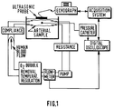
measurement instrumentation. Figure 1 shows a schematic
representation of this arterial set-up and of the related
acquisition and control instrumentation.
An arterial sample freshly taken from a cadaver is
placed in a temperature regulated conservation bath. It is
perfused with total human blood supplied by a blood transfusion
center. The hematocrit is controlled during the experiment to
verify the hemolysis degree and the blood viscosity. Heparin is
used to prevent any coagulation of the system. The hemodynamic
set-up is made up of an extracorporal peristaltic pump
interfaced with a frequency generator allowing pulsed flow
generation, and of a dedicated conduction equipment ensuring a
low hemolysis. A compliance and a resistance allow to adjust
the flow and pressure curves. A heat exchanger and an
extracorporal circulation module are place in parallel to
ensure a constant temperature, to oxygenate and eliminate
bubbles.
The instrumentation allows on the one hand to
control flow conditions, using an external flow-meter and
manometer, and on the other hand to acquire data. A pressure
catheter provides the intra luminal pressure, and an echograph
allows the recording of acoustic signals. The pressure
acquisition is performed using a digital oscilloscope, the
ultrasound signals are stored in a dedicated acquisition
system, and these two recordings are synchronized to avoid an
artificial delay which would hide any viscous behaviour. The
arterial diameter variations are derived by applying the
ultrasound signal processing techniques previously described.
The confrontation of the measurements and of the
viscous or viscoelastic models are performed by minimizing the
quadratic error between the measured pressure and the modelled
one, the coefficient set K, α, β being adapted to realize this
minimization. Using data extracted from seven common carotid
arteries, the two models are assessed through the residual
quadratic error. In all the cases, this error is above 10 % for
the pure elastic model and below 1 % for the viscoelastic one.
Figure 2 illustrates the model validity, by showing the
superposition of measured dilation, and pressure curves as
functions of time.
From a more precise mathematical point of view, the
general equation linking the pressure gradient and blood flow
parameters, given by the relation (1), can also be expressed as
follows :
dpdx = (∂p∂x )r0 + ∂p∂r .∂r∂x
The first term
 is here considered as a constant term,
responsible for the continuous component of the flow RQ0. The
time varying term of the pressure gradient :
is here considered as a constant term,
responsible for the continuous component of the flow RQ0. The
time varying term of the pressure gradient :
dp(t)dx = ∂p∂r .∂r∂x - ∂p∂x
is linked to the elasticity of the artery and is related to the
compliance relation (7), equivalent to the relation (5) :
C = ∂S∂p = 2πr0 .∂r∂p = 2πr0 γ
The initial equation can be rewritten in the following
way (with q = Q - Q0 ) :
Rq + L dqdt = - 1γ ∂r∂x = - dp∂x RQ0 = (∂p∂x )r0
A resolution using the mean square method then consists
in minimizing the quadratic error :
E2 = ∥Rq + L dqdt + dpdx ∥2
The constants R and L are easily extracted from the
equations ∂E2 /∂R = 0 and ∂E2 /∂L = 0 , which gives :
R = 〈q.dqdt 〉 〈dpdx .dqdt 〉 - 〈dq∂t 2 〉 〈q.dpdx 〉〈dqdt 2 〉 〈q2 〉 - 〈q.dqdt 〉2 L = 〈q.dqdt 〉 〈dpdx .q〉 - 〈q2 〉 〈dpdx .dqdt 〉〈dqdt 2 〉 〈q2 〉 - 〈q.dqdt 〉2
It can be noted that in the case of a cyclic phenomenon, when
the summation involved by the integration (〈.〉 = mean value of
the concerned function over a cardiac cycle) is done during
this cycle, one has :
〈q.dqdt 〉 = 0
which simplifies the previous expressions (9a) and (9b) and
gives :
R = - 〈q.dqdx 〉〈q2 〉 L = - 〈dpdx .dqdt 〉〈dqdt 2 〉
The resulting quadratic error becomes :
E2 = R2 〈q2 〉 + L2 〈dqdt 2 〉
+ 〈dpdx 2 〉 + 2RL.〈q.dqdt 〉 + 2R.〈dpdx .q〉 + 2L.〈dpdx dqdt 〉
and, in the case of a cyclic behaviour :
E2 = 〈dpdx 2 〉 - 〈q.dpdx 〉2 〈q2 〉 - 〈dqdt .dpdx 〉〈dqdt 2 〉
The final resolution of the equation (8a) consists in
introducing an elasticity law in the expression of :
dpdx = 1γ .∂r∂x ,
γ being the compliance of the artery. This law is taken into
account in computing the quadratic error, and the parameters
inducing a minimum are considered as the solution of the
problem. This compliance law is the basis of the resolution of
the problem according to the invention. The time varying
pressure gradient dp/dx is then modelized according to two
different hypotheses :
- either the artery is purely elastic,
- or it is viscoelastic.
Considering the following model :
Pα,β (r) = k.[Arctg(α + βr) - Arctg α] (∂p∂x )α,β = dp∂r .∂r∂x = 1kβ.cos2 (α+βr) .∂r∂x
and considering now the associated normalized quadratic error :
E2 n(α,β) = E2 α,β 〈(dpdx )α,β 〉
where the formula (12b) is used to compute
 the best
solution is found for (αS, βS) minimizing this expression (13).
It may be noted that the variation range of α and β is the
following :
the best
solution is found for (αS, βS) minimizing this expression (13).
It may be noted that the variation range of α and β is the
following :
- for α : -π/2 ≤ α ≤ π/2 - for β : 0 ≤ β ≤ ((π/2)-α)/rmax
With the first hypothesis of a purely elastic model, α
and β are considered as constant during the cardiac cycle :
dilation and contraction of the artery are performed with the
same stress-strain law. The quadratic error is then :
E2 n(α,β) = 1 - 〈q.(dpdx )α,β 〉2 〈q2 〉 〈(dpdx )2 α,β 〉 - 〈dqdt .(dpdx )α,β 〉2 〈dqdt 2 〉 〈(dpdx )2 α,β 〉
Now αS and βS are determined, and k is computed thanks to the
expression (10b) where L is already known :
k = - 1L .〈1βS .cos2 (αS +βS r) .∂r∂x .dqdt 〉〈dqdt 2 〉
Finally :
P(r) = k.[Arctg(αS + βS r) - Arctg(αS )]
With the second hypothesis of a viscoelastic model, the
dilation and contraction phases of the artery correspond to the
same kind of model but with different (α, β) solutions. That
allows to introduce an hysteresis phenomenon in the stress-strain
relations. However the resolution becomes more
sophisticated since the term 〈q.dq/dt〉 cannot be set to 0, the
integration being made on only a part of the cardiac cycle. For
each part of the cardiac cycle, the following computation is
then required :

Thanks to this improved method, it is now possible to
introduce an appropriate arterial strain law in vascular
procedure. Assuming that this viscoelastic model is reliable,
it is still necessary to define more precisely the convenient
algorithm for the non invasive extraction of the arterial
pressure. If successful, this new modality would lead to a
genuine breakthrough of vascular echography, allowing the early
detection of cardio-vascular diseases and improving the
therapeutic follow-up.
Claims (3)
- A method for measuring locally the elasticity γ of an artery under the effect of the blood pressure P by means of an ultrasonic echograph suitable to determine the instantaneous blood flow rates (QA(t), QB(t)), the instantaneous radius variations (ΔrA(t), ΔrB(t)) and the mean arterial radius r0 for two neighbouring, parallel excitation lines A and B traversing said artery according to one of its meridian planes, said method comprising the following steps :(A) determining, on the basis of one of the two functions (QA(t), QB(t)), the time mean value Q0 and the derivative ∂Q(t)/∂t of the function(B) computing, on the basis of the instantaneous values (ΔrA(t), ΔrB(t)), the dilation gradient(C) determining the mean values γ0, R0, R0Q0 of the elasticity γ, the hydrodynamic resistance R and the pressure gradient dp/dx respectively, said determination being carried out on the basis of the least squares method applied, over a whole cardiac cycle, to the following relation :(D) determining the time varying part of γ by means of a parametric modelization of the stress-strain relations of arterial walls.
- A method as claimed in Claim 1, characterized in that said parametric stress-strain relation is defined by three parameters α, β, k and according to relations of the following type :defined by the relation :

- A device for measuring locally the elasticity γ of an artery under the effect of the blood pressure and comprising, in an ultrasonic echograph used in profilometer mode (M mode) and provided with transmitting and receiving means including a device for forming channels in the receiving mode, a first measuring sub-assembly for the determination of the instantaneous blood flow rates (QA(t), QB(t)), the instantaneous radius variations (ΔrA(t), ΔrB(t)) and the mean arterial radius r0 for two neighbouring, parallel excitation lines A and B, said lines traversing said artery according to one of its meridian planes and being situated at a given distance e from each other taken in the direction of the axis of the artery and comprised between some tenths of a millimeter and some millimeters, a second storing sub-assembly for storing at the output of said first sub-assembly digital samples of signals relating to the blood flows and signals relating to the walls of the artery, and a third computing sub-assembly which operate on said digital signal samples, wherein said third sub-assembly comprises computing means for determining the time varying part of γ by using a parametric modelization of the stress-strain relations of arterial walls.
Priority Applications (7)
| Application Number | Priority Date | Filing Date | Title |
|---|---|---|---|
| EP96402081A EP0832604A1 (en) | 1996-09-30 | 1996-09-30 | Method and device for measuring the elasticity of an artery by ultrasonic echography |
| US09/077,414 US6113543A (en) | 1996-09-30 | 1997-09-27 | Method and device for determining the compliance and the blood pressure of an artery by ultrasonic echography |
| JP10516344A JP2000501327A (en) | 1996-09-30 | 1997-09-29 | Method and apparatus for determining arterial compliance and blood pressure by ultrasound echography |
| AT97941136T ATE283665T1 (en) | 1996-09-30 | 1997-09-29 | METHOD AND APPARATUS FOR DETERMINING THE COMPLEXITY AND BLOOD PRESSURE OF AN ARTERY USING ULTRASONIC ECHOGRAPHY |
| PCT/IB1997/001180 WO1998014119A1 (en) | 1996-09-30 | 1997-09-29 | Method and device for determining the compliance and the blood pressure of an artery by ultrasonic echography |
| EP97941136A EP0876127B1 (en) | 1996-09-30 | 1997-09-29 | Method and device for determining the compliance and the blood pressure of an artery by ultrasonic echography |
| DE69731817T DE69731817T2 (en) | 1996-09-30 | 1997-09-29 | METHOD AND DEVICE FOR DETERMINING THE SUBSEQUENTITY AND BLOOD PRESSURE OF AN ARTERY BY USING ULTRASONIC ECHOGRAPHY |
Applications Claiming Priority (1)
| Application Number | Priority Date | Filing Date | Title |
|---|---|---|---|
| EP96402081A EP0832604A1 (en) | 1996-09-30 | 1996-09-30 | Method and device for measuring the elasticity of an artery by ultrasonic echography |
Publications (1)
| Publication Number | Publication Date |
|---|---|
| EP0832604A1 true EP0832604A1 (en) | 1998-04-01 |
Family
ID=8225292
Family Applications (2)
| Application Number | Title | Priority Date | Filing Date |
|---|---|---|---|
| EP96402081A Withdrawn EP0832604A1 (en) | 1996-09-30 | 1996-09-30 | Method and device for measuring the elasticity of an artery by ultrasonic echography |
| EP97941136A Expired - Lifetime EP0876127B1 (en) | 1996-09-30 | 1997-09-29 | Method and device for determining the compliance and the blood pressure of an artery by ultrasonic echography |
Family Applications After (1)
| Application Number | Title | Priority Date | Filing Date |
|---|---|---|---|
| EP97941136A Expired - Lifetime EP0876127B1 (en) | 1996-09-30 | 1997-09-29 | Method and device for determining the compliance and the blood pressure of an artery by ultrasonic echography |
Country Status (6)
| Country | Link |
|---|---|
| US (1) | US6113543A (en) |
| EP (2) | EP0832604A1 (en) |
| JP (1) | JP2000501327A (en) |
| AT (1) | ATE283665T1 (en) |
| DE (1) | DE69731817T2 (en) |
| WO (1) | WO1998014119A1 (en) |
Cited By (2)
| Publication number | Priority date | Publication date | Assignee | Title |
|---|---|---|---|---|
| FR2830430A1 (en) * | 2001-10-08 | 2003-04-11 | Cong Hoan Nguyen | Method for determining a behavior law for an artery by determination of blood pressure and artery wall thickness in a non-invasive manner so that artery behavior can be modeled |
| US8092655B2 (en) | 2000-02-24 | 2012-01-10 | Basf Aktiengesellschaft | Dividing wall column for fractionation of a multicomponent mixture |
Families Citing this family (17)
| Publication number | Priority date | Publication date | Assignee | Title |
|---|---|---|---|---|
| US6510337B1 (en) * | 1999-11-26 | 2003-01-21 | Koninklijke Philips Electronics, N.V. | Multi-phase cardiac imager |
| JP2003517912A (en) * | 1999-12-21 | 2003-06-03 | コーニンクレッカ フィリップス エレクトロニクス エヌ ヴィ | Ultrasound image processing method and inspection system for displaying ultrasonic composite image sequence of artery |
| US7374538B2 (en) * | 2000-04-05 | 2008-05-20 | Duke University | Methods, systems, and computer program products for ultrasound measurements using receive mode parallel processing |
| JP5076203B2 (en) * | 2001-06-21 | 2012-11-21 | 学校法人日本大学 | Vascular disease inspection device and bypass vascular diagnosis device |
| JP3898047B2 (en) | 2001-07-09 | 2007-03-28 | セイコーインスツル株式会社 | Blood rheology measuring device |
| JP4206218B2 (en) * | 2002-04-03 | 2009-01-07 | セイコーインスツル株式会社 | Cardiodynamic measurement device |
| US7052463B2 (en) * | 2002-09-25 | 2006-05-30 | Koninklijke Philips Electronics, N.V. | Method and apparatus for cooling a contacting surface of an ultrasound probe |
| JP4269623B2 (en) * | 2002-10-07 | 2009-05-27 | 株式会社 東北テクノアーチ | Blood flow visualization diagnostic device |
| US7125383B2 (en) * | 2003-12-30 | 2006-10-24 | General Electric Company | Method and apparatus for ultrasonic continuous, non-invasive blood pressure monitoring |
| FR2899336B1 (en) * | 2006-03-29 | 2008-07-04 | Super Sonic Imagine | METHOD AND DEVICE FOR IMAGING A VISCOELASTIC MEDIUM |
| JP5426101B2 (en) * | 2008-02-25 | 2014-02-26 | 株式会社東芝 | Ultrasonic diagnostic apparatus, ultrasonic image processing apparatus, and ultrasonic image processing program |
| JP5474986B2 (en) | 2009-09-09 | 2014-04-16 | 株式会社ユネクス | Vascular function testing device |
| JP6177453B2 (en) | 2014-02-25 | 2017-08-09 | アイシーユー・メディカル・インコーポレーテッド | Patient monitoring system with gatekeeper signal |
| WO2016081517A2 (en) * | 2014-11-17 | 2016-05-26 | Borkholder David A | Pulse wave velocity, arterial compliance, and blood pressure |
| CA3105936C (en) | 2015-10-19 | 2023-08-01 | Icu Medical, Inc. | Hemodynamic monitoring system with detachable display unit |
| US10438355B2 (en) | 2015-11-10 | 2019-10-08 | General Electric Company | System and method for estimating arterial pulse wave velocity |
| EP3569155B1 (en) * | 2018-05-16 | 2022-12-14 | Esaote S.p.A. | Method and ultrasound system for shear wave elasticity imaging |
Citations (3)
| Publication number | Priority date | Publication date | Assignee | Title |
|---|---|---|---|---|
| GB2156985A (en) * | 1984-04-02 | 1985-10-16 | Teltec Electronic Equip | Apparatus for measuring movable part-structures, eg blood vessels, within a living body |
| US5099852A (en) * | 1989-03-08 | 1992-03-31 | Asulab S.A. | Method for determining the arterial blood pressure in a non-invasive manner |
| EP0603967A1 (en) * | 1992-12-22 | 1994-06-29 | Laboratoires D'electronique Philips S.A.S. | Means and process to measure the elasticity of an artery by ultrasonic echography |
Family Cites Families (5)
| Publication number | Priority date | Publication date | Assignee | Title |
|---|---|---|---|---|
| US5054493A (en) * | 1986-01-31 | 1991-10-08 | Regents Of The University Of Minnesota | Method for diagnosing, monitoring and treating hypertension |
| CH678691A5 (en) * | 1989-03-08 | 1991-10-31 | Asulab Sa | |
| FR2662348A1 (en) * | 1990-05-22 | 1991-11-29 | Philips Electronique Lab | DEVICE FOR MEASURING AND VISUALIZING ULTRASONIC ULTRASONIC ECHOGRAPHY OF BLOOD FLOW RATE AND EXPANSION OF THE ASSOCIATED VESSEL. |
| US5211177A (en) * | 1990-12-28 | 1993-05-18 | Regents Of The University Of Minnesota | Vascular impedance measurement instrument |
| US5590649A (en) * | 1994-04-15 | 1997-01-07 | Vital Insite, Inc. | Apparatus and method for measuring an induced perturbation to determine blood pressure |
-
1996
- 1996-09-30 EP EP96402081A patent/EP0832604A1/en not_active Withdrawn
-
1997
- 1997-09-27 US US09/077,414 patent/US6113543A/en not_active Expired - Lifetime
- 1997-09-29 AT AT97941136T patent/ATE283665T1/en not_active IP Right Cessation
- 1997-09-29 EP EP97941136A patent/EP0876127B1/en not_active Expired - Lifetime
- 1997-09-29 WO PCT/IB1997/001180 patent/WO1998014119A1/en active IP Right Grant
- 1997-09-29 JP JP10516344A patent/JP2000501327A/en not_active Withdrawn
- 1997-09-29 DE DE69731817T patent/DE69731817T2/en not_active Expired - Fee Related
Patent Citations (3)
| Publication number | Priority date | Publication date | Assignee | Title |
|---|---|---|---|---|
| GB2156985A (en) * | 1984-04-02 | 1985-10-16 | Teltec Electronic Equip | Apparatus for measuring movable part-structures, eg blood vessels, within a living body |
| US5099852A (en) * | 1989-03-08 | 1992-03-31 | Asulab S.A. | Method for determining the arterial blood pressure in a non-invasive manner |
| EP0603967A1 (en) * | 1992-12-22 | 1994-06-29 | Laboratoires D'electronique Philips S.A.S. | Means and process to measure the elasticity of an artery by ultrasonic echography |
Non-Patent Citations (1)
| Title |
|---|
| P.J. DHAWALE ET AL.: "In Vivo Estimation of Arteries with Intracoronary Ultrasound", PROC. OF THE ANNUAL INTERNATIONAL CONF. OF THE IEEE ENGINEERING IN MEDICINE & BIOLOGY SOCIETY, vol. 15, no. 1, 28 October 1993 (1993-10-28) - 31 October 1993 (1993-10-31), SAN DIEGO (US), pages 204 - 205, XP000436720 * |
Cited By (2)
| Publication number | Priority date | Publication date | Assignee | Title |
|---|---|---|---|---|
| US8092655B2 (en) | 2000-02-24 | 2012-01-10 | Basf Aktiengesellschaft | Dividing wall column for fractionation of a multicomponent mixture |
| FR2830430A1 (en) * | 2001-10-08 | 2003-04-11 | Cong Hoan Nguyen | Method for determining a behavior law for an artery by determination of blood pressure and artery wall thickness in a non-invasive manner so that artery behavior can be modeled |
Also Published As
| Publication number | Publication date |
|---|---|
| JP2000501327A (en) | 2000-02-08 |
| DE69731817T2 (en) | 2005-12-15 |
| ATE283665T1 (en) | 2004-12-15 |
| WO1998014119A1 (en) | 1998-04-09 |
| DE69731817D1 (en) | 2005-01-05 |
| US6113543A (en) | 2000-09-05 |
| EP0876127B1 (en) | 2004-12-01 |
| EP0876127A1 (en) | 1998-11-11 |
Similar Documents
| Publication | Publication Date | Title |
|---|---|---|
| EP0832604A1 (en) | Method and device for measuring the elasticity of an artery by ultrasonic echography | |
| Beulen et al. | Toward noninvasive blood pressure assessment in arteries by using ultrasound | |
| Reuderink et al. | Linear and nonlinear one-dimensional models of pulse wave transmission at high Womersley numbers | |
| US8784327B2 (en) | Method and system for obtaining dimension related information for a flow channel | |
| US20220160328A1 (en) | Fluid Flow Analysis | |
| US20030109785A1 (en) | Method and apparatus for measuring volume flow and area for a dynamic orifice | |
| EP2303137A1 (en) | Method for measuring intracranial elasticity | |
| Warriner et al. | A viscoelastic model of arterial wall motion in pulsatile flow: implications for Doppler ultrasound clutter assessment | |
| Olesen et al. | Noninvasive estimation of pressure changes using 2-D vector velocity ultrasound: an experimental study with in vivo examples | |
| Lillie et al. | Pulse wave velocity prediction and compliance assessment in elastic arterial segments | |
| Migliavacca et al. | Calculating blood flow from Doppler measurements in the systemic-to-pulmonary artery shunt after the Norwood operation: a method based on computational fluid dynamics | |
| Perrot et al. | Translation of simultaneous vessel wall motion and vectorial blood flow imaging in healthy and diseased carotids to the clinic: A pilot study | |
| Gudmundsson et al. | Factors affecting color Doppler energy ultrasound recordings in an in-vitro model | |
| WO2000055579A2 (en) | A system and method for detection and characterization of stenosis, blood vessels flow and vessel walls properties using vessel geometrical measurements | |
| Babbs | Noninvasive measurement of cardiac stroke volume using pulse wave velocity and aortic dimensions: a simulation study | |
| Jenni et al. | In vitro validation of volumetric blood flow measurement using Doppler flow wire | |
| Jenni et al. | A novel in vivo procedure for volumetric flow measurements | |
| EP3922173A1 (en) | Systems and methods for obtaining a pulse wave velocity measurement | |
| Kanai et al. | Transcutaneous measurement of frequency dispersion in the regional pulse wave velocity | |
| US20200345323A1 (en) | Fluid flow analysis | |
| Urban et al. | Understanding Arterial Biomechanics with Ultrasound and Waveguide–Models | |
| Kara et al. | Spectral broadening of lower extremity venous Doppler signals using STFT and AR modeling | |
| Banerjee et al. | Flow-pressure drop measurement and calculation in a tapered femoral artery of a dog | |
| WO2021100856A1 (en) | Ultrasonic image processing device, ultrasonic image processing method, and program | |
| Beulen | Toward simultaneous flow and pressure assessment in large arteries using non-invasive ultrasound |
Legal Events
| Date | Code | Title | Description |
|---|---|---|---|
| PUAI | Public reference made under article 153(3) epc to a published international application that has entered the european phase |
Free format text: ORIGINAL CODE: 0009012 |
|
| AK | Designated contracting states |
Kind code of ref document: A1 Designated state(s): AT BE CH DE DK ES FI FR GB GR IE IT LI LU MC NL PT SE |
|
| AKX | Designation fees paid | ||
| RBV | Designated contracting states (corrected) | ||
| STAA | Information on the status of an ep patent application or granted ep patent |
Free format text: STATUS: THE APPLICATION IS DEEMED TO BE WITHDRAWN |
|
| 18D | Application deemed to be withdrawn |
Effective date: 19981002 |