EP0577752B2 - Human pf4a receptors and their use - Google Patents
Human pf4a receptors and their use Download PDFInfo
- Publication number
- EP0577752B2 EP0577752B2 EP92910478A EP92910478A EP0577752B2 EP 0577752 B2 EP0577752 B2 EP 0577752B2 EP 92910478 A EP92910478 A EP 92910478A EP 92910478 A EP92910478 A EP 92910478A EP 0577752 B2 EP0577752 B2 EP 0577752B2
- Authority
- EP
- European Patent Office
- Prior art keywords
- pf4ar
- polypeptide
- nucleic acid
- pf4ar polypeptide
- dna
- Prior art date
- Legal status (The legal status is an assumption and is not a legal conclusion. Google has not performed a legal analysis and makes no representation as to the accuracy of the status listed.)
- Expired - Lifetime
Links
- 241000282414 Homo sapiens Species 0.000 title claims abstract description 32
- 150000007523 nucleic acids Chemical group 0.000 claims abstract description 81
- 230000027455 binding Effects 0.000 claims abstract description 69
- 210000004027 cell Anatomy 0.000 claims description 163
- 108090000765 processed proteins & peptides Proteins 0.000 claims description 151
- 102000004196 processed proteins & peptides Human genes 0.000 claims description 146
- 229920001184 polypeptide Polymers 0.000 claims description 144
- 108020004414 DNA Proteins 0.000 claims description 115
- 238000000034 method Methods 0.000 claims description 102
- 108020004707 nucleic acids Proteins 0.000 claims description 61
- 102000039446 nucleic acids Human genes 0.000 claims description 61
- 239000013598 vector Substances 0.000 claims description 54
- 108090001007 Interleukin-8 Proteins 0.000 claims description 41
- 125000003275 alpha amino acid group Chemical group 0.000 claims description 40
- 108091028043 Nucleic acid sequence Proteins 0.000 claims description 32
- 241001465754 Metazoa Species 0.000 claims description 23
- 239000013604 expression vector Substances 0.000 claims description 19
- 239000000203 mixture Substances 0.000 claims description 14
- 230000000295 complement effect Effects 0.000 claims description 13
- 108010008211 N-Formylmethionine Leucyl-Phenylalanine Proteins 0.000 claims description 12
- PRQROPMIIGLWRP-BZSNNMDCSA-N chemotactic peptide Chemical compound CSCC[C@H](NC=O)C(=O)N[C@@H](CC(C)C)C(=O)N[C@H](C(O)=O)CC1=CC=CC=C1 PRQROPMIIGLWRP-BZSNNMDCSA-N 0.000 claims description 12
- 230000004044 response Effects 0.000 claims description 11
- 239000002671 adjuvant Substances 0.000 claims description 9
- 230000002163 immunogen Effects 0.000 claims description 8
- 230000001225 therapeutic effect Effects 0.000 claims description 8
- 102000004211 Platelet factor 4 Human genes 0.000 claims description 6
- 108090000778 Platelet factor 4 Proteins 0.000 claims description 6
- 238000003745 diagnosis Methods 0.000 claims description 5
- 238000009472 formulation Methods 0.000 claims description 5
- 239000000969 carrier Substances 0.000 claims description 4
- 210000000170 cell membrane Anatomy 0.000 claims description 4
- 238000002156 mixing Methods 0.000 claims description 3
- 239000000546 pharmaceutical excipient Substances 0.000 claims description 3
- 239000003381 stabilizer Substances 0.000 claims description 3
- 238000002560 therapeutic procedure Methods 0.000 claims description 2
- 239000003937 drug carrier Substances 0.000 claims 2
- 108020003175 receptors Proteins 0.000 abstract description 84
- 102000005962 receptors Human genes 0.000 abstract description 82
- 238000003556 assay Methods 0.000 abstract description 35
- 102000010681 interleukin-8 receptors Human genes 0.000 abstract description 33
- 238000002360 preparation method Methods 0.000 abstract description 10
- 238000000746 purification Methods 0.000 abstract description 10
- 108020004635 Complementary DNA Proteins 0.000 abstract description 4
- 101001055222 Homo sapiens Interleukin-8 Proteins 0.000 abstract description 4
- 108090000623 proteins and genes Proteins 0.000 description 65
- 102000004890 Interleukin-8 Human genes 0.000 description 40
- 239000000523 sample Substances 0.000 description 31
- 108010038415 interleukin-8 receptors Proteins 0.000 description 30
- 235000001014 amino acid Nutrition 0.000 description 28
- 239000012634 fragment Substances 0.000 description 28
- 150000001413 amino acids Chemical class 0.000 description 25
- 230000014509 gene expression Effects 0.000 description 25
- 235000018102 proteins Nutrition 0.000 description 25
- 102000004169 proteins and genes Human genes 0.000 description 25
- 239000012491 analyte Substances 0.000 description 24
- 229940024606 amino acid Drugs 0.000 description 23
- 102000004190 Enzymes Human genes 0.000 description 21
- 108090000790 Enzymes Proteins 0.000 description 21
- 239000002299 complementary DNA Substances 0.000 description 21
- 229940088598 enzyme Drugs 0.000 description 21
- 238000006467 substitution reaction Methods 0.000 description 21
- 108091034117 Oligonucleotide Proteins 0.000 description 19
- 240000004808 Saccharomyces cerevisiae Species 0.000 description 19
- 235000014680 Saccharomyces cerevisiae Nutrition 0.000 description 19
- 239000013612 plasmid Substances 0.000 description 19
- 238000013518 transcription Methods 0.000 description 19
- 230000035897 transcription Effects 0.000 description 19
- 230000000694 effects Effects 0.000 description 18
- 230000035772 mutation Effects 0.000 description 16
- 210000000440 neutrophil Anatomy 0.000 description 16
- 241000283973 Oryctolagus cuniculus Species 0.000 description 14
- -1 that which encodes Chemical class 0.000 description 14
- 239000013615 primer Substances 0.000 description 13
- 102100030304 Platelet factor 4 Human genes 0.000 description 12
- 238000004113 cell culture Methods 0.000 description 12
- 238000006243 chemical reaction Methods 0.000 description 12
- 239000003795 chemical substances by application Substances 0.000 description 12
- 239000003623 enhancer Substances 0.000 description 12
- 238000003780 insertion Methods 0.000 description 12
- 230000037431 insertion Effects 0.000 description 12
- 230000004048 modification Effects 0.000 description 12
- 238000012986 modification Methods 0.000 description 12
- 101000818522 Homo sapiens fMet-Leu-Phe receptor Proteins 0.000 description 11
- 238000010367 cloning Methods 0.000 description 11
- 230000013595 glycosylation Effects 0.000 description 11
- 238000006206 glycosylation reaction Methods 0.000 description 11
- 238000009396 hybridization Methods 0.000 description 11
- 239000003446 ligand Substances 0.000 description 11
- 238000002703 mutagenesis Methods 0.000 description 11
- 231100000350 mutagenesis Toxicity 0.000 description 11
- 238000003752 polymerase chain reaction Methods 0.000 description 11
- 241000894006 Bacteria Species 0.000 description 10
- 108010076288 Formyl peptide receptors Proteins 0.000 description 10
- 102000011652 Formyl peptide receptors Human genes 0.000 description 10
- 241000829100 Macaca mulatta polyomavirus 1 Species 0.000 description 10
- 238000012217 deletion Methods 0.000 description 10
- 230000037430 deletion Effects 0.000 description 10
- 210000004962 mammalian cell Anatomy 0.000 description 10
- 108020004999 messenger RNA Proteins 0.000 description 10
- 238000012360 testing method Methods 0.000 description 10
- 210000001519 tissue Anatomy 0.000 description 10
- 108091032973 (ribonucleotides)n+m Proteins 0.000 description 9
- 108091026890 Coding region Proteins 0.000 description 9
- 241000196324 Embryophyta Species 0.000 description 9
- 241000588724 Escherichia coli Species 0.000 description 9
- KDXKERNSBIXSRK-YFKPBYRVSA-N L-lysine Chemical compound NCCCC[C@H](N)C(O)=O KDXKERNSBIXSRK-YFKPBYRVSA-N 0.000 description 9
- KDXKERNSBIXSRK-UHFFFAOYSA-N Lysine Natural products NCCCCC(N)C(O)=O KDXKERNSBIXSRK-UHFFFAOYSA-N 0.000 description 9
- 108010022394 Threonine synthase Proteins 0.000 description 9
- 102000004419 dihydrofolate reductase Human genes 0.000 description 9
- 241000894007 species Species 0.000 description 9
- 102100034221 Growth-regulated alpha protein Human genes 0.000 description 8
- 101001069921 Homo sapiens Growth-regulated alpha protein Proteins 0.000 description 8
- FAPWRFPIFSIZLT-UHFFFAOYSA-M Sodium chloride Chemical compound [Na+].[Cl-] FAPWRFPIFSIZLT-UHFFFAOYSA-M 0.000 description 8
- 230000004071 biological effect Effects 0.000 description 8
- 150000001720 carbohydrates Chemical class 0.000 description 8
- 230000004927 fusion Effects 0.000 description 8
- 230000012010 growth Effects 0.000 description 8
- 238000000338 in vitro Methods 0.000 description 8
- 230000003834 intracellular effect Effects 0.000 description 8
- 239000003550 marker Substances 0.000 description 8
- 125000003729 nucleotide group Chemical group 0.000 description 8
- 238000012216 screening Methods 0.000 description 8
- 239000000126 substance Substances 0.000 description 8
- 238000001890 transfection Methods 0.000 description 8
- 230000009466 transformation Effects 0.000 description 8
- ZDXPYRJPNDTMRX-VKHMYHEASA-N L-glutamine Chemical compound OC(=O)[C@@H](N)CCC(N)=O ZDXPYRJPNDTMRX-VKHMYHEASA-N 0.000 description 7
- 238000004458 analytical method Methods 0.000 description 7
- 230000001965 increasing effect Effects 0.000 description 7
- 239000002773 nucleotide Substances 0.000 description 7
- 230000010076 replication Effects 0.000 description 7
- MTCFGRXMJLQNBG-REOHCLBHSA-N (2S)-2-Amino-3-hydroxypropansäure Chemical compound OC[C@H](N)C(O)=O MTCFGRXMJLQNBG-REOHCLBHSA-N 0.000 description 6
- ZHNUHDYFZUAESO-UHFFFAOYSA-N Formamide Chemical compound NC=O ZHNUHDYFZUAESO-UHFFFAOYSA-N 0.000 description 6
- COLNVLDHVKWLRT-QMMMGPOBSA-N L-phenylalanine Chemical compound OC(=O)[C@@H](N)CC1=CC=CC=C1 COLNVLDHVKWLRT-QMMMGPOBSA-N 0.000 description 6
- 125000000539 amino acid group Chemical group 0.000 description 6
- 230000003321 amplification Effects 0.000 description 6
- 239000005557 antagonist Substances 0.000 description 6
- 230000001580 bacterial effect Effects 0.000 description 6
- 230000015572 biosynthetic process Effects 0.000 description 6
- 230000008859 change Effects 0.000 description 6
- 239000003153 chemical reaction reagent Substances 0.000 description 6
- 230000002860 competitive effect Effects 0.000 description 6
- 239000001963 growth medium Substances 0.000 description 6
- 210000000265 leukocyte Anatomy 0.000 description 6
- 238000004519 manufacturing process Methods 0.000 description 6
- 239000002609 medium Substances 0.000 description 6
- 238000003199 nucleic acid amplification method Methods 0.000 description 6
- 239000000047 product Substances 0.000 description 6
- 239000000700 radioactive tracer Substances 0.000 description 6
- 108091008146 restriction endonucleases Proteins 0.000 description 6
- 108700028146 Genetic Enhancer Elements Proteins 0.000 description 5
- 230000004988 N-glycosylation Effects 0.000 description 5
- 108010076504 Protein Sorting Signals Proteins 0.000 description 5
- MTCFGRXMJLQNBG-UHFFFAOYSA-N Serine Natural products OCC(N)C(O)=O MTCFGRXMJLQNBG-UHFFFAOYSA-N 0.000 description 5
- 241000700605 Viruses Species 0.000 description 5
- 239000000556 agonist Substances 0.000 description 5
- 239000000427 antigen Substances 0.000 description 5
- 108091007433 antigens Proteins 0.000 description 5
- 102000036639 antigens Human genes 0.000 description 5
- 239000002975 chemoattractant Substances 0.000 description 5
- 238000012258 culturing Methods 0.000 description 5
- 238000002474 experimental method Methods 0.000 description 5
- 210000003292 kidney cell Anatomy 0.000 description 5
- 239000011159 matrix material Substances 0.000 description 5
- 239000012528 membrane Substances 0.000 description 5
- 125000001360 methionine group Chemical group N[C@@H](CCSC)C(=O)* 0.000 description 5
- 239000000243 solution Substances 0.000 description 5
- YBJHBAHKTGYVGT-ZKWXMUAHSA-N (+)-Biotin Chemical compound N1C(=O)N[C@@H]2[C@H](CCCCC(=O)O)SC[C@@H]21 YBJHBAHKTGYVGT-ZKWXMUAHSA-N 0.000 description 4
- 239000004475 Arginine Substances 0.000 description 4
- 108091003079 Bovine Serum Albumin Proteins 0.000 description 4
- 241000282693 Cercopithecidae Species 0.000 description 4
- 102000053602 DNA Human genes 0.000 description 4
- 102000003688 G-Protein-Coupled Receptors Human genes 0.000 description 4
- 108090000045 G-Protein-Coupled Receptors Proteins 0.000 description 4
- 108091027305 Heteroduplex Proteins 0.000 description 4
- 108010001336 Horseradish Peroxidase Proteins 0.000 description 4
- WHUUTDBJXJRKMK-VKHMYHEASA-N L-glutamic acid Chemical compound OC(=O)[C@@H](N)CCC(O)=O WHUUTDBJXJRKMK-VKHMYHEASA-N 0.000 description 4
- LRQKBLKVPFOOQJ-YFKPBYRVSA-N L-norleucine Chemical compound CCCC[C@H]([NH3+])C([O-])=O LRQKBLKVPFOOQJ-YFKPBYRVSA-N 0.000 description 4
- AYFVYJQAPQTCCC-GBXIJSLDSA-N L-threonine Chemical compound C[C@@H](O)[C@H](N)C(O)=O AYFVYJQAPQTCCC-GBXIJSLDSA-N 0.000 description 4
- QIVBCDIJIAJPQS-VIFPVBQESA-N L-tryptophane Chemical compound C1=CC=C2C(C[C@H](N)C(O)=O)=CNC2=C1 QIVBCDIJIAJPQS-VIFPVBQESA-N 0.000 description 4
- 239000004472 Lysine Substances 0.000 description 4
- 229930193140 Neomycin Natural products 0.000 description 4
- QIVBCDIJIAJPQS-UHFFFAOYSA-N Tryptophan Natural products C1=CC=C2C(CC(N)C(O)=O)=CNC2=C1 QIVBCDIJIAJPQS-UHFFFAOYSA-N 0.000 description 4
- 241000251539 Vertebrata <Metazoa> Species 0.000 description 4
- 230000002378 acidificating effect Effects 0.000 description 4
- 230000004075 alteration Effects 0.000 description 4
- ODKSFYDXXFIFQN-UHFFFAOYSA-N arginine Natural products OC(=O)C(N)CCCNC(N)=N ODKSFYDXXFIFQN-UHFFFAOYSA-N 0.000 description 4
- 230000001588 bifunctional effect Effects 0.000 description 4
- 210000004899 c-terminal region Anatomy 0.000 description 4
- 239000011575 calcium Substances 0.000 description 4
- 210000004978 chinese hamster ovary cell Anatomy 0.000 description 4
- 238000004132 cross linking Methods 0.000 description 4
- 125000000151 cysteine group Chemical group N[C@@H](CS)C(=O)* 0.000 description 4
- 229940079593 drug Drugs 0.000 description 4
- 239000003814 drug Substances 0.000 description 4
- 238000004520 electroporation Methods 0.000 description 4
- 230000002255 enzymatic effect Effects 0.000 description 4
- BRZYSWJRSDMWLG-CAXSIQPQSA-N geneticin Natural products O1C[C@@](O)(C)[C@H](NC)[C@@H](O)[C@H]1O[C@@H]1[C@@H](O)[C@H](O[C@@H]2[C@@H]([C@@H](O)[C@H](O)[C@@H](C(C)O)O2)N)[C@@H](N)C[C@H]1N BRZYSWJRSDMWLG-CAXSIQPQSA-N 0.000 description 4
- 230000002209 hydrophobic effect Effects 0.000 description 4
- 230000001900 immune effect Effects 0.000 description 4
- 238000003018 immunoassay Methods 0.000 description 4
- 230000001939 inductive effect Effects 0.000 description 4
- 239000007924 injection Substances 0.000 description 4
- 238000002347 injection Methods 0.000 description 4
- NOESYZHRGYRDHS-UHFFFAOYSA-N insulin Chemical compound N1C(=O)C(NC(=O)C(CCC(N)=O)NC(=O)C(CCC(O)=O)NC(=O)C(C(C)C)NC(=O)C(NC(=O)CN)C(C)CC)CSSCC(C(NC(CO)C(=O)NC(CC(C)C)C(=O)NC(CC=2C=CC(O)=CC=2)C(=O)NC(CCC(N)=O)C(=O)NC(CC(C)C)C(=O)NC(CCC(O)=O)C(=O)NC(CC(N)=O)C(=O)NC(CC=2C=CC(O)=CC=2)C(=O)NC(CSSCC(NC(=O)C(C(C)C)NC(=O)C(CC(C)C)NC(=O)C(CC=2C=CC(O)=CC=2)NC(=O)C(CC(C)C)NC(=O)C(C)NC(=O)C(CCC(O)=O)NC(=O)C(C(C)C)NC(=O)C(CC(C)C)NC(=O)C(CC=2NC=NC=2)NC(=O)C(CO)NC(=O)CNC2=O)C(=O)NCC(=O)NC(CCC(O)=O)C(=O)NC(CCCNC(N)=N)C(=O)NCC(=O)NC(CC=3C=CC=CC=3)C(=O)NC(CC=3C=CC=CC=3)C(=O)NC(CC=3C=CC(O)=CC=3)C(=O)NC(C(C)O)C(=O)N3C(CCC3)C(=O)NC(CCCCN)C(=O)NC(C)C(O)=O)C(=O)NC(CC(N)=O)C(O)=O)=O)NC(=O)C(C(C)CC)NC(=O)C(CO)NC(=O)C(C(C)O)NC(=O)C1CSSCC2NC(=O)C(CC(C)C)NC(=O)C(NC(=O)C(CCC(N)=O)NC(=O)C(CC(N)=O)NC(=O)C(NC(=O)C(N)CC=1C=CC=CC=1)C(C)C)CC1=CN=CN1 NOESYZHRGYRDHS-UHFFFAOYSA-N 0.000 description 4
- 238000002955 isolation Methods 0.000 description 4
- 210000003734 kidney Anatomy 0.000 description 4
- 229960004927 neomycin Drugs 0.000 description 4
- YBYRMVIVWMBXKQ-UHFFFAOYSA-N phenylmethanesulfonyl fluoride Chemical compound FS(=O)(=O)CC1=CC=CC=C1 YBYRMVIVWMBXKQ-UHFFFAOYSA-N 0.000 description 4
- 230000008488 polyadenylation Effects 0.000 description 4
- 239000011780 sodium chloride Substances 0.000 description 4
- 235000000346 sugar Nutrition 0.000 description 4
- 238000013268 sustained release Methods 0.000 description 4
- 239000012730 sustained-release form Substances 0.000 description 4
- 238000003786 synthesis reaction Methods 0.000 description 4
- 230000010474 transient expression Effects 0.000 description 4
- 238000011144 upstream manufacturing Methods 0.000 description 4
- 230000003612 virological effect Effects 0.000 description 4
- 108010088751 Albumins Proteins 0.000 description 3
- 102000009027 Albumins Human genes 0.000 description 3
- 102000002260 Alkaline Phosphatase Human genes 0.000 description 3
- 108020004774 Alkaline Phosphatase Proteins 0.000 description 3
- 241000193830 Bacillus <bacterium> Species 0.000 description 3
- 241000701022 Cytomegalovirus Species 0.000 description 3
- 230000004544 DNA amplification Effects 0.000 description 3
- 102000016928 DNA-directed DNA polymerase Human genes 0.000 description 3
- 108010014303 DNA-directed DNA polymerase Proteins 0.000 description 3
- 239000006144 Dulbecco’s modified Eagle's medium Substances 0.000 description 3
- 241000206602 Eukaryota Species 0.000 description 3
- SXRSQZLOMIGNAQ-UHFFFAOYSA-N Glutaraldehyde Chemical compound O=CCCCC=O SXRSQZLOMIGNAQ-UHFFFAOYSA-N 0.000 description 3
- DHMQDGOQFOQNFH-UHFFFAOYSA-N Glycine Chemical compound NCC(O)=O DHMQDGOQFOQNFH-UHFFFAOYSA-N 0.000 description 3
- 241000238631 Hexapoda Species 0.000 description 3
- 101000867983 Homo sapiens C5a anaphylatoxin chemotactic receptor 1 Proteins 0.000 description 3
- 108060003951 Immunoglobulin Proteins 0.000 description 3
- ODKSFYDXXFIFQN-BYPYZUCNSA-P L-argininium(2+) Chemical compound NC(=[NH2+])NCCC[C@H]([NH3+])C(O)=O ODKSFYDXXFIFQN-BYPYZUCNSA-P 0.000 description 3
- DCXYFEDJOCDNAF-REOHCLBHSA-N L-asparagine Chemical compound OC(=O)[C@@H](N)CC(N)=O DCXYFEDJOCDNAF-REOHCLBHSA-N 0.000 description 3
- FBOZXECLQNJBKD-ZDUSSCGKSA-N L-methotrexate Chemical compound C=1N=C2N=C(N)N=C(N)C2=NC=1CN(C)C1=CC=C(C(=O)N[C@@H](CCC(O)=O)C(O)=O)C=C1 FBOZXECLQNJBKD-ZDUSSCGKSA-N 0.000 description 3
- 230000004989 O-glycosylation Effects 0.000 description 3
- 229910019142 PO4 Inorganic materials 0.000 description 3
- 101150006914 TRP1 gene Proteins 0.000 description 3
- 108091036066 Three prime untranslated region Proteins 0.000 description 3
- 239000004473 Threonine Substances 0.000 description 3
- IQFYYKKMVGJFEH-XLPZGREQSA-N Thymidine Chemical compound O=C1NC(=O)C(C)=CN1[C@@H]1O[C@H](CO)[C@@H](O)C1 IQFYYKKMVGJFEH-XLPZGREQSA-N 0.000 description 3
- 238000001261 affinity purification Methods 0.000 description 3
- 239000003242 anti bacterial agent Substances 0.000 description 3
- 229940088710 antibiotic agent Drugs 0.000 description 3
- 125000000613 asparagine group Chemical group N[C@@H](CC(N)=O)C(=O)* 0.000 description 3
- 150000001718 carbodiimides Chemical class 0.000 description 3
- 125000003178 carboxy group Chemical group [H]OC(*)=O 0.000 description 3
- 150000001875 compounds Chemical class 0.000 description 3
- 230000008878 coupling Effects 0.000 description 3
- 238000010168 coupling process Methods 0.000 description 3
- 238000005859 coupling reaction Methods 0.000 description 3
- 238000001212 derivatisation Methods 0.000 description 3
- 238000005516 engineering process Methods 0.000 description 3
- 238000001976 enzyme digestion Methods 0.000 description 3
- 210000003527 eukaryotic cell Anatomy 0.000 description 3
- 239000000499 gel Substances 0.000 description 3
- 125000000291 glutamic acid group Chemical group N[C@@H](CCC(O)=O)C(=O)* 0.000 description 3
- 102000056190 human C5AR1 Human genes 0.000 description 3
- 102000018358 immunoglobulin Human genes 0.000 description 3
- 238000011532 immunohistochemical staining Methods 0.000 description 3
- 238000001727 in vivo Methods 0.000 description 3
- 230000005764 inhibitory process Effects 0.000 description 3
- 238000007912 intraperitoneal administration Methods 0.000 description 3
- 239000002502 liposome Substances 0.000 description 3
- 239000000463 material Substances 0.000 description 3
- 230000001404 mediated effect Effects 0.000 description 3
- 229960000485 methotrexate Drugs 0.000 description 3
- 239000003094 microcapsule Substances 0.000 description 3
- 235000015097 nutrients Nutrition 0.000 description 3
- 239000010452 phosphate Substances 0.000 description 3
- 125000002924 primary amino group Chemical group [H]N([H])* 0.000 description 3
- 238000012545 processing Methods 0.000 description 3
- 238000011084 recovery Methods 0.000 description 3
- 238000000926 separation method Methods 0.000 description 3
- 238000012163 sequencing technique Methods 0.000 description 3
- 239000001509 sodium citrate Substances 0.000 description 3
- NLJMYIDDQXHKNR-UHFFFAOYSA-K sodium citrate Chemical compound O.O.[Na+].[Na+].[Na+].[O-]C(=O)CC(O)(CC([O-])=O)C([O-])=O NLJMYIDDQXHKNR-UHFFFAOYSA-K 0.000 description 3
- 125000000341 threoninyl group Chemical group [H]OC([H])(C([H])([H])[H])C([H])(N([H])[H])C(*)=O 0.000 description 3
- 230000005030 transcription termination Effects 0.000 description 3
- 230000001052 transient effect Effects 0.000 description 3
- 230000014616 translation Effects 0.000 description 3
- 125000001493 tyrosinyl group Chemical group [H]OC1=C([H])C([H])=C(C([H])=C1[H])C([H])([H])C([H])(N([H])[H])C(*)=O 0.000 description 3
- 241000701161 unidentified adenovirus Species 0.000 description 3
- 239000003981 vehicle Substances 0.000 description 3
- VYEWZWBILJHHCU-OMQUDAQFSA-N (e)-n-[(2s,3r,4r,5r,6r)-2-[(2r,3r,4s,5s,6s)-3-acetamido-5-amino-4-hydroxy-6-(hydroxymethyl)oxan-2-yl]oxy-6-[2-[(2r,3s,4r,5r)-5-(2,4-dioxopyrimidin-1-yl)-3,4-dihydroxyoxolan-2-yl]-2-hydroxyethyl]-4,5-dihydroxyoxan-3-yl]-5-methylhex-2-enamide Chemical compound N1([C@@H]2O[C@@H]([C@H]([C@H]2O)O)C(O)C[C@@H]2[C@H](O)[C@H](O)[C@H]([C@@H](O2)O[C@@H]2[C@@H]([C@@H](O)[C@H](N)[C@@H](CO)O2)NC(C)=O)NC(=O)/C=C/CC(C)C)C=CC(=O)NC1=O VYEWZWBILJHHCU-OMQUDAQFSA-N 0.000 description 2
- JKMHFZQWWAIEOD-UHFFFAOYSA-N 2-[4-(2-hydroxyethyl)piperazin-1-yl]ethanesulfonic acid Chemical compound OCC[NH+]1CCN(CCS([O-])(=O)=O)CC1 JKMHFZQWWAIEOD-UHFFFAOYSA-N 0.000 description 2
- OSJPPGNTCRNQQC-UWTATZPHSA-N 3-phospho-D-glyceric acid Chemical compound OC(=O)[C@H](O)COP(O)(O)=O OSJPPGNTCRNQQC-UWTATZPHSA-N 0.000 description 2
- 101710154825 Aminoglycoside 3'-phosphotransferase Proteins 0.000 description 2
- 102000005590 Anaphylatoxin C5a Receptor Human genes 0.000 description 2
- 108010059426 Anaphylatoxin C5a Receptor Proteins 0.000 description 2
- CIWBSHSKHKDKBQ-JLAZNSOCSA-N Ascorbic acid Chemical compound OC[C@H](O)[C@H]1OC(=O)C(O)=C1O CIWBSHSKHKDKBQ-JLAZNSOCSA-N 0.000 description 2
- IJGRMHOSHXDMSA-UHFFFAOYSA-N Atomic nitrogen Chemical compound N#N IJGRMHOSHXDMSA-UHFFFAOYSA-N 0.000 description 2
- 241000972773 Aulopiformes Species 0.000 description 2
- 108090001008 Avidin Proteins 0.000 description 2
- 241000304886 Bacilli Species 0.000 description 2
- 241000701822 Bovine papillomavirus Species 0.000 description 2
- OYPRJOBELJOOCE-UHFFFAOYSA-N Calcium Chemical compound [Ca] OYPRJOBELJOOCE-UHFFFAOYSA-N 0.000 description 2
- 201000009030 Carcinoma Diseases 0.000 description 2
- 108020004705 Codon Proteins 0.000 description 2
- SRBFZHDQGSBBOR-IOVATXLUSA-N D-xylopyranose Chemical compound O[C@@H]1COC(O)[C@H](O)[C@H]1O SRBFZHDQGSBBOR-IOVATXLUSA-N 0.000 description 2
- 101150074155 DHFR gene Proteins 0.000 description 2
- 102000004594 DNA Polymerase I Human genes 0.000 description 2
- 108010017826 DNA Polymerase I Proteins 0.000 description 2
- 239000003155 DNA primer Substances 0.000 description 2
- QSJXEFYPDANLFS-UHFFFAOYSA-N Diacetyl Chemical compound CC(=O)C(C)=O QSJXEFYPDANLFS-UHFFFAOYSA-N 0.000 description 2
- 108090000204 Dipeptidase 1 Proteins 0.000 description 2
- 241000255925 Diptera Species 0.000 description 2
- 238000002965 ELISA Methods 0.000 description 2
- YQYJSBFKSSDGFO-UHFFFAOYSA-N Epihygromycin Natural products OC1C(O)C(C(=O)C)OC1OC(C(=C1)O)=CC=C1C=C(C)C(=O)NC1C(O)C(O)C2OCOC2C1O YQYJSBFKSSDGFO-UHFFFAOYSA-N 0.000 description 2
- 241000233866 Fungi Species 0.000 description 2
- 108091006027 G proteins Proteins 0.000 description 2
- 102000030782 GTP binding Human genes 0.000 description 2
- 108091000058 GTP-Binding Proteins 0.000 description 2
- WQZGKKKJIJFFOK-GASJEMHNSA-N Glucose Natural products OC[C@H]1OC(O)[C@H](O)[C@@H](O)[C@@H]1O WQZGKKKJIJFFOK-GASJEMHNSA-N 0.000 description 2
- 102000005731 Glucose-6-phosphate isomerase Human genes 0.000 description 2
- 108010070600 Glucose-6-phosphate isomerase Proteins 0.000 description 2
- WHUUTDBJXJRKMK-UHFFFAOYSA-N Glutamic acid Natural products OC(=O)C(N)CCC(O)=O WHUUTDBJXJRKMK-UHFFFAOYSA-N 0.000 description 2
- 244000068988 Glycine max Species 0.000 description 2
- 235000010469 Glycine max Nutrition 0.000 description 2
- 108010031186 Glycoside Hydrolases Proteins 0.000 description 2
- 102000005744 Glycoside Hydrolases Human genes 0.000 description 2
- MHAJPDPJQMAIIY-UHFFFAOYSA-N Hydrogen peroxide Chemical compound OO MHAJPDPJQMAIIY-UHFFFAOYSA-N 0.000 description 2
- 102000004877 Insulin Human genes 0.000 description 2
- 108090001061 Insulin Proteins 0.000 description 2
- 108090000467 Interferon-beta Proteins 0.000 description 2
- 102100038609 Lactoperoxidase Human genes 0.000 description 2
- 108010023244 Lactoperoxidase Proteins 0.000 description 2
- NQTADLQHYWFPDB-UHFFFAOYSA-N N-Hydroxysuccinimide Chemical class ON1C(=O)CCC1=O NQTADLQHYWFPDB-UHFFFAOYSA-N 0.000 description 2
- 238000000636 Northern blotting Methods 0.000 description 2
- 101710163270 Nuclease Proteins 0.000 description 2
- 108700026244 Open Reading Frames Proteins 0.000 description 2
- 102000004316 Oxidoreductases Human genes 0.000 description 2
- 108090000854 Oxidoreductases Proteins 0.000 description 2
- 239000002202 Polyethylene glycol Substances 0.000 description 2
- 108010029485 Protein Isoforms Proteins 0.000 description 2
- 102000001708 Protein Isoforms Human genes 0.000 description 2
- RADKZDMFGJYCBB-UHFFFAOYSA-N Pyridoxal Chemical compound CC1=NC=C(CO)C(C=O)=C1O RADKZDMFGJYCBB-UHFFFAOYSA-N 0.000 description 2
- 108020005091 Replication Origin Proteins 0.000 description 2
- VYPSYNLAJGMNEJ-UHFFFAOYSA-N Silicium dioxide Chemical compound O=[Si]=O VYPSYNLAJGMNEJ-UHFFFAOYSA-N 0.000 description 2
- 108020004682 Single-Stranded DNA Proteins 0.000 description 2
- 238000002105 Southern blotting Methods 0.000 description 2
- 239000004098 Tetracycline Substances 0.000 description 2
- NYTOUQBROMCLBJ-UHFFFAOYSA-N Tetranitromethane Chemical compound [O-][N+](=O)C([N+]([O-])=O)([N+]([O-])=O)[N+]([O-])=O NYTOUQBROMCLBJ-UHFFFAOYSA-N 0.000 description 2
- AYFVYJQAPQTCCC-UHFFFAOYSA-N Threonine Natural products CC(O)C(N)C(O)=O AYFVYJQAPQTCCC-UHFFFAOYSA-N 0.000 description 2
- 102000006601 Thymidine Kinase Human genes 0.000 description 2
- 108020004440 Thymidine kinase Proteins 0.000 description 2
- YJQCOFNZVFGCAF-UHFFFAOYSA-N Tunicamycin II Natural products O1C(CC(O)C2C(C(O)C(O2)N2C(NC(=O)C=C2)=O)O)C(O)C(O)C(NC(=O)C=CCCCCCCCCC(C)C)C1OC1OC(CO)C(O)C(O)C1NC(C)=O YJQCOFNZVFGCAF-UHFFFAOYSA-N 0.000 description 2
- JLCPHMBAVCMARE-UHFFFAOYSA-N [3-[[3-[[3-[[3-[[3-[[3-[[3-[[3-[[3-[[3-[[3-[[5-(2-amino-6-oxo-1H-purin-9-yl)-3-[[3-[[3-[[3-[[3-[[3-[[5-(2-amino-6-oxo-1H-purin-9-yl)-3-[[5-(2-amino-6-oxo-1H-purin-9-yl)-3-hydroxyoxolan-2-yl]methoxy-hydroxyphosphoryl]oxyoxolan-2-yl]methoxy-hydroxyphosphoryl]oxy-5-(5-methyl-2,4-dioxopyrimidin-1-yl)oxolan-2-yl]methoxy-hydroxyphosphoryl]oxy-5-(6-aminopurin-9-yl)oxolan-2-yl]methoxy-hydroxyphosphoryl]oxy-5-(6-aminopurin-9-yl)oxolan-2-yl]methoxy-hydroxyphosphoryl]oxy-5-(6-aminopurin-9-yl)oxolan-2-yl]methoxy-hydroxyphosphoryl]oxy-5-(6-aminopurin-9-yl)oxolan-2-yl]methoxy-hydroxyphosphoryl]oxyoxolan-2-yl]methoxy-hydroxyphosphoryl]oxy-5-(5-methyl-2,4-dioxopyrimidin-1-yl)oxolan-2-yl]methoxy-hydroxyphosphoryl]oxy-5-(4-amino-2-oxopyrimidin-1-yl)oxolan-2-yl]methoxy-hydroxyphosphoryl]oxy-5-(5-methyl-2,4-dioxopyrimidin-1-yl)oxolan-2-yl]methoxy-hydroxyphosphoryl]oxy-5-(5-methyl-2,4-dioxopyrimidin-1-yl)oxolan-2-yl]methoxy-hydroxyphosphoryl]oxy-5-(6-aminopurin-9-yl)oxolan-2-yl]methoxy-hydroxyphosphoryl]oxy-5-(6-aminopurin-9-yl)oxolan-2-yl]methoxy-hydroxyphosphoryl]oxy-5-(4-amino-2-oxopyrimidin-1-yl)oxolan-2-yl]methoxy-hydroxyphosphoryl]oxy-5-(4-amino-2-oxopyrimidin-1-yl)oxolan-2-yl]methoxy-hydroxyphosphoryl]oxy-5-(4-amino-2-oxopyrimidin-1-yl)oxolan-2-yl]methoxy-hydroxyphosphoryl]oxy-5-(6-aminopurin-9-yl)oxolan-2-yl]methoxy-hydroxyphosphoryl]oxy-5-(4-amino-2-oxopyrimidin-1-yl)oxolan-2-yl]methyl [5-(6-aminopurin-9-yl)-2-(hydroxymethyl)oxolan-3-yl] hydrogen phosphate Polymers Cc1cn(C2CC(OP(O)(=O)OCC3OC(CC3OP(O)(=O)OCC3OC(CC3O)n3cnc4c3nc(N)[nH]c4=O)n3cnc4c3nc(N)[nH]c4=O)C(COP(O)(=O)OC3CC(OC3COP(O)(=O)OC3CC(OC3COP(O)(=O)OC3CC(OC3COP(O)(=O)OC3CC(OC3COP(O)(=O)OC3CC(OC3COP(O)(=O)OC3CC(OC3COP(O)(=O)OC3CC(OC3COP(O)(=O)OC3CC(OC3COP(O)(=O)OC3CC(OC3COP(O)(=O)OC3CC(OC3COP(O)(=O)OC3CC(OC3COP(O)(=O)OC3CC(OC3COP(O)(=O)OC3CC(OC3COP(O)(=O)OC3CC(OC3COP(O)(=O)OC3CC(OC3COP(O)(=O)OC3CC(OC3COP(O)(=O)OC3CC(OC3CO)n3cnc4c(N)ncnc34)n3ccc(N)nc3=O)n3cnc4c(N)ncnc34)n3ccc(N)nc3=O)n3ccc(N)nc3=O)n3ccc(N)nc3=O)n3cnc4c(N)ncnc34)n3cnc4c(N)ncnc34)n3cc(C)c(=O)[nH]c3=O)n3cc(C)c(=O)[nH]c3=O)n3ccc(N)nc3=O)n3cc(C)c(=O)[nH]c3=O)n3cnc4c3nc(N)[nH]c4=O)n3cnc4c(N)ncnc34)n3cnc4c(N)ncnc34)n3cnc4c(N)ncnc34)n3cnc4c(N)ncnc34)O2)c(=O)[nH]c1=O JLCPHMBAVCMARE-UHFFFAOYSA-N 0.000 description 2
- YRKCREAYFQTBPV-UHFFFAOYSA-N acetylacetone Chemical compound CC(=O)CC(C)=O YRKCREAYFQTBPV-UHFFFAOYSA-N 0.000 description 2
- 230000003213 activating effect Effects 0.000 description 2
- 230000004913 activation Effects 0.000 description 2
- OIRDTQYFTABQOQ-KQYNXXCUSA-N adenosine Chemical compound C1=NC=2C(N)=NC=NC=2N1[C@@H]1O[C@H](CO)[C@@H](O)[C@H]1O OIRDTQYFTABQOQ-KQYNXXCUSA-N 0.000 description 2
- 125000000217 alkyl group Chemical group 0.000 description 2
- WQZGKKKJIJFFOK-PHYPRBDBSA-N alpha-D-galactose Chemical compound OC[C@H]1O[C@H](O)[C@H](O)[C@@H](O)[C@H]1O WQZGKKKJIJFFOK-PHYPRBDBSA-N 0.000 description 2
- 150000001412 amines Chemical class 0.000 description 2
- 229960000723 ampicillin Drugs 0.000 description 2
- AVKUERGKIZMTKX-NJBDSQKTSA-N ampicillin Chemical compound C1([C@@H](N)C(=O)N[C@H]2[C@H]3SC([C@@H](N3C2=O)C(O)=O)(C)C)=CC=CC=C1 AVKUERGKIZMTKX-NJBDSQKTSA-N 0.000 description 2
- 230000000890 antigenic effect Effects 0.000 description 2
- 125000000637 arginyl group Chemical group N[C@@H](CCCNC(N)=N)C(=O)* 0.000 description 2
- 125000003118 aryl group Chemical group 0.000 description 2
- 102000006635 beta-lactamase Human genes 0.000 description 2
- 238000004166 bioassay Methods 0.000 description 2
- 229960002685 biotin Drugs 0.000 description 2
- 235000020958 biotin Nutrition 0.000 description 2
- 239000011616 biotin Substances 0.000 description 2
- 229940098773 bovine serum albumin Drugs 0.000 description 2
- 210000004556 brain Anatomy 0.000 description 2
- 239000000872 buffer Substances 0.000 description 2
- 229910052791 calcium Inorganic materials 0.000 description 2
- 235000014633 carbohydrates Nutrition 0.000 description 2
- 238000012219 cassette mutagenesis Methods 0.000 description 2
- 210000002421 cell wall Anatomy 0.000 description 2
- HVYWMOMLDIMFJA-DPAQBDIFSA-N cholesterol Chemical compound C1C=C2C[C@@H](O)CC[C@]2(C)[C@@H]2[C@@H]1[C@@H]1CC[C@H]([C@H](C)CCCC(C)C)[C@@]1(C)CC2 HVYWMOMLDIMFJA-DPAQBDIFSA-N 0.000 description 2
- 230000002759 chromosomal effect Effects 0.000 description 2
- 210000000349 chromosome Anatomy 0.000 description 2
- 238000003776 cleavage reaction Methods 0.000 description 2
- 238000012875 competitive assay Methods 0.000 description 2
- 230000021615 conjugation Effects 0.000 description 2
- 239000000356 contaminant Substances 0.000 description 2
- 230000002950 deficient Effects 0.000 description 2
- 239000005547 deoxyribonucleotide Substances 0.000 description 2
- 239000003599 detergent Substances 0.000 description 2
- 229960000633 dextran sulfate Drugs 0.000 description 2
- 230000029087 digestion Effects 0.000 description 2
- XBDQKXXYIPTUBI-UHFFFAOYSA-N dimethylselenoniopropionate Natural products CCC(O)=O XBDQKXXYIPTUBI-UHFFFAOYSA-N 0.000 description 2
- LOKCTEFSRHRXRJ-UHFFFAOYSA-I dipotassium trisodium dihydrogen phosphate hydrogen phosphate dichloride Chemical compound P(=O)(O)(O)[O-].[K+].P(=O)(O)([O-])[O-].[Na+].[Na+].[Cl-].[K+].[Cl-].[Na+] LOKCTEFSRHRXRJ-UHFFFAOYSA-I 0.000 description 2
- 238000001400 expression cloning Methods 0.000 description 2
- 239000012894 fetal calf serum Substances 0.000 description 2
- 230000001605 fetal effect Effects 0.000 description 2
- 229930182830 galactose Natural products 0.000 description 2
- 239000008103 glucose Substances 0.000 description 2
- ZDXPYRJPNDTMRX-UHFFFAOYSA-N glutamine Natural products OC(=O)C(N)CCC(N)=O ZDXPYRJPNDTMRX-UHFFFAOYSA-N 0.000 description 2
- 125000000404 glutamine group Chemical group N[C@@H](CCC(N)=O)C(=O)* 0.000 description 2
- 102000006602 glyceraldehyde-3-phosphate dehydrogenase Human genes 0.000 description 2
- 108020004445 glyceraldehyde-3-phosphate dehydrogenase Proteins 0.000 description 2
- 125000000487 histidyl group Chemical group [H]N([H])C(C(=O)O*)C([H])([H])C1=C([H])N([H])C([H])=N1 0.000 description 2
- 238000002744 homologous recombination Methods 0.000 description 2
- 230000006801 homologous recombination Effects 0.000 description 2
- 125000002887 hydroxy group Chemical group [H]O* 0.000 description 2
- 150000002463 imidates Chemical class 0.000 description 2
- RAXXELZNTBOGNW-UHFFFAOYSA-N imidazole Substances C1=CNC=N1 RAXXELZNTBOGNW-UHFFFAOYSA-N 0.000 description 2
- 230000028993 immune response Effects 0.000 description 2
- 238000007901 in situ hybridization Methods 0.000 description 2
- 238000011534 incubation Methods 0.000 description 2
- 229940125396 insulin Drugs 0.000 description 2
- 239000007928 intraperitoneal injection Substances 0.000 description 2
- 238000001990 intravenous administration Methods 0.000 description 2
- 229940057428 lactoperoxidase Drugs 0.000 description 2
- 210000005229 liver cell Anatomy 0.000 description 2
- 125000003588 lysine group Chemical group [H]N([H])C([H])([H])C([H])([H])C([H])([H])C([H])([H])C([H])(N([H])[H])C(*)=O 0.000 description 2
- HPNSFSBZBAHARI-UHFFFAOYSA-N micophenolic acid Natural products OC1=C(CC=C(C)CCC(O)=O)C(OC)=C(C)C2=C1C(=O)OC2 HPNSFSBZBAHARI-UHFFFAOYSA-N 0.000 description 2
- 210000001616 monocyte Anatomy 0.000 description 2
- 229960000951 mycophenolic acid Drugs 0.000 description 2
- HPNSFSBZBAHARI-RUDMXATFSA-N mycophenolic acid Chemical compound OC1=C(C\C=C(/C)CCC(O)=O)C(OC)=C(C)C2=C1C(=O)OC2 HPNSFSBZBAHARI-RUDMXATFSA-N 0.000 description 2
- 230000007935 neutral effect Effects 0.000 description 2
- 238000005191 phase separation Methods 0.000 description 2
- OJUGVDODNPJEEC-UHFFFAOYSA-N phenylglyoxal Chemical compound O=CC(=O)C1=CC=CC=C1 OJUGVDODNPJEEC-UHFFFAOYSA-N 0.000 description 2
- NBIIXXVUZAFLBC-UHFFFAOYSA-K phosphate Chemical compound [O-]P([O-])([O-])=O NBIIXXVUZAFLBC-UHFFFAOYSA-K 0.000 description 2
- 239000002953 phosphate buffered saline Substances 0.000 description 2
- 230000026731 phosphorylation Effects 0.000 description 2
- 238000006366 phosphorylation reaction Methods 0.000 description 2
- 230000003169 placental effect Effects 0.000 description 2
- 229920001223 polyethylene glycol Polymers 0.000 description 2
- 239000001267 polyvinylpyrrolidone Substances 0.000 description 2
- 229920000036 polyvinylpyrrolidone Polymers 0.000 description 2
- 235000013855 polyvinylpyrrolidone Nutrition 0.000 description 2
- 230000001323 posttranslational effect Effects 0.000 description 2
- 230000008569 process Effects 0.000 description 2
- 230000000770 proinflammatory effect Effects 0.000 description 2
- 230000017854 proteolysis Effects 0.000 description 2
- NGVDGCNFYWLIFO-UHFFFAOYSA-N pyridoxal 5'-phosphate Chemical compound CC1=NC=C(COP(O)(O)=O)C(C=O)=C1O NGVDGCNFYWLIFO-UHFFFAOYSA-N 0.000 description 2
- 238000003127 radioimmunoassay Methods 0.000 description 2
- 230000003362 replicative effect Effects 0.000 description 2
- 235000019515 salmon Nutrition 0.000 description 2
- 230000007017 scission Effects 0.000 description 2
- 230000003248 secreting effect Effects 0.000 description 2
- 210000002966 serum Anatomy 0.000 description 2
- 238000002415 sodium dodecyl sulfate polyacrylamide gel electrophoresis Methods 0.000 description 2
- 239000012064 sodium phosphate buffer Substances 0.000 description 2
- 238000010561 standard procedure Methods 0.000 description 2
- 239000007929 subcutaneous injection Substances 0.000 description 2
- 238000010254 subcutaneous injection Methods 0.000 description 2
- 150000008163 sugars Chemical class 0.000 description 2
- 229960002180 tetracycline Drugs 0.000 description 2
- 229930101283 tetracycline Natural products 0.000 description 2
- 235000019364 tetracycline Nutrition 0.000 description 2
- 150000003522 tetracyclines Chemical class 0.000 description 2
- FYSNRJHAOHDILO-UHFFFAOYSA-N thionyl chloride Chemical compound ClS(Cl)=O FYSNRJHAOHDILO-UHFFFAOYSA-N 0.000 description 2
- 238000000844 transformation Methods 0.000 description 2
- 238000013519 translation Methods 0.000 description 2
- MEYZYGMYMLNUHJ-UHFFFAOYSA-N tunicamycin Natural products CC(C)CCCCCCCCCC=CC(=O)NC1C(O)C(O)C(CC(O)C2OC(C(O)C2O)N3C=CC(=O)NC3=O)OC1OC4OC(CO)C(O)C(O)C4NC(=O)C MEYZYGMYMLNUHJ-UHFFFAOYSA-N 0.000 description 2
- 241001515965 unidentified phage Species 0.000 description 2
- 238000005406 washing Methods 0.000 description 2
- FXYPGCIGRDZWNR-UHFFFAOYSA-N (2,5-dioxopyrrolidin-1-yl) 3-[[3-(2,5-dioxopyrrolidin-1-yl)oxy-3-oxopropyl]disulfanyl]propanoate Chemical compound O=C1CCC(=O)N1OC(=O)CCSSCCC(=O)ON1C(=O)CCC1=O FXYPGCIGRDZWNR-UHFFFAOYSA-N 0.000 description 1
- DIGQNXIGRZPYDK-WKSCXVIASA-N (2R)-6-amino-2-[[2-[[(2S)-2-[[2-[[(2R)-2-[[(2S)-2-[[(2R,3S)-2-[[2-[[(2S)-2-[[2-[[(2S)-2-[[(2S)-2-[[(2R)-2-[[(2S,3S)-2-[[(2R)-2-[[(2S)-2-[[(2S)-2-[[(2S)-2-[[2-[[(2S)-2-[[(2R)-2-[[2-[[2-[[2-[(2-amino-1-hydroxyethylidene)amino]-3-carboxy-1-hydroxypropylidene]amino]-1-hydroxy-3-sulfanylpropylidene]amino]-1-hydroxyethylidene]amino]-1-hydroxy-3-sulfanylpropylidene]amino]-1,3-dihydroxypropylidene]amino]-1-hydroxyethylidene]amino]-1-hydroxypropylidene]amino]-1,3-dihydroxypropylidene]amino]-1,3-dihydroxypropylidene]amino]-1-hydroxy-3-sulfanylpropylidene]amino]-1,3-dihydroxybutylidene]amino]-1-hydroxy-3-sulfanylpropylidene]amino]-1-hydroxypropylidene]amino]-1,3-dihydroxypropylidene]amino]-1-hydroxyethylidene]amino]-1,5-dihydroxy-5-iminopentylidene]amino]-1-hydroxy-3-sulfanylpropylidene]amino]-1,3-dihydroxybutylidene]amino]-1-hydroxy-3-sulfanylpropylidene]amino]-1,3-dihydroxypropylidene]amino]-1-hydroxyethylidene]amino]-1-hydroxy-3-sulfanylpropylidene]amino]-1-hydroxyethylidene]amino]hexanoic acid Chemical compound C[C@@H]([C@@H](C(=N[C@@H](CS)C(=N[C@@H](C)C(=N[C@@H](CO)C(=NCC(=N[C@@H](CCC(=N)O)C(=NC(CS)C(=N[C@H]([C@H](C)O)C(=N[C@H](CS)C(=N[C@H](CO)C(=NCC(=N[C@H](CS)C(=NCC(=N[C@H](CCCCN)C(=O)O)O)O)O)O)O)O)O)O)O)O)O)O)O)N=C([C@H](CS)N=C([C@H](CO)N=C([C@H](CO)N=C([C@H](C)N=C(CN=C([C@H](CO)N=C([C@H](CS)N=C(CN=C(C(CS)N=C(C(CC(=O)O)N=C(CN)O)O)O)O)O)O)O)O)O)O)O)O DIGQNXIGRZPYDK-WKSCXVIASA-N 0.000 description 1
- XMQUEQJCYRFIQS-YFKPBYRVSA-N (2s)-2-amino-5-ethoxy-5-oxopentanoic acid Chemical compound CCOC(=O)CC[C@H](N)C(O)=O XMQUEQJCYRFIQS-YFKPBYRVSA-N 0.000 description 1
- KYBXNPIASYUWLN-WUCPZUCCSA-N (2s)-5-hydroxypyrrolidine-2-carboxylic acid Chemical compound OC1CC[C@@H](C(O)=O)N1 KYBXNPIASYUWLN-WUCPZUCCSA-N 0.000 description 1
- NWUYHJFMYQTDRP-UHFFFAOYSA-N 1,2-bis(ethenyl)benzene;1-ethenyl-2-ethylbenzene;styrene Chemical compound C=CC1=CC=CC=C1.CCC1=CC=CC=C1C=C.C=CC1=CC=CC=C1C=C NWUYHJFMYQTDRP-UHFFFAOYSA-N 0.000 description 1
- GZCWLCBFPRFLKL-UHFFFAOYSA-N 1-prop-2-ynoxypropan-2-ol Chemical compound CC(O)COCC#C GZCWLCBFPRFLKL-UHFFFAOYSA-N 0.000 description 1
- OWEGMIWEEQEYGQ-UHFFFAOYSA-N 100676-05-9 Natural products OC1C(O)C(O)C(CO)OC1OCC1C(O)C(O)C(O)C(OC2C(OC(O)C(O)C2O)CO)O1 OWEGMIWEEQEYGQ-UHFFFAOYSA-N 0.000 description 1
- SDWMVMBAKVYNMX-UHFFFAOYSA-N 11-(4-bromophenyl)-9-piperidin-1-ylnaphtho[2,1-b][1,10]phenanthroline-8-carbonitrile Chemical compound BrC1=CC=C(C=C1)C1=C2C=CC3=NC=4C=5N=CC=CC=5C=CC=4C=C3C2=C(C(=C1)N1CCCCC1)C#N SDWMVMBAKVYNMX-UHFFFAOYSA-N 0.000 description 1
- NCYCYZXNIZJOKI-IOUUIBBYSA-N 11-cis-retinal Chemical compound O=C/C=C(\C)/C=C\C=C(/C)\C=C\C1=C(C)CCCC1(C)C NCYCYZXNIZJOKI-IOUUIBBYSA-N 0.000 description 1
- 125000003287 1H-imidazol-4-ylmethyl group Chemical group [H]N1C([H])=NC(C([H])([H])[*])=C1[H] 0.000 description 1
- KGLPWQKSKUVKMJ-UHFFFAOYSA-N 2,3-dihydrophthalazine-1,4-dione Chemical class C1=CC=C2C(=O)NNC(=O)C2=C1 KGLPWQKSKUVKMJ-UHFFFAOYSA-N 0.000 description 1
- NHJVRSWLHSJWIN-UHFFFAOYSA-N 2,4,6-trinitrobenzenesulfonic acid Chemical compound OS(=O)(=O)C1=C([N+]([O-])=O)C=C([N+]([O-])=O)C=C1[N+]([O-])=O NHJVRSWLHSJWIN-UHFFFAOYSA-N 0.000 description 1
- 150000003923 2,5-pyrrolediones Chemical class 0.000 description 1
- FALRKNHUBBKYCC-UHFFFAOYSA-N 2-(chloromethyl)pyridine-3-carbonitrile Chemical compound ClCC1=NC=CC=C1C#N FALRKNHUBBKYCC-UHFFFAOYSA-N 0.000 description 1
- BHANCCMWYDZQOR-UHFFFAOYSA-N 2-(methyldisulfanyl)pyridine Chemical compound CSSC1=CC=CC=N1 BHANCCMWYDZQOR-UHFFFAOYSA-N 0.000 description 1
- 125000000979 2-amino-2-oxoethyl group Chemical group [H]C([*])([H])C(=O)N([H])[H] 0.000 description 1
- FKJSFKCZZIXQIP-UHFFFAOYSA-N 2-bromo-1-(4-bromophenyl)ethanone Chemical compound BrCC(=O)C1=CC=C(Br)C=C1 FKJSFKCZZIXQIP-UHFFFAOYSA-N 0.000 description 1
- BFSVOASYOCHEOV-UHFFFAOYSA-N 2-diethylaminoethanol Chemical compound CCN(CC)CCO BFSVOASYOCHEOV-UHFFFAOYSA-N 0.000 description 1
- JQPFYXFVUKHERX-UHFFFAOYSA-N 2-hydroxy-2-cyclohexen-1-one Natural products OC1=CCCCC1=O JQPFYXFVUKHERX-UHFFFAOYSA-N 0.000 description 1
- VJINKBZUJYGZGP-UHFFFAOYSA-N 3-(1-aminopropylideneamino)propyl-trimethylazanium Chemical compound CCC(N)=NCCC[N+](C)(C)C VJINKBZUJYGZGP-UHFFFAOYSA-N 0.000 description 1
- BIGBDMFRWJRLGJ-UHFFFAOYSA-N 3-benzyl-1,5-didiazoniopenta-1,4-diene-2,4-diolate Chemical compound [N-]=[N+]=CC(=O)C(C(=O)C=[N+]=[N-])CC1=CC=CC=C1 BIGBDMFRWJRLGJ-UHFFFAOYSA-N 0.000 description 1
- ONZQYZKCUHFORE-UHFFFAOYSA-N 3-bromo-1,1,1-trifluoropropan-2-one Chemical compound FC(F)(F)C(=O)CBr ONZQYZKCUHFORE-UHFFFAOYSA-N 0.000 description 1
- QHSXWDVVFHXHHB-UHFFFAOYSA-N 3-nitro-2-[(3-nitropyridin-2-yl)disulfanyl]pyridine Chemical compound [O-][N+](=O)C1=CC=CN=C1SSC1=NC=CC=C1[N+]([O-])=O QHSXWDVVFHXHHB-UHFFFAOYSA-N 0.000 description 1
- HJBUBXIDMQBSQW-UHFFFAOYSA-N 4-(4-diazoniophenyl)benzenediazonium Chemical compound C1=CC([N+]#N)=CC=C1C1=CC=C([N+]#N)C=C1 HJBUBXIDMQBSQW-UHFFFAOYSA-N 0.000 description 1
- QFVHZQCOUORWEI-UHFFFAOYSA-N 4-[(4-anilino-5-sulfonaphthalen-1-yl)diazenyl]-5-hydroxynaphthalene-2,7-disulfonic acid Chemical compound C=12C(O)=CC(S(O)(=O)=O)=CC2=CC(S(O)(=O)=O)=CC=1N=NC(C1=CC=CC(=C11)S(O)(=O)=O)=CC=C1NC1=CC=CC=C1 QFVHZQCOUORWEI-UHFFFAOYSA-N 0.000 description 1
- NLPWSMKACWGINL-UHFFFAOYSA-N 4-azido-2-hydroxybenzoic acid Chemical compound OC(=O)C1=CC=C(N=[N+]=[N-])C=C1O NLPWSMKACWGINL-UHFFFAOYSA-N 0.000 description 1
- 229940117976 5-hydroxylysine Drugs 0.000 description 1
- 102100031126 6-phosphogluconolactonase Human genes 0.000 description 1
- 108010029731 6-phosphogluconolactonase Proteins 0.000 description 1
- CJIJXIFQYOPWTF-UHFFFAOYSA-N 7-hydroxycoumarin Natural products O1C(=O)C=CC2=CC(O)=CC=C21 CJIJXIFQYOPWTF-UHFFFAOYSA-N 0.000 description 1
- 102000013563 Acid Phosphatase Human genes 0.000 description 1
- 108010051457 Acid Phosphatase Proteins 0.000 description 1
- 102000007469 Actins Human genes 0.000 description 1
- 108010085238 Actins Proteins 0.000 description 1
- 241000256118 Aedes aegypti Species 0.000 description 1
- 241000256173 Aedes albopictus Species 0.000 description 1
- 229920000936 Agarose Polymers 0.000 description 1
- 241000589158 Agrobacterium Species 0.000 description 1
- OYJCVIGKMXUVKB-GARJFASQSA-N Ala-Leu-Pro Chemical compound C[C@@H](C(=O)N[C@@H](CC(C)C)C(=O)N1CCC[C@@H]1C(=O)O)N OYJCVIGKMXUVKB-GARJFASQSA-N 0.000 description 1
- 101710187573 Alcohol dehydrogenase 2 Proteins 0.000 description 1
- 101710133776 Alcohol dehydrogenase class-3 Proteins 0.000 description 1
- 102100023635 Alpha-fetoprotein Human genes 0.000 description 1
- 101100191372 Arabidopsis thaliana PRK5 gene Proteins 0.000 description 1
- BHSYMWWMVRPCPA-CYDGBPFRSA-N Arg-Arg-Ile Chemical compound CC[C@H](C)[C@@H](C(O)=O)NC(=O)[C@H](CCCN=C(N)N)NC(=O)[C@@H](N)CCCN=C(N)N BHSYMWWMVRPCPA-CYDGBPFRSA-N 0.000 description 1
- PTVGLOCPAVYPFG-CIUDSAMLSA-N Arg-Gln-Asp Chemical compound [H]N[C@@H](CCCNC(N)=N)C(=O)N[C@@H](CCC(N)=O)C(=O)N[C@@H](CC(O)=O)C(O)=O PTVGLOCPAVYPFG-CIUDSAMLSA-N 0.000 description 1
- 206010003445 Ascites Diseases 0.000 description 1
- PTNFNTOBUDWHNZ-GUBZILKMSA-N Asn-Arg-Met Chemical compound [H]N[C@@H](CC(N)=O)C(=O)N[C@@H](CCCNC(N)=N)C(=O)N[C@@H](CCSC)C(O)=O PTNFNTOBUDWHNZ-GUBZILKMSA-N 0.000 description 1
- LJUOLNXOWSWGKF-ACZMJKKPSA-N Asn-Asn-Glu Chemical compound C(CC(=O)O)[C@@H](C(=O)O)NC(=O)[C@H](CC(=O)N)NC(=O)[C@H](CC(=O)N)N LJUOLNXOWSWGKF-ACZMJKKPSA-N 0.000 description 1
- KHCNTVRVAYCPQE-CIUDSAMLSA-N Asn-Lys-Asn Chemical compound [H]N[C@@H](CC(N)=O)C(=O)N[C@@H](CCCCN)C(=O)N[C@@H](CC(N)=O)C(O)=O KHCNTVRVAYCPQE-CIUDSAMLSA-N 0.000 description 1
- FANQWNCPNFEPGZ-WHFBIAKZSA-N Asp-Asp-Gly Chemical compound [H]N[C@@H](CC(O)=O)C(=O)N[C@@H](CC(O)=O)C(=O)NCC(O)=O FANQWNCPNFEPGZ-WHFBIAKZSA-N 0.000 description 1
- DCXYFEDJOCDNAF-UHFFFAOYSA-N Asparagine Natural products OC(=O)C(N)CC(N)=O DCXYFEDJOCDNAF-UHFFFAOYSA-N 0.000 description 1
- 241000228212 Aspergillus Species 0.000 description 1
- 241000351920 Aspergillus nidulans Species 0.000 description 1
- 241000228245 Aspergillus niger Species 0.000 description 1
- 241001203868 Autographa californica Species 0.000 description 1
- 241000713842 Avian sarcoma virus Species 0.000 description 1
- 235000014469 Bacillus subtilis Nutrition 0.000 description 1
- 108700003860 Bacterial Genes Proteins 0.000 description 1
- 108090000363 Bacterial Luciferases Proteins 0.000 description 1
- DWRXFEITVBNRMK-UHFFFAOYSA-N Beta-D-1-Arabinofuranosylthymine Natural products O=C1NC(=O)C(C)=CN1C1C(O)C(O)C(CO)O1 DWRXFEITVBNRMK-UHFFFAOYSA-N 0.000 description 1
- 102100026189 Beta-galactosidase Human genes 0.000 description 1
- 241000255789 Bombyx mori Species 0.000 description 1
- 241000409811 Bombyx mori nucleopolyhedrovirus Species 0.000 description 1
- 206010006187 Breast cancer Diseases 0.000 description 1
- 102100021943 C-C motif chemokine 2 Human genes 0.000 description 1
- 125000001433 C-terminal amino-acid group Chemical group 0.000 description 1
- 239000002126 C01EB10 - Adenosine Substances 0.000 description 1
- UXVMQQNJUSDDNG-UHFFFAOYSA-L Calcium chloride Chemical compound [Cl-].[Cl-].[Ca+2] UXVMQQNJUSDDNG-UHFFFAOYSA-L 0.000 description 1
- 241000282465 Canis Species 0.000 description 1
- 241000283707 Capra Species 0.000 description 1
- 102000000844 Cell Surface Receptors Human genes 0.000 description 1
- 108010001857 Cell Surface Receptors Proteins 0.000 description 1
- 102000001327 Chemokine CCL5 Human genes 0.000 description 1
- 108010055166 Chemokine CCL5 Proteins 0.000 description 1
- 101710091342 Chemotactic peptide Proteins 0.000 description 1
- 241000282552 Chlorocebus aethiops Species 0.000 description 1
- 108091062157 Cis-regulatory element Proteins 0.000 description 1
- KRKNYBCHXYNGOX-UHFFFAOYSA-K Citrate Chemical compound [O-]C(=O)CC(O)(CC([O-])=O)C([O-])=O KRKNYBCHXYNGOX-UHFFFAOYSA-K 0.000 description 1
- 108091033380 Coding strand Proteins 0.000 description 1
- 229920000742 Cotton Polymers 0.000 description 1
- 241000699800 Cricetinae Species 0.000 description 1
- 241000699802 Cricetulus griseus Species 0.000 description 1
- 102000004127 Cytokines Human genes 0.000 description 1
- 108090000695 Cytokines Proteins 0.000 description 1
- FBPFZTCFMRRESA-FSIIMWSLSA-N D-Glucitol Natural products OC[C@H](O)[C@H](O)[C@@H](O)[C@H](O)CO FBPFZTCFMRRESA-FSIIMWSLSA-N 0.000 description 1
- IGXWBGJHJZYPQS-SSDOTTSWSA-N D-Luciferin Chemical compound OC(=O)[C@H]1CSC(C=2SC3=CC=C(O)C=C3N=2)=N1 IGXWBGJHJZYPQS-SSDOTTSWSA-N 0.000 description 1
- FBPFZTCFMRRESA-KVTDHHQDSA-N D-Mannitol Chemical compound OC[C@@H](O)[C@@H](O)[C@H](O)[C@H](O)CO FBPFZTCFMRRESA-KVTDHHQDSA-N 0.000 description 1
- QNAYBMKLOCPYGJ-UWTATZPHSA-N D-alanine Chemical compound C[C@@H](N)C(O)=O QNAYBMKLOCPYGJ-UWTATZPHSA-N 0.000 description 1
- QNAYBMKLOCPYGJ-UHFFFAOYSA-N D-alpha-Ala Natural products CC([NH3+])C([O-])=O QNAYBMKLOCPYGJ-UHFFFAOYSA-N 0.000 description 1
- FBPFZTCFMRRESA-JGWLITMVSA-N D-glucitol Chemical compound OC[C@H](O)[C@@H](O)[C@H](O)[C@H](O)CO FBPFZTCFMRRESA-JGWLITMVSA-N 0.000 description 1
- WQZGKKKJIJFFOK-QTVWNMPRSA-N D-mannopyranose Chemical compound OC[C@H]1OC(O)[C@@H](O)[C@@H](O)[C@@H]1O WQZGKKKJIJFFOK-QTVWNMPRSA-N 0.000 description 1
- 102000012410 DNA Ligases Human genes 0.000 description 1
- 108010061982 DNA Ligases Proteins 0.000 description 1
- 230000009946 DNA mutation Effects 0.000 description 1
- CYCGRDQQIOGCKX-UHFFFAOYSA-N Dehydro-luciferin Natural products OC(=O)C1=CSC(C=2SC3=CC(O)=CC=C3N=2)=N1 CYCGRDQQIOGCKX-UHFFFAOYSA-N 0.000 description 1
- 239000004375 Dextrin Substances 0.000 description 1
- 229920001353 Dextrin Polymers 0.000 description 1
- 241000255601 Drosophila melanogaster Species 0.000 description 1
- 206010059866 Drug resistance Diseases 0.000 description 1
- KCXVZYZYPLLWCC-UHFFFAOYSA-N EDTA Chemical compound OC(=O)CN(CC(O)=O)CCN(CC(O)=O)CC(O)=O KCXVZYZYPLLWCC-UHFFFAOYSA-N 0.000 description 1
- 102400001368 Epidermal growth factor Human genes 0.000 description 1
- 101800003838 Epidermal growth factor Proteins 0.000 description 1
- 241001522878 Escherichia coli B Species 0.000 description 1
- 241001646716 Escherichia coli K-12 Species 0.000 description 1
- 241001302584 Escherichia coli str. K-12 substr. W3110 Species 0.000 description 1
- 102000008857 Ferritin Human genes 0.000 description 1
- 108050000784 Ferritin Proteins 0.000 description 1
- 238000008416 Ferritin Methods 0.000 description 1
- 229920001917 Ficoll Polymers 0.000 description 1
- 108090000331 Firefly luciferases Proteins 0.000 description 1
- BJGNCJDXODQBOB-UHFFFAOYSA-N Fivefly Luciferin Natural products OC(=O)C1CSC(C=2SC3=CC(O)=CC=C3N=2)=N1 BJGNCJDXODQBOB-UHFFFAOYSA-N 0.000 description 1
- 241000700662 Fowlpox virus Species 0.000 description 1
- 108010058643 Fungal Proteins Proteins 0.000 description 1
- 108010015133 Galactose oxidase Proteins 0.000 description 1
- 241000287828 Gallus gallus Species 0.000 description 1
- 108010010803 Gelatin Proteins 0.000 description 1
- 108700023863 Gene Components Proteins 0.000 description 1
- 108700039691 Genetic Promoter Regions Proteins 0.000 description 1
- NUSWUSKZRCGFEX-FXQIFTODSA-N Glu-Glu-Cys Chemical compound OC(=O)CC[C@H](N)C(=O)N[C@@H](CCC(O)=O)C(=O)N[C@@H](CS)C(O)=O NUSWUSKZRCGFEX-FXQIFTODSA-N 0.000 description 1
- 108010073178 Glucan 1,4-alpha-Glucosidase Proteins 0.000 description 1
- 102100022624 Glucoamylase Human genes 0.000 description 1
- 102000030595 Glucokinase Human genes 0.000 description 1
- 108010021582 Glucokinase Proteins 0.000 description 1
- 239000004366 Glucose oxidase Substances 0.000 description 1
- 108010015776 Glucose oxidase Proteins 0.000 description 1
- 108010018962 Glucosephosphate Dehydrogenase Proteins 0.000 description 1
- YWAQATDNEKZFFK-BYPYZUCNSA-N Gly-Gly-Ser Chemical compound NCC(=O)NCC(=O)N[C@@H](CO)C(O)=O YWAQATDNEKZFFK-BYPYZUCNSA-N 0.000 description 1
- 239000004471 Glycine Substances 0.000 description 1
- 241000219146 Gossypium Species 0.000 description 1
- 239000007995 HEPES buffer Substances 0.000 description 1
- 241000700721 Hepatitis B virus Species 0.000 description 1
- 102000005548 Hexokinase Human genes 0.000 description 1
- 108700040460 Hexokinases Proteins 0.000 description 1
- 101001046686 Homo sapiens Integrin alpha-M Proteins 0.000 description 1
- 101000973997 Homo sapiens Nucleosome assembly protein 1-like 4 Proteins 0.000 description 1
- 101000947178 Homo sapiens Platelet basic protein Proteins 0.000 description 1
- 241000701109 Human adenovirus 2 Species 0.000 description 1
- 241000701024 Human betaherpesvirus 5 Species 0.000 description 1
- PMMYEEVYMWASQN-DMTCNVIQSA-N Hydroxyproline Chemical compound O[C@H]1CN[C@H](C(O)=O)C1 PMMYEEVYMWASQN-DMTCNVIQSA-N 0.000 description 1
- IOVUXUSIGXCREV-DKIMLUQUSA-N Ile-Leu-Phe Chemical compound CC[C@H](C)[C@H](N)C(=O)N[C@@H](CC(C)C)C(=O)N[C@H](C(O)=O)CC1=CC=CC=C1 IOVUXUSIGXCREV-DKIMLUQUSA-N 0.000 description 1
- IPFKIGNDTUOFAF-CYDGBPFRSA-N Ile-Val-Arg Chemical compound CC[C@H](C)[C@H](N)C(=O)N[C@@H](C(C)C)C(=O)N[C@H](C(O)=O)CCCN=C(N)N IPFKIGNDTUOFAF-CYDGBPFRSA-N 0.000 description 1
- DGAQECJNVWCQMB-PUAWFVPOSA-M Ilexoside XXIX Chemical compound C[C@@H]1CC[C@@]2(CC[C@@]3(C(=CC[C@H]4[C@]3(CC[C@@H]5[C@@]4(CC[C@@H](C5(C)C)OS(=O)(=O)[O-])C)C)[C@@H]2[C@]1(C)O)C)C(=O)O[C@H]6[C@@H]([C@H]([C@@H]([C@H](O6)CO)O)O)O.[Na+] DGAQECJNVWCQMB-PUAWFVPOSA-M 0.000 description 1
- 102000009786 Immunoglobulin Constant Regions Human genes 0.000 description 1
- 108010009817 Immunoglobulin Constant Regions Proteins 0.000 description 1
- 238000012695 Interfacial polymerization Methods 0.000 description 1
- 102000003996 Interferon-beta Human genes 0.000 description 1
- 102000008070 Interferon-gamma Human genes 0.000 description 1
- 108010074328 Interferon-gamma Proteins 0.000 description 1
- 241001138401 Kluyveromyces lactis Species 0.000 description 1
- 241000235058 Komagataella pastoris Species 0.000 description 1
- QNAYBMKLOCPYGJ-REOHCLBHSA-N L-alanine Chemical compound C[C@H](N)C(O)=O QNAYBMKLOCPYGJ-REOHCLBHSA-N 0.000 description 1
- HNDVDQJCIGZPNO-YFKPBYRVSA-N L-histidine Chemical compound OC(=O)[C@@H](N)CC1=CN=CN1 HNDVDQJCIGZPNO-YFKPBYRVSA-N 0.000 description 1
- TYYLDKGBCJGJGW-UHFFFAOYSA-N L-tryptophan-L-tyrosine Natural products C=1NC2=CC=CC=C2C=1CC(N)C(=O)NC(C(O)=O)CC1=CC=C(O)C=C1 TYYLDKGBCJGJGW-UHFFFAOYSA-N 0.000 description 1
- OUYCCCASQSFEME-QMMMGPOBSA-N L-tyrosine Chemical compound OC(=O)[C@@H](N)CC1=CC=C(O)C=C1 OUYCCCASQSFEME-QMMMGPOBSA-N 0.000 description 1
- GUBGYTABKSRVRQ-QKKXKWKRSA-N Lactose Natural products OC[C@H]1O[C@@H](O[C@H]2[C@H](O)[C@@H](O)C(O)O[C@@H]2CO)[C@H](O)[C@@H](O)[C@H]1O GUBGYTABKSRVRQ-QKKXKWKRSA-N 0.000 description 1
- LCPYQJIKPJDLLB-UWVGGRQHSA-N Leu-Leu Chemical compound CC(C)C[C@H](N)C(=O)N[C@H](C(O)=O)CC(C)C LCPYQJIKPJDLLB-UWVGGRQHSA-N 0.000 description 1
- DDWFXDSYGUXRAY-UHFFFAOYSA-N Luciferin Natural products CCc1c(C)c(CC2NC(=O)C(=C2C=C)C)[nH]c1Cc3[nH]c4C(=C5/NC(CC(=O)O)C(C)C5CC(=O)O)CC(=O)c4c3C DDWFXDSYGUXRAY-UHFFFAOYSA-N 0.000 description 1
- 235000007688 Lycopersicon esculentum Nutrition 0.000 description 1
- 239000004907 Macro-emulsion Substances 0.000 description 1
- FYYHWMGAXLPEAU-UHFFFAOYSA-N Magnesium Chemical compound [Mg] FYYHWMGAXLPEAU-UHFFFAOYSA-N 0.000 description 1
- 101710091439 Major capsid protein 1 Proteins 0.000 description 1
- GUBGYTABKSRVRQ-PICCSMPSSA-N Maltose Natural products O[C@@H]1[C@@H](O)[C@H](O)[C@@H](CO)O[C@@H]1O[C@@H]1[C@@H](CO)OC(O)[C@H](O)[C@H]1O GUBGYTABKSRVRQ-PICCSMPSSA-N 0.000 description 1
- 241000124008 Mammalia Species 0.000 description 1
- 229930195725 Mannitol Natural products 0.000 description 1
- 102000003792 Metallothionein Human genes 0.000 description 1
- 108090000157 Metallothionein Proteins 0.000 description 1
- 101710151803 Mitochondrial intermediate peptidase 2 Proteins 0.000 description 1
- 102000016943 Muramidase Human genes 0.000 description 1
- 108010014251 Muramidase Proteins 0.000 description 1
- 102000014415 Muscarinic acetylcholine receptor Human genes 0.000 description 1
- 108050003473 Muscarinic acetylcholine receptor Proteins 0.000 description 1
- OVRNDRQMDRJTHS-CBQIKETKSA-N N-Acetyl-D-Galactosamine Chemical compound CC(=O)N[C@H]1[C@@H](O)O[C@H](CO)[C@H](O)[C@@H]1O OVRNDRQMDRJTHS-CBQIKETKSA-N 0.000 description 1
- 108010062010 N-Acetylmuramoyl-L-alanine Amidase Proteins 0.000 description 1
- OVRNDRQMDRJTHS-UHFFFAOYSA-N N-acelyl-D-glucosamine Natural products CC(=O)NC1C(O)OC(CO)C(O)C1O OVRNDRQMDRJTHS-UHFFFAOYSA-N 0.000 description 1
- MBLBDJOUHNCFQT-UHFFFAOYSA-N N-acetyl-D-galactosamine Natural products CC(=O)NC(C=O)C(O)C(O)C(O)CO MBLBDJOUHNCFQT-UHFFFAOYSA-N 0.000 description 1
- OVRNDRQMDRJTHS-FMDGEEDCSA-N N-acetyl-beta-D-glucosamine Chemical compound CC(=O)N[C@H]1[C@H](O)O[C@H](CO)[C@@H](O)[C@@H]1O OVRNDRQMDRJTHS-FMDGEEDCSA-N 0.000 description 1
- MBLBDJOUHNCFQT-LXGUWJNJSA-N N-acetylglucosamine Natural products CC(=O)N[C@@H](C=O)[C@@H](O)[C@H](O)[C@H](O)CO MBLBDJOUHNCFQT-LXGUWJNJSA-N 0.000 description 1
- PRQROPMIIGLWRP-UHFFFAOYSA-N N-formyl-methionyl-leucyl-phenylalanin Chemical compound CSCCC(NC=O)C(=O)NC(CC(C)C)C(=O)NC(C(O)=O)CC1=CC=CC=C1 PRQROPMIIGLWRP-UHFFFAOYSA-N 0.000 description 1
- 125000000729 N-terminal amino-acid group Chemical group 0.000 description 1
- 241000221961 Neurospora crassa Species 0.000 description 1
- 244000061176 Nicotiana tabacum Species 0.000 description 1
- 235000002637 Nicotiana tabacum Nutrition 0.000 description 1
- 108020005187 Oligonucleotide Probes Proteins 0.000 description 1
- 238000012408 PCR amplification Methods 0.000 description 1
- 238000010222 PCR analysis Methods 0.000 description 1
- 102000016387 Pancreatic elastase Human genes 0.000 description 1
- 108010067372 Pancreatic elastase Proteins 0.000 description 1
- 108091005804 Peptidases Proteins 0.000 description 1
- 102000035195 Peptidases Human genes 0.000 description 1
- 241000577979 Peromyscus spicilegus Species 0.000 description 1
- 240000007377 Petunia x hybrida Species 0.000 description 1
- WEMYTDDMDBLPMI-DKIMLUQUSA-N Phe-Ile-Lys Chemical compound CC[C@H](C)[C@@H](C(=O)N[C@@H](CCCCN)C(=O)O)NC(=O)[C@H](CC1=CC=CC=C1)N WEMYTDDMDBLPMI-DKIMLUQUSA-N 0.000 description 1
- 102000001105 Phosphofructokinases Human genes 0.000 description 1
- 108010069341 Phosphofructokinases Proteins 0.000 description 1
- 102000012288 Phosphopyruvate Hydratase Human genes 0.000 description 1
- 108010022181 Phosphopyruvate Hydratase Proteins 0.000 description 1
- 108091000080 Phosphotransferase Proteins 0.000 description 1
- 206010035226 Plasma cell myeloma Diseases 0.000 description 1
- 102100036154 Platelet basic protein Human genes 0.000 description 1
- 241001505332 Polyomavirus sp. Species 0.000 description 1
- SVXXJYJCRNKDDE-AVGNSLFASA-N Pro-Pro-His Chemical compound C([C@@H](C(=O)O)NC(=O)[C@H]1N(CCC1)C(=O)[C@H]1NCCC1)C1=CN=CN1 SVXXJYJCRNKDDE-AVGNSLFASA-N 0.000 description 1
- ONIBWKKTOPOVIA-UHFFFAOYSA-N Proline Natural products OC(=O)C1CCCN1 ONIBWKKTOPOVIA-UHFFFAOYSA-N 0.000 description 1
- 229940124158 Protease/peptidase inhibitor Drugs 0.000 description 1
- 241000589516 Pseudomonas Species 0.000 description 1
- 108010011939 Pyruvate Decarboxylase Proteins 0.000 description 1
- 102000013009 Pyruvate Kinase Human genes 0.000 description 1
- 108020005115 Pyruvate Kinase Proteins 0.000 description 1
- 239000012979 RPMI medium Substances 0.000 description 1
- 239000012980 RPMI-1640 medium Substances 0.000 description 1
- 102000004879 Racemases and epimerases Human genes 0.000 description 1
- 108090001066 Racemases and epimerases Proteins 0.000 description 1
- 241000700157 Rattus norvegicus Species 0.000 description 1
- 108020004511 Recombinant DNA Proteins 0.000 description 1
- 102100040756 Rhodopsin Human genes 0.000 description 1
- 108090000820 Rhodopsin Proteins 0.000 description 1
- 241000714474 Rous sarcoma virus Species 0.000 description 1
- 229910006124 SOCl2 Inorganic materials 0.000 description 1
- 241000293869 Salmonella enterica subsp. enterica serovar Typhimurium Species 0.000 description 1
- 229920005654 Sephadex Polymers 0.000 description 1
- 239000012507 Sephadex™ Substances 0.000 description 1
- QMCDMHWAKMUGJE-IHRRRGAJSA-N Ser-Phe-Val Chemical compound [H]N[C@@H](CO)C(=O)N[C@@H](CC1=CC=CC=C1)C(=O)N[C@@H](C(C)C)C(O)=O QMCDMHWAKMUGJE-IHRRRGAJSA-N 0.000 description 1
- FZXOPYUEQGDGMS-ACZMJKKPSA-N Ser-Ser-Gln Chemical compound [H]N[C@@H](CO)C(=O)N[C@@H](CO)C(=O)N[C@@H](CCC(N)=O)C(O)=O FZXOPYUEQGDGMS-ACZMJKKPSA-N 0.000 description 1
- DKGRNFUXVTYRAS-UBHSHLNASA-N Ser-Ser-Trp Chemical compound [H]N[C@@H](CO)C(=O)N[C@@H](CO)C(=O)N[C@@H](CC1=CNC2=C1C=CC=C2)C(O)=O DKGRNFUXVTYRAS-UBHSHLNASA-N 0.000 description 1
- 241000607715 Serratia marcescens Species 0.000 description 1
- 108010071390 Serum Albumin Proteins 0.000 description 1
- 102000007562 Serum Albumin Human genes 0.000 description 1
- BQCADISMDOOEFD-UHFFFAOYSA-N Silver Chemical compound [Ag] BQCADISMDOOEFD-UHFFFAOYSA-N 0.000 description 1
- 241000700584 Simplexvirus Species 0.000 description 1
- 240000003768 Solanum lycopersicum Species 0.000 description 1
- 244000061456 Solanum tuberosum Species 0.000 description 1
- 235000002595 Solanum tuberosum Nutrition 0.000 description 1
- 241000256248 Spodoptera Species 0.000 description 1
- 241000256251 Spodoptera frugiperda Species 0.000 description 1
- 108091081024 Start codon Proteins 0.000 description 1
- 210000001744 T-lymphocyte Anatomy 0.000 description 1
- 241000255588 Tephritidae Species 0.000 description 1
- COYHRQWNJDJCNA-NUJDXYNKSA-N Thr-Thr-Thr Chemical compound C[C@@H](O)[C@H](N)C(=O)N[C@@H]([C@@H](C)O)C(=O)N[C@@H]([C@@H](C)O)C(O)=O COYHRQWNJDJCNA-NUJDXYNKSA-N 0.000 description 1
- 108010034949 Thyroglobulin Proteins 0.000 description 1
- 102000009843 Thyroglobulin Human genes 0.000 description 1
- 102000004338 Transferrin Human genes 0.000 description 1
- 108090000901 Transferrin Proteins 0.000 description 1
- 241000223259 Trichoderma Species 0.000 description 1
- 102000005924 Triose-Phosphate Isomerase Human genes 0.000 description 1
- 108700015934 Triose-phosphate isomerases Proteins 0.000 description 1
- TYYLDKGBCJGJGW-WMZOPIPTSA-N Trp-Tyr Chemical compound C([C@H](NC(=O)[C@H](CC=1C2=CC=CC=C2NC=1)N)C(O)=O)C1=CC=C(O)C=C1 TYYLDKGBCJGJGW-WMZOPIPTSA-N 0.000 description 1
- 102000004142 Trypsin Human genes 0.000 description 1
- 108090000631 Trypsin Proteins 0.000 description 1
- 229940122618 Trypsin inhibitor Drugs 0.000 description 1
- 101710162629 Trypsin inhibitor Proteins 0.000 description 1
- RMRFSFXLFWWAJZ-HJOGWXRNSA-N Tyr-Tyr-Tyr Chemical compound C([C@H](N)C(=O)N[C@@H](CC=1C=CC(O)=CC=1)C(=O)N[C@@H](CC=1C=CC(O)=CC=1)C(O)=O)C1=CC=C(O)C=C1 RMRFSFXLFWWAJZ-HJOGWXRNSA-N 0.000 description 1
- 108010092464 Urate Oxidase Proteins 0.000 description 1
- 244000000188 Vaccinium ovalifolium Species 0.000 description 1
- 108020005202 Viral DNA Proteins 0.000 description 1
- 108010093894 Xanthine oxidase Proteins 0.000 description 1
- 102100033220 Xanthine oxidase Human genes 0.000 description 1
- 241000235013 Yarrowia Species 0.000 description 1
- 240000008042 Zea mays Species 0.000 description 1
- 235000005824 Zea mays ssp. parviglumis Nutrition 0.000 description 1
- 235000002017 Zea mays subsp mays Nutrition 0.000 description 1
- 230000001594 aberrant effect Effects 0.000 description 1
- 230000021736 acetylation Effects 0.000 description 1
- 238000006640 acetylation reaction Methods 0.000 description 1
- CAWBRCOBJNWRLK-UHFFFAOYSA-N acetyloxymethyl 2-[4-[bis[2-(acetyloxymethoxy)-2-oxoethyl]amino]-3-[2-[2-[bis[2-(acetyloxymethoxy)-2-oxoethyl]amino]-5-methylphenoxy]ethoxy]phenyl]-1h-indole-6-carboxylate Chemical compound CC(=O)OCOC(=O)CN(CC(=O)OCOC(C)=O)C1=CC=C(C)C=C1OCCOC1=CC(C=2NC3=CC(=CC=C3C=2)C(=O)OCOC(C)=O)=CC=C1N(CC(=O)OCOC(C)=O)CC(=O)OCOC(C)=O CAWBRCOBJNWRLK-UHFFFAOYSA-N 0.000 description 1
- 239000002253 acid Substances 0.000 description 1
- 230000006978 adaptation Effects 0.000 description 1
- 229960005305 adenosine Drugs 0.000 description 1
- 239000011543 agarose gel Substances 0.000 description 1
- 230000004931 aggregating effect Effects 0.000 description 1
- 230000001270 agonistic effect Effects 0.000 description 1
- 235000004279 alanine Nutrition 0.000 description 1
- 238000012867 alanine scanning Methods 0.000 description 1
- 108010026331 alpha-Fetoproteins Proteins 0.000 description 1
- 229940037003 alum Drugs 0.000 description 1
- 230000009435 amidation Effects 0.000 description 1
- 238000007112 amidation reaction Methods 0.000 description 1
- 125000003368 amide group Chemical group 0.000 description 1
- 238000012870 ammonium sulfate precipitation Methods 0.000 description 1
- 238000004873 anchoring Methods 0.000 description 1
- 230000003042 antagnostic effect Effects 0.000 description 1
- 230000008485 antagonism Effects 0.000 description 1
- 229940121363 anti-inflammatory agent Drugs 0.000 description 1
- 239000002260 anti-inflammatory agent Substances 0.000 description 1
- 238000009175 antibody therapy Methods 0.000 description 1
- 239000003963 antioxidant agent Substances 0.000 description 1
- 235000006708 antioxidants Nutrition 0.000 description 1
- 239000007864 aqueous solution Substances 0.000 description 1
- PYMYPHUHKUWMLA-UHFFFAOYSA-N arabinose Natural products OCC(O)C(O)C(O)C=O PYMYPHUHKUWMLA-UHFFFAOYSA-N 0.000 description 1
- 229960005070 ascorbic acid Drugs 0.000 description 1
- 235000010323 ascorbic acid Nutrition 0.000 description 1
- 239000011668 ascorbic acid Substances 0.000 description 1
- 229960001230 asparagine Drugs 0.000 description 1
- 235000009582 asparagine Nutrition 0.000 description 1
- 210000003719 b-lymphocyte Anatomy 0.000 description 1
- 230000004888 barrier function Effects 0.000 description 1
- 230000008901 benefit Effects 0.000 description 1
- SRBFZHDQGSBBOR-UHFFFAOYSA-N beta-D-Pyranose-Lyxose Natural products OC1COC(O)C(O)C1O SRBFZHDQGSBBOR-UHFFFAOYSA-N 0.000 description 1
- WQZGKKKJIJFFOK-VFUOTHLCSA-N beta-D-glucose Chemical compound OC[C@H]1O[C@@H](O)[C@H](O)[C@@H](O)[C@@H]1O WQZGKKKJIJFFOK-VFUOTHLCSA-N 0.000 description 1
- 108010005774 beta-Galactosidase Proteins 0.000 description 1
- IQFYYKKMVGJFEH-UHFFFAOYSA-N beta-L-thymidine Natural products O=C1NC(=O)C(C)=CN1C1OC(CO)C(O)C1 IQFYYKKMVGJFEH-UHFFFAOYSA-N 0.000 description 1
- GUBGYTABKSRVRQ-QUYVBRFLSA-N beta-maltose Chemical compound OC[C@H]1O[C@H](O[C@H]2[C@H](O)[C@@H](O)[C@H](O)O[C@@H]2CO)[C@H](O)[C@@H](O)[C@@H]1O GUBGYTABKSRVRQ-QUYVBRFLSA-N 0.000 description 1
- 230000003115 biocidal effect Effects 0.000 description 1
- 229920001222 biopolymer Polymers 0.000 description 1
- 210000001124 body fluid Anatomy 0.000 description 1
- 239000010839 body fluid Substances 0.000 description 1
- DQXBYHZEEUGOBF-UHFFFAOYSA-N but-3-enoic acid;ethene Chemical compound C=C.OC(=O)CC=C DQXBYHZEEUGOBF-UHFFFAOYSA-N 0.000 description 1
- 239000001110 calcium chloride Substances 0.000 description 1
- 229910001628 calcium chloride Inorganic materials 0.000 description 1
- 239000001506 calcium phosphate Substances 0.000 description 1
- 229910000389 calcium phosphate Inorganic materials 0.000 description 1
- 235000011010 calcium phosphates Nutrition 0.000 description 1
- 150000001244 carboxylic acid anhydrides Chemical class 0.000 description 1
- 125000002057 carboxymethyl group Chemical group [H]OC(=O)C([H])([H])[*] 0.000 description 1
- 238000006555 catalytic reaction Methods 0.000 description 1
- 239000003729 cation exchange resin Substances 0.000 description 1
- 230000020411 cell activation Effects 0.000 description 1
- 239000006143 cell culture medium Substances 0.000 description 1
- 230000010261 cell growth Effects 0.000 description 1
- 208000019065 cervical carcinoma Diseases 0.000 description 1
- 230000003196 chaotropic effect Effects 0.000 description 1
- 239000002738 chelating agent Substances 0.000 description 1
- VDQQXEISLMTGAB-UHFFFAOYSA-N chloramine T Chemical compound [Na+].CC1=CC=C(S(=O)(=O)[N-]Cl)C=C1 VDQQXEISLMTGAB-UHFFFAOYSA-N 0.000 description 1
- VIMWCINSBRXAQH-UHFFFAOYSA-M chloro-(2-hydroxy-5-nitrophenyl)mercury Chemical compound OC1=CC=C([N+]([O-])=O)C=C1[Hg]Cl VIMWCINSBRXAQH-UHFFFAOYSA-M 0.000 description 1
- VXIVSQZSERGHQP-UHFFFAOYSA-N chloroacetamide Chemical compound NC(=O)CCl VXIVSQZSERGHQP-UHFFFAOYSA-N 0.000 description 1
- FOCAUTSVDIKZOP-UHFFFAOYSA-N chloroacetic acid Chemical compound OC(=O)CCl FOCAUTSVDIKZOP-UHFFFAOYSA-N 0.000 description 1
- 229940106681 chloroacetic acid Drugs 0.000 description 1
- 235000012000 cholesterol Nutrition 0.000 description 1
- 238000011098 chromatofocusing Methods 0.000 description 1
- 238000004587 chromatography analysis Methods 0.000 description 1
- 239000013611 chromosomal DNA Substances 0.000 description 1
- 238000005354 coacervation Methods 0.000 description 1
- 238000007796 conventional method Methods 0.000 description 1
- 229920001577 copolymer Polymers 0.000 description 1
- 235000005822 corn Nutrition 0.000 description 1
- 239000007822 coupling agent Substances 0.000 description 1
- 239000003431 cross linking reagent Substances 0.000 description 1
- 230000009260 cross reactivity Effects 0.000 description 1
- ATDGTVJJHBUTRL-UHFFFAOYSA-N cyanogen bromide Chemical compound BrC#N ATDGTVJJHBUTRL-UHFFFAOYSA-N 0.000 description 1
- OILAIQUEIWYQPH-UHFFFAOYSA-N cyclohexane-1,2-dione Chemical compound O=C1CCCCC1=O OILAIQUEIWYQPH-UHFFFAOYSA-N 0.000 description 1
- 235000018417 cysteine Nutrition 0.000 description 1
- XUJNEKJLAYXESH-UHFFFAOYSA-N cysteine Natural products SCC(N)C(O)=O XUJNEKJLAYXESH-UHFFFAOYSA-N 0.000 description 1
- SUYVUBYJARFZHO-RRKCRQDMSA-N dATP Chemical compound C1=NC=2C(N)=NC=NC=2N1[C@H]1C[C@H](O)[C@@H](COP(O)(=O)OP(O)(=O)OP(O)(O)=O)O1 SUYVUBYJARFZHO-RRKCRQDMSA-N 0.000 description 1
- SUYVUBYJARFZHO-UHFFFAOYSA-N dATP Natural products C1=NC=2C(N)=NC=NC=2N1C1CC(O)C(COP(O)(=O)OP(O)(=O)OP(O)(O)=O)O1 SUYVUBYJARFZHO-UHFFFAOYSA-N 0.000 description 1
- RGWHQCVHVJXOKC-SHYZEUOFSA-J dCTP(4-) Chemical compound O=C1N=C(N)C=CN1[C@@H]1O[C@H](COP([O-])(=O)OP([O-])(=O)OP([O-])([O-])=O)[C@@H](O)C1 RGWHQCVHVJXOKC-SHYZEUOFSA-J 0.000 description 1
- HAAZLUGHYHWQIW-KVQBGUIXSA-N dGTP Chemical compound C1=NC=2C(=O)NC(N)=NC=2N1[C@H]1C[C@H](O)[C@@H](COP(O)(=O)OP(O)(=O)OP(O)(O)=O)O1 HAAZLUGHYHWQIW-KVQBGUIXSA-N 0.000 description 1
- 125000001295 dansyl group Chemical group [H]C1=C([H])C(N(C([H])([H])[H])C([H])([H])[H])=C2C([H])=C([H])C([H])=C(C2=C1[H])S(*)(=O)=O 0.000 description 1
- 230000003247 decreasing effect Effects 0.000 description 1
- 230000007812 deficiency Effects 0.000 description 1
- 230000022811 deglycosylation Effects 0.000 description 1
- 230000003413 degradative effect Effects 0.000 description 1
- 230000018044 dehydration Effects 0.000 description 1
- 238000006297 dehydration reaction Methods 0.000 description 1
- 239000003405 delayed action preparation Substances 0.000 description 1
- YSMODUONRAFBET-UHFFFAOYSA-N delta-DL-hydroxylysine Natural products NCC(O)CCC(N)C(O)=O YSMODUONRAFBET-UHFFFAOYSA-N 0.000 description 1
- 125000002637 deoxyribonucleotide group Chemical group 0.000 description 1
- 235000019425 dextrin Nutrition 0.000 description 1
- FFYPMLJYZAEMQB-UHFFFAOYSA-N diethyl pyrocarbonate Chemical compound CCOC(=O)OC(=O)OCC FFYPMLJYZAEMQB-UHFFFAOYSA-N 0.000 description 1
- 150000002016 disaccharides Chemical class 0.000 description 1
- 208000037265 diseases, disorders, signs and symptoms Diseases 0.000 description 1
- BFMYDTVEBKDAKJ-UHFFFAOYSA-L disodium;(2',7'-dibromo-3',6'-dioxido-3-oxospiro[2-benzofuran-1,9'-xanthene]-4'-yl)mercury;hydrate Chemical compound O.[Na+].[Na+].O1C(=O)C2=CC=CC=C2C21C1=CC(Br)=C([O-])C([Hg])=C1OC1=C2C=C(Br)C([O-])=C1 BFMYDTVEBKDAKJ-UHFFFAOYSA-L 0.000 description 1
- 208000035475 disorder Diseases 0.000 description 1
- 238000009826 distribution Methods 0.000 description 1
- PMMYEEVYMWASQN-UHFFFAOYSA-N dl-hydroxyproline Natural products OC1C[NH2+]C(C([O-])=O)C1 PMMYEEVYMWASQN-UHFFFAOYSA-N 0.000 description 1
- 231100000673 dose–response relationship Toxicity 0.000 description 1
- 238000012377 drug delivery Methods 0.000 description 1
- 239000012636 effector Substances 0.000 description 1
- 238000001962 electrophoresis Methods 0.000 description 1
- 210000002889 endothelial cell Anatomy 0.000 description 1
- 230000002708 enhancing effect Effects 0.000 description 1
- 229940116977 epidermal growth factor Drugs 0.000 description 1
- YSMODUONRAFBET-UHNVWZDZSA-N erythro-5-hydroxy-L-lysine Chemical compound NC[C@H](O)CC[C@H](N)C(O)=O YSMODUONRAFBET-UHNVWZDZSA-N 0.000 description 1
- 150000002148 esters Chemical class 0.000 description 1
- 238000012869 ethanol precipitation Methods 0.000 description 1
- 239000005038 ethylene vinyl acetate Substances 0.000 description 1
- 210000002950 fibroblast Anatomy 0.000 description 1
- 238000001914 filtration Methods 0.000 description 1
- 238000000684 flow cytometry Methods 0.000 description 1
- 239000012530 fluid Substances 0.000 description 1
- GNBHRKFJIUUOQI-UHFFFAOYSA-N fluorescein Chemical compound O1C(=O)C2=CC=CC=C2C21C1=CC=C(O)C=C1OC1=CC(O)=CC=C21 GNBHRKFJIUUOQI-UHFFFAOYSA-N 0.000 description 1
- 238000005194 fractionation Methods 0.000 description 1
- 238000013467 fragmentation Methods 0.000 description 1
- 238000006062 fragmentation reaction Methods 0.000 description 1
- 238000004108 freeze drying Methods 0.000 description 1
- 230000005714 functional activity Effects 0.000 description 1
- 239000008273 gelatin Substances 0.000 description 1
- 229920000159 gelatin Polymers 0.000 description 1
- 235000019322 gelatine Nutrition 0.000 description 1
- 235000011852 gelatine desserts Nutrition 0.000 description 1
- 238000002523 gelfiltration Methods 0.000 description 1
- 238000010353 genetic engineering Methods 0.000 description 1
- 108060003196 globin Proteins 0.000 description 1
- 102000018146 globin Human genes 0.000 description 1
- 229940116332 glucose oxidase Drugs 0.000 description 1
- 235000019420 glucose oxidase Nutrition 0.000 description 1
- 229960002989 glutamic acid Drugs 0.000 description 1
- 230000002414 glycolytic effect Effects 0.000 description 1
- 229930182470 glycoside Natural products 0.000 description 1
- 150000002338 glycosides Chemical class 0.000 description 1
- HHLFWLYXYJOTON-UHFFFAOYSA-N glyoxylic acid Chemical compound OC(=O)C=O HHLFWLYXYJOTON-UHFFFAOYSA-N 0.000 description 1
- 239000003102 growth factor Substances 0.000 description 1
- ZRALSGWEFCBTJO-UHFFFAOYSA-N guanidine group Chemical group NC(=N)N ZRALSGWEFCBTJO-UHFFFAOYSA-N 0.000 description 1
- 229940093915 gynecological organic acid Drugs 0.000 description 1
- 206010073071 hepatocellular carcinoma Diseases 0.000 description 1
- 125000000623 heterocyclic group Chemical group 0.000 description 1
- HNDVDQJCIGZPNO-UHFFFAOYSA-N histidine Natural products OC(=O)C(N)CC1=CN=CN1 HNDVDQJCIGZPNO-UHFFFAOYSA-N 0.000 description 1
- 229940088597 hormone Drugs 0.000 description 1
- 239000005556 hormone Substances 0.000 description 1
- 210000005260 human cell Anatomy 0.000 description 1
- 210000005104 human peripheral blood lymphocyte Anatomy 0.000 description 1
- 239000000017 hydrogel Substances 0.000 description 1
- 229920001477 hydrophilic polymer Polymers 0.000 description 1
- 125000002349 hydroxyamino group Chemical group [H]ON([H])[*] 0.000 description 1
- 230000033444 hydroxylation Effects 0.000 description 1
- 238000005805 hydroxylation reaction Methods 0.000 description 1
- 229920003063 hydroxymethyl cellulose Polymers 0.000 description 1
- 229940031574 hydroxymethyl cellulose Drugs 0.000 description 1
- 229960002591 hydroxyproline Drugs 0.000 description 1
- 230000014726 immortalization of host cell Effects 0.000 description 1
- 230000003053 immunization Effects 0.000 description 1
- 238000002649 immunization Methods 0.000 description 1
- 229940127121 immunoconjugate Drugs 0.000 description 1
- 229940072221 immunoglobulins Drugs 0.000 description 1
- 230000002055 immunohistochemical effect Effects 0.000 description 1
- 238000001114 immunoprecipitation Methods 0.000 description 1
- 238000011065 in-situ storage Methods 0.000 description 1
- 208000015181 infectious disease Diseases 0.000 description 1
- 238000001802 infusion Methods 0.000 description 1
- 230000002401 inhibitory effect Effects 0.000 description 1
- 150000002484 inorganic compounds Chemical class 0.000 description 1
- 229910010272 inorganic material Inorganic materials 0.000 description 1
- 230000003993 interaction Effects 0.000 description 1
- 229940096397 interleukin-8 Drugs 0.000 description 1
- XKTZWUACRZHVAN-VADRZIEHSA-N interleukin-8 Chemical compound C([C@H](NC(=O)[C@H](CC(O)=O)NC(=O)[C@H](CC=1C2=CC=CC=C2NC=1)NC(=O)[C@@H](NC(C)=O)CCSC)C(=O)N[C@@H](CC(O)=O)C(=O)N[C@@H](CC(O)=O)C(=O)N[C@@H](CC(C)C)C(=O)N[C@@H](CC(N)=O)C(=O)N[C@@H](CC=1C=CC=CC=1)C(=O)N[C@@H]([C@@H](C)O)C(=O)NCC(=O)N[C@@H](CCSC)C(=O)N1[C@H](CCC1)C(=O)N1[C@H](CCC1)C(=O)N[C@@H](C)C(=O)N[C@H](CC(O)=O)C(=O)N[C@H](CCC(O)=O)C(=O)N[C@H](CC(O)=O)C(=O)N[C@H](CC=1C=CC(O)=CC=1)C(=O)N[C@H](CO)C(=O)N1[C@H](CCC1)C(N)=O)C1=CC=CC=C1 XKTZWUACRZHVAN-VADRZIEHSA-N 0.000 description 1
- 239000000543 intermediate Substances 0.000 description 1
- 238000001361 intraarterial administration Methods 0.000 description 1
- 238000007918 intramuscular administration Methods 0.000 description 1
- XMBWDFGMSWQBCA-RNFDNDRNSA-M iodine-131(1-) Chemical compound [131I-] XMBWDFGMSWQBCA-RNFDNDRNSA-M 0.000 description 1
- 238000005342 ion exchange Methods 0.000 description 1
- 229960000318 kanamycin Drugs 0.000 description 1
- 229930027917 kanamycin Natural products 0.000 description 1
- SBUJHOSQTJFQJX-NOAMYHISSA-N kanamycin Chemical compound O[C@@H]1[C@@H](O)[C@H](O)[C@@H](CN)O[C@@H]1O[C@H]1[C@H](O)[C@@H](O[C@@H]2[C@@H]([C@@H](N)[C@H](O)[C@@H](CO)O2)O)[C@H](N)C[C@@H]1N SBUJHOSQTJFQJX-NOAMYHISSA-N 0.000 description 1
- 229930182823 kanamycin A Natural products 0.000 description 1
- 108010045069 keyhole-limpet hemocyanin Proteins 0.000 description 1
- 238000002372 labelling Methods 0.000 description 1
- 239000008101 lactose Substances 0.000 description 1
- 230000003902 lesion Effects 0.000 description 1
- 108010091798 leucylleucine Proteins 0.000 description 1
- 230000000670 limiting effect Effects 0.000 description 1
- 150000002632 lipids Chemical class 0.000 description 1
- 210000005265 lung cell Anatomy 0.000 description 1
- 210000004698 lymphocyte Anatomy 0.000 description 1
- 239000006166 lysate Substances 0.000 description 1
- 239000004325 lysozyme Substances 0.000 description 1
- 229960000274 lysozyme Drugs 0.000 description 1
- 235000010335 lysozyme Nutrition 0.000 description 1
- 239000011777 magnesium Substances 0.000 description 1
- 229910052749 magnesium Inorganic materials 0.000 description 1
- 210000001161 mammalian embryo Anatomy 0.000 description 1
- 239000000594 mannitol Substances 0.000 description 1
- 235000010355 mannitol Nutrition 0.000 description 1
- 230000004060 metabolic process Effects 0.000 description 1
- MYWUZJCMWCOHBA-VIFPVBQESA-N methamphetamine Chemical compound CN[C@@H](C)CC1=CC=CC=C1 MYWUZJCMWCOHBA-VIFPVBQESA-N 0.000 description 1
- YCXSYMVGMXQYNT-UHFFFAOYSA-N methyl 3-[(4-azidophenyl)disulfanyl]propanimidate Chemical compound COC(=N)CCSSC1=CC=C(N=[N+]=[N-])C=C1 YCXSYMVGMXQYNT-UHFFFAOYSA-N 0.000 description 1
- RMAHPRNLQIRHIJ-UHFFFAOYSA-N methyl carbamimidate Chemical compound COC(N)=N RMAHPRNLQIRHIJ-UHFFFAOYSA-N 0.000 description 1
- NEGQCMNHXHSFGU-UHFFFAOYSA-N methyl pyridine-2-carboximidate Chemical compound COC(=N)C1=CC=CC=N1 NEGQCMNHXHSFGU-UHFFFAOYSA-N 0.000 description 1
- 230000011987 methylation Effects 0.000 description 1
- 238000007069 methylation reaction Methods 0.000 description 1
- 239000004530 micro-emulsion Substances 0.000 description 1
- 244000005700 microbiome Species 0.000 description 1
- 108010029942 microperoxidase Proteins 0.000 description 1
- 239000004005 microsphere Substances 0.000 description 1
- 230000005012 migration Effects 0.000 description 1
- 238000013508 migration Methods 0.000 description 1
- 150000002772 monosaccharides Chemical class 0.000 description 1
- 201000000050 myeloid neoplasm Diseases 0.000 description 1
- 229950006780 n-acetylglucosamine Drugs 0.000 description 1
- 239000002088 nanocapsule Substances 0.000 description 1
- 239000002105 nanoparticle Substances 0.000 description 1
- FEMOMIGRRWSMCU-UHFFFAOYSA-N ninhydrin Chemical compound C1=CC=C2C(=O)C(O)(O)C(=O)C2=C1 FEMOMIGRRWSMCU-UHFFFAOYSA-N 0.000 description 1
- 229910052757 nitrogen Inorganic materials 0.000 description 1
- 239000002736 nonionic surfactant Substances 0.000 description 1
- 231100000252 nontoxic Toxicity 0.000 description 1
- 230000003000 nontoxic effect Effects 0.000 description 1
- 108010058731 nopaline synthase Proteins 0.000 description 1
- 239000002777 nucleoside Substances 0.000 description 1
- 125000003835 nucleoside group Chemical group 0.000 description 1
- 239000002751 oligonucleotide probe Substances 0.000 description 1
- 238000002515 oligonucleotide synthesis Methods 0.000 description 1
- 210000000287 oocyte Anatomy 0.000 description 1
- 150000007524 organic acids Chemical class 0.000 description 1
- 235000005985 organic acids Nutrition 0.000 description 1
- 210000001672 ovary Anatomy 0.000 description 1
- 230000001590 oxidative effect Effects 0.000 description 1
- YFZOUMNUDGGHIW-UHFFFAOYSA-M p-chloromercuribenzoic acid Chemical compound OC(=O)C1=CC=C([Hg]Cl)C=C1 YFZOUMNUDGGHIW-UHFFFAOYSA-M 0.000 description 1
- 239000000137 peptide hydrolase inhibitor Substances 0.000 description 1
- 102000013415 peroxidase activity proteins Human genes 0.000 description 1
- 108040007629 peroxidase activity proteins Proteins 0.000 description 1
- COLNVLDHVKWLRT-UHFFFAOYSA-N phenylalanine Natural products OC(=O)C(N)CC1=CC=CC=C1 COLNVLDHVKWLRT-UHFFFAOYSA-N 0.000 description 1
- HMFAQQIORZDPJG-UHFFFAOYSA-N phosphono 2-chloroacetate Chemical compound OP(O)(=O)OC(=O)CCl HMFAQQIORZDPJG-UHFFFAOYSA-N 0.000 description 1
- 150000008300 phosphoramidites Chemical class 0.000 description 1
- OJMIONKXNSYLSR-UHFFFAOYSA-N phosphorous acid Chemical compound OP(O)O OJMIONKXNSYLSR-UHFFFAOYSA-N 0.000 description 1
- 102000020233 phosphotransferase Human genes 0.000 description 1
- 230000001766 physiological effect Effects 0.000 description 1
- 239000013600 plasmid vector Substances 0.000 description 1
- 229920001983 poloxamer Polymers 0.000 description 1
- 229920001200 poly(ethylene-vinyl acetate) Polymers 0.000 description 1
- 229920000747 poly(lactic acid) Polymers 0.000 description 1
- 229920002401 polyacrylamide Polymers 0.000 description 1
- 108010054442 polyalanine Proteins 0.000 description 1
- 229920000728 polyester Polymers 0.000 description 1
- 229920002338 polyhydroxyethylmethacrylate Polymers 0.000 description 1
- 229920000642 polymer Polymers 0.000 description 1
- 230000000379 polymerizing effect Effects 0.000 description 1
- 108091033319 polynucleotide Proteins 0.000 description 1
- 102000040430 polynucleotide Human genes 0.000 description 1
- 239000002157 polynucleotide Substances 0.000 description 1
- 229920000136 polysorbate Polymers 0.000 description 1
- 238000001556 precipitation Methods 0.000 description 1
- 239000002243 precursor Substances 0.000 description 1
- 210000001236 prokaryotic cell Anatomy 0.000 description 1
- 230000001737 promoting effect Effects 0.000 description 1
- 230000000644 propagated effect Effects 0.000 description 1
- 235000019260 propionic acid Nutrition 0.000 description 1
- 230000012743 protein tagging Effects 0.000 description 1
- 229940024999 proteolytic enzymes for treatment of wounds and ulcers Drugs 0.000 description 1
- 210000001938 protoplast Anatomy 0.000 description 1
- 229960003581 pyridoxal Drugs 0.000 description 1
- 235000008164 pyridoxal Nutrition 0.000 description 1
- 239000011674 pyridoxal Substances 0.000 description 1
- 235000007682 pyridoxal 5'-phosphate Nutrition 0.000 description 1
- 239000011589 pyridoxal 5'-phosphate Substances 0.000 description 1
- 229960001327 pyridoxal phosphate Drugs 0.000 description 1
- IUVKMZGDUIUOCP-BTNSXGMBSA-N quinbolone Chemical compound O([C@H]1CC[C@H]2[C@H]3[C@@H]([C@]4(C=CC(=O)C=C4CC3)C)CC[C@@]21C)C1=CCCC1 IUVKMZGDUIUOCP-BTNSXGMBSA-N 0.000 description 1
- 230000002285 radioactive effect Effects 0.000 description 1
- 229910052761 rare earth metal Inorganic materials 0.000 description 1
- 150000002910 rare earth metals Chemical class 0.000 description 1
- 239000011541 reaction mixture Substances 0.000 description 1
- 238000001525 receptor binding assay Methods 0.000 description 1
- 238000003259 recombinant expression Methods 0.000 description 1
- 238000010188 recombinant method Methods 0.000 description 1
- 230000001105 regulatory effect Effects 0.000 description 1
- 239000011347 resin Substances 0.000 description 1
- 229920005989 resin Polymers 0.000 description 1
- 238000004007 reversed phase HPLC Methods 0.000 description 1
- PYWVYCXTNDRMGF-UHFFFAOYSA-N rhodamine B Chemical compound [Cl-].C=12C=CC(=[N+](CC)CC)C=C2OC2=CC(N(CC)CC)=CC=C2C=1C1=CC=CC=C1C(O)=O PYWVYCXTNDRMGF-UHFFFAOYSA-N 0.000 description 1
- XMVJITFPVVRMHC-UHFFFAOYSA-N roxarsone Chemical group OC1=CC=C([As](O)(O)=O)C=C1[N+]([O-])=O XMVJITFPVVRMHC-UHFFFAOYSA-N 0.000 description 1
- 150000003839 salts Chemical class 0.000 description 1
- 238000013391 scatchard analysis Methods 0.000 description 1
- 230000028327 secretion Effects 0.000 description 1
- 230000035945 sensitivity Effects 0.000 description 1
- 125000003607 serino group Chemical group [H]N([H])[C@]([H])(C(=O)[*])C(O[H])([H])[H] 0.000 description 1
- 210000000717 sertoli cell Anatomy 0.000 description 1
- 230000011664 signaling Effects 0.000 description 1
- 239000000377 silicon dioxide Substances 0.000 description 1
- 229910052709 silver Inorganic materials 0.000 description 1
- 239000004332 silver Substances 0.000 description 1
- 238000002741 site-directed mutagenesis Methods 0.000 description 1
- 238000004513 sizing Methods 0.000 description 1
- 239000011734 sodium Substances 0.000 description 1
- 229910052708 sodium Inorganic materials 0.000 description 1
- IHQKEDIOMGYHEB-UHFFFAOYSA-M sodium dimethylarsinate Chemical compound [Na+].C[As](C)([O-])=O IHQKEDIOMGYHEB-UHFFFAOYSA-M 0.000 description 1
- FQENQNTWSFEDLI-UHFFFAOYSA-J sodium diphosphate Chemical compound [Na+].[Na+].[Na+].[Na+].[O-]P([O-])(=O)OP([O-])([O-])=O FQENQNTWSFEDLI-UHFFFAOYSA-J 0.000 description 1
- 239000001488 sodium phosphate Substances 0.000 description 1
- 229910000162 sodium phosphate Inorganic materials 0.000 description 1
- 229940048086 sodium pyrophosphate Drugs 0.000 description 1
- 239000007787 solid Substances 0.000 description 1
- 238000005063 solubilization Methods 0.000 description 1
- 230000007928 solubilization Effects 0.000 description 1
- 230000003381 solubilizing effect Effects 0.000 description 1
- 239000000600 sorbitol Substances 0.000 description 1
- 238000001179 sorption measurement Methods 0.000 description 1
- 230000009870 specific binding Effects 0.000 description 1
- 230000003595 spectral effect Effects 0.000 description 1
- 210000004989 spleen cell Anatomy 0.000 description 1
- 230000000087 stabilizing effect Effects 0.000 description 1
- 238000010186 staining Methods 0.000 description 1
- 239000007858 starting material Substances 0.000 description 1
- 238000011146 sterile filtration Methods 0.000 description 1
- 230000000638 stimulation Effects 0.000 description 1
- 238000003860 storage Methods 0.000 description 1
- 238000007920 subcutaneous administration Methods 0.000 description 1
- 239000000758 substrate Substances 0.000 description 1
- 229940014800 succinic anhydride Drugs 0.000 description 1
- 150000005846 sugar alcohols Chemical class 0.000 description 1
- 239000013589 supplement Substances 0.000 description 1
- 230000001629 suppression Effects 0.000 description 1
- 230000004083 survival effect Effects 0.000 description 1
- 238000004114 suspension culture Methods 0.000 description 1
- 235000019818 tetrasodium diphosphate Nutrition 0.000 description 1
- 239000001577 tetrasodium phosphonato phosphate Substances 0.000 description 1
- 229940124598 therapeutic candidate Drugs 0.000 description 1
- 125000003396 thiol group Chemical group [H]S* 0.000 description 1
- 229940104230 thymidine Drugs 0.000 description 1
- 229960002175 thyroglobulin Drugs 0.000 description 1
- 239000003053 toxin Substances 0.000 description 1
- 231100000765 toxin Toxicity 0.000 description 1
- 108700012359 toxins Proteins 0.000 description 1
- 239000011573 trace mineral Substances 0.000 description 1
- 235000013619 trace mineral Nutrition 0.000 description 1
- FGMPLJWBKKVCDB-UHFFFAOYSA-N trans-L-hydroxy-proline Natural products ON1CCCC1C(O)=O FGMPLJWBKKVCDB-UHFFFAOYSA-N 0.000 description 1
- 239000012581 transferrin Substances 0.000 description 1
- 230000014621 translational initiation Effects 0.000 description 1
- QORWJWZARLRLPR-UHFFFAOYSA-H tricalcium bis(phosphate) Chemical compound [Ca+2].[Ca+2].[Ca+2].[O-]P([O-])([O-])=O.[O-]P([O-])([O-])=O QORWJWZARLRLPR-UHFFFAOYSA-H 0.000 description 1
- 150000005691 triesters Chemical class 0.000 description 1
- ITMCEJHCFYSIIV-UHFFFAOYSA-N triflic acid Chemical compound OS(=O)(=O)C(F)(F)F ITMCEJHCFYSIIV-UHFFFAOYSA-N 0.000 description 1
- 239000001226 triphosphate Substances 0.000 description 1
- 235000011178 triphosphate Nutrition 0.000 description 1
- RYFMWSXOAZQYPI-UHFFFAOYSA-K trisodium phosphate Chemical compound [Na+].[Na+].[Na+].[O-]P([O-])([O-])=O RYFMWSXOAZQYPI-UHFFFAOYSA-K 0.000 description 1
- 239000012588 trypsin Substances 0.000 description 1
- 239000002753 trypsin inhibitor Substances 0.000 description 1
- 108010044292 tryptophyltyrosine Proteins 0.000 description 1
- OUYCCCASQSFEME-UHFFFAOYSA-N tyrosine Natural products OC(=O)C(N)CC1=CC=C(O)C=C1 OUYCCCASQSFEME-UHFFFAOYSA-N 0.000 description 1
- ORHBXUUXSCNDEV-UHFFFAOYSA-N umbelliferone Chemical compound C1=CC(=O)OC2=CC(O)=CC=C21 ORHBXUUXSCNDEV-UHFFFAOYSA-N 0.000 description 1
- HFTAFOQKODTIJY-UHFFFAOYSA-N umbelliferone Natural products Cc1cc2C=CC(=O)Oc2cc1OCC=CC(C)(C)O HFTAFOQKODTIJY-UHFFFAOYSA-N 0.000 description 1
- 241001430294 unidentified retrovirus Species 0.000 description 1
- 230000003827 upregulation Effects 0.000 description 1
- VBEQCZHXXJYVRD-GACYYNSASA-N uroanthelone Chemical compound C([C@@H](C(=O)N[C@H](C(=O)N[C@@H](CS)C(=O)N[C@@H](CC(N)=O)C(=O)N[C@@H](CS)C(=O)N[C@H](C(=O)N[C@@H]([C@@H](C)CC)C(=O)NCC(=O)N[C@@H](CC=1C=CC(O)=CC=1)C(=O)N[C@@H](CO)C(=O)NCC(=O)N[C@@H](CC(O)=O)C(=O)N[C@@H](CCCNC(N)=N)C(=O)N[C@@H](CS)C(=O)N[C@@H](CCC(N)=O)C(=O)N[C@@H]([C@@H](C)O)C(=O)N[C@@H](CCCNC(N)=N)C(=O)N[C@@H](CC(O)=O)C(=O)N[C@@H](CC(C)C)C(=O)N[C@@H](CCCNC(N)=N)C(=O)N[C@@H](CC=1C2=CC=CC=C2NC=1)C(=O)N[C@@H](CC=1C2=CC=CC=C2NC=1)C(=O)N[C@@H](CCC(O)=O)C(=O)N[C@@H](CC(C)C)C(=O)N[C@@H](CCCNC(N)=N)C(O)=O)C(C)C)[C@@H](C)O)NC(=O)[C@H](CO)NC(=O)[C@H](CC(O)=O)NC(=O)[C@H](CC(C)C)NC(=O)[C@H](CO)NC(=O)[C@H](CCC(O)=O)NC(=O)[C@@H](NC(=O)[C@H](CC=1NC=NC=1)NC(=O)[C@H](CCSC)NC(=O)[C@H](CS)NC(=O)[C@@H](NC(=O)CNC(=O)CNC(=O)[C@H](CC(N)=O)NC(=O)[C@H](CC(C)C)NC(=O)[C@H](CS)NC(=O)[C@H](CC=1C=CC(O)=CC=1)NC(=O)CNC(=O)[C@H](CC(O)=O)NC(=O)[C@H](CC=1C=CC(O)=CC=1)NC(=O)[C@H](CO)NC(=O)[C@H](CO)NC(=O)[C@H]1N(CCC1)C(=O)[C@H](CS)NC(=O)CNC(=O)[C@H]1N(CCC1)C(=O)[C@H](CC=1C=CC(O)=CC=1)NC(=O)[C@H](CO)NC(=O)[C@@H](N)CC(N)=O)C(C)C)[C@@H](C)CC)C1=CC=C(O)C=C1 VBEQCZHXXJYVRD-GACYYNSASA-N 0.000 description 1
- 210000005253 yeast cell Anatomy 0.000 description 1
Images

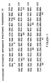

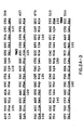
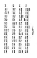


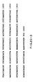
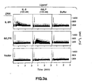


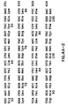

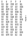


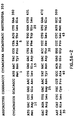


Classifications
-
- C—CHEMISTRY; METALLURGY
- C07—ORGANIC CHEMISTRY
- C07K—PEPTIDES
- C07K16/00—Immunoglobulins [IGs], e.g. monoclonal or polyclonal antibodies
- C07K16/18—Immunoglobulins [IGs], e.g. monoclonal or polyclonal antibodies against material from animals or humans
- C07K16/28—Immunoglobulins [IGs], e.g. monoclonal or polyclonal antibodies against material from animals or humans against receptors, cell surface antigens or cell surface determinants
- C07K16/2866—Immunoglobulins [IGs], e.g. monoclonal or polyclonal antibodies against material from animals or humans against receptors, cell surface antigens or cell surface determinants against receptors for cytokines, lymphokines, interferons
-
- A—HUMAN NECESSITIES
- A61—MEDICAL OR VETERINARY SCIENCE; HYGIENE
- A61P—SPECIFIC THERAPEUTIC ACTIVITY OF CHEMICAL COMPOUNDS OR MEDICINAL PREPARATIONS
- A61P29/00—Non-central analgesic, antipyretic or antiinflammatory agents, e.g. antirheumatic agents; Non-steroidal antiinflammatory drugs [NSAID]
-
- A—HUMAN NECESSITIES
- A61—MEDICAL OR VETERINARY SCIENCE; HYGIENE
- A61P—SPECIFIC THERAPEUTIC ACTIVITY OF CHEMICAL COMPOUNDS OR MEDICINAL PREPARATIONS
- A61P43/00—Drugs for specific purposes, not provided for in groups A61P1/00-A61P41/00
-
- C—CHEMISTRY; METALLURGY
- C07—ORGANIC CHEMISTRY
- C07K—PEPTIDES
- C07K14/00—Peptides having more than 20 amino acids; Gastrins; Somatostatins; Melanotropins; Derivatives thereof
- C07K14/435—Peptides having more than 20 amino acids; Gastrins; Somatostatins; Melanotropins; Derivatives thereof from animals; from humans
- C07K14/705—Receptors; Cell surface antigens; Cell surface determinants
- C07K14/715—Receptors; Cell surface antigens; Cell surface determinants for cytokines; for lymphokines; for interferons
- C07K14/7155—Receptors; Cell surface antigens; Cell surface determinants for cytokines; for lymphokines; for interferons for interleukins [IL]
-
- A—HUMAN NECESSITIES
- A61—MEDICAL OR VETERINARY SCIENCE; HYGIENE
- A61K—PREPARATIONS FOR MEDICAL, DENTAL OR TOILETRY PURPOSES
- A61K38/00—Medicinal preparations containing peptides
-
- A—HUMAN NECESSITIES
- A61—MEDICAL OR VETERINARY SCIENCE; HYGIENE
- A61K—PREPARATIONS FOR MEDICAL, DENTAL OR TOILETRY PURPOSES
- A61K39/00—Medicinal preparations containing antigens or antibodies
-
- C—CHEMISTRY; METALLURGY
- C07—ORGANIC CHEMISTRY
- C07K—PEPTIDES
- C07K2319/00—Fusion polypeptide
Definitions
- This invention relates to the field of assaying platelet factor 4 superfamily members (hereafter "PF4A”) and the preparation of agonists and antagonists to the members of this family.
- PF4A platelet factor 4 superfamily members
- interleukin-8 was initially identified as a chemoattractant for neutrophils, and was known to bind a receptor on neutrophils 8-10 , it has in addition a wide range of pro-inflammatory activities including the stimulation of degranulation and the upregulation of the cell adhesion molecule MAC-1 and of the complement receptor CR1 1 . IL-8 can also mediate the inhibition of the adherence of neutrophils to activated endothelial cells 2 .
- IL-8 is a member of a family of ten or more pro-inflammatory cytokines with an M, ⁇ 10,000 1 .
- This larger family of proteins is called the platelet factor 4 superfamily (Wolpe et al., FASEB J. 3 :2565-73 [1989]).
- Some members of the platelet factor 4 superfamily in general the subset referred to as CXC peptides (including IL-8), possess neutrophil agonist activity, e.g. NAP-2, MIP-2, platelet factor 4 and NAP-3 (MGSA/gro).
- C-C peptides are not neutrophil agonists.
- PF4A means the PF4 superfamily.
- PF4AR PF4A superfamily
- a still further object is to obtain DNA encoding variants of such receptors and to prepare such variants in recombinant cell culture.
- an isolated PF4AR polypeptide having at least 85% amino acid sequence homology with the translated amino acid sequence of figure 2, 4 or 5.
- an isolated PF4AR polypeptide wherein the nucleic acid encoding the PF4AR polypeptide hybridises with the complement of the nucleic acid encoding the polypeptide of figures 4 or 5 under high stringency conditions.
- the invention provides a composition comprising the PF4AR that is free of contaminating polypeptides of the animal species from which the PF4AR is derived.
- PF4AR or fragments thereof (which also may be synthesized by chemical methods) are fused (by recombinant expression or in vitro covalent methods) to an immunogenic polypeptide and this fusion polypeptide, in turn, is used to immunize an animal to raise antibodies against a PF4AR epitope.
- Anti-PF4AR antibodies are recovered from the serum of immunized animals. Alternatively, monoclonal antibodies are prepared from cells of the immunized animal in conventional fashion.
- Anti-PF4AR antibodies are useful particularly in the diagnosis ( in vitro or in vivo ) or (when immoblilized on an insoluble matrix) the purification of the PF4AR.
- Substitutional, deletional, or insertional variants of the PF4AR are prepared by in vitro or recombinant methods and screened for immuno-crossreactivity with the PF4AR and for PF4AR antagonist or agonist activity.
- the PF4AR also is derivatized in vitro to prepare immobilized PF4AR and labeled PF4AR, particularly for purposes of diagnosis of PF4AR or its antibodies, or for affinity purification of PF4AR antibodies.
- the PF4AR, its derivatives, or its antibodies are formulated into physiologically acceptable vehicles, especially for therapeutic use.
- physiologically acceptable vehicles include sustained-release formulations of the PF4AR.
- the invention provides an isolated nucleic acid molecule encoding the PF4AR, labeled or unlabeled, and a nucleic acid sequence that is complementary to, or hybridizes under suitable conditions to a nucleic acid sequence encoding the PF4AR.
- the invention provides a replicable vector comprising the nucleic acid molecule encoding the PF4AR operably linked to control sequences recognized by a host transformed by the vector; host cells transformed with the vector; and a method of using a nucleic acid molecule encoding the PF4AR to effect the production of PF4AR, comprising expressing the nucleic acid molecule in a culture of the transformed host cells and recovering the PF4AR from the host cell culture.
- the nucleic acid sequence is also useful in hybridization assays for PF4AR nucleic acid.
- the recombinant host cells are particularly useful in assaying the appropriate PF4A members.
- the invention provides a method for producing PF4AR comprising inserting into the DNA of a cell containing the nucleic acid encoding the PF4AR a transcription modulatory element in sufficient proximity and orientation to the PF4AR nucleic acid to influence or destroy transcription of DNA encoding a biologically active PF4AR, with an optional further step comprising culturing the cell containing the transcription modulatory element and the PF4AR nucleic acid.
- Fig. 1 depicts the high affinity binding of IL-8 to COS cells transfected with clone pRKSB.il8r1.1.
- a Competition with unlabelled IL-8 or fMLP.
- Figs. 2a-2c (SEQ ID NO. 1) (hereinafter referred to collectively as Fig. 2) depicts the amino acid and nucleotide sequences of the IL-8 receptor cDNA insert from clone pRKSB.il8rl.1.
- the seven putative transmembrane domains are shown. There are 4 extracellular segments and 4 intracellular segments, each being separated by one of the transmembrane domains. The extracellular segments are approximately delineated by residues 1-39, 99-111, 134-154, 175-203 and 265-290.
- the IL-8 receptor contains 3 potential N-linked glycosylation sites in the first extracellular region and 3 more in the third extracellular loop.
- Fig. 3a depicts the flow cytometry determination of the intracellular Ca ++ response of transfected human IL-8 and fMLP receptors to their ligands.
- IL-8 receptor clone pRK5B.il8r1.1
- fMLP receptor human fMLP receptor cDNA 3 in the vector pRK5
- vector pRK5B 18
- Intracellular Ca + + was measured with a Coulter 753 flow cytometer using the ratio of 405 and 525 nm fluorescence 23 .
- Fig. 3b illustrates the percent of cells above 400 nM Ca i ++ for the time period after addition of IL-8 (about 15 sec. into each run).
- Figs. 4 depict the DNA and an imputed polypeptide sequences for two additional PF4AR members identified by probing lambda libraries from a human monocyte-like cell line (HL-60) and human PBLs using a large fragment of the IL-8 receptor DNA.
- IL-8 receptor is a member of the G-protein coupled receptor family with clear similarity (29 % amino acid identity) to the human neutrophil receptors for the chemoattractants f-Met-Leu-Phe 3 and C5a 4 .
- the IL-8 receptor sequence may be the human homologue of what has been identified as the isoform of the rabbit f-Met-Leu-Phe receptor 5 , we show that when transfected into mammalian cells, this receptor clone confers high affinity binding to IL-8 and produces a transient Ca ++ mobilization in response to IL-8 with no binding or response to f-Met-Leu-Phe.
- a COS cell expression cloning strategy 6.7 was used to isolate clones encoding the IL-8 receptor.
- a cDNA library constructed from human neutrophil mRNA in the mammalian expression vector pRK5B was transfected into COS-7 cells as pools of 2500 clones, and the cells screened for the binding of 125 I-IL-8.
- One positive pool from the first 58 transfections was partitioned into smaller pools until a pure clone (pRK5B.il8r1.1) was obtained.
- Figure 1 shows the competition of 126 I-IL-8 binding by unlabelled IL-8 to COS cells transfected with the isolated clone.
- the DNA sequence of the isolated cDNA clone (Fig. 2) contains a single long open reading frame beginning with a methionine residue that matches the consensus expected for a translation initiation site 11 .
- This open reading frame encodes a protein of 350 amino acids (translated M, 39.5 kD).
- the amino add sequence shares several features with the G-protein coupled receptors of the rhodopsin superfamily including seven hydrophobic domains that are presumed to span the cell membrane and N-linked glycosylation sites near the N-terminus 12 (see below).
- the encoded amino acid sequence is the most similar to a recently cloned sequence for the rabbit fMLP receptor 5 .
- the similarity is sufficiently high (79% amino acid identity overall with multiple stretches of more than 20 contiguous amino add matches) that these two sequences may well be species homologs of the same receptor.
- the human fMLP receptor has also been cloned 3 ; it has only 26% amino acid identity with the rabbit fMLP receptor (and 29% identity to the human IL-8 receptor presented here).
- the considerable divergence between the rabbit and human fMLP receptor amino acid sequences has lead to the suggestion in the art (now believed to be possibly erroneous) that these may be two isoforms of the fMLP receptor 5 .
- Neutrophils respond to the chemoattractants IL-8 and fMLP with a rapid, transient increase in the intracellular free Ca ++ concentration 1,13 .
- the intracellular Ca ++ response of transfected cells to added IL-8 as well as fMLP
- Flow cytometer analysis shows a clear transient increase in intracellular Ca ++ for the transfected IL-8 receptor in response to IL-8. No response is found to fMLP.
- cells transfected with the human fMLP receptor respond to fMLP but not to IL-8.
- Alignment of the receptor sequences for the three neutrophil chemoattractants IL-8, fMLP 3 , and C5a 4 shows that they form a subfamily of the G-protein coupled receptors with 29-34 % amino acid identity.
- This subfamily has a short third intracellular loop as compared with other G-protein coupled receptors such as the ⁇ -adrenergic 12 or muscarinic acetylcholine receptors 15 .
- This loop contains determinants at least partially responsible for the binding of G-proteins to the receptors 12 .
- the intracellular C-terminal region of the IL-8 receptor while not very similar to that of the fMLP and C5a receptors does preserve a high number of serine and threonine residues that may function as phosphorylation sites.
- the N-terminal extracellular region for the IL-8 receptor has several acidic residues. These may aid in the binding of IL-8 which is quite basic (pl ⁇ 9.5).
- PF4AR is defined as a polypeptide having a qualitative biological activity in common with the polypeptides of Figs. 2. 4, or 5.
- PF4AR will have at least 30% and ordinarily 75% amino acid sequence identity with any of the polypeptides of Figs. 2, 4 or 5.
- PF4AR excludes the rabbit fMLP receptor 5 , the human fMLP receptor 3 , the human C5a receptor 1 .
- Identity or homology with respect to a PF4AR is defined herein to be the percentage of amino acid residues in the candidate sequence that are identical with the residues in Figs. 2, 4 or 5 after aligning the sequences and introducing gaps, if necessary, to achieve the maximum percent homology, and not considering any conservative substitutions as representing residue identity. No N- nor C-terminal extensions, deletions nor insertions shall be construed as reducing identity or homology.
- PF4AR qualitative biological activity is defined as any one of (1) immunological cross-reactivity with at least one epitope of a polypeptide set forth in Figs. 2, 4, or 5; (2) the ability to specifically bind to a member of the PF4 super-family; or (3) any effector or functional activity of the Figs. 2, 4 or 5 polypeptides as found in nature, including their ability to bind any ligands other than superfamily members.
- Immunologically cross-reactive means that the candidate polypeptide is capable of competitively inhibiting the binding of a PF4AR to polyclonal antibodies or antisera raised against a PF4AR.
- Such antibodies and antisera are prepared in conventional fashion by injecting an animal such as a goat or rabbit, for example, subcutaneously with the known native PF4AR in complete Freund's adjuvant, followed by booster intraperitoneal or subcutaneous injection in incomplete Freund's.
- polypeptides having the amino acid sequences described in Figs. 2, 4 or 5 amino acid sequence variants of such amino acid sequences, glycosylation variants of the polypeptides and covalent modifications of the polypeptides. Each of these are described in more detail below.
- isolated PF4AR nucleic acid or polypeptide is a PF4AR nucleic acid or polypeptide that is identified and separated from at least one contaminant (nucleic acid or polypeptide respectively) with which it is ordinarily associated in nature, such as from the animal or human source of the PF4AR nucleic acid or polypeptide.
- the PF4AR will be isolated to pharmaceutically acceptable levels of purity with respect to proteins of its species of origin.
- PF4AR protein will be purified (1) to greater than 95% by weight of protein as determined by the Lowry method, and most preferably more than 99% by weight, (2) to a degree sufficient to obtain at least 15 residues of N-terminal or internal amino acid sequence by an amino acid sequenator commercially available on the filing date hereof, or (3) to homogeneity by conventional nonreducing SDS-PAGE using Coomassie blue or, preferably, silver stain.
- Isolated PF4AR includes PF4AR in situ within recombinant cells since, in this instance, at least one component of the PF4AR natural environment will not be present.
- Isolated PF4AR includes PF4AR from one species in a recombinant cell culture of another species since the receptor in such circumstances will be devoid of source polypeptides. Ordinarily, however, isolated receptor will be prepared by at least one purification step.
- Isolated PF4AR nucleic acid includes a nucleic acid that is identified and separated from at least one containment nucleic acid with which it is ordinarily associated in the natural source of the receptor nucleic acid. Isolated PF4AR nucleic acid thus is present in other than in the form or setting in which it is found in nature.
- isolated receptor-encoding nucleic acid includes PF4AR nucleic acid in ordinarily receptor-expressing cells where the nucleic acid is in a chromosomal location different from that of natural cells or is otherwise flanked by a different DNA sequence than that found in nature.
- nucleic acid or polypeptide may be labeled for diagnostic and probe purposes, using a label as described and defined further below in the discussion of diagnostic assays.
- nucleic acid is defined as RNA or DNA containing greater than ten bases that encodes a polypeptide sequence within Figs. 2, 4 or 5, is complementary to nucleic acid sequence of Figs. 2, 4 or 5, hybridizes to such nucleic acid and remains stably bound to it under low stringency conditions, or encodes a polypeptide sharing at least 30% sequence identity, preferably at least 75%, and more preferably at least 85%, with the translated amino acid sequence shown in Figs. 2, 4 or 5 or a fragment thereof.
- the DNA which hybridizes to the nucleic acid of Figs. 2, 4 or 5 contain at least 20, more preferably 40, and more preferably 60 bases.
- hybridizing DNA or RNA contains 45 or even more preferably 90 bases.
- hybridizing or complementary nucleic acid is defined further as being novel and unobvious over any prior art nucleic acid including that which encodes, hybridizes under low stringency conditions, or is complementary to nucleic acid encoding rabbit fMLP receptor 5 , human fMLP receptor or (optionally) the IL-8 receptor of Murphy et al. ( supra ).
- High stringency conditions are any of those that (1) employ low ionic strength and high temperature for washing, for example, 0.015 M NaCl/0.0015 M sodium citrate/0.1% NaDodSO 4 at 50°C; (2) employ during hybridization 50% (vol/vol) formamide with 0.1% bovine serum albumin/0.1% Ficoll/0.1% polyvinylpyrrolidone/50mM sodium phosphate buffer at pH 6.5 with 750 mM NaCl, 75 mM sodium citrate at 42°C; or (3) employ hybridization with 50% formamide, 5 x SSC (0.75 M NaCl, 0.075 M sodium citrate), 50 mM sodium phosphate (pH 6.8), 0.1% sodium pyrophosphate, 5 x Denhardt's solution, sonicated salmon sperm DNA (50 ⁇ g/ml), 0.1% SDS, and 10% dextran sulfate at 42°C, with washes at 42°C in 0.2 x SSC and 0.1% SDS. Conditions of low string
- control sequences refers to DNA sequences necessary for the expression of an operably linked coding sequence in a particular host organism.
- the control sequences that are suitable for prokaryotes include a promoter, optionally an operator sequence, and a ribosome binding site.
- Eukaryotic cells are known to utilize promoters, polyadenylation signals, and enhancers.
- Nucleic acid is "operably linked" when it is placed into a functional relationship with another nucleic acid sequence.
- DNA for a presequence or secretory leader is operably linked to DNA for a polypeptide if it is expressed as a preprotein that participates in the secretion of the polypeptide;
- a promoter or enhancer is operably linked to a coding sequence if it affects the transcription of the sequence; or
- a ribosome binding site is operably linked to a coding sequence if it is positioned so as to facilitate translation.
- "operably linked” means that the DNA sequences being linked are contiguous and, in the case of a secretory leader, contiguous and in reading phase. However enhancers do not have to be contiguous. Linking is accomplished by ligation at convenient restriction sites. If such sites do not exist, then synthetic oligonucleotide adaptors or linkers are used in accord with conventional practice.
- the starting plasmids herein are commercially available, are publicly available on an unrestricted basis, or can be constructed from such available plasmids in accord with published procedures.
- other equivalent plasmids are known in the art and will be apparent to the ordinary artisan.
- Methods for restriction enzyme digestion, recovery or isolation of DNA, hybridization analysis, and ligation are conventional and by this time well known to the ordinary artisan.
- Recovery or “isolation” of a given fragment of DNA from a restriction digest means separation of the digest on polyacrylamide or agarose gel by electrophoresis, identification of the fragment of interest by comparison of its mobility versus that of marker DNA fragments of known molecular weight, removal of the gel section containing the desired fragment, and separation of the gel from DNA.
- This procedure is known generally. For example, see Lawn et al., Nucleic Acids Res. 9 : 6103-6114 (1981), and Goeddel et al., Nucleic Acids Res. 8 :4057 (1980).
- the DNA encoding of the PF4AR may be obtained from any cDNA library prepared from tissue believed to contain the PF4AR mRNA, generally HL60 or PBL libraries.
- the PF4AR gene may also be obtained from a genomic library. Libraries are screened with probes designed to identify the gene of interest or the protein encoded by it. We have described the entire cDNA for the IL-8 receptor and two homologous receptors. Nucleic acid encoding this family of receptors is readily obtained under low stringency conditions from genomic DNA or neutrophil cDNA libraries using probes having oligonucleotide sequences from the receptor gene sequences of Figs. 2, 4 or 5. These probes usually will contain about 500 or more bases.
- probes will hybridize perfectly to the three exemplified DNAs, there is no need to use probe pools containing degenerate sequences. Screening with the probes to identify the Figs. 2, 4 or 5 receptors is more efficient if performed under conditions of high stringency.
- PF4ARs other than those in Figs. 2, 4 or 5 are believed to exist and to contain regions of homology to the exemplified receptors.
- probes having the sequences of the DNAs in Figs. 2, 4 or 5 can be used to screen for these receptors as well.
- the best candidates for probes are long sequences (greater than about 100 bases) that represent sequences that are highly homologous among the three exemplified human receptors.
- IL-8 cDNA encoding the IL-8 residues 15-34, 78-94, 176-193, 264-282 and 299-312 are useful, particularly in probing for IL-8 receptor DNA. Probes useful for the receptor of Fig.
- Fig. 4 (and isolated proteins characteristic of the Fig. 5 receptor) are represented by sequences comprising residues 1-48, 77-92, 107-137, 156-177, 189-226, 239-257 and 271-315. Homologous probes and residues of the Fig. 4 receptor also are useful, i.e. residues 1-35, 64-78, 94-124, 143-164, 176-197, 219-239 and 251-295. cDNAs comprising cDNA encoding the following regions of the Figs. 2, 4 or 5 polypeptides are useful in probing for other receptors: 92-106, 57-72, 138-154, 314-329 and 57-154.
- Cells identified by this process (and some are already known for individual PF4As) therefore are expressing a receptor for this PF4A.
- a cDNA library is prepared from such cells and is screened using the receptor probes by procedures that are conventional per se. In this instance, however, it is preferred to use low stringency conditions (such as those in Example 2) and then analyze the resulting positive clones for homology to the Figs. 2, 4 or 5 receptors.
- candidate human PF4ARs will exhibit greater than about 30% amino acid sequence homology to the Figs. 2, 4 or 5 receptors and bear a similar transmembrane loop structure.
- Assays are then conducted to confirm that the hybridizing full length genes are the desired PF4AR.
- the candidate is simply inserted into an expression vector and transformed into a host cell that ordinarily does not bind to the candidate PF4A ligand. Transformants that acquire the ability to bind the ligand thus bear the desired receptor gene.
- Example 2 we show that two additional homologous polypeptide sequences representing PR4ARs are identified using IL-8R DNA encoding residues 23-314, although the particular probe is not believed to be critical.
- PCR polymerase chain reaction
- cDNA libraries may be screened from various tissues, preferably mammalian PBL, monocyte, placental, fetal, brain, and carcinoma cell lines in order to obtain DNA encoding the receptors of Figs. 2, 4 or 5, or homologous receptors. More preferably, human or rabbit placental, fetal, brain, and carcinoma cell line cDNA libraries are screened with labelled oligonucleotide probes.
- Another method for obtaining the gene of interest is to chemically synthesize it using one of the methods described in Engels et al. (Agnew. Chem. Int. Ed. Engl. 28 :716-734 [1989]). These methods include triester, phosphite, phosphoramidite and H-phosphonate methods, typically proceeding by oligonucleotide synthesis on solid supports. These methods may be used if the entire amino acid or nucleic acid sequence of the gene is known, or the sequence of the nucleic acid complementary to the coding strand is available. If the desired amino acid sequence is known, one may infer potential nucleic acid sequences using known and preferred coding residues for each amino acid residue.
- Amino acid sequence variants of the PF4AR are prepared by introducing appropriate nucleotide changes into the PF4AR DNA, or by in vitro synthesis of the desired PF4AR polypeptide.
- Such variants include, for example, deletions from, or insertions or substitutions of, residues within the amino acid sequence shown for the receptors in Figs. 2, 4 or 5. Any combination of deletion, insertion, and substitution can be made to arrive at the final construct, provided that the final construct possesses the desired characteristics.
- the amino acid changes also may alter post-translational processing of the PF4AR, such as changing the number or position of glycosylation sites or by altering its membrane anchoring characteristics.
- PF4AR variants or polypeptide sequences that are not statutorily novel and unobvious over the prior art.
- the location of the mutation site and the nature of the mutation will depend on the PF4AR characteristic(s) to be modified.
- the sites for mutation can be modified individually or in series, e.g., by (1) substituting first with conservative amino acid choices and then with more radical selections depending upon the results achieved, (2) deleting the target residue, or (3) inserting residues of the same or a different class adjacent to the located site, or combinations of options 1-3.
- a useful method for identification of certain residues or regions of the PF4AR polypeptide that are preferred locations for mutagenesis is called "alanine scanning mutagenesis" as described by Cunningham and Wells ( Science 244 :1081-1085 [1989]).
- a residue or group of target residues are identified (e.g., charged residues such as arg, asp, his, lys, and glu) and replaced by a neutral or negatively charged amino acid (most preferably alanine or poly-alanine) to affect the interaction of the amino acids with the surrounding aqueous environment in or outside the cell.
- Those domains demonstrating functional sensitivity to the substitutions then are refined by introducing further or other variants at or for the sites of substitution.
- the site for introducing an amino acid sequence variation is predetermined, the nature of the mutation per se need not be predetermined.
- ala scanning or random mutagenests may be conducted at the target codon or region and the expressed PF4AR variants are screened for the optimal combination of desired activity.
- the regions of the PF4AR molecule preferred for alterations are non-hydrophic regions or regions that are not highly conserved. Such regions are those in which sequences of 5 or more residues are not substantially conserved in the homologous positions in the rabbit fMLP receptor, the human fMLP receptor, the human C5a receptor and the receptors of Figs. 2, 4 and 5.
- PF4AR variants will exhibit at least a biological activity of the parental sequence, for example ligand binding activity or antigenic activity.
- Antigenically active PF4AR is a polypeptide that binds with an affinity of at least about 10 -9 1/mole to an antibody raised against a naturally occurring PF4AR sequence. Ordinarily the polypeptide binds with an affinity of at least about 10 -8 1/mole.
- the antigenically active PF4AR is a polypeptide that binds to an antibody raised against the receptor in its native conformation
- "native conformation” generally meaning the receptor as found in nature which has not been denatured by chaotropic agents, heat or other treatment that substantially modifies the three dimensional structure of the receptor (this can be determined, for example, by migration on nonreducing, non-denaturing sizing gels).
- Antibody used in determination of antigenic activity is rabbit polyclonal antibody raised by formulating the native non-rabbit receptor in Freund's complete adjuvant, subcutaneously injecting the formulation, and boosting the immune response by intraperitoneal injection of the formulation until the titer of anti-receptor antibody plateaus.
- variants are deletion mutants, or fragments of the sequences set forth in Figs. 2, 4, 5 or other PF4AR.
- the fragments are those which constitute the extracellular regions of the receptors (these receptors are unlike most in that they are believed to contain a plurality of hydrophobic, trans-memberan domains separated by hydrophilic sequences believed to loop into the ectoplasm).
- Particularly of interest are the N-terminal extracellular region containing acidic amino acid residues.
- any sequence which is capable of raising an antibody that will cross-react with the intact receptor, or which will bind to a member of the PF4 superfamily, is useful.
- These fragments typically will contain a consecutive sequence of at least about 5 (and ordinarily at least about 10) residues.
- Amino acid sequence deletions generally range from about 1 to 30 residues, more preferably about 1 to 10 residues, and typically are contiguous. Deletions may be introduced into regions of low homology among the receptors of Figs. 2, 4 and 5 to modify the activity of the receptors. Such deletions will be more likely to modify the biological activity of the receptors more significantly than deletions made elsewhere. The number of consecutive deletions will be selected so as to preserve the tertiary structure of the PF4AR in the affected domain, e.g.. beta-pleated sheet or alpha helix.
- Amino acid sequence insertions include amino- and/or carboxyl-terminal fusions ranging in length from one residue to polypeptides containing a hundred or more residues, as well as intrasequence insertions of single or multiple amino acid residues.
- Intrasequence insertions i.e., insertions within the PF4AR sequence
- Insertional variants of the PF4AR or its extracellular segments include the fusion to the N- or C-terminus of the PF4AR of immunogenic polypeptides, e.g., bacterial polypeptides such as beta-lactamase or an enzyme encoded by the E. coli trp locus, or yeast protein, and C-terminal fusions with proteins having a long half-life such as immunoglobulin constant regions (or other immunoglobulin regions), albumin, or ferritin, as described in WO 89/02922 published 6 April 1989.
- immunogenic polypeptides e.g., bacterial polypeptides such as beta-lactamase or an enzyme encoded by the E. coli trp locus, or yeast protein
- C-terminal fusions with proteins having a long half-life such as immunoglobulin constant regions (or other immunoglobulin regions), albumin, or ferritin, as described in WO 89/02922 published 6 April 1989.
- variants are amino acid substitution variants. These variants have at least one amino acid residue in the PF4AR molecule removed and a different residue inserted in its place.
- the sites of greatest interest for substitutional mutagenesis include sites identified as the active site(s) of the PF4AR, and sites where the amino acids found in the PF4AR from various species are substantially different in terms of side-chain bulk, charge, and/or hydrophobicity.
- sites of interest are those in which particular residues of the PF4ARs of Figs. 2. 4 and 5 are identical. These positions may be important for the biological activity of the PF4AR. These sites, especially those falling within a sequence of at least three other identically conserved sites, are substituted in a relatively conservative manner. Such conservative substitutions are shown in Table 1 under the heading of preferred substitutions. If such substitutions result in a change in biological activity, then more substantial changes, denominated exemplary substitutions in Table 1, or as further described below in reference to amino acid classes, are introduced and the products screened.
- Substantial modifications in function or immunological identity of the PF4AR are accomplished by selecting substitutions that differ significantly in their effect on maintaining (a) the structure of the polypeptide backbone in the area of the substitution, for example, as a sheet or helical conformation, (b) the charge or hydrophobicity of the molecule at the target site, or (c) the bulk of the side chain.
- Naturally occurring residues are divided into groups based on common side chain properties:
- Non-conservative substitutions will entail exchanging a member of one of these classes for another.
- Such substituted residues may be introduced into regions of the PF4AR that are homologous with other PF4ARs, or, more preferably, into the non-homologous regions of the molecule.
- Any cysteine residues not involved in maintaining the proper conformation of the PF4AR may be substituted, generally with serine, to improve the oxidative stability of the molecule and prevent aberrant crosslinking.
- DNA encoding amino acid sequence variants of the PF4AR is prepared by a variety of methods known in the an. These methods include, but are not limited to, isolation from a natural source (in the case of naturally occurring amino acid sequence variants) or preparation by oligonucleotide-mediated (or site-directed) mutagenesis, PCR mutagenesis, and cassette mutagenesis of an earlier prepared variant or a non-variant version of the PF4AR. These techniques may utilize PF4AR nucleic acid (DNA or RNA), or nucleic acid complementary to the PF4AR nucleic acid.
- Oligonucleotide-mediated mutagenesis is a preferred method for preparing substitution, deletion, and insertion variants of PF4AR DNA.
- This technique is well known in the art, for example as described by Adelman et al., DNA 2 :183 (1983).
- the PF4AR DNA is altered by hybridizing an oligonucleotide encoding the desired mutation to a DNA template, where the template is the single-stranded form of a plasmid or bacteriophage containing the unaltered or native DNA sequence of the PF4AR.
- a DNA polymerase is used to synthesize an entire second complementary strand of the template that will thus incorporate the oligonucleotide primer, and will code for the selected alteration in the PF4AR DNA.
- oligonucleoxides of at least 25 nucleotides in length are used.
- An optimal oligonucleotide will have 12 to 15 nucleotides that are completely complementary to the template on either side of the nucleotide(s) coding for the mutation. This ensures that the oligonucleotide will hybridize properly to the single-stranded DNA template molecule.
- the oligonucleotides are readily synthesized using techniques known in the art such as that described by Crea et al. (Proc. Natl. Acad. Sci. USA 75 :5765 [1978]).
- Single-stranded DNA template may also be generated by denaturing double-stranded plasmid (or other) DNA using standard techniques.
- the oligonucleotide is hybridized to the single-stranded template under suitable hybridization conditions.
- a DNA polymerizing enzyme usually the Klenow fragment of DNA polymerase I, is then added to synthesize the complementary strand of the template using the oligonucleotide as a primer for synthesis.
- a heteroduplex molecule is thus formed such that one strand of DNA encodes the mutated form of the PF4AR, and the other strand (the original template) encodes the native, unaltered sequence of the PF4AR.
- This heteroduplex molecule is then transformed into a suitable host cell, usually a prokaryote such as E . coli JM101.
- the cells are plated onto agarose plates, and screened using the oligonucleotide primer radiolabeled with 32-phosphate to identify the bacterial colonies that contain the mutated DNA.
- the mutated region is then removed and placed in an appropriate vector for protein production, generally an expression vector of the type typically employed for transformation of an appropriate host.
- the method described immediately above may be modified such that a homoduplex molecule is created wherein both strands of the plasmid contain the mutation(s).
- the modifications are as follows:
- the single-stranded oligonucleotide is annealed to the single-stranded template as described above.
- a mixture of three deoxyribonucleotides, deoxyriboadenosine (dATP), deoxyriboguanosine (dGTP), and deoxyribothymidine (dTTP) is combined with a modified thio-deoxyribocytosine called dCTP-(aS) (which can be obtained from Amersham Corporation). This mixture is added to the template-oligonucleotide complex.
- this new strand of DNA Upon addition of DNA polymerase to this mixture, a strand of DNA identical to the template except for the mutated bases is generated.
- this new strand of DNA will contain dCTP-(aS) instead of dCTP, which serves to protect it from restriction endonuclease digestion.
- the template strand can be digested with Exo III nuclease or another appropriate nuclease past the region that contains the site(s) to be mutagenized. The reaction is then stopped to leave a molecule that is only partially single-stranded.
- a complete double-stranded DNA homoduplex is then formed using DNA polymerase in the presence of all four deoxyribonucleotide triphosphates, ATP, and DNA ligase.
- This homoduplex molecule can then be transformed into a suitable host cell such as E. coli JM101, as described above.
- DNA encoding PF4AR mutants at more than one site may be generated in one of several ways. If the amino acids are located close together in the polypeptide chain, they may be mutated simultaneously using one oligonucleotide that codes for all of the desired amino acid substitutions. If, however, the amino acids are located some distance from each other (separated by more than about ten amino acids), it is more difficult to generate a single oligonucleotide that encodes all of the desired changes. Instead, one of two alternative methods may be employed.
- a separate oligonucleotide is generated for each amino add to be substituted.
- the oligonucleotides are then annealed to the single-stranded template DNA simultaneously, and the second strand of DNA that is synthesized from the template will encode all of the desired amino acid substitutions.
- the alternative method involves two or more rounds of mutagenesis to produce the desired mutant.
- the first round is as described for the single mutants: wild-type DNA is used for the template, an oligonucleotide encoding the first desired amino acid substitution(s) is annealed to this template, and the heteroduplex DNA molecule is then generated.
- the second round of mutagenesis utilizes the mutated DNA produced in the first round of mutagenesis as the template.
- this template already contains one or more mutations.
- the oligonucleotide encoding the additional desired amino acid substitution(s) is then annealed to this template, and the resulting strand of DNA now encodes mutations from both the first and second rounds of mutagenesis.
- This resultant DNA can be used as a template in a third round of mutagenesis, and so on.
- PCR mutagenesis is also suitable for making amino acid variants of the PF4AR. While the following discussion refers to DNA, it is understood that the technique also finds application with RNA.
- the PCR technique generally refers to the following procedure (see Erlich, supra, the chapter by R. Higuchi, p. 61-70): When small amounts of template DNA are used as starting material in a PCR, primers that differ slightly in sequence from the corresponding region in a template DNA can be used to generate relatively large quantities of a specific DNA fragment that differs from the template sequence only at the positions where the primers differ from the template.
- one of the primers is designed to overlap the position of the mutation and to contain the mutation; the sequence of the other primer must be identical to a stretch of sequence of the opposite strand of the plasmid, but this sequence can be located anywhere along the plasmid DNA. It is preferred, however, that the sequence of the second primer is located within 200 nucleotides from that of the first, such that in the end the entire amplified region of DNA bounded by the primers can be easily sequenced.
- PCR amplification using a primer pair like the one just described results in a population of DNA fragments that differ at the position of the mutation specified by the primer, and possibly at other positions, as template copying is somewhat error-prone.
- the cDNA or genomic DNA encoding native or variant PF4AR is inserted into a replicable vector for further cloning (amplification of the DNA) or for expression.
- Many vectors are available, and selection of the appropriate vector will depend on (1) whether it is to be used for DNA amplification or for DNA expression, (2) the size of the DNA to be inserted into the vector, and (3) the host cell to be transformed with the vector.
- Each vector contains various components depending on its function (amplification of DNA or expression of DNA) and the host cell for which it is compatible.
- the vector components generally include, but are not limited to, one or more of the following: a signal sequence, an origin of replication, one or more marker genes, an enhancer element, a promoter, and a transcription termination sequence.
- a signal sequence may be a component of the vector, or it may be a part of the PF4AR DNA that is inserted into the vector.
- the native pro PF4AR DNA is directed to the cell surface in our recombinant cells but it does not contain a conventional signal and no N-terminal polypeptide is cleaved during post-translational processing of the polypeptide during membrane insertion of the PF4AR.
- Both expression and cloning vectors contain a nucleic acid sequence that enables the vector to replicate in one or more selected host cells.
- this sequence is one that enables the vector to replicate independently of the host chromosomal DNA, and includes origins of replication or autonomously replicating sequences.
- origins of replication or autonomously replicating sequences are well known for a variety of bacteria, yeast, and viruses.
- the origin of replication from the plasmid pBR322 is suitable for most Gram-negative bacteria, the 2 ⁇ plasmid origin is suitable for yeast, and various viral origins (SV40, polyoma, adenovirus, VSV or BPV) are useful for cloning vectors in mammalian cells.
- the origin of replication component is not needed for mammalian expression vectors (the SV40 origin may typically be used only because it contains the early promoter).
- Most expression vectors are "shuttle" vectors, i.e. they are capable of replication in at least one class of organisms but can be transfected into another organism for expression.
- a vector is cloned in E . coli and then the same vector is transfected into yeast or mammalian cells for expression even though it is not capable of replicating independently of the host cell chromosome.
- DNA may also be amplified by insertion into the host genome. This is readily accomplished using Bacillus species as hosts, for example, by including in the vector a DNA sequence that is complementary to a sequence found in Bacillus genomic DNA. Transfection of Bacillus with this vector results in homologous recombination with the genome and insertion of the PF4AR DNA. However, the recovery of genomic DNA encoding the PF4AR is more complex than that of an exogenously replicated vector because restriction enzyme digestion is required to excise the PF4AR DNA.
- Selection genes should contain a selection gene, also termed a selectable marker. This gene encodes a protein necessary for the survival or growth of transformed host cells grown in a selective culture medium. Host cells not transformed with the vector containing the selection gene will not survive in the culture medium.
- Typical selection genes encode proteins that (a) confer resistance to antibiotics or other toxins, e.g. ampicillin, neomycin, methotrexate, or tetracycline, (b) complement auxotrophic deficiencies, or (c) supply critical nutrients not available from complex media, e.g. the gene encoding D-alanine racemase for Bacilli.
- a selection scheme utilizes a drug to arrest growth of a host cell. Those cells that are successfully transformed with a heterologous gene express a protein conferring drug resistance and thus survive the selection regimen. Examples of such dominant selection use the drugs neomycin (Southern et al., J. Molec. Appl. Genet. 1 :327 (19821), mycophenolic acid (Mulligan et al., Science 209 :1422 [1980]) or hygromycin (Sugden et al., Mol. Cell. Biol. 5 :410-413 [1985]).
- the three examples given above employ bacterial genes under eukaryotic control to convey resistance to the appropriate drug G418 or neomycin (geneticin), xgpt (mycophenolic acid), or hygromycin, respectively.
- suitable selectable markers for mammalian cells are those that enable the identification of cells competent to take up the PF4AR nucleic acid, such as dihydrofolate reductase (DHFR) or thymidine kinase.
- DHFR dihydrofolate reductase
- the mammalian cell transformants are placed under selection pressure which only the transformants are uniquely adapted to survive by virtue of having taken up the marker. Selection pressure is imposed by culturing the transformants under conditions in which the concentration of selection agent in the medium is successively changed, thereby leading to amplification of both the selection gene and the DNA that encodes the PF4AR.
- Amplification is the process by which genes in greater demand for the production of a protein critical for growth are reiterated in tandem within the chromosomes of successive generations of recombinant cells. Increased quantities of the PF4AR are synthesized from the amplified DNA.
- cells transformed with the DHFR selection gene are first identified by culturing all of the transformants in a culture medium that contains methotrexate (Mtx), a competitive antagonist of DHFR.
- Mtx methotrexate
- An appropriate host cell when wild-type DHFR is employed is the Chinese hamster ovary (CHO) cell line deficient in DHFR activity, prepared and propagated as described by Urlaub and Chasm, Proc. Natl. Acad. Sci. USA. 77 :4216 [1980].
- the transformed cells are then exposed to increased levels of methotrexate. This leads to the synthesis of multiple copies of the DHFR gene, and, concomitantly, multiple copies of other DNA comprising the expression vectors, such as the DNA encoding the PF4AR.
- This amplification technique can be used with any otherwise suitable host, e.g., ATCC No. CCL61 CHO-K1, notwithstanding the presence of endogenous DHFR if, for example, a mutant DHFR gene that is highly resistant to Mtx is employed (EP 117,060).
- host cells transformed or co-transformed with DNA sequences encoding the PF4AR, wild-type DHFR protein, and another selectable marker such as aminoglycoside 3' phosphotransferase (APH) can be selected by cell growth in medium containing a selection agent for the selectable marker such as an aminoglycosidic antibiotic, e.g., kanamycin, neomycin, or G418. See U.S. Pat. No. 4,965,199.
- APH aminoglycoside 3' phosphotransferase
- a suitable selection gene for use in yeast is the trp 1 gene present in the yeast plasmid YRp7 (Stinchcomb et al., Nature 282 :39 [1979]; Kingsman et al., Gene 7 :141 [1979]; or Tschemper et al. , Gene 10 :157 (1980]).
- the trp 1 gene provides a selection marker for a mutant strain of yeast lacking the ability to grow in tryptophan, for example, ATCC No. 44076 or PEP4-1 (Jones, Genetics 85 :12 [1977]).
- the presence of the trp 1 lesion in the yeast host cell genome then provides an effective environment for detecting transformation by growth in the absence of tryptophan.
- Leu 2-deficient yeast strains (ATCC 20,622 or 38,626) are complemented by known plasmids bearing the Leu 2 gene.
- Expression vectors usually contain a promoter that is recognized by the host organism and is operably linked to the PF4AR nucleic acid. Promoters are untranslated sequences located upstream (5') to the start codon of a structural gene (generally within about 100 to 1000 bp) that control the transcription and translation of a particular nucleic acid sequence, such as the PF4AR, to which they are operably linked. Such promoters typically fall into two classes, inducible and constitutive. Inducible promoters are promoters that initiate increased levels of transcription from DNA under their control in response to some change in culture conditions, e.g. the presence or absence of a nutrient or a change in temperature.
- promoters recognized by a variety of potential host cells are well known. These promoters are operably linked to DNA encoding the PF4AR by removing the promoter from the source DNA by restriction enzyme digestion and inserting the isolated promoter sequence into the vector. Both the native PF4AR promoter sequence and many heterologous promoters may be used to direct amplification and/or expression of the PF4AR DNA. However, heterologous promoters are preferred, as they generally permit greater transcription and higher yields of expressed PF4AR as compared to the native PF4AR promoter.
- Promoters suitable for use with prokaryotic hosts include the ⁇ -lactamase and lactose promoter systems (Chang et al., Nature. 275 :615 [1978]; and Goeddel et al., Nature. 281 :544 [1979]), alkaline phosphatase, a tryptophan (trp) promoter system (Goeddel, Nucleic Acids Res.. 8 :4057 [1980] and EP 36,776) and hybrid promoters such as the tac promoter (deBoer et al., Proc. Natl. Acad. Sci. USA. 80 :21-25 [1983]).
- trp tryptophan
- hybrid promoters such as the tac promoter (deBoer et al., Proc. Natl. Acad. Sci. USA. 80 :21-25 [1983]).
- other known bacterial promoters are suitable.
- Suitable promoting sequences for use with yeast hosts include the promoters for 3-phosphoglycerate kinase (Hitzeman et al, J. Biol. Chem., 255 :2073 [1980]) or other glycolytic enzymes (Hess et al., J. Adv. Enzyme Reg. , 7 :149 [1968]; and Holland, Biochemistry.
- enolase such as enolase, glyceraldehyde-3-phosphate dehydrogenase, hexokinase, pyruvate decarboxylase, phosphofructokinase, glucose-6-phosphate isomerase, 3-phosphoglycerate mutase, pyruvate kinase, triosephosphate isomerase, phosphoglucose isomerase, and glucokinase.
- enolase such as enolase, glyceraldehyde-3-phosphate dehydrogenase, hexokinase, pyruvate decarboxylase, phosphofructokinase, glucose-6-phosphate isomerase, 3-phosphoglycerate mutase, pyruvate kinase, triosephosphate isomerase, phosphoglucose isomerase, and glucokinase.
- yeast promoters which are inducible promoters having the additional advantage of transcription controlled by growth conditions, are the promoter regions for alcohol dehydrogenase 2, isocytochrome C, acid phosphatase, degradative enzymes associated with nitrogen metabolism, metallothionein, glyceraldehyde-3-phosphate dehydrogenase, and enzymes responsible for maltose and galactose utilization.
- Suitable vectors and promoters for use in yeast expression are further described in Hitzeman et al., EP 73,657A.
- Yeast enhancers also are advantageously used with yeast promoters.
- Promoter sequences are known for eukaryotes. Virtually all eukaryotic genes have an AT-rich region located approximately 25 to 30 bases upstream from the site where transcription is initiated. Another sequence found 70 to 80 bases upstream from the start of transcription of many genes is a CXCAAT region where X may be any nucleotide. At the 3' end of most eukaryotic genes is an AATAAA sequence that may be the signal for addition of the poly A tail to the 3' end of the coding sequence. All of these sequences are suitably inserted into mammalian expression vectors.
- PF4AR transcription from vectors in mammalian host cells is controlled by promoters obtained from the genomes of viruses such as polyoma virus, fowlpox virus (UK 2,211,504 published 5 July 1989), adenovirus (such as Adenovirus 2), bovine papilloma virus, avian sarcoma virus, cytomegalovirus, a retrovirus, hepatitis-B virus and most preferably Simian Virus 40 (SV40), from heterologous mammalian promoters, e.g. the actin promoter or an immunoglobulin promoter, from heat-shock promoters, and from the promoter normally associated with the PF4AR sequence, provided such promoters are compatible with the host cell systems.
- viruses such as polyoma virus, fowlpox virus (UK 2,211,504 published 5 July 1989), adenovirus (such as Adenovirus 2), bovine papilloma virus, avian sarcoma virus
- the early and late promoters of the SV40 virus are conveniently obtained as an SV40 restriction fragment that also contains the SV40 viral origin of replication. Fiers et al., Nature. 273 :113 (1978); Mulligan and Berg, Science, 209 :1422-1427 (1980); Pavlakis et al ., Proc. Natl. Acad. Sci. USA, 78 :7398-7402 (1981).
- the immediate early promoter of the human cytomegalovirus is conveniently obtained as a Hin dIII E restriction fragment Greenaway et al., Gene , 18 :355-360 (1982).
- a system for expressing DNA in mammalian hosts using the bovine papilloma virus as a vector is disclosed in U.S.
- Enhancers are cis-acting elements of DNA, usually about from 10-300 bp, that act on a promoter to increase its transcription. Enhancers are relatively orientation and position independent having been found 5' (Laimins et al., Proc. Natl. Acad. Sci. USA, 78 :993 [1981]) and 3' (Lusky et al., Mol. Cell Big.
- Examples include the SV40 enhancer on the late side of the replication origin (bp 100-270), the cytomegalovirus early promoter enhancer, the polyoma enhancer on the late side of the replication origin, and adenovirus enhancers. See also Yaniv, Nature , 297 :17-18 (1982) on enhancing elements for activation of eukaryotic promoters.
- the enhancer may be spliced into the vector at a position 5' or 3' to the PF4AR DNA, but is preferably located at a site 5' from the promoter.
- Expression vectors used in eukaryotic host cells will also contain sequences necessary for the termination of transcription and for stabilizing the mRNA. Such sequences are commonly available from the 5' and, occasionally 3' untranslated regions of eukaryotic or viral DNAs or cDNAs. These regions contain nucleotide segments transcribed as polyadenylated fragments in the untranslated portion of the mRNA encoding the PF4AR. The 3' untranslated regions also include transcription termination sites.
- Suitable vectors containing one or more of the above listed components and the desired coding and control sequences are constructed by standard ligation techniques. Isolated plasmids or DNA fragments are cleaved, tailored, and religated in the form desired to generate the plasmids required.
- the ligation mixtures are used to transform E. coli K12 strain 294 (ATCC 31,446) and successful transformants selected by ampicillin or tetracycline resistance where appropriate. Plasmids from the transformants are prepared, analyzed by restriction endonuclease digestion, and/or sequenced by the method of Messing et al., Nucleic Acids Res., 9 :309 (1981) or by the method of Maxam et al., Methods in Enzymology, 65 :499 (1980).
- transient expression involves the use of an expression vector that is able to replicate efficiently in a host cell, such that the host cell accumulates many copies of the expression vector and, in turn, synthesizes high levels of a desired polypeptide encoded by the expression vector.
- Transient expression systems comprising a suitable expression vector and a host cell, allow for the convenient positive identification of polypeptides encoded by cloned DNAs, as well as for the rapid screening of such polypeptides for desired biological or physiological properties.
- transient expression systems are particularly useful in the invention for purposes of identifying analogs and variants of the PF4AR that have PF4AR-like activity.
- Suitable host cells for cloning or expressing the vectors herein are the prokaryote, yeast, or higher eukaryote cells described above.
- Suitable prokaryotes include eubacteria, such as Gram-negative or Gram-positive organisms, for example, E. coli, Bacilli such as B. subtilis, Pseudomonas species such as P. aeruginosa, Salmonella typhimurium, or Serratia marcescens.
- E. coli cloning host is E. coli 294 (ATCC 31,446), although other strains such as E. coli B. E. coli X 1776 (ATCC 31,537), and E. coli W3110 (ATCC 27,325) are suitable.
- the host cell should secrete minimal amounts of proteolytic enzymes.
- in vitro methods of cloning e.g. PCR or other nucleic acid polymerase reactions, are suitable.
- eukaryotic microbes such as filamentous fungi or yeast are suitable hosts for vectors containing PF4AR DNA.
- Saccharomyces cerevisiae or common baker's yeast, is the most commonly used among lower eukaryotic host microorganisms.
- S . pombe Beach and Nurse, Nature, 290 :140 (1981)] Kluyveromyces lactis [Louvencourt et al., J.
- Suitable host cells for the expression of glycosylated PF4AR polypeptide are derived from multicellular organisms. Such host cells are capable of complex processing and glycosylation activities. In principle, any higher eukaryotic cell culture is workable, whether from vertebrate or invertebrate culture. Examples of invertebrate cells include plant and insect cells. Numerous baculoviral strains and variants and corresponding permissive insect host cells from hosts such as Spodoptera frugiperda (caterpillar), Aedes aegypti (mosquito), Aedes albopictus (mosquito), Drosophila melanogaster (fruitfly), and Bombyx mori host cells have been identified.
- a variety of such viral strains are publicly available, e.g., the L-1 variant of Autographa californica NPV and the Bm-5 strain of Bombyx mori NPV, and such viruses may be used as the virus herein according to the present invention, particularly for transfection of Spodoptera frugiparda cells.
- Plant cell cultures of cotton, corn, potato, soybean, petunia, tomato, and tobacco can be utilized as hosts.
- plant cells are transfected by incubation with certain strains of the bacterium Agrobactanium tumefaciens, which has been previously manipulated to contain the PF4AR DNA.
- the DNA encoding PF4AR is transferred to the plant cell host such that it is transfected, and will, under appropriate conditions, express the PF4AR DNA.
- regulatory and signal sequences compatible with plant cells are available, such as the nopaline synthase promoter and polyadenylation signal sequences. Depicker et al., J. Mol. Anol.
- DNA segments isolated from the upstream region of the T-DNA 780 gene are capable of activating or increasing transcription levels of plant-expressible genes in recombinant DNA-containing plant tissue. See EP 321,196 published 21 June 1989.
- mice sertoli cells TM4, Mather, Biol. Reprod. , 23 :243-251 [1980]); monkey kidney cells (CV1 ATCC CCL 70); African green monkey kidney cells (VERO-76, ATCC CRL-1587); human cervical carcinoma cells (HELA, ATCC CCL 2); canine kidney cells (MDCK, ATCC CCL 34); buffalo rat liver cells (BRL 3A, ATCC CRL 1442); human lung cells (W138, ATCC CCL 75); human liver cells (Hep G2, HB 8065); mouse mammary tumor (MMT 060562, ATCC CCL51); TRI cells (Mather et al., Annals N.Y Acad. Sci. , 383 :44-68 [1982]); MAC 5 cells; FS4 cells; and a human hepatoma cell line (Hep G2).
- Preferred host cells are human embryonic kidney 293 and Chinese hamster ovary cells.
- Host cells are transfected and preferably transformed with the above-described expression or cloning vectors of this invention and cultured in conventional nutrient media modified as appropriate for inducing promoters, selecting transformants, or amplifying the genes encoding the desired sequences.
- Transfection refers to the taking up of an expression vector by a host cell whether or not any coding sequences are in fact expressed. Numerous methods of transfection are known to the ordinarily skilled artisan, for example, CaPO 4 and electroporation. Successful transfection is generally recognized when any indication of the operation of this vector occurs within the host cell.
- Transformation means introducing DNA into an organism so that the DNA is replicable, either as an extra-chromosomal element or by chromosomal integrant Depending on the host cell used, transformation is done using standard techniques appropriate to such cells.
- the calcium treatment employing calcium chloride as described in section 1.82 of Sambrook et al ., supra, is generally used for prokaryotes or other cells that contain substantial cell-wall barriers.
- Infection with Agrobacterium tumefeciens is used for transformation of certain plant cells, as described by Shaw et al., Gene , 23 :315 (1983) and WO 89/05859 published 29 June 1989.
- Prokaryotic cells used to produce the PF4AR polypeptide of this invention are cultured in suitable media as described generally in Sambrook et al., supra.
- the mammalian host cells used to produce the PF4AR of this invention may be cultured in a variety of media.
- Commercially available media such as Ham's F10 (Sigma), Minimal Essential Medium ([MEM], Sigma), RPMI-1640 (Sigma), and Dulbecco's Modified Eagle's Medium ([DMEM], Sigma) are suitable for culturing the host cells.
- 4,767,704; 4,657,866; 4,927,762; or 4,560,655; WO 90/03430; WO 87/00195; or U.S. Pat. Re. 30,985 may be used as culture media for the host cells. Any of these media may be supplemented as necessary with hormones and/or other growth factors (such as insulin, transferrin, or epidermal growth factor), salts (such as sodium chloride, calcium, magnesium, and phosphate), buffers (such as HEPES), nucleosides (such as adenosine and thymidine), antibiotics (such as Gentamycin TM drug), trace elements (defined as inorganic compounds usually present at final concentrations in the micromolar range), and glucose or an equivalent energy source. Any other necessary supplements may also be included at appropriate concentrations that would be known to those skilled in the art.
- the culture conditions, such as temperature, pH, and the like, are those previously used with the host cell selected for expression, and will be apparent to the ordinarily skilled
- the host cells referred to in this disclosure encompass cells in in vitro culture as well as cells that are within a host animal.
- the PF4AR of this invention may be produced by homologous recombination, or with recombinant production methods utilizing control elements introduced into cells already containing DNA encoding the PF4AR.
- control elements introduced into cells already containing DNA encoding the PF4AR.
- a powerful promoter/enhancer element, a suppressor, or an exogenous transcription modulatory element is inserted in the genome of the intended host cell in proximity and orientation sufficient to influence the transcription of DNA encoding the desired PF4AR.
- the control element does not encode the PF4AR of this invention, but the DNA is present in the host cell genome.
- Gene amplification and/or expression may be measured in a sample directly, for example, by conventional Southern blotting, northern blotting to quantitate the transcription of mRNA (Thomas, Proc. Natl. Acad. Sci. USA , 77 :5201-5205 [1980]), dot blotting (DNA analysis), or in situ hybridization, using an appropriately labeled probe, based on the sequences provided herein.
- Various labels may be employed, most commonly radioisotopes, particularly 32 P.
- other techniques may also be employed, such as using biotin-modified nucleotides for introduction into a polynucleotide.
- the biotin then serves as the site for binding to avidin or antibodies, which may be labeled with a wide variety of labels, such as radionuclides, fluorescers, enzymes, or the like.
- antibodies may be employed that can recognize specific duplexes, including DNA duplexes, RNA duplexes, and DNA-RNA hybrid duplexes or DNA-protein duplexes.
- the antibodies in turn may be labeled and the assay may be carried out where the duplex is bound to a surface, so that upon the formation of duplex on the surface, the presence of antibody bound to the duplex can be detected.
- Gene expression may be measured by immunological methods, such as immunohistochemical staining of tissue sections and assay of cell culture or body fluids, to quantitate directly the expression of gene product
- immunohistochemical staining techniques a cell sample is prepared, typically by dehydration and fixation, followed by reaction with labeled antibodies specific for the gene product coupled, where the labels are usually visually detectable, such as enzymatic labels, fluorescent labels, luminescent labels, and the like.
- a particularly sensitive staining technique suitable for use in the present invention is described by Hsu et al ., Am. J. Clin. Path., 75 :734-738 (1980).
- Antibodies useful for immunohistochemical staining and/or assay of sample fluids may be either monoclonal or polyclonal, and may be prepared in any mammal. Conveniently, the antibodies may be prepared against a native PF4AR polypeptide or against a synthetic peptide based on the DNA sequences provided herein as described further in Section 4 below.
- the PF4AR is recovered from the culture cells by solubilizing cell membrane in detergent.
- PF4AR When a human PF4AR is expressed in a recombinant cell other than one of human origin, the PF4AR is completely free of proteins or polypeptides of human origin. However, it is necessary to purify the PF4AR from recombinant cell proteins or polypeptides to obtain preparations that are substantially homogeneous as to the PF4AR. As a first step, the cells are centrifuged to separate them from culture medium. The membrane and soluble protein fractions are then separated.
- the PF4AR may then be purified from the membrane fraction of the culture lysate by solubilization with detergents followed by suitable purification procedures: fractionation on immunoaffinity or ion-exchange columns; ethanol precipitation; reverse phase HPLC; chromatography on silica or on a cation exchange resin such as DEAE; chromatofocusing; SDS-PAGE; ammonium sulfate precipitation; gel filtration using, for example, Sephadex G-75; hydrophobic affinity resins and ligand affinity using the appropriate PF4A immobilized on a matrix.
- PF4AR variants in which residues have been deleted, inserted or substituted are recovered in the same fashion as the native PF4AR, taking account of any substantial changes in properties occasioned by the variation.
- preparation of a PF4AR fusion with another protein or polypeptide, e.g. a bacterial or viral antigen facilitates purification; an immunoaffinity column containing antibody to the antigen can be used to adsorb the fusion.
- Immunoaffinity columns such as a rabbit polyclonal anti-PF4AR column can be employed to absorb the PF4AR variant by binding it to at least one remaining immune epitope.
- a protease inhibitor such as phenyl methyl sulfonyl fluoride (PMSF) also may be useful to inhibit proteolytic degradation during purification, and antibiotics may be included to prevent the growth of adventitious contaminants.
- PMSF phenyl methyl sulfonyl fluoride
- purification methods suitable for native PF4AR may require modification to account for changes in the character of the PF4AR or its variants upon expression in recombinant cell culture.
- PF4AR polypeptides are included within the scope of this invention. Both native PF4ARs and amino acid sequence variants of the PF4AR may be covalently modified. Covalent modifications of the PF4AR, fragments thereof or antibodies thereto are introduced into the molecule by reacting targeted amino acid residues of the PF4AR, fragments thereof, or PF4AR antibody with an organic derivatizing agent that is capable of reacting with selected side chains or the N- or C-terminal residues. Most commonly, PF4AR and its antibodies are covalently bonded to detectable groups used in diagnosis, e.g. enzymes, radio isotopes, spin labels, antigens, fluorescent or chemiluminescent groups and the like.
- detectable groups used in diagnosis e.g. enzymes, radio isotopes, spin labels, antigens, fluorescent or chemiluminescent groups and the like.
- Cysteinyl residues most commonly are reacted with ⁇ -haloacetates (and corresponding amines), such as chloroacetic acid or chloroacetamide, to give carboxymethyl or carboxyamidomethyl derivatives. Cysteinyl residues also are derivatized by reaction with bromotrifluoroacetone, ⁇ -bromo- ⁇ -(5-imidazole)propionic acid, chloroacetyl phosphate, N-alkylmaleimides, 3-nitro-2-pyridyl disulfide, methyl 2-pyridyl disulfide, p-chloromercuribenzoate, 2-chloromercuri-4-nitrophenol, or chloro-7-nitrobenzo-2-oxa-1,3-diazole.
- Histidyl residues are derivatized by reaction with diethylpyrocarbonate at pH 5.5-7.0 because this agent is relatively specific for the histidyl side chain.
- Para-bromophenacyl bromide also is useful; the reaction is preferably performed in 0.1 M sodium cacodylate at pH 6.0.
- Lysinyl and amino terminal residues are reacted with succinic or other carboxylic acid anhydrides. Derivatization with these agents has the effect of reversing the charge of the lysinyl residues.
- Other suitable reagents for derivatizing ⁇ -amino-containing residues include imidoesters such as methyl picolinimidate; pyridoxal phosphate; pyridoxal; chloroborohydride; trinitrobenzenesulfonic acid; O-methylisourea; 2,4-pentanedione; and transaminase-catalyzed reaction with glyoxylate.
- Arginyl residues are modified by reaction with one or several conventional reagents, among them phenylglyoxal, 2,3-butanedione, 1,2-cyclohexanedione, and ninhydrin. Derivatization of arginine residues requires that the reaction be performed in alkaline conditions because of the high pK a of the guanidine functional group. Furthermore, these reagents may react with the groups of lysine as well as the arginine epsilon-amino group.
- tyrosyl residues may be made, with particular interest in introducing spectral labels into tyrosyl residues by reaction with aromatic diazonium compounds or tetranitromethane.
- aromatic diazonium compounds or tetranitromethane Most commonly, N-acetylimidizole and tetranitromethane are used to form O-acetyl tyrosyl species and 3-nitro derivatives, respectively.
- Tyrosyl residues are iodinated using 125 I or 131 I to prepare labeled proteins for use in radioimmunoassay, the chloramine T method described above being suitable.
- aspartyl and glutamyl residues are converted to asparaginyl and glutaminyl residues by reaction with ammonium ions.
- Derivatization with bifunctional agents is useful for crosslinking PF4AR, its fragments or antibodies to a water-insoluble support matrix or surface for use in methods for purifying anti-PF4AR antibodies, and vice versa.
- Immobilized PF4AR also is useful in screening for the PF4 superfamily members to which the receptor binds.
- crosslinking 4, agents include, e.g., 1,1-bis(diazoacetyl)-2-phenylethane, glutaraldehyde, N-hydroxysuccinimide esters, for example, esters with 4-azidosalicylic acid, homobifunctional imidoesters, including disuccinimidylesters such as 3,3'-dithiobis(succinimidylpropionate), and bifunctional maleimides such as bis-N-maleimido-1,8-octane.
- 1,1-bis(diazoacetyl)-2-phenylethane glutaraldehyde
- N-hydroxysuccinimide esters for example, esters with 4-azidosalicylic acid
- homobifunctional imidoesters including disuccinimidylesters such as 3,3'-dithiobis(succinimidylpropionate)
- bifunctional maleimides such as bis-N-maleimido-1,8
- Derivatizing agents such as methyl-3-[(p-azidophenyl)dithio]propioimidate yield photoactivatable intermediates that are capable of forming crosslinks in the presence of light.
- reactive water-insoluble matrices such as cyanogen bromide-activated carbohydrates and the reactive substrates described in U.S. 3,969,287; 3,691,016; 4,195,128; 4,247,642; 4,229,537; and 4,330,440 are employed for protein immobilization.
- Glutaminyl and asparaginyl residues are frequently deamidated to the corresponding glutamyl and aspartyl residues, respectively. Alternatively, these residues are deamidated under mildly acidic conditions. Either form of these residues falls within the scope of this invention.
- Another type of covalent modification of the beta-8 polypeptide included within the scope of this invention comprises altering the native glycosylation pattern of the polypeptide.
- altering is meant deleting one or more carbohydrate moieties found in the native receptor, and/or adding one or more glycosylation sites that are not present in the native receptor.
- N-linked refers to the attachment of the carbohydrate moiety to the side chain of an asparagine residue.
- the tri-peptide sequences asparagine-X-serine and asparagine-X-threonine, where X is any amino acid except praline, are the recognition sequences for enzymatic attachment of the carbohydrate moiety to the asparagine side chain.
- O-linked glycosylation refers to the attachment of one of the sugars N-acetylgalactosamine, galactose, or xylose, to a hydroxyamino acid, most commonly serine or threonine, although 5-hydroxyproline or 5-hydroxylysine may also be used.
- the OL-8 receptor contains 6 putative N-linked glycosylation sites.
- Addition of glycosylation sites to the PF4AR polypeptide is conveniently accomplished by altering the amino acid sequence such that it contains one or more of the above-described tri-peptide sequences (for N-linked glycosylation sites).
- the alteration may also be made by the addition of, or substitution by, one or more serine or threonine residues to the native PF4AR sequence (for O-linked glycosylation sites).
- the PF4AR amino acid sequence is preferably altered through changes at the DNA level, particularly by mutating the DNA encoding the PF4AR polypeptide at preselected bases such that codons are generated that will translate into the desired amino acids.
- the DNA mutation(s) may be made using methods described above under the heading of "Amino Acid Sequence Variants of PF4AR Polypeptide".
- Another means of increasing the number of carbohydrate moieties on the PF4AR polypeptide is by chemical or enzymatic coupling of glycosides to the polypeptide. These procedures are advantageous in that they do not require production of the polypeptide in a host cell that has glycosylation capabilities for N- and O- linked glycosylation.
- the sugar(s) may be attached to (a) arginine and histidine, (b) free carboxyl groups, (c) free sulfhydryl groups such as those of cysteine, (d) free hydroxyl groups such as those of serine, threonine, or hydroxyproline, (e) aromatic residues such as those of phenylalanine, tyrosine, or tryptophan, or (f) the amide group of glutamine.
- Removal of carbohydrate moieties present on the native PF4AR polypeptide may be accomplished chemically or enzymatically.
- Chemical deglycosylation requires exposure of the polypeptide to the compound trifluoromethanesulfonic acid, or an equivalent compound. This treatment results in the cleavage of most or all sugars except the linking sugar (N-acetylglucosamine or N-acetytgalactosamine), while leaving the polypeptide intact.
- Chemical deglycosylaxion is described by Hakimuddin et al. (Arch. Biochem. Biophys., 259 :52 [1987]) and by Edge et al. (Anal. Biochem., 118 :131 [1981]).
- Enzymatic cleavage of carbohydrate moieties on polypeptides can be achieved by the use of a variety of endo- and exoglycosidases as described by Thotakura et al. (Meth. Enzymol., 138 :350 [1987]).
- Glycosylation at potential glycosylation sites may be prevented by the use of the compound tunicamycin as described by Duskin et al. ( J. Biol. Chem., 257 :3105 [1982]). Tunicamycin blocks the formation of protein-N-glycoside linkages.
- the PF4AR also may be entrapped in microcapsules prepared, for example, by coacervation techniques or by interfacial polymerization (for example, hydroxymethylcellulose or gelaxin-microcapsuies and poly-[methylmethacylate] microcapsules, respectively), in colloidal drug delivery systems (for example, liposomes, albumin microspheres, microemulsions, nano-particles and nanocapsules), or in macroemulsions.
- colloidal drug delivery systems for example, liposomes, albumin microspheres, microemulsions, nano-particles and nanocapsules
- PF4AR preparations are also useful in generating antibodies, for use as standards in assays for the PF4AR (e.g. by labeling the PF4AR for use as a standard in a radioimmunoassay, enzyme-linked immunoassay, or radioreceptor assay), in affinity purification techniques, and in competitive-type receptor binding assays when labeled with radio-iodine, enzymes, fluorophores, spin labels, and the like.
- PF4AR PF4AR
- a change in the immunological character of the PF4AR molecule such as affinity for a given antibody, is measured by a competitive-type immunoassay.
- the variant is assayed for changes in the suppression or enhancement of its activity by comparison to the activity observed for native PF4AR in the same assay.
- Other potential modifications of protein or polypeptide properties such as redox or thermal stability, hydrophobicity, susceptibility to proteolytic degradation, or the tendency to aggregate with carriers or into multimers are assayed by methods well known in the art.
- PF4AR including its PF4AR binding fragments
- antibodies thereto are prepared for storage by mixing PF4AR having the desired degree of purity with optional physiologically acceptable carriers, excipients, or stabilizers ( Remington's Pharmaceutical Sciences, supra ), in the form of lyophilized cake or aqueous solutions.
- Acceptable carriers, excipients or stabilizers are nontoxic to recipients at the dosages and concentrations employed, and include buffers such as phosphate, citrate, and other organic acids; antioxidants including ascorbic acid; low molecular weight (less than about 10 residues) polypeptides; proteins, such as serum albumin, gelatin, or immunoglobulins; hydrophilic polymers such as polyvinylpyrrolidone; amino acids such as glycine, glutamine, asparagine, arginine or lysine; monosaccharides, disaccharides, and other carbohydrates including glucose, mannose, or dextrins; chelating agents such as EDTA; sugar alcohols such as mannitol or sorbitol; salt-forming counterions such as sodium; and/or nonionic surfactants such as Tween, Pluronics or polyethylene glycol (PEG).
- buffers such as phosphate, citrate, and other organic acids
- antioxidants including ascorbic acid
- the PF4AR, or antibody to be used for in vivo administration must be sterile. This is readily accomplished by filtration through sterile filtration membranes, prior to or following lyophilization and reconstitution. The PF4AR ordinarily will be stored in lyophilized form or in solution.
- Therapeutic PF4AR or antibody compositions generally are placed into a container having a sterile access port, for example, an intravenous solution bag or vial having a stopper pierceable by a hypodermic injection needle.
- PF4AR or antibody administration is in accord with known methods, e.g. injection or infusion by intravenous, intraperitoneal, intracerebral, intramuscular, intraocular, intraarterial, or intralesional routes, or by sustained release systems as noted below.
- sustained-release preparations include semipermeable polymer matrices in the form of shaped articles, e.g. films, or microcapsules.
- Sustained release matrices include polyesters, hydrogels, polylactides (U.S. 3,773,919, EP58,481), copolymers of L-glutamic acid and gamma ethyl-L-glutamate (Sidman et al., Biopolymers. 22 :547-556 [1983]), poly (2-hydroxyethyl-methacrylate) (Langer et al., J. Biomed. Mater. Res ., 15 :167-277 [1981] and Langer, Chem. Tech.
- Sustained-release PF4AR or antibody compositions also include liposomally entrapped PF4AR or antibody. Liposomes containing PF4AR or antibody are prepared by methods known per se: DE 3,218,121; Epstein et al , Proc. Natl. Acad. Sci. USA. 82 :3688-3692 (1985); Hwang et al., Proc. Natl. Acad. Sci. USA.
- the liposomes are of the small (about 200-800 Angstroms) unilamelar type in which the lipid content is greater than about 30 mol. % cholesterol, the selected proportion being adjusted for the optimal PF4AR or antibody therapy.
- PF4AR or antibody to be employed therapeutically will depend, for example, upon the therapeutic objectives, the route of administration, and the condition of the patient. Accordingly, it will be necessary for the therapist to titer the dosage and modify the route of administration as required to obtain the optimal therapeutic effect. Typically, the clinician will administer the PF4AR or antibody until a dosage is reached that achieves the desired effect. The progress of this therapy is easily monitored by conventional assays.
- Polyclonal antibodies to the PF4AR generally are raised in animals by multiple subcutaneous (sc) or intraperitoneal (ip) injections of the PF4AR and an adjuvant Immunization with recombinant cells transformed with the PF4AR (e.g.
- Animals ordinarily are immunized against the cells or immunogenic conjugates or derivatives by combining 1 mg or 1 ⁇ g of PF4AR of Freund's complete adjuvant and injecting the solution intradermally at multiple sites.
- the animals are boosted with 1/5 to 1/10 the original amount of conjugate in Freund's complete adjuvant by subcutaneous injection at multiple sites.
- 7 to 14 days later animals are bled and the serum is assayed for anti-PF4AR titer. Animals are boosted until the titer plateaus.
- the animal is boosted with the conjugate of the same PF4AR, but conjugated to a different protein and/or through a different cross-linking agent.
- Conjugates also can be made in recombinant cell culture as protein fusions. Also, aggregating agents such as alum are used to enhance the immune response.
- Another option is to employ combinatorial variable domain libraries and screening methods to identify the desired anti-PF4AR antibodies.
- Monoclonal antibodies are prepared by recovering spleen cells from immunized animals and immortalizing the cells in conventional fashion, e.g. by fusion with myeloma cells or by Epstein-Barr (EB)-virus transformation and screening for clones expressing the desired antibody.
- EB Epstein-Barr
- the monoclonal antibody preferably is specific for each target PF4AR polypeptide, and will not cross-react with rabbit fMLP receptor 5 , human fMLP receptor, human C5a receptor the low affinity IL-8 receptor or other members of the PF4AR family Antibodies specific for the receptor of Figs. 2, 4 or 5 are preferred.
- the antibody is selected to be either agonistic, antagonistic or to have no effect on the activity of a PF4 super-family member in binding to or activating the receptor.
- Murphy et al. ( supra ) describe a receptor having a high degree of homology to the receptor of Fig. 2.
- the Murphy et al. characterized their receptor in recombinant oocytes as being a "low affinity" receptor for IL-8 and having little capability to bind MGSA, thus suggesting that it plays a minor role in IL-8 and MGSA biological activity in vivo.
- Our studies, however, have shown that the Murphy et al. receptor exhibits IL-8 affinity as high or higher than the receptor of Fig. 2 and that as well it shows high affinity (about 1-10nM) for MGSA.
- antagonism of the IL-8 and/or MGSA response of lymphoid cells will likely require that both receptors be inhibited or blocked.
- an IL-8 antagonist antibody that binds to an epitope of the Fig. 2 receptor that is shared by the Murphy et al. receptor.
- the candidate antibodies can be assayed for their ability to compete against labelled IL-8 for binding to cells bearing the Fig. 2 receptor, and then the same study conducted with cells bearing the Murphy et al. receptor.
- Antibodies that inhibit IL-8 activation or binding to both cells are then selected as therapeutic candidates.
- antibodies that can discriminate between the Fig. 2 and Murphy et al. receptors and bind only to one or the other are useful in diagnosis.
- the receptor of Fig. 2 binds MGSA poorly, in contrast to the Murphy et al. receptor.
- the nucleic acid encoding the PF4AR may be used as a diagnostic for tissue specific typing. For example, such procedures as in situ hybridization, and northern and Southern blotting, and PCR analysis may be used to determine whether DNA and/or RNA encoding the PF4AR are present in the cell type(s) being evaluated. These receptors typically are diagnostic of PBL or monocytic cells.
- Isolated PF4AR polypeptide may be used in quantitative diagnostic assays as a standard or control against which samples e.g. from PBL or monocytic cells, containing unknown quantities of PF4AR may be compared.
- Recombinant cells which express the IL-8 receptor can be used in assays for PF4A ligands in the same fashion as for example neutrophils are used in IL-8 assays.
- the PF4AR polypeptides, fragments or cells also can be used as immunogens in the production of antibodies to PF4AR, for the purification of such antibodies from ascites or recombinant cell culture media or for use as competitive anatagonists for superfamily ligands, e.g. IL-8.
- the PF4AR are useful in screening for amino acid sequence or other variants of PF4 superfamily members.
- a bank of candidate IL-8 amino acid sequence variants are prepared by site directed mutagenesis. These are incubated in competition with labelled native IL-8 for cells bearing the IL-8 receptor of Fig. 2 in order identify agonist or antagonist IL-8 variants. Binding or cell activation are suitable assay endpoints. Alternatively, the receptor is recovered in cell-free form and binding of IL-8 and candidate variants assayed.
- PF4AR antibodies are useful in diagnostic assays for PF4AR expression in specific cells or tissues wherein the antibodies are labeled in the same fashion as the PF4AR described above and/or are immobilized on an insoluble matrix.
- PF4AR antibodies also are useful for the affinity purification of the PF4AR from recombinant cell culture or natural sources.
- the PF4AR antibodies that do not detectably cross-react with other PF4ARs can be used to purify each PF4AR free from other homologous receptors.
- PF4AR antibodies that are PF4 antagonists are useful as anti-inflammatory agents or in the treatment of other PF4 superfamily-mediated disorders.
- Suitable diagnostic assays for the PF4AR and its antibodies are well known per se. Such assays include competitive and sandwich assays, and steric inhibition assays.
- Competitive and sandwich methods employ a phase-separation step as an integral part of the method while steric inhibition assays are conducted in a single reaction mixture. Fundamentally, the same procedures are used for the assay of the PF4AR and for substances that bind the PF4AR, although certain methods will be favored depending upon the molecular weight of the substance being assayed.
- analyte the substance to be tested is referred to herein as an analyte, irrespective of its status otherwise as an antigen or antibody, and proteins that bind to the analyte are denominated binding partners, whether they be antibodies, cell surface receptors, or antigens.
- Analytical methods for the PF4AR or its antibodies all use one or more of the following reagents: labeled analyte analogue, immobilized analyte analogue, labeled binding partner, immobilized binding partner and steric conjugates.
- the labeled reagents also are known as "tracers.”
- the label used (and this is also useful to label PF4AR nucleic acid for use as a probe) is any detectable functionality that does not interfere with the binding of analyte and its binding partner.
- Numerous labels are known for use in immunoassay, examples including moieties that may be detected directly, such as fluorochrome, chemiluminescent, and radioactive labels, as well as moieties, such as enzymes, that must be reacted or derivatized to be detected.
- radioisotopes 32 P, 14 C, 126 I 3 H, and 131 I examples include the radioisotopes 32 P, 14 C, 126 I 3 H, and 131 I, fluorophores such as rare earth chelates or fluorescein and its derivatives, rhodamine and its derivatives, dansyl, umbelliferone, luceriferases, e.g., firefly luciferase and bacterial luciferase (U.S. Pat. No.
- luciferin 2,3-dihydrophthalazinediones
- horseradish peroxidase HRP
- alkaline phosphatase alkaline phosphatase
- ⁇ -galactosidase glucoamylase
- lysozyme saccharide oxidases, e.g., glucose oxidase, galactose oxidase, and glucose-6-phosphate dehydrogenase
- heterocyclic oxidases such as uricase and xanthine oxidase, coupled with an enzyme that employs hydrogen peroxide to oxidize a dye precursor such as HRP, lactoperoxidase, or microperoxidase, biotin/avidin, spin labels, bacteriophage labels, stable free radicals, and the like.
- coupling agents such as dialdehydes, carbodiimides, dimaleimides, bis-imidates, bis-diazotized benzidine, and the like may be used to tag the antibodies with the above-described fluorescent, chemiluminescent, and enzyme labels. See, for example, U.S. Pat. Nos. 3,940,475 (fluorimetry) and 3,645,090 (enzymes); Hunter et al., Nature, 144 :946 (1962); David et al., Biochemistry. 13 :1014-1021 (1974); Pain et al., J. Immunol. Methods.
- Preferred labels herein are enzymes such as horseradish peroxidase and alkaline phosphatase.
- Immobilization of reagents is required for certain assay methods. Immobilization entails separating the binding partner from any analyte that remains free in solution. This conventionally is accomplished by either insolubilizing the binding partner or analyte analogue before the assay procedure, as by adsorption to a water-insoluble matrix or surface (Bennich et al., U.S. 3,720,760), by covalent coupling (for example, using glutaraldehyde cross-linking), or by insolubilizing the partner or analogue afterward, e.g., by immunoprecipitation.
- ком ⁇ онент rely on the ability of a tracer analogue to compete with the test sample analyte for a limited number of binding sites on a common binding partner.
- the binding partner generally is insolubilized before or after the competition and then the tracer and analyte bound to the binding partner are separated from the unbound tracer and analyte. This separation is accomplished by decanting (where the binding partner was preinsolubilized) or by centrifuging (where the binding partner was precipitated after the competitive reaction).
- the amount of test sample analyte is inversely proportional to the amount of bound tracer as measured by the amount of marker substance.
- Dose-response curves with known amounts of analyte are prepared and compared with the test results to quantitatively determine the amount of analyte present in the test sample. These assays are called ELISA systems when enzymes are used as the detectable markers.
- a conjugate of an enzyme with the analyte is prepared and used such that when anti-analyte binds to the analyte the presence of the anti-analyte modifies the enzyme activity.
- the PF4AR or its immunologically active fragments are conjugated with a bifunctional organic bridge to an enzyme such as peroxidase. Conjugates are selected for use with anti-PF4AR so that binding of the anti-PF4AR inhibits or potentiates the enzyme activity of the label. This method per se is widely practiced under the name of EMIT.
- Steric conjugates are used in steric hindrance methods for homogeneous assay. These conjugates are synthesized by covalently linking a low-molecular-weight hapten to a small analyte so that antibody to hapten substantially is unable to bind the conjugate at the same time as anti-analyte. Under this assay procedure the analyte present in the test sample will bind anti-analyte, thereby allowing anti-hapten to bind the conjugate, resulting in a change in the character of the conjugate hapten, e.g.. a change in fluorescence when the hapten is a fluorophore.
- Sandwich assays particularly are useful for the determination of PF4AR or PF4AR antibodies.
- an immobilized binding partner is used to adsorb test sample analyte, the test sample is removed as by washing, the bound analyte is used to adsorb labeled binding partner, and bound material is then separated from residual tracer. The amount of bound tracer is directly proportional to test sample analyte.
- sandwich assays the test sample is not separated before adding the labeled binding partner.
- a sequential sandwich assay using an anti-PF4AR monoclonal antibody as one antibody and a polyclonal anti-PF4AR antibody as the other is useful in testing samples for PF4AR activity.
- the polypeptides set forth in Figs. 4 and 5 are believed to represent receptors for different and as yet undetermined members of the PF4 superfamily (which includes both the C-C and CXC subfamilies). Like the IL-8 receptor of Fig. 2 they are members of the G-protein-coupled superfamily and bear greater similarity to the IL-8 receptor than other receptors. In preliminary experiments, recombinant cells bearing these receptors do not respond to Rantes, MCP1, IL-8 or MGSA, although they may ultimately be shown to bind other members of the PF4 superfamily or presently unknown ligands. However, whether or not the Figs.
- polypeptides bind to members of the PF4 superfamily
- the polypeptides are useful for preparing antibodies for diagnostic use in determining the tissue distribution of the receptors and thus as an immunohistochemical diagnostic for such tissues, in particular as a diagnostic for monocytic cells or PBLs since it is known that such cells express the receptors of Figs. 4 and 5.
- the receptors can be used to diagnose the presence of the identified members or for their purification in specific affinity procedures.
- the DNA in Figs. 4 and 5 also is useful in diagnostics for the presence of DNA or RNA encoding the IL-8 receptor when low stringency conditions are employed.
- a cDNA 16 library of 1,000,000 clones was constructed from human neutrophil mRNA 17 in the vector pRK5B using bstXI linkers.
- the cDNA is produced in blunted form.
- Hemi-kinase bstXI linkers are ligated to the CDNA, and the linkers ligated into the PRK5B vector that had been bstXI digested, phosphatased, and the long vector fragment isolated.
- PRK5B is a derivative of PRK5 18 that contains a cytomegalovirus promoter followed by a 5' intron, bstXl cloning site, and an SV40 early polyadenylation signal, although it will be understood that any mammalian cell expression vector will be satisfactory, 58 pools of 2500 clones each were transfected into COS-7 cells by electroporation 19 of 20 ⁇ g of DNA into 3,750,000 cells After 2 days of growth on 150-mm dishes in medium (50:50::Ham's F12:DMEM) containing 10% fetal calf serum, 125 I-IL-8 binding was performed. Purified human 72 amino acid IL-8 made in E.
- coli 20 was labeled by the lactoperoxidase method 21 to about 1100 Ci/mmol and was at least 85 % bindable. Dishes were rinsed twice with phosphate-buffered saline, and binding was performed with 8 ml per dish of growth medium containing 2.5 % fetal calf serum and about 0.5 nM 125 I-IL-8. After 2 hr at 37°, the plates were rinsed three times with phosphate-buffered saline, the bottoms cut out 22 , and autoradiographed. Each positive pool of 2500 cDNA clones was subsequently partitioned into pools of 800 clones, and each of these was transfected and assayed.
- Each positive pool in turn was subdivided through pools of 185, 30 and finally a single clone(s) until single positive clones were identified to obtain the pure isolate. Since only a portion of each pool was used for transfection it was unnecessary to rescue clones from transformants.
- Binding competition was performed with electroporated COS-7 cells after 1 day of expression in 6-well dishes (about 175,000 cells/dish). Binding was performed with radioiodinated wild type IL-8 in binding medium Ca 2+ and Mg 2+ -free Hanks buffered with 25 nM Hepes and supplemented with 0.5% BSA) at 4° for about 2 hr. Wells were then washed, the cells harvested with trypsin, and counted. No specific binding was found in parallel wells containing cells transfected with DNA from the vector pRK5B. Neutrophil binding was performed as described 22 but for 2 hr at 4°.
- a number of duplicate spots of varying intensity (about 60) were picked, plaque purified, subcloned into plasmid vectors, and sequenced. Nucleic acid sequencing began with the selection of spots of greatest intensity. Sufficient sequence was obtained for a given spot (phage) to determine whether or not evidence of structural or sequence homology with the IL-8 receptor existed. If it did, then the remainder of the gene was obtained (if necessary) and sequenced in its entirety. To avoid sequences all hybridizing clone the sequence was then used to probe the parental collection of IL-8 receptor DNA hybridizing clones under high stringency conditions in order to identify and discard other spots containing the same hybridizing gene. This technique was highly effective in reducing the sequencing burden. For example, one clone was represented by about one third of the initial 60 clones, and on this result alone the negative screen was able to reduce considering the work involved in sequencing the clones.
Abstract
Description
- This invention relates to the field of assaying platelet factor 4 superfamily members (hereafter "PF4A") and the preparation of agonists and antagonists to the members of this family.
- While interleukin-8 was initially identified as a chemoattractant for neutrophils, and was known to bind a receptor on neutrophils8-10, it has in addition a wide range of pro-inflammatory activities including the stimulation of degranulation and the upregulation of the cell adhesion molecule MAC-1 and of the complement receptor CR11. IL-8 can also mediate the inhibition of the adherence of neutrophils to activated endothelial cells2.
- IL-8 is a member of a family of ten or more pro-inflammatory cytokines with an M,~10,0001. This larger family of proteins is called the platelet factor 4 superfamily (Wolpe et al., FASEB J. 3:2565-73 [1989]). Some members of the platelet factor 4 superfamily, in general the subset referred to as CXC peptides (including IL-8), possess neutrophil agonist activity, e.g. NAP-2, MIP-2, platelet factor 4 and NAP-3 (MGSA/gro). Other members of this family, the C-C peptides, are not neutrophil agonists. Hereafter "PF4A" means the PF4 superfamily.
- It is an object of this invention to identify receptors for the PF4A superfamily (hereinafter "PF4AR").
- It is another object of this invention to obtain DNA encoding or hybridizing to these receptors, and to express the receptors in host cells.
- It is an additional object of this invention to provide isolates of PF4AR for diagnostic and therapeutic purposes.
- A still further object is to obtain DNA encoding variants of such receptors and to prepare such variants in recombinant cell culture.
- These and other objects of this invention will be apparent from the specification as a whole.
- In a first aspect of the present invention there is provided an isolated PF4AR polypeptide having at least 85% amino acid sequence homology with the translated amino acid sequence of figure 2, 4 or 5.
- In a further aspect there is provided an isolated PF4AR polypeptide wherein the nucleic acid encoding the PF4AR polypeptide hybridises with the complement of the nucleic acid encoding the polypeptide of figures 4 or 5 under high stringency conditions.
- In another aspect, the invention provides a composition comprising the PF4AR that is free of contaminating polypeptides of the animal species from which the PF4AR is derived.
- The PF4AR or fragments thereof (which also may be synthesized by chemical methods) are fused (by recombinant expression or in vitro covalent methods) to an immunogenic polypeptide and this fusion polypeptide, in turn, is used to immunize an animal to raise antibodies against a PF4AR epitope. Anti-PF4AR antibodies are recovered from the serum of immunized animals. Alternatively, monoclonal antibodies are prepared from cells of the immunized animal in conventional fashion.
- Anti-PF4AR antibodies are useful particularly in the diagnosis (in vitro or in vivo) or (when immoblilized on an insoluble matrix) the purification of the PF4AR.
- Substitutional, deletional, or insertional variants of the PF4AR are prepared by in vitro or recombinant methods and screened for immuno-crossreactivity with the PF4AR and for PF4AR antagonist or agonist activity.
- The PF4AR also is derivatized in vitro to prepare immobilized PF4AR and labeled PF4AR, particularly for purposes of diagnosis of PF4AR or its antibodies, or for affinity purification of PF4AR antibodies.
- The PF4AR, its derivatives, or its antibodies are formulated into physiologically acceptable vehicles, especially for therapeutic use. Such vehicles include sustained-release formulations of the PF4AR.
- In still other aspects, the invention provides an isolated nucleic acid molecule encoding the PF4AR, labeled or unlabeled, and a nucleic acid sequence that is complementary to, or hybridizes under suitable conditions to a nucleic acid sequence encoding the PF4AR.
- In addition, the invention provides a replicable vector comprising the nucleic acid molecule encoding the PF4AR operably linked to control sequences recognized by a host transformed by the vector; host cells transformed with the vector; and a method of using a nucleic acid molecule encoding the PF4AR to effect the production of PF4AR, comprising expressing the nucleic acid molecule in a culture of the transformed host cells and recovering the PF4AR from the host cell culture. The nucleic acid sequence is also useful in hybridization assays for PF4AR nucleic acid. The recombinant host cells are particularly useful in assaying the appropriate PF4A members.
- In further embodiments, the invention provides a method for producing PF4AR comprising inserting into the DNA of a cell containing the nucleic acid encoding the PF4AR a transcription modulatory element in sufficient proximity and orientation to the PF4AR nucleic acid to influence or destroy transcription of DNA encoding a biologically active PF4AR, with an optional further step comprising culturing the cell containing the transcription modulatory element and the PF4AR nucleic acid.
- Fig. 1 depicts the high affinity binding of IL-8 to COS cells transfected with clone pRKSB.il8r1.1. a, Competition with unlabelled IL-8 or fMLP. b, Scatchard analysis of the IL-8 competition data; apparent Kd = 3.6 nM, average of 820,000 binding sites/cell. Similar competitions with human neutrophils gave Kd = 1.1 nM, 31000 binding sites/cell.
Figs. 2a-2c (SEQ ID NO. 1) (hereinafter referred to collectively as Fig. 2) depicts the amino acid and nucleotide sequences of the IL-8 receptor cDNA insert from clone pRKSB.il8rl.1. The seven putative transmembrane domains are shown. There are 4 extracellular segments and 4 intracellular segments, each being separated by one of the transmembrane domains. The extracellular segments are approximately delineated by residues 1-39, 99-111, 134-154, 175-203 and 265-290. The IL-8 receptor contains 3 potential N-linked glycosylation sites in the first extracellular region and 3 more in the third extracellular loop.
Fig. 3a depicts the flow cytometry determination of the intracellular Ca++ response of transfected human IL-8 and fMLP receptors to their ligands. Human embryonic kidney 293 cells were transtected by electroporation19 with IL-8 receptor (clone pRK5B.il8r1.1), fMLP receptor (human fMLP receptor cDNA3 in the vector pRK5), or vector (pRK5B18) DNA. After two days, the cells were loaded with 2 µM indo-1 acetoxymethyl ester in RPMI medium for 30 mm at 37°. Intracellular Ca + + was measured with a Coulter 753 flow cytometer using the ratio of 405 and 525 nm fluorescence23.
Fig. 3b illustrates the percent of cells above 400 nM Cai ++ for the time period after addition of IL-8 (about 15 sec. into each run).
Figs. 4 (SEQ ID NO.2) and 5 (SEQ ID NO.3) depict the DNA and an imputed polypeptide sequences for two additional PF4AR members identified by probing lambda libraries from a human monocyte-like cell line (HL-60) and human PBLs using a large fragment of the IL-8 receptor DNA. - We have isolated by expression cloning a cDNA encoding the human neutrophil IL-8 receptor together with two other homologous receptors. The amino acid sequence shows that the IL-8 receptor is a member of the G-protein coupled receptor family with clear similarity (29 % amino acid identity) to the human neutrophil receptors for the chemoattractants f-Met-Leu-Phe3 and C5a4. Although the IL-8 receptor sequence may be the human homologue of what has been identified as the isoform of the rabbit f-Met-Leu-Phe receptor5, we show that when transfected into mammalian cells, this receptor clone confers high affinity binding to IL-8 and produces a transient Ca++ mobilization in response to IL-8 with no binding or response to f-Met-Leu-Phe.
- A COS cell expression cloning strategy6.7 was used to isolate clones encoding the IL-8 receptor. A cDNA library constructed from human neutrophil mRNA in the mammalian expression vector pRK5B was transfected into COS-7 cells as pools of 2500 clones, and the cells screened for the binding of 125I-IL-8. One positive pool from the first 58 transfections was partitioned into smaller pools until a pure clone (pRK5B.il8r1.1) was obtained. Figure 1 shows the competition of 126I-IL-8 binding by unlabelled IL-8 to COS cells transfected with the isolated clone. Analysis of this data gives a Kd of 3.6 nM for IL-8 binding which is within the range of 0.8 to 4 nM reported for IL-8 binding to human neutrophils8-10. There is no competition of the IL-8 binding by the chemotactic peptide f-Met-Leu-Phe (fMLP).
- The DNA sequence of the isolated cDNA clone (Fig. 2) contains a single long open reading frame beginning with a methionine residue that matches the consensus expected for a translation initiation site11. This open reading frame encodes a protein of 350 amino acids (translated M, 39.5 kD). The amino add sequence shares several features with the G-protein coupled receptors of the rhodopsin superfamily including seven hydrophobic domains that are presumed to span the cell membrane and N-linked glycosylation sites near the N-terminus12 (see below).
- The encoded amino acid sequence is the most similar to a recently cloned sequence for the rabbit fMLP receptor5. The similarity is sufficiently high (79% amino acid identity overall with multiple stretches of more than 20 contiguous amino add matches) that these two sequences may well be species homologs of the same receptor. The human fMLP receptor has also been cloned3; it has only 26% amino acid identity with the rabbit fMLP receptor (and 29% identity to the human IL-8 receptor presented here). The considerable divergence between the rabbit and human fMLP receptor amino acid sequences has lead to the suggestion in the art (now believed to be possibly erroneous) that these may be two isoforms of the fMLP receptor5.
- Neutrophils respond to the chemoattractants IL-8 and fMLP with a rapid, transient increase in the intracellular free Ca++ concentration1,13. In order to verify the identification of the clone isolated here as the IL-8 receptor, we have determined the intracellular Ca++ response of transfected cells to added IL-8 as well as fMLP We have used parallel experiments with transfected human fMLP receptor or with the expression vector as controls. Flow cytometer analysis shows a clear transient increase in intracellular Ca++ for the transfected IL-8 receptor in response to IL-8. No response is found to fMLP. Conversely, cells transfected with the human fMLP receptor respond to fMLP but not to IL-8. No response to either chemoattractant is found in vector transfected cells. Only a subset of the cells are expected to respond in these experiments as the transfection efficiency is estimated to be 15-25%. Binding experiments14 also failed to detect any binding of 3H-fMLP to the expressed IL-8 receptor or 126I-IL-8 to the expressed human fMLP receptor. These experiments clearly demonstrate the specificity of the two receptors for their respective ligands; a result expected based on the lack of binding competition between IL-8 and fMLP for neutrophils1. These results also demonstrate that the cloned receptors function in second message signaling in response to ligand binding.
- Blot hybridization of the cloned IL-8 receptor cDNA to human neutrophil mRNA, shows strong bands of 2.4 and 3.0 kb as well as a fainter band at 3.5 kb. While it is clear from the DNA sequence data presented in Fig. 2 that the mRNA for the receptor has a long 3' untranslated region, additional work will be needed to establish whether the multiple RNA bands are due to multiple polyadenylation sites. No hybridization was detected to mRNA from U266 or Jurkat cell lines, which are of the B cell and T cell lineages. No hybridization was found for mRNA from the monocyte cell line U937 as well, in spite of the reports of low levels of IL-8 binding to these cells9,10.
- Alignment of the receptor sequences for the three neutrophil chemoattractants IL-8, fMLP3, and C5a4 shows that they form a subfamily of the G-protein coupled receptors with 29-34 % amino acid identity. This subfamily has a short third intracellular loop as compared with other G-protein coupled receptors such as the β-adrenergic12 or muscarinic acetylcholine receptors15. This loop contains determinants at least partially responsible for the binding of G-proteins to the receptors12. The intracellular C-terminal region of the IL-8 receptor, while not very similar to that of the fMLP and C5a receptors does preserve a high number of serine and threonine residues that may function as phosphorylation sites. As has been noted for the C5a receptor4, the N-terminal extracellular region for the IL-8 receptor has several acidic residues. These may aid in the binding of IL-8 which is quite basic (pl ~ 9.5).
- In general, the following words or phrases have the indicated definition when used in the description, examples, and claims:
- The term "PF4AR" is defined as a polypeptide having a qualitative biological activity in common with the polypeptides of Figs. 2. 4, or 5. Optionally PF4AR will have at least 30% and ordinarily 75% amino acid sequence identity with any of the polypeptides of Figs. 2, 4 or 5. PF4AR excludes the rabbit fMLP receptor5, the human fMLP receptor3, the human C5a receptor1.
- Identity or homology with respect to a PF4AR is defined herein to be the percentage of amino acid residues in the candidate sequence that are identical with the residues in Figs. 2, 4 or 5 after aligning the sequences and introducing gaps, if necessary, to achieve the maximum percent homology, and not considering any conservative substitutions as representing residue identity. No N- nor C-terminal extensions, deletions nor insertions shall be construed as reducing identity or homology.
- PF4AR qualitative biological activity is defined as any one of (1) immunological cross-reactivity with at least one epitope of a polypeptide set forth in Figs. 2, 4, or 5; (2) the ability to specifically bind to a member of the PF4 super-family; or (3) any effector or functional activity of the Figs. 2, 4 or 5 polypeptides as found in nature, including their ability to bind any ligands other than superfamily members.
- Immunologically cross-reactive as used herein means that the candidate polypeptide is capable of competitively inhibiting the binding of a PF4AR to polyclonal antibodies or antisera raised against a PF4AR. Such antibodies and antisera are prepared in conventional fashion by injecting an animal such as a goat or rabbit, for example, subcutaneously with the known native PF4AR in complete Freund's adjuvant, followed by booster intraperitoneal or subcutaneous injection in incomplete Freund's.
- Included within the scope of the PF4AR as that term is used herein are polypeptides having the amino acid sequences described in Figs. 2, 4 or 5, amino acid sequence variants of such amino acid sequences, glycosylation variants of the polypeptides and covalent modifications of the polypeptides. Each of these are described in more detail below.
- "Isolated" PF4AR nucleic acid or polypeptide is a PF4AR nucleic acid or polypeptide that is identified and separated from at least one contaminant (nucleic acid or polypeptide respectively) with which it is ordinarily associated in nature, such as from the animal or human source of the PF4AR nucleic acid or polypeptide. In preferred embodiments, the PF4AR will be isolated to pharmaceutically acceptable levels of purity with respect to proteins of its species of origin. In preferred embodiments, PF4AR protein will be purified (1) to greater than 95% by weight of protein as determined by the Lowry method, and most preferably more than 99% by weight, (2) to a degree sufficient to obtain at least 15 residues of N-terminal or internal amino acid sequence by an amino acid sequenator commercially available on the filing date hereof, or (3) to homogeneity by conventional nonreducing SDS-PAGE using Coomassie blue or, preferably, silver stain. Isolated PF4AR includes PF4AR in situ within recombinant cells since, in this instance, at least one component of the PF4AR natural environment will not be present. Isolated PF4AR includes PF4AR from one species in a recombinant cell culture of another species since the receptor in such circumstances will be devoid of source polypeptides. Ordinarily, however, isolated receptor will be prepared by at least one purification step.
- Isolated PF4AR nucleic acid includes a nucleic acid that is identified and separated from at least one containment nucleic acid with which it is ordinarily associated in the natural source of the receptor nucleic acid. Isolated PF4AR nucleic acid thus is present in other than in the form or setting in which it is found in nature. However, isolated receptor-encoding nucleic acid includes PF4AR nucleic acid in ordinarily receptor-expressing cells where the nucleic acid is in a chromosomal location different from that of natural cells or is otherwise flanked by a different DNA sequence than that found in nature.
- The nucleic acid or polypeptide may be labeled for diagnostic and probe purposes, using a label as described and defined further below in the discussion of diagnostic assays.
- PF4AR "nucleic acid" is defined as RNA or DNA containing greater than ten bases that encodes a polypeptide sequence within Figs. 2, 4 or 5, is complementary to nucleic acid sequence of Figs. 2, 4 or 5, hybridizes to such nucleic acid and remains stably bound to it under low stringency conditions, or encodes a polypeptide sharing at least 30% sequence identity, preferably at least 75%, and more preferably at least 85%, with the translated amino acid sequence shown in Figs. 2, 4 or 5 or a fragment thereof. Preferably the DNA which hybridizes to the nucleic acid of Figs. 2, 4 or 5 contain at least 20, more preferably 40, and more preferably 60 bases. Most preferably, the hybridizing DNA or RNA contains 45 or even more preferably 90 bases. Such hybridizing or complementary nucleic acid, however, is defined further as being novel and unobvious over any prior art nucleic acid including that which encodes, hybridizes under low stringency conditions, or is complementary to nucleic acid encoding rabbit fMLP receptor5, human fMLP receptor or (optionally) the IL-8 receptor of Murphy et al. (supra).
- "High stringency conditions" are any of those that (1) employ low ionic strength and high temperature for washing, for example, 0.015 M NaCl/0.0015 M sodium citrate/0.1% NaDodSO4 at 50°C; (2) employ during
hybridization 50% (vol/vol) formamide with 0.1% bovine serum albumin/0.1% Ficoll/0.1% polyvinylpyrrolidone/50mM sodium phosphate buffer at pH 6.5 with 750 mM NaCl, 75 mM sodium citrate at 42°C; or (3) employ hybridization with 50% formamide, 5 x SSC (0.75 M NaCl, 0.075 M sodium citrate), 50 mM sodium phosphate (pH 6.8), 0.1% sodium pyrophosphate, 5 x Denhardt's solution, sonicated salmon sperm DNA (50 µg/ml), 0.1% SDS, and 10% dextran sulfate at 42°C, with washes at 42°C in 0.2 x SSC and 0.1% SDS. Conditions of low stringency are set forth in Example 2. - The term "control sequences" refers to DNA sequences necessary for the expression of an operably linked coding sequence in a particular host organism. The control sequences that are suitable for prokaryotes, for example, include a promoter, optionally an operator sequence, and a ribosome binding site. Eukaryotic cells are known to utilize promoters, polyadenylation signals, and enhancers.
- Nucleic acid is "operably linked" when it is placed into a functional relationship with another nucleic acid sequence. For example, DNA for a presequence or secretory leader is operably linked to DNA for a polypeptide if it is expressed as a preprotein that participates in the secretion of the polypeptide; a promoter or enhancer is operably linked to a coding sequence if it affects the transcription of the sequence; or a ribosome binding site is operably linked to a coding sequence if it is positioned so as to facilitate translation. Generally, "operably linked" means that the DNA sequences being linked are contiguous and, in the case of a secretory leader, contiguous and in reading phase. However enhancers do not have to be contiguous. Linking is accomplished by ligation at convenient restriction sites. If such sites do not exist, then synthetic oligonucleotide adaptors or linkers are used in accord with conventional practice.
- The starting plasmids herein are commercially available, are publicly available on an unrestricted basis, or can be constructed from such available plasmids in accord with published procedures. In addition, other equivalent plasmids are known in the art and will be apparent to the ordinary artisan. Methods for restriction enzyme digestion, recovery or isolation of DNA, hybridization analysis, and ligation are conventional and by this time well known to the ordinary artisan.
- "Recovery" or "isolation" of a given fragment of DNA from a restriction digest means separation of the digest on polyacrylamide or agarose gel by electrophoresis, identification of the fragment of interest by comparison of its mobility versus that of marker DNA fragments of known molecular weight, removal of the gel section containing the desired fragment, and separation of the gel from DNA. This procedure is known generally. For example, see Lawn et al., Nucleic Acids Res. 9: 6103-6114 (1981), and Goeddel et al., Nucleic Acids Res. 8:4057 (1980).
- The DNA encoding of the PF4AR may be obtained from any cDNA library prepared from tissue believed to contain the PF4AR mRNA, generally HL60 or PBL libraries. The PF4AR gene may also be obtained from a genomic library. Libraries are screened with probes designed to identify the gene of interest or the protein encoded by it. We have described the entire cDNA for the IL-8 receptor and two homologous receptors. Nucleic acid encoding this family of receptors is readily obtained under low stringency conditions from genomic DNA or neutrophil cDNA libraries using probes having oligonucleotide sequences from the receptor gene sequences of Figs. 2, 4 or 5. These probes usually will contain about 500 or more bases. Since the probes will hybridize perfectly to the three exemplified DNAs, there is no need to use probe pools containing degenerate sequences. Screening with the probes to identify the Figs. 2, 4 or 5 receptors is more efficient if performed under conditions of high stringency.
- Other PF4ARs other than those in Figs. 2, 4 or 5 are believed to exist and to contain regions of homology to the exemplified receptors. Thus probes having the sequences of the DNAs in Figs. 2, 4 or 5 can be used to screen for these receptors as well. The best candidates for probes are long sequences (greater than about 100 bases) that represent sequences that are highly homologous among the three exemplified human receptors. IL-8 cDNA encoding the IL-8 residues 15-34, 78-94, 176-193, 264-282 and 299-312 (and comparable probes from other receptors of the IL-8R family) are useful, particularly in probing for IL-8 receptor DNA. Probes useful for the receptor of Fig. 4 (and isolated proteins characteristic of the Fig. 5 receptor) are represented by sequences comprising residues 1-48, 77-92, 107-137, 156-177, 189-226, 239-257 and 271-315. Homologous probes and residues of the Fig. 4 receptor also are useful, i.e. residues 1-35, 64-78, 94-124, 143-164, 176-197, 219-239 and 251-295. cDNAs comprising cDNA encoding the following regions of the Figs. 2, 4 or 5 polypeptides are useful in probing for other receptors: 92-106, 57-72, 138-154, 314-329 and 57-154.
- In general, one first identifies a cell which is capable of specifically binding or which is activated by a given PF4A, typically by in vitro bioassays and, optionally, by cell binding analysis using the labelled PF4A. Cells identified by this process (and some are already known for individual PF4As) therefore are expressing a receptor for this PF4A. A cDNA library is prepared from such cells and is screened using the receptor probes by procedures that are conventional per se. In this instance, however, it is preferred to use low stringency conditions (such as those in Example 2) and then analyze the resulting positive clones for homology to the Figs. 2, 4 or 5 receptors. In general, candidate human PF4ARs will exhibit greater than about 30% amino acid sequence homology to the Figs. 2, 4 or 5 receptors and bear a similar transmembrane loop structure.
- Assays are then conducted to confirm that the hybridizing full length genes are the desired PF4AR. The candidate is simply inserted into an expression vector and transformed into a host cell that ordinarily does not bind to the candidate PF4A ligand. Transformants that acquire the ability to bind the ligand thus bear the desired receptor gene. In Example 2, we show that two additional homologous polypeptide sequences representing PR4ARs are identified using IL-8R DNA encoding residues 23-314, although the particular probe is not believed to be critical.
- An alternative means to isolate genes encoding additional PF4ARs is to use polymerase chain reaction (PCR) methodology (U.S. Patent 4,683,195; Erlich, ed., PCR Technology, 1989) to amplify the target DNA or RNA, e.g. as described in section 14 of Sambrook et al., supra. This method requires the use of oligonucleotide primers that will be expected to hybridize to the PF4AR, and these readily are selected from the receptor cDNAs of Figs. 2, 4 or 5. Strategies for selection of oligonucleotide primers are described above.
- cDNA libraries may be screened from various tissues, preferably mammalian PBL, monocyte, placental, fetal, brain, and carcinoma cell lines in order to obtain DNA encoding the receptors of Figs. 2, 4 or 5, or homologous receptors. More preferably, human or rabbit placental, fetal, brain, and carcinoma cell line cDNA libraries are screened with labelled oligonucleotide probes.
- Another method for obtaining the gene of interest is to chemically synthesize it using one of the methods described in Engels et al. (Agnew. Chem. Int. Ed. Engl. 28:716-734 [1989]). These methods include triester, phosphite, phosphoramidite and H-phosphonate methods, typically proceeding by oligonucleotide synthesis on solid supports. These methods may be used if the entire amino acid or nucleic acid sequence of the gene is known, or the sequence of the nucleic acid complementary to the coding strand is available. If the desired amino acid sequence is known, one may infer potential nucleic acid sequences using known and preferred coding residues for each amino acid residue.
- Amino acid sequence variants of the PF4AR are prepared by introducing appropriate nucleotide changes into the PF4AR DNA, or by in vitro synthesis of the desired PF4AR polypeptide. Such variants include, for example, deletions from, or insertions or substitutions of, residues within the amino acid sequence shown for the receptors in Figs. 2, 4 or 5. Any combination of deletion, insertion, and substitution can be made to arrive at the final construct, provided that the final construct possesses the desired characteristics.
- The amino acid changes also may alter post-translational processing of the PF4AR, such as changing the number or position of glycosylation sites or by altering its membrane anchoring characteristics. Excluded from the scope of this invention are PF4AR variants or polypeptide sequences that are not statutorily novel and unobvious over the prior art.
- In designing amino acid sequence variants of PF4ARs, the location of the mutation site and the nature of the mutation will depend on the PF4AR characteristic(s) to be modified. The sites for mutation can be modified individually or in series, e.g., by (1) substituting first with conservative amino acid choices and then with more radical selections depending upon the results achieved, (2) deleting the target residue, or (3) inserting residues of the same or a different class adjacent to the located site, or combinations of options 1-3.
- A useful method for identification of certain residues or regions of the PF4AR polypeptide that are preferred locations for mutagenesis is called "alanine scanning mutagenesis" as described by Cunningham and Wells (Science 244:1081-1085 [1989]). Here, a residue or group of target residues are identified (e.g., charged residues such as arg, asp, his, lys, and glu) and replaced by a neutral or negatively charged amino acid (most preferably alanine or poly-alanine) to affect the interaction of the amino acids with the surrounding aqueous environment in or outside the cell. Those domains demonstrating functional sensitivity to the substitutions then are refined by introducing further or other variants at or for the sites of substitution. Thus, while the site for introducing an amino acid sequence variation is predetermined, the nature of the mutation per se need not be predetermined. For example, to optimize the performance of a mutation at a given site, ala scanning or random mutagenests may be conducted at the target codon or region and the expressed PF4AR variants are screened for the optimal combination of desired activity.
- In general, the regions of the PF4AR molecule preferred for alterations are non-hydrophic regions or regions that are not highly conserved. Such regions are those in which sequences of 5 or more residues are not substantially conserved in the homologous positions in the rabbit fMLP receptor, the human fMLP receptor, the human C5a receptor and the receptors of Figs. 2, 4 and 5.
- PF4AR variants will exhibit at least a biological activity of the parental sequence, for example ligand binding activity or antigenic activity. Antigenically active PF4AR is a polypeptide that binds with an affinity of at least about 10-9 1/mole to an antibody raised against a naturally occurring PF4AR sequence. Ordinarily the polypeptide binds with an affinity of at least about 10-8 1/mole. Most preferably, the antigenically active PF4AR is a polypeptide that binds to an antibody raised against the receptor in its native conformation, "native conformation" generally meaning the receptor as found in nature which has not been denatured by chaotropic agents, heat or other treatment that substantially modifies the three dimensional structure of the receptor (this can be determined, for example, by migration on nonreducing, non-denaturing sizing gels). Antibody used in determination of antigenic activity is rabbit polyclonal antibody raised by formulating the native non-rabbit receptor in Freund's complete adjuvant, subcutaneously injecting the formulation, and boosting the immune response by intraperitoneal injection of the formulation until the titer of anti-receptor antibody plateaus.
- One group of variants are deletion mutants, or fragments of the sequences set forth in Figs. 2, 4, 5 or other PF4AR. In general, the fragments are those which constitute the extracellular regions of the receptors (these receptors are unlike most in that they are believed to contain a plurality of hydrophobic, trans-memberan domains separated by hydrophilic sequences believed to loop into the ectoplasm). Particularly of interest are the N-terminal extracellular region containing acidic amino acid residues. However, any sequence which is capable of raising an antibody that will cross-react with the intact receptor, or which will bind to a member of the PF4 superfamily, is useful. These fragments typically will contain a consecutive sequence of at least about 5 (and ordinarily at least about 10) residues.
- Amino acid sequence deletions generally range from about 1 to 30 residues, more preferably about 1 to 10 residues, and typically are contiguous. Deletions may be introduced into regions of low homology among the receptors of Figs. 2, 4 and 5 to modify the activity of the receptors. Such deletions will be more likely to modify the biological activity of the receptors more significantly than deletions made elsewhere. The number of consecutive deletions will be selected so as to preserve the tertiary structure of the PF4AR in the affected domain, e.g.. beta-pleated sheet or alpha helix.
- Amino acid sequence insertions include amino- and/or carboxyl-terminal fusions ranging in length from one residue to polypeptides containing a hundred or more residues, as well as intrasequence insertions of single or multiple amino acid residues. Intrasequence insertions (i.e., insertions within the PF4AR sequence) may range generally from about 1 to 10 residues, more preferably 1 to 5, most preferably 1 to 3.
- Insertional variants of the PF4AR or its extracellular segments include the fusion to the N- or C-terminus of the PF4AR of immunogenic polypeptides, e.g., bacterial polypeptides such as beta-lactamase or an enzyme encoded by the E. coli trp locus, or yeast protein, and C-terminal fusions with proteins having a long half-life such as immunoglobulin constant regions (or other immunoglobulin regions), albumin, or ferritin, as described in WO 89/02922 published 6 April 1989.
- Another group of variants are amino acid substitution variants. These variants have at least one amino acid residue in the PF4AR molecule removed and a different residue inserted in its place. The sites of greatest interest for substitutional mutagenesis include sites identified as the active site(s) of the PF4AR, and sites where the amino acids found in the PF4AR from various species are substantially different in terms of side-chain bulk, charge, and/or hydrophobicity.
- Other sites of interest are those in which particular residues of the PF4ARs of Figs. 2. 4 and 5 are identical. These positions may be important for the biological activity of the PF4AR. These sites, especially those falling within a sequence of at least three other identically conserved sites, are substituted in a relatively conservative manner. Such conservative substitutions are shown in Table 1 under the heading of preferred substitutions. If such substitutions result in a change in biological activity, then more substantial changes, denominated exemplary substitutions in Table 1, or as further described below in reference to amino acid classes, are introduced and the products screened.
Table 1 Original Residue Exemplary Substitutions Preferred Substitutions Ala (A) val; feu; ile val Arg (R) lys; gln; asn lys Asn (N) gln; his; lys; arg gln Asp (D) glu glu Cys (C) ser ser Gln (Q) asn asn Glu (E) asp asp Gly (G) pro pro His (H) asn; gln; lys; arg arg Ile (I) leu; val; met; ala; phe; norleucine leu Leu (L) norleucine; ile; val; met; ala; phe ile Lys (K) arg; gln; asn arg Met (M) leu; phe; ile leu Phe (F) leu; val; ile; ala leu Pro (P) gly gly Ser (S) thr thr Thr (T) ser ser Trp (W) tyr tyr Tyr (Y) trp; phe; thr; ser phe Val (V) ile; leu; met; phe; ala; norleucine leu - Substantial modifications in function or immunological identity of the PF4AR are accomplished by selecting substitutions that differ significantly in their effect on maintaining (a) the structure of the polypeptide backbone in the area of the substitution, for example, as a sheet or helical conformation, (b) the charge or hydrophobicity of the molecule at the target site, or (c) the bulk of the side chain. Naturally occurring residues are divided into groups based on common side chain properties:
- (1) hydrophobic: norleucine, met, ala, val, leu, ile;
- (2) neutral hydrophilic: cys, ser, thr;
- (3) acidic: asp, glu;
- (4) basic: asn, gln, his, lys, arg;
- (5) residues that influence chain orientation: gly, pro; and
- (6) aromatic: trp tyr, phe.
- Non-conservative substitutions will entail exchanging a member of one of these classes for another. Such substituted residues may be introduced into regions of the PF4AR that are homologous with other PF4ARs, or, more preferably, into the non-homologous regions of the molecule.
- Any cysteine residues not involved in maintaining the proper conformation of the PF4AR may be substituted, generally with serine, to improve the oxidative stability of the molecule and prevent aberrant crosslinking.
- DNA encoding amino acid sequence variants of the PF4AR is prepared by a variety of methods known in the an. These methods include, but are not limited to, isolation from a natural source (in the case of naturally occurring amino acid sequence variants) or preparation by oligonucleotide-mediated (or site-directed) mutagenesis, PCR mutagenesis, and cassette mutagenesis of an earlier prepared variant or a non-variant version of the PF4AR. These techniques may utilize PF4AR nucleic acid (DNA or RNA), or nucleic acid complementary to the PF4AR nucleic acid.
- Oligonucleotide-mediated mutagenesis is a preferred method for preparing substitution, deletion, and insertion variants of PF4AR DNA. This technique is well known in the art, for example as described by Adelman et al., DNA 2:183 (1983). Briefly, the PF4AR DNA is altered by hybridizing an oligonucleotide encoding the desired mutation to a DNA template, where the template is the single-stranded form of a plasmid or bacteriophage containing the unaltered or native DNA sequence of the PF4AR. After hybridization, a DNA polymerase is used to synthesize an entire second complementary strand of the template that will thus incorporate the oligonucleotide primer, and will code for the selected alteration in the PF4AR DNA.
- Generally, oligonucleoxides of at least 25 nucleotides in length are used. An optimal oligonucleotide will have 12 to 15 nucleotides that are completely complementary to the template on either side of the nucleotide(s) coding for the mutation. This ensures that the oligonucleotide will hybridize properly to the single-stranded DNA template molecule. The oligonucleotides are readily synthesized using techniques known in the art such as that described by Crea et al. (Proc. Natl. Acad. Sci. USA 75:5765 [1978]).
- Single-stranded DNA template may also be generated by denaturing double-stranded plasmid (or other) DNA using standard techniques.
- For alteration of the native DNA sequence (to generate amino acid sequence variants, for example), the oligonucleotide is hybridized to the single-stranded template under suitable hybridization conditions. A DNA polymerizing enzyme, usually the Klenow fragment of DNA polymerase I, is then added to synthesize the complementary strand of the template using the oligonucleotide as a primer for synthesis. A heteroduplex molecule is thus formed such that one strand of DNA encodes the mutated form of the PF4AR, and the other strand (the original template) encodes the native, unaltered sequence of the PF4AR. This heteroduplex molecule is then transformed into a suitable host cell, usually a prokaryote such as E. coli JM101. The cells are plated onto agarose plates, and screened using the oligonucleotide primer radiolabeled with 32-phosphate to identify the bacterial colonies that contain the mutated DNA. The mutated region is then removed and placed in an appropriate vector for protein production, generally an expression vector of the type typically employed for transformation of an appropriate host.
- The method described immediately above may be modified such that a homoduplex molecule is created wherein both strands of the plasmid contain the mutation(s). The modifications are as follows: The single-stranded oligonucleotide is annealed to the single-stranded template as described above. A mixture of three deoxyribonucleotides, deoxyriboadenosine (dATP), deoxyriboguanosine (dGTP), and deoxyribothymidine (dTTP), is combined with a modified thio-deoxyribocytosine called dCTP-(aS) (which can be obtained from Amersham Corporation). This mixture is added to the template-oligonucleotide complex. Upon addition of DNA polymerase to this mixture, a strand of DNA identical to the template except for the mutated bases is generated. In addition, this new strand of DNA will contain dCTP-(aS) instead of dCTP, which serves to protect it from restriction endonuclease digestion. After the template strand of the double-stranded heteroduplex is nicked with an appropriate restriction enzyme, the template strand can be digested with ExoIII nuclease or another appropriate nuclease past the region that contains the site(s) to be mutagenized. The reaction is then stopped to leave a molecule that is only partially single-stranded. A complete double-stranded DNA homoduplex is then formed using DNA polymerase in the presence of all four deoxyribonucleotide triphosphates, ATP, and DNA ligase. This homoduplex molecule can then be transformed into a suitable host cell such as E. coli JM101, as described above.
- DNA encoding PF4AR mutants at more than one site may be generated in one of several ways. If the amino acids are located close together in the polypeptide chain, they may be mutated simultaneously using one oligonucleotide that codes for all of the desired amino acid substitutions. If, however, the amino acids are located some distance from each other (separated by more than about ten amino acids), it is more difficult to generate a single oligonucleotide that encodes all of the desired changes. Instead, one of two alternative methods may be employed.
- In the first method, a separate oligonucleotide is generated for each amino add to be substituted. The oligonucleotides are then annealed to the single-stranded template DNA simultaneously, and the second strand of DNA that is synthesized from the template will encode all of the desired amino acid substitutions.
- The alternative method involves two or more rounds of mutagenesis to produce the desired mutant. The first round is as described for the single mutants: wild-type DNA is used for the template, an oligonucleotide encoding the first desired amino acid substitution(s) is annealed to this template, and the heteroduplex DNA molecule is then generated. The second round of mutagenesis utilizes the mutated DNA produced in the first round of mutagenesis as the template. Thus, this template already contains one or more mutations. The oligonucleotide encoding the additional desired amino acid substitution(s) is then annealed to this template, and the resulting strand of DNA now encodes mutations from both the first and second rounds of mutagenesis. This resultant DNA can be used as a template in a third round of mutagenesis, and so on.
- PCR mutagenesis is also suitable for making amino acid variants of the PF4AR. While the following discussion refers to DNA, it is understood that the technique also finds application with RNA. The PCR technique generally refers to the following procedure (see Erlich, supra, the chapter by R. Higuchi, p. 61-70): When small amounts of template DNA are used as starting material in a PCR, primers that differ slightly in sequence from the corresponding region in a template DNA can be used to generate relatively large quantities of a specific DNA fragment that differs from the template sequence only at the positions where the primers differ from the template. For introduction of a mutation into a plasmid DNA, one of the primers is designed to overlap the position of the mutation and to contain the mutation; the sequence of the other primer must be identical to a stretch of sequence of the opposite strand of the plasmid, but this sequence can be located anywhere along the plasmid DNA. It is preferred, however, that the sequence of the second primer is located within 200 nucleotides from that of the first, such that in the end the entire amplified region of DNA bounded by the primers can be easily sequenced. PCR amplification using a primer pair like the one just described results in a population of DNA fragments that differ at the position of the mutation specified by the primer, and possibly at other positions, as template copying is somewhat error-prone.
- If the ratio of template to product material is extremely low, the vast majority of product DNA fragments incorporate the desired mutation(s). This product material is used to replace the corresponding region in the plasmid that served as PCR template using standard DNA technology. Mutations at separate positions can be introduced simultaneously by either using a mutant second primer, or performing a second PCR with different mutant primers and ligating the two resulting PCR fragments simultaneously to the vector fragment in a three (or more)-part ligation.
- Another method for preparing variants, cassette mutagenesis, is based on the technique described by Wells et al. (Gene, 34:315 [1985]).
- The cDNA or genomic DNA encoding native or variant PF4AR is inserted into a replicable vector for further cloning (amplification of the DNA) or for expression. Many vectors are available, and selection of the appropriate vector will depend on (1) whether it is to be used for DNA amplification or for DNA expression, (2) the size of the DNA to be inserted into the vector, and (3) the host cell to be transformed with the vector. Each vector contains various components depending on its function (amplification of DNA or expression of DNA) and the host cell for which it is compatible. The vector components generally include, but are not limited to, one or more of the following: a signal sequence, an origin of replication, one or more marker genes, an enhancer element, a promoter, and a transcription termination sequence.
- In general, a signal sequence may be a component of the vector, or it may be a part of the PF4AR DNA that is inserted into the vector. The native pro PF4AR DNA is directed to the cell surface in our recombinant cells but it does not contain a conventional signal and no N-terminal polypeptide is cleaved during post-translational processing of the polypeptide during membrane insertion of the PF4AR.
- Both expression and cloning vectors contain a nucleic acid sequence that enables the vector to replicate in one or more selected host cells. Generally in cloning vectors this sequence is one that enables the vector to replicate independently of the host chromosomal DNA, and includes origins of replication or autonomously replicating sequences. Such sequences are well known for a variety of bacteria, yeast, and viruses. The origin of replication from the plasmid pBR322 is suitable for most Gram-negative bacteria, the 2µ plasmid origin is suitable for yeast, and various viral origins (SV40, polyoma, adenovirus, VSV or BPV) are useful for cloning vectors in mammalian cells. Generally, the origin of replication component is not needed for mammalian expression vectors (the SV40 origin may typically be used only because it contains the early promoter).
- Most expression vectors are "shuttle" vectors, i.e. they are capable of replication in at least one class of organisms but can be transfected into another organism for expression. For example, a vector is cloned in E. coli and then the same vector is transfected into yeast or mammalian cells for expression even though it is not capable of replicating independently of the host cell chromosome.
- DNA may also be amplified by insertion into the host genome. This is readily accomplished using Bacillus species as hosts, for example, by including in the vector a DNA sequence that is complementary to a sequence found in Bacillus genomic DNA. Transfection of Bacillus with this vector results in homologous recombination with the genome and insertion of the PF4AR DNA. However, the recovery of genomic DNA encoding the PF4AR is more complex than that of an exogenously replicated vector because restriction enzyme digestion is required to excise the PF4AR DNA.
- Expression and cloning vectors should contain a selection gene, also termed a selectable marker. This gene encodes a protein necessary for the survival or growth of transformed host cells grown in a selective culture medium. Host cells not transformed with the vector containing the selection gene will not survive in the culture medium. Typical selection genes encode proteins that (a) confer resistance to antibiotics or other toxins, e.g. ampicillin, neomycin, methotrexate, or tetracycline, (b) complement auxotrophic deficiencies, or (c) supply critical nutrients not available from complex media, e.g. the gene encoding D-alanine racemase for Bacilli.
- One example of a selection scheme utilizes a drug to arrest growth of a host cell. Those cells that are successfully transformed with a heterologous gene express a protein conferring drug resistance and thus survive the selection regimen. Examples of such dominant selection use the drugs neomycin (Southern et al., J. Molec. Appl. Genet. 1:327 (19821), mycophenolic acid (Mulligan et al., Science 209:1422 [1980]) or hygromycin (Sugden et al., Mol. Cell. Biol. 5:410-413 [1985]). The three examples given above employ bacterial genes under eukaryotic control to convey resistance to the appropriate drug G418 or neomycin (geneticin), xgpt (mycophenolic acid), or hygromycin, respectively.
- Another example of suitable selectable markers for mammalian cells are those that enable the identification of cells competent to take up the PF4AR nucleic acid, such as dihydrofolate reductase (DHFR) or thymidine kinase. The mammalian cell transformants are placed under selection pressure which only the transformants are uniquely adapted to survive by virtue of having taken up the marker. Selection pressure is imposed by culturing the transformants under conditions in which the concentration of selection agent in the medium is successively changed, thereby leading to amplification of both the selection gene and the DNA that encodes the PF4AR. Amplification is the process by which genes in greater demand for the production of a protein critical for growth are reiterated in tandem within the chromosomes of successive generations of recombinant cells. Increased quantities of the PF4AR are synthesized from the amplified DNA.
- For example, cells transformed with the DHFR selection gene are first identified by culturing all of the transformants in a culture medium that contains methotrexate (Mtx), a competitive antagonist of DHFR. An appropriate host cell when wild-type DHFR is employed is the Chinese hamster ovary (CHO) cell line deficient in DHFR activity, prepared and propagated as described by Urlaub and Chasm, Proc. Natl. Acad. Sci. USA. 77:4216 [1980]. The transformed cells are then exposed to increased levels of methotrexate. This leads to the synthesis of multiple copies of the DHFR gene, and, concomitantly, multiple copies of other DNA comprising the expression vectors, such as the DNA encoding the PF4AR. This amplification technique can be used with any otherwise suitable host, e.g., ATCC No. CCL61 CHO-K1, notwithstanding the presence of endogenous DHFR if, for example, a mutant DHFR gene that is highly resistant to Mtx is employed (EP 117,060). Alternatively, host cells (particularly wild-type hosts that contain endogenous DHFR) transformed or co-transformed with DNA sequences encoding the PF4AR, wild-type DHFR protein, and another selectable marker such as aminoglycoside 3' phosphotransferase (APH) can be selected by cell growth in medium containing a selection agent for the selectable marker such as an aminoglycosidic antibiotic, e.g., kanamycin, neomycin, or G418. See U.S. Pat. No. 4,965,199.
- A suitable selection gene for use in yeast is the trp1 gene present in the yeast plasmid YRp7 (Stinchcomb et al., Nature 282:39 [1979]; Kingsman et al., Gene 7:141 [1979]; or Tschemper et al., Gene 10:157 (1980]). The trp1 gene provides a selection marker for a mutant strain of yeast lacking the ability to grow in tryptophan, for example, ATCC No. 44076 or PEP4-1 (Jones, Genetics 85:12 [1977]). The presence of the trp1 lesion in the yeast host cell genome then provides an effective environment for detecting transformation by growth in the absence of tryptophan. Similarly, Leu2-deficient yeast strains (ATCC 20,622 or 38,626) are complemented by known plasmids bearing the Leu2 gene.
- Expression vectors usually contain a promoter that is recognized by the host organism and is operably linked to the PF4AR nucleic acid. Promoters are untranslated sequences located upstream (5') to the start codon of a structural gene (generally within about 100 to 1000 bp) that control the transcription and translation of a particular nucleic acid sequence, such as the PF4AR, to which they are operably linked. Such promoters typically fall into two classes, inducible and constitutive. Inducible promoters are promoters that initiate increased levels of transcription from DNA under their control in response to some change in culture conditions, e.g. the presence or absence of a nutrient or a change in temperature. At this time a large number of promoters recognized by a variety of potential host cells are well known. These promoters are operably linked to DNA encoding the PF4AR by removing the promoter from the source DNA by restriction enzyme digestion and inserting the isolated promoter sequence into the vector. Both the native PF4AR promoter sequence and many heterologous promoters may be used to direct amplification and/or expression of the PF4AR DNA. However, heterologous promoters are preferred, as they generally permit greater transcription and higher yields of expressed PF4AR as compared to the native PF4AR promoter.
- Promoters suitable for use with prokaryotic hosts include the β-lactamase and lactose promoter systems (Chang et al., Nature. 275:615 [1978]; and Goeddel et al., Nature. 281:544 [1979]), alkaline phosphatase, a tryptophan (trp) promoter system (Goeddel, Nucleic Acids Res.. 8:4057 [1980] and EP 36,776) and hybrid promoters such as the tac promoter (deBoer et al., Proc. Natl. Acad. Sci. USA. 80:21-25 [1983]). However, other known bacterial promoters are suitable. Their nucleotide sequences have been published, thereby enabling a skilled worker operably to ligate them to DNA encoding the PF4AR (Siebenlist et al., Cell. 20:269 (1980]) using linkers or adaptors to supply any required restriction sites. Promoters for use in bacterial systems also generally will contain a Shine-Dalgarno (S.D.) sequence operably linked to the DNA encoding the PF4AR.
- Suitable promoting sequences for use with yeast hosts include the promoters for 3-phosphoglycerate kinase (Hitzeman et al, J. Biol. Chem., 255:2073 [1980]) or other glycolytic enzymes (Hess et al., J. Adv. Enzyme Reg., 7:149 [1968]; and Holland, Biochemistry. 17:4900[1978]), such as enolase, glyceraldehyde-3-phosphate dehydrogenase, hexokinase, pyruvate decarboxylase, phosphofructokinase, glucose-6-phosphate isomerase, 3-phosphoglycerate mutase, pyruvate kinase, triosephosphate isomerase, phosphoglucose isomerase, and glucokinase.
- Other yeast promoters, which are inducible promoters having the additional advantage of transcription controlled by growth conditions, are the promoter regions for
alcohol dehydrogenase 2, isocytochrome C, acid phosphatase, degradative enzymes associated with nitrogen metabolism, metallothionein, glyceraldehyde-3-phosphate dehydrogenase, and enzymes responsible for maltose and galactose utilization. Suitable vectors and promoters for use in yeast expression are further described in Hitzeman et al., EP 73,657A. Yeast enhancers also are advantageously used with yeast promoters. - Promoter sequences are known for eukaryotes. Virtually all eukaryotic genes have an AT-rich region located approximately 25 to 30 bases upstream from the site where transcription is initiated. Another sequence found 70 to 80 bases upstream from the start of transcription of many genes is a CXCAAT region where X may be any nucleotide. At the 3' end of most eukaryotic genes is an AATAAA sequence that may be the signal for addition of the poly A tail to the 3' end of the coding sequence. All of these sequences are suitably inserted into mammalian expression vectors.
- PF4AR transcription from vectors in mammalian host cells is controlled by promoters obtained from the genomes of viruses such as polyoma virus, fowlpox virus (UK 2,211,504 published 5 July 1989), adenovirus (such as Adenovirus 2), bovine papilloma virus, avian sarcoma virus, cytomegalovirus, a retrovirus, hepatitis-B virus and most preferably Simian Virus 40 (SV40), from heterologous mammalian promoters, e.g. the actin promoter or an immunoglobulin promoter, from heat-shock promoters, and from the promoter normally associated with the PF4AR sequence, provided such promoters are compatible with the host cell systems.
- The early and late promoters of the SV40 virus are conveniently obtained as an SV40 restriction fragment that also contains the SV40 viral origin of replication. Fiers et al., Nature. 273:113 (1978); Mulligan and Berg, Science, 209:1422-1427 (1980); Pavlakis et al., Proc. Natl. Acad. Sci. USA, 78:7398-7402 (1981). The immediate early promoter of the human cytomegalovirus is conveniently obtained as a HindIII E restriction fragment Greenaway et al., Gene, 18:355-360 (1982). A system for expressing DNA in mammalian hosts using the bovine papilloma virus as a vector is disclosed in U.S. 4,419,446. A modification of this system is described in U.S. 4,601,978. See also Gray et al., Nature. 295:503-508 (1982) on expressing cDNA encoding immune interferon in monkey cells; Reyes et al., Nature. 297:598-601 (1982) on expression of human β-interferon cDNA in mouse cells under the control of a thymidine kinase promoter from herpes simplex virus, Canaani and Berg, Proc. Natl. Acad. Sci. USA. 79:5166-5170 (1982) on expression of the human interferon β1 gene in cultured mouse and rabbit cells, and Gorman et al., Proc. Natl. Acad. Sci. USA. 79:6777-6781 (1982) on expression of bacterial CAT sequences in CV-1 monkey kidney cells, chicken embryo fibroblasts, Chinese hamster ovary cells, HeLa cells, and mouse NIH-3T3 cells using the Rous sarcoma virus long terminal repeat as a promoter.
- Transcription of a DNA encoding the PF4AR of this invention by higher eukaryotes is often increased by inserting an enhancer sequence into the vector. Enhancers are cis-acting elements of DNA, usually about from 10-300 bp, that act on a promoter to increase its transcription. Enhancers are relatively orientation and position independent having been found 5' (Laimins et al., Proc. Natl. Acad. Sci. USA, 78:993 [1981]) and 3' (Lusky et al., Mol. Cell Big., 3:1108 [1983]) to the transcription unit, within an intron (Banerji et al., Cell, 33:729 [1983]) as well as within the coding sequence itself (Osborne et al., Mol. Cell Bio., 4:1293 [1984]). Many enhancer sequences are now known from mammalian genes (globin, elastase, albumin, α-fetoprotein and insulin). Typically, however, one will use an enhancer from a eukaryotic cell virus. Examples include the SV40 enhancer on the late side of the replication origin (bp 100-270), the cytomegalovirus early promoter enhancer, the polyoma enhancer on the late side of the replication origin, and adenovirus enhancers. See also Yaniv, Nature, 297:17-18 (1982) on enhancing elements for activation of eukaryotic promoters. The enhancer may be spliced into the vector at a position 5' or 3' to the PF4AR DNA, but is preferably located at a site 5' from the promoter.
- Expression vectors used in eukaryotic host cells (yeast, fungi, insect, plant, animal, human, or nucleated cells from other multicellular organisms) will also contain sequences necessary for the termination of transcription and for stabilizing the mRNA. Such sequences are commonly available from the 5' and, occasionally 3' untranslated regions of eukaryotic or viral DNAs or cDNAs. These regions contain nucleotide segments transcribed as polyadenylated fragments in the untranslated portion of the mRNA encoding the PF4AR. The 3' untranslated regions also include transcription termination sites.
- Suitable vectors containing one or more of the above listed components and the desired coding and control sequences are constructed by standard ligation techniques. Isolated plasmids or DNA fragments are cleaved, tailored, and religated in the form desired to generate the plasmids required.
- For analysis to confirm correct sequences in plasmids constructed, the ligation mixtures are used to transform E. coli K12 strain 294 (ATCC 31,446) and successful transformants selected by ampicillin or tetracycline resistance where appropriate. Plasmids from the transformants are prepared, analyzed by restriction endonuclease digestion, and/or sequenced by the method of Messing et al., Nucleic Acids Res., 9:309 (1981) or by the method of Maxam et al., Methods in Enzymology, 65:499 (1980).
- Particularly useful in the practice of this invention are expression vectors that provide for the transient expression in mammalian cells of DNA encoding the PF4AR. In general, transient expression involves the use of an expression vector that is able to replicate efficiently in a host cell, such that the host cell accumulates many copies of the expression vector and, in turn, synthesizes high levels of a desired polypeptide encoded by the expression vector.
Transient expression systems, comprising a suitable expression vector and a host cell, allow for the convenient positive identification of polypeptides encoded by cloned DNAs, as well as for the rapid screening of such polypeptides for desired biological or physiological properties. Thus, transient expression systems are particularly useful in the invention for purposes of identifying analogs and variants of the PF4AR that have PF4AR-like activity. - Other methods, vectors, and host cells suitable for adaptation to the synthesis of the PF4AR in recombinant vertebrate cell culture are described in Gething et al., Nature, 293:620-625 [1981]; Mantei et at., Nature, 281:40-46 [1979]; Levinson et al.; EP 117,060; and EP 117,058. A particularly useful plasmid for mammalian cell culture expression of the PF4AR is pRK5 (EP pub. no. 307,247).
- Suitable host cells for cloning or expressing the vectors herein are the prokaryote, yeast, or higher eukaryote cells described above. Suitable prokaryotes include eubacteria, such as Gram-negative or Gram-positive organisms, for example, E. coli, Bacilli such as B. subtilis, Pseudomonas species such as P. aeruginosa, Salmonella typhimurium, or Serratia marcescens. One preferred E. coli cloning host is E. coli 294 (ATCC 31,446), although other strains such as E. coli B. E. coli X 1776 (ATCC 31,537), and E. coli W3110 (ATCC 27,325) are suitable. These examples are illustrative rather than limiting. Preferably the host cell should secrete minimal amounts of proteolytic enzymes. Alternatively in vitro methods of cloning, e.g. PCR or other nucleic acid polymerase reactions, are suitable.
- In addition to prokaryotes, eukaryotic microbes such as filamentous fungi or yeast are suitable hosts for vectors containing PF4AR DNA. Saccharomyces cerevisiae, or common baker's yeast, is the most commonly used among lower eukaryotic host microorganisms. However, a number of other genera, species, and strains are commonly available and useful herein, such as S. pombe [Beach and Nurse, Nature, 290:140 (1981)], Kluyveromyces lactis [Louvencourt et al., J. Bacteriol., 737 (1983)], yarrowia [EP 402,226], Pichia pastoris [EP 183,070], Trichoderma reesia [EP 244,234], Neurospora crassa [Case et al., Proc. Natl. Acad. Sci. USA, 76:5259-5263 (1979)], and Aspergillus hosts such as A. nidulans [Ballance et al., Biochem: Biophys. Res. Commun.. 112:284-289 (1983); Tilburn et al., Gene, 26:205-221 (1983); Yelton et al., Proc. Natl. Acad. Sci. USA, 81:1470-1474 (1984)] and A. niger [Kelly and Hynes, EMBO J., 4:475-479 (1985)].
- Suitable host cells for the expression of glycosylated PF4AR polypeptide are derived from multicellular organisms. Such host cells are capable of complex processing and glycosylation activities. In principle, any higher eukaryotic cell culture is workable, whether from vertebrate or invertebrate culture. Examples of invertebrate cells include plant and insect cells. Numerous baculoviral strains and variants and corresponding permissive insect host cells from hosts such as Spodoptera frugiperda (caterpillar), Aedes aegypti (mosquito), Aedes albopictus (mosquito), Drosophila melanogaster (fruitfly), and Bombyx mori host cells have been identified. See, e.g., Luckow et al., Bio/Technology, 6:47-55 (1988); Miller et al., in Genetic Engineering, Setlow, J.K. et al., 8:277-279 (Plenum Publishing, 1986), and Maeda et al., Nature. 315:592-594 (1985). A variety of such viral strains are publicly available, e.g., the L-1 variant of Autographa californica NPV and the Bm-5 strain of Bombyx mori NPV, and such viruses may be used as the virus herein according to the present invention, particularly for transfection of Spodoptera frugiparda cells. Plant cell cultures of cotton, corn, potato, soybean, petunia, tomato, and tobacco can be utilized as hosts. Typically, plant cells are transfected by incubation with certain strains of the bacterium Agrobactanium tumefaciens, which has been previously manipulated to contain the PF4AR DNA. During incubation of the plant cell culture with A. tumafacians, the DNA encoding PF4AR is transferred to the plant cell host such that it is transfected, and will, under appropriate conditions, express the PF4AR DNA. In addition, regulatory and signal sequences compatible with plant cells are available, such as the nopaline synthase promoter and polyadenylation signal sequences. Depicker et al., J. Mol. Anol. Gen., 1:561 (1982). In addition, DNA segments isolated from the upstream region of the T-DNA 780 gene are capable of activating or increasing transcription levels of plant-expressible genes in recombinant DNA-containing plant tissue. See EP 321,196 published 21 June 1989.
- However, interest has been greatest in vertebrate cells, and propagation of vertebrate cells in culture (tissue culture) has become a routine procedure in recent years [Tissue Culture, Academic Press, Kruse and Patterson, editors (1973)]. Examples of useful mammalian host cell lines are monkey kidney CV1 line transformed by SV40 (COS-7, ATCC CRL 1651); human embryonic kidney line (293 or 293 cells subcloned for growth in suspension culture, Graham et al., J. Gen Virol., 36:59 [1977]); baby hamster kidney cells (BHK, ATCC CCL 10); Chinese hamster ovary cells/-DHFR (CHO, Urlaub and Chasin, Proc. Natl. Acad. Sci. USA, 77:4216 [1980]); mouse sertoli cells (TM4, Mather, Biol. Reprod., 23:243-251 [1980]); monkey kidney cells (CV1 ATCC CCL 70); African green monkey kidney cells (VERO-76, ATCC CRL-1587); human cervical carcinoma cells (HELA, ATCC CCL 2); canine kidney cells (MDCK, ATCC CCL 34); buffalo rat liver cells (BRL 3A, ATCC CRL 1442); human lung cells (W138, ATCC CCL 75); human liver cells (Hep G2, HB 8065); mouse mammary tumor (MMT 060562, ATCC CCL51); TRI cells (Mather et al., Annals N.Y Acad. Sci., 383:44-68 [1982]); MAC 5 cells; FS4 cells; and a human hepatoma cell line (Hep G2). Preferred host cells are human embryonic kidney 293 and Chinese hamster ovary cells.
- Host cells are transfected and preferably transformed with the above-described expression or cloning vectors of this invention and cultured in conventional nutrient media modified as appropriate for inducing promoters, selecting transformants, or amplifying the genes encoding the desired sequences.
- Transfection refers to the taking up of an expression vector by a host cell whether or not any coding sequences are in fact expressed. Numerous methods of transfection are known to the ordinarily skilled artisan, for example, CaPO4 and electroporation. Successful transfection is generally recognized when any indication of the operation of this vector occurs within the host cell.
- Transformation means introducing DNA into an organism so that the DNA is replicable, either as an extra-chromosomal element or by chromosomal integrant Depending on the host cell used, transformation is done using standard techniques appropriate to such cells. The calcium treatment employing calcium chloride, as described in section 1.82 of Sambrook et al., supra, is generally used for prokaryotes or other cells that contain substantial cell-wall barriers. Infection with Agrobacterium tumefeciens is used for transformation of certain plant cells, as described by Shaw et al., Gene, 23:315 (1983) and WO 89/05859 published 29 June 1989. For mammalian cells without such cell walls, the calcium phosphate precipitation method described in sections 16.30-16.37 of Sambrook et al., supra, is preferred. General aspects of mammalian cell host system transformations have been described by Axel in U.S. 4,399,216 issued 16 August 1983. Transformations into yeast are typically carried out according to the method of Van Solingen et al., J.
Bact., 130:946 (1977) and Hsiao et al., Proc. Natl. Acad. Sci. (USA), 76:3829 (1979). However, other methods for introducing DNA into cells such as by nuclear injection, electroporation, or by protoplast fusion may also be used. - Prokaryotic cells used to produce the PF4AR polypeptide of this invention are cultured in suitable media as described generally in Sambrook et al., supra.
- The mammalian host cells used to produce the PF4AR of this invention may be cultured in a variety of media. Commercially available media such as Ham's F10 (Sigma), Minimal Essential Medium ([MEM], Sigma), RPMI-1640 (Sigma), and Dulbecco's Modified Eagle's Medium ([DMEM], Sigma) are suitable for culturing the host cells. In addition, any of the media described in Ham and Wallace, Meth. Enz., 58:44 (1979), Barnes and Sato, Anal. Biochem., 102:255 (1980), U.S. 4,767,704; 4,657,866; 4,927,762; or 4,560,655; WO 90/03430; WO 87/00195; or U.S. Pat. Re. 30,985, may be used as culture media for the host cells. Any of these media may be supplemented as necessary with hormones and/or other growth factors (such as insulin, transferrin, or epidermal growth factor), salts (such as sodium chloride, calcium, magnesium, and phosphate), buffers (such as HEPES), nucleosides (such as adenosine and thymidine), antibiotics (such as Gentamycin™ drug), trace elements (defined as inorganic compounds usually present at final concentrations in the micromolar range), and glucose or an equivalent energy source. Any other necessary supplements may also be included at appropriate concentrations that would be known to those skilled in the art. The culture conditions, such as temperature, pH, and the like, are those previously used with the host cell selected for expression, and will be apparent to the ordinarily skilled artisan.
- The host cells referred to in this disclosure encompass cells in in vitro culture as well as cells that are within a host animal.
- It is further envisioned that the PF4AR of this invention may be produced by homologous recombination, or with recombinant production methods utilizing control elements introduced into cells already containing DNA encoding the PF4AR. For example, a powerful promoter/enhancer element, a suppressor, or an exogenous transcription modulatory element is inserted in the genome of the intended host cell in proximity and orientation sufficient to influence the transcription of DNA encoding the desired PF4AR. The control element does not encode the PF4AR of this invention, but the DNA is present in the host cell genome. One next screens for cells making the PF4AR of this invention, or increased or decreased levels of expression, as desired.
- Gene amplification and/or expression may be measured in a sample directly, for example, by conventional Southern blotting, northern blotting to quantitate the transcription of mRNA (Thomas, Proc. Natl. Acad. Sci. USA, 77:5201-5205 [1980]), dot blotting (DNA analysis), or in situ hybridization, using an appropriately labeled probe, based on the sequences provided herein. Various labels may be employed, most commonly radioisotopes, particularly 32P.
However, other techniques may also be employed, such as using biotin-modified nucleotides for introduction into a polynucleotide. The biotin then serves as the site for binding to avidin or antibodies, which may be labeled with a wide variety of labels, such as radionuclides, fluorescers, enzymes, or the like. Alternatively, antibodies may be employed that can recognize specific duplexes, including DNA duplexes, RNA duplexes, and DNA-RNA hybrid duplexes or DNA-protein duplexes. The antibodies in turn may be labeled and the assay may be carried out where the duplex is bound to a surface, so that upon the formation of duplex on the surface, the presence of antibody bound to the duplex can be detected. - Gene expression, alternatively, may be measured by immunological methods, such as immunohistochemical staining of tissue sections and assay of cell culture or body fluids, to quantitate directly the expression of gene product With immunohistochemical staining techniques, a cell sample is prepared, typically by dehydration and fixation, followed by reaction with labeled antibodies specific for the gene product coupled, where the labels are usually visually detectable, such as enzymatic labels, fluorescent labels, luminescent labels, and the like. A particularly sensitive staining technique suitable for use in the present invention is described by Hsu et al., Am. J. Clin. Path., 75:734-738 (1980).
- Antibodies useful for immunohistochemical staining and/or assay of sample fluids may be either monoclonal or polyclonal, and may be prepared in any mammal. Conveniently, the antibodies may be prepared against a native PF4AR polypeptide or against a synthetic peptide based on the DNA sequences provided herein as described further in Section 4 below.
- The PF4AR is recovered from the culture cells by solubilizing cell membrane in detergent.
- When a human PF4AR is expressed in a recombinant cell other than one of human origin, the PF4AR is completely free of proteins or polypeptides of human origin. However, it is necessary to purify the PF4AR from recombinant cell proteins or polypeptides to obtain preparations that are substantially homogeneous as to the PF4AR. As a first step, the cells are centrifuged to separate them from culture medium. The membrane and soluble protein fractions are then separated. The PF4AR may then be purified from the membrane fraction of the culture lysate by solubilization with detergents followed by suitable purification procedures: fractionation on immunoaffinity or ion-exchange columns; ethanol precipitation; reverse phase HPLC; chromatography on silica or on a cation exchange resin such as DEAE; chromatofocusing; SDS-PAGE; ammonium sulfate precipitation; gel filtration using, for example, Sephadex G-75; hydrophobic affinity resins and ligand affinity using the appropriate PF4A immobilized on a matrix.
- PF4AR variants in which residues have been deleted, inserted or substituted are recovered in the same fashion as the native PF4AR, taking account of any substantial changes in properties occasioned by the variation. For example, preparation of a PF4AR fusion with another protein or polypeptide, e.g. a bacterial or viral antigen, facilitates purification; an immunoaffinity column containing antibody to the antigen can be used to adsorb the fusion. Immunoaffinity columns such as a rabbit polyclonal anti-PF4AR column can be employed to absorb the PF4AR variant by binding it to at least one remaining immune epitope. A protease inhibitor such as phenyl methyl sulfonyl fluoride (PMSF) also may be useful to inhibit proteolytic degradation during purification, and antibiotics may be included to prevent the growth of adventitious contaminants. One skilled in the art will appreciate that purification methods suitable for native PF4AR may require modification to account for changes in the character of the PF4AR or its variants upon expression in recombinant cell culture.
- Covalent modifications of PF4AR polypeptides are included within the scope of this invention. Both native PF4ARs and amino acid sequence variants of the PF4AR may be covalently modified. Covalent modifications of the PF4AR, fragments thereof or antibodies thereto are introduced into the molecule by reacting targeted amino acid residues of the PF4AR, fragments thereof, or PF4AR antibody with an organic derivatizing agent that is capable of reacting with selected side chains or the N- or C-terminal residues. Most commonly, PF4AR and its antibodies are covalently bonded to detectable groups used in diagnosis, e.g. enzymes, radio isotopes, spin labels, antigens, fluorescent or chemiluminescent groups and the like.
- Cysteinyl residues most commonly are reacted with α-haloacetates (and corresponding amines), such as chloroacetic acid or chloroacetamide, to give carboxymethyl or carboxyamidomethyl derivatives. Cysteinyl residues also are derivatized by reaction with bromotrifluoroacetone, α-bromo-β-(5-imidazole)propionic acid, chloroacetyl phosphate, N-alkylmaleimides, 3-nitro-2-pyridyl disulfide, methyl 2-pyridyl disulfide, p-chloromercuribenzoate, 2-chloromercuri-4-nitrophenol, or chloro-7-nitrobenzo-2-oxa-1,3-diazole.
- Histidyl residues are derivatized by reaction with diethylpyrocarbonate at pH 5.5-7.0 because this agent is relatively specific for the histidyl side chain. Para-bromophenacyl bromide also is useful; the reaction is preferably performed in 0.1 M sodium cacodylate at pH 6.0.
- Lysinyl and amino terminal residues are reacted with succinic or other carboxylic acid anhydrides. Derivatization with these agents has the effect of reversing the charge of the lysinyl residues. Other suitable reagents for derivatizing α-amino-containing residues include imidoesters such as methyl picolinimidate; pyridoxal phosphate; pyridoxal; chloroborohydride; trinitrobenzenesulfonic acid; O-methylisourea; 2,4-pentanedione; and transaminase-catalyzed reaction with glyoxylate.
- Arginyl residues are modified by reaction with one or several conventional reagents, among them phenylglyoxal, 2,3-butanedione, 1,2-cyclohexanedione, and ninhydrin. Derivatization of arginine residues requires that the reaction be performed in alkaline conditions because of the high pKa of the guanidine functional group. Furthermore, these reagents may react with the groups of lysine as well as the arginine epsilon-amino group.
- The specific modification of tyrosyl residues may be made, with particular interest in introducing spectral labels into tyrosyl residues by reaction with aromatic diazonium compounds or tetranitromethane. Most commonly, N-acetylimidizole and tetranitromethane are used to form O-acetyl tyrosyl species and 3-nitro derivatives, respectively. Tyrosyl residues are iodinated using 125I or 131I to prepare labeled proteins for use in radioimmunoassay, the chloramine T method described above being suitable.
- Carboxyl side groups (aspartyl or glutamyl) are selectively modified by reaction with carbodiimides (R'-N = C = N-R'), where R and R' are different alkyl groups, such as 1-cyclohexyl-3-(2-morpholinyl-4-ethyl) carbodiimide or 1-ethyl-3-(4-azonia-4,4-dimethylpentyl) carbodiimide. Furthermore, aspartyl and glutamyl residues are converted to asparaginyl and glutaminyl residues by reaction with ammonium ions.
- Derivatization with bifunctional agents is useful for crosslinking PF4AR, its fragments or antibodies to a water-insoluble support matrix or surface for use in methods for purifying anti-PF4AR antibodies, and vice versa. Immobilized PF4AR also is useful in screening for the PF4 superfamily members to which the receptor binds. Commonly used crosslinking 4, agents include, e.g., 1,1-bis(diazoacetyl)-2-phenylethane, glutaraldehyde, N-hydroxysuccinimide esters, for example, esters with 4-azidosalicylic acid, homobifunctional imidoesters, including disuccinimidylesters such as 3,3'-dithiobis(succinimidylpropionate), and bifunctional maleimides such as bis-N-maleimido-1,8-octane. Derivatizing agents such as methyl-3-[(p-azidophenyl)dithio]propioimidate yield photoactivatable intermediates that are capable of forming crosslinks in the presence of light. Alternatively, reactive water-insoluble matrices such as cyanogen bromide-activated carbohydrates and the reactive substrates described in U.S. 3,969,287; 3,691,016; 4,195,128; 4,247,642; 4,229,537; and 4,330,440 are employed for protein immobilization.
- Glutaminyl and asparaginyl residues are frequently deamidated to the corresponding glutamyl and aspartyl residues, respectively. Alternatively, these residues are deamidated under mildly acidic conditions. Either form of these residues falls within the scope of this invention.
- Other modifications include hydroxylation of proline and lysine, phosphorylation of hydroxyl groups of seryl or threonyl residues, methylation of the α-amino groups of lysine, arginine, and histidine side chains (TE. Creighton, Proteins: Structure and Molecular Properties. W.H. Freeman & Co., San Francisco, pp. 79-86 [1983]), acetylation of the N-terminal amine, and amidation of any C-terminal carboxyl group.
- Another type of covalent modification of the beta-8 polypeptide included within the scope of this invention comprises altering the native glycosylation pattern of the polypeptide. By altering is meant deleting one or more carbohydrate moieties found in the native receptor, and/or adding one or more glycosylation sites that are not present in the native receptor.
- Glycosylation of polypeptides is typically either N-linked or O-linked. N-linked refers to the attachment of the carbohydrate moiety to the side chain of an asparagine residue. The tri-peptide sequences asparagine-X-serine and asparagine-X-threonine, where X is any amino acid except praline, are the recognition sequences for enzymatic attachment of the carbohydrate moiety to the asparagine side chain. Thus, the presence of either of these tri-peptide sequences in a polypeptide creates a potential glycosylation site. O-linked glycosylation refers to the attachment of one of the sugars N-acetylgalactosamine, galactose, or xylose, to a hydroxyamino acid, most commonly serine or threonine, although 5-hydroxyproline or 5-hydroxylysine may also be used. As noted above, the OL-8 receptor contains 6 putative N-linked glycosylation sites.
- Addition of glycosylation sites to the PF4AR polypeptide is conveniently accomplished by altering the amino acid sequence such that it contains one or more of the above-described tri-peptide sequences (for N-linked glycosylation sites). The alteration may also be made by the addition of, or substitution by, one or more serine or threonine residues to the native PF4AR sequence (for O-linked glycosylation sites). For ease, the PF4AR amino acid sequence is preferably altered through changes at the DNA level, particularly by mutating the DNA encoding the PF4AR polypeptide at preselected bases such that codons are generated that will translate into the desired amino acids. The DNA mutation(s) may be made using methods described above under the heading of "Amino Acid Sequence Variants of PF4AR Polypeptide".
- Another means of increasing the number of carbohydrate moieties on the PF4AR polypeptide is by chemical or enzymatic coupling of glycosides to the polypeptide. These procedures are advantageous in that they do not require production of the polypeptide in a host cell that has glycosylation capabilities for N- and O- linked glycosylation.
Depending on the coupling mode used, the sugar(s) may be attached to (a) arginine and histidine, (b) free carboxyl groups, (c) free sulfhydryl groups such as those of cysteine, (d) free hydroxyl groups such as those of serine, threonine, or hydroxyproline, (e) aromatic residues such as those of phenylalanine, tyrosine, or tryptophan, or (f) the amide group of glutamine. These methods are described in WO 87/05330 published 11 September 1987, and in Aplin and Wriston (CRC Crit. Rev. Biochem., pp. 259-306 [1981]). - Removal of carbohydrate moieties present on the native PF4AR polypeptide may be accomplished chemically or enzymatically. Chemical deglycosylation requires exposure of the polypeptide to the compound trifluoromethanesulfonic acid, or an equivalent compound. This treatment results in the cleavage of most or all sugars except the linking sugar (N-acetylglucosamine or N-acetytgalactosamine), while leaving the polypeptide intact. Chemical deglycosylaxion is described by Hakimuddin et al. (Arch. Biochem. Biophys., 259:52 [1987]) and by Edge et al. (Anal. Biochem., 118:131 [1981]). Enzymatic cleavage of carbohydrate moieties on polypeptides can be achieved by the use of a variety of endo- and exoglycosidases as described by Thotakura et al. (Meth. Enzymol., 138:350 [1987]).
- Glycosylation at potential glycosylation sites may be prevented by the use of the compound tunicamycin as described by Duskin et al. (J. Biol. Chem., 257:3105 [1982]). Tunicamycin blocks the formation of protein-N-glycoside linkages.
- The PF4AR also may be entrapped in microcapsules prepared, for example, by coacervation techniques or by interfacial polymerization (for example, hydroxymethylcellulose or gelaxin-microcapsuies and poly-[methylmethacylate] microcapsules, respectively), in colloidal drug delivery systems (for example, liposomes, albumin microspheres, microemulsions, nano-particles and nanocapsules), or in macroemulsions. Such techniques are disclosed in Remington's Pharmaceutical Sciences 16th edition, Osol, A., Ed., (1980).
- PF4AR preparations are also useful in generating antibodies, for use as standards in assays for the PF4AR (e.g. by labeling the PF4AR for use as a standard in a radioimmunoassay, enzyme-linked immunoassay, or radioreceptor assay), in affinity purification techniques, and in competitive-type receptor binding assays when labeled with radio-iodine, enzymes, fluorophores, spin labels, and the like.
- Since it is often difficult to predict in advance the characteristics of a variant PF4AR, it will be appreciated that some screening of the recovered variant will be needed to select the optimal variant. For example, a change in the immunological character of the PF4AR molecule, such as affinity for a given antibody, is measured by a competitive-type immunoassay. The variant is assayed for changes in the suppression or enhancement of its activity by comparison to the activity observed for native PF4AR in the same assay. Other potential modifications of protein or polypeptide properties such as redox or thermal stability, hydrophobicity, susceptibility to proteolytic degradation, or the tendency to aggregate with carriers or into multimers are assayed by methods well known in the art.
- Therapeutic formulations of PF4AR (including its PF4AR binding fragments) or antibodies thereto are prepared for storage by mixing PF4AR having the desired degree of purity with optional physiologically acceptable carriers, excipients, or stabilizers (Remington's Pharmaceutical Sciences, supra), in the form of lyophilized cake or aqueous solutions. Acceptable carriers, excipients or stabilizers are nontoxic to recipients at the dosages and concentrations employed, and include buffers such as phosphate, citrate, and other organic acids; antioxidants including ascorbic acid; low molecular weight (less than about 10 residues) polypeptides; proteins, such as serum albumin, gelatin, or immunoglobulins; hydrophilic polymers such as polyvinylpyrrolidone; amino acids such as glycine, glutamine, asparagine, arginine or lysine; monosaccharides, disaccharides, and other carbohydrates including glucose, mannose, or dextrins; chelating agents such as EDTA; sugar alcohols such as mannitol or sorbitol; salt-forming counterions such as sodium; and/or nonionic surfactants such as Tween, Pluronics or polyethylene glycol (PEG).
- The PF4AR, or antibody to be used for in vivo administration must be sterile. This is readily accomplished by filtration through sterile filtration membranes, prior to or following lyophilization and reconstitution. The PF4AR ordinarily will be stored in lyophilized form or in solution.
- Therapeutic PF4AR or antibody compositions generally are placed into a container having a sterile access port, for example, an intravenous solution bag or vial having a stopper pierceable by a hypodermic injection needle.
- The route of PF4AR or antibody administration is in accord with known methods, e.g. injection or infusion by intravenous, intraperitoneal, intracerebral, intramuscular, intraocular, intraarterial, or intralesional routes, or by sustained release systems as noted below.
- Suitable examples of sustained-release preparations include semipermeable polymer matrices in the form of shaped articles, e.g. films, or microcapsules. Sustained release matrices include polyesters, hydrogels, polylactides (U.S. 3,773,919, EP58,481), copolymers of L-glutamic acid and gamma ethyl-L-glutamate (Sidman et al., Biopolymers. 22:547-556 [1983]), poly (2-hydroxyethyl-methacrylate) (Langer et al., J. Biomed. Mater. Res., 15:167-277 [1981] and Langer, Chem. Tech., 12:98-105 [1982]), ethylene vinyl acetate (Langer et al., supra) or poly-D-(-)-3-hydroxybutyric acid (EP 133,988). Sustained-release PF4AR or antibody compositions also include liposomally entrapped PF4AR or antibody. Liposomes containing PF4AR or antibody are prepared by methods known per se: DE 3,218,121; Epstein et al, Proc. Natl. Acad. Sci. USA. 82:3688-3692 (1985); Hwang et al., Proc. Natl. Acad. Sci. USA. 77:4030-4034 (1980); EP 52,322; EP 36,676; EP 88,046; EP 143,949; EP 142,641; Japanese patent application 83-118008; U.S. 4,485,045 and 4,544,545; and EP 102,324. Ordinarily the liposomes are of the small (about 200-800 Angstroms) unilamelar type in which the lipid content is greater than about 30 mol. % cholesterol, the selected proportion being adjusted for the optimal PF4AR or antibody therapy.
- An effective amount of PF4AR or antibody to be employed therapeutically will depend, for example, upon the therapeutic objectives, the route of administration, and the condition of the patient. Accordingly, it will be necessary for the therapist to titer the dosage and modify the route of administration as required to obtain the optimal therapeutic effect. Typically, the clinician will administer the PF4AR or antibody until a dosage is reached that achieves the desired effect. The progress of this therapy is easily monitored by conventional assays.
- Polyclonal antibodies to the PF4AR generally are raised in animals by multiple subcutaneous (sc) or intraperitoneal (ip) injections of the PF4AR and an adjuvant Immunization with recombinant cells transformed with the PF4AR (e.g. mouse or CHO cells transformed with huPF4AR) may be satisfactory, or it may be useful to separate the PF4AR nad conjugate it or a fragment containing the target amino acid sequence to a protein that is immunogenic in the species to be immunized, e.g., keyhole limpet hemocyanin, serum albumin, bovine thyroglobulin, or soybean trypsin inhibitor using a bifunctional or derivatizing agent, for example, maleimidobenzoyl sulfosuccinimide ester (conjugation through cysteine residues), N-hydroxysuccinimide (throughlysine residues), glutaraldehyde, succinic anhydride, SOCl2, or R1N = C = NR, where R and R1 are different alkyl groups.
- Animals ordinarily are immunized against the cells or immunogenic conjugates or derivatives by combining 1 mg or 1 µg of PF4AR of Freund's complete adjuvant and injecting the solution intradermally at multiple sites. One month later the animals are boosted with 1/5 to 1/10 the original amount of conjugate in Freund's complete adjuvant by subcutaneous injection at multiple sites. 7 to 14 days later animals are bled and the serum is assayed for anti-PF4AR titer. Animals are boosted until the titer plateaus. Preferably, the animal is boosted with the conjugate of the same PF4AR, but conjugated to a different protein and/or through a different cross-linking agent. Conjugates also can be made in recombinant cell culture as protein fusions. Also, aggregating agents such as alum are used to enhance the immune response.
- Another option is to employ combinatorial variable domain libraries and screening methods to identify the desired anti-PF4AR antibodies.
- Monoclonal antibodies are prepared by recovering spleen cells from immunized animals and immortalizing the cells in conventional fashion, e.g. by fusion with myeloma cells or by Epstein-Barr (EB)-virus transformation and screening for clones expressing the desired antibody.
- The monoclonal antibody preferably is specific for each target PF4AR polypeptide, and will not cross-react with rabbit fMLP receptor5, human fMLP receptor, human C5a receptor the low affinity IL-8 receptor or other members of the PF4AR family Antibodies specific for the receptor of Figs. 2, 4 or 5 are preferred. The antibody is selected to be either agonistic, antagonistic or to have no effect on the activity of a PF4 super-family member in binding to or activating the receptor.
- Murphy et al. (supra) describe a receptor having a high degree of homology to the receptor of Fig. 2. The Murphy et al. characterized their receptor in recombinant oocytes as being a "low affinity" receptor for IL-8 and having little capability to bind MGSA, thus suggesting that it plays a minor role in IL-8 and MGSA biological activity in vivo. Our studies, however, have shown that the Murphy et al. receptor exhibits IL-8 affinity as high or higher than the receptor of Fig. 2 and that as well it shows high affinity (about 1-10nM) for MGSA. Thus, antagonism of the IL-8 and/or MGSA response of lymphoid cells will likely require that both receptors be inhibited or blocked. For example, one should select an IL-8 antagonist antibody that binds to an epitope of the Fig. 2 receptor that is shared by the Murphy et al. receptor.
This could be readily accomplished by routine screening methods. For example, the candidate antibodies can be assayed for their ability to compete against labelled IL-8 for binding to cells bearing the Fig. 2 receptor, and then the same study conducted with cells bearing the Murphy et al. receptor. Antibodies that inhibit IL-8 activation or binding to both cells are then selected as therapeutic candidates. On the other hand, antibodies that can discriminate between the Fig. 2 and Murphy et al. receptors and bind only to one or the other are useful in diagnosis. The receptor of Fig. 2 binds MGSA poorly, in contrast to the Murphy et al. receptor. - The nucleic acid encoding the PF4AR may be used as a diagnostic for tissue specific typing. For example, such procedures as in situ hybridization, and northern and Southern blotting, and PCR analysis may be used to determine whether DNA and/or RNA encoding the PF4AR are present in the cell type(s) being evaluated. These receptors typically are diagnostic of PBL or monocytic cells.
- Isolated PF4AR polypeptide may be used in quantitative diagnostic assays as a standard or control against which samples e.g. from PBL or monocytic cells, containing unknown quantities of PF4AR may be compared. Recombinant cells which express the IL-8 receptor can be used in assays for PF4A ligands in the same fashion as for example neutrophils are used in IL-8 assays. The PF4AR polypeptides, fragments or cells (as such, or derivatized) also can be used as immunogens in the production of antibodies to PF4AR, for the purification of such antibodies from ascites or recombinant cell culture media or for use as competitive anatagonists for superfamily ligands, e.g. IL-8.
- The PF4AR are useful in screening for amino acid sequence or other variants of PF4 superfamily members.
For example, a bank of candidate IL-8 amino acid sequence variants are prepared by site directed mutagenesis. These are incubated in competition with labelled native IL-8 for cells bearing the IL-8 receptor of Fig. 2 in order identify agonist or antagonist IL-8 variants. Binding or cell activation are suitable assay endpoints. Alternatively, the receptor is recovered in cell-free form and binding of IL-8 and candidate variants assayed. - PF4AR antibodies are useful in diagnostic assays for PF4AR expression in specific cells or tissues wherein the antibodies are labeled in the same fashion as the PF4AR described above and/or are immobilized on an insoluble matrix. PF4AR antibodies also are useful for the affinity purification of the PF4AR from recombinant cell culture or natural sources. The PF4AR antibodies that do not detectably cross-react with other PF4ARs can be used to purify each PF4AR free from other homologous receptors. PF4AR antibodies that are PF4 antagonists are useful as anti-inflammatory agents or in the treatment of other PF4 superfamily-mediated disorders.
- Suitable diagnostic assays for the PF4AR and its antibodies are well known per se. Such assays include competitive and sandwich assays, and steric inhibition assays. Competitive and sandwich methods employ a phase-separation step as an integral part of the method while steric inhibition assays are conducted in a single reaction mixture. Fundamentally, the same procedures are used for the assay of the PF4AR and for substances that bind the PF4AR, although certain methods will be favored depending upon the molecular weight of the substance being assayed. Therefore, the substance to be tested is referred to herein as an analyte, irrespective of its status otherwise as an antigen or antibody, and proteins that bind to the analyte are denominated binding partners, whether they be antibodies, cell surface receptors, or antigens.
- Analytical methods for the PF4AR or its antibodies all use one or more of the following reagents: labeled analyte analogue, immobilized analyte analogue, labeled binding partner, immobilized binding partner and steric conjugates. The labeled reagents also are known as "tracers."
- The label used (and this is also useful to label PF4AR nucleic acid for use as a probe) is any detectable functionality that does not interfere with the binding of analyte and its binding partner. Numerous labels are known for use in immunoassay, examples including moieties that may be detected directly, such as fluorochrome, chemiluminescent, and radioactive labels, as well as moieties, such as enzymes, that must be reacted or derivatized to be detected. Examples of such labels include the radioisotopes 32P, 14C, 126I 3H, and 131I, fluorophores such as rare earth chelates or fluorescein and its derivatives, rhodamine and its derivatives, dansyl, umbelliferone, luceriferases, e.g., firefly luciferase and bacterial luciferase (U.S. Pat. No. 4,737,456), luciferin, 2,3-dihydrophthalazinediones, horseradish peroxidase (HRP), alkaline phosphatase, β-galactosidase, glucoamylase, lysozyme, saccharide oxidases, e.g., glucose oxidase, galactose oxidase, and glucose-6-phosphate dehydrogenase, heterocyclic oxidases such as uricase and xanthine oxidase, coupled with an enzyme that employs hydrogen peroxide to oxidize a dye precursor such as HRP, lactoperoxidase, or microperoxidase, biotin/avidin, spin labels, bacteriophage labels, stable free radicals, and the like.
- Conventional methods are available to bind these labels covalently to proteins or polypeptides. For instance, coupling agents such as dialdehydes, carbodiimides, dimaleimides, bis-imidates, bis-diazotized benzidine, and the like may be used to tag the antibodies with the above-described fluorescent, chemiluminescent, and enzyme labels. See, for example, U.S. Pat. Nos. 3,940,475 (fluorimetry) and 3,645,090 (enzymes); Hunter et al., Nature, 144:946 (1962); David et al., Biochemistry. 13:1014-1021 (1974); Pain et al., J. Immunol. Methods. 40:219-230 (1981); and Nygren, J. Histochem. and Cytochem., 30:407-412 (1982). Preferred labels herein are enzymes such as horseradish peroxidase and alkaline phosphatase.
- The conjugation of such label, including the enzymes, to the antibody is a standard manipulative procedure for one of ordinary skill in immunoassay techniques. See, for example, O'Sullivan et al., "Methods for the Preparation of Enzyme-antibody Conjugates for Use in Enzyme immunoassay," in Methods in Enzymology, ed. J.J. Langone and H. Van Vunakis. Vol. 73 (Academic Press, New York, New York, 1981), pp. 147-166. Such bonding methods are suitable for use with PF4AR or its antibodies, all of which are proteinaceous.
- Immobilization of reagents is required for certain assay methods. Immobilization entails separating the binding partner from any analyte that remains free in solution. This conventionally is accomplished by either insolubilizing the binding partner or analyte analogue before the assay procedure, as by adsorption to a water-insoluble matrix or surface (Bennich et al., U.S. 3,720,760), by covalent coupling (for example, using glutaraldehyde cross-linking), or by insolubilizing the partner or analogue afterward, e.g., by immunoprecipitation.
- Other assay methods, known as competitive or sandwich assays, are well established and widely used in the commercial diagnostics industry.
- Competitive assays rely on the ability of a tracer analogue to compete with the test sample analyte for a limited number of binding sites on a common binding partner. The binding partner generally is insolubilized before or after the competition and then the tracer and analyte bound to the binding partner are separated from the unbound tracer and analyte. This separation is accomplished by decanting (where the binding partner was preinsolubilized) or by centrifuging (where the binding partner was precipitated after the competitive reaction). The amount of test sample analyte is inversely proportional to the amount of bound tracer as measured by the amount of marker substance. Dose-response curves with known amounts of analyte are prepared and compared with the test results to quantitatively determine the amount of analyte present in the test sample. These assays are called ELISA systems when enzymes are used as the detectable markers.
- Another species of competitive assay, called a "homogeneous" assay, does not require a phase separation. Here, a conjugate of an enzyme with the analyte is prepared and used such that when anti-analyte binds to the analyte the presence of the anti-analyte modifies the enzyme activity. In this case, the PF4AR or its immunologically active fragments are conjugated with a bifunctional organic bridge to an enzyme such as peroxidase. Conjugates are selected for use with anti-PF4AR so that binding of the anti-PF4AR inhibits or potentiates the enzyme activity of the label. This method per se is widely practiced under the name of EMIT.
- Steric conjugates are used in steric hindrance methods for homogeneous assay. These conjugates are synthesized by covalently linking a low-molecular-weight hapten to a small analyte so that antibody to hapten substantially is unable to bind the conjugate at the same time as anti-analyte. Under this assay procedure the analyte present in the test sample will bind anti-analyte, thereby allowing anti-hapten to bind the conjugate, resulting in a change in the character of the conjugate hapten, e.g.. a change in fluorescence when the hapten is a fluorophore.
- Sandwich assays particularly are useful for the determination of PF4AR or PF4AR antibodies. In sequential sandwich assays an immobilized binding partner is used to adsorb test sample analyte, the test sample is removed as by washing, the bound analyte is used to adsorb labeled binding partner, and bound material is then separated from residual tracer. The amount of bound tracer is directly proportional to test sample analyte. In "simultaneous" sandwich assays the test sample is not separated before adding the labeled binding partner. A sequential sandwich assay using an anti-PF4AR monoclonal antibody as one antibody and a polyclonal anti-PF4AR antibody as the other is useful in testing samples for PF4AR activity.
- The foregoing are merely exemplary diagnostic assays for PF4AR and antibodies. Other methods now or hereafter developed for the determination of these analytes are included within the scope hereof, including the bioassays described above.
- The polypeptides set forth in Figs. 4 and 5 are believed to represent receptors for different and as yet undetermined members of the PF4 superfamily (which includes both the C-C and CXC subfamilies). Like the IL-8 receptor of Fig. 2 they are members of the G-protein-coupled superfamily and bear greater similarity to the IL-8 receptor than other receptors. In preliminary experiments, recombinant cells bearing these receptors do not respond to Rantes, MCP1, IL-8 or MGSA, although they may ultimately be shown to bind other members of the PF4 superfamily or presently unknown ligands. However, whether or not the Figs. 4 or 5 polypeptides bind to members of the PF4 superfamily, the polypeptides are useful for preparing antibodies for diagnostic use in determining the tissue distribution of the receptors and thus as an immunohistochemical diagnostic for such tissues, in particular as a diagnostic for monocytic cells or PBLs since it is known that such cells express the receptors of Figs. 4 and 5. Of course, once the PF4 super-family members are identified which bind to these receptors then the receptors can be used to diagnose the presence of the identified members or for their purification in specific affinity procedures. The DNA in Figs. 4 and 5 also is useful in diagnostics for the presence of DNA or RNA encoding the IL-8 receptor when low stringency conditions are employed.
- The following examples are offered by way of illustration and not by way of limitation.
- To obtain the clone pRKSB.il8r1.1, a cDNA16 library of 1,000,000 clones was constructed from human neutrophil mRNA17 in the vector pRK5B using bstXI linkers. The cDNA is produced in blunted form. Hemi-kinase bstXI linkers are ligated to the CDNA, and the linkers ligated into the PRK5B vector that had been bstXI digested, phosphatased, and the long vector fragment isolated. PRK5B is a derivative of PRK518 that contains a cytomegalovirus promoter followed by a 5' intron, bstXl cloning site, and an SV40 early polyadenylation signal, although it will be understood that any mammalian cell expression vector will be satisfactory, 58 pools of 2500 clones each were transfected into COS-7 cells by electroporation19 of 20 µg of DNA into 3,750,000 cells After 2 days of growth on 150-mm dishes in medium (50:50::Ham's F12:DMEM) containing 10% fetal calf serum, 125I-IL-8 binding was performed. Purified human 72 amino acid IL-8 made in E. coli20 was labeled by the lactoperoxidase method21 to about 1100 Ci/mmol and was at least 85 % bindable. Dishes were rinsed twice with phosphate-buffered saline, and binding was performed with 8 ml per dish of growth medium containing 2.5 % fetal calf serum and about 0.5 nM 125I-IL-8. After 2 hr at 37°, the plates were rinsed three times with phosphate-buffered saline, the bottoms cut out22, and autoradiographed. Each positive pool of 2500 cDNA clones was subsequently partitioned into pools of 800 clones, and each of these was transfected and assayed. Each positive pool in turn was subdivided through pools of 185, 30 and finally a single clone(s) until single positive clones were identified to obtain the pure isolate. Since only a portion of each pool was used for transfection it was unnecessary to rescue clones from transformants.
- Binding competition was performed with electroporated COS-7 cells after 1 day of expression in 6-well dishes (about 175,000 cells/dish). Binding was performed with radioiodinated wild type IL-8 in binding medium Ca2+ and Mg2+ -free Hanks buffered with 25 nM Hepes and supplemented with 0.5% BSA) at 4° for about 2 hr. Wells were then washed, the cells harvested with trypsin, and counted. No specific binding was found in parallel wells containing cells transfected with DNA from the vector pRK5B. Neutrophil binding was performed as described22 but for 2 hr at 4°.
- Existing λgt10 cDNA libraries from the human cell line, HL60, and from human peripheral blood lymphocytes were screened at low stringency with a probe from the coding region of the cloned high-affinity human IL-8 receptor (Fig. 2). The probe was the 874 bp PstI/NcoI fragment of the receptor containing the coding region for amino acids 23-314. Hybridization was in 20% formamide, 4 x SSC, 50 mM sodium phosphate buffer, pH 7, 0.2 g/l sonicated salmon sperm DNA, 5 x Denhardts, 10% dextran sulfate, at 42°C with a wash at 1 x SSC, 0.1 % SDS at 50°C. A number of duplicate spots of varying intensity (about 60) were picked, plaque purified, subcloned into plasmid vectors, and sequenced. Nucleic acid sequencing began with the selection of spots of greatest intensity. Sufficient sequence was obtained for a given spot (phage) to determine whether or not evidence of structural or sequence homology with the IL-8 receptor existed. If it did, then the remainder of the gene was obtained (if necessary) and sequenced in its entirety. To avoid sequences all hybridizing clone the sequence was then used to probe the parental collection of IL-8 receptor DNA hybridizing clones under high stringency conditions in order to identify and discard other spots containing the same hybridizing gene. This technique was highly effective in reducing the sequencing burden. For example, one clone was represented by about one third of the initial 60 clones, and on this result alone the negative screen was able to reduce considering the work involved in sequencing the clones.
- From this screen, two new gene sequences were found that are clearly related to the IL-8 receptor. The coding region for one new gene was split between two clones (8rr.20 and 8rr.15). The combined sequence of this gene (8rr.20.15) is shown in Figure 4. The complete coding region for the second gene is found on clone 8rr.9 (Figure 5). The predicted amino acid sequence of 8rr.20.15 is 34% identical with both the high and low affinity IL-8 receptor sequences. The sequence of 8rr.9 is 36% and 38% identical with the high and low affinity IL-8 receptor sequences, respectively (W.E. Holmes et al., Science 253, 1278 (1991), and P.M. Murphy et al., Science 253, 1280 (1991). The amino acid sequence of 8rr.20.15 and 8rr.9 are 31 % identical. Use of this probe under low stringency conditions did not produce detectable hybridization to the fMLP receptor genes that were expected to be found in these libraries.
-
- 1. Oppenheim, J.J. et al., Annu. Rev. Immunol., 9:617-648 (1991).
- 2. Gimbrone, M.A.Jr. et al., Science, 246:1601-3 (1989).
- 3. Boulay, F. et al., Biochem. Biophys. Res. Comm., 168:1103-1109 (1990).
- 4. Gerard, N.P & Gerard, C., Nature, 349:614-617 (1991).
- 5. Thomas, K.M., Pyun, H.Y & Navarro, J., J. Biol. Chem., 265:20061-20064 (1990).
- 6. Sims, J.E. et al., Science. 241:585-589 (1988).
- 7. D'Andrea, A.D., Lodish, H.F & Wong, G.G., Cell. 57:277-285 (1989).
- 8. Samanta, A.K., Oppenheim, J.J. & Matsushima, K., J. Exp. Med., 169:1185-1189 (1989).
- 9. Besemer, J., Hujber, A. & Kuhn, B., J. Biol. Chem., 264:17409-17415 (1989).
- 10. Grob, P.M. et al, J. Biol. Chem., 265:8311-8316 (1990).
- 11. Kozak, M., Nucleic Acid Res., 12:857-872 (1984).
- 12. Dixon, R.A.F., Sigal, I.S. & Strader, C.D., Cold Spring Har. Sym. Quant. Biol., 53:487-497 (1988).
- 13. Korchak, H.M. et al., J. Biol Chem., 259:4076-4082 (1984).
- 14. Tennenberg, S.D., Zemlan, F.P. & Solomkin J.S., J. Immunol., 141:3937-3944 (1988).
- 15. Ramachandran, J. et al., BigEssays, 10:54-57 (1989).
- 16. Gubler, U. & Hoffman, B.J. Gene, 25, 263-269, (1983).
- 17. Chirgwin, J.J. et al. Biochem., 18:5294-5299 (1979).
- 18. EP 307,247.
- 19. Gearing, D.P. et al. EMBO J., 8:3667-3676 (1989).
- 20. Hebert, C.A. et al. J. Immunol., 145:3033-3040 (1991).
- 21. Morrison M. & Bayse, G.S., Biochem., 9:2995-3000 (1970).
- 22. Pacholcryk, T., Blakely, R.D. & Amara, S.G., BioTechniques, 9:556-558 (1990).
- 23. Grynkiewicz, G., Poenie, M. & Tsien, R.Y., J. Biol. Chem., 260:3440-3450 (1985).
-
- (1) GENERAL INFORMATION:
- (i) APPLICANT: GENENTECH, INC.
- (ii) TITLE OF INVENTION: Human PF4A Receptors and Their Use
- (iii) NUMBER OF SEQUENCES: 3
- (iv) CORRESPONDENCE ADDRESS:
- (A) ADDRESSEE: Genentech, Inc.
- (B) STREET: 460 Point San Bruno Blvd
- (C) CITY: South San Francisco
- (D) STATE: California
- (E) COUNTRY: USA
- (F) ZIP: 94080
- (v) COMPUTER READABLE FORM:
- (A) MEDIUM TYPE: 5.25 inch, 360 Kb floppy disk
- (B) COMPUTER: IBM PC compatible
- (C) OPERATING SYSTEM: PC-DOS/MS-DOS
- (D) SOFTWARE: patin (Genentech)
- (vi) CURRENT APPLICATION DATA:
- (A) APPLICATION NUMBER:
- (B) FILING DATE: 23-Mar-1992
- (C) CLASSIFICATION:
- (vii) PRIOR APPLICATION DATA:
- (A) APPLICATION NUMBER: 07/677211
- (B) APPLICATION DATE: 29-Mar-1991
- (vii) PRIOR APPLICATION DATA:
- (A) APPLICATION NUMBER: 07/B10782
- (B) APPLICATION DATE: 19-Dec-1991
- (viii) ATTORNEY/AGENT INFORMATION:
- (A) NAME: Hensley, Max D.
- (B) REGISTRATION NUMBER: 27,043
- (C) REFERENCE/DOCKET NUMBER: 706P1
- (ix) TELECOMMUNICATION INFORMATION:
- (A) TELEPHONE: 415/266-1994
- (B) TELEFAX: 415/952-9881
- (C) TELEX: 910/371-7168
- (2) INFORMATION FOR SEQ ID NO:1:
- (i) SEQUENCE CHARACTERISTICS:
- (A) LENGTH: 1933 bases
- (B) TYPE: nucleic acid
- (C) STRANDEDNESS: single
- (D) TOPOLOGY: linear
- (xi) SEQUENCE DESCRIPTION: SEQ ID NO:1:
CCTGGCCGGT GCTTCAGTTAGATCAAACCA TTGCTGAAAC TGAAGAGGAC 50


- (i) SEQUENCE CHARACTERISTICS:
- (2) INFORMATION FOR SEQ ID NO:2:
- (i) SEQUENCE CHARACTERISTICS:
- (A) LENGTH: 1737 bases
- (B) TYPE: nucleic acid
- (C) STRANDEDNESS: single
- (D) TOPOLOGY: linear
- (xi) SEQUENCE DESCRIPTION: SEQ ID NO:2:


- (i) SEQUENCE CHARACTERISTICS:
- (2) INFORMATION FOR SEQ ID NO:3:
- (i) SEQUENCE CHARACTERISTICS:
- (A) LENGTH: 1679 bases
- (B) TYPE: nucleic acid
- (C) STRANDEDNESS: single
- (D) TOPOLOGY: linear
- (xi) SEQUENCE DESCRIPTION: SEQ ID NO:3:


- (i) SEQUENCE CHARACTERISTICS:
Claims (42)
- An isolated platelet factor 4 superfamily receptor (PF4AR) polypeptide having at least an 85% amino acid sequence homology with the translated amino acid sequence of figure 2.
- The PF4AR polypeptide of claim 1 that comprises the polypeptide of figure 2.
- An isolated polypeptide comprising the PF4AR polypeptide of any one of the preceding claims fused to a polypeptide heterologous to the PF4AR polypeptide.
- The PF4AR polypeptide of any one of the preceding claims wherein the polypeptide binds IL-8 with no binding or response to f-Met-Leu-Phe.
- The PF4AR polypeptide of any one of the preceding claims that is human.
- An isolated nucleic acid molecule comprising a nucleic acid sequence encoding the PF4AR polypeptide of any one of claims 1 to 5.
- The nucleic acid molecule of claim 6 which is DNA and has the translated DNA sequence shown in figure 2.
- The nucleic acid molecule of claim 6 or claim 7 further comprising a promoter operably linked to the nucleic acid sequence encoding the PF4AR polypeptide.
- An expression vector comprising the nucleic acid sequence of any one of claims 6 to 8 operably linked to control sequences recognized by a host cell transformed with the vector.
- A host cell transformed with the vector of claim 9.
- A method of using a nucleic acid molecule containing a nucleic acid sequence encoding the PF4AR polypeptide of any one of claims 1 to 5, comprising expressing the nucleic acid molecule in a cultured host cell transformed with a vector comprising the nucleic acid sequence encoding the PF4AR polypeptide operably linked to control sequences recognized by the host cell transformed with the vector, thereby producing the PF4AR polypeptide, and recovering the PF4AR polypeptide from the host cell.
- The method of claim 11 wherein the PF4AR polypeptide is recovered from the host cell membranes.
- A monoclonal antibody that is capable of specifically binding the PF4AR polypeptide according to claim 1.
- A monoclonal antibody that is capable of specifically binding the PF4AR polypeptide of figure 2.
- The monoclonal antibody of claim 14 which is capable of specifically binding an N-terminal extracellular region of the PF4AR polypeptide.
- A composition comprising the monoclonal antibody of any one of claims 13 to 15 and a pharmaceutically acceptable carrier.
- A monoclonal antibody of any one of claims 13 to 15 for use in therapy or diagnosis.
- A method of using a PF4AR polypeptide as defined in any one of claims 1 to 9, the method comprising:using a recombinant cell expressing the PF4AR polypeptide, or the PF4AR polypeptide and an adjuvant, or the PF4AR polypeptide conjugated to an immunogenic peptide, to immunise an animal to raise antibodies against a PF4AR epitope; andpreparing monoclonal antibodies from cells of the immunised animal,wherein the monoclonal antibodies are capable of specifically binding a PF4AR polypeptide as defined in claim 1.
- A method of using a PF4AR polypeptide as defined in any one of claims 1 to 5, the method comprising:using a recombinant cell expressing the PF4AR polypeptide, or the PF4AR polypeptide and an adjuvant, or the PF4AR polypeptide conjugated to an immunogenic peptide, to immunise an animal to raise antibodies against a PF4AR epitope; andpreparing monoclonal antibodies from cells of the immunised animal,wherein the monoclonal antibodies are capable of specifically binding the PF4AR polypeptide of figure 2.
- A method according to claim 19 wherein the antibodies are capable of specifically binding an N-terminal extracellular region of the PF4AR polypeptide.
- A method according to any one of claims 18 to 20 further comprising mixing the antibodies with a pharmaceutically acceptable carrier.
- A method of preparing a therapeutic formulation of a PF4AR polypeptide as defined in any one of claims 1 to 5 or a monoclonal antibody as defined in any one of claims 13 to 15, the method comprising mixing the polypeptide or antibody with physiologically acceptable carriers, excipients or stabilizers.
- A method according to claim 22 wherein the antibody is an antibody according to claim 15.
- An isolated platelet factor 4 superfamily receptor (PF4AR) polypeptide having at least an 85% amino acid sequence homology with the translated amino acid sequence of figure 4 or figure 5.
- An isolated PF4AR polypeptide wherein the nucleic acid encoding the PF4AR polypeptide hybridises with the complement of the nucleic acid encoding the polypeptide of figures 4 or 5 under high stringency conditions.
- An isolated PF4AR polypeptide comprising an amino acid sequence that is at least 10 residues in length and is contained in an extracellular region of the polypeptide of figure 4 or 5 and is capable of raising an antibody that will cross-react with the polypeptide of figure 4 or 5.
- The PF4AR polypeptide of claim 26 wherein the extracellular region is the N-terminal extracellular region of figure 4 or 5.
- An isolated polypeptide comprising the PF4AR polypeptide of any one of claims 24 to 27 fused to a polypeptide heterologous to the PF4AR polypeptide.
- An isolated nucleic acid molecule comprising a nucleic acid sequence encoding the PF4AR polypeptide of any one of claims 24 to 28.
- The nucleic acid molecule of claim 29 which is DNA and has the translated DNA sequence shown in figure 4 or figure 5.
- The nucleic acid molecule of claim 29 or claim 30 further comprising a promoter operably linked to the nucleic acid sequence encoding the PF4AR polypeptide.
- An expression vector comprising the nucleic acid sequence of any one of claims 29 to 31 operably linked to control sequences recognized by a host cell transformed with the vector.
- A host cell transformed with the vector of claim 32.
- A method of using a nucleic acid molecule containing a nucleic acid sequence encoding the PF4AR polypeptide of any one of claims 24 to 28, comprising expressing the nucleic acid molecule in a cultured host cell transformed with a vector comprising the nucleic acid sequence encoding the PF4AR polypeptide operably linked to control sequences recognized by the host cell transformed with the vector, thereby producing the PF4AR polypeptide, and recovering the PF4AR polypeptide from the host cell.
- The method of claim 34 wherein the PF4AR polypeptide is recovered from the host cell membranes.
- A monoclonal antibody that is capable of specifically binding the PF4AR polypeptide according to any one of claims 24 to 27.
- The monoclonal antibody of claim 36 which is capable of specifically binding an N-terminal extracellular region of the PF4AR polypeptide.
- The monoclonal antibody of claim 36 or 37 that is capable of specifically binding the PF4AR polypeptide of figure 4.
- A method of using a PF4AR polypeptide as defined in any one of claims 24 to 28, the method comprising:using a recombinant cell expressing the PF4AR polypeptide, or the PF4AR polypeptide and an adjuvant, or the PF4AR polypeptide conjugated to an immunogenic peptide, to immunise an animal to raise antibodies against a PF4AR epitope; andpreparing monoclonal antibodies from cells of the immunised animal,wherein the monoclonal antibodies are capable of specifically binding a PF4AR polypeptide as defined in any one of claims 24 to 27.
- A method of using a PF4AR polypeptide as defined in any one of claims 24 to 28, the method comprising:using a recombinant cell expressing the PF4AR polypeptide, or the PF4AR polypeptide and an adjuvant, or the PF4AR polypeptide conjugated to an immunogenic peptide, to immunise an animal to raise antibodies against a PF4AR epitope; andpreparing monoclonal antibodies from cells of the immunised animal,wherein the monoclonal antibodies are capable of specifically binding the PF4AR polypeptide of figure 4 or 5.
- A method according to claim 40 wherein the antibodies are capable of specifically binding an N-tenninal extracellular region of the PF4AR polypeptide.
- A method according to claim 40 or 41 wherein the antibodies are capable of specifically binding the PF4AR polypeptide of figure 4.
Applications Claiming Priority (5)
| Application Number | Priority Date | Filing Date | Title |
|---|---|---|---|
| US67721191A | 1991-03-29 | 1991-03-29 | |
| US677211 | 1991-03-29 | ||
| US81078291A | 1991-12-19 | 1991-12-19 | |
| US810782 | 1991-12-19 | ||
| PCT/US1992/002317 WO1992017497A1 (en) | 1991-03-29 | 1992-03-23 | Human pf4a receptors and their use |
Publications (3)
| Publication Number | Publication Date |
|---|---|
| EP0577752A1 EP0577752A1 (en) | 1994-01-12 |
| EP0577752B1 EP0577752B1 (en) | 2000-07-05 |
| EP0577752B2 true EP0577752B2 (en) | 2007-07-11 |
Family
ID=27101739
Family Applications (1)
| Application Number | Title | Priority Date | Filing Date |
|---|---|---|---|
| EP92910478A Expired - Lifetime EP0577752B2 (en) | 1991-03-29 | 1992-03-23 | Human pf4a receptors and their use |
Country Status (9)
| Country | Link |
|---|---|
| EP (1) | EP0577752B2 (en) |
| JP (3) | JP4156662B2 (en) |
| AT (1) | ATE194385T1 (en) |
| CA (2) | CA2407304A1 (en) |
| DE (1) | DE69231223T3 (en) |
| DK (1) | DK0577752T4 (en) |
| ES (1) | ES2149772T5 (en) |
| GR (1) | GR3034528T3 (en) |
| WO (1) | WO1992017497A1 (en) |
Families Citing this family (92)
| Publication number | Priority date | Publication date | Assignee | Title |
|---|---|---|---|---|
| US5840856A (en) * | 1991-03-29 | 1998-11-24 | Genentech, Inc. | Antibodies to a human PF4 superfamily receptor |
| US5919896A (en) * | 1991-03-29 | 1999-07-06 | Genentech, Inc. | PF4A receptor |
| US5543503A (en) * | 1991-03-29 | 1996-08-06 | Genentech Inc. | Antibodies to human IL-8 type A receptor |
| US5783415A (en) * | 1991-03-29 | 1998-07-21 | Genentech, Inc. | Method of producing an IL-8 receptor polypeptide |
| JPH06506596A (en) * | 1991-04-10 | 1994-07-28 | ザ トラスティーズ オブ ボストン ユニバーシティー | Interleukin-8 receptor and related molecules and methods |
| US6087475A (en) * | 1991-12-19 | 2000-07-11 | Genentech, Inc. | PF4A receptor |
| EP0669979B1 (en) * | 1992-11-10 | 2005-10-12 | Genentech, Inc. | C-c ckr-1, c-c chemokine receptor |
| US6107475A (en) * | 1992-11-17 | 2000-08-22 | Icos Corporation | Seven transmembrane receptors |
| ES2118372T3 (en) * | 1992-11-17 | 1998-09-16 | Icos Corp | NEW RECEIVERS SEVEN TRANSMEMBRANA V28. |
| US8212004B2 (en) | 1999-03-02 | 2012-07-03 | Human Genome Sciences, Inc. | Neutrokine-alpha fusion proteins |
| US6812327B1 (en) | 1996-10-25 | 2004-11-02 | Human Genome Sciences, Inc. | Neutrokine-alpha polypeptides |
| US6110695A (en) * | 1997-12-02 | 2000-08-29 | The Regents Of The University Of California | Modulating the interaction of the chemokine, B Lymphocyte Hemoattractant, and its Receptor, BLR1 |
| US6287805B1 (en) | 1998-03-20 | 2001-09-11 | Millennium Pharmaceuticals, Inc. | Nucleic acid molecules of the protein-coupled heptahelical receptor superfamily and uses therefor |
| GB9806677D0 (en) * | 1998-03-27 | 1998-05-27 | Smithkline Beecham Plc | Novel compounds |
| EP1068357B2 (en) | 1998-03-30 | 2014-12-10 | NorthWest Biotherapeutics, Inc. | Therapeutic and diagnostic applications based on the role of the cxcr-4 gene in tumorigenesis |
| US6863887B1 (en) | 1998-03-30 | 2005-03-08 | Northwest Biotherapeutics, Inc. | Therapeutic and diagnostic applications based on the role of the CXCR-4 gene in tumorigenesis |
| US7879328B2 (en) | 2000-06-16 | 2011-02-01 | Human Genome Sciences, Inc. | Antibodies that immunospecifically bind to B lymphocyte stimulator |
| US7138501B2 (en) | 2000-06-16 | 2006-11-21 | Human Genome Sciences, Inc. | Antibodies that immunospecifically bind BLyS |
| WO2004058797A2 (en) | 2002-12-16 | 2004-07-15 | Medarex, Inc. | Human monoclonal antibodies against interleukin 8 (il-8) |
| KR101520209B1 (en) | 2003-11-06 | 2015-05-13 | 시애틀 지네틱스, 인크. | Monomethylvaline compounds capable of conjugation to ligands |
| MXPA06014065A (en) | 2004-06-01 | 2007-01-31 | Genentech Inc | Antibody drug conjugates and methods. |
| US7521541B2 (en) | 2004-09-23 | 2009-04-21 | Genetech Inc. | Cysteine engineered antibodies and conjugates |
| US20100111856A1 (en) | 2004-09-23 | 2010-05-06 | Herman Gill | Zirconium-radiolabeled, cysteine engineered antibody conjugates |
| US9168286B2 (en) | 2005-10-13 | 2015-10-27 | Human Genome Sciences, Inc. | Methods and compositions for use in treatment of patients with autoantibody positive disease |
| US8211649B2 (en) | 2006-03-31 | 2012-07-03 | Human Genome Sciences, Inc. | Methods of diagnosing and prognosing hodgkin's lymphoma |
| US7892546B2 (en) | 2008-05-14 | 2011-02-22 | Eli Lilly And Company | Anti-CXCR4 antibodies |
| JP2013504585A (en) | 2009-09-09 | 2013-02-07 | セントローズ, エルエルシー | Extracellular targeted drug complex |
| SI2528625T1 (en) | 2010-04-15 | 2013-11-29 | Spirogen Sarl | Pyrrolobenzodiazepines and conjugates thereof |
| CN114246952A (en) | 2010-06-08 | 2022-03-29 | 基因泰克公司 | Cysteine engineered antibodies and conjugates |
| EA037063B1 (en) | 2010-11-08 | 2021-02-01 | Аблинкс Н.В. | Cxcr2 binding polypeptides |
| CA2816426A1 (en) | 2010-11-17 | 2012-06-07 | Genentech, Inc. | Alaninyl maytansinol antibody conjugates |
| WO2012155019A1 (en) | 2011-05-12 | 2012-11-15 | Genentech, Inc. | Multiple reaction monitoring lc-ms/ms method to detect therapeutic antibodies in animal samples using framework signature pepides |
| BR112014009050B1 (en) | 2011-10-14 | 2022-06-21 | Medimmune Limited | Pyrrolbenzodiazepine antibody-drug conjugate, pharmaceutical composition comprising the same, as well as pyrrolebenzodiazepine compounds |
| WO2013130093A1 (en) | 2012-03-02 | 2013-09-06 | Genentech, Inc. | Biomarkers for treatment with anti-tubulin chemotherapeutic compounds |
| US9328174B2 (en) | 2012-05-09 | 2016-05-03 | Novartis Ag | Chemokine receptor binding polypeptides |
| EP2906296B1 (en) | 2012-10-12 | 2018-03-21 | ADC Therapeutics SA | Pyrrolobenzodiazepine-antibody conjugates |
| LT2906251T (en) | 2012-10-12 | 2017-12-11 | Adc Therapeutics Sa | Pyrrolobenzodiazepine-anti-cd22 antibody conjugates |
| EP2906297B1 (en) | 2012-10-12 | 2017-12-06 | ADC Therapeutics SA | Pyrrolobenzodiazepine-antibody conjugates |
| WO2014057114A1 (en) | 2012-10-12 | 2014-04-17 | Adc Therapeutics Sàrl | Pyrrolobenzodiazepine-anti-psma antibody conjugates |
| LT2906298T (en) | 2012-10-12 | 2018-12-27 | Adc Therapeutics Sa | Pyrrolobenzodiazepine-antibody conjugates |
| ES2690067T3 (en) | 2012-10-12 | 2018-11-19 | Adc Therapeutics Sa | Anti-PSMA-Pirrolobenzodiazepine antibody conjugates |
| EA029587B1 (en) | 2012-10-12 | 2018-04-30 | Медимьюн Лимитед | Pyrrolobenzodiazepines and conjugates thereof |
| EA032986B1 (en) | 2012-12-21 | 2019-08-30 | Медимьюн Лимитед | Pyrrolobenzodiazepines |
| CA2894961C (en) | 2012-12-21 | 2020-09-15 | Spirogen Sarl | Pyrrolobenzodiazepines and conjugates thereof |
| AU2014229529B2 (en) | 2013-03-13 | 2018-02-15 | Medimmune Limited | Pyrrolobenzodiazepines and conjugates thereof |
| US9821074B2 (en) | 2013-03-13 | 2017-11-21 | Genentech, Inc. | Pyrrolobenzodiazepines and conjugates thereof |
| EP2968585B1 (en) | 2013-03-13 | 2018-07-18 | Medimmune Limited | Pyrrolobenzodiazepines and conjugates thereof |
| AU2014307080B2 (en) | 2013-08-12 | 2018-06-07 | Genentech, Inc. | 1-(chloromethyl)-2,3-dihydro-1H-benzo(E)indole dimer antibody-drug conjugate compounds, and methods of use and treatment |
| WO2015052535A1 (en) | 2013-10-11 | 2015-04-16 | Spirogen Sàrl | Pyrrolobenzodiazepine-antibody conjugates |
| WO2015052534A1 (en) | 2013-10-11 | 2015-04-16 | Spirogen Sàrl | Pyrrolobenzodiazepine-antibody conjugates |
| GB201317982D0 (en) | 2013-10-11 | 2013-11-27 | Spirogen Sarl | Pyrrolobenzodiazepines and conjugates thereof |
| EP3054983B1 (en) | 2013-10-11 | 2019-03-20 | Medimmune Limited | Pyrrolobenzodiazepine-antibody conjugates |
| WO2015095223A2 (en) | 2013-12-16 | 2015-06-25 | Genentech, Inc. | Peptidomimetic compounds and antibody-drug conjugates thereof |
| MX2016007578A (en) | 2013-12-16 | 2016-10-03 | Genentech Inc | 1-(chloromethyl)-2,3-dihydro-1h-benzo[e]indole dimer antibody-drug conjugate compounds, and methods of use and treatment. |
| CR20160271A (en) | 2013-12-16 | 2016-12-02 | Genentech Inc | PEPTIDOMETIC COMPOUNDS AND THEIR ANTIBODY-DRUG CONJUGATES |
| US10188746B2 (en) | 2014-09-10 | 2019-01-29 | Medimmune Limited | Pyrrolobenzodiazepines and conjugates thereof |
| WO2016040825A1 (en) | 2014-09-12 | 2016-03-17 | Genentech, Inc. | Anthracycline disulfide intermediates, antibody-drug conjugates and methods |
| GB201416112D0 (en) | 2014-09-12 | 2014-10-29 | Medimmune Ltd | Pyrrolobenzodiazepines and conjugates thereof |
| CA2957354A1 (en) | 2014-09-12 | 2016-03-17 | Genentech, Inc. | Cysteine engineered antibodies and conjugates |
| EP3235820A1 (en) | 2014-09-17 | 2017-10-25 | Genentech, Inc. | Pyrrolobenzodiazepines and antibody disulfide conjugates thereof |
| JP6878287B2 (en) | 2014-11-25 | 2021-05-26 | アーデーセー セラピューティクス ソシエテ アノニム | Pyrrolobenzodiazepine-antibody conjugate |
| CA2969689A1 (en) | 2014-12-03 | 2016-06-09 | Genentech, Inc. | Quaternary amine compounds and antibody-drug conjugates thereof |
| GB201506402D0 (en) | 2015-04-15 | 2015-05-27 | Berkel Patricius H C Van And Howard Philip W | Site-specific antibody-drug conjugates |
| GB201506411D0 (en) | 2015-04-15 | 2015-05-27 | Bergenbio As | Humanized anti-axl antibodies |
| MA43345A (en) | 2015-10-02 | 2018-08-08 | Hoffmann La Roche | PYRROLOBENZODIAZEPINE ANTIBODY-DRUG CONJUGATES AND METHODS OF USE |
| MA43354A (en) | 2015-10-16 | 2018-08-22 | Genentech Inc | CONJUGATE DRUG CONJUGATES WITH CLOUDY DISULPHIDE |
| MA45326A (en) | 2015-10-20 | 2018-08-29 | Genentech Inc | CALICHEAMICIN-ANTIBODY-DRUG CONJUGATES AND METHODS OF USE |
| GB201601431D0 (en) | 2016-01-26 | 2016-03-09 | Medimmune Ltd | Pyrrolobenzodiazepines |
| GB201602356D0 (en) | 2016-02-10 | 2016-03-23 | Medimmune Ltd | Pyrrolobenzodiazepine Conjugates |
| GB201602359D0 (en) | 2016-02-10 | 2016-03-23 | Medimmune Ltd | Pyrrolobenzodiazepine Conjugates |
| JP6943872B2 (en) | 2016-03-25 | 2021-10-06 | ジェネンテック, インコーポレイテッド | Multiple whole antibody and antibody complex drug quantification assay |
| GB201607478D0 (en) | 2016-04-29 | 2016-06-15 | Medimmune Ltd | Pyrrolobenzodiazepine Conjugates |
| JP2019522633A (en) | 2016-05-20 | 2019-08-15 | ジェネンテック, インコーポレイテッド | PROTAC antibody conjugates and methods of use |
| EP3465221B1 (en) | 2016-05-27 | 2020-07-22 | H. Hoffnabb-La Roche Ag | Bioanalytical method for the characterization of site-specific antibody-drug conjugates |
| EP3464280B1 (en) | 2016-06-06 | 2021-10-06 | F. Hoffmann-La Roche AG | Silvestrol antibody-drug conjugates and methods of use |
| EP3496763A1 (en) | 2016-08-11 | 2019-06-19 | Genentech, Inc. | Pyrrolobenzodiazepine prodrugs and antibody conjugates thereof |
| WO2018065501A1 (en) | 2016-10-05 | 2018-04-12 | F. Hoffmann-La Roche Ag | Methods for preparing antibody drug conjugates |
| GB201617466D0 (en) | 2016-10-14 | 2016-11-30 | Medimmune Ltd | Pyrrolobenzodiazepine conjugates |
| KR102181375B1 (en) | 2017-02-08 | 2020-11-25 | 에이디씨 테라퓨틱스 에스에이 | Pyrrolobenzodiazepine-antibody conjugate |
| GB201702031D0 (en) | 2017-02-08 | 2017-03-22 | Medlmmune Ltd | Pyrrolobenzodiazepine-antibody conjugates |
| AU2018255876B2 (en) | 2017-04-18 | 2020-04-30 | Medimmune Limited | Pyrrolobenzodiazepine conjugates |
| US20200129637A1 (en) | 2017-04-20 | 2020-04-30 | Adc Therapeutics Sa | Combination therapy with an anti-axl antibody-drug conjugate |
| MX2019015042A (en) | 2017-06-14 | 2020-08-06 | Adc Therapeutics Sa | Dosage regimes for the administration of an anti-cd19 adc. |
| ES2906965T3 (en) | 2017-08-18 | 2022-04-21 | Medimmune Ltd | Pyrrolobenzodiazepine conjugates |
| BR112020004307A2 (en) | 2017-09-20 | 2020-11-10 | Ph Pharma Co., Ltd. | tailanestatin analogues |
| AU2019207812A1 (en) | 2018-01-12 | 2020-07-23 | Bristol-Myers Squibb Company | Combination therapy with anti-IL-8 antibodies and anti-PD-1 antibodies for treating cancer |
| GB201803342D0 (en) | 2018-03-01 | 2018-04-18 | Medimmune Ltd | Methods |
| GB201806022D0 (en) | 2018-04-12 | 2018-05-30 | Medimmune Ltd | Pyrrolobenzodiazepines and conjugates thereof |
| GB201814281D0 (en) | 2018-09-03 | 2018-10-17 | Femtogenix Ltd | Cytotoxic agents |
| WO2020086858A1 (en) | 2018-10-24 | 2020-04-30 | Genentech, Inc. | Conjugated chemical inducers of degradation and methods of use |
| WO2020123275A1 (en) | 2018-12-10 | 2020-06-18 | Genentech, Inc. | Photocrosslinking peptides for site specific conjugation to fc-containing proteins |
| GB201901197D0 (en) | 2019-01-29 | 2019-03-20 | Femtogenix Ltd | G-A Crosslinking cytotoxic agents |
-
1992
- 1992-03-23 EP EP92910478A patent/EP0577752B2/en not_active Expired - Lifetime
- 1992-03-23 DK DK92910478T patent/DK0577752T4/en active
- 1992-03-23 AT AT92910478T patent/ATE194385T1/en active
- 1992-03-23 ES ES92910478T patent/ES2149772T5/en not_active Expired - Lifetime
- 1992-03-23 CA CA002407304A patent/CA2407304A1/en not_active Expired - Lifetime
- 1992-03-23 DE DE69231223T patent/DE69231223T3/en not_active Expired - Lifetime
- 1992-03-23 JP JP51060892A patent/JP4156662B2/en not_active Expired - Lifetime
- 1992-03-23 WO PCT/US1992/002317 patent/WO1992017497A1/en active IP Right Grant
- 1992-03-23 CA CA002105998A patent/CA2105998C/en not_active Expired - Lifetime
-
2000
- 2000-09-29 GR GR20000402218T patent/GR3034528T3/en unknown
-
2008
- 2008-01-08 JP JP2008001642A patent/JP2008178408A/en not_active Withdrawn
-
2009
- 2009-09-28 JP JP2009223146A patent/JP2010029201A/en not_active Withdrawn
Also Published As
| Publication number | Publication date |
|---|---|
| ES2149772T3 (en) | 2000-11-16 |
| ES2149772T5 (en) | 2008-02-16 |
| JP2008178408A (en) | 2008-08-07 |
| EP0577752A1 (en) | 1994-01-12 |
| WO1992017497A1 (en) | 1992-10-15 |
| DK0577752T4 (en) | 2007-10-22 |
| JP2010029201A (en) | 2010-02-12 |
| DE69231223D1 (en) | 2000-08-10 |
| CA2105998C (en) | 2003-05-13 |
| DK0577752T3 (en) | 2000-10-16 |
| JP4156662B2 (en) | 2008-09-24 |
| DE69231223T3 (en) | 2008-01-17 |
| JPH06506697A (en) | 1994-07-28 |
| GR3034528T3 (en) | 2000-12-29 |
| ATE194385T1 (en) | 2000-07-15 |
| EP0577752B1 (en) | 2000-07-05 |
| DE69231223T2 (en) | 2001-03-08 |
| CA2407304A1 (en) | 1992-10-15 |
| CA2105998A1 (en) | 1992-09-30 |
Similar Documents
| Publication | Publication Date | Title |
|---|---|---|
| EP0577752B2 (en) | Human pf4a receptors and their use | |
| US5856457A (en) | Nucleic acids encoding a human IL-8 receptor | |
| US5543503A (en) | Antibodies to human IL-8 type A receptor | |
| CA2146328C (en) | C-c ckr-1, c-c chemokine receptor | |
| US5440021A (en) | Antibodies to human IL-8 type B receptor | |
| US7132508B2 (en) | Mammalian chemokine reagents | |
| US6498015B1 (en) | Methods of identifying agents that modulate the binding between MDC and an MDC receptor | |
| JPH10507646A (en) | Chemokines and chemokine analogs derived from macrophages | |
| US5874543A (en) | Antibodies to PF4A receptor | |
| US5776457A (en) | Antibodies to human PF4A receptor and compositions thereof | |
| US6087475A (en) | PF4A receptor | |
| US5919896A (en) | PF4A receptor | |
| WO1992016621A1 (en) | Novel beta integrin subunit | |
| US20070065441A1 (en) | Human PF4A receptors, nucleic acid encoding and antibodies binding thereto | |
| US20030166052A1 (en) | 7 transmembrane receptor family member blrx |
Legal Events
| Date | Code | Title | Description |
|---|---|---|---|
| PUAI | Public reference made under article 153(3) epc to a published international application that has entered the european phase |
Free format text: ORIGINAL CODE: 0009012 |
|
| 17P | Request for examination filed |
Effective date: 19931018 |
|
| AK | Designated contracting states |
Kind code of ref document: A1 Designated state(s): AT BE CH DE DK ES FR GB GR IT LI LU MC NL SE |
|
| 17Q | First examination report despatched |
Effective date: 19960614 |
|
| GRAG | Despatch of communication of intention to grant |
Free format text: ORIGINAL CODE: EPIDOS AGRA |
|
| GRAG | Despatch of communication of intention to grant |
Free format text: ORIGINAL CODE: EPIDOS AGRA |
|
| GRAG | Despatch of communication of intention to grant |
Free format text: ORIGINAL CODE: EPIDOS AGRA |
|
| GRAH | Despatch of communication of intention to grant a patent |
Free format text: ORIGINAL CODE: EPIDOS IGRA |
|
| GRAH | Despatch of communication of intention to grant a patent |
Free format text: ORIGINAL CODE: EPIDOS IGRA |
|
| GRAA | (expected) grant |
Free format text: ORIGINAL CODE: 0009210 |
|
| AK | Designated contracting states |
Kind code of ref document: B1 Designated state(s): AT BE CH DE DK ES FR GB GR IT LI LU MC NL SE |
|
| REF | Corresponds to: |
Ref document number: 194385 Country of ref document: AT Date of ref document: 20000715 Kind code of ref document: T |
|
| REG | Reference to a national code |
Ref country code: CH Ref legal event code: EP |
|
| REF | Corresponds to: |
Ref document number: 69231223 Country of ref document: DE Date of ref document: 20000810 |
|
| ET | Fr: translation filed | ||
| ITF | It: translation for a ep patent filed |
Owner name: MODIANO & ASSOCIATI S.R.L. |
|
| REG | Reference to a national code |
Ref country code: DK Ref legal event code: T3 |
|
| REG | Reference to a national code |
Ref country code: ES Ref legal event code: FG2A Ref document number: 2149772 Country of ref document: ES Kind code of ref document: T3 |
|
| PLBQ | Unpublished change to opponent data |
Free format text: ORIGINAL CODE: EPIDOS OPPO |
|
| PLBI | Opposition filed |
Free format text: ORIGINAL CODE: 0009260 |
|
| PLBF | Reply of patent proprietor to notice(s) of opposition |
Free format text: ORIGINAL CODE: EPIDOS OBSO |
|
| 26 | Opposition filed |
Opponent name: SMITHKLINE BEECHAM PLC Effective date: 20010405 |
|
| NLR1 | Nl: opposition has been filed with the epo |
Opponent name: SMITHKLINE BEECHAM PLC |
|
| PLBF | Reply of patent proprietor to notice(s) of opposition |
Free format text: ORIGINAL CODE: EPIDOS OBSO |
|
| PLBF | Reply of patent proprietor to notice(s) of opposition |
Free format text: ORIGINAL CODE: EPIDOS OBSO |
|
| REG | Reference to a national code |
Ref country code: GB Ref legal event code: IF02 |
|
| RIC2 | Information provided on ipc code assigned after grant |
Ipc: 7C 12N 5/10 B Ipc: 7C 12P 21/08 B Ipc: 7C 07K 14/705 B Ipc: 7C 12N 15/10 B Ipc: 7C 12N 15/12 A |
|
| RIC2 | Information provided on ipc code assigned after grant |
Ipc: 7C 12N 5/10 B Ipc: 7C 12P 21/08 B Ipc: 7C 07K 14/705 B Ipc: 7C 12N 15/10 B Ipc: 7C 12N 15/12 A |
|
| RIC2 | Information provided on ipc code assigned after grant |
Ipc: 7C 12N 5/10 B Ipc: 7C 12P 21/08 B Ipc: 7C 07K 14/705 B Ipc: 7C 12N 15/10 B Ipc: 7C 12N 15/12 A |
|
| RIC2 | Information provided on ipc code assigned after grant |
Ipc: 7C 12N 5/10 B Ipc: 7C 12P 21/08 B Ipc: 7C 07K 14/705 B Ipc: 7C 12N 15/10 B Ipc: 7C 12N 15/12 A |
|
| APBP | Date of receipt of notice of appeal recorded |
Free format text: ORIGINAL CODE: EPIDOSNNOA2O |
|
| APBQ | Date of receipt of statement of grounds of appeal recorded |
Free format text: ORIGINAL CODE: EPIDOSNNOA3O |
|
| APAA | Appeal reference recorded |
Free format text: ORIGINAL CODE: EPIDOS REFN |
|
| APAH | Appeal reference modified |
Free format text: ORIGINAL CODE: EPIDOSCREFNO |
|
| APBU | Appeal procedure closed |
Free format text: ORIGINAL CODE: EPIDOSNNOA9O |
|
| PLAY | Examination report in opposition despatched + time limit |
Free format text: ORIGINAL CODE: EPIDOSNORE2 |
|
| PLBC | Reply to examination report in opposition received |
Free format text: ORIGINAL CODE: EPIDOSNORE3 |
|
| PUAH | Patent maintained in amended form |
Free format text: ORIGINAL CODE: 0009272 |
|
| STAA | Information on the status of an ep patent application or granted ep patent |
Free format text: STATUS: PATENT MAINTAINED AS AMENDED |
|
| 27A | Patent maintained in amended form |
Effective date: 20070711 |
|
| AK | Designated contracting states |
Kind code of ref document: B2 Designated state(s): AT BE CH DE DK ES FR GB GR IT LI LU MC NL SE |
|
| REG | Reference to a national code |
Ref country code: CH Ref legal event code: AEN Free format text: AUFRECHTERHALTUNG DES PATENTES IN GEAENDERTER FORM |
|
| NLR2 | Nl: decision of opposition |
Effective date: 20070711 |
|
| REG | Reference to a national code |
Ref country code: CH Ref legal event code: AEN Free format text: AUFRECHTERHALTUNG DES PATENTES IN GEAENDERTER FORM |
|
| REG | Reference to a national code |
Ref country code: DK Ref legal event code: T4 |
|
| REG | Reference to a national code |
Ref country code: SE Ref legal event code: RPEO |
|
| REG | Reference to a national code |
Ref country code: GR Ref legal event code: EP Ref document number: 20070403043 Country of ref document: GR |
|
| NLR3 | Nl: receipt of modified translations in the netherlands language after an opposition procedure | ||
| ET3 | Fr: translation filed ** decision concerning opposition | ||
| REG | Reference to a national code |
Ref country code: ES Ref legal event code: DC2A Date of ref document: 20071009 Kind code of ref document: T5 |
|
| PLAB | Opposition data, opponent's data or that of the opponent's representative modified |
Free format text: ORIGINAL CODE: 0009299OPPO |
|
| PGFP | Annual fee paid to national office [announced via postgrant information from national office to epo] |
Ref country code: MC Payment date: 20110228 Year of fee payment: 20 Ref country code: DK Payment date: 20110310 Year of fee payment: 20 |
|
| PGFP | Annual fee paid to national office [announced via postgrant information from national office to epo] |
Ref country code: AT Payment date: 20110228 Year of fee payment: 20 Ref country code: CH Payment date: 20110314 Year of fee payment: 20 Ref country code: SE Payment date: 20110311 Year of fee payment: 20 Ref country code: NL Payment date: 20110317 Year of fee payment: 20 Ref country code: LU Payment date: 20110415 Year of fee payment: 20 Ref country code: FR Payment date: 20110317 Year of fee payment: 20 |
|
| PGFP | Annual fee paid to national office [announced via postgrant information from national office to epo] |
Ref country code: GR Payment date: 20110217 Year of fee payment: 20 |
|
| PGFP | Annual fee paid to national office [announced via postgrant information from national office to epo] |
Ref country code: DE Payment date: 20110316 Year of fee payment: 20 Ref country code: BE Payment date: 20110311 Year of fee payment: 20 Ref country code: GB Payment date: 20110323 Year of fee payment: 20 Ref country code: ES Payment date: 20110303 Year of fee payment: 20 |
|
| PGFP | Annual fee paid to national office [announced via postgrant information from national office to epo] |
Ref country code: IT Payment date: 20110325 Year of fee payment: 20 |
|
| REG | Reference to a national code |
Ref country code: DE Ref legal event code: R071 Ref document number: 69231223 Country of ref document: DE |
|
| REG | Reference to a national code |
Ref country code: DE Ref legal event code: R071 Ref document number: 69231223 Country of ref document: DE |
|
| REG | Reference to a national code |
Ref country code: CH Ref legal event code: PL |
|
| BE20 | Be: patent expired |
Owner name: *GENENTECH INC. Effective date: 20120323 |
|
| REG | Reference to a national code |
Ref country code: NL Ref legal event code: V4 Effective date: 20120323 |
|
| REG | Reference to a national code |
Ref country code: DK Ref legal event code: EUP |
|
| REG | Reference to a national code |
Ref country code: GB Ref legal event code: PE20 Expiry date: 20120322 |
|
| PG25 | Lapsed in a contracting state [announced via postgrant information from national office to epo] |
Ref country code: DE Free format text: LAPSE BECAUSE OF EXPIRATION OF PROTECTION Effective date: 20120324 |
|
| REG | Reference to a national code |
Ref country code: GR Ref legal event code: MA Ref document number: 20070403043 Country of ref document: GR Effective date: 20120324 |
|
| PG25 | Lapsed in a contracting state [announced via postgrant information from national office to epo] |
Ref country code: GB Free format text: LAPSE BECAUSE OF EXPIRATION OF PROTECTION Effective date: 20120322 |
|
| REG | Reference to a national code |
Ref country code: SE Ref legal event code: EUG |
|
| REG | Reference to a national code |
Ref country code: AT Ref legal event code: MK07 Ref document number: 194385 Country of ref document: AT Kind code of ref document: T Effective date: 20120323 |
|
| REG | Reference to a national code |
Ref country code: ES Ref legal event code: FD2A Effective date: 20140827 |
|
| PG25 | Lapsed in a contracting state [announced via postgrant information from national office to epo] |
Ref country code: ES Free format text: LAPSE BECAUSE OF EXPIRATION OF PROTECTION Effective date: 20120324 |