EP0244730A1 - Device for smashing concretions present in a living body - Google Patents
Device for smashing concretions present in a living body Download PDFInfo
- Publication number
- EP0244730A1 EP0244730A1 EP87106094A EP87106094A EP0244730A1 EP 0244730 A1 EP0244730 A1 EP 0244730A1 EP 87106094 A EP87106094 A EP 87106094A EP 87106094 A EP87106094 A EP 87106094A EP 0244730 A1 EP0244730 A1 EP 0244730A1
- Authority
- EP
- European Patent Office
- Prior art keywords
- ray
- customer
- shock wave
- shock
- wave generator
- Prior art date
- Legal status (The legal status is an assumption and is not a legal conclusion. Google has not performed a legal analysis and makes no representation as to the accuracy of the status listed.)
- Granted
Links
Images
Classifications
-
- A—HUMAN NECESSITIES
- A61—MEDICAL OR VETERINARY SCIENCE; HYGIENE
- A61B—DIAGNOSIS; SURGERY; IDENTIFICATION
- A61B6/00—Apparatus for radiation diagnosis, e.g. combined with radiation therapy equipment
- A61B6/12—Devices for detecting or locating foreign bodies
-
- A—HUMAN NECESSITIES
- A61—MEDICAL OR VETERINARY SCIENCE; HYGIENE
- A61B—DIAGNOSIS; SURGERY; IDENTIFICATION
- A61B17/00—Surgical instruments, devices or methods, e.g. tourniquets
- A61B17/22—Implements for squeezing-off ulcers or the like on the inside of inner organs of the body; Implements for scraping-out cavities of body organs, e.g. bones; Calculus removers; Calculus smashing apparatus; Apparatus for removing obstructions in blood vessels, not otherwise provided for
- A61B17/225—Implements for squeezing-off ulcers or the like on the inside of inner organs of the body; Implements for scraping-out cavities of body organs, e.g. bones; Calculus removers; Calculus smashing apparatus; Apparatus for removing obstructions in blood vessels, not otherwise provided for for extracorporeal shock wave lithotripsy [ESWL], e.g. by using ultrasonic waves
- A61B17/2256—Implements for squeezing-off ulcers or the like on the inside of inner organs of the body; Implements for scraping-out cavities of body organs, e.g. bones; Calculus removers; Calculus smashing apparatus; Apparatus for removing obstructions in blood vessels, not otherwise provided for for extracorporeal shock wave lithotripsy [ESWL], e.g. by using ultrasonic waves with means for locating or checking the concrement, e.g. X-ray apparatus, imaging means
-
- A—HUMAN NECESSITIES
- A61—MEDICAL OR VETERINARY SCIENCE; HYGIENE
- A61B—DIAGNOSIS; SURGERY; IDENTIFICATION
- A61B6/00—Apparatus for radiation diagnosis, e.g. combined with radiation therapy equipment
- A61B6/54—Control of apparatus or devices for radiation diagnosis
- A61B6/541—Control of apparatus or devices for radiation diagnosis involving acquisition triggered by a physiological signal
-
- A—HUMAN NECESSITIES
- A61—MEDICAL OR VETERINARY SCIENCE; HYGIENE
- A61B—DIAGNOSIS; SURGERY; IDENTIFICATION
- A61B17/00—Surgical instruments, devices or methods, e.g. tourniquets
- A61B2017/00681—Aspects not otherwise provided for
- A61B2017/00694—Aspects not otherwise provided for with means correcting for movement of or for synchronisation with the body
- A61B2017/00699—Aspects not otherwise provided for with means correcting for movement of or for synchronisation with the body correcting for movement caused by respiration, e.g. by triggering
Abstract
Description
Die Erfindung betrifft eine Einrichtung zum Zertrümmern von im Körper eines Lebewesens befindlichen Konkrementen mit einem Stoßwellengenerator und mit einer Röntgenuntersuchungsvorrichtung zur Ortung der Konkremente.The invention relates to a device for smashing concrements located in the body of a living being with a shock wave generator and with an X-ray examination device for locating the concrements.
In der DE-OS 31 22 056 ist eine derartige Vorrichtung beschrieben, die beispielsweise zum Zertrümmern von Harnsteinen, Nierensteinen, Gallensteinen oder dergleichen dient. In einer Fokussierungskammer wird als Stoßwellengenerator eine Stoßwelle zum Beispiel durch Funkenentladung erzeugt, die auf das Konkrement konzentriert wird und es zerkleinert. Damit genau festgestellt werden kann, ob sich das Konkrement im Brennpunkt der Fokussierungskammer befindet, ist die Einrichtung mit einer Röntgenuntersuchungsvorrichtung zur besseren Ortung verbunden. Im Durchleuchtungsbetrieb werden die einzelnen Bilder eines Stereobildpaares in Bildspeicher einzeln oder über mehrere Bilder integriert im Fernsehtakt eingespeichert. Hierbei tritt aber das Problem auf, daß insbesondere bei der Behandlung von Nierensteinen diese während der Atmung bewegt werden, so daß eine genaue Lokalisierung der Konkremente nicht erfolgen kann.DE-OS 31 22 056 describes such a device, which is used, for example, to smash urinary stones, kidney stones, gall stones or the like. In a focusing chamber, a shock wave is generated as a shock wave generator, for example by spark discharge, which is concentrated on the concrement and comminutes it. The device is connected to an X-ray examination device for better location so that it can be determined exactly whether the calculus is in the focal point of the focusing chamber. In the fluoroscopic mode, the individual images of a stereo image pair are stored individually in the image memory or integrated over several images in the television clock. Here, however, the problem arises that, especially in the treatment of kidney stones, they are moved during breathing, so that an exact localization of the concrements cannot take place.
Die Erfindung geht von der Aufgabe aus, eine Vorrichtung der eingangs genannten Art zu schaffen, die es ermöglicht, eine sehr genaue Lokalisierung der Konkremente durchzuführen, so daß eine sichere, gewebeschonende Zerkleinerung der Konkremente erfolgt.The invention is based on the object of creating a device of the type mentioned at the outset, which makes it possible to carry out very precise localization of the concretions, so that the concretions are comminuted in a manner which is gentle on the tissue.
Die Aufgabe wird erfindungsgemäß dadurch gelöst, daß die Einrichtung wenigstens einen Abnehmer für periodische Körperfunktionen des Patienten aufweist, daß der Abnehmer über eine Auswerteschaltung mit einer Steuervorrichtung verbunden ist, die derart ausgebildet ist, daß zur Ortung Röntgenbilder bei markanten Merkmalen der Körperfunktion erzeugt werden und daß nach erfolgter Ortung die Stoßwellen aufgrund von Triggerimpulsen ausgelöst werden, die den Zeitpunkten der Röntgenbilderzeugung zugeordnet sind. Durch die Betrachtung des Konkrementes beispielsweise bei einer bestimmten Atemphase, in der die zu untersuchende Region nahezu unbeweglich ist, lassen sich die Konkremente sehr genau lokalisieren, so daß die Einrichtung sich sehr genau einstellen läßt. Da weiterhin die Auslösung der Stoßwellen in der gleichen Phase erfolgt, wird somit eine sehr genaue Übereinstimmung des Lokalisationspunktes mit dem Brennpunkt des Stoßwellengenerators erreicht, so daß die gesamte aufgewandte Energie zur Zerkleinerung des Konkrementes genutzt werden kann.The object is achieved in that the Device has at least one customer for periodic bodily functions of the patient, that the customer is connected via an evaluation circuit to a control device which is designed in such a way that X-ray images are generated for striking features of the body function and that after the localization the shock waves are triggered due to trigger pulses that are assigned to the times of the x-ray image generation. By looking at the concretion, for example during a certain breathing phase in which the region to be examined is almost immobile, the concrements can be localized very precisely, so that the device can be set very precisely. Since the shock waves continue to be triggered in the same phase, a very precise match of the localization point with the focal point of the shock wave generator is achieved, so that the entire energy used can be used to comminute the calculus.
Die Berücksichtigung der Atemphase kann erfolgen, wenn als Abnehmer ein Meßwertaufnehmer für die Atmung verwendet wird. Eine Bewegung innerhalb einer Phase wird sicher vermieden, wenn der Meßwertaufnehmer ein pneumatischer Gürtel ist. Die Herzfrequenz läßt sich zusätzlich berücksichtigen, wenn als weiterer Abnehmer eine EKG-Elektrode verwendet wird und die Triggerimpulse für die Röntgenbild- und Stoßwellenerzeugung über eine UND-Schaltung der Steuervorrichtung zugeführt werden.The breathing phase can be taken into account if a transducer is used for breathing. Movement within a phase is reliably avoided if the sensor is a pneumatic belt. The heart rate can also be taken into account if an EKG electrode is used as a further consumer and the trigger pulses for the x-ray image and shock wave generation are supplied to the control device via an AND circuit.
Die Erfindung ist nachfolgend anhand eines in der Zeichnung dargestellten Ausführungsbeispieles näher erläutert.The invention is explained below with reference to an embodiment shown in the drawing.
Es zeigen:
- Fig. 1 eine erfindungsgemäße Einrichtung, und
- Fig. 2 und 3 Zeitverläufe zur Erläuterung der in Figur 1 dargestellten Einrichtung.
- Fig. 1 an inventive device, and
- 2 and 3 time courses to explain the device shown in Figure 1.
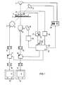
In der Figur 1 ist eine Einrichtung zum Zertrümmern von im Körper eines Lebewesens befindlichen Konkrementen mit einer Röntgenuntersuchungsvorrichtung dargestellt, die zwei Röntgenröhren 1 und 2 aufweist, die Röntgenstrahlenbündel erzeugen, die eine auf einer Patientenliege 3 befindliche, zu behandelnde Untersuchungsperson 4 durchdringen und auf die Eingangsleuchtschirme von Röntgenbildverstärker 5 und 6 fallen. Die Röntgenröhre 1 und der Röntgenbildverstärker 6 sind derart angeordnet, daß der Zentralstrahl des Röntgenstrahlenbündels der Röntgenröhre 1 senkrecht auf die Untersuchungsperson 4 fällt (a.p.-Projektion). Die Röntgenröhre 2 und der Röntgenbildverstärker 5 sind derart schräg angeordnet, daß der Zentralstrahl der Röntgenröhre 2 den Zentralstrahl der Röntgenröhre 1 in einem Zielgebiet innerhalb der Untersuchungsperson 4 unter einem Winkel von beispielsweise 45° schneidet. Dadurch erhält man Durchleuchtungsbilder aus zwei verschiedenen Projektionsrichtungen, so daß man die Untersuchungsperson 4 durch die Patientenliege 3 derart verschieben kann, daß sich in dem Zielgebiet die Konkremente befinden.FIG. 1 shows a device for crushing concrements located in the body of a living being with an X-ray examination device, which has two X-ray tubes 1 and 2, which generate X-ray beams that penetrate an examiner 4 to be treated on a
Die Ausgangssignale der an dem Röntgenbildverstärker 5 und 6 angekoppelten Fernsehkameras 7 und 8 werden über zwei Analog/Digital-Wandler (A/D-Wandler 9, 10) in zwei Bildspeicher 11 und 12 eingelesen. Die Ausgangssignale können über zwei Digital-Analog-Wandler (D/A-Wandler 13, 14) auf zwei Monitoren 15, 16 betrachtet werden. Befinden sich nun die Konkremente im Zielgebiet, so erscheinen sie in der Mitte der Bildschirme der Monitore 15 und 16.The output signals of the
Zur Zerkleinerung der in dem Zielgebiet befindlichen Konkremente ist ein Stoßwellengenerator 17 vorgesehen, der in Figur 1 schematisch dargestellt ist. Er erzeugt in bekannter Weise Stoßwellen, die zu einer Zertrümmerung der Konkremente führen, wenn sie beispielsweise im Brennpunkt des Stoßwellengenerators 17 liegen. Die Röntgenuntersu chungseinrichtung und der Stoßwellengenerator 17 sind derart fest miteinander verbunden, daß die Stoßwellen im Zielgebiet der Röntgenuntersuchungseinrichtung fokussiert sind. Der Stoßwellengenerator 17 ist an einer Ansteuerschaltung 19 angeschlossen.A
An der Untersuchungsperson 4 ist ein Abnehmer 20 für die Atmung angebracht, der beispielsweise aus einem um den Bauch der Untersuchungsperson 4 geschlungenen pneumatischen Gürtel besteht. An dem Abnehmer 20 ist eine Schaltung 21 angeschlossen, die einen Einsteller 22 für einen Schwellwert aufweist. An der Untersuchungsperson 4 sind weiterhin EKG-Elektroden 23 angeordnet, die mit einer EKG-Schaltung 24 verbunden sind. Die Ausgangssignale der Schaltungen 21 und 24 werden einer Steuervorrichtung 25 zugeführt, die mit einem Hochspannungsgenerator 26 zur Steuerung der Röntgenröhren 1 und 2, mit den Bildspeichern 11 und 12 und der Ansteuerschaltung 19 für den Stoßwellengenerator 17 verbunden ist.A
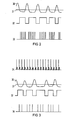
Anhand der in den Figuren 2 und 3 dargestellten Zeitverläufe wird die Funktionsweise der in Figur 1 dargestellten Einrichtung näher erläutert. Der Abnehmer 20 liefert ein der Atmung der Untersuchungsperson 4 entsprechendes Ausgangssignal 30. Eine durch den Einsteller 22 vorgewählte Schwelle 31 bewirkt beispielsweise durch einen Komparator ein Ausgangssignal 32 der Schaltung 21. Aufgrund beispielsweise der ansteigenden Vorderflanke der Rechteckimpulse oder auch zusätzlich zeitverzögert bewirkt die Steuervorrichtung 25 die Auslösung eines Röntgenimpulses durch den Hochspannungsgenerator 26 und die gleichzeitige Einspeicherung der Röntgenbilder in die Bildspeicher 11 und 12. Dadurch wird erreicht, daß immer in der gleichen Atemphase die Untersuchungsaufnahmen erstellt werden. Wird nun die Einrichtung durch einen Schalter 27 von Untersuchung auf Therapie umgeschaltet, so wird aus dem Ausgangssignal 32 der Schaltung 21 durch die Steuervorrichtung 25 ein Freigabesignal für die Ansteuerschaltung 19 des Stoßwellengenerator 17 erzeugt, der daraufhin innerhalb der Rechteckimpulse periodisch Stoßwellen erzeugt, die durch den Kurvenverlauf 33 als nadelförmige Impulse dargestellt sind. Es werden also während der Untersuchungsphase Röntgenbilder erstellt, die während einer bestimmten Atemphase anfallen, und während dieser gleichen Atemphase, in der nur eine geringe oder gar keine Bewegung beispielsweise der Niere erfolgt, werden mehrere Stoßwellen erzeugt, die die Konkremente gezielt treffen und somit nahezu vollständig zerkleinern.The mode of operation of the device shown in FIG. 1 is explained in more detail using the time profiles shown in FIGS. 2 and 3. The
Es kann aber auch gleichzeitig mit der Triggerung auf die Atemphase eine Triggerung aus dem EKG erfolgen, dessen Kurvenverlauf 34 in Figur 3 dargestellt ist. Hierzu werden aus dem EKG, beispielsweise aus der sogenannten R-Zacke, durch die EKG-Schaltung 24 Triggerimpulse erzeugt, die der Steuervorrichtung 25 zugeführt werden. Dieses Signal wird, wie gestrichelt dargestellt, zusammen mit dem Ausgangssignal 32 der Schaltung 21 in einem UND-Glied verkoppelt, so daß daraus ein Kombinationssignal entsteht, das zur Steuerung der Röntgenuntersuchungseinrichtung und des Stoßwellengenerators verwendet wird. Daraus folgt, daß nur während der durch das Ausgangssignal 32 bestimmten Atemphase und während des Auftretens der R-Zacke im EKG-Signal eine Aufnahme bzw. Stoßwelle erzeugt wird, wie dies aus dem in Figur 3 dargestellten Kurvenverlauf durch die Nadelimpulse 35 ersichtlich ist.However, triggering from the EKG can also take place simultaneously with the triggering on the breathing phase, the
Claims (5)
Applications Claiming Priority (2)
| Application Number | Priority Date | Filing Date | Title |
|---|---|---|---|
| DE3615564 | 1986-05-09 | ||
| DE3615564 | 1986-05-09 |
Publications (2)
| Publication Number | Publication Date |
|---|---|
| EP0244730A1 true EP0244730A1 (en) | 1987-11-11 |
| EP0244730B1 EP0244730B1 (en) | 1990-07-04 |
Family
ID=6300408
Family Applications (1)
| Application Number | Title | Priority Date | Filing Date |
|---|---|---|---|
| EP87106094A Expired - Lifetime EP0244730B1 (en) | 1986-05-09 | 1987-04-27 | Device for smashing concretions present in a living body |
Country Status (3)
| Country | Link |
|---|---|
| EP (1) | EP0244730B1 (en) |
| JP (1) | JPH045150Y2 (en) |
| DE (1) | DE3763533D1 (en) |
Cited By (6)
| Publication number | Priority date | Publication date | Assignee | Title |
|---|---|---|---|---|
| EP0296349A2 (en) * | 1987-06-24 | 1988-12-28 | Dornier Medizintechnik Gmbh | Respiratory trigger |
| EP0324948A2 (en) * | 1988-01-21 | 1989-07-26 | Dornier Medizintechnik Gmbh | Concretions destruction device |
| EP0336620A2 (en) * | 1988-03-31 | 1989-10-11 | Kabushiki Kaisha Toshiba | Apparatus for destroying calculuses |
| EP0460536A1 (en) * | 1990-05-31 | 1991-12-11 | Kabushiki Kaisha Toshiba | Lithotrity apparatus |
| US5419327A (en) * | 1992-12-07 | 1995-05-30 | Siemens Aktiengesellschaft | Acoustic therapy means |
| DE10260594B4 (en) * | 2002-12-23 | 2012-06-06 | Dornier Medtech Systems Gmbh | Device for extracorporeal generation of focused shock waves |
Families Citing this family (1)
| Publication number | Priority date | Publication date | Assignee | Title |
|---|---|---|---|---|
| JP4928739B2 (en) * | 2004-06-25 | 2012-05-09 | 株式会社東芝 | X-ray diagnostic apparatus and X-ray imaging method |
Citations (4)
| Publication number | Priority date | Publication date | Assignee | Title |
|---|---|---|---|---|
| DE496749C (en) * | 1927-09-10 | 1930-04-30 | Hans Appelrath Dr | Method and device for stereoscopic X-ray recording of periodically moving organs of the human or animal body |
| DE2722252A1 (en) * | 1977-05-17 | 1978-11-23 | Dornier System Gmbh | Solid accretion three=dimensional location and dissolution system - has ultrasonic emitter and receiver axes intersecting shock wave at accretion for medical application |
| GB2002987A (en) * | 1977-07-15 | 1979-02-28 | Emi Ltd | Improvements in or relating to radiography |
| EP0081051A1 (en) * | 1981-11-25 | 1983-06-15 | Dornier Gmbh | Releasing device for shock waves for therapeutic purposes |
Family Cites Families (3)
| Publication number | Priority date | Publication date | Assignee | Title |
|---|---|---|---|---|
| DE3122056A1 (en) * | 1981-06-03 | 1982-12-23 | Siemens AG, 1000 Berlin und 8000 München | Device for crushing concrements present in the body of a live organism |
| JPS618686A (en) * | 1984-06-25 | 1986-01-16 | Oki Electric Ind Co Ltd | Sonar system using adaptive beam former |
| DE3426398C1 (en) * | 1984-07-18 | 1987-11-12 | Dornier System Gmbh, 7990 Friedrichshafen | Device for spatial location and positioning of calculus |
-
1987
- 1987-04-27 EP EP87106094A patent/EP0244730B1/en not_active Expired - Lifetime
- 1987-04-27 DE DE8787106094T patent/DE3763533D1/en not_active Expired - Fee Related
- 1987-05-06 JP JP1987067792U patent/JPH045150Y2/ja not_active Expired
Patent Citations (4)
| Publication number | Priority date | Publication date | Assignee | Title |
|---|---|---|---|---|
| DE496749C (en) * | 1927-09-10 | 1930-04-30 | Hans Appelrath Dr | Method and device for stereoscopic X-ray recording of periodically moving organs of the human or animal body |
| DE2722252A1 (en) * | 1977-05-17 | 1978-11-23 | Dornier System Gmbh | Solid accretion three=dimensional location and dissolution system - has ultrasonic emitter and receiver axes intersecting shock wave at accretion for medical application |
| GB2002987A (en) * | 1977-07-15 | 1979-02-28 | Emi Ltd | Improvements in or relating to radiography |
| EP0081051A1 (en) * | 1981-11-25 | 1983-06-15 | Dornier Gmbh | Releasing device for shock waves for therapeutic purposes |
Cited By (11)
| Publication number | Priority date | Publication date | Assignee | Title |
|---|---|---|---|---|
| EP0296349A2 (en) * | 1987-06-24 | 1988-12-28 | Dornier Medizintechnik Gmbh | Respiratory trigger |
| EP0296349A3 (en) * | 1987-06-24 | 1989-03-15 | Dornier Medizintechnik Gmbh | Respiratory trigger |
| EP0324948A2 (en) * | 1988-01-21 | 1989-07-26 | Dornier Medizintechnik Gmbh | Concretions destruction device |
| EP0324948A3 (en) * | 1988-01-21 | 1989-10-25 | Dornier Medizintechnik Gmbh | Concretions destruction device |
| EP0336620A2 (en) * | 1988-03-31 | 1989-10-11 | Kabushiki Kaisha Toshiba | Apparatus for destroying calculuses |
| EP0336620A3 (en) * | 1988-03-31 | 1990-04-25 | Kabushiki Kaisha Toshiba | Apparatus for destroying calculuses |
| US5054469A (en) * | 1988-03-31 | 1991-10-08 | Kabushiki Kaisha Toshiba | Apparatus for destroying calculuses |
| EP0460536A1 (en) * | 1990-05-31 | 1991-12-11 | Kabushiki Kaisha Toshiba | Lithotrity apparatus |
| US5243985A (en) * | 1990-05-31 | 1993-09-14 | Kabushiki Kaisha Toshiba | Lithotrity apparatus having a missed-shot preventive function |
| US5419327A (en) * | 1992-12-07 | 1995-05-30 | Siemens Aktiengesellschaft | Acoustic therapy means |
| DE10260594B4 (en) * | 2002-12-23 | 2012-06-06 | Dornier Medtech Systems Gmbh | Device for extracorporeal generation of focused shock waves |
Also Published As
| Publication number | Publication date |
|---|---|
| JPS62183808U (en) | 1987-11-21 |
| DE3763533D1 (en) | 1990-08-09 |
| EP0244730B1 (en) | 1990-07-04 |
| JPH045150Y2 (en) | 1992-02-14 |
Similar Documents
| Publication | Publication Date | Title |
|---|---|---|
| DE4241161C2 (en) | Acoustic therapy facility | |
| DE4315282C2 (en) | Use of an acoustic pressure pulse source | |
| DE4125950C1 (en) | ||
| DE4202302C2 (en) | Computer tomograph | |
| EP0081051B2 (en) | Releasing device for shock waves for therapeutic purposes | |
| DE3426398C1 (en) | Device for spatial location and positioning of calculus | |
| DE3543867C2 (en) | ||
| DE3607949C2 (en) | ||
| DE10158519B4 (en) | Shock and shock wave therapy device | |
| DE10102317A1 (en) | Method and device for applying pressure waves to the body of a living being | |
| DE3119295A1 (en) | DEVICE FOR DESTROYING CONCRETE IN BODIES | |
| DE19733838A1 (en) | Human or animal body shock wave treatment method and appliance | |
| DE3840077A1 (en) | LITHOTRIPTOR | |
| EP0372119B2 (en) | Lithotripter | |
| DE3328039C2 (en) | FACILITIES FOR THE CONTACTLESS SMASHING OF A CONCERMENT IN THE BODY OF A LIVING BEING | |
| EP0273256A1 (en) | Apparatus for the non-invasive fragmentation of concrements | |
| DE19548000C1 (en) | Device for locating calculus in a patient's body | |
| EP0355178B1 (en) | Apparatus for the contactless desintegration of concrements in a living thing body | |
| EP0400196B1 (en) | Shock wave transducer for the destruction of concretions | |
| EP0244730B1 (en) | Device for smashing concretions present in a living body | |
| EP0257199B1 (en) | Device for the destruction of calculi | |
| DE102005031125A1 (en) | Method and lithotripsy apparatus for destroying a calculus in a patient | |
| EP0280088B1 (en) | Sound generator for treating a living being with focused sound waves | |
| DE102005031117A1 (en) | Method and device for determining an operating parameter of a shockwave source | |
| EP0560444B1 (en) | Shock wave treatment device |
Legal Events
| Date | Code | Title | Description |
|---|---|---|---|
| PUAI | Public reference made under article 153(3) epc to a published international application that has entered the european phase |
Free format text: ORIGINAL CODE: 0009012 |
|
| AK | Designated contracting states |
Kind code of ref document: A1 Designated state(s): DE FR GB NL |
|
| 17P | Request for examination filed |
Effective date: 19871204 |
|
| 17Q | First examination report despatched |
Effective date: 19890307 |
|
| GRAA | (expected) grant |
Free format text: ORIGINAL CODE: 0009210 |
|
| AK | Designated contracting states |
Kind code of ref document: B1 Designated state(s): DE FR GB NL |
|
| REF | Corresponds to: |
Ref document number: 3763533 Country of ref document: DE Date of ref document: 19900809 |
|
| ET | Fr: translation filed | ||
| GBT | Gb: translation of ep patent filed (gb section 77(6)(a)/1977) | ||
| PLBE | No opposition filed within time limit |
Free format text: ORIGINAL CODE: 0009261 |
|
| STAA | Information on the status of an ep patent application or granted ep patent |
Free format text: STATUS: NO OPPOSITION FILED WITHIN TIME LIMIT |
|
| 26N | No opposition filed | ||
| PGFP | Annual fee paid to national office [announced via postgrant information from national office to epo] |
Ref country code: GB Payment date: 19930319 Year of fee payment: 7 |
|
| PGFP | Annual fee paid to national office [announced via postgrant information from national office to epo] |
Ref country code: FR Payment date: 19930423 Year of fee payment: 7 |
|
| PGFP | Annual fee paid to national office [announced via postgrant information from national office to epo] |
Ref country code: NL Payment date: 19930430 Year of fee payment: 7 |
|
| PG25 | Lapsed in a contracting state [announced via postgrant information from national office to epo] |
Ref country code: GB Effective date: 19940427 |
|
| PG25 | Lapsed in a contracting state [announced via postgrant information from national office to epo] |
Ref country code: NL Effective date: 19941101 |
|
| NLV4 | Nl: lapsed or anulled due to non-payment of the annual fee | ||
| GBPC | Gb: european patent ceased through non-payment of renewal fee |
Effective date: 19940427 |
|
| PG25 | Lapsed in a contracting state [announced via postgrant information from national office to epo] |
Ref country code: FR Effective date: 19941229 |
|
| REG | Reference to a national code |
Ref country code: FR Ref legal event code: ST |
|
| PGFP | Annual fee paid to national office [announced via postgrant information from national office to epo] |
Ref country code: DE Payment date: 20000619 Year of fee payment: 14 |
|
| PG25 | Lapsed in a contracting state [announced via postgrant information from national office to epo] |
Ref country code: DE Free format text: LAPSE BECAUSE OF NON-PAYMENT OF DUE FEES Effective date: 20020201 |