EP0170345B1 - Cytométrie à écoulement - Google Patents
Cytométrie à écoulement Download PDFInfo
- Publication number
- EP0170345B1 EP0170345B1 EP85302094A EP85302094A EP0170345B1 EP 0170345 B1 EP0170345 B1 EP 0170345B1 EP 85302094 A EP85302094 A EP 85302094A EP 85302094 A EP85302094 A EP 85302094A EP 0170345 B1 EP0170345 B1 EP 0170345B1
- Authority
- EP
- European Patent Office
- Prior art keywords
- cells
- cell
- antibody
- cell suspension
- conjugate
- Prior art date
- Legal status (The legal status is an assumption and is not a legal conclusion. Google has not performed a legal analysis and makes no representation as to the accuracy of the status listed.)
- Expired - Lifetime
Links
Images
Classifications
-
- G—PHYSICS
- G01—MEASURING; TESTING
- G01N—INVESTIGATING OR ANALYSING MATERIALS BY DETERMINING THEIR CHEMICAL OR PHYSICAL PROPERTIES
- G01N33/00—Investigating or analysing materials by specific methods not covered by groups G01N1/00 - G01N31/00
- G01N33/48—Biological material, e.g. blood, urine; Haemocytometers
- G01N33/50—Chemical analysis of biological material, e.g. blood, urine; Testing involving biospecific ligand binding methods; Immunological testing
- G01N33/53—Immunoassay; Biospecific binding assay; Materials therefor
- G01N33/569—Immunoassay; Biospecific binding assay; Materials therefor for microorganisms, e.g. protozoa, bacteria, viruses
- G01N33/56966—Animal cells
- G01N33/56972—White blood cells
Definitions
- This invention relates to flow cytometry, particularly to the use of specific binding assays in flow cytometry systems.
- All animal and plant cells have a large variety of protein enzymes which chemically catalyze the various biochemical reactions that are necessary for maintenance, growth and specialized cell functions. Among these enzymes are subsets which are present in only a few, or even only one kind of cell.
- the introduced substances must (a) be able to enter the cell; (b) must not be toxic to the cell; (c) must convert to an extremely insoluble form; and (d) the conversion must occur extremely rapidly.
- Cells are typically a few to tens of micrometers in diameter. Molecules in aqueous solution typically diffuse such distances in fractions of seconds or seconds. Therefore, if, after enzymatic reaction, the conversion to the insoluble form is delayed, it will deposit far from the enzyme and even outside the cell. Since the product concentration diminishes with the cube of the distance it diffuses from site of reaction, its solubility product might not be exceeded at even short distances from the enzymatic site and, therefore, the cell remains unstained unless the product is extremely insoluble.
- Specific binding assay techniques have provided extremely useful analytical methods for determining various organic substances of diagnostic, medical, environmental and industrial importance which appear in liquid media at very low concentrations.
- Specific binding assays are based on the specific interaction between a ligand, i.e. a bindable analyte under determination, and a binding partner therefor. Where one of the ligand and its binding partner is a hapten or antigen and the other is a corresponding antibody, the assay is known as an immunoassay.
- Enzymes are among the many labels which are commonly used in the form of a conjugate in which the enzyme is linked to the binding protein with a low molecular weight ligand with a species like that under assay or its binding partner.
- a substrate is included and reacts with the enzyme, to the extent permitted by the binding partner interaction, to provide a detectable response.
- immunoenzyme-cytochemical staining methods the very high chemical specificity of antibodies to bind selectively to unique molecular sites which are present on or in special subsets of cells is utilized. Such antibodies are either directly, or indirectly attached to enzymes for which high-resolution enzyme-cytochemical methods exist (e.g. peroxidase enzymes, alkaline phosphatase enzymes, etc.).
- the preparation can then be stained by an appropriate enzyme-cytochemical method, and only the labeled cells will accumulate light-absorbing (colored) product on or in them.
- enzyme-cytochemical method an enzyme-cytochemical method
- fixing reagents it is essential that they do not destroy the antibody-binding sites of the cells to be studied.
- immunoperoxidase One of the classes of enzyme-labeled specific binding techniques used in immunocytology is referred to as the "immunoperoxidase" method, for which there are five basic protocols. Among the following methods, those which bind larger numbers of peroxidase molecules for each molecule of primary antibody bound increase sensitivity by "enzyme amplification".
- a peroxidase-antibody conjugate binds directly to a tissue antigen.
- a primary antibody binds the tissue antigen and is, in turn, bound by a peroxidase-secondary antibody (anti-primary antibody) conjugate.
- the "labeled antigen” protocol is essentially a sandwich technique in which primary antibody binds to both the tissue antigen and to an analogous antigen which has been conjugated with peroxidase.
- the "enzyme bridge” protocol primary and secondary antibodies are bound as described above and the secondary antibody is bound by a third antibody which has been conjugated with peroxidase.
- the "peroxidase-anti-peroxidase” protocol is as described for the enzyme bridge protocol with the addition that the peroxidase-tertiary antibody is followed by an anti-peroxidase antibody and excess peroxidase.
- Avidin is a glycoprotein, molecular weight 68,000, with four binding sites that have high affinity for biotin, one of the B vitamin complex. This high affinity binding has been used as an alternative to the labeled antibody methods described above in immunohistology.
- a biotin-primary-antibody conjugate binds directly to a cellular antigen and is, in turn, bound by an avidin-peroxidase conjugate.
- the tissue-bound biotinylated primary antibody is bound with unlabeled avidin which, in turn, is bound with peroxidase-labeled biotin.
- the third such protocol uses an unconjugated primary antibody, a biotinylated secondary antibody and an avidin-biotin-peroxidase complex, from which it derives its connotation as the "ABC method". See, generally, Falini, et al, supra and Guesdon, J-L, et al, J. Histochem. Cytochem. , 27 :1131-1139 (1979).
- Human white blood cells can be classified as monocytes, polymorphonucleocytes (PMNs) and lymphocytes. There are two principal classes of lymphocytes. The first of these (the thymus-derived cell or T-cell) is immunologically active in effecting cell-mediated responses and the second (the bone marrow derived cell or B cell) is immunologically active in producing antibodies. It is now recognized that T cells are divided into at least several subtypes, termed "helper”, “suppressor”, and “killer” T cells, which have the function of (respectively) promoting a reaction, suppressing a reaction, or killing (lysing) foreign cells. Lymphocyte subclasses of clinical interest are not easily distinguishable by other than immunological methods.
- the antigen on the outer surfaces of these lymphocytes are distinguishable with specific antibodies. It is particularly important to recognize and understand that a number of these antigens have been found to be remarkably fragile in the sense that mild chemical or physical treatment of the cell can either destroy the antibody-combining site and/or detach the antigen from the cell surface.
- lymphocytes When lymphocytes are stained by immuno-enzyme-cytochemical methods with the cells in liquid suspension, they have typically been stained live , with high resolution methods. These produce a fine stippling of colored product on the outer cell surface. This distribution of product is due to the fact that the insoluble colored product precipitates within a few nanometers of the enzyme molecules (which are all on the outside surface of the cell). Half or more of the product diffuses away from the cell. When this diffused product precipitates, it is mostly freely suspended in the aqueous medium and drifts or is washed away from the cell.
- the other part diffuses towards the cell and either precipitates between and among the antibody and antigen molecules on the surface of the cell and is trapped in that network, or precipitates on or in the cell membrane, or, if the membrane is permeable to the product , just inside the cell.
- Such surface-stippled cells absorb much less light than cells which have a similar amount of stain distributed more uniformly on or within the cells.
- Binet, et al, Blood Cells, 6 :371-376 (1980) have examined such preparations by flow-cytometry (for instrumentation, see U.S. Patent No. 3,740,143).
- Such surface-stippled cells generated too weak a signal to permit clean separation of the signals of the stained and unstained cells.
- a flow cytometry method of reproducibly detecting a cell population of interest in a heterogeneous cell suspension which method comprises:
- the antibodies and other reagents used in the method can be combined with a cell sample in a continuous-flow system, as in U.S. Patent No. 3,740,143, supra , or in a discrete reaction chamber at any point upstream of the point of illumination in the flow cytometer.
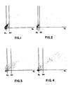
- FIGs 1,2 and 4 illustrate results obtained using prior art techniques for processing unfixed leukocyte-enriched cell suspensions.
- Figure 3 is an unlabeled control for such prior art techniques.
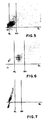
- Figures 5 and 6 illustrate the results obtained when fixation of the cell suspension is effected after and before, respectively, the application of the second antibody to the leukocyte.
- Figure 7 is an unlabeled control for the processes depicted in Figures 5 and 6.
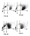
- Figures 8 and 10 illustrate the results obtained from a corresponding method according to the present invention when using whole blood, and a corresponding method where cell fixation is effected immediately after the application of the primary antibody to the leukocytes respectively.
- Figures 9 and 11 are unlabeled controls for Figures 8 and 10, respectively.
- the method of the invention is useful in that it permits the detectable species used to selectively and intensely color certain cells, particularly white blood cells, and not others.
- the cell sample can, for example, be whole blood or a heterogeneous white blood cell sample.
- the whole blood sample is preferably one in which the red blood cells have been lysed prior to introduction into the flow cytometer.
- specific binding protein refers to any substance, or class of substances, which has a specific binding affinity for the cell population of interest to the exclusion of other substances.
- the present invention will incorporate specific binding assay reagents which interact with the sample in an immunochemical manner. That is, there will be an antigen-antibody or hapten-antibody relationship between reagents and/or antigens associated with the cells in the cell population of interest. Such assays therefore are termed immunoassays and the special interaction between the ligand and its receptor, or binding partner, is an immunochemical binding.
- the use of either polyclonal or monoclonal antibodies is contemplated unless otherwise indicated. Additionally, it is well understood in the art that other binding interactions between the differentiating characteristics of the cell surface and a binding partner can serve as the basis of other specific binding assays.
- the primary specific binding protein is usually an antibody which is specific for cell surface marker antigens.
- antigens are those which differentiate populations or subpopulations of white blood cells, tumor cells, or other heterogeneous cell samples within which it is useful to distinguish various groups.
- antibodies are known to be useful for the differentiation of lymphocyte classes and subclasses, including monoclonal antibodies from various hybridoma cell lines. Such include mouse monoclonal antibodies having designated specificities for all human T cells, suppressor T cells, helper T cells and the like. Other such antibodies are known for specific reactivity with human B cells.
- the cells are fixed as early as is feasible, i.e. preceding the application of the antibody.
- a fixative is chosen which insolubilizes and stiffens the cells without destroying the chemical activity of the cell-specific surface antigen and which does not cause the cells to adhere to one another or to the walls of the vessel in which they are contained.
- Monoaldehydes such as formaldehyde, paraformaldehyde and acrolein, and dialdehydes such as glutaraldehyde, used alone or in combination, have been found useful for cells in suspension (see, for example, U.S. Patents 3,741,875 and 4,412,004).
- the secondary specific binding protein is usually an antibody against the class of proteins, usually immunoglobulins, of which the primary specific binding protein is a member. Thus, this secondary antibody reacts with and is specific for all antibodies of the primary antibody class.
- the methodology also uses a reagent conjugate comprising an enzyme which is bound to an avidin molecule.
- Another embodiment of this methodology uses a reagent conjugate comprising an enzyme bound to a biotin molecule which is in turn bound to an avidin molecule.
- enzymes suitable for such use include peroxidase (such as horseradish peroxidase), alkaline phosphatase and combinations of these and other enzymes.
- the redox chromogen used is critical in that it must be insoluble in the reaction medium in its reacted form. Those which have been identified as useful in the invention include 3-amino-9-ethylcarbazole and 4-chloro-1-naphthol. Chromogens producing a product appreciably soluble in the reaction environment are not useful.
- the primary antibody, biotinylated secondary antibody, labeled conjugate, enzyme substrate and redox chromogen can be combined with the cell sample under assay, either prior or subsequent to introducing said cell sample to a flow cytometer in which the method is to be performed.
- the cell sample into a fluid stream flowing in a conduit or analysis channel in the flow cytometer.
- This preferably comprises establishing a flowing stream of a flowing fluid sheath stream in the conduit or analysis channel and thereafter introducing the said sample into the flowing fluid sheath stream.
- sheath streams are usually of fluids having a refractive index substantially identical to that of the cell sample suspending medium.
- One such flow cytometer which uses a sheath stream carrier fluid is used in the Technicon Hemalog D and H-6000 systems, which handle all routine hematology tests. Detailed information on the Hemalog D and H-6000 systems is available from Technicon Instruments Corporation, Tarrytown, NY.
- FIGS. 1-11 are 2-dimensional displays from the peroxidase channel of an H-6000 instrument system (Technicon Instruments Corporation, Tarrytown, NY) in which absorption is measured along the abscissa and light scattered out of the forward direction is measured along the ordinate. Each dot represents the measured coordinates of a single cell. Three pre-set thresholds enable the operator to separate and count distinct clusters of signals. Absorption Low (AL) and Absorption High (AH) are shown as vertical lines. Scatter Low (SL) is shown as a horizontal line. H-6000 gives separate counts for all signals above SL and to the left of AL, between AL and AH, and to the right of AH. It will ignore all the signals below SL, which represent signals smaller than those from cells, thus removing noise signals due to red cell ghosts, platelets, etc.
- AL Absorption Low
- AH Absorption High
- SL Scatter Low
- lymphocyte-labeling methods are applied to either lymphocyte-enriched samples or whole blood samples.
- Whole blood contains both neutrophils and eosinophils (which are PMNs) and monocytes, all of which carry endogenous peroxidases. After peroxidase staining, distinguishing these cells from peroxidase-labeled lymphocytes could pose a problem.
- the signals from these cells are easily separated from the positive (peroxidase-labeled) lymphocytes by the AH threshold. The remaining weakly stained monocytes which lie between the AL and AH threshholds can be counted separately in the control, and subtracted from the count from between the AL and AH threshholds in the corresponding experimental run.
- lymphocyte-enriched samples are usually contaminated with a few PMNs and monocytes. (See Figs. 1-7.) Similar subtraction of appropriate controls correct for such contamination.
- Example III experiment (b) demonstrates results obtained from an embodiment of the present invention. Standard, commercially available reagent grade chemicals were used whenever possible.
- the peroxidase enzyme thus associated with the surface of the T cells was stained by each of one of two different redox chromogens, namely, 3-amino-9-ethyl-carbazole and 4-chloro-1-naphthol.
- the details of the methods employed are as follows:
- Lymphocyte-enriched suspensions were prepared as described in Boyum, A., Scand. J. Clin. Lab. Invest. , 21 , Suppl. 97 :77 (1968).
- the lymphocyte fraction was harvested and the cells washed three times by centrifuging for 10 minutes at 400 g in neutral phosphate buffered saline (PBS) containing 0.4% bovine serum albumin (PBS/BSA).
- PBS neutral phosphate buffered saline
- PBS/BSA bovine serum albumin
- the supernatant fluid was aspirated to waste.
- Sufficient PBS/BSA was added to the final harvest of cells to yield a concentration of 107 cells per ml. A 100 ⁇ l aliquot of this cell suspension was dispensed into a test tube.
- the cells were then resuspended and incubated for 10-20 minutes at room temperature in 1 ml of staining mixture consisting of: 2 mg 3-amino-9-ethylcarbazole (AEC) dissolved in 0.5 ml dimethylformamide (DMF) to which was added 9.5 ml 0.05 M acetate buffer (pH 5) plus 50 ⁇ l 3% H2O2.
- AEC 3-amino-9-ethylcarbazole
- DMF dimethylformamide
- pH 5 acetate buffer
- a 1 ml volume of the suspension of cells in the staining mixture was then diluted with 2 ml PBS and the cells were introduced into the flow cell of an H-6000 flow cytometer, by-passing the manifold by disconnecting the sample line at the peristaltic pump leading to the flow cell of the peroxidase channel and placing the disconnected sample line directly into, and to the bottom of, the test tube containing the reacted cell suspension.
- the results are illustrated in Fig. 1.
- Fig. 3 shows all lymphocytes to the left of AL.
- the dots between AL and AH are contaminating monocytes.
- the dots to the right of AH are contaminating neutrophils and eosinophils.
- Fig. 1 the stained lymphocytes are distributed across AL and into the space between AL and AH. All other cells remain is in the control (Fig. 3).
- the staining is clearly inadequate to separate all of the stained lymphocytes from the unstained lymphocytes. This can be attributed, in part, to the variable damage to and losses from the cell surfaces following the extensive manipulation of these live cells.
- Binet, et al sometimes fixed their cells after they had been completely processed (i.e., after staining step with AEC in Example I). Their procedure was to add 1 ml 0.7% formaldehyde to 1 ml of the AEC cell suspension, allowing the mixture to stand at room temperature for 10 minutes and diluting with 1 ml PBS before introduction into an H-6000 flow cell. The results were substantially identical to those illustrated in Fig. 1.
- PBS/BSA phosphate buffered saline containing 0.4% bovine serum albumin
- OKT3 pan T primary antibody at a strength of 25 ⁇ g/ml
- PBS/BSA/EDTA ethylene diamine tetracetic acid
- the staining solution contained 8 mg 3-amino-9-ethylcarbazole (AEC) in 10 ml methanol combined with 100 ⁇ l 3% H2O2 made up to 25 ml using 0.05 M sodium acetate buffer (pH 5). After 10 minutes at room temperature, the mixture was combined with 2.0 ml PBS and aspirated directly through a sheathed H-6000 flow cell via a peristaltic pump to obtain the cell signatures in the peroxidase channel. The result is illustrated in Fig. 5. A negative control was run in which every step was the same as described above except that the primary antibody was omitted. The results for the control are illustrated in Fig. 7.
- AEC 3-amino-9-ethylcarbazole
- a 100 ⁇ l aliquot of a lymphocyte-enriched suspension was treated as described in Experiment A to yield a pellet of washed lymphocytes including OKT3-bound cells.
- Experiments A, B and C demonstrate that the labeled lymphocyte subset no longer forms a continuum with the unlabeled subset, as in Example 1, Figs. 1, 2 and 4. Instead, the labeled subset forms a discrete cluster, separated from the unlabeled cluster by a gap.
- This gap is narrow in Fig. 5 (post-secondary-antibody fixation), but in Fig. 6 it is very wide (pre-secondary-antibody fixation), thus meeting the essential condition necessary for convenient and accurate counting and classifying of subsets of cells in flow cytometry.
- the width of the gap can be attributed to how early in the process the cells were fixed.
- lymphocyte differentiation immuno-assays were also performed in accordance with the invention on whole blood samples using OKT4 (Ortho Diagnostics, Raritan, NJ) or Coulter Clone T4 (Coulter, Hialea, FL) as the primary antibody.
- OKT4 Ortho Diagnostics, Raritan, NJ
- Coulter Clone T4 Coulter, Hialea, FL
- a fixation step was performed after incubation with primary antibody (OKT4) but prior to incubation with secondary antibody.
- Example B using the method according to the present invention, the same type of fixation was performed prior to incubation with primary antibody (Coulter Clone T4).
- a 100 ⁇ l aliquot of anticoagulated whole blood was dispensed into a clean test tube. This was mixed with 100 ⁇ l of cold isotonic buffered saline containing approximately 2.5 ⁇ g/ml OKT4 T-cell monoclonal antibody (Ortho Diagnostics, Raritan, NJ) and the mixture was incubated at 4 o C for 15 to 30 minutes. Red cells were then lysed by adding 2 ml of 0.85% NH4Cl solution at room temperature with thorough mixing. The cell suspension was centrifuged for 1 minute (1,000 g) to harvest the white cells. The white cells were recovered and washed twice in PBS/BSA/EDTA at 4 o C. This yielded a pellet of washed white blood cells.
- these cells were resuspended in the residual supernatant and fixed by adding 1 ml of 0.075 M phosphate buffered 7.5% formaldehyde solution (pH 6.7) containing 15% dextrose. After 5-10 minutes, the fixed cells were washed twice with PBS/BSA/EDTA. Then, after fixation, the cells were resuspended in residual supernatant, incubated with 0.1 ml of biotinylated secondary anti-mouse immunoglobulin antibody (12.5 ⁇ g/ml) for 15-30 minutes at room temperature and were washed twice with PBS/BSA/EDTA.
- the washed cells were resuspended in residual supernatant and incubated with 0.1 ml avidin-horseradish peroxidase conjugate (A:HRP) in PBS (50-100 ⁇ g A:HRP per ml PBS). After 15-30 minutes at room temperature, the cells were washed three times with PBS/BSA/EDTA and mixed with 0.5 ml of a staining solution which contained 0.3 mg 4-chloro-1-naphthol, 16% ethanol, 0.01% H2O2 in 0.025 M phosphate buffer (pH 7.5) and incubated for 10 minutes at room temperature.
- A:HRP avidin-horseradish peroxidase conjugate
- the stained cell suspension was diluted to 1 ml with phosphate buffer and aspirated directly through the H-6000 flow cell as previously described, with the results illustrated in Fig. 8.
- a negative control was run in which every step was the same, as described above, except that the primary antibody was omitted. The results for the control are illustrated in Fig. 9.
- the cells were fixed at room temperature by mixing the resuspended pellet with 1 ml of 0.075 M phosphate buffered 7.5% formaldehyde solution (pH 6.7), which contains 15% dextrose, for 5-10 minutes, and washed twice with PBS/BSA/EDTA.
- the cell pellet was resuspended in residual supernatant, incubated with 0.2 ml of T4-biotinylated primary antibody solution (Coulter, Hialea, FL) in PBS, diluted from stock according to the manufacturer's directions, for 15-30 minutes at room temperature and washed twice with PBS/BSA/EDTA.
- the resulting pellet was resuspended and incubated with 0.1 ml avidin-peroxidase (50-100 ⁇ g/ml PBS) for 15-30 minutes at room temperature and washed twice with PBS/BSA/EDTA.
- the resulting pellet was resuspended, mixed with 0.5 ml of a staining solution which contained 0.3 mg 4-chloro-1-naphthol, 0.01% H2O2 and 16% ethanol (or methanol) in 0.025 M phosphate buffer (pH 7.5) and incubated for 10 minutes at room temperature.
- lymphocyte-enriched suspension two lymphocyte subsets, one of which is labeled, form discrete and separate clusters.
- the PMNs are to the right of AH threshhold.
- Figs. 9 and 11 show that significant numbers of monocytes fall between AL and AH. These are subtracted from the counts from between AL and AH in Figs. 8 and 10, respectively, to determine the number of stained lymphocytes.
Landscapes
- Life Sciences & Earth Sciences (AREA)
- Health & Medical Sciences (AREA)
- Cell Biology (AREA)
- Hematology (AREA)
- Immunology (AREA)
- Engineering & Computer Science (AREA)
- Molecular Biology (AREA)
- Chemical & Material Sciences (AREA)
- Urology & Nephrology (AREA)
- Biomedical Technology (AREA)
- Virology (AREA)
- Physics & Mathematics (AREA)
- Biotechnology (AREA)
- Tropical Medicine & Parasitology (AREA)
- Zoology (AREA)
- Food Science & Technology (AREA)
- Medicinal Chemistry (AREA)
- Microbiology (AREA)
- Analytical Chemistry (AREA)
- Biochemistry (AREA)
- General Health & Medical Sciences (AREA)
- General Physics & Mathematics (AREA)
- Pathology (AREA)
- Measuring Or Testing Involving Enzymes Or Micro-Organisms (AREA)
- Investigating Or Analysing Biological Materials (AREA)
Claims (9)
- Un procédé de cytométrie à écoulement pour détecter de façon reproductible une population de cellules intéressante dans une suspension hétérogène de cellules , procédé comprenanta) premièrement la combinaison de ladite suspension de cellules avec un réactif de fixation et ensuite la combinaison des cellules ainsi fixées avec un réactif qui comprend la protéine de fixation primaire spécifique de ladite population de cellules intéressantes;b) le couplage des enzymes de ladite protéine de fixation primaire avec au moins un ligand;c) ensuite la réaction de ladite suspension de cellules fixées avec une composition contenant un chromogène afin de colorer la population de cellules intéressantes , le chomogène étant complètement insoluble dans le milieu réactionnel aprés réaction, de telle sorte qu'au moins une des propriétés d'absorption ou de diffusion des cellules de ladite population de cellules intéressantes est altérée;d) passage subséquent de la suspension de cellules ayant subi la réaction, sensiblement une seule cellule à la fois, à travers un faisceau de lumière tandis que l'on mesure ainsi une desdites propriérés des cellules qui passent à travers; ete) différenciation des cellules de ladite population intéressante basée sur la mesure d'au moins une desdites propriétés desdites cellules interéssantes.
- Un procédé selon la revendication 1, où le couplage des enzymes audit anticorps primaire est effectué à l'aide d'un ligand de bas poids moléculaire et avant la combinaison dudit anticorps primaire avec ladite suspension de cellules.
- Un procédé selon la revendication 1 ou 2, où ladite suspension de cellules comprend une suspension hétérogène de globules blancs suspension complétement débarassée de globules rouges, ou bien la suspension de cellules comprend le sang entier dans lequel les globules rouges ont étés lysés au moins avant l'étape (d)
- Un procédé selon l'une quelconque des revendications 1 ou 2 où dans l'étape (a) la protéine de fixation primaire est un anticorps polyclonal ou monoclonal, et/ou la protéine de fixation primaire est un conjugué de l'anticorps de la biotine.
- Un procédé selon l'une quelconque des revendications 1 à 4, où ladite suspension de cellules est fixée à l'aide d'un aldéhyde, de préférence le formaldéhyde.
- Un procédé selon la revendication 4 où l'étape (b) comprend l'addition d'un conjugué de l'enzyme avidine-biotine à ladite suspension de cellules , tel que le conjugué de l'avidine-biotine peroxydase ou le conjugué de l'avidinebiotine phosphatase alcaline.
- Un procédé selon la revendication 4 où la protéine de fixation primaire est un conjugué de l'anticorps de la biotine et l'étape (b) comprend la combinaison de la suspension de cellules avec un conjugué de l'avidine-enzyme tel que le conjugué de l'avidine-peroxydase ou le conjugué de l'avidine-phosphatase alcaline.
- Un procédé selon l'une quelconque des revendications 1 à 3, où l'étape (b) comprend la combinaison des celules fixées attachées à l'anticorps après l'étape (a) avec un anticorps secondaire modifié par la biotine , et en option avec un conjugué d'enzyme marqué.
- Un procédé selon l'une quelconque des revendications 1 à 8,où ladite composition de l'étape (c) contient le chromogène 3-amino-9-éthylcarbazole ou 4-chloro-1-naphtol.
Applications Claiming Priority (2)
| Application Number | Priority Date | Filing Date | Title |
|---|---|---|---|
| US59407784A | 1984-03-28 | 1984-03-28 | |
| US594077 | 1984-03-28 |
Publications (3)
| Publication Number | Publication Date |
|---|---|
| EP0170345A2 EP0170345A2 (fr) | 1986-02-05 |
| EP0170345A3 EP0170345A3 (en) | 1987-03-25 |
| EP0170345B1 true EP0170345B1 (fr) | 1991-10-30 |
Family
ID=24377431
Family Applications (1)
| Application Number | Title | Priority Date | Filing Date |
|---|---|---|---|
| EP85302094A Expired - Lifetime EP0170345B1 (fr) | 1984-03-28 | 1985-03-26 | Cytométrie à écoulement |
Country Status (5)
| Country | Link |
|---|---|
| EP (1) | EP0170345B1 (fr) |
| JP (1) | JPS618664A (fr) |
| AU (1) | AU592528B2 (fr) |
| CA (1) | CA1254134A (fr) |
| DE (1) | DE3584547D1 (fr) |
Families Citing this family (9)
| Publication number | Priority date | Publication date | Assignee | Title |
|---|---|---|---|---|
| US5179026A (en) * | 1986-11-27 | 1993-01-12 | Toa Medical Electronics Co., Ltd. | Method of classifying leukocytes by flow cytometry and reagents used in the method |
| US5039613A (en) * | 1986-11-27 | 1991-08-13 | Toa Medical Electronics Co., Ltd. | Reagents used in a method of classifying leukocytes by flow cytometry |
| US5057413A (en) * | 1988-06-13 | 1991-10-15 | Becton, Dickinson And Company | Method for discriminating between intact and damaged cells in a sample |
| US5047321A (en) * | 1988-06-15 | 1991-09-10 | Becton Dickinson & Co. | Method for analysis of cellular components of a fluid |
| JP2852346B2 (ja) * | 1988-11-02 | 1999-02-03 | イクストゥルードゥ ホーン コーポレーション | 粘弾性媒体を用いたワークピースの加工方法 |
| CA2070244A1 (fr) * | 1991-08-07 | 1993-02-08 | Young Ran Kim | Methode et composition reactive pour le sous-typage des lymphocytes dans le sang peripherique |
| WO1993020448A1 (fr) * | 1992-04-01 | 1993-10-14 | The Regents Of The University Of California | Immuno-titrage par enzymes sans instrument d'analyse: procede general recourant a des effets de liaison cinetiques |
| US5788229A (en) * | 1994-08-29 | 1998-08-04 | Ricoh Co., Ltd. | Path guide for selectively corrugating an output medium |
| WO2006090096A1 (fr) * | 2005-02-28 | 2006-08-31 | Sheffield Teaching Hospitals Nhs Foundation Trust | Composition, kit et procédé servant à fixer des cellules |
Family Cites Families (5)
| Publication number | Priority date | Publication date | Assignee | Title |
|---|---|---|---|---|
| US3741875A (en) * | 1970-10-30 | 1973-06-26 | Mount Sinai Res Foundation Inc | Process and apparatus for obtaining a differential white blood cell count |
| US3781112A (en) * | 1972-12-15 | 1973-12-25 | Technicon Instr | Method and apparatus for analysis of leukocytes using light scattered by each leukocyte at absorbing and non-absorbing wavelength |
| US4284412A (en) * | 1979-07-13 | 1981-08-18 | Ortho Diagnostics, Inc. | Method and apparatus for automated identification and enumeration of specified blood cell subclasses |
| US4492752A (en) * | 1982-09-03 | 1985-01-08 | Ortho Diagnostics Systems Inc. | Method for discriminating between unstained and absorbing dye stained cells |
| AU602129B2 (en) * | 1985-09-06 | 1990-10-04 | Technicon Instruments Corportion | Method for the determination of a differential white blood cell count |
-
1985
- 1985-03-15 CA CA000476638A patent/CA1254134A/fr not_active Expired
- 1985-03-25 AU AU40316/85A patent/AU592528B2/en not_active Ceased
- 1985-03-26 EP EP85302094A patent/EP0170345B1/fr not_active Expired - Lifetime
- 1985-03-26 DE DE8585302094T patent/DE3584547D1/de not_active Expired - Fee Related
- 1985-03-28 JP JP6220485A patent/JPS618664A/ja active Granted
Non-Patent Citations (1)
| Title |
|---|
| ARCH. PATHOL. LAB. MED., vol. 107, 1983; pp. 105-116 * |
Also Published As
| Publication number | Publication date |
|---|---|
| DE3584547D1 (de) | 1991-12-05 |
| AU592528B2 (en) | 1990-01-18 |
| AU4031685A (en) | 1985-10-03 |
| CA1254134A (fr) | 1989-05-16 |
| EP0170345A2 (fr) | 1986-02-05 |
| JPS618664A (ja) | 1986-01-16 |
| JPH0566548B2 (fr) | 1993-09-22 |
| EP0170345A3 (en) | 1987-03-25 |
Similar Documents
| Publication | Publication Date | Title |
|---|---|---|
| US5122453A (en) | Method for discriminating surface stained lymphocytes | |
| US5374531A (en) | Immunoassay for determination of cells | |
| US5385822A (en) | Methods for detection and quantification of cell subsets within subpopulations of a mixed cell population | |
| JP3542808B2 (ja) | 細胞計数イムノアッセイ | |
| De Waele et al. | Immunogold staining method for the light microscopic detection of leukocyte cell surface antigens with monoclonal antibodies: its application to the enumeration of lymphocyte subpopulations. | |
| JP3022930B2 (ja) | 移植片受容の改良された評価 | |
| CA2103151A1 (fr) | Epreuve de compatibilite croisee du hla en solution | |
| DK174032B1 (da) | Sæt samt fremgangsmåde til immunometrisk dosering, der kan anvendes på hele celler | |
| EP0170345B1 (fr) | Cytométrie à écoulement | |
| US20060024744A1 (en) | Methods for substantially simultaneous evaluation of a sample containing a cellular target and a soluble analyte | |
| US6461825B1 (en) | Immunometric assay kit and method applicable to whole cells | |
| JPH05223816A (ja) | 末梢血液中のリンパ球亜型分類のための方法と試薬組成物 | |
| CA2013006A1 (fr) | Complexe de reactifs pour des dosages immunologiques | |
| WO2008068554A1 (fr) | Essai d'analyte basé sur le complément | |
| JPH05346428A (ja) | 顆粒球の迅速なカウントのためのキット及び前記キットを使用する方法 | |
| US20050175979A1 (en) | Methods for absolute cell counting |
Legal Events
| Date | Code | Title | Description |
|---|---|---|---|
| PUAI | Public reference made under article 153(3) epc to a published international application that has entered the european phase |
Free format text: ORIGINAL CODE: 0009012 |
|
| AK | Designated contracting states |
Designated state(s): BE CH DE FR GB IT LI NL SE |
|
| PUAL | Search report despatched |
Free format text: ORIGINAL CODE: 0009013 |
|
| AK | Designated contracting states |
Kind code of ref document: A3 Designated state(s): BE CH DE FR GB IT LI NL SE |
|
| 17P | Request for examination filed |
Effective date: 19870826 |
|
| 17Q | First examination report despatched |
Effective date: 19890420 |
|
| RAP1 | Party data changed (applicant data changed or rights of an application transferred) |
Owner name: THE MOUNT SINAI SCHOOL OF MEDICINE Owner name: TECHNICON INSTRUMENTS CORPORATION(A DELAWARE CORPO |
|
| GRAA | (expected) grant |
Free format text: ORIGINAL CODE: 0009210 |
|
| AK | Designated contracting states |
Kind code of ref document: B1 Designated state(s): BE CH DE FR GB IT LI NL SE |
|
| ITF | It: translation for a ep patent filed |
Owner name: JACOBACCI & PERANI S.P.A. |
|
| REF | Corresponds to: |
Ref document number: 3584547 Country of ref document: DE Date of ref document: 19911205 |
|
| ET | Fr: translation filed | ||
| PLBE | No opposition filed within time limit |
Free format text: ORIGINAL CODE: 0009261 |
|
| STAA | Information on the status of an ep patent application or granted ep patent |
Free format text: STATUS: NO OPPOSITION FILED WITHIN TIME LIMIT |
|
| 26N | No opposition filed | ||
| PGFP | Annual fee paid to national office [announced via postgrant information from national office to epo] |
Ref country code: CH Payment date: 19930204 Year of fee payment: 9 |
|
| PGFP | Annual fee paid to national office [announced via postgrant information from national office to epo] |
Ref country code: SE Payment date: 19930222 Year of fee payment: 9 |
|
| PGFP | Annual fee paid to national office [announced via postgrant information from national office to epo] |
Ref country code: BE Payment date: 19930316 Year of fee payment: 9 |
|
| PGFP | Annual fee paid to national office [announced via postgrant information from national office to epo] |
Ref country code: NL Payment date: 19930331 Year of fee payment: 9 |
|
| PG25 | Lapsed in a contracting state [announced via postgrant information from national office to epo] |
Ref country code: SE Free format text: LAPSE BECAUSE OF NON-PAYMENT OF DUE FEES Effective date: 19940327 |
|
| PG25 | Lapsed in a contracting state [announced via postgrant information from national office to epo] |
Ref country code: LI Effective date: 19940331 Ref country code: CH Effective date: 19940331 Ref country code: BE Effective date: 19940331 |
|
| BERE | Be: lapsed |
Owner name: THE MOUNT SINAI SCHOOL OF MEDICINE Effective date: 19940331 Owner name: TECHNICON INSTRUMENTS CORP. (A DELAWARE CORP.) Effective date: 19940331 |
|
| PG25 | Lapsed in a contracting state [announced via postgrant information from national office to epo] |
Ref country code: NL Effective date: 19941001 |
|
| NLV4 | Nl: lapsed or anulled due to non-payment of the annual fee | ||
| REG | Reference to a national code |
Ref country code: CH Ref legal event code: PL |
|
| EUG | Se: european patent has lapsed |
Ref document number: 85302094.9 Effective date: 19941010 |
|
| REG | Reference to a national code |
Ref country code: GB Ref legal event code: IF02 |
|
| PGFP | Annual fee paid to national office [announced via postgrant information from national office to epo] |
Ref country code: FR Payment date: 20020304 Year of fee payment: 18 |
|
| PGFP | Annual fee paid to national office [announced via postgrant information from national office to epo] |
Ref country code: GB Payment date: 20020320 Year of fee payment: 18 Ref country code: DE Payment date: 20020320 Year of fee payment: 18 |
|
| PG25 | Lapsed in a contracting state [announced via postgrant information from national office to epo] |
Ref country code: GB Free format text: LAPSE BECAUSE OF NON-PAYMENT OF DUE FEES Effective date: 20030326 |
|
| PG25 | Lapsed in a contracting state [announced via postgrant information from national office to epo] |
Ref country code: DE Free format text: LAPSE BECAUSE OF NON-PAYMENT OF DUE FEES Effective date: 20031001 |
|
| GBPC | Gb: european patent ceased through non-payment of renewal fee |
Effective date: 20030326 |
|
| PG25 | Lapsed in a contracting state [announced via postgrant information from national office to epo] |
Ref country code: FR Free format text: LAPSE BECAUSE OF NON-PAYMENT OF DUE FEES Effective date: 20031127 |
|
| REG | Reference to a national code |
Ref country code: FR Ref legal event code: ST |