Technical background
Thromboembolic stroke is the 3rd national main killer concerning the adult, is the main cause of paralysis.Only have every year and surpass 700000 routine apoplexy generations in the U.S..Therein, nearly 100000 examples are massive hemorrhage, and 600000 examples are ischemia (reason are not that angiostenosis is exactly a blood vessel embolism).The most common reason that originates from the embolic stroke of heart is the thrombosis owing to atrial fibrillation.Approximately annual 80000 routine apoplexy are attributable to atrial fibrillation.Atrial fibrillation is a kind of arrhythmia of heart, and its result causes quick and mixed and disorderly heart beating, thereby produces the turbulent flow of irregular blood and lower heartbeat output in cardiovascular system.Surpass five million peoples in the worldwide and suffer from atrial fibrillation, annual report has 400,000 new cases.Atrial fibrillation is with relevant owing to 500% of this situation greatly dangerous apoplexy.Suffer from the patient of atrial fibrillation and carry out necessary medicine course of treatment for reducing danger again, therefore reduced quality of life significantly usually owing to worry apoplexy to a certain extent.
To formed the patient of atrium thrombosis by atrial fibrillation, sludged blood usually occurs on the left atrial appendage (LAA) of heart.LAA is one and it seems the cave that resembles little finger of toe or wind sleeve sample, and it links to each other with the sidewall of left atrium between Bicuspid valve and left pulmonary vein.In normal heart beating circulation, LAA shrinks with the other parts of left atrium usually, thereby prevent that blood slow wherein is motionless, relevantly with AF but patient stands do not work in coordination with the atrial fibrillation that the signal of telecommunication causes, LAA is debility and can not shrinking usually.Its result is easy to the formation of thrombosis in the drowse blood in LAA.
Blackshear and Mark Odell propose report and claim: in 1288 patients that suffer from non-rheumatic atrial fibrillation that they studied, have 221 (17%) at the left atrium of heart thrombosis to be arranged after testing.(Blackshear JL, Mark Odell JA, " in the Cardiac Surgery patient of atrial fibrillation, eliminate appendage and reduce the apoplexy morbidity " peace Sang Lake, surgery's magazine, 1996.61 (2): 755-9).In suffering from the patient of atrium thrombosis, there are 201 people (91%) to suffer from the interior atrium thrombosis of left atrial appendage.Elimination set forth above or the thrombosis that suppresses to form among trouble atrial fibrillation patient's the LAA will significantly reduce this patient's apoplexy sickness rate.
Because medicine serious adverse and patient can not take medicine by doctor's advice, so antiapoplectic Drug therapy, for example the way of oral tromexan ethyl acetate or systemic application tromexan ethyl acetate and the like etc. is all unqualified.The surgery of invasion and attack formula or division of chest disease's technology have been used to eliminate this LAA, yet because the state of an illness involves or before accepted cardiac surgery operation, many patients are not suitable for this operating person.In addition, the thoracic surgery danger of being recognized usually is bigger than potential benefit.Referring to the above-mentioned Blackshear and the article of Mark Odell.Also, pacify Sang Lake, surgery's magazine, 1996.61 (2): 515 referring to the article " elimination left atrial appendage " " idea that is worth test " of woods moral China fir BD.
Although prior art had various effort, but still the relevant apparatus that is necessary to seek MIN invasion and attack formula method and reduces thrombotic danger in the left atrial appendage.
From other situation of organizing the slot closure catheter to benefit is tissue openings, and for example atrial septum is damaged.In general, heart is divided into four chambers, and two on top is left and right atrium, below two be left and right ventricle.The atrium by myocardial wall, be that atrial septum is spaced-apart, ventricle is spaced-apart by the ventricle barrier film.
Owing to inborn reason or the day after tomorrow obtain, unusual opening can take place in heart or big arteries, hole or shunting (being respectively atrium and ventricle barrier film damaged or patent ductus arteriosus and pulmonary artery window) cause blood to pass through open split.Ductus arteriosus is the passage between antenatal pulmonary artery and the aortic arch, promptly closes in puerperal usually.In the fetus vital stage, heart forms four chambers from folding tube chamber, i.e. two cellular systems are not because the heart both sides finish barrier film, are the formation of wall, so cause congenital malformation.
This deformity can be brought the sequela of can not ignore.The damaged situation of atrial septum for example, blood is diverted to the right from the left atrium of heart, causes the over loading of the right heart working.Except the shunting of L-R (as occur in patent ductus arteriosus from aorta to Pulmonic shunting),, a part of blood that heart pumps outwards do not flow to the other parts of health because being back to heart by two lungs, therefore, the left side of heart must make play, the ill effect that this organ injury causes usually causes the heavy burden extra to heart, as not correcting, finally cause serious consequence.
(outside heart) that elder generation's front center is outer or the intracardiac damaged needs of barrier film quite comprehensively surgical technic are corrected.In order to upgrade the most general method of closing intracardiac identical shunting, for example atrial septum damaged (ASD) and ventricle barrier film damaged (VSD) need quite strict happy surgical technic, and need open the thoracic cavity is breastbone, come to shift blood from heart with cardio pulmonary bypass.Open heart then, directly sew up damaged part, close heart thereafter by paster with synthetic material (terylene, polytetrafluoroethylene silk fabric, nylon or pericardium are arranged usually).Patient takes away the cardiopulmonary bypass machine then, closes the thoracic cavity at last.
The damaged technology of barrier film that replaces direct suture to close interior auricle by the machine prothesis discloses.
People's such as King U.S. Patent number 3874388 relates to a shunting defective shutdown system, and it comprises with aspectant relation and locking together and by a pair of umbrella shape element in opposite directions that a conduit provides, use close one damaged.People's such as Erlebacher U.S. Patent number 5350399 relates to endermic arterypuncture sealing device, also comprises a pair of umbrella shape element and a kind of insertion instrument in opposite directions simultaneously.
People's such as Liotta U.S. Patent number 4710192 relates to the dome shaped diaphragm that is used to block in the thoracic aorta that descends.
People's such as Marks U.S. Patent number 5108420 relates to the slot blocking device, and it comprises that one has the line of elongated profile to be delivered to the structure that slot and a sequencing in advance is included in the obstruction formation line segment of the every side of slot.
The U.S. Patent number 4007743 of Blake relates to an opening mechanism that is used for shunting defective shutoff device in the umbrella shape blood vessel, and this device has folding flat loop section, and this loop section extends at an intercolumniation that pivots when this device diffusion; When this folding device got up, it was folding at an intercolumniation.
Although aforesaid patent is arranged, but still need be for correcting the damaged method and apparatus that passes through body cavity of intracardiac barrier film.
Anastomosis is a hollow blood vessel or structure with the associating of another blood vessel or structure or is connected that circulate mutually in the inside of blood vessel like this.Two types angiostomy is arranged usually: the termination is to termination and termination opposite side.In the anastomosis of termination, take usually to sew up or, receive on the cut-out termination of second blood vessel by chance with the cut-out termination or the synthetical grafting vessel termination of the fixed method of nail with first blood vessel to the termination.In the situation that artificial blood vessel is transplanted, the termination of transplanting and possibility mid portion can be connected on the blood vessel wall under the situation of not removing original vasculature part.In the anastomosis of termination opposite side, the several terminations of first blood vessel or the termination of artificial grafting vessel are connected on the side of second blood vessel around the opening that cuts.
Anastomosis is used for multiple coincide, between for example between the air flue, between the blood vessel, the intestines and genitourinary/urogenital chamber between coincide.Connect intervascular way and be called as angiostomy, the most famous surgical operation of using angiostomy is a coronary bypass.The situation of coronary artery disease shows as: the oxygen fresh blood that contains that flows into cardiac muscle is suppressed because of stenosis or occlusion thing coronarius.This blood flow can improve by coronary bypass between the coronary artery of aorta and narrow far-end (" CABG ").Usually, adopting the anastomosis of termination opposite side is that one section vein one will taking from shank terminates to aorta, and the other end is received coronary artery.This transplanting is called the saphena coronary bypass grafting.Perhaps, but the using artificial transplant realizes this bypass.
Although it is very favourable that this typical coronary artery bypass surgery has play a part anginal generation and seriousness in suffering from the patient of coronary artery disease, also there is multiple danger relevant with this operation.Wherein be mortality rate, myocardial infarction, postoperative hemorrhage, cerebrovascular accident, arrhythmia, wound and other infection, aorta cut and limb ischemia.And the vein of transplanting is being degenerated always, causes angor once again thus, and myocardial infarction is until death.In addition, the appropriate litigation fees height of this operation, patient recovery is quite long.
In order to overcome these problems, engender many new methods, for example, use the bypass surgery of tremulous pulse to tremulous pulse, cut tremulous pulse source such as the tremulous pulse in the left breast (" LIMA ") of oxygenated blood fluid in the operation, tremulous pulse (" RIMA ") or right side arteria thoracica interna (" RITA ") in the right breast, away from the stenosis or occlusion part with itself and the coronary anastomosis that blocks.Recently, other tremulous pulse is adopted in this operation, comprises the tremulous pulse and the abdomen district omentum majus tremulous pulse of epigastrium below.In general, with autogenous vein graft or artificial graft's bulk phase ratio, tremulous pulse demonstrates better openness to the bypass surgery of tremulous pulse.
Although angiostomy becomes effectively and becomes the operation of saving life sometimes, the technology that tradition is used has many complexity.For example, the conventional art that carries out angiostomy generally will be made big otch on one's body patient.This operation is traumatic to patient, comprises long recovery and the quite high risk of being brought by infection or other complicated reason.
Coronary bypass surgery art, for example carry out bypass graft or tremulous pulse tremulous pulse operation is opened the way in thoracic cavity adopting traditionally.Specifically, each operation needs to make regular 20 to 25cm otch at patient's chest, cuts off breastbone, and cutting and strip off multilayer tissue are to enter heart and tremulous pulse source.Its result, this operation needs a large amount of stitchings usually or fixedly closes otch with nail, needs 5 to 10 hooks that tinsel is done for connecting the breastbone that cuts off.And, the cicatrix that this surgery leaves is disagreeable, whole surgery is painful to patient.Can not work for a long time after the most patients operation, and in succession several weeks restraint.This surgical operation often brings such as breastbone built on the sand, postoperative hemorrhage and in every extra challenges such as infection.Especially, open chest surgery brings the long recuperation time.
Because the incident danger of this operation is necessary to form otch and the traumatogenic thus operation that bottom line reduces the patient body tissue.This respect, formed the limited breast technology of opening, wherein, implement (the xiphoid-process below) method that coronary bypass has adopted abdominal part, perhaps " Zhang Bailun " otch (at the about 8cm otch of chest rib joint) has reduced operation area and relevant complexity thus.The risk of coming although follow this operation is generally lower compared with its corresponding open chest surgery, but still is necessary to form the surgical technic of minimum degree invasion and attack formula.In any case, these technology all are breast cutter formulas, require to do on thoracic wall otch, introduce traditional surgical instruments by otch and carry out traditional coronary artery bypass surgery.
For reducing the risk of patient's mortality rate relevant with surgical operation, infection and other challenge, the technology with the thoracic cavity mirror of using endoscope type is advantage to be arranged and ideal.This operation generally includes trocar puncturing abdominal part or the chest that uses surgery to use, and easily enters body cavity by medical sleeve pipe and the quite little opening of patient on one's body like this.Usually, the diameter of this trocar is approximately 3mm to 15mm.Surgical instruments and other device as fiber optic camera and so on insert body cavity by sleeve pipe.Use the trocar advantageously to make the wound that interrelates with the many surgical operations degree that minimizes.
A kind of application in addition comprises implantation and/or connects artificial grafting vessel.By polytetrafluoroethylene (PTFE), the piped grafting vessel implantable vessel that terylene or other fibrous material are formed is put up a bridge in illness or downright bad position, and in this application technology, by the transplant guide blood, ill vasculature part is isolated next.This can realize by the near-end of grafting vessel being linked the far-end at ill position and the blood vessel wall of near-end.In some cases, the part of the transplant between near-end and the far-end is preferably also linked on the blood vessel wall to keep the openness of whole transplant.A kind of application of this transplant is an aortic aneurysm of handling abdominal part, and two crunodes of implanting a straight section transplant or infra abdominal aortic and left and right ileal arteries are implanted " two fork " transplant of a Y shape.
When carrying out angiostomy, purpose is to try to achieve the fully connection of leakproof between tubular-shaped structures.Usually, this connection is set up by suturing skill in the CABG operation.Yet sew up blood vessel structure is dull and process spended time.And present suturing skill can not adopt and pass through body cavity and enter, also not too be fit to use endoscope because the surgeon near and active degree of freedom be restricted.Therefore, need a kind of technology that replaces present suturing skill, this technology can be accelerated the program of anastomosis, and can be easy to be fit to use pass through Celomic type endoscope.
Known have variously be used for identical connection between the organ, for example intestinal and colorectal anastomosis with the fixed technology of nail.Yet owing to these device size reasons, they generally not too are applicable to the blood vessel organoid, particularly are not suitable for the technology or the endoscopic technique that pass through the abdominal cavity.
Also researched and developed surgical clamp, this makes the anastomosis of blood vessel class formation become easy.Use this technology, vascular tissue is approaching, and part is turned up, and the tissue that will turn up with the arm of surgical clamp is clamped then, and fixedly clip connects together tissue and do not traverse to the inwall of blood vessel.In any case, in order to make good use of these clips rightly, tissue should be turned up.So this technology of the unlikely use of method of passing through body cavity.
Therefore, although done various effort in the prior art, in the closed neighborhood of the pass of angiostomy left atrial appendage or tissue openings, still need the method and apparatus that farthest reduces the risk that infection, wound and other challenge that links to each other with conventional surgical bring, particularly, this method and apparatus can be used to pass through body cavity or make vascular anastomosis in conjunction with endoscopic technique.
The technology summary
The invention provides a closure catheter and the method that is used to close chamber, the hole upper shed on tissue, body cavity, hollow tissue or other health.Conduit and its using method are useful in various operations, for example handle (closing) wound and slot or path born or that surgical operation is created.Operation includes but are not limited to:: left atrial appendage is closed, the damaged anastomosis operation of closing, closing the ductus arteriosus of patent, aneurysmal isolation and transplant and/or bypass of atrial septum.
Closure catheter comprises a flagelliform catheter body, and body has a near-end and a far-end, and a longitudinal axis extends betwixt.Two anchoring supportings are arranged on far-end at least, and this anchoring supporting can be moved between the primary importance and the second position, is arranged essentially parallel to axle in primary importance anchoring supporting, supports lateral inclination in axle in second position anchoring.Near-end is provided with control section, and control anchoring supporting is from axially entering into moving of incline direction.
Preferably, dispose approximately big bear from 4 to 8.First-selected is, has a supporting to comprise a pipe in these anchorings supportings at least, and this pipe has one removablely and localized in pipe to organize anchoring.
According to one aspect of the present invention, have one with the conduit longitudinal axis at angle, dispose the conduit that at least one tissue anchor is affixed to the adjacent tissue surface.Conduit comprises an elongated and flexible body, and it has a near-end and an end.At least being provided with anchoring supporting on the body also can move between axial and incline direction.There is a control anchoring to be bearing in axially and the control head that moves between the incline direction on the body.Have at least an anchoring carrying mobile by the anchoring supporting.
In one embodiment, anchoring supporting adipping deflection.Control head comprises an axially movable actuator, and it is bearing in axially and on the incline direction anchoring and moves.Actuator comprises an inner core that links to each other with the anchoring supporting, and inner core is in axial moving the anchoring supporting from the axial advance to the incline direction like this.
In one embodiment, each anchoring supporting is carried one movably and is introduced head, and this introducing head is connected to movably in the anchoring and enters tissue to drive anchoring.Anchoring comprises a tubular body, receives the introducing head movably by pipe.
According to another aspect of the present invention, have one in a plurality of methods of organizing anchoring of interior all internal fixation of tubular tissue structure.This method comprise provide a step of organizing the anchoring delivery conduit, this conduit have axially and incline direction a plurality of anchorings of moving support.An anchoring is carried in each anchoring supporting, and each anchoring has the suture of fixing on it.Conduit penetrates body cavity and is advanced to the interior purpose position of tubular tissue structure, and the anchoring supporting is in axis direction.With the incline direction of anchoring supporting from the axial advance to the target site, and anchoring is deployed in the tissue from the anchoring support.
In one embodiment, advance the step of anchoring supporting to comprise and move axially a supravasal control head.Thereafter organize anchoring to be fixed to tissue with a plurality of, the step that this method also can comprise the withdrawal suture with drawing close spurs anchoring in opposite directions and closes tubular tissue structure in the mode of radial inward.In an application examples of the present invention, what tubular tissue structure comprised is left atrial appendage.
According to another aspect of the present invention, a method that reduces the left atrial appendage volume is arranged.This method comprises provides one to have at least four steps of organizing the conduit of anchoring, in each anchoring anchor suture is arranged.Conduit is advanced to left atrial appendage by heart.Anchoring in the mode of radial outward from the conduit axioversion, and towards tissue around the left atrial appendage opening.Anchoring is deployed to tissue from conduit top, and, make the suture withdrawal, to reduce the volume of left atrial appendage.
Preferably, tilting step comprises that the control head of withdrawing adjacent to each other supports inclination so that carry the anchoring of anchoring.In one embodiment, thus axial retraction control head compressing anchoring supporting adjacent to each other, like this anchoring from the axial-radial of conduit towards nonlocal run-off the straight.
According to one aspect of the present invention, there is a kind of patch to repair the method for the endocardium defective such as atrial septum is damaged.This method comprises the step that the conduit that has the elongated flexible body is provided, and this body has a near-end and a terminal patch and at least by the entrained movably anchoring of end.End is advanced near the damaged position of atrial septum, patch be positioned at damaged on.Thus, anchoring is disposed with fixing patch on damaged from conduit.
In one embodiment, positioning step comprises the process of amplifying the patch cross section, and this process is dwindled the cross section earlier from one for propulsion bulb, enlarges cross section to the back and comes the patch installing repairing damaged.Positioning step also comprises tilting procedure, have at least in this process one from axially to the patch supporting of incline direction with patch is positioned damaged on.Positioning step preferably also comprises tilting procedure, this process comprise at least three patches from axial adipping support with patch be positioned damaged on.In one implements, dispose the anchoring step comprise by passage to far-end advance anchoring enter near damaged tissue and fixedly patch on damaged.
According to another aspect of the present invention, a kind of method of closing opening in the subcutaneous tissue surface is arranged.This method comprises provides a step that has a conduit of patch and at least one anchoring.Conduit is pushed to opening, and patch is positioned on the opening, and anchoring is advanced to and fixes patch in the tissue on opening, and is best, advances the anchoring step to comprise that at least three anchorings of propelling enter tissue patch is fixed on the opening.Opening can be the born opening damaged and so on as atrial septum, or the opening created of surgical operation.
According to another aspect of the present invention, have one to dispose patch in the deployment catheter of organizing on the slot.Deployment catheter comprises that one has near-end and terminal elongated body.Be arranged to rare patch supporting on the body and carry a patch movably.Also be provided with at least one anchoring supporting on the body and carry at least one anchoring movably.Anchoring is bearing in axially and with respect to moving between the direction of body vertical axis tilt.In one embodiment, the anchoring supporting is hinged with the patch supporting and is connected.Anchoring supporting and the also same structure of patch supporting, patch is supported by anchoring and carries like this.Preferably, at least three anchoring supportings and/or at least three patch supportings are set.
According to a further aspect of the invention, have one to be used for patch is deployed in patch deployment catheter on the opening.This conduit comprises having near-end and terminal slender body.At least two supportings that can between axial and incline direction, move of conduit setting.Each is supported with nearly central cross-section and a far-end cross section and a hinge betwixt.One control section is set on conduit, is used for radially outward mobile hinge between two positions, the primary importance at a position from the guide catheter to the health is to the second position that patch is deployed in this position.When hinge during, be bearing on the direction of principal axis in primary importance.
In one embodiment, slender body is flexible.Preferably, at least one organizes anchoring to be carried by the nearly central segment of each supporting.The first-selected ground of at least one patch is carried by the distal ports of each supporting.In one embodiment, patch comprises a tissue ingrowth surface.
According to another aspect of the present invention, the method for closing the heart wall upper shed is arranged.The step that this method comprises is: advance a conduit by opening, from least two suture ends of conduit deploy, and enter tissue near opening.Conduit is withdrawn from opening, and suture ends is drawn over to one's side mutually opposite to each other to reduce the size of opening.Opening just is fixed on this size of dwindling in view of the above.
In one embodiment, forward step comprises by the damaged conduit progradation of an atrial septum.Deploying step comprises at least four suture ends of deployment.Preferably, each suture ends is provided with one and organizes anchoring, and deploying step comprises and will organize anchoring to be advanced near in the tissue of opening.Fixing step comprise suture knotting, clamp suture, bonding suture and/or tissue, be in the size of dwindling to keep opening.
According to another aspect of the present invention, an atrial septum closure catheter for closing is arranged.This conduit comprises that one has near-end and terminal and axially extended betwixt elongated flexible body.Two supportings are set on the end at least, are bearing between two positions and move, they are basically parallel to axle primary importance, and they tilt the second position with respect to axle.Near-end is provided with a control section, is used for moving supporting from primary importance to the second position.In one embodiment, when being bearing in the second position, being bearing in nearly center position and radially outward tilting.
Preferably, closure catheter comprises at least four supportings, and at least one anchoring is carried in each supporting.Each anchoring preferably is provided with an anchoring suture.
According to another aspect of the present invention, the method for closing the heart wall upper shed is arranged.This method comprises provides the step that has at least three to organize the conduit of anchoring thereon, and each organizes anchoring to have a suture fixed thereon.Conduit is advanced to opening on the heart wall, and anchoring from catheter shaft towards outer incline to aim at parameatal tissue.Anchoring is deployed in the parameatal tissue, handles suture to dwindle the size of opening.
In one embodiment, the step of deployment anchoring is included in nearly center position and disposes anchoring.In another embodiment, the step of deployment anchoring is included in the direction deployment anchoring of far-end.
According to another aspect of the present invention, be provided with one and close the damaged closure catheter of atrial septum.This conduit comprises having the elongated of a near-end and a longitudinal axis terminal and that extend betwixt and flexible tubular body.At least two anchorings supportings are set endways, and anchoring is bearing between axial location and the obliquity and moves, and axial location is that they are basically parallel to longitudinal axis, and obliquity is that their side direction are left axle and tilted.Control section is arranged on near-end, is used for mobile anchoring supporting between axial location and obliquity.Each anchoring is supported with a near-end and an end, and end is fixed in conduit pivotally, and like this, when obliquity was shifted in the anchoring supporting, near-end was then removed axle.
In one embodiment, closure catheter also is included in the anchoring in each anchoring supporting.Preferably, in about 4 to 10 anchorings supporting, each is provided with an anchoring.Each anchoring preferably is connected with a suture.
In one embodiment, keep structure terminal entrained or entrained by suture slidably by conduit movably.Keep structure to be suitable for advancing at a distance, like this, it shrinks around suture, with this they is fixed on the position that requires.In one embodiment, keep structure to comprise a slip knot, as-Prusik knot.
According to an aspect of of the present present invention content, provide one to connect the method for tubulose transplant on the blood vessel wall.This method comprises that positioning step is: location tubular graft body in blood vessel, organize the anchoring deployment catheter transplanting on the intravital primary importance location one, and this deployment catheter comprises that one first group is a plurality ofly organized anchoring.In view of the above anchoring is advanced in the blood vessel wall, transplant is fixed on the blood vessel wall.In one embodiment, advancing the anchoring step to comprise is advanced to anchoring in the blood vessel wall by transplant.Preferably, advance the anchoring step to comprise that at least four of propellings anchor in the blood vessel wall.In one embodiment, locate a transplant and comprise location one tubular PTFE transplant.Preferably, this method also comprises forward step: in transplant a conduit is advanced to the second position; One second group of a plurality of anchoring is advanced in the blood vessel wall.This can realize by carried second group of a plurality of anchoring by conduit.
According to another aspect of the present invention, provide the method that first tubular structure is connected to second tubular structure patient on one's body.The step that this method comprises is: identification patient first tubular structure on one's body; Second tubular structure that the location communicates with first tubular structure.At least the location anchoring section is affixed one's name to conduit in the structure in first and second tubular structures.Organize anchoring by a structure at least the first and second tubular structures from the conduit deploy is a plurality of, thereby first tubular structure is connected on second tubular structure.This first tubular structure is a tremulous pulse or a vein, and this second tubular structure is a transplant.This transplant is from the tubular graft body of vascular tissue, homotransplantation body, xenotransplantation body or the prosthese of body.
The specific embodiment
For simplicity, the present invention mainly describes the situation that left atrial appendage is closed operation.Yet apparatus and method described here are easy to be applied to closing of wider scope or connect in the operation, and all these are used present inventors and all consider.For example, considered in addition such as atrial septum damaged close with the closing of patent ductus arteriosus the cardiac muscle operation.The anastomosis that vascular surgery such as aneurysmal isolation or repairing, blood vessel engage the tubular transplant of prosthese blood vessel or blood vessel (for example PTFE or terylene pipe, silk is arranged or do not have structure that silk strengthens all in the present technique field for well-known) also available device of the present invention realizes.The connection of the prosthese of implanting can realize such as belt thing that connects the prosthese tissue or mechanical heart valve.According to the present invention, the passage that various other tissue openings, body cavity, center organ and surgical operation are created can be closed, repairing or volume reduce.For example the opening on the tissue plane can be closed or repair with patch, connects a thin slice fabric or tissue as passing through on opening.In a concrete application examples, to close an atrial septum damaged thereby device of the present invention is used for anchoring one fabric patch.Target slot or chamber, hole can be by passing through body cavity (for example vessel cannula or endoscope), or by massive texture-as saturating wall, come approaching via skin or other method.The present invention also can be used for the surgical operation opened, and left atrial appendage is corrected or satisfy a different situation such as closing in the department of cardiac surgery art of opening.In another example, this device advances and is used for closing the perforation of blood vessel by the opening via skin, enters PTA position or other diagnostic or curative interventional therapy such as femoral artery.Use apparatus and method disclosed herein and finish that all to perform the operation in the art technical staff as the aforementioned be obviously.
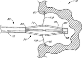
With reference to Fig. 1, illustrate a heart 10, some part is shown comprises left ventricle 12, left atrium 14, left atrial appendage (LAA) 16, pulmonary artery 18, aorta 20, right ventricle 22, right atrium 24 and right atrium appendage 26.Well-known in the art, left atrium 14 is positioned at left ventricle 12 tops, and both are separated (not shown) by Bicuspid valve.LAA16 communicates with left atrium 14 liquid usually, and when heart 10 was beated, blood flowed into and outflow LAA16 like this.
According to the present invention, a closure catheter 38 is advanced among the LAA by heart.In general, closure catheter 38 is fit to grasp the parameatal tissue of LAA, and the radial inward withdrawal is with the volume that reduces LAA and/or close LAA.Thereafter LAA is fixing on its closing direction, and closure catheter 38 is removed.Concrete aspect according to an embodiment of closure catheter of the present invention will be described below in more detail.
Can enter LAA by any approach that those skilled in the art are understood.As Fig. 2 considers, pass membranous entering and to introduce one by femoral vein or jugular vein and wear the barrier film conduit and pass the body cavity propulsion bulb and enter right atrium and can realize.The pin of hollow is by recess pressure insertion in case enter that right atrium has in advance the sharp and terminal length of radian that is shaped with one, then, the control media that a kind of ray can not significantly penetrate is injected into by needle tubing then, to be visible, and guarantee that pin correctly is placed on left atrium, rather than be put into space, aorta or other unnecessary position of pericardium.
In case pin after the position of left atrium is identified, is worn membranous conduit and is advanced to left atrium.Closure catheter 38 can advance, handle or be directed to left atrial appendage by wearing barrier film conduit 30 then.Another kind method comprises venous punching room method, for example by aorta and mitral propelling of wearing blood vessel.In addition, although the method that the puncture body cavity enters is preferential the high praise at present, device of the present invention can easily be adapted to the application of the cardiac operation of opening type.
Therefore, with reference to Fig. 2, wearing barrier film conduit 30 has a near-end 32 and an end 34.Wear the end 34 of barrier film conduit and break through the barrier film 40 of heart of patient 10, and be arranged in opening 42 near patient's LAA16.The end 36 of closure catheter 38 extends to LAA16 from the end 34 of wearing barrier film conduit 30.
At the near-end of wearing barrier film conduit 30, one stops blood to flow out from the central chamber of wearing barrier film conduit 30 with the mutually coupled union joint of haemostatic valve 48.The near-end 50 of closure catheter 38 is from extending near haemostatic valve 48.Other is relevant wears barrier film to enter the details of design and use of formula conduit well-known in the present technique field, is not discussed further at this.
With reference to Fig. 2 and 3, closure catheter 38 thereby the elongated and flexible flexible tubular body 52 that has a near-end 50, an end 36 and to extend betwixt.The axial length of closure catheter 38 is variable, depends on to want the point and the path that arrive.Concerning femoral vein was worn the barrier film method, the axial length range that closure catheter 38 generally has was greatly about 100cm to 140cm, and approximately was 117cm in a certain embodiment.
The external diameter of flexible flexible tubular body 52 also can change, and depends on the number of internal chamber and other function, and these are all understood by those skilled in that art.In one embodiment, this external diameter is approximately 12FR (0.156 inch), and the external diameter that the consideration closure catheter has is usually approximately in the scope of 0.078 inch to 0.250 inch.Diameter outside this scope also can adopt, as long as the result of the function of this diameter acceptablely just can concerning conduit is used.
For example, in a given application examples, the lower limit of the external diameter of flexible tubular body 52 will be the function of the function chamber number that holds in number of fluids or the conduit.In addition, flexible tubular body 52 must have enough propulsion capabilities so that conduit target direction towards it in heart does not have bending or unnecessary propelling agley.Also require tubular body 50 to have to transmit the ability of moment of torsion, as in an embodiment, organizing anchoring to stretch guider is not circumferentially equally distributed for the end 36 of conduit.The flexible consideration of the optimization of conduit external diameter, propelling property and moment of torsion transmission characteristic can be achieved by the well-known traditional catheter design technology of those skilled in that art.
The manufacturing of flexible flexible tubular body 52 can be according in the known various technology any.In one embodiment, flexible flexible tubular body 52 can be formed by any extruding in the material such as HDPE, PEBAX, nylon, polyimides and PEEK.Perhaps, at least a portion of flexible tubular body 52 or all comprise pin or other metal pipe or woollen yarn knitting reinforced wall on the length as the subcutaneous injection formula of a turn well known in the art, solid wall.
The near-end 50 of closure catheter 38 is furnished with a manifold 51, and this manifold 51 has a plurality of admission passages.In general, manifold 51 is furnished with an admission passage 53 and a deployment wire passage 57 as the dragline passage.Other admission passage, for example control media introducing passage 55 or other passage that provides are as required decided on the functional requirement of conduit.
Flexible tubular body 52 has one first actuator cavities 54 at least, to move axially admittance actuator 56.Actuator extends between the near-end 64 and terminal 66 of closure catheter, and wherein near-end 64 approximately is positioned at the near-end of closure catheter, and end 66 is located on or near in the far-end of closure catheter 38.The end 66 of actuator 56 is fixed on the medicated cap 68.In illustrated embodiment, actuator cavities 54 communicates with admission passage 53, extends therein to allow actuator 56 with being right after.
Depend on the structure of anchoring supporting 62 on closure catheter 38 ends 36, actuator can have various ways.Generally, conduit has the cross section that approximately is not more than 14French in order to wear body cavity propelling and location in the zone of anchoring supporting 62.Yet the anchoring supporting transmitting tissue's anchoring of must having the ability enters the wall of hole or chamber, is example with general adult's LAA situation, and the internal diameter of this chamber is greatly about the order of magnitude of 1.5cm to 3cm.Device of the present invention can easily be calibrated up and down according to purposes, for example adapts to the application of 5cm to 10cm hole on the GL road or adapts to the vascular applications of 5mm to about 2cm.For this purpose, the anchoring supporting is preferably moved between direction that reduces between the cross section and cross section amplification direction, to aim at and to touch destination organization surface (in certain embodiments).
Realize above-described one easily structure be the structure that lever arm is taked in each anchoring supporting, this structure at one end can be distinguished with catheter body with changeing and be connected.This structure allows anchoring to be bearing in run-off the straight in the whole successive range of external diameter, and this is ideal to the difference on the anatomical structure in aiming at anchoring and adapting to different into treatment sites and/or patient crowd.
An anchoring supporting movable sideways can be moved between axial and incline direction in many ways.A kind of mode easily is by use drag-line or other actuator, and along with movably short section axial distance shortening of regular length, the diameter of the working area of conduit just increases, and this mode will be disclosed in detail.In this structure, actuator is in tension during actuating.All can use such as the sub-thread of polymeric or metal or any structure multiply clue, belt and the pipe.
In illustrated embodiment, actuator 56 comprises that external diameter is about the stainless steel tube of 0.025 inch.
Drag-line also can be connected on the surface that radial outward faces, and preferably near the end of each anchoring supporting, each anchoring is bearing in its near-end and is hingedly connected to conduit.The draw drag-line makes anchoring be bearing in the end direction radial inward, and the head for target tissue.
In another structure, anchoring is bearing in run-off the straight under the actuator 56 applied pressure effects.For example, by axially movably being fixed to the end of anchoring director assembly on the conduit and on distal direction, promoting the anchoring director assembly slidably to obtain axial compressive force, the embodiment that describes in detail below just can easily be converted to the thrust actuating system, as can be more clear from following discussion.
Inquire into actuator and compare with pull-type actuator system different requirements is arranged, the ability that transmits enough compression stresses for example will be arranged and not produce excessive compressing crooked or rub.Therefore, solid wire rope or tubular structure can be first-selected, and the external diameter bigger than the minimum requirements of pull-type actuator system will be arranged.Therefore, according to the design of actuator system, the internal diameter of actuator cavities 57 can change.In illustrated embodiment, the internal diameter of actuator cavities 57 is about 0.038 inch, in order to hold the actuator 56 that external diameter is 0.025 inch slidably.
Which kind of structure is the power that acts on the radial outward in the anchoring supporting 62 can select for use depend on the operation and the structure of requirement by any the providing in the multiple expansion structure.For example, can locate an aerating ballon radially from a plurality of hinged being loaded in the anchoring supporting 62, and it is placed on the place that communicates with actuator cavities 54, this actuator cavities can be used as inflatable chamber.Can use any in the multiple balloon material, its physical characteristic scope is from the latex of high compliance, low-pressure system, to the PET of non-compliance, high pressure thereby take high radial power system, as the airballoon cover industry generally understand.
Flexible tubular body 52 can be provided with guide line chamber 57 in addition, and perhaps embodiment is such as shown, and guide line chamber 57 can coaxial extension on the whole length of tubulose actuator 56.
Flexible tubular body 52 can be provided with deployment lumen 58 in addition, is used for moving axially to admit one or more deployment elements 60, for example, is used for organizing anchoring 90 to be deployed to clue or suture on the destination organization 110 with one or more.Deployment organizes the deployment power of anchoring 90 can be designed to distal direction or proximal direction, and described that discuss and actuator 56 are that be connected, and 54 corresponding multiple considerations also can be applied to this deployment system with actuator cavities.In the illustrated embodiment, organize the deployment of anchoring 90 can be, dispose element 60 deployment wire 106 of withdrawing conversely by the proximal retraction of disposing element 60 is achieved.Therefore the promotion ability is not a problem, such as, the ordinary suture of the nylon wire of 0.008 inch diameter can be used.To this embodiment, the internal diameter of deployment lumen 58 is approximately 0.038 inch.This deployment lumen 58 both single deployment element 60 of receivability of fixed size, also a plurality of deployment elements 106 of receivability organize anchoring with unified suture such as each.
The end 36 of closure catheter 38 is provided with one or more anchoring supportings 62, is used for carrying movably one or more anchorings of organizing.Preferably, two or more anchoring supportings 62 are set, in general, are used for the device that LAA closes, about 3 to 12 anchorings supporting 62 is set.In illustrated embodiment, 6 supporting 62 longitudinal axis around closure catheter 38 circumferentially are arranged on the circumference equably.
Each anchoring supporting 62 comprises that one is used for clamping slidably at least one surface of organizing anchoring 63, and organizes anchoring by the aiming of assigning to of the control part on the near-end 50 of operation closure catheter 38.The detail of an embodiment with anchoring supporting 62 of single anchoring will be discussed below.A plurality of anchorings, for example two or three or more a plurality of, also can be entrained by each anchoring of disposing in turn supporting.
Along with the operation of near-end control section, anchoring supporting 62 is moved between axial and incline direction.The control of near-end can be taked any form, for example slide switch or lever, rotating lever or knob or like that or the like, and this all decides on the operation of requirement.For example, rotary spherical handle control head can support 62 the accurate control of gradient do to anchoring.The knob or other handle that directly endwisely slip control head, for example directly are contained on the actuator 56 can be optimized touching the sense of touch of feeding back behind the destination organization such as anchoring supporting 62.
Each illustrated anchoring supporting 62 comprises at least one nearly central segment 70, a distal ports 72 and an inflexion point 74.Referring to Fig. 4, the end 73 of each distal ports 72 is connected on catheter body or the medicated cap 68 movably.In the present embodiment, the distance between the end 73 of the near-end 71 of nearly central segment 70 and distal ports 72 has been shortened in the withdrawal of actuator 56 near-ends, forces inflexion point 74 to move from the longitudinal axis radial outward of closure catheter 38.Like this, by the control axial distance, the proximal retraction of actuator 56 will have prediction and controllably increase the nearly central segment of anchoring supporting 62 and the angle between the distal ports and conduit distance longitudinally.This just is suitable for a plurality of inwalls of organizing anchoring aiming tube assembling structure, for example blood vessel or left atrial appendage very goodly.
With reference to Fig. 4, it illustrates an enlarged detailed according to anchoring supporting 62 of the present invention.Nearly central segment 70 is preferably included in the tube wall 76 and 78 that inflexion point 74 places link to each other with distal ports 72.In one embodiment, nearly central segment 70 and distal ports 72 can be made up of the pipe of single length, by cut, photoetch method or filing nearly central segment 70 separate with distal ports 72 and stay the one or two or more individual hinges that connect together at inflexion point 74.Any polymer or the pipe of metal all can be used for this purposes, comprise rustless steel, Nitinol or other hyperelastic alloy, polyimides or the other materials field that is subjected to technical staff's favorable comment in the present technique field.
In illustrated six pipe embodiment, nearly central segment 70 and distal ports 72 are made up of one section PEEK pipe, and this ips is about 0.038 inch, and external diameter is about 0.045 inch, and total length is about 1.4 inch.In general, if be special application, used anchoring supporting surpasses 6, and then each diameter reduces than six pipe the corresponding of embodiment.When nearly central segment 70 and distal ports 72 co-axially aligns, between above-mentioned two sections, axial length is set and is about 0.030 gap.Although in certain embodiments, nearly central segment 70 and distal ports 72 can require to be uneven in length, and in the illustrated embodiment, nearly central segment 70 is approximately equal with distal ports 72 length.Carry the length of organizing anchoring 90 and anchoring to support 62 parts and consider that at first the structure in particular procedure or the human dissection selects, like this, when the deployment end of anchoring supporting 62 contacts with purpose tissue 110, anchoring supporting 62 is with an acceptable emission angle run-off the straight, in the application of LAA disclosed herein, the length from the hinge to the anchoring between the deployment end of supporting 62 can be thought of as about scope from 0.5cm to 1.5cm.
To some application, nearly central segment 70 is crossed about 10% to the youthful and the elderly than distal ports 72, and words grew about 20% preferably.For example, one is applicable to that LAA closes the device of operation, and the length of the nearly central segment 70 in 6 anchors is about 0.54 inch, and the length of distal ports 72 is about 0.40 inch.The external diameter of each anchoring supporting is about 0.045 inch.Embodiment as the aforementioned, the function and/or the size of nearly central segment and distal ports can be turned around, but still within the scope of the invention.To every kind of application, the determining to take an examination of the relative length of optimum lever arm considered multiple variable, for example the diameter of desired device, target cavity or organize diameter, the angle of departure of slot and be used for aiming and dispose desired pulling force.
The near-end of nearly central segment 70 and the end 73 of distal ports 72 are fixed to closure catheter movably, and this fixing can have multiple mode, and according to technology disclosed herein, these modes will be known by those skilled in the art.In illustrated embodiment, each anchoring supporting 62 comprises 4 sections parts, and these parts can be made of the pipe of single length, inflexion point 74, near-end inflexion point 80 and far-end inflexion point 82 in the middle of wherein being provided with.Far-end inflexion point 82 provides a pivotable connection between anchoring supporting 62 and far-end linkage section 84.Far-end linkage section 84 can be fixed on the far-end of actuator 56 by multiple technologies, and these technology are all known by technical staff in the present technique field, cooperates or other technology as soldering, bonding, machinery.In illustrated embodiment, far-end linkage section 84 is fixed on the far-end 66 of actuator 56 by agglutinating mode.
Near-end inflexion point 80 in illustrated embodiment nearly section 70 separates with the near-end linkage section 86 that links to each other with catheter body 52, in this structure, actuator 56 will make far-end linkage section 84 closely advance towards near-end linkage section 86 with respect to flexible tubular body 52 in the axial retraction of near-end, deviate from the laterally mobile inflexion point 74 of the longitudinal axis of closure catheter 38 thus.Its result, each proximal segment 70 and distal ports 72 are concentrated one's gaze on an angle, and this angle is from the axial outer incline of closure catheter 38.
In general, each inflexion point 80,82 comprises a hinge 81,83, and as shown in the figure, this hinge can be a slat elastomeric material.Hinge 81 and 83 preferably lays respectively at the inner radius of inflexion point 80,82 concerning many structural materials.To certain material, for example Nitinol or other superelastic alloy, hinge 81 and 83 can be positioned at about 90 ° or 180 ° or from other angle that makes progress in week of the tubulose anchoring guider of inflexion point internal diameter.
Organize anchoring 90 to be positioned at distal ports 72 as shown in the figure, be used for disposing at nearly center position.Perhaps, anchoring also can be loaded in the proximal segment 70, disposes as distal direction.According in this disclosed technology, the multiple application of organizing anchoring to be easy to be suitable for closure catheter 38 of the present invention, they will receive the favorable comment of technical staff in the present technique field.In illustrated embodiment, organize anchoring 90 to comprise that one has the tubular structure of body 92 and one or more agnail 94.Tubular body 92 is coaxial movably to be arranged in and to introduce on 96.Introducing 96 has a proximal part 98 and a sharp distal head 100, and it is separated by an elongated distal portions 102, is used for admitting slidably organizing anchoring 90.
The anchoring 90 of organizing in the illustrated embodiment comprises a tubular body 92, and its axial length is about 0.118 inch, and internal diameter is about 0.017 inch, and external diameter is about 0.023 inch.Two or more agnail 94 usefulness are laser-cut into a kind of pattern and are arranged on the tube wall, and each agnail 94 is bent to the radial outward deflection, as shown in the figure.Organize anchoring 90 can by any biocompatible, can not cause that the metal of repulsion is constituted, rustless steel is for example arranged, Nitinol, the metal of being known in Elgiloy or other industry.Eset is such as HDPE selectively, nylon, the anchoring of the polymer of PTFE or other polymer.For depending on the second class closing structure such as nail, the embodiment that suture or clip keep LAA or other hole to close, anchoring comprises that a biology can absorb or soluble material, like this, through after a while, it will be died away.A kind of anchoring suture 108 is fixed in the anchoring.
In one embodiment of this invention, introduce an axial length of 96 and be about 0.250 inch.Proximal part 98 external diameters are about 0.023 inch, and axial length is about 0.100 inch.Distal portions 102 external diameters are about 0.016 inch, and axial length is about 0.150 inch.External diameter mismatch between proximal part 98 and the distal portions 102 provides the face butt joint 104 of far-end, when carrying out the tissue penetration step, and this tubular body 92 that butt joint has been supported to organize anchoring 90.Deployment wire (for example suture) 106 is fixed in introduces a near-end 98 of 96.Introducing 96 can be made by multiple mode, for example extrusion molding or formed by the metalworking of stainless steel tube raw material.
With reference to Fig. 6 A-6C, there is shown according to process with respect to the direction transmitting tissue anchoring 90 target approach tissues 110 of the anchoring of closure catheter 38 axioversions supporting 62.Deployment wire 106 makes in the withdrawal of near-end and organizes anchoring 90 and introduce 96 assembly and move vertically by distal portions 72, enters tissue 110 at last.Constantly to deployment wire 106 axially towing make and introduce a longitudinal axis of 96 and move, like this, introduce 96 and align coaxially with the longitudinal axis of proximal part 70.Constantly to deployment wire 106 towing, introduce 96 and organize anchoring 90 and stay in the tissue from organizing anchoring 90 withdrawals, breaking away from.Shown in Fig. 6 C, anchoring suture 18 still is fixed on to be organized in the anchoring 90.
In use, with well-known technology in the industry closure catheter 38 is incorporated into vascular system via skin and passes body cavity and advance to heart, enter left atrial appendage at last.With reference to Fig. 7, the end 36 of closure catheter approximately is positioned at the opening part of LAA16, and by the radioscopy imaging, the ultrasonic cardiography or other imaging method are determined the position.As shown in Figure 8, actuator 56 proximal retraction radially outward tilt anchoring supporting 62 longitudinal axis with respect to closure catheter 38.Preferably, the axial length of the proximal part 70 of each anchoring supporting 62 combines with rotational angle range at the inflexion point 80 of proximal end the parameatal tissue of inflexion point 74 and LAA is contacted.In general, preferably, end portion 72 with respect to the vertical axis tilt angle of closure catheter 38 within about 45 ° to 120 ° scope.Actuator 56 till supporting 62 is fully tilted, perhaps discloses anchoring supporting 62 with till surrounding tissue 110 contacts up to tactile feedback in proximal end withdrawal.
Along with the inclination of anchoring supporting 62, deployment wire 106 promotes each in proximal retraction with this and organizes anchoring 90, enters surrounding tissue 110 as discussed like that.Ask for an interview Fig. 9.Subsequently, first axial location is got back in anchoring supporting 62, as shown in figure 10, withdraws from left atrial appendage.Near-end will make left atrial appendage overlap as shown in figure 11 by the withdrawal on anchoring suture 108 of pipe, loop or slot to contract.The anchoring suture can be fixed together with any traditional means, and for example clip is tied a knot, bonding, or other method of being understood for technical staff in the industry.Perhaps, modes such as the also available stitching of LAA, safety pin, nail or clip are closed, and keep this closure with any biocompatible binding agent.
In another embodiment, single suture 108 slidably at least with 3 anchor connections, preferably with 5 or 5 above anchor connections, like this, along with the deployment of anchoring, the withdrawal of suture 108 is closed with a kind of " on the moneybag close up rope " mode drawing tissue.Figure 31 A and 31B in people's such as U.S. Whayne the U.S. Patent number 5865791 show similar techniques, quote this invention all sidedly at this, with for referencial use.
Power according to wanting pent slot or chamber size and effect structure thereon adopts any deployment catheter disclosed herein, in the arranged around 2 of opening to about 12 or more anchoring.Preferably, about 3 to 8 anchorings, and in embodiment, adopted 6 anchorings to closing the damaged state of an illness of atrial septum in " rope closes up on the moneybag " mode.Yet atrial septum correct digit and position damaged or slot anchoring on every side change, and this will and be that the medical diagnosis that the technical staff understands in the industry is determined according to anatomy.
With reference to Figure 11 A-11C, illustrated the situation of disposing a plurality of anchorings 90 deployment catheter end 36 afterwards concisely, for the sake of simplicity, only show 2 anchorings among the figure.An anchoring suture 108 extends in loop 113, and carries each anchoring 90 slidably.One keeps structure 109 entrained by first and second parts of anchoring suture 108, like this, keep structure 109 loop 113 is made up of anchoring suture 108 distal portions and the girth of maintenance structure 109 to be dwindled, so just realized organizing the dwindling of size in slot or chamber along the distal advancement of suture 108.
Preferably, can will keep structure 109 to advance closing loop 113 to far-end along suture 108, this process like by near-end with suture 108 indentation deployment catheters and make the process that keeps structure to run into distal face 69.Distal face 69 can be on the medicated cap 68 or on other face of catheter tip 36.In the illustrated embodiment, keep structure 109 to comprise one first Prusik knot 115 and one second Prusik knot 117, they are contained on the suture 108 movably.First and second Prusik knots 115,117 are fixed together and form as reef knot 119.The knot of any other form, circle or connected mode are also all available.
The aforementioned technology of closing can or use isolating conduit to finish by closure catheter.Closure catheter can be from patient's proximal withdrawal, and endermic and access site blood vessel can be closed according to traditional puncture technology of closing.
According to another aspect of the present invention content, in view of the requirements of operation of being done, can be used to close any slot of organizing after closure catheter 38 made an amendment, the known form of technical staff is equal in this modification and following and/or the industry.This can comprise that atrial septum damaged (ASD) for example, ventricle barrier film damaged (VSD), patent ductus arteriosus, preceding hole patent and other are the operation known to the technical staff in the industry.Organizing slot to close general the combining with Figure 12-17 of technology is discussed.
With reference to Figure 12, illustrated the part figure of a tissue plane 120 concisely, this tissue can be barrier film or other heart wall.Tissue plane 120 contains the slot that will close 122.Closure catheter 38, as shown in the figure, a part of terminal 36 extends into slot 122 at least.Although this one side of the present invention, be that the anchoring of organizing that just retreat or far-end is described from the propulsive situation in the back side of tissue plane, but can the anchoring section management side is inverted to easily according to a common technology of passing through in the present technique field that discloses out here.And relevant with the method be modified in the far-end anchoring to advance among the embodiment be obviously.In general, as shown in the figure, near-end anchoring propulsion method can help to participate in the centering of slot inner catheter very goodly, and can make withdrawal along disposing identical direction with anchoring.
As previously described, closure catheter 38 is provided with a plurality of anchoring supportings 62.Be used for the damaged embodiment that closes of atrial septum one, can adopt the anchoring supporting 62 in about 3 to 12 scopes.
With reference to Figure 13, as previously mentioned, each anchoring supporting 62 comprises a proximal part 70, a distal portions 72 and a hinge or inflexion point 74 betwixt.At least an anchoring 90 is carried in each anchoring supporting 62, deploys the situation of the embodiment of direction as far-end, and anchoring 90 is in tubular distal end portion 72 the insides.As previously mentioned, anchoring 90 is connected with anchoring suture 108.In illustrated embodiment, the anchoring suture extends along the outside of anchoring supporting 62, enters the distal chamber opening in flexible tubular body 52.Anchoring suture 108 can be bonded into a unitary element on certain point, perhaps, each other anchoring suture 108 also can extend to near-end on the whole length of catheter body.
As shown in figure 13, with anchoring supporting 62 from the axial advance bearing of trend to incline direction become convenient so that anchoring 90 is deployed near the slot 122 the tissue plane 120.Preferably, hank such geometry so that a plurality of anchoring 90 roughly limits a circle by the triangle that the longitudinal axis constituted of distal ports 72, proximal segment 70 and conduit, this round diameter is greater than the diameter of slot 122.Therefore, the length of distal ports 70 is usually greater than the approximate radius of slot 122.
In general, to the damaged operation of atrial septum, when distal ports 72 tilts to its operating angle, the circle diameter that pattern is disposed in the most suitable anchoring about 0.5 centimetre to 3 cm range.Correct non-general defective, desired size will surpass the value at the two ends of above-mentioned scope.In addition, in the time of in anchoring section is deployed to tissue plane 120, then anchoring needn't be limit the pattern of circumference, the pattern of non-circle, also is fine such as polygon, ellipse, avette or other shape, and this depends on the essence of the slot 122 that will close.
Figure 13 shows that anchoring 90 parts extend into or the situation by tissue plane 120.In general, anchoring 90 also can be designed to reside in the tissue plane 120, such as the position near the slot 120 of quite thick tissue.Perhaps, organize anchoring 90 also can be designed to reside on the one side of tissue plane 120, be connected with the suture that passes tissue plane 120, shown in Figure 14 and 15.
With reference to Figure 14, illustrate closure catheter 38 and turn back to substantially axially, and organize anchoring 90 to pass through slot 122 proximal retraction after disposing a plurality of.Subsequently, the proximal retraction that anchoring suture 108 can closure catheter 38 is drawn tissue around the slot 122 over to one's side thus to close slot.Anchoring suture 108 can be fixed together by any way, such as using clamping method, knotting, binding agent, hot adhesion and the like.
In illustrated embodiment, closure catheter 38 is carried a releasable clamp 124, and these anchor clamps can extend from the end of closure catheter 38, for example inquires into by one to keep anchoring suture 108.Anchor clamps 124 are the cyclic structures with slot of a receivability anchoring suture 108.Anchor clamps are loaded on the conduit position that is in " opening ", and are partial to the position of " pass ", and anchor clamps clamp around suture 108 on the position of " pass ".Ring of forming by elastomer polymer, a kind ofly relatively say so stiff but the loop that closes such as the circle of colligation or a kind of metal alloy of shape memory all can be used for this kind purposes.Any anchor clamps, clip, binding agent or other structure all can be used to fixedly anchoring suture 108, and according to technology disclosed herein, these structures will be subjected to the favorable comment of technical staff in the industry.Subsequently, cut off anchoring suture 108 by method machinery or heat, after this, closure catheter 38 is withdrawn from into treatment sites again.
Perhaps, the anchor clamps of elastic ring or other form also can be used to directly clamp tissue and close slot.In this operation, closure catheter is used for connecting slot a plurality of anchorings on every side.By near-end towing on one or more sutures, all anchorings are just drawn over to one's side radially mutually.Further tissue plane can be pulled out in the slot edge to one or more suture near-end towings.Then, the slot that part is turned up can be fixed, and can use its anchor clamps on every side that it is closed.Here said " anchor clamps " comprise all elastic rings, tie up circle, metal holder and disclosed herein other are implemented anchor clamps.
According to another aspect content of the present invention, closure catheter 38 is provided with an extensible patch 126, shown in Figure 16 and 17.Patch 126 can comprise any material, as PTFE, terylene or other material of deciding on purposes.Suitable fabric is well-known in technical field of medical instruments, and for example those are used to cover material or other prosthetic articles for use of transplant in the blood vessel.
Patch 126 is preferably carried by the end segment 72 of anchoring supporting 62.In the illustrated embodiment, organize anchoring 90 to be loaded in the proximal segment 70 of anchoring supporting 62.Like this, when anchoring supporting 62 tilted to the anchoring expansion direction, patch 126 launched automatically and is positioned on the slot 122.As shown in figure 17, organize anchoring 90 to be advanced in the tissue plane 120 by patch 126, with patch 126 hobnail on opening 122.Perhaps, organize the pattern that anchoring also can be such to dispose: to organize anchoring to center on but do not penetrate this patch tissue.In this embodiment, organize anchoring preferably to be connected on the patch tissue with suture.Organize anchoring both can be connected to that also available suture interconnects on the patch, and pass patch target approach tissue.
Organize anchoring 90 by towing deployment wire 106 is deployable.Perhaps, by axially movable be positioned at one in the proximal segment 70 inquire into will have or not have an anchoring suture 108 organize anchoring 90 from proximal segment 70 expansion.As has discussed the front, organize anchoring 90 to be loaded in to introduce on 96.
Patch 126 can remain on the distal ports 72 by any way, for example passes through to use low-intensity binding agent composition, or anchoring 90 is pierced through the material of patch 126 in the conduit set process of assembling.
With reference to Figure 18-20, according to the invention discloses another anchoring.Anchoring 90 can be used to anchoring one suture in solid tissue, perhaps a transplant or patch is fixed on the tissue plane, as shown in figure 20.
With reference to Figure 18, anchoring 90 comprises the central chamber 134 that a near-end 130, a far-end 132 and extend betwixt.As shown in figure 19, central chamber 134 is positioned at anchoring 90 to introduce on 96, and this formerly discussed.
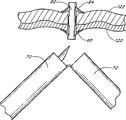
Anchoring 90 is provided with outstanding 136 and 1 second near-end outstanding 138 of at least one first near-end.First and second near-ends outstanding 136 and 138 are designed to the radial outward expansion with anchoring 90 axial compression.Therefore, under incompressible profile situation as shown in figure 19, the longitudinal axis that first and second near-ends outstanding 136 and 138 are parallel to anchoring 90 usually extends.Along with the axial shortening of anchoring 90, distal face is forced to radial outward to the contact surface 144 of tissue, and according to shown in Figure 180, this is obviously for the technical staff in the industry.Although illustrated is two separated from each other, the near-ends that are in about 180 ° of positions outstanding, it is outstanding that 3 or 4 even more a plurality of near-end also can be set, preferably uniform distribution around the anchoring 90.
At least the first far-end is outstanding 140, preferably, together with second far-end outstanding 142 is arranged on the tubular body 92 away from the outstanding position of near-end.First and second far-ends outstanding 140 and 142 are along with the anchoring axial compression, and similarly radial outward is expanded or increased.Axially-spaced between outstanding 136 and first far-end of first near-end outstanding 140 is by making anchoring one patch 126 or transplant or other structure can be fixed on the tissue plane 120 at distal face between the contact surface of organizing 146 to the contact surface 144 of tissue and proximal end face patch 126 and tissue plane 120 are flat.Utilize any deployment catheter disclosed herein, anchoring 90 can be deployed on the tissue plane from introducing 96 top.
Can realize near-end and the outstanding radial dilatation of far-end by anchoring 90 along the axial shortening of its longitudinal axis.As shown in figure 19, thereby this can introduce on 96 the proximal part 98 anti-prevention near-end 130 and realize to moving of near-end by near-end 130 is seated in, then, such as by being the near-end towing on the proximal force conveyer 148 of a suture 150 and far-end 132 is advanced to near-end.Suture 150 extends in an annular distance by a plurality of holes 152, and this to extend through near-end and far-end outstanding.Perhaps, suture 150 also can extend against the next door of anchoring 90 or extend by central chamber 134, and this decides on a central chamber 134 and the tolerance introduced between 96.Also can adopt other proximal force conveyor structures that the technical staff is known in the industry.
Anchoring 90 can be made with multiple mode, as being formed by cutting of a metal or polymer tubing or etching.Preferably, anchoring 90 is carried out cut by Nitinol or steel pipe, and the external diameter of pipe is about 0.014 " to 0.038 " scope within, axial length is about 0.050 " to 0.250 " within the scope.Each distal face to the contact surface 144 of tissue and proximal end face to the axial length of contact surface 146 of tissue about 0.010 " to 0.060 " within the scope.The wall thickness of pipe is about 0.002 " to 0.012 " within the scope.Axial full compression among most of metal tube embodiment will make metal surpass elastic limit in the bending at each outstanding ridge place, top, suture 150 can be removed from anchoring 190 after disposing like this, and anchoring will still keep the profile of its deployment (axial compression) as shown in figure 20.
Although illustrated mainly is that patch or other thin film are connected to embodiment on the tissue plane, anchoring 90 as shown in figure 18 also can come anchoring one suture to solid tissue as discussed previously.For this purpose, anchoring can be reduced to has only first and second near-ends to give prominence to 136 and 138, or other outstanding the same being in the same plane of outstanding and first and second near-ends.Yet first and second far-ends are given prominence to or the outstanding of other can be added together, and this depends on from in-house implantation position takes anchoring 90 needed pulling force away.
Can enter into the heart defect place by conduit by multiple passage.Can enter into ASD or VSD from the tremulous pulse loop.Conduit is incorporated in the arterial vascular system and with it is directed to the chest aorta and/or the abdominal aortic of decline.Flow out pipeline by aorta, then conduit is advanced to (LV) in the left ventricle.In case entered LV, patch just can be deployed to VSD.Perhaps, in case entered LV, patch can upwards guide by Bicuspid valve and enter into left atrium (LA).When patch is positioned at LA, it can be incorporated in the ASD and be installed.
Perhaps, can enter ASD or VSD by the vein loop.The conduit that has patch on it is incorporated in the Venous system, is advanced forward into down venous lumen (IVC) or upper vein chamber (SVC), and introduce right atrium (RA).Then patch is imported (ASD).Perhaps,, advance patch, enter right ventricle (RV), and also installed in the importing VSD by Tricuspid valve in case entered RA.
Anchoring deployment catheter of the present invention can easily be used for finishing any anastomosis operation, be included in the blood vessel artificial blood vessel transplant is connected to connecting portion, carry out the anastomosis of the tissues, tissue of autogenous vein graft, for example saphena is transplanted to coronary artery.The intermediate that anastomosis conduit embodiment also can be used to provide being positioned at the artificial transplant on the endovascular into treatment sites already supports.
With reference to Figure 21, illustrate lateral cross section just like the blood vessel 122 of the such defective 124 of aneurysm.A transplant 120 is on defective 124, at least in the near-end of aneurysm 124 and the blood vessel wall of far-end imbrication part health as shown in the figure.The identical conduit 126 of diagram is positioned at the near-end of transplant 120.The conduit 126 that coincide is provided with a plurality of anchoring supportings 62 in the place near far-end 36.Each anchoring supporting comprises a proximal segment 70, a distal ports 72 and a pin joint 74.
With reference to Figure 22, transplant 120 and blood vessel 122 are introduced into a sharp tip 100 of 96 and penetrate, and being deployed in before of this introducing 96 discussed.Introduce 96 and carried an anchoring 92.In illustrated embodiment, the deployment wire that near-end towing one had formerly been spoken of is incorporated into into treatment sites so that introduce 96 with anchoring 92.Constantly the towing deployment wire makes and introduces 96 and be withdrawn in the proximal segment 70 of anchoring supporting 62, and anchoring 92 is stayed on the position.
As shown in figure 23, anchoring 92 is provided with and is used for stoping one or more far-end agnails 94 that anchoring 92 moves to near-end and is used for stoping the one or more near-end agnails 95 of anchoring 92 to distal migration.Like this, anchoring 92 will keep the location so that transplant 120 is fixed on the blood vessel 122.
The anastomosis conduit is applicable to the implantation and the method for attachment of multiple transplant.For example, by using the tubular graft body that is connected in the method for transplant near-end and a certain amount of self-expanding of far-end or balloon expansion, can require intermediate supports to keep the openness of the central bore between the axial two ends.By one section extra transplant of arrangement in the tubular graft body, or utilize anastomosis conduit 126 that transplant is anchored on the natural blood vessel wall, can realize intermediate supports by these two kinds of methods.Each anchoring supporting can have two or more anchorings.Like this, anastomosis conduit 126 can be positioned on primary importance, and on this position, first group of a plurality of anchoring is deployed on the natural blood vessel by transplant, and then navigates to the second position, on this position, disposes second group of a plurality of anchoring and keeps or fix this transplant.Other anchoring supporting and/or anchoring can be arranged on the anastomosis conduit 126, and depending on will be along the localized anchoring number of transplant axial length.
Perhaps, anastomosis conduit 126 also can be used to the implantable tubular transplant.In this embodiment, the tubular graft body is arranged in the outside of anastomosis conduit 126 coaxially.Catheter positioning is in into treatment sites, and the anchoring supporting favours axially, thus blood vessel graft is navigated on the blood vessel wall.The deployment of anchoring such as precedingly state.Anchoring can directly be fixed on the transplant, for example by using a rope or other syndeton, or irrelevant and fixing on the spot with transplant, this fixedly be by near-end be known other structural configuration of technical staff in the industry with agnail far-end or according to technology disclosed herein.Therefore, although here normally with reference to anastomosis conduit 126, embodiments of the invention also can be considered to pass through body cavity transplant implantation catheter or transplant connecting duct, and these all will be known by technical staff in the industry.
With reference to Figure 24 A-24G, show and can be used for that tissue of the present invention is closed or connecting device multiple organized anchoring, each figure among Figure 24 A and the 24B discloses an anchoring, and it has a body 92, terminal 101 and one or more in order to stop the Cao that moves of anchoring to near-end.Hole 107 is used for receiving the anchoring suture.The embodiment of Figure 24 A and 24B processing and manufacturing is easily for example formed by flat plate stock punching press or cutting.
Anchoring shown in Figure 24 C comprises that one has the clue of body 92 and terminal 101.This rope preferably includes superelastic alloy, as the alloy of Nitinol or other NiTi base.Be anchored at and be loaded in tubulose on the straight line direction and introduce in the head, be used for being directed to anchoring and want in the resident tissue.When body 92 from carrying pipe when far-end advances, be anchored at and continue end appearance that it goes in ring in the tissue, to stop proximal retraction Suo Benti 92.
Figure 24 D illustrates one tubulose anchoring, and it rolls and welded or fixed by other method around mandrel by the section of a pipe processing and manufacturing or by one flat plate.Anchoring comprises an end 101, one or more agnail 94 and is used for fixing the hole pattern 107 of anchoring suture.The anchoring of Figure 24 D can be loaded by the inside of tubulose anchoring supporting, also can dispose from tubulose anchoring supporting inside.Perhaps, the anchoring of Figure 24 D also can be positioned on central tubular or the solid anchoring supporting-line coaxially.
Figure 24 E shows a kind of anchoring, and it can be by forming from tubing cutting, or cuts a plate shown in Figure 24 F and roll and welded or alternate manner is fixedly made a tubular body around an axle.In the present embodiment, single terminal 101 of the anchoring finished in can making as Figure 24 E of three ends 101 on the former material of flat board.One or more agnails 94 can form by U-shaped or the V-arrangement profile shown in striking out onboard as shown.Anchoring among Figure 24 E also is provided with one or more prevention anchorings extraly and is passing implantation place between thin film to the agnail 95 of distal migration when anchoring, this anchoring may be ideal, or in requiring minimized other the application of the desired migration of near-end and far-end, above-mentioned anchoring also is very good.
Although described the present invention by means of given preferred embodiment, according to technology disclosed herein, other embodiment also can be known by the technical staff in the industry.Therefore, scope of the present invention has more than and is limited among the specific disclosed embodiment, and on the contrary, it is limited by appended claim.