CN1208602A - Method and device to improve aqueous humor drainage in eye - Google Patents
Method and device to improve aqueous humor drainage in eye Download PDFInfo
- Publication number
- CN1208602A CN1208602A CN98117177A CN98117177A CN1208602A CN 1208602 A CN1208602 A CN 1208602A CN 98117177 A CN98117177 A CN 98117177A CN 98117177 A CN98117177 A CN 98117177A CN 1208602 A CN1208602 A CN 1208602A
- Authority
- CN
- China
- Prior art keywords
- canal
- schlemm
- support member
- dilate
- shape
- Prior art date
- Legal status (The legal status is an assumption and is not a legal conclusion. Google has not performed a legal analysis and makes no representation as to the accuracy of the status listed.)
- Pending
Links
Images


Classifications
-
- A—HUMAN NECESSITIES
- A61—MEDICAL OR VETERINARY SCIENCE; HYGIENE
- A61F—FILTERS IMPLANTABLE INTO BLOOD VESSELS; PROSTHESES; DEVICES PROVIDING PATENCY TO, OR PREVENTING COLLAPSING OF, TUBULAR STRUCTURES OF THE BODY, e.g. STENTS; ORTHOPAEDIC, NURSING OR CONTRACEPTIVE DEVICES; FOMENTATION; TREATMENT OR PROTECTION OF EYES OR EARS; BANDAGES, DRESSINGS OR ABSORBENT PADS; FIRST-AID KITS
- A61F2/00—Filters implantable into blood vessels; Prostheses, i.e. artificial substitutes or replacements for parts of the body; Appliances for connecting them with the body; Devices providing patency to, or preventing collapsing of, tubular structures of the body, e.g. stents
- A61F2/82—Devices providing patency to, or preventing collapsing of, tubular structures of the body, e.g. stents
- A61F2/94—Stents retaining their form, i.e. not being deformable, after placement in the predetermined place
-
- A—HUMAN NECESSITIES
- A61—MEDICAL OR VETERINARY SCIENCE; HYGIENE
- A61F—FILTERS IMPLANTABLE INTO BLOOD VESSELS; PROSTHESES; DEVICES PROVIDING PATENCY TO, OR PREVENTING COLLAPSING OF, TUBULAR STRUCTURES OF THE BODY, e.g. STENTS; ORTHOPAEDIC, NURSING OR CONTRACEPTIVE DEVICES; FOMENTATION; TREATMENT OR PROTECTION OF EYES OR EARS; BANDAGES, DRESSINGS OR ABSORBENT PADS; FIRST-AID KITS
- A61F2/00—Filters implantable into blood vessels; Prostheses, i.e. artificial substitutes or replacements for parts of the body; Appliances for connecting them with the body; Devices providing patency to, or preventing collapsing of, tubular structures of the body, e.g. stents
- A61F2/82—Devices providing patency to, or preventing collapsing of, tubular structures of the body, e.g. stents
- A61F2/86—Stents in a form characterised by the wire-like elements; Stents in the form characterised by a net-like or mesh-like structure
- A61F2/88—Stents in a form characterised by the wire-like elements; Stents in the form characterised by a net-like or mesh-like structure the wire-like elements formed as helical or spiral coils
-
- A—HUMAN NECESSITIES
- A61—MEDICAL OR VETERINARY SCIENCE; HYGIENE
- A61F—FILTERS IMPLANTABLE INTO BLOOD VESSELS; PROSTHESES; DEVICES PROVIDING PATENCY TO, OR PREVENTING COLLAPSING OF, TUBULAR STRUCTURES OF THE BODY, e.g. STENTS; ORTHOPAEDIC, NURSING OR CONTRACEPTIVE DEVICES; FOMENTATION; TREATMENT OR PROTECTION OF EYES OR EARS; BANDAGES, DRESSINGS OR ABSORBENT PADS; FIRST-AID KITS
- A61F9/00—Methods or devices for treatment of the eyes; Devices for putting-in contact lenses; Devices to correct squinting; Apparatus to guide the blind; Protective devices for the eyes, carried on the body or in the hand
- A61F9/007—Methods or devices for eye surgery
- A61F9/00781—Apparatus for modifying intraocular pressure, e.g. for glaucoma treatment
-
- A—HUMAN NECESSITIES
- A61—MEDICAL OR VETERINARY SCIENCE; HYGIENE
- A61F—FILTERS IMPLANTABLE INTO BLOOD VESSELS; PROSTHESES; DEVICES PROVIDING PATENCY TO, OR PREVENTING COLLAPSING OF, TUBULAR STRUCTURES OF THE BODY, e.g. STENTS; ORTHOPAEDIC, NURSING OR CONTRACEPTIVE DEVICES; FOMENTATION; TREATMENT OR PROTECTION OF EYES OR EARS; BANDAGES, DRESSINGS OR ABSORBENT PADS; FIRST-AID KITS
- A61F2/00—Filters implantable into blood vessels; Prostheses, i.e. artificial substitutes or replacements for parts of the body; Appliances for connecting them with the body; Devices providing patency to, or preventing collapsing of, tubular structures of the body, e.g. stents
- A61F2/82—Devices providing patency to, or preventing collapsing of, tubular structures of the body, e.g. stents
- A61F2/86—Stents in a form characterised by the wire-like elements; Stents in the form characterised by a net-like or mesh-like structure
- A61F2/90—Stents in a form characterised by the wire-like elements; Stents in the form characterised by a net-like or mesh-like structure characterised by a net-like or mesh-like structure
Landscapes
- Health & Medical Sciences (AREA)
- Engineering & Computer Science (AREA)
- Biomedical Technology (AREA)
- Veterinary Medicine (AREA)
- General Health & Medical Sciences (AREA)
- Public Health (AREA)
- Heart & Thoracic Surgery (AREA)
- Vascular Medicine (AREA)
- Life Sciences & Earth Sciences (AREA)
- Animal Behavior & Ethology (AREA)
- Ophthalmology & Optometry (AREA)
- Oral & Maxillofacial Surgery (AREA)
- Transplantation (AREA)
- Cardiology (AREA)
- Nuclear Medicine, Radiotherapy & Molecular Imaging (AREA)
- Surgery (AREA)
- Prostheses (AREA)
Abstract
A method and an apparatus for improving aqueous humor drainage in an eye. A medium injected in the form of a hydrophilic liquid or a biocompatible gaseous medium or a mixture of the hydrophilic liquid and the gaseous medium into Schlemm's canal, which is microsurgically exposed at one or more locations, locally expands Schlemm's canal by the increased pressure. With a support element subsequently implanted in the lumen of Schlemm's canal, the inner walls of the canal are supported and permanently held in an expanded position, whereby unimpeded drainage of the aqueous humor from Schlemm's canal through the subsequent outflow pathways is ensured.
Description
The present invention relates to a kind of method and apparatus that improves the current drainage of ophthalmic aqueous humor, by this method and the device, by the current drainage of the excretory ophthalmic aqueous humor of corpus ciliare at iris-cornea angular domain, enter schlemm's canal by trabecular meshwork, by the schlemm's canal naturally outer circulation flow path of current drainage by subsequently again.
In order to treat the pathological changes in schlemm's canal (Canal of Schlemm) the preceding trabecular meshwork (Trabecular meshwork), this pathological changes can be fully or the current drainage of only partly blocking the ophthalmic aqueous humor, U.S. Pat-A5,360,399 and US-A 5,486,165 disclose the device of a kind of method and this method of enforcement, according to this method and apparatus, a kind of water-soluble aqueous medium of high viscosity that is, be the aqueous solution of base preferably, be injected into schlemm's canal by a kind of injection device, like this with hyaluronic acid (hyaluronic acid), trabecular meshwork just dilates under fluid power, its mesh opens, so the opening of Xing Chenging just covers and gone up the high viscosity material that prevents immediately again closure function like this.Although above-mentioned patent disclosure a kind of suitable media injections is gone into schlemm's canal is a kind of its open measure that makes, this disclosed method still has its uncertainty, because the result of various pathological changes finally also can make schlemm's canal closed once more.Therefore the ophthalmic aqueous humor, even gets clogged because the result of the distortion of schlemm's canal is subjected to great restriction fully by the current drainage of schlemm's canal and the outer circulation flow path by subsequently.
The purpose of this invention is to provide a kind of method and apparatus and realize a kind of improvedly, regulate the ophthalmic aqueous humor circulation of pressure, and keep this aqueous humor for a long time from the ophthalmic current drainage.
Say with regard to method, its characteristics are schlemm's canal, wherein dew is cut by microsurgery in a place or many places, increasing fluid pressure in the phase I by the part dilates, in second stage, for example be supported, thereby remain on muchly on the position that dilates by in the tube chamber that dilates of schlemm's canal, implanting suitable object.
Just device is said, its characteristics be to be provided with an axial orientation, dilate the support member that lumen area is supporting the schlemm's canal inwall in the part, this support member should settle make the ophthalmic aqueous humor can be muchly from the schlemm's canal current drainage and the eye of flowing through subsequently naturally outside circulation flow path.
Existing embodiments of the present invention are described with reference to the accompanying drawings.These accompanying drawings are:
Fig. 1 is preocular schematic sectional view when being in the vertical plane position;
Fig. 2 schematically shows the part of eye, and sclera is separated and cut open and up turnover in this part, and schlemm's canal is partly exposed;
Fig. 3 is the part enlarged diagram of eye shown in Figure 2, shows the situation that dilates this pipe in the exposed schlemm's canal of injection probe insertion portion;
Fig. 4 shows the situation that is partly with a support member of eye shown in Figure 3, in the schlemm's canal of this support member with column prosthese form implant part;
Fig. 5 is the amplification stereogram of the support member of first embodiment shown in Figure 4;
Fig. 6 has an X-rayed ground and part shows support member shown in Figure 5 with analysing and observe;
Fig. 7 shows the situation of support member in the schlemm's canal that is partly with an implant part of eye shown in Figure 3, is second embodiment;
Fig. 8 has an X-rayed ground and part shows the second embodiment support member shown in Figure 7 with analysing and observe;
Fig. 9 shows the 3rd embodiment support member of waiting to implant in the schlemm's canal;
Figure 10 is the side view cutaway drawing along the X-X line of support member shown in Figure 9;
Figure 11 shows the 4th embodiment support member of waiting to implant in the schlemm's canal;
Figure 12 shows another embodiment support member of waiting to implant in the schlemm's canal.
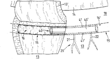
Fig. 1 is preocular 10 amplification vertical sectional view.Can see significantly that preocular 10 have cornea 11, have sphincter 12 ' and 12 " iris 12; sclera 13; eyeball 14 and pupil 14 '; ribbon-like fibre 19 and the schlemm's canal 15 (sinus venous sclerae) and the trabecular meshwork 18 (corneoscleral meshwork, Trabeculum corneosclerale) that are in party upstream.
In the eyes of a health, the ophthalmic aqueous humor along arrow 1,1 ' and 2,2 ' direction constantly upgrade from back room H to anterior chamber V and replenish and circulation, the aqueous humor current drainage is carried out at iridocorneal angle shape zone V ', enter schlemm's canal 15 along the direction of arrow 3 through trabecular meshwork 18, again from this pipe flow through subsequently naturally outside circulation flow path 20,20 ' (Fig. 2, Fig. 4, collection channel 21 Fig. 7) ', 22 ' (Fig. 4) or 21,22 (Fig. 7) and enter (not shown) in the Venous system.The resistance of stream discharging system is being regulated the flow of aqueous humor, so that intraocular pressure is remained in the particular range that part tissue of eye can bear.
According to pathological condition, resistance may increase, and one of factor is just in schlemm's canal.Schlemm's canal 15 may closures so far, to such an extent as to make that the current drainage of aqueous humor is hindered, or get clogged fully.The result that resistance increases is that the increased pressure of ophthalmic makes the function of blood supply and thing followed optical neuron be restricted.This disease generally is referred to as " glaucoma ", often causes simple eye even blindness.
Fig. 2 schematically expresses eye 10, show eyeball 14 and pupil 14 ', the sclera 13 of part, the natural collection channel of the schlemm's canal 15 of part and the part that is connected with this pipe system 20,20 ' (ophthalmic aqueous humor channel system).For the intervention of performing the operation, in the phase I, as shown in Figure 2, sclera 13 cut by microsurgery and form one deck lobe 13 ', this layer lobe be turnover upwards, so that partly expose schlemm's canal 15.In the process of operation, the layer lobe portion 13 of sclera ' pass through, for example clamp and so on remain on this position, here clamp and so on are not explained and illustrate.
In second stage, shown in Fig. 3 signal, probe 33 is introduced in the tube chamber 16 of schlemm's canal 15, probe on the configuration in a tubular form and be configured on the connector 32 that shows.Connector 32 is connected with the injection device 30 that schematically illustrates by a carrier pipe (not shown).With a kind of water wetted material, for example water seeking liquid 29, be injected in the schlemm's canal 15, injection be with injection device 30 make one or more outflow openings 33 that injection is provided with by its far-end along direction shown in the arrow ' tubular probe 33 carry out.Owing to used the water seeking liquid 29 of injection, the part 15 of closure ' just quilt has been dilated under the fluid power effect in fact in the schlemm's canal 15 that schematically illustrates among Fig. 3.
As a kind of to said method improvement or replenish, with the part 15 of the schlemm's canal 15 for the treatment of part 15 ' relative " on can adopt similar method to treat (not shown detail), can adopt a kind of preferred design to become probe mirror image and that be introduced in the schlemm's canal 15 to dilate.Fig. 3 also show the trabecular meshwork 18 (girder) of schlemm's canal 15 party upstream and the trabecular reticulum 18 that schematically illustrates '.
When above-mentioned schlemm's canal 15 dilates, the water wetted material of injection on formed opening (not shown) just covers simultaneously in its wall, for example water seeking liquid 29, water seeking liquid just sticks on the wall of opening, form a thin film, this prevents local contact between these edge of opening, because can hinder the discharging of ophthalmic aqueous humor.
Should be noted that a fact here, except that water wetted material or liquid, can adopt suitable, with the gas medium of health compatibility, or the mixture of water seeking liquid and gas medium dilates schlemm's canal.
As shown in Figure 4, in schlemm's canal 15, used one support inwall 16 ' implant, this implant dilates with fluid power or strength and is connected, make permanent circulation of ophthalmic aqueous humor and circulation reach desired level.As first embodiment of implant, be provided with a support member 35 that has a long tube portion 36, it is together with its far-end 35 " inserts in the schlemm's canal 15.On the other hand, at near-end 35 ' locate, the first embodiment support member 35 is provided with a flange part 37, this flange part closely with the inner face 13 of scleral incision ' cooperate, thereby prevent that the support member of implanting in the schlemm's canal 15 from moving.Long tube portion 36 also be provided with some through holes 38,38 ', dispose with peripheral intervals vertically.As shown in Figure 4, the part 15 of schlemm's canal 15 is preferably settled and implanted to support member 35 in this wise " in, make to have a through hole 38,38 at least ' with outer naturally circulation flow path 20 ' in little collection channel 21 ', 22 ' connection.
The long tube portion 36 of support member 35 shows with the axonometric chart form in Fig. 5, shows with perspective view and phantom in Fig. 6, also show simultaneously with the through hole 38,38 of inwall 36 ' be connected ' be the flange 37 of anchor ring shape substantially.In an illustrated embodiment, annular lip 37 is by the transition part 37 of for example anchor ring shape ' be placed in the long tube portion 36, forms one open tubaeform (bell) by a for example suitable mandrel shape instrument (not shown) thereon.(not shown) has improved the configuration that support member 35 inserts in the schlemm's canals 15 in another embodiment, from flange 37 beginnings or from transition part 37 ' begin to far-end 35 " is designed to axial tilted conical with long tube portion 36.
Figure 7 shows that schlemm's canal 15 have another part 15 of support member 40 ', this support member is designed to second kind of embodiment and in the implant.Support member 40 preferably settle in this wise and implant in, make to have a through hole 41,41 at least ', shown in Fig. 7 signal, with collection channel 21,22 (passage aisle) connection in the outer naturally circulation flow path 20.The ophthalmic aqueous humor of infiltration by trabecular meshwork 18 be by schlemm's canal 15 current drainages, or the inside 40 by support member 40 ' and by through hole 41 ' and the outer circulation flow path 20 of nature in collection channel 21,22 and current drainage.
Fig. 8 has an X-rayed ground, and part shows the tubular supporting piece 40 as second embodiment with analysing and observe.Support member 40 be provided with a plurality of outer openings 41,41 ', they are configuration at regular intervals vertically, and along peripheral random distribution or radially establish relative to each other, and with inner 40 ' be connected.
Fig. 9 and Figure 10 shows that the 3rd embodiment support member 45, this support member be provided with two ends 47,47 ', each end be provided with an opening 45 ', 45 "; the end is designed to axially be separated by and the anchor ring established, is settling two between the end, but best three connecting plates 46; 46 ' and 46 ", they are along periphery setting separated by a certain interval, and with end 47,47 ' interconnection.In the embodiment of this variation, connecting plate 46,46 ' and 46 " between formed recessed empty 48,48 ' and 48 " opening effect outside under each situation, plaing, so as to make the ophthalmic aqueous humor pass through substantially opening 45 ', 45 " current drainages.
Figure 11 shows that the 4th embodiment support member 50, this support member constitutes, is substantially the net of bung flange shape by line 51, and these lines are interconnection and have a rigidity preferably.Net can be prepared by harder plastics or metal wire, also can be prepared by biomaterial.Each line 51 (silk) also can relatively be wound into a bung flange shape interconnectionly in the net.In the embodiment of this variation, the gap 52,52 between each line ' and 52 " play ophthalmic ah outflow through hole respectively.Support member can be designed to like this, and it can compress and implant, and can dilate automatically in the tube chamber 16 at schlemm's canal 15 after the implantation.
The interconnected metal wire of support member 50 or silk 51 (seeing Figure 11) are preferably prepared by Nitinol.These silks 51 have the shape-memory properties of being referred to as, and consequently, design support member 50 energy plastic deformations into the net, can automatically recover its original shape and give suitable heating.This support member with hot shape-memory properties has its superiority, it can, for example plastic deformation ground inserts in the schlemm's canal that exposes, be in normal body temperature, then with less external diameter, just owing to normal body temperature, it returns to its original configuration or shape again.
Figure 12 shows that another embodiment support member 55, this support member can be wound into a bung flange shape by single line 56, and line can perhaps be prepared by noble metal, for example filamentary silver, spun gold or platinum wire by harder plastic cord or metal wire 56 preparations.In the embodiment of this variation, gap 57,57 ' play a part the respectively ophthalmic ah outflow through hole between each circle.
By suitable health compatible material support member 35,40,50 or 55 preparation, that be designed to piped or bung flange shape, because its inherent pliability can be adapted to the natural shape of schlemm's canal 15 especially.But the post supports of hollow 35,40,50 or 55 can cover a kind of suitable material, utilize this cladding material not only can produce desirable biological respinse or but also can reduce even prevent undesirable biological respinse fully.
In an embodiment who does not show, support member 35,40,50 or 55 can be designed to slightly be arc in the vertical.In the embodiment that another is not showed, support member 35,40,50 or 55 can be designed to pass through in the vertical is tilted conical.
For size that each support member 35,40,50 or 55 are described and the operation easier of considering in the tube chamber 16 of schlemm's canal 15, to carry out implantation process, to indicate here, support member for example its length is the L=2.0 millimeter, its external diameter is the D=0.2 millimeter.According to Fig. 5, Fig. 6 and Fig. 8 in the axial direction with periphery on be provided with at interval outflow opening 38,38 ' or 41,41 ', its opening inner diameter d=0.18 millimeter.But support member 35,40,50 or 55 are not restricted to the above-mentioned size that proposes as an example.
In order to introduce the embodiment of each support member:
Embodiment changes 1: after dilating, from schlemm's canal 15, take out the injection device 30 that has probe 33, use medical tweezers or medical pliers or other surgical device manually with support member 35 then, 40,50 or 55 insert in the tube chamber 16 of schlemm's canal 15, and make its location (see Fig. 4, Fig. 7);
Embodiment changes 2: support member 35,40,50 or 55 are placed on probe 33 far-ends of injection device 30 by a dismountable type connector, dilate schlemm's canal 15 after, support member is separated by a unshowned measure and is implanted;
Embodiment changes 3: probe 33 remote design of injection device 30 become a detachable support member 35,40,50 or 55;
Embodiment changes 4: make the post supports 35 that is hollow substantially, 40,50 or 55 press against on the far-end of probe 33 in this wise, after dilating schlemm's canal 15, with a suitable utensil with support member 35,40,50 or 55 along in the tube chamber 16 that is advanced into schlemm's canal 15 with respect to the axial direction of probe 33 and have good positioning.
Can also propose other good embodiment within the scope of the invention and change support member 35,40,50 or 55 implant in the tube chamber 16 of schlemm's canal 15.
The present invention is not restricted to each above-mentioned support member 35,40,50 or 55 embodiment.Can also propose the design of other useful support member 35,40,50 or 55 and not exceed basic ideas of the present invention.Support member above-mentioned detailed description and that show often is referred to as interior prosthese (Endoprostheses).As shown in Figure 3, utilize fluid power to dilate, the schlemm's canal 15 of basic closure is opened, implant the support member 35,40,50 or 55 of correct design then, particularly have the support member of pliability design, it is superior especially that this combination is considered to.
The tube chamber 16 of schlemm's canal 15 is owing to there is support member 35 or 40 to keep muchly opening, and support member 35 or 40 should be located the feasible outer opening 38 that has at least, 38 ' or 41,41 ', as Fig. 4 and shown in Figure 7, with outer naturally circulation flow path 20 or 20 subsequently ' in collection channel 21 ', 22 ' or 21,22 connect.The ophthalmic aqueous humor that penetrates into trabecular meshwork 18 is by schlemm's canal 15, or by support member 35 or 40 tube chamber 36 ' or 40 ' and by opening 38 ' or 41 ' and subsequently outer naturally circulation flow path 20 or 20 ' in collection channel 21 ', 22 ' or 21,22 current drainages.
Need indicatedly to be, in the tube chamber 16 of schlemm's canal 15, implant the support member 35,40,50 or 55 of an axial orientation at least, with the inwall 16 of supportive ground contact schlemm's canal 15 '.If desired, two or more support members can also be implanted in the schlemm's canal 15 distortion or that block.If each support member of implanting can guarantee schlemm's canal 15 and at least one outer naturally subsequently circulation flow path 20 or 20 ' in collection channel 21 ', 22 ' or 21,22 connections on, so this situation is very useful.
Claims (32)
1. method of improving the current drainage of ophthalmic aqueous humor, by this method, by the excretory aqueous humor current drainage of corpus ciliare at iris-cornea angular domain, enter schlemm's canal (15) by trabecular meshwork (18), by the schlemm's canal naturally outer circulation flow path of current drainage by subsequently again, it is characterized in that schlemm's canal (15), wherein a place or many places are cut by microsurgery and are exposed at the phase I and increase fluid pressure by the part and dilate, in second stage, for example be supported, thereby remain on muchly on the position that dilates by in the tube chamber that dilates (16) of schlemm's canal, implanting suitable object.
2. method according to claim 1 is characterized in that the schlemm's canal (15) that exposes is dilated by the water seeking liquid in a kind of tube chamber (16) that is injected into schlemm's canal.
3. method according to claim 1 is characterized in that the schlemm's canal (15) that exposes by the gas medium in a kind of tube chamber (16) that is injected into schlemm's canal, and the health compatible medium dilates.
4. according to claim 2 and 3 described methods, it is characterized in that the schlemm's canal (15) that exposes is dilated by the water seeking liquid in a kind of tube chamber (16) that is injected into schlemm's canal and the mixture of gas medium.
5. method according to claim 1 is characterized in that having at least the support member (35,40 of an axial location, 45,50,55) implant schlemm's canal (15) dilate part (15 ', supporting the inwall (15 ') of schlemm's canal (15) in 15 ") muchly.
6. according to claim 1 and 5 described methods, it is characterized in that implanting schlemm's canal (15) dilate part (15 ', support member (35) in 15 ") and far-end thereof (35 ") are to go up by the inwall (13 ") that its near-end (35 ') is configured in otch on the inwall that is attached to otch tightly admittedly (13 ").
7. according to claim 1 and 5 described methods, it is characterized in that implanting schlemm's canal (15) dilate part (15 ', 15 " support member) (50) is a kind ofly can carry out plastic deformation at least on its external diameter, and can recover the support member of its original form owing to its hot shape-memory properties.
8. method according to claim 7 is characterized in that support member (50) can carry out plastic deformation under normal body temperature, and implants the back because the result of body temperature and shape memory returns to its original shape.
9. according to claim 1 and 5 described methods, it is characterized in that schlemm's canal (15) at least two parts (15 ', dilate on 15 "); these two parts are being provided with on periphery at certain intervals separated from each otherly, support member (35,40; 45,50,55) is implanted wherein under each situation.
10. according to claim 1,5 and 9 described methods, it is characterized in that support member (35,40,45,50,55) be distributed with outer opening etc. thereon, and these through holes are connected with its tube chamber, in the schlemm's canal (15) with such support member implantation and the connection of outer naturally circulation flow path (20,20 ') subsequently.
11. according to the described method of claim 1 to 10, it is characterized in that schlemm's canal (15) go up micro-cut the part revealing and dilate (15 ', all implant a support member (35,40,45,50,55) on each end at 15 ") two ends.
12. device of implementing the described method of claim 1, in this device, at least the schlemm's canal that is exposed at a place (15) is to be dilated by injection device (30) a kind of medium of injection, it is characterized in that dilating partly in schlemm's canal (15) part (15 ', 15 ") introduce a support member axial orientation, that supporting schlemm's canal inwall (16 ') (35; 40; 45; 50; 55); this support member should settle make the ophthalmic aqueous humor can be muchly from schlemm's canal (15) current drainage and flow through ophthalmic subsequently naturally outside circulation flow path (20,20 ').
13. device according to claim 12, (near-end of 35 ") (35 ') is provided with one and outwards opens the contact flange (37) that is the loudspeaker taper, preferably is provided with a contact flange (37) that forms thereon with respect to far-end at it to it is characterized in that being designed to the support member (35) of long tube (36) shape.
14. device according to claim 12 is characterized in that the shape for the tube chamber (16) that is similar to schlemm's canal (15), support member (35,40,45,50,55) is designed to slightly be arc in the axial direction, perhaps can be deformed into arc automatically.
15. device according to claim 12 is characterized in that support member (35,40,45,50,55) becomes tilted conical in the vertical from the near-end to the remote design.
16. device according to claim 12, it is characterized in that tubular supporting piece (35,40) be provided with a plurality of outer openings (38,38 '; 41,41 '), these through holes dispose at certain intervals in the vertical with periphery on, and connect with inner (36 ', 40 ').
17. device according to claim 12, it is characterized in that support member (45) has two anchor ring shape ends (47,47 '), they are separated by vertically and establish, and be provided with thereon outer opening (45 ', 45 "); between both ends of the surface, settled two at certain intervals along periphery; but best three connecting plates (46,46 ', 46 ").
18. device according to claim 12 is characterized in that support member (50) is formed by interconnected line (51), be one have outer opening (52,52 ', the hollow columnar net of 52 ").
19. device according to claim 18 is characterized in that support member (50) net can carry out plastic deformation, and can recover its original form by heating.
20., it is characterized in that netting twine (51) is by the Nitinol preparation with hot shape-memory properties according to claim 18 and 19 described devices.
21., it is characterized in that netting twine (51) is by the plastics preparation with hot shape-memory properties according to claim 18 and 19 described devices.
22., it is characterized in that support member (50) is the hollow columnar net that is formed by line (51) coiling with single line mode or relative line mode according to the described device of claim 18 to 21.
23., it is characterized in that the interconnection bung flange shape net that forms of line (51) of support member (50) together according to described device one of in the claim 18 to 21.
24. device according to claim 18, the support member (50) that it is characterized in that being designed to the hollow columnar net makes and can dilate automatically.
25. device according to claim 18 is characterized in that mesh-supported part (50) is wound on together by many one metal wires to constitute.
26. device according to claim 12 is characterized in that support member (55) makes helical spring shape, this helical spring is to form spiral by single spring wire (56) coiling.
27. device according to claim 12 is characterized in that hollow columnar support member (35,40,45,50,55) be to make by a kind of material of health compatibility, suitable plastic for example, rustless steel, noble metal, for example silver-colored, gold or platinum are perhaps made by a kind of biomaterial.
28. according to claim 12 described devices, it is characterized in that hollow columnar support member (35,40,45,50,55) is covered with suitable material,, perhaps stop a kind of disadvantageous biological respinse to produce so that produce a kind of desired biological respinse that has.
29., it is characterized in that hollow columnar support member (35,40,45,50,55) is designed to have flexibility, so that tube chamber (16) that can automatically adaptive schlemm's canal (15) on the direction of theoretical longitudinal axis according to claim 12 described devices.
30., it is characterized in that hollow columnar support member (35,40,45,50,55) is placed on the injection device (30) by a dismountable type connector according to claim 12 described devices.
31. device according to claim 12 is characterized in that injection device (30) is provided with a probe (33), this probe design becomes a dismountable type support member (35,40,45,50,55), so that implant in the tube chamber (16) of schlemm's canal (15).
32. according to claim 12 and 31 described devices, it is characterized in that hollow columnar support member (35,40,45,50,55) be on the probe (33) that is pressed to injection device (30), and can pass through probe (33) with respect to support member (35,40,45,50,55) axially-movable and emplace or be fixed in schlemm's canal (15) dilate part (15 ', in 15 ").
Applications Claiming Priority (4)
| Application Number | Priority Date | Filing Date | Title |
|---|---|---|---|
| CH1923/97 | 1997-08-15 | ||
| CH192397 | 1997-08-15 | ||
| CH57498 | 1998-03-10 | ||
| CH0574/98 | 1998-03-10 |
Publications (1)
| Publication Number | Publication Date |
|---|---|
| CN1208602A true CN1208602A (en) | 1999-02-24 |
Family
ID=25684999
Family Applications (1)
| Application Number | Title | Priority Date | Filing Date |
|---|---|---|---|
| CN98117177A Pending CN1208602A (en) | 1997-08-15 | 1998-08-14 | Method and device to improve aqueous humor drainage in eye |
Country Status (13)
| Country | Link |
|---|---|
| US (1) | US20020013546A1 (en) |
| EP (1) | EP0898947A3 (en) |
| JP (1) | JPH11123205A (en) |
| KR (1) | KR19990023598A (en) |
| CN (1) | CN1208602A (en) |
| AR (1) | AR016603A1 (en) |
| AU (1) | AU746903B2 (en) |
| BR (1) | BR9806652A (en) |
| CA (1) | CA2244646A1 (en) |
| IL (1) | IL125576A (en) |
| MY (1) | MY132831A (en) |
| SG (1) | SG75865A1 (en) |
| TW (1) | TW391875B (en) |
Cited By (11)
| Publication number | Priority date | Publication date | Assignee | Title |
|---|---|---|---|---|
| CN103251479A (en) * | 2013-05-22 | 2013-08-21 | 王宁利 | Schlemm canal expansion stent |
| CN105068042A (en) * | 2015-07-29 | 2015-11-18 | 中国科学院上海微系统与信息技术研究所 | Moving target counting method based on microphone array |
| CN105769433A (en) * | 2016-01-08 | 2016-07-20 | 王素常 | Screw type Schlemm tube stent |
| CN105997341A (en) * | 2016-04-21 | 2016-10-12 | 温州医科大学附属眼视光医院 | Manufacturing of glaucoma internal drainage replacement bionic support and application method thereof |
| CN107837139A (en) * | 2017-12-04 | 2018-03-27 | 广州聚明生物科技有限公司 | Aqueous humor drainage device and preparation method thereof |
| CN107981969A (en) * | 2017-12-29 | 2018-05-04 | 温州医科大学附属眼视光医院 | Drainage substitutes biomimetic scaffolds in a kind of glaucoma |
| CN108743014A (en) * | 2018-06-11 | 2018-11-06 | 温州医科大学 | For lasting sexual balance across sieve plate pressure difference across the sieve plate pressure difference method of across sieve plate pressure balance maintainer and implanted device and balance |
| CN110621270A (en) * | 2017-03-17 | 2019-12-27 | W.L.戈尔及同仁股份有限公司 | Glaucoma treatment system and method |
| US11617644B2 (en) | 2014-10-13 | 2023-04-04 | W. L. Gore & Associates, Inc. | Prosthetic valved conduit |
| US11678983B2 (en) | 2018-12-12 | 2023-06-20 | W. L. Gore & Associates, Inc. | Implantable component with socket |
| WO2023237127A1 (en) * | 2022-06-10 | 2023-12-14 | 健诺维(成都)生物科技有限公司 | Ocular implant catheter |
Families Citing this family (134)
| Publication number | Priority date | Publication date | Assignee | Title |
|---|---|---|---|---|
| DE19840047B4 (en) * | 1998-09-02 | 2004-07-08 | Neuhann, Thomas, Prof.Dr.med. | Device for the targeted improvement and / or permanent guarantee of the permeability for eye chamber water through the trabecular mechanism in the Schlemm's Canal |
| US20050119601A9 (en) * | 1999-04-26 | 2005-06-02 | Lynch Mary G. | Shunt device and method for treating glaucoma |
| PT1173124E (en) | 1999-04-26 | 2005-08-31 | Gmp Vision Solutions Inc | A PARALLEL DERIVATIVE DEVICE AND METHOD FOR TREATING GLAUCOMA |
| EP1239774B1 (en) * | 1999-12-10 | 2005-09-07 | Iscience Corporation | Treatment of ocular disease |
| US6726676B2 (en) | 2000-01-05 | 2004-04-27 | Grieshaber & Co. Ag Schaffhausen | Method of and device for improving the flow of aqueous humor within the eye |
| US20050119737A1 (en) * | 2000-01-12 | 2005-06-02 | Bene Eric A. | Ocular implant and methods for making and using same |
| US6375642B1 (en) * | 2000-02-15 | 2002-04-23 | Grieshaber & Co. Ag Schaffhausen | Method of and device for improving a drainage of aqueous humor within the eye |
| US20050049578A1 (en) * | 2000-04-14 | 2005-03-03 | Hosheng Tu | Implantable ocular pump to reduce intraocular pressure |
| US6533768B1 (en) | 2000-04-14 | 2003-03-18 | The Regents Of The University Of California | Device for glaucoma treatment and methods thereof |
| US20050277864A1 (en) * | 2000-04-14 | 2005-12-15 | David Haffner | Injectable gel implant for glaucoma treatment |
| US20040111050A1 (en) * | 2000-04-14 | 2004-06-10 | Gregory Smedley | Implantable ocular pump to reduce intraocular pressure |
| US7708711B2 (en) | 2000-04-14 | 2010-05-04 | Glaukos Corporation | Ocular implant with therapeutic agents and methods thereof |
| US6638239B1 (en) | 2000-04-14 | 2003-10-28 | Glaukos Corporation | Apparatus and method for treating glaucoma |
| US7867186B2 (en) | 2002-04-08 | 2011-01-11 | Glaukos Corporation | Devices and methods for treatment of ocular disorders |
| DE10023176A1 (en) * | 2000-05-11 | 2001-11-15 | Glautec Ag | Glaucoma treatment device |
| US9603741B2 (en) | 2000-05-19 | 2017-03-28 | Michael S. Berlin | Delivery system and method of use for the eye |
| WO2001089437A2 (en) * | 2000-05-19 | 2001-11-29 | Berlin Michael S | Laser delivery system and method of use for the eye |
| US8679089B2 (en) | 2001-05-21 | 2014-03-25 | Michael S. Berlin | Glaucoma surgery methods and systems |
| JP2003535646A (en) * | 2000-06-19 | 2003-12-02 | グローコス コーポレーション | Trabecular shunt with stent mounted and method therefor |
| EP1353617A2 (en) | 2001-01-18 | 2003-10-22 | The Regents Of The University Of California | Minimally invasive glaucoma surgical instrument and method |
| ES2304438T3 (en) * | 2001-04-07 | 2008-10-16 | Glaukos Corporation | GLAUCOMA STENT FOR THE TREATMENT OF GLAUCOMA. |
| US6666841B2 (en) | 2001-05-02 | 2003-12-23 | Glaukos Corporation | Bifurcatable trabecular shunt for glaucoma treatment |
| US7431710B2 (en) | 2002-04-08 | 2008-10-07 | Glaukos Corporation | Ocular implants with anchors and methods thereof |
| US7488303B1 (en) | 2002-09-21 | 2009-02-10 | Glaukos Corporation | Ocular implant with anchor and multiple openings |
| US6981958B1 (en) * | 2001-05-02 | 2006-01-03 | Glaukos Corporation | Implant with pressure sensor for glaucoma treatment |
| US7678065B2 (en) | 2001-05-02 | 2010-03-16 | Glaukos Corporation | Implant with intraocular pressure sensor for glaucoma treatment |
| AU2002305400A1 (en) | 2001-05-03 | 2002-11-18 | Glaukos Corporation | Medical device and methods of use for glaucoma treatment |
| US7331984B2 (en) | 2001-08-28 | 2008-02-19 | Glaukos Corporation | Glaucoma stent for treating glaucoma and methods of use |
| US7163543B2 (en) * | 2001-11-08 | 2007-01-16 | Glaukos Corporation | Combined treatment for cataract and glaucoma treatment |
| US20030153863A1 (en) * | 2002-02-13 | 2003-08-14 | Patel Anilbhai S. | Implant system for glaucoma surgery |
| AU2003219932A1 (en) | 2002-02-28 | 2003-09-16 | Gmp Vision Solutions, Inc. | Device and method for monitoring aqueous flow within the eye |
| US7186232B1 (en) | 2002-03-07 | 2007-03-06 | Glaukoa Corporation | Fluid infusion methods for glaucoma treatment |
| US7951155B2 (en) | 2002-03-15 | 2011-05-31 | Glaukos Corporation | Combined treatment for cataract and glaucoma treatment |
| US20040147870A1 (en) * | 2002-04-08 | 2004-07-29 | Burns Thomas W. | Glaucoma treatment kit |
| US9301875B2 (en) | 2002-04-08 | 2016-04-05 | Glaukos Corporation | Ocular disorder treatment implants with multiple opening |
| US20040024345A1 (en) * | 2002-04-19 | 2004-02-05 | Morteza Gharib | Glaucoma implant with valveless flow bias |
| AU2003273339B2 (en) * | 2002-09-17 | 2008-08-14 | Iscience Interventional Corporation | Apparatus and method for surgical bypass of aqueous humor |
| EP1415671A1 (en) * | 2002-11-01 | 2004-05-06 | Polyganics B.V. | Biodegradable drains for medical applications |
| WO2004093761A1 (en) | 2003-04-16 | 2004-11-04 | Iscience Surgical Corporation | Opthalmic microsurgical instruments |
| US20040225250A1 (en) | 2003-05-05 | 2004-11-11 | Michael Yablonski | Internal shunt and method for treating glaucoma |
| US7291125B2 (en) * | 2003-11-14 | 2007-11-06 | Transcend Medical, Inc. | Ocular pressure regulation |
| IL159818A0 (en) * | 2004-01-12 | 2004-06-20 | Nulens Ltd | Intraocular structure |
| US20050250788A1 (en) * | 2004-01-30 | 2005-11-10 | Hosheng Tu | Aqueous outflow enhancement with vasodilated aqueous cavity |
| US20060036207A1 (en) * | 2004-02-24 | 2006-02-16 | Koonmen James P | System and method for treating glaucoma |
| US20050194303A1 (en) * | 2004-03-02 | 2005-09-08 | Sniegowski Jeffry J. | MEMS flow module with filtration and pressure regulation capabilities |
| US7384550B2 (en) * | 2004-02-24 | 2008-06-10 | Becton, Dickinson And Company | Glaucoma implant having MEMS filter module |
| US20060206049A1 (en) * | 2005-03-14 | 2006-09-14 | Rodgers M S | MEMS flow module with piston-type pressure regulating structure |
| US20060173399A1 (en) * | 2005-02-01 | 2006-08-03 | Rodgers M S | MEMS flow module with pivoting-type baffle |
| US7544176B2 (en) * | 2005-06-21 | 2009-06-09 | Becton, Dickinson And Company | Glaucoma implant having MEMS flow module with flexing diaphragm for pressure regulation |
| US7364564B2 (en) * | 2004-03-02 | 2008-04-29 | Becton, Dickinson And Company | Implant having MEMS flow module with movable, flow-controlling baffle |
| US20060219627A1 (en) * | 2005-03-31 | 2006-10-05 | Rodgers M S | MEMS filter module with concentric filtering walls |
| US7226540B2 (en) * | 2004-02-24 | 2007-06-05 | Becton, Dickinson And Company | MEMS filter module |
| US20100173866A1 (en) * | 2004-04-29 | 2010-07-08 | Iscience Interventional Corporation | Apparatus and method for ocular treatment |
| CN1976732A (en) * | 2004-04-29 | 2007-06-06 | i科学外科公司 | Apparatus and method for surgical enhancement of aqueous humor drainage |
| WO2005122980A1 (en) * | 2004-06-10 | 2005-12-29 | James Savage | Apparatus and method for non-pharmacological treatment of glaucoma and lowering intraocular pressure |
| EP1833440B1 (en) * | 2004-12-16 | 2012-08-22 | Iscience Interventional Corporation | Ophthalmic implant for treatment of glaucoma |
| WO2007084582A2 (en) | 2006-01-17 | 2007-07-26 | Forsight Labs, Llc | Drug delivery treatment device |
| PL2526910T3 (en) | 2006-01-17 | 2016-01-29 | Novartis Ag | Glaucoma treatment device |
| US7909789B2 (en) | 2006-06-26 | 2011-03-22 | Sight Sciences, Inc. | Intraocular implants and methods and kits therefor |
| US8506515B2 (en) | 2006-11-10 | 2013-08-13 | Glaukos Corporation | Uveoscleral shunt and methods for implanting same |
| JP4957258B2 (en) * | 2007-01-15 | 2012-06-20 | 富士通株式会社 | Step counting device and step counting method |
| EP2173289A4 (en) | 2007-07-17 | 2010-11-24 | Transcend Medical Inc | Ocular implant with hydrogel expansion capabilities |
| US7740604B2 (en) | 2007-09-24 | 2010-06-22 | Ivantis, Inc. | Ocular implants for placement in schlemm's canal |
| US20170360609A9 (en) | 2007-09-24 | 2017-12-21 | Ivantis, Inc. | Methods and devices for increasing aqueous humor outflow |
| EP2194941B1 (en) * | 2007-09-24 | 2013-07-10 | Ivantis, Inc. | Ocular implants |
| US8734377B2 (en) * | 2007-09-24 | 2014-05-27 | Ivantis, Inc. | Ocular implants with asymmetric flexibility |
| US20090082862A1 (en) | 2007-09-24 | 2009-03-26 | Schieber Andrew T | Ocular Implant Architectures |
| US8512404B2 (en) * | 2007-11-20 | 2013-08-20 | Ivantis, Inc. | Ocular implant delivery system and method |
| US8808222B2 (en) | 2007-11-20 | 2014-08-19 | Ivantis, Inc. | Methods and apparatus for delivering ocular implants into the eye |
| AU2009221859B2 (en) | 2008-03-05 | 2013-04-18 | Alcon Inc. | Methods and apparatus for treating glaucoma |
| EP2334268B1 (en) | 2008-06-25 | 2017-08-02 | Novartis Ag | Ocular implant with shape change capabilities |
| US9125720B2 (en) | 2008-10-13 | 2015-09-08 | Alcon Research, Ltd. | Capsularhexis device with flexible heating element |
| EP3960135A1 (en) * | 2008-12-05 | 2022-03-02 | Ivantis, Inc. | Apparatus for delivering ocular implants into the eye |
| US8137344B2 (en) | 2008-12-10 | 2012-03-20 | Alcon Research, Ltd. | Flexible, automated capsulorhexis device |
| CH700161A2 (en) * | 2008-12-22 | 2010-06-30 | Grieshaber Ophthalmic Res Foun | IMPLANT FOR INTRODUCING into Schlemm's canal AN EYE. |
| CH700142A1 (en) * | 2008-12-22 | 2010-06-30 | Grieshaber Ophthalmic Res Foun | Implant for insertion into Schlemm's canal of eye for use during glaucoma surgery, has connecting parts inserted into lumen of canal in circumferential direction together with ring parts and openings and comprising curved surfaces |
| US8157797B2 (en) | 2009-01-12 | 2012-04-17 | Alcon Research, Ltd. | Capsularhexis device with retractable bipolar electrodes |
| US20100191177A1 (en) * | 2009-01-23 | 2010-07-29 | Iscience Interventional Corporation | Device for aspirating fluids |
| US8425473B2 (en) | 2009-01-23 | 2013-04-23 | Iscience Interventional Corporation | Subretinal access device |
| ES2920877T3 (en) | 2009-01-28 | 2022-08-11 | Alcon Inc | Ocular implant placement system |
| US8814854B2 (en) | 2009-06-03 | 2014-08-26 | Alcon Research, Ltd. | Capsulotomy repair device and method for capsulotomy repair |
| WO2011006113A1 (en) | 2009-07-09 | 2011-01-13 | Ivantis, Inc. | Ocular implants and methods for delivering ocular implants into the eye |
| CA2766131C (en) | 2009-07-09 | 2017-10-24 | Ivantis, Inc. | Single operator device for delivering an ocular implant |
| US8951221B2 (en) | 2009-08-20 | 2015-02-10 | Grieshaber Ophthalmic Research Foundation | Method and device for the treatment of glaucoma |
| US8257295B2 (en) | 2009-09-21 | 2012-09-04 | Alcon Research, Ltd. | Intraocular pressure sensor with external pressure compensation |
| US20110098809A1 (en) | 2009-10-23 | 2011-04-28 | John Wardle | Ocular Implant System and Method |
| US20110105990A1 (en) * | 2009-11-04 | 2011-05-05 | Silvestrini Thomas A | Zonal drug delivery device and method |
| US8845572B2 (en) | 2009-11-13 | 2014-09-30 | Grieshaber Ophthalmic Research Foundation | Method and device for the treatment of glaucoma |
| US8529492B2 (en) | 2009-12-23 | 2013-09-10 | Trascend Medical, Inc. | Drug delivery devices and methods |
| WO2011097408A1 (en) | 2010-02-05 | 2011-08-11 | Sight Sciences, Inc | Intraocular implants and related kits and methods |
| US9241755B2 (en) | 2010-05-11 | 2016-01-26 | Alcon Research, Ltd. | Capsule polishing device and method for capsule polishing |
| US8545430B2 (en) | 2010-06-09 | 2013-10-01 | Transcend Medical, Inc. | Expandable ocular devices |
| US9510973B2 (en) | 2010-06-23 | 2016-12-06 | Ivantis, Inc. | Ocular implants deployed in schlemm's canal of the eye |
| US9149388B2 (en) | 2010-09-29 | 2015-10-06 | Alcon Research, Ltd. | Attenuated RF power for automated capsulorhexis |
| JP5902720B2 (en) | 2011-02-23 | 2016-04-13 | グリースハーバー オフサルミック リサーチ ファンデーションGrieshaber Ophthalmic Research Foundation | Implants for treating glaucoma |
| US20120283557A1 (en) | 2011-05-05 | 2012-11-08 | Berlin Michael S | Methods and Apparatuses for the Treatment of Glaucoma using visible and infrared ultrashort laser pulses |
| US8747299B2 (en) | 2011-06-02 | 2014-06-10 | Grieshaber Ophtalmic Research Foundation | Method and device for the pathology analysis of the Schlemm's canal |
| US8657776B2 (en) | 2011-06-14 | 2014-02-25 | Ivantis, Inc. | Ocular implants for delivery into the eye |
| CN102824238B (en) * | 2011-06-16 | 2014-07-09 | 王宁利 | Schlemm tube expansion bracket and combined body thereof |
| EP4193907A1 (en) | 2011-09-13 | 2023-06-14 | Glaukos Corporation | Intraocular physiological sensor |
| US9339187B2 (en) | 2011-12-15 | 2016-05-17 | Alcon Research, Ltd. | External pressure measurement system and method for an intraocular implant |
| US8663150B2 (en) | 2011-12-19 | 2014-03-04 | Ivantis, Inc. | Delivering ocular implants into the eye |
| ES2842454T3 (en) | 2012-03-20 | 2021-07-14 | Sight Sciences Inc | Eye delivery systems |
| US9554940B2 (en) | 2012-03-26 | 2017-01-31 | Glaukos Corporation | System and method for delivering multiple ocular implants |
| US9358156B2 (en) | 2012-04-18 | 2016-06-07 | Invantis, Inc. | Ocular implants for delivery into an anterior chamber of the eye |
| US10085633B2 (en) | 2012-04-19 | 2018-10-02 | Novartis Ag | Direct visualization system for glaucoma treatment |
| US9241832B2 (en) | 2012-04-24 | 2016-01-26 | Transcend Medical, Inc. | Delivery system for ocular implant |
| WO2014043698A2 (en) | 2012-09-17 | 2014-03-20 | Transcend Medical, Inc. | Expanding ocular implant devices and methods |
| US9763829B2 (en) | 2012-11-14 | 2017-09-19 | Novartis Ag | Flow promoting ocular implant |
| WO2014085450A1 (en) | 2012-11-28 | 2014-06-05 | Ivantis, Inc. | Apparatus for delivering ocular implants into an anterior chamber of the eye |
| US9572712B2 (en) | 2012-12-17 | 2017-02-21 | Novartis Ag | Osmotically actuated fluidic valve |
| US9295389B2 (en) | 2012-12-17 | 2016-03-29 | Novartis Ag | Systems and methods for priming an intraocular pressure sensor in an intraocular implant |
| US9528633B2 (en) | 2012-12-17 | 2016-12-27 | Novartis Ag | MEMS check valve |
| USD707818S1 (en) | 2013-03-05 | 2014-06-24 | Alcon Research Ltd. | Capsulorhexis handpiece |
| US9730638B2 (en) | 2013-03-13 | 2017-08-15 | Glaukos Corporation | Intraocular physiological sensor |
| US9592151B2 (en) | 2013-03-15 | 2017-03-14 | Glaukos Corporation | Systems and methods for delivering an ocular implant to the suprachoroidal space within an eye |
| US10517759B2 (en) | 2013-03-15 | 2019-12-31 | Glaukos Corporation | Glaucoma stent and methods thereof for glaucoma treatment |
| US9987163B2 (en) | 2013-04-16 | 2018-06-05 | Novartis Ag | Device for dispensing intraocular substances |
| US9205181B2 (en) | 2014-01-09 | 2015-12-08 | Rainbow Medical, Ltd. | Injectable hydrogel implant for treating glaucoma |
| USD737438S1 (en) | 2014-03-04 | 2015-08-25 | Novartis Ag | Capsulorhexis handpiece |
| AU2015266850B2 (en) | 2014-05-29 | 2019-12-05 | Glaukos Corporation | Implants with controlled drug delivery features and methods of using same |
| US10709547B2 (en) | 2014-07-14 | 2020-07-14 | Ivantis, Inc. | Ocular implant delivery system and method |
| US10299958B2 (en) | 2015-03-31 | 2019-05-28 | Sight Sciences, Inc. | Ocular delivery systems and methods |
| CA2995240A1 (en) | 2015-08-14 | 2017-02-23 | Ivantis, Inc. | Ocular implant with pressure sensor and delivery system |
| WO2017040853A1 (en) | 2015-09-02 | 2017-03-09 | Glaukos Corporation | Drug delivery implants with bi-directional delivery capacity |
| WO2017106517A1 (en) | 2015-12-15 | 2017-06-22 | Ivantis, Inc. | Ocular implant and delivery system |
| US11116625B2 (en) | 2017-09-28 | 2021-09-14 | Glaukos Corporation | Apparatus and method for controlling placement of intraocular implants |
| CN110573117B (en) | 2017-10-06 | 2021-10-26 | 格劳科斯公司 | Systems and methods for delivering multiple ocular implants |
| USD846738S1 (en) | 2017-10-27 | 2019-04-23 | Glaukos Corporation | Implant delivery apparatus |
| CN108743016B (en) * | 2018-06-29 | 2020-10-20 | 北京诺康达医药科技股份有限公司 | Glaucoma miniature shunt device with variable structure |
| US11504270B1 (en) | 2019-09-27 | 2022-11-22 | Sight Sciences, Inc. | Ocular delivery systems and methods |
| DE102020002231B4 (en) | 2020-04-09 | 2022-02-17 | aixtent GmbH | Method of manufacturing an implant for insertion into Schlemm's canal of an eye, implant and arrangement with an implant |
| CN111772920A (en) * | 2020-07-22 | 2020-10-16 | 深圳市朗目医疗科技有限公司 | Glaucoma drainage device and drainage implant therefor |
| AU2022205382A1 (en) | 2021-01-11 | 2023-06-22 | Alcon Inc. | Systems and methods for viscoelastic delivery |
Family Cites Families (15)
| Publication number | Priority date | Publication date | Assignee | Title |
|---|---|---|---|---|
| US4068664A (en) * | 1976-02-25 | 1978-01-17 | Texas Medical Products, Inc. | Surgical suction wand assembly and method |
| US4457757A (en) * | 1981-07-20 | 1984-07-03 | Molteno Anthony C B | Device for draining aqueous humour |
| DE3672981D1 (en) * | 1985-11-27 | 1990-08-30 | Thomas C White | TISSUE-IMPLANTABLE LIQUID DISTRIBUTION DEVICE. |
| US4936825A (en) * | 1988-04-11 | 1990-06-26 | Ungerleider Bruce A | Method for reducing intraocular pressure caused by glaucoma |
| IT1225673B (en) * | 1988-07-22 | 1990-11-22 | Luigi Bozzo | DESTRUCTOR DEVICE FOR USE IN THE URINARY OBSTRUCTIVE PATHOLOGY OF THE MALE AND INSTRUCTOR-EXTRACTOR INSTRUMENT OF THE DEVICE ITSELF |
| US4994066A (en) * | 1988-10-07 | 1991-02-19 | Voss Gene A | Prostatic stent |
| US5180362A (en) * | 1990-04-03 | 1993-01-19 | Worst J G F | Gonio seton |
| RU1805938C (en) * | 1991-03-11 | 1993-03-30 | Мнтк, Краснодарский Филиал "Микрохирургия Глаза" | Implant for draining at treating glaucoma |
| FR2676355A1 (en) * | 1991-05-14 | 1992-11-20 | De Crepy Bruno | Surgical prosthesis for vascular surgery |
| US5360399A (en) * | 1992-01-10 | 1994-11-01 | Robert Stegmann | Method and apparatus for maintaining the normal intraocular pressure |
| FR2710269A1 (en) * | 1993-09-22 | 1995-03-31 | Voir Vivre | Implantable device for the treatment of edemas. |
| US6616996B1 (en) * | 1994-10-28 | 2003-09-09 | Medsource Trenton, Inc. | Variable stiffness microtubing and method of manufacture |
| DE19508805C2 (en) * | 1995-03-06 | 2000-03-30 | Lutz Freitag | Stent for placement in a body tube with a flexible support structure made of at least two wires with different shape memory functions |
| JPH09215753A (en) * | 1996-02-08 | 1997-08-19 | Schneider Usa Inc | Self-expanding stent made of titanium alloy |
| US5827321A (en) * | 1997-02-07 | 1998-10-27 | Cornerstone Devices, Inc. | Non-Foreshortening intraluminal prosthesis |
-
1998
- 1998-07-14 EP EP98113044A patent/EP0898947A3/en not_active Withdrawn
- 1998-07-16 AU AU76197/98A patent/AU746903B2/en not_active Ceased
- 1998-07-29 IL IL12557698A patent/IL125576A/en not_active IP Right Cessation
- 1998-08-03 SG SG1998002896A patent/SG75865A1/en unknown
- 1998-08-11 CA CA002244646A patent/CA2244646A1/en not_active Abandoned
- 1998-08-12 AR ARP980103976A patent/AR016603A1/en not_active Application Discontinuation
- 1998-08-14 KR KR1019980032995A patent/KR19990023598A/en not_active Application Discontinuation
- 1998-08-14 BR BR9806652-8A patent/BR9806652A/en not_active Application Discontinuation
- 1998-08-14 MY MYPI98003705A patent/MY132831A/en unknown
- 1998-08-14 TW TW087113382A patent/TW391875B/en not_active IP Right Cessation
- 1998-08-14 CN CN98117177A patent/CN1208602A/en active Pending
- 1998-08-17 JP JP10230566A patent/JPH11123205A/en active Pending
-
2001
- 2001-09-17 US US09/954,415 patent/US20020013546A1/en not_active Abandoned
Cited By (18)
| Publication number | Priority date | Publication date | Assignee | Title |
|---|---|---|---|---|
| CN103251479A (en) * | 2013-05-22 | 2013-08-21 | 王宁利 | Schlemm canal expansion stent |
| US11617644B2 (en) | 2014-10-13 | 2023-04-04 | W. L. Gore & Associates, Inc. | Prosthetic valved conduit |
| CN105068042A (en) * | 2015-07-29 | 2015-11-18 | 中国科学院上海微系统与信息技术研究所 | Moving target counting method based on microphone array |
| CN105769433A (en) * | 2016-01-08 | 2016-07-20 | 王素常 | Screw type Schlemm tube stent |
| CN105997341A (en) * | 2016-04-21 | 2016-10-12 | 温州医科大学附属眼视光医院 | Manufacturing of glaucoma internal drainage replacement bionic support and application method thereof |
| US11523940B2 (en) | 2017-03-17 | 2022-12-13 | W. L. Gore & Associates, Inc. | Delivery aids for glaucoma shunts |
| CN110621270A (en) * | 2017-03-17 | 2019-12-27 | W.L.戈尔及同仁股份有限公司 | Glaucoma treatment system and method |
| CN110621270B (en) * | 2017-03-17 | 2021-12-21 | W.L.戈尔及同仁股份有限公司 | Glaucoma treatment system and method |
| US11351058B2 (en) | 2017-03-17 | 2022-06-07 | W. L. Gore & Associates, Inc. | Glaucoma treatment systems and methods |
| US11406533B2 (en) | 2017-03-17 | 2022-08-09 | W. L. Gore & Associates, Inc. | Integrated aqueous shunt for glaucoma treatment |
| CN107837139A (en) * | 2017-12-04 | 2018-03-27 | 广州聚明生物科技有限公司 | Aqueous humor drainage device and preparation method thereof |
| CN107981969A (en) * | 2017-12-29 | 2018-05-04 | 温州医科大学附属眼视光医院 | Drainage substitutes biomimetic scaffolds in a kind of glaucoma |
| CN107981969B (en) * | 2017-12-29 | 2023-07-14 | 苏州朗目医疗科技有限公司 | Replacement bionic bracket for glaucoma internal drainage |
| CN108743014A (en) * | 2018-06-11 | 2018-11-06 | 温州医科大学 | For lasting sexual balance across sieve plate pressure difference across the sieve plate pressure difference method of across sieve plate pressure balance maintainer and implanted device and balance |
| WO2019237749A1 (en) * | 2018-06-11 | 2019-12-19 | 温州医科大学 | Lamina cribrosa crossing pressure balance maintainer for constantly balancing lamina cribrosa crossing pressure difference, implantation device, and method for balancing lamina cribrosa crossing pressure difference |
| US11872160B2 (en) | 2018-06-11 | 2024-01-16 | Wenzhou Medical University | Lamina cribrosa crossing pressure balance maintainer for constantly balancing lamina cribrosa crossing pressure difference, implantation device, and method for balancing lamina cribrosa crossing pressure difference |
| US11678983B2 (en) | 2018-12-12 | 2023-06-20 | W. L. Gore & Associates, Inc. | Implantable component with socket |
| WO2023237127A1 (en) * | 2022-06-10 | 2023-12-14 | 健诺维(成都)生物科技有限公司 | Ocular implant catheter |
Also Published As
| Publication number | Publication date |
|---|---|
| MY132831A (en) | 2007-10-31 |
| KR19990023598A (en) | 1999-03-25 |
| EP0898947A2 (en) | 1999-03-03 |
| IL125576A (en) | 2003-09-17 |
| AU7619798A (en) | 1999-02-25 |
| AU746903B2 (en) | 2002-05-02 |
| JPH11123205A (en) | 1999-05-11 |
| IL125576A0 (en) | 1999-03-12 |
| TW391875B (en) | 2000-06-01 |
| BR9806652A (en) | 2000-03-08 |
| SG75865A1 (en) | 2000-10-24 |
| AR016603A1 (en) | 2001-07-25 |
| CA2244646A1 (en) | 1999-02-15 |
| EP0898947A3 (en) | 1999-09-08 |
| US20020013546A1 (en) | 2002-01-31 |
Similar Documents
| Publication | Publication Date | Title |
|---|---|---|
| CN1208602A (en) | Method and device to improve aqueous humor drainage in eye | |
| US9883969B2 (en) | Intrascleral shunt placement | |
| US8852137B2 (en) | Methods for implanting a soft gel shunt in the suprachoroidal space | |
| US20120165720A1 (en) | Intraocular shunts | |
| US4936825A (en) | Method for reducing intraocular pressure caused by glaucoma | |
| US6736791B1 (en) | Glaucoma treatment device | |
| US8282592B2 (en) | Glaucoma treatment method | |
| JP5090742B2 (en) | Improved glaucoma transplant device | |
| US20120123315A1 (en) | Intraocular shunts | |
| US20030060752A1 (en) | Glaucoma device and methods thereof | |
| US20040254520A1 (en) | Coil implant for glaucoma treatment | |
| JP2001245916A (en) | Method and apparatus for improving discharge of aqueous humor of eye | |
| WO2002036052A1 (en) | Glaucoma treatment device | |
| JP2002541976A (en) | Stent device and method for treating glaucoma | |
| TW201106935A (en) | Device for the treatment of glaucoma | |
| WO2001078656A2 (en) | Device for glaucoma treatment and methods thereof | |
| JP2001504732A (en) | Biological micro fistula tube, transplantation method and transplantation device | |
| WO2002102274A2 (en) | Glaucoma device and methods thereof | |
| WO2021176332A1 (en) | Apparatus for treating excess intraocular fluid having an elastic membrane | |
| KR102388341B1 (en) | A drainage device for pipe capable controlling diameter | |
| CN217245138U (en) | Drainage stent and drainage implantation system for glaucoma | |
| CN1057586A (en) | Reduce the device of intraocular pressure | |
| CN115337141A (en) | Glaucoma drainage tube |
Legal Events
| Date | Code | Title | Description |
|---|---|---|---|
| C06 | Publication | ||
| PB01 | Publication | ||
| C10 | Entry into substantive examination | ||
| SE01 | Entry into force of request for substantive examination | ||
| AD01 | Patent right deemed abandoned | ||
| C20 | Patent right or utility model deemed to be abandoned or is abandoned | ||
| REG | Reference to a national code |
Ref country code: HK Ref legal event code: WD Ref document number: 1016868 Country of ref document: HK |