CN116251182A - Compositions for preventing or treating IL-8 related diseases - Google Patents
Compositions for preventing or treating IL-8 related diseases Download PDFInfo
- Publication number
- CN116251182A CN116251182A CN202310193929.XA CN202310193929A CN116251182A CN 116251182 A CN116251182 A CN 116251182A CN 202310193929 A CN202310193929 A CN 202310193929A CN 116251182 A CN116251182 A CN 116251182A
- Authority
- CN
- China
- Prior art keywords
- amino acid
- seq
- acid sequence
- hvr
- antibody
- Prior art date
- Legal status (The legal status is an assumption and is not a legal conclusion. Google has not performed a legal analysis and makes no representation as to the accuracy of the status listed.)
- Pending
Links
Images


Classifications
-
- A—HUMAN NECESSITIES
- A61—MEDICAL OR VETERINARY SCIENCE; HYGIENE
- A61K—PREPARATIONS FOR MEDICAL, DENTAL OR TOILETRY PURPOSES
- A61K39/00—Medicinal preparations containing antigens or antibodies
- A61K39/395—Antibodies; Immunoglobulins; Immune serum, e.g. antilymphocytic serum
-
- A—HUMAN NECESSITIES
- A61—MEDICAL OR VETERINARY SCIENCE; HYGIENE
- A61K—PREPARATIONS FOR MEDICAL, DENTAL OR TOILETRY PURPOSES
- A61K45/00—Medicinal preparations containing active ingredients not provided for in groups A61K31/00 - A61K41/00
-
- A—HUMAN NECESSITIES
- A61—MEDICAL OR VETERINARY SCIENCE; HYGIENE
- A61P—SPECIFIC THERAPEUTIC ACTIVITY OF CHEMICAL COMPOUNDS OR MEDICINAL PREPARATIONS
- A61P1/00—Drugs for disorders of the alimentary tract or the digestive system
- A61P1/16—Drugs for disorders of the alimentary tract or the digestive system for liver or gallbladder disorders, e.g. hepatoprotective agents, cholagogues, litholytics
-
- A—HUMAN NECESSITIES
- A61—MEDICAL OR VETERINARY SCIENCE; HYGIENE
- A61P—SPECIFIC THERAPEUTIC ACTIVITY OF CHEMICAL COMPOUNDS OR MEDICINAL PREPARATIONS
- A61P11/00—Drugs for disorders of the respiratory system
-
- A—HUMAN NECESSITIES
- A61—MEDICAL OR VETERINARY SCIENCE; HYGIENE
- A61P—SPECIFIC THERAPEUTIC ACTIVITY OF CHEMICAL COMPOUNDS OR MEDICINAL PREPARATIONS
- A61P13/00—Drugs for disorders of the urinary system
- A61P13/12—Drugs for disorders of the urinary system of the kidneys
-
- A—HUMAN NECESSITIES
- A61—MEDICAL OR VETERINARY SCIENCE; HYGIENE
- A61P—SPECIFIC THERAPEUTIC ACTIVITY OF CHEMICAL COMPOUNDS OR MEDICINAL PREPARATIONS
- A61P15/00—Drugs for genital or sexual disorders; Contraceptives
-
- A—HUMAN NECESSITIES
- A61—MEDICAL OR VETERINARY SCIENCE; HYGIENE
- A61P—SPECIFIC THERAPEUTIC ACTIVITY OF CHEMICAL COMPOUNDS OR MEDICINAL PREPARATIONS
- A61P15/00—Drugs for genital or sexual disorders; Contraceptives
- A61P15/02—Drugs for genital or sexual disorders; Contraceptives for disorders of the vagina
-
- A—HUMAN NECESSITIES
- A61—MEDICAL OR VETERINARY SCIENCE; HYGIENE
- A61P—SPECIFIC THERAPEUTIC ACTIVITY OF CHEMICAL COMPOUNDS OR MEDICINAL PREPARATIONS
- A61P15/00—Drugs for genital or sexual disorders; Contraceptives
- A61P15/08—Drugs for genital or sexual disorders; Contraceptives for gonadal disorders or for enhancing fertility, e.g. inducers of ovulation or of spermatogenesis
-
- A—HUMAN NECESSITIES
- A61—MEDICAL OR VETERINARY SCIENCE; HYGIENE
- A61P—SPECIFIC THERAPEUTIC ACTIVITY OF CHEMICAL COMPOUNDS OR MEDICINAL PREPARATIONS
- A61P17/00—Drugs for dermatological disorders
- A61P17/06—Antipsoriatics
-
- A—HUMAN NECESSITIES
- A61—MEDICAL OR VETERINARY SCIENCE; HYGIENE
- A61P—SPECIFIC THERAPEUTIC ACTIVITY OF CHEMICAL COMPOUNDS OR MEDICINAL PREPARATIONS
- A61P29/00—Non-central analgesic, antipyretic or antiinflammatory agents, e.g. antirheumatic agents; Non-steroidal antiinflammatory drugs [NSAID]
-
- A—HUMAN NECESSITIES
- A61—MEDICAL OR VETERINARY SCIENCE; HYGIENE
- A61P—SPECIFIC THERAPEUTIC ACTIVITY OF CHEMICAL COMPOUNDS OR MEDICINAL PREPARATIONS
- A61P43/00—Drugs for specific purposes, not provided for in groups A61P1/00-A61P41/00
-
- C—CHEMISTRY; METALLURGY
- C07—ORGANIC CHEMISTRY
- C07K—PEPTIDES
- C07K16/00—Immunoglobulins [IGs], e.g. monoclonal or polyclonal antibodies
- C07K16/18—Immunoglobulins [IGs], e.g. monoclonal or polyclonal antibodies against material from animals or humans
- C07K16/24—Immunoglobulins [IGs], e.g. monoclonal or polyclonal antibodies against material from animals or humans against cytokines, lymphokines or interferons
- C07K16/244—Interleukins [IL]
-
- C—CHEMISTRY; METALLURGY
- C07—ORGANIC CHEMISTRY
- C07K—PEPTIDES
- C07K16/00—Immunoglobulins [IGs], e.g. monoclonal or polyclonal antibodies
- C07K16/18—Immunoglobulins [IGs], e.g. monoclonal or polyclonal antibodies against material from animals or humans
- C07K16/28—Immunoglobulins [IGs], e.g. monoclonal or polyclonal antibodies against material from animals or humans against receptors, cell surface antigens or cell surface determinants
- C07K16/2866—Immunoglobulins [IGs], e.g. monoclonal or polyclonal antibodies against material from animals or humans against receptors, cell surface antigens or cell surface determinants against receptors for cytokines, lymphokines, interferons
-
- A—HUMAN NECESSITIES
- A61—MEDICAL OR VETERINARY SCIENCE; HYGIENE
- A61K—PREPARATIONS FOR MEDICAL, DENTAL OR TOILETRY PURPOSES
- A61K39/00—Medicinal preparations containing antigens or antibodies
- A61K2039/505—Medicinal preparations containing antigens or antibodies comprising antibodies
-
- A—HUMAN NECESSITIES
- A61—MEDICAL OR VETERINARY SCIENCE; HYGIENE
- A61K—PREPARATIONS FOR MEDICAL, DENTAL OR TOILETRY PURPOSES
- A61K39/00—Medicinal preparations containing antigens or antibodies
- A61K2039/505—Medicinal preparations containing antigens or antibodies comprising antibodies
- A61K2039/507—Comprising a combination of two or more separate antibodies
-
- C—CHEMISTRY; METALLURGY
- C07—ORGANIC CHEMISTRY
- C07K—PEPTIDES
- C07K16/00—Immunoglobulins [IGs], e.g. monoclonal or polyclonal antibodies
- C07K16/18—Immunoglobulins [IGs], e.g. monoclonal or polyclonal antibodies against material from animals or humans
- C07K16/24—Immunoglobulins [IGs], e.g. monoclonal or polyclonal antibodies against material from animals or humans against cytokines, lymphokines or interferons
-
- C—CHEMISTRY; METALLURGY
- C07—ORGANIC CHEMISTRY
- C07K—PEPTIDES
- C07K2317/00—Immunoglobulins specific features
- C07K2317/20—Immunoglobulins specific features characterized by taxonomic origin
- C07K2317/24—Immunoglobulins specific features characterized by taxonomic origin containing regions, domains or residues from different species, e.g. chimeric, humanized or veneered
-
- C—CHEMISTRY; METALLURGY
- C07—ORGANIC CHEMISTRY
- C07K—PEPTIDES
- C07K2317/00—Immunoglobulins specific features
- C07K2317/30—Immunoglobulins specific features characterized by aspects of specificity or valency
- C07K2317/31—Immunoglobulins specific features characterized by aspects of specificity or valency multispecific
-
- C—CHEMISTRY; METALLURGY
- C07—ORGANIC CHEMISTRY
- C07K—PEPTIDES
- C07K2317/00—Immunoglobulins specific features
- C07K2317/50—Immunoglobulins specific features characterized by immunoglobulin fragments
- C07K2317/51—Complete heavy chain or Fd fragment, i.e. VH + CH1
-
- C—CHEMISTRY; METALLURGY
- C07—ORGANIC CHEMISTRY
- C07K—PEPTIDES
- C07K2317/00—Immunoglobulins specific features
- C07K2317/50—Immunoglobulins specific features characterized by immunoglobulin fragments
- C07K2317/515—Complete light chain, i.e. VL + CL
-
- C—CHEMISTRY; METALLURGY
- C07—ORGANIC CHEMISTRY
- C07K—PEPTIDES
- C07K2317/00—Immunoglobulins specific features
- C07K2317/50—Immunoglobulins specific features characterized by immunoglobulin fragments
- C07K2317/52—Constant or Fc region; Isotype
-
- C—CHEMISTRY; METALLURGY
- C07—ORGANIC CHEMISTRY
- C07K—PEPTIDES
- C07K2317/00—Immunoglobulins specific features
- C07K2317/50—Immunoglobulins specific features characterized by immunoglobulin fragments
- C07K2317/56—Immunoglobulins specific features characterized by immunoglobulin fragments variable (Fv) region, i.e. VH and/or VL
- C07K2317/565—Complementarity determining region [CDR]
-
- C—CHEMISTRY; METALLURGY
- C07—ORGANIC CHEMISTRY
- C07K—PEPTIDES
- C07K2317/00—Immunoglobulins specific features
- C07K2317/50—Immunoglobulins specific features characterized by immunoglobulin fragments
- C07K2317/56—Immunoglobulins specific features characterized by immunoglobulin fragments variable (Fv) region, i.e. VH and/or VL
- C07K2317/567—Framework region [FR]
-
- C—CHEMISTRY; METALLURGY
- C07—ORGANIC CHEMISTRY
- C07K—PEPTIDES
- C07K2317/00—Immunoglobulins specific features
- C07K2317/70—Immunoglobulins specific features characterized by effect upon binding to a cell or to an antigen
- C07K2317/76—Antagonist effect on antigen, e.g. neutralization or inhibition of binding
-
- C—CHEMISTRY; METALLURGY
- C07—ORGANIC CHEMISTRY
- C07K—PEPTIDES
- C07K2317/00—Immunoglobulins specific features
- C07K2317/90—Immunoglobulins specific features characterized by (pharmaco)kinetic aspects or by stability of the immunoglobulin
-
- C—CHEMISTRY; METALLURGY
- C07—ORGANIC CHEMISTRY
- C07K—PEPTIDES
- C07K2317/00—Immunoglobulins specific features
- C07K2317/90—Immunoglobulins specific features characterized by (pharmaco)kinetic aspects or by stability of the immunoglobulin
- C07K2317/92—Affinity (KD), association rate (Ka), dissociation rate (Kd) or EC50 value
-
- C—CHEMISTRY; METALLURGY
- C07—ORGANIC CHEMISTRY
- C07K—PEPTIDES
- C07K2317/00—Immunoglobulins specific features
- C07K2317/90—Immunoglobulins specific features characterized by (pharmaco)kinetic aspects or by stability of the immunoglobulin
- C07K2317/94—Stability, e.g. half-life, pH, temperature or enzyme-resistance
Landscapes
- Health & Medical Sciences (AREA)
- Chemical & Material Sciences (AREA)
- Life Sciences & Earth Sciences (AREA)
- Medicinal Chemistry (AREA)
- Organic Chemistry (AREA)
- General Health & Medical Sciences (AREA)
- Veterinary Medicine (AREA)
- Animal Behavior & Ethology (AREA)
- Public Health (AREA)
- Pharmacology & Pharmacy (AREA)
- Chemical Kinetics & Catalysis (AREA)
- Nuclear Medicine, Radiotherapy & Molecular Imaging (AREA)
- General Chemical & Material Sciences (AREA)
- Engineering & Computer Science (AREA)
- Bioinformatics & Cheminformatics (AREA)
- Reproductive Health (AREA)
- Immunology (AREA)
- Endocrinology (AREA)
- Molecular Biology (AREA)
- Proteomics, Peptides & Aminoacids (AREA)
- Genetics & Genomics (AREA)
- Biophysics (AREA)
- Biochemistry (AREA)
- Epidemiology (AREA)
- Gynecology & Obstetrics (AREA)
- Pregnancy & Childbirth (AREA)
- Rheumatology (AREA)
- Pain & Pain Management (AREA)
- Microbiology (AREA)
- Mycology (AREA)
- Gastroenterology & Hepatology (AREA)
- Pulmonology (AREA)
- Urology & Nephrology (AREA)
- Dermatology (AREA)
- Medicines Containing Antibodies Or Antigens For Use As Internal Diagnostic Agents (AREA)
- Peptides Or Proteins (AREA)
- Medicines That Contain Protein Lipid Enzymes And Other Medicines (AREA)
- Acyclic And Carbocyclic Compounds In Medicinal Compositions (AREA)
Abstract
For example, methods of treatment of novel IL-8-related diseases using inhibitors of IL-8 signaling, and the like, are provided. Alternatively, for example, methods of treatment of known or novel IL-8-related diseases using novel anti-IL-8 antibodies, and the like, are provided.
Description
The present application is a divisional application with the application date of 2017, 8, 4, china application number 201780046902.0, and the name of the composition for preventing or treating IL-8 related diseases.
Technical Field
The present disclosure relates to therapeutic or prophylactic compositions for IL-8 related diseases and the like.
Background
Endometriosis is known not only for its estrogen dependence (non-patent documents 1 and 2) but also for its use as an inflammatory disease (non-patent document 3).
In one aspect, and not intended to be limiting in any way, endometriosis (endometritis) refers to a condition in which endometrial tissue appears outside the uterine cavity and grows ectopic (usually in the pelvis, occasionally in the peritoneal cavity, and rarely in the chest). Endometriosis is not a malignant tumor, but a disease that develops over time and leads to tumor formation and adhesions of adjacent tissues, severe abdominal and pelvic pain during menstrual and non-menstrual periods, and infertility (impaired fertility). Endometriosis occurs mainly in women of childbearing age 20 years or older, resulting in reduced QOL associated with pain and other symptoms, and chronic pain and infertility due to exacerbation of adhesions. In one aspect, and not intended to be limiting in any way, adenomyosis (adenomyosis) refers to a disease that is similar to endometriosis but involves the occurrence of endometrium within the myometrium. Adenomyosis causes severe menstrual pain, menorrhagia, anemia and chronic pain. (the terms "endometriosis" and "adenomyosis" as used herein are defined by the following description in the "manner of practicing the invention")
Conventional treatments for endometriosis or adenomyosis include analgesics, hormonal therapies, and surgical treatments. Analgesics have only limited effectiveness and do not prevent the progression of the disease state. Hormone therapy is effective in alleviating pain and slowing the progression of the disease; however, it has problems in that conception ability is lost due to its stopping menstruation during treatment, the disease state may develop again after the interruption of treatment, and hormonal drugs have side effects. Furthermore, there has been no previous report of improving the ability of hormonal drugs to conception after administration. Even in the case of surgical treatment, the possibility of recurrence after three to five years of surgery is as high as about 50% (non-patent documents 4 and 5). In order to prevent postoperative recurrence, patients need to continue taking hormonal drugs and tolerating their side effects.
IL-8 (interleukin 8) is a chemokine and is a protein known to have 72 and 77 amino acid forms. IL-8 is also known as CXCL8. When numbered according to the 72 amino acid form, the monomer of IL-8 has two disulfide bonds between cysteines 7 and 34 and between cysteines 9 and 50. IL-8 is also known to exist as a homodimer in solution. The homodimer has no intermolecular covalent bonds and is stabilized by non-covalent interactions between the beta sheets of the two monomers.
IL-8 is produced by various cells such as peripheral blood mononuclear cells, tissue macrophages, NK cells, fibroblasts, and vascular endothelial cells under stimulation with inflammatory cytokines and the like (non-patent document 6). IL-8 is thought to have activity in activating mainly neutrophils, increasing their expression of cell adhesion molecules, and thus enhancing their adhesion to vascular endothelial cells. IL-8 also has chemotactic activity for neutrophils. IL-8 produced in damaged tissue promotes migration of neutrophils, which adhere to vascular endothelial cells, to the tissue, thereby inducing inflammation associated with neutrophil infiltration. In addition, IL-8 is the primary angiogenic factor for endothelial cells, and is also known to be involved in tumor angiogenesis.
It has been reported that when human proliferation-stage endometrial tissues are intraperitoneally transplanted into ovariectomized nude mice having estradiol releasing capsules, and then an anti-IL-8 antibody is administered thereto, regression of endometrial lesions is observed (patent document 1).
However, menstruation similar to humans does not occur in non-primate experimental animal models. Therefore, in order to elucidate the cause of endometriosis in humans, animals such as rats and mice cannot be used as a true animal model of endometriosis in humans. First rats and mice do not have IL-8. Furthermore, since there has been no in vivo non-human primate model available until recently for the correct assessment of human endometriosis, the use of in vivo non-human primate models to confirm the effect of IL-8 signaling on endometriosis has not been reported previously.
Furthermore, adhesions caused by surgery and the like are pending in various disease conditions. Anti-blocking sheets are a method of treating or preventing blocking. Although the anti-blocking sheet is effective in reducing postoperative adhesions, the effect is still insufficient because dense adhesion is still formed at a frequency of 15% (non-patent document 7).
CITATION LIST
[ patent literature ]
Patent document 1: WO2009/026117
[ non-patent literature ]
Non-patent document 1: bulun et al, endometritis. N Engl J Med 2009;360:268-279
Non-patent document 2: giudice et al, endomatriosis.Lancet 2004;364:1789-1799
Non-patent document 3: donnez et al Gynecol Obstet Invest 2002;54Suppl.1:52-58; discourse 59-62
Non-patent document 4: vercellini et al, am J Obstet Gynecol.2008, month 5; 198 (5) 504.e1-5
Non-patent document 5: guo et al, hum Reprod update.2009, 7-8 months; 15 (4):441-461
Non-patent document 6: remo et al, expert Rev. Clin. Immunol.2014 10 (5): 593-619
Non-patent document 7: becker et al, J Am Coll Sur10 months in 1996; 183 (4):297-306Summary of The Invention
[ problem to be solved by the invention ]
The present invention has been achieved in view of various circumstances including those described above. In one non-limiting aspect, it is an object of the present invention to provide novel methods of treating IL-8-related diseases, and the like, using inhibitors of IL-8 signaling.
More specifically, in one non-limiting embodiment, it is an object of the present invention to provide novel methods of treatment and the like for novel IL-8 related diseases through the use of IL-8 signaling inhibitors. Alternatively, in another non-limiting embodiment, it is an object of the present invention to provide novel methods of treatment and the like for novel or known IL-8-related diseases by using novel anti-IL-8 antibodies.
[ means for solving the problems ]
Without necessarily being bound by theory, the inventors have proposed that the actual pathology of endometriosis is conceptually a chronic inflammatory proliferative disease as previously reported (odagili, feril steril.2009;92 (5): 1525-31). The pathology of endometriosis is an immune response following chronic inflammation, manifested by fibrosis, smooth myogenesis, nerve regeneration, angiogenesis, etc. Based on the concept that IL-8 is an important component of the chronic inflammatory mechanism and endometriosis is a chronic inflammatory disease, the inventors have carried out pathological and clinical evaluation of drug effectiveness (using laparoscopic or MR observations).
The present invention is not intended to be limited in any way, and is based on comprehensive studies ranging from basic studies to clinical studies using animal models, which include the creation of non-human primate models to elucidate the pathology of endometriosis and the like, as well as the preparation and improvement of highly functional anti-IL-8 antibodies and the evaluation of their pharmacological effects. Since a true evaluation of human uterine-related diseases cannot be achieved using animal model experiments of rats, mice or other small animals, the present inventors created a cynomolgus monkey model with surgically-induced endometriosis and conducted trial-and-error and special studies while collecting excellent techniques, knowledge, etc. of each person, such as basic propagation studies from primates including cynomolgus monkeys, concepts of human endometriosis pathological conditions, etc., as well as evaluation and clinical management thereof (laparoscopic surgery and observation, MRI (magnetic resonance imaging) evaluation, etc.), and then completed the present invention.
Specifically, in one non-limiting embodiment, the inventors employed the following method: IL-8, which is the primary inflammatory cytokine in endometriosis, is of interest in anti-inflammatory terms to seek a therapeutic agent that, unlike existing hormonal therapies, improves its pathology without affecting endometriosis or adenomyosis of the sexual cycle. As a result, the inventors have unexpectedly found that the pathological condition is improved by administration of an inhibitor of IL-8 signaling.
Furthermore, in another non-limiting embodiment, the present inventors have employed methods of addressing adhesions caused by surgery or the like from an anti-inflammatory aspect, and as a result have unexpectedly found that adhesions are improved by administering an inhibitor of IL-8 signaling.
Moreover, in another non-limiting embodiment, the present inventors believe that the highly functional anti-IL-8 antibodies of the present disclosure may be used to treat new or known IL-8 related diseases and the like. Such highly functional anti-IL-8 antibodies include pH-dependent anti-IL-8 antibodies (anti-IL-8 antibodies that bind to IL-8 in a pH-dependent manner). When administered to an individual, pH-dependent anti-IL-8 antibodies are advantageous because they can have at least one or more of the following properties compared to a reference antibody: they rapidly remove IL-8; they stably retain their IL-8 neutralizing activity; their immunogenicity is low; and their expression level is high. Alternatively, such a highly functional anti-IL-8 antibody may be an antibody having at least one or more of the following properties: an anti-IL-8 antibody comprising an Fc domain that binds with greater affinity to FcRn at acidic pH than the native Fc domain; an anti-IL-8 antibody comprising an Fc domain having a binding affinity for pre-existing ADA that is lower than the binding affinity of the native Fc domain; an anti-IL-8 antibody comprising an Fc domain having a plasma half-life longer than the plasma half-life of the native Fc domain; and a pH-dependent anti-IL-8 antibody comprising an Fc domain having a binding affinity for an effector receptor that is lower than the binding affinity of the native Fc domain.
In one non-limiting embodiment, the invention relates to:
[1] a composition for treating or preventing an IL-8-related disease comprising as an active ingredient an isolated anti-IL-8 antibody that binds human IL-8, wherein the IL-8-related disease is selected from the group consisting of:
endometriosis; uterine adenomyosis; dysmenorrhea; adhering; fibrotic diseases; pain in endometriosis, adenomyosis or dysmenorrhea; infertility; pain caused by adhesions, fibrosis or inflammation;
wherein the anti-IL-8 antibody is selected from the group consisting of:
(1) An anti-IL-8 antibody that binds to IL-8 in a pH-dependent manner and comprises at least one amino acid substitution in at least one of:
(a) HVR-H1 comprising amino acid sequence of SEQ ID NO. 23,
(b) HVR-H2 comprising amino acid sequence of SEQ ID NO. 24,
(c) HVR-H3 comprising amino acid sequence of SEQ ID NO. 25,
(d) HVR-L1 comprising amino acid sequence of SEQ ID NO. 26,
(e) HVR-L2 comprising amino acid sequence of SEQ ID NO. 27, and
(f) HVR-L3 comprising amino acid sequence of SEQ ID NO. 28;
(2) An anti-IL-8 antibody that binds to IL-8 in a pH-dependent manner and comprises at least one amino acid substitution in at least one of:
(a) HVR-H1 comprising amino acid sequence of SEQ ID NO. 23,
(b) HVR-H2 comprising amino acid sequence of SEQ ID NO. 24,
(c) HVR-H3 comprising amino acid sequence of SEQ ID NO. 25,
(d) HVR-L1 comprising amino acid sequence of SEQ ID NO. 26,
(e) HVR-L2 comprising amino acid sequence of SEQ ID NO. 27, and
(f) HVR-L3 comprising amino acid sequence of SEQ ID NO. 28,
wherein the anti-IL-8 antibody comprises at least the following amino acid substitutions:
tyrosine at position 3 in the amino acid sequence of SEQ ID NO. 25,
asparagine at position 1 and leucine at position 5 in the amino acid sequence of SEQ ID NO 27, and
glutamine at position 1 in the amino acid sequence of SEQ ID NO. 28;
(3) An anti-IL-8 antibody that binds to IL-8 in a pH-dependent manner and comprises at least one amino acid substitution in at least one of:
(a) HVR-H1 comprising amino acid sequence of SEQ ID NO. 23,
(b) HVR-H2 comprising amino acid sequence of SEQ ID NO. 24,
(c) HVR-H3 comprising amino acid sequence of SEQ ID NO. 25,
(d) HVR-L1 comprising amino acid sequence of SEQ ID NO. 26,
(e) HVR-L2 comprising amino acid sequence of SEQ ID NO. 27, and
(f) HVR-L3 comprising amino acid sequence of SEQ ID NO. 28,
Wherein the anti-IL-8 antibody comprises at least the following amino acid substitutions:
tyrosine at position 9 and arginine at position 11 in the amino acid sequence of SEQ ID NO. 24, and
tyrosine at position 3 in the amino acid sequence of SEQ ID NO. 25;
(4) An anti-IL-8 antibody that binds to IL-8 in a pH-dependent manner and comprises at least one amino acid substitution in at least one of:
(a) HVR-H1 comprising amino acid sequence of SEQ ID NO. 23,
(b) HVR-H2 comprising amino acid sequence of SEQ ID NO. 24,
(c) HVR-H3 comprising amino acid sequence of SEQ ID NO. 25,
(d) HVR-L1 comprising amino acid sequence of SEQ ID NO. 26,
(e) HVR-L2 comprising amino acid sequence of SEQ ID NO. 27, and
(f) HVR-L3 comprising amino acid sequence of SEQ ID NO. 28,
wherein the anti-IL-8 antibody comprises at least the following amino acid substitutions:
alanine at position 6, glycine at position 8, tyrosine at position 9 and arginine at position 11 in the amino acid sequence of SEQ ID NO. 24, and
tyrosine at position 3 in the amino acid sequence of SEQ ID NO. 25;
(5) An anti-IL-8 antibody that binds to IL-8 in a pH-dependent manner and comprises at least one amino acid substitution in at least one of:
(a) HVR-H1 comprising amino acid sequence of SEQ ID NO. 23,
(b) HVR-H2 comprising amino acid sequence of SEQ ID NO. 24,
(c) HVR-H3 comprising amino acid sequence of SEQ ID NO. 25,
(d) HVR-L1 comprising amino acid sequence of SEQ ID NO. 26,
(e) HVR-L2 comprising amino acid sequence of SEQ ID NO. 27, and
(f) HVR-L3 comprising amino acid sequence of SEQ ID NO. 28,
wherein the anti-IL-8 antibody comprises at least the following amino acid substitutions:
asparagine at position 1 and leucine at position 5 in the amino acid sequence of SEQ ID NO 27, and
glutamine at position 1 in the amino acid sequence of SEQ ID NO. 28;
(6) An anti-IL-8 antibody that binds to IL-8 in a pH-dependent manner and comprises at least one amino acid substitution in at least one of:
(a) HVR-H1 comprising amino acid sequence of SEQ ID NO. 23,
(b) HVR-H2 comprising amino acid sequence of SEQ ID NO. 24,
(c) HVR-H3 comprising amino acid sequence of SEQ ID NO. 25,
(d) HVR-L1 comprising amino acid sequence of SEQ ID NO. 26,
(e) HVR-L2 comprising amino acid sequence of SEQ ID NO. 27, and
(f) HVR-L3 comprising amino acid sequence of SEQ ID NO. 28,
wherein the anti-IL-8 antibody comprises at least the following amino acid substitutions:
Tyrosine at position 9 and arginine at position 11 in the amino acid sequence of SEQ ID NO. 24,
tyrosine at position 3 in the amino acid sequence of SEQ ID NO. 25,
asparagine at position 1 and leucine at position 5 in the amino acid sequence of SEQ ID NO 27, and
glutamine at position 1 in the amino acid sequence of SEQ ID NO. 28; and
(7) An anti-IL-8 antibody that binds to IL-8 in a pH-dependent manner and comprises at least one amino acid substitution in at least one of:
(a) HVR-H1 comprising amino acid sequence of SEQ ID NO. 23,
(b) HVR-H2 comprising amino acid sequence of SEQ ID NO. 24,
(c) HVR-H3 comprising amino acid sequence of SEQ ID NO. 25,
(d) HVR-L1 comprising amino acid sequence of SEQ ID NO. 26,
(e) HVR-L2 comprising amino acid sequence of SEQ ID NO. 27, and
(f) HVR-L3 comprising amino acid sequence of SEQ ID NO. 28,
wherein the anti-IL-8 antibody comprises at least the following amino acid substitutions:
alanine at position 6, glycine at position 8, tyrosine at position 9 and arginine at position 11 in the amino acid sequence of SEQ ID NO. 24,
tyrosine at position 3 in the amino acid sequence of SEQ ID NO. 25,
asparagine at position 1 and leucine at position 5 in the amino acid sequence of SEQ ID NO 27, and
Glutamine at position 1 in the amino acid sequence of SEQ ID NO. 28.
[2] The composition of [1], wherein the anti-IL-8 antibody is selected from the group consisting of:
(8) The anti-IL-8 antibody of any one of (1) to (7) in [1], which comprises:
(a) The amino acid sequence of SEQ ID NO. 23 is taken as HVR-H1,
(b) The amino acid sequence of SEQ ID NO. 29 as HVR-H2, and
(c) The amino acid sequence of SEQ ID NO. 30 as HVR-H3;
(9) The anti-IL-8 antibody of any one of (1) to (7) in [1], which comprises:
(d) The amino acid sequence of SEQ ID NO. 26 as HVR-L1,
(e) The amino acid sequence of SEQ ID NO. 31 as HVR-L2, and
(f) The amino acid sequence of SEQ ID NO. 32 as HVR-L3;
(10) The anti-IL-8 antibody of any one of (1) to (7) in [1], which comprises:
(a) The amino acid sequence of SEQ ID NO. 23 is taken as HVR-H1,
(b) The amino acid sequence of SEQ ID NO. 24 as HVR-H2, and
(c) The amino acid sequence of SEQ ID NO. 30 as HVR-H3;
(11) The anti-IL-8 antibody of any one of (1) to (7) in [1], which comprises:
(d) The amino acid sequence of SEQ ID NO. 26 as HVR-L1,
(e) The amino acid sequence of SEQ ID NO. 107 as HVR-L2, and
(f) The amino acid sequence of SEQ ID NO. 32 as HVR-L3;
(12) The anti-IL-8 antibody of any one of (1) to (7) in [1], which comprises:
(a) The amino acid sequence of SEQ ID NO. 23 is taken as HVR-H1,
(b) The amino acid sequence of SEQ ID NO. 29 as HVR-H2,
(c) The amino acid sequence of SEQ ID NO. 30 as HVR-H3,
(d) The amino acid sequence of SEQ ID NO. 26 as HVR-L1,
(e) The amino acid sequence of SEQ ID NO. 31 as HVR-L2, and
(f) The amino acid sequence of SEQ ID NO. 32 as HVR-L3;
(13) The anti-IL-8 antibody described in (1) or (2), which comprises:
(a) The amino acid sequence of SEQ ID NO. 23 is taken as HVR-H1,
(b) The amino acid sequence of SEQ ID NO. 24 is taken as HVR-H2,
(c) The amino acid sequence of SEQ ID NO. 30 as HVR-H3,
(d) The amino acid sequence of SEQ ID NO. 26 as HVR-L1,
(e) The amino acid sequence of SEQ ID NO. 107 as HVR-L2, and
(f) The amino acid sequence of SEQ ID NO. 32 as HVR-L3;
(14) The anti-IL-8 antibody of any one of (1) to (7) in [1], which comprises a heavy chain variable region comprising the amino acid sequence of SEQ ID NO. 34 and a light chain variable region comprising the amino acid sequence of SEQ ID NO. 35;
(15) The anti-IL-8 antibody of (1) or (2) above, which comprises a heavy chain variable region comprising the amino acid sequence of SEQ ID NO. 108 and a light chain variable region comprising the amino acid sequence of SEQ ID NO. 109;
(16) The anti-IL-8 antibody of any one of (1) to (7) in [1], comprising HVR-H1, HVR-H2, HVR-H3, HVR-L1, HVR-L2 and HVR-L3, which have at least 80% sequence identity to (a) HVR-H1 comprising the amino acid sequence of SEQ ID NO:23, (b) HVR-H2 comprising the amino acid sequence of SEQ ID NO:29, (c) HVR-H3 comprising the amino acid sequence of SEQ ID NO:30, (d) HVR-L1 comprising the amino acid sequence of SEQ ID NO:26, (e) HVR-L2 comprising the amino acid sequence of SEQ ID NO:31, and (f) HVR-L3 comprising the amino acid sequence of SEQ ID NO:32, respectively;
(17) The anti-IL-8 antibody of any one of (1) to (7) in [1], comprising HVR-H1, HVR-H2, HVR-H3, HVR-L1, HVR-L2 and HVR-L3, which have at least 80% sequence identity to (a) HVR-H1 comprising the amino acid sequence of SEQ ID NO:23, (b) HVR-H2 comprising the amino acid sequence of SEQ ID NO:24, (c) HVR-H3 comprising the amino acid sequence of SEQ ID NO:30, (d) HVR-L1 comprising the amino acid sequence of SEQ ID NO:26, (e) HVR-L2 comprising the amino acid sequence of SEQ ID NO:107, and (f) HVR-L3 comprising the amino acid sequence of SEQ ID NO:32, respectively;
(18) The anti-IL-8 antibody of any one of (1) to (7) in [1], which comprises a heavy chain variable region and a light chain variable region, which have at least 80% sequence identity to a heavy chain variable region comprising the amino acid sequence of SEQ ID No. 34 and a light chain variable region comprising the amino acid sequence of SEQ ID No. 35, respectively; and
(19) The anti-IL-8 antibody of any one of (1) to (7) above, which comprises a heavy chain variable region and a light chain variable region having at least 80% sequence identity to a heavy chain variable region comprising the amino acid sequence of SEQ ID NO. 108 and a light chain variable region comprising the amino acid sequence of SEQ ID NO. 109, respectively;
[3] a composition for treating or preventing an IL-8-related disease comprising as an active ingredient an isolated anti-IL-8 antibody that binds human IL-8, wherein the IL-8-related disease is selected from the group consisting of:
endometriosis; uterine adenomyosis; dysmenorrhea; adhering; fibrotic diseases; pain in endometriosis, adenomyosis or dysmenorrhea; infertility; pain caused by adhesions, fibrosis or inflammation,
and wherein the anti-IL-8 antibody is selected from the group consisting of:
(I) An anti-IL-8 antibody comprising an Fc region comprising amino acid substitutions at one or more positions selected from the group consisting of: 235. 236, 239, 327, 330, 331, 428, 434, 436, 438 and 440, according to EU numbering;
(II) an anti-IL-8 antibody comprising an Fc region comprising amino acid substitutions at all positions selected from the group consisting of 434, 438 and 440, numbered according to EU;
(III) an anti-IL-8 antibody comprising an Fc region comprising all of the following substitutions: amino acid substitution with Ala at position 434; amino acid substitution at position 438 with Glu, arg, ser or Lys; amino acid substitution at position 440 with Glu, asp or Gln, numbered according to EU;
(IV) an anti-IL-8 antibody comprising an Fc region comprising all of the following substitutions: amino acid substitution with Ala at position 434; amino acid substitution at position 438 with Glu, arg, ser or Lys; an amino acid substitution at position 440 with Glu, asp, or Gln, and further comprising Ile or Leu at position 428, and/or Ile, leu, val, thr or Phe at position 436, according to EU numbering;
(V) an anti-IL-8 antibody comprising an Fc region comprising one or more amino acid substitutions selected from the group consisting of: L235R, G236R, S239K, A327G, A330S, P331S, M428L, N434A, Y436T, Q438R and S440E, according to EU numbering;
(VI) an anti-IL-8 antibody comprising an Fc region comprising a combination of amino acid substitutions selected from the group consisting of:
N434A/Q438R/S440E;N434A/Q438R/S440D;
N434A/Q438K/S44QE;N434A/Q438K/S440D;
N434A/Y436T/Q438R/S440E;N434A/Y436T/Q438R/S440D;
N434A/Y436T/Q438K/S440E;N434A/Y436T/Q438K/S440D;
N434A/Y436V/Q438R/S440E;N434A/Y436V/Q438R/S440D;
N434A/Y436V/Q438K/S440E;N434A/Y436V/Q438K/S440D;
N434A/R435H/F436T/Q438R/S440E;N434A/R435H/F436T/Q438R/S440D;
N434A/R435H/F436T/Q438K/S440E;N434A/R435H/F436T/Q438K/S440D;
N434A/R435H/F436V/Q438R/S440E;N434A/R435H/F436V/Q438R/S440D;
N434A/R435H/F436V/Q438K/S440E;N434A/R435H/F436V/Q438K/S440D;
M428L/N434A/Q438R/S440E;M428L/N434A/Q438R/S440D;
M428L/N434A/Q438K/S440E;M428L/N434A/Q438K/S440D;
M428L/N434A/Y436T/Q438R/S440E;M428L/N434A/Y436T/Q438R/S440D;
M428L/N434A/Y436T/Q438K/S440E;M428L/N434A/Y436T/Q438K/S440D;
M428L/N434A/Y436V/Q438R/S440E;M428L/N434A/Y436V/Q438R/S440D;
M428L/N434A/Y436V/Q438K/S440E; and M428L/N434A/Y436V/Q438K/S440D;
according to EU numbering;
(VII) an anti-IL-8 antibody comprising an Fc region comprising a combination of amino acid substitutions:
L235R/G236R/S239K/M428L/N434A/Y436T/Q438R/S440E; or (b)
L235R/G236R/A327G/A330S/P331S/M428L/N434A/Y436T/Q438R/S440E
According to EU numbering; and
(VIII) an anti-IL-8 antibody comprising an Fc region of any one of (I) to (VII) above, wherein the Fc region has at least one property selected from the group consisting of (a) to (e) below:
(a) The FcRn binding affinity of the Fc region at acidic pH has been increased relative to the FcRn binding affinity of the native Fc region;
(b) The binding affinity of the Fc region to the pre-existing ADA has been reduced relative to the binding affinity of the native Fc region to the pre-existing ADA;
(c) The plasma half-life of the Fc region has been increased relative to the plasma half-life of the native Fc region;
(d) Plasma clearance from the Fc region has been reduced relative to that of the native Fc region; and
(e) The binding affinity of the Fc region for the effector receptor has been reduced relative to the binding affinity of the native Fc region for the effector receptor.
[4] A composition for treating or preventing an IL-8-related disease comprising as an active ingredient an isolated anti-IL-8 antibody that binds human IL-8, wherein the IL-8-related disease is selected from the group consisting of:
endometriosis; uterine adenomyosis; dysmenorrhea; adhering; fibrotic diseases; pain in endometriosis, adenomyosis or dysmenorrhea; infertility; pain caused by adhesions, fibrosis or inflammation,
And wherein the anti-IL-8 antibody is selected from the group consisting of:
(A) An anti-IL-8 antibody comprising a heavy chain comprising the amino acid sequence of SEQ ID No. 36 and a light chain comprising the amino acid sequence of SEQ ID No. 38;
(B) An anti-IL-8 antibody comprising a heavy chain comprising the amino acid sequence of SEQ ID No. 37 and a light chain comprising the amino acid sequence of SEQ ID No. 38;
(C) An anti-IL-8 antibody comprising a heavy chain comprising the amino acid sequence of SEQ ID No. 106 and a light chain comprising the amino acid sequence of SEQ ID No. 44;
(D) An anti-IL-8 antibody comprising a heavy chain and a light chain having at least 80% sequence identity to a heavy chain comprising the amino acid sequence of SEQ ID No. 36 and a light chain comprising the amino acid sequence of SEQ ID No. 38, respectively;
(E) An anti-IL-8 antibody comprising a heavy chain and a light chain having at least 80% sequence identity to a heavy chain comprising the amino acid sequence of SEQ ID No. 37 and a light chain comprising the amino acid sequence of SEQ ID No. 38, respectively; and
(E) An anti-IL-8 antibody comprising a heavy chain and a light chain having at least 80% sequence identity to a heavy chain comprising the amino acid sequence of SEQ ID No. 106 and a light chain comprising the amino acid sequence of SEQ ID No. 44, respectively;
[5] the composition of any one of claims 1-4, wherein the fibrotic disease is selected from the group consisting of: fibrosis in endometriosis or adenomyosis, chronic Obstructive Pulmonary Disease (COPD), cystic fibrosis, psoriasis, liver fibrosis, kidney fibrosis and lung fibrosis.
[6] A composition for treating or preventing an IL-8-associated disease comprising an inhibitor of IL-8 signaling as an active ingredient, wherein the IL-8-associated disease is selected from the group consisting of: endometriosis in humans; uterine adenomyosis; dysmenorrhea; adhering; pain in endometriosis, adenomyosis or dysmenorrhea in humans; infertility; and pain caused by adhesions, fibrosis or inflammation.
[7] The composition of [6] for inhibiting infiltration of immune cells into a lesion or its surrounding environment in an IL-8-related disorder.
[8] The composition of [6] or [7], which is useful for inhibiting the production of aromatase or a fibrosis factor.
[9] The composition of any one of [6] to [8], which does not affect the sexual cycle.
[10] The composition of any one of [6] to [9] for inhibiting adhesion or fibrosis in endometriosis or adenomyosis.
[11] The composition of any one of [6] to [10] for (1) atrophy of endometrial epithelial cells or stromal cells in endometriosis or adenomyosis patients, or (2) reduction of endometrial stroma in endometriosis.
[12] The composition of any one of [6] to [11], wherein the dysmenorrhea is dysmenorrhea having endometriosis or adenomyosis, or is dysmenorrhea suspected of endometriosis or adenomyosis.
[13] The composition of [6], wherein the adhesion is formed after surgery.
[14] The composition of any one of [6] to [13], wherein the inhibitor of IL-8 signaling is an IL-8 inhibitor, a CXCR1 inhibitor, or a CXCR2 inhibitor.
[15] The composition of [14], wherein the IL-8 inhibitor, the CXCR1 inhibitor, or the CXCR2 inhibitor is an anti-IL-8 antibody, an anti-CXCR 1 antibody, or an anti-CXCR 2 antibody, respectively.
[16] The composition of any one of [1] to [5], which is useful for inhibiting infiltration of immune cells into the pathology of IL-8-related disease or its immediate environment.
[17] The composition of [1] to [5], which is useful for inhibiting the production of aromatase or a fibrosis factor.
[18] The composition of any one of [1] to [5], which does not affect the sexual cycle.
[19] The composition of any one of [1] to [5] for inhibiting adhesion or fibrosis in endometriosis or adenomyosis.
[20] The composition of any one of [1] to [5], for use in:
(1) Atrophy of endometrial epithelial or stromal cells in endometriosis or adenomyosis patients, or
(2) Reducing the endometrial stroma in endometriosis.
[21] The composition of any one of [1] to [5], wherein the dysmenorrhea has or is suspected of being endometriosis or adenomyosis.
[22] The composition of any one of [1] to [5], wherein the adhesion is formed after surgery.
[23] The composition of any one of [1] to [22], wherein the IL-8-associated disease is responsive to an IL-8 signal.
[24] The composition of any one of [1] to [23], additionally comprising a pharmaceutically acceptable carrier.
[25] The composition of any one of [1] to [5] and [16] to [24], wherein the endometriosis is human endometriosis.
[A1] The anti-IL-8 antibody of any one of [1] to [5] and [16] to [25], for use in the treatment or prevention of an IL-8-associated disease selected from the group consisting of:
endometriosis; uterine adenomyosis; dysmenorrhea; adhering; fibrotic diseases; pain in endometriosis, adenomyosis or dysmenorrhea; infertility; and pain caused by adhesions, fibrosis or inflammation.
[A2] The IL-8 signaling inhibitor of any one of [6] to [15] and [23] to [25], for use in the treatment or prevention of an IL-8-associated disease selected from the group consisting of:
endometriosis in humans; uterine adenomyosis; dysmenorrhea; adhering; pain in human endometriosis, adenomyosis or dysmenorrhea; infertility; and pain caused by adhesions, fibrosis or inflammation.
[A3] A method of treating or preventing an IL-8-associated disease, wherein the IL-8-associated disease is selected from the group consisting of:
endometriosis; uterine adenomyosis; dysmenorrhea; adhering; fibrotic diseases; pain in endometriosis, adenomyosis or dysmenorrhea; infertility; and pain caused by adhesion, fibrosis or inflammation, wherein the method comprises administering to a subject in need thereof an anti-IL-8 antibody as defined in any one of [1] to [5] and [16] to [25], or a composition of any one of [1] to [5] and [16] to [25] (wherein the subject in need thereof may be a subject suffering from or suspected of suffering from the IL-8-related disease).
[A4] A method of treating or preventing an IL-8-associated disease, wherein the IL-8-associated disease is selected from the group consisting of:
endometriosis in humans; uterine adenomyosis; dysmenorrhea; adhering; pain in human endometriosis, adenomyosis or dysmenorrhea; infertility; and pain caused by adhesion, fibrosis or inflammation, wherein the method comprises administering to a subject in need thereof an inhibitor of IL-8 signaling as defined in any one of [6] to [15] and [23] to [25], or a composition of any one of [6] to [15] and [23] to [25] (wherein the subject in need thereof may be a subject suffering from or suspected of suffering from the IL-8-related disease).
[A5] Use of an anti-IL-8 antibody as defined in any one of [1] to [5] and [16] to [25] in the manufacture of a medicament for the treatment or prophylaxis of an IL-8 related disorder, wherein the IL-8 related disorder is selected from the group consisting of:
endometriosis; uterine adenomyosis; dysmenorrhea; adhering; fibrotic diseases; pain in endometriosis, adenomyosis or dysmenorrhea; infertility; and pain caused by adhesions, fibrosis or inflammation.
[A6] Use of an inhibitor of IL-8 signaling as defined in any one of [6] to [15] and [23] to [25] in the manufacture of a medicament for the treatment or prevention of an IL-8 related disease, wherein the IL-8 related disease is selected from the group consisting of:
endometriosis in humans; uterine adenomyosis; dysmenorrhea; adhering; pain in human endometriosis, adenomyosis or dysmenorrhea; infertility; and pain caused by adhesions, fibrosis or inflammation.
It will of course be appreciated by those of ordinary skill in the art that the invention is intended to include any combination of part or all of one or more of the elements described in any of the foregoing, unless they are technically contradictory based on common technical knowledge of those of ordinary skill in the art.
Brief Description of Drawings
FIG. 1 shows the extent of binding of Fv4-IgG1 with the Fc domain of native human IgG1 to rheumatoid factor in serum of RA-patient.
FIG. 2 shows the extent of Fv4-YTE binding to rheumatoid factor in serum of RA patients.
FIG. 3 shows the extent of Fv4-LS binding to rheumatoid factor in serum of RA patients.
FIG. 4 shows the extent of binding of Fv4-N434H to rheumatoid factor in serum of RA patients.
FIG. 5 shows the extent of binding of Fv4-F1847m to rheumatoid factor in serum of RA patients.
FIG. 6 shows the extent of binding of Fv4-F1848m to rheumatoid factor in serum of RA patients.
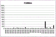
FIG. 7 shows the extent of binding of Fv4-F1886m to rheumatoid factor in serum of RA patients.
FIG. 8 shows the extent of binding of Fv4-F1889m to rheumatoid factor in serum of RA patients.
FIG. 9 shows the extent of binding of Fv4-F1927m to rheumatoid factor in serum of RA patients.
FIG. 10 shows the extent of binding of Fv4-F1168m to rheumatoid factor in serum of RA patients.
FIG. 11 shows the average binding to rheumatoid factor in blood of RA patients for each of Fv4-IgG1 and antibodies comprising various Fc region variants having increased FcRn binding.
FIG. 12 shows the time course of various anti-human IgE antibody concentrations in cynomolgus plasma, where OHB-IgG1 (which is an anti-human IgE antibody having the Fc domain of native human IgG 1) and antibodies containing various Fc region variants with increased FcRn binding (OHB-LS, OHB-N434A, OHB-F1847m, OHB-F1848m, OHB-F1886m, OHB-F1889m, and OHB-F1927 m) were each administered.
FIG. 13 shows the time course of antibody concentration in plasma of human FcRn transgenic mice when Fv4-IgG1 (which is an anti-human IL-6 receptor antibody) or Fv4-F1718 (which results from increasing FcRn binding activity of Fv4-IgG1 at acidic pH) is administered.
FIG. 14 shows a sensorgram obtained by Biacore measurement of H998/L63 and Hr9 binding to IL-8 at pH 7.4 and 5.8.
FIG. 15 shows the time course of human IL-8 concentration in mouse plasma when H998/L63 and H89/L118 were each administered to mice at 2mg/kg in combination with human IL-8.
FIG. 16 shows the time course of human IL-8 concentration in mouse plasma when H89/L118 was administered to mice at 2mg/kg or 8mg/kg in combination with human IL-8.
FIG. 17 shows the time course of human IL-8 concentration in mouse plasma when H89/L118 and H553/L118 are each administered to mice at 2mg/kg or 8mg/kg in combination with human IL-8.
FIG. 18-1 shows the variation of the relative intensities of chemiluminescence depending on the concentrations of antibodies of Hr9, H89/L118 and H553/L118 before storage in plasma.
FIG. 18-2 shows the variation of the relative intensities of chemiluminescence depending on the antibody concentrations of Hr9, H89/L118 and H553/L118 after one week of storage in plasma.
FIG. 18-3 shows the variation of the relative intensity of chemiluminescence depending on the concentration of antibodies against Hr9, H89/L118 and H553/L118 after two weeks of storage in plasma.
FIG. 19 shows the predicted ADA frequency for EpiMatrix for each anti-IL-8 antibody (hWS, hr9, H89/L118, H496/L118, and H553/L118) and other existing antibody drugs.
FIG. 20 shows predicted ADA frequency of EpiMatrix for each anti-IL-8 antibody (H496/L118, H496v1/L118, H496v2/L118, H496v3/L118, H1004/L118 and H1004/L395) and other existing antibody drugs.
FIG. 21-1 shows the variation of the relative intensities of chemiluminescence depending on the concentration of antibodies by Hr9, H89/L118 and H1009/L395-F1886s prior to storage in plasma.
FIG. 21-2 shows the variation of the relative intensities of chemiluminescence depending on the concentration of antibodies for Hr9, H89/L118 and H1009/L395-F1886s after one week of storage in plasma.
FIG. 21-3 shows the change in the relative intensity of chemiluminescence depending on the concentration of antibodies for Hr9, H89/L118 and H1009/L395-F1886s after two weeks of storage in plasma.
FIG. 22 shows the time course of human IL-8 concentration in mouse plasma when H1009/L395, H553/L118 and H998/L63 are each administered in combination with human IL-8 to mice.
FIG. 23 shows the amount of binding of Hr9, H89/L118 or H1009/L395 to the extracellular matrix when added to the extracellular matrix alone or in combination with human IL-8.
FIG. 24 shows the time course of antibody concentration in human FcRn transgenic mouse plasma when antibodies with variable region H1009/L395 and Fc domain that does not bind FcRn (F1942 m) are administered alone or in combination with human IL-8.
FIG. 25 shows predicted ADA frequency of EpiMatrix versus H1009/L395 and H1004/L395 as well as other prior antibody drugs.
FIG. 26 shows the time course of each anti-human IL-8 antibody concentration in cynomolgus monkey plasma when H89/L118-IgG1 (which comprises the variable region of H89/L118 and the Fc domain of native human IgG 1), and antibodies (H89/L118-F1168 m, H89/L118-F1847m, H89/L118-F1848m, H89/L118-F1889m, and H89/L118-F1927 m) comprising various Fc region variants with increased FcRn binding, were each administered to cynomolgus monkeys.
FIG. 27 shows the binding activity of antibodies to various FcγRs, wherein the antibodies comprise the variable region of H1009/L395 and their Fc domains are variants (F1886 m, F1886s and F1974 m).
FIG. 28 shows the time course of human IL-8 concentration in human FcRn transgenic mice plasma when anti-IL-8 antibodies were administered in combination with human IL-8 to mice. The anti-IL-8 antibody is: H1009/L395-IgG1 (2 mg/kg) comprising the variable region of H1009/L395 and the Fc domain of native human IgG 1; and H1009/L395-F1886s (2, 5 or 10 mg/kg) comprising a variable region of H1009/L395 and a modified Fc domain.
FIG. 29 shows the time course of antibody concentration in cynomolgus plasma when Hr9-IgG1 or H89/L118-IgG1 (which comprises the Fc region of native human IgG 1) or H1009/L395-F1886s or H1009/L395-F1974m comprising a modified Fc region is administered to cynomolgus monkeys.
FIG. 30 is a graph showing IL-8 concentration in the cyst fluid of a endometriosis patient.
Fig. 31 shows a laparoscopic recording sheet in which lesions formed by seeding endometrial tissue cut into small pieces and lesions formed by adhesion and suturing are recorded.
Fig. 32 is a photograph showing the formation of nodulization lesions (nodulization forms) and adhesions at 4 months after induction and 12 months after induction (6 months after administration).
Fig. 33 is a photograph showing the formation of endometriosis-like proliferative endometrial epithelium and stroma 12 months after induction (6 months after administration).
FIG. 34-1 is a graph showing a high correlation between adhesion r-AFS score and IL-8 concentration in cyst fluid in a monkey endometriosis model.
FIG. 34-2 is a graph showing the relative volumes of endometriotic nodular lesions after administration of vector or antibody H1009/L395-F1974 m.
FIG. 35-1 is a set of graphs showing the change in total r-AFS score, adhesion r-AFS score, and size r-AFS score before and after administration of vector or antibody H1009/L395-F1974 m.
FIG. 35-2 is a graph showing the change in total r-AFS score before and after administration of vector or antibody H1009/L395-F1974 m.
FIG. 36 shows histopathological images of the graft sites of the vehicle group and antibody H1009/L395-F1974m administration group 12 months after induction (6 months after administration). Atrophy of proliferating epithelial and stromal cells and a decrease in the stroma were observed in the antibody H1009/L395-F1974m administration group compared to the vector group.
FIG. 37 is a set of photographs showing endometrial tissue in a vector group and antibody H1009/L395-F1974m administration group of monkeys with concurrent adenomyosis. Atrophy of endometrial epithelium and atrophy and reduction of stromal cells were observed in the antibody H1009/L395-F1974m administration group compared to the vehicle group.
FIG. 38 is a set of photographs of post-operative adhesions showing a representative abdominal cavity in a vehicle group and antibody H1009/L395-F1974m administration group of a monkey endometriosis model.
FIG. 39 shows the results of evaluation of the relative migration ability of neutrophils in each well supplemented with various agents, relative to the migration ability of neutrophils in wells without IL-8.
FIG. 40 shows the results of analyzing the expression level of aromatase when the culture supernatant of neutrophils was added to endometriosis cells.
FIG. 41 shows the results of analysis of MCP-1 concentration in neutrophil culture when IL-8 and anti-IL-8 antibodies were added.
FIG. 42 shows the results of analysis of CTGF expression in macrophages when IL-8 and anti-IL-8 antibodies are added.
Mode for carrying out the invention
Preferred non-limiting embodiments of the present disclosure are described below.
It is intended that all elements described in the embodiments hereinafter illustrated be naturally be considered as being equivalent to the description herein "means for carrying out the invention" without being limited by any patent practice, habit, law, etc., whereby it is possible to attempt to explain what is described in the embodiments in a limited manner in the countries where patent applications claim protection.
Those of ordinary skill in the art will of course appreciate that the present disclosure is intended to include any combination of some or all of one or more of the elements described in any of the locations of the present disclosure, so long as they are not technically contradictory based on the ordinary technical knowledge of the skilled person.
Herein, the term "antibody" is used in the broadest sense and includes, but is not limited to, monoclonal antibodies, polyclonal antibodies, multispecific antibodies (e.g., bispecific antibodies), and antibody fragments so long as they exhibit the desired antigen-binding activity.
In the present disclosure, an "antibody that binds to the same epitope" as a desired reference antibody (e.g., a reference anti-IL-8 antibody, an anti-CXCR 1 antibody, or an anti-CXCR 2 antibody) refers to an antibody that, in one embodiment, inhibits antigen binding of the reference antibody to a desired antigen (e.g., IL-8, CXCR1, or CXCR 2), for example, by 50%, 60%, 70%, 80%, 90%, 95% or more. In contrast, a reference antibody, for example, 50%, 60%, 70%, 80%, 90%, 95% or more inhibits binding of the antibody to the antigen. Herein, a typical competition assay may be used, but is not limited thereto.
Herein, the term "monoclonal antibody" refers to an antibody obtained from a substantially homogeneous population of antibodies. In particular, for example, the individual antibodies comprising a population are identical and/or bind to the same epitope, except for possible variant antibodies, such as variants that typically exist in small amounts that contain naturally occurring mutations or mutations that occur during the production of monoclonal antibody preparations. In contrast to polyclonal antibody preparations, which typically include different antibodies directed against different determinants (epitopes), each monoclonal antibody of a monoclonal antibody preparation is directed against a single determinant on the antigen. Thus, the modifier "monoclonal" refers to the characteristics of the antibody as obtained from a substantially homogeneous population of antibodies, and is not to be construed as requiring production of the antibody by any particular method. For example, monoclonal antibodies of the invention can be produced by a variety of techniques, including but not limited to, hybridoma methods, recombinant DNA methods, phage display methods, and methods using transgenic animals containing all or part of the human immunoglobulin loci. In one embodiment, the antibodies in the present disclosure may be monoclonal antibodies.
Herein, the term "natural antibody" refers to naturally occurring immunoglobulin molecules having various structures. In one aspect, the native IgG antibody is, for example, but not limited to, an heterotetrameric glycoprotein of about 150,000 daltons, which consists of two identical light chains and two identical heavy chains linked together by disulfide bonds. In the N-to C-terminal direction, each heavy chain has a variable region (VH) followed by three constant regions (CH 1, CH2 and CH 3). Also, in the N-to C-terminal direction, each light chain has a variable region (VL) followed by a constant region (CL). Based on the amino acid sequence of its constant region, an antibody light chain can be assigned to one of two types (called kappa (κ)) and lambda (λ))). The constant region used herein may be any reported allotype (allele) or any subclass/isoform. For example, the heavy chain constant region used may be, but is not limited to, the constant region of a natural IgG antibody (IgG 1, igG2, igG3 or IgG 4). For example, the known IgG1 allele is IGHG1 * 01 to 05 (see http:// www.imgt.org /), and any of these may be used as native human IgG1 sequences. Meanwhile, the constant region sequences may be from a single allele or subclass/isoform, or from multiple alleles or subclasses/isoforms. In particular, such antibodies include, but are not limited to, antibodies having CH1 derived from IGHG1 x 01, CH2 derived from IGHG1 x 02, and CH3 derived from IGHG1 x 01. The heavy chain constant region of a natural human IgG antibody includes, for example, a human IgG1 constant region (SEQ ID NO: 100), a human IgG2 constant region (SEQ ID NO: 101), a human IgG3 constant region (SEQ ID NO: 102), and a human IgG4 constant region (SEQ ID NO: 103). Meanwhile, the light chain constant region of a natural human IgG antibody includes, for example, a human kappa chain constant region (SEQ ID NO: 104) and a human lambda constant region (SEQ ID NO: 105).
Herein, the term "framework" or "FR" refers to the portion of the variable region other than the hypervariable region (HVR) residues. The FR of the variable region generally consists of four FR domains: FR1, FR2, FR3 and FR4. Thus, HVR and FR sequences typically occur in VH (or VL) in the following order: FR1-H1 (L1) -FR2-H2 (L2) -FR3-H3 (L3) -FR4.
Herein, the term "human consensus framework" is a framework representing the amino acid residues most commonly occurring in the selection of human immunoglobulin VL or VH framework sequences. Typically, the human immunoglobulin VL or VH sequence is selected from a subset of variable region sequences. Typically, the subgroup of sequences is as in Kabat et al Sequences of proteins of Immunological Interest,5th Ed.Public Health Service,National Institutes of Health,Bethesda,MD (1991). In one embodiment, for VL, the subgroup may be subgroup κI as described in Kabat et al, supra. In one embodiment, for VH, the subgroup may be subgroup III as described in Kabat et al, supra.
Herein, a "recipient human framework" is a framework comprising an amino acid sequence derived from a VL or VH framework of a human immunoglobulin framework or a human consensus framework. The recipient human framework "derived from" a human immunoglobulin framework or human consensus framework may comprise its identical amino acid sequence, or it may contain pre-existing amino acid sequence substitutions. In one embodiment, the number of pre-existing amino acid substitutions is 10, 9, 8, 7, 6, 5, 4, 3, or 2 or less. In one embodiment, the VL acceptor human framework is identical in sequence to the VL human immunoglobulin framework sequence or human consensus framework sequence.
Herein, the term "variable region" refers to the domain of an antibody heavy or light chain that is involved in binding of the antibody to a desired antigen. The heavy and light chain variable regions of natural antibodies (VH and VL, respectively) generally have similar structures, with each domain containing 4 conserved Framework Regions (FR) and 3 hypervariable regions (HVR) (Kindt et al Kuby Immunology,6th ed., w.h. freeman and co., 2007p.91). In one embodiment, a single VH or VL domain is sufficient to confer antigen binding specificity, but is not limited thereto. In addition, a library of complementary VL or VH domains can be screened using VH or VL domains, respectively, from antigen-binding antibodies to isolate antibodies that bind a particular antigen (e.g., portolano et al, J. Immunol.150:880-887 (1993); clarkson et al Nature1991 352:624-628).
In this context, the term "hypervariable region" or "HVR" refers to each region of an antibody variable domain that is hypervariable in sequence ("complementarity determining region" or "CDR") and/or forms a structurally defined loop ("hypervariable loop") and/or contains antigen-contacting residues ("antigen-contacting"). Typically, an antibody comprises six HVRs; three of VH (H1, H2, and H3) and three of VL (L1, L2, and L3). Exemplary HVRs include:
(a) Hypervariable loops present at amino acid residues 26-32 (L1), 50-52 (L2), 91-96 (L3), 26-32 (H1), 53-55 (H2) and 96-101 (H3) (Chothia and Lesk, J.mol. Biol.196:901-917 (1987));
(b) CDRs present at amino acid residues 24-34 (L1), 50-56 (L2), 89-97 (L3), 31-35b (H1), 50-65 (H2) and 95-102 (H3) (Kabat et al Sequences of Proteins of Immunological Interest,5th Ed.Public Health Service,National Institutes of Health,Bethesda,MD (1991));
(c) Antigen contacts present at amino acid residues 27c-36 (L1), 46-55 (L2), 89-96 (L3), 30-35b (H1), 47-58 (H2) and 93-101 (H3) (MacCallum et al, J.mol.biol.262:732-745 (1996)); and
(d) Combinations of (a), (b) and/or (c) including HVR amino acid residues 46-56 (L2), 47-56 (L2), 48-56 (L2), 49-56 (L2), 26-35 (H1), 26-35b (H1), 49-65 (H2), 93-102 (H3) and 94-102 (H3).
Unless otherwise indicated, HVR residues and other residues within the variable region (e.g., FR residues) are numbered herein according to Kabat et al, supra.
Herein, "individual" refers to a mammal. Mammals include, but are not limited to, domesticated animals (e.g., cattle, sheep, cats, dogs, and horses), primates (e.g., humans and non-human primates such as monkeys), rabbits, and rodents (e.g., mice and rats). In one embodiment, the "individual" is preferably a mammal that naturally has IL-8 in its body, more preferably an animal that has menstruation similar to a human, such as a non-human primate, and more preferably a human. The term "individual" is used interchangeably herein with "subject" unless it is inconsistent with the context.
Herein, "isolated" antibody refers to an antibody that has been separated from components of its natural environment. In one embodiment, the antibodies can be purified to, for example, 95% or more, or 99% or more purity, e.g., chromatographically (e.g., ion exchange or reverse phase HPLC) or electrophoretically (e.g., SDS-PAGE, isoelectric focusing (IEF), or capillary electrophoresis). For methods of assessing antibody purity, see, e.g., flatman et al, J.chromatogr.B 848:79-87 (2007). In one aspect, an "isolated" antibody in the present disclosure may refer to a "purified" antibody.
As used herein, "isolated" nucleic acid refers to a nucleic acid molecule that has been separated from components of its natural environment. Nucleic acids include nucleic acid molecules that are normally contained in cells containing the nucleic acid molecule, but which are present extrachromosomally or at a chromosomal location different from their natural chromosomal location.
Herein, the term "affinity" may generally refer to the strength of the sum of non-covalent interactions between a single binding site of a molecule (e.g., an antibody or chemical compound) and its binding partner (e.g., an antigen). The term "binding affinity" in this disclosure refers to an intrinsic binding affinity that reflects a 1:1 interaction between members of a binding pair (e.g., an antibody or chemical compound and an antigen), unless otherwise specified. The affinity of a molecule X for its partner Y can generally be expressed by a dissociation constant (KD). Binding affinities may be measured using methods known to those of skill in the art, including those described in the present disclosure.
In one embodiment, an antibody that binds to an antigen such as IL-8, CXCR1, or CXCR2 may have a dissociation constant (KD) (e.g., 10) of, for example, 1000nM, 100nM, 10nM, 1nM, 0.1nM, 0.01nM, or 0.001nM -8 M or less, 10 -8 M to 10 -13 M or 10 -9 M to 10 -13 M)。
The terms "host cell" and "host cell line" are used interchangeably herein and refer to a cell into which exogenous nucleic acid has been introduced (including the progeny of the cell). Host cells include "transformants" and "transformed cells," which include primary transformed cells and progeny derived therefrom, regardless of the number of progeny. The nucleic acid content of the offspring and its parent cells may not be exactly the same, but may contain mutations. Mutant progeny having the same function or biological activity for selection or selection in the initially transformed cells are also included herein.
As used herein, the term "vector" refers to a nucleic acid molecule capable of transmitting another nucleic acid to which it is linked. The term includes vectors that are self-replicating nucleic acid structures as well as vectors that integrate into the genome of a host cell into which they have been introduced. Certain vectors are capable of directing the expression of nucleic acids to which they are operably linked. Such vectors are also referred to herein as "expression vectors".
In one embodiment, an antibody (e.g., an anti-IL-8 antibody, an anti-CXCR 1 antibody, or an anti-CXCR 2 antibody) in the present disclosure can be an antibody fragment. Antibody fragments may include, for example, fab '-SH, F (ab') 2 Fv, scFv fragments, diabodies and single domain antibodies. For reviews of antibody fragments, see, e.g., hudson et al, nat. Med.9:129-134 (2003). For reviews of scFv fragments, see, for example, pluckaphun, the Pharmacology of Monoclonal Antibodies, vol.113, rosenburg and Moore (Springer-Verlag,new York), pp.269-315 (1994); WO93/16185; U.S. Pat. No.5,571,894; and U.S. Pat. No.5,587,458.
Herein, a "diabody" is an antibody fragment having two antigen binding sites, which may be bivalent or bispecific (see, e.g., EP404,097; WO1993/01161; hudson et al, nat. Med.9:129-134 (2003); hollinger et al, proc. Natl. Acad. Sci. USA 90:6444-6448 (1993)). Tri-and tetra-antibodies are described, for example, in Hudson et al, nat.Med.9:129-134 (2003).
Herein, a "single domain antibody" is an antibody fragment comprising all or a portion of the heavy chain variable domain or all or a portion of the light chain variable domain of an antibody. In one embodiment, when the antibody in the present disclosure is a single domain antibody, it may be a human single domain antibody (see, e.g., domatis, inc., waltham, MA; U.S. patent No.6,248,516). Antibody fragments can be prepared by a variety of techniques including, but not limited to, proteolytic digestion of intact antibodies and production using recombinant host cells.
Herein, the term "chimeric antibody" refers to an antibody in which a portion of the heavy and/or light chain is derived from a particular source or species, while the remainder of the heavy and/or light chain is derived from a different source or species.
Herein, "humanized" antibody refers to a chimeric antibody comprising amino acid residues from a non-human HVR and amino acid residues from a human FR. In one embodiment, the humanized antibody comprises substantially at least one, and typically two, variable regions, in which all (or substantially all) HVRs correspond to those of a non-human antibody (e.g., CDRs) and all (or substantially all) FRs correspond to those of a human antibody. The humanized antibody may optionally comprise at least a portion of an antibody constant region derived from a human antibody.
In one embodiment, an antibody in the present disclosure (e.g., an anti-IL-8 antibody, an anti-CXCR 1 antibody, or an anti-CXCR 2 antibody) can be a chimeric antibody. Chimeric antibodies are described, for example, in U.S. Pat. nos. 4,816,567; and Morrison et al, proc.Natl.Acad.Sci.USA,81:6851-6855 (1984). Chimeric antibodies may comprise a non-human variable region (e.g., a variable region derived from a non-human primate such as a monkey, or a mouse, rat, hamster, or rabbit) and a human constant region.
In one embodiment, an antibody in the present disclosure (e.g., an anti-IL-8 antibody, an anti-CXCR 1 antibody, or an anti-CXCR 2 antibody) can be a humanized antibody. Typically, the non-human antibodies are humanized to reduce immunogenicity in humans, while retaining the specificity and affinity of the parent non-human antibody. Typically, a humanized antibody comprises one or more variable regions, in which the HVRs (e.g., CDRs) (or portions thereof) are derived from a non-human antibody and the FRs (or portions thereof) are derived from a human antibody sequence. The humanized antibody may also optionally comprise at least a portion of a human constant region. In one embodiment, some FR amino acid residues in a humanized antibody may be substituted with corresponding amino acid residues from a non-human antibody (e.g., an antibody from which HVR residues are derived), e.g., to restore or improve antibody specificity or affinity.
Humanized antibodies and methods for their preparation are reviewed in, for example, almagro and Franson, front. Biosci.13:1619-1633 (2008) and further described in Riechmann et al, nature 332:323-329 (1988); queen et al, proc.Nat' l Acad.Sci.USA 86:10029-10033 (1989); U.S. Pat. nos. 5,821,337, 7,527,791, 6,982,321 and 7,087,409; kashmiri et al, methods 36:25-34 (2005) (describing Specificity Determining Region (SDR) migration); padlan, mol. Immunol.28:489-498 (1991) (description "surface remolding"); dall' acquata et al, methods 36:43-60 (2005) (description "FR shuffling"); and Osbourn et al, methods 36:61-68 (2005) and Klimka et al, br.J.cancer,83:252-260 (2000) (describing the "guide selection" method of FR shuffling)).
In one embodiment, human frameworks that can be used for humanization can include, for example, framework regions selected using the "best fit" method (Sims et al, J.Immunol.151:2296 (1993), framework regions derived from consensus sequences of human antibodies of specific subsets of the light or heavy chain variable regions (Carter et al, proc.Natl. Acad.Sci.USA,89:4285 (1992) and Presta et al, J.Immunol.,151:2623 (1993)), and framework regions derived from screening FR libraries (Baca et al, J.biol. Chem.272:10678-10684 (1997), and Rosok et al, J.biol. Chem.271:611-22618 (1996)).
In one implementationIn embodiments, an antibody (e.g., an anti-IL-8 antibody, an anti-CXCR 1 antibody, or an anti-CXCR 2 antibody) in the present disclosure can be a human antibody. Human antibodies can be produced by a variety of techniques. Human antibodies are reviewed in, for example, van Dijk and van de Winkel, curr. Opin. Pharmacol.5:368-374 (2001) and Lonberg, curr. Opin. Immunol.20:450-459 (2008). Human antibodies can be prepared by administering an antigen (e.g., IL-8, CXCR1, or CXCR 2) to a transgenic animal that has been modified to produce a fully human antibody or a fully antibody with a human variable region in response to the antigen. These animals typically contain all or part of the human immunoglobulin loci, either in place of the endogenous immunoglobulin loci, or extrachromosomally present or randomly integrated into the animal's chromosome. In such transgenic mice, the endogenous immunoglobulin loci have generally been inactivated. For a review of methods of obtaining human antibodies from transgenic animals, see Lonberg, nat. Biotech.23:1117-1125 (2005). See also, e.g., descriptions xenomouise TM Technical U.S. Pat. nos. 6,075,181 and 6,150,584; description of the invention U.S. Pat. No.5,770,429 to the art; description of K-M->
 U.S. Pat. No.7,041,870 to the art; description of the invention
 US2007/0061900 of the technology. The human variable regions of the whole antibodies produced by these animals may be further modified, for example by combining them with different human constant regions.
In another embodiment, human antibodies may also be prepared by hybridoma-based methods. Human myeloma and mouse-human heterologous myeloma cell lines for the production of human monoclonal antibodies are described below (e.g., kozbor J. Immunol.,133:3001 (1984); brodeur et al, monoclonal Antibody Production Techniques and Applications, pp.51-63 (Marcel Dekker, inc., new York, 1987); and Boerner et al J. Immunol.,147:86 (1991)). Human antibodies produced by human B cell hybridoma technology are described in Li et al, proc.Natl. Acad. Sci. USA,103:3557-3562 (2006). Other methods include, for example, those described in U.S. Pat. No.7,189,826 (describing the production of monoclonal human IgM antibodies from hybridoma cell lines) and Ni, xiandai Mianyixue,26 (4): 265-268 (2006) (describing human-human hybridomas). Human hybridoma technology (Trioma technology) is described in Vollmers and Brandlein, histology and Histopathology,20 (3): 927-937 (2005) and Vollmers and Brandlein, methods and Findings in Experimental and Clinical Pharmacology,27 (3): 185-91 (2005).
In an alternative embodiment, human antibodies may also be generated by isolating Fv clone variable region sequences selected from a human phage display library. Such variable region sequences can then be combined with the desired human constant region. Techniques for selecting human antibodies from antibody libraries are described below.
In one embodiment, antibodies (e.g., anti-IL-8 antibodies, anti-CXCR 1 antibodies, or anti-CXCR 2 antibodies) in the present disclosure can be isolated by screening a combinatorial library for antibodies having the desired activity. For example, a variety of methods are known in the art for generating phage display libraries and screening these libraries to obtain antibodies with desired binding characteristics. Such methods are reviewed in, for example, hoogenboom et al, methods in Molecular Biology 178:1-37 (O' Brien et al, human Press, totowa, NJ, 2001) and are further described, for example, in the following: mcCafferty et al, nature 348:552-554; clackson et al, nature 352:624-628 (1991); marks et al, J.mol.biol.222:581-597 (1992); marks and Bradbury, molecular Biology248:161-175 (Lo, eds., human Press, totowa, N.J., 2003); sidhu et al, J.mol.biol.338 (2): 299-310 (2004); lee et al, J.mol.biol.340 (5): 1073-1093 (2004); felloose, proc. Natl. Acad. Sci. USA 101 (34): 12467-12472 (2004); and Lee et al, J.Immunol. Methods 284 (1-2): 119-132 (2004).
In one embodiment, in certain phage display methods, the VH and VL repertoires are cloned separately by Polymerase Chain Reaction (PCR) and randomly recombined in a phage library, and antigen-binding phages can then be screened as described in Winter et al, ann.Rev.Immunol.,12:433-455 (1994). Phage display antibody fragments, such as scFv and Fab. Libraries from immunized sources can provide high affinity antibodies to immunogens without the need to construct hybridomas. In an alternative embodiment, the natural repertoire may also be cloned (e.g., from humans) to provide a single antibody source against a wide range of non-self and self-antigens without any immunization, as described by Griffiths et al, EMBO J,12:725-734 (1993). In yet another embodiment, the original (native) library can also be prepared synthetically by cloning non-rearranged V gene segments from stem cells and encoding highly variable region CDR3 using PCR primers containing random sequences and completing the rearrangement in vitro, as described in Hoogenboom, h.r. and Winter, g., j.mol. Biol.227 (1992) 381-388. Patent publications describing human antibody phage libraries include, for example: U.S. Pat. No.5,750,373; US 2005/0079774; US 2005/019455; US2005/0266000; US 2007/017126; US2007/0160598; US2007/0237764; US2007/0292936; US2009/0002360.
Antibodies or antibody fragments isolated from a human antibody library are herein considered human antibodies or human antibody fragments.
In one embodiment, an antibody in the present disclosure (e.g., an anti-IL-8 antibody, an anti-CXCR 1 antibody, or an anti-CXCR 2 antibody) can be a multispecific antibody (e.g., a bispecific antibody). A multispecific antibody is an antibody (e.g., monoclonal antibody) that has binding specificity for at least two different sites. In one embodiment, one of the binding specificities is for an antigen (e.g., IL-8, CXCR1 or CXCR 2) and the other is for any other antigen. In another embodiment, a bispecific antibody can bind to two different epitopes of an antigen (e.g., IL-8, CXCR1, or CXCR 2). Bispecific antibodies can be used to localize cytotoxic agents to cells expressing an antigen (e.g., IL-8, CXCR1, or CXCR 2). Bispecific antibodies can be prepared as full length antibodies or antibody fragments.
Techniques for preparing multispecific antibodies include, but are not limited to, recombinant coexpression of two immunoglobulin heavy-light chain pairs with different specificities (e.g., milstein and Cuello, nature 305:537 (1983); WO 93/08829; and Traunecker et al, EMBO J.10:3655 (1991)) and "knob-in-hole" designs (e.g., U.S. Pat. No. 5,731,168). Multispecific antibodies may also be prepared by involving electrostatic control effects to Fc-heterodimeric molecules (e.g. WO2009/089004 A1); crosslinking two or more antibodies or antibody fragments (e.g., U.S. Pat. No. 4,676,980; and Brennan et al Science 229:81 (1985)); bispecific antibodies were generated using leucine zippers (e.g., kostelny et al, J. Immunol.148 (5): 1547-1553 (1992)); bispecific antibody fragments were prepared using "diabody" techniques (e.g., hollinger et al, proc. Natl. Acad. Sci. USA 90:6444-6448 (1993)); preparation using scFv dimers (e.g., gruber et al, j. Immunol.,152:5368 (1994)). And preparation of trispecific antibodies (e.g., tutt et al, J. Immunol.147:60 (1991)). Engineered antibodies having three or more functional antigen binding sites, including "octopus antibodies", are also included herein (e.g., US 2006/0025576).
In one embodiment, an antibody (e.g., an anti-IL-8 antibody, an anti-CXCR 1 antibody, or an anti-CXCR 2 antibody) or an antibody fragment thereof in the present disclosure can comprise a "dual acting Fab" or "DAF" that binds to an antigen (e.g., IL-8, CXCR1, or CXCR 2) and an antigen binding site of another, different antigen (e.g., US 2008/0069820).
In one embodiment, amino acid sequence variants (mutants) of antibodies (e.g., anti-IL-8 antibodies, anti-CXCR 1 antibodies, or anti-CXCR 2 antibodies) in the present disclosure can be prepared by introducing appropriate modifications in the nucleotide sequence encoding the antibody or by peptide synthesis. These modifications may be achieved by introducing one or more arbitrary deletions, insertions, substitutions or combinations thereof of any amino acid (residue) into the amino acid sequence. Any set of deletions, insertions, and substitutions may be made as long as the final construct has the desired characteristics (e.g., antigen binding).
In one embodiment, where antibody variants (mutants) having one or more amino acid substitutions are provided, the target sites for substitution mutagenesis may include HVRs and FRs.
Conservative substitutions in one embodiment are shown under the heading of "preferred substitutions" in table 1, and more substantial changes are shown under the heading of "exemplary substitutions" in table 1, and are described further below with reference to the amino acid side chain class. Amino acid substitutions may be introduced into the antibody of interest and the products screened for desired activity, e.g., retained/improved antigen binding, reduced immunogenicity, or improved ADCC or CDC.
TABLE 1
| Initial residues | Exemplary substitution | Preferably substituted |
| Ala(A) | Val;Leu;Ile | Val |
| Arg(R) | Lys;Gln;Asn | Lys |
| Asn(N) | Gln;His;Asp.Lys;Arg | Gln |
| Asp(D) | Glu;Asn | Glu |
| Cys(V) | Ser;Ala | Ser |
| Gln(Q) | Asn;Glu | Asn |
| Glu(E) | Asp;Gln | Asp |
| Gly(G) | Ala | Ala |
| His(H) | Ash;Gln;Lys;Arg | Arg |
| Ile(I) | Leu; val; met; ala; phe; norleucine (N-leucine) | Leu |
| Leu(L) | Norleucine; tle; val; met; ala; phe (Phe) | Ile |
| Lys(K) | Arg;Gln;Asn | Arg |
| Met(M) | Leu;Phe;Ile | Leu |
| Phe(F) | Trp;Leu;Val;Ile;Ala;Tyr | Tyr |
| Pro(P) | Ala | Ala |
| Ser(S) | Thr | Thr |
| Thr(T) | Val;Ser | Ser |
| Trp(W) | Tyr;Phe | Tyr |
| Tyr(Y) | Trp;Phe;Thr;Ser | Phe |
| Val(V) | Ile; leu; met; phe; ala; norleucine (N-leucine) | Leu |
Amino acids can be grouped according to common side chain characteristics:
(1) Hydrophobicity: norleucine, met, ala, val, leu, ile;
(2) Neutral, hydrophilic: cys, ser, thr, asn, gln;
(3) Acid: asp, glu;
(4) Alkaline: his, lys, arg;
(5) Residues that affect chain orientation: gly, pro;
(6) Aromatic: trp, tyr, phe
Non-conservative substitutions mean that a member of one of these categories is swapped for another category.
In one embodiment, the amino acid insertion includes amino and/or carboxy terminal fusion of one or two residues or polypeptides containing three to one hundred or more residues, as well as insertion of single or more amino acid residues into the amino acid sequence. Examples of terminal insertions include antibodies with an N-terminal methionyl residue. Other examples of insertional variants (mutants) include fusions of the N-and/or C-terminus of an antibody with an enzyme (e.g., an enzyme directed against ADEPT) or a polypeptide that increases the plasma half-life of an antibody.
"percent (%) amino acid sequence identity" with respect to a reference polypeptide sequence is defined herein as the percentage of amino acid residues in a candidate sequence that are identical to amino acid residues in the reference polypeptide sequence, with the greatest percent sequence identity achieved, if necessary, after aligning the sequences and introducing gaps, and without regard to any conservative substitutions as part of the sequence identity. Alignment for the purpose of determining the percent amino acid sequence identity can be accomplished in a variety of ways within the skill of the art, e.g., using publicly available computer software such as BLAST, BLAST-2, ALIGN, megalign (DNASTAR) software, or  One skilled in the art can determine the appropriate parameters for aligning sequences, including any algorithms needed to achieve maximum alignment over the full length of the sequences compared.
ALIGN-2 sequence comparison computer program was written by Genntech corporation and the source code has been submitted with the U.S. copyright office user file, which is registered under U.S. copyright accession number TXU 510087. ALIGN-2 programs are publicly available from Genntech corporation (san Francisco, calif.), or may be compiled from source code. Compiling ALIGN-2 program to generate a new program Used on an operating system (including digitized UNIX v4.0d). All sequence comparison parameters are ALIGThe N-2 program is set and unchanged.
In the case of amino acid sequence comparisons using ALIGN-2, the% amino acid sequence identity of a given pair of amino acid sequences, with or against a given amino acid sequence B (which may alternatively be expressed as a given amino acid sequence a having or comprising a certain% amino acid sequence identity to, with or against a given amino acid sequence B), is calculated as follows: a score of X/Y of 100, where X is the number of amino acid residues scored as identical matches in the alignment of the programs of a and B by the sequence alignment program ALIGN-2, where Y is the total number of amino acid residues in B. It will be appreciated that when the length of amino acid sequence a is not equal to the length of amino acid sequence B, the% amino acid sequence identity of a to B will not be equal to the% amino acid sequence identity of B to a. Unless specifically stated otherwise, all% amino acid sequence identity values herein are obtained using the computer program ALIGN-2.
In one embodiment, the amino acid sequence of an antibody (e.g., an anti-IL-8 antibody, an anti-CXCR 1 antibody, or an anti-CXCR 2 antibody) in the present disclosure, which has an appropriate substitution, deletion, or insertion of one or more amino acids in the amino acid sequence of the CDR region, heavy chain variable region, light chain variable region, heavy chain constant region, light chain constant region, entire heavy chain region, entire light chain region, or any particular region thereof, and has binding activity against an antigen (e.g., IL-8, CXCR1, or CXCR 2), can also be obtained from a nucleic acid that hybridizes under stringent conditions to a nucleic acid comprising a nucleotide sequence encoding the amino acid sequence of that region. Exemplary stringent hybridization conditions for isolating hybridized nucleic acids under stringent conditions are 6M urea, 0.4% SDS,0.5 XSSC, and 37℃or conditions equivalent or corresponding thereto. By using more stringent conditions, such as 6M urea, 0.4% SDS,0.1 XSSC and 42℃it is expected that nucleic acids with higher homology will be isolated. Conditions for post hybridization washing include, for example, 0.5XSSC (where 1 XSSC is, for example, 0.15M NaCl and 0.015M sodium citrate, pH 7.0) and 0.1% SDS at 60 ℃, preferably 0.2 XSSC and 0.1% SDS at 60 ℃, more preferably 0.2 XSSC and 0.1% SDS at 62 ℃, even more preferably 0.2 XSSC and 0.1% SDS at 65 ℃, still more preferably 0.1 XSSC and 0.1% SDS at 65 ℃. The washing time and the number of times of washing can be appropriately adjusted; for example, a 20 minute wash may be performed three times. The isolated nucleic acid may be sequenced by known methods.
In an alternative embodiment, instead of the hybridization techniques described above, a gene amplification method, such as PCR, using primers synthesized based on nucleotide sequence information encoding the amino acid sequence of the CDR region, heavy chain variable region, light chain variable region, heavy chain constant region, light chain constant region, whole heavy chain region, whole light chain region or any specific region thereof, may also be used to isolate nucleic acids that hybridize under stringent conditions to nucleic acids comprising the nucleotide sequence of the amino acid sequence of that region of Bituma.
The nucleic acid thus isolated has an overall sequence identity of at least 50% or more, preferably 70% or more, 75% or more, 80% or more, 85% or more, more preferably 90% or more (e.g., 91%, 92%, 93%, 94%, 95%, 96%, 97%, 98% or 99% or more) to the nucleotide sequence of the target region. It would be advantageous if an antibody having an amino acid sequence encoded by such an isolated nucleic acid was functionally (substantially) identical (e.g., any one or more of the indicators or characteristics, such as antigen binding in blood or retention (substantially) identical) to an antibody having an amino acid sequence of the target region; however, it is not limited thereto. Herein, the term "substantially" is intended to mean that at least 50% or more, preferably 70% or more, 75% or more, 80% or more, 85% or more, more preferably 90% or more (e.g., 91%, 92%, 93%, 94%, 95%, 96%, 97%, 98% or 99% or more) of the function (e.g., possibly focused on any one or more index features such as antigen binding and retention in blood) is retained as compared to the function possessed by an antibody having the amino acid sequence of the region of interest when assessed by methods known to those of skill in the art.
In one embodiment, an antibody in the present disclosure (e.g., an anti-IL-8 antibody, an anti-CXCR 1 antibody, or an anti-CXCR 2 antibody) can be glycosylated. The addition or deletion of glycosylation sites to antibodies can be easily accomplished by altering the amino acid sequence such that glycosylation sites are created or eliminated.
In one embodiment, when an antibody of the invention (e.g., an anti-IL-8 antibody, an anti-CXCR 1 antibody, or an anti-CXCR 2 antibody) comprises an Fc region, the sugar chain attached to the Fc domain can vary. Natural antibodies produced by animal cells typically contain branched, bifurcated oligosaccharides that are linked via an N-linkage to Asn297 of the CH2 domain of the Fc region (Wright et al TIBTECH 15:26-32 (1997)). Oligosaccharides include, for example, mannose, N-acetylglucosamine (GlcNAc), galactose, and sialic acid, as well as fucose, which are linked to GlcNAc in the "stem" of a bifurcated oligosaccharide structure. In one embodiment, oligosaccharides in an antibody may be modified to produce antibody variants with certain improved properties. In one embodiment, the oligosaccharides in the antibodies of the present disclosure may be modified to produce antibody variants with certain improved properties.
Herein, "effector function" refers to biological activity attributable to an antibody Fc region (e.g., an anti-IL-8 antibody, an anti-CXCR 1 antibody, or an anti-CXCR 2 antibody), and may vary with antibody isotype. Examples of antibody effector functions include, but are not limited to, C1q binding or Complement Dependent Cytotoxicity (CDC); fc receptor binding; antibody-dependent cell-mediated cytotoxicity (ADCC); phagocytosis; down-regulation of cell surface receptors (e.g., B cell receptors); and B cell activation.
The term "Fc region" as used herein is used to define the C-terminal region of an immunoglobulin heavy chain that contains at least a portion of a constant region. The term includes both native and variant Fc regions. The native Fc region refers to the Fc region of a native antibody. Examples of the Fc region of natural human IgG include, for example, the Fc region contained in the human IgG1 constant region (SEQ ID NO: 100), human IgG2 constant region (SEQ ID NO: 101), human IgG3 constant region (SEQ ID NO: 102), or human IgG4 constant region (SEQ ID NO: 103) as described above. In one embodiment, the Fc region of a human IgG heavy chain extends from Cys226 or from Pro230 to the C-terminus of the heavy chain. However, the C-terminal lysine (Lys 447) of the Fc domain or the C-terminal glycine (Gly 446) and lysine (Lys 447) of the Fc domain may or may not be present. Unless otherwise specified herein, numbering of amino acid residues in the Fc region or constant region is according to the EU numbering system, also known as the EU index, e.g., kabat et al, sequences of Proteins of Immunological Interest, fifth edition, public Health Service, national Institutes of Health, bethesda, MD (1991).
Unlike fcγr, which belongs to the immunoglobulin superfamily, fcRn herein, particularly human FcRn, is structurally similar to polypeptides of class I of the Major Histocompatibility Complex (MHC), sharing 22% to 29% sequence identity with MHC class I molecules (Ghetie et al, immunol. Today (1997) 18 (12), 592-598). FcRn is expressed as a heterodimer consisting of a soluble β or light chain (β2 microglobulin) complexed with a transmembrane α or heavy chain. Like MHC, the alpha chain of FcRn consists of three extracellular domains (α1, α2 and α3), and its short cytoplasmic domain anchors proteins to the cell surface. The α1 and α2 domains interact with the FcRn binding domain of the Fc region of antibodies (Raghavan et al, immunity, (1994), 1:303-15). FcRn is expressed in the maternal placenta or yolk sac of mammals and is involved in maternal to fetal IgG transfer. Furthermore, in the small intestine of neonatal rodents expressing FcRn, fcRn is involved in the transfer of maternal IgG from ingested colostrum or milk across the brush border epithelium. FcRn is expressed in a variety of other tissue and endothelial cell systems of various species. FcRn is also expressed in adult endothelium, muscular vasculature, and hepatic sinus capillaries. FcRn is thought to play a role in maintaining plasma IgG concentrations by mediating IgG recycling into serum after binding to IgG, and is generally strictly pH dependent for IgG molecule binding. Optimal binding was observed in the acidic pH range below 7.0. The polynucleotide and amino acid sequences of human FcRn may be derived, for example, from the precursors (containing signal sequences) shown in nm_004107.4 and np_004098.1, respectively (RefSeq accession numbers are shown in brackets). The precursor forms a complex with human beta 2-microglobulin in vivo. Thus, using known recombinant expression techniques, soluble human FcRn capable of forming complexes with human β2-microglobulin can be prepared and suitably used in a variety of experimental systems. Such soluble human FcRn capable of forming a complex with human β2-microglobulin may be used to evaluate FcRn binding activity of an antibody or Fc region variant without particular limitation, so long as it is in a form capable of binding to an FcRn binding domain, and human FcRn is preferred.
In one embodiment, when an antibody (e.g., an anti-IL-8 antibody, an anti-CXCR 1 antibody, or an anti-CXCR 2 antibody) or an Fc region variant thereof in the present disclosure has FcRn binding activity, the antibody or Fc region variant preferably has an "FcRn binding domain", more preferably has a human FcRn binding domain. The FcRn binding domain is not particularly limited so long as the antibody (e.g., anti-IL-8 antibody, anti-CXCR 1 antibody or anti-CXCR 2 antibody) has FcRn binding activity or affinity at acidic pH and/or neutral pH. Alternatively, the FcRn binding domain may have activity to bind directly or indirectly to FcRn. These domains include, but are not limited to, for example, the Fc region of an IgG-type immunoglobulin, albumin domain 3, anti-FcRn antibodies, anti-FcRn peptides and anti-FcRn scaffold molecules (having activity to bind directly to FcRn) and molecules that bind to IgG or albumin (which have activity to bind indirectly to FcRn), alternatively domains that have FcRn binding activity at, for example, acidic pH and/or neutral pH may be used. The domain may be used unmodified if it initially has FcRn binding activity at acidic pH and/or neutral pH. When the domain has no or weak FcRn binding activity at acidic pH and/or neutral pH, the amino acid residues in the FcRn binding domain of the antibody or Fc region variant may be modified to produce FcRn binding activity at acidic pH and/or neutral pH. Alternatively, the amino acids of the domain that initially have FcRn binding activity at acidic pH and/or neutral pH may be modified to increase its FcRn binding activity. The amino acid modifications required for an FcRn binding domain can be identified by comparing the FcRn binding activity at acidic pH and/or neutral pH before and after the amino acid modification.
The FcRn binding domain is preferably a region that binds directly to FcRn. Preferred examples of FcRn binding domains include the constant and Fc regions of antibodies. However, a region capable of binding to a polypeptide having FcRn binding activity, such as albumin or IgG, may bind to FcRn indirectly through albumin or IgG. Thus, an FcRn binding region may be a region that binds a polypeptide having binding activity for albumin or IgG. Without limitation, the FcRn binding activity of the FcRn binding domain is preferably higher at neutral pH to facilitate removal of antigen from plasma, while the FcRn binding activity of the FcRn binding domain is preferably higher at acidic pH to improve retention of antibody in plasma. For example, fcRn binding domains whose FcRn binding activity is initially higher at neutral pH or acidic pH may be selected and used. Alternatively, the amino acids of the antibody or Fc region may be modified to confer FcRn binding activity at neutral pH or acidic pH. Alternatively, the pre-existing FcRn binding activity at neutral pH or acidic pH may be increased.
Whether the FcRn binding activity of an antibody (e.g., an anti-IL-8 antibody, CXCR1 antibody or anti-CXCR 2 antibody) or Fc region (variant) has been increased compared to before its modification can be determined by any method known to those of skill in the art, without particular limitation, and it is also possible to use the methods described in this example. For example, BIACORE, catchard curves and flow cytometry etc. can be used (WO 2013/046722). In these assays, the extracellular domain of human FcRn may be used as a soluble antigen. In addition to pH, the conditions for measuring FcRn binding activity of an antibody or Fc region (variant) can be appropriately determined by those skilled in the art, and are not particularly limited. These assays can be performed, for example, in MES buffer at 37℃as described in WO 2009/125825. The FcRn binding activity of an antibody or Fc region (variant) can be assessed, for example, by allowing FcRn as an analyte to flow onto the antibody-immobilized chip.
Furthermore, fcRn binding activity of an antibody or Fc region (variant) can be assessed based on dissociation constant (KD), apparent dissociation constant (apparent KD), dissociation rate (KD), or apparent dissociation rate (apparent KD).
When measuring FcRn binding activity of an FcRn binding domain in an antibody (e.g., an anti-IL-8 antibody, an anti-CXCR 1 antibody, or an anti-CXCR 2 antibody) or an Fc region (variant), an acidic or neutral pH can be suitably used. The temperature used to evaluate FcRn binding activity (binding affinity) of the FcRn binding domain may be any temperature from 10 ℃ to 50 ℃. Preferably, the temperature used to determine the FcRn binding activity (binding affinity) of the human FcRn binding domain is 15 ℃ to 40 ℃. More preferably, the temperature used to determine the FcRn binding activity (binding affinity) of the FcRn binding domain is any temperature from 20 ℃ to 35 ℃, such as, but not limited to, any of 20, 21, 22, 23, 24, 25, 26, 27, 28, 29, 30, 31, 32, 33, 34 and 35 ℃.25℃is a non-limiting example.
In one embodiment, the amino acids contained in the amino acid sequences of the antibodies of the present disclosure (e.g., anti-IL-8 antibodies, anti-CXCR 1 antibodies, or anti-CXCR 2 antibodies) can be post-translationally modified (e.g., modification of the N-terminal glutamine to pyroglutamic acid by pyroglutamyl (pyroglutamation) is well known to those skilled in the art). It is to be understood that such amino acid sequences having post-translational modifications are also included in the equivalents of the amino acid sequences described in the present disclosure.
In one embodiment, an antibody of the disclosure (e.g., an anti-IL-8 antibody, an anti-CXCR 1 antibody, or an anti-CXCR 2 antibody) can be derivatized by inclusion of additional non-protein moieties. Binding moieties suitable for antibody-derived antibodies include, for example, water-soluble polymers. Examples of soluble polymers include, for example, polyethylene glycol (PEG), copolymers of ethylene glycol/propylene glycol, carboxymethyl cellulose, dextran, polyvinyl alcohol, polyvinylpyrrolidone, poly-1, 3-dioxolane, poly-1, 3, 6-trioxane, ethylene/maleic acid glycoside copolymers, polyaminoacids (homo-or random copolymers), dextran or poly (n-vinylpyrrolidone) polyethylene glycol, polypropylene glycol homopolymers, polypropylene oxide/ethylene oxide copolymers, polyoxyethylated alcohols (e.g., glycerol), polyvinyl alcohol, and any mixtures thereof. As an example of this moiety, polyethylene glycol propionaldehyde may be advantageous in manufacturing due to its stability in water. The polymer may be of any molecular weight and may be branched or unbranched. The number of polymers attached to the antibody may vary, and if one or more polymers are attached, they may be the same or different molecules. In general, the amount and/or type of polymer used for derivatization may be determined based on such considerations as the particular nature or function of the antibody to be improved, whether the antibody derivative is to be used in a defined context therapy, and the like.
In one embodiment, conjugates of the antibodies of the disclosure (e.g., anti-IL-8 antibodies, anti-CXCR 1 antibodies, or anti-CXCR 2 antibodies) and conjugates of non-protein moieties that can be selectively heated by exposure to radiation can be provided. The non-protein moiety may be, for example, a carbon nanotube (Kam et al, proc. Natl. Acad. Sci. USA 102:11600-11605 (2005)). The irradiation may be of any wavelength as long as it achieves the desired purpose, and may be of a wavelength that does not damage ordinary cells but that can heat the non-protein moiety to a temperature that kills cells close to the antibody-non-protein moiety.
The term "IL-8" in this disclosure refers to natural IL-8 derived from any vertebrate, primate (e.g., human, cynomolgus monkey, and rhesus monkey) and other mammals (e.g., rabbit and dog; however, mice and rats do not have endogenous IL-8), unless specifically indicated otherwise. IL-8 includes intact IL-8, any in vivo modified form of IL-8, and naturally occurring IL-8 derivatives such as splice variants and allelic variants. A typical example of the amino acid sequence of human IL-8 is shown in SEQ ID NO. 22. Furthermore, for a non-limiting example of the human IL-8 amino acid sequence, the sequence of the human interleukin-8 precursor is recorded as NP-000575.1 in NCBI. Furthermore, a non-limiting example of the amino acid sequence of cynomolgus monkey IL-8 is registered as xp_005555144.1 in NCBI.
CXCR1 is also known as interleukin 8 receptor α, IL8RA or CD181, and CXCR2 is also known as interleukin 8 receptor β or IL8RB, both of which are chemokine receptors. IL-8 is known to exert its physiological effects by binding to its receptors CXCR1 and CXCR2 (science.1991; 253:1278-80; science.1991; 253:1280-3). CXCR1 has been reported to be activated only by IL-8 and granulocyte chemotactic protein-2, CXCR2 being activated by binding to IL-8 and various molecules such as GROα, β and γ, neutrophil activating peptides and granulocyte chemotactic protein-2 (Neuro-oncol. 2005; 7:122-33). To date, other IL-8 receptors besides CXCR1 and CXCR2 have not been reported. As described in this example, the inventors have demonstrated that the ability of neutrophils to migrate to IL-8 is blocked by IL-8 neutralizing antibodies as well as CXCR2 inhibitors, and thus it is readily predicted that not only anti-IL-8 antibodies, but also IL-8 signaling inhibitors including CXCR1 inhibitors and CXCR2 inhibitors, can be used to combat endometriosis, adhesions and adenomyosis as described in the examples of the invention.
Unless specifically stated otherwise, the term "CXCR1" in this disclosure refers to natural CXCR1 present in any vertebrate, primate (e.g., human, cynomolgus, and rhesus) and other mammals (e.g., rabbits and dogs). CXCR1 includes intact CXCR1, any in vivo modified form of CXCR1, and naturally occurring CXCR1 derivatives such as splice variants and allelic variants. A non-limiting example of the amino acid sequence of human CXCR1 is recorded as np_000625.1 in NCBI.
Unless specifically stated otherwise, the term "CXCR2" in this disclosure refers to natural CXCR2 present in any vertebrate, primate (e.g., humans, cynomolgus and rhesus) and other mammals (e.g., rabbits and dogs). CXCR2 includes intact CXCR2, any in vivo modified form of CXCR2, and naturally occurring CXCR2 derivatives such as splice variants and allelic variants. Non-limiting examples of amino acid sequences of human CXCR2 are recorded as np_001161770.1 and np_001548.1 in NCBI.
IL-8 is produced by immune cells such as monocytes, macrophages, neutrophils and lymphocytes, skin fibroblasts, keratinocytes, vascular endothelial cells, melanocytes, hepatocytes and various tumor cell lines. IL-8 is a potent chemokine for neutrophils and is involved in migration of neutrophils to sites of inflammation. IL-8 has been reported to promote degranulation and increase cytoplasmic free Ca when it binds to the high affinity receptors (CXCR 1 and CXCR 2) on the surface of neutrophils 2+ To activate neutrophils and induce their migration, thereby destroying the penetrated tissue (WO 2004/058797).
As described in this example, the inventors disclose that CXCR1 and CXCR2 are expressed in immune cells such as neutrophils, monocytes and macrophages, endometriosis epithelial cells and vascular endothelial cells, but not in stromal cells and muscle fibroblasts. Furthermore, the addition of IL-8 or anti-IL-8 antibodies to endometriosis-derived stromal cells and myofibroblasts, which have been collected and cultured from human endometriosis patients, did not show changes such as cell proliferation or atrophy (data not shown).
These results are not consistent with the report by Ulukus et al, which reports call CXCR1 expressed at high levels in the epithelium, stroma, and fibrotic regions of endometriotic tissue (Human Reproduction2005 20 (3): 794-80). In the Ulukus report, the overall staining intensity is high. CXCR2, although weakly positive, is also stained on stromal cells and vascular endothelial cells. Furthermore, the above results are not consistent with the report by Iwabe et al Fertility and Sterility 1998 69 (5): 924-930, which reports suggest that in vitro experiments indicate that stromal cell proliferation from endometriosis patients is enhanced by IL-8 but inhibited by the addition of anti-IL-8 antibodies. In the report of Iwabe, endometriosis lesions from human endometriosis patients were de-epithelialized to obtain stromal cells, and then IL-8 was added to examine cell proliferation. The response was weak, approximately 20%.
Meanwhile, as described in this example, the present inventors demonstrated that proliferative epithelial and stromal cells showed atrophic changes after administration of an IL-8 signaling inhibitor such as antibody H1009/L395-F1974m in a non-human primate endometriosis model.
Without intending to be bound by theory, it is speculated on the one hand that immune cells may coexist in the report of Iwabe, as their experiments are performed shortly after the cells pass. Since CXCR1 and CXCR2 are not expressed in stromal cells, it is believed that CXCR1 or CXCR2 mediated IL-8 signaling does not act directly on stromal cells as their targets, but rather on endometrial epithelial and immune cells as direct targets and indirectly on stromal cells through signals from endometrial epithelial and immune cells.
The "IL-8 signaling inhibitor" in the present disclosure is not particularly limited as long as it can directly or indirectly and partially or completely inhibit IL-8 signaling. For example, the inhibitor of IL-8 signaling may be a nucleic acid (e.g., a double stranded nucleic acid such as siRNA), a protein (including antibodies and antibody fragments), a peptide, or other compound (e.g., a low molecular weight compound). In one aspect, the inhibitor of IL-8 signaling may be an IL-8 inhibitor, a CXCR1 inhibitor, or a CXCR2 inhibitor, and in this case they include, for example, nucleic acids (e.g., double stranded nucleic acids such as siRNA), proteins (including antibodies and antibody fragments, more specifically, anti-IL-8 antibodies, anti-CXCR 1 antibodies, anti-CXCR 2 antibodies, and antibody fragments that fully or partially retain their function), peptides, or other compounds (e.g., low molecular weight compounds).
If an inhibitor of IL-8 signaling partially or completely inhibits the binding of IL-8 to its receptor CXCR1 and/or CXCR2, then inhibition of IL-8 preferably reduces or alters the normal level or type of activity that should occur when IL-8 binds to the receptor without inhibition. Such a decrease or change can be observed, for example, as inhibition of IL-8-induced elastase release or calcium flux, inhibition of IL-8-induced increase in CD11b (Mac-1) expression or inhibition of decrease in L-selectin expression. Such inhibition includes a reduction or change in the level or type of activity described above, e.g., at least 10%, 20%, 30%, 40%, 50%, 60%, 70%, 80%, 90%, 95%, 99% or 100%, as compared to the absence of the inhibitor of IL-8 signaling.
In one embodiment, a CXCR1 inhibitor in the present disclosure can bind (is specific) to CXCR1 but not to CXCR2, or can bind (is specific) to CXCR1 and CXCR2 (also referred to as CXCR1/CXCR2 inhibitors).
In one embodiment, a CXCR2 inhibitor in the present disclosure can bind (is specific) to CXCR2 but not to CXCR1, or can bind (is specific) to CXCR1 and CXCR2 (also referred to as CXCR1/CXCR2 inhibitors).
In one embodiment, specific examples of CXCR1 inhibitors and/or CXCR2 inhibitors in the present disclosure include, for example, anti-CXCR 1 antibodies, repartiaxin, and repartiaxin derivatives described in WO 2010/056753; CXCR1 and/or CXCR2 inhibitors described in WO 2011/042466; CXCR1 and CXCR2 chemokine antagonists described in WO2005/113534 and WO 2003/057676; CXCR2 binding polypeptides described in WO2012/062713 and WO 2013/16108; CXCR2 inhibitors described in WO2008/061740, WO2008/061741, WO2008/062026, WO2009/106539, WO2010/015613 and WO 2013/030803.
In one embodiment, specific examples of IL-8 inhibitors in the present disclosure include, for example, IL-8 mimetics described in JP 2006-117633A; inhibitors of IL-8 expression disclosed in JP2013-180992A (or JP 5496232B); inhibitors of IL-8 production disclosed in JPH9-2954A (or JP 3008010B); antibody fragment-polymer complexes disclosed in WO 1998/037200; anti-IL-8 antibodies, antigen-binding multispecific antibodies, including IL-8, and variants thereof disclosed in WO1995/023865, WO2009/026117, WO2013/166099, WO2006/113643, WO2004/058797, WO2008/130969, WO2011/100271, WO98/58671, WO2014/149733, US 2003/007783 A1, and the like. In another embodiment, these anti-IL-8 antibody variants may additionally have one or more of the properties described below for highly functional anti-IL-8 antibodies.
In one embodiment, the term "anti-IL-8 antibody" or "antibody that binds to IL-8" in the present disclosure refers to an antibody that is capable of binding to IL-8 with sufficient affinity and thus is useful as a compound for detection, diagnosis, treatment, and/or prevention by targeting IL-8.
In one embodiment, the extent to which an anti-IL-8 antibody non-specifically binds to an unrelated non-IL-8 protein may be, for example, less than 10% of the extent to which the antibody binds to IL-8.
In one embodiment, the anti-IL-8 antibodies of the present disclosure have IL-8 neutralizing activity. IL-8 neutralizing activity may refer to an activity that inhibits the biological activity shown for IL-8, or may refer to an activity that inhibits IL-8 binding to its receptor (CXCR 1 or CXCR 2).
In one embodiment, the anti-IL-8 antibodies in the present disclosure may be chimeric, humanized or human antibodies.
In one embodiment, the anti-IL-8 antibodies in the present disclosure are preferably cross-reactive with animals (e.g., rabbits) that have endogenous IL-8, or in some cases are preferably anti-primate IL-8 antibodies (preferably non-human primate antibodies to IL-8 with menses similar to human menses, such as cynomolgus monkey or baboon, or anti-human IL-8 antibodies (e.g., anti-human IL-8 neutralizing antibodies)).
As embodied in this example, but not limited to, the present inventors discovered amino acid modification techniques capable of conferring high functionality to antibodies (e.g., anti-IL-8 antibodies, anti-CXCR 1 antibodies, or anti-CXCR 2 antibodies), and considered high-functionality anti-IL-8 antibodies based on this discovery. Non-limiting examples of such antibodies are described below.
In one embodiment, the anti-IL-8 antibodies in the present disclosure may be anti-IL-8 antibodies, which may have a pH-dependent affinity for IL-8. Such anti-IL-8 antibodies include, for example, sequences comprising at least one, two, three, four, five, six, seven, eight or more amino acid substitutions in the following amino acid sequences:
(a) HVR-H1 comprising amino acid sequence of SEQ ID NO. 23;
(b) HVR-H2 comprising amino acid sequence of SEQ ID NO. 24;
(c) HVR-H3 comprising amino acid sequence of SEQ ID NO. 25;
(d) HVR-L1 comprising amino acid sequence of SEQ ID NO. 26;
(e) HVR-L2 comprising amino acid sequence of SEQ ID NO. 27; and
(f) HVR-L3 comprising amino acid sequence of SEQ ID NO. 28.
Unless specifically stated otherwise, an amino acid may be substituted with any other amino acid. In another embodiment, the anti-IL-8 antibodies in the present disclosure may additionally comprise at least a substitution with a different amino acid or with an amino acid shown below at a position selected from the group consisting of:
(1) Serine at position 8 in the sequence of SEQ ID NO. 26; asparagine at position 1 and leucine at position 5 in the sequence of SEQ ID NO. 27; and glutamine at position 1 in the sequence of SEQ ID NO. 28;
(2) Substitution of aspartic acid for alanine at position 6 and proline for arginine at position 11 in the sequence of SEQ ID NO. 24; and replacing tyrosine at position 3 with histidine in the sequence of SEQ ID NO. 25;
(3) Substitution of glycine at position 8 with tyrosine and substitution of tyrosine at position 9 with histidine in the sequence of SEQ ID NO. 24;
(4) Substitution of aspartic acid for alanine at position 6, tyrosine for glycine at position 8, histidine for tyrosine at position 9, and proline for arginine at position 11 in the sequence of SEQ ID NO. 24; and replacing tyrosine at position 3 with histidine in the sequence of SEQ ID NO. 25;
(5) Substitution of lysine for asparagine at position 1 and histidine for leucine at position 5 in the sequence of SEQ ID NO 27: and substitution of the glutamine at position 1 with lysine in the sequence of SEQ ID NO. 28;
(6) Substitution of serine at position 8 with glutamine acid in the sequence of SEQ ID NO. 26; substitution of lysine for asparagine at position 1 and histidine for leucine at position 5 in the sequence of SEQ ID NO. 27; and substitution of the glutamine at position 1 with lysine in the sequence of SEQ ID NO. 28;
(7) Substitution of histidine for tyrosine at position 9 and proline for arginine at position 11 in the sequence of SEQ ID NO. 24; substitution of histidine for tyrosine at position 3 in the sequence of SEQ ID NO. 25; substitution of lysine for asparagine at position 1 and histidine for leucine at position 5 in the sequence of SEQ ID NO. 27; and substitution of the glutamine at position 1 with lysine in the sequence of SEQ ID NO. 28; and
(8) Substitution of aspartic acid for alanine at position 6, tyrosine for glycine at position 8, histidine for tyrosine at position 9, and proline for arginine at position 11 in the sequence of SEQ ID NO. 24; substitution of histidine for tyrosine at position 3 in the sequence of SEQ ID NO. 25; substitution of lysine for asparagine at position 1 and histidine for leucine at position 5 in the sequence of SEQ ID NO. 27; and substitution of the glutamine at position 1 with lysine in the sequence of SEQ ID NO. 28.
In further or alternative embodiments, an anti-IL-8 antibody in the present disclosure that may have a pH-dependent affinity for IL-8 comprises any one or more amino acid sequences selected from the group consisting of:
(a) HVR-H1 comprising amino acid sequence of SEQ ID NO. 23, or HVR-H1 having at least 65%, 70%, 75% or more, preferably 80%, 85%, 90% or more, and more preferably 95% or more sequence identity thereto;
(b) HVR-H2 comprising the amino acid sequence of SEQ ID No. 24 or 29, or HVR-H2 having at least 65%, 70%, 75% or more, preferably 80%, 85%, 90% or more, and more preferably 95% or more sequence identity thereto;
(c) HVR-H3 comprising the amino acid sequence of SEQ ID No. 25 or 30, or HVR-H3 having at least 65%, 70%, 75% or more, preferably 80%, 85%, 90% or more, and more preferably 95% or more sequence identity thereto;
(d) HVR-L1 comprising amino acid sequence of SEQ ID NO. 26, or HVR-L1 having at least 65%, 70%, 75% or more, preferably 80%, 85%, 90% or more, and more preferably 95% or more sequence identity thereto;
(e) HVR-L2 comprising amino acid sequence of SEQ ID NO 27, 31 or 107, or HVR-L2 having at least 65%, 70%, 75% or more, preferably 80%, 85%, 90% or more, and more preferably 95% or more sequence identity thereto; and
(f) HVR-L3 comprising the amino acid sequence of SEQ ID No. 28 or 32, or HVR-L3 having at least 65%, 70%, 75% or more, preferably 80%, 85%, 90% or more, and more preferably 95% or more sequence identity thereto;
herein, an HVR having a particular% sequence identity to a reference amino acid sequence may be functionally equivalent to an HVR comprising such a reference amino acid sequence. In addition, anti-IL-8 antibodies may comprise, for example, HVR-H2 comprising the amino acid sequence of SEQ ID NO. 29, and HVR-H3 comprising the amino acid sequence of SEQ ID NO. 30, and/or HVR-L2 comprising the amino acid sequence of SEQ ID NO. 31 and HVR-L3 comprising the amino acid sequence of SEQ ID NO. 32. Moreover, the anti-IL-8 antibody may additionally comprise at least a substitution with a different amino acid or with an amino acid shown below at a position selected from the group consisting of:
(1) Serine at position 8 in the amino acid sequence of SEQ ID NO. 26; asparagine at position 1 and leucine at position 5 in the amino acid sequence of SEQ ID NO 27; and glutamine at position 1 in the amino acid sequence of SEQ ID NO. 28;
(2) Substitution of aspartic acid for alanine at position 6 and proline for arginine at position 11 in the amino acid sequence of SEQ ID NO. 24; and replacing tyrosine at position 3 with histidine in the amino acid sequence of SEQ ID NO. 25;
(3) Substitution of glycine at position 8 with tyrosine and substitution of tyrosine at position 9 with histidine in the amino acid sequence of SEQ ID NO. 24;
(4) Substitution of aspartic acid for alanine at position 6, tyrosine for glycine at position 8, histidine for tyrosine at position 9, and proline for arginine at position 11 in the amino acid sequence of SEQ ID NO. 24; and replacing tyrosine at position 3 with histidine in the amino acid sequence of SEQ ID NO. 25;
(5) Substitution of lysine for asparagine at position 1 and histidine for leucine at position 5 in the amino acid sequence of SEQ ID NO 27: and substitution of the glutamine at position 1 with lysine in the amino acid sequence of SEQ ID NO. 28;
(6) Substitution of serine at position 8 with glutamine acid in the amino acid sequence of SEQ ID NO. 26; substitution of lysine for asparagine at position 1 and histidine for leucine at position 5 in the amino acid sequence of SEQ ID NO 27; and substitution of the glutamine at position 1 with lysine in the amino acid sequence of SEQ ID NO. 28;
(7) Substitution of histidine for tyrosine at position 9 and proline for arginine at position 11 in the amino acid sequence of SEQ ID NO. 24; substitution of histidine for tyrosine at position 3 in the amino acid sequence of SEQ ID NO. 25; substitution of asparagine at position 1 with lysine and substitution of leucine at position 5 with histidine in the amino acid sequence of SEQ ID NO. 27; and substitution of the glutamine at position 1 with lysine in the amino acid sequence of SEQ ID NO. 28; and
(8) Substitution of aspartic acid for alanine at position 6, tyrosine for glycine at position 8, histidine for tyrosine at position 9, and proline for arginine at position 11 in the amino acid sequence of SEQ ID NO. 24; substitution of histidine for tyrosine at position 3 in the amino acid sequence of SEQ ID NO. 25; substitution of asparagine at position 1 with lysine and substitution of leucine at position 5 with histidine in the amino acid sequence of SEQ ID NO. 27; and substitution of the glutamine at position 1 with lysine in the amino acid sequence of SEQ ID NO. 28.
Those of skill in the art will naturally understand all possible combinations of the amino acid sequences described herein.
In one embodiment, unless specifically stated otherwise, the term "functional equivalent" in the present disclosure means that at least 50% or more, preferably 70% or more, 75% or more, 80% or more, 85% or more, more preferably 90% or more (e.g., 91%, 92%, 93%, 94%, 95%, 96%, 97%, 98% or 99% or more) of the function possessed by an antibody containing a reference amino acid sequence, expressed as its physicochemical properties and/or biological activity, etc. (e.g., may be of interest in any one or more indicators or properties such as antigen binding and blood retention) is retained, as measured by methods known to those of skill in the art.
In further or alternative embodiments, an anti-IL-8 antibody in the present disclosure that may have a pH-dependent affinity for IL-8 comprises any one or more amino acid sequences selected from the group consisting of:
(A) A heavy chain variable region comprising the amino acid sequence of SEQ ID NO 33, 34 or 108, or a heavy chain variable region having at least 65%, 70%, 75% or more, preferably 80%, 85%, 90% or more, and more preferably 95% or more sequence identity thereto; and
(B) A light chain variable region comprising the amino acid sequence of SEQ ID No. 35 or 109, or a light chain variable region having at least 65%, 70%, 75% or more, preferably 80%, 85%, 90% or more, and more preferably 95% or more sequence identity thereto.
Herein, a variable region having a specific% sequence identity to a reference amino acid sequence may be functionally equivalent to a variable region comprising such a reference amino acid sequence. In addition, for example, an anti-IL-8 antibody comprises a heavy chain variable region comprising the amino acid sequence of SEQ ID NO. 34 and a light chain variable region comprising the amino acid sequence of SEQ ID NO. 35. Such antibodies may be advantageous because their IL-8 neutralizing activity is stably maintained in vivo (e.g., in plasma) or because of their low immunogenicity. Moreover, when the anti-IL-8 antibody comprises a variant of the amino acid sequence of SEQ ID NO. 33, it may additionally comprise at least a substitution with a different amino acid or an amino acid shown below at a position selected from the group consisting of:
(i) Substitution of alanine at position 55 with a different amino acid (e.g., aspartic acid) in the amino acid sequence of SEQ ID NO. 33;
(ii) Substitution of glycine at position 57 with a different amino acid (e.g., tyrosine) in the amino acid sequence of SEQ ID NO. 33;
(iii) Substitution of tyrosine at position 58 with a different amino acid (e.g., histidine) in the amino acid sequence of SEQ ID NO. 33;
(iv) In the amino acid sequence of SEQ ID NO. 33, arginine at position 60 is replaced with a different amino acid (e.g., proline);
(v) Substitution of glutamine at position 84 with a different amino acid (e.g., threonine) in the amino acid sequence of SEQ ID NO. 33;
(vi) In the amino acid sequence of SEQ ID NO. 33, serine at position 87 is replaced with a different amino acid (e.g., aspartic acid);
(vii) In the amino acid sequence of SEQ ID NO. 33, tyrosine at position 103 is replaced with a different amino acid (e.g., histidine).
Those skilled in the art will naturally understand all possible combinations of the amino acid sequences described above.
In another embodiment, the anti-IL-8 antibody in the present disclosure may be an antibody comprising at least one to six amino acid sequences of (a) to (f) listed in each of (1) to (7) below, or an antibody comprising at least one amino acid substitution in at least any one or more of the amino acid sequences of (a) to (f) listed in each. The antibody may be an anti-IL-8 antibody having a pH-dependent affinity for IL-8. Unless explicitly stated otherwise, amino acids may be substituted with any other amino acid.
(1) (a) HVR-H1 comprising the amino acid sequence of SEQ ID NO. 58,
(b) HVR-H2 comprising amino acid sequence of SEQ ID NO. 59,
(c) HVR-H3 comprising amino acid sequence of SEQ ID NO. 60,
(d) HVR-L1 comprising amino acid sequence of SEQ ID NO. 61,
(e) HVR-L2 comprising amino acid sequence of SEQ ID NO. 62, and
(f) HVR-L3 comprising amino acid sequence of SEQ ID NO. 63.
(2) (a) HVR-H1 comprising the amino acid sequence of SEQ ID NO. 64,
(b) HVR-H2 comprising amino acid sequence of SEQ ID NO. 65,
(c) HVR-H3 comprising amino acid sequence of SEQ ID NO. 66,
(d) HVR-L1 comprising amino acid sequence of SEQ ID NO. 67,
(e) HVR-L2 comprising amino acid sequence of SEQ ID NO. 68, and
(f) HVR-L3 comprising amino acid sequence of SEQ ID NO. 69.
(3) (a) HVR-H1 comprising the amino acid sequence of SEQ ID NO. 70,
(b) HVR-H2 comprising amino acid sequence of SEQ ID NO. 71,
(c) HVR-H3 comprising amino acid sequence of SEQ ID NO. 72,
(d) HVR-L1 comprising amino acid sequence of SEQ ID NO. 73,
(e) HVR-L2 comprising amino acid sequence of SEQ ID NO. 74, and
(f) HVR-L3 comprising amino acid sequence of SEQ ID NO. 75.
(4) (a) HVR-H1 comprising amino acid sequence of SEQ ID NO. 76,
(b) HVR-H2 comprising amino acid sequence of SEQ ID NO. 77,
(c) HVR-H3 comprising amino acid sequence of SEQ ID NO. 78,
(d) HVR-L1 comprising amino acid sequence of SEQ ID NO. 79,
(e) HVR-L2 comprising amino acid sequence of SEQ ID NO. 80, and
(f) HVR-L3 comprising amino acid sequence of SEQ ID NO. 81.
(5) (a) HVR-H1 comprising amino acid sequence of SEQ ID NO. 82,
(b) HVR-H2 comprising amino acid sequence of SEQ ID NO. 83,
(c) HVR-H3 comprising amino acid sequence of SEQ ID NO. 84,
(d) HVR-L1 comprising amino acid sequence of SEQ ID NO. 85,
(e) HVR-L2 comprising amino acid sequence of SEQ ID NO. 86, and
(f) HVR-L3 comprising amino acid sequence of SEQ ID NO. 87.
(6) (a) HVR-H1 comprising the amino acid sequence of SEQ ID NO. 88,
(b) HVR-H2 comprising amino acid sequence of SEQ ID NO. 89,
(c) HVR-H3 comprising amino acid sequence of SEQ ID NO. 90,
(d) HVR-L1 comprising amino acid sequence of SEQ ID NO. 91,
(e) HVR-L2 comprising amino acid sequence of SEQ ID NO. 92, and
(f) HVR-L3 comprising amino acid sequence of SEQ ID NO. 93.
(7) (a) HVR-H1 comprising the amino acid sequence of SEQ ID NO. 94,
(b) HVR-H2 comprising amino acid sequence of SEQ ID NO. 95,
(c) HVR-H3 comprising amino acid sequence of SEQ ID NO. 96,
(d) HVR-L1 comprising amino acid sequence of SEQ ID NO. 97,
(e) HVR-L2 comprising amino acid sequence of SEQ ID NO. 98, and
(f) HVR-L3 comprising amino acid sequence of SEQ ID NO. 99.
Herein, the term "acidic pH" refers to a pH selected from, for example, between pH 4.0 and pH 6.5. In one embodiment, acidic pH refers to, but is not limited to, pH 4.0, pH 4.1, pH 4.2, pH4.3, pH 4.4, pH 4.5, pH 4.6, pH 4.7, pH 4.8, pH 4.9, pH 5.0, pH 5.1, pH 5.2, pH 5.3, pH 5.4, pH 5.5, pH 5.6, pH 5.7, pH 5.8, pH 5.9, pH 6.0, pH 6.1, pH 6.2, pH 6.3, pH 6.4, or pH 6.5.
Herein, the term "neutral pH" refers to a pH selected from, for example, between pH 6.7 and pH 10.0. In one embodiment, neutral pH refers to, but is not limited to, for example, pH 6.7, pH 6.8, pH 6.9, pH 7.0, pH 7.1, pH 7.2, pH 7.3, pH 7.4, pH 7.5, pH 7.6, pH 7.7, pH 7.8, pH 7.9, pH 8.0, pH 8.1, pH 8.2, pH 8.3, pH 8.4, pH 8.5, pH 8.6, pH 8.7, pH 8.8, pH 8.9, pH 9.0, pH 9.5, or pH 10.0.
In one embodiment, the anti-IL-8 antibodies of the present disclosure are anti-IL-8 antibodies that bind IL-8 in a pH-dependent manner. In one aspect of the disclosure, in a pH dependent manner binding to IL-8 anti IL-8 antibodies refers to the acid pH compared to the neutral pH at IL-8 binding affinity reduced antibody. Such pH-dependent anti-IL-8 antibodies include, for example, antibodies having a higher affinity for IL-8 at neutral pH than at acidic pH. In one embodiment, the anti-IL-8 antibodies of the present disclosure have an affinity for IL-8 at neutral pH that is at least 2-fold, 3-fold, 5-fold, 10-fold, 15-fold, 20-fold, 25-fold, 30-fold, 35-fold, 40-fold, 45-fold, 50-fold, 55-fold, 60-fold, 65-fold, 70-fold, 75-fold, 80-fold, 85-fold, 90-fold, 95-fold, 100-fold, 200-fold, 400-fold, 1000-fold, 10000-fold or more than the affinity at acidic pH. Without limitation, the binding affinity may be determined by using a surface plasmon resonance assay (BIACORE et al). By fitting the binding and dissociation sensorgrams simultaneously, a simple one-to-one Langmuir binding model (Biacore T200 evaluation software (GE Healthcare)) can be used to calculate the binding rate constant (kon) and dissociation rate constant (koff). The equilibrium dissociation constant (KD) is calculated as the ratio of koff/kon. Screening binding affinity as a function of pH The antibodies that are converted can be assayed using, for example, but not limited to, ELISA, kinetic exclusion assay (KinExA TM ) Surface plasmon resonance measurement (BIACORE, etc.). The pH-dependent IL-8 binding capacity refers to the property of binding IL-8 in a pH-dependent manner. Meanwhile, whether or not an antibody can bind IL-8 multiple times can be evaluated by the method described in WO 2009/125825.
In one embodiment, the anti-IL-8 antibodies of the present disclosure preferably have a small dissociation constant (KD) for IL-8 at neutral pH (e.g., pH 7.4). The dissociation constant of the antibody for IL-8 at neutral pH (e.g., pH 7.4) may be, for example, 0.3nM or less, 0.1nM or less, or 0.03nM or less.
In one embodiment, the anti-IL-8 antibodies of the present disclosure preferably have a large dissociation constant (KD) for IL-8 at an acidic pH (e.g., pH 5.8). The dissociation constant of the antibody for IL-8 at an acidic pH (e.g., pH 5.8) may be, for example, 3nM or greater, 10nM or greater, or 30nM or greater.
In one embodiment, the anti-IL-8 antibodies of the present disclosure have a ratio of dissociation constant at acidic pH (e.g., pH 5.8)) to dissociation constant at neutral pH (e.g., pH 7.4), [ KD (acidic pH)/KD (neutral pH) ] (e.g., KD (pH 5.8)/KD (pH 7.4) ]), which is, for example, 30 or greater, 50 or greater, or, for example, 100 or greater, such as 200, 300, 400, 500, 600, 700, 800, 900, 1000, 1500, 2000, 2500, 3000, 3500, 4000, 4500, 5000, 5500, 6000, 6500, 7000, 7500, 8000, 8500, 9000 or 9500 or greater.
In one embodiment, the anti-IL-8 antibodies of the present disclosure preferably have a large dissociation rate constant (koff) at acidic pH (e.g., pH 5.8). The dissociation rate constant of an antibody at an acidic pH (e.g., pH 5.8) may be, for example, 0.003 (1/s) or greater, 0.005 (1/s) or greater, or 0.01 (1/s) or greater.
WO2013/046704 reports that Fc region variants incorporating specific mutations (in combination with mutations that increase FcRn binding at acidic pH) (a representative example is the double residue mutation Q438R/S440E according to EU numbering) exhibit a significant decrease in the binding of their rheumatoid factors. However, WO2013/046704 does not show that Fc region variants whose rheumatoid factor binding has been reduced by Q438R/S440E modification are superior in plasma retention compared to antibodies containing the native Fc region. Thus, there is a need for safe and more advantageous Fc region variants that allow improved plasma retention, but do not bind to pre-existing ADA. The inventors believe that Fc region variants comprising a combination of amino acid residue mutations (substitution of the amino acid at position 434 (numbering according to EU) with Ala (a) and specific double residue mutations (representative examples are Q438R/S440E)) are preferred as safe and more advantageous Fc region variants allowing improved plasma retention but not binding to anti-drug antibodies (pre-existing ADA, etc.).
Thus, in further or alternative embodiments, an anti-IL-8 antibody of the present disclosure comprises an Fc region variant comprising an FcRn binding domain, and the FcRn binding domain may comprise Ala at position 434 compared to the native Fc region, e.g., according to EU numbering; glu, arg, ser or Lys at position 438; glu, asp or Gln at position 440. For anti-IL-8 antibodies, the FcRn binding domain may be an FcRn binding domain having increased FcRn binding activity at acidic pH and neutral pH, particularly at acidic pH. Alternatively, in the antibody, its FcRn binding domain preferably contains an Ala comprising at position 434 according to EU numbering; glu, arg, ser or Lys at position 438; glu, asp or Gln at position 440, more preferably comprising Ala at position 434; arg or Lys at position 438; and Fc region variants of Glu and Asp at position 440. Alternatively, the Fc region variant preferably further comprises Ile or Leu at position 428 according to EU numbering; and/or Ile, leu, val, thr or Phe at position 436, more preferably, further containing Leu at position 428; and/or Val or Thr at position 436.
In one embodiment, the Fc region variant of an anti-IL-8 antibody of the present disclosure is preferably a native Ig antibody, more preferably a native IgG (IgG 1, igG2, igG3 or IgG4 type) antibody, more preferably it is derived from a native human IgG1, igG2, igG3 or IgG4. The Fc region variant may be derived from, for example, human IgG1.
With respect to the Fc region of natural human IgG1, igG2, igG3, and IgG4 type antibodies, the modification sites described above at positions 428, 434, 438, and 440 are common to all of the Fc regions of natural human IgG1, igG2, igG3, and IgG4 type antibodies, except for position 436, according to EU numbering. In another aspect, at position 436 in the Fc region, the native human IgG1, igG2, and IgG4 type antibodies have Tyr (Y), while the native human IgG3 type antibodies have Phe (F). However, stapleton et al (completionfor fcrn-mediated transport gives rise to short half-life of human IgG, 3 and offers therapeutic potential, nature Communications, 12 months 2011, 599) reported that human IgG3 allotypes containing the amino acid substitution of R435H (numbering according to EU) show plasma half-lives comparable to IgG1 in humans. Thus, plasma retention may be improved by increasing FcRn binding under acidic conditions by introducing an R435H amino acid substitution other than the amino acid substitution at position 436.
WO2013/046704 specifically reports that Q438R/S440E, Q438R/S440D, Q438K/S440E and Q438K/S440D (according to EU numbering) as double amino acid residue substitutions, when combined with amino acid substitutions that can increase FcRn binding at acidic pH, can result in a reduction in rheumatoid factor binding.
Thus, in one embodiment, the amino acid sequence of an Fc region variant of an anti-IL-8 antibody of the present disclosure may comprise in its FcRn binding domain a combination of substituted amino acids selected from the group consisting of:
N434A/Q438R/S440E;N434A/Q438R/S440D;
N434A/Q438K/S440E;N434A/Q438K/S440D;
N434A/Y436T/Q438R/S440E;N434A/Y436T/Q438R/S440D;
N434A/Y436T/Q438K/S440E;N434A/Y436T/Q438K/S440D;
N434A/Y436V/Q438R/S440E;N434A/Y436V/Q438R/S440D;
N434A/Y436V/Q438K/S440E;N434A/Y436V/Q438K/S440D;
N434A/R435H/F436T/Q438R/S440E;N434A/R435H/F436T/Q438R/S440D;
N434A/R435H/F436T/Q438K/S440E;N434A/R435H/F436T/Q438K/S440D;
N434A/R435H/F436V/Q438R/S440E;N434A/R435H/F436V/Q438R/S440D;
N434A/R435H/F436V/Q438K/S440E;N434A/R435H/F436V/Q438K/S440D;
M428L/N434A/Q438R/S440E;M428L/N434A/Q438R/S440D;
M428L/N434A/Q438K/S440E;M428L/N434A/Q438K/S440D;
M428L/N434A/Y436T/Q438R/S440E;M428L/N434A/Y436T/Q438R/S440D;
M428L/N434A/Y436T/Q438K/S440E;M428L/N434A/Y436T/Q438K/S440D;
M428L/N434A/Y436V/Q438R/S440E;M428L/N434A/Y436V/Q438R/S440D;
M428L/N434A/Y436V/Q438K/S440E; and M428L/N434A/Y436V/Q438K/S440D;
L235R/G236R/S239K/M428L/N434A/Y436T/Q438R/S440E; and
L235R/G236R/A327G/A330S/P331S/M428L/N434A/Y436T/Q438R/S440E;
according to EU numbering.
Alternatively, in another embodiment, the Fc region variant of an anti-IL-8 antibody of the present disclosure may comprise a substitution of other amino acids at one or more positions (e.g., six, seven, or all positions) selected from the group consisting of: according to EU numbering, positions 235, 236, 239, 327, 330, 331, 428, 434, 436, 438 and 440 are, for example, selected from the group consisting of: one or more amino acid substitutions (e.g., six, seven, or all substitutions) of L235R, G236R, S239K, A327G, A330S, P331S, M428L, N434A, Y T, Q438R and S440E.
In one embodiment, preferably, the FcRn binding activity of the Fc region variant of an anti-IL-8 antibody of the present disclosure has been increased at acidic pH compared to the Fc region of a native IgG.
An increase in FcRn binding affinity of an FcRn binding domain at a certain pH may correspond to an increase in measured FcRn binding affinity compared to the measured FcRn binding affinity of the native FcRn binding domain. In this case, KD (native Fc region)/KD (Fc region variant of an anti-IL-8 antibody of the disclosure) representing a difference in binding affinity can be at least 1.5-fold, 2-fold, 3-fold, 4-fold, 5-fold, 10-fold, 15-fold, 20-fold, 50-fold, 70-fold, 80-fold, 100-fold, 500-fold, or 1000-fold. Such an increase in FcRn binding affinity of the FcRn binding domain may be provided at an acidic pH and/or a neutral pH; however, in particular, it is preferred that FcRn binding activity increases at acidic pH.
Furthermore, the FcRn binding affinity of the Fc region variant of an anti-IL-8 antibody of the present disclosure (whose FcRn binding affinity has been increased at acidic pH) may be 1.5-fold, 2-fold, 3-fold, 4-fold, 5-fold, 6-fold, 7-fold, 8-fold, 9-fold, 10-fold, 20-fold, 30-fold, 50-fold, 75-fold, 100-fold, 200-fold, 500-fold, 1000-fold or more greater than the FcRn binding affinity of the Fc region of a native IgG, e.g., at pH6.0 and 25 ℃.
As used herein, "anti-drug antibody" or "ADA" refers to an endogenous antibody that has binding activity to an epitope located on a therapeutic antibody and, thus, can bind to the therapeutic antibody. As used herein, "pre-existing anti-drug antibodies" or "pre-existing ADA" refers to anti-drug antibodies that are present and detectable in the blood of a patient prior to administration of therapeutic antibodies to the patient. In one embodiment, the pre-existing ADA is a rheumatoid factor, polyclonal or monoclonal autoantibody against the Fc region of a human IgG antibody. The rheumatoid factor epitope is located in the CH2/CH3 interface region and in the CH3 domain and can vary depending on the clone. For "Fc region variants with low binding affinity to pre-existing ADA," the affinity may have been reduced, e.g., to 1/10 or less, 1/50 or less, or 1/100 or less, as compared to the ADA binding affinity of an antibody comprising the native IgG Fc region.
In one embodiment, the binding affinity of an antibody Fc region (variant) to pre-existing ADA can be demonstrated by, for example, an Electrochemiluminescence (ECL) reaction at acidic pH and/or neutral pH; however, it is also possible to determine the binding affinity to pre-existing ADA using other suitable methods known to those of skill in the art. ECL assays are described, for example, in Moxness et al (Clin Chem,2005, 51:1983-85) and in the examples herein. The assay can be performed with MES buffer at 37 ℃. The antigen binding activity of an antibody can be determined, for example, by BIACORE. In one embodiment, the binding affinity for pre-existing ADA can be assessed at any temperature from 10 ℃ to 50 ℃. The binding activity between the human Fc region and the pre-existing human ADA is preferably determined at a temperature of 15 ℃ to 40 ℃. The temperature is preferably 20℃to 25℃and more preferably 25 ℃. Activity may be measured at pH 7.4 (or pH 7.0) and 25 ℃.
In one embodiment, it is preferred that the Fc region variant of the anti-IL-8 antibodies of the present disclosure have no significantly increased binding affinity for pre-existing ADA, preferably Rheumatoid Factor (RF) at neutral pH, and/or have increased FcRn binding activity at acidic pH compared to the Fc region of native IgG; as a result, it preferably exhibits reduced Clearance (CL) in plasma, prolonged retention time in plasma or prolonged half-life (t 1/2) in plasma. The correlation between antibody clearance in plasma (CL), retention time in plasma and half-life in plasma (t 1/2) is known to those skilled in the art.
In one embodiment, the Fc region variant of an anti-IL-8 antibody of the present disclosure has improved plasma retention compared to a reference Fc region variant comprising a combination of amino acid substitutions N434Y/Y436V/Q438R/S440E according to EU numbering.
Examples 1 to 3 herein compare the plasma retention of the following two Fc region variants: an Fc region variant described in WO2013/046704 (at four sites: N434Y/Y436V/Q438R/S440E introduced a mutated Fc region); and Fc region variant F1848m (Fc region with mutations introduced at four sites: N434A/Y436V/Q438R/S440E). The difference in amino acid mutation between the two Fc region variants is only at position 434 according to EU numbering, where the introduced amino acid mutation is Y (tyrosine) for F1718 and a (alanine) for F1848 m. However, F1848m showed improved plasma retention compared to native IgG1, whereas F1718 did not show improvement in plasma retention (see, in particular, example (3-2) herein). Furthermore, the results of examples (1-2) and (3-3) herein demonstrate that the plasma retention of F1847m, F1886m, F1889m and F1927m is better than that of F1848m for the various Fc region variants. Thus, one skilled in the art can naturally predict that Fc region variants comprising F1847m, F1886m, F1889m and F1927m and F1848m have improved plasma retention compared to the reference Fc region variants comprising a combination of amino acid substitutions N434Y/Y436V/Q438R/S440E according to EU numbering.
Antibodies whose half-life has been prolonged in blood and whose FcRn binding at acidic pH has been improved are also described in US2005/0014934 (hiton et al). An antibody may contain an Fc region variant comprising one or more amino acid substitutions that increase binding of the Fc region to FcRn, and may comprise one or more amino acid substitutions at positions selected from the group consisting of: 238. 256, 265, 272, 286, 303, 305, 307, 311, 312, 317, 340, 356, 360, 362, 376, 378, 380, 382, 413, 424, and 434 (EU numbering), e.g., an amino acid substitution at position 434 in the Fc region. For other examples of Fc region variants, see Duncan & Winter, nature 322:738-40 (1988); U.S. Pat. nos. 5,648,260; U.S. Pat. nos. 5,624,821; and WO 94/29351. Appropriate modifications of these amino acids can be introduced into the Fc region (variant) of the anti-IL-8 antibodies of the present disclosure.
Binding to fcγr or complement proteins can also cause adverse effects (e.g., inappropriate platelet activation). Variants of the Fc region that do not bind to effector receptors such as fcyriia receptors may be safer and/or more advantageous.
In one embodiment, an anti-IL-8 antibody comprising a variant of the Fc region of the present disclosure has only weak complement protein binding activity or does not bind to complement proteins. The preferred complement protein is C1q. A low binding activity to a complement protein may mean that the complement protein binding activity has been reduced, e.g., to 1/10 or less, 1/50 or less, or 1/100 or less, as compared to the complement protein binding activity of a native IgG or an antibody comprising the Fc region of a native IgG. Complement protein binding activity of the Fc region (variant) may be reduced by amino acid sequence modifications such as amino acid substitutions, insertions, and deletions.
In further or alternative embodiments, anti-IL-8 antibodies comprising the Fc region variants of the present disclosure have only weak effector receptor binding activity or do not bind to effector receptors. Effector receptors include, for example, activating fcγr, in particular fcγri, fcγrii and fcγrRIII. Fcyri includes, for example, fcyria, fcyrib, and fcyric and subtypes thereof. Fcγrii includes, for example, fcγriia (two allotypes: R131 and H131) and fcγriib. FcgammaRIII includes, for example, fcgammaRIIIa (two allotypes: V158 and F158) and FcgammaRIIIb (two allotypes: fcgammaIIIb-NA 1 and FcgammaIIIb-NA 2). Antibodies that have only a weak effector receptor binding affinity or do not bind to the receptor include antibodies comprising variants of Fc structures that have reduced binding affinity, e.g., 1/10 or less, 1/50 or less, or 1/100 or less, as compared to the binding affinity of antibodies comprising the Fc region of a native IgG. Specific examples include antibodies comprising silent Fc regions and antibodies without Fc regions (e.g., fab, F (ab ')' 2 、scFv、sc(Fv) 2 And diabodies).
Examples of Fc region variants with only weak or no effector receptor binding affinity are described in Strohl et al (Current Opinion in Biotechnology (2009) 20 (6), 685-691), which include deglycosylated Fc regions (N297A and N297Q) and silent Fc regions, and silent Fc regions resulting from manipulation of the Fc regions to silence their effector functions (or suppress immunity) (IgG 1-L234A/L235A, igG1-H268Q/A330S/P331S, igG1-C226S/C229S, igG1-C226S/C229S/E233P/L234V/L235A, igG1-L234F/L235E/P331S, igG-V234A/G237A, igG2-H268Q/V309L/A330S/A331S, igG4-L235A/G237A/E318A and IgG 4-L236E). WO2008/092117 describes antibodies containing a substituted silent Fc region comprising G236R/L328R, L G/G236R, N a/L328R or N325L/L328R (numbering according to EU). WO2000/042072 describes antibodies containing a silent Fc region comprising substitutions at one or more of positions EU233, EU234, EU235 and EU 237. WO 2009/01941 describes antibodies containing a silent Fc region with residues EU231 to EU238 deleted. Davis et al (Journal of Rheumatology (2007) 34 (11): 2204-2210) describe antibodies having a silenced Fc region containing substitutions C220S/C226S/C229S/P238S. Shields et al (Journal of Biological Chemistry (2001) 276 (9), 6591-6604) describe antibodies containing a silenced Fc region comprising a substitution D265A. U.S. Pat. No.6,737,056 describes antibodies with reduced effector function comprising one or more amino acid substitutions at EU238, EU265, EU269, EU270, EU297, EU327 and EU 329. U.S. Pat. No.7,332,581 describes variants of the Fc region containing two or more amino acid substitutions at EU265, EU269, EU270, EU297 and EU327, with so-called "DANA" Fc region variants substituted with alanine at EU265 and EU 297. Modifications of these amino acid residues may be suitably introduced into the Fc region variants of the anti-IL-8 antibodies of the present disclosure.
By "weakly binds to an effector" is meant that the effector binding activity is that of a native IgG (or an antibody comprising the Fc region of a native IgG), e.g., 95% or less, preferably 90% or less, 85% or less, 80% or less, or 75% or less, more preferably 70% or less, 65% or less, 60% or less, 55% or less, 50% or less, 45% or less, 40% or less, 35% or less, 30% or less, 25% or less, 20% or less, 15% or less, 10% or less, 9% or less, 8% or less, 7% or less, 6% or less, 5% or less, 4% or less, 3% or less, 2% or less, or 1% or less. The fcγr binding activity can be reduced to 1/10 or less, 1/50 or less, or 1/100 or less as compared to the effector receptor binding activity of native IgG (or an antibody comprising the Fc region of native IgG).
Herein, a "silent Fc region" is a variant Fc region comprising one or more amino acid substitutions, insertions, deletions, so as to reduce effector receptor binding compared to the native Fc region. Since the effector receptor binding activity can be reduced to a large extent, the Fc region variant (substantially) no longer binds to the effector receptor. Silent Fc regions include, for example, fc regions (variants) comprising amino acid substitutions at one or more positions selected from the group consisting of: EU234, EU235, EU236, EU237, EU238, EU239, EU265, EU266, EU267, EU269, EU270, EU271, EU295, EU296, EU297, EU298, EU300, EU324, EU325, EU327, EU328, EU329, EU331 and EU332. In one embodiment, modifications of these amino acid residues may also be suitably introduced into the Fc region (variant) of the anti-IL-8 antibodies of the present disclosure.
In one embodiment, the method for modifying the antibody constant regions to produce the Fc region variants of the anti-IL-8 antibodies of the present disclosure may be based, for example, on evaluating several constant region isotypes (IgG 1, igG2, igG3, igG 4) followed by introducing, for example, appropriate amino acid substitutions in their sequences to select isotypes with reduced antigen binding activity at acidic pH and/or increased dissociation rates at acidic pH.
In further or alternative embodiments, the anti-IL-8 antibodies of the disclosure may comprise:
(A) A heavy chain comprising the amino acid sequence of SEQ ID NO 36, 37 or 106, or a heavy chain having at least 65%, 70% or 75% or more, preferably 80%, 85% or 90% or more, and more preferably 95% or more sequence identity thereto; and/or
(B) A heavy chain comprising the amino acid sequence of SEQ ID No. 38 or 44, or a heavy chain having at least 65%, 70% or 75% or more, preferably 80%, 85% or 90% or more, and more preferably 95% or more sequence identity thereto.
Herein, a heavy chain or a light chain having a certain percentage (%) of sequence identity with a reference amino acid sequence may be functionally equivalent to a heavy chain or a light chain comprising the reference amino acid sequence. In addition, anti-IL-8 antibodies may contain Fc region variants having at least one of the following five properties:
(a) The FcRn binding affinity of the Fc region variant has been increased relative to the FcRn binding affinity of the native Fc region at acidic pH;
(b) The binding affinity of the Fc region variant to the pre-existing ADA has been reduced relative to the binding affinity of the native Fc region to the pre-existing ADA;
(c) The plasma half-life of the Fc region variant has been extended relative to the plasma half-life of the native Fc region;
(d) The Fc region variant has reduced clearance in plasma relative to the clearance in plasma of the native Fc region;
(e) The effector receptor binding affinity of the Fc region variant has been reduced relative to the effector receptor binding affinity of the native Fc region; and
(F) Can bind to IL-8 in a pH dependent manner.
Those of skill in the art will naturally understand all possible combinations of the amino acid sequences described herein.
In one embodiment, the anti-IL-8 antibodies of the present disclosure may be produced using methods such as those described in U.S. Pat. No.4,816,567. In one embodiment, provided is an isolated nucleic acid encoding an anti-IL-8 antibody of the disclosure. Such nucleic acids may encode amino acid sequences comprising the VL of an antibody and/or amino acid sequences comprising the VH of an antibody (e.g., antibody light and/or heavy chains). Isolated nucleic acids encoding anti-IL-8 antibodies may be inserted into one or more vectors for further cloning and/or expression in a host cell. Such nucleic acids can be readily isolated and sequenced using conventional procedures (e.g., by using oligonucleotide probes that specifically bind to genes encoding the heavy and/or light chains of an antibody).
In further embodiments, one or more vectors (e.g., expression vectors) containing such nucleic acids are provided. In one embodiment, a host cell containing such a nucleic acid is provided. The host cell contains: (1) A vector comprising a nucleic acid encoding an amino acid sequence comprising an antibody VL and an amino acid sequence comprising an antibody VH, or (2) a first vector comprising a nucleic acid encoding an amino acid sequence comprising an antibody VL and a second vector comprising a nucleic acid encoding an amino acid sequence comprising an antibody VH (e.g., a cell that has been transformed with the vector).
In one embodiment, the host is eukaryotic (e.g., CHO cells or lymphocytes (e.g., Y0, NS0, or SP20 cells)).
In one embodiment, provided is a method for producing an anti-IL-8 antibody of the disclosure. The method comprises, for example, culturing a host cell containing the above-described nucleic acid under conditions suitable for expression of the antibody, and optionally collecting the antibody from the host cell (or host cell culture medium).
Suitable host cells for cloning or expressing the antibody-encoding vectors include, for example, prokaryotic and eukaryotic cells. Cells derived from multicellular organisms (invertebrates and vertebrates) can be used to express glycosylated antibodies. Invertebrates include, for example, plants and insect cells.
In one embodiment, the host cells (culture medium, milk, etc.) containing the nucleic acid encoding the antibody may be isolated from the inside or outside of the host cells by culturing the produced anti-IL-8 antibody under conditions suitable for expression of the antibody and purified as a substantially pure homogeneous antibody. Without limitation, the separation/purification methods commonly used for purifying polypeptides may be suitably used for separating and purifying the antibodies. The antibody can be appropriately isolated and purified, for example, by appropriately selecting and combining column chromatography, filters, ultrafiltration, salting out, solvent precipitation, solvent extraction, distillation, immunoprecipitation, SDS-polyacrylamide gel electrophoresis, isoelectric focusing, dialysis and recrystallization. Chromatography includes, for example, affinity chromatography, ion exchange chromatography, hydrophobic chromatography, gel filtration chromatography, reverse phase chromatography, and adsorption chromatography. The chromatography may be performed using liquid chromatography, for example, HPLC and FPLC. Columns for affinity chromatography include, for example, protein a columns and protein G columns. Protein a columns include, for example, hyper D, POROS, and Sepharose f.f. (Pharmacia).
In one embodiment, the present disclosure focuses on the properties of anti-IL-8 antibodies (increased extracellular matrix binding and enhanced cellular uptake of the complex between IL-8 and anti-IL-8), and thus the present disclosure relates to methods for providing antibodies with increased extracellular matrix binding and antibodies with enhanced cellular uptake. In one embodiment, the present disclosure provides a method for producing an anti-IL-8 antibody comprising a variable region whose IL-8 binding activity is pH-dependent, comprising the steps of: (i) Assessing binding between the anti-IL-8 antibody and the extracellular matrix; (ii) selecting an anti-IL-8 antibody that binds strongly to the extracellular matrix; (ii) Culturing a host comprising a vector comprising nucleic acid encoding an antibody; and (iv) isolating the antibody from the culture medium. Binding to the extracellular matrix can be assessed, for example, by an ELISA system, wherein an antibody is added to a plate immobilized with the extracellular matrix, and then a labeled antibody against the antibody is added thereto to detect binding between the antibody and the extracellular matrix. Alternatively, the binding may be assessed, for example, as follows: a mixture of antibodies and ruthenium antibodies was added to the plate immobilized with the extracellular matrix, and the binding between the antibodies and the extracellular matrix was evaluated by measuring the electrochemiluminescence of ruthenium by the Electrochemiluminescence (ECL) method.
In one embodiment, preferably the anti-IL-8 antibody IL-8 neutralizing activity of the present disclosure is stably maintained in solution (e.g., in PBS). Whether the activity remains stably in solution can be tested by assessing whether the IL-8 neutralizing activity of an anti-IL-8 antibody of the present disclosure added to the solution changes before storage and after a certain period of storage at a certain temperature. In one embodiment, the shelf life is, for example, one to four weeks, and the storage temperature is, for example, 25 ℃, 30 ℃, 35 ℃, 40 ℃, or 50 ℃.
In one embodiment, preferably the anti-IL-8 antibody of the present disclosure has IL-8 neutralizing activity that is stably maintained in vivo (e.g., in plasma). Whether this activity remains stable in vivo can be tested by assessing whether the IL-8 neutralizing activity of the anti-IL-8 antibodies of the present disclosure added to the plasma of a non-human animal or human changes before storage and after a period of storage at a certain temperature. In one embodiment, the shelf life is, for example, one to four weeks, and the storage temperature is, for example, 25 ℃, 30 ℃, 35 ℃, or 40 ℃.
In one embodiment, the anti-IL-8 antibodies of the present disclosure have a higher cellular uptake rate in complexes with IL-8 than the antibody alone.
In one embodiment, preferably the anti-IL-8 antibodies of the invention have reduced predicted immunogenicity in a human host. By "low immunogenicity" it may be meant, for example, that the administered anti-IL-8 antibody does not induce an immune response in vivo in at least a majority of individuals administered a sufficient amount of antibody for a sufficient period of time to achieve a therapeutic effect. Induction of an immune response may include the production of anti-drug antibodies. "Low anti-drug antibody production" may be referred to as "low immunogenicity". The level of immunogenicity of humans can be predicted using T cell epitope prediction programs. Such T cell epitope prediction programs include (Lonza), iTope/TCED (Antitope) and EpiMatrix (EpiVax). Sequences with reduced immunogenicity may be designed, for example, by analysis using T cell epitope prediction programs. Non-limiting examples of amino acid modification sites include position 81 and/or position 82b according to Kabat numbering in the heavy chain sequence of the anti-IL-8 antibody shown in SEQ ID NO. 34.
In one embodiment, the reference antibody for functional comparison with the anti-IL-8 antibodies described herein may be an antibody comprising the amino acid sequences of SEQ ID NOs 39 and 40. In a specific embodiment, the reference antibody for PK testing may be an antibody comprising the amino acid sequences of SEQ ID NOs 43 and 45.
In some embodiments, the IL-8 signaling inhibitors (e.g., anti-IL-8 antibodies) of the present disclosure are identified, screened, or characterized by various known methods using physicochemical properties and/or biological activity as indicators.
Combining measurements and other measurements
In one aspect, the antibodies of the disclosure can be evaluated for antigen binding activity by any method, e.g., ELISA, western blot, kinetic exclusion assay (KinExA TM ) And surface plasmon resonance using a device such as BIACORE (GE Healthcare).
In one embodiment, binding affinity may be measured using Biacore T200 (GE Healthcare) in the following manner. An appropriate amount of a capture protein (e.g., protein a/G (PIERCE)) is immobilized on a sensing chip CM4 (GE Healthcare) by an amine coupling method, and a target antibody is allowed to be captured therethrough. Then, diluted antigen solution and running buffer (as reference solution, e.g., 0.05% tween 20, 20mM ACES,150mM NaCl,pH 7.4) were injected to allow antigen molecules to interact with antibodies captured on the sensor chip. The sensor chip was regenerated using 10mM glycine HCl solution (pH 1.5). The measurement is performed at a predetermined temperature (e.g., 37 ℃,25 ℃, or 20 ℃). The KD (M) for each antibody to the antigen was calculated based on the binding rate constant kon (1/Ms) and the dissociation rate constant koff (1/s), which are kinetic parameters, calculated from the sensorgram obtained by this measurement. Each parameter was calculated using BIACORE T200 evaluation software (GE Healthcare).
In one embodiment, IL-8 can be quantified as described below. Anti-human IL-8 antibodies containing mouse IgG constant regions were immobilized on plates. Solutions containing IL-8 bound to humanized anti-IL-8 antibodies (which do not compete with the anti-human IL-8 antibodies described above) were aliquoted onto immobilization plates. After stirring, biotinylated anti-human igkappa light chain antibodies were added and allowed to react for a period of time. Then, the SULFO-Tag-labeled streptavidin was further added and allowed to react for a certain period of time. Then, assay buffer was added and measurement was immediately performed with SECTOR Imager 2400 (Meso Scale Discovery).
Activity evaluation assay
In one aspect, assays are provided to identify antibodies having biological activity. Biological activity includes, for example, neutralizing antibody (e.g., IL-8) activity and blocking signals from antigens (e.g., IL-8).
In one embodiment, the level of IL-8 neutralizing activity can be determined, for example, by the methods described below. PathHunter TM The CHO-K1 cxcr2β -arestin cell line (discover rx, cat.#93-0202C 2) is an artificial cell line that is produced for expression of human CXCR2 known as the human IL-8 receptor and emits chemiluminescence when a signal caused by human IL-8 is transmitted. When human IL-8 is added to the medium of the cells, chemiluminescence emanates from the cells in a manner that depends on the concentration of human IL-8 added. When human IL-8 is added to the medium in combination with an anti-human IL-8 antibody, the chemiluminescence of the cells is reduced or undetectable as compared to when no antibody is added, as the anti-human IL-8 antibody can block signaling via human IL-8. That is, the more potent the neutralizing activity of human IL-8 of the antibody, the weaker the chemiluminescent level; and the weaker the neutralizing activity of human IL-8 of the antibody, the higher the chemiluminescent level. Thus, the human IL-8 neutralizing activity of an anti-human IL-8 antibody can be assessed by examining this difference.
As used herein, the terms "diagnostic composition", "therapeutic composition" and "prophylactic composition" are interchangeable with "diagnostic agent", "therapeutic agent" and "prophylactic agent", respectively. In one embodiment, when a "therapeutic composition" or "prophylactic composition" is intended to treat or prevent a disease or condition in a desired subject, the composition is also referred to as a "pharmaceutical composition"; generally, a pharmaceutical composition refers to an agent for treating or preventing a disease or symptom.
As used herein, "pharmaceutically acceptable carrier" refers to components other than the active ingredient contained in the pharmaceutical composition. A pharmaceutically acceptable carrier that is non-toxic to the subject is typically selected, but the choice will be selected with consideration of benefit/risk for the purpose. Pharmaceutically acceptable carriers include, for example, buffers, excipients, stabilizers, and preservatives.
In one embodiment, the therapeutic or prophylactic compositions of the present disclosure comprise a pharmaceutically acceptable carrier and may be prepared in the form of a lyophilized or aqueous formulation.
Pharmaceutically acceptable carriers are generally non-toxic to recipients at the dosages and concentrations employed, and include, for example: buffers such as phosphates, citrates and other organic acids; antioxidants including ascorbic acid and methionine; preservatives (e.g., octadecyldimethylbenzyl ammonium chloride, hexahydrocarbon quaternary ammonium chloride, benzalkonium chloride, benzethonium chloride, phenols, butanols or benzyl alcohols, alkyl esters of parahydroxybenzoic acid such as methyl or propyl parahydroxybenzoate, catechol, resorcinol, cyclohexanol, 3-pentanol, and m-cresol); a low molecular weight (less than about 10 residues) polypeptide; proteins such as serum albumin, gelatin, and immunoglobulins; hydrophilic polymers such as polyvinylpyrrolidone; amino acids such as glycine, glutamine, asparagine, histidine, arginine or lysine; monosaccharides, disaccharides, and other carbohydrates including glucose, mannose, and dextrins; chelating agents such as EDTA; sugars such as sucrose, mannitol, trehalose, and sorbitol; salt-forming counterions, such as sodium; metal complexes (e.g., zn-protein complexes); and/or such as TWEEN TM 、PLURONICS TM Or polyethylene glycol (PEG). Alternatively, the pharmaceutically acceptable carrier may also include, for example, a matrix drug dispersant, such as a soluble hyaluronidase glycoprotein (sHASEGP) (US 2005/026086; US 2006/0104968). It is possible to combine sHASEGP with one or more glycosaminoglycanases, such as a chondroitinase.
As used herein, an "effective amount" of an active ingredient (IL-8 signaling inhibitor, such as an IL-8 inhibitor (e.g., an anti-IL-8 antibody), CXCR1 inhibitor, or CXCR2 inhibitor) in a pharmaceutical composition refers to an amount effective to achieve a desired therapeutic or prophylactic result over the necessary dose and period of time.
In one aspect, the disclosure is based on the applicability of inhibitors of IL-8 signaling (e.g., inhibitors of IL-8 (e.g., anti-IL-8 antibodies), inhibitors of CXCR1, and inhibitors of CXCR 2) as pharmaceutical compositions. Inhibitors of IL-8 signaling of the present disclosure (e.g., IL-8 inhibitors (e.g., anti-IL-8 antibodies), CXCR1 inhibitors, and CXCR2 inhibitors) are useful, for example, in diagnosing, treating, or preventing diseases in which IL-8 is present in excess.
In one embodiment, an IL-8 signaling inhibitor of the present disclosure (e.g., an IL-8 inhibitor (e.g., an anti-IL-8 antibody), a CXCR1 inhibitor, or a CXCR2 inhibitor) is administered by any suitable means. Including parenteral, intrapulmonary, and intranasal, and if topical treatment is desired, intralesional administration. Parenteral infusion includes intramuscular, intravenous, intraarterial, intraperitoneal and subcutaneous administration. Depending in part on whether the administration is brief or chronic, the administration may be by any suitable route, for example by injection, such as intravenous or subcutaneous injection. Various dosing schedules are available.
In one embodiment, the IL-8 signaling inhibitors of the present disclosure may be formulated, administered, and administered in a manner consistent with medical utility.
In one embodiment, the present disclosure relates to products comprising inhibitors of IL-8 signaling that are useful in diagnosing, treating and/or preventing the diseases disclosed herein. Such products include containers and labels or attached documents associated with the containers. Suitable containers include, for example, bottles, vials, bags, and intravenous solution bags.
The appropriate dosage and interval of administration of an IL-8 signaling inhibitor of the present disclosure (e.g., an IL-8 inhibitor (e.g., an anti-IL-8 antibody), a CXCR1 inhibitor, or a CXCR2 inhibitor) for preventing or treating a disease can be determined as appropriate depending on the type of disease to be treated, the type of inhibitor, the severity and course of the disease, the purpose of administration, previous treatment, the patient's clinical history, and response to the inhibitor, as well as the discretion of the attending physician.
In one embodiment, therapeutic effects include, for example, preventing disease progression or recurrence, alleviating symptoms, reducing any direct or indirect pathological consequences of the disease, preventing metastasis, reducing the rate of disease progression, healing, alleviating pathological conditions, and improving prognosis.
In one embodiment, in the present disclosure, diseases or symptoms in which IL-8 is present in excess and diseases involving IL-8 signaling or diseases that may involve pathological conditions of onset, progression, exacerbation, recurrence, etc., are collectively referred to as "IL-8-related diseases".
In one embodiment of one aspect, the present disclosure relates to a therapeutic or prophylactic composition for an IL-8-related disease, comprising an inhibitor of IL-8 signaling (e.g., an IL-8 inhibitor (e.g., an anti-IL-8 antibody), a CXCR1 inhibitor, or a CXCR2 inhibitor) as an active ingredient. In further embodiments, the composition may additionally comprise a pharmaceutically acceptable carrier.
In one embodiment of one aspect, the disclosure relates to an inhibitor of IL-8 signaling (e.g., an inhibitor of IL-8 (e.g., an anti-IL-8 antibody), a CXCR1 inhibitor, or a CXCR2 inhibitor) for use in the treatment or prevention of IL-8 related diseases (e.g., endometriosis, adenomyosis, dysmenorrhea, adhesion, and fibrotic diseases).
In one embodiment of one aspect, the disclosure relates to the use of an inhibitor of IL-8 signaling (e.g., an IL-8 inhibitor (e.g., an anti-IL-8 antibody), a CXCR1 inhibitor, or a CXCR2 inhibitor) as a diagnostic, therapeutic, or prophylactic composition for an IL-8 related disease (e.g., endometriosis, adenomyosis, dysmenorrhea, adhesions, and fibrotic diseases).
In one embodiment of one aspect, the present disclosure relates to a method for treating or preventing an IL-8-associated disease (e.g., endometriosis, adenomyosis, dysmenorrhea, adhesion, and a fibrotic disease), comprising administering (in an effective amount) an IL-8 signaling inhibitor (e.g., an IL-8 inhibitor (e.g., an anti-IL-8 antibody), a CXCR1 inhibitor, or a CXCR2 inhibitor), or a therapeutic or prophylactic composition comprising the same, to a subject in need thereof. Herein, a subject in need thereof may be a subject suffering from or suspected of suffering from an IL-8 related disease. In further embodiments, the composition may additionally comprise a pharmaceutically acceptable carrier.
In one embodiment of one aspect, the present disclosure relates to the use of an IL-8 signaling inhibitor (e.g., an IL-8 inhibitor (e.g., an anti-IL-8 antibody), a CXCR1 inhibitor, or a CXCR2 inhibitor) of the present disclosure, or a therapeutic or prophylactic composition comprising the same, in the manufacture of a medicament for the treatment or prevention of an IL-8 related disorder (e.g., endometriosis, adenomyosis, dysmenorrhea, adhesions, and fibrotic disorders). In further embodiments, the composition may additionally comprise a pharmaceutically acceptable carrier.
In one embodiment, IL-8-related diseases (e.g., IL-8 inhibitors (e.g., anti-IL-8 antibodies), CXCR1 inhibitors, or CXCR2 inhibitors) that are therapeutic or prophylactic targets for IL-8 signaling inhibitors of the present disclosure include, for example, the following: endometriosis; uterine adenomyosis; dysmenorrhea; adhesions such as Ashermans syndrome; infertility; pain in endometriosis, adenomyosis or dysmenorrhea; pain in adhesions, fibrosis or inflammation; inflammatory skin diseases such as inflammatory keratosis, atopic dermatitis, contact dermatitis, palmoplantar impetigo and psoriasis; chronic inflammatory diseases such as chronic rheumatoid arthritis, which is an autoimmune disease, systemic Lupus Erythematosus (SLE), and Behcet's disease; inflammatory bowel diseases such as crohn's disease and ulcerative colitis; inflammatory liver diseases such as hepatitis B and C, alcoholic hepatitis and drug-induced allergic hepatitis; inflammatory kidney diseases such as glomerulonephritis and ANCA-associated nephritis; inflammatory respiratory diseases such as bronchitis and asthma; inflammatory chronic vascular diseases such as atherosclerosis; multiple sclerosis; stomatitis; glottitis; ischemia reperfusion after implantation; inflammation that occurs when artificial organs and artificial blood vessels are used; malignant tumors such as ovarian cancer, lung cancer, prostate cancer, gastric cancer, breast cancer, melanoma (malignant melanoma), head and neck cancer, renal cancer, bladder cancer and glioblastoma; sepsis caused by infection or the like (including septic shock); acute lung injury; fibrotic diseases such as fibrosis in endometriosis or adenomyosis, cystic fibrosis, liver fibrosis, kidney fibrosis, and pulmonary fibrosis such as Chronic Obstructive Pulmonary Disease (COPD); brain diseases selected from cerebral stroke, cerebral infarction (e.g., ischemic cerebral infarction), cerebral thrombosis, cerebral embolism, hemorrhagic cerebrovascular injury, intracerebral hemorrhage, and subarachnoid hemorrhage; myocardial infarction (including acute myocardial infarction, unstable angina and cardiac ischemia/reperfusion injury) (see, e.g., examples herein; literature shown herein; remo et al, expert Rev. Clin. Immunol.10 (5), pp.593-619,2014; mian et al, clin Cancer Res.2003, 1 day, 9 (8): 3167-75; huang et al, am JPathol.2002, 161 (1): 125-34; WO97/49426; WO 97/3975j 10-045622; JP10-182488; JP 10-053536). The IL-8 related disease may be a mammalian disease, and preferably a primate disease, and more preferably a human disease (e.g. human endometriosis for endometriosis, human adenomyosis for adenomyosis; the same applies below).
In one embodiment, a patient (subject) suffering from an IL-8-related disorder that is a therapeutic or prophylactic target for an IL-8 signaling inhibitor of the present disclosure may be a subject suffering from or suspected of suffering from an IL-8-related disorder. Herein, a subject suspected of having an IL-8-related disease includes, but is not limited to, a subject who had once had an IL-8-related disease and had a likelihood of recurrence of the disease, as well as a subject suspected of having an IL-8-related disease prior to receiving a definitive diagnosis made by a physician or other person based on a pathological finding indicative of an IL-8-related disease, or the like. In one embodiment, the prevention and treatment of IL-8 related diseases may be construed as synonyms in some cases. Alternatively, prevention of IL-8 related diseases may be explained based on general concepts well known to those skilled in the art, if appropriate.
In one embodiment of the present disclosure, the IL-8-related disorder is responsive to IL-8 signaling.
Herein, endometriosis refers to, but is not limited to, diseases in which symptoms manifest as ectopic formation and growth of tissue having the same or similar morphology and function as endometrial tissue outside the uterus (usually in the pelvis, occasionally in the peritoneal cavity, rarely in the chest cavity) (japanese society of gynaecology and obstetrics, 1993). Endometriosis is not a malignancy; however, this disease develops over time and leads to tumor formation and adhesions of surrounding tissues, severe abdominal and pelvic pain between menstrual periods and cycles, dyspareunia pain, faecal pain and infertility (impaired fertility). Endometriosis develops mainly in women of childbearing age 20 years or older, which can lead to pain and other symptoms, lead to reduced QOL, and may lead to chronic pain and infertility due to severe adhesions. Fibrosis is caused by the progression of pathological conditions at the site of adhesion in endometriosis lesions (affected areas). In the fibrotic region, collagen is produced and leads to fibrosis. Progression of fibrosis in endometriosis is generally considered irreversible. Thus, there has been no report on a method of preventing progression of fibrosis or alleviating fibrosis in endometriosis. As described above, endometriosis is a disease that becomes worse during a process having multiple stages, while showing various pathological features from the progression of the disease onset; nevertheless, this disease is collectively known as endometriosis. Recently, however, in vivo endometriosis models have been created using non-human primates with menses similar to human menses, which ultimately facilitate elucidation of the pathological conditions of the disease. The present inventors created an in vivo cynomolgus monkey endometriosis model with menses similar to human menses by surgically inducing endometriosis. By using this reliable in vivo model, the inventors successfully demonstrated for the first time in the world that proliferation of endometriotic lesions can be inhibited by administering an IL-8 signaling inhibitor to the organism, allowing atrophic changes in epithelial and stromal cells in endometriotic lesions, or reduction of endometriotic intermediaries resulting in smooth muscle cell depletion. Reduction of ectopic neurons due to atrophic endometriotic tissue, particularly the interstitium, is also expected. Furthermore, the inventors observed that muscle tissue regeneration in the group to which the IL-8 signaling inhibitor was administered was undetectable in the vehicle group. Thus, the inventors believe that lesions invading the myometrium are resolved and replaced by muscle tissue for recovery. The presence of deep lesions was assessed based on whether endometriosis gland ducts had invaded the abdominal wall myolayer and it was suggested that an inhibitor of IL-8 signaling could inhibit the formation of deep lesions. Furthermore, unexpectedly, the inventors have also demonstrated for the first time that inhibitors of IL-8 signaling are capable of inhibiting adhesion and fibrosis even in endometriosis. Thus, in one embodiment, the IL-8 signaling inhibitors of the present disclosure are useful for treating or preventing a symptom known as fibrosis at a particular stage of endometriosis, which can be considered a fibrotic disease. Moreover, in an in vivo cynomolgus model, one animal may have an irregular period of sex due to the effects of model preparation; however, except for this animal, the sexual cycle is regular, and therefore inhibitors of IL-8 signaling are believed to have no effect on the sexual cycle (see examples herein). These findings indicate that inhibitors of IL-8 signaling are promising therapeutic or prophylactic agents compared to conventional therapeutic agents for endometriosis. Furthermore, it is surprising that endometriosis can be treated or prevented by using inhibitors of IL-8 signaling as single agents without combining them with hormonal agents. In one embodiment, the endometriosis of the present disclosure is responsive to IL-8 signaling.
Herein, adenomyopathy refers to, but is not limited to, the presence of endometrial tissue, particularly the endometrial gland and its surrounding uterine matrix, in uterine musculature, which is a disease with hypertrophy and hyperplasia of the surrounding smooth muscle and causes severe menstrual pain, menorrhagia, anemia and chronic pain. Although 6% to 20% of patients with adenomyosis also suffering from endometriosis (Taketani et al, study report on mental and physical disorders sponsored by the Ministry of Health and Welfare.1998:99-104), adenomyosis is a disease that differs from endometriosis in the classification of the disease. The two separate, distinct diseases are of the most recent general knowledge to those skilled in the art (Frankl et al 1925,10:680-4; brosens et al Lancet.1993, 6/16; 341 (8838): 181-2; benagiano et al Best Pract Res Clin Obstet Gynaecol.2006, 20 (4): 449-63). Although adenomyosis resembles endometriosis in the following respects: endometriosis develops and proliferates and changes in an estrogen dependent manner, and menstrual pain is a major symptom, but adenomyosis is usually developed in relatively older, normative women compared to endometriosis, which is epidemiologically different from endometriosis. It is impossible to specify the cause of the boundary failure between the myometrium and the endometrial base layer; however, the following assumptions have been made for the formation: uterine injury (oversdoctoring or birth injury) results in direct incorporation of the endometrium into the myometrium (Pappas et al, offset Gynecol.1959,13:714; bennagiano et al, feril Steril.2012, month 9, 98 (3): 572-9). The case where there is no endometrial tissue up to 90% of the myometrium on its luminal side and it is distributed only on its serosal side on the myometrium is called pelvic peritoneal endometriosis (Bergeno et al, best Pract Res Clin Obset Gynaecol 2006;20 (4): 511-521).
The present inventors observed and confirmed spontaneous onset of uterine adenomyosis in the above-described in vivo cynomolgus model, and succeeded in demonstrating for the first time in the world that proliferation of uterine adenomyosis lesions can be inhibited by administering an IL-8 signaling inhibitor to the living body. Inhibitors of IL-8 signaling do not affect the sexual cycle in an in vivo cynomolgus model (see examples herein). Thus, inhibitors of IL-8 signaling are promising therapeutic or prophylactic agents compared to conventional therapeutic agents for adenomyosis. Furthermore, it is surprising that it is possible to treat or prevent adenomyosis by means of IL-8 signaling inhibitors as single agents without combining them with hormonal agents. Administration of inhibitors of IL-8 signaling may lead to a thinning of the wall of the uterus which is hypertrophied in adenomyosis, and thus they are expected to have a therapeutic or prophylactic effect on fibrosis in adenomyosis. In one embodiment of the present disclosure, the adenomyosis is responsive to IL-8 signaling.
Dysmenorrhea is not limited herein, but may specifically refer to menstrual symptoms that disrupt daily life. Endometriosis, adenomyosis, menorrhagia and the like are known to be associated with dysmenorrhea, and typical symptoms include, for example, lumbago, headache, nausea, anemia and fatigue, and abdominal pain during menstruation, in particular lower abdominal pain. Dysmenorrhea is largely classified into the following two types according to its cause: functional (primary) dysmenorrhea and organic (secondary) dysmenorrhea. Causes of functional dysmenorrhoea include, for example, excessive secretion of prostaglandins, and causes associated with disease progression such as uterine fibroids and endometriosis. Causes of organic dysmenorrhoea include, for example, those associated with disease progression such as uterine fibroids and endometriosis. Furthermore, dysmenorrhea is known to be caused by inflammation inside and outside the uterus. Meanwhile, IL-8 is considered an anti-inflammatory cytokine. Thus, in one embodiment, the IL-8 signaling inhibitors of the present disclosure can treat or prevent dysmenorrhea. In a preferred, non-limiting embodiment, the IL-8 signaling inhibitors of the present disclosure can treat or prevent dysmenorrhoea of endometriosis or adenomyosis, dysmenorrhoea of endometriosis or adenomyosis suspected, dysmenorrhoea of inflammation, or dysmenorrhoea of pain caused by adhesions. Alternatively, in another embodiment of the present disclosure, dysmenorrhea is responsive to IL-8 signaling.
Herein, "dysmenorrhea with suspected endometriosis or adenomyosis" includes, but is not limited to, dysmenorrhea in subjects who have had endometriosis or adenomyosis and have the potential for recurrence of the disease, as well as dysmenorrhea in subjects who are suspected of having endometriosis or adenomyosis prior to a definitive diagnosis made by a physician or other person based on, for example, pathological results indicative of endometriosis or adenomyosis, or the like.
In one embodiment, as described above, when the IL-8 signaling inhibitor of the present disclosure is used to treat or prevent dysmenorrhea having endometriosis or adenomyosis, or dysmenorrhea having suspected endometriosis or adenomyosis, the IL-8 signaling inhibitor is advantageous in that it does not affect the sexual cycle and can be used as a single drug without combining it with hormonal drugs to treat or prevent endometriosis or adenomyosis.
Furthermore, the inventors believe and demonstrate that inhibitors of IL-8 signaling may treat or prevent adhesions in addition to the treatment and prevention of endometriosis and adenomyosis, as described in the examples herein. Dysmenorrhea is also characterized by pain such as abdominal pain. Thus, it is naturally believed that not only endometriotic adhesions but also adhesions of other symptoms or disorders cause pain to the patient due to adhesive contact of tissues or organs. For example, it is well known that in pelvic adhesions, pain is associated with adhesions of the uterine rectal fossa (Douglas' pouch), and not with adhesions between genital organ appendages (Porfa et al The Journal of the American Association of Gynecologic Laparoscopists 1999; 6:429-434), and that adhesions between organs cause twitches leading to pain. Furthermore, ectopic nerve fibers and NGF production are known to cause pain and pain exacerbation in endometriosis (Anaf et al, hum Reprod 2000,15:1744-1750; berkley et al, proc Natl Acad Sci U S A2004,101:11094-11098; odagili et al, feril Steril 2009, 92:1525-1531). In addition, there is a previous report (Odagiri et al, feril Steril 2009, 92:1525-1531) that shows pain during contraction is exacerbated by the interstitium with increased smooth muscle around endometriotic lesions. It was demonstrated that administration of the anti-IL-8 antibodies described in the examples herein reduced the stroma of endometriotic tissue and was expected to reduce ectopic neurons, and thus pain relief could be expected.
In one embodiment, the IL-8 signaling inhibitors of the present disclosure can treat or prevent endometriosis or adenomyosis (chronic pain, menstrual pain, etc.), pain in dysmenorrhea, or pain by adhesion, fibrosis, or inflammation. Herein, chronic pain includes, but is not limited to, chronic lower abdominal pain and chronic pelvic pain.
In one embodiment, "not affecting the periodic cycle" herein may mean that the periodic menstrual cycle and ovulation are substantially undisturbed in the subject (female in a human or female in a non-human animal). The periodicity of the cycle can be assessed based on the presence of menstrual bleeding as observed in the examples herein, or based on changes in estrogen or progesterone levels in the blood or urine. Hormone (e.g., estrogen/progestin combination, progestin formulation, gnRH agonist and danazol) therapy is known to interfere with the menstrual cycle and inhibit ovulation. Without limitation, the presence of substantial interference may be assessed, for example, by testing the trend of the menstrual cycle, or by statistical testing whether the menstrual cycle is disturbed in the group to which the IL-8 signaling inhibitor is administered compared to the reference group undergoing hormonal therapy of known affliction cycle. In one embodiment, the IL-8 signaling inhibitors of the present disclosure are not believed to, unlike conventional hormonal therapies, inhibit ovulation and allow the opportunity for pregnancy, since they do not affect the sexual cycle of the subject being administered with endometriosis, adenomyosis or dysmenorrhea. Furthermore, the inhibitor has the advantage that hormonal drugs are avoided, so that the subject may exclude side-effect conditions such as menopause caused by low estrogenic conditions.
When cells, tissues, organs or viscera are damaged or injured, the body has a wound healing function to heal the injured site or wound. However, during the wound healing process, unwanted adhesion may typically occur between cells, tissues, organs or viscera. Generally, without limitation, this attached condition is known as adhesions. Some patients are asymptomatic for adhesion; however, a non-negligible number of patients need treatment due to symptoms such as pain, infertility and abdominal satiety. Adhesions include, for example, adhesions that are formed (formed) after surgery or (e.g., adhesions at the surface of a surgical incision, areas around a suture site, adhesions that are formed directly or indirectly from an abrasion site such as gauze, or adhesions that are formed directly or indirectly at such sites; intrauterine adhesions that are formed (formed) after uterine curettage (Asherman syndrome)); drug-induced adhesions (e.g., drug topically applied to an organ); the inventors have discovered by chance that in vivo administration of an IL-8 signaling inhibitor reduces adhesions at the site of a laparotomy procedure, which are different from postoperative endometriosis in an in vivo cynomolgus model for endometriosis in one embodiment of the invention, adhesions are responsive to IL-8 signaling.
Sterile in this context means, without limitation, that a healthy couple desiring to become pregnant, although having intercourse without contraception, does not achieve pregnancy for a period of time (typically within one year) (japanese obstetric women's society, 2016). In one aspect, sterility includes, but is not limited to, sterility following adhesion formation (particularly caused by adhesion) and sterility caused by intraperitoneal inflammation. In one embodiment of the present disclosure, the sterility is responsive to IL-8 signaling.
Palmoplantar impetigo herein refers to, but is not limited to, diseases in which an indispensible rash (a pus-filled blister) forms on the palm or sole of the foot. The above pus filled blisters are sterile and free of bacteria or fungi, where neutrophils have accumulated in skin keratin and IL-8 is present in high concentrations (Skov et al, JImmunol 2008, 181:669-679). The symptoms are chronic and recur repeatedly over time. In clinical trials against palmoplantar impetigo, pathological conditions were improved when HuMab10F8 was administered as an anti-IL-8 antibody (Skov et al, J Immunol 2008; 181:669-679). Thus, in one embodiment of the present disclosure, palmoplantar pustulosis is responsive to IL-8 signaling.
In this context, ANCA-related nephritis refers to nephritis in which ANCA (anti-neutrophil cytoplasmic antibody) is positive. ANCA-related nephritis is a disease in which inflammation occurs in renal blood vessels such as glomeruli, and its typical pathological condition is necrotizing crescentic glomerulonephritis. As nephritis becomes chronic, fibrosis progresses and glomerulosclerosis results in renal failure ("event-based Clinical Guideline for Rapidly Progressive Glomerulonephritis (RPGN) 2014"in Japanese;Nature Review Rheumatology 10 (2014) 463). In ANCA-related nephritis, IL-8 is expressed at high levels in lesions, and the ability of patients to migrate neutrophils is enhanced by ANCA treatment and inhibited by anti-IL-8 antibody treatment (Cockwell et al, kidney int.1999Mar,55 (3): 852-63). In one embodiment of the present disclosure, ANCA-related nephritis is responsive to IL-8 signaling.
Cystic fibrosis, as used herein, refers to systemic autosomal recessive inherited diseases caused by genetic mutations in the cystic fibrosis transmembrane conductance regulator (CFTR). Secretions and mucosal fluids such as airway fluid, intestinal fluid, and pancreatic fluid become extremely viscous throughout the body, resulting in lumen occlusion, which is susceptible to infection and manifests as symptoms such as meconium ileus, dyspepsia and malabsorption due to pancreatic atrophy and exocrine failure, and respiratory failure due to recurrent respiratory tract infections. Lesions and surrounding areas thereof, such as airway fluid or bronchoalveolar lavage fluid, contain high concentrations of IL-8 (Marcos et al, nat Med.2010, 9, 16 (9): 1018-23; khan et al, am J Respir Crit Care Med.1995, 4, 151 (4): 1075-82) and there is a large number of neutrophils migrating through IL-8. Excessive and persistent inflammation is thought to occur and neutrophils produce enzymes such as elastase and undergo NETosis, thus an increase in viscosity of body fluids leads to manifestation of symptoms (Cheng et al, front immunol.2013jan 24, 4:1). Inhibition of local neutrophil migration was observed in clinical trials of the CXCR2 inhibitor Elubixin for cystic fibrosis (J cys fibrios. 2013, month 5, 12 (3): 241-8). It is expected that when enzyme production and NETosis are inhibited by blocking the IL-8 pathway to inhibit neutrophil migration, the local viscosity is reduced and symptoms are improved. Thus, in one embodiment of the present disclosure, cystic fibrosis is responsive to IL-8 signaling.
Herein, without intending to be limiting, psoriasis is also referred to as an inflammatory disease. Typical symptoms are well-defined red pimples or convex protrusions covered with silvery dander. Common triggers include trauma, infection, and certain medications. Typically, there are few symptoms, but occasionally mild itching is experienced. Some major complaints may come from cosmetic perspectives. Some people suffer from arthritis with pain as a complication, which increases in severity. US 2003/007783A 1 discloses topical treatment of psoriasis using anti-IL-8 neutralizing antibodies. In addition, therapeutic agents for anti-IL-8 neutralizing antibodies for infection are available on the market as Abstream TM Obtained by the name of (a). Thus, in one embodiment of the present disclosure, psoriasis responds to IL-8 signaling.
The mechanism of fibrosis is as follows: post-injury inflammation caused by bleeding or disease acts as a trigger, and chemokines and cytokines including IL-8 recruit immune cells such as neutrophils and monocytes from surrounding tissues; cytokines and chemokines are produced which induce activation and proliferation of muscle fibroblasts (or fibrotic cells), excessive production or low degradation of extracellular matrix such as collagen, which leads to fibrosis. This is common for fibrosis in various organs (N Engl JMed 2015,372:1138-1149 2015,3 months, 19 days).
By focusing on this fibrotic condition, diseases and conditions with such fibrosis are collectively referred to herein as "fibrotic diseases".
In addition to the fact that IL-8 signaling inhibitors inhibit infiltration of immune cells and reduce collagen-rich matrix in an in vivo cynomolgus model (see examples herein), IL-8 signaling inhibitors are promising therapeutic or prophylactic agents against fibrosis as described above, since they exhibit inhibitory activity against migration of MCP-1-producing neutrophils (termed macrophage migration factor and fibrosis enhancing factor) and the like in an in vitro assay.
In addition, as described in the examples herein, various anti-IL-8 antibodies inhibited the increase in expression of Connective Tissue Growth Factor (CTGF) as a fibrosis-enhancing factor in an in vitro assay. This result supports the fact that inhibitors of IL-8 signaling, such as anti-IL-8 antibodies, are promising therapeutic or prophylactic agents for fibrotic diseases.
In one embodiment of the present disclosure, adhesions and fibrosis in endometriosis can occur during the formation and proliferation of uterine derived tissue outside the uterus (e.g., pelvic organs, in the peritoneal cavity, or in the chest cavity).
In this context, liver fibrosis refers to, but is not limited to, diseases in which chronic inflammation caused by viral infection, fat accumulation or the like triggers excessive production or low degradation of extracellular matrix (ECM), such as collagen in the liver, which leads to fibrosis. Liver fibrosis includes cirrhosis caused by chronic inflammation such as viral hepatitis, alcoholic hepatitis, non-alcoholic fatty liver disease (NAFLD) or Primary Biliary Cirrhosis (PBC).
The following have been reported: IL-8 is increased in chronic liver diseases such as viral hepatitis A and B, alcoholic hepatitis and primary biliary cirrhosis (Mediators Informim.2015, 2015: 276850); and IL-8 levels are high in the blood in chronic liver disease, and particularly in patients with advanced pathological conditions; expression levels of CXCR1 receptor are high in monocytes; and IL-8 strongly enhances inflammation in the liver; and these results indicate the contribution of IL-8 to fibrosis progression (PLoS one.2011;6 (6): e 21381).
Furthermore, the plasma IL-8 concentration in patients with NASH with fibrosis was significantly higher (Gastroenterology, month 9 of 2015, 149 (3): 635-48.e14), indicating a correlation between IL-8 and fibrosis. In one embodiment of the present disclosure, liver fibrosis is responsive to IL-8 signaling.
Herein, renal fibrosis refers to, but is not limited to, diseases in which inflammation triggers excessive production or low degradation of extracellular matrix (such as collagen) in the kidney, which leads to fibrosis. Renal fibrosis includes, for example, chronic kidney disease caused by chronic inflammation such as chronic nephritis and diabetes. As chronic kidney disease progresses, fibrosis occurs in the kidneys, regardless of the pathogenic disease. Once started, fibrosis is irreversible and for the kidneys, dialysis or kidney transplantation is required.
The following reports have been published: kidney fibroblasts enhance IL-8 production by IL-1 (Kidney int.1995, month 3, 47 (3): 845-54); renal fibrosis is reduced by inhibition of MCP-1 signaling (Kitagawa et al, am J Pathol. 7.2004, 165 (1): 237-46); and detecting activation and mechanism of fibrosis in diabetic conditions with human fibroblasts, and the results indicate that human fibroblasts are involved in the progression of diabetic nephropathy via elevated blood glucose and elevated blood glucose of MCP-1/CCR2 (Clin Exp Nephrol.2013, month 12, 17 (6): 793-804). Since inhibitors of IL-8 signaling inhibit migration of MCP-1-producing neutrophils to lesions (see examples herein), inhibitors are expected to inhibit progression of or improve renal fibrosis by MCP-1. In one embodiment of the present disclosure, renal fibrosis is responsive to IL-8 signaling.
In this context, pulmonary fibrosis refers to, but is not limited to, diseases in which fibrosis is caused by excessive production or low degradation of extracellular matrix (such as collagen) in the lung or bronchi (which is triggered by inflammation). Pulmonary fibrosis includes, for example, chronic Obstructive Pulmonary Disease (COPD), pulmonary fibrosis combined emphysema (CPFE), idiopathic Interstitial Pneumonia (IIP), and Idiopathic Pulmonary Fibrosis (IPF).
Chronic Obstructive Pulmonary Disease (COPD) refers to, but is not limited to, inflammatory diseases of the lungs (Guideline for diagnosis and therapy of Chronic Obstructive Pulmonary Disease (COPD), 4 th edition) caused by prolonged inhalation exposure to harmful substances, mainly cigarette smoke. In the respiratory tract and lungs, chronic inflammation reduces alveolar elastance and reduces broncho-lumen, and as a result, airflow to the lungs becomes weaker, airflow obstruction causes dyspnea, chronic cough, sputum production, and the like. IL-8 is reported to be increased and a large number of neutrophils are present in the sputum of COPD patients, and chemotactic index in the migration assay of neutrophils in patients is related to the lung function FEV1/FVC (Yamamoto et al, chem. 1997, month 8, 112 (2): 505-10; wu et al, PLoS one.2015, month 11, 10 (5). Furthermore, it is reported that in clinical trials for CXCR2 inhibitor MK7123 in COPD patients, lung function (FEV 1) in the active drug group is improved, while this function in the placebo group is worsened (Am J Respir Crit Care Med.2015, month 1, 191 (9): 1001-11).
Pulmonary Fibrosis and Emphysema (CPFE) refers to, but is not limited to, a disease of poor prognosis in which pathological changes known as emphysema (which destroys and enlarges the lung) are complicated by pulmonary fibrosis (Cottin et al, eur Respir J.10 month 2005, 26 (4): 586-93). It has been reported that in CPFE, IL-8 increases in bronchoalveolar lavage fluid of patients, and that the concentration of IL-8 in bronchoalveolar lavage fluid correlates with Low Attenuation Area (LAA) indicative of emphysema lesions in chest CT (virology.2012, 7 months, 17 (5): 814-20); and LAA showing emphysema lesions in chest CT is prognostic-related (Johannessen A, et al Am J Respir Crit Care Med 2013, 187:602-8).
IIP is a common name for various interstitial pneumonitis, the cause of which is not yet identifiable, and which is a disease in which inflammation and injury occur in the alveolar walls, which leads to fibrosis that thickens and stiffens the walls, resulting in impaired gas exchange function (Guideline for diagnosis and therapy of idiopathic interstitial pneumonias, revised second edition; travis et al Am J Respir Crit Care Med.2013, 9, 15, 188 (6): 733-48). Among IIPs, IPF is the common and most refractory to treatment and is a poor prognosis for lung disease, which follows chronic and progressive processes and develops into severe fibrosis, leading to irreversible honeycomb lung formation (Guideline for diagnosis and therapy of idiopathic interstitial pneumonias, revised version 2; raghu et al Am J Respir Crit Care Med.20111081; 183 (6): 788-824). High levels of IL-8 in serum and bronchoalveolar lavage fluid from patients with IPF (Car et al, am J Respir Crit Care Med1994,149:655-659; ziegler hagen et al, am J Respir Crit Care Med 1998, 157:762-768); and in bronchoalveolar lavage fluid, the number of neutrophils migrating in response to IL-8 increases and prognosis is worse at high numbers (Haslam et al, thorax 1980,35:328-339; turner-Warwick M et al, am Rev Respir Dis 1987, 135:26-34). These indicate that IL-8 and neutrophil depth are involved in the pathological conditions of IPF. From the discovery that IPF patients have SNIP for IL-8 and local IL-8 concentrations are elevated, IL-8 is also presumed to be responsible for the disease (Ahn et al, respir Res.2011, 6, 8; 12:73). In one embodiment of the disclosure, pulmonary fibrosis, COPD, CPFE, IIP and IPF are responsive to IL-8 signaling.
In further or alternative embodiments, the disclosure relates to inhibitors of IL-8 signaling (e.g., anti-IL-8 antibodies) for inhibiting the production of fibrotic factors. Alternatively, the disclosure relates to methods of inhibiting the production of a fibrosis factor in a subject comprising administering an effective amount of an IL-8 signaling inhibitor (e.g., an anti-IL-8 antibody) to the subject, or to an IL-8 signaling inhibitor (e.g., an anti-IL-8 antibody) for use in the methods. Alternatively, the present disclosure relates to pharmaceutical compositions for inhibiting the production of a fibrotic factor comprising an effective amount of an inhibitor of IL-8 signaling (e.g., an anti-IL-8 antibody). Alternatively, the present disclosure relates to the use of an inhibitor of IL-8 signaling (e.g., an anti-IL-8 antibody) in the manufacture of a medicament for inhibiting the production of a fibrotic factor. Alternatively, the present disclosure relates to the use of an inhibitor of IL-8 signaling (e.g., an anti-IL-8 antibody) in inhibiting the production of a fibrotic factor. Alternatively, the present disclosure relates to methods of preparing a pharmaceutical composition for inhibiting the production of a fibrosis factor comprising the step of mixing an inhibitor of IL-8 signaling (e.g., an anti-IL-8 antibody) with a pharmaceutically acceptable carrier. In one embodiment, the fibrosis factor includes, for example, MCP-1 (monocyte chemotactic protein-1). Such inhibition of the production of fibrotic factors may occur, for example, in immune cells such as neutrophils.
In further or alternative embodiments, the disclosure relates to inhibitors of IL-8 signaling (e.g., anti-IL-8 antibodies) for inhibiting angiogenesis. Alternatively, the disclosure relates to methods of inhibiting angiogenesis in a subject comprising administering an effective amount of an inhibitor of IL-8 signaling (e.g., an anti-IL-8 antibody) to the subject, or to an inhibitor of IL-8 signaling (e.g., an anti-IL-8 antibody) for use in the methods. Alternatively, the present disclosure relates to pharmaceutical compositions for inhibiting angiogenesis comprising an effective amount of an inhibitor of IL-8 signaling (e.g., an anti-IL-8 antibody). Alternatively, the present disclosure relates to the use of an inhibitor of IL-8 signaling (e.g., an anti-IL-8 antibody) in the manufacture of a medicament for inhibiting angiogenesis. Alternatively, the present disclosure relates to the use of an inhibitor of IL-8 signaling (e.g., an anti-IL-8 antibody) in inhibiting angiogenesis. Alternatively, the present disclosure relates to methods of preparing a pharmaceutical composition for inhibiting angiogenesis comprising the step of mixing an inhibitor of IL-8 signaling (e.g., an anti-IL-8 antibody) with a pharmaceutically acceptable carrier.
As described in the examples herein, in vitro assays, neutrophil migration was demonstrated to be inhibited not only by commercially available anti-IL-8 antibodies, but also by IL-8 receptor inhibitors as well as anti-IL-8 antibodies used in vivo experiments. This means that those skilled in the art will naturally understand that findings from the in vivo models described in the examples herein can be generalized and extended to general applications of inhibitors of IL-8 signaling.
Thus, in another or alternative embodiment, the present disclosure relates to an inhibitor of IL-8 signaling (e.g., an anti-IL-8 antibody) for use in inhibiting infiltration of immune cells into a lesion or its surrounding environment (which may be re-expressed as migration in one aspect) in an IL-8 related disease. Alternatively, the present disclosure relates to methods for inhibiting infiltration of immune cells to a lesion or its surrounding environment in a subject in an IL-8-related disease, comprising administering to the subject an effective amount of an IL-8 signaling inhibitor (e.g., an anti-IL-8 antibody), or to an IL-8 signaling inhibitor (e.g., an anti-IL-8 antibody) for use in the methods. Alternatively, the present disclosure relates to pharmaceutical compositions for inhibiting infiltration of immune cells into a lesion or its surrounding environment in an IL-8-associated disease comprising an effective amount of an IL-8 signaling inhibitor (e.g., an anti-IL-8 antibody). Alternatively, the present disclosure relates to the use of an inhibitor of IL-8 signaling (e.g., an anti-IL-8 antibody) in the manufacture of a medicament for inhibiting infiltration of immune cells into a lesion or its surrounding in a disease associated with IL-8. Alternatively, the present disclosure relates to the use of an inhibitor of IL-8 signaling (e.g., an anti-IL-8 antibody) in inhibiting infiltration of immune cells into a lesion or its surrounding environment in an IL-8 related disease. Alternatively, the present disclosure relates to methods of preparing a pharmaceutical composition for inhibiting infiltration of immune cells into a lesion or its surrounding environment in an IL-8-associated disease, comprising the step of mixing an IL-8 signaling inhibitor (e.g., an anti-IL-8 antibody) with a pharmaceutically acceptable carrier.
In one embodiment, the immune cells preferably include, for example, neutrophils, macrophages, monocytes, helper T cells, killer T cells, dendritic cells, and NK cells, and preferably include, but are not limited to, neutrophils, macrophages, and monocytes. When referring to "infiltration of immune cells into lesions in IL-8-related diseases", the IL-8-related diseases may be any of the diseases described above, such as endometriosis, adenomyosis, dysmenorrhea, adhesions or fibrotic diseases. It should be intended that immune cells infiltrate or migrate to or around the lesions (disease sites) of the IL-8 related disease. Furthermore, when referring to "infiltration of immune cells into the surrounding environment in IL-8 related diseases", the skilled person will understand that for example for endometriosis it will be intended to infiltrate or migrate into the peritoneal cavity or ascites, or for example for pulmonary fibrosis it will be intended to infiltrate or migrate into the chest cavity or pleural effusion.
In another or alternative embodiment, the disclosure relates to an inhibitor of IL-8 signaling (e.g., an anti-IL-8 antibody) for inhibiting the production of aromatase in a pathology of endometriosis or adenomyosis. Alternatively, the disclosure relates to methods of inhibiting aromatase production in a subject comprising administering an effective amount of an inhibitor of IL-8 signaling (e.g., an anti-IL-8 antibody) to the subject, or to an inhibitor of IL-8 signaling (e.g., an anti-IL-8 antibody) for use in the methods. Alternatively, the present disclosure relates to pharmaceutical compositions for inhibiting aromatase production comprising an effective amount of an inhibitor of IL-8 signaling (e.g., an anti-IL-8 antibody). Alternatively, the present disclosure relates to the use of an inhibitor of IL-8 signaling (e.g., an anti-IL-8 antibody) in the manufacture of a medicament for inhibiting aromatase production. Alternatively, the present disclosure relates to the use of an inhibitor of IL-8 signaling (e.g., an anti-IL-8 antibody) to inhibit aromatase production in a pathology of endometriosis or adenomyosis. Alternatively, the present disclosure relates to a method of preparing a pharmaceutical composition for inhibiting aromatase production in endometriosis or adenomyosis lesions, comprising the step of mixing an inhibitor of IL-8 signaling (e.g., an anti-IL-8 antibody) with a pharmaceutically acceptable carrier.
Those of ordinary skill in the art will naturally appreciate that the present disclosure includes any combination of all or part of one or more of any of the embodiments described herein based on the knowledge of ordinary skill in the art, unless there is a technical conflict.
All prior art documents cited in this specification are incorporated herein by reference to the ordinary skill of those skilled in the art.
In this document, the meaning of the term "and/or" is understood to include any combination of terms preceding and following the phrase "and/or" (where "and" or "are properly combined).
When terms such as first, second, third, fourth, etc. are used to refer to various elements, it should be understood that these elements should not be limited by these terms. These terms are only used to distinguish one element from another element, and it should be understood that a first element may be referred to as a second element, and similarly, a second element may be referred to as a first element, without departing from the scope of the present disclosure.
The words "a," "an," and "the" as used herein mean "at least one" unless specifically indicated otherwise or unless contradicted by context.
The terminology used herein is for the purpose of describing particular embodiments, but is not intended to be limiting of the invention or the disclosure herein. Unless otherwise defined, all terms (including technical and scientific terms) used herein are to be interpreted as having the same meaning as commonly understood by one of ordinary skill in the art to which this disclosure relates and to which this disclosure pertains, and should not be interpreted in an excessively formal sense.
As used herein, the term "comprising" means that there are described items (members, steps, elements, numbers, etc.), unless a different understanding is made clear in the context, and the term does not exclude the presence of other items (members, steps, elements, numbers, etc.).
Embodiments of the present disclosure are described with reference to schematic drawings, which may be exaggerated for clarity of explanation.
Unless the context indicates otherwise, the numerical values described herein are understood to have a range of values based on the general technical knowledge of one of ordinary skill in the art. For example, the expression "1mg" is understood to describe "about 1mg" with some variation. Further, for example, unless the context is reversed, the expression "1 to 5 items" as used herein is understood to specifically and individually describe each value such as "1 item, 2 items, 3 items, 4 items, 5 items".
Examples (example)
Example 1 production of novel Fc region variants with enhanced FcRn binding at acidic pH for improved plasma retention
IgG antibodies taken up into cells are known to return to plasma by binding FcRn under acidic pH conditions in endosomes. Thus, igG antibodies generally have a long plasma half-life compared to proteins that do not bind FcRn. Methods are known that exploit this property to enhance plasma retention of antibodies by increasing their FcRn binding capacity (binding affinity) under acidic pH conditions (by introducing amino acid modifications in the Fc region of the antibody). In particular, methods are known to improve plasma retention of antibodies by increasing their ability to bind FcRn under acidic pH conditions (by amino acid modifications, such as M252Y/S254T/T256E (YTE) modifications) (j.biol. Chem.281:23514-23524 (2006)), M428L/N434S (LS) modification (nat. Biotechnol.28:157-159 (2010)), and N434H modifications (Clinical Pharmacology & Therapeutics 89 (2): 283-290 (2011)).
On the other hand, as described above, fc region variants with increased binding to FcRn under acidic pH conditions are also known to show binding to Rheumatoid Factor (RF) (WO 2013/046704). Thus, the following studies were conducted in order to generate Fc region variants that could improve plasma retention without exhibiting binding to rheumatoid factors.
(1-1) production of novel Fc region variant-containing antibodies
As shown below, fc region variants that result in increased FcRn binding capacity under acidic pH conditions include known modifications, YTE, LS, or N434H, as well as a number of newly discovered Fc region variants (F1847 m, F1848m, F1889m, F1927m, and F1168 m).
The heavy chain (Fc region of VH3-IgG1 m) of the heavy chain gene in which amino acid modification was introduced into Fv4-IgG1 (which is an anti-human IL-6 receptor antibody) was produced by the method of reference example 1). These heavy chains were used to generate the following antibodies by the method of reference example 2:
fv4-IgG1 consists of VH3-IgG1m (SEQ ID NO: 2) as the heavy chain and VL3-CK (SEQ ID NO: 110) as the light chain;
fv4-YTE consists of VH3-YTE (SEQ ID NO: 3) as heavy chain and VL3-CK as light chain;
fv4-LS consists of VH3-LS (SEQ ID NO: 4) as heavy chain and VL3-CK as light chain;
fv4-N434H consists of VH3-N434H (SEQ ID NO: 5) as heavy chain and VL3-CK as light chain;
fv4-F1847m consists of VH3-F1847m (SEQ ID NO: 6) as heavy chain and VL3-CK as light chain;
fv4-F1848m consists of VH3-F1848m (SEQ ID NO: 7) as heavy chain and VL3-CK as light chain;
fv4-F1886m consists of VH3-F1886m (SEQ ID NO: 8) as heavy chain and VL3-CK as light chain;
Fv4-F1889m consists of VH3-F1889m (SEQ ID NO: 9) as heavy chain and VL3-CK as light chain;
fv4-F1927m consists of VH3-F1927m (SEQ ID NO: 10) as heavy chain and VL3-CK as light chain; and
fv4-F1168m consists of VH3-F1168m (SEQ ID NO: 11) as heavy chain and VL3-CK as light chain
(1-2) analysis of binding kinetics for human FcRn
Antibodies containing VH3-IgG1m or the above variants as heavy chain and L (WT) (SEQ ID NO: 1) as light chain were produced by the method of reference example 2, and the binding activity to human FcRn was evaluated as follows.
Kinetic analysis of human FcRn and each of the antibodies was performed using BIACORE T100 (GE Healthcare). An appropriate amount of protein L (activin) was immobilized on a sensing chip CM4 (GE Healthcare) by an amine coupling method to capture the target antibody. Next, human FcRn was interacted with antibodies captured on the sensor chip by injection of diluted FcRn solution and running buffer (used as reference solution). For the running buffer, 50mM sodium phosphate, 150mM NaCl, and 0.05% (w/v) Tween 20 (pH 6.0)) were used, and each buffer was also used to dilute FcRn. To regenerate the sensor chip, 10mM glycine-HCl pH 1.5 was used. All measurements were performed at 25 ℃. KD (M) for human FcRn was calculated for each antibody based on the binding rate constant ka (1/Ms) and dissociation rate constant KD (1/s), which is a kinetic parameter calculated from the sensorgram obtained by measurement. BIACORE T100 evaluation software (GE Healthcare) was used to calculate each parameter.
The results are shown in table 2.
TABLE 2

Example 2 evaluation of binding of antibodies containing novel Fc region variants with enhanced FcRn binding to rheumatoid factor under acidic pH conditions
Anti-drug antibodies (ADAs) affect the efficacy and pharmacokinetics of therapeutic antibodies and sometimes lead to serious side effects; thus, the clinical application and efficacy of therapeutic antibodies can be limited by the production of ADA. Many factors affect the immunogenicity of therapeutic antibodies, and the presence of effector T cell epitopes is a factor. Furthermore, the presence of ADA (also referred to as "pre-existing ADA") in patients prior to administration of therapeutic antibodies may also have similar problems. In particular, in the case of therapeutic antibodies for patients suffering from self-diseases such as Rheumatoid Arthritis (RA), rheumatoid Factor (RF), which is an autoantibody against human IgG, can become a "pre-existing ADA" problem. Recently, it was reported that humanized anti-CD 4 IgG1 antibodies with the N434H (Asn 434 His) mutation caused significant rheumatoid factor binding (Clin Pharm Ther 2011, 2 months 89 (2): 283-290). Detailed studies confirm that the N434H mutation in human IgG1 increases the binding of rheumatoid factor to the Fc region of the antibody compared to the parent human IgG 1.
Rheumatoid factors are polyclonal autoantibodies to human IgG and their epitopes in human IgG vary from clone to clone and appear to be located in the CH2/CH3 junction region and in the CH3 domain that can overlap with FcRn binding epitopes. Thus, mutations that increase the binding activity (binding affinity) to FcRn may increase the binding activity (binding affinity) of rheumatoid factors to a particular clone.
Indeed, regarding fcs with increased binding to FcRn at acidic or neutral pH, it is also known that not only the N434H modification but many other amino acid modifications similarly increase binding to rheumatoid factors (WO 2013/046704).
On the other hand, various amino acid modifications that selectively inhibit binding to rheumatoid factors without affecting binding to FcRn are also listed in WO2013/046704, and combinations of two amino acid mutations of these have been indicated, namely Q438R/S440E, Q438R/S440D, Q K/S440E and Q438K/S440D. Thus, it was investigated whether this time the binding of newly generated Fc with increased binding capacity under acidic pH conditions to rheumatoid factor could be reduced by introducing Q438R/S440E.
(2-1) rheumatoid factor binding assay of antibodies containing novel Fc region variants
Binding assays for rheumatoid factors were performed by using individual serum (Proteogenex) from 30 RA patients at pH 7.4 using Electrochemiluminescence (ECL). 50-fold dilutions of serum samples, each biotinylated test antibody (1. Mu.g/mL), and each SULFO-TAG NHS ester (Meso Scale Discovery) -labeled test antibody (1. Mu.g/mL) were mixed and incubated for three hours at room temperature. Thereafter, the mixture was added to a streptavidin-coated MULTI-ARRAY 96-well plate (Meso Scale Discovery), and the plate was incubated at room temperature for two hours and then washed. Immediately after addition of read buffer T (x 4) (Meso Scale Discovery) to each well, the plate was placed on a SECTOR imager 2400 reader (Meso Scale Discovery) and chemiluminescence was measured.
The results of this assay are shown in figures 1 to 10. Fv4-IgG1 (FIG. 1) with native human IgG1 showed only weak binding to rheumatoid factor, whereas the existing Fc variants with increased FcRn binding, fv4-YTE (FIG. 2), fv4-LS (FIG. 3), and Fv4-N434H (FIG. 4) all showed significantly increased rheumatoid factor binding in many donors. On the other hand, all the novel Fc region variants with increased FcRn binding, fv4-F1847m (fig. 5), fv4-F1848m (fig. 6), fv4-F1886m (fig. 7), fv4-F1889m (fig. 8), fv4-F1927m (fig. 9), and Fv4-F1168m (fig. 10), showed only weak rheumatoid factor binding, and this indicated that binding to rheumatoid factor was significantly inhibited as a result of the modification to increase FcRn binding.
Furthermore, figure 11 shows the mean value of rheumatoid factor binding for each variant in blood of 30 RA patients. All six new variants showed lower binding than the three pre-existing variants (YTE, LS and N434H) and they also showed lower binding to rheumatoid factor compared to native human IgG 1. Thus, when it is considered that therapeutic antibodies having improved binding ability to FcRn are clinically developed for autoimmune diseases and the like, such as rheumatoid arthritis and the like, the risk associated with rheumatoid factors, which is a problem of existing Fc region variants, is suppressed in this newly generated Fc region variants, and therefore they can be used more safely.
Example 3 PK assessment of novel Fc region variants with increased FcRn binding in cyno at acidic pH conditions
In example 3, the effect of improving plasma retention in cynomolgus monkeys was evaluated by using a novel Fc region variant-containing antibody whose binding to rheumatoid factor was confirmed to be inhibited, by the following method.
(3-1) production of novel Fc region variant-containing antibodies
The following anti-human IgE antibodies were generated:
OHB-IgG1 consists of OHB-IgG1 as the heavy chain (SEQ ID NO: 12) and OHB-CK as the light chain (SEQ ID NO: 13);
OHB-LS consists of OHB-LS (SEQ ID NO: 14) as the heavy chain and OHB-CK as the light chain;
OHB-N434A consists of OHB-N434A (SEQ ID NO: 15) as the heavy chain and OHB-CK as the light chain;
OHB-F1847m is composed of OHB-F1847m (SEQ ID NO: 16) as a heavy chain and OHB-CK as a light chain;
OHB-F1848m is composed of OHB-F1848m (SEQ ID NO: 17) as a heavy chain and OHB-CK as a light chain;
OHB-F1886m is composed of OHB-F1886m (SEQ ID NO: 18) as a heavy chain and OHB-CK as a light chain;
OHB-F1889m is composed of OHB-F1889m (SEQ ID NO: 19) as a heavy chain and OHB-CK as a light chain; and
OHB-F1927m is composed of OHB-F1927m (SEQ ID NO: 20) as a heavy chain and OHB-CK as a light chain.
(3-2) monkey PK assay for novel Fc region variant-containing antibodies
The in vivo kinetics of anti-human IgE antibodies in plasma after administration of the anti-human IgE antibodies to cynomolgus monkeys was evaluated. The anti-human IgE antibody solution was administered intravenously at 2mg/kg once. Blood collection was performed five minutes, (two hours), seven hours, one day, two days, three days, (four days), seven days, 14 days, 21 days, 28 days, 35 days, 42 days, 49 days, and 56 days after administration. The collected blood was immediately centrifuged at 4℃and 15,000rpm for 5 minutes to obtain plasma. The separated plasma was stored in a refrigerator set at-80 ℃ or lower until measurement was performed. Eight types of anti-human IgE antibodies were used, namely OHB-IgG1, OHB-LS, OHB-N434A, OHB-F1847m, OHB-F1848m, OHB-F1886m, OHB-F1889m and OHB-F1927m.
(3-3) measurement of the concentration of anti-human IgE antibodies in plasma by ELISA
The concentration of anti-human IgE antibodies in the plasma of cynomolgus monkeys was measured by ELISA. First, anti-human IgG kappa chain antibody (Antibody Solutions) was dispersed in Nunc-immunol plates, maxiSorp (Nalge Nunc International) and allowed to stand overnight at 4 ℃ to yield anti-human IgG kappa chain antibody-immobilized plates. Calibration curve samples were prepared at plasma concentrations of 640, 320, 160, 80, 40, 20 or 10ng/mL, and cynomolgus monkey plasma measurement samples diluted 100-fold or more. These calibration curve samples and plasma measurement samples were generated such that cynomolgus IgE (product prepared in company) was added at a concentration of 1 μg/mL. Subsequently, the samples were dispersed in anti-human IgG kappa chain antibody-immobilized plates and allowed to stand at room temperature for two hours. Then, HRP-anti-human IgG gamma chain antibody (Southern Biotech) was dispersed and allowed to stand at room temperature for one hour. Subsequently, a chromogenic reaction was carried out using TMB chrome (chromagen)) solution (Life Technologies) as a substrate, and after termination of the reaction by addition of 1N sulfuric acid (Wako), absorbance at 450nm was measured by a microplate reader. The concentration of anti-human IgE antibodies in monkey plasma was calculated from the absorbance of the calibration curve using the analytical software SOFTmax PRO (Molecular Devices). The changes in the measured concentration of anti-human IgE antibodies in monkey plasma are shown in fig. 12. The clearance was calculated from the change in the measured concentration of anti-human IgE antibodies in monkey plasma using transient analysis of Phoenix WinNonlin ver.6.2 (Pharsight Corporation). The calculated pharmacokinetic parameters are shown in table 3. Samples from individuals in plasma positive for antibody to the administered sample were excluded from calculation of changes in anti-human IgE antibody concentration and clearance in monkey plasma.
TABLE 3
Removal clearance of samples administered after anti-human IgE antibody administration

(3-4) measuring antibodies to the administered sample in plasma by an electrochemiluminescence method
Antibodies to the administered samples were measured in monkey plasma by electrochemiluminescence. The applied samples, which were ruthenium-labeled using the SULFO-TAG NHS ester (Meso Scale Discovery), were biotinylated using the EZ-Link Micro SULFO-NHS-biotinylated kit (Pierce), were mixed in equal amounts with cynomolgus monkey plasma measurement samples and allowed to stand overnight at 4 ℃. Samples were added to MULTI-ARRAY 96-well streptavidin Gold Plate (Meso Scale Discovery), which was then allowed to react for two hours at room temperature and washed. Then, immediately after the read buffer T (x 4) (Meso Scale Discovery) was dispersed into the plate, measurement was performed using a SECTOR imager 2400 (Meso Scale Discovery).
As a result, it was confirmed that all the novel Fc region variants showed greatly improved plasma retention compared to the examples of native IgG 1.
(3-5) mouse PK assay for Fc variants
The following experiments were performed to compare F1718 (which is an Fc region variant described in WO 2013/046704), and F1848m (which is a newly discovered Fc region variant), which is an Fc variant that increases FcRn binding at acidic pH.
Genes of the heavy chain (VH 3-IgG 1) in which amino acid modifications were introduced into the Fc region of Fv4-IgG1, an anti-human IL-6 receptor antibody were produced by the method of reference example 1. Using these heavy chains, the following antibodies were generated with reference to the method of example 2:
fv4-IgG1 consisting of VH3-IgG1 as the heavy chain and VL3-CK as the light chain; and
fv4-F1718 consisting of VH3-F1718 (SEQ ID NO: 21) as the heavy chain and VL3-CK as the light chain.
The above anti-human IL-6 receptor antibody was administered at 1mg/kg to human FcRn transgenic mice (B6.mFcRn-/-. HFcRn Tg line 32+/+ mice; jackson Laboratories, methods mol. Biol.602:93-104 (2010) once in the tail vein 15 minutes, seven hours, one day, two days, three days, seven days, 14 days, 21 days and 28 days after administration of the anti-human IL-6 receptor antibody, blood was collected, the collected blood was immediately centrifuged at 15,000rpm and 4℃for 15 minutes to obtain plasma, and the isolated plasma was stored in a refrigerator set at-20℃or below until measurement was performed.
(3-6) measurement of the concentration of anti-human IL-6 receptor antibody in plasma by ELISA
The concentration of anti-human IL-6 receptor antibodies in mouse plasma was measured by ELISA. First, anti-human IgG (gamma-chain specific) F (ab') of an antibody 2 Fragments (SIGMA) were dispersed in Nunc-Immuno plates, maxiSorp (Nalge Nunc International), and allowed to stand at 4℃overnight, yielding plates immobilized with anti-human IgG. Calibration curve samples containing anti-human IL-6 receptor antibodies at plasma concentrations of 0.8, 0.4, 0.2, 0.1, 0.05, 0.025 or 0.0125. Mu.g/mL and mouse plasma measurement samples diluted 100-fold or more were prepared, respectively. mu.L of 20ng/mL of soluble human IL-6 receptor was added to 100. Mu.L of calibration curve sample or plasma measurement sample, and then the mixed solution was allowed to stand at room temperature for one hour. Subsequently, the mixed solution was dispersed in each well of the plate to which the anti-human IgG was immobilized, and the plate was left to stand at room temperature for one hour. Then, biotinylated anti-human IL-6R antibody (R&D) The reaction was carried out at room temperature for one hour. Subsequently, streptavidin-PolyHRP 80 (Stereospecific Detection Technologies) was added, the reaction was carried out at room temperature for one hour, and the chromogenic reaction of the reaction solution was carried out using TMB single-component HRP microporous substrate (TMB One Component HRP Microwell Substrate) (BioFX Laboratories) as a substrate. After the reaction was terminated by adding 1N sulfuric acid (Showa Chemical), the absorbance of the reaction solution at 450nm in each well was measured on a microplate reader. Antibody concentrations in mouse plasma were calculated from the absorbance of the calibration curve using the analytical software SOFTmax PRO (Molecular Devices).
The results are shown in fig. 13. F1718 (which is an Fc region variant that increases FcRn binding at acidic pH as described in WO 2013/046704)) does not show any effect of prolonging antibody PK, but shows a plasma retention comparable to native IgG 1.
Herein, F1718 described in WO2013/046704 has four mutations introduced into the Fc region, namely N434Y/Y436V/Q438R/S440E. In contrast, the newly disclosed F1848m introduced four mutations, N434A/Y436V/Q438R/S440E. The only difference in the amino acid mutations introduced into the two types of Fc is that the amino acid mutation introduced at position 434 according to EU numbering is Y (tyrosine) in F1718 and a (alanine) in F1848 m. In example (3-2), F1848m showed improved plasma retention compared to native IgG1, while F1718 did not show any improvement in plasma retention. Thus, without being particularly limited, this suggests that a (alanine) is preferred as the amino acid mutation to be introduced at position 434 for improving plasma retention.
EXAMPLE 4 production of humanized anti-human IL-8 antibody
(4-1) production of humanized anti-human IL-8 antibody hWS-4
The humanized anti-IL-8 antibody disclosed in U.S. Pat. No. 6,245,894 (WO 1996/002576) binds human IL-8 (hereinafter denoted hIL-8) and blocks its physiological function. Humanized anti-IL-8 antibodies can be produced by combining the heavy and light chain variable region sequences disclosed in U.S. patent No. 6,245,894 with any of the known human antibody constant region sequences. The human antibody constant region sequence is not particularly limited, but a natural human IgG1 sequence or a natural human IgG4 sequence may be used as the heavy chain constant region, and a natural human kappa sequence may be used as the light chain constant region sequence.
Herein, the hWS H-IgG1 gene (SEQ ID NO: 39), in which the heavy chain variable region RVHg and the natural human anti-IgG 1 sequence for the heavy chain constant region are combined, from the humanized IL-8 antibody disclosed in U.S. Pat. No. 6,245,894, was produced by the method of reference example 1. Furthermore, the coding sequence of hWS L-k0MT (SEQ ID NO: 40), in which the light chain variable region RVLa and the natural human kappa sequence for the light chain constant region are combined, was produced by the method of reference example 1. Antibodies in which the above heavy and light chains were combined were produced and named humanized WS-4 antibodies (hereinafter, hWS-4).
(4-2) production of humanized anti-human IL-8 antibody Hr9
New humanized antibodies were generated using human consensus framework sequences different from the FRs used in hWS-4.
Specifically, the hybridizing sequence of VH3-23 and VH3-64 is used as heavy chain FR1, the sequences seen in VH3-15 and VH3-49 are used as FR2, the sequences seen in VH3-72 are used as FR3 (provided 82a according to Kabat numbering is excluded), and the sequences seen in JH1 et al are used as FR4. These were linked to the CDR sequences of the hWS-4 heavy chain, resulting in Hr9-IgG1 (SEQ ID NO: 41), a novel humanized antibody heavy chain.
Next, two types of antibodies were produced, hWS-4 having hWS H-IgG1 as the heavy chain and hWS L-k0MT as the light chain, and Hr9 having Hr9-IgG1 as the heavy chain and hWS L-k0MT as the light chain. Within the present disclosure, and particularly when light chains are involved, hr9 (the heavy chain variable region of which is shown in SEQ ID NO: 33) is written as Hr9/hWS L. Antibodies were expressed using FreeStyle 293F cells (Invitrogen) according to the protocol attached to the product. Antibodies were purified from the culture supernatant by the method of reference example 2. As a result, antibodies were obtained in the amounts shown in table 4. Unexpectedly, the expression level of Hr9 was about 8 times the expression level of hWS-4.
TABLE 4
| Antibody yield/1 mL Medium (. Mu.g) | |
| hWS-4 | 6.4 |
| Hr9 | 50 |
(4-3) human IL-8-binding Activity of hWS-4 and Hr9
Binding affinities for human IL-8 for hWS-4 and Hr9 were determined using BIACORE T200 (GEHealthcare) as follows.
Running buffer with a composition of 0.05% tween 20, 20mM ACES, and 150mM NaCl (pH 7.4) was used. An appropriate amount of protein a/G (PIERCE) was immobilized on a sensing chip CM4 (GE Healthcare) by an amine coupling method and a target antibody was captured. Next, human IL-8 was allowed to interact with antibodies captured on the sensor chip by injecting a diluted human IL-8 solution and running buffer (used as a reference solution). For the running buffer, a solution having the above composition was used, and the buffer was also used for diluting human IL-8. To regenerate the sensor chip, 10mM glycine-HCl pH 1.5 was used. All measurements were performed at 37 ℃. The KD (M) for human IL-8 for each antibody was calculated based on the binding rate constant kon (1/Ms) and dissociation rate constant koff (1/s), which is a kinetic parameter calculated from the sensorgram obtained by measurement. BIACORE T200 evaluation software (GEHealthcare) was used to calculate each parameter.
The results are shown in table 5. hWS-4 and Hr9 were confirmed to have comparable binding affinities for human IL-8.
TABLE 5
| Antibody name | kon(1/Ms) | koff(1/s) | KD(M) |
| hWS-4 | 9.74E+05 | 2.03E-04 | 2.09E-10 |
| Hr9 | 1.11E+06 | 2.17E-04 | 1.95E-10 |
The production level of antibody molecules is an important factor for the development of antibody drugs, and in general, high production levels are desirable. Of particular note from the above assays, more appropriate human consensus framework-derived sequences were selected for combination with the HVR sequence of hWS-4 and resulted in Hr9 with improved production levels while maintaining binding affinity to human IL-8.
EXAMPLE 5 acquisition of antibodies with pH-dependent IL-8 binding Capacity
(5-1) production of Hr 9-modified antibody imparting pH dependency
In order to impart the pH-dependent IL-8 binding ability to Hr9 obtained in example 4, a study was conducted.
While not being bound by a particular theory, it is believed that antibodies with pH-dependent binding capacity for IL-8 may exhibit the following in vivo behavior. Antibodies administered to living organisms can bind strongly to IL-8 and block its function in an environment that maintains a neutral pH (e.g., in plasma). A portion of the IL-8/antibody complex is taken up into the cell by non-specific interactions with the cell membrane (pinocytosis) (hereinafter referred to as non-specific uptake). Under acidic pH conditions of the endosome, the binding affinity of the aforementioned antibodies to IL-8 becomes weak, and thus the antibodies release IL-8. Antibodies that have released IL-8 may then be returned to the extracellular space via FcRn. The aforementioned antibodies that return to the extracellular space (into the plasma) in this way can bind again to another IL-8 and block its function. Antibodies that have a pH-dependent binding capacity for IL-8 are also believed to be capable of binding IL-8 multiple times by the mechanisms described above.
In contrast, in the case of an antibody that does not have properties like the aforementioned antibody, the antibody molecule is capable of neutralizing the antigen only once, but not multiple times. Typically, because IgG antibodies have two Fabs, a single antibody molecule can neutralize two molecules of IL-8. On the other hand, antibodies capable of binding IL-8 multiple times are capable of binding IL-8 any number of times, as long as they remain in vivo. For example, because a single molecule of pH-dependent IL-8 binding antibody taken up ten times into a cell is administered until removed, up to 20 molecules of IL-8 can be neutralized. Thus, an antibody that can bind IL-8 multiple times has the advantage of being able to neutralize multiple IL-8 molecules even in small amounts of antibody. From another perspective, an antibody that can bind IL-8 multiple times has the advantage of being able to maintain a state of neutralizing IL-8 for a longer period of time when the same amount of antibody is administered. From another perspective, an antibody that can bind IL-8 multiple times has the advantage of being able to block the biological activity of IL-8 more strongly than when the same amount of antibody that does not have the possessed properties is administered.
To achieve these advantages, amino acid modifications (mainly histidine) were introduced into the variable regions of Hr9-IgG1 and WS4L-k0MT in order to generate antibodies capable of binding IL-8 multiple times. Specifically, the variants shown in table 6 were produced by the methods described with reference to examples 1 and 2.
Markers such as "Y97H" shown in Table 6 indicate the position of introduction of mutation, amino acid before introduction of mutation, and amino acid after introduction of mutation, defined by Kabat numbering. Specifically, when denoted as "Y97H", it means that the amino acid residue at position 97 is substituted from Y (tyrosine) to H (histidine) according to Kabat numbering. Furthermore, when a combination of amino acid substitutions is introduced, it is described in a manner such as "N50H/L54H".
TABLE 6
| Antibody name | Mutations introduced into the heavy chain | Mutations introduced into the light chain |
| Hr9NVS4L | Without any means for | Without any means for |
| Hr9/L16 | Without any means for | L54H |
| H89/WS4L | Y97H | Without any means for |
| H89/L12 | Y97H | N50H |
| H89/L16 | Y97H | L54H |
(5-2) pH-dependent IL-8 binding Capacity
The human IL-8-binding affinity of the antibodies generated in example 5-1 was determined using BIACORE T200 (GE Healthcare) as described below. The following two running buffers were used:
0.05% tween 20, 20mMACES,150mM NaCl,pH 7.4;
0.05% tween 20, 20mMACES,150mM NaCl,pH 5.8.
An appropriate amount of protein a/G (PIERCE) was immobilized on a sensing chip CM4 (GE Healthcare) by an amine coupling method and a target antibody was captured. Next, human IL-8 was interacted with antibodies captured on the sensor chip by injecting diluted human IL-8 solution and running buffer (used as reference solution). For running buffers, any of the above solutions were used, and each buffer was also used to dilute human IL-8. To regenerate the sensor chip, 10mM glycine-HCl pH 1.5 was used. All measurements were performed at 37 ℃. The KD (M) of each antibody to human IL-8 was calculated based on the binding rate constant kon (1/Ms) and the dissociation rate constant koff (1/s), which is a kinetic parameter calculated from the sensorgram obtained by measurement. BIACORE T200 evaluation software (GE Healthcare) was used to calculate each parameter.
The results are shown in Table 7. First, hr9/L16 containing the L54H modification in the light chain has slightly enhanced human IL-8-binding affinity at neutral pH (pH 7.4) but reduced human IL-8-binding affinity at acidic pH (pH 5.8) compared to Hr 9. On the other hand, anti-IL-8 antibodies (H89/WS 4L, H89/L12, and H89/L16) produced by combining various light chains with H89 containing the Y97H modification in the heavy chain all showed reduced human IL-8-binding affinity at acidic pH and reduced human IL-8-binding affinity at neutral pH.
TABLE 7

(5-3) production and evaluation of antibodies for further modification to confer pH dependency
Next, combinations of the promising modifications and new amino acid mutations found in 5-2 were investigated, and as a result the following combinations were found.
[ Table 7-2]
| Antibody name | Introduction of one or more mutations in the heavy chain | Introduction of one or more mutations in the light chain |
| H89/L63 | Y97H | N50H/L54H |
| H89/L118 | Y97H | N50H/L54H/Q89K |
These variants were generated by the methods described in examples 1 and 2, and the binding affinity for human IL-8 was evaluated by the methods similar to those of example 5-2.
The results are also shown in table 7. H89/L63, which has H89-IgG1 (SEQ ID NO: 42) as the heavy chain and L63-k0MT (SEQ ID NO: 43) as the light chain, shows a comparable binding affinity for human IL-8-to Hr9 at neutral pH (pH 7.4) and a reduced binding affinity for human IL-8-at acidic pH (pH 5.8). Specifically, H89/L63 was higher in both koff (dissociation rate constant) and KD (dissociation constant) at pH 5.8 than those of Hr 9. This means that H89/L63 has the property of readily releasing human IL-8 at acidic pH conditions in the endosome.
Furthermore, unexpectedly, H89/L118, which has H89-IgG1 as the heavy chain and L118-k0MT (SEQ ID NO: 44) as the light chain, has an increased human IL-8-binding affinity (KD) compared to Hr9 at neutral pH conditions, but has a decreased human IL-8-binding affinity (KD) compared to Hr9 at acidic pH conditions. Without particular limitation, in general, when antibodies that can bind antigen multiple times are used as pharmaceutical products, the pH-dependent antigen-binding antibodies preferably have a strong binding affinity (small KD) so that they can strongly neutralize antigen under neutral pH conditions (e.g., in plasma). On the other hand, antibodies preferably have a large dissociation rate constant (koff) and/or a weak binding affinity (large KD) so that they can rapidly release antigen under acidic pH conditions (e.g., in endosomes). H89/L118 has advantageous properties available at both these neutral and acidic pH as compared to Hr 9.
Thus, useful amino acid modifications were identified for Hr9, such as Y97H for its heavy chain and N50H/L54H/Q89K for its light chain. Not limited thereto, it has been shown that a better pH-dependent IL-8 binding antibody as a drug can be produced by a combination of single or multiple amino acid modifications selected from these modifications.
Without being bound by a particular theory, it is considered that the emphasis when using pH-dependent antigen binding antibodies as drugs is whether antibodies administered to the body can release antigen in the endosome. In this connection, either a sufficiently weak binding (large dissociation constant (KD)) or a sufficiently fast dissociation rate (large dissociation rate constant (koff)) under acidic pH conditions is considered important. Thus, it was investigated in the following experiments whether KD or koff of H89/L118 obtained by BIACORE is sufficient for dissociation of antigen in vivo and in vivo.
EXAMPLE 6 production of high-affinity antibodies for mouse PK assays
The method for confirming the effect of the antibody on the rate of removal of human IL-8 in mice is not particularly limited. For example, there is a method comprising administering an antibody to a mouse under conditions of mixing with human IL-8, and then comparing the rate of removal of human IL-8 from mouse plasma.
In this context, it is desirable that the reference antibody to be used in the mouse PK assay have a sufficiently strong binding affinity at both neutral and acidic pH conditions. Modifications conferring high-affinity to Hr9 were then searched and, thus, H998/L63 was produced with H998-IgG1 (SEQ ID NO: 45) as the heavy chain and L63-k0MT as the light chain.
H998/L63 was used to evaluate human IL-8-binding affinity by a similar method to that of example 5-2. The resulting sensorgram is shown in fig. 14.
H998/L63 showed a significantly slow off-rate in both neutral and acidic pH conditions and showed a stronger IL-8-binding affinity than Hr 9. However, it is known that due to mechanical limitations of BIACORE, analytical values such as dissociation rate constants (koff) and dissociation constants (KD) cannot be accurately calculated in cases where protein-protein interactions have low dissociation rates. No accurate analytical value could be obtained for H998/L63, which analytical value is shown herein. However, from the experimental results, it was confirmed that H998/L63 had very strong binding affinity at both neutral pH and acidic pH, and was suitable as an antibody to be used for comparison in the mouse PK assay.
Example 7]Mouse PK assay using a pH dependent IL-8 binding antibody H89/L118(7-1) use of H89 +. Mouse PK assay for L118
The in vivo removal rate of human IL-8 was evaluated using H89/L118 produced in example 5 and H998/L63 produced in example 6.
The pharmacokinetics of human IL-8 was evaluated after simultaneous administration of human IL-8 and anti-human IL-8 antibodies to mice (C57 BL/6J, charles river). A mixed solution of human IL-8 and anti-human IL-8 antibody (10. Mu.g/mL and 200. Mu.g/mL, respectively) was administered to the tail vein in a single dose of 10 mL/kg. At this time, because there is a sufficient excess of anti-human IL-8 antibody with respect to human IL-8, it is believed that almost all human IL-8 binds to the antibody. Five minutes, two hours, four hours, seven hours, one day, two days, three days, seven days, 14 days, 21 days, and 28 days after administration, blood was collected. The collected blood was immediately centrifuged at 15,000rpm and 4℃for 15 minutes to obtain plasma. The separated plasma was stored in a refrigerator set at-20 ℃ or below until measurement was performed.
(7-2) measurement of human IL-8 concentration in plasma
The concentration of human IL-8 in mouse plasma was determined by electrochemiluminescence. First, an anti-human IL-8 antibody (internally prepared) having a mouse IgG constant region was dispersed into a MULTI-ARRAY 96-well plate (Meso Scale Discovery), and allowed to stand at room temperature for one hour. A PBS-Tween solution containing 5% BSA (w/v) was then used for blocking at room temperature for two hours to prepare a plate immobilized with an anti-human IL-8 antibody. Calibration curve samples containing human IL-8 at plasma concentrations of 275, 91.7, 30.6, 10.2,3.40,1.13, or 0.377ng/mL and 25-fold or more diluted mouse plasma measurement samples were prepared. The sample was mixed with hWS-4 and allowed to react overnight at 37 ℃. Subsequently, 50. Mu.L of the mixed solution was dispersed in each well of the plate to which the anti-human IL-8 antibody was immobilized, and the solution was stirred at room temperature for one hour. The final concentration of hWS-4 was adjusted to 25. Mu.g/mL. Then, after one hour of reaction with biotin mouse anti-human Ig kappa light chain (BD Pharmingen) at room temperature, and then one hour of reaction with SULFO-TAG labeled streptavidin (Meso Scale Discovery) at room temperature, read buffer T (x 1) was dispersed (Meso Scale Discovery), and immediately measurement was performed using SECTOR imager 2400 (Meso Scale Discovery). Using analysis software, SOFT Max PRO (Molecular Devices), the human IL-8 concentration was calculated based on the response of the calibration curve.
The data obtained for the concentration of human IL-8 in plasma are shown in FIG. 15, and the values for the Clearance (CL) of human IL-8 from mouse plasma are shown in Table 8.
TABLE 8

As can be seen from FIG. 15, the human IL-8 co-administered with H89/L118 showed significantly faster removal from mouse plasma compared to the human IL-8 co-administered with H998/L63. In addition, the CL values, which quantitatively indicate the removal rate of human IL-8 from mouse plasma, indicate that the removal rate of human IL-8 is increased by a factor of about 19 for H89/L118 as compared to H998/L63.
Without being limited by a particular theory, the following may be hypothesized from the data obtained. Most of the human IL-8 administered simultaneously with the antibody binds to the antibody in the plasma and exists in a complex form. Due to the strong affinity, human IL-8 bound to H998/L63 exists in an antibody-bound state even under acidic pH conditions in the endosome. Thereafter, H998/L63 returned to the plasma via FcRn while still in the form of human IL-8-complex; thus, when this occurs, human IL-8 is also returned to the plasma. Thus, most of the human IL-8 taken up into the cells was returned to the plasma again. That is, when H998/L63 was administered simultaneously, the rate of removal of human IL-8 from plasma was significantly reduced. On the other hand, human IL-8, which is taken up into cells in a complex form with H89/L118 (pH dependent IL-8 binding antibody), dissociates from the antibody under acidic pH conditions in the endosome, as described previously. Human IL-8 dissociated from antibodies degrades after transfer to lysosomes. Thus, the pH-dependent IL-8 binding antibodies can significantly accelerate the removal of human IL-8 compared to IL-8 binding antibodies such as H998/L63 which have strong binding affinity at both acidic and neutral pH.
(7-3) mouse PK assay with increased dose of H89/L118
Next, experiments were conducted to verify the effect of varying the dosage of H89/L118 as follows. The pharmacokinetics of human IL-8 was evaluated following simultaneous administration of human IL-8 and H89/L118 (2 mg/kg or 8 mg/kg) to mice (C57 BL/6J, charles river). A mixed solution of human IL-8 (2.5. Mu.g/mL) and anti-human IL-8 antibody (200. Mu.g/mL or 800. Mu.g/mL) was administered to the tail vein at a single dose of 10 mL/kg. At this time, because there is a sufficient excess of anti-human IL-8 antibody compared to human IL-8, it is believed that almost all human IL-8 binds to the antibody. Five minutes, seven hours, one day, two days, three days, seven days, 14 days, 21 days, and 28 days after administration, blood was collected. The collected blood was immediately centrifuged at 15,000rpm and 4℃for 15 minutes to obtain plasma. The separated plasma was stored in a refrigerator set at-20 ℃ or below until measurement was performed.
Measurement of human IL-8 concentration in mouse plasma was performed by a method similar to that of example 7-2. The data obtained for the concentration of human IL-8 in plasma are shown in FIG. 16, and the values of the Clearance (CL) of human IL-8 from mouse plasma are shown in Table 9.
TABLE 9

Thus, it was confirmed that the group administered with 8mg/kg of antibody had a removal rate of human IL-8 that was about 2-fold slower than the group administered with 2mg/kg of H89/L118.
Hereinafter, without intending to be bound by theory, the description is based on the summary as one of the possible factors that bring about the foregoing results.
Of the antibodies that are returned from the interior of the endosome to the plasma via FcRn, it is preferred that the proportion of antibodies that bind human IL-8 is low. Regarding human IL-8 present in endosomes, it is desirable to have a high proportion of its free form that is not bound by antibodies. When human IL-8 is administered with an antibody that does not have pH-dependent IL-8-binding capacity, most (near 100%) of the human IL-8 in endosomes is thought to exist in a form complexed with the antibody, and a small amount (near 0%) is thought to be free. On the other hand, when administered with a pH-dependent IL-8 binding antibody (e.g., H89/L118), a particular portion of human IL-8 should be present in free form in the endosome. Virtually, the proportion of free form in this case can be understood as follows: [ ratio (%) of free human IL-8 in endosome ] = [ concentration of free human IL-8 in endosome ]/[ concentration of total human IL-8 in endosome ] x 100.
Desirably, the proportion of free human IL-8 in the endosome as understood by the above equation is higher, and for example, 20% is more preferable than 0%, 40% is more preferable than 20%, 60% is more preferable than 40%, 80% is more preferable than 60%, and 100% is more preferable than 80%.
Therefore, it is reasonable to consider that there is a correlation between the ratio of free human IL-8 in the endosome described above and the binding affinity (KD) and/or dissociation rate constant (koff) for human IL-8 at acidic pH. That is, the weaker the binding affinity and/or the stronger the dissociation rate for human IL-8 at acidic pH, the higher the proportion of free human IL-8 in the endosome should be. However, in cases where the proportion of free human IL-8 has approached 100% of the pH-dependent IL-8 bound antibody in the endosome, further attenuation of the binding affinity at acidic pH and/or increase of the dissociation rate does not necessarily lead to an effective increase in the proportion of free human IL-8. The skilled person will readily understand that even if the proportion of free human IL-8 is improved from 99.9% to 99.99%, for example, this improvement may not be significant.
Furthermore, according to the general theory of chemical equilibrium, when an anti-IL-8 antibody and human IL-8 coexist and their binding and dissociation reactions reach equilibrium, the proportion of free human IL-8 is clearly determined by three parameters: antibody concentration, antigen concentration, and dissociation constant (KD). Herein, when the concentration of the antibody is high, when the concentration of the antigen is high, or when the dissociation constant (KD) is small, the complex is easily formed and the proportion of free human IL-8 is reduced. On the other hand, when the antibody concentration is low, when the antigen concentration is low, or when the dissociation constant (KD) is large, complex formation becomes difficult, and the proportion of free human IL-8 increases.
Also, in this experiment, the rate of removal of human IL-8 when H89/L118 was administered at 8mg/kg was lower than when antibody was administered at 2 mg/kg. This therefore suggests that the proportion of free human IL-8 is reduced in endosomes when antibody is administered at 8mg/kg compared to when antibody is administered at 2 mg/kg. The reason may be that increasing the antibody dose by four times increases the antibody concentration in the endosome and thereby promotes the formation of the IL-8 antibody complex in the endosome. That is, it is believed that in the group administered with increased doses of antibody, the proportion of free human IL-8 in the endosome is reduced, and thus the rate of removal of human IL-8 is reduced. This also suggests that when antibody was administered at 8mg/kg, the degree of dissociation constant (KD) of H89/L118 under acidic pH conditions was insufficient to bring free human IL-8 to nearly 100%. More specifically, if it is an antibody having a large dissociation constant (KD) (weaker binding) under acidic pH conditions, it can achieve a state close to 100% free IL-8 even when the antibody is administered at 8mg/kg, and the rate of human IL-8 removal is equal to the rate when the antibody is administered at 2 mg/kg.
Based on the above, in order to confirm whether the pH-dependent IL-8 binding antibodies studied as mentioned above can achieve a ratio of nearly 100% free human IL-8 in vivo, there is no particular limitation, and the skilled person can verify whether there is room for increasing the degree of antigen-removal effect in vivo. For example, there is a method of comparing the rate of human IL-8 removal when using a novel pH-dependent IL-8 binding antibody to when using H89/L118, in which case the novel antibody has a weaker binding affinity at acidic pH and/or an increased dissociation rate at acidic pH as compared to H89/L118. In the case where the aforementioned novel pH-dependent IL-8 antibodies exhibit human IL-8 removal rates comparable to H89/L118, this suggests that the binding affinity and/or dissociation rate of H89/L118 at acidic pH is already at a level sufficient to achieve a proportion of free human IL-8 in the endosome of approximately 100%. On the other hand, in the case where the aforementioned novel pH-dependent IL-8 antibody shows a higher removal rate of human IL-8, this suggests that H89/L118 has room for improvement in the binding affinity and/or dissociation rate at acidic pH.
EXAMPLE 8 production and evaluation of the pH-dependent IL-8 binding antibody H553/L118
(8-1) production of novel H553/L118 having a pH-dependent IL-8 binding ability
Herein, the inventors aimed at generating antibodies with even weaker human IL-8-binding affinity and/or higher dissociation rates under acidic pH conditions compared to H89/L118.
Amino acid modifications (mainly involving histidine) were introduced using H89/L118 as a basis), and modified antibodies shown in table 10 were produced by a method similar to that of example 5. In addition, the human IL-8-binding affinity of these antibodies was determined by a method similar to that of example 5-2.
The partial results are shown in table 10. It was shown that H553/L118, which had H553-IgG1 (SEQ ID NO: 46) as the heavy chain and L118-k0MT as the light chain, and H496/L118, which had H496-IgG1 (SEQ ID NO: 57) as the heavy chain and L118-k0MT as the light chain, had a further increased pH dependence than H89/L118.
TABLE 10

In the H553/L118 obtained, two amino acid modifications, Y55H and R57P, were introduced into the heavy chain of H89/L118. On the other hand, H496/L118, wherein R57P alone is incorporated into the heavy chain of H89/L118, has an enhanced binding affinity for human IL-8 at neutral pH compared to H89/L118, but has little change in human IL-8-binding affinity at acidic pH. More specifically, the R57P modification that incorporates H89/L118 is a modification that only enhances the binding affinity of human IL-8-at neutral pH, but does not alter the binding affinity at acidic pH. Furthermore, H553/L118, which is produced by introducing the Y55H modification into the heavy chain of H496/L118, has a maintained or slightly enhanced binding affinity at neutral pH, but on the other hand a reduced binding affinity at acidic pH, as compared to H89/L118. That is, the combination of two amino acid modifications (Y55H and R57P) introduced into H89/L118 allows for further enhancement of the property of reducing binding affinity at acidic pH while maintaining or slightly enhancing binding affinity at neutral pH.
(8-2) mouse PK assay Using H553/L118
The rate of human IL-8 removal in mice was evaluated using H553/L118 and performed in a similar manner as in example 7-2. The data obtained on the concentration of human IL-8 in plasma are shown in FIG. 17, and the values of the Clearance (CL) of human IL-8 from mouse plasma are shown in Table 11.
TABLE 11

Thus, when comparing groups administered with 2mg/kg antibody, no large difference was observed between H553/L118 and H89/L118; however, it was confirmed that H553/L118 accelerated the removal of human IL-8 by a factor of 2.5 compared to H89/L118 when comparing the group administered with 8mg/kg antibody. From another point of view, H553/L118 did not show a difference in the removal rate of human IL-8 between 2mg/kg and 8mg/kg, and no decrease in the removal rate of antigen due to an increase in the antibody dose was observed with respect to H89/L118.
One reason for obtaining this result is not particularly limited and may be discussed as follows. H533/L118 showed comparable human IL-8 removal rates when antibodies were administered at 2mg/kg and at 8 mg/kg. This shows that the proportion of free IL-8 in the endosome achieves a level of approximately 100% even under the conditions of 8mg/kg administration, since H553/L118 binds IL-8 sufficiently weakly at acidic pH. In other words, this suggests that although H89/L118 may achieve the maximum human IL-8 removal effect at a dose of about 2mg/kg, its effect may be weakened at a high dose of about 8 mg/kg. On the other hand, H553/L118 may achieve the maximum effect of removing human IL-8 even at a high dose of about 8 mg/kg.
(8-3) stability evaluation Using H553/L118
H553/L118 was shown to be an antibody that can accelerate the removal of human IL-8 in mice more significantly than H89/L118. However, in order for the antibody to persist in vivo for a long period of time with respect to the inhibitory effect on human IL-8, it is also important that the IL-8 neutralizing activity (stability of the IL-8 neutralizing activity of the antibody) be stably maintained during the period in which the administered antibody is present in vivo (e.g., in plasma). Thus, the stability of these antibodies in mouse plasma was evaluated by the following method.
Mouse plasma was collected from the blood of C57BL/6J (Charles River) by methods known to those skilled in the art. 200. Mu.L of 200mM PBS (Sigma, P4417) was added to 800. Mu.L of mouse plasma, yielding 1mL. In addition, sodium azide was added as a preservative at a final concentration of 0.1%. Then, each antibody (Hr 9, H89/L118 and H553/L118) was added to the above mouse plasma to give a final concentration of 0.2 mg/mL. At this point, a portion of the sample was collected as the initial sample. The remaining sample was stored at 40 ℃. One and two weeks after storage, a portion of each sample was collected and used as a one week storage sample and a two week storage sample. All samples were frozen at-80 ℃ and stored until each analysis was performed.
Subsequently, the neutralizing activity of human IL-8 was evaluated on an anti-IL-8 antibody contained in mouse plasma as follows: CXCR1 and CXCR2 are known receptors for human IL-8. The PathHunter CHO-K1cxcr2β -repressor cell line (discover rx co., cat.#93-0202C 2) expresses human CXCR2 and is an artificially generated cell line, emitting chemiluminescence when a human IL-8 mediated signal is transmitted. The neutralizing activity of human IL-8 possessed by the anti-human IL-8 antibody is not particularly limited, and can be evaluated using the cell. First, when human IL-8 is added to a culture solution of cells, a specific amount of chemiluminescence is exhibited in a manner that depends on the concentration of added human IL-8. When human IL-8 and anti-human IL-8 antibodies are added together to a culture solution, human IL-8 signaling may be blocked after the anti-human IL-8 antibodies bind to human IL-8. Thus, chemiluminescence induced by the addition of human IL-8 will be inhibited by anti-human IL-8 antibodies, and chemiluminescence will be weaker than without antibodies, or no chemiluminescence at all. Thus, as the neutralizing activity of human IL-8 possessed by the antibody becomes stronger, the degree of chemiluminescence becomes weaker; and as the neutralizing activity of human IL-8 possessed by the antibody becomes weaker, the degree of chemiluminescence becomes stronger.
The same is true for antibodies that have been added to mouse plasma and stored for a period of time. If the neutralizing activity of the antibody is not altered by storage in mouse plasma, the extent of chemiluminescence described above before and after storage should not be altered. On the other hand, in the case of an antibody in which the neutralizing activity is reduced due to storage in mouse plasma, the degree of chemiluminescence by using the stored antibody will be increased compared to before storage.
Subsequently, the above cell lines were used to examine whether the neutralizing activity of antibodies stored in mouse plasma was maintained. First, the Cell line was suspended in AssayComplete (tm) Cell Plating 0 reagent and then seeded in 384-well plates at 5000 cells/well. One day after the start of cell culture, the following experiments were performed to determine the concentration of human IL-8 to be added. Serial dilutions of human IL-8 solution (which contained 45nM (400 ng/mL) to 0.098nM (0.1 ng/mL) of final concentration of human IL-8) were added to the cell culture solution. Next, a detection reagent is added according to the protocol of the product and the relative chemiluminescent level is detected using a chemiluminescent detector. From this result, the reactivity of the cells to human IL-8 was confirmed, and the concentration of human IL-8 suitable for confirming the neutralizing activity of the anti-human IL-8 antibody was determined. Herein, the human IL-8 concentration was set to 2nM.
The above-described mouse plasma to which an anti-human IL-8 antibody was added was then used to evaluate the neutralizing activity of the antibody contained therein. Human IL-8 at the concentrations determined above and the aforementioned mouse plasma containing anti-human IL-8 antibodies were added to the cell culture. The amount of mouse plasma to be added was determined to contain a step concentration of anti-human IL-8 antibody ranging from 2 μg/mL (13.3 nM) to 0.016 μg/mL (0.1 nM). Next, a detection reagent is added according to the product protocol and the relative chemiluminescent level is detected using a chemiluminescent detector.
Herein, the relative value of the relative chemiluminescent levels at each antibody concentration was calculated by defining the average relative chemiluminescent level in wells without added human IL-8 and antibody as 0% and the average relative chemiluminescent level in wells with added human IL-8 alone but without added antibody as 100%.
The results of a human IL-8 inhibition assay using cells expressing human CXCR2 are shown in fig. 18-1 (which shows the results from the initial sample (no preservative treatment in mouse plasma), fig. 18-2 (which shows the results for samples stored for one week at 40 ℃), and fig. 18-3 (which shows the results for samples stored for two weeks at 40 ℃).
Thus, no difference in human IL-8 neutralizing activity was observed for Hr9 and H89/L118 before and after storage in mouse plasma. On the other hand, H553/L118 showed a decrease in human IL-8 neutralizing activity after two weeks of storage. Thus, the human IL-8 neutralizing activity of H553/L118 was easily reduced in mouse plasma as compared to Hr9 and H89/L118, and H553/L118 was shown to be an antibody having unstable properties with respect to the IL-8 neutralizing activity.
Example 9 production of antibodies with reduced predicted immunogenicity scores Using a computer System
(9-1) predictive immunogenicity scores for various IL-8 binding antibodies
The generation of anti-drug antibodies (ADA) affects the efficacy and pharmacokinetics of therapeutic antibodies and in some cases brings about various side effects; thus, clinical utilization and pharmaceutical efficacy of therapeutic antibodies may be limited by ADA production. The immunogenicity of therapeutic antibodies is known to be affected by a number of factors, and in particular, there are many reports describing the importance of epitopes possessed by effector T cells in therapeutic antibodies.
Computer tools for predicting T cell epitopes have been developed such as Epibase (Lonza), iTope/TCED (Antitope), and EpiMatrix (EpiVax). Using these computer tools, T cell epitopes in each amino acid sequence can be predicted (Expert Opin. Biol. Ther.2007, 3 months; 7 (3): 405-418) and the potential immunogenicity of therapeutic antibodies can be assessed.
Herein, epiMatrix was used to calculate the immunogenicity score for each anti-IL-8 antibody. EpiMatrix is a system for predicting the immunogenicity of a protein under study by automatically designing the sequence of the peptide fragment in nine amino acid segments from the amino acid sequence of the protein whose immunogenicity is to be predicted, and then calculating its ability to bind eight major MHC class II alleles (DRB 1 x 0101, DRB1 x 0301, DRB1 x 0401, DRB1 x 0701, DRB1 x 0801, DRB1 x 1101, DRB1 x 1301, and DRB1 x 1501) (clin.immunol.2009 month; 131 (2): 189-201).
The immunogenicity scores (calculated as described above) for the heavy and light chains of each IL-8 antibody are shown in the column "EpiMatrix score" of table 12. Furthermore, with respect to the EpiMatrix score, the immunogenicity score calibrated for Tregitope content is shown in the "tReg-adjusted Epx score" column. Tregitope is a peptide fragment sequence that is largely found in natural antibody sequences and is a sequence that is thought to suppress immunogenicity by activating regulatory T cells (tregs).
Further, with respect to these scores, the sum of the scores for the heavy and light chains is shown in the "total" column.
TABLE 12

According to these results, both the "EpiMatrix score" and the "tREG-adjusted Epx score" showed a decrease in the immunogenicity scores of H89/L118, H496/L118 and H553/L118 compared to hWS-4, which is a known humanized anti-human IL-8 antibody.
Furthermore, with EpiMatrix, it is possible to compare the ADA production frequency predicted for the antibody molecule as a whole by considering the heavy and light chain scores with the actual ADA production frequency caused by various commercially available antibodies. The results of performing the analysis are shown in fig. 19. Due to system limitations, the notation used in FIG. 19 is "WS4" for hWS-4, "HR9" for Hr9, "H89L118" for H89/L118, "H496L118" for H496/L118, and "H553L118" for H553/L118.
As shown in fig. 19, the frequency of ADA production in humans caused by various commercially available antibodies is known to be 45% for compas (Campath) (Alemtuzumab), 27% for Rituximab (Rituximab), and 14% for senegal (Zenapax) (Daclizumab)). On the other hand, while the ADA production frequency predicted from the amino acid sequence was 10.42% for hWS-4, which is a known humanized anti-human IL-8 antibody, the frequency of H89/L118 (5.52%), H496/L118 (4.67%), or H553/L118 (3.45%) was significantly lower this time as compared to hWS-4.
(9-2) production of modified antibodies with reduced predicted immunogenicity scores
As described above, the immunogenicity scores for H89/L118, H496/L118, and H553/L118 are lower compared to hWS-4; however, as is apparent from table 12, the immunogenicity score for the heavy chain is higher than the immunogenicity score for the light chain, suggesting that there is still room for improvement (especially from an immunogenicity perspective) for the amino acid sequence of the heavy chain. Then, a search for amino acid modifications in the heavy chain variable region of H496 that can reduce the immunogenicity score was performed. As a result of the careful search, three variants were found, H496v1 (wherein alanine at position 52c was substituted with aspartic acid according to Kabat numbering), H496v2 (wherein glutamine at position 81 was substituted with threonine), and H496v3 (wherein serine at position 82b was substituted with aspartic acid). Furthermore, H1004 was generated in which all three modifications were introduced.
The results of the immunogenicity scores calculated by the similar method to example 9-1 are shown in table 13.
TABLE 13

Three heavy chains, H496v1, H496v2, and H496v3 (all of which contain a single modification), showed a reduced immunogenicity score as compared to H496. Furthermore, H1004 (a combination of three modifications introduced therein) achieved significant improvement in immunogenicity scores.
Herein, in addition to L118, L395 is identified as a light chain suitable for combination with H1004. Thus, in the calculation of the immunogenicity score, the L118 combination and the L395 combination are used. As shown in Table 13, H1004/L118 and H1004/L395 (which are combinations of heavy and light chains) also show very low immunogenicity scores.
Next, the frequencies of ADA production were predicted in a similar manner to example 9-1. The results are shown in fig. 20. The notation used in FIG. 20 is "V1" for H496V1/L118, "V2" for H496V2/L118, "V3" for H496V3/L118, "H1004L118" for H1004/L118, and "H1004L395" for H1004/L395.
Unexpectedly, H1004/L118 and H1004/L395 (which have significantly reduced immunogenicity scores) also showed an improvement in the value of the prediction of ADA production frequency, and showed the predicted value to be 0%.
(9-3) measurement of IL-8-binding affinity of H1004/L395
H1004/L395 was produced, which was an antibody having H1004-IgG1m (SEQ ID NO: 47) as the heavy chain and L395-k0MT (SEQ ID NO: 38) as the light chain. Binding affinity of H1004/L395 to human IL-8 was measured using BIACORE T200 (GE Healthcare) as described below.
The following two running buffers were used and measurements were made at each temperature:
0.05% Tween 20, 40mM ACES,150mM NaCl,pH 7.4, 40 DEG C
0.05% Tween 20, 40mM ACES,150mM NaCl,pH 5.8, 37 ℃.
An appropriate amount of protein a/G (PIERCE) was immobilized on a sensing chip CM4 (GE Healthcare) by an amine coupling method and a target antibody was captured. Then, diluted human IL-8 solution or running buffer (used as a reference solution) is injected to allow the interaction of the antibody captured on the sensor chip with human IL-8. For running buffers, any of the above solutions were used, and each buffer was also used to dilute human IL-8. To regenerate the sensor chip, 25mM NaOH and 10mM glycine-HCl (pH 1.5) were used. The KD (M) for each antibody to human IL-8 was calculated based on the binding rate constant kon (1/Ms) and dissociation rate constant koff (1/s), which is a kinetic parameter calculated from the sensorgram obtained by measurement. BIACORE T200 evaluation software (GE Healthcare) was used to calculate each parameter.
The measurement results are shown in table 14. H1004/L395 (with reduced immunogenicity score) has a comparable KD for human IL-8 at neutral pH but an increased KD and koff at acidic pH compared to H89/L118; and shows that it has the property of being easily dissociated from IL-8 in endosomes.
TABLE 14

EXAMPLE 10 production and evaluation of the pH-dependent IL-8 binding antibody H1009/L395
(10-1) production of various pH-dependent IL-8 binding antibodies
H1004/L395 (which has a pH-dependent IL-8 binding capacity and a reduced immunogenicity score) was obtained by the evaluation in example 9. Subsequently, intensive studies were conducted to produce variants having these advantageous properties as well as stability in mouse plasma.
Antibodies modified as follows were generated by introducing various modifications based on H1004/L395.
TABLE 14-2
Heavy chain
| H1004 | A52cD/R57P/Q81T/S82bD/Y97H |
| H0932 | A52cD/G54H/Y55H/R57P/Q81T/S82bD/Y97H |
| H1000 | D31E/A52cD/G54H/Y55H/R57P/Q81T/S82bD/Y97H |
| H1009 | A52cD/G54Y/Y55H/R57P/Q81T/S82bD/Y97H |
| H1022 | A52cD/G54H/Y55H/T56H/R57P/Q81T/S82bD/Y97H |
| H1023 | A52cD/T56H/R57P/Q81T/S82bD/Y97H |
| H1028 | A52cD/G54Y/Y55H/T56H/R57P/Q81T/S82bD/Y97H |
| H1029 | S30D/D31K/A52cD/G54H/Y55H/R57P/Q81T/S82bD/Y97H |
| H1031 | S30D/D31K/A52eD/G54H/Y55H/T56H/R57P/Q81T/S82bD/Y97H |
| H1032 | S30D/D31K/A52cD/T56H/R57P/Q81T/S82bD/Y97H |
| H1037 | S30D/D31K/A52cD/G54Y/Y55H/T56H/R57P/Q81T/S82bD/Y97H |
| H1040 | D31E/A52cD/G54H/Y55H/T56H/R57P/Q81T/S82bD/Y97H |
| H1041 | D31E/A52cD/T56H/R57P/Q81T/S82bD/Y97H |
| H1046 | D31E/A52cD/G54Y/Y55H/T56H/R57P/Q81T/S82bD/Y97H |
| H1047 | S30D/D31K/A52cD/R57P/Q81T/S82bD/Y97H |
| H1048 | D31E/A52cD/R57P/Q81T/S82bD/Y97H |
| H1049 | S30D/D31K/A52cD/G54Y/Y55H/R57P/Q81T/S82bD/Y97H |
| H1050 | D31E/A52cD/G54Y/Y55H/R57P/Q81T/S82bD/Y97H |
[ Table 14-3]
| L395 | N50K/L54H/Q89K |
| L442 | S31E/N50K/L54H/Q89K |
Further, 36 types of antibodies in total were produced by combining the 18 types of heavy chains and the two types of light chains described above. These antibodies were evaluated variously as described below.
Human IL-8-binding affinity was measured under neutral and acidic pH conditions in a similar manner to the method of example 9-3. In the results obtained, KD at pH7.4, and KD and koff at pH5.8 are shown in Table 15.
Next, stability with respect to IL-8 binding after storing the antibody in PBS was evaluated by the following method.
The individual antibodies were dialyzed overnight against DPBS (Sigma-Aldrich) and the concentration of each antibody was then adjusted to 0.1mg/mL. At this point, some antibody samples were collected as initial samples. The remaining samples were stored at 50 ℃ for one week and subsequently collected as samples for the thermally accelerated test.
Next, BIACORE measurements of IL-8-binding affinity were performed as follows using the initial samples and the samples for the thermal acceleration test.
The binding level of human IL-8 to the modified antibodies was analyzed using BIACORET200 (GEHealthcare). Measurements were made at 40℃using 0.05% Tween 20, 40mM AES, and 150mM NaCl (pH 7.4) as running buffer.
An appropriate amount of protein a/G (PIERCE) was immobilized on a sensing chip CM4 (GE Healthcare) by an amine coupling method and a target antibody was captured. Then, diluted human IL-8 solution or running buffer (used as a reference solution) is injected to allow the interaction of the antibody captured on the sensor chip with human IL-8. Running buffer was also used to dilute human IL-8. To regenerate the sensor chip, 25mM NaOH and 10mM glycine-HCl (pH 1.5) were used. The measured levels of human IL-8 binding and the amount of antibody captured at that binding level were extracted using BIACORE T200 evaluation software (GE Healthcare).
The amount of human IL-8 binding per 1000RU amount of captured antibody was calculated for the initial sample and the sample used for the thermal acceleration test. Furthermore, the ratio of human IL-8-binding levels for the initial sample to the sample used for the thermal acceleration test was calculated.
The ratio of IL-8-binding levels of the initial samples obtained to the samples used for the thermal acceleration test is also shown in Table 15.
TABLE 15

Through the above study, H1009/L395, which is an antibody having H1009-IgG1m (SEQ ID NO: 48) as a heavy chain and L395-k0MT as a light chain, was obtained.
As shown in Table 15, H1009/L395 has slightly enhanced human IL-8-binding affinity at neutral pH but, on the other hand, reduced binding affinity at acidic pH, i.e., pH-dependence is further enhanced, as compared to H89/L118. Furthermore, H1009/L395 has slightly improved IL-8 binding stability compared to H89/L118 when exposed to stringent conditions, such as in PBS at 50 ℃.
Thus, H1009/L395 was chosen as an antibody whose neutralizing activity in mouse plasma can be stably maintained while maintaining its pH-dependent IL-8 binding capacity.
(10-2) evaluation of stability of H1009/L395
Next, in a similar manner to the method of example 8-3, it was evaluated whether the IL-8 neutralizing activity of H1009/L395 was stably maintained in mouse plasma. Herein, H1009/L395-F1886s is used, which will be described in detail later in example 15. The antibody has the same variable region as H1009/L395 and a constant region with modifications that enhance FcRn binding under acidic pH conditions and modifications for reducing its binding to fcγr(s) compared to native human IgG 1. The variable region of H1009/L395, particularly the region surrounding the HVR, is responsible for the human IL-8-binding and IL-8 neutralizing activity of the antibody, and modifications introduced into the constant region are believed not to affect these properties.
Stability evaluation of mouse plasma was performed as follows. mu.L of 200mM phosphate buffer (pH 6.7) was added to 585. Mu.L of mouse plasma. In addition, sodium azide was added as a preservative at a final concentration of 0.1%. Each antibody (Hr 9, H89/L118, or H1009/L395-F1886 s) was added to the above mouse plasma at a final concentration of 0.4 mg/mL. At this point, a portion of the sample was collected as the initial sample. The remaining sample was stored at 40 ℃. One and two weeks after the start of storage, a portion of each sample was collected and used as a sample for one week of storage and a sample for two weeks of storage. All samples were frozen at-80 ℃ and stored until each analysis was performed.
Measurement of human IL-8 neutralizing Activity Using cells expressing human CXCR2, the measurement was performed by a method similar to that of example 8-3. However, this time the concentration of human IL-8 used to confirm the neutralizing activity of the anti-human IL-8 antibody was 1.2nM.
The results of the human IL-8 inhibition assay obtained using the above antibodies with cells expressing human CXCR2 are shown in fig. 21-1 (which shows the results for the initial sample (without storage treatment in mouse plasma), fig. 21-2 (which shows the results for the sample stored for one week at 40 ℃) and fig. 21-3 (which shows the results for the sample stored for two weeks at 40 ℃).
Thus, unexpectedly, even after storing it in mouse plasma at 40℃for two weeks, the human IL-8 neutralizing activity was maintained in H1009/L395-F1886s, and the IL-8 neutralizing activity was maintained more stably than in the case of H553/L118.
(10-3) mouse PK assay Using H1009/L395
The rate of removal of human IL-8 from H1009/L395 in mice was evaluated by the following method. H1009/L395, H553/L118 and H998/L63 were used as antibodies. Administration and blood collection to mice, as well as measurement of human IL-8 concentration in mouse plasma, were performed by the method shown in example 7.
The data obtained for the concentration of human IL-8 in plasma are shown in FIG. 22, and the values for the Clearance (CL) of human IL-8 from mouse plasma are shown in Table 16.
TABLE 16

Thus, when H1009/L395 was administered at 2mg/kg, the rate of human IL-8 removal in mice was comparable to H553/L118, and it was shown that H1009/L395 achieved nearly 100% free IL-8 in endosomes. The Clearance (CL) value, which quantitatively indicates the rate of removal of human IL-8 from mouse plasma, was shown to be about 30 times higher than H998/L63.
The effect of increasing the removal rate of human IL-8 is understood as follows, without particular limitation. In general, in a living body that holds an antigen at an almost constant concentration, the production rate and the removal rate of the antigen will also be kept at almost constant values. When antibodies are administered under such conditions, the rate of antigen removal may be altered by the formation of complexes of antigen and antibody, even where the antigen production rate is unaffected. Typically, because the antigen-removal rate is greater than the antibody-removal rate, in this case, the removal rate of antigen that has formed a complex with the antibody is reduced. When the antigen removal rate is reduced, the concentration of antigen in the plasma increases, but in this case the degree of increase may also be defined by the ratio of the removal rate when the antigen alone is present to the removal rate when the antigen forms a complex. That is, if the removal rate at which the complex is formed is reduced to one tenth as compared to the removal rate when the antigen alone is present, the concentration of the antigen in the plasma of the organism to which the antibody is administered may be increased to about ten times as much as before the antibody administration. Herein, the Clearance (CL) may be used as the removal rate. More specifically, the increase in antigen concentration (antigen accumulation) that occurs after antibody administration to a organism can be defined by the antigen CL under each condition before and after antibody administration.
Herein, the presence of a difference of about 30-fold in CL of human IL-8 at administration of H998/L63 and H1009/L395 suggests that there may be a difference of about 30-fold between the elevated levels of human IL-8 concentration in plasma at the time of administration of these antibodies to humans. Furthermore, the generation of a 30-fold difference in the concentration of human IL-8 in plasma suggests that there will be a difference of about 30-fold in the amount of antibody required to completely block the biological activity of human IL-8 under the respective conditions. That is, H1009/L395 can block the biological activity of IL-8 in plasma in an amount of about 1/30, which is a very small amount of antibody, compared to H998/L63. Furthermore, when H1009/L395 and H998/L63 are administered to humans separately at the same dose, H1009/L395 will be able to block the biological activity of IL-8 with higher intensity for a prolonged period of time. In order to block the biological activity of IL-8 for a long period of time, it is necessary to stably maintain the neutralizing activity of IL-8. As shown in example 10, experiments using mouse plasma demonstrate that H1009/L395 can retain its human IL-8 neutralizing activity for a long period of time. It was also shown that H1009/L395, which includes these remarkable properties, is an antibody with better effect from the point of in vivo neutralization of the efficacy of IL-8.
EXAMPLE 11 evaluation of extracellular matrix binding Using the pH-dependent IL-8 binding antibody H1009/L395
The excellent 30-fold effect of H1009/L395 in eliminating human IL-8 as shown in example 10 is a surprising effect. It is known that the antigen removal rate when a pH-dependent antigen binding antibody is administered depends on the rate at which the antibody-antigen complex is taken up into the cell. That is, if the rate at which the pH-dependent antibody ingests the cells increases when a complex with an antigen is formed (as compared to when no complex is formed), the antigen-removing effect of the pH-dependent antibody may be increased. Known methods for increasing the rate of antibody uptake into cells include methods that confer on an antibody the ability to bind FcRn under neutral pH conditions (WO 2011/122011), methods for enhancing the binding ability of an antibody to fcγr(s) (WO 2013/047752), and methods for promoting the formation of complexes containing multivalent antibodies and multivalent antigens (WO 2013/081143) are used.
However, the above technique is not used in the constant region of H1009/L395. Furthermore, while IL-8 is known to form homodimers, human IL-8 bound by H1009/L395 was found to exist in monomeric form because H1009/L395 recognizes the homodimer-forming surface of human IL-8. Thus, these antibodies will not form multivalent complexes.
More specifically, when the above technique is not used for H1009/L395, H1009/L395 shows a 30-fold higher human IL-8-removal effect.
Without intending to be bound by theory, possible factors that may bring about the above-described properties of the pH-dependent IL-8 binding antibodies represented by H1009/L395 are shown below.
Human IL-8 is a protein with a high isoelectric point (pI) and the theoretical isoelectric point calculated by known methods is about 10. That is, human IL-8 is a protein whose charge is transferred to the front side under neutral pH conditions. The pH-dependent IL-8 binding antibody represented by H1009/L395 is also a positively charged shifted protein, and the theoretical isoelectric point of H1009/L395 is about 9. That is, the isoelectric point of a complex produced by binding human IL-8 having a high isoelectric point by H1009/L395 (a protein having a high isoelectric point and initially being enriched with a positive charge) will be higher than H1009/L395 alone.
Increasing the isoelectric point of an antibody (which includes increasing the number of positive charges on the antibody and/or decreasing the number of negative charges on the antibody) can be considered to increase the nonspecific uptake of the antibody-antigen complex into cells. There is also a possibility that the isoelectric point of a complex formed between an anti-IL-8 antibody and human IL-8 having a high isoelectric point is higher than that of an anti-IL-8 antibody alone, and the complex can be taken into cells more easily.
As previously described, binding to the extracellular matrix is also a factor that may affect uptake into cells. Then, it was examined whether there was a difference in extracellular matrix binding between the individual antibodies and the complex with the human IL-8 antibody.
(11-1) evaluation of the amount of antibody bound to extracellular matrix by ECL (electrochemical luminescence) method
Extracellular matrix (BD Matrigel basement membrane matrix/manufactured by BD) was diluted to 2mg/mL using TBS (Takara, T903). Diluted extracellular matrix was dispersed in MULTI-ARRAY 96-well plates at 5. Mu.L per well, highly bound, bare (manufactured by Meso Scale Discovery: MSD), and fixed overnight at 4 ℃. Next, a solution containing 150mM NaCl,0.05% Tween 20,0.5% BSA, and 0.01% NaN was used 3 Blocking was performed in 20mM ACES buffer (pH 7.4).
Antibody samples to be added separately were diluted to 9. Mu.g/mL of each antibody by using buffer 1 shown below, followed by further dilution of them to a final concentration of 3. Mu.g/mL with buffer 2.
Buffer 1: contains 150mM NaCl,0.05% Tween 20, and 0.01% NaN 3 20mM ACES buffer, pH 7.4
Buffer 2: contains 150mM NaCl,0.05% Tween 20,0.1% BSA, and 0.01% NaN 3 20mM ACES buffer, pH 7.4
On the other hand, the added antibody-human IL-8 complex samples were prepared as follows: human IL-8 was added to the antibody sample at ten times the molar concentration of the antibody, then each antibody was diluted with buffer-1 so that the antibody concentration became 9. Mu.g/mL, respectively, and then each of them was further diluted to the antibody final concentration of 3. Mu.g/mL using buffer-2. At this time, the human IL-8 concentration was about 0.6. Mu.g/mL. It was shaken at room temperature for one hour for complex formation.
Next, the blocking solution was removed from the plate, a solution of the individual antibodies or complexes was added to the plate, and this was shaken at room temperature for one hour. Then, after removing the individual antibody solution or the complex solution, buffer-1 containing 0.25% glutaraldehyde was added. Then, after the plate was left to stand for 10 minutes, it was washed with DPBS (manufactured by Wako Pure Chemical Industries) containing 0.05% tween 20. Antibodies for ECL detection were prepared by using a Sulfo-tagged NHS ester (manufactured by MSD) Sulfo-tagged goat anti-human IgG (γ) (manufactured by Zymed Laboratories). Antibodies for ECL detection were diluted to 1 μg/mL with buffer-2, added to the plates, and subsequently shaken in the dark at room temperature for one hour. The antibody for ECL detection was removed, a solution produced by diluting MSD read buffer T (4 x) (manufactured by MSD) 2-fold with ultrapure water was added, and then the amount of luminescence was measured by a SECTOR imager 2400 (manufactured by MSD).
The results are shown in fig. 23. Interestingly, all anti-IL-8 antibodies such as H1009/L395 were found to show little any binding to extracellular matrix (-IL 8) as the antibody alone, but bound to extracellular matrix (+hIL 8) after formation of a complex with human IL-8.
As described above, the nature of anti-IL-8 antibodies to gain binding capacity to extracellular matrix by binding to human IL-8 has not been elucidated from prior art information. Furthermore, without limitation, binding this property to a pH-dependent IL-8 binding antibody may be more effective in increasing the rate of IL-8 removal.
EXAMPLE 12 mouse PK assay Using non-FcRn binding antibodies
The following procedure was used to confirm whether complexes between human IL-8 and pH-dependent IL-8 binding antibodies formed and whether complex uptake into cells was increased in mice.
First, mutants were generated comprising the variable region of H1009/L395 and an Fc region lacking binding affinity for various Fc receptors. Specifically, as a modification for deletion of binding capacity to human FcRn under acidic pH conditions, heavy chain H1009-IgG1 was substituted with alanine for isoleucine at position 253 and aspartic acid for serine at position 254 (according to EU numbering). Furthermore, as modifications for deletion of binding to mouse fcγr(s), leucine at position 235 was substituted with arginine, glycine at position 236 was substituted with arginine, and serine at position 239 was substituted with lysine. H1009-F1942m (SEQ ID NO: 49) was generated as heavy chain containing four of these modifications. In addition, H1009/L395-F1942m having H1009-F1942m as the heavy chain and L395-k0MT as the light chain was produced.
Because antibodies with this Fc region lack FcRn binding affinity under acidic pH conditions, they do not transfer from endosomes to plasma. Thus, the antibodies are rapidly removed from plasma in vivo as compared to antibodies having a native Fc region. In this case, after ingestion of antibodies with native Fc regions into cells, only a portion of them that is not rescued by FcRn is degraded after transfer to lysosomes, but in the case of antibodies with Fc regions that do not have FcRn binding affinity, all antibodies ingested into cells are degraded in lysosomes. More specifically, in the case of antibodies having such modified Fc regions, the rate of removal of the administered antibody from plasma may be comparable to the rate of incorporation into cells. That is, the rate of intracellular uptake of antibodies whose FcRn binding affinity is deleted can also be confirmed by measuring the rate at which these antibodies are removed from plasma.
Then, it was tested whether the intracellular uptake of the complex formed between H1009/L395-F1942m and human IL-8 was increased compared to the uptake of H1009/L395-F1942 m. In particular, it was tested whether the rate of antibody removal from plasma would be altered when the antibody was administered alone and when the antibody was administered after formation of a complex with human IL-8.
Evaluation of the respective biological kinetics of anti-human IL-8 antibodies when the anti-human IL-8 antibodies were administered alone to human FcRn transgenic mice (B6.mFcRn-/-. HFcRn Tg line 32+/+ mice; jackson Laboratories; methods mol. Biol. (2010) 602:93-104) and the human IL-8 and anti-human IL-8 antibodies were administered simultaneously to human FcRn transgenic mice. Anti-human IL-8 antibody solution (200. Mu.g/mL), and mixed solutions of human IL-8 (10. Mu.g/mL) and anti-human IL-8 antibody (200. Mu.g/mL) were administered at 10mL/kg once to the tail vein, respectively. In this case, because the anti-human IL-8 antibody is present in a sufficient excess of human IL-8, it is believed that almost all human IL-8 binds to the antibody. Blood was collected five minutes, two hours, seven hours, one day and two days after administration. The collected blood was immediately centrifuged at 4℃and 15,000rpm for 15 minutes to obtain plasma. The separated plasma was stored in a refrigerator set at-20 ℃ or below until measurement was performed.
The concentration of anti-human IL-8 antibodies in mouse plasma was measured by electrochemiluminescence. First, an anti-human kappa light chain goat IgG Biotin (IBL) was reacted at room temperature for one hour to a streptavidin Gold Multi-ARRAY plate (Meso Scale Discovery) blocked overnight at room temperature using a PBS-Tween solution containing 5% BSA (w/v), to generate a plate immobilized with an anti-human antibody. Samples for calibration curves and samples for mouse plasma measurements diluted 100-fold or more were prepared containing anti-human IL-8 antibodies at concentrations of 3.20, 1.60, 0.800, 0.400, 0.200, 0.100, and 0.0500 μg/mL in plasma. Each sample was mixed with human IL-8 and then dispersed in a plate immobilized with anti-human antibody at 50 μl/well, and then stirred at room temperature for one hour. Human IL-8 was adjusted to a final concentration of 333 ng/mL.
Then, an anti-human IL-8 antibody (prepared internally) with a mouse IgG constant region was added to the plate and allowed to react at room temperature for one hour. In addition, anti-mouse IgG (BECKMAN COULTER), ruthenium-labeled with a SULFO-TAG NHS ester (Meso Scale Discovery), was added to the plate and allowed to react for one hour. Then, immediately after the read buffer T (x 1) (Meso Scale Discovery) was dispersed into the plate, measurement was performed using a SECTOR imager 2400 (Meso Scale Discovery). Using analytical software, SOFTmax PRO (Molecular Devices), anti-human IL-8 antibody concentrations were calculated based on the response of the calibration curve.
The concentration of antibodies in the plasma of the mice thus obtained is shown in fig. 24, and the clearance of antibodies under each condition is shown in table 17.
TABLE 17

The rate of intracellular uptake of the complex of H1009/L395-F1942m and human IL-8 was shown to be increased by at least a factor of 2.2 compared to the uptake rate of H1009/L395-F1942 m. Herein, it is noted as "at least 2.2 times" for the following reasons included as one possibility of a value that may be actually 5 times, 10 times, or 30 times. Because the rate of removal of human IL-8 from mouse plasma is very fast compared to the rate of removal of H1009/L395-F1942m, the proportion of human IL-8 bound H1009/L395-F1942m in plasma after administration is rapidly reduced. More specifically, even when administered simultaneously with human IL-8, not all H1009/L395-F1942m present in plasma is in the form of human IL-8 binding and indeed, about seven hours after administration, most of them are already present in free form antibodies. Because the uptake rate was evaluated under this condition, the results of this experimental system were only partially reflected even though the intracellular uptake rate of the complex of H1009/L395-F1942m and human IL-8 was actually increased five, ten or 30 times as compared to the uptake rate of H1009/L395-F1942 m; thus, the effect may possibly be expressed as an increase of about 2.2 times. Thus, from these obtained results, although the intracellular uptake rate of the complex of H1009/L395 and IL-8 was shown to be increased as compared with the actual intracellular uptake rate of H1009/L395 in vivo, the effect was not limited to the obtained 2.2-fold increased value.
The following explanation can be made from the results of studies currently obtained without particular limitation. When H1009/L395, which is a pH dependent IL-8 binding antibody, forms a complex with human IL-8, the complex has a higher isoelectric point and more positive charge conversion than the antibody alone. At the same time, the binding of the complex to the extracellular matrix is increased compared to the binding of the antibody alone. Properties such as an increase in isoelectric point and an enhancement in extracellular matrix binding can be considered factors that promote antibody uptake into cells in vivo. Furthermore, according to the mouse experiment, the intracellular uptake rate of the complex of H1009/L395 and human IL-8 also showed an increase of 2.2-fold or more compared to the uptake rate of H1009/L395. Thus, theoretical explanations, in vitro properties and in vivo phenomena consistently support the following hypothesis: H1009/L395 forms a complex with human IL-8, promoting complex uptake into cells, and resulting in a significant increase in the removal of human IL-8.
Various antibodies to IL-8 have been reported so far, but there has been no report so far on the increase in binding to extracellular matrix and the increase in uptake of the complex into cells when the complex is formed with IL-8.
Furthermore, based on the findings that an increase in intracellular uptake of anti-IL-8 antibodies was observed when the antibodies formed a complex with IL-8, the skilled artisan could consider that anti-IL-8 antibodies forming a complex with IL-8 rapidly uptake cells in plasma, while free antibodies not forming a complex with IL-8 tend to remain in plasma without uptake of cells. In this case, when the anti-IL-8 antibody is pH-dependent, the anti-IL-8 antibody taken up into the cell releases the IL-8 molecule in the cell and then returns to the outside of the cell, and then it is able to bind to another IL-8 molecule; and thus, an increase in intracellular uptake upon complex formation may have the further effect of more strongly removing IL-8. That is, it is also another aspect of the present disclosure to select for anti-IL-8 antibodies with increased binding to the extracellular matrix or anti-IL-8 antibodies with increased intracellular uptake.
EXAMPLE 13 prediction of pH-dependent IL-8 binding antibody h1009/L395 Using computer System immunogenicity
Next, the immunogenicity score and ADA production frequency were predicted for H1009/L395 by a method similar to that of example 9-1. The results are shown in table 18 and fig. 25. In FIG. 25, H1009/L395 is referred to as "H1009L395".
TABLE 18

The results in Table 18 show that H1009/L395 has the same low level of immunogenicity score as H1004/L395. Furthermore, according to the results in FIG. 25, the predicted ADA production frequency for H1009/L395 was 0%, and this was also similar to that of H1004/L395.
Thus, the predicted immunogenicity for H1009/L395 is greatly reduced compared to the known anti-human IL-8 antibody hWS-4. Thus, H1009/L395 is believed to have very low immunogenicity in humans and to be able to safely retain anti-IL-8 neutralizing activity for long periods of time.
Example 14 cynomolgus monkey PK assay using H89/L118 variants with enhanced FcRn binding ability under acidic pH conditions
As described in the above examples, in the case where an antibody has native IgG1 as its constant region, the pH-dependent IL-8 binding antibody H1009/L395 is an antibody having superior properties. However, the antibodies may also be used as antibodies containing amino acid substitutions in the constant region, for example, those containing an Fc region with enhanced FcRn binding at acidic pH, as exemplified in example 5. Thus, H89/L118 was used to confirm that Fc regions with enhanced FcRn binding at acidic pH can also function in pH dependent IL-8 binding antibodies.
(14-1) production of H89/L118 Fc region-modified antibodies with enhanced FcRn binding at acidic pH
Various modifications described in example 5-1 for enhancing FcRn binding were introduced into the Fc region of H89/L118. In particular, the following variants were generated by introducing modifications for F1847m, F1848m, F1886m, F1889m, F1927m and F1168m into the Fc region of H89-IgG 1:
H89/L118-IgG1 having H89-IgG1m (SEQ ID NO: 50) as heavy chain and L118-K0MT as light chain;
H89/L118-F1168m, which has H89-F1168m (SEQ ID NO: 51) as the heavy chain and L118-K0MT as the light chain;
H89/L118-F1847m, which has H89-F1847m (SEQ ID NO: 52) as the heavy chain and L118-K0MT as the light chain;
H89/L118-F1848m, which has H89-F1848m (SEQ ID NO: 53) as the heavy chain and L118-K0MT as the light chain;
H89/L118-F1886m, which has H89-F1886m (SEQ ID NO: 54) as the heavy chain and L118-K0MT as the light chain;
H89/L118-F1889m, which has H89-F1889m (SEQ ID NO: 55) as the heavy chain and L118-K0MT as the light chain;
H89/L118-F1927m, which has H89-F1927m (SEQ ID NO: 56) as the heavy chain and L118-K0MT as the light chain.
The following methods were used to conduct cynomolgus PK assays using these antibodies.
H89/L118-F22, which is described later in the examples, is also produced in the same manner (with H89-F22 (SEQ ID NO: 106) as heavy chain and L118-K0MT as light chain).
(14-2) cynomolgus monkey PK assay of antibodies containing novel Fc region variants
After administration of the anti-human IL-8 antibody to the cynomolgus monkey, the biological kinetics of the anti-human IL-8 antibody were assessed. The anti-human IL-8 antibody solution was administered intravenously at 2mg/kg once. Blood was collected five minutes, four hours, one day, two days, three days, seven days, ten days, 14 days, 21 days, 28, days, 35 days, 42 days, 49 days, and 56 days after administration. The collected blood was immediately centrifuged at 4℃and 15,000rpm for ten minutes to obtain plasma. The separated plasma was stored in a refrigerator set at-60 ℃ or below until measurement was performed.
The concentration of anti-human IL-8 antibodies in cynomolgus monkey plasma was measured by electrochemiluminescence methods. First, anti-hκ capture Ab (Antibody Solutions) was dispersed in MULTI-ARRAY 96-well plates (Meso Scale Discovery) and stirred at room temperature for one hour. Then, a PBS-Tween solution containing 5% BSA (w/v) was used for blocking at room temperature for two hours, and a plate immobilized with an anti-human antibody was prepared. Samples for calibration curves and 500-fold or higher diluted samples for cynomolgus monkey plasma measurement containing anti-human IL-8 antibodies at concentrations of 40.0, 13.3, 4.44, 1.48, 0.494, 0.165 and 0.0549 μg/mL in plasma were prepared, 50 μl of the solution was dispersed in each well of the plate to which the anti-human antibodies were immobilized, and the solution was stirred at room temperature for one hour. Then, anti-hκ reporter Ab, biotin conjugate (Antibody Solutions), was added to the foregoing plate and allowed to react for one hour at room temperature. After further addition of the SULFO-TAG labeled streptavidin (Meso Scale Discovery) and reaction at room temperature for one hour, the read buffer T (x 1) (Meso Scale Discovery) was dispersed in the plate and immediately measured using ECTOR imager 2400 (Meso Scale Discovery). Using analytical software, SOFTmax PRO (Molecular Devices), anti-human IL-8 antibody concentrations were calculated based on the response of the calibration curve.
The results obtained for half-life (t 1/2) and Clearance (CL) for each antibody are shown in table 19, and changes in antibody concentration in cynomolgus monkey plasma are shown in fig. 26.
TABLE 19

The above results confirm that all Fc region variants show improved plasma retention compared to antibodies with native IgG1 Fc regions. In particular, H89/L118-F1886m shows the most desirable hemodynamics.
EXAMPLE 15 Fc region with reduced binding Capacity to FcgammaR
The Fc region of native human IgG1 is known to bind to one or more fcγ receptors (hereinafter, fcγr) of various cells of the immune system, and to exhibit effector functions such as ADCC and ADCP to target cells.
On the other hand, IL-8 is a soluble cytokine, and an anti-IL-8 antibody for use as a medicament is mainly expected to exhibit a pharmacological effect by neutralizing the function of IL-8 at a site where IL-8 is present in excess. The site where IL-8 is present in excess is not particularly limited, and may be, for example, a site of inflammation. It is generally known that at such sites of inflammation, various immune cells aggregate and are activated. The transmission of unwanted activation signals to these immune cells via Fc receptors and the induction of activities such as ADCC and ADCP in unwanted cells is generally undesirable. Therefore, without particular limitation, from a safety standpoint, it may be preferable that the anti-IL-8 antibody administered in vivo has a low binding capacity to fcγr.
(15-1) production of modified antibodies with reduced binding to FcgammaR
Amino acid modifications were further introduced into the Fc region of H1009/L395-F1886m in order to reduce binding capacity to various human and cynomolgus monkey FcgammaR. Specifically, H1009-F1886s (SEQ ID NO: 37) was produced by subjecting the H1009-F1886m heavy chain to each of the following substitutions: r replaces L at position 235, R replaces G at position 236, and K replaces S (according to EU numbering) at position 239. Similarly, H1009-F1974m (SEQ ID NO: 36) was generated by substituting H1009-F1886m for L at position 235 and G at position 236 (according to EU numbering) with R, and substituting the region from position 327 to position 331 according to EU numbering with this region of the native human IgG4 sequence. H1009/L395-F1886s and H1009/L395-F1974m were generated as antibodies with these heavy chains and L395-k0MT as light chain.
(15-2) confirmation of affinity for various human FcγRs
Next, the binding of the resulting H1009/L395-F1886s or H1009/L395-F1974m to the soluble form of FcgammaRIa or FcgammaRIIIa in humans and cynomolgus monkeys was confirmed by the following method.
Binding of the resulting H1009/L395-F1886s and H1009/L395-F1974m to soluble forms of Fcgamma and FcgammaRIIIa in humans and cynomolgus monkeys was determined using BIACORE T200 (GE Healthcare). Soluble fcγria and fcγriiia in humans and cynomolgus monkeys are produced in the form of His-tagged molecules by methods known to those skilled in the art. An appropriate amount of rProtein L (BioVision) was immobilized on a sensing chip CM4 (GE Healthcare) by an amine coupling method and an antibody was captured. Next, soluble fcγria or fcγriiia is injected with running buffer (used as reference solution) and allowed to interact with antibodies captured on the sensor chip. HBS-EP+ (GE Healthcare) was used as running buffer and HBS-EP+ was also used to dilute soluble FcgammaRIa or FcgammaRIIIa. To regenerate the sensor chip, 10mM glycine-HCl pH 1.5 was used. All measurements were performed at 20 ℃.
The results are shown in fig. 27. Herein, the symbols for human fcyria, human fcyriiia, cynomolgus fcyria and cynomolgus fcyriiia have the same order, respectively: hfcyfcγria, hfcyfcγriiia, cynofcγria, and cynofcγriiia. H1009/L395-F1886m was shown to bind to all FcgammaR, but on the other hand, it was confirmed that the newly generated H1009/L395-F1886s and H1009/L395-F1974m did not bind to any FcgammaR.
(15-3) mouse IL-8 removal assay of Fc variants
Next, for the produced H1009/L395-F1886s and H1009/L395-F1974m, the human IL-8 removal rate and plasma retention of antibodies in mice were confirmed by the following experiments. Here, three doses of H1009/L395-F1886s, 2mg/kg, 5mg/kg and 10mg/kg were used for evaluation, and thus the effect of increasing the antibody dose against H1009/L395-F1886s could also be evaluated.
The biological kinetics of human IL-8 was assessed after simultaneous administration of human IL-8 and anti-human IL-8 antibodies to human FcRn transgenic mice (B6.mFcRn-/-. HFcRn Igline32+/+ mouse; jackson Laboratories; methods mol. Biol. (2010) 602:93-104). A mixed solution of human IL-8 (10. Mu.g/mL) and an anti-human IL-8 antibody (200. Mu.g/mL, 500. Mu.g/mL or 1000. Mu.g/mL) was administered once at 10mL/kg through the tail vein. In this case, because the anti-human IL-8 antibody is present in a sufficient excess of human IL-8, it is believed that almost all human IL-8 binds to the antibody. Five minutes, two hours, four hours, seven hours, one day, two days, three days, seven days, 14 days, 21 days, and 28 days after administration, blood was collected. The collected blood was immediately centrifuged at 4℃and 15,000rpm for 15 minutes to obtain plasma. The separated plasma was stored in a refrigerator set at-20 ℃ or below until measurement was performed.
Human IL-8 concentration in mouse plasma was measured by a method similar to example 7. The data obtained for human IL-8 concentration in plasma is shown in FIG. 28, and the values of human IL-8 Clearance (CL) from mouse plasma are shown in Table 20.
First, when comparing the 2mg/kg administration group, it was shown that H1009/L395 having the Fc region of native IgG1 and H1009/L395-F1886s having the modified Fc region had comparable human IL-8 removal.
Next, when the dosage of H1009/L395-F1886s antibody was changed, no significant difference in the human IL-8 clearance values was observed between 2mg/kg and 10mg/kg, whereas after one day of administration there was a slight difference in plasma IL-8. This strongly suggests that antibodies comprising the H1009/L395 variable region show sufficient IL-8 removal even when the antibody is administered at high doses.
TABLE 20
| Antibody name | Dosage of | Human IL-8CL (mL/d/kg) |
| H1009/L395 | 2mg/kg | 740 |
| H1009/L395-F1886s | 2mg/kg | 628 |
| H1009/L395-F1886s | 5mg/kg | 458 |
| H1009/L395-F1886s | 10mg/kg | 560 |
(15-4) cynomolgus monkey PK assay of Fc region variants
Next, plasma retention of antibodies in cynomolgus monkeys was verified by the following method using H1009/L395-F1886s or H1009/L395-F1974 m.
The biological kinetics of the anti-human IL-8 antibodies were assessed after administration of the anti-human IL-8 antibodies alone or simultaneously with administration of the human IL-8 and anti-human IL-8 antibodies to cynomolgus monkeys. An anti-human IL-8 antibody solution (2 mg/mL) or a mixed solution of human IL-8 (100. Mu.g/kg) and an anti-human IL-8 antibody (2 mg/kg) was administered intravenously at 1mL/kg once. Blood was collected five minutes, four hours, one day, two days, three days, seven days, ten days, 14 days, 21 days, 28, days, 35 days, 42 days, 49 days, and 56 days after administration. The collected blood was immediately centrifuged at 4℃and 15,000rpm for ten minutes to obtain plasma. The separated plasma was stored in a refrigerator set at-60 ℃ or below until measurement was performed.
Anti-human IL-8 antibody concentration in cynomolgus monkey plasma was measured by the method of example 14. The data obtained for the concentration of anti-human IL-8 antibodies in the plasma are shown in figure 29, and the half-life (t 1/2 ) And the values of Clearance (CL) are shown in table 21.
First, H1009/L395-F1886s with the Fc region of improved function showed significantly prolonged plasma retention compared to Hr9 and H89/L118 with the Fc region of native human IgG 1.
Furthermore, when H1009/L395-F1886s was administered simultaneously with human IL-8, the change in plasma concentration was equal to the change in plasma concentration when the antibody was administered alone. Without being particularly limited, the following discussion may come from this discovery. As described above, intracellular uptake of the complex of H1009/L395 and human IL-8 has been shown to be increased compared to H1009/L395 alone. In general, high molecular weight proteins are thought to be incorporated into cells in a non-specific or receptor-dependent manner, and then transferred to lysosomes and degraded by various degrading enzymes present in lysosomes. Thus, if the rate of protein uptake into cells increases, the plasma retention of the protein may also be worse. However, in the case of antibodies, it has the property of returning to plasma through FcRn in the endosome; thus, plasma retention may not be affected even if intracellular uptake rates are increased, as long as it is adequately rescued by FcRn function. Herein, even when H1009/L395-F1886s is administered to cynomolgus monkeys simultaneously with human IL-8, plasma retention is not affected. This suggests that although the rate of antibody uptake into cells increases for H1009/L395-F1886s, the antibody is sufficiently rescued by FcRn so that it can return to plasma.
Furthermore, another Fc variant, H1009/L395-F1974m, also showed a plasma retention comparable to that of H1009/L395-F1886 s. While these Fc variants have introduced different modifications with the ability to reduce binding to the various fcγrs described above, they have been shown not to affect plasma retention of the antibody itself. From the above, the plasma retention in cynomolgus monkeys of H1009/L395-F1886s and H1009/L395-F1974m was significantly prolonged and very satisfactory compared to antibodies with the native IgG1 Fc region.
TABLE 21
| t1/2 | CL | |
| Tiantian (Chinese character of 'Tian') | mL/d/kg | |
| Hr9 | 20.26 | 3.72 |
| H89/L118 | 11.88 | 2.95 |
| H1009/L395-F1886s | 35.75 | 1.64 |
| H1009/L395-F1886s+hIL-8 | 72.24 | 1.11 |
| H1009/L395-F1974m+hIL-8 | 43.78 | 1.60 |
As shown in the examples above, H1009/L395 is an antibody that is first substantive as an antibody that can significantly increase the rate of human IL-8 removal in vivo by having a pH-dependent IL-8 binding ability and by rapidly incorporating into cells when complexed with IL-8. Furthermore, since the IL-8-binding affinity of H1009/L395 is also increased at neutral pH compared to the known hWS-4 antibody, H1009/L395 can more strongly neutralize human IL-8 at neutral pH (e.g., in plasma). In addition, it is an antibody having excellent stability under plasma conditions, and its IL-8 neutralizing activity is not reduced even after in vivo administration. Moreover, H1009/L395 constructed based on Hr9 (whose production has been significantly improved compared to hWS-4) is an antibody suitable for production from the production point of view. Furthermore, in silico (in) immunogenicity predictions, H1009/L395 exhibited very low immunogenicity scores, which were significantly reduced compared to the known hWS-4 antibody and some existing antibodies available on the market. In particular, because ADA is unlikely to be produced in humans, the expected H1009/L395 is an antibody that can be safely used for long periods of time. Thus, H1009/L395 is an antibody which has been improved from various viewpoints as compared with known anti-human IL-8 antibodies, and is very useful as a pharmaceutical agent.
As described above, H1009/L395 with the Fc region of native IgG is fully useful; however, H1009/L395 variants comprising a functionally improved Fc region may also be suitably used as antibodies whose effectiveness has been improved. In particular, by increasing FcRn binding under acidic pH conditions and improving retention in plasma, the effect can last longer. Furthermore, variants comprising an Fc region, in which modifications have been introduced to reduce fcγr binding capacity, avoid accidental activation of immune system cells, development of cytotoxic activity, etc. in the body of the organism to which they are administered, and can be used as highly safe antibody drugs. Such fcs particularly preferably include, but are not limited to, F1886s and F1974m as described herein. Antibody drugs comprising other modified Fc regions may also be used as embodiments of the invention, provided that they have similar functions.
Thus, the antibodies of the present disclosure, including H1009/L395-F1886s and H1009/L395-F1974m, may remain in a state that allows for a safe and long-term strong inhibition of the biological activity of human IL-8. The antibodies have reached levels that are not attainable with existing anti-IL-8 antibodies and are expected to be useful as very high quality anti-IL-8 antibody drugs.
EXAMPLE 16 measurement of IL-8 concentration in the cyst fluid of endometriosis patients
The concentration of IL-8 in the cyst fluid of endometriosis patients was determined to assess the involvement of IL-8 in endometriosis. As a sample, cyst fluid was collected from post-operative tissue of endometriosis patients receiving surgery at university of autonomous medical science (Jichi Medical University), and after the first anonymization, the sample was subjected to a second anonymization at the middle and outer pharmaceutical company (Chugai Pharmaceutical co., ltd.). The concentration of IL-8 in the cyst fluid was measured by electrochemiluminescence. First, after labeling with biotin, anti-human IL-8 antibody (Hycult Biotech) was aliquoted into MSD GOLD 96-well streptavidin SECTOR plates (Meso Scale Discovery) that had been blocked with PBS-Tween solution containing 5% (w/v) BSA for 2 hours and shaken in the dark at room temperature for 1 hour to prepare plates immobilized with anti-human IL-8 antibody. Standard curve samples containing human IL-8 (prepared internally with reference NP 000575.1) at a concentration of 900, 300, 100, 33.3, 11.1, 3.70 or 1.23pg/ml in the cyst fluid and human cyst fluid measurement samples diluted 5-fold or more were prepared and aliquoted in 25 μl volumes into each well of a plate immobilized with anti-human IL-8 antibody, followed by stirring in the dark at room temperature for 1 hour. Then, after the SULFO-TAG labeled anti-human IL-8 antibody (internal preparation) was reacted in the dark at room temperature for 1 hour, the Read Buffer T (x 1) (Read Buffer T (x 1)) (Meso Scale Discovery) was aliquoted and immediately measured using a SectorOR Imager 2400 (Meso Scale Discovery). The concentration of human IL-8 was calculated based on the standard curve response using the analysis software SOFT Max PRO (Molecular Devices).
As seen in FIG. 30, the results show that the concentration of IL-8 in the cyst fluid of endometriosis patients is 10000pg/ml or more and therefore quite high. This suggests the importance of IL-8 in endometriosis.
EXAMPLE 17 preparation of a monkey model for surgery-induced endometriosis and assessment of pathological conditions
A surgically-induced endometriosis model was prepared and evaluated to examine the drug efficacy of anti-IL-8 antibodies against endometriosis. The pathology model was prepared as follows.
Female cynomolgus monkeys of 8 to 14 years old with a regular period (provided by the national institute for research and development, tsukuba primate research center (Tsukuba Primate Research Center at the National Institutes of Biomedical Innovation, health and Nutrition)) were subjected to laparotomy under anesthesia during a period corresponding to the luteal phase. The uterus is removed by dissection in a V-shape. The uterus is then cut into cubes of 5 to 10mm while leaving smooth muscle layers of the uterus, and one and two sheets are transplanted onto the right and left abdominal walls (cephalad and caudal), respectively, in such a way that the endometrial portion is brought into contact with the peritoneum by suturing with absorbable sutures, and the graft is formed. After excision, the uterus is sutured with absorbable sutures. The remaining endometrial tissue was then fine cut and suspended in 5ml of 2ng/ml hepatocyte growth factor solution (R & D system), inoculated in the peritoneal cavity for implantation, and the abdomen was then closed. Anesthesia was performed using equilibrium anesthesia with ketamine/xylazine (mixed in an appropriate ratio of about 2:1); however, anesthesia was maintained with isoflurane depending on the maintenance period of anesthesia. The body temperature is maintained and controlled during surgery using a heating pad and the condition is monitored by an electrocardiogram. The antagonists antimeldan (substantially the same amount as xylazine) and the antibiotic cefradine were administered at postoperative wakeup. The pain reliever Zalban was given post-operatively for two days, and the monkeys were observed periodically daily to confirm that there were no post-operative abnormalities. In laparoscopic observations, anesthesia and post-operative monitoring are also performed in the same manner.
Laparoscopic observations were made four months, nine months (3 months after administration) and 12 months (6 months after administration) after the above-described surgical induction of endometriosis, and the pathological conditions of endometriosis were evaluated as follows.
The abdomen was cut at the midline under anesthesia and laparoscopic observation was performed by inserting a trocar and then introducing a laparoscope. The laparoscope is connected to a video system (KARL stop) and monitor. The monitor is used for intraperitoneal viewing and the video system is used for recording. Then, the peritoneal cavity is filled with carbon dioxide gas using an intra-abdominal inflation unit (OLYMPUS) to expand the abdomen, and a scale bar and forceps are inserted from the side of the abdomen. The size of the lesions is measured using a graduated stick or caliper.
The size (width, height and depth) of the nodular lesions formed by suturing endometrial tissue were measured and the volume was calculated based on the width (mm) x height (mm) x depth (mm). Lesions and adhesions formed by post-fine-cut inoculation of endometrial tissue were assessed by laparoscopic observation for adhesion location, area and depth, and size of lesions based on modified r-AFS scores (which were modified for monkeys from the r-AFS score for clinical use). As shown in fig. 31, the modified r-AFS score was modified for monkeys as follows:
1. Items were added to adhere to the cystoceles because in the monkey model, adhesion to the bladder was more frequent than in humans. The method for assessing adhesion to the bladder is the same as the score for adhesion to the uterine rectal fossa.
2. Since the body and organ sizes of monkeys are smaller than humans, the criteria for lesion size scoring were changed to: <3mm, 3-10mm > and >10mm.
Laparoscopic assessment is done at a later date by confirming the video of laparoscopic observations recorded using the video system.
Individuals meeting the criteria were selected and grouped based on laparoscopic observations at 4 months after induction. The selection criteria were as follows:
1. implantation of the endometrial graft (endometrial transplant) was confirmed in laparoscopic observations at 4 months after induction.
2. Menstrual bleeding was confirmed three or more times during the period after induction and before administration.
The fractionation was performed twice. After randomization based on the average volume of nodular lesions and the number of measurable lesions in each individual, 6 individuals were eventually assigned to the vehicle group and 7 individuals were assigned to the antibody H1009/L395-F1974m administration group. To confirm that there was no bias between groups, the Kraskal-Wallis test was performed using data for all the final assigned individuals. The items evaluated are the volume, body weight, size r-AFS score and adhesion r-AFS score of the nodular lesions (for each lesion).
TABLE 22

Administration was started 6 months after induction, once every 4 weeks for 6 months, and 6 times. H1009/L395-F1974m was administered by intravenous injection at 10mg/kg in the antibody-administered group, while His buffer (20 mM His-Asp,150mM Arg-Asp (pH 6.0)) was administered subcutaneously in the vector group. To assess the pathological condition of endometriosis, laparoscopic observations were made every three months after the start of administration, i.e. 3 months and 6 months after the initial administration.
After a final laparoscopic observation 6 months after the start of administration, the monkeys were euthanized by exsanguination after supplemental anesthesia. Necropsy was performed to collect pelvic internal organs (right and left ovaries, right and left fallopian tubes, right and left fallopian tube membranes, right and left parapet, right and left broad uterine ligaments, uterus, vagina, uterine rectal fossa, and cystoceles), including the implantation site. The collected tissues were fixed with 10% neutral buffered formalin and slices of paraffin-embedded tissue samples were stained with Hematoxylin and Eosin (HE). Tissue and lesions induced by suturing are also severed to collect accumulated capsular fluid, if any.
The results of laparoscopic observations of the vehicle control group showed the formation of nodular lesions (which formed by suturing endometrial tissue 4 months after induction) and further formation of nodular lesions and adjacent black cysts 12 months after induction (6 months after administration). Furthermore, adhesion formation was observed in the uterine rectal fossa and bladder due to fine cutting and inoculation of endometrial tissue (fig. 32). Furthermore, as shown in fig. 33, HE staining of tissue samples of the vector group collected at necropsy 12 months after induction was shown in transplanted endometrial tissue (initial migration Implant) until a proliferative endometrial epithelium is densely formed near the muscular abdominal wall layer. The area surrounding the endometrial epithelium is composed of multiple layers of stromal cells and collagen-rich matrix, and thus the formation of structures similar to human endometriosis tissue structures is observed. These results indicate that the model is useful as a model for endometriosis (12 th World Congress on Endometriosis,2014, P-221). Similarly, there are reports on methods using baboons as a model of surgically induced endometriosis (feril steril.2013;99 (3): 783-789,Fertil Steril.2013;100 (4): 1144-50).
In addition, necropsy was performed 12 months after induction (6 months after administration) after final laparoscopic observation. The accumulated vesicle fluids were collected and the concentration of IL-8 in the vesicle fluids of the carrier group was measured by electrochemiluminescence. First, a solution of antibody H1009/L395-F1974m was aliquoted into MULTI-ARRAY 96-well plates (Meso Scale Discovery), and the plates were allowed to stand overnight at 4℃to prepare plates of immobilized antibody H1009/L395-F1974 m. Standard curve samples of monkey IL-8 (prepared internally with reference to xm_ 005555087.1) and 50-fold or more diluted monkey cyst fluid measurement samples were prepared at concentrations 269, 108, 43.0, 17.2, 6.89, 2.75 or 1.10pg/ml in buffer and aliquoted into each well of plates of immobilized antibody H1009/L395-F1974m, followed by stirring at room temperature for two hours in the dark. Then, a rabbit anti-IL-8 antibody (Hycult Biotech) was reacted in the dark at room temperature for one hour. Then, after the SULFO-TAG labeled anti-rabbit antibody (Meso Scale Discovery) was reacted in the dark at room temperature for one hour, the read buffer T (x 1) (Meso Scale Discovery) was aliquoted and immediately measured using a Sector Imager 2400 (Meso Scale Discovery). The concentration of monkey IL-8 was calculated based on the standard curve response using the analysis software SOFT Max PRO (Molecular Devices). As seen in table 23, the results show that the concentration of IL-8 in the cyst fluid of the surgically-induced endometriosis monkey model was significantly higher, similar to that of human endometriosis patients. This finding supports that the monkey model is highly extrapolated to human endometriosis. In addition, each individual was evaluated for correlation of IL-8 concentration in the cyst fluid and adhesion r-AFS score. Interestingly, as shown in FIG. 34-1, the concentration of IL-8 in cyst fluid tended to correlate with the adhesion r-AFS score. The highest value was used as representative value when several cyst fluid IL-8 concentrations were measured in individuals. The results indicate that IL-8 is involved in the adhesion of endometriosis.
TABLE 23

Drug efficacy assessment was performed by laparoscopic observation 6 months after the start of administration in the antibody H1009/L395-F1974m administration group and the vehicle group. As analyzed, individuals found menstrual bleeding less than 3 times during 6 months of administration were excluded from analysis. Among the individuals under this examination, the individuals from antibody H1009/L395-F1974m administration group #117 (10 mg/kg group) fell under it and were therefore excluded from analysis. In the antibody H1009/L395-F1974m administration group, antibody detection was performed by methods known to those skilled in the art. Antibodies were detected and a decrease in API blood trough concentration was found in two of the six individuals. The following analysis was performed by including anti-antibody positive individuals.
First, the volume of each nodular lesions formed by suturing endometrial tissue was calculated and classified according to their size and change over time, as shown in table 24 below. Lesions that cannot be assessed over time, such as due to adhesions, are not included in the analysis.
TABLE 24

As seen in table 25, the following results are shown in the vector group: the frequencies of forms A and B showed proliferation were as high as 54% (7/13 lesions) and reduced form D was as low as 15% (2/13 lesions), while in the antibody H1009/L395-F1974 m-administered group, the frequencies of reduced form D were as high as 58% (7/12 lesions). Thus, administration of antibody H1009/L395-F1974m was shown to have an effect of reducing sarcoidosis.
TABLE 25

Furthermore, the relative volumes of the nodular lesions for each individual were calculated by the following calculation procedure to evaluate the drug efficacy of antibody H1009/L395-F1974m on the nodular lesions (relative volumes of nodular lesions (%) = sum of volumes of lesions selected 6 months after initiation of administration/sum of volumes of lesions selected 2 months before initiation of administration) ×100). The results are shown in FIG. 34-2. The relative volume of nodular lesions was 141% (67-331) (median (min-max)) for the vehicle group and 49% (15-156) for the antibody H1009/L395-F1974m administration group. Nodular lesions were significantly reduced by administration of antibody H1009/L395-F1974m (p=0.0492) compared to the vehicle group. The parameter t-test (significance level: P < 0.05) was used for statistical analysis. The results showed the effect of inhibiting the increase in the volume of nodular lesions by the anti-IL-8 antibody.
Next, lesions (les) formed by fine cutting and seeding of endometrial tissue and adhesions were evaluated based on the "modified r-AFS score" to examine drug efficacy in antibody H1009/L395-F1974m administration group (n=6) and vehicle group (n=6). The "change in modified r-AFS score" was calculated by the following procedure to see the change in each individual before and after antibody administration. The results are shown in FIG. 35-1.
Change in total r-AFS score = total r-AFS score (post-administration) -total r-AFS score (pre-administration)
Change in adhesion r-AFS score = adhesion r-AFS score (post-application) -adhesion r-AFS score (pre-application)
Change in size r-AFS score = size r-AFS score (post-administration) -size r-AFS score (pre-administration)
The "change in total r-AFS score" and "change in adhesion score" were significantly reduced in the antibody H1009/L395-F1974m administration group compared to the vehicle group (p=0.0070, p=0.0096). The "change in size r-AFS score" also tended to be reduced in the H1009/L395-F1974m group compared to the vehicle group. Statistical analysis was performed by a nonparametric Steel test (non-parametric Steel test) (significance level: 5% for double-sided test) using statistical analysis software (SAS Institute). The results of significantly reducing the "adhesion score change" by administration of antibody H1009/L395-F1974m, and the results of the correlation trend between IL-8 concentration in the cyst fluid and the "adhesion r-AFS score" as shown in FIG. 34-1 above, indicate that IL-8 is closely related to adhesion in endometriosis, and that anti-IL-8 antibodies can be used to improve adhesion in endometriosis.
FIG. 35-1 shows the "change in modified r-AFS score" based on image evaluation recorded during laparoscopic surgery. FIG. 35-2 shows the "total modified r-AFS score" based on the scores recorded by the physician during laparoscopic surgery. A score of two months prior to the initial administration was used as the pre-administration score. The change in "total modified r-AFS score" before and after each group application was analyzed statistically by a Wilcoxon signed rank sum test on the corresponding pair. Results (significance level: P < 0.05) showed a significant increase in the score in the vehicle group (p=0.0313), but no change in the score in the antibody H1009/L395-F1974m administration group (p=0.875). Since the "total modified r-AFS score" represents a pathological malignancy of endometriosis, this result shows the effect of inhibiting the pathological exacerbation of endometriosis by anti-IL-8 antibodies.
After the final laparoscopic observation, the graft and transplanted site HE collected at necropsy were stained and histopathological observed under an optical microscope. As a result, atrophic changes in proliferative epithelial and stromal cells and a decrease in the matrix with collagen fibers as the main component were observed in the antibody H1009/L395-F1974m administration group as compared to the vector group, as seen in FIG. 36. Moreover, the effect was particularly strong in anti-antibody negative individuals who maintained API blood concentration (table 26). Although D-type lesions (# 102-L2 and # 115-R) were observed in the vehicle group (which showed a trend of reduction in the volume of the nodular lesions), the pathological outcome of the nodular lesions did not show atrophy or a reduction in the interstitium, and thus lesions were maintained. Although the group administered with antibody H1009/L395-F1974m included individuals of type B (which exhibited a tendency of the volume of the nodular lesions to increase), the pathological outcome of the nodular lesions showed atrophic changes in the matrix and a decrease in the matrix in #106-L1, demonstrating an improvement in the pathological condition. Furthermore, ferrioxacin was observed in 56% (9/16) of the vector group, but only in 24% (4/17) of the antibody administration group, indicating that infiltration of macrophages or monocytes was inhibited by antibody administration. Furthermore, regeneration of muscle tissue not seen in the vehicle group was observed in the antibody administration group, and this data suggests that lesions invading the muscle layer have degenerated and were replaced with muscle tissue. There was no significant difference between the vector group and the antibody H1009/L395-F1974m administration group comprising anti-antibody positive individuals when the presence of deep lesions was assessed based on the presence of endometriosis glandular tubes that had invaded the abdominal wall myolayer; however, when anti-antibody positive individuals were excluded, the frequency of antibody H1009/L395-F1974m administration group was as low as 1/11 lesions compared to 5/18 lesions in the vehicle group. Therefore, the antibody was also suggested to have an effect of inhibiting the formation of deep lesions.
TABLE 26

In addition, the pharmaceutical efficacy of antibody H89/L118-F22 (which was the antibody modified to antibody H1009/L395-F1974m ago) was also evaluated in the same manner using the surgically-induced monkey model. Atrophy of proliferative epithelial and stromal cells and a decrease in the matrix whose main component is collagen fibers were also observed in the antibody H89/L118-F22-administered group.
These results demonstrate that anti-IL-8 antibodies have pharmaceutical efficacy in reducing the volume of endometriosis lesions, improving adhesions, inducing atrophic changes in epithelial and stromal cells, reducing immune cell infiltration, and improving fibrosis, demonstrating that anti-IL-8 antibodies are useful as therapeutic agents for endometriosis. Based on the above results, it will be naturally understood by those skilled in the art that inhibitors of IL-8 signaling may be useful as therapeutic or prophylactic agents for endometriosis.
EXAMPLE 18 pharmaceutical efficacy of anti-IL-8 antibodies against adenomyosis
Following a final laparoscopic observation 6 months after initiation of antibody administration in the surgically-induced endometriosis monkey model, the uterus collected during necropsy was fixed with 10% neutral buffered formalin and HE stained for observation. In one case of the vehicle group and in one case of the antibody administration group, endometrial tissue resembling adenomyosis was strongly observed in the uterus. Endometrial tissue observed in these two cynomolgus monkeys showed pathological features of adenomyosis seen in the human uterus with endometrial glands and endometrial stroma in the myometrium. Thus, the monkey was treated as a model of human adenomyosis complications and endometrial tissue in the myometrium (myometria) of these individual cases was compared. As shown in FIG. 37, in the antibody H1009/L395-F1974m administration group, atrophy of endometrial epithelium and reduction and atrophy of stromal cells in adenomyosis lesions were observed as compared to the vector group. This finding shows that anti-IL-8 antibodies also have inhibitory effects on adenomyosis. Based on the above results, it will be naturally understood by those skilled in the art that inhibitors of IL-8 signaling may be useful as therapeutic or prophylactic agents for adenomyosis.
Example 19 analysis of CXCR1 and CXCR2 expression in endometriosis tissue
CXCR1 and CXCR2 are known as human IL-8 receptors. To examine the cells in which IL-8 plays a role in endometriosis lesions, paraffin-embedded blocks of surgically excised human endometriosis tissue were immunohistochemically stained with mouse anti-human CXCR1 monoclonal antibody (R & D Systems, catalog number: 42705) and mouse anti-human CXCR2 monoclonal antibody (Abcam, catalog number: 19). Sheet tissue was prepared from these paraffin blocks and incubated with antibodies against CXCR1 and CXCR2 as primary antibodies. Then, goat anti-mouse IgG antibody (Dako) conjugated with polymer-HRP was reacted as a secondary antibody and visualized using diaminobenzidine (Wako Pure Chemical Industries). The results show that CXCR1 and CXCR2 are negative in stromal cells, as seen in table 27. At the same time, both CXCR1 and CXCR2 were positive in neutrophils infiltrated into the tissue and macrophages and monocytes containing ferrioxacin deposits, and their expression in epithelial cells was also found to be weakly positive. Furthermore, both CXCR1 and CXCR2 were observed to be weakly positive in vascular endothelial cells. These findings indicate the possibility that IL-8 has a direct pharmacological effect on endometriosis epithelial cells and a pharmacological effect via infiltrating immune cells such as neutrophils and macrophages.
TABLE 27

* : macrophage-like cells
NA: no application
-: negative, +positive, ±: partial positivity
EXAMPLE 20 pharmaceutical efficacy of anti-IL-8 antibodies against post-operative adhesions
In the above-described surgically-induced endometriosis cynomolgus model, adhesion to the area around the incision site of the abdominal wall was observed in all individuals by laparoscopic observation, unlike the adhesion of endometriosis which occurs after a laparotomy. 4 months after induction (2 months prior to antibody administration). To examine the drug efficacy of anti-IL-8 antibodies against adhesions after surgery, the number of individuals was anonymized and adhesions in the area around the incision site on the abdominal wall were assessed by comparing the results of laparoscopic observations two months prior to administration with photographs of final laparoscopic observations 6 months after the start of administration. Specifically, adhesion to the surrounding area of the incision site on the abdominal wall was compared before and after administration, and evaluated by five evaluators on a three-point scale: (1) improved, (2) stabilized, and (3) advanced. Achieving consensus ratings with three or more of the five raters; otherwise (2) use stable. The evaluation was performed on 6 monkeys in the vehicle group and 7 monkeys in the antibody H1009/L395-F1974m administration group. FIG. 38 shows representative photographs of the peritoneal cavity in the vehicle group and antibody H1009/L395-F1974m administration group. As shown in table 28, of the six monkeys in the vehicle group, five were "stable" and one was "progressive", while in the antibody H1009/L395-F1974m administration group, six of the seven monkeys were "progressive" and six were "improved". Monkeys with "progression" are individuals in which the blood concentration of antibody H1009/L395-F1974m is reduced due to anti-antibody production. The above findings indicate that anti-IL-8 antibodies are useful as post-operative adhesion improving therapeutic or prophylactic agents. Based on the above results, it will be naturally understood by those skilled in the art that inhibitors of IL-8 signaling may be useful as therapeutic or prophylactic agents for adhesions.
TABLE 28
| Group of | Improved process | Stable | Progress of the |
| Carrier body | |||
| 0/6 | 5/6 | 1/6 | |
| H1009/L395-F1974m 10mg/ |
6/7 | 0/7 | 1/7 |
EXAMPLE 21 isolation of neutrophils and evaluation of IL-8 induced neutrophil migration
IL-8 has chemotactic activity on neutrophils. IL-8 produced by damaged tissue has been thought to promote migration of neutrophils that adhere to vascular endothelial cells to the tissue and induce inflammation associated with neutrophil infiltration. As shown in table 27, infiltration of neutrophils, macrophages and monocytes was also observed at the site of endometriosis lesions, and CXCR1 expression and CXCR2 expression were positive in these immune cells. In addition, expression was also weakly positive relative to positive in epithelial cells, indicating the possibility of IL-8 having a direct pharmacological effect on endometriosis epithelial cells and the possibility of pharmacological effects via infiltrating immune cells such as neutrophils and macrophages. In this case, the inventors evaluated, as a first step, the ability of neutrophils to migrate towards IL-8. Neutrophils were collected from peripheral blood of healthy individuals and isolated and purified as described below.
Heparin-treated whole blood was combined with 1/5 volume HetaSep (STEMCELL Technologies). The supernatant containing nucleated cells was collected according to the protocol of the product, and then ACK solution (GIBCO) was added thereto to induce hemolysis. After hemolysis, cells were suspended in 0.1% BSA/PBS to determine cell counts. After the cell count measurement, cell counts were adjusted and neutrophils were isolated using EasySep (registered trademark) Human Neutrophil Enrichment Kit (STEMCELL technologies) and EasySep (registered trademark) Magnet (STEMCELL technologies) according to the product protocol. After isolation, neutrophils were assessed by FACS to confirm their purity using anti-CD 66b antibodies and anti-CD 45RA antibodies.
The migration capacity of isolated neutrophils was assessed using a MigrationPlate for CytoSelect (registered trademark) 96-well Cell migration assay (Cell Biolabs, cat# CBA-104). The bottom layer below the membrane is a feed tray to which 1% FBS or 1% FBS+IL-8 (100 ng/ml) and 150. Mu.l RPMI-1640 medium (SIGMA) supplemented with various antibodies, compounds or control solvents were added. The evaluation targets were as follows: as antibodies, each 10 μg/ml: H1009/L395-F1974m, anti-IL-8 neutralizing antibody anti-IL-8 antibody [807] (abcam, cat#: ab 18672) and anti-IL-8 antibody [6217] (abcam, cat#: ab 10768) and IgG1 (Sigma, cat#: I5154) as negative controls; as a chemical compound, 10. Mu.M CXCR2 inhibitor SRT3109 (Shanghai Haoyuan chemical technology Co., ltd. (Shanghai Haoyuan Chemexpress), catalog number: HY-15462); and as control medium, PBS and DMSO. The top layer above the membrane is the membrane chamber in which neutrophils suspended in serum-free RPMI-1640 medium are seeded at 50000 cells/well. After 2 hours of incubation, the number of living cells (neutrophils) that had migrated to the bottom feed tray was detected based on the relative amount of chemiluminescence using CellTiter-Glo (registered trademark) luminescent cell viability assay (Promega). The average relative chemiluminescence of wells without IL-8 added was taken as 1 to calculate the relative amounts of relative chemiluminescence with the addition of the various reagents. The results are shown in FIG. 39.
The results show promotion of neutrophil migration by IL-8, and inhibition of neutrophil migration by various anti-IL-8 neutralizing antibodies and CXCR2 inhibitors. The above results convincingly support the notion that not only IL-8 inhibitors but also a wide range of IL-8 signaling inhibitors such as CXCR1 inhibitors and CXCR2 inhibitors known to be involved in IL-8 signaling inhibition inhibit neutrophil migration. Thus, the results also convincingly indicate that inhibitors of IL-8 signaling inhibit neutrophil infiltration in endometriosis as well as in lesions where the IL-8 concentration is high. Thus, by summarizing and extending these results, one of skill in the art will fully understand that inhibitors of IL-8 signaling are capable of preventing endometriosis as well as inflammatory diseases in which IL-8 concentrations are known to be high.
EXAMPLE 22 Effect of neutrophils on endometriosis cells
Next, endometriosis cells were examined to assess how neutrophils infiltrating endometriosis lesions acted. Endometriosis cells are obtained by the following steps. Human endometriosis tissue excised by surgery was fine-cut in DMEM/F-12, HEPES (GIBCO) medium containing 0.5mg/ml collagenase and 0.1mg/ml DNase 1, and cells were collected after incubation at 37 ℃. The cell suspension was passed through a 100 μm cell filter, to which lysis buffer (BD Biosciences) was then added to induce hemolysis. After hemolysis, the cells were seeded, and the passaged cells were frozen in liquid nitrogen and stored as endometriosis cells. Cultures were performed using DMEM/F-12, HEPES supplemented with 10% FBS, 2.5. Mu.g/ml amphotericin B,100U/ml penicillin and 100. Mu.g/ml streptomycin. Frozen stored cells were primed (were primed) prior to use in the experiment.
The neutrophil culture supernatant was added to the endometriosis cells obtained by the above method, and the reactivity of the endometriosis cells was evaluated. To prepare the neutrophil culture supernatant, isolated neutrophils were suspended in RPMI-1640 medium containing 1% FBS at 3.0X10 6 Cells/well were seeded in 6-well plates. Culture supernatants were collected one day after culture. Endometriosis cells were seeded at 10000 cells/well in a 96-well plate, and the collected neutrophil culture supernatant was added thereto in such a manner that it accounted for half the volume of the medium. Likewise, to a control group (also referred to as "control" in the examples of the present disclosure), RPMI-1640 medium containing 1% fbs was added so as to make it half the volume of the medium. After 8 hours, the medium was removed and the RNA was purified using RNeasy 96 kit (QIAGEN) according to the protocol of the product. cDNA was synthesized from the purified RNA using SuperScript (registered trademark) VILO (registered trademark) MasterMix (Invitrogen), and the expression level of aromatase mRNA was compared by real-time PCR. The probes used were Taqman probes H00903411 _m1 (Thermo Fisher) for the aromatase and human GAPD (GAPDH) endogenous controls (Applied Biosystems, catalog number: 4326317E) for the GAPDH assay.
After normalizing the expression level of the aromatase to that of GAPDH, the relative expression level of the aromatase when the control was 1 is shown in fig. 40. It was confirmed that the expression level of aromatase was increased in endometriosis cells to which neutrophil culture supernatant was added.
Aromatase is an estrogen synthase, which is known to promote proliferation of endometriosis epithelial cells. By adding neutrophil culture supernatant, aromatase expression is enhanced in endometriosis cells, and this finding suggests that infiltration of neutrophils into endometriosis lesions increases aromatase expression levels in endometriosis cells, which enhances proliferation of endometriosis epithelial cells. Thus, it is speculated that by inhibiting neutrophil infiltration into endometriosis lesions using an IL-8 signaling inhibitor, proliferation of endometriosis epithelial cells is inhibited. The above speculation is also supported by the following findings: in the surgically-induced, antibody H1009/L395-F1974m administered group of the endometriosis monkey model, the proliferative epithelial cells were atrophied.
EXAMPLE 23 analysis of chemokines and cytokines produced by neutrophils
Next, it was analyzed what chemokines, cytokines and growth factors were produced in neutrophils in the presence or absence of IL-8 or in the presence of anti-IL-8 antibodies. Isolated neutrophils were suspended in RPMI-1640 medium containing 1% FBS at 1.5X10 6 Cells/well were seeded into 6-well plates. IL-8 was added thereto or not, or IL-8 and various antibodies were added thereto, and neutrophils were cultured for one day. Antibody H1009/L395-F1974m and anti-IL-8 antibody [807 ] were added separately at a final concentration of 10. Mu.g/ml](ab 18672) and IgG1, and IL-8 was added at a final concentration of 100 ng/ml. After one day, the cell culture broth was collected, and the concentration of various cytokines, chemokines and growth factors in the cell culture broth was determined according to the product protocol using a cytokine human magnetic 30-Plex plate against Luminex (registered trademark) platform (Thermo Fisher).
As shown in FIG. 41, the results revealed that the concentration of MCP-1 (monocyte chemotactic protein 1) in the cell culture broth was strongly increased by the addition of IL-8, and that the increase was suppressed by the neutralization of anti-IL-8.
MCP-1, also known as CCL2, has chemotactic activity on monocytes. MCP-1 is thought to promote monocyte migration and infiltration to local inflammatory sites. As shown in table 26, in the nodular lesions of the surgically-induced endometriosis monkey model, the antibody H1009/L395-F1974m was reduced in ferrioxacin in the administered group, and this finding suggests that infiltration of macrophages or monocytes was inhibited by antibody administration. Since MCP-1, which has chemotactic activity on monocytes, is produced by stimulating neutrophils with IL-8, the above results indicate that, in response to IL-8, neutrophils may infiltrate into endometriosis lesions and produce MCP-1, which causes monocytes and macrophages to migrate and infiltrate into the lesion.
Furthermore, MCP-1 is known to act on fibroblasts and promote collagen production (J Biol chem.1996, 7, 26; 271 (30): 17779-84), and there are many reports describing that MCP-1 enhances tissue fibrosis in vivo (J immunol.1994, 11, 15; 153 (10): 4733-41;Am J Physiol Lung Cell Mol Physiol.2004, 5, 286 (5): L1038-44;J Invest Dermatol.2003, 9; 121 (3): 510-6). This suggests that neutrophils infiltrate into endometriotic lesions and further produce MCP-1 in response to IL-8, thereby enhancing fibrosis of endometriotic lesions in addition to migration and infiltration of monocytes and macrophages. The above results indicate that fibrosis in the lesions of endometriosis is improved by inhibiting neutrophil infiltration using an inhibitor of IL-8 signaling. Indeed, as shown in fig. 36 and table 26, a decrease in the matrix whose main component is collagen fibers was observed in the nodular lesions of the surgically-induced endometriosis monkey model of the antibody H1009/L395-F1974m administration group, thus demonstrating the efficacy of the drug treatment to improve fibrosis. The above results are convincingly useful to support that inhibitors of IL-8 signaling are useful against fibrosis in endometriosis. By summarizing and extending these results, one of skill in the art will fully understand that inhibitors of IL-8 signaling can treat or prevent endometriosis as well as fibrosis in various fibrotic diseases.
EXAMPLE 24 preparation of a monkey model for postoperative adhesion induction and assessment of pathological conditions
(24-1) preparation of post-operative adhesion-induced monkey model
To prepare a model for post-operative adhesion induction, female cynomolgus monkeys of 9 to 16 years old (provided by the national institute of biomedical innovation, health and nutrition Tsukuba primate research center) were treated under anesthesia as follows. Anesthesia was performed using equilibrium anesthesia of ketamine/xylazine (mixed in an appropriate ratio of about 2:1) and isoflurane. An incision of 5 to 6cm was made in the midline of the abdominal wall. After opening the abdomen, 1cm was cut on the uterus, and 4 needles were sutured with a single suture. The entire uterus is then brushed (brush) five times with gauze. Next, a 1cm incision was made in the peritoneal portion of the right abdominal wall at a distance of 2cm from the midline of the abdominal wall incision, and was sutured by 4-needle sequential suturing using absorbable sutures. After sewing, the sewn portion was brushed five times with gauze. Similarly, the peritoneum of the left abdominal wall was incised, sutured and swabbed with gauze. Finally, the midline of the abdominal wall was sutured by 10 to 11 needle continuous suturing. The entire step is completed in one hour or less. The antagonists antimeldan (substantially the same amount as xylazine) and the antibiotic cefradine were administered at postoperative wakeup. Then, the pain reliever Zalban was given postoperatively for two days, and the monkeys were observed periodically daily to confirm that there was no postoperative abnormality. Anesthesia and post-operative monitoring were also performed in the same manner under laparoscopic observation.
(24-2) evaluation of drug efficacy of anti-IL-8 antibodies in postoperative adhesion-induced models
Animals in the anti-IL-8 antibody administration group were intravenously administered anti-IL-8 antibody H1009/L395-F1974m at 10mg/kg at 1 hour or less post-surgery to induce the above post-surgical adhesions. The control group was untreated. 24 to 35 days after administration of the anti-IL-8 antibody, laparoscopy was performed to observe adhesions formed due to incision, suturing, and brushing with gauze. The abdomen was cut at the midline under anesthesia and laparoscopic observation was performed by inserting a trocar and then introducing a laparoscope. The laparoscope is connected to a video system (KARL stop) and monitor. The video system is used for recording and the monitor is used for intraperitoneal observation. The size of the adhesion was measured using a graduated stick or graduated forceps. The presence, extent and location of adhesions were assessed during laparoscopic observation. The results are shown in Table 29. Laparoscopic observations showed that adhesions were observed at the entire incision site in both monkeys in the untreated group. Although adhesion was found in individual 206 in the group of anti-IL-8 antibody administration, no adhesion was found in the right and left abdominal wall peritoneum or uterine incision sites of individual 201. Thus, the anti-adhesion effect of anti-IL-8 antibodies was demonstrated. The results indicate that anti-IL-8 antibodies are useful as adhesion preventatives. It will be naturally understood by those skilled in the art that an anti-IL-8 antibody can exert a stronger preventive effect on adhesion formation by appropriately changing the administration time or the like. In addition, based on the above results, those skilled in the art will naturally understand that inhibitors of IL-8 signaling may be useful as adhesion preventatives.
TABLE 29

O = finding blocking
X = no blocking
EXAMPLE 25 functional analysis of IL-8 from monocyte-differentiated macrophages and evaluation of the efficacy of anti-IL-8 antibodies
Mononuclear Cells isolated as a CD14 positive fraction in peripheral blood from healthy persons (All Cells, catalog number: PB 011-P-F-2) were purchased and stored at-80 ℃. When used in the experiments, the cells were thawed and used as follows. Cells were thawed in a 37℃water bath, and medium containing DNase I (STEMCELL Technologies, catalog number: 07900) and 10% FBS was added thereto. After centrifugation in a high-speed centrifuge, the supernatant was removed. After repeating this step, the cells are suspended in a sufficient volume of medium to determine the cell count. Monocytes were cultured for in vitro differentiation into macrophages and analyzed for changes in expression of Connective Tissue Growth Factor (CTGF) between the presence and absence of IL-8. CTGF is a factor that promotes fibrosis. Monocytes were suspended in RPMI-1640 medium (SIGMA) containing 10% FBS and 25ng/ml GM-CSF (SIGMA) and inoculated in culture plates. Cells were cultured for 7 days while half of the culture supernatant was replaced every two or three days. Seven days later, the culture supernatant was removed, 100ng/mL IL-8 and various antibodies were added to RPMI-1640 medium containing 10% FBS,20ng/mL IFN-. Gamma.s (SIGMA) and 10pg/mL LPS (Wako Pure Chemical Industries), and the cells were cultured therein for 3 days.
The antibodies evaluated were: antibody H1009/L395-F1974m, anti-IL-8 antibody [807] (abcam, catalog number: ab 18672), anti-IL-8 antibody [6217] (abcam, catalog number: ab 10768), anti-IL-8 antibody (Becton Dickinson, catalog number: BD 554726) and anti-IL-8 antibody (Becton Dickinson, catalog number: BD 555717); they were used at a concentration of 10. Mu.g/ml. As a negative control, igG1 (SIGMA, catalog number: I5154) was used at a concentration of 10. Mu.g/ml.
After three days of culture, cells were collected and RNA was purified using the RNeasy Mini Kit (QIAGEN) according to the product protocol. cDNA was synthesized from the purified RNA using SuperScript VILO MasterMix (Thermo Fisher), and expression levels of CTGF mRNA were estimated by real-time PCR. The probes used were the Taqman probe Hs00170014_m1 for CTGF (Thermo Fisher) and the eukaryotic 18SrRNA endogenous control for the 18SrRNA assay (Thermo Fisher, catalog number: E4319413E).
Expression levels of CTGF were normalized with 18S rRNA. Normalized values are shown in fig. 42. In the absence of added IL-8, no CTGF expression was observed in macrophages differentiated from monocytes. Meanwhile, CTGF expression was increased in differentiated macrophages with the addition of IL-8. The results indicate that IL-8 has the function of inducing an increase in CTGF expression in macrophages differentiated from monocytes. Furthermore, when an anti-IL-8 antibody was added simultaneously with IL-8, no increase in CTGF expression was observed. The increase in CTGF expression by IL-8 was shown to be inhibited by various anti-IL-8 antibodies. Based on the results described above, it will be naturally understood by those skilled in the art that IL-8 is a factor involved in the progression of fibrosis, and that inhibitors of IL-8 signaling such as anti-IL-8 antibodies can be used as therapeutic or prophylactic agents for fibrotic diseases.
Reference example 1 construction of expression vectors for IgG antibodies having amino acid substitutions
The H chain expression vector and L chain expression vector of interest were constructed by inserting plasmid fragments containing variants prepared by the method described in the attached specification using the quikChange Site-Directed Mutagenesis Kit (Stratagene) into animal cell expression vectors. The nucleotide sequence of the prepared expression vector is determined by methods known to those skilled in the art.
[ reference example 2] expression and purification of IgG antibodies
Antibody expression was performed by the following method. HEK293H cell line (Invitrogen) derived from human embryonic kidney cancer was suspended in DMEM medium (Invitrogen) containing 10% fetal bovine serum (Invitrogen) at a cell density of 5X 10 5 Up to 6X 10 5 Individual cells/ml, dishes (10 cm diameter, CORNING) for adherent cells, each dish10ml of inoculation. In CO 2 Incubator (37 ℃,5% CO) 2 ) After one night a day, the medium was removed by aspiration and 6.9ml of CHO-S-SFM-II (Invitrogen) medium was added thereto. The prepared plasmid was introduced into cells by lipofection. The resulting culture supernatant was collected, centrifuged (about 2000g for 5 minutes at room temperature) to remove cells, and sterilized by 0.22 μm filter MILLEX (R) -GV (Millipore) to obtain a culture supernatant. The resulting culture supernatant was purified by methods known to those skilled in the art using rProtein A SepharoseTM Fast Flow) (Amersham Biosciences). The concentration of purified antibodies was determined using a spectrophotometer to measure absorbance at 280 nm. Antibody concentrations were calculated from the measured values using the extinction coefficients measured by the method described in Protein Science1995, 4:2411-2423.
Industrial application
The present invention proves that an anti-IL-8 antibody or the like can be used as a therapeutic and/or prophylactic agent for endometriosis or the like. The invention can be used for reducing the volume of endometriosis lesions, improving adhesion, inducing atrophic changes in epithelial cells and stromal cells, reducing infiltration of immune cells, improving fibrosis and the like.
Claims (30)
1. A composition for treating or preventing an IL-8-related disease comprising as an active ingredient an isolated anti-IL-8 antibody that binds human IL-8, wherein the IL-8-related disease is selected from the group consisting of:
endometriosis; uterine adenomyosis; dysmenorrhea; adhering; fibrotic diseases; pain in endometriosis, adenomyosis or dysmenorrhea; infertility; pain caused by adhesions, fibrosis or inflammation,
and wherein the anti-IL-8 antibody is selected from the group consisting of:
(A) An anti-IL-8 antibody comprising a heavy chain comprising the amino acid sequence of SEQ ID No. 36 and a light chain comprising the amino acid sequence of SEQ ID No. 38;
(B) An anti-IL-8 antibody comprising a heavy chain comprising the amino acid sequence of SEQ ID No. 37 and a light chain comprising the amino acid sequence of SEQ ID No. 38;
(C) An anti-IL-8 antibody comprising a heavy chain comprising the amino acid sequence of SEQ ID No. 106 and a light chain comprising the amino acid sequence of SEQ ID No. 44;
(D) An anti-IL-8 antibody comprising a heavy chain and a light chain having at least 80% sequence identity to a heavy chain comprising the amino acid sequence of SEQ ID No. 36 and a light chain comprising the amino acid sequence of SEQ ID No. 38, respectively;
(E) An anti-IL-8 antibody comprising a heavy chain and a light chain having at least 80% sequence identity to a heavy chain comprising the amino acid sequence of SEQ ID No. 37 and a light chain comprising the amino acid sequence of SEQ ID No. 38, respectively; and
(F) An anti-IL-8 antibody comprising a heavy chain and a light chain having at least 80% sequence identity to a heavy chain comprising the amino acid sequence of SEQ ID No. 106 and a light chain comprising the amino acid sequence of SEQ ID No. 44, respectively;
(G) An anti-IL-8 antibody comprising:
(a) The amino acid sequence of SEQ ID NO. 23 is taken as HVR-H1,
(b) The amino acid sequence of SEQ ID NO. 29 as HVR-H2, and
(c) The amino acid sequence of SEQ ID NO. 30 as HVR-H3;
(H) An anti-IL-8 antibody comprising:
(d) The amino acid sequence of SEQ ID NO. 26 as HVR-L1,
(e) The amino acid sequence of SEQ ID NO. 31 as HVR-L2, and
(f) The amino acid sequence of SEQ ID NO. 32 as HVR-L3;
(I) An anti-IL-8 antibody comprising:
(a) The amino acid sequence of SEQ ID NO. 23 is taken as HVR-H1,
(b) The amino acid sequence of SEQ ID NO. 24 as HVR-H2, and
(c) The amino acid sequence of SEQ ID NO. 30 as HVR-H3;
(J) An anti-IL-8 antibody comprising:
(d) The amino acid sequence of SEQ ID NO. 26 as HVR-L1,
(e) The amino acid sequence of SEQ ID NO. 107 as HVR-L2, and
(f) The amino acid sequence of SEQ ID NO. 32 as HVR-L3;
(K) An anti-IL-8 antibody comprising:
(a) The amino acid sequence of SEQ ID NO. 23 is taken as HVR-H1,
(b) The amino acid sequence of SEQ ID NO. 29 as HVR-H2,
(c) The amino acid sequence of SEQ ID NO. 30 as HVR-H3,
(d) The amino acid sequence of SEQ ID NO. 26 as HVR-L1,
(e) The amino acid sequence of SEQ ID NO. 31 as HVR-L2, and
(f) The amino acid sequence of SEQ ID NO. 32 as HVR-L3;
(L) an anti-IL-8 antibody comprising:
(a) The amino acid sequence of SEQ ID NO. 23 is taken as HVR-H1,
(b) The amino acid sequence of SEQ ID NO. 24 is taken as HVR-H2,
(c) The amino acid sequence of SEQ ID NO. 30 as HVR-H3,
(d) The amino acid sequence of SEQ ID NO. 26 as HVR-L1,
(e) The amino acid sequence of SEQ ID NO. 107 as HVR-L2, and
(f) The amino acid sequence of SEQ ID NO. 32 as HVR-L3;
(M) an anti-IL-8 antibody comprising a heavy chain variable region comprising the amino acid sequence of SEQ ID No. 34 and a light chain variable region comprising the amino acid sequence of SEQ ID No. 35;
(N) an anti-IL-8 antibody comprising a heavy chain variable region comprising the amino acid sequence of SEQ ID No. 108 and a light chain variable region comprising the amino acid sequence of SEQ ID No. 109;
(O) an anti-IL-8 antibody comprising HVR-H1, HVR-H2, HVR-H3, HVR-L1, HVR-L2 and HVR-L3, respectively, having at least 80% sequence identity to (a) HVR-H1 comprising the amino acid sequence of SEQ ID No. 23, (b) HVR-H2 comprising the amino acid sequence of SEQ ID No. 29, (c) HVR-H3 comprising the amino acid sequence of SEQ ID No. 30, (d) HVR-L1 comprising the amino acid sequence of SEQ ID No. 26, (e) HVR-L2 comprising the amino acid sequence of SEQ ID No. 31, and (f) HVR-L3 comprising the amino acid sequence of SEQ ID No. 32;
(P) an anti-IL-8 antibody comprising HVR-H1, HVR-H2, HVR-H3, HVR-L1, HVR-L2 and HVR-L3, respectively, having at least 80% sequence identity to (a) HVR-H1 comprising the amino acid sequence of SEQ ID No. 23, (b) HVR-H2 comprising the amino acid sequence of SEQ ID No. 24, (c) HVR-H3 comprising the amino acid sequence of SEQ ID No. 30, (d) HVR-L1 comprising the amino acid sequence of SEQ ID No. 26, (e) HVR-L2 comprising the amino acid sequence of SEQ ID No. 107, and (f) HVR-L3 comprising the amino acid sequence of SEQ ID No. 32;
(Q) an anti-IL-8 antibody comprising a heavy chain variable region and a light chain variable region, which have at least 80% sequence identity to a heavy chain variable region comprising the amino acid sequence of SEQ ID No. 34 and a light chain variable region comprising the amino acid sequence of SEQ ID No. 35, respectively; and
(R) an anti-IL-8 antibody comprising a heavy chain variable region and a light chain variable region, which have at least 80% sequence identity to a heavy chain variable region comprising the amino acid sequence of SEQ ID No. 108 and a light chain variable region comprising the amino acid sequence of SEQ ID No. 109, respectively.
2. A composition for treating or preventing an IL-8-related disease comprising as an active ingredient an isolated anti-IL-8 antibody that binds human IL-8, wherein the IL-8-related disease is selected from the group consisting of:
endometriosis; uterine adenomyosis; dysmenorrhea; adhering; fibrotic diseases; pain in endometriosis, adenomyosis or dysmenorrhea; infertility; pain caused by adhesions, fibrosis or inflammation;
wherein the anti-IL-8 antibody is selected from the group consisting of:
(1) An anti-IL-8 antibody that binds to IL-8 in a pH-dependent manner and comprises at least one amino acid substitution in at least one of:
(a) HVR-H1 comprising amino acid sequence of SEQ ID NO. 23,
(b) HVR-H2 comprising amino acid sequence of SEQ ID NO. 24,
(c) HVR-H3 comprising amino acid sequence of SEQ ID NO. 25,
(d) HVR-L1 comprising amino acid sequence of SEQ ID NO. 26,
(e) HVR-L2 comprising amino acid sequence of SEQ ID NO. 27, and
(f) HVR-L3 comprising amino acid sequence of SEQ ID NO. 28;
(2) An anti-IL-8 antibody that binds to IL-8 in a pH-dependent manner and comprises at least one amino acid substitution in at least one of:
(a) HVR-H1 comprising amino acid sequence of SEQ ID NO. 23,
(b) HVR-H2 comprising amino acid sequence of SEQ ID NO. 24,
(c) HVR-H3 comprising amino acid sequence of SEQ ID NO. 25,
(d) HVR-L1 comprising amino acid sequence of SEQ ID NO. 26,
(e) HVR-L2 comprising amino acid sequence of SEQ ID NO. 27, and
(f) HVR-L3 comprising amino acid sequence of SEQ ID NO. 28,
wherein the anti-IL-8 antibody comprises at least the following amino acid substitutions:
tyrosine at position 3 in the amino acid sequence of SEQ ID NO. 25,
asparagine at position 1 and leucine at position 5 in the amino acid sequence of SEQ ID NO 27, and
glutamine at position 1 in the amino acid sequence of SEQ ID NO. 28;
(3) An anti-IL-8 antibody that binds to IL-8 in a pH-dependent manner and comprises at least one amino acid substitution in at least one of:
(a) HVR-H1 comprising amino acid sequence of SEQ ID NO. 23,
(b) HVR-H2 comprising amino acid sequence of SEQ ID NO. 24,
(c) HVR-H3 comprising amino acid sequence of SEQ ID NO. 25,
(d) HVR-L1 comprising amino acid sequence of SEQ ID NO. 26,
(e) HVR-L2 comprising amino acid sequence of SEQ ID NO. 27, and
(f) HVR-L3 comprising amino acid sequence of SEQ ID NO. 28,
wherein the anti-IL-8 antibody comprises at least the following amino acid substitutions:
tyrosine at position 9 and arginine at position 11 in the amino acid sequence of SEQ ID NO. 24, and
tyrosine at position 3 in the amino acid sequence of SEQ ID NO. 25;
(4) An anti-IL-8 antibody that binds to IL-8 in a pH-dependent manner and comprises at least one amino acid substitution in at least one of:
(a) HVR-H1 comprising amino acid sequence of SEQ ID NO. 23,
(b) HVR-H2 comprising amino acid sequence of SEQ ID NO. 24,
(c) HVR-H3 comprising amino acid sequence of SEQ ID NO. 25,
(d) HVR-L1 comprising amino acid sequence of SEQ ID NO. 26,
(e) HVR-L2 comprising amino acid sequence of SEQ ID NO. 27, and
(f) HVR-L3 comprising amino acid sequence of SEQ ID NO. 28,
wherein the anti-IL-8 antibody comprises at least the following amino acid substitutions:
alanine at position 6, glycine at position 8, tyrosine at position 9 and arginine at position 11 in the amino acid sequence of SEQ ID NO. 24, and
tyrosine at position 3 in the amino acid sequence of SEQ ID NO. 25;
(5) An anti-IL-8 antibody that binds to IL-8 in a pH-dependent manner and comprises at least one amino acid substitution in at least one of:
(a) HVR-H1 comprising amino acid sequence of SEQ ID NO. 23,
(b) HVR-H2 comprising amino acid sequence of SEQ ID NO. 24,
(c) HVR-H3 comprising amino acid sequence of SEQ ID NO. 25,
(d) HVR-L1 comprising amino acid sequence of SEQ ID NO. 26,
(e) HVR-L2 comprising amino acid sequence of SEQ ID NO. 27, and
(f) HVR-L3 comprising amino acid sequence of SEQ ID NO. 28,
wherein the anti-IL-8 antibody comprises at least the following amino acid substitutions:
asparagine at position 1 and leucine at position 5 in the amino acid sequence of SEQ ID NO 27, and
glutamine at position 1 in the amino acid sequence of SEQ ID NO. 28;
(6) An anti-IL-8 antibody that binds to IL-8 in a pH-dependent manner and comprises at least one amino acid substitution in at least one of:
(a) HVR-H1 comprising amino acid sequence of SEQ ID NO. 23,
(b) HVR-H2 comprising amino acid sequence of SEQ ID NO. 24,
(c) HVR-H3 comprising amino acid sequence of SEQ ID NO. 25,
(d) HVR-L1 comprising amino acid sequence of SEQ ID NO. 26,
(e) HVR-L2 comprising amino acid sequence of SEQ ID NO. 27, and
(f) HVR-L3 comprising amino acid sequence of SEQ ID NO. 28,
wherein the anti-IL-8 antibody comprises at least the following amino acid substitutions:
tyrosine at position 9 and arginine at position 11 in the amino acid sequence of SEQ ID NO. 24,
tyrosine at position 3 in the amino acid sequence of SEQ ID NO. 25,
asparagine at position 1 and leucine at position 5 in the amino acid sequence of SEQ ID NO 27, and
glutamine at position 1 in the amino acid sequence of SEQ ID NO. 28; and
(7) An anti-IL-8 antibody that binds to IL-8 in a pH-dependent manner and comprises at least one amino acid substitution in at least one of:
(a) HVR-H1 comprising amino acid sequence of SEQ ID NO. 23,
(b) HVR-H2 comprising amino acid sequence of SEQ ID NO. 24,
(c) HVR-H3 comprising amino acid sequence of SEQ ID NO. 25,
(d) HVR-L1 comprising amino acid sequence of SEQ ID NO. 26,
(e) HVR-L2 comprising amino acid sequence of SEQ ID NO. 27, and
(f) HVR-L3 comprising amino acid sequence of SEQ ID NO. 28,
wherein the anti-IL-8 antibody comprises at least the following amino acid substitutions:
alanine at position 6, glycine at position 8, tyrosine at position 9 and arginine at position 11 in the amino acid sequence of SEQ ID NO. 24,
Tyrosine at position 3 in the amino acid sequence of SEQ ID NO. 25,
asparagine at position 1 and leucine at position 5 in the amino acid sequence of SEQ ID NO 27, and
glutamine at position 1 in the amino acid sequence of SEQ ID NO. 28.
3. The composition of claim 2, wherein the anti-IL-8 antibody is selected from the group consisting of:
(8) The anti-IL-8 antibody of any one of claims 1 to (7), which comprises:
(a) The amino acid sequence of SEQ ID NO. 23 is taken as HVR-H1,
(b) The amino acid sequence of SEQ ID NO. 29 as HVR-H2, and
(c) The amino acid sequence of SEQ ID NO. 30 as HVR-H3;
(9) The anti-IL-8 antibody of any one of claims 1 to (7), which comprises:
(d) The amino acid sequence of SEQ ID NO. 26 as HVR-L1,
(e) The amino acid sequence of SEQ ID NO. 31 as HVR-L2, and
(f) The amino acid sequence of SEQ ID NO. 32 as HVR-L3;
(10) The anti-IL-8 antibody of any one of claims 1 to (7), which comprises:
(a) The amino acid sequence of SEQ ID NO. 23 is taken as HVR-H1,
(b) The amino acid sequence of SEQ ID NO. 24 as HVR-H2, and
(c) The amino acid sequence of SEQ ID NO. 30 as HVR-H3;
(11) The anti-IL-8 antibody of any one of claims 1 to (7), which comprises:
(d) The amino acid sequence of SEQ ID NO. 26 as HVR-L1,
(e) The amino acid sequence of SEQ ID NO. 107 as HVR-L2, and
(f) The amino acid sequence of SEQ ID NO. 32 as HVR-L3;
(12) The anti-IL-8 antibody of any one of claims 1 to (7), which comprises:
(a) The amino acid sequence of SEQ ID NO. 23 is taken as HVR-H1,
(b) The amino acid sequence of SEQ ID NO. 29 as HVR-H2,
(c) The amino acid sequence of SEQ ID NO. 30 as HVR-H3,
(d) The amino acid sequence of SEQ ID NO. 26 as HVR-L1,
(e) The amino acid sequence of SEQ ID NO. 31 as HVR-L2, and
(f) The amino acid sequence of SEQ ID NO. 32 as HVR-L3;
(13) The anti-IL-8 antibody of claim 1 (1) or (2), which comprises:
(a) The amino acid sequence of SEQ ID NO. 23 is taken as HVR-H1,
(b) The amino acid sequence of SEQ ID NO. 24 is taken as HVR-H2,
(c) The amino acid sequence of SEQ ID NO. 30 as HVR-H3,
(d) The amino acid sequence of SEQ ID NO. 26 as HVR-L1,
(e) The amino acid sequence of SEQ ID NO. 107 as HVR-L2, and
(f) The amino acid sequence of SEQ ID NO. 32 as HVR-L3;
(14) The anti-IL-8 antibody of any one of claims 1 to (7), which comprises a heavy chain variable region comprising the amino acid sequence of SEQ ID No. 34 and a light chain variable region comprising the amino acid sequence of SEQ ID No. 35;
(15) The anti-IL-8 antibody of claim 1 (1) or (2), which comprises a heavy chain variable region comprising the amino acid sequence of SEQ ID No. 108 and a light chain variable region comprising the amino acid sequence of SEQ ID No. 109;
(16) The anti-IL-8 antibody of any one of claims 1 (1) to (7), which comprises HVR-H1, HVR-H2, HVR-H3, HVR-L1, HVR-L2 and HVR-L3, which each have at least 80% sequence identity to (a) HVR-H1 comprising the amino acid sequence of SEQ ID No. 23, (b) HVR-H2 comprising the amino acid sequence of SEQ ID No. 29, (c) HVR-H3 comprising the amino acid sequence of SEQ ID No. 30, (d) HVR-L1 comprising the amino acid sequence of SEQ ID No. 26, (e) HVR-L2 comprising the amino acid sequence of SEQ ID No. 31, and (f) HVR-L3 comprising the amino acid sequence of SEQ ID No. 32;
(17) The anti-IL-8 antibody of any one of claims 1 (1) to (7), which comprises HVR-H1, HVR-H2, HVR-H3, HVR-L1, HVR-L2 and HVR-L3, which each have at least 80% sequence identity to (a) HVR-H1 comprising the amino acid sequence of SEQ ID No. 23, (b) HVR-H2 comprising the amino acid sequence of SEQ ID No. 24, (c) HVR-H3 comprising the amino acid sequence of SEQ ID No. 30, (d) HVR-L1 comprising the amino acid sequence of SEQ ID No. 26, (e) HVR-L2 comprising the amino acid sequence of SEQ ID No. 107, and (f) HVR-L3 comprising the amino acid sequence of SEQ ID No. 32;
(18) The anti-IL-8 antibody of any one of claims 1 to (7), comprising a heavy chain variable region and a light chain variable region, which have at least 80% sequence identity to a heavy chain variable region comprising the amino acid sequence of SEQ ID No. 34 and a light chain variable region comprising the amino acid sequence of SEQ ID No. 35, respectively; and
(19) The anti-IL-8 antibody of any one of claims 1 to (7), which comprises a heavy chain variable region and a light chain variable region, which have at least 80% sequence identity to a heavy chain variable region comprising the amino acid sequence of SEQ ID No. 108 and a light chain variable region comprising the amino acid sequence of SEQ ID No. 109, respectively.
4. A composition for treating or preventing an IL-8-related disease comprising as an active ingredient an isolated anti-IL-8 antibody that binds human IL-8, wherein the IL-8-related disease is selected from the group consisting of:
endometriosis; uterine adenomyosis; dysmenorrhea; adhering; fibrotic diseases; pain in endometriosis, adenomyosis or dysmenorrhea; infertility; pain caused by adhesions, fibrosis or inflammation,
and wherein the anti-IL-8 antibody is selected from the group consisting of:
(I) An anti-IL-8 antibody comprising an Fc region comprising amino acid substitutions at one or more positions selected from the group consisting of: 235. 236, 239, 327, 330, 331, 428, 434, 436, 438 and 440, according to EU numbering;
(II) an anti-IL-8 antibody comprising an Fc region comprising amino acid substitutions at all positions selected from the group consisting of 434, 438 and 440, numbered according to EU;
(III) an anti-IL-8 antibody comprising an Fc region comprising all of the following substitutions: amino acid substitution with Ala at position 434; amino acid substitution at position 438 with Glu, arg, ser or Lys; amino acid substitution at position 440 with Glu, asp or Gln, numbered according to EU;
(IV) an anti-IL-8 antibody comprising an Fc region comprising all of the following substitutions: amino acid substitution with Ala at position 434; amino acid substitution at position 438 with Glu, arg, ser or Lys; an amino acid substitution at position 440 with Glu, asp, or Gln, and further comprising Ile or Leu at position 428, and/or Ile, leu, val, thr or Phe at position 436, according to EU numbering;
(V) an anti-IL-8 antibody comprising an Fc region comprising one or more amino acid substitutions selected from the group consisting of: L235R, G236R, S239K, A327G, A330S, P331S, M428L, N434A, Y436T, Q438R and S440E, according to EU numbering;
(VI) an anti-IL-8 antibody comprising an Fc region comprising a combination of amino acid substitutions selected from the group consisting of:
N434A/Q438R/S440E;N434A/Q438R/S440D;
N434A/Q438K/S440E;N434A/Q438K/S440D;
N434A/Y436T/Q438R/S440E;N434A/Y436T/Q438R/S440D;
N434A/Y436T/Q438K/S440E;N434A/Y436T/Q438K/S440D;
N434A/Y436V/Q438R/S440E;N434A/Y436V/Q438R/S440D;
N434A/Y436V/Q438K/S440E;N434A/Y436V/Q438K/S440D;
N434A/R435H/F436T/Q438R/S440E;N434A/R435H/F436T/Q438R/S440D;
N434A/R435H/F436T/Q438K/S440E;N434A/R435H/F436T/Q438K/S440D;
N434A/R435H/F436V/Q438R/S440E;N434A/R435H/F436V/Q438R/S440D;
N434A/R435H/F436V/Q438K/S440E;N434A/R435H/F436V/Q438K/S440D;
M428L/N434A/Q438R/S440E;M428L/N434A/Q438R/S440D;
M428L/N434A/Q438K/S440E;M428L/N434A/Q438K/S440D;
M428L/N434A/Y436T/Q438R/S440E;M428L/N434A/Y436T/Q438R/S440D;
M428L/N434A/Y436T/Q438K/S440E;M428L/N434A/Y436T/Q438K/S440D;
M428L/N434A/Y436V/Q438R/S440E;M428L/N434A/Y436V/Q438R/S440D;
M428L/N434A/Y436V/Q438K/S440E; and M428L/N434A/Y436V/Q438K/S440D;
according to EU numbering;
(VII) an anti-IL-8 antibody comprising an Fc region comprising a combination of amino acid substitutions:
L235R/G236R/S239K/M428L/N434A/Y436T/Q438R/S440E; or (b)
L235R/G236R/A327G/A330S/P331S/M428L/N434A/Y436T/Q438R/S440E
According to EU numbering; and
(VIII) an anti-IL-8 antibody comprising an Fc region of any one of (I) to (VII) above, wherein the Fc region has at least one property selected from the group consisting of (a) to (e) below:
(a) The FcRn binding affinity of the Fc region at acidic pH has been increased relative to the FcRn binding affinity of the native Fc region;
(b) The binding affinity of the Fc region to the pre-existing ADA has been reduced relative to the binding affinity of the native Fc region to the pre-existing ADA;
(c) The plasma half-life of the Fc region has been increased relative to the plasma half-life of the native Fc region;
(d) Plasma clearance from the Fc region has been reduced relative to that of the native Fc region; and
(e) The binding affinity of the Fc region for the effector receptor has been reduced relative to the binding affinity of the native Fc region for the effector receptor.
5. The composition of any one of claims 1-4, wherein the fibrotic disease is selected from the group consisting of: fibrosis in endometriosis or adenomyosis, chronic Obstructive Pulmonary Disease (COPD), cystic fibrosis, psoriasis, liver fibrosis, kidney fibrosis and lung fibrosis.
6. A composition for treating or preventing an IL-8-associated disease comprising an inhibitor of IL-8 signaling as an active ingredient, wherein the IL-8-associated disease is selected from the group consisting of: endometriosis in humans; uterine adenomyosis; dysmenorrhea; adhering; pain in endometriosis, adenomyosis or dysmenorrhea in humans; infertility; and pain caused by adhesions, fibrosis or inflammation.
7. The composition of claim 6 for use in inhibiting infiltration of immune cells into a lesion or its surrounding in an IL-8 related disorder.
8. The composition of claim 6 or 7 for inhibiting the production of aromatase or a fibrosis factor.
9. The composition of any one of claims 6 to 8, which does not affect sexual cycle.
10. The composition of any one of claims 6 to 9 for use in inhibiting adhesion or fibrosis in endometriosis or adenomyosis.
11. The composition of any one of claims 6 to 10 for use in (1) atrophy of endometrial epithelial cells or stromal cells in endometriosis or adenomyosis patients, or (2) reduction of endometrial stroma in endometriosis.
12. The composition of any one of claims 6 to 11, wherein the dysmenorrhea is dysmenorrhea having or suspected of having endometriosis or adenomyosis.
13. The composition of claim 6, wherein the adhesion is formed after surgery.
14. The composition of any one of claims 6 to 13, wherein the inhibitor of IL-8 signaling is an IL-8 inhibitor, a CXCR1 inhibitor, or a CXCR2 inhibitor.
15. The composition of claim 14, wherein the IL-8 inhibitor, the CXCR1 inhibitor, or the CXCR2 inhibitor is an anti-IL-8 antibody, an anti-CXCR 1 antibody, or an anti-CXCR 2 antibody, respectively.
16. The composition of any one of claims 1 to 5 for use in inhibiting infiltration of immune cells into the pathology of IL-8 related disease or its immediate environment.
17. The composition of claims 1 to 5 for inhibiting the production of aromatase or a fibrosis factor.
18. The composition of any one of claims 1 to 5, which does not affect sexual cycle.
19. A composition according to any one of claims 1 to 5 for use in inhibiting adhesion or fibrosis in endometriosis or adenomyosis.
20. The composition of any one of claims 1 to 5 for use in:
(1) Atrophy of endometrial epithelial or stromal cells in endometriosis or adenomyosis patients, or
(2) Reducing the endometrial stroma in endometriosis.
21. The composition of any one of claims 1 to 5, wherein the dysmenorrhea has or is suspected of being endometriosis or adenomyosis.
22. The composition of any one of claims 1 to 5, wherein the adhesion is formed after surgery.
23. The composition of any one of claims 1 to 22, wherein the IL-8-associated disorder is responsive to IL-8 signaling.
24. The composition of any one of claims 1 to 23, additionally comprising a pharmaceutically acceptable carrier.
25. The composition of any one of claims 1 to 5 and 16 to 24, wherein the endometriosis is human endometriosis.
26. The anti-IL-8 antibody of any one of claims 1 to 5 and 16 to 25 for use in the treatment or prevention of an IL-8-associated disease selected from the group consisting of:
endometriosis; uterine adenomyosis; dysmenorrhea; adhering; fibrotic diseases; pain in endometriosis, adenomyosis or dysmenorrhea; infertility; and pain caused by adhesions, fibrosis or inflammation.
27. The IL-8 signaling inhibitor of any one of claims 6 to 15 and 23 to 25 for use in the treatment or prevention of an IL-8-related disorder selected from the group consisting of:
Endometriosis in humans; uterine adenomyosis; dysmenorrhea; adhering; pain in human endometriosis, adenomyosis or dysmenorrhea; infertility; and pain caused by adhesions, fibrosis or inflammation.
28. Use of an anti-IL-8 antibody as defined in any one of claims 1 to 5 and 16 to 25 in the manufacture of a medicament for the treatment or prophylaxis of an IL-8 related disease, wherein the IL-8 related disease is selected from the group consisting of:
endometriosis; uterine adenomyosis; dysmenorrhea; adhering; fibrotic diseases; pain in endometriosis, adenomyosis or dysmenorrhea; infertility; and pain caused by adhesions, fibrosis or inflammation.
29. Use of an inhibitor of IL-8 signaling as defined in any one of claims 6 to 15 and 23 to 25 in the manufacture of a medicament for the treatment or prevention of an IL-8 related disease, wherein said IL-8 related disease is selected from the group consisting of:
endometriosis in humans; uterine adenomyosis; dysmenorrhea; adhering; pain in human endometriosis, adenomyosis or dysmenorrhea; infertility; and pain caused by adhesions, fibrosis or inflammation.
30. Use of an anti-IL-8 antibody in the manufacture of a medicament for the treatment or prevention of an IL-8 related disease, wherein the IL-8 related disease is adenomyosis, and wherein the anti-IL-8 antibody comprises:
(A) An anti-IL-8 antibody comprising a heavy chain comprising the amino acid sequence of SEQ ID No. 36 and a light chain comprising the amino acid sequence of SEQ ID No. 38;
(B) An anti-IL-8 antibody comprising a heavy chain comprising the amino acid sequence of SEQ ID No. 37 and a light chain comprising the amino acid sequence of SEQ ID No. 38;
(C) An anti-IL-8 antibody comprising a heavy chain comprising the amino acid sequence of SEQ ID No. 106 and a light chain comprising the amino acid sequence of SEQ ID No. 44;
(D) An anti-IL-8 antibody comprising a heavy chain and a light chain having at least 80% sequence identity to a heavy chain comprising the amino acid sequence of SEQ ID No. 36 and a light chain comprising the amino acid sequence of SEQ ID No. 38, respectively;
(E) An anti-IL-8 antibody comprising a heavy chain and a light chain having at least 80% sequence identity to a heavy chain comprising the amino acid sequence of SEQ ID No. 37 and a light chain comprising the amino acid sequence of SEQ ID No. 38, respectively; and
(F) An anti-IL-8 antibody comprising a heavy chain and a light chain having at least 80% sequence identity to a heavy chain comprising the amino acid sequence of SEQ ID No. 106 and a light chain comprising the amino acid sequence of SEQ ID No. 44, respectively;
(G) An anti-IL-8 antibody comprising:
(a) The amino acid sequence of SEQ ID NO. 23 is taken as HVR-H1,
(b) The amino acid sequence of SEQ ID NO. 29 as HVR-H2, and
(c) The amino acid sequence of SEQ ID NO. 30 as HVR-H3;
(H) An anti-IL-8 antibody comprising:
(d) The amino acid sequence of SEQ ID NO. 26 as HVR-L1,
(e) The amino acid sequence of SEQ ID NO. 31 as HVR-L2, and
(f) The amino acid sequence of SEQ ID NO. 32 as HVR-L3;
(I) An anti-IL-8 antibody comprising:
(a) The amino acid sequence of SEQ ID NO. 23 is taken as HVR-H1,
(b) The amino acid sequence of SEQ ID NO. 24 as HVR-H2, and
(c) The amino acid sequence of SEQ ID NO. 30 as HVR-H3;
(J) An anti-IL-8 antibody comprising:
(d) The amino acid sequence of SEQ ID NO. 26 as HVR-L1,
(e) The amino acid sequence of SEQ ID NO. 107 as HVR-L2, and
(f) The amino acid sequence of SEQ ID NO. 32 as HVR-L3;
(K) An anti-IL-8 antibody comprising:
(a) The amino acid sequence of SEQ ID NO. 23 is taken as HVR-H1,
(b) The amino acid sequence of SEQ ID NO. 29 as HVR-H2,
(c) The amino acid sequence of SEQ ID NO. 30 as HVR-H3,
(d) The amino acid sequence of SEQ ID NO. 26 as HVR-L1,
(e) The amino acid sequence of SEQ ID NO. 31 as HVR-L2, and
(f) The amino acid sequence of SEQ ID NO. 32 as HVR-L3;
(L) an anti-IL-8 antibody comprising:
(a) The amino acid sequence of SEQ ID NO. 23 is taken as HVR-H1,
(b) The amino acid sequence of SEQ ID NO. 24 is taken as HVR-H2,
(c) The amino acid sequence of SEQ ID NO. 30 as HVR-H3,
(d) The amino acid sequence of SEQ ID NO. 26 as HVR-L1,
(e) The amino acid sequence of SEQ ID NO. 107 as HVR-L2, and
(f) The amino acid sequence of SEQ ID NO. 32 as HVR-L3;
(M) an anti-IL-8 antibody comprising a heavy chain variable region comprising the amino acid sequence of SEQ ID No. 34 and a light chain variable region comprising the amino acid sequence of SEQ ID No. 35;
(N) an anti-IL-8 antibody comprising a heavy chain variable region comprising the amino acid sequence of SEQ ID No. 108 and a light chain variable region comprising the amino acid sequence of SEQ ID No. 109;
(O) an anti-IL-8 antibody comprising HVR-H1, HVR-H2, HVR-H3, HVR-L1, HVR-L2 and HVR-L3, respectively, having at least 80% sequence identity to (a) HVR-H1 comprising the amino acid sequence of SEQ ID No. 23, (b) HVR-H2 comprising the amino acid sequence of SEQ ID No. 29, (c) HVR-H3 comprising the amino acid sequence of SEQ ID No. 30, (d) HVR-L1 comprising the amino acid sequence of SEQ ID No. 26, (e) HVR-L2 comprising the amino acid sequence of SEQ ID No. 31, and (f) HVR-L3 comprising the amino acid sequence of SEQ ID No. 32;
(P) an anti-IL-8 antibody comprising HVR-H1, HVR-H2, HVR-H3, HVR-L1, HVR-L2 and HVR-L3, respectively, having at least 80% sequence identity to (a) HVR-H1 comprising the amino acid sequence of SEQ ID No. 23, (b) HVR-H2 comprising the amino acid sequence of SEQ ID No. 24, (c) HVR-H3 comprising the amino acid sequence of SEQ ID No. 30, (d) HVR-L1 comprising the amino acid sequence of SEQ ID No. 26, (e) HVR-L2 comprising the amino acid sequence of SEQ ID No. 107, and (f) HVR-L3 comprising the amino acid sequence of SEQ ID No. 32;
(Q) an anti-IL-8 antibody comprising a heavy chain variable region and a light chain variable region, which have at least 80% sequence identity to a heavy chain variable region comprising the amino acid sequence of SEQ ID No. 34 and a light chain variable region comprising the amino acid sequence of SEQ ID No. 35, respectively; and
(R) an anti-IL-8 antibody comprising a heavy chain variable region and a light chain variable region, which have at least 80% sequence identity to a heavy chain variable region comprising the amino acid sequence of SEQ ID No. 108 and a light chain variable region comprising the amino acid sequence of SEQ ID No. 109, respectively.
Applications Claiming Priority (4)
| Application Number | Priority Date | Filing Date | Title |
|---|---|---|---|
| JP2016154174 | 2016-08-05 | ||
| JP2016-154174 | 2016-08-05 | ||
| CN201780046902.0A CN109689099B (en) | 2016-08-05 | 2017-08-04 | Composition for preventing or treating IL-8-related diseases |
| PCT/JP2017/028346 WO2018025982A1 (en) | 2016-08-05 | 2017-08-04 | Composition for prophylaxis or treatment of il-8 related diseases |
Related Parent Applications (1)
| Application Number | Title | Priority Date | Filing Date |
|---|---|---|---|
| CN201780046902.0A Division CN109689099B (en) | 2016-08-05 | 2017-08-04 | Composition for preventing or treating IL-8-related diseases |
Publications (1)
| Publication Number | Publication Date |
|---|---|
| CN116251182A true CN116251182A (en) | 2023-06-13 |
Family
ID=61072950
Family Applications (3)
| Application Number | Title | Priority Date | Filing Date |
|---|---|---|---|
| CN202310193929.XA Pending CN116251182A (en) | 2016-08-05 | 2017-08-04 | Compositions for preventing or treating IL-8 related diseases |
| CN202310196594.7A Pending CN116271014A (en) | 2016-08-05 | 2017-08-04 | Compositions for preventing or treating IL-8 related diseases |
| CN201780046902.0A Active CN109689099B (en) | 2016-08-05 | 2017-08-04 | Composition for preventing or treating IL-8-related diseases |
Family Applications After (2)
| Application Number | Title | Priority Date | Filing Date |
|---|---|---|---|
| CN202310196594.7A Pending CN116271014A (en) | 2016-08-05 | 2017-08-04 | Compositions for preventing or treating IL-8 related diseases |
| CN201780046902.0A Active CN109689099B (en) | 2016-08-05 | 2017-08-04 | Composition for preventing or treating IL-8-related diseases |
Country Status (14)
| Country | Link |
|---|---|
| US (3) | US11053308B2 (en) |
| EP (1) | EP3494991A4 (en) |
| JP (3) | JP6527643B2 (en) |
| KR (3) | KR20230079499A (en) |
| CN (3) | CN116251182A (en) |
| AU (1) | AU2017305073B2 (en) |
| BR (1) | BR112019001902A2 (en) |
| CA (1) | CA3026050A1 (en) |
| IL (1) | IL264626B (en) |
| MX (2) | MX2019001448A (en) |
| RU (1) | RU2766112C2 (en) |
| SG (1) | SG11201801024XA (en) |
| TW (3) | TWI693940B (en) |
| WO (1) | WO2018025982A1 (en) |
Families Citing this family (17)
| Publication number | Priority date | Publication date | Assignee | Title |
|---|---|---|---|---|
| US11046784B2 (en) | 2006-03-31 | 2021-06-29 | Chugai Seiyaku Kabushiki Kaisha | Methods for controlling blood pharmacokinetics of antibodies |
| SI2202245T1 (en) | 2007-09-26 | 2016-10-28 | Chugai Seiyaku Kabushiki Kaisha | Method of modifying isoelectric point of antibody via amino acid substitution in cdr |
| CN107488228A (en) | 2008-04-11 | 2017-12-19 | 中外制药株式会社 | The antigen binding molecules combined repeatedly with the antigen of multiple molecules |
| RU2658504C9 (en) | 2010-11-30 | 2018-08-21 | Чугаи Сейяку Кабусики Кайся | Antigen-binding molecule, that is capable of multiple binding with a lot of antigenic molecules |
| KR20210084688A (en) | 2012-08-24 | 2021-07-07 | 추가이 세이야쿠 가부시키가이샤 | FcγRIIb-specific Fc region variant |
| WO2014030750A1 (en) | 2012-08-24 | 2014-02-27 | 中外製薬株式会社 | MOUSE FcγRII-SPECIFIC Fc ANTIBODY |
| EP3783017A1 (en) | 2013-04-02 | 2021-02-24 | Chugai Seiyaku Kabushiki Kaisha | Fc region variant |
| EP3240804A4 (en) | 2014-12-19 | 2019-01-09 | Chugai Seiyaku Kabushiki Kaisha | Anti-myostatin antibodies, polypeptides containing variant fc regions, and methods of use |
| EP3816179A3 (en) | 2015-02-05 | 2021-08-04 | Chugai Seiyaku Kabushiki Kaisha | Fc region variant comprising a modified fcrn-binding domain |
| EP3394098A4 (en) | 2015-12-25 | 2019-11-13 | Chugai Seiyaku Kabushiki Kaisha | Anti-myostatin antibodies and methods of use |
| KR20230079499A (en) | 2016-08-05 | 2023-06-07 | 추가이 세이야쿠 가부시키가이샤 | Composition for prophylaxis or treatment of il-8 related diseases |
| SG10201607778XA (en) | 2016-09-16 | 2018-04-27 | Chugai Pharmaceutical Co Ltd | Anti-Dengue Virus Antibodies, Polypeptides Containing Variant Fc Regions, And Methods Of Use |
| WO2019177543A1 (en) | 2018-03-15 | 2019-09-19 | Chugai Seiyaku Kabushiki Kaisha | Anti-dengue virus antibodies having cross-reactivity to zika virus and methods of use |
| CN113544157A (en) * | 2019-04-19 | 2021-10-22 | 中外制药株式会社 | Antibodies and compositions for detecting or capturing polypeptides in a sample, and methods for detecting or capturing polypeptides in a sample |
| PE20230108A1 (en) * | 2020-04-17 | 2023-01-25 | Zoetis Services Llc | VARIANTS OF CANINE ANTIBODIES |
| EP4141033A4 (en) * | 2020-04-22 | 2024-05-29 | Akeso Biopharma Inc | Anti-cd73-anti-pd-1 bispecific antibody and use thereof |
| TW202227081A (en) * | 2020-09-15 | 2022-07-16 | 美商雅力思提雅治療公司 | Compositions and methods for the treatment of palmoplantar pustulosis |
Family Cites Families (496)
| Publication number | Priority date | Publication date | Assignee | Title |
|---|---|---|---|---|
| US4769320A (en) | 1982-07-27 | 1988-09-06 | New England Medical Center Hospitals, Inc. | Immunoassay means and methods useful in human native prothrombin and human abnormal prothorombin determinations |
| CA1209907A (en) | 1982-04-12 | 1986-08-19 | Richard M. Bartholomew | Method of affinity purification employing monoclonal antibodies |
| US4689299A (en) | 1982-09-30 | 1987-08-25 | University Of Rochester | Human monoclonal antibodies against bacterial toxins |
| US4816567A (en) | 1983-04-08 | 1989-03-28 | Genentech, Inc. | Recombinant immunoglobin preparations |
| JPS6132803A (en) | 1984-07-25 | 1986-02-15 | Electric Power Dev Co Ltd | Winding machine for optical fiber cable to overhead wire |
| US4737456A (en) | 1985-05-09 | 1988-04-12 | Syntex (U.S.A.) Inc. | Reducing interference in ligand-receptor binding assays |
| US4676980A (en) | 1985-09-23 | 1987-06-30 | The United States Of America As Represented By The Secretary Of The Department Of Health And Human Services | Target specific cross-linked heteroantibodies |
| US6548640B1 (en) | 1986-03-27 | 2003-04-15 | Btg International Limited | Altered antibodies |
| JPH06104071B2 (en) | 1986-08-24 | 1994-12-21 | 財団法人化学及血清療法研究所 | Factor IX Monoclonal antibody specific for conformation |
| US4801687A (en) | 1986-10-27 | 1989-01-31 | Bioprobe International, Inc. | Monoclonal antibody purification process using protein A |
| IL85035A0 (en) | 1987-01-08 | 1988-06-30 | Int Genetic Eng | Polynucleotide molecule,a chimeric antibody with specificity for human b cell surface antigen,a process for the preparation and methods utilizing the same |
| JP3101690B2 (en) | 1987-03-18 | 2000-10-23 | エス・ビィ・2・インコーポレイテッド | Modifications of or for denatured antibodies |
| US5004697A (en) | 1987-08-17 | 1991-04-02 | Univ. Of Ca | Cationized antibodies for delivery through the blood-brain barrier |
| US5606040A (en) | 1987-10-30 | 1997-02-25 | American Cyanamid Company | Antitumor and antibacterial substituted disulfide derivatives prepared from compounds possessing a methyl-trithio group |
| US5770701A (en) | 1987-10-30 | 1998-06-23 | American Cyanamid Company | Process for preparing targeted forms of methyltrithio antitumor agents |
| US5322678A (en) | 1988-02-17 | 1994-06-21 | Neorx Corporation | Alteration of pharmacokinetics of proteins by charge modification |
| JP2919890B2 (en) | 1988-11-11 | 1999-07-19 | メディカル リサーチ カウンスル | Single domain ligand, receptor consisting of the ligand, method for producing the same, and use of the ligand and the receptor |
| US5530101A (en) | 1988-12-28 | 1996-06-25 | Protein Design Labs, Inc. | Humanized immunoglobulins |
| US5202253A (en) | 1988-12-30 | 1993-04-13 | Oklahoma Medical Research Foundation | Monoclonal antibody specific for protein C and antibody purification method |
| CA2006684C (en) | 1988-12-30 | 1996-12-17 | Charles T. Esmon | Monoclonal antibody against protein c |
| DE3920358A1 (en) | 1989-06-22 | 1991-01-17 | Behringwerke Ag | BISPECIFIC AND OLIGO-SPECIFIC, MONO- AND OLIGOVALENT ANTI-BODY CONSTRUCTS, THEIR PRODUCTION AND USE |
| US5208020A (en) | 1989-10-25 | 1993-05-04 | Immunogen Inc. | Cytotoxic agents comprising maytansinoids and their therapeutic use |
| CA2026147C (en) | 1989-10-25 | 2006-02-07 | Ravi J. Chari | Cytotoxic agents comprising maytansinoids and their therapeutic use |
| US5959177A (en) | 1989-10-27 | 1999-09-28 | The Scripps Research Institute | Transgenic plants expressing assembled secretory antibodies |
| JPH0636741B2 (en) | 1989-11-08 | 1994-05-18 | 帝人株式会社 | Method for separating human protein C |
| US6075181A (en) | 1990-01-12 | 2000-06-13 | Abgenix, Inc. | Human antibodies derived from immunized xenomice |
| US6150584A (en) | 1990-01-12 | 2000-11-21 | Abgenix, Inc. | Human antibodies derived from immunized xenomice |
| DE69130561T2 (en) | 1990-02-16 | 1999-06-24 | Boston Biomed Res Inst | Hybrid reagents with the ability to selectively release molecules into cells |
| US5130129A (en) | 1990-03-06 | 1992-07-14 | The Regents Of The University Of California | Method for enhancing antibody transport through capillary barriers |
| JP2909971B2 (en) | 1990-06-06 | 1999-06-23 | 三菱電機ホーム機器株式会社 | Mat with sheet for futon dryer |
| US5770429A (en) | 1990-08-29 | 1998-06-23 | Genpharm International, Inc. | Transgenic non-human animals capable of producing heterologous antibodies |
| GB9022547D0 (en) | 1990-10-17 | 1990-11-28 | Wellcome Found | Purified immunoglobulin |
| DK0564531T3 (en) | 1990-12-03 | 1998-09-28 | Genentech Inc | Enrichment procedure for variant proteins with altered binding properties |
| US5571894A (en) | 1991-02-05 | 1996-11-05 | Ciba-Geigy Corporation | Recombinant antibodies specific for a growth factor receptor |
| RU2139351C1 (en) | 1991-04-25 | 1999-10-10 | Чугаи Сейяку Кабусики Кайся | H- and l-chains of monoclonal antibody pm-1 (monat) to human il-6r receptor and their v-region, modified monat, its h- and l-chains and their v-regions, cdr-sequence, dna-sequence |
| CA2103059C (en) | 1991-06-14 | 2005-03-22 | Paul J. Carter | Method for making humanized antibodies |
| GB9114948D0 (en) | 1991-07-11 | 1991-08-28 | Pfizer Ltd | Process for preparing sertraline intermediates |
| JPH06104071A (en) | 1991-08-30 | 1994-04-15 | Jasco Corp | Method for welding heating element ceramics |
| WO1993006217A1 (en) | 1991-09-19 | 1993-04-01 | Genentech, Inc. | EXPRESSION IN E. COLI OF ANTIBODY FRAGMENTS HAVING AT LEAST A CYSTEINE PRESENT AS A FREE THIOL, USE FOR THE PRODUCTION OF BIFUNCTIONAL F(ab')2 ANTIBODIES |
| US5587458A (en) | 1991-10-07 | 1996-12-24 | Aronex Pharmaceuticals, Inc. | Anti-erbB-2 antibodies, combinations thereof, and therapeutic and diagnostic uses thereof |
| WO1993008829A1 (en) | 1991-11-04 | 1993-05-13 | The Regents Of The University Of California | Compositions that mediate killing of hiv-infected cells |
| EP0625200B1 (en) | 1992-02-06 | 2005-05-11 | Chiron Corporation | Biosynthetic binding protein for cancer marker |
| GB9203459D0 (en) | 1992-02-19 | 1992-04-08 | Scotgen Ltd | Antibodies with germ-line variable regions |
| EP0640094A1 (en) | 1992-04-24 | 1995-03-01 | The Board Of Regents, The University Of Texas System | Recombinant production of immunoglobulin-like domains in prokaryotic cells |
| JP2650574B2 (en) | 1992-07-10 | 1997-09-03 | 株式会社島津製作所 | Magnetic field mass spectrometer |
| US5639641A (en) | 1992-09-09 | 1997-06-17 | Immunogen Inc. | Resurfacing of rodent antibodies |
| PL174721B1 (en) | 1992-11-13 | 1998-09-30 | Idec Pharma Corp | Monoclonal antibody anty-cd2 |
| US5635483A (en) | 1992-12-03 | 1997-06-03 | Arizona Board Of Regents Acting On Behalf Of Arizona State University | Tumor inhibiting tetrapeptide bearing modified phenethyl amides |
| US5780588A (en) | 1993-01-26 | 1998-07-14 | Arizona Board Of Regents | Elucidation and synthesis of selected pentapeptides |
| WO1994021681A1 (en) | 1993-03-19 | 1994-09-29 | Johns Hopkins University School Of Medicine | Growth differentiation factor-8 |
| US7393682B1 (en) | 1993-03-19 | 2008-07-01 | The Johns Hopkins University School Of Medicine | Polynucleotides encoding promyostatin polypeptides |
| ATE322547T1 (en) | 1993-06-10 | 2006-04-15 | Genetic Therapy Inc | ADENOVIRAL VECTORS FOR THE TREATMENT OF HEMOPHILIA |
| WO1994029351A2 (en) | 1993-06-16 | 1994-12-22 | Celltech Limited | Antibodies |
| IL107742A0 (en) | 1993-11-24 | 1994-02-27 | Yeda Res & Dev | Chemically-modified binding proteins |
| JPH07177572A (en) | 1993-12-20 | 1995-07-14 | Matsushita Electric Ind Co Ltd | Method for automatic message recording for cordless telephone set |
| US6074642A (en) | 1994-05-02 | 2000-06-13 | Alexion Pharmaceuticals, Inc. | Use of antibodies specific to human complement component C5 for the treatment of glomerulonephritis |
| US5773001A (en) | 1994-06-03 | 1998-06-30 | American Cyanamid Company | Conjugates of methyltrithio antitumor agents and intermediates for their synthesis |
| TW416960B (en) * | 1994-07-13 | 2001-01-01 | Chugai Pharmaceutical Co Ltd | Reshaped human antibody to human interleukin-8 |
| CN1156460A (en) * | 1994-07-13 | 1997-08-06 | 中外制药株式会社 | Reconstructed hyman antibody against human interleukin-8 |
| US6048972A (en) | 1994-07-13 | 2000-04-11 | Chugai Pharmaceutical Co., Ltd. | Recombinant materials for producing humanized anti-IL-8 antibodies |
| US5994524A (en) | 1994-07-13 | 1999-11-30 | Chugai Seiyaku Kabushiki Kaisha | Polynucleotides which encode reshaped IL-8-specific antibodies and methods to produce the same |
| JP3865418B2 (en) * | 1994-07-13 | 2007-01-10 | 中外製薬株式会社 | Reconstituted human antibody against human interleukin-8 |
| US6309636B1 (en) | 1995-09-14 | 2001-10-30 | Cancer Research Institute Of Contra Costa | Recombinant peptides derived from the Mc3 anti-BA46 antibody, methods of use thereof, and methods of humanizing antibody peptides |
| ATE552012T1 (en) | 1994-10-07 | 2012-04-15 | Chugai Pharmaceutical Co Ltd | INHIBITION OF ABNORMAL GROWTH OF SYNOVIAL CELLS USING IL-6 ANTAGONISTS AS AN ACTIVE SUBSTANCE |
| CZ298325B6 (en) | 1994-10-21 | 2007-08-29 | Kishimoto@Tadamitsu | Pharmaceutical composition intended for prevention or treatment of plasmocytosis |
| US5789199A (en) | 1994-11-03 | 1998-08-04 | Genentech, Inc. | Process for bacterial production of polypeptides |
| US6485943B2 (en) | 1995-01-17 | 2002-11-26 | The University Of Chicago | Method for altering antibody light chain interactions |
| US5731168A (en) | 1995-03-01 | 1998-03-24 | Genentech, Inc. | Method for making heteromultimeric polypeptides |
| US5840523A (en) | 1995-03-01 | 1998-11-24 | Genetech, Inc. | Methods and compositions for secretion of heterologous polypeptides |
| US5869046A (en) | 1995-04-14 | 1999-02-09 | Genentech, Inc. | Altered polypeptides with increased half-life |
| US5830478A (en) | 1995-06-07 | 1998-11-03 | Boston Biomedical Research Institute | Method for delivering functional domains of diphtheria toxin to a cellular target |
| US5714586A (en) | 1995-06-07 | 1998-02-03 | American Cyanamid Company | Methods for the preparation of monomeric calicheamicin derivative/carrier conjugates |
| US5712374A (en) | 1995-06-07 | 1998-01-27 | American Cyanamid Company | Method for the preparation of substantiallly monomeric calicheamicin derivative/carrier conjugates |
| US6267958B1 (en) | 1995-07-27 | 2001-07-31 | Genentech, Inc. | Protein formulation |
| US5783186A (en) | 1995-12-05 | 1998-07-21 | Amgen Inc. | Antibody-induced apoptosis |
| GB9603256D0 (en) | 1996-02-16 | 1996-04-17 | Wellcome Found | Antibodies |
| DE69731289D1 (en) | 1996-03-18 | 2004-11-25 | Univ Texas | IMMUNGLOBULIN-LIKE DOMAIN WITH INCREASED HALF-VALUE TIMES |
| WO1998003546A1 (en) | 1996-07-19 | 1998-01-29 | Amgen Inc. | Analogs of cationic proteins |
| US7247302B1 (en) | 1996-08-02 | 2007-07-24 | Bristol-Myers Squibb Company | Method for inhibiting immunoglobulin-induced toxicity resulting from the use of immunoglobulins in therapy and in vivo diagnosis |
| JP2001506967A (en) | 1996-08-02 | 2001-05-29 | ブリストル―マイヤーズ・スクイブ・カンパニー | Methods for suppressing immunoglobulin-induced toxicity as a result of the use of immunoglobulins in therapy and in vivo diagnosis |
| US5990286A (en) | 1996-12-18 | 1999-11-23 | Techniclone, Inc. | Antibodies with reduced net positive charge |
| US6025158A (en) | 1997-02-21 | 2000-02-15 | Genentech, Inc. | Nucleic acids encoding humanized anti-IL-8 monoclonal antibodies |
| US6277375B1 (en) | 1997-03-03 | 2001-08-21 | Board Of Regents, The University Of Texas System | Immunoglobulin-like domains with increased half-lives |
| US20070059302A1 (en) | 1997-04-07 | 2007-03-15 | Genentech, Inc. | Anti-vegf antibodies |
| US6884879B1 (en) | 1997-04-07 | 2005-04-26 | Genentech, Inc. | Anti-VEGF antibodies |
| IL132380A0 (en) | 1997-04-17 | 2001-03-19 | Amgen Inc | Compositions comprising conjugates of stable active human ob protein with antibody fc chain and methods |
| US6171586B1 (en) | 1997-06-13 | 2001-01-09 | Genentech, Inc. | Antibody formulation |
| CN1068524C (en) | 1997-06-23 | 2001-07-18 | 叶庆炜 | Process for preparing medicine for curing intractable psoriasis |
| PT994903E (en) | 1997-06-24 | 2005-10-31 | Genentech Inc | METHODS AND COMPOSITIONS FOR GALACTOSILED GLICOPROTEINS |
| US6040498A (en) | 1998-08-11 | 2000-03-21 | North Caroline State University | Genetically engineered duckweed |
| BR9812846A (en) | 1997-10-03 | 2000-08-08 | Chugai Pharmaceutical Co Ltd | Natural humanized antibody |
| WO1999022764A1 (en) | 1997-10-31 | 1999-05-14 | Genentech, Inc. | Methods and compositions comprising glycoprotein glycoforms |
| US6610833B1 (en) | 1997-11-24 | 2003-08-26 | The Institute For Human Genetics And Biochemistry | Monoclonal human natural antibodies |
| WO1999029888A1 (en) | 1997-12-05 | 1999-06-17 | The Scripps Research Institute | Humanization of murine antibody |
| US6458355B1 (en) | 1998-01-22 | 2002-10-01 | Genentech, Inc. | Methods of treating inflammatory disease with anti-IL-8 antibody fragment-polymer conjugates |
| PT1068241E (en) | 1998-04-02 | 2007-11-19 | Genentech Inc | Antibody variants and fragments thereof |
| US6194551B1 (en) | 1998-04-02 | 2001-02-27 | Genentech, Inc. | Polypeptide variants |
| PL343322A1 (en) | 1998-04-03 | 2001-08-13 | Chugai Pharmaceutical Co Ltd | Humanized antibody against human tissue factor (tf) and process for constructing humanized antibody |
| DK2180007T4 (en) | 1998-04-20 | 2017-11-27 | Roche Glycart Ag | Glycosylation technique for antibodies to enhance antibody-dependent cell cytotoxicity |
| GB9809951D0 (en) | 1998-05-08 | 1998-07-08 | Univ Cambridge Tech | Binding molecules |
| CA2341029A1 (en) | 1998-08-17 | 2000-02-24 | Abgenix, Inc. | Generation of modified molecules with increased serum half-lives |
| US6475718B2 (en) | 1998-09-08 | 2002-11-05 | Schering Aktiengesellschaft | Methods and compositions for modulating the interaction between the APJ receptor and the HIV virus |
| EP1109545A4 (en) | 1998-09-11 | 2004-11-10 | Ilexus Pty Ltd | Fc RECEPTOR MODULATORS AND USES THEREOF |
| RU2236222C2 (en) | 1998-09-11 | 2004-09-20 | Айлексус Пти Лимитед | Fc receptor modulators and their application |
| EP1135415B1 (en) | 1998-12-01 | 2010-08-11 | Facet Biotech Corporation | Humanized antibodies to gamma-interferon |
| US6737056B1 (en) | 1999-01-15 | 2004-05-18 | Genentech, Inc. | Polypeptide variants with altered effector function |
| US7183387B1 (en) | 1999-01-15 | 2007-02-27 | Genentech, Inc. | Polypeptide variants with altered effector function |
| MXPA01007170A (en) | 1999-01-15 | 2002-07-30 | Genentech Inc | Polypeptide variants with altered effector function. |
| US6972125B2 (en) | 1999-02-12 | 2005-12-06 | Genetics Institute, Llc | Humanized immunoglobulin reactive with B7-2 and methods of treatment therewith |
| AU2006225302B2 (en) | 1999-03-25 | 2010-08-12 | AbbVie Deutschland GmbH & Co. KG | Human antibodies that bind human IL-12 and methods for producing |
| EP2275540B1 (en) | 1999-04-09 | 2016-03-23 | Kyowa Hakko Kirin Co., Ltd. | Method for controlling the activity of immunologically functional molecule |
| SK782002A3 (en) | 1999-07-21 | 2003-08-05 | Lexigen Pharm Corp | FC fusion proteins for enhancing the immunogenicity of protein and peptide antigens |
| US7125978B1 (en) | 1999-10-04 | 2006-10-24 | Medicago Inc. | Promoter for regulating expression of foreign genes |
| BR0014480A (en) | 1999-10-04 | 2002-06-11 | Medicago Inc | Method for regulating the transcription of foreign genes |
| JP4668498B2 (en) | 1999-10-19 | 2011-04-13 | 協和発酵キリン株式会社 | Method for producing polypeptide |
| SE9903895D0 (en) | 1999-10-28 | 1999-10-28 | Active Biotech Ab | Novel compounds |
| JP2003516755A (en) | 1999-12-15 | 2003-05-20 | ジェネンテック・インコーポレーテッド | Shotgun scanning, a combined method for mapping functional protein epitopes |
| AU767394C (en) | 1999-12-29 | 2005-04-21 | Immunogen, Inc. | Cytotoxic agents comprising modified doxorubicins and daunorubicins and their therapeutic use |
| CA2404163C (en) | 2000-03-22 | 2009-09-29 | Octagene Gmbh | Production of recombinant blood clotting factors in human cell lines |
| SI2857516T1 (en) | 2000-04-11 | 2017-09-29 | Genentech, Inc. | Multivalent antibodies and uses therefor |
| FR2807767B1 (en) | 2000-04-12 | 2005-01-14 | Lab Francais Du Fractionnement | MONOCLONAL ANTIBODIES ANTI-D |
| US20020034537A1 (en) | 2000-05-03 | 2002-03-21 | Brita Schulze | Cationic diagnostic, imaging and therapeutic agents associated with activated vascular sites |
| PT2281843T (en) | 2000-06-16 | 2017-01-02 | Human Genome Sciences Inc | Antibodies that immunospecifically bind to blys |
| AU2011244851A1 (en) | 2000-07-27 | 2011-11-24 | The John Hopkins University School Of Medicine | Promyostatin peptides and methods of using same |
| US6946292B2 (en) | 2000-10-06 | 2005-09-20 | Kyowa Hakko Kogyo Co., Ltd. | Cells producing antibody compositions with increased antibody dependent cytotoxic activity |
| US7064191B2 (en) | 2000-10-06 | 2006-06-20 | Kyowa Hakko Kogyo Co., Ltd. | Process for purifying antibody |
| MXPA03002974A (en) | 2000-10-06 | 2004-05-05 | Kyowa Hakko Kogyo Kk | Cells producing antibody compositions. |
| US6534058B2 (en) | 2000-10-10 | 2003-03-18 | Tanox, Inc. | Anti-C5 monoclonal antibodies |
| US6596541B2 (en) | 2000-10-31 | 2003-07-22 | Regeneron Pharmaceuticals, Inc. | Methods of modifying eukaryotic cells |
| ES2295228T3 (en) | 2000-11-30 | 2008-04-16 | Medarex, Inc. | TRANSGROMIC TRANSCROMOSOMIC ROLLERS FOR THE PREPARATION OF HUMAN ANTIBODIES. |
| EP2357187A1 (en) | 2000-12-12 | 2011-08-17 | MedImmune, LLC | Molecules with extended half-lives, compositions and uses thereof |
| US20040002450A1 (en) | 2000-12-29 | 2004-01-01 | Janette Lazarovits | Y17 - isolated molecules comprising epitopes containing sulfated moieties, antibodies to such epitopes, and uses thereof |
| US20040001822A1 (en) | 2000-12-29 | 2004-01-01 | Avigdor Levanon | Y1-isolated molecules comprising epitopes containing sulfated moieties, antibodies to such epitopes, and uses thereof |
| US20040001839A1 (en) | 2000-12-29 | 2004-01-01 | Avigdor Levanon | Multimers - isolated molecules comprising epitopes containing sulfated moieties, antibodies to such epitopes, and uses thereof |
| ES2347532T3 (en) | 2001-04-13 | 2010-11-02 | Biogen Idec Ma Inc. | ANTIBODIES AGAINST VLA-1. |
| US7667004B2 (en) | 2001-04-17 | 2010-02-23 | Abmaxis, Inc. | Humanized antibodies against vascular endothelial growth factor |
| AU2002315857B2 (en) | 2001-06-22 | 2007-07-26 | Kaisha, Chugai Seiyaku Kabushiki | Cell proliferation inhibitors containing anti-glypican 3 antibody |
| US20040161741A1 (en) | 2001-06-30 | 2004-08-19 | Elazar Rabani | Novel compositions and processes for analyte detection, quantification and amplification |
| MXPA04001072A (en) | 2001-08-03 | 2005-02-17 | Glycart Biotechnology Ag | Antibody glycosylation variants having increased antibody-dependent cellular cytotoxicity. |
| MX342385B (en) | 2001-08-17 | 2016-09-27 | Genentech Inc | Complement pathway inhibitors binding to c5 and c5a without preventing the formation of c5b. |
| US20030049203A1 (en) | 2001-08-31 | 2003-03-13 | Elmaleh David R. | Targeted nucleic acid constructs and uses related thereto |
| US7320789B2 (en) | 2001-09-26 | 2008-01-22 | Wyeth | Antibody inhibitors of GDF-8 and uses thereof |
| BR0213761A (en) | 2001-10-25 | 2005-04-12 | Genentech Inc | Compositions, pharmaceutical preparation, industrialized article, mammalian treatment method, host cell, method for producing a glycoprotein and use of the composition |
| CN100374457C (en) | 2001-11-14 | 2008-03-12 | 森托科尔公司 | Anti-IL-6 antibodies, compositions, methods and uses |
| EP1461423B1 (en) | 2001-12-03 | 2008-05-14 | Amgen Fremont Inc. | Antibody categorization based on binding characteristics |
| US20040093621A1 (en) | 2001-12-25 | 2004-05-13 | Kyowa Hakko Kogyo Co., Ltd | Antibody composition which specifically binds to CD20 |
| MXPA04007583A (en) | 2002-02-11 | 2005-04-25 | Genentech Inc | Antibody variants with faster antigen association rates. |
| AR038568A1 (en) | 2002-02-20 | 2005-01-19 | Hoffmann La Roche | ANTI-A BETA ANTIBODIES AND ITS USE |
| US8188231B2 (en) | 2002-09-27 | 2012-05-29 | Xencor, Inc. | Optimized FC variants |
| US20080199471A1 (en) | 2002-03-01 | 2008-08-21 | Bernett Matthew J | Optimized cd40 antibodies and methods of using the same |
| US7662925B2 (en) | 2002-03-01 | 2010-02-16 | Xencor, Inc. | Optimized Fc variants and methods for their generation |
| WO2003074679A2 (en) | 2002-03-01 | 2003-09-12 | Xencor | Antibody optimization |
| US7317091B2 (en) | 2002-03-01 | 2008-01-08 | Xencor, Inc. | Optimized Fc variants |
| US20040132101A1 (en) | 2002-09-27 | 2004-07-08 | Xencor | Optimized Fc variants and methods for their generation |
| US7691568B2 (en) | 2002-04-09 | 2010-04-06 | Kyowa Hakko Kirin Co., Ltd | Antibody composition-containing medicament |
| AU2003236019A1 (en) | 2002-04-09 | 2003-10-20 | Kyowa Hakko Kirin Co., Ltd. | Drug containing antibody composition appropriate for patient suffering from Fc Gamma RIIIa polymorphism |
| CA2481837A1 (en) | 2002-04-09 | 2003-10-16 | Kyowa Hakko Kogyo Co., Ltd. | Production process for antibody composition |
| ATE503829T1 (en) | 2002-04-09 | 2011-04-15 | Kyowa Hakko Kirin Co Ltd | CELL WITH REDUCED OR DELETED ACTIVITY OF A PROTEIN INVOLVED IN GDP-FUCOSE TRANSPORT |
| WO2003085107A1 (en) | 2002-04-09 | 2003-10-16 | Kyowa Hakko Kogyo Co., Ltd. | Cells with modified genome |
| WO2003085119A1 (en) | 2002-04-09 | 2003-10-16 | Kyowa Hakko Kogyo Co., Ltd. | METHOD OF ENHANCING ACTIVITY OF ANTIBODY COMPOSITION OF BINDING TO FcϜ RECEPTOR IIIa |
| WO2003102580A1 (en) | 2002-05-31 | 2003-12-11 | Biacore Ab | Method of coupling binding agents to a substrate surface |
| EP1513879B1 (en) | 2002-06-03 | 2018-08-22 | Genentech, Inc. | Synthetic antibody phage libraries |
| EP2107116A3 (en) | 2002-06-12 | 2009-12-09 | Genencor International, Inc. | Methods for improving a binding characteristic of a molecule |
| EP1511861A4 (en) | 2002-06-12 | 2007-12-05 | Genencor Int | Methods and compositions for milieu-dependent binding of a targeted agent to a target |
| ITMI20021527A1 (en) | 2002-07-11 | 2004-01-12 | Consiglio Nazionale Ricerche | C5 COMPONENT ANTIBODIES OF COMPLEMENT AND THEIR USE |
| US8193318B2 (en) | 2002-08-14 | 2012-06-05 | Macrogenics, Inc. | FcγRIIB specific antibodies and methods of use thereof |
| WO2004016740A2 (en) | 2002-08-15 | 2004-02-26 | Epitomics, Inc. | Humanized rabbit antibodies |
| US7572599B2 (en) | 2002-09-16 | 2009-08-11 | The Johns Hopkins University School Of Medicine | Metalloprotease activation of myostatin, and methods of modulating myostatin activity |
| RU2325186C2 (en) | 2002-09-27 | 2008-05-27 | Ксенкор, Инк. | ANTIBODY CONTAINING Fc-VARIANT PART (VERSIONS), PHARMACEUTICAL FORMULATION, CONTAINING ANTIBODY AND METHOD OF MAMMAL MEDICAL TREATMENT |
| WO2004029207A2 (en) | 2002-09-27 | 2004-04-08 | Xencor Inc. | Optimized fc variants and methods for their generation |
| US7217797B2 (en) | 2002-10-15 | 2007-05-15 | Pdl Biopharma, Inc. | Alteration of FcRn binding affinities or serum half-lives of antibodies by mutagenesis |
| SI1562972T1 (en) | 2002-10-15 | 2010-12-31 | Facet Biotech Corp | Alteration of fcrn binding affinities or serum half-lives of antibodies by mutagenesis |
| US7361740B2 (en) | 2002-10-15 | 2008-04-22 | Pdl Biopharma, Inc. | Alteration of FcRn binding affinities or serum half-lives of antibodies by mutagenesis |
| AR047392A1 (en) | 2002-10-22 | 2006-01-18 | Wyeth Corp | NEUTRALIZATION OF ANTIBODIES AGAINST GDF 8 AND ITS USE FOR SUCH PURPOSES |
| RS51318B (en) | 2002-12-16 | 2010-12-31 | Genentech Inc. | Immunoglobulin variants and uses thereof |
| CA2510087C (en) | 2002-12-16 | 2018-09-04 | Medarex, Inc. | Human monoclonal antibodies against interleukin 8 (il-8) |
| US7960512B2 (en) | 2003-01-09 | 2011-06-14 | Macrogenics, Inc. | Identification and engineering of antibodies with variant Fc regions and methods of using same |
| CA2510003A1 (en) | 2003-01-16 | 2004-08-05 | Genentech, Inc. | Synthetic antibody phage libraries |
| CA2515081A1 (en) | 2003-02-07 | 2004-08-19 | Protein Design Labs, Inc. | Amphiregulin antibodies and their use to treat cancer and psoriasis |
| AU2004268497A1 (en) | 2003-02-28 | 2005-03-10 | Antigenics Inc. | Use of lectins to promote oligomerization of glycoproteins and antigenic molecules |
| WO2004076485A1 (en) | 2003-02-28 | 2004-09-10 | Lonza Biologics Plc. | Antibody purification by protein a and ion exchange chromatography |
| US20090010920A1 (en) | 2003-03-03 | 2009-01-08 | Xencor, Inc. | Fc Variants Having Decreased Affinity for FcyRIIb |
| US8388955B2 (en) | 2003-03-03 | 2013-03-05 | Xencor, Inc. | Fc variants |
| US7871607B2 (en) | 2003-03-05 | 2011-01-18 | Halozyme, Inc. | Soluble glycosaminoglycanases and methods of preparing and using soluble glycosaminoglycanases |
| US20060104968A1 (en) | 2003-03-05 | 2006-05-18 | Halozyme, Inc. | Soluble glycosaminoglycanases and methods of preparing and using soluble glycosaminogly ycanases |
| GB2401040A (en) | 2003-04-28 | 2004-11-03 | Chugai Pharmaceutical Co Ltd | Method for treating interleukin-6 related diseases |
| US9051373B2 (en) | 2003-05-02 | 2015-06-09 | Xencor, Inc. | Optimized Fc variants |
| KR100973564B1 (en) | 2003-05-02 | 2010-08-03 | 젠코어 인코포레이티드 | OPTIMIZED Fc VARIANTS AND METHODS FOR THEIR GENERATION |
| MXPA05012965A (en) | 2003-06-02 | 2006-03-09 | Wyeth Corp | Use of myostatin (gdf8) inhibitors in conjunction with corticosteroids for treating neuromuscular disorders. |
| KR20060027801A (en) | 2003-06-05 | 2006-03-28 | 제넨테크, 인크. | Combination therapy for b cell disorders |
| ATE403497T1 (en) * | 2003-06-17 | 2008-08-15 | Xuning Wang | A MILL FOR CRUSHING AND GRINDING, A DEVICE FOR PRODUCING SOYBEAN MILK USING THE MILL AND A METHOD FOR PRODUCING SOYBEAN MILK |
| WO2005023193A2 (en) * | 2003-09-04 | 2005-03-17 | Interleukin Genetics, Inc. | Methods of treating endometriosis |
| AU2004273791A1 (en) | 2003-09-05 | 2005-03-31 | Genentech, Inc. | Antibodies with altered effector functions |
| US8101720B2 (en) | 2004-10-21 | 2012-01-24 | Xencor, Inc. | Immunoglobulin insertions, deletions and substitutions |
| CA2542046A1 (en) | 2003-10-08 | 2005-04-21 | Kyowa Hakko Kogyo Co., Ltd. | Fused protein composition |
| EP1705251A4 (en) | 2003-10-09 | 2009-10-28 | Kyowa Hakko Kirin Co Ltd | PROCESS FOR PRODUCING ANTIBODY COMPOSITION BY USING RNA INHIBITING THE FUNCTION OF a1,6-FUCOSYLTRANSFERASE |
| AU2003271174A1 (en) | 2003-10-10 | 2005-04-27 | Chugai Seiyaku Kabushiki Kaisha | Double specific antibodies substituting for functional protein |
| JP2007531707A (en) | 2003-10-15 | 2007-11-08 | ピーディーエル バイオファーマ, インコーポレイテッド | Modification of Fc fusion protein serum half-life by mutagenesis of heavy chain constant region positions 250, 314 and / or 428 of IG |
| ME01198B (en) | 2003-11-05 | 2013-03-20 | Ares Trading Sa | Process for the purification of il-18 binding protein |
| CA2544865C (en) | 2003-11-05 | 2019-07-09 | Glycart Biotechnology Ag | Cd20 antibodies with increased fc receptor binding affinity and effector function |
| SG195524A1 (en) | 2003-11-06 | 2013-12-30 | Seattle Genetics Inc | Monomethylvaline compounds capable of conjugation to ligands |
| CA2545603A1 (en) | 2003-11-12 | 2005-05-26 | Biogen Idec Ma Inc. | Neonatal fc receptor (fcrn)-binding polypeptide variants, dimeric fc binding proteins and methods related thereto |
| US20050100965A1 (en) | 2003-11-12 | 2005-05-12 | Tariq Ghayur | IL-18 binding proteins |
| US20050142133A1 (en) | 2003-12-03 | 2005-06-30 | Xencor, Inc. | Optimized proteins that target the epidermal growth factor receptor |
| WO2005053742A1 (en) | 2003-12-04 | 2005-06-16 | Kyowa Hakko Kogyo Co., Ltd. | Medicine containing antibody composition |
| EP1697741A4 (en) | 2003-12-04 | 2008-02-13 | Xencor Inc | Methods of generating variant proteins with increased host string content and compositions thereof |
| WO2005059106A2 (en) | 2003-12-10 | 2005-06-30 | Medarex, Inc. | Interferon alpha antibodies and their uses |
| WO2005077981A2 (en) | 2003-12-22 | 2005-08-25 | Xencor, Inc. | Fc POLYPEPTIDES WITH NOVEL Fc LIGAND BINDING SITES |
| CN1902221A (en) | 2003-12-31 | 2007-01-24 | 先灵-普劳有限公司 | Neutralizing epitope-based growth enhancing vaccine |
| NL1027975C2 (en) | 2004-01-09 | 2005-12-23 | Pfizer | New antibody to Mucosal Adressin Cell Adhesion Molecule, useful for diagnosing and treating an inflammatory disease, e.g. inflammatory bowel disease, ulcerative colitis, gastritis, insulin-dependent diabetes or graft versus host disease |
| US20050169921A1 (en) | 2004-02-03 | 2005-08-04 | Leonard Bell | Method of treating hemolytic disease |
| US20070116710A1 (en) | 2004-02-03 | 2007-05-24 | Leonard Bell | Methods of treating hemolytic anemia |
| WO2005080429A2 (en) | 2004-02-11 | 2005-09-01 | Warner-Lambert Company Llc | Methods of treating osteoarthritis with anti-il-6 or anti-il6 receptor antibodies |
| ATE557042T1 (en) | 2004-03-23 | 2012-05-15 | Lilly Co Eli | ANTI-MYOSTATIN ANTIBODIES |
| EP2053062A1 (en) | 2004-03-24 | 2009-04-29 | Xencor, Inc. | Immunoglobin variants outside the Fc region |
| US20050260711A1 (en) | 2004-03-30 | 2005-11-24 | Deepshikha Datta | Modulating pH-sensitive binding using non-natural amino acids |
| NZ550217A (en) | 2004-03-31 | 2009-11-27 | Genentech Inc | Humanized anti-TGF-beta antibodies |
| WO2005123780A2 (en) | 2004-04-09 | 2005-12-29 | Protein Design Labs, Inc. | Alteration of fcrn binding affinities or serum half-lives of antibodies by mutagenesis |
| US7785903B2 (en) | 2004-04-09 | 2010-08-31 | Genentech, Inc. | Variable domain library and uses |
| EP2374817B1 (en) | 2004-04-13 | 2017-09-06 | F. Hoffmann-La Roche AG | Anti-P-selectin antibodies |
| WO2005112564A2 (en) | 2004-04-15 | 2005-12-01 | The Government Of The United States Of America As Represented By The Secretary Of The Department Of Health And Human Services | Germline and sequence variants of humanized antibodies and methods of making and using them |
| WO2005115452A2 (en) | 2004-04-16 | 2005-12-08 | Macrogenics, Inc. | Fcϝriib-specific antibodies and methods of use thereof |
| KR100620554B1 (en) | 2004-06-05 | 2006-09-06 | 한국생명공학연구원 | -72 Humanized Anti-TAG-72 Monoclonal Antibodies |
| AR049390A1 (en) | 2004-06-09 | 2006-07-26 | Wyeth Corp | ANTIBODIES AGAINST HUMAN INTERLEUQUINE-13 AND USES OF THE SAME |
| BRPI0512235A (en) | 2004-06-18 | 2008-02-19 | Ambrx Inc | antigen-binding polypeptides and their uses |
| JP2008504289A (en) | 2004-06-25 | 2008-02-14 | メディミューン,インコーポレーテッド | Increased production of recombinant antibodies in mammalian cells by site-directed mutagenesis |
| WO2006085967A2 (en) | 2004-07-09 | 2006-08-17 | Xencor, Inc. | OPTIMIZED ANTI-CD20 MONOCONAL ANTIBODIES HAVING Fc VARIANTS |
| WO2006019447A1 (en) | 2004-07-15 | 2006-02-23 | Xencor, Inc. | Optimized fc variants |
| EP1776384B1 (en) | 2004-08-04 | 2013-06-05 | Mentrik Biotech, LLC | Variant fc regions |
| JP2008510007A (en) | 2004-08-16 | 2008-04-03 | メディミューン,インコーポレーテッド | Eph receptor Fc variants with enhanced antibody-dependent cellular cytotoxic activity |
| EP1778728A2 (en) | 2004-08-19 | 2007-05-02 | Genentech, Inc. | Polypeptide variants with altered effector function |
| WO2006031825A2 (en) | 2004-09-13 | 2006-03-23 | Macrogenics, Inc. | Humanized antibodies against west nile virus and therapeutic and prophylactic uses thereof |
| AU2005284006A1 (en) | 2004-09-14 | 2006-03-23 | Health Protection Agency | Vaccine |
| US7563443B2 (en) | 2004-09-17 | 2009-07-21 | Domantis Limited | Monovalent anti-CD40L antibody polypeptides and compositions thereof |
| TWI309240B (en) | 2004-09-17 | 2009-05-01 | Hoffmann La Roche | Anti-ox40l antibodies |
| AU2005286607B2 (en) | 2004-09-23 | 2011-01-27 | Genentech, Inc. | Cysteine engineered antibodies and conjugates |
| SI1797127T1 (en) | 2004-09-24 | 2017-09-29 | Amgen Inc. | Modified fc molecules |
| JO3000B1 (en) | 2004-10-20 | 2016-09-05 | Genentech Inc | Antibody Formulations. |
| WO2006047350A2 (en) | 2004-10-21 | 2006-05-04 | Xencor, Inc. | IgG IMMUNOGLOBULIN VARIANTS WITH OPTIMIZED EFFECTOR FUNCTION |
| CN101132811B (en) | 2004-10-22 | 2012-05-30 | 米迪缪尼有限公司 | High affinity antibodies against HMGB1 and methods of use thereof |
| WO2006050166A2 (en) | 2004-10-29 | 2006-05-11 | Medimmune, Inc. | Methods of preventing and treating rsv infections and related conditions |
| US7462697B2 (en) | 2004-11-08 | 2008-12-09 | Epitomics, Inc. | Methods for antibody engineering |
| CA2587766A1 (en) | 2004-11-10 | 2007-03-01 | Macrogenics, Inc. | Engineering fc antibody regions to confer effector function |
| US8802820B2 (en) | 2004-11-12 | 2014-08-12 | Xencor, Inc. | Fc variants with altered binding to FcRn |
| US20070135620A1 (en) | 2004-11-12 | 2007-06-14 | Xencor, Inc. | Fc variants with altered binding to FcRn |
| EP2845865A1 (en) | 2004-11-12 | 2015-03-11 | Xencor Inc. | Fc variants with altered binding to FcRn |
| US8329186B2 (en) | 2004-12-20 | 2012-12-11 | Isu Abxis Co., Ltd | Treatment of inflammation using BST2 inhibitor |
| EP2208783A1 (en) | 2004-12-22 | 2010-07-21 | Chugai Seiyaku Kabushiki Kaisha | Method of producing an antibody using a cell in which the function of fucose transporter is inhibited |
| ATE461211T1 (en) | 2004-12-23 | 2010-04-15 | Novo Nordisk As | LIGANDS WITH ANTIBODY BINDING AFFINITY |
| WO2006071877A2 (en) | 2004-12-27 | 2006-07-06 | Progenics Pharmaceuticals (Nevada), Inc. | Orally deliverable and anti-toxin antibodies and methods for making and using them |
| CA2594356C (en) | 2005-01-05 | 2018-07-17 | F-Star Biotechnologische Forschungs- Und Entwicklungsges.M.B.H. | Synthetic immunoglobulin domains with binding properties engineered in regions of the molecule different from the complementarity determining regions |
| WO2006076594A2 (en) | 2005-01-12 | 2006-07-20 | Xencor, Inc. | Antibodies and fc fusion proteins with altered immunogenicity |
| GB0502358D0 (en) | 2005-02-04 | 2005-03-16 | Novartis Ag | Organic compounds |
| NZ538097A (en) | 2005-02-07 | 2006-07-28 | Ovita Ltd | Method and compositions for improving wound healing |
| EP1869192B1 (en) | 2005-03-18 | 2016-01-20 | MedImmune, LLC | Framework-shuffling of antibodies |
| WO2006105338A2 (en) | 2005-03-31 | 2006-10-05 | Xencor, Inc. | Fc VARIANTS WITH OPTIMIZED PROPERTIES |
| WO2006106905A1 (en) | 2005-03-31 | 2006-10-12 | Chugai Seiyaku Kabushiki Kaisha | Process for production of polypeptide by regulation of assembly |
| PL1876236T3 (en) | 2005-04-08 | 2015-01-30 | Chugai Pharmaceutical Co Ltd | Antibody substituting for function of blood coagulation factor viii |
| US8604174B2 (en) | 2005-04-20 | 2013-12-10 | Amgen Inc. | High affinity fully human monoclonal antibodies to interleukin-8 |
| EA201100642A1 (en) | 2005-04-25 | 2011-12-30 | Пфайзер Инк. | ANTIBODIES TO MYOSTATIN |
| EP1885755A4 (en) | 2005-05-05 | 2009-07-29 | Univ Duke | Anti-cd19 antibody therapy for autoimmune disease |
| US8163881B2 (en) | 2005-05-31 | 2012-04-24 | The Board Of Regents Of The University Of Texas System | Immunoglobulin molecules with improved characteristics |
| CA2652434A1 (en) | 2005-07-08 | 2007-01-18 | Xencor, Inc. | Optimized proteins that target ep-cam |
| US8008453B2 (en) | 2005-08-12 | 2011-08-30 | Amgen Inc. | Modified Fc molecules |
| JP5415071B2 (en) | 2005-08-19 | 2014-02-12 | ワイス・エルエルシー | Antagonist antibodies against GDF-8 and use in the treatment of ALS and other GDF-8 related disorders |
| EP1928495A2 (en) | 2005-08-19 | 2008-06-11 | Cerus Corporation | Antibody-mediated enhancement of immune response |
| AU2006299429B2 (en) | 2005-10-03 | 2012-02-23 | Xencor, Inc. | Fc variants with optimized Fc receptor binding properties |
| CA2625998C (en) | 2005-10-06 | 2015-12-01 | Xencor, Inc. | Optimized anti-cd30 antibodies |
| ES2533464T3 (en) | 2005-10-06 | 2015-04-10 | Eli Lilly And Company | Anti-myostatin antibodies |
| UA92504C2 (en) | 2005-10-12 | 2010-11-10 | Эли Лилли Энд Компани | Anti-myostatin monoclonal antibody |
| US7790655B2 (en) | 2005-10-14 | 2010-09-07 | Medimmune, Llc | Cell display of antibody libraries |
| EP2465870A1 (en) | 2005-11-07 | 2012-06-20 | Genentech, Inc. | Binding polypeptides with diversified and consensus VH/VL hypervariable sequences |
| AR057579A1 (en) | 2005-11-23 | 2007-12-05 | Merck & Co Inc | SPIROCICLICAL COMPOUNDS AS INHIBITORS OF ACETYLASE HISTONE (HDAC) |
| WO2007060411A1 (en) | 2005-11-24 | 2007-05-31 | Ucb Pharma S.A. | Anti-tnf alpha antibodies which selectively inhibit tnf alpha signalling through the p55r |
| US20080311122A1 (en) | 2005-11-28 | 2008-12-18 | Medimmune, Llc | Antagonists of Hmgb1 and/or Rage and Methods of Use Thereof |
| US20070237764A1 (en) | 2005-12-02 | 2007-10-11 | Genentech, Inc. | Binding polypeptides with restricted diversity sequences |
| CA2633399C (en) | 2005-12-12 | 2016-01-26 | Ac Immune S.A. | Therapeutic vaccine |
| RS60616B1 (en) | 2005-12-29 | 2020-09-30 | Janssen Biotech Inc | Human anti-il-23 antibodies, compositions, method and uses |
| CA2638811A1 (en) | 2006-02-03 | 2007-08-16 | Medimmune, Llc | Protein formulations |
| US20070190056A1 (en) | 2006-02-07 | 2007-08-16 | Ravi Kambadur | Muscle regeneration compositions and uses therefor |
| EP1992692B1 (en) | 2006-02-21 | 2013-01-09 | Protenova Co., Ltd. | Immunoglobulin affinity ligand |
| JP4179517B2 (en) | 2006-02-21 | 2008-11-12 | プロテノバ株式会社 | Immunoglobulin affinity ligand |
| MX2008011054A (en) | 2006-03-02 | 2009-03-03 | Alexion Pharma Inc | Prolongation of survival of an allograft by inhibiting complement activity. |
| MX2008011323A (en) | 2006-03-08 | 2008-11-18 | Archemix Corp | Complement binding aptamers and anti-c5 agents useful in the treatment of ocular disorders. |
| DK2359834T5 (en) | 2006-03-15 | 2017-02-06 | Alexion Pharma Inc | Treatment of paroxysmal nocturnal hemoglobinuria patients with a complement inhibitor |
| BRPI0709598A8 (en) | 2006-03-17 | 2019-01-08 | Biogen Idec Inc | stabilized polypeptide compositions |
| JP2009532027A (en) | 2006-03-28 | 2009-09-10 | バイオジェン・アイデック・エムエイ・インコーポレイテッド | Anti-IGF-1R antibodies and uses thereof |
| DK2009101T3 (en) | 2006-03-31 | 2018-01-15 | Chugai Pharmaceutical Co Ltd | Antibody modification method for purification of a bispecific antibody |
| US11046784B2 (en) | 2006-03-31 | 2021-06-29 | Chugai Seiyaku Kabushiki Kaisha | Methods for controlling blood pharmacokinetics of antibodies |
| KR20140071452A (en) | 2006-04-05 | 2014-06-11 | 애브비 바이오테크놀로지 리미티드 | Antibody purification |
| EP2016101A2 (en) | 2006-05-09 | 2009-01-21 | Genentech, Inc. | Binding polypeptides with optimized scaffolds |
| WO2008105886A2 (en) | 2006-05-26 | 2008-09-04 | Macrogenics, Inc. | HUMANIZED FCγRIIB-SPECIFIC ANTIBODIES AND METHODS OF USE THEREOF |
| BRPI0712426B1 (en) | 2006-06-08 | 2021-10-26 | Chugai Seiyaku Kabushiki Kaisha | USES OF AN ANTI-NR10/IL-31RA ANTIBODY WITH NR10/IL-31RA NEUTRALIZING ACTIVITY, OF A FRAGMENT AND/OR A CHEMICALLY MODIFIED FRAGMENT OF THE ANTIBODY AND AN AGENT |
| AR061571A1 (en) * | 2006-06-23 | 2008-09-03 | Smithkline Beecham Corp | COMPOSITE SALT OF TOLUENOSULPHONIC ACID OF 4 - {[6-CHLORINE-3 - ({[(2- CHLORO-3-FLUOROPHENYL) AMINO] CARBONIL} AMINO) - 2- HYDROXYPHENYL] SULFONYL] -1- PIPERAZINACARBXYLATE OF 1.1-DIMETILE PHARMACEUTICAL COMPOSITION INCLUDED BY ITS USE FOR THE MANUFACTURE OF A PHARMACEUTICAL COMBINATION DRUG WITH A |
| CA2656224C (en) | 2006-06-26 | 2018-01-09 | Macrogenics, Inc. | Combination of fc.gamma.riib antibodies and cd20-specific antibodies and methods of use thereof |
| AR062223A1 (en) | 2006-08-09 | 2008-10-22 | Glycart Biotechnology Ag | MOLECULES OF ADHESION TO THE ANTIGEN THAT ADHER TO EGFR, VECTORS THAT CODE THEM, AND THEIR USES OF THESE |
| AU2007285976B2 (en) | 2006-08-14 | 2011-08-18 | Xencor, Inc | Optimized antibodies that target CD19 |
| WO2008027236A2 (en) | 2006-08-30 | 2008-03-06 | Genentech, Inc. | Multispecific antibodies |
| PT2066695E (en) | 2006-09-05 | 2013-05-23 | Lilly Co Eli | Anti-myostatin antibodies |
| MX2009002414A (en) | 2006-09-08 | 2009-05-20 | Medimmune Llc | Humanized anti-cd19 antibodies and their use in treatment of oncology, transplantation and autoimmune disease. |
| WO2008036688A2 (en) | 2006-09-18 | 2008-03-27 | Xencor, Inc. | Optimized antibodies that target hm1.24 |
| CA3163418A1 (en) | 2006-10-06 | 2008-05-22 | Jeffrey S. Isenberg | Prevention of tissue ischemia, related methods and compositions |
| US20100034194A1 (en) | 2006-10-11 | 2010-02-11 | Siemens Communications Inc. | Eliminating unreachable subscribers in voice-over-ip networks |
| WO2008121160A2 (en) | 2006-11-21 | 2008-10-09 | Xencor, Inc. | Optimized antibodies that target cd5 |
| CN100455598C (en) | 2006-11-29 | 2009-01-28 | 中国抗体制药有限公司 | Antibody of anti human CD20 from human resources functionally, and application |
| US8652466B2 (en) | 2006-12-08 | 2014-02-18 | Macrogenics, Inc. | Methods for the treatment of disease using immunoglobulins having Fc regions with altered affinities for FcγRactivating and FcγRinhibiting |
| US20080226635A1 (en) | 2006-12-22 | 2008-09-18 | Hans Koll | Antibodies against insulin-like growth factor I receptor and uses thereof |
| WO2008091798A2 (en) | 2007-01-22 | 2008-07-31 | Xencor, Inc. | Optimized ca9 antibodies and methods of using the same |
| WO2008091954A2 (en) | 2007-01-23 | 2008-07-31 | Xencor, Inc. | Optimized cd40 antibodies and methods of using the same |
| WO2008092117A2 (en) | 2007-01-25 | 2008-07-31 | Xencor, Inc. | Immunoglobulins with modifications in the fcr binding region |
| WO2008098115A2 (en) | 2007-02-07 | 2008-08-14 | Xencor, Inc. | Optimized igf-1r antibodies and methods of using the same |
| US20100184959A1 (en) | 2007-03-19 | 2010-07-22 | Medimmune Limited | Polypeptide Variants |
| WO2008113834A2 (en) | 2007-03-22 | 2008-09-25 | Novartis Ag | C5 antigens and uses thereof |
| US8337854B2 (en) | 2007-04-04 | 2012-12-25 | The United States Of America, As Represented By The Secretary, Department Of Health & Human Services | Monoclonal antibodies against dengue and other viruses with deletion in Fc region |
| CL2008001071A1 (en) | 2007-04-17 | 2009-05-22 | Smithkline Beecham Corp | Method to obtain penta-specific antibody against human il-8 / cxcl8, gro-alpha / cxcl1, gro-beta / cxcl2), gro-gamma / cxcl3 and ena-78 / cxcl5; penta-specific antibody; production process of the same; vector, hybridoma or cella that comprises it; pharmaceutical composition; use to treat copd, other diseases. |
| CN100592373C (en) | 2007-05-25 | 2010-02-24 | 群康科技(深圳)有限公司 | Liquid crystal panel drive device and its drive method |
| WO2008150494A1 (en) | 2007-05-30 | 2008-12-11 | Xencor, Inc. | Methods and compositions for inhibiting cd32b expressing cells |
| EP2164961B1 (en) | 2007-06-25 | 2015-01-07 | ESBATech, an Alcon Biomedical Research Unit LLC | Sequence based engineering and optimization of single chain antibodies |
| CN102838673B (en) | 2007-06-25 | 2016-05-11 | 艾斯巴技术-诺华有限责任公司 | The method of modified antibodies and there is the modified antibodies of functional character of improvement |
| CA2691974C (en) | 2007-06-29 | 2016-08-30 | Quest Diagnostics Investments Incorporated | Analysis of amino acids in body fluid by liquid chromatography-mass spectrometry |
| WO2009026117A2 (en) | 2007-08-16 | 2009-02-26 | Glaxo Group Limited | Novel compounds |
| JOP20080381B1 (en) | 2007-08-23 | 2023-03-28 | Amgen Inc | Antigen Binding Proteins to Proprotein Convertase subtillisin Kexin type 9 (pcsk9) |
| CN101842117A (en) | 2007-08-28 | 2010-09-22 | 比奥根艾迪克Ma公司 | Anti-IGF-1R antibodies and uses thereof |
| JP2010538012A (en) | 2007-08-28 | 2010-12-09 | バイオジェン アイデック マサチューセッツ インコーポレイテッド | Compositions that bind to multiple epitopes of IGF-1R |
| EP2206775B1 (en) | 2007-09-26 | 2016-06-29 | Chugai Seiyaku Kabushiki Kaisha | Anti-il-6 receptor antibody |
| MY163473A (en) | 2007-09-26 | 2017-09-15 | Chugai Pharmaceutical Co Ltd | Modified antibody constant region |
| SI2202245T1 (en) | 2007-09-26 | 2016-10-28 | Chugai Seiyaku Kabushiki Kaisha | Method of modifying isoelectric point of antibody via amino acid substitution in cdr |
| MY148637A (en) | 2007-09-28 | 2013-05-15 | Chugai Pharmaceutical Co Ltd | Anti-glypican-3 antibody having improved kinetics in plasma |
| KR100888133B1 (en) | 2007-10-02 | 2009-03-13 | 에스케이에너지 주식회사 | Method of preparing multicomponent bismuth molybdate catalysts comprising four metal components and method of preparing 1,3-butadiene using said catalysts |
| PE20091163A1 (en) | 2007-11-01 | 2009-08-09 | Wyeth Corp | ANTIBODIES FOR GDF8 |
| WO2009062051A2 (en) | 2007-11-08 | 2009-05-14 | Pikamab, Inc. | Methods and compositions for antibody therapy |
| CA2709354C (en) | 2007-12-21 | 2014-06-17 | Amgen Inc. | Anti-amyloid antibodies and uses thereof |
| HUE024903T2 (en) | 2007-12-26 | 2016-02-29 | Xencor Inc | Fc variants with altered binding to fcrn |
| PT2235064E (en) | 2008-01-07 | 2016-03-01 | Amgen Inc | Method for making antibody fc-heterodimeric molecules using electrostatic steering effects |
| EP2245065A1 (en) | 2008-01-23 | 2010-11-03 | Xencor, Inc. | Optimized cd40 antibodies and methods of using the same |
| CN101932593B (en) | 2008-01-29 | 2014-08-20 | 埃博灵克斯股份有限公司 | Methods to stabilize proteins and polypeptides |
| CN107488228A (en) | 2008-04-11 | 2017-12-19 | 中外制药株式会社 | The antigen binding molecules combined repeatedly with the antigen of multiple molecules |
| AU2015227424A1 (en) | 2008-04-11 | 2015-10-01 | Chugai Seiyaku Kabushiki Kaisha | Antigen-binding molecule capable of binding to two or more antigen molecules repeatedly |
| KR102072896B1 (en) | 2008-04-25 | 2020-02-03 | 다이액스 코포레이션 | Antibodies against fcrn and use thereof |
| WO2009139822A1 (en) | 2008-05-01 | 2009-11-19 | Amgen Inc. | Anti-hepcidin antibodies and methods of use |
| MX2010012437A (en) | 2008-05-14 | 2011-06-01 | Agriculture Victoria Serv Pty | Use of angiogenin or angiogenin agonists for treating diseases and disorders. |
| KR101784231B1 (en) | 2008-06-20 | 2017-11-08 | 노파르티스 아게 | Immunoglobulins with reduced aggregation |
| EP2837388A1 (en) | 2008-08-05 | 2015-02-18 | Novartis AG | Compositions and methods for antibodies targeting complement protein C5 |
| DK2853545T3 (en) | 2008-09-17 | 2016-08-29 | Xencor Inc | Antibody specific for IgE |
| TWI440469B (en) | 2008-09-26 | 2014-06-11 | Chugai Pharmaceutical Co Ltd | Improved antibody molecules |
| JP5229888B2 (en) | 2008-09-30 | 2013-07-03 | 独立行政法人産業技術総合研究所 | Protein A mutant protein and antibody capture agent with improved easy dissociation in weakly acidic region |
| WO2010043977A2 (en) | 2008-10-13 | 2010-04-22 | Institute For Research In Biomedicine | Dengue virus neutralizing antibodies and uses thereof |
| RU2536937C2 (en) | 2008-10-14 | 2014-12-27 | Дженентек, Инк. | Versions of immunoglobulin and their application |
| SI2894165T1 (en) | 2008-11-10 | 2023-04-28 | Alexion Pharmaceuticals, Inc. | Methods and compositions for treating complement-associated disorders |
| JP5807300B2 (en) | 2008-11-18 | 2015-11-10 | 株式会社シノテスト | Method and reagent for measuring C-reactive protein in sample |
| JO3672B1 (en) | 2008-12-15 | 2020-08-27 | Regeneron Pharma | High Affinity Human Antibodies to PCSK9 |
| AR074777A1 (en) | 2008-12-19 | 2011-02-09 | Glaxo Group Ltd | PROTEINS OF UNION TO ANTIGEN |
| US20100292443A1 (en) | 2009-02-26 | 2010-11-18 | Sabbadini Roger A | Humanized platelet activating factor antibody design using anti-lipid antibody templates |
| JP5787446B2 (en) | 2009-03-19 | 2015-09-30 | 中外製薬株式会社 | Antibody constant region variants |
| EP2233500A1 (en) | 2009-03-20 | 2010-09-29 | LFB Biotechnologies | Optimized Fc variants |
| CN102459344A (en) | 2009-05-15 | 2012-05-16 | 中外制药株式会社 | Anti-axl antibody |
| US8609097B2 (en) | 2009-06-10 | 2013-12-17 | Hoffmann-La Roche Inc. | Use of an anti-Tau pS422 antibody for the treatment of brain diseases |
| US8945511B2 (en) | 2009-06-25 | 2015-02-03 | Paul Weinberger | Sensitive methods for detecting the presence of cancer associated with the over-expression of galectin-3 using biomarkers derived from galectin-3 |
| EP2448972A4 (en) | 2009-06-30 | 2012-11-28 | Res Dev Foundation | Immunoglobulin fc polypeptides |
| GB0914691D0 (en) | 2009-08-21 | 2009-09-30 | Lonza Biologics Plc | Immunoglobulin variants |
| EP2473531A4 (en) | 2009-09-03 | 2013-05-01 | Merck Sharp & Dohme | Anti-gitr antibodies |
| CN102781963B (en) | 2009-10-27 | 2018-02-16 | Ucb医药有限公司 | Functionalized modification NAv1.7 antibody |
| CN101875696B (en) | 2009-11-11 | 2012-02-08 | 中国人民解放军军事医学科学院生物工程研究所 | Antibody, and preparation method and application thereof |
| EP2327725A1 (en) | 2009-11-26 | 2011-06-01 | InflaRx GmbH | Anti-C5a binding moieties with high blocking activity |
| US8362210B2 (en) | 2010-01-19 | 2013-01-29 | Xencor, Inc. | Antibody variants with enhanced complement activity |
| EP2528948B1 (en) | 2010-01-28 | 2018-09-19 | AB Biosciences, Inc. | Novel lowered affinity antibodies and methods of making the same |
| US20120315267A1 (en) | 2010-02-09 | 2012-12-13 | Glaxosmithkline Llc | Novel uses |
| RU2015145543A (en) | 2010-03-01 | 2019-01-11 | Алексион Фармасьютикалз Инк. | METHODS AND COMPOSITIONS FOR TREATING DEGO DISEASE |
| SG10201507722QA (en) | 2010-03-11 | 2015-10-29 | Rinat Neuroscience Corp | ANTIBODIES WITH pH DEPENDENT ANTIGEN BINDING |
| TW201206466A (en) | 2010-03-11 | 2012-02-16 | Rinat Neuroscience Corp | Antibodies with pH dependent antigen binding |
| TWI667257B (en) | 2010-03-30 | 2019-08-01 | 中外製藥股份有限公司 | Antibodies with modified affinity to fcrn that promote antigen clearance |
| KR20130098161A (en) | 2010-04-30 | 2013-09-04 | 알렉시온 파마슈티칼스, 인코포레이티드 | Antibodies having reduced immunogenicity in a human |
| JO3340B1 (en) | 2010-05-26 | 2019-03-13 | Regeneron Pharma | Antibodies to human gdf8 |
| AR081556A1 (en) | 2010-06-03 | 2012-10-03 | Glaxo Group Ltd | HUMANIZED ANTIGEN UNION PROTEINS |
| WO2012016227A2 (en) | 2010-07-29 | 2012-02-02 | Xencor, Inc. | Antibodies with modified isoelectric points |
| NZ608206A (en) | 2010-08-16 | 2015-02-27 | Amgen Inc | Antibodies that bind myostatin, compositions and methods |
| EP2640745B1 (en) | 2010-09-10 | 2018-11-07 | MedImmune Limited | Bivalent and bispecific anti-il6/anti-il23 antibodies |
| EP2622074B1 (en) | 2010-09-30 | 2014-11-12 | Board Of Trustees Of Northern Illinois University | Library-based methods and compositions for introducing molecular switch functionality into protein affinity reagents |
| RU2658504C9 (en) | 2010-11-30 | 2018-08-21 | Чугаи Сейяку Кабусики Кайся | Antigen-binding molecule, that is capable of multiple binding with a lot of antigenic molecules |
| EP2651975B1 (en) | 2010-12-14 | 2017-08-09 | National University of Singapore | Human monoclonal antibody with specificity for dengue virus serotype 1 e protein and uses thereof |
| CA2822288A1 (en) | 2010-12-22 | 2012-06-28 | Medimmune, Llc | Anti-c5/c5a/c5adesr antibodies and fragments |
| EP3539991A1 (en) | 2011-01-07 | 2019-09-18 | Chugai Seiyaku Kabushiki Kaisha | Method for improving physical properties of antibody |
| WO2012133782A1 (en) | 2011-03-30 | 2012-10-04 | 中外製薬株式会社 | Retention of antigen-binding molecules in blood plasma and method for modifying immunogenicity |
| WO2012132067A1 (en) | 2011-03-30 | 2012-10-04 | 中外製薬株式会社 | Retention of antigen-binding molecules in blood plasma and method for modifying immunogenicity |
| KR20230005405A (en) | 2011-02-25 | 2023-01-09 | 추가이 세이야쿠 가부시키가이샤 | FcγRIIb-specific Fc antibody |
| CN103635488B (en) | 2011-04-29 | 2016-12-14 | 埃派斯进有限公司 | Anti-CD 40 antibodies and using method thereof |
| US9011860B2 (en) | 2011-05-04 | 2015-04-21 | Omeros Corporation | Compositions for inhibiting MASP-2 dependent complement activation |
| US8961981B2 (en) | 2011-06-20 | 2015-02-24 | Saint Louis University | Targeting the neuromuscular junction for treatment |
| DK2728002T3 (en) | 2011-06-30 | 2022-04-04 | Chugai Pharmaceutical Co Ltd | HETERODIMERIZED POLYPEPTIDE |
| US9738707B2 (en) | 2011-07-15 | 2017-08-22 | Biogen Ma Inc. | Heterodimeric Fc regions, binding molecules comprising same, and methods relating thereto |
| AU2012317395B2 (en) | 2011-09-30 | 2017-06-29 | Chugai Seiyaku Kabushiki Kaisha | Antigen-binding molecule inducing immune response to target antigen |
| TW201817745A (en) | 2011-09-30 | 2018-05-16 | 日商中外製藥股份有限公司 | Therapeutic antigen-binding molecule with a FcRn-binding domain that promotes antigen clearance |
| CN113061993A (en) | 2011-09-30 | 2021-07-02 | 中外制药株式会社 | Ion concentration dependent binding molecule libraries |
| US20150050269A1 (en) | 2011-09-30 | 2015-02-19 | Chugai Seiyaku Kabushiki Kaisha | Antigen-binding molecule promoting disappearance of antigens having plurality of biological activities |
| TW201726745A (en) | 2011-09-30 | 2017-08-01 | 中外製藥股份有限公司 | Antigen-binding molecule for promoting loss of antigens |
| WO2013051294A1 (en) | 2011-10-05 | 2013-04-11 | 中外製薬株式会社 | Antigen-binding molecule for promoting clearance from plasma of antigen comprising saccharide chain receptor-binding domain |
| WO2013081143A1 (en) | 2011-11-30 | 2013-06-06 | 中外製薬株式会社 | Drug containing carrier into cell for forming immune complex |
| US20140349321A1 (en) | 2011-12-16 | 2014-11-27 | Agency For Science, Technology And Research | Binding molecules against dengue virus and uses thereof |
| CN113480639A (en) | 2012-02-09 | 2021-10-08 | 中外制药株式会社 | Fc region variants of antibodies |
| DK2818183T3 (en) | 2012-02-24 | 2020-06-15 | Chugai Pharmaceutical Co Ltd | Antigen-binding molecule to promote the disappearance of antigen by Fc RIIB |
| SG10201607727PA (en) | 2012-03-16 | 2016-11-29 | Regeneron Pharma | Mice that produce antigen-binding proteins with ph-dependent binding characteristics |
| EP3348140B1 (en) | 2012-03-16 | 2020-12-30 | Regeneron Pharmaceuticals, Inc. | Histidine engineered light chain antibodies and genetically modified rodents for generating the same |
| SG10201700360VA (en) | 2012-03-16 | 2017-03-30 | Regeneron Pharma | Non-human animals expressing ph-sensitive immunoglobulin sequences |
| JP6419064B2 (en) | 2012-03-29 | 2018-11-07 | ノビミューン エスアー | Anti-TLR4 antibodies and uses thereof |
| TWI619729B (en) | 2012-04-02 | 2018-04-01 | 再生元醫藥公司 | Anti-hla-b*27 antibodies and uses thereof |
| BR112014024612A2 (en) | 2012-04-02 | 2021-06-08 | Univ North Carolina Chapel Hill | nucleic acid, polypeptide, glycoprotein and chimeric, dengue virus epitope, chimeric flavivirus-like particle (vlp), chimeric flavivirus, and in vitro methods to identify a neutralizing antibody and to identify an immunogenic composition against a dengue virus |
| JP5988659B2 (en) | 2012-04-09 | 2016-09-07 | シャープ株式会社 | Blower |
| EP2844290A4 (en) | 2012-05-01 | 2015-12-16 | Glaxosmithkline Llc | Novel antibodies |
| US9255154B2 (en) | 2012-05-08 | 2016-02-09 | Alderbio Holdings, Llc | Anti-PCSK9 antibodies and use thereof |
| WO2013173348A1 (en) | 2012-05-14 | 2013-11-21 | The Usa, As Represented By The Secretary, Dept. Of Health & Human Services | Cross-reactive antibodies against dengue virus and uses thereof |
| EP3892638A1 (en) | 2012-05-30 | 2021-10-13 | Chugai Seiyaku Kabushiki Kaisha | Antigen-binding molecule for eliminating aggregated antigens |
| CN107964042B (en) | 2012-05-30 | 2022-04-19 | 中外制药株式会社 | Target tissue specific antigen binding molecules |
| JP6628966B2 (en) | 2012-06-14 | 2020-01-15 | 中外製薬株式会社 | Antigen binding molecule containing an altered Fc region |
| MY177331A (en) | 2012-06-15 | 2020-09-12 | Pfizer | Improved antagonist antibodies against gdf-8 and uses therefor |
| BR112015000167B1 (en) | 2012-07-06 | 2021-11-23 | Genmab B.V. | DIMERIC PROTEIN, PROTEIN, COMPOSITION, KIT OF PARTS AND THEIR USES, AS WELL AS A METHOD TO INCREASE THE OLIGOMERIZATION IN SOLUTION OF A DIMERIC PROTEIN COMPRISING A FIRST AND SECOND POLYPEPTIDE, EACH COMPRISING AT LEAST ONE CH2 AND CH3 REGIONS HUMAN AND VARIANT DIMER PROTEIN |
| SG11201500605TA (en) | 2012-08-07 | 2015-02-27 | Massachusetts Inst Technology | Anti-dengue virus antibodies and uses thereof |
| EA028244B1 (en) | 2012-08-13 | 2017-10-31 | Ридженерон Фармасьютикалз, Инк. | ANTI-PCSK9 ANTIBODIES WITH pH-DEPENDENT BINDING CHARACTERISTICS |
| KR20210084688A (en) | 2012-08-24 | 2021-07-07 | 추가이 세이야쿠 가부시키가이샤 | FcγRIIb-specific Fc region variant |
| WO2014030750A1 (en) | 2012-08-24 | 2014-02-27 | 中外製薬株式会社 | MOUSE FcγRII-SPECIFIC Fc ANTIBODY |
| US9133269B2 (en) | 2012-08-24 | 2015-09-15 | Anaptysbio, Inc. | Humanized antibodies directed against complement protein C5 |
| TW201922795A (en) | 2012-09-10 | 2019-06-16 | 愛爾蘭商尼歐托普生物科學公司 | Anti-MCAM antibodies and associated methods of use |
| TWI694085B (en) | 2012-09-13 | 2020-05-21 | 美商必治妥美雅史谷比公司 | Fibronectin based scaffold domain proteins that bind to myostatin |
| TW201418707A (en) | 2012-09-21 | 2014-05-16 | Alexion Pharma Inc | Screening assays for complement component C5 antagonists |
| JP2016500704A (en) | 2012-11-06 | 2016-01-14 | スカラー ロック インコーポレイテッドScholar Rock,Inc. | Compositions and methods for modulating cell signaling |
| EA038645B1 (en) | 2012-12-21 | 2021-09-28 | Авео Фармасьютикалз, Инк. | Anti-gdf15 antibodies |
| EP2940135B9 (en) | 2012-12-27 | 2021-09-08 | Chugai Seiyaku Kabushiki Kaisha | Heterodimerized polypeptide |
| US9701759B2 (en) | 2013-01-14 | 2017-07-11 | Xencor, Inc. | Heterodimeric proteins |
| EP2948473A1 (en) | 2013-01-24 | 2015-12-02 | GlaxoSmithKline Intellectual Property Development Limited | Tnf-alpha antigen-binding proteins |
| CN105143261B (en) | 2013-01-31 | 2021-04-09 | 首尔大学校产学协力团 | C5 antibodies and methods for preventing and treating complement-associated diseases |
| JO3532B1 (en) | 2013-03-13 | 2020-07-05 | Regeneron Pharma | Anti-il-33 antibodies and uses thereof |
| US9481725B2 (en) | 2013-03-14 | 2016-11-01 | Alderbio Holdings, Llc | Antibodies to HGF and compositions containing |
| KR20150140294A (en) | 2013-03-15 | 2015-12-15 | 암젠 인코퍼레이티드 | Myostatin antagonism in human subjects |
| EP2970406B1 (en) | 2013-03-15 | 2017-12-13 | Affibody AB | New polypeptides |
| US10858417B2 (en) | 2013-03-15 | 2020-12-08 | Xencor, Inc. | Heterodimeric proteins |
| US9321686B2 (en) | 2013-03-15 | 2016-04-26 | Forta Corporation | Reinforcement fiber coating compositions, methods of making and treating, and uses for improved adhesion to asphalt and portland cement concrete |
| CA2902739C (en) | 2013-03-15 | 2022-11-22 | Xencor, Inc. | Heterodimeric proteins |
| EP2968985A2 (en) | 2013-03-15 | 2016-01-20 | Amgen, Inc. | Human antigen binding proteins that bind to proprotein convertase subtilisin kexin type 9 |
| US20160039945A1 (en) | 2013-03-15 | 2016-02-11 | Amgen Inc. | Human antigen binding proteins that bind to proprotein convertase subtilisin kexin type 9 |
| CN105452298B (en) | 2013-03-15 | 2021-08-31 | 拜尔健康护理有限责任公司 | anti-TFPI antibody variants with differential binding across pH range that improve pharmacokinetics |
| EP2970508A4 (en) | 2013-03-15 | 2016-12-14 | Permeon Biologics Inc | Charge-engineered antibodies or compositions of penetration-enhanced targeting proteins and methods of use |
| WO2014160958A1 (en) | 2013-03-29 | 2014-10-02 | Alexion Pharmaceuticals, Inc. | Compositions and methods for increasing the serum half-life of a therapeutic agent targeting complement c5 |
| EP3783017A1 (en) | 2013-04-02 | 2021-02-24 | Chugai Seiyaku Kabushiki Kaisha | Fc region variant |
| CA2911514A1 (en) | 2013-05-06 | 2014-11-13 | Scholar Rock, Inc. | Compositions and methods for growth factor modulation |
| WO2014184384A1 (en) | 2013-05-17 | 2014-11-20 | Centre National De La Recherche Scientifique (Cnrs) | Anti-cxcl1, cxcl7 and cxcl8 antibodies and their applications |
| US10111953B2 (en) | 2013-05-30 | 2018-10-30 | Regeneron Pharmaceuticals, Inc. | Methods for reducing remnant cholesterol and other lipoprotein fractions by administering an inhibitor of proprotein convertase subtilisin kexin-9 (PCSK9) |
| RU2655439C2 (en) | 2013-05-31 | 2018-05-28 | Займворкс Инк. | Heteromultimers with reduced or silenced effector function |
| CN105722859B (en) | 2013-07-25 | 2021-05-07 | 西托姆克斯治疗公司 | Multispecific antibodies, multispecific activatable antibodies, and methods of use thereof |
| CA2918300A1 (en) | 2013-08-14 | 2015-02-19 | Novartis Ag | Methods of treating sporadic inclusion body myositis |
| CA2920293A1 (en) | 2013-08-16 | 2015-02-19 | Alexion Pharmaceuticals, Inc. | Treatment of graft rejection by administering a complement inhibitor to an organ prior to transplant |
| EP3042912A4 (en) | 2013-09-04 | 2017-05-03 | Protenova Co., Ltd. | Immunoglobulin-binding domain multimer |
| ES2738679T3 (en) | 2013-09-18 | 2020-01-24 | Regeneron Pharma | Light chain antibodies genetically engineered with histidine and genetically modified non-human animals to generate them |
| EP2853898B1 (en) | 2013-09-27 | 2017-01-04 | Medizinische Hochschule Hannover | Analysis of myostatin in serum |
| CA2931299C (en) | 2013-11-20 | 2024-03-05 | Regeneron Pharmaceuticals, Inc. | Aplnr modulators and uses thereof |
| CN105829341A (en) | 2013-12-13 | 2016-08-03 | 宾夕法尼亚大学理事会 | DNA antibody constructs and method of using same |
| US20170248609A1 (en) | 2014-01-27 | 2017-08-31 | Novartis Ag | Biomarkers predictive of muscle atrophy, method and use |
| CN106413749B (en) | 2014-02-11 | 2022-04-05 | 麻省理工学院 | Novel full-spectrum anti-dengue antibodies |
| CA2938590C (en) | 2014-02-11 | 2023-10-17 | Visterra, Inc. | Antibody molecules to dengue virus and uses thereof |
| US9701743B2 (en) | 2014-02-20 | 2017-07-11 | Allergan, Inc. | Complement component C5 antibodies |
| NZ631007A (en) | 2014-03-07 | 2015-10-30 | Alexion Pharma Inc | Anti-c5 antibodies having improved pharmacokinetics |
| JP6451279B2 (en) | 2014-03-31 | 2019-01-16 | 信越化学工業株式会社 | Fluoropolyether group-containing polymer-modified silane, surface treatment agent and article |
| TW201622746A (en) | 2014-04-24 | 2016-07-01 | 諾華公司 | Methods of improving or accelerating physical recovery after surgery for hip fracture |
| GB201413086D0 (en) | 2014-07-23 | 2014-09-03 | Imp Innovations Ltd And Inst Pasteur | Methods |
| US9688717B2 (en) | 2014-09-10 | 2017-06-27 | Purdue Research Foundation | Raman tag |
| WO2016073879A2 (en) | 2014-11-06 | 2016-05-12 | Scholar Rock, Inc. | Transforming growth factor-related antibodies and uses thereof |
| JP6706617B2 (en) | 2014-11-06 | 2020-06-10 | スカラー ロック インコーポレイテッドScholar Rock,Inc. | Anti-pro/latent-myostatin antibodies and uses thereof |
| KR20170094292A (en) | 2014-12-08 | 2017-08-17 | 노파르티스 아게 | Myostatin or activin antagonists for the treatment of sarcopenia |
| WO2016098356A1 (en) | 2014-12-19 | 2016-06-23 | Chugai Seiyaku Kabushiki Kaisha | Anti-c5 antibodies and methods of use |
| EP3240804A4 (en) | 2014-12-19 | 2019-01-09 | Chugai Seiyaku Kabushiki Kaisha | Anti-myostatin antibodies, polypeptides containing variant fc regions, and methods of use |
| EP3247723A1 (en) | 2015-01-22 | 2017-11-29 | Chugai Seiyaku Kabushiki Kaisha | A combination of two or more anti-c5 antibodies and methods of use |
| EP3816179A3 (en) | 2015-02-05 | 2021-08-04 | Chugai Seiyaku Kabushiki Kaisha | Fc region variant comprising a modified fcrn-binding domain |
| EP3271390B1 (en) | 2015-03-17 | 2023-09-27 | Agency For Science, Technology And Research | A serotype cross-reactive, dengue neutralizing antibody and uses thereof |
| US20190135903A1 (en) | 2015-03-31 | 2019-05-09 | Alexion Pharmaceuticals, Inc. | Identifying and treating subpopulations of paroxysmal nocturnal hemoglobinuria (pnh) patients |
| WO2016168613A1 (en) | 2015-04-15 | 2016-10-20 | Regeneron Pharmaceuticals, Inc. | Methods of increasing strength and functionality with gdf8 inhibitors |
| US10940126B2 (en) | 2015-07-03 | 2021-03-09 | Camilla Svensson | Inhibition of IL-8 in the treatment of pain and/or bone loss |
| MX2018003196A (en) | 2015-09-15 | 2019-05-16 | Scholar Rock Inc | Anti-pro/latent-myostatin antibodies and uses thereof. |
| CN113372443A (en) | 2015-09-18 | 2021-09-10 | 中外制药株式会社 | IL-8-binding antibodies and uses thereof |
| MX2018007144A (en) | 2015-12-18 | 2018-11-29 | Chugai Pharmaceutical Co Ltd | Anti-c5 antibodies and methods of use. |
| TW202231662A (en) | 2015-12-18 | 2022-08-16 | 日商中外製藥股份有限公司 | Anti-myostatin antibodies, polypeptides containing variant fc regions, and methods of use |
| US10233252B2 (en) | 2015-12-21 | 2019-03-19 | Wisconsin Alumni Research Foundation | pH-dependent antibodies targeting the transferrin receptor and methods of use thereof to deliver a therapeutic agent |
| EP3394098A4 (en) | 2015-12-25 | 2019-11-13 | Chugai Seiyaku Kabushiki Kaisha | Anti-myostatin antibodies and methods of use |
| KR20180094110A (en) | 2016-01-08 | 2018-08-22 | 스칼러 락, 인크. | Anti-pro / latent myostatin antibodies and methods of use thereof |
| EP3402816A1 (en) | 2016-01-11 | 2018-11-21 | Alexion Pharmaceuticals, Inc. | Dosage and administration of anti-c5 antibodies for treatment |
| GB201610162D0 (en) | 2016-06-10 | 2016-07-27 | Imp Innovations Ltd And Inst Pasteur | Methods |
| KR20230028813A (en) | 2016-06-13 | 2023-03-02 | 스칼러 락, 인크. | Use of myostatin inhibitors and combination therapies |
| CA3027487A1 (en) | 2016-06-14 | 2017-12-21 | Regeneron Pharmaceuticals, Inc. | Anti-c5 antibodies and uses thereof |
| EA201990017A1 (en) | 2016-06-17 | 2019-07-31 | Чугаи Сейяку Кабусики Кайся | ANTIBODIES TO MYOSTATIN AND WAYS OF THEIR APPLICATION |
| UA126561C2 (en) | 2016-06-17 | 2022-11-02 | Чугаі Сейяку Кабусікі Кайся | Anti-c5 antibodies and methods of use |
| KR20230079499A (en) | 2016-08-05 | 2023-06-07 | 추가이 세이야쿠 가부시키가이샤 | Composition for prophylaxis or treatment of il-8 related diseases |
| SG10201607778XA (en) | 2016-09-16 | 2018-04-27 | Chugai Pharmaceutical Co Ltd | Anti-Dengue Virus Antibodies, Polypeptides Containing Variant Fc Regions, And Methods Of Use |
| JP6383122B2 (en) | 2017-01-31 | 2018-08-29 | 中外製薬株式会社 | Pharmaceutical composition for treatment or prevention of C5-related diseases and method for treating or preventing C5-related diseases |
| KR20190128198A (en) | 2017-03-14 | 2019-11-15 | 파이브 프라임 테라퓨틱스, 인크. | Antibodies that bind VISTA at acidic pH |
| UA125757C2 (en) | 2017-03-16 | 2022-06-01 | Медімм'Юн Лімітед | Anti-par2 antibodies and uses thereof |
| WO2018184739A1 (en) | 2017-04-03 | 2018-10-11 | Inflarx Gmbh | Treatment of inflammatory diseases with inhibitors of c5a activity |
| WO2019177543A1 (en) | 2018-03-15 | 2019-09-19 | Chugai Seiyaku Kabushiki Kaisha | Anti-dengue virus antibodies having cross-reactivity to zika virus and methods of use |
| CA3107618A1 (en) | 2018-08-01 | 2020-02-06 | Chugai Seiyaku Kabushiki Kaisha | A pharmaceutical composition for use in the treatment or prevention of a c5-related disease and a method for treating or preventing a c5-related disease |
| KR20210149779A (en) | 2019-04-10 | 2021-12-09 | 추가이 세이야쿠 가부시키가이샤 | Purification method of Fc region modified antibody |
-
2017
- 2017-08-04 KR KR1020237017761A patent/KR20230079499A/en not_active Application Discontinuation
- 2017-08-04 US US16/323,142 patent/US11053308B2/en active Active
- 2017-08-04 TW TW106126372A patent/TWI693940B/en active
- 2017-08-04 JP JP2018531994A patent/JP6527643B2/en active Active
- 2017-08-04 CN CN202310193929.XA patent/CN116251182A/en active Pending
- 2017-08-04 CN CN202310196594.7A patent/CN116271014A/en active Pending
- 2017-08-04 SG SG11201801024XA patent/SG11201801024XA/en unknown
- 2017-08-04 KR KR1020177027051A patent/KR102102734B1/en active IP Right Grant
- 2017-08-04 KR KR1020207009872A patent/KR102538749B1/en not_active Application Discontinuation
- 2017-08-04 BR BR112019001902A patent/BR112019001902A2/en unknown
- 2017-08-04 TW TW109114015A patent/TWI831965B/en active
- 2017-08-04 MX MX2019001448A patent/MX2019001448A/en unknown
- 2017-08-04 AU AU2017305073A patent/AU2017305073B2/en active Active
- 2017-08-04 EP EP17837083.9A patent/EP3494991A4/en active Pending
- 2017-08-04 TW TW111130380A patent/TW202300168A/en unknown
- 2017-08-04 IL IL264626A patent/IL264626B/en unknown
- 2017-08-04 WO PCT/JP2017/028346 patent/WO2018025982A1/en active Application Filing
- 2017-08-04 CN CN201780046902.0A patent/CN109689099B/en active Active
- 2017-08-04 CA CA3026050A patent/CA3026050A1/en active Pending
- 2017-08-04 RU RU2019105510A patent/RU2766112C2/en active
-
2019
- 2019-02-01 MX MX2022010229A patent/MX2022010229A/en unknown
- 2019-05-10 JP JP2019089724A patent/JP7193803B2/en active Active
-
2021
- 2021-05-28 US US17/333,256 patent/US11780912B2/en active Active
-
2022
- 2022-12-01 JP JP2022192775A patent/JP2023022238A/en active Pending
-
2023
- 2023-08-16 US US18/450,863 patent/US20240083996A1/en active Pending
Also Published As
Similar Documents
| Publication | Publication Date | Title |
|---|---|---|
| US11780912B2 (en) | Composition for prophylaxis or treatment of IL-8 related diseases | |
| KR102473544B1 (en) | Antibody-fynomer conjugates | |
| KR102557216B1 (en) | Gremlin-1 crystal structure and inhibitory antibody | |
| MX2014004662A (en) | Antagonists of il17c for the treatment of inflammatory disorders. | |
| US20240124572A1 (en) | INHIBITORS OF IL-11 OR IL-11Ra FOR USE IN THE TREATMENT OF ABNORMAL UTERINE BLEEDING |
Legal Events
| Date | Code | Title | Description |
|---|---|---|---|
| PB01 | Publication | ||
| PB01 | Publication | ||
| SE01 | Entry into force of request for substantive examination | ||
| SE01 | Entry into force of request for substantive examination | ||
| REG | Reference to a national code |
Ref country code: HK Ref legal event code: DE Ref document number: 40086724 Country of ref document: HK |