CN115997005A - 3D culture of mesenchymal lineage precursors or stem cells - Google Patents
3D culture of mesenchymal lineage precursors or stem cells Download PDFInfo
- Publication number
- CN115997005A CN115997005A CN202180047714.6A CN202180047714A CN115997005A CN 115997005 A CN115997005 A CN 115997005A CN 202180047714 A CN202180047714 A CN 202180047714A CN 115997005 A CN115997005 A CN 115997005A
- Authority
- CN
- China
- Prior art keywords
- cells
- stem cells
- medium
- cell
- mesenchymal lineage
- Prior art date
- Legal status (The legal status is an assumption and is not a legal conclusion. Google has not performed a legal analysis and makes no representation as to the accuracy of the status listed.)
- Pending
Links
Images


Classifications
-
- C—CHEMISTRY; METALLURGY
- C12—BIOCHEMISTRY; BEER; SPIRITS; WINE; VINEGAR; MICROBIOLOGY; ENZYMOLOGY; MUTATION OR GENETIC ENGINEERING
- C12N—MICROORGANISMS OR ENZYMES; COMPOSITIONS THEREOF; PROPAGATING, PRESERVING, OR MAINTAINING MICROORGANISMS; MUTATION OR GENETIC ENGINEERING; CULTURE MEDIA
- C12N5/00—Undifferentiated human, animal or plant cells, e.g. cell lines; Tissues; Cultivation or maintenance thereof; Culture media therefor
- C12N5/06—Animal cells or tissues; Human cells or tissues
- C12N5/0602—Vertebrate cells
- C12N5/0652—Cells of skeletal and connective tissues; Mesenchyme
- C12N5/0662—Stem cells
-
- C—CHEMISTRY; METALLURGY
- C12—BIOCHEMISTRY; BEER; SPIRITS; WINE; VINEGAR; MICROBIOLOGY; ENZYMOLOGY; MUTATION OR GENETIC ENGINEERING
- C12N—MICROORGANISMS OR ENZYMES; COMPOSITIONS THEREOF; PROPAGATING, PRESERVING, OR MAINTAINING MICROORGANISMS; MUTATION OR GENETIC ENGINEERING; CULTURE MEDIA
- C12N2500/00—Specific components of cell culture medium
- C12N2500/70—Undefined extracts
- C12N2500/80—Undefined extracts from animals
- C12N2500/84—Undefined extracts from animals from mammals
-
- C—CHEMISTRY; METALLURGY
- C12—BIOCHEMISTRY; BEER; SPIRITS; WINE; VINEGAR; MICROBIOLOGY; ENZYMOLOGY; MUTATION OR GENETIC ENGINEERING
- C12N—MICROORGANISMS OR ENZYMES; COMPOSITIONS THEREOF; PROPAGATING, PRESERVING, OR MAINTAINING MICROORGANISMS; MUTATION OR GENETIC ENGINEERING; CULTURE MEDIA
- C12N2500/00—Specific components of cell culture medium
- C12N2500/90—Serum-free medium, which may still contain naturally-sourced components
- C12N2500/92—Medium free of human- or animal-derived components
-
- C—CHEMISTRY; METALLURGY
- C12—BIOCHEMISTRY; BEER; SPIRITS; WINE; VINEGAR; MICROBIOLOGY; ENZYMOLOGY; MUTATION OR GENETIC ENGINEERING
- C12N—MICROORGANISMS OR ENZYMES; COMPOSITIONS THEREOF; PROPAGATING, PRESERVING, OR MAINTAINING MICROORGANISMS; MUTATION OR GENETIC ENGINEERING; CULTURE MEDIA
- C12N2501/00—Active agents used in cell culture processes, e.g. differentation
- C12N2501/10—Growth factors
- C12N2501/11—Epidermal growth factor [EGF]
-
- C—CHEMISTRY; METALLURGY
- C12—BIOCHEMISTRY; BEER; SPIRITS; WINE; VINEGAR; MICROBIOLOGY; ENZYMOLOGY; MUTATION OR GENETIC ENGINEERING
- C12N—MICROORGANISMS OR ENZYMES; COMPOSITIONS THEREOF; PROPAGATING, PRESERVING, OR MAINTAINING MICROORGANISMS; MUTATION OR GENETIC ENGINEERING; CULTURE MEDIA
- C12N2501/00—Active agents used in cell culture processes, e.g. differentation
- C12N2501/10—Growth factors
- C12N2501/115—Basic fibroblast growth factor (bFGF, FGF-2)
-
- C—CHEMISTRY; METALLURGY
- C12—BIOCHEMISTRY; BEER; SPIRITS; WINE; VINEGAR; MICROBIOLOGY; ENZYMOLOGY; MUTATION OR GENETIC ENGINEERING
- C12N—MICROORGANISMS OR ENZYMES; COMPOSITIONS THEREOF; PROPAGATING, PRESERVING, OR MAINTAINING MICROORGANISMS; MUTATION OR GENETIC ENGINEERING; CULTURE MEDIA
- C12N2501/00—Active agents used in cell culture processes, e.g. differentation
- C12N2501/10—Growth factors
- C12N2501/135—Platelet-derived growth factor [PDGF]
-
- C—CHEMISTRY; METALLURGY
- C12—BIOCHEMISTRY; BEER; SPIRITS; WINE; VINEGAR; MICROBIOLOGY; ENZYMOLOGY; MUTATION OR GENETIC ENGINEERING
- C12N—MICROORGANISMS OR ENZYMES; COMPOSITIONS THEREOF; PROPAGATING, PRESERVING, OR MAINTAINING MICROORGANISMS; MUTATION OR GENETIC ENGINEERING; CULTURE MEDIA
- C12N2502/00—Coculture with; Conditioned medium produced by
- C12N2502/11—Coculture with; Conditioned medium produced by blood or immune system cells
- C12N2502/115—Platelets, megakaryocytes
-
- C—CHEMISTRY; METALLURGY
- C12—BIOCHEMISTRY; BEER; SPIRITS; WINE; VINEGAR; MICROBIOLOGY; ENZYMOLOGY; MUTATION OR GENETIC ENGINEERING
- C12N—MICROORGANISMS OR ENZYMES; COMPOSITIONS THEREOF; PROPAGATING, PRESERVING, OR MAINTAINING MICROORGANISMS; MUTATION OR GENETIC ENGINEERING; CULTURE MEDIA
- C12N2513/00—3D culture
-
- C—CHEMISTRY; METALLURGY
- C12—BIOCHEMISTRY; BEER; SPIRITS; WINE; VINEGAR; MICROBIOLOGY; ENZYMOLOGY; MUTATION OR GENETIC ENGINEERING
- C12N—MICROORGANISMS OR ENZYMES; COMPOSITIONS THEREOF; PROPAGATING, PRESERVING, OR MAINTAINING MICROORGANISMS; MUTATION OR GENETIC ENGINEERING; CULTURE MEDIA
- C12N2531/00—Microcarriers
-
- C—CHEMISTRY; METALLURGY
- C12—BIOCHEMISTRY; BEER; SPIRITS; WINE; VINEGAR; MICROBIOLOGY; ENZYMOLOGY; MUTATION OR GENETIC ENGINEERING
- C12N—MICROORGANISMS OR ENZYMES; COMPOSITIONS THEREOF; PROPAGATING, PRESERVING, OR MAINTAINING MICROORGANISMS; MUTATION OR GENETIC ENGINEERING; CULTURE MEDIA
- C12N2533/00—Supports or coatings for cell culture, characterised by material
- C12N2533/50—Proteins
- C12N2533/52—Fibronectin; Laminin
-
- C—CHEMISTRY; METALLURGY
- C12—BIOCHEMISTRY; BEER; SPIRITS; WINE; VINEGAR; MICROBIOLOGY; ENZYMOLOGY; MUTATION OR GENETIC ENGINEERING
- C12N—MICROORGANISMS OR ENZYMES; COMPOSITIONS THEREOF; PROPAGATING, PRESERVING, OR MAINTAINING MICROORGANISMS; MUTATION OR GENETIC ENGINEERING; CULTURE MEDIA
- C12N2533/00—Supports or coatings for cell culture, characterised by material
- C12N2533/50—Proteins
- C12N2533/54—Collagen; Gelatin
Abstract
The present disclosure relates to improved methods of serum-free stem cell culture, in particular to 3D culture in a bioreactor and cell culture media and compositions for use in the culture. Such methods may be particularly suitable for large scale cell manufacturing.
Description
Technical Field
The present disclosure relates to improved methods of serum-free stem cell culture, in particular to 3D culture in a bioreactor and cell culture media and compositions for use in the culture.
Background
Multipotent Mesenchymal Lineage Cells (MLCs) have been proposed as attractive candidates for therapeutic applications due to their high proliferation and differentiation potential as well as immunomodulating and other beneficial properties (Caplan AI (2007), "journal of cytophysiology (J. Cell Physiol.)), 213,341-347; prockop DJ (2007)," clinical pharmacology and therapeutics (Clin Pharmacol Ther.)), 82, 241-243. However, one of the most important and urgent challenges faced is the need to translate the highly individualized in vitro requirements of cell products into a reproducible, robust and safe large-scale streamlined biological process.
Conventional media for isolating and expanding MSCs consist of defined basal media (e.g., dulbecco's modified Eagle's medium, DMEM) or alpha modified minimal essential medium (alpha-MEM)) supplemented with fetal bovine serum due to their high content of stimulating growth factors. Although these media are generally reported to support proliferation of multiple passaged MSCs, concerns have arisen about potential risks associated with fetal bovine serum (Dimarakis and Levicar (2006) Stem Cells, 24,1407-1408; mannello and Tonti (2007) Stem Cells, 25, 1603-1609). In particular, fetal bovine serum may contain harmful contaminants such as prions, viruses and zoonotic agents and may elicit an immune response. Furthermore, the poorly defined nature of fetal bovine serum and its high batch-to-batch variability may make the growth support characteristics of the medium inconsistent and thus make standardization of the cell production process difficult.
Supplements of human origin, such as human serum and platelet lysate, have been studied as alternatives to fetal bovine serum. Human serum is not generally considered a suitable alternative because of its lack of availability and its inconsistent growth promoting potential. Human platelet-derived supplements such as platelet lysate (hPL) and platelet-rich plasma have recently been proposed as superior substitutes (Doucet et al (2005), journal of cell physiology, 205,228-236; muller et al (2006), cytotherapy (cytotherapy) 8,437-444; capelli et al (2007), bone Marrow transplantation (Bone Marrow transfer), 40,785-91; lange et al (2007), 213,18-26; reinisch et al (2007), regenerative medicine (Regen Med), 2,371-82). Although these studies showed that pooled human platelet derivatives have considerable growth promoting properties, their effect on MLC growth was inconsistent (BieBack et al (2008) transfer medicine and blood therapy (Transfus Med heat.), 35, 286-294). Furthermore, the high cost of these hPL formulations may prevent the step of commercial cell culture.
Thus, there remains an unmet need for a cost-effective method to support isolation and rapid expansion of MSCs in cell cultures that do not contain fetal bovine serum.
Disclosure of Invention
The inventors found that fetal bovine serum was a surprisingly poor stimulator of mesenchymal lineage or stem cell growth in three-dimensional (3D) bioreactor culture, but it was an effective stimulator of mesenchymal lineage or stem cell growth in two-dimensional (2D) environments. Thus, the inventors noted that growth media that are effective in a 2D environment may not be effective in a 3D environment. Furthermore, by removing animal serum from the culture medium, the inventors found that other non-animal growth stimulators, such as human platelet lysate (hPL), are particularly effective in promoting the growth of stem cells in a 3D environment. Thus, in one example, the present disclosure relates to a method of culturing mesenchymal lineage precursors or stem cells in three-dimensional culture, the method comprising culturing a population of mesenchymal lineage precursors or stem cells in a cell culture medium, wherein the cell culture medium is free of animal serum.
hPL includes various growth factors such as Platelet Derived Growth Factor (PDGF), fibroblast growth factor 2 (FGF 2) and Epidermal Growth Factor (EGF). The inventors found the importance of PDGF and FGF2 in promoting mesenchymal lineage or stem cells in a 3D environment. Thus, in one example, the medium is animal serum free and includes PDGF and FGF2. In another example, the medium further comprises EGF. In one example, the mesenchymal lineage precursor or stem cells are cultured in a bioreactor.
The inventors also found that when the medium is free of animal serum, culturing mesenchymal lineage or stem cells on adherent material in 3D culture is important for growth in a 3D environment. Thus, in another example, the present disclosure relates to a method of culturing mesenchymal lineage precursors or stem cells in three-dimensional culture, the method comprising culturing a population of mesenchymal lineage precursors or stem cells on an adherent material in a cell culture medium, wherein the mesenchymal lineage precursors or stem cells are attached to the adherent material, and wherein the cell culture medium is free of animal serum. In one example, the medium further comprises PDGF and FGF2. In another example, the medium further comprises EGF. In one example, the mesenchymal lineage precursor or stem cells are cultured in a bioreactor. In one example, the adhesive material is a microcarrier.
The inventors subsequently found that culturing mesenchymal lineage precursors or stem cells on certain adherent materials in 3D culture was problematic because the number of living cells decreased significantly at or near peak cell density. Surprisingly, this problem is alleviated by culturing mesenchymal systems or stem cells on certain adherent materials, in particular degradable microcarriers, such as those with a degradable core and/or low density microcarriers. Thus, in one example, the microcarrier is degradable. In another example, the microcarrier has a degradable core. In another example, the microcarrier has a carbohydrate polymer or glycoprotein core.
In various examples, the adhesive material, such as a microcarrier or the like, may be coated. In one example, the adhesive material is coated. In another example, the microcarrier is coated. In one example, the adhesive material or the microcarrier is coated with a glycoprotein. In one example, the glycoprotein is collagen or vitronectin. In one example, the vitronectin is human vitronectin or a synthetic mimetic thereof. The synthetic mimetic of vitronectin is capable of binding to and supporting growth of mesenchymal lineage precursors or stem cells on the surface thereof. In another example, the glycoprotein is synthetic. Thus, in one example, the disclosure encompasses 3D cell culture comprising culturing a population of mesenchymal lineage precursors or stem cells on an adherent material in a cell culture medium, wherein the mesenchymal lineage precursors or stem cells are attached to the adherent material, wherein the cell culture medium is free of animal serum, and wherein the adherent material is coated with a glycoprotein such as fibronectin or collagen.
In another example, the microcarrier comprises a carbohydrate polymer core, wherein the carbohydrate polymer is linked in a calcium-dependent manner.
In one example, the microcarrier has a density of about 0.5g/ml to 5g/ml. In another example, the microcarrier has a density of about 0.5g/ml to 3g/ml. In one example, the medium includes 0.5g/L to 5g/L microcarriers. In another example, the medium comprises 0.5g/L to 3g/L microcarriers. In another example, the medium comprises 0.5g/L to 2g/L microcarriers. In another example, the medium includes about 1g/L microcarriers.
In one example, the microcarrier is porous. In another example, the microcarrier is macroporous.
In one example, the cell culture medium is free of animal components.
The inventors have also surprisingly found that changing a specified amount of medium every 24 hours in bioreactor culture is associated with improved cell growth. Thus, in one example, the methods of the present disclosure include changing 60% to 80% of the medium every 24 hours of culture. In another example, the methods of the present disclosure include replacing about 70% of the medium every 24 hours of culture. In these examples, the medium exchange may begin from day 2 to day 4 of culture in the bioreactor. In one example, the medium exchange starts on day 3 of culture in the bioreactor.
In one example, the methods of the present disclosure further comprise dissociating the mesenchymal lineage precursor or stem cells from the adherent material by contacting the mesenchymal lineage precursor or stem cells with a dissociating agent. In one example, the mesenchymal lineage precursor or stem cells dissociate from the adherent material after reaching a peak cell density. In one example, the mesenchymal lineage precursor or stem cells dissociate after about 7 days of culture in the bioreactor.
In one example, the methods of the present disclosure further comprise degrading the adhesive material or the microcarrier. In one example, the adherent material or the microcarrier is degraded by adding an enzyme to the culture medium. In one example, the microcarrier comprises vitronectin, e.g., coated in vitronectin, and the enzyme is a recombinant pectinase. In this example, the microcarrier may include a carbohydrate core linked in a calcium-dependent manner, in which case EDTA and enzymes such as recombinant pectinase may be added to the medium.
In one example, mesenchymal lineage precursors or stem cells are seeded in 3D culture at 5,000 cells/ml to 20,000 cells/ml. In another example, mesenchymal lineage precursors or stem cells are seeded at 10,000 cells/ml.
In one example, mesenchymal lineage precursors or stem cells have been culture expanded from a master cell bank. In one example, the mesenchymal lineage precursor or stem cells have been culture expanded in a two-dimensional culture format from a master cell bank.
In another example, the methods of the present disclosure further comprise recovering the cells from the culture medium and cryopreserving the recovered cells. In one example, the recovered cells are washed and concentrated prior to cryopreservation.
In one example, the mesenchymal lineage precursor or stem cells are cultured in three-dimensional culture for at least 6 days, preferably 5 days to 8 days, more preferably 7 days.
In one example, the bioreactor is a stirred tank bioreactor. In another example, the bioreactor is a packed bed bioreactor. In another example, the bioreactor is a stirred tank bioreactor and/or a packed bed bioreactor.
In another example, the present disclosure encompasses a composition comprising a population of mesenchymal lineage precursors or stem cells and a cell culture medium, wherein the cell culture medium is animal serum free and comprises an adherent material, PDGF, and FGF2, and wherein the mesenchymal lineage precursors or stem cells are attached to the adherent material. In one example, the adhesive material is as defined above. For example, the adhesive material may be a microcarrier.
In one example, the mesenchymal lineage precursor or stem cells are mesenchymal precursor cells or mesenchymal stem cells. In one example, the mesenchymal lineage precursor or stem cells are mesenchymal precursor cells. In one example, the mesenchymal lineage precursor or stem cells are mesenchymal stem cells.
In one example, the PDGF in the medium is PDGF-BB. In one example, the medium comprises between 3.0ng/ml and 120ng/ml PDGF-BB. In another example, the medium comprises between 2pg/ml and 6ng/ml FGF2. In another example, the medium comprises less than 0.8ng/ml FGF2. In another example, the medium further comprises EGF. In another example, the medium further comprises between 0.08ng/ml and 7ng/ml EGF.
In one example, the medium comprises an alpha minimal essential medium or an expanded medium without fetal bovine serum. In one example, the medium is serum-free. In one example, the medium maintains the stem cells in an undifferentiated state.
In another example, the disclosure relates to a method of culturing stem cells in a bioreactor, the method comprising culturing a population of mesenchymal lineage precursors or stem cells in a bioreactor comprising a cell culture medium, wherein the cell culture medium is animal serum free and comprises platelet-derived growth factor (PDGF) and fibroblast growth factor 2 (FGF 2), and optionally EGF. Thus, in this example, the medium may include EGF.
Drawings
Fig. 1: proliferation and maximum cell density after culturing Mesenchymal Lineage Cells (MLC) with EMD-dense-gabor (EMD-Millipore) using medium supplemented with hPL at the concentrations described. The results of previous runs with the same MLC library in fetal bovine serum are shown for comparison.
Fig. 2: v2.2 supports robust proliferation of MLC in a millbore Mobius 50L Cell Ready bioreactor (Millipore Mobius 50L Cell Ready bioreactor).
Fig. 3: MLC from different donors showed different proliferation kinetics and yields in V2.2.
Fig. 4: the medium exchange/harvest strategy used to prepare the seeds had no effect on the subsequent yield produced in V2.2 in the rotator flask.
Fig. 5: harvesting CF10 cell factories seeded with a single MCB from 8 different donors on day 6 (D4 MX/D6H) after medium exchange on day 4 produced a target number of 4 billion cells of 7/8 MCB.
Fig. 6: comparison of the performance of the Cultispher-G microcarrier with Solohill collagen coated microcarrier in BioBLU 3c in V2.2 medium.
Fig. 7: the use of a Cultispher-G microcarrier with a BioBLU 50c single use BioR produced a highly reproducible yield of MLCs driven by V2.2 and reached a plateau after the number of peaks.
Fig. 8: comparison of the performance of the Cultispher G, solohill collagen coated microcarriers in rotator flasks with the performance of Corning DMC (Corning DMC) coated with collagen or Synthemax.
Fig. 9: comparison of yields obtained in rotator flasks (100 mL volume) for corning synthamax DMC used among different MLC libraries.
Fig. 10: highly reproducible yields of MLCs were obtained in BioBLU 50c using V2.2 and the Conning Synthemax microcarrier. The yield was not affected by DO modulation.
Fig. 11: post-thaw viability of MLCs from MCB019 propagated in V2.2 on Corning Synthesis max DMC in a BioBLU 50c bioreactor. Those-blue colors produced in the absence of Dissolved Oxygen (DO) control; those with DO controls-red.
Fig. 12: frozen MLC cell diameter immediately after thawing generated after propagation in V2.2 on corning synthamax DMC in a bio blu 50c bioreactor. Those-blue colors produced in the absence of DO control; those with DO controls-red.
Fig. 13: post-thaw proliferation kinetics of cryopreserved MLCs generated in V2.2 on a Corning Synthesis DMC in a BioBLU 50c bioreactor.
Fig. 14: average cell number per well generated on day 6 by cryopreserved MLC generated in corning synthamax DMC in BioBLU 50c bioreactor in V2.2. Those-blue colors produced in the absence of DO control; those with DO controls-red.
Fig. 15: flow cytometry analysis of the identity of the frozen MLC after thawing generated in V2.2 on corning synthenax DMC in a bio blu 50c bioreactor (upper panel) and expression of MLC purity markers on the MLC (lower panel).
Fig. 16: SDF-1α levels of conditioned medium of MLC produced in V2.2 on Corning Synthesis DMC in BioBLU 50c bioreactor.
Fig. 17: SDF-1. Alpha. Bioactivity was measured in conditioned medium from MLC produced in a BioBLU 50c bioreactor on a Corning Synthesis max DMC in V2.2 using SDF-1. Alpha. Dependent migration.
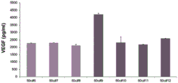
Fig. 18: VEGF-A levels of conditioned medium of MLCs produced in V2.2 on Corning Synthesis DMC in BioBLU 50c bioreactor.
Fig. 19: ANGPT1 levels of conditioned medium of MLC produced in V2.2 on corning synthenax DMC in a bio blu 50c bioreactor.
Fig. 20: MLCs produced in V2.2 on corning Synthesis DMC in BioBLU 50c bioreactor consistently showed a strong ability to inhibit proliferation of activated allogeneic T cells.
Fig. 21: schematic of a bioreactor process for MLC fabrication.
Detailed Description
General techniques and definitions
Unless specifically stated otherwise, all technical and scientific terms used herein should be taken to have the same meaning as commonly understood by one of ordinary skill in the art (e.g., in cell culture, molecular biology, stem cell culture, immunology, and biochemistry).
Unless otherwise indicated, cell culture techniques and assays used in the present disclosure are standard procedures well known to those skilled in the art. Such techniques are described and explained in the literature of the following sources: such as J.Perbal, molecular cloning Utility Specification (A practical Guide To Molecular Cloning), john Wili's father-son publishing company (John Wiley and Sons) (1984); sambrook et al, molecular cloning: laboratory Manual (Molecular Cloning: A Laboratory Manual), cold spring harbor laboratory Press (Cold Spring Harbour Laboratory Press) (1989); brown (edit), "basic molecular biology: practical methods (Essential Molecular Biology: A Practical Approach), volumes 1 and 2, IRL Press (1991); glover and B.D.Hames (editions), and F.M.Ausubel et al (editions), guidelines for contemporary molecular biology experiments (Current Protocols in Molecular Biology), greene Pub.associates, and Wiley International science publication (Wiley-Interscience) (1988, including all updates to date); ed Harlow and David Lane (edit) antibody: laboratory manuals (Antibodies: A Laboratory Manual), cold spring harbor laboratory, (1988); and J.E.Coligan et al (editions), "contemporary immunology guidelines (Current Protocols in Immunology)," John Wili father-son publishing company (including all updates so far).
The term "and/or", e.g. "X and/or Y", is understood to mean "X and Y" or "X or Y", and is to be taken as providing explicit support for both meanings or for either meaning.
As used herein, unless specified to the contrary, the term "about" refers to +/-10%, more preferably +/-5% of the specified value.
The term "level" is used to define the amount of a particular substance present in the cell culture medium and compositions of the present disclosure. For example, a particular concentration, weight, percentage (e.g., v/v%) or ratio may be used to define the level of a particular substance.
The term "sufficient" is used herein to define an amount that provides a particular concentration of growth factor when dissolved in a stem cell culture medium. In this case, the "sufficient amount" is determined by the volume of medium required. For example, if the desired concentration of FGF2 in a stem cell culture medium is about 10pg/ml and 500ml of cell culture medium is required, a sufficient amount will be about 5ng.
In the context of the release of mesenchymal lineage precursors or stem cells from an adherent material, the term "sufficient" is used to refer to shaking for a period of time at a frequency and amplitude sufficient to cause the mesenchymal lineage precursors or stem cells to release from the adherent material.
The term "seeding" is used herein to refer to the process of introducing cells into 3-dimensional (3D) culture. In one example, the methods of the present disclosure encompass dynamic seeding, wherein the medium continues to mix as the cells adhere to the adherent material. In another example, cells are inoculated into 3D culture and placed for a period of time sufficient to adhere to an adherent material in the culture medium so that the cells can adhere to the material. In some embodiments, the step of seeding the cells into the bioreactor is accomplished when the flow in the bioreactor is disconnected for at least 10 hours after seeding.
In one example, the mesenchymal lineage precursor or stem cells are seeded at 5,000 cells/ml to 20,000 cells/ml. In another example, the mesenchymal lineage precursor or stem cells are seeded at 8,000 cells/ml to 20,000 cells/ml. In another example, the mesenchymal lineage precursor or stem cells are seeded at 8,000 cells/ml to 15,000 cells/ml. In another example, mesenchymal lineage precursors or stem cells are seeded at 5,000 cells/ml. In another example, mesenchymal lineage precursors or stem cells are seeded at 8,000 cells/ml. In another example, the mesenchymal lineage precursor or stem cells are seeded at least 8,000 cells/ml. In another example, mesenchymal lineage precursors or stem cells are seeded at 10,000 cells/ml.
The term "recovery" is used herein to refer to the removal of cells from 2D or 3D culture. For example, cells can be recovered from the bioreactor culture disclosed herein. In one example, the recovered cells are first washed with a saline solution or comparable solution (e.g., 2-3 times). After the washing step, the adherent material may be subjected to a dissociation step. In one example, a suitable dissociation enzyme is employed during the dissociation step. In one example, cells recovered from 3D culture are washed and concentrated prior to cryopreservation. In one example, the washed and concentrated cells may be stored, packed, finished, and visually inspected prior to cryopreservation.
As used herein, a "dissociating agent" is any compound that is used to disrupt the attachment point between a cell and the surface to which the cell is attached. In some embodiments, the dissociating agent is an enzyme. In particular embodiments, the enzyme is trypsin, comprising recombinant trypsin, papain, elastase, hyaluronidase, collagenase type 1, collagenase type 2, collagenase type 3, collagenase type 4, or dispase. In one example, the dissociating agent includes EDTA. In one example, the dissociating agent includes EDTA and an enzyme. For example, the dissociating agent may include EDTA and pectinase. In one example, the dissociating agent may include EDTA and collagenase. Those skilled in the art will appreciate that EDTA may be a suitable dissociating agent for microcarriers with a carbohydrate core attached in a calcium-dependent manner. In one example, the dissociating agent also degrades the microcarriers. For example, the dissociating agent may degrade the microcarrier core.
In one example, a dissociating agent can be used to dissociate cells from the adherent materials disclosed herein. For example, a dissociating agent may be fed into the bioreactor disclosed herein to dissociate cells as desired. In one example, the cell culture may be filtered or partially filtered prior to contact with the appropriate dissociating agent. For example, the cells may be contacted with a dissociating agent after reaching a peak cell density. In another example, the cultured cells can be dissociated from the adherent material by vibration. "vibration" means mechanical oscillation about a point of equilibrium. The oscillations may be periodic or random. In one embodiment, the vibrations are due to reciprocating linear oscillations that are controlled in amplitude and frequency. In some embodiments, the amplitude and frequency of the oscillation are constant, while in other embodiments, one or both of the amplitude or frequency may be varied as needed to achieve dissociation of the cells. Other examples, the duration of the period of vibration may also be controlled using equipment and devices conventional in the art. In some examples, the vibration is provided by an electromechanical device, such as an electric motor having an unbalanced mass on its drive shaft. In other examples, the vibration is provided by an electrical device. Various examples of devices capable of imparting vibrations are known in the art.
Throughout this specification the word "comprise", or variations such as "comprises" or "comprising", will be understood to imply the inclusion of a stated element, integer or step or group of elements, integers or steps, but not the exclusion of any other element, integer or step or group of elements, integers or groups of steps.
Throughout this specification, unless the context clearly indicates otherwise, reference to a single step, composition of matter, group of steps, or group of compositions of matter should be taken to encompass one or more (i.e., one or more) of those steps, compositions of matter, group of steps, or group of compositions of matter.
It will be appreciated by those skilled in the art that variations and modifications of the disclosure described herein other than those specifically described may be made. It is to be understood that the present disclosure encompasses all such variations and modifications. The disclosure also includes all of the steps, features, compositions and compounds referred to or indicated in this specification, individually or collectively, and any and all combinations of any two or more of said steps or features.
The scope of the present disclosure is not limited by the specific embodiments described herein, which are for illustrative purposes only. Functionally equivalent products, compositions, and methods, as described herein, are clearly within the scope of the disclosure.
Any examples disclosed herein should be considered applicable to any other examples, mutatis mutandis, unless explicitly stated otherwise.
Mesenchymal precursor cells
As used herein, the term "mesenchymal lineage precursor or stem cells (MLPSC)" refers to undifferentiated pluripotent cells that have the ability to self-renew while retaining the ability to differentiate into cell types of many mesenchymal sources (e.g., osteoblasts, chondrocytes, adipocytes, stromal cells, fibroblasts, and tendons) or non-mesodermal sources (e.g., hepatocytes, neural cells, and epithelial cells). For the avoidance of doubt, "mesenchymal lineage precursor cells" refers to cells that can differentiate into mesenchymal cells such as bone, cartilage, muscle and fat cells, and fibrous connective tissue.
The term "mesenchymal lineage precursor or stem cell" encompasses the parental cell and its undifferentiated progeny. The term also encompasses mesenchymal precursor cells, pluripotent stromal cells, mesenchymal Stem Cells (MSCs), perivascular mesenchymal precursor cells, and undifferentiated progeny thereof.
The mesenchymal lineage precursor or stem cells can be autologous, allogeneic, xenogeneic, syngeneic or isogenic. Autologous cells are isolated from the same individual in which they are to be re-implanted. Allogeneic cells are isolated from a donor of the same species. The xenogeneic cells are isolated from a donor of another species. Homologous or isogenic cells are isolated from genetically identical organisms, such as twins, clones or highly inbred research animal models.
In one example, the mesenchymal lineage precursor or stem cells are allogeneic. In one example, allogeneic mesenchymal lineage precursors or stem cells are expanded in culture and cryopreserved.
Mesenchymal lineage precursors or stem cells are found predominantly in bone marrow, but are also shown to be present in a variety of host tissues including, for example, umbilical cord blood and cord, adult peripheral blood, adipose tissue, trabecular bone, and dental pulp. It is also present in skin, spleen, pancreas, brain, kidney, liver, heart, retina, brain, hair follicle, intestine, lung, lymph node, thymus, ligament, tendon, skeletal muscle, dermis and periosteum; and is capable of differentiating into a germ line, such as mesoderm and/or endoderm and/or ectoderm. Thus, mesenchymal lineage precursors or stem cells can differentiate into a wide variety of cell types including, but not limited to, fat, bone, cartilage, elastic tissue, muscle, and fibrous connective tissue. The particular lineage commitment and differentiation pathway that these cells enter depends on various effects from mechanical influences and/or endogenous bioactive factors such as growth factors, cytokines, and/or local microenvironment conditions established by the host tissues.
The term "enriched", "enriched" or variants thereof are used herein to describe a population of cells having an increased proportion of one particular cell type or of a plurality of particular cell types as compared to an untreated population of cells (e.g., cells in their natural environment). In one example, the population enriched for mesenchymal lineage precursors or stem cells includes at least about 0.1% or 0.5% or 1% or 2% or 5% or 10% or 15% or 20% or 25% or 30% or 50% or 75% of mesenchymal lineage precursors or stem cells. In this regard, the term "cell population enriched for mesenchymal lineage precursors or stem cells" will be employed to expressly support the term "cell population comprising X% mesenchymal lineage precursors or stem cells," where X% is a percentage as described herein. In some examples, the mesenchymal lineage precursor or stem cells can form clonogenic colonies, e.g., CFU-F (fibroblasts) or a subset thereof (e.g., 50% or 60% or 70% or 90% or 95%) can have this activity.
In one example of the present disclosure, the mesenchymal lineage precursor or stem cells are Mesenchymal Stem Cells (MSCs). MSCs may be of homogeneous composition or may be a mixed population of cells enriched in MSCs. Homogeneous MSC compositions can be obtained by culturing adherent bone marrow or periosteal cells, and MSCs can be identified by specific cell surface markers identified with unique monoclonal antibodies. For example, in U.S. Pat. No. 5,486,359, a method for obtaining a population of cells enriched in MSC is described. Alternative sources of MSCs include, but are not limited to, blood, skin, cord blood, muscle, fat, bone, and perichondrium. In one example, the MSC is allogeneic. In one example, the MSC is cryopreserved. In one example, MSCs are culture expanded and cryopreserved.
In another example, the mesenchymal lineage precursor or stem cell is cd29+, cd54+, cd73+, cd90+, cd102+, cd105+, cd106+, cd166+, MHC1+ MSC.
Isolated or enriched mesenchymal lineage precursors or stem cells can be expanded in vitro by culture. Isolated or enriched mesenchymal lineage precursors or stem cells can be cryopreserved, thawed, and subsequently expanded in vitro by culture.
In one example, the isolated or enriched mesenchymal lineage precursor or stem cells are at 50,000 viable cells/cm 2 Inoculated in a medium (serum-free or supplemented), e.g. supplemented with 5% foetal calfSerum (FBS) and glutamine in an alpha minimum essential medium (alpha MEM) and allowed to stand at 37℃at 20% O 2 Adhere to the culture vessel overnight. The medium is then replaced and/or changed as required, and the cells are incubated at 37℃with 5% O 2 The lower culture was continued for an additional 68 to 72 hours.
As will be appreciated by those skilled in the art, cultured mesenchymal lineage precursors or stem cells are phenotypically different from in vivo cells. For example, in one embodiment, it expresses one or more of the following markers: CD44, NG2, DC146, and CD140b. The cultured mesenchymal lineage precursor or stem cells are also biologically different from in vivo cells, with higher proliferation rates than most non-circulating (quiescent) cells in vivo.
In one example, the population of cells is enriched from a cell preparation comprising an alternative form of STRO-1+ cells. In this regard, the term "selectable form" will be understood to mean that the cells express a marker (e.g., a cell surface marker) that allows selection of STRO-1+ cells. The marker may be STRO-1, but is not necessarily. For example, as described and/or exemplified herein, cells expressing STRO-2 and/or STRO-3 (TNAP) and/or STRO-4 and/or VCAM-1 and/or CD146 and/or 3G5 (e.g., mesenchymal precursor cells) also express STRO-1 (and may be STRO-1 bright). Thus, the indication that the cell is STRO-1+ does not mean that the cell is selected by STRO-1 expression alone. In one example, cells are selected based at least on STRO-3 expression, e.g., which is STRO-3+ (TNAP+).
References to the selection of cells or populations thereof do not necessarily require selection from a particular tissue source. STRO-1+ cells may be selected from or isolated or enriched from a variety of sources, as described herein. That is, in some examples, these terms provide support for selection from any tissue or vascularized tissue comprising STRO-1+ cells (e.g., mesenchymal precursor cells) or tissue comprising pericytes (e.g., STRO-1+ pericytes) or any one or more of the tissues described herein.
In one example, the cells used in the present disclosure express one or more markers, either alone or in combination, selected from the group consisting of: TNAP+, VCAM-1+, THY-1+, STRO-2+, STRO-4+ (HSP-90 beta), CD45+, CD146+, 3G5+, or any combination thereof.
By "individually" is meant that the present disclosure individually encompasses the markers or sets of markers, and although individual markers or sets of markers may not be individually listed herein, the appended claims may define such markers or sets of markers individually and separately from each other.
"collectively" means that the present disclosure encompasses any number or combination of the markers or sets of markers, and that although such number or combination of markers or sets of markers may not be specifically listed herein, the appended claims may define such combination or sub-combination separately and separately from any other marker combination or set of markers.
As used herein, the term "TNAP" is intended to encompass all isoforms of tissue-non-specific alkaline phosphatase. For example, the term encompasses liver isotype (LAP), bone isotype (BAP) and kidney isotype (KAP). In one example, the TNAP is BAP. In one example, TNAP as used herein refers to a molecule that can bind to STRO-3 antibodies produced by a hybridoma cell line deposited with ATCC under the provisions of the Budapest Treaty at 12/19/2005 under deposit accession number PTA-7282.
Furthermore, in one example, STRO-1+ cells are capable of producing clonogenic CFU-F.
In one example, a significant proportion of STRO-1+ cells are capable of differentiating into at least two different lineages. Non-limiting examples of lineages into which STRO-1+ cells may committed include: bone precursor cells; a hepatocyte progenitor cell having multipotency for biliary epithelial cells and hepatocytes; a neural restricted cell that can produce glial cell precursors that progress into oligodendrocytes and astrocytes; a neuron precursor that progresses to a neuron; myocardium and precursors of cardiomyocytes, glucose-responsive insulin secreting pancreatic beta cell lines. Other lineages include, but are not limited to, odontoblasts, dentin-producing cells and chondrocytes, as well as precursor cells of: retinal pigment epithelial cells, fibroblasts, skin cells such as keratinocytes, dendritic cells, hair follicle cells, renal catheter epithelial cells, smooth and skeletal muscle cells, testicular progenitor cells, vascular endothelial cells, tendons, ligaments, cartilage, adipocytes, fibroblasts, bone marrow stroma, cardiac muscle, smooth muscle, skeletal muscle, pericytes, blood vessels, epithelial cells, glial cells, neurons, astrocytes and oligodendrocytes.
In one example, the mesenchymal lineage precursors or stem cells are obtained from a single donor or multiple donors, where the donor samples or mesenchymal lineage precursors or stem cells are then pooled and then culture expanded.
Mesenchymal lineage precursors or stem cells encompassed by the present disclosure can also be cryopreserved prior to administration to a subject. In one example, the mesenchymal lineage precursor or stem cells are culture expanded and cryopreserved prior to administration to a subject.
In one example, the present disclosure encompasses mesenchymal lineage precursors or stem cells and their progeny, soluble factors derived therefrom, and/or extracellular vesicles isolated therefrom. In another example, the disclosure encompasses mesenchymal lineage precursors or stem cells and extracellular vesicles isolated therefrom. For example, mesenchymal precursor lineages or stem cells of the present disclosure may be expanded in culture for a period of time under conditions suitable for secretion of extracellular vesicles into the cell culture medium. Secreted extracellular vesicles can then be obtained from the culture medium for use in therapy.
As used herein, the term "extracellular vesicles" refers to lipid particles that are naturally released from cells and range in size from about 30nm to as large as 10 microns, but are typically less than 200nm in size. It may contain cells derived from the release (e.g., mesenchymal stem cells; STRO-1) + Cells), proteins, nucleic acids, lipids, metabolites or organelles.
As used herein, the term "exosome" refers to a type of extracellular vesicle that is typically in the range of about 30nm to about 150nm in size and that is derived from the endosomal compartment of a mammalian cell from which it is transported to the cell membrane and released. It may contain nucleic acids (e.g., RNA; microRNA), proteins, lipids, and metabolites, and may play a role in intercellular communication by being secreted from one cell and taken up by other cells to deliver its cargo.
Culture expansion of cells
In one example, the mesenchymal lineage precursor or stem cells are expanded by culture. The "culture expanded" mesenchymal lineage precursors or stem cell culture media are distinguished from freshly isolated cells in that they have been cultured and passaged in cell culture media (i.e., subcultured). In one example, culture-expanded mesenchymal lineage precursors or stem cells are culture-expanded for about 4-10 passages. In one example, the mesenchymal lineage precursor or stem cells are expanded by culture at least 5 passages, at least 6 passages, at least 7 passages, at least 8 passages, at least 9 passages, at least 10 passages. For example, mesenchymal lineage precursors or stem cells can be expanded by culture for at least 5 passages. In one example, the mesenchymal lineage precursor or stem cells can be culture expanded at least 5 passages to 10 passages. In one example, the mesenchymal lineage precursor or stem cells can be culture expanded at least 5 passages to 8 passages. In one example, the mesenchymal lineage precursor or stem cells can be culture expanded at least 5 passages to 7 passages. In one example, the mesenchymal lineage precursor or stem cells can be culture expanded for more than 10 passages. In another example, the mesenchymal lineage precursor or stem cells can be culture expanded for more than 7 passages. In these examples, stem cells can be culture expanded prior to being cryopreserved to provide a moderately cryopreserved MLPSC population. In one example, the methods of the present disclosure culture cells from a moderately cryopreserved MLPSC population.
In one embodiment, the mesenchymal lineage precursors or stem cells can be obtained from a single donor or multiple donors, where the donor samples or mesenchymal lineage precursors or stem cells are then pooled and then culture expanded. In one example, the culture amplification process comprises:
i. expanding a plurality of living cells by passaging to provide a preparation of at least about 10 hundred million living cells, wherein passaging comprises establishing a primary culture of isolated mesenchymal lineage precursors or stem cells, and then continuously establishing a first non-primary (P1) culture of mesenchymal lineage precursors or stem cells isolated from a previous culture;
expanding the P1 culture of isolated mesenchymal lineage precursors or stem cells to a second non-primary (P2) culture of mesenchymal lineage precursors or stem cells by passaging expansion; and
preparing and cryopreserving an intermediate mesenchymal lineage precursor or stem cell preparation in a process obtained from P2 culture of mesenchymal lineage precursors or stem cells; and
thawing the intermediate mesenchymal lineage precursor or stem cell preparation in the cryopreserved treatment and amplifying the intermediate mesenchymal lineage precursor or stem cell preparation in the treatment by passaging expansion.
In one example, the expanded mesenchymal lineage precursor or stem cell preparation has an antigen profile and an activity profile, the antigen profile and activity profile comprising:
i. Less than about 0.75% cd45+ cells;
at least about 95% cd105+ cells;
at least about 95% cd166+ cells.
In one example, the expanded mesenchymal lineage precursor or stem cell preparation is capable of inhibiting IL2Ra expression of CD3/CD28 activated PBMCs by at least about 30% relative to a control.
In one example, the culture-expanded mesenchymal lineage precursor or stem cells are culture-expanded for about 4-10 passages, where the mesenchymal lineage precursor or stem cells are cryopreserved after at least 2 passages or 3 passages before being further culture-expanded. In one example, the mesenchymal lineage precursor or stem cell is culture expanded at least 1 passage, at least 2 passages, at least 3 passages, at least 4 passages, at least 5 passages, cryopreserved, and then further culture expanded at least 1 passage, at least 2 passages, at least 3 passages, at least 4 passages, at least 5 passages prior to culturing according to the methods of the present disclosure.
The process of mesenchymal lineage precursor or stem cell isolation and ex vivo expansion can be performed using any apparatus and cell handling method known in the art. Various culture expansion embodiments of the present disclosure employ steps requiring manipulation of cells, e.g., seeding, feeding, dissociating or washing of adherent cultures. Any step of manipulating the cells may damage the cells. Although mesenchymal lineage precursors or stem cells can generally sustain a certain amount of damage during preparation, the cells are preferably manipulated by a treatment procedure and/or apparatus that adequately performs the given steps while minimizing damage to the cells.
In an example, mesenchymal lineage precursors or stem cells are washed in an apparatus comprising a cell source bag, a wash solution bag, a recycle wash bag, a rotating membrane filter with inlet and outlet ports, a filtrate bag, a mixing zone, a final product bag for washed cells, and appropriate tubing, for example, as described in US 6251295, which is hereby incorporated by reference.
In one example, a mesenchymal lineage precursor or stem cell composition cultured according to the present disclosure is 95% homogeneous in CD105 positive and CD166 positive and CD45 negative. In one example, this homogeneity persists through ex vivo amplification; i.e. although multiple population doublings.
In one example, the mesenchymal lineage precursors or stem cells of the present disclosure are culture expanded in 2D culture prior to 3D culture. In one example, the mesenchymal lineage precursors or stem cells of the present disclosure are culture expanded from a master cell bank. In one example, mesenchymal lineage precursor or stem cells of the present disclosure are culture expanded from a master cell bank in 2D culture prior to seeding in 3D culture. In one example, the mesenchymal lineage precursors or stem cells of the present disclosure are culture expanded in a bioreactor from a master cell bank in 2D culture for at least 3 days prior to seeding in 3D culture. In one example, the mesenchymal lineage precursors or stem cells of the present disclosure are culture expanded in a bioreactor from a master cell bank in 2D culture for at least 4 days prior to seeding in 3D culture. In one example, the mesenchymal lineage precursor or stem cells of the present disclosure are culture expanded in a bioreactor in 2D culture from a master cell bank for 3 days to 5 days prior to seeding in 3D culture. In these examples, the 2D culture may be performed in a cell factory. Various cell factory products are commercially available (e.g., siemens, sigma, thermofiser).
Modification of cells
Mesenchymal lineage precursors or stem cells cultured according to the present disclosure can be altered in such a way that upon administration, lysis of the cells is inhibited. The alteration of the antigen may induce immune non-responses or tolerance, thereby preventing effector phases (e.g., cytotoxic T cell production, antibody production, etc.) that induce immune responses that ultimately lead to rejection of the foreign cells in a normal immune response. Antigens that can be altered to achieve this goal include, for example, MHC class I antigens, MHC class II antigens, LFA-3, and ICAM-1.
Mesenchymal lineage precursors or stem cells can also be genetically modified to express proteins that are important for differentiation and/or maintenance of striated skeletal muscle cells. Exemplary proteins include growth factors (TGF-beta, insulin-like growth factor 1 (IGF-1), FGF), myogenic factors (e.g., myoD, myogenic factor 5 (Myf 5), myogenic Regulatory Factor (MRF)), transcription factors (e.g., GATA-4), cytokines (e.g., cardiophilin-1), neuregulin family members (e.g., neuregulin 1, 2, and 3), and homeobox genes (e.g., csx, tinman, and NKx families).
Cell culture medium
The methods of the present disclosure use a fetal bovine serum free stem cell medium comprising growth factors that promote proliferation of mesenchymal lineage precursors or stem cells. In one embodiment, the medium is a serum-free stem cell medium. In one example, in addition to the adhesive materials discussed below, the cell culture media used in the methods of the present disclosure include:
a basal medium;
platelet Derived Growth Factor (PDGF);
fibroblast growth factor 2 (FGF 2).
The term "medium" as used in the context of the present disclosure comprises a component of the surrounding environment of a cell. The culture medium facilitates and/or provides conditions suitable to allow cell growth. The medium may be solid, liquid, gaseous or a mixture of phases and materials. The medium may comprise a liquid growth medium and a liquid medium that does not sustain cell growth. The medium also comprises a gelatinous medium such as agar, agarose, gelatin and collagen matrix. Exemplary gaseous media comprise a gaseous phase to which cells grown on a petri dish or other solid or semi-solid support are exposed. The term "medium" also refers to a material intended for cell culture even though it has not been in contact with cells.
The media of the present disclosure may be prepared by using a basal medium. In the context of the present disclosure, "basal medium" refers to an unsupplemented medium suitable for exposure to cells, such as mesenchymal precursor lineages or stem cells. The basal medium comprises, for example, eagl Minimum Essential (MEM) medium, alpha-modified MEM medium, stemSpan TM And a mixed medium thereof, and is not particularly limited as long as it can be used for culturing stem cells.
Further, the cell culture media of the present disclosure may comprise any component, such as fatty acids or lipids, vitamins, cytokines, antioxidants, buffers, inorganic salts, and the like.
The cell culture media used in the present disclosure contain all essential amino acids, and may also contain non-essential amino acids. Generally, amino acids are classified as essential amino acids (Thr, met, val, leu, ile, phe, trp, lys, his) and non-essential amino acids (Gly, ala, ser, cys, gln, asn, asp, tyr, arg, pro).
Those skilled in the art will appreciate that in order to achieve optimal results, the basal medium must be suitable for the cell line of interest with critical nutrients that can be used at levels sufficient to enhance cell proliferation. For example, if glucose (or other energy source) in the basal medium is found to have been depleted and thus limit cell proliferation, it may be desirable to increase the level of this energy source, or to add glucose (or other energy source) during the culture process. In one example, the Dissolved Oxygen (DO) level may also be controlled.
In one example, the cell culture media of the present disclosure contains additives of human origin. For example, human serum and human platelet cell lysate can be added to the cell culture medium used in the methods of the present disclosure.
In one example, the cell culture media of the present disclosure contains only additives of human origin. Thus, in one example, the cell culture medium is xeno-free. For the avoidance of doubt, in these examples the medium is animal protein free. In one example, the cell culture medium used in the methods of the present disclosure is free of animal components.
Ascorbic acid
Ascorbic acid is an essential supplement for the growth and differentiation of various kinds of cells in culture. It should now be understood that certain ascorbic acid derivatives are "short acting" in that they are unstable in solution, particularly under normal cell culture conditions of neutral pH and 37 ℃. These short-acting derivatives oxidize rapidly to oxalic acid or threonic acid. Oxidation reduced the levels of these short-acting ascorbic acid derivatives by about 80-90% in 24 hours in 37 ℃ medium (pH 7). Thus, in conventional cell culture of various cell types, the short-acting ascorbic acid derivatives have been replaced with more stable "long-acting" ascorbic acid derivatives.
In the context of the present disclosure, the term "short-acting" encompasses ascorbic acid derivatives that are approximately 80-90% oxidized after cell culture for 24 hours at neutral pH and 37 ℃. In one example, the short acting L-ascorbic acid derivative is L-ascorbate. For example, in the context of the present disclosure, the sodium salt of L-ascorbic acid is a "short-acting" ascorbic acid derivative.
In contrast, the term "long-acting" encompasses ascorbic acid derivatives that are about 80% -90% unoxidized after cell culture for 24 hours at neutral pH and 37 ℃. In one example, in the context of the present disclosure, L-ascorbic acid-2-phosphate is a "long-acting" ascorbic acid derivative. Other examples of long acting ascorbic acid derivatives include tetrahexyldecanol ascorbate, magnesium ascorbyl phosphate, and 2-O-alpha-D-glucopyranosyl-L-ascorbic acid. The cell culture media of the present disclosure may contain a short acting ascorbic acid derivative, a long acting ascorbic acid derivative, or a mixture thereof.
Mitogenic factors
PDGF and FGF2 synergistically promote stem cell proliferation in cell culture in vitro without fetal bovine serum.
PDGF is a regulator of cell growth and division that binds to platelet-derived growth factor receptor (PDGFR). In chemical terms PDGF is a dimeric glycoprotein consisting of two a (-AA) or two B (-BB) chains or a combination of both chains (-AB). PDGF-AB has been shown to bind to PDGF alpha and beta receptor subunits to form PDGF alpha beta and alpha receptor dimers. In the context of the present disclosure PDGF encompasses PDGF-BB and PDGF-AB.
Basic fibroblast growth factor (FGF 2) is also known as BFG, FGFB, and HBGF-2 is a member of the Fibroblast Growth Factor (FGF) family. FGF2 is also a regulator of cell growth and division. Both PDGF and FGF2 can be classified as mitogens because they promote the initiation of cell division by cells.
In one example, the methods of the present disclosure comprise culturing a population of stem cells in a fetal bovine serum-free cell culture medium comprising platelet-derived growth factor (PDGF) and fibroblast growth factor 2 (FGF 2), wherein the FGF2 level is less than about 6ng/ml. For example, FGF2 levels may be less than about 5ng/ml, less than about 4ng/ml, less than about 3ng/ml, less than about 2ng/ml, less than about 1ng/ml. In other examples, the FGF2 level is less than about 0.9ng/ml, less than about 0.8ng/ml, less than about 0.7ng/ml, less than about 0.6ng/ml, less than about 0.5ng/ml, less than about 0.4ng/ml, less than about 0.3ng/ml, less than about 0.2ng/ml.
In another example, the level of FGF2 is between about 1pg/ml and 100 pg/ml. In another example, the FGF2 level is between about 5pg/ml and 80 pg/ml. In another example, the FGF2 level is between about 10pg/ml and 40 pg/ml. In another example, the FGF2 level is at least about 10pg/ml. In another example, the FGF2 level is at least about 11pg/ml. In another example, the FGF2 level is at least about 12pg/ml. In another example, the FGF2 level is at least about 13pg/ml. In another example, the FGF2 level is at least about 14pg/ml. In another example, the FGF2 level is at least about 15pg/ml. In another example, the FGF2 level is at least about 16pg/ml. In another example, the FGF2 level is at least about 17pg/ml. In another example, the FGF2 level is at least about 18pg/ml. In another example, the FGF2 level is at least about 19pg/ml. In another example, the FGF2 level is at least about 20pg/ml. In another example, the FGF2 level is at least about 21pg/ml. In another example, the FGF2 level is at least about 22pg/ml. In another example, the FGF2 level is at least about 23pg/ml. In another example, the FGF2 level is at least about 24pg/ml. In another example, the FGF2 level is at least about 25pg/ml. In another example, the FGF2 level is at least about 26pg/ml. In another example, the FGF2 level is at least about 27pg/ml. In another example, the FGF2 level is at least about 28pg/ml. In another example, the FGF2 level is at least about 29pg/ml. In another example, the FGF2 level is at least about 30pg/ml.
In one example, the PDGF is PDGF-BB. In one example, PDGF-BB levels are between about 1ng/ml and 150 ng/ml. In another example, the PDGF-BB level is between about 7.5ng/ml and 120 ng/ml. In another example, the PDGF-BB level is between about 15ng/ml and 60 ng/ml. In another example, the PDGF-BB level is at least about 10ng/ml. In another example, the PDGF-BB level is at least about 15ng/ml. In another example, the PDGF-BB level is at least about 20ng/ml. In another example, the PDGF-BB level is at least about 21ng/ml. In another example, the PDGF-BB level is at least about 22ng/ml. In another example, the PDGF-BB level is at least about 23ng/ml. In another example, the PDGF-BB level is at least about 24ng/ml. In another example, the PDGF-BB level is at least about 25ng/ml. In another example, the PDGF-BB level is at least about 26ng/ml. In another example, the PDGF-BB level is at least about 27ng/ml. In another example, the PDGF-BB level is at least about 28ng/ml. In another example, the PDGF-BB level is at least about 29ng/ml. In another example, the PDGF-BB level is at least about 30ng/ml. In another example, the PDGF-BB level is at least about 31ng/ml. In another example, the PDGF-BB level is at least about 32ng/ml. In another example, the PDGF-BB level is at least about 33ng/ml. In another example, the PDGF-BB level is at least about 34ng/ml. In another example, the PDGF-BB level is at least about 35ng/ml. In another example, the PDGF-BB level is at least about 36ng/ml. In another example, the PDGF-BB level is at least about 37ng/ml. In another example, the PDGF-BB level is at least about 38ng/ml. In another example, the PDGF-BB level is at least about 39ng/ml. In another example, the PDGF-BB level is at least about 40ng/ml.
In another example, the PDGF is PDGF-AB. In one example, PDGF-AB levels are between about 1ng/ml and 150 ng/ml. In another example, the PDGF-AB level is between about 7.5ng/ml and 120 ng/ml. In another example, the PDGF-AB level is between about 15ng/ml and 60 ng/ml. In another example, the PDGF-AB level is at least about 10ng/ml. In another example, the PDGF-AB level is at least about 15ng/ml. In another example, the PDGF-AB level is at least about 20ng/ml. In another example, the PDGF-AB level is at least about 21ng/ml. In another example, the PDGF-AB level is at least about 22ng/ml. In another example, the PDGF-AB level is at least about 23ng/ml. In another example, the PDGF-AB level is at least about 24ng/ml. In another example, the PDGF-AB level is at least about 25ng/ml. In another example, the PDGF-AB level is at least about 26ng/ml. In another example, the PDGF-AB level is at least about 27ng/ml. In another example, the PDGF-AB level is at least about 28ng/ml. In another example, the PDGF-AB level is at least about 29ng/ml. In another example, the PDGF-AB level is at least about 30ng/ml. In another example, the PDGF-AB level is at least about 31ng/ml. In another example, the PDGF-AB level is at least about 32ng/ml. In another example, the PDGF-AB level is at least about 33ng/ml. In another example, the PDGF-AB level is at least about 34ng/ml. In another example, the PDGF-AB level is at least about 35ng/ml. In another example, the PDGF-AB level is at least about 36ng/ml. In another example, the PDGF-AB level is at least about 37ng/ml. In another example, the PDGF-AB level is at least about 38ng/ml. In another example, the PDGF-AB level is at least about 39ng/ml. In another example, the PDGF-AB level is at least about 40ng/ml.
In other examples, additional factors may be added to the cell culture medium. In one example, the methods of the present disclosure comprise culturing a population of stem cells in a cell culture medium that further comprises EGF that does not contain fetal bovine serum. EGF is a growth factor that stimulates cell proliferation by binding to EGFR, its receptor. In one example, the methods of the present disclosure comprise culturing a population of stem cells in a cell culture medium that further comprises EGF that does not contain fetal bovine serum. In one example, the EGF level is between about 0.1ng/ml and 7ng/ml. For example, the EGF level may be at least about 5ng/ml.
In another example, the EGF level is between about 0.2ng/ml and 3.2 ng/ml. In another example, the EGF level is between about 0.4ng/ml and 1.6 ng/ml. In another example, the EGF level is about 0.2ng/ml. In another example, the EGF level is at least about 0.3ng/ml. In another example, the EGF level is at least about 0.4ng/ml. In another example, the EGF level is at least about 0.5ng/ml. In another example, the EGF level is at least about 0.6ng/ml. In another example, the EGF level is at least about 0.7ng/ml. In another example, the EGF level is at least about 0.8ng/ml. In another example, the EGF level is at least about 0.9ng/ml. In another example, the EGF level is at least about 1.0ng/ml. In another example, the EGF level is at least about 1.1ng/ml. In another example, the EGF level is at least about 1.2ng/ml. In another example, the EGF level is at least about 1.3ng/ml. In another example, the EGF level is at least about 1.4ng/ml.
In one example, PDGF-BB levels are at least about 3.2ng/ml, EGF levels are at least about 0.8ng/ml, and FGF2 levels are at least about 0.002ng/ml. In another example, the PDGF-BB level is at least about 9.6ng/ml, the EGF level is at least about 0.24ng/ml, and the FGF2 level is at least about 0.006ng/ml. In another example, the PDGF-BB level is at least about 16ng/ml, the EGF level is at least about 0.40ng/ml, and the FGF2 level is at least about 0.01ng/ml. In another example, the PDGF-BB level is at least about 32ng/ml, the EGF level is at least about 0.80ng/ml, and the FGF2 level is at least about 0.01ng/ml.
In one example, the medium comprises 3.2ng/ml PDGF-BB, 0.08ng/ml EGF and 0.002ng/ml FGF. In another example, the medium comprises 9.6ng/ml PDGF-BB, 0.24ng/ml EGF and 0.006ng/ml FGF. In another example, the medium comprises 16ng/ml PDGF-BB, 0.4ng/ml EGF and 0.01ng/ml FGF. In another example, the medium comprises 32ng/ml PDGF-BB, 0.8ng/ml EGF and 0.02ng/ml FGF. In these examples, e.g. alphaMEM or StemSpan TM The basal medium may be supplemented with a reference amount of growth factors. In one example, the medium comprises alpha MEM or StemSpan supplemented with 32ng/ml PDGF-BB, 0.8ng/ml EGF and 0.02ng/ml FGF TM 。
In other examples, additional factors may be added to the cell culture media of the present disclosure. For example, the cell culture medium may be supplemented with one or more stimulating factors selected from the group consisting of: epidermal Growth Factor (EGF), 1 alpha, 25-dihydroxyvitamin D3 (1,25D), tumor necrosis factor alpha (TNF-alpha), interleukin-lbeta (IL-lbeta), and stroma-derived factor lalpha (SDF-lalpha). In another embodiment, the cells may also be cultured in the presence of at least one cytokine in an amount sufficient to support the growth of the cells. In another embodiment, the cells may be cultured in the presence of heparin or a derivative thereof. For example, the cell culture medium may contain about 50ng/ml heparin. In other examples, the cell culture medium contains about 60ng/ml heparin, about 70ng/ml heparin, about 80ng/ml heparin, about 90ng/ml heparin, about 100ng/ml heparin, about 110ng/ml heparin, about 120ng/ml heparin, about 130ng/ml heparin, about 140ng/ml heparin, about 150ng/ml heparin, or derivatives thereof. In one example, the heparin derivative is sulfate. Various forms of heparin sulfate are known in the art and comprise heparin sulfate 2 (HS 2). HS2 may be from a variety of sources including, for example, the liver of male and/or female mammals. Exemplary heparan sulfates therefore include male heparin sulfate (MML HS) and female heparin sulfate (FML HS).
In another example, the cell culture media of the present disclosure promote stem cell proliferation while maintaining stem cells in an undifferentiated state. Stem cells are considered undifferentiated when they have not yet become a specific differentiation lineage. As discussed above, stem cells exhibit morphological features that are distinct from differentiated cells. In addition, undifferentiated stem cells express genes that can be used as markers for detecting the differentiation status. The polypeptide product may also be used as a marker for detecting the differentiation status. Thus, one of skill in the art can readily determine whether the methods of the present disclosure maintain stem cells in an undifferentiated state using conventional morphological, genetic, and/or proteomic analysis.
Serum
Typically, stem cells are maintained in cell culture using a medium supplemented with at least about 10-15v/v% serum, typically Fetal Bovine Serum (FBS), also known as Fetal Calf Serum (FCS). The cell culture medium used in the methods of the present disclosure is a cell culture medium that does not contain fetal bovine serum. In one embodiment, the cell culture medium is supplemented with non-fetal serum. For example, the medium may be supplemented with neonatal serum or adult serum.
In another embodiment, the cell culture medium is supplemented with human serum. In one example, the cell culture medium may be supplemented with human non-fetal serum. For example, the cell culture medium may be supplemented with at least about 1v/v%, at least about 2v/v%, at least about 3v/v%, at least about 4v/v%, at least about 5v/v%, at least about 6v/v%, at least about 7v/v%, at least about 8v/v%, at least about 9v/v%, at least about 10v/v%, at least about 11v/v%, at least about 12v/v%, at least about 13v/v%, at least about 14v/v%, at least about 15v/v%, at least about 16v/v%, at least about 17v/v%, at least about 18v/v%, at least about 19v/v%, at least about 20v/v%, at least about 21v/v%, at least about 22v/v%, at least about 23v/v%, at least about 24v/v%, at least about 25v/v% human non-fetal serum.
In another example, the cell culture medium may be supplemented with human neonatal serum. For example, the cell culture medium may be supplemented with at least about 1v/v%, at least about 2v/v%, at least about 3v/v%, at least about 4v/v%, at least about 5v/v%, at least about 6v/v%, at least about 7v/v%, at least about 8v/v%, at least about 9v/v% human neonatal serum. In one example, human neonatal serum is obtained from cord blood ".
In another example, the cell culture medium may be supplemented with human adult serum. For example, the medium may be supplemented with at least about 1v/v%, at least about 2v/v%, at least about 3v/v%, at least about 4v/v%, at least about 5v/v%, at least about 6v/v%, at least about 7v/v%, at least about 8v/v%, at least about 9v/v%, at least about 10v/v%, at least about 11v/v%, at least about 12%, at least about 13v/v%, at least about 14v/v%, at least about 15v/v%, at least about 16v/v%, at least about 17v/v%, at least about 18v/v%, at least about 19v/v%, at least about 20v/v%, at least about 21v/v%, at least about 22v/v%, at least about 23v/v%, at least about 24v/v%, at least about 25v/v% human adult serum.
In one example, the human adult serum is human AB serum. For example, the cell culture medium may be supplemented with at least about 1v/v%, at least about 2v/v%, at least about 3v/v%, at least about 4v/v%, at least about 5v/v%, at least about 6v/v%, at least about 7v/v%, at least about 8v/v%, at least about 9v/v% human AB serum. In one example, the cell culture medium is supplemented with at least about 3% human AB serum.
The cell culture media of the present disclosure may also contain known serum substitutes. Serum substitutes may be, for example, albumin (e.g., lipid-rich albumin), transferrin, fatty acids, insulin, collagen precursors, trace elements, 2-mercaptoethanol or 3' -mercaptoglycerol, platelet lysate, platelet-rich plasma, or those serum substitutes suitably containing serum and the like. Such serum substitutes may be prepared by methods such as those described in International publication WO 93/30679, and commercially available products may also be used.
In one embodiment, the medium is serum-free. In one embodiment, the 3D culture is FBS serum free. In one embodiment, the 3D medium is supplemented with the above mentioned xenogeneic free serum. In one embodiment, the 3D culture is serum-free.
Adhesive material and microcarrier
By "adherent material" is meant a synthetic, naturally occurring, or a combination thereof, non-cytotoxic (i.e., biocompatible) material having a chemical structure (e.g., charged surface-exposing groups) that can hold cells on a surface or allow cells to adhere to a surface. In one example, the adhesive material is a microcarrier. A "microcarrier" is a supporting substrate that allows adherent cells to grow in a bioreactor. In one example, the microcarriers are spheres of 125-250 microns. In one example, the microcarriers are dense enough to remain suspended under gentle agitation. Microcarriers can be made of many different materials including DEAE dextran, glass, polystyrene plastic, acrylamide, collagen and alginate. In one example, the microcarrier is porous. In another example, the microcarrier is macroporous. For example, the microcarrier can have a pore size greater than about 50nm. For example, the microcarrier can have a pore size of about 50nm to about 500nm. In other examples, the microcarrier has a pore size of about 50nm to about 250nm, about 50nm to about 150nm, about 50nm to about 100nm. In one example, the microcarrier is a soluble particle. In one example, the microcarrier comprises a glycoprotein. In one example, the glycoprotein is synthetic. In one example, the microcarrier comprises cellulose, glass fibers, ceramic particles, matrix gel, extracellular matrix components, collagen, poly-L-lactic acid, dextran, inert metal fibers, silica, glass, chitosan, or plant sponge. In one example, the microcarrier comprises one or more of fibronectin, vitronectin, chondronectin, or laminin. For example, the microcarrier may be coated with one or more of fibronectin, vitronectin, chondronectin, or laminin. In other examples, the microcarriers are electrostatically charged. In one example, the microcarriers are coated with a glycoprotein. In other examples, the microcarriers are coated with collagen or gelatin. In other examples, the microcarriers are coated with collagen or vitronectin. In one example, the microcarriers are coated with vitronectin (e.g., human vitronectin). In one example, the microcarriers are coated with a derivatized vitronectin (e.g., human vitronectin). In one example, the coating is synthetic. In one example, the coating is free of xenogenic species.
In one example, the microcarrier is degradable. For example, the microcarrier may be enzymatically degradable. In one example, the microcarrier is degradable and porous. In one example, the microcarrier is degradable and macroporous. In one example, the microcarrier has a degradable core. In another example, the microcarrier has a polymeric core. For example, the microcarrier may have a carbohydrate polymer core. For example, the microcarrier may have a synthetic carbohydrate polymer core. In one example, the carbohydrate polymers are linked in a calcium dependent manner. In these examples, the microcarrier core may be coated. Exemplary coatings are as discussed above. For example, the microcarrier core may have a collagen or vitronectin coating.
In one example, the microcarrier has a density between 0.5g/ml and 3 g/ml. In another example, the microcarrier has a density between 0.5g/ml and 2 g/ml. In one example, the microcarrier has a density of about 1g/ml.
Other examples of microcarriers are outlined by Chen et al 2020, "Biotechnology letters", 42:1-10.
Examples of microcarriers suitable for use in the present disclosure include a Cultispher-G microcarrier and a Corning DMC microcarrier. In one example, these microcarriers may be coated with a coating that is free of heterogenous species. In one example, these microcarriers are coated with a glycoprotein. For example, the microcarriers may be coated with collagen or an fibronectin, such as vitronectin, or a synthetic derivative thereof.
In one example, the adhesive materials and microcarriers disclosed herein are degraded as part of the methods disclosed herein. Those skilled in the art will appreciate that the method used to degrade the adhesive material or microcarrier will be determined by its composition. For example, the adherent material or microcarrier may be degraded by enzymatic digestion. For example, vitronectin coated microcarriers can be degraded using rpfectase. In these examples, the adherent material or microcarrier may be degraded by adding enzymes to the culture medium. Other examples of suitable enzymes include TrypLE and collagenase, depending on the nature of degradation desired.
In one example, the medium includes between 0.5g/L and 12g/L microcarriers. In another example, the medium comprises between 0.5g/L and 10g/L microcarriers. In another example, the medium comprises between 0.5g/L and 5g/L microcarriers. In another example, the medium comprises between 0.5g/L and 3g/L microcarriers. In another example, the medium includes 1g/L microcarriers. For example, the culture medium may include 1g/L collagen-coated microcarriers. In another example, the medium may include 1g/L vitronectin coated microcarriers.
3D culture and bioreactor
In some embodiments, the culturing according to the present disclosure is effected in 3D culture. For example, 3D culture may be performed in a bioreactor. Examples of such bioreactors include, but are not limited to, plug flow bioreactors, continuous stirred tank bioreactors, and fixed bed bioreactors. For example, a three-dimensional plug flow bioreactor (as described in U.S. patent No. 6,911,201) is capable of supporting the growth and long-term maintenance of adherent cells described herein. In this bioreactor, adherent stromal cells are seeded onto the microcarriers discussed above, packed in a column, thereby enabling large cell numbers to be propagated in relatively small volumes. Other 3D bioreactors may be used in the present disclosure. Another example is a continuous stirred tank bioreactor. Various stirred tank bioreactors are commercially available. Those skilled in the art will appreciate that the impeller position may need to be optimized. Other examples include fixed bed bioreactors, airlift bioreactors, cell seeding perfusion bioreactors, and radial flow perfusion bioreactors. Other bioreactors that may be used in accordance with the present disclosure are described in U.S. patent nos. 6,277,151, 6,197,575, 6,139,578, 6,132,463, 5,902,741, and 5,629,186. In another example, the bioreactor is a stirred tank bioreactor. In another example, the bioreactor is a packed bed bioreactor. In one example, the packed bed bioreactor is a stirred tank, single use vessel. In one example, the packed bed bioreactor is a BioBLU series vessel manufactured by Ai Bende company (Eppendorf).
In one example, the cells are cultured in 3D culture for at least 5 days. In another example, the cells are cultured in 3D culture for at least 6 days. In another example, the cells are cultured in 3D culture for at least 7 days. In another example, the cells are cultured in 3D culture for at least 8 days. In another example, the cells are cultured in 3D culture for at least 9 days. In another example, the cells are cultured in 3D culture for at least 10 days. In another example, the cells are cultured in 3D culture for 5 days to 10 days. In another example, the cells are cultured in 3D culture for 6 days to 8 days. Those skilled in the art will appreciate that cells are typically cultured to peak cell densities. The time to peak cell density can be determined by the number of cells seeded. Thus, in another example, the cells are seeded at about 10,000 cells/ml and cultured in 3D culture for at least 6 days. In another example, the cells are seeded at about 10,000 cells/ml and cultured in 3D culture for at least 7 days. In another example, cells are seeded at about 10,000 cells/ml and cultured in 3D culture for 6 days to 8 days.
In one example, 60% to 80% of the medium is changed every 24 hours. In another example, 65% to 75% of the medium is changed every 24 hours. In one example, about 70% of the medium is changed every 24 hours. In these examples, the medium may be replaced by pouring the medium into and out of the bioreactor. In these examples, the medium was changed from day 3 of culture in the bioreactor.
Peak cell density
In performing the methods of the present disclosure, in one example, cells are cultured to peak cell density in 3D culture. For example, cells may be cultured in a bioreactor to a peak cell density. In one example, culturing according to the methods of the present disclosure smoothes the number of living cells after the peak cell density is reached. In one example, the number of living cells is greater than 75% 24 hours after the peak cell density is reached. In another example, the number of living cells is greater than 80% 24 hours after the peak cell density is reached. In another example, the number of living cells is greater than 85% 24 hours after the peak cell density is reached. In another example, the number of living cells is greater than 90% 24 hours after the peak cell density is reached. In another example, the number of living cells is greater than 95% 24 hours after the peak cell density is reached. In one example, the number of living cells is greater than 75% 48 hours after the peak cell density is reached. In another example, the number of living cells is greater than 80% 48 hours after the peak cell density is reached. In another example, the number of living cells is greater than 85% 48 hours after the peak cell density is reached. In another example, the number of living cells is greater than 90% 48 hours after the peak cell density is reached. In another example, the number of living cells is greater than 95% 48 hours after the peak cell density is reached.
In one example, the methods of the present disclosure produce 150 to 200 million cells in a 50L bioreactor. In another example, the methods of the present disclosure produce 150 to 180 hundred million cells in a 50L bioreactor. In another example, the methods of the present disclosure produce 150 to 200 million cells in a 50L bioreactor, wherein the initial medium volume is 40L. In another example, the methods of the present disclosure produce 150 to 180 hundred million cells in a 50L bioreactor, with a starting medium volume of 40L. In these examples, the starting medium will need to be replaced over time, and thus the total volume of medium used to reach a specified number of cells will be greater than 40L, as will be appreciated by those skilled in the art.
Composition and method for producing the same
The present disclosure encompasses compositions comprising a population of mesenchymal lineage precursors or stem cells and a cell culture medium, wherein the cell culture medium is serum-free and comprises an adhesion material, PDGF, and FGF2, and wherein the mesenchymal lineage precursors or stem cells are attached to the adhesion material. In one example, the adhesive material is the microcarrier mentioned above.
In one example, the composition may optionally be packaged in a suitable container with written instructions for the desired purpose, such as mixing the composition with a cell culture medium to provide a specific concentration.
In one example, the composition is provided in a bioreactor.
It will be appreciated by persons skilled in the art that numerous variations and/or modifications may be made to the invention as shown in the specific embodiments without departing from the spirit or scope of the invention as broadly described. The present embodiments are, therefore, to be considered in all respects as illustrative and not restrictive.
All publications discussed and/or mentioned herein are incorporated herein in their entirety.
Any discussion of documents, acts, materials, devices, articles or the like which has been included in the present specification is solely for the purpose of providing a context for the present invention. This is not to be taken as an admission that any or all of these matters form part of the prior art base or were common general knowledge in the field relevant to the present invention as it existed before the priority date of each claim of this application.
The present application claims priority to AU2020901931 filed on 11/6/2020, which is incorporated herein by reference.
Examples:
example 1: preparation of culture medium
Human platelet lysate
Human platelet lysate (hPL) is widely reported in the published stem cell literature as a more potent stimulator of Mesenchymal Lineage Cell (MLC) growth than Fetal Bovine Serum (FBS). In a short term proliferation assay performed in 96-well plates, two batches of hPL from 4 different suppliers were screened for their ability to stimulate proliferation of three different batches of MLC (each batch derived from a different donor). Based on this preliminary screening, the performance of the other seven batches of hPL from a single vendor on one batch of MLC was examined. The results indicate that the MLC reached plateau at approximately 5 v/v% in response to dose-dependent stimulation of the increase in hPL concentration. According to published literature, at optimal hPL concentrations, proliferation was more than 2-fold that induced by 10% FBS in the same assay (data not shown). Based on these data, hPL was used as a surrogate for FBS in a three-fold BioR run in a millbox 50L CellReady bioreactor using the same MLC library with media including FBS as used in the previous run. These three runs were performed using Solohill collagen coated microcarriers (collagen coated polystyrene microcarriers).
The runs performed in medium supplemented with 5 vol/vol% and 7.5 vol/vol% hPL are shown in fig. 1. These runs showed a significant increase in cell proliferation rate, with MLC reaching cell numbers of 110 billion (day 7) to 247 billion (day 10) on those days of run termination. These three runs produced peak numbers of 107 hundred million (day 11), 112 hundred million (day 9) and 199 hundred million (day 10) MLCs on those days specified in brackets. Overall, these data indicate that the medium composition is critical to ensure a high yield bioreactor (BioR) process for the manufacture of MLC. FBS is clearly a very poor stimulus for MLC growth on microcarriers in millbo Mobius 50L SUB, whereas hPL represents a more potent stimulus, consistent with its effect on MLC proliferation in 2-D.
V2.2 Medium
However, one important concern of using hPL to make MLCs on a commercial scale is a sufficient number of availability. There is a lack of comprehensive supply chain analysis to evaluate the supplies available for cell therapy product manufacture. In addition, the batch size of hPL is currently small and needs to be pooled from multiple donors. Finally, there is currently no consensus on the most appropriate pathogen depletion strategy required to eliminate the possibility of human pathogen transmission. For these reasons, V2.2 medium was developed. This medium is animal-component free and contains a targeted combination of recombinant mitogens for MLC proliferation. V2.2 medium includes basal medium supplemented with FGF2, PDGF and EGF and other desirable substances for cell growth. Thus, V2.2 medium eliminates the safety issues of supply constraints and limiting the use of hPL.
Again, V2.2 was tested with the same MLC cell bank as used in the previous experiments to eliminate MLC as a variable and allow cross-comparison of the data with other varying variables. Cells were again seeded in 6 million cells in a millbox Mobius 50L Cell Ready bioreactor containing 15g/L Solohill collagen coated microcarriers. As shown in fig. 2, V2.2 supports very robust MLC proliferation rapidly expanding to peak densities on day 8 in all 3 replicates, with peak cell densities varying between 152 and 177 billions of cells. Thereafter, a dramatic and problematic decrease in cell number occurred during the next 24-48 hours.
When tested in parallel with MLCs from two additional donors, MCB010 and MCB020, using the Master Cell Bank (MCB) MCB006, V2.2 supports proliferation of all three cells. However, for the two additional donors tested, a rapid decrease in cell number was also observed, which was previously observed after the MCB006 reached peak cell density (fig. 3).
In summary, V2.2 shows the ability to support robust proliferation of MLCs from multiple donors when grown on collagen-coated microcarriers in a millbox Mobius 50L CellReady bioreactor. However, after reaching peak cell density in each run, the cell number rapidly decreased. Measurement of lactate and ammonium levels at the time of the traumatic injury did not show toxic levels nor were glucose levels or recombinant cytokine levels limited. In another run that attempts to prevent such a drop in cell number by increasing the frequency of medium exchange at about the time of descent, no drop could be prevented. However, as shown in example 3 below, a stable plateau of peak cell density was achieved by varying the microcarrier and bioreactor designs.
Example 2: optimized seed preparation and inoculation of microcarriers
The following set of experiments underscores other components developed by the BioR process using V2.2 medium, aimed at optimizing the inoculation of the microcarriers and the preparation of the seeds themselves. All experiments were performed in FBS, hPL and V2.2 up to this point using seeds of 6 million cells in a millbox 50LCellReady bioreactor as described above. To determine the optimal seeding density for seeding Solohill collagen coated microcarriers in V2.2 medium, experiments were performed in a Corning 125mL rotator flask.
The effect of seeded cell density on the final number of MLCs recovered at harvest on day 7 was examined (data not shown). A rotator containing 100mL v2.2 and 15g/L collagen coated microcarriers (in triplicate) was inoculated with 100,000 to 2 million MLCs, corresponding to inoculation densities of 1,000MLC/mL, 2,000MLC/mL, 5,000MLC/mL, 8,000MLC/mL, 10,000MLC/mL and 20,000MLC/mL. The number of cells harvested at the end point was gradually increased to 10 to 50 ten thousand cells inoculated, reaching a plateau at about 80 ten thousand cells. Based on these data, the number of cells seeded per mL was 10,000/mL in all subsequent small and large scale experiments.
As mentioned above, the optimal seed density per mL was determined to be 10,000MLC at the donor cell bank stage. For a 40L BioR volume, this is equivalent to 4 hundred million cells. To determine the optimal medium exchange/harvest time required to reproducibly generate this number of cells from the contents of a single MCB vial in V2.2 medium in a CF10 cell factory, preliminary experiments were performed with MCBs from two different donors that consistently produced high or low yields. The contents of a single vial of each donor were inoculated into 3 CF10 cell factories and the following V2.2 medium exchange/harvest time protocol was performed to identify which conditions produced >4 billion cells at harvest:
Group B (day 4 medium exchange/day 6 harvest) (D4 MX/D6H) presents the lowest condition required to generate this cell number from slow and high yield MCB. Cells from the D4MX/D6H strategy and from the other two groups were then tested in a rotator, seeded at 10,000/mL and 20,000/mL in 100mL 15g/l of V2.2+ collagen coated MC. Notably, the medium exchange/harvest protocol appeared to have no effect on the yield of MLC in the rotator, as shown in fig. 4.
The application of this D4MX/D6H protocol to MCB from 8 different donors is shown in FIG. 5. All but one MCB were the lowest target number of 4 billion cells obtained. Based on these data, the D4MX/D6H protocol was used for the remaining examples to prepare seeds.
Example 3: microcarrier selection and bioreactor design
In the previous examples, all runs, whether rotator or 50L scale, were performed using Solohill collagen coated microcarriers. One problem with using these polystyrene supports is the need to remove these supports at the end of the run at a downstream processing stage of the process. In addition, 15g/L, which corresponds to 0.75kg in a 50L bioreactor, is required to be used, which affects the cost of the commodity.
The Cultispher-G microcarrier was evaluated in view of the good binding of MLCs to collagen. The Cultispher-G microcarrier is a macroporous microcarrier composed of porcine collagen from Percell Biolytica company (Percell Biolytica). In addition to the large surface area provided by these carriers, another advantage is that at the end of the run these carriers can be removed simply by enzymatic digestion of gelatin. In preliminary experiments, it was determined that 2X final concentration of TryPLE was sufficient to completely solubilize the carrier, and digestion of this concentration and duration had no adverse effect on MLC viability.
In addition, an additional bioreactor platform, namely a BioBLU series vessel manufactured by Ai Bende company, was also evaluated. A series of experiments were performed using Cultispher-G microcarriers at rotator and intermediate scale (BioBLU 3 c). Representative experiments are shown in fig. 6. MCB019 was inoculated into triplicate BioBLU 3c vessels at 10,000/mL in full vessel volume (2.5L) in V2.2 containing either Cultispher-G microcarriers (1G/L) or Solohill collagen coated microcarriers (15G/L). On day 4 and daily thereafter, 70% by volume of the medium was replaced with fresh V2.2 (batch process). The combination of Cultispher-G microcarriers and BioBLU containers resulted in a steady increase in MLC number/mL, reaching a plateau around day 7 with three runs with peak numbers of 738,000/mL, 637,000/mL and 708,000/mL. In contrast, one of the runs with Solohill vector reached a peak cell density of only 382,000/mL on day 5, followed by a plateau, while in the second run the cell number gradually increased (635,000/mL) up to day 9, but showed a significant decrease in number to 441,000/mL over the next 24 hours. These data indicate that the use of glycoprotein-coated microcarriers, such as the Cultispher-G microcarrier, and/or the BioBLU vessel design, can prevent the sharp drop in cell numbers seen previously with the combination of Solohill microcarriers in the Miybus BioR vessel. The results of additional experiments using BioBLU 3c (data not shown) further supported this impression, which demonstrated that the combination using the cultisphere-G microcarrier/BioBLU vessel design correlated consistently with the generation of a plateau phase of cell numbers after peak cell density was reached, and seemed to eliminate the significant drop in cell numbers observed with the Solohill microcarrier/millbox Mobius combination.
To test this hypothesis at full scale, 3 runs were performed, with the Cultispher-G (1G/L) vector tested in a BioBLU 50c vessel (40L scale) for comparison purposes using the same MLC library MCB006 as in the early PD runs using Solohill vectors and Mobius 50L bioreactor. These 3 runs were performed in V2.2 and cells were seeded at 10,000/mL. Feed was by a batch process in which 70% of the medium was replaced starting from day 4 of operation until it terminated.
As shown in FIG. 7, the use of the Cultispher-G microcarrier in combination with BioBLU 50c resulted in a steady exponential increase in cell numbers in all 3 replicates, peaking at day 7 with an average of 191 billion cells. Most importantly and in complete agreement with the data of the BioBLU 3c scale (fig. 6), the cell numbers were in a plateau phase in all 3 runs until the end of the 10 th day run, in sharp contrast to the sharp drop in cell numbers characterized by previous runs with the millbox Mobius 50L/Solohill collagen coated microcarrier combination (fig. 2 and 3).
Finally, fully synthesized soluble microcarrier (DMC) formulations without animal components were compared to those of Cultispher-G and Solohill collagen coated polystyrene microcarriers to study the performance of these carriers on a rotator scale. The DMC formulation comprises corning DMC particles coated with a non-heterologous collagen-containing or synthetic flexible vitronectin substrate Synthemax. DMC formulations were evaluated to address the concern that if the process was carried out with a cultisphere-G microcarrier comprising porcine collagen, the process as a whole would obviously be animal protein free, despite the obvious advantages of these carriers.
All groups were performed in V2.2 using a triplicate rotator and cells from MCB MB006 were seeded at 10,000 cells/mL. The medium replacement was performed by a batch process in which 70% of the medium was replaced from day 4 to day 7.
At day 7 harvest, the MLC reached a density between 570,000/mL (Solohill) and 730,000/mL (Corning DMC-Synthesis max; cultispher-G). Both collagen-coated corning DMC and corning DMC coated with synthetic vitronectin peptide synthamax showed significantly higher yields than those obtained using the Solohill carrier. There was no significant difference between the yields obtained with corning DMC-collagen or corning DMC-synthamax formulations and the yields supported by the cultisphere-G microcarriers (figure 8). Comparison of the yields obtained in the rotator flask with corning DMC-synthamax across a range of different cell banks demonstrates the expected differences typically observed between MLC donors (figure 9). Importantly, all showed high levels of proliferation. For example, 440,000 cells/mL of MCB019 extrapolated to about 170 million cells at 40L scale in BioBLU 50c, a value close to the number actually obtained with this cMLC cell bank in 50c runs (see fig. 10). Based on the properties exhibited by corning DMC-Synthesis in these rotator flask cultures, their performance in BioBLU 50c was next evaluated.
Perfusion-based methods for media exchange in the bio blu 50c were also optimized. This method avoids the disadvantages of the fed-batch process and, as with the batch process, replaces 70% of the volume in the vessel every 24 hours from day 3 (data not shown).
The protocol for harvesting cells at the end of the run using corning DMC-synthamax was also optimized and included the protocol recommended for microcarrier lysis by corning and the brief use of TryPLE to disrupt cell-to-cell contact. The resulting cell suspension was collected in bags (Flex protocols) and transferred to kSep 400 for washing and concentration prior to cryopreservation of the product. Seven runs were performed that combined the perfusion-based media exchange strategy with downstream harvest methods along with kSep washing and concentration steps. The operation was performed with a single MCB (MCB 019). Dissolved Oxygen (DO) was maintained in 4 runs, while it was not controlled in the remaining 3 runs. Seeds were prepared as described above and inoculated at 10,000 cells/mL into 40L V2.2 medium containing 1g/L of Corning Synthesis max DMC. Analytes and cell counts were measured daily. 1L harvest was performed on day 7 and the remaining cells were fully harvested on day 8.
FIG. 10 shows the highly reproducible yields of MLCs obtained on a 40L scale on a Corning DMC-Synthemax in BioBLU 50 c. At the average of 14.3+0.9 billion, the yield in the absence of the DO control was not significantly different from that obtained in the presence of the DO control at 1.56+0.86 (p=0.176).
In view of the above, it was noted that the use of glycoprotein coated particles such as corning DMC-Synthemax and BioBLU bioreactors is itself unexpectedly advantageous, particularly in terms of rapid decline in resting cell numbers after culture reaches peak cell density. Thus, these data support the use of microcarriers coated with an fibronectin peptide such as synthamax in 3D culture of MLPSCs.
The above example also identified the combination of Ai Bende BioBLU 50c bioreactor, corning DMC-Synthemax microparticles and V2.2 medium as a bioR process without animal components that supports reproducibly high levels of MLC yield on day 7.
Example 4: extended characterization of MSC products produced during V2.2/2-D downscaling
To investigate whether the proliferated MLC as outlined above exhibited the appropriate identity, purity and potency markers, cells harvested from each run were cryopreserved and a series of comprehensive bioassays were performed after thawing to determine any impact on the Critical Quality Attributes (CQAs) of the cells. Extended characterization assays include post-thawing viability and recovery, cell size, proliferation capacity, identity, purity, cytokine secretion, and immunomodulatory potential as measured by the ability to inhibit T cell proliferation.
Vigor and recovery after thawing
Frozen MLC from each of the 7 test samples was thawed according to rd.sop.04.06, and viability immediately after thawing was measured by trypan blue exclusion (Trypan Blue exclusion) according to the method described in PR-031. Viability was extremely consistent between all samples ranging between 96% and 97% after thawing, with no significant difference +do regulation (fig. 11).
MSC diameter after thawing
MLC size in suspension after thawing for each of 8 cryopreserved test articles was performed by flow cytometry using microbeads of known size as reference standard. The bead size was determined by the manufacturer using scanning electron microscopy and national institute of standards and technology (National Institute of Standards and Technology, NIST) traceable particles. Microbeads from Spherotec corporation (Spherotec) were used as references for the following cell diameters: 2 μm, 3 μm, 5 μm, 7 μm, 10 μm and 14.5 μm, while microbeads from Bangs laboratories (Bangs Laboratories) were used for cell sizes of 20 μm, 25 μm, 30 μm and larger. A standard curve is generated from a series of reference standards (typically spanning 5-30 μm) by plotting the bead size versus its Forward Scatter (FS) signal (linear or logarithmic peak maximum, FS median) as determined by flow cytometry. Thus, the size of the ceMSC test sample was determined from the standard curve using the FS median of the sample. The test was performed on freshly thawed samples. As shown in FIG. 12, all 7 test articles produced in BioBLU 50c on the Corning DMC-Synthesis max vector in V2.2 exhibited very similar cell diameters ranging from 21 μm (run # 8) to 24.8. Mu.m. Those test articles produced in the absence of the DO control exhibited an average cell diameter of 22.6+1.63 μm, while those test articles produced in the presence of the DO control exhibited an average diameter of 22.6±0.36 μm (p=0.477).
Proliferation ability after thawing
Analysis of proliferation kinetics in culture after thawing of MLCs from cryopreservation provides a quantitative measure of MSC function rather than an immediate measure of cell viability. Each of the 7 batches of MLCs generated during bioreactor operation was seeded in triplicate with 2,000 cells per well in serum-supplemented growth medium in 96-well plates. A batch of Mesenchymal Lineage Cells (MLC) expanded to passage 5 and inoculated in minimal essential medium alpha (αmem) containing 10% Fetal Bovine Serum (FBS) was used as a fitness control. This suitability control reliably demonstrated peak proliferative activity in this assay; thus, its activity in each experimental run is used to indicate the suitability of the basic reagents, materials and equipment used in the test procedure. Furthermore, as negative controls, each sample containing the suitability control was inoculated in a serum-free (basal) medium. Cultures were assembled to set 5% CO 2 In a humidified cell culture incubator at 37.+ -. 2 °c ZOOM real-time imaging microscope (Essen Bioscience) for 140-146 hours, and imaging was performed every 6 hours simultaneously. At the end of the incubation time, vybrant was added before plates in the IncuCyte Zoom were scanned using the green filter set TM DyeCycle TM Green (Invitrogen). The number of nuclei/wells present at the end of the incubation time under each test condition and the% confluency throughout the passage duration were determined after applying a comprehensive algorithm to mask the reagent-stained nuclei or the area occupied by the cells, respectively. As shown in fig. 13, all 7 of the BioR products exhibited proliferation kinetics very similar to the suitability control 2014CC006 grown during the current 2-D/FBS and reached confluence levels ranging from 72% (run # 7) to 93.6% (run # 10) at the end of
 day 6 of the assay. There was no significant difference between the confluence levels reached on day 6 (p=0.212) with or without the DO control. In line therewith, vybrantTM DyeCycleTM green was used to measure the number of cells per well(fig. 14) similarly demonstrates that there was no significant difference in the number of cells per well at day 6 between the DO control and the DO-free control (p=0.257).
Flow cytometry analysis of MLC identity and purity
Freshly thawed test samples were incubated with monoclonal antibodies against human CD146, STRO-4 (as a marker of MLC identity) and antibodies against CD45, CD31, CD80, CD86 and HLA-DR (as a marker of MLC purity), as well as non-binding isotype matched negative control antibodies. The antibodies used were conjugated directly with the fluorescent dye R-Phycoerythrin (PE). DAPI was used to distinguish between live and dead cells. For each of the purity markers (CD 80CD86, HLA-DR), positive control cell lines were run in parallel with ceMSC samples to confirm the measured system suitability. The samples were analyzed by flow cytometry. After removal of debris and dead cells based on light scattering properties by DAPI staining, the expression level of each marker was determined by comparing the fluorescence of antibody stained samples with their isotype negative controls. The results of these analyses are shown in fig. 15. All test articles from 7 BioR runs expressed a level of >86.8% purity marker CD146, and STRO-4 between 96% and 99.9% and between 99.76% and 99.9%. For each of the 5 purity markers analyzed, staining did not exceed 1.3% in any of the samples, and was mostly below 1%.
Example 5: functional Activity of MLC
A key attribute of MLC in a multimodal mechanism in a range of therapeutic indications is its ability to secrete a variety of paracrine activities that together mediate cellular repair and immunomodulatory functions. Seven batches of MLC were examined for the secretion of several pro-angiogenic factors as a measure of the effect of MLC to stimulate tissue repair by promoting neovascular development. The ability of cells to inhibit T cell proliferation was also assessed as a measure of their immunomodulatory properties.
SDF-1α
It is well documented that mesenchymal lineage cells secrete robust levels of various growth factors that play causal roles in the formation of new blood vessels. These growth factors include SDF-1α, VEGF-A and angiopoietin-1 (ANGPT 1). After thawing, the various test articles were inoculated in culture at equal viable cell densities and allowed to produce conditioned medium in serum-free conditions. After collecting the conditioned medium, each of the proteins of interest was quantified by ELISA (R & D Systems).
SDF-1 alpha levels in conditioned media varied between 7 MLC test article batches, ranging from approximately 3198pg/mL to 6022pg/mL (average 4246+802pg/mL) (FIG. 16). There was no significant difference in average levels of SDF-1α secreted in the absence and presence of DO maintenance (p=0.34) at 4069+203pg/mL and 4379+1027pg/mL, respectively.
The biological activity of SDF-1 alpha in samples of the same test article conditioned medium used for ELISA measurement of SDF-1 alpha concentration was also measured. For the SDF-1 a bioassay, a cell line expressing high levels of CXCR4 (U937) was inoculated into the cross-well insert and serum-free MLC conditioned medium was placed in the lower well of the 24-well plate. Cells were allowed to migrate into MLC conditioned medium or recombinant human SDF-1α for 3 hours. Serum-free basal medium without conditioning and without measurable levels of SDF-1 alpha was used as a negative control and 3ng/ml recombinant human SDF-1 alpha was used as a system suitability assay control. After a specified 3 hour time point, cells that have migrated to the bottom chamber were collected and mixed with countb right absolute count beads (invitrogen, C36950), and the number of migrated cells was quantified using flow cytometry for the standard number of count beads added to each tube.
Fig. 17 shows data from migration assays. The observed levels of each sample of U937 cells migrating to MLC conditioned medium very closely reflect the levels of SDF-1 a measured by ELISA in the corresponding samples. The average level of migration observed across all 7 samples (70,610+9067 cells) was close to the level of MLC made in the 2-D/FBS method used as a suitability control for these assays.
VEGF-A
The levels of VEGF in conditioned medium samples were similarly measured by ELISA. The level of VEGF secreted by the MLCs produced in the remaining 6 runs was very consistent with the mean 2290+148pg/mL (FIG. 18), except that one outlier, run # 9, which secreted VEGF, was at 4204 pg/mL.
ANGPT1
The levels of ANGPT1 in conditioned medium showed moderate changes from run to run, with the average level of secretion across all 7 runs being 4269+672pg/mL (fig. 19). For 3 runs performed in the absence of DO control, ANGPT levels showed minimal change (mean 3794+187 pg/mL), but runs performed in the presence of DO control, levels were slightly more variable (4625+684), but despite these differences, there was no significant difference in mean levels of ANGPT1 secreted in the absence of DO maintenance versus the presence of DO maintenance (p=0.073; p < 0.05).
Example 6: immunomodulatory activity
The ability of MLC produced in each of seven bioreactor runs to inhibit proliferation of activated T cells was evaluated as a direct measure of its immunosuppressive ability and cell quality. Briefly, for this assay, PBMCs were stimulated with CD3 and CD28 antibodies and co-cultured with MLC at different ratios (1:5, 1:10, and 1:20), and activated T cell proliferation was measured by EdU incorporation and polychromatic flow cytometry. The controls for this assay contained unstimulated PBMCs alone, stimulated PBMCs alone, and MLC batches (fitness controls) made in FBS as described above.
As shown in fig. 20, the 7/7MLC test article produced in V2.2 on corning DMC-synthamax in the bio blu 50c bioreactor showed very strong ability to inhibit T cell proliferation at a 1:10 ratio, with 3/7 inhibition >90% proliferation, which is a level very close to that of the fitness control to 92% level. The level of inhibition achieved was still very high for the remaining four BioR generated samples, and ranged from 73% -88%.
Example 7: summary
Fig. 21 shows a schematic of a bioreactor process, which represents the end point of the work outlined in the present disclosure. Starting from a single vial of Master Cell Bank (MCB), it was transferred to a single CF10 cell factory and used to produce the required 4 hundred million seeds in 6 days under the conditions described above. The harvested seeds were then transferred to a BioBLU 50c containing 40L of V2.2 and 1g/L of Corning DMC-Synthesis max. MLC was grown in the vessel for another 7 days (total activity time 13 days), with medium change by perfusion starting on day 3, changing 70% of V2.2 volume every 24 hours. A total of 160L of V2.2 medium was used per BioBLU 50c run. Harvesting of the cells takes place in a vessel, and subsequently the carrier settles and 35L of spent medium is removed. This was followed by the addition of EDTA and pectinase (for dissolving corning DMC-synthamax) and 2xTrypLE for 30 minutes, after which the contents of the container were transferred by peristaltic pump through a bag (flexconnectors) to kSep400 for washing and concentration. The washed and concentrated product was subjected to packing, finishing and visual inspection, and then cryopreserved.
Claims (38)
1. A method of culturing mesenchymal lineage precursors or stem cells in three-dimensional culture, the method comprising culturing a population of mesenchymal lineage precursors or stem cells on an adherent material in a cell culture medium, wherein the mesenchymal lineage precursors or stem cells are attached to the adherent material, and wherein the cell culture medium is free of animal serum.
2. The method of claim 1, wherein the cell culture medium comprises Platelet Derived Growth Factor (PDGF) and fibroblast growth factor 2 (FGF 2).
3. The method of claim 2, wherein the cell culture medium further comprises EGF.
4. A method according to any one of claims 1 to 3, wherein the mesenchymal lineage precursor or stem cells are cultured in a bioreactor.
5. The method of any one of claims 1 to 4, wherein the adhesive material is a microcarrier.
6. The method of claim 5, wherein the microcarrier has a degradable core.
7. The method of claim 5 or claim 6, wherein the microcarrier has a carbohydrate polymer or glycoprotein core.
8. The method according to any one of claims 1 to 7, wherein the adhesion material or the microcarrier is coated with a glycoprotein.
9. The method of claim 7 or claim 8, wherein the glycoprotein is collagen or vitronectin.
10. The method of claim 9, wherein the vitronectin is human vitronectin or a synthetic mimetic thereof.
11. The method of any one of claims 7 to 10, wherein the glycoprotein is synthetic.
12. The method of claim 11, wherein the carbohydrate polymers are linked in a calcium dependent manner.
13. The method of any one of claims 1 to 12, wherein the medium comprises 0.5g/L to 5g/L microcarriers.
14. The method of any one of claims 5 to 13, wherein the microcarrier is a porous microcarrier.
15. The method of claim 14, wherein the microcarrier is a macroporous microcarrier.
16. The method of any one of claims 1 to 15, wherein the culture medium is animal component free.
17. The method of any one of claims 1 to 16, wherein about 70% of the medium is changed every 24 hours of culture.
18. The method of any one of claims 1 to 17, further comprising dissociating the mesenchymal lineage precursor or stem cells from the adherent material by contacting the mesenchymal lineage precursor or stem cells with a dissociating agent.
19. The method of claim 17 or claim 18, further comprising vibrating the adherent material for a period of time and at a frequency and amplitude sufficient to release the mesenchymal lineage precursor or stem cells from the adherent material.
20. The method of any one of claims 1-19, further comprising degrading the adhesive material.
21. The method of claim 20, wherein the adherent material is degraded by adding an enzyme to the culture medium.
22. The method of any one of claims 1 to 21, wherein the mesenchymal lineage precursor or stem cells are seeded at 5,000 cells/ml to 20,000 cells/ml.
23. The method of any one of claims 1 to 21, wherein the mesenchymal lineage precursor or stem cells are seeded at 10,000 cells/ml.
24. The method of any one of claims 1 to 23, wherein the mesenchymal lineage precursor or stem cells have been culture expanded from a master cell bank.
25. The method of claim 24, wherein the mesenchymal lineage precursor or stem cells have been culture expanded in a two-dimensional culture format from a master cell bank.
26. The method of any one of claims 1 to 25, further comprising recovering the cells from the culture medium and cryopreserving the recovered cells.
27. The method of claim 26, wherein the recovered cells are washed and concentrated prior to cryopreservation.
28. The method according to any one of claims 1 to 27, wherein the mesenchymal lineage precursor or stem cells are cultured in a three-dimensional culture for at least 6 days, preferably 5 to 8 days, more preferably 7 days.
29. The method of any one of claims 4 to 28, wherein the bioreactor is a stirred tank bioreactor and/or a packed bed bioreactor.
30. A composition comprising a population of mesenchymal lineage precursors or stem cells and a cell culture medium, wherein the cell culture medium is animal serum free and comprises an adherent material, PDGF, and FGF2, and wherein the mesenchymal lineage precursors or stem cells are attached to the adherent material.
31. The composition according to claim 30, wherein the adhesive material is as defined in any one of claims 5 to 15.
32. The method of any one of claims 1 to 29 or the composition of claim 30 or 31, wherein the mesenchymal lineage precursor or stem cell is a mesenchymal precursor cell or a mesenchymal stem cell.
33. The method of any one of claims 1 to 29 or 32 or the composition of claim 30 or 31, wherein the PDGF in the medium is PDGF-BB.
34. The method of any one of claims 1 to 29 or 32 or 33 or the composition of any one of claims 30 to 32, wherein the medium:
-comprising between 3.0ng/ml and 120ng/ml PDGF-BB;
-comprising between 2pg/ml and 6ng/ml FGF2;
-comprising FGF2 less than 0.8 ng/ml;
-further comprising EGF.
35. The method of any one of claims 1 to 29 or 32 to 34 or the composition of any one of claims 30 to 34, wherein the medium further comprises between 0.08ng/ml and 7ng/ml EGF.
36. The method of any one of claims 1 to 29 or 32 to 35 or the composition of any one of claims 30 to 35, wherein the medium comprises an alpha minimal essential medium or an expanded medium free of fetal bovine serum.
37. The method of any one of claims 1 to 29, 32 to 36 or the composition of any one of claims 30 to 36, wherein the culture medium maintains the stem cells in an undifferentiated state.
38. A method of culturing stem cells in a bioreactor, the method comprising culturing a population of mesenchymal lineage precursors or stem cells in a bioreactor comprising a cell culture medium, wherein the cell culture medium is animal serum free and comprises platelet-derived growth factor (PDGF) and fibroblast growth factor 2 (FGF 2) and optionally EGF.
Applications Claiming Priority (3)
| Application Number | Priority Date | Filing Date | Title |
|---|---|---|---|
| AU2020901931A AU2020901931A0 (en) | 2020-06-11 | 3D culture of mesenchymal lineage precursor or stem cells | |
| AU2020901931 | 2020-06-11 | ||
| PCT/IB2021/055055 WO2021250583A2 (en) | 2020-06-11 | 2021-06-09 | 3d culture of mesenchymal lineage precursor or stem cells |
Publications (1)
| Publication Number | Publication Date |
|---|---|
| CN115997005A true CN115997005A (en) | 2023-04-21 |
Family
ID=76641730
Family Applications (1)
| Application Number | Title | Priority Date | Filing Date |
|---|---|---|---|
| CN202180047714.6A Pending CN115997005A (en) | 2020-06-11 | 2021-06-09 | 3D culture of mesenchymal lineage precursors or stem cells |
Country Status (9)
| Country | Link |
|---|---|
| US (1) | US20230220349A1 (en) |
| EP (1) | EP4165166A2 (en) |
| JP (1) | JP2023530098A (en) |
| KR (1) | KR20230022242A (en) |
| CN (1) | CN115997005A (en) |
| AU (1) | AU2021289205A1 (en) |
| BR (1) | BR112022024866A2 (en) |
| CA (1) | CA3181611A1 (en) |
| WO (1) | WO2021250583A2 (en) |
Family Cites Families (12)
| Publication number | Priority date | Publication date | Assignee | Title |
|---|---|---|---|---|
| US5902741A (en) | 1986-04-18 | 1999-05-11 | Advanced Tissue Sciences, Inc. | Three-dimensional cartilage cultures |
| US5486359A (en) | 1990-11-16 | 1996-01-23 | Osiris Therapeutics, Inc. | Human mesenchymal stem cells |
| US5629186A (en) | 1994-04-28 | 1997-05-13 | Lockheed Martin Corporation | Porous matrix and method of its production |
| US6132463A (en) | 1995-05-19 | 2000-10-17 | Etex Corporation | Cell seeding of ceramic compositions |
| US6251295B1 (en) | 1998-01-08 | 2001-06-26 | Nexell Therapeutics Inc. | Method for recirculation washing of blood cells |
| EP1064353B1 (en) | 1998-03-18 | 2002-11-06 | Massachusetts Institute Of Technology | Vascularized perfused microtissue/micro-organ arrays |
| ES2338405T3 (en) | 1999-02-04 | 2010-05-07 | Pluristem Ltd. | METHOD AND APPARATUS FOR MAINTENANCE AND EXPASION OF HEMOPOYTIC MOTHER CELLS AND / OR PROGENITOR CELLS. |
| CN113957043A (en) * | 2015-03-04 | 2022-01-21 | 迈索布拉斯特国际有限公司 | Cell culture method of mesenchymal stem cells |
| WO2017132358A1 (en) * | 2016-01-26 | 2017-08-03 | Kansas State University Research Foundation | Methods for isolation and expansion of umbilical cord mesenchymal stem cells |
| CN108715833B (en) * | 2018-06-01 | 2021-09-14 | 天晴干细胞股份有限公司 | Preparation method of microsphere loaded with platelet lysate |
| KR20200015174A (en) * | 2018-08-03 | 2020-02-12 | 이동희 | Support for three dimensional cell culutre and culturing compostion for stem cells having the support |
| CN112522191B (en) * | 2020-12-18 | 2023-05-12 | 云南中科灵长类生物医学重点实验室 | Culture method of mesenchymal stem cells |
-
2021
- 2021-06-09 US US18/001,462 patent/US20230220349A1/en active Pending
- 2021-06-09 CN CN202180047714.6A patent/CN115997005A/en active Pending
- 2021-06-09 WO PCT/IB2021/055055 patent/WO2021250583A2/en unknown
- 2021-06-09 CA CA3181611A patent/CA3181611A1/en active Pending
- 2021-06-09 AU AU2021289205A patent/AU2021289205A1/en active Pending
- 2021-06-09 JP JP2022576173A patent/JP2023530098A/en active Pending
- 2021-06-09 BR BR112022024866A patent/BR112022024866A2/en unknown
- 2021-06-09 EP EP21735402.6A patent/EP4165166A2/en active Pending
- 2021-06-09 KR KR1020237001093A patent/KR20230022242A/en unknown
Also Published As
| Publication number | Publication date |
|---|---|
| KR20230022242A (en) | 2023-02-14 |
| CA3181611A1 (en) | 2021-12-16 |
| WO2021250583A3 (en) | 2022-02-24 |
| US20230220349A1 (en) | 2023-07-13 |
| BR112022024866A2 (en) | 2023-02-14 |
| WO2021250583A2 (en) | 2021-12-16 |
| EP4165166A2 (en) | 2023-04-19 |
| JP2023530098A (en) | 2023-07-13 |
| AU2021289205A1 (en) | 2023-02-02 |
Similar Documents
| Publication | Publication Date | Title |
|---|---|---|
| Yu et al. | Decellularized adipose tissue microcarriers as a dynamic culture platform for human adipose-derived stem/stromal cell expansion | |
| JP5670053B2 (en) | In vitro expansion of postpartum-derived cells using microcarriers | |
| EP1383869B1 (en) | Progenitor cell populations, expansion thereof, and growth of non-hematopoietic cell types and tissues therefrom | |
| EP2374871B1 (en) | Pluripotent stem cells, method for preparation thereof and uses thereof | |
| CA2645142C (en) | Regulating stem cells | |
| JP2010508851A5 (en) | ||
| Saleh et al. | Turning round: multipotent stromal cells, a three-dimensional revolution? | |
| EP3265554B1 (en) | Cell culture method for mesenchymal stem cells | |
| EP1725656B1 (en) | Serum-free suspension culture system for mesenchymal progenitor cells | |
| EP3597729A1 (en) | Cell culturing using nanofibers | |
| US20220135947A1 (en) | Methods for culturing mesenchymal stem cells, products thereof, and applications thereof | |
| Narakornsak et al. | Mesenchymal stem cells differentiated into chondrocyte—Like cells | |
| CN106834223B (en) | Method for inducing differentiation of umbilical cord mesenchymal stem cells into chondrocytes | |
| US20230220349A1 (en) | 3d culture of mesenchymal lineage precursor or stem cells | |
| Mahboudi et al. | Comparison between high cell-density culture systems for chondrogenic differentiation and articular cartilage reconstruction of human mesenchymal stem cells: a literature review | |
| Gautrot et al. | Biomimetic Artificial Bone Marrow Niches for the Scale Up of Hematopoietic Stem Cells | |
| WO2023063417A1 (en) | Method for suspension culture of adherent cells with stirring | |
| Bosch-Fortea et al. | Biomimetic Artificial Bone Marrow Niches for the Scale Up of Hematopoietic Stem Cells | |
| AU2002250754A1 (en) | Progenitor cell populations, expansion thereof, and growth of non-hematopietic cell types and tissues therefrom | |
| IL193947A (en) | Cultured cells that stain as cd31bright, method for stimulating the same to differentiate into a progenitor/precursor cell population (pcp), use of said pcp in the preparation of medicaments and apparatus for implantation comprising the same |
Legal Events
| Date | Code | Title | Description |
|---|---|---|---|
| PB01 | Publication | ||
| PB01 | Publication | ||
| SE01 | Entry into force of request for substantive examination | ||
| SE01 | Entry into force of request for substantive examination |