CN115643806A - Bispecific T cell engagers - Google Patents
Bispecific T cell engagers Download PDFInfo
- Publication number
- CN115643806A CN115643806A CN202180026897.3A CN202180026897A CN115643806A CN 115643806 A CN115643806 A CN 115643806A CN 202180026897 A CN202180026897 A CN 202180026897A CN 115643806 A CN115643806 A CN 115643806A
- Authority
- CN
- China
- Prior art keywords
- seq
- acid sequence
- amino acid
- heavy chain
- light chain
- Prior art date
- Legal status (The legal status is an assumption and is not a legal conclusion. Google has not performed a legal analysis and makes no representation as to the accuracy of the status listed.)
- Pending
Links
- 210000001744 T-lymphocyte Anatomy 0.000 title claims abstract description 152
- 238000000034 method Methods 0.000 claims abstract description 137
- 206010028980 Neoplasm Diseases 0.000 claims abstract description 123
- 101000914514 Homo sapiens T-cell-specific surface glycoprotein CD28 Proteins 0.000 claims abstract description 113
- 102100027213 T-cell-specific surface glycoprotein CD28 Human genes 0.000 claims abstract description 113
- 201000011510 cancer Diseases 0.000 claims abstract description 98
- 101000928513 Homo sapiens Delta-like protein 3 Proteins 0.000 claims abstract description 84
- 229940127276 delta-like ligand 3 Drugs 0.000 claims abstract description 77
- 239000012634 fragment Substances 0.000 claims abstract description 77
- 102100036466 Delta-like protein 3 Human genes 0.000 claims abstract description 73
- 101000623904 Homo sapiens Mucin-17 Proteins 0.000 claims abstract description 57
- 102100023125 Mucin-17 Human genes 0.000 claims abstract description 52
- 239000000556 agonist Substances 0.000 claims abstract description 29
- 230000004069 differentiation Effects 0.000 claims abstract description 14
- 210000004027 cell Anatomy 0.000 claims description 262
- 150000001413 amino acids Chemical class 0.000 claims description 205
- 102100035360 Cerebellar degeneration-related antigen 1 Human genes 0.000 claims description 190
- 102000017420 CD3 protein, epsilon/gamma/delta subunit Human genes 0.000 claims description 147
- 239000000203 mixture Substances 0.000 claims description 94
- 230000027455 binding Effects 0.000 claims description 70
- 238000009739 binding Methods 0.000 claims description 69
- 102100036856 Tumor necrosis factor receptor superfamily member 9 Human genes 0.000 claims description 64
- 108090000623 proteins and genes Proteins 0.000 claims description 60
- 102000004169 proteins and genes Human genes 0.000 claims description 46
- 150000007523 nucleic acids Chemical group 0.000 claims description 37
- 239000000427 antigen Substances 0.000 claims description 34
- 108091007433 antigens Proteins 0.000 claims description 34
- 102000036639 antigens Human genes 0.000 claims description 34
- 230000003013 cytotoxicity Effects 0.000 claims description 31
- 231100000135 cytotoxicity Toxicity 0.000 claims description 31
- 230000003213 activating effect Effects 0.000 claims description 26
- 102000005962 receptors Human genes 0.000 claims description 21
- 108020003175 receptors Proteins 0.000 claims description 21
- 230000008685 targeting Effects 0.000 claims description 19
- 102000008394 Immunoglobulin Fragments Human genes 0.000 claims description 18
- 108010021625 Immunoglobulin Fragments Proteins 0.000 claims description 18
- 239000005557 antagonist Substances 0.000 claims description 18
- 230000001225 therapeutic effect Effects 0.000 claims description 18
- 102000037982 Immune checkpoint proteins Human genes 0.000 claims description 17
- 108091008036 Immune checkpoint proteins Proteins 0.000 claims description 17
- 108091028043 Nucleic acid sequence Proteins 0.000 claims description 14
- 239000003085 diluting agent Substances 0.000 claims description 10
- 229940123309 Immune checkpoint modulator Drugs 0.000 claims description 9
- 102000053255 human DLL3 Human genes 0.000 claims description 9
- 206010061902 Pancreatic neoplasm Diseases 0.000 claims description 8
- 208000029742 colonic neoplasm Diseases 0.000 claims description 8
- 102000045938 human MUC17 Human genes 0.000 claims description 8
- 206010009944 Colon cancer Diseases 0.000 claims description 7
- 108010054477 Immunoglobulin Fab Fragments Proteins 0.000 claims description 6
- 102000001706 Immunoglobulin Fab Fragments Human genes 0.000 claims description 6
- 241000124008 Mammalia Species 0.000 claims description 6
- 208000015486 malignant pancreatic neoplasm Diseases 0.000 claims description 6
- 201000002528 pancreatic cancer Diseases 0.000 claims description 6
- 208000008443 pancreatic carcinoma Diseases 0.000 claims description 6
- 210000002784 stomach Anatomy 0.000 claims description 6
- 108010003723 Single-Domain Antibodies Proteins 0.000 claims description 4
- 208000005718 Stomach Neoplasms Diseases 0.000 claims description 4
- 239000002671 adjuvant Substances 0.000 claims description 4
- 210000003734 kidney Anatomy 0.000 claims description 4
- 210000004072 lung Anatomy 0.000 claims description 4
- 208000000461 Esophageal Neoplasms Diseases 0.000 claims description 3
- 206010030155 Oesophageal carcinoma Diseases 0.000 claims description 3
- 210000000481 breast Anatomy 0.000 claims description 3
- 210000001072 colon Anatomy 0.000 claims description 3
- 201000004101 esophageal cancer Diseases 0.000 claims description 3
- 206010017758 gastric cancer Diseases 0.000 claims description 3
- 210000004185 liver Anatomy 0.000 claims description 3
- 210000001672 ovary Anatomy 0.000 claims description 3
- 210000000496 pancreas Anatomy 0.000 claims description 3
- 210000002307 prostate Anatomy 0.000 claims description 3
- 201000011549 stomach cancer Diseases 0.000 claims description 3
- 210000003932 urinary bladder Anatomy 0.000 claims description 3
- 208000001333 Colorectal Neoplasms Diseases 0.000 claims description 2
- 201000007270 liver cancer Diseases 0.000 claims description 2
- 208000014018 liver neoplasm Diseases 0.000 claims description 2
- 230000001988 toxicity Effects 0.000 claims description 2
- 231100000419 toxicity Toxicity 0.000 claims description 2
- 125000003275 alpha amino acid group Chemical group 0.000 claims 360
- 239000012190 activator Substances 0.000 claims 12
- 208000021780 appendiceal neoplasm Diseases 0.000 claims 3
- 210000004556 brain Anatomy 0.000 claims 2
- 230000007541 cellular toxicity Effects 0.000 claims 2
- 210000003679 cervix uteri Anatomy 0.000 claims 2
- 210000003238 esophagus Anatomy 0.000 claims 2
- 210000003128 head Anatomy 0.000 claims 2
- 210000003739 neck Anatomy 0.000 claims 2
- 210000003899 penis Anatomy 0.000 claims 2
- 210000003491 skin Anatomy 0.000 claims 2
- 210000001550 testis Anatomy 0.000 claims 2
- 210000003708 urethra Anatomy 0.000 claims 2
- 210000004291 uterus Anatomy 0.000 claims 2
- 210000001215 vagina Anatomy 0.000 claims 2
- 210000003905 vulva Anatomy 0.000 claims 2
- 208000037265 diseases, disorders, signs and symptoms Diseases 0.000 abstract description 39
- 201000010099 disease Diseases 0.000 abstract description 37
- 210000003819 peripheral blood mononuclear cell Anatomy 0.000 description 120
- 102000000588 Interleukin-2 Human genes 0.000 description 113
- 108010002350 Interleukin-2 Proteins 0.000 description 113
- 101001057504 Homo sapiens Interferon-stimulated gene 20 kDa protein Proteins 0.000 description 85
- 101001055144 Homo sapiens Interleukin-2 receptor subunit alpha Proteins 0.000 description 85
- 102100026878 Interleukin-2 receptor subunit alpha Human genes 0.000 description 85
- 230000037396 body weight Effects 0.000 description 78
- 239000008194 pharmaceutical composition Substances 0.000 description 74
- 230000028327 secretion Effects 0.000 description 73
- 210000004978 chinese hamster ovary cell Anatomy 0.000 description 48
- 238000009472 formulation Methods 0.000 description 44
- 108090000765 processed proteins & peptides Proteins 0.000 description 44
- 235000018102 proteins Nutrition 0.000 description 44
- 230000002147 killing effect Effects 0.000 description 43
- 238000011282 treatment Methods 0.000 description 42
- 102000004196 processed proteins & peptides Human genes 0.000 description 38
- 102000003855 L-lactate dehydrogenase Human genes 0.000 description 35
- 108700023483 L-lactate dehydrogenases Proteins 0.000 description 35
- 210000004881 tumor cell Anatomy 0.000 description 33
- 229920001184 polypeptide Polymers 0.000 description 32
- 102100040678 Programmed cell death protein 1 Human genes 0.000 description 29
- 102000039446 nucleic acids Human genes 0.000 description 25
- 108020004707 nucleic acids Proteins 0.000 description 25
- 238000002835 absorbance Methods 0.000 description 24
- 239000012636 effector Substances 0.000 description 24
- 238000002965 ELISA Methods 0.000 description 23
- 239000003814 drug Substances 0.000 description 23
- 230000000694 effects Effects 0.000 description 23
- 230000006870 function Effects 0.000 description 23
- 208000024891 symptom Diseases 0.000 description 23
- 230000006044 T cell activation Effects 0.000 description 21
- 230000001404 mediated effect Effects 0.000 description 21
- 101000611936 Homo sapiens Programmed cell death protein 1 Proteins 0.000 description 19
- 230000001965 increasing effect Effects 0.000 description 19
- 102100024216 Programmed cell death 1 ligand 1 Human genes 0.000 description 18
- 235000001014 amino acid Nutrition 0.000 description 18
- 230000035772 mutation Effects 0.000 description 18
- 229940079593 drug Drugs 0.000 description 17
- 239000007924 injection Substances 0.000 description 17
- 238000002347 injection Methods 0.000 description 17
- 239000003446 ligand Substances 0.000 description 17
- 239000002953 phosphate buffered saline Substances 0.000 description 17
- 230000004913 activation Effects 0.000 description 15
- 241000699666 Mus <mouse, genus> Species 0.000 description 14
- 239000002609 medium Substances 0.000 description 14
- -1 signaling pathway Substances 0.000 description 14
- 238000007792 addition Methods 0.000 description 13
- 210000000987 immune system Anatomy 0.000 description 13
- 239000002775 capsule Substances 0.000 description 12
- 238000002474 experimental method Methods 0.000 description 12
- 210000001519 tissue Anatomy 0.000 description 12
- 238000005406 washing Methods 0.000 description 12
- 101000831007 Homo sapiens T-cell immunoreceptor with Ig and ITIM domains Proteins 0.000 description 11
- 102100024834 T-cell immunoreceptor with Ig and ITIM domains Human genes 0.000 description 11
- 239000012867 bioactive agent Substances 0.000 description 11
- 239000000872 buffer Substances 0.000 description 11
- 239000003795 chemical substances by application Substances 0.000 description 11
- 230000002401 inhibitory effect Effects 0.000 description 11
- 238000004519 manufacturing process Methods 0.000 description 11
- 239000000546 pharmaceutical excipient Substances 0.000 description 11
- 230000011664 signaling Effects 0.000 description 11
- 108020004705 Codon Proteins 0.000 description 10
- 101001117317 Homo sapiens Programmed cell death 1 ligand 1 Proteins 0.000 description 10
- 101150106931 IFNG gene Proteins 0.000 description 10
- 102000017578 LAG3 Human genes 0.000 description 10
- 101710089372 Programmed cell death protein 1 Proteins 0.000 description 10
- 208000015181 infectious disease Diseases 0.000 description 10
- 230000004936 stimulating effect Effects 0.000 description 10
- 239000003826 tablet Substances 0.000 description 10
- 101001137987 Homo sapiens Lymphocyte activation gene 3 protein Proteins 0.000 description 9
- 239000004480 active ingredient Substances 0.000 description 9
- 239000012830 cancer therapeutic Substances 0.000 description 9
- 239000003937 drug carrier Substances 0.000 description 9
- 230000000638 stimulation Effects 0.000 description 9
- 239000000126 substance Substances 0.000 description 9
- 239000006228 supernatant Substances 0.000 description 9
- 238000012360 testing method Methods 0.000 description 9
- 102100034458 Hepatitis A virus cellular receptor 2 Human genes 0.000 description 8
- 241001465754 Metazoa Species 0.000 description 8
- 239000013543 active substance Substances 0.000 description 8
- 239000003755 preservative agent Substances 0.000 description 8
- 229940124597 therapeutic agent Drugs 0.000 description 8
- 229920001213 Polysorbate 20 Polymers 0.000 description 7
- 238000004458 analytical method Methods 0.000 description 7
- 239000002246 antineoplastic agent Substances 0.000 description 7
- 229960000074 biopharmaceutical Drugs 0.000 description 7
- 229940127121 immunoconjugate Drugs 0.000 description 7
- 125000002496 methyl group Chemical group [H]C([H])([H])* 0.000 description 7
- 125000003729 nucleotide group Chemical group 0.000 description 7
- 239000000256 polyoxyethylene sorbitan monolaurate Substances 0.000 description 7
- 235000010486 polyoxyethylene sorbitan monolaurate Nutrition 0.000 description 7
- 230000003827 upregulation Effects 0.000 description 7
- YXHLJMWYDTXDHS-IRFLANFNSA-N 7-aminoactinomycin D Chemical compound C[C@H]1OC(=O)[C@H](C(C)C)N(C)C(=O)CN(C)C(=O)[C@@H]2CCCN2C(=O)[C@@H](C(C)C)NC(=O)[C@H]1NC(=O)C1=C(N)C(=O)C(C)=C2OC(C(C)=C(N)C=C3C(=O)N[C@@H]4C(=O)N[C@@H](C(N5CCC[C@H]5C(=O)N(C)CC(=O)N(C)[C@@H](C(C)C)C(=O)O[C@@H]4C)=O)C(C)C)=C3N=C21 YXHLJMWYDTXDHS-IRFLANFNSA-N 0.000 description 6
- 108700012813 7-aminoactinomycin D Proteins 0.000 description 6
- 108010074708 B7-H1 Antigen Proteins 0.000 description 6
- 208000035473 Communicable disease Diseases 0.000 description 6
- WSFSSNUMVMOOMR-UHFFFAOYSA-N Formaldehyde Chemical compound O=C WSFSSNUMVMOOMR-UHFFFAOYSA-N 0.000 description 6
- 101001068133 Homo sapiens Hepatitis A virus cellular receptor 2 Proteins 0.000 description 6
- 108091008874 T cell receptors Proteins 0.000 description 6
- 102000016266 T-Cell Antigen Receptors Human genes 0.000 description 6
- 241000700605 Viruses Species 0.000 description 6
- 238000004113 cell culture Methods 0.000 description 6
- 239000003153 chemical reaction reagent Substances 0.000 description 6
- 230000001186 cumulative effect Effects 0.000 description 6
- 238000001514 detection method Methods 0.000 description 6
- 230000028993 immune response Effects 0.000 description 6
- 238000007918 intramuscular administration Methods 0.000 description 6
- 238000001990 intravenous administration Methods 0.000 description 6
- 239000007788 liquid Substances 0.000 description 6
- 239000012931 lyophilized formulation Substances 0.000 description 6
- 239000000463 material Substances 0.000 description 6
- 230000004048 modification Effects 0.000 description 6
- 238000012986 modification Methods 0.000 description 6
- 208000015122 neurodegenerative disease Diseases 0.000 description 6
- 239000002773 nucleotide Substances 0.000 description 6
- 230000037361 pathway Effects 0.000 description 6
- 239000000047 product Substances 0.000 description 6
- 230000004044 response Effects 0.000 description 6
- 239000002904 solvent Substances 0.000 description 6
- 108010021064 CTLA-4 Antigen Proteins 0.000 description 5
- 102000008203 CTLA-4 Antigen Human genes 0.000 description 5
- 108010087819 Fc receptors Proteins 0.000 description 5
- 102000009109 Fc receptors Human genes 0.000 description 5
- 241000282412 Homo Species 0.000 description 5
- 101000666896 Homo sapiens V-type immunoglobulin domain-containing suppressor of T-cell activation Proteins 0.000 description 5
- 229940076838 Immune checkpoint inhibitor Drugs 0.000 description 5
- 108060003951 Immunoglobulin Proteins 0.000 description 5
- 108010022394 Threonine synthase Proteins 0.000 description 5
- 102100038282 V-type immunoglobulin domain-containing suppressor of T-cell activation Human genes 0.000 description 5
- 230000004071 biological effect Effects 0.000 description 5
- 230000000295 complement effect Effects 0.000 description 5
- 230000004540 complement-dependent cytotoxicity Effects 0.000 description 5
- 210000001151 cytotoxic T lymphocyte Anatomy 0.000 description 5
- 230000006378 damage Effects 0.000 description 5
- 102000004419 dihydrofolate reductase Human genes 0.000 description 5
- 239000000839 emulsion Substances 0.000 description 5
- IJJVMEJXYNJXOJ-UHFFFAOYSA-N fluquinconazole Chemical compound C=1C=C(Cl)C=C(Cl)C=1N1C(=O)C2=CC(F)=CC=C2N=C1N1C=NC=N1 IJJVMEJXYNJXOJ-UHFFFAOYSA-N 0.000 description 5
- 239000012274 immune-checkpoint protein inhibitor Substances 0.000 description 5
- 102000018358 immunoglobulin Human genes 0.000 description 5
- 208000020816 lung neoplasm Diseases 0.000 description 5
- 230000004770 neurodegeneration Effects 0.000 description 5
- 229920002113 octoxynol Polymers 0.000 description 5
- 210000002966 serum Anatomy 0.000 description 5
- 239000000243 solution Substances 0.000 description 5
- 238000007920 subcutaneous administration Methods 0.000 description 5
- 238000006467 substitution reaction Methods 0.000 description 5
- 235000000346 sugar Nutrition 0.000 description 5
- 239000004094 surface-active agent Substances 0.000 description 5
- 238000002560 therapeutic procedure Methods 0.000 description 5
- 239000013598 vector Substances 0.000 description 5
- 230000003612 virological effect Effects 0.000 description 5
- 102100027207 CD27 antigen Human genes 0.000 description 4
- 101150013553 CD40 gene Proteins 0.000 description 4
- 108091026890 Coding region Proteins 0.000 description 4
- 108020004414 DNA Proteins 0.000 description 4
- LFQSCWFLJHTTHZ-UHFFFAOYSA-N Ethanol Chemical compound CCO LFQSCWFLJHTTHZ-UHFFFAOYSA-N 0.000 description 4
- 101000834898 Homo sapiens Alpha-synuclein Proteins 0.000 description 4
- 101000914511 Homo sapiens CD27 antigen Proteins 0.000 description 4
- 101000652359 Homo sapiens Spermatogenesis-associated protein 2 Proteins 0.000 description 4
- 102000018071 Immunoglobulin Fc Fragments Human genes 0.000 description 4
- 108010091135 Immunoglobulin Fc Fragments Proteins 0.000 description 4
- 102000006496 Immunoglobulin Heavy Chains Human genes 0.000 description 4
- 108010019476 Immunoglobulin Heavy Chains Proteins 0.000 description 4
- 229930192392 Mitomycin Natural products 0.000 description 4
- 108010063954 Mucins Proteins 0.000 description 4
- 102000015728 Mucins Human genes 0.000 description 4
- NWIBSHFKIJFRCO-WUDYKRTCSA-N Mytomycin Chemical compound C1N2C(C(C(C)=C(N)C3=O)=O)=C3[C@@H](COC(N)=O)[C@@]2(OC)[C@@H]2[C@H]1N2 NWIBSHFKIJFRCO-WUDYKRTCSA-N 0.000 description 4
- 229930193140 Neomycin Natural products 0.000 description 4
- FAPWRFPIFSIZLT-UHFFFAOYSA-M Sodium chloride Chemical compound [Na+].[Cl-] FAPWRFPIFSIZLT-UHFFFAOYSA-M 0.000 description 4
- 102000000591 Tight Junction Proteins Human genes 0.000 description 4
- 108010002321 Tight Junction Proteins Proteins 0.000 description 4
- 108060008682 Tumor Necrosis Factor Proteins 0.000 description 4
- 101710165473 Tumor necrosis factor receptor superfamily member 4 Proteins 0.000 description 4
- 102100022153 Tumor necrosis factor receptor superfamily member 4 Human genes 0.000 description 4
- 102100040245 Tumor necrosis factor receptor superfamily member 5 Human genes 0.000 description 4
- 230000015572 biosynthetic process Effects 0.000 description 4
- 239000000969 carrier Substances 0.000 description 4
- 230000022534 cell killing Effects 0.000 description 4
- 150000001875 compounds Chemical class 0.000 description 4
- 235000018417 cysteine Nutrition 0.000 description 4
- 125000000151 cysteine group Chemical class N[C@@H](CS)C(=O)* 0.000 description 4
- 231100000433 cytotoxic Toxicity 0.000 description 4
- 229940127089 cytotoxic agent Drugs 0.000 description 4
- 230000001472 cytotoxic effect Effects 0.000 description 4
- 238000011161 development Methods 0.000 description 4
- 230000018109 developmental process Effects 0.000 description 4
- 238000005516 engineering process Methods 0.000 description 4
- 210000002919 epithelial cell Anatomy 0.000 description 4
- 239000013604 expression vector Substances 0.000 description 4
- 108020001507 fusion proteins Proteins 0.000 description 4
- 102000037865 fusion proteins Human genes 0.000 description 4
- 230000036541 health Effects 0.000 description 4
- 210000004408 hybridoma Anatomy 0.000 description 4
- 238000003018 immunoassay Methods 0.000 description 4
- 238000007912 intraperitoneal administration Methods 0.000 description 4
- 239000000314 lubricant Substances 0.000 description 4
- 239000003550 marker Substances 0.000 description 4
- 230000007246 mechanism Effects 0.000 description 4
- 229960004857 mitomycin Drugs 0.000 description 4
- 238000012544 monitoring process Methods 0.000 description 4
- 210000000822 natural killer cell Anatomy 0.000 description 4
- 229960004927 neomycin Drugs 0.000 description 4
- 230000036961 partial effect Effects 0.000 description 4
- 239000002245 particle Substances 0.000 description 4
- 238000002360 preparation method Methods 0.000 description 4
- 230000008569 process Effects 0.000 description 4
- 230000002062 proliferating effect Effects 0.000 description 4
- RXWNCPJZOCPEPQ-NVWDDTSBSA-N puromycin Chemical compound C1=CC(OC)=CC=C1C[C@H](N)C(=O)N[C@H]1[C@@H](O)[C@H](N2C3=NC=NC(=C3N=C2)N(C)C)O[C@@H]1CO RXWNCPJZOCPEPQ-NVWDDTSBSA-N 0.000 description 4
- 239000003381 stabilizer Substances 0.000 description 4
- 150000008163 sugars Chemical class 0.000 description 4
- 230000004083 survival effect Effects 0.000 description 4
- 102000003390 tumor necrosis factor Human genes 0.000 description 4
- 229950005972 urelumab Drugs 0.000 description 4
- XLYOFNOQVPJJNP-UHFFFAOYSA-N water Substances O XLYOFNOQVPJJNP-UHFFFAOYSA-N 0.000 description 4
- 230000003442 weekly effect Effects 0.000 description 4
- WVDDGKGOMKODPV-UHFFFAOYSA-N Benzyl alcohol Chemical compound OCC1=CC=CC=C1 WVDDGKGOMKODPV-UHFFFAOYSA-N 0.000 description 3
- 206010006187 Breast cancer Diseases 0.000 description 3
- 208000026310 Breast neoplasm Diseases 0.000 description 3
- 102000002029 Claudin Human genes 0.000 description 3
- 108050009302 Claudin Proteins 0.000 description 3
- FBPFZTCFMRRESA-KVTDHHQDSA-N D-Mannitol Chemical compound OC[C@@H](O)[C@@H](O)[C@H](O)[C@H](O)CO FBPFZTCFMRRESA-KVTDHHQDSA-N 0.000 description 3
- 241000283086 Equidae Species 0.000 description 3
- 108010010803 Gelatin Proteins 0.000 description 3
- WQZGKKKJIJFFOK-GASJEMHNSA-N Glucose Natural products OC[C@H]1OC(O)[C@H](O)[C@@H](O)[C@@H]1O WQZGKKKJIJFFOK-GASJEMHNSA-N 0.000 description 3
- PEDCQBHIVMGVHV-UHFFFAOYSA-N Glycerine Chemical compound OCC(O)CO PEDCQBHIVMGVHV-UHFFFAOYSA-N 0.000 description 3
- 108010067060 Immunoglobulin Variable Region Proteins 0.000 description 3
- 206010058467 Lung neoplasm malignant Diseases 0.000 description 3
- 241000282567 Macaca fascicularis Species 0.000 description 3
- 229930195725 Mannitol Natural products 0.000 description 3
- 101710155095 Mucin-17 Proteins 0.000 description 3
- 108091034117 Oligonucleotide Proteins 0.000 description 3
- 239000004698 Polyethylene Substances 0.000 description 3
- 208000000236 Prostatic Neoplasms Diseases 0.000 description 3
- 206010070308 Refractory cancer Diseases 0.000 description 3
- 206010041067 Small cell lung cancer Diseases 0.000 description 3
- 229930006000 Sucrose Natural products 0.000 description 3
- CZMRCDWAGMRECN-UGDNZRGBSA-N Sucrose Chemical compound O[C@H]1[C@H](O)[C@@H](CO)O[C@@]1(CO)O[C@@H]1[C@H](O)[C@@H](O)[C@H](O)[C@@H](CO)O1 CZMRCDWAGMRECN-UGDNZRGBSA-N 0.000 description 3
- 102000005789 Vascular Endothelial Growth Factors Human genes 0.000 description 3
- 108010019530 Vascular Endothelial Growth Factors Proteins 0.000 description 3
- 230000009471 action Effects 0.000 description 3
- 230000002776 aggregation Effects 0.000 description 3
- 238000004220 aggregation Methods 0.000 description 3
- 125000000539 amino acid group Chemical group 0.000 description 3
- 230000000259 anti-tumor effect Effects 0.000 description 3
- 230000005809 anti-tumor immunity Effects 0.000 description 3
- 230000005888 antibody-dependent cellular phagocytosis Effects 0.000 description 3
- 210000003719 b-lymphocyte Anatomy 0.000 description 3
- 230000008901 benefit Effects 0.000 description 3
- WQZGKKKJIJFFOK-VFUOTHLCSA-N beta-D-glucose Chemical compound OC[C@H]1O[C@@H](O)[C@H](O)[C@@H](O)[C@@H]1O WQZGKKKJIJFFOK-VFUOTHLCSA-N 0.000 description 3
- 230000008859 change Effects 0.000 description 3
- 238000002512 chemotherapy Methods 0.000 description 3
- 229960004316 cisplatin Drugs 0.000 description 3
- DQLATGHUWYMOKM-UHFFFAOYSA-L cisplatin Chemical compound N[Pt](N)(Cl)Cl DQLATGHUWYMOKM-UHFFFAOYSA-L 0.000 description 3
- 238000011260 co-administration Methods 0.000 description 3
- 238000012217 deletion Methods 0.000 description 3
- 230000037430 deletion Effects 0.000 description 3
- 238000003745 diagnosis Methods 0.000 description 3
- 238000010790 dilution Methods 0.000 description 3
- 239000012895 dilution Substances 0.000 description 3
- 230000002708 enhancing effect Effects 0.000 description 3
- 239000008273 gelatin Substances 0.000 description 3
- 229920000159 gelatin Polymers 0.000 description 3
- 235000019322 gelatine Nutrition 0.000 description 3
- 235000011852 gelatine desserts Nutrition 0.000 description 3
- 239000008103 glucose Substances 0.000 description 3
- 239000008187 granular material Substances 0.000 description 3
- 210000005260 human cell Anatomy 0.000 description 3
- 238000001802 infusion Methods 0.000 description 3
- 239000004615 ingredient Substances 0.000 description 3
- 230000003993 interaction Effects 0.000 description 3
- 239000007951 isotonicity adjuster Substances 0.000 description 3
- 230000000670 limiting effect Effects 0.000 description 3
- 201000005202 lung cancer Diseases 0.000 description 3
- 230000003211 malignant effect Effects 0.000 description 3
- 210000004962 mammalian cell Anatomy 0.000 description 3
- 239000000594 mannitol Substances 0.000 description 3
- 235000010355 mannitol Nutrition 0.000 description 3
- 230000007935 neutral effect Effects 0.000 description 3
- 238000007911 parenteral administration Methods 0.000 description 3
- 230000001575 pathological effect Effects 0.000 description 3
- 239000000825 pharmaceutical preparation Substances 0.000 description 3
- 229920000642 polymer Polymers 0.000 description 3
- 108091033319 polynucleotide Proteins 0.000 description 3
- 102000040430 polynucleotide Human genes 0.000 description 3
- 239000002157 polynucleotide Substances 0.000 description 3
- 230000012354 positive regulation of binding Effects 0.000 description 3
- 230000036515 potency Effects 0.000 description 3
- 239000000843 powder Substances 0.000 description 3
- 230000035755 proliferation Effects 0.000 description 3
- 230000000069 prophylactic effect Effects 0.000 description 3
- 238000001959 radiotherapy Methods 0.000 description 3
- 208000016691 refractory malignant neoplasm Diseases 0.000 description 3
- 230000020129 regulation of cell death Effects 0.000 description 3
- 238000013207 serial dilution Methods 0.000 description 3
- 230000019491 signal transduction Effects 0.000 description 3
- 208000000587 small cell lung carcinoma Diseases 0.000 description 3
- 229940126586 small molecule drug Drugs 0.000 description 3
- 235000002639 sodium chloride Nutrition 0.000 description 3
- 239000007787 solid Substances 0.000 description 3
- 239000005720 sucrose Substances 0.000 description 3
- 239000000725 suspension Substances 0.000 description 3
- 239000006188 syrup Substances 0.000 description 3
- 235000020357 syrup Nutrition 0.000 description 3
- 238000002626 targeted therapy Methods 0.000 description 3
- 230000000699 topical effect Effects 0.000 description 3
- 238000013518 transcription Methods 0.000 description 3
- 230000035897 transcription Effects 0.000 description 3
- 238000001890 transfection Methods 0.000 description 3
- 238000012546 transfer Methods 0.000 description 3
- NFGXHKASABOEEW-UHFFFAOYSA-N 1-methylethyl 11-methoxy-3,7,11-trimethyl-2,4-dodecadienoate Chemical compound COC(C)(C)CCCC(C)CC=CC(C)=CC(=O)OC(C)C NFGXHKASABOEEW-UHFFFAOYSA-N 0.000 description 2
- GUBGYTABKSRVRQ-XLOQQCSPSA-N Alpha-Lactose Chemical compound O[C@@H]1[C@@H](O)[C@@H](O)[C@@H](CO)O[C@H]1O[C@@H]1[C@@H](CO)O[C@H](O)[C@H](O)[C@H]1O GUBGYTABKSRVRQ-XLOQQCSPSA-N 0.000 description 2
- CIWBSHSKHKDKBQ-JLAZNSOCSA-N Ascorbic acid Chemical compound OC[C@H](O)[C@H]1OC(=O)C(O)=C1O CIWBSHSKHKDKBQ-JLAZNSOCSA-N 0.000 description 2
- IJGRMHOSHXDMSA-UHFFFAOYSA-N Atomic nitrogen Chemical compound N#N IJGRMHOSHXDMSA-UHFFFAOYSA-N 0.000 description 2
- 208000023275 Autoimmune disease Diseases 0.000 description 2
- 206010005003 Bladder cancer Diseases 0.000 description 2
- 241000283690 Bos taurus Species 0.000 description 2
- 208000003174 Brain Neoplasms Diseases 0.000 description 2
- 210000001266 CD8-positive T-lymphocyte Anatomy 0.000 description 2
- 229940045513 CTLA4 antagonist Drugs 0.000 description 2
- VTYYLEPIZMXCLO-UHFFFAOYSA-L Calcium carbonate Chemical compound [Ca+2].[O-]C([O-])=O VTYYLEPIZMXCLO-UHFFFAOYSA-L 0.000 description 2
- 241000282472 Canis lupus familiaris Species 0.000 description 2
- DLGOEMSEDOSKAD-UHFFFAOYSA-N Carmustine Chemical compound ClCCNC(=O)N(N=O)CCCl DLGOEMSEDOSKAD-UHFFFAOYSA-N 0.000 description 2
- 206010008342 Cervix carcinoma Diseases 0.000 description 2
- 102000002038 Claudin-18 Human genes 0.000 description 2
- 108050009324 Claudin-18 Proteins 0.000 description 2
- 102100040835 Claudin-18 Human genes 0.000 description 2
- 101150094680 Cldn18 gene Proteins 0.000 description 2
- 241000699802 Cricetulus griseus Species 0.000 description 2
- 102000004127 Cytokines Human genes 0.000 description 2
- 108090000695 Cytokines Proteins 0.000 description 2
- FBPFZTCFMRRESA-FSIIMWSLSA-N D-Glucitol Natural products OC[C@H](O)[C@H](O)[C@@H](O)[C@H](O)CO FBPFZTCFMRRESA-FSIIMWSLSA-N 0.000 description 2
- FBPFZTCFMRRESA-JGWLITMVSA-N D-glucitol Chemical compound OC[C@H](O)[C@@H](O)[C@H](O)[C@H](O)CO FBPFZTCFMRRESA-JGWLITMVSA-N 0.000 description 2
- AOJJSUZBOXZQNB-TZSSRYMLSA-N Doxorubicin Chemical compound O([C@H]1C[C@@](O)(CC=2C(O)=C3C(=O)C=4C=CC=C(C=4C(=O)C3=C(O)C=21)OC)C(=O)CO)[C@H]1C[C@H](N)[C@H](O)[C@H](C)O1 AOJJSUZBOXZQNB-TZSSRYMLSA-N 0.000 description 2
- YQYJSBFKSSDGFO-UHFFFAOYSA-N Epihygromycin Natural products OC1C(O)C(C(=O)C)OC1OC(C(=C1)O)=CC=C1C=C(C)C(=O)NC1C(O)C(O)C2OCOC2C1O YQYJSBFKSSDGFO-UHFFFAOYSA-N 0.000 description 2
- 241000282326 Felis catus Species 0.000 description 2
- DHMQDGOQFOQNFH-UHFFFAOYSA-N Glycine Chemical compound NCC(O)=O DHMQDGOQFOQNFH-UHFFFAOYSA-N 0.000 description 2
- 101710083479 Hepatitis A virus cellular receptor 2 homolog Proteins 0.000 description 2
- 241000238631 Hexapoda Species 0.000 description 2
- 101000749329 Homo sapiens Claudin-18 Proteins 0.000 description 2
- 101000914484 Homo sapiens T-lymphocyte activation antigen CD80 Proteins 0.000 description 2
- 241000725303 Human immunodeficiency virus Species 0.000 description 2
- 102000017727 Immunoglobulin Variable Region Human genes 0.000 description 2
- 208000008839 Kidney Neoplasms Diseases 0.000 description 2
- FFEARJCKVFRZRR-BYPYZUCNSA-N L-methionine Chemical compound CSCC[C@H](N)C(O)=O FFEARJCKVFRZRR-BYPYZUCNSA-N 0.000 description 2
- GUBGYTABKSRVRQ-QKKXKWKRSA-N Lactose Natural products OC[C@H]1O[C@@H](O[C@H]2[C@H](O)[C@@H](O)C(O)O[C@@H]2CO)[C@H](O)[C@@H](O)[C@H]1O GUBGYTABKSRVRQ-QKKXKWKRSA-N 0.000 description 2
- GQYIWUVLTXOXAJ-UHFFFAOYSA-N Lomustine Chemical compound ClCCN(N=O)C(=O)NC1CCCCC1 GQYIWUVLTXOXAJ-UHFFFAOYSA-N 0.000 description 2
- 101100407308 Mus musculus Pdcd1lg2 gene Proteins 0.000 description 2
- ZRKWMRDKSOPRRS-UHFFFAOYSA-N N-Methyl-N-nitrosourea Chemical compound O=NN(C)C(N)=O ZRKWMRDKSOPRRS-UHFFFAOYSA-N 0.000 description 2
- WHNWPMSKXPGLAX-UHFFFAOYSA-N N-Vinyl-2-pyrrolidone Chemical compound C=CN1CCCC1=O WHNWPMSKXPGLAX-UHFFFAOYSA-N 0.000 description 2
- 206010029260 Neuroblastoma Diseases 0.000 description 2
- 102000005650 Notch Receptors Human genes 0.000 description 2
- 108010070047 Notch Receptors Proteins 0.000 description 2
- 206010033128 Ovarian cancer Diseases 0.000 description 2
- 206010061535 Ovarian neoplasm Diseases 0.000 description 2
- 241001494479 Pecora Species 0.000 description 2
- 108091093037 Peptide nucleic acid Proteins 0.000 description 2
- 102100029740 Poliovirus receptor Human genes 0.000 description 2
- 108700030875 Programmed Cell Death 1 Ligand 2 Proteins 0.000 description 2
- 101710094000 Programmed cell death 1 ligand 1 Proteins 0.000 description 2
- 102100024213 Programmed cell death 1 ligand 2 Human genes 0.000 description 2
- 206010060862 Prostate cancer Diseases 0.000 description 2
- 108020004511 Recombinant DNA Proteins 0.000 description 2
- 206010038389 Renal cancer Diseases 0.000 description 2
- 108091028664 Ribonucleotide Proteins 0.000 description 2
- 241000283984 Rodentia Species 0.000 description 2
- 240000004808 Saccharomyces cerevisiae Species 0.000 description 2
- 229920002472 Starch Chemical class 0.000 description 2
- 229940126547 T-cell immunoglobulin mucin-3 Drugs 0.000 description 2
- 102100027222 T-lymphocyte activation antigen CD80 Human genes 0.000 description 2
- 101150003725 TK gene Proteins 0.000 description 2
- 208000024313 Testicular Neoplasms Diseases 0.000 description 2
- 206010057644 Testis cancer Diseases 0.000 description 2
- 208000024770 Thyroid neoplasm Diseases 0.000 description 2
- 108060008683 Tumor Necrosis Factor Receptor Proteins 0.000 description 2
- 208000007097 Urinary Bladder Neoplasms Diseases 0.000 description 2
- 208000006105 Uterine Cervical Neoplasms Diseases 0.000 description 2
- 230000002159 abnormal effect Effects 0.000 description 2
- 239000000654 additive Substances 0.000 description 2
- 230000002411 adverse Effects 0.000 description 2
- 235000004279 alanine Nutrition 0.000 description 2
- 229940100198 alkylating agent Drugs 0.000 description 2
- 239000002168 alkylating agent Substances 0.000 description 2
- 239000000611 antibody drug conjugate Substances 0.000 description 2
- 229940049595 antibody-drug conjugate Drugs 0.000 description 2
- 210000000628 antibody-producing cell Anatomy 0.000 description 2
- 210000000612 antigen-presenting cell Anatomy 0.000 description 2
- 229940045719 antineoplastic alkylating agent nitrosoureas Drugs 0.000 description 2
- 238000013459 approach Methods 0.000 description 2
- 150000001541 aziridines Chemical class 0.000 description 2
- 230000001580 bacterial effect Effects 0.000 description 2
- 239000002585 base Substances 0.000 description 2
- 239000011230 binding agent Substances 0.000 description 2
- 239000004067 bulking agent Substances 0.000 description 2
- 239000001506 calcium phosphate Substances 0.000 description 2
- 229910000389 calcium phosphate Inorganic materials 0.000 description 2
- 235000011010 calcium phosphates Nutrition 0.000 description 2
- 239000007963 capsule composition Substances 0.000 description 2
- 150000001720 carbohydrates Chemical group 0.000 description 2
- 210000000170 cell membrane Anatomy 0.000 description 2
- 230000004663 cell proliferation Effects 0.000 description 2
- 230000007969 cellular immunity Effects 0.000 description 2
- 239000001913 cellulose Chemical class 0.000 description 2
- 235000010980 cellulose Nutrition 0.000 description 2
- 229920002678 cellulose Chemical class 0.000 description 2
- 201000010881 cervical cancer Diseases 0.000 description 2
- 238000006243 chemical reaction Methods 0.000 description 2
- 229940044683 chemotherapy drug Drugs 0.000 description 2
- 238000004587 chromatography analysis Methods 0.000 description 2
- 239000000562 conjugate Substances 0.000 description 2
- 230000000139 costimulatory effect Effects 0.000 description 2
- 239000002577 cryoprotective agent Substances 0.000 description 2
- 238000012258 culturing Methods 0.000 description 2
- 238000002784 cytotoxicity assay Methods 0.000 description 2
- 231100000263 cytotoxicity test Toxicity 0.000 description 2
- 230000034994 death Effects 0.000 description 2
- 231100000517 death Toxicity 0.000 description 2
- 238000010586 diagram Methods 0.000 description 2
- 235000005911 diet Nutrition 0.000 description 2
- 230000037213 diet Effects 0.000 description 2
- 230000029087 digestion Effects 0.000 description 2
- 239000000539 dimer Substances 0.000 description 2
- 239000007884 disintegrant Substances 0.000 description 2
- 239000002270 dispersing agent Substances 0.000 description 2
- 239000002552 dosage form Substances 0.000 description 2
- 230000009977 dual effect Effects 0.000 description 2
- 230000002500 effect on skin Effects 0.000 description 2
- 230000008030 elimination Effects 0.000 description 2
- 238000003379 elimination reaction Methods 0.000 description 2
- 239000003623 enhancer Substances 0.000 description 2
- 239000000796 flavoring agent Substances 0.000 description 2
- 238000000684 flow cytometry Methods 0.000 description 2
- 235000013355 food flavoring agent Nutrition 0.000 description 2
- 230000033581 fucosylation Effects 0.000 description 2
- 210000001035 gastrointestinal tract Anatomy 0.000 description 2
- 239000000499 gel Substances 0.000 description 2
- 239000007903 gelatin capsule Substances 0.000 description 2
- RWSXRVCMGQZWBV-WDSKDSINSA-N glutathione Chemical compound OC(=O)[C@@H](N)CCC(=O)N[C@@H](CS)C(=O)NCC(O)=O RWSXRVCMGQZWBV-WDSKDSINSA-N 0.000 description 2
- 230000012010 growth Effects 0.000 description 2
- 208000006454 hepatitis Diseases 0.000 description 2
- 231100000283 hepatitis Toxicity 0.000 description 2
- 239000000833 heterodimer Substances 0.000 description 2
- 238000004128 high performance liquid chromatography Methods 0.000 description 2
- 238000001794 hormone therapy Methods 0.000 description 2
- 230000005745 host immune response Effects 0.000 description 2
- 239000000017 hydrogel Substances 0.000 description 2
- 229920003088 hydroxypropyl methyl cellulose Polymers 0.000 description 2
- FDGQSTZJBFJUBT-UHFFFAOYSA-N hypoxanthine Chemical compound O=C1NC=NC2=C1NC=N2 FDGQSTZJBFJUBT-UHFFFAOYSA-N 0.000 description 2
- 239000002955 immunomodulating agent Substances 0.000 description 2
- 229940121354 immunomodulator Drugs 0.000 description 2
- 238000009169 immunotherapy Methods 0.000 description 2
- 230000008676 import Effects 0.000 description 2
- 230000005764 inhibitory process Effects 0.000 description 2
- 230000000977 initiatory effect Effects 0.000 description 2
- 238000001361 intraarterial administration Methods 0.000 description 2
- 230000005865 ionizing radiation Effects 0.000 description 2
- 238000002955 isolation Methods 0.000 description 2
- 201000010982 kidney cancer Diseases 0.000 description 2
- 239000008101 lactose Substances 0.000 description 2
- 210000000265 leukocyte Anatomy 0.000 description 2
- 239000013627 low molecular weight specie Substances 0.000 description 2
- 208000037841 lung tumor Diseases 0.000 description 2
- HQKMJHAJHXVSDF-UHFFFAOYSA-L magnesium stearate Chemical compound [Mg+2].CCCCCCCCCCCCCCCCCC([O-])=O.CCCCCCCCCCCCCCCCCC([O-])=O HQKMJHAJHXVSDF-UHFFFAOYSA-L 0.000 description 2
- 238000012423 maintenance Methods 0.000 description 2
- 239000011159 matrix material Substances 0.000 description 2
- 201000001441 melanoma Diseases 0.000 description 2
- 239000012528 membrane Substances 0.000 description 2
- 229960004452 methionine Drugs 0.000 description 2
- YACKEPLHDIMKIO-UHFFFAOYSA-N methylphosphonic acid Chemical compound CP(O)(O)=O YACKEPLHDIMKIO-UHFFFAOYSA-N 0.000 description 2
- HPNSFSBZBAHARI-UHFFFAOYSA-N micophenolic acid Natural products OC1=C(CC=C(C)CCC(O)=O)C(OC)=C(C)C2=C1C(=O)OC2 HPNSFSBZBAHARI-UHFFFAOYSA-N 0.000 description 2
- 238000000465 moulding Methods 0.000 description 2
- 229960000951 mycophenolic acid Drugs 0.000 description 2
- HPNSFSBZBAHARI-RUDMXATFSA-N mycophenolic acid Chemical compound OC1=C(C\C=C(/C)CCC(O)=O)C(OC)=C(C)C2=C1C(=O)OC2 HPNSFSBZBAHARI-RUDMXATFSA-N 0.000 description 2
- 230000000626 neurodegenerative effect Effects 0.000 description 2
- 230000000955 neuroendocrine Effects 0.000 description 2
- 238000011275 oncology therapy Methods 0.000 description 2
- 210000000056 organ Anatomy 0.000 description 2
- 238000002823 phage display Methods 0.000 description 2
- 239000012071 phase Substances 0.000 description 2
- 230000001766 physiological effect Effects 0.000 description 2
- 239000002504 physiological saline solution Substances 0.000 description 2
- 239000006187 pill Substances 0.000 description 2
- 235000013855 polyvinylpyrrolidone Nutrition 0.000 description 2
- 229920000036 polyvinylpyrrolidone Polymers 0.000 description 2
- 229910052700 potassium Inorganic materials 0.000 description 2
- 230000002265 prevention Effects 0.000 description 2
- 125000002924 primary amino group Chemical group [H]N([H])* 0.000 description 2
- 230000002685 pulmonary effect Effects 0.000 description 2
- 238000000746 purification Methods 0.000 description 2
- 229950010131 puromycin Drugs 0.000 description 2
- 238000010188 recombinant method Methods 0.000 description 2
- 230000009467 reduction Effects 0.000 description 2
- 230000001105 regulatory effect Effects 0.000 description 2
- 238000011160 research Methods 0.000 description 2
- 239000002336 ribonucleotide Substances 0.000 description 2
- 239000000523 sample Substances 0.000 description 2
- 230000003248 secreting effect Effects 0.000 description 2
- 230000035945 sensitivity Effects 0.000 description 2
- 238000002864 sequence alignment Methods 0.000 description 2
- 238000001542 size-exclusion chromatography Methods 0.000 description 2
- 150000003384 small molecules Chemical class 0.000 description 2
- 239000011780 sodium chloride Substances 0.000 description 2
- 239000008279 sol Substances 0.000 description 2
- 235000010356 sorbitol Nutrition 0.000 description 2
- 239000000600 sorbitol Substances 0.000 description 2
- 241000894007 species Species 0.000 description 2
- 235000019698 starch Nutrition 0.000 description 2
- 239000008107 starch Chemical class 0.000 description 2
- 229940032147 starch Drugs 0.000 description 2
- 238000003860 storage Methods 0.000 description 2
- 238000001356 surgical procedure Methods 0.000 description 2
- 230000002459 sustained effect Effects 0.000 description 2
- 238000003786 synthesis reaction Methods 0.000 description 2
- 201000003120 testicular cancer Diseases 0.000 description 2
- 150000004905 tetrazines Chemical class 0.000 description 2
- RWQNBRDOKXIBIV-UHFFFAOYSA-N thymine Chemical compound CC1=CNC(=O)NC1=O RWQNBRDOKXIBIV-UHFFFAOYSA-N 0.000 description 2
- 231100000331 toxic Toxicity 0.000 description 2
- 230000002588 toxic effect Effects 0.000 description 2
- 239000003053 toxin Substances 0.000 description 2
- 231100000765 toxin Toxicity 0.000 description 2
- 235000013619 trace mineral Nutrition 0.000 description 2
- 239000011573 trace mineral Substances 0.000 description 2
- 238000011269 treatment regimen Methods 0.000 description 2
- QORWJWZARLRLPR-UHFFFAOYSA-H tricalcium bis(phosphate) Chemical compound [Ca+2].[Ca+2].[Ca+2].[O-]P([O-])([O-])=O.[O-]P([O-])([O-])=O QORWJWZARLRLPR-UHFFFAOYSA-H 0.000 description 2
- LWIHDJKSTIGBAC-UHFFFAOYSA-K tripotassium phosphate Chemical compound [K+].[K+].[K+].[O-]P([O-])([O-])=O LWIHDJKSTIGBAC-UHFFFAOYSA-K 0.000 description 2
- 102000003298 tumor necrosis factor receptor Human genes 0.000 description 2
- 201000005112 urinary bladder cancer Diseases 0.000 description 2
- 238000011179 visual inspection Methods 0.000 description 2
- 210000005253 yeast cell Anatomy 0.000 description 2
- HDTRYLNUVZCQOY-UHFFFAOYSA-N α-D-glucopyranosyl-α-D-glucopyranoside Natural products OC1C(O)C(O)C(CO)OC1OC1C(O)C(O)C(O)C(CO)O1 HDTRYLNUVZCQOY-UHFFFAOYSA-N 0.000 description 1
- FDKXTQMXEQVLRF-ZHACJKMWSA-N (E)-dacarbazine Chemical compound CN(C)\N=N\c1[nH]cnc1C(N)=O FDKXTQMXEQVLRF-ZHACJKMWSA-N 0.000 description 1
- VGONTNSXDCQUGY-RRKCRQDMSA-N 2'-deoxyinosine Chemical group C1[C@H](O)[C@@H](CO)O[C@H]1N1C(N=CNC2=O)=C2N=C1 VGONTNSXDCQUGY-RRKCRQDMSA-N 0.000 description 1
- 102000002627 4-1BB Ligand Human genes 0.000 description 1
- 108010082808 4-1BB Ligand Proteins 0.000 description 1
- LMFAFFIRCNUJJO-UHFFFAOYSA-N 5-(4-azidophenyl)-8-iodo-3-methyl-1,2,4,5-tetrahydro-3-benzazepin-7-ol Chemical compound C1N(C)CCC2=CC(I)=C(O)C=C2C1C1=CC=C(N=[N+]=[N-])C=C1 LMFAFFIRCNUJJO-UHFFFAOYSA-N 0.000 description 1
- FHVDTGUDJYJELY-UHFFFAOYSA-N 6-{[2-carboxy-4,5-dihydroxy-6-(phosphanyloxy)oxan-3-yl]oxy}-4,5-dihydroxy-3-phosphanyloxane-2-carboxylic acid Chemical compound O1C(C(O)=O)C(P)C(O)C(O)C1OC1C(C(O)=O)OC(OP)C(O)C1O FHVDTGUDJYJELY-UHFFFAOYSA-N 0.000 description 1
- 244000215068 Acacia senegal Species 0.000 description 1
- 208000006468 Adrenal Cortex Neoplasms Diseases 0.000 description 1
- 102100021266 Alpha-(1,6)-fucosyltransferase Human genes 0.000 description 1
- 102100021761 Alpha-mannosidase 2 Human genes 0.000 description 1
- 102000010565 Apoptosis Regulatory Proteins Human genes 0.000 description 1
- 108010063104 Apoptosis Regulatory Proteins Proteins 0.000 description 1
- 239000004475 Arginine Substances 0.000 description 1
- BHELIUBJHYAEDK-OAIUPTLZSA-N Aspoxicillin Chemical compound C1([C@H](C(=O)N[C@@H]2C(N3[C@H](C(C)(C)S[C@@H]32)C(O)=O)=O)NC(=O)[C@H](N)CC(=O)NC)=CC=C(O)C=C1 BHELIUBJHYAEDK-OAIUPTLZSA-N 0.000 description 1
- 208000013590 Autosomal recessive spondylocostal dysostosis Diseases 0.000 description 1
- 241000271566 Aves Species 0.000 description 1
- 241000894006 Bacteria Species 0.000 description 1
- 108700003860 Bacterial Genes Proteins 0.000 description 1
- 108010006654 Bleomycin Proteins 0.000 description 1
- COVZYZSDYWQREU-UHFFFAOYSA-N Busulfan Chemical compound CS(=O)(=O)OCCCCOS(C)(=O)=O COVZYZSDYWQREU-UHFFFAOYSA-N 0.000 description 1
- 101150091609 CD274 gene Proteins 0.000 description 1
- 102100038078 CD276 antigen Human genes 0.000 description 1
- 102100025221 CD70 antigen Human genes 0.000 description 1
- 210000001239 CD8-positive, alpha-beta cytotoxic T lymphocyte Anatomy 0.000 description 1
- FVLVBPDQNARYJU-XAHDHGMMSA-N C[C@H]1CCC(CC1)NC(=O)N(CCCl)N=O Chemical compound C[C@H]1CCC(CC1)NC(=O)N(CCCl)N=O FVLVBPDQNARYJU-XAHDHGMMSA-N 0.000 description 1
- GAGWJHPBXLXJQN-UORFTKCHSA-N Capecitabine Chemical compound C1=C(F)C(NC(=O)OCCCCC)=NC(=O)N1[C@H]1[C@H](O)[C@H](O)[C@@H](C)O1 GAGWJHPBXLXJQN-UORFTKCHSA-N 0.000 description 1
- GAGWJHPBXLXJQN-UHFFFAOYSA-N Capecitabine Natural products C1=C(F)C(NC(=O)OCCCCC)=NC(=O)N1C1C(O)C(O)C(C)O1 GAGWJHPBXLXJQN-UHFFFAOYSA-N 0.000 description 1
- 241000283707 Capra Species 0.000 description 1
- 102000000844 Cell Surface Receptors Human genes 0.000 description 1
- 108010001857 Cell Surface Receptors Proteins 0.000 description 1
- PTHCMJGKKRQCBF-UHFFFAOYSA-N Cellulose, microcrystalline Chemical compound OC1C(O)C(OC)OC(CO)C1OC1C(O)C(O)C(OC)C(CO)O1 PTHCMJGKKRQCBF-UHFFFAOYSA-N 0.000 description 1
- 208000017667 Chronic Disease Diseases 0.000 description 1
- 102000000989 Complement System Proteins Human genes 0.000 description 1
- 108010069112 Complement System Proteins Proteins 0.000 description 1
- 108010047041 Complementarity Determining Regions Proteins 0.000 description 1
- 241000938605 Crocodylia Species 0.000 description 1
- 229920002785 Croscarmellose sodium Polymers 0.000 description 1
- CMSMOCZEIVJLDB-UHFFFAOYSA-N Cyclophosphamide Chemical compound ClCCN(CCCl)P1(=O)NCCCO1 CMSMOCZEIVJLDB-UHFFFAOYSA-N 0.000 description 1
- 102100039498 Cytotoxic T-lymphocyte protein 4 Human genes 0.000 description 1
- SHZGCJCMOBCMKK-UHFFFAOYSA-N D-mannomethylose Natural products CC1OC(O)C(O)C(O)C1O SHZGCJCMOBCMKK-UHFFFAOYSA-N 0.000 description 1
- 108010092160 Dactinomycin Proteins 0.000 description 1
- 229920002307 Dextran Polymers 0.000 description 1
- 239000006144 Dulbecco’s modified Eagle's medium Substances 0.000 description 1
- 241000196324 Embryophyta Species 0.000 description 1
- 206010014733 Endometrial cancer Diseases 0.000 description 1
- 206010014759 Endometrial neoplasm Diseases 0.000 description 1
- 102000004190 Enzymes Human genes 0.000 description 1
- 108090000790 Enzymes Proteins 0.000 description 1
- 239000004386 Erythritol Substances 0.000 description 1
- UNXHWFMMPAWVPI-UHFFFAOYSA-N Erythritol Natural products OCC(O)C(O)CO UNXHWFMMPAWVPI-UHFFFAOYSA-N 0.000 description 1
- 208000032027 Essential Thrombocythemia Diseases 0.000 description 1
- 241000206602 Eukaryota Species 0.000 description 1
- 108700024394 Exon Proteins 0.000 description 1
- 201000008808 Fibrosarcoma Diseases 0.000 description 1
- GHASVSINZRGABV-UHFFFAOYSA-N Fluorouracil Chemical compound FC1=CNC(=O)NC1=O GHASVSINZRGABV-UHFFFAOYSA-N 0.000 description 1
- PNNNRSAQSRJVSB-SLPGGIOYSA-N Fucose Natural products C[C@H](O)[C@@H](O)[C@H](O)[C@H](O)C=O PNNNRSAQSRJVSB-SLPGGIOYSA-N 0.000 description 1
- 102000000802 Galectin 3 Human genes 0.000 description 1
- 108010001517 Galectin 3 Proteins 0.000 description 1
- 102100031351 Galectin-9 Human genes 0.000 description 1
- 101710121810 Galectin-9 Proteins 0.000 description 1
- 206010061968 Gastric neoplasm Diseases 0.000 description 1
- 206010064571 Gene mutation Diseases 0.000 description 1
- 201000010915 Glioblastoma multiforme Diseases 0.000 description 1
- 108010024636 Glutathione Proteins 0.000 description 1
- 239000004471 Glycine Substances 0.000 description 1
- 208000003807 Graves Disease Diseases 0.000 description 1
- 208000015023 Graves' disease Diseases 0.000 description 1
- 229920000084 Gum arabic Polymers 0.000 description 1
- 241000590002 Helicobacter pylori Species 0.000 description 1
- 208000017604 Hodgkin disease Diseases 0.000 description 1
- 208000010747 Hodgkins lymphoma Diseases 0.000 description 1
- 101000819490 Homo sapiens Alpha-(1,6)-fucosyltransferase Proteins 0.000 description 1
- 101100407305 Homo sapiens CD274 gene Proteins 0.000 description 1
- 101000884279 Homo sapiens CD276 antigen Proteins 0.000 description 1
- 101000934356 Homo sapiens CD70 antigen Proteins 0.000 description 1
- 101000889276 Homo sapiens Cytotoxic T-lymphocyte protein 4 Proteins 0.000 description 1
- 102000003839 Human Proteins Human genes 0.000 description 1
- 108090000144 Human Proteins Proteins 0.000 description 1
- 208000037147 Hypercalcaemia Diseases 0.000 description 1
- 206010020850 Hyperthyroidism Diseases 0.000 description 1
- UGQMRVRMYYASKQ-UHFFFAOYSA-N Hypoxanthine nucleoside Natural products OC1C(O)C(CO)OC1N1C(NC=NC2=O)=C2N=C1 UGQMRVRMYYASKQ-UHFFFAOYSA-N 0.000 description 1
- 108700005091 Immunoglobulin Genes Proteins 0.000 description 1
- 206010062016 Immunosuppression Diseases 0.000 description 1
- 206010061218 Inflammation Diseases 0.000 description 1
- 102000037984 Inhibitory immune checkpoint proteins Human genes 0.000 description 1
- 108091008026 Inhibitory immune checkpoint proteins Proteins 0.000 description 1
- FFEARJCKVFRZRR-UHFFFAOYSA-N L-Methionine Natural products CSCCC(N)C(O)=O FFEARJCKVFRZRR-UHFFFAOYSA-N 0.000 description 1
- ODKSFYDXXFIFQN-BYPYZUCNSA-P L-argininium(2+) Chemical compound NC(=[NH2+])NCCC[C@H]([NH3+])C(O)=O ODKSFYDXXFIFQN-BYPYZUCNSA-P 0.000 description 1
- SHZGCJCMOBCMKK-DHVFOXMCSA-N L-fucopyranose Chemical compound C[C@@H]1OC(O)[C@@H](O)[C@H](O)[C@@H]1O SHZGCJCMOBCMKK-DHVFOXMCSA-N 0.000 description 1
- 229930195722 L-methionine Natural products 0.000 description 1
- FBOZXECLQNJBKD-ZDUSSCGKSA-N L-methotrexate Chemical compound C=1N=C2N=C(N)N=C(N)C2=NC=1CN(C)C1=CC=C(C(=O)N[C@@H](CCC(O)=O)C(O)=O)C=C1 FBOZXECLQNJBKD-ZDUSSCGKSA-N 0.000 description 1
- QIVBCDIJIAJPQS-VIFPVBQESA-N L-tryptophane Chemical compound C1=CC=C2C(C[C@H](N)C(O)=O)=CNC2=C1 QIVBCDIJIAJPQS-VIFPVBQESA-N 0.000 description 1
- 101150030213 Lag3 gene Proteins 0.000 description 1
- 102000004856 Lectins Human genes 0.000 description 1
- 108090001090 Lectins Proteins 0.000 description 1
- 108091054437 MHC class I family Proteins 0.000 description 1
- 102000043129 MHC class I family Human genes 0.000 description 1
- 102000043131 MHC class II family Human genes 0.000 description 1
- 108091054438 MHC class II family Proteins 0.000 description 1
- 102000018697 Membrane Proteins Human genes 0.000 description 1
- 108010052285 Membrane Proteins Proteins 0.000 description 1
- 229920000168 Microcrystalline cellulose Polymers 0.000 description 1
- 208000034578 Multiple myelomas Diseases 0.000 description 1
- 241000699660 Mus musculus Species 0.000 description 1
- 101100021974 Mus musculus Ltk gene Proteins 0.000 description 1
- 102100038895 Myc proto-oncogene protein Human genes 0.000 description 1
- 101710135898 Myc proto-oncogene protein Proteins 0.000 description 1
- OVRNDRQMDRJTHS-UHFFFAOYSA-N N-acelyl-D-glucosamine Natural products CC(=O)NC1C(O)OC(CO)C(O)C1O OVRNDRQMDRJTHS-UHFFFAOYSA-N 0.000 description 1
- OVRNDRQMDRJTHS-RTRLPJTCSA-N N-acetyl-D-glucosamine Chemical compound CC(=O)N[C@H]1C(O)O[C@H](CO)[C@@H](O)[C@@H]1O OVRNDRQMDRJTHS-RTRLPJTCSA-N 0.000 description 1
- MBLBDJOUHNCFQT-LXGUWJNJSA-N N-acetylglucosamine Natural products CC(=O)N[C@@H](C=O)[C@@H](O)[C@H](O)[C@H](O)CO MBLBDJOUHNCFQT-LXGUWJNJSA-N 0.000 description 1
- ZDZOTLJHXYCWBA-VCVYQWHSSA-N N-debenzoyl-N-(tert-butoxycarbonyl)-10-deacetyltaxol Chemical compound O([C@H]1[C@H]2[C@@](C([C@H](O)C3=C(C)[C@@H](OC(=O)[C@H](O)[C@@H](NC(=O)OC(C)(C)C)C=4C=CC=CC=4)C[C@]1(O)C3(C)C)=O)(C)[C@@H](O)C[C@H]1OC[C@]12OC(=O)C)C(=O)C1=CC=CC=C1 ZDZOTLJHXYCWBA-VCVYQWHSSA-N 0.000 description 1
- 230000004988 N-glycosylation Effects 0.000 description 1
- 108091061960 Naked DNA Proteins 0.000 description 1
- 208000015914 Non-Hodgkin lymphomas Diseases 0.000 description 1
- 108091005461 Nucleic proteins Proteins 0.000 description 1
- 208000008589 Obesity Diseases 0.000 description 1
- 241000283973 Oryctolagus cuniculus Species 0.000 description 1
- 229930012538 Paclitaxel Natural products 0.000 description 1
- 206010033645 Pancreatitis Diseases 0.000 description 1
- 208000009608 Papillomavirus Infections Diseases 0.000 description 1
- 208000002471 Penile Neoplasms Diseases 0.000 description 1
- 206010034299 Penile cancer Diseases 0.000 description 1
- 102000057297 Pepsin A Human genes 0.000 description 1
- 108090000284 Pepsin A Proteins 0.000 description 1
- 108091005804 Peptidases Proteins 0.000 description 1
- 102000035195 Peptidases Human genes 0.000 description 1
- 206010035226 Plasma cell myeloma Diseases 0.000 description 1
- 241000276498 Pollachius virens Species 0.000 description 1
- 239000002202 Polyethylene glycol Substances 0.000 description 1
- 102000004257 Potassium Channel Human genes 0.000 description 1
- 206010049422 Precancerous skin lesion Diseases 0.000 description 1
- 108010029485 Protein Isoforms Proteins 0.000 description 1
- 102000001708 Protein Isoforms Human genes 0.000 description 1
- 208000015634 Rectal Neoplasms Diseases 0.000 description 1
- 239000006146 Roswell Park Memorial Institute medium Substances 0.000 description 1
- 206010039491 Sarcoma Diseases 0.000 description 1
- 208000000453 Skin Neoplasms Diseases 0.000 description 1
- 208000033749 Small cell carcinoma of the bladder Diseases 0.000 description 1
- 108020004459 Small interfering RNA Proteins 0.000 description 1
- ZSJLQEPLLKMAKR-UHFFFAOYSA-N Streptozotocin Natural products O=NN(C)C(=O)NC1C(O)OC(CO)C(O)C1O ZSJLQEPLLKMAKR-UHFFFAOYSA-N 0.000 description 1
- 241000282887 Suidae Species 0.000 description 1
- 229940123237 Taxane Drugs 0.000 description 1
- BPEGJWRSRHCHSN-UHFFFAOYSA-N Temozolomide Chemical compound O=C1N(C)N=NC2=C(C(N)=O)N=CN21 BPEGJWRSRHCHSN-UHFFFAOYSA-N 0.000 description 1
- 238000012338 Therapeutic targeting Methods 0.000 description 1
- FOCVUCIESVLUNU-UHFFFAOYSA-N Thiotepa Chemical compound C1CN1P(N1CC1)(=S)N1CC1 FOCVUCIESVLUNU-UHFFFAOYSA-N 0.000 description 1
- 102000006601 Thymidine Kinase Human genes 0.000 description 1
- 108020004440 Thymidine kinase Proteins 0.000 description 1
- AUYYCJSJGJYCDS-LBPRGKRZSA-N Thyrolar Chemical class IC1=CC(C[C@H](N)C(O)=O)=CC(I)=C1OC1=CC=C(O)C(I)=C1 AUYYCJSJGJYCDS-LBPRGKRZSA-N 0.000 description 1
- 101710120037 Toxin CcdB Proteins 0.000 description 1
- 101710150448 Transcriptional regulator Myc Proteins 0.000 description 1
- HDTRYLNUVZCQOY-WSWWMNSNSA-N Trehalose Natural products O[C@@H]1[C@@H](O)[C@@H](O)[C@@H](CO)O[C@@H]1O[C@@H]1[C@H](O)[C@@H](O)[C@@H](O)[C@@H](CO)O1 HDTRYLNUVZCQOY-WSWWMNSNSA-N 0.000 description 1
- QIVBCDIJIAJPQS-UHFFFAOYSA-N Tryptophan Natural products C1=CC=C2C(CC(N)C(O)=O)=CNC2=C1 QIVBCDIJIAJPQS-UHFFFAOYSA-N 0.000 description 1
- 101710165434 Tumor necrosis factor receptor superfamily member 9 Proteins 0.000 description 1
- 208000006593 Urologic Neoplasms Diseases 0.000 description 1
- 208000002495 Uterine Neoplasms Diseases 0.000 description 1
- 241000251539 Vertebrata <Metazoa> Species 0.000 description 1
- JXLYSJRDGCGARV-WWYNWVTFSA-N Vinblastine Natural products O=C(O[C@H]1[C@](O)(C(=O)OC)[C@@H]2N(C)c3c(cc(c(OC)c3)[C@]3(C(=O)OC)c4[nH]c5c(c4CCN4C[C@](O)(CC)C[C@H](C3)C4)cccc5)[C@@]32[C@H]2[C@@]1(CC)C=CCN2CC3)C JXLYSJRDGCGARV-WWYNWVTFSA-N 0.000 description 1
- 229940122803 Vinca alkaloid Drugs 0.000 description 1
- 208000036142 Viral infection Diseases 0.000 description 1
- 208000004354 Vulvar Neoplasms Diseases 0.000 description 1
- 208000033559 Waldenström macroglobulinemia Diseases 0.000 description 1
- TVXBFESIOXBWNM-UHFFFAOYSA-N Xylitol Natural products OCCC(O)C(O)C(O)CCO TVXBFESIOXBWNM-UHFFFAOYSA-N 0.000 description 1
- 239000003070 absorption delaying agent Substances 0.000 description 1
- 238000010521 absorption reaction Methods 0.000 description 1
- 235000010489 acacia gum Nutrition 0.000 description 1
- 239000000205 acacia gum Substances 0.000 description 1
- 238000009825 accumulation Methods 0.000 description 1
- DPXJVFZANSGRMM-UHFFFAOYSA-N acetic acid;2,3,4,5,6-pentahydroxyhexanal;sodium Chemical compound [Na].CC(O)=O.OCC(O)C(O)C(O)C(O)C=O DPXJVFZANSGRMM-UHFFFAOYSA-N 0.000 description 1
- 239000002253 acid Substances 0.000 description 1
- 150000007513 acids Chemical class 0.000 description 1
- 229930183665 actinomycin Natural products 0.000 description 1
- 208000009956 adenocarcinoma Diseases 0.000 description 1
- 201000002454 adrenal cortex cancer Diseases 0.000 description 1
- 239000000443 aerosol Substances 0.000 description 1
- 238000001261 affinity purification Methods 0.000 description 1
- 230000008484 agonism Effects 0.000 description 1
- 125000003295 alanine group Chemical group N[C@@H](C)C(=O)* 0.000 description 1
- 229940072056 alginate Drugs 0.000 description 1
- 235000010443 alginic acid Nutrition 0.000 description 1
- 229920000615 alginic acid Polymers 0.000 description 1
- HDTRYLNUVZCQOY-LIZSDCNHSA-N alpha,alpha-trehalose Chemical compound O[C@@H]1[C@@H](O)[C@H](O)[C@@H](CO)O[C@@H]1O[C@@H]1[C@H](O)[C@@H](O)[C@H](O)[C@@H](CO)O1 HDTRYLNUVZCQOY-LIZSDCNHSA-N 0.000 description 1
- 230000004075 alteration Effects 0.000 description 1
- 229940035676 analgesics Drugs 0.000 description 1
- 238000010171 animal model Methods 0.000 description 1
- 239000000730 antalgic agent Substances 0.000 description 1
- 239000003242 anti bacterial agent Substances 0.000 description 1
- 230000000844 anti-bacterial effect Effects 0.000 description 1
- 230000001093 anti-cancer Effects 0.000 description 1
- 230000003432 anti-folate effect Effects 0.000 description 1
- 230000000340 anti-metabolite Effects 0.000 description 1
- 229940044684 anti-microtubule agent Drugs 0.000 description 1
- 239000003146 anticoagulant agent Substances 0.000 description 1
- 229940127219 anticoagulant drug Drugs 0.000 description 1
- 229940127074 antifolate Drugs 0.000 description 1
- 239000003429 antifungal agent Substances 0.000 description 1
- 229940121375 antifungal agent Drugs 0.000 description 1
- 230000000890 antigenic effect Effects 0.000 description 1
- 229940100197 antimetabolite Drugs 0.000 description 1
- 239000002256 antimetabolite Substances 0.000 description 1
- 229940041181 antineoplastic drug Drugs 0.000 description 1
- 239000003963 antioxidant agent Substances 0.000 description 1
- 235000006708 antioxidants Nutrition 0.000 description 1
- 239000007864 aqueous solution Substances 0.000 description 1
- 239000003125 aqueous solvent Substances 0.000 description 1
- ODKSFYDXXFIFQN-UHFFFAOYSA-N arginine Natural products OC(=O)C(N)CCCNC(N)=N ODKSFYDXXFIFQN-UHFFFAOYSA-N 0.000 description 1
- 235000010323 ascorbic acid Nutrition 0.000 description 1
- 229960005070 ascorbic acid Drugs 0.000 description 1
- 239000011668 ascorbic acid Substances 0.000 description 1
- 208000021024 autosomal recessive inheritance Diseases 0.000 description 1
- VSRXQHXAPYXROS-UHFFFAOYSA-N azanide;cyclobutane-1,1-dicarboxylic acid;platinum(2+) Chemical compound [NH2-].[NH2-].[Pt+2].OC(=O)C1(C(O)=O)CCC1 VSRXQHXAPYXROS-UHFFFAOYSA-N 0.000 description 1
- 230000003385 bacteriostatic effect Effects 0.000 description 1
- 230000009286 beneficial effect Effects 0.000 description 1
- 235000019445 benzyl alcohol Nutrition 0.000 description 1
- 108010087667 beta-1,4-mannosyl-glycoprotein beta-1,4-N-acetylglucosaminyltransferase Proteins 0.000 description 1
- 230000003115 biocidal effect Effects 0.000 description 1
- 229920002988 biodegradable polymer Polymers 0.000 description 1
- 239000004621 biodegradable polymer Substances 0.000 description 1
- 229960001561 bleomycin Drugs 0.000 description 1
- OYVAGSVQBOHSSS-UAPAGMARSA-O bleomycin A2 Chemical compound N([C@H](C(=O)N[C@H](C)[C@@H](O)[C@H](C)C(=O)N[C@@H]([C@H](O)C)C(=O)NCCC=1SC=C(N=1)C=1SC=C(N=1)C(=O)NCCC[S+](C)C)[C@@H](O[C@H]1[C@H]([C@@H](O)[C@H](O)[C@H](CO)O1)O[C@@H]1[C@H]([C@@H](OC(N)=O)[C@H](O)[C@@H](CO)O1)O)C=1N=CNC=1)C(=O)C1=NC([C@H](CC(N)=O)NC[C@H](N)C(N)=O)=NC(N)=C1C OYVAGSVQBOHSSS-UAPAGMARSA-O 0.000 description 1
- 230000000903 blocking effect Effects 0.000 description 1
- 210000004369 blood Anatomy 0.000 description 1
- 239000008280 blood Substances 0.000 description 1
- 210000000601 blood cell Anatomy 0.000 description 1
- 229960002092 busulfan Drugs 0.000 description 1
- 210000004899 c-terminal region Anatomy 0.000 description 1
- 229910000019 calcium carbonate Inorganic materials 0.000 description 1
- 239000000378 calcium silicate Substances 0.000 description 1
- 229910052918 calcium silicate Inorganic materials 0.000 description 1
- 235000012241 calcium silicate Nutrition 0.000 description 1
- OYACROKNLOSFPA-UHFFFAOYSA-N calcium;dioxido(oxo)silane Chemical compound [Ca+2].[O-][Si]([O-])=O OYACROKNLOSFPA-UHFFFAOYSA-N 0.000 description 1
- 238000002619 cancer immunotherapy Methods 0.000 description 1
- 229960004117 capecitabine Drugs 0.000 description 1
- 235000014633 carbohydrates Nutrition 0.000 description 1
- 229960004562 carboplatin Drugs 0.000 description 1
- 208000002458 carcinoid tumor Diseases 0.000 description 1
- 229960005243 carmustine Drugs 0.000 description 1
- 230000015556 catabolic process Effects 0.000 description 1
- 230000030833 cell death Effects 0.000 description 1
- 230000010261 cell growth Effects 0.000 description 1
- 238000005119 centrifugation Methods 0.000 description 1
- 238000012512 characterization method Methods 0.000 description 1
- 230000005591 charge neutralization Effects 0.000 description 1
- 239000002738 chelating agent Substances 0.000 description 1
- 239000013043 chemical agent Substances 0.000 description 1
- 238000010382 chemical cross-linking Methods 0.000 description 1
- 229960001231 choline Drugs 0.000 description 1
- OEYIOHPDSNJKLS-UHFFFAOYSA-N choline Chemical compound C[N+](C)(C)CCO OEYIOHPDSNJKLS-UHFFFAOYSA-N 0.000 description 1
- 238000000576 coating method Methods 0.000 description 1
- 239000003086 colorant Substances 0.000 description 1
- 238000002648 combination therapy Methods 0.000 description 1
- 230000004154 complement system Effects 0.000 description 1
- 239000002299 complementary DNA Substances 0.000 description 1
- 239000007891 compressed tablet Substances 0.000 description 1
- 238000007906 compression Methods 0.000 description 1
- 230000006835 compression Effects 0.000 description 1
- 238000010276 construction Methods 0.000 description 1
- 239000000356 contaminant Substances 0.000 description 1
- 238000013270 controlled release Methods 0.000 description 1
- 238000007796 conventional method Methods 0.000 description 1
- 108091008034 costimulatory receptors Proteins 0.000 description 1
- 229960001681 croscarmellose sodium Drugs 0.000 description 1
- 229960000913 crospovidone Drugs 0.000 description 1
- 238000004132 cross linking Methods 0.000 description 1
- 235000010947 crosslinked sodium carboxy methyl cellulose Nutrition 0.000 description 1
- DMSZORWOGDLWGN-UHFFFAOYSA-N ctk1a3526 Chemical compound NP(N)(N)=O DMSZORWOGDLWGN-UHFFFAOYSA-N 0.000 description 1
- 239000012228 culture supernatant Substances 0.000 description 1
- 210000004748 cultured cell Anatomy 0.000 description 1
- 229960004397 cyclophosphamide Drugs 0.000 description 1
- 230000016396 cytokine production Effects 0.000 description 1
- 229960003901 dacarbazine Drugs 0.000 description 1
- 230000003247 decreasing effect Effects 0.000 description 1
- 238000006731 degradation reaction Methods 0.000 description 1
- 239000003405 delayed action preparation Substances 0.000 description 1
- 230000003111 delayed effect Effects 0.000 description 1
- 230000002939 deleterious effect Effects 0.000 description 1
- 238000002716 delivery method Methods 0.000 description 1
- 210000004443 dendritic cell Anatomy 0.000 description 1
- 239000005547 deoxyribonucleotide Substances 0.000 description 1
- 125000002637 deoxyribonucleotide group Chemical group 0.000 description 1
- 230000008021 deposition Effects 0.000 description 1
- 230000001627 detrimental effect Effects 0.000 description 1
- 238000006471 dimerization reaction Methods 0.000 description 1
- LOKCTEFSRHRXRJ-UHFFFAOYSA-I dipotassium trisodium dihydrogen phosphate hydrogen phosphate dichloride Chemical compound P(=O)(O)(O)[O-].[K+].P(=O)(O)([O-])[O-].[Na+].[Na+].[Cl-].[K+].[Cl-].[Na+] LOKCTEFSRHRXRJ-UHFFFAOYSA-I 0.000 description 1
- 208000035475 disorder Diseases 0.000 description 1
- 239000002612 dispersion medium Substances 0.000 description 1
- 239000003534 dna topoisomerase inhibitor Substances 0.000 description 1
- 229960003668 docetaxel Drugs 0.000 description 1
- 231100000673 dose–response relationship Toxicity 0.000 description 1
- 230000002222 downregulating effect Effects 0.000 description 1
- 229960004679 doxorubicin Drugs 0.000 description 1
- 210000003162 effector t lymphocyte Anatomy 0.000 description 1
- 238000004520 electroporation Methods 0.000 description 1
- 230000013020 embryo development Effects 0.000 description 1
- 239000003995 emulsifying agent Substances 0.000 description 1
- 230000002357 endometrial effect Effects 0.000 description 1
- 230000003511 endothelial effect Effects 0.000 description 1
- 239000002158 endotoxin Substances 0.000 description 1
- 239000002702 enteric coating Substances 0.000 description 1
- 238000009505 enteric coating Methods 0.000 description 1
- 230000007613 environmental effect Effects 0.000 description 1
- 239000003344 environmental pollutant Substances 0.000 description 1
- 229940088598 enzyme Drugs 0.000 description 1
- 210000003979 eosinophil Anatomy 0.000 description 1
- UNXHWFMMPAWVPI-ZXZARUISSA-N erythritol Chemical compound OC[C@H](O)[C@H](O)CO UNXHWFMMPAWVPI-ZXZARUISSA-N 0.000 description 1
- 235000019414 erythritol Nutrition 0.000 description 1
- 229940009714 erythritol Drugs 0.000 description 1
- 125000001495 ethyl group Chemical group [H]C([H])([H])C([H])([H])* 0.000 description 1
- 210000003527 eukaryotic cell Anatomy 0.000 description 1
- 230000007717 exclusion Effects 0.000 description 1
- 230000001747 exhibiting effect Effects 0.000 description 1
- 239000000945 filler Substances 0.000 description 1
- 238000011049 filling Methods 0.000 description 1
- 239000010408 film Substances 0.000 description 1
- 150000005699 fluoropyrimidines Chemical class 0.000 description 1
- 229960002949 fluorouracil Drugs 0.000 description 1
- 239000004052 folic acid antagonist Substances 0.000 description 1
- YAKWPXVTIGTRJH-UHFFFAOYSA-N fotemustine Chemical compound CCOP(=O)(OCC)C(C)NC(=O)N(CCCl)N=O YAKWPXVTIGTRJH-UHFFFAOYSA-N 0.000 description 1
- 229960004783 fotemustine Drugs 0.000 description 1
- 125000002446 fucosyl group Chemical group C1([C@@H](O)[C@H](O)[C@H](O)[C@@H](O1)C)* 0.000 description 1
- 230000004927 fusion Effects 0.000 description 1
- 101150023212 fut8 gene Proteins 0.000 description 1
- 230000002496 gastric effect Effects 0.000 description 1
- 230000008571 general function Effects 0.000 description 1
- 230000002068 genetic effect Effects 0.000 description 1
- 239000011521 glass Substances 0.000 description 1
- 208000005017 glioblastoma Diseases 0.000 description 1
- 239000003862 glucocorticoid Substances 0.000 description 1
- 229960001031 glucose Drugs 0.000 description 1
- 229960003180 glutathione Drugs 0.000 description 1
- 230000013595 glycosylation Effects 0.000 description 1
- 238000006206 glycosylation reaction Methods 0.000 description 1
- 208000035474 group of disease Diseases 0.000 description 1
- 239000003102 growth factor Substances 0.000 description 1
- 239000007902 hard capsule Substances 0.000 description 1
- 201000010536 head and neck cancer Diseases 0.000 description 1
- 208000014829 head and neck neoplasm Diseases 0.000 description 1
- 229940037467 helicobacter pylori Drugs 0.000 description 1
- 238000005734 heterodimerization reaction Methods 0.000 description 1
- 210000003630 histaminocyte Anatomy 0.000 description 1
- 239000005556 hormone Substances 0.000 description 1
- 229940088597 hormone Drugs 0.000 description 1
- 208000021145 human papilloma virus infection Diseases 0.000 description 1
- 239000003906 humectant Substances 0.000 description 1
- 239000001866 hydroxypropyl methyl cellulose Substances 0.000 description 1
- 235000010979 hydroxypropyl methyl cellulose Nutrition 0.000 description 1
- UFVKGYZPFZQRLF-UHFFFAOYSA-N hydroxypropyl methyl cellulose Chemical compound OC1C(O)C(OC)OC(CO)C1OC1C(O)C(O)C(OC2C(C(O)C(OC3C(C(O)C(O)C(CO)O3)O)C(CO)O2)O)C(CO)O1 UFVKGYZPFZQRLF-UHFFFAOYSA-N 0.000 description 1
- 230000000148 hypercalcaemia Effects 0.000 description 1
- 208000030915 hypercalcemia disease Diseases 0.000 description 1
- HOMGKSMUEGBAAB-UHFFFAOYSA-N ifosfamide Chemical compound ClCCNP1(=O)OCCCN1CCCl HOMGKSMUEGBAAB-UHFFFAOYSA-N 0.000 description 1
- 229960001101 ifosfamide Drugs 0.000 description 1
- 210000002865 immune cell Anatomy 0.000 description 1
- 230000000899 immune system response Effects 0.000 description 1
- 230000006058 immune tolerance Effects 0.000 description 1
- 229940072221 immunoglobulins Drugs 0.000 description 1
- 230000002584 immunomodulator Effects 0.000 description 1
- 229960001438 immunostimulant agent Drugs 0.000 description 1
- 239000003022 immunostimulating agent Substances 0.000 description 1
- 230000003308 immunostimulating effect Effects 0.000 description 1
- 230000001506 immunosuppresive effect Effects 0.000 description 1
- 229940051026 immunotoxin Drugs 0.000 description 1
- 239000002596 immunotoxin Substances 0.000 description 1
- 230000002637 immunotoxin Effects 0.000 description 1
- 231100000608 immunotoxin Toxicity 0.000 description 1
- 230000001976 improved effect Effects 0.000 description 1
- 238000000338 in vitro Methods 0.000 description 1
- 238000001727 in vivo Methods 0.000 description 1
- 239000005414 inactive ingredient Substances 0.000 description 1
- 230000001939 inductive effect Effects 0.000 description 1
- 239000003701 inert diluent Substances 0.000 description 1
- 230000002757 inflammatory effect Effects 0.000 description 1
- 230000004054 inflammatory process Effects 0.000 description 1
- 230000028709 inflammatory response Effects 0.000 description 1
- 239000003112 inhibitor Substances 0.000 description 1
- 238000011221 initial treatment Methods 0.000 description 1
- 239000007972 injectable composition Substances 0.000 description 1
- 210000004692 intercellular junction Anatomy 0.000 description 1
- 239000000543 intermediate Substances 0.000 description 1
- 210000000936 intestine Anatomy 0.000 description 1
- 230000003834 intracellular effect Effects 0.000 description 1
- 238000000185 intracerebroventricular administration Methods 0.000 description 1
- 238000007917 intracranial administration Methods 0.000 description 1
- 239000007927 intramuscular injection Substances 0.000 description 1
- 239000007928 intraperitoneal injection Substances 0.000 description 1
- 238000007913 intrathecal administration Methods 0.000 description 1
- 230000002601 intratumoral effect Effects 0.000 description 1
- FZWBNHMXJMCXLU-BLAUPYHCSA-N isomaltotriose Chemical compound O[C@@H]1[C@@H](O)[C@H](O)[C@@H](CO)O[C@@H]1OC[C@@H]1[C@@H](O)[C@H](O)[C@@H](O)[C@@H](OC[C@@H](O)[C@@H](O)[C@H](O)[C@@H](O)C=O)O1 FZWBNHMXJMCXLU-BLAUPYHCSA-N 0.000 description 1
- 229960001375 lactose Drugs 0.000 description 1
- 239000002523 lectin Substances 0.000 description 1
- 231100000225 lethality Toxicity 0.000 description 1
- 208000032839 leukemia Diseases 0.000 description 1
- 230000021633 leukocyte mediated immunity Effects 0.000 description 1
- 239000002502 liposome Substances 0.000 description 1
- 239000008297 liquid dosage form Substances 0.000 description 1
- 244000144972 livestock Species 0.000 description 1
- 229960002247 lomustine Drugs 0.000 description 1
- 230000007774 longterm Effects 0.000 description 1
- 239000007937 lozenge Substances 0.000 description 1
- 210000004698 lymphocyte Anatomy 0.000 description 1
- 201000000564 macroglobulinemia Diseases 0.000 description 1
- 229920002521 macromolecule Polymers 0.000 description 1
- 210000002540 macrophage Anatomy 0.000 description 1
- 235000019359 magnesium stearate Nutrition 0.000 description 1
- 230000036210 malignancy Effects 0.000 description 1
- 208000025854 malignant tumor of adrenal cortex Diseases 0.000 description 1
- 239000000845 maltitol Substances 0.000 description 1
- 235000010449 maltitol Nutrition 0.000 description 1
- VQHSOMBJVWLPSR-WUJBLJFYSA-N maltitol Chemical compound OC[C@H](O)[C@@H](O)[C@@H]([C@H](O)CO)O[C@H]1O[C@H](CO)[C@@H](O)[C@H](O)[C@H]1O VQHSOMBJVWLPSR-WUJBLJFYSA-N 0.000 description 1
- 229940035436 maltitol Drugs 0.000 description 1
- 229960001855 mannitol Drugs 0.000 description 1
- 108010083819 mannosyl-oligosaccharide 1,3 - 1,6-alpha-mannosidase Proteins 0.000 description 1
- 238000005259 measurement Methods 0.000 description 1
- KJLLKLRVCJAFRY-UHFFFAOYSA-N mebutizide Chemical compound ClC1=C(S(N)(=O)=O)C=C2S(=O)(=O)NC(C(C)C(C)CC)NC2=C1 KJLLKLRVCJAFRY-UHFFFAOYSA-N 0.000 description 1
- 229960004961 mechlorethamine Drugs 0.000 description 1
- HAWPXGHAZFHHAD-UHFFFAOYSA-N mechlorethamine Chemical class ClCCN(C)CCCl HAWPXGHAZFHHAD-UHFFFAOYSA-N 0.000 description 1
- SGDBTWWWUNNDEQ-LBPRGKRZSA-N melphalan Chemical compound OC(=O)[C@@H](N)CC1=CC=C(N(CCCl)CCCl)C=C1 SGDBTWWWUNNDEQ-LBPRGKRZSA-N 0.000 description 1
- 229960001924 melphalan Drugs 0.000 description 1
- 210000003071 memory t lymphocyte Anatomy 0.000 description 1
- GLVAUDGFNGKCSF-UHFFFAOYSA-N mercaptopurine Chemical compound S=C1NC=NC2=C1NC=N2 GLVAUDGFNGKCSF-UHFFFAOYSA-N 0.000 description 1
- 229960001428 mercaptopurine Drugs 0.000 description 1
- HEBKCHPVOIAQTA-UHFFFAOYSA-N meso ribitol Natural products OCC(O)C(O)C(O)CO HEBKCHPVOIAQTA-UHFFFAOYSA-N 0.000 description 1
- 108020004999 messenger RNA Proteins 0.000 description 1
- 230000004060 metabolic process Effects 0.000 description 1
- 230000001394 metastastic effect Effects 0.000 description 1
- 206010061289 metastatic neoplasm Diseases 0.000 description 1
- 229930182817 methionine Natural products 0.000 description 1
- 229960000485 methotrexate Drugs 0.000 description 1
- SUHLUMKZPUMAFP-UHFFFAOYSA-N methyl 2-hydroxy-3-methylbenzoate Chemical compound COC(=O)C1=CC=CC(C)=C1O SUHLUMKZPUMAFP-UHFFFAOYSA-N 0.000 description 1
- 229920000609 methyl cellulose Polymers 0.000 description 1
- 239000001923 methylcellulose Substances 0.000 description 1
- 235000010981 methylcellulose Nutrition 0.000 description 1
- 235000019813 microcrystalline cellulose Nutrition 0.000 description 1
- 239000008108 microcrystalline cellulose Substances 0.000 description 1
- 229940016286 microcrystalline cellulose Drugs 0.000 description 1
- 239000002480 mineral oil Substances 0.000 description 1
- 235000010446 mineral oil Nutrition 0.000 description 1
- KKZJGLLVHKMTCM-UHFFFAOYSA-N mitoxantrone Chemical compound O=C1C2=C(O)C=CC(O)=C2C(=O)C2=C1C(NCCNCCO)=CC=C2NCCNCCO KKZJGLLVHKMTCM-UHFFFAOYSA-N 0.000 description 1
- 229960001156 mitoxantrone Drugs 0.000 description 1
- QXYYYPFGTSJXNS-UHFFFAOYSA-N mitozolomide Chemical compound N1=NN(CCCl)C(=O)N2C1=C(C(=O)N)N=C2 QXYYYPFGTSJXNS-UHFFFAOYSA-N 0.000 description 1
- 229950005967 mitozolomide Drugs 0.000 description 1
- 238000002156 mixing Methods 0.000 description 1
- 239000007932 molded tablet Substances 0.000 description 1
- 238000010369 molecular cloning Methods 0.000 description 1
- 239000000178 monomer Substances 0.000 description 1
- 210000000214 mouth Anatomy 0.000 description 1
- 210000000066 myeloid cell Anatomy 0.000 description 1
- UFVHVURXVBHPDA-UHFFFAOYSA-N n-(dichloromethyl)-n-ethylethanamine Chemical compound CCN(CC)C(Cl)Cl UFVHVURXVBHPDA-UHFFFAOYSA-N 0.000 description 1
- 201000011519 neuroendocrine tumor Diseases 0.000 description 1
- 210000000440 neutrophil Anatomy 0.000 description 1
- 229910052757 nitrogen Inorganic materials 0.000 description 1
- 231100000252 nontoxic Toxicity 0.000 description 1
- 230000003000 nontoxic effect Effects 0.000 description 1
- 235000015097 nutrients Nutrition 0.000 description 1
- 235000020824 obesity Nutrition 0.000 description 1
- 229920001542 oligosaccharide Polymers 0.000 description 1
- 150000002482 oligosaccharides Chemical class 0.000 description 1
- 229940100692 oral suspension Drugs 0.000 description 1
- 239000003960 organic solvent Substances 0.000 description 1
- 230000001151 other effect Effects 0.000 description 1
- 229960001756 oxaliplatin Drugs 0.000 description 1
- DWAFYCQODLXJNR-BNTLRKBRSA-L oxaliplatin Chemical compound O1C(=O)C(=O)O[Pt]11N[C@@H]2CCCC[C@H]2N1 DWAFYCQODLXJNR-BNTLRKBRSA-L 0.000 description 1
- 238000004806 packaging method and process Methods 0.000 description 1
- 229960001592 paclitaxel Drugs 0.000 description 1
- 238000002638 palliative care Methods 0.000 description 1
- 201000002530 pancreatic endocrine carcinoma Diseases 0.000 description 1
- 244000052769 pathogen Species 0.000 description 1
- 239000008188 pellet Substances 0.000 description 1
- 230000035515 penetration Effects 0.000 description 1
- 229940111202 pepsin Drugs 0.000 description 1
- 230000002093 peripheral effect Effects 0.000 description 1
- 230000000144 pharmacologic effect Effects 0.000 description 1
- 239000008363 phosphate buffer Substances 0.000 description 1
- 230000037081 physical activity Effects 0.000 description 1
- 230000000704 physical effect Effects 0.000 description 1
- 230000003169 placental effect Effects 0.000 description 1
- 239000013612 plasmid Substances 0.000 description 1
- 210000004180 plasmocyte Anatomy 0.000 description 1
- 239000004033 plastic Substances 0.000 description 1
- 229920003023 plastic Polymers 0.000 description 1
- 229920001223 polyethylene glycol Polymers 0.000 description 1
- 239000000244 polyoxyethylene sorbitan monooleate Substances 0.000 description 1
- 235000010482 polyoxyethylene sorbitan monooleate Nutrition 0.000 description 1
- 229940068977 polysorbate 20 Drugs 0.000 description 1
- 229940068968 polysorbate 80 Drugs 0.000 description 1
- 229920000053 polysorbate 80 Polymers 0.000 description 1
- 235000013809 polyvinylpolypyrrolidone Nutrition 0.000 description 1
- 229920000523 polyvinylpolypyrrolidone Polymers 0.000 description 1
- 239000001267 polyvinylpyrrolidone Substances 0.000 description 1
- 235000017924 poor diet Nutrition 0.000 description 1
- 230000029279 positive regulation of transcription, DNA-dependent Effects 0.000 description 1
- 108020001213 potassium channel Proteins 0.000 description 1
- 229910000160 potassium phosphate Inorganic materials 0.000 description 1
- 235000011009 potassium phosphates Nutrition 0.000 description 1
- 229940069328 povidone Drugs 0.000 description 1
- 229940071643 prefilled syringe Drugs 0.000 description 1
- 230000002335 preservative effect Effects 0.000 description 1
- 230000003449 preventive effect Effects 0.000 description 1
- 208000030266 primary brain neoplasm Diseases 0.000 description 1
- 210000001236 prokaryotic cell Anatomy 0.000 description 1
- 230000002035 prolonged effect Effects 0.000 description 1
- QELSKZZBTMNZEB-UHFFFAOYSA-N propylparaben Chemical compound CCCOC(=O)C1=CC=C(O)C=C1 QELSKZZBTMNZEB-UHFFFAOYSA-N 0.000 description 1
- 229960003415 propylparaben Drugs 0.000 description 1
- 235000019833 protease Nutrition 0.000 description 1
- 238000001742 protein purification Methods 0.000 description 1
- 238000003259 recombinant expression Methods 0.000 description 1
- 238000011084 recovery Methods 0.000 description 1
- 206010038038 rectal cancer Diseases 0.000 description 1
- 201000001275 rectum cancer Diseases 0.000 description 1
- 230000000306 recurrent effect Effects 0.000 description 1
- 230000002829 reductive effect Effects 0.000 description 1
- 230000000717 retained effect Effects 0.000 description 1
- 201000009410 rhabdomyosarcoma Diseases 0.000 description 1
- 125000002652 ribonucleotide group Chemical group 0.000 description 1
- 102200068053 rs587777039 Human genes 0.000 description 1
- 102220072488 rs794729189 Human genes 0.000 description 1
- 150000003839 salts Chemical class 0.000 description 1
- 229960003440 semustine Drugs 0.000 description 1
- FVLVBPDQNARYJU-UHFFFAOYSA-N semustine Chemical compound CC1CCC(NC(=O)N(CCCl)N=O)CC1 FVLVBPDQNARYJU-UHFFFAOYSA-N 0.000 description 1
- 125000005629 sialic acid group Chemical group 0.000 description 1
- 102000035025 signaling receptors Human genes 0.000 description 1
- 108091005475 signaling receptors Proteins 0.000 description 1
- 201000000849 skin cancer Diseases 0.000 description 1
- 230000000391 smoking effect Effects 0.000 description 1
- 239000001509 sodium citrate Substances 0.000 description 1
- NLJMYIDDQXHKNR-UHFFFAOYSA-K sodium citrate Chemical compound O.O.[Na+].[Na+].[Na+].[O-]C(=O)CC(O)(CC([O-])=O)C([O-])=O NLJMYIDDQXHKNR-UHFFFAOYSA-K 0.000 description 1
- 239000001488 sodium phosphate Substances 0.000 description 1
- 229910000162 sodium phosphate Inorganic materials 0.000 description 1
- 229940079832 sodium starch glycolate Drugs 0.000 description 1
- 239000008109 sodium starch glycolate Substances 0.000 description 1
- 229920003109 sodium starch glycolate Polymers 0.000 description 1
- 229940074404 sodium succinate Drugs 0.000 description 1
- ZDQYSKICYIVCPN-UHFFFAOYSA-L sodium succinate (anhydrous) Chemical compound [Na+].[Na+].[O-]C(=O)CCC([O-])=O ZDQYSKICYIVCPN-UHFFFAOYSA-L 0.000 description 1
- 239000007901 soft capsule Substances 0.000 description 1
- 239000007790 solid phase Substances 0.000 description 1
- 229960002920 sorbitol Drugs 0.000 description 1
- 230000009870 specific binding Effects 0.000 description 1
- 239000007921 spray Substances 0.000 description 1
- 239000008223 sterile water Substances 0.000 description 1
- 229960001052 streptozocin Drugs 0.000 description 1
- ZSJLQEPLLKMAKR-GKHCUFPYSA-N streptozocin Chemical compound O=NN(C)C(=O)N[C@H]1[C@@H](O)O[C@H](CO)[C@@H](O)[C@@H]1O ZSJLQEPLLKMAKR-GKHCUFPYSA-N 0.000 description 1
- 238000010254 subcutaneous injection Methods 0.000 description 1
- 239000007929 subcutaneous injection Substances 0.000 description 1
- 229960004793 sucrose Drugs 0.000 description 1
- 150000005846 sugar alcohols Polymers 0.000 description 1
- 230000001502 supplementing effect Effects 0.000 description 1
- 239000000829 suppository Substances 0.000 description 1
- 230000001629 suppression Effects 0.000 description 1
- 239000000375 suspending agent Substances 0.000 description 1
- 238000013268 sustained release Methods 0.000 description 1
- 238000007910 systemic administration Methods 0.000 description 1
- 239000000454 talc Substances 0.000 description 1
- 229910052623 talc Inorganic materials 0.000 description 1
- 235000012222 talc Nutrition 0.000 description 1
- RCINICONZNJXQF-MZXODVADSA-N taxol Chemical compound O([C@@H]1[C@@]2(C[C@@H](C(C)=C(C2(C)C)[C@H](C([C@]2(C)[C@@H](O)C[C@H]3OC[C@]3([C@H]21)OC(C)=O)=O)OC(=O)C)OC(=O)[C@H](O)[C@@H](NC(=O)C=1C=CC=CC=1)C=1C=CC=CC=1)O)C(=O)C1=CC=CC=C1 RCINICONZNJXQF-MZXODVADSA-N 0.000 description 1
- 229960004964 temozolomide Drugs 0.000 description 1
- 239000006192 thin film tablet Substances 0.000 description 1
- RYYWUUFWQRZTIU-UHFFFAOYSA-K thiophosphate Chemical compound [O-]P([O-])([O-])=S RYYWUUFWQRZTIU-UHFFFAOYSA-K 0.000 description 1
- 229960001196 thiotepa Drugs 0.000 description 1
- 229940113082 thymine Drugs 0.000 description 1
- 201000002510 thyroid cancer Diseases 0.000 description 1
- 210000001685 thyroid gland Anatomy 0.000 description 1
- 239000005495 thyroid hormone Substances 0.000 description 1
- 229940036555 thyroid hormone Drugs 0.000 description 1
- 102000015486 thyroid-stimulating hormone receptor activity proteins Human genes 0.000 description 1
- 108040006218 thyroid-stimulating hormone receptor activity proteins Proteins 0.000 description 1
- 210000001578 tight junction Anatomy 0.000 description 1
- 238000011200 topical administration Methods 0.000 description 1
- 229940044693 topoisomerase inhibitor Drugs 0.000 description 1
- 238000010361 transduction Methods 0.000 description 1
- 230000026683 transduction Effects 0.000 description 1
- 238000011830 transgenic mouse model Methods 0.000 description 1
- 238000013519 translation Methods 0.000 description 1
- RYFMWSXOAZQYPI-UHFFFAOYSA-K trisodium phosphate Chemical compound [Na+].[Na+].[Na+].[O-]P([O-])([O-])=O RYFMWSXOAZQYPI-UHFFFAOYSA-K 0.000 description 1
- 230000005909 tumor killing Effects 0.000 description 1
- 201000007710 urinary bladder small cell neuroendocrine carcinoma Diseases 0.000 description 1
- 206010046766 uterine cancer Diseases 0.000 description 1
- 206010046885 vaginal cancer Diseases 0.000 description 1
- 208000013139 vaginal neoplasm Diseases 0.000 description 1
- 229960003048 vinblastine Drugs 0.000 description 1
- JXLYSJRDGCGARV-XQKSVPLYSA-N vincaleukoblastine Chemical compound C([C@@H](C[C@]1(C(=O)OC)C=2C(=CC3=C([C@]45[C@H]([C@@]([C@H](OC(C)=O)[C@]6(CC)C=CCN([C@H]56)CC4)(O)C(=O)OC)N3C)C=2)OC)C[C@@](C2)(O)CC)N2CCC2=C1NC1=CC=CC=C21 JXLYSJRDGCGARV-XQKSVPLYSA-N 0.000 description 1
- 229960004528 vincristine Drugs 0.000 description 1
- OGWKCGZFUXNPDA-XQKSVPLYSA-N vincristine Chemical compound C([N@]1C[C@@H](C[C@]2(C(=O)OC)C=3C(=CC4=C([C@]56[C@H]([C@@]([C@H](OC(C)=O)[C@]7(CC)C=CCN([C@H]67)CC5)(O)C(=O)OC)N4C=O)C=3)OC)C[C@@](C1)(O)CC)CC1=C2NC2=CC=CC=C12 OGWKCGZFUXNPDA-XQKSVPLYSA-N 0.000 description 1
- OGWKCGZFUXNPDA-UHFFFAOYSA-N vincristine Natural products C1C(CC)(O)CC(CC2(C(=O)OC)C=3C(=CC4=C(C56C(C(C(OC(C)=O)C7(CC)C=CCN(C67)CC5)(O)C(=O)OC)N4C=O)C=3)OC)CN1CCC1=C2NC2=CC=CC=C12 OGWKCGZFUXNPDA-UHFFFAOYSA-N 0.000 description 1
- 230000009385 viral infection Effects 0.000 description 1
- 239000013603 viral vector Substances 0.000 description 1
- 235000001892 vitamin D2 Nutrition 0.000 description 1
- 201000005102 vulva cancer Diseases 0.000 description 1
- 238000009736 wetting Methods 0.000 description 1
- 239000000080 wetting agent Substances 0.000 description 1
- 239000000811 xylitol Substances 0.000 description 1
- 235000010447 xylitol Nutrition 0.000 description 1
- 229960002675 xylitol Drugs 0.000 description 1
- HEBKCHPVOIAQTA-SCDXWVJYSA-N xylitol Chemical compound OC[C@H](O)[C@@H](O)[C@H](O)CO HEBKCHPVOIAQTA-SCDXWVJYSA-N 0.000 description 1
Images


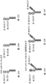


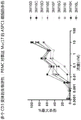

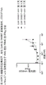


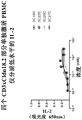


Classifications
-
- C—CHEMISTRY; METALLURGY
- C07—ORGANIC CHEMISTRY
- C07K—PEPTIDES
- C07K16/00—Immunoglobulins [IGs], e.g. monoclonal or polyclonal antibodies
- C07K16/18—Immunoglobulins [IGs], e.g. monoclonal or polyclonal antibodies against material from animals or humans
- C07K16/28—Immunoglobulins [IGs], e.g. monoclonal or polyclonal antibodies against material from animals or humans against receptors, cell surface antigens or cell surface determinants
- C07K16/2803—Immunoglobulins [IGs], e.g. monoclonal or polyclonal antibodies against material from animals or humans against receptors, cell surface antigens or cell surface determinants against the immunoglobulin superfamily
- C07K16/2809—Immunoglobulins [IGs], e.g. monoclonal or polyclonal antibodies against material from animals or humans against receptors, cell surface antigens or cell surface determinants against the immunoglobulin superfamily against the T-cell receptor (TcR)-CD3 complex
-
- A—HUMAN NECESSITIES
- A61—MEDICAL OR VETERINARY SCIENCE; HYGIENE
- A61K—PREPARATIONS FOR MEDICAL, DENTAL OR TOILETRY PURPOSES
- A61K45/00—Medicinal preparations containing active ingredients not provided for in groups A61K31/00 - A61K41/00
- A61K45/06—Mixtures of active ingredients without chemical characterisation, e.g. antiphlogistics and cardiaca
-
- A—HUMAN NECESSITIES
- A61—MEDICAL OR VETERINARY SCIENCE; HYGIENE
- A61P—SPECIFIC THERAPEUTIC ACTIVITY OF CHEMICAL COMPOUNDS OR MEDICINAL PREPARATIONS
- A61P35/00—Antineoplastic agents
-
- C—CHEMISTRY; METALLURGY
- C07—ORGANIC CHEMISTRY
- C07K—PEPTIDES
- C07K16/00—Immunoglobulins [IGs], e.g. monoclonal or polyclonal antibodies
- C07K16/18—Immunoglobulins [IGs], e.g. monoclonal or polyclonal antibodies against material from animals or humans
- C07K16/28—Immunoglobulins [IGs], e.g. monoclonal or polyclonal antibodies against material from animals or humans against receptors, cell surface antigens or cell surface determinants
- C07K16/2803—Immunoglobulins [IGs], e.g. monoclonal or polyclonal antibodies against material from animals or humans against receptors, cell surface antigens or cell surface determinants against the immunoglobulin superfamily
- C07K16/2818—Immunoglobulins [IGs], e.g. monoclonal or polyclonal antibodies against material from animals or humans against receptors, cell surface antigens or cell surface determinants against the immunoglobulin superfamily against CD28 or CD152
-
- C—CHEMISTRY; METALLURGY
- C07—ORGANIC CHEMISTRY
- C07K—PEPTIDES
- C07K16/00—Immunoglobulins [IGs], e.g. monoclonal or polyclonal antibodies
- C07K16/18—Immunoglobulins [IGs], e.g. monoclonal or polyclonal antibodies against material from animals or humans
- C07K16/28—Immunoglobulins [IGs], e.g. monoclonal or polyclonal antibodies against material from animals or humans against receptors, cell surface antigens or cell surface determinants
- C07K16/2878—Immunoglobulins [IGs], e.g. monoclonal or polyclonal antibodies against material from animals or humans against receptors, cell surface antigens or cell surface determinants against the NGF-receptor/TNF-receptor superfamily, e.g. CD27, CD30, CD40, CD95
-
- C—CHEMISTRY; METALLURGY
- C07—ORGANIC CHEMISTRY
- C07K—PEPTIDES
- C07K16/00—Immunoglobulins [IGs], e.g. monoclonal or polyclonal antibodies
- C07K16/18—Immunoglobulins [IGs], e.g. monoclonal or polyclonal antibodies against material from animals or humans
- C07K16/28—Immunoglobulins [IGs], e.g. monoclonal or polyclonal antibodies against material from animals or humans against receptors, cell surface antigens or cell surface determinants
- C07K16/30—Immunoglobulins [IGs], e.g. monoclonal or polyclonal antibodies against material from animals or humans against receptors, cell surface antigens or cell surface determinants from tumour cells
- C07K16/3076—Immunoglobulins [IGs], e.g. monoclonal or polyclonal antibodies against material from animals or humans against receptors, cell surface antigens or cell surface determinants from tumour cells against structure-related tumour-associated moieties
- C07K16/3092—Immunoglobulins [IGs], e.g. monoclonal or polyclonal antibodies against material from animals or humans against receptors, cell surface antigens or cell surface determinants from tumour cells against structure-related tumour-associated moieties against tumour-associated mucins
-
- C—CHEMISTRY; METALLURGY
- C07—ORGANIC CHEMISTRY
- C07K—PEPTIDES
- C07K16/00—Immunoglobulins [IGs], e.g. monoclonal or polyclonal antibodies
- C07K16/46—Hybrid immunoglobulins
- C07K16/468—Immunoglobulins having two or more different antigen binding sites, e.g. multifunctional antibodies
-
- A—HUMAN NECESSITIES
- A61—MEDICAL OR VETERINARY SCIENCE; HYGIENE
- A61K—PREPARATIONS FOR MEDICAL, DENTAL OR TOILETRY PURPOSES
- A61K39/00—Medicinal preparations containing antigens or antibodies
- A61K2039/505—Medicinal preparations containing antigens or antibodies comprising antibodies
-
- A—HUMAN NECESSITIES
- A61—MEDICAL OR VETERINARY SCIENCE; HYGIENE
- A61K—PREPARATIONS FOR MEDICAL, DENTAL OR TOILETRY PURPOSES
- A61K39/00—Medicinal preparations containing antigens or antibodies
- A61K2039/505—Medicinal preparations containing antigens or antibodies comprising antibodies
- A61K2039/507—Comprising a combination of two or more separate antibodies
-
- C—CHEMISTRY; METALLURGY
- C07—ORGANIC CHEMISTRY
- C07K—PEPTIDES
- C07K2317/00—Immunoglobulins specific features
- C07K2317/20—Immunoglobulins specific features characterized by taxonomic origin
- C07K2317/24—Immunoglobulins specific features characterized by taxonomic origin containing regions, domains or residues from different species, e.g. chimeric, humanized or veneered
-
- C—CHEMISTRY; METALLURGY
- C07—ORGANIC CHEMISTRY
- C07K—PEPTIDES
- C07K2317/00—Immunoglobulins specific features
- C07K2317/30—Immunoglobulins specific features characterized by aspects of specificity or valency
- C07K2317/31—Immunoglobulins specific features characterized by aspects of specificity or valency multispecific
-
- C—CHEMISTRY; METALLURGY
- C07—ORGANIC CHEMISTRY
- C07K—PEPTIDES
- C07K2317/00—Immunoglobulins specific features
- C07K2317/30—Immunoglobulins specific features characterized by aspects of specificity or valency
- C07K2317/33—Crossreactivity, e.g. for species or epitope, or lack of said crossreactivity
-
- C—CHEMISTRY; METALLURGY
- C07—ORGANIC CHEMISTRY
- C07K—PEPTIDES
- C07K2317/00—Immunoglobulins specific features
- C07K2317/50—Immunoglobulins specific features characterized by immunoglobulin fragments
- C07K2317/52—Constant or Fc region; Isotype
-
- C—CHEMISTRY; METALLURGY
- C07—ORGANIC CHEMISTRY
- C07K—PEPTIDES
- C07K2317/00—Immunoglobulins specific features
- C07K2317/50—Immunoglobulins specific features characterized by immunoglobulin fragments
- C07K2317/55—Fab or Fab'
-
- C—CHEMISTRY; METALLURGY
- C07—ORGANIC CHEMISTRY
- C07K—PEPTIDES
- C07K2317/00—Immunoglobulins specific features
- C07K2317/50—Immunoglobulins specific features characterized by immunoglobulin fragments
- C07K2317/56—Immunoglobulins specific features characterized by immunoglobulin fragments variable (Fv) region, i.e. VH and/or VL
- C07K2317/565—Complementarity determining region [CDR]
-
- C—CHEMISTRY; METALLURGY
- C07—ORGANIC CHEMISTRY
- C07K—PEPTIDES
- C07K2317/00—Immunoglobulins specific features
- C07K2317/60—Immunoglobulins specific features characterized by non-natural combinations of immunoglobulin fragments
- C07K2317/62—Immunoglobulins specific features characterized by non-natural combinations of immunoglobulin fragments comprising only variable region components
- C07K2317/622—Single chain antibody (scFv)
-
- C—CHEMISTRY; METALLURGY
- C07—ORGANIC CHEMISTRY
- C07K—PEPTIDES
- C07K2317/00—Immunoglobulins specific features
- C07K2317/70—Immunoglobulins specific features characterized by effect upon binding to a cell or to an antigen
- C07K2317/73—Inducing cell death, e.g. apoptosis, necrosis or inhibition of cell proliferation
-
- C—CHEMISTRY; METALLURGY
- C07—ORGANIC CHEMISTRY
- C07K—PEPTIDES
- C07K2317/00—Immunoglobulins specific features
- C07K2317/70—Immunoglobulins specific features characterized by effect upon binding to a cell or to an antigen
- C07K2317/75—Agonist effect on antigen
Landscapes
- Health & Medical Sciences (AREA)
- Chemical & Material Sciences (AREA)
- Immunology (AREA)
- Organic Chemistry (AREA)
- Life Sciences & Earth Sciences (AREA)
- General Health & Medical Sciences (AREA)
- Medicinal Chemistry (AREA)
- Molecular Biology (AREA)
- Genetics & Genomics (AREA)
- Biophysics (AREA)
- Proteomics, Peptides & Aminoacids (AREA)
- Biochemistry (AREA)
- Veterinary Medicine (AREA)
- Pharmacology & Pharmacy (AREA)
- Animal Behavior & Ethology (AREA)
- Public Health (AREA)
- Cell Biology (AREA)
- Epidemiology (AREA)
- Chemical Kinetics & Catalysis (AREA)
- General Chemical & Material Sciences (AREA)
- Nuclear Medicine, Radiotherapy & Molecular Imaging (AREA)
- Peptides Or Proteins (AREA)
- Medicines Containing Antibodies Or Antigens For Use As Internal Diagnostic Agents (AREA)
- Micro-Organisms Or Cultivation Processes Thereof (AREA)
- Medicines That Contain Protein Lipid Enzymes And Other Medicines (AREA)
- Preparation Of Compounds By Using Micro-Organisms (AREA)
- Apparatus Associated With Microorganisms And Enzymes (AREA)
- Electric Double-Layer Capacitors Or The Like (AREA)
Abstract
A bispecific T cell engager is provided. More specifically, the invention relates to bispecific molecules that bind DLL3, MUC17 or CLD18 and activate CD (cluster of differentiation) molecules, such as CD3, CD28 and CD 137. Also provided is a method of treating a disease (e.g., cancer) using an antibody (or fragment) against DLL3, MUC17 or CLD18, conjugated with an antibody (or fragment) that activates an agonist antibody to CD3, CD28 and/or CD 137.
Description
RELATED APPLICATIONS
This application claims priority to U.S. provisional patent application No. 62/968,999, filed on 31/2020, U.S. provisional patent application No. 62/981,048, filed on 25/2/2020, and U.S. provisional patent application No. 62/991,070, filed on 17/3/2020. The contents of the above-mentioned applications are incorporated herein by reference.
Sequence listing reference
The instant application contains a sequence listing submitted electronically in ASCII format, which is hereby incorporated by reference in its entirety. This ASCII copy was created at 1/2/2021 and named GB1-002US_SL. Txt, size 979,504 bytes.
Technical Field
The present invention relates generally to cancer treatment and, more particularly, to a novel compound for cancer treatment comprising an antibody or immunoreactive fragment thereof against DLL3, CLDN18.2 and Muc 17.
Background
Cancer is generally defined as a group of diseases involving abnormal cell growth that have the potential to invade or spread to other parts of the body. In 2019, about 180 million people in the united states were diagnosed with cancer. An estimated 606,880 people die from cancer each year in the united states. Lung and bronchial cancer are the leading cause of the most deaths. Colorectal and pancreatic cancers are the second and third most common causes of cancer death, respectively.
Cancer is associated with a variety of factors, including smoking, obesity, poor diet, lack of physical activity, and excessive alcohol consumption. Other factors include certain infections, ionizing radiation exposure, and environmental pollutants. Certain cancers are associated with infections such as helicobacter pylori, hepatitis b, hepatitis c, human papilloma virus infection, EB virus and Human Immunodeficiency Virus (HIV).
The aim of traditional cancer therapy is to remove cancerous tissue and prevent its spread. These treatment regimens include surgery, chemotherapy, radiation therapy, hormonal therapy, targeted therapy, and palliative care. Treatment is typically performed according to the type, location and grade of the cancer, as well as the health and preference of the patient. These options have limitations. They may be ineffective, particularly when the cancer has metastasized. In addition, chemotherapy and radiation therapy have a series of side effects associated with cytotoxicity.
Because cancer cells divide faster than most normal cells, they are sensitive to chemotherapeutic drugs. However, chemotherapeutic drugs can also attack other cells in the body, especially rapidly dividing cells, such as blood cells and cells in the mouth, stomach and intestinal tract. Thus, there is a need for improved drugs and methods for treating cancer that are more targeted and have fewer side effects.
One promising area of therapeutic development includes targeted therapy using antibodies. For example, the use of antibody drug conjugates can be used to target drugs to tumors. An immunoconjugate is an antibody that binds to a second molecule, which is typically a toxin, a radioisotope, or a label. Immunoconjugates can provide relatively high concentrations of the drug within the tumor, however systemic administration of unconjugated (i.e., non-targeted) drugs to achieve the same tumor concentration can result in toxic levels to normal cells.
Another promising area of therapeutic development is the use of the immune system to attack and kill tumor cells. Checkpoint inhibitors, such as anti-CTLA 4, anti-PD 1, and anti-PDL 1 therapies, have altered the therapeutic modalities of cancer. Similarly, direct activation of cytotoxic T cells by bispecific T cell engagers or CAR-T engineered T cells has led to an unprecedented approach to treatment in many types of cancer.
Delta-like ligand 3 (Delta-like ligand 3) (DLL 3) is an inhibitory notch ligand and is expressed at relatively low levels in normal tissues. It is expressed at high levels in Small Cell Lung Cancer (SCLC) and other neuroendocrine tumors, thus providing a potential therapeutic target for cancer diagnosis and treatment.
Mucin 17 (Mucin 17), also known as MUC17, is one of the members of the Mucin family consisting of more than 20 members. Mucin is a large, highly glycosylated membrane-bound protein. They generally act in the mucosal area to protect epithelial cells from the environment and to regulate cell proliferation and survival. MUC17 is expressed in pancreatic, appendiceal and some colon cancers and is therefore the target antigen for these cancers. Thus, MUC17 is a candidate target for targeted therapies (such as antibody drug conjugates, T cell engagers and CAR-T cells).
Claudin 18 (CLD 18) is a human protein encoded by the CLDN18 gene. It belongs to the class of tight junction proteins, a family of proteins that form components of the tight cell junction chain in epithelial cells. Studies have shown that a large number of subtype 2 (Isoform 2) (tight junction protein 18.2 or CLDN 18.2) are present in gastric tumors. Its extracellular stem loop is exposed and available for monoclonal antibody binding. These biological properties have led to the development of monoclonal antibodies against claudin 18.2, such as claudicaximab (IMAB 362).
CD3, CD28 and CD137 are receptors on T cells. T cells can be activated by CD3, CD28 and CD137 by antigen presenting cells that utilize the activation signals MHC class I and II, CD80 and CD86, and 4-1BBL, respectively. CD3 is part of the T Cell Receptor (TCR) and is the signaling component of the receptor. CD3 has three subunits, epsilon, delta, and gamma. ε is related to both δ and ζ, which are collectively responsible for signal transfer. CD3 signaling is considered to be a signal 1 required for activation of T cells. The co-receptors CD28 and CD137 are considered signal 2. Both signal 1 and signal 2 are necessary for adequate activation, proliferation and survival of T cells.
The present invention discloses a bispecific T cell engager. Bispecific molecules can bind DLL3, MUC17 and/or CLDN18.2 and activate CD (cluster of differentiation) molecules (e.g. CD3, CD28 and CD 137). Methods of treating diseases (e.g., cancer) using the antibodies and antibody conjugates, pharmaceutical compositions and articles thereof are also provided.
Summary of The Invention
The invention described and claimed herein has many features and embodiments, including but not limited to features and embodiments illustrated or described or referenced in the summary of the invention. The invention described and claimed herein is not limited or restricted to the features or embodiments identified in this summary, which is intended to be illustrative only and not limiting.
One aspect of the invention is an antibody against DLL 3. The antibodies may be fragments, such as antigen binding fragments (Fab) or single chain variable fragments (Scfvs).
One aspect of the invention is an agonist antibody that activates CD3, CD28 and/or CD 137.
One aspect of the invention is a bispecific molecule comprising an antibody (or fragment) against DLL3 conjugated to an antibody (or fragment) that activates an agonist antibody to CD3, CD28 or CD 137.
One aspect of the invention is a method of treating a disease (e.g., cancer) using an antibody (or fragment) against DLL3 paired with an antibody (or fragment) that activates an agonist antibody to CD3, CD28, or CD 137.
One aspect of the invention is a method of treating a disease using two or more of the bispecific molecules described herein in association with each other.
One aspect of the invention is a method of activating cytotoxicity of T cells against DLL3 expressing cells.
One aspect of the invention is a method of activating cytotoxicity of T cells using a bispecific molecule comprising an antibody (or fragment) against DLL3, conjugated with an antibody (or fragment) that activates an agonist antibody to CD3, CD28 or CD 137.
One aspect of the invention is a humanized antibody that binds to human DLL3 protein, which comprises a heavy chain variable domain having at least 95%, at least 96%, at least 97%, at least 98%, at least 99% or at least 100% sequence identity to SEQ ID NOs 56-74 or 75.
The disclosed methods may utilize any DLL3 antibody, including, for example: an antibody against DLL3 comprising three CDRs of the heavy chain variable region amino acid sequence of SEQ ID NOs 1-27 or 29.
One aspect of the present invention is a humanized antibody that binds to human DLL3 protein, which comprises a light chain variable domain that has at least 95%, at least 96%, at least 97%, at least 98%, at least 99%, or at least 100% sequence identity to SEQ ID NOs 76-90 or 91.
The disclosed methods may utilize any DLL3 antibody, including, for example: an antibody against DLL3 comprising three CDRs of the light chain variable region amino acid sequence of SEQ ID NOs 32-54 or 55.
One aspect of the invention is an antibody against MUC 17. The antibody may be a fragment, such as an antigen binding fragment (Fab) or a single chain variable fragment (Scfv).
One aspect of the invention is an agonist antibody that activates CD3, CD28 and/or CD 137.
One aspect of the invention is a bispecific molecule comprising an antibody (or fragment) against MUC17, conjugated with an antibody (or fragment) that activates an agonist antibody to CD3, CD28 or CD 137.
One aspect of the invention is a method of treating a disease (e.g., cancer) using an anti-MUC 17 antibody (or fragment) in combination with an antibody (or fragment) that activates an agonist antibody to CD3, CD28 or CD 137.
One aspect of the invention is a method of treating a disease using two or more of the bispecific molecules described herein in association with each other.
One aspect of the invention is a method of activating the cytotoxicity of T cells against cells expressing MUC 17.
One aspect of the invention is a method of activating cytotoxicity of T cells using a bispecific molecule comprising an antibody (or fragment) against MUC17, conjugated with an antibody (or fragment) that activates an agonist antibody to CD3, CD28 or CD 137.
One aspect of the invention is a humanized antibody that binds to human MUC17 protein, the humanized antibody comprising a heavy chain variable domain having at least 95%, at least 96%, at least 97%, at least 98%, at least 99%, or at least 100% sequence identity to SEQ ID NO:127-245 or 246.
The disclosed methods may utilize any MUC17 antibody, including, for example: an anti-MUC 17 antibody comprising the three CDRs of the heavy chain variable region amino acid sequence of SEQ ID NOs 92-111 or 112.
The disclosed methods may utilize any MUC17 antibody, including, for example: an antibody against MUC17 comprising the three CDRs of the light chain variable region amino acid sequence of SEQ ID NOs 113-125 or 126.
One aspect of the invention is a humanized antibody that binds to human MUC17 protein, the humanized antibody comprising a light chain variable domain having at least 95%, at least 96%, at least 97%, at least 98%, at least 99%, or at least 100% sequence identity to sequence ID NO:147-168 or 169.
The disclosed methods may utilize any MUC17 antibody, including, for example: an anti-MUC 17 antibody comprising the three CDRs of the heavy chain variable region amino acid sequence of SEQ ID NOs 92-111 or 112.
The disclosed methods may utilize any MUC17 antibody, including, for example: an anti-MUC 17 antibody comprising the three CDRs of the light chain variable region amino acid sequence of SEQ ID NO 113-125 or 126.
One aspect of the present invention is a method of treating a disease (e.g., cancer) using an antibody (or fragment) to CLDN18.2 in combination with an antibody (or fragment) that activates an agonist antibody to CD3, CD28 or CD 137.
One aspect of the invention is a method of activating cytotoxicity of T cells against cells expressing CLDN 18.2.
One aspect of the invention is a method of activating cytotoxicity of T cells using a bispecific molecule comprising an antibody (or fragment) against CLDN18.2 conjugated with an antibody (or fragment) that activates an agonist antibody of CD3, CD28 or CD 137.
The disclosed methods may utilize any CLDN18.2 antibody, including, for example: an antibody of CLDN18.2 comprising three CDRs of the heavy chain variable region amino acid sequence of SEQ ID NOs 170-186 or 187.
One aspect of the invention is a humanized antibody that binds to a human CLDN18.2 protein, the humanized antibody comprising a heavy chain variable domain having at least 95%, at least 96%, at least 97%, at least 98%, at least 99%, or at least 100% sequence identity to SEQ ID NOs 197-205 or 206.
The disclosed methods may utilize any CLDN18.2 antibody, including, for example: an antibody against CLDN18 comprising the three CDRs of the light chain variable region amino acid sequence of SEQ ID NOs 188-195 or 196.
One aspect of the invention is a humanized antibody that binds to a human CLDN18.2 protein, the humanized antibody comprising a light chain variable domain having at least 95%, at least 96%, at least 97%, at least 98%, at least 99%, or at least 100% sequence identity to SEQ ID NOs 207-212 or 213.
The disclosed methods may utilize any CLDN18.2 antibody, including, for example: an antibody of CLDN18.2 comprising three CDRs of the heavy chain variable region amino acid sequence of SEQ ID NOs 170-186 or 187.
The disclosed methods may utilize any CLDN18.2 antibody, including, for example: an antibody to CLDN18.2 comprising the three CDRs of the light chain variable region amino acid sequence of SEQ ID NOs 188-195 or 196.
In some embodiments, the targeting domains are linked to each other by peptide bonds via peptide linkers or by covalent conjugates using suitable cross-linking techniques known in the art.
In some embodiments, the targeting domain comprises an antibody variable region. In some embodiments, the targeting domain is in a form selected from the group consisting of: a single domain antibody (sdAb), a fragment variable (Fv) heterodimer, a single chain Fv (scFv), a Fab fragment, a trilab, or a combination thereof.
In some embodiments, the bispecific molecule is administered with a checkpoint inhibitor.
In some embodiments, the bispecific molecule is administered with an anti-PD 1 and/or anti-PDL 1 antagonist.
Other features and advantages of various aspects of the present invention will become apparent from the following more detailed description, taken in conjunction with the accompanying drawings, illustrating by way of example the principles of various aspects of the invention.
Brief description of the drawings
The drawings illustrate various aspects of the invention. In these drawings:
FIGS. 1A-1F depict the Scfv-Fc x Fab-Fc format of the bispecific molecules of the present invention.
FIG. 1A depicts an anti-CD 3, anti-DLL 3 (Scfv-Fc x Fab-Fc) molecule.
FIG. 1B depicts an anti-CD 28, anti-DLL 3 (Scfv-Fc x Fab-Fc) molecule.
FIG. 1C depicts anti-DLL 3, anti-CD 3 (Scfv-Fc x Fab-Fc) molecules.
FIG. 1D depicts anti-DLL 3, anti-CD 28 (Scfv-Fc x Fab-Fc) molecules.
FIG. 1E depicts anti-CD 137, anti-DLL 3 (Scfv-Fc x Fab-Fc) molecules.
FIG. 1F depicts an anti-DLL 3, anti-CD 137 (Scfv-Fc x Fab-Fc) molecule.
Figures 2A to 2C depict several scfv-scfv-Fc x Fc formats of bispecific molecules of the invention.
Figure 2A depicts an anti-DLL 3, anti-CD 3 (scfv-scfv-Fc x Fc) molecule.
Figure 2B depicts an anti-DLL 3, anti-CD 28 (scfv-scfv-Fc x Fc) molecule.
Figure 2C depicts an anti-DLL 3, anti-CD 137 (scfv-scfv-Fc x Fc) molecule.
Figures 2D to 2F depict several scfv-scfv-Fc x Fab-Fc forms of bispecific molecules of the invention.
FIG. 2D depicts anti-DLL 3, anti-CD 3, anti-DLL 3 (scfv-scfv-Fc x Fab-Fc) molecules.
FIG. 2E depicts anti-DLL 3, anti-CD 28, anti-DLL 3 (scfv-scfv-Fc x Fab-Fc) molecules.
FIG. 2F depicts anti-DLL 3, anti-CD 137, anti-DLL 3 (scfv-scfv-Fc x Fab-Fc) molecules.
Fig. 3A is a graph showing binding of anti-DLL 3 antibodies to CHO cells expressing human DLL 3.
Fig. 3B is a graph showing binding of anti-DLL 3 antibodies to CHO cells expressing cynomolgus monkey DLL 3.
Figure 4 is a graph showing the binding of anti-DLL 3scfv-Fc to CHO cells expressing human DLL 3.
FIG. 5 is a graph showing DLL3Fab-Fc x CD3scfv-Fc mediated killing of huDLL3 expressing CHO cells by human T cells.
FIG. 6 is a graph showing DLL3Fab-Fc x CD3scfv-Fc mediated activation of human T cells in the presence of huDLL3 expressing CHO cells.
FIG. 7 is a graph showing DLL3scfv-CD3scfv-Fc x Fc killing of human T cells against huDLL3 expressing CHO cells.
FIG. 8 is a graph showing DLL3scfv-CD3scfv-Fc x Fc mediated activation of human T cells in the presence of huDLL3 expressing CHO cells.
FIGS. 9A-9C are graphs showing DLL3Fab-Fc x DLL3scfv-CD3scfv-Fc mediated killing of human T cells to huDLL3 expressing NCI-H82 cells.
FIGS. 10A and 10B are graphs showing anti-CD 3 variants of DLL3Fab-Fc x CD3scfv Fc and DLL3scfv-CD3 scvvv-Fc format molecules that mediate killing of human T cells against huDLL3 expressing NCI-H82 cells.
FIGS. 11A-11F are graphs showing that DLL3Fab-Fc mediates IL-2 secretion from huPBMC in the presence of huDLL3 expressing NCI-H82 cells in combination with DLL3scfv-CD3scfv-scFc or DLL3Fab-CD3 scfv-Fc.
Figure 12 is a graph of HPLC size exclusion chromatography analysis of transiently expressed HEK293 and protein a purified bispecific molecules using various CD3 variants.
FIG. 13 is a graph showing killing of NC1-H82 tumor cells using 3D22I variants on Peripheral Blood Mononuclear Cells (PBMCs).
FIG. 14 is a graph showing stimulation of PBMC with 3D22I variants to express CD25 in the presence of NC1-H82 tumor cells.
Figure 15 is a graph showing that DLL 3scfv x CD3scfv x DLL3Fab variant 3D45I more efficiently stimulated PBMC to kill NCI-H82 cells than 3D 39I.
Figure 16 is a graph showing that DLL 3scfv x CD3scfv x DLL3Fab variant 3D45I stimulated PBMC expression of CD25 more efficiently than 3D39I in the presence of NCI-H82 cells.
Figure 17 is a graph showing that CD28scfv-Fc x DLL3Fab-Fc bispecific combined with 50M CD3-DLL3 baseline 3DBM activates T cells to secrete IL-2 better than 3DBM alone.
FIG. 18 is a graph showing that CD28scfv-Fc (C50S) x DLL3-Fab-Fc variants 28D13 and 28D15, whose free cysteines in the anti-CD 28 molecule were knocked out, were able to induce secretion of IL-2 by PBMC similar to the parent molecule 28D10 when bound to 50pM baseline 3DBM in the presence of NCI-H82 cells.
Fig. 19A is a graph showing that uremumab (Urelumab) increases IFNy secretion when combined with a 50pM CD3xDLL3T cell engager Benchmark (BM) molecule.
Figure 19B is a graph showing that 4-1BB Fab-Fc x DLL3scfv-Fc bispecific 4D3 increases IFNy secretion when combined with a 50pM CD3xDLL3T-cell engager Benchmark (BM) molecule.
Figure 20A depicts an anti-CD 137, anti-DLL 3 Fab molecule.
FIGS. 21A to 21F depict the Scfv-Fc x Fab-Fc format of the bispecific molecules of the invention. FIG. 21A depicts anti-CD 3, anti-MUC 17 (Scfv-Fc x Fab-Fc) molecules.
FIG. 21B depicts anti-CD 28, anti-MUC 17 (Scfv-Fc x Fab-Fc) molecules.
FIG. 21C depicts an anti-MUC 17, anti-CD 3 (Scfv-Fc x Fab-Fc) molecule.
FIG. 21D depicts anti-MUC 17, anti-CD 28 (Scfv-Fc x Fab-Fc) molecules.
FIG. 21E depicts anti-CD 137, anti-MUC 17 (Scfv-Fc x Fab-Fc) molecules.
FIG. 21F depicts anti-MUC 17, anti-CD 137 (Scfv-Fc x Fab-Fc) molecules.
Figures 22A to 22F depict several scfv-scfv-Fc x Fc forms of bispecific molecules of the invention. Figure 22A depicts an anti-MUC 17, anti-CD 3 (scfv-scfv-Fc x Fc) molecule.
Figure 22B depicts an anti-MUC 17, anti-CD 28 (scfv-scfv-Fc x Fc) molecule.
Figure 22C depicts anti-MUC 17, anti-CD 137 (scfv-scfv-Fc x Fc) molecules.
FIG. 22D depicts anti-MUC 17, anti-CD 3, anti-MUC 17 (scfv-scfv-Fc x Fab-Fc) molecules.
FIG. 22E depicts anti-MUC 17, anti-CD 28, anti-MUC 17 (scfv-scfv-Fc x Fab-Fc) molecules.
FIG. 22F depicts anti-MUC 17, anti-CD 137, anti-MUC 17 (scfv-scfv-Fc x Fab-Fc) molecules.
Fig. 23 is a diagram showing that MUC17xCD3 bispecific molecules bind to CHOK1 cells expressing MUC 17.
Figure 24 is a diagram showing the binding of MUC17xCD3 bispecific molecules to ASPC1 tumor cells.
Figure 25 is a graph showing that MUC17xCD3 bispecific molecules activate human PBMC T cells to kill CHO cells expressing MUC 17.
Figure 26A is a graph showing that various forms of the MUC17xCD3 bispecific molecule of antibody 1MU32A activate PBMC T cells to kill ASPC1 cells.
Figure 26B is a graph showing that various forms of the MUC17xCD3 bispecific molecule of antibody 1MU32A increase the level of the T cell activation marker CD25 in the presence of ASPC1 cells.
FIGS. 26C-26F depict bispecific molecules 3M46C, 3M64C, 3M62C, and 3M66C.
Figure 27A is a graph showing that various forms of the MUC17xCD3 bispecific molecule of antibody 1MU11A activate PBMC T cells to kill ASPC1 cells.
Figure 27B is a graph showing that various forms of the MUC17xCD3 bispecific molecule of antibody 1MU11A increase the level of the T cell activation marker CD25 in the presence of ASPC1 cells.
FIGS. 27C-27F depict bispecific molecules 3M55C, 3M63C, 3M61C, and 3M65C.
FIG. 28A is a graph showing that CD28xMuc17 bispecific molecules 28M1, 28M2, and 28M3 in combination with 10pM of MUC17xCD3 bispecific molecule 3M62C activate T cells to secrete IL-2 in the presence of ASPC1 cells.
Figure 28B is a graph showing that CD28xMuc17 bispecific molecules 28M1, 28M2, and 28M3 in combination with 10pM MUC17xCD3 bispecific molecule 3M62C activate T cells to express CD25 in the presence of ASPC1 cells.
FIGS. 28C-28F depict bispecific molecules 28M1, 28M2, 28M3, and 3M62C.
FIG. 29A is a graph showing that CD28xMuc17 bispecific molecules 28M1, 28M2, and 28M3 in combination with 20pM of MUC17xCD3 bispecific molecule 3M55C activate T cells to secrete IL-2 in the presence of ASPC1 cells.
Figure 29B is a graph showing that CD28xMuc17 bispecific molecules 28M1, 28M2, and 28M3 in combination with 20pM MUC17xCD3 bispecific molecule 3M55C activate T cells to express CD25 in the presence of ASPC1 cells.
FIGS. 29C-29F depict bispecific molecules 28M1, 28M2, 28M3, and 3M55C.
Figure 30A is a graph showing that CD28xMuc17 bispecific molecules 28M1, 28M2, and 28M3 alone are unable to activate T cells to secrete IL-2 in the presence of ASPC1 cells.
Figure 30B is a graph showing that CD28xMuc17 bispecific molecules 28M1, 28M2, and 28M3 alone are unable to activate T cells to express CD25 in the presence of ASPC1 cells.
Figure 31 is a graph showing the biological activity of multiple CD3 variants: killing of Muc17 expressing ASPC1 cells by PBMC.
FIG. 32 is a graph showing that the Scfv forms of hu1MU11A and hu1MU32A retain Muc17 binding activity.
Figure 33 is a graph showing that CD137 Fab x Muc17 scfv bispecific 4M1 increases IFNg secretion from PBMC when bound to a 2pM 3M8B7 baseline in the presence of ASPC1 cells.
Figure 34 is a graph showing that CD137 Fab x Muc17 scfv bispecific 4M2 increases IFNg secretion from PBMC when bound to a 2pM 3M8B7 baseline in the presence of ASPC1 cells.
Figure 35 is a graph showing that the Muc17xCD137 bispecific molecule 4M2 in combination with 10pM or 20pM CD3 bispecific 3M8B7 activates PBMCs to secrete IFNg in the presence of Muc17 expressing CHO cells.
Figure 36 is a graph showing that the Muc17xCD137 bispecific molecule 4M7 in combination with 10pM or 20pM CD3 bispecific 3M8B7 activates PBMCs to secrete IFNg in the presence of Muc17 expressing CHO cells.
Figure 37 is a graph showing that the Muc17xCD137 bispecific molecule 4M8 in combination with 10pM or 20pM CD3 bispecific 3M8B7 activates PBMCs to secrete IFNg in the presence of Muc17 expressing CHO cells.
FIGS. 38A and 38B depict the Muc17xCD137 and DLL3xCD137 molecules.
Figures 39A to 39E depict the Muc17x CD137 form of a bispecific molecule of the invention.
Figures 40A to 40D depict several scfv-scfv-Fc x Fc forms of bispecific molecules of the invention. FIG. 40A depicts an anti-CLDN 18.2, anti-CD 3 (scfv-scfv-Fc x Fc) molecule.
FIG. 40B depicts anti-CLDN 18.2, anti-CD 28 (scfv-scfv-Fc x Fc) molecules
FIG. 40C depicts anti-CD 3, anti-CLDN 18.2 (scfv-scfv-Fc x Fc) molecules.
FIG. 40D depicts anti-CD 28, anti-CLDN 18.2 (scfv-scfv-Fc x Fc) molecules.
Figures 41A to 41F depict several scfv-scfv-Fc x Fc forms of bispecific molecules of the invention. FIG. 41A depicts an anti-CLDN 18.2, anti-CD 3 (scfv-scfv-Fc x Fc) molecule.
FIG. 41B depicts anti-CLDN 18.2, anti-CD 28 (scfv-scfv-Fc x Fc) molecules.
FIG. 41C depicts anti-CLDN 18.2, anti-CD 137 (scfv-scfv-Fc x Fc) molecules.
FIG. 41D depicts anti-CLDN 18.2, anti-CD 3 (scfv-scfv-Fc x Fc) molecules.
FIG. 41E depicts anti-CLDN 18.2, anti-CD 28 (scfv-scfv-Fc x Fc) molecules.
FIG. 41F depicts an anti-CLDN 18.2, anti-CD 137 (scfv-scfv-Fc x Fc) molecule.
Fig. 42 is a graph showing binding of cldn18.2xcdd 3 bispecific molecules to CHO cells expressing huclnd18.2.
Fig. 43A is a graph showing that CLDN 18.2xcdd 3 bispecific molecules activate T cells and up-regulate CD25 in the presence of CHO cells expressing CLDN 18.2.
Fig. 43B is a graph showing the killing of huPBMC on hucldn18.2-expressing CHO cells stimulated by cldn18.2xcdd 3 bispecific molecules.
Fig. 44A is a graph showing that CLDN 18.2xcdd 3 bispecific molecules with CD3 variant molecules activate T cells and up-regulate CD25 in the presence of SNU-601 tumor cells expressing CLDN 18.2.
Fig. 44B is a graph showing the killing of SNU-601 tumor cells by hupmc stimulated by cldn18.2xcdd 3 bispecific molecules with CD3 variant molecules.
Fig. 45A is a graph showing that CLDN 18.2xcdd 3 bispecific molecules with CD3 variant molecules activate T cells and upregulate CD25 in the presence of SNU-601 tumor cells expressing CLDN 18.2.
Fig. 45B is a graph showing the killing of SNU-601 tumor cells by hupmc stimulated by cldn18.2xcdd 3 bispecific molecules with CD3 variant molecules.
Fig. 46A is a graph showing that CLDN 18.2xcdd 3 bispecific molecules with CD3 variant molecules activate T cells and up-regulate CD25 in the presence of SNU-601 tumor cells expressing CLDN 18.2.
Fig. 46B is a graph showing the killing of SNU-601 tumor cells by hupmc stimulated by cldn18.2xcdd 3 bispecific molecules with CD3 variant molecules.
FIG. 47 is a graph showing binding of CD3xCDN18.2 to LDN18.2-CHO cells.
FIGS. 48A and 48B are graphs showing that all four CD28xCDLN18.2T cell engagers in combination with 10pM of 3C18C efficiently activate PBMCs in the presence of SNU-601 cells.
Fig. 49A and 49B are graphs showing that all four cd28xcldn 18.2T cell engagers in combination with 10pM of 3C22C efficiently activate PBMCs in the presence of SNU-601 cells.
Figures 50A and 50B are graphs showing that all four cd28xcldn 18.2T cell engagers in combination with 20pM of 3C22C efficiently activate PBMCs in the presence of SNU-601 cells.
FIGS. 51A and 51B are graphs showing that all four CD28xCDLN 18.2T cell engagers efficiently activate PBMCs in combination with 2pM of 3C27C in the presence of SNU-601 cells.
Fig. 52A and 52B are graphs showing that four cd28xcldn 18.2T cell engagers alone were unable to activate PBMCs.
FIGS. 53A and 53B are graphs showing that four CD3xCDLND 18.2T-cell engagers activate PBMCs alone to express CD25, but to a much lesser extent secrete IL-2.
Figure 54 is a graph showing that cd3xcldn18.2 bispecific 3C22C stimulated PBMC-mediated killing of GSU cells more effectively than 3 lots of benchmark 3 CBM.
Figure 55 is a graph showing that cd3xcldn18.2 bispecific 3C22C stimulated PBMCs to express CD25 more efficiently than 3 lots of baseline 3CBM in the presence of GSU cells.
FIG. 56 is a graph showing that 3C27I is more effective at killing SNU-601 cells than 3C18I, 3C22I, or 3C 26I.
FIG. 57 is a graph showing that CD28xCLDN18.2T cell engagers 28C1, 28C2, 28C3 and 28C4 activate PBMCs in combination with 10pM of CD3xCLDN18.2 bispecific 3C18C to produce IL-2 when co-cultured with SNU-601 cells.
FIG. 58 is a graph showing that CD28xCLDNN 18.2T cell engagers 28C1, 28C2, 28C3 and 28C4, when co-cultured with SNU-601 cells, activate PBMCs in combination with 10pM of CD3xCldn18.2 bispecific 3C22C to produce IL-2.
FIG. 59 is a graph showing that CD28xCDnCLdN18.2T cell engagers 28C1, 28C2, 28C3 and 28C4, when co-cultured with SNU-601, activate PBMCs in combination with 10pM of CD3xCldn18.2 bispecific 3C26C to produce IL-2.
FIG. 60 is a graph showing that CD28xCDLND 18.2T cell engagers 28C1, 28C2, 28C3 and 28C4 alone are unable to activate PBMCs to produce IL-2 when co-cultured with SNU-601 cells.
Fig. 61 is a graph showing that cldn18.2xcdd 28 bispecific molecules 28C7 and 28C10 activate T cells to secrete IL-2 in combination with 5pM of CD3 bispecific 3C 22I.
Fig. 62 is a graph showing that CLDN 18.2xcdd 28 bispecific molecules 28C5, 28C6, 28C7 and 28C8 induce IL-2 secretion by PBMCs in the presence of CHO cells expressing CLDN18.2 instead of parental CHO cells.
FIG. 63 is a graph showing that the CD137xCLDN18.2 bispecific molecules 4C1 and 4C2 in combination with 10pM of CD3 bispecific 3C18C can cause IFN γ secretion from PBMC over 3C18 in the presence of SNU-601 cells.
FIG. 64 is a graph showing that the CD137xCDLN18.2 bispecific molecules 4C1 and 4C2 in combination with 10pM of CD3 bispecific 3C18C can cause IFN γ secretion from PBMCs over 3C18 in the presence of SNU-601 cells.
Figures 65 and 66 are graphs showing that the cd137xcldn18.2 bispecific molecules 4C5 and 4C6 in combination with 3C22I activate T cells to secrete IFNg.
Fig. 67 is a graph showing that cd137xcldn18.2 bispecific molecules 4C1 and 4C5 in combination with 80pM 3C27I increase activation of CD8 cells in the presence of mitomycin treated CHO cells expressing CLDN 18.2.
Fig. 68A and 69B depict the CLDN x CD137 form of a bispecific molecule of the invention.
Fig. 69A and 69B depict alternative forms of bispecific molecules of the invention.
Definition of
Reference in the specification to "one embodiment/aspect" or "some embodiments/aspects" means that a particular feature, structure, or characteristic described in connection with the embodiment/aspect is included in at least one embodiment/aspect of the invention. The use of the phrases "in one embodiment/aspect" or "in another embodiment/aspect" in various places in the specification are not necessarily all referring to the same embodiment/aspect, nor are separate or alternative embodiments or aspects necessarily mutually exclusive of other embodiments/aspects. In addition, various features are described which some embodiments/aspects may exhibit rather than other embodiments/aspects. Similarly, various requirements are described which may be requirements for some embodiments/aspects but not other embodiments/aspects. In some cases, embodiments and aspects may be used interchangeably.
The terms used in this specification generally have their ordinary meanings in the art, in the context of the invention, and in the specific context in which each term is used. Certain terms used to describe the invention are discussed below or elsewhere in the specification to provide additional guidance to the practitioner with respect to describing the invention. It should be understood that the same thing can be expressed in more than one way.
Accordingly, alternative languages and synonyms may be used for any one or more of the terms discussed herein. No special meaning is intended for a term whether or not it is set forth or discussed herein. Synonyms for certain terms are provided. The recitation of one or more synonyms does not exclude the use of other synonyms. Examples used anywhere in this specification, including examples of any terms discussed herein, are for illustration only and are not intended to further limit the scope and meaning of the invention or any example terms. Also, the present invention is not limited to the various embodiments presented in this specification.
Examples of apparatus, devices, methods and their related results of embodiments of the present invention are given below without intending to further limit the scope of the invention. Note that titles or subtitles may be used in the examples for convenience of a reader, which shall not limit the scope of the invention. Unless defined otherwise, all technical and scientific terms used herein have the same meaning as commonly understood by one of ordinary skill in the art to which this invention belongs. In the event of conflict, the present document, including definitions, will control.
Where applicable, the term "about" or "generally" as used in this specification and the appended claims means a +/-20% margin, unless otherwise indicated. Furthermore, where applicable, the term "substantially" as used in this specification and the appended claims means a margin of +/-10%, unless otherwise specified. It should be understood that not all of the above terms are used quantifiable so that the recited range can be applied.
The term "subject" or "patient" refers to any single animal, more preferably a mammal (including non-human animals, such as dogs, cats, horses, rabbits, zoo animals, cows, pigs, sheep, and non-human primates) in need of treatment. Most preferably, the patient described herein is a human. In one embodiment, the "subject" for diagnosis or treatment is a prokaryote or eukaryote, a tissue culture, a tissue, or an animal, such as a mammal, including a human.
As used herein, the term "comprising" is intended to mean that the compositions and methods include the listed elements, but do not exclude other, non-listed elements. When used to define compositions and methods, "substantially comprising" does not include other elements that alter the basic nature of the composition and/or method, but does not exclude other, non-listed elements. Thus, a composition comprising essentially the elements defined herein does not exclude trace elements, such as contaminants from any isolation and purification process or pharmaceutically acceptable carriers (e.g. phosphate buffer salts, preservatives, etc.), but does not include other unspecified amino acids. "consisting of 8230%" \8230comprises no more than trace elements and is more than a substantial method step for applying the compositions described herein. Embodiments defined by each of these transitional terms are within the scope of the invention and the inventions contained therein.
The term "active agent" or "active ingredient" refers to a substance, compound or molecule that is biologically active or otherwise induces a biological or physiological effect in a subject to which it is administered. In other words, "active agent" or "active ingredient" refers to one or more components of a composition to which all or part of the effect of the composition is attributed. The active agent may be the primary active agent, that is, the full or partial effect of the composition is attributed to the components of the composition. The active agent may be a minor agent, that is, an additional part of the composition and/or other effects attributable to components of the composition.
In one embodiment, a "pharmaceutical composition" is intended to include the combination of an active agent (e.g., an anti-DLL 3, anti-MUC 17 or anti-CLDN 18.2 antibody and antibody conjugate) and a carrier (inert or active) in a sterile composition suitable for diagnostic or therapeutic use in vitro, in vivo, or ex vivo. In one aspect, the pharmaceutical composition is substantially free of endotoxin or is non-toxic to the recipient at the dosage or concentration employed.
In one embodiment, "effective amount" refers to, but is not limited to, an amount of a defined ingredient sufficient to achieve a desired therapeutic result. In one embodiment, the result may be an effective cancer treatment.
In one embodiment, as used herein, the terms "treatment," "treating," and the like are used herein to mean, but are not limited to, obtaining a desired pharmacological and/or physiological effect. The effect may be prophylactic in terms of completely or partially preventing the disease or signs or symptoms thereof, and/or therapeutic in terms of ameliorating the symptoms of the disease or infection, or in terms of a partial or complete cure of the disease and/or adverse effects caused by the disease.
As used herein, the term "recombinant" refers to a non-naturally occurring polypeptide or polynucleotide that can be produced by combining polynucleotides or polypeptides in an arrangement that would not normally occur together. The term may refer to a polypeptide produced by a biological host selected from the group consisting of mammalian, insect cell, yeast and bacterial expression systems.
The term "delta-like 3" or "DLL3" refers to a protein encoded by the DLL3 gene in humans. The gene mutation causes the autosomal recessive inheritance disease Jarcho-Levin syndrome. DLL3 is normally expressed inside cells and at low levels in normal tissues. However, lung tumor cells overexpress this gene and cell surface DLL3 levels are elevated.
The term "mucin 17" or "MUC17" refers to one of the members of the mucin family that includes more than 20 members. Mucin is a large, highly glycosylated membrane-associated protein. They are expressed almost exclusively in the gut. Their general function is to protect epithelial cells from environmental influences, as well as to regulate cell proliferation and survival. MUC17 is highly expressed (at the protein level) in pancreatic cancer tissues. MUC17 is expressed in pancreatic, appendiceal, and some colon cancers. Its expression was not detected in normal pancreas, pancreatitis or other cancer cell lines.
The term "claudin 18" or "CLD 18" refers to a protein encoded by the CLDN18 gene in humans. CLDN18 belongs to a large family of tight junction proteins that form tight junction chains in epithelial cells. "Claudin 18.2" or "CLDN18.2" denotes the subtype 2 which is present in large numbers in tumors, in particular in tumors of the gastric system.
The term "CD" or "cluster of differentiation molecules" refers to cell surface markers, such as CD3, CD28, and CD137, that aid in the identification and characterization of leukocytes. CD3 is a signaling component of the T Cell Receptor (TCR) complex. Because CD3 is essential for T cell activation, CD 3-targeting drugs (usually monoclonal antibodies) are being investigated as immunostimulants for the treatment of cancer.
CD28 is the major costimulatory molecule required to generate a T cell-mediated immune response. Upon interaction of CD28 with its ligands CD80 and CD86, transduction leads to activation signals for expression of anti-apoptotic proteins and enhances synthesis of several cytokines including IL-2. The CD28 co-stimulatory receptor is present on all T cells. Agonist antibodies targeting anti-CD 28 have clinically led to serious adverse events, in contrast to antibodies targeting anti-other CD28 family members CTLA-4, PD-1, or their B7 ligands, which act as checkpoint inhibitors to overcome tumor immune tolerance and are useful in cancer immunotherapy.
CD137 is a member of the Tumor Necrosis Factor (TNF) receptor family. It is also named as tumor necrosis factor receptor superfamily member 9 (TNFRSF 9) and 4-1BB, and can induce lymphocyte activation. Agonist anti-CD 137 antibodies are particularly important for effector/memory T cells as an activating costimulatory molecule, and promote survival and proliferation of T lymphocytes. For example, BBK-4, urelumab (Urelumab) and Utomumab (Utomillab) (PF-05082566) target this receptor, stimulating the immune system to more aggressively attack cancer.
PD-1 (programmed cell death protein 1 or CD 279) is a protein on the cell surface that plays a role in regulating the immune system's response to human cells by down-regulating the immune system and promotes self-tolerance by inhibiting T cell inflammatory activity. Binding of PD-1 to neighboring cells by its ligand PD-L1 or PD-L2 inhibits TCR signaling and TCR-mediated proliferation, transcriptional activation, and cytokine production. This can prevent autoimmune diseases, but can also prevent the immune system from killing cancer cells. Therapeutic antibodies designed to block the PD-1/PD-L1 interaction have potential for the treatment of cancer.
Programmed death ligand 1 (PD-L1), also known as cluster of differentiation 274 (CD 274) or B7 homolog 1 (B7-H1), is a protein encoded by the CD274 gene. PD-L1 binds to the receptor PD-1 on activated T cells, B cells, and myeloid cells to regulate the activation or inhibition of the cells.
As used herein, the term "antibody" refers to a polypeptide or polypeptide complex that specifically recognizes and binds an antigen through one or more immunoglobulin variable regions. Recognized immunoglobulin genes include the kappa, lambda, alpha, gamma, delta, epsilon and mu constant region genes, as well as myriad immunoglobulin variable region genes. Light chains are classified as either kappa or lambda. Heavy chains are classified as gamma, mu, alpha, delta, or epsilon, which individually define the immunoglobulin classes IgG, igM, igA, igD, and IgE, respectively. Generally, the antigen binding region of an antibody is most critical in specificity and affinity of binding and is encoded by a variable domain. The antibody may be a whole antibody, an antigen-binding fragment, or a single chain thereof.
Exemplary immunoglobulin (antibody) building blocks include tetramers. Each tetramer is composed of two identical pairs of polypeptide chains, each pair having one "light" (about 25 kD) and one "heavy" chain (about 50-70 kD). The N-terminus of each chain defines a variable region of about 100 to 110 or more amino acids, which are primarily responsible for antigen recognition. The terms variable light chain (VL) and variable heavy chain (VH) refer to the variable domains of the light and heavy chains, respectively.
Antibodies exist, for example, as intact immunoglobulins or as a large number of well-characterized fragments produced by digestion with various peptidases. Thus, for example, pepsin digests the antibody below the disulfide bond in the hinge region to produce F (ab)' 2 I.e., a dimer of Fab, which is itself a light chain VL-CL, is linked to VH- -CH1 by a disulfide bond. Although various antibody fragments are defined in terms of digestion of intact antibodies, the skilled artisan understands that these fragments can be synthesized de novo by chemical methods or using recombinant DNA methods. Thus, as used herein, the term antibody also includes antibody fragments produced by modification of whole antibodies, or antibody fragments synthesized de novo using recombinant DNA methods (e.g., single chain Fv) or antibody fragments recognized using phage display libraries (see, e.g., mcCafferty et al, nature 348 (1990)).
Thus, in any aspect of the invention, the term antibody also includes minibodies, scfvs, diabodies, triabodies, and the like. ScFv and diabodies are small bivalent biospecific antibody fragments with high affinity and specificity. Their high signal-to-noise ratio is generally better because higher specificity and rapid blood clearance increases their potential for diagnostic and therapeutic targeting of specific antigens (Sundaresan et al, J nuclear Med 44 1962-9 (2003). Furthermore, these antibodies are advantageous because they can be engineered into different types of antibody fragments, from small single chain antibodies (scFv) to intact IgG with different subtypes, if necessary (Wu and Senter, nat. Biotechnol.23:1137-1146 (2005)). In some embodiments, the antibody fragment is a scFv-scFv or a portion of a diabody. In some embodiments, in any aspect, the invention provides a high affinity antibody for use according to the invention.
The term "agonist antibody" refers to an antibody that stimulates or activates an organ. Antibodies can act as agonists of the receptor, essentially replacing the activity of the normal ligand. Agonist activity occurs when an antibody binds to a receptor in a manner that mimics the binding of a physiological ligand, resulting in antibody-mediated agonism. For example, agonist antibodies against the thyrotropin receptor in Grave's disease stimulate the thyroid gland to release thyroid hormone, resulting in hyperthyroidism. Agonist antibodies can also be stimulated upon aggregation by binding of the Fc portion of the antibody to Fc receptors in trans or cis, or by antigen-mediated aggregation. The latter aggregation mechanism requires that the antigen bind to one half of the bispecific molecule and that the receptor be stimulated to bind to the other half of the bispecific molecule. Exemplary stimulatory receptors are CD3, CD28, and 4-1BB that stimulate T cells.
The term "antibody fragment" or "antigen-binding fragment" refers to a portion of an antibody, e.g., fab', fab, fv, scFv, and the like. Regardless of structure, an antibody fragment binds to the same antigen that is recognized by an intact antibody. The term "antibody fragment" also includes diabodies and any artificially synthesized or genetically engineered protein that comprises an immunoglobulin variable region that functions as an antibody by forming a complex with a particular antigen.
The term "antigen binding fragment" or "Fab" refers to the region of an antibody that binds to an antigen. It comprises heavy and light chains (i.e., four domains: VH, CH1, VL, and CL 1) each having a constant region and a variable region. The variable region contains a complement (antigen binding site) that includes a set of complementarity determining regions at the amino terminus of the monomer. Thus, each arm of Y binds to an epitope on the antigen.
The term "Fc region" or "fragment crystalline region" refers to the tail region of the antibody CH2-CH3, which interacts with some of the proteins of the cell surface receptor and complement system known as Fc receptors. This "effector function" allows the antibody to activate the immune system, resulting in cytotoxicity (ADCC), antibody-dependent cellular phagocytosis (ADCP) and/or complement-dependent cytotoxicity (CDC). ADCC and ADCP are mediated by Fc binding to Fc receptors on the cell surface of the immune system. CDC is mediated by Fc binding to complement system proteins (e.g., C1 q).
In the IgG, igA, and IgD antibody isotypes, the Fc region has two identical protein fragments, derived from the second and third constant regions of the two heavy chains of the antibody, respectively. The IgM and IgE Fc regions have three heavy chain constant regions (CH domains 2-4) per polypeptide chain, while IgG consists of 2 CH domains 2 and 3. The Fc region of IgG has highly conserved N-glycosylation sites. Glycosylation of the Fc fragment is critical for Fc receptor-mediated activity. The N-glycans attached to this site are mainly complex core fucosyl biantennary structures. In addition, a small number of these N-glycans also carry bisecting GlcNAc and alpha-2,6-linked sialic acid residues.
A particular subclass of IgG is preferred for a particular application. For example, igG1 is more effective than IgG2 and IgG4 in mediating ADCC and CDC. Accordingly, igG2 Fc may be preferred when effector function is not ideal. However, igG2 Fc-containing molecules are generally more difficult to manufacture and may not be as stable as IgG1 Fc-containing molecules. Furthermore, the effector function of an antibody can be enhanced or decreased by introducing one or more mutations into the Fc (see, e.g., strohl, curr, opin, biotech, 20 685-691, 2009). Exemplary IgG1 Fc molecules with enhanced effector function include molecules with the following substitutions:
S239D/I332E、S239D/A330S/1332E、S239D/A330L/1332E、S298A/D333A/K334A、P2471/A339D、P2471/A339Q、D280H/K290S、D280H/K290S/S298D、D280H/K290S/S298V、F243L/R292P/Y300L、F243L/R292P/Y300L/P396L、F243L/R292P/Y300L/V3051/P396L、G236A/S239D/I332E、K326A/E333A、K326W/E333S、K290E/S298G/T299A、K290N/S298G/T299A、K290E/S298G/T299A/K326E、K290N/S298G/T299A/K326E。
fucosylation is another method of increasing the effector function of IgG-containing Fc proteins. Removal of core fucose from biantennary complex oligosaccharides attached to Fc can greatly increase ADCC effector function without altering antigen binding or CDC effector function. There are different approaches to reduce or eliminate fucosylation of Fc-containing molecules. These methods include recombinant expression in certain mammalian cell lines, including the FUT8 knockout cell line, the variant CHO line Lec13, the rat hybridoma cell line YB2/0, cell lines containing small interfering RNAs against the FUT8 gene, and cell lines co-expressing β -1, 4-N-acetylglucosaminyltransferase III and golgi α -mannosidase II. Alternatively, the Fc-containing molecule can be expressed in a non-mammalian cell, such as a plant cell, yeast, or prokaryotic cell (e.g., e.
It may be desirable to reduce effector function. Exemplary Fc molecules with reduced effector function include molecules with the following substitutions:
N297A or N297Q (IgG 1), L234A/L235A (IgG 1), V234A/G237A (IgG 2), L235A/G237A/E318A (IgG 4), H268Q/V309L/A330S/A331S (IgG 2), C220S/C226S/C229S/P238S (IgG 1), C226S/C229S/E233P/L234V/L235A (IgG 1), L234F/L235E/P331S (IgG 1), S267E/L328F (IgG 1).
Both light and heavy chains are divided into regions of structural and functional homology. The terms "constant" and "variable" are used functionally. In this regard, it is understood that the variable region (VL) of the light chain portion and the variable region (VH) of the heavy chain portion determine antigen recognition and specificity. In contrast, the constant domains of the light Chain (CL) and heavy chains (CH 1, CH2 and CH 3) confer important biological properties such as secretion, placental mobility, fc receptor binding, complement fixation, and the like. Conventionally, the numbering of constant region domains in conventional antibodies increases as they are further from the antigen binding site or amino terminus of the antibody. In conventional antibodies, the N-terminal portion is the variable region and the C-terminal portion is the constant region; the CH3 and CL domains constitute the carboxy-terminal ends of the heavy and light chains, respectively.
As used herein, the term "heavy chain constant region" includes amino acid sequences derived from immunoglobulin heavy chains. A polypeptide comprising a heavy chain constant region comprising at least one selected from the group consisting of: a CH1 domain, a hinge (e.g., upper, middle, and/or lower hinge region) domain, a CH2 domain, a CH3 domain, or a variant or fragment thereof. For example, antigen binding polypeptides used in the present invention may include: a polypeptide chain comprising a CH1 domain; a polypeptide chain comprising a CH1 domain, at least a portion of a hinge domain, and a CH2 domain; a polypeptide chain comprising a CH1 domain and a CH3 domain; a polypeptide chain comprising a CH1 domain, at least a portion of a hinge domain, and a CH3 domain, or a polypeptide chain comprising a CH1 domain, at least a portion of a hinge domain, a CH2 domain, and a CH3 domain. In some embodiments, the polypeptide of the invention comprises a polypeptide chain comprising a CH3 domain. Furthermore, an antibody used in the invention may lack at least a portion of a CH2 domain (e.g., all or part of a CH2 domain). It is understood that the heavy chain constant region may be modified to differ in amino acid sequence from the native immunoglobulin molecule.
The heavy chain constant regions of the antibodies disclosed herein can be derived from different immunoglobulin molecules. For example, the heavy chain constant region of a polypeptide can include a CH1 domain derived from an IgG1 molecule and a hinge region derived from an IgG3 molecule. In another example, the heavy chain constant region can include a hinge region derived in part from an IgG1 molecule and in part from an IgG3 molecule. In another example, the heavy chain portion can include a chimeric hinge derived in part from an IgG1 molecule and in part from an IgG4 molecule.
As used herein, the term "light chain constant region" includes amino acid sequences derived from an antibody light chain. Preferably, the light chain constant region comprises at least one of a constant kappa domain or a constant lambda domain. "light chain-heavy chain pair" refers to a collection of light and heavy chains that can form dimers through disulfide bonds between the CL domain of the light chain and the CH1 domain of the heavy chain.
The subunit structures and three-dimensional configurations of various immunoglobulin-type constant regions are well known. As used herein, the term "VH domain" includes the amino-terminal variable domain of an immunoglobulin heavy chain, and the term "CH1 domain" includes the first (amino-terminal most) constant region domain of an immunoglobulin heavy chain. The CH1 domain is contiguous with the VH domain and is the amino-terminal end of the hinge region of an immunoglobulin heavy chain molecule.
As used herein, the term "CH2 domain" includes a portion of a heavy chain molecule that extends from, for example, about residue 244 to residue 360 of an antibody using conventional numbering schemes (residues 244 to 360, kabat numbering system; residues 231-340, eu numbering system). The CH2 domain is unique in that it is not closely paired with another domain. In contrast, two N-linked branched carbohydrate chains are located between the two CH2 domains of an intact native IgG molecule. The CH3 domain extends from the CH2 domain to the C-terminus of the IgG molecule, and comprises about 108 residues.
As used herein, the term "hinge region" includes the portion of the heavy chain molecule that connects the CH1 domain and the CH2 domain. The hinge region comprises about 25 residues and is flexible, thus allowing the two N-terminal antigen-binding regions to move independently. The hinge region can be subdivided into three distinct domains: upper, middle and lower hinge regions.
The term "bispecific monoclonal antibody" or "BSMAb" refers to an antibody that is capable of binding two different types of epitopes on the same target or on different targets simultaneously. This has the advantage of being able to redirect specific polyclonal immune cells (e.g., T cells and NK cells) to tumor cells to enhance tumor killing. These antibodies can be divided into two types: igG-like bispecific antibodies that carry an Fc region and thus retain Fc-mediated effector functions, and non-IgG-like antibodies that function depending on their antigen-binding ability. Recombinant techniques also result in the production of small fragment molecules. Single chain variable fragments from two different monoclonal antibodies can be combined to form a bivalent, bispecific antibody. Examples include: bispecific T cell engagers (BiTE), tandem single chain variable fragments (taFvs), diabodies (Dbs), single chain antibodies (scDbs), and triabodies. These scFv-based antibody fragments have high tumor specificity and high tumor penetration due to their small size (ranging from 50 to 60 kDa).
The term "trispecific monoclonal antibody" or "TSMAb" refers to an antibody that is capable of binding three different types of epitopes on the same target or on different targets simultaneously.
The term "scFv" or "scFv fragment antibody" refers to a small molecule antibody consisting of VH and VL domains, in a VL-VH or VH-VL configuration, with a linking region between the VH and VL domains. scFv fragment antibodies penetrate the vessel wall and solid tumors more easily, making them preferred carriers for targeted drugs.
The term "scFvs" or "single-chain variable fragment" refers to a bivalent (or bivalent) single-chain variable fragment (di-scFvs, bi-scFvs) that can be genetically engineered by linking two scFvs. This can be achieved by generating a single peptide chain with two VH regions and two VL regions, resulting in a tandem scFv, also known as a scFv-scFv molecule. Another possibility is to create scfvs with linker peptides that are too short to fold together (about five amino acids) the two variable regions, forcing the scFv to dimerize. This type is called diabodies.
The term "humanized antibody" refers to an antibody from a non-human species whose protein sequence has been modified to increase its similarity to naturally occurring antibody variants of humans. The "humanization" process is generally applied to monoclonal antibodies developed for human administration (e.g., antibodies developed as anti-cancer drugs). Humanization may be necessary when the process of developing a particular antibody involves the generation of a non-human immune system (e.g., the mouse immune system).
Bispecific antibodies can be produced by chemical cross-linking or hybridoma technology. Alternatively, bispecific antibody molecules may be produced by recombinant techniques, e.g. by linking 2 scFv molecules together with a short linker. For example, VH 1-linker 1-VL 1-linker 2-VH 2-linker 3-VL2. Linker l and linker 3 are between 15-30 amino acids in length, and linker 2 is between 5-10 amino acids in length. The linker may be composed of a variety of amino acids, such as repeating units of GGGGS, GKPGS, GEPGS and/or GGPGS. Dimerization of 2 scFv molecules can be achieved by reducing the length of the linker connecting the VH and VL domains from about 15 amino acids to about 5 amino acids, which is commonly used to produce scFv fragments. These linkers facilitate intrachain assembly of the VH and VL domains, with the structure VH 1-linker 1-VL 2-linker 2-VH 2-linker 3-VL1, linker 1 and linker 3 being 5 amino acids in length. Any suitable short linker may be used. Thus, the two fragments assemble into a dimeric molecule. Further reduction of linker length to 0-2 amino acids can result in trimeric (tri-chain antibody) or tetrameric (tetra-chain antibody) molecules.
A "chimeric antibody" is an antibody molecule in which (a) the constant region or a portion thereof is altered, replaced or exchanged such that the antigen binding site (variable region) is linked to a constant region of a different or altered class, effector function and/or species, or to a completely different molecule that confers novel properties to the chimeric antibody, e.g., an enzyme, toxin, hormone, growth factor, drug, etc.; or (b) the variable region or a portion thereof is altered, replaced or exchanged by a variable region having a different or altered antigenic specificity.
One of the challenges in efficiently producing bispecific antibody formulations when co-expressing chains of different binding specificities is to reduce the formation of homodimeric molecules in favor of heterodimeric formation. The "heterodimeric antibody" can preferentially promote the correct binding of 2 molecules using a "knob-and-hole" or "charge pair" format to form heterodimers with 2 specificities. These forms are specific to the heavy chain Fc portion of the constant region in an antibody. For the pestle-hole version, the "pestle" portion is genetically engineered by replacing smaller amino acids with larger amino acids. The "knob" fits into the "hole" which is genetically engineered by replacing the larger amino acid with a smaller amino acid. The introduction of the T366W mutation in the first Fc resulted in a "pestle" and the introduction of the T366S, L368A and Y407V mutations in the second Fc resulted in a "hole" (numbering of residues according to the Kabat EU numbering system). For the charge pair format, heterodimerization is favored by the introduction of linking charge residues in the opposite Fc domain to stabilize ionic interactions. For example, D356K, E357K, and D399K in the first Fc domain, and K370E, K409D, and K439E mutations in the second Fc domain, or combinations thereof. For example, the K392D and K409D mutations in the first Fc chain, and the D399K and D356K mutations in the second Fc chain, the K409E and D399K in the first Fc, the K409E and D399R in the first Fc, the K409D and D399K in the second Fc, the K409D and D399R in the first Fc, the K392E and D399K in the second Fc, the K392E and D399K in the first Fc, the K392E and D399K in the second Fc, the K392D and D399R in the first Fc, the K392D and D399K in the second Fc, the K392D and K360D in the first Fc, and the D399K 409 and E356K in the first Fc, and the D356K and E356K in the second Fc, K409D and K370D in the first Fc and D399K and E357K in the second Fc, K409D and K392D in the first Fc and D399K, D356K and E357K in the second Fc, K409D and K392D in the first Fc and D399K in the second Fc, K409D and K392D in the first Fc and D356K in the second Fc, K409D and K392D in the first Fc and D399K and E357K in the second Fc, K409D and K370D in the first Fc and D399K and D357K in the second Fc, D399K in the first Fc and K409D and K439D in the second Fc, and/or D399K and D in the first Fc and D439D in the second Fc and D356K in the second Fc, numbering according to the Kabat system. In addition, cysteines may be introduced to stabilize the heterodimer pair, e.g., S234C in the first Fc and Y349C in the second Fc, or Y349C in the first Fc and S344C in the second Fc.
When referring to proteins or peptides, the phrase "specifically (or selectively) binds" an antibody or "specifically (or selectively) immunoreactive" refers to a binding reaction that determines the presence or absence of a protein, typically in a heterogeneous population of proteins and other organisms. Thus, under the specified immunoassay conditions, a given antibody binds to a particular protein to at least twice the background, usually 10 to 100 times more than the background. Specific binding to an antibody under such conditions requires the antibody to be selected for its specificity for a particular protein. For example, polyclonal antibodies can be selected to obtain only polyclonal antibodies specifically immunoreactive with the selected antigen and not with other proteins. This selection can be achieved by subtracting out antibodies that cross-react with other molecules. A variety of immunoassay formats can be used to select antibodies specifically immunoreactive with a particular protein. For example, solid phase ELISA immunoassays are commonly used to select Antibodies specifically immunoreactive with a protein (see, e.g., harlow and Lane, antibodies use, laboratory Manual (1998)) for a description of immunoassay formats and conditions that can be used to determine specific immunoreactivity).
By "immune response" is meant the action of cells of the immune system (e.g., T lymphocytes, B lymphocytes, natural Killer (NK) cells, macrophages, eosinophils, mast cells, dendritic cells, and neutrophils), as well as soluble macromolecules produced by any of these cells or the liver, including antibodies, cytokines, and complements, that result in the selective targeting, binding, damage, destruction and/or elimination of invading pathogens, pathogen-infected cells or tissues, cancer cells, or other abnormal cells within a vertebrate, or that result in the selective targeting, binding, damage, destruction and/or elimination of normal human cells or tissues in the context of an autoimmune disease, neurodegenerative disease, or pathological inflammation.
"immunomodulator" refers to a substance, agent, signaling pathway, or component thereof that modulates an immune response. By "modulating", "modulating" or "regulating" an immune response is meant any change in the activity of cells of the immune system or of such cells. Such modulation includes stimulation or suppression of the immune system, manifested by an increase or decrease in the number of various cell types, an increase or decrease in the activity of these cells, or any other change that may occur within the immune system. Inhibitory and stimulatory immunomodulators have been identified, some of which may have enhanced function in cancer, infectious disease or neurodegenerative microenvironments.
A cytotoxic T cell (also known as TC, cytotoxic T lymphocyte, CTL, T killer cell, cytolytic T cell, CD8+ T cell, or killer T cell) is a T lymphocyte (a type of leukocyte) that can kill cancer cells, infected cells (especially viral infections), or otherwise damaged cells.
The term "immunotherapy" refers to the treatment of a patient suffering from a disease or at risk of infection or recurrent disease by methods that include inducing, enhancing, suppressing or otherwise modulating an immune response. By "treating" or "treatment" of a subject is meant any type of intervention or process performed on the subject, or the administration of an active agent to the subject, with the purpose of reversing, alleviating, ameliorating, inhibiting, slowing, or preventing the onset, progression, severity, or recurrence of the symptoms, complications, conditions, or biochemical indicators associated with the disease.
By "enhancing an endogenous immune response" is meant increasing the effectiveness or potency of an existing immune response in a subject. Such an increase in effectiveness and efficacy may be achieved, for example, by overcoming mechanisms that suppress the endogenous host immune response or stimulating mechanisms that enhance the endogenous host immune response.
A nucleic acid is "operably linked" when it is placed into a functional relationship with another nucleic acid sequence. For example, DNA for a presequence or secretory leader is operably linked to DNA for a polypeptide if it is expressed as a preprotein that participates in the secretion of the polypeptide; a promoter or enhancer is operably linked to a coding sequence if it affects the transcription of the sequence; alternatively, if the ribosome binding site is positioned so as to facilitate translation, it can be operably linked to a coding sequence. In general, "operably linked" means that the DNA sequences being linked are in close proximity to each other and, in the case of a secretory leader, are contiguous and in reading phase. However, enhancers need not be contiguous. Ligation is accomplished by performing ligation reactions at convenient restriction sites. If these sites are not present, synthetic oligonucleotide linkers or linkers are used according to conventional practice.
"conservatively modified variants" applies to both amino acid and nucleic acid sequences. With respect to particular nucleic acid sequences, conservatively modified variants refers to nucleic acids that encode identical or essentially identical amino acid sequences, or where the nucleic acid does not encode an amino acid sequence, to essentially identical sequences. Due to the degeneracy of the genetic code, a large number of functionally identical nucleic acids can encode any given protein. For example, codons GCA, GCC, GCG and GCU all encode alanine amino acids. Thus, at each position of the alanine specified by a codon, the codon can be changed to any of the corresponding codons described without changing the encoded polypeptide. Such nucleic acid variations are "silent variations," which are one type of conservatively modified variations. Each nucleic acid sequence herein that encodes a polypeptide also describes every possible silent variation of the nucleic acid. The skilled artisan will appreciate that each codon in a nucleic acid, except AUG (which is typically the only codon for methionine) and TGG (which is typically the only codon for tryptophan), may be modified to produce a functionally identical molecule. Thus, every silent variation of a nucleic acid which encodes a polypeptide is implicit in every described sequence in respect of the expression product, rather than in every described sequence in respect of the actual probe sequence.
The term "identical" or percent "identity," in the context of two or more nucleic acid or polypeptide sequences, refers to two or more identical sequences or subsequences, or two or more sequences or subsequences that have a specified percentage of amino acid residues or nucleotides that are the same (i.e., about 60% identity, preferably 65%, 70%, 75%, 80%, 85%, 90%, 91%, 92%, 93%, 94%, 95%, 96%, 97%, 98%, 99% or more identity over a specified region, when the two or more sequences are compared and aligned for maximum similarity over a comparison window or region), as measured using the BLAST or BLAST 2.0 sequence comparison algorithm with the default parameters described below, or by manual alignment and visual inspection. Such sequences are said to be "substantially identical". This definition also refers to, or may also apply to, testing the complementary strand of a sequence. The definition also includes sequences with deletions and/or additions, and with substitutions. As described below, the preferred algorithm may take into account gaps, etc. Preferably, the identity exists over a region that is at least about 25 amino acids or nucleotides in length, or more preferably over a region that is 50-100 amino acids or nucleotides in length.
For sequence comparison, typically one sequence serves as a reference sequence for comparison of test sequences. When using a sequence comparison algorithm, the test sequence and the reference sequence are input into a computer, subsequence coordinates are designated, if necessary, and sequence algorithm program parameters are designated. Preferably, default program parameters may be used, and alternative parameters may also be specified. The sequence comparison algorithm then calculates the percent sequence identity of the test sequence relative to the reference sequence based on the program parameters.
As used herein, a "comparison window" includes reference to a fragment of any one of a plurality of contiguous sites selected from the group consisting of: from 20 to the full length of the reference sequence, typically from about 25 to 100, or 50 to about 150, more typically from about 100 to about 150, wherein after optimal alignment of the two sequences, one sequence can be compared to the reference sequence with the same number of consecutive positions. Methods of sequence alignment for comparison are well known in the art. Optimal sequence alignment for comparison can be performed by: for example, these algorithms are implemented by computer (GAP, BESTFIT, FASTA and TFASTA in the Wisconsin genetics software package, genetics computer set, madison scientific Dawley No. 575, wisconsin), or by manual alignment and visual inspection (see, for example, the Molecular Biology laboratory guide (edited by Subel et al, 1995, supplementary edition)), by the local homology algorithm of Smith and Waterman, adv.Appl.Math.2:482 (1981), by the homology alignment algorithm of Needleman and Wunsch, J.mol.biol.48:443 (1970), by the similarity search method of Pearson and Lipman, proc.Nat.l.Acad.Sci.USA 85 (1988)).
Examples of preferred algorithms suitable for determining sequence identity and percentage of sequence similarity are the BLAST and BLAST 2.0 algorithms, as described in Altschul et al, nuc.acids Res.25:3389-3402 (1977) and Altschul et al, J.mol.biol.215:403-410 (1990), respectively. Percent sequence identity for the nucleic acids and proteins of the invention is determined using BLAST and BLAST 2.0 and the parameters described herein. Software for performing BLAST analyses is publicly available through the national center for Biotechnology information. This algorithm first identifies high scoring sequence pairs (HSPs) by identifying short words of length W in the query sequence that either match or satisfy some positive-valued threshold score T when aligned with a word of the same length in a database sequence. T is referred to as the neighborhood word score threshold (Altschul et al, supra). These initial neighborhood word hits act as seeds for initiating searches to find longer HSPs containing them. Word hits extend in both directions along each sequence until the cumulative alignment score no longer increases. Cumulative scores were calculated for nucleotide sequences using the parameters M (reward score for a pair of matching residues; always > 0) and N (penalty score for non-matching residues; always < 0). For amino acid sequences, cumulative scores were calculated using a scoring matrix. The extension of word hits in various directions stops when: the cumulative alignment score has dropped by an amount X from its maximum realizable value; a cumulative score of zero or less due to accumulation of one or more negative-scoring residue alignments; or the end of either sequence is reached. The BLAST algorithm parameters W, T, and X determine the sensitivity and speed of alignment. The BLASTN program (for nucleotide sequences) defaults to 11 word length (W), 10 expectation (E), M =5, N = -4, and is used for comparison of the two strands. For amino acid sequences, the BLASTP program defaults to a word length of 3, an expectation (E) of 10, blosum62 scoring matrix (see Henikoff and Henikoff, proc. Natl. Acad. Sci. Usa 89 (1989)) alignment value (B) of 50, an expectation (E) of 10, m =5, n = -4, and for comparison of the two strands.
"nucleic acid" refers to deoxyribonucleotides or ribonucleotides and polymers thereof in either single-or double-stranded form, and their complementary strands. The term includes nucleic acids containing known nucleotide analogs or modified backbone residues or linkages, which may be synthetic, naturally occurring, and non-naturally occurring, have similar binding properties to a reference nucleic acid, and are metabolized in a manner similar to a reference nucleotide. Examples of such analogs include, but are not limited to: phosphorothioate, phosphoramide, methylphosphonate, chiral methylphosphonate, 2-O-methyl ribonucleotide, peptide Nucleic Acids (PNAs).
Unless otherwise indicated, a particular nucleic acid sequence also implicitly includes conservatively modified variants thereof (e.g., degenerate codon substitutions) and complementary sequences, as well as the sequence explicitly indicated. Specifically, degenerate codon substitutions may be achieved by generating sequences in which the third position of one or more selected (or all) codons is substituted with mixed-base and/or deoxyinosine residues (Batzer et al, nucleic Acid Res.19:5081 (1991); ohtsuka et al, J.biol. Chem.260:2605-2608 (1985); rossolini et al, mol.cell. Probes 8 (1994)). The term nucleic acid is used interchangeably with gene, cDNA, mRNA, oligonucleotide, and polynucleotide.
The specific nucleic acid sequence implicitly also includes "splice variants". Similarly, a particular protein encoded by a nucleic acid implicitly includes any protein encoded by a splice variant of that nucleic acid. As the name implies, a "splice variant" is the product of alternative splicing of a gene. Following transcription, the initial nucleic acid transcript may be spliced such that a different (alternative) nucleic acid splice product encodes a different polypeptide. The mechanism of production of splice variants varies, but all involve alternative splicing of exons. Also included in this definition are alternative polypeptides derived from the same nucleic acid by read-through transcription. Any product of a splicing reaction, including recombinant forms of the spliced product, is included in this definition. Examples of potassium channel splice variants are discussed in Leicher et al, J.biol.chem.273 (52): 35095-35101 (1998).
The term "heterologous" when referring to a portion of a nucleic acid indicates that the nucleic acid includes two or more subsequences that are not in the same relationship to each other in nature. For example, nucleic acids are often recombinantly produced, having two or more sequences from unrelated genes arranged to produce new functional nucleic acids, e.g., a promoter from one source and a coding region from another source. Similarly, a heterologous protein means that the protein comprises two or more subsequences that are not in the same relationship to each other in nature (e.g., a fusion protein).
As used herein, the term "prevention" refers to the inhibition or delay of all actions that occur in a disease.
As used herein, the term "treatment" refers to all actions that reduce, ameliorate, or improve the symptoms of a disease. In the present specification, "treatment" refers to alleviating, ameliorating, or improving the symptoms of cancer, neurodegenerative disease, or infectious disease by administering the antibodies disclosed herein.
The term "administering" refers to introducing an amount of a predetermined substance into a patient by some suitable method. The compositions disclosed herein may be administered by any common route as long as they are able to reach the desired tissue, such as, but not limited to: intraperitoneal, intravenous, intramuscular, subcutaneous, intradermal, oral, topical, intranasal, intrapulmonary, or intrarectal administration. However, since peptides are digested upon oral administration, the active ingredients of compositions for oral administration should be coated or formulated to prevent degradation in the stomach.
The term "subject" refers to those suspected of having or diagnosed as having cancer, a neurodegenerative disease, or an infectious disease. However, any subject treated with the pharmaceutical compositions disclosed herein is included, but not limited to. The pharmaceutical compositions comprising anti-DLL 3 antibodies disclosed herein are administered to a subject suspected of having cancer, a neurodegenerative disease, or an infectious disease.
Construction of suitable vectors containing the desired sequences and control sequences employs standard ligation and restriction techniques, which are well known in the art (see Maniatis et al, molecular Cloning: A Laboratory Manual, cold spring harbor Laboratory, N.Y. (1982)). The isolated plasmid, DNA sequence or synthetic oligonucleotide is cut, tailored and religated in the desired form.
For the preparation of Antibodies, such as recombinant, monoclonal or polyclonal Antibodies, a number of techniques known in the art can be used (see, for example, kohler and Milstein, nature 256 (Nature) 495-497 (1975); kozbor et al, immunology Today 4 (1983); cole et al, monoclonal Antibodies and Cancer Therapy, alan R.Liss, inc., pp 77-96 (1985); coligan, current Protocols in Immunology) handbook (1991); harlow and Lane, harlow Antibodies, laboratory (Antibodies, A Laboratory Manual) (1988); and Goding, monoclonal Antibodies and Monoclonal Antibodies in practical handbook, 1986). Genes encoding the heavy and light chains of the antibody of interest can be cloned from the cell, for example, genes encoding monoclonal antibodies can be cloned from hybridomas and used to produce recombinant monoclonal antibodies. Libraries of genes encoding heavy and light chains of monoclonal antibodies can also be prepared from hybridomas or plasma cells. Random combinations of heavy and light chain gene products can produce large numbers of antibodies with different antigen specificities (see, e.g., kuby, immunology (1997, 3 rd edition)). Techniques for the production of single chain antibodies or recombinant antibodies (U.S. Pat. No. 4,946,778, U.S. Pat. No. 4,816,567) can be used to produce antibodies to the polypeptides of the invention. In addition, transgenic mice or other organisms (such as other mammals) can be used to express humanized or human antibodies (see, e.g., U.S. Pat. Nos. 5,545,807, 5,545,806, 5,569,825, 5,625,126, 5,633,425, 661,016, marks et al, bio/Technology (Bio/Technology) 10. Alternatively, phage display technology can be used to recognize antibodies and heteromeric Fab fragments that specifically bind to selected antigens (see, e.g., mcCafferty et al, nature 348 (1990); marks et al, biotechnology 10, 779-783 (1992). Antibodies may also be bispecific, i.e., capable of recognizing two different antigens (see, e.g., WO 93/08829, traunecker et al, EMBO J.10:3655-3659 (1991); and Suresh et al, methods in Enzymology 121 (1986). The antibody can also be a heteroconjugate, such as two covalently linked antibodies or immunotoxins (see, e.g., U.S. Pat. Nos. 4,676,980, WO 91/00360; and WO 92/200373).
Methods for humanizing antibodies are well known in the art. Typically, humanized antibodies have one or more amino acid residues introduced from a non-human source. These non-human amino acid residues are often referred to as import residues, and are typically taken from an import variable domain. Humanization can be performed essentially as per Winter and colleagues (see, for example, jones et al, nature 321-525 (1986); riechmann et al, nature 332-323-327 (1988); verhoeyen et al, science 239 1534-1536 (1988) and Presta, curr. Op. Struct. Biol.2:593-596 (1992)), by replacing the corresponding sequences of human antibodies with rodent CDRs or CDR sequences. Thus, such humanized antibodies are chimeric antibodies (U.S. Pat. No. 4,816,567) in which substantially less than an entire human variable domain has been substituted by the corresponding sequence of a non-human species. In practice, humanized antibodies are typically human antibodies in which some CDR residues and possibly some FR residues are substituted by residues from analogous sites in rodent antibodies.
The term "cancer" refers to human cancers and malignancies, sarcomas, adenocarcinomas, and the like, including: solid tumors, kidney cancer, breast cancer, lung cancer, kidney cancer, bladder cancer, urinary tract cancer, penis cancer, vulva cancer, vagina cancer, cervical cancer, colon cancer, ovarian cancer, prostate cancer, pancreatic cancer, stomach cancer, brain cancer, head and neck cancer, skin cancer, uterine cancer, testicular cancer, esophageal cancer, and liver cancer. Other cancers include: such as hodgkin's disease, multiple myeloma, neuroblastoma, breast cancer, ovarian cancer, lung cancer, rhabdomyosarcoma, primary thrombocythemia, primary macroglobulinemia, small cell lung cancer, primary brain tumor, stomach cancer, colon cancer, malignant islet cell tumor, malignant carcinoid, precancerous skin lesion, testicular cancer, thyroid cancer, neuroblastoma, esophageal cancer, genitourinary tract cancer, malignant hypercalcemia, cervical cancer, endometrial cancer, and adrenal cortex cancer.
The term "checkpoint inhibitor" or "immune checkpoint inhibitor" refers to an agent such as a drug that inhibits/blocks an inhibitory checkpoint molecule. Some cancers may protect themselves from attack by stimulating immune checkpoint targets. Checkpoint therapy can block inhibitory checkpoints and restore immune system function.
The term "immune checkpoint modulator" refers to a receptor and its associated ligand that together provide a means of inhibiting or stimulating a signaling pathway that would otherwise result in T cell activation. Immune checkpoint modulators include TIGIT and its CD155 ligand, PVR; PD-1 and its ligands, PD-L1 and PD-L2; CTLA-4 and its ligands, B7-1 and B7-2; TIM-3 and its ligands, galectin-9; LAG-3 and its ligands, including sinusoid endothelial lectin (LSECtin) and galectin-3; CD122 and its CD122R ligand; CD70, B7H3, B and T lymphocyte attenuating factor (BTLA) and VISTA.
The terms "checkpoint modulator antagonist", "immune checkpoint binding antagonist" and "immune checkpoint antagonist" refer to a class of agents that interfere with (or inhibit) the activity of an immune checkpoint modulator, and thus, due to their binding to the checkpoint modulator or its ligand, result in blocking or inhibiting signaling through the checkpoint modulator receptor. By inhibiting this signaling, immunosuppression can be reversed, thereby reestablishing or enhancing T cell immunity against cancer cells. Immune checkpoint modulator antagonists include antibody fragments, peptide inhibitors, dominant negative peptides, and small molecule drugs, either alone or as part of a fusion protein or conjugate. Exemplary targets for checkpoint modulator antagonists include PD1, PDL1, CTLA4, LAG3, TIM-3, TIGIT, VISTA.
The terms "immune checkpoint binding agonist" and "immune checkpoint agonist" refer to a class of agents that stimulate the activity of immune checkpoint modulators, and thus, due to their binding to checkpoint modulators or their ligands, result in stimulation of signaling through checkpoint modulator receptors. By stimulating this signaling, T cell immunity of the anti-cancer cells can be re-established or enhanced. Targets for checkpoint modulator agonists include: tumor Necrosis Factor (TNF) receptor superfamily members (e.g., CD27, CD40, OX40 (CD 134)), glucocorticoid-induced TNFR family-related protein (GITR), and 4-1BB (CD 137), and their ligands. Other targets of checkpoint modulator agonists belong to the B7-CD28 superfamily, including CD28 and ICOS.
In any of the above embodiments, one or more cancer treatments, such as chemotherapy, radiation therapy, immunotherapy, surgery, or hormone therapy, may be further co-administered with an antibody of the invention.
In one embodiment, the chemotherapeutic agent is an alkylating agent: nitrogen mustards, nitrosoureas, tetrazines, aziridines, cisplatin and its derivatives, and non-classical alkylating agents. The nitrogen mustard comprises: dichloromethyl diethylamine, cyclophosphamide, melphalan, chlorobutyl pyrimidine, ifosfamide and busulfan. Nitrosoureas include: N-nitroso-N-Methylurea (MNU), carmustine (BCNU), lomustine (CCNU) and semustine (MeCCNU), fotemustine and streptozotocin. Tetrazines include: dacarbazine, mitozolomide and temozolomide. Aziridines include: thiotepa, mitomycin and diazaquinone (AZQ). Cisplatin and its derivatives include: cisplatin, carboplatin, and oxaliplatin. In one embodiment, the chemotherapeutic agent is an antimetabolite: antifolates (e.g., methotrexate), fluoropyrimidines (e.g., fluorouracil and capecitabine), deoxynucleoside analogs, and mercaptopurine. In another embodiment, the chemotherapeutic agent is an anti-microtubule agent, such as vinca alkaloids (e.g., vincristine and vinblastine) and taxanes (e.g., paclitaxel and docetaxel). In another embodiment, the chemotherapeutic agent is a topoisomerase inhibitor or a cytotoxic antibiotic, such as doxorubicin, mitoxantrone, bleomycin, actinomycin and mitomycin.
The patient may be contacted with the antibody or antibody fragment by administering the antibody to the patient intravenously, intraperitoneally, intramuscularly, intratumorally, or intradermally. In some embodiments, the antibody is co-administered with a cancer therapeutic.
The term "formulation" as used herein refers to an antibody disclosed herein and excipients that bind together and are useful for administration and are capable of binding to the corresponding receptor and initiating a signal transduction pathway leading to the desired activity. The formulation may optionally include other agents.
According to common practice in the art, all numerical designations such as pH, temperature, time, concentration, and molecular weight, including ranges, are to be understood as approximations. As used herein, the term "about" may mean a change (+) or (-) of the amount by 1%, 5%, or 10% where appropriate in context. It is to be understood that, although not always explicitly stated, the reagents described herein are merely exemplary reagents and equivalents thereof are known in the art.
Many known and useful compounds and the like can be found in Remington's Pharmaceutical Sciences (13 th edition) published by Mack Publishing Company (Mack Publishing Company) in Easton, pa., as a standard reference for various dosing. As used herein, the term "formulation" refers to a combination of at least one active ingredient with one or more other ingredients (also commonly referred to as excipients), which may be separate active or inactive ingredients. The term "formulation" may or may not refer to a pharmaceutically acceptable composition for administration to a human or animal, and may include compositions that are useful intermediates for storage or research purposes.
Since the patients and subjects of the methods of the invention can be veterinary subjects in addition to humans, formulations suitable for these subjects are also suitable for the methods of the invention. These subjects include livestock and pets and sport animals such as horses, greyhounds, and the like.
Detailed Description
The DLL3 gene provides an indication for the production of proteins that contribute to the control of the Notch pathway, an important pathway for embryonic development. DLL3 is usually an intracellular protein, but is also expressed on the surface of cancer cells. DLL3 is normally expressed inside cells and at low levels in normal tissues. However, lung tumor cells overexpress this gene and cell surface DLL3 levels are elevated. Recent studies have reported that DLL3 is also expressed in other neuroendocrine derived tumor types, including: melanoma, glioblastoma multiforme, small cell bladder cancer, metastatic castration-resistant prostate cancer, and neuroendocrine lung cancer.
CD (cluster of differentiation) molecules (e.g., CD3, CD28 and CD 137) CD3, CD28 and CD137 are receptors on T cells. Antigen presenting cells can activate T cells by CD3, CD28, and CD 137. Activating or agonizing T cells to kill tumor cells requires two parallel therapeutic strategies.
Embodiments of the invention include methods of diagnosing, prognosing, treating, monitoring, and preventing cancer, including refractory cancer, methods of using anti-DLL 3 antibodies and antibody conjugates, pharmaceutical compositions and articles of manufacture thereof. More specifically, the invention relates to bispecific molecules that bind DLL3 and activate CD (cluster of differentiation) molecules, such as CD3, CD28 and CD 137.
Embodiments of the invention also include methods of diagnosing, prognosing, treating, monitoring, and preventing cancer, including refractory cancer, methods of using anti-MUC 17 antibodies and antibody conjugates, pharmaceutical compositions and articles thereof. More specifically, the invention relates to bispecific molecules that bind to MUC17 and activate CD (cluster of differentiation) molecules, such as CD3, CD28 and CD 137.
Embodiments of the invention further include: methods of diagnosing, prognosing, treating, monitoring and preventing cancer, including refractory cancer, methods of using anti-CLDN 18.2 antibodies and antibody conjugates, pharmaceutical compositions and articles thereof. More specifically, the invention relates to bispecific molecules which bind to CLDN18.2 and activate CD (cluster of differentiation) molecules, such as CD3, CD28 and CD 137.
DLL 3T cell adaptor
Embodiments of the invention include bispecific monoclonal antibodies (BSMAbs). The single chain variable fragment (scFv) of the first antibody may be linked to the antigen binding fragment (Fab) of the second antibody. For example, the scFv portion of the anti-CD 3 antibody can be linked to the Fab portion of the anti-DLL 3 antibody. They may be linked to linkers, such as 4xGKPGS and 4xG4S linkers. IgG1 Fc can heterodimerize via charge pair or "knob" mutations or charge pair mutations. By introducing the N297A/G mutation or the LLP mutation, fc effector function can be minimized. The combination can bring effector cells (T cells or NK cells) into proximity of tumor cells to enhance the anti-tumor effect.
FIGS. 1A-1F and 2A-2C depict several forms of bispecific molecules of the present invention. DLL3 x CD3 bispecific molecules can activate the cytotoxic effect of T cells on DLL3 expressing CHO cells or NCI-H82 tumor cells. Release of LDH upon cell death and up-regulation of CD25 on T cells are exemplified. When bound to DLL3 x CD28 or DLL3 x CD137 bispecific molecules, T cells are further activated, proliferate and release IFN γ and IL-2.
CD28 signaling is critical for the activity of anti-PD 1 and anti-PDL 1 antibodies, and thus, co-administration of a DLL3 x CD28 bispecific molecule with anti-PD 1 and anti-PDL 1 may enhance the inhibitory response to the PD1/PDL1 pathway. CD137 is highly expressed on activated T cells, and CD137 expression cannot be stimulated by stimulation of PD1/PDL1 alone. However, stimulation of binding to PD-1 by DLL3 x CD137 or DLL3 x CD28 x CD137 can generate strong anti-tumor immunity.
FIGS. 1A-1F depict several Scfv-Fc x Fab-Fc formats of bispecific molecules of the present invention. Each of which was a Scfv-Fc fragment linked to a Fab-Fc fragment. FIG. 1A depicts anti-CD 3 Scfy paired with anti-DLL 3 Fab (CH 1-VH + CK-VL). Similarly, FIG. 1B depicts anti-CD 28 Scfv paired with an anti-DLL 3 Fab molecule (CH 1-VH + CK-VL).
FIG. 1C depicts anti-DLL 3 Scfv-Fc paired with an anti-CD 3 Fab molecule (CH 1-VH + CK-VL). Similarly, FIG. 1D depicts anti-DLL 3 Scfv paired with an anti-CD 28 Fab molecule (CH 1-VH + CK-VL).
FIG. 1E depicts anti-CD 137 Scfv paired with an anti-DLL 3 Fab molecule (CH 1-VH + CK-VL). Similarly, FIG. 1F depicts anti-DLL 3 Scfv-Fc paired with an anti-CD 137 Fab-Fc molecule (CH 1-VH + CK-VL).
FIGS. 2A-2C depict the Scfv-Scfv-Fc x Fc form of the bispecific molecules of the invention. Each of which was a Scfv-Scfv-Fc fragment linked to an Fc fragment.
FIG. 2A depicts anti-DLL 3, anti-CD 3 Scfv-Fc x Fab-Fc molecules. FIG. 2B depicts anti-DLL 3, anti-CD 28 Scfv-Fc x Fc molecules. FIG. 2C depicts anti-DLL 3, anti-CD 137 Scfv-Fc x Fc molecules.
Fig. 2D-2F depict combinations of DLL3 bispecific T cell engagers, in particular, scfv-Fc x Fab-Fc molecules, according to embodiments of the present invention.
FIG. 2D depicts anti-DLL 3, anti-CD 3, anti-DLL 3 Scfv-Scfv-Fc x Fab-Fc molecules. FIG. 2E depicts anti-DLL 3, anti-CD 28, anti-DLL 3 Scfv-Scfv-Fc x Fab-Fc molecules. FIG. 2F depicts anti-DLL 3, anti-CD 137, anti-DLL 3 Scfv-Scfv-Fc x Fab-Fc molecules.
DLL3 bispecific molecules with two CD137 fragments
Figures 20A to 20F depict bispecific molecules with two CD137 Fab fragments.
Figure 20A depicts an anti-CD 137, anti-DLL 3 Fab molecule. Similarly, figure 20B depicts anti-CD 137, anti-DLL 3, anti-CD 3 Fab molecules. Fig. 20C depicts alternative structures of anti-CD 137, anti-DLL 3, anti-CD 3 Fab molecules.
Figures 21A and 21B depict bispecific molecules with two CD137 scfv fragments. Similarly, figure 21A depicts anti-DLL 3, anti-CD 137 scfv molecules.
Fig. 21B depicts an alternative structure of anti-DLL 3, anti-CD 137 scfv molecules. Fig. 21C depicts alternative structures of anti-DLL 3, anti-CD 137 scfv molecules. Figure 21D depicts an alternative structure of anti-DLL 3, anti-CD 137 scfv molecules. Fig. 21E depicts alternative structures of anti-DLL 3, anti-CD 137 scfv molecules. Figure 21F depicts an alternative structure of anti-DLL 3, anti-CD 137 scfv molecules.
In one embodiment, the bispecific molecule can be co-administered or conjugated with an antagonist (e.g., PD1, PDL1, TIGIT, LAG3, TIM3, VISTA, or CTLA 4). Alternatively, the bispecific molecule can be co-administered or conjugated with a bispecific antagonist (e.g., PD1 x TIGIT, LAG3 x TIGIT, PD1 x LAG3, PD1 x TIM3, or VEGF x TGFBR 2). The bispecific molecules may also be co-administered or conjugated with an agonist (e.g., CD40, GITR, CD27, OX40, or 4-1 BB).
MUC 17T cell engager
Embodiments of the invention include bispecific monoclonal antibodies (BSMAbs). A single chain variable fragment (scFv) of a first antibody may be linked to an antigen binding fragment (Fab) of a second antibody. For example, the scFv portion of the anti-CD 3 antibody can be linked to the Fab portion of the anti-MUC 17 antibody. They may be linked to linkers, such as the 4xGKPGS and 4xG4S linkers. IgG1 Fc can heterodimerize via charge pair or "knob" mutations or charge pair mutations. By introducing the N297A/G mutation or LLP mutation, the Fc effector function can be minimized. The combination can bring effector cells (T cells or NK cells) into proximity of tumor cells to enhance the anti-tumor effect.
FIGS. 22A-22F and 23A-23F depict several forms of bispecific molecules of the present invention. MUC17 x CD3 bispecific molecules can activate the cytotoxic effect of T cells on CHO cells or ASPC1 tumor cells expressing MUC 17. Release of LDH upon cell death and up-regulation of CD25 on T cells are exemplified. When bound to MUC17 x CD28 or MUC17 x CD137 bispecific molecules, T cells are further activated, proliferate and release IFN γ and IL-2.
CD28 signaling is critical for the activity of anti-PD 1 and anti-PDL 1 antibodies, and therefore, co-administration of a MUC17 x CD28 bispecific molecule with anti-PD 1 and anti-PDL 1 enhances the inhibitory response to the PD1/PDL1 pathway. CD137 is highly expressed on activated T cells, and CD137 expression cannot be stimulated by stimulation of PD1/PDL1 alone. However, stimulation of binding of PD-1 by MUC17 x CD137 or MUC17 x CD28 x CD137 may generate strong anti-tumor immunity.
FIGS. 21A-21F depict several Scfv-Fc x Fab-Fc formats of bispecific molecules of the invention. Each of which was a Scfv-Fc fragment linked to a Fab-Fc fragment. FIG. 21A depicts anti-CD 3 Scfv paired with anti-MUC 17 Fab (CH 1-VH + CK-VL). Similarly, FIG. 21B depicts anti-CD 28 Scfv paired with an anti-MUC 17 Fab molecule (CH 1-VH + CK-VL).
FIG. 21C depicts anti-MUC 17 Scfv-Fc paired with an anti-CD 3 Fab molecule (CH 1-VH + CK-VL). Similarly, FIG. 21D depicts anti-MUC 17 Scfv paired with anti-CD 28 Fab molecules (CH 1-VH + CK-VL).
FIG. 21E depicts anti-CD 137 Scfv paired with an anti-MUC 17 Fab molecule (CH 1-VH + CK-VL). Similarly, FIG. 21F depicts anti-MUC 17 Scfv-Fc paired with an anti-CD 137 Fab-Fc molecule (CH 1-VH + CK-VL).
FIGS. 22A-22C depict the Scfv-Scfv-Fc x Fc form of the bispecific molecules of the invention. Each of which was a Scfv-Scfv-Fc fragment linked to an Fc fragment.
FIG. 22A depicts an anti-MUC 17, anti-CD 3 Scfv-Scfv-Fc x-Fc molecule. FIG. 23B depicts anti-MUC 17, anti-CD 28 Scfv-Fc x Fc molecules. FIG. 22C depicts anti-MUC 17, anti-CD 137 Scfv-Scfv-Fc x Fc molecules.
Figures 22D-22F depict a combination of MUC17 bispecific T cell engagers, in particular, scfv-Fc x Fab-Fc molecules, according to embodiments of the invention.
FIG. 22D depicts anti-MUC 17, anti-CD 3, anti-MUC 17 Scfv-Scfv-Fc x Fab-Fc molecules. FIG. 22E depicts anti-MUC 17, anti-CD 28, anti-MUC 17 Scfv-Scfv-Fc x Fab-Fc molecules. FIG. 22F depicts anti-MUC 17, anti-CD 137, anti-MUC 17 Scfv-Scfv-Fc x Fab-Fc molecules.
Muc17 x CD137 and DLL3 x CD137 molecules
Fig. 38A and 38B depict Muc17 x CD137 and DLL3 x CD137 molecules. FIG. 38A depicts an anti-Muc 17+ BBK-4Scfv-Scfv-Fc x Fab-Fc molecule. Similarly, FIG. 38B depicts the BBK-4+ anti-Muc 17 Scfv-Scfv-Fc x Fab-Fc molecule.
Figures 39A and 39B depict bispecific Muc17 x CD137 molecules. Fig. 39A depicts an alternative structure of an urirumab (Urelumab) antibody with anti-Muc 17 scfv fused to the C-terminus of the heavy chain. Figure 39B depicts a BBK-4 antibody with anti-Muc 17 scfv fused to the C-terminus of the heavy chain.
In one embodiment, the bispecific molecule can be co-administered or conjugated with an antagonist (e.g., PD1, PDL1, TIGIT, LAG3, TIM3, VISTA, or CTLA 4). Alternatively, the bispecific molecule can be co-administered or conjugated with a bispecific antagonist (e.g., PD1 x TIGIT, LAG3 x TIGIT, PD1 xLAG3, PD1 x TIM3, or VEGF x TGFBR 2). The bispecific molecule may also be co-administered or conjugated with an agonist (e.g., CD40, GITR, CD27, OX40, or 4-1 BB).
CLDN 18.2T cell engager
Embodiments of the invention include bispecific monoclonal antibodies (BSMAbs). A single chain variable fragment (scFv) of a first antibody may be linked to an antigen binding fragment (Fab) of a second antibody. For example, the scFv portion of an anti-CD 3 antibody can be linked to the Fab portion of an anti-CLDN 18.2 antibody. They may be linked to linkers, such as 4xGKPGS and 4xG4S linkers. IgG1 Fc can heterodimerize via charge pair or "knob" mutations or charge pair mutations. By introducing the N297A/G mutation or the LLP mutation, fc effector function can be minimized. The combination can bring effector cells (T cells or NK cells) into proximity of tumor cells to enhance the anti-tumor effect.
FIGS. 40A-40D and 41A-41F depict several forms of bispecific molecules of the present invention. CLDN18.2 x CD3 bispecific molecules CLDN18.2 x CD28 x CD3 and CLDN18.2 x CD137 x CD3 can activate the cytotoxic effect of T cells on CHO cells or SNU-601 tumor cells expressing hucldn 18.2. Release of LDH upon cell death and up-regulation of CD25 on T cells are exemplified. When bound to CLDN18.2 x CD28 or CLDN18.2 x CD137 bispecific molecules, T cells are further activated, proliferate and release IFN γ and IL-2.
CD28 signaling is critical for the activity of anti-PD 1 and anti-PDL 1 antibodies, and therefore, co-administration of a CLDN18.2 x CD28 bispecific molecule with anti-PD 1 and anti-PDL 1 enhances the inhibitory response to the PD1/PDL1 pathway. CD137 is highly expressed on activated T cells, and CD137 expression cannot be stimulated by stimulation of PD1/PDL1 alone. However, stimulation of binding to PD-1 by CLDN18.2 x CD137 or CLDN18.2 x CD28 x CD137 blocks can generate strong anti-tumor immunity.
FIGS. 40A-40D and 41A-41F depict several Scfv-Fc x Fab-Fc forms of bispecific molecules of the present invention. Each of which was a Scfv-Fc fragment linked to a Fab-Fc fragment. FIG. 40A depicts anti-CD 3 Scfv paired with anti-CLDN 18.2 Fab (CH 1-VH + CK-VL). Similarly, FIG. 40B depicts anti-CD 28 Scfv paired with an anti-CLDN 18.2 Fab molecule (CH 1-VH + CK-VL).
FIG. 40C depicts anti-CLDN 18.2 Scfv-Fc paired with an anti-CD 3 Fab molecule (CH 1-VH + CK-VL). Similarly, FIG. 40D depicts anti-CLDN 18.2 Scfv paired with anti-CD 28 Fab molecules (CH 1-VH + CK-VL).
FIG. 40E depicts anti-CD 137 Scfv paired with anti-CLDN 18.2 Fab molecules (CH 1-VH + CK-VL). Similarly, FIG. 40F depicts anti-CLDN 18.2 Scfv-Fc paired with anti-CD 137 Fab-Fc molecules (CH 1-VH + CK-VL).
FIGS. 41A-41C depict the Scfv-Scfv-Fc x Fc format of bispecific molecules of the present invention. Each of which is a Scfv-Fc fragment linked to an Fc fragment.
FIG. 41A depicts anti-CLDN 18.2, anti-CD 3 Scfv-Scfv-Fc x Fc molecules. FIG. 41B depicts anti-CLDN 18.2, anti-CD 28 Scfv-Scfv-Fc x Fc molecules. FIG. 41C depicts anti-CLDN 18.2, anti-CD 137 Scfv-Scfv-Fc x Fc molecules.
FIGS. 41D-41F depict combinations of CLDN18.2 bispecific T cell engagers, in particular, scfv-Scfv-Fc x Fab-Fc molecules, according to embodiments of the present invention.
FIG. 41D depicts anti-CLDN 18.2, anti-CD 3, anti-CLDN 18.2 Scfv-Scfv-Fc x Fab-Fc molecules. FIG. 41E depicts anti-CLDN 18.2, anti-CD 28, anti-CLDN 18.2 Scfv-Scfv-Fc x Fab-Fc molecules. FIG. 41F depicts anti-CLDN 18.2, anti-CD 137, anti-CLDN 18.2 Scfv-Scfv-Fc x Fab-Fc molecules.
In one embodiment, the bispecific molecule can be co-administered or conjugated with an antagonist (e.g., PD1, PDL1, TIGIT, LAG3, TIM3, VISTA, or CTLA 4). Alternatively, the bispecific molecule can be co-administered or conjugated with a bispecific antagonist (e.g., PD1 x TIGIT, LAG3 x TIGIT, PD1 x LAG3, PD1 x TIM3, or VEGF x TGFBR 2). The bispecific molecule may also be co-administered or conjugated with an agonist (e.g., CD40, GITR, CD27, OX40, or 4-1 BB).
Method for producing bispecific T cell engagers
Another aspect relates to a method of producing a bispecific antibody comprising culturing a cell transiently or stably expressing one or more constructs encoding one or more polypeptide chains in a bispecific antibody; the bispecific antibody is purified from the cultured cells. Any cell capable of producing a functional bispecific antibody can be used. In a preferred embodiment, the cell expressing the bispecific antibody is of eukaryotic or mammalian origin, preferably a human cell or a chinese hamster cell. Cells from different tissue cell types can be used to express bispecific antibodies. In other embodiments, the cell is a yeast cell, an insect cell, or a bacterial cell. Preferably, bispecific antibody producing cells are stably transformed with a vector expressing a bispecific antibody.
One or more expression vectors encoding the heavy or light chains of an antibody can be introduced into a cell by any conventional method, such as by naked DNA techniques, cationic lipid-mediated transfection, polymer-mediated transfection, peptide-mediated transfection, virus-mediated infection, physical or chemical agents or treatments, electroporation, and the like. In addition, cells can be transfected with one or more expression vectors for expression of the bispecific antibody, along with a selectable marker that facilitates selection of stably transformed clones expressing the bispecific antibody. Antibodies produced by such cells can be collected and/or purified according to techniques known in the art, e.g., by centrifugation, chromatography, and the like.
Suitable selectable markers for mammalian cells include dihydrofolate reductase (DHFR), thymidine kinase, neomycin analog G418, hydromycin, zeamycin, instantidin, and puromycin. When such a selectable marker is successfully transferred to a mammalian host cell, the transformed mammalian host cell can survive if placed under selective pressure. There are two widely used different types of selectivity strategies. The first type of strategy is based on the metabolism of the cells and the use of mutant cell lines that lack the ability to grow independently of the supplemented medium. Two examples are CHO DHFR cells and mouse LTK cells. These cells lack the ability to grow without the addition of nutrients such as thymine or hypoxanthine. Since these cells lack the specific genes required for the complete nucleotide synthesis pathway, they cannot survive unless the deleted nucleotides are provided in the supplemented media. Another alternative to supplementing the medium is to introduce the complete DHFR or TK gene into cells lacking the corresponding gene, thereby altering their growth requirements. Individual cells that are not transformed with the DHFR or TK gene will not survive in non-supplemented media.
The second type of strategy is dominant selection, which is a selection scheme used in any cell type, without the need to use mutant cell lines. These protocols typically use drugs to prevent the growth of the host cell. Those cells with the novel gene will express a protein that confers resistance to the drug and survive selection. Examples of such dominant selection use neomycin, mycophenolic acid or hygromycin. These three examples utilize bacterial genes under eukaryotic control to deliver resistance to the appropriate drug, G418 or neomycin, xgpt (mycophenolic acid), or hygromycin, respectively. Others include the neomycin analog G418 and puromycin.
Exemplary bispecific antibody-expressing cells include human Jurkat, human Embryonic Kidney (HEK) 293, chinese Hamster Ovary (CHO) cells, mouse WEHI fibrosarcoma cells, and unicellular protozoan species such as talneta. In addition, primary cells immortalized with c-myc or other immortalizing agents can be used to produce stably transformed antibody-producing cell lines.
In some embodiments, the cell line expresses at least 1mg, at least 2mg, at least 5mg, at least 10mg, at least 20mg,At least 50mg, at least 100mg, at least 500mg, at least 1g, at least 2g, at least 4g, or at least 10g of bispecific antibody per liter of culture. In any suitable medium (e.g., RPMI, DMEM and AIM)  ) After culturing and maintenance, bispecific antibodies can be isolated from cells expressing bispecific antibodies. Bispecific antibodies can be purified using conventional protein purification methods (e.g., affinity purification, chromatography, etc.), including immunoaffinity purification using protein a or protein G. In some embodiments, the bispecific antibody is engineered to be secreted into the culture supernatant for isolation therefrom.
Method of treatment
Another aspect of the present application relates to a method of treating a cell proliferative disorder. The method comprises administering to a subject in need thereof an effective amount of a bispecific antibody of the invention. In another aspect, a method of treating a cell proliferative disease comprises administering to a subject in need thereof an effective amount of one or more expression vectors that express a bispecific antibody of the invention.
Any suitable route or means of administration may be employed to provide the patient with a therapeutically or prophylactically effective dose of the bispecific antibody. Exemplary routes or modes of administration include parenteral { e.g., intravenous, intraarterial, intramuscular, subcutaneous, intratumoral), oral, topical (nasal, transdermal, intradermal, or intraocular), mucosal { e.g., nasal, sublingual, buccal, rectal, vaginal), inhalation, intralymphatic, intraspinal, intracranial, intraperitoneal, intratracheal, intravesical, intrathecal, enteral, intrapulmonary, intralymphatic, intracavity, intraorbital, intravesicular, and transurethral, as well as local delivery via a catheter or stent.
Pharmaceutical compositions comprising a bispecific antibody of the invention may be formulated in any pharmaceutically acceptable carrier or excipient. The term "pharmaceutically acceptable carrier" as used herein includes any and all solvents, dispersion media, coatings, antibacterial and antifungal agents, isotonic and absorption delaying agents, and the like, which are physiologically compatible. The pharmaceutical compositions may include suitable solid or gel phase carriers or excipients. Exemplary carriers or excipients include polymers such as calcium carbonate, calcium phosphate, various sugars, starch, cellulose derivatives, gelatin, and polyethylene glycol. Exemplary pharmaceutically acceptable carriers include one or more of water, physiological saline, phosphate buffered saline, glucose, glycerol, ethanol, and the like, and combinations thereof. In many cases, it will be desirable to include isotonic agents, for example, sugars, polyalcohols such as mannitol, sorbitol, or sodium chloride in the composition. The pharmaceutically acceptable carrier may further comprise minor amounts of auxiliary substances, such as wetting or emulsifying agents, preservatives or buffers, which may enhance the shelf life or effectiveness of the therapeutic agent.
The bispecific antibody can be incorporated into a pharmaceutical composition suitable for parenteral administration. Suitable buffers include, but are not limited to, sodium succinate, sodium citrate, sodium phosphate, or potassium phosphate. Sodium chloride can be used to modify the toxicity of solutions at concentrations of 0-300mM (preferably 150mM in liquid dosage forms). The lyophilized formulation may include a cryoprotectant, primarily 0-10% sucrose (preferably 0.5-1.0%). Other suitable cryoprotectants include trehalose and lactose. The lyophilized formulation may include a bulking agent, primarily 1-10% mannitol (most preferably 2-4%). The stabilizer can be used in liquid and lyophilized forms, and is mainly 1-50mM L-methionine (preferably 5-10 mM). Other suitable bulking agents include glycine, arginine, and can be added as 0-0.05% > polysorbate-80 (preferably 0.005-0.01%). Other surfactants include, but are not limited to, polysorbate 20 and BRIJ surfactants.
Therapeutic bispecific antibody formulations may be lyophilized and stored as sterile powders, preferably under vacuum, and then reconstituted with bacteriostatic water (containing, for example, benzyl alcohol preservatives) or sterile water prior to injection. The pharmaceutical compositions may be formulated for parenteral administration by injection (e.g., by bolus injection or continuous infusion).
The therapeutic agent in the pharmaceutical composition can be formulated in a "therapeutically effective amount" or a "prophylactically effective amount". A "therapeutically effective amount" is an amount effective, at dosages and for periods of time necessary, to achieve the desired therapeutic effect. The therapeutically effective amount of the recombinant vector may vary with the condition being treated, the severity and course of the condition, the mode of administration, whether the antibody or agent is administered for prophylactic or therapeutic purposes, the bioavailability of the particular agent, the ability of the bispecific antibody to elicit an intended response in an individual, the previous therapy, the age, weight and sex of the patient, the clinical history and response to the antibody of the patient, the type of bispecific serum used, the judgment of the attending physician, and the like. A therapeutically effective amount also refers to an amount such that any toxic or detrimental effects of the recombinant vector are offset by the therapeutically beneficial effects. A "prophylactically effective amount" is an amount effective, at dosages and for periods of time necessary, to achieve the desired prophylactic effect.
Preferably, the polypeptide domains in the bispecific antibody are from the same host in which they will be administered to reduce the inflammatory response to administration of the therapeutic agent.
Bispecific antibodies are suitably administered to a patient at one time or over a series of treatments, and may be administered to the patient at any time after diagnosis. Bispecific antibodies can be administered as the sole therapeutic method, or in combination with other drugs or therapeutic agents for the treatment of the associated disease.
Generally, a therapeutically or prophylactically effective amount of a bispecific antibody will be administered in the range of about 1ng/kg body weight/day to about 100mg/kg body weight/day, whether by single or multiple administrations. In specific embodiments, each bispecific antibody is administered in the following ranges: about 1ng/kg body weight/day to about 10mg/kg body weight/day, about 1ng/kg body weight/day to about 1mg/kg body weight/day, about 1ng/kg body weight/day to about 100g/kg body weight/day, about 1ng/kg body weight/day to about 10g/kg body weight/day, about 1ng/kg body weight/day to about 1g/kg body weight/day, about 1ng/kg body weight/day to about 100ng/kg body weight/day, about 1ng/kg body weight/day to about 10ng/kg body weight/day, about 10ng/kg body weight/day to about 100mg/kg body weight/day, about 10ng/kg body weight/day to about 10mg/kg body weight/day, about 10ng/kg body weight/day to about 1mg/kg body weight/day about 10ng/kg body weight/day to about 100g/kg body weight/day, about 10ng/kg body weight/day to about 10mg/kg body weight/day, about 10ng/kg body weight/day to about 1mg/kg body weight/day, 10ng/kg body weight/day to about 100ng/kg body weight/day, about 100ng/kg body weight/day to about 100mg/kg body weight/day, about 100ng/kg body weight/day to about 10mg/kg body weight/day, about 100ng/kg body weight/day to about 1mg/kg body weight/day, about 100ng/kg body weight/day to about 100mg/kg body weight/day, about 100ng/kg body weight/day to about 10mg/kg body weight/day, about, about 100ng/kg body weight/day to about 1mg/kg body weight/day, about 1mg/kg body weight/day to about 100mg/kg body weight/day, about 1mg/kg body weight/day to about 10mg/kg body weight/day, about 1mg/kg body weight/day to about 1mg/kg body weight/day, about 1mg/kg body weight/day to about 100mg/kg body weight/day, about 1mg/kg body weight/day to about 10mg/kg body weight/day, about 10mg/kg body weight/day to about 100mg/kg body weight/day, about 10mg/kg body weight/day to about 10mg/kg body weight/day, about 1mg/kg body weight/day to about 100mg/kg body weight/day, about 10mg/kg body weight/day, or from about 10mg/kg body weight/day to about 1mg/kg body weight/day, from about 10mg/kg body weight/day to about 100mg/kg body weight/day, from about 100mg/kg body weight/day to about 10mg/kg body weight/day, from about 100mg/kg body weight/day to about 1mg/kg body weight/day, from about 1mg/kg body weight/day to about 100mg/kg body weight/day, from about 1mg/kg body weight/day to about 10mg/kg body weight/day, from about 10mg/kg body weight/day to about 100mg/kg body weight/day.
In other embodiments, the bispecific antibody is administered at a dose of 500g to 20g every three days or 25mg/kg body weight every three days.
In other embodiments, the range of each dose of each bispecific antibody is as follows: about 10ng to about 100ng, about 10ng to about 1g, about 10ng to about 10g, about 10ng to about 100mg, about 10ng to about 1mg, about 10ng to about 10mg, about 10ng to about 100mg, about 10ng to about 1000mg per injection, about 100ng to about 10,000mg, about 100ng to about 1mg, about 100ng to about 10mg, about 100ng to about 100mg, about 100ng to about 1000mg per injection, about 100ng to about 10,000mg, about 1mg to about 10mg, about 1mg to about 100mg, about 1mg to about 1mg, about 1mg to about 10mg, about 1mg to about 100mg, about 1mg to about 1000mg per injection, about 10,10 mg per injection, about 10mg to about 10,000mg about 10mg to about 100mg, about 10mg to about 1mg, about 10mg to about 10mg, about 10mg to about 100mg, about 10mg to about 1000mg per injection, about 10mg to about 10,000mg, about 100mg to about 1mg, about 100mg to about 10mg, about 100mg to about 100mg, about 100mg to about 1000mg per injection, about 100mg to about 10,000mg, about 1mg to about 10mg, about 1mg to about 100mg per injection, about 1mg to about 1000mg per injection, about 1mg to about 10,000mg, about 10mg to about 100mg per injection, about 10mg to about 1000mg per injection, about 10mg to about 10,000mg, about 100mg to about 1000mg per injection, about 100mg to about 10,000mg and about 1000mg to about 10,000mg per injection. The bispecific antibody may be administered daily, every 2, 3, 4, 5, 6, or 7 days, or every 1, 2, 3, or 4 weeks.
In other specific embodiments, the amount of bispecific antibody administered may be the following dose: about 0.0006 mg/day, 0.001 mg/day, 0.003 mg/day, 0.006 mg/day, 0.01 mg/day, 0.03 mg/day, 0.06 mg/day, 0.1 mg/day, 0.3 mg/day, 0.6 mg/day, 1 mg/day, 3 mg/day, 6 mg/day, 10 mg/day, 30 mg/day, 60 mg/day, 100 mg/day, 300 mg/day, 600 mg/day, 1000 mg/day, 2000 mg/day, 5000 mg/day, or 10,000mg/day. As expected, the dosage depends on the condition, shape, age and condition of the patient.
In certain embodiments, the coding sequence for the bispecific antibody is incorporated into a suitable expression vector (e.g., a viral or non-viral vector) for expression of an effective amount of the bispecific antibody in a patient with a cell proliferative disease. In certain embodiments comprising administration of, e.g., one or more recombinant AAV (rAAV) viruses, the pharmaceutical composition may comprise rAAV in an amount comprising at least 10 per kg 10 At least 10 11 At least 10 12 At least 10 13 Or at least 10 14 Genomic Copies (GC) or recombinant viral particles, or any range thereof. In certain embodiments, the pharmaceutical composition comprises an effective amount of a recombinant virus, e.g., rAAV, in an amount comprising at least 10 kg per subject 10 At least 10 11 At least 10 12 At least 10 13 At least 10 14 At least 10 15 A genomic copy or a recombinant viral particle genomic copy, or any range thereof.
The dosage can be tested in several recognized animal models appropriate for any particular cell proliferative disease.
Delivery methods may also include the use of polycation-concentrated DNA, ligand-linked DNA, liposomes, eukaryotic cell delivery vector cells, photopolymerizable hydrogel material deposition, the use of hand-held gene transfer particle guns, ionizing radiation, nucleic acid charge neutralization or fusion with cell membranes, particle-mediated gene transfer, and the like, with or without a killer virus.
In other aspects of this embodiment, a pharmaceutical composition disclosed herein reduces tumor volume by, e.g., at least 10%, at least 15%, at least 20%, at least 25%, at least 30%, at least 35%, at least 40%, at least 45%, at least 50%, at least 55%, at least 60%, at least 65%, at least 70%, at least 75%, at least 80%, at least 85%, at least 90%, or at least 95%. In still other aspects of this embodiment, a pharmaceutical composition disclosed herein reduces a tumor volume by, e.g., about 5% to about 100%, about 10% to about 100%, about 20% to about 100%, about 30% to about 100%, about 40% to about 100%, about 50% to about 100%, about 60% to about 100%, about 70% to about 100%, about 80% to about 100%, about 10% to about 90%, about 20% to about 90%, about 30% to about 90%, about 40% to about 90%, about 50% to about 90%, about 60% to about 90%, about 70% to about 90%, about 10% to about 80%, about 20% to about 80%, about 30% to about 80%, about 50% to about 80%, or about 60% to about 80%, about 10% to about 70%, about 20% to about 70%, about 30% to about 70%, about 40% to about 70%, or about 50% to about 70%.
The amount of the pharmaceutical composition disclosed herein is sufficient to allow for routine administration to an individual. In aspects of this embodiment, the pharmaceutical composition disclosed herein can be at least 5mg, at least 10mg, at least 15mg, at least 20mg, at least 25mg, at least 30mg, at least 35mg, at least 40mg, at least 45mg, at least 50mg, at least 55mg, at least 60mg, at least 65mg, at least 70mg, at least 75mg, at least 80mg, at least 85mg, at least 90mg, at least 95mg, or at least 100mg of the pharmaceutical composition. In other aspects of this embodiment, a pharmaceutical composition disclosed herein can be, e.g., at least 5mg, at least 10mg, at least 20mg, at least 25mg, at least 50mg, at least 75mg, at least 100mg, at least 200mg, at least 300mg, at least 400mg, at least 500mg, at least 600mg, at least 700mg, at least 800mg, at least 900mg, at least 1,000mg, at least 1,100mg, at least 1,200mg, at least 1,300mg, at least 1,400mg, or at least 1,500mg of a pharmaceutical composition. In still other aspects of this embodiment, a pharmaceutical composition disclosed herein may be in a range from, for example, about 5mg to about 100mg, about 10mg to about 100mg, about 50mg to about 150mg, about 100mg to about 250mg, about 150mg to about 350mg, about 250mg to about 500mg, about 350mg to about 600mg, about 500mg to about 750mg, about 600mg to about 900mg, about 750mg to about 1,000mg, about 850mg to about 1,200mg, or about 1,000mg to about 1,500mg. In still other aspects of this embodiment, a pharmaceutical composition disclosed herein can be in a range from, e.g., about 10mg to about 250mg, about 10mg to about 500mg, about 10mg to about 750mg, about 10mg to about 1,000mg, about 10mg to about 1,500mg, about 50mg to about 250mg, about 50mg to about 500mg, about 50mg to about 750mg, about 50mg to about 1,000mg, about 50mg to about 1,500mg, about 100mg to about 250mg, about 100mg to about 500mg, about 100mg to about 750mg, about 100mg to about 1,000mg, about 100mg to about 1,500mg, about 200mg to about 500mg, about 200mg to about 750mg, about 200mg to about 1,000mg, about 200mg to about 1,500mg, about 5mg to about 1,000mg, or about 5mg to about 250mg.
The pharmaceutical compositions disclosed herein may comprise a solvent, emulsion, or other diluent in an amount sufficient to dissolve the pharmaceutical compositions disclosed herein. In other aspects of this embodiment, a pharmaceutical composition disclosed herein may comprise a solvent, emulsion, or other diluent in an amount, for example, of less than about 90% (v/v), less than about 80% (v/v), less than about 70% (v/v), less than about 65% (v/v), less than about 60% (v/v), less than about 55% (v/v), less than about 50% (v/v), less than about 45% (v/v), less than about 40% (v/v), less than about 35% (v/v), less than about 30% (v/v), less than about 25% (v/v), less than about 20% (v/v), less than about 15% (v/v), less than about 10% (v/v), less than about 5% (v/v), or less than about 1% (v/v). In other aspects of this embodiment, the pharmaceutical compositions disclosed herein may comprise a solvent, emulsion or other diluent in an amount ranging from, for example, about 1% (v/v) -90% (v/v), about 1% (v/v) -70% (v/v), about 1% (v/v) -60% (v/v), about 1% (v/v) -50% (v/v), about 1% (v/v) -40% (v/v), about 1% (v/v) -30% (v/v), about 1% (v/v) -20% (v/v), about 1% (v/v) -10% (v/v), about 2% (v/v) -50% (v/v), about 2% (v/v) -40% (v/v), about 2% (v/v) -30% (v/v), about 2% (v/v) -20% (v/v), about 2% (v/v) -10% (v/v), about 4% (v/v) -50% (v/v), about 4% (v/v) -40% (v/v), about 4% (v/v) -30% (v/v) ((v)), about 4% (v/v) -20% (v/v), about 4% (v/v) -10% (v/v), about 6% (v/v) -50% (v/v), about 6% (v/v) -40% (v/v), about 6% (v/v) -30% (v/v), about 6% (v/v) -20% (v/v), about 6% (v/v) -10% (v/v), about 8% (v/v) -50% (v/v), about 8% (v/v) -40% (v/v), about 8% (v/v) -30% (v/v), about 8% (v/v) -20% (v/v), about 8% (v/v) -15% (v/v), or about 8% (v/v) -12% (v/v).
The final concentration of the pharmaceutical composition disclosed herein in the pharmaceutical composition disclosed herein can be any desired concentration. In one aspect of this embodiment, the final concentration of the pharmaceutical composition in the pharmaceutical composition can be a therapeutically effective amount. In other aspects of this embodiment, the final concentration of the pharmaceutical composition in the pharmaceutical composition can be, e.g., at least 0.00001mg/mL, at least 0.0001mg/mL, at least 0.001mg/mL, at least 0.01mg/mL, at least 0.1mg/mL, at least 1mg/mL, at least 10mg/mL, at least 25mg/mL, at least 50mg/mL, at least 100mg/mL, at least 200mg/mL, or at least 500mg/mL. <xnotran> , , 0.00001mg/mL- 3,000mg/mL, 0.0001mg/mL- 3,000mg/mL, 0.01mg/mL- 3,000mg/mL, 0.1mg/mL- 3,000mg/mL, 1mg/mL- 3,000mg/mL, 250mg/mL- 3,000mg/mL, 500mg/mL- 3,000mg/mL, 750mg/mL- 3,000mg/mL, 1,000mg/mL- 3,000mg/mL, 100mg/mL- 2,000mg/mL, 250mg/mL- 2,000mg/mL, 500mg/mL- 2,000mg/mL, 750mg/mL- 2,000mg/mL, 1,000mg/mL- 2,000mg/mL, 100mg/mL- 1,500mg/mL, 250mg/mL- 1,500mg/mL, 500mg/mL- 1,500mg/mL, 750mg/mL- 1,500mg/mL, 1,000mg/mL- 1,500mg/mL, 100mg/mL- 1,200mg/mL, 250mg/mL- 1,200mg/mL, 500mg/mL- 1,200mg/mL, 750mg/mL- 1,200mg/mL, 1,000mg/mL- 1,200mg/mL, 100mg/mL- 1,000mg/mL, 250mg/mL- 1,000mg/mL, 500mg/mL- 1,000mg/mL, 750mg/mL- 1,000mg/mL, 100mg/mL- 750mg/mL, 250mg/mL- 750mg/mL, </xnotran> About 500mg/mL to about 750mg/mL, about 100mg/mL to about 500mg/mL, about 250mg/mL to about 500mg/mL, about 0.00001mg/mL to about 0.0001mg/mL, about 0.00001mg/mL to about 0.001mg/mL, about 0.00001mg/mL to about 0.01mg/mL, about 0.00001mg/mL to about 0.1mg/mL, about 0.00001mg/mL to about 1mg/mL, about 0.001mg/mL to about 0.01mg/mL, about 0.001mg/mL to about 0.1mg/mL, about 0.001mg/mL to about 1mg/mL, about 0.001mg/mL to about 10mg/mL, or about 0.001mg/mL to about 100mg/mL.
Aspects of the specification disclose, in part, treatment of an individual having cancer. The term "treating" as used herein refers to reducing or eliminating the clinical symptoms of cancer in an individual; or delay or arrest the onset of clinical symptoms of cancer in the individual. For example, the term "treating" can mean reducing a characteristic symptom of a cancer, including but not limited to, a reduction in tumor volume, e.g., by at least 20%, at least 25%, at least 30%, at least 35%, at least 40%, at least 45%, at least 50%, at least 55%, at least 60%, at least 65%, at least 70%, at least 75%, at least 80%, at least 85%, at least 90%, at least 95%, or at least 100%. The actual symptoms associated with cancer are well known and can be determined by one of ordinary skill in the art by considering factors including, but not limited to, the site of the cancer, the cause of the cancer, the severity of the cancer, and/or the tissue or organ affected by the cancer. One skilled in the art will know the appropriate symptoms or indicators associated with a particular type of cancer and will know how to determine whether an individual is a candidate for the treatment methods disclosed herein.
In another aspect, the pharmaceutical compositions disclosed herein reduce the severity of symptoms of a cancer-related disease. In aspects of this embodiment, a pharmaceutical composition disclosed herein reduces the severity of a symptom of a cancer-related disease by, e.g., at least 10%, at least 15%, at least 20%, at least 25%, at least 30%, at least 35%, at least 40%, at least 45%, at least 50%, at least 55%, at least 60%, at least 65%, at least 70%, at least 75%, at least 80%, at least 85%, at least 90%, or at least 95%. In other aspects of this embodiment, a pharmaceutical composition disclosed herein reduces the severity of a symptom of a cancer-related disease, e.g., about 10% to about 100%, about 20% to about 100%, about 30% to about 100%, about 40% to about 100%, about 50% to about 100%, about 60% to about 100%, about 70% to about 100%, about 80% to about 100%, about 10% to about 90%, about 20% to about 90%, about 30% to about 90%, about 40% to about 90%, about 50% to about 90%, about 60% to about 90%, about 70% to about 90%, about 10% to about 80%, about 20% to about 80%, about 30% to about 80%, about 50% to about 80%, or about 60% to about 80%, about 10% to about 70%, about 20% to about 70%, about 30% to about 70%, about 40% to about 70%, or about 50% to about 70%.
In aspects of this embodiment, a therapeutically effective amount of a pharmaceutical composition disclosed herein reduces a cancer-related symptom, e.g., by at least 10%, at least 15%, at least 20%, at least 25%, at least 30%, at least 35%, at least 40%, at least 45%, at least 50%, at least 55%, at least 60%, at least 65%, at least 70%, at least 75%, at least 80%, at least 85%, at least 90%, at least 95%, or at least 100%. In other aspects of this embodiment, a therapeutically effective amount of a pharmaceutical composition disclosed herein reduces a cancer-related symptom by, e.g., at most 10%, at most 15%, at most 20%, at most 25%, at most 30%, at most 35%, at most 40%, at most 45%, at most 50%, at most 55%, at most 60%, at most 65%, at most 70%, at most 75%, at most 80%, at most 85%, at most 90%, at most 95%, or at most 100%. In still other aspects of this embodiment, a therapeutically effective amount of a pharmaceutical composition disclosed herein reduces a symptom associated with cancer, e.g., about 10% to about 100%, about 10% to about 90%, about 10% to about 80%, about 10% to about 70%, about 10% to about 60%, about 10% to about 50%, about 10% to about 40%, about 20% to about 100%, about 20% to about 90%, about 20% to about 80%, about 20% to about 20%, about 20% to about 60%, about 20% to about 50%, about 20% to about 40%, about 30% to about 100%, about 30% to about 90%, about 30% to about 80%, about 30% to about 70%, about 30% to about 60%, or about 30% to about 50%.
In still other aspects of this embodiment, a therapeutically effective amount of a pharmaceutical composition disclosed herein is typically in the range of about 0.001mg/kg to about 100mg/kg and is administered every, e.g., 3, 5, 7, 10, 14 days. In aspects of this embodiment, an effective amount of a pharmaceutical composition disclosed herein can be, e.g., at least 0.001mg/kg, at least 0.01mg/kg, at least 0.1mg/kg, at least 1.0mg/kg, at least 5.0mg/kg, at least 10mg/kg, at least 15mg/kg, at least 20mg/kg, at least 25mg/kg, at least 30mg/kg, at least 35mg/kg, at least 40mg/kg, at least 45mg/kg, or at least 50mg/kg, and is administered every, e.g., 3, 5, 7, 10, 14 days. In other aspects of this embodiment, an effective amount of a pharmaceutical composition disclosed herein can be in the range, for example, from about 0.001mg/kg to about 10mg/kg, from about 0.001mg/kg to about 15mg/kg, from about 0.001mg/kg to about 20mg/kg, from about 0.001mg/kg to about 25mg/kg, from about 0.001mg/kg to about 30mg/kg, from about 0.001mg/kg to about 35mg/kg, from about 0.001mg/kg to about 40mg/kg, from about 0.001mg/kg to about 45mg/kg, from about 0.001mg/kg to about 50mg/kg, from about 0.001mg/kg to about 75mg/kg, or from about 0.001mg/kg to about 100mg/kg, and is administered every, e.g., 3, 5, 7, 10, 14 days. In still other aspects of this embodiment, an effective amount of a pharmaceutical composition disclosed herein can be in a range, for example, of about 0.01mg/kg to about 10mg/kg, about 0.01mg/kg to about 15mg/kg, about 0.01mg/kg to about 20mg/kg, about 0.01mg/kg to about 25mg/kg, about 0.01mg/kg to about 30mg/kg, about 0.01mg/kg to about 35mg/kg, about 0.01mg/kg to about 40mg/kg, about 0.01mg/kg to about 45mg/kg, about 0.01mg/kg to about 50mg/kg, about 0.01mg/kg to about 75mg/kg, or about 0.01mg/kg to about 100mg/kg, and is administered per, for example, 3, 5, 7, 10, 14 days. In still other aspects of this embodiment, an effective amount of a pharmaceutical composition disclosed herein can be in the range of, e.g., about 0.1mg/kg to about 10mg/kg, about 0.1mg/kg to about 15mg/kg, about 0.1mg/kg to about 20mg/kg, about 0.1mg/kg to about 25mg/kg, about 0.1mg/kg to about 30mg/kg, about 0.1mg/kg to about 35mg/kg, about 0.1mg/kg to about 40mg/kg, about 0.1mg/kg to about 45mg/kg, about 0.1mg/kg to about 50mg/kg, about 0.1mg/kg to about 75mg/kg, or about 0.1mg/kg to about 100mg/kg, and administered per, e.g., 3, 5, 7, 10, 14 days.
The dosage can be single dose or cumulative (continuous dose), and can be readily determined by one skilled in the art. For example, cancer treatment may comprise a single administration of an effective dose of a pharmaceutical composition disclosed herein. Alternatively, cancer treatment may comprise multiple administrations of an effective dose of the pharmaceutical composition over a series of time periods, e.g., once per day, twice per day, three times per day, once every few days, or once per week. The time of administration varies from person to person depending on factors such as the severity of the individual symptoms. For example, an effective dose of a pharmaceutical composition disclosed herein can be administered to an individual once a day for an indefinite period of time, or until the individual no longer needs treatment. One of ordinary skill in the art will recognize that the condition of an individual can be monitored throughout the course of treatment, and the effective amount of the pharmaceutical composition disclosed herein administered can be adjusted accordingly.
In one embodiment, a cancer therapeutic disclosed herein is capable of reducing the number of cancer cells or tumor volume in an individual having cancer, e.g., by at least 10%, at least 15%, at least 20%, at least 25%, at least 30%, at least 35%, at least 40%, at least 45%, at least 50%, at least 55%, at least 60%, at least 65%, at least 70%, at least 75%, at least 80%, at least 85%, at least 90%, or at least 95% as compared to a patient not receiving the same treatment. In other aspects of this embodiment, a cancer therapeutic agent is capable of reducing the number of cancer cells or tumor volume in an individual having cancer, e.g., from about 10% to about 100%, from about 20% to about 100%, from about 30% to about 100%, from about 40% to about 100%, from about 50% to about 100%, from about 60% to about 100%, from about 70% to about 100%, from about 80% to about 100%, from about 10% to about 90%, from about 20% to about 90%, from about 30% to about 90%, from about 40% to about 90%, from about 50% to about 90%, from about 60% to about 90%, from about 70% to about 90%, from about 10% to about 80%, from about 30% to about 80%, from about 40% to about 80%, from about 50% to about 80%, or from about 60% to about 80%, from about 10% to about 70%, from about 20% to about 70%, from about 30% to about 70%, from about 40% to about 70%, or from about 50% to about 70%, as compared to a patient not receiving the same treatment.
In further embodiments, the cancer therapeutic agent and derivatives thereof have a half-life of 2 hours, 3 hours, 4 hours, 5 hours, 6 hours, 7 hours, 8 hours, 9 hours, 10 hours, 11 hours, 12 hours, 13 hours, 14 hours, 15 hours, 16 hours, 17 hours, 18 hours, 19 hours, 20 hours, 21 hours, 22 hours, 23 hours, 1 day, 2 days, 3 days, 4 days, 5 days, 6 days, 7 days, 1 week, 2 weeks, 3 weeks, 4 weeks, one month, two months, three months, four months, or more.
In one embodiment, the cancer therapeutic agent is administered for a period of 1 day, 2 days, 3 days, 4 days, 5 days, 6 days, 7 days, 8 days, 9 days, 10 days, 11 days, 12 days, 13 days, 14 days, 3 weeks, 4 weeks, 5 weeks, 6 weeks, 7 weeks, 8 weeks, 9 weeks, 10 weeks, 11 weeks, 12 weeks, 4 months, 5 months, 6 months, 7 months, 8 months, 9 months, 10 months, 11 months, 12 months, or longer. In further embodiments, the period of time to end administration is 1 day, 2 days, 3 days, 4 days, 5 days, 6 days, 7 days, 8 days, 9 days, 10 days, 11 days, 12 days, 13 days, 14 days, 3 weeks, 4 weeks, 5 weeks, 6 weeks, 7 weeks, 8 weeks, 9 weeks, 10 weeks, 11 weeks, 12 weeks, 4 months, 5 months, 6 months, 7 months, 8 months, 9 months, 10 months, 11 months, 12 months or longer.
In aspects of this embodiment, a therapeutically effective amount of a cancer therapeutic disclosed herein reduces or maintains the volume of cancer cell populations and/or tumor cells in an individual, e.g., at least 10%, at least 15%, at least 20%, at least 25%, at least 30%, at least 35%, at least 40%, at least 45%, at least 50%, at least 55%, at least 60%, at least 65%, at least 70%, at least 75%, at least 80%, at least 85%, at least 90%, at least 95%, or at least 100%. In other aspects of this embodiment, a therapeutically effective amount of a cancer therapeutic disclosed herein reduces or maintains the volume of cancer cells and/or tumor cells in an individual at, e.g., at most 10%, at most 15%, at most 20%, at most 25%, at most 30%, at most 35%, at most 40%, at most 45%, at most 50%, at most 55%, at most 60%, at most 65%, at most 70%, at most 75%, at most 80%, at most 85%, at most 90%, at most 95%, or at most 100%. In still other aspects of this embodiment, a therapeutically effective amount of a cancer therapeutic disclosed herein reduces or maintains the volume of a cancer cell population and/or tumor cells in an individual at, e.g., about 10% to about 100%, about 10% to about 90%, about 10% to about 80%, about 10% to about 70%, about 10% to about 60%, about 10% to about 50%, about 10% to about 40%, about 20% to about 100%, about 20% to about 90%, about 20% to about 80%, about 20% to about 20%, about 20% to about 60%, about 20% to about 50%, about 20% to about 40%, about 30% to about 100%, about 30% to about 90%, about 30% to about 80%, about 30% to about 70%, about 30% to about 60%, or about 30% to about 50%.
Administering the pharmaceutical composition or the cancer therapeutic agent to the subject. The subject is typically a human, but may be an animal, including but not limited to dogs, cats, birds, cattle, horses, sheep, goats, reptiles, and other animals, whether domesticated or not. Generally, any individual who is a candidate for treatment is a candidate for some form of cancer, whether benign or malignant, a solid tumor or other tumor, cancer cells not located in a tumor, or other forms of cancer. The most common types of cancer include, but are not limited to, bladder, breast, colon and rectal cancer, endometrial, kidney, leukemia, lung, melanoma, non-hodgkin's lymphoma, pancreatic, prostate, stomach, and thyroid cancers. Preoperative assessment typically includes routine medical history and physical examination, except for thorough informed consent disclosing all the associated risks and benefits of the procedure.
In one aspect, the pharmaceutical compositions disclosed herein reduce the symptoms of a cancer-related disease. In aspects of this embodiment, a pharmaceutical composition disclosed herein reduces a symptom of a cancer-associated disease by, e.g., at least 10%, at least 15%, at least 20%, at least 25%, at least 30%, at least 35%, at least 40%, at least 45%, at least 50%, at least 55%, at least 60%, at least 65%, at least 70%, at least 75%, at least 80%, at least 85%, at least 90%, or at least 95%. In other aspects of this embodiment, a pharmaceutical composition disclosed herein reduces a symptom of a cancer-associated disease by, e.g., about 10% to about 100%, about 20% to about 100%, about 30% to about 100%, about 40% to about 100%, about 50% to about 100%, about 60% to about 100%, about 70% to about 100%, about 80% to about 100%, about 10% to about 90%, about 20% to about 90%, about 30% to about 90%, about 40% to about 90%, about 50% to about 90%, about 60% to about 90%, about 70% to about 90%, about 10% to about 80%, about 20% to about 80%, about 30% to about 80%, about 40% to about 80%, or about 60% to about 80%, about 10% to about 70%, about 20% to about 70%, about 30% to about 70%, about 40% to about 70%, or about 50% to about 70%.
In another aspect, the pharmaceutical compositions disclosed herein reduce the frequency of cancer-related disease symptoms that occur at a given time period. In aspects of this embodiment, a pharmaceutical composition disclosed herein reduces the frequency of cancer-related disease symptoms occurring at a given time period by, e.g., at least 10%, at least 15%, at least 20%, at least 25%, at least 30%, at least 35%, at least 40%, at least 45%, at least 50%, at least 55%, at least 60%, at least 65%, at least 70%, at least 75%, at least 80%, at least 85%, at least 90%, or at least 95%. In other aspects of this embodiment, a pharmaceutical composition disclosed herein reduces the frequency of cancer-related disease symptoms occurring at a given time period, e.g., from about 10% to about 100%, from about 20% to about 100%, from about 30% to about 100%, from about 40% to about 100%, from about 50% to about 100%, from about 60% to about 100%, from about 70% to about 100%, from about 80% to about 100%, from about 10% to about 90%, from about 20% to about 90%, from about 30% to about 90%, from about 40% to about 90%, from about 50% to about 90%, from about 60% to about 90%, from about 70% to about 90%, from about 10% to about 80%, from about 30% to about 80%, from about 50% to about 80%, or from about 60% to about 80%, from about 10% to about 70%, from about 20% to about 70%, from about 30% to about 70%, from about 40% to about 70%, or from about 50% to about 70%.
TABLE 1 DLL3 HC and LC pairings

TABLE 2 DLL3 CD3


TABLE 3 DLL3 CD28


TABLE 4 DLL4-41BB


TABLE 5 MUC17 VH and VL pairings


TABLE 6 MUC17 VH x CD3


TABLE 7 MUC17 x CD28

TABLE 8 MUC17 x 41BB


TABLE 9 CLDN VH and HL pairings

TABLE 10 CLDN x CD3


TABLE 11 CLDN x CD28

TABLE 12-CLDN x 41BB


Examples
The compositions and methods described herein will be further understood with reference to the following examples, which are intended to be purely exemplary. The scope of the compositions and methods described herein is not limited by the exemplary embodiments, which are intended as illustrations of single aspects only. Any functionally equivalent method is within the scope of the invention. Various modifications of the compositions and methods described herein, in addition to those explicitly described herein, will become apparent to those skilled in the art based upon the foregoing description and accompanying drawings. Such modifications are within the scope of the invention.
Example 1
Humanized anti-DLL 3 antibodies bind to CHO cells expressing human and cynomolgus monkey DLL3
To evaluate the binding capacity of the anti-DLL 3 antibodies, human serial dilutions of the anti-DLL 3 antibodies were added to CHO-K1 cells (20,000 cells/well) overexpressing human or cynomolgus monkey (cyno) DLL 3. The mixture was incubated at 4 ℃ for 20 minutes, washed three times, and stained with a secondary antibody (PE-labeled F (ab') 2-goat anti-human IgG Fc; seimer (Thermo) H10104) at 4 ℃ for 20 minutes. The cells were washed and resuspended in 7-amino-actinomycin D (7-AAD) solution and fixed in 10% neutral buffered formalin for 15 min before analysis using the iQue Intellicy System. FIG. 3A is a graph of concentration (nM) versus mean fluorescence intensity.
Four humanized antibodies (DLL 3.1, DLL3.2, DLL3.3, DLL 3.4) were used in this example. The experiment was repeated with CHO cells expressing cynomolgus DLL3. FIG. 3B is also a plot of concentration (nM) versus mean fluorescence intensity.
Example 2
anti-DLL 3scfv-Fc binding to huDLL3 expressing CHO cells
The experiment was repeated to assess the ability of the anti-DLL 3scfv-Fc antibody fragment to bind human DLL3. FIG. 4 is a graph of the average fluorescence intensity of cell binding versus the concentration of molecules (nM). Six antibody fragments (DLL 3.1 VH-VL, DLL3.2 VH-VL, DLL3.3 VH-VL, DLL3.4 VH-VL, DLL3.1 VL-VH and DLL3.4 VL-VH) were used in this example. All molecules bind to DLL3 expressing cells in the form of scfv-Fc.
Example 3
DLL3Fab-Fc x CD3scfv-Fc mediated killing of human PBMC cells to DLL3 expressing CHO cells
Cell death or cytotoxicity was assessed by quantifying plasma membrane damage by measuring Lactate Dehydrogenase (LDH) release from damaged cells. Cells (attached and suspended) are collected and resuspended with a test medium (e.g., a medium containing 10% serum). Peripheral Blood Mononuclear Cells (PBMC) and target cells were plated in 96-well plates at a potency target ratio of 10. Placing the cells in an incubator (5% CO) 2 90% humidity, 37 ℃). The cells were then centrifuged at 250 Xg for 10 minutes, 100. Mu.l of supernatant plus 100. Mu.l of LDH test reagent were transferred to the corresponding well of an optically clear 96-well plate and incubated at room temperature for up to 30 minutes. The absorbance of all samples was measured at 490-500nm using a microtiter plate reader.
Cytotoxicity against DLL 3-expressing CHO cells was determined by the release of Lactate Dehydrogenase (LDH). FIG. 5 is a graph of the percent of maximal killing of DLL3-CHO cells by PBMC versus molecular concentration (nM). It shows that the DLL3 xcd 3 bispecific molecules 3D1C, 3D4C, 3D7C, 3D22C and 3D32C are able to stimulate killing of DLL3-CHO cells by human PBMC cells.
Example 4
DLL3 Fab-Fc x CD3scfv-Fc mediates human T cell activation in the presence of DLL3 expressing CHO cells
T cell activation in the presence of DLL3 expressing CHO cells or NCI-H82 tumor cells and DLL3 xcd 3 bispecific molecules was measured by an increase in cell surface CD 25. Cells were harvested from the cultures described in example 3, washed once with PBS buffer containing BSA, and co-stained with anti-CD 3 and anti-CD 25 antibodies for 30 min. Cells were then washed once with PBS-BSA buffer and analyzed using flow cytometry. Fig. 6 is a graph of CD25 Mean Fluorescence Intensity (MFI) of CD3 positive cell population versus concentration showing that DLL3 xcd 3 bispecific molecules 3D1C, 3D4C, 3D7C, 3D22C, and 3D32C activate T cells in the presence of DLL 3-expressing CHO cells.
Example 5
DLL3scfv-CD3scfv-Fc XFc mediated killing of human T cells to DLL3 expressing CHO cells
Cytotoxicity against DLL 3-expressing CHO cells was determined by release of Lactate Dehydrogenase (LDH) as described in example 3. Figure 7 is a graph of the percent of maximal killing of DLL3-CHO cells versus molecule concentration (nM), showing that bispecific molecules 3D34C, 3D36C, 3D35C, and 3D37C stimulate PBMCs to kill DLL 3-expressing CHO cells.
Example 6
DLL3scfv-CD3scfv-Fc XFc mediates human T cell activation in the presence of DLL3 expressing CHO cells
T cell activation in the presence of DLL3 expressing CHO cells and DLL3 xcd 3 bispecific molecules was measured by an increase in cell surface CD25 as described in example 4. Fig. 8 is a graph of molecular concentration (nM) versus CD25 MFI showing that bispecific molecules 3D34C, 3D36C, 3D35C, and 3D37C stimulate T cell killing of DLL 3-expressing CHO cells.
Example 7
DLL3Fab-Fc x DLL3scfv-CD3scfv-Fc mediated killing of DLL3 expressing NCI-H82 cells by human T cells
Cytotoxicity against DLL 3-expressing NCI-H82 cells was determined by release of Lactate Dehydrogenase (LDH) as described in example 3. FIGS. 9A-9C are graphs of the percent of maximal killing of NCI-H82 cells by PBMCs versus molecular concentration (nM). FIG. 9A shows that bispecific molecules 3D36-1C, 3D36-4C, 3D36-7C and 3D36-10C stimulate the activation of NCI-H82 cells by PBMCs. FIG. 9B shows that bispecific molecules 3D34-1C, 3D34-4C, 3D34-7C and 3D34-10C stimulate the activation of NCI-H82 cells by PBMCs. FIG. 9C shows that bispecific molecules 3D37-1C, 3D37-4C, 3D37-7C and 3D37-10C stimulate the activation of NCI-H82 cells by PBMC.
Example 8
Killing of DLL3 expressing NCI-H82 cells by human PBMC cells and anti-CD 3 variant molecules DLL3Fab-Fc x CD3scfv-Fc and DLL3scfv-CD3scfv-Fc
Cytotoxicity against DLL 3-expressing NCI-H82 cells was determined by release of Lactate Dehydrogenase (LDH) as described in example 3. FIGS. 10A and 10B are graphs of the percent of maximal killing of NCI-H82 cells by PBMC versus molecular concentration (nM). Figure 10A shows that CD3 variants C, D, I and K in the 3D22 molecular structure have similar activities. Figure 10B shows that CD3 variants C, D, I, and K in the 3D36 molecular structure have similar activities.
Example 9
DLL3scfv-Fc x CD28Fab-Fc in combination with DLL3scfv-CD3scfv-scFc; activation of PBMC cells to express IL-2 when co-cultured with DLL3 expressing NCI-H82 cells
Human PBMC were co-cultured with NCI-H82 cells and stimulated with 10pM of DLL3 × CD3 bispecific and DLL3 × CD28 bispecific 28D1-28D 8. After 48 hours of culture, T cell activation was measured as an increase in IL-2 secreted into the medium and an increase in CD25 on the surface of CD3+ T cells. IL-2 secretion was detected using IL-2 ELISA. ELISA was performed using a high protein binding 96-well plate coated overnight with 50uL mouse anti-human IL-2 capture antibody. Plates were washed with PBS +0.05% Tween-20 (PBST) and blocked with 200uL PBS containing 1% BSA. After washing, the coated plates were incubated with 50uL of supernatant from PBMC target cell cultures stimulated with bispecific molecules for 120 min. The plate was washed and then incubated with 50uL of biotinylated mouse anti-human IL-2 detection antibody at a concentration of 0.5. Mu.g/ml for 60 minutes. Plates were washed and incubated with streptavidin-HRP for 20 min. After washing, the captured IL-2 was quantified using 3,3', 5' -Tetramethylbenzidine (TMB). FIGS. 11A-11F are graphs showing the concentration (nM) of CD28 × DLL3 bispecific molecule versus the level of IL-2 secretion (absorbance at 650nM according to IL-2 ELISA). Figure 11A shows that scDLL3-CD3-Fc (aka 3 DBM) bispecific molecules do not stimulate IL-2 secretion by T cells except in the presence of DLL3 xcd 28 bispecific molecules 28D1, 28D2, 28D3, or 28D4 as well. Figure 11B shows that scDLL3-CD3-Fc bispecific molecules do not stimulate IL-2 secretion by T cells except in the presence of DLL3 xcd 28 bispecific molecules 28D5, 28D6, 28D7 or 28D 8. Figure 11C shows that DLL3-CD3 bispecific molecule 3D36C does not stimulate IL-2 secretion by T cells except in the presence of DLL3 xcd 28 bispecific molecule 28D1, 28D2, 28D3 or 28D4 as well. Figure 11D shows that DLL3-CD3 bispecific molecule 3D36C does not stimulate IL-2 secretion by T cells except in the presence of DLL3 xcd 28 bispecific molecule 28D5, 28D6, 28D7 or 28D8 as well. Figure 11E shows that DLL3-CD3 bispecific 3D22C does not stimulate IL-2 secretion by T cells except in the presence of DLL3 xcd 28 bispecific 28D1, 28D2, 28D3 or 28D 4. Figure 11F shows that DLL3-CD3 bispecific molecule 3D22C does not stimulate IL-2 secretion by T cells except in the presence of DLL3 xcd 28 bispecific molecule 28D5, 28D6, 28D7 or 28D 8.
Example 10
HPLC size exclusion chromatography analysis of HEK293 transiently expressed and protein A purified bispecific molecules with CD3 affinity variants
Figure 12 is a table showing size exclusion data for analysis of DLL3 xcd 3 bispecific molecules using affinity variants of CD 3. All variants exhibited a high percentage of the desired main peak and low levels of High Molecular Weight (HMW) species and low molecular weight species (LMW).
Example 11
Killing of NCI-H82 tumor cells by 3D22I variants and PBMCs
Cytotoxicity against DLL 3-expressing NCI-H82 cells was determined by release of Lactate Dehydrogenase (LDH) as described in example 3. FIG. 13 is a graph of the percent of maximal killing of NCI-H82 cells by PBMCs versus molecular concentration (nM). The bispecific molecule 3D22I and its variants 3D38I, 3D39I, 33D40I and 3D411 have different killing levels, wherein 3D38I and 3D39I retain the ability to stimulate PBMCs to kill NCI-H82 cells approximately.
Example 12
PBMCs express CD25 when stimulated with variants of 3D22I in the presence of NC1-H82 tumor cells
CD25 expression when PBMCs were stimulated in the presence of NC1-H82 tumor cells was detected using 3D22I variants. Figure 14 is a graph of molecular concentration (nM) versus PBMC-derived CD3+ T cell CD25 MFI.
3D22I and its variants 3D38I, 3D39I, 33D40I and 3D41I have different levels of T cell activation (e.g., increased CD25 MFI), where 3D38I and 3D39I retain the ability to approximately stimulate PBMCs to kill NCI-H82 cells.
Example 13
DLL 3scfv x CD3scfv x DLL3Fab variant 3D45I is more able to stimulate PBMC killing of NCI-H82 cells than 3D39I
FIG. 15 shows that variant 3D45I is effective in stimulating PBMC to kill NCI-H82 cells. Similarly, figure 16 shows that variant 3D45I stimulates CD3+ T cells to express CD25 in the presence of NCI-H82 cells.
Example 14
CD28scfv-Fc x DLL3Fab-Fc secretion by PBMC cells better than 3DBM alone IL-2
It was shown that bispecific molecules (CD 28scfv-Fc x DLL3 Fab-Fc), 28D9, 28D10, 28D11 and 28D12 in combination with 50M CD3-DLL3 benchmark 3DBM can activate PBMC cells to secrete IL-2. The use of 3DBM alone did not stimulate IL-2 secretion. FIG. 17 is a graph of dual specificity molecule DLL3 xCD 28 molecule concentration (nM) and IL-2 level (measured by ELISA and shown as absorbance at 650nM from IL-2 ELISA).
Example 15
CD28-Scfv-Fc (C50S). Times.DLL 3-Fab-Fc variants 28D13 and 28D15, which were designed to remove free cysteines in anti-CD 28Scfv, induced secretion of IL-2 by PBMCs in the presence of NCI-H82 cells in analogy to the parental molecule 28D10 when combined with a baseline 3DBM of 50 pM.
Bispecific molecules designed to remove free cysteines in anti-CD 28 Scfv, 28D13 (C50G), 28D14 (C50A), and 28D15 (C50S), induced secretion of IL-2 by PBMCs in the presence of NCI-H82 cells, similar to parental molecule 28D10, when combined with a baseline 3DBM of 50 pM. FIG. 18 is a graph of molecular concentration (nM) versus absorbance at 650nM of an IL-2 ELISA.
Example 16
When used in combination with a 50pM CD3 x DLL 3T cell engager Benchmark (BM) molecule, both the 4-1BB Fab-Fc x DLL3scfv-Fc bispecific molecule 4D3 and Urucirumab increased IFN γ secretion
It is shown that the bispecific molecule 4D3 (4-1 BB Fab-Fc × DLL3 scfv-Fc) increases IFN γ secretion when used in combination with a 50pM CD3 × DLL 3T cell engager Baseline (BM) molecule. Fig. 19A is a graph of uderumab concentration (nM) versus absorbance at ELISA 650nM to measure INF γ levels in culture over time. Figure 19B shows that bispecific molecule 4D3 stimulates secretion of IFN γ to similar levels over time as compared to uderumab.
Example 17
The ability of 7 humanized antibodies 1MU11A, 1MU32A, 1MU36A, 1MU16A, 1MU37A, 1MU43A and 1MU47A to bind to CHO-K1 cells expressing the proximal fragment of MUC17 membrane was evaluated.
To evaluate the ability of anti-MUC 17 antibodies to bind to MUC 17-expressing cells, serial dilutions of anti-MUC 17 antibodies were added to the MUC 17-expressing CHO-K1 cells at a concentration of 20,000 cells/well. The antibody mixture was incubated at 4 ℃ for 20 minutes, washed three times, and stained with a secondary antibody (PE-labeled F (ab') 2-goat anti-human IgG Fc; semer H10104) at 4 ℃ for 20 minutes. The cells were washed and resuspended in 7-amino-actinomycin D (7-AAD) solution and fixed in 10% neutral buffered formalin for 15 min before analysis using the iQue Intellicy System. FIG. 23 is a graph of Mean Fluorescence Intensity (MFI) bound to MUC17-CHO cells versus concentration (nM).
The experiment was repeated with APSC1 cells endogenously expressing full-length human MUC 17. Fig. 24 shows a graph of Mean Fluorescence Intensity (MFI) versus concentration (nM) of antibody bound to ASPC1 cells.
Example 18
The experiments were aimed at evaluating the ability of human peripheral mononuclear (PBMCs) effector cells to kill MC 17-expressing CHO target cells under stimulation by MUC17 × CD3 bispecific molecules. Cytotoxicity was determined as the amount of Lactate Dehydrogenase (LDH) released by damaged cells as a percentage of total LDH release by adding 1-cent triton-X (maximum killing) to the cell mixture at time 0. For cytotoxicity assays, target cells (MUC 17-CHO or ASPC 1) and effector cells (human PBMC) were suspended in 200ul medium containing 10% serum in 96-well plates at a target to target ratio of 10. Dilutions of bispecific molecules were added to the cultures in triplicate. The 96-well plate was incubated at 37 ℃ for 48 hours. The cells were then centrifuged at 250 Xg for 10 minutes and 100. Mu.l of supernatant was transferred to the corresponding well of an optically clear 96-well plate containing 100. Mu.l LDH test reagent per well. Plates were incubated at room temperature for up to 30 minutes. The absorbance of all samples was measured at 490-500nm using a microtiter plate reader.
Figure 25 is a graph of the concentration (nM) of bispecific molecules 3M32C, 3M46C, 3M55C versus the percentage of maximal killing (% maximal killing) of PBMC on MUC17-CHO cells, showing various killing potencies.
Example 19
Killing of ASPC1 cells by PBMC and a different form of the MUC17 XCD 3 bispecific molecule of antibody 1MU32A
The ability of MUC17 xcd 3 bispecific molecules derived from anti-MUC 17 antibody 1MU32A to stimulate PBMC cells to activate and kill ASPC1 cells was evaluated. Three forms of scfv-Fc + Fab-Fc (3M 46C and 3M 66C), scfv-scfv-Fc + Fab-Fc (3M 64C), and scfv-scfv-Fc (3M 62C) were tested. Cell killing was assessed as described in example 18. CD25 expression of CD3+ T cells in PBMC effector cell populations was evaluated in the remaining cells from the culture evaluating LDH release. The cells were collected, washed once with PBS buffer containing 1% bsa, and stained with anti-CD 3 and anti-CD 25 antibodies for 30 minutes. The cells were then washed once with PBS-BSA buffer and analyzed using a flow cytometer.
Fig. 26A is a graph of concentration (nM) versus percent maximum kill, showing different levels of molecular kill. FIG. 26B is a plot of CD25 Mean Fluorescence Intensity (MFI) versus concentration (nM), showing similar levels of various forms of molecules activating T cells.
FIGS. 26C-26F depict constructs for bispecific molecules. Fig. 26C depicts the structure of 3M46C (anti-CD 3+1MU 32A). Fig. 26D depicts the structure of 3M64C (anti-MUC 17+ anti-CD 3+1MU 11A). Fig. 26E depicts the structure of 3M42C (anti-MUC 17+ anti-CD 3). Fig. 26F depicts the structure of 3M66C (anti-MUC 17+ anti-CD 3).
Example 20
Killing of ASPC1 cells by a MUC17 XCD 3 bispecific molecule of different forms of PBMC and antibody 1MU11A
As described in the above examples, three forms of MUC17 XCD 3 bispecific molecules derived from the anti-MUC 17 antibody 1MU11A (scfv-Fc + Fab-Fc (3M 55C and 3M 65C), scfv-scfv-Fc + Fab-Fc (3M 63C), and scfv-scfv-Fc (3M 61C)) were evaluated for their ability to stimulate PBMC cells to activate and kill ASPC1 cells.
Fig. 27A is a graph of concentration (nM) versus percent maximal killing, showing that each form is capable of stimulating PBMC to kill ASPC1 cells. Fig. 27B is a graph of CD25 Mean Fluorescence Intensity (MFI) on CD3+ T cells versus molecular concentration (nM), showing a similar trend for various molecules to activate T cells compared to lethality.
FIGS. 27C-27F depict constructs for bispecific molecules. Fig. 27C depicts the structure of 3M55C (anti-CD 3+1MU 11A). Fig. 27D depicts the structure of 3M63C (anti-MUC 17+ anti-CD 3+1MU 32A). Fig. 27E depicts the structure of 3M61C (anti-MUC 17+ anti-CD 3). Fig. 27F depicts the structure of 3M65C (anti-MUC 17+ anti-CD 3).
Example 21
CD28 XMuc 17 bispecific in combination with 10pM MUC17 XCD 3 bispecific 3M62C activated T cells to express IL-2 and CD25
Human PBMC were co-cultured with ASPC1 cells and stimulated with the 10pM MUC17 xcd 3 bispecific 3M 62C. In addition, MUC17 × CD28 bispecific molecules 28M1, 28M2 and 28M3 were added to the cultures. After 48 hours of culture, T cell activation was measured based on increased IL-2 secretion into the medium and increased CD25 on the surface of CD3+ T cells. IL-2 secretion was detected using IL-2 ELISA. ELISA was performed using a high protein binding 96-well plate coated overnight with 50uL of mouse anti-human IL-2 capture antibody at a concentration of 2. Mu.g/ml. Plates were washed with PBS +0.05% Tween-20 (PBST) and blocked with 200uL PBS containing 1% BSA. After washing, the coated plates were incubated with the bispecific molecules from PBMCs stimulated with bispecific molecules: 50uL of supernatant of the target cell culture was co-incubated for 120 minutes. The plate was washed and then incubated with 50uL of biotinylated mouse anti-human IL-2 detection antibody at a concentration of 0.5. Mu.g/ml for 60 minutes. Plates were washed and incubated with streptavidin-HRP for 20 min. After washing, the captured IL-2 was quantified using 3,3', 5' -Tetramethylbenzidine (TMB). CD25 expression levels were measured as described in example 3.
FIG. 28A shows IL-2 secretion as a function of molecular concentration (nM) versus absorbance of IL-2 ELISA. 3M82C weakly stimulates the secretion of IL-2, but when added to a CD28 XMuc 17 bispecific molecule, IL-2 secretion is significantly increased. Figure 28B shows CD25 expression, expressed as concentration (nM) versus CD25 MFI. Similarly, the combination of CD28 x Muc17 and CD3 x Mud 7 bispecific molecule activates T cells to a greater extent than the CD3 x Mud 7 bispecific molecule alone.
FIGS. 28C-28F depict constructs for bispecific molecules. Fig. 28C depicts the structure of 28M1 (anti-CD 28+1MU 37A). Fig. 28D depicts the structure of 28M2 (anti-CD 28+1MU 32A). Fig. 28E depicts the structure of 28M3 (anti-CD 28+1MU 11A). FIG. 28F depicts the structure of 3M62C (1 MU32A + anti-CD 3).
Example 22
CD28 XMuc 17 bispecific molecule in combination with 20pM MUC17 XCD 3 (3M 55C) bispecific molecule activates T cells to express IL-2 and CD25
Human PBMC were co-cultured with ASPC1 cells and stimulated with 20pM of MUC17 xcd 3 bispecific 3M 55C. In addition, MUC17 × CD28 bispecific molecules 28M1, 28M2 and 28M3 were added to the culture. Activation was measured by an increase in IL-2 secretion and an increase in cell surface CD25 on CD3+ T cells as described in example 21 above.
FIG. 29A shows IL-2 secretion as a function of CD28 × Muc17 bispecific concentration (nM) versus absorbance of IL-2 ELISA. The secretion of IL-2 was not stimulated with 3M55C alone, but IL-2 levels were significantly increased when the CD28 x Muc7 bispecific molecule 28M1, 28M2, 28M3 was added to the culture. Figure 29B shows that the combination of CD3 xmuc 17 and CD28 xmuc 17 bispecific increased CD25 expression to a greater extent than CD3 xmuc 17 alone, in terms of CD28 xmuc 17 bispecific concentration (nM) versus CD25 MFI on CD3+ T cells as shown.
FIGS. 29C-29F depict constructs for bispecific molecules. Fig. 29C depicts the structure of 28M1 (anti-CD 28+1MU 37A). Fig. 28D depicts the structure of 28M2 (anti-CD 28+1MU 32A). Fig. 28E depicts the structure of 28M3 (anti-CD 28+1MU 11A). Fig. 28F depicts the structure of 3M55C (anti-CD 3+1MU 11A).
Example 23
CD28 XMuc 17 bispecific molecule alone does not activate PBMC cells
Repeated experiments demonstrated that CD28 × Muc17 bispecific molecule alone was unable to activate PBMC cells. Human PBMC cells, ASPC1 cells and MUC17 xcd 28 bispecific molecules 28M1, 28M2 and 28M3 were cultured for 48 hours as described above and evaluated as described in example 5. FIG. 30A shows IL-2 secretion as a function of CD28 XMUC 17 bispecific concentration (nM) versus absorbance of IL-2 ELISA. Figure 30B shows CD25 expression as MFI of CD25 on CD3+ T cells versus CD28 x MUC17 bispecific concentration. 28M1, 28M2 and 28M3 did not activate T cells in the absence of the MUC17 xcd 3 bispecific molecule to stimulate CD 3.
Example 24
Biological activity of various CD3 affinity variants: PBMC kills ASPC1 cells expressing Muc17
Figure 31 shows the concentration versus percent maximum kill. Bispecific constructs 3M16B, 3M16C, 3M16D, 3M16E, 3M16F, 3M16G, 3M16I, 3M16K and 3M16L showed different abilities to stimulate PBMC killing of ASPC1 cells.
Example 25
Binding of MUC17scfv-Fc x CD3Fab-Fc construct compared to Muc17Fab-Fc x CD3scfv-Fc
Figure 32 shows the binding of anti-Muc 17 bispecific molecules as scfv and Fab forms to Muc17 expressing cells as a graph of bispecific molecule concentration versus absorbance at 650 nm. 1MU11A and 1MU32A retained activity in the scfv format.
Example 26
CD137 Fab XMuc 17Scfv bispecific molecule 4M1 increases IFN γ secretion from PBMC when used in combination with the 2pM CD3 XMUC 17 benchmark molecule 3M8B7 and in the presence of ASPC1 cells
Figure 33 shows the concentration of 4M1 versus the level of IFN γ secretion (absorbance at 650 nm), indicating that an increase in 4M1 concentration, in combination with 2pM of the CD3 × Mud17 molecule 3M8B7, increases the level of IFN γ secretion in culture over time.
Example 27
CD137 Fab XMuc 17Scfv bispecific molecule 4M2 increases IFN γ secretion from PBMC when used in combination with 2pM 3M8B7 and in the presence of ASPC1 cells
Figure 34 shows 4M2 concentration versus IFN γ (absorbance at 650 nm), indicating that an increase in 4M2 concentration, in combination with 2pM of CD3 × Mud17 molecule 3M8B7, increases the level of IFN γ secretion in culture over time.
Example 28
In the presence of Muc 17-expressing CHO cells, the Muc17 × CD137 bispecific molecule 4M2 in combination with 10pM or 20pM CD3 bispecific molecule 3M8B7 activated PBMC secreted IFNg
Figure 35 shows 4M2 concentration versus IFN γ (absorbance at 650 nm), indicating that an increase in 4M2 concentration, in combination with 2pM and 10pM of the CD3 × Mud17 molecule 3M8B7, increases the level of IFN γ secretion in culture over time. 4M2 alone did not stimulate secretion of IFN γ.
Example 29
In the presence of Muc 17-expressing CHO cells, the Muc17 × CD137 bispecific molecule 4M7 in combination with 10pM or 20pM CD3 bispecific molecule 3M8B7 activated PBMC secreted IFNg
FIG. 36 shows the concentration of 4M7 versus the level of IFN γ secretion (absorbance at 650 nm), indicating that an increase in 4M7 concentration, in combination with 2pM and 10pM of the CD3 XMud 17 molecule 3M8B7, increases the level of IFN γ secretion in culture over time. 4M7 alone did not stimulate secretion of IFN γ.
Example 29
In the presence of Muc 17-expressing CHO cells, the Muc17 × CD137 bispecific molecule 4M8 in combination with 10pM or 20pM CD3 bispecific molecule 3M8B7 activated PBMC secreted IFNg
FIG. 37 shows IFNg (absorbance at 650 nm) versus concentration, indicating that an increase in 4M8 concentration in combination with 2pM and 10pM of the CD3 XMud 7 molecule 3M8B7 increases the level of secretion of IFN γ over time in culture. 4M8 alone did not stimulate secretion of IFN γ.
Example 30
CLDN18.2 XCD 3 bispecific molecule binds to CHO cells expressing huCLND18.2
The ability of 6 humanized antibodies in the form of CD3 × CLDN18.2 bispecific molecules, 3C17C, 3C18C, 3C22C, 3C26C, 3C27C, 3C29C, to bind to human CLDN18.2 stably transfected CHO-K1 cells was evaluated.
To evaluate the ability of the anti-CLDN 18.2 bispecific antibody to bind to cells expressing CLDN18.2, serial dilutions of the anti-CLDN 18.2 bispecific antibody were added to CHO-K1 cells expressing CLDN18.2 at a concentration of 20,000 cells/well. The bispecific molecule mixture was incubated at 4 ℃ for 20 minutes, washed three times, and stained with a secondary antibody (PE-labeled F (ab') 2-goat anti-human IgG Fc; saimer H10104) at 4 ℃ for 20 minutes. The cells were washed and resuspended in 7-amino-actinomycin D (7-AAD) solution and fixed in 10% neutral buffered formalin for 15 min before analysis using the iQue Intellicy System. Fig. 42 is a graph of Mean Fluorescence Intensity (MFI) of CLDN18.2-CHO cell binding versus concentration (nM) of bispecific molecules, showing that all bispecific molecules bind to human CLDN18.2.
Example 31
T cell activation and CD25 upregulation by CLDN18.2 XCD 3 bispecific molecules in the presence of CHO cells expressing CLDN18.2
This experiment was aimed at assessing the ability of CLDN18.2 × CD3 bispecific molecules to activate and upregulate CD25 on T cells.
Target and effector cells were suspended in 200ul of medium containing 10% serum in 96-well plates at a target-to-target ratio of 10. Dilutions of bispecific molecules were added to the cultures in triplicate. The 96-well plate was incubated at 37 ℃ for 48 hours. The cells were collected, washed once with PBS buffer containing 1% bsa, and stained with anti-CD 3 and anti-CD 25 antibodies for 30 minutes. The cells were then washed once with PBS-BSA buffer and analyzed using a flow cytometer. Figure 43A is a graph of concentration (nM) of bispecific molecules 3C18C, 3C22C, 3C26C, and 3C27C versus CD25 Mean Fluorescence Intensity (MFI), showing that all CD3 × CDLN18.2 bispecific molecules can activate T cells to increase expression of CD 25.
Example 32
CHO cell killing by PBMC combined with CLDN18.2 XCD 3
The ability of CLDN18.2 xcd 3 bispecific molecules to stimulate PBMCs to kill CHO cells expressing CLDN18.2 was evaluated. The molecules tested were 3C18C, 3C22C, 3C26C, and 3C27C. Cytotoxicity was determined as 1% triton-X (maximum kill) added to the cell mixture at time 0, the amount of Lactate Dehydrogenase (LDH) released by the damaged cells as a percentage of total LDH release. For cytotoxicity assays, target cells (CLDN-CHO) and effector cells (human PBMC) were suspended in 200 μ l of medium containing 10% serum in 96-well plates at a target-to-target ratio of 10. Dilutions of bispecific molecules were added to the cultures in triplicate. The 96-well plate was incubated at 37 ℃ for 48 hours. The cells were then centrifuged at 250 Xg for 10 minutes and 100. Mu.l of supernatant was transferred to the corresponding well of an optically clear 96-well plate containing 100. Mu.l LDH test reagent per well. The plates were incubated at room temperature for up to 30 minutes. The absorbance of all samples was measured at 490-500nm using a microtiter plate reader. Fig. 43B is a graph of concentration (nM) versus percent maximal killing for bispecific molecules, showing that all CLDN18.2 × CD3 bispecific molecules can stimulate PBMCs to kill CHO cells expressing CLDN 18.2.
Example 33
T cell activation and CD25 upregulation by CLDN18.2 × CD3 bispecific molecules and CD3 affinity variants in the presence of SNU-601 cells expressing CLDN18.2
This experiment was aimed at assessing the ability of CLDN18.2 × CD3 bispecific molecules to activate and upregulate T cells. CD25 expression of CD3+ T cells in PBMC effector cell populations was evaluated in the culture residual cells from which LDH release was evaluated in example 3. The cells were collected, washed once with PBS buffer containing 1% bsa, and stained with anti-CD 3 and anti-CD 25 antibodies for 30 minutes. Cells were then washed once with PBS-BSA buffer and analyzed using flow cytometry. Figure 44A is a graph of concentration (nM) of bispecific molecules 3C18C, 3C18D, 3C18I, and 3C18K versus CD25 Mean Fluorescence Intensity (MFI), showing that all four 3D18 variants exhibit similar ability to activate T cells to increase CD25 expression.
Example 34
CLDN18.2 XCD 3 bispecific molecules and CD3 affinity variants stimulate hupPBMC to kill SNU-601 tumor cells
The ability of CLDN18.2 xcd 3 bispecific molecules to stimulate PBMCs to activate and kill SNU-601 expressing tumor cells was evaluated. The molecules tested were 3C18C, 3C18D, 3C18I and 3C18K.
Cytotoxicity was determined as the amount of Lactate Dehydrogenase (LDH) released by damaged cells as a percentage of total LDH release by adding 1-cent triton-X (maximum killing) to the cell mixture at time 0, as described in example 3. Figure 44B is a graph of concentration (nM) versus percent maximal killing for bispecific molecule variants, showing that all 3D18 variants exhibit similar ability to stimulate PBMC to kill SNU-601 cells.
Example 35
T cell activation and CD25 upregulation by CLDN18.2 × CD3 bispecific molecules and CD3 affinity variants in the presence of SNU-601 cells expressing CLDN18.2
This experiment was aimed at assessing the ability of CLDN18.2 xcd 3 bispecific molecules to activate and upregulate CD25 on T cells. The experimental procedure is as described in example 4. Figure 45A shows CD25 expression, expressed as bispecific molecule variant concentration (nM) versus CD25 MFI on CD3+ T cells, showing that all four 3D22 variants have approximately the ability to stimulate T cells to increase CD25 expression.
Example 36
CLDN18.2 XCD 3 bispecific molecules and CD3 affinity variants stimulate hupPBMC to kill SNU-601 tumor cells
The ability of CLDN18.2 xcd 3 bispecific molecules to stimulate PBMCs to activate and kill SNU-601 expressing tumor cells was evaluated. The molecules tested were 3C22C, 3C22D, 3C22I and 3C22K.
As described in example 32, cytotoxicity was determined as 1% Triton-X (maximum kill) added to the cell mixture at time 0, the amount of Lactate Dehydrogenase (LDH) released by the injured cells as a percentage of total LDH release. Figure 45B is a graph of concentration (nM) versus percent maximal killing for bispecific molecule variants, showing that all 3D22 variants have similar ability to stimulate PBMC to kill SNU-601 cells.
Example 37
T cell activation and CD25 upregulation by CLDN18.2 x CD3 bispecific and CD3 affinity variants in the presence of SNU-601 cells expressing CLDN18.2
This experiment was aimed at assessing the ability of CLDN18.2 xcd 3 bispecific molecules to activate and upregulate CD25 on T cells. The molecules tested were 3C26C, 3C26D, 3C26I and 3C26K. The experimental procedure is as described in example 33. Figure 46A shows CD25 expression, expressed as bispecific molecule variant concentration (nM) versus CD25 MFI, showing that all four 3D26 variants have similar ability to stimulate CD25 expression by T cells.
Example 38
CLDN18.2 XCD 3 bispecific molecules and CD3 affinity variants stimulate hupPBMC to kill SNU-601 tumor cells
The ability of CLDN18.2 xcd 3 bispecific molecules to stimulate PBMCs to activate and kill SNU-601 expressing tumor cells was evaluated. The molecules tested were 3C26C, 3C26D, 3C26I and 3C26K.
Cytotoxicity was determined as the amount of Lactate Dehydrogenase (LDH) released by damaged cells as a percentage of total LDH release by adding 1-cent triton-X (maximum killing) to the cell mixture at time 0, as described in example 32. Figure 46B is a graph of concentration (nM) versus percent maximal killing for bispecific molecule variants, showing that all four 3D26 variants were approximately capable of stimulating PBMC to kill SNU-601 cells.
Example 39
CD3 XCLDn18.2 binds to Cldn18.2-CHO cells
The ability of the CD3 x CLDN18.2 bispecific molecule to bind to cldn18.2 expressing CHO cells was evaluated. The molecules tested were 3C17C, 3C18C, 3C22C, 3C26C, 3C27C and 3C29C.
Fig. 47 is a plot of concentration (nM) of cd3cddlnd18.2 bispecific molecules versus bound Mean Fluorescence Intensity (MFI), showing that all six bispecific molecules are capable of binding to CHO cells expressing CLDN 18.2.
Example 40
CD28 × CLND18.2 bispecific molecule in combination with 10pM CLND18.2 × CD3 bispecific molecule 3C18C activated T cells to express IL-2 and CD25
Human PBMC were co-cultured with SNU-601 cells and stimulated with 10pM of CLND18.2 × CD3 bispecific molecule 3C 18C. Additionally CLDN18.2 xcd 28 bispecific molecules 28C5, 28C6, 28C7 and 28C8 were added to the cultures. After 48 hours of culture, T cell activation was measured based on increased IL-2 secretion into the medium and increased CD25 on the surface of CD3+ T cells. IL-2 secretion was detected using IL-2 ELISA. ELISA was performed using 50. Mu.L of a high protein binding 96-well plate coated overnight with mouse anti-human IL-2 capture antibody at a concentration of 2. Mu.g/ml. Plates were washed with PBS +0.05% Tween-20 (PBST) and blocked with 200. Mu.L PBS containing 1% BSA. After washing, the coated plates were incubated for 120 min with 50 μ L of supernatant from PBMC target cell cultures stimulated with bispecific molecules. The plate was washed and then incubated with 50. Mu.L of biotinylated mouse anti-human IL-2 detection antibody at a concentration of 0.5. Mu.g/ml for 60 minutes. Plates were washed and incubated with streptavidin-HRP for 20 min. After washing, captured IL-2 was quantified using 3,3', 5' -Tetramethylbenzidine (TMB). CD25 expression levels were measured as described in example 33. Figure 48A shows CD25 MFI as a function of concentration (nM) versus absorbance, indicating that the addition of the CD28 × CDLN18.2 bispecific molecule 3C18C increases T cell activity and expression of CD 25. Similarly, figure 48B shows the amount of IL-2 secreted into the culture, showing the concentration of IL-2 (nM) as the concentration of the CD28 x CLDN18.2 bispecific molecule increases, indicating that the addition of the CD28 x CLDN18.2 bispecific molecule 3C18C increases the secretion of IL-2.
EXAMPLE 41
CD28 × CLND18.2 bispecific molecule in combination with 10pM CLND18.2 × CD3 bispecific molecule 3C22C activated T cells to express IL-2 and CD25
Human PBMC were co-cultured with SNU-601 cells and stimulated with 10pM of CLND18.2 × CD3 bispecific molecule 3C 22C. Additionally CLDN18.2 xcd 28 bispecific molecules 28C5, 28C6, 28C7 and 28C8 were added to the cultures. After 48 hours of culture, T cell activation was measured based on increased IL-2 secretion into the medium and increased CD25 on the surface of CD3+ T cells. IL-2 secretion was detected using IL-2 ELISA. ELISA was performed using 50. Mu.L of a high protein binding 96-well plate coated overnight with mouse anti-human IL-2 capture antibody at a concentration of 2. Mu.g/ml. Plates were washed with PBS +0.05% Tween-20 (PBST) and blocked with 200. Mu.L PBS containing 1% BSA. After washing, the coated plates were incubated for 120 min with 50 μ L of supernatant from PBMC target cell cultures stimulated with bispecific molecules. The plate was washed and then incubated with 50. Mu.L of biotinylated mouse anti-human IL-2 detection antibody at a concentration of 0.5. Mu.g/ml for 60 minutes. Plates were washed and incubated with streptavidin-HRP for 20 min. After washing, the captured IL-2 was quantified using 3,3', 5' -Tetramethylbenzidine (TMB). CD25 expression levels were measured as described in example 33. Figure 49A shows CD25 expression in concentration (nM), indicating that the addition of CD28 × CDLN18.2 bispecific molecule 3C22C stimulates T cells to express CD25. FIG. 49B shows IL-2 secretion as a function of CD28 XCLND 18.2 bispecific concentration (nM) versus IL-2ELISA absorbance, showing that the addition of CD28 XCLN 18.2 bispecific 3C22C stimulates PBMC to secrete IL-2.
Example 42
The CD28 × CLND18.2 bispecific molecule in combination with the 20pM CLND18.2 × CD3 bispecific molecule 3C26C activated T cells expressed IL-2 and CD25.
Human PBMC were co-cultured with SNU-601 cells and stimulated with 20pM of CLND18.2 × CD3 bispecific molecule 3C 26C. Additionally CLDN18.2 xcd 28 bispecific molecules 28C5, 28C6, 28C7 and 28C8 were added to the cultures. After 48 hours of culture, T cell activation was measured based on increased IL-2 secretion into the medium and increased CD25 on the surface of CD3+ T cells. IL-2 secretion was detected using IL-2 ELISA. ELISA was performed using a high protein binding 96-well plate coated overnight with 50uL of mouse anti-human IL-2 capture antibody at a concentration of 2. Mu.g/ml. Plates were washed with PBS +0.05% Tween-20 (PBST) and blocked with 200. Mu.L PBS containing 1% BSA. After washing, the coated plates were mixed with PBMCs from stimulation with bispecific molecules: 50uL of supernatant of the target cell culture was co-incubated for 120 minutes. The plate was washed and then incubated with 50. Mu.L of biotinylated mouse anti-human IL-2 detection antibody at a concentration of 0.5. Mu.g/ml for 60 minutes. Plates were washed and incubated with streptavidin-HRP for 20 min. After washing, the captured IL-2 was quantified using 3,3', 5' -Tetramethylbenzidine (TMB). CD25 expression levels were measured as described in example 33. Figure 50A shows CD25 expression, expressed as concentration (nM) versus CD25 MFI, indicating that the addition of the CD28 × CDLN18.2 bispecific molecule 3C26C increases T cell activation and expression of CD25. FIG. 50B shows IL-2 secretion levels versus concentration (nM) of CD28 × CLDN18.2 bispecific molecule as determined from absorbance readings of IL-2ELISA, indicating that the addition of the CD28 × CLDN18.2 bispecific molecule 3C26C increases IL-2 secretion by PBMC.
Example 43
CD28 XCLND 18.2 bispecific molecule in combination with 2pM CLND18.2 XCDL 3 bispecific molecule 3C27C activated T cells to express IL-2 and CD25
Human PBMC were co-cultured with SNU-601 cells and stimulated with 2pM of CLND18.2 × CD3 bispecific molecule 3C 27C. Additionally CLDN18.2 xcd 28 bispecific molecules 28C5, 28C6, 28C7 and 28C8 were added to the cultures. After 48 hours of culture, T cell activation was measured based on increased IL-2 secretion into the medium and increased CD25 on the surface of CD3+ T cells. IL-2 secretion was detected using IL-2 ELISA. ELISA was performed using 50uL of mouse anti-human IL-2 capture antibody coated overnight high protein binding 96-well plates at a concentration of 2. Mu.g/ml. Plates were washed with PBS +0.05% Tween-20 (PBST) and blocked with 200. Mu.L PBS containing 1% BSA. After washing, the coated plates were incubated with the bispecific molecules from PBMCs stimulated with bispecific molecules: 50uL of supernatant of the target cell culture was co-incubated for 120 minutes. The plate was washed and then incubated with 50. Mu.L of biotinylated mouse anti-human IL-2 detection antibody at a concentration of 0.5. Mu.g/ml for 60 minutes. Plates were washed and incubated with streptavidin-HRP for 20 min. After washing, captured IL-2 was quantified using 3,3', 5' -Tetramethylbenzidine (TMB). CD25 expression levels were measured as described in example 33. Figure 51A shows CD25 expression expressed as CD25 MFI on CD3+ T cells versus CD28 × CLDN18.2 bispecific concentration (nM), indicating that the addition of CD28 × CDLN18.2 bispecific 3C27C increases T cell activation and expression of CD 25. FIG. 51B shows the IL-2 secretion levels relative to the molecular concentration (nM) of the CD28 × CLDN18.2 bispecific molecule as determined from the absorbance readings of the IL-2ELISA, indicating that the addition of the CD28 × CLDN18.2 bispecific molecule 3C27C increases IL-2 secretion from PBMCs.
Example 44
CD28 × CLDN18.2 bispecific molecule alone does not activate T cells
Repeated experiments demonstrated that CD28 x CLDN18.2 bispecific molecule alone did not activate T cells. Human PBMC cells, SNU-601 cells and CLDN18.2 xcd 28 bispecific molecules 28C5, 28C6, 28C7 and 28C8 were cultured for 48 hours as described above and evaluated as described in example 40. Figure 52A shows CD25 expression, expressed as CD25 MFI on CD3+ T cells versus CD28 × CLDN18.2 bispecific concentration (nM), indicating that the CD28 × CDLN18.2 bispecific does not activate T cells in the absence of CD3 × CLDN18.2 molecules. FIG. 52B shows IL-2 secretion levels versus concentration (nM) of the CD28 × CLDN18.2 bispecific molecule as determined from absorbance readings of an IL-2ELISA, indicating that the CD28 × CDLN18.2 bispecific molecule does not activate PBMCs to secrete IL-2 in the absence of the CD3 × CLDN18.2 molecule.
Example 45
CD3 XCLDn18.2 bite alone activates PBMC to express CD25, but secretes only low levels of IL-2
Figure 53A is a graph of CD28 × CLDN18.2 bispecific concentration (nM) versus mean CD25 fluorescence intensity (MFI) on CD3+ T cells for four CD3 × CDLN18.2 bispecific molecules, showing that they are all able to stimulate CD25 expression by T cells. Fig. 53B is a graph of CD28 × CLDN18.2 bispecific concentration (nM) versus mean CD25 fluorescence intensity (MFI) on CD3+ T cells, showing that the four CD3 × CDLN18.2 bispecific molecules are only able to stimulate low levels of IL-2 secretion in the absence of the CD28 × CLND18.2 bispecific molecule.
Example 46
The CD3 XCLDn18.2 bispecific molecule 3C22C stimulates PBMC mediated GSU cell killing more efficiently than the 3 baseline CD3 XCLDLN 18.2 molecule 3CBM
Fig. 54 is a graph of the percent of maximal killing of GSU target cells by PBMCs versus CD3 × CLDN18.2 bispecific concentration (nM).
Example 47
The CD3 XCLDn18.2 bispecific molecule 3C22C stimulates PBMCs to express CD25 in the presence of GSU cells more efficiently than 3 baseline 3 CBM' s
Fig. 55 is a graph of CD3 × CLDN18.2 bispecific molecule concentration (nM) versus Mean Fluorescence Intensity (MFI) of CD25 on CD3+ T cells.
Example 48
3C27I is more effective at killing SNU-601 cells than 3C18I, 3C22I or 3C26I
FIG. 56 is a graph of the percent killing of SNU-601 target cells by PBMCs versus CD3 × CLDN18.2 bispecific molecule concentration (nM).
Example 49
When co-cultured with SNU-601 cells, the CD28scFv xCldn18.2Fab T-cell engagers 28C1, 28C2, 28C3 and 28C4 in combination with 10pM CD3 xCldn18.2 bispecific molecule 3C18C activated PBMC to produce IL-2
FIG. 57 shows a graph of CD28 × CLND18.2 bispecific concentration (nM) versus IL-2ELISA absorbance at 650nM, showing that the addition of CD28 × CDLN18.2 bispecific 3C18C increases PBMC secretion of IL-2.
Example 50
CD28-scFv x Cldn18.2-Fab T-cell engagers 28C1, 28C2, 28C3 and 28C4 in combination with 10pM CD3 x Cldn18.2 bispecific molecule 3C22C activated PBMC to produce IL-2 when co-cultured with SNU-601 cells
FIG. 58 shows a plot of CD28 × CLND18.2 bispecific concentration (nM) versus absorbance at 650nM of an IL-2ELISA, showing that the addition of the CD28 × CDLN18.2 bispecific 3C22C increases secretion of IL-2 by PBMCs.
Example 51
CD28-scFv xCldn18.2-Fab T-cell engagers 28C1, 28C2, 28C3 and 28C4 in combination with 10pM CD3 xCldn18.2 bispecific molecule 3C26C activated PBMC to produce IL-2 when co-cultured with SNU-601 cells
FIG. 59 shows a plot of CD28 × CLND18.2 bispecific concentration (nM) versus IL-2ELISA absorbance at 650nM, showing that the addition of CD28 × CDLN18.2 bispecific 3C26C increases PBMC secretion of IL-2.
Example 52
Application of CD28-scFv xCldn18.2-Fab T-cell engagers 28C1, 28C2, 28C3 and 28C4 alone did not activate PBMC to produce IL-2 when co-cultured with SNU-601 cells
Fig. 60 is a graph of CD28 × CLDN18.2 bispecific molecule concentration (nM) versus Mean Fluorescence Intensity (MFI) of CD25 on CD3+ T cells.
Example 53
Cldn18.2 × CD28 bispecific molecules 28C7 and 28C10 in combination with the 5pM CD3 × CLDN18.2 bispecific molecule 3C22I activated T cells to secrete IL-2
FIG. 61 is the concentration of CD28 × CLDN18.2 bispecific molecule (nM) versus the concentration of IL-2 secretion (pg/mL) as determined by IL-2 ELISA.
Example 54
The CLDIN18.2 xcd 28 bispecific molecules 28C5, 28C6, 28C7 and 28C8 in combination with CLND18.3 xcd 3 bispecific molecules induced secretion of IL-2 by PBMCs in the presence of CHO cells expressing CLDN18.2, but not in the presence of parental CHO cells.
FIG. 62 is the concentration of CD28 × CLDN18.2 bispecific molecule (nM) versus the concentration of IL-2 secretion (pg/mL) as determined by IL-2 ELISA.
Example 55
In the presence of SNU-601 cells, the CD137 XCldn18.2 bispecific molecule 4C1 in combination with the 10pM CD3 XCLDN 18.2 bispecific molecule 3C18C increased IFN γ secretion from PBMCs over that of 3C18
FIG. 63 is a graph of the increase in IFN γ concentration relative to CD137 × CLDN18.2 specific molecules over time compared to the unstimulated condition.
Example 56
In the presence of SNU-601 cells, the CD137 × Cldn18.2 bispecific molecule 4C2 in combination with 10pM CD3 × CLDN18.2 bispecific molecule 3C18C increased IFN γ secretion from PBMCs over 3C18
FIG. 64 is a graph showing the increase rate of IFN γ over CD137 × CLDN18.2 specific molecule concentration over time compared to the case of no stimulation.
Example 57
CD137 xCldn18.2 bispecific molecule 4C6 combined with 3C22I activates T cells to secrete IFN gamma
FIG. 65 is a graph of the rate of increase of IFN γ over the concentration of CD137 × CLDN 18.2-specific molecules over time, compared to the unstimulated condition.
Example 58
CD137 XCLDn18.2 bispecific molecule 4C6 combined with 3C22I activates T cells to secrete IFN gamma
FIG. 66 is a graph of the increase in IFN γ concentration relative to CD137 × CLDN 18.2-specific molecule concentration over time compared to the unstimulated case.
Example 59
CD137 XCLDn18.2 bispecific molecules 4C1 and 4C5 in combination 80pM 3C27I increase the number of CD8 cells in the presence of mitomycin-treated CLND18.2 expressing CHO cells
Figure 66 is a graph of the increase in CD8+ cell number compared to CD8+ cell number stimulated with 3C27I alone, relative to the CD137 x CDLN18.2 bispecific molecule concentration.
The methods of treatment described herein can include the step of administering a composition comprising the antibody in a pharmaceutically effective amount. The total daily dose should be determined by a physician, with the appropriate medical judgment, and administered one or more times. The specific therapeutically effective amount level for any particular patient may vary according to various factors well known in the medical arts, including the type and extent of the response to be achieved, the specific composition according to whether used with other drugs, the age, body weight, health, sex and diet of the patient, the time and route of administration, the secretion rate of the composition, the period of treatment, other drugs used in combination or concomitantly with the compositions disclosed herein, and like factors well known in the medical arts.
In another aspect, the present specification provides the use of a therapeutic protein or pharmaceutical composition, including for the preparation of a medicament for the prevention or treatment of cancer, neurodegenerative or infectious diseases.
In one embodiment, the dosage of the composition may be administered daily, semi-weekly, biweekly, or monthly. The treatment period may be one week, two weeks, one month, two months, four months, six months, eight months, one year or more. The initial dose may be greater than the maintenance dose. In one embodiment, the dosage range is a weekly dose of at least 0.01mg/kg, at least 0.25mg/kg, at least 0.3mg/kg, at least 0.5mg/kg, at least 0.75mg/kg, at least 1mg/kg, at least 2mg/kg, at least 3mg/kg, at least 4mg/kg, at least 5mg/kg, at least 6mg/kg, at least 7mg/kg, at least 8mg/kg, at least 9mg/kg, at least 10mg/kg, at least 15mg/kg, at least 20mg/kg, at least 25mg/kg, or at least 30mg/kg. In one embodiment, the weekly dose may be at most 1.5mg/kg, at most 2mg/kg, at most 2.5mg/kg, at most 3mg/kg, at most 4mg/kg, at most 5mg/kg, at most 6mg/kg, at most 7mg/kg, at most 8mg/kg, at most 9mg/kg, at most 10mg/kg, at most 15mg/kg, at most 20mg/kg, at most 25mg/kg, or at most 30mg/kg. In a particular aspect, the weekly dose may range from 5mg/kg to 20mg/kg. In another aspect, the weekly dose may range from 10mg/kg to 15mg/kg.
The present specification also provides pharmaceutical compositions for administration to a subject. The pharmaceutical compositions disclosed herein may further comprise a pharmaceutically acceptable carrier, excipient or diluent. As used herein, the term "pharmaceutically acceptable" means that the composition is sufficient to achieve a therapeutic effect without deleterious side effects, which can be readily determined based on: the type of disease, the age, weight, health, sex, and drug sensitivity of the patient, the route of administration, the mode of administration, the frequency of administration, the duration of treatment, drugs used in combination or concomitantly with the compositions disclosed herein, and other factors known medically.
Pharmaceutical compositions containing the antibodies disclosed herein may further comprise a pharmaceutically acceptable carrier. For oral administration, the carrier may include, but is not limited to, binders, lubricants, disintegrants, excipients, solubilizers, dispersants, stabilizers, suspending agents, colorants, and flavoring agents. For injectable formulations, the carrier may include buffers, preservatives, analgesics, solubilizers, isotonic agents and stabilizers. For formulations for topical administration, the carrier may include bases, excipients, lubricants, and preservatives.
The compositions of the present disclosure may be formulated in a variety of dosage forms with the above-mentioned pharmaceutically acceptable carriers. For example, for oral administration, the pharmaceutical composition may be formulated as tablets, troches, capsules, elixirs, suspensions, syrups, or thin film tablets. For injectable preparations, the pharmaceutical compositions may be formulated in single dose ampoules or in multi-dose containers. The pharmaceutical compositions may also be formulated as solutions, suspensions, tablets, pills, capsules and depot preparations.
On the other hand, examples of carriers, excipients and diluents suitable for pharmaceutical preparations include, but are not limited to, lactose, glucose, sucrose, sorbitol, mannitol, xylitol, erythritol, maltitol, starch, gum arabic, alginate, gelatin, calcium phosphate, calcium silicate, cellulose, methyl cellulose, microcrystalline cellulose, polyvinylpyrrolidone, water, methyl methylhydroxybenzoate, propyl hydroxybenzoate, talc, magnesium stearate and mineral oil. In addition, the pharmaceutical preparation may further include fillers, anticoagulants, lubricants, humectants, flavoring agents, and preservatives.
Further, the pharmaceutical compositions disclosed herein may have any formulation selected from the group consisting of: tablets, pills, powders, granules, capsules, suspensions, oral liquids, emulsions, syrups, sterile aqueous solutions, non-aqueous solvents, lyophilized preparations and suppositories.
The composition may be formulated into a single dosage form suitable for the body of a patient, and is preferably formulated into a formulation useful for peptide drugs according to a typical method in the pharmaceutical field, so as to be administered by oral or parenteral routes, for example, by dermal, intravenous, intramuscular, intra-arterial, intramedullary, intracerebroventricular, transpulmonary, transdermal, subcutaneous, intraperitoneal, intranasal, intracolonic, topical, sublingual, vaginal or rectal administration, but not limited thereto.
The composition can be used by mixing with various pharmaceutically acceptable carriers, such as physiological saline or organic solvents. To increase stability or absorption, carbohydrates (such as glucose, sucrose or dextran), antioxidants (such as ascorbic acid or glutathione), chelating agents, low molecular weight proteins or other stabilizers may be used.
The dosage and frequency of administration of the pharmaceutical composition disclosed herein are determined by the type of active ingredient and various factors, such as the disease to be treated, the administration route, the age, sex, body weight and severity of the disease of the patient.
The total effective dose of the compositions disclosed herein can be administered to a patient in a single dose, or can be administered in multiple doses over a prolonged period of time according to a fractionated treatment regimen. In the pharmaceutical compositions disclosed herein, the amount of active ingredient may vary depending on the severity of the disease. Preferably, the total daily dose of the peptides disclosed herein may be from about 0.0001 μ g to 500mg per 1kg patient body weight. However, the effective dose of the peptide is determined in consideration of various factors, including the age, body weight, health condition, sex, disease severity, diet and secretion rate of the patient, and the administration route and treatment frequency of the pharmaceutical composition. In view of this, one skilled in the art can readily determine an effective dose suitable for a particular use of the pharmaceutical compositions disclosed herein. The pharmaceutical composition disclosed herein is not particularly limited by formulation, route and mode of administration, as long as a suitable effect is exhibited. In addition, the pharmaceutical composition may be administered alone or in combination or simultaneously with other pharmaceutical preparations exhibiting preventive or therapeutic effects.
In another aspect, the present specification provides a method for preventing or treating cancer, an infectious disease, or a neurodegenerative disease, comprising the steps of: administering to the subject the chimeric protein or a pharmaceutical composition comprising the chimeric protein.
In view of the teachings and guidance provided herein, those skilled in the art will appreciate that the formulations described herein may be equally applicable to many types of biopharmaceuticals, including those exemplified, as well as other formulations known in the art. One skilled in the art will also appreciate in view of the teachings and instructions provided herein that the type and/or amount of one or more excipients, surfactants, and/or optional components may be selected, for example, based on chemical and functional compatibility with the biopharmaceutical being formulated and/or the mode of administration, as well as other chemical, functional, physiological, and/or art-recognized medical factors. For example, non-reducing sugars exhibit advantageous excipient properties when used with polypeptide biopharmaceuticals, as compared to reducing sugars. Thus, exemplary formulations are further exemplified herein with reference to polypeptide biopharmaceuticals. However, the range of applicability, chemical and physical properties, considerations and methods applied to polypeptide biopharmaceuticals may similarly be applied to biopharmaceuticals other than polypeptide biopharmaceuticals.
In various embodiments, the formulation may include, but is not limited to, a combination of bioactive agents (e.g., viruses, proteins, antibodies, peptides, etc., as described herein) in the formulation. For example, the formulations described herein may include a single bioactive agent for the treatment of one or more conditions (including but not limited to diseases). In one embodiment, the formulations described herein may also include, but are not limited to, two or more different bioactive agents for a single or multiple instances. The use of multiple bioactive agents in a formulation can, for example, be for the same or different indications. Similarly, in another embodiment, multiple bioactive agents may be used in a formulation to treat, for example, pathological conditions and one or more side effects resulting from primary treatment. In further embodiments, a formulation as described herein may also include, but is not limited to, a plurality of bioactive agents to achieve different medical objectives, including, for example, simultaneous treatment and monitoring of the progression of a pathological condition. In other embodiments, multiple, concurrent therapies (such as those exemplified herein and others known in the art) are particularly useful for patient compliance, as a single formulation may satisfy some or all of the suggested treatments and/or diagnoses. Those skilled in the art will be aware of those bioactive agents that can be added to a wide range of combination therapies. Similarly, in various embodiments, the formulations may be used with small molecule drugs and a combination of one or more bioactive agents and one or more small molecule agents. Thus, in various embodiments, a formulation is provided that contains 1, 2, 3, 4, 5, or 6 or more different bioactive agents, and one or more bioactive agents in combination with one or more small molecule drugs.
In various embodiments, the formulation may include one or more preservatives and/or additives known in the art. Similarly, the formulation may be further formulated into any of a variety of known delivery formulations, but is not limited thereto. For example, in one embodiment, the formulation may comprise surfactants, adjuvants, biodegradable polymers, hydrogels, and the like, these optional components, their chemical and functional properties being known in the art. Similarly, formulations that promote rapid, sustained, or delayed release of a biologically active agent after administration are also known in the art. The formulations can be made to include these or other formulation components known in the art.
Thus, the compositions may be administered as a single dose, or as two or more doses (which may or may not contain the same amount of the desired molecule) over time, or as a continuous infusion through an implanted device or catheter. Further refinement of appropriate dosages is routinely made by those of ordinary skill in the art and is within the scope of their routine performance. By using appropriate dose response data, an appropriate dose can be determined. In various embodiments, the bioactive agent in the formulations described herein can be, but is not limited to, administered to a patient over an extended period of time, such as for long-term administration of a chronic condition. The composition may be a solid, semi-solid or aerosol, while the pharmaceutical composition is formulated as a tablet, geltab, lozenge, oral dissolving strip, capsule, syrup, oral suspension, emulsion, granule, spray or pellet.
In one embodiment, for oral, rectal, vaginal, parenteral, pulmonary, sublingual, and/or intranasal delivery formulations, tablets may be prepared by compression or molding, optionally with one or more accessory ingredients or additives. In one embodiment, a compressed tablet is prepared, for example, by compressing in a suitable tablet press the active ingredient in a free-flowing form, such as a powder or granules, optionally mixed with a binder (such as, but not limited to, povidone, gelatin, hydroxypropyl methylcellulose), lubricant, inert diluent, preservative, disintegrant (such as, but not limited to, sodium starch glycolate, crospovidone, croscarmellose sodium), and/or surfactant or dispersing agent.
In one embodiment, a molded tablet is prepared, for example and without limitation, by molding a mixture of the powdered compound moistened with an inert liquid diluent in a suitable tablet press. In one embodiment, the tablets may optionally be coated or scored and may be formulated to provide sustained or controlled release of the active ingredient using, for example (but not limited to) hydroxypropylmethyl cellulose in varying proportions to provide the desired release profile. In one embodiment, the tablets may optionally be provided in a coated form, not limited to, for example, a film, a sugar coat, or an enteric coating, to provide release in parts of the intestine other than the stomach. In one embodiment, processes, equipment and charge manufacturers for the manufacture of tablets and capsules are well known in the art.
In one embodiment, the capsule formulation may use hard or soft capsules, including but not limited to gelatin capsules or vegetarian capsules, such as those made from Hydroxymethylpropylcellulose (HMPC). In one embodiment, one type of capsule is a gelatin capsule. In one embodiment, the capsules may be filled using a capsule filling machine, such as, but not limited to, those available from commercial suppliers such as Miranda International (Miranda International), or using capsule manufacturing techniques well known in the art, such as those described in, for example, 2004, F.Podczeck and B.Jones Pharmaceutical capsules, second edition (Pharmaceutical capsules, 2.sup.nd Ed., F.Podczeck and B.Jones, 2004). In one embodiment, the capsule formulation may be prepared, but is not limited to, using a toll Manufacturing Center such as the Chao Industrial pharmaceutical & Contract Manufacturing Center (Chao Center for Industrial Pharmacy & Contract Manufacturing) located in the Purdue Research Park.
The packaging and devices used for administration can be determined by a variety of considerations, such as, but not limited to, the volume of material to be administered, storage conditions, whether administered by a skilled healthcare practitioner or subject to self-compliance, dosage regimes, geopolitical environment (e.g., exposure of developing countries to extreme temperature conditions), and other practical considerations.
Injection devices include pen injectors, auto injectors, safety syringes, syringe pumps, infusion pumps, glass pre-filled syringes, plastic pre-filled syringes, and needle-free injectors, which may be pre-filled with a liquid or may be dual-chambered, for example, for use with lyophilized materials. An example of a syringe for such use is Lyo-Ject TM This is a dual chamber pre-filled sol syringe available from lafensburg viet ltd, germany (Vetter GmbH). Another example is LyoTip, which is a pre-filled syringe designed to conveniently provide a lyophilized formulation, available from Cambridge, california, USASupplied by LyoTip corporation. Administration by injection may be, but is not limited to, intravenous, intramuscular, intraperitoneal or subcutaneous injection, as the case may be. Non-injectable routes of administration may be, but are not limited to, nasal, oral, ocular, dermal or pulmonary, as the case may be.
In certain embodiments, the kit may include, but is not limited to, one or more single or multi-lumen syringes (e.g., liquid syringes and sol gel kits) for administering one or more of the formulations described herein. In various embodiments, the kit may comprise formulation components for parenteral, subcutaneous, intramuscular, or intravenous administration sealed in a vial under partial vacuum in a form ready to be filled into a syringe and administered to a subject. In this regard, the composition may be disposed therein under a partial vacuum. In all of these and other embodiments, the kit may comprise one or more vials conforming to any of the foregoing, wherein each vial contains a single unit dose for administration to a subject.
The kit may comprise a lyophilized formulation as configured herein, which upon reconstitution provides a corresponding composition. In various embodiments, the kit can comprise a lyophilized formulation and a sterile diluent for reconstituting the lyophilized formulation.
Also described herein are methods for treating a subject in need of treatment comprising administering to the subject an effective amount of a formulation described herein. The therapeutically effective amount or dose of the formulation will depend on the disease or condition of the subject and the actual clinical setting.
In one embodiment, the formulations as described herein may be administered by any suitable route, in particular by parenteral (including subcutaneous, intramuscular, intravenous and intradermal) administration. It will be appreciated that the preferred route will vary with the condition and age of the recipient and the disease being treated. Methods of determining the most effective means and dosage of administration are known to those skilled in the art and will vary with the composition used for treatment, the purpose of the treatment, and the subject being treated, but are not limited thereto. Single or multiple administrations can be carried out with dose levels and pattern being selected by the treating physician, but are not limited thereto. Suitable dosage formulations and methods of administering these agents are known in the art.
The formulations as described herein may be used in the manufacture of medicaments and in the treatment of humans and other animals by administration according to conventional procedures.
Also provided herein are combinatorial methods for developing suitable viral formulations using amino acid combinations. These methods are effective for the development of stable liquid or lyophilized formulations, particularly pharmaceutical viral formulations.
The compositions according to embodiments described herein have desirable properties, such as desirable solubility, viscosity, injectability, and stability. Lyophilized formulations according to embodiments described herein also have desirable properties, such as desirable recovery, stability, and reconstitution.
In one embodiment, the pharmaceutical formulation has a pH of at least about 3.5, 3.75, 4, 4.25, 4.5, 4.75, 5, 5.25, 5.5, 5.75, 6, 6.25, 6.5, 6.75, 7, 7.25, 7.5, 7.75, 8, 8.25, 8.5, 8.75, or 9.
In one embodiment, the pH of the pharmaceutical formulation is from about 3 to about 9, from about 4 to about 19, from about 5 to about 9, from about 6 to about 8, from about 6 to about 7, from about 6 to about 9, from about 5 to about 6, from about 5 to about 7, from about 5 to about 8, from about 4 to about 9, from about 4 to about 8, from about 4 to about 7, from about 4 to about 6, from about 4 to about 5, from about 3 to about 8, from about 3 to about 7, from about 3 to about 6, from about 3 to about 5, from about 3 to about 4, from about 7 to about 8, from about 7 to about 9, from about 7 to about 10.
In general, embodiments include antibodies that bind DLL3, muc17, or Claudin 18.2, which are provided with agonist antibodies that activate CD3, CD28, or CD 137. The antibody may be one of the following: (1) An antibody comprising a heavy chain variable region comprising a heavy chain CDR1 comprising at least the amino acid sequence set forth in SEQ ID NO. 7, a heavy chain CDR2 comprising at least the amino acid sequence set forth in SEQ ID NO. 14, and a heavy chain CDR3 comprising at least the amino acid sequence set forth in SEQ ID NO. 27, and a light chain variable region comprising a light chain CDR1 comprising at least the amino acid sequence set forth in SEQ ID NO. 31, a light chain CDR2 comprising at least the amino acid sequence set forth in SEQ ID NO. 40, and a light chain CDR3 comprising at least the amino acid sequence set forth in SEQ ID NO. 47; or (2) an antibody comprising a heavy chain variable region comprising at least a heavy chain CDR1 comprising the amino acid sequence shown in SEQ ID NO. 1, a heavy chain CDR2 comprising at least the amino acid sequence shown in SEQ ID NO. 11, and a heavy chain CDR3 comprising at least the amino acid sequence shown in SEQ ID NO. 22, and a light chain variable region comprising at least a light chain CDR1 comprising the amino acid sequence shown in SEQ ID NO. 32, a light chain CDR2 comprising at least the amino acid sequence shown in SEQ ID NO. 39, and a light chain CDR3 comprising at least the amino acid sequence shown in SEQ ID NO. 54; or (3) an antibody comprising a heavy chain variable region comprising at least a heavy chain CDR1 comprising the amino acid sequence shown in SEQ ID NO. 6, a heavy chain CDR2 comprising at least the amino acid sequence shown in SEQ ID NO. 16, and a heavy chain CDR3 comprising at least the amino acid sequence shown in SEQ ID NO. 24, and a light chain variable region comprising at least a light chain CDR1 comprising the amino acid sequence shown in SEQ ID NO. 33, a light chain CDR2 comprising at least the amino acid sequence shown in SEQ ID NO. 43, and a light chain CDR3 comprising at least the amino acid sequence shown in SEQ ID NO. 49; or (4) an antibody comprising a heavy chain variable region comprising at least a heavy chain CDR1 comprising an amino acid sequence shown as SEQ ID NO. 5, a heavy chain CDR2 comprising at least an amino acid sequence shown as SEQ ID NO. 16, and a heavy chain CDR3 comprising at least an amino acid sequence shown as SEQ ID NO. 24, and a light chain variable region comprising at least a light chain CDR1 comprising an amino acid sequence shown as SEQ ID NO. 33, a light chain CDR2 comprising at least an amino acid sequence shown as SEQ ID NO. 43, and a light chain CDR3 comprising at least an amino acid sequence shown as SEQ ID NO. 49; or (5) an antibody comprising a heavy chain variable region comprising at least a heavy chain CDR1 comprising an amino acid sequence shown in SEQ ID NO. 2, a heavy chain CDR2 comprising at least an amino acid sequence shown in SEQ ID NO. 10, and a heavy chain CDR3 comprising at least an amino acid sequence shown in SEQ ID NO. 22, and a light chain variable region comprising at least a light chain CDR1 comprising an amino acid sequence shown in SEQ ID NO. 32, a light chain CDR2 comprising at least an amino acid sequence shown in SEQ ID NO. 46, and a light chain CDR3 comprising at least an amino acid sequence shown in SEQ ID NO. 55; or (6) an antibody comprising a heavy chain variable region comprising at least a heavy chain CDR1 comprising an amino acid sequence set forth in SEQ ID NO. 1, a heavy chain CDR2 comprising at least an amino acid sequence set forth in SEQ ID NO. 11, and a heavy chain CDR3 comprising at least an amino acid sequence set forth in SEQ ID NO. 22, and a light chain variable region comprising at least a light chain CDR1 comprising an amino acid sequence set forth in SEQ ID NO. 32, a light chain CDR2 comprising at least an amino acid sequence set forth in SEQ ID NO. 46, and a light chain CDR3 comprising at least an amino acid sequence set forth in SEQ ID NO. 55; or (7) an antibody comprising a heavy chain variable region comprising at least a heavy chain CDR1 comprising the amino acid sequence shown in SEQ ID NO. 3, a heavy chain CDR2 comprising at least the amino acid sequence shown in SEQ ID NO. 13, and a heavy chain CDR3 comprising at least the amino acid sequence shown in SEQ ID NO. 28, and a light chain variable region comprising at least a light chain CDR1 comprising the amino acid sequence shown in SEQ ID NO. 32, a light chain CDR2 comprising at least the amino acid sequence shown in SEQ ID NO. 46, and a light chain CDR3 comprising at least the amino acid sequence shown in SEQ ID NO. 54; or (8) an antibody comprising a heavy chain variable region comprising at least a heavy chain CDR1 comprising the amino acid sequence shown in SEQ ID NO. 6, a heavy chain CDR2 comprising at least the amino acid sequence shown in SEQ ID NO. 15, and a heavy chain CDR3 comprising at least the amino acid sequence shown in SEQ ID NO. 25, and a light chain variable region comprising at least a light chain CDR1 comprising the amino acid sequence shown in SEQ ID NO. 33, a light chain CDR2 comprising at least the amino acid sequence shown in SEQ ID NO. 43, and a light chain CDR3 comprising at least the amino acid sequence shown in SEQ ID NO. 51; or (9) an antibody comprising a heavy chain variable region comprising at least a heavy chain CDR1 comprising an amino acid sequence set forth in SEQ ID NO. 6, a heavy chain CDR2 comprising at least an amino acid sequence set forth in SEQ ID NO. 16, and a heavy chain CDR3 comprising at least an amino acid sequence set forth in SEQ ID NO. 25, and a light chain variable region comprising at least a light chain CDR1 comprising an amino acid sequence set forth in SEQ ID NO. 33, a light chain CDR2 comprising at least an amino acid sequence set forth in SEQ ID NO. 43, and a light chain CDR3 comprising at least an amino acid sequence set forth in SEQ ID NO. 51; or (10) an antibody comprising a heavy chain variable region comprising at least a heavy chain CDR1 comprising the amino acid sequence shown in SEQ ID NO. 6, a heavy chain CDR2 comprising at least the amino acid sequence shown in SEQ ID NO. 18, and a heavy chain CDR3 comprising at least the amino acid sequence shown in SEQ ID NO. 25, and a light chain variable region comprising at least a light chain CDR1 comprising the amino acid sequence shown in SEQ ID NO. 33, a light chain CDR2 comprising at least the amino acid sequence shown in SEQ ID NO. 43, and a light chain CDR3 comprising at least the amino acid sequence shown in SEQ ID NO. 51; or (11) an antibody comprising a heavy chain variable region comprising at least a heavy chain CDR1 comprising the amino acid sequence shown in SEQ ID NO. 6, a heavy chain CDR2 comprising at least the amino acid sequence shown in SEQ ID NO. 16, and a heavy chain CDR3 comprising at least the amino acid sequence shown in SEQ ID NO. 25, and a light chain variable region comprising at least a light chain CDR1 comprising the amino acid sequence shown in SEQ ID NO. 33, a light chain CDR2 comprising at least the amino acid sequence shown in SEQ ID NO. 43, and a light chain CDR3 comprising at least the amino acid sequence shown in SEQ ID NO. 51; or (12) an antibody comprising a heavy chain variable region comprising at least a heavy chain CDR1 comprising the amino acid sequence shown in SEQ ID NO. 6, a heavy chain CDR2 comprising at least the amino acid sequence shown in SEQ ID NO. 15, and a heavy chain CDR3 comprising at least the amino acid sequence shown in SEQ ID NO. 24, and a light chain variable region comprising at least a light chain CDR1 comprising the amino acid sequence shown in SEQ ID NO. 34, a light chain CDR2 comprising at least the amino acid sequence shown in SEQ ID NO. 43, and a light chain CDR3 comprising at least the amino acid sequence shown in SEQ ID NO. 49; or (13) an antibody comprising a heavy chain variable region comprising at least a heavy chain CDR1 comprising the amino acid sequence shown in SEQ ID NO. 6, a heavy chain CDR2 comprising at least the amino acid sequence shown in SEQ ID NO. 16, and a heavy chain CDR3 comprising at least the amino acid sequence shown in SEQ ID NO. 24, and a light chain variable region comprising at least a light chain CDR1 comprising the amino acid sequence shown in SEQ ID NO. 34, a light chain CDR2 comprising at least the amino acid sequence shown in SEQ ID NO. 43, and a light chain CDR3 comprising at least the amino acid sequence shown in SEQ ID NO. 49; or (14) an antibody comprising a heavy chain variable region comprising at least a heavy chain CDR1 comprising the amino acid sequence shown in SEQ ID NO. 6, a heavy chain CDR2 comprising at least the amino acid sequence shown in SEQ ID NO. 18, and a heavy chain CDR3 comprising at least the amino acid sequence shown in SEQ ID NO. 24, and a light chain variable region comprising at least a light chain CDR1 comprising the amino acid sequence shown in SEQ ID NO. 34, a light chain CDR2 comprising at least the amino acid sequence shown in SEQ ID NO. 43, and a light chain CDR3 comprising at least the amino acid sequence shown in SEQ ID NO. 49; or (15) an antibody comprising a heavy chain variable region comprising at least a heavy chain CDR1 comprising the amino acid sequence shown in SEQ ID NO. 6, a heavy chain CDR2 comprising at least the amino acid sequence shown in SEQ ID NO. 16, and a heavy chain CDR3 comprising at least the amino acid sequence shown in SEQ ID NO. 24, and a light chain variable region comprising at least a light chain CDR1 comprising the amino acid sequence shown in SEQ ID NO. 34, a light chain CDR2 comprising at least the amino acid sequence shown in SEQ ID NO. 43, and a light chain CDR3 comprising at least the amino acid sequence shown in SEQ ID NO. 49; or (16) an antibody comprising a heavy chain variable region comprising at least a heavy chain CDR1 comprising an amino acid sequence set forth in SEQ ID NO. 6, a heavy chain CDR2 comprising at least an amino acid sequence set forth in SEQ ID NO. 15, and a heavy chain CDR3 comprising at least an amino acid sequence set forth in SEQ ID NO. 25, and a light chain variable region comprising at least a light chain CDR1 comprising an amino acid sequence set forth in SEQ ID NO. 37, a light chain CDR2 comprising at least an amino acid sequence set forth in SEQ ID NO. 42, and a light chain CDR3 comprising at least an amino acid sequence set forth in SEQ ID NO. 51; or (17) an antibody comprising a heavy chain variable region comprising at least a heavy chain CDR1 comprising the amino acid sequence shown in SEQ ID NO. 6, a heavy chain CDR2 comprising at least the amino acid sequence shown in SEQ ID NO. 16, and a heavy chain CDR3 comprising at least the amino acid sequence shown in SEQ ID NO. 25, and a light chain variable region comprising at least a light chain CDR1 comprising the amino acid sequence shown in SEQ ID NO. 37, a light chain CDR2 comprising at least the amino acid sequence shown in SEQ ID NO. 42, and a light chain CDR3 comprising at least the amino acid sequence shown in SEQ ID NO. 51; or (18) an antibody comprising a heavy chain variable region comprising at least a heavy chain CDR1 comprising the amino acid sequence shown in SEQ ID NO. 6, a heavy chain CDR2 comprising at least the amino acid sequence shown in SEQ ID NO. 18, and a heavy chain CDR3 comprising at least the amino acid sequence shown in SEQ ID NO. 25, and a light chain variable region comprising at least a light chain CDR1 comprising the amino acid sequence shown in SEQ ID NO. 37, a light chain CDR2 comprising at least the amino acid sequence shown in SEQ ID NO. 42, and a light chain CDR3 comprising at least the amino acid sequence shown in SEQ ID NO. 51; or (19) an antibody comprising a heavy chain variable region comprising at least a heavy chain CDR1 comprising the amino acid sequence shown in SEQ ID NO. 6, a heavy chain CDR2 comprising at least the amino acid sequence shown in SEQ ID NO. 16, and a heavy chain CDR3 comprising at least the amino acid sequence shown in SEQ ID NO. 25, and a light chain variable region comprising at least a light chain CDR1 comprising the amino acid sequence shown in SEQ ID NO. 37, a light chain CDR2 comprising at least the amino acid sequence shown in SEQ ID NO. 42, and a light chain CDR3 comprising at least the amino acid sequence shown in SEQ ID NO. 51; or (20) an antibody comprising a heavy chain variable region comprising at least a heavy chain CDR1 comprising the amino acid sequence shown in SEQ ID NO. 7, a heavy chain CDR2 comprising at least the amino acid sequence shown in SEQ ID NO. 15, and a heavy chain CDR3 comprising at least the amino acid sequence shown in SEQ ID NO. 24, and a light chain variable region comprising at least a light chain CDR1 comprising the amino acid sequence shown in SEQ ID NO. 38, a light chain CDR2 comprising at least the amino acid sequence shown in SEQ ID NO. 42, and a light chain CDR3 comprising at least the amino acid sequence shown in SEQ ID NO. 49; or (21) an antibody comprising a heavy chain variable region comprising at least a heavy chain CDR1 comprising the amino acid sequence shown in SEQ ID NO. 6, a heavy chain CDR2 comprising at least the amino acid sequence shown in SEQ ID NO. 16, and a heavy chain CDR3 comprising at least the amino acid sequence shown in SEQ ID NO. 24, and a light chain variable region comprising at least a light chain CDR1 comprising the amino acid sequence shown in SEQ ID NO. 38, a light chain CDR2 comprising at least the amino acid sequence shown in SEQ ID NO. 42, and a light chain CDR3 comprising at least the amino acid sequence shown in SEQ ID NO. 49; or (22) an antibody comprising a heavy chain variable region comprising at least a heavy chain CDR1 comprising the amino acid sequence shown in SEQ ID NO. 6, a heavy chain CDR2 comprising at least the amino acid sequence shown in SEQ ID NO. 18, and a heavy chain CDR3 comprising at least the amino acid sequence shown in SEQ ID NO. 24, and a light chain variable region comprising at least a light chain CDR1 comprising the amino acid sequence shown in SEQ ID NO. 38, a light chain CDR2 comprising at least the amino acid sequence shown in SEQ ID NO. 42, and a light chain CDR3 comprising at least the amino acid sequence shown in SEQ ID NO. 49; or (23) an antibody comprising a heavy chain variable region comprising at least a heavy chain CDR1 comprising the amino acid sequence shown in SEQ ID NO. 6, a heavy chain CDR2 comprising at least the amino acid sequence shown in SEQ ID NO. 16, and a heavy chain CDR3 comprising at least the amino acid sequence shown in SEQ ID NO. 24, and a light chain variable region comprising at least a light chain CDR1 comprising the amino acid sequence shown in SEQ ID NO. 38, a light chain CDR2 comprising at least the amino acid sequence shown in SEQ ID NO. 42, and a light chain CDR3 comprising at least the amino acid sequence shown in SEQ ID NO. 49; or (24) an antibody comprising a heavy chain variable region comprising at least a heavy chain CDR1 comprising an amino acid sequence set forth in SEQ ID NO. 5, a heavy chain CDR2 comprising at least an amino acid sequence set forth in SEQ ID NO. 16, and a heavy chain CDR3 comprising at least an amino acid sequence set forth in SEQ ID NO. 25, and a light chain variable region comprising at least a light chain CDR1 comprising an amino acid sequence set forth in SEQ ID NO. 33, a light chain CDR2 comprising at least an amino acid sequence set forth in SEQ ID NO. 43, and a light chain CDR3 comprising at least an amino acid sequence set forth in SEQ ID NO. 51. (25) An antibody comprising a heavy chain variable region comprising a heavy chain CDR1 comprising at least the amino acid sequence set forth in SEQ ID NO. 5, a heavy chain CDR2 comprising at least the amino acid sequence set forth in SEQ ID NO. 16, and a heavy chain CDR3 comprising at least the amino acid sequence set forth in SEQ ID NO. 24, and a light chain variable region comprising a light chain CDR1 comprising at least the amino acid sequence set forth in SEQ ID NO. 34, a light chain CDR2 comprising at least the amino acid sequence set forth in SEQ ID NO. 43, and a light chain CDR3 comprising at least the amino acid sequence set forth in SEQ ID NO. 50; or (26) an antibody comprising a heavy chain variable region comprising at least a heavy chain CDR1 comprising an amino acid sequence set forth in SEQ ID NO. 5, a heavy chain CDR2 comprising at least an amino acid sequence set forth in SEQ ID NO. 17, and a heavy chain CDR3 comprising at least an amino acid sequence set forth in SEQ ID NO. 26, and a light chain variable region comprising at least a light chain CDR1 comprising an amino acid sequence set forth in SEQ ID NO. 34, a light chain CDR2 comprising at least an amino acid sequence set forth in SEQ ID NO. 43, and a light chain CDR3 comprising at least an amino acid sequence set forth in SEQ ID NO. 50; or (27) an antibody comprising a heavy chain variable region comprising at least a heavy chain CDR1 comprising an amino acid sequence set forth in SEQ ID NO. 8, a heavy chain CDR2 comprising at least an amino acid sequence set forth in SEQ ID NO. 19, and a heavy chain CDR3 comprising at least an amino acid sequence set forth in SEQ ID NO. 23, and a light chain variable region comprising at least a light chain CDR1 comprising an amino acid sequence set forth in SEQ ID NO. 35, a light chain CDR2 comprising at least an amino acid sequence set forth in SEQ ID NO. 45, and a light chain CDR3 comprising at least an amino acid sequence set forth in SEQ ID NO. 52. (28) An antibody comprising a heavy chain variable region comprising a heavy chain CDR1 comprising at least the amino acid sequence shown in SEQ ID NO. 9, a heavy chain CDR2 comprising at least the amino acid sequence shown in SEQ ID NO. 21, and a heavy chain CDR3 comprising at least the amino acid sequence shown in SEQ ID NO. 29, and a light chain variable region comprising a light chain CDR1 comprising at least the amino acid sequence shown in SEQ ID NO. 32, a light chain CDR2 comprising at least the amino acid sequence shown in SEQ ID NO. 39, and a light chain CDR3 comprising at least the amino acid sequence shown in SEQ ID NO. 54.
Certain embodiments of this invention are described herein, including the best mode known to the inventors for carrying out the invention. Of course, variations on these descriptions will become apparent to those of ordinary skill in the art upon reading the foregoing description. The inventors expect skilled artisans to employ such variations as appropriate, and the inventors intend for the invention to be practiced otherwise than as specifically described herein. Accordingly, this invention includes all modifications and equivalents of the subject matter recited in the claims appended hereto as permitted by applicable law. Moreover, any combination of the above-described all possible variations thereof is encompassed by the invention unless otherwise indicated herein or otherwise clearly contradicted by context.
Groups of alternative embodiments, elements or steps of the invention should not be construed as limiting. Each group member may be referred to and claimed individually or in any combination with other group members disclosed herein. For reasons of convenience and/or patentability, it is possible that one or more members of the group will be included in the group or deleted from the group. When any such inclusion or deletion occurs, the specification is to be considered as including the modified group so as to satisfy the written description requirements of all markush groups used in the appended claims.
Unless otherwise indicated, all numbers expressing features, items, quantities, parameters, properties, terms, and so forth, used in the specification and claims are to be understood as being modified in all instances by the term "about. As used herein, the term "about" means that the feature, item, quantity, parameter, property, or term so defined is included within a range of plus or minus ten percent above and below the value of the feature, item, quantity, parameter, property, or term, and thus, unless stated to the contrary, the numerical parameters set forth in the specification and attached claims are approximations that may vary. At the very least, and not as an attempt to limit the application of the doctrine of equivalents to the scope of the claims, each numerical expression should at least be construed in light of the number of reported significant digits and by applying ordinary rounding techniques. Notwithstanding that the numerical ranges and values setting forth the broad scope of the invention are approximations, the numerical ranges and values set forth in the specific examples are reported as precisely as possible. Any numerical range or value, however, inherently contains certain errors necessarily resulting from their respective testing measurements. Recitation of ranges of values herein are merely intended to serve as a shorthand method of referring individually to each separate value falling within the range. Unless otherwise indicated, each individual value of a range of values is incorporated into the specification as if it were individually recited.
The use of the terms "a" and "an" and "the" and similar referents in the context of describing the invention (especially in the context of the following claims) are to be construed to cover both the singular and the plural, unless otherwise indicated herein or clearly contradicted by context. All methods described in this context can be performed in any suitable order unless otherwise indicated herein or otherwise clearly contradicted by context. The use of any and all examples, or exemplary language (e.g., "such as") provided, is intended merely to better illuminate the invention and does not pose a limitation on the scope of the invention otherwise claimed. No language in the specification should be construed as indicating any non-claimed element as essential to the practice of the invention.
The specific disclosure in the claims may be further limited by the language consisting of or consisting essentially of 82308230, 8230aus, 82308230, and vice versa. When used in the claims, the transitional term "consisting of 8230, 8230composition" excludes any element, step or material not specified in the claims, whether filed or added with each modification. The transitional term "consisting essentially of 8230% \8230:" limits the scope of the claims to the specified materials or steps as well as materials or steps that do not materially affect the nature and novel characteristics. Embodiments of the invention so claimed are described and claimed herein either inherently or explicitly.
Groups of alternative embodiments, elements or steps of the invention should not be construed as limiting. Each group member may be referred to and claimed individually or in any combination with other group members disclosed herein. For reasons of convenience and/or patentability, it is possible that one or more members of the group will be included in the group or deleted from the group. When any such inclusion or deletion occurs, the specification is to be considered as including the modified group so as to satisfy the written description requirements of all markush groups used in the appended claims.
All patents, patent publications, and other publications cited and identified in this specification are herein incorporated by reference in their entirety for the purpose of description and disclosure, both individually and specifically, as if each individual publication were specifically and individually indicated to be incorporated by reference herein as being so incorporated by reference, as for example, for all compositions and methods described in such publications as might be used in connection with the invention. These publications are provided solely for their disclosure prior to the filing date of the present application. Nothing in this regard should be construed as an admission that the inventors are not entitled to antedate such disclosure by virtue of prior invention or any other reason. All statements as to the date or representation as to the contents of these documents is based on the information available to the applicants and does not constitute any admission as to the correctness of the dates or contents of these documents.
Finally, it should be understood that the aspects of the present description, while emphasized by the reference to specific embodiments, will be readily apparent to those skilled in the art that these disclosed embodiments are merely illustrative of the principles of the subject matter disclosed herein. Accordingly, it is to be understood that the disclosed subject matter is in no way limited to the particular methodology, protocols, and/or reagents, etc., described herein. Accordingly, various modifications or alterations or alternative arrangements to the disclosed subject matter may be made in accordance with the teachings herein without departing from the spirit of the specification. Finally, the terminology used herein is for the purpose of describing particular embodiments only and is not intended to limit the scope of the present invention, which is defined only by the claims. Accordingly, the invention is not limited to the exact details shown and described.
TABLE 13 sequences


Claims (102)
1. A bispecific molecule comprising an anti-DLL 3 antibody which is provided with an activator antibody which activates one or more of CD3, CD28 or CD 137.
2. A method of activating T cell cytotoxicity against DLL3 expression using the bispecific molecule of claim 1.
3. A method of treating cancer using the bispecific molecule of claim 1.
4. The method of claim 3, wherein the cancer is at least one selected from the group consisting of: breast, lung, kidney, bladder, urinary tract, urethra, penis, vulva, vagina, cervix, colon, ovary, prostate, pancreas, stomach, brain, head and neck, skin, uterus, testis, esophagus and liver.
5. The method of claim 3, wherein the bispecific molecule is co-administered with one or more checkpoint modulators.
6. The bispecific molecule of claim 1, further comprising an immune checkpoint agonist or an immune checkpoint antagonist.
7. A method of activating T cell cytotoxicity against DLL3 expression using the bispecific molecule of claim 6.
8. A method of treating cancer using the bispecific molecule of claim 6.
9. The method of claim 8, wherein the cancer is at least one selected from the group consisting of: breast, lung, kidney, bladder, urinary tract, urethra, penis, vulva, vagina, cervix, colon, ovary, prostate, pancreas, stomach, brain, head and neck, skin, uterus, testis, esophagus and liver.
10. An antibody that binds DLL3 which is provided with an activator antibody that activates CD3, CD28 or CD137, which antibody is selected from the group consisting of:
(1) An antibody comprising a heavy chain variable region comprising a heavy chain CDR1 comprising at least the amino acid sequence set forth in SEQ ID NO. 7, a heavy chain CDR2 comprising at least the amino acid sequence set forth in SEQ ID NO. 14, and a heavy chain CDR3 comprising at least the amino acid sequence set forth in SEQ ID NO. 27, and a light chain variable region comprising a light chain CDR1 comprising at least the amino acid sequence set forth in SEQ ID NO. 31, a light chain CDR2 comprising at least the amino acid sequence set forth in SEQ ID NO. 40, and a light chain CDR3 comprising at least the amino acid sequence set forth in SEQ ID NO. 47;
(2) An antibody comprising a heavy chain variable region comprising a heavy chain CDR1 comprising at least the amino acid sequence set forth in SEQ ID NO. 1, a heavy chain CDR2 comprising at least the amino acid sequence set forth in SEQ ID NO. 11, and a heavy chain CDR3 comprising at least the amino acid sequence set forth in SEQ ID NO. 22, and a light chain variable region comprising a light chain CDR1 comprising at least the amino acid sequence set forth in SEQ ID NO. 32, a light chain CDR2 comprising at least the amino acid sequence set forth in SEQ ID NO. 39, and a light chain CDR3 comprising at least the amino acid sequence set forth in SEQ ID NO. 54;
(3) An antibody comprising a heavy chain variable region comprising a heavy chain CDR1 comprising at least the amino acid sequence shown in SEQ ID No. 6, a heavy chain CDR2 comprising at least the amino acid sequence shown in SEQ ID No. 16, and a heavy chain CDR3 comprising at least the amino acid sequence shown in SEQ ID No. 24, and a light chain variable region comprising a light chain CDR1 comprising at least the amino acid sequence shown in SEQ ID No. 33, a light chain CDR2 comprising at least the amino acid sequence shown in SEQ ID No. 43, and a light chain CDR3 comprising at least the amino acid sequence shown in SEQ ID No. 49;
(4) An antibody comprising a heavy chain variable region comprising at least a heavy chain CDR1 comprising an amino acid sequence set forth in SEQ ID NO. 5, a heavy chain CDR2 comprising at least an amino acid sequence set forth in SEQ ID NO. 16, and a heavy chain CDR3 comprising at least an amino acid sequence set forth in SEQ ID NO. 24, and a light chain variable region comprising at least a light chain CDR1 comprising an amino acid sequence set forth in SEQ ID NO. 33, a light chain CDR2 comprising at least an amino acid sequence set forth in SEQ ID NO. 43, and a light chain CDR3 comprising at least an amino acid sequence set forth in SEQ ID NO. 49;
(5) An antibody comprising a heavy chain variable region comprising a heavy chain CDR1 comprising at least the amino acid sequence set forth in SEQ ID NO. 2, a heavy chain CDR2 comprising at least the amino acid sequence set forth in SEQ ID NO. 10, and a heavy chain CDR3 comprising at least the amino acid sequence set forth in SEQ ID NO. 22, and a light chain variable region comprising a light chain CDR1 comprising at least the amino acid sequence set forth in SEQ ID NO. 32, a light chain CDR2 comprising at least the amino acid sequence set forth in SEQ ID NO. 46, and a light chain CDR3 comprising at least the amino acid sequence set forth in SEQ ID NO. 55;
(6) An antibody comprising a heavy chain variable region comprising at least a heavy chain CDR1 comprising an amino acid sequence set forth in SEQ ID NO. 1, a heavy chain CDR2 comprising at least an amino acid sequence set forth in SEQ ID NO. 11, and a heavy chain CDR3 comprising at least an amino acid sequence set forth in SEQ ID NO. 22, and a light chain variable region comprising at least a light chain CDR1 comprising an amino acid sequence set forth in SEQ ID NO. 32, a light chain CDR2 comprising at least an amino acid sequence set forth in SEQ ID NO. 46, and a light chain CDR3 comprising at least an amino acid sequence set forth in SEQ ID NO. 55;
(7) An antibody comprising a heavy chain variable region comprising a heavy chain CDR1 comprising at least the amino acid sequence set forth in SEQ ID NO. 3, a heavy chain CDR2 comprising at least the amino acid sequence set forth in SEQ ID NO. 13, and a heavy chain CDR3 comprising at least the amino acid sequence set forth in SEQ ID NO. 28, and a light chain variable region comprising a light chain CDR1 comprising at least the amino acid sequence set forth in SEQ ID NO. 32, a light chain CDR2 comprising at least the amino acid sequence set forth in SEQ ID NO. 46, and a light chain CDR3 comprising at least the amino acid sequence set forth in SEQ ID NO. 54;
(8) An antibody comprising a heavy chain variable region comprising at least a heavy chain CDR1 comprising an amino acid sequence set forth in SEQ ID NO. 6, a heavy chain CDR2 comprising at least an amino acid sequence set forth in SEQ ID NO. 15, and a heavy chain CDR3 comprising at least an amino acid sequence set forth in SEQ ID NO. 25, and a light chain variable region comprising at least a light chain CDR1 comprising an amino acid sequence set forth in SEQ ID NO. 33, a light chain CDR2 comprising at least an amino acid sequence set forth in SEQ ID NO. 43, and a light chain CDR3 comprising at least an amino acid sequence set forth in SEQ ID NO. 51;
(9) An antibody comprising a heavy chain variable region comprising a heavy chain CDR1 comprising at least the amino acid sequence shown in SEQ ID NO. 6, a heavy chain CDR2 comprising at least the amino acid sequence shown in SEQ ID NO. 16, and a heavy chain CDR3 comprising at least the amino acid sequence shown in SEQ ID NO. 25, and a light chain variable region comprising a light chain CDR1 comprising at least the amino acid sequence shown in SEQ ID NO. 33, a light chain CDR2 comprising at least the amino acid sequence shown in SEQ ID NO. 43, and a light chain CDR3 comprising at least the amino acid sequence shown in SEQ ID NO. 51;
(10) An antibody comprising a heavy chain variable region comprising a heavy chain CDR1 comprising at least the amino acid sequence set forth in SEQ ID NO. 6, a heavy chain CDR2 comprising at least the amino acid sequence set forth in SEQ ID NO. 18, and a heavy chain CDR3 comprising at least the amino acid sequence set forth in SEQ ID NO. 25, and a light chain variable region comprising a light chain CDR1 comprising at least the amino acid sequence set forth in SEQ ID NO. 33, a light chain CDR2 comprising at least the amino acid sequence set forth in SEQ ID NO. 43, and a light chain CDR3 comprising at least the amino acid sequence set forth in SEQ ID NO. 51;
(11) An antibody comprising a heavy chain variable region comprising a heavy chain CDR1 comprising at least the amino acid sequence shown in SEQ ID NO. 6, a heavy chain CDR2 comprising at least the amino acid sequence shown in SEQ ID NO. 16, and a heavy chain CDR3 comprising at least the amino acid sequence shown in SEQ ID NO. 25, and a light chain variable region comprising a light chain CDR1 comprising at least the amino acid sequence shown in SEQ ID NO. 33, a light chain CDR2 comprising at least the amino acid sequence shown in SEQ ID NO. 43, and a light chain CDR3 comprising at least the amino acid sequence shown in SEQ ID NO. 51;
(12) An antibody comprising a heavy chain variable region comprising at least a heavy chain CDR1 comprising an amino acid sequence set forth in SEQ ID NO. 6, a heavy chain CDR2 comprising at least an amino acid sequence set forth in SEQ ID NO. 15, and a heavy chain CDR3 comprising at least an amino acid sequence set forth in SEQ ID NO. 24, and a light chain variable region comprising at least a light chain CDR1 comprising an amino acid sequence set forth in SEQ ID NO. 34, a light chain CDR2 comprising at least an amino acid sequence set forth in SEQ ID NO. 43, and a light chain CDR3 comprising at least an amino acid sequence set forth in SEQ ID NO. 49;
(13) An antibody comprising a heavy chain variable region comprising a heavy chain CDR1 comprising at least the amino acid sequence shown in SEQ ID No. 6, a heavy chain CDR2 comprising at least the amino acid sequence shown in SEQ ID No. 16, and a heavy chain CDR3 comprising at least the amino acid sequence shown in SEQ ID No. 24, and a light chain variable region comprising a light chain CDR1 comprising at least the amino acid sequence shown in SEQ ID No. 34, a light chain CDR2 comprising at least the amino acid sequence shown in SEQ ID No. 43, and a light chain CDR3 comprising at least the amino acid sequence shown in SEQ ID No. 49;
(14) An antibody comprising a heavy chain variable region comprising at least a heavy chain CDR1 comprising an amino acid sequence set forth in SEQ ID NO. 6, a heavy chain CDR2 comprising at least an amino acid sequence set forth in SEQ ID NO. 18, and a heavy chain CDR3 comprising at least an amino acid sequence set forth in SEQ ID NO. 24, and a light chain variable region comprising at least a light chain CDR1 comprising an amino acid sequence set forth in SEQ ID NO. 34, a light chain CDR2 comprising at least an amino acid sequence set forth in SEQ ID NO. 43, and a light chain CDR3 comprising at least an amino acid sequence set forth in SEQ ID NO. 49;
(15) An antibody comprising a heavy chain variable region comprising at least a heavy chain CDR1 comprising an amino acid sequence set forth in SEQ ID No. 6, a heavy chain CDR2 comprising at least an amino acid sequence set forth in SEQ ID No. 16, and a heavy chain CDR3 comprising at least an amino acid sequence set forth in SEQ ID No. 24, and a light chain variable region comprising at least a light chain CDR1 comprising an amino acid sequence set forth in SEQ ID No. 34, a light chain CDR2 comprising at least an amino acid sequence set forth in SEQ ID No. 43, and a light chain CDR3 comprising at least an amino acid sequence set forth in SEQ ID No. 49;
(16) An antibody comprising a heavy chain variable region comprising at least a heavy chain CDR1 comprising an amino acid sequence set forth in SEQ ID NO. 6, a heavy chain CDR2 comprising at least an amino acid sequence set forth in SEQ ID NO. 15, and a heavy chain CDR3 comprising at least an amino acid sequence set forth in SEQ ID NO. 25, and a light chain variable region comprising at least a light chain CDR1 comprising an amino acid sequence set forth in SEQ ID NO. 37, a light chain CDR2 comprising at least an amino acid sequence set forth in SEQ ID NO. 42, and a light chain CDR3 comprising at least an amino acid sequence set forth in SEQ ID NO. 51;
(17) An antibody comprising a heavy chain variable region comprising a heavy chain CDR1 comprising at least the amino acid sequence shown in SEQ ID NO. 6, a heavy chain CDR2 comprising at least the amino acid sequence shown in SEQ ID NO. 16, and a heavy chain CDR3 comprising at least the amino acid sequence shown in SEQ ID NO. 25, and a light chain variable region comprising a light chain CDR1 comprising at least the amino acid sequence shown in SEQ ID NO. 37, a light chain CDR2 comprising at least the amino acid sequence shown in SEQ ID NO. 42, and a light chain CDR3 comprising at least the amino acid sequence shown in SEQ ID NO. 51;
(18) An antibody comprising a heavy chain variable region comprising at least a heavy chain CDR1 comprising an amino acid sequence set forth in SEQ ID NO. 6, a heavy chain CDR2 comprising at least an amino acid sequence set forth in SEQ ID NO. 18, and a heavy chain CDR3 comprising at least an amino acid sequence set forth in SEQ ID NO. 25, and a light chain variable region comprising at least a light chain CDR1 comprising an amino acid sequence set forth in SEQ ID NO. 37, a light chain CDR2 comprising at least an amino acid sequence set forth in SEQ ID NO. 42, and a light chain CDR3 comprising at least an amino acid sequence set forth in SEQ ID NO. 51;
(19) An antibody comprising a heavy chain variable region comprising at least a heavy chain CDR1 comprising an amino acid sequence set forth in SEQ ID NO. 6, a heavy chain CDR2 comprising at least an amino acid sequence set forth in SEQ ID NO. 16, and a heavy chain CDR3 comprising at least an amino acid sequence set forth in SEQ ID NO. 25, and a light chain variable region comprising at least a light chain CDR1 comprising an amino acid sequence set forth in SEQ ID NO. 37, a light chain CDR2 comprising at least an amino acid sequence set forth in SEQ ID NO. 42, and a light chain CDR3 comprising at least an amino acid sequence set forth in SEQ ID NO. 51;
(20) An antibody comprising a heavy chain variable region comprising a heavy chain CDR1 comprising at least the amino acid sequence set forth in SEQ ID NO. 7, a heavy chain CDR2 comprising at least the amino acid sequence set forth in SEQ ID NO. 15, and a heavy chain CDR3 comprising at least the amino acid sequence set forth in SEQ ID NO. 24, and a light chain variable region comprising a light chain CDR1 comprising at least the amino acid sequence set forth in SEQ ID NO. 38, a light chain CDR2 comprising at least the amino acid sequence set forth in SEQ ID NO. 42, and a light chain CDR3 comprising at least the amino acid sequence set forth in SEQ ID NO. 49;
(21) An antibody comprising a heavy chain variable region comprising at least a heavy chain CDR1 comprising an amino acid sequence set forth in SEQ ID NO. 6, a heavy chain CDR2 comprising at least an amino acid sequence set forth in SEQ ID NO. 16, and a heavy chain CDR3 comprising at least an amino acid sequence set forth in SEQ ID NO. 24, and a light chain variable region comprising at least a light chain CDR1 comprising an amino acid sequence set forth in SEQ ID NO. 38, a light chain CDR2 comprising at least an amino acid sequence set forth in SEQ ID NO. 42, and a light chain CDR3 comprising at least an amino acid sequence set forth in SEQ ID NO. 49;
(22) An antibody comprising a heavy chain variable region comprising a heavy chain CDR1 comprising at least the amino acid sequence set forth in SEQ ID NO. 6, a heavy chain CDR2 comprising at least the amino acid sequence set forth in SEQ ID NO. 18, and a heavy chain CDR3 comprising at least the amino acid sequence set forth in SEQ ID NO. 24, and a light chain variable region comprising a light chain CDR1 comprising at least the amino acid sequence set forth in SEQ ID NO. 38, a light chain CDR2 comprising at least the amino acid sequence set forth in SEQ ID NO. 42, and a light chain CDR3 comprising at least the amino acid sequence set forth in SEQ ID NO. 49;
(23) An antibody comprising a heavy chain variable region comprising a heavy chain CDR1 comprising at least the amino acid sequence shown in SEQ ID No. 6, a heavy chain CDR2 comprising at least the amino acid sequence shown in SEQ ID No. 16, and a heavy chain CDR3 comprising at least the amino acid sequence shown in SEQ ID No. 24, and a light chain variable region comprising a light chain CDR1 comprising at least the amino acid sequence shown in SEQ ID No. 38, a light chain CDR2 comprising at least the amino acid sequence shown in SEQ ID No. 42, and a light chain CDR3 comprising at least the amino acid sequence shown in SEQ ID No. 49;
(24) An antibody comprising a heavy chain variable region comprising a heavy chain CDR1 comprising at least the amino acid sequence set forth in SEQ ID NO. 5, a heavy chain CDR2 comprising at least the amino acid sequence set forth in SEQ ID NO. 16, and a heavy chain CDR3 comprising at least the amino acid sequence set forth in SEQ ID NO. 25, and a light chain variable region comprising a light chain CDR1 comprising at least the amino acid sequence set forth in SEQ ID NO. 33, a light chain CDR2 comprising at least the amino acid sequence set forth in SEQ ID NO. 43, and a light chain CDR3 comprising at least the amino acid sequence set forth in SEQ ID NO. 51;
(25) An antibody comprising a heavy chain variable region comprising a heavy chain CDR1 comprising at least the amino acid sequence set forth in SEQ ID NO. 5, a heavy chain CDR2 comprising at least the amino acid sequence set forth in SEQ ID NO. 16, and a heavy chain CDR3 comprising at least the amino acid sequence set forth in SEQ ID NO. 24, and a light chain variable region comprising a light chain CDR1 comprising at least the amino acid sequence set forth in SEQ ID NO. 34, a light chain CDR2 comprising at least the amino acid sequence set forth in SEQ ID NO. 43, and a light chain CDR3 comprising at least the amino acid sequence set forth in SEQ ID NO. 50;
(26) An antibody comprising a heavy chain variable region comprising a heavy chain CDR1 comprising at least the amino acid sequence set forth in SEQ ID NO. 5, a heavy chain CDR2 comprising at least the amino acid sequence set forth in SEQ ID NO. 17, and a heavy chain CDR3 comprising at least the amino acid sequence set forth in SEQ ID NO. 26, and a light chain variable region comprising a light chain CDR1 comprising at least the amino acid sequence set forth in SEQ ID NO. 34, a light chain CDR2 comprising at least the amino acid sequence set forth in SEQ ID NO. 43, and a light chain CDR3 comprising at least the amino acid sequence set forth in SEQ ID NO. 50;
(27) An antibody comprising a heavy chain variable region comprising a heavy chain CDR1 comprising at least the amino acid sequence set forth in SEQ ID NO. 8, a heavy chain CDR2 comprising at least the amino acid sequence set forth in SEQ ID NO. 19, and a heavy chain CDR3 comprising at least the amino acid sequence set forth in SEQ ID NO. 23, and a light chain variable region comprising a light chain CDR1 comprising at least the amino acid sequence set forth in SEQ ID NO. 35, a light chain CDR2 comprising at least the amino acid sequence set forth in SEQ ID NO. 45, and a light chain CDR3 comprising at least the amino acid sequence set forth in SEQ ID NO. 52;
(28) An antibody comprising a heavy chain variable region comprising at least a heavy chain CDR1 comprising an amino acid sequence set forth in SEQ ID NO. 9, a heavy chain CDR2 comprising at least an amino acid sequence set forth in SEQ ID NO. 21, and a heavy chain CDR3 comprising at least an amino acid sequence set forth in SEQ ID NO. 29, and a light chain variable region comprising at least a light chain CDR1 comprising an amino acid sequence set forth in SEQ ID NO. 32, a light chain CDR2 comprising at least an amino acid sequence set forth in SEQ ID NO. 39, and a light chain CDR3 comprising at least an amino acid sequence set forth in SEQ ID NO. 54.
11. The antibody of claim 10, wherein the CDR domain of the antibody has 1, 2, 3, 4, or 5 mutated, deleted, or added amino acids.
12. The antibody of claim 10, wherein the antibody is humanized.
13. A composition, comprising:
a) The antibody of claim 10, and
b) A pharmaceutically acceptable diluent, adjuvant or carrier.
14. A method of activating T cell toxicity to DLL3 expressing cells comprising administering to a subject in need thereof an effective amount of the antibody of claim 10.
15. The method of claim 14, wherein the antibody is co-administered with an anti-PD 1 and/or anti-PDL 1 antagonist.
16. A method of treating cancer in a mammal comprising administering to a subject in need thereof an effective amount of the antibody of claim 10.
17. The method of claim 16, wherein the antibody is co-administered with one or more immune checkpoint modulator.
18. A humanized antibody that binds human DLL3 protein comprising a heavy chain variable domain that has at least 95%, at least 96%, at least 97%, at least 98%, at least 99%, or at least 100% sequence identity to SEQ ID NOs 1-28 or 29.
19. A humanized antibody that binds human DLL3 protein comprising a light chain variable domain that has at least 95%, at least 96%, at least 97%, at least 98%, at least 99%, or at least 100% sequence identity to SEQ ID NOs 76-90 or 91.
20. A nucleic acid sequence encoding a humanized human DLL3 antibody heavy chain comprising an amino acid sequence having at least 95%, at least 96%, at least 97%, at least 98%, at least 99%, or at least 100% sequence identity to SEQ ID NOs 1-28 or 29.
21. A nucleic acid sequence encoding a humanized human DLL3 antibody light chain comprising an amino acid sequence having at least 95%, at least 96%, at least 97%, at least 98%, at least 99%, or at least 100% sequence identity to SEQ ID NOs 76-90 or 91.
22. A bispecific antibody comprising:
a first targeting domain having a first binding specificity for DLL 3;
a second targeting domain having a second binding specificity for a cluster of differentiation receptor,
wherein the first and second targeting domains each comprise one or more antibody variable regions.
23. The bispecific antibody of claim 22, wherein the cluster of differentiation receptor is at least one of CD3, CD28, or CD 137.
24. The bispecific antibody of claim 22, wherein the first and second targeting domains each comprise at least one antibody fragment selected from the group consisting of: single domain antibodies (sdabs), variable fragment (Fv) heterodimers, single chain Fv, fab fragments, and combinations thereof.
25. A method of activating T cell cytotoxicity for DLL3 expressing using the bispecific antibody of claim 22.
26. A method of treating cancer using the bispecific antibody of claim 22.
27. The method of claim 25, further comprising administering one or more immune checkpoint modulator.
28. The method of claim 26, further comprising administering one or more immune checkpoint modulators.
29. An antibody that binds human DLL3 conjugated with an activator antibody that activates CD3, CD28 or CD137, comprising a heavy chain variable domain selected from the group consisting of: SEQ ID Nos 170-186 and 187.
30. An antibody that binds human DLL3 conjugated with an activator antibody that activates CD3, CD28 or CD137, comprising a light chain variable domain selected from the group consisting of: SEQ ID Nos 188-195 and 196.
31. A bispecific molecule comprising an anti-MUC 17 antibody, wherein said anti-MUC 17 antibody is provided with an activator antibody that activates one or more of CD3, CD28 or CD 137.
32. A method of activating T cell cytotoxicity against expressing MUC17 using the bispecific molecule of claim 1.
33. A method of treating cancer using the bispecific molecule of claim 30.
34. The method of claim 32, wherein the cancer is at least one of pancreatic cancer, appendiceal cancer, and colon cancer.
35. The method of claim 32, wherein the bispecific molecule is co-administered with one or more checkpoint modulators.
36. The bispecific molecule of claim 30, further comprising an immune checkpoint agonist or an immune checkpoint antagonist.
37. A method of activating T cell cytotoxicity against expressing MUC17 using the bispecific molecule of claim 35.
38. A method of treating cancer using the bispecific molecule of claim 35.
39. The method of claim 37, wherein the cancer is at least one of pancreatic cancer, appendiceal cancer, and colon cancer.
40. An antibody that binds MUC17, which is conjugated with an activator antibody that activates CD3, CD28, or CD137, selected from the group consisting of:
(1) An antibody comprising a heavy chain variable region comprising at least a heavy chain CDR1 comprising an amino acid sequence set forth in SEQ ID NO. 92, a heavy chain CDR2 comprising at least an amino acid sequence set forth in SEQ ID NO. 106, and a heavy chain CDR3 comprising at least an amino acid sequence set forth in SEQ ID NO. 111, and a light chain variable region comprising at least a light chain CDR1 comprising an amino acid sequence set forth in SEQ ID NO. 118, a light chain CDR2 comprising at least an amino acid sequence set forth in SEQ ID NO. 121, and a light chain CDR3 comprising at least an amino acid sequence set forth in SEQ ID NO. 124;
(2) An antibody comprising a heavy chain variable region comprising a heavy chain CDR1 comprising at least the amino acid sequence shown in SEQ ID NO:92, a heavy chain CDR2 comprising at least the amino acid sequence shown in SEQ ID NO:106, and a heavy chain CDR3 comprising at least the amino acid sequence shown in SEQ ID NO:111, and a light chain variable region comprising a light chain CDR1 comprising at least the amino acid sequence shown in SEQ ID NO:118, a light chain CDR2 comprising at least the amino acid sequence shown in SEQ ID NO:121, and a light chain CDR3 comprising at least the amino acid sequence shown in SEQ ID NO: 126;
(3) An antibody comprising a heavy chain variable region comprising a heavy chain CDR1 comprising at least the amino acid sequence shown in SEQ ID NO. 93, a heavy chain CDR2 comprising at least the amino acid sequence shown in SEQ ID NO. 105, and a heavy chain CDR3 comprising at least the amino acid sequence shown in SEQ ID NO. 108, and a light chain variable region comprising a light chain CDR1 comprising at least the amino acid sequence shown in SEQ ID NO. 116, a light chain CDR2 comprising at least the amino acid sequence shown in SEQ ID NO. 119, and a light chain CDR3 comprising at least the amino acid sequence shown in SEQ ID NO. 123;
(4) An antibody comprising a heavy chain variable region comprising a heavy chain CDR1 comprising at least the amino acid sequence shown in SEQ ID NO. 94, a heavy chain CDR2 comprising at least the amino acid sequence shown in SEQ ID NO. 103, and a heavy chain CDR3 comprising at least the amino acid sequence shown in SEQ ID NO. 108, and a light chain variable region comprising a light chain CDR1 comprising at least the amino acid sequence shown in SEQ ID NO. 116, a light chain CDR2 comprising at least the amino acid sequence shown in SEQ ID NO. 119, and a light chain CDR3 comprising at least the amino acid sequence shown in SEQ ID NO. 122;
(5) An antibody comprising a heavy chain variable region comprising at least a heavy chain CDR1 comprising an amino acid sequence set forth as SEQ ID NO. 95, a heavy chain CDR2 comprising at least an amino acid sequence set forth as SEQ ID NO. 104, and a heavy chain CDR3 comprising at least an amino acid sequence set forth as SEQ ID NO. 112, and a light chain variable region comprising at least a light chain CDR1 comprising an amino acid sequence set forth as SEQ ID NO. 117, a light chain CDR2 comprising at least an amino acid sequence set forth as SEQ ID NO. 120, and a light chain CDR3 comprising at least an amino acid sequence set forth as SEQ ID NO. 123;
(6) An antibody comprising a heavy chain variable region comprising a heavy chain CDR1 comprising at least the amino acid sequence shown in SEQ ID NO:97, a heavy chain CDR2 comprising at least the amino acid sequence shown in SEQ ID NO:101, and a heavy chain CDR3 comprising at least the amino acid sequence shown in SEQ ID NO:109, and a light chain variable region comprising a light chain CDR1 comprising at least the amino acid sequence shown in SEQ ID NO:115, a light chain CDR2 comprising at least the amino acid sequence shown in SEQ ID NO:119, and a light chain CDR3 comprising at least the amino acid sequence shown in SEQ ID NO: 125;
(7) An antibody comprising a heavy chain variable region comprising a heavy chain CDR1 comprising at least the amino acid sequence shown in SEQ ID NO. 96, a heavy chain CDR2 comprising at least the amino acid sequence shown in SEQ ID NO. 99, and a heavy chain CDR3 comprising at least the amino acid sequence shown in SEQ ID NO. 109, and a light chain variable region comprising a light chain CDR1 comprising at least the amino acid sequence shown in SEQ ID NO. 115, a light chain CDR2 comprising at least the amino acid sequence shown in SEQ ID NO. 119, and a light chain CDR3 comprising at least the amino acid sequence shown in SEQ ID NO. 125;
(8) An antibody comprising a heavy chain variable region comprising at least a heavy chain CDR1 comprising an amino acid sequence set forth in SEQ ID NO:97, a heavy chain CDR2 comprising at least an amino acid sequence set forth in SEQ ID NO:101, and a heavy chain CDR3 comprising at least an amino acid sequence set forth in SEQ ID NO:109, and a light chain variable region comprising at least a light chain CDR1 comprising an amino acid sequence set forth in SEQ ID NO:114, a light chain CDR2 comprising at least an amino acid sequence set forth in SEQ ID NO:119, and a light chain CDR3 comprising at least an amino acid sequence set forth in SEQ ID NO: 125;
(9) An antibody comprising a heavy chain variable region comprising at least a heavy chain CDR1 comprising an amino acid sequence set forth in SEQ ID NO. 98, a heavy chain CDR2 comprising at least an amino acid sequence set forth in SEQ ID NO. 101, and a heavy chain CDR3 comprising at least an amino acid sequence set forth in SEQ ID NO. 109, and a light chain variable region comprising at least a light chain CDR1 comprising an amino acid sequence set forth in SEQ ID NO. 114, a light chain CDR2 comprising at least an amino acid sequence set forth in SEQ ID NO. 119, and a light chain CDR3 comprising at least an amino acid sequence set forth in SEQ ID NO. 125;
(10) An antibody comprising a heavy chain variable region comprising a heavy chain CDR1 comprising at least the amino acid sequence shown in SEQ ID NO:97, a heavy chain CDR2 comprising at least the amino acid sequence shown in SEQ ID NO:101, and a heavy chain CDR3 comprising at least the amino acid sequence shown in SEQ ID NO:109, and a light chain variable region comprising a light chain CDR1 comprising at least the amino acid sequence shown in SEQ ID NO:115, a light chain CDR2 comprising at least the amino acid sequence shown in SEQ ID NO:119, and a light chain CDR3 comprising at least the amino acid sequence shown in SEQ ID NO: 125;
(11) An antibody comprising a heavy chain variable region comprising at least a heavy chain CDR1 comprising an amino acid sequence set forth in SEQ ID NO:97, a heavy chain CDR2 comprising at least an amino acid sequence set forth in SEQ ID NO:100, and a heavy chain CDR3 comprising at least an amino acid sequence set forth in SEQ ID NO:110, and a light chain variable region comprising at least a light chain CDR1 comprising an amino acid sequence set forth in SEQ ID NO:115, a light chain CDR2 comprising at least an amino acid sequence set forth in SEQ ID NO:119, and a light chain CDR3 comprising at least an amino acid sequence set forth in SEQ ID NO: 125;
(12) An antibody comprising a heavy chain variable region comprising a heavy chain CDR1 comprising at least the amino acid sequence shown in SEQ ID NO:97, a heavy chain CDR2 comprising at least the amino acid sequence shown in SEQ ID NO:99, and a heavy chain CDR3 comprising at least the amino acid sequence shown in SEQ ID NO:109, and a light chain variable region comprising a light chain CDR1 comprising at least the amino acid sequence shown in SEQ ID NO:115, a light chain CDR2 comprising at least the amino acid sequence shown in SEQ ID NO:119, and a light chain CDR3 comprising at least the amino acid sequence shown in SEQ ID NO: 125;
(13) An antibody comprising a heavy chain variable region comprising a heavy chain CDR1 comprising at least the amino acid sequence shown in SEQ ID NO:97, a heavy chain CDR2 comprising at least the amino acid sequence shown in SEQ ID NO:99, and a heavy chain CDR3 comprising at least the amino acid sequence shown in SEQ ID NO:109, and a light chain variable region comprising a light chain CDR1 comprising at least the amino acid sequence shown in SEQ ID NO:114, a light chain CDR2 comprising at least the amino acid sequence shown in SEQ ID NO:119, and a light chain CDR3 comprising at least the amino acid sequence shown in SEQ ID NO: 125;
(14) An antibody comprising a heavy chain variable region comprising a heavy chain CDR1 comprising at least the amino acid sequence shown in SEQ ID NO. 96, a heavy chain CDR2 comprising at least the amino acid sequence shown in SEQ ID NO. 101, and a heavy chain CDR3 comprising at least the amino acid sequence shown in SEQ ID NO. 109, and a light chain variable region comprising a light chain CDR1 comprising at least the amino acid sequence shown in SEQ ID NO. 115, a light chain CDR2 comprising at least the amino acid sequence shown in SEQ ID NO. 119, and a light chain CDR3 comprising at least the amino acid sequence shown in SEQ ID NO. 125;
(15) An antibody comprising a heavy chain variable region comprising a heavy chain CDR1 comprising at least the amino acid sequence shown in SEQ ID NO. 96, a heavy chain CDR2 comprising at least the amino acid sequence shown in SEQ ID NO. 101, and a heavy chain CDR3 comprising at least the amino acid sequence shown in SEQ ID NO. 109, and a light chain variable region comprising a light chain CDR1 comprising at least the amino acid sequence shown in SEQ ID NO. 114, a light chain CDR2 comprising at least the amino acid sequence shown in SEQ ID NO. 119, and a light chain CDR3 comprising at least the amino acid sequence shown in SEQ ID NO. 125;
(16) An antibody comprising a heavy chain variable region comprising at least a heavy chain CDR1 comprising an amino acid sequence set forth in SEQ ID NO. 96, a heavy chain CDR2 comprising at least an amino acid sequence set forth in SEQ ID NO. 101, and a heavy chain CDR3 comprising at least an amino acid sequence set forth in SEQ ID NO. 109, and a light chain variable region comprising at least a light chain CDR1 comprising an amino acid sequence set forth in SEQ ID NO. 114, a light chain CDR2 comprising at least an amino acid sequence set forth in SEQ ID NO. 119, and a light chain CDR3 comprising at least an amino acid sequence set forth in SEQ ID NO. 125;
(17) An antibody comprising a heavy chain variable region comprising a heavy chain CDR1 comprising at least the amino acid sequence shown in SEQ ID NO. 96, a heavy chain CDR2 comprising at least the amino acid sequence shown in SEQ ID NO. 99, and a heavy chain CDR3 comprising at least the amino acid sequence shown in SEQ ID NO. 109, and a light chain variable region comprising a light chain CDR1 comprising at least the amino acid sequence shown in SEQ ID NO. 114, a light chain CDR2 comprising at least the amino acid sequence shown in SEQ ID NO. 119, and a light chain CDR3 comprising at least the amino acid sequence shown in SEQ ID NO. 125;
(18) An antibody comprising a heavy chain variable region comprising a heavy chain CDR1 comprising at least the amino acid sequence shown in SEQ ID NO. 96, a heavy chain CDR2 comprising at least the amino acid sequence shown in SEQ ID NO. 99, and a heavy chain CDR3 comprising at least the amino acid sequence shown in SEQ ID NO. 109, and a light chain variable region comprising a light chain CDR1 comprising at least the amino acid sequence shown in SEQ ID NO. 115, a light chain CDR2 comprising at least the amino acid sequence shown in SEQ ID NO. 119, and a light chain CDR3 comprising at least the amino acid sequence shown in SEQ ID NO. 125;
(19) An antibody comprising a heavy chain variable region comprising a heavy chain CDR1 comprising at least the amino acid sequence shown in SEQ ID NO:97, a heavy chain CDR2 comprising at least the amino acid sequence shown in SEQ ID NO:101, and a heavy chain CDR3 comprising at least the amino acid sequence shown in SEQ ID NO:109, and a light chain variable region comprising a light chain CDR1 comprising at least the amino acid sequence shown in SEQ ID NO:115, a light chain CDR2 comprising at least the amino acid sequence shown in SEQ ID NO:119, and a light chain CDR3 comprising at least the amino acid sequence shown in SEQ ID NO: 125;
(20) An antibody comprising a heavy chain variable region comprising a heavy chain CDR1 comprising at least the amino acid sequence shown in SEQ ID NO:97, a heavy chain CDR2 comprising at least the amino acid sequence shown in SEQ ID NO:100, and a heavy chain CDR3 comprising at least the amino acid sequence shown in SEQ ID NO:110, and a light chain variable region comprising a light chain CDR1 comprising at least the amino acid sequence shown in SEQ ID NO:115, a light chain CDR2 comprising at least the amino acid sequence shown in SEQ ID NO:119, and a light chain CDR3 comprising at least the amino acid sequence shown in SEQ ID NO: 125;
(21) An antibody comprising a heavy chain variable region comprising a heavy chain CDR1 comprising at least the amino acid sequence shown in SEQ ID NO:97, a heavy chain CDR2 comprising at least the amino acid sequence shown in SEQ ID NO:101, and a heavy chain CDR3 comprising at least the amino acid sequence shown in SEQ ID NO:109, and a light chain variable region comprising a light chain CDR1 comprising at least the amino acid sequence shown in SEQ ID NO:115, a light chain CDR2 comprising at least the amino acid sequence shown in SEQ ID NO:119, and a light chain CDR3 comprising at least the amino acid sequence shown in SEQ ID NO: 125.
41. The antibody of claim 39, wherein the CDR domain of said antibody has 1, 2, 3, 4 or 5 mutated, deleted or added amino acids.
42. The antibody of claim 39, wherein the antibody is humanized.
43. A composition, comprising:
a) The antibody of claim 39, and
b) A pharmaceutically acceptable diluent, adjuvant or carrier.
44. A method of activating T cell toxicity to MUC 17-expressing cells comprising administering to a subject in need thereof an effective amount of the antibody of claim 39.
45. The method of claim 43, wherein the antibody is co-administered with an anti-PD 1 and/or anti-PDL 1 antagonist.
46. A method of treating cancer in a mammal, comprising administering to a subject in need thereof an effective amount of the antibody of claim 39.
47. The method of claim 45, wherein the antibody is co-administered with one or more immune checkpoint modulator.
48. A humanized antibody that binds human MUC17 protein comprising a heavy chain variable domain having at least 95%, at least 96%, at least 97%, at least 98%, at least 99%, or at least 100% sequence identity to SEQ ID NOs 127-245 or 246.
49. A humanized antibody that binds human MUC17 protein comprising a light chain variable domain having at least 95%, at least 96%, at least 97%, at least 98%, at least 99%, or at least 100% sequence identity to SEQ ID NOs 113-125 or 126.
50. A nucleic acid sequence encoding a humanized human MUC17 antibody heavy chain comprising an amino acid sequence having at least 95%, at least 96%, at least 97%, at least 98%, at least 99%, or at least 100% sequence identity to SEQ ID NOs 92-111 or 112.
51. A nucleic acid sequence encoding a humanized human MUC17 antibody light chain comprising an amino acid sequence having at least 95%, at least 96%, at least 97%, at least 98%, at least 99%, or at least 100% sequence identity to SEQ ID NOs 147-168 or 169.
52. A bispecific antibody comprising:
a first targeting domain having a first binding specificity for MUC 17;
a second targeting domain having a second binding specificity for a cluster of differentiation receptor,
wherein the first and second targeting domains each comprise one or more antibody variable regions.
53. The bispecific antibody of claim 51, wherein the cluster of differentiation receptor is at least one of CD3, CD28, or CD 137.
54. The bispecific antibody of claim 51, wherein the first and second targeting domains each comprise at least one antibody fragment selected from the group consisting of: single domain antibodies (sdabs), variable fragment (Fv) heterodimers, single chain fvs, fab fragments, and combinations thereof.
55. A method of activating T cell cytotoxicity against MUC17 expression using the bispecific antibody of claim 51.
56. A method of treating cancer using the bispecific antibody of claim 51.
57. The method of claim 25, further comprising administering one or more immune checkpoint modulator.
58. The method of claim 26, further comprising administering one or more immune checkpoint modulators.
59. An antibody that binds human MUC17 which is provided with an activator antibody that activates CD3, CD28 or CD137 comprising a heavy chain variable domain selected from the group consisting of: SEQ ID Nos 92-111 and 112.
60. An antibody that binds human MUC17, which is conjugated to an activator antibody that activates CD3, CD28, or CD137, comprising a light chain variable domain selected from the group consisting of: SEQ ID Nos 113-125 and 126.
61. A bispecific molecule comprising an anti-CLDN 18.2 antibody, which anti-CLDN 18.2 antibody is provided with an activator antibody that activates one or more of CD3, CD28, or CD 137.
62. A method of activating T cell cytotoxicity against expression of CLDN18.2 using the bispecific molecule of claim 60.
63. A method of treating cancer using the bispecific molecule of claim 60.
64. The method of claim 62, wherein the cancer is at least one of pancreatic cancer, appendiceal cancer, and colon cancer.
65. The method of claim 62, wherein the bispecific molecule is co-administered with one or more checkpoint modulators.
66. The bispecific molecule of claim 60, further comprising an immune checkpoint agonist or an immune checkpoint antagonist.
67. A method of activating T cell cytotoxicity against expression of CLDN18.2 using the bispecific molecule of claim 65.
68. A method of treating cancer using the bispecific molecule of claim 65.
69. The method of claim 8, wherein the cancer is at least one selected from the group consisting of: colorectal cancer, gastric cancer, liver cancer, esophageal cancer, and pancreatic cancer.
70. An antibody having CLDN18.2 binding activity, which is provided with an activator antibody activating CD3, CD28 or CD137, wherein the antibody is selected from the group consisting of:
(1) An antibody comprising a heavy chain variable region comprising a heavy chain CDR1 comprising at least the amino acid sequence shown in SEQ ID NO. 175, a heavy chain CDR2 comprising at least the amino acid sequence shown in SEQ ID NO. 180, and a heavy chain CDR3 comprising at least the amino acid sequence shown in SEQ ID NO. 186, and a light chain variable region comprising a light chain CDR1 comprising at least the amino acid sequence shown in SEQ ID NO. 188, a light chain CDR2 comprising at least the amino acid sequence shown in SEQ ID NO. 191, and a light chain CDR3 comprising at least the amino acid sequence shown in SEQ ID NO. 195;
(2) An antibody comprising a heavy chain variable region comprising a heavy chain CDR1 comprising at least the amino acid sequence shown in SEQ ID NO:174, a heavy chain CDR2 comprising at least the amino acid sequence shown in SEQ ID NO:179, and a heavy chain CDR3 comprising at least the amino acid sequence shown in SEQ ID NO:187, and a light chain variable region comprising a light chain CDR1 comprising at least the amino acid sequence shown in SEQ ID NO:188, a light chain CDR2 comprising at least the amino acid sequence shown in SEQ ID NO:191, and a light chain CDR3 comprising at least the amino acid sequence shown in SEQ ID NO: 194;
(3) An antibody comprising a heavy chain variable region comprising a heavy chain CDR1 comprising at least the amino acid sequence shown in SEQ ID NO:170, a heavy chain CDR2 comprising at least the amino acid sequence shown in SEQ ID NO:181, and a heavy chain CDR3 comprising at least the amino acid sequence shown in SEQ ID NO:185, and a light chain variable region comprising a light chain CDR1 comprising at least the amino acid sequence shown in SEQ ID NO:188, a light chain CDR2 comprising at least the amino acid sequence shown in SEQ ID NO:191, and a light chain CDR3 comprising at least the amino acid sequence shown in SEQ ID NO: 196;
(4) An antibody comprising a heavy chain variable region comprising a heavy chain CDR1 comprising at least the amino acid sequence shown in SEQ ID NO. 173, a heavy chain CDR2 comprising at least the amino acid sequence shown in SEQ ID NO. 176, and a heavy chain CDR3 comprising at least the amino acid sequence shown in SEQ ID NO. 182, and a light chain variable region comprising a light chain CDR1 comprising at least the amino acid sequence shown in SEQ ID NO. 190, a light chain CDR2 comprising at least the amino acid sequence shown in SEQ ID NO. 191, and a light chain CDR3 comprising at least the amino acid sequence shown in SEQ ID NO. 193;
(5) An antibody comprising a heavy chain variable region comprising heavy chain CDR1 comprising at least the amino acid sequence shown in SEQ ID NO:171, heavy chain CDR2 comprising at least the amino acid sequence shown in SEQ ID NO:177, and heavy chain CDR3 comprising at least the amino acid sequence shown in SEQ ID NO:184, and a light chain variable region comprising light chain CDR1 comprising at least the amino acid sequence shown in SEQ ID NO:189, light chain CDR2 comprising at least the amino acid sequence shown in SEQ ID NO:191, and light chain CDR3 comprising at least the amino acid sequence shown in SEQ ID NO: 192;
(6) An antibody comprising a heavy chain variable region comprising a heavy chain CDR1 comprising at least the amino acid sequence shown in SEQ ID NO:172, a heavy chain CDR2 comprising at least the amino acid sequence shown in SEQ ID NO:178, and a heavy chain CDR3 comprising at least the amino acid sequence shown in SEQ ID NO:173, and a light chain variable region comprising a light chain CDR1 comprising at least the amino acid sequence shown in SEQ ID NO:188, a light chain CDR2 comprising at least the amino acid sequence shown in SEQ ID NO:191, and a light chain CDR3 comprising at least the amino acid sequence shown in SEQ ID NO: 192;
(7) An antibody comprising a heavy chain variable region comprising a heavy chain CDR1 comprising at least the amino acid sequence shown in SEQ ID NO. 173, a heavy chain CDR2 comprising at least the amino acid sequence shown in SEQ ID NO. 176, and a heavy chain CDR3 comprising at least the amino acid sequence shown in SEQ ID NO. 182, and a light chain variable region comprising a light chain CDR1 comprising at least the amino acid sequence shown in SEQ ID NO. 190, a light chain CDR2 comprising at least the amino acid sequence shown in SEQ ID NO. 191, and a light chain CDR3 comprising at least the amino acid sequence shown in SEQ ID NO. 193;
(8) An antibody comprising a heavy chain variable region comprising at least a heavy chain CDR1 comprising an amino acid sequence set forth in SEQ ID NO:170, a heavy chain CDR2 comprising at least an amino acid sequence set forth in SEQ ID NO:181, and a heavy chain CDR3 comprising at least an amino acid sequence set forth in SEQ ID NO:185, and a light chain variable region comprising at least a light chain CDR1 comprising an amino acid sequence set forth in SEQ ID NO:188, a light chain CDR2 comprising at least an amino acid sequence set forth in SEQ ID NO:191, and a light chain CDR3 comprising at least an amino acid sequence set forth in SEQ ID NO: 196;
(9) An antibody comprising a heavy chain variable region comprising a heavy chain CDR1 comprising at least the amino acid sequence shown in SEQ ID NO:174, a heavy chain CDR2 comprising at least the amino acid sequence shown in SEQ ID NO:179, and a heavy chain CDR3 comprising at least the amino acid sequence shown in SEQ ID NO:187, and a light chain variable region comprising a light chain CDR1 comprising at least the amino acid sequence shown in SEQ ID NO:188, a light chain CDR2 comprising at least the amino acid sequence shown in SEQ ID NO:191, and a light chain CDR3 comprising at least the amino acid sequence shown in SEQ ID NO: 194; or
(10) An antibody comprising a heavy chain variable region comprising a heavy chain CDR1 comprising at least the amino acid sequence shown in SEQ ID NO:172, a heavy chain CDR2 comprising at least the amino acid sequence shown in SEQ ID NO:178, and a heavy chain CDR3 comprising at least the amino acid sequence shown in SEQ ID NO:183, and a light chain variable region comprising a light chain CDR1 comprising at least the amino acid sequence shown in SEQ ID NO:188, a light chain CDR2 comprising at least the amino acid sequence shown in SEQ ID NO:191, and a light chain CDR3 comprising at least the amino acid sequence shown in SEQ ID NO: 192.
71. The antibody of claim 69, wherein the CDR domain of said antibody has 1, 2, 3, 4 or 5 mutated, deleted or added amino acids.
72. The antibody of claim 69, wherein the antibody is humanized.
73. A composition, comprising:
a) The antibody of claim 69, and
b) A pharmaceutically acceptable diluent, adjuvant or carrier.
74. A method of activating toxicity of T cells to cells expressing CLDN18.2, comprising administering to a subject in need thereof an effective amount of the antibody of claim 69.
75. The method of claim 73, wherein the antibody is co-administered with an anti-PD 1 and/or anti-PDL 1 antagonist.
76. A method of treating cancer in a mammal, comprising administering to a subject in need thereof an effective amount of the antibody of claim 69.
77. The method of claim 75, wherein the antibody is co-administered with one or more immune checkpoint modulator.
78. A humanized antibody that binds to human CLDN18.2 protein comprising a heavy chain variable domain having at least 95%, at least 96%, at least 97%, at least 98%, at least 99%, or at least 100% sequence identity to SEQ ID NOs 170-186 or 187.
79. A humanized antibody that binds to a human CLDN18.2 protein comprising a light chain variable domain having at least 95%, at least 96%, at least 97%, at least 98%, at least 99%, or at least 100% sequence identity to SEQ ID NOs 188-195 or 196.
80. A nucleic acid sequence encoding a humanized human CLDN18.2 antibody heavy chain comprising an amino acid sequence having at least 95%, at least 96%, at least 97%, at least 98%, at least 99%, or at least 100% sequence identity to SEQ ID NOs 170-186 or 187.
81. A nucleic acid sequence encoding a humanized human CLDN18.2 antibody light chain comprising an amino acid sequence having at least 95%, at least 96%, at least 97%, at least 98%, at least 99%, or at least 100% sequence identity to SEQ ID NOs 207-212 or 213.
82. A bispecific antibody comprising:
a first targeting domain having a first binding specificity for CLDN 18.2;
a second targeting domain having a second binding specificity for a cluster of differentiation receptor,
wherein the first and second targeting domains each comprise one or more antibody variable regions.
83. The bispecific antibody of claim 81, wherein the cluster of differentiation receptor is at least one of CD3, CD28, or CD 137.
84. The bispecific antibody of claim 81, wherein the first and second targeting domains each comprise at least one antibody fragment selected from the group consisting of: single domain antibodies (sdabs), variable fragment (Fv) heterodimers, single chain fvs, fab fragments, and combinations thereof.
85. A method of activating T cell cytotoxicity against expression of CLDN18.2 using the bispecific antibody of claim 81.
86. A method of treating cancer using the bispecific antibody of claim 81.
87. The method of claim 25, further comprising administering one or more immune checkpoint modulator.
88. The method of claim 26, further comprising administering one or more immune checkpoint modulators.
89. An antibody binding to human CLDN18.2, which is conjugated with an activator antibody activating CD3, CD28 or CD137, comprising a heavy chain variable domain selected from the group consisting of: SEQ ID Nos 170-186 and 187.
90. An antibody binding to human CLDN18.2 provided with an activator antibody that activates CD3, CD28 or CD137 comprising a light chain variable domain selected from the group consisting of: SEQ ID Nos 188-195 and 196.
91. A bispecific antibody comprising:
a) anti-DLL 3 single chain variable fragment region (scFv),
b) An anti-CD 3 single chain variable fragment region (scFv), and
c) anti-DLL 3 antigen binding fragment (Fab) -crystallizable fragment region (Fc region).
92. A method of treating cancer comprising administering a therapeutic amount of the bispecific antibody of claim 90.
93. A method of activating T cell cytotoxicity against DLL3 expressing using the bispecific antibody of claim 90.
94. The bispecific molecule of claim 90, comprising SEQ ID NO 269, SEQ ID NO 276 and SEQ ID NO 297.
95. A bispecific antibody comprising:
a) An anti-CD 3 single chain variable fragment region (scFv) -crystallizable fragment region (Fc region), and
b) anti-DLL 3 antigen binding fragment (Fab) -crystallizable fragment region (Fc region).
96. A method of treating cancer comprising administering a therapeutic amount of the bispecific antibody of claim 94.
97. A method of activating T cell cytotoxicity against DLL3 expressing using the bispecific antibody of claim 94.
98. The bispecific molecule of claim 90, comprising SEQ ID NO 263, SEQ ID NO 276 and SEQ ID NO 303.
99. A bispecific antibody comprising:
a) An anti-CD 3 single chain variable fragment region (scFv) -crystallizable fragment region (Fc region), and
b) anti-Claudin 18.2 antigen binding fragment (Fab) -crystallizable fragment region (Fc region).
100. A method of treating cancer comprising administering a therapeutic amount of the bispecific antibody of claim 98.
101. A method of activating T cell cytotoxicity of DLL3 expressing using the bispecific antibody of claim 98.
102. The bispecific molecule of claim 90, comprising SEQ ID NO 430, SEQ ID NO 405 and SEQ ID NO 418.
Applications Claiming Priority (3)
| Application Number | Priority Date | Filing Date | Title |
|---|---|---|---|
| US202062968999P | 2020-01-31 | 2020-01-31 | |
| US62/968,999 | 2020-01-31 | ||
| PCT/US2021/016104 WO2021155380A1 (en) | 2020-01-31 | 2021-02-01 | Bispecific t cell engagers |
Publications (1)
| Publication Number | Publication Date |
|---|---|
| CN115643806A true CN115643806A (en) | 2023-01-24 |
Family
ID=77079996
Family Applications (1)
| Application Number | Title | Priority Date | Filing Date |
|---|---|---|---|
| CN202180026897.3A Pending CN115643806A (en) | 2020-01-31 | 2021-02-01 | Bispecific T cell engagers |
Country Status (7)
| Country | Link |
|---|---|
| US (1) | US11976133B2 (en) |
| EP (1) | EP4097135A1 (en) |
| JP (1) | JP2023512297A (en) |
| KR (1) | KR20220162690A (en) |
| CN (1) | CN115643806A (en) |
| AU (1) | AU2021213840A1 (en) |
| WO (1) | WO2021155380A1 (en) |
Families Citing this family (7)
| Publication number | Priority date | Publication date | Assignee | Title |
|---|---|---|---|---|
| US20210269551A1 (en) * | 2020-02-25 | 2021-09-02 | Gensun Biopharma Inc. | Trispecific T cell Engagers |
| EP4200332A1 (en) | 2020-08-19 | 2023-06-28 | Xencor, Inc. | Anti-cd28 and/or anti-b7h3 compositions |
| WO2023151613A1 (en) * | 2022-02-10 | 2023-08-17 | 上海齐鲁制药研究中心有限公司 | Bispecific antigen-binding molecule and use thereof |
| WO2023164474A1 (en) | 2022-02-23 | 2023-08-31 | Amgen Inc. | Cancer treatment targeting dll3 |
| WO2023196953A2 (en) * | 2022-04-08 | 2023-10-12 | The Wistar Institute Of Anatomy And Biology | Combinations of bispecific t cell engagers and methods of use thereof |
| WO2023204290A1 (en) * | 2022-04-21 | 2023-10-26 | 愛知県 | Multispecific nanoparticle |
| WO2023222135A1 (en) * | 2022-05-20 | 2023-11-23 | I-Mab Biopharma Co., Ltd. | A method of treating solid tumor |
Family Cites Families (6)
| Publication number | Priority date | Publication date | Assignee | Title |
|---|---|---|---|---|
| JP6130097B2 (en) * | 2008-10-01 | 2017-05-17 | アムゲン リサーチ (ミュンヘン) ゲーエムベーハー | PSCA × CD3, CD19 × CD3, C-MET × CD3, endosialin × CD3, EpCAM × CD3, IGF-1R × CD3, or FAPalpha × CD3 cross-species specific bispecific single chain antibody |
| EP2934576A1 (en) * | 2012-12-19 | 2015-10-28 | The Johns Hopkins University | Anti-human b7-h4 antibodies and their uses |
| GB201302447D0 (en) * | 2013-02-12 | 2013-03-27 | Oxford Biotherapeutics Ltd | Therapeutic and diagnostic target |
| MA41613A (en) * | 2015-02-23 | 2018-01-02 | Abbvie Stemcentrx Llc | ANTI-DLL3 CHEMERICAL ANTIGENIC RECEPTORS AND METHODS FOR USING SUCH RECEIVERS |
| TWI793062B (en) * | 2015-07-31 | 2023-02-21 | 德商安美基研究(慕尼黑)公司 | Antibody constructs for dll3 and cd3 |
| TW202016151A (en) * | 2018-06-09 | 2020-05-01 | 德商百靈佳殷格翰國際股份有限公司 | Multi-specific binding proteins for cancer treatment |
-
2021
- 2021-02-01 US US17/164,699 patent/US11976133B2/en active Active
- 2021-02-01 EP EP21747803.1A patent/EP4097135A1/en active Pending
- 2021-02-01 WO PCT/US2021/016104 patent/WO2021155380A1/en unknown
- 2021-02-01 KR KR1020227029767A patent/KR20220162690A/en unknown
- 2021-02-01 JP JP2022547063A patent/JP2023512297A/en active Pending
- 2021-02-01 CN CN202180026897.3A patent/CN115643806A/en active Pending
- 2021-02-01 AU AU2021213840A patent/AU2021213840A1/en active Pending
Also Published As
| Publication number | Publication date |
|---|---|
| US20210246222A1 (en) | 2021-08-12 |
| US11976133B2 (en) | 2024-05-07 |
| WO2021155380A1 (en) | 2021-08-05 |
| JP2023512297A (en) | 2023-03-24 |
| EP4097135A1 (en) | 2022-12-07 |
| AU2021213840A1 (en) | 2022-09-29 |
| KR20220162690A (en) | 2022-12-08 |
Similar Documents
| Publication | Publication Date | Title |
|---|---|---|
| US11976133B2 (en) | Bispecific T cell engagers | |
| Carter et al. | Next generation antibody drugs: pursuit of the'high-hanging fruit' | |
| US11566083B2 (en) | Bispecific antibodies and uses thereof | |
| US20230220082A1 (en) | Anti-pd-1/cd40 bispecific antibodies and uses thereof | |
| US11292849B2 (en) | Anti-TNFRSF9 antibodies and uses thereof | |
| US20200332018A1 (en) | Anti-ox40 antibodies and uses thereof | |
| KR20200073203A (en) | CD96-binding agent as an immunomodulator | |
| US20210269551A1 (en) | Trispecific T cell Engagers | |
| EP4243871A1 (en) | Multispecific antibodies and uses thereof | |
| WO2022098771A1 (en) | Multispecific antibodies and uses thereof | |
| WO2013109904A9 (en) | Compositions, methods and kits for treatment of cancer and autoimmune diseases | |
| JP2022500004A (en) | Anti-CD40 antibody and its use | |
| CN115803344A (en) | Modified immunoglobulins | |
| EP3917579A2 (en) | Bispecific antibodies and uses thereof | |
| WO2023143597A1 (en) | Anti-ctla4/ox40 bispecific antibodies and uses thereof | |
| WO2024102604A1 (en) | Anti-5t4 antibodies and uses thereof | |
| WO2024076864A2 (en) | Anti-ror1 antibodies and uses thereof | |
| CN116490519A (en) | Multispecific immune targeting molecules and uses thereof |
Legal Events
| Date | Code | Title | Description |
|---|---|---|---|
| PB01 | Publication | ||
| PB01 | Publication | ||
| SE01 | Entry into force of request for substantive examination | ||
| SE01 | Entry into force of request for substantive examination |