CN110179989B - Methods and compositions for treating lupus - Google Patents
Methods and compositions for treating lupus Download PDFInfo
- Publication number
- CN110179989B CN110179989B CN201910644240.8A CN201910644240A CN110179989B CN 110179989 B CN110179989 B CN 110179989B CN 201910644240 A CN201910644240 A CN 201910644240A CN 110179989 B CN110179989 B CN 110179989B
- Authority
- CN
- China
- Prior art keywords
- antibody
- cells
- mouse
- lupus
- human
- Prior art date
- Legal status (The legal status is an assumption and is not a legal conclusion. Google has not performed a legal analysis and makes no representation as to the accuracy of the status listed.)
- Active
Links
Images


Classifications
-
- C—CHEMISTRY; METALLURGY
- C07—ORGANIC CHEMISTRY
- C07K—PEPTIDES
- C07K16/00—Immunoglobulins [IGs], e.g. monoclonal or polyclonal antibodies
- C07K16/18—Immunoglobulins [IGs], e.g. monoclonal or polyclonal antibodies against material from animals or humans
- C07K16/28—Immunoglobulins [IGs], e.g. monoclonal or polyclonal antibodies against material from animals or humans against receptors, cell surface antigens or cell surface determinants
- C07K16/2893—Immunoglobulins [IGs], e.g. monoclonal or polyclonal antibodies against material from animals or humans against receptors, cell surface antigens or cell surface determinants against CD52
-
- A—HUMAN NECESSITIES
- A61—MEDICAL OR VETERINARY SCIENCE; HYGIENE
- A61K—PREPARATIONS FOR MEDICAL, DENTAL OR TOILETRY PURPOSES
- A61K39/00—Medicinal preparations containing antigens or antibodies
- A61K39/395—Antibodies; Immunoglobulins; Immune serum, e.g. antilymphocytic serum
- A61K39/39533—Antibodies; Immunoglobulins; Immune serum, e.g. antilymphocytic serum against materials from animals
- A61K39/39566—Antibodies; Immunoglobulins; Immune serum, e.g. antilymphocytic serum against materials from animals against immunoglobulins, e.g. anti-idiotypic antibodies
-
- A—HUMAN NECESSITIES
- A61—MEDICAL OR VETERINARY SCIENCE; HYGIENE
- A61K—PREPARATIONS FOR MEDICAL, DENTAL OR TOILETRY PURPOSES
- A61K38/00—Medicinal preparations containing peptides
- A61K38/16—Peptides having more than 20 amino acids; Gastrins; Somatostatins; Melanotropins; Derivatives thereof
- A61K38/17—Peptides having more than 20 amino acids; Gastrins; Somatostatins; Melanotropins; Derivatives thereof from animals; from humans
- A61K38/18—Growth factors; Growth regulators
- A61K38/1841—Transforming growth factor [TGF]
-
- A—HUMAN NECESSITIES
- A61—MEDICAL OR VETERINARY SCIENCE; HYGIENE
- A61K—PREPARATIONS FOR MEDICAL, DENTAL OR TOILETRY PURPOSES
- A61K38/00—Medicinal preparations containing peptides
- A61K38/16—Peptides having more than 20 amino acids; Gastrins; Somatostatins; Melanotropins; Derivatives thereof
- A61K38/17—Peptides having more than 20 amino acids; Gastrins; Somatostatins; Melanotropins; Derivatives thereof from animals; from humans
- A61K38/19—Cytokines; Lymphokines; Interferons
- A61K38/20—Interleukins [IL]
- A61K38/2026—IL-4
-
- A—HUMAN NECESSITIES
- A61—MEDICAL OR VETERINARY SCIENCE; HYGIENE
- A61K—PREPARATIONS FOR MEDICAL, DENTAL OR TOILETRY PURPOSES
- A61K38/00—Medicinal preparations containing peptides
- A61K38/16—Peptides having more than 20 amino acids; Gastrins; Somatostatins; Melanotropins; Derivatives thereof
- A61K38/17—Peptides having more than 20 amino acids; Gastrins; Somatostatins; Melanotropins; Derivatives thereof from animals; from humans
- A61K38/19—Cytokines; Lymphokines; Interferons
- A61K38/20—Interleukins [IL]
- A61K38/2066—IL-10
-
- A—HUMAN NECESSITIES
- A61—MEDICAL OR VETERINARY SCIENCE; HYGIENE
- A61K—PREPARATIONS FOR MEDICAL, DENTAL OR TOILETRY PURPOSES
- A61K38/00—Medicinal preparations containing peptides
- A61K38/16—Peptides having more than 20 amino acids; Gastrins; Somatostatins; Melanotropins; Derivatives thereof
- A61K38/17—Peptides having more than 20 amino acids; Gastrins; Somatostatins; Melanotropins; Derivatives thereof from animals; from humans
- A61K38/19—Cytokines; Lymphokines; Interferons
- A61K38/21—Interferons [IFN]
-
- A—HUMAN NECESSITIES
- A61—MEDICAL OR VETERINARY SCIENCE; HYGIENE
- A61P—SPECIFIC THERAPEUTIC ACTIVITY OF CHEMICAL COMPOUNDS OR MEDICINAL PREPARATIONS
- A61P1/00—Drugs for disorders of the alimentary tract or the digestive system
-
- A—HUMAN NECESSITIES
- A61—MEDICAL OR VETERINARY SCIENCE; HYGIENE
- A61P—SPECIFIC THERAPEUTIC ACTIVITY OF CHEMICAL COMPOUNDS OR MEDICINAL PREPARATIONS
- A61P1/00—Drugs for disorders of the alimentary tract or the digestive system
- A61P1/16—Drugs for disorders of the alimentary tract or the digestive system for liver or gallbladder disorders, e.g. hepatoprotective agents, cholagogues, litholytics
-
- A—HUMAN NECESSITIES
- A61—MEDICAL OR VETERINARY SCIENCE; HYGIENE
- A61P—SPECIFIC THERAPEUTIC ACTIVITY OF CHEMICAL COMPOUNDS OR MEDICINAL PREPARATIONS
- A61P11/00—Drugs for disorders of the respiratory system
-
- A—HUMAN NECESSITIES
- A61—MEDICAL OR VETERINARY SCIENCE; HYGIENE
- A61P—SPECIFIC THERAPEUTIC ACTIVITY OF CHEMICAL COMPOUNDS OR MEDICINAL PREPARATIONS
- A61P13/00—Drugs for disorders of the urinary system
- A61P13/02—Drugs for disorders of the urinary system of urine or of the urinary tract, e.g. urine acidifiers
-
- A—HUMAN NECESSITIES
- A61—MEDICAL OR VETERINARY SCIENCE; HYGIENE
- A61P—SPECIFIC THERAPEUTIC ACTIVITY OF CHEMICAL COMPOUNDS OR MEDICINAL PREPARATIONS
- A61P13/00—Drugs for disorders of the urinary system
- A61P13/12—Drugs for disorders of the urinary system of the kidneys
-
- A—HUMAN NECESSITIES
- A61—MEDICAL OR VETERINARY SCIENCE; HYGIENE
- A61P—SPECIFIC THERAPEUTIC ACTIVITY OF CHEMICAL COMPOUNDS OR MEDICINAL PREPARATIONS
- A61P17/00—Drugs for dermatological disorders
-
- A—HUMAN NECESSITIES
- A61—MEDICAL OR VETERINARY SCIENCE; HYGIENE
- A61P—SPECIFIC THERAPEUTIC ACTIVITY OF CHEMICAL COMPOUNDS OR MEDICINAL PREPARATIONS
- A61P19/00—Drugs for skeletal disorders
-
- A—HUMAN NECESSITIES
- A61—MEDICAL OR VETERINARY SCIENCE; HYGIENE
- A61P—SPECIFIC THERAPEUTIC ACTIVITY OF CHEMICAL COMPOUNDS OR MEDICINAL PREPARATIONS
- A61P19/00—Drugs for skeletal disorders
- A61P19/02—Drugs for skeletal disorders for joint disorders, e.g. arthritis, arthrosis
-
- A—HUMAN NECESSITIES
- A61—MEDICAL OR VETERINARY SCIENCE; HYGIENE
- A61P—SPECIFIC THERAPEUTIC ACTIVITY OF CHEMICAL COMPOUNDS OR MEDICINAL PREPARATIONS
- A61P21/00—Drugs for disorders of the muscular or neuromuscular system
-
- A—HUMAN NECESSITIES
- A61—MEDICAL OR VETERINARY SCIENCE; HYGIENE
- A61P—SPECIFIC THERAPEUTIC ACTIVITY OF CHEMICAL COMPOUNDS OR MEDICINAL PREPARATIONS
- A61P25/00—Drugs for disorders of the nervous system
-
- A—HUMAN NECESSITIES
- A61—MEDICAL OR VETERINARY SCIENCE; HYGIENE
- A61P—SPECIFIC THERAPEUTIC ACTIVITY OF CHEMICAL COMPOUNDS OR MEDICINAL PREPARATIONS
- A61P29/00—Non-central analgesic, antipyretic or antiinflammatory agents, e.g. antirheumatic agents; Non-steroidal antiinflammatory drugs [NSAID]
-
- A—HUMAN NECESSITIES
- A61—MEDICAL OR VETERINARY SCIENCE; HYGIENE
- A61P—SPECIFIC THERAPEUTIC ACTIVITY OF CHEMICAL COMPOUNDS OR MEDICINAL PREPARATIONS
- A61P37/00—Drugs for immunological or allergic disorders
-
- A—HUMAN NECESSITIES
- A61—MEDICAL OR VETERINARY SCIENCE; HYGIENE
- A61P—SPECIFIC THERAPEUTIC ACTIVITY OF CHEMICAL COMPOUNDS OR MEDICINAL PREPARATIONS
- A61P37/00—Drugs for immunological or allergic disorders
- A61P37/02—Immunomodulators
-
- A—HUMAN NECESSITIES
- A61—MEDICAL OR VETERINARY SCIENCE; HYGIENE
- A61P—SPECIFIC THERAPEUTIC ACTIVITY OF CHEMICAL COMPOUNDS OR MEDICINAL PREPARATIONS
- A61P37/00—Drugs for immunological or allergic disorders
- A61P37/02—Immunomodulators
- A61P37/04—Immunostimulants
-
- A—HUMAN NECESSITIES
- A61—MEDICAL OR VETERINARY SCIENCE; HYGIENE
- A61P—SPECIFIC THERAPEUTIC ACTIVITY OF CHEMICAL COMPOUNDS OR MEDICINAL PREPARATIONS
- A61P37/00—Drugs for immunological or allergic disorders
- A61P37/02—Immunomodulators
- A61P37/06—Immunosuppressants, e.g. drugs for graft rejection
-
- A—HUMAN NECESSITIES
- A61—MEDICAL OR VETERINARY SCIENCE; HYGIENE
- A61P—SPECIFIC THERAPEUTIC ACTIVITY OF CHEMICAL COMPOUNDS OR MEDICINAL PREPARATIONS
- A61P43/00—Drugs for specific purposes, not provided for in groups A61P1/00-A61P41/00
-
- A—HUMAN NECESSITIES
- A61—MEDICAL OR VETERINARY SCIENCE; HYGIENE
- A61P—SPECIFIC THERAPEUTIC ACTIVITY OF CHEMICAL COMPOUNDS OR MEDICINAL PREPARATIONS
- A61P7/00—Drugs for disorders of the blood or the extracellular fluid
-
- A—HUMAN NECESSITIES
- A61—MEDICAL OR VETERINARY SCIENCE; HYGIENE
- A61P—SPECIFIC THERAPEUTIC ACTIVITY OF CHEMICAL COMPOUNDS OR MEDICINAL PREPARATIONS
- A61P9/00—Drugs for disorders of the cardiovascular system
-
- A—HUMAN NECESSITIES
- A61—MEDICAL OR VETERINARY SCIENCE; HYGIENE
- A61K—PREPARATIONS FOR MEDICAL, DENTAL OR TOILETRY PURPOSES
- A61K39/00—Medicinal preparations containing antigens or antibodies
- A61K2039/505—Medicinal preparations containing antigens or antibodies comprising antibodies
-
- A—HUMAN NECESSITIES
- A61—MEDICAL OR VETERINARY SCIENCE; HYGIENE
- A61K—PREPARATIONS FOR MEDICAL, DENTAL OR TOILETRY PURPOSES
- A61K2121/00—Preparations for use in therapy
Landscapes
- Health & Medical Sciences (AREA)
- Chemical & Material Sciences (AREA)
- Life Sciences & Earth Sciences (AREA)
- Medicinal Chemistry (AREA)
- General Health & Medical Sciences (AREA)
- Organic Chemistry (AREA)
- Immunology (AREA)
- Bioinformatics & Cheminformatics (AREA)
- Veterinary Medicine (AREA)
- Pharmacology & Pharmacy (AREA)
- Engineering & Computer Science (AREA)
- Animal Behavior & Ethology (AREA)
- Public Health (AREA)
- Chemical Kinetics & Catalysis (AREA)
- General Chemical & Material Sciences (AREA)
- Nuclear Medicine, Radiotherapy & Molecular Imaging (AREA)
- Proteomics, Peptides & Aminoacids (AREA)
- Gastroenterology & Hepatology (AREA)
- Epidemiology (AREA)
- Molecular Biology (AREA)
- Genetics & Genomics (AREA)
- Biophysics (AREA)
- Biochemistry (AREA)
- Zoology (AREA)
- Physical Education & Sports Medicine (AREA)
- Orthopedic Medicine & Surgery (AREA)
- Rheumatology (AREA)
- Neurology (AREA)
- Urology & Nephrology (AREA)
- Hematology (AREA)
- Transplantation (AREA)
- Pain & Pain Management (AREA)
- Heart & Thoracic Surgery (AREA)
- Mycology (AREA)
- Cardiology (AREA)
- Pulmonology (AREA)
- Neurosurgery (AREA)
- Biomedical Technology (AREA)
- Dermatology (AREA)
- Microbiology (AREA)
Abstract
The invention provides methods of treating lupus in a patient with an anti-CD 52 antibody. Also included are methods of increasing infiltration of regulatory T cells into the affected side of a patient's body, methods of reducing urine protein and/or albumin levels, and methods of depleting lymphocytes to alleviate lupus symptoms.
Description
The present application is a divisional application of a patent application having a filing date of 2010, 5/13, application No. 201080020953.4, entitled "method and composition for treating lupus". This application claims priority from U.S. provisional application No. 61/177,924, filed on date 5/13 of 2009. The disclosure of this application is incorporated herein by reference in its entirety.
Background
Lupus is an autoimmune disease that affects many parts of the body, such as the blood, Central Nervous System (CNS), heart, liver, joints, kidneys, lungs, skin, gut and vasculature. Inflammation is often observed in tissues or organs affected by lupus. Symptoms of lupus include abnormal blood routine (blood panels), joint pain, atherosclerosis, CNS disorders, infections, joint pain, malaise, eruptions, ulcers, nephritis, cardiovascular disease and autoantibody production. Lupus has characteristics including systemic lupus erythematosus, lupus nephritis, cutaneous lupus erythematosus, CNS lupus, cardiovascular manifestations, pulmonary manifestations, hepatic manifestations, hematologic manifestations, gastrointestinal manifestations, musculoskeletal manifestations, neonatal lupus erythematosus, systemic lupus erythematosus in children, drug-induced lupus erythematosus, antiphospholipid syndrome, and complement deficiency syndrome that causes lupus characterizations. See, e.g., Robert G.Lahita, Editor, systematic Lupus Erythematosus,4thEd, Elsevier Academic Press, 2004. In the united states, approximately 1.5-2 million people suffer lupus. 90% of these lupus patients are female. Today, lupus is commonly treated with corticosteroids and immunosuppressive agents. There is an urgent need for improved therapeutic methods and compositions for treating lupus.
Disclosure of Invention
We have invented new and useful methods and compositions for treating lupus using anti-CD 52 antibodies (e.g., alemtuzumab). In some embodiments, antibodies that significantly deplete lymphocytes are used. In other embodiments, methods, antibodies that do not significantly deplete lymphocytes may also be used.
In one aspect, the invention provides a method of increasing FoxP3+ (e.g., CD4+ CD25+ FoxP3+) regulatory T cells in a lupus patient comprising administering to the patient a therapeutically effective amount of an anti-CD 52 antibody. In some embodiments, the method further comprises administering to the patient an agent capable of stimulating the regulatory T cells, for example, rapamycin (rapamycin), TGF- β (active or latent TGF- β 1, TGF- β 2, TGF- β 3, TGF- β 4, or TGF- β 5), IL-10, IL-4, IFN- α, vitamin D3, dexamethasone (dexamethasone), or mycophenolate mofetil. Regulatory T cells can penetrate to sites of inflammation in lupus patients, e.g., the blood, Central Nervous System (CNS), heart, liver, joints, kidneys, lungs, skin, gut, or vasculature.
In another aspect, the invention provides a method of reducing urine protein and/or albumin levels in a lupus patient comprising administering to the patient a therapeutically effective amount of an anti-CD 52 antibody.
In another aspect, the invention also provides a method of depleting lymphocytes (e.g., B cells and T cells) in a lupus patient, comprising administering to the patient a therapeutically effective amount of an anti-CD 52 antibody.
In another aspect, the invention also provides a method of treating a patient in need thereof (e.g., a lupus patient) comprising administering to the patient a therapeutically effective amount of an anti-CD 52 antibody in combination with at least one second compound. The second compound is typically a compound used to treat lupus, such as standard-of-care (standard-of-care) or experimental treatment.
The methods of the invention can be used to treat patients with one or more lupus characterizations, including, but not limited to, systemic lupus erythematosus, lupus nephritis, cutaneous lupus erythematosus, Central Nervous System (CNS) lupus, cardiovascular manifestations, pulmonary manifestations, hepatic manifestations, hematological manifestations, gastrointestinal manifestations, musculoskeletal manifestations, neonatal lupus erythematosus, childhood systemic lupus erythematosus, drug-induced lupus erythematosus, antiphospholipid syndrome, and complement deficiency syndrome that causes lupus characterizations.
In the binding therapies of the invention, the anti-CD 52 antibody and the other therapeutic agent may be administered in any order suitable for the patient. The anti-CD 52 antibody and the other agent can be administered simultaneously or sequentially, or both simultaneously and sequentially. For example, the other agent may be administered before or after the anti-CD 52 therapy. The invention also provides kits for use in such combination therapies.
In some embodiments, the patient is a human patient and the anti-CD 52 antibody targets human CD 52. In those embodiments, it may be preferred that the anti-CD 52 antibody is a human antibody, a humanized antibody, or a chimeric antibody that is chimeric to the Fc portion of a human.
The invention also provides the use of an anti-CD 52 antibody in the manufacture of a medicament for use in a method of treatment of the invention.
Drawings
FIGS. 1A-1B show no lymphocyte depletion in NZB/NZWF1 mice treated with monoclonal rat anti-mouse CD52IgG2a antibody. Blood was collected at basal values from individual mice prior to the first injection of control rat IgG or rat anti-mouse CD52 antibody, and two days later prior to the second antibody injection. Blood samples were stained and analyzed by flow cytometry to give CD3+T cells and CD19+Absolute number of B cells.
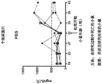
FIGS. 2A-2F show that treatment with the rat anti-mouse CD52 antibody successfully reduced the urinary protein levels in NZB/NZWF1 mice. Figures 2A-2E show that anti-mouse CD52 treated mice exhibited urine protein levels comparable to mice in the cyclophosphamide treated group of the positive control, while control rat IgG treated mice exhibited urine protein levels comparable to vehicle control (PBS) treated mice. Figure 2F shows that by the end of the study, only 38% of mice treated with anti-mouse CD52 antibody and 20% of mice treated with cyclophosphamide achieved severe proteinuria (>500 mg/dL/day) relative to 67% of mice treated with rat IgG and 60% of mice treated with vehicle.
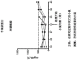
Figures 3A-3G show that treatment with the rat anti-mouse CD52 antibody successfully reduced the urinary protein levels in NZB/NZWF1 mice. The level of albumin in urine was assessed using a semi-quantitative "Albustix" method (fig. 3A) and a quantitative ELISA assay (fig. 3B). FIGS. 3A-3F show that the urinary albumin levels of mice treated with anti-CD 52 antibody were lower than those of mice treated with vehicle (PBS) and control rat IgG. Figure 3G shows that by the end of the study, only 50% of mice treated with anti-CD 52 antibody developed significant albuminuria (>40 mg/dL/day) compared to 80% of mice treated with vehicle and 89% of mice treated with rat IgG.
Figure 4 shows that treatment with the rat anti-mouse CD52 antibody had no detectable effect on the development of autoantibodies against dsDNA. The antibody titer of mice treated with anti-mouse CD52 was comparable to the titer of mice treated with vector and rat IgG. Cyclophosphamide-only treatment was effective in reducing the increase in serum antibodies against dsDNA.
Figure 5 shows that treatment with rat anti-mouse CD52 antibody provided significant survival benefit for NZB/NZWF1 mice (survival benefit). Two doses of anti-mouse CD52 antibody treatment (75% survival) resulted in comparable levels of survival to weekly injections of cyclophosphamide (80%) (P-value 0.9218, anti-mouse CD52 antibody vs. cyclophosphamide). The survival of mice treated with control rat IgG was only 20% (P value 0.0401, anti-mouse CD52 antibody vs. control rat IgG) (fig. 5).
FIGS. 6A-6C show histological examination of harvested mouse kidneys. Although the median severity scores for glomerulopathy, interstitial inflammation, or proteotype did not differ statistically significantly between the treatment groups, as shown in figure 6A, treatment with anti-mouse CD52 antibody and cyclophosphamide reduced the median glomerulopathy scores compared to the rat IgG and vehicle control groups. As shown in fig. 6B, reduced interstitial inflammation was also observed in the cyclophosphamide treated group.
FIGS. 7A-7C show FoxP3 permeating into the kidney+An increase in regulatory T cells. Mouse kidneys were stained with immunofluorescent-labeled antibody to reveal CD4+、CD8+And FoxP3+A cell. The relative abundance of positive cells on the kidney sections was blindly (blindly) scored in the range of 0-4. Cyclophosphamide treatment induced renal infiltration of CD4+、CD8+And FoxP3+A significant reduction of cells. By comparison, treatment with anti-CD 52 antibody did not prevent CD4+And CD8+Infiltration of lymphocytes into the kidney but increased presence of FoxP3 positive cells, a marker of regulatory T cells.
FIGS. 8A-8B show the effective lymphocyte depletion of NZB/NZWF1 mice by monoclonal mouse anti-mouse CD52 antibody (clone W19) at different dose levels (1mg/kg, 5mg/kg, and 10 mg/kg). In blood (FIG. 8A), dose-dependent depletion of 5mg/kg and 10mg/kg was observed in all lymphoid populations, resulting in all cell types (CD 4)+Cell, CD8+Cells, NK cells and B cells). In the spleen (fig. 8B), similar dose-dependent depletion was observed. In particular, CD4 in the spleen was observed+And CD8+Significant depletion of T cells, while B cells showed a lesser degree of depletion at all dose levels tested.
Detailed Description
The present invention is based on our findings relating to the administration of anti-CD 52 antibodies to subjects. We have found that anti-CD 52 antibodies increase FoxP3 in a murine model of lupus+Regulating T cell infiltration into locally inflamed tissue (kidney). We have also found that treatment with anti-CD 52 antibody can reduce the levels of urinary protein and albumin in this mouse model.
Accordingly, the invention provides methods of treating lupus in a patient (e.g., a human patient) with an anti-CD 52 antibody. In some embodiments, treatment will help recruit FoxP3+ regulatory T cells to locally inflamed tissues, such as the CNS, kidney, heart, and liver, thereby alleviating or preventing symptoms in lupus patients. In some embodiments, the treatment will help reduce urinary and/or albumin levels in lupus patients. In some embodiments, the treatment depletes lymphocytes from a lupus patient. In other embodiments of the invention, the patient is also treated with an agent that stimulates FoxP3+ regulatory T cell growth and/or activation, thereby improving regulation of the patient's immune system and alleviating the symptoms of autoimmunity.
Characterization of lupus
The methods of the invention may also be used in patients suffering from various lupus characterizations, including but not limited to systemic lupus erythematosus; lupus nephritis; cutaneous lupus erythematosus; CNS lupus; cardiovascular, pulmonary, hepatic, hematological, gastrointestinal, and musculoskeletal manifestations; lupus erythematosus in neonates; systemic lupus erythematosus in children; drug induced lupus erythematosus; antiphospholipid syndrome; and complement deficiency syndrome that causes lupus characterization. The methods of the invention can also be used to treat patients suffering from active lupus episodes, or patients with inactive lupus.
Therapy with anti-CD 52 antibodies
In the methods of the invention, an antibody to CD52 is administered to a patient in a therapeutically effective amount to achieve a clinical endpoint as determined by monitoring an infected organ system (e.g., hematuria and/or proteinuria of lupus nephritis) and/or using a disease activity index (e.g., BILAG, SLAM, SLEDAI, ECLAM), wherein the disease activity index provides a combined score of disease severity throughout several organ systems. See, e.g., Mandl et al, "Monitoring Patents with System Lupus Erythematosus," in systematic Lupus Erythematosus,4thedition, pp.619-631, R.G.Lahita, Editor, Elsevier Academic Press, (2004). A therapeutically effective amount of an anti-CD 52 antibody is an amount that helps treat a subject to achieve one or more desired clinical endpoints.
CD52 is a cell surface protein expressed at high levels by normal and malignant B and T lymphocytes (Hale et al, J Biol regol Homeost Agents 15: 386-. CD52 is expressed at lower levels in monocytes, macrophages and eosinophils, but is almost found expressed in mature Natural Killer (NK) cells, neutrophils and hematopoietic stem cells. CD52 also has been generated in epididymal and vas deferens epithelial cells, obtained by sperm as they pass through the reproductive tract (Hale et al, 2001, supra; Domagala et al, Med Sci monitor 7:325-331 (2001)). The exact biological function of CD52 is unclear, but some evidence suggests that it may be associated with T cell migration and co-stimulation (Rowan et al, Int Immunol 7:69-77 (1995); Masuyama et al, J Exp Med 189:979-989 (1999); Watanabe et al, Clin Immunol 120:247-259 (2006)).
An example of a human CD52 antigen polypeptide sequence is:
MKRFLFLLLT ISLLVMVQIQ TGLSGQNDTS QTSSPSASSNISGGIFLFFV ANAIIHLFCF S (SEQ ID NO: 1; NCBI accession No.: NP-001794)
The mature human CD52 antigen is quite short, only 12 amino acids (Xia et al, Eur J Immunol.21(7):1677-84(1991)) and is glycosylated. For example, the mature human CD52 antigens may each have the following polypeptide sequence: GQNDTSQTSSPS (SEQ ID NO: 2).
anti-CD 52 antibody therapies encompassed by the present invention include any treatment regimen using anti-CD 52 antibodies, including any suitable isotype of antibodies, such as IgG1, IgG2, IgG3, and IgG 4. Useful antibodies also include those in which the constant/Fc region has been modified and binds with equal or better affinity to Fc receptors on neutrophils and/or NK cells, or antibodies with improved antibody-dependent cell-mediated cytotoxicity (ADCC) and complement-dependent cytotoxicity (CDC) functions. anti-CD 52 antibodies useful in the present invention are those that specifically bind to CD52 and do not specifically bind to non-CD 52 molecules. Specific binding between anti-CD 52 antibodies and CD52 can be determined, for example, by measuring EC of antibodies that bind to CD52+ cells by flow cytometry50. Specific binding may be with EC50Expressed, for example, in the range of 0.5-10. mu.g/ml. For clinical use, the anti-CD 52 antibody may preferably be monoclonal and of pharmaceutically acceptable purity. The antibody may be administered by any suitable method, optionally with a pharmaceutically acceptable carrier, to be pharmaceutically effectiveE.g., an amount that assists the patient in reaching a desired clinical endpoint.
When the patient to be treated is a human, it is preferred that the anti-CD 52 antibody specifically binds human CD 52. To minimize immunogenicity upon repeated administration to human patients, it may also be preferred that the antibody is chimeric (e.g., a murine anti-CD 52 antibody with constant domains replaced with those of a human antibody), humanized (e.g., a human antibody with CDRs that have been replaced with those of a murine anti-human CD52 antibody), or fully human. An example of a useful antibody is alemtuzumab (e.g., and variants thereof). Alemtuzumab is a recombinant humanized IgG1 monoclonal antibody that targets human CD52(hCD52), a 28kD glycosylated glycosyl-phosphatidylinositol (GPI) -linked cell surface protein (Hale et al, Tissue antibodies 35:118-27 (1990); Hale et al, 2001, supra). Alemtuzumab has recently been demonstrated as a first line therapy for B-cell chronic lymphocytic leukemia and is in the third phase of clinical trials for the treatment of multiple sclerosis. Useful antibodies include, but are not limited to, antibodies that compete with alemtuzumab for binding to hCD52, and/or antibodies that bind to the same or overlapping epitopes of alemtuzumab or other epitopes on hCD 52. For example, humanized antibodies described in international application PCT/US2010/034704 may be used.
Human anti-hCD 52 antibodies can be made by one of skill in the art using, for example Technology (Amgen, thunder Oaks, CA). Chimeric and humanized anti-CD 52 antibodies can be made by well-accepted antibody techniques from, for example, the rat anti-hCD 52 antibody or the mouse anti-hCD 52 antibody.
If desired, the anti-CD 52 antibodies for use in the invention may contain a detectable label to allow monitoring, for example, in therapy, diagnosis, or assays. Suitable detectable labels include, for example, radioisotopes (e.g., indium-111, technetium-99 m, or iodine-131), positron emitting labels (e.g., fluorine-19), paramagnetic ions (e.g., gadolinium (III), manganese (II)), epitope labels (tags), affinity labels (e.g., biotin, avidin), spin labels, enzymes, fluorescent groups, or chemiluminescent groups. When no label is used, complex formation can be determined by surface plasmon resonance, ELISA, flow cytometry, or other suitable methods. The anti-CD 52 antibodies used in the present invention can be conjugated to another therapeutic agent such as a biologically active compound (e.g., cytokines and cytotoxic agents). The anti-CD 52 antibodies used in the invention can also be conjugated to other moieties (e.g., pegylated moieties) that can improve the pharmacokinetics, e.g., half-life, of the antibody by, for example, chemical reaction or genetic modification. In some embodiments, the anti-CD 52 antibodies used in the invention can be associated with an appropriate cytokine by, for example, chemical binding or genetic modification (e.g., the coding sequence for the cytokine in frame is added to the coding sequence for the antibody, thereby creating an antibody: cytokine fusion protein).
Increasing infiltration of Fox P3+ regulatory T cells
We have found that anti-CD 52 antibodies tend to increase FoxP3 relative to other T cells+Modulating T cells, including increasing the penetration of these cells into local tissues such as sites of inflammation or tissue injury. Regulatory T cells (also referred to as "tregs" or suppressor T cells) are cells that are capable of suppressing the proliferation and/or function of other lymphocytes by a contact-dependent or non-contact-dependent (e.g., cytokine production) mechanism. Several types of regulatory T cells have been described, including γ δ T cells, Natural Killer T (NKT) cells, CD8+T cell, CD4+T cell, CD4-CD8-Double negative T cells. See, e.g., Bach et al, Immunol.3:189-98 (2003). CD4+CD25+FoxP3+Regulatory T cells have been considered "naturally occurring" regulatory T cells; they express CD4, CD25 and the forkhead (forkhead) family of transcription factors FoxP3 (forkhead box p 3).
An increase in Tregs may be a symptom of the autoimmune disease under treatmentIs required. Thus, stimulation FoxP3 may be administered to a patient+(e.g., CD4+CD25+FoxP3+) An agent that modulates T cells. The agent may, for example, activate those T cells, expand the number of those cells, mobilize and increase the circulation of those cells, and/or recruit those cells to a target site. Examples of such agents are rapamycin, activated or latent TGF- β (e.g., TGF- β 1, TGF- β 2, TGF- β 3, TGF- β 4 and TGF- β 5), IL-10, IL-4, IFN- α, vitamin D3, dexamethasone, and mycophenolate esters (see, e.g., Barrat et al, J.Exp.Med.195:603-616 (2002); Gregori et al, J. Immunol.167:1945-1953 (2001); Battaglia et al, Blood 105:4743-4748 (2005); Battaglia et al, J.Immunol.177:8338-8347 (2010)). In some embodiments of the invention, the increase in Tregs may occur at one or more sites of inflammation (e.g., blood, central nervous system, heart, liver, joints, kidney, skin, digestive tract, or vasculature).
The Treg stimulating agent may be administered before, during or after the anti-CD 52 antibody treatment. anti-CD 52 antibodies for use in the invention preferentially deplete T effector cells and B cells, while preferentially retaining FoxP3+Tregs (see, e.g., Hu et al, Immunology 128: 260-. Thus, a treatment regimen using both an anti-CD 52 antibody and a Treg stimulator would greatly enhance the efficacy of lupus treatment, or other autoimmune disease treatment, by rebalancing the patient's immune system.
Reducing urine protein and/or albumin levels
Lupus patients may exhibit proteinuria or albuminuria-an excess of serum or albumin in the urine. In lupus, kidney damage, as measured by the levels of protein or albumin in the urine, is one of the most acute and accounts for at least 50% of mortality. The treatment methods of the invention (either the anti-CD 52 antibody alone or the anti-CD 52 antibody in combination with a Treg stimulator) can reduce urine protein and/or albumin levels in a patient by at least 25%, 50%, 75%, or 90% compared to pre-treatment levels. In some embodiments, the urinary protein level is at least greater than or equal to 500 mg/L/day (e.g., 1,000 mg/L/day, 2,000 mg/L/day, or 3,000 mg/L/day) prior to administration of the anti-CD 52 antibody. Following initial treatment with anti-CD 52 antibody, urinary protein levels may be reduced to less than 500 mg/L/day or less than 1,000 mg/L/day.
Combination therapy
In some aspects of the invention, the anti-CD 52 antibody can be co-administered to a lupus patient in a combination therapy with one or more other therapeutic agents (e.g., an immunosuppressive agent). The second therapeutic agent can be, for example, corticosteroids, non-steroidal anti-inflammatory drugs, disorder-modifying antirheumatic drugs (DMARDs) (e.g., cyclophosphamide or mycophenolic acid), immunosuppressive agents (e.g., methotrexate and azathioprine), molecules that target B or T lymphocytes (e.g., CD20 antibodies, such as Rituximab (Rituximab), also known as Rituximab) anti-BLys antibody, or anti-BAFF-R antibody). In some embodiments, the other agent is, e.g., a Cytokine (e.g., IL-7), an anti-Cytokine receptor antibody, or a soluble receptor that directs, manipulates, and/or amplifies the recombination process that occurs after lymphoid depletion mediated by the anti-CD 52 antibody (see, e.g., sports et al, Cytokine therapeutics: ann.n.y.acad.sci.1182:28-38 (2009)). Other therapeutic agents may be administered before, during, or after the anti-CD 52 antibody treatment.
Unless defined otherwise, all technical and scientific terms used herein have the same meaning as commonly understood by one of ordinary skill in the art to which this invention belongs. Exemplary methods and materials are described below, although methods and materials similar or equivalent to those described herein can also be used in the practice or testing of the present invention. All publications and other references mentioned herein are incorporated by reference in their entirety. In case of conflict, the present specification, including definitions, will control. Although a number of documents are referred to herein, this reference does not constitute an admission that any of these documents form part of the common general knowledge in the art. Throughout this specification and claims, the word "comprise", or variations such as "comprises" or "comprising", will be understood to imply the inclusion of a stated integer or group of integers but not the exclusion of any other integer or group of integers. The materials, methods, and examples are illustrative only and not intended to be limiting.
Examples
The following examples are intended to illustrate the methods and materials of the present invention. Suitable modifications and variations to the conditions and parameters described, which are often found in the art and which are obvious to those skilled in the art, are within the spirit and scope of the invention.
Mouse lupus model
NZB/NZWF1 mice were used as a spontaneous model for lupus. As they grow, animals develop autoantibodies against a variety of cellular antigens, eventually leading to the deposition of immune complexes in the kidney and the progressive development of fatal nephropathy (Peutz-Kootstra et al, J Lab Clin Med 137: 244-. In the following examples, NZB/NZWF1 female mice were used to study the role of anti-CD 52 antibodies in the development of systemic lupus.
anti-CD 52 antibodies
In examples 1-6, monoclonal rat anti-mouse CD52IgG2a antibody was used. This rat isotype is not the optimal isotype for mouse effector functions (e.g., complement fixation and antibody-dependent cell-mediated cytotoxicity). In examples 7-8, monoclonal IgG2a mouse anti-mouse CD52 antibody was used.
In examples 1-6, NZB/NZWF1 mice (15 weeks old, Jackson Labs) were divided into 4 groups and treated with different samples (Table 1). A nitrogen mustard alkylating agent, cyclophosphamide, was used as a positive control in examples 1-6. Cyclophosphamide has been used to treat a variety of cancers and certain autoimmune disorders, including lupus.
TABLE 1

ELISA of rat IgG2a showed that only 184 μ g/ml of total protein content in the stock consisted of rat IgG2a, indicating that the effective dose can be as low as 1.7 mg/kg.
The time points for examples 1-6 were as follows:
i) blood was collected from individual mice every 4 weeks starting at 19 weeks of age to assess IgG anti-double stranded DNA (anti-dsDNA) antibody titers, and 24 hour urine was collected in metabolic cages to determine proteinuria and albuminuria.
ii) treatment with the test sample begins at 31 weeks of age, when the animals begin to develop significant antibody titers against dsDNA and/or elevated proteinuria. Group 2 treated with normal rat IgG and group 3 treated with rat anti-mouse CD52 antibody received a total of two injections, respectively. Group 4 treated with cyclophosphamide received one injection per week until the end of the study.
iii) blood was collected from groups 2 and 3 for Fluorescence Activated Cell Sorting (FACS) analysis of basal values (staining CD3, CD19 positive cells, counting absolute number of lymphocytes) prior to the first antibody injection.
iv) two days after the first injection of antibody, a second injection of antibody was performed in groups 2 and 3. Prior to the second injection, blood was collected from groups 2 and 3 for flow cytometry analysis (CD 3, CD19 positive cells were stained for absolute counts). Spleens were collected from one mouse of group 2 and one mouse of group 3 and were also used for flow cytometry staining.
Any animal that was moribund during the study was sacrificed and a kidney was collected if possible. The study was terminated at 43 weeks of age in mice, and one kidney from each animal was harvested for histological analysis.
Example 1: NZB/NZWF1 mice treated with rat anti-mouse CD52 antibody had no depletion of lymphocytes
For the determination of rat anti-mouse using monoclonal antibodyWhether treatment with murine CD52 antibody would result in CD52+Depletion of lymphocytes, blood was collected at basal values from individual mice prior to the first injection of rat IgG or rat anti-mouse CD52, and two days later prior to the second antibody injection. Blood samples were stained and analyzed with flow cytometry to obtain CD3+T cells and CD19+Absolute number of B cells. A50. mu.l sample of whole blood was blocked with 10% normal mouse serum and 0.05% sodium azide in RPMI medium and then stained with rat anti-mouse CD3-APC and rat anti-mouse CD19-PE (BD Pharmingen, San Diego, Calif.). Lymphocytes in FACSCaliburTMStaining analysis was performed in the system (Becton-Dickinson, San Diego, Calif.). Data analysis was performed using Cell Quest Pro software (Becton-Dickinson). The results showed no significant depletion of B or T lymphocytes (fig. 1A and 1B).
Example 2: level of proteinuria and albuminuria
A. Level of proteinuria
The protein level in the urine of individual mice was determined colorimetrically according to the manufacturer's instructions (Microprotein-PR, Sigma), which colorimetric method was designed to determine the total protein concentration. The protein concentration of the test sample is calculated using the reference standard. Although there was no depletion of lymphocytes upon detection, treatment with the rat anti-mouse CD52 antibody successfully inhibited the development of renal disease, as determined by total urinary protein levels (fig. 2A-2E). During the course of the study, mice treated with anti-mouse CD52 exhibited urine protein levels comparable to those of cyclophosphamide treated positive control groups, while mice treated with control rat IgG exhibited urine protein levels comparable to those of vehicle control mice (fig. 2A-2E). Only 38% of mice treated with anti-mouse CD52 antibody and 20% of mice treated with cyclophosphamide reached severe urine protein (>500 mg/dL/day) relative to 67% of mice treated with rat IgG and 60% of mice treated with vehicle (fig. 2F).
B. Level of albuminuria
The level of albumin in urine was assessed using an indirect competitive ELISA kit according to the manufacturer's instructions (Albuwell-M, Exocell, Inc.). The concentration of albumin in the urine sample was derived from a standard curve derived from known murine albumin concentrations (figure 3B). A semi-quantitative "Albustix" method (Roche Diagnostics) (FIG. 3A) was also used, in which urine was dropped on an indicator filter paper that would change color depending on the amount of albumin present in the urine and then a corresponding score of 0-6 was determined. Consistent with the total protein levels, treatment with anti-mouse CD52 antibody was also effective in inhibiting the development of albuminuria in NZB/NZWF1 mice (fig. 3A-3G). Urine albumin levels were lower in anti-CD 52 antibody treated mice than in vehicle and rat IgG treated mice. However, the inhibition of albuminuria observed in this group was not as significant as that obtained in the cyclophosphamide treated group. By the end of the study, only 50% of mice treated with anti-CD 52 antibody developed significant albuminuria (>40 mg/dL/day) compared to 80% of vehicle-treated mice and 89% of mice treated with rat IgG (fig. 3G).
Example 3: level of antibodies to double stranded DNA
IgG antibody titers against dsDNA in individual mouse serum samples were determined by ELISA. Mouse dsDNA (The Jackson Laboratory, BarHarbor, ME) was digested with S1 nuclease (Invitrogen, Carlsbad, Calif.) to remove any ssDNA, and then used to coat wells of a 96-well ELISA plate (1. mu.g/ml dsDNA, 100. mu.l/well) overnight at 4 ℃. The microplate was pretreated with 0.01% protamine sulfate/water to promote DNA attachment. After coating, the microplate was incubated with 2.5% bovine serum albumin blocking buffer at 37 ℃ for 1 hour and washed. Subsequently 100 μ l series of 2 fold diluted sera were added to replicate wells and incubated for 1 hour at 37 ℃. The microplate was washed and horseradish peroxidase (HRP) -conjugated goat anti-mouse IgG (Pierce, Rockford, IL) was added to detect the antibody bound to dsDNA (37 ℃, 1 hour). After washing, HRP substrate was added and the colorimetric product read at Optical Density (OD) at 490nM wavelength with 650nM as reference wavelength on a dual wavelength plate reader (Molecular Devices, Sunnyvale, CA). Antibody titer was defined as the reciprocal of serum dilution when OD was greater than or equal to 0.1. Normal mouse sera were used as negative controls (titer. ltoreq.200, lowest dilution detected) and pooled sera from aged lupus mice were used as positive controls (titer 25,600). Treatment with the rat anti-mouse CD52 antibody had no detectable effect on the development of autoantibodies against dsDNA (fig. 4). The antibody titer of mice treated with anti-mouse CD52 was comparable to the titer of mice treated with vector and rat IgG. Cyclophosphamide treatment alone effectively reduced the rise in serum of antibodies against dsDNA (fig. 4).
Example 4: improvement of survival rate
Treatment with the rat anti-mouse CD52 antibody was well tolerated in NZB/NZWF1 mice. Comparable levels of survival were obtained between two doses of anti-mouse CD52 antibody (75% survival) and weekly injections of cyclophosphamide (80% survival) (P-value 0.9218, anti-mouse CD52 antibody vs. cyclophosphamide) (fig. 5). In mice treated with control rat IgG, the survival was only 20% (P value ═ 0.0401, anti-mouse CD52 antibody vs. control rat IgG) (fig. 5). By comparison, vehicle-treated mice showed 60% survival (fig. 5), indicating that injection of large amounts of immunoglobulin to the control rat IgG group may have exacerbated the disease, possibly by renal pressurization, while the same amount of anti-mouse CD52 material provided a therapeutic benefit.
Example 5: histological examination of kidney
Kidneys were collected at sacrifice, fixed in 10% neutral buffered formalin and then embedded in paraffin. Sections were taken at a thickness of 5 μm and stained with hematoxylin and eosin (H & E), hematoxylin phosphotungstic acid (PTAH), and Periodic Acid Schiff (PAS) staining methods. During the course of the study, some animals of the negative control group (vehicle and rat IgG) had to be sacrificed or found dead. As a result, few kidneys are available for analysis at the end of the study, thus limiting statistical power.
The collected kidneys were further examined. The median severity scores for glomerulopathy, interstitial inflammation, or proteotype did not differ statistically significantly between treatment groups (fig. 6A-6C). However, some trends are still evident. Treatment with anti-mouse CD52 antibody and cyclophosphamide reduced the median glomerulopathy score compared to the rat IgG and vehicle control groups (fig. 6A). Reduced interstitial inflammation was also observed in the cyclophosphamide treated group (fig. 6B).
+Example 6: increased FoxP3 regulatory T cells in the kidney
The kidney sections obtained in example 5 were further stained with an immunofluorescent-labeled antibody to observe CD4+、CD8+And FoxP3+The presence of cells. For staining of CD4 and CD8 positive cells, frozen sections of kidney were fixed in acetone and incubated sequentially with peroxidase (Dako), avidin, biotin (Biocare), and protein (Dako) blocking solution, followed by biotinylated rat anti-mouse CD4 (clone L3T 4; BD Pharmingen) or biotinylated goat anti-mouse CD8 (clone Ly-2; BD Pharmingen), streptavidin-HRP, and DAB (3-3' -diaminobenzidine) to produce brown staining on positive cells. For staining of FoxP3 positive cells, kidney cryosections were fixed in 10% neutral buffered formalin and incubated with peroxidase and protein blocking solution in sequence. Rat anti-mouse FoxP3 antibody (eBioscience) was then added followed by Mach-2HRP conjugated anti-rabbit antibody (Biocare) and DAB to produce brown staining on positive cells. All sections were then stained with hematoxylin to visualize the cells. The relative abundance of positive cells on the slides was blindly scored in the range of 0-4. Treatment with cyclophosphamide causes renal infiltration of CD4+、CD8+And FoxP3+Significant reduction of cells (fig. 7A-7C). By comparison, treatment with anti-CD 52 antibody failed to prevent CD4+And CD8+Lymphocytes permeated the kidney but increased the presence of cells positive for FoxP3, FoxP3 is a marker for regulatory T cells (fig. 7A-7C).
Example 7: lymphocyte depletion from NZB/NZWF1 mice treated with monoclonal mouse anti-mouse CD52 antibody
Reducing
Depletion experiments were performed to determine whether lupus mice were sensitive to depletion of lymphocytes by targeting mouse CD52 using the homemade monoclonal IgG2a mouse anti-mouse CD52 antibody (clone W19). NZB/NZWF1 mice were treated with vector, 1mg/kg, 5mg/kg, or 10mg/kg monoclonal mouse anti-mouse CD52 antibody. Three days after treatment, splenocytes and peripheral blood were collected and evaluated for lymphocyte depletion by flow cytometry. Significant levels of lymphocyte depletion in blood and spleen were observed at all antibody dose levels. In blood (fig. 8A), dose-dependent depletion was observed in all lymphoid populations, with 5 and 10mg/kg doses causing almost complete depletion of all cell types. Similar dose-dependent reduction was also observed in the spleen (fig. 8B). When CD4 was observed in the spleen+And CD8+At significant T cell depletion, B cells appeared to be depleted to a lesser extent at all dose levels tested.
Example 8: efficacy analysis of anti-mouse CD52 antibody in NZB/NZW female mice
The monoclonal anti-mouse CD52 antibodies used in example 7 were further tested for their effect on the development and/or progression of disease in the NZB/NZWF1 mouse lupus model. First, a group of 10 mice received two injections of either control antibody or monoclonal mouse anti-mouse CD52 antibody at 10mg/kg one week apart before significant disease occurred, i.e., at approximately 21 weeks of age. Thereafter, 10 mice from different groups received two injections of either control antibody or monoclonal mouse anti-mouse CD52 antibody at 10mg/kg at approximately 32 weeks of age during the disease period, separated by one week intervals. The positive control group received a weekly 50mg/kg injection of cyclophosphamide starting at approximately 21 weeks of age. We examined the following readings: 1) lymphocyte depletion by flow cytometry; 2) development of autoantibodies against dsDNA as determined by ELISA; 3) proteinuria; and 4) histological analysis of the kidney; and further determine the extent of remission of renal injury from targeting CD52 in this manner.
Sequence listing
<110> Genzyme Ltd
<120> methods and compositions for treating lupus
<130> 001662-0020-WO1
<140> PCT/US2010/034741
<141> 2010-05-13
<150> 61/177,924
<151> 2009-05-13
<160> 2
<170> PatentIn version 3.5
<210> 1
<211> 61
<212> PRT
<213> Intelligent
<400> 1
Met Lys Arg Phe Leu Phe Leu Leu Leu Thr Ile Ser Leu Leu Val Met
1 5 10 15
Val Gln Ile Gln Thr Gly Leu Ser Gly Gln Asn Asp Thr Ser Gln Thr
20 25 30
Ser Ser Pro Ser Ala Ser Ser Asn Ile Ser Gly Gly Ile Phe Leu Phe
35 40 45
Phe Val Ala Asn Ala Ile Ile His Leu Phe Cys Phe Ser
50 55 60
<210> 2
<211> 12
<212> PRT
<213> Intelligent people
<400> 2
Gly Gln Asn Asp Thr Ser Gln Thr Ser Ser Pro Ser
1 5 10
Claims (9)
1. Use of an anti-human CD52 monoclonal antibody in the manufacture of a medicament for the treatment of lupus nephritis in a human patient, wherein the anti-human CD52 monoclonal antibody is the sole active ingredient of the medicament.
2. The use of claim 1, wherein the anti-human CD52 monoclonal antibody reduces the level of urine protein, urine albumin, or both in the patient.
3. The use of claim 1, wherein the anti-human CD52 monoclonal antibody increases FoxP3 in the patient+Regulatory T cells.
4. The use of claim 3, wherein the regulatory T cells are increased at least at one site of inflammation.
5. The use of claim 4, wherein the site of inflammation is blood, Central Nervous System (CNS), heart, liver, joint, kidney, lung, skin, gut, or vasculature.
6. The use of any one of claims 1-5, wherein the patient is undergoing pretreatment with a corticosteroid.
7. The use of any one of claims 1-5, wherein the anti-human CD52 monoclonal antibody does not significantly deplete lymphocytes.
8. The use of any one of claims 1-5, wherein the anti-human CD52 monoclonal antibody is a humanized or human antibody.
9. The use of claim 8, wherein the anti-human CD52 monoclonal antibody is alemtuzumab.
Applications Claiming Priority (4)
| Application Number | Priority Date | Filing Date | Title |
|---|---|---|---|
| US17792409P | 2009-05-13 | 2009-05-13 | |
| US61/177,924 | 2009-05-13 | ||
| PCT/US2010/034741 WO2010132683A1 (en) | 2009-05-13 | 2010-05-13 | Methods and compositions for treating lupus |
| CN2010800209534A CN102438654A (en) | 2009-05-13 | 2010-05-13 | Methods and compositions for treating lupus |
Related Parent Applications (1)
| Application Number | Title | Priority Date | Filing Date |
|---|---|---|---|
| CN2010800209534A Division CN102438654A (en) | 2009-05-13 | 2010-05-13 | Methods and compositions for treating lupus |
Publications (2)
| Publication Number | Publication Date |
|---|---|
| CN110179989A CN110179989A (en) | 2019-08-30 |
| CN110179989B true CN110179989B (en) | 2022-06-24 |
Family
ID=43085330
Family Applications (2)
| Application Number | Title | Priority Date | Filing Date |
|---|---|---|---|
| CN2010800209534A Pending CN102438654A (en) | 2009-05-13 | 2010-05-13 | Methods and compositions for treating lupus |
| CN201910644240.8A Active CN110179989B (en) | 2009-05-13 | 2010-05-13 | Methods and compositions for treating lupus |
Family Applications Before (1)
| Application Number | Title | Priority Date | Filing Date |
|---|---|---|---|
| CN2010800209534A Pending CN102438654A (en) | 2009-05-13 | 2010-05-13 | Methods and compositions for treating lupus |
Country Status (12)
| Country | Link |
|---|---|
| US (2) | US9617343B2 (en) |
| EP (2) | EP2429583A4 (en) |
| JP (3) | JP5834004B2 (en) |
| KR (1) | KR101800467B1 (en) |
| CN (2) | CN102438654A (en) |
| AU (2) | AU2010248935B2 (en) |
| BR (1) | BRPI1013927A2 (en) |
| CA (1) | CA2761885A1 (en) |
| IL (1) | IL216146A0 (en) |
| MX (1) | MX342907B (en) |
| RU (1) | RU2607022C2 (en) |
| WO (1) | WO2010132683A1 (en) |
Families Citing this family (10)
| Publication number | Priority date | Publication date | Assignee | Title |
|---|---|---|---|---|
| EP3683317A3 (en) | 2009-05-13 | 2020-09-30 | Genzyme Corporation | Anti-human cd52 immunoglobulins |
| US20150073043A1 (en) * | 2012-01-19 | 2015-03-12 | Institut Pasteur Of Shanghai, Chinese Academy Of Sciences | Use of phosphorylation pathway-related factor in regulating function of regulatory t cell |
| CN111965358A (en) * | 2013-02-08 | 2020-11-20 | 阿勒格尼-辛格研究所 | Cell-bound complement activation products as pre-lupus diagnostic biomarkers |
| AR095199A1 (en) | 2013-03-15 | 2015-09-30 | Genzyme Corp | ANTI-CD52 ANTIBODIES |
| EP2986990B1 (en) * | 2013-03-20 | 2018-08-15 | F.Hoffmann-La Roche Ag | Specific detection of rat antibodies in mouse serum |
| KR102157924B1 (en) | 2014-02-14 | 2020-10-26 | 셀렉티스 | Cells for immunotherapy engineered for targeting antigen present both on immune cells and pathological cells |
| EP4091616A1 (en) * | 2015-02-27 | 2022-11-23 | iCell Gene Therapeutics LLC | Chimeric antigen receptors (car) targeting hematologic malignancies, compositions and methods of use thereof |
| US11173179B2 (en) | 2015-06-25 | 2021-11-16 | Icell Gene Therapeutics Llc | Chimeric antigen receptor (CAR) targeting multiple antigens, compositions and methods of use thereof |
| US11655452B2 (en) | 2015-06-25 | 2023-05-23 | Icell Gene Therapeutics Inc. | Chimeric antigen receptors (CARs), compositions and methods of use thereof |
| JP7114490B2 (en) | 2016-06-24 | 2022-08-08 | アイセル・ジーン・セラピューティクス・エルエルシー | Construction of chimeric antibody receptors (CARs) and methods of their use |
Family Cites Families (20)
| Publication number | Priority date | Publication date | Assignee | Title |
|---|---|---|---|---|
| GB9022543D0 (en) | 1990-10-17 | 1990-11-28 | Wellcome Found | Antibody production |
| GB9022547D0 (en) | 1990-10-17 | 1990-11-28 | Wellcome Found | Purified immunoglobulin |
| GB9125768D0 (en) | 1991-12-04 | 1992-02-05 | Hale Geoffrey | Therapeutic method |
| US7119248B1 (en) | 1994-04-12 | 2006-10-10 | Miltenyi Biotec Gmbh | Antibodies against epitopes with homology to self antigens, methods of preparation and applications thereof |
| US7060802B1 (en) * | 2000-09-18 | 2006-06-13 | The Trustees Of Columbia University In The City Of New York | Tumor-associated marker |
| US7465790B2 (en) | 2000-10-09 | 2008-12-16 | Isis Innovation, Inc. | Therapeutic antibodies |
| WO2004042032A2 (en) * | 2002-11-01 | 2004-05-21 | The Ohio State University Research Foundation | Cytogenetic abnormalities that are predictive of response to therapy for chronic lymphocytic leukemia |
| US7534427B2 (en) | 2002-12-31 | 2009-05-19 | Immunomedics, Inc. | Immunotherapy of B cell malignancies and autoimmune diseases using unconjugated antibodies and conjugated antibodies and antibody combinations and fusion proteins |
| CA2520830A1 (en) * | 2003-03-31 | 2004-10-14 | Kirin Beer Kabushiki Kaisha | Method for inducing differentiation and/or proliferating regulatory t cells using anti-cd52 antibodies and pharmaceutical composition therefor |
| DK1694706T3 (en) | 2003-11-01 | 2012-07-16 | Merck Patent Gmbh | Modified anti-CD52 antibody |
| WO2005117967A2 (en) | 2004-04-12 | 2005-12-15 | Medimmune, Inc. | Anti-il-9 antibody formulations and uses thereof |
| CN101443040A (en) * | 2006-03-16 | 2009-05-27 | 基因技术股份有限公司 | Methods of treating lupus using CD4 antibodies |
| AU2007348315A1 (en) | 2006-04-07 | 2008-09-12 | Chimeros, Inc. | Compositions and methods for treating B- cell malignancies |
| ES2547333T3 (en) * | 2006-04-12 | 2015-10-05 | Genzyme Corporation | Methods to treat autoimmune diseases |
| GB0608054D0 (en) * | 2006-04-24 | 2006-05-31 | Isis Innovation | Production and use of regulatory t cells |
| CA2653848A1 (en) * | 2006-05-31 | 2007-12-06 | Genzyme Corporation | Methods of using anti-thymocyte globulin and related agents |
| ES2612383T3 (en) * | 2006-07-19 | 2017-05-16 | The Trustees Of The University Of Pennsylvania | WSX-1 / IL-27 as a target for anti-inflammatory responses |
| US8148067B2 (en) | 2006-11-09 | 2012-04-03 | Xdx, Inc. | Methods for diagnosing and monitoring the status of systemic lupus erythematosus |
| EP3683317A3 (en) * | 2009-05-13 | 2020-09-30 | Genzyme Corporation | Anti-human cd52 immunoglobulins |
| AR095199A1 (en) | 2013-03-15 | 2015-09-30 | Genzyme Corp | ANTI-CD52 ANTIBODIES |
-
2010
- 2010-05-13 CN CN2010800209534A patent/CN102438654A/en active Pending
- 2010-05-13 EP EP10775545.6A patent/EP2429583A4/en not_active Withdrawn
- 2010-05-13 RU RU2011150495A patent/RU2607022C2/en active
- 2010-05-13 KR KR1020117029688A patent/KR101800467B1/en active IP Right Grant
- 2010-05-13 BR BRPI1013927A patent/BRPI1013927A2/en not_active Application Discontinuation
- 2010-05-13 CN CN201910644240.8A patent/CN110179989B/en active Active
- 2010-05-13 JP JP2012511015A patent/JP5834004B2/en active Active
- 2010-05-13 WO PCT/US2010/034741 patent/WO2010132683A1/en active Application Filing
- 2010-05-13 CA CA2761885A patent/CA2761885A1/en not_active Withdrawn
- 2010-05-13 MX MX2011012048A patent/MX342907B/en active IP Right Grant
- 2010-05-13 EP EP17206028.7A patent/EP3345620A1/en active Pending
- 2010-05-13 AU AU2010248935A patent/AU2010248935B2/en active Active
- 2010-05-13 US US13/320,001 patent/US9617343B2/en active Active
-
2011
- 2011-11-03 IL IL216146A patent/IL216146A0/en active IP Right Grant
-
2014
- 2014-12-11 JP JP2014250861A patent/JP2015052021A/en not_active Withdrawn
-
2016
- 2016-10-06 JP JP2016197833A patent/JP2017008106A/en active Pending
-
2017
- 2017-03-01 US US15/446,162 patent/US20170369583A1/en not_active Abandoned
- 2017-03-15 AU AU2017201771A patent/AU2017201771A1/en not_active Abandoned
Also Published As
| Publication number | Publication date |
|---|---|
| MX342907B (en) | 2016-10-17 |
| WO2010132683A1 (en) | 2010-11-18 |
| RU2011150495A (en) | 2013-06-20 |
| AU2010248935B2 (en) | 2016-12-15 |
| JP2012526846A (en) | 2012-11-01 |
| JP2017008106A (en) | 2017-01-12 |
| EP2429583A1 (en) | 2012-03-21 |
| BRPI1013927A2 (en) | 2016-04-05 |
| CA2761885A1 (en) | 2010-11-18 |
| JP2015052021A (en) | 2015-03-19 |
| RU2607022C2 (en) | 2017-01-10 |
| CN110179989A (en) | 2019-08-30 |
| KR20140014392A (en) | 2014-02-06 |
| AU2017201771A1 (en) | 2017-04-06 |
| MX2011012048A (en) | 2011-12-12 |
| US20120070408A1 (en) | 2012-03-22 |
| EP3345620A1 (en) | 2018-07-11 |
| IL216146A0 (en) | 2012-01-31 |
| KR101800467B1 (en) | 2017-11-22 |
| EP2429583A4 (en) | 2013-10-16 |
| US9617343B2 (en) | 2017-04-11 |
| CN102438654A (en) | 2012-05-02 |
| US20170369583A1 (en) | 2017-12-28 |
| AU2010248935A1 (en) | 2011-11-17 |
| JP5834004B2 (en) | 2015-12-16 |
Similar Documents
| Publication | Publication Date | Title |
|---|---|---|
| CN110179989B (en) | Methods and compositions for treating lupus | |
| CA2146647C (en) | Treatment of autoimmune and inflammatory disorders | |
| CN111939261B (en) | Methods of treating eosinophilic esophagitis with IL-4R inhibitors | |
| JP6797810B2 (en) | Anti-CD40L Antibodies and Methods for Treating CD40L-Related Diseases or Disorders | |
| US20180155436A1 (en) | Methods of Treating Inflammatory Conditions | |
| WO2018071777A1 (en) | Innate immune cell trispecific binding proteins and methods of use | |
| JP6373944B2 (en) | IL-21 antibody | |
| EA031849B1 (en) | Anti-ox40 antibodies and methods of using the same | |
| US20120058082A1 (en) | Methods and compositions for treatment | |
| KR101691534B1 (en) | Use of il-20 antagonists for treating rheumatoid arthritis and osteoporosis | |
| JPH09502184A (en) | Method for persistently suppressing humoral immunity | |
| US20090004182A1 (en) | Methods to Treat or Prevent Viral-Associated Lymphoproliferative Disorders | |
| US9289469B2 (en) | Depleting immunosuppressive monocytes within a mammal | |
| EP2373339B1 (en) | A b cell depleting agent for the treatment of atherosclerosis | |
| Ibtehaj et al. | B cell-specific mAb–siRNA conjugates improve experimental myasthenia | |
| CN112079922B (en) | Anti-human p40 protein domain antibody and application thereof | |
| CN118146376A (en) | HLA-G antibodies, methods of making and uses thereof | |
| CN113784985A (en) | anti-CD 40 antibodies for use in the treatment of T1DM and insulitis | |
| NZ794842A (en) | Methods of treating inflammatory conditions |
Legal Events
| Date | Code | Title | Description |
|---|---|---|---|
| PB01 | Publication | ||
| PB01 | Publication | ||
| SE01 | Entry into force of request for substantive examination | ||
| SE01 | Entry into force of request for substantive examination | ||
| GR01 | Patent grant | ||
| GR01 | Patent grant |