CN108738323B - anti-TREM 2 antibodies and methods of use thereof - Google Patents
anti-TREM 2 antibodies and methods of use thereof Download PDFInfo
- Publication number
- CN108738323B CN108738323B CN201680070761.1A CN201680070761A CN108738323B CN 108738323 B CN108738323 B CN 108738323B CN 201680070761 A CN201680070761 A CN 201680070761A CN 108738323 B CN108738323 B CN 108738323B
- Authority
- CN
- China
- Prior art keywords
- amino acid
- seq
- acid sequence
- antibody
- trem2
- Prior art date
- Legal status (The legal status is an assumption and is not a legal conclusion. Google has not performed a legal analysis and makes no representation as to the accuracy of the status listed.)
- Active
Links
- 238000000034 method Methods 0.000 title claims description 52
- 101000795117 Homo sapiens Triggering receptor expressed on myeloid cells 2 Proteins 0.000 claims abstract description 524
- 102100029678 Triggering receptor expressed on myeloid cells 2 Human genes 0.000 claims abstract description 504
- 102000008394 Immunoglobulin Fragments Human genes 0.000 claims abstract description 19
- 108010021625 Immunoglobulin Fragments Proteins 0.000 claims abstract description 19
- 125000003275 alpha amino acid group Chemical group 0.000 claims description 717
- 125000000539 amino acid group Chemical group 0.000 claims description 264
- 230000027455 binding Effects 0.000 claims description 101
- 230000000694 effects Effects 0.000 claims description 78
- 235000001014 amino acid Nutrition 0.000 claims description 77
- 208000037265 diseases, disorders, signs and symptoms Diseases 0.000 claims description 70
- 238000006467 substitution reaction Methods 0.000 claims description 59
- 239000003446 ligand Substances 0.000 claims description 52
- 201000010099 disease Diseases 0.000 claims description 47
- 150000007523 nucleic acids Chemical class 0.000 claims description 39
- 239000012634 fragment Substances 0.000 claims description 37
- 229940024606 amino acid Drugs 0.000 claims description 33
- 150000001413 amino acids Chemical class 0.000 claims description 33
- 238000004519 manufacturing process Methods 0.000 claims description 28
- 239000003814 drug Substances 0.000 claims description 25
- 208000035475 disorder Diseases 0.000 claims description 23
- 230000001939 inductive effect Effects 0.000 claims description 22
- 125000003729 nucleotide group Chemical group 0.000 claims description 22
- 208000024827 Alzheimer disease Diseases 0.000 claims description 20
- 102000048432 human TREM2 Human genes 0.000 claims description 20
- 102000039446 nucleic acids Human genes 0.000 claims description 20
- 108020004707 nucleic acids Proteins 0.000 claims description 20
- 239000013598 vector Substances 0.000 claims description 20
- 108091028043 Nucleic acid sequence Proteins 0.000 claims description 19
- DHMQDGOQFOQNFH-UHFFFAOYSA-N Glycine Chemical compound NCC(O)=O DHMQDGOQFOQNFH-UHFFFAOYSA-N 0.000 claims description 18
- 208000014674 injury Diseases 0.000 claims description 17
- 230000001684 chronic effect Effects 0.000 claims description 16
- 230000007278 cognition impairment Effects 0.000 claims description 16
- 206010012289 Dementia Diseases 0.000 claims description 14
- 201000011240 Frontotemporal dementia Diseases 0.000 claims description 13
- 238000012217 deletion Methods 0.000 claims description 13
- 230000037430 deletion Effects 0.000 claims description 13
- 239000002773 nucleotide Substances 0.000 claims description 13
- 206010009900 Colitis ulcerative Diseases 0.000 claims description 12
- 201000006704 Ulcerative Colitis Diseases 0.000 claims description 12
- 206010009887 colitis Diseases 0.000 claims description 12
- 208000020431 spinal cord injury Diseases 0.000 claims description 12
- 239000004475 Arginine Substances 0.000 claims description 11
- 230000002708 enhancing effect Effects 0.000 claims description 11
- 239000004471 Glycine Substances 0.000 claims description 10
- 208000030886 Traumatic Brain injury Diseases 0.000 claims description 10
- 230000009529 traumatic brain injury Effects 0.000 claims description 10
- 208000027418 Wounds and injury Diseases 0.000 claims description 9
- 230000006378 damage Effects 0.000 claims description 9
- 208000000044 Amnesia Diseases 0.000 claims description 8
- 108020004705 Codon Proteins 0.000 claims description 8
- 208000026139 Memory disease Diseases 0.000 claims description 8
- 230000006984 memory degeneration Effects 0.000 claims description 8
- 208000023060 memory loss Diseases 0.000 claims description 8
- 101000809875 Homo sapiens TYRO protein tyrosine kinase-binding protein Proteins 0.000 claims description 5
- 102100038717 TYRO protein tyrosine kinase-binding protein Human genes 0.000 claims description 5
- ODKSFYDXXFIFQN-UHFFFAOYSA-N arginine Natural products OC(=O)C(N)CCCNC(N)=N ODKSFYDXXFIFQN-UHFFFAOYSA-N 0.000 claims description 5
- UYTPUPDQBNUYGX-UHFFFAOYSA-N guanine Chemical class O=C1NC(N)=NC2=C1N=CN2 UYTPUPDQBNUYGX-UHFFFAOYSA-N 0.000 claims description 5
- 239000008194 pharmaceutical composition Substances 0.000 claims description 5
- 241001529936 Murinae Species 0.000 claims description 4
- QIVBCDIJIAJPQS-UHFFFAOYSA-N Tryptophan Natural products C1=CC=C2C(CC(N)C(O)=O)=CNC2=C1 QIVBCDIJIAJPQS-UHFFFAOYSA-N 0.000 claims description 4
- 125000003588 lysine group Chemical class [H]N([H])C([H])([H])C([H])([H])C([H])([H])C([H])([H])C([H])(N([H])[H])C(*)=O 0.000 claims description 4
- 125000000430 tryptophan group Chemical class [H]N([H])C(C(=O)O*)C([H])([H])C1=C([H])N([H])C2=C([H])C([H])=C([H])C([H])=C12 0.000 claims description 4
- DCXYFEDJOCDNAF-UHFFFAOYSA-N Asparagine Natural products OC(=O)C(N)CC(N)=O DCXYFEDJOCDNAF-UHFFFAOYSA-N 0.000 claims description 3
- WHUUTDBJXJRKMK-UHFFFAOYSA-N Glutamic acid Natural products OC(=O)C(N)CCC(O)=O WHUUTDBJXJRKMK-UHFFFAOYSA-N 0.000 claims description 3
- DCXYFEDJOCDNAF-REOHCLBHSA-N L-asparagine Chemical compound OC(=O)[C@@H](N)CC(N)=O DCXYFEDJOCDNAF-REOHCLBHSA-N 0.000 claims description 3
- KDXKERNSBIXSRK-UHFFFAOYSA-N Lysine Natural products NCCCCC(N)C(O)=O KDXKERNSBIXSRK-UHFFFAOYSA-N 0.000 claims description 3
- 239000004472 Lysine Substances 0.000 claims description 3
- 239000003937 drug carrier Substances 0.000 claims description 3
- 235000013922 glutamic acid Nutrition 0.000 claims description 3
- 239000004220 glutamic acid Substances 0.000 claims description 3
- 125000000291 glutamic acid group Chemical group N[C@@H](CCC(O)=O)C(=O)* 0.000 claims description 3
- ZDXPYRJPNDTMRX-UHFFFAOYSA-N glutamine Chemical group OC(=O)C(N)CCC(N)=O ZDXPYRJPNDTMRX-UHFFFAOYSA-N 0.000 claims description 3
- HNDVDQJCIGZPNO-UHFFFAOYSA-N histidine Natural products OC(=O)C(N)CC1=CN=CN1 HNDVDQJCIGZPNO-UHFFFAOYSA-N 0.000 claims description 3
- 108700024394 Exon Proteins 0.000 claims description 2
- 101100170937 Mus musculus Dnmt1 gene Proteins 0.000 claims description 2
- AYFVYJQAPQTCCC-UHFFFAOYSA-N Threonine Natural products CC(O)C(N)C(O)=O AYFVYJQAPQTCCC-UHFFFAOYSA-N 0.000 claims description 2
- 239000004473 Threonine Substances 0.000 claims description 2
- KZSNJWFQEVHDMF-UHFFFAOYSA-N Valine Natural products CC(C)C(N)C(O)=O KZSNJWFQEVHDMF-UHFFFAOYSA-N 0.000 claims description 2
- 235000009582 asparagine Nutrition 0.000 claims description 2
- 229960001230 asparagine Drugs 0.000 claims description 2
- 235000003704 aspartic acid Nutrition 0.000 claims description 2
- CKLJMWTZIZZHCS-REOHCLBHSA-N aspartic acid group Chemical class N[C@@H](CC(=O)O)C(=O)O CKLJMWTZIZZHCS-REOHCLBHSA-N 0.000 claims description 2
- OQFSQFPPLPISGP-UHFFFAOYSA-N beta-carboxyaspartic acid Natural products OC(=O)C(N)C(C(O)=O)C(O)=O OQFSQFPPLPISGP-UHFFFAOYSA-N 0.000 claims description 2
- 238000012258 culturing Methods 0.000 claims description 2
- 150000002333 glycines Chemical class 0.000 claims description 2
- 229930182817 methionine Natural products 0.000 claims description 2
- 125000001360 methionine group Chemical class N[C@@H](CCSC)C(=O)* 0.000 claims description 2
- 239000004474 valine Substances 0.000 claims description 2
- NYHBQMYGNKIUIF-UUOKFMHZSA-N Guanosine Chemical class C1=NC=2C(=O)NC(N)=NC=2N1[C@@H]1O[C@H](CO)[C@@H](O)[C@H]1O NYHBQMYGNKIUIF-UUOKFMHZSA-N 0.000 claims 1
- 125000000637 arginyl group Chemical class N[C@@H](CCCNC(N)=N)C(=O)* 0.000 claims 1
- 125000000404 glutamine group Chemical class N[C@@H](CCC(N)=O)C(=O)* 0.000 claims 1
- 125000002987 valine group Chemical class [H]N([H])C([H])(C(*)=O)C([H])(C([H])([H])[H])C([H])([H])[H] 0.000 claims 1
- 239000000203 mixture Substances 0.000 abstract description 11
- 210000004027 cell Anatomy 0.000 description 171
- 108090000623 proteins and genes Proteins 0.000 description 95
- 230000001965 increasing effect Effects 0.000 description 82
- 210000000274 microglia Anatomy 0.000 description 78
- 230000014509 gene expression Effects 0.000 description 71
- 210000002540 macrophage Anatomy 0.000 description 66
- 210000004443 dendritic cell Anatomy 0.000 description 64
- 235000018102 proteins Nutrition 0.000 description 64
- 102000004169 proteins and genes Human genes 0.000 description 64
- 239000000556 agonist Substances 0.000 description 60
- -1 etc.) Proteins 0.000 description 53
- 210000003690 classically activated macrophage Anatomy 0.000 description 49
- 108090000765 processed proteins & peptides Proteins 0.000 description 49
- 241000699670 Mus sp. Species 0.000 description 46
- 239000000427 antigen Substances 0.000 description 45
- 102000036639 antigens Human genes 0.000 description 45
- 108091007433 antigens Proteins 0.000 description 45
- 206010028980 Neoplasm Diseases 0.000 description 42
- 241000699666 Mus <mouse, genus> Species 0.000 description 38
- 230000001419 dependent effect Effects 0.000 description 34
- 239000002158 endotoxin Substances 0.000 description 32
- 210000001616 monocyte Anatomy 0.000 description 32
- 210000002997 osteoclast Anatomy 0.000 description 31
- 230000026731 phosphorylation Effects 0.000 description 29
- 238000006366 phosphorylation reaction Methods 0.000 description 29
- 210000001744 T-lymphocyte Anatomy 0.000 description 28
- 102000004196 processed proteins & peptides Human genes 0.000 description 28
- 210000004322 M2 macrophage Anatomy 0.000 description 27
- 210000001821 langerhans cell Anatomy 0.000 description 27
- 102000005962 receptors Human genes 0.000 description 27
- 108020003175 receptors Proteins 0.000 description 27
- TZCPCKNHXULUIY-RGULYWFUSA-N 1,2-distearoyl-sn-glycero-3-phosphoserine Chemical compound CCCCCCCCCCCCCCCCCC(=O)OC[C@H](COP(O)(=O)OC[C@H](N)C(O)=O)OC(=O)CCCCCCCCCCCCCCCCC TZCPCKNHXULUIY-RGULYWFUSA-N 0.000 description 25
- ZWZWYGMENQVNFU-UHFFFAOYSA-N Glycerophosphorylserin Natural products OC(=O)C(N)COP(O)(=O)OCC(O)CO ZWZWYGMENQVNFU-UHFFFAOYSA-N 0.000 description 25
- 208000015181 infectious disease Diseases 0.000 description 24
- NFGXHKASABOEEW-UHFFFAOYSA-N 1-methylethyl 11-methoxy-3,7,11-trimethyl-2,4-dodecadienoate Chemical compound COC(C)(C)CCCC(C)CC=CC(C)=CC(=O)OC(C)C NFGXHKASABOEEW-UHFFFAOYSA-N 0.000 description 23
- 230000003213 activating effect Effects 0.000 description 23
- 210000001185 bone marrow Anatomy 0.000 description 23
- 102000009109 Fc receptors Human genes 0.000 description 22
- 108010087819 Fc receptors Proteins 0.000 description 22
- 230000006698 induction Effects 0.000 description 22
- 102100021943 C-C motif chemokine 2 Human genes 0.000 description 21
- 108010041872 Islet Amyloid Polypeptide Proteins 0.000 description 21
- 108060003951 Immunoglobulin Proteins 0.000 description 20
- 210000001175 cerebrospinal fluid Anatomy 0.000 description 20
- 102000018358 immunoglobulin Human genes 0.000 description 20
- 239000007924 injection Substances 0.000 description 20
- 238000002347 injection Methods 0.000 description 20
- 239000000243 solution Substances 0.000 description 20
- 230000002025 microglial effect Effects 0.000 description 19
- 229920001184 polypeptide Polymers 0.000 description 19
- 238000011282 treatment Methods 0.000 description 19
- 102000004127 Cytokines Human genes 0.000 description 18
- 108090000695 Cytokines Proteins 0.000 description 18
- 108060001084 Luciferase Proteins 0.000 description 18
- 239000005089 Luciferase Substances 0.000 description 18
- 210000004556 brain Anatomy 0.000 description 18
- 230000006870 function Effects 0.000 description 18
- 230000001717 pathogenic effect Effects 0.000 description 18
- 102000004889 Interleukin-6 Human genes 0.000 description 17
- 108090001005 Interleukin-6 Proteins 0.000 description 17
- 230000011664 signaling Effects 0.000 description 17
- 210000002865 immune cell Anatomy 0.000 description 16
- 210000001519 tissue Anatomy 0.000 description 16
- 230000002401 inhibitory effect Effects 0.000 description 15
- 230000004048 modification Effects 0.000 description 15
- 238000012986 modification Methods 0.000 description 15
- 102100022297 Integrin alpha-X Human genes 0.000 description 14
- 230000004083 survival effect Effects 0.000 description 14
- 208000031261 Acute myeloid leukaemia Diseases 0.000 description 13
- 101000897480 Homo sapiens C-C motif chemokine 2 Proteins 0.000 description 13
- 208000033776 Myeloid Acute Leukemia Diseases 0.000 description 13
- 108091007491 NSP3 Papain-like protease domains Proteins 0.000 description 13
- 210000000683 abdominal cavity Anatomy 0.000 description 13
- 239000003795 chemical substances by application Substances 0.000 description 13
- 210000000440 neutrophil Anatomy 0.000 description 13
- 101000611183 Homo sapiens Tumor necrosis factor Proteins 0.000 description 12
- 102100022338 Integrin alpha-M Human genes 0.000 description 12
- 102000036770 Islet Amyloid Polypeptide Human genes 0.000 description 12
- 102100040247 Tumor necrosis factor Human genes 0.000 description 12
- 230000004913 activation Effects 0.000 description 12
- 229960003767 alanine Drugs 0.000 description 12
- NOESYZHRGYRDHS-UHFFFAOYSA-N insulin Chemical compound N1C(=O)C(NC(=O)C(CCC(N)=O)NC(=O)C(CCC(O)=O)NC(=O)C(C(C)C)NC(=O)C(NC(=O)CN)C(C)CC)CSSCC(C(NC(CO)C(=O)NC(CC(C)C)C(=O)NC(CC=2C=CC(O)=CC=2)C(=O)NC(CCC(N)=O)C(=O)NC(CC(C)C)C(=O)NC(CCC(O)=O)C(=O)NC(CC(N)=O)C(=O)NC(CC=2C=CC(O)=CC=2)C(=O)NC(CSSCC(NC(=O)C(C(C)C)NC(=O)C(CC(C)C)NC(=O)C(CC=2C=CC(O)=CC=2)NC(=O)C(CC(C)C)NC(=O)C(C)NC(=O)C(CCC(O)=O)NC(=O)C(C(C)C)NC(=O)C(CC(C)C)NC(=O)C(CC=2NC=NC=2)NC(=O)C(CO)NC(=O)CNC2=O)C(=O)NCC(=O)NC(CCC(O)=O)C(=O)NC(CCCNC(N)=N)C(=O)NCC(=O)NC(CC=3C=CC=CC=3)C(=O)NC(CC=3C=CC=CC=3)C(=O)NC(CC=3C=CC(O)=CC=3)C(=O)NC(C(C)O)C(=O)N3C(CCC3)C(=O)NC(CCCCN)C(=O)NC(C)C(O)=O)C(=O)NC(CC(N)=O)C(O)=O)=O)NC(=O)C(C(C)CC)NC(=O)C(CO)NC(=O)C(C(C)O)NC(=O)C1CSSCC2NC(=O)C(CC(C)C)NC(=O)C(NC(=O)C(CCC(N)=O)NC(=O)C(CC(N)=O)NC(=O)C(NC(=O)C(N)CC=1C=CC=CC=1)C(C)C)CC1=CN=CN1 NOESYZHRGYRDHS-UHFFFAOYSA-N 0.000 description 12
- 210000001865 kupffer cell Anatomy 0.000 description 12
- 101150013553 CD40 gene Proteins 0.000 description 11
- 101000851018 Homo sapiens Vascular endothelial growth factor receptor 1 Proteins 0.000 description 11
- 108090000174 Interleukin-10 Proteins 0.000 description 11
- 102000003814 Interleukin-10 Human genes 0.000 description 11
- 102000007651 Macrophage Colony-Stimulating Factor Human genes 0.000 description 11
- 108010046938 Macrophage Colony-Stimulating Factor Proteins 0.000 description 11
- 206010057249 Phagocytosis Diseases 0.000 description 11
- 102100040245 Tumor necrosis factor receptor superfamily member 5 Human genes 0.000 description 11
- 102100037236 Tyrosine-protein kinase receptor UFO Human genes 0.000 description 11
- 102100033178 Vascular endothelial growth factor receptor 1 Human genes 0.000 description 11
- 230000000903 blocking effect Effects 0.000 description 11
- 125000005647 linker group Chemical group 0.000 description 11
- 201000006417 multiple sclerosis Diseases 0.000 description 11
- 230000008782 phagocytosis Effects 0.000 description 11
- 208000003476 primary myelofibrosis Diseases 0.000 description 11
- KZNQNBZMBZJQJO-UHFFFAOYSA-N 1-(2-azaniumylacetyl)pyrrolidine-2-carboxylate Chemical compound NCC(=O)N1CCCC1C(O)=O KZNQNBZMBZJQJO-UHFFFAOYSA-N 0.000 description 10
- 238000011818 5xFAD mouse Methods 0.000 description 10
- 108010090849 Amyloid beta-Peptides Proteins 0.000 description 10
- 102000013455 Amyloid beta-Peptides Human genes 0.000 description 10
- 102100031151 C-C chemokine receptor type 2 Human genes 0.000 description 10
- 101710149815 C-C chemokine receptor type 2 Proteins 0.000 description 10
- 102100032367 C-C motif chemokine 5 Human genes 0.000 description 10
- 101150060333 GATA3 gene Proteins 0.000 description 10
- 101000797762 Homo sapiens C-C motif chemokine 5 Proteins 0.000 description 10
- 101001046686 Homo sapiens Integrin alpha-M Proteins 0.000 description 10
- 101000613820 Homo sapiens Osteopontin Proteins 0.000 description 10
- 101000795107 Homo sapiens Triggering receptor expressed on myeloid cells 1 Proteins 0.000 description 10
- 102000013462 Interleukin-12 Human genes 0.000 description 10
- 108010065805 Interleukin-12 Proteins 0.000 description 10
- 102100040557 Osteopontin Human genes 0.000 description 10
- 102000004422 Phospholipase C gamma Human genes 0.000 description 10
- 108010056751 Phospholipase C gamma Proteins 0.000 description 10
- 102000004022 Protein-Tyrosine Kinases Human genes 0.000 description 10
- 108090000412 Protein-Tyrosine Kinases Proteins 0.000 description 10
- 239000005557 antagonist Substances 0.000 description 10
- 230000002950 deficient Effects 0.000 description 10
- 210000001320 hippocampus Anatomy 0.000 description 10
- 230000001506 immunosuppresive effect Effects 0.000 description 10
- 230000035772 mutation Effects 0.000 description 10
- 210000000822 natural killer cell Anatomy 0.000 description 10
- 101710155857 C-C motif chemokine 2 Proteins 0.000 description 9
- 108090001007 Interleukin-8 Proteins 0.000 description 9
- 102000004890 Interleukin-8 Human genes 0.000 description 9
- 102100027670 Islet amyloid polypeptide Human genes 0.000 description 9
- 208000021320 Nasu-Hakola disease Diseases 0.000 description 9
- 230000001154 acute effect Effects 0.000 description 9
- 230000015572 biosynthetic process Effects 0.000 description 9
- 230000003920 cognitive function Effects 0.000 description 9
- 210000005153 frontal cortex Anatomy 0.000 description 9
- 230000003993 interaction Effects 0.000 description 9
- 230000015654 memory Effects 0.000 description 9
- 230000001537 neural effect Effects 0.000 description 9
- 208000031334 polycystic lipomembranous osteodysplasia with sclerosing leukoencephaly Diseases 0.000 description 9
- 102000040430 polynucleotide Human genes 0.000 description 9
- 108091033319 polynucleotide Proteins 0.000 description 9
- 239000002157 polynucleotide Substances 0.000 description 9
- 230000000770 proinflammatory effect Effects 0.000 description 9
- 230000004044 response Effects 0.000 description 9
- 230000004936 stimulating effect Effects 0.000 description 9
- 229910052717 sulfur Inorganic materials 0.000 description 9
- 238000012360 testing method Methods 0.000 description 9
- 230000001225 therapeutic effect Effects 0.000 description 9
- 108091032973 (ribonucleotides)n+m Proteins 0.000 description 8
- GOZMBJCYMQQACI-UHFFFAOYSA-N 6,7-dimethyl-3-[[methyl-[2-[methyl-[[1-[3-(trifluoromethyl)phenyl]indol-3-yl]methyl]amino]ethyl]amino]methyl]chromen-4-one;dihydrochloride Chemical compound Cl.Cl.C=1OC2=CC(C)=C(C)C=C2C(=O)C=1CN(C)CCN(C)CC(C1=CC=CC=C11)=CN1C1=CC=CC(C(F)(F)F)=C1 GOZMBJCYMQQACI-UHFFFAOYSA-N 0.000 description 8
- 108010005939 Ciliary Neurotrophic Factor Proteins 0.000 description 8
- 102100031614 Ciliary neurotrophic factor Human genes 0.000 description 8
- 102100031051 Cysteine and glycine-rich protein 1 Human genes 0.000 description 8
- 108010017213 Granulocyte-Macrophage Colony-Stimulating Factor Proteins 0.000 description 8
- 102100039620 Granulocyte-macrophage colony-stimulating factor Human genes 0.000 description 8
- 108090000177 Interleukin-11 Proteins 0.000 description 8
- 102000003815 Interleukin-11 Human genes 0.000 description 8
- 108050003558 Interleukin-17 Proteins 0.000 description 8
- 102000013691 Interleukin-17 Human genes 0.000 description 8
- 102000003810 Interleukin-18 Human genes 0.000 description 8
- 108090000171 Interleukin-18 Proteins 0.000 description 8
- 102000013264 Interleukin-23 Human genes 0.000 description 8
- 108010065637 Interleukin-23 Proteins 0.000 description 8
- 108090000630 Oncostatin M Proteins 0.000 description 8
- 102100031942 Oncostatin-M Human genes 0.000 description 8
- ONIBWKKTOPOVIA-UHFFFAOYSA-N Proline Chemical compound OC(=O)C1CCCN1 ONIBWKKTOPOVIA-UHFFFAOYSA-N 0.000 description 8
- 102000000551 Syk Kinase Human genes 0.000 description 8
- 108010016672 Syk Kinase Proteins 0.000 description 8
- 102100029681 Triggering receptor expressed on myeloid cells 1 Human genes 0.000 description 8
- 210000004369 blood Anatomy 0.000 description 8
- 239000008280 blood Substances 0.000 description 8
- 108091006374 cAMP receptor proteins Proteins 0.000 description 8
- 238000002983 circular dichroism Methods 0.000 description 8
- 238000001727 in vivo Methods 0.000 description 8
- 238000004020 luminiscence type Methods 0.000 description 8
- 206010025135 lupus erythematosus Diseases 0.000 description 8
- 206010028537 myelofibrosis Diseases 0.000 description 8
- 210000000066 myeloid cell Anatomy 0.000 description 8
- 210000002569 neuron Anatomy 0.000 description 8
- 230000002829 reductive effect Effects 0.000 description 8
- 210000003289 regulatory T cell Anatomy 0.000 description 8
- 230000001105 regulatory effect Effects 0.000 description 8
- 230000008733 trauma Effects 0.000 description 8
- 238000001262 western blot Methods 0.000 description 8
- 102100026882 Alpha-synuclein Human genes 0.000 description 7
- 108020004414 DNA Proteins 0.000 description 7
- 102000007665 Extracellular Signal-Regulated MAP Kinases Human genes 0.000 description 7
- 108010007457 Extracellular Signal-Regulated MAP Kinases Proteins 0.000 description 7
- 101000599048 Homo sapiens Interleukin-6 receptor subunit alpha Proteins 0.000 description 7
- 102100037792 Interleukin-6 receptor subunit alpha Human genes 0.000 description 7
- 241000699660 Mus musculus Species 0.000 description 7
- 208000018737 Parkinson disease Diseases 0.000 description 7
- 108091008611 Protein Kinase B Proteins 0.000 description 7
- 108700008625 Reporter Genes Proteins 0.000 description 7
- 108010073929 Vascular Endothelial Growth Factor A Proteins 0.000 description 7
- 102000005789 Vascular Endothelial Growth Factors Human genes 0.000 description 7
- 108010019530 Vascular Endothelial Growth Factors Proteins 0.000 description 7
- 230000003110 anti-inflammatory effect Effects 0.000 description 7
- 230000001640 apoptogenic effect Effects 0.000 description 7
- 210000004979 bone marrow derived macrophage Anatomy 0.000 description 7
- 201000011510 cancer Diseases 0.000 description 7
- 238000000423 cell based assay Methods 0.000 description 7
- 210000003169 central nervous system Anatomy 0.000 description 7
- 238000000338 in vitro Methods 0.000 description 7
- 238000011534 incubation Methods 0.000 description 7
- 230000002757 inflammatory effect Effects 0.000 description 7
- 210000002966 serum Anatomy 0.000 description 7
- 238000011830 transgenic mouse model Methods 0.000 description 7
- 210000004881 tumor cell Anatomy 0.000 description 7
- 230000029663 wound healing Effects 0.000 description 7
- IVNJKQPHHPMONX-WCCKRBBISA-N 2-aminoacetic acid;(2s)-2-amino-5-(diaminomethylideneamino)pentanoic acid Chemical compound NCC(O)=O.OC(=O)[C@@H](N)CCCNC(N)=N IVNJKQPHHPMONX-WCCKRBBISA-N 0.000 description 6
- 102000005666 Apolipoprotein A-I Human genes 0.000 description 6
- 108010059886 Apolipoprotein A-I Proteins 0.000 description 6
- 101800001288 Atrial natriuretic factor Proteins 0.000 description 6
- 102400001282 Atrial natriuretic peptide Human genes 0.000 description 6
- 101800001890 Atrial natriuretic peptide Proteins 0.000 description 6
- 108060001064 Calcitonin Proteins 0.000 description 6
- 102000055006 Calcitonin Human genes 0.000 description 6
- 108010016626 Dipeptides Proteins 0.000 description 6
- 102000004878 Gelsolin Human genes 0.000 description 6
- 108090001064 Gelsolin Proteins 0.000 description 6
- 108010017080 Granulocyte Colony-Stimulating Factor Proteins 0.000 description 6
- 102100039619 Granulocyte colony-stimulating factor Human genes 0.000 description 6
- 241000282412 Homo Species 0.000 description 6
- 102000013463 Immunoglobulin Light Chains Human genes 0.000 description 6
- 108010065825 Immunoglobulin Light Chains Proteins 0.000 description 6
- 206010061218 Inflammation Diseases 0.000 description 6
- 102000004877 Insulin Human genes 0.000 description 6
- 108090001061 Insulin Proteins 0.000 description 6
- 102000003816 Interleukin-13 Human genes 0.000 description 6
- 108090000176 Interleukin-13 Proteins 0.000 description 6
- 102000004388 Interleukin-4 Human genes 0.000 description 6
- 108090000978 Interleukin-4 Proteins 0.000 description 6
- 208000031422 Lymphocytic Chronic B-Cell Leukemia Diseases 0.000 description 6
- 102400001156 Medin Human genes 0.000 description 6
- 101800003015 Medin Proteins 0.000 description 6
- 102100040243 Microtubule-associated protein tau Human genes 0.000 description 6
- 208000001089 Multiple system atrophy Diseases 0.000 description 6
- 108010014251 Muramidase Proteins 0.000 description 6
- 102000016943 Muramidase Human genes 0.000 description 6
- 108010062010 N-Acetylmuramoyl-L-alanine Amidase Proteins 0.000 description 6
- 108010071690 Prealbumin Proteins 0.000 description 6
- 102000029797 Prion Human genes 0.000 description 6
- 108091000054 Prion Proteins 0.000 description 6
- 108010057464 Prolactin Proteins 0.000 description 6
- 102000003946 Prolactin Human genes 0.000 description 6
- 102000003890 RNA-binding protein FUS Human genes 0.000 description 6
- 108090000292 RNA-binding protein FUS Proteins 0.000 description 6
- 102000054727 Serum Amyloid A Human genes 0.000 description 6
- 108700028909 Serum Amyloid A Proteins 0.000 description 6
- 102000019197 Superoxide Dismutase Human genes 0.000 description 6
- 108010012715 Superoxide dismutase Proteins 0.000 description 6
- 101710150875 TAR DNA-binding protein 43 Proteins 0.000 description 6
- 102100040347 TAR DNA-binding protein 43 Human genes 0.000 description 6
- 108700012920 TNF Proteins 0.000 description 6
- 102000040945 Transcription factor Human genes 0.000 description 6
- 108091023040 Transcription factor Proteins 0.000 description 6
- 102000009190 Transthyretin Human genes 0.000 description 6
- 108090000185 alpha-Synuclein Proteins 0.000 description 6
- PLOPBXQQPZYQFA-AXPWDRQUSA-N amlintide Chemical compound C([C@@H](C(=O)NCC(=O)N[C@@H](C)C(=O)N[C@@H]([C@@H](C)CC)C(=O)N[C@@H](CC(C)C)C(=O)N[C@@H](CO)C(=O)N[C@@H](CO)C(=O)N[C@@H]([C@@H](C)O)C(=O)N[C@@H](CC(N)=O)C(=O)N[C@@H](C(C)C)C(=O)NCC(=O)N[C@@H](CO)C(=O)N[C@@H](CC(N)=O)C(=O)N[C@@H]([C@@H](C)O)C(=O)N[C@@H](CC=1C=CC(O)=CC=1)C(N)=O)NC(=O)[C@H](CC(N)=O)NC(=O)[C@H](CC(N)=O)NC(=O)[C@H](CO)NC(=O)[C@H](CO)NC(=O)[C@H](CC=1NC=NC=1)NC(=O)[C@@H](NC(=O)[C@H](CC(C)C)NC(=O)[C@H](CC=1C=CC=CC=1)NC(=O)[C@H](CC(N)=O)NC(=O)[C@H](C)NC(=O)[C@H](CC(C)C)NC(=O)[C@H](CCCNC(N)=N)NC(=O)[C@H](CCC(N)=O)NC(=O)[C@@H](NC(=O)[C@H](C)NC(=O)[C@H]1NC(=O)[C@H]([C@@H](C)O)NC(=O)[C@H](C)NC(=O)[C@H]([C@@H](C)O)NC(=O)[C@H](CC(N)=O)NC(=O)[C@@H](NC(=O)[C@@H](N)CCCCN)CSSC1)[C@@H](C)O)C(C)C)C1=CC=CC=C1 PLOPBXQQPZYQFA-AXPWDRQUSA-N 0.000 description 6
- 230000000692 anti-sense effect Effects 0.000 description 6
- 102000015736 beta 2-Microglobulin Human genes 0.000 description 6
- 108010081355 beta 2-Microglobulin Proteins 0.000 description 6
- BBBFJLBPOGFECG-VJVYQDLKSA-N calcitonin Chemical compound N([C@H](C(=O)N[C@@H](CC(C)C)C(=O)NCC(=O)N[C@@H](CCCCN)C(=O)N[C@@H](CC(C)C)C(=O)N[C@@H](CO)C(=O)N[C@@H](CCC(N)=O)C(=O)N[C@@H](CCC(O)=O)C(=O)N[C@@H](CC(C)C)C(=O)N[C@@H](CC=1NC=NC=1)C(=O)N[C@@H](CCCCN)C(=O)N[C@@H](CC(C)C)C(=O)N[C@@H](CCC(N)=O)C(=O)N[C@@H]([C@@H](C)O)C(=O)N[C@@H](CC=1C=CC(O)=CC=1)C(=O)N1[C@@H](CCC1)C(=O)N[C@@H](CCCNC(N)=N)C(=O)N[C@@H]([C@@H](C)O)C(=O)N[C@@H](CC(N)=O)C(=O)N[C@@H]([C@@H](C)O)C(=O)NCC(=O)N[C@@H](CO)C(=O)NCC(=O)N[C@@H]([C@@H](C)O)C(=O)N1[C@@H](CCC1)C(N)=O)C(C)C)C(=O)[C@@H]1CSSC[C@H](N)C(=O)N[C@@H](CO)C(=O)N[C@@H](CC(N)=O)C(=O)N[C@@H](CC(C)C)C(=O)N[C@@H](CO)C(=O)N[C@@H]([C@@H](C)O)C(=O)N1 BBBFJLBPOGFECG-VJVYQDLKSA-N 0.000 description 6
- 229960004015 calcitonin Drugs 0.000 description 6
- NSQLIUXCMFBZME-MPVJKSABSA-N carperitide Chemical compound C([C@H]1C(=O)NCC(=O)NCC(=O)N[C@@H](CCCNC(N)=N)C(=O)N[C@@H](CCSC)C(=O)N[C@@H](CC(O)=O)C(=O)N[C@@H](CCCNC(N)=N)C(=O)N[C@H](C(NCC(=O)N[C@@H](C)C(=O)N[C@@H](CCC(N)=O)C(=O)N[C@@H](CO)C(=O)NCC(=O)N[C@@H](CC(C)C)C(=O)NCC(=O)N[C@@H](CSSC[C@@H](C(=O)N1)NC(=O)[C@H](CO)NC(=O)[C@H](CO)NC(=O)[C@H](CCCNC(N)=N)NC(=O)[C@H](CCCNC(N)=N)NC(=O)[C@H](CC(C)C)NC(=O)[C@@H](N)CO)C(=O)N[C@@H](CC(N)=O)C(=O)N[C@@H](CO)C(=O)N[C@@H](CC=1C=CC=CC=1)C(=O)N[C@@H](CCCNC(N)=N)C(=O)N[C@@H](CC=1C=CC(O)=CC=1)C(O)=O)=O)[C@@H](C)CC)C1=CC=CC=C1 NSQLIUXCMFBZME-MPVJKSABSA-N 0.000 description 6
- 230000001413 cellular effect Effects 0.000 description 6
- 229920003045 dextran sodium sulfate Polymers 0.000 description 6
- 230000004054 inflammatory process Effects 0.000 description 6
- 238000003780 insertion Methods 0.000 description 6
- 230000037431 insertion Effects 0.000 description 6
- 229940125396 insulin Drugs 0.000 description 6
- 210000004558 lewy body Anatomy 0.000 description 6
- 235000010335 lysozyme Nutrition 0.000 description 6
- 239000004325 lysozyme Substances 0.000 description 6
- 229960000274 lysozyme Drugs 0.000 description 6
- 239000013642 negative control Substances 0.000 description 6
- 210000003200 peritoneal cavity Anatomy 0.000 description 6
- 239000000047 product Substances 0.000 description 6
- 229940097325 prolactin Drugs 0.000 description 6
- 230000001737 promoting effect Effects 0.000 description 6
- 230000028327 secretion Effects 0.000 description 6
- 108010026424 tau Proteins Proteins 0.000 description 6
- 230000014616 translation Effects 0.000 description 6
- 238000013519 translation Methods 0.000 description 6
- 102100029470 Apolipoprotein E Human genes 0.000 description 5
- 101710095339 Apolipoprotein E Proteins 0.000 description 5
- 208000010839 B-cell chronic lymphocytic leukemia Diseases 0.000 description 5
- 208000032791 BCR-ABL1 positive chronic myelogenous leukemia Diseases 0.000 description 5
- 208000009137 Behcet syndrome Diseases 0.000 description 5
- 102100036846 C-C motif chemokine 21 Human genes 0.000 description 5
- 229940045513 CTLA4 antagonist Drugs 0.000 description 5
- 208000006547 Central Nervous System Lupus Vasculitis Diseases 0.000 description 5
- 208000020406 Creutzfeldt Jacob disease Diseases 0.000 description 5
- 208000003407 Creutzfeldt-Jakob Syndrome Diseases 0.000 description 5
- 208000010859 Creutzfeldt-Jakob disease Diseases 0.000 description 5
- 102000015833 Cystatin Human genes 0.000 description 5
- 101000713085 Homo sapiens C-C motif chemokine 21 Proteins 0.000 description 5
- 101000851370 Homo sapiens Tumor necrosis factor receptor superfamily member 9 Proteins 0.000 description 5
- 102000003839 Human Proteins Human genes 0.000 description 5
- 108090000144 Human Proteins Proteins 0.000 description 5
- 208000022559 Inflammatory bowel disease Diseases 0.000 description 5
- 102000006992 Interferon-alpha Human genes 0.000 description 5
- 108010047761 Interferon-alpha Proteins 0.000 description 5
- 208000009829 Lewy Body Disease Diseases 0.000 description 5
- 201000002832 Lewy body dementia Diseases 0.000 description 5
- 102000043131 MHC class II family Human genes 0.000 description 5
- 108091054438 MHC class II family Proteins 0.000 description 5
- 101100426015 Mus musculus Trem2 gene Proteins 0.000 description 5
- 208000008589 Obesity Diseases 0.000 description 5
- 208000032536 Pseudomonas Infections Diseases 0.000 description 5
- 241000700159 Rattus Species 0.000 description 5
- 208000006011 Stroke Diseases 0.000 description 5
- 108090001012 Transforming Growth Factor beta Proteins 0.000 description 5
- 102000004887 Transforming Growth Factor beta Human genes 0.000 description 5
- 102100036856 Tumor necrosis factor receptor superfamily member 9 Human genes 0.000 description 5
- 108010046882 ZAP-70 Protein-Tyrosine Kinase Proteins 0.000 description 5
- 102000007624 ZAP-70 Protein-Tyrosine Kinase Human genes 0.000 description 5
- 230000032683 aging Effects 0.000 description 5
- 206010002026 amyotrophic lateral sclerosis Diseases 0.000 description 5
- 125000000129 anionic group Chemical group 0.000 description 5
- 210000003719 b-lymphocyte Anatomy 0.000 description 5
- 230000001580 bacterial effect Effects 0.000 description 5
- 210000004227 basal ganglia Anatomy 0.000 description 5
- 230000009286 beneficial effect Effects 0.000 description 5
- 210000000170 cell membrane Anatomy 0.000 description 5
- 208000011235 central nervous system lupus Diseases 0.000 description 5
- 108050004038 cystatin Proteins 0.000 description 5
- 230000007812 deficiency Effects 0.000 description 5
- 230000007850 degeneration Effects 0.000 description 5
- 229940079593 drug Drugs 0.000 description 5
- 230000007774 longterm Effects 0.000 description 5
- 201000004792 malaria Diseases 0.000 description 5
- 210000005087 mononuclear cell Anatomy 0.000 description 5
- OHDXDNUPVVYWOV-UHFFFAOYSA-N n-methyl-1-(2-naphthalen-1-ylsulfanylphenyl)methanamine Chemical compound CNCC1=CC=CC=C1SC1=CC=CC2=CC=CC=C12 OHDXDNUPVVYWOV-UHFFFAOYSA-N 0.000 description 5
- 235000020824 obesity Nutrition 0.000 description 5
- 201000002212 progressive supranuclear palsy Diseases 0.000 description 5
- 208000024891 symptom Diseases 0.000 description 5
- 208000011580 syndromic disease Diseases 0.000 description 5
- ZRKFYGHZFMAOKI-QMGMOQQFSA-N tgfbeta Chemical compound C([C@H](NC(=O)[C@H](C(C)C)NC(=O)CNC(=O)[C@H](CCC(O)=O)NC(=O)[C@H](CCCNC(N)=N)NC(=O)[C@H](CC(N)=O)NC(=O)[C@H](CC(C)C)NC(=O)[C@H]([C@@H](C)O)NC(=O)[C@H](CCC(O)=O)NC(=O)[C@H]([C@@H](C)O)NC(=O)[C@H](CC(C)C)NC(=O)CNC(=O)[C@H](C)NC(=O)[C@H](CO)NC(=O)[C@H](CCC(N)=O)NC(=O)[C@@H](NC(=O)[C@H](C)NC(=O)[C@H](C)NC(=O)[C@@H](NC(=O)[C@H](CC(C)C)NC(=O)[C@@H](N)CCSC)C(C)C)[C@@H](C)CC)C(=O)N[C@@H]([C@@H](C)O)C(=O)N[C@@H](C(C)C)C(=O)N[C@@H](CC=1C=CC=CC=1)C(=O)N[C@@H](C)C(=O)N1[C@@H](CCC1)C(=O)N[C@@H]([C@@H](C)O)C(=O)N[C@@H](CC(N)=O)C(=O)N[C@@H](CCC(O)=O)C(=O)N[C@@H](C)C(=O)N[C@@H](CC=1C=CC=CC=1)C(=O)N[C@@H](CCCNC(N)=N)C(=O)N[C@@H](C)C(=O)N[C@@H](CC(C)C)C(=O)N1[C@@H](CCC1)C(=O)N1[C@@H](CCC1)C(=O)N[C@@H](CCCNC(N)=N)C(=O)N[C@@H](CCC(O)=O)C(=O)N[C@@H](CCCNC(N)=N)C(=O)N[C@@H](CO)C(=O)N[C@@H](CCCNC(N)=N)C(=O)N[C@@H](CC(C)C)C(=O)N[C@@H](CC(C)C)C(O)=O)C1=CC=C(O)C=C1 ZRKFYGHZFMAOKI-QMGMOQQFSA-N 0.000 description 5
- 230000035899 viability Effects 0.000 description 5
- 238000010175 APPswe/PSEN1dE9 Methods 0.000 description 4
- 208000024893 Acute lymphoblastic leukemia Diseases 0.000 description 4
- 102000014461 Ataxins Human genes 0.000 description 4
- 108010078286 Ataxins Proteins 0.000 description 4
- 241000894006 Bacteria Species 0.000 description 4
- 206010005003 Bladder cancer Diseases 0.000 description 4
- 208000003174 Brain Neoplasms Diseases 0.000 description 4
- 206010006187 Breast cancer Diseases 0.000 description 4
- 208000026310 Breast neoplasm Diseases 0.000 description 4
- 102100025248 C-X-C motif chemokine 10 Human genes 0.000 description 4
- 102000043334 C9orf72 Human genes 0.000 description 4
- 108700030955 C9orf72 Proteins 0.000 description 4
- 102100035793 CD83 antigen Human genes 0.000 description 4
- 108010021064 CTLA-4 Antigen Proteins 0.000 description 4
- 206010051226 Campylobacter infection Diseases 0.000 description 4
- 206010009944 Colon cancer Diseases 0.000 description 4
- 206010010904 Convulsion Diseases 0.000 description 4
- 208000011231 Crohn disease Diseases 0.000 description 4
- 102100039498 Cytotoxic T-lymphocyte protein 4 Human genes 0.000 description 4
- 238000002965 ELISA Methods 0.000 description 4
- 206010014733 Endometrial cancer Diseases 0.000 description 4
- 206010014759 Endometrial neoplasm Diseases 0.000 description 4
- 208000032027 Essential Thrombocythemia Diseases 0.000 description 4
- 208000001860 Eye Infections Diseases 0.000 description 4
- 201000008808 Fibrosarcoma Diseases 0.000 description 4
- 208000010412 Glaucoma Diseases 0.000 description 4
- 241000606768 Haemophilus influenzae Species 0.000 description 4
- 101000858088 Homo sapiens C-X-C motif chemokine 10 Proteins 0.000 description 4
- 101000946856 Homo sapiens CD83 antigen Proteins 0.000 description 4
- 101000716102 Homo sapiens T-cell surface glycoprotein CD4 Proteins 0.000 description 4
- 101000818543 Homo sapiens Tyrosine-protein kinase ZAP-70 Proteins 0.000 description 4
- 208000023105 Huntington disease Diseases 0.000 description 4
- XQFRJNBWHJMXHO-RRKCRQDMSA-N IDUR Chemical compound C1[C@H](O)[C@@H](CO)O[C@H]1N1C(=O)NC(=O)C(I)=C1 XQFRJNBWHJMXHO-RRKCRQDMSA-N 0.000 description 4
- 108010054477 Immunoglobulin Fab Fragments Proteins 0.000 description 4
- 102000001706 Immunoglobulin Fab Fragments Human genes 0.000 description 4
- 102000000588 Interleukin-2 Human genes 0.000 description 4
- 108010002350 Interleukin-2 Proteins 0.000 description 4
- OUYCCCASQSFEME-QMMMGPOBSA-N L-tyrosine Chemical compound OC(=O)[C@@H](N)CC1=CC=C(O)C=C1 OUYCCCASQSFEME-QMMMGPOBSA-N 0.000 description 4
- 206010058467 Lung neoplasm malignant Diseases 0.000 description 4
- 102000043136 MAP kinase family Human genes 0.000 description 4
- 108091054455 MAP kinase family Proteins 0.000 description 4
- 208000034762 Meningococcal Infections Diseases 0.000 description 4
- 208000029725 Metabolic bone disease Diseases 0.000 description 4
- 208000034578 Multiple myelomas Diseases 0.000 description 4
- 208000010191 Osteitis Deformans Diseases 0.000 description 4
- 208000001132 Osteoporosis Diseases 0.000 description 4
- 206010033128 Ovarian cancer Diseases 0.000 description 4
- 206010061535 Ovarian neoplasm Diseases 0.000 description 4
- 102000038030 PI3Ks Human genes 0.000 description 4
- 108091007960 PI3Ks Proteins 0.000 description 4
- 208000027868 Paget disease Diseases 0.000 description 4
- 206010061902 Pancreatic neoplasm Diseases 0.000 description 4
- 208000030852 Parasitic disease Diseases 0.000 description 4
- 206010035226 Plasma cell myeloma Diseases 0.000 description 4
- 206010060862 Prostate cancer Diseases 0.000 description 4
- 208000000236 Prostatic Neoplasms Diseases 0.000 description 4
- 208000015634 Rectal Neoplasms Diseases 0.000 description 4
- 208000006265 Renal cell carcinoma Diseases 0.000 description 4
- 206010057190 Respiratory tract infections Diseases 0.000 description 4
- 208000007014 Retinitis pigmentosa Diseases 0.000 description 4
- 206010040047 Sepsis Diseases 0.000 description 4
- 102100036011 T-cell surface glycoprotein CD4 Human genes 0.000 description 4
- 101150069237 TYROBP gene Proteins 0.000 description 4
- 208000034799 Tauopathies Diseases 0.000 description 4
- 208000024770 Thyroid neoplasm Diseases 0.000 description 4
- 102000002689 Toll-like receptor Human genes 0.000 description 4
- 108020000411 Toll-like receptor Proteins 0.000 description 4
- 102100021125 Tyrosine-protein kinase ZAP-70 Human genes 0.000 description 4
- 208000007097 Urinary Bladder Neoplasms Diseases 0.000 description 4
- 201000004810 Vascular dementia Diseases 0.000 description 4
- 206010047505 Visceral leishmaniasis Diseases 0.000 description 4
- 208000017733 acquired polycythemia vera Diseases 0.000 description 4
- 208000002552 acute disseminated encephalomyelitis Diseases 0.000 description 4
- 206010064930 age-related macular degeneration Diseases 0.000 description 4
- 230000001270 agonistic effect Effects 0.000 description 4
- 238000004458 analytical method Methods 0.000 description 4
- 238000013459 approach Methods 0.000 description 4
- 206010003246 arthritis Diseases 0.000 description 4
- 210000004436 artificial bacterial chromosome Anatomy 0.000 description 4
- 230000001588 bifunctional effect Effects 0.000 description 4
- 239000004126 brilliant black BN Substances 0.000 description 4
- 210000004899 c-terminal region Anatomy 0.000 description 4
- 230000008859 change Effects 0.000 description 4
- 208000029742 colonic neoplasm Diseases 0.000 description 4
- 150000001875 compounds Chemical class 0.000 description 4
- 235000018417 cysteine Nutrition 0.000 description 4
- 125000000151 cysteine group Chemical group N[C@@H](CS)C(=O)* 0.000 description 4
- 230000001086 cytosolic effect Effects 0.000 description 4
- 230000007423 decrease Effects 0.000 description 4
- 231100000673 dose–response relationship Toxicity 0.000 description 4
- 239000012636 effector Substances 0.000 description 4
- 238000005516 engineering process Methods 0.000 description 4
- 238000007667 floating Methods 0.000 description 4
- 229940047650 haemophilus influenzae Drugs 0.000 description 4
- 229910052739 hydrogen Inorganic materials 0.000 description 4
- 229940072221 immunoglobulins Drugs 0.000 description 4
- 238000003364 immunohistochemistry Methods 0.000 description 4
- 230000006872 improvement Effects 0.000 description 4
- 230000008595 infiltration Effects 0.000 description 4
- 238000001764 infiltration Methods 0.000 description 4
- 208000032839 leukemia Diseases 0.000 description 4
- 201000005202 lung cancer Diseases 0.000 description 4
- 208000020816 lung neoplasm Diseases 0.000 description 4
- 208000002780 macular degeneration Diseases 0.000 description 4
- 208000015486 malignant pancreatic neoplasm Diseases 0.000 description 4
- 208000020984 malignant renal pelvis neoplasm Diseases 0.000 description 4
- 208000027202 mammary Paget disease Diseases 0.000 description 4
- 230000035800 maturation Effects 0.000 description 4
- 201000001441 melanoma Diseases 0.000 description 4
- 230000004973 motor coordination Effects 0.000 description 4
- 201000003077 normal pressure hydrocephalus Diseases 0.000 description 4
- 201000002528 pancreatic cancer Diseases 0.000 description 4
- 208000008443 pancreatic carcinoma Diseases 0.000 description 4
- 230000001575 pathological effect Effects 0.000 description 4
- 239000013612 plasmid Substances 0.000 description 4
- 208000037244 polycythemia vera Diseases 0.000 description 4
- 206010038038 rectal cancer Diseases 0.000 description 4
- 201000001275 rectum cancer Diseases 0.000 description 4
- 201000007444 renal pelvis carcinoma Diseases 0.000 description 4
- 238000011160 research Methods 0.000 description 4
- 206010039073 rheumatoid arthritis Diseases 0.000 description 4
- 239000000523 sample Substances 0.000 description 4
- 201000000306 sarcoidosis Diseases 0.000 description 4
- 239000007787 solid Substances 0.000 description 4
- 238000007619 statistical method Methods 0.000 description 4
- 235000000346 sugar Nutrition 0.000 description 4
- 238000003786 synthesis reaction Methods 0.000 description 4
- 230000009885 systemic effect Effects 0.000 description 4
- 230000008685 targeting Effects 0.000 description 4
- 201000002510 thyroid cancer Diseases 0.000 description 4
- 238000012546 transfer Methods 0.000 description 4
- OUYCCCASQSFEME-UHFFFAOYSA-N tyrosine Natural products OC(=O)C(N)CC1=CC=C(O)C=C1 OUYCCCASQSFEME-UHFFFAOYSA-N 0.000 description 4
- 201000005112 urinary bladder cancer Diseases 0.000 description 4
- 229910052720 vanadium Inorganic materials 0.000 description 4
- 101710137189 Amyloid-beta A4 protein Proteins 0.000 description 3
- 102100022704 Amyloid-beta precursor protein Human genes 0.000 description 3
- 101710151993 Amyloid-beta precursor protein Proteins 0.000 description 3
- 102100029822 B- and T-lymphocyte attenuator Human genes 0.000 description 3
- 230000003844 B-cell-activation Effects 0.000 description 3
- 102100036842 C-C motif chemokine 19 Human genes 0.000 description 3
- 108010040471 CC Chemokines Proteins 0.000 description 3
- 102000001902 CC Chemokines Human genes 0.000 description 3
- 101150082967 FABP3 gene Proteins 0.000 description 3
- 101000864344 Homo sapiens B- and T-lymphocyte attenuator Proteins 0.000 description 3
- 101000713106 Homo sapiens C-C motif chemokine 19 Proteins 0.000 description 3
- 101000914514 Homo sapiens T-cell-specific surface glycoprotein CD28 Proteins 0.000 description 3
- 101000801234 Homo sapiens Tumor necrosis factor receptor superfamily member 18 Proteins 0.000 description 3
- 238000009015 Human TaqMan MicroRNA Assay kit Methods 0.000 description 3
- 108010073807 IgG Receptors Proteins 0.000 description 3
- 102000009490 IgG Receptors Human genes 0.000 description 3
- 102000037982 Immune checkpoint proteins Human genes 0.000 description 3
- 108091008036 Immune checkpoint proteins Proteins 0.000 description 3
- 102000006496 Immunoglobulin Heavy Chains Human genes 0.000 description 3
- 108010019476 Immunoglobulin Heavy Chains Proteins 0.000 description 3
- 102000019149 MAP kinase activity proteins Human genes 0.000 description 3
- 108040008097 MAP kinase activity proteins Proteins 0.000 description 3
- 241001465754 Metazoa Species 0.000 description 3
- 208000015914 Non-Hodgkin lymphomas Diseases 0.000 description 3
- 241000283973 Oryctolagus cuniculus Species 0.000 description 3
- 108090000430 Phosphatidylinositol 3-kinases Proteins 0.000 description 3
- 102100024216 Programmed cell death 1 ligand 1 Human genes 0.000 description 3
- 101710089372 Programmed cell death protein 1 Proteins 0.000 description 3
- 102100033810 RAC-alpha serine/threonine-protein kinase Human genes 0.000 description 3
- 201000007737 Retinal degeneration Diseases 0.000 description 3
- 241000193990 Streptococcus sp. 'group B' Species 0.000 description 3
- 230000006044 T cell activation Effects 0.000 description 3
- 108091008874 T cell receptors Proteins 0.000 description 3
- 102000016266 T-Cell Antigen Receptors Human genes 0.000 description 3
- 102100027213 T-cell-specific surface glycoprotein CD28 Human genes 0.000 description 3
- 102000042834 TEC family Human genes 0.000 description 3
- 108091082333 TEC family Proteins 0.000 description 3
- 102100032885 Trem-like transcript 1 protein Human genes 0.000 description 3
- 102100032990 Trem-like transcript 2 protein Human genes 0.000 description 3
- 102100033728 Tumor necrosis factor receptor superfamily member 18 Human genes 0.000 description 3
- DZHSAHHDTRWUTF-SIQRNXPUSA-N amyloid-beta polypeptide 42 Chemical compound C([C@@H](C(=O)N[C@@H](C)C(=O)N[C@@H](CCC(O)=O)C(=O)N[C@@H](CC(O)=O)C(=O)N[C@H](C(=O)NCC(=O)N[C@@H](CO)C(=O)N[C@@H](CC(N)=O)C(=O)N[C@@H](CCCCN)C(=O)NCC(=O)N[C@@H](C)C(=O)N[C@H](C(=O)N[C@@H]([C@@H](C)CC)C(=O)NCC(=O)N[C@@H](CC(C)C)C(=O)N[C@@H](CCSC)C(=O)N[C@@H](C(C)C)C(=O)NCC(=O)NCC(=O)N[C@@H](C(C)C)C(=O)N[C@@H](C(C)C)C(=O)N[C@@H]([C@@H](C)CC)C(=O)N[C@@H](C)C(O)=O)[C@@H](C)CC)C(C)C)NC(=O)[C@H](CC=1C=CC=CC=1)NC(=O)[C@@H](NC(=O)[C@H](CC(C)C)NC(=O)[C@H](CCCCN)NC(=O)[C@H](CCC(N)=O)NC(=O)[C@H](CC=1N=CNC=1)NC(=O)[C@H](CC=1N=CNC=1)NC(=O)[C@@H](NC(=O)[C@H](CCC(O)=O)NC(=O)[C@H](CC=1C=CC(O)=CC=1)NC(=O)CNC(=O)[C@H](CO)NC(=O)[C@H](CC(O)=O)NC(=O)[C@H](CC=1N=CNC=1)NC(=O)[C@H](CCCNC(N)=N)NC(=O)[C@H](CC=1C=CC=CC=1)NC(=O)[C@H](CCC(O)=O)NC(=O)[C@H](C)NC(=O)[C@@H](N)CC(O)=O)C(C)C)C(C)C)C1=CC=CC=C1 DZHSAHHDTRWUTF-SIQRNXPUSA-N 0.000 description 3
- SQVRNKJHWKZAKO-UHFFFAOYSA-N beta-N-Acetyl-D-neuraminic acid Natural products CC(=O)NC1C(O)CC(O)(C(O)=O)OC1C(O)C(O)CO SQVRNKJHWKZAKO-UHFFFAOYSA-N 0.000 description 3
- 230000004071 biological effect Effects 0.000 description 3
- 230000015556 catabolic process Effects 0.000 description 3
- 238000004113 cell culture Methods 0.000 description 3
- 238000006243 chemical reaction Methods 0.000 description 3
- 230000035605 chemotaxis Effects 0.000 description 3
- 208000032852 chronic lymphocytic leukemia Diseases 0.000 description 3
- 208000010877 cognitive disease Diseases 0.000 description 3
- 238000004132 cross linking Methods 0.000 description 3
- 230000003247 decreasing effect Effects 0.000 description 3
- 230000007547 defect Effects 0.000 description 3
- 238000006731 degradation reaction Methods 0.000 description 3
- 238000010494 dissociation reaction Methods 0.000 description 3
- 230000005593 dissociations Effects 0.000 description 3
- 230000003828 downregulation Effects 0.000 description 3
- 210000003560 epithelium corneal Anatomy 0.000 description 3
- 201000006517 essential tremor Diseases 0.000 description 3
- 239000013604 expression vector Substances 0.000 description 3
- 238000001943 fluorescence-activated cell sorting Methods 0.000 description 3
- 230000012010 growth Effects 0.000 description 3
- 210000004408 hybridoma Anatomy 0.000 description 3
- 125000002887 hydroxy group Chemical group [H]O* 0.000 description 3
- 230000001900 immune effect Effects 0.000 description 3
- 238000003018 immunoassay Methods 0.000 description 3
- 108091008042 inhibitory receptors Proteins 0.000 description 3
- PGHMRUGBZOYCAA-ADZNBVRBSA-N ionomycin Chemical compound O1[C@H](C[C@H](O)[C@H](C)[C@H](O)[C@H](C)/C=C/C[C@@H](C)C[C@@H](C)C(/O)=C/C(=O)[C@@H](C)C[C@@H](C)C[C@@H](CCC(O)=O)C)CC[C@@]1(C)[C@@H]1O[C@](C)([C@@H](C)O)CC1 PGHMRUGBZOYCAA-ADZNBVRBSA-N 0.000 description 3
- PGHMRUGBZOYCAA-UHFFFAOYSA-N ionomycin Natural products O1C(CC(O)C(C)C(O)C(C)C=CCC(C)CC(C)C(O)=CC(=O)C(C)CC(C)CC(CCC(O)=O)C)CCC1(C)C1OC(C)(C(C)O)CC1 PGHMRUGBZOYCAA-UHFFFAOYSA-N 0.000 description 3
- 238000011813 knockout mouse model Methods 0.000 description 3
- 150000002632 lipids Chemical class 0.000 description 3
- 230000001404 mediated effect Effects 0.000 description 3
- 108020004999 messenger RNA Proteins 0.000 description 3
- 229910052751 metal Inorganic materials 0.000 description 3
- 239000002184 metal Substances 0.000 description 3
- 150000002739 metals Chemical class 0.000 description 3
- 210000004498 neuroglial cell Anatomy 0.000 description 3
- 238000010606 normalization Methods 0.000 description 3
- 230000011164 ossification Effects 0.000 description 3
- 230000002018 overexpression Effects 0.000 description 3
- 229910052698 phosphorus Inorganic materials 0.000 description 3
- 229920000642 polymer Polymers 0.000 description 3
- 239000013641 positive control Substances 0.000 description 3
- 210000004990 primary immune cell Anatomy 0.000 description 3
- 230000035755 proliferation Effects 0.000 description 3
- 230000000069 prophylactic effect Effects 0.000 description 3
- 238000011002 quantification Methods 0.000 description 3
- 238000003753 real-time PCR Methods 0.000 description 3
- 230000004258 retinal degeneration Effects 0.000 description 3
- 238000011808 rodent model Methods 0.000 description 3
- 238000002864 sequence alignment Methods 0.000 description 3
- SQVRNKJHWKZAKO-OQPLDHBCSA-N sialic acid Chemical compound CC(=O)N[C@@H]1[C@@H](O)C[C@@](O)(C(O)=O)OC1[C@H](O)[C@H](O)CO SQVRNKJHWKZAKO-OQPLDHBCSA-N 0.000 description 3
- 230000000638 stimulation Effects 0.000 description 3
- 238000002560 therapeutic procedure Methods 0.000 description 3
- 230000005909 tumor killing Effects 0.000 description 3
- FSWYUDLVKBSHDX-UHFFFAOYSA-N 1,4,5,8-tetrahydronaphthalene Chemical group C1C=CCC2=C1CC=CC2 FSWYUDLVKBSHDX-UHFFFAOYSA-N 0.000 description 2
- 108020004463 18S ribosomal RNA Proteins 0.000 description 2
- 102100023990 60S ribosomal protein L17 Human genes 0.000 description 2
- 102000007469 Actins Human genes 0.000 description 2
- 108010085238 Actins Proteins 0.000 description 2
- 108700028369 Alleles Proteins 0.000 description 2
- 102100029647 Apoptosis-associated speck-like protein containing a CARD Human genes 0.000 description 2
- 102100022278 Arachidonate 5-lipoxygenase-activating protein Human genes 0.000 description 2
- CIWBSHSKHKDKBQ-JLAZNSOCSA-N Ascorbic acid Chemical compound OC[C@H](O)[C@H]1OC(=O)C(O)=C1O CIWBSHSKHKDKBQ-JLAZNSOCSA-N 0.000 description 2
- 102000007372 Ataxin-1 Human genes 0.000 description 2
- 108010032963 Ataxin-1 Proteins 0.000 description 2
- 102000002785 Ataxin-10 Human genes 0.000 description 2
- 108010043914 Ataxin-10 Proteins 0.000 description 2
- 102000007371 Ataxin-3 Human genes 0.000 description 2
- 108010032947 Ataxin-3 Proteins 0.000 description 2
- 102000007368 Ataxin-7 Human genes 0.000 description 2
- 108010032953 Ataxin-7 Proteins 0.000 description 2
- 102100026565 Ataxin-8 Human genes 0.000 description 2
- 101710147490 Ataxin-8 Proteins 0.000 description 2
- 102000007370 Ataxin2 Human genes 0.000 description 2
- 108010032951 Ataxin2 Proteins 0.000 description 2
- 108010074708 B7-H1 Antigen Proteins 0.000 description 2
- 102100036301 C-C chemokine receptor type 7 Human genes 0.000 description 2
- 101150014718 C9orf72 gene Proteins 0.000 description 2
- 108010017009 CD11b Antigen Proteins 0.000 description 2
- 102100027207 CD27 antigen Human genes 0.000 description 2
- 102100038078 CD276 antigen Human genes 0.000 description 2
- 101710185679 CD276 antigen Proteins 0.000 description 2
- 108010047041 Complementarity Determining Regions Proteins 0.000 description 2
- 108091035707 Consensus sequence Proteins 0.000 description 2
- 102100031237 Cystatin-A Human genes 0.000 description 2
- SRBFZHDQGSBBOR-IOVATXLUSA-N D-xylopyranose Chemical compound O[C@@H]1COC(O)[C@H](O)[C@H]1O SRBFZHDQGSBBOR-IOVATXLUSA-N 0.000 description 2
- 208000016192 Demyelinating disease Diseases 0.000 description 2
- 206010012305 Demyelination Diseases 0.000 description 2
- RTZKZFJDLAIYFH-UHFFFAOYSA-N Diethyl ether Chemical compound CCOCC RTZKZFJDLAIYFH-UHFFFAOYSA-N 0.000 description 2
- 108010021472 Fc gamma receptor IIB Proteins 0.000 description 2
- 102100027988 GTP-binding protein Rhes Human genes 0.000 description 2
- 102100033039 Glutathione peroxidase 1 Human genes 0.000 description 2
- 102100028006 Heme oxygenase 1 Human genes 0.000 description 2
- 102100034458 Hepatitis A virus cellular receptor 2 Human genes 0.000 description 2
- 102100026119 High affinity immunoglobulin gamma Fc receptor IB Human genes 0.000 description 2
- 101000728679 Homo sapiens Apoptosis-associated speck-like protein containing a CARD Proteins 0.000 description 2
- 101000755875 Homo sapiens Arachidonate 5-lipoxygenase-activating protein Proteins 0.000 description 2
- 101000716065 Homo sapiens C-C chemokine receptor type 7 Proteins 0.000 description 2
- 101000914511 Homo sapiens CD27 antigen Proteins 0.000 description 2
- 101000921786 Homo sapiens Cystatin-A Proteins 0.000 description 2
- 101000578396 Homo sapiens GTP-binding protein Rhes Proteins 0.000 description 2
- 101000860415 Homo sapiens Galanin peptides Proteins 0.000 description 2
- 101001014936 Homo sapiens Glutathione peroxidase 1 Proteins 0.000 description 2
- 101001079623 Homo sapiens Heme oxygenase 1 Proteins 0.000 description 2
- 101001068133 Homo sapiens Hepatitis A virus cellular receptor 2 Proteins 0.000 description 2
- 101000913077 Homo sapiens High affinity immunoglobulin gamma Fc receptor IB Proteins 0.000 description 2
- 101001046668 Homo sapiens Integrin alpha-X Proteins 0.000 description 2
- 101000935040 Homo sapiens Integrin beta-2 Proteins 0.000 description 2
- 101001033249 Homo sapiens Interleukin-1 beta Proteins 0.000 description 2
- 101000956317 Homo sapiens Membrane-spanning 4-domains subfamily A member 4A Proteins 0.000 description 2
- 101100426014 Homo sapiens TREM2 gene Proteins 0.000 description 2
- 101000800583 Homo sapiens Transcription factor 20 Proteins 0.000 description 2
- 101000635938 Homo sapiens Transforming growth factor beta-1 proprotein Proteins 0.000 description 2
- 101000797340 Homo sapiens Trem-like transcript 1 protein Proteins 0.000 description 2
- 101000797332 Homo sapiens Trem-like transcript 2 protein Proteins 0.000 description 2
- 101000743488 Homo sapiens V-set and immunoglobulin domain-containing protein 4 Proteins 0.000 description 2
- 102100025390 Integrin beta-2 Human genes 0.000 description 2
- 108090000193 Interleukin-1 beta Proteins 0.000 description 2
- 102000003777 Interleukin-1 beta Human genes 0.000 description 2
- 102100039065 Interleukin-1 beta Human genes 0.000 description 2
- 102000017578 LAG3 Human genes 0.000 description 2
- 101150030213 Lag3 gene Proteins 0.000 description 2
- 102100029205 Low affinity immunoglobulin gamma Fc region receptor II-b Human genes 0.000 description 2
- 102100025354 Macrophage mannose receptor 1 Human genes 0.000 description 2
- 108010031099 Mannose Receptor Proteins 0.000 description 2
- 108010061593 Member 14 Tumor Necrosis Factor Receptors Proteins 0.000 description 2
- 102100038556 Membrane-spanning 4-domains subfamily A member 4A Human genes 0.000 description 2
- 101100407308 Mus musculus Pdcd1lg2 gene Proteins 0.000 description 2
- MWUXSHHQAYIFBG-UHFFFAOYSA-N Nitric oxide Chemical compound O=[N] MWUXSHHQAYIFBG-UHFFFAOYSA-N 0.000 description 2
- 102100029438 Nitric oxide synthase, inducible Human genes 0.000 description 2
- 101710089543 Nitric oxide synthase, inducible Proteins 0.000 description 2
- 229910019142 PO4 Inorganic materials 0.000 description 2
- 102000035195 Peptidases Human genes 0.000 description 2
- 108091005804 Peptidases Proteins 0.000 description 2
- 108091000080 Phosphotransferase Proteins 0.000 description 2
- 108700030875 Programmed Cell Death 1 Ligand 2 Proteins 0.000 description 2
- 102100024213 Programmed cell death 1 ligand 2 Human genes 0.000 description 2
- 108020004511 Recombinant DNA Proteins 0.000 description 2
- 108091027967 Small hairpin RNA Proteins 0.000 description 2
- 102000004584 Somatomedin Receptors Human genes 0.000 description 2
- 108010017622 Somatomedin Receptors Proteins 0.000 description 2
- 238000000692 Student's t-test Methods 0.000 description 2
- 101100215487 Sus scrofa ADRA2A gene Proteins 0.000 description 2
- 101150085127 TREM2 gene Proteins 0.000 description 2
- 108010033576 Transferrin Receptors Proteins 0.000 description 2
- 102100026144 Transferrin receptor protein 1 Human genes 0.000 description 2
- 102100030742 Transforming growth factor beta-1 proprotein Human genes 0.000 description 2
- 238000010162 Tukey test Methods 0.000 description 2
- 102100028785 Tumor necrosis factor receptor superfamily member 14 Human genes 0.000 description 2
- 102100022153 Tumor necrosis factor receptor superfamily member 4 Human genes 0.000 description 2
- 101710165473 Tumor necrosis factor receptor superfamily member 4 Proteins 0.000 description 2
- 108090000848 Ubiquitin Proteins 0.000 description 2
- 102000044159 Ubiquitin Human genes 0.000 description 2
- 108010079206 V-Set Domain-Containing T-Cell Activation Inhibitor 1 Proteins 0.000 description 2
- 102100038296 V-set and immunoglobulin domain-containing protein 4 Human genes 0.000 description 2
- 102100038929 V-set domain-containing T-cell activation inhibitor 1 Human genes 0.000 description 2
- 229920000392 Zymosan Polymers 0.000 description 2
- DZBUGLKDJFMEHC-UHFFFAOYSA-N acridine Chemical compound C1=CC=CC2=CC3=CC=CC=C3N=C21 DZBUGLKDJFMEHC-UHFFFAOYSA-N 0.000 description 2
- 230000004075 alteration Effects 0.000 description 2
- 210000001132 alveolar macrophage Anatomy 0.000 description 2
- 210000004102 animal cell Anatomy 0.000 description 2
- 230000010056 antibody-dependent cellular cytotoxicity Effects 0.000 description 2
- 238000011319 anticancer therapy Methods 0.000 description 2
- PYMYPHUHKUWMLA-UHFFFAOYSA-N arabinose Natural products OCC(O)C(O)C(O)C=O PYMYPHUHKUWMLA-UHFFFAOYSA-N 0.000 description 2
- 238000003556 assay Methods 0.000 description 2
- SRBFZHDQGSBBOR-UHFFFAOYSA-N beta-D-Pyranose-Lyxose Natural products OC1COC(O)C(O)C1O SRBFZHDQGSBBOR-UHFFFAOYSA-N 0.000 description 2
- 230000008499 blood brain barrier function Effects 0.000 description 2
- 210000001218 blood-brain barrier Anatomy 0.000 description 2
- 210000000988 bone and bone Anatomy 0.000 description 2
- 229910052799 carbon Inorganic materials 0.000 description 2
- 239000000969 carrier Substances 0.000 description 2
- 230000020411 cell activation Effects 0.000 description 2
- 238000003776 cleavage reaction Methods 0.000 description 2
- 230000001149 cognitive effect Effects 0.000 description 2
- XUJNEKJLAYXESH-UHFFFAOYSA-N cysteine Natural products SCC(N)C(O)=O XUJNEKJLAYXESH-UHFFFAOYSA-N 0.000 description 2
- 210000005220 cytoplasmic tail Anatomy 0.000 description 2
- 238000013461 design Methods 0.000 description 2
- 230000001627 detrimental effect Effects 0.000 description 2
- 239000000539 dimer Substances 0.000 description 2
- NAGJZTKCGNOGPW-UHFFFAOYSA-K dioxido-sulfanylidene-sulfido-$l^{5}-phosphane Chemical compound [O-]P([O-])([S-])=S NAGJZTKCGNOGPW-UHFFFAOYSA-K 0.000 description 2
- 208000033068 episodic angioedema with eosinophilia Diseases 0.000 description 2
- 238000002474 experimental method Methods 0.000 description 2
- 230000002068 genetic effect Effects 0.000 description 2
- 210000003714 granulocyte Anatomy 0.000 description 2
- 230000036541 health Effects 0.000 description 2
- 102000054961 human TREM1 Human genes 0.000 description 2
- 210000005260 human cell Anatomy 0.000 description 2
- 238000001114 immunoprecipitation Methods 0.000 description 2
- 238000009169 immunotherapy Methods 0.000 description 2
- 230000001976 improved effect Effects 0.000 description 2
- 230000003834 intracellular effect Effects 0.000 description 2
- 230000037041 intracellular level Effects 0.000 description 2
- 238000006317 isomerization reaction Methods 0.000 description 2
- 210000004072 lung Anatomy 0.000 description 2
- 238000005259 measurement Methods 0.000 description 2
- 239000002609 medium Substances 0.000 description 2
- 210000004379 membrane Anatomy 0.000 description 2
- 239000012528 membrane Substances 0.000 description 2
- 230000005012 migration Effects 0.000 description 2
- 238000013508 migration Methods 0.000 description 2
- 208000027061 mild cognitive impairment Diseases 0.000 description 2
- 210000004985 myeloid-derived suppressor cell Anatomy 0.000 description 2
- 108010068617 neonatal Fc receptor Proteins 0.000 description 2
- 230000007170 pathology Effects 0.000 description 2
- 210000005259 peripheral blood Anatomy 0.000 description 2
- 239000011886 peripheral blood Substances 0.000 description 2
- 210000004303 peritoneum Anatomy 0.000 description 2
- 238000002823 phage display Methods 0.000 description 2
- 235000021317 phosphate Nutrition 0.000 description 2
- 150000003904 phospholipids Chemical class 0.000 description 2
- DCWXELXMIBXGTH-QMMMGPOBSA-N phosphonotyrosine Chemical compound OC(=O)[C@@H](N)CC1=CC=C(OP(O)(O)=O)C=C1 DCWXELXMIBXGTH-QMMMGPOBSA-N 0.000 description 2
- PTMHPRAIXMAOOB-UHFFFAOYSA-L phosphoramidate Chemical compound NP([O-])([O-])=O PTMHPRAIXMAOOB-UHFFFAOYSA-L 0.000 description 2
- 102000020233 phosphotransferase Human genes 0.000 description 2
- 210000005134 plasmacytoid dendritic cell Anatomy 0.000 description 2
- 239000002243 precursor Substances 0.000 description 2
- 125000001500 prolyl group Chemical group [H]N1C([H])(C(=O)[*])C([H])([H])C([H])([H])C1([H])[H] 0.000 description 2
- 125000006239 protecting group Chemical group 0.000 description 2
- ZCCUUQDIBDJBTK-UHFFFAOYSA-N psoralen Chemical compound C1=C2OC(=O)C=CC2=CC2=C1OC=C2 ZCCUUQDIBDJBTK-UHFFFAOYSA-N 0.000 description 2
- 230000005180 public health Effects 0.000 description 2
- 238000000746 purification Methods 0.000 description 2
- 238000011084 recovery Methods 0.000 description 2
- 230000007115 recruitment Effects 0.000 description 2
- 230000010076 replication Effects 0.000 description 2
- 230000002441 reversible effect Effects 0.000 description 2
- 102200060685 rs75932628 Human genes 0.000 description 2
- 230000007017 scission Effects 0.000 description 2
- 208000012788 shakes Diseases 0.000 description 2
- 230000019491 signal transduction Effects 0.000 description 2
- 241000894007 species Species 0.000 description 2
- 230000009870 specific binding Effects 0.000 description 2
- 210000000952 spleen Anatomy 0.000 description 2
- 239000000126 substance Substances 0.000 description 2
- 239000000758 substrate Substances 0.000 description 2
- 150000008163 sugars Chemical class 0.000 description 2
- 229940071127 thioglycolate Drugs 0.000 description 2
- CWERGRDVMFNCDR-UHFFFAOYSA-M thioglycolate(1-) Chemical compound [O-]C(=O)CS CWERGRDVMFNCDR-UHFFFAOYSA-M 0.000 description 2
- RYYWUUFWQRZTIU-UHFFFAOYSA-K thiophosphate Chemical compound [O-]P([O-])([O-])=S RYYWUUFWQRZTIU-UHFFFAOYSA-K 0.000 description 2
- 231100000331 toxic Toxicity 0.000 description 2
- 230000002588 toxic effect Effects 0.000 description 2
- 238000013518 transcription Methods 0.000 description 2
- 230000035897 transcription Effects 0.000 description 2
- 238000010361 transduction Methods 0.000 description 2
- 230000026683 transduction Effects 0.000 description 2
- 230000009261 transgenic effect Effects 0.000 description 2
- 230000032258 transport Effects 0.000 description 2
- 230000004614 tumor growth Effects 0.000 description 2
- 210000004981 tumor-associated macrophage Anatomy 0.000 description 2
- 238000007492 two-way ANOVA Methods 0.000 description 2
- 230000003827 upregulation Effects 0.000 description 2
- XLYOFNOQVPJJNP-UHFFFAOYSA-N water Substances O XLYOFNOQVPJJNP-UHFFFAOYSA-N 0.000 description 2
- 229910052727 yttrium Inorganic materials 0.000 description 2
- MZOFCQQQCNRIBI-VMXHOPILSA-N (3s)-4-[[(2s)-1-[[(2s)-1-[[(1s)-1-carboxy-2-hydroxyethyl]amino]-4-methyl-1-oxopentan-2-yl]amino]-5-(diaminomethylideneamino)-1-oxopentan-2-yl]amino]-3-[[2-[[(2s)-2,6-diaminohexanoyl]amino]acetyl]amino]-4-oxobutanoic acid Chemical compound OC[C@@H](C(O)=O)NC(=O)[C@H](CC(C)C)NC(=O)[C@H](CCCN=C(N)N)NC(=O)[C@H](CC(O)=O)NC(=O)CNC(=O)[C@@H](N)CCCCN MZOFCQQQCNRIBI-VMXHOPILSA-N 0.000 description 1
- 102000040650 (ribonucleotides)n+m Human genes 0.000 description 1
- KGRVJHAUYBGFFP-UHFFFAOYSA-N 2,2'-Methylenebis(4-methyl-6-tert-butylphenol) Chemical compound CC(C)(C)C1=CC(C)=CC(CC=2C(=C(C=C(C)C=2)C(C)(C)C)O)=C1O KGRVJHAUYBGFFP-UHFFFAOYSA-N 0.000 description 1
- ASJSAQIRZKANQN-CRCLSJGQSA-N 2-deoxy-D-ribose Chemical compound OC[C@@H](O)[C@@H](O)CC=O ASJSAQIRZKANQN-CRCLSJGQSA-N 0.000 description 1
- HVCOBJNICQPDBP-UHFFFAOYSA-N 3-[3-[3,5-dihydroxy-6-methyl-4-(3,4,5-trihydroxy-6-methyloxan-2-yl)oxyoxan-2-yl]oxydecanoyloxy]decanoic acid;hydrate Chemical compound O.OC1C(OC(CC(=O)OC(CCCCCCC)CC(O)=O)CCCCCCC)OC(C)C(O)C1OC1C(O)C(O)C(O)C(C)O1 HVCOBJNICQPDBP-UHFFFAOYSA-N 0.000 description 1
- VXGRJERITKFWPL-UHFFFAOYSA-N 4',5'-Dihydropsoralen Natural products C1=C2OC(=O)C=CC2=CC2=C1OCC2 VXGRJERITKFWPL-UHFFFAOYSA-N 0.000 description 1
- QFVHZQCOUORWEI-UHFFFAOYSA-N 4-[(4-anilino-5-sulfonaphthalen-1-yl)diazenyl]-5-hydroxynaphthalene-2,7-disulfonic acid Chemical compound C=12C(O)=CC(S(O)(=O)=O)=CC2=CC(S(O)(=O)=O)=CC=1N=NC(C1=CC=CC(=C11)S(O)(=O)=O)=CC=C1NC1=CC=CC=C1 QFVHZQCOUORWEI-UHFFFAOYSA-N 0.000 description 1
- 102100038222 60 kDa heat shock protein, mitochondrial Human genes 0.000 description 1
- 102000009091 Amyloidogenic Proteins Human genes 0.000 description 1
- 108010048112 Amyloidogenic Proteins Proteins 0.000 description 1
- 244000303258 Annona diversifolia Species 0.000 description 1
- 235000002198 Annona diversifolia Nutrition 0.000 description 1
- 208000002109 Argyria Diseases 0.000 description 1
- 208000032116 Autoimmune Experimental Encephalomyelitis Diseases 0.000 description 1
- ZOXJGFHDIHLPTG-UHFFFAOYSA-N Boron Chemical compound [B] ZOXJGFHDIHLPTG-UHFFFAOYSA-N 0.000 description 1
- 208000024806 Brain atrophy Diseases 0.000 description 1
- 241000589562 Brucella Species 0.000 description 1
- 102100029400 CMRF35-like molecule 7 Human genes 0.000 description 1
- 102100022444 CMRF35-like molecule 9 Human genes 0.000 description 1
- 108010071134 CRM197 (non-toxic variant of diphtheria toxin) Proteins 0.000 description 1
- 101100381481 Caenorhabditis elegans baz-2 gene Proteins 0.000 description 1
- 101100182248 Caenorhabditis elegans lat-2 gene Proteins 0.000 description 1
- OYPRJOBELJOOCE-UHFFFAOYSA-N Calcium Chemical compound [Ca] OYPRJOBELJOOCE-UHFFFAOYSA-N 0.000 description 1
- KXDHJXZQYSOELW-UHFFFAOYSA-M Carbamate Chemical compound NC([O-])=O KXDHJXZQYSOELW-UHFFFAOYSA-M 0.000 description 1
- 102100035904 Caspase-1 Human genes 0.000 description 1
- 108010058432 Chaperonin 60 Proteins 0.000 description 1
- 102000019034 Chemokines Human genes 0.000 description 1
- 108010012236 Chemokines Proteins 0.000 description 1
- 108010019670 Chimeric Antigen Receptors Proteins 0.000 description 1
- 208000028698 Cognitive impairment Diseases 0.000 description 1
- 208000034656 Contusions Diseases 0.000 description 1
- 229940046168 CpG oligodeoxynucleotide Drugs 0.000 description 1
- 235000004035 Cryptotaenia japonica Nutrition 0.000 description 1
- HAIWUXASLYEWLM-UHFFFAOYSA-N D-manno-Heptulose Natural products OCC1OC(O)(CO)C(O)C(O)C1O HAIWUXASLYEWLM-UHFFFAOYSA-N 0.000 description 1
- HMFHBZSHGGEWLO-SOOFDHNKSA-N D-ribofuranose Chemical compound OC[C@H]1OC(O)[C@H](O)[C@@H]1O HMFHBZSHGGEWLO-SOOFDHNKSA-N 0.000 description 1
- 102000053602 DNA Human genes 0.000 description 1
- 102000016928 DNA-directed DNA polymerase Human genes 0.000 description 1
- 108010014303 DNA-directed DNA polymerase Proteins 0.000 description 1
- 108090000626 DNA-directed RNA polymerases Proteins 0.000 description 1
- 102000004163 DNA-directed RNA polymerases Human genes 0.000 description 1
- 101100382568 Danio rerio caspa gene Proteins 0.000 description 1
- 208000031124 Dementia Alzheimer type Diseases 0.000 description 1
- 101100226056 Dictyostelium discoideum erkA gene Proteins 0.000 description 1
- BWGNESOTFCXPMA-UHFFFAOYSA-N Dihydrogen disulfide Chemical compound SS BWGNESOTFCXPMA-UHFFFAOYSA-N 0.000 description 1
- 206010061818 Disease progression Diseases 0.000 description 1
- 102000004190 Enzymes Human genes 0.000 description 1
- 108090000790 Enzymes Proteins 0.000 description 1
- 108010021468 Fc gamma receptor IIA Proteins 0.000 description 1
- 206010064571 Gene mutation Diseases 0.000 description 1
- 229930186217 Glycolipid Natural products 0.000 description 1
- 102000003886 Glycoproteins Human genes 0.000 description 1
- 108090000288 Glycoproteins Proteins 0.000 description 1
- 102400001369 Heparin-binding EGF-like growth factor Human genes 0.000 description 1
- 101800001649 Heparin-binding EGF-like growth factor Proteins 0.000 description 1
- 101000990007 Homo sapiens CMRF35-like molecule 7 Proteins 0.000 description 1
- 101000901716 Homo sapiens CMRF35-like molecule 9 Proteins 0.000 description 1
- 101000715398 Homo sapiens Caspase-1 Proteins 0.000 description 1
- 101000998947 Homo sapiens Immunoglobulin heavy variable 1-46 Proteins 0.000 description 1
- 101001047628 Homo sapiens Immunoglobulin kappa variable 2-29 Proteins 0.000 description 1
- 101000878605 Homo sapiens Low affinity immunoglobulin epsilon Fc receptor Proteins 0.000 description 1
- 101000934338 Homo sapiens Myeloid cell surface antigen CD33 Proteins 0.000 description 1
- 101001128158 Homo sapiens Nanos homolog 2 Proteins 0.000 description 1
- 101000589305 Homo sapiens Natural cytotoxicity triggering receptor 2 Proteins 0.000 description 1
- 101001124991 Homo sapiens Nitric oxide synthase, inducible Proteins 0.000 description 1
- 101001117317 Homo sapiens Programmed cell death 1 ligand 1 Proteins 0.000 description 1
- 101000831567 Homo sapiens Toll-like receptor 2 Proteins 0.000 description 1
- 206010020751 Hypersensitivity Diseases 0.000 description 1
- 102100026120 IgG receptor FcRn large subunit p51 Human genes 0.000 description 1
- 101710177940 IgG receptor FcRn large subunit p51 Proteins 0.000 description 1
- 102000009786 Immunoglobulin Constant Regions Human genes 0.000 description 1
- 108010009817 Immunoglobulin Constant Regions Proteins 0.000 description 1
- 102000018071 Immunoglobulin Fc Fragments Human genes 0.000 description 1
- 108010091135 Immunoglobulin Fc Fragments Proteins 0.000 description 1
- 108010067060 Immunoglobulin Variable Region Proteins 0.000 description 1
- 102000017727 Immunoglobulin Variable Region Human genes 0.000 description 1
- 102100036888 Immunoglobulin heavy variable 1-46 Human genes 0.000 description 1
- 102100022949 Immunoglobulin kappa variable 2-29 Human genes 0.000 description 1
- 108010001127 Insulin Receptor Proteins 0.000 description 1
- 102100036721 Insulin receptor Human genes 0.000 description 1
- 102000018682 Interleukin Receptor Common gamma Subunit Human genes 0.000 description 1
- 108010066719 Interleukin Receptor Common gamma Subunit Proteins 0.000 description 1
- 102000049772 Interleukin-16 Human genes 0.000 description 1
- 101800003050 Interleukin-16 Proteins 0.000 description 1
- 208000008839 Kidney Neoplasms Diseases 0.000 description 1
- HSNZZMHEPUFJNZ-UHFFFAOYSA-N L-galacto-2-Heptulose Natural products OCC(O)C(O)C(O)C(O)C(=O)CO HSNZZMHEPUFJNZ-UHFFFAOYSA-N 0.000 description 1
- 239000005517 L01XE01 - Imatinib Substances 0.000 description 1
- 101710198419 Lectin 11 Proteins 0.000 description 1
- 101710197067 Lectin 5 Proteins 0.000 description 1
- 101710197064 Lectin 9 Proteins 0.000 description 1
- 241000222727 Leishmania donovani Species 0.000 description 1
- 102100034238 Linker for activation of T-cells family member 2 Human genes 0.000 description 1
- 108010015340 Low Density Lipoprotein Receptor-Related Protein-1 Proteins 0.000 description 1
- 108010015372 Low Density Lipoprotein Receptor-Related Protein-2 Proteins 0.000 description 1
- 102100038007 Low affinity immunoglobulin epsilon Fc receptor Human genes 0.000 description 1
- 102100021922 Low-density lipoprotein receptor-related protein 2 Human genes 0.000 description 1
- 206010025323 Lymphomas Diseases 0.000 description 1
- 241000282553 Macaca Species 0.000 description 1
- 241000124008 Mammalia Species 0.000 description 1
- 238000000585 Mann–Whitney U test Methods 0.000 description 1
- 201000009906 Meningitis Diseases 0.000 description 1
- 101100426016 Mus musculus Trem3 gene Proteins 0.000 description 1
- 101100268066 Mus musculus Zap70 gene Proteins 0.000 description 1
- 108010083674 Myelin Proteins Proteins 0.000 description 1
- 102000006386 Myelin Proteins Human genes 0.000 description 1
- 102100025243 Myeloid cell surface antigen CD33 Human genes 0.000 description 1
- 230000004988 N-glycosylation Effects 0.000 description 1
- 102000003945 NF-kappa B Human genes 0.000 description 1
- 108010057466 NF-kappa B Proteins 0.000 description 1
- 102000027581 NK cell receptors Human genes 0.000 description 1
- 108091008877 NK cell receptors Proteins 0.000 description 1
- 102100032851 Natural cytotoxicity triggering receptor 2 Human genes 0.000 description 1
- 241000588653 Neisseria Species 0.000 description 1
- 206010029260 Neuroblastoma Diseases 0.000 description 1
- 101710163270 Nuclease Proteins 0.000 description 1
- 108090000526 Papain Proteins 0.000 description 1
- 102000057297 Pepsin A Human genes 0.000 description 1
- 108090000284 Pepsin A Proteins 0.000 description 1
- ABLZXFCXXLZCGV-UHFFFAOYSA-N Phosphorous acid Chemical group OP(O)=O ABLZXFCXXLZCGV-UHFFFAOYSA-N 0.000 description 1
- 208000009362 Pneumococcal Pneumonia Diseases 0.000 description 1
- 206010035728 Pneumonia pneumococcal Diseases 0.000 description 1
- 241000276498 Pollachius virens Species 0.000 description 1
- 241000288906 Primates Species 0.000 description 1
- 102100021923 Prolow-density lipoprotein receptor-related protein 1 Human genes 0.000 description 1
- 239000004365 Protease Substances 0.000 description 1
- 108010029485 Protein Isoforms Proteins 0.000 description 1
- 102000001708 Protein Isoforms Human genes 0.000 description 1
- 102000001253 Protein Kinase Human genes 0.000 description 1
- 108010076504 Protein Sorting Signals Proteins 0.000 description 1
- 108010010974 Proteolipids Proteins 0.000 description 1
- 102000016202 Proteolipids Human genes 0.000 description 1
- 206010037075 Protozoal infections Diseases 0.000 description 1
- 208000032561 Rare neurodegenerative disease Diseases 0.000 description 1
- 101100372762 Rattus norvegicus Flt1 gene Proteins 0.000 description 1
- 206010038389 Renal cancer Diseases 0.000 description 1
- 108091028664 Ribonucleotide Proteins 0.000 description 1
- PYMYPHUHKUWMLA-LMVFSUKVSA-N Ribose Natural products OC[C@@H](O)[C@@H](O)[C@@H](O)C=O PYMYPHUHKUWMLA-LMVFSUKVSA-N 0.000 description 1
- 102000014400 SH2 domains Human genes 0.000 description 1
- 108050003452 SH2 domains Proteins 0.000 description 1
- 108091006238 SLC7A8 Proteins 0.000 description 1
- HAIWUXASLYEWLM-AZEWMMITSA-N Sedoheptulose Natural products OC[C@H]1[C@H](O)[C@H](O)[C@H](O)[C@@](O)(CO)O1 HAIWUXASLYEWLM-AZEWMMITSA-N 0.000 description 1
- 102000007562 Serum Albumin Human genes 0.000 description 1
- 108010071390 Serum Albumin Proteins 0.000 description 1
- 208000009106 Shy-Drager Syndrome Diseases 0.000 description 1
- 108010003723 Single-Domain Antibodies Proteins 0.000 description 1
- 206010061372 Streptococcal infection Diseases 0.000 description 1
- 230000020385 T cell costimulation Effects 0.000 description 1
- 230000006052 T cell proliferation Effects 0.000 description 1
- 230000005867 T cell response Effects 0.000 description 1
- 210000000662 T-lymphocyte subset Anatomy 0.000 description 1
- 102000008235 Toll-Like Receptor 9 Human genes 0.000 description 1
- 108010060818 Toll-Like Receptor 9 Proteins 0.000 description 1
- 102100024333 Toll-like receptor 2 Human genes 0.000 description 1
- 102100023935 Transmembrane glycoprotein NMB Human genes 0.000 description 1
- 102000007641 Trefoil Factors Human genes 0.000 description 1
- 101710095056 Trem-like transcript 1 protein Proteins 0.000 description 1
- 101710149051 Trem-like transcript 2 protein Proteins 0.000 description 1
- 235000015724 Trifolium pratense Nutrition 0.000 description 1
- 101710174937 Triggering receptor expressed on myeloid cells 2 Proteins 0.000 description 1
- 108060008682 Tumor Necrosis Factor Proteins 0.000 description 1
- 108060008683 Tumor Necrosis Factor Receptor Proteins 0.000 description 1
- 102000000852 Tumor Necrosis Factor-alpha Human genes 0.000 description 1
- 241000251539 Vertebrata <Metazoa> Species 0.000 description 1
- 238000002679 ablation Methods 0.000 description 1
- 230000002378 acidificating effect Effects 0.000 description 1
- 230000009471 action Effects 0.000 description 1
- 125000002015 acyclic group Chemical group 0.000 description 1
- 230000009824 affinity maturation Effects 0.000 description 1
- 125000003342 alkenyl group Chemical group 0.000 description 1
- 125000000217 alkyl group Chemical group 0.000 description 1
- 239000002168 alkylating agent Substances 0.000 description 1
- 229940100198 alkylating agent Drugs 0.000 description 1
- 208000026935 allergic disease Diseases 0.000 description 1
- 230000007815 allergy Effects 0.000 description 1
- HMFHBZSHGGEWLO-UHFFFAOYSA-N alpha-D-Furanose-Ribose Natural products OCC1OC(O)C(O)C1O HMFHBZSHGGEWLO-UHFFFAOYSA-N 0.000 description 1
- SRBFZHDQGSBBOR-STGXQOJASA-N alpha-D-lyxopyranose Chemical compound O[C@@H]1CO[C@H](O)[C@@H](O)[C@H]1O SRBFZHDQGSBBOR-STGXQOJASA-N 0.000 description 1
- VREFGVBLTWBCJP-UHFFFAOYSA-N alprazolam Chemical compound C12=CC(Cl)=CC=C2N2C(C)=NN=C2CN=C1C1=CC=CC=C1 VREFGVBLTWBCJP-UHFFFAOYSA-N 0.000 description 1
- 230000009435 amidation Effects 0.000 description 1
- 238000007112 amidation reaction Methods 0.000 description 1
- 150000001412 amines Chemical class 0.000 description 1
- 238000000540 analysis of variance Methods 0.000 description 1
- 230000000259 anti-tumor effect Effects 0.000 description 1
- 239000000611 antibody drug conjugate Substances 0.000 description 1
- 229940049595 antibody-drug conjugate Drugs 0.000 description 1
- 230000000890 antigenic effect Effects 0.000 description 1
- 239000003963 antioxidant agent Substances 0.000 description 1
- 235000006708 antioxidants Nutrition 0.000 description 1
- PYMYPHUHKUWMLA-WDCZJNDASA-N arabinose Chemical compound OC[C@@H](O)[C@@H](O)[C@H](O)C=O PYMYPHUHKUWMLA-WDCZJNDASA-N 0.000 description 1
- 150000001483 arginine derivatives Chemical class 0.000 description 1
- 125000003710 aryl alkyl group Chemical group 0.000 description 1
- 125000003118 aryl group Chemical group 0.000 description 1
- 235000010323 ascorbic acid Nutrition 0.000 description 1
- 229960005070 ascorbic acid Drugs 0.000 description 1
- 239000011668 ascorbic acid Substances 0.000 description 1
- 208000006673 asthma Diseases 0.000 description 1
- 230000001363 autoimmune Effects 0.000 description 1
- 239000011324 bead Substances 0.000 description 1
- 208000013404 behavioral symptom Diseases 0.000 description 1
- 230000008901 benefit Effects 0.000 description 1
- 230000033228 biological regulation Effects 0.000 description 1
- 230000037396 body weight Effects 0.000 description 1
- 210000002798 bone marrow cell Anatomy 0.000 description 1
- 229910052796 boron Inorganic materials 0.000 description 1
- 208000029028 brain injury Diseases 0.000 description 1
- 239000000872 buffer Substances 0.000 description 1
- 239000008366 buffered solution Substances 0.000 description 1
- 239000011575 calcium Substances 0.000 description 1
- 229910052791 calcium Inorganic materials 0.000 description 1
- 238000004422 calculation algorithm Methods 0.000 description 1
- 238000002619 cancer immunotherapy Methods 0.000 description 1
- 229940022399 cancer vaccine Drugs 0.000 description 1
- 238000009566 cancer vaccine Methods 0.000 description 1
- 125000002837 carbocyclic group Chemical group 0.000 description 1
- 125000004432 carbon atom Chemical group C* 0.000 description 1
- 230000024245 cell differentiation Effects 0.000 description 1
- 230000012292 cell migration Effects 0.000 description 1
- 230000003833 cell viability Effects 0.000 description 1
- 239000002738 chelating agent Substances 0.000 description 1
- GBVKRUOMSUTVPW-AHNVSIPUSA-N chembl1089636 Chemical compound N([C@H]([C@@H](OC(=O)CCC(=O)N[C@@H](C(O)C)C(=O)N[C@@H](CC=1C=CC=CC=1)C(=O)N[C@@H](CC=1C=CC=CC=1)C(=O)N[C@@H](CC=1C=CC(O)=CC=1)C(=O)NCC(=O)NCC(=O)N[C@@H](CO)C(=O)N[C@@H](CCCNC(N)=N)C(=O)NCC(=O)N[C@@H](CCCCNC(=O)CCC(=O)O[C@H]([C@@H](NC(=O)C=1C=CC=CC=1)C=1C=CC=CC=1)C(=O)O[C@@H]1C(=C2[C@@H](OC(C)=O)C(=O)[C@]3(C)[C@@H](O)C[C@H]4OC[C@]4([C@H]3[C@H](OC(=O)C=3C=CC=CC=3)[C@](C2(C)C)(O)C1)OC(C)=O)C)C(=O)N[C@@H](CCCNC(N)=N)C(=O)N[C@@H](CC(N)=O)C(=O)N[C@@H](CC(N)=O)C(=O)N[C@@H](CC=1C=CC=CC=1)C(=O)N[C@@H](CCCCNC(=O)CCC(=O)O[C@H]([C@@H](NC(=O)C=1C=CC=CC=1)C=1C=CC=CC=1)C(=O)O[C@@H]1C(=C2[C@@H](OC(C)=O)C(=O)[C@]3(C)[C@@H](O)C[C@H]4OC[C@]4([C@H]3[C@H](OC(=O)C=3C=CC=CC=3)[C@](C2(C)C)(O)C1)OC(C)=O)C)C(=O)N[C@@H]([C@@H](C)O)C(=O)N[C@@H](CCC(O)=O)C(=O)N[C@@H](CCC(O)=O)C(=O)N[C@@H](CC=1C=CC(O)=CC=1)C(O)=O)C(=O)O[C@@H]1C(=C2[C@@H](OC(C)=O)C(=O)[C@]3(C)[C@@H](O)C[C@H]4OC[C@]4([C@H]3[C@H](OC(=O)C=3C=CC=CC=3)[C@](C2(C)C)(O)C1)OC(C)=O)C)C=1C=CC=CC=1)C(=O)C1=CC=CC=C1 GBVKRUOMSUTVPW-AHNVSIPUSA-N 0.000 description 1
- 238000012412 chemical coupling Methods 0.000 description 1
- 210000000349 chromosome Anatomy 0.000 description 1
- 208000037976 chronic inflammation Diseases 0.000 description 1
- 208000037893 chronic inflammatory disorder Diseases 0.000 description 1
- 150000001860 citric acid derivatives Chemical class 0.000 description 1
- 239000011248 coating agent Substances 0.000 description 1
- 238000000576 coating method Methods 0.000 description 1
- 230000019771 cognition Effects 0.000 description 1
- 210000001072 colon Anatomy 0.000 description 1
- 230000000295 complement effect Effects 0.000 description 1
- 239000002299 complementary DNA Substances 0.000 description 1
- 238000002591 computed tomography Methods 0.000 description 1
- 230000009519 contusion Effects 0.000 description 1
- 238000007796 conventional method Methods 0.000 description 1
- 230000001054 cortical effect Effects 0.000 description 1
- 230000008878 coupling Effects 0.000 description 1
- 238000010168 coupling process Methods 0.000 description 1
- 238000005859 coupling reaction Methods 0.000 description 1
- 238000000315 cryotherapy Methods 0.000 description 1
- 239000013078 crystal Substances 0.000 description 1
- 238000002425 crystallisation Methods 0.000 description 1
- 230000008025 crystallization Effects 0.000 description 1
- 210000004748 cultured cell Anatomy 0.000 description 1
- 125000000392 cycloalkenyl group Chemical group 0.000 description 1
- 125000000753 cycloalkyl group Chemical group 0.000 description 1
- JHIVVAPYMSGYDF-UHFFFAOYSA-N cyclohexyloxide Natural products O=C1CCCCC1 JHIVVAPYMSGYDF-UHFFFAOYSA-N 0.000 description 1
- 210000000805 cytoplasm Anatomy 0.000 description 1
- 230000003436 cytoskeletal effect Effects 0.000 description 1
- 238000011393 cytotoxic chemotherapy Methods 0.000 description 1
- 230000003013 cytotoxicity Effects 0.000 description 1
- 231100000135 cytotoxicity Toxicity 0.000 description 1
- 239000005547 deoxyribonucleotide Substances 0.000 description 1
- 125000002637 deoxyribonucleotide group Chemical group 0.000 description 1
- 238000011161 development Methods 0.000 description 1
- 230000018109 developmental process Effects 0.000 description 1
- FHIVAFMUCKRCQO-UHFFFAOYSA-N diazinon Chemical compound CCOP(=S)(OCC)OC1=CC(C)=NC(C(C)C)=N1 FHIVAFMUCKRCQO-UHFFFAOYSA-N 0.000 description 1
- 230000004069 differentiation Effects 0.000 description 1
- 230000029087 digestion Effects 0.000 description 1
- 238000010790 dilution Methods 0.000 description 1
- 239000012895 dilution Substances 0.000 description 1
- NKDDWNXOKDWJAK-UHFFFAOYSA-N dimethoxymethane Chemical compound COCOC NKDDWNXOKDWJAK-UHFFFAOYSA-N 0.000 description 1
- 230000009266 disease activity Effects 0.000 description 1
- 230000005750 disease progression Effects 0.000 description 1
- 208000025688 early-onset autosomal dominant Alzheimer disease Diseases 0.000 description 1
- 239000006274 endogenous ligand Substances 0.000 description 1
- 230000007613 environmental effect Effects 0.000 description 1
- 229940088598 enzyme Drugs 0.000 description 1
- 208000012997 experimental autoimmune encephalomyelitis Diseases 0.000 description 1
- 208000015756 familial Alzheimer disease Diseases 0.000 description 1
- 238000009472 formulation Methods 0.000 description 1
- 150000002243 furanoses Chemical class 0.000 description 1
- 108020001507 fusion proteins Proteins 0.000 description 1
- 102000037865 fusion proteins Human genes 0.000 description 1
- 230000005021 gait Effects 0.000 description 1
- 238000003197 gene knockdown Methods 0.000 description 1
- 239000003862 glucocorticoid Substances 0.000 description 1
- 150000002308 glutamine derivatives Chemical class 0.000 description 1
- 150000004676 glycans Chemical class 0.000 description 1
- 229940093915 gynecological organic acid Drugs 0.000 description 1
- 230000002489 hematologic effect Effects 0.000 description 1
- 239000000833 heterodimer Substances 0.000 description 1
- 229940088597 hormone Drugs 0.000 description 1
- 239000005556 hormone Substances 0.000 description 1
- 238000001794 hormone therapy Methods 0.000 description 1
- 210000003917 human chromosome Anatomy 0.000 description 1
- 238000003384 imaging method Methods 0.000 description 1
- KTUFNOKKBVMGRW-UHFFFAOYSA-N imatinib Chemical compound C1CN(C)CCN1CC1=CC=C(C(=O)NC=2C=C(NC=3N=C(C=CN=3)C=3C=NC=CC=3)C(C)=CC=2)C=C1 KTUFNOKKBVMGRW-UHFFFAOYSA-N 0.000 description 1
- 229960002411 imatinib Drugs 0.000 description 1
- 230000002519 immonomodulatory effect Effects 0.000 description 1
- 230000028993 immune response Effects 0.000 description 1
- 230000036039 immunity Effects 0.000 description 1
- 230000003053 immunization Effects 0.000 description 1
- 230000005847 immunogenicity Effects 0.000 description 1
- 230000016784 immunoglobulin production Effects 0.000 description 1
- 230000004957 immunoregulator effect Effects 0.000 description 1
- 210000005008 immunosuppressive cell Anatomy 0.000 description 1
- 230000001771 impaired effect Effects 0.000 description 1
- 238000011065 in-situ storage Methods 0.000 description 1
- 230000004968 inflammatory condition Effects 0.000 description 1
- 230000015788 innate immune response Effects 0.000 description 1
- 238000011081 inoculation Methods 0.000 description 1
- 230000004068 intracellular signaling Effects 0.000 description 1
- 238000007917 intracranial administration Methods 0.000 description 1
- 238000001990 intravenous administration Methods 0.000 description 1
- 230000000302 ischemic effect Effects 0.000 description 1
- 210000001630 jejunum Anatomy 0.000 description 1
- 201000010982 kidney cancer Diseases 0.000 description 1
- 210000004185 liver Anatomy 0.000 description 1
- 210000003141 lower extremity Anatomy 0.000 description 1
- 210000004698 lymphocyte Anatomy 0.000 description 1
- 210000005210 lymphoid organ Anatomy 0.000 description 1
- 210000004962 mammalian cell Anatomy 0.000 description 1
- 230000007246 mechanism Effects 0.000 description 1
- YACKEPLHDIMKIO-UHFFFAOYSA-N methylphosphonic acid Chemical compound CP(O)(O)=O YACKEPLHDIMKIO-UHFFFAOYSA-N 0.000 description 1
- 230000006724 microglial activation Effects 0.000 description 1
- 230000003278 mimic effect Effects 0.000 description 1
- 239000003607 modifier Substances 0.000 description 1
- 238000010369 molecular cloning Methods 0.000 description 1
- 230000007659 motor function Effects 0.000 description 1
- 210000005012 myelin Anatomy 0.000 description 1
- 210000000653 nervous system Anatomy 0.000 description 1
- 230000004770 neurodegeneration Effects 0.000 description 1
- 208000015122 neurodegenerative disease Diseases 0.000 description 1
- 231100000252 nontoxic Toxicity 0.000 description 1
- 230000003000 nontoxic effect Effects 0.000 description 1
- 230000005937 nuclear translocation Effects 0.000 description 1
- 239000002777 nucleoside Substances 0.000 description 1
- 150000003833 nucleoside derivatives Chemical class 0.000 description 1
- 238000002515 oligonucleotide synthesis Methods 0.000 description 1
- 238000011275 oncology therapy Methods 0.000 description 1
- 238000001543 one-way ANOVA Methods 0.000 description 1
- 230000003287 optical effect Effects 0.000 description 1
- 150000007524 organic acids Chemical class 0.000 description 1
- 235000005985 organic acids Nutrition 0.000 description 1
- 230000008520 organization Effects 0.000 description 1
- 229940043515 other immunoglobulins in atc Drugs 0.000 description 1
- 230000001590 oxidative effect Effects 0.000 description 1
- 108010046239 paclitaxel-Angiopep-2 conjugate Proteins 0.000 description 1
- 229940055729 papain Drugs 0.000 description 1
- 235000019834 papain Nutrition 0.000 description 1
- 230000037361 pathway Effects 0.000 description 1
- 229940111202 pepsin Drugs 0.000 description 1
- 210000003024 peritoneal macrophage Anatomy 0.000 description 1
- 210000001539 phagocyte Anatomy 0.000 description 1
- 239000000546 pharmaceutical excipient Substances 0.000 description 1
- 239000012071 phase Substances 0.000 description 1
- NBIIXXVUZAFLBC-UHFFFAOYSA-K phosphate Chemical compound [O-]P([O-])([O-])=O NBIIXXVUZAFLBC-UHFFFAOYSA-K 0.000 description 1
- 239000010452 phosphate Substances 0.000 description 1
- 125000002467 phosphate group Chemical group [H]OP(=O)(O[H])O[*] 0.000 description 1
- WTJKGGKOPKCXLL-RRHRGVEJSA-N phosphatidylcholine Chemical compound CCCCCCCCCCCCCCCC(=O)OC[C@H](COP([O-])(=O)OCC[N+](C)(C)C)OC(=O)CCCCCCCC=CCCCCCCCC WTJKGGKOPKCXLL-RRHRGVEJSA-N 0.000 description 1
- 150000004713 phosphodiesters Chemical class 0.000 description 1
- 150000003013 phosphoric acid derivatives Chemical class 0.000 description 1
- 230000010399 physical interaction Effects 0.000 description 1
- 230000036470 plasma concentration Effects 0.000 description 1
- 229920003023 plastic Polymers 0.000 description 1
- 239000004033 plastic Substances 0.000 description 1
- 230000010287 polarization Effects 0.000 description 1
- 229920000729 poly(L-lysine) polymer Polymers 0.000 description 1
- 108010011110 polyarginine Proteins 0.000 description 1
- 238000003752 polymerase chain reaction Methods 0.000 description 1
- 229920001282 polysaccharide Polymers 0.000 description 1
- 239000005017 polysaccharide Substances 0.000 description 1
- 238000010837 poor prognosis Methods 0.000 description 1
- 238000010149 post-hoc-test Methods 0.000 description 1
- 230000004481 post-translational protein modification Effects 0.000 description 1
- 238000002360 preparation method Methods 0.000 description 1
- 238000004393 prognosis Methods 0.000 description 1
- 230000008741 proinflammatory signaling process Effects 0.000 description 1
- 108060006633 protein kinase Proteins 0.000 description 1
- 230000004850 protein–protein interaction Effects 0.000 description 1
- 230000002797 proteolythic effect Effects 0.000 description 1
- 230000000722 protumoral effect Effects 0.000 description 1
- 150000003214 pyranose derivatives Chemical class 0.000 description 1
- 230000002285 radioactive effect Effects 0.000 description 1
- 238000007674 radiofrequency ablation Methods 0.000 description 1
- 238000001959 radiotherapy Methods 0.000 description 1
- 238000002708 random mutagenesis Methods 0.000 description 1
- 230000009257 reactivity Effects 0.000 description 1
- 238000003259 recombinant expression Methods 0.000 description 1
- 230000006798 recombination Effects 0.000 description 1
- 238000005215 recombination Methods 0.000 description 1
- 230000027425 release of sequestered calcium ion into cytosol Effects 0.000 description 1
- 230000002207 retinal effect Effects 0.000 description 1
- 239000002336 ribonucleotide Substances 0.000 description 1
- 125000002652 ribonucleotide group Chemical group 0.000 description 1
- HSNZZMHEPUFJNZ-SHUUEZRQSA-N sedoheptulose Chemical compound OC[C@@H](O)[C@@H](O)[C@@H](O)[C@H](O)C(=O)CO HSNZZMHEPUFJNZ-SHUUEZRQSA-N 0.000 description 1
- 238000004904 shortening Methods 0.000 description 1
- 230000008054 signal transmission Effects 0.000 description 1
- 238000002415 sodium dodecyl sulfate polyacrylamide gel electrophoresis Methods 0.000 description 1
- 239000007790 solid phase Substances 0.000 description 1
- 210000000278 spinal cord Anatomy 0.000 description 1
- 238000009987 spinning Methods 0.000 description 1
- 102000009076 src-Family Kinases Human genes 0.000 description 1
- 108010087686 src-Family Kinases Proteins 0.000 description 1
- 239000003381 stabilizer Substances 0.000 description 1
- 210000000130 stem cell Anatomy 0.000 description 1
- 208000022218 streptococcal pneumonia Diseases 0.000 description 1
- 238000012353 t test Methods 0.000 description 1
- 238000002626 targeted therapy Methods 0.000 description 1
- 229940124597 therapeutic agent Drugs 0.000 description 1
- 150000007970 thio esters Chemical class 0.000 description 1
- 125000003396 thiol group Chemical group [H]S* 0.000 description 1
- 239000003053 toxin Substances 0.000 description 1
- 231100000765 toxin Toxicity 0.000 description 1
- 108700012359 toxins Proteins 0.000 description 1
- 230000001052 transient effect Effects 0.000 description 1
- 108091007466 transmembrane glycoproteins Proteins 0.000 description 1
- 238000002054 transplantation Methods 0.000 description 1
- 238000011269 treatment regimen Methods 0.000 description 1
- 102000003298 tumor necrosis factor receptor Human genes 0.000 description 1
- 238000011870 unpaired t-test Methods 0.000 description 1
- 229960005486 vaccine Drugs 0.000 description 1
- 150000003679 valine derivatives Chemical class 0.000 description 1
- 230000002792 vascular Effects 0.000 description 1
- 239000013603 viral vector Substances 0.000 description 1
- 230000003612 virological effect Effects 0.000 description 1
- 230000003442 weekly effect Effects 0.000 description 1
- 238000007482 whole exome sequencing Methods 0.000 description 1
Images


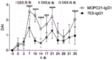


Classifications
-
- C—CHEMISTRY; METALLURGY
- C07—ORGANIC CHEMISTRY
- C07K—PEPTIDES
- C07K16/00—Immunoglobulins [IGs], e.g. monoclonal or polyclonal antibodies
- C07K16/18—Immunoglobulins [IGs], e.g. monoclonal or polyclonal antibodies against material from animals or humans
- C07K16/28—Immunoglobulins [IGs], e.g. monoclonal or polyclonal antibodies against material from animals or humans against receptors, cell surface antigens or cell surface determinants
- C07K16/2803—Immunoglobulins [IGs], e.g. monoclonal or polyclonal antibodies against material from animals or humans against receptors, cell surface antigens or cell surface determinants against the immunoglobulin superfamily
-
- A—HUMAN NECESSITIES
- A61—MEDICAL OR VETERINARY SCIENCE; HYGIENE
- A61P—SPECIFIC THERAPEUTIC ACTIVITY OF CHEMICAL COMPOUNDS OR MEDICINAL PREPARATIONS
- A61P1/00—Drugs for disorders of the alimentary tract or the digestive system
- A61P1/04—Drugs for disorders of the alimentary tract or the digestive system for ulcers, gastritis or reflux esophagitis, e.g. antacids, inhibitors of acid secretion, mucosal protectants
-
- A—HUMAN NECESSITIES
- A61—MEDICAL OR VETERINARY SCIENCE; HYGIENE
- A61P—SPECIFIC THERAPEUTIC ACTIVITY OF CHEMICAL COMPOUNDS OR MEDICINAL PREPARATIONS
- A61P17/00—Drugs for dermatological disorders
- A61P17/02—Drugs for dermatological disorders for treating wounds, ulcers, burns, scars, keloids, or the like
-
- A—HUMAN NECESSITIES
- A61—MEDICAL OR VETERINARY SCIENCE; HYGIENE
- A61P—SPECIFIC THERAPEUTIC ACTIVITY OF CHEMICAL COMPOUNDS OR MEDICINAL PREPARATIONS
- A61P25/00—Drugs for disorders of the nervous system
-
- A—HUMAN NECESSITIES
- A61—MEDICAL OR VETERINARY SCIENCE; HYGIENE
- A61P—SPECIFIC THERAPEUTIC ACTIVITY OF CHEMICAL COMPOUNDS OR MEDICINAL PREPARATIONS
- A61P25/00—Drugs for disorders of the nervous system
- A61P25/02—Drugs for disorders of the nervous system for peripheral neuropathies
-
- A—HUMAN NECESSITIES
- A61—MEDICAL OR VETERINARY SCIENCE; HYGIENE
- A61P—SPECIFIC THERAPEUTIC ACTIVITY OF CHEMICAL COMPOUNDS OR MEDICINAL PREPARATIONS
- A61P25/00—Drugs for disorders of the nervous system
- A61P25/28—Drugs for disorders of the nervous system for treating neurodegenerative disorders of the central nervous system, e.g. nootropic agents, cognition enhancers, drugs for treating Alzheimer's disease or other forms of dementia
-
- A—HUMAN NECESSITIES
- A61—MEDICAL OR VETERINARY SCIENCE; HYGIENE
- A61P—SPECIFIC THERAPEUTIC ACTIVITY OF CHEMICAL COMPOUNDS OR MEDICINAL PREPARATIONS
- A61P37/00—Drugs for immunological or allergic disorders
- A61P37/02—Immunomodulators
- A61P37/04—Immunostimulants
-
- A—HUMAN NECESSITIES
- A61—MEDICAL OR VETERINARY SCIENCE; HYGIENE
- A61P—SPECIFIC THERAPEUTIC ACTIVITY OF CHEMICAL COMPOUNDS OR MEDICINAL PREPARATIONS
- A61P43/00—Drugs for specific purposes, not provided for in groups A61P1/00-A61P41/00
-
- A—HUMAN NECESSITIES
- A61—MEDICAL OR VETERINARY SCIENCE; HYGIENE
- A61K—PREPARATIONS FOR MEDICAL, DENTAL OR TOILETRY PURPOSES
- A61K39/00—Medicinal preparations containing antigens or antibodies
- A61K2039/505—Medicinal preparations containing antigens or antibodies comprising antibodies
-
- C—CHEMISTRY; METALLURGY
- C07—ORGANIC CHEMISTRY
- C07K—PEPTIDES
- C07K2317/00—Immunoglobulins specific features
- C07K2317/20—Immunoglobulins specific features characterized by taxonomic origin
- C07K2317/24—Immunoglobulins specific features characterized by taxonomic origin containing regions, domains or residues from different species, e.g. chimeric, humanized or veneered
-
- C—CHEMISTRY; METALLURGY
- C07—ORGANIC CHEMISTRY
- C07K—PEPTIDES
- C07K2317/00—Immunoglobulins specific features
- C07K2317/30—Immunoglobulins specific features characterized by aspects of specificity or valency
- C07K2317/34—Identification of a linear epitope shorter than 20 amino acid residues or of a conformational epitope defined by amino acid residues
-
- C—CHEMISTRY; METALLURGY
- C07—ORGANIC CHEMISTRY
- C07K—PEPTIDES
- C07K2317/00—Immunoglobulins specific features
- C07K2317/70—Immunoglobulins specific features characterized by effect upon binding to a cell or to an antigen
- C07K2317/75—Agonist effect on antigen
-
- C—CHEMISTRY; METALLURGY
- C07—ORGANIC CHEMISTRY
- C07K—PEPTIDES
- C07K2317/00—Immunoglobulins specific features
- C07K2317/70—Immunoglobulins specific features characterized by effect upon binding to a cell or to an antigen
- C07K2317/76—Antagonist effect on antigen, e.g. neutralization or inhibition of binding
-
- C—CHEMISTRY; METALLURGY
- C07—ORGANIC CHEMISTRY
- C07K—PEPTIDES
- C07K2317/00—Immunoglobulins specific features
- C07K2317/90—Immunoglobulins specific features characterized by (pharmaco)kinetic aspects or by stability of the immunoglobulin
- C07K2317/92—Affinity (KD), association rate (Ka), dissociation rate (Kd) or EC50 value
Landscapes
- Health & Medical Sciences (AREA)
- Chemical & Material Sciences (AREA)
- Organic Chemistry (AREA)
- Immunology (AREA)
- Life Sciences & Earth Sciences (AREA)
- General Health & Medical Sciences (AREA)
- Medicinal Chemistry (AREA)
- Bioinformatics & Cheminformatics (AREA)
- Engineering & Computer Science (AREA)
- Nuclear Medicine, Radiotherapy & Molecular Imaging (AREA)
- Public Health (AREA)
- Chemical Kinetics & Catalysis (AREA)
- General Chemical & Material Sciences (AREA)
- Veterinary Medicine (AREA)
- Pharmacology & Pharmacy (AREA)
- Animal Behavior & Ethology (AREA)
- Neurosurgery (AREA)
- Neurology (AREA)
- Biomedical Technology (AREA)
- Biochemistry (AREA)
- Molecular Biology (AREA)
- Proteomics, Peptides & Aminoacids (AREA)
- Biophysics (AREA)
- Genetics & Genomics (AREA)
- Psychiatry (AREA)
- Hospice & Palliative Care (AREA)
- Dermatology (AREA)
- Peptides Or Proteins (AREA)
- Medicines Containing Antibodies Or Antigens For Use As Internal Diagnostic Agents (AREA)
- Preparation Of Compounds By Using Micro-Organisms (AREA)
- Pharmaceuticals Containing Other Organic And Inorganic Compounds (AREA)
- Micro-Organisms Or Cultivation Processes Thereof (AREA)
- Plural Heterocyclic Compounds (AREA)
- Medicinal Preparation (AREA)
- Medicines That Contain Protein Lipid Enzymes And Other Medicines (AREA)
Abstract
The present disclosure relates generally to compositions comprising antibodies, e.g., monoclonal antibodies, chimeric antibodies, humanized antibodies, antibody fragments, etc., that specifically bind to TREM2 proteins, e.g., mammalian TREM2 or human TREM 2; and the use of such compositions in preventing, reducing risk, or treating an individual in need thereof.
Description
Cross Reference to Related Applications
The present application claims the benefit of U.S. provisional application Ser. No. 62/238,044 filed on 10 month 06 of 2015 and U.S. provisional application Ser. No. 62/369,666 filed on 8 month 01 of 2016, each of which is hereby incorporated by reference in its entirety.
Submitting sequence list with ASCII text file
The following contents submitted in ASCII text file are incorporated herein by reference in their entirety: a sequence listing in Computer Readable Form (CRF) (file name: 735022000940 seqlising. Txt, date recorded: 10/6/2016, size: 651 KB).
Field of the disclosure
The present disclosure relates to anti-TREM 2 antibodies and therapeutic uses of such antibodies.
Background of the disclosure
Trigger receptor-2 (TREM 2) expressed on myeloid cells is an immunoglobulin-like receptor expressed mainly on myeloid cells such as megaphaga cells, dendritic cells, monocytes, skin Langerhans cells (Langerhans cells), kupffer cells (Kupffer cells), osteoclasts and microglia; but also are required to regulate Toll-like receptor (TLR) signaling, regulate inflammatory cytokines, and normal osteoclast development. TREM2 was found as a member of TREM transmembrane glycoproteins that belong to the single immunoglobulin variable (IgV) domain receptor family. The genes encoding human and mouse TREMs are located on human chromosome 6p21.1 and mouse chromosome 17C3, respectively. The TREM cluster includes genes encoding TREM1, TREM2, TREM4, and TREM5, as well as TREM-like genes in humans and mice. In addition, TREM3 and plasmacytoid dendritic cells (pDC) -TREM were also identified in mice. TREM-like genes, i.e. TREML1 and TREML2 in humans, and TREML1 and TREML2 in mice, encode TLT-1 and TLT-2, respectively. The two best characterized receptors of these receptor families TREM1 and TREM2 exhibit about 20% sequence homology as well as some homology to other members of the Ig-SF, such as the activated NK cell receptor (20% identity to NKp 44), and function by correlating with the DAP 12-mediated signaling pathway.
TREM2 was originally cloned as cDNA encoding a homologue of TREM1 (Bouchon, A et al, J Exp Med,2001.194 (8): pages 1111-22). This receptor is an approximately 40kDa glycoprotein, which decreases to 26kDa after N-deglycosylation. The TREM2 gene encodes a protein 230 amino acids in length, which includes an extracellular domain, a transmembrane region, and a short cytoplasmic tail. The extracellular region is encoded by exon 2, is composed of a single V-type Ig-SF domain, and contains three potential N-glycosylation sites. The putative transmembrane region contains a charged lysine residue. The cytoplasmic tail of TREM2 lacks a signaling motif and is thought to signal through the signaling adapter molecule DAP 12/tryobp.
The signaling adapter molecule DAP12 is expressed as homodimers at the surface of a variety of cells involved in innate immune responses including microglia, macrophages, granulocytes, NK cells and Dendritic Cells (DCs). DAP12 is a member of the class I transmembrane adapter proteolytical family based on homology to the CD3 chain and Fc receptor (FcR) gamma chain associated with the human T Cell Receptor (TCR) (Turnbull, IR and Colonna, M, nat Rev Immunol 2007.7 (2): pages 155-61). These proteins share many structural and functional features including one or more ITAM motifs in their cytoplasmic domains, charged acidic residues in the transmembrane region (critical for interaction with their partner chains), and the ability to recruit Src homology-2 (SH 2) containing proteins following tyrosine phosphorylation. The ITAM motif mediates signal transmission by activating ZAP70 or Syk tyrosine kinase. These two kinases phosphorylate several substrates, thereby promoting the formation of signaling complexes, leading to cell activation. Interestingly, some B cells and T cells also expressed DAP12 in inflammatory conditions. In the case of humans, CD4 expressing this protein has been described in the context of autoimmune T cells in patients with chronic inflammatory diseases + CD28 - T cell, alpha beta TCR + CD4 + T cells and CD8 + T cell subsets (Schleinitz, N.et al, PLoS ONE,4 (2009), page e 6264). Given the high level of DAP12 expression in mouse peritoneal macrophages, this protein is believed to be expressed in other macrophage-related cells, such as osteoclasts in bone marrow, coulomb cells in liver, alveolar macrophages in lung, skin Langerhans cells, and microglial cells in brain (Takaki, R et al, immunol Rev,2006.214: pages 118-29).
TREM2 was identified to be expressed on the surface of human monocytogenesis dendritic cells and as an mRNA transcript in the mouse macrophage cell line RAW264 (Bouchon, a et al, J Exp Med,2001.194 (8): pages 1111-22). Human TREM2 is the first DAP 12-related receptor described on a DC surface. Studies have demonstrated that cell surface expression of TREM2 is reduced in DAP 12-deficient bone marrow-derived dendritic cells (BMDCs) and in DAP 12-deficient macrophages (Ito, H and Hamerman, JA, eur J Immunol.42 (1): pages 176-85; hamerman, JA et al, J Immunol, 2006.177 (4): pages 2051-5; and Hamerman, JA et al, nat Immunol,2005.6 (6): pages 579-86) compared to wild-type cells. This suggests that maximum TREM2 surface expression requires the formation of TREM2/DAP12 complex.
Recent studies have also shown that TREM2 has surface expression on macrophages infiltrated with tissue by circulation and on IL-4 or IL-13 activated macrophages (Turnbull, IR et al, J Immunol,2006.177 (6): pages 3520-4). However, expression of TREM2 is not always seen in other cell populations, such as tissue resident macrophages, circulating monocytes or corresponding progenitor cells in bone marrow, indicating that TREM2 expression is not centrally induced, but locally induced during tissue infiltration or by cytokine-mediated activation. Furthermore, IFN-. Gamma.and LPS were also observed to reduce expression of TREM 2. In addition, it has recently been reported that TREM2 is highly expressed on microglial cells and infiltrating macrophages in the central nervous system during experimental autoimmune encephalomyelitis or Alzheimer's disease (Picchio, L et al, eur J Immunol,2007.37 (5): pages 1290-301, and Wang Y, cell.2015, 3 months 12; 160 (6): 1061-71).
TREM2 has been demonstrated to signal through DAP 12. This causes activation of downstream Syk/Zap70 tyrosine kinase family PI3 ks and other intracellular signals. On myeloid cells, TLR signaling is critical for activation, as in the case of infection reactions, but also plays a critical role in pathological inflammatory reactions, as in the case of macrophages and dendritic cells (Hamerman, JA et al, (2006) J Immunol 177:2051-2055; ito, H et al, eur J Immunol 42:176-185; neumann, H et al, (2007) J Neurolimunol 184:92-99; takahashi, K et al, (2005) J Exp Med 201:647-657; and Takahashi, K et al, (2007) PLoS Med 4:e124). It is known that the absence of TREM2 or DAP12 increases pro-inflammatory signaling. The effects of TREM2 deficiency in vitro were shown with stimulation with typical TLR ligands, such as LPS, cpG DNA, and zymosan. TREM-2 deficient dendritic cells show increased release of IL-12p70, TNF, IL-6 and IL-10 in the presence of stimulation, but not in the absence of stimulation.
Several recent studies explored intracellular signaling events induced by activation of the TREM2/DAP12 pathway. For example, TREM2 is thought to activate signaling pathways involved in cell survival (e.g., protein kinase B-Akt), cell activation and differentiation (e.g., syk, erk1/2, PLC-gamma, etc.), actin cytoskeletal control (e.g., syk, vav, etc.) (Peng, Q et al, sci Signal.3 (122): page ra 38; and Whittaker, GC et al, J Biol chem.285 (5): pages 2976-85). Upon attachment of TREM2, ITAM tyrosine in DAP12 is phosphorylated by SRC family kinases, leading to recruitment and activation of Syk kinase and/or ZAP70 kinase. In mice, syk is likely to be the primary kinase involved, whereas in humans, syk and ZAP70 appear to be efficiently coupled to such ITAM-containing subunits, binding them through their tandem SH2 domains.
Studies on TREM2 signaling have shown that like TREM1, TREM2 signaling mediated by DAP12 also causes an increase in intracellular calcium content and ERK1/2 phosphorylation of ERK1/2 (Bouchon, A et al, J Exp Med,2001.194 (8): pages 1111-22; and Sharif, O and Knapp, S, immunology, 2008.213 (9-10): pages 701-13). Importantly, the TREM2 receptor linkage did not induce IkB-a degradation and subsequent NF-kB nuclear translocation, indicating a possible difference between TREM2 and TREM1 signaling (Bouchon, A et al, J Exp Med,2001.194 (8): pages 1111-22). Receptor cross-linking of TREM2 on immature dendritic cells triggers molecules involved in T cell co-stimulation, such as CD86, CD40 and MHC class II upregulation, and chemokine receptor CCR7 upregulation (Bouchon, A et al, J Exp Med,2001.194 (8): pages 1111-22). TREM2 was also expressed on microglial cells, where receptor cross-linking caused ERK1/2 phosphorylation and CCR7 increase, but not CD86 or class II MHC expression, indicating that there may be cell type specific differences in TREM2 signaling. In addition, overexpression of TREM2 signaling in microglial, myeloid precursor, CHO or EK293 cells causes increased phagocytosis of apoptotic neurons, neural and non-neural tissue fragments, pathogenic proteins, bacteria and other foreign invaders in the nervous system, which is accompanied by polarization and recombination of F-actin in ERK-dependent fashion (Takahashi, K et al, PLoS Med,2007.4 (4): e 124; neumann, H and Takahashi, K, J neurommunol, 2007.184 (1-2): pages 92-9; and Kleinberg et al, sci Transl Med.2014, 7 month 2; 6 (243): 243ra 86). However, in some physiological environments (such as pneumococcal pneumonia), TREM2 appears to reduce phagocytosis. Thus, TREM 2-deficient alveolar macrophages exhibit enhanced bacterial clearance from the lungs and enhanced phagocytosis of fine bacteria in vivo (Sharif et al, PLoS Pathog.2014, 12 months; 10 (6): e 1004167).
It was also demonstrated that BMDM cells silenced with TREM2 using shrna showed increased TNF secretion in response to TLR2/6 ligand zymosan and TLR9 ligand CpG compared to control bone marrow-derived megaphage cells (BMDM) treated with non-specific shrna, suggesting that TREM2 negatively regulated cytokine synthesis in macrophages (Ito, H and Hamerman, JA, eur J immunol.42 (1): pages 176-85; hamerman, JA et al, J Immunol,2006.177 (4): pages 2051-5; and Hamerman, JA et al, nat Immunol, 2005.6 (6): pages 579-86). These results have been confirmed using BMDM cells from TREM2 knockout mice, and further show that in response to LPS, in TREM2 -/- TNF and IL-6 levels were higher in BMDM cells than in wild type BMDM cells (Turnbull, IR et al, J Immunol,2006.177 (6): pages 3520-4, and Turnbull, IR and Colonna, M, nat Rev Immunol,2007.7 (2): pages 155-61). In addition, it was demonstrated that after microglial cells were cultured with apoptotic neurons, TREM2 overexpression in these cells caused a decrease in TNF and Inducible Nitric Oxide (iNOS) mRNA, while TREM2 knockdown caused a modest increase in TNF and iNOS mRNA levels. This indicates that, in contrast to TREM1, which is a positive regulator of cytokine synthesis, TREM2 is a negative regulator of cytokine synthesis. This effect of TREM2 on inflammation is believed to be independent of the type of macrophages, as it occurs in both microglial and BMDM cells.
In addition, microglial activation has been shown to cause inflammation in resident myeloid cells of the central nervous system (Neumann, H et al, (2007) J neuroimunol 184:92-99; takahashi, K et al, (2005) J Exp Med 201:647-657; takahashi, K et al, (2007) PLoS Med 4:e124; and Hsieh, CL et al, (2009) J Neurochem 109:1144-1156). Furthermore, activation of microglial cells has also been implicated in frontotemporal dementia (FTD), alzheimer's disease, parkinson's disease, stroke/ischemic brain injury and multiple sclerosis. Decreasing TREM2 activation increases the transcription of some activation and inflammatory markers, such as NOS2 genes in myeloid cells, while increasing TREM2 activation decreases the transcription of NOS 2. The dying neurons are thought to express endogenous ligands for TREM 2. HSP60 is involved as a ligand for TREM2 on neuroblastoma cells (Stefani, L et al, (2009) Neurochem 110:284-294). TREM2 overexpression also causes increased phagocytosis of dying neurons by small neurocollagen cells, and in a similar manner increases phagocytosis of other myeloid lineage cells. TREM2 is also involved in myeloid cell migration because TREM 2-deficient myeloid cells cannot fill the brain of rodent models of alzheimer's disease (Malm, TM et al, neurotherapeutics.2014, 11 months 18).
In humans, complete deficiency of TREM2 has been shown to cause Nasu-Hakola disease, a rare neurodegenerative disease, associated with late-onset dementia, demyelination and brain atrophy (Paloneva, J et al, (2002) Am J Hum Genet 71:656-662; and Paloneva, J et al, (2003) J Exp Med 198:669-675). DAP12 deficiency also causes Nasu-Hakola disease. In addition, exome sequencing of individuals with frontotemporal dementia (FTD) manifestations identified homozygous mutations in TREM2 (Guerreiro, RJ et al, (2013) JAMA neuron 70:78-84; guerreiro, RJ et al, (2012) Arch neuron 1-7). Recently, heterozygous mutations in TREM2 were found to increase the risk of Alzheimer's disease up to 3-fold (Guerreiro, R et al, (2013) N Engl J Med 368:117-127; jonsson, T et al, (2013) N Engl J Med 368:107-116; and Neumann, H et al, (2013) N Engl J Med 368:182-184). Even individuals without alzheimer's disease carrying heterozygous TREM2 mutations showed poorer cognition than individuals with two normal TREM2 alleles. These carriers also showed a doubling of brain volume shrinkage (Rajagopalan et al, (2013) N Engl J Med 369; 16). Some of these mutations lead to truncations and possible loss of function of TREM 2. Other amino acid related changes include Q33X, R47H, T M, and S116C (Borroni B et al, neurobiol agent.2014, month 4; 35 (4): 934.e7-10). Imaging analysis of certain individuals with TREM2 homozygous mutations also showed evidence of demyelination. In addition, the R47H variant of TREM2 (arginine to histidine amino acid substitution at position 47 of TREM 2), which is the most common TREM2 mutation, was shown to be located within the immunoglobulin domain of TREM2 and reduced ligand binding. Other TREM2 mutations were shown to reduce cell surface expression of TREM2, indicating that loss of function is responsible for increased risk of AD (Wang Y, cell.2015; 160 (6): 1061-71).
In addition, the hierarchical ordered organization of molecular networks of gene expression associated with late onset progression of Alzheimer's disease (LOAD) an integrated network-based approach to identify TYROBP/DAP12 as a signaling molecule of TREM2 as a key regulator of immune/microglial gene modules associated with LOAD. TYROBP was found to be the causative modulator of the highest scoring immune/microglial module as ranked, based on the number of other genes regulated by TREM2 and the magnitude of loss of regulation and differential expression in the LOAD brain. TYROBP is significantly upregulated in the LOAD brain, and there is progression of TYROBP expression changes across Mild Cognitive Impairment (MCI), which often occurs before LOAD (Zhang et al, (2013) Cell 153, 707-720; and Ma et al, mol neurobiol.2014, 7 months 23). Targeting such etiologic networks in a manner that restores such etiologic networks to a normal state may be a way of treating a disease.
TREM2 is highly expressed on microglial cells and infiltrating macrophages in the central nervous system during pathological conditions including alzheimer's disease (Picchio, L et al, (2007) Eur J Immunol 37 (5): pages 1290-301, and Wang et al, (2015), cell.;160 (6): 1061-71). It has also been demonstrated that TREM2 gene expression is increased in APP23 transgenic mice as a model of Alzheimer's disease, wherein the mice express mutant forms of amyloid precursor protein associated with familial Alzheimer's disease (Melshior, B et al, ASN Neuro 2:e00037). In addition, it has been demonstrated that the uptake of amyloid 1-42 is increased in BV-2 microglial cell lines overexpressing TREM 2.
It has also been demonstrated that TREM2 is up-regulated in the EAE mouse multiple sclerosis model (Neumann, H et al, (2007) J Neurommunol 184:92-99; takahashi, K et al, (2005) J Exp Med 201:647-657; and Takahashi, K et al, (2007) PLoS Med 4:e 124). Transduction of bone marrow-derived myeloid precursor cells (BM-DCs) with TREM2 in vitro resulted in increased phagocytosis of the variable myelin. Phagocytosis of the beads or of the neuronal fragments is increased. In response to LPS, these cells showed increased IL-10 and decreased IL-1β. Intravenous transplantation of myeloid cells overexpressing TREM2 can inhibit EAE in vivo. In contrast, multiple sclerosis in the cyclohexanone oxalyl dihydrazone model, which is shown to exacerbate multiple sclerosis by TREM2 deficiency (Cantoni et al, acta neuro-lateral (2015) 129 (3): 429-47; luigi Poliani et al, (2015) J Clin invest.125 (5): 2161-2170). It has also been shown that TREM2 deficiency also exacerbates Alzheimer's disease in rodent models (Wang et al, (2015), cell.;160 (6): 1061-71), although reverse data showing the beneficial effects of TREM2 deficient Alzheimer's disease in rodent models have also been reported (Jay et al, (2015) J Exp Med 212:287-295). TREM2 was also shown to be required for microglial survival in the brain (altero et al, (2009) Nat immunol.; 10:734-43). In general terms, TREM2 variants were identified as genetic risk factors for frontotemporal dementia, parkinson' S disease, and amyotrophic lateral sclerosis (Borroni B et al, neurobiol Aging.2014, month 4; 35 (4): 934.e7-10; rayaprolu S et al, mol neurogenin.2013, month 21, month 8:19; and Cady J et al, JAMA neurol.2014, month 4; 71 (4): 449-53). This common genetic linkage suggests a more general role for TREM2 in regulating the pathology of neurodegenerative diseases.
TREM2 antibodies have been described, but only the reported effects are for cultured cells and their therapeutic use is limited, in part because they block the interaction between TREM2 and its natural ligand and act as antagonists in solution. Such antibodies in solution form mimic disease-induced loss of the functional phenotype of TREM2 mutations and thus pose safety and efficacy risks. Another problem with existing anti-TREM 2 antibodies is that they need to be clustered by coating on plastic plates or by secondary antibodies in order to induce agonistic activity. Thus, there is a need for antibodies that specifically bind to TREM2 on the cell surface and modulate (e.g., activate) one or more TREM2 activities in a safe and effective manner in order to treat one or more diseases, disorders, and conditions associated with reduced TREM2 activity.
Some diseases may require TREM2 blocking antibodies that do not activate TREM2 under any conditions. For example, tumor microenvironments are composed of heterogeneous immunoinfiltrates including T lymphocytes, macrophages, and myeloid/granulocyte lineage cells. The treatment regimen for modulating a particular immune cell subpopulation is to alter standard care. "checkpoint blocking" antibodies targeting immunoregulatory molecules expressed on T cells (such as CTLA-4 and PD-1) have demonstrated clinical activity in a variety of tumor types (Naidoo et al, (2014) British Journal of cancer111, 2214-2219).
Cancer immunotherapy targeting tumor-associated macrophages (e.g., M2-type macrophages) is a popular area of research. The presence of M2 macrophages in tumors is associated with poor prognosis.
Thus, there is also a need for antibodies that specifically bind TREM2 on the cell surface and that modulate (e.g., inhibit and/or otherwise reduce) ligand binding and/or one or more TREM2 activities in order to prevent, reduce the risk of, or treat cancer.
All references, including patent applications and publications, cited herein are hereby incorporated by reference in their entirety.
Summary of the disclosure
The present disclosure relates generally to compositions comprising antibodies, e.g., monoclonal antibodies, chimeric antibodies, humanized antibodies, antibody fragments, etc., that specifically bind to TREM2 proteins, e.g., mammalian TREM2 (e.g., any non-human mammal) or human TREM 2; and to methods of using such compositions. Antibodies of the disclosure may include agonist antibodies, antagonist antibodies, or inert antibodies. The methods provided herein find use in preventing, reducing risk of, or treating an individual suffering from: dementia, frontotemporal dementia, alzheimer's disease, vascular dementia, mixed dementia, creutzfeldt-Jakob disease (Creutzfeldt-Jakob disease), normal pressure hydrocephalus, amyotrophic lateral sclerosis, huntington's disease, tauopathies, nasu-Hakola disease, stroke, acute trauma, chronic trauma, cognitive deficit, memory loss, lupus, acute and chronic colitis, rheumatoid arthritis, wound healing, crohn's disease, inflammatory bowel disease, ulcerative colitis, obesity, malaria, essential tremor, central nervous system lupus, behcet's disease, parkinson's disease, lewy body dementia, multiple system atrophy Sheyday's syndrome (Shy-Drager syndrome), progressive supranuclear palsy, degeneration of cortical basal ganglia, acute disseminated encephalomyelitis, granulomatous disorders, sarcoidosis, aging disorders, seizures, spinal cord injury, traumatic brain injury, age-related macular degeneration, glaucoma, retinitis pigmentosa, retinal degeneration, respiratory tract infection, sepsis ocular infections, systemic infections, lupus, arthritis, multiple sclerosis, low bone density, osteoporosis, osteogenesis, osteoproliferation disease, paget's disease (s disease of bone), solid and hematological cancers, bladder cancer, brain cancer, breast cancer, colon cancer, rectal cancer, endometrial cancer, kidney cancer, renal cell carcinoma, renal pelvis cancer, leukemia, lung cancer, melanoma, non-Hodgkin's lymphoma, pancreatic cancer, prostate cancer, ovarian cancer, fibrosarcoma, acute Lymphoblastic Leukemia (ALL), acute Myelogenous Leukemia (AML), chronic Lymphocytic Leukemia (CLL), chronic Myelogenous Leukemia (CML), multiple myeloma, polycythemia vera, primary thrombocythemia, primary or primary myelofibrosis, primary or primary myelosclerosis, tumors of myeloid origin, tumors expressing TREM2 and/or TREM2 ligands, thyroid cancer, infections, CNS herpes, parasitic infections, trypanosome infections, cruzi infections, pseudomonas aeruginosa infections (Pseudomonas aeruginosa infection), leishmania donovani infections (Leishmania donovani infection), group B streptococci infections (group B Streptococcus infection), campylobacter jejuni infections (Campylobacter jejuni infection), neisseria meningitidis infections (Neisseria meningiditis infection), HIV type I, and haemophilus influenzae. The methods provided herein also find use in inducing or promoting survival, maturation, functionality, migration, or proliferation of one or more immune cells in an individual in need thereof. The methods provided herein find additional use in reducing the activity, functionality, or survival of: regulatory T cells, tumor-embedded immunosuppressive dendritic cells, tumor-embedded immunosuppressive macrophages, neutrophils, natural Killer (NK) cells, bone marrow-derived suppressor cells, tumor-associated megaly-phagocytic cells, neutrophils, NK cells, acute Myelogenous Leukemia (AML) cells, chronic Lymphocytic Leukemia (CLL) cells, or Chronic Myelogenous Leukemia (CML) cells
In some embodiments, tumor cells, such as Acute Myeloblastic Leukemia (AML) cells, express TREM2. Thus, the anti-TREM 2 antibodies of the present disclosure also find use in the treatment of cancer. In some embodiments, anti-TREM 2 antibodies (including antibodies that exhibit antibody-dependent cell-mediated cytotoxicity (ADCC)) and/or TREM2 antibody drug conjugates can be used to target and inhibit cancer, such as AML.
Certain aspects of the present disclosure are based, at least in part, on identifying two different classes of isolated antibodies that specifically bind to and modulate TREM2 proteins.
One class of antibodies relates to agonist antibodies that induce one or more TREM2 activities on, for example, human primary immune cells and TREM2 expressing cell lines and that, when combined with one or more TREM2 ligands, enhance one or more TREM2 activities induced by the binding of one or more TREM2 ligands to TREM2 proteins. Advantageously, such agonist anti-TREM 2 antibodies may enhance ligand-induced TREM2 activity without competing with one or more TREM2 ligands for binding to TREM2 proteins or otherwise blocking binding of one or more TREM2 ligands to TREM2 proteins. In some embodiments, the agonist antibody may activate and/or enhance one or more TREM2 activities, whether the antibody clusters or in solution. In some embodiments, the agonist antibodies may activate TREM2 in solution without clustering by secondary antibodies, by Fc receptors, or by binding to plates. In some embodiments, the agonist antibody can activate TREM2, whether or not the mechanism of antibody clustering is present at the therapeutic site of action in vivo. In some embodiments, the agonist antibodies may have increased safety and efficacy. In some embodiments, the agonist antibodies can ensure that immune cells expressing TREM2 function primarily in locations where their therapeutic efficacy is required and are capable of interacting with their physiological targets. In some embodiments, the agonist antibodies do not block TREM2 activity resulting in an increased risk of disease similar to those observed in the case of gene mutations that reduce TREM2 activity.
The second class of antibodies relates to antagonist antibodies that specifically bind to and inhibit TREM2 and are incapable of activating TREM2, whether the antibodies cluster or are in solution. In some embodiments, the antagonist antibodies have increased safety and efficacy. In some embodiments, the antagonist antibody is incapable of activating TREM2, regardless of its configuration or its ability to cluster.
Accordingly, certain aspects of the present disclosure relate to an isolated (e.g., monoclonal) antibody that binds to a TREM2 protein, wherein the antibody induces one or more TREM2 activities and enhances one or more TREM2 activities induced by the binding of one or more TREM2 ligands to the TREM2 protein. In some embodiments, the antibody enhances one or more TREM2 activities induced by binding of one or more TREM2 ligands to TREM2 protein compared to one or more TREM2 activities induced by binding of one or more TREM2 ligands to TREM2 protein in the absence of the isolated antibody. In some embodiments, the antibody enhances one or more TREM2 activities without blocking binding of one or more TREM2 ligands to TREM2 proteins. In some embodiments, the antibody does not compete with one or more TREM2 ligands for binding to TREM2 protein. In some embodiments, the antibodies enhance binding of one or more TREM2 ligands to TREM2 protein.
Other aspects of the disclosure relate to an isolated (e.g., monoclonal) antibody that binds to a TREM2 protein, wherein the antibody induces one or more TREM2 activities without blocking binding of one or more TREM2 ligands to the TREM2 protein. In some embodiments, the antibody does not compete with one or more TREM2 ligands for binding to TREM2 protein. In some embodiments, the antibody enhances binding of one or more TREM2 ligands to TREM2 protein. In some embodiments, the antibodies enhance one or more TREM2 activities induced by binding of one or more TREM2 ligands to TREM2 proteins. In some embodiments, the antibody enhances one or more TREM2 activities induced by binding of one or more TREM2 ligands to TREM2 protein compared to one or more TREM2 activities induced by binding of one or more TREM2 ligands to TREM2 protein in the absence of the isolated antibody.
In some embodiments that may be combined with any of the preceding embodiments, the antibody cooperates with one or more TREM2 ligands to enhance one or more TREM2 activities. In some embodiments that may be combined with any of the preceding embodiments, the antibody cooperates with one or more TREM2 ligands to enhance one or more TREM2 activities. In some embodiments that may be combined with any of the preceding embodiments, the antibody enhances one or more TREM2 activities in the absence of TREM2 cell surface clustering. In some embodiments that may be combined with any of the preceding embodiments, the antibody enhances one or more TREM2 activities by inducing or maintaining cell surface clustering of TREM2. In some embodiments that may be combined with any of the preceding embodiments, the antibodies are clustered by Fc-gamma receptors expressed on one or more immune cells. In some embodiments that may be combined with any of the preceding embodiments, the one or more immune cells are B cells or microglia. In some embodiments that may be combined with any of the preceding embodiments, the enhancement of one or more TREM2 activities induced by the binding of one or more TREM2 ligands to TREM2 proteins is measured on primary cells selected from the group consisting of: dendritic cells, bone marrow derived dendritic cells, monocytes, microglia, macrophages, neutrophils, NK cells, osteoclasts, skin langerhans cells, and kupfu cells, or on a cell line, and wherein the enhancement of one or more TREM2 activities induced by the binding of one or more TREM2 ligands to TREM2 proteins is measured using an in vitro cellular assay. In some embodiments that can be combined with any of the preceding embodiments, the antibody increases the level of soluble TREM2, increases the half-life of soluble TREM2, or both. In some embodiments that may be combined with any of the preceding embodiments, the level of soluble TREM2 is selected from the group consisting of: serum levels of TREM2, cerebral Spinal Fluid (CSF) levels of TREM2, tissue levels of TREM2, and any combination thereof. In some embodiments that may be combined with any of the preceding embodiments, the antibody does not bind to soluble TREM2. In some embodiments that may be combined with any of the preceding embodiments, the antibody does not bind to in vivo soluble TREM2. In some embodiments that may be combined with any of the preceding embodiments, the soluble TREM2 corresponds to an amino acid residue selected from the group consisting of: amino acid residues 19-160 of SEQ ID NO. 1, amino acid residues 19-159 of SEQ ID NO. 1, amino acid residues 19-158 of SEQ ID NO. 1, amino acid residues 19-157 of SEQ ID NO. 1, amino acid residues 19-156 of SEQ ID NO. 1, amino acid residues 19-155 of SEQ ID NO. 1, and amino acid residues 19-154 of SEQ ID NO. 1. In some embodiments that can be combined with any of the preceding embodiments, the antibody reduces the level of TREM2 in one or more cells. In some embodiments that can be combined with any of the preceding embodiments, the antibody reduces the cell surface level of TREM2, reduces the intracellular level of TREM2, reduces the total level of TREM2, or any combination thereof. In some embodiments that may be combined with any of the preceding embodiments, the antibody induces TREM2 degradation, TREM2 cleavage, TREM2 internalization, TREM2 shedding, down-regulation of TREM2 expression, or any combination thereof. In some embodiments that may be combined with any of the preceding embodiments, the level of TREM2 in the one or more cells is measured in a primary cell selected from the group consisting of: dendritic cells, bone marrow derived dendritic cells, mononuclear cells, microglia, macrophages, neutrophils, NK cells, osteoclasts, skin langerhans cells, and coulomb cells, or on a cell line, and wherein the cellular level of TREM2 is measured using an in vitro cellular assay. In some embodiments that may be combined with any of the preceding embodiments, the TREM2 protein is a mammalian (such as a non-human mammal) protein or a human protein. In some embodiments that may be combined with any of the preceding embodiments, the TREM2 protein is a wild-type protein. In some embodiments that can be combined with any of the preceding embodiments, the TREM2 protein is a naturally occurring variant. In some embodiments that may be combined with any of the preceding embodiments, the TREM2 protein is expressed on human dendritic cells, human macrophages, human mononuclear cells, human osteoclasts, human skin langerhans cells, human kupfu cells, human microglia cells, or any combination thereof. In some embodiments that may be combined with any of the preceding embodiments, the one or more TREM2 activities are selected from the group consisting of: (a) TREM2 binds to DAP12; (b) TREM2 phosphorylation; (c) DAP12 phosphorylation; (d) Activating one or more tyrosine kinases, optionally wherein the one or more tyrosine kinases comprise Syk kinase, ZAP70 kinase, or both; (e) activating phosphatidylinositol 3-kinase (PI 3K); (f) activating protein kinase B (Akt); (g) Recruiting phospholipase C-gamma (PLC-gamma) to the cytoplasmic membrane, activating PLC-gamma, or both; (h) recruiting TEC family kinase dVav to the cytoplasmic membrane; (i) activating nuclear factor-rB (NF-rB); (j) inhibiting MAPK signaling; (k) Phosphorylation of Linkers (LAT) for T cell activation, linkers (LAB) for B cell activation, or both; (l) activating IL-2 induced tyrosine kinase (Itk); (m) modulating one or more pro-inflammatory mediators selected from the group consisting of: IFN- β, IL-1α, IL-1β, TNF- α, IL-6, IL-8, CRP, CD86, MCP-1/CCL2, CCL3, CCL4, CCL5, CCR2, CXCL-10, gata3, IL-20 family members, IL-33, LIF, IFN- γ, OSM, CNTF, CSF-1, OPN, CD11c, GM-CSF, IL-11, IL-12, IL-17, IL-18, and IL-23, optionally wherein said modulation occurs in one or more cells selected from the group consisting of: macrophages, M1 macrophages, activated M1 macrophages, M2 macrophages, dendritic cells, monocytes, osteoclasts, skin langerhans cells, kupffer cells, and microglia cells; (n) modulating one or more anti-inflammatory mediators selected from the group consisting of: IL-4, IL-10 TGF-beta, IL-13, IL-35IL-16, IFN-alpha, IL-1Ra, VEGF, G-CSF, YM, AXL, FLT1, and soluble receptor for TNF or IL-6, optionally wherein said modulation occurs in one or more cells selected from the group consisting of: macrophages, M1 macrophages, activated M1 macrophages, M2 macrophages, dendritic cells, monocytes, osteoclasts, skin langerhans cells, kupffer cells, and microglia cells; (o) one or more genes that regulate its expression to increase after induction of inflammation, optionally wherein the one or more genes are selected from the group consisting of Fabp3, fabp5, and LDR; (p) phosphorylation of extracellular signal-regulated kinase (ERK); (q) modulating expression of C-C chemokine receptor 7 (CCR 7) in one or more cells selected from the group consisting of: macrophages, M1 macrophages, activated M1 macrophages, M2 macrophages, dendritic cells, monocytes, osteoclasts, skin langerhans cells, kupfer cells, microglia cells, M1 microglia cells, activated M1 microglia cells, and M2 microglia cells, and any combination thereof; (r) inducing chemotaxis of microglial cells to CCL19 and CCL21 expressing cells; (s) normalization of disrupted TREM2/DAP 12-dependent gene expression; (t) recruiting Syk, ZAP70, or both to the DAP12/TREM2 complex; (u) increasing the activity of one or more TREM 2-dependent genes, optionally wherein the one or more TREM 2-dependent genes comprise Nuclear Factor (NFAT) transcription factors of activated T cells; (v) Increasing maturation of dendritic cells, monocytes, microglia, M1 microglia, activated M1 microglia, and M2 microglia, macrophages, M1 macrophages, activated M1 megalobhagocytes, M2 macrophages, or any combination thereof; (w) increasing the ability of dendritic cells, mononuclear cells, microglia, M1 microglia, activated M1 microglia, and M2 microglia, macrophages, M1 macrophages, activated M1 macrophages, M2 macrophages, or any combination thereof to initiate or modulate the function of T cells, optionally wherein the T cells are one or more cells selected from the group consisting of: cd8+ T cells, cd4+ T cells, regulatory T cells, and any combination thereof; (x) Bone marrow-derived dendritic cells, optionally wherein the antigen-specific T cells are one or more cells selected from the group consisting of: cd8+ T cells, cd4+ T cells, regulatory T cells, and any combination thereof; (y) bone marrow-derived dendritic cells induce an enhanced ability of antigen-specific T cell proliferation, an ability to normalize, or both; (z) inducing osteoclast production, increasing the rate of osteoclast production, or both; (aa) increasing survival of dendritic cells, macrophages, M1 macrophages, activated M1 macrophages, M2 megaloblastic cells, monocytes, osteoclasts, skin langerhans cells, kupfer cells, microglia cells, M1 microglia cells, activated M1 microglia cells, and M2 microglia cells, or any combination thereof; (bb) increasing the function of dendritic cells, megaloblastic cells, M1 macrophages, activated M1 macrophages, M2 macrophages, microglia, M1 microglia, activated M1 microglia, and M2 microglia, or any combination thereof; (cc) increasing phagocytosis by dendritic cells, megaloblastic cells, M1 macrophages, activated M1 macrophages, M2 macrophages, monocytes, microglia, M1 microglia, activated M1 microglia, and M2 microglia, or any combination thereof; (dd) inducing one or more types of clearance selected from the group consisting of: apoptotic neuronal clearance, neuronal tissue fragment clearance, non-neuronal tissue fragment clearance, bacterial or other foreign body clearance, pathogenic agent clearance, tumor cell clearance, or any combination thereof, optionally wherein the pathogenic agent is selected from the group consisting of: amyloid β or a fragment thereof, tau, IAPP, α -synuclein, TDP-43, FUS protein, prion protein, prPSc, huntingtin, calcitonin, superoxide dismutase, ataxin, lewy body, atrial natriuretic factor, islet amyloid polypeptide, insulin, apolipoprotein AI, serum amyloid A, medin, prolactin, transthyretin, lysozyme, β2 microglobulin, gelsolin, corneal epithelium protein, cystatin (cystatin), immunoglobulin light chain AL, S-IBM protein, and repeat related non-ATG (RAN) translation products (including dipeptide repeat (DPR peptide) consisting of glycine-alanine (GA), glycine-proline (GP), glycine-arginine (GR), proline-alanine (PA), or proline-arginine (PR), antisense ggc (G2C 4) repeat amplified cccc RNA); (ee) induces phagocytosis of one or more of: apoptotic neurons, fragments of nervous tissue, fragments of non-nervous tissue, bacteria, other foreign bodies, pathogenic agents, tumor cells, or any combination thereof, optionally wherein the pathogenic agents are selected from the group consisting of: amyloid β or fragments thereof, tau, IAPP, α -synuclein, TDP-43, FUS protein, prion protein, prPSc, huntingtin, calcitonin, superoxide dismutase, ataxin, lewy body, atrial natriuretic factor, islet amyloid polypeptide, insulin, apolipoprotein AI, serum amyloid A, medin, prolactin, transthyretin, lysozyme, β2 microglobulin, gelsolin, corneal epithelium protein, cysteine-inhibiting protease protein, immunoglobulin light chain AL, S-IBM protein, and repeat related non-ATG (RAN) translation products (including dipeptide repeat (DPR peptide) consisting of glycine-alanine (GA), glycine-proline (GP), glycine-arginine (GR), proline-alanine (PA), or proline-arginine (PR), antisense GGCCCC (G2C 4) repeat amplified RNAs); (ff) modulating expression of one or more stimulatory molecules selected from the group consisting of: CD83, CD86, MHC class II, CD40, and any combination thereof, optionally wherein the CD40 is expressed on a dendritic cell, monocyte, macrophage, or any combination thereof, and optionally wherein the dendritic cell comprises a bone marrow derived dendritic cell; (gg) modulates secretion of one or more pro-inflammatory mediators selected from the group consisting of: IFN- β, IL-1α, IL-1β, CD86, TNF- α, IL-6, IL-8, CRP, MCP-1/CCL2, CCL3, CCL4, CCL5, CCR2, CXCL-10, gata3, IL-20 family members, IL-33, LIF, IFN- γ, OSM, CNTF, CSF-1, OPN, CD11c, GM-CSF, IL-11, IL-12, IL-17, IL-18, and IL-23, and optionally wherein said modulation occurs in one or more cells selected from the group consisting of: macrophages, M1 macrophages, activated M1 macrophages, M2 macrophages, dendritic cells, monocytes, osteoclasts, skin langerhans cells, kupffer cells, and microglia cells; (hh) modulates secretion of one or more anti-inflammatory mediators selected from the group consisting of: IL-4, IL-10 TGF-beta, IL-13, IL-35IL-16, IFN-alpha, IL-1Ra, VEGF, G-CSF, YM, AXL, FLT1, and soluble receptor for TNF or IL-6, and optionally wherein said modulation occurs in one or more cells selected from the group consisting of: macrophages, M1 macrophages, activated M1 megaphaga cells, M2 macrophages, dendritic cells, monocytes, osteoclasts, skin langerhans cells, kupfer cells, and microglia cells; (ii) Regulating expression of one or more proteins selected from the group consisting of: c1qa, C1qB, C1qC, C1s, C1R, C, C2, C3, ITGB2, HMOX1, lat2.casp1, CSTA, VSIG4, MS4A4A, C AR1, GPX1, tyroBP, ALOX5AP, ITGAM, SLC A7, CD4, ITGAX, PYCARD, and VEGF; (jj) increasing memory; and (kk) reduces cognitive deficits. In some embodiments that may be combined with any of the preceding embodiments, the one or more TREM2 activities are selected from the group consisting of: (a) TREM2 binds to DAP12; (b) DAP12 phosphorylation; (c) activating Syk kinase; (d) Modulating one or more pro-inflammatory mediators selected from the group consisting of: IFN- β, IL-1α, IL-1β, TNF- α, IL-6, IL-8, CRP, CD86, MCP-1/CCL2, CCL3, CCL4, CCL5, CCR2, CXCL-10, gata3, IL-20 family members, IL-33, LIF, IFN- γ, OSM, CNTF, CSF-1, OPN, CD11c, GM-CSF, IL-11, IL-12, IL-17, IL-18, and IL-23, optionally wherein said modulation occurs in one or more cells selected from the group consisting of: macrophages, M1 macrophages, activated M1 macrophages, M2 macrophages, dendritic cells, monocytes, osteoclasts, skin langerhans cells, kupffer cells, and microglia cells; (e) recruiting Syk to the DAP12/TREM2 complex; (f) Increasing the activity of one or more TREM 2-dependent genes, optionally wherein the one or more TREM 2-dependent genes comprise Nuclear Factor (NFAT) transcription factors of activated T cells; (g) Increasing survival of dendritic cells, macrophages, M1 macrophages, activated M1 macrophages, M2 macrophages, monocytes, osteoclasts, skin langerhans cells, kupfer cells, microglia cells, M1 microglia cells, activated M1 microglia cells, and M2 microglia cells, or any combination thereof; (h) Regulating expression of one or more stimulatory molecules selected from the group consisting of: CD83, CD86, MHC class II, CD40, and any combination thereof, optionally wherein the CD40 is expressed on a dendritic cell, monocyte, macrophage, or any combination thereof, and optionally wherein the dendritic cell comprises a bone marrow derived dendritic cell; (i) increasing memory; and (j) reducing cognitive deficit. In some embodiments that may be combined with any of the preceding embodiments, the antibody is of the IgG class, igM class, or IgA class. In some embodiments that may be combined with any of the preceding embodiments, the antibody is of the IgG class and has an IgG1, igG2, igG3, or IgG4 isotype. In some embodiments that may be combined with any of the preceding embodiments, the antibody has an IgG2 isotype. In some embodiments that may be combined with any of the preceding embodiments, the antibody comprises a human IgG2 constant region. In some embodiments that may be combined with any of the preceding embodiments, the human IgG2 constant region comprises an Fc region. In some embodiments that may be combined with any of the preceding embodiments, the antibody enhances one or more TREM2 activities independent of binding to Fc receptors. In some embodiments that may be combined with any of the preceding embodiments, the antibody binds to an inhibitory Fc receptor. In some embodiments that may be combined with any of the preceding embodiments, the inhibitory Fc receptor is an inhibitory Fc-gamma receptor IIB (fcγiib). In some embodiments that may be combined with any of the preceding embodiments: (a) The isolated antibody has a human or mouse IgG1 isotype and comprises one or more amino acid substitutions in the Fc region at residue positions selected from the group consisting of: N297A, D265, A, D, A, L, 234A, L, 235A, G, 237, 226, S, C, 229, S, E, 233, P, L, 234, V, L, 234, F, L, 235, E, P, S, S, 267, E, L, 328, Y, S, T, T E, L328, E, P, 238, D, S, 267, E, L, F, E, 268, F, E, 271, F, E, 330R, and any combination thereof, wherein the numbering of the residues is according to EU numbering, or comprises an amino acid deletion in the Fc region at a position corresponding to glycine 236; (b) The isolated antibody has an IgG1 isotype and comprises an IgG2 isotype heavy chain constant domain 1 (CH 1) and a hinge region, optionally wherein the IgG2 isotype CH1 and hinge region comprise an amino acid sequence ASTKGPSVFP LAPCSRSTSE STAALGCLVK DYFPEPVTVS WNSGALTSGVHTFPAVLQSS GLYSLSSVVT VPSSNFGTQT YTCNVDHKPS NTKVDKTVERKCCVECPPCP (SEQ ID NO: 886), and optionally wherein the antibody Fc region comprises an S267E amino acid substitution, an L328F amino acid substitution, or both, and/or an N297A or N297Q amino acid substitution, wherein the numbering of the residues is according to EU numbering; (c) The isolated antibody has an IgG2 isotype and comprises one or more amino acid substitutions in the Fc region at residue positions selected from the group consisting of: P238S, V234A, G237 268A, H Q, V309L, A S, P331S, C38320S, C232S, C S, S267E, L328F, M Y, S254T, T256E, H268E, N297A, N297Q, A L, and any combination thereof, wherein the numbering of the residues is according to EU numbering; (d) The isolated antibody has a human or mouse IgG4 isotype and comprises one or more amino acid substitutions in the Fc region at residue positions selected from the group consisting of: L235A, G237A, S P, L236E, S267E, E318A, L328F, M Y, S254T, T256E, E P, F234V, L a/F234A, S228P, S P, L248E, T394D, N297A, N297Q, L E, and any combination thereof, wherein the numbering of the residues is according to EU numbering; or (e) the isolated antibody has a hybrid IgG2/4 isotype, and optionally wherein the antibody comprises an amino acid sequence comprising amino acids 118 to 260 of human IgG2 and amino acids 261 to 447 of human IgG4, wherein the numbering of the residues is according to EU numbering. In some embodiments that may be combined with any of the preceding embodiments, the antibody has an IgG4 isotype. In some embodiments that can be combined with any of the preceding embodiments, the antibody comprises an S228P amino acid substitution at residue position 228, an F234A amino acid substitution at residue position 234, an L235A amino acid substitution at residue position 235, wherein the numbering of the residue positions is according to EU numbering.
In some embodiments that may be combined with any of the preceding embodiments, the antibody binds to one or more amino acids within an amino acid residue selected from the group consisting of: (i) Amino acid residues 19-174 of SEQ ID NO. 1, or amino acid residues on the TREM2 protein corresponding to amino acid residues 19-174 of SEQ ID NO. 1; (ii) Amino acid residues 29-112 of SEQ ID NO. 1, or amino acid residues on the TREM2 protein corresponding to amino acid residues 29-112 of SEQ ID NO. 1; (iii) Amino acid residues 113-174 of SEQ ID NO. 1, or amino acid residues on the TREM2 protein corresponding to amino acid residues 113-174 of SEQ ID NO. 1; (iv) Amino acid residues 35-49 of SEQ ID NO. 1, or amino acid residues on the TREM2 protein corresponding to amino acid residues 35-49 of SEQ ID NO. 1; (v) Amino acid residues 35-49 and 140-150 of SEQ ID NO. 1, or amino acid residues on the TREM2 protein corresponding to amino acid residues 35-49 and 140-150 of SEQ ID NO. 1; (vi) Amino acid residues 39-49 of SEQ ID NO. 1, or amino acid residues on the TREM2 protein corresponding to amino acid residues 39-49 of SEQ ID NO. 1; (vii) Amino acid residues 39-49 and 63-77 of SEQ ID NO. 1, or amino acid residues on the TREM2 protein corresponding to amino acid residues 39-49 and 63-77 of SEQ ID NO. 1; (viii) Amino acid residues 51-61 of SEQ ID NO. 1, or amino acid residues on the TREM2 protein corresponding to amino acid residues 51-61 of SEQ ID NO. 1; (ix) Amino acid residues 55-62 of SEQ ID NO. 1, or amino acid residues on the TREM2 protein corresponding to amino acid residues 55-62 of SEQ ID NO. 1; (x) Amino acid residues 55-62, 104-109, and 148-158 of SEQ ID NO. 1, or amino acid residues corresponding to amino acid residues 55-62, 104-109, and 148-158 of SEQ ID NO. 1 on the TREM2 protein; (xi) Amino acid residues 55-62, 104-109, and 160-166 of SEQ ID NO. 1, or amino acid residues corresponding to amino acid residues 55-62, 104-109, and 160-166 of the TREM2 protein; (xii) Amino acid residues 55-65 of SEQ ID NO. 1, or amino acid residues on the TREM2 protein corresponding to amino acid residues 55-65 of SEQ ID NO. 1; (xiii) Amino acid residues 55-65 and 124-134 of SEQ ID NO. 1, or amino acid residues on the TREM2 protein corresponding to amino acid residues 55-65 and 124-134 of SEQ ID NO. 1; (xiv) Amino acid residues 63-73 of SEQ ID NO. 1, or amino acid residues on the TREM2 protein corresponding to amino acid residues 63-73 of SEQ ID NO. 1; (xv) Amino acid residues 63-77 of SEQ ID NO. 1, or amino acid residues on the TREM2 protein corresponding to amino acid residues 63-77 of SEQ ID NO. 1; (xvi) Amino acid residues 104-109 of SEQ ID NO. 1, or amino acid residues on the TREM2 protein corresponding to amino acid residues 104-109 of SEQ ID NO. 1; (xvii) Amino acid residues 117-133 of SEQ ID NO. 1, or amino acid residues on the TREM2 protein corresponding to amino acid residues 117-133 of SEQ ID NO. 1; (xviii) Amino acid residues 124-134 of SEQ ID NO. 1, or amino acid residues on the TREM2 protein corresponding to amino acid residues 124-134 of SEQ ID NO. 1; (xix) Amino acid residues 137-146 of SEQ ID NO. 1, or amino acid residues on the TREM2 protein corresponding to amino acid residues 137-146 of SEQ ID NO. 1; (xx) Amino acid residues 139-147 of SEQ ID NO. 1, or amino acid residues on the TREM2 protein corresponding to amino acid residues 139-147 of SEQ ID NO. 1; (xxi) Amino acid residues 139-149 of SEQ ID NO. 1, or amino acid residues corresponding to amino acid residues 139-149 of SEQ ID NO. 1 on the TREM2 protein; (xxii) Amino acid residues 140-150 of SEQ ID NO. 1, or amino acid residues on the TREM2 protein corresponding to amino acid residues 140-150 of SEQ ID NO. 1; (xxiii) Amino acid residues 140-146 of SEQ ID NO. 1, or amino acid residues on the TREM2 protein corresponding to amino acid residues 140-146 of SEQ ID NO. 1; (xxiv) Amino acid residues 140-143 of SEQ ID NO. 1, or amino acid residues on the TREM2 protein corresponding to amino acid residues 140-143 of SEQ ID NO. 1; (xxv) Amino acid residues 142-152 of SEQ ID NO. 1, or amino acid residues on the TREM2 protein corresponding to amino acid residues 142-152 of SEQ ID NO. 1; (xxvi) Amino acid residues 146-154 of SEQ ID NO. 1, or amino acid residues on the TREM2 protein corresponding to amino acid residues 146-154 of SEQ ID NO. 1; (xxvii) Amino acid residues 148-158 of SEQ ID NO. 1, or amino acid residues on the TREM2 protein corresponding to amino acid residues 148-158 of SEQ ID NO. 1; (xxviii) Amino acid residues 149-157 of SEQ ID NO. 1, or amino acid residues on the TREM2 protein corresponding to amino acid residues 149-157 of SEQ ID NO. 1; (xxix) Amino acid residues 149 and 150 of SEQ ID NO. 1, or amino acid residues on the TREM2 protein corresponding to amino acid residues 149 and 150 of SEQ ID NO. 1; (xxx) Amino acid residues 151-155 of SEQ ID NO. 1, or amino acid residues on the TREM2 protein corresponding to amino acid residues 151-155 of SEQ ID NO. 1; (xxxi) Amino acid residues 154-161 of SEQ ID NO. 1, or amino acid residues on the TREM2 protein corresponding to amino acid residues 154-161 of SEQ ID NO. 1; (xxxii) Amino acid residues 156-170 of SEQ ID NO. 1, or amino acid residues on the TREM2 protein corresponding to amino acid residues 156-170 of SEQ ID NO. 1; (xxxiii) Amino acid residues 160-166 of SEQ ID NO. 1, or amino acid residues on the TREM2 protein corresponding to amino acid residues 160-166 of SEQ ID NO. 1; and (xxxiv) amino acid residues 162-165 of SEQ ID NO. 1, or amino acid residues corresponding to amino acid residues 162-165 of SEQ ID NO. 1 on the TREM2 protein. In some embodiments that can be combined with any of the preceding embodiments, the antibody binds to one or more amino acid residues of SEQ ID No. 1 selected from the group consisting of: k42, H43, W44, G45, H67, R77, T88, H114, E117, E151, D152, H154, and E156, or one or more amino acid residues corresponding to an amino acid residue of SEQ ID NO:1 selected from the group consisting of: k42, H43, W44, G45, H67, R77, T88, H114, E117, E151, D152, H154, and E156. In some embodiments that can be combined with any of the preceding embodiments, the antibody binds to one or more amino acid residues of SEQ ID No. 1 selected from the group consisting of: e151, D152, H154, and E156, or one or more amino acid residues corresponding to amino acid residues of SEQ ID NO:1 selected from the group consisting of: e151, D152, H154, and E156. In some embodiments that can be combined with any of the preceding embodiments, the antibody competes for binding to TREM2 with one or more antibodies selected from the group consisting of: 3B10, 7B3, 8F8, 9F5, 9G1, 9G3, 11A8, 12F9, 7E9, 7F6, 8C3, 2C5, 3C5, 4C12, 7D9, 2F6, 3A7, 7E5, 11H5, 1B4, 6H2, 7B11, 18D8, 18E4, 29F6, 40D5, 43B9, 44A8, 44B4, and any combination thereof.
In some embodiments that may be combined with any of the preceding embodiments, the antibody comprises a light chain variable domain and a heavy chain variable domain, wherein the light chain variable domain, or the heavy chain variable domain, or both comprise at least one, two, three, four, five, or six HVRs selected from the group consisting of: 4D11, 7C5, 6G12, 8F11, 8E10, 7E5, 7F8, 8F8, 1H7, 2H8, 3A2, 3A7, 3B10, 4F11, 6H6, 7A9, 7B3, 8A1, 9F5, 9G1, 9G3, 10A9, 11A8, 12D9, 12F9, 10C1, 7E9, 7F6, 8C3, 2C5, 3C5, 4C12, 7D9, 2F6, 11H5, B4, 6H2, 7B11v1, 7B11v2, 18D8, 18E4v1, 18E4v2, 29F6v1, 29F6v2, 40D5v1, 40D5v2, 43B9, 44A8v1, 44A8v2, 44B4v1, and 44B4v2. In some embodiments that may be combined with any of the preceding embodiments: (a) The HVR-L1 comprises an amino acid sequence selected from the group consisting of: SEQ ID NO 9-23, SEQ ID NO 581, SEQ ID NO 690-694, SEQ ID NO 734-738, SEQ ID NO 826-828; (b) The HVR-L2 comprises an amino acid sequence selected from the group consisting of: 24-33, 695-697, and 739-743; and (c) the HVR-L3 comprises an amino acid sequence selected from the group consisting of: 34-47, 582, 583, 698-702, 744-746; (d) The HVR-H1 comprises an amino acid sequence selected from the group consisting of: 48-65, 584, 703-705, 747-754, 829-835; (e) The HVR-H2 comprises an amino acid sequence selected from the group consisting of: 66-84, 585-587, 706-708, 755-762, 836-842, 888; or (f) the HVR-H3 comprises an amino acid sequence selected from the group consisting of: SEQ ID NO 85-102, SEQ ID NO 588, SEQ ID NO 589, SEQ ID NO 709, SEQ ID NO 710, SEQ ID NO 763-770. In some embodiments that may be combined with any of the preceding embodiments: (a) The HVR-L1 comprising the amino acid sequence of SEQ ID NO. 11, the HVR-L2 comprising the amino acid sequence of SEQ ID NO. 26, the HVR-L3 comprising the amino acid sequence of SEQ ID NO. 36, the HVR-H1 comprising the amino acid sequence of SEQ ID NO. 51, the HVR-H2 comprising the amino acid sequence of SEQ ID NO. 69, and the HVR-H3 comprising the amino acid sequence of SEQ ID NO. 88; (b) The HVR-L1 comprising the amino acid sequence of SEQ ID NO. 14, the HVR-L2 comprising the amino acid sequence of SEQ ID NO. 28, the HVR-L3 comprising the amino acid sequence of SEQ ID NO. 39, the HVR-H1 comprising the amino acid sequence of SEQ ID NO. 53, the HVR-H2 comprising the amino acid sequence of SEQ ID NO. 71, and the HVR-H3 comprising the amino acid sequence of SEQ ID NO. 90; (c) The HVR-L1 comprising the amino acid sequence of SEQ ID NO. 11, the HVR-L2 comprising the amino acid sequence of SEQ ID NO. 26, the HVR-L3 comprising the amino acid sequence of SEQ ID NO. 36, the HVR-H1 comprising the amino acid sequence of SEQ ID NO. 51, the HVR-H2 comprising the amino acid sequence of SEQ ID NO. 69, and the HVR-H3 comprising the amino acid sequence of SEQ ID NO. 88; (d) The HVR-L1 comprising the amino acid sequence of SEQ ID NO. 16, the HVR-L2 comprising the amino acid sequence of SEQ ID NO. 29, the HVR-L3 comprising the amino acid sequence of SEQ ID NO. 35, the HVR-H1 comprising the amino acid sequence of SEQ ID NO. 55, the HVR-H2 comprising the amino acid sequence of SEQ ID NO. 73, and the HVR-H3 comprising the amino acid sequence of SEQ ID NO. 92; (e) The HVR-H1 comprises the amino acid sequence of SEQ ID NO. 58, the HVR-H2 comprises the amino acid sequence of SEQ ID NO. 76, and the HVR-H3 comprises the amino acid sequence of SEQ ID NO. 95; (f) The HVR-L1 comprising the amino acid sequence of SEQ ID NO. 19, the HVR-L2 comprising the amino acid sequence of SEQ ID NO. 28, the HVR-L3 comprising the amino acid sequence of SEQ ID NO. 43, the HVR-H1 comprising the amino acid sequence of SEQ ID NO. 60, the HVR-H2 comprising the amino acid sequence of SEQ ID NO. 78, and the HVR-H3 comprising the amino acid sequence of SEQ ID NO. 97; (g) The HVR-L1 comprising the amino acid sequence of SEQ ID NO. 20, the HVR-L2 comprising the amino acid sequence of SEQ ID NO. 28, the HVR-L3 comprising the amino acid sequence of SEQ ID NO. 44, the HVR-H1 comprising the amino acid sequence of SEQ ID NO. 61, the HVR-H2 comprising the amino acid sequence of SEQ ID NO. 79, and the HVR-H3 comprising the amino acid sequence of SEQ ID NO. 98; (h) The HVR-L1 comprising the amino acid sequence of SEQ ID NO. 21, the HVR-L2 comprising the amino acid sequence of SEQ ID NO. 32, the HVR-L3 comprising the amino acid sequence of SEQ ID NO. 45, the HVR-H1 comprising the amino acid sequence of SEQ ID NO. 62, the HVR-H2 comprising the amino acid sequence of SEQ ID NO. 80, and the HVR-H3 comprising the amino acid sequence of SEQ ID NO. 99; (i) The HVR-L1 comprising the amino acid sequence of SEQ ID NO. 22, the HVR-L2 comprising the amino acid sequence of SEQ ID NO. 29, the HVR-L3 comprising the amino acid sequence of SEQ ID NO. 46, the HVR-H1 comprising the amino acid sequence of SEQ ID NO. 63, the HVR-H2 comprising the amino acid sequence of SEQ ID NO. 82, and the HVR-H3 comprising the amino acid sequence of SEQ ID NO. 100; or (j) the HVR-L1 comprises the amino acid sequence of SEQ ID NO. 16, the HVR-L2 comprises the amino acid sequence of SEQ ID NO. 29, the HVR-L3 comprises the amino acid sequence of SEQ ID NO. 35, the HVR-H1 comprises the amino acid sequence of SEQ ID NO. 65, the HVR-H2 comprises the amino acid sequence of SEQ ID NO. 84, and the HVR-H3 comprises the amino acid sequence of SEQ ID NO. 102. In some embodiments that may be combined with any of the preceding embodiments, the antibody comprises a light chain variable domain and a heavy chain variable domain, wherein the light chain variable domain comprises: (a) HVR-L1 comprising an amino acid sequence selected from the group consisting of: SEQ ID NO 9-23, SEQ ID NO 581, SEQ ID NO 690-694, SEQ ID NO 734-738, and SEQ ID NO 826-828, or an amino acid sequence comprising at least about 90% homology with an amino acid sequence selected from the group consisting of: SEQ ID NO 9-23, SEQ ID NO 581, SEQ ID NO 690-694, SEQ ID NO 734-738, SEQ ID NO 826-828; (b) HVR-L2 comprising an amino acid sequence selected from the group consisting of: 24-33, 695-697, and 739-743, or an amino acid sequence having at least about 90% homology to an amino acid sequence selected from the group consisting of SEQ ID NOs: 24-33, 695-697, and 739-743; and (c) HVR-L3 comprising an amino acid sequence selected from the group consisting of: 34-47, 582, 583, 698-702, and 744-746, or an amino acid sequence having at least about 90% homology to an amino acid sequence selected from the group consisting of SEQ ID NOs: 34-47, 582, 583, 698-702, 744-746; and wherein the heavy chain variable domain comprises: (a) HVR-H1 comprising an amino acid sequence selected from the group consisting of: 48-65, 584, 703-705, 747-754, and 829-835, or an amino acid sequence having at least about 90% homology to an amino acid sequence selected from the group consisting of SEQ ID NOs: 48-65, 584, 703-705, 747-754, 829-835; (b) HVR-H2 comprising an amino acid sequence selected from the group consisting of: SEQ ID NOS 66-84, SEQ ID NOS 585-587, SEQ ID NOS 706-708, SEQ ID NOS 755-762, SEQ ID NOS 836-842, and SEQ ID NO 888, or an amino acid sequence comprising at least about 90% homology with an amino acid sequence selected from the group consisting of: 66-84, 585-587, 706-708, 755-762, 836-842, 888; and (c) HVR-H3 comprising an amino acid sequence selected from the group consisting of: SEQ ID NO 85-102, SEQ ID NO 588, SEQ ID NO 589, SEQ ID NO 709, SEQ ID NO 710, and SEQ ID NO 763-770, or an amino acid sequence having at least about 90% homology with an amino acid sequence selected from the group consisting of: SEQ ID NO 85-102, SEQ ID NO 588, SEQ ID NO 589, SEQ ID NO 709, SEQ ID NO 710, SEQ ID NO 763-770. In some embodiments that may be combined with any of the preceding embodiments, the antibody comprises: a light chain variable domain comprising an amino acid sequence selected from the group consisting of: 219-398, 602-634, 679-689, 724-730, 809-816, 821, 843, 844, 849, and 850; and/or a heavy chain variable domain comprising an amino acid sequence selected from the group consisting of: 399-580, 635-678, 731-733, 817-820, 822-825, and 845-847. In some embodiments that can be combined with any of the preceding embodiments, the antibody comprises a light chain variable domain and a heavy chain variable domain, wherein: (a) The light chain variable domain comprises the amino acid sequence of SEQ ID NO. 333 and the heavy chain variable domain comprises the amino acid sequence of SEQ ID NO. 521; (b) The light chain variable domain comprises the amino acid sequence of SEQ ID NO. 850 and the heavy chain variable domain comprises the amino acid sequence of SEQ ID NO. 521; (c) The light chain variable domain comprises the amino acid sequence of SEQ ID NO. 334 and the heavy chain variable domain comprises the amino acid sequence of SEQ ID NO. 522; (d) The light chain variable domain comprises the amino acid sequence of SEQ ID NO. 335 and the heavy chain variable domain comprises the amino acid sequence of SEQ ID NO. 523; (e) The light chain variable domain comprises the amino acid sequence of SEQ ID NO. 336 and the heavy chain variable domain comprises the amino acid sequence of SEQ ID NO. 524; (f) The light chain variable domain comprises the amino acid sequence of SEQ ID NO. 337 and the heavy chain variable domain comprises the amino acid sequence of SEQ ID NO. 525; (g) The light chain variable domain comprises the amino acid sequence of SEQ ID NO. 338 and the heavy chain variable domain comprises the amino acid sequence of SEQ ID NO. 526; (h) The light chain variable domain comprises the amino acid sequence of SEQ ID NO. 339 and the heavy chain variable domain comprises the amino acid sequence of SEQ ID NO. 526; (i) The light chain variable domain comprises the amino acid sequence of SEQ ID NO. 340 and the heavy chain variable domain comprises the amino acid sequence of SEQ ID NO. 527; (j) The light chain variable domain comprises the amino acid sequence of SEQ ID NO. 341 and the heavy chain variable domain comprises the amino acid sequence of SEQ ID NO. 528; (k) The light chain variable domain comprises the amino acid sequence of SEQ ID No. 342 and the heavy chain variable domain comprises the amino acid sequence of SEQ ID No. 529; (l) The light chain variable domain comprises the amino acid sequence of SEQ ID NO. 343 and the heavy chain variable domain comprises the amino acid sequence of SEQ ID NO. 530; (m) the light chain variable domain comprises the amino acid sequence of SEQ ID No. 843 and the heavy chain variable domain comprises the amino acid sequence of SEQ ID No. 845; (n) the light chain variable domain comprises the amino acid sequence of SEQ ID No. 844 and the heavy chain variable domain comprises the amino acid sequence of SEQ ID No. 846; (o) the light chain variable domain comprises the amino acid sequence of SEQ ID No. 844 and the heavy chain variable domain comprises the amino acid sequence of SEQ ID No. 847; (p) the light chain variable domain comprises the amino acid sequence of SEQ ID No. 219 and the heavy chain variable domain comprises the amino acid sequence of SEQ ID No. 399; (q) the light chain variable domain comprises the amino acid sequence of SEQ ID No. 230 and the heavy chain variable domain comprises the amino acid sequence of SEQ ID No. 409; (r) the light chain variable domain comprises the amino acid sequence of SEQ ID No. 252 and the heavy chain variable domain comprises the amino acid sequence of SEQ ID No. 419; (s) the light chain variable domain comprises the amino acid sequence of SEQ ID No. 241 and the heavy chain variable domain comprises the amino acid sequence of SEQ ID No. 429; (t) the light chain variable domain comprises the amino acid sequence of SEQ ID No. 849 and the heavy chain variable domain comprises the amino acid sequence of SEQ ID No. 429; (u) the light chain variable domain comprises the amino acid sequence of SEQ ID No. 263 and the heavy chain variable domain comprises the amino acid sequence of SEQ ID No. 439; (v) The light chain variable domain comprises the amino acid sequence of SEQ ID NO. 274 and the heavy chain variable domain comprises the amino acid sequence of SEQ ID NO. 449; (w) the light chain variable domain comprises the amino acid sequence of SEQ ID No. 285 and the heavy chain variable domain comprises the amino acid sequence of SEQ ID No. 459; (x) The light chain variable domain comprises the amino acid sequence of SEQ ID NO. 286 and the heavy chain variable domain comprises the amino acid sequence of SEQ ID NO. 460; (y) the light chain variable domain comprises the amino acid sequence of SEQ ID No. 287 and the heavy chain variable domain comprises the amino acid sequence of SEQ ID No. 461; (z) the light chain variable domain comprises the amino acid sequence of SEQ ID No. 298 and the heavy chain variable domain comprises the amino acid sequence of SEQ ID No. 429; (aa) the light chain variable domain comprises the amino acid sequence of SEQ ID No. 299 and the heavy chain variable domain comprises the amino acid sequence of SEQ ID No. 471; (bb) the light chain variable domain comprises the amino acid sequence of SEQ ID NO. 310 and the heavy chain variable domain comprises the amino acid sequence of SEQ ID NO. 461; (cc) said light chain variable domain comprises the amino acid sequence of SEQ ID No. 679 and said heavy chain variable domain comprises the amino acid sequence of SEQ ID No. 481; (dd) the light chain variable domain comprises the amino acid sequence of SEQ ID No. 311 and the heavy chain variable domain comprises the amino acid sequence of SEQ ID No. 491; (ee) the light chain variable domain comprises the amino acid sequence of SEQ ID No. 322 and the heavy chain variable domain comprises the amino acid sequence of SEQ ID No. 511; (ff) the light chain variable domain comprises the amino acid sequence of SEQ ID No. 344 and the heavy chain variable domain comprises the amino acid sequence of SEQ ID No. 531; (gg) the light chain variable domain comprises the amino acid sequence of SEQ ID NO:355 and the heavy chain variable domain comprises the amino acid sequence of SEQ ID NO: 635; (hh) the light chain variable domain comprises the amino acid sequence of SEQ ID No. 365 and the heavy chain variable domain comprises the amino acid sequence of SEQ ID No. 541; (ii) The light chain variable domain comprises the amino acid sequence of SEQ ID NO. 376 and the heavy chain variable domain comprises the amino acid sequence of SEQ ID NO. 551; (jj) the light chain variable domain comprises the amino acid sequence of SEQ ID No. 387 and the heavy chain variable domain comprises the amino acid sequence of SEQ ID No. 561; (kk) the light chain variable domain comprises the amino acid sequence of SEQ ID No. 398 and the heavy chain variable domain comprises the amino acid sequence of SEQ ID No. 571; (ll) the light chain variable domain comprises the amino acid sequence of SEQ ID No. 724 and the heavy chain variable domain comprises the amino acid sequence of SEQ ID No. 731; (mm) the light chain variable domain comprises the amino acid sequence of SEQ ID No. 809 and the heavy chain variable domain comprises the amino acid sequence of SEQ ID No. 731; (nn) the light chain variable domain comprises the amino acid sequence of SEQ ID No. 725 and the heavy chain variable domain comprises the amino acid sequence of SEQ ID No. 732; (oo) the light chain variable domain comprises the amino acid sequence of SEQ ID No. 726 and the heavy chain variable domain comprises the amino acid sequence of SEQ ID No. 731; (pp) the light chain variable domain comprises the amino acid sequence of SEQ ID No. 726 and the heavy chain variable domain comprises the amino acid sequence of SEQ ID No. 817; (qq) the light chain variable domain comprises the amino acid sequence of SEQ ID No. 727 and the heavy chain variable domain comprises the amino acid sequence of SEQ ID No. 731; (rr) the light chain variable domain comprises the amino acid sequence of SEQ ID No. 728 and the heavy chain variable domain comprises the amino acid sequence of SEQ ID No. 733; (ss) the light chain variable domain comprises the amino acid sequence of SEQ ID No. 810 and the heavy chain variable domain comprises the amino acid sequence of SEQ ID No. 818; (tt) the light chain variable domain comprises the amino acid sequence of SEQ ID No. 811 and the heavy chain variable domain comprises the amino acid sequence of SEQ ID No. 733; (uu) the light chain variable domain comprises the amino acid sequence of SEQ ID No. 729 and the heavy chain variable domain comprises the amino acid sequence of SEQ ID No. 731; (v) the light chain variable domain comprises the amino acid sequence of SEQ ID No. 812 and the heavy chain variable domain comprises the amino acid sequence of SEQ ID No. 819; (ww) the light chain variable domain comprises the amino acid sequence of SEQ ID No. 729 and the heavy chain variable domain comprises the amino acid sequence of SEQ ID No. 820; (xx) The light chain variable domain comprises the amino acid sequence of SEQ ID NO. 730 and the heavy chain variable domain comprises the amino acid sequence of SEQ ID NO. 731; (yy) the light chain variable domain comprises the amino acid sequence of SEQ ID No. 813 and the heavy chain variable domain comprises the amino acid sequence of SEQ ID No. 731; (zz) the light chain variable domain comprises the amino acid sequence of SEQ ID No. 814 and the heavy chain variable domain comprises the amino acid sequence of SEQ ID No. 822; (aaa) the light chain variable domain comprises the amino acid sequence of SEQ ID No. 815 and the heavy chain variable domain comprises the amino acid sequence of SEQ ID No. 824; or (bbb) the light chain variable domain comprises the amino acid sequence of SEQ ID NO. 816 and the heavy chain variable domain comprises the amino acid sequence of SEQ ID NO. 825. In some embodiments that may be combined with any of the preceding embodiments, the antibody comprises: a light chain variable domain of an antibody selected from the group consisting of: 3B10, 7B3, 8F8, 9F5, 9G1, 9G3, 11A8, 12F9, 7E9, 7F6, 8C3, 2C5, 3C5, 4C12, 7D9, 2F6, 3A7, 7E5, 11H5, 1B4v1, 1B4v2, 6H2, 7B11v1, 7B11v2, 18D8, 18E4v1, 18E4v2, 29F6v1, 29F6v2, 40D5v1, 40D5v2, 43B9, 44A8v1, 44A8v2, 44B4v1, 44B4v2; and/or a heavy chain variable domain of an antibody selected from the group consisting of: 3B10, 7B3, 8F8, 9F5, 9G1, 9G3, 11A8, 12F9, 7E9, 7F6, 8C3, 2C5, 3C5, 4C12, 7D9, 2F6, 3A7, 77E5, 11H5, 1B4v1, 1B4v2, 6H2, 7B11v1, 7B11v2, 18D8, 18E4v1, 18E4v2, 29F6v1, 29F6v2, 40D5v1, 40D5v2, 43B9, 44A8v1, 44A8v2, 44B4v1, and 44B4v2. In some embodiments that may be combined with any one of the preceding embodiments, the anti-TREM 2 antibody comprises a light chain variable domain and a heavy chain variable domain, wherein the light chain variable domain comprises HVR-L1, HVR-L2, HVR-L3, the heavy chain variable domain comprises HVR-H1, HVR-H2, and HVR-H3, and wherein the HVR-H3 comprises an amino acid sequence selected from the group consisting of: SEQ ID NO 85-102, SEQ ID NO 588, SEQ ID NO 589, SEQ ID NO 709, SEQ ID NO 710, and SEQ ID NO 763-770, or an amino acid sequence having at least about 90% homology with an amino acid sequence selected from the group consisting of: SEQ ID NO 85-102, SEQ ID NO 588, SEQ ID NO 589, SEQ ID NO 709, SEQ ID NO 710, SEQ ID NO 763-770.
Other aspects of the disclosure relate to an isolated (e.g., monoclonal) antibody that binds to a TREM2 protein, wherein the antibody binds to one or more amino acids within amino acid residues selected from the group consisting of: (i) Amino acid residues 19-174 of SEQ ID NO. 1, or amino acid residues on the TREM2 protein corresponding to amino acid residues 19-174 of SEQ ID NO. 1; (ii) Amino acid residues 29-112 of SEQ ID NO. 1, or amino acid residues on the TREM2 protein corresponding to amino acid residues 29-112 of SEQ ID NO. 1; (iii) Amino acid residues 113-174 of SEQ ID NO. 1, or amino acid residues on the TREM2 protein corresponding to amino acid residues 113-174 of SEQ ID NO. 1; (iv) Amino acid residues 35-49 of SEQ ID NO. 1, or amino acid residues on the TREM2 protein corresponding to amino acid residues 35-49 of SEQ ID NO. 1; (v) Amino acid residues 35-49 and 140-150 of SEQ ID NO. 1, or amino acid residues on the TREM2 protein corresponding to amino acid residues 35-49 and 140-150 of SEQ ID NO. 1; (vi) Amino acid residues 39-49 of SEQ ID NO. 1, or amino acid residues on the TREM2 protein corresponding to amino acid residues 39-49 of SEQ ID NO. 1; (vii) Amino acid residues 39-49 and 63-77 of SEQ ID NO. 1, or amino acid residues on the TREM2 protein corresponding to amino acid residues 39-49 and 63-77 of SEQ ID NO. 1; (viii) Amino acid residues 51-61 of SEQ ID NO. 1, or amino acid residues on the TREM2 protein corresponding to amino acid residues 51-61 of SEQ ID NO. 1; (ix) Amino acid residues 55-62 of SEQ ID NO. 1, or amino acid residues on the TREM2 protein corresponding to amino acid residues 55-62 of SEQ ID NO. 1; (x) Amino acid residues 55-62, 104-109, and 148-158 of SEQ ID NO. 1, or amino acid residues corresponding to amino acid residues 55-62, 104-109, and 148-158 of SEQ ID NO. 1 on the TREM2 protein; (xi) Amino acid residues 55-62, 104-109, and 160-166 of SEQ ID NO. 1, or amino acid residues corresponding to amino acid residues 55-62, 104-109, and 160-166 of the TREM2 protein; (xii) Amino acid residues 55-65 of SEQ ID NO. 1, or amino acid residues on the TREM2 protein corresponding to amino acid residues 55-65 of SEQ ID NO. 1; (xiii) Amino acid residues 55-65 and 124-134 of SEQ ID NO. 1, or amino acid residues on the TREM2 protein corresponding to amino acid residues 55-65 and 124-134 of SEQ ID NO. 1; (xiv) Amino acid residues 63-73 of SEQ ID NO. 1, or amino acid residues on the TREM2 protein corresponding to amino acid residues 63-73 of SEQ ID NO. 1; (xv) Amino acid residues 63-77 of SEQ ID NO. 1, or amino acid residues on the TREM2 protein corresponding to amino acid residues 63-77 of SEQ ID NO. 1; (xvi) Amino acid residues 104-109 of SEQ ID NO. 1, or amino acid residues on the TREM2 protein corresponding to amino acid residues 104-109 of SEQ ID NO. 1; (xvii) Amino acid residues 117-133 of SEQ ID NO. 1, or amino acid residues on the TREM2 protein corresponding to amino acid residues 117-133 of SEQ ID NO. 1; (xviii) Amino acid residues 124-134 of SEQ ID NO. 1, or amino acid residues on the TREM2 protein corresponding to amino acid residues 124-134 of SEQ ID NO. 1; (xix) Amino acid residues 137-146 of SEQ ID NO. 1, or amino acid residues on the TREM2 protein corresponding to amino acid residues 137-146 of SEQ ID NO. 1; (xx) Amino acid residues 139-147 of SEQ ID NO. 1, or amino acid residues on the TREM2 protein corresponding to amino acid residues 139-147 of SEQ ID NO. 1; (xxi) Amino acid residues 139-149 of SEQ ID NO. 1, or amino acid residues corresponding to amino acid residues 139-149 of SEQ ID NO. 1 on the TREM2 protein; (xxii) Amino acid residues 140-150 of SEQ ID NO. 1, or amino acid residues on the TREM2 protein corresponding to amino acid residues 140-150 of SEQ ID NO. 1; (xxiii) Amino acid residues 140-146 of SEQ ID NO. 1, or amino acid residues on the TREM2 protein corresponding to amino acid residues 140-146 of SEQ ID NO. 1; (xxiv) Amino acid residues 140-143 of SEQ ID NO. 1, or amino acid residues on the TREM2 protein corresponding to amino acid residues 140-143 of SEQ ID NO. 1; (xxv) Amino acid residues 142-152 of SEQ ID NO. 1, or amino acid residues on the TREM2 protein corresponding to amino acid residues 142-152 of SEQ ID NO. 1; (xxvi) Amino acid residues 146-154 of SEQ ID NO. 1, or amino acid residues on the TREM2 protein corresponding to amino acid residues 146-154 of SEQ ID NO. 1; (xxvii) Amino acid residues 148-158 of SEQ ID NO. 1, or amino acid residues on the TREM2 protein corresponding to amino acid residues 148-158 of SEQ ID NO. 1; (xxviii) Amino acid residues 149-157 of SEQ ID NO. 1, or amino acid residues on the TREM2 protein corresponding to amino acid residues 149-157 of SEQ ID NO. 1; (xxix) Amino acid residues 149 and 150 of SEQ ID NO. 1, or amino acid residues on the TREM2 protein corresponding to amino acid residues 149 and 150 of SEQ ID NO. 1; (xxx) Amino acid residues 151-155 of SEQ ID NO. 1, or amino acid residues on the TREM2 protein corresponding to amino acid residues 151-155 of SEQ ID NO. 1; (xxxi) Amino acid residues 154-161 of SEQ ID NO. 1, or amino acid residues on the TREM2 protein corresponding to amino acid residues 154-161 of SEQ ID NO. 1; (xxxii) Amino acid residues 156-170 of SEQ ID NO. 1, or amino acid residues on the TREM2 protein corresponding to amino acid residues 156-170 of SEQ ID NO. 1; (xxxiii) Amino acid residues 160-166 of SEQ ID NO. 1, or amino acid residues on the TREM2 protein corresponding to amino acid residues 160-166 of SEQ ID NO. 1; and (xxxiv) amino acid residues 162-165 of SEQ ID NO. 1, or amino acid residues corresponding to amino acid residues 162-165 of SEQ ID NO. 1 on the TREM2 protein. In some embodiments, the antibody induces one or more TREM2 activities and enhances one or more TREM2 activities induced by binding of one or more TREM2 ligands to TREM2 proteins. In some embodiments, the antibody is further bound to one or more amino acid residues selected from the group consisting of: (i) amino acid residue Arg47 or Asp87 of SEQ ID NO. 1; (ii) amino acid residues 40-44 of SEQ ID NO. 1; (iii) amino acid residues 67-76 of SEQ ID NO. 1; and (iv) amino acid residues 114-118 of SEQ ID NO. 1.
Other aspects of the disclosure relate to an isolated (e.g., monoclonal) antibody that binds to a TREM2 protein, wherein the antibody binds to one or more amino acid residues of SEQ ID NO:1 selected from the group consisting of: k42, H43, W44, G45, H67, R77, T88, H114, E117, E151, D152, H154, and E156, or one or more amino acid residues corresponding to amino acid residues of SEQ ID NO:1 selected from the group consisting of: k42, H43, W44, G45, H67, R77, T88, H114, E117, E151, D152, H154, and E156. In some embodiments, the antibody binds to one or more amino acid residues of SEQ ID No. 1 selected from the group consisting of: e151, D152, H154, and E156, or one or more amino acid residues corresponding to amino acid residues of SEQ ID NO:1 selected from the group consisting of: e151, D152, H154, and E156. Other aspects of the disclosure relate to an isolated (e.g., monoclonal) antibody that binds to a TREM2 protein, wherein the antibody binds to one or more amino acid residues of SEQ ID NO:1 selected from the group consisting of: e151, D152, H154, and E156, or one or more amino acid residues corresponding to an amino acid residue of SEQ ID NO:1 selected from the group consisting of: e151, D152, H154, and E156.
Other aspects of the disclosure relate to an isolated (e.g., monoclonal) antibody that binds to a TREM2 protein, wherein the antibody competes for binding to TREM2 with one or more antibodies selected from the group consisting of: 1A7, 3A2, 3B10, 6G12, 6H6, 7A9, 7B3, 8A1, 8E10, 8F11, 8F8, 9F5, 9G1, 9G3, 10A9, 10C1, 11A8, 12E2, 12F9, 12G6, 2C7, 2F5, 3C1, 4D7, 4D11, 6C11, 6G12, 7A3, 7C5, 7E9, 7F6, 7G1, 7H1, 8C3, 8F10, 12A1, 1E9, 2C5, 3C5, 4C12, 4F2, 5A2, 6B3, 7D1, 7D9, 11D8, 8A12, 10E7, 10B11, 8A12 10D2, 7D5, 2A7, 3G12, 6H9, 8G9, 9B4, 10A1, 11A8, 12F3, 2F8, 10E3, 1H7, 2F6, 2H8, 3A7, 7E5, 7F8, 11H5, 7C5, 4F11, 12D9, 1B4v1, 1B4v2, 6H2, 7B11v1, 7B11v2, 18D8, 18E4v1, 18E4v2, 29F6v1, 29F6v2, 40D5v1, 40D5v2, 43B9, 44A8v1, 44A8v2, 44B4v1, 44B4v2, and any combination thereof.
Other aspects of the disclosure relate to an isolated (e.g., monoclonal) antibody that binds to a TREM2 protein, wherein the antibody comprises a light chain variable domain and a heavy chain variable domain, wherein the light chain variable domain, or the heavy chain variable domain, or both comprise at least one, two, three, four, five, or six HVRs selected from the group consisting of: 1A7, 3A2, 3B10, 6G12, 6H6, 7A9, 7B3, 8A1, 8E10, 8F11, 8F8, 9F5, 9G1, 9G3, 10A9, 10C1, 11A8, 12E2, 12F9, 12G6, 2C7, 2F5, 3C1, 4D7, 4D11, 6C11, 6G12, 7A3, 7C5, 7E9, 7F6, 7G1, 7H1, 8C3, 8F10, 12A1, 1E9, 2C5, 3C5, 4C12, 4F2, 5A2, 6B3, 7D1, 7D9, 11D8, 8A12, 10E7 10B11, 10D2, 7D5, 2A7, 3G12, 6H9, 8G9, 9B4, 10A1, 11A8, 12F3, 2F8, 10E3, 1H7, 2F6, 2H8, 3A7, 7E5, 7F8, 11H5, 7C5, 4F11, 12D9, 1B4v1, 1B4v2, 6H2, 7B11v1, 7B11v2, 18D8, 18E4v1, 18E4v2, 29F6v1, 29F6v2, 40D5v1, 40D5v2, 43B9, 44A8v1, 44A8v2, 44B4v1, and 44B4v2. In some embodiments: (a) The HVR-L1 comprises an amino acid sequence selected from the group consisting of: SEQ ID NO 9-23, SEQ ID NO 581, SEQ ID NO 690-694, SEQ ID NO 734-738, SEQ ID NO 826-828; (b) The HVR-L2 comprises an amino acid sequence selected from the group consisting of: 24-33, 695-697, and 739-743; and (c) the HVR-L3 comprises an amino acid sequence selected from the group consisting of: 34-47, 582, 583, 698-702, 744-746; (d) The HVR-H1 comprises an amino acid sequence selected from the group consisting of: 48-65, 584, 703-705, 747-754, 829-835; (e) The HVR-H2 comprises an amino acid sequence selected from the group consisting of: 66-84, 585-587, 706-708, 755-762, 836-842, 888; or (f) the HVR-H3 comprises an amino acid sequence selected from the group consisting of: SEQ ID NO 85-102, SEQ ID NO 588, SEQ ID NO 589, SEQ ID NO 709, SEQ ID NO 710, SEQ ID NO 763-770. In some embodiments: (a) The HVR-L1 comprising the amino acid sequence of SEQ ID NO. 9, the HVR-L2 comprising the amino acid sequence of SEQ ID NO. 24, the HVR-L3 comprising the amino acid sequence of SEQ ID NO. 34, the HVR-H1 comprising the amino acid sequence of SEQ ID NO. 48, the HVR-H2 comprising the amino acid sequence of SEQ ID NO. 66, and the HVR-H3 comprising the amino acid sequence of SEQ ID NO. 85; (b) The HVR-L1 comprising the amino acid sequence of SEQ ID NO. 9, the HVR-L2 comprising the amino acid sequence of SEQ ID NO. 24, the HVR-L3 comprising the amino acid sequence of SEQ ID NO. 34, the HVR-H1 comprising the amino acid sequence of SEQ ID NO. 48, the HVR-H2 comprising the amino acid sequence of SEQ ID NO. 66, and the HVR-H3 comprising the amino acid sequence of SEQ ID NO. 85; (c) The HVR-L1 comprising the amino acid sequence of SEQ ID NO. 10, the HVR-L2 comprising the amino acid sequence of SEQ ID NO. 25, the HVR-L3 comprising the amino acid sequence of SEQ ID NO. 35, the HVR-H1 comprising the amino acid sequence of SEQ ID NO. 49, the HVR-H2 comprising the amino acid sequence of SEQ ID NO. 67, and the HVR-H3 comprising the amino acid sequence of SEQ ID NO. 86; (d) The HVR-L1 comprising the amino acid sequence of SEQ ID NO. 12, the HVR-L2 comprising the amino acid sequence of SEQ ID NO. 26, the HVR-L3 comprising the amino acid sequence of SEQ ID NO. 37, the HVR-H1 comprising the amino acid sequence of SEQ ID NO. 50, the HVR-H2 comprising the amino acid sequence of SEQ ID NO. 68, and the HVR-H3 comprising the amino acid sequence of SEQ ID NO. 87; (e) The HVR-L1 comprising the amino acid sequence of SEQ ID NO. 11, the HVR-L2 comprising the amino acid sequence of SEQ ID NO. 26, the HVR-L3 comprising the amino acid sequence of SEQ ID NO. 36, the HVR-H1 comprising the amino acid sequence of SEQ ID NO. 51, the HVR-H2 comprising the amino acid sequence of SEQ ID NO. 69, and the HVR-H3 comprising the amino acid sequence of SEQ ID NO. 88; (f) The HVR-L1 comprising the amino acid sequence of SEQ ID NO. 13, the HVR-L2 comprising the amino acid sequence of SEQ ID NO. 27, the HVR-L3 comprising the amino acid sequence of SEQ ID NO. 38, the HVR-H1 comprising the amino acid sequence of SEQ ID NO. 52, the HVR-H2 comprising the amino acid sequence of SEQ ID NO. 70, and the HVR-H3 comprising the amino acid sequence of SEQ ID NO. 89; (g) The HVR-L1 comprising the amino acid sequence of SEQ ID NO. 14, the HVR-L2 comprising the amino acid sequence of SEQ ID NO. 28, the HVR-L3 comprising the amino acid sequence of SEQ ID NO. 39, the HVR-H1 comprising the amino acid sequence of SEQ ID NO. 53, the HVR-H2 comprising the amino acid sequence of SEQ ID NO. 71, and the HVR-H3 comprising the amino acid sequence of SEQ ID NO. 90; (h) The HVR-L1 comprising the amino acid sequence of SEQ ID NO. 13, the HVR-L2 comprising the amino acid sequence of SEQ ID NO. 27, the HVR-L3 comprising the amino acid sequence of SEQ ID NO. 38, the HVR-H1 comprising the amino acid sequence of SEQ ID NO. 52, the HVR-H2 comprising the amino acid sequence of SEQ ID NO. 70, and the HVR-H3 comprising the amino acid sequence of SEQ ID NO. 89; (i) The HVR-L1 comprising the amino acid sequence of SEQ ID NO. 13, the HVR-L2 comprising the amino acid sequence of SEQ ID NO. 27, the HVR-L3 comprising the amino acid sequence of SEQ ID NO. 38, the HVR-H1 comprising the amino acid sequence of SEQ ID NO. 52, the HVR-H2 comprising the amino acid sequence of SEQ ID NO. 70, and the HVR-H3 comprising the amino acid sequence of SEQ ID NO. 89; (j) The HVR-L1 comprising the amino acid sequence of SEQ ID NO. 15, the HVR-L2 comprising the amino acid sequence of SEQ ID NO. 28, the HVR-L3 comprising the amino acid sequence of SEQ ID NO. 40, the HVR-H1 comprising the amino acid sequence of SEQ ID NO. 54, the HVR-H2 comprising the amino acid sequence of SEQ ID NO. 72, and the HVR-H3 comprising the amino acid sequence of SEQ ID NO. 91; (k) The HVR-L1 comprising the amino acid sequence of SEQ ID NO. 11, the HVR-L2 comprising the amino acid sequence of SEQ ID NO. 26, the HVR-L3 comprising the amino acid sequence of SEQ ID NO. 36, the HVR-H1 comprising the amino acid sequence of SEQ ID NO. 51, the HVR-H2 comprising the amino acid sequence of SEQ ID NO. 69, and the HVR-H3 comprising the amino acid sequence of SEQ ID NO. 88; (l) The HVR-L1 comprising the amino acid sequence of SEQ ID NO. 16, the HVR-L2 comprising the amino acid sequence of SEQ ID NO. 29, the HVR-L3 comprising the amino acid sequence of SEQ ID NO. 35, the HVR-H1 comprising the amino acid sequence of SEQ ID NO. 55, the HVR-H2 comprising the amino acid sequence of SEQ ID NO. 73, and the HVR-H3 comprising the amino acid sequence of SEQ ID NO. 92; (m) the HVR-L1 comprises the amino acid sequence of SEQ ID No. 15, the HVR-L2 comprises the amino acid sequence of SEQ ID No. 28, the HVR-L3 comprises the amino acid sequence of SEQ ID No. 40, the HVR-H1 comprises the amino acid sequence of SEQ ID No. 54, the HVR-H2 comprises the amino acid sequence of SEQ ID No. 72, and the HVR-H3 comprises the amino acid sequence of SEQ ID No. 91; (n) the HVR-L1 comprises the amino acid sequence of SEQ ID No. 581, the HVR-L2 comprises the amino acid sequence of SEQ ID No. 29, the HVR-L3 comprises the amino acid sequence of SEQ ID No. 582, the HVR-H1 comprises the amino acid sequence of SEQ ID No. 56, the HVR-H2 comprises the amino acid sequence of SEQ ID No. 74, and the HVR-H3 comprises the amino acid sequence of SEQ ID No. 93; (o) the HVR-L1 comprises the amino acid sequence of SEQ ID No. 17, the HVR-L2 comprises the amino acid sequence of SEQ ID No. 30, the HVR-L3 comprises the amino acid sequence of SEQ ID No. 41, the HVR-H1 comprises the amino acid sequence of SEQ ID No. 57, the HVR-H2 comprises the amino acid sequence of SEQ ID No. 75, and the HVR-H3 comprises the amino acid sequence of SEQ ID No. 94; (p) the HVR-H1 comprises the amino acid sequence of SEQ ID No. 58, the HVR-H2 comprises the amino acid sequence of SEQ ID No. 76, and the HVR-H3 comprises the amino acid sequence of SEQ ID No. 95; (q) the HVR-L1 comprises the amino acid sequence of SEQ ID No. 18, the HVR-L2 comprises the amino acid sequence of SEQ ID No. 31, the HVR-L3 comprises the amino acid sequence of SEQ ID No. 42, the HVR-H1 comprises the amino acid sequence of SEQ ID No. 59, the HVR-H2 comprises the amino acid sequence of SEQ ID No. 77, and the HVR-H3 comprises the amino acid sequence of SEQ ID No. 96; (r) the HVR-L1 comprises the amino acid sequence of SEQ ID No. 19, the HVR-L2 comprises the amino acid sequence of SEQ ID No. 28, the HVR-L3 comprises the amino acid sequence of SEQ ID No. 43, the HVR-H1 comprises the amino acid sequence of SEQ ID No. 60, the HVR-H2 comprises the amino acid sequence of SEQ ID No. 78, and the HVR-H3 comprises the amino acid sequence of SEQ ID No. 97; (s) the HVR-L1 comprises the amino acid sequence of SEQ ID No. 20, the HVR-L2 comprises the amino acid sequence of SEQ ID No. 28, the HVR-L3 comprises the amino acid sequence of SEQ ID No. 44, the HVR-H1 comprises the amino acid sequence of SEQ ID No. 61, the HVR-H2 comprises the amino acid sequence of SEQ ID No. 79, and the HVR-H3 comprises the amino acid sequence of SEQ ID No. 98; (t) the HVR-L1 comprises the amino acid sequence of SEQ ID No. 21, the HVR-L2 comprises the amino acid sequence of SEQ ID No. 32, the HVR-L3 comprises the amino acid sequence of SEQ ID No. 45, the HVR-H1 comprises the amino acid sequence of SEQ ID No. 62, the HVR-H2 comprises the amino acid sequence of SEQ ID No. 80, and the HVR-H3 comprises the amino acid sequence of SEQ ID No. 99; (u) the HVR-L1 comprises the amino acid sequence of SEQ ID No. 15, the HVR-L2 comprises the amino acid sequence of SEQ ID No. 33, the HVR-L3 comprises the amino acid sequence of SEQ ID No. 40, the HVR-H1 comprises the amino acid sequence of SEQ ID No. 54, the HVR-H2 comprises the amino acid sequence of SEQ ID No. 81, and the HVR-H3 comprises the amino acid sequence of SEQ ID No. 91; (v) The HVR-L1 comprising the amino acid sequence of SEQ ID NO. 22, the HVR-L2 comprising the amino acid sequence of SEQ ID NO. 29, the HVR-L3 comprising the amino acid sequence of SEQ ID NO. 46, the HVR-H1 comprising the amino acid sequence of SEQ ID NO. 63, the HVR-H2 comprising the amino acid sequence of SEQ ID NO. 82, and the HVR-H3 comprising the amino acid sequence of SEQ ID NO. 100; (w) the HVR-L1 comprises the amino acid sequence of SEQ ID No. 23, the HVR-L2 comprises the amino acid sequence of SEQ ID No. 29, the HVR-L3 comprises the amino acid sequence of SEQ ID No. 47, the HVR-H1 comprises the amino acid sequence of SEQ ID No. 64, the HVR-H2 comprises the amino acid sequence of SEQ ID No. 83, and the HVR-H3 comprises the amino acid sequence of SEQ ID No. 101; (x) The HVR-L1 comprising the amino acid sequence of SEQ ID NO. 16, the HVR-L2 comprising the amino acid sequence of SEQ ID NO. 29, the HVR-L3 comprising the amino acid sequence of SEQ ID NO. 35, the HVR-H1 comprising the amino acid sequence of SEQ ID NO. 65, the HVR-H2 comprising the amino acid sequence of SEQ ID NO. 84, and the HVR-H3 comprising the amino acid sequence of SEQ ID NO. 102; (y) the HVR-L1 comprises the amino acid sequence of SEQ ID No. 581, the HVR-L2 comprises the amino acid sequence of SEQ ID No. 29, the HVR-L3 comprises the amino acid sequence of SEQ ID No. 582, the HVR-H1 comprises the amino acid sequence of SEQ ID No. 56, the HVR-H2 comprises the amino acid sequence of SEQ ID No. 585, and the HVR-H3 comprises the amino acid sequence of SEQ ID No. 588; (z) the HVR-L1 comprises the amino acid sequence of SEQ ID No. 10, the HVR-L2 comprises the amino acid sequence of SEQ ID No. 29, the HVR-L3 comprises the amino acid sequence of SEQ ID No. 35, the HVR-H1 comprises the amino acid sequence of SEQ ID No. 49, the HVR-H2 comprises the amino acid sequence of SEQ ID No. 586, and the HVR-H3 comprises the amino acid sequence of SEQ ID No. 86; or (aa) the HVR-L1 comprises the amino acid sequence of SEQ ID NO. 14, the HVR-L2 comprises the amino acid sequence of SEQ ID NO. 28, the HVR-L3 comprises the amino acid sequence of SEQ ID NO. 583, the HVR-H1 comprises the amino acid sequence of SEQ ID NO. 584, the HVR-H2 comprises the amino acid sequence of SEQ ID NO. 587, and the HVR-H3 comprises the amino acid sequence of SEQ ID NO. 589. In some embodiments, the light chain variable domain comprises: (a) HVR-L1 comprising an amino acid sequence selected from the group consisting of: SEQ ID NO 9-23, SEQ ID NO 581, SEQ ID NO 690-694, SEQ ID NO 734-738, and SEQ ID NO 826-828, or an amino acid sequence having at least about 90% homology with an amino acid sequence selected from the group consisting of: SEQ ID NO 9-23, SEQ ID NO 581, SEQ ID NO 690-694, SEQ ID NO 734-738, SEQ ID NO 826-828; (b) HVR-L2 comprising an amino acid sequence selected from the group consisting of: 24-33, 695-697, and 739-743, or an amino acid sequence comprising at least about 90% homology to an amino acid sequence selected from the group consisting of SEQ ID NOs: 24-33, 695-697, and 739-743; and (c) HVR-L3 comprising an amino acid sequence selected from the group consisting of: 34-47, 582, 583, 698-702, and 744-746, or an amino acid sequence having at least about 90% homology to an amino acid sequence selected from the group consisting of SEQ ID NOs: 34-47, 582, 583, 698-702, 744-746; and wherein the heavy chain variable domain comprises: (a) HVR-H1 comprising an amino acid sequence selected from the group consisting of: 48-65, 584, 703-705, 747-754, and 829-835, or an amino acid sequence having at least about 90% homology to an amino acid sequence selected from the group consisting of SEQ ID NOs: 48-65, 584, 703-705, 747-754, 829-835; (b) HVR-H2 comprising an amino acid sequence selected from the group consisting of: SEQ ID NOS 66-84, SEQ ID NOS 585-587, SEQ ID NOS 706-708, SEQ ID NOS 755-762, SEQ ID NOS 836-842, and SEQ ID NO 888, or an amino acid sequence comprising at least about 90% homology with an amino acid sequence selected from the group consisting of: 66-84, 585-587, 706-708, 755-762, 836-842, 888; and (c) HVR-H3 comprising an amino acid sequence selected from the group consisting of: SEQ ID NO 85-102, SEQ ID NO 588, SEQ ID NO 589, SEQ ID NO 709, SEQ ID NO 710, and SEQ ID NO 763-770, or an amino acid sequence having at least about 90% homology with an amino acid sequence selected from the group consisting of: SEQ ID NO 85-102, SEQ ID NO 588, SEQ ID NO 589, SEQ ID NO 709, SEQ ID NO 710, SEQ ID NO 763-770. In some embodiments, the anti-TREM 2 antibody comprises a light chain variable domain and a heavy chain variable domain, wherein the light chain variable domain comprises HVR-L1, HVR-L2, HVR-L3, the heavy chain variable domain comprises HVR-H1, HVR-H2, and HVR-H3, and wherein the HVR-H3 comprises an amino acid sequence selected from the group consisting of: SEQ ID NOS 85-102, 588, 589, 709, 710, and 763-770, or an amino acid sequence comprising at least about 90% homology to an amino acid sequence selected from the group consisting of: SEQ ID NO 85-102, SEQ ID NO 588, SEQ ID NO 589, SEQ ID NO 709, SEQ ID NO 710, SEQ ID NO 763-770. In some embodiments, the antibody comprises: a light chain variable domain comprising an amino acid sequence selected from the group consisting of: 219-398, 602-634, 679-689, 724-730, 809-816, 821, 843, 844, 849, and 850; and/or a heavy chain variable domain comprising an amino acid sequence selected from the group consisting of: 399-580, 635-678, 731-733, 817-820, 822-825, 845-847. In some embodiments, the antibody comprises a light chain variable domain and a heavy chain variable domain, wherein: (a) The light chain variable domain comprises the amino acid sequence of SEQ ID NO. 333 and the heavy chain variable domain comprises the amino acid sequence of SEQ ID NO. 521; (b) The light chain variable domain comprises the amino acid sequence of SEQ ID NO. 850 and the heavy chain variable domain comprises the amino acid sequence of SEQ ID NO. 521; (c) The light chain variable domain comprises the amino acid sequence of SEQ ID NO. 334 and the heavy chain variable domain comprises the amino acid sequence of SEQ ID NO. 522; (d) The light chain variable domain comprises the amino acid sequence of SEQ ID NO. 335 and the heavy chain variable domain comprises the amino acid sequence of SEQ ID NO. 523; (e) The light chain variable domain comprises the amino acid sequence of SEQ ID NO. 336 and the heavy chain variable domain comprises the amino acid sequence of SEQ ID NO. 524; (f) The light chain variable domain comprises the amino acid sequence of SEQ ID NO. 337 and the heavy chain variable domain comprises the amino acid sequence of SEQ ID NO. 525; (g) The light chain variable domain comprises the amino acid sequence of SEQ ID NO. 338 and the heavy chain variable domain comprises the amino acid sequence of SEQ ID NO. 526; (h) The light chain variable domain comprises the amino acid sequence of SEQ ID NO. 339 and the heavy chain variable domain comprises the amino acid sequence of SEQ ID NO. 526; (i) The light chain variable domain comprises the amino acid sequence of SEQ ID NO. 340 and the heavy chain variable domain comprises the amino acid sequence of SEQ ID NO. 527; (j) The light chain variable domain comprises the amino acid sequence of SEQ ID NO. 341 and the heavy chain variable domain comprises the amino acid sequence of SEQ ID NO. 528; (k) The light chain variable domain comprises the amino acid sequence of SEQ ID No. 342 and the heavy chain variable domain comprises the amino acid sequence of SEQ ID No. 529; (l) The light chain variable domain comprises the amino acid sequence of SEQ ID NO. 343 and the heavy chain variable domain comprises the amino acid sequence of SEQ ID NO. 530; (m) the light chain variable domain comprises the amino acid sequence of SEQ ID No. 843 and the heavy chain variable domain comprises the amino acid sequence of SEQ ID No. 845; (n) the light chain variable domain comprises the amino acid sequence of SEQ ID No. 844 and the heavy chain variable domain comprises the amino acid sequence of SEQ ID No. 846; (o) the light chain variable domain comprises the amino acid sequence of SEQ ID No. 844 and the heavy chain variable domain comprises the amino acid sequence of SEQ ID No. 847; (p) the light chain variable domain comprises the amino acid sequence of SEQ ID No. 219 and the heavy chain variable domain comprises the amino acid sequence of SEQ ID No. 399; (q) the light chain variable domain comprises the amino acid sequence of SEQ ID No. 230 and the heavy chain variable domain comprises the amino acid sequence of SEQ ID No. 409; (r) the light chain variable domain comprises the amino acid sequence of SEQ ID No. 252 and the heavy chain variable domain comprises the amino acid sequence of SEQ ID No. 419; (s) the light chain variable domain comprises the amino acid sequence of SEQ ID No. 241 and the heavy chain variable domain comprises the amino acid sequence of SEQ ID No. 429; (t) the light chain variable domain comprises the amino acid sequence of SEQ ID No. 849 and the heavy chain variable domain comprises the amino acid sequence of SEQ ID No. 429; (u) the light chain variable domain comprises the amino acid sequence of SEQ ID No. 263 and the heavy chain variable domain comprises the amino acid sequence of SEQ ID No. 439; (v) The light chain variable domain comprises the amino acid sequence of SEQ ID NO. 274 and the heavy chain variable domain comprises the amino acid sequence of SEQ ID NO. 449; (w) the light chain variable domain comprises the amino acid sequence of SEQ ID No. 285 and the heavy chain variable domain comprises the amino acid sequence of SEQ ID No. 459; (x) The light chain variable domain comprises the amino acid sequence of SEQ ID NO. 286 and the heavy chain variable domain comprises the amino acid sequence of SEQ ID NO. 460; (y) the light chain variable domain comprises the amino acid sequence of SEQ ID No. 287 and the heavy chain variable domain comprises the amino acid sequence of SEQ ID No. 461; (z) the light chain variable domain comprises the amino acid sequence of SEQ ID No. 298 and the heavy chain variable domain comprises the amino acid sequence of SEQ ID No. 429; (aa) the light chain variable domain comprises the amino acid sequence of SEQ ID No. 299 and the heavy chain variable domain comprises the amino acid sequence of SEQ ID No. 471; (bb) the light chain variable domain comprises the amino acid sequence of SEQ ID NO. 310 and the heavy chain variable domain comprises the amino acid sequence of SEQ ID NO. 461; (cc) said light chain variable domain comprises the amino acid sequence of SEQ ID No. 679 and said heavy chain variable domain comprises the amino acid sequence of SEQ ID No. 481; (dd) the light chain variable domain comprises the amino acid sequence of SEQ ID No. 311 and the heavy chain variable domain comprises the amino acid sequence of SEQ ID No. 491; (ee) the light chain variable domain comprises the amino acid sequence of SEQ ID No. 322 and the heavy chain variable domain comprises the amino acid sequence of SEQ ID No. 511; (ff) the light chain variable domain comprises the amino acid sequence of SEQ ID No. 344 and the heavy chain variable domain comprises the amino acid sequence of SEQ ID No. 531; (gg) the light chain variable domain comprises the amino acid sequence of SEQ ID NO:355 and the heavy chain variable domain comprises the amino acid sequence of SEQ ID NO: 635; (hh) the light chain variable domain comprises the amino acid sequence of SEQ ID No. 365 and the heavy chain variable domain comprises the amino acid sequence of SEQ ID No. 541; (ii) The light chain variable domain comprises the amino acid sequence of SEQ ID NO. 376 and the heavy chain variable domain comprises the amino acid sequence of SEQ ID NO. 551; (jj) the light chain variable domain comprises the amino acid sequence of SEQ ID No. 387 and the heavy chain variable domain comprises the amino acid sequence of SEQ ID No. 561; (kk) the light chain variable domain comprises the amino acid sequence of SEQ ID No. 398 and the heavy chain variable domain comprises the amino acid sequence of SEQ ID No. 571; (ll) the light chain variable domain comprises the amino acid sequence of SEQ ID No. 724 and the heavy chain variable domain comprises the amino acid sequence of SEQ ID No. 731; (mm) the light chain variable domain comprises the amino acid sequence of SEQ ID No. 809 and the heavy chain variable domain comprises the amino acid sequence of SEQ ID No. 731; (nn) the light chain variable domain comprises the amino acid sequence of SEQ ID No. 725 and the heavy chain variable domain comprises the amino acid sequence of SEQ ID No. 732; (oo) the light chain variable domain comprises the amino acid sequence of SEQ ID No. 726 and the heavy chain variable domain comprises the amino acid sequence of SEQ ID No. 731; (pp) the light chain variable domain comprises the amino acid sequence of SEQ ID No. 726 and the heavy chain variable domain comprises the amino acid sequence of SEQ ID No. 817; (qq) the light chain variable domain comprises the amino acid sequence of SEQ ID No. 727 and the heavy chain variable domain comprises the amino acid sequence of SEQ ID No. 731; (rr) the light chain variable domain comprises the amino acid sequence of SEQ ID No. 728 and the heavy chain variable domain comprises the amino acid sequence of SEQ ID No. 733; (ss) the light chain variable domain comprises the amino acid sequence of SEQ ID No. 810 and the heavy chain variable domain comprises the amino acid sequence of SEQ ID No. 818; (tt) the light chain variable domain comprises the amino acid sequence of SEQ ID No. 811 and the heavy chain variable domain comprises the amino acid sequence of SEQ ID No. 733; (uu) the light chain variable domain comprises the amino acid sequence of SEQ ID No. 729 and the heavy chain variable domain comprises the amino acid sequence of SEQ ID No. 731; (v) the light chain variable domain comprises the amino acid sequence of SEQ ID No. 812 and the heavy chain variable domain comprises the amino acid sequence of SEQ ID No. 819; (ww) the light chain variable domain comprises the amino acid sequence of SEQ ID No. 729 and the heavy chain variable domain comprises the amino acid sequence of SEQ ID No. 820; (xx) The light chain variable domain comprises the amino acid sequence of SEQ ID NO. 730 and the heavy chain variable domain comprises the amino acid sequence of SEQ ID NO. 731; (yy) the light chain variable domain comprises the amino acid sequence of SEQ ID No. 813 and the heavy chain variable domain comprises the amino acid sequence of SEQ ID No. 731; (zz) the light chain variable domain comprises the amino acid sequence of SEQ ID No. 814 and the heavy chain variable domain comprises the amino acid sequence of SEQ ID No. 822; (aaa) the light chain variable domain comprises the amino acid sequence of SEQ ID No. 815 and the heavy chain variable domain comprises the amino acid sequence of SEQ ID No. 824; or (bbb) the light chain variable domain comprises the amino acid sequence of SEQ ID NO. 816 and the heavy chain variable domain comprises the amino acid sequence of SEQ ID NO. 825.
Other aspects of the disclosure relate to an isolated (e.g., monoclonal) antibody that binds to a TREM2 protein, wherein the antibody comprises: a light chain variable domain comprising an amino acid sequence selected from the group consisting of: 219-398, 602-634, 679-689, 724-730, 809-816, 821, 843, 844, 849, and 850; and/or a heavy chain variable domain comprising an amino acid sequence selected from the group consisting of: 399-580, 635-678, 731-733, 817-820, 822-825, and 845-847. In some embodiments, the antibody comprises: a light chain variable domain comprising the amino acid sequence of SEQ ID No. 843; and a heavy chain variable domain comprising the amino acid sequence of SEQ ID NO. 845. In some embodiments, the antibody comprises: a light chain variable domain comprising the amino acid sequence of SEQ ID No. 843; and a heavy chain variable domain comprising the amino acid sequence of SEQ ID NO. 846. In some embodiments, the antibody comprises: a light chain variable domain comprising the amino acid sequence of SEQ ID No. 843; and a heavy chain variable domain comprising the amino acid sequence of SEQ ID NO. 847. In some embodiments, the antibody comprises: a light chain variable domain comprising the amino acid sequence of SEQ ID NO 844; and a heavy chain variable domain comprising the amino acid sequence of SEQ ID NO. 847. In some embodiments, the antibody comprises a light chain variable domain and a heavy chain variable domain, wherein: (a) The light chain variable domain comprises the amino acid sequence of SEQ ID NO. 333 and the heavy chain variable domain comprises the amino acid sequence of SEQ ID NO. 521; (b) The light chain variable domain comprises the amino acid sequence of SEQ ID NO. 850 and the heavy chain variable domain comprises the amino acid sequence of SEQ ID NO. 521; (c) The light chain variable domain comprises the amino acid sequence of SEQ ID NO. 334 and the heavy chain variable domain comprises the amino acid sequence of SEQ ID NO. 522; (d) The light chain variable domain comprises the amino acid sequence of SEQ ID NO. 335 and the heavy chain variable domain comprises the amino acid sequence of SEQ ID NO. 523; (e) The light chain variable domain comprises the amino acid sequence of SEQ ID NO. 336 and the heavy chain variable domain comprises the amino acid sequence of SEQ ID NO. 524; (f) The light chain variable domain comprises the amino acid sequence of SEQ ID NO. 337 and the heavy chain variable domain comprises the amino acid sequence of SEQ ID NO. 525; (g) The light chain variable domain comprises the amino acid sequence of SEQ ID NO. 338 and the heavy chain variable domain comprises the amino acid sequence of SEQ ID NO. 526; (h) The light chain variable domain comprises the amino acid sequence of SEQ ID NO. 339 and the heavy chain variable domain comprises the amino acid sequence of SEQ ID NO. 526; (i) The light chain variable domain comprises the amino acid sequence of SEQ ID NO. 340 and the heavy chain variable domain comprises the amino acid sequence of SEQ ID NO. 527; (j) The light chain variable domain comprises the amino acid sequence of SEQ ID NO. 341 and the heavy chain variable domain comprises the amino acid sequence of SEQ ID NO. 528; (k) The light chain variable domain comprises the amino acid sequence of SEQ ID No. 342 and the heavy chain variable domain comprises the amino acid sequence of SEQ ID No. 529; (l) The light chain variable domain comprises the amino acid sequence of SEQ ID NO. 343 and the heavy chain variable domain comprises the amino acid sequence of SEQ ID NO. 530; (m) the light chain variable domain comprises the amino acid sequence of SEQ ID No. 843 and the heavy chain variable domain comprises the amino acid sequence of SEQ ID No. 845; (n) the light chain variable domain comprises the amino acid sequence of SEQ ID No. 844 and the heavy chain variable domain comprises the amino acid sequence of SEQ ID No. 846; (o) the light chain variable domain comprises the amino acid sequence of SEQ ID No. 844 and the heavy chain variable domain comprises the amino acid sequence of SEQ ID No. 847; (p) the light chain variable domain comprises the amino acid sequence of SEQ ID No. 219 and the heavy chain variable domain comprises the amino acid sequence of SEQ ID No. 399; (q) the light chain variable domain comprises the amino acid sequence of SEQ ID No. 230 and the heavy chain variable domain comprises the amino acid sequence of SEQ ID No. 409; (r) the light chain variable domain comprises the amino acid sequence of SEQ ID No. 252 and the heavy chain variable domain comprises the amino acid sequence of SEQ ID No. 419; (s) the light chain variable domain comprises the amino acid sequence of SEQ ID No. 241 and the heavy chain variable domain comprises the amino acid sequence of SEQ ID No. 429; (t) the light chain variable domain comprises the amino acid sequence of SEQ ID No. 849 and the heavy chain variable domain comprises the amino acid sequence of SEQ ID No. 429; (u) the light chain variable domain comprises the amino acid sequence of SEQ ID No. 263 and the heavy chain variable domain comprises the amino acid sequence of SEQ ID No. 439; (v) The light chain variable domain comprises the amino acid sequence of SEQ ID NO. 274 and the heavy chain variable domain comprises the amino acid sequence of SEQ ID NO. 449; (w) the light chain variable domain comprises the amino acid sequence of SEQ ID No. 285 and the heavy chain variable domain comprises the amino acid sequence of SEQ ID No. 459; (x) The light chain variable domain comprises the amino acid sequence of SEQ ID NO. 286 and the heavy chain variable domain comprises the amino acid sequence of SEQ ID NO. 460; (y) the light chain variable domain comprises the amino acid sequence of SEQ ID No. 287 and the heavy chain variable domain comprises the amino acid sequence of SEQ ID No. 461; (z) the light chain variable domain comprises the amino acid sequence of SEQ ID No. 298 and the heavy chain variable domain comprises the amino acid sequence of SEQ ID No. 429; (aa) the light chain variable domain comprises the amino acid sequence of SEQ ID No. 299 and the heavy chain variable domain comprises the amino acid sequence of SEQ ID No. 471; (bb) the light chain variable domain comprises the amino acid sequence of SEQ ID NO. 310 and the heavy chain variable domain comprises the amino acid sequence of SEQ ID NO. 461; (cc) said light chain variable domain comprises the amino acid sequence of SEQ ID No. 679 and said heavy chain variable domain comprises the amino acid sequence of SEQ ID No. 481; (dd) the light chain variable domain comprises the amino acid sequence of SEQ ID No. 311 and the heavy chain variable domain comprises the amino acid sequence of SEQ ID No. 491; (ee) the light chain variable domain comprises the amino acid sequence of SEQ ID No. 322 and the heavy chain variable domain comprises the amino acid sequence of SEQ ID No. 511; (ff) the light chain variable domain comprises the amino acid sequence of SEQ ID No. 344 and the heavy chain variable domain comprises the amino acid sequence of SEQ ID No. 531; (gg) the light chain variable domain comprises the amino acid sequence of SEQ ID No. 355 and the heavy chain variable domain comprises the amino acid sequence of SEQ ID No. 635; (hh) the light chain variable domain comprises the amino acid sequence of SEQ ID No. 365 and the heavy chain variable domain comprises the amino acid sequence of SEQ ID No. 541; (ii) The light chain variable domain comprises the amino acid sequence of SEQ ID NO. 376 and the heavy chain variable domain comprises the amino acid sequence of SEQ ID NO. 551; (jj) the light chain variable domain comprises the amino acid sequence of SEQ ID No. 387 and the heavy chain variable domain comprises the amino acid sequence of SEQ ID No. 561; (kk) the light chain variable domain comprises the amino acid sequence of SEQ ID No. 398 and the heavy chain variable domain comprises the amino acid sequence of SEQ ID No. 571; (ll) the light chain variable domain comprises the amino acid sequence of SEQ ID No. 724 and the heavy chain variable domain comprises the amino acid sequence of SEQ ID No. 731; (mm) the light chain variable domain comprises the amino acid sequence of SEQ ID No. 809 and the heavy chain variable domain comprises the amino acid sequence of SEQ ID No. 731; (nn) the light chain variable domain comprises the amino acid sequence of SEQ ID No. 725 and the heavy chain variable domain comprises the amino acid sequence of SEQ ID No. 732; (oo) the light chain variable domain comprises the amino acid sequence of SEQ ID No. 726 and the heavy chain variable domain comprises the amino acid sequence of SEQ ID No. 731; (pp) the light chain variable domain comprises the amino acid sequence of SEQ ID No. 726 and the heavy chain variable domain comprises the amino acid sequence of SEQ ID No. 817; (qq) the light chain variable domain comprises the amino acid sequence of SEQ ID No. 727 and the heavy chain variable domain comprises the amino acid sequence of SEQ ID No. 731; (rr) the light chain variable domain comprises the amino acid sequence of SEQ ID No. 728 and the heavy chain variable domain comprises the amino acid sequence of SEQ ID No. 733; (ss) the light chain variable domain comprises the amino acid sequence of SEQ ID No. 810 and the heavy chain variable domain comprises the amino acid sequence of SEQ ID No. 818; (tt) the light chain variable domain comprises the amino acid sequence of SEQ ID No. 811 and the heavy chain variable domain comprises the amino acid sequence of SEQ ID No. 733; (uu) the light chain variable domain comprises the amino acid sequence of SEQ ID No. 729 and the heavy chain variable domain comprises the amino acid sequence of SEQ ID No. 731; (v) the light chain variable domain comprises the amino acid sequence of SEQ ID No. 812 and the heavy chain variable domain comprises the amino acid sequence of SEQ ID No. 819; (ww) the light chain variable domain comprises the amino acid sequence of SEQ ID No. 729 and the heavy chain variable domain comprises the amino acid sequence of SEQ ID No. 820; (xx) The light chain variable domain comprises the amino acid sequence of SEQ ID NO. 730 and the heavy chain variable domain comprises the amino acid sequence of SEQ ID NO. 731; (yy) the light chain variable domain comprises the amino acid sequence of SEQ ID No. 813 and the heavy chain variable domain comprises the amino acid sequence of SEQ ID No. 731; (zz) the light chain variable domain comprises the amino acid sequence of SEQ ID No. 814 and the heavy chain variable domain comprises the amino acid sequence of SEQ ID No. 822; (aaa) the light chain variable domain comprises the amino acid sequence of SEQ ID No. 815 and the heavy chain variable domain comprises the amino acid sequence of SEQ ID No. 824; or (bbb) the light chain variable domain comprises the amino acid sequence of SEQ ID NO. 816 and the heavy chain variable domain comprises the amino acid sequence of SEQ ID NO. 825.
Other aspects of the disclosure relate to an isolated (e.g., monoclonal) antibody that binds to a TREM2 protein, wherein the antibody comprises: a light chain variable domain of an antibody selected from the group consisting of: 1A7, 3A2, 3B10, 6G12, 6H6, 7A9, 7B3, 8A1, 8E10, 8F11, 8F8, 9F5, 9G1, 9G3, 10A9, 10C1, 11A8, 12E2, 12F9, 12G6, 2C7, 2F5, 3C1, 4D7, 4D11, 6C11, 6G12, 7A3, 7C5, 7E9, 7F6, 7G1, 7H1, 8C3, 8F10, 12A1, 1E9, 2C5, 3C5, 4C12, 4F2, 5A2, 6B3, 7D1, 7D9, 11D8, 8A12, 10E7 10B11, 10D2, 7D5, 2A7, 3G12, 6H9, 8G9, 9B4, 10A1, 11A8, 12F3, 2F8, 10E3, 1H7, 2F6, 2H8, 3A7, 7E5, 7F8, 11H5, 7C5, 4F11, 12D9, 1B4v1, 1B4v2, 6H2, 7B11v1, 7B11v2, 18D8, 18E4v1, 18E4v2, 29F6v1, 29F6v2, 40D5v1, 40D5v2, 43B9, 44A8v1, 44A8v2, 44B4v1, and 44B4v2; and/or a heavy chain variable domain of an antibody selected from the group consisting of: 1A7, 3A2, 3B10, 6G12, 6H6, 7A9, 7B3, 8A1, 8E10, 8F11, 8F8, 9F5, 9G1, 9G3, 10A9, 10C1, 11A8, 12E2, 12F9, 12G6, 2C7, 2F5, 3C1, 4D7, 4D11, 6C11, 6G12, 7A3, 7C5, 7E9, 7F6, 7G1, 7H1, 8C3, 8F10, 12A1, 1E9, 2C5, 3C5, 4C12, 4F2, 5A2, 6B3, 7D1, 7D9, 11D8, 8A12, 10E7 10B11, 10D2, 7D5, 2A7, 3G12, 6H9, 8G9, 9B4, 10A1, 11A8, 12F3, 2F8, 10E3, 1H7, 2F6, 2H8, 3A7, 7E5, 7F8, 11H5, 7C5, 4F11, 12D9, 1B4v1, 1B4v2, 6H2, 7B11v1, 7B11v2, 18D8, 18E4v1, 18E4v2, 29F6v1, 29F6v2, 40D5v1, 40D5v2, 43B9, 44A8v1, 44A8v2, 44B4v1, and 44B4v2.
Other aspects of the disclosure relate to an isolated (e.g., monoclonal) antibody that binds to a TREM2 protein, wherein the antibody binds to substantially the same TREM2 epitope as an antibody selected from the group consisting of: 1A7, 3A2, 3B10, 6G12, 6H6, 7A9, 7B3, 8A1, 8E10, 8F11, 8F8, 9F5, 9G1, 9G3, 10A9, 10C1, 11A8, 12E2, 12F9, 12G6, 2C7, 2F5, 3C1, 4D7, 4D11, 6C11, 6G12, 7A3, 7C5, 7E9, 7F6, 7G1, 7H1, 8C3, 8F10, 12A1, 1E9, 2C5, 3C5, 4C12, 4F2, 5A2, 6B3, 7D1, 7D9, 11D8, 8A12, 10E7 10B11, 10D2, 7D5, 2A7, 3G12, 6H9, 8G9, 9B4, 10A1, 11A8, 12F3, 2F8, 10E3, 1H7, 2F6, 2H8, 3A7, 7E5, 7F8, 11H5, 7C5, 4F11, 12D9, 1B4v1, 1B4v2, 6H2, 7B11v1, 7B11v2, 18D8, 18E4v1, 18E4v2, 29F6v1, 29F6v2, 40D5v1, 40D5v2, 43B9, 44A8v1, 44A8v2, 44B4v1, and 44B4v2.
Other aspects of the disclosure relate to an isolated (e.g., monoclonal) antibody that binds to a TREM2 protein, wherein the antibody comprises a light chain variable domain and a heavy chain variable domain, wherein the light chain variable domain comprises: (a) HVR-L1 comprising an amino acid sequence selected from the group consisting of: SEQ ID NO 9-23, SEQ ID NO 581, SEQ ID NO 690-694, SEQ ID NO 734-738, and SEQ ID NO 826-828, or an amino acid sequence having at least about 90% homology with an amino acid sequence selected from the group consisting of: SEQ ID NO 9-23, SEQ ID NO 581, SEQ ID NO 690-694, SEQ ID NO 734-738, SEQ ID NO 826-828; (b) HVR-L2 comprising an amino acid sequence selected from the group consisting of: 24-33, 695-697, and 739-743, or an amino acid sequence having at least about 90% homology to an amino acid sequence selected from the group consisting of SEQ ID NOs: 24-33, 695-697, and 739-743; and (c) HVR-L3 comprising an amino acid sequence selected from the group consisting of: 34-47, 582, 583, 698-702, and 744-746, or an amino acid sequence having at least about 90% homology to an amino acid sequence selected from the group consisting of SEQ ID NOs: 34-47, 582, 583, 698-702, 744-746; and wherein the heavy chain variable domain comprises: (a) HVR-H1 comprising an amino acid sequence selected from the group consisting of: 48-65, 584, 703-705, 747-754, and 829-835, or an amino acid sequence having at least about 90% homology to an amino acid sequence selected from the group consisting of SEQ ID NOs: 48-65, 584, 703-705, 747-754, 829-835; (b) HVR-H2 comprising an amino acid sequence selected from the group consisting of: SEQ ID NOS 66-84, SEQ ID NOS 585-587, SEQ ID NOS 706-708, SEQ ID NOS 755-762, SEQ ID NOS 836-842, and SEQ ID NO 888, or an amino acid sequence comprising at least about 90% homology with an amino acid sequence selected from the group consisting of: 66-84, 585-587, 706-708, 755-762, 836-842, 888; and (c) HVR-H3 comprising an amino acid sequence selected from the group consisting of: SEQ ID NO 85-102, SEQ ID NO 588, SEQ ID NO 589, SEQ ID NO 709, SEQ ID NO 710, and SEQ ID NO 763-770, or an amino acid sequence having at least about 90% homology with an amino acid sequence selected from the group consisting of: SEQ ID NO 85-102, SEQ ID NO 588, SEQ ID NO 589, SEQ ID NO 709, SEQ ID NO 710, SEQ ID NO 763-770. Other aspects of the disclosure relate to an isolated (e.g., monoclonal) antibody that binds to a TREM2 protein, wherein the anti-TREM 2 antibody comprises a light chain variable domain and a heavy chain variable domain, wherein the light chain variable domain comprises HVR-L1, HVR-L2, HVR-L3, the heavy chain variable domain comprises HVR-H1, HVR-H2, and HVR-H3, and wherein the HVR-H3 comprises an amino acid sequence selected from the group consisting of: SEQ ID NO 85-102, SEQ ID NO 588, SEQ ID NO 589, SEQ ID NO 709, SEQ ID NO 710, and SEQ ID NO 763-770, or an amino acid sequence having at least about 90% homology with an amino acid sequence selected from the group consisting of: SEQ ID NO 85-102, SEQ ID NO 588, SEQ ID NO 589, SEQ ID NO 709, SEQ ID NO 710, SEQ ID NO 763-770.
In some embodiments that can be combined with any of the preceding embodiments, the antibody competes with one or more TREM2 ligands for binding to TREM2 protein. In some embodiments that may be combined with any of the preceding embodiments, the antibody induces one or more TREM2 activities and enhances one or more TREM2 activities induced by binding of one or more TREM2 ligands to TREM2 proteins. In some embodiments that can be combined with any of the preceding embodiments, the antibody induces one or more TREM2 activities without blocking binding of one or more TREM2 ligands to TREM2 protein. In some embodiments that may be combined with any of the preceding embodiments, the antibody induces one or more TRME2 activities without blocking one or more TREsBinding of M2 ligand to TREM2 protein. In some embodiments that may be combined with any of the preceding embodiments, the antibody enhances one or more TREM2 activities. In some embodiments that can be combined with any of the preceding embodiments, the antibody does not compete for binding to the TREM2 protein with one or more TREM2 ligands. In some embodiments that can be combined with any of the preceding embodiments, the antibody enhances binding of one or more TREM2 ligands to TREM2 protein. In some embodiments that may be combined with any of the preceding embodiments, the antibody enhances one or more TREM2 activities induced by binding of one or more TREM2 ligands to TREM2 protein compared to one or more TREM2 activities induced by binding of one or more TREM2 ligands to TREM2 protein in the absence of the isolated antibody. In some embodiments that may be combined with any of the preceding embodiments, the antibody cooperates with one or more TREM2 ligands to enhance one or more TREM2 activities. In some embodiments that may be combined with any of the preceding embodiments, the antibody enhances one or more TREM2 activities in the absence of TREM2 cell surface clustering. In some embodiments that may be combined with any of the preceding embodiments, the antibody enhances one or more TREM2 activities by inducing or maintaining cell surface clustering of TREM 2. In some embodiments that may be combined with any of the preceding embodiments, the antibodies are clustered by Fc-gamma receptors expressed on one or more immune cells. In some embodiments that may be combined with any of the preceding embodiments, the one or more immune cells are B cells or microglia. In some embodiments that can be combined with any of the preceding embodiments, the antibody increases the level of soluble TREM2, increases the half-life of soluble TREM2, or both. In some embodiments that may be combined with any of the preceding embodiments, the level of soluble TREM2 is selected from the group consisting of: serum levels of TREM2, cerebral Spinal Fluid (CSF) levels of TREM2, tissue levels of TREM2, and any combination thereof. In some implementations combinable with any of the previous embodiments In embodiments, the antibody reduces the level of TREM2 in one or more cells. In some embodiments that can be combined with any of the preceding embodiments, the antibody reduces the cell surface level of TREM2, reduces the intracellular level of TREM2, reduces the total level of TREM2, or any combination thereof. In some embodiments that may be combined with any of the preceding embodiments, the antibody induces TREM2 degradation, TREM2 cleavage, TREM2 internalization, TREM2 shedding, down-regulation of TREM2 expression, or any combination thereof. In some embodiments that may be combined with any of the preceding embodiments, the level of TREM2 in the one or more cells is measured in a primary cell selected from the group consisting of: dendritic cells, bone marrow derived dendritic cells, monocytes, microglia, macrophages, neutrophils, NK cells, osteoclasts, skin langerhans cells, and kupffer cells, or on a cell line, and wherein the cellular level of TREM2 is measured using an in vitro cellular assay. In some embodiments that may be combined with any of the preceding embodiments, the TREM2 protein is a mammalian protein or a human protein. In some embodiments that may be combined with any of the preceding embodiments, the TREM2 protein is a wild-type protein. In some embodiments that can be combined with any of the preceding embodiments, the TREM2 protein is a naturally occurring variant. In some embodiments that may be combined with any of the preceding embodiments, the TREM2 protein is expressed on human dendritic cells, human macrophages, human monocytes, human osteoclasts, human skin langerhans cells, human kupfu cells, human microglia cells, or any combination thereof. In some embodiments that may be combined with any of the preceding embodiments, the one or more TREM2 activities are selected from the group consisting of: (a) TREM2 binds to DAP12; (b) TREM2 phosphorylation; (c) DAP12 phosphorylation; (d) Activating one or more tyrosine kinases, optionally wherein the one or more tyrosine kinases comprise Syk kinase, ZAP70 kinase, or both; (e) activating phosphatidylinositol 3-kinase (PI 3K); (f) activating protein kinase B (Akt); (g) Recruitment of phospholipase C-gamma (PLC-gamma) to the cytoplasm A membrane, an activated PLC-gamma, or both; (h) recruiting TEC family kinase dVav to the cytoplasmic membrane; (i) activating nuclear factor-rB (NF-rB); (j) inhibiting MAPK signaling; (k) Phosphorylation of Linkers (LAT) for T cell activation, linkers (LAB) for B cell activation, or both; (l) activating IL-2 induced tyrosine kinase (Itk); (m) modulating one or more pro-inflammatory mediators selected from the group consisting of: IFN- β, IL-1α, IL-1β, TNF- α, IL-6, IL-8, CRP, CD86, MCP-1/CCL2, CCL3, CCL4, CCL5, CCR2, CXCL-10, gata3, IL-20 family members, IL-33, LIF, IFN- γ, OSM, CNTF, CSF-1, OPN, CD11c, GM-CSF, IL-11, IL-12, IL-17, IL-18, and IL-23, optionally wherein said modulation occurs in one or more cells selected from the group consisting of: macrophages, M1 macrophages, activated M1 macrophages, M2 macrophages, dendritic cells, monocytes, osteoclasts, skin langerhans cells, kupffer cells, and microglia cells; (n) modulating one or more anti-inflammatory mediators selected from the group consisting of: IL-4, IL-10, TGF-beta, IL-13, IL-35, IL-16, IFN-alpha, IL-1Ra, VEGF, G-CSF, YM, AXL, FLT1, and soluble receptor for TNF or IL-6, optionally wherein said modulation occurs in one or more cells selected from the group consisting of: macrophages, M1 macrophages, activated M1 macrophages, M2 macrophages, dendritic cells, monocytes, osteoclasts, skin langerhans cells, kupffer cells, and microglia cells; (o) one or more genes that regulate its expression to increase after induction of inflammation, optionally wherein the one or more genes are selected from the group consisting of Fabp3, fabp5, and LDR; (p) phosphorylation of extracellular signal-regulated kinase (ERK); (q) modulating expression of C-C chemokine receptor 7 (CCR 7) in one or more cells selected from the group consisting of: macrophages, M1 macrophages, activated M1 macrophages, M2 macrophages, dendritic cells, monocytes, osteoclasts, skin langerhans cells, kupfer cells, microglia cells, M1 microglia cells, activated M1 microglia cells, and M2 microglia cells, and any combination thereof; (r) inducing the mind Chemotaxis of glial cells to CCL19 and CCL21 expressing cells; (s) normalization of disrupted TREM2/DAP 12-dependent gene expression; (t) recruiting Syk, ZAP70, or both to the DAP12/TREM2 complex; (u) increasing the activity of one or more TREM 2-dependent genes, optionally wherein the one or more TREM 2-dependent genes comprise Nuclear Factor (NFAT) transcription factors of activated T cells; (v) Increasing maturation of dendritic cells, monocytes, microglia, M1 microglia, activated M1 microglia, and M2 microglia, macrophages, M1 macrophages, activated M1 megalobhagocytes, M2 macrophages, or any combination thereof; (w) increasing the ability of dendritic cells, mononuclear cells, microglia, M1 microglia, activated M1 microglia, and M2 microglia, macrophages, M1 macrophages, activated M1 macrophages, M2 macrophages, or any combination thereof to initiate or modulate the function of T cells, optionally wherein the T cells are one or more cells selected from the group consisting of: cd8+ T cells, cd4+ T cells, regulatory T cells, and any combination thereof; (x) Bone marrow-derived dendritic cells, optionally wherein the antigen-specific T cells are one or more cells selected from the group consisting of: cd8+ T cells, cd4+ T cells, regulatory T cells, and any combination thereof; (y) inducing osteoclast production, increasing the rate of osteoclast production, or both; (z) increasing survival of dendritic cells, macrophages, M1 macrophages, activated M1 macrophages, M2 macrophages, monocytes, osteoclasts, skin langerhans cells, kupfer cells, microglia cells, M1 microglia cells, activated M1 microglia cells, and M2 microglia cells, or any combination thereof; (aa) increasing the function of dendritic cells, macrophages, M1 macrophages, activated M1 macrophages, M2 macrophages, microglia, M1 microglia, activated M1 microglia, and M2 microglia, or any combination thereof; (bb) increase of the proliferation of dendritic cells and macrophages Phagocytosis by M1 macrophages, activated M1 macrophages, M2 macrophages, monocytes, microglia, M1 microglia, activated M1 microglia, and M2 microglia, or any combination thereof; (cc) inducing one or more types of clearance selected from the group consisting of: apoptotic neuronal clearance, neuronal tissue fragment clearance, non-neuronal tissue fragment clearance, bacterial or other foreign body clearance, pathogenic agent clearance, tumor cell clearance, or any combination thereof, optionally wherein the pathogenic agent is selected from the group consisting of: amyloid β or a fragment thereof, tau, IAPP, α -synuclein, TDP-43, FUS protein, prion protein, prPSc, huntingtin, calcitonin, superoxide dismutase, ataxin, lewy body, atrial natriuretic factor, islet amyloid polypeptide, insulin, apolipoprotein AI, serum amyloid A, medin, prolactin, transthyretin, lysozyme, β2 microglobulin, gelsolin, corneal epithelial protein, cystatin, immunoglobulin light chain AL, S-IBM protein, and repeat related non-ATG (RAN) translation products (including dipeptide repeat (DPR peptide) consisting of glycine-alanine (GA), glycine-arginine (GR), proline-alanine (PA), or proline-arginine (PR), antisense ggc (G2C 4) repeat amplified RNA); (dd) inducing phagocytosis of one or more of the following: apoptotic neurons, fragments of nervous tissue, fragments of non-nervous tissue, bacteria, other foreign bodies, pathogenic agents, tumor cells, or any combination thereof, optionally wherein the pathogenic agents are selected from the group consisting of: amyloid beta or fragments thereof, tau, IAPP, alpha-synuclein, TDP-43, FUS protein, prion protein, prPSc, huntington protein, calcitonin, superoxide dismutase, ataxin, lewy body, atrial natriuretic factor, islet amyloid polypeptide, insulin, apolipoprotein AI, serum amyloid A, medin, prolactin, transthyretin, lysozyme, beta 2 microglobulin, gelsolin, corneal epithelial protein, cysteine-inhibiting protease Proteins, immunoglobulin light chain AL, S-IBM proteins, and repeat related non-ATG (RAN) translation products (including dipeptide repeats (DPR peptides) consisting of glycine-alanine (GA), glycine-proline (GP), glycine-arginine (GR), proline-alanine (PA), or proline-arginine (PR), antisense GGCCCC (G2C 4) repeats amplified RNA); (ee) increasing expression of one or more stimulatory molecules selected from the group consisting of: CD83, CD86, MHC class II, CD40, and any combination thereof, optionally wherein the CD40 is expressed on a dendritic cell, monocyte, megaloblastic cell, or any combination thereof, and optionally wherein the dendritic cell comprises a bone marrow derived dendritic cell; (ff) modulating secretion of one or more pro-inflammatory mediators selected from the group consisting of: IFN- β, IL-1α, IL-1β, CD86, TNF- α, IL-6, IL-8, CRP, MCP-1/CCL2, CCL3, CCL4, CCL5, CCR2, CXCL-10, gata3, IL-20 family members, IL-33, LIF, IFN- γ, OSM, CNTF, CSF-1, OPN, CD11c, GM-CSF, IL-11, IL-12, IL-17, IL-18, and IL-23, and optionally wherein said modulation occurs in one or more cells selected from the group consisting of: giant phagocytic cells, M1 macrophages, activated M1 macrophages, M2 macrophages, dendritic cells, monocytes, osteoclasts, skin langerhans cells, kupfer cells, and microglia cells; (gg) modulates the secretion of one or more anti-inflammatory mediators selected from the group consisting of: IL-4, IL-10 TGF-beta, IL-13, IL-35IL-16, IFN-alpha, IL-1Ra, VEGF, G-CSF, YM, AXL, FLT1, and soluble receptor for TNF or IL-6, and optionally wherein said modulation occurs in one or more cells selected from the group consisting of: macrophages, M1 macrophages, activated M1 macrophages, M2 macrophages, dendritic cells, monocytes, osteoclasts, skin langerhans cells, kupffer cells, and microglia cells; (hh) regulating the expression of one or more proteins selected from the group consisting of: c1qa, C1qB, C1qC, C1s, C1R, C, C2, C3, ITGB2, HMOX1, LAT2.CASP1, CSTA, VSIG4, MS4A4A, C AR1, GPX1, tyrobP, ALOX5AP, ITGAM, SLC A7, CD4, ITGAX, PYCARD, VEGF The method comprises the steps of carrying out a first treatment on the surface of the (ii) increasing memory; and (jj) reducing cognitive deficit. In some embodiments that may be combined with any of the preceding embodiments, the one or more TREM2 activities are selected from the group consisting of: (a) TREM2 binds to DAP12; (b) DAP12 phosphorylation; (c) activating Syk kinase; (d) Modulating one or more pro-inflammatory mediators selected from the group consisting of: IFN- β, IL-1α, IL-1β, TNF- α, IL-6, IL-8, CRP, CD86, MCP-1/CCL2, CCL3, CCL4, CCL5, CCR2, CXCL-10, gata3, IL-20 family members, IL-33, LIF, IFN- γ, OSM, CNTF, CSF-1, OPN, CD11c, GM-CSF, IL-11, IL-12, IL-17, IL-18, and IL-23, optionally wherein said modulation occurs in one or more cells selected from the group consisting of: macrophages, M1 macrophages, activated M1 macrophages, M2 macrophages, dendritic cells, mononuclear cells, osteoclasts, skin langerhans cells, kupffer cells, and microglia cells; (e) recruiting Syk to the DAP12/TREM2 complex; (f) Increasing the activity of one or more TREM 2-dependent genes, optionally wherein the one or more TREM 2-dependent genes comprise Nuclear Factor (NFAT) transcription factors of activated T cells; (g) Increasing survival of dendritic cells, macrophages, M1 macrophages, activated M1 macrophages, M2 macrophages, monocytes, osteoclasts, skin langerhans cells, kupfer cells, microglia cells, M1 microglia cells, activated M1 microglia cells, and M2 microglia cells, or any combination thereof; (h) Regulating expression of one or more stimulatory molecules selected from the group consisting of: CD83, CD86, MHC class II, CD40, and any combination thereof, optionally wherein the CD40 is expressed on a dendritic cell, monocyte, megaloblastic cell, or any combination thereof, and optionally wherein the dendritic cell comprises a bone marrow derived dendritic cell; (i) increasing memory; and (j) reducing cognitive deficit. In some embodiments that may be combined with any of the preceding embodiments, the antibody is of the IgG class, igM class, or IgA class. In some embodiments that may be combined with any of the preceding embodiments, the antibody is of the IgG class and has IgG1, igG2, igG3, or IgG4 identity And (5) seed type. In some embodiments that may be combined with any of the preceding embodiments, the antibody has an IgG2 isotype. In some embodiments that may be combined with any of the preceding embodiments, the antibody comprises a human IgG2 constant region. In some embodiments that may be combined with any of the preceding embodiments, the human IgG2 constant region comprises an Fc region. In some embodiments that may be combined with any of the preceding embodiments, the antibody enhances one or more TREM2 activities independent of binding to Fc receptors. In some embodiments that may be combined with any of the preceding embodiments, the antibody binds to an inhibitory Fc receptor. In some embodiments that may be combined with any of the preceding embodiments, the inhibitory Fc receptor is an inhibitory Fc-gamma receptor IIB (fcγiib). In some embodiments that may be combined with any of the preceding embodiments: (a) The isolated antibody has a human or mouse IgG1 isotype and comprises one or more amino acid substitutions in the Fc region at residue positions selected from the group consisting of: N297A, D265, A, D, A, L, 234A, L, 235A, G, 237, 226, S, C, 229, S, E, 233, P, L, 234, V, L, 234, F, L, 235, E, P, S, S, 267, E, L, 328, Y, S, T, T E, L328, E, P, 238, D, S, 267, E, L, F, E, 268, F, E, 271, F, E, 330R, and any combination thereof, wherein the numbering of the residues is according to EU numbering, or comprises an amino acid deletion in the Fc region at a position corresponding to glycine 236; (b) The isolated antibody has an IgG1 isotype and comprises an IgG2 isotype heavy chain constant region 1 (CH 1) and a hinge region, optionally wherein the IgG2 isotype CH1 and hinge region comprise an amino acid sequence ASTKGPSVFP LAPCSRSTSE STAALGCLVK DYFPEPVTVS WNSGALTSGVHTFPAVLQSS GLYSLSSVVT VPSSNFGTQT YTCNVDHKPS NTKVDKTVERKCCVECPPCP (SEQ ID NO: 886), and optionally wherein the antibody Fc region comprises an S267E amino acid substitution, an L328F amino acid substitution, or both, and/or an N297A or N297Q amino acid substitution, wherein the numbering of the residues is according to EU numbering; (c) The isolated antibody has an IgG2 isotype and comprises one or more amino acid substitutions in the Fc region at residue positions selected from the group consisting of: P238S, V234A, g237A, H268A, H268Q, V309L, A330S, C214S, C232S, C233S, S267E, L F, M252Y, S254 256E, H E, N297A, N297 4639L, and any combination thereof, wherein the numbering of the residues is according to EU numbering; (d) The isolated antibody has a human or mouse IgG4 isotype and comprises one or more amino acid substitutions in the Fc region at residue positions selected from the group consisting of: L235A, G237A, S P, L236E, S267E, E318A, L328F, M Y, S254T, T256E, E P, F234V, L a/F234A, S P, S P, L248E, T394D, N297A, N297Q, L E, and any combination thereof, wherein the numbering of the residues is according to EU numbering; or (e) the isolated antibody has a hybrid IgG2/4 isotype, and optionally wherein the antibody comprises an amino acid sequence comprising amino acids 118 to 260 of human IgG2 and amino acids 261 to 447 of human IgG4, wherein the numbering of the residues is according to EU or Kabat numbering. In some embodiments that can be combined with any of the preceding embodiments, the antibody is an inert antibody that binds to TREM2 protein. In some embodiments that can be combined with any of the preceding embodiments, the antibody is an antagonist antibody that binds to TREM2 protein. In some embodiments that may be combined with any of the preceding embodiments, the TREM2 protein is a mammalian protein or a human protein. In some embodiments that can be combined with any of the preceding embodiments, the TREM2 protein is a wild-type protein. In some embodiments that can be combined with any of the preceding embodiments, the TREM2 protein is a naturally occurring variant. In some embodiments that may be combined with any of the preceding embodiments, the TREM2 protein is a disease variant. In some embodiments that may be combined with any of the preceding embodiments, the antibody inhibits one or more TREM2 activities. In some embodiments that may be combined with any of the preceding embodiments, the one or more TREM2 activities are selected from the group consisting of: (a) TREM2 binds to DAP12; (b) TREM2 phosphorylation; (c) DAP12 phosphorylation; (d) Activating one or more tyrosine kinases, optionally wherein the one or more tyrosine kinases comprise Syk kinase, ZAP70 kinase Or both; (e) activating phosphatidylinositol 3-kinase (PI 3K); (f) activating protein kinase B (Akt); (g) Recruiting phospholipase C-gamma (PLC-gamma) to the cytoplasmic membrane, activating PLC-gamma, or both; (h) recruiting TEC family kinase dVav to the cytoplasmic membrane; (i) activating nuclear factor-rB (NF-rB); (j) inhibiting MAPK signaling; (k) Phosphorylation of Linkers (LAT) for T cell activation, linkers (LAB) for B cell activation, or both; (l) activating IL-2 induced tyrosine kinase (Itk); (m) modulating one or more pro-inflammatory mediators selected from the group consisting of: IFN- β, IL-1α, IL-1β, TNF- α, IL-6, IL-8, CRP, CD86, MCP-1/CCL2, CCL3, CCL4, CCL5, CCR2, CXCL-10, gata3, IL-20 family members, IL-33, LIF, IFN- γ, OSM, CNTF, CSF-1, OPN, CD11c, GM-CSF, IL-11, IL-12, IL-17, IL-18, and IL-23, optionally wherein said modulation occurs in one or more cells selected from the group consisting of: macrophages, M1 macrophagocytes, activated M1 macrophages, M2 macrophages, dendritic cells, monocytes, osteoclasts, skin langerhans cells, kupfer cells, and microglia cells; (n) modulating one or more anti-inflammatory mediators selected from the group consisting of: IL-4, IL-10 TGF-beta, IL-13, IL-35IL-16, IFN-alpha, IL-1Ra, VEGF, G-CSF, YM, AXL, FLT1 and soluble receptor for TNF or IL-6, optionally wherein said modulation occurs in one or more cells selected from the group consisting of: macrophages, M1 macrophages, activated M1 macrophages, M2 macrophages, dendritic cells, monocytes, osteoclasts, skin langerhans cells, kupffer cells, and microglia cells; (o) one or more genes that regulate its expression to increase after induction of inflammation, optionally wherein the one or more genes are selected from the group consisting of Fabp3, fabp5, and LDR; (p) phosphorylation of extracellular signal-regulated kinase (ERK); (q) increasing expression of C-C chemokine receptor 7 (CCR 7) in one or more cells selected from the group consisting of: macrophages, M1 macrophagocytes, activated M1 macrophages, M2 macrophages, dendritic cells, monocytes, osteoclasts, skin Langerhans cells, coulopfer cells, microglia cells, M1 minicells Glial cells, activated M1 microglial cells, and M2 microglial cells, and any combination thereof; (r) inducing chemotaxis of microglial cells to CCL19 and CCL21 expressing cells; (s) normalization of disrupted TREM2/DAP 12-dependent gene expression; (t) recruiting Syk, ZAP70, or both to the DAP12/TREM2 complex; (u) increasing the activity of one or more TREM 2-dependent genes, optionally wherein the one or more TREM 2-dependent genes comprise Nuclear Factor (NFAT) transcription factors of activated T cells; (v) Promote proliferation, maturation, migration, differentiation, or functionality of one or more cells selected from the group consisting of: immunosuppressive dendritic cells, immunosuppressive macrophages, immunosuppressive neutrophils, immunosuppressive NK cells, bone marrow derived suppressor cells, tumor-associated megaly-phagocytic cells, tumor-associated suppressor neutrophils, tumor-associated suppressor NK cells, regulatory T cells, and any combination thereof; (w) enhancing infiltration of one or more cells selected from the group consisting of: immunosuppressive dendritic cells, immunosuppressive macrophages, immunosuppressive neutrophils, immunosuppressive NK cells, bone marrow derived suppressor cells, tumor-associated macrophages, tumor-associated suppressor neutrophils, tumor-associated suppressor NK cells, regulatory T cells, and any combination thereof; (x) Increasing the number of tumor-promoting myeloid immunosuppressive cells or tumor-promoting granulocyte-immunosuppressive cells in the tumor, peripheral blood, lymphoid organs, or any combination thereof; (y) enhancing tumor-promoting activity of myeloid-derived suppressor cells (MDSCs); (z) increasing expression of a tumor-promoting cytokine in the tumor or in peripheral blood, optionally wherein the tumor-promoting cytokine is selected from the group consisting of TGF- β, IL-10, and any combination thereof; (aa) increasing tumor infiltration of foxp3+ regulatory T lymphocytes promoting tumors; (bb) reducing activation of tumor-specific T lymphocytes with tumor killing potential; (cc) reducing infiltration of one or more cells selected from the group consisting of: tumor-specific T lymphocytes with tumor killing potential, tumor-specific NK cells with tumor killing potential, tumor-specific B-showers with potential for enhancing immune response A bablet, and any combination thereof; (dd) increasing tumor volume; (ee) increasing tumor growth rate; (ff) increasing transfer; (gg) increase tumor recurrence rate; (hh) reducing the efficacy of one or more immunotherapies that modulate an anti-tumor T cell response, optionally wherein the one or more immunotherapies are selected from the group consisting of PD1/PDL1 blocking, CTLA-4 blocking, and cancer vaccine; (ii) inhibits plcγ/PKC/calcium mobilization; and (jj) inhibits PI3K/Akt, ras/MAPK signaling. In some embodiments that may be combined with any of the preceding embodiments, the one or more TREM2 activities are selected from the group consisting of: (a) TREM2 binds to DAP12; (b) DAP12 phosphorylation; (c) activating Syk kinase; (d) recruiting Syk to the DAP12/TREM2 complex; (e) Increasing the activity of one or more TREM 2-dependent genes, optionally wherein the one or more TREM 2-dependent genes comprise Nuclear Factor (NFAT) transcription factors of activated T cells; (f) increasing tumor volume; and (g) increasing the tumor growth rate. In some embodiments that may be combined with any of the preceding embodiments, the antibody inhibits interaction between TREM2 and one or more TREM2 ligands, inhibits TREM2 signaling, or both. In some embodiments that may be combined with any of the preceding embodiments, the antibody is not capable of binding to an Fc-gamma receptor (fcγr). In some embodiments that may be combined with any of the preceding embodiments, the antibody has an IgG1, igG2, igG3, or IgG4 isotype. In some embodiments that may be combined with any of the preceding embodiments: (a) The antibody has a human or mouse IgG1 isotype and comprises one or more amino acid substitutions in the Fc region at residue positions selected from the group consisting of: N297A, N297Q, D, A, D, A, L,234,235, A, C,226 5237,5237,5237,5237,234,5237,238,5237 327S, C329S, C322S, C235S, C331S, C394S, C330S, C52254S, C256E L328S, C238 267S, C328S, C233S, C237 268S, C271S, C R, any combination thereof, wherein the numbering of the residues is according to EU numbering, or an amino acid deletion at a position corresponding to glycine 236 is contained in the Fc region; (b) The antibodies have an IgG2 isotype and are packaged One or more amino acid substitutions in the Fc region at a residue position selected from the group consisting of: P238S, V234A, G237A, H268A, H268Q, H268E, V309L, N297A, N297Q, A330S, P331S, C232S, C S, M Y, S254T, T E, and any combination thereof, wherein the numbering of the residues is according to EU numbering; or (c) the antibody has a human IgG4 isotype and comprises one or more amino acid substitutions in the Fc region at residue positions selected from the group consisting of: E233P, F234V, L a/F234A, L235A, G237A, E A, S228P, L236E, S P, L248E, T394D, M Y, S254T, T E, N297A, N297Q, and any combination thereof, wherein the numbering of the residues is according to EU numbering. In some embodiments that may be combined with any of the preceding embodiments: (a) The Fc region further comprises one or more additional amino acid substitutions at positions selected from the group consisting of: a330L, L234F; L235E, P331S, and any combination thereof, wherein the numbering of the residues is according to EU numbering; (b) The Fc region further comprises one or more additional amino acid substitutions at positions selected from the group consisting of: M252Y, S254T, T E, and any combination thereof, wherein the numbering of the residues is according to EU numbering; or (c) the Fc region further comprises an S228P amino acid substitution according to EU numbering. In some embodiments that may be combined with any of the preceding embodiments, the antibody is an antibody fragment that binds to one or more human proteins selected from the group consisting of: human TREM2, naturally occurring variants of human TREM2, and disease variants of human TREM2, and optionally wherein the antibody fragment is cross-linked with a second antibody fragment that binds to one or more human proteins selected from the group consisting of: human TREM2, naturally occurring variants of human TREM2, and disease variants of human TREM 2. In some embodiments that can be combined with any of the preceding embodiments, the fragment is a Fab, fab '-SH, F (ab') 2, fv, or scFv fragment. In some embodiments that may be combined with any of the preceding embodiments, the one or more TREM2 ligands are selected from the group consisting of: coli cells, apoptotic cells, nucleic acids, anionic lipids, yin An ionic lipid, an APOE2, an APOE3, an APOE4, an anionic APOE2, an anionic APOE3, an anionic APOE4, a lipidated APOE2, a lipidated APOE3, a lipidated APOE4, a zwitterionic lipid, a negatively charged phospholipid, phosphatidylserine, a thiolester, phosphatidylcholine, sphingomyelin, membrane phospholipids, lipidated proteins, proteolipids, lipidated peptides, lipidated amyloid beta peptides, and any combination thereof. In some embodiments that may be combined with any of the preceding embodiments, the antibody is a murine antibody. In some embodiments that may be combined with any of the preceding embodiments, the antibody is a humanized antibody, a bispecific antibody, a multivalent antibody, a conjugated antibody, or a chimeric antibody. In some embodiments that may be combined with any of the preceding embodiments, the antibody is a monoclonal antibody. In some embodiments that may be combined with any of the preceding embodiments, the antibody is a bispecific antibody that recognizes a first antigen and a second antigen. In some embodiments that may be combined with any of the preceding embodiments, the first antigen is human TREM2 or a naturally occurring variant thereof, and the second antigen is: (a) an antigen that facilitates transport across the blood brain barrier; (b) An antigen that facilitates transport across the blood brain barrier selected from the group consisting of: transferrin Receptor (TR), insulin receptor (HIR), insulin-like growth factor receptor (IGFR), low density lipoprotein receptor-related protein 1 and low density lipoprotein receptor-related protein 2 (LPR-1 and LPR-2), diphtheria toxin receptor, CRM197, llama single domain antibody, TMEM 30 (A), protein transduction domain, TAT, syn-B, transmembrane peptide, polyarginine peptide, vascular peptide, and ANG1005; (c) A pathogenic agent selected from the group consisting of a pathogenic peptide or protein or a pathogenic nucleic acid, wherein the pathogenic nucleic acid is an antisense GGCCCC (G2C 4) repeat amplified RNA, and the pathogenic protein is selected from the group consisting of: amyloid beta, oligomeric amyloid beta, amyloid beta plaques, amyloid precursor protein or fragments thereof, tau, IAPP, alpha-synuclein, TDP-43, FUS protein, C9orf72 (chromosome 9 open reading frame 72), C9RAN protein, prion protein, prPSc, huntingtin, Calcitonin, superoxide dismutase, ataxin 1, ataxin 2, ataxin 3, ataxin 7, ataxin 8, ataxin 10, lewy body, atrial natriuretic factor, islet amyloid polypeptide, insulin, apolipoprotein AI, serum amyloid A, medin, prolactin, transthyretin, lysozyme, β2 microglobulin, gelsolin, corneal epithelium, cystatin, immunoglobulin light chain AL, S-IBM protein, repeat related non-ATG (RAN) translation products, dipeptide repeat (DPR) peptide, glycine-alanine (GA) repeat peptide, glycine-proline (GP) repeat peptide, glycine-arginine (GR) repeat peptide, proline-alanine (PA) repeat peptide, ubiquitin, and proline-arginine (PR) repeat peptide; (d) A ligand and/or protein expressed on immune cells, wherein the ligand and/or protein is selected from the group consisting of: CD40, OX40, ICOS, CD28, CD137/4-1BB, CD27, GITR, PD-L1, CTLA-4, PD-L2, PD-1, B7-H3, B7-H4, HVEM, BTLA, KIR, GAL, TIM3, A2AR, LAG-3, and phosphatidylserine; and (e) a protein, lipid, polysaccharide, or glycolipid expressed on one or more tumor cells. In some embodiments that may be combined with any of the preceding embodiments, the antibody is used in combination with one or more antibodies that specifically bind to a pathogenic agent selected from the group consisting of: pathogenic peptides, pathogenic proteins, amyloid beta, oligomeric amyloid beta, amyloid beta plaques, amyloid precursor protein or fragments thereof, tau, IAPP, alpha-synuclein, TDP-43, FUS protein, C9orf72 (chromosome 9 open reading frame 72), prion protein, prPSc, huntingtin, calcitonin, superoxide dismutase, ataxin 1, ataxin 2, ataxin 3, ataxin 7, ataxin 8, ataxin 10, lewy body, atrial natriuretic factor, islet amyloid polypeptide, insulin, apolipoprotein AI, serum amyloid A, medin, prolactin, transthyretin Plain protein, lysozyme, β2 microglobulin, gelsolin, corneal epithelial protein, cystatin, immunoglobulin light chain AL, S-IBM protein, repeat related non-ATG (RAN) translation products, dipeptide repeat (DPR) peptides, glycine-alanine (GA) repeat peptides, glycine-proline (GP) repeat peptides, glycine-arginine (GR) repeat peptides, proline-alanine (PA) repeat peptides, ubiquitin, and proline-arginine (PR) repeat peptides, and any combination thereof; or in combination with one or more antibodies that bind an immunomodulatory protein selected from the group consisting of: CD40, OX40, ICOS, CD28, CD137/4-1BB, CD27, GITR, PD-L1, CTLA-4, PD-L2, PD-1, B7-H3, B7-H4, HVEM, BTLA, KIR, GAL, TIM3, A2AR, LAG-3, TREM1, TREM2, CD33, sialic acid binding immunoglobulin-like lectin-5, sialic acid binding immunoglobulin-like lectin-9, sialic acid binding immunoglobulin-like lectin-11, phosphatidylserine, pathogenic nucleic acids, antisense GGCCCC (G2C 4) repeat amplified RNA, and any combination thereof. In some embodiments that may be combined with any of the preceding embodiments, the memory is increased, the cognitive deficit is decreased, or both when administered to the individual. In some embodiments that can be combined with any of the preceding embodiments, the antibody specifically binds to human TREM2 and mouse TREM2. In some embodiments that may be combined with any of the preceding embodiments, the antibody has a dissociation constant (K) for human TREM2 and mouse TREM2 ranging from about 12.8nM to about 1.2nM, or less than 1.2nM D ). In some embodiments that may be combined with any of the preceding embodiments, the antibody has a dissociation constant (K) for human TREM2 ranging from about 12.8nM to about 2.9nM nM, or less than 2.9nM D ). In some embodiments that can be combined with any of the preceding embodiments, the antibody has a dissociation constant (K) for mouse TREM2 ranging from about 10.4nM to about 1.2nM, or less than 1.2nM D ). In some embodiments, which may be combined with any of the preceding embodiments, K D Determined at a temperature of about 4 ℃. In the embodiments which can be combined with the previous embodimentsIn some embodiments of any combination of cases, the antibody does not inhibit the growth of an innate immune cell. In some embodiments that may be combined with any of the preceding embodiments, the antibody is raised against a protein with a K of less than 1nM D Binding to primary immune cells. In some embodiments that may be combined with any of the preceding embodiments, the antibodies accumulate in the brain, or Cerebral Spinal Fluid (CSF), or both, to an extent of 1% or more of the concentration of the antibodies in the blood. In some embodiments that may be combined with any of the preceding embodiments, the antibodies accumulate in the brain, or Cerebral Spinal Fluid (CSF), or both, to an extent of 2% or more of the concentration of the antibodies in the blood. In some embodiments that may be combined with any of the preceding embodiments, the antibodies accumulate in the brain, or Cerebral Spinal Fluid (CSF), or both, to an extent of 3% or more of the concentration of the antibodies in the blood. In some embodiments that may be combined with any of the preceding embodiments, the antibodies accumulate in the brain, or Cerebral Spinal Fluid (CSF), or both, to an extent of 4% or more of the concentration of the antibodies in the blood.
Other aspects of the disclosure relate to an isolated nucleic acid comprising a nucleic acid sequence encoding an antibody of any one of the preceding embodiments. Other aspects of the disclosure relate to a vector comprising a nucleic acid according to any of the preceding embodiments. Other aspects of the disclosure relate to an isolated host cell comprising a vector according to any one of the preceding embodiments. Other aspects of the disclosure relate to a method of producing an antibody that binds to TREM2 comprising culturing a host cell of any of the preceding embodiments to produce the antibody. In some embodiments, the method further comprises recovering antibodies produced by the cells. Other aspects of the disclosure relate to an isolated (e.g., monoclonal) antibody that binds to TREM2 produced by the method of any of the preceding embodiments. Other aspects of the disclosure relate to a pharmaceutical composition comprising an antibody according to any one of the preceding embodiments and a pharmaceutically acceptable carrier.
Other aspects of the present disclosureTo a method of preventing, reducing risk of, or treating an individual suffering from a disease, disorder, or injury selected from the group consisting of: dementia, frontotemporal dementia, alzheimer's disease, vascular dementia, mixed dementia, creutzfeldt-Jakob disease, normal pressure hydrocephalus, amyotrophic lateral sclerosis, huntington's disease, tauopathies, nasu-Hakola disease, stroke, acute trauma, chronic trauma, cognitive deficit, memory loss, lupus, acute and chronic colitis, rheumatoid arthritis, wound healing, crohn's disease, inflammatory bowel disease, ulcerative colitis, obesity, malaria, essential tremor, central nervous system lupus, behcet's disease, parkinson's disease, lewy body dementia, multiple system atrophy, hidder's syndrome, progressive supranuclear palsy, basal ganglia degeneration, acute disseminated encephalomyelitis, granulomatous disorders, sarcoidosis, aging diseases, epileptic seizures spinal cord injury, traumatic brain injury, age-related macular degeneration, glaucoma, retinitis pigmentosa, retinal degeneration, respiratory tract infection, sepsis, ocular infection, systemic infection, lupus, arthritis, multiple sclerosis, low bone density, osteoporosis, osteogenesis, osteoproliferation disease, paget's disease, bladder cancer, brain cancer, breast cancer, colon cancer, rectal cancer, endometrial cancer, renal cell carcinoma, renal pelvis cancer, leukemia, lung cancer, melanoma, non-Hockey lymphoma, pancreatic cancer, prostate cancer, ovarian cancer, fibrosarcoma, acute Lymphoblastic Leukemia (ALL), acute Myelogenous Leukemia (AML), chronic Lymphoblastic Leukemia (CLL), chronic Myelogenous Leukemia (CML), multiple myeloma, polycythemia vera, primary thrombocythemia, primary or idiopathic myelofibrosis, primary or idiopathic myelosclerosis, tumors of myeloid origin, thyroid cancer, infections, CNS herpes, parasitic infections, trypanosome infections, cruzi infections, pseudomonas aeruginosa infections, leishmania donovani infections, group B streptococcus infections, campylobacter jejuni infections, neisseria meningitidis infections, HIV type I, and haemophilus influenzae, comprising administering to an individual in need thereof a therapeutically effective amount of An isolated (e.g., monoclonal) antibody that binds to TREM2 protein. Other aspects of the disclosure relate to an isolated (e.g., monoclonal) antibody that binds to a TREM2 protein for use in preventing, reducing risk, or treating an individual suffering from a disease, disorder, or injury selected from the group consisting of: dementia, frontotemporal dementia, alzheimer's disease, vascular dementia, mixed dementia, creutzfeldt-Jakob disease, normal pressure hydrocephalus, amyotrophic lateral sclerosis, huntington's disease, tauopathies, nasu-Hakola disease, stroke, acute trauma, chronic trauma, cognitive deficit, memory loss, lupus, acute and chronic colitis, rheumatoid arthritis, wound healing, crohn's disease, inflammatory bowel disease, ulcerative colitis, obesity, malaria, essential tremor, central nervous system lupus, behcet's disease, parkinson's disease, lewy body dementia, multiple system atrophy, hidder's syndrome, progressive supranuclear palsy, basal ganglia degeneration, acute disseminated cerebrospinal meningitis, granulomatous disorders, sarcoidosis, aging diseases, epileptic seizures spinal cord injury, traumatic brain injury, age-related macular degeneration, glaucoma, retinitis pigmentosa, retinal variability, respiratory tract infection, sepsis, ocular infection, systemic infection, lupus, arthritis, multiple sclerosis, low bone density, osteoporosis, osteogenesis, osteoproliferation disease, paget's disease, bladder cancer, brain cancer, breast cancer, colon cancer, rectal cancer, endometrial cancer, renal cell carcinoma, renal pelvis cancer, leukemia, lung cancer, melanoma, non-Hodgkin's lymphoma, pancreatic cancer, prostate cancer, ovarian cancer, fibrosarcoma, acute Lymphoblastic Leukemia (ALL), acute Myelogenous Leukemia (AML), chronic Lymphoblastic Leukemia (CLL), chronic Myelogenous Leukemia (CML), multiple myeloma, polycythemia vera, primary thrombocythemia, primary or idiopathic myelofibrosis, primary or idiopathic myelosclerosis, tumors of myeloid origin, thyroid cancer, infections, CNS herpes, parasitic infections, trypanosome infections, cruzi infections, pseudomonas aeruginosa infections, leishmania donovani infections, group B streptococcal infections, jejunum infections Trefoil infection, neisseria meningitidis infection, HIV type I, and haemophilus influenzae. Other aspects of the disclosure relate to the use of an isolated (e.g., monoclonal) antibody that binds to a TREM2 protein in the manufacture of a medicament for preventing, reducing risk of, or treating an individual suffering from a disease, disorder, or injury selected from the group consisting of: dementia, frontotemporal dementia, alzheimer's disease, vascular dementia, mixed dementia, creutzfeldt-Jakob disease, normal pressure hydrocephalus, amyotrophic lateral sclerosis, huntington's disease, tauopathies, nasu-Hakola disease, stroke, acute trauma, chronic trauma, cognitive deficit, memory loss, lupus, acute and chronic colitis, rheumatoid arthritis, wound healing, crohn's disease, inflammatory bowel disease, ulcerative colitis, obesity, malaria, idiopathic shake, central nervous system lupus, behcet's disease, parkinson's disease, lewy body dementia, multiple system atrophy, hidder's syndrome, progressive supranuclear palsy, basal ganglia degeneration, acute disseminated encephalomyelitis, granulomatous disorders, sarcoidosis, aging diseases, epileptic seizures, inflammatory bowel disease, ulcerative colitis, obesity, malaria, idiopathic shake, central nervous system lupus, behcet's disease, parkinson's disease, lewy body dementia, multiple system atrophy, hidder syndrome, progressive supranuclear palsy, basal ganglia degeneration, acute disseminated encephalomyelitis, granulomatosis spinal cord injury, traumatic brain injury, age-related macular degeneration, glaucoma, retinitis pigmentosa, retinal degeneration, respiratory tract infection, sepsis, ocular infection, systemic infection, lupus, arthritis, multiple sclerosis, low bone density, osteoporosis, bone production, osteoproliferation disease, paget's disease, bladder cancer, brain cancer, breast cancer, colon cancer, rectal cancer, endometrial cancer, renal cell carcinoma, renal pelvis cancer, leukemia, lung cancer, melanoma, non-Hodgkin's lymphoma, pancreatic cancer, prostate cancer, ovarian cancer, fibrosarcoma, acute Lymphoblastic Leukemia (ALL), acute Myelogenous Leukemia (AML), chronic Lymphoblastic Leukemia (CLL), chronic Myelogenous Leukemia (CML), multiple myeloma, polycythemia vera, primary thrombocythemia, primary or idiopathic myelofibrosis, primary or idiopathic myelosclerosis, tumors of myeloid origin, thyroid cancer, infections, CNS herpes, parasitic infections, trypanosome infections, cruzi infections, pseudomonas aeruginosa infections, leishmania donovani Protozoal infections, group B streptococcus infections, campylobacter jejuni infections, neisseria meningitidis infections, HIV type I, and haemophilus influenzae. Other aspects of the disclosure relate to a method of preventing, reducing risk of, or treating an individual suffering from a disease, disorder, or injury selected from the group consisting of: dementia, frontotemporal dementia, alzheimer's disease, nasu-Hakola disease, cognitive deficit, memory loss, spinal cord injury, traumatic brain injury, multiple sclerosis, chronic colitis, ulcerative colitis, and cancer, comprising administering to an individual in need thereof a therapeutically effective amount of an isolated (e.g., monoclonal) antibody that binds to a TREM2 protein. Other aspects of the disclosure relate to an isolated (e.g., monoclonal) antibody that binds to a TREM2 protein for use in preventing, reducing risk, or treating an individual suffering from a disease, disorder, or injury selected from the group consisting of: dementia, frontotemporal dementia, alzheimer's disease, nasu-Hakola disease, cognitive deficit, memory loss, spinal cord injury, traumatic brain injury, multiple sclerosis, chronic colitis, ulcerative colitis, and cancer. Other aspects of the disclosure relate to the use of an isolated (e.g., monoclonal) antibody that binds to a TREM2 protein in the manufacture of a medicament for preventing, reducing risk of, or treating an individual having a disease, disorder, or injury selected from the group consisting of: dementia, frontotemporal dementia, alzheimer's disease, nasu-Hakola disease, cognitive deficit, memory loss, spinal cord injury, traumatic brain injury, multiple sclerosis, chronic colitis, ulcerative colitis, and cancer. In some embodiments, the isolated antibody is: (a) an agonist antibody; (b) an inert antibody; or (c) an antagonist antibody. In some embodiments, the isolated antibody is an antibody according to any one of the preceding embodiments. In some embodiments, the disease, disorder, or injury is alzheimer's disease. In some embodiments, the isolated antibody that binds to TREM2 protein increases expression of one or more inflammatory mediators, wherein the one or more inflammatory mediators are selected from the group consisting of: IL-1 beta, TNF-alpha, YM-1, CD86, CCL2, CCL3, CCL5, CCR2, CXCL10, gata3, rorc, and any combination thereof. In some embodiments, the isolated antibody that binds to TREM2 protein reduces expression of one or more inflammatory mediators, wherein the one or more inflammatory mediators are selected from the group consisting of: FLT1, OPN, CSF-1, CD11c, AXL, and any combination thereof. In some embodiments, the isolated antibody that binds to TREM2 protein reduces the level of aβ peptide in the individual. In some embodiments, the isolated antibody that binds to a TREM2 protein increases CD11b in the brain of the individual + Number of microglial cells. In some embodiments, the isolated antibody that binds to TREM2 protein increases memory in the individual. In some embodiments, the isolated antibody that binds to TREM2 protein reduces cognitive deficit in the individual. In some embodiments, the isolated antibody that binds to TREM2 protein increases motor coordination in an individual. In some embodiments, the method further comprises administering to the individual at least one antibody that specifically binds to an inhibitory checkpoint molecule and/or another standard or research anti-cancer therapy. In some embodiments, at least one antibody that specifically binds to a inhibitory checkpoint molecule is administered in combination with the isolated antibody. In some embodiments, the at least one antibody that specifically binds to an inhibitory checkpoint molecule is selected from the group consisting of: anti-PD-L1 antibodies, anti-CTLA-4 antibodies, anti-PD-L2 antibodies, anti-PD-1 antibodies, anti-B7-H3 antibodies, anti-B7-H4 antibodies, and anti-HVEM antibodies, anti-B lymphocyte and T lymphocyte attenuation factor (BTLA) antibodies, anti-killer cell inhibitory receptor (KIR) antibodies, anti-GAL 9 antibodies, anti-TIM 3 antibodies, anti-A2 AR antibodies, anti-LAG-3 antibodies, anti-phosphatidylserine antibodies, anti-CD 27 antibodies, and any combination thereof. In some embodiments, the standard or research anti-cancer therapy is one or more therapies selected from the group consisting of: radiation therapy, cytotoxic chemotherapy, targeted therapy, hormonal therapy, imatinib  Trastuzumab->
 Bevacizumab->
 Offatuzumab->
 Rituximab->
 Cryotherapy, ablation, radio frequency ablation, adoptive Cell Transfer (ACT), chimeric antigen receptor T cell transfer (CAR-T), vaccine therapy, and cytokine therapy. In some embodiments, the method further comprises administering to the individual at least one antibody that specifically binds to an inhibitory cytokine. In some embodiments, at least one antibody that specifically binds to an inhibitory cytokine is administered in combination with the isolated antibody. In some embodiments, the at least one antibody that specifically binds to an inhibitory cytokine is selected from the group consisting of:
 anti-CCL 2 antibodies, anti-CSF-1 antibodies, anti-IL-2 antibodies, and any combination thereof. In some embodiments, the method further comprises administering to the individual at least one agonistic antibody that specifically binds to a stimulatory checkpoint protein. In some embodiments, at least one agonistic antibody that specifically binds to a stimulatory checkpoint protein is administered in combination with the isolated antibody. In some embodiments, the at least one agonistic antibody that specifically binds to a stimulatory checkpoint protein is selected from the group consisting of: agonist anti-CD 40 antibodies, agonist anti-OX 40 antibodies, agonist anti-ICOS antibodies, agonist anti-CD 28 antibodies, agonist anti-CD 137/4-1BB antibodies, agonist anti-CD 27 antibodies, agonist anti-glucocorticoid-induced TNFR-related protein GITR antibodies, and any combination thereof. In some embodiments, the method further comprises administering at least one stimulus to the individual A cytokine. In some embodiments, the at least one stimulatory cytokine is administered in combination with the isolated antibody. In some embodiments, the at least one stimulatory cytokine is selected from the group consisting of: TNF- α, IL-10, IL-6, IL-8, CRP, a TGF- β member of the cytokine protein family, an IL-20 family member, IL-33, LIF, OSM, CNTF, TGF- β, IL-11, IL-12, IL-17, IL-8, IL-23, IFN- α, IFN- β, IL-2, IL-18, GM-CSF, G-CSF, and any combination thereof.
Other aspects of the disclosure relate to a method of enhancing the activity of one or more TREM2 in an individual in need thereof induced by binding of one or more TREM2 ligands to TREM2 protein, comprising administering to the individual a therapeutically effective amount of an isolated (e.g., monoclonal) antibody that binds to TREM2 protein. Other aspects of the disclosure relate to an isolated (e.g., monoclonal) antibody that binds to a TREM2 protein for use in enhancing one or more TREM2 activities induced by binding of one or more TREM2 ligands to a TREM2 protein in an individual in need thereof. Other aspects of the disclosure relate to the use of an isolated (e.g., monoclonal) antibody that binds to a TREM2 protein in the manufacture of a medicament for enhancing one or more TREM2 activities induced by the binding of one or more TREM2 ligands to a TREM2 protein in an individual in need thereof. In some embodiments, the isolated antibody is an antibody according to any one of the preceding embodiments.
Other aspects of the disclosure relate to a method of inducing one or more TREM2 activities in an individual in need thereof, comprising administering to the individual a therapeutically effective amount of an isolated (e.g., monoclonal) antibody that binds to TREM2 protein. Other aspects of the disclosure relate to an isolated (e.g., monoclonal) antibody that binds to a TREM2 protein for use in inducing one or more TREM2 activities in an individual in need thereof. Other aspects of the disclosure relate to the use of an isolated (e.g., monoclonal) antibody that binds to a TREM2 protein in the manufacture of a medicament for inducing one or more TREM2 activities in an individual in need thereof. In some embodiments, the isolated antibody is an antibody according to any one of the preceding embodiments.
Other aspects of the disclosure relate to a method of inducing one or more TREM2 activities and enhancing one or more TREM2 activities induced by binding of one or more TREM2 ligands to TREM2 proteins in an individual in need thereof, comprising administering to the individual a therapeutically effective amount of an isolated (e.g., monoclonal) antibody that binds to TREM2 proteins. Other aspects of the disclosure relate to an isolated (e.g., monoclonal) antibody that binds to a TREM2 protein for inducing one or more TREM2 activities and enhancing one or more TREM2 activities induced by binding of one or more TREM2 ligands to TREM2 protein in an individual in need thereof. Other aspects of the disclosure relate to the use of an isolated (e.g., monoclonal) antibody that binds to a TREM2 protein in the manufacture of a medicament for inducing one or more TREM2 activities and enhancing one or more TREM2 activities induced by the binding of one or more TREM2 ligands to TREM2 protein in an individual in need thereof. In some embodiments, the isolated antibody is an antibody of any one of the preceding embodiments.
Other aspects of the disclosure relate to a method of reducing cellular levels of TREM2 in one or more cells in an individual in need thereof, comprising administering to the individual a therapeutically effective amount of an isolated (e.g., monoclonal) antibody that binds to TREM2 protein. Other aspects of the disclosure relate to an isolated (e.g., monoclonal) antibody that binds to a TREM2 protein for use in reducing cellular levels of TREM2 in one or more cells in an individual in need thereof. Other aspects of the disclosure relate to the use of an isolated (e.g., monoclonal) antibody that binds to a TREM2 protein in the manufacture of a medicament for reducing cellular levels of TREM2 in one or more cells in an individual in need thereof. In some embodiments, the isolated antibody is an antibody according to any one of the preceding embodiments.
In some embodiments that can be combined with any of the preceding embodiments, the individual has a heterozygous variant of TREM2, wherein the variant comprises one or more substitutions selected from the group consisting of: i. substitution of glutamic acid in the nucleic acid sequence encoding amino acid residue Glu14 of SEQ ID NO. 1 to a stop codon; substitution of glutamine in the nucleic acid sequence encoding amino acid residue Gln33 of SEQ ID NO. 1 to a stop codon; substitution of tryptophan to a stop codon in the nucleic acid sequence encoding amino acid residue Trp44 of SEQ ID No. 1; an amino acid substitution of arginine to histidine at an amino acid corresponding to amino acid residue Arg47 of SEQ ID NO. 1; v. substitution of tryptophan into a stop codon in the nucleic acid sequence encoding amino acid residue Trp78 of SEQ ID NO. 1; amino acid substitution of valine to glycine at an amino acid corresponding to amino acid residue Val126 of SEQ ID NO. 1; amino acid substitution of aspartic acid to glycine at an amino acid corresponding to amino acid residue Asp134 of SEQ ID NO. 1; amino acid substitution of lysine to asparagine at an amino acid corresponding to amino acid residue Lys186 of SEQ ID NO. 1. In some embodiments that can be combined with any of the preceding embodiments, the individual has a heterozygous variant of TREM2, wherein the variant comprises: a guanine nucleotide deletion at a nucleotide corresponding to nucleotide residue G313 of the nucleic acid sequence encoding SEQ ID No. 1; a guanine nucleotide deletion at the nucleotide corresponding to nucleotide residue G267 of the nucleic acid sequence encoding SEQ ID No. 1; or both. In some embodiments that can be combined with any of the preceding embodiments, the individual has a heterozygous variant of DAP12, wherein the variant comprises one or more variants selected from the group consisting of: i. substitution of methionine to threonine at the amino acid corresponding to amino acid residue Met1 of SEQ ID NO. 2; amino acid substitution of glycine to arginine at an amino acid corresponding to amino acid residue Gly49 of SEQ ID NO. 2; deletion within exons 1-4 of the nucleic acid sequence encoding SEQ ID NO. 2; insertion of 14 amino acid residues at exon 3 of the nucleic acid sequence encoding SEQ ID NO. 2; and v. a guanine nucleotide deletion at the nucleotide corresponding to nucleotide residue G141 of the nucleic acid sequence encoding SEQ ID NO. 2.
Other aspects of the disclosure relate to a method of inducing or promoting innate immune cell survival or wound healing in an individual in need thereof comprising administering to the individual a therapeutically effective amount of an isolated agonist antibody that binds to TREM2 protein. Other aspects of the disclosure relate to an isolated agonist antibody that binds to a TREM2 protein for use in inducing or promoting innate immune cell survival or wound healing in an individual in need thereof. Other aspects of the disclosure relate to the use of an isolated agonist antibody that binds to a TREM2 protein in the manufacture of a medicament for inducing or promoting innate immune cell survival or wound healing in an individual in need thereof. In some embodiments, the isolated agonist antibody is an agonist antibody of any one of the preceding embodiments.
Other aspects of the disclosure relate to a method of increasing memory, reducing cognitive deficit, or both in an individual in need thereof, comprising administering to the individual a therapeutically effective amount of an isolated agonist antibody that binds to TREM2 protein. Other aspects of the disclosure relate to an isolated agonist antibody that binds to a TREM2 protein for use in increasing memory, reducing cognitive deficit, or both in an individual in need thereof. Other aspects of the disclosure relate to the use of an isolated agonist antibody that binds to a TREM2 protein in the manufacture of a medicament for increasing memory, reducing cognitive impairment, or both in an individual in need thereof. In some embodiments, the isolated agonist antibody is an agonist antibody of any one of the preceding embodiments.
Other aspects of the disclosure relate to a method of increasing motor coordination in an individual in need thereof, comprising administering to the individual a therapeutically effective amount of an isolated agonist antibody that binds to a TREM2 protein. Other aspects of the disclosure relate to an isolated agonist antibody that binds to a TREM2 protein for use in increasing motor coordination in an individual in need thereof. Other aspects of the disclosure relate to the use of an isolated agonist antibody that binds to a TREM2 protein in the manufacture of a medicament for increasing motor coordination in an individual in need thereof. In some embodiments, the isolated agonist antibody is an agonist antibody of any one of the preceding embodiments.
Other aspects of the disclosure relate to a method of reducing aβ peptide levels in an individual in need thereof comprising administering to the individual a therapeutically effective amount of an isolated agonist antibody that binds to a TREM2 protein. Other aspects of the disclosure relate to an isolated agonist antibody that binds to a TREM2 protein for use in reducing aβ peptide levels in an individual in need thereof. Other aspects of the disclosure relate to the use of an isolated agonist antibody that binds to a TREM2 protein in the manufacture of a medicament for reducing aβ peptide levels in an individual in need thereof. In some embodiments, the isolated agonist antibody is an agonist antibody of any one of the preceding embodiments.
Other aspects of the disclosure relate to an increase in CD11b in an individual in need thereof + A method of reducing the number of glial cells comprising administering to the individual a therapeutically effective amount of an isolated agonist antibody that binds to a TREM2 protein. Other aspects of the disclosure relate to an isolated agonist antibody that binds to a TREM2 protein for use in increasing CD11b in an individual in need thereof + Number of microglial cells. Other aspects of the disclosure relate to isolated agonist antibodies that bind to TREM2 protein in the manufacture of a medicament for increasing CD11b in an individual in need thereof + Use of microglial cell count in medicine. In some embodiments, the isolated agonist antibody is an agonist antibody of any one of the preceding embodiments.
Other aspects of the disclosure relate to a method of increasing the level of one or more of FLT1, OPNCSF1, CD11c, and AXL in an individual in need thereof, comprising administering to the individual a therapeutically effective amount of an isolated agonist antibody that binds to TREM2 protein. Other aspects of the disclosure relate to an isolated agonist antibody that binds to a TREM2 protein for use in increasing the level of one or more of FLT1, OPNCSF1, CD11c, and AXL in an individual in need thereof. Other aspects of the disclosure relate to the use of an isolated agonist antibody that binds to a TREM2 protein in the manufacture of a medicament for increasing the level of one or more of FLT1, OPNCSF1, CD11c, and AXL in an individual in need thereof. In some embodiments, the isolated agonist antibody is an agonist antibody according to any one of the preceding embodiments.
Other aspects of the disclosure relate to a method of treating spinal cord injury in an individual in need thereof, comprising administering to the individual a therapeutically effective amount of an isolated agonist antibody that binds to a TREM2 protein. Other aspects of the disclosure relate to an isolated agonist antibody that binds to a TREM2 protein for use in treating spinal cord injury in an individual in need thereof. Other aspects of the disclosure relate to the use of an isolated agonist antibody that binds to a TREM2 protein in the manufacture of a medicament for treating spinal cord injury in an individual in need thereof. In some embodiments, the isolated agonist antibody is an agonist antibody of any one of the preceding embodiments.
Other aspects of the disclosure relate to a method of treating chronic colitis or ulcerative colitis in an individual in need thereof, comprising administering to the individual a therapeutically effective amount of an isolated agonist antibody that binds to a TREM2 protein. Other aspects of the disclosure relate to an isolated agonist antibody that binds to a TREM2 protein for use in treating chronic colitis or ulcerative colitis in an individual in need thereof. Other aspects of the disclosure relate to the use of an isolated agonist antibody that binds to a TREM2 protein in the manufacture of a medicament for treating chronic colitis or ulcerative colitis in an individual in need thereof. In some embodiments, the isolated agonist antibody is an agonist antibody of any one of the preceding embodiments.
In some embodiments that may be combined with any of the preceding embodiments, the antibody does not inhibit the growth of an innate immune cell. In some embodiments that may be combined with any of the preceding embodiments, the antibody is raised against a protein with a K of less than 1nM D Binding to primary immune cells. In some embodiments that may be combined with any of the preceding embodiments, K D Determined at a temperature of about 4 ℃. In some embodiments that may be combined with any of the preceding embodimentsThe antibodies accumulate in the brain, or in the cerebrospinal fluid (CSF), or both, to an extent of 1% or more of the concentration of the antibodies in the blood. In some embodiments that may be combined with any of the preceding embodiments, the antibodies accumulate in the brain, or Cerebral Spinal Fluid (CSF), or both, to an extent of 2% or more of the concentration of the antibodies in the blood. In some embodiments that may be combined with any of the preceding embodiments, the antibodies accumulate in the brain, or Cerebral Spinal Fluid (CSF), or both, to a degree of antibody concentration of 3% or more in the blood. In some embodiments that may be combined with any of the preceding embodiments, the antibodies accumulate in the brain, or Cerebral Spinal Fluid (CSF), or both, to an extent of 4% or more of the concentration of the antibodies in the blood.
Brief Description of Drawings
FIG. 1A shows an amino acid sequence alignment between the human TREM2 protein (SEQ ID NO: 1) and the human NCTR2 protein (SEQ ID NO: 851), depicting homology between the two proteins. The consensus sequence is SEQ ID NO 852.
Fig. 1B shows structure-based sequence alignment between several TREM proteins and other members of the IgV family. Amino acid residue numbering is consistent with the mature sequence of the human TREM1 protein. The secondary structural elements of TREM1 are depicted with arrows for the β chain and the α helix is depicted with a cylindrical shape. Amino acid residues involved in homo-and heterodimer formation are shown on a black background. Cysteine residues that form disulfide bonds and are conserved for the V-type Ig fold are depicted in bold and marked with asterisks. Vacancies are indicated with "-". M-1 residues that violate the antibody-like dimer formation pattern are marked with solid triangles, such as (e.g., radaev et al, (2003) Structure.11 (12): 1527-1535). TREM-1_human (SEQ ID NO: 853), TREM-2_human (SEQ ID NO: 854), TREM-1_mouse (SEQ ID NO: 855), TREM-2_mouse (SEQ ID NO: 856), TREM-3_mouse (SEQ ID NO: 857), NKp44 (SEQ ID NO: 858), aTCR_human (SEQ ID NO: 859), bTCR_human (SEQ ID NO: 860), gTCR_human (SEQ ID NO: 861), dTCR_human (SEQ ID NO: 862), vd_human (SEQ ID NO: 863), hIGG1_mouse (SEQ ID NO: 864), lIGG1_mouse (SEQ ID NO: 865), CD8_human (SEQ ID NO: 866), and CTLA 4_human (SEQ ID NO: 867).
FIG. 2 shows an amino acid sequence alignment between human TREM1 protein (SEQ ID NO: 868) and human TREM2 protein (SEQ ID NO: 1), depicting homology between the two proteins. The consensus sequence is SEQ ID NO 869.
Fig. 3A shows FACS plots demonstrating binding of TREM2 antibodies 7E5 and 2H8 to a mouse cell line (BWZ) expressing recombinant mouse TREM 2. FIG. 3B shows that antibodies 7E5 and 2H8 bind to wild-type (TREM 2+/+) bone marrow-derived mouse macrophages (BMMac) and TREM2 defects (TREM 2-/-) BMMac. Antibody mIgG1 represents a negative isotype control. The shadow map represents a TREM2 negative cell population. The black outline represents the TREM2 positive cell population. Fig. 3C shows a dose response curve demonstrating dose-dependent binding of TREM2 antibody 7E5 to BWZ cells expressing recombinant mouse TREM2 but not to parent BWZ cells. Antibody mIgG1 represents a negative isotype control.
Fig. 4A shows FACS plots demonstrating binding of TREM2 antibodies 10A9, 10C1, and 8F8 to a human cell line (293) expressing recombinant human TREM2-DAP12 fusion protein. The negative image shows a TREM2 negative cell population. The black outline represents the TREM2 positive cell population. Fig. 4B shows that antibodies 10A9, 10C1, and 8F8 bind to primary human dendritic cells (hdcs). The shading shows binding of isotype antibody negative control. The black outline represents the binding of TREM2 antibody. FIG. 4C shows a schematic representation of the antibody light chain variable region (VL) sequences for the combination of humanized versions of anti-TREM 2 antibody 9F5 (mAb T2-9F5.1). Additional variants are listed under each sequence. The figures include the sequence of the humanized antibody 9F5 version. In the attached drawings, IGKV2-29 * 02 (SEQ ID NO: 870); a junction region (SEQ ID NO: 871); T2-9F5.1 (SEQ ID NO: 872); 2-29 * 02 (SEQ ID NO: 873); h9F5-L1 (SEQ ID NO: 874); h9F5-L2 (SEQ ID NO: 875). Fig. 4D shows a schematic representation of the antibody heavy chain variable region (VH) sequences for combining humanized versions of anti-TREM 2 antibody 9F5 (mAb T2-9F5.1). Additional variants are listed under each sequence. The figures include the sequence of the humanized antibody 9F5 version. In the drawings, IGHV1-46 * 01 (SEQ ID NO: 876); a linker region (SEQ ID NO: 877); T2-9F5.1 (SEQ ID NO: 878); 1-46 * 01(SEQ ID NO:879);h9F5-H1(SEQ ID NO:880);h9F5-H2(SEQ ID NO: 881); H9F5-H3 (SEQ ID NO: 882). FIG. 4E shows the percent binding reactivity of anti-TREM 2 antibody 9F5 (MAb), and anti-TREM 2 antibodies T21-9 (Fab), T22 (Fab), and T45-10 (Fab) to wild-type TREM2 (% WT), to the indicated TREM2 mutants.
Fig. 5A shows Syk phosphorylation in mouse bone marrow-derived macrophages after incubation with TREM2 antibodies 2F6, 11H5, 2H8, 1H7, 3A7, 3B10, 10A9, 7F8, and 7E5 as determined by western blot analysis. As a control, cells that were Not Treated (NT) or incubated with the mIgG1 isotype control did not induce Syk phosphorylation. FIG. 5B shows Syk phosphorylation in WT, fc receptor common gamma chain defects (FcgR-/-) and TREM2 defects (TREM 2-/-) bone marrow-derived mouse megaphaga cells after incubation with TREM2 antibodies 7E5, 3A7, and 2F6 as determined by Western blotting.
Fig. 6A shows Syk phosphorylation in wild-type (WT) and TREM2 deficient (TREM 2-/-) bone marrow-derived mouse megaphaga cells without treatment (NT) or treated with TREM2 antibodies 7E5, 3A7, 8F8, and 2F6 in the presence of a P815 cell line overexpressing Fc receptors FcR2b and FcR3 as determined by western blot. Antibody IgG1 is isotype control. Fig. 6B shows Syk phosphorylation in WT bone marrow-derived mouse macrophages without treatment (NT) or treated with TREM2 antibodies 7E5, 3A7, 8F8, and 2F6 in the presence of primary murine B cells expressing the endogenous Fc receptor FcR2B, as determined by western blotting. Antibody IgG1 is isotype control.
Fig. 7A shows DAP12 phosphorylation (pTyr) as determined by western blot in mouse megaphaga cells after incubation with TREM2 antibodies 11A2, 11H5, 2F6, 3A7, 4G3, 12F9, 3B10, and 7A9 or without treatment (NT). Antibody mIgG1 is an isotype negative control. FIG. 7B shows DAP12 phosphorylation as determined by Western blotting in wild-type (WT) and TREM2 deficient (TREM 2-/-) mouse macrophages without treatment (NT) or with TREM2 antibodies 7E5 and 2F 6. Fig. 7C shows DAP12 phosphorylation as determined by western blot immunoprecipitation in peritoneal cells from mice treated for 15 min with control antibody mopc.1 or TREM2 antibody 7E 5. Fig. 7D shows fold change of MOPC1 treated mice treated for 15 minutes relative to IP-TREM 2. Fig. 7E shows DAP12 phosphorylation as determined by western blot immunoprecipitation in peritoneal cells from mice treated for 24 hours with control antibody mopc.1 or TREM2 antibody 7E 5. Fig. 7F shows fold change of MOPC1 treated mice treated for 24 hours relative to IP-TREM 2.
Figure 8A shows induction of mouse TREM 2-dependent luciferase reporter protein in a cell-based assay. Cells were either Not Treated (NT) or were treated with plate-bound full length anti-TREM 2 antibodies 1H7, 2F6, 2H8, 3A7, 3B10, 7E5, 7F8, 8F8, and 11H 5. Results are expressed as fold against background. Background levels are depicted with dashed lines. Figure 8B shows induction of human TREM 2-dependent luciferase reporter protein in a cell-based assay. Cells were either Not Treated (NT) or treated with plate-bound full length anti-TREM 2 antibodies 9F5, 9G1, 9G3, 10A9, 10C1, 11A8, 12D9, 12E2, 12F9, 12G6, 2C7, 2F5, 3C1, and 4D 7. Results are expressed as fold against background. Antibody msIgG1 is an isotype negative control. Cells treated with PMA/ionomycin (p+i) represent positive controls. Fig. 8C shows induction of mouse TREM 2-dependent luciferase reporter expression by increasing the concentration of plate-bound Phosphatidylserine (PS) or Sphingomyelin (SM). The result is expressed as absolute luminescence value. Fig. 8D shows induction of human TREM 2-dependent luciferase reporter gene expression by increasing the concentration of plate-bound Phosphatidylserine (PS) or Sphingomyelin (SM). The result is expressed as absolute luminescence value. FIG. 8E shows induction of expression of human TREM 2-dependent luciferase reporter by increasing the concentration of apolipoprotein E (APOE). Three different alleles of APOE (APOE 2, APOE3 and APOE 4) were tested. The result is expressed as absolute luminescence value. FIG. 8F shows the binding of APOE2, APOE3 and APOE4 to recombinant human TREM2 protein as detected by ELISA. Results expressed as OD 450 。
Figure 9A shows induction of mouse TREM 2-dependent luciferase reporter protein in a cell-based assay. Cells were Not Treated (NT) or treated with soluble full length anti-TREM 2 antibodies 1H7, 2F6, 2H8, 3A7, 3B10, 7E5, 7F8, 8F8, and 11H 5. Antibody mIgG1 is an isotype negative control. Cells treated with PMA/ionomycin represent a positive control. Results are expressed as fold against background (indicated by dashed lines). Fig. 9B shows induction of human TREM 2-dependent luciferase reporter gene expression by full length anti-TREM 2 antibodies 9F5, 9G1, 9G3, 10A9, 10C1, 11A8, 12D9, 12E2, 12F9, 12G6, 2C7, 2F5, 3C1, and 4D7 in solution. Antibody mIgG1 is an isotype negative control. Cells treated with PMA/ionomycin represent a positive control. Results are expressed as fold against background (indicated by dashed lines). Fig. 9C shows dose-dependent induction of TREM2 luciferase reporter protein in a cell-based assay. Cells were either Not Treated (NT) or were treated with increasing concentrations of full length anti-TREM 2 antibody 7E5 in solution. The result is expressed as absolute luminescence value. Data were analyzed using Prism6 software and fitted with a log (agonist) variable slope relative to the four-parameter response. EC50 = 1.52nM.
Fig. 10A shows induction of mouse TREM 2-dependent luciferase reporter expression by addition of indicated amounts of full length anti-TREM 2 antibody 7E5 in solution and binding to increasing concentrations of plate-bound Phosphatidylserine (PS). Figure 10B shows induction of mouse TREM 2-dependent luciferase reporter expression by addition of indicated amounts of full length IgG1 isotype control antibody in solution and binding to increasing concentrations of plate-bound Phosphatidylserine (PS). Fig. 10C shows induction of mouse TREM 2-dependent luciferase reporter expression by addition of indicated amounts of full length anti-TREM 2 antibody 7E5 in solution and binding to increasing concentrations of plate-bound Sphingomyelin (SM). Figure 10D shows induction of mouse TREM 2-dependent luciferase reporter gene expression by addition of indicated amounts of full length IgG1 isotype control antibody in solution and binding to increasing concentrations of plate-bound Sphingomyelin (SM). Figure 10E shows induction of mouse TREM 2-dependent luciferase reporter gene expression by addition of full length anti-TREM 2 antibodies 2F6, 3A7, 3B10, 8F8, and 11H5 or IgG1 isotype controls in solution and binding to increased concentrations of plate-bound Phosphatidylserine (PS). The result is expressed as absolute luminescence value. Fig. 10F shows induction of mouse TREM 2-dependent luciferase reporter gene expression by addition of full length anti-TREM 2 antibody 7E5 in solution compared to commercial antibody and binding to increasing concentrations of plate-bound Sphingomyelin (SM). Mouse IgG1 and rat IgG2b antibodies were used as isotype controls.
Fig. 11A shows induction of human TREM 2-dependent luciferase reporter expression by addition of indicated amounts of full length anti-TREM 2 antibody 9F5 in solution and binding to increasing concentrations of plate-bound Phosphatidylserine (PS). Figure 11B shows induction of human TREM 2-dependent luciferase reporter gene expression by addition of indicated amounts of full length IgG1 isotype control antibody in solution and binding to increased concentrations of plate-bound Phosphatidylserine (PS). Fig. 11C shows induction of human TREM 2-dependent luciferase reporter expression by full length anti-TREM 2 antibodies 7B3, 9G1, 9G3, 9F5, and IgG1 isotype control antibody (msIgG 1) in solution and binding to increased concentrations of plate-bound Phosphatidylserine (PS). The result is expressed as absolute luminescence value. Fig. 11D shows induction of human TREM 2-dependent luciferase reporter gene expression by full length anti-TREM 2 antibodies 11A8, 12F9, 3B10, 8F8, and IgG1 isotype control antibody (msIgG 1) in solution and binding to increased concentrations of plate-bound Phosphatidylserine (PS). The result is expressed as absolute luminescence value. FIG. 11E shows the binding of recombinant human TREM2 protein to APOE3 in the presence of 5 μg/ml of full length anti-TREM 2 antibodies 9F5, 7B3, and 9G3 and in the presence of an IgG1 isotype control antibody in solution (msIgG 1). The mean and SEM of the two replicates are shown. FIG. 11F shows the binding of recombinant human TREM2 protein to APOE3 in the presence of 15 μg/ml of full length anti-TREM 2 antibodies 9F5, 7B3, and 9G3 and in the presence of an IgG1 isotype control antibody in solution (msIgG 1). Average and SEM of two replicates are shown.
FIG. 12A shows the viability of wild-type (WT) bone marrow-derived mouse macrophages after incubation with 100nM of soluble full-length anti-TREM 2 antibodies 1H7, 2F6, 2H8, 3A7, 7E5, 7F8, and 8F8 or commercial antibodies (R & D catalog number F7E 57291). As an anionic control, cells were incubated with mouse IgG1 and rat IgG2b isotype control antibodies. The results were expressed as viable cells, with 100% being the viability of the untreated cells and 0% being the viability of the cells cultured in the absence of the cytokine M-CSF. Figure 12B shows the viability of wild-type (WT) bone marrow-derived mouse macrophages after incubation with full-length anti-TREM 2 antibodies 2F6, 3A7, 7E5, and 8F8 conjugated to 2.5ug/ml or 10ug/ml plates. As a negative control, cells were incubated with mouse IgG1 (mIgG 1). The result is expressed as luminescence, which is a measure of cell viability. The dashed line indicates the baseline average viability of cells when not treated. Fig. 12C shows the number of immune cells expressing markers CD11b or CD11b and Gr1, which were present in the brains of mice injected with anti-TREM 2 antibody 7E5 or isotype control antibody (mIgG 1).
Fig. 13A shows the design of an exemplary in vivo experiment to determine the effect of TREM2 antibodies injected into the abdominal cavity alone or in combination with LPS on the total number of immune cells. Fig. 13B shows the percentage of neutrophils in the abdominal cavity after injection of LPS, control (CTR) or TREM2 antibody 7E5 alone or in combination with LPS injection of CTR or TREM2 antibody 7E 5. Fig. 13C shows the number of neutrophils in the abdominal cavity after injection of LPS alone, control (CTR) or TREM2 antibody 7E5, or in combination with LPS, CTR or TREM2 antibody 7E 5. Fig. 13D shows the percentage of neutrophils in the abdominal cavity after injection of LPS alone, control (CTR) or TREM2 antibody 8F8, or in combination with LPS, CTR or TREM2 antibody 8F 8. Fig. 13E shows the number of neutrophils in the abdominal cavity after injection of LPS alone, control (CTR) or TREM2 antibody 8F8, or in combination with LPS, CTR or TREM2 antibody 8F 8. FIG. 13F shows resident macrophages (CD 11 b) in the abdominal cavity after injection of LPS alone, control (CTR) or TREM2 antibody 7E5 or in combination with LPS to inject CTR or TREM2 antibody 7E5 + F4/80 High height ) Is a percentage of (c). FIG. 13G shows resident macrophages (CD 11 b) in the abdominal cavity after injection of LPS, control (CTR) or TREM2 antibody 7E5 alone or in combination with LPS, CTR or TREM2 antibody 7E5 + F4/80 High height ) Is a number of (3). FIG. 13H shows the injection of CTR or TREM2 antibody 8F8 alone, in combination with LPS, control (CTR) or TREM2 antibody 8F8Resident macrophages in the posterior abdominal cavity (CD 11b + F4/80 High height ) Is a percentage of (c). FIG. 13I shows resident macrophages (CD 11 b) in the abdominal cavity after injection of LPS, control (CTR) or TREM2 antibody 8F8 alone or in combination with LPS to inject CTR or TREM2 antibody 8F8 + F4/80 High height ) Is a number of (3). FIG. 13J shows small infiltrating macrophages (CD 11 b) in the peritoneal cavity after injection of LPS, control (CTR) or TREM2 antibody 7E5 alone or in combination with LPS, CTR or TREM2 antibody 7E5 + F4/80 In (a) ) Is a percentage of (c). FIG. 13K shows small infiltrating macrophages (CD 11 b) in the peritoneal cavity after injection of LPS, control (CTR) or TREM2 antibody 7E5 alone or in combination with LPS, CTR or TREM2 antibody 7E5 + F4/80 In (a) ) Is a number of (3). FIG. 13L shows small infiltrating macrophages (CD 11 b) in the peritoneal cavity after injection of LPS, control (CTR) or TREM2 antibody 8F8 alone or in combination with LPS, CTR or TREM2 antibody 8F8 + F4/80 In (a) ) Is a percentage of (c). FIG. 13M shows small infiltrating macrophages (CD 11 b) in the peritoneal cavity after injection of LPS, control (CTR) or TREM2 antibody 8F8 alone or in combination with LPS + F4/80 In (a) ) Is a number of (3). Fig. 13N shows the design of an exemplary in vivo experiment to determine the effect of TREM2 antibodies injected into the abdominal cavity alone or in combination with LPS on the production of inflammatory mediators CCL4, IL-1 beta, and MCP-1 (CCL 2). Fig. 13O shows the concentration in pg/ml of CCL4 in the abdominal cavity after injection of Control (CTR) or TREM2 antibodies 7E5 and 8F8 in combination with LPS. FIG. 13P shows the concentration in pg/ml of IL-1β in the abdominal cavity after injection of Control (CTR) or TREM2 antibodies 7E5 and 8F8 in combination with LPS. FIG. 13Q shows the concentration in pg/ml of MCP-1 (CCL 2) in the abdominal cavity after injection of Control (CTR) or TREM2 antibodies 7E5 and 8F8 in combination with LPS.
Figure 14 shows the average concentration (ug/ml) of 7E5 antibody present in serum 2 days, 4 days, 8 days, and 15 days after the indicated dose of antibody was injected in the peritoneum of three mice. Measurement of soluble 7E5 antibodies was performed by standard ELISA. The data were analyzed using Prism6 software and fitted to an exponential monophasic decay curve (exponential one-phase decay curve) to calculate half-life. The half-life of the antibody was about 9.5 days in mouse serum.
Fig. 15 shows the concentration (ng/ml) of soluble TREM2 receptor (sTREM 2) present in serum 2 days, 4 days, 8 days, and 15 days after injection of indicated doses of antibody in the peritoneum. Measurement of the soluble TREM2 was performed by ELISA.
Fig. 16A shows TREM2 receptor down-regulation in culture in response to plate-bound Phosphatidylserine (PS) and Sphingomyelin (SM). Fig. 16B shows downward modulation of TREM2 receptor in culture in response to soluble full length anti-TREM 2 antibodies 3A7 and 2F6 in solution and binding to increased concentrations of plate-bound Phosphatidylserine (PS).
FIG. 17A shows the use of a composition comprising IL-1B, IL-6, TNFa, IL-12, YM-1, IL-1Ra, MRC1, IL-10, CD86, FCGR1B, and TGFb as described in example 16 TaqMan assay of the Gene expression probes (Applied Biosystems, invitrogen) and changes in the expression of pro-and anti-inflammatory genes in the hippocampus of APP/PS1 mice injected with anti-TREM 2 antibody 7E5 obtained by real-time PCR. Fold change relative to gene expression in control mice (dashed line). Treatment with
 anti-TREM 2 antibody 7E5 significantly increased the expression of IL-1b, IL-6, TNFa, and CD86 by approximately 2-fold. The FCGR1B expression was increased by about 3-fold, and the IL-10 expression was increased by about 4-fold. In contrast, the expression of IL-1Ra was reduced by half. IL-12, YM-1, MRC1, and TGFB expression remained unchanged. All gene expression data were normalized to 18S rRNA expression. Figure 17B shows that as described in example 16, using a +.about.A kit comprising IL-1B, TNFa, YM-1, IL-1Rn CD86, TGF-. Beta.1, CCL2, CCL3, CCL5, CCR2, CXCL10, gata3 and Rorc > TaqMan assay of Gene expression probes (Applied Biosystems, invitrogen) and changes in the expression of pro-and anti-inflammatory genes in the hippocampus of
 5XFAD mice 24 hours and 72 hours after intracranial injection of anti-TREM 2 antibody 7E5 in mice obtained by real-time PCR. Fold change relative to use of isotype control antibodyGene expression in treated mice. The dashed line indicates the expression level in mice treated with control antibodies. Treatment with anti-TREM 2 antibody 7E5 significantly increased expression of IL-1b, TNFa, YM-1, CD86, CCL2, CCL3, CCR2, CXCL10, gata3, and Rorc by approximately 2-fold at 72 hours post injection. The expression of CCL5 was increased approximately 3-fold. The expression of IL-1Rn and TGFB remained unchanged. All gene expression data were normalized to 18S rRNA expression. * =Pval<0.05; ** =Pval<0.01. FIG. 17C shows changes in FLT1 expression in the brains of APP/PS1 mice injected intracranially with 5mg/ml 7E5 or control msIgG1 antibody. * Pval<0.01 Student's t-test (Student's s t-test). Figures 17D-17P illustrate that as described in example 16, using a +.A kit comprising CCL2, CXCL10, rorc, TNF alpha, AXL, LDR, CXCR, fabp5, fabp3, OPN, FLT1, CSF-1, and CD11c> TaqMan assay of Gene expression probes and expression of cytokines and chemokines in the brains of
 5XFAD mice 3 months after weekly injections of 50 mg/kg anti-TREM 2 antibody 7E5 into the mice obtained by real-time PCR. Fig. 17D shows the result of CCL 2. Fig. 17E shows the results of CXCL 10. Fig. 17F shows the result of Rorc. Fig. 17G shows the results of tnfα. FIG. 17H shows the results of CSF-1. Fig. 17I shows the result of OPN. Fig. 17J shows the result of CD11 c. FIG. 17K shows the results of Flt 1. Fig. 17L shows the results of AXL. Fig. 17M shows the result of LDR. Fig. 17N shows the results of CXCR 4. FIG. 17O shows the results of Fabp 5. FIG. 17P shows the results of Fabp 3. With respect to figures 17D-17P, * Pval<0.05, ** Pval<0.01, *** Pval<0.001, single factor Fang Chafen analysis (One Way Anova) using Tukey post hoc test. FIG. 17Q shows quantification of Abeta peptide in Frontal Cortex (FCX) and Hippocampus (HPC) of APP/PS1 mice intracranially injected with anti-TREM 2 antibody 7E5 or isotype control antibody (mIgG 1) obtained using free-floating immunohistochemistry of Abeta stained with rabbit polyclonal antibody Abeta 1-16 (Invitrogen) as described in example 16. ** =Pval<0.01, two-way ANOVA using Fisher's PLSD post hoc test. FIG. 17R showsQuantification of aβ peptide in Frontal Cortex (FCX) and Hippocampus (HPC) of 5xFAD mice injected with anti-TREM 2 antibody 7E5 or isotype control antibody (mIgG 1) intracranially for a long period of time obtained using free-floating immunohistochemistry of aβ stained with rabbit polyclonal antibody aβ1-16 (Invitrogen) as described in example 16. * =Pval<0.05; ** =Pval<0.01. Fig. 17S-17U show the results from an analysis of insoluble proteins from frontal cortex of 5xFAD mice injected with anti-TREM 2 antibody 7E5 or isotype control antibody (mIgG 1) intracranially for a long period using Meso Scale Discovery A beta kit measuring aβ38 (Ab 38), aβ40 (Ab 40), and aβ42 (Ab 42). There was a significant decrease in insoluble aβ42 following treatment with 7E 5. Fig. 17S shows the results of aβ38 (Ab 38). Fig. 17T shows the results of aβ40 (Ab 40). Fig. 17U shows the results of aβ42 (Ab 42). FIG. 17V shows quantification of CD11b expressing cells in Frontal Cortex (FCX) and Hippocampus (HPC) of APP/PS1 mice intracranially injected with anti-TREM 2 antibody 7E5 or isotype control antibody (mIgG 1) using free-floating immunohistochemistry of CD11b stained with rat monoclonal antibody (Serotec, raleigh, NC, USA) as described in example 16. ** =Pval<0.01. FIG. 17W shows quantification of CD11b expressing cells in Frontal Cortex (FCX) and Hippocampus (HPC) of mice chronically intracranially injected with anti-TREM 2 antibody 7E5 or isotype control antibody (mIgG 1) using free floating immunohistochemistry of CD11b stained with rat monoclonal antibody (Serotec, raleigh, NC, USA) as described in example 16. * Pval =<0.01. Figure 17X shows the results of cognitive function assessed using radial arm water maze test of WT or 5xFAD mice chronically injected with 7E5 or control antibodies as described in example 16. The radial arm water maze test was performed 12 weeks after treatment with the antibody. The graph represents the average number of errors performed to complete the task. The unit (block) is the average of three trials. The 5XFAD transgenic mice receiving control antibodies were significantly impaired compared to non-transgenic wild type mice (WTs), with an average score of more than 3 errors throughout the next day of testing. In contrast, WT mice treated with either antibody scored less than one error in units 8-10, e.g., from the kitMice with normal cognitive function are expected. The 5XFAD transgenic mice treated with the anti-TREM 2 antibody 7E5 antibody performed significantly better than the control 5XFAD transgenic mice treated with the isotype antibody and did not differ from the normal non-transgenic mice in units 5, 9, and 10, indicating a restoration of cognitive function. Bars indicate the SEM of the bars, * =Pval<0.05, ** =Pval<0.05. Figure 17Y shows the results of cognitive function assessed by new article cognitive test (novel object recognition test, NORT) using WT or 5xFAD mice chronically injected with 7E5 or control antibodies as described in example 16. The NORT test was performed after 12 weeks of treatment with antibody. Bar graphs represent the percentage of time spent at the new item. The 5XFAD mice treated with control antibodies spent only about 50% of the time on exploring new articles, indicating highly unpaired cognitive function. In contrast, mice treated with anti-TREM 2 antibody 7E5 spent 67% of the time on the new article of lanyard, which is close to normal cognitive function, indicating almost complete recovery. Post hoc Fisher's PLSD assay for statistical analysis ** =Pval<0.01。
Fig. 18 shows TREM2 expression on indicated immune cell populations present in the Spleen (SPL) or tumor (Tum) of native mice or mice bearing indicated types of tumors.
Fig. 19A shows tumor sizes in wild-type (WT) or TREM2 deficient (KO) mice measured 8 days or 26 days after inoculation of MC38 tumor cells. Each point indicates a single mouse. Mean and Standard Error (SEM) are indicated. The Mann-Whitney U test was used for statistical analysis. FIG. 19B shows the median growth curve of transplanted MC38 cells in wild-type (WT) or TREM2 deficient (KO) mice.
Figure 20 shows the dose dependent improvement of cognitive function in mice with traumatic brain injury treated with different doses of anti-TREM 2 antibody 7E 5. Cognitive function was assessed using a new article cognitive test (NORT) as described in example 25. The treatment group is: 1=40 mg/Kg 7E5; 2=20 mg/Kg 7E5; 3=10 mg/Kg 7E5; 4=5 mg/Kg 7E5 and ctr=40 mg/Kg isotype control antibody mIgG1. The NORT test was performed 32 days after injury. Strip-shapedThe graph represents the percentage of time spent at a new item as a percentage of the total exploration time spent for both items. The "baseline" bar graph represents the time spent exploring two identical items, regardless of how similar the mice were treated. The "test" bar graph represents the time spent exploring new items. Mice with traumatic brain injury treated with control antibodies spent only 57.4% ± 5.3% on exploring new articles, indicating highly unpaired recognition function. In contrast, mice treated with the highest dose of anti-TREM 2 antibody 7E5 spent 73.9% ± 5.4% of the time on exploring new articles, which is close to normal cognitive function, indicating almost complete recovery. Post hoc Fisher's PLSD assay for statistical analysis * =Pval<0.05。
Fig. 21A shows the amount of cytokine TNFa measured in peritoneal cavities of TREM2 wild-type mice (WT) and TREM2 knockout mice (KO) injected with brucella thioglycolate (Brewer's Thioglyicollate) and then administered with an anti-TREM 2 antibody 7E5 or isotype control antibody (mIgG 1). The concentration of TNFa was increased by about 6-fold in mice treated with antibody 7E5 compared to control treated mice. Fig. 21B shows the amount of cytokine CCL2 measured in peritoneal cavities of TREM2 wild-type mice (WT) and TREM2 knockout mice (KO) injected with brinell thioglycolate and then administered with anti-TREM 2 antibody 7E5 or isotype control antibody (mIgG 1). The concentration of CCL2 was increased by about 2-fold in mice treated with antibody 7E5 compared to control treated mice. These cytokine increases are specific in that it does not occur in TREM2KO mice.
Fig. 22A and 22B show the results of measuring Basso mice hindlimb performance of mice treated with anti-TREM 2 antibody 7E5 (7E 5) or isotype control antibody (control IgG) after induction of spinal cord injury on day 0. Fig. 22A shows BMS scores. Fig. 22B shows BMS sub-scores. The results indicate a transient improvement in motor function after antibody 7E5 induced spinal cord contusion, as measured by the BMS scoring system. * p<Two-way repeat ANOVA with Tukey post hoc test at 0.05.
Fig. 23 shows the percent (%) survival of human monocyte-derived dendritic cells after incubation with soluble TREM2 antibody 9F5 or 10 A9. Compared to antibody 10A9, dendritic cell survival was not significantly reduced after incubation with antibody 9F 5. "mIgG1" refers to a mouse isotype control antibody and "medium" refers to a medium-only control.
Fig. 24 shows that treatment of mice challenged with long-term Dextran Sodium Sulfate (DSS) with anti-TREM 2 antibody 7E5 significantly reduced symptoms of chronic colitis. Figure 24A shows body weight loss in mice challenged with long-term DSS treated with antibody 7E 5. Fig. 24B shows disease activity index of long-term DSS challenged mice treated with antibody 7E 5. Fig. 24C shows colon length of long-term DSS challenged mice treated with antibody 7E 5. Fig. 24D shows colonoscopic scores of long-term DSS challenged mice treated with antibody 7E 5. Statistical analysis was performed using two-way ANOVA (figures 24A and 24B) or unpaired t-test (figures 24C and 24D), *** p<0.001, **** p<0.0001。
fig. 25 shows that the anti-TREM 2 antibody 9F5 can bind to and crosslink human TREM2 expressed by mouse megaphaga cells. Fig. 25A shows FACS plots showing binding of human specific TREM2 antibodies 9F5 and 10A9 to human TREM2 expressed on macrophages from humanized TEM2 BAC transgenic mice (huTREM 2 Tg) but not on macrophages from wild type mice (WT). anti-TREM 2 antibodies (antibody 2F5 and commercial antibodies from R & D) that bound to both human and mouse TREM2 showed positive binding to TREM2 expressed on macrophages from both WT and huTREM2 Tg mice. The grey shading is isotype-stained cells and the black line illustrates cells stained with anti-TREM 2 antibody. FIG. 25B shows secretion of TNF alpha from macrophages from humanized TEM2 BAC transgenic mice (Bac-Tg) stimulated in vitro with plate-bound 9F5 or control antibodies. FIG. 25C shows Dap12 phosphorylation (pTyr) after in vitro clustering of anti-TREM 2 antibody 9F5 on macrophages from humanized TREM2 BAC transgenic mice (Bac-Tg) or wild type mice (WT). Control I antibody did not induce Dap12 phosphorylation.
Fig. 26A shows the levels of soluble human TREM2 (sTREM 2) measured in human TREM2 BAC transgenic mice (huTREM 2 Tg) compared to wild-type mice (WT). anti-TREM 2 antibody T21-9 significantly increased plasma levels of sTREM2, whereas anti-TREM 2 antibody 9F5 did not. Fig. 26B shows that the anti-TREM 2 antibody 9F5 only very weakly binds to sTREM2 in the plasma sample compared to the anti-TREM 2 antibody T21-9. The X-axis represents the dilution factor of the plasma tested and the Y-axis shows the optical density readings.
Detailed description of the disclosure
General technique
The techniques and procedures described or referenced herein are generally well understood by those skilled in the art and are often employed using conventional methods, such as, for example, the widely utilized methods described in the following: sambrook et al Molecular Cloning: A Laboratory Manual, 3 rd edition (2001) Cold Spring Harbor Laboratory Press, cold Spring Harbor, n.y.; current Protocols in Molecular Biology (F.M. Ausubel et al, (2003)); the series Methods in Enzymology (Academic Press Co.) PCR 2:A Practical Approach (M.J. MacPherson, B.D. Hames and G.R. Taylor (1995)), harlow and Lane (1988) Antibodies, A Laboratory Manual and Animal Cell Culture (R.I. Freshney (1987)); oligonucleotide Synthesis (m.j. Gait, 1984); methods in Molecular Biology, humana Press; cell Biology A Laboratory Notebook (J.E.Cellis, 1998) Academic Press; animal Cell Culture (r.i. freshney) braid, 1987); introduction to Cell and Tissue Culture (J.P.Mather and P.E.Roberts, 1998) Plenum Press; cell and Tissue Culture: laboratory Procedures (A. Doyle, J.B.Griffiths, and D.G.Newell, 1993-8) J.Wiley and Sons; handbook of Experimental Immunology (d.m. weir and c.c. blackwell); gene Transfer Vectors for Mammalian Cells (J.M.Miller and M.P. Calos, inc., 1987); PCR: the Polymerase Chain Reaction, (Mullis et al, 1994); current Protocols in Immunology (J.E. Coligan et al, 1991); short Protocols in Molecular Biology (Wiley and Sons, 1999); immunobiology (c.a. janeway and p.convers, 1997); antibodies (P.Finch, 1997); antibodies A Practical Approach (D.Catty. Eds., IRL Press, 1988-1989); monoclonal Antibodies: A Practical Approach (P.shepherd and C.dean, eds., (Oxford University Press, 2000)); using Antibodies A Laboratory Manual (E.Harlow and D.Lane (Cold Spring Harbor Laboratory Press, 1999), the Antibodies (M.Zanetti and J.D.Capra, inc., harwood Academic Publishers, 1995), and Cancer Principles and Practice of Oncology (V.T.DeVita et al, J.B.Lippincott Company, 1993).
Definition of the definition
As used herein, the term "preventing" includes providing control over the occurrence or recurrence of a particular disease, disorder or condition in an individual. An individual may be susceptible to or susceptible to, or at risk of developing, a particular disease, disorder or condition, but has not yet been diagnosed with such disease, disorder or condition.
As used herein, an individual who is "at risk of developing a particular disease, disorder, or condition" may or may not have a detectable disease or disease symptom, and may or may not have displayed a detectable disease or disease symptom prior to the methods of treatment described herein. By "at risk" is meant that an individual has one or more risk factors, which are measurable parameters associated with a particular disease, disorder, or condition, as known in the art. Individuals with one or more of these risk factors have a higher probability of developing a particular disease, disorder, or condition than individuals without one or more of these risk factors.
As used herein, the term "treatment" refers to a clinical intervention designed to alter the natural course of a treated individual during a clinical pathology procedure. Desirable therapeutic effects include reducing the rate of progression, improving or alleviating the pathological state, and alleviating or improving the prognosis of a particular disease, disorder or condition. For example, an individual is successfully "treated" if one or more symptoms associated with a particular disease, disorder, or condition are reduced or eliminated.
An "effective amount" refers to an amount that is at least effective to achieve the desired therapeutic or prophylactic result at the desired dose and for the desired period of time. The effective amount may be provided in one or more administrations. The effective amounts herein may vary depending on the following factors: such as the disease state, age, sex and weight of the individual, and the ability of the treatment to elicit a desired response in the individual. An effective amount is also an amount in which the therapeutically beneficial effect exceeds any toxic or detrimental effect of the treatment. For prophylactic use, beneficial or desired results include the following: such as eliminating or reducing risk, lessening severity, or delaying the onset of a disease (including biochemical, histological, and/or behavioral symptoms of the disease, complications of the disease, and intermediate pathological phenotypes that are present during the progression of the disease). For therapeutic use, beneficial or desired results include the following clinical results: such as reducing one or more symptoms caused by the disease, improving the quality of life of the patient suffering from the disease, reducing the dosage of other drugs required to treat the disease, enhancing the effect of another drug, such as by targeting, delaying disease progression, and/or prolonging survival. An effective amount of a drug, compound, or pharmaceutical composition is an amount sufficient to effect, directly or indirectly, prophylactic or therapeutic treatment. As understood in the clinical setting, an effective amount of a drug, compound, or pharmaceutical composition may or may not be achieved in combination with another drug, compound, or pharmaceutical composition. Thus, an "effective amount" may be considered in the context of administration of one or more therapeutic agents, and administration of a single agent in an effective amount may be considered if the desired result is achieved or achieved in combination with one or more other agents.
A "therapeutically effective amount" is at least the minimum concentration required to achieve a measurable improvement in a particular disease, disorder or condition. The therapeutically effective amount herein can vary depending on a variety of factors, such as the disease state; age, sex and weight of the patient; and the ability of the anti-TREM 2 antibody to elicit a desired response in an individual. The therapeutically effective amount is also an amount by which the therapeutically beneficial effect of the anti-TREM 2 antibody exceeds any toxic or detrimental effect thereof.
As used herein, administration "in conjunction with" another compound or composition includes administration simultaneously and/or at different times. Combined administration also encompasses administration as a co-formulation, or as separate compositions, including at different dosing frequencies or time intervals, as well as using the same route of administration or different routes of administration.
The term "immunoglobulin" (Ig) is used interchangeably herein with "antibody". The term "antibody" is used herein in the broadest sense and specifically covers monoclonal antibodies, polyclonal antibodies, multispecific antibodies (e.g., bispecific antibodies) formed from at least two intact antibodies, and antibody fragments so long as the antibodies exhibit the desired biological activity.
The basic 4-chain antibody unit is an iso-tetralin protein consisting of two identical light chains (L) and two identical heavy chains (H). V (V) H And V is equal to L Pairing together forms a single antigen binding site. For structure and properties of different antibody classes, see e.g. Basic and Clinical Immunology, 8 th edition, daniel P.Stites, abba I.terr and Tristram G.Parslow (eds.), appleton&Lange, norwalk, CT,1994, pages 71 and chapter 6.
The amino acid sequences of the L chains from any vertebrate species based on their constant domains can be classified into one of two distinct types, termed kappa ("kappa") and lambda ("lambda"). Immunoglobulins can be assigned to different classes or isotypes depending on the amino acid sequence of the heavy chain constant domain (CH). There are five classes of immunoglobulins: igA, igD, igE, igG and an IgM, respectively, having heavy chains respectively designated alpha ("alpha"), delta ("delta"), epsilon ("epsilon"), gamma ("gamma"), mu ("mu"). The gamma and alpha categories are further divided into subclasses (isoforms) based on relatively small differences in CH sequence and function, e.g., humans express the following subclasses: igG1, igG2, igG3, igG4, igA1, and IgA2. The subunit structures and three-dimensional configurations of different classes of immunoglobulins are well known and are generally described, for example, in Abbas et al, cellular and Molecular Immunology, 4 th edition (w.b. samanders co., 2000).
"Natural antibodies" are typically iso-tetralin proteins of about 150,000 daltons, consisting of two identical light chains (L) and two identical heavy chains (H). Each light chain is linked to the heavy chain by one covalent disulfide bond, but the number of disulfide bonds is differentImmunoglobulin isotypes vary from heavy chain to heavy chain. Each heavy and light chain also has regularly spaced intrachain disulfide bridges. Each heavy chain has a variable domain at one end (V H ) Followed by a plurality of constant domains. Each light chain has a variable domain at one end (V L ) And has a constant domain at its other end; the constant domain of the light chain is aligned with the first constant domain of the heavy chain and the light chain variable domain is aligned with the heavy chain variable domain. It is known that a particular amino acid residue forms an interface between the light chain and heavy chain variable domains.
An "isolated" antibody, such as an isolated anti-TREM 2 antibody of the present disclosure, is an antibody identified, isolated and/or recovered from a component of its production environment (e.g., naturally or recombinantly). Preferably, the isolated polypeptide is not associated with all other contaminating components in its production environment. The environmental contaminating components that produce them, such as those produced by recombinantly transfected cells, are substances that typically interfere with research, diagnostic or therapeutic applications involving the antibody, and may include enzymes, hormones, and other proteinaceous or nonproteinaceous solutes. In a preferred embodiment, the polypeptide will be purified: (1) Up to more than 95% by weight, and in some embodiments up to more than 99% by weight of antibody as determined by, for example, the Lowry method; (2) To an extent sufficient to obtain at least 15 residues of an N-terminal or internal amino acid sequence using a rotor sequencer (spinning cup sequenator); or (3) homogeneity is achieved by SDS-PAGE under non-reducing or reducing conditions using Coomassie blue or preferably silver staining. Isolated antibodies include antibodies produced in situ within recombinant T cells, as at least one component of the antibody's natural environment will not be present. However, the isolated polypeptide or antibody is typically prepared by at least one purification step.
An antibody, such as the "variable region" or "variable domain" of an anti-TREM 2 antibody of the present disclosure, refers to the amino-terminal domain of the heavy or light chain of the antibody. The variable domains of the heavy and light chains, respectively, may be referred to as "V H "and" V L ". These domains are generally the most variable portions of an antibody (phaseFor other antibodies of the same class) and contains antigen binding sites.
The term "variable" refers to the fact that the sequences of certain segments of the variable domain vary widely from antibody to antibody, such as the anti-TREM 2 antibodies of the present disclosure. The V domain mediates antigen binding and determines the specificity of a particular antibody for its particular antigen. However, the variability is not evenly distributed throughout the variable domains. Instead, it is concentrated in three segments called hypervariable regions (HVRs) in the light and heavy chain variable domains. The more conserved parts of the variable domains are called the Framework Regions (FR). The variable domains of the natural heavy and light chains each comprise four FR regions connected by three HVRs, primarily in a β -sheet configuration, which regions form loops that connect, and in some cases form part of, the β -sheet structure. The HVRs in each chain are held closely together by the FR regions and contribute to the formation of the antigen binding site of the antibody with the HVRs from the other chain (see Kabat et al, sequences of Immunological Interest, fifth edition, national Institute ofHealth, bethesda, MD (1991)). The constant domains are not directly involved in binding of antibodies to antigens, but exhibit various effector functions, such as participation of antibodies in antibody-dependent cytotoxicity.
As used herein, the term "monoclonal antibody" refers to an antibody obtained from a substantially homogeneous population of antibodies, i.e., the individual antibodies comprising the population are identical except for possible naturally occurring mutations and/or post-translational modifications (e.g., isomerization, amidation, etc.) that may be present in minor amounts, as the monoclonal anti-TREM 2 antibodies of the present disclosure. Monoclonal antibodies are highly specific for a single antigenic site. In contrast to polyclonal antibody preparations, which typically include different antibodies directed against different determinants (epitopes), each monoclonal antibody is directed against a single determinant on the antigen. In addition to their specificity, monoclonal antibodies are advantageous because they are synthesized by hybridoma culture and are not contaminated with other immunoglobulins. The modifier "monoclonal" indicates the character of the antibody as being obtained from a substantially homogeneous population of antibodies, and is not to be construed as requiring production of the antibody by any particular method. For example, monoclonal Antibodies for use according to the invention can be prepared by a variety of techniques, including, for example, the Hybridoma method (e.g., kohler and milstein, nature,256:495-97 (1975); hongo et al, hybrid, 14 (3): 253-260 (1995); harlow et al Antibodies A Laboratory Manual, (Cold Spring Harbor Laboratory Press, 2 nd edition, 1988); hammerling et al, monoclonal Antibodies and T-Cell hybrid 563-681 (Elsevier, N.Y., 1981)), recombinant DNA methods (see, e.g., U.S. Pat. No. 4,816,567), phage display techniques (see, e.g., clackson et al, nature,352:624-628 (1991), marks et al, J.Mol. Biol.222:581-597 (1992), sidhu et al, J.Mol. Biol.338 (2): 299-310 (2004), lee et al, J.Mol. Biol.340 (5): 1073-1093 (2004), fellose, proc. Nat' l Acad. Sci. USA 101 (34): 12467-472 (2004), and Lee et al, J.Immunol. Methods 284 (1-2): 119-132 (2004), genes encoding human immunoglobulin sequences in animals or genes encoding human immunoglobulins in part or all of the animal, WO 35:25:35 (1993; lee.1993; lee.Nature et al, J.1993; nature, J.1993; J.Nature, J.mol. Biol.340 (1993) or WO 35:1993; 1996, 1993; J.1996, J.Nature, 1993; J.1993; J.1996, J.1993; J.1995; J.J.Sci.Sci.Sci.35; 1997; WO 35; 1996, 1996-35; see, 1997; lel.5; lel, J.E.E.E.E.1, J.1, J.J.J.J. J. 1, J.J. J.J.J. 1, J.J.J.J.J.J.J.J.J.J.J.J.J.J.J.J.J.J.J.1, J.1, J1, J, J.1, J1, J1J 1, J1, J1J, 5,545,806, 5,569,825, 5,625,126, 5,633,425 and 5,661,016; marks et al, bio/Technology 10:779-783 (1992); lonberg et al, nature 368:856-859 (1994); morrison, nature 368:812-813 (1994); fishwild et al, nature Biotechnol.14:845-851 (1996); neuberger, nature Biotechnol.14:826 (1996); and Lonberg and Huszar, international.Rev.Immunol.13:65-93 (1995).
The terms "full length antibody", "whole antibody" or "whole antibody" are used interchangeably to refer to an antibody in a substantially intact form, such as an anti-TREM 2 antibody of the present disclosure, as opposed to an antibody fragment. Specifically, whole antibodies include those having heavy and light chains (including Fc regions). The constant domain may be a natural sequence constant domain (e.g., a human natural sequence constant domain) or an amino acid sequence variant thereof. In some cases, an intact antibody may have one or more effector functions.
An "antibody fragment" includes a portion of an intact antibody, preferably the antigen-binding and/or variable regions of an intact antibody. Examples of antibody fragments include Fab, fab ', F (ab') 2 Fv fragments; a bifunctional antibody (diabody); linear antibodies (see U.S. Pat. No. 5,641,870, example 2; zapata et al, protein Eng.8 (10): 1057-1062 (1995)); a single chain antibody molecule; and multispecific antibodies formed from antibody fragments.
Papain digestion of antibodies, such as the anti-TREM 2 antibodies of the present disclosure, produces two identical antigen binding fragments, termed "Fab" fragments; and a residual "Fc" fragment, the name being the ability to reverse crystallization easily. Fab fragments consist of the complete L chain and the variable region domain of the H chain (V H ) And a first constant domain of a heavy chain (C H 1) Composition is prepared. Each Fab fragment is monovalent for antigen binding, i.e., it has a single antigen binding site. Pepsin treatment of the antibody yielded a single large F (ab') 2 A fragment which corresponds approximately to two disulfide-linked Fab fragments having different antigen binding activities and which is still capable of cross-linking an antigen. Fab 'fragments differ from Fab fragments in that the F (ab') fragment is at C H 1 contains several other residues at the carboxy terminus, including one or more cysteines in the antibody hinge region. Fab '-SH is herein the name of a Fab' fragment, wherein the cysteine residue of the constant domain bears a free thiol group. F (ab') 2 Antibody fragments were initially produced as pairs of Fab 'fragments with hinge cysteines between the Fab' fragments. Other chemical couplings of antibody fragments are also known.
The Fc fragment comprises the carboxy-terminal portions of two H chains held together by disulfide bonds. The effector function of antibodies is determined by sequences in the Fc region, which is also recognized by Fc receptors (fcrs) found on certain cell types.
"Fv" is the smallest antibody fragment that contains the complete antigen recognition and binding site. This fragment is composed of a dimer of one heavy chain variable domain and one light chain variable domain in close non-covalent association. Folding from these two domains gives six hypervariable loops (3 loops from each of the H and L chains) which provide amino acid residues for antigen binding and confer antigen binding specificity to the antibody. However, even a single variable domain (or half Fv comprising only three HVRs specific for an antigen) is able to recognize and bind antigen, but with less affinity than the complete binding site.
"Single chain Fv", also abbreviated "sFv" or "scFv", is an antibody fragment comprising VH and VL antibody domains linked into a single polypeptide chain. Preferably sFv polypeptides are additionally comprised in V H And V is equal to L Polypeptide linkers between domains that enable sFv to form the desired structure for antigen binding. For comments on sFvs, see Pluckthun, the Pharmacology of Monoclonal Antibodies, volume 113, rosenburg and Moore, springer-VerLAG-3, new York, pages 269-315 (1994).
An antibody, such as a "functional fragment" of an anti-TREM 2 antibody of the present disclosure, comprises a portion of an intact antibody that retains or has altered FcR binding capacity, typically including the antigen binding or variable regions of the intact antibody, or the F region of the antibody. Examples of antibody fragments include linear antibodies, single chain antibody molecules, and multispecific antibodies formed from antibody fragments.
The term "bifunctional antibody" refers to a antibody produced by the reaction of a polypeptide at V H And V is equal to L The sFv fragments are constructed with short linkers (about 5-10 residues) between the domains (see previous paragraph) such that interchain rather than intrachain pairing of the V domains is achieved, thereby generating bivalent fragments, i.e., small antibody fragments made from fragments with two antigen binding sites. Bispecific bifunctional antibodies are heterodimers of two "cross" sFv fragments, where the V of both antibodies H And V L The domains are present on different polypeptide chains. Bifunctional antibodies are described in more detail in, for example, EP 404,097; WO 93/11161; and Hollinger et al, proc.Nat' l Acad.Sci.USA 90:6444-48 (1993).
As used herein, "chimeric antibody" refers to an antibody (immunoglobulin) in which a portion of the heavy and/or light chain is identical or homologous to corresponding sequences in antibodies derived from a particular species or belonging to a particular antibody class or subclass,while the remainder of the chain is identical or homologous to corresponding sequences in antibodies derived from another species or belonging to another antibody class or subclass, e.g., chimeric anti-TREM 2 antibodies of the disclosure, as well as fragments of such antibodies, so long as they exhibit the desired biological activity (U.S. Pat. No. 4,816,567; morrison et al, proc.Nat' l Acad.Sci.USA,81:6851-55 (1984)). Chimeric antibodies of interest herein include An antibody, wherein the antigen binding region of the antibody is derived from an antibody produced by, for example, immunizing macaque with an antigen of interest. As used herein, "humanized antibodies" are used as a subgroup of "chimeric antibodies". />
A "humanized" form of a non-human (e.g., murine) antibody, such as the humanized form of an anti-TREM 2 antibody of the present disclosure, is a chimeric antibody that contains minimal sequences derived from a non-human immunoglobulin. In one embodiment, the humanized antibody is a human immunoglobulin (recipient antibody) in which residues from the HVR of the recipient are replaced with residues from a HVR of a non-human species (donor antibody) such as a mouse, rat, rabbit or non-human primate having the desired specificity, affinity, and/or capacity. In some cases, FR residues of the human immunoglobulin are replaced with corresponding non-human residues. In addition, the humanized antibody may comprise residues not found in the recipient antibody or the donor antibody. These modifications are made for further improvement of antibody properties, such as binding affinity. In general, a humanized antibody will comprise substantially all of at least one and typically two variable domains, in which all or substantially all of the hypervariable loops correspond to those of a non-human immunoglobulin sequence, and all or substantially all of the FR regions are those of a human immunoglobulin sequence, although the FR regions may comprise one or more substitutions of individual FR residues that improve antibody performance, e.g., binding affinity, isomerization, immunogenicity, or the like. The number of these amino acid substitutions in the FR typically does not exceed 6 in the H chain and 3 in the L chain. The humanized antibody will optionally also comprise at least a portion of an immunoglobulin constant region (Fc), typically that of a human immunoglobulin. For additional details, see, e.g., jones et al, nature 321:522-525 (1986); riechmann et al Nature 332:323-329 (1988); and Presta, curr.Op.struct.biol.2:593-596 (1992). See also, e.g., vaswani and Hamilton, ann. Allergy, asthma & Immunol.1:105-115 (1998); harris, biochem. Soc. Transactions 23:1035-1038 (1995); hurle and Gross, curr.op.Biotech.5:428-433 (1994); and U.S. patent nos. 6,982,321 and 7,087,409.
A "human antibody" is an antibody whose amino acid sequence corresponds to the amino acid sequence of an antibody produced by a human and/or produced using any of the techniques for producing a human antibody as disclosed herein (e.g., an anti-TREM 2 antibody of the present disclosure). This definition of human antibodies specifically excludes humanized antibodies that comprise non-human antigen binding residues. Human antibodies can be produced using a variety of techniques known in the art, including phage display libraries. Hoogenboom and Winter, J.mol.biol.,227:381 (1991); marks et al, J.mol.biol.,222:581 (1991). In addition, methods useful for preparing human monoclonal antibodies are described in Cole et al, monoclonal Antibodies and Cancer Therapy, alan R.List, page 77 (1985); boerner et al, J.Immunol., 147 (1): 86-95 (1991). See also van Dijk and van de Winkel, curr. Opin. Pharmacol.5:368-74 (2001). Human antibodies can be prepared by administering an antigen to a transgenic animal modified to produce such antibodies in response to antigen challenge, but with a disabled endogenous locus, such as immunized xenomice (for xenomouis) TM Techniques, see, for example, U.S. Pat. nos. 6,075,181 and 6,150,584). For human antibodies produced via human B cell hybridoma technology, see, e.g., li et al, proc.Nat' l Acad.Sci.USA,103:3557-3562 (2006).
The term "hypervariable region", "HVR" or "HV", as used herein, refers to a region of an antibody variable domain that is hypervariable in sequence and/or forms a structurally defined loop, such as the region of an anti-TREM 2 antibody of the present disclosure. Generally, an antibody comprises six HVRs: three in VH (H1, H2, H3) and three in VL (L1, L2, L3). In natural antibodies, H3 and L3 display the highest diversity of six HVRs, and in particular, H3 is believed to play a unique role in conferring fine specificity to antibodies. See, e.g., xu et al, immunity 13:37-45 (2000); johnson and Wu, methods in Molecular Biology 248:248:1-25 (Lo et al, human Press, totowa, N.J., 2003)). In fact, naturally occurring camelid antibodies consisting of heavy chains only function and are stable in the absence of light chains. See, e.g., hamers-Casterman et al, nature 363:446-448 (1993); and Shereoff et al, nature Structure. Biol.3:733-736 (1996).
A variety of HVR descriptions are used and are contemplated herein. HVRs, which are Kabat Complementarity Determining Regions (CDRs), are based on sequence variability and are most commonly used (Kabat et al, supra). Chothia refers to the position of the structural ring (Chothia and Lesk J.mol.biol. 196:901-917 (1987)). AbM HVR represents a tradeoff between Kabat CDR and Chothia structural loops and is used by Oxford Molecular AbM antibody modeling software. The "contact" HVR is based on analysis of available complex crystal structures. Residues of each of these HVRs are shown below.

The HVR may include an "extended HVR" as follows: 24-36 or 24-34 (L1), 46-56 or 50-56 (L2), and 89-97 or 89-96 (L3) in VL; and 26-35 (H1), 50-65 or 49-65 (preferred embodiment) (H2) in VH, and 93-102, 94-102 or 95-102 (H3). For each of these extended HVR definitions, the variable domain residues are numbered according to Kabat et al (see above).
"framework" or "FR" residues are those variable domain residues other than HVR residues as defined herein.
The phrase "variable domain residue numbering according to Kabat" or "amino acid position numbering according to Kabat" and variants thereof refers to the numbering system used in Kabat et al (supra) to edit the heavy chain variable domain or the light chain variable domain of an antibody. Using this numbering system, the actual linear amino acid sequence may contain fewer or additional amino acids corresponding to shortening or insertion of the variable domain FR or HVR. For example, the heavy chain variable domain may include a single amino acid insertion (residue 52a according to Kabat) following residue 52 of H2 and insertion residues (e.g., residues 82a, 82b, and 82c according to Kabat, etc.) following heavy chain FR residue 82. The Kabat numbering of residues in a given antibody may be determined by aligning regions of antibody sequence homology with "standard" Kabat numbering sequences.
When referring to residues in the variable domain, the Kabat numbering system (approximately, residues 1-107 for the light chain and residues 1-113 for the heavy chain) is generally used (e.g., kabat et al, sequences of Immunological Intest. 5 th edition, public Health Service, national Institutes of Health, bethesda, md. (1991)). When referring to residues in the constant region of an immunoglobulin heavy chain, the "EU or Kabat numbering system" or "EU index" is generally used (e.g., kabat et al, see the EU index reported above). "EU index according to Kabat" refers to the residue number of the human IgG1EU antibody. The reference to residue numbering in the variable domain of an antibody is intended to mean residue numbering according to the Kabat numbering system. Reference to residue numbering in the constant domain of an antibody means residue numbering according to the EU or Kabat numbering system (see, e.g., U.S. patent publication No. 2010-280227).
As used herein, a "recipient human framework" is a framework comprising an amino acid sequence of a VL or VH framework derived from a human immunoglobulin framework or a human consensus framework. The recipient human framework "derived from" a human immunoglobulin framework or human consensus framework may comprise its identical amino acid sequence, or it may contain pre-existing amino acid sequence changes. In some embodiments, the number of pre-existing amino acid changes is 10 or less, 9 or less, 8 or less, 7 or less, 6 or less, 5 or less, 4 or less, 3 or less, or 2 or less. When there are pre-existing amino acid changes in the VH, it is preferred that these changes occur only at three, two or one of positions 71H, 73H and 78H; for example, the amino acid residues at these positions may be 71A, 73T and/or 78A. In some embodiments, the sequence of the VL recipient human framework is identical to the VL human immunoglobulin framework sequence or the human consensus framework sequence.
A "human consensus framework" is a framework that represents the amino acid residues most commonly found in a selected human immunoglobulin VL or VH framework sequence. In general, the human immunoglobulin VL or VH sequence is selected from a subset of variable domain sequences. In general, these sequence subgroups are those as in Kabat et al Sequences of Proteins of Immunological Interest, 5 th edition, public Health Service, national Institutes of Health, bethesda, MD (1991). Examples include for VL, this subgroup may be the subgroup kappa I, kappa II, kappa III or kappa IV according to Kabat et al (see above). In addition, for VH, the subpopulation may be subpopulation I, subpopulation II or subpopulation III according to Kabat et al (see above).
"amino acid modification" at a specified position of, for example, an anti-TREM 2 antibody of the present disclosure refers to substitution or deletion of a specified residue, or insertion of at least one amino acid residue adjacent to the specified residue. "adjacent" to a given residue is inserted within the range of one to two residues. The insertion may be at the N-terminus or C-terminus of the indicated residue. Preferred amino acid modifications herein are substitutions.
An "affinity matured" antibody, such as an affinity matured anti-TREM 2 antibody of the present disclosure, is an antibody that has one or more alterations in one or more of its HVRs such that the affinity of the antibody for an antigen is improved over the parent antibody without those alterations. In one embodiment, the affinity matured antibody has nanomolar or even picomolar affinity for the target antigen. Affinity matured antibodies are produced by procedures known in the art. For example, marks et al, bio/Technology 10:779-783 (1992) describe affinity maturation by VH and VL domain shuffling. Random mutagenesis of HVR and/or framework residues is described, for example: barbas et al, proc Nat. Acad. Sci. USA 91:3809-3813 (1994); schier et al, gene 169:147-155 (1995); yelton et al, J.Immunol.155:1994-2004 (1995); jackson et al, J.Immunol.154 (7): 3310-9 (1995); and Hawkins et al, J.mol.biol.226:889-896 (1992).
As used herein, the term "specifically recognizes" or "specifically binds" refers to a measurable and reproducible interaction, such as attraction or binding, between a target and an antibody, such as an anti-TREM 2 antibody and TREM2, that determines the presence of the target in the presence of a heterogeneous population of molecules including biological molecules. For example, an antibody that specifically or preferentially binds to a target or epitope, such as an anti-TREM 2 antibody of the present disclosure, is an antibody that binds to such target or epitope with higher affinity, avidity, easier, and/or for a longer duration than it binds to other targets or other epitopes of the target. By reading this definition, it will also be appreciated that, for example, an antibody (or moiety) that specifically or preferentially binds to a first target may or may not specifically or preferentially bind to a second target. Thus, "specific binding" or "preferential binding" does not necessarily require (but may include) exclusive binding. Antibodies that specifically bind to the target can have a specific binding capacity of at least about 10 3 M -1 Or 10 4 M -1 Sometimes about 10 5 M -1 Or 10 6 M -1 In other cases about 10 6 M -1 Or 10 7 M -1 About 10 8 M -1 To 10 9 M -1 Or about 10 10 M -1 To 10 11 M -1 Or higher association constants. Antibodies that specifically immunoreact with a particular protein can be selected using a variety of immunoassay formats. For example, solid phase ELISA immunoassays are commonly used to select monoclonal antibodies that specifically immunoreact with a protein. For a description of immunoassay formats and conditions that can be used to determine specific immunoreactivity, see, e.g., harlow and Lane (1988) Antibodies, A Laboratory Manual, cold Spring Harbor Publications, new York.
As used herein, "interaction" between a TREM2 protein or DAP12 protein and a second protein encompasses, but is not limited to, protein-protein interactions, physical interactions, chemical interactions, binding, covalent binding, and ionic binding. As used herein, an antibody "inhibits interactions between two proteins" when the antibody disrupts, reduces, or completely eliminates interactions between the two proteins. An antibody or fragment thereof of the present disclosure "inhibits interactions between two proteins" when the antibody or fragment thereof binds to one of the two proteins.
An "agonist" antibody or "activated" antibody is an antibody that induces (e.g., increases) one or more activities or functions of an antigen after the antibody binds to the antigen, such as an agonist anti-TREM 2 antibody of the present disclosure.
An "antagonist" antibody or "blocking" antibody is an antibody that inhibits or reduces or eliminates (e.g., reduces) the binding of an antigen to one or more ligands after the antibody binds an antigen, and/or reduces or eliminates (e.g., reduces) one or more activities or functions of an antigen after the antibody binds an antigen, such as an antagonist of the present disclosure, an anti-TREM 2 antibody. In some embodiments, the antagonist antibody or blocking antibody substantially or completely inhibits one or more activities or functions of antigen binding to one or more ligands and/or antigens.
An antibody "effector function" refers to the biological activity caused by an antibody Fc region (native sequence Fc region or amino acid sequence variant Fc region) and varies with antibody isotype.
The term "Fc region" is used herein to define the C-terminal region of an immunoglobulin heavy chain, including native sequence Fc regions and variant Fc regions. Although the boundaries of the Fc region of an immunoglobulin heavy chain may vary, a human IgG heavy chain Fc region is generally defined as a segment from position Cys226, or from Pro230 to its carboxy terminus. The C-terminal lysine (residue 447 according to EU or Kabat numbering system) of the Fc region may be removed, for example, during antibody production or purification, or by engineering nucleic acid encoding the antibody heavy chain. Thus, the composition of an intact antibody may comprise a population of antibodies that have all K447 residues removed, a population of antibodies that have no K447 residues removed, and a population of antibodies that have a mixture of antibodies with and without K447 residues. Native sequence Fc regions suitable for use in antibodies of the present disclosure include human IgG1, igG2, igG3, and IgG4.
"native sequence Fc region" comprises an amino acid sequence that is identical to the amino acid sequence of an Fc region found in nature. Natural sequence human Fc regions include natural sequence human IgG1Fc regions (non-a and a allotypes); a native sequence human IgG2Fc region; a native sequence human IgG3Fc region; and the native sequence human IgG4Fc region, and naturally occurring variants thereof.
A "variant Fc region" comprises an amino acid sequence that differs from the native sequence Fc region by at least one amino acid modification, preferably one or more amino acid substitutions. Preferably, the variant Fc-region has at least one amino acid substitution, e.g., about one to about ten amino acid substitutions, and preferably about one to about five amino acid substitutions, in the native sequence Fc-region or in the Fc-region of the parent polypeptide, as compared to the native sequence Fc-region or the Fc-region of the parent polypeptide. The variant Fc-regions herein will preferably have at least about 80% homology with the native sequence Fc-region and/or with the Fc-region of the parent polypeptide, and most preferably at least about 90% homology therewith, more preferably at least about 95% homology therewith.
"Fc receptor" or "FcR" describes a receptor that binds to the Fc region of an antibody. The preferred FcR is a native sequence human FcR. Furthermore, preferably the FcR is a receptor that binds an IgG antibody (gamma receptor) and includes receptors of the fcyri, fcyrii and fcyriii subclasses, including allelic variants and alternatively spliced forms of these receptors, fcyrii receptors including fcyriia ("activated receptor") and fcyriib ("inhibitory receptor"), which have similar amino acid sequences, differing primarily in their cytoplasmic domains. The activation receptor fcγriia contains an immunoreceptor tyrosine activation motif ("ITAM") in its cytoplasmic domain. The inhibitory receptor fcyriib contains an immunoreceptor tyrosine inhibitory motif ("ITIM") in its cytoplasmic domain. (see, e.g., M.  Annu.Rev.Immunol.15:203-234 (1997)). FcR is described in Ravetch and Kinet, annu. Rev. Immunol.9:457-92 (1991); capel et al, immunomethods 4:25-34 (1994); and de Haas et al, J.Lab.Clin.Med.126:330-41 (1995). Other fcrs, including those that will be identified in the future, are encompassed by the term "FcR" herein. Fcrs can also increase the serum half-life of antibodies.
Binding of human FcRn high affinity binding polypeptides to FcRn and serum half-life in vivo can be determined, for example, in transgenic mice or transfected human cell lines expressing human FcRn, or in primates administered polypeptides with variant Fc regions. WO 2004/42072 (Presta) describes antibody variants with improved or reduced binding to FcR. See also, e.g., shields et al, J.biol.chem.9 (2): 6591-6604 (2001).
As used herein, "percent (%) amino acid sequence identity" and "homology" with respect to a peptide, polypeptide, or antibody sequence refers to the percentage of amino acid residues in a candidate sequence that are identical to amino acid residues in a particular peptide or polypeptide sequence after aligning the candidate sequence to the particular peptide or polypeptide sequence and introducing gaps, if necessary, to achieve the maximum percent sequence identity, and without considering any He Baoshou type substitution as part of the sequence identity. By various means within the skill in the art, e.g., using publicly available computer software such as BLAST, BLAST-2, ALIGN or MEGALIGN TM (DNASTAR) software, to achieve alignment to achieve the determination of amino acid sequence identity percentage. The person skilled in the art can determine parameters suitable for measuring the alignment, including any algorithm known in the art to be required to achieve maximum alignment over the full length of the compared sequences.
An "isolated" nucleic acid molecule encoding an antibody, such as an anti-TREM 2 antibody of the present disclosure, is one that is identified and isolated from at least one contaminating nucleic acid molecule with which it is normally associated in the environment in which the nucleic acid molecule is produced. Preferably, the isolated nucleic acid is independent of all components associated with the production environment. An isolated nucleic acid molecule encoding a polypeptide and an antibody herein is in a form other than that found in nature or in the environment. Thus, an isolated nucleic acid molecule differs from nucleic acids encoding the polypeptides and antibodies herein that naturally occur in a cell.
As used herein, the term "vector" is intended to refer to a nucleic acid molecule capable of transporting another nucleic acid to which it is attached. One type of vector is a "plasmid," which refers to circular double stranded DNA into which additional DNA segments may be ligated. Another type of vector is a phage vector. Another class of vectors are viral vectors, wherein additional DNA segments can be ligated into the viral genome. Some vectors are capable of autonomous replication in host cells into which they are introduced (e.g., bacterial vectors having a bacterial origin of replication and episomal mammalian vectors). Other vectors (e.g., non-episomal mammalian vectors) can be integrated into the genome of a host cell upon introduction into the host cell, and thereby are replicated along with the host genome. In addition, certain vectors are capable of directing the expression of genes to which they are operably linked. Such vectors are referred to herein as recombinant expression vectors, or simply "expression vectors". In general, expression vectors utilized in recombinant DNA technology are typically in the form of plasmids. In this specification, since a plasmid is the most commonly used form of vector, the "plasmid" and "vector" are used interchangeably.
"Polynucleotide" or "nucleic acid" is used interchangeably herein to refer to a polymer of nucleotides of any length, and includes DNA and RNA. The nucleotide may be a deoxyribonucleotide, a ribonucleotide, a modified nucleotide or base, and/or an analog thereof, or any substrate that can be incorporated into a polymer by a DNA or RNA polymerase, or by a synthetic reaction. Polynucleotides may comprise modified nucleotides, such as methylated nucleotides and analogs thereof. Modification of the nucleotide structure, if present, may be performed before or after assembly of the polymer. The nucleotide sequence may be interspersed with non-nucleotide components. The polynucleotide may comprise modifications that are performed after synthesis, such as coupling to a label. Other types of modifications include, for example, "capping"; one or more naturally occurring nucleotides are substituted with an analog; internucleotide modifications such as, for example, modifications with uncharged linkages (e.g., methylphosphonate, phosphotriester, phosphoramidate, carbamate, etc.) and modifications with charged linkages (e.g., phosphorothioate, phosphorodithioate, etc.); modifications containing flanking moieties such as, for example, proteins (e.g., nucleases, toxins, antibodies, signal peptides, poly-L-lysine, etc.), etc.; modification with intercalators (e.g., acridine, psoralen, etc.); modification with chelating agents (e.g., metals, radioactive metals, boron, oxidative metals, etc.); modification with an alkylating agent; modification with modified linkages (e.g., alpha-anomeric nucleic acids, etc.); and unmodified forms of the polynucleotides. In addition, any hydroxyl groups originally present in the sugar may be replaced, for example, by phosphonate groups, phosphate groups; protected by standard protecting groups; or activated to form additional linkages with additional nucleotides; or may be coupled to a solid or semi-solid support. The 5 'and 3' terminal OH groups may be phosphorylated or partially substituted with an amine or an organic capping group having 1 to 20 carbon atoms. Other hydroxyl groups may also be derivatized to standard protecting groups. Polynucleotides may also contain analog forms of ribose or deoxyribose generally known in the art, including, for example, 2 '-O-methyl-, 2' -O-allyl, 2 '-fluoro-or 2' -azido-ribose, carbocyclic sugar analogs, alpha-anomeric sugars, epimeric sugars (e.g., arabinose, xylose or lyxose), pyranose, furanose, sedoheptulose, acyclic analogs, and basic nucleoside analogs such as methylriboside. One or more phosphodiester linkages may be replaced with alternative linking groups. These alternative linking groups include, but are not limited to, embodiments in which the phosphate is replaced with P (O) S ("phosphorothioate"), P (S) S ("phosphorodithioate"), (O) NR2 ("phosphoramidate"), P (O) R, P (O) OR ', CO, OR CH2 ("methylal"), where each R OR R' is independently H OR a substituted OR unsubstituted alkyl (1-20C) optionally containing an ether (-O-) linkage, aryl, alkenyl, cycloalkyl, cycloalkenyl, OR aralkyl. Not all linkages in a polynucleotide are necessarily identical. The foregoing description applies to all polynucleotides referred to herein, including RNA and DNA.
"host cells" include individual cells or cell cultures that can act as or have acted as a recipient of a vector to incorporate a polynucleotide insert. Host cells include progeny of a single host cell, and the progeny may not be exactly identical (in morphology or in genomic DNA complement) to the original parent cell due to natural, accidental, or deliberate mutation. Host cells include cells transfected in vivo with a polynucleotide of the invention.
As used herein, a "carrier" includes a pharmaceutically acceptable carrier, excipient, or stabilizer that is non-toxic to the cells or mammals to which it is exposed at the dosages and concentrations employed. Typically, the physiologically acceptable carrier is an aqueous pH buffered solution. Examples of physiologically acceptable carriers include buffers such as phosphates, citrates and other organic acids; antioxidants, including ascorbic acid; a low molecular weight (less than about 10 residues) polypeptide; proteins, such as serum albumin, gelatin or immunoglobulins; hydrophilic polymers such as polyvinylpyrrolidone; amino acids such as glycine, glutamine, asparagine, arginine or lysine; monosaccharides, disaccharides, and other carbohydrates including glucose, mannose, or dextrins; chelating agents such as EDTA; sugar alcohols such as mannitol or sorbitol; salt forming counterions, such as sodium; and/or nonionic surfactants, e.g. TWEEN TM Polyethylene glycol (PEG) and PLURONICS TM 。
As used herein, the term "about" refers to a common range of error for individual values that are readily apparent to those of skill in the art. References to "about" a value or parameter include (and describe) embodiments directed to the value or parameter itself.
As used herein and in the appended claims, the singular forms "a," "an," and "the" include plural referents unless the context clearly dictates otherwise. For example, reference to an "antibody" is a reference to one to a plurality of antibodies, such as molar amounts, and includes equivalents thereof known to those skilled in the art, and so forth.
It should be understood that the various aspects and embodiments of the disclosure described herein include, consist of, and/or consist essentially of such aspects and embodiments.
Overview of the invention
The present disclosure relates to anti-TREM 2 antibodies (e.g., monoclonal antibodies) having one or more agonist or antagonist activities; methods of making and using such antibodies; pharmaceutical compositions containing such antibodies; nucleic acids encoding such antibodies; and host cells containing nucleic acids encoding such antibodies.
In some embodiments, the agonistic activity of the anti-TREM 2 antibodies of the present disclosure results at least in part from the ability of the antibodies to enhance one or more TREM2 activities induced by the binding of one or more TREM2 ligands to TREM2 proteins without competing with the one or more TREM2 ligands for binding to TREM2 proteins or otherwise blocking the binding of one or more TREM2 ligands to TREM2 proteins. In some embodiments, the enhancement of one or more TREM2 activities by the anti-TREM 2 antibody is compared to one or more TREM2 activities induced by the binding of one or more TREM2 ligands to TREM2 protein in the absence of the anti-TREM 2 antibody. In some embodiments, enhancement of one or more TREM2 activities can be determined or tested in vitro or in vivo by any of the several techniques disclosed herein (see, e.g., examples 3-13 and example 24).
Accordingly, certain aspects of the present disclosure are based, at least in part, on the identification of anti-TREM 2 antibodies that are capable of binding with high affinity to both human and mouse TREM2 (see, e.g., example 1); TREM2 activity can be activated and enhanced (e.g., by co-acting with TREM2 ligands) (see, e.g., examples 3-13 and example 24). Advantageously, agonist anti-TREM 2 antibodies of the present disclosure are shown to be therapeutically effective in treating alzheimer's disease and symptoms of alzheimer's disease in several mouse models of alzheimer's disease (see, e.g., example 16).
Other aspects of the disclosure are based, at least in part, on the unexpected discovery that an anti-TREM 2 antibody of the disclosure can also induce antagonistic activity when the antibody is produced or otherwise formatted such that it is unable to induce or retain TREM2 receptor clustering. In some embodiments, an anti-TREM 2 antibody of the present disclosure exhibits one or more antagonistic TREM2 activities, including, but not limited to, inhibition of TREM 2-dependent gene activation (see, e.g., examples 7 and 8).
TREM2 protein
In one aspect, the present disclosure provides antibodies that bind to TREM2 proteins of the present disclosure and induce one or more TREM2 activities and/or enhance one or more TREM2 activities after binding to TREM2 proteins expressed in cells.
The TREM2 proteins of the present disclosure include, but are not limited to, human TREM2 protein (Uniprot accession number Q9NZC2; SEQ ID NO: 1), and non-human mammalian TREM2 proteins, such as mouse TREM2 protein (Uniprot accession number Q99NH8; SEQ ID NO: 2), rat TREM2 protein (Uniprot accession number D3ZZ89; SEQ ID NO: 3), rhesus monkey TREM2 protein (Uniprot accession number F6QVF2; SEQ ID NO: 4), bovine TREM2 protein (Uniprot accession number Q05B59; SEQ ID NO: 5), horse TREM2 protein (Uniprot accession number F7D6L0; SEQ ID NO: 6), porcine TREM2 protein (Uniprot accession number H2 EZZ; SEQ ID NO: 7), and dog TREM2 protein (Uniprot accession number E2RP46; SEQ ID NO: 8). "TREM2 protein" as used herein refers to both wild-type sequences and naturally occurring variant sequences.
Trigger receptor-2 expressed on myeloid cells (TREM 2) is variously referred to as TREM-2, TREM2a, TREM2b, TREM2c, trigger receptor-2 a expressed on myeloid cells and trigger receptor-2 expressed on monocytes. TREM2 is a membrane protein with 230 amino acids. TREM2 is an immunoglobulin-like receptor expressed primarily on myeloid cells including, but not limited to, macrophages, dendritic cells, monocytes, skin langerhans cells, kupfu cells, osteoclasts, and microglia. In some embodiments, TREM2 forms a receptor signaling complex with DAP 12. In some embodiments, TREM2 is phosphorylated and signaled by DAP12 (ITAM domain adapter protein). In some embodiments, TREM2 signaling causes downstream activation of PI3K or other intracellular signals. On myeloid cells, toll-like receptor (TLR) signaling is important for activating TREM2 activity, e.g. in the case of an infection response. TLRs also play a key role in pathogenic inflammatory responses, such as TLRs expressed in macrophages and dendritic cells.
In some embodiments, examples of human TREM2 amino acid sequences are shown below as SEQ ID NO:1:

In some embodiments, human TREM2 is a precursor protein comprising a signal peptide. In some embodiments, human TREM2 is a mature protein. In some embodiments, the mature TREM2 protein does not comprise a signal peptide. In some embodiments, the mature TREM2 protein is expressed on a cell. In some embodiments, TREM2 comprises a signal peptide located at amino acid residues 1-18 of human TREM2 (SEQ ID NO: 1); an extracellular immunoglobulin-like variable (IgV) domain located at amino acid residues 29-112 of human TREM2 (SEQ ID NO: 1); an additional extracellular sequence located at amino acid residues 113-174 of human TREM2 (SEQ ID NO: 1); a transmembrane domain located at amino acid residues 175-195 of human TREM2 (SEQ ID NO: 1); and an intracellular domain located at amino acid residues 196-230 of human TREM2 (SEQ ID NO: 1).
The transmembrane domain of human TREM2 contains a lysine at amino acid residue 186 that can interact with aspartic acid in DAP12, DAP12 being a key adaptor protein for transduction of signals from TREM2, TREM1 and other related IgV family members.
Homologs of human TREM2 include, but are not limited to, natural Killer (NK) cell receptor NK-p44 (NCTR 2), polymeric immunoglobulin receptor (pIgR), CD300E, CD300A, CD C and TREML1/TLT1. In some embodiments, NCTR2 and TREM2 have similarity within IgV domains.
DAP12 protein
In one aspect, the disclosure provides antibodies that can also bind to the DAP12 proteins of the disclosure and modulate one or more DAP12 activities after binding to the DAP12 protein expressed in the cells.
DAP12 proteins of the present disclosure include, but are not limited to, mammalian (e.g., non-human mammal) DAP12 protein, human DAP12 protein (Uniprot accession No. O43914), mouse DAP12 protein (Uniprot accession No. O54885), rat DAP12 protein (Uniprot accession No. Q6X9T 7), rhesus DAP12 protein (Uniprot accession No. Q8WNQ 8), bovine DAP12 protein (Uniprot accession No. Q95J 80), and porcine DAP12 protein (Uniprot accession No. Q9TU 45). As used herein, "DAP12 protein" refers to both wild-type sequences and naturally occurring variant sequences.
DNAX activating protein 12 (DAP 12) is variously referred to as a killer cell activating receptor-related protein, KAR-related protein (karp), PLOSL, PLO-SL, TYRO protein, and tyrosine kinase binding protein. DAP12 is a membrane protein with 113 amino acids. In some embodiments, DAP12 is used as a transmembrane signaling polypeptide that contains an Immunoreceptor Tyrosine Activation Motif (ITAM) in its cytoplasmic domain. It may be associated with the killer cell inhibitory receptor (KIR) family of membrane glycoproteins and may act as an activation signal transduction element. In other embodiments, the DAP12 protein can bind to the zeta chain (TCR) related protein kinase 70kDa (ZAP-70) and spleen tyrosine kinase (SYK) and play a role in signal transduction, bone modeling, brain myelination, and inflammation.
Mutations in the DAP 12-encoding gene are associated with polycystic lipid membranous bone dysplasia with sclerotic leukoencephalopathy (PLOSL, also known as Nasu-Hakola disease). Without wishing to be bound by theory, it is believed that the DAP12 receptor is TREM2, also causing PLOSL. A number of alternative transcriptional variants have been identified that encode different subtypes of DAP 12. DAP12 associates non-covalently with the activation receptor of the CD300 family. Crosslinking of the CD300-TYROBP/DAP12 complex causes cell activation, such as neutrophil activation mediated by integrins. DAP12 is a homodimer; disulfide-linked proteins. In some embodiments, DAP12 interacts with SIRPB1, TREM1, CLECSF5, SIGLEC14, CD300LB, CD300E, and CD300D according to similarity and via ITAM domains, and in the case of SYK via SH2 domains. In other embodiments, DAP12 activates SYK, which mediates neutrophil and macrophage integrin-mediated activation. In other embodiments, DAP12 interacts with KLRC2 and KIR2DS 3.
In some embodiments, examples of human DAP12 amino acid sequences are shown below as SEQ ID NO:887:

in some embodiments, human DAP12 is a precursor protein that comprises a signal peptide. In some embodiments, human DAP12 is a mature protein. In some embodiments, the mature DAP12 protein does not comprise a signal peptide. In some embodiments, the mature DAP12 protein is expressed on a cell. DAP12 is a type I single transmembrane protein. It contains an extracellular domain located at amino acid residues 22-40 of human DAP12 (SEQ ID NO: 887); a transmembrane domain located at amino acid residues 41-61 of human DAP12 (SEQ ID NO: 887); and an intracellular domain located at amino acid residues 62-113 of human DAP12 (SEQ ID NO: 887). The Immunoreceptor Tyrosine Activation Motif (ITAM) domain is located at amino acid residues 80-118 of human DAP12 (SEQ ID NO: 887).
In some embodiments, aspartic acid residues in DAP12 interact with the transmembrane domain of human TREM2 containing lysine at amino acid residue 186 and transduce signaling from TREM2, TREM1, and other related IgV family member proteins.
anti-TREM 2 antibodies
Certain aspects of the disclosure relate to antibodies (e.g., monoclonal antibodies) that specifically bind to TREM 2. In some embodiments, the antibodies of the disclosure bind to mature TREM2 protein. In some embodiments, the antibodies of the disclosure bind to mature TREM2 protein, wherein the mature TREM2 protein is expressed on a cell. In some embodiments, the antibodies of the disclosure bind to TREM2 protein expressed on one or more human cells selected from the group consisting of: human dendritic cells, human macrophages, human monocytes, human osteoclasts, human skin langerhans cells, human kupffer cells, human microglia cells, and any combination thereof. In some embodiments, the antibodies of the disclosure are agonist antibodies. In some embodiments, the antibodies of the disclosure are inert antibodies. In some embodiments, the antibodies of the disclosure are antagonist antibodies.
In some embodiments, the anti-TREM 2 antibodies of the present disclosure bind to TREM2 proteins without competing with one or more TREM2 ligands for binding to TREM2 proteins, without inhibiting, or otherwise blocking, binding of one or more TREM2 ligands to TREM2 proteins. Examples of suitable TREM2 ligands include, but are not limited to TREM2 ligands expressed by e.coli cells, apoptotic cells, nucleic acids, anionic lipids, APOE2, APOE3, APOE4, anionic APOE2, anionic APOE3, anionic APOE4, lipidated APOE2, lipidated APOE3, lipidated APOE4, zwitterionic lipids, negatively charged phospholipids, phosphatidylserine, thiols, phosphatidylcholines, sphingomyelins, membrane phospholipids, lipidated proteins, proteolipids, lipidated peptides, and lipidated amyloid β peptides. Thus, in certain embodiments, the one or more TREM2 ligands include e.coli cells, apoptotic cells, nucleic acids, anionic lipids, zwitterionic lipids, negatively charged phospholipids, phosphatidylserine (PS), sulfa, phosphatidylcholine, sphingomyelin (SM), phospholipids, lipidated proteins, proteolipids, lipidated peptides, and lipidated amyloid β peptides.
In some embodiments, the anti-TREM 2 antibodies of the present disclosure do not inhibit the growth of one or more innate immune cells. In some embodiments, an anti-TREM 2 antibody of the disclosure is expressed as a K of less than 50nM, less than 45nM, less than 40nM, less than 35nM, less than 30nM, less than 25nM, less than 20nM, less than 15nM, less than 10nM, less than 9nM, less than 8nM, less than 7nM, less than 6nM, less than 5nM, less than 4nM, less than 3nM, less than 2nM, or less than 1nM D Binds to one or more primary immune cells. In some embodiments, an anti-TREM 2 antibody of the present disclosure accumulates in the brain, or Cerebral Spinal Fluid (CSF), or both, to an extent of 1% or more, 2% or more, 3% or more, 4% or more, 5% or more, 6% or more, 7% or more, 8% or more, 9% or more, 10% or more of the concentration of the antibody in the blood. In some embodiments, the dissociation constant (K D ) Determined at a temperature of about 4 ℃. In some embodiments, K D Determined using monovalent antibodies (e.g., fab) or full length antibodies in monovalent form. Methods for preparing and selecting antibodies that interact with TREM2 and/or specifically bind to TREM2 are described herein. (see, e.g., example 1).
The anti-TREM 2 antibodies of the present disclosure typically bind to one or more TREM2 proteins expressed on cells. One class of antibodies is agonist antibodies. For example, TREM2 receptors are thought to need to cluster on the cell surface in order to transduce signals. Thus, agonist antibodies may have unique characteristics that stimulate, for example, TREM2 receptors. For example, they may have a positive epitope specificity compatible with receptor activation and the ability to induce or maintain receptor clustering on the cell surface. Furthermore, agonist anti-TREM 2 antibodies of the present disclosure may exhibit the ability to bind TREM2 without blocking simultaneous binding of one or more TREM2 ligands. The anti-TREM 2 antibodies of the present disclosure may also exhibit additive and/or synergistic functional interactions with one or more TREM2 ligands. Thus, in some embodiments, the maximum activity of TREM2 when bound to an anti-TREM 2 antibody of the present disclosure in combination with one or more TREM2 ligands of the present disclosure can be greater (e.g., enhanced) than the maximum activity of TREM2 when exposed to a saturated concentration of ligand alone or to an antibody alone at a saturated concentration. Furthermore, the activity of TREM2 at a given concentration of TREM2 ligand may be greater (e.g., enhanced) in the presence of antibodies. Thus, in some embodiments, the anti-TREM 2 antibodies of the present disclosure have additive effects with one or more TREM2 ligands to enhance one or more TREM2 activities when bound to TREM2 proteins. In some embodiments, the anti-TREM 2 antibodies of the present disclosure act synergistically with one or more TREM2 ligands to enhance one or more TREM2 activities. In some embodiments, the antibodies increase the potency of one or more TREM2 ligands to induce one or more TREM2 activities as compared to the potency of one or more TREM2 ligands to induce one or more TREM2 activities in the absence of an anti-TREM 2 antibody of the present disclosure. In some embodiments, an anti-TREM 2 antibody of the present disclosure enhances one or more TREM2 activities in the absence of a cell surface cluster of TREM 2. In some embodiments, the disclosed anti-TREM 2 antibodies enhance one or more TREM2 activities by inducing or maintaining cell surface clustering of TREM 2. In some embodiments, the anti-TREM 2 antibodies of the present disclosure cluster through one or more Fc-gamma receptors expressed on one or more immune cells (including, but not limited to, B cells and microglia). In some embodiments, the enhancement of one or more TREM2 activities induced by binding of one or more TREM2 ligands to TREM2 proteins is measured on primary cells including, but not limited to, dendritic cells, bone marrow derived dendritic cells, monocytes, microglial cells, megaphaga cells, neutrophils, NK cells, osteoclasts, skin langerhans cells, and cupfevers cells, or on cell lines, and the enhancement of one or more TREM2 activities induced by binding of one or more TREM2 ligands to TREM2 proteins is measured, for example, using an in vitro cellular assay.
In vivo, the anti-TREM 2 antibodies of the present disclosure can activate receptors through a variety of potential mechanisms. In some embodiments, the agonistic anti-TREM 2 antibodies of the present disclosure have the ability to activate TREM2 in solution due to the correct epitope specificity without having to cluster with secondary antibodies, bind on plates, or cluster through Fcg receptors. In some embodiments, the anti-TREM 2 antibodies of the present disclosure have an isotype of human antibodies, such as IgG2, that has the inherent ability to cluster receptors, or to hold receptors in a clustered configuration, thereby activating receptors, such as TREM2, without binding to Fc receptors (e.g., white et al, (2015) Cancer Cell 27, 138-148).
In some embodiments, an anti-TREM 2 antibody of the present disclosure can cluster receptors (e.g., TREM 2) by binding to Fcg receptors on adjacent cells. Binding of the constant IgG Fc portion of an antibody to the Fcg receptor results in aggregation of the antibody, and the antibody in turn aggregates the receptor to which the antibody binds through its variable region (Chu et al (2008) Mol Immunol, 45:3926-3933; and Wilson et al, (2011) Cancer Cell 19, 101-113). Binding to the inhibitory Fcg receptor FcgR (fcgcriib) that does not trigger cytokine secretion, oxidative burst, increased phagocytosis, and enhanced antibody-dependent cell-mediated cytotoxicity (ADCC) is often the preferred way to cluster antibodies in vivo because binding to fcgcriib is not associated with immune adverse effects. Any suitable assay described herein (e.g., see,
Example 4) can be used to determine antibody clustering.
Other mechanisms may also be used to cluster receptors (e.g., TREM 2). For example, in some embodiments, antibody fragments (e.g., fab fragments) that are cross-linked together can be used to cluster receptors (e.g., TREM 2) in a similar manner as the antibodies described above having Fc regions that bind Fcg receptors. In some embodiments, a crosslinked antibody fragment (e.g., fab fragment) can act as an agonist antibody if it induces receptor clustering on the cell surface and binds to an appropriate epitope on the target (e.g., TREM 2).
In some embodiments, antibodies of the present disclosure that bind to TREM2 proteins can include agonist antibodies that bind TREM2 and activate one or more TREM2 activities due to their epitope specificity. In some embodiments, such antibodies can bind to a ligand binding site on TREM2 and mimic the effects of one or more TREM2 ligands, or stimulate a target antigen transduction signal by binding to one or more domains that are not ligand binding sites. In some embodiments, the antibody does not compete with ligand binding for binding to TREM2 or otherwise block ligand binding to TREM2. In some embodiments, the antibodies act additively or synergistically with one or more TREM2 ligands to activate and/or enhance one or more TREM2 activities.
In some embodiments, TREM2 activity that can be induced and/or enhanced by an anti-TREM 2 antibody of the present disclosure and/or one or more TREM2 ligands of the present disclosure includes, but is not limited to TREM2 binding to DAP12; TREM2 phosphorylation; DAP12 phosphorylation; activating one or more tyrosine kinases, optionally wherein the one or more tyrosine kinases comprise Syk kinase, ZAP70 kinase, or both; activating phosphatidylinositol 3-kinase (PI 3K); activating protein kinase B (Akt); recruiting phospholipase C-gamma (PLC-gamma) to the cytoplasmic membrane, activating PLC-gamma, or both; recruiting TEC family kinase dVav to the cytoplasmic membrane; activating nuclear factor-rB (NF-rB); inhibiting MAPK signaling; phosphorylation of Linkers (LAT) for T cell activation, linkers (LAB) for B cell activation, or both; activation of IL-2 induced tyrosine kinase (Itk); modulating one or more pro-inflammatory mediators selected from the group consisting of: IFN- β, IL-1α, IL-1β, TNF- α, YM-1, IL-6, IL-8, CRP, CD86, MCP-1/CCL2, CCL3, CCL4, CCL5, CCR2, CXCL-10, gata3, rorc, IL-20 family members, IL-33, LIF, IFN- γ, OSM, CNTF, GM-CSF, CSF-1, MHC-II, OPN, CD c, GM-CSF, IL-11, IL-12, IL-17, IL-18, and IL-23, optionally wherein the modulation occurs in one or more cells selected from the group consisting of: macrophages, M1 macrophagocytes, activated M1 macrophages, M2 macrophages, dendritic cells, monocytes, osteoclasts, skin langerhans cells, kupfer cells, and microglia cells; modulating one or more anti-inflammatory mediators selected from the group consisting of: IL-4, IL-10 TGF-beta, IL-13, IL-35IL-16, IFN-alpha, IL-1Ra, VEGF, G-CSF, YM, AXL, FLT1, and soluble receptors for TNF or IL-6, optionally wherein said modulation occurs in one or more cells selected from the group consisting of: macrophages, M1 macrophages, activated M1 macrophages, M2 macrophages, dendritic cells, monocytes, osteoclasts, skin langerhans cells, kupffer cells, and microglia cells; one or more genes that regulate its expression to increase after induction of inflammation, optionally wherein the one or more genes are selected from Fabp3, fabp5, and LDR; phosphorylation of extracellular signal-regulated kinase (ERK); one or more genes that regulate its expression to increase after induction of inflammation, optionally wherein the one or more genes are selected from the group consisting of Fabp3, fabp5, and LDR; modulating expression of C-C chemokine receptor 7 (CCR 7) in one or more cells selected from the group consisting of: macrophages, M1 megaphagosides, activated M1 macrophages, M2 macrophages, dendritic cells, monocytes, osteoclasts, skin langerhans cells, kupfer cells, microglia cells, M1 microglia cells, activated M1 microglia cells, and M2 microglia cells, and any combination thereof; inducing chemotaxis of microglial cells to CCL19 and CCL21 expressing cells; normalization of disrupted TREM2/DAP 12-dependent gene expression; recruiting Syk, ZAP70, or both to the DAP12/TREM2 complex; increasing the activity of one or more TREM 2-dependent genes, optionally wherein the one or more TREM 2-dependent genes comprise Nuclear Factor (NFAT) transcription factors of activated T cells; increasing maturation of dendritic cells, monocytes, microglia, M1 microglia, activated M1 microglia, and M2 microglia, macrophages, M1 macrophages, activated M1 macrophages, M2 macrophages, or any combination thereof; increasing the ability of dendritic cells, monocytes, microglia, M1 microglia, activated M1 microglia, and M2 microglia, macrophages, M1 megamacrophage cells, activated M1 macrophages, M2 macrophages, or any combination thereof to initiate or modulate the function of T cells, optionally wherein the T cells are one or more cells selected from the group consisting of cd8+ T cells, cd4+ T cells, regulatory T cells, and any combination thereof; bone marrow derived dendritic cells, optionally wherein the antigen specific T cells are one or more cells selected from the group consisting of cd8+ T cells, cd4+ T cells, regulatory T cells, and any combination thereof; inducing osteoclast production, increasing the rate of osteoclast production, or both; increasing survival of dendritic cells, macrophages, M1 macrophages, activated M1 megamacrophage cells, M2 macrophages, monocytes, osteoclasts, skin langerhans cells, kupffer cells, microglia cells, M1 microglia cells, activated M1 microglia cells, and M2 microglia cells, or any combination thereof; increasing the function of dendritic cells, macrophages, M1 macrophages, activated M1 macrophages, M2 macrophages, microglia, M1 microglia, activated M1 microglia, and M2 microglia, or any combination thereof; increasing phagocytosis by dendritic cells, megaphagostimulants, M1 macrophages, activated M1 macrophages, M2 macrophages, mononuclear cells, microglia, M1 microglia, activated M1 microglia, and M2 microglia, or any combination thereof; inducing one or more types of clearance selected from: apoptotic neuronal clearance, neuronal debris clearance, non-neuronal tissue debris clearance, bacterial or other foreign matter clearance, pathogenic matter clearance, tumor cell clearance, or any combination thereof, optionally wherein the pathogenic matter is selected from amyloid β or a fragment thereof, tau, IAPP, α -synuclein, TDP-43, FUS protein, prion protein, prPSc, huntingtin, calcitonin, superoxide dismutase, ataxin, lewis corpuscles, atrial natriuretic factor, islet amyloid polypeptide, glucagon, apolipoprotein AI, serum amyloid A, medin, prolactin, transthyretin, lysozyme, β2 microglobulin, gelsolin, corneal epithelial protein, cystatin protein, immunoglobulin light chain AL, S-synuclein, and repetitive sequence related non-ATG (RAN) translation products (including antisense RNA (GA), glycine-proline (GR), glycine-arginine (GR), proline (PR) or the antisense RNA (cccr 4) of the repeating sequence; induce phagocytosis of one or more of the following: apoptotic neurons, fragments of nervous tissue, non-neural tissue, bacteria, other foreign matter, pathogenic substances, tumor cells, or any combination thereof, optionally wherein the pathogenic substances are selected from amyloid β or a fragment thereof, tau, IAPP, a-synuclein, TDP-43, FUS protein, prion protein, prPSc, huntingtin, calcitonin, superoxide dismutase, ataxin, lewy body, atrial natriuretic factor, islet amyloid polypeptide, insulin, apolipoprotein AI, serum amyloid A, medin, prolamin, transthyretin, lysozyme, β2 microglobulin, gelsolin, corneal epithelium protein, cysteine-inhibiting egg albumin, immunoglobulin light chain AL, S-IBM protein, and repeat related non-ATG (RAN) translation products (including those consisting of glycine-alanine (GA), glycine-proline (GP), glycine-arginine (GR), proline (PA), or proline-arginine (dppr) and the cccr-4 repeat (cccr 2); modulating expression of one or more stimulatory molecules selected from the group consisting of CD83, CD86, MHC class II, CD40, and any combination thereof, optionally wherein said CD40 is expressed on a dendritic cell, a mononuclear cell, a macrophage, or any combination thereof, and optionally wherein said dendritic cell comprises a bone marrow derived dendritic cell; modulating secretion of one or more pro-inflammatory mediators, optionally wherein the one or more pro-inflammatory mediators are selected from IFN- β, IL-1α, IL-1β, CD86, TNF- α, IL-6, IL-8, CRP, MCP-1/CCL2, CCL3, CCL4, CCL5, CCR2, CXCL-10, gata3, IL-20 family members, IL-33, LIF, IFN- γ, OSM, CNTF, CSF-1, OPN, CD11c, GM-CSF, IL-11, IL-12, IL-17, IL-18, and IL-23, and any combination thereof; regulate secretion of one or more anti-inflammatory mediators selected from the group consisting of: IL-4, IL-10 TGF-beta, IL-13, IL-35IL-16, IFN-alpha, IL-1Ra, VEGF, G-CSF, YM, AXL, FLT1, and soluble receptors for TNF or IL-6, and any combination thereof; regulating expression of one or more proteins selected from the group consisting of: c1qa, C1qB, C1qC, C1s, C1R, C, C2, C3, ITGB2, HMOX1, lat2.casp1, CSTA, VSIG4, MS4A4A, C AR1, GPX1, tyroBP, ALOX5AP, ITGAM, SLC A7, CD4, ITGAX, PYCARD, and VEGF; memory is increased; and reduce cognitive deficits. In some embodiments, an anti-TREM 2 antibody of the present disclosure increases memory and/or reduces cognitive deficit when administered to an individual.
As used herein, an antibody enhances one or more TREM2 activities induced by binding of one or more TREM2 to TREM2 proteins if the antibody induces at least 2-fold, at least 3-fold, at least 4-fold, at least 5-fold, at least 6-fold, at least 7-fold, at least 8-fold, at least 9-fold, at least 10-fold, at least 11-fold, at least 12-fold, at least 13-fold, at least 14-fold, at least 15-fold, at least 16-fold, at least 17-fold, at least 18-fold, at least 19-fold, at least 20-fold or more TREM2 activity compared to the level of one or more TREM2 activities induced by binding of one or more TREM2 ligands to TREM2 proteins in the absence of an anti-TREM 2 antibody of the present disclosure. In some embodiments, an increase in one or more TREM2 activities can be measured by any suitable in vitro cell-based assay or suitable in vivo model described herein or known in the art, for example by measuring TREM 2-dependent gene expression using a luciferase-based reporter assay, measuring an increase in TREM 2-induced phosphorylation of downstream signaling partners (such as Syk) using western blot analysis, or measuring a change in cell surface level of a marker of TREM2 activation using flow cytometry such as Fluorescence Activated Cell Sorting (FACS). Any in vitro cell-based assay or suitable in vivo model described herein or known in the art may be used to measure interaction (e.g., binding) between TREM2 and one or more TREM2 ligands.
In some embodiments, when the TREM2 ligand is at its EC 50 When used at concentrations, the antibodies enhance one or more TREM2 activities induced by binding of TREM2 ligand to TREM2 protein if the antibodies induce about 1-fold to about 6-fold, or more than 6-fold multiplication of ligand-induced TREM 2-dependent gene transcription when used at concentrations ranging from about 0.5nM to about 50nM, or greater than 50nM, compared to levels of TREM 2-dependent gene transcription induced by binding of TREM2 ligand to TREM2 protein in the absence of an anti-TREM 2 antibody of the present disclosure. In some embodiments, when the TREM2 ligand is at its EC 50 The increase in ligand-induced TREM 2-dependent gene transcription is at least 1-fold, at least 2-fold, at least 3-fold, at least 4-fold, at least 5-fold, at least 6-fold, at least 7-fold, at least 8-fold, at least 9-fold, at least 10-fold, at least 11-fold, at least 12-fold, at least 13-fold, at least 14-fold, at least 15-fold, at least 16-fold, at least 17-fold, at least 18-fold, at least 19-fold, at least 20-fold, or more when used at a concentration that is in the range of about 0.5nM to about 50nM, or greater than the level of TREM 2-dependent gene transcription induced by binding of TREM2 ligand to TREM2 protein in the absence of the anti-TREM 2 antibody. In some embodiments, the anti-TREM 2 antibody is at least 0.5nM, at least 0.6nM, at least 0.7nM, at least 0.8nM, at least 0.9nM, at least 1nM, at least 2nM, at least 3nM, at least 4nM, at least 5nM, at least 6nM, at least 7nM, at least 8nM, at least 9nM, at least 10nM, at least 15nM, at least 20nM, at least 25nM, at least 30nM, at least 3nM At a concentration of 5nM, at least 40nM, at least 45nM, at least 46nM, at least 47nM, at least 48nM, at least 49nM, or at least 50 nM. In some embodiments, the TREM2 ligand is Phosphatidylserine (PS). In some embodiments, the TREM2 ligand is Sphingomyelin (SM). In some embodiments, an increase in one or more TEM2 activities can be measured by any suitable in vitro cell-based assay or suitable in vivo model described herein or known in the art. In some embodiments, a luciferase-based reporter assay is used to measure a fold increase in ligand-induced TREM 2-dependent gene expression in the presence and absence of antibodies, as described in example 8, fig. 10A-10F, and fig. 11A-11D.
As used herein, an anti-TREM 2 antibody of the present disclosure does not compete with, inhibit, or otherwise block the interaction (e.g., binding) between one or more TREM2 ligands and TREM2 if the anti-TREM 2 antibody reduces ligand binding to TREM2 by less than 20% at saturated antibody concentrations using any in vitro assay or cell-based culture assay described herein or known in the art. In some embodiments, the anti-TREM 2 antibodies of the present disclosure inhibit the interaction (e.g., binding) between one or more TREM2 ligands and TREM2 by less than 20%, less than 19%, less than 18%, less than 17%, less than 16%, less than 15%, less than 14%, less than 13%, less than 12%, less than 11%, less than 10%, less than 9%, less than 8%, less than 7%, less than 6%, less than 5%, less than 4%, less than 3%, less than 2%, or less than 1% at saturated antibody concentrations using any in vitro assay or cell-based culture assay described herein or known in the art.
In some embodiments, an anti-TREM 2 antibody of the present disclosure is an agonist antibody that induces one or more TREM2 activities. In some embodiments, the antibody induces one or more TREM2 activities after binding to TREM2 proteins expressed on cells. In some embodiments, the antibody induces one or more TREM2 activities after binding to a soluble TREM2 protein that is not bound to a cell membrane. In certain embodiments, the TREM2 protein is expressed on the cell surface. In certain embodiments, the soluble TREM2 protein (sTREM 2) may be present in the extracellular environment, in serum, in cerebrospinal fluid (CSF), and in the interstitial space within the tissue, without limitation. In certain embodiments, the soluble TREM2 protein (sTREM 2) is non-cellular. In some embodiments, the anti-TREM 2 antibodies of the present disclosure increase the level of soluble TREM2 protein (sTREM 2) and/or increase the half-life of soluble TREM2 protein (sTREM 2). In some embodiments, the soluble TREM2 (sTREM 2) proteins of the disclosure correspond to amino acid residues 19-160 of SEQ ID NO. 1. In some embodiments, the soluble TREM2 (sTREM 2) proteins of the disclosure correspond to amino acid residues 19-159 of SEQ ID NO. 1. In some embodiments, the soluble TREM2 (sTREM 2) proteins of the disclosure correspond to amino acid residues 19-158 of SEQ ID NO. 1. In some embodiments, the soluble TREM2 (sTREM 2) proteins of the disclosure correspond to amino acid residues 19-157 of SEQ ID NO. 1. In some embodiments, the soluble TREM2 (sTREM 2) proteins of the disclosure correspond to amino acid residues 19-156 of SEQ ID NO. 1. In some embodiments, the soluble TREM2 (sTREM 2) proteins of the disclosure correspond to amino acid residues 19-155 of SEQ ID NO. 1. In some embodiments, the soluble TREM2 (sTREM 2) proteins of the disclosure correspond to amino acid residues 19-154 of SEQ ID NO. 1.
In some embodiments, the soluble TREM2 (sTREM 2) protein of the present disclosure can be an inactivated variant of a cellular TREM2 receptor. In some embodiments, sTREM2 may be present in the periphery, such as plasma, or in the brain of the subject, and the (sequencer) anti-sTREM 2 antibody may be sequestered. Such sequestered antibodies will not be able to bind to and activate cellular TREM2 receptors, for example, present on cells. Thus, in certain embodiments, an anti-TREM 2 antibody of the present disclosure (such as an agonist anti-TREM 2 antibody of the present disclosure) does not bind to soluble TREM2. In some embodiments, an anti-TREM 2 antibody of the present disclosure (such as an agonist anti-TREM 2 antibody of the present disclosure) does not bind to in vivo soluble TREM2. In some embodiments, an agonist anti-TREM 2 antibody of the present disclosure that does not bind to soluble TREM2 can bind to an epitope on TREM2, which can include, for example, a portion of the extracellular domain of cellular TREM2 that is not included in sTREM2, such as one or more amino acid residues within amino acid residues 161-175; may be at or near the transmembrane portion of TREM 2; or may include a transmembrane portion of TREM2. In some embodiments, such antibodies can bind to epitopes comprising amino acid residues E151, D152, H154, and E156 of SEQ ID NO. 1. In some embodiments, such antibodies may bind to an epitope comprising the N-terminal region of the extracellular domain of TREM2. Thus, such anti-TREM 2 antibodies bind to cellular TREM2 and not to soluble TREM2. Advantageously, such anti-TREM 2 antibodies will not be sequestered by sTREM2 present in, for example, the periphery or brain, and will therefore be useful for activating cellular TREM2 receptors present on cells.
The TREM2 activity induced by an anti-TREM 2 antibody of the present disclosure can include (a) modulating expression of one or more anti-inflammatory cytokines, optionally wherein the one or more anti-inflammatory cytokines are selected from the group consisting of IL-4, IL-10TGF- β, IL-13, IL-35IL-16, IFN- α, IL-1Ra, VEGF, G-CSF, YM, AXL, FLT1, and soluble receptors for TNF or IL-6; (b) Modulating the expression of one or more anti-inflammatory cytokines in one or more cells selected from the group consisting of: macrophages, dendritic cells, bone marrow derived dendritic cells, monocytes, osteoclasts, and microglia; (c) Modulating expression of one or more pro-inflammatory cytokines, optionally wherein the one or more pro-inflammatory cytokines are selected from the group consisting of IFN- β, IL-1α, IL-1β, TNF- α, IL-6, IL-8, CRP, IL-20 family members, IL-33, LIF, IFN- γ, OSM, CNTF, GM-CSF, IL-11, IL-12, IL-17, IL-18, IL-23, CXCL10, CCL4, and MCP-1; (d) Modulating expression of one or more pro-inflammatory cytokines in one or more cells selected from the group consisting of: macrophages, dendritic cells, bone marrow derived dendritic cells, monocytes, osteoclasts, and microglia; (e) Activating extracellular signal-regulated kinase (ERK) phosphorylation; (f) activating tyrosine phosphorylation on a plurality of cellular proteins; (g) modulating expression of C-C chemokine receptor 7 (CCR 7); (h) Chemotaxis of activated microglial cells to CCL19 and CCL21 expressing cells; ( i) Increasing the function of one or more T cells (such as cd8+ T cells, cd4+ T cells, and/or regulatory T cells) triggered and/or regulated by one or more cells selected from the group consisting of: dendritic cells, bone marrow derived dendritic cells, monocytes, microglia, M1 microglia, activated M1 microglia, M2 microglia, macrophages, M1 macrophages, activated M1 macrophages, and M2 macrophages; (j) Activating osteoclast production, increasing the rate of osteoclast production, or both; (k) Increasing survival of one or more cells selected from the group consisting of: dendritic cells, bone marrow derived dendritic cells, macrophages, M1 macrophages, activated M1 macrophages, M2 macrophages, monocytes, osteoclasts, T cells, T helper cells, cytotoxic T cells, granulocytes, neutrophils, microglia, M1 microglia, activated M1 microglia, and M2 microglia; (l) Increasing proliferation of one or more cells selected from the group consisting of: dendritic cells, bone marrow derived dendritic cells, macrophages, M1 macrophages, activated M1 megaloblastic cells, M2 macrophages, monocytes, osteoclasts, T cells, T helper cells, cytotoxic T cells, granulocytes, neutrophils, microglia, M1 microglia, activated M1 microglia, and M2 microglia; (m) activating migration of one or more cells selected from the group consisting of: dendritic cells, bone marrow derived dendritic cells, macrophages, M1 macrophages, activated M1 macrophages, M2 macrophages, monocytes, osteoclasts, T cells, T helper cells, cytotoxic T cells, granulocytes, neutrophils, microglia, M1 microglia cells, activated M1 microglia cells, and M2 microglia cells; (n) activating one or more functions of one or more cells selected from the group consisting of: dendritic cells, bone marrow derived dendritic cells, macrophages, M1 macrophages, activated M1 macrophages, M2 macrophages, monocytes, osteoclasts, T cells, T helper cells, cytotoxic T cells, granulocytes, neutrophils, microglia Plasma cells, M1 microglial cells, activated M1 microglial cells, and M2 microglial cells; (o) activating maturation of one or more cells selected from the group consisting of: dendritic cells, bone marrow derived dendritic cells, macrophages, M1 macrophages, activated M1 macrophages, M2 macrophages, mononuclear cells, osteoclasts, T cells, T helper cells, cytotoxic T cells, granulocytes, neutrophils, microglia, M1 microglia, activated M1 microglia, and M2 microglia; (p) activating one or more types of clearance selected from the group consisting of: apoptotic neuronal clearance, neuronal tissue fragment clearance, non-neuronal tissue fragment clearance, bacterial clearance, other foreign body clearance, pathogenic protein clearance, pathogenic peptide clearance, and tumor cell clearance; optionally wherein the pathogenic protein is selected from the group consisting of amyloid beta, oligomeric amyloid beta, amyloid beta plaques, amyloid precursor protein or a fragment thereof, tau, IAPP, alpha-synuclein, TDP-43, FUS protein, C9orf72 (chromosome 9 open reading frame 72), C9RAN protein, prion protein, prPSc, huntingtin, calcitonin, superoxide dismutase, ataxin 1, ataxin 2, ataxin 3, ataxin 7, ataxin 8, ataxin 10, lewy body, atrial natriuretic factor, islet amyloid polypeptide, insulin apolipoprotein AI, serum amyloid A, medin, prolactin, transferrin, lysozyme, β2 microglobulin, gelsolin, corneal epithelial protein, cystatin, immunoglobulin light chain AL, S-IBM protein, repeat related non-ATG (RAN) translation products, dipeptide repeat (DPR) peptides, glycine-alanin (GA) repeat peptides, glycine-proline (GP) repeat peptides, glycine-arginine (GR) repeat peptides, proline-alanine (PA) repeat peptides, ubiquitin, and prolyl-arginine (PR) repeat peptides and the tumor cells are from a cancer selected from the group consisting of: bladder cancer, brain cancer, breast cancer, colon cancer, rectal cancer, endometrial cancer, kidney cancer, renal cell carcinoma, renal pelvis cancer, leukemia Lung cancer, melanoma, non-hodgkin's lymphoma, pancreatic cancer, prostate cancer, ovarian cancer, fibrosarcoma, and thyroid cancer; (q) inhibits phagocytosis of one or more of: apoptotic neurons, fragments of neural tissue, fragments of non-neural tissue, bacteria, other foreign bodies, pathogenic proteins, pathogenic peptides, pathogenic nucleic acids, or tumor cells; optionally wherein the pathogenic nucleic acid is an antisense GGCCCC (G2C 4) repeat amplified RNA, the pathogenic protein is selected from the group consisting of amyloid beta, oligomeric amyloid beta, amyloid beta plaques, amyloid precursor protein or fragments thereof, tau, IAPP, alpha-synuclein, TDP-43, FUS protein, C9orf72 (chromosome 9 open reading frame 72), C9RAN protein, prion protein, prPSc, huntingtin, calcitonin, superoxide dismutase, ataxin 1, ataxin 2, ataxin 3, ataxin 7, ataxin 8, ataxin 10, louis' S small body, atrial natriuretic factor, islet amyloid polypeptide, insulin, apolipoprotein AI, serum amyloid A, medin, prolactin, transthyretin, lysozyme, beta 2 microglobulin, gelsolin, corneal epithelium protein, cystatin, epidemic light chain AL, S-protein, repetitive sequence related non-ATG (ATG), glycine-proline (R), glycine (R), proline (R), glycine (L-proline), peptide (R), peptide (L-alanine (R), and the tumor cells are from a cancer selected from the group consisting of: bladder cancer, brain cancer, breast cancer, colorectal cancer, rectal cancer, endometrial cancer, kidney cancer, renal cell carcinoma, renal pelvis cancer, leukemia, lung cancer, melanoma, non-hodgkin's lymphoma, pancreatic cancer, prostate cancer, ovarian cancer, fibrosarcoma, or thyroid cancer; (r) TREM2 ligand binding to tumor cells; (s) TREM2 ligand binding to a cell selected from the group consisting of: neutrophils, dendritic cells, bone marrow derived dendritic cells, monocytes, microglia, and macrophages; (t) Activation of tumor cell killing by one or more of the following: microglia, macrophages, dendritic cells, bone marrow derived dendritic cells, neutrophils, T cells, T helper cells, or cytotoxic T cells; (u) activating anti-tumor cell proliferation activity of one or more of: microglia, macrophages, dendritic cells, bone marrow derived dendritic cells, neutrophils, T cells, T helper cells, or cytotoxic T cells; (v) Activating an anti-tumor cell migration activity of one or more of: microglia, macrophages, dendritic cells, bone marrow derived dendritic cells, neutrophils, T cells, T helper cells, or cytotoxic T cells; (w) activating one or more receptors containing an ITAM motif, optionally wherein the one or more receptors containing an ITAM motif are selected from TREM1, TREM2, fcgR, DAP10, and DAP12; (x) Activating signaling through one or more Pattern Recognition Receptors (PRRs), optionally wherein the one or more PRRs are selected from the group consisting of receptors that recognize pathogen-associated molecular patterns (PAMPs), receptors that recognize damage-associated molecular patterns (DAMP), and any combination thereof; (y) activating one or more motif-containing D/Ex 0–2 YxxL/IX 6–8 The receptor for YxxL/I (SEQ ID NO: 883); (z) activating signaling through one or more Toll-like receptors; (aa) activate JAK-STAT signaling pathway; (bb) activation of the nuclear factor kappa-light chain enhancer (nfkb) of activated B cells; (cc) phosphorylation of ITAM motif-containing receptors; (dd) modulating expression of one or more inflammatory receptors, optionally wherein the one or more inflammatory receptors comprise CD86 and the one or more inflammatory receptors are expressed on one or more of: microglia, macrophages, dendritic cells, bone marrow derived dendritic cells, neutrophils, T cells, T helper cells, or cytotoxic T cells; (ee) increasing expression of one or more TREM 2-dependent genes; (gg) normalization of disrupted TREM 2-dependent gene expression; (ff) increasing expression of one or more ITAM-dependent genes, optionally wherein the one or more ITAM-dependent genes are activated by Nuclear Factor (NFAT) transcription factors of activated T cellsPerforming chemical treatment; (gg) inhibit differentiation of one or more of: immunosuppressive dendritic cells, immunosuppressive macrophages, bone marrow-derived suppressor cells, tumor-associated megaphaga cells, immunosuppressive neutrophils, and regulatory T cells; (hh) inhibits the functionality of one or more of: immunosuppressive dendritic cells, immunosuppressive macrophages, bone marrow-derived suppressor cells, tumor-associated macrophages, immunosuppressive neutrophils, and regulatory T cells; (ii) Reducing infiltration into the tumor of one or more of the following: immunosuppressive dendritic cells, immunosuppressive macrophages, bone marrow-derived suppressor cells, tumor-associated macrophages, immunosuppressive neutrophils, and regulatory T cells; (jj) decreasing the number of tumor-promoting myeloid/granulocytic immunosuppressive cells in the tumor, in peripheral blood, or in other lymphoid organs; (kk) inhibiting the tumor-promoting activity of bone marrow-derived suppressor cells; (ll) reducing expression of a tumor-promoting cytokine in a tumor or in peripheral blood, optionally wherein the tumor-promoting cytokine is TGF- β or IL-10; (mm) reducing tumor infiltration of foxp3+ regulatory T lymphocytes promoting tumors; (nn) increasing activation of tumor-specific T lymphocytes with tumor-killing potential; (oo) decrease tumor volume; (pp) decrease tumor growth rate; (qq) increasing the efficacy of one or more immunotherapies that modulate an anti-tumor T cell response, optionally wherein the one or more immunotherapies are selected from the group consisting of PD1/PDL1 blocking, CTLA-4 blocking, and cancer vaccine; (rr) inhibits plcγ/PKC/calcium mobilization; and (uu) inhibits PI3K/Akt, ras/MAPK signaling. (ss) increasing phagocytosis by dendritic cells, megaphagocytic cells, monocytes, and/or microglial cells; (tt) inducing or maintaining TREM2 clustering on the cell surface; (xx) TREM2 binds to DAP12; (uu) TREM2 phosphorylation; (v) DAP12 phosphorylation; (ww) TREM2 phosphorylation; (xx) Activating one or more SRC family tyrosine kinases, including Syk kinase; (yy) increase memory; and (zz) reduces cognitive deficits.
The anti-TREM 2 antibodies of the present disclosure are useful for preventing, reducing risk, or treating dementia, frontotemporal dementia, alzheimer's disease, vascular dementia, mixed dementia, creutzfeldt-jakob disease, normal pressure hydrocephalus, amyotrophic lateral sclerosis, huntington's disease, tauopathies, nasu-Hakola disease, stroke, acute trauma, chronic trauma, cognitive deficit, memory loss, lupus, acute and chronic colitis, rheumatoid arthritis, wound healing, creutzfeldt-jakob disease, inflammatory bowel disease, ulcerative colitis, obesity, malaria, essential tremor, central nervous system lupus, behcet's disease, parkinson's disease, lewy body dementia, multiple system atrophy, himedal's syndrome, progressive supranuclear palsy, basal ganglia degeneration of the cortex, acute disseminated encephalomyelitis granulomatous disorders, sarcoid, aging disorders, seizures, spinal cord injury, traumatic brain injury, age-related macular degeneration, glaucoma, retinitis pigmentosa, retinal degeneration, respiratory tract infections, sepsis, ocular infections, systemic infections, lupus, arthritis, multiple sclerosis, low bone density, osteoporosis, osteogenesis, bone proliferative disorders, paget's disease, solid and hematologic cancers, bladder cancer, brain cancer, breast cancer, colon cancer, rectal cancer, endometrial cancer, kidney cancer, renal cell carcinoma, renal pelvis cancer, leukemia, lung cancer, melanoma, non-Hodgkin's lymphoma, pancreatic cancer, prostate cancer, ovarian cancer, fibrosarcoma, acute Lymphoblastic Leukemia (ALL), acute Myelogenous Leukemia (AML), chronic Lymphoblastic Leukemia (CLL), chronic Myelogenous Leukemia (CML), multiple myeloma, polycythemia vera, essential thrombocythemia, primary or idiopathic myelofibrosis, primary or idiopathic myelosclerosis, tumors of myeloid origin, TREM 2-expressing tumors, thyroid cancer, infections, CNS herpes, parasitic infections, trypanosome infections, cruzi infections, pseudomonas aeruginosa infections, leishmania donovani infections, group B streptococcus infections, campylobacter jejuni infections, neisseria meningitidis infections, HIV type I, and haemophilus influenzae. The methods provided herein also find use in inducing or promoting survival, maturation, functionality, migration, or proliferation of one or more immune cells in an individual in need thereof. The methods provided herein find additional use in reducing the activity, functionality, or survival of: regulatory T cells, tumor-embedded immunosuppressive dendritic cells, tumor-embedded immunosuppressive macrophages, bone marrow-derived suppressor cells, tumor-associated macrophages, acute Myelogenous Leukemia (AML) cells, chronic Lymphocytic Leukemia (CLL) cells, or Chronic Myelogenous Leukemia (CML) cells. The methods provided herein find additional application in increasing memory and/or reducing cognitive deficits.
The anti-TREM 2 antibodies of the present disclosure may also be used for advanced wound care. In some embodiments, the anti-TREM 2 antibodies of the present disclosure are monoclonal antibodies. The anti-TREM 2 antibodies of the present disclosure can be tested for induction of one or more TREM2 activities, including (a) modulating expression of one or more anti-inflammatory cytokines, optionally wherein the one or more anti-inflammatory cytokines are selected from the group consisting of IL-4, IL-10TGF- β, IL-13, IL-35 IL-16, IFN- α, IL-1Ra, VEGF, G-CSF, YM, AXL, FLT1, and soluble receptors for TNF or IL-6; (b) Modulating the expression of one or more anti-inflammatory cytokines in one or more cells selected from the group consisting of: macrophages, dendritic cells, bone marrow derived dendritic cells, monocytes, osteoclasts, and microglia; (c) Modulating expression of one or more pro-inflammatory cytokines, optionally wherein the one or more pro-inflammatory cytokines are selected from IFN- β, IL-1α, IL-1β, TNF- α, YM-1, CD86, CCL2, CCL3, CCL5, CCR2, gata3, rorc, IL-6, IL-8, CRP, IL-20 family members, IL-33, LIF, IFN- γ, OSM, CNTF, GM-CSF, IL-11, IL-12, IL-17, IL-18, IL-23, CXCL10, CCL4, FLT1, CSF-1, OPN, MHC-II, CD11c, AXL, and MCP-1; (d) Modulating expression of one or more pro-inflammatory cytokines in one or more cells selected from the group consisting of: macrophages, dendritic cells, bone marrow derived dendritic cells, monocytes, osteoclasts, and microglia; (e) Activating extracellular signal-regulated kinase (ERK) phosphorylation; (f) activating tyrosine phosphorylation on a plurality of cellular proteins; (g) modulating expression of C-C chemokine receptor 7 (CCR 7); (h) Activation of microglial cells to CCL19 and C Chemotaxis of CL21 expressing cells; (i) Increasing the function of a priming or regulatory T cell (such as a cd8+ T cell, a cd4+ T cell, and/or a regulatory T cell) by one or more cells selected from the group consisting of: dendritic cells, bone marrow derived dendritic cells, monocytes, microglia, M1 microglia, activated M1 microglia, M2 microglia, macrophages, M1 macrophages, activated M1 macrophages, and M2 macrophages; (j) Activating osteoclast production, increasing the rate of osteoclast production, or both; (k) Increasing survival of one or more cells selected from the group consisting of: dendritic cells, bone marrow derived dendritic cells, macrophages, M1 macrophages, activated M1 macrophages, M2 macrophages, monocytes, osteoclasts, T cells, T helper cells, cytotoxic T cells, granulocytes, neutrophils, microglia, M1 microglia, activated M1 microglia, and M2 microglia; (l) Increasing proliferation of one or more cells selected from the group consisting of: dendritic cells, bone marrow derived dendritic cells, macrophages, M1 macrophages, activated M1 macrophages, M2 macrophages, monocytes, osteoclasts, T cells, T helper cells, cytotoxic T cells, granulocytes, neutrophils, microglia, M1 microglia, activated M1 microglia, and M2 microglia; (m) activating migration of one or more cells selected from the group consisting of: dendritic cells, bone marrow derived dendritic cells, macrophages, M1 macrophages, activated M1 macrophages, M2 macrophages, monocytes, osteoclasts, T cells, T helper cells, cytotoxic T cells, granulocytes, neutrophils, microglia, M1 microglia, activated M1 microglia, M2 microglia; (n) activating one or more functions of one or more cells selected from the group consisting of: dendritic cells, bone marrow derived dendritic cells, macrophages, M1 macrophages, activated M1 macrophages, M2 macrophages, monocytes, osteoclasts, T cells, T helper cells, cytotoxic T cells, granulocytes, neutrophils Microglial cells, M1 microglial cells, activated M1 microglial cells, and M2 microglial cells; (o) activating maturation of one or more cells selected from the group consisting of: dendritic cells, bone marrow derived dendritic cells, macrophages, M1 macrophages, activated M1 macrophages, M2 macrophages, monocytes, osteoclasts, T cells, T helper cells, cytotoxic T cells, granulocytes, neutrophils, microglia, M1 microglia, activated M1 microglia, and M2 microglia; (p) activating one or more types of clearance selected from the group consisting of: apoptotic neuronal clearance, non-neuronal clearance, bacterial clearance, other foreign body clearance, pathogenic protein clearance, pathogenic peptide clearance, and tumor cell clearance; optionally wherein the pathogenic protein is selected from the group consisting of amyloid β, oligomeric amyloid β, amyloid β -plaque, amyloid precursor protein or fragment thereof, tau, IAPP, α -synuclein, TDP-43, FUS protein, C9orf72 (chromosome 9 open reading frame 72), C9RAN protein, prion protein, prPSc, huntingtin, calcitonin, superoxide dismutase, ataxin 1, ataxin 2, ataxin 3, ataxin 7, ataxin 8, ataxin 10, lewy body, atrial natriuretic factor, pancreatic islet amyloid polypeptide, insulin, apolipoprotein AI, serum amyloid A, medin, prolamin, transthyretin, lysozyme, β2 microglobulin, gelsolin, corneal epithelial protein, cystatin, immunoglobulin light chain AL, S-IBM protein, a repeat related non-g (a) and a repeat (a-r) from the group consisting of a peptide, a repeat of a peptide (a-r), a repeat (a repeat of a peptide, a peptide (a-r-g), a repeat (a-r-amino acid, a-repeat (a) from the amino acid sequence of a peptide (a-g ) and a repeat (a-r-repeat of a peptide): bladder cancer, brain cancer, breast cancer, colon cancer, rectal cancer, endometrial cancer, kidney cancer, renal cell carcinoma, renal pelvis cancer Leukemia, lung cancer, melanoma, non-hodgkin's lymphoma, pancreatic cancer, prostate cancer, ovarian cancer, fibrosarcoma, and thyroid cancer; (u) activating phagocytosis of one or more of the following: apoptotic neurons, fragments of neural tissue, fragments of non-neural tissue, bacteria, other foreign bodies, pathogenic proteins, pathogenic peptides, pathogenic nucleic acids, or tumor cells; optionally wherein the pathogenic nucleic acid is an antisense GGCCCC (G2C 4) repeat amplified RNA, the pathogenic protein is selected from amyloid beta, oligomeric amyloid beta, amyloid beta plaques, amyloid precursor protein or fragments thereof, tau, IAPP, alpha-synuclein, TDP-43, FUS protein, C9orf72 (chromosome 9 open reading frame 72), C9RAN protein, prion protein, prPSc, huntingtin, calcitonin, superoxide dismutase, ataxin 1, ataxin 2, ataxin 3, ataxin 7, ataxin 8, ataxin 10, louis' body, atrial natriuretic factor, islet amyloid polypeptide insulin, apolipoprotein AI, serum amyloid A, medin, prolactin, transthyretin, lysozyme, beta 2 microglobulin, gelsolin, corneal epithelial protein, cystatin, immunoglobulin light chain AL, S-IBM protein, repeat related non-ATG (RAN) translation products, dipeptide repeat (DPR) peptides, glycine-alanine (GA) repeat peptides, glycine-proline (GP) repeat peptides, glycine-arginine (GR) repeat peptides, proline-alanine (PA) repeat peptides, ubiquitin, and proline-arginine (PR) repeat peptides, and the tumor cells are from a cancer selected from the group consisting of: bladder cancer, brain cancer, breast cancer, colon cancer, rectal cancer, endometrial cancer, kidney cancer, renal cell carcinoma, renal pelvis cancer, leukemia, lung cancer, melanoma, non-hodgkin's lymphoma, pancreatic cancer, prostate cancer, ovarian cancer, fibrosarcoma, or thyroid cancer; (p) TREM2 ligand binding to tumor cells; (q) TREM2 ligand binding to a cell selected from the group consisting of: neutrophils, dendritic cells, bone marrow derived dendritic cells, monocytes, microglia, and megaly A cell; (r) activation of tumor cell killing by one or more of: microglia, macrophages, dendritic cells, bone marrow derived dendritic cells, neutrophils, T cells, T helper cells, or cytotoxic T cells; (s) an anti-tumor cell proliferation activity that activates one or more of the following: microglia, macrophages, dendritic cells, bone marrow derived dendritic cells, neutrophils, T cells, T helper cells, or cytotoxic T cells; (t) activating anti-tumor cell migration activity of one or more of: microglia, macrophages, dendritic cells, bone marrow derived dendritic cells, neutrophils, T cells, T helper cells, or cytotoxic T cells; (y) activating one or more receptors containing an ITAM motif, optionally wherein the one or more receptors containing an ITAM motif are selected from TREM1, TREM2, fcgR, DAP10, and DAP12; (z) activating signaling through one or more Pattern Recognition Receptors (PRRs), optionally wherein the one or more PRRs are selected from the group consisting of receptors that recognize pathogen-associated molecular patterns (PAMPs), receptors that recognize damage-associated molecular patterns (DAMP), and any combination thereof; (aa) activating one or more motif-containing D/Ex 0–2 YxxL/IX 6–8 The receptor for YxxL/I (SEQ ID NO: 883); (bb) activating signaling through one or more Toll-like receptors; (cc) activating JAK-STAT signaling pathway; (dd) activation of the nuclear factor kappa-light chain enhancer (nfkb) of activated B cells; (dd) phosphorylation of ITAM motif-containing receptors; (ee) modulates the expression of one or more inflammatory receptors, optionally wherein the one or more inflammatory receptors comprise CD86 and the one or more inflammatory receptors are expressed on one or more of: microglia, macrophages, dendritic cells, bone marrow derived dendritic cells, neutrophils, T cells, T helper cells, or cytotoxic T cells; (ff) increasing expression of one or more TREM 2-dependent genes; (gg) normalization of disrupted TREM2 dependent gene expression; (hh) increasing expression of one or more ITAM-dependent genes, optionally wherein the one or more ITAM-dependent genes are transferred by Nuclear Factor (NFAT) of activated T cellsActivating a factor; (ii) inhibiting differentiation of one or more of: immune suppressive dendritic cells, immune suppressive macrophages, bone marrow derived suppressive cells, tumor-associated macrophages, immune suppressive neutrophils, and regulatory T cells; (jj) inhibit the functionality of one or more of: immunosuppressive dendritic cells, immunosuppressive megaphaga cells, bone marrow-derived suppressor cells, tumor-associated macrophages, immunosuppressive neutrophils, and regulatory T cells; (nn) reducing infiltration into the tumor of one or more of: immunosuppressive dendritic cells, immunosuppressive macrophages, bone marrow-derived suppressor cells, tumor-associated macrophages, immunosuppressive neutrophils, and regulatory T cells; (kk) reducing the number of tumor-promoting myeloid/granulocyte immunosuppressive cells in the tumor, in peripheral blood, or in other lymphoid organs; (ll) inhibiting the activity of bone marrow derived suppressor cells to promote tumor; (mm) reducing expression of a tumor-promoting cytokine in a tumor or in peripheral blood, optionally wherein the tumor-promoting cytokine is TGF- β or IL-10; (nn) reducing tumor infiltration of foxp3+ regulatory T lymphocytes that promote tumors; (oo) increasing activation of tumor-specific T lymphocytes with tumor killing potential; (pp) decrease tumor volume; (qq) decreases tumor growth rate; (rr) increasing the efficacy of one or more immune therapies that modulate an anti-tumor T cell response, optionally wherein the one or more immune therapies is selected from the group consisting of PD1/PDL1 blocking, CTLA-4 blocking, and cancer vaccine; (ww) inhibits plcγ/PKC/calcium mobilization; and (xx) inhibits PI3K/Akt, ras/MAPK signaling. (xx) Increasing phagocytosis by dendritic cells, macrophages, monocytes, and/or microglia; (yy) inducing or maintaining TREM2 clustering on the cell surface; (zz) TREM2 binds to DAP12; (aaa) TREM2 phosphorylation; (bbb) DAP12 phosphorylation; (ccc) TREM2 autophosphorylation; (ddd) activating one or more SRC family tyrosine kinases including Syk kinase; (eee) modulating expression of one or more proteins selected from the group consisting of: c1qa, C1qB, C1qC, C1s, C1R, C, C2, C3, ITGB2, HMOX1, LAT2.CASP1, CSTA, VSIG4, MS4A4A, C AR1, GPX1, tyrobP, ALOX5AP, ITGAM, SLC A7, CD4, ITGAX, PYCARD, and VEGF; (fff) increased memory; and (ggg) reduces cognitive deficit. Useful assays can include western blotting (e.g., a PI3K kinase substrate for tyrosine phosphorylation, DAP12 or threonine/serine phosphorylation), ELISA (e.g., for secreted interleukin or cytokine secretion), FACS (e.g., anti-TREM 2 for binding to TREM 2), immunocytochemistry (e.g., for example, a PI3K kinase substrate for tyrosine phosphorylation, DAP12 or threonine/serine phosphorylation), reporter gene assays (e.g., increased survival and/or function for TLR activation), dendritic cells, megaloblastic cells, monocytes, osteoclasts, dermal langerhans cells, kupfu cells, and/or microglia cells, increased survival and/or function for apoptotic neurons, damaged synapses, amyloid beta or fragments thereof, tau, IAPP, alpha-synuclein, TDP-43, FUS protein, prion protein, prPSc, huntingtin, calcitonin, superoxide dismutase, ataxin, lewy bodies, atrial natriuretic factor, pancreatic islet amyloid polypeptide, insulin, apolipoprotein AI, serum amyloid A, medin, prolamin, transthyretin, lysozyme, beta 2 microglobulin, gelsolin, corneal epithelial protein, cystatin, immunoglobulin light chain, S-repeat sequence, and related non-translated products of the peptide (r) of IBM-tag, glycine-alanine (GA) repeat peptide, glycine-proline (GP) repeat peptide, glycine-arginine (GR) repeat peptide, proline-alanine (PA) repeat peptide, and proline-arginine (PR) repeat peptide, neural tissue fragments, non-neural tissue fragments, bacteria, other foreign bodies, pathogenic proteins, pathogenic peptides, pathogenic nucleic acids, or increased phagocytosis of tumor cells, increased cytoskeletal reorganization, and reduced microglial pro-inflammatory responses, or other assays known in the art.
Antibodies that rely on binding to FcgR receptor to activate the target receptor may lose their agonist activity if engineered to eliminate FcgR binding (see, e.g., wilson et al (2011) Cancer Cell 19,101-113; armouret al (2003) Immunology 40 (2003) 585-593); and White et al, (2015) Cancer Cell 27, 138-148). Thus, it is believed that when an anti-TREM 2 antibody of the present disclosure with the correct epitope specificity has an Fc domain (CH 1 and hinge region) from a human IgG2 isotype or another class of Fc domain or variant thereof capable of preferentially binding to the inhibitory fcgnriib receptor, the antibody can be an agonist antibody and activate the target antigen with minimal adverse effects.
Exemplary agonist antibody Fc isoforms and modifications are provided in table a below. In some embodiments, the agonist antibody has an Fc isotype as set forth in table a below.
Table a: exemplary anti-TREM 2 antibody Fc isoforms capable of binding fcγ receptor


In addition to the isotypes described in table a, and without wishing to be bound by theory, it is believed that antibodies and mutants thereof having the human IgG1 or IgG3 isotype that bind to the human activated Fcg receptor I, IIA, IIC, IIIA, IIIB and/or the mouse Fcg receptor I, III and IV (e.g., strohl (2009) Current Opinion in Biotechnology 2009, 20:685-691) can also act as agonist antibodies in vivo, but may cause ADCC-related adverse effects. However, such Fcg receptors appear to be less readily available for in vivo antibody binding than the inhibitory Fcg receptor fcgcriib (see, e.g., white et al (2013) Cancer immunol. Immunother.62, 941-948; and Li et al (2011) Science 333 (6045): 1030-1034).
In some embodiments, the agonist antibody belongs to the IgG class, igM class, or IgA class. In some embodiments, the agonist antibody has an IgG1, igG2, igG3, or IgG4 isotype.
In certain embodiments, the agonist antibody has an IgG2 isotype. In some embodiments, the agonist antibody comprises a human IgG2 constant region. In some embodiments, the human IgG2 constant region comprises an Fc region. In some embodiments, the agonist antibody induces one or more TREM2 activity, DAP12 activity, or both that is not associated with binding to an Fc receptor. In some embodiments, the agonist antibody binds to an inhibitory Fc receptor. In certain embodiments, the inhibitory Fc receptor is inhibitory fcγreceptor IIB (fcγiib). In some embodiments, the Fc region contains one or more modifications. For example, in some embodiments, the Fc region contains one or more amino acid substitutions (e.g., relative to a wild-type Fc region of the same isotype). In some embodiments, the one or more amino acid substitutions are selected from V234A (Alegre et al, (1994) transition 57:1537-1543.31; xu et al, (2000) Cell Immunol, 200:16-26), G237A (Cole et al (1999) transition, 68:563-571), H268Q, V309L, A330S, P S (US 2007/0148167; armour et al (1999) Eur J Immunol 29:2613-2624; armour et al (2000) The Haematology Journal 1 (journal 1): 27; armour et al (2000) The Haematology Journal (journal 1): 27), C232S and/or C233S (White et al (2015) Cancer 27, cell-148), S E, L F (Chu et al, (2008) Mol Immunol, 45:3926-563), M252T 254 or the amino acid positions of which are defined by the EU or the EU position numbers 393.
In some embodiments, the agonist antibody has an IgG2 isotype with a heavy chain constant domain that contains a C127S amino acid substitution, wherein the amino acid position is according to EU or Kabat numbering convention (White et al (2015) Cancer Cell 27, 138-148; light et al (2010) PROTEIN SCIENCE 19:753-762; and WO 2008079246).
In some embodiments, agonist antibodies have an IgG2 isotype with a kappa light chain constant domain that contains a C214S amino acid substitution, wherein the amino acid position is according to EU or Kabat numbering convention (White et al (2015) Cancer Cell 27, 138-148; light et al (2010) PROTEIN SCIENCE 19:753-762; and WO 2008079246).
In certain embodiments, the agonist antibody has an IgG1 isotype. In some embodiments, the agonist antibody comprises a mouse IgG1 constant region. In some embodiments, the agonist antibody comprises a human IgG1 constant region. In some embodiments, the human IgG1 constant region comprises an Fc region. In some embodiments, the agonist antibody binds to an inhibitory Fc receptor. In some embodiments, the inhibitory Fc receptor is inhibitory fcγreceptor IIB (fcγiib). In some embodiments, the Fc region contains one or more modifications. For example, in some embodiments, the Fc region contains one or more amino acid substitutions (e.g., relative to a wild-type Fc region of the same isotype). In some embodiments, the one or more amino acid substitutions are selected from N297A (Bolt S et al (1993) Eur J Immunol 23:403-411), D265A (Shields et al (2001) R.J. biol.chem.276, 6591-6604), L234A, L235A (Hutchins et al (1995) Proc Natl Acad Sci USA,92:11980-11984; alegre et al (1994) transfer 57:1537-1543.31; xu et al (2000) Cell Immunol, 200:16-26), G237A (Alegre et al (1994) transfer 57:1537-1543.31; xu et al (2000) Immunol, 200:16-26), C226S, C229S, E233P, L E (McEa et al (2007) 235 Blood, 109:2007), P331 K.g. 35S (19935) and/8238) or position No. 2008-8238B (19952) amino acid position No. 2008/82306B/L, M) is defined by either N297A (Bolt S et al (1993) Cell Immunol 57:1537-1543.31), G237A (Alegre et al (1994) Cell Immunol 57:1537-1543.31), or by position No. 2008/8238B (2008/L, M).
In some embodiments, the antibody comprises an IgG2 isotype heavy chain constant domain 1 (CH 1) and a hinge region (White et al (2015) Cancer Cell 27, 138-148). In certain embodiments, the IgG2 isotype CH1 and the hinge region comprise the amino acid sequence ASTKGPSVFPLAPCSRSTSESTAALGCLVKDYFPEPVTVSWNSGAL TSGVHTFPAVLQSSGLYSLSSVVTVPSSNFGTQTYTCNVDHKPSN TKVDKTVERKCCVECPPCP (SEQ ID NO: 886). In some embodiments, the antibody Fc region contains an S267E amino acid substitution, an L328F amino acid substitution, or both, and/or an N297A or N297Q amino acid substitution, wherein the amino acid positions are specified according to EU or Kabat numbering.
In certain embodiments, the agonist antibody has an IgG4 isotype. In some embodiments, the agonist antibody comprises a human IgG4 constant region. In some embodiments, the human IgG4 constant region comprises an Fc region. In some embodiments, the agonist antibody binds to an inhibitory Fc receptor. In certain embodiments, the inhibitory Fc receptor is inhibitory fcγreceptor IIB (fcγiib). In some embodiments, the Fc region contains one or more modifications. For example, in some embodiments, the Fc region contains one or more amino acid substitutions (e.g., relative to a wild-type Fc region of the same isotype). In some embodiments, the one or more amino acid substitutions is selected from L235A, G237A, S228P, L E (Reddy et al (2000) J Immunol, 164:1925-1933), S267E, E318A, L328F, M Y, S254T and/or T256E, wherein the amino acid positions are specified according to EU or Kabat numbering.
In certain embodiments, the agonist antibodies have a hybrid IgG2/4 isotype. In some embodiments, the agonist antibody comprises an amino acid sequence comprising amino acids 118 to 260 of human IgG2 numbered EU or Kabat and amino acids 261-447 of human IgG4 numbered EU or Kabat (WO 1997/11971; WO 2007/106585).
In certain embodiments, the antibodies comprise a mouse IgG4 constant region (Bartholomaeus et al (2014). J.immunol.192, 2091-2098).
In some embodiments, the Fc region further comprises one or more additional amino acid substitutions selected from the group consisting of: a330L, L234F according to EU or Kabat numbering; L235E, or P331S; and any combination thereof.
Inert antibodies
Another antibody class of the present disclosure includes inert antibodies. As used herein, an "inert" antibody refers to an antibody that specifically binds to its target antigen but does not modulate (e.g., reduce/inhibit or activate/induce) the function of the antigen. For example, in the case of TREM2, the inert antibody does not modulate ligand binding and/or TREM2 activity. Without wishing to be bound by theory, it is believed that antibodies that are unable to cluster TREM2 on the cell surface may be inert antibodies, even though these antibodies have epitope specificity compatible with receptor activation.
In some embodiments, antibodies that bind TREM2 protein may include antibodies that bind TREM2 but do not modulate protein function due to their epitope specificity. Such functionally inert antibodies can be used as vehicles (argo) for toxin or metastasis to tumor cells, as described for the CD antibody gemfibrozil Shan Kangao zomib (sold as mailostane (Mylotarg)) which is conjugated to a cytotoxic agent from the calicheamicin class and is used to target and kill acute myelogenous Leukemia tumors (Naito et al, (2000), leukemia,14, 1436-1443; ricart (2011) Clin Cancer Res 17;6417-6436; hamann et al, (2002) Journal: bioconjugate Chemistry,13, 47-58; and Beitz et al, (2001) Clin Cancer Res 7; 1490-6). Thus, in some embodiments, the antibodies of the present disclosure are inert antibodies that bind TREM2 but are incapable of inducing one or more TREM2 activities (e.g., TREM2 activities described herein).
Exemplary inert antibody Fc isoforms and modifications are provided in table B below. In some embodiments, the inert antibody has an Fc isotype as set forth in table B below.
Antagonist antibodies
A third class of antibodies of the present disclosure includes antagonist antibodies. In some embodiments, antibodies that bind to a TREM2 protein can include antagonist antibodies that bind TREM2 and inhibit one or more TREM2 activities by preventing interactions between TREM2 and one or more TREM2 ligands or by preventing signaling from the extracellular domain of TREM2 into the cytoplasm in the presence of a ligand. In some embodiments, an antagonist antibody of the present disclosure may have the epitope specificity of an agonist antibody of the present disclosure, but have an Fc domain that is not capable of binding to Fcg receptors and thus is not capable of clustering TREM2 receptors, for example.
In some embodiments, the antibodies of the disclosure are antagonist antibodies. In some embodiments, the antagonist antibody inhibits one or more TREM2 activities. In some embodiments, the antagonist antibody reduces the activity of one or more TREM 2-dependent genes. In some embodiments, the anti-TREM 2 antibody reduces the level of TREM2 (e.g., cell surface level, intracellular level, or total level) in one or more cells. In some embodiments, the anti-TREM 2 antibody induces degradation of TREM 2. In some embodiments, the anti-TREM 2 antibody induces cleavage of TREM 2. In some embodiments, the anti-TREM 2 antibody induces internalization of TREM 2. In some embodiments, the anti-TREM 2 antibody induces shedding of TREM 2. In some embodiments, the anti-TREM 2 antibody induces down-regulation of TREM2 expression. In some embodiments, the anti-TREM 2 antibody inhibits interactions (e.g., binding) between TREM2 and one or more TREM2 ligands. In some embodiments, the anti-TREM 2 antibody transiently activates TREM2 and then induces degradation of TREM 2. In some embodiments, the anti-TREM 2 antibody transiently activates TREM2 and then induces cleavage of TREM 2. In some embodiments, the anti-TREM 2 antibody transiently activates TREM2 and then induces internalization of TREM 2. In some embodiments, the anti-TREM 2 antibody transiently activates TREM2 and then induces shedding of TREM 2. In some embodiments, the anti-TREM 2 antibody transiently activates TREM2 expression and then induces downregulation of TREM2 expression. In some embodiments, the anti-TREM 2 antibody transiently activates TREM2 and then induces reduced expression of TREM 2. In certain embodiments, the individual has a TREM2 variant allele. In some embodiments, the anti-TREM 2 antibody acts in solution.
In some embodiments, the one or more TREM 2-dependent genes include, but are not limited to, one or more Nuclear Factor (NFAT) transcription factors that activate T cells. In some embodiments, the antagonist antibody reduces survival of macrophages, microglia, M1 megalobytes, M1 microglia, M2 macrophages, M2 microglia, osteoclasts, skin langerhans cells, kupfer cells, and/or dendritic cells. In some embodiments, the antagonist antibody inhibits interaction between TREM2 and one or more TREM2 ligands. In some embodiments, the antagonist antibody inhibits TREM2 signaling. In some embodiments, the antagonist antibody inhibits interaction between TREM2 and one or more TREM2 ligands and inhibits TREM2 signaling. In some embodiments, the antagonist antibody inhibits interaction of TREM2 with DAP 12.
The level of TREM2 (e.g., cellular level) in the one or more cells can refer to, but is not limited to, the cell surface level of TREM2, the intracellular level of TREM2, and the total level of TREM 2. In some embodiments, reducing the cellular level of TREM2 comprises reducing the cell surface level of TREM 2. As used herein, the cell surface level of TREM2 can be measured by any in vitro cell-based assay described herein or known in the art, or a suitable in vivo model, for example, using flow cytometry, such as Fluorescence Activated Cell Sorting (FACS), to measure the cell surface level of TREM 2. In some embodiments, reducing the level of TREM2 in the cell comprises reducing the intracellular level of TREM 2. As used herein, intracellular levels of TREM2 can be measured by any in vitro cell-based assay described herein or known in the art, or by suitable in vivo models (e.g., immunostaining, western blot analysis, co-immunoprecipitation, and cell counting). In some embodiments, reducing the cellular level of TREM2 comprises reducing the total level of TREM 2. As used herein, the total level of TREM2 can be measured by any in vitro cell-based assay described herein or known in the art, or by a suitable in vivo model (e.g., immunostaining, western blot analysis, co-immunoprecipitation, and cell counting). In some embodiments, the anti-TREM 2 antibody induces TREM2 degradation, TREM2 cleavage, TREM2 internalization, TREM2 shedding, and/or down-regulation of TREM2 expression. In some embodiments, the level of TREM2 (e.g., cellular level) in one or more cells is measured on primary cells (e.g., dendritic cells, bone marrow derived dendritic cells, monocytes, microglia, and macrophages) or on cell lines using in vitro cell assays. In some embodiments, the anti-TREM 2 antibody reduces the cellular level of TREM2 by at least 15%, at least 20%, at least 25%, at least 30%, at least 35%, at least 40%, at least 45%, at least 50%, at least 55%, at least 60%, at least 65%, at least 70%, at least 75%, at least 80%, at least 85%, at least 90%, at least 95%, at least 96%, at least 97%, at least 98%, at least 99%, or more compared to the cellular level of TREM2 in the absence of the anti-TREM 2 antibody of the present disclosure. Inhibition of interaction (e.g., binding) between TREM2 and one or more TREM2 ligands can be measured using any in vitro cell-based assay or suitable in vivo model described herein or known in the art. In some embodiments, an anti-TREM 2 antibody of the present disclosure inhibits interactions (e.g., binding) between TREM2 and one or more TREM2 ligands by at least 15%, at least 20%, at least 25%, at least 30%, at least 35%, at least 40%, at least 45%, at least 50%, at least 55%, at least 60%, at least 65%, at least 70%, at least 75%, at least 80%, at least 85%, at least 90%, at least 95%, at least 96%, at least 97%, at least 98%, at least 99%, or more, at saturated antibody concentrations using any in vitro assay or cell-based culture assay described herein or known in the art.
In some embodiments, antibody cross-linking is required for agonist antibody to function. Antibody cross-linking may occur in vitro by binding to a secondary antibody or in vivo by binding to an Fc receptor. For example, antagonistic antibodies can be converted to agonistic antibodies via biotin/streptavidin cross-linking or in vitro via secondary antibody binding (see, e.g., gravestein et al (1996) J. Exp. Med.184:675-685; gravestein et al (1994) International immunol. 7:551-557). Agonistic antibodies may exert their activity by mimicking the biological activity of a receptor ligand or by enhancing receptor aggregation, thereby activating receptor signaling. In some embodiments, antagonistic activity does not require the presence of antibody cross-linking. In some embodiments, the antibody will act as an antagonist when presented as a monomer and as an agonist when presented as a dimer or multimer. Antagonistic antibodies can exert their activity by blocking receptor-ligand interactions.
Exemplary antagonist antibody Fc isoforms and modifications are provided in table B below. In some embodiments, the antagonist antibody has an Fc isotype as set forth in table B below.
Exemplary Fc isoforms of inert and antagonist antibodies
In some embodiments, the inert and/or antagonist anti-TREM antibodies have Fc isoforms listed in table B below.
Table B: exemplary anti-TREM 2 antibody Fc isoforms with reduced binding to fcγ receptor


In certain embodiments, the antibody has an IgG1 isotype. In some embodiments, the antibody contains a mouse IgG1 constant region. In some embodiments, the antibody contains a human IgG1 constant region. In some embodiments, the human IgG1 constant region comprises an Fc region. In some embodiments, the Fc region contains one or more modifications. For example, in some embodiments, the Fc region contains one or more amino acid substitutions (e.g., relative to a wild-type Fc region of the same isotype). In some embodiments, the one or more amino acid substitutions are selected from the group consisting of N297A, N297Q (Bolt S et al (1993) Eur J Immunol 23:403-411), D265A, L234A, L A (McEarcon et al (2007) Blood, 109:1185-1192), C226S, C229S (McEarcon et al (2007) Blood, 109:1185-1192), P238S (Davis et al (2007) J Rheumatoid, 34:2204-2210), E233P, L V (McEarcon et al (2007) Blood, 109:1185-1192), P238A, A Q, A327G, P A (Shelelds RL. et al (2001) J Biol chem.276 (9): 6591-604), K322A, L234 37235E (Hezareh et al (2001) J Virol 75,12161-12168; oganesman et al (2008) Acta Crystallographica, 700-704), P331S (Oganesman et al (2008) Acta Crystallographica 64, 700-704), T394D (Wilkinson et al (2013) MAbs 5 (3): 406-417), A330L, M252Y, S254T and/or T256E, wherein the amino acid positions are specified according to EU or Kabat numbering. In certain embodiments, the Fc region further comprises an amino acid deletion at a position corresponding to glycine 236 specified according to EU or Kabat numbering.
In some embodiments, the antibody has an IgG1 isotype with a heavy chain constant region that contains a C220S amino acid substitution according to EU or Kabat numbering.
In some embodiments, the Fc region additionally contains one or more additional amino acid substitutions selected from t A330L, L234F, L235E and/or P331S according to EU or Kabat numbering convention.
In certain embodiments, the antibody has an IgG2 isotype. In some embodiments, the antibody contains a human IgG2 constant region. In some embodiments, the human IgG2 constant region comprises an Fc region. In some embodiments, the Fc region contains one or more modifications. For example, in some embodiments, the Fc region contains one or more amino acid substitutions (e.g., relative to a wild-type Fc region of the same isotype). In some embodiments, the one or more amino acid substitutions is selected from V234A, G237A, H268E, V309L, N297A, N297Q, A S, P331S, C232S, C233S, M252Y, S254T and/or T256E, wherein the amino acid positions are specified according to EU or Kabat numbering.
In certain embodiments, the antibody has an IgG4 isotype. In some embodiments, the antibody contains a human IgG4 constant region. In some embodiments, the human IgG4 constant region comprises an Fc region. In some embodiments, the Fc region contains one or more modifications. For example, in some embodiments, the Fc region contains one or more amino acid substitutions (e.g., relative to a wild-type Fc region of the same isotype). In some embodiments, the one or more amino acid substitutions are selected from E233P, F234V, L235A, G237A, E A (Hutchins et al (1995) Proc Natl Acad Sci USA, 92:11980-11984), S228P, L236E, S241P, L248E (Reddy et al (2000) J Immunol,164:1925-1933; angal et al (1993) Mol immunol.30 (1): 105-8; U.S. Pat. No. 1,190,992B), T394D, M252Y, S254T, T E, and/or N297A, N297Q, wherein the amino acid positions are specified according to EU or Kabat numbering.
In some embodiments, the Fc region additionally contains one or more additional amino acid substitutions selected from M252Y, S T and/or T256E, wherein the amino acid positions are specified according to EU or Kabat numbering.
Other IgG mutations
In some embodiments, one or more of the IgG1 variants described herein may be combined with one or more of the a330L mutations (Lazar et al (2006) Proc Natl Acad Sci USA, 103:4005-4010), or the L234F, L E and/or P331S mutations (Sazinsky et al (2008) Proc Natl Acad Sci USA, 105:20167-20172) to eliminate complement activation, wherein the amino acid positions are specified according to EU or Kabat numbering. In some embodiments, the IgG variants described herein may be combined with the one or more mutations to increase the half-life of the antibody in human serum (e.g., the M252Y, S254T, T E mutation specified according to EU or Kabat numbering) (Dall' Acqua et al (2006) J Biol Chem, 281:23514-23524; and Strohl et al (2009) Current Opinion in Biotechnology, 20:685-691).
In some embodiments, the IgG4 variants of the disclosure can be mutated with S228P (Angal et al (1993) Mol Immunol, 30:105-108) and/or with Peters et al (2012) J Biol chem.13, specified according to EU or Kabat numbering; 287 (29) one or more of the mutations described in 24525-33 are combined to enhance antibody stabilization.
Exemplary anti-TREM 2 antibodies
In some embodiments, the antibodies enhance one or more TREM2 activities induced by binding of one or more TREM2 ligands to TREM2 proteins compared to one or more TREM2 activities induced by binding of one or more TREM2 ligands to TREM2 proteins in the absence of the isolated anti-TREM 2 antibodies of the present disclosure. In some embodiments, the anti-TREM 2 antibodies enhance one or more TREM2 activities without competing with one or more TREM2 ligands for binding to TREM2 proteins or otherwise blocking binding of one or more TREM2 ligands to TREM2 proteins. In some embodiments, the antibody is a human antibody, a humanized antibody, a bispecific antibody, a multivalent antibody, or a chimeric antibody. Exemplary descriptions of such antibodies are found throughout the present disclosure. In some embodiments, the antibody is a bispecific antibody that recognizes a first antigen and a second antigen.
In some embodiments, the anti-TREM 2 antibodies of the present disclosure bind to human TREM2 or homologues thereof, including, but not limited to, mammalian (e.g., non-human mammal) TREM2 protein, mouse TREM2 protein (Uniprot accession number Q99NH 8), rat TREM2 protein (Uniprot accession number D3ZZ 89), rhesus monkey TREM2 protein (Uniprot accession number F6QVF 2), bovine TREM2 protein (Uniprot accession number Q05B 59), equine TREM2 protein (Uniprot accession number F7D6L 0), porcine TREM2 protein (Uniprot accession number H2 EZZ), and dog TREM2 protein (Uniprot accession number E2RP 46). In some embodiments, the anti-TREM 2 antibodies of the present disclosure specifically bind to human TREM2. In some embodiments, the anti-TREM 2 antibodies of the present disclosure specifically bind to mouse TREM2. In some embodiments, the anti-TREM 2 antibodies of the present disclosure specifically bind to both human TREM2 and mouse TREM2. In some embodiments, an anti-TREM 2 antibody of the present disclosure modulates (e.g., induces or inhibits) at least one TREM2 activity. In some embodiments, the at least one TREM2 activity includes, but is not limited to, (a) modulating expression of one or more anti-inflammatory mediators, optionally wherein the one or more anti-inflammatory mediators are selected from the group consisting of IL-4, IL-10TGF- β, IL-13, IL-35IL-16, IFN- α, IL-1Ra, VEGF, G-CSF, YM, AXL, FLT1, and soluble receptors for TNF or IL-6; (b) Modulating expression of one or more anti-inflammatory mediators in one or more cells selected from the group consisting of: macrophages, dendritic cells, bone marrow derived dendritic cells, monocytes, osteoclasts, and microglia; (c) Modulating expression of one or more pro-inflammatory mediators, optionally wherein the one or more pro-inflammatory mediators are selected from IFN- β, IL-1α, IL-1β, TNF- α, IL-6, IL-8, CRP, CD86, MCP-1/CCL2, CCL3, CCL4, CCL5, CCR2, CXCL-10, gata3, IL-20 family members, IL-33, LIF, IFN- γ, OSM, CNTF, CSF1, OPN, CD11c, GM-CSF, IL-11, IL-12, IL-17, IL-18, and IL-23; one or more genes that regulate its expression to increase after induction of inflammation, optionally wherein the one or more genes are selected from the group consisting of Fabp3, fabp5, and LDR; regulating one or more promotion Secretion of inflammatory mediators selected from the group consisting of IFN- β, IL-1α, IL-1β, CD86, TNF- α, IL-6, IL-8, CRP, MCP-1/CCL2, CCL3, CCL4, CCL5, CCR2, CXCL-10, gata3, IL-20 family members, IL-33, LIF, IFN- γ, OSM, CNTF, CSF-1, OPN, CD11c, GM-CSF, IL-11, IL-12, IL-17, IL-18, and IL-23, and optionally wherein said modulation occurs in one or more cells selected from the group consisting of: macrophages, M1 macrophages, activated M1 macrophages, M2 macrophagocytes, dendritic cells, monocytes, osteoclasts, skin langerhans cells, kupffer cells, and microglia cells; regulate the secretion of one or more anti-inflammatory mediators selected from the group consisting of: IL-4, IL-10 TGF-beta, IL-13, IL-35IL-16, IFN-alpha, IL-1Ra, VEGF, G-CSF, YM, AXL, FLT1, and soluble receptors for TNF or IL-6, and optionally wherein said modulation occurs in one or more cells selected from the group consisting of: giant phagocytic cells, M1 macrophages, activated M1 macrophages, M2 macrophages, dendritic cells, monocytes, osteoclasts, skin langerhans cells, kupfer cells, and microglia cells; (d) Modulating expression of one or more pro-inflammatory mediators in one or more cells selected from the group consisting of: macrophages, dendritic cells, bone marrow derived dendritic cells, monocytes, osteoclasts, and microglia; (e) Activating extracellular signal-regulated kinase (ERK) phosphorylation; (f) activating tyrosine phosphorylation on a plurality of cellular proteins; (g) modulating expression of C-C chemokine receptor 7 (CCR 7); (h) Chemotaxis of activated microglial cells to CCL19 and CCL21 expressing cells; (i) Increasing T cell function of cd8+ T cells, cd4+ T cells, and/or regulatory T cells induced by one or more cells selected from the group consisting of: dendritic cells, bone marrow derived dendritic cells, monocytes, microglia, M1 microglia, activated M1 microglia, M2 microglia, macrophages, M1 macrophages, activated M1 macrophages, and M2 macrophages; (j) Activating osteoclast production, increasing the rate of osteoclast production, or both; (k) Adding one or more selected from the group consisting of Survival of cells: dendritic cells, bone marrow derived dendritic cells, macrophages, M1 macrophages, activated M1 macrophages, M2 macrophages, monocytes, osteoclasts, T cells, T helper cells, cytotoxic T cells, granulocytes, neutrophils, microglia, M1 microglia, activated M1 microglia, and M2 microglia; (l) Increasing proliferation of one or more cells selected from the group consisting of: dendritic cells, bone marrow derived dendritic cells, macrophages, M1 macrophages, activated M1 macrophages, M2 macrophages, monocytes, osteoclasts, T cells, T helper cells, cytotoxic T cells, granulocytes, neutrophils, microglia, M1 microglia, activated M1 microglia, and M2 microglia; (m) activating migration of one or more cells selected from the group consisting of: dendritic cells, bone marrow derived dendritic cells, macrophages, M1 macrophages, activated M1 macrophages, M2 macrophages, monocytes, osteoclasts, T cells, T helper cells, cytotoxic T cells, granulocytes, neutrophils, microglia, M1 microglia, activated M1 microglia, M2 microglia; (n) activating one or more functions of one or more cells selected from the group consisting of: dendritic cells, bone marrow derived dendritic cells, macrophages, M1 macrophages, activated M1 macrophages, M2 macrophages, monocytes, osteoclasts, T cells, T helper cells, cytotoxic T cells, granulocytes, neutrophils, microglia, M1 microglia, activated M1 microglia, M2 microglia; (o) activating maturation of one or more cells selected from the group consisting of: dendritic cells, bone marrow derived dendritic cells, macrophages, M1 macrophages, activated M1 macrophages, M2 macrophages, monocytes, osteoclasts, T cells, T helper cells, cytotoxic T cells, granulocytes, neutrophils, microglia, M1 microglia, activated M1 microglia, and M2 microglia; (p) activating the catalyst selected from the group consisting of One or more types of purge: apoptotic neuronal clearance, non-neuronal clearance, bacterial clearance, other foreign body clearance, pathogenic protein clearance, pathogenic peptide clearance, and tumor cell clearance; optionally wherein the pathogenic protein is selected from the group consisting of amyloid β, oligomeric amyloid β, amyloid β -plaque, amyloid precursor protein or fragment thereof, tau, IAPP, α -synuclein, TDP-43, FUS protein, C9orf72 (chromosome 9 open reading frame 72), C9RAN protein, prion protein, prPSc, huntingtin, calcitonin, superoxide dismutase, ataxin 1, ataxin 2, ataxin 3, ataxin 7, ataxin 8, ataxin 10, lewy body, atrial natriuretic factor, pancreatic islet amyloid polypeptide, insulin, apolipoprotein AI, serum amyloid A, medin, prolamin, transthyretin, lysozyme, β2 microglobulin, gelsolin, corneal epithelial protein, cystatin, immunoglobulin light chain AL, S-IBM protein, a repeat related non-g (a) and a repeat (a-r) from the group consisting of a peptide, a repeat of a peptide (a-r), a repeat (a repeat of a peptide, a peptide (a-r-g), a repeat (a-r-amino acid, a-repeat (a) from the amino acid sequence of a peptide (a-g ) and a repeat (a-r-repeat of a peptide): bladder cancer, brain cancer, breast cancer, colon cancer, rectal cancer, endometrial cancer, kidney cancer, renal cell carcinoma, renal pelvis cancer, leukemia, lung cancer, melanoma, non-hodgkin's lymphoma, pancreatic cancer, prostate cancer, ovarian cancer, fibrosarcoma, and thyroid cancer; (u) activating phagocytosis of one or more of the following: apoptotic neurons, fragments of neural tissue, fragments of non-neural tissue, bacteria, other foreign bodies, pathogenic proteins, pathogenic peptides, pathogenic nucleic acids, or tumor cells; optionally wherein the pathogenic nucleic acid is an antisense GGCCCC (G2C 4) repeat amplified RNA and the pathogenic protein is selected from the group consisting of amyloid beta, oligomeric amyloid beta, amyloid beta plaques, amyloid precursors Protein or fragment thereof, tau, IAPP, α -synuclein, TDP-43, FUS protein, C9orf72 (chromosome 9 open reading frame 72), C9RAN protein, prion protein, prPSc, huntingtin, calcitonin, superoxide dismutase, ataxin 1, ataxin 2, ataxin 3, ataxin 7, ataxin 8, ataxin 10, lewy body, atrial natriuretic factor, islet amyloid polypeptide, insulin, apolipoprotein, serum amyloid A, medin, prolactin, transthyretin, lysozyme, β2 microglobulin, gelsolin, corneal epithelial protein, cysteine-inhibiting protease protein, immunoglobulin light chain AL, S-microglobulin, repetitive sequence related non-ATG (RAN) translation products, dipeptide repetitive sequence (DPR) peptide, glycine-alanine (GA) repetitive sequence, glycine-proline (GR) peptide, glycine-Proline (PR) repetitive sequence, proline (PR-Proline (PR) repetitive sequence) and the peptide (PR-repeated sequence) from the tumor cells: bladder cancer, brain cancer, breast cancer, colon cancer, rectal cancer, endometrial cancer, kidney cancer, renal cell carcinoma, renal pelvis cancer, leukemia, lung cancer, melanoma, non-hodgkin's lymphoma, pancreatic cancer, prostate cancer, ovarian cancer, fibrosarcoma, or thyroid cancer; (p) TREM2 ligand binding to tumor cells; (q) TREM2 ligand binding to a cell selected from the group consisting of: neutrophils, dendritic cells, bone marrow derived dendritic cells, monocytes, microglia, and macrophages; (r) activation of tumor cell killing by one or more of: microglia, macrophages, dendritic cells, bone marrow derived dendritic cells, neutrophils, T cells, T helper cells, or cytotoxic T cells; (s) an anti-tumor cell proliferation activity that activates one or more of the following: microglia, macrophages, dendritic cells, bone marrow derived dendritic cells, neutrophils, T cells, T helper cells, or cytotoxic T cells; (t) activating anti-tumor cell migration activity of one or more of: microglial cells Macrophages, dendritic cells, bone marrow derived dendritic cells, neutrophils, T cells, T helper cells, or cytotoxic T cells; (y) activating one or more receptors containing an ITAM motif, optionally wherein the one or more receptors containing an ITAM motif are selected from TREM1, TREM2, fcgR, DAP10, and DAP12; (z) activating signaling through one or more Pattern Recognition Receptors (PRRs), optionally wherein the one or more PRRs are selected from the group consisting of receptors that recognize pathogen-associated molecular patterns (PAMPs), receptors that recognize damage-associated molecular patterns (DAMP), and any combination thereof; (aa) activating one or more motif-containing D/Ex 0–2 YxxL/IX 6–8 The receptor for YxxL/I (SEQ ID NO: 883); (bb) activating signaling through one or more Toll-like receptors; (cc) activating JAK-STAT signaling pathway; (dd) activation of the nuclear factor kappa-light chain enhancer (nfkb) of activated B cells; (dd) phosphorylation of ITAM motif-containing receptors; (ee) modulates the expression of one or more inflammatory receptors, optionally wherein the one or more inflammatory receptors comprise CD86 and the one or more inflammatory receptors are expressed on one or more of: microglia, macrophages, dendritic cells, bone marrow derived dendritic cells, neutrophils, T cells, T helper cells, or cytotoxic T cells; (ff) increasing expression of one or more TREM 2-dependent genes; (gg) normalization of disrupted TREM2 dependent gene expression; (hh) increasing expression of one or more ITAM-dependent genes, optionally wherein the one or more ITAM-dependent genes are activated by Nuclear Factor (NFAT) transcription factors of activated T cells; (ii) inhibiting differentiation of one or more of: immune suppressive dendritic cells, immune suppressive macrophages, bone marrow derived suppressive cells, tumor-associated macrophages, immune suppressive neutrophils, and regulatory T cells; (jj) inhibit the functionality of one or more of: immunosuppressive dendritic cells, immunosuppressive megaphaga cells, bone marrow-derived suppressor cells, tumor-associated macrophages, immunosuppressive neutrophils, and regulatory T cells; (kk) reduces infiltration into the tumor of one or more of the following: immunosuppressive dendritic cells, immune Epidemic-suppressing macrophages, bone marrow-derived suppressing cells, tumor-associated macrophages, immunosuppressive neutrophils, and regulatory T cells; (ll) reducing the number of tumor-promoting myeloid/granulocyte immunosuppressive cells in tumors, peripheral blood, or other lymphoid organs; (mm) inhibiting tumor-promoting activity of bone marrow-derived suppressor cells; (nn) reducing expression of a tumor-promoting cytokine in a tumor or in peripheral blood, optionally wherein the tumor-promoting cytokine is TGF- β or IL-10; (oo) reducing tumor infiltration of foxp3+ regulatory T lymphocytes that promote tumors; (pp) increasing activation of tumor-specific T lymphocytes with tumor killing potential; (qq) decrease tumor volume; (rr) decrease tumor growth rate; (ss) increasing the efficacy of one or more immune therapies that modulate an anti-tumor T cell response, optionally wherein the one or more immune therapies is selected from the group consisting of PD1/PDL1 blocking, CTLA-4 blocking, and cancer vaccine; (tt) inhibits plcγ/PKC/calcium mobilization; and (uu) inhibits PI3K/Akt, ras/MAPK signaling. (v) increases phagocytosis by dendritic cells, megaphagostimulants, monocytes, and/or microglia; (ww) induces or maintains TREM2 clustering on the cell surface; (xx) TREM2 binds to DAP12; (yy) TREM2 phosphorylation; (zz) DAP12 phosphorylation; (aaa) activates one or more SRC family tyrosine kinases including Syk kinase; (bbb) recruiting Syk, ZAP70, or both to the DAP12/TREM2 complex; (ccc) modulating expression of one or more proteins selected from the group consisting of: c1qa, C1qB, C1qC, C1s, C1R, C, C2, C3, ITGB2, HMOX1, lat2.casp1, CSTA, VSIG4, MS4A4A, C AR1, GPX1, tyroBP, ALOX5AP, ITGAM, SLC A7, CD4, ITGAX, PYCARD, and VEGF; (ddd) increasing memory; and (eee) reduces cognitive deficits.
In some embodiments, an anti-TREM 2 antibody of the present disclosure binds to a membrane-bound or soluble form and/or naturally occurring variant of a TREM2 protein of the present disclosure. In certain preferred embodiments, the anti-TREM 2 antibody binds to human TREM2.
In some embodiments, an anti-TREM 2 antibody of the present disclosure is an agonist antibody or an antagonist antibody that binds to a TREM2 protein of the present disclosure expressed on the cell surface and modulates (e.g., induces or inhibits) at least one TREM2 activity of the present disclosure after binding to the surface-expressed TREM2 protein. In some embodiments, the anti-TREM 2 antibodies of the present disclosure are inert antibodies.
anti-TREM 2 antibody binding regions
Certain aspects of the disclosure provide anti-TREM 2 antibodies that bind to one or more amino acids at amino acid residues 19-174 of human TREM2 (SEQ ID NO: 1); 29-112;113-174;35-49, 35-49 and 140-150;39-49, 39-49 and 63-77;51-61;55-62;55-62, 104-109, and 148-158;55-62, 104-109, and 160-166;55-65, 55-65 and 124-134;63-73;63-77;104-109;117-133;124-134;137-146; 139-147;139-149;140-150;140-146;140-143;142-152;146-154; 148-158;149-157;149 and 150;151-155;154-161;156-170;160-166; amino acid residues 19-174 corresponding to SEQ ID NO. 1, either within 162-165, or on a TREM2 homolog or ortholog; 29-112;113-174;35-49, 35-49 and 140-150;39-49, 39-49 and 63-77;51-61;55-62;55-62, 104-109, and 148-158;55-62, 104-109, and 160-166;55-65, 55-65 and 124-134;63-73;63-77;104-109;117-133;124-134;137-146; 139-147;139-149;140-150;140-146;140-143;142-152;146-154; 148-158;149-157;149 and 150;151-155;154-161;156-170;160-166; or 162-165. In some embodiments, the anti-TREM 2 antibody binds to one or more amino acids within amino acid residues 35-49 of human TREM2 (SEQ ID NO: 1), or within amino acid residues corresponding to amino acid residues 35-49 of SEQ ID NO:1 on a TREM2 homolog or ortholog. In some embodiments, the anti-TREM 2 antibody binds to one or more amino acids within amino acid residues 35-49 and 140-150 of human TREM2 (SEQ ID NO: 1), or within amino acid residues corresponding to amino acid residues 35-49 and 140-150 of SEQ ID NO:1 on a TREM2 homolog or ortholog. In some embodiments, the anti-TREM 2 antibody binds to one or more amino acids within amino acid residues 39-49 of human TREM2 (SEQ ID NO: 1), or within amino acid residues corresponding to amino acid residues 39-49 of SEQ ID NO:1 on a TREM2 homolog or ortholog. In some embodiments, the anti-TREM 2 antibody binds to one or more amino acids within amino acid residues 39-49 and 63-77 of human TREM2 (SEQ ID NO: 1), or within amino acid residues corresponding to amino acid residues 39-49 and 63-77 of SEQ ID NO:1 on a TREM2 homolog or ortholog. In some embodiments, the anti-TREM 2 antibody binds to one or more amino acids within amino acid residues 51-61 of human TREM2 (SEQ ID NO: 1), or within amino acid residues corresponding to amino acid residues 51-61 of SEQ ID NO:1 on a TREM2 homolog or ortholog. In some embodiments, the anti-TREM 2 antibody binds to one or more amino acids within amino acid residues 55-62 of human TREM2 (SEQ ID NO: 1), or within amino acid residues corresponding to amino acid residues 55-62 of SEQ ID NO:1 on a TREM2 homolog or ortholog. In some embodiments, the anti-TREM 2 antibody is conjugated to one or more amino acids within amino acid residues 55-62, 104-109, and 148-158 of human TREM2 (SEQ ID NO: 1), or within amino acid residues corresponding to amino acid residues 55-62, 104-109, and 148-158 of SEQ ID NO:1 on a TREM2 homolog or ortholog. In some embodiments, the anti-TREM 2 antibody binds to one or more amino acids within amino acid residues 55-62, 104-109, and 160-166 of human TREM2 (SEQ ID NO: 1), or within amino acid residues corresponding to amino acid residues 55-62, 104-109, and 160-166 of SEQ ID NO:1 on a TREM2 homolog or ortholog. In some embodiments, the anti-TREM 2 antibody binds to one or more amino acids within amino acid residues 55-65 of human TREM2 (SEQ ID NO: 1), or within amino acid residues corresponding to amino acid residues 55-65 of SEQ ID NO:1 on a TREM2 homolog or ortholog. In some embodiments, the anti-TREM 2 antibody is conjugated to one or more amino acids within amino acid residues 55-65 and 124-134 of human TREM2 (SEQ ID NO: 1), or within amino acid residues corresponding to amino acid residues 55-65 and 124-134 of SEQ ID NO:1 on a TREM2 homolog or ortholog. In some embodiments, the anti-TREM 2 antibody binds to one or more amino acids within amino acid residues 63-73 of human TREM2 (SEQ ID NO: 1), or within amino acid residues corresponding to amino acid residues 63-73 of SEQ ID NO:1 on a TREM2 homolog or ortholog. In some embodiments, the anti-TREM 2 antibody binds to one or more amino acids within amino acid residues 63-77 of human TREM2 (SEQ ID NO: 1), or within amino acid residues corresponding to amino acid residues 63-77 of SEQ ID NO:1 on a TREM2 homolog or ortholog. In some embodiments, the anti-TREM 2 antibody binds to one or more amino acids within amino acid residues 104-109 of human TREM2 (SEQ ID NO: 1), or within amino acid residues corresponding to amino acid residues 104-109 of SEQ ID NO:1 on a TREM2 homolog or ortholog. In some embodiments, the anti-TREM 2 antibody binds to one or more amino acids within amino acid residues 117-133 of human TREM2 (SEQ ID NO: 1), or within amino acid residues corresponding to amino acid residues 117-133 of SEQ ID NO:1 on a TREM2 homolog or ortholog. In some embodiments, the anti-TREM 2 antibody binds to one or more amino acids within amino acid residues 124-134 of human TREM2 (SEQ ID NO: 1), or within amino acid residues corresponding to amino acid residues 124-134 of SEQ ID NO:1 on a TREM2 homolog or ortholog. In some embodiments, the anti-TREM 2 antibody binds to one or more amino acids within amino acid residues 137-146 of human TREM2 (SEQ ID NO: 1), or within amino acid residues corresponding to amino acid residues 137-146 of SEQ ID NO:1 on a TREM2 homolog or ortholog. In some embodiments, the anti-TREM 2 antibody binds to one or more amino acids within amino acid residues 139-147 of human TREM2 (SEQ ID NO: 1), or within amino acid residues corresponding to amino acid residues 139-147 of SEQ ID NO:1 on a TREM2 homolog or ortholog. In some embodiments, the anti-TREM 2 antibody binds to one or more amino acids within amino acid residues 139-149 of human TREM2 (SEQ ID NO: 1), or within amino acid residues corresponding to amino acid residues 139-149 of SEQ ID NO:1 on a TREM2 homolog or ortholog. In some embodiments, the anti-TREM 2 antibody binds to one or more amino acids within amino acid residues 140-150 of human TREM2 (SEQ ID NO: 1), or within amino acid residues corresponding to amino acid residues 140-150 of SEQ ID NO:1 on a TREM2 homolog or ortholog. In some embodiments, the anti-TREM 2 antibody binds to one or more amino acids within amino acid residues 140-146 of human TREM2 (SEQ ID NO: 1), or within amino acid residues corresponding to amino acid residues 140-146 of SEQ ID NO:1 on a TREM2 homolog or ortholog. In some embodiments, the anti-TREM 2 antibody binds to one or more amino acids within amino acid residues 140-143 of human TREM2 (SEQ ID NO: 1), or within amino acid residues corresponding to amino acid residues 140-143 of SEQ ID NO:1 on a TREM2 homolog or ortholog. In some embodiments, the anti-TREM 2 antibody binds to one or more amino acids within amino acid residues 142-152 of human TREM2 (SEQ ID NO: 1), or within amino acid residues corresponding to amino acid residues 142-152 of SEQ ID NO:1 on a TREM2 homolog or ortholog. In some embodiments, the anti-TREM 2 antibody binds to one or more amino acids within amino acid residues 146-154 of human TREM2 (SEQ ID NO: 1), or within amino acid residues corresponding to amino acid residues 146-154 of SEQ ID NO:1 on a TREM2 homolog or ortholog. In some embodiments, the anti-TREM 2 antibody binds to one or more amino acids within amino acid residues 148-158 of human TREM2 (SEQ ID NO: 1), or within amino acid residues corresponding to amino acid residues 148-158 of SEQ ID NO:1 on a TREM2 homolog or ortholog. In some embodiments, the anti-TREM 2 antibody binds to one or more amino acids within amino acid residues 149-157 of human TREM2 (SEQ ID NO: 1), or within amino acid residues corresponding to amino acid residues 149-157 of SEQ ID NO:1 on a TREM2 homolog or ortholog. In some embodiments, the anti-TREM 2 antibody binds to one or more amino acids within amino acid residues 149 and 150 of human TREM2 (SEQ ID NO: 1), or within amino acid residues corresponding to amino acid residues 149 and 150 of SEQ ID NO:1 on a TREM2 homolog or ortholog. In some embodiments, the anti-TREM 2 antibody binds to one or more amino acids within amino acid residues 154-161 of human TREM2 (SEQ ID NO: 1), or within amino acid residues corresponding to amino acid residues 154-161 of SEQ ID NO:1 on a TREM2 homolog or ortholog. In some embodiments, the anti-TREM 2 antibody binds to one or more amino acids within amino acid residues 156-170 of human TREM2 (SEQ ID NO: 1), or within amino acid residues corresponding to amino acid residues 156-170 of SEQ ID NO:1 on a TREM2 homolog or ortholog. In some embodiments, the anti-TREM 2 antibody binds to one or more amino acids within amino acid residues 160-166 of human TREM2 (SEQ ID NO: 1), or within amino acid residues corresponding to amino acid residues 160-166 of SEQ ID NO:1 on a TREM2 homolog or ortholog. In some embodiments, the anti-TREM 2 antibody binds to one or more amino acids within amino acid residues 162-165 of human TREM2 (SEQ ID NO: 1), or within amino acid residues corresponding to amino acid residues 162-165 of SEQ ID NO:1 on a TREM2 homolog or ortholog.
In other embodiments, the anti-TREM 2 antibodies of the disclosure bind to an epitope comprising amino acid residue Arg47 or Asp87 of human TREM2 (SEQ ID NO: 1). In some embodiments, the anti-TREM 2 antibodies of the disclosure bind to an epitope comprising amino acid residues 40-44 of human TREM2 (SEQ ID NO: 1). In some embodiments, the anti-TREM 2 antibodies of the disclosure bind to an epitope comprising amino acid residues 67-76 of human TREM2 (SEQ ID NO: 1). In some embodiments, the anti-TREM 2 antibodies of the disclosure bind to an epitope comprising amino acid residues 114-118 of human TREM2 (SEQ ID NO: 1).
In some embodiments, the anti-TREM 2 antibodies of the present disclosure bind to one or more amino acid residues of SEQ ID No. 1 selected from the group consisting of: k42, H43, W44, G45, H67, R77, T88, H114, E117, E151, D152, H154, and E156, or one or more amino acid residues corresponding to amino acid residues of SEQ ID NO:1 that bind to mammalian TREM2 protein selected from the group consisting of: k42, H43, W44, G45, H67, R77, T88, H114, E117, E151, D152, H154, and E156. In some embodiments, the anti-TREM 2 antibodies of the present disclosure bind to one or more, two or more, three or more, or all four amino acid residues of SEQ ID No. 1 selected from E151, D152, H154, and E156, or bind to one or more, two or more, three or more, or all four amino acid residues of mammalian TREM2 proteins corresponding to amino acid residues of SEQ ID No. 1 selected from E151, D152, H154, and E156. In some embodiments, the anti-TREM 2 antibodies of the present disclosure bind to one or more or both of the two amino acid residues selected from K42 and H114 of SEQ ID No. 1, or bind to one or more or both of the amino acid residues corresponding to the amino acid residues selected from K42 and H114 of SEQ ID No. 1 on mammalian TREM2 proteins. In some embodiments, the anti-TREM 2 antibodies of the present disclosure bind to one or more, two or more, or all three amino acid residues selected from K42, G45, and H114 of SEQ ID No. 1, or to one or more, two or more, or all three amino acid residues corresponding to amino acid residues selected from K42, G45, and H114 of SEQ ID No. 1 on mammalian TREM2 proteins. In some embodiments, the anti-TREM 2 antibodies of the present disclosure bind to amino acid residue R77 of SEQ ID No. 1, or to amino acid residues on mammalian TREM2 proteins corresponding to amino acid residue R77 of SEQ ID No. 1.
In some embodiments, an anti-TREM 2 antibody of the present disclosure competitively inhibits binding of at least one antibody selected from any of the antibodies listed in table 2A, table 2B, table 3A, table 3B, table 4A, table 4B, table 7A, and table 7B. In some embodiments, an anti-TREM 2 antibody of the present disclosure competitively inhibits binding of at least one antibody selected from the group consisting of: 11A7, 3A2, 3B10, 6G12, 6H6, 7A9, 7B3, 8A1, 8E10, 8F11, 8F8, 9F5, 9G1, 9G3, 10A9, 10C1, 11A8, 12E2, 12F9, 12G6, 2C7, 2F5, 3C1, 4D7, 4D11, 6C11, 6G12, 7A3, 7C5, 7E9, 7F6, 7G1, 7H1, 8C3, 8F10, 12A1, 1E9, 2C5, 3C5, 4C12, 4F2, 5A2, 6B3, 7D1, 7D9, 11D8, 8A12, 10E7 10B11, 10D2, 7D5, 2A7, 3G12, 6H9, 8G9, 9B4, 10A1, 11A8, 12F3, 2F8, 10E3, 1H7, 2F6, 2H8, 3A7, 7E5, 7F8, 11H5, 7C5, 4F11, 12D9, 1B4v1, 1B4v2, 6H2, 7B11v1, 7B11v2, 18D8, 18E4v1, 18E4v2, 29F6v1, 29F6v2, 40D5v1, 40D5v2, 43B9, 44A8v1, 44A8v2, 44B4v1, and 44B4v2.
In some embodiments, an anti-TREM 2 antibody of the present disclosure binds to a human TREM2 epitope that is the same as or overlaps with a TREM2 epitope bound by at least one antibody selected from any one of the antibodies listed in table 2A, table 2B, table 3A, table 3B, table 4A, table 4B, table 7A, and table 7B. In some embodiments, the anti-TREM 2 antibodies of the present disclosure bind to a human TREM2 epitope that is identical to or overlaps with a TREM2 epitope to which at least one antibody selected from the group consisting of: 11A7, 3A2, 3B10, 6G12, 6H6, 7A9, 7B3, 8A1, 8E10, 8F11, 8F8, 9F5, 9G1, 9G3, 10A9, 10C1, 11A8, 12E2, 12F9, 12G6, 2C7, 2F5, 3C1, 4D7, 4D11, 6C11, 6G12, 7A3, 7C5, 7E9, 7F6, 7G1, 7H1, 8C3, 8F10, 12A1, 1E9, 2C5, 3C5, 4C12, 4F2, 5A2, 6B3, 7D1, 7D9, 11D8, 8A12, 10E7 10B11, 10D2, 7D5, 2A7, 3G12, 6H9, 8G9, 9B4, 10A1, 11A8, 12F3, 2F8, 10E3, 1H7, 2F6, 2H8, 3A7, 7E5, 7F8, 11H5, 7C5, 4F11, 12D9, 1B4v1, 1B4v2, 6H2, 7B11v1, 7B11v2, 18D8, 18E4v1, 18E4v2, 29F6v1, 29F6v2, 40D5v1, 40D5v2, 43B9, 44A8v1, 44A8v2, 44B4v1, and 44B4v2.
In some embodiments, an anti-TREM 2 antibody of the present disclosure binds to substantially the same TREM2 epitope that is bound by at least one antibody selected from any one of the antibodies listed in table 2A, table 2B, table 3A, table 3B, table 4A, table 4B, table 7A, and table 7B. In some embodiments, an anti-TREM 2 antibody of the present disclosure binds to substantially the same TREM2 epitope to which at least one antibody selected from the group consisting of: 1A7, 3A2, 3B10, 6G12, 6H6, 7A9, 7B3, 8A1, 8E10, 8F11, 8F8, 9F5, 9G1, 9G3, 10A9, 10C1, 11A8, 12E2, 12F9, 12G6, 2C7, 2F5, 3C1, 4D7, 4D11, 6C11, 6G12, 7A3, 7C5, 7E9, 7F6, 7G1, 7H1, 8C3, 8F10, 12A1, 1E9, 2C5, 3C5, 4C12, 4F2, 5A2, 6B3, 7D1, 7D9, 11D8, 8A12, 10E7 10B11, 10D2, 7D5, 2A7, 3G12, 6H9, 8G9, 9B4, 10A1, 11A8, 12F3, 2F8, 10E3, 1H7, 2F6, 2H8, 3A7, 7E5, 7F8, 11H5, 7C5, 4F11, 12D9, 1B4v1, 1B4v2, 6H2, 7B11v1, 7B11v2, 18D8, 18E4v1, 18E4v2, 29F6v1, 29F6v2, 40D5v1, 40D5v2, 43B9, 44A8v1, 44A8v2, 44B4v1, and 44B4v2. Detailed exemplary methods for locating epitopes to which antibodies bind are provided in Morris (1996) 'Epitope Mapping Protocols,' in Methods in Molecular Biology, volume 66 (Humana Press, totowa, N.J.).
In some embodiments, an anti-TREM 2 antibody of the present disclosure competes for binding to TREM2 with one or more antibodies selected from the group consisting of: 1A7, 3A2, 3B10, 6G12, 6H6, 7A9, 7B3, 8A1, 8E10, 8F11, 8F8, 9F5, 9G1, 9G3, 10A9, 10C1, 11A8, 12E2, 12F9, 12G6, 2C7, 2F5, 3C1, 4D7, 4D11, 6C11, 6G12, 7A3, 7C5, 7E9, 7F6, 7G1, 7H1, 8C3, 8F10, 12A1, 1E9, 2C5, 3C5, 4C12, 4F2, 5A2, 6B3, 7D1, 7D9, 11D8, 8A12, 10E7, 10B11, 8A12 10D2, 7D5, 2A7, 3G12, 6H9, 8G9, 9B4, 10A1, 11A8, 12F3, 2F8, 10E3, 1H7, 2F6, 2H8, 3A7, 7E5, 7F8, 11H5, 7C5, 4F11, 12D9, 1B4v1, 1B4v2, 6H2, 7B11v1, 7B11v2, 18D8, 18E4v1, 18E4v2, 29F6v1, 29F6v2, 40D5v1, 40D5v2, 43B9, 44A8v1, 44A8v2, 44B4v1, and 44B4v2, and any combination thereof.
In an exemplary competition assay, immobilized TREM2 or cells expressing TREM2 on the cell surface are incubated in a solution comprising a first labeled antibody that binds to TREM2 (e.g., human or non-human primates) and a second unlabeled antibody that is tested for its ability to compete with the first antibody for binding to TREM2. The second antibody may be present in the hybridoma supernatant. As a control, immobilized TREM2 or TREM2 expressing cells were incubated in a solution containing the first labeled antibody but no second unlabeled antibody. After incubation under conditions that allow the first antibody to bind to TREM2, the excess unbound antibody is removed and the amount of label associated with the immobilized TREM2 or TREM2 expressing cells is measured. If the amount of label associated with the immobilized TREM2 or TREM2 expressing cells is substantially reduced in the test sample relative to the control sample, this indicates that the second antibody competes with the first antibody for binding to TREM2. See, harlow and Lane (1988) Antibodies, A Laboratory Manual chapter 14 (Cold Spring Harbor Laboratory, cold Spring Harbor, N.Y.).
anti-TREM 2 antibody light chain variable region and heavy chain variable region
In some embodiments, an anti-TREM 2 antibody of the disclosure comprises: (a) A light chain variable region comprising at least one, two, or three HVRs selected from HVR-L1, HVR-L2, and HVR-L3 of any one of the antibodies, the antibodies are listed in or selected from 1A7, 3A 2B, 3A, 3B, 4A, 4B, 7A, and 7B in Table 2A, 3A2, 3B10, 6G12, 6H6, 7A9, 7B3, 8A1, 8E10, 8F11, 8F8, 9F5, 9G1, 9G3, 10A9, 10C1, 11A8, 12E2, 12F9, 12G6, 2C7, 2F5, 3C1, 4D7, 4D11, 6C11, 6G12, 7A3, 7C5, 7E9, 7F6, 7G1, 7H1, 8C3, 8F10, 12A1, 1E9, 2C5, 3C5, 4C12, 4F2, 5A2 6B3, 7D1, 7D9, 11D8, 8a12, 10E7, 10B11, 10D2, 7D5, 2A7, 3G12, 6H9, 8G9, 9B4, 10A1, 11A8, 12F3, 2F8, 10E3, 1H7, 2F6, 2H8, 3A7, 7E5, 7F8, 11H5, 7C5, 4F11, 12D9, 1B4v1, 1B4v2, 6H2, 7B11v1, 7B11v2, 18D8, 18E4v1, 18E4v2, 29F6v1, 29F6v2, 40D5v1, 40D5v2, 43B9, 44A8v1, 44A8v2, 44B4v1, and 44B4v2, and any combination thereof; and/or (B) a heavy chain variable region comprising at least one, two, or three HVRs selected from the group consisting of HVR-H1, HVR-H2, and HVR-H3 of any of the antibodies, the antibodies are listed in or selected from 1A7, 3A 2B, 3A, 3B, 4A, 4B, 7A, and 7B in Table 2A, 3A2, 3B10, 6G12, 6H6, 7A9, 7B3, 8A1, 8E10, 8F11, 8F8, 9F5, 9G1, 9G3, 10A9, 10C1, 11A8, 12E2, 12F9, 12G6, 2C7, 2F5, 3C1, 4D7, 4D11, 6C11, 6G12, 7A3, 7C5, 7E9, 7F6, 7G1, 7H1, 8C3, 8F10, 12A1, 1E9, 2C5, 3C5, 4C12, 4F2, 5A2 6B3, 7D1, 7D9, 11D8, 8a12, 10E7, 10B11, 10D2, 7D5, 2A7, 3G12, 6H9, 8G9, 9B4, 10A1, 11A8, 12F3, 2F8, 10E3, 1H7, 2F6, 2H8, 3A7, 7E5, 7F8, 11H5, 7C5, 4F11, 12D9, 1B4v1, 1B4v2, 6H2, 7B11v1, 7B11v2, 18D8, 18E4v1, 18E4v2, 29F6v1, 29F6v2, 40D5v1, 40D5v2, 43B9, 44A8v1, 44A8v2, 44B4v1, and 44B4v2, and any combination thereof. In some embodiments, the HVR-L1, HVR-L2, HVR-L3, HVR-H1, HVR-H2, and HVR-H3 comprise EU or Kabat HVR, chothia HVR, or contact HVR sequences as shown in table 2A, table 2B, table 3A, table 3B, table 4A, table 4B, table 7A, and table 7B or antibodies from the group consisting of: 1A7, 3A2, 3B10, 6G12, 6H6, 7A9, 7B3, 8A1, 8E10, 8F11, 8F8, 9F5, 9G1, 9G3, 10A9, 10C1, 11A8, 12E2, 12F9, 12G6, 2C7, 2F5, 3C1, 4D7, 4D11, 6C11, 6G12, 7A3, 7C5, 7E9, 7F6, 7G1, 7H1, 8C3, 8F10, 12A1, 1E9, 2C5, 3C5, 4C12, 4F2, 5A2, 6B3, 7D1, 7D9, 11D8, 8A12, 10E7, 10B11, 8A12 10D2, 7D5, 2A7, 3G12, 6H9, 8G9, 9B4, 10A1, 11A8, 12F3, 2F8, 10E3, 1H7, 2F6, 2H8, 3A7, 7E5, 7F8, 11H5, 7C5, 4F11, 12D9, 1B4v1, 1B4v2, 6H2, 7B11v1, 7B11v2, 18D8, 18E4v1, 18E4v2, 29F6v1, 29F6v2, 40D5v1, 40D5v2, 43B9, 44A8v1, 44A8v2, 44B4v1, and 44B4v2, and any combination thereof.
In some embodiments, an anti-TREM 2 antibody of the present disclosure comprises at least one, two, three, four, five, or six HVRs selected from: (i) The HVR-L1 is provided with a plurality of control signals, comprising the components listed in Table 2A, table 2B, table 3A, table 3B, table 4A, table 4B, table 7A, and Table 7B or from the components selected from the group consisting of 1A7, 3A2, 3B10, 6G12, 6H6, 7A9, 7B3, 8A1, 8E10, 8F11, 8F8, 9F5, 9G1, 9G3, 10A9, 10C1, 11A8, 12E2, 12F9, 12G6, 2C7, 2F5, 3C1, 4D7, 4D11, 6C11, 6G12, 7A3, 7C5, 7E9, 7F6, 7G1, 7H1, 8C3, 8F10, 12A1, 1E9, 2C5, 3C5, 4C12, 4F2, 5A2, 6B3, 7D1, 7D 9' an amino acid sequence of any of HVR-L1 sequences of antibodies 11D8, 8a12, 10E7, 10B11, 10D2, 7D5, 2A7, 3G12, 6H9, 8G9, 9B4, 10A1, 11A8, 12F3, 2F8, 10E3, 1H7, 2F6, 2H8, 3A7, 7E5, 7F8, 11H5, 7C5, 4F11, 12D9, 1B4v1, 1B4v2, 6H2, 7B11v1, 7B11v2, 18D8, 18E4v1, 18E4v2, 29F6v1, 29F6v2, 40D5v1, 40D5v2, 43B9, 44A8v1, 44A8v2, 44B4v1, and 44B4v 2; (ii) The power of the HVR-L2, comprising a structural formula shown in Table 2A, table 2B, table 3A, table 3B, table 4A, table 4B, table 7A, table 7B or from a structural formula shown in Table 7A 7, 3A2, 3B10, 6G12, 6H6, 7A9, 7B3, 8A1, 8E10, 8F11, 8F8, 9F5, 9G1, 9G3, 10A9, 10C1, 11A8, 12E2, 12F9, 12G6, 2C7, 2F5, 3C1, 4D7, 4D11, 6C11, 6G12, 7A3, 7C5, 7E9, 7F6, 7G1, 7H1, 8C3, 8F10, 12A1, 1E9, 2C5, 3C5, 4C12, 4F2, 5A2, 6B3, 7D1, 7D9 an amino acid sequence of any of HVR-L2 sequences of antibodies 11D8, 8a12, 10E7, 10B11, 10D2, 7D5, 2A7, 3G12, 6H9, 8G9, 9B4, 10A1, 11A8, 12F3, 2F8, 10E3, 1H7, 2F6, 2H8, 3A7, 7E5, 7F8, 11H5, 7C5, 4F11, 12D9, 1B4v1, 1B4v2, 6H2, 7B11v1, 7B11v2, 18D8, 18E4v1, 18E4v2, 29F6v1, 29F6v2, 40D5v1, 40D5v2, 43B9, 44A8v1, 44A8v2, 44B4v1, and 44B4v 2; (iii) The power of the HVR-L3, comprising a structural formula shown in Table 2A, table 2B, table 3A, table 3B, table 4A, table 4B, table 7A, table 7B or from a structural formula shown in Table 7A 7, 3A2, 3B10, 6G12, 6H6, 7A9, 7B3, 8A1, 8E10, 8F11, 8F8, 9F5, 9G1, 9G3, 10A9, 10C1, 11A8, 12E2, 12F9, 12G6, 2C7, 2F5, 3C1, 4D7, 4D11, 6C11, 6G12, 7A3, 7C5, 7E9, 7F6, 7G1, 7H1, 8C3, 8F10, 12A1, 1E9, 2C5, 3C5, 4C12, 4F2, 5A2, 6B3, 7D1, 7D9 an amino acid sequence of any of HVR-L3 sequences of antibodies 11D8, 8a12, 10E7, 10B11, 10D2, 7D5, 2A7, 3G12, 6H9, 8G9, 9B4, 10A1, 11A8, 12F3, 2F8, 10E3, 1H7, 2F6, 2H8, 3A7, 7E5, 7F8, 11H5, 7C5, 4F11, 12D9, 1B4v1, 1B4v2, 6H2, 7B11v1, 7B11v2, 18D8, 18E4v1, 18E4v2, 29F6v1, 29F6v2, 40D5v1, 40D5v2, 43B9, 44A8v1, 44A8v2, 44B4v1, and 44B4v 2; (iv) The HVR-H1 is provided with a plurality of channels, comprising a structural formula shown in Table 2A, table 2B, table 3A, table 3B, table 4A, table 4B, table 7A, table 7B or from a structural formula shown in Table 7A 7, 3A2, 3B10, 6G12, 6H6, 7A9, 7B3, 8A1, 8E10, 8F11, 8F8, 9F5, 9G1, 9G3, 10A9, 10C1, 11A8, 12E2, 12F9, 12G6, 2C7, 2F5, 3C1, 4D7, 4D11, 6C11, 6G12, 7A3, 7C5, 7E9, 7F6, 7G1, 7H1, 8C3, 8F10, 12A1, 1E9, 2C5, 3C5, 4C12, 4F2, 5A2, 6B3, 7D1, 7D9 an amino acid sequence of any of HVR-H1 sequences of antibodies 11D8, 8a12, 10E7, 10B11, 10D2, 7D5, 2A7, 3G12, 6H9, 8G9, 9B4, 10A1, 11A8, 12F3, 2F8, 10E3, 1H7, 2F6, 2H8, 3A7, 7E5, 7F8, 11H5, 7C5, 4F11, 12D9, 1B4v1, 1B4v2, 6H2, 7B11v1, 7B11v2, 18D8, 18E4v1, 18E4v2, 29F6v1, 29F6v2, 40D5v1, 40D5v2, 43B9, 44A8v1, 44A8v2, 44B4v1, and 44B4v 2; (v) The HVR-H2 is set up in the reactor, comprising a structural formula shown in Table 2A, table 2B, table 3A, table 3B, table 4A, table 4B, table 7A, table 7B or from a structural formula shown in Table 7A 7, 3A2, 3B10, 6G12, 6H6, 7A9, 7B3, 8A1, 8E10, 8F11, 8F8, 9F5, 9G1, 9G3, 10A9, 10C1, 11A8, 12E2, 12F9, 12G6, 2C7, 2F5, 3C1, 4D7, 4D11, 6C11, 6G12, 7A3, 7C5, 7E9, 7F6, 7G1, 7H1, 8C3, 8F10, 12A1, 1E9, 2C5, 3C5, 4C12, 4F2, 5A2, 6B3, 7D1, 7D9 an amino acid sequence of any of HVR-H2 sequences of antibodies 11D8, 8a12, 10E7, 10B11, 10D2, 7D5, 2A7, 3G12, 6H9, 8G9, 9B4, 10A1, 11A8, 12F3, 2F8, 10E3, 1H7, 2F6, 2H8, 3A7, 7E5, 7F8, 11H5, 7C5, 4F11, 12D9, 1B4v1, 1B4v2, 6H2, 7B11v1, 7B11v2, 18D8, 18E4v1, 18E4v2, 29F6v1, 29F6v2, 40D5v1, 40D5v2, 43B9, 44A8v1, 44A8v2, 44B4v1, and 44B4v 2; and (vi) HVR-H3, comprising the components listed in Table 2A, table 2B, table 3A, table 3B, table 4A, table 4B, table 7A, table 7B or from the components selected from the group consisting of 1A7, 3A2, 3B10, 6G12, 6H6, 7A9, 7B3, 8A1, 8E10, 8F11, 8F8, 9F5, 9G1, 9G3, 10A9, 10C1, 11A8, 12E2, 12F9, 12G6, 2C7, 2F5, 3C1, 4D7, 4D11, 6C11, 6G12, 7A3, 7C5, 7E9, 7F6, 7G1, 7H1, 8C3, 8F10, 12A1, 1E9, 2C5, 3C5, 4C12, 4F2, 6B3, 7D1, 7D9 11D8, 8a12, 10E7, 10B11, 10D2, 7D5, 2A7, 3G12, 6H9, 8G9, 9B4, 10A1, 11A8, 12F3, 2F8, 10E3, 1H7, 2F6, 2H8, 3A7, 7E5, 7F8, 11H5, 7C5, 4F11, 12D9, 1B4v1, 1B4v2, 6H2, 7B11v1, 7B11v2, 18D8, 18E4v1, 18E4v2, 29F6v1, 29F6v2, 40D5v1, 40D5v2, 43B9, 44A8v1, 44A8v2, 44B4v1, and 44B4v 2. In some embodiments, an anti-TREM 2 antibody of the present disclosure comprises a light chain variable domain and a heavy chain variable domain, wherein (a) the HVR-L1 comprises the amino acid sequence of SEQ ID No. 9, the HVR-L2 comprises the amino acid sequence of SEQ ID No. 24, the HVR-L3 comprises the amino acid sequence of SEQ ID No. 34, the HVR-H1 comprises the amino acid sequence of SEQ ID No. 48, the HVR-H2 comprises the amino acid sequence of SEQ ID No. 66, and the HVR-H3 comprises the amino acid sequence of SEQ ID No. 85; (b) The HVR-L1 comprising the amino acid sequence of SEQ ID NO. 9, the HVR-L2 comprising the amino acid sequence of SEQ ID NO. 24, the HVR-L3 comprising the amino acid sequence of SEQ ID NO. 34, the HVR-H1 comprising the amino acid sequence of SEQ ID NO. 48, the HVR-H2 comprising the amino acid sequence of SEQ ID NO. 66, and the HVR-H3 comprising the amino acid sequence of SEQ ID NO. 85; (c) The HVR-L1 comprising the amino acid sequence of SEQ ID NO. 10, the HVR-L2 comprising the amino acid sequence of SEQ ID NO. 25, the HVR-L3 comprising the amino acid sequence of SEQ ID NO. 35, the HVR-H1 comprising the amino acid sequence of SEQ ID NO. 49, the HVR-H2 comprising the amino acid sequence of SEQ ID NO. 67, and the HVR-H3 comprising the amino acid sequence of SEQ ID NO. 86; (d) The HVR-L1 comprising the amino acid sequence of SEQ ID NO. 12, the HVR-L2 comprising the amino acid sequence of SEQ ID NO. 26, the HVR-L3 comprising the amino acid sequence of SEQ ID NO. 37, the HVR-H1 comprising the amino acid sequence of SEQ ID NO. 50, the HVR-H2 comprising the amino acid sequence of SEQ ID NO. 68, and the HVR-H3 comprising the amino acid sequence of SEQ ID NO. 87; (e) The HVR-L1 comprising the amino acid sequence of SEQ ID NO. 11, the HVR-L2 comprising the amino acid sequence of SEQ ID NO. 26, the HVR-L3 comprising the amino acid sequence of SEQ ID NO. 36, the HVR-H1 comprising the amino acid sequence of SEQ ID NO. 51, the HVR-H2 comprising the amino acid sequence of SEQ ID NO. 69, and the HVR-H3 comprising the amino acid sequence of SEQ ID NO. 88; (f) The HVR-L1 comprising the amino acid sequence of SEQ ID NO. 13, the HVR-L2 comprising the amino acid sequence of SEQ ID NO. 27, the HVR-L3 comprising the amino acid sequence of SEQ ID NO. 38, the HVR-H1 comprising the amino acid sequence of SEQ ID NO. 52, the HVR-H2 comprising the amino acid sequence of SEQ ID NO. 70, and the HVR-H3 comprising the amino acid sequence of SEQ ID NO. 89; (g) The HVR-L1 comprising the amino acid sequence of SEQ ID NO. 14, the HVR-L2 comprising the amino acid sequence of SEQ ID NO. 28, the HVR-L3 comprising the amino acid sequence of SEQ ID NO. 39, the HVR-H1 comprising the amino acid sequence of SEQ ID NO. 53, the HVR-H2 comprising the amino acid sequence of SEQ ID NO. 71, and the HVR-H3 comprising the amino acid sequence of SEQ ID NO. 90; (h) The HVR-L1 comprising the amino acid sequence of SEQ ID NO. 13, the HVR-L2 comprising the amino acid sequence of SEQ ID NO. 27, the HVR-L3 comprising the amino acid sequence of SEQ ID NO. 38, the HVR-H1 comprising the amino acid sequence of SEQ ID NO. 52, the HVR-H2 comprising the amino acid sequence of SEQ ID NO. 70, and the HVR-H3 comprising the amino acid sequence of SEQ ID NO. 89; (i) The HVR-L1 comprising the amino acid sequence of SEQ ID NO. 13, the HVR-L2 comprising the amino acid sequence of SEQ ID NO. 27, the HVR-L3 comprising the amino acid sequence of SEQ ID NO. 38, the HVR-H1 comprising the amino acid sequence of SEQ ID NO. 52, the HVR-H2 comprising the amino acid sequence of SEQ ID NO. 70, and the HVR-H3 comprising the amino acid sequence of SEQ ID NO. 89; (j) The HVR-L1 comprising the amino acid sequence of SEQ ID NO. 15, the HVR-L2 comprising the amino acid sequence of SEQ ID NO. 28, the HVR-L3 comprising the amino acid sequence of SEQ ID NO. 40, the HVR-H1 comprising the amino acid sequence of SEQ ID NO. 54, the HVR-H2 comprising the amino acid sequence of SEQ ID NO. 72, and the HVR-H3 comprising the amino acid sequence of SEQ ID NO. 91; (k) The HVR-L1 comprising the amino acid sequence of SEQ ID NO. 11, the HVR-L2 comprising the amino acid sequence of SEQ ID NO. 26, the HVR-L3 comprising the amino acid sequence of SEQ ID NO. 36, the HVR-H1 comprising the amino acid sequence of SEQ ID NO. 51, the HVR-H2 comprising the amino acid sequence of SEQ ID NO. 69, and the HVR-H3 comprising the amino acid sequence of SEQ ID NO. 88; (l) The HVR-L1 comprising the amino acid sequence of SEQ ID NO. 16, the HVR-L2 comprising the amino acid sequence of SEQ ID NO. 29, the HVR-L3 comprising the amino acid sequence of SEQ ID NO. 35, the HVR-H1 comprising the amino acid sequence of SEQ ID NO. 55, the HVR-H2 comprising the amino acid sequence of SEQ ID NO. 73, and the HVR-H3 comprising the amino acid sequence of SEQ ID NO. 92; (m) the HVR-L1 comprises the amino acid sequence of SEQ ID No. 15, the HVR-L2 comprises the amino acid sequence of SEQ ID No. 28, the HVR-L3 comprises the amino acid sequence of SEQ ID No. 40, the HVR-H1 comprises the amino acid sequence of SEQ ID No. 54, the HVR-H2 comprises the amino acid sequence of SEQ ID No. 72, and the HVR-H3 comprises the amino acid sequence of SEQ ID No. 91; (n) the HVR-L1 comprises the amino acid sequence of SEQ ID No. 581, the HVR-L2 comprises the amino acid sequence of SEQ ID No. 29, the HVR-L3 comprises the amino acid sequence of SEQ ID No. 582, the HVR-H1 comprises the amino acid sequence of SEQ ID No. 56, the HVR-H2 comprises the amino acid sequence of SEQ ID No. 74, and the HVR-H3 comprises the amino acid sequence of SEQ ID No. 93; (o) the HVR-L1 comprises the amino acid sequence of SEQ ID No. 17, the HVR-L2 comprises the amino acid sequence of SEQ ID No. 30, the HVR-L3 comprises the amino acid sequence of SEQ ID No. 41, the HVR-H1 comprises the amino acid sequence of SEQ ID No. 57, the HVR-H2 comprises the amino acid sequence of SEQ ID No. 75, and the HVR-H3 comprises the amino acid sequence of SEQ ID No. 94; (p) the HVR-H1 comprises the amino acid sequence of SEQ ID No. 58, the HVR-H2 comprises the amino acid sequence of SEQ ID No. 76, and the HVR-H3 comprises the amino acid sequence of SEQ ID No. 95; (q) the HVR-L1 comprises the amino acid sequence of SEQ ID No. 18, the HVR-L2 comprises the amino acid sequence of SEQ ID No. 31, the HVR-L3 comprises the amino acid sequence of SEQ ID No. 42, the HVR-H1 comprises the amino acid sequence of SEQ ID No. 59, the HVR-H2 comprises the amino acid sequence of SEQ ID No. 77, and the HVR-H3 comprises the amino acid sequence of SEQ ID No. 96; (r) the HVR-L1 comprises the amino acid sequence of SEQ ID No. 19, the HVR-L2 comprises the amino acid sequence of SEQ ID No. 28, the HVR-L3 comprises the amino acid sequence of SEQ ID No. 43, the HVR-H1 comprises the amino acid sequence of SEQ ID No. 60, the HVR-H2 comprises the amino acid sequence of SEQ ID No. 78, and the HVR-H3 comprises the amino acid sequence of SEQ ID No. 97; (s) the HVR-L1 comprises the amino acid sequence of SEQ ID No. 20, the HVR-L2 comprises the amino acid sequence of SEQ ID No. 28, the HVR-L3 comprises the amino acid sequence of SEQ ID No. 44, the HVR-H1 comprises the amino acid sequence of SEQ ID No. 61, the HVR-H2 comprises the amino acid sequence of SEQ ID No. 79, and the HVR-H3 comprises the amino acid sequence of SEQ ID No. 98; (t) the HVR-L1 comprises the amino acid sequence of SEQ ID No. 21, the HVR-L2 comprises the amino acid sequence of SEQ ID No. 32, the HVR-L3 comprises the amino acid sequence of SEQ ID No. 45, the HVR-H1 comprises the amino acid sequence of SEQ ID No. 62, the HVR-H2 comprises the amino acid sequence of SEQ ID No. 80, and the HVR-H3 comprises the amino acid sequence of SEQ ID No. 99; (u) the HVR-L1 comprises the amino acid sequence of SEQ ID No. 15, the HVR-L2 comprises the amino acid sequence of SEQ ID No. 33, the HVR-L3 comprises the amino acid sequence of SEQ ID No. 40, the HVR-H1 comprises the amino acid sequence of SEQ ID No. 54, the HVR-H2 comprises the amino acid sequence of SEQ ID No. 81, and the HVR-H3 comprises the amino acid sequence of SEQ ID No. 91; (v) The HVR-L1 comprising the amino acid sequence of SEQ ID NO. 22, the HVR-L2 comprising the amino acid sequence of SEQ ID NO. 29, the HVR-L3 comprising the amino acid sequence of SEQ ID NO. 46, the HVR-H1 comprising the amino acid sequence of SEQ ID NO. 63, the HVR-H2 comprising the amino acid sequence of SEQ ID NO. 82, and the HVR-H3 comprising the amino acid sequence of SEQ ID NO. 100; (w) the HVR-L1 comprises the amino acid sequence of SEQ ID No. 23, the HVR-L2 comprises the amino acid sequence of SEQ ID No. 29, the HVR-L3 comprises the amino acid sequence of SEQ ID No. 47, the HVR-H1 comprises the amino acid sequence of SEQ ID No. 64, the HVR-H2 comprises the amino acid sequence of SEQ ID No. 83, and the HVR-H3 comprises the amino acid sequence of SEQ ID No. 101; (x) The HVR-L1 comprising the amino acid sequence of SEQ ID NO. 16, the HVR-L2 comprising the amino acid sequence of SEQ ID NO. 29, the HVR-L3 comprising the amino acid sequence of SEQ ID NO. 35, the HVR-H1 comprising the amino acid sequence of SEQ ID NO. 65, the HVR-H2 comprising the amino acid sequence of SEQ ID NO. 84, and the HVR-H3 comprising the amino acid sequence of SEQ ID NO. 102, (y) the HVR-L1 comprising the amino acid sequence of SEQ ID NO. 581, the HVR-L2 comprising the amino acid sequence of SEQ ID NO. 29, the HVR-L3 comprising the amino acid sequence of SEQ ID NO. 582, the HVR-H1 comprising the amino acid sequence of SEQ ID NO. 56, the HVR-H2 comprising the amino acid sequence of SEQ ID NO. 585, and the HVR-H3 comprising the amino acid sequence of SEQ ID NO. 588, (z) the HVR-L1 comprising the amino acid sequence of SEQ ID NO. 102, the HVR-L2 comprising the amino acid sequence of SEQ ID NO. 29, the HVR-L3 comprising the amino acid sequence of SEQ ID NO. 56, the HVR-H2 comprising the amino acid sequence of SEQ ID NO. 585, the HVR-L2 comprising the amino acid sequence of SEQ ID NO. 586 comprising the amino acid sequence of SEQ ID NO. 5, the HVR-L1 comprising the amino acid sequence of SEQ ID NO. 2 comprising the amino acid sequence of SEQ ID NO. 58, the HVR-L3 comprises the amino acid sequence of SEQ ID NO:583, the HVR-H1 comprises the amino acid sequence of SEQ ID NO:584, the HVR-H2 comprises the amino acid sequence of SEQ ID NO:587, and the HVR-H3 comprises the amino acid sequence of SEQ ID NO: 589.
In some embodiments, an anti-TREM 2 antibody of the present disclosure comprises at least one, two, three, four, five, or six HVRs selected from: (i) HVR-L1 comprising an amino acid sequence selected from the group consisting of SEQ ID NO. 826-828, or an amino acid sequence having at least about 90% homology with an amino acid sequence selected from the group consisting of SEQ ID NO. 826-828; (ii) The power of the HVR-L2, comprising a structural formula shown in Table 2A, table 2B, table 3A, table 3B, table 4A, table 4B, table 7A, table 7B or from a structural formula shown in Table 7A 7, 3A2, 3B10, 6G12, 6H6, 7A9, 7B3, 8A1, 8E10, 8F11, 8F8, 9F5, 9G1, 9G3, 10A9, 10C1, 11A8, 12E2, 12F9, 12G6, 2C7, 2F5, 3C1, 4D7, 4D11, 6C11, 6G12, 7A3, 7C5, 7E9, 7F6, 7G1, 7H1, 8C3, 8F10, 12A1, 1E9, 2C5, 3C5, 4C12, 4F2, 5A2, 6B3, 7D1, 7D9 an amino acid sequence of any of HVR-L2 sequences of antibodies 11D8, 8a12, 10E7, 10B11, 10D2, 7D5, 2A7, 3G12, 6H9, 8G9, 9B4, 10A1, 11A8, 12F3, 2F8, 10E3, 1H7, 2F6, 2H8, 3A7, 7E5, 7F8, 11H5, 7C5, 4F11, 12D9, 1B4v1, 1B4v2, 6H2, 7B11v1, 7B11v2, 18D8, 18E4v1, 18E4v2, 29F6v1, 29F6v2, 40D5v1, 40D5v2, 43B9, 44A8v1, 44A8v2, 44B4v1, and 44B4v 2; (iii) The power of the HVR-L3, comprising a structural formula shown in Table 2A, table 2B, table 3A, table 3B, table 4A, table 4B, table 7A, table 7B or from a structural formula shown in Table 7A 7, 3A2, 3B10, 6G12, 6H6, 7A9, 7B3, 8A1, 8E10, 8F11, 8F8, 9F5, 9G1, 9G3, 10A9, 10C1, 11A8, 12E2, 12F9, 12G6, 2C7, 2F5, 3C1, 4D7, 4D11, 6C11, 6G12, 7A3, 7C5, 7E9, 7F6, 7G1, 7H1, 8C3, 8F10, 12A1, 1E9, 2C5, 3C5, 4C12, 4F2, 5A2, 6B3, 7D1, 7D9 an amino acid sequence of any of HVR-L3 sequences of antibodies 11D8, 8a12, 10E7, 10B11, 10D2, 7D5, 2A7, 3G12, 6H9, 8G9, 9B4, 10A1, 11A8, 12F3, 2F8, 10E3, 1H7, 2F6, 2H8, 3A7, 7E5, 7F8, 11H5, 7C5, 4F11, 12D9, 1B4v1, 1B4v2, 6H2, 7B11v1, 7B11v2, 18D8, 18E4v1, 18E4v2, 29F6v1, 29F6v2, 40D5v1, 40D5v2, 43B9, 44A8v1, 44A8v2, 44B4v1, and 44B4v 2; (iv) HVR-H1 comprising an amino acid sequence selected from the group consisting of SEQ ID NOS: 829-835, or comprising an amino acid sequence having at least about 90% homology with an amino acid sequence selected from the group consisting of SEQ ID NOS: 829-835; (v) HVR-H2 comprising an amino acid sequence selected from SEQ ID NOS.836-842, or comprising an amino acid sequence having at least about 90% homology with an amino acid sequence selected from SEQ ID NOS.836-842; and (vi) HVR-H3, comprising a structural formula shown in Table 2A, table 2B, table 3A, table 3B, table 4A, table 4B, table 7A, table 7B or from a structural formula shown in Table 7A 7, 3A2, 3B10, 6G12, 6H6, 7A9, 7B3, 8A1, 8E10, 8F11, 8F8, 9F5, 9G1, 9G3, 10A9, 10C1, 11A8, 12E2, 12F9, 12G6, 2C7, 2F5, 3C1, 4D7, 4D11, 6C11, 6G12, 7A3, 7C5, 7E9, 7F6, 7G1, 7H1, 8C3, 8F10, 12A1, 1E9, 2C5, 3C5, 4C12, 4F2, 5A2, 6B3, 7D1, 7D9 11D8, 8a12, 10E7, 10B11, 10D2, 7D5, 2A7, 3G12, 6H9, 8G9, 9B4, 10A1, 11A8, 12F3, 2F8, 10E3, 1H7, 2F6, 2H8, 3A7, 7E5, 7F8, 11H5, 7C5, 4F11, 12D9, 1B4v1, 1B4v2, 6H2, 7B11v1, 7B11v2, 18D8, 18E4v1, 18E4v2, 29F6v1, 29F6v2, 40D5v1, 40D5v2, 43B9, 44A8v1, 44A8v2, 44B4v1, and 44B4v 2.
In some embodiments, an anti-TREM 2 antibody of the disclosure comprises a light chain variable domain and a heavy chain variable domain, wherein the light chain variable domain comprises one or more of the following: (a) HVR-L1 comprising an amino acid sequence selected from: SEQ ID NO 9-23, SEQ ID NO 581, SEQ ID NO 690-694, SEQ ID NO 734-738, and SEQ ID NO 826-828, or an amino acid sequence comprising at least about 90% homology to an amino acid sequence selected from the group consisting of: SEQ ID NO 9-23, SEQ ID NO 581, SEQ ID NO 690-694, SEQ ID NO 734-738, SEQ ID NO 826-828; (b) HVR-L2 comprising an amino acid sequence selected from: 24-33, 695-697, and 739-743, or an amino acid sequence having at least about 90% homology to an amino acid sequence selected from the group consisting of SEQ ID NOs: 24-33, 695-697, and 739-743; and (c) HVR-L3 comprising an amino acid sequence selected from: 34-47, 582, 583, 698-702, and 744-746, or an amino acid sequence having at least about 90% homology to an amino acid sequence selected from the group consisting of SEQ ID NOs: 34-47, 582, 583, 698-702, 744-746; and/or wherein the heavy chain variable domain comprises one or more of: (a) HVR-H1 comprising an amino acid sequence selected from: 48-65, 584, 703-705, 747-754, and 829-835, or an amino acid sequence having at least about 90% homology to an amino acid sequence selected from the group consisting of SEQ ID NOs: 48-65, 584, 703-705, 747-754, 829-835; (b) HVR-H2 comprising an amino acid sequence selected from: SEQ ID NOS 66-84, SEQ ID NOS 585-587, SEQ ID NOS 706-708, SEQ ID NOS 755-762, SEQ ID NOS 836-842, and SEQ ID NO 888, or an amino acid sequence comprising at least about 90% homology to an amino acid sequence selected from the group consisting of: 66-84, 585-587, 706-708, 755-762, 836-842, 888; and (c) HVR-H3 comprising an amino acid sequence selected from: SEQ ID NOS 85-102, 588, 589, 709, 710, and 763-770, or an amino acid sequence having at least about 90% homology to an amino acid sequence selected from the group consisting of SEQ ID NOs: SEQ ID NO 85-102, SEQ ID NO 588, SEQ ID NO 589, SEQ ID NO 709, SEQ ID NO 710, SEQ ID NO 763-770.
In some embodiments, an anti-TREM 2 antibody of the disclosure comprises: the light chain variable region of any one of the antibodies, the antibodies are listed in or selected from 1A7, 3A2, 3A, 3B, 4A, 4B, 7A, and 7B in Table 2A, 3A2, 3B10, 6G12, 6H6, 7A9, 7B3, 8A1, 8E10, 8F11, 8F8, 9F5v2, 9G1, 9G3, 10A9, 10C1, 11A8, 12E2, 12F9, 12G6, 2C7, 2F5, 3C1, 4D7, 4D11, 6C11, 6G12, 7A3, 7C5, 7E9, 7F6, 7G1, 7H1, 8C3, 8F10, 12A1, 1E9, 2C5, 3C5, 4C12, 4F 25 A2, 6B3, 7D1, 7D9, 11D8, 8a12, 10E7, 10B11, 10D2, 7D5, 2A7, 3G12, 6H9, 8G9, 9B4, 10A1, 11A8, 12F3, 2F8, 10E3, 1H7, 2F6, 2H8, 3A7, 7E5v2, 7F8, 11H5, 7C5, 4F11, 12D9, 1B4v1, 1B4v2, 6H2, 7B11v1, 7B11v2, 18D8, 18E4v1, 18E4v2, 29F6v1, 29F6v2, 40D5v1, 40D5v2, 43B9, 44A8v1, 44A8v2, 44B4v1, and 44B4v2; and/or the heavy chain variable region of any of the antibodies, the antibodies are listed in or selected from 1A7, 3A 2B, 3A, 3B, 4A, 4B, 7A, and 7B in Table 2A7, 3A2, 3B10, 6G12, 6H6, 7A9, 7B3, 8A1, 8E10, 8F11, 8F8, 9F5, 9G1, 9G3, 10A9, 10C1, 11A8, 12E2, 12F9, 12G6, 2C7, 2F5, 3C1, 4D7, 4D11, 6C11, 6G12, 7A3, 7C5, 7E9, 7F6, 7G1, 7H1, 8C3, 8F10, 12A1, 1E9, 2C5, 3C5, 4C12, 4F 25 A2, 6B3, 7D1, 7D9, 11D8, 8a12, 10E7, 10B11, 10D2, 7D5, 2A7, 3G12, 6H9, 8G9, 9B4, 10A1, 11A8, 12F3, 2F8, 10E3, 1H7, 2F6, 2H8, 3A7, 7E5, 7F8, 11H5, 7C5, 4F11, 12D9, 1B4v1, 1B4v2, 6H2, 7B11v1, 7B11v2, 18D8, 18E4v2, 29F6v1, 29F6v2, 40D5v1, 40D5v2, 43B9, 44A8v1, 44A8v2, 44B4v1, 44B4v2. In some embodiments, an anti-TREM 2 antibody of the present disclosure comprises: a light chain variable region comprising an amino acid sequence selected from any one of: 219-398, 602-634, 679-689, 724-730, 809-816, 821, 843, 844, 849, and 850; and/or a heavy chain variable domain comprising an amino acid sequence selected from any one of: 399-580, 635-678, 731-733, 817-820, 822-825, and 845-847. In some embodiments, the antibody comprises: a light chain variable domain comprising the amino acid sequence of SEQ ID No. 843; and a heavy chain variable domain comprising the amino acid sequence of SEQ ID NO. 845. In some embodiments, the antibody comprises: a light chain variable domain comprising the amino acid sequence of SEQ ID No. 843; and a heavy chain variable domain comprising the amino acid sequence of SEQ ID NO. 846. In some embodiments, the antibody comprises: a light chain variable domain comprising the amino acid sequence of SEQ ID No. 843; and a heavy chain variable domain comprising the amino acid sequence of SEQ ID NO. 847. In some embodiments, the antibody comprises: a light chain variable domain comprising the amino acid sequence of SEQ ID NO 844; and a heavy chain variable domain comprising the amino acid sequence of SEQ ID NO. 845. In some embodiments, the antibody comprises: a light chain variable domain comprising the amino acid sequence of SEQ ID NO 844; and a heavy chain variable domain comprising the amino acid sequence of SEQ ID NO. 846. In some embodiments, the antibody comprises: a light chain variable domain comprising the amino acid sequence of SEQ ID NO 844; and a heavy chain variable domain comprising the amino acid sequence of SEQ ID NO. 847. In some embodiments, the antibody comprises a light chain variable domain and a heavy chain variable domain, wherein: (a) The light chain variable domain comprises the amino acid sequence of SEQ ID NO. 333 and the heavy chain variable domain comprises the amino acid sequence of SEQ ID NO. 521; (b) The light chain variable domain comprises the amino acid sequence of SEQ ID NO. 850 and the heavy chain variable domain comprises the amino acid sequence of SEQ ID NO. 521; (c) The light chain variable domain comprises the amino acid sequence of SEQ ID NO. 334 and the heavy chain variable domain comprises the amino acid sequence of SEQ ID NO. 522; (d) The light chain variable domain comprises the amino acid sequence of SEQ ID NO. 335 and the heavy chain variable domain comprises the amino acid sequence of SEQ ID NO. 523; (e) The light chain variable domain comprises the amino acid sequence of SEQ ID NO. 336 and the heavy chain variable domain comprises the amino acid sequence of SEQ ID NO. 524; (f) The light chain variable domain comprises the amino acid sequence of SEQ ID NO. 337 and the heavy chain variable domain comprises the amino acid sequence of SEQ ID NO. 525; (g) The light chain variable domain comprises the amino acid sequence of SEQ ID NO. 338 and the heavy chain variable domain comprises the amino acid sequence of SEQ ID NO. 526; (h) The light chain variable domain comprises the amino acid sequence of SEQ ID NO. 339 and the heavy chain variable domain comprises the amino acid sequence of SEQ ID NO. 526; (i) The light chain variable domain comprises the amino acid sequence of SEQ ID NO. 340 and the heavy chain variable domain comprises the amino acid sequence of SEQ ID NO. 527; (j) The light chain variable domain comprises the amino acid sequence of SEQ ID NO. 341 and the heavy chain variable domain comprises the amino acid sequence of SEQ ID NO. 528; (k) The light chain variable domain comprises the amino acid sequence of SEQ ID No. 342 and the heavy chain variable domain comprises the amino acid sequence of SEQ ID No. 529; (l) The light chain variable domain comprises the amino acid sequence of SEQ ID NO. 343 and the heavy chain variable domain comprises the amino acid sequence of SEQ ID NO. 530; (m) the light chain variable domain comprises the amino acid sequence of SEQ ID No. 843 and the heavy chain variable domain comprises the amino acid sequence of SEQ ID No. 845; (n) the light chain variable domain comprises the amino acid sequence of SEQ ID No. 844 and the heavy chain variable domain comprises the amino acid sequence of SEQ ID No. 846; (o) the light chain variable domain comprises the amino acid sequence of SEQ ID No. 844 and the heavy chain variable domain comprises the amino acid sequence of SEQ ID No. 847; (p) the light chain variable domain comprises the amino acid sequence of SEQ ID No. 219 and the heavy chain variable domain comprises the amino acid sequence of SEQ ID No. 399; (q) the light chain variable domain comprises the amino acid sequence of SEQ ID No. 230 and the heavy chain variable domain comprises the amino acid sequence of SEQ ID No. 409; (r) the light chain variable domain comprises the amino acid sequence of SEQ ID No. 252 and the heavy chain variable domain comprises the amino acid sequence of SEQ ID No. 419; (s) the light chain variable domain comprises the amino acid sequence of SEQ ID No. 241 and the heavy chain variable domain comprises the amino acid sequence of SEQ ID No. 429; (t) the light chain variable domain comprises the amino acid sequence of SEQ ID No. 849 and the heavy chain variable domain comprises the amino acid sequence of SEQ ID No. 429; (u) the light chain variable domain comprises the amino acid sequence of SEQ ID No. 263 and the heavy chain variable domain comprises the amino acid sequence of SEQ ID No. 439; (v) The light chain variable domain comprises the amino acid sequence of SEQ ID NO. 274 and the heavy chain variable domain comprises the amino acid sequence of SEQ ID NO. 449; (w) the light chain variable domain comprises the amino acid sequence of SEQ ID No. 285 and the heavy chain variable domain comprises the amino acid sequence of SEQ ID No. 459; (x) The light chain variable domain comprises the amino acid sequence of SEQ ID NO. 286 and the heavy chain variable domain comprises the amino acid sequence of SEQ ID NO. 460; (y) the light chain variable domain comprises the amino acid sequence of SEQ ID No. 287 and the heavy chain variable domain comprises the amino acid sequence of SEQ ID No. 461; (z) the light chain variable domain comprises the amino acid sequence of SEQ ID No. 298 and the heavy chain variable domain comprises the amino acid sequence of SEQ ID No. 429; (aa) the light chain variable domain comprises the amino acid sequence of SEQ ID No. 299 and the heavy chain variable domain comprises the amino acid sequence of SEQ ID No. 471; (bb) the light chain variable domain comprises the amino acid sequence of SEQ ID NO. 310 and the heavy chain variable domain comprises the amino acid sequence of SEQ ID NO. 461; (cc) said light chain variable domain comprises the amino acid sequence of SEQ ID No. 679 and said heavy chain variable domain comprises the amino acid sequence of SEQ ID No. 481; (dd) the light chain variable domain comprises the amino acid sequence of SEQ ID No. 311 and the heavy chain variable domain comprises the amino acid sequence of SEQ ID No. 491; (ee) the light chain variable domain comprises the amino acid sequence of SEQ ID No. 322 and the heavy chain variable domain comprises the amino acid sequence of SEQ ID No. 511; (ff) the light chain variable domain comprises the amino acid sequence of SEQ ID No. 344 and the heavy chain variable domain comprises the amino acid sequence of SEQ ID No. 531; (gg) the light chain variable domain comprises the amino acid sequence of SEQ ID No. 355 and the heavy chain variable domain comprises the amino acid sequence of SEQ ID No. 635; (hh) the light chain variable domain comprises the amino acid sequence of SEQ ID No. 365 and the heavy chain variable domain comprises the amino acid sequence of SEQ ID No. 541; (ii) The light chain variable domain comprises the amino acid sequence of SEQ ID NO. 376 and the heavy chain variable domain comprises the amino acid sequence of SEQ ID NO. 551; (jj) the light chain variable domain comprises the amino acid sequence of SEQ ID No. 387 and the heavy chain variable domain comprises the amino acid sequence of SEQ ID No. 561; (kk) the light chain variable domain comprises the amino acid sequence of SEQ ID No. 398 and the heavy chain variable domain comprises the amino acid sequence of SEQ ID No. 571; (ll) the light chain variable domain comprises the amino acid sequence of SEQ ID No. 724 and the heavy chain variable domain comprises the amino acid sequence of SEQ ID No. 731; (mm) the light chain variable domain comprises the amino acid sequence of SEQ ID No. 809 and the heavy chain variable domain comprises the amino acid sequence of SEQ ID No. 731; (nn) the light chain variable domain comprises the amino acid sequence of SEQ ID No. 725 and the heavy chain variable domain comprises the amino acid sequence of SEQ ID No. 732; (oo) the light chain variable domain comprises the amino acid sequence of SEQ ID No. 726 and the heavy chain variable domain comprises the amino acid sequence of SEQ ID No. 731; (pp) the light chain variable domain comprises the amino acid sequence of SEQ ID No. 726 and the heavy chain variable domain comprises the amino acid sequence of SEQ ID No. 817; (qq) the light chain variable domain comprises the amino acid sequence of SEQ ID No. 727 and the heavy chain variable domain comprises the amino acid sequence of SEQ ID No. 731; (rr) the light chain variable domain comprises the amino acid sequence of SEQ ID No. 728 and the heavy chain variable domain comprises the amino acid sequence of SEQ ID No. 733; (ss) the light chain variable domain comprises the amino acid sequence of SEQ ID No. 810 and the heavy chain variable domain comprises the amino acid sequence of SEQ ID No. 818; (tt) the light chain variable domain comprises the amino acid sequence of SEQ ID No. 811 and the heavy chain variable domain comprises the amino acid sequence of SEQ ID No. 733; (uu) the light chain variable domain comprises the amino acid sequence of SEQ ID No. 729 and the heavy chain variable domain comprises the amino acid sequence of SEQ ID No. 731; (v) the light chain variable domain comprises the amino acid sequence of SEQ ID No. 812 and the heavy chain variable domain comprises the amino acid sequence of SEQ ID No. 819; (ww) the light chain variable domain comprises the amino acid sequence of SEQ ID No. 729 and the heavy chain variable domain comprises the amino acid sequence of SEQ ID No. 820; (xx) The light chain variable domain comprises the amino acid sequence of SEQ ID NO. 730 and the heavy chain variable domain comprises the amino acid sequence of SEQ ID NO. 731; (yy) the light chain variable domain comprises the amino acid sequence of SEQ ID No. 813 and the heavy chain variable domain comprises the amino acid sequence of SEQ ID No. 731; (zz) the light chain variable domain comprises the amino acid sequence of SEQ ID No. 814 and the heavy chain variable domain comprises the amino acid sequence of SEQ ID No. 822; (aaa) the light chain variable domain comprises the amino acid sequence of SEQ ID No. 815 and the heavy chain variable domain comprises the amino acid sequence of SEQ ID No. 824; or (bbb) the light chain variable domain comprises the amino acid sequence of SEQ ID NO. 816 and the heavy chain variable domain comprises the amino acid sequence of SEQ ID NO. 825.
Any of the antibodies of the disclosure may be produced by a cell line. In some embodiments, the cell line may be a mammalian cell line. In certain embodiments, the cell line may be a hybridoma cell line. In other embodiments, the cell line may be a yeast cell line. Any cell line known in the art suitable for antibody production may be used to produce the antibodies of the present disclosure. Exemplary cell lines for antibody production are described throughout this disclosure.
In some embodiments, the anti-TREM 2 antibody is an anti-TREM 2 monoclonal antibody selected from the group consisting of: 1A7, 3A2, 3B10, 6G12, 6H6, 7A9, 7B3, 8A1, 8E10, 8F11, 8F8, 9F5, 9G1, 9G3, 10A9, 10C1, 11A8, 12E2, 12F9, 12G6, 2C7, 2F5, 3C1, 4D7, 4D11, 6C11, 6G12, 7A3, 7C5, 7E9, 7F6, 7G1, 7H1, 8C3, 8F10, 12A1, 1E9, 2C5, 3C5, 4C12, 4F2, 5A2, 6B3, 7D1, 7D9, 11D8, 8A12, 10E7, 10B11, 8A12 10D2, 7D5, 2A7, 3G12, 6H9, 8G9, 9B4, 10A1, 11A8, 12F3, 2F8, 10E3, 1H7, 2F6, 2H8, 3A7, 7E5, 7F8, 11H5, 7C5, 4F11, 12D9, 1B4v1, 1B4v2, 6H2, 7B11v1, 7B11v2, 18D8, 18E4v1, 18E4v2, 29F6v1, 29F6v2, 40D5v1, 40D5v2, 43B9, 44A8v1, 44A8v2, 44B4v1, and 44B4v2, and humanized variants thereof. In certain embodiments, the anti-TREM 2 antibody is an agonist antibody. In certain embodiments, the anti-TREM 2 antibody is an inert antibody. In certain embodiments, the anti-TREM 2 antibody is an antagonist antibody.
In some embodiments, the anti-TREM 2 antibody is anti-TREM 2 monoclonal antibody 7E5. In some embodiments, the anti-TREM 2 antibody is an isolated antibody that binds substantially the same TREM2 epitope as 7E5. In some embodiments, the anti-TREM 2 antibody is an isolated antibody comprising HVR-H1, HVR-H2, and HVR-H3 of the heavy chain variable domain of monoclonal antibody 7E5. In some embodiments, the anti-TREM 2 antibody is an isolated antibody comprising the HVR-L1, HVR-L2, and HVR-L3 of the light chain variable domain of monoclonal antibody 7E5. In some embodiments, the anti-TREM 2 antibody is an isolated antibody comprising HVR-H1, HVR-H2, and HVR-H3 of the heavy chain variable domain and HVR-L1, HVR-L2, and HVR-L3 of the light chain variable domain of monoclonal antibody 7E5.
In some embodiments, the anti-TREM 2 antibody is anti-TREM 2 monoclonal antibody 9F5. In some embodiments, the anti-TREM 2 antibody is an isolated antibody that binds substantially the same TREM2 epitope as 9F5. In some embodiments, the anti-TREM 2 antibody is an isolated antibody comprising HVR-H1, HVR-H2, and HVR-H3 of the heavy chain variable domain of monoclonal antibody 9F5. In some embodiments, the anti-TREM 2 antibody is an isolated antibody comprising the HVR-L1, HVR-L2, and HVR-L3 of the light chain variable domain of monoclonal antibody 9F5. In some embodiments, the anti-TREM 2 antibody is an isolated antibody comprising HVR-H1, HVR-H2, and HVR-H3 of the heavy chain variable domain and HVR-L1, HVR-L2, and HVR-L3 of the light chain variable domain of monoclonal antibody 9F5.
In some embodiments, the anti-TREM 2 antibody is anti-TREM 2 monoclonal antibody 3A7. In some embodiments, the anti-TREM 2 antibody is an isolated antibody that binds substantially the same TREM2 epitope as 3A7. In some embodiments, the anti-TREM 2 antibody is an isolated antibody comprising HVR-H1, HVR-H2, and HVR-H3 of the heavy chain variable domain of monoclonal antibody 3A7. In some embodiments, the anti-TREM 2 antibody is an isolated antibody comprising HVR-L1, HVR-L2, and HVR-L3 of the light chain variable domain of monoclonal antibody 3A7. In some embodiments, the anti-TREM 2 antibody is an isolated antibody comprising HVR-H1, HVR-H2, and HVR-H3 of the heavy chain variable domain and HVR-L1, HVR-L2, and HVR-L3 of the light chain variable domain of monoclonal antibody 3A7.
In some embodiments, the anti-TREM 2 antibody is anti-TREM 2 monoclonal antibody 4D11. In some embodiments, the anti-TREM 2 antibody is an isolated antibody that binds substantially the same TREM2 epitope as 4D11. In some embodiments, the anti-TREM 2 antibody is an isolated antibody comprising HVR-H1, HVR-H2, and HVR-H3 of the heavy chain variable domain of monoclonal antibody 4D11. In some embodiments, the anti-TREM 2 antibody is an isolated antibody comprising HVR-L1, HVR-L2, and HVR-L3 of the light chain variable domain of monoclonal antibody 4D11. In some embodiments, the anti-TREM 2 antibody is an isolated antibody comprising HVR-H1, HVR-H2, and HVR-H3 of the heavy chain variable domain and HVR-L1, HVR-L2, and HVR-L3 of the light chain variable domain of monoclonal antibody 4D11.
In some embodiments, the anti-TREM 2 antibody is anti-TREM 2 monoclonal antibody 12F9. In some embodiments, the anti-TREM 2 antibody is an isolated antibody that binds substantially the same TREM2 epitope as 12F9. In some embodiments, the anti-TREM 2 antibody is an isolated antibody comprising HVR-H1, HVR-H2, and HVR-H3 of the heavy chain variable domain of monoclonal antibody 12F9. In some embodiments, the anti-TREM 2 antibody is an isolated antibody comprising the HVR-L1, HVR-L2, and HVR-L3 of the light chain variable domain of monoclonal antibody 12F9. In some embodiments, the anti-TREM 2 antibody is an isolated antibody comprising HVR-H1, HVR-H2, and HVR-H3 of the heavy chain variable domain and HVR-L1, HVR-L2, and HVR-L3 of the light chain variable domain of monoclonal antibody 12F9.
In some embodiments, the anti-TREM 2 antibody is anti-TREM 2 monoclonal antibody 8F8. In some embodiments, the anti-TREM 2 antibody is an isolated antibody that binds substantially the same TREM2 epitope as 8F8. In some embodiments, the anti-TREM 2 antibody is an isolated antibody comprising HVR-H1, HVR-H2, and HVR-H3 of the heavy chain variable domain of monoclonal antibody 8F8. In some embodiments, the anti-TREM 2 antibody is an isolated antibody comprising the HVR-L1, HVR-L2, and HVR-L3 of the light chain variable domain of monoclonal antibody 8F8. In some embodiments, the anti-TREM 2 antibody is an isolated antibody comprising HVR-H1, HVR-H2, and HVR-H3 of the heavy chain variable domain and HVR-L1, HVR-L2, and HVR-L3 of the light chain variable domain of monoclonal antibody 8F8.
In some embodiments, the anti-TREM 2 antibody is anti-TREM 2 monoclonal antibody 1B4. In some embodiments, the anti-TREM 2 antibody is an isolated antibody that binds substantially the same TREM2 epitope as 1B4. In some embodiments, the anti-TREM 2 antibody is an isolated antibody comprising HVR-H1, HVR-H2, and HVR-H3 of the heavy chain variable domain of monoclonal antibody 1B4. In some embodiments, the anti-TREM 2 antibody is an isolated antibody comprising HVR-L1, HVR-L2, and HVR-L3 of the light chain variable domain of monoclonal antibody 1B4v 1. In some embodiments, the anti-TREM 2 antibody is an isolated antibody comprising HVR-L1, HVR-L2, and HVR-L3 of the light chain variable domain of monoclonal antibody 1B4v 2. In some embodiments, the anti-TREM 2 antibody is an isolated antibody comprising HVR-H1, HVR-H2, and HVR-H3 of the heavy chain variable domain of monoclonal antibody 1B4 and HVR-L1, HVR-L2, and HVR-L3 of the light chain variable domain of monoclonal antibody 1B4v 1. In some embodiments, the anti-TREM 2 antibody is an isolated antibody comprising HVR-H1, HVR-H2, and HVR-H3 of the heavy chain variable domain of monoclonal antibody 1B4 and HVR-L1, HVR-L2, and HVR-L3 of the light chain variable domain of monoclonal antibody 1B4v 2.
In some embodiments, the anti-TREM 2 antibody is an anti-TREM 2 monoclonal antibody 6H2. In some embodiments, the anti-TREM 2 antibody is an isolated antibody that binds substantially the same TREM2 epitope as 6H2. In some embodiments, the anti-TREM 2 antibody is an isolated antibody comprising HVR-H1, HVR-H2, and HVR-H3 of the heavy chain variable domain of monoclonal antibody 6H2. In some embodiments, the anti-TREM 2 antibody is an isolated antibody comprising HVR-L1, HVR-L2, and HVR-L3 of the light chain variable domain of monoclonal antibody 6H2. In some embodiments, the anti-TREM 2 antibody is an isolated antibody comprising HVR-H1, HVR-H2, and HVR-H3 of the heavy chain variable domain and HVR-L1, HVR-L2, and HVR-L3 of the light chain variable domain of monoclonal antibody 6H2.
In some embodiments, the anti-TREM 2 antibody is anti-TREM 2 monoclonal antibody 7B11. In some embodiments, the anti-TREM 2 antibody is an isolated antibody that binds substantially the same TREM2 epitope as 7B11. In some embodiments, the anti-TREM 2 antibody is an isolated antibody comprising HVR-H1, HVR-H2, and HVR-H3 of the heavy chain variable domain of monoclonal antibody 7B11v 1. In some embodiments, the anti-TREM 2 antibody is an isolated antibody comprising HVR-H1, HVR-H2, and HVR-H3 of the heavy chain variable domain of monoclonal antibody 7B11v 2. In some embodiments, the anti-TREM 2 antibody is an isolated antibody comprising the HVR-L1, HVR-L2, and HVR-L3 of the light chain variable domain of monoclonal antibody 7B11. In some embodiments, the anti-TREM 2 antibody is an isolated antibody comprising HVR-H1, HVR-H2, and HVR-H3 of the heavy chain variable domain of monoclonal antibody 7B11v1, and HVR-L1, HVR-L2, and HVR-L3 of the light chain variable domain of monoclonal antibody 7B11. In some embodiments, the anti-TREM 2 antibody is an isolated antibody comprising HVR-H1, HVR-H2, and HVR-H3 of the heavy chain variable domain of monoclonal antibody 7B11v2, and HVR-L1, HVR-L2, and HVR-L3 of the light chain variable domain of monoclonal antibody 7B11.
In some embodiments, the anti-TREM 2 antibody is anti-TREM 2 monoclonal antibody 18D8. In some embodiments, the anti-TREM 2 antibody is an isolated antibody that binds substantially the same TREM2 epitope as 18D8. In some embodiments, the anti-TREM 2 antibody is an isolated antibody comprising HVR-H1, HVR-H2, and HVR-H3 of the heavy chain variable domain of monoclonal antibody 18D8. In some embodiments, the anti-TREM 2 antibody is an isolated antibody comprising the HVR-L1, HVR-L2, and HVR-L3 of the light chain variable domain of monoclonal antibody 18D8. In some embodiments, the anti-TREM 2 antibody is an isolated antibody comprising HVR-H1, HVR-H2, and HVR-H3 of the heavy chain variable domain and HVR-L1, HVR-L2, and HVR-L3 of the light chain variable domain of monoclonal antibody 18D8.
In some embodiments, the anti-TREM 2 antibody is an anti-TREM 2 monoclonal antibody 18E4v1. In some embodiments, the anti-TREM 2 antibody is an isolated antibody that binds to substantially the same TREM2 epitope as 18E4v1. In some embodiments, the anti-TREM 2 antibody is an isolated antibody comprising HVR-H1, HVR-H2, and HVR-H3 of the heavy chain variable domain of monoclonal antibody 18E4v1. In some embodiments, the anti-TREM 2 antibody is an isolated antibody comprising the HVR-L1, HVR-L2, and HVR-L3 of the light chain variable domain of monoclonal antibody 18E4v1. In some embodiments, the anti-TREM 2 antibody is an isolated antibody comprising HVR-H1, HVR-H2, and HVR-H3 of the heavy chain variable domain and HVR-L1, HVR-L2, and HVR-L3 of the light chain variable domain of monoclonal antibody 18E4v1.
In some embodiments, the anti-TREM 2 antibody is an anti-TREM 2 monoclonal antibody 18E4v2. In some embodiments, the anti-TREM 2 antibody is an isolated antibody that binds to substantially the same TREM2 epitope as 18E4v2. In some embodiments, the anti-TREM 2 antibody is an isolated antibody comprising HVR-H1, HVR-H2, and HVR-H3 of the heavy chain variable domain of monoclonal antibody 18E4v2. In some embodiments, the anti-TREM 2 antibody is an isolated antibody comprising the HVR-L1, HVR-L2, and HVR-L3 of the light chain variable domain of monoclonal antibody 18E4v2. In some embodiments, the anti-TREM 2 antibody is an isolated antibody comprising HVR-H1, HVR-H2, and HVR-H3 of the heavy chain variable domain and HVR-L1, HVR-L2, and HVR-L3 of the light chain variable domain of monoclonal antibody 18E4v2.
In some embodiments, the anti-TREM 2 antibody is anti-TREM 2 monoclonal antibody 29F6v1. In some embodiments, the anti-TREM 2 antibody is an isolated antibody that binds to substantially the same TREM2 epitope as 29F6v1. In some embodiments, the anti-TREM 2 antibody is an isolated antibody comprising HVR-H1, HVR-H2, and HVR-H3 of the heavy chain variable domain of monoclonal antibody 29F6v1. In some embodiments, the anti-TREM 2 antibody is an isolated antibody comprising HVR-L1, HVR-L2, and HVR-L3 of the light chain variable domain of monoclonal antibody 29F6v1. In some embodiments, the anti-TREM 2 antibody is an isolated antibody comprising HVR-H1, HVR-H2, and HVR-H3 of the heavy chain variable domain and HVR-L1, HVR-L2, and HVR-L3 of the light chain variable domain of monoclonal antibody 29F6v1.
In some embodiments, the anti-TREM 2 antibody is anti-TREM 2 monoclonal antibody 29F6v2. In some embodiments, the anti-TREM 2 antibody is an isolated antibody that binds to substantially the same TREM2 epitope as 29F6v2. In some embodiments, the anti-TREM 2 antibody is an isolated antibody comprising HVR-H1, HVR-H2, and HVR-H3 of the heavy chain variable domain of monoclonal antibody 29F6v2. In some embodiments, the anti-TREM 2 antibody is an isolated antibody comprising HVR-L1, HVR-L2, and HVR-L3 of the light chain variable domain of monoclonal antibody 29F6v2. In some embodiments, the anti-TREM 2 antibody is an isolated antibody comprising HVR-H1, HVR-H2, and HVR-H3 of the heavy chain variable domain and HVR-L1, HVR-L2, and HVR-L3 of the light chain variable domain of monoclonal antibody 29F6v2.
In some embodiments, the anti-TREM 2 antibody is anti-TREM 2 monoclonal antibody 40D5. In some embodiments, the anti-TREM 2 antibody is an isolated antibody that binds substantially the same TREM2 epitope as 40D5. In some embodiments, the anti-TREM 2 antibody is an isolated antibody comprising HVR-H1, HVR-H2, and HVR-H3 of the heavy chain variable domain of monoclonal antibody 40D5v 1. In some embodiments, the anti-TREM 2 antibody is an isolated antibody comprising HVR-H1, HVR-H2, and HVR-H3 of the heavy chain variable domain of monoclonal antibody 40D5v 2. In some embodiments, the anti-TREM 2 antibody is an isolated antibody comprising HVR-L1, HVR-L2, and HVR-L3 of the light chain variable domain of monoclonal antibody 40D5. In some embodiments, the anti-TREM 2 antibody is an isolated antibody comprising HVR-H1, HVR-H2, and HVR-H3 of the heavy chain variable domain of monoclonal antibody 40D5v1, and HVR-L1, HVR-L2, and HVR-L3 of the light chain variable domain of monoclonal antibody 40D5. In some embodiments, the anti-TREM 2 antibody is an isolated antibody comprising HVR-H1, HVR-H2, and HVR-H3 of the heavy chain variable domain of monoclonal antibody 40D5v2, and HVR-L1, HVR-L2, and HVR-L3 of the light chain variable domain of monoclonal antibody 40D5.
In some embodiments, the anti-TREM 2 antibody is anti-TREM 2 monoclonal antibody 43B9. In some embodiments, the anti-TREM 2 antibody is an isolated antibody that binds substantially the same TREM2 epitope as 43B9. In some embodiments, the anti-TREM 2 antibody is an isolated antibody comprising HVR-H1, HVR-H2, and HVR-H3 of the heavy chain variable domain of monoclonal antibody 43B9. In some embodiments, the anti-TREM 2 antibody is an isolated antibody comprising the HVR-L1, HVR-L2, and HVR-L3 of the light chain variable domain of monoclonal antibody 43B9. In some embodiments, the anti-TREM 2 antibody is an isolated antibody comprising HVR-H1, HVR-H2, and HVR-H3 of the heavy chain variable domain and HVR-L1, HVR-L2, and HVR-L3 of the light chain variable domain of monoclonal antibody 43B9.
In some embodiments, the anti-TREM 2 antibody is anti-TREM 2 monoclonal antibody 44A8. In some embodiments, the anti-TREM 2 antibody is an isolated antibody that binds substantially the same TREM2 epitope as 44A8. In some embodiments, the anti-TREM 2 antibody is an isolated antibody comprising the HVR-H1, HVR-H2, and HVR-H3 of the heavy chain variable domain of monoclonal antibody 44A8. In some embodiments, the anti-TREM 2 antibody is an isolated antibody comprising the HVR-L1, HVR-L2, and HVR-L3 of the light chain variable domain of monoclonal antibody 44A8v 1. In some embodiments, the anti-TREM 2 antibody is an isolated antibody comprising the HVR-L1, HVR-L2, and HVR-L3 of the light chain variable domain of monoclonal antibody 44A8v 2. In some embodiments, the anti-TREM 2 antibody is an isolated antibody comprising HVR-H1, HVR-H2, and HVR-H3 of the heavy chain variable domain of monoclonal antibody 44A8 and HVR-L1, HVR-L2, and HVR-L3 of the light chain variable domain of monoclonal antibody 44A8v 1. In some embodiments, the anti-TREM 2 antibody is an isolated antibody comprising HVR-H1, HVR-H2, and HVR-H3 of the heavy chain variable domain of monoclonal antibody 44A8 and HVR-L1, HVR-L2, and HVR-L3 of the light chain variable domain of monoclonal antibody 44A8v 2.
In some embodiments, the anti-TREM 2 antibody is an anti-TREM 2 monoclonal antibody 44B4v1. In some embodiments, the anti-TREM 2 antibody is an isolated antibody that binds substantially the same TREM2 epitope as 44B4v1. In some embodiments, the anti-TREM 2 antibody is an isolated antibody comprising HVR-H1, HVR-H2, and HVR-H3 of the heavy chain variable domain of monoclonal antibody 44B4v1. In some embodiments, the anti-TREM 2 antibody is an isolated antibody comprising HVR-L1, HVR-L2, and HVR-L3 of the light chain variable domain of monoclonal antibody 44B4v1. In some embodiments, the anti-TREM 2 antibody is an isolated antibody comprising HVR-H1, HVR-H2, and HVR-H3 of the heavy chain variable domain and HVR-L1, HVR-L2, and HVR-L3 of the light chain variable domain of monoclonal antibody 44B4v1.
In some embodiments, the anti-TREM 2 antibody is anti-TREM 2 monoclonal antibody 44B4v2. In some embodiments, the anti-TREM 2 antibody is an isolated antibody that binds substantially the same TREM2 epitope as 44B4v2. In some embodiments, the anti-TREM 2 antibody is an isolated antibody comprising HVR-H1, HVR-H2, and HVR-H3 of the heavy chain variable domain of monoclonal antibody 44B4v2. In some embodiments, the anti-TREM 2 antibody is an isolated antibody comprising HVR-L1, HVR-L2, and HVR-L3 of the light chain variable domain of monoclonal antibody 44B4v2. In some embodiments, the anti-TREM 2 antibody is an isolated antibody comprising HVR-H1, HVR-H2, and HVR-H3 of the heavy chain variable domain and HVR-L1, HVR-L2, and HVR-L3 of the light chain variable domain of monoclonal antibody 44B4v2.
In some embodiments, the anti-TREM 2 antibodies of the present disclosure do not compete with one or more TREM2 ligands for binding to TREM2. In some embodiments, an anti-TREM 2 antibody of the present disclosure is capable of binding TREM2 without blocking simultaneous binding of one or more TREM2 ligands to TREM2. In some embodiments, the anti-TREM 2 antibodies of the present disclosure are capable of additive and/or synergistic functional interactions with one or more TREM2 ligands. In some embodiments, the anti-TREM 2 antibodies of the present disclosure increase the maximum activity of TREM2 exposed to a saturated concentration of one or more TREM2 ligands. In some embodiments, the anti-TREM 2 antibodies of the present disclosure increase the activity of TREM2 obtained at any concentration of one or more TREM2 ligands.
anti-TREM 2 antibody binding affinity
Dissociation constants (K) of anti-TREM 2 antibodies to human TREM2 and mouse TREM2 D ) Can be less than 15nM, less than 14.5nM, less than 14nM, less than 13.5nM, less than 13nM, less than 12.9nM, less than 12.8nM, less than 12.7nM, less than 12.6nM, less than 12.5 nM nM, less than 12.4nM, less than 12.3nM, less than 12.2nM, less than 12.1nM, less than 12nM, less than 11.5nM, less than 11nM, less than 10.9nM, less than 10.8nM, less than 10.7nM, less than 10.6nM, less than 10.5nM, less than 10.4nM, less than 10.3 nM, less than 10.2nM, less than 10.1nM, less than 10nM, less than 9.5nM, less than 9nM, less than 8.5nM, less than 8nM, less than 7.5nM, less than 7nM, less than 6.9 nM, less than nM, less than 6.8nM, less than 6.7nM, 6nM, 6.6nM, 6nM less than 6.5nM, less than 6.4nM, less than 6.3nM, less than 6.2nM, less than 6.1nM, less than 6nM, less than 5.5nM, less than 5nM, less than 4.5nM, less than 4nM, less than 3.5nM, less than 3.4nM, less than 3.3nM, less than 3.2nM, less than 3.1nM, less than 3nM, less than 2.9nM, less than 2.8nM, less than 2.7nM, less than 2.6nM, less than 2.5nM, less than 2.4nM, less than 2.3nM, less than 2.2nM, less than 2.1nM, less than 2nM, less than 1.9nM, less than 1.8nM, less than 1.7nM, less than 1.6nM, less than 1.5nM, less than 1.4nM, less than 1.3nM, less than 1.2nM, less than 1.1nM, less than 1nM, less than 0.95nM, or less than 0 nM. In some embodiments, the dissociation constant ranges from about 12.8nM to about 1.2nM, or less than 1.2nM. In some embodiments, the dissociation constant of the anti-TREM 2 antibody for human TREM2 ranges from about 12.8nM to about 2.9nM, or less than 2.9nM . In some embodiments, the dissociation constant of the anti-TREM 2 antibody for mouse TREM2 ranges from about 10.4nM to about 1.2nM, or less than 1.2nM.
In some embodiments, an anti-TREM 2 antibody of the present disclosure increases memory and/or reduces cognitive deficit when administered to an individual. In some embodiments, the anti-TREM 2 antibodies of the present disclosure do not inhibit the growth of one or more innate immune cells. In some embodiments, an anti-TREM 2 antibody of the disclosure is expressed as a K of less than 50nM, less than 45nM, less than 40nM, less than 35nM, less than 30nM, less than 25nM, less than 20nM, less than 15nM, less than 10nM, less than 9nM, less than 8nM, less than 7nM, less than 6nM, less than 5nM, less than 4nM, less than 3nM, less than 2nM, or less than 1nM D Binds to one or more primary immune cells. In some embodiments, the dissociation constant (K D ) Determined at a temperature of about 4 ℃. In some embodiments, K D Determined using monovalent antibodies (e.g., fab) or full length antibodies in monovalent form. Methods for preparing and selecting for interaction with TREM2 and/or specific binding to TREM2 are described herein. (see, e.g., example 1).
The dissociation constant may be determined by any analytical technique, including any biochemical or biophysical technique, such as ELISA, surface Plasmon Resonance (SPR), biofilm interferometry (see, e.g., the Octet system of ForteBio), isothermal Titration Calorimetry (ITC), differential Scanning Calorimetry (DSC), circular Dichroism (CD), stop-stream analysis, and colorimetric or fluorescent protein fusion analysis. In some embodiments, the dissociation constant (K D ) Determined at a temperature of about 4 ℃. In some embodiments, K D Determined using monovalent antibodies (e.g., fab) or full length antibodies. In some embodiments, K D Full length antibodies in monovalent form were used for determination. Using, for example, any of the assays described herein (see, e.g., example 1).
Additional anti-TREM 2 antibodies (e.g., antibodies that specifically bind to TREM2 proteins of the present disclosure) can be identified, screened, and/or characterized by various assays known in the art for their physical/chemical properties and/or biological activity.
Bispecific antibodies
Certain aspects of the disclosure relate to bispecific antibodies that bind to TREM2 proteins and second antigens of the disclosure. Methods of generating bispecific antibodies are well known in the art and are described herein. In some embodiments, the bispecific antibodies of the present disclosure bind to one or more amino acid residues of human TREM2 (SEQ ID NO: 1), or to amino acid residues on TREM2 proteins corresponding to amino acid residues of SEQ ID NO: 1. In other embodiments, bispecific antibodies of the disclosure also bind to one or more amino acid residues of human DAP12 (SEQ ID NO: 887), or to an amino acid residue on the DAP12 protein corresponding to the amino acid residue of SEQ ID NO: 887.
In some embodiments, the bispecific antibodies of the present disclosure recognize a first antigen and a second antigen. In some embodiments, the first antigen is TREM2 or a naturally occurring variant thereof, or human DAP12 or a naturally occurring variant thereof. In some embodiments, the second antigen is a) an antigen that facilitates transport across the blood brain barrier; (b) An antigen that facilitates transport across the blood brain barrier selected from Transferrin Receptor (TR), insulin receptor (HIR), insulin-like growth factor receptor (IGFR), low density lipoprotein receptor-related protein 1 and low density lipoprotein receptor-related protein 2 (LPR-1 and LPR-2), diphtheria toxin receptor, CRM197, mezzanine single domain antibody, TMEM 30 (a), protein transduction domain, TAT, syn-B, transmembrane peptide, polyarginine peptide, vascular peptide, and ANG1005; (c) A pathogenic protein selected from the group consisting of amyloid β, oligomeric amyloid β, amyloid β -plaque, amyloid precursor protein or fragment thereof, tau, IAPP, α -synuclein, TDP-43, FUS protein, C9orf72 (chromosome 9 open reading frame 72), C9RAN protein, prion protein, prPSc, huntingtin, calcitonin, superoxide dismutase, ataxin 1, ataxin 2, ataxin 3, ataxin 7, ataxin 8, ataxin 10, lewy body, atrial natriuretic factor, islet amyloid polypeptide, insulin, apolipoprotein AI, serum amyloid A, medin, prolactin, transthyretin, lysozyme, β2 microglobulin, gelsolin, keratin epithelial protein, cystatin, immunoglobulin light chain AL, S-protein, repetitive sequence related non-ATG (a) peptide, a repeated sequence of a peptide (a-r), a repeated sequence of a peptide (a-g), a repeated sequence of a peptide (a-g, a repeated sequence of a peptide (a-g); and (d) a ligand and/or protein expressed on immune cells, wherein the ligand and/or protein is selected from the group consisting of CD40, OX40, ICOS, CD28, CD137/4-1BB, CD27, GITR, PD-L1, CTLA-4, PD-L2, PD-1, B7-H3, B7-H4, HVEM, BTLA, KIR, GAL9, TIM3, A2AR, LAG-3, and phosphatidylserine; and (e) a protein, lipid, polysaccharide, or glycolipid expressed on one or more tumor cells, and any combination thereof.
Antibody fragments
Certain aspects of the present disclosure relate to antibody fragments that bind to one or more of human TREM2, naturally occurring variants of human TREM2, and disease variants of human TREM 2. In some embodiments, the antibody fragment is a Fab, fab '-SH, F (ab') 2, fv, or scFv fragment. In some embodiments, the antibody fragment is used in combination with one or more antibodies that specifically bind to a pathogenic protein selected from the group consisting of: a) An antigen that facilitates transport across the blood brain barrier; (b) An antigen that facilitates transport across the blood brain barrier selected from the group consisting of Transferrin Receptor (TR), insulin receptor (HIR), insulin-like growth factor receptor (IGFR), low density lipoprotein receptor-related protein 1 and low density lipoprotein receptor-related protein 2 (LPR-1 and LPR-2), diphtheria toxin receptor, CRM197, llama single domain antibody, TMEM 30 (a), protein transduction domain, TAT, syn-B, transmembrane peptide, polyarginine peptide, vascular peptide, and ANG1005; (c) A pathogenic protein, wherein the pathogenic protein is a protein, it is selected from the group consisting of amyloid beta, oligomeric amyloid beta, amyloid beta plaques, amyloid precursor protein or fragments thereof, tau, IAPP, alpha-synuclein, TDP-43, FUS protein, C9orf72 (chromosome 9 open reading frame 72), C9RAN protein, prion protein, prPSc, huntingtin, calcitonin, superoxide dismutase, ataxin 1, ataxin 2, ataxin 3, ataxin 7, ataxin 8, ataxin 10, louis' S body, atrial natriuretic factor, islet amyloid polypeptide insulin, apolipoprotein AI, serum amyloid A, medin, prolactin, transferrin, lysozyme, β2 microglobulin, gelsolin, corneal epithelial protein, cystatin, immunoglobulin light chain AL, S-IBM protein, repeat related non-ATG (RAN) translation products, dipeptide repeat (DPR) peptides, glycine-alanin (GA) repeat peptides, glycine-proline (GP) repeat peptides, glycine-arginine (GR) repeat peptides, proline-alanine (PA) repeat peptides, ubiquitin, and proline-arginine (PR) repeat peptides; and (d) a ligand and/or protein expressed on immune cells, wherein the ligand and/or protein is selected from the group consisting of CD40, OX40, ICOS, CD28, CD137/4-1BB, CD27, GITR, PD-L1, CTLA-4, PD-L2, PD-1, B7-H3, B7-H4, HVEM, BTLA, KIR, GAL9, TIM3, A2AR, LAG-3, and phosphatidylserine; and (e) a protein, lipid, polysaccharide, or glycolipid expressed on one or more tumor cells, and any combination thereof.
Antibody frameworks
Any of the antibodies described herein further comprise a framework. In some embodiments, the framework is a human immunoglobulin framework. For example, in some embodiments, an antibody (e.g., an anti-TREM 2 antibody) comprises an HVR as in any of the above embodiments, and further comprises a recipient human framework, such as a human immunoglobulin framework or a human consensus framework. The human immunoglobulin framework may be part of a human antibody, or a non-human antibody may be humanized by replacing one or more endogenous frameworks with human framework regions. Human framework regions useful for humanization include, but are not limited to: the framework regions were selected using the "best fit" method (see, e.g., sims et al, J. Immunol.151:2296 (1993)); framework regions of consensus sequences of human antibodies derived from light chain variable regions or heavy chain variable regions of a particular subgroup (see, e.g., carter et al, proc. Natl. Acad. Sci89:4285 (1992); and Presta et al J. Immunol., 151:2623 (1993)); human mature (somatic mutation) framework regions or human germline framework regions (see, e.g., almagro and Fransson, front. Biosci.13:1619-1633 (2008)); and framework regions derived from screening FR libraries (see, e.g., baca et al, J. Biol. Chem.272:10678-10684 (1997) and Rosok et al, J. Biol. Chem.271:22611-22618 (1996)).
In some embodiments, an antibody comprises a light chain variable region comprising one, two, three, or four of the HVR-L1, HVR-L2, and HVR-L3 of the disclosure, and a light chain framework region as shown in Table 4A. In some embodiments, the antibody comprises a heavy chain variable region comprising one, two, three, or four of the HVR-H1, HVR-H2, and HVR-H3 of the disclosure, and a heavy chain framework region as shown in table 4B. In some embodiments, an antibody comprises a light chain variable region comprising one, two, three, or four of the HVR-L1, HVR-L2, and HVR-L3 of the disclosure, and a light chain framework region as shown in table 4A; and further comprising a heavy chain variable region comprising one, two, three, or four of the HVR-H1, HVR-H2, and HVR-H3 comprising the disclosure, and a heavy chain framework region as shown in table 4B.
PI3K activation.
In some embodiments, the anti-TREM 2 antibodies of the present disclosure can induce PI3K activation after binding to TREM2 proteins expressed in cells.
PI3 ks are a family of related intracellular signal transduction kinase capable of phosphorylating the hydroxyl group at the 3-position of the inositol ring of phosphatidylinositol (PtdIns). The PI3K family is divided into three different classes (class I, class II, and class III) based on primary structure, regulation, and in vitro lipid substrate specificity.
Activated PI3 ks produce various 3-phosphorylated inositol phosphates including, but not limited to, ptdIns3P, ptdIns (3, 4) P2, ptdIns (3, 5) P2, and PtdIns (3, 4, 5) P3. These 3-phosphorylated phosphoinositides act through a mechanism of recruiting signaling proteins to various cell membranes. These signaling proteins contain phosphoinositide binding domains including, but not limited to, the PX domain, the pleckstrin homology domain (PH domain), and the FYVE domain. Any method known in the art for determining PI3K activation may be used.
In some embodiments, the anti-TREM 2 antibodies of the present disclosure may be useful for preventing, reducing risk of, or treating conditions and/or diseases associated with reduced levels of PI3K activity, the conditions and/or diseases include dementia, frontotemporal dementia, alzheimer's disease, vascular dementia, mixed dementia, creutzfeldt-Jakob disease, normal pressure hydrocephalus, amyotrophic lateral sclerosis, huntington's disease, tauopathy, nasu-Hakola disease, stroke, acute trauma, chronic trauma, cognitive deficit, memory loss, lupus, acute and chronic colitis, rheumatoid arthritis, wound healing, crohn's disease, inflammatory bowel disease, ulcerative colitis, obesity, malaria, essential tremor, central nervous system lupus, behcet's disease, parkinson's disease, louis body dementia, multiple system atrophy, hilder's syndrome, progressive supranuclear palsy, basal ganglia degeneration of the cortex acute disseminated encephalomyelitis, granulomatosis, nodding disease, aging disease, epileptic seizures, spinal cord injury, traumatic brain injury, age-related macular degeneration, glaucoma, retinitis pigmentosa, retinal degeneration, respiratory tract infection, sepsis, ocular infection, systemic infection, lupus, arthritis, multiple sclerosis, low bone density, osteoporosis, osteogenesis, osteoproliferation disease, paget's disease, cancer, bladder cancer, brain cancer, breast cancer, colon cancer, rectal cancer, endometrial cancer, kidney cancer, renal cell carcinoma, renal pelvis cancer, leukemia, lung cancer, melanoma, non-hodgkin's lymphoma, pancreatic cancer, prostate cancer, ovarian cancer, fibrosarcoma, acute Lymphoblastic Leukemia (ALL), acute Myelogenous Leukemia (AML), chronic Lymphocytic Leukemia (CLL), chronic Myelogenous Leukemia (CML), multiple myeloma, polycythemia vera, primary thrombocythemia, primary or idiopathic myelofibrosis, primary or idiopathic myelosclerosis, tumors of myeloid origin, tumors expressing TREM2, thyroid cancer, infection, CNS herpes, parasitic infection, trypanosomal infection, cruzi infection, pseudomonas aeruginosa infection, leishmania infection, group B streptococcus infection, campylobacter jejuni infection, neisseria meningitidis infection, HIV type I, and haemophilus influenzae, comprising administering to a subject in need thereof a therapeutically effective amount of an agent that does not inhibit interaction between TREM2 and one or more TREM2 ligands and/or enhance one or more activities of at least one TREM2 ligand. Other aspects of the disclosure relate to an agent that does not inhibit interaction between TREM2 and one or more TREM2 ligands and/or enhance one or more activities of at least one TREM2 ligand for use in preventing, reducing risk, or treating a disease, disorder, or injury selected from the group consisting of: dementia, frontotemporal dementia, alzheimer's disease, vascular dementia, mixed dementia, creutzfeldt-Jakob disease, normal pressure hydrocephalus, amyotrophic lateral sclerosis, huntington's disease, tauopathies, nasu-Hakola disease, stroke, acute trauma, chronic trauma, cognitive deficit, loss of memory, lupus, acute and chronic colitis, rheumatoid arthritis, wound healing, crohn Luo Enshi disease, inflammatory bowel disease, ulcerative colitis, obesity, malaria, essential tremor, central nervous system lupus, behcet's disease, parkinson's disease, lewy body dementia, multiple system atrophy, hidder's syndrome, progressive supranuclear palsy, basal ganglia degeneration, acute disseminated encephalomyelitis, granulomatous disorders, sarcoidosis, aging diseases, epileptic seizures spinal cord injury, traumatic brain injury, age-related macular degeneration, glaucoma, retinitis pigmentosa, retinal degeneration, respiratory tract infection, sepsis, ocular infection, systemic infection, lupus, arthritis, multiple sclerosis, low bone density, osteoporosis, osteogenesis, osteoproliferative diseases, paget's disease, cancer, bladder cancer, brain cancer, breast cancer, colon cancer, rectal cancer, endometrial cancer, renal cell carcinoma, renal pelvis cancer, leukemia, lung cancer, melanoma, non-hodgkin's lymphoma, pancreatic cancer, prostate cancer, ovarian cancer, fibrosarcoma, acute Lymphoblastic Leukemia (ALL), acute Myelogenous Leukemia (AML), chronic Lymphocytic Leukemia (CLL), chronic Myelogenous Leukemia (CML), multiple myeloma, polycythemia vera, essential thrombocythemia, essential or idiopathic myelofibrosis, essential or idiopathic myelosclerosis, tumors of myeloid origin, tumor expressing TREM2, thyroid cancer, infection, CNS herpes, parasitic infection, trypanosome infection, cruzi infection, pseudomonas aeruginosa infection, leishmania donovani infection, group B streptococcal infection, campylobacter jejuni infection, neisseria meningitidis infection, HIV type I, haemophilus influenzae.
Regulatory expression of anti-inflammatory mediators
In some embodiments, an anti-TREM 2 antibody of the present disclosure modulates (e.g., increases or decreases) anti-inflammatory mediators in the brain after binding to TREM2 proteins expressed on the cell surface. The anti-TREM 2 antibodies of the present disclosure regulate expression of cytokines (e.g., anti-inflammatory mediators) and/or regulate expression of pro-inflammatory mediators after binding to TREM2 proteins expressed in cells. Once cells die due to defects in TREM2 signaling, they induce a pro-inflammatory response.
Inflammation is a part of the complex biological response of vascular tissue to harmful stimuli such as pathogens, damaged cells, and irritants. Typical symptoms of acute inflammation are pain, fever, redness, swelling and loss of function. Inflammation is a protective attempt by an organism to remove harmful stimuli and initiate the healing process. Inflammation may be classified as acute or chronic. Acute inflammation is the initial response of the body to harmful stimuli and is achieved by increasing the movement of plasma and leukocytes (especially granulocytes) from the blood into damaged tissue. A series of biochemical events will propagate and mature the inflammatory response, involving the local vascular system, the immune system and various cells within the damaged tissue. Chronic inflammation is a long-term inflammation that results in progressive transformation of the cell types present at the site of inflammation and is characterized by simultaneous destruction and healing of tissue with the inflammatory process.
As used herein, an anti-inflammatory mediator is a protein that participates directly or indirectly (e.g., in the manner of an anti-inflammatory signaling pathway) in a mechanism that reduces, inhibits, or inactivates an inflammatory response. Any method known in the art for identifying and characterizing anti-inflammatory mediators may be used. Examples of anti-inflammatory mediators include, but are not limited to, cytokines such as IL-4, IL-10 TGF-beta, IL-13, IL-35 IL-16, IFN-alpha, IL-1Ra, VEGF, G-CSF, YM, AXL, FLT1, and soluble receptors for TNF or IL-6.
In some embodiments, the anti-TREM 2 antibodies of the disclosure can regulate expression of cytokines (such as IL-12p70, IL-6, and IL-10). In certain embodiments, the modulated expression of the cytokine occurs in macrophages, dendritic cells, monocytes, osteoclasts, skin langerhans cells, kupffer cells, and/or microglia. Regulatory expression may include, but is not limited to, regulation of gene expression, regulation of transcriptional expression, or regulation of protein expression. Any method known in the art for determining gene, transcript (e.g., mRNA), and/or protein expression may be used. For example, northern blot analysis can be used to determine cytokine gene expression levels, RT-PCR can be used to determine cytokine transcription levels, and western blot analysis can be used to determine cytokine protein levels.
As used herein, a cytokine may have a modulated (e.g., increased or decreased) expression if its expression in one or more cells of a subject treated with an anti-TREM 2 antibody of the disclosure is modulated compared to the expression of the same cytokine expressed in one or more cells of a corresponding subject not treated with an anti-TREM 2 antibody. In some embodiments, for example, the anti-TREM 2 antibody can modulate cytokine expression in one or more cells of a subject not treated with an anti-TREM 2 antibody of the present disclosure by at least 10%, at least 15%, at least 20%, at least 25%, at least 30%, at least 35%, at least 40%, at least 45%, at least 50%, at least 55%, at least 60%, at least 65%, at least 70%, at least 75%, at least 80%, at least 85%, at least 90%, at least 95%, at least 100%, at least 110%, at least 115%, at least 120%, at least 125%, at least 130%, at least 135%, at least 140%, at least 145%, at least 150%, at least 160%, at least 170%, at least 180%, at least 190%, or at least 200% compared to cytokine expression in one or more cells of the corresponding subject. In other embodiments, the anti-TREM 2 antibody modulates cytokine expression in one or more cells of a corresponding subject by at least 1.5-fold, at least 1.6-fold, at least 1.7-fold, at least 1.8-fold, at least 1.9-fold, at least 2.0-fold, at least 2.1-fold, at least 2.15-fold, at least 2.2-fold, at least 2.25-fold, at least 2.3-fold, at least 2.35-fold, at least 2.4-fold, at least 2.45-fold, at least 2.5-fold, at least 2.55-fold, at least 3.0-fold, at least 3.5-fold, at least 4.0-fold, at least 4.5-fold, at least 5.0-fold, at least 5.5-fold, at least 6.5-fold, at least 7.0-fold, at least 7.5-fold, at least 8.0-fold, at least 8.5-fold, at least 9.0-fold, at least 9.5-fold, or at least 10.5-fold, e.g., as compared to cytokine expression in one or more cells of the subject not treated with the anti-TREM 2 antibody of the subject of the present disclosure.
In some embodiments, the anti-TREM 2 antibodies of the present disclosure may be used to prevent, reduce risk of, or treat conditions and/or diseases associated with abnormal levels of one or more anti-inflammatory mediators, the conditions and/or diseases include dementia, frontotemporal dementia, alzheimer's disease, vascular dementia, mixed dementia, creutzfeldt-Jakob disease, normal pressure hydrocephalus, amyotrophic lateral sclerosis, huntington's disease, tauopathies, nasu-Hakola disease, stroke, acute trauma, chronic trauma, cognitive deficit, memory loss, lupus, acute and chronic coloitis, rheumatoid arthritis, wound healing, crohn's disease, inflammatory bowel disease, ulcerative colitis, obesity, malaria, essential tremor, central nervous system lupus, behcet's disease, parkinson's disease, lewy body dementia, multiple system atrophy, hilder's syndrome, progressive supranuclear palsy degeneration of basal ganglia of the cortex, acute disseminated encephalomyelitis, granulomatosis, sarcoidosis, aging diseases, seizures, spinal cord injury, traumatic brain injury, age-related macular degeneration, glaucoma, retinitis pigmentosa, retinal degeneration, respiratory tract infections, sepsis, ocular infections, systemic infections, lupus, arthritis, multiple sclerosis, low bone density, osteoporosis, osteogenesis, osteoproliferation diseases, paget's disease of bone, cancer, bladder cancer, brain cancer, breast cancer, colon cancer, rectal cancer, endometrial cancer, kidney cancer, renal cell carcinoma, renal pelvis cancer, leukemia, lung cancer, melanoma, non-hodgkin's lymphoma, pancreatic adenocarcinoma, prostate cancer, ovarian cancer, fibrosarcoma, acute Lymphoblastic Leukemia (ALL), acute Myelogenous Leukemia (AML), chronic Lymphocytic Leukemia (CLL), chronic Myelogenous Leukemia (CML), multiple myeloma, polycythemia vera, primary thrombocytopenia, primary or idiopathic myelofibrosis, primary or idiopathic myelosclerosis, tumors of myeloid origin, TREM 2-expressing tumors, thyroid cancer, infection, CNS herpes, parasitic infection, trypanosome infection, cruzi infection, pseudomonas aeruginosa infection, leishmania donovani infection, group B streptococcus infection, campylobacter jejuni infection, neisseria meningitidis infection, HIV type I, and haemophilus influenzae, comprising administering to an individual in need thereof a therapeutically effective amount of an agent that does not inhibit interaction between TREM2 and one or more TREM2 ligands and/or enhance one or more activities of at least one TREM2 ligand. Other aspects of the disclosure relate to an agent that does not inhibit interaction between TREM2 and one or more TREM2 ligands and/or enhance one or more activities of at least one TREM2 ligand for use in preventing, reducing risk, or treating a disease, disorder, or injury selected from the group consisting of: dementia, frontotemporal dementia, alzheimer's disease, vascular dementia, mixed dementia, creutzfeldt-Jakob disease, normal pressure hydrocephalus, amyotrophic lateral sclerosis, huntington's disease, tauopathies, nasu-Hakola disease, stroke, acute trauma, chronic trauma, cognitive deficit, memory loss, lupus, acute and chronic colitis, rheumatoid arthritis, wound healing, crohn's disease, inflammatory bowel disease, ulcerative colitis, obesity, malaria, idiopathic shake, central nervous system lupus, behcet's disease, parkinson's disease, lewy body dementia, multiple system atrophy, hidder's syndrome, progressive supranuclear palsy, basal ganglia degeneration, acute disseminated encephalomyelitis, granulomatous disorders, sarcoidosis, aging diseases seizure, spinal cord injury, traumatic brain injury, age-related macular degeneration, glaucoma, retinitis pigmentosa, retinal degeneration, respiratory tract infection, sepsis, ocular infection, systemic infection, lupus, arthritis, multiple sclerosis, low bone density, osteoporosis, bone production, osteoproliferation disease, paget's disease, cancer, bladder cancer, brain cancer, breast cancer, colon cancer, rectal cancer, endometrial cancer, renal cell carcinoma, renal pelvis cancer, leukemia, lung cancer, melanoma, non-Hodgkin's lymphoma, pancreatic cancer, prostate cancer, ovarian cancer, fibrosarcoma, acute Lymphoblastic Leukemia (ALL), acute Myelogenous Leukemia (AML), chronic Lymphoblastic Leukemia (CLL), chronic Myelogenous Leukemia (CML), multiple myeloma, polycythemia vera, essential thrombocythemia, essential or idiopathic myelofibrosis, essential or idiopathic myelosclerosis, tumors of myeloid origin, tumor expressing TREM2, thyroid cancer, infection, CNS herpes, parasitic infection, trypanosome infection, cruzi infection, pseudomonas aeruginosa infection, leishmania donovani infection, group B streptococcal infection, campylobacter jejuni infection, neisseria meningitidis infection, HIV type I, haemophilus influenzae.
Regulatory expression of pro-inflammatory mediators
In some embodiments, an anti-TREM 2 antibody of the present disclosure can modulate (e.g., increase or decrease) expression of a pro-inflammatory mediator after binding to a TREM2 protein expressed in a cell.
As used herein, a pro-inflammatory mediator is a protein that participates directly or indirectly (e.g., in the manner of a pro-inflammatory signaling pathway) in the induction, activation, promotion, or otherwise increasing the mechanism of an inflammatory response. Any method known in the art for identifying and characterizing pro-inflammatory mediators may be used. Examples of pro-inflammatory mediators include, but are not limited to, cytokines such as IFN- β, IL-1α, IL-1β, CD86, TNF- α, IL-6, IL-8, CRP, MCP-1/CCL2, CCL3, CCL4, CCL5, CCR2, CXCL-10, gata3, IL-20 family members, IL-33, LIF, IFN- γ, OSM, CNTF, CSF1, OPN, CD11c, GM-CSF, IL-11, IL-12, IL-17, IL-18, and IL-23.
In some embodiments, an anti-TREM 2 antibody of the present disclosure may regulate functional expression and/or secretion of pro-inflammatory mediators such as IFN- β, IL-1α, IL-1β, CD86, TNF- α, IL-6, IL-8, CRP, MCP-1/CCL2, CCL3, CCL4, CCL5, CCR2, CXCL-10, gata3, IL-20 family members, IL-33, LIF, IFN- γ, OSM, CNTF, CSF1, OPN, CD11c, GM-CSF, IL-11, IL-12, IL-17, IL-18, and IL-23. In certain embodiments, the modulated expression of a pro-inflammatory mediator occurs in megaphaga cells, dendritic cells, monocytes, osteoclasts, skin langerhans cells, kupffer cells, and/or microglial cells. Regulatory expression may include, but is not limited to, regulation of gene expression, regulation of transcriptional expression, or regulation of protein expression. Any method known in the art for determining gene, transcript (e.g., mRNA), and/or protein expression may be used. For example, northern blot analysis may be used to determine the level of proinflammatory mediator gene expression, RT-PCR may be used to determine the level of proinflammatory mediator transcription, and Western blot analysis may be used to determine the proinflammatory mediator protein level.
In certain embodiments, the pro-inflammatory mediators comprise inflammatory cytokines. Thus, in certain embodiments, an anti-TREM 2 antibody of the present disclosure can modulate secretion of one or more inflammatory cytokines. Examples of inflammatory cytokines whose secretion may be reduced by the anti-TREM 2 antibodies of the present disclosure include, but are not limited to, IFN- β, IL-1α, IL-1β, CD86, TNF- α, IL-6, IL-8, CRP, MCP-1/CCL2, CCL3, CCL4, CCL5, CCR2, CXCL-10, gata3, IL-20 family members, IL-33, LIF, IFN- γ, OSM, CNTF, CSF1, OPN, CD11c, GM-CSF, IL-11, IL-12, IL-17, IL-18, and IL-23.
In certain embodiments, the pro-inflammatory mediator comprises an inflammatory receptor. Thus, in certain embodiments, an anti-TREM 2 antibody of the present disclosure may modulate the expression of one or more inflammatory receptors. Examples of inflammatory receptors whose expression may be reduced by the anti-TREM 2 antibodies of the present disclosure include, but are not limited to, CD86.
As used herein, a pro-inflammatory mediator may have modulated expression if expression of the pro-inflammatory mediator in one or more cells of a subject treated with an agonist anti-TREM 2 antibody of the present disclosure is modulated (e.g., increased or decreased) as compared to expression of the same pro-inflammatory mediator in one or more cells of a corresponding subject not treated with the agonist anti-TREM 2 antibody. In some embodiments, the agonist anti-TREM 2 antibodies can modulate pro-inflammatory mediator expression in one or more cells of a corresponding subject by at least 10%, at least 15%, at least 20%, at least 25%, at least 30%, at least 35%, at least 40%, at least 45%, at least 50%, at least 55%, at least 60%, at least 65%, at least 70%, at least 75%, at least 80%, at least 85%, at least 90%, at least 95%, at least 100%, at least 110%, at least 115%, at least 120%, at least 125%, at least 130%, at least 135%, at least 140%, at least 145%, at least 150%, at least 160%, at least 170%, at least 180%, at least 190%, or at least 200%, for example, as compared to pro-inflammatory mediator expression in one or more cells of a subject not treated with an agonist anti-TREM 2 antibody of the present disclosure. In other embodiments, an agonist anti-TREM 2 antibody modulates pro-inflammatory mediator expression in one or more cells of a corresponding subject by at least 1.5-fold, at least 1.6-fold, at least 1.7-fold, at least 1.8-fold, at least 1.9-fold, at least 2.0-fold, at least 2.1-fold, at least 2.15-fold, at least 2.2-fold, at least 2.25-fold, at least 2.3-fold, at least 2.35-fold, at least 2.4-fold, at least 2.45-fold, at least 2.5-fold, at least 2.55-fold, at least 3.0-fold, at least 3.5-fold, at least 4.0-fold, at least 4.5-fold, at least 5.0-fold, at least 5.5-fold, at least 6.0-fold, at least 6.5-fold, at least 7.0-fold, at least 7.5-fold, at least 8.0-fold, at least 8.5-fold, at least 9.0-fold, at least 9.5-fold, or at least 10-fold, e.g., as compared to pro-inflammatory mediator expression in one or more cells of a subject not treated with an anti-TREM 2 antibody.
In some embodiments, the anti-TREM 2 antibodies of the disclosure may be used to prevent, reduce risk of, or treat conditions and/or diseases associated with abnormal levels of one or more pro-inflammatory mediators, the conditions and/or diseases include dementia, frontotemporal dementia, alzheimer's disease, vascular dementia, mixed dementia, creutzfeldt-Jakob disease, normal pressure hydrocephalus, amyotrophic lateral sclerosis, huntington's disease, tauopathies, nasu-Hakola disease, stroke, acute trauma, chronic trauma, cognitive deficit, memory loss, lupus, acute and chronic coloitis, rheumatoid arthritis, wound healing, crohn's disease, inflammatory bowel disease, ulcerative colitis, obesity, malaria, essential tremor, central nervous system lupus, behcet's disease, parkinson's disease, lewy body dementia, multiple system atrophy, hilder's syndrome, progressive supranuclear palsy degeneration of basal ganglia of the cortex, acute disseminated encephalomyelitis, granulomatosis, sarcoidosis, aging diseases, seizures, spinal cord injury, traumatic brain injury, age-related macular degeneration, glaucoma, retinitis pigmentosa, retinal degeneration, respiratory tract infections, sepsis, ocular infections, systemic infections, lupus, arthritis, multiple sclerosis, low bone density, osteoporosis, osteogenesis, osteoproliferation diseases, paget's disease of bone, cancer, bladder cancer, brain cancer, breast cancer, colon cancer, rectal cancer, endometrial cancer, kidney cancer, renal cell carcinoma, renal pelvis cancer, leukemia, lung cancer, melanoma, non-hodgkin's lymphoma, pancreatic adenocarcinoma, prostate cancer, ovarian cancer, fibrosarcoma, acute Lymphoblastic Leukemia (ALL), acute Myelogenous Leukemia (AML), chronic Lymphocytic Leukemia (CLL), chronic Myelogenous Leukemia (CML), multiple myeloma, polycythemia vera, primary thrombocytopenia, primary or idiopathic myelofibrosis, primary or idiopathic myelosclerosis, tumors of myeloid origin, TREM 2-expressing tumors, thyroid cancer, infection, CNS herpes, parasitic infection, trypanosome infection, cruzi infection, pseudomonas aeruginosa infection, leishmania donovani infection, group B streptococcus infection, campylobacter jejuni infection, neisseria meningitidis infection, HIV type I, and haemophilus influenzae, comprising administering to an individual in need thereof a therapeutically effective amount of an agent that does not inhibit interaction between TREM2 and one or more TREM2 ligands and/or enhance one or more activities of at least one TREM2 ligand. Other aspects of the disclosure relate to an agent that does not inhibit interaction between TREM2 and one or more TREM2 ligands and/or enhance one or more activities of at least one TREM2 ligand for use in preventing, reducing risk, or treating a disease, disorder, or injury selected from the group consisting of: dementia, frontotemporal dementia, alzheimer's disease, vascular dementia, mixed dementia, creutzfeldt-Jakob disease, normal pressure hydrocephalus, amyotrophic lateral sclerosis, huntington's disease, tauopathies, nasu-Hakola disease, stroke, acute trauma, chronic trauma, cognitive deficit, memory loss, lupus, acute and chronic colitis, rheumatoid arthritis, wound healing, crohn's disease, inflammatory bowel disease, ulcerative colitis, obesity, malaria, idiopathic shake, central nervous system lupus, behcet's disease, parkinson's disease, lewy body dementia, multiple system atrophy, hidder's syndrome, progressive supranuclear palsy, basal ganglia degeneration, acute disseminated encephalomyelitis, granulomatous disorders, sarcoidosis, aging diseases seizure, spinal cord injury, traumatic brain injury, age-related macular degeneration, glaucoma, retinitis pigmentosa, retinal degeneration, respiratory tract infection, sepsis, ocular infection, systemic infection, lupus, arthritis, multiple sclerosis, low bone density, osteoporosis, bone production, osteoproliferation disease, paget's disease, cancer, bladder cancer, brain cancer, breast cancer, colon cancer, rectal cancer, endometrial cancer, renal cell carcinoma, renal pelvis cancer, leukemia, lung cancer, melanoma, non-Hodgkin's lymphoma, pancreatic cancer, prostate cancer, ovarian cancer, fibrosarcoma, acute Lymphoblastic Leukemia (ALL), acute Myelogenous Leukemia (AML), chronic Lymphoblastic Leukemia (CLL), chronic Myelogenous Leukemia (CML), multiple myeloma, polycythemia vera, essential thrombocythemia, essential or idiopathic myelofibrosis, essential or idiopathic myelosclerosis, tumors of myeloid origin, tumor expressing TREM2, thyroid cancer, infection, CNS herpes, parasitic infection, trypanosome infection, cruzi infection, pseudomonas aeruginosa infection, leishmania donovani infection, group B streptococcal infection, campylobacter jejuni infection, neisseria meningitidis infection, HIV type I, haemophilus influenzae.
ERK phosphorylation
In some embodiments, an anti-TREM 2 antibody of the present disclosure can induce extracellular signal-regulated kinase (ERK) phosphorylation after binding to TREM2 proteins expressed in cells.
Extracellular signal-regulating kinases (ERKs) are widely expressed protein kinase intracellular signal-conducting kinases involved in, for example, the regulation of meiosis, mitosis, and postmitotic functions in differentiated cells. Various stimuli activate ERK pathways such as growth factors, cytokines, viral infections, ligands for heterotrimeric G protein-coupled receptors, transforming agents, and carcinogens. Phosphorylation of ERK results in activation of its kinase activity.
In some embodiments, the anti-TREM 2 antibodies of the present disclosure may be useful for preventing, reducing risk of, or treating conditions and/or diseases associated with reduced levels of ERK phosphorylation, the conditions and/or diseases include dementia, frontotemporal dementia, alzheimer's disease, vascular dementia, mixed dementia, creutzfeldt-Jakob disease, normal pressure hydrocephalus, amyotrophic lateral sclerosis, huntington's disease, tauopathies, nasu-Hakola disease, stroke, acute trauma, chronic trauma, cognitive deficit, memory loss, lupus, acute and chronic colitis, rheumatoid arthritis, wound healing, crohn's disease, inflammatory bowel disease, ulcerative colitis, obesity, malaria, essential tremor, central nervous system lupus, behcet's disease, parkinson's disease, lewy body dementia, multiple system atrophy, hilder's syndrome, progressive supranuclear palsy degeneration of basal ganglia, acute disseminated encephalomyelitis, granulomatous disorders, sarcoidosis, aging diseases, seizures, spinal cord injury, traumatic brain injury, age-related macular degeneration, glaucoma, retinitis pigmentosa, retinal degeneration, respiratory tract infection, sepsis, ocular infections, systemic infections, lupus, arthritis, multiple sclerosis, low bone density, osteoporosis, osteogenesis, osteoproliferation disease, paget's disease of bone, cancer, bladder cancer, brain cancer, breast cancer, colon cancer, rectal cancer, endometrial cancer, kidney cancer, renal cell carcinoma, renal pelvis cancer, leukemia, lung cancer, melanoma, non-hodgkin's lymphoma, pancreatic cancer, prostate cancer, ovarian cancer, fibrosarcoma, acute Lymphoblastic Leukemia (ALL), acute Myelogenous Leukemia (AML), chronic Lymphocytic Leukemia (CLL), chronic Myelogenous Leukemia (CML), multiple myeloma, polycythemia vera, primary thrombocythemia, primary or idiopathic myelofibrosis, primary or idiopathic myelosclerosis, tumors of myeloid origin, TREM 2-expressing tumors, thyroid cancer, infections, CNS herpes, parasitic infections, trypanosome infections, cruzi infections, pseudomonas aeruginosa infections, leishmania donovani infections, group B streptococcal infections, campylobacter jejuni infections, neisseria meningitidis infections, HIV type I, and haemophilus influenzae, comprising administering to an individual in need thereof a therapeutically effective amount of an agent that does not inhibit interaction between TREM2 and one or more TREM2 ligands and/or enhance one or more activities of at least one TREM2 ligand. Other aspects of the disclosure relate to an agent that does not inhibit interaction between TREM2 and one or more TREM2 ligands and/or enhance one or more activities of at least one TREM2 ligand for use in preventing, reducing risk, or treating a disease, disorder, or injury selected from the group consisting of: dementia, frontotemporal dementia, alzheimer's disease, vascular dementia, mixed dementia, creutzfeldt-Jakob disease, normal pressure hydrocephalus, amyotrophic lateral sclerosis, huntington's disease, tauopathies, nasu-Hakola disease, stroke, acute trauma, chronic trauma, cognitive deficit, loss of memory, lupus, acute and chronic colitis, rheumatoid arthritis, wound healing, crohn Luo Enshi disease, inflammatory bowel disease, ulcerative colitis, obesity, malaria, essential tremor, central nervous system lupus, behcet's disease, parkinson's disease, lewy body dementia, multiple system atrophy, hidder's syndrome, progressive supranuclear palsy, basal ganglia degeneration, acute disseminated encephalomyelitis, granulomatous disorders, sarcoidosis, aging diseases, epileptic seizures spinal cord injury, traumatic brain injury, age-related macular degeneration, glaucoma, retinitis pigmentosa, retinal degeneration, respiratory tract infection, sepsis, ocular infection, systemic infection, lupus, arthritis, multiple sclerosis, low bone density, osteoporosis, osteogenesis, osteoproliferative diseases, paget's disease, cancer, bladder cancer, brain cancer, breast cancer, colon cancer, rectal cancer, endometrial cancer, renal cell carcinoma, renal pelvis cancer, leukemia, lung cancer, melanoma, non-hodgkin's lymphoma, pancreatic cancer, prostate cancer, ovarian cancer, fibrosarcoma, acute Lymphoblastic Leukemia (ALL), acute Myelogenous Leukemia (AML), chronic Lymphocytic Leukemia (CLL), chronic Myelogenous Leukemia (CML), multiple myeloma, polycythemia vera, essential thrombocythemia, essential or idiopathic myelofibrosis, essential or idiopathic myelosclerosis, tumors of myeloid origin, tumor expressing TREM2, thyroid cancer, infection, CNS herpes, parasitic infection, trypanosome infection, cruzi infection, pseudomonas aeruginosa infection, leishmania donovani infection, group B streptococcal infection, campylobacter jejuni infection, neisseria meningitidis infection, HIV type I, haemophilus influenzae.
Syk phosphorylation
In some embodiments, the anti-TREM 2 antibodies of the present disclosure can induce spleen tyrosine kinase (Syk) phosphorylation after binding to TREM2 proteins expressed in cells.
Spleen tyrosine kinase (Syk) is an intracellular signaling molecule that acts downstream of TREM2 by phosphorylating several substrates, thereby helping to form signaling complexes that lead to cell activation and inflammatory processes.
In some embodiments, the anti-TREM 2 antibodies of the present disclosure may be useful for preventing, reducing risk of, or treating conditions and/or diseases associated with reduced levels of Syk phosphorylation, the conditions and/or diseases include dementia, frontotemporal dementia, alzheimer's disease, vascular dementia, mixed dementia, creutzfeldt-Jakob disease, normal pressure hydrocephalus, amyotrophic lateral sclerosis, huntington's disease, tauopathies, nasu-Hakola disease, stroke, acute trauma, chronic trauma, cognitive deficit, memory loss, lupus, acute and chronic colitis, rheumatoid arthritis, wound healing, crohn's disease, inflammatory bowel disease, ulcerative colitis, obesity, malaria, essential tremor, central nervous system lupus, behcet's disease, parkinson's disease, lewy body dementia, multiple system atrophy, hilder's syndrome, progressive supranuclear palsy degeneration of basal ganglia, acute disseminated encephalomyelitis, granulomatous disorders, sarcoidosis, aging diseases, seizures, spinal cord injury, traumatic brain injury, age-related macular degeneration, glaucoma, retinitis pigmentosa, retinal degeneration, respiratory tract infection, sepsis, ocular infections, systemic infections, lupus, arthritis, multiple sclerosis, low bone density, osteoporosis, osteogenesis, osteoproliferation disease, paget's disease of bone, cancer, bladder cancer, brain cancer, breast cancer, colon cancer, rectal cancer, endometrial cancer, kidney cancer, renal cell carcinoma, renal pelvis cancer, leukemia, lung cancer, melanoma, non-hodgkin's lymphoma, pancreatic cancer, prostate cancer, ovarian cancer, fibrosarcoma, acute Lymphoblastic Leukemia (ALL), acute Myelogenous Leukemia (AML), chronic Lymphocytic Leukemia (CLL), chronic Myelogenous Leukemia (CML), multiple myeloma, polycythemia vera, primary thrombocythemia, primary or idiopathic myelofibrosis, primary or idiopathic myelosclerosis, tumors of myeloid origin, TREM 2-expressing tumors, thyroid cancer, infections, CNS herpes, parasitic infections, trypanosome infections, cruzi infections, pseudomonas aeruginosa infections, leishmania donovani infections, group B streptococcal infections, campylobacter jejuni infections, neisseria meningitidis infections, HIV type I, and haemophilus influenzae, comprising administering to an individual in need thereof a therapeutically effective amount of an agent that does not inhibit interaction between TREM2 and one or more TREM2 ligands and/or enhance one or more activities of at least one TREM2 ligand. Other aspects of the disclosure relate to an agent that does not inhibit interaction between TREM2 and one or more TREM2 ligands and/or enhance one or more activities of at least one TREM2 ligand for use in preventing, reducing risk, or treating a disease, disorder, or injury selected from the group consisting of: dementia, frontotemporal dementia, alzheimer's disease, vascular dementia, mixed dementia, creutzfeldt-Jakob disease, normal pressure hydrocephalus, amyotrophic lateral sclerosis, huntington's disease, tauopathies, nasu-Hakola disease, stroke, acute trauma, chronic trauma, cognitive deficit, loss of memory, lupus, acute and chronic colitis, rheumatoid arthritis, wound healing, crohn Luo Enshi disease, inflammatory bowel disease, ulcerative colitis, obesity, malaria, essential tremor, central nervous system lupus, behcet's disease, parkinson's disease, lewy body dementia, multiple system atrophy, hidder's syndrome, progressive supranuclear palsy, basal ganglia degeneration, acute disseminated encephalomyelitis, granulomatous disorders, sarcoidosis, aging diseases, epileptic seizures spinal cord injury, traumatic brain injury, age-related macular degeneration, glaucoma, retinitis pigmentosa, retinal degeneration, respiratory tract infection, sepsis, ocular infection, systemic infection, lupus, arthritis, multiple sclerosis, low bone density, osteoporosis, osteogenesis, osteoproliferative diseases, paget's disease, cancer, bladder cancer, brain cancer, breast cancer, colon cancer, rectal cancer, endometrial cancer, renal cell carcinoma, renal pelvis cancer, leukemia, lung cancer, melanoma, non-hodgkin's lymphoma, pancreatic cancer, prostate cancer, ovarian cancer, fibrosarcoma, acute Lymphoblastic Leukemia (ALL), acute Myelogenous Leukemia (AML), chronic Lymphocytic Leukemia (CLL), chronic Myelogenous Leukemia (CML), multiple myeloma, polycythemia vera, essential thrombocythemia, essential or idiopathic myelofibrosis, essential or idiopathic myelosclerosis, tumors of myeloid origin, tumor expressing TREM2, thyroid cancer, infection, CNS herpes, parasitic infection, trypanosome infection, cruzi infection, pseudomonas aeruginosa infection, leishmania donovani infection, group B streptococcal infection, campylobacter jejuni infection, neisseria meningitidis infection, HIV type I, haemophilus influenzae.
TREM2 autophosphorylation
In some embodiments, an anti-TREM 2 antibody of the present disclosure can induce TREM2 autophosphorylation after binding to TREM2 proteins expressed in cells.
In some embodiments, the anti-TREM 2 antibodies of the present disclosure may be useful in preventing, reducing risk of, or treating conditions and/or diseases associated with reduced levels of TREM2 phosphorylation, the conditions and/or diseases include dementia, frontotemporal dementia, alzheimer's disease, vascular dementia, mixed dementia, creutzfeldt-Jakob disease, normal pressure hydrocephalus, amyotrophic lateral sclerosis, huntington's disease, tauopathies, nasu-Hakola disease, stroke, acute trauma, chronic trauma, cognitive deficit, memory loss, lupus, acute and chronic coloitis, rheumatoid arthritis, wound healing, crohn's disease, inflammatory bowel disease, ulcerative colitis, obesity, malaria, essential tremor, central nervous system lupus, behcet's disease, parkinson's disease, lewy body dementia, multiple system atrophy, hilder's syndrome, progressive supranuclear palsy degeneration of basal ganglia of the cortex, acute disseminated encephalomyelitis, granulomatosis, sarcoidosis, aging diseases, seizures, spinal cord injury, traumatic brain injury, age-related macular degeneration, glaucoma, retinitis pigmentosa, retinal degeneration, respiratory tract infections, sepsis, ocular infections, systemic infections, lupus, arthritis, multiple sclerosis, low bone density, osteoporosis, osteogenesis, osteoproliferation diseases, paget's disease of bone, cancer, bladder cancer, brain cancer, breast cancer, colon cancer, rectal cancer, endometrial cancer, kidney cancer, renal cell carcinoma, renal pelvis cancer, leukemia, lung cancer, melanoma, non-hodgkin's lymphoma, pancreatic adenocarcinoma, prostate cancer, ovarian cancer, fibrosarcoma, acute Lymphoblastic Leukemia (ALL), acute Myelogenous Leukemia (AML), chronic Lymphocytic Leukemia (CLL), chronic Myelogenous Leukemia (CML), multiple myeloma, polycythemia vera, primary thrombocytopenia, primary or idiopathic myelofibrosis, primary or idiopathic myelosclerosis, tumors of myeloid origin, TREM 2-expressing tumors, thyroid cancer, infection, CNS herpes, parasitic infection, trypanosome infection, cruzi infection, pseudomonas aeruginosa infection, leishmania donovani infection, group B streptococcus infection, campylobacter jejuni infection, neisseria meningitidis infection, HIV type I, and haemophilus influenzae, comprising administering to an individual in need thereof a therapeutically effective amount of an agent that does not inhibit interaction between TREM2 and one or more TREM2 ligands and/or enhance one or more activities of at least one TREM2 ligand. Other aspects of the disclosure relate to an agent that does not inhibit interaction between TREM2 and one or more TREM2 ligands and/or enhance one or more activities of at least one TREM2 ligand for use in preventing, reducing risk, or treating a disease, disorder, or injury selected from the group consisting of: dementia, frontotemporal dementia, alzheimer's disease, vascular dementia, mixed dementia, creutzfeldt-Jakob disease, normal pressure hydrocephalus, amyotrophic lateral sclerosis, huntington's disease, tauopathies, nasu-Hakola disease, stroke, acute trauma, chronic trauma, cognitive deficit, memory loss, lupus, acute and chronic colitis, rheumatoid arthritis, wound healing, crohn's disease, inflammatory bowel disease, ulcerative colitis, obesity, malaria, idiopathic shake, central nervous system lupus, behcet's disease, parkinson's disease, lewy body dementia, multiple system atrophy, hidder's syndrome, progressive supranuclear palsy, basal ganglia degeneration, acute disseminated encephalomyelitis, granulomatous disorders, sarcoidosis, aging diseases seizure, spinal cord injury, traumatic brain injury, age-related macular degeneration, glaucoma, retinitis pigmentosa, retinal degeneration, respiratory tract infection, sepsis, ocular infection, systemic infection, lupus, arthritis, multiple sclerosis, low bone density, osteoporosis, bone production, osteoproliferation disease, paget's disease, cancer, bladder cancer, brain cancer, breast cancer, colon cancer, rectal cancer, endometrial cancer, renal cell carcinoma, renal pelvis cancer, leukemia, lung cancer, melanoma, non-Hodgkin's lymphoma, pancreatic cancer, prostate cancer, ovarian cancer, fibrosarcoma, acute Lymphoblastic Leukemia (ALL), acute Myelogenous Leukemia (AML), chronic Lymphoblastic Leukemia (CLL), chronic Myelogenous Leukemia (CML), multiple myeloma, polycythemia vera, essential thrombocythemia, essential or idiopathic myelofibrosis, essential or idiopathic myelosclerosis, tumors of myeloid origin, tumor expressing TREM2, thyroid cancer, infection, CNS herpes, parasitic infection, trypanosome infection, cruzi infection, pseudomonas aeruginosa infection, leishmania donovani infection, group B streptococcal infection, campylobacter jejuni infection, neisseria meningitidis infection, HIV type I, haemophilus influenzae.
DAP12 binding and phosphorylation
In some embodiments, an anti-TREM 2 antibody of the disclosure can induce binding of TREM2 to DAP 12. In other embodiments, the anti-TREM 2 antibodies of the present disclosure can induce DAP12 phosphorylation after binding to TREM2 protein expressed in cells. In other embodiments, TREM 2-mediated DAP12 phosphorylation is induced by one or more SRC family tyrosine kinases. Examples of Src family tyrosine kinases include, but are not limited to Src, syk, yes, fyn, fgr, lck, hck, blk, lyn, and Frk.
DAP12 is variably referred to as TYRO protein tyrosine kinase binding protein, TYROBP, KARAP, and PLOSL. DAP12 is a transmembrane signaling protein that contains an immunoreceptor tyrosine-based activation motif (ITAM) in its cytoplasmic domain. In certain embodiments, the anti-TREM 2 and/or anti-DAP 12 antibodies can induce phosphorylation of DAP12 in its ITAM motif. Any method known in the art for determining protein phosphorylation, such as DAP12 phosphorylation, may be used.
In some embodiments, DAP12 is phosphorylated by SRC family kinase, thereby causing Syk kinase, ZAP70 kinase, or both to recruit to the DAP12/TREM2 complex and activate. Thus, in certain embodiments, the anti-TREM 2 antibodies of the present disclosure can recruit Syk, ZAP70, or both to the DAP12/TREM2 complex. Without wishing to be bound by theory, it is believed that the anti-TREM 2 antibodies of the present disclosure may be used to prevent, reduce risk of, or treat conditions and/or diseases associated with reduced levels of DAP12 activity, DAP12 phosphorylation, or recruitment of Syk, ZAP70, or both to the DAP12/TREM2 complex, the conditions and/or diseases include dementia, frontotemporal dementia, alzheimer's disease, vascular dementia, mixed dementia, creutzfeldt-Jakob disease, normal pressure hydrocephalus, amyotrophic lateral sclerosis, huntington's disease, tauopathies, nasu-Hakola disease, stroke, acute trauma, chronic trauma, cognitive deficit, memory loss, lupus, acute and chronic colitis, rheumatoid arthritis, wound healing, crohn's disease, inflammatory bowel disease, ulcerative colitis, obesity, malaria, essential tremor, central nervous system lupus, behcet's disease, parkinson's disease, louis body dementia, multiple system atrophy, hilder's syndrome, progressive supranuclear palsy degeneration of basal ganglia, acute disseminated encephalomyelitis, granulomatosis, sarcoidosis, aging diseases, seizures, spinal cord injury, traumatic brain injury, age-related macular degeneration, green eye, retinitis pigmentosa, retinal degeneration, respiratory tract infection, sepsis, ocular infections, systemic infection, lupus, arthritis, multiple sclerosis, low bone density, osteoporosis, osteogenesis, hyperosteogeny disease, paget's disease, cancer, bladder cancer, brain cancer, breast cancer, colon cancer, rectal cancer, endometrial cancer, renal cell carcinoma, renal pelvis cancer, leukemia, lung cancer, melanoma, non-Hodgkin's lymphoma, pancreatic cancer, prostate cancer, ovarian cancer, fibrosarcoma, acute Lymphoblastic Leukemia (ALL), acute Myelogenous Leukemia (AML), chronic Lymphocytic Leukemia (CLL), chronic Myelogenous Leukemia (CML), multiple myeloma, polycythemia vera, essential thrombocythemia, essential or idiopathic myelofibrosis, essential or idiopathic myelosclerosis, tumors of myeloid origin, tumors expressing TREM2, thyroid cancer, infection, CNS herpes, parasitic infection, trypanosome infection, cruzi infection, pseudomonas aeruginosa infection, leishmania donovani infection, group B streptococcal infection, campylobacter jejuni infection, neisseria meningitidis infection, HIV type I, and haemophilus influenzae, comprising administering to an individual in need thereof a therapeutically effective amount of an agent that does not inhibit the interaction between TREM2 and one or more TREM2 ligands and/or enhance the activity of at least one or more TREM2 ligands, the agent that does not inhibit the interaction between TREM2 and one or more other agents that does not inhibit the one or more TREM2 ligands, the risk of which is selected from the group consisting of: dementia, frontotemporal dementia, alzheimer's disease, vascular dementia, mixed dementia, creutzfeldt-Jakob disease, normal pressure hydrocephalus, amyotrophic lateral sclerosis, huntington's disease, tauopathies, nasu-Hakola disease, stroke, acute trauma, chronic trauma, cognitive deficit, memory loss, lupus, acute and chronic colitis, rheumatoid arthritis, wound healing, crohn's disease, inflammatory bowel disease, ulcerative colitis, obesity, malaria, essential tremor, central nervous system lupus, behcet's disease, parkinson's disease, lewy body dementia, multiple system atrophy, hildebrand's syndrome, progressive supranuclear palsy, basal ganglia degeneration, acute disseminated encephalomyelitis, granulomatous disorders, sarcoidosis, aging diseases, epileptic seizures spinal cord injury, traumatic brain injury, age-related macular degeneration, glaucoma, retinitis pigmentosa, retinal degeneration, respiratory tract infection, sepsis, ocular infection, systemic infection, lupus, arthritis, multiple sclerosis, low bone density, osteoporosis, osteogenesis, osteoproliferative diseases, paget's disease, cancer, bladder cancer, brain cancer, breast cancer, colon cancer, rectal cancer, endometrial cancer, renal cell carcinoma, renal pelvis cancer, leukemia, lung cancer, melanoma, non-hodgkin's lymphoma, pancreatic cancer, prostate cancer, ovarian cancer, fibrosarcoma, acute Lymphoblastic Leukemia (ALL), acute Myelogenous Leukemia (AML), chronic Lymphocytic Leukemia (CLL), chronic Myelogenous Leukemia (CML), multiple myeloma, polycythemia vera, essential thrombocythemia, essential or idiopathic myelofibrosis, essential or idiopathic myelosclerosis, tumors of myeloid origin, tumor expressing TREM2, thyroid cancer, infection, CNS herpes, parasitic infection, trypanosome infection, cruzi infection, pseudomonas aeruginosa infection, leishmania donovani infection, group B streptococcal infection, campylobacter jejuni infection, neisseria meningitidis infection, HIV type I, haemophilus influenzae.
Regulated expression of C-C chemokine receptor 7
In some embodiments, an anti-TREM 2 antibody of the present disclosure can modulate (e.g., increase or decrease) expression of C-C chemokine receptor 7 (CCR 7) after binding to TREM2 protein expressed in a cell. Regulatory expression may include, but is not limited to, regulation of gene expression, regulation of transcriptional expression, or regulation of protein expression. Any method known in the art for determining gene, transcript (e.g., mRNA), and/or protein expression may be used. For example, northern blot analysis can be used to determine the level of anti-inflammatory mediator gene expression, RT-PCR can be used to determine the level of anti-inflammatory mediator transcription, and Western blot analysis can be used to determine the anti-inflammatory mediator protein level.
C-C chemokine receptor 7 (CCR 7) is a member of the G protein-coupled receptor family. CCR7 is expressed in various lymphoid tissues and can activate B cells and T cells. In some embodiments, CCR7 may regulate migration of memory T cells to secondary lymphoid organs (such as the gonorrhoeae). In other embodiments, CCR7 may stimulate dendritic cell maturation. CCR7 is a receptor protein that binds chemokine (C-C motif) ligands CCL19/ELC and CCL 21.
As used herein, CCR7 may have modulated (e.g., increased or decreased) expression if expression of CCR7 in one or more cells of a subject treated with an anti-TREM 2 antibody of the disclosure is modulated as compared to expression of CCR7 in one or more cells of a corresponding subject not treated with an anti-TREM 2 antibody. In some embodiments, for example, the anti-TREM 2 antibody can modulate CCR7 expression in one or more cells of a subject not treated with an anti-TREM 2 antibody of the present disclosure by at least 10%, at least 15%, at least 20%, at least 25%, at least 30%, at least 35%, at least 40%, at least 45%, at least 50%, at least 55%, at least 60%, at least 65%, at least 70%, at least 75%, at least 80%, at least 85%, at least 90%, at least 95%, at least 100%, at least 110%, at least 115%, at least 120%, at least 125%, at least 130%, at least 135%, at least 140%, at least 145%, at least 150%, at least 160%, at least 170%, at least 180%, at least 190%, or at least 200% compared to CCR7 expression in one or more cells of the corresponding subject. In other embodiments, the anti-TREM 2 antibody modulates CCR7 expression in one or more cells of a corresponding subject by at least 1.5-fold, at least 1.6-fold, at least 1.7-fold, at least 1.8-fold, at least 1.9-fold, at least 2.0-fold, at least 2.1-fold, at least 2.15-fold, at least 2.2-fold, at least 2.25-fold, at least 2.3-fold, at least 2.35-fold, at least 2.4-fold, at least 2.45-fold, at least 2.5-fold, at least 2.55-fold, at least 3.0-fold, at least 3.5-fold, at least 4.0-fold, at least 4.5-fold, at least 5.0-fold, at least 5.5-fold, at least 6.0-fold, at least 7.0-fold, at least 7.5-fold, at least 8.0-fold, at least 8.5-fold, at least 9.0-fold, at least 9.5-fold, or at least 10-fold, e.g., as compared to CCR7 expression in one or more cells of the subject not treated with the presently disclosed anti-TREM 2 antibody.
In some embodiments, the modulated expression of CCR7 occurs in macrophages, dendritic cells, and/or microglia. Increased expression of CCR7 may induce chemotaxis of microglial cells to cells expressing chemokines CCL19 and CCL 21. Thus, in certain embodiments, an anti-TREM 2 antibody of the present disclosure can induce chemotaxis of microglial cells to CCL19 and CCL21 expressing cells.
In some embodiments, the anti-TREM 2 antibodies of the present disclosure are useful for preventing, reducing the risk of, or treating conditions and/or diseases associated with abnormal levels of CCR7, including dementia, frontotemporal dementia, alzheimer's disease, nasu-Hakola disease, parkinson's disease, amyotrophic lateral sclerosis, huntington's disease, and tauopathies.
Regulated expression of inflammation-induced genes
In some embodiments, an anti-TREM 2 antibody of the present disclosure can regulate (e.g., increase or decrease) expression of one or more genes whose expression increases after inflammatory induction after binding to TREM2 protein expressed in a cell. Examples of such genes include, but are not limited to, fabp3, fabp5, and LDR. Regulatory expression may include, but is not limited to, regulation of gene expression, regulation of transcriptional expression, or regulation of protein expression. Any method known in the art for determining gene, transcript (e.g., mRNA), and/or protein expression may be used. For example, northern blot analysis can be used to determine the level of anti-inflammatory mediator gene expression, RT-PCR can be used to determine the level of anti-inflammatory mediator transcription, and Western blot analysis can be used to determine the level of anti-inflammatory mediator protein.
As used herein, one or more genes (e.g., fabp3, fabp5, and/or LDR) may have a modulated (e.g., increased or decreased) expression if the expression of the one or more genes in one or more cells of a subject treated with an anti-TREM 2 antibody of the disclosure is modulated as compared to the expression of the one or more genes in one or more cells of a corresponding subject not treated with an anti-TREM 2 antibody. In some embodiments, the anti-TREM 2 antibody can modulate gene (e.g., fabp3, fabp5, and/or LDR) expression in one or more cells of a corresponding subject by at least 10%, at least 15%, at least 20%, at least 25%, at least 30%, at least 35%, at least 40%, at least 45%, at least 50%, at least 55%, at least 60%, at least 65%, at least 70%, at least 75%, at least 80%, at least 85%, at least 90%, at least 95%, at least 100%, at least 110%, at least 115%, at least 120%, at least 125%, at least 130%, at least 135%, at least 140%, at least 145%, at least 150%, at least 160%, at least 170%, at least 180%, at least 190%, or at least 200%, e.g., as compared to gene (e.g., fabp3, fabp5, and/or LDR) expression in one or more cells of a subject not treated with an anti-TREM 2 antibody of the present disclosure. In other embodiments, the anti-TREM 2 antibody modulates gene (e.g., fabp3, fabp5, and/or LDR) expression in one or more cells of a corresponding subject by at least 1.5-fold, at least 1.6-fold, at least 1.7-fold, at least 1.8-fold, at least 1.9-fold, at least 2.0-fold, at least 2.1-fold, at least 2.15-fold, at least 2.2-fold, at least 2.25-fold, at least 2.3-fold, at least 2.35-fold, at least 2.4-fold, at least 2.45-fold, at least 2.5-fold, at least 2.55-fold, at least 3.0-fold, at least 3.5-fold, at least 4.0-fold, at least 4.5-fold, at least 5.0-fold, at least 5.5-fold, at least 6.0-fold, at least 6.5-fold, at least 7.5-fold, at least 8.5-fold, at least 9.5-fold, at least 9-fold, or at least 9.5-fold, e.2.5-fold, e.2-fold, e.g., compared to gene (e.g., in one or more than in one cell of a subject not treated with an anti-TREM 2 antibody of the subject of the present disclosure.
Enhancement or normalization of bone marrow derived dendritic cells' ability to elicit or regulate antigen specific T cell function
In some embodiments, the anti-TREM 2 antibodies of the present disclosure can enhance and/or normalize the ability of bone marrow-derived dendritic cells to elicit or regulate the function of antigen-specific T cells, including cd8+ T cells, cd4+ T cells, and/or regulatory T cells, after binding to TREM2 proteins expressed in the cells.
In some embodiments, for example, the agonist anti-TREM 2 antibody can enhance and/or normalize the ability of bone marrow-derived dendritic cells in a corresponding subject to elicit or modulate the function of one or more antigen-specific T cells by at least 10%, at least 15%, at least 20%, at least 25%, at least 30%, at least 35%, at least 40%, at least 45%, at least 50%, at least 55%, at least 60%, at least 65%, at least 70%, at least 75%, at least 80%, at least 85%, at least 90%, at least 95%, at least 100%, at least 110%, at least 115%, at least 120%, at least 125%, at least 130%, at least 135%, at least 140%, at least 145%, at least 150%, at least 160%, at least 170%, at least 180%, at least 190%, or at least 200% as compared to the ability of bone marrow-derived dendritic cells in a subject not treated with the agonist anti-TREM 2 antibody of the present disclosure. In other embodiments, for example, an agonist anti-TREM 2 antibody can enhance and/or normalize the ability of bone marrow-derived dendritic cells in a corresponding subject to elicit or modulate the function of antigen-specific T cells by at least 1.5-fold, at least 1.6-fold, at least 1.7-fold, at least 1.8-fold, at least 1.9-fold, at least 2.0-fold, at least 2.1-fold, at least 2.15-fold, at least 2.2-fold, at least 2.25-fold, at least 2.3-fold, at least 2.35-fold, at least 2.4-fold, at least 2.45-fold, at least 2.5-fold, at least 2.55-fold, at least 3.0-fold, at least 3.5-fold, at least 4.0-fold, at least 4.5-fold, at least 5.0-fold, at least 5.5-fold, at least 6.0-fold, at least 6.5-fold, at least 7.0-fold, at least 7.5-fold, at least 8.5-fold, at least 8.0-fold, at least 9.5-fold, or at least 9.0-fold, as compared to the ability of bone marrow-derived dendritic cells in a subject not treated with an agonist anti-TREM 2 antibody.
In some embodiments, the anti-TREM 2 antibodies of the present disclosure may be useful for preventing, reducing risk of, or treating conditions and/or diseases associated with reduced or down-regulated ability of bone marrow-derived dendritic cells to elicit or regulate the function of antigen-specific T cells, the conditions and/or diseases include dementia, frontotemporal dementia, alzheimer's disease, vascular dementia, mixed dementia, creutzfeldt-Jakob disease, normal pressure hydrocephalus, amyotrophic lateral sclerosis, huntington's disease, tauopathies, nasu-Hakola disease, stroke, acute trauma, chronic trauma, cognitive deficit, memory loss, lupus, acute and chronic colitis, rheumatoid arthritis, wound healing, crohn's disease, inflammatory bowel disease, ulcerative colitis, obesity, malaria, essential tremor, central nervous system lupus, behcet's disease, parkinson's disease, louis body dementia, multiple system atrophy, hilder's syndrome, progressive supranuclear palsy degeneration of basal ganglia, acute disseminated encephalomyelitis, granulomatosis, sarcoidosis, aging diseases, seizures, spinal cord injury, traumatic brain injury, age-related macular degeneration, green eye, retinitis pigmentosa, retinal degeneration, respiratory tract infection, sepsis, ocular infections, systemic infection, lupus, arthritis, multiple sclerosis, low bone density, osteoporosis, osteogenesis, hyperosteogeny disease, paget's disease, cancer, bladder cancer, brain cancer, breast cancer, colon cancer, rectal cancer, endometrial cancer, renal cell carcinoma, renal pelvis cancer, leukemia, lung cancer, melanoma, non-Hodgkin's lymphoma, pancreatic cancer, prostate cancer, ovarian cancer, fibrosarcoma, acute Lymphoblastic Leukemia (ALL), acute Myelogenous Leukemia (AML), chronic Lymphocytic Leukemia (CLL), chronic Myelogenous Leukemia (CML), multiple myeloma, polycythemia vera, primary thrombocythemia, primary or idiopathic myelofibrosis, primary or idiopathic myelosclerosis, tumors of myeloid origin, tumors expressing TREM2, thyroid cancer, infection, CNS herpes, parasitic infection, trypanosome infection, cruzi infection, pseudomonas aeruginosa infection, leishmania donovani infection, group B streptococcal infection, campylobacter jejuni infection, neisseria meningitidis infection, HIV type I, and haemophilus influenzae, comprising administering to an individual in need thereof a therapeutically effective amount of an agent that does not inhibit interaction between TREM2 and one or more TREM2 ligands and/or enhance activity of one or more TREM2 ligands. Other aspects of the disclosure relate to an agent that does not inhibit interaction between TREM2 and one or more TREM2 ligands and/or enhance one or more activities of one or more TREM2 ligands for use in preventing, reducing risk of, or treating a disease, disorder, or injury selected from the group consisting of: dementia, frontotemporal dementia, alzheimer's disease, vascular dementia, mixed dementia, creutzfeldt-Jakob disease, normal pressure hydrocephalus, amyotrophic lateral sclerosis, huntington's disease, tauopathies, nasu-Hakola disease, stroke, acute trauma, chronic trauma, cognitive deficit, memory loss, lupus, acute and chronic colitis, rheumatoid arthritis, wound healing, crohn's disease, inflammatory bowel disease, ulcerative colitis, obesity, malaria, essential tremor, central nervous system lupus, behcet's disease, parkinson's disease, lewy body dementia, multiple system atrophy, hildebrand's syndrome, progressive supranuclear palsy, basal ganglia degeneration of the cortex, acute disseminated encephalomyelitis, granulomatous disorders, sarcoidosis, aging diseases, seizure disorders spinal cord injury, traumatic brain injury, age-related macular degeneration, glaucoma, retinitis pigmentosa, retinal degeneration, respiratory tract infection, sepsis, ocular infection, systemic infection, lupus, arthritis, multiple sclerosis, low bone density, osteoporosis, osteogenesis, osteoproliferation disease, paget's disease, cancer, bladder cancer, brain cancer, breast cancer, colon cancer, rectal cancer, endometrial cancer, renal cell carcinoma, renal pelvis cancer, leukemia, lung cancer, melanoma, non-Hodgkin's lymphoma, pancreatic cancer, prostate cancer, ovarian cancer, fibrosarcoma, acute Lymphoblastic Leukemia (ALL), acute Myelogenous Leukemia (AML), chronic Lymphocytic Leukemia (CLL), chronic Myelogenous Leukemia (CML), multiple myeloma, polycythemia vera, essential thrombocythemia, essential or idiopathic myelofibrosis, essential or idiopathic myelosclerosis, tumors of myeloid origin, tumors expressing TREM2, thyroid adenocarcinomas, infections, CNS herpes, parasitic infections, trypanosome infections, cruzi infections, pseudomonas aeruginosa infections, leishmania donovani infections, group B streptococci infections, campylobacter jejuni infections, neisseria meningitidis infections, HIV type I, haemophilus influenzae.
Osteoclast production
In some embodiments, the anti-TREM 2 antibodies of the present disclosure can induce osteoclast production and/or increase the rate of osteoclast production after binding to TREM2 protein expressed in the cells.
As used herein, osteoclasts are bone cell types that can remove bone tissue by removing mineralized matrix of the bone tissue and destroying organic bone (e.g., bone resorption). Osteoclasts may be formed by cell fusion of a mononuclear cell-macrophage cell line. In some embodiments, the osteoclast may be characterized by high expression of tartrate-resistant acid phosphatase (TRAP) and cathepsin K.
As used herein, the rate of osteoclast generation may be increased if the rate of osteoclast generation in a subject treated with an agonist anti-TREM 2 antibody of the present disclosure is greater than the rate of osteoclast generation in a corresponding subject not treated with an agonist anti-TREM 2 antibody. In some embodiments, for example, the agonist anti-TREM 2 antibodies can increase the rate of osteoclastogenesis in a corresponding subject by at least 10%, at least 15%, at least 20%, at least 25%, at least 30%, at least 35%, at least 40%, at least 45%, at least 50%, at least 55%, at least 60%, at least 65%, at least 70%, at least 75%, at least 80%, at least 85%, at least 90%, at least 95%, at least 100%, at least 110%, at least 115%, at least 120%, at least 125%, at least 130%, at least 135%, at least 140%, at least 145%, at least 150%, at least 160%, at least 170%, at least 180%, at least 190%, or at least 200% compared to the rate of osteoclastogenesis in a subject not treated with an agonist anti-TREM 2 antibody of the present disclosure. In other embodiments, the agonist anti-TREM 2 antibodies can increase the rate of osteoclast production in a corresponding subject by at least 1.5-fold, at least 1.6-fold, at least 1.7-fold, at least 1.8-fold, at least 1.9-fold, at least 2.0-fold, at least 2.1-fold, at least 2.15-fold, at least 2.2-fold, at least 2.25-fold, at least 2.3-fold, at least 2.35-fold, at least 2.4-fold, at least 2.45-fold, at least 2.5-fold, at least 2.55-fold, at least 3.0-fold, at least 3.5-fold, at least 4.0-fold, at least 4.5-fold, at least 5.0-fold, at least 5.5-fold, at least 6.0-fold, at least 7.0-fold, at least 7.5-fold, at least 8.0-fold, at least 8.5-fold, at least 9.0-fold, at least 9.5-fold, or at least 10-fold, e.g., as compared to the rate of osteoclast in a subject not treated with an agonist anti-TREM 2 antibody of the present disclosure.
As used herein, the rate of osteoclast generation may be reduced if the rate of osteoclast generation in a subject treated with an antagonist anti-TREM 2 antibody of the present disclosure is less than the rate of osteoclast generation in a corresponding subject not treated with an antagonist anti-TREM 2 antibody. In some embodiments, for example, the antagonist anti-TREM 2 antibody can reduce the rate of osteoclast generation in a corresponding subject by at least 10%, at least 15%, at least 20%, at least 25%, at least 30%, at least 35%, at least 40%, at least 45%, at least 50%, at least 55%, at least 60%, at least 65%, at least 70%, at least 75%, at least 80%, at least 85%, at least 90%, at least 95%, at least 100%, at least 110%, at least 115%, at least 120%, at least 125%, at least 130%, at least 135%, at least 140%, at least 145%, at least 150%, at least 160%, at least 170%, at least 180%, at least 190%, or at least 200% compared to the rate of osteoclast generation in a subject not treated with the antagonist anti-TREM 2 antibody of the present disclosure. In other embodiments, the antagonist anti-TREM 2 antibody can reduce the rate of osteoclast production in a corresponding subject by at least 1.5-fold, at least 1.6-fold, at least 1.7-fold, at least 1.8-fold, at least 1.9-fold, at least 2.0-fold, at least 2.1-fold, at least 2.15-fold, at least 2.2-fold, at least 2.25-fold, at least 2.3-fold, at least 2.35-fold, at least 2.4-fold, at least 2.45-fold, at least 2.5-fold, at least 2.55-fold, at least 3.0-fold, at least 3.5-fold, at least 4.0-fold, at least 4.5-fold, at least 5.0-fold, at least 5.5-fold, at least 6.0-fold, at least 7.0-fold, at least 7.5-fold, at least 8.0-fold, at least 8.5-fold, at least 9.0-fold, at least 9.5-fold, or at least 10.5-fold, e.g., as compared to the rate of osteoclast in a subject not treated with an antagonist anti-TREM 2 antibody of the present disclosure.
In some embodiments, the anti-TREM 2 antibodies of the present disclosure may be beneficial in preventing, reducing risk of, or treating conditions and/or diseases associated with abnormal bone formation and maintenance, including osteoporosis (which is associated with a pathological decrease in bone density) and hyperosteogeny disease (which is associated with a pathological increase in bone density).
Proliferation, survival and functionality of TREM2 expressing cells
In some embodiments, an anti-TREM 2 antibody of the present disclosure can increase proliferation, survival, and/or function of dendritic cells, macrophages, monocytes, osteoclasts, skin langerhans cells, coulpfes cells, and microglia (microglia) after binding to TREM2 protein expressed on the cells. In some embodiments, an anti-TREM 2 antibody of the present disclosure does not inhibit the growth (e.g., proliferation and/or survival) of one or more innate immune cells.
Microglial cells are the glial type of resident macrophages of the brain and spinal cord and thus serve as the first and major form of active immune defense mechanisms in the Central Nervous System (CNS). Microglial cells account for 20% of the total glial cell population in the brain. Microglia constantly clear plaque, damaged neurons, and infectious agents of the CNS. The brain and spinal marrow are considered "immune privileged" organs because they are separated from other parts of the body by a series of endothelial cells known as the blood brain barrier, which prevents most infections from reaching fragile nerve tissue. In the case of an infectious agent introduced directly into the brain or across the blood brain barrier, microglial cells must react rapidly to reduce inflammation and destroy the infectious agent before the infectious agent damages sensitive nerve tissue. Due to the unavailability of antibodies from other parts of the body (few antibodies are small enough to cross the blood brain barrier), microglial cells must be able to recognize the foreign body, engulf it, and act as antigen presenting cells that activate T cells. Microglial cells are extremely sensitive to even minor pathological changes in the CNS, as this process must be performed rapidly to prevent potentially fatal damage. They achieve this sensitivity in part due to having unique potassium channels that respond to even small changes in extracellular potassium.
As used herein, macrophages of the present disclosure include, but are not limited to, M1 macrophages, activated M1 macrophages, and M2 macrophages. As used herein, microglial cells of the present disclosure include, but are not limited to, M1 microglial cells, activated M1 microglial cells, and M2 microglial cells. In some embodiments, the anti-TREM 2 antibodies of the present disclosure may be beneficial in reducing risk, or treating conditions and/or diseases associated with reduced proliferation or survival of immune cells, the conditions and/or diseases include dementia, frontotemporal dementia, alzheimer's disease, vascular dementia, mixed dementia, creutzfeldt-Jakob disease, normal pressure hydrocephalus, amyotrophic lateral sclerosis, huntington's disease, tauopathies, nasu-Hakola disease, stroke, acute trauma, chronic trauma, cognitive deficit, memory loss, lupus, acute and chronic colitis, rheumatoid arthritis, wound healing, crohn's disease, inflammatory bowel disease, ulcerative colitis, obesity, malaria, essential tremor, central nervous system lupus, behcet's disease, parkinson's disease, lewy body dementia, multiple system atrophy, hilder's syndrome, progressive supranuclear palsy degeneration of basal ganglia of the cortex, acute disseminated cerebrospinal meningitis, granulomatous disorders, sarcoidosis, aging diseases, epileptic seizures, spinal cord injury, traumatic brain injury, age-related macular degeneration, glaucoma, retinitis pigmentosa, retinal variability, respiratory tract infections, sepsis, ocular infections, systemic infections, lupus, arthritis, multiple sclerosis, and low bone density, osteoporosis, osteogenesis, osteoproliferation disease, paget's disease of bone, cancer, bladder cancer, brain cancer, breast cancer, colon cancer, rectal cancer, endometrial cancer, kidney cancer, renal cell carcinoma, renal pelvis cancer, leukemia, lung cancer, melanoma, non-hodgkin's lymphoma, pancreatic cancer, prostate cancer, ovarian cancer, fibrosarcoma, fibrotic carcinoma, renal cell carcinoma, renal carcinoma, fibrotic carcinoma, and carcinoma of the heart, carcinoma, fibrotic carcinoma, fibrotic carcinoma, and carcinoma, acute Lymphoblastic Leukemia (ALL), acute Myelogenous Leukemia (AML), chronic Lymphocytic Leukemia (CLL), chronic Myelogenous Leukemia (CML), multiple myeloma, polycythemia vera, primary thrombocytopenia, primary or idiopathic myelofibrosis, primary or idiopathic myelosclerosis, tumors of myeloid origin, TREM 2-expressing tumors, thyroid cancer, infection, CNS herpes, parasitic infection, trypanosome infection, cruzi infection, pseudomonas aeruginosa infection, leishmania donovani infection, group B streptococcus infection, campylobacter jejuni infection, neisseria meningitidis infection, HIV type I, and haemophilus influenzae, comprising administering to a subject in need thereof a therapeutically effective amount of an agent that does not inhibit interaction between TREM2 and one or more TREM2 ligands and/or enhance one or more activities of one or more TREM2 ligands. Other aspects of the disclosure relate to an agent that does not inhibit interaction between TREM2 and one or more TREM2 ligands and/or enhance one or more activities of one or more TREM2 ligands for use in preventing, reducing risk, or treating a disease, disorder, or injury selected from the group consisting of: dementia, frontotemporal dementia, alzheimer's disease, vascular dementia, mixed dementia, creutzfeldt-Jakob disease, normal pressure hydrocephalus, amyotrophic lateral sclerosis, huntington's disease, tauopathies, nasu-Hakola disease, stroke, acute trauma, chronic trauma, cognitive deficit, memory loss, lupus, acute and chronic colitis, rheumatoid arthritis, wound healing, crohn's disease, inflammatory bowel disease, ulcerative colitis, obesity, malaria, essential tremor, central nervous system lupus, behcet's disease, parkinson's disease, louis body dementia, multiple system atrophy, hidder's syndrome, progressive supranuclear palsy, basal ganglia degeneration, acute disseminated encephalomyelitis, granulomatous disorders, sarcoidosis, aging diseases, epileptic seizures, inflammatory bowel disease, ulcerative colitis, obesity, malaria, idiopathic tremor, central nervous system lupus, behcet's disease, parkinson's disease, louis body dementia, multiple system atrophy, hiddtwo syndrome, progressive supranuclear palsy, basal ganglia degeneration, acute disseminated encephalopathy, granulomatous disorder, spinal cord injury, traumatic brain injury, age-related macular degeneration, glaucoma, retinitis pigmentosa, retinal degeneration, respiratory tract infection, sepsis, ocular infection, systemic infection, lupus, arthritis, multiple sclerosis, low bone density, osteoporosis, osteogenesis, osteoproliferation disease, paget's disease, cancer, cystocele, brain cancer, breast cancer, colon cancer, rectal cancer, endometrial cancer, renal cell carcinoma, renal pelvis cancer, leukemia, lung cancer, melanoma, non-hodgkin's lymphoma, pancreatic cancer, prostate cancer, ovarian cancer, fibrosarcoma, acute Lymphoblastic Leukemia (ALL), acute Myelogenous Leukemia (AML), chronic Lymphoblastic Leukemia (CLL), chronic Myelogenous Leukemia (CML), multiple myeloma, polycythemia vera, essential thrombocythemia, essential or idiopathic myelofibrosis, essential or idiopathic myelosclerosis, tumors of myeloid origin, tumor expressing TREM2, thyroid cancer, infection, CNS herpes, parasitic infection, trypanosome infection, cruzi infection, pseudomonas aeruginosa infection, leishmania donovani infection, group B streptococcal infection, campylobacter jejuni infection, neisseria meningitidis infection, HIV type I, haemophilus influenzae.
In some embodiments, the anti-TREM 2 antibodies of the present disclosure can increase expression of CD83 and/or CD86 on dendritic cells, monocytes, and/or macrophages.
As used herein, a macrophage, dendritic cell, monocyte, and/or microglial cell may include increased expression if the proliferation rate, survival, and/or function of a dendritic cell, macrophage, monocyte, osteoclast, skin langerhans cell, kupfu cell, and/or microglial cell in a subject treated with an anti-TREM 2 antibody of the present disclosure is greater than the proliferation rate, survival, and/or function of a dendritic cell, giant cell, monocyte, osteoclast, skin langerhans cell, kupfu cell, and/or microglial cell in a corresponding subject not treated with an anti-TREM 2 antibody. In some embodiments, the anti-TREM 2 antibody can increase the proliferation rate, survival, and/or function of dendritic cells, macrophages, monocytes, osteoclasts, skin langerhans cells, kuplav cells, and/or microglial cells in a corresponding subject by at least 10%, at least 15%, at least 20%, at least 25%, at least 30%, at least 35%, at least 40%, at least 45%, at least 50%, at least 55%, at least 60%, at least 65%, at least 70%, at least 75%, at least 80%, at least 85%, at least 90%, at least 95%, at least 100%, at least 110%, at least 115%, at least 120%, at least 125%, at least 130%, at least 135%, at least 140%, at least 145%, at least 150%, at least 160%, at least 170%, at least 180%, at least 190%, or at least 200%, for example, as compared to the proliferation rate, survival, and/or function of dendritic cells, macrophages, monocytes, osteoclasts, at least 5%, osteoclasts, skin langerhans cells, kuplav cells, and/or microglial cells in a subject not treated with the anti-TREM 2 antibody of the present disclosure. In other embodiments, for example, compared to the proliferation rate, survival and/or function of dendritic cells, macrophages, monocytes, osteoclasts, skin langerhans cells, coulomb cells, and/or microglial cells in a subject not treated with an anti-TREM 2 antibody of the present disclosure, the anti-TREM 2 antibody can increase proliferation rate, survival, and/or function of dendritic cells, megaloblastic cells, monocytes, osteoclasts, skin langerhans cells, coulnford cells, and/or microglial cells in a corresponding subject by at least 1.5 fold, at least 1.6 fold, at least 1.7 fold, at least 1.8 fold, at least 1.9 fold, at least 2.0 fold, at least 2.1 fold, at least 2.15 fold, at least 2.2 fold, at least 2.25 fold, at least 2.3 fold, at least 2.35 fold, at least 2.4 fold, at least 2.45 fold, at least 2.5 fold, at least 2.55 fold, at least 3.0 fold, at least 3.5 fold, at least 4.0 fold, at least 4.5 fold, at least 5.0 fold, at least 5.5 fold, at least 6.0 fold, at least 6.5 fold, at least 7.0 fold, at least 7.5 fold, at least 8.0 fold, at least 8.5 fold, at least 9.0 fold, at least 9.5 fold, or at least 10.5 fold.
In some embodiments, the anti-TREM 2 antibodies of the present disclosure may be useful for preventing, reducing risk of, or treating conditions and/or diseases associated with reduced function of dendritic cells, macrophages, monocytes, osteoclasts, skin Langerhans cells, coulomb cells, and/or microglia cells, the conditions and/or diseases include dementia, frontotemporal dementia, alzheimer's disease, vascular dementia, mixed dementia, creutzfeldt-Jakob disease, normotensive hydrocephalus, amyotrophic lateral sclerosis, huntington's disease, tauopathies, nasu-Hakola disease, stroke, acute trauma, chronic trauma, cognitive deficit, memory loss, lupus, acute and chronic colitis, rheumatoid arthritis, wound healing, crohn's disease, inflammatory bowel disease, ulcerative colitis, obesity, malaria, essential tremor, central nervous system lupus, behcet's disease, parkinson's disease, lewy body dementia, multiple system atrophy Sheyday syndrome, progressive supranuclear palsy, cortical basal ganglia degeneration, acute disseminated cerebrospinal meningitis, granulomatous disorders, sarcoidosis, aging disorders, seizures, spinal cord injury, traumatic brain injury, age-related macular degeneration, glaucoma, retinitis pigmentosa, retinal variability, respiratory tract infection, septicemia, ocular infection, systemic infection, lupus, arthritis, multiple sclerosis, low bone density, osteoporosis, osteogenesis, hyperosteogeny diseases, paget's disease, cancer, bladder cancer, brain cancer, breast cancer, colon cancer, rectal cancer, endometrial cancer, renal cell carcinoma, and, renal pelvis cancer, leukemia, lung cancer, melanoma, non-hodgkin's lymphoma, pancreatic cancer, prostate cancer, ovarian cancer, fibrosarcoma, acute Lymphoblastic Leukemia (ALL), acute Myelogenous Leukemia (AML), chronic Lymphoblastic Leukemia (CLL), chronic Myelogenous Leukemia (CML), multiple myeloma, polycythemia vera, primary thrombocytopenia, primary or idiopathic myelofibrosis, primary or idiopathic myelosclerosis, tumors of myeloid origin, tumors expressing TREM2, thyroid cancer, infections, CNS herpes, parasitic infections, trypanosome infections, cruzi infections, pseudomonas aeruginosa infections, leishmania donovani infections, group B streptococcal infections, campylobacter jejuni infections, neisseria meningitidis infections, HIV type I, and haemophilus influenzae, comprising administering to an individual in need thereof a therapeutically effective amount of an agent that does not inhibit the interaction between TREM2 and one or more TREM2 ligands and/or enhances the activity of at least one TREM2 ligand. Other aspects of the disclosure relate to an agent that does not inhibit interaction between TREM2 and one or more TREM2 ligands and/or enhance one or more activities of at least one TREM2 ligand for use in preventing, reducing risk, or treating a disease, disorder, or injury selected from the group consisting of: dementia, frontotemporal dementia, alzheimer's disease, vascular dementia, mixed dementia, creutzfeldt-Jakob disease, normal pressure hydrocephalus, amyotrophic lateral sclerosis, huntington's disease, tauopathies, nasu-Hakola disease, stroke, acute trauma, chronic trauma, cognitive deficit, memory loss, lupus, acute and chronic colitis, rheumatoid arthritis, wound healing, crohn's disease, inflammatory bowel disease, ulcerative colitis, obesity, malaria, essential tremor, central nervous system lupus, behcet's disease, parkinson's disease, lewy body dementia, multiple system atrophy, hidder's syndrome, progressive supranuclear palsy, basal ganglia degeneration, acute disseminated encephalomyelitis, granulomatous disorders, sarcoidosis, aging diseases, epileptic seizures spinal cord injury, traumatic brain injury, age-related macular degeneration, glaucoma, retinitis pigmentosa, retinal degeneration, respiratory tract infection, sepsis, ocular infection, systemic infection, lupus, arthritis, multiple sclerosis, low bone density, osteoporosis, osteogenesis, osteoproliferation disease, paget's disease, cancer, bladder cancer, brain cancer, breast cancer, colon cancer, rectal cancer, endometrial cancer, renal cell carcinoma, renal pelvis cancer, leukemia, lung cancer, melanoma, non-hodgkin's lymphoma, pancreatic cancer, prostate cancer, ovarian cancer, fibrosarcoma, acute Lymphoblastic Leukemia (ALL), acute Myelogenous Leukemia (AML), chronic Lymphoblastic Leukemia (CLL), chronic Myelogenous Leukemia (CML), multiple myeloma, rectal cancer, endometrial cancer, renal cell carcinoma, renal pelvis cancer, leukemia, lung cancer, melanoma, non-hodgkin's lymphoma, pancreatic cancer, prostate cancer, ovarian cancer, fibrosarcoma, acute Lymphoblastic Leukemia (ALL), acute Myelogenous Leukemia (AML), chronic Lymphoblastic Leukemia (CLL), polycythemia vera, essential thrombocythemia, essential or idiopathic myelofibrosis, essential or idiopathic myelosclerosis, tumors of myeloid origin, tumor expressing TREM2, thyroid cancer, infection, CNS herpes, parasitic infection, trypanosome infection, cruzi infection, pseudomonas aeruginosa infection, leishmania donovani infection, group B streptococcal infection, campylobacter jejuni infection, neisseria meningitidis infection, HIV type I, haemophilus influenzae.
Clearance and phagocytosis
In some embodiments, an anti-TREM 2 antibody of the present disclosure can induce clearance and/or phagocytosis of one or more of the following after binding to TREM2 protein expressed in a cell: apoptotic neurons, fragments of nervous tissue of the nervous system, fragments of non-nervous tissue of the nervous system, bacteria, other foreign bodies, pathogenic proteins, pathogenic peptides, pathogenic nucleic acids, or tumor cells. In certain embodiments, pathogenic proteins include, but are not limited to, amyloid β or a fragment thereof, tau, IAPP, α -synuclein, TDP-43, FUS protein, prion protein, prPSc, huntingtin, calcitonin, superoxide dismutase, ataxin, lewy body, atrial natriuretic factor, islet amyloid polypeptide, insulin, apolipoprotein AI, serum amyloid A, medin, prolactin, transthyretin, lysozyme, β2 microglobulin, gelsolin, corneal epithelium protein, cystatin, immunoglobulin light chain AL, S-IBM protein, and repeat related non-ATG (RAN) translation products, including dipeptide repeat sequences (DPR peptides) consisting of glycine-alanine (GA), glycine-proline (GP), glycine-arginine (GR), proline-alanine (PA), or proline-arginine (PR). In certain embodiments, pathogenic nucleic acids include, but are not limited to, antisense GGCCCC (G2C 4) repeats to amplify RNA.
In some embodiments, the anti-TREM 2 antibodies of the present disclosure can induce one or more types of clearance, including, but not limited to, apoptotic neuronal clearance, neuronal tissue fragment clearance, non-neuronal tissue fragment clearance, bacterial or other foreign body clearance, pathogenic protein clearance, pathogenic peptide clearance, pathogenic nucleic acid clearance, and tumor cell clearance.
In some embodiments, an anti-TREM 2 antibody of the present disclosure can induce phagocytosis of one or more of the following: apoptotic neurons, nerve tissue fragments, non-nerve tissue fragments, bacteria, other foreign bodies, pathogenic proteins, pathogenic peptides, pathogenic nucleic acids, and/or tumor cells.
In some embodiments, the anti-TREM 2 antibodies of the present disclosure can increase phagocytosis by macrophages, dendritic cells, monocytes, and/or microglia under conditions of reduced levels of megaphaga-colony stimulating factor (MCSF). Alternatively, in some embodiments, the anti-TREM 2 antibodies of the present disclosure can increase phagocytosis by macrophages, dendritic cells, monocytes, and/or microglia in the presence of normal levels of Macrophage Colony Stimulating Factor (MCSF)
In some embodiments, the anti-TREM 2 antibodies of the present disclosure may be useful for preventing, reducing risk of, or treating conditions and/or diseases associated with clearance and/or phagocytosis of apoptotic neurons, neural tissue fragments of the nervous system, non-neural tissue fragments of the nervous system, bacteria, other foreign bodies, pathogenic proteins, pathogenic peptides, pathogenic nucleic acids, or tumor cells, the conditions and/or diseases include dementia, frontotemporal dementia, alzheimer's disease, vascular dementia, mixed dementia, creutzfeldt-Jakob disease, normal pressure hydrocephalus, amyotrophic lateral sclerosis, huntington's disease, tauopathy, nasu-Hakola disease, stroke, acute trauma, chronic trauma, cognitive deficit, memory loss, lupus, acute and chronic colitis, rheumatoid arthritis, wound healing, crohn's disease, inflammatory bowel disease, ulcerative colitis, obesity, malaria, essential tremor, central nervous system lupus, behcet's disease, parkinson's disease, lewy body dementia multiple system atrophy, sheyday syndrome, progressive supranuclear palsy, basal ganglia degeneration, acute disseminated encephalomyelitis, granulomatous disorders, sarcoidosis, aging disorders, seizures, spinal cord injury, traumatic brain injury, age-related macular degeneration, glaucoma, retinitis pigmentosa, retinal degeneration, respiratory tract infection, sepsis, ocular infection, systemic infection, lupus, arthritis, multiple sclerosis, low bone density, osteoporosis, osteogenesis, osteoproliferation disorders, paget's disease of bone, cancer, bladder cancer, brain cancer, breast cancer, colon cancer, rectal cancer, endometrial cancer, kidney cancer, renal cell carcinoma, renal pelvis cancer, leukemia, lung cancer, melanoma, non-hodgkin's lymphoma, pancreatic cancer, prostate cancer, ovarian cancer, fibrosarcoma, acute Lymphoblastic Leukemia (ALL), acute Myelogenous Leukemia (AML), chronic Lymphocytic Leukemia (CLL), chronic Myelogenous Leukemia (CML), multiple myeloma, polycythemia vera, primary thrombocythemia, primary or primary myelofibrosis, primary or primary myelosclerosis, tumors of myeloid origin, tumors expressing TREM2, thyroid cancer, infections, CNS herpes, parasitic infections, trypanosome infections, cruzi infections, pseudomonas aeruginosa infections, leishmania donovani infections, group B infections, campylobacter jejuni infections, neisseria meningitidis infections, HIV type I, and haemophilus influenzae comprising administering to an individual in need thereof a therapeutically effective amount of at least one or more agents that do not inhibit the interaction between TREM2 and one or more ligands TREM 2. Other aspects of the disclosure relate to an agent that does not inhibit interactions between TREM2 and is useful for preventing, reducing risk of, or treating a disease, disorder, or injury selected from: dementia, frontotemporal dementia, alzheimer's disease, vascular dementia, mixed dementia, creutzfeldt-Jakob disease, normal pressure hydrocephalus, amyotrophic lateral sclerosis, huntington's disease, tauopathies, nasu-Hakola disease, stroke, acute trauma, chronic trauma, cognitive deficit, memory loss, lupus, acute and chronic colitis, rheumatoid arthritis, wound healing, crohn's disease, inflammatory bowel disease, ulcerative colitis, obesity, malaria, essential tremor, central nervous system lupus, behcet's disease, parkinson's disease, lewy body dementia, multiple system atrophy, hidder's syndrome, progressive supranuclear palsy, basal ganglia degeneration, acute disseminated encephalomyelitis, granulomatous disorders, sarcoidosis, aging diseases, epileptic seizures spinal cord injury, traumatic brain injury, age-related macular degeneration, glaucoma, retinitis pigmentosa, retinal degeneration, respiratory tract infection, sepsis, ocular infection, systemic infection, lupus, arthritis, multiple sclerosis, low bone density, osteoporosis, osteogenesis, osteoproliferation disease, paget's disease, cancer, bladder cancer, brain cancer, breast cancer, colon cancer, rectal cancer, endometrial cancer, renal cell carcinoma, renal pelvis cancer, leukemia, lung cancer, melanoma, non-Hodgkin's lymphoma, pancreatic cancer, prostate cancer, ovarian cancer, fibrosarcoma, acute Lymphoblastic Leukemia (ALL), acute Myelogenous Leukemia (AML), chronic Lymphocytic Leukemia (CLL), chronic Myelogenous Leukemia (CML), multiple myeloma, polycythemia vera, essential thrombocythemia, essential or idiopathic myelofibrosis, essential or idiopathic myelosclerosis, tumors of myeloid origin, tumors expressing TREM2, thyroid adenocarcinomas, infections, CNS herpes, parasitic infections, trypanosome infections, cruzi infections, pseudomonas aeruginosa infections, leishmania donovani infections, group B streptococci infections, campylobacter jejuni infections, neisseria meningitidis infections, HIV type I, haemophilus influenzae.
TREM2 dependent gene expression
In some embodiments, agonist anti-TREM 2 antibodies of the present disclosure can increase the activity and/or expression of TREM 2-dependent genes, such as one or more transcription factors of the Nuclear Factor (NFAT) family of activated T cells of transcription factors. Alternatively, the antagonistic anti-TREM 2 antibodies of the present disclosure can inhibit the activity and/or expression of TREM 2-dependent genes (such as one or more transcription factors of NFAT family of transcription factors).
In some embodiments, the anti-TREM 2 antibodies of the disclosure may be useful for preventing, reducing risk of, or treating conditions and/or diseases associated with reduced levels of TREM 2-dependent genes, the conditions and/or diseases include dementia, frontotemporal dementia, alzheimer's disease, vascular dementia, mixed dementia, creutzfeldt-Jakob disease, normal pressure hydrocephalus, amyotrophic lateral sclerosis, huntington's disease, tauopathies, nasu-Hakola disease, stroke, acute trauma, chronic trauma, cognitive deficit, memory loss, lupus, acute and chronic colitis, rheumatoid arthritis, wound healing, crohn's disease, inflammatory bowel disease, ulcerative colitis, obesity, malaria, essential tremor, central nervous system lupus, behcet's disease, parkinson's disease, lewy body dementia, multiple system atrophy, hilder's syndrome, progressive supranuclear palsy degeneration of basal ganglia, acute disseminated encephalomyelitis, granulomatous disorders, sarcoid, aging diseases, seizures, spinal cord injury, traumatic brain injury, age-related macular degeneration, glaucoma, retinitis pigmentosa, retinal degeneration, respiratory tract infection, sepsis, ocular infection, systemic infection, lupus, arthritis, multiple sclerosis, ocular infection, ocular degeneration, ocular low bone density, osteoporosis, osteogenesis, osteoproliferation, paget's disease of bone, cancer, bladder cancer, brain cancer, breast cancer, colon cancer, rectal cancer, endometrial cancer, renal cell carcinoma, renal pelvis cancer, leukemia, lung cancer, melanoma, non-hodgkin's lymphoma, pancreatic adenocarcinoma, prostate cancer, ovarian cancer, fibrosarcoma, acute Lymphoblastic Leukemia (ALL), acute Myelogenous Leukemia (AML), chronic Lymphocytic Leukemia (CLL), chronic Myelogenous Leukemia (CML), multiple myeloma, polycythemia vera, primary thrombocytopenia, primary or idiopathic myelofibrosis, primary or idiopathic myelosclerosis, tumors of myeloid origin, TREM 2-expressing tumors, thyroid cancer, infection, CNS herpes, parasitic infection, trypanosome infection, cruzi infection, pseudomonas aeruginosa infection, leishmania donovani infection, group B streptococcus infection, campylobacter jejuni infection, neisseria meningitidis infection, HIV type I, and haemophilus influenzae, comprising administering to an individual in need thereof a therapeutically effective amount of an agent that does not inhibit interaction between TREM2 and one or more TREM2 ligands and/or enhance one or more activities of at least one TREM2 ligand. Other aspects of the disclosure relate to an agent that does not inhibit interaction between TREM2 and one or more CD33 ligands for use in preventing, reducing risk, or treating a disease, disorder, or injury selected from the group consisting of: dementia, frontotemporal dementia, alzheimer's disease, vascular dementia, mixed dementia, creutzfeldt-Jakob disease, normal pressure hydrocephalus, amyotrophic lateral sclerosis, huntington's disease, tauopathies, nasu-Hakola disease, stroke, acute trauma, chronic trauma, cognitive deficit, memory loss, lupus, acute and chronic colitis, rheumatoid arthritis, wound healing, crohn's disease, inflammatory bowel disease, ulcerative colitis, obesity, malaria, essential tremor, central nervous system lupus, behcet's disease, parkinson's disease, lewy body dementia, multiple system atrophy, hidder's syndrome, progressive supranuclear palsy, basal ganglia degeneration, acute disseminated encephalomyelitis, granulomatous disorders, sarcoidosis, aging diseases, epileptic seizures spinal cord injury, traumatic brain injury, age-related macular degeneration, glaucoma, retinitis pigmentosa, retinal degeneration, respiratory tract infection, sepsis, ocular infection, systemic infection, lupus, arthritis, multiple sclerosis, low bone density, osteoporosis, osteogenesis, osteoproliferation disease, paget's disease, cancer, bladder cancer, brain cancer, breast cancer, colon cancer, rectal cancer, endometrial cancer, renal cell carcinoma, renal pelvis cancer, leukemia, lung cancer, melanoma, non-hodgkin's lymphoma, pancreatic cancer, prostate cancer, ovarian cancer, fibrosarcoma, acute Lymphoblastic Leukemia (ALL), acute Myelogenous Leukemia (AML), chronic Lymphoblastic Leukemia (CLL), chronic Myelogenous Leukemia (CML), multiple myeloma, rectal cancer, endometrial cancer, renal cell carcinoma, renal pelvis cancer, leukemia, lung cancer, melanoma, non-hodgkin's lymphoma, pancreatic cancer, prostate cancer, ovarian cancer, fibrosarcoma, acute Lymphoblastic Leukemia (ALL), acute Myelogenous Leukemia (AML), chronic Lymphoblastic Leukemia (CLL), polycythemia vera, essential thrombocythemia, essential or idiopathic myelofibrosis, essential or idiopathic myelosclerosis, tumors of myeloid origin, tumor expressing TREM2, thyroid cancer, infection, CNS herpes, parasitic infection, trypanosome infection, cruzi infection, pseudomonas aeruginosa infection, leishmania donovani infection, group B streptococcal infection, campylobacter jejuni infection, neisseria meningitidis infection, HIV type I, haemophilus influenzae.
Antibody formulations
anti-TREM 2 antibodies of the present disclosure can encompass polyclonal antibodies, monoclonal antibodies, humanized and chimeric antibodies, human antibodies, antibody fragments (e.g., fab '-SH, fv, scFv, and F (ab') 2 ) Bispecific and multispecific antibodies, multivalent antibodies, library-derived antibodies, antibodies with modified effector functions, fusion proteins comprising antibody portions, and immunoglobulin molecules comprising any other modified configuration of antigen recognition sites (such as epitopes having amino acid residues of TREM2 proteins of the present disclosure), including glycosylated variants of antibodies, amino acid sequence variants of antibodies, and covalently modified antibodies. The anti-TREM 2 antibody may be human, murine, rat, or of any other origin (including chimeric or humanized antibodies).
(1) Polyclonal antibodies
Polyclonal antibodies, such as anti-TREM 2 polyclonal antibodies, are typically raised in animals by multiple subcutaneous (sc) or intraperitoneal (ip) injections of the relevant antigen and adjuvant. Using difunctional reagents or derivatizing agents, e.g. maleimidobenzoyl sulfosuccinimidyl ester (coupled via cysteine residues), N-hydroxysuccinimide (coupled via lysine residues), glutaraldehyde, succinic anhydride, SOCl 2 Or R is 1 N=c=nr (where R and R 1 Independently lower alkyl) it may be useful to couple a related antigen (e.g., a purified or recombinant TREM2 protein of the present disclosure) to an immunogenic protein (e.g., keyhole Limpet Hemocyanin (KLH), serum albumin, bovine thyroglobulin, or soybean trypsin inhibitor) in the species to be immunized. Examples of adjuvants that may be used include Freund's complete adjuvant (Freund's complete adjuvant) and MPL-TDM adjuvant (monophosphoryl lipid A, synthetic mycolic acid diester of Corynebacterium trehalose). The immunization regimen may be selected by one of skill in the art without undue experimentation.
Animals are immunized against the protein or conjugate by combining, for example, 100 μg (for rabbits) or 5 μg (for mice) of the desired antigen, immunogenic conjugate or derivative with 3 volumes of Freund's complete adjuvant and injecting the solution intradermally at multiple sites. One month later, animals were boosted by subcutaneous injections at multiple sites with an initial amount of 1/5 to 1/10 of Freund's complete adjuvant containing the peptide or conjugate. Seven to fourteen days later, animals were bled and the antibody titer in the serum was determined. Animals were boosted until the titers were at a steady level. The conjugates can also be prepared in recombinant cell culture as protein fusions. In addition, aggregating agents, such as alum, are also suitable for enhancing immune responses.
(2) Monoclonal antibodies
Monoclonal antibodies, such as anti-TREM 2 monoclonal antibodies, are obtained from a substantially homogeneous population of antibodies, i.e., the individual antibodies comprising the population are identical except for naturally occurring mutations and/or post-translational modifications (e.g., isomerization, amidation) that may be present in minor amounts. Thus, the modifier "monoclonal" indicates the character of the antibody as not being a mixture of dispersed antibodies.
For example, the first-to-come-from may be used The hybridoma method described by Nature,256:495 (1975), et al, or an anti-TREM 2 monoclonal antibody may be prepared by a recombinant DNA method (U.S. Pat. No. 4,816,567).
In the hybridoma method, a mouse or other suitable host animal (e.g., hamster) is immunized as described above to elicit lymphocytes that produce or are capable of producing antibodies that will specifically bind to the protein used for immunization (e.g., purified or recombinant TREM2 protein of the disclosure). Alternatively, lymphocytes may be immunized in vitro. Lymphocytes are then fused with myeloma cells using a suitable fusion agent, such as polyethylene glycol, to form hybridoma cells (Goding, monoclonal Antibodies: principles and Practice, pages 59-103 (Academic Press, 1986)).
The immunizing agent will typically include an antigenic protein (e.g., a purified or recombinant TREM2 protein of the present disclosure) or a fusion variant thereof. Generally, peripheral blood lymphocytes ("PBLs") are used if cells of human origin are desired, while spleen or lymph node cells are used if non-human mammalian origin are desired. The lymphocytes are then fused with an immortalized cell line using a suitable fusion agent, such as polyethylene glycol, to form a hybridoma cell. Goding, monoclonal Antibodies: principles and Practice, academic Press (1986), pages 59-103.
Immortalized cell lines are typically transformed mammalian cells, in particular myeloma cells of rodent, bovine or human origin. Rat or mouse myeloma cell lines are typically employed. The hybridoma cells thus prepared are inoculated into and grown in a suitable medium, preferably containing one or more substances that inhibit the growth or survival of the unfused parent myeloma cells. For example, if the parent myeloma cells lack the enzyme hypoxanthine guanine phosphoribosyl transferase (HGPRT or HPRT), the hybridoma culture medium will typically include hypoxanthine, aminopterin, and thymidine (HAT medium), which are substances that prevent the growth of HGPRT-deficient cells.
Preferred immortal myeloma cells are those that fuse efficiently, support stable high levels of antibody production by the selected antibody-producing cells, and are sensitive to media such as HAT medium. Among them, preferred are murine myeloma cell lines such as those derived from MOPC-21 and MPC-11 mouse tumors (obtained from Salk Institute Cell Distribution Center, san Diego, california USA), and SP-2 cells and derivatives thereof (e.g., X63-Ag 8-653) (obtained from American Type Culture Collection, manassas, virginia USA). Human myeloma and mouse-human hybrid myeloma cell lines for the production of human monoclonal antibodies have also been described (Kozbor, J.Immunol.,133:3001 (1984); brodeur et al, monoclonal Antibody Production Techniques and Applications, pages 51-63 (Marcel Dekker, inc., new York, 1987)).
The production of monoclonal antibodies directed against an antigen (e.g., TREM2 protein of the present disclosure) in the medium in which the hybridoma cells are growing is determined. Preferably, the binding specificity of monoclonal antibodies produced by hybridoma cells is determined by immunoprecipitation or by an in vitro binding assay, such as a Radioimmunoassay (RIA) or an enzyme-linked immunosorbent assay (ELISA).
The presence of monoclonal antibodies directed against a desired antigen (e.g., TREM2 protein of the present disclosure) in the culture medium in which the hybridoma cells are cultured can be determined. Preferably, the binding affinity and specificity of monoclonal antibodies can be determined by immunoprecipitation or by in vitro binding assays, such as Radioimmunoassays (RIA) or enzyme-linked assays (ELISA). Such techniques and assays are known in the art. For example, binding affinity may be determined by Scatchard analysis of Munson et al, anal. Biochem.,107:220 (1980).
After hybridoma cells producing antibodies of the desired specificity, affinity and/or activity are identified, these clones can be subcloned by limiting dilution procedures and grown by standard methods (Goding, supra). Suitable media for this purpose include, for example, D-MEM or RPMI-1640 media. Furthermore, hybridoma cells can be grown in vivo in mammals in the form of tumors.
Monoclonal antibodies secreted by the subclones are preferably isolated from the culture medium, ascites fluid, or serum by conventional immunoglobulin purification procedures such as protein A-Sepharose chromatography, hydroxyapatite chromatography, gel electrophoresis, dialysis, affinity chromatography, and other methods described above.
anti-TREM 2 monoclonal antibodies can also be prepared by recombinant DNA methods, such as those disclosed in U.S. Pat. No. 4,816,567 and described above. The DNA encoding these monoclonal antibodies is readily isolated and sequenced using conventional procedures (e.g., by using oligonucleotide probes that specifically bind to genes encoding the heavy and light chains of the murine antibody). Hybridoma cells are used as a preferred source of such DNA. After isolation, the DNA may be placed into expression vectors, which are then transfected into host cells that do not otherwise produce immunoglobulins, such as e.g., e.coli cells, simian COS cells, chinese Hamster Ovary (CHO) cells, or myeloma cells, for the synthesis of monoclonal antibodies in such recombinant host cells. The remarks on recombinant expression of DNA encoding antibodies in bacteria include Skerra et al, curr. Opin. Immunol.,5:256-262 (1993) and Pluckthun, immunol. Rev.130:151-188 (1992).
In certain embodiments, anti-TREM 2 monoclonal antibodies can be isolated from a phage library of antibodies generated using the techniques described in McCafferty et al, nature, 348:552-554 (1990). Clackson et al, nature,352:624-628 (1991) and Marks et al, J.mol.biol.,222:581-597 (1991) describe the isolation of murine and human antibodies, respectively, from phage libraries. Subsequent publications describe the generation of high affinity (nanomolar ("nM") range) human antibodies by chain shuffling (Marks et al, bio/Technology,10:779-783 (1992)), and combined infection and in vivo recombination (Waterhouse et al, nucleic acids Res., 21:2265-2266 (1993)) as a strategy to construct very large phage libraries. Thus, these techniques can be used as alternatives to conventional monoclonal antibody hybridoma techniques for isolating monoclonal antibodies having the desired specificity (e.g., those antibodies that bind to TREM2 proteins of the present disclosure).
The DNA encoding an antibody or fragment thereof may also be modified, for example, by substituting homologous murine sequences with the coding sequences for human heavy and light chain constant domains (U.S. Pat. No. 4,816,567; morrison et al, proc. Natl Acad. Sci. USA,81:6851 (1984)), or by covalently binding all or part of the coding sequence for a non-immunoglobulin polypeptide to an immunoglobulin coding sequence. Typically, the constant domains of antibodies are replaced with such non-immunoglobulin polypeptides, or the variable domains of one antigen combining site of an antibody are replaced with them, to produce chimeric bivalent antibodies with one antigen combining site specific for an antigen and another antigen combining site specific for a different antigen.
The monoclonal antibodies described herein (e.g., anti-TREM 2 antibodies of the disclosure or fragments thereof) can be monovalent, the methods of making which are well known in the art. For example, one method involves recombinant expression of an immunoglobulin light chain and a modified heavy chain. The heavy chain is typically truncated at any point in the Fc region to prevent heavy chain cross-linking. Alternatively, the relevant cysteine residue may be substituted or deleted with another amino acid residue to prevent crosslinking. In vitro methods are also suitable for the preparation of monovalent antibodies. The antibodies can be digested to produce fragments thereof, particularly Fab fragments, using conventional techniques known in the art.
Chimeric or hybrid anti-TREM 2 antibodies can also be prepared in vitro using methods known in synthetic protein chemistry, including methods involving cross-linking agents. For example, immunotoxins may be constructed using disulfide exchange reactions or by forming thioether linkages. Examples of reagents suitable for this purpose include iminothiolate and methyl 4-mercaptobutyrimidate.
(3) Humanized antibodies
The anti-TREM 2 antibodies or antibody fragments thereof of the present disclosure may additionally include humanized or human antibodies. Humanized forms of non-human (e.g., murine) antibodies are those containing chimeric immunoglobulins, immunoglobulin chains or fragments thereof derived from the minimal sequence of a non-human immunoglobulin (e.g., fab '-SH, fv, scFv, F (ab') 2 Or other antigen-binding subsequences of antibodies). Humanized antibodies include human immunoglobulins (recipient antibody) in which residues from a Complementarity Determining Region (CDR) of the recipient are replaced by residues from a CDR of a non-human species (donor antibody; e.g., mouse, rat or rabbit) having the desired specificity, affinity or capacity. In some cases, fv framework residues of the human immunoglobulin are replaced by corresponding non-human residues. Humanized antibodies may also comprise residues not found in both the recipient antibody and the imported CDR or framework sequences. In general, a humanized antibody will comprise substantially all of at least one and typically two variable domains, in which all or substantially all of the CDR regions correspond to those of a non-human immunoglobulin and all or substantially all of the FR regions are those of a human immunoglobulin consensus sequence. The humanized antibody will optimally also comprise at least a portion of an immunoglobulin constant region (Fc), typically that of a human immunoglobulin. Jones et al, nature 321:522-525 (1986); riechmann et al Nature 332:323-329 (1988) and Presta, curr. Opin. Structure. Biol.2:593-596 (1992).
Methods for humanizing non-human anti-TREM 2 antibodies are well known in the art. Generally, humanized antibodies incorporate one or more amino acid residues from a non-human source. These non-human amino acid residues are often referred to as "import" residues, which are typically obtained from an "import" variable domain. Humanization may be essentially followed by Winter and colleagues, jones et al Nature 321:522-525 (1986); riechmann et al Nature 332:323-327 (1988); verhoeyen et al, science 239:1534-1536 (1988), or by replacing the corresponding sequences of human antibodies with rodent CDR or CDR sequences. Thus, such "humanized" antibodies are chimeric antibodies (U.S. Pat. No. 4,816,567) in which substantially less than the entire human variable domain is replaced by the corresponding sequence from a non-human species. Indeed, humanized antibodies are typically human antibodies in which some CDR residues and possibly some FR residues are replaced by residues from similar sites in rodent antibodies.
The choice of human light and heavy chain variable domains for the preparation of humanized antibodies is critical to reduce antigenicity. The sequence of the variable domains of rodent antibodies is screened against a complete library of known human variable domain sequences according to the so-called "best fit" method. Next, the human sequence closest to the rodent sequence was accepted as the human Framework (FR) of the humanized antibody. Sims et al, J.Immunol.,151:2296 (1993); chothia et al, J.mol.biol., 196:901 (1987). Another approach uses a specific framework derived from the consensus sequence of all human antibodies with a specific light chain or heavy chain subgroup. The scaffold can be used for several different humanized antibodies. Carter et al, proc.Nat' l Acad.Sci.USA 89:4285 (1992); presta et al, J.Immunol.151:2623 (1993).
In addition, it is important that humanized antibodies retain high affinity for antigens and other favorable biological properties. To achieve this objective, according to a preferred method, humanized antibodies are prepared by a method of analyzing a parent sequence and various conceptual humanized products using a three-dimensional model of the parent sequence and the humanized sequence. Three-dimensional immunoglobulin models are common and familiar to those skilled in the art. The possible three-dimensional conformational structures of the selected candidate immunoglobulin sequences may be specified and displayed using a computer program. Examination of these displays allows analysis of the possible role of the residues in the function of the candidate immunoglobulin sequence, i.e. analysis of residues affecting the ability of the candidate immunoglobulin to bind its antigen. In this way, FR residues can be selected from the receptor sequence and the input sequence and combined to obtain the desired antibody characteristics, such as increasing affinity for one or more target antigens (e.g., TREM2 proteins of the present disclosure). Generally, CDR residues are directly and most fully involved in influencing antigen binding.
Various forms of humanized anti-TREM 2 antibodies are contemplated. For example, the humanized anti-TREM 2 antibody may be an antibody fragment, such as Fab, which is optionally conjugated to one or more TREM2 ligands (such as HSP 60). Alternatively, the humanized anti-TREM 2 antibody may be an intact antibody, such as an intact IgG1 antibody.
(4) Human antibodies
Alternatively, a human anti-TREM 2 antibody may be produced. For example, it is now possible to generate a vaccine that is capable of being free of endogenous immunoglobulins after immunizationTransgenic animals (e.g., mice) that produce a fully human antibody repertoire in the context of protein production. Antibody heavy chain connecting region (J) in chimeric and germ line mutant mice H ) Loss of homozygosity of the gene results in complete inhibition of endogenous antibody production. The transfer of an array of human germline immunoglobulin genes into the germline mutant mice will produce human antibodies upon antigen challenge. See, e.g., jakobovits et al, proc.Nat' l Acad.Sci.USA,90:2551 (1993); jakobovits et al Nature,362:255-258 (1993); bruggermann et al, year in immunol.,7:33 (1993); U.S. Pat. No. 5,591,669 and WO 97/17852.
Alternatively, human anti-TREM 2 antibodies and antibody fragments can be generated in vitro using phage display technology from a library of immunoglobulin variable (V) domain genes from a non-immunized donor. McCafferty et al, nature 348:552-553 (1990); hoogenboom and Winter, J.mol.biol.227:381 (1991). According to this technique, the antibody V domain is in-frame cloned into the major or minor capsid protein genes of filamentous phage such as M13 or fd and displayed as functional antibody fragments on the surface of the phage particle. Since the filamentous particle contains a single-stranded DNA copy of the phage genome, selection based on the functional properties of the antibody also results in selection of genes encoding antibodies exhibiting these properties. Thus, the phage mimics certain characteristics of B cells. Phage display can be performed in a variety of ways, as described, for example, in Johnson, kevin S. And Chiswell, david J. Phage display can be performed using several sources of V gene segments. Clackson et al, nature 352:624-628 (1991) isolated a series of different anti-oxazolone antibodies from a random combinatorial library of small V genes derived from the spleen of immunized mice. The V gene bank from the non-immunized human donor can be constructed and antibodies to a range of different antigens, including autoantigens, can be isolated substantially following the techniques described by Marks et al, J.mol. Biol. 222:581-597 (1991), or Griffith et al, EMBO J.12:725-734 (1993). See also U.S. Pat. nos. 5,565,332 and 5,573,905. In addition, human anti-TREM 2 antibodies and antibody fragments can also be produced in vitro using yeast display technology (e.g., WO 2009/036379; WO 2010/105256; WO 2012/009568; us 2009/0181855; us 2010/0056386; and Feldhaus and Siegel (2004) j. Immunology Methods 290:69-80). In other embodiments, human anti-TREM 2 antibodies and antibody fragments can be produced in vitro using ribosome display techniques (e.g., roberts and Szostank (1997) Proc Natl Acad Sci 94:12297-12302; schaffitzel et al (1999) J.immunol Methods 231:119-135; lipovsek and Pluckthun (2004) J.immunol Methods 290:51-67).
The techniques of Cole et al and Boerner et al can also be used to prepare human anti-TREM 2 monoclonal antibodies (Cole et al, monoclonal Antibodies and Cancer Therapy, alan R.List, page 77 (1985) and Boerner et al, J.Immunol.147 (1): 86-95 (1991): similarly, human anti-TREM 2 antibodies can also be prepared by introducing a human immunoglobulin locus into transgenic animals (e.g., mice) in which endogenous immunoglobulin genes have been partially or fully inactivated; lonberg et al, nature 368:856-859 (1994), morrison, nature 368:812-13 (1994), fishwild et al, nature Biotechnology:845-51 (1996), neuberger, nature Biotechnology:826 (1996), and Lonberg and Huszar, international.Rev.Immunol.13:65-93 (1995).
Finally, human anti-TREM 2 antibodies can also be produced in vitro from activated B cells (see U.S. Pat. nos. 5,567,610 and 5,229,275).
(5) Antibody fragments
In certain embodiments, it is advantageous to use an anti-TREM 2 antibody fragment instead of a full anti-TREM 2 antibody. In some embodiments, smaller fragment sizes achieve rapid clearance and better brain penetration.
Various techniques have been developed for the production of antibody fragments. Traditionally, these fragments are obtained by proteolytic digestion of intact antibodies (seeFor example, morimoto et al, J.biochem., biophys. Methods.24:107-117 (1992); and Brennan et al Science 229:81 (1985)). However, these fragments can now be produced directly by recombinant host cells, for example using nucleic acids encoding the anti-TREM 2 antibodies of the present disclosure. Fab, fv and scFv antibody fragments can all be expressed in and secreted from e.coli, thus enabling direct production of large numbers of these fragments. anti-TREM 2 antibody fragments can also be isolated from antibody phage libraries as discussed above. Alternatively, fab '-SH fragments can be recovered directly from E.coli and chemically coupled to form F (ab') 2 Fragments (Carter et al, bio/Technology 10:163-167 (1992)). According to another method, F (ab') can be isolated directly from recombinant host cell cultures 2 Fragments. Fab and F (ab') 2 Production of antibody fragments. In other embodiments, the antibody of choice is a single chain Fv antibody (scFv). See WO 93/16185; U.S. patent No. 5,571,894 and U.S. patent No. 5,587,458. An anti-TREM 2 antibody fragment may also be a "linear antibody", for example, as described in U.S. Pat. No. 5,641,870. Such linear antibody fragments may be monospecific or bispecific.
(6) Bispecific and multispecific antibodies
Bispecific antibodies (bsabs) are antibodies that have binding specificities for at least two different epitopes, including those on the same or another protein (e.g., one or more TREM2 proteins of the present disclosure). Alternatively, a portion of the BsAb may be used to bind to the target TREM2 antigen, while another portion may be combined with an arm that binds to a second protein. Such antibodies may be derived from full length antibodies or antibody fragments (e.g., F (ab') 2 Bispecific antibodies).
Methods for preparing bispecific antibodies are known in the art. The traditional method of generating full length bispecific antibodies is based on the co-expression of two immunoglobulin heavy chain/light chain pairs, where the two chains have different specificities. Millstein et al, nature,305:537-539 (1983). These hybridomas (quadromas) produce a potential mixture of 10 different antibody molecules, only one of which has the correct bispecific structure, due to the random distribution of immunoglobulin heavy and light chains. Purification of the correct molecule is usually performed by affinity chromatography steps, but these steps are rather cumbersome and the product yields are low. A similar procedure is disclosed in WO 93/08829 and Traunecker et al, EMBO J.,10:3655-3659 (1991).
According to various methods, an antibody variable domain having a desired binding specificity (antibody-antigen combination site) is fused to an immunoglobulin constant domain sequence. Fusion is preferably with an immunoglobulin heavy chain constant domain comprising a hinge region, C H 2 and C H At least a portion of zone 3. Preferably having a first heavy chain constant region (C) comprising the desired site for the light chain present in at least one fusion H 1). DNA encoding an immunoglobulin heavy chain fusion and, if necessary, an immunoglobulin light chain is inserted into an independent expression vector and co-transfected into a suitable host organism. Thus providing greater flexibility in adjusting the mutual proportions of the three polypeptide fragments in embodiments where unequal ratios of the three polypeptide chains are used in the construct to provide optimal yields. However, when expression of equal ratios of at least two polypeptide chains results in high yields, or when these ratios are not of particular significance, the coding sequences of two or all three polypeptide chains may be inserted into one expression vector.
In a preferred embodiment of this method, the bispecific antibody consists of a hybrid immunoglobulin heavy chain with a first binding specificity in one arm and a hybrid immunoglobulin heavy chain-light chain pair (providing a second binding specificity) in the other arm. It has been found that this asymmetric structure facilitates the combined separation of the desired bispecific compound from the unwanted immunoglobulin chains, as it provides an easy way of separation for the presence of immunoglobulin light chains in only half of the bispecific molecule. This method is disclosed in WO 94/04690. For further details on the generation of bispecific antibodies, see, e.g., suresh et al Methods in Enzymology 121:121:210 (1986); and Garber, nature Reviews Drug Discovery, 799-801 (2014).
According to another approach described in WO 96/27011 or U.S. Pat. No. 5,731,168, the interface between a pair of antibody molecules can be engineered to maximize the percentage of heterodimers recovered from the recombinant cell culture. Preferred interfaces comprise C of antibody constant domains H At least a portion of zone 3. In this method, one or more small amino acid chains from the interface of the first antibody molecule are replaced with a large side chain (e.g., tyrosine or tryptophan). By replacing a larger amino acid side chain with a smaller amino acid side chain (e.g., alanine or threonine), a compensatory "cavity" of the same or similar size as the larger side chain is created at the interface of the second antibody molecule. Thereby providing a mechanism to increase the yield of heteromultimers relative to other unwanted end products like dimers.
Techniques for generating bispecific antibodies from antibody fragments have been described in the literature. For example, bispecific antibodies can be prepared using chemical linkage. Brennan et al, science 229:81 (1985) describe proteolytic cleavage of whole antibodies to produce F (ab') 2 Fragment program. These fragments are reduced in the presence of the dithiol complexing agent sodium arsenite to stabilize the adjacent dithiol and prevent intermolecular disulfide bond formation. The resulting Fab' fragment is then converted to a Thionitrobenzoate (TNB) derivative. Next, one of the Fab '-TNB derivatives is reconverted to the Fab' -TNB derivative to form a bispecific antibody. The bispecific antibodies produced can be used as reagents for selectively immobilizing enzymes.
Fab' fragments can be recovered directly from e.coli and chemically coupled to form bispecific antibodies. SHalaby et al, J.Exp.Med.175:217-225 (1992) describe fully humanized bispecific antibody F (ab') 2 Production of molecules. Each Fab' fragment was secreted by escherichia coli alone and underwent directed chemical coupling in vitro to form bispecific antibodies. The bispecific antibody thus formed is capable of binding to cells that overexpress ErbB2 receptors and normal human T cells and triggers lytic activity of human cytotoxic lymphocytes against human breast tumor targets.
Directly from recombinant cellsVarious techniques for preparing and isolating bivalent antibody fragments have been described. For example, divalent heterodimers have been prepared using leucine zippers. Kostelny et al, J.Immunol.,148 (5): 1547-1553 (1992). Leucine zipper peptides from the Fos and Jun proteins were linked to the Fab' portions of two different antibodies by gene fusion. The antibody homodimers are reduced at the hinge region to form monomers, which are then oxidized to form antibody heterodimers. The "bifunctional antibody" technique described by Hollinger et al, proc.Nat' l Acad.Sci.USA,90:6444-6448 (1993) provides an alternative mechanism for the preparation of bispecific/bivalent antibody fragments. These fragments comprise a light chain variable domain (V L ) Heavy chain variable domain (V) H ) The linker is too short to allow pairing between two domains on the same strand. Thus, one segment of V is forced H And V L Complementation of Domain with another fragment V L And V H The domains mate, thereby forming two antigen binding sites. In addition, another strategy for preparing bispecific/bivalent antibody fragments has been reported through the use of single chain Fv (sFv) dimers. See Gruber et al, J.Immunol.,152:5368 (1994).
Another method of generating bispecific antibodies is to specify controlled Fab arm exchange (cFAE), which is an easy to use method of generating bispecific IgG1 (bsIgG 1). The solution involves the following: (i) Separately expressing two parent IgG1 comprising a single matched point mutation in the CH3 domain; (ii) Mixing the parent IgG1 under in vitro permissive redox conditions to effect recombination of half the molecules; (iii) Removing the reducing agent to allow reoxidation of the interchain disulfide bonds; and (iv) analyzing the exchange efficiency and the final product using a chromatography-based method or a Mass Spectrometry (MS) -based method. The protocol produced bsAb with regular IgG architecture, features and quality attributes at both laboratory scale (micrograms to milligrams) and at the scale of the microreactors designed to simulate mass production (kilograms). From a good quality purified protein, an exchange efficiency of 95% or more can be obtained within 2-3 days (including quality control). See Labrijn et al, nature Protocols 9, 2450-2463 (2014); and Garber, nature Reviews Drug Discovery, 799-801 (2014).
Antibodies having more than two valencies are also contemplated. For example, trispecific antibodies may be prepared. Tutt et al, J.Immunol.147:60 (1991).
Exemplary bispecific antibodies can bind to two different epitopes on a given molecule (e.g., TREM2 protein of the present disclosure). In some embodiments, the bispecific antibody binds to a first antigen (such as TREM2 or DAP12 protein of the disclosure) and a second antigen that facilitates transport across the blood brain barrier. Various antigens that facilitate transport across the blood brain barrier are known in the art (see, e.g., gabothule r., approaches to transport therapeutic drugs across the blood-brain barrier to treat brain diseases, neurobiol. Dis.37 (2010) 48-57). Such second antigens include, but are not limited to, transferrin Receptor (TR), glucagon receptor (HIR), insulin-like growth factor receptor (IGFR), low density lipoprotein receptor-related protein 1 and low density lipoprotein receptor-related protein 2 (LPR-1 and LPR-2), diphtheria toxin receptor (including CRM197 (a non-toxic mutant of diphtheria toxin)), llama single domain antibodies such as TMEM 30 (a) (invertase), protein transduction domains such as TAT, syn-B, or transmembrane peptides, polyarginine peptides or peptides that are normally positively charged, vascular peptides such as ANG1005 (see, e.g., gabothule, 2010), and other cell surface proteins that are enriched on endothelial cells of the blood brain barrier (see, e.g., danman et al, PLoS one, 10 month 29, 5 (10): e 13741). In some embodiments, the second antigen of the anti-TREM 2 antibody can include, but is not limited to, the DAP12 antigen of the present disclosure. In other embodiments, the second antigen of the anti-DAP 12 antibody can include, but is not limited to, the TREM2 antigen of the disclosure. In other embodiments, bispecific antibodies that bind to both TREM2 and DAP12 can contribute to and enhance one or more TREM2 activities. In other embodiments, the second antigen of the anti-TREM 2 antibody may include, but is not limited to, an aβ peptide, an antigen or an alpha synuclein antigen, or a Tau protein antigen, or a TDP-43 protein antigen, or a prion protein antigen, or a huntingtin antigen, or an RNA, translation product antigen, including a dipeptide repeat (DPR peptide) composed of glycine-alanine (GA), glycine-proline (GP), glycine-arginine (GR), proline-alanine (PA), or proline-arginine (PR).
(7) Multivalent antibodies
Multivalent antibodies may be internalized (and/or catabolized) by cells of an antigen to which the expressed antibody binds faster than divalent antibodies. The anti-TREM 2 antibodies or antibody fragments thereof of the present disclosure may be multivalent antibodies (classes other than IgM class) having three or more antigen binding sites (e.g., tetravalent antibodies), which can be readily produced by recombinant expression of nucleic acids encoding the antibody polypeptide chains. Multivalent antibodies may comprise a dimerization domain and three or more antigen binding sites. Preferred dimerization domains comprise an Fc region or a hinge region. In this case, the antibody will comprise an Fc region and three or more antigen binding sites at the amino terminus of the Fc region. Preferred multivalent antibodies herein contain from three to about eight, but preferably four antigen binding sites. Multivalent antibodies contain at least one polypeptide chain (and preferably two polypeptide chains), wherein the one or more polypeptide chains comprise two or more variable domains. For example, the one or more polypeptide chains can comprise VD1- (X1) n-VD2- (X2) n-Fc, wherein VD1 is a first variable domain, VD2 is a second variable domain, fc is one polypeptide chain of an Fc region, X1 and X2 represent amino acids or polypeptides, and n is 0 or 1. Similarly, the one or more polypeptide chains may comprise V H -C H 1-Flexible linker-V H -C H 1-Fc region chain; or V H -C H 1-V H -C H 1-Fc region chain. The multivalent antibodies herein preferably additionally comprise at least two (and preferably four) light chain variable domain polypeptides. Multivalent antibodies herein may, for example, comprise from about two to about eight light chain variable domain polypeptides. The light chain variable domain polypeptides contemplated herein comprise a light chain variable domain and optionally, additionally comprise a CL domain. The multivalent antibody may recognize the TREM2 antigen, and is not limited to the additional antigen Abeta peptide, antigen or alpha synuclein protein antigen, or Tau protein antigen, or TDP-43 protein antigen, or prion protein antigen, or HuntingtonProtein antigens, or RANs, translation product antigens, including dipeptide repeats (DPR peptides) consisting of glycine-alanine (GA), glycine-proline (GP), glycine-arginine (GR), proline-alanine (PA) or proline-arginine (PR), insulin receptors, insulin-like growth factor receptors. Transferrin receptor or any other antigen that facilitates transfer of antibodies across the blood brain barrier.
(8) Engineering of effector functions
In addition, it may be desirable to modify the anti-TREM 2 antibodies of the present disclosure to alter effector function and/or increase serum half-life of the antibodies. For example, fc receptor binding sites on the constant region can be modified or mutated to remove or reduce binding affinity to certain Fc receptors, such as fcyri, fcyrii, and/or fcyriii, to reduce antibody-dependent cell-mediated cytotoxicity. In some embodiments, effector function is reduced by removing N-glycosylation of the Fc region of the antibody (e.g., in the CH 2 domain of IgG). In some embodiments, such as PCT WO 99/58572 and Armour et al Molecular Immunology 40:40:585-593 (2003); reddy et al, J.immunology 164:1925-1933 (2000), reduced effector function by modifying regions of human IgG such as 233-236, 297 and/or 327-331. In other embodiments, it may also be desirable to modify the anti-TREM 2 antibodies of the present disclosure to modify effector function to increase discovery selectivity towards ITIM-containing fcgliib (CD 32 b) to increase clustering of TREM2 antibodies on neighboring cells without activating humoral responses, including antibody-dependent cell-mediated cytotoxicity and antibody-dependent cellular phagocytosis.
To increase the serum half-life of antibodies, rescue receptor (salvage receptor) binding epitopes may be incorporated into antibodies, particularly antibody fragments, as described in U.S. Pat. No. 5,739,277. As used herein, the term "rescue receptor binding epitope" refers to an IgG molecule (e.g., igG 1 、 IgG 2 、IgG 3 Or IgG 4 ) Is responsible for increasing the serum half-life of IgG molecules in vivo.
(9) Other amino acid sequence modifications
Amino acid sequence modifications of the anti-TREM 2 antibodies or antibody fragments thereof of the disclosure are also contemplated. For example, it may be desirable to improve the binding affinity and/or other biological properties of these antibodies or antibody fragments. Amino acid sequence variants of antibodies or antibody fragments are prepared by introducing appropriate nucleotide changes into the nucleic acids encoding these antibodies or antibody fragments, or by peptide synthesis. Such modifications include, for example, deletions and/or insertions and/or substitutions of residues within the amino acid sequence of the antibody. Any combination of deletions, insertions, and substitutions may be made to arrive at the final construct, provided that the final construct has the desired characteristics (i.e., is capable of binding or physically interacting with the TREM2 protein of the present disclosure). Amino acid changes may also alter post-translational processing of the antibody, such as altering the number or position of glycosylation sites.
One useful method for identifying residues or regions in anti-TREM 2 antibodies that are preferred mutagenesis sites is referred to as "alanine scanning mutagenesis" as described by Cunningham and Wells, science, 244:1081-1085 (1989). Here, a residue or set of target residues (e.g., charged residues such as arg, asp, his, lys and glu) are identified and substituted with neutral or negatively charged amino acids (most preferably alanine or polyalanine) to affect the interaction of the amino acids with the target antigen. The amino acid position exhibiting functional sensitivity to substitution is then improved by introducing additional or other variants into or for the substitution site. Thus, although the site of introduction of the amino acid sequence variation is predetermined, the nature of the mutation itself need not be predetermined. For example, to analyze the performance of a mutation at a given site, an alanine scan or random mutagenesis is performed at the target codon or region and the expressed antibody variants are screened for the desired activity.
Amino acid sequence insertions include amino ("N") and/or carboxyl ("C") terminal fusions ranging in length from one residue to polypeptides containing one hundred or more residues, as well as intrasequence insertions of single or multiple amino acid residues. Examples of terminal insertions include antibodies with an N-terminal methionyl residue or antibodies fused to a cytotoxic polypeptide. Other insertional variants of an antibody molecule include fusions of the N-terminus or C-terminus of the antibody with enzymes or polypeptides that increase the serum half-life of the antibody.
Another class of variants are amino acid substitution variants. These variants have at least one amino acid residue in the antibody molecule replaced with a different residue. The sites of most interest for substitution mutagenesis include hypervariable regions, but FR variations are also contemplated. Conservative substitutions are shown below under the heading "preferred substitutions" in table C. If these substitutions cause a change in biological activity, more substantial changes may be introduced, named "exemplary substitutions" in Table C, or as described further below with reference to the amino acid class, and the products screened.
Table C: amino acid substitutions

Maintaining the structure of the polypeptide backbone of (a) in the substitution region, e.g., folded or spiro conformation, by selection of pairs; (b) the charge or hydrophobicity of the molecule at the target site; or (c) substitution of the volume of the side chain that affects a significant difference to effect a significant change in the biological properties of the antibody. Naturally occurring residues fall into the following groups based on common side chain characteristics:
(1) Hydrophobicity: norleucine, met, ala, val, leu, ile;
(2) Neutral hydrophilicity: cys, ser, thr;
(3) Acid: asp, glu;
(4) Alkaline: asn, gln, his, lys, arg;
(5) Residues that affect chain orientation: gly, pro; a kind of electronic device with high-pressure air-conditioning system
(6) Aromatic: trp, tyr, phe.
Non-conservative substitutions require a member of one of these classes to be replaced with another class.
Any cysteine residue not involved in maintaining the proper configuration of the antibody may also generally be substituted with serines to improve the oxidative stability of the molecule and prevent abnormal cross-linking. Conversely, cysteine bonds may be added to the antibody to improve its stability (particularly where the antibody is an antibody fragment, such as an Fv fragment).
One particularly preferred class of substitution variants involves substitution of one or more hypervariable region residues of a parent antibody (e.g., a humanized or human anti-TREM 2 antibody). In general, the resulting variants selected for further development will have improved biological properties relative to the parent antibody from which they were produced. A convenient method for producing such substitution variants involves affinity maturation using a phage display. Jian Shandian it is said that several hypervariable region sites (e.g. 6-7 sites) are mutated to produce all possible amino substitutions at each site. The antibody variants thus produced are displayed from the filamentous phage particles in a monovalent manner as fusions with the M13 gene III product packaged in each particle. Next, phage-displayed variants are screened for the biological activity (e.g., binding affinity) disclosed herein. To identify candidate hypervariable region sites for modification, alanine scanning mutagenesis can be performed to identify hypervariable region residues that significantly contribute to antigen binding. Alternatively or additionally, it may be beneficial to analyze the crystal structure of the antigen-antibody complex to identify the point of contact between the antibody and the antigen (e.g., TREM2 protein of the present disclosure). Such contact residues and neighboring residues are candidates for substitution according to the techniques detailed herein. After such variants are generated, the set of variants is subjected to screening and antibodies with superior properties in one or more relevant assays can be selected for further development as described herein.
Another class of amino acid variants of an antibody alters the initial glycosylation pattern of the antibody. Altering means deleting one or more carbohydrate moieties found in the antibody, and/or adding one or more glycosylation sites not present in the antibody.
Glycosylation of antibodies is typically N-linked or O-linked. N-linked refers to the attachment of the carbohydrate moiety to the side chain of an asparagine residue. Tripeptide sequences asparagine-X-serine and asparagine-X-threonine (where X is any amino acid other than proline) are recognition sequences that enzymatically link a carbohydrate moiety to an asparagine side chain. Thus, the presence of any of these tripeptide sequences in a polypeptide creates a potential glycosylation site. O-linked glycosylation refers to the attachment of the sugar N-acetylgalactosamine, galactose or xylose to a hydroxy amino acid, most commonly serine or threonine, although 5-hydroxyproline or 5-hydroxylys may also be used.
The addition of glycosylation sites to antibodies is typically accomplished by altering the amino acid sequence so that it contains one or more of the tripeptide sequences described above (for N-linked glycosylation sites). Alterations (to O-linked glycosylation sites) may also be made by addition or substitution of one or more serine or threonine residues in the original antibody
Nucleic acid molecules encoding amino acid sequence variants of anti-IgE antibodies are prepared by a variety of methods known in the art. Such methods include, but are not limited to, isolation from natural sources (in the case of naturally occurring amino acid sequence variants), or preparation by oligonucleotide-mediated (or site-directed) mutagenesis, PCR mutagenesis, and cassette mutagenesis of antibodies (e.g., anti-TREM 2 bodies of the present disclosure) or pre-prepared variants or non-variant forms of antibody fragments.
(10) Other antibody modifications
The anti-TREM 2 antibodies of the present disclosure, or antibody fragments thereof, may be further modified to contain additional non-protein moieties known and readily available in the art or to contain different types of drug conjugates known and readily available in the art. Preferably, the moiety suitable for derivatization of the antibody is a water-soluble polymer. Non-limiting examples of water soluble polymers include, but are not limited to, polyethylene glycol (PEG), ethylene glycol/propylene glycol copolymers, carboxymethyl cellulose, dextran, polyvinyl alcohol, polyvinylpyrrolidone, poly-1, 3-dioxolane, poly-1, 3, 6-trioxane, ethylene/maleic anhydride copolymers, polyaminoacids (homo-or random copolymers) and dextran or poly (n-vinylpyrrolidone) polyethylene glycol, polypropylene glycol homopolymers, polyoxypropylene/ethylene oxide copolymers, polyoxyethylated polyols (e.g., glycerol), polyvinyl alcohol, and mixtures thereof. Polyethylene glycol propionaldehyde may be advantageous in manufacturing because of its stability in water. The polymer may have any molecular weight and may be branched or unbranched. The number of polymers attached to the antibody may vary, and if more than one polymer is attached, the polymers may be the same or different molecules. In general, the number and/or type of polymers used for derivatization may be determined based on the following considerations: including, but not limited to, the particular characteristics or functions of the antibody to be improved, whether the antibody derivative will be used in therapy under the indicated conditions, and the like. Such techniques and other suitable formulations are disclosed in Remington, the Science and Practice of Pharmacy, 20 th edition, alfonso Gennaro, philadelphia College of Pharmacy and Science (2000).
Drug coupling involves bioactive cytotoxic (anticancer) payloads or drug coupling to antibodies that specifically target a certain tumor marker (e.g., a protein that is ideally present only in or on tumor cells). Antibodies track these proteins in vivo and attach themselves to the surface of cancer cells. The biochemical reaction between the antibody and the target protein (antigen) triggers a signal in the tumor cells, which then take up or internalize the antibody along with the cytotoxin. After ADC internalization, the cytotoxic drug is released and kills the cancer. Because of this targeting, the drug desirably has lower side effects and gives a broader therapeutic window than other chemotherapeutic agents. Techniques for coupling the disclosed antibodies are known in the art (see, e.g., jane de Lartigue, oncLive2012, 7/5; ADC Review on antibody-drug conjugates; and Ducry et al, (2010) Bioconjugate Chemistry (1): 5-13).
Binding assays and other assays
The anti-TREM 2 antibodies of the present disclosure can be tested for antigen binding activity, for example, by known methods (such as ELISA, western blot, etc.).
In some embodiments, the competition assay can be used to identify antibodies that compete for binding to TREM2 with any of the antibodies listed in Table 2A, table 2B, table 3A, table 3B, table 4A, table 4B, table 7A, and Table 7B, selected from 1A7, 3A2, 3B10, 6G12, 6H6, 7A9, 7B3, 8A1, 8E10, 8F11, 8F8, 9F5, 9G1, 9G3, 10A9, 10C1, 11A8, 12E2, 12F9, 12G6, 2C7, 2F5, 3C1, 4D7, 4D11, 6C11, 6G12, 7A3, 7C5, 7E9, 7F6, 7G1, 7H1, 8C3, 8F10, 12A1, 1E9, 2C5, 3C5, 4C12, 4F2, 5A2, 6B3, 7D1, 7D9, 11D8, 8A12, 10E7, 10B11, 10D2, 7D5 2A7, 3G12, 6H9, 8G9, 9B4, 10A1, 11A8, 12F3, 2F8, 10E3, 1H7, 2F6, 2H8, 3A7, 7E5, 7F8, 11H5, 7C5, 4F11, 12D9, 1B4v1, 1B4v2, 6H2, 7B11v1, 7B11v2, 18D8, 18E4v1, 18E4v2, 29F6v1, 29F6v2, 40D5v1, 40D5v2, 43B9, 44A8v1, 44A8v2, 44B4v1, and 44B4v2, and humanized variants thereof, and/or humanized antibodies M7E57291. In certain embodiments, this competing antibody binds to the same epitope (e.g., a linear or conformational epitope) to which any of the following antibodies listed in table 1 are selected from 4D11, 7C5, 6G12, 8F11, 8E10, 7E5, 7F8, 8F8, 1H7, 2H8, 3A2, 3A7, 3B10, 4F11, 6H6, 7A9, 7B3, 8A1, 9F5, 9G1, 9G3, 10A9, 11A8, 12D9, 12F9, 1B4v1, 1B4v2, 6H2, 7B11v1, 7B11v2, 18D8, 18E4v1, 18E4v2, and humanized derivatives thereof, and/or human and/or humanized M7E57291. A detailed exemplary method for locating epitopes to which antibodies bind is provided in Morris (1996) 'Epitope Mapping Protocols,' in Methods in Molecular Biology, volume 66 (Humana Press, totowa, N.J.).
In an exemplary competition assay, immobilized TREM2 or cells expressing TREM2 on the cell surface are incubated in a solution comprising a first labeled antibody that binds to TREM2 (e.g., human or non-human primates) and a second unlabeled antibody that is tested for its ability to compete with the first antibody for binding to TREM2. The second antibody may be present in the hybridoma supernatant. As a control, immobilized TREM2 or TREM2 expressing cells were incubated in a solution containing the first labeled antibody but no second unlabeled antibody. After incubation under conditions that allow the first antibody to bind to TREM2, the excess unbound antibody is removed and the amount of label associated with the immobilized TREM2 or TREM2 expressing cells is measured. If the amount of label associated with the immobilized TREM2 or TREM2 expressing cells is substantially reduced in the test sample relative to the control sample, this indicates that the second antibody competes with the first antibody for binding to TREM2. See, harlow and Lane (1988) Antibodies, A Laboratory Manual chapter 14 (Cold Spring Harbor Laboratory, cold Spring Harbor, N.Y.).
Nucleic acids, vectors, and host cells
The anti-TREM 2 antibodies of the present disclosure can be produced using recombinant methods and compositions described, for example, in U.S. patent No. 4,816,567. In some embodiments, an isolated nucleic acid having a nucleotide sequence encoding any one of the anti-TREM 2 antibodies of the present disclosure is provided. Such nucleic acids may encode an amino acid sequence comprising a VL of an anti-TREM 2 antibody and/or an amino acid sequence comprising a VH of the antibody (e.g., a light chain and/or a heavy chain of an antibody). In some embodiments, one or more vectors (e.g., expression vectors) containing these nucleic acids are provided. In some embodiments, host cells containing the nucleic acids are also provided. In some embodiments, the host cell contains (e.g., is transduced with) the following: (1) A vector comprising a nucleic acid encoding an amino acid sequence comprising an antibody VL and an amino acid sequence comprising an antibody VH, or (2) a first vector comprising a nucleic acid encoding an amino acid sequence comprising an antibody VL and a second vector comprising a nucleic acid encoding an amino acid sequence comprising an antibody VH. In some embodiments, the host cell is a eukaryotic cell, such as a Chinese Hamster Ovary (CHO) cell or a lymphoid lineage cell (e.g., Y0, NS0, sp20 cell). Host cells of the present disclosure also include, but are not limited to, isolated cells, cells cultured in vitro, and cells cultured ex vivo.
Methods of making the anti-TREM 2 antibodies of the present disclosure are provided. In some embodiments, the method comprises culturing a host cell of the disclosure containing a nucleic acid encoding an anti-TREM 2 antibody under conditions suitable for expression of the antibody. In some embodiments, the antibody is subsequently recovered from the host cell (or host cell culture medium).
For recombinant production of an anti-TREM 2 antibody of the present disclosure, a nucleic acid encoding the anti-TREM 2 antibody is isolated and inserted into one or more vectors for further cloning and/or expression in a host cell. Such nucleic acids can be readily isolated and sequenced using conventional procedures (e.g., by using oligonucleotide probes that are capable of specifically binding to genes encoding heavy and light chains of the antibody).
Suitable vectors containing nucleic acid sequences encoding any of the anti-TREM 2 antibodies of the present disclosure or fragment polypeptides thereof (including antibodies) described herein include, but are not limited to, cloning vectors and expression vectors. Suitable cloning vectors may be constructed according to standard techniques or may be selected from a large number of cloning vectors available in the art. Although the cloning vector selected may vary depending on the host cell intended to be used, useful cloning vectors are generally capable of self-replication, may have a single target for a particular restriction endonuclease, and/or may carry genes that may be used to select markers for clones containing the vector. Suitable examples include plasmids and bacterial viruses, such as pUC18, pUC19, bluescript (e.g., pBS SK+) and derivatives thereof, mpl8, mpl9, pBR322, pMB9, colE1, pCR1, RP4, phage DNA, and shuttle vectors, such as pSA3 and pAT28. These and many other cloning vectors are available from commercial suppliers such as BioRad, strategene and Invitrogen.
Expression vectors are generally replicable polynucleotide constructs containing a nucleic acid of the disclosure. The expression vector may replicate in the host cell as an episome, or as an integral part of chromosomal DNA. Suitable expression vectors include, but are not limited to, plasmids, viral vectors, including adenoviruses, adeno-associated viruses, retroviruses, cosmids, and the expression vectors disclosed in PCT publication No. WO 87/04462. The carrier component may generally include, but is not limited to, one or more of the following: a signal sequence; an origin of replication; one or more marker genes; suitable transcriptional control elements (e.g., promoters, enhancers, and terminators). For expression (i.e., translation), one or more translational control elements, such as ribosome binding sites, translation initiation sites, and termination codons are also typically required.
The method may be performed by any of a number of suitable means, including electroporation; transfection with calcium chloride, rubidium chloride, calcium phosphate, DEAE-dextran or other substances; bombarding the micro-shell; lipofection; and infection (e.g., when the vector is an infectious agent, such as vaccinia virus), introducing the vector containing the nucleic acid of interest into a host cell. The choice of vector or polynucleotide to be introduced will generally depend on the characteristics of the host cell. In some embodiments, the vector comprises a nucleic acid comprising one or more amino acid sequences encoding an anti-TREM 2 antibody of the present disclosure.
Host cells suitable for cloning or expressing the antibody-encoding vector include prokaryotic or eukaryotic cells. For example, the anti-TREM 2 antibodies of the present disclosure can be produced in bacteria, particularly when glycosylation and Fc effector functions are not required. Expression of antibody fragments and polypeptides in bacteria is described (e.g., U.S. Pat. Nos. 5,648,237, 5,789,199 and 5,840,523; and Charlton, methods in Molecular Biology, volume 248 (B.K.C.Lo. Et al, humana Press, totowa, NJ, 2003), pages 245-254). After expression, the antibodies may be isolated from the soluble portion of the bacterial cell slurry and may be further purified.
In addition to prokaryotes, eukaryotic microorganisms such as filamentous fungi or yeast are also suitable cloning or expression hosts for antibody encoding vectors, including fungal and yeast strains in which the glycosylation pathway has been "humanized" such that the resulting antibody has a partially or fully human glycosylation pattern (e.g., gerngross, nat. Biotech.22:1409-1414 (2004); and Li et al, nat. Biotech. 24:210-215 (2006)).
Host cells suitable for expression of glycosylated antibodies may also be derived from multicellular organisms (invertebrates and vertebrates). Examples of invertebrate cells include plant and insect cells. Numerous baculovirus strains have been identified that can be used in combination with insect cells, particularly for transfection of Spodoptera frugiperda (Spodoptera frugiperda) cells. Plant cell cultures may also be used as hosts (e.g., U.S. Pat. nos. 5,959,177, 6,040,498, 6,420,548, 7,125,978 and 6,417,429, describe plantibeodies for antibody production in transgenic plants) TM Technology).
Vertebrate cells can also be used as hosts. For example, mammalian cell lines suitable for growth in suspension may be used. Other examples of useful mammalian host cell lines are the monkey kidney CV1 cell line transformed with SV40 (COS-7); human embryonic kidney cell lines (293 or 293 cells, as described, for example, in Graham et al, J.Gen. Virol.36:59 (1977); baby hamster kidney cells (BHK); mouse Sitoli cells (Sertoli cells) (TM 4 cells, as described, for example, in Mather, biol. Reprod.23:243-251 (1980); monkey kidney cells (CV 1); african green monkey kidney cells (VERO-76); human cervical cancer cells (HELA); canine kidney cells (MDCK; buffalo rat (BRL 3A)), human lung cells (W138), human liver cells (Hep G2), mouse mammary tumors (MMT 060562), TRI cells as described, for example, in Mather et al, annals N.Y. Acad.Sci.383:44-68 (1982), MRC 5 cells, and FS4 cells other useful mammalian host cell lines include Chinese Hamster Ovary (CHO) cells, including DHFR-CHO cells (Urlaub et al, proc. Natl. Acad. Sci.USA 77:4216 (1980)), and myeloma cell lines, such as Y0, NS0 and Sp2/0. Comments on certain mammalian host cell lines suitable for antibody production, see, for example, yazaki and Wu, methods in Molecular Biology, volume 248 (B.K.C.Lo., humana Press, totowa, NJ.255).
Pharmaceutical composition
The anti-TREM 2 antibodies of the present disclosure may be incorporated into a variety of formulations for therapeutic administration by combining the antibodies with an appropriate pharmaceutically acceptable carrier or diluent, and may be formulated as solid, semi-solid, liquid, or gaseous forms of formulations. Examples of such formulations include, but are not limited to, tablets, capsules, powders, granules, ointments, solutions, suppositories, injections, inhalants, gels, microspheres, and aerosols. Depending on the desired formulation, the pharmaceutical composition may comprise a pharmaceutically acceptable non-toxic carrier or diluent, which is a vehicle commonly used to formulate pharmaceutical compositions for animal or human administration. The diluent selected should not affect the biological activity of the combination. Examples of such diluents include, but are not limited to, distilled water, buffered water, normal saline, PBS, ringer's solution, dextrose solution, and Hank's solution. The pharmaceutical compositions or formulations of the present disclosure may additionally comprise other carriers, adjuvants, or non-toxic, non-therapeutic, non-immunogenic stabilizers, excipients, and the like. These compositions may also contain additional substances that approximate physiological conditions, such as pH adjusting and buffering agents, toxicity adjusting agents, wetting agents, and cleaning agents.
The pharmaceutical compositions of the present disclosure may also comprise any of a variety of stabilizers, such as anti-oxidants. When the pharmaceutical composition comprises a polypeptide, the polypeptide may form complexes with various well-known compounds that enhance the stability of the polypeptide in vivo, or otherwise enhance its pharmacological properties (e.g., increase the half-life of the polypeptide, reduce its toxicity, and enhance dissolution or absorption). Examples of such modifications or complexing agents include, but are not limited to, sulfate, gluconate, citrate, and phosphate. The polypeptides in the composition may also form complexes with molecules that enhance their in vivo properties. Such molecules include, but are not limited to, carbohydrates, polyamines, amino acids, other peptides, ions (e.g., sodium, potassium, calcium, magnesium, manganese), and lipids.
Other examples of formulations suitable for various types of administration can be found in Remington's Pharmaceutical Sciences, mace Publishing Company, philiadelphia, PA, 17 th edition (1985). For a brief review of drug delivery methods, see Langer, science 249:1527-1533 (1990).
For oral administration, the active ingredient to be administered may be in solid dosage forms, such as capsules, tablets and powders; or in liquid dosage forms, such as elixirs, syrups and suspensions. The active ingredient may be enclosed in gelatin capsules with no active ingredient and a powdered carrier such as glucose, lactose, sucrose, mannitol, starch, cellulose or cellulose derivatives, magnesium stearate, stearic acid, sodium saccharin, talcum, magnesium carbonate. Examples of additional inactive ingredients that may be added to provide the desired color, taste, stability, buffering capacity, dispersibility, or other known desired characteristics are red iron oxide, silica gel, sodium lauryl sulfate, titanium dioxide, and edible white ink. Compressed tablets may be prepared using similar diluents. Both tablets and capsules can be manufactured in the form of sustained release products to provide sustained drug release over a period of hours. Compressed tablets may be sugar coated or film coated to mask any unpleasant taste and protect the tablet from the atmosphere, or enteric coated to selectively disintegrate in the gastrointestinal tract. Liquid dosage forms for oral administration may contain coloring and flavoring agents to increase patient acceptance.
Formulations suitable for parenteral administration include aqueous and non-aqueous, isotonic sterile injection solutions which may contain antioxidants, buffers, bacteriostats and solutes which render the formulation isotonic with the blood of the intended recipient; and aqueous and non-aqueous sterile suspensions which may include suspending agents, solubilizers, thickening agents, stabilizers and preservatives.
The components used to formulate the pharmaceutical composition are preferably of high purity and are substantially free of potentially harmful contaminants (e.g., at least National Food (NF) grade, typically at least analytical grade, and more typically at least pharmaceutical grade). Furthermore, compositions intended for in vivo use are typically sterile. To the extent that a given compound must be synthesized prior to use, the resulting product is typically substantially free of any potentially toxic materials, particularly any endotoxins, that may be present during the synthesis or purification process. Compositions for parenteral administration are also sterile, substantially isotonic and prepared under GMP conditions.
The formulation may be optimized to remain and stable in the brain or central nervous system. When a drug is administered into the ventricle, it is desirable that the drug remain in the compartment without diffusing or otherwise crossing the blood-brain barrier. Stabilization techniques include crosslinking, multimerization, or linking with groups such as polyethylene glycol, polyacrylamide, neutral protein carriers, and the like, in order to increase molecular weight.
Other strategies for increasing retention include embedding antibodies (such as the anti-TREM 2 antibodies of the present disclosure) in biodegradable or bioerodible implants. The release rate of the therapeutically active agent is controlled by the rate of transport through the polymer matrix and the rate of implant biodegradation. Drug transport through the polymeric barrier layer will also be subject to compound solubility; the hydrophilicity of the polymer; the degree of polymer cross-linking; the polymer swells upon absorption of water to make the polymeric barrier layer more permeable to the drug; the geometry of the implant, etc. The implant has dimensions commensurate with the size and shape of the region selected as the implantation site. The implant may be particles, flakes, patches, plates, fibers, microcapsules, etc., and may be of any size or shape compatible with the selected insertion site.
The implant may be one-piece, i.e., have the active agent uniformly distributed in the polymer matrix, or be encapsulated, in which case the active agent reservoir is encapsulated by the polymer matrix. The choice of polymer composition intended for use will vary with the site of administration, the desired period of treatment, patient tolerance, the nature of the disease to be treated, and the like. The characteristics of the polymer will include biodegradability at the implantation site, compatibility with the agent of interest, ease of encapsulation, half-life in a physiological environment.
Biodegradable polymer compositions that may be used may be organic esters or ethers, including monomers, that when degraded produce physiologically acceptable degradation products. Anhydrides, amides, orthoesters, and the like may be used alone or in combination with other monomers. These polymers will be condensation polymers. The polymer may be crosslinked or uncrosslinked. Of interest are hydroxy aliphatic carboxylic acid polymers (homo-or copolymers), and polysaccharides. Among the polyesters of interest are polymers of D-lactic acid, L-lactic acid, racemic lactic acid, glycolic acid, polycaprolactone, and combinations thereof. By using L-lactate or D-lactate, a slowly biodegradable polymer is obtained, whereas with racemates degradation will be significantly enhanced. Copolymers of glycolic acid and lactic acid are of particular concern, in which case the rate of biodegradation is controlled by the ratio of glycolic acid to lactic acid. The fastest degrading copolymers have roughly equal amounts of glycolic and lactic acids, in which case either homopolymer is more resistant to degradation. The ratio of glycolic acid to lactic acid will also affect the brittleness of the implant, in which case a more flexible implant will be required for larger geometries. Polysaccharides of interest are calcium alginate, and functionalized celluloses, particularly carboxymethyl cellulose esters and the like characterized by being insoluble in water, having a molecular weight of about 5 kD to 500 kD. Biodegradable hydrogels may also be employed in the implants of the present invention. Hydrogels are typically copolymer materials characterized by the ability to absorb liquids. Exemplary biodegradable hydrogels that may be used are described in Heller, hydrogels in Medicine and Pharmacy, N.A. Peppes, vol. III, CRC Press, boca Raton, fla, 1987, pages 137-149.
Dosage of drug
The pharmaceutical compositions of the present disclosure comprising an anti-TREM 2 antibody of the present disclosure may be administered to an individual (preferably a human) in need of treatment with a TREM2 antibody according to known methods, such as intravenous administration in a bolus or by continuous infusion over a period of time, by intramuscular, intraperitoneal, intracerebro-spinal, intracranial, intraspinal, subcutaneous, intra-articular, intrasynovial, intracapsular, oral, topical, or inhalation routes.
The dosage and desired drug concentration of the pharmaceutical compositions of the present disclosure may vary depending on the particular use contemplated. Determination of the appropriate dosage or route of administration is within the skill of the ordinarily skilled artisan. Animal experiments provide reliable guidance for determining effective dosages for human therapy. Effective dose inter-seed scaling may be performed following the principles described in Mornteni, J.and Chappell, W. "The Use of Interspecies Scaling in Toxicokinetics," Toxicokinetics and New Drug Development, yacobi et al, pergamon Press, new York 1989, pages 42-46.
For the in vivo administration of any of the anti-TREM 2 antibodies of the present disclosure, the normal dose may vary from about 10ng to about 100mg or higher per kg of body weight of the individual per day, preferably from about 1mg/kg to 10mg/kg per day, depending on the route of administration. For repeated administration over days or longer, depending on the severity of the disease, disorder or condition to be treated, treatment is continued until the desired symptom suppression is achieved.
An exemplary dosing regimen may include administration of an initial dose of about 2mg/kg of anti-TREM 2 antibody followed by weekly maintenance doses of about 1mg/kg every week. Other dosage regimens may also be useful depending on the pharmacokinetic decay pattern that the physician wishes to achieve. For example, one to twenty times a circumferential individual administration is contemplated herein. In certain embodiments, dosages in the range of about 3 μg/kg to about 2mg/kg (e.g., about 3 μg/kg, about 10 μg/kg, about 30 μg/kg, about 100 μg/kg, about 300 μg/kg, about 1mg/kg, and about 2 mg/kg) may be used. In certain embodiments, the dosing frequency is three times per day, twice per day, once every other day, once per week, once every two weeks, once every four weeks, once every five weeks, once every six weeks, once every seven weeks, once every eight weeks, once every nine weeks, once every ten weeks, or once a month, once every two months, once every three months, or longer. Treatment progress is readily monitored by conventional techniques and assays. The dosage regimen, including the anti-TREM 2 antibody administered, may vary over time regardless of the dosage employed
The dose for a particular anti-TREM 2 antibody may be determined empirically in an individual who has been administered one or more administrations of the anti-TREM 2 antibody. The individual is administered an incremental dose of anti-TREM 2 antibody. To assess the efficacy of an anti-TREM 2 antibody, the clinical symptoms of any of the diseases, disorders, or conditions of the present disclosure (e.g., dementia, frontotemporal dementia, alzheimer's disease, nasu-Hakola disease, and multiple sclerosis) can be monitored.
The administration of the anti-TREM 2 antibodies of the present disclosure may be continuous or intermittent, depending on, for example, the physiological condition of the recipient, whether the purpose of administration is therapeutic or prophylactic, and other factors known to the skilled practitioner. The administration of the anti-TREM 2 antibody may be substantially continuous over a preselected period of time or may be at a series of spaced doses.
Guidance regarding specific delivered doses and methods is provided in the literature; see, for example, U.S. patent nos. 4,657,760, 5,206,344 or 5,225,212. It is within the scope of the present disclosure that different formulations will be effective for different methods of treatment and different conditions, and that administration intended to treat a particular organ or tissue may need to be delivered in a different manner than another organ or tissue. In addition, the doses may be administered independently by one or more times, or by continuous infusion. For repeated administration over days or longer, depending on the condition, treatment will continue until the desired inhibition of disease symptoms occurs. However, other dosing regimens may also be useful. The progress of this treatment is readily monitored by conventional techniques and assays.
Therapeutic use
Other aspects of the disclosure provide methods for: modulating (e.g., activating or inhibiting) TREM2 in an individual in need thereof; modulating (e.g., activating or inhibiting) DAP12; modulating (e.g., activating or inhibiting) PI3 ks; modulating (e.g., increasing or decreasing) expression of one or more pro-inflammatory and anti-inflammatory mediators (e.g., IFN-a4, IFN-b, IL-1β, TNF- α, IL-10, IL-6, IL-8, IL-23, TGF- β members of the chemokine family of proteins, IL-20 family members, IL-33, LIF, IFN- γ, OSM, CNTF, TGF- β, GM-CSF, IL-11, IL-12, IL-17, IL-18, IL-23, CCL4, MCP-1, VEGF, CXCL10, and CRP); or modulating (e.g., increasing or decreasing) survival of one or more TREM2 expressing cells; or modulating (e.g., increasing or decreasing) the functionality of one or more TREM2 expressing cells; or modulating (e.g., increasing or decreasing) proliferation or of one or more TREM2 expressing cells; or modulating (e.g., increasing or decreasing) migration of one or more TREM2 expressing cells; or modulating (e.g., increasing or decreasing) the interaction of one or more TREM2 expressing cells with other cells; the method is performed by administering to the individual a therapeutically effective amount of an anti-TREM 2 antibody of the present disclosure to modulate (e.g., induce or inhibit) one or more TREM2 activities in the individual.
As disclosed herein and as described in the text, the anti-TREM 2 antibodies of the present disclosure are useful for preventing, reducing risk, or treating dementia, frontotemporal dementia, alzheimer's disease, vascular dementia, mixed dementia, creutzfeldt-jakob disease, normal pressure hydrocephalus, amyotrophic lateral sclerosis, huntington's disease, tauopathies, nasu-Hakola disease, stroke, acute trauma, chronic trauma, cognitive deficit, memory loss, lupus, acute and chronic colitis, rheumatoid arthritis, wound healing, crohn's disease, inflammatory bowel disease, ulcerative colitis, obesity, malaria, essential tremor, central nervous system lupus, behcet's disease, parkinson's disease, lewy body dementia, multiple system atrophy, himedes's syndrome, progressive supranuclear palsy, basal ganglia degeneration of the cortex, acute disseminated encephalomyelitis, chronic colitis, rheumatoid arthritis granulomatous disorders, sarcoid, aging disorders, seizures, spinal cord injury, traumatic brain injury, age-related macular degeneration, blue light eye, retinitis pigmentosa, retinal degeneration, respiratory tract infection, sepsis, ocular infections, systemic infections, lupus, arthritis, multiple sclerosis, low bone density, osteoporosis, osteogenesis, osteoproliferation disorders, paget's disease, cancer, bladder cancer, brain cancer, breast cancer, colon cancer, rectal cancer, endometrial cancer, kidney cancer, renal cell carcinoma, renal pelvis cancer, leukemia, lung cancer, melanoma, non-Hodgkin's lymphoma, pancreatic cancer, prostate cancer, ovarian nest cancer, fibrosarcoma, acute Lymphoblastic Leukemia (ALL), acute Myelogenous Leukemia (AML), chronic Lymphoblastic Leukemia (CLL), chronic Myelogenous Leukemia (CML), multiple myeloma, polycythemia vera, essential thrombocythemia, primary or idiopathic myelofibrosis, primary or idiopathic myelosclerosis, tumors of myeloid origin, tumors expressing TREM2, thyroid cancer, infections, CNS herpes, parasitic infections, trypanosome infections, cruzi infections, pseudomonas aeruginosa infections, leishmania donovani infections, group B streptococcus infections, campylobacter jejuni infections, neisseria meningitidis infections, HIV type I, and/or haemophilus influenzae. In some embodiments, the anti-TREM 2 antibody is an agonist antibody.
In some embodiments, the present disclosure provides methods for preventing, reducing risk of, or treating an individual suffering from: dementia, frontotemporal dementia, alzheimer's disease, vascular dementia, mixed dementia, creutzfeldt-Jakob disease, normal pressure hydrocephalus, amyotrophic lateral sclerosis, huntington's disease, tauopathies, nasu-Hakola disease, stroke, acute trauma, chronic trauma, cognitive deficit, memory loss, lupus, acute and chronic colitis, rheumatoid arthritis, wound healing, crohn's diseaseEndshurica, inflammatory bowel disease, ulcerative colitis, obesity, malaria, essential tremor, CNS lupus, behcet's disease, parkinson's disease, dementia with lewy bodies, multiple system atrophy, hilde's syndrome, progressive supranuclear palsy, basal ganglia degeneration of the cortex, acute disseminated encephalomyelitis, granulomatous disorders, sarcoid, aging disorders, seizures, spinal cord injury, traumatic brain injury, age-related macular degeneration, glaucoma, retinitis pigmentosa, retinal degeneration, respiratory tract infection, sepsis, ocular infection, systemic infection, lupus, arthritis, multiple sclerosis, low bone density, osteoporosis, osteogenesis, osteoproliferation diseases, paget's disease, cancer, bladder cancer, brain cancer, breast cancer, colon cancer rectal cancer, endometrial cancer, kidney cancer, renal cell cancer, renal pelvis cancer, leukemia, lung cancer, melanoma, non-hodgkin's lymphoma, pancreatic cancer, prostate cancer, ovarian cancer, fibrosarcoma, acute Lymphoblastic Leukemia (ALL), acute Myelogenous Leukemia (AML), chronic Lymphocytic Leukemia (CLL), chronic Myelogenous Leukemia (CML), multiple myeloma, polycythemia vera, primary thrombocythemia, primary or primary myelofibrosis, primary or primary myelosclerosis, tumors of myeloid origin, tumors expressing TREM2, thyroid cancer, infections, CNS infections, parasitic infections, trypanosome infections, cruzi infections, pseudomonas aeruginosa infections, leishmania donovani infections, group B streptococcus infections, campylobacter jejuni infections, neisseria meningitidis infection, HIV type I, and haemophilus influenzae by administering to the individual a therapeutically effective amount of an anti-TREM 2 antibody of the present disclosure. In some embodiments, the anti-TREM 2 antibody is an agonist antibody. In some embodiments, the anti-TREM 2 antibody is an inert antibody. In some embodiments, the anti-TREM 2 antibody is an antagonist antibody. In some embodiments, the method further comprises administering to the individual at least one antibody that specifically binds to an inhibitory checkpoint molecule, and/or another standard or research anti-cancer therapy. In some embodiments, specifically An antibody that binds to an inhibitory checkpoint molecule is administered in combination with the isolated antibody. In some embodiments, the at least one antibody that specifically binds to an inhibitory checkpoint molecule is selected from the group consisting of an anti-PD-L1 antibody, an anti-CTLA-4 antibody, an anti-PD-L2 antibody, an anti-PD-1 antibody, an anti-B7-H3 antibody, an anti-B7-H4 antibody, and anti-HVEM antibody, an anti-B lymphocyte and T lymphocyte attenuation factor (BTLA) antibody, an anti-killer cell inhibitory receptor (KIR) antibody, an anti-GAL 9 antibody, an anti-TIM 3 antibody, an anti-A2 AR antibody, an anti-LAG-3 antibody, an anti-phosphatidylserine antibody, an anti-CD 27 antibody, and any combination thereof. In some embodiments, the standard or research anti-cancer therapy is one or more therapies selected from the group consisting of: radiation therapy, cytotoxic chemotherapy, targeted therapy, hormonal therapy, imatinib Trastuzumab
 Bevacizumab->
 Offatuzumab->
 Rituximab->
 Cryotherapy, ablation, radio frequency ablation, adoptive Cell Transfer (ACT), chimeric antigen receptor T cell transfer (CAR-T), vaccine therapy, and cytokine therapy. In some embodiments, the method further comprises administering to the individual at least one antibody that specifically binds to an inhibitory cytokine. In some embodiments, at least one antibody that specifically binds to an inhibitory cytokine is administered in combination with the isolated antibody. In some embodiments, a special The at least one antibody that binds specifically to the inhibitory cytokine is selected from the group consisting of an anti-CCL 2 antibody, an anti-CSF-1 antibody, an anti-IL-2 antibody, and any combination thereof. In some embodiments, the method further comprises administering to the individual at least one agonistic antibody that specifically binds to a stimulatory checkpoint protein. In some embodiments, at least one agonistic antibody that specifically binds to a stimulatory checkpoint protein is administered in combination with the isolated antibody. In some embodiments, the at least one agonistic antibody that specifically binds to the stimulatory checkpoint protein is selected from the group consisting of an
 agonist anti-CD 40 antibody, an agonist anti-OX 40 antibody, an agonist anti-ICOS antibody, an agonist anti-CD 28 antibody, an agonist anti-CD 137/4-1BB antibody, an agonist anti-CD 27 antibody, an agonist anti-glucocorticoid-induced TNFR-related protein GITR antibody, and any combination thereof. In some embodiments, the method further comprises administering at least one stimulatory cytokine to the individual. In some embodiments, the at least one stimulatory cytokine is administered in combination with the isolated antibody. In some embodiments, the at least one stimulatory cytokine is selected from the group consisting of TNF- α, IL-10, IL-6, IL-8, CRP, a TGF- β member of the cytokine protein family, an IL-20 family member, IL-33, LIF, OSM, CNTF, TGF- β, IL-11, IL-12, IL-17, IL-8, IL-23, IFN- α, IFN- β, IL-2, IL-18, GM-CSF, G-CSF, and any combination thereof.
In some embodiments, the present disclosure provides methods of preventing, reducing risk of, or treating an individual suffering from alzheimer's disease by administering to the individual a therapeutically effective amount of an anti-TREM 2 antibody of the present disclosure. In some embodiments, the anti-TREM 2 antibody is an agonist antibody. In some embodiments, the anti-TREM 2 antibody increases expression of one or more inflammatory mediators, such as IL-1β, TNF- α, YM-1, CD86, CCL2, CCL3, CCL5, CCR2, CXCL10, gata3, rorc, and any combination thereof. In some embodiments, the anti-TREM 2 antibody reduces expression of one or more inflammatory mediators, such as FLT1, OPN, CSF-1, CD11c, AXL, and the likeAny combination. In some embodiments, the anti-TREM 2 antibody reduces the level of aβ peptide in the individual (e.g., in the brain of the individual). In some embodiments, the anti-TREM 2 antibody increases CD11b in the brain of the individual + Number of microglial cells. In some embodiments, the anti-TREM 2 antibody increases memory in an individual. In some embodiments, the anti-TREM 2 antibody reduces cognitive deficit in the individual. In some embodiments, the anti-TREM 2 antibody increases motor coordination in an individual.
In some embodiments, the present disclosure provides methods of increasing memory, reducing cognitive deficits, or both in an individual in need thereof by administering to the individual a therapeutically effective amount of an anti-TREM 2 antibody of the present disclosure. In some embodiments, the anti-TREM 2 antibody is an agonist antibody.
In some embodiments, the present disclosure provides methods of increasing motor coordination in an individual in need thereof by administering to the individual a therapeutically effective amount of an anti-TREM 2 antibody of the present disclosure. In some embodiments, the anti-TREM 2 antibody is an agonist antibody.
In some embodiments, the present disclosure provides methods of reducing the level of aβ peptide in an individual in need thereof by administering to the individual a therapeutically effective amount of an anti-TREM 2 antibody of the present disclosure. In some embodiments, the anti-TREM 2 antibody is an agonist antibody.
In some embodiments, the present disclosure provides for increasing CD11b in an individual in need thereof by administering to the individual a therapeutically effective amount of an anti-TREM 2 antibody of the present disclosure + Method for the number of microglia. In some embodiments, the anti-TREM 2 antibody is an agonist antibody.
In some embodiments, the present disclosure provides methods of increasing the level of one or more of FLT1, OPNCSF1, CD11c, and AXL in an individual in need thereof by administering to the individual a therapeutically effective amount of an anti-TREM 2 antibody of the present disclosure. In some embodiments, the anti-TREM 2 antibody is an agonist antibody.
In some embodiments, the anti-TREM 2 antibody can increase expression of one or more inflammatory mediators (such as IL-1 β, TNF- α, YM-1, CD86, CCL2, CCL3, CCL5, CCR2, CXCL10, gata3, rorc, and any combination thereof) in one or more cells of a corresponding individual by at least 10%, at least 15%, at least 20%, at least 25%, at least 30%, at least 35%, at least 40%, at least 45%, at least 50%, at least 55%, at least 60%, at least 65%, at least 70%, at least 75%, at least 80%, at least 85%, at least 90%, at least 95%, at least 100%, at least 110%, at least 125%, at least 140%, at least 150%, at least 180%, at least 150%, or at least 180%, at least 150%, for example, as compared to expression of one or more inflammatory mediators (such as IL-1 β, TNF- α, YM-1, CD86, CCL2, and any combination thereof) in one or more cells of the individual not treated with the anti-TREM 2 antibody of the present disclosure. In other embodiments, for example, in comparison to expression of one or more inflammatory mediators (such as IL-1β, TNF- α, YM-1, CD86, CCL2, CCL3, CCL5, CCR2, CXCL10, gata3, rorc, and any combination thereof) in one or more cells of an individual not treated with an anti-TREM 2 antibody of the present disclosure, the anti-TREM 2 antibodies increase expression of one or more inflammatory mediators (such as IL-1 beta, TNF-alpha, YM-1, CD86, CCL2, CCL3, CCL5, CCR2, CXCL10, gata3, rorc, and any combination thereof) in one or more cells of a corresponding individual by at least 1.5 fold, at least 1.6 fold, at least 1.7 fold, at least 1.8 fold, at least 1.9 fold, at least 2.0 fold, at least 2.1 fold, at least 2.15 fold, at least 2.2 fold, at least 2.25 fold, at least 2.3 fold, at least 2.35 fold, at least 2.4 fold, at least 2.45 fold, at least 2.5 fold, at least 2.55 fold, at least 3.0 fold, at least 3.5 fold, at least 4.0 fold, at least 4.5 fold, at least 5.0 fold, at least 5.5 fold, at least 6.0 fold, at least 6.5 fold, at least 7.0 fold, at least 7.5 fold, at least 8.5 fold, at least 8.0 fold, at least 9.5 fold, or at least 9.0 fold.
In some embodiments, the anti-TREM 2 antibody can reduce expression of one or more inflammatory mediators (such as FLT1, OPN, CSF-1, CD11c, AXL, and any combination thereof) in one or more cells of a corresponding individual by at least 10%, at least 15%, at least 20%, at least 25%, at least 30%, at least 35%, at least 40%, at least 45%, at least 50%, at least 55%, at least 60%, at least 65%, at least 70%, at least 75%, at least 80%, at least 85%, at least 90%, at least 95%, at least 100%, at least 110%, at least 115%, at least 120%, at least 125%, at least 130%, at least 135%, at least 140%, at least 145%, at least 150%, at least 160%, at least 170%, at least 180%, at least 190%, or at least 200%, for example, as compared to expression of one or more inflammatory mediators (such as FLT1, OPN, CSF-1, CD11c, AXL, and any combination thereof) in one or more cells of an individual not treated with the anti-TREM 2 antibody of the present disclosure. In other embodiments, for example, compared to expression of one or more inflammatory mediators (such as FLT1, OPN, CSF-1, CD11c, AXL, and any combination thereof) in one or more cells of an individual not treated with an anti-TREM 2 antibody of the disclosure, the anti-TREM 2 antibody can reduce expression of one or more inflammatory mediators (such as FLT1, OPN, CSF-1, CD11c, AXL, and any combination thereof) in one or more cells of a corresponding individual by at least 1.5 fold, at least 1.6 fold, at least 1.7 fold, at least 1.8 fold, at least 1.9 fold, at least 2.0 fold, at least 2.1 fold, at least 2.15 fold, at least 2.2 fold, at least 2.25 fold, at least 2.3 fold, at least 2.35 fold, at least 2.4 fold, at least 2.45 fold, at least 2.5 fold, at least 2.55 fold, at least 3.0 fold, at least 3.5 fold, at least 4.0 fold, at least 4.5 fold, at least 5.0 fold, at least 5.5 fold, at least 6.0 fold, at least 6.5 fold, at least 7.5 fold, at least 8.0 fold, at least 8.5 fold, at least 9.0 fold, at least 9.5 fold, or at least 10 fold.
In some embodiments, the anti-TREM 2 antibody can reduce the level of aβ peptide in one or more cells of a corresponding individual by at least 10%, at least 15%, at least 20%, at least 25%, at least 30%, at least 35%, at least 40%, at least 45%, at least 50%, at least 55%, at least 60%, at least 65%, at least 70%, at least 75%, at least 80%, at least 85%, at least 90%, at least 95%, at least 100%, at least 110%, at least 115%, at least 120%, at least 125%, at least 130%, at least 135%, at least 140%, at least 145%, at least 150%, at least 160%, at least 170%, at least 180%, at least 190%, or at least 200%, for example, as compared to the level of aβ peptide in one or more cells of an individual not treated with an anti-TREM 2 antibody of the present disclosure. In other embodiments, the anti-TREM 2 antibody reduces the level of aβ peptide in one or more cells of a corresponding individual by at least 1.5 fold, at least 1.6 fold, at least 1.7 fold, at least 1.8 fold, at least 1.9 fold, at least 2.0 fold, at least 2.1 fold, at least 2.15 fold, at least 2.2 fold, at least 2.25 fold, at least 2.3 fold, at least 2.35 fold, at least 2.4 fold, at least 2.45 fold, at least 2.5 fold, at least 2.55 fold, at least 3.0 fold, at least 3.5 fold, at least 4.0 fold, at least 4.5 fold, at least 5.0 fold, at least 5.5 fold, at least 6.0 fold, at least 6.5 fold, at least 7.0 fold, at least 7.5 fold, at least 8.0 fold, at least 8.5 fold, at least 9.0 fold, at least 9.5 fold, or at least 10.5 fold, e.g., as compared to the level of aβ peptide in one or more cells of the individual not treated with the anti-TREM 2 antibody of the present disclosure.
In some embodiments, for example, the anti-TREM 2 antibody can increase memory of a corresponding individual by at least 10%, at least 15%, at least 20%, at least 25%, at least 30%, at least 35%, at least 40%, at least 45%, at least 50%, at least 55%, at least 60%, at least 65%, at least 70%, at least 75%, at least 80%, at least 85%, at least 90%, at least 95%, at least 100%, at least 110%, at least 115%, at least 120%, at least 125%, at least 130%, at least 135%, at least 140%, at least 145%, at least 150%, at least 160%, at least 170%, at least 180%, at least 190%, or at least 200% compared to memory of an individual not treated with an anti-TREM 2 antibody of the present disclosure. In other embodiments, the anti-TREM 2 antibody can increase the memory of a corresponding individual by at least 1.5-fold, at least 1.6-fold, at least 1.7-fold, at least 1.8-fold, at least 1.9-fold, at least 2.0-fold, at least 2.1-fold, at least 2.15-fold, at least 2.2-fold, at least 2.25-fold, at least 2.3-fold, at least 2.35-fold, at least 2.4-fold, at least 2.45-fold, at least 2.5-fold, at least 2.55-fold, at least 3.0-fold, at least 3.5-fold, at least 4.0-fold, at least 4.5-fold, at least 5.5-fold, at least 6.0-fold, at least 6.5-fold, at least 7.0-fold, at least 7.5-fold, at least 8.0-fold, at least 8.5-fold, at least 9.0-fold, at least 9.5-fold, or at least 10-fold, for example, as compared to the memory of an individual not treated with an anti-TREM 2 antibody of the present disclosure.
In some embodiments, the anti-TREM 2 antibody can reduce a cognitive deficit in a corresponding individual by at least 10%, at least 15%, at least 20%, at least 25%, at least 30%, at least 35%, at least 40%, at least 45%, at least 50%, at least 55%, at least 60%, at least 65%, at least 70%, at least 75%, at least 80%, at least 85%, at least 90%, at least 95%, at least 100%, at least 110%, at least 115%, at least 120%, at least 125%, at least 130%, at least 135%, at least 140%, at least 145%, at least 150%, at least 160%, at least 170%, at least 180%, at least 190%, or at least 200%, for example, as compared to a cognitive deficit in an individual not treated with an anti-TREM 2 antibody of the present disclosure. In other embodiments, the anti-TREM 2 antibody can reduce a cognitive deficit of a corresponding individual by at least 1.5-fold, at least 1.6-fold, at least 1.7-fold, at least 1.8-fold, at least 1.9-fold, at least 2.0-fold, at least 2.1-fold, at least 2.15-fold, at least 2.2-fold, at least 2.25-fold, at least 2.3-fold, at least 2.35-fold, at least 2.4-fold, at least 2.45-fold, at least 2.5-fold, at least 2.55-fold, at least 3.0-fold, at least 3.5-fold, at least 4.0-fold, at least 4.5-fold, at least 5.5-fold, at least 6.0-fold, at least 6.5-fold, at least 7.0-fold, at least 7.5-fold, at least 8.0-fold, at least 8.5-fold, at least 9.0-fold, at least 9.5-fold, or at least 10-fold, for example, as compared to a cognitive deficit of an individual not treated with an anti-TREM 2 antibody of the present disclosure.
In some embodiments, the anti-TREM 2 antibody can increase the coordination of movement of a corresponding individual by at least 10%, at least 15%, at least 20%, at least 25%, at least 30%, at least 35%, at least 40%, at least 45%, at least 50%, at least 55%, at least 60%, at least 65%, at least 70%, at least 75%, at least 80%, at least 85%, at least 90%, at least 95%, at least 100%, at least 110%, at least 115%, at least 120%, at least 125%, at least 130%, at least 135%, at least 140%, at least 145%, at least 150%, at least 160%, at least 170%, at least 180%, at least 190%, or at least 200%, for example, as compared to the coordination of movement of an individual not treated with an anti-TREM 2 antibody of the present disclosure. In other embodiments, the anti-TREM 2 antibody can increase the coordination of movement of a corresponding individual by at least 1.5-fold, at least 1.6-fold, at least 1.7-fold, at least 1.8-fold, at least 1.9-fold, at least 2.0-fold, at least 2.1-fold, at least 2.15-fold, at least 2.2-fold, at least 2.25-fold, at least 2.3-fold, at least 2.35-fold, at least 2.4-fold, at least 2.45-fold, at least 2.5-fold, at least 2.55-fold, at least 3.0-fold, at least 3.5-fold, at least 4.0-fold, at least 4.5-fold, at least 5.5-fold, at least 6.0-fold, at least 6.5-fold, at least 7.0-fold, at least 7.5-fold, at least 8.0-fold, at least 8.5-fold, at least 9.0-fold, at least 9.5-fold, or at least 10-fold, for example, as compared to the coordination of movement of an individual not treated with an anti-TREM 2 antibody of the present disclosure.
Other aspects of the disclosure relate to methods of enhancing one or more TREM2 activities induced by binding of one or more TREM2 ligands to TREM2 proteins in an individual in need thereof by administering to the individual a therapeutically effective amount of an anti-TREM 2 antibody of the disclosure. Other aspects of the disclosure relate to methods of inducing one or more TREM2 activities in an individual in need thereof by administering to the individual a therapeutically effective amount of an anti-TREM 2 antibody of the disclosure. Any suitable method for measuring TREM2 activity may be used, such as an in vitro cell-based assay or in vivo model of the present disclosure. Exemplary TREM2 activities include, but are not limited to TREM2 binding to DAP12; TREM2 phosphorylation; DAP12 phosphorylation; activating one or more tyrosine kinases, optionally wherein the one or more tyrosine kinases comprise Syk kinase, ZAP70 kinase, or both; activating phosphatidylinositol 3-kinase (PI 3K); activating protein kinase B (Akt); recruiting phospholipase C-gamma (PLC-gamma) to the cytoplasmic membrane, activating PLC-gamma, or both; recruiting TEC family kinase dVav to the cytoplasmic membrane; activating nuclear factor-rB (NF-rB); inhibiting MAPK signaling; phosphorylation of Linkers (LAT) for T cell activation, linkers (LAB) for B cell activation, or both; activation of IL-2 induced tyrosine kinase (Itk); inhibition after transient activation is selected from one or more of the following pro-inflammatory mediators: IFN-a4, IFN-b, IL-1 beta, TNF-alpha, IL-10, IL-6, IL-8, CRP, TGF-beta members of the chemokine family of proteins, IL-20 family members, IL-33, LIF, IFN-gamma, OSM, CNTF, TGF-beta, GM-CSF, IL-11, IL-12, IL-17, IL-18, IL-23, CXCL10, VEGF, CCL4, and MCP-1, optionally wherein said transient activation is followed by inhibition in one or more cells selected from the group consisting of: macrophages, M1 macrophages, activated M1 macrophages, M2 macrophages, dendritic cells, monocytes, osteoclasts, skin langerhans cells, kupffer cells, and microglia cells; phosphorylation of extracellular signal-regulated kinase (ERK); increasing expression of C-C chemokine receptor 7 (CCR 7) in one or more cells selected from the group consisting of: macrophages, M1 macrophages, activated M1 macrophages, M2 macrophages, dendritic cells, monocytes, osteoclasts, skin langerhans cells, kupfer cells, microglia cells, M1 microglia cells, activated M1 microglia cells, and M2 microglia cells, and any combination thereof; inducing chemotaxis of microglial cells to CCL19 and CCL21 expressing cells; normalization of disrupted TREM2/DAP 12-dependent gene expression; recruiting Syk, ZAP70, or both to the DAP12/TREM2 complex; increasing the activity of one or more TREM 2-dependent genes, optionally wherein the one or more TREM 2-dependent genes comprise Nuclear Factor (NFAT) transcription factors of activated T cells; increasing maturation of dendritic cells, monocytes, microglia, M1 microglia, activated M1 microglia, and M2 microglia, macrophages, M1 macrophages, activated M1 megalobhagocytes, M2 macrophages, or any combination thereof; increasing the ability of dendritic cells, mononuclear cells, microglia, M1 microglia, activated M1 microglia, and M2 microglia, macrophages, M1 macrophages, activated M1 macrophages, M2 macrophages, or any combination thereof to induce T cell proliferation; bone marrow derived dendritic cells induce an enhanced ability of antigen specific T cell proliferation, an ability to normalize, or both; inducing osteoclast production, increasing the rate of osteoclast production, or both; increasing survival of dendritic cells, macrophages, M1 macrophages, activated M1 megamacrophage cells, M2 macrophages, monocytes, osteoclasts, skin langerhans cells, kupffer cells, microglia cells, M1 microglia cells, activated M1 microglia cells, and M2 microglia cells, or any combination thereof; increasing the function of dendritic cells, macrophages, M1 macrophages, activated M1 macrophages, M2 macrophages, microglia, M1 microglia, activated M1 microglia, and M2 microglia, or any combination thereof; modulating phagocytosis by dendritic cells, megaphagostimulants, M1 macrophages, activated M1 macrophages, M2 macrophages, mononuclear cells, microglia, M1 microglia, activated M1 microglia, and M2 microglia, or any combination thereof; inducing one or more types of clearance selected from: apoptotic neuronal clearance, neuronal debris clearance, non-neuronal tissue debris clearance, bacterial or other foreign matter clearance, pathogenic matter clearance, tumor cell clearance, or any combination thereof, optionally wherein the pathogenic matter is selected from amyloid β or a fragment thereof, tau, IAPP, α -synuclein, TDP-43, FUS protein, prion protein, prPSc, huntingtin, calcitonin, superoxide dismutase, ataxin, lewis corpuscles, atrial natriuretic factor, islet amyloid polypeptide, glucagon, apolipoprotein AI, serum amyloid A, medin, prolactin, transthyretin, lysozyme, β2 microglobulin, gelsolin, corneal epithelial protein, cystatin protein, immunoglobulin light chain AL, S-synuclein, and repetitive sequence related non-ATG (RAN) translation products (including antisense RNA (GA), glycine-proline (GR), glycine-arginine (GR), proline (PR) or the antisense RNA (cccr 4) of the repeating sequence; induce phagocytosis of one or more of the following: apoptotic neurons, fragments of nervous tissue, non-neural tissue, bacteria, other foreign matter, pathogenic substances, tumor cells, or any combination thereof, optionally wherein the pathogenic substances are selected from amyloid β or a fragment thereof, tau, IAPP, a-synuclein, TDP-43, FUS protein, prion protein, prPSc, huntingtin, calcitonin, superoxide dismutase, ataxin, lewy body, atrial natriuretic factor, islet amyloid polypeptide, insulin, apolipoprotein AI, serum amyloid A, medin, prolamin, transthyretin, lysozyme, β2 microglobulin, gelsolin, corneal epithelium protein, cysteine-inhibiting egg albumin, immunoglobulin light chain AL, S-IBM protein, and repeat related non-ATG (RAN) translation products (including those consisting of glycine-alanine (GA), glycine-proline (GP), glycine-arginine (GR), proline (PA), or proline-arginine (dppr) and the cccr-4 repeat (cccr 2); increasing expression of one or more stimulatory molecules selected from the group consisting of CD83, CD86, MHC class II, CD40, and any combination thereof, optionally wherein the CD40 is expressed on a dendritic cell, a mononuclear cell, a macrophage, or any combination thereof, and optionally wherein the dendritic cell comprises a bone marrow derived dendritic cell; reducing secretion of one or more inflammatory mediators, optionally wherein the one or more inflammatory mediators are selected from the group consisting of CD86, IFN-a4, IFN-b, IL-1β, TNF- α, IL-10, IL-6, IL-8, CRP, a member of the chemokine protein family TGF- β, a member of the IL-20 family, IL-33, LIF, IFN- γ, OSM, CNTF, TGF- β, GM-CSF, IL-11, IL-12, IL-17, IL-18, IL-23, CXCL10, VEGF, CCL4, and MCP-1, and any combination thereof; memory is increased; and reduce cognitive deficits.
As disclosed herein, the anti-TREM 2 antibodies of the present disclosure can be used to reduce cellular levels of TREM2 on one or more cells and/or cell lines, including, but not limited to, dendritic cells, bone marrow derived dendritic cells, monocytes, microglial cells, macrophages, neutrophils, NK cells, osteoclasts, skin langerhans cells, and kupfu cells. In some embodiments, the present disclosure provides methods of reducing the cellular level of TREM2 on one or more cells in an individual in need thereof by administering to the individual a therapeutically effective amount of an anti-TREM 2 antibody of the present disclosure. In some embodiments, the one or more cells are selected from the group consisting of dendritic cells, bone marrow derived dendritic cells, monocytes, microglial cells, macrophages, neutrophils, NK cells, osteoclasts, skin langerhans cells, and kupfu cells, and any combination thereof. The cellular level of TREM2 may refer to, but is not limited to, the cell surface level of TREM2, the intracellular level of TREM2, and the total level of TREM 2. In some embodiments, reducing the cellular level of TREM2 comprises reducing the cell surface level of TREM 2. As used herein, the cell surface level of TREM2 can be measured by any in vitro cell-based assay described herein or known in the art, or by a suitable in vivo model. In some embodiments, reducing the cellular level of TREM2 comprises reducing the intracellular level of TREM 2. As used herein, intracellular levels of TREM2 may be measured by any in vitro cell-based assay or suitable in vivo model described herein or known in the art. In some embodiments, reducing the cellular level of TREM2 comprises reducing the total level of TREM 2. As used herein, the total level of TREM2 may be measured by any in vitro cell-based assay or suitable in vivo model described herein or known in the art. In some embodiments, the anti-TREM 2 antibody induces TREM2 degradation, TREM2 cleavage, TREM2 internalization, TREM2 shedding, and/or down-regulation of TREM2 expression. In some embodiments, the cell level of TREM2 is measured on primary cells (e.g., dendritic cells, bone marrow derived dendritic cells, monocytes, microglial cells, and macrophages) or on cell lines using in vitro cell assays.
As disclosed herein, the anti-TREM 2 antibodies of the present disclosure may also be used to increase memory and/or reduce cognitive deficits. In some embodiments, the present disclosure provides methods of increasing memory and/or reducing cognitive deficit in an individual in need thereof by administering to the individual a therapeutically effective amount of an anti-TREM 2 antibody of the present disclosure.
In certain embodiments, the individual has a heterozygous TREM2 variant allele having a substitution of glutamic acid to a stop codon in the nucleic acid sequence encoding amino acid residue 14 of the human TREM2 protein (SEQ ID NO: 1). In certain embodiments, the individual has a heterozygous TREM2 variant allele having a substitution of glutamine in the nucleic acid sequence encoding amino acid residue 33 of human TREM2 protein (SEQ ID NO: 1) to a stop codon. In certain embodiments, the individual has a heterozygous TREM2 variant allele having a tryptophan to stop codon substitution in the nucleic acid sequence encoding amino acid residue 44 of the human TREM2 protein (SEQ ID NO: 1). In certain embodiments, the individual has a heterozygous TREM2 variant allele having an arginine to histidine amino acid substitution at amino acid residue 47 of the human TREM2 protein (SEQ ID NO: 1). In certain embodiments, the individuals have heterozygous TREM2 variant alleles with tryptophan to stop codon substitutions in the nucleic acid sequence encoding amino acid residue 78 of the human TREM2 protein (SEQ ID NO: 1). In certain embodiments, the individual has a heterozygous TREM2 variant allele having a valine to glycine amino acid substitution at an amino acid corresponding to amino acid residue 126 of the human TREM2 protein (SEQ ID NO: 1). In certain embodiments, the individual has a heterozygous TREM2 variant allele having an aspartic acid to glycine amino acid substitution at an amino acid corresponding to amino acid residue 134 of the human TREM2 protein (SEQ ID NO: 1). In certain embodiments, the individual has a heterozygous TREM2 variant allele having a lysine to asparagine amino acid substitution at an amino acid corresponding to amino acid residue 186 of the human TREM2 protein (SEQ ID NO: 1).
In some embodiments, the individual has a heterozygous TREM2 variant allele having a guanine nucleotide deletion at a nucleotide corresponding to nucleotide residue G313 of the nucleic acid sequence encoding SEQ ID NO 1; a guanine nucleotide deletion at nucleotide residue G267 corresponding to the nucleic acid sequence encoding SEQ ID No. 1; substitution of threonine to methionine amino acid at an amino acid corresponding to amino acid residue Thr66 of SEQ ID No. 1; and/or substitution of serine at an amino acid corresponding to amino acid residue Ser116 of SEQ ID NO. 1 into a cysteine amino acid.
In some embodiments, the individual has a hybrid DAP12 variant allele having a methionine to threonine substitution at amino acid corresponding to amino acid residue Met1 of SEQ ID NO:887, a glycine to arginine amino acid substitution at amino acid corresponding to amino acid residue Gly49 of SEQ ID NO:887, a deletion within exons 1-4 of the nucleic acid sequence encoding SEQ ID NO:887, an insertion of 14 amino acid residues at exon 3 of the nucleic acid sequence encoding SEQ ID NO:887, and/or a guanine nucleotide deletion at nucleotide corresponding to nucleotide residue G141 of the nucleic acid sequence encoding SEQ ID NO: 887.
As disclosed herein, the anti-TREM 2 antibodies of the present disclosure may also be used to induce and/or promote survival of innate immune cells. In some embodiments, the present disclosure provides methods of inducing and/or promoting the survival of an innate immune cell in an individual in need thereof by administering to the individual a therapeutically effective amount of an agonist anti-TREM 2 antibody of the present disclosure.
As disclosed herein, the anti-TREM 2 antibodies of the present disclosure may also be used to induce and/or promote wound healing, such as after injury. In some embodiments, the wound healing may be colon wound repair after injury. In some embodiments, the present disclosure provides methods of inducing or promoting wound healing in an individual in need thereof by administering to the individual a therapeutically effective amount of an agonist anti-TREM 2 antibody of the present disclosure.
In some embodiments, the methods of the disclosure can involve co-administration of an anti-TREM 2 antibody or a bispecific antibody with a TLR antagonist or with an agent that neutralizes a TLR agonist (e.g., a neutralizing cytokine or interleukin antibody).
In some embodiments, the methods of the present disclosure may involve administration of chimeric constructs (including the disclosed anti-TREM 2 antibodies) in combination with TREM2 ligands (such as HSP 60).
In some embodiments, the anti-TREM 2 antibodies of the present disclosure do not inhibit the growth of one or more innate immune cells. In some embodiments, an anti-TREM 2 antibody of the disclosure is expressed as a K of less than 50nM, less than 45nM, less than 40nM, less than 35nM, less than 30nM, less than 25nM, less than 20nM, less than 15nM, less than 10nM, less than 9nM, less than 8nM, less than 7nM, less than 6nM, less than 5nM, less than 4nM, less than 3nM, less than 2nM, or less than 1nM D Binds to one or more primary immune cells. In some embodiments, an anti-TREM 2 antibody of the present disclosure accumulates in the brain, or Cerebral Spinal Fluid (CSF), or both, to an extent of 1% or more, 2% or more, 3% or more, 4% or more, 5% or more, 6% or more, 7% or more, 8% or more, 9% or more, 10% or more of the concentration of the antibody in the blood.
In some embodiments, the subject or individual is a mammal. Mammals include, but are not limited to, domesticated animals (e.g., cattle, sheep, cats, dogs, and horses), primates (e.g., humans and non-human primates such as monkeys), rabbits, and rodents (e.g., mice and rats). In some embodiments, the subject or individual is a human.
Dementia (dementia)
Dementia is a non-specific syndrome (i.e., a group of signs and symptoms) that manifests as a severe loss of overall cognitive ability in previously undamaged individuals beyond what might be expected from normal age. Dementia may be static dementia caused by unique global brain injury. Alternatively, dementia may be progressive, causing long-term decline due to physical injury or disease. Although dementia is common in the elderly, it may also occur before the age of 65. Areas of awareness affected by dementia include, but are not limited to, memory, attention duration, language, and ability to solve a problem. In general, symptoms must be present for at least six months before an individual is diagnosed with dementia.
Exemplary forms of dementia include, but are not limited to, frontotemporal dementia, alzheimer's disease, vascular dementia, semantic dementia, and dementia with Lewy bodies.
In some embodiments, administration of an anti-TREM 2 antibody of the present disclosure can prevent, reduce risk of, and/or treat dementia. In some embodiments, administration of an anti-TREM 2 antibody can induce one or more TREM2 activities (e.g., DAP12 phosphorylation, PI3K activation, increased expression of one or more anti-inflammatory mediators, or decreased expression of one or more pro-inflammatory mediators) in an individual suffering from dementia.
Frontotemporal dementia
Frontotemporal dementia (FTD) is a condition caused by progressive deterioration of the frontal lobe of the brain. Over time, degeneration may progress to the temporal lobe. FTD has a incidence next to Alzheimer's Disease (AD), accounting for 20% of cases of alzheimer's disease. Clinical features of FTD include hypomnesis, behavioral abnormalities, character changes, and language disorders (Cruts, M. And Van Broeckhoven, C.; trends Genet.24:186-194 (2008); news, D. Et al, neurology 51:1546-1554 (1998); ratnallli, E.; brayne, C.; dawson, K.; hodges, J.R.; neurology 58:1615-1621 (2002)).
A significant portion of cases of FTD inherit in an autosomal dominant manner, but even in one family, symptoms can range from FTD with behavioral impairment to primary progressive aphasia, and even degeneration of the cortical basal ganglia. Like most neurodegenerative diseases, FTD can be characterized by the pathological presence of specific protein aggregates in the diseased brain. In calendar history, the initial description of FTD recognizes the presence of an in-neuronal accumulation of hyperphosphorylated Tau protein in neuronal fiber tangles or Pick bodies. Mutations in the Tau protein encoding gene were identified in several families to support the pathogenic effects of microtubule-associated protein Tau (Hutton, M. Et al, nature 393:702-705 (1998)). However, most FTD brains show no accumulation of excessively phosphorylated Tau, but rather exhibit immunoreactivity with ubiquitin (Ub) and TAR DNA binding protein (TDP 43) (Neumann, M.et al, arch. Neurol. 64:1388-1394 (2007)). It has been demonstrated that most of the FTD cases (FTD-U) containing Ub carry mutations in the granulin precursor gene.
In some embodiments, administration of an anti-TREM 2 antibody of the present disclosure can prevent, reduce risk, and/or treat FTD. In some embodiments, administration of an anti-TREM 2 antibody can induce one or more TREM2 activities (e.g., DAP12 phosphorylation, PI3K activation, increased expression of one or more anti-inflammatory mediators, or decreased expression of one or more pro-inflammatory mediators) in an individual with FTD.
Alzheimer's disease
Alzheimer's Disease (AD) is the most common form of dementia. The disease is incurable, worsens as it progresses, and eventually leads to death. AD is most often diagnosed in people over 65 years of age. However, early-onset Alzheimer's disease, with a not too high incidence, occurs much earlier.
Common symptoms of alzheimer's disease include behavioral symptoms such as difficulty in remembering recent events; cognitive symptoms, confusion, irritability, aggression, mood swings, language disorders, and long term memory loss. As the disease progresses, bodily functions are lost, ultimately leading to death. Alzheimer's disease develops an unknown and varying amount of time before becoming fully apparent, and the disease can progress for years without diagnosis.
In some embodiments, administration of an anti-TREM 2 antibody of the present disclosure can prevent, reduce risk of, and/or treat alzheimer's disease. In some embodiments, administration of an anti-TREM 2 antibody can induce one or more TREM2 activities (e.g., DAP12 phosphorylation, PI3K activation, increased expression of one or more anti-inflammatory mediators, or decreased expression of one or more pro-inflammatory mediators) in an individual with alzheimer's disease.
Nasu-Hakola disease
Nasu-Hakola disease (NHD), also known as polycystic lipid membranous bone dysplasia with sclerotic leukoencephalopathy (PLOSL), is a rare hereditary leukodystrophy characterized by progressive Alzheimer's disease with recurrent fractures caused by polycystic bone lesions of the lower and upper limbs. The course of NHD is generally divided into four stages: latency phase, bone phase, early stage neuropathy phase, and late stage neuropathy phase. After normal childhood (latency) development, NHD begins to manifest in adolescence or young age (onset typically at 20-30 years of age) with pain in the hands, wrists, ankles and feet. The patient then begins to experience recurrent fractures (bone phases) due to polycystic bone lesions and osteoporotic lesions in the limb bones. During the thirty or forty years (early stages of neuropathy), patients develop obvious personality changes characteristic of frontal lobe syndrome (e.g., mental confusion, inattention, loss of judgment, and social withdrawal). Typically, patients also experience progressive memory impairment. In addition, seizures are often observed. Finally (advanced stage of neuropathy) patients develop significant dementia, fail to speak and move, and die usually at age 50.
In some embodiments, administration of an anti-TREM 2 antibody of the present disclosure can prevent, reduce risk of, and/or treat Nasu-Hakola disease (NHD). In some embodiments, administration of an anti-TREM 2 antibody can induce one or more TREM2 activities (e.g., DAP12 phosphorylation, PI3K activation, increased expression of one or more anti-inflammatory mediators, or decreased expression of one or more pro-inflammatory mediators) in an individual with NHD.
Parkinson's disease
Parkinson's disease, which may be referred to as idiopathic or primary Parkinson's disease, hypokinesia ankylosing syndrome (HRS) or parkinsonism, is a neurodegenerative brain disorder affecting motor system control. Progressive death of dopamine-producing cells in the brain causes the major symptoms of parkinson's disease. Parkinson's disease is most often diagnosed in people over 50 years of age. Parkinson's disease is idiopathic (unknown in cause) in most people. However, genetic factors also play a role in the disease.
Symptoms of parkinson's disease include, but are not limited to, hand, arm, leg, jaw, and facial tremor; limb and trunk muscle stiffness; slow motion (bradykinesia); unstable posture; difficult to walk; neuropsychiatric problems; speech or behavioral changes; depression; anxiety disorder; pain; psychosis; dementia; illusion; sleep problems.
In some embodiments, administration of an anti-TREM 2 antibody of the present disclosure can prevent, reduce risk of, and/or treat parkinson's disease. In some embodiments, administration of an anti-TREM 2 antibody can induce one or more TREM2 activities (e.g., DAP12 phosphorylation, PI3K activation, increased expression of one or more anti-inflammatory mediators, or decreased expression of one or more pro-inflammatory mediators) in an individual with parkinson's disease.
Amyotrophic lateral sclerosis
As used herein, amyotrophic Lateral Sclerosis (ALS), or motor neuron disease, or Lou Gehrig disease, are used interchangeably, and refer to debilitating diseases caused by different etiologies characterized by rapid progression of weakness, muscle atrophy and muscle bundle tremor, muscle spasms, difficulty speaking (dysarthria), difficulty swallowing (dysphagia), and difficulty breathing (dyspnea).
The granulin precursors have been shown to play a role in ALS (Schymick, JC et al (2007) J Neurol Neurosurg Psychiary.; 78:754-6) and also to protect against damage caused by ALS proteins such AS TDP-43 (Laird, AS et al (2010); PLoS ONE 5:e13368). In addition, NGF precursors were shown to induce p 75-mediated oligodendrocyte and corticospinal neuronal death following spinal cord injury (Beatty et al, neuron (2002), 36, pages 375-386; giehl et al, proc. Natl. Acad. Sci USA (2004), 101, pages 6226-30).
In some embodiments, administration of an anti-TREM 2 antibody of the present disclosure can prevent, reduce risk of, and/or treat ALS. In some embodiments, administration of an anti-TREM 2 antibody can induce one or more TREM2 activities (e.g., DAP12 phosphorylation, PI3K activation, increased expression of one or more anti-inflammatory mediators, or decreased expression of one or more pro-inflammatory mediators) in an individual with ALS.
Huntington's disease
Huntington's Disease (HD) is a hereditary neurodegenerative disease caused by an autosomal dominant mutation of the huntington gene (HTT). Amplification of the cytokine-adenine-guanine (CAG) triplet repeat within the huntingtin gene results in the production of a mutant form of huntingtin protein (Htt) encoded by the gene. This mutant huntingtin (mHtt) is toxic and causes neuronal death. Symptoms of huntington's disease are most common at ages between 35 and 44 years of age, although they can be seen at any age.
Symptoms of huntington's disease include, but are not limited to, motor control problems, jerking, voluntary movements (chorea), abnormal eye movements, balance disorders, seizures, chewing difficulties, dysphagia, cognitive problems, altered speech, memory disorders, thinking difficulties, insomnia, fatigue, dementia, altered sexual lattice, depression, anxiety, and compulsive behavior.
In some embodiments, administration of an anti-TREM 2 antibody of the present disclosure can prevent, reduce risk of, and/or treat Huntington's Disease (HD). In some embodiments, administration of an anti-TREM 2 antibody can induce one or more TREM2 activities (e.g., DAP12 phosphorylation, PI3K activation, increased expression of one or more anti-inflammatory mediators, or decreased expression of one or more pro-inflammatory mediators) in an individual with HD.
Tauopathies of Tau protein
Tauopathies, or tauopathies, are a type of neurodegenerative disease caused by the aggregation of microtubule-associated protein Tau in the brain. Alzheimer's Disease (AD) is the most well known tauopathy and involves accumulation of tau protein within neurons in the form of insoluble neurofibrillary tangles (NFT). Other tauopathies and conditions include progressive supranuclear palsy, dementia pugilistica (chronic traumatic encephalopathy), frontotemporal dementia and parkinsonism linked to chromosome 17, lytico-Bodig disease (guam parkinsonism-dementia complex), dementia with major tangles, ganglioglioma and gangliocytoma, meningioma, subacute sclerotic encephalitis, lead poisoning encephalopathy, tuberous sclerosis, hallervorden-Spatz disease, lipofuscinosis, pick's disease, basal degeneration of the cortex, silver-philic granulomatosis (AGD), huntington's disease, frontotemporal dementia, frontotemporal degeneration.
In some embodiments, administration of an anti-TREM 2 antibody of the present disclosure can prevent, reduce risk of, and/or treat tauopathies. In some embodiments, administration of an anti-TREM 2 antibody can induce one or more TREM2 activities (e.g., DAP12 phosphorylation, PI3K activation, increased expression of one or more anti-inflammatory mediators, or decreased expression of one or more pro-inflammatory mediators) in an individual suffering from tauopathies.
Multiple sclerosis (MULTIPLE SCLEROSIS)
Multiple Sclerosis (MS) may also be referred to as disseminated sclerosis or disseminated encephalomyelitis. MS is an inflammatory disease in which the myelin sheath of fat surrounding brain and spinal cord axons is damaged, leading to demyelination and scarring, as well as a number of signs and symptoms. MS affects the ability of nerve cells in the brain and spinal cord to effectively communicate with each other. The nerve cells communicate by transmitting electrical signals (called action potentials) down fibers (called axons) contained within the spacer material (called myelin). In the case of MS, the body's own immune system attacks and damages myelin. When myelin is lost, the axons no longer effectively conduct signals. MS usually occurs in adolescents and is more common in women.
Symptoms of MS include, but are not limited to, sensory changes, such as loss of sensitivity or tingling; tingling or numbness, such as dysesthesia and paresthesia; muscle weakness; clonic system; muscle cramps; difficulty in movement; difficulties in coordination and balance, such as ataxia; difficulty speaking, such as dysarthria, or difficulty swallowing, such as dysphagia; vision problems such as nystagmus, optic neuritis, including dysphotopsia and pseudovision; fatigue; acute or chronic pain; urination and difficult defecation; cognitive disorders of varying degrees; depression or mood-labile affective symptoms; the Uhthoff phenomenon, which is the worsening of existing symptoms caused by exposure to ambient temperatures above normal temperature; and the lehr's sign, which is an electric shock sensation that spreads to the back when bending the neck.
In some embodiments, administration of an anti-TREM 2 antibody of the present disclosure can prevent, reduce risk of, and/or treat multiple sclerosis. In some embodiments, administration of an anti-TREM 2 antibody can induce one or more TREM2 activities (e.g., DAP12 phosphorylation, PI3K activation, increased expression of one or more anti-inflammatory mediators, and decreased expression of one or more pro-inflammatory mediators) in an individual with multiple sclerosis.
Cancer of the human body
Still further aspects of the present disclosure provide methods for preventing, reducing risk of, or treating an individual having cancer comprising administering to the individual a therapeutically effective amount of an isolated anti-TREM 2 antibody of the present disclosure. Any of the isolated antibodies of the present disclosure may be used in these methods. In some embodiments, the isolated antibody is an agonist antibody of the present disclosure. In other embodiments, the isolated antibody is an antagonist antibody of the present disclosure.
As mentioned above, tumor microenvironments are known to contain heterogeneous immunoinfiltrates, which include T lymphocytes, macrophages, and cells of myeloid/granulocyte lineage. In particular, the presence of M2 megaphaga cells in tumors is associated with poor prognosis. Therapies that reduce the number of these cells in tumors (such as CSF-1R blockers) show beneficial effects in preclinical models and early stage clinical studies. TREM2 has been shown to act synergistically with CSF-1 to promote survival of macrophages in vitro, and this effect is particularly pronounced in M2-type macrophages compared to other types of phagocytes. Open preclinical studies have also shown a synergistic effect between drugs targeting tumor-associated macrophages (e.g., CSF-1/CSF-1R blocking antibodies) and checkpoint blocking antibodies targeting T cells, indicating that manipulation of both cell types showed efficacy in tumor models with poor single therapy efficacy (Zhu Y; cancer res.2014, 9, 15 days; 74 (18): 5057-69). Thus, without wishing to be bound by theory, it is believed that blocking TREM2 signaling in tumor-associated macrophages can inhibit the suppression of immune responses in the tumor microenvironment, thereby eliciting therapeutic anti-tumor immune responses.
Because of the synergistic effect between TREM2 and CSF-1 and the synergistic effect between targeting tumor-associated megaloblastic cells and targeting T cells, in some embodiments, the method for preventing, reducing risk, or treating an individual with cancer further comprises administering to the individual at least one antibody that specifically binds to an inhibitory checkpoint molecule. Examples of antibodies that specifically bind to the inhibitory checkpoint molecule include, but are not limited to, anti-PD-L1 antibodies, anti-CTLA-4 antibodies, anti-PD-L2 antibodies, anti-PD-1 antibodies, anti-B7-H3 antibodies, anti-B7-H4 antibodies, and anti-HVEM antibodies, anti-BTLA antibodies, anti-GAL 9 antibodies, anti-TIM 3 antibodies, anti-A2 AR antibodies, anti-LAG-3 antibodies, anti-phosphatidylserine antibodies, and any combination thereof. In some embodiments, at least one antibody that specifically binds to an inhibitory checkpoint molecule is administered in combination with an antagonist anti-TREM 2 antibody of the present disclosure.
In some embodiments, the cancer to be prevented or treated by the methods of the present disclosure includes, but is not limited to, squamous cell carcinoma (e.g., epithelial squamous cell carcinoma), lung cancer (including small-cell lung cancer, non-small-cell lung cancer, lung adenocarcinoma, and lung squamous carcinoma), peritoneal cancer, hepatocellular carcinoma, gastric or gastric cancer (including gastrointestinal cancer and gastrointestinal stromal cancer), pancreatic cancer, glioblastoma, cervical cancer, ovarian cancer, liver cancer, bladder cancer, urinary tract cancer, hepatocellular carcinoma, breast cancer, colon cancer, rectal cancer, colorectal cancer, endometrial or uterine cancer, salivary gland cancer, kidney or kidney cancer, prostate cancer, vulval cancer, thyroid cancer, liver cancer, anal cancer, penile cancer, melanoma, surface-interspersed melanoma, malignant nevus-like melanoma, nodular melanoma, multiple myeloma, and B-cell lymphoma; chronic Lymphocytic Leukemia (CLL); acute Lymphoblastic Leukemia (ALL); hairy cell leukemia; chronic myeloblastosis; and post-transplant lymphoproliferative disorder (PTLD), as well as abnormal vascular proliferation associated with scarring mole, oedema (such as that associated with brain tumors), meigs' syndrome, brain and head and neck cancer, and associated metastases. In some embodiments, the cancer is colorectal cancer. In some embodiments, the cancer is selected from the group consisting of non-small cell lung cancer, glioblastoma, neuroblastoma, renal cell carcinoma, bladder cancer, ovarian cancer, melanoma, breast cancer, gastric cancer, and hepatocellular carcinoma. In some embodiments, the cancer is a triple negative breast cancer. In some embodiments, the cancer may be an early stage cancer or a late stage cancer. In some embodiments, the cancer may be a primary tumor. In some embodiments, the cancer may be a metastatic tumor at a second site derived from any of the above cancer types.
In some embodiments, the anti-TREM 2 antibodies of the present disclosure can be used to prevent, reduce the risk of, or treat cancers including, but not limited to, bladder cancer, breast cancer, colon and rectal cancer, endometrial cancer, kidney cancer, renal cell carcinoma, renal pelvis cancer, leukemia, lung cancer, melanoma, non-hodgkin's lymphoma, pancreatic cancer, prostate cancer, ovarian cancer, fibrosarcoma, and thyroid cancer.
In some embodiments, the present disclosure provides methods of preventing, reducing risk of, or treating an individual suffering from cancer by administering to the individual a therapeutically effective amount of an anti-TREM 2 antibody of the present disclosure.
In some embodiments, the method further comprises administering to the individual at least one antibody that specifically binds to an inhibitory checkpoint molecule and/or another standard or research anti-cancer therapy. In some embodiments, at least one antibody that specifically binds to an inhibitory checkpoint molecule is administered in combination with the isolated antibody. In some embodiments, the at least one antibody that specifically binds to an inhibitory checkpoint molecule is selected from the group consisting of an anti-PD-L1 antibody, an anti-CTLA-4 antibody, an anti-PD-L2 antibody, an anti-PD-1 antibody, an anti-B7-H3 antibody, an anti-B7-H4 antibody, and anti-HVEM antibody, an anti-B lymphocyte and T lymphocyte attenuation factor (BTLA) antibody, an anti-killer cell inhibitory receptor (KIR) antibody, an anti-GAL 9 antibody, an anti-TIM 3 antibody, an anti-A2 AR antibody, an anti-LAG-3 antibody, an anti-phosphatidylserine antibody, an anti-CD 27 antibody, and any combination thereof. In some embodiments, the standard or research anti-cancer therapy is one or more therapies selected from the group consisting of: radiation therapy, cytotoxic chemotherapy, targeted therapy, imatinib  Trastuzumab->
 Adoptive Cell Transfer (ACT), chimeric antigen receptor T cell transfer (CAR-T), vaccine therapy, hormone therapy, bevacizumab
 Offatuzumab->
 Rituximab
 Cryotherapy, ablation, radiofrequency ablation, and cytokine therapy.
In some embodiments, the method further comprises administering to the individual at least one antibody that specifically binds to an inhibitory cytokine. In some embodiments, at least one antibody that specifically binds to an inhibitory cytokine is administered in combination with the isolated antibody. In some embodiments, the at least one antibody that specifically binds to an inhibitory cytokine is selected from the group consisting of an anti-CCL 2 antibody, an anti-CSF-1 antibody, an anti-IL-2 antibody, and any combination thereof.
In some embodiments, the method further comprises administering to the individual at least one agonistic antibody that specifically binds to a stimulatory checkpoint protein. In some embodiments, at least one agonistic antibody that specifically binds to a stimulatory checkpoint protein is administered in combination with the isolated antibody. In some embodiments, the at least one agonistic antibody that specifically binds to the stimulatory checkpoint protein is selected from the group consisting of an agonist anti-CD 40 antibody, an agonist anti-OX 40 antibody, an agonist anti-ICOS antibody, an agonist anti-CD 28 antibody, an agonist anti-CD 137/4-1BB antibody, an agonist anti-CD 27 antibody, an agonist anti-glucocorticoid-induced TNFR-related protein GITR antibody, and any combination thereof.
In some embodiments, the method further comprises administering at least one stimulatory cytokine to the individual. In some embodiments, the at least one stimulatory cytokine is administered in combination with the isolated antibody. In some embodiments, the at least one stimulatory cytokine is selected from the group consisting of TNF- α, IL-1α, IL-1β, IL-10, IL-6, IL-8, CRP, TGF- β members of the cytokine protein family, IL-20 family members, IL-33, LIF, IFN- γ, OSM, CNTF, TGF- β, IL-11, IL-12, IL-17, IL-8, CRP, IFN- α, IFN- β, IL-2, IL-18, IL-23, CXCL10, CCL4, MCP-1, VEGF, GM-CSF, G-CSF, and any combination thereof.
Kit/article of manufacture
The disclosure also provides kits containing the isolated antibodies of the disclosure (e.g., anti-TREM 2 or anti-DAP 12 antibodies described herein) or functional fragments thereof. Kits of the present disclosure may include one or more containers comprising purified antibodies of the present disclosure. In some embodiments, the kit further comprises instructions for use according to the methods of the present disclosure. In some embodiments, these instructions include descriptions of administering an isolated antibody of the disclosure (e.g., an anti-TREM 2 or anti-DAP 12 antibody described herein) according to any of the methods of the disclosure to prevent, reduce risk of, or treat an individual having a disease, disorder, or injury selected from the group consisting of: dementia, frontotemporal dementia, alzheimer's disease, nasu-Hakola disease, multiple sclerosis, and cancer.
In some embodiments, the instructions include a description of how to detect TREM2 and/or DAP12, e.g., in an individual, in a tissue sample, or in a cell. The kit may also include a description of selecting an individual suitable for treatment based on identifying whether the individual has a disease and a stage of the disease.
In some embodiments, the kit can further comprise another antibody of the disclosure (e.g., at least one antibody that specifically binds to an inhibitory checkpoint molecule, at least one antibody that specifically binds to an inhibitory cytokine, and/or at least one agonistic antibody that specifically binds to a stimulatory checkpoint protein) and/or at least one stimulatory cytokine. In some embodiments, the kit can further include instructions for using the antibodies and/or stimulatory cytokines in combination with the isolated antibodies of the present disclosure (e.g., the anti-TREM 2 antagonist antibodies described herein) according to any of the methods of the present disclosure, instructions for using the isolated antibodies of the present disclosure in combination with the antibodies and/or stimulatory cytokines according to any of the methods of the present disclosure, or instructions for using the isolated antibodies and/or stimulatory cytokines of the present disclosure according to any of the methods of the present disclosure.
The instructions typically include information about the dosage, time of administration, and route of administration for which treatment is intended. The container may be a unit dose, a bulk package (e.g., a multi-dose package), or a subunit dose. The instructions provided in the kits of the present disclosure are typically written instructions on a label or package insert (e.g., paper included in the kit), but machine-readable instructions (e.g., instructions on a magnetic disk or storage disc) are also acceptable.
The label or package insert indicates that the composition is used to treat, for example, a disease of the present disclosure. Instructions for practicing any of the methods described herein may be provided.
The kits of the present disclosure are in a suitable packaging format. Suitable packages include, but are not limited to, vials, bottles, jars, flexible packaging (e.g., sealed Mylar or plastic bags), and the like. Packages for use with specific devices, such as inhalers, nasal applicators (e.g., nebulizers), or infusion devices (e.g., micropumps), are also contemplated. The kit may have a sterile access port (e.g., the container may be an intravenous solution bag or a vial with a stopper pierceable by a hypodermic injection needle). The container may also have a sterile access port (e.g., the container may be an intravenous solution bag or a vial having a stopper pierceable by a hypodermic injection needle). At least one active agent in the composition is an isolated antibody of the disclosure (e.g., an anti-TREM 2 or anti-DAP 12 antibody described herein). The container may additionally contain a second pharmaceutically active agent.
The kit may optionally provide additional components such as buffers and explanatory information. Typically, the kit comprises a container and a label or packaging insert on or associated with the container.
Diagnostic use
The isolated antibodies of the disclosure (e.g., anti-TREM 2 or anti-DAP 12 antibodies described herein) also have diagnostic uses. Thus, the present disclosure provides methods of using the antibodies of the present disclosure, or functional fragments thereof, for diagnostic purposes, such as detecting TREM2 or DAP12 in an individual or in a tissue sample derived from an individual.
In some embodiments, the individual is a human. In some embodiments, the individual is a human patient suffering from or at risk of developing cancer. In some embodiments, the diagnostic methods involve detecting TREM2 or DAP12 in a biological sample (such as a biopsy sample, tissue, or cells). The isolated antibodies of the disclosure (e.g., anti-TREM 2 or anti-DAP 12 antibodies described herein) are contacted with a biological sample and antigen-bound antibodies are detected. For example, a tumor sample (e.g., a biopsy sample) can be stained with an anti-TREM 2 or anti-DAP 12 antibody as described herein in order to detect and/or quantify tumor-associated megaphaga cells (e.g., M2-type macrophages). . The detection method may involve quantification of antigen-binding antibodies. Detection of antibodies in a biological sample can occur using any method known in the art, including immunofluorescence microscopy, immunocytochemistry, immunohistochemistry, ELISA, FACS analysis, immunoprecipitation, or micro-positron emission tomography. In certain embodiments, antibodies are used, for example 18 F is radiolabeled and subsequently detected using a micro positron emission tomography analysis. Antibody binding can also be quantified in patients by non-invasive techniques such as Positron Emission Tomography (PET), X-ray Computed Tomography (CT), single Photon Emission Computed Tomography (SPECT), computed Tomography (CT), and Computed Axial Tomography (CAT).
In other embodiments, the isolated antibodies of the disclosure (e.g., anti-TREM 2 or anti-DAP 12 antibodies described herein) can be used to detect and/or quantify microglial cells in brain samples taken, for example, from a preclinical disease model (e.g., a non-human disease model). Thus, the isolated antibodies of the disclosure (e.g., anti-TREM 2 or anti-DAP 12 antibodies described herein) can be used to evaluate therapeutic responses after treatment in models against neurological diseases or injuries such as dementia, frontotemporal dementia, alzheimer's disease, nasu-Hakola disease, or multiple sclerosis as compared to controls.
The present disclosure will be more fully understood by reference to the following examples. However, these examples should not be construed as limiting the scope of the invention. All references throughout this disclosure are expressly incorporated by reference herein.
Examples
Example 1: generation, identification and characterization of agonist anti-TREM 2 antibodies
Introduction to the invention
The amino acid sequence of the human TREM2 protein is set forth in SEQ ID NO. 1 below. Human TREM2 comprises a signal peptide located at amino residues 1-18 of SEQ ID NO. 1. Human TREM2 comprises an extracellular immunoglobulin-like variable (IgV) domain located at amino residues 29-112 of SEQ ID NO. 1; additional extracellular sequence located at amino residues 113-174 of SEQ ID NO. 1; a transmembrane domain located at amino residues 175-195 of SEQ ID NO. 1; and an intracellular domain located at amino residues 196-230 of SEQ ID NO. 1.
TREM2 amino acid sequence (SEQ ID NO: 1):

a known feature of human TREM2 is that the transmembrane domain comprises lysine (aa 186) that can interact with aspartic acid in DAP12, a key adaptor protein that transduces signaling from TREM2, TREM1, and other related IgV family members.
BLAST analysis of human TREM2 identified 18 related homologs. These include Natural Killer (NK) cell receptor NK-p44 (NCTR 2), poly-immunoglobulin receptor (pIgR), CD300E, CD300A, CD C, TREML1/TLT1. The closest homolog identified as NCTR2, which has similarity to TREM2 in the IgV domain (fig. 1A). BLAST analysis also compared TREM proteins with other IgV family proteins (fig. 1B).
TREM2 is also related to TREM 1. Alignment of amino acid sequences of TREM1 and TREM2 was generated by bidirectional blast (fig. 2). This is also limited to IgV domains.
Antibodies that bind to the extracellular domain of TREM2, specifically the extracellular domain (amino acid residues 19-174 of SEQ ID NO: 1), were generated using mouse hybridoma technology, phage display technology, and yeast display technology. Antibodies were then screened for their ability to bind to cells expressing TREM2 and for their ability to activate TREM2 signaling and function in cells and in whole animals in vivo, as described below in examples 2-48. For example, agonist anti-TREM 2 antibodies targeting the IgV domain (amino acid residues 29-112) may be generated. IgV domains bind to targets and are activated by multimerization of the receptor. Thus, these domains are rational targets for agonistic antibodies. They are also highly diverse.
Results
anti-TREM 2 antibody production
Immunization program
Fast priming method:four 50-day-old female BALB/c mice were immunized using the following procedure. A series of subcutaneous aqueous injections containing human TREM2 antigen but no adjuvant were administered over a period of 19 days. Mice were housed in a ventilated rack system from Lab Products. All four mice were euthanized on day 19 and lymphocytes were harvested for hybridoma cell line production.
The standard method comprises the following steps:four 50 day old female BALB/c mice, NZB/W mice or Trem2tm1 (KOMP) Vlcg mice were immunized using the following procedure. Mice were housed in a ventilated rack system from Lab Products. Using mixtures inThe human TREM2 antigen in CpG-ODN adjuvant was injected intraperitoneally every 3 weeks with 25. Mu.g protein antigen/mouse (125. Mu.L total volume/mouse). Test bleeds were performed seven days after the second boost by saphenotomy. Test bleeds (immune sera) were tested by an indirect ELISA assay to determine the two mice that responded best to fusion. Mice may require another test bleed at day 3 and day 4 boost and 7 days after boost to assess titers prior to fusion. When the antibody titer was sufficiently high, the two mice that responded best through the lateral tail vein were given the final intravenous boost. Mice were euthanized four days after IV boost for fusion. Spleen was harvested and lymphocytes isolated from the spleen were used in the fusion process to produce hybridomas.
HTV method
Ten female Trem2tm1 (KOMP) Vlcg mice were immunized according to Bates et al, biotechniques 2006, 40 (2): 199-208 and Hazen et al, landes Bioscience 2014,6:1, 95-107 using the following procedure. Mice were housed in a ventilated rack system from Lab Products. Recombinant DNA constructs free of endotoxin were produced by the BlueSky technique. Human Trem2-Dap12 fusion protein was subcloned into pCAGGS-Kan plasmid, and pUNO-mGGSF and pUNO-mFlt3La plasmids were purchased from Kerafast. Plasmid DNA in PBS was diluted to 10% of mouse body weight in warm Ringer's solution and transferred to a 3ml syringe with a 29G needle. For hydrodynamic tail vein injection (HTV), mice were lightly anesthetized with isoflurane on a hot pad and DNA was bolus injected into the lateral tail vein within 6-10 seconds. Mice were allowed to recover on the hot pad for 2 minutes and any acute effects were observed for 10 minutes after injection. Mice were boosted up to five times per week. Immune responses to Trem2 antigen were assessed by test exsanguination of mice 5 days after 4 th boost using indirect Elisa. Mice with the best IgG titers will be used for hybridoma development.
Hybridoma development
Lymphocytes were isolated and fused with murine SP2/0 myeloma cells in the presence of polyethylene glycol (PEG 1500) according to standard Roche protocols. The fused cells were cultured using a single step cloning method (HAT selection). This method uses semi-solid methylcellulose-based HAT selective media to combine hybridoma selection and cloning into one step. Single cell derived hybridomas are grown on semi-solid media to form monoclonal colonies. Ten days after the fusion event, 948 resulting hybridoma clones were transferred to 96-well tissue culture plates and grown in medium containing HT until mid-log growth (5 days) was achieved.
Hybridoma screening
Tissue culture supernatants from 948 hybridomas were tested by indirect ELISA on screening antigen (primary screening) and probed with goat anti-IgG/IgM (H & L) -HRP secondary antibodies against both IgG and IgM antibodies and developed using TMB substrate. Clones >0.2OD in this assay were used in the next round of testing. Positive cultures were retested on screening antigens to confirm secretion and positive cultures were retested on non-related antigens (human transferrin) to eliminate non-specific or "sticky" mabs and to eliminate false positives. All clones of interest were isotyped by antibody capture ELISA to determine whether they were IgG isotype or IgM isotype.
Hybridoma cell culture
After transfer to 96-well plates, the hybridoma cell line of interest was maintained in culture in 24-well plates for 32 days. This is called the stability period and it is tested whether the clone remains stable and secreted. During this stationary phase time, a temporary frozen cell line backup was prepared from all clones of interest for storage at-80 ℃ (survival for 6 months). Hybridomas are periodically tested for secretion and specificity during this period.
Subcloning
The top hybridoma cell line (clone) was subcloned to ensure monoclonality. Subcloning was performed by plating the parental clones again using a single step cloning system. Between 24 and 90 subclones were transferred to 96-plate culture plates. Subclones were screened by indirect ELISA and antibody capture ELISA. The top subclone of each parent was used for amplification in culture. A second round of subcloning was performed on any parental clone of <50% clone.
Antibodies were then screened for TREM2 binding. Antibodies that bind to human TREM2 positives were tested for the ability to block ligand binding and the ability to induce, enhance, or otherwise increase ligand-induced TREM2 activity in a variety of cell types. The isotype and class of grouping (bin category) for each antibody are listed in table 1. In table 1, "ND" refers to an antibody whose grouping class is not determined.
Table 1: anti-TREM 2 antibodies


Antibody heavy and light chain variable domain sequences
Amino acid sequences encoding the light chain variable domain and the heavy chain variable domain of the generated antibodies are determined using standard techniques. The EU or Kabat light chain HVR sequences of the antibodies are listed in table 2A. The EU or Kabat light chain HVR consensus sequences for antibodies are listed in table 2B. The EU or Kabat heavy chain HVR sequences of the antibodies are listed in table 3A. The EU or Kabat heavy chain HVR consensus sequences for antibodies are listed in table 3B. The EU or Kabat light chain Framework (FR) sequences of the antibodies are listed in table 4A. The EU or Kabat heavy chain Framework (FR) sequences of the antibodies are listed in table 4B. EU or Kabat heavy chain HVR
Table 2A: EU or Kabat light chain HVR sequences


TABLE 2 EU or Kabat light chain HVR consensus sequences

Table 3A: EU or Kabat heavy chain HVR sequences


TABLE 3 EU or Kabat heavy chain HVR consensus sequences


Table 4A: EU or Kabat light chain framework sequences


TABLE 4 EU or Kabat heavy chain framework sequences


Characterization of TREM2 antibody binding
Initial characterization of TREM2 antibodies involved determining their ability to bind TREM2 expressed on macrophages and other primary human or mouse immune cells. Harvesting cells in 96-well plates at 10 5 Each ml was plated, washed, and incubated in 100. Mu.l PBS containing 10-50ug/ml Mab and Fc blocking reagent for 1 hour in ice. Then washing the cells Once, and incubated in 100 μl PBS containing 5 μg/ml PE-conjugated secondary antibody for 30 min in ice. Cells were washed twice in cold PBS and harvested on BD FACS Canto. Data analysis and calculation of Mean Fluorescence Intensity (MFI) values or% positive cells were performed using FlowJo (TreeStar) software version 10.0.7.
Antibodies 7E5 and 2H8, for example, displayed binding to a mouse cell line expressing recombinant mouse TREM2 (BWZ T2), as indicated by positive TREM2 antibody staining detected by FACS analysis (black outline) (fig. 3A). The negative isotype control (antibody mIgG 1) does not exhibit binding. Antibodies 7E5 and 2H8 displayed antibodies that were associated with WT (trem+/+) bone marrow-derived mouse megaloblastic cells (BMMac, mac) but not TREM 2-deficient (TREM 2-/-) mouse macrophages (BMMac, mac s) (fig. 3B). Fig. 3C shows a dose response curve demonstrating dose-dependent binding of TREM2 antibody 7E5 to BWZ cells expressing recombinant mouse TREM2 but not to parent BWZ cells. Antibodies 10A9, 10C1, and 8F8 displayed binding to both a human cell line (293) expressing recombinant human TREM2 (fig. 4A) and to primary human dendritic cells (hdcs) (fig. 4B).
Average fluorescence intensity (MFI) values for mouse cell types bound by TREM2 antibodies 1H7, 2F6, 2H8, 3A7, 3B10, 7E5, 7F8, 8F8, and 11H5 are listed in table 5. The binding to the parental cell line (BWZ parental) and to BWZ cells overexpressing mouse TREM2 (BWZmT 2) will be compared. The table also depicts binding to TREM2 deficient primary mouse macrophages (KO BMMACS) compared to wild type primary macrophages (WT BMMACS). The results in table 5 indicate that antibodies 1H7, 2F6, 2H8, 3A7, 3B10, 7E5, 7F8, 8F8, and 11H5 specifically bind to cell lines that overexpressed mouse TREM2 on the cell membrane, but do not bind to control cell lines that do not express TREM 2. The antibodies also bind to mouse primary macrophages. Binding to mouse primary cells is specific in that it is not detected on primary cells derived from TREM2KO mice or with isotype control antibody mIgG 1.
In table 5, "migg1" refers to isotype control antibody, "NT" refers to non-treated control, "2 ° Ab only" refers to secondary antibody only control, "RDT2" refers to commercially available anti-TREM 2 antibody (R & D catalog number F7E 57291), and "ND" refers to undetermined.
Table 5: anti-TREM 2 antibodies that bind to mouse cells

Average fluorescence intensity (MFI) values of human cell types bound by TREM2 antibodies 1A7, 3A2, 3B10, 6G12, 6H6, 7A9, 7B3, 8A1, 8E10, 8F11, 8F8, 9F5, 9G1, 9G3, 10A9, 10C1, 11A8, 12D9, 12E2, 12F9, and 12G6 are listed in table 6. Binding to a parental cell line (HEK parental) and to HEK cells overexpressing human TREM2 (HEKhT 2) will be compared. The table also depicts binding to primary human dendritic cells (hdcs) and macrophages (hmacs). The results in table 6 indicate that antibodies 1A7, 3A2, 3B10, 6G12, 6H6, 7A9, 7B3, 8A1, 8E10, 8F11, 8F8, 9F5, 9G1, 9G3, 10A9, 10C1, 11A8, 12D9, 12E2, 12F9, and 12G6 specifically bind to cell lines that overexpress human TREM2 on the cell membrane, but do not bind to control cell lines that do not express TREM 2. The antibodies also bind to human primary dendritic cells and macrophages. Binding to human primary cells is specific in that it is not detected in the case of isotype control antibodies mIgG1, mIgG2a, mIgG2 b.
In table 6, "medium" refers to medium-only control, "2 ° antibody" refers to secondary antibody-only control, "migg1" refers to mouse IgG1 isotype control antibody, "migg2a" refers to mouse IgG2a isotype control antibody, "migg2b" refers to mouse IgG2b isotype control antibody, "mIgM" refers to mouse IgM isotype control antibody, "IgG 1" refers to rat IgG1 isotype control antibody, "rIgG2a" refers to rat IgG2a isotype control antibody, "rIgG2b" refers to rat IgG2b isotype control antibody, and "ND" refers to undetermined.
In table 6: anti-TREM 2 antibodies that bind to human cells


Antibody humanization
Antibody humanization is used to transform antibodies generated in different species to optimally resemble human antibodies by sequence and structural relationship in order to prevent immunogenicity in human administration. Antibodies from different species share characteristic sequences and structural features that allow for the grafting of the Specific Determinant Regions (SDRs) of non-human antibodies onto the human antibody framework. This allows the non-human antibodies to remain specific. The humanization process involves the identification of non-human antibody sequences and features, including framework regions and SDRs. The antibodies were humanized using the following criteria: 1) percent similarity of framework regions between non-human antibodies and known human antibodies, 2) length similarity of SDR between non-human antibodies and known human antibodies, 3) genes for generating framework regions of human antibodies, and 4) previous use of human antibody frameworks in humanization and as therapeutic agents. Similarity in framework region and SDR length is important because differences can create differences in antibody structure that can alter the specificity of an antibody. Specific genes for the framework of human antibodies are known to be beneficial or detrimental to the stability or specificity of the antibody and are therefore selectively used or avoided. Finally, previously successful humanization frameworks (including those used in human therapeutics) that have good half-lives that are well tolerated are possible candidates for successful humanization in the future.
As shown in tables 7A and 7B, humanized light and heavy chain variable region sequences were identified for each of antibodies 4D11, 7C5, 6G12, 8F11, 8E10, 7E5, 7F8, 8F8, 1H7, 2H8, 3A2, 3A7, 3B10, 4F11, 6H6, 7A9, 7B3, 8A1, 9F5, 9G1, 9G3, 10A9, 11A8, 12D9, 12F9, 10C1, 7E9, and 8C 1. In tables 7A and 7B, bold letters indicate CDR sequences.
Table 7A: humanized light chain variable region sequences


TABLE 7B humanized heavy chain variable region sequences


Humanization of antibody 9F5
The heavy chain Variable (VH) and light chain Variable (VL) sequences of the murine anti-TREM 2 antibody 9F5 (T2-9F5.1) were used as inputs to the IgBLAST program on the NCBI website (Ye et al Nucleic Acids Research 41:W34-W40 (2013)). IgBLAST takes murine VH or VL sequences and compares them to a library of known human germline sequences. The databases used were the IMGT human VH gene (f+orf, 273 germline sequences) and the IMGT human VL kappa gene (f+orf, 74 germline sequences). For the 2F5 antibody VL, human germline IGKV2-29 (allele 2) was selected as a good acceptor sequence, and from the group consisting of International immunogenetics information->
 (the international ImMunoGeneTics information/>
 ) Human light chain IGKJ2 (allele 1) junction region (J gene) was selected from the human junction region sequences compiled (fig. 4C). For the 2F5 antibody VH, the human germline IGHV1-46 (allele 1) was selected as a good acceptor sequence and was selected from +. >
 International immunogenetics information->
 The human junction sequence compiled here selects the human heavy chain IGHJ4 (allele 1) junction (J gene) (fig. 4D). Complementarity Determining Regions (CDRs) of antibodies VL and VH are defined according to the AbM definition (AbM antibody modeling software).
It may be desirable to alter the positions of the human germline frameworks (i.e., non-CDR residues in VH and VL) corresponding to the parent murine sequences to optimize binding of the humanized antibody. The potential variations of each humanized sequence are indicated in fig. 4C and 4D.
Fig. 4C and 4D show the sequence of the humanized form of the anti-TREM 2 antibody 9F 5. In CDR-L1 of the VL domain of antibody 9F5 Asp30C-Gly30d has a high potential for deamidation followed by isoaspartic acid formation (FIG. 4C).
Post-translational modifications at this site can affect the binding of the antibody to its target. 9F5 can be humanized and then NG can be changed to QG, for example, in the final step and tested to determine if binding is maintained (fig. 4C). In CDR-L2 Asn53 has low potential for deamidation based on sequence and conformation, but can show low levels of deamidation and can be altered to reduce the risk of deamidation (fig. 4C). Variant VL sequences based on the above are listed in table 7A.
In the VH domain of antibody 9F5, there are two asns (Asn 58 and Asn 98) that have low potential for deamidation based on sequence and conformation, and can show low levels of these post-translational modifications (fig. 4D). In addition, asn 58 and Asn 98 can be altered to reduce the risk of deamidation (fig. 4D). In CDR-H1 Trp33 may be solvent exposed and has the potential to undergo oxidation, especially under pressure conditions (fig. 4D). Thus Trp33 may be altered to reduce the risk of oxidation. In CDR-H2 Asp54-Gly55 has a moderate potential for isoaspartic acid formation (FIG. 4D). Thus, post-translational modifications at Asp54-Gly55 may affect binding of antibodies to their targets. 9F5 may be humanized and then DG may be changed to EG or other amino acids in the final step, for example, and tested to determine if binding is maintained. The variant VH sequences based on the above are listed in table 7B.
Example 2: epitope mapping of TREM2 antibodies
The TREM2 antibodies were tested for their ability to bind 15-mer or 25-mer peptides across human TREM2 (SEQ ID NO: 1) and mouse TREM2 (SEQ ID NO: 2). In addition, epitopes of anti-TREM 2 antibodies 9F5 (MAb), T21-9 (Fab), T22 (Fab), and T45-10 (Fab) were located by shotgun mutagenesis.
Method
A linear 15-mer peptide was synthesized based on the sequence of human TREM2 (SEQ ID NO: 1), with 14 residue overlap. Furthermore, linear 25-mer peptides were synthesized based on the sequence of human TREM2 (SEQ ID NO: 1) or the sequence of mouse TREM2 (SEQ ID NO: 2), with single residue shift. Binding of TREM2 antibodies to each of the synthetic peptides was tested in an ELISA-based method. In this assay, the peptide array was incubated with a primary antibody solution (overnight incubation at 4 ℃). After washing, the peptide array was incubated with 1/1000 dilution of antibody peroxidase conjugate (SBA, catalog No. 2010-05) for one hour at 25 ℃. After washing, the peroxidase substrate 2,2' -azino-di-3-ethylbenzothiazoline sulfonate (ABTS) and 2. Mu.l/ml 3%H are added 2 O 2 . After one hour, the color development was measured. Color development was quantified using a Charge Coupled Device (CCD) camera and an image processing system.
Alternatively, libraries of cyclic and combinatorial peptides are synthesized in order to reconstruct epitopes of the target molecule. The amino-functionalized polypropylene support was obtained by grafting with a proprietary hydrophilic polymer formulation followed by reaction with tert-butoxycarbonyl-hexamethylenediamine (BocHMDA) using Dicyclohexylcarbodiimide (DCC) and N-hydroxybenzotriazole (HOBt) and subsequent cleavage of the Boc groups using trifluoroacetic acid (TFA). Standard Fmoc peptide synthesis was used to synthesize peptides on amino-functionalized solid supports by custom modified JANUS liquid treatment station (Perkin Elmer).
The synthesis of structural mimics was performed using the Pepscan's proprietary Chemically linked Peptides on Scaffolds, CLIPS technology, which is a proprietary on-scaffold chemical synthesis of Pepscan. The CLIPS technology allows peptides to be made both monocyclic and bicyclic. The CLIPS template was conjugated to cysteine residues. The side chains of multiple cysteines in the peptide are coupled to one or two CLIPS templates. For example, a 0.5mM solution of mP2CLIPS (2, 6-bis (bromomethyl) pyridine) was dissolved in ammonium bicarbonate (20 mM, pH 7.8)/acetonitrile (1:3 (v/v)). This solution was added to the peptide array. The CLIPS template will bind to the side chains of two cysteines (455-well plate with 3 μl well) as present in the solid phase binding peptides of the peptide array. The peptide array was gently shaken in solution for 30 to 60 minutes while completely covered in solution. Finally, the peptide array was washed thoroughly with excess H2O and sonicated in PBS (pH 7.2) in a disruption buffer containing 1% SDS/0.1% beta-mercaptoethanol at 70℃for 30 minutes followed by a further sonication in H2O for 45 minutes. Peptides carrying T3CLIPS (2, 4, 6-tris (bromomethyl) pyridine) were prepared in a similar manner, but now three cysteines were used.
Cyclic peptides: length 17 binding peptide. Positions 2-16 are 15-mers derived from the target sequence. The native Cys residue is protected by Acetamidomethyl (ACM). Positions 1 and 17 are Cys linked by the mP2 CLIPS moiety.Combined peptide (discontinuous simulation) Object (C): length 33 of the binding peptide. Positions 2-16 and positions 18-32 are 15-mer peptides derived from a target sequence with native Cys residues protected by ACM. Positions 1, 17 and 33 are Cys linked by the T3CLIPS moiety.
Antibodies were tested for binding to each of the synthetic peptides in a PEPSCAN-based ELISA. The peptide array was incubated with a test antibody solution consisting of an experimentally optimized concentration of test antibody and blocking solution (e.g., 4% horse serum, 5% ovalbumin (w/v) in PBS/1% tween 80). The peptide array was incubated overnight at 4 ℃ with the test antibody solution. After extensive washing with wash buffer (1 XPBS, 0.05% Tween 80), the peptide array was incubated with 1/1000 dilution of the appropriate antibody peroxidase conjugate for one hour at 25 ℃. After washing with wash buffer, the peroxidase substrate 2,2' -azino-di-3-ethylbenzothiazoline sulfonate (ABTS) and 2 μl/ml of 3% h2o2 were added. After one hour, the color development was measured. Color development was quantified using a Charge Coupled Device (CCD) camera and an image processing system.
Alternatively, mass spectrometry is used to identify conformational epitopes. To determine at high resolution the key residues of the conformational epitope on the TREM2 protein to which the anti-TREM 2 antibody binds, the antibody/antigen complex is incubated with a deuterated cross-linker and subjected to multiple enzymatic proteolytic cleavage. After enrichment of the cross-linked peptides, the samples were analyzed by high resolution mass spectrometry (nLC-Orbitrap MS) and the generated data was analyzed using XQuest software. Specifically, the TREM2 ECD/antibody complex was generated by mixing equimolar TREM2 antigen and antibody solutions (4 μΜ each in 5 μl). Mu.l of the mixture obtained was mixed with 1. Mu.l of matrix (10 mg/ml) consisting of recrystallized sinapic acid matrix in acetonitrile/water (1:1, v/v), TFA 0.1% (K200 MALDI kit). After mixing, 1 μl of each sample was spotted on a MALDI plate (spout 384). After crystallization at room temperature, the plates were introduced into a MALDI mass spectrometer and analyzed immediately. The analysis was repeated in triplicate. Peaks representing monomeric antibodies, antigens, and antibodies, and antigen/antibody complexes are detected at the predicted molecular weights.
It was then determined whether the epitope in conformational binding competed with the unstructured C1q peptide generated by proteolysis. Specifically, to determine if the TREM2 ECD/antibody complex can compete with the linear peptide, the TREM2ECD antigen was digested with immobilized pepsin. 25 μl of antigen at a concentration of 10 μM was mixed with 5 μM immobilized pepsin and incubated for 30 minutes at room temperature. After incubation time, the samples were centrifuged and the supernatant removed. The completion of proteolysis was controlled by high mass MALDI mass spectrometry in linear mode. Pepsin protein hydrolysis was optimised in order to obtain a large number of peptides in the range 1000-3500 Da. Next, 5. Mu.l of the antigen peptide produced by the proteolytic reaction was mixed with 5. Mu.l of the antibody (8. Mu.M), and incubated at 37℃for 6 hours. After incubation of the antibodies with the TREM2 antigen peptide, 5 μl of the mixture was mixed with 5 μl of whole TREM2 antigen (4 μΜ) so that the final mixture contained 2 μΜ/2.5 μΜ TREM 2/antibody/TREM 2 antigen peptide. MALDI ToF MS analysis was performed using CovalX's HM3 interaction module with standard nitrogen lasers and focused on different masses ranging from 0 to 2000 kDa. For the analysis, the following parameters were applied to the mass spectrometer: linear and positive modes; ion source 1:20kV; ion source 2:17kV; pulsed ion extraction: 400ns; for HM3: gain voltage: 3.14kV; gain voltage: 3.14kV; acceleration voltage: 20kV. To calibrate the instrument, external calibration using clustering of insulin, BSA and IgG was applied. For each sample, 3 spots (300 laser shots/spot) were analyzed. The spectrum presented corresponds to the sum of 300 laser shots. MS data was analyzed using the complete Tracker analysis software version 2.0 (CovalX corporation). To identify conformational epitopes of TREM2 bound to antibodies, the following procedure was followed at the interaction interface between antigen and antibody using chemical cross-linking, high mass MALDI mass spectrometry and nlerbitrap mass spectrometry. Mu.l of sample antigen (4. Mu.M concentration) was mixed with 5. Mu.l of sample antibody (4. Mu.M concentration) to obtain an antibody/antigen mixture having a final concentration of 2. Mu.M/2. Mu.M. The mixture was incubated at 37℃for 180 minutes. In the first step, 1mg of disuccinimide suberate H12 (DSS-H12) crosslinker is mixed with 1mg of disuccinimide suberate D12 (DSS-D12) crosslinker. 2mg of the prepared DSS H12/D12 solution was mixed with 1ml of DMF to obtain 2 mg/ml. Mu.l of the previously prepared antibody/antigen mixture was mixed with 1. Mu.l of the prepared cross-linker d0/d12 solution (2 mg/ml). The solution was incubated at room temperature for 180 minutes to effect the cross-linking reaction.
To facilitate proteolysis, disulfide bonds present in the protein need to be reduced. The crosslinked samples were mixed with 20. Mu.l ammonium bicarbonate (25 mM, pH 8.3). After mixing, 2.5. Mu.l of DTT (500 mM) was added to the solution. The mixture was then incubated at 55℃for 1 hour. After incubation, 2.5 μl iodoacetamide (1M) was added followed by incubation in a dark room at room temperature for 1 hour. After incubation, the solution was diluted 1/5 by adding 120. Mu.l buffer for proteolysis. Mu.l of the reduced/alkylated cross-linked sample was mixed with 2. Mu.l of trypsin (Sigma, T6567). The proteolytic mixture was incubated overnight at 37 ℃. For a-chymotrypsin proteolysis, the proteolytic buffer is Tris-HCl 100mM,CaCl2 10mM,pH7.8. Mu.l of the reduced/alkylated cross-linked complex was mixed with 2. Mu.l of 200. Mu.M alpha. -chymotrypsin and incubated overnight at 30 ℃. For this analysis, nLC combined with Orbitrap mass spectrometry was used. Crosslinker peptides were analyzed using Xquest version 2.0 and stavrox software. Peptides and cross-linked amino acids are then identified.
Results
The TREM2 binding regions of 26 anti-TREM 2 antibodies were determined. Binding regions within human and/or mouse TREM2 are listed in tables 8A and 8B.
Table 8A: TREM2 antibody binding region of human TREM2


TABLE 8 TREM2 antibody binding region of mouse TREM2

As indicated in table 8A, antibodies 5A2 and 7D1 showed robust binding specifically to peptides within both extracellular domains (including IgV domains) of human TREM 2. As indicated in table 8A, the peptides recognized by antibodies 5A2 and 7D1 correspond to amino acid residues 35-49 and 140-150 of SEQ ID NO:1 and have the amino acid sequences: SCPYDSMKHWGRRKA and DLWFPGESESF.
As indicated in table 8A, antibody 8E10 showed robust binding specifically to peptides within the extracellular IgV domain of human TREM 2. As indicated in table 8A, the peptide recognized by antibody 8E10 corresponds to amino acid residues 39-49 and 63-77 of SEQ ID NO:1 and has the amino acid sequence: DSMKHWGRRKA and VVSTHNLWLLSFLRR.
As indicated in tables 8A and 8B, antibody 2H8 showed robust binding specifically to peptides within the extracellular IgV domains of human and mouse TREM 2. As indicated in table 8A, the human TREM2 peptide recognized by antibody 2H8 corresponds to amino acid residues 51-61 of SEQ ID NO:1 and has the amino acid sequence: CRQLGEKGPCQ. As indicated in table 8B, the mouse TREM2 peptide recognized by antibody 2H8 corresponds to amino acid residues 51-61 of SEQ ID No. 2 and has the amino acid sequence: CRQLGEEGPCQ.
As indicated in table 8A, antibody 18E4 showed robust binding specifically to peptides within the extracellular domain of human TREM 2. As indicated in Table 8A, the peptide recognized by antibody 18E4 corresponds to amino acid residues 55-62, 104-109, and 148-158 of SEQ ID NO. 1 and has the amino acid sequence: GEKGPCQR, DAGLYQ, and ESFEDAHVEHS. Furthermore, it was determined that the key sequences involved in binding correspond to amino acid residues 151-155 of SEQ ID NO. 1 and have the following amino acid sequences: EDAHV.
As indicated in table 8A, antibody 44A8 showed robust binding specifically to peptides within the extracellular domain of human TREM 2. As indicated in table 8A, the peptide recognized by antibody 44A8 corresponds to amino acid residues 55-62, 104-109, and 160-166 of SEQ ID NO:1 and has the amino acid sequence: GEKGPCQR, DAGLYQ, and SRSLLEG. Furthermore, it was determined that the key sequences involved in binding correspond to amino acid residues 162-165 of SEQ ID NO. 1 and have the following amino acid sequences: SLLE.
As indicated in table 8A, antibody 18D8 showed robust binding specifically to peptides within the extracellular domain of human TREM 2. As indicated in table 8A, the peptide recognized by antibody 18D8 corresponds to amino acid residues 55-65 and 124-134 of SEQ ID NO:1 and has the amino acid sequence: GEKGPCQRVVS and VLVEVLADPLD.
As indicated in table 8A, antibodies 49E12 and 11D7 showed robust binding specifically to peptides within the extracellular domain of human TREM 2. As indicated in table 8A, the peptides recognized by antibodies 49E12 and 11D7 correspond to amino acid residues 63-73 and 156-170 of SEQ ID NO:1 and have the amino acid sequences: VVSTHNLWLLS and EHSISRSLLEGEIPF.
As indicated in table 8A, antibody 6H6 showed robust binding specifically to peptides within the extracellular domain of human TREM 2. As indicated in table 8A, the peptide recognized by antibody 6H6 corresponds to amino acid residues 117-133 of SEQ ID NO:1 and has the amino acid sequence: EADTLRKVLVEVLADPL.
As indicated in table 8A, antibodies 8C3, 7E9, 12F9, 9G1, 9G3, 11A8, and 7B3 showed robust binding specifically to peptides within the extracellular domain of human TREM 2. As indicated in table 8A, the peptides recognized by antibodies 8C3, 7E9, 12F9, 9G1, 9G3, 11A8, and 7B3 correspond to amino acid residues 137-146 of SEQ ID NO:1 and have the amino acid sequence: DAGDLWFPGE.
As indicated in tables 8A and 8B, antibodies 3B10 and 8F8 showed robust binding specifically to peptides within the extracellular domain of human and mouse TREM 2. As indicated in table 8A, the human TREM2 peptide recognized by antibodies 3B10 and 8F8 corresponds to amino acid residues 139-147 of SEQ ID No. 1 and has the amino acid sequence: GDLWFPGES. As indicated in table 8B, the mouse TREM2 peptide recognized by antibodies 3B10 and 8F8 corresponds to amino acid residues 139-147 of SEQ ID No. 2 and has the amino acid sequence: GDLWVPEES. Furthermore, the human and mouse peptides recognized by antibodies 3B10 and 8F8 have the following amino sequences: GDLW [ F/V ] P [ G/E ] ES (SEQ ID NO: 884).
As indicated in table 8A, antibodies 11H5, 6G12, 10A9, 10C1, and 3A2 showed robust binding specifically to peptides within the extracellular domain of human TREM 2. As indicated in table 8A, the peptides recognized by antibodies 11H5, 6G12, 10A9, 10C1, and 3A2 correspond to amino acid residues 139-149 of SEQ ID NO:1 and have the amino acid sequence: GDLWFPGESES.
As indicated in tables 8A and 8B, antibody 2F6 showed robust binding specifically to peptides within the extracellular domain of human and mouse TREM 2. As indicated in table 8A, the human TREM2 peptide recognized by antibody 2F6 corresponds to amino acid residues 139-149 of SEQ ID NO:1 and has the amino acid sequence: GDLWFPGESES. As indicated in table 8B, the mouse TREM2 peptide recognized by antibody 2F6 corresponds to amino acid residues 139-147 of SEQ ID No. 2 and has the amino acid sequence: GDLWVPEES.
As indicated in table 8A, antibodies 1B4, 29F6, 7B11, 40D5, 45D6, 29F7, and 21D10 showed robust binding specifically to peptides within the extracellular domain of human and mouse TREM 2. As indicated in table 8A, the peptides recognized by antibodies 1B4, 29F6, 7B11, 40D5, 45D6, 29F7, and 21D10 correspond to amino acid residues 140-146 of SEQ ID NO:1 and have the amino acid sequence: DLWFPGE. Furthermore, it was determined that the key sequences involved in binding correspond to amino acid residues 140-143 of SEQ ID NO. 1 and have the following amino acid sequences: DLWF.
As indicated in tables 8A and 8B, antibodies 3A7 and 7E5 showed robust binding specifically to peptides within the extracellular domain of human and mouse TREM 2. As indicated in table 8A, the human TREM2 peptide recognized by antibodies 3A7 and 7E5 corresponds to amino acid residues 142-152 of SEQ ID No. 1 and has the amino acid sequence: WFPGESESFED. As indicated in table 8B, the mouse TREM2 peptide recognized by antibodies 3A7 and 7E5 corresponds to amino acid residues 142-152 of SEQ ID NO:1 and has the amino acid sequence: WVPEESSSFEG.
As indicated in table 8A, antibody 6H2 showed robust binding specifically to peptides within the extracellular domain of human TREM 2. As indicated in table 8A, the peptide recognized by antibody 6H2 corresponds to amino acid residues 146-154 of SEQ ID NO:1 and has the amino acid sequence: ESESFEDAH. Furthermore, it was determined that the key amino acid residues involved in binding correspond to amino acid residues S149 and F150 of SEQ ID NO. 1.
As indicated in table 8A, antibodies 12G6 and 9F5 showed robust binding specifically to peptides within the extracellular domain of human TREM 2. As indicated in table 8A, the peptides recognized by antibodies 12G6 and 9F5 correspond to amino acid residues 149-157 of SEQ ID NO:1 and have the amino acid sequence: SFEDAHVEH.
As indicated in table 8A, antibodies 2A7 and 9B4 showed robust binding specifically to peptides within the extracellular domain of human TREM 2. As indicated in table 8A, the peptides recognized by antibodies 2A7 and 9B4 correspond to amino acid residues 154-161 of SEQ ID NO:1 and have the amino acid sequence: HVEHSISR.
Shotgun mutagenesis epitope positioning
Shotgun-induced epitope mapping of anti-TREM 2 antibodies (9F 5 (MAb), T21-9 (Fab), T22 (Fab), and T45-10 (Fab)) was performed using an alanine scanning library of TREM2 proteins. The TREM2-Dap12 expression construct encoding the full length htem 2-Dap12 chimera was subjected to high throughput alanine scanning mutagenesis (outlined in Davidson and Doranz,2014 Immunology 143, 13-20) to generate a comprehensive mutation library. Each of residues 19 to 174 of TREM2 representing TREM2 extracellular domain was mainly mutated to alanin, while alanine codon was mutated to serine. In general, 154 TREM2 mutant expression constructs were generated, sequence confirmed and arrayed into 384 well plates, one mutant per well.
TREM2 mutant library clones arrayed in 384 well microplates were individually transfected into HEK-293T cells and allowed to express for 22 hours. Cells were then incubated with indicator MAbs or Fab diluted in 10% Normal Goat Serum (NGS) (Sigma-Aldrich, st. Louis, MO). Prior to library screening, primary MAb and Fab concentrations were determined using independent immunofluorescence titration curves for cells expressing wild-type TREM2 to ensure that the signal was within a linear detection range. MAbs were detected using 3.75 μg/ml AlexaFluor488 conjugated secondary antibodies in 10% NGS (Jackson ImmunoResearch Laboratories, westgrove, pa., catalog number 115-545-003), while Fab was detected using 7.50 μg/ml AlexaFluor488 conjugated secondary antibodies in 10% NGS (Jackson ImmunoResearch Laboratories, westgrove, pa., catalog number 109-546-006). Cells were washed twice with PBS-/-and resuspended in Cellstripper (Cellgro, manassas, va.) with 0.1% BSA (Sigma-Aldrich, st. Louis, MO). The average cell fluorescence was measured using an intelllicyt high throughput flow cytometer (HTFC, intelllicyt, albuquerque, NM). MAb and Fab reactivity for each mutant clone relative to wild-type TREM2 protein reactivity was calculated by subtracting the signal from mock transfection control and normalizing the signal from wild-type TREM2 transfection control.
If a mutant residue in a critical clone does not support the reactivity of a test MAb or Fab but supports the reactivity of other test antibodies, the mutant residue is identified as critical to the MAb or Fab epitope. This counter-screening strategy helps to exclude TREM2 mutants that are locally misfolded or have an expression defect.
Figure 4E depicts the average binding reactivity and range for all key residues identified in the screen. The range is the difference between the combined values of the duplicate experiments. Primary key residues were identified as residues in which the mutation was negative for test antibody binding (Fab T21-9 binding to WT <30%; MAb 9F5 and Fab T22 and T45-10 binding to WT < 20%) but positive for other test antibodies (> 70% WT).
Amino acid residues important for antibody binding are listed in table 8C.
Table 8C: residues involved in binding of anti-TREM 2 antibody MAb and Fab
| Antibodies to | Key TREM2 residues | Secondary TREM2 residues |
| 9F5MAb | E 151 ;D 152 ;H 154 The method comprises the steps of carrying out a first treatment on the surface of the E and E 156 | |
| T21-9Fab | K 42 And H 114 | |
| T22Fab | K 42 ;G 45 The method comprises the steps of carrying out a first treatment on the surface of the And H 114 | H 43 ;W 44 The method comprises the steps of carrying out a first treatment on the surface of the E and E 117 |
| T45-10Fab | R 77 | H 67 And T 88 |
As indicated in Table 8C, the key TREM2 residue involved in binding by MAb 9F5 corresponds to amino acid residue E of SEQ ID NO. 1 151 、D 152 、H 154 And E 156 . The key TREM2 residue involved in binding by Fab T21-9 corresponds to amino acid residue K of SEQ ID NO. 1 42 And H 114 . The key TREM2 residue involved in binding by Fab 122 corresponds to amino acid residue K of SEQ ID NO. 1 42 、G 45 And H 114 The method comprises the steps of carrying out a first treatment on the surface of the And the secondary residues involved in binding by Fab T22 correspond to amino acid residue H 43 、 W 44 And E 117 . The key TREM2 residue involved in binding by Fab T45-10 corresponds to amino acid residue R of SEQ ID NO. 1 77 The method comprises the steps of carrying out a first treatment on the surface of the And the secondary residue involved in binding by Fab T45-10 corresponds to amino acid residue H 67 And T 88 . Secondary residues involved in binding by Fab T22 and Fab T45-10 contribute to a lesser extent to the overall binding energy.
Example 3: TREM2 antibody induced Syk phosphorylation
Spleen tyrosine kinase (Syk) is an intracellular signaling molecule that acts downstream of TREM2 by phosphorylating several substrates, thereby helping to form signaling complexes that lead to cell activation and inflammatory processes. The ability of the agonist TREM2 antibody to induce Syk activation was determined by culturing mouse macrophages and measuring the phosphorylation status of Syk protein in the cell extract.
Allowing the expression of a mutant gene from wild-type (WT) mice, from TREM2 Knockout (KO) mice, and from deficient functional Fc receptorsBone marrow-derived macrophages (BMDM) of mice expressing the common gamma chain gene (FcgR KO; REF: takai T1994. Cell 76 (3): 519-29) were starved in 1% serum RPMI for 4 hours and then removed from the tissue culture dish using PBS-EDTA, washed using PBS, and counted. Cells were coated on ice for 15 min with full length TREM2 antibodies 2F6, 11H5, 2H8, 1H7, 3A7, 3B10, 7F8, and 7E5 or with control antibodies (10 A9 or msIgG1 isotype control). After washing with cold PBS, cells were incubated at 37 ℃ in the presence of goat anti-human IgG for the indicated period of time. After stimulation, lysis buffer (1% v/v NP-40%, 50mM Tris-HCl (pH 8.0), 150mM NaCl, 1mM EDTA, 1.5mM MgCl) was used 2 10% glycerol, plus protease and phosphatase inhibitors) followed by centrifugation at 16,000g for 10min at 4 ℃ to remove insoluble material. Lysates were then immunoprecipitated using anti-Syk antibodies (N-19 for BMDM or 4D10,Santa Cruz Biotechnology for human DCs). The precipitated proteins were fractionated by SDS-PAGE, transferred to PVDF membrane and probed with anti-phosphotyrosine antibodies (4G 10, millipore). To confirm that all substrates were adequately immunoprecipitated, immunoblots were re-probed with anti-Syk antibodies (Abcam for BMDM) or anti-Syk (Novus Biological for human DCs). Visualization is performed using an Enhanced Chemiluminescence (ECL) system (GE healthcare), as described (e.g., peng et al, (2010) Sci signal, 3 (122): ra 38).
As shown in fig. 5A, TREM2 antibodies 2F6, 11H5, 2H8, 1H7, 3A7, 3B10, 7F8, and 7E5 induced TREM 2-mediated Syk phosphorylation in BMDM. Syk phosphorylation induced by antibodies 7E5, 3A7, and 2F6 was TREM 2-specific, as Syk phosphorylation was not induced when TREM2KO BMDM was used as a control (fig. 5B). Syk phosphorylation induced by antibodies 7E5 and 3A7 did not require Fc receptor common gamma chain (FcgR), as Syk phosphorylation was not induced when FcgR KO BMDM was used as a control (fig. 5B). The control antibody 10A9 and msIgG1 did not induce significant amounts of Syk phosphorylation. Based on these results, TREM2 antibodies 2F6, 11H5, 2H8, 1H7, 3A7, 3B10, 7F8, and 7E5 served as agonist antibodies that induced Syk phosphorylation in macrophages.
Example 4: TREM2 antibodies induce Syk phosphorylation when clustered by adjacent cells expressing fcγ receptors.
Activation of spleen tyrosine kinase (Syk) is facilitated by cross-linking two or more TREM2 receptors with antibodies, thereby helping to form signaling complexes that lead to cell activation and inflammatory processes. In vivo cross-linking is mediated by neighboring cells that express high affinity Fc receptors (FcR), such as B cells and other leukocytes (White AL Cancer Immunol Immunother (2013) 62:941-948;Wilson NS 2011,Cancer Cell 19, 101-113; bartholomaeus P J Immunol 2014; 192:2091-2098).
The ability of Fc receptors to induce Syk activation by antibody clustering was determined by culturing mouse macrophages in the presence of cells expressing Fc receptors and measuring the phosphorylation status of Syk protein in the cell extract. Bone marrow-derived macrophages (BMDM) from wild-type (WT) mice and from TREM2 Knockout (KO) mice were starved in 1% serum RPMI for 4 hours and then removed from tissue culture dishes using PBS-EDTA, washed using PBS, and counted. Cells were coated on ice for 15 min with full length TREM2 antibodies 2F6, 11H5, 2H8, 1H7, 3A7, 3B10, 7F8, and 7E5 or control antibodies (10 A9 or msIgG1 isotype control). After washing with cold PBS, cells were incubated with glutaraldehyde-fixed cells expressing Fc receptors and prepared previously as follows for 5 minutes at 37 ℃. Briefly, MACS microbeads (CD 19) were used according to the manufacturer's protocol + B cell isolation kit Miltenyi Biotec) Fc receptor expressing cells were subjected to isolation of B cells from the mouse spleen or alternatively to isolation of P815 cell lines that overexpressed FcR2B and FcR 3. Fixation with 0.05% glutaraldehyde at room temperature 2X 10 6 The reaction was stopped with 1. Mu.M glycine for 1 min per cell/ml and then the cells were washed thoroughly with PBS. After stimulation, lysis buffer (1% v/v NP-40%, 50mM Tris-HCl (pH 8.0), 150mM NaCl, 1mM EDTA, 1.5mM MgCl) was used 2 10% glycerol, plus protease and phosphatase inhibitors) were used to lyse the cells, followed by centrifugation at 16,000g for 10min at 4℃to remove insoluble material. Then using an anti-Syk antibody (N-19 for BMDM or for BMDM)4D10,Santa Cruz Biotechnology of human DCs) immunoprecipitates the lysate. The precipitated proteins were fractionated by SDS-PAGE, transferred to PVDF membrane and probed with anti-phosphotyrosine antibody (4G 10, millipore). To confirm that all substrates were adequately immunoprecipitated, immunoblots were re-probed with anti-Syk antibodies (Abcam for BMDM) or anti-Syk (Novus Biological for human DCs). Visualization is performed using an Enhanced Chemiluminescence (ECL) system (GE healthcare), as described (e.g., peng et al, (2010) Sci signal, 3 (122): ra 38).
In contrast to isotype control msIgG1, TREM2 antibodies 3A7 and 7E5 induced TREM-2 mediated Syk phosphorylation in BMDM after clustering with P815 cells, whereas antibodies 8F8 and 2F6 did not activate Syk phosphorylation (fig. 6A). Antibody 3A7 and 7E5 induced Syk phosphorylation was TREM2 specific, as Syk phosphorylation was not induced when TREM2KO BMDM was used as a control (fig. 6A). In contrast to isotype control msIgG1, TREM2 antibodies 2F6, 3A7, and 7E5 induced TREM-2 mediated Syk phosphorylation in BMDM after clustering with primary splenic B cells, whereas antibody 8F8 did not activate Syk phosphorylation (fig. 6B). Based on these results, TREM2 antibodies 7E5, 3A7, and 2F6 are agonist antibodies that induce Syk phosphorylation in primary macrophages when clustered by adjacent cells expressing fcγ receptors.
Example 5: TREM2 antibodies induce DAP12 phosphorylation in vitro and in vivo
DAP12 phosphorylation in mouse macrophages
TREM2 signals through DAP12, leading downstream to the activation of PI3K and other intracellular signals. The ability of TREM2 antibodies to induce DAP12 activation was determined by culturing mouse macrophages and measuring the phosphorylation status of DAP12 protein in the cell extract. Mice wild-type (WT) bone marrow-derived macrophages (BMDM) and TREM2 Knockout (KO) BMDM were starved in 1% serum RPMI for 4h prior to stimulation with antibodies. Will be 15X 10 in ice 6 Individual cells were incubated with full length TREM2 antibody or control antibody for 15 min. Cells were washed and incubated at 37 ℃ in the presence of goat anti-human IgG for the indicated period of time. After stimulation, useLysis buffer (1% v/v n-dodecyl-. Beta. -D-maltoside, 50mM Tris-HCl (pH 8.0), 150mM NaCl, 1mM EDTA, 1.5mM MgCl) 2 10% glycerol, plus protease and phosphatase inhibitors) followed by centrifugation at 16,000g for 10min at 4 ℃ to remove insoluble material. Using a second TREM2 antibody (R&D Systems) immunoprecipitates cell lysates. Proteins precipitated were fractionated by SDS-PAGE, transferred to PVDF membrane and probed using anti-phosphotyrosine Ab (4G 10, millipore). Membranes were peeled off and re-probed using anti-DAP 12 antibody (Cells Signaling, D7G 1X). Each cell lysate used for TREM2 immunoprecipitation contained an equal amount of protein as indicated by the control antibody (anti-actin, santa Cruz).
As shown in fig. 7A and 7B, DAP12 co-precipitated with TREM2 and phosphorylated in WT macrophages incubated with TREM2 antibodies 11H5, 2F6, 3A7, 7E5, and 3B10, but not in WT macrophages incubated with antibodies 11A2, 4G3, 12F9, and 7A9 or isotype control msIgG1 (fig. 7A and 7B). As a control, in TREM2KO incubated with antibody 2F6 or 7E5 (TREM 2 -/- ) DAP12 phosphorylation was not observed in megaphaga cells (fig. 7B). These results demonstrate that TREM2 antibodies 2F6 and 7E5 are agonist antibodies that induce TREM 2-related phosphorylation of DAP12 in a TREM 2-specific manner, as DAP12 phosphorylation is not present in TREM 2-deficient BMDM.
In vivo DAP12 phosphorylation
The ability of TREM2 antibodies to induce DAP12 activation was determined on cloth Lu Er-based acetate (Brewer's thioglycolate) induced peritoneal megaphaga cells and measuring the phosphorylation status of DAP12 protein in cell extracts. C57Bl6 mice were injected intraperitoneally (i.p.) with 3ml of 3% cloth Lu Er-base acetate on day 0. On day 3, mice were intraperitoneally (i.p.) injected with isotype control antibody (CTR antibody) mIgG1 (clone MOPC-21, bioxcell) or with anti-TREM 2 antibody 7E5 for 15 minutes (fig. 7C and 7D) or 24 hours (fig. 7E and 7F). Peritoneal cavity (PEC) cells were harvested by peritoneal lavage using 4mL of saline solution and washed with PBS. Then lysis buffer (1% v/v n-dodecyl-beta-D-Maltoside, 50mM Tris-HCl (pH 8.0), 150mM NaCl, 1mM EDTA, 1.5mM MgCl 2 10% glycerol, plus protease and phosphatase inhibitors) followed by centrifugation at 16,000g for 10min at 4 ℃ to remove insoluble material. Lysates recovered from each mouse sample were separated and used with a second TREM2 antibody (R &Dsystems) or immunoprecipitated using isotype control rat IgG2b antibodies coupled directly to the beads. Proteins precipitated were fractionated by SDS-PAGE, transferred to PVDF membranes and probed using an anti-phosphotyrosine Ab (4 g10, millipore). Membranes were peeled off and re-probed using anti-DAP 12 antibody (Cells Signaling, D7G 1X). Each cell lysate used for TREM2 immunoprecipitation was also probed using a control antibody (anti-actin, santa Cruz). The peritoneal fluid contains cells that express high levels of TREM2, as well as other cell types (e.g., eosinophils), and the number of cells harvested can vary. Thus, some lysates recovered after immunoprecipitation were loaded on a gel and blotted with anti-actin.
This blot gives lysed cells (TREM 2 + Cells and TREM2 - Cells). The graphs shown in fig. 7D and 7F indicate Fold Change (FC) in TREM 2-related DAP12 phosphorylation after in vivo stimulation with anti-TREM 2 antibody 7E5 stimulation relative to CTR antibody stimulation. The phosphorylated TREM 2-related DAP12 was normalized based on the amount of TREM2 related DAP 12.
It was previously shown that in vitro cross-linking of anti-TREM 2 antibody 7E5 (by FcR expressing cells) induced TREM2 clustering and TREM2 related DAP12 phosphorylation. The results in fig. 7C-7F indicate that DAP12 co-precipitated with TREM2 and phosphorylated in peritoneal cells from mice treated with antibody 7E5, but not in peritoneal cells from mice treated with isotype control antibody (msIgG 1). The results demonstrate that TREM2 antibody 7E5 is an agonist antibody that induces TREM 2-related phosphorylation of DAP12 in TREM-2 specific manner, as DAP12 phosphorylation is not present in mice treated with control antibodies.
Example 6: plate-bondedTREM2 antibody-induced TREM 2-dependent NFAT promoter
Plate-bound full-length anti-TREM 2 antibodies were evaluated for their ability to activate mouse or human TREM 2-dependent genes under the control of NFAT (nuclear factor of activated T cells) promoters using luciferase reporter genes. Cell line bw5147.1.4 @ derived from mouse thymic lymphoma T lymphocytes was infected with mouse TREM2 and DAP12 and using Cignal Lenti NFAT-luciferase virus (Qiagen) TIB48 TM ). Alternatively, the bw5147.g.1.4 cell line was infected with human TREM2/DAP12 fusion protein and using Cignal Lenti NFAT-luciferase virus (Qiagen). As a positive control for signal transduction, PMA (0.05 ug/ml) was added together with ionomycin (0.25 uM). anti-TREM 2 and isotype control antibodies were dissolved in PBS, plated on tissue culture plates at a concentration of 10ug/ml and incubated overnight at 4 ℃ to allow the antibodies to be absorbed into the plates. After washing the plates, cells were plated on plate-bound antibodies and incubated for 6 hours. Luciferase activity was measured by adding OneGlo reagent (Promega) to each well and incubating for 3min on a plate shaker at room temperature. Luciferase signals were measured using a BioTek microplate reader. Cells exhibit tonic TREM 2-dependent signaling due to the presence of endogenous ligands or due to spontaneous receptor aggregation, which results in TREM2 signaling in the absence of additional stimulation.
As shown in fig. 8A, anti-TREM 2 antibodies 2F6, 3A7, 7E5, 7F8, 8F8, and 11H5 increased luciferase activity in cells expressing mouse TREM2 compared to isotype control (msIgG 1), indicating that the antibodies were able to induce TREM 2-dependent gene transcription. As shown in fig. 8B, anti-TREM 2 antibodies 9F5, 9G3, 11A8, 12D9, 12F9, 12G6, 3C1, and 4D7 increased luciferase activity in cells expressing human TREM2 compared to isotype control (msIgG 1), indicating that the antibodies were able to induce TREM 2-dependent gene transcription. The dashed lines in fig. 8A and 8B indicate the level of TREM2 activity without stimulation. Fig. 8C and 8D show that plate-bound Phosphatidylserine (PS) and Sphingomyelin (SM) also induce NFAT promoter signaling in cells expressing mouse TREM2 (fig. 8C) and in cells expressing human TREM2 (fig. 8D). PS and SM are believed to be natural ligands for TREM 2. Thus, the results in fig. 8A-8D indicate that the agonist anti-TREM 2 antibodies can mimic the natural ligand of TREM 2.
Example 7: TREM2 ligand induced TREM2 dependent NFAT promoter
The ability of natural TREM2 ligands to activate mouse or human TREM 2-dependent genes was assessed using the cell-based luciferase reporter protein system described in example 6. Plates were coated overnight with increasing concentrations of Phosphatidylserine (PS), sphingomyelin (SM), and human APOE variants APOE2, APOE3, and APOE 4. After washing the plates, the cells were plated and incubated for 6 hours at 37 ℃. Luciferase activity was then measured by addition of OneGlo reagent (Promega), as described in example 6. As previously described, PS, SM, and APOE variants provide for activation of TREM 2-dependent signaling.
In addition, APO variants APOE2, APOE3, and APOE4 proteins were evaluated for their ability to bind recombinant human TREM2 protein by ELISA. 2. Mu.g/ml of ApoE protein was coated overnight on high binding ELISA plates at 4 ℃. On day 2, plates were washed three times with 0.05% tween in 1X PBS. The 3% milk blocking plate was then used for 1 hour at room temperature. Plates were incubated with 20nM TREM2-Fc protein. Plates were then washed 3 times at room temperature and incubated with secondary anti-goat anti-human Fc HRP for 1 hour. Plates were again washed three times at room temperature and 100 μl of TMB was added to each well. After the reaction was completed, 50. Mu.L of 2N sulfuric acid was added to each well to stop the reaction. Plates were read using a microplate reader at 630 and 450 nm.
Fig. 8E shows that plate-bound APOE2, APOE3, and APOE4 also induced NFAT promoter activity in cells expressing human TREM 2. It is believed that the different APOE subtypes are natural ligands of TREM 2. FIG. 8F shows the binding of different APOE alleles (APOE 2, APOE3 and APOE 4) to recombinant TREM2 protein in an ELISA assay. Thus, the results indicate that the agonistic anti-TREM 2 antibodies can mimic the natural ligand of TREM 2.
Example 8: soluble in waterSex TREM2 antibody induced TREM2 dependent gene
The ability of soluble full length anti-TREM 2 antibodies to activate mouse or human TREM2 dependent genes was assessed using luciferase reporter genes under the control of NFAT (nuclear factor activating T cells) promoters. Cell line bw5147.1.4 @ derived from mouse thymic lymphoma T lymphocytes was infected with mouse TREM2 and DAP12 and using Cignal Lenti NFAT-luciferase virus (Qiagen) TIB48 TM ). Alternatively, the bw5147.g.1.4 cell line was infected with human TREM2/DAP12 fusion protein and using Cignal Lenti NFAT-luciferase virus (Qiagen). As a positive control for signal transduction, PMA (0.05 ug/ml) was added together with ionomycin (0.25 uM). Cells were incubated with soluble anti-TREM 2 and isotype control antibody for 6 hours, and luciferase activity was measured by adding OneGlo reagent (Promega) to each well and incubating for 3min on a plate shaker at room temperature. Luciferase signals were measured using a BioTek microplate reader. Cells exhibit tonic TREM 2-dependent signaling due to the presence of endogenous ligands or due to spontaneous receptor aggregation, which results in TREM2 signaling. />
As shown in fig. 9A, soluble full length anti-TREM 2 antibodies 2F6, 3A7, 3B10, 7E5, 8F8, and 11H5 increased luciferase activity of cells expressing mouse TREM2 compared to isotype control (msIgG 1), indicating that the antibodies are agonist antibodies capable of inducing TREM 2-dependent gene transcription. In contrast, soluble full-length anti-TREM 2 antibodies 1H7, 2H8, and 7F8 appear to act as antagonists that block tonic TREM2 signaling. The dashed line in fig. 9A indicates the level of TREM2 activity without stimulation.
As shown in fig. 9B, anti-TREM 2 antibodies 9F5, 12F9, 2C7, 2F5, 3C1, and 4D7 increased luciferase activity of human TREM2 expressing cells compared to isotype control (msIgG 1), indicating that the antibodies were activator antibodies capable of inducing TREM 2-dependent gene transcription. In contrast, soluble full-length anti-TREM 2 antibodies (such as 10A9 and 10C 1) appear to act as antagonists that block tonic TREM2 signaling. The dashed line in fig. 9B indicates the level of TREM2 activity without stimulation.
FIG. 9C shows a dose response curve of luciferase activity induced by increasing the concentration of soluble full length anti-TREM 2 antibody 7E5, indicating that the effect on gene expression is dose dependent and EC 50 Is about 1.52nM.
Taken together with the results in fig. 8C and 8D, the results in fig. 9A-9C indicate that the soluble agonist anti-TREM 2 antibody can induce gene expression to a similar extent as plate-bound Phosphatidylserine (PS), which is believed to be a natural ligand for TREM 2.
Example 9: analysis of the ability of soluble TREM2 antibodies to enhance the Activity of the Natural ligand of TREM2
Activation of gene expression was measured under the control of NFAT (nuclear factor of activated T cells) promoter using luciferase reporter gene to evaluate the ability of soluble full length anti-TREM 2 antibodies to enhance the activity of natural ligands of mouse TREM2 or human TREM 2. Cell line bw5147.1.4 @ derived from mouse thymic lymphoma T lymphocytes was infected with mouse TREM2 and DAP12 and using Cignal Lenti NFAT-luciferase virus (Qiagen)  TIB48 TM ). Alternatively, the bw5147.g.1.4 cell line was infected with human TREM2/DAP12 fusion protein and using Cignal Lenti NFAT-luciferase virus (Qiagen). Cells were incubated with soluble anti-TREM 2 and isotype control antibodies for 6 hours on plates pre-coated with increasing concentrations of Phosphatidylserine (PS) or Sphingomyelin (SM). Luciferase activity was measured by adding OneGlo reagent (Promega) to each well and incubating for 3min on a plate shaker at room temperature. Luciferase signals were measured using a BioTek microplate reader.
In addition, the ability of soluble full-length anti-TREM 2 antibodies to enhance APOE3 binding to recombinant human TREM2 protein was assessed by ELISA. 1 μg/ml of ApoE protein was coated overnight on high binding ELISA plates at 4 ℃. On day 2, plates were washed three times with 0.05% tween in 1X PBS. The 3% milk blocking plate was then used for 1 hour at room temperature. Plates were incubated with 20nM TREM2-Fc protein and 15. Mu.g/ml or 5. Mu.g/ml TREM2 antibody per well. Plates were then washed three times at room temperature and incubated with secondary anti-goat anti-human Fc HRP for 1 hour. Plates were again washed three times at room temperature and 100 μl of TMB was added to each well. After the reaction was completed, 50. Mu.L of 2N sulfuric acid was added to each well to stop the reaction. Plates were read using a microplate reader at 630 and 450 nm.
As shown in fig. 10A and 10B, the soluble full-length anti-TREM 2 antibody 7E5 increased potency and maximal effect of Phosphatidylserine (PS) in cells expressing mouse TREM2 compared to isotype control (msIgG 1). 7E5 also increased the maximal effect of Sphingomyelin (SM) in cells expressing mouse TREM2 compared to isotype control (msIgG 1) (fig. 10C and 10D). These results indicate that antibody 7E5 is also able to enhance TREM 2-dependent gene transcription induced by PS and SM (which are believed to be natural ligands for TREM 2).
As shown in fig. 10E, anti-TREM 2 antibodies 3A7, 2F6, 11H5, and 8F8 increased the maximal effect of Phosphatidylserines (PS) in cells expressing mouse TREM2 compared to isotype control (msIgG 1), indicating that these antibodies were able to enhance natural ligand (such as PS) induced TREM 2-dependent gene transcription.
Fig. 10F shows that commercial anti-TREM 2 antibody (R & D catalog number F7E 57291) inhibited Sphingomyelin (SM) activity compared to agonistic anti-TREM 2 antibody 7E 5.
As shown in fig. 11A, the soluble full-length anti-TREM 2 antibody 9F5 increased the maximum effect of Phosphatidylserine (PS) in human TREM2 expressing cells. This result indicates that antibody 9F5 was able to enhance TREM 2-dependent gene transcription induced by PS (which is believed to be a natural ligand for TREM 2). As a control, the mouse IgG1 isotype antibody had no effect (fig. 11B).
Like antibody 9F5, anti-TREM 2 antibodies 7B3, 9G1, 9G3, 11A8, 12F9, 3B10, and 8F8 also increased the maximal effect of Phosphatidylserine (PS) in human TREM2 expressing cells compared to isotype control (msIgG 1) (fig. 11C and 11D). These results indicate that these antibodies are able to enhance TREM 2-dependent gene transcription induced by natural ligands (such as PS).
As shown in fig. 11E and 11F, the soluble full-length anti-TREM 2 antibody 9F5 increased the strength of binding of recombinant human TREM2 to APOE3 in the ELISA binding assay. The results indicate that antibody 9F5 was able to stabilize binding to the natural ligand of TREM 2. In contrast, other antibodies (such as antibody 9G 3) antagonize TREM2 binding to APOE 3. As a control, the mouse IgG1 isotype antibody had no effect.
Considering the results in fig. 8A-8F and fig. 9A-9C together, these results demonstrate that agonistic anti-TREM 2 antibodies act synergistically with natural ligands of TREM2 (such as PS, SM, and APOE) to enhance TREM 2-dependent gene transcription, because the increased level of TREM 2-dependent gene transcription induced by the combination of agonistic anti-TREM 2 antibodies and TREM2 ligands is greater than the cumulative level of TREM 2-dependent gene transcription expected when the levels induced by anti-TREM 2 antibodies and TREM2 ligands alone are added together.
Example 10: TREM2 antibody induced macrophage killing
Antagonistic functionality of soluble non-cross-linked anti-TREM 2 antibodies was evaluated in innate immune cells (e.g., bone marrow derived macrophages).
Soluble non-crosslinked anti-TREM 2 antibodies 1H7, 2F6, 2H8, 2A7, 7E5, 7F8, 8F8, R at 20ng/ml M-CSF and 10. Mu.g/ml&D(R&Catalog D F7E 57291) or mouse IgG1 (mIgG 1) or rat IgG2b (R IgG2 b) as isotype control, bone marrow-derived macrophages obtained from C57Bl6 mice were plated on non-tissue culture treated 96-well plates. Plating was performed in triplicate for each condition. After 3 days, cellTiter- The kit (Promega) performs cell viability analysis. Using GEN5 TM 2.04 Software uses BioTek Synergy TM The plate was read by an enzyme-labeled instrument.
In fig. 12A, the dashed line indicates the average cell viability obtained using non-treated macrophages (i.e., without added antibody). The 0% reference indicates the average cell viability obtained when macrophages were cultured in the absence of M-CSF.
When macrophage cell viability was assessed using the soluble non-crosslinked anti-TREM 2 antibody, the results indicated that antibodies 1H7, 2H8, and 7F8, as well as commercial antibody R & D reduced cell viability by about 50% after 3 days of culture. In contrast, antibodies 3A7, 7E5, 2F6, and 8F8 did not have significant cytotoxic effects on primary macrophages.
Example 11: TREM2 increases survival of immune cells
In vitro cell survival
To assess the ability of anti-TREM 2 antibodies to enhance Cell survival in vitro, fcgRI, fcgRIII and Fcgr1KO mice deficient in the gamma chain subunit of the FceRI receptor (REF: takai T, li M, sylvestre D, clynes R, ravetch j. (1994). Cell, 76:519-529) were cultured in the presence of plate-bound anti-TREM 2 antibodies and Cell viability was determined when cells were cultured in sub-optimal growth conditions.
Murine bone marrow precursor cells from FcgR1KO mice (Taconic, model 584) were obtained by rinsing tibial and femoral bone marrow cells with cold PBS. After one wash with PBS, erythrocytes were lysed using ACK lysis buffer (Lonza), washed twice with PBS and at 0.5x10 6 Individual cells/ml were resuspended in complete RPMI medium (10% fcs, penicillin/streptomycin, gin, neAA) with indicated amounts of M-CSF (Peprotech) producing macrophages. To analyze the cell viability of bone marrow derived macrophages, cells were prepared as above and at 2.5x10 4 Mu.l plates were plated in 96-well plates with sub-optimal amounts of M-CSF (10 ng/ml) in non-tissue culture treated plates for two days. Then use ToxGlo TM The kit (Promega) quantitates cells and determines luminescence as a measure of cell viability. All experiments were performed in the presence or absence of anti-TREM 2 antibodies or isotype control antibodies.
As shown in fig. 12B, cross-linking anti-TREM 2 antibodies 7E5, 2F6, 3A7, and 8F8, which bound TREM2 receptors to plates, increased the number of macrophages with metabolic activity in sub-optimal culture conditions. Indeed, incubation with plate-bound anti-TREM 2 antibodies 7E5, 2F6, 3A7, and 8F8 increased cell viability by about 50% compared to isotype control (mIgG 1) and compared to untreated macrophages (dashed line).
Cell survival in vivo
To evaluate the ability of the anti-TREM 2 antibody to increase the number of immune cells in vivo, C57Bl6 mice were Intraperitoneally (IP) injected with the anti-TREM 2 antibody 7E5 or a mouse IgG1 isotype control antibody, and then the number of immune cells in the brain was quantified by FACS.
Three to four mice/group received IP injections of 40mg/kg of anti-TREM 2 antibody 7E5 or isotype control antibody mIgG1 (clone MOPC-21, bioxcell). After 48 hours, the whole brain was harvested, rinsed with PBS, incubated in PBS containing 1mg/ml collagenase at 37 ℃ and treated by a cell filter (cell filter) to obtain a single cell suspension. The cells were then incubated on ice for 30min with anti-CD 45-PerCp-Cy7, anti-CD 11b-PerCP-Cy5.5, anti-Gr 1-FITC antibodies and cell viability dye (Life Technologies, catalog number L34957) and then washed twice with cold FACS buffer. The 4% pfa-immobilized samples were then analyzed by FACS. In BD FACSCanto TM Data were acquired on a II cytometer (Becton Dickinson) and analyzed using FlowJo software.
As shown in fig. 12C, the treatment with the anti-TREM 2 antibody 7E5 increased the number of immune cells of co-expression markers CD11b and Gr1 in the brain or blood vessels associated with the brain, compared to the treatment with the isotype control antibody. Treatment with only the anti-TREM 2 antibody 7E5 induced recruitment of cd11b+gr1+ cells (about 6,000) as much as 50% on average compared to isotype control or untreated cells (about 4000 cells). Cells of the mononuclear/macrophage lineage were defined as positive for the surface markers CD11b and Gr 1.
Example 12: induction of gene expression or enhancement of gene expression induced by natural ligands or by binding to natural ligands
Overview of TREM2 agonistic antibodies to be reached
Fig. 9A and 9B outline the results of the functional studies described in examples 3-11 above. The anti-TREM 2 antibodies displayed agonistic or antagonistic activity in terms of modulating TREM 2-dependent gene expression in cells expressing human TREM2 (table 9A) or mouse TREM2 (table 9B) in solution or after antibody clustering (i.e., by plate binding), as measured by luciferase reporter genes or by modulating the intensity of binding to TREM 2. As indicated in tables 9A and 9B, a subset of TREM2 antibodies showed agonistic activity when plate bound. Another subset of TREM2 antibodies showed agonistic activity when in solution. The third subset of TREM2 antibodies showed antagonism of soluble non-cross-linked antibodies. Some anti-TREM 2 antibodies increase the binding of recombinant TREM2 protein to ligand (e.g., APOE 3), while other anti-TREM 2 antibodies decrease the binding of recombinant TREM2 protein to ligand (e.g., APOE 3).
In table 9A, "medium" refers to a medium-only control, "mIgG1" refers to a mouse IgG1 isotype control antibody, "mIgG2a" refers to a mouse IgG2a isotype control antibody, "mIgG2b" refers to a mouse IgG2b isotype control antibody, "rgg 1" refers to a rat IgG1 isotype control antibody, "rIgG2a" refers to a rat IgG2a isotype control antibody, "rig g2b" refers to a rat IgG2b isotype control antibody, "p+i" refers to a PMA/ionomycin control, and "ND" refers to undetermined.
Table 9A: functional studies of TREM2 antibodies using human TREM2


In table 9B, "mIgG1" and "igg 2B" refer to isotype control antibodies, "NT" refers to non-treated controls, "RDT2" refers to commercial anti-TREM 2 antibodies from R & D (R & D catalog number F7E 57291), "PMA" refers to PMA-only positive controls, "ND" refers to undetermined, and "BMMAc killing solution" refers to reduced bone marrow-derived macrophage viability (due to increased macrophage killing).
TABLE 9 functional studies of TREM2 antibodies with mouse TREM2

Example 13: analysis of the effects of TREM2 antibodies on increasing recruitment of immune cells and induction of proinflammatory signals in vivo
Recruitment of immune cells
TREM2 antibodies were evaluated for their ability to modulate the recruitment of inflammatory cells (neutrophils, monocytes, and macrophages) in the peritoneal cavity (PEC) of C57Bl6 mice following Intraperitoneal (IP) administration of the antibodies alone or in combination with LPS. Four mice/groups were treated as described in fig. 13A. Briefly, mice were first subjected to IP injection of 40mg/kg of anti-TREM 2 antibody 7E5, antibody 8F8, or isotype control antibody mIgG1 (clone MOPC-21, bioxcell). Fourteen hours later, mice received IP injections of 4mg/kg LPS or PBS as a control. Six hours after LPS or PBS injection, cells were harvested from PEC as described (see, e.g., gawish R et al, 2014FASEB J) and analyzed by FACS. For FACS analysis, PEC cells were incubated with anti-CD 11b-Pacific Blue, anti-CD 11c PeCy7, anti-MCH-II-APCCy 7, anti-Gr 1-FITC, anti-Ly 6G-PE and vital stain (Life Technologies, catalog number L34957) for 1 hour on ice, then washed twice with FACS buffer. A 4% pfa-immobilized sample was then obtained. Data were acquired on a BD FACS CANTO II cytometer (Becton Dickinson) and analyzed using FlowJo software.
As shown in fig. 13B and 13C, the treatment with the anti-TREM 2 antibody 7E5 increased the number of neutrophils recruited in PEC compared to the treatment with the control antibody. The effect of anti-TREM 2 antibody 7E5 on neutrophil recruitment was more pronounced in the presence of LPS, indicating that anti-TREM 2 antibody 7E5 was synergistic with LPS in recruiting neutrophils in PEC. As shown in fig. 13D and 13E, treatment with the anti-TREM 2 antibody 8F8 did not increase the number of neutrophils recruited in PEC compared to treatment with the control antibody. Neutrophils were defined as positive for the surface markers Ly6G and Gr 1.
As shown in fig. 13F, 13G, 13H, and 13I, treatment with anti-TREM 2 antibody 7E5 or 8F8 did not increase the number of resident macrophages recruited in PEC compared to treatment with control antibody. Resident macrophages are defined as positive for the surface marker CD11b and highly positive for the surface marker F4/80.
As shown in fig. 13J, 13K, 13L, and 13M, treatment with anti-TREM 2 antibodies 7E5 and 8F8 increased the number of small infiltrating macrophages recruited in PEC compared to treatment with control antibody. The effect of anti-TREM 2 antibodies 7E5 and 8F8 on small infiltrating macrophages recruitment did not increase in the presence of LPS, indicating that anti-TREM 2 antibodies 7E5 and 8F8 alone are sufficient to recruit small infiltrating macrophages in PEC. Small infiltrating macrophages are defined as positive for the surface marker CD11b and moderately positive for the surface marker F4/80. Statistical values in fig. 13A-13M were calculated using student T test, pval <0.05, pval <0.01, pval <0.001.
Previous results demonstrated that anti-TREM 2 antibody 7E5 induced in vivo agonistic activity, and that anti-TREM 2 antibody 8F8 served as an in vivo blocking antibody. The results in fig. 13A-13M indicate that antibody 7E5 induced in vivo neutrophil accumulation in the peritoneum of LPS-injected mice, while antibody 8F8 injection reduced neutrophil (and infiltrating macrophages) accumulation during LPS-induced leukemia. The results confirm that antibody 8F8 is an in vivo blocking antibody.
Induction of pro-inflammatory signals
The ability of TREM2 antibodies to modulate the production of pro-inflammatory cytokines (CCL 4, IL-1 beta, and MCP-1) in the peritoneal cavity (PEC) of C57Bl6 mice after Intraperitoneal (IP) administration of the antibodies alone or in combination with LPS was evaluated. Mice were treated as described in fig. 13N. Briefly, mice received an IP injection of 40mg/kg of anti-TREM 2 antibody 7E5, antibody 8F8, or isotype control antibody mIgG1 (clone MOPC-21, bioxcell) on day 0. On day 1, mice received IP injections of 4mg/kg LPS or PBS as a control. Concentrations of CCL4, IL-1 beta, and MCP-1 (CCL 2) in serum samples from mice were measured by a cell Count Bead Array (CBA) 1.5 hours after LPS or PBS injection.
As shown in fig. 13O-13Q, anti-TREM 2 antibody 7E5 induced enhanced production of CCL4, IL-1 β, and MCP-1 in vivo. However, anti-TREM 2 antibody 8F8 did not induce an in vivo increase in CCL4, IL-1 β, or MCP-1 production (fig. 13O-13Q).
Example 14: TREM2 antibodies increase the level of soluble TREM2 in mice
It is believed that the extracellular portion of TREM2 can be shed into a soluble form (sTREM 2) and thus can be detected in plasma and cerebrospinal fluid (CSF). It is also believed that the amount of sTREM2 in CSF is reduced in individuals with alzheimer's disease or frontotemporal dementia as compared to healthy control individuals.
To determine the amount of anti-TREM 2 antibody present in the serum of mice 2 days, 4 days, 8 days and 15 days after injection of anti-TREM 2 antibody 7E5, standard ELISA methods were used. Briefly, ELISA plates were coated overnight at 4deg.C in carbonate coating buffer (pH 9.6) at 100 uL/well with 0.1 ug/well recombinant mouse TREM2 protein. The plates were then washed and blocked with 3% skim milk powder in PBS for 1 hour at room temperature, and then washed. Mouse serum samples were titrated in PBS-Tween, added to the plate at 100 uL/well, and incubated for 1 hour at 37 ℃ with shaking. anti-TREM 2 antibodies were detected using goat anti-mouse IgG1-HRP secondary antibodies and developed using TMB substrate. A defined amount of anti-TREM 2 antibody 7E5 was incorporated in the serum of a natural mouse and titrated to obtain a calibration curve. The results are depicted in fig. 14 and indicate that the half-life of antibody 7E5 in the serum of mice is about 9.3 days.
To determine the effect of anti-TREM 2 antibodies on serum levels of sTREM2 in mice, the amount of sTREM2 present in blood samples from mice was measured 2 days, 4 days, 8 days, and 15 days after injection of the soluble anti-TREM 2 antibody 7E 5. Serum levels of sTREM2 were measured using standard ELISA methods. Briefly, immulon ELISA 96-well plates were coated overnight at 4℃with 2. Mu.g/ml of 100. Mu.l of the captured anti-TREM 2 antibody (ADI-9). The next morning, the plates were washed three times with 200. Mu.l of wash buffer (PBS+0.05% Tween-20). The plate was then blocked on an orbital shaker for 1h at room temperature by adding 300 μl binding buffer (pbs+1% bsa). Serum samples (1:12 dilution) and standards (recombinant mouse TREM2, R & D Systems) were then added to 100 μl of binding buffer and the plates incubated for 1h at room temperature. The plate was then washed three times with 200 μl wash buffer. Biotinylated rat anti-TREM 2 (R & D Systems, biotinylated using the micro NHS-Peg 4-biotinylation kit from Life Technologies Pierce) was detected at 1:10,000 in 100 μl binding buffer and incubated for 1h at room temperature on an orbital shaker. The plate was then washed three times with 200 μl wash buffer. 100 μl of streptavidin-HRP (R & D Systems) in binding buffer 1:200 was added to the plate and incubated for 20min on an orbital shaker. The plates were then washed three times with 200 μl wash buffer and 100 μl TMB substrate (Life Technologies Pierce) was added and incubated on a plate shaker until color developed. The reaction was stopped by adding 50 μl sulfuric acid and the color was quantified using a Biotek Synergy H1 microplate reader.
As shown in fig. 15, anti-TREM 2 antibody 7E5 increased serum half-life of sTREM2 in mice in a dose-dependent manner. This increase in serum levels can serve as a biomarker for the biological activity of TREM2 antibodies.
Example 15: TREM2 antibodies reduce cell surface levels of TREM2
It is believed that antibodies targeting certain ITIM/ITAM receptors expressed on the surface of immune cells can reduce the surface level of receptors on monocytes, macrophages, dendritic cells, and/or microglia.
The ability of anti-TREM 2 antibodies to reduce cell surface expression of TREM2 on mouse primary Bone Marrow Derived Macrophages (BMDM) was evaluated. BMDM was cultured in 96-well tissue culture plates pre-coated with increased concentrations of Phosphatidylserine (PS) or Sphingomyelin (SM) (believed to be natural ligands for TREM 2). Syk inhibitor (R408) or 10ug/ml of soluble anti-TREM 2 antibody or isotype control antibody was added. After 24 hours, BMDM was analyzed by FACS for TREM2 expression on the cell surface. TREM2 expression was detected using a commercial anti-TREM 2 antibody (R & D catalog No. F7E 57291).
As shown in fig. 16A, both TREM2 ligands PS and SM were able to reduce the cell surface level of TREM2 in a dose-dependent manner. The cell surface level of TREM2 was reduced by about 75% at the most effective PS dose (fig. 16A). The effect of SM is less pronounced than PS. The maximum reduction in cell surface level of TREM2 obtained using SM was about 30% (fig. 16A).
Fig. 16B demonstrates that treatment with soluble anti-TREM 2 antibodies 3A7 or 2F6 alone reduced the cell surface level of TREM2 by about 58%, similar to the reduction seen with TREM2 ligands. R408 is a SYK inhibitor, which also reduces TREM2 cell surface levels. The results indicate that both activation of TREM2 signaling by lipid binding and inhibition induced by Signaling (SYK) inhibitors reduced TREM2 cell surface levels. Similarly, TREM2 antibodies 3A7 and 2F6 also reduced TREM2 cell surface levels.
Example 16: analysis of the role of anti-TREM 2 antibodies in a mouse model of alzheimer's disease
Mouse model of Alzheimer's disease
APP/PS1 mice contain human transgenes for both APP and PSEN 1. The APP human transgene comprises a swedish mutation (K670N, M671L), and the PSEN1 human transgene comprises an L166P mutation. Both transgenes are under the control of the Thy1 promoter.
The 5XFAD mice overexpress mutant human APP (695) with sweden (K670N, M671L), florida (I716V), and london (V717I) Familial Alzheimer's Disease (FAD) mutations. The 5XFAD mice also overexpressed human PS1 with two FAD mutations M146L and L286V. Both transgenes were under the control of the mouse Thy1 promoter to drive overexpression in the brain and reproduce the major features of alzheimer's disease.
Tg2576 mice overexpress mutant forms of APP (subtype 695) carrying swedish mutations (KM 670/671 NL).
For intracranial injection into 4 month old APP/PS1 mice or 5 month old 5xFAD mice, five mice/groups received 2ul of 1 or 5mg/ml solution of anti-TREM 2 antibody 7E5 or isotype control antibody mIgG1 (clone MOPC-21, bioxcell) (Wilcock DM et al, (2003) J Neurosci 23:3745;Wilcock DM et al, (2004) Neurobiol Dis 15:11; suduth et al, (2013) j.neurosci,33, 9684. On the day of surgery, mice were weighed, anesthetized with isoflurane, and placed in a stereotactic device (51733D digital double manipulator mouse stereotactic frame; stoelting) to expose the brain, and four boreholes were drilled in frontal cortex and hippocampus using a dental drill mounted in a stereotactic frame at a distance of +1.7mm anterior-posterior diameter, 2.0mm lateral, 2.7mm anterior-posterior diameter, 2.5 mm lateral, all measured from the anterior fontanel 26 gauge needles attached to a 10ml Hamilton syringe (Hamilton) containing the solution to be injected were lowered 3.0mm anterior to the anterior fontanel and 2 μl injections were made over a 2min period, the incision was cleared and closed using surgical staples, three days after injection, mice were perfused with saline and the right hemisphere of the anatomical brain was the frontal cortex, hippocampus, other parts of the brain, and the remaining half was flash frozen.
For systemic treatment of 3 month old Wild Type (WT) mice or 5xFAD transgenic mice, animals were intraperitoneally injected with 50mg/kg of 7E5 antibody or mouse IgG1 isotype control antibody weekly for 16 weeks. At the end of the experiment, mice were perfused with normal saline and the whole brain was extracted. The left half brain was fixed in 4% paraformaldehyde for 24h, followed by immunohistochemistry. The right half brain was dissected into frontal cortex, hippocampus and cerebellum, and flash frozen in liquid nitrogen.
Analysis of cytokine and chemokine expression following anti-TREM 2 antibody treatment in the brain in vivo
The ability of anti-TREM 2 antibodies to regulate inflammatory gene expression in different areas of the brains of APP/PS1 mice and 5XFAD mice was evaluated after Intracranial (IC) administration of the anti-TREM 2 antibodies.
RNA was extracted from the left hippocampus using a Trizol plus RNA purification system (Ambion, invitrogen) according to the manufacturer's instructions. RNA was quantified using a BioSpec nanospectrophotometer (Shimadzu) and cDNA was reverse transcribed using a cDNA high-volume kit (Applied Biosystems) according to the manufacturer's instructions. Using a cell comprising the genes of interest IL-1b, IL-6, TNFa, IL-12, YM-1, IL-1Ra, MRC1, IL-10, CD86, FCGR1B, CCL, CCL3, CCR2, CXCL10, gata3, rorc, OPN, FLT1, CSF-1, MHC-II, AXL, and TGFb  384-well microfluidic card customization of gene expression probes>
 Assay (Applied Biosystems, invitrogen) real-time PCR was performed. All gene expression data were normalized to 18S rRNA expression. Fold changes were determined using the delta CT method. Data are presented as mean ± SEM. Statistical analysis was performed using JMP statistical analysis program (SAS). Statistical significance was assigned at p values below 0.05. Where appropriate, one-way ANOVA and two-way ANOVA were used to detect treatment differences and differences within the treatment groups along the course of time.
As shown in FIG. 17A, treatment of APP/PS1 mice with anti-TREM 2 antibody 7E5 significantly increased the expression of IL-1b, IL-6, TNFa, and CD86 by about 2-fold. The FCGR1B expression was increased by about 3-fold, and the IL-10 expression was increased by about 4-fold. In contrast, the expression of IL-1Ra was reduced by half. IL-12, YM-1, MRC1, and TGFB expression remained unchanged.
As shown in fig. 17B, the use of the anti-TREM 2 antibody 7E5 was performedIntracranial injection of 5XFAD mice significantly increased the expression of IL-1b, TNFa, YM-1, CD86, CCL2, CCL3, CCR2, CXCL10, gata3, and Rorc by about 2-fold at 72 hours post injection. The expression of CCL5 was increased approximately 3-fold. The expression of TGF- β1 remained unchanged. As shown in fig. 17C, FLT1 levels increased after 7E5 injection. Using a polypeptide comprising IL-1b, TNFa, YM-1, IL-1Rn CD86, TGF-. Beta.1, CCL2, CCL3, CCL5, CCR2, CXCL10, gata3, FLT1 and Rorc  TaqMan assay of Gene expression probes (Applied Biosystems, invitrogen) and real-time PCR measurement of the expression of pro-and anti-inflammatory genes in the hippocampus of
 5XFAD mice 24 and 72 hours after injection of anti-TREM 2 antibody 7E 5. />
Antibody 7E5 was injected into 5xFAD mice three months per week to decrease the level of CD11c while increasing the level of Fabp3 (fig. 17D-17P). In contrast, the levels of CCL2, CXCL10, rorc, fabp5, and TNFa were increased. The results indicate that antibody 7E5 modulates inflammatory signaling and may therefore elicit therapeutic benefit (fig. 17D-17P).
Amyloid beta peptide accumulation
The ability of the anti-TREM 2 antibodies to reduce the amount of amyloid β (aβ) peptide in different areas of the brain was evaluated after Intracranial (IC) injection of the anti-TREM 2 antibody into APP/PS1 mice or after intraperitoneal injection of the anti-TREM 2 antibody 7E5 into 5xFAD mice as described above. For quantification of aβ peptide, paraformaldehyde-fixed left half brain was passed through a series of 10%, 20%, and 30% sucrose solutions as cryoprotection, and 25 μm frozen horizontal sections were collected using a slide microtome and stored floating in PBS containing sodium azide at 4 ℃. Sections 300 μm apart across the estimated injection site were first fixed and the injection site identified by cresyl violet staining. For all subsequent histology and immunohistochemistry, six sections spaced 100 μm apart across the injection site were selected and analyzed. Free-floating immunohistochemistry for Abeta (rabbit polyclonal antibody Abeta 1-16; invitrogen) was performed. The percentage of area occupied by positive staining was calculated using Nikon elements BR software.
To measure aβ peptide levels in protein lysates, a two-step extraction method was used to extract proteins from the right frontal cortex. First, the brain was homogenized in PBS containing complete protease and phosphatase inhibitors (Pierce Biotechnology). These samples were centrifuged at 16,000Xg for 1h at 4 ℃. The supernatant was removed and made into a "soluble" extract. The resulting precipitate was homogenized in 100 μl of 70% formic acid and centrifuged at 16,000Xg for 1h at 4 ℃. The supernatant was removed and neutralized with 1M Tris-HCl at 1:20 and made into an "insoluble" extract. Protein concentrations of both soluble and insoluble extracts were determined using a bicinchoninic acid (bicinchoninic acid) protein assay according to the manufacturer's instructions. Abeta 38 (Aβ38), abeta 40 (Aβ40), and Abeta 42 (Aβ42) (MSD) were measured using a Meso-Scale Discovery multiplex ELISA system. ELISA kits were run according to the manufacturer's instructions.
As shown in fig. 17Q, treatment with the anti-TREM 2 antibody 7E5 significantly reduced the area of brain positive for aβ peptide staining. In the frontal cortex, the aβ peptide covered approximately 4% of the tissue in mice treated with the control antibody compared to approximately 2% of the tissue in mice treated with the anti-TREM 2 antibody 7E 5. In the hippocampus, aβ peptide covered approximately 3% of the tissue in mice treated with control antibody compared to approximately 2% of the tissue in mice treated with anti-TREM 2 antibody 7E 5.
As shown in fig. 17R, injection of 5xFAD mice with anti-TREM 2 antibody 7E5 significantly reduced the area of brain positive for aβ peptide staining. In the frontal cortex, the aβ peptide covered approximately 20% of the tissue in mice treated with the control antibody compared to approximately 12.5% of the tissue in mice treated with the anti-TREM 2 antibody 7E 5. In the hippocampus, aβ peptide covered around 12% of the tissue in mice treated with control antibody, compared to around 7.5% of the tissue in mice treated with anti-TREM 2 antibody 7E 5.
As shown in fig. 17S-17U, injection of 5XFAD mice with anti-TREM 2 antibody 7E5 significantly reduced the level of aβ42 peptide in insoluble protein lysates, but not significantly reduced the level of aβ30 peptide. There is a small but not significant reduction of aβ40 peptide. Fig. 17S shows the level of aβ38 peptide. Fig. 17T shows the level of aβ40 peptide. Fig. 17U shows the level of aβ42 peptide.
Microglial immunostaining
After treatment with anti-TREM 2 antibody, the ability of the anti-TREM 2 antibody to modulate the number, morphology, and activation status of microglial cells in different areas of the brain in APP/PS1 mice that were intracranially injected with the anti-TREM 2 antibody or 5xFAD mice that were weekly peripherally injected with the anti-TREM 2 antibody 7E5 for three months was evaluated. Mice were treated, perfused, and brain samples were treated for histology as described above. Six sections spaced 100 μm apart across the injection site were selected and analyzed. Sections were incubated in a single anti-CD 11b antibody, left to stand in 4% goat serum in DPBS at 1:3,000 (rat monoclonal, abD Serotec, raleigh, NC) at room temperature and then overnight at 4 ℃. The sections were then incubated in biotinylated secondary antibody for 2 hours. Goat anti-rabbit IgG was used for GFAP and goat anti-rat was used for CD11b, both 1:3,000 (Vector Laboratories, burlingame, CA). Amplification of the secondary antibody signal was achieved by incubation in avidin-biotin complex (ABC) (Vector Laboratories, burlingame, CA) for 1 hour. For color development, the carrier Diaminobenzidine (DAB) peroxidase kit (Vector Laboratories, burlingame, CA) was used according to the manufacturer's instructions. Sections were fixed on slides, left to air dry overnight, dehydrated in ethanol gradient followed by xylene incubation, and covered with coverslips using DPX mounting agent (Electron Microscopy Sciences, hatfield, PA). The percentage of area occupied by positive staining was calculated using Nikon elements BR software.
As shown in fig. 17V, IC injection with anti-TREM 2 antibody 7E5 in apps 1 mice significantly reduced the area of brain positive for CD11b staining. In the frontal cortex, CD11b positive cells occupy approximately 10% of the tissue in mice treated with control antibody compared to approximately 22% in mice treated with anti-TREM 2 antibody 7E 5. In the hippocampus, the sea,CD11b compared to approximately 22% of tissue in mice treated with anti-TREM 2 antibody 7E5 + Cells occupy approximately 14% of the tissue in mice treated with control antibodies.
As shown in fig. 17W, systemic treatment of 5XFAD mice with anti-TREM 2 antibody 7E5 significantly increased the area of brain positive for CD11b staining. In the frontal cortex, CD11b positive cells occupy approximately 12% of the tissue when mice are treated with control antibody, compared to approximately 23% of the tissue when mice are treated with anti-TREM 2 antibody 7E 5. In the hippocampus, cd11b+ cells occupied approximately 18% of the tissue when mice were treated with control antibody, compared to approximately 32% of the tissue when mice were treated with anti-TREM 2 antibody 7E 5.
Cognitive and motor function determination
To evaluate the ability of anti-TREM 2 antibodies to delay, prevent, or reverse cognitive deficit in Alzheimer's Disease (AD), 5X FAD mice were used. 5X FAD mice were treated weekly for 12 weeks with 50mg/kg of anti-TREM 2 antibody 7E5 or with isotype control antibody mIgG1 (clone MOPC-21, bioxcell). At the end of the treatment, radial arm water maze and new were used Article and method for manufacturing the sameCognitive testing mice were tested for a reduction in cognitive deficit.
Radial arm water maze
The radial arm water maze is a spatial learning and memory task, as described in Wilcock et al, (2006) The Journal of Neuroscience, 26:5340. Briefly, after 12 weeks of antibody treatment, 5X FAD mice were subjected to 2 days of radial arm water maze protocol followed by a 1 day open pool visible platform task. The device is a six-arm maze as previously described in Gordon et al, (2001) Neurobiol imaging 22:377. The radial arm water vagus task operates as previously described in Wilcok et al, (2004) J Neuroinflammation 1:24. On day 1, 15 trials were run in three of the five units. Mice also run in groups of four mice, allowing for a brief rest between each trial (when the other three mice are running) and a longer break between units when another group of four mice is running. This allows for rapid testing of aged mice without fatigue. Furthermore, the interval between trials appears to increase the rate of acquisition compared to a large number of trials daily over 10-14 days. The starting arm for each trial was different and the target arm was kept constant for two days. For the first 11 trials, the platform was alternately visible and then hidden, and remained hidden for the last four trials. On day 2, the mice run in exactly the same way as on day 1, except that all the platforms tested were hidden. The number of errors (incorrect arm entry) in each trial was measured over a 1min time frame. To avoid confusion caused by inactive mice, every 20s mice failed to make arm selections, one error was assigned. Errors were averaged for each mouse of three consecutive trials, resulting in five units of trials performed daily. Test units were analyzed statistically by ANOVA using StatView (SAS Institute, cary, NC). After 2 days of radial arm water maze, mice received 15 trials in an open pool task with visible plateau to identify if the poor score in radial arm maze was attributable to sensory or performance defects rather than memory impairment. In the current study, all mice performed well on the maze in the form of a visible plateau, and none was excluded because of sensory or performance defects.
As shown in fig. 17X, treatment with anti-TREM 2 antibody 7E5 significantly improved specific learning and memory deficit in 5XFAD mice. In the last unit (9-10), 5xFAD mice had an average of 3 errors when treated with control antibody compared to less than 1 error when treated with anti-TREM 2 antibody 7E5 (fig. 17X).
New article cognition test
The new article cognitive test (NORT) is a sensitive and reproducible test for measuring cognitive abnormalities in a mouse model of Alzheimer's disease. In the sound insulated room, 5X FAD mice were placed in clear boxes resembling open glass aquariums for a 1 hour adaptation period, one at a time. On the next day they were reintroduced into a box with two identical clean gypsum articles placed in two different corners of the box for 5min to measure baseline activity. Four hours later, one of the items was replaced with a new item of the same size and texture, and the mice were reintroduced into the same cage for an additional 5 minutes to test new item identification. The time the mice spent in the exploration of the article was recorded manually by operators blinded to the different treatments. The cumulative time spent at each location in the item is recorded. Exploration of an item is defined as directing the nose at the item at a distance of 2cm and/or touching the item with the nose. The percentage of total time spent on animal study of new items in total explored time is a measure of recognition memory. At baseline, the mice spent about equal time at each of the two items, as both were new to the mice. In the test, cognitively healthy mice identified the new item as "new", remembered the old item, and therefore spent more time exploring the new item (about 70% -75% of the time).
Alzheimer's disease develops in 5xFAD mice over time, resulting in impaired memory, and therefore a smaller percentage of time to explore new articles (than normal). NORT was tested for significance using a two-way ANOVA and post hoc Fisher's PLSD test for repeated measurements. Data are expressed as mean ± s.e.m.
As shown in fig. 17Y, improvement in cognitive function was observed in 5XFAD mice treated with anti-TREM 2 antibody 7E 5. The 5XFAD mice treated with control antibodies spent only about 50% of the time on exploring new articles, indicating highly unpaired cognitive function. In contrast, mice treated with anti-TREM 2 antibody 7E5 spent 67% of the time on exploring new articles, which is close to the normal cognitive function measured in wild-type (WT) mice, indicating near complete recovery. Post hoc Fisher's PLSD assay for statistical analysis ** =Pval<0.01。
Tg2576 mouse Alzheimer's disease model
To evaluate the ability of anti-TREM 2 antibodies to delay, prevent, or reverse the progression of Alzheimer's Disease (AD), tg2576 mice were used. Tg2576 mice overexpress a mutant form of APP (subtype 695) carrying swedish mutations (KM 670/671 NL). Mice were treated weekly starting from 98-99 weeks of age with 50mg/kg of anti-TREM 2 antibody 7E5 or with isotype control antibody mIgG1 (clone MOPC-21, bioxcell). Mice were tested for aβ plaque burden using immunohistochemistry and by ELISA of tissue extracts. Morris water maze (spatial learning and memory task), radial arm water maze (spatial learning and memory task), Y maze (spontaneous alternation is quantified as a measure of spatial cognition), new preferences in the open field, operational learning to evaluate learning and memory, and reduction of numbers of microglial cells and cognitive deficits in the brains of fear conditioned reflex test mice (mousehology. Org website; wang et al, (2015) cell. Pii: S0092-8674 (15) 00127-0) were also used.
Example 17: TREM2 expression in tumor microenvironments
1X10 suspended in 100ul PBS was used 6 Groups of 3C 57Bl6 or BALB/C mice (females, 8 weeks old) were challenged subcutaneously with MC38 or CT26 colon cancer cells or EMT-6 murine breast cancer cells. Animals were anesthetized with isoflurane prior to implantation. When the tumor reaches 700-1000mm 3 Tumors were removed to analyze TREM2 expression in tumor microenvironment by FACS. As a comparison, spleens of tumor bearing mice or control spleens of native mice were also analyzed. For expression analysis by FACS, tumors and spleens were incubated in PBS containing 1 mg/ml collagenase, and then treated by stained cells (cell restricted) to obtain single cell suspensions. Cells were then incubated with anti-CD 45-PerCp-Cy7, anti-CD 11b-PerCP-Cy5.5, anti-CD 3-PC, anti-Gr 1-FITC, anti-NK 1.1-PE, anti-TREM 2-APC antibody and vital dye (Life Technologies, catalog number L34957) on ice for 30min, and then washed twice with cold FACS buffer. A 4% pfa-immobilized sample was then obtained. Data were acquired on a BD FACS CANTO II cytometer (Becton Dickinson) and analyzed using FlowJo software.
As shown in fig. 18, TREM2 was found to be expressed on the cell surface in about 5% -20% of cd45+cd3-cd11b+gr 1-myeloid cells (which include macrophages, monocytes, and dendritic cells) and in about 5% -20% of cd45+cd3-cd11b+gr1+ bone Marrow Derived Suppressor Cells (MDSCs) infiltrating MC38, CT26, and EMT6 tumors. TREM2 was not found to be expressed in the spleen of tumor bearing mice or native mice. These results indicate that MC38, CT26, and EMT-6 tumors stimulated cell surface expression of TREM2 in a subset of myeloid cells.
Example 18: analysis of tumor growth in TREM 2-deficient mice
1X10 suspended in 100ul PBS was used 6 Groups of MC38 colon carcinoma tumor cells challenged subcutaneously with TREM2 wild type (WT, n=11) and TREM2 knockout (KO, n=14) mice (sex and age matched littermates, 10+/-2 weeks old). Mice were anesthetized with isoflurane prior to implantation. Tumor growth was monitored using calipers starting on day 5 once every two weeks to measure tumor growth. The end point of the experiment was 2000mm 3 Or 60 days. Tumor size over time (expressed as volume, mm 3 ) Is a resulting measurement.
Fig. 19A shows that at an earlier time point (day 8) after tumor injection, the average tumor size was significantly smaller in TREM2 Knockout (KO) mice compared to Wild Type (WT) mice, whereas the difference in tumor size became no longer statistically significant at a later time point (day 26). Median tumor growth was reduced in TREM2 (KO) mice compared to wild-type (WT) mice (fig. 19B). These results indicate that TREM2 promotes tumor growth and is particularly prominent in the early stages of tumor progression.
Example 19: analysis of anti-cancer effects of TREM2 antibodies in mouse models of breast cancer
Using 5x10 suspended in 100ul PBS 6 Groups of 10 8 week (+/-2 weeks) old BALB/c mice were challenged subcutaneously with EMT-6 tumor cells. Animals were anesthetized with isoflurane prior to implantation. Group IP of mice was injected with 40mg/kg of anti-TREM 2 antibody on days 1, 4, 8, 15, and 22 starting on day 2. Tumor growth was monitored every two weeks using calipers starting on day 4 to measure tumor growth. The end point of the experiment was 2000mm 3 Or 60 days. Tumor growth and% survival are the resulting measurements. Decreased tumor uptake and growth rate, decreased number of tumor infiltrating immunosuppressive macrophages, and increased effector T cell influx into the tumor are indicative of the anti-cancer effect of the blocking anti-TREM 2 antibody.
Example 20: combining a TREM2 antibody with a peptide directed against an inhibitory checkpoint protein or an inhibitory cytokine/chemokine
Analysis of additive antitumor effects of combination therapies of antibody combinations of their receptors in a mouse model of breast cancer
Using 5x10 suspended in 100ul PBS 6 Groups of 10 8 week (+/-2 weeks) old BALB/c mice were challenged subcutaneously with EMT-6 tumor cells. Animals were anesthetized with isoflurane prior to implantation. Group IP of mice was injected with 40mg/kg of anti-TREM 2 antibody alone or in combination with antibodies to checkpoint proteins (e.g., anti-PDL 1mAb clone 10f.9g2 and/or anti-CTLA-4 mAb clone 9H 10) on days 1, 4, 8, 15, and 22 starting on day 2. The treatment group included anti-TREM 2; anti-CTLA-4; anti-trem2+ anti-CTLA-4 and isotype control. Tumor growth was monitored every two weeks using calipers starting on day 4 to measure tumor growth. The end point of the test is 2000mm 3 Or 60 days. Tumor growth and% survival are the resulting measurements. The reduced tumor growth and increased percent survival using the combination therapy is indicative of an additive or synergistic therapeutic effect of the anti-TREM 2 antibody and the anti-checkpoint antibody. Antagonistic antibodies against checkpoint molecules include antibodies against PDL1, PDL2, PD1, CTLA-4, B7-H3, B7-H4, HVEM, BTLA, KIR, GAL9, TIM3, A2AR, LAG-3, and Phosphatidylserine (PS). Antagonist antibodies against inhibitory cytokines include antibodies against CCL2, CSF-1, and IL-2.
Example 21: addition of combination therapy combining TREM2 antibodies with antibodies that activate stimulatory checkpoint proteins
Is an analysis of antitumor effect of (a)
Subcutaneous challenge with tumor cells 15C 57B at an age of only 8 weeks (+/-2 weeks)Group of l6/NTac mice as described in example 19. Animals were anesthetized with isoflurane prior to implantation. Starting on day 2, mice were intraperitoneally injected with 4 doses of 200ug of anti-TREM 2 antibody alone or in combination with an agonistic antibody (e.g., OX40 or ICOS mAb) that activates a stimulatory checkpoint protein every 3 days on days 3, 6, and 9. Tumor growth was monitored every two weeks using calipers starting on day 4 to measure tumor growth. The end point of the experiment was 2000mm 3 Or 60 days. The percent tumor growth and survival are the resulting measurements. The reduced tumor growth and increased percent survival using the combination therapy is indicative of an additive or synergistic therapeutic effect of the anti-TREM 2 antibody and the stimulatory checkpoint antibody. Stimulatory checkpoint antibodies include agonistic/stimulatory antibodies against CD28, ICOS, CD137, CD27, CD40, and GITR.
Example 22: additive antitumor effects of combination therapies combining TREM2 antibodies with stimulatory cytokines
Analysis of (a)
Groups of C57Bl6/NTac mice 15 weeks (+/-2 weeks) old were challenged subcutaneously with tumor cells, as described in example 19. Animals were anesthetized with isoflurane prior to implantation. Starting on day 2, mice were intraperitoneally injected with 4 doses of 200ug anti-TREM 2 antibody alone or in combination with stimulatory cytokines (e.g., IL-12, IFN-a) every 3 days. Tumor growth was monitored every two weeks using calipers starting on day 4 to measure tumor growth. The end point of the experiment was 2000mm 3 Or 60 days. The percent tumor growth and survival are the resulting measurements. The reduced tumor growth and increased percent survival using combination therapy is indicative of an additive or co-therapeutic effect of the anti-TREM 2 antibody with the immunostimulatory cytokine. The stimulatory cytokines include IFN-a/b, IL-2, IL-12, IL-18, GM-CSF, and G-CSF.
Example 23: treatment of agonistic TREM2 antibodies and/or TREM2 bispecific antibodies in a model of inflammatory disease
Characterization of sexual use
Therapeutic uses of agonistic anti-TREM 2 antibodies and/or TREM2 bispecific antibodies were tested in a model of inflammatory disease. For example, rheumatoid arthritis or in a model of another inflammatory disease established (Mizoguchi (2012) Prog Mol Biol Transl Sci., 105:263-320; and Ashuth et al, (2009) Eur J Immunol. 39:2040-4).
Example 24: in vivo protection against EAE and bicyclohexanoyl dihydrazone in intact animals
Female C57BL/6 mice (obtained from Charles River Laboratories) of 7-9 weeks old were injected on both sides in the tail base with 200. Mu.l inoculum containing 100. Mu.g of myelin oligodendrocyte glycoprotein peptide 35-55 (amino acid MEVGWYRSPFSRVVVVHLYRNGK (SEQ ID NO: 885); seqlab) and 1mg of M.tuberculosis H37Ra (Difco) in incomplete Freund's adjuvant (Difco). Pertussis toxin (200 ng; list Bio-logical Laboratories) was injected on day 0 and on day 2 after immunization. Clinical signs were scored as follows: 0, no clinical signs; 1, a completely weak tail; 2, complete weak tail and abnormal gait; 3, a hind limb is lightly paralyzed; 4, complete hind limb light paralysis; and 5, paralysis or dying of the forelimbs and hind limbs. Only mice with onset of disease on day 14 (clinical score of 1 or greater) were used for the experiment. The animals with EAE disease were injected intraperitoneally or intraperitoneally with an agonistic anti-TREM 2 antibody and/or TREM2 bispecific antibody on the current day of the first clinical symptoms or at any other desired time (PLoS Med (2007) 4 (4): e 124).
Young or elderly Wild Type (WT) mice were fed a standard diet (Harlan) containing 0.2% cyclohexanone oxalyl dihydrazone (CPZ) powdered oxalic acid bis (cyclohexylidenehydrazide) (Sigma-Aldrich) for 4, 6 or 12 weeks. For histological and immunohistochemical analysis, brains were removed after infusion of 4% Paraformaldehyde (PFA) into mice, fixed in 4% PFA for 24h, followed by immersion in 30% sucrose for 24-28h. To assess myelin integrity and damage and cell proliferation, inflammatory sections or mouse brains were stained with anti-MBP (1:100; abcam, ab7349), anti-dMBP (1:2000; millipore, ab 5864), anti- βAPP (1:100; invitrogen, 51-2700), anti-SMI-31 (1:1000; covance, SMI-31R), anti-Iba 1 (1:600; wako, 019-19741), anti-BrdU (1:250; abcam,7E 5893), anti-GFAP (1:200; invitrogen, 13-0), anti-iNOS (1:100;BD Pharmingen,610329), anti-LPL (1:400, from Dr.G.Olivecrona) and anti-MHC II (1:100;BD Pharmingen,553549). For the behavioral effects of the antibodies, mice were analyzed for locomotor activity using a transparent polystyrene housing and computerized beam instrument. General activity variables (total ambulation, vertical upright activity) are analyzed, along with emotional indices, including time spent, distance walked, and entry. A set of sensorimotor tests were performed to evaluate balance (tab and platform), strength (inverted baffle), coordination (bar and inclined baffle) and initiation of motion (walk initiation). The motion coordination and balance was studied using a rotating rod protocol (Cantoni et al, acta neuro-lateral (2015) 129 (3): 429-47).
Example 25: agonistic TREM2 antibodies and/or TREM2 bispecific in established animal models of traumatic brain injury
Characterization of therapeutic uses of the heterologous antibodies
Therapeutic uses of agonistic anti-TREM 2 antibodies and/or TREM2 bispecific antibodies were tested in established animal models of traumatic brain injury (Tanaka, Y et al (2013) Neuroscience 231 49-60). For example, models of traumatic brain injury that induce activation of microglia and astrocytes are used. Male C57BL/6J WT mice or granulin precursor heterozygous mice (purchased from Charles River Laboratories or Jackson Laboratories) were used eight weeks or nine weeks old. Mice were anesthetized by intraperitoneal administration of tolylthiazide hydrochloride (8 mg/kg) and chloral hydrate (300 mg/kg) dissolved in sterile saline and then placed in a stereotactic device (Narishige, tokyo, japan). Incisions were made in the scalp and the cranium was exposed. Periosteum is cleaned from the skull, drilled on the right brain hemisphere using a dental drill, and dura mater is removed using a needle tip. A longitudinal puncture was made in the right hemisphere using a stainless steel cannula with an outer diameter of 0.5 mm. The cannula was positioned 1.3mm lateral to the midline and 1mm posterior to the bregma and introduced into the brain until the tip reached a depth of 2 mm. The cannula was then moved caudally 2mm (bregma 3 mm) and then rotationally 2mm back to its original position. Finally, the cannula is removed from the brain and the scalp wound is sutured.
Alternatively, a modified weight-drop device (Chen, y. Et al, (1996) j. Neurotrauma 13, 557-568) is used. Specifically, after isoflurane anesthesia, a midline longitudinal incision is made and the skull is exposed. A polytetrafluoroethylene tip cone (2-mm diameter) was placed 1-2mm to the side of the midline in the medial coronal plane. The head was manually placed in position and 95-g weight was dropped from a predetermined height onto the cone, causing focal damage to the left hemisphere. After recovery from anesthesia, the mice were returned to their home cages with post-operative care and free access to food and water. The sham control received anesthesia and only skin incision. Mice were treated with Trem2 antibody delivered by intraperitoneal injection at a volume of 250ul per mouse (calculated as 100ul per 10 gram body weight) at an antibody concentration ranging from 4mg/ml to 0.5 mg/ml. Control IgG antibodies were injected at a concentration of 4 mg/ml. Antibodies were injected on day-3 to traumatic brain injury and then on days 1, 7, 14, 21, 28. Neurological scores (NSS) were assessed 1 hour after TBI (to define and ensure similar severity of lesions in all groups) and then 24 hours and on days 3, 5, 7 and once a week until the end of follow-up (4 weeks). Cognitive function was tested on days 4, 16, 32 after injury using the new article cognitive test.
Neurological Severity Scoring (NSS) was performed as described (Beni-Adani, L. Et al, (2001) J. Neurotrauma 25, 324-333; tenterer, J. Et al, (2008) J. Neurotrauma 25, 324-333). Specifically, NSS consists of 10 individual tasks, including open field performance, rail walking, balance, and paretic assessment, which reflect motor function, alertness, and behavior. A score is given for failure to execute a task, and a score of 0 is given for successful execution of a task. NSS 1h post-traumatic reflects the initial lesion severity. Thus, the extent of recovery (Δnss) is calculated as the difference between 1h after injury and the initial NSS score at any subsequent point in time.
New article cognitive test (NORT) is a sensitive and reproducible test for measuring cognitive abnormalities in TBI. In the sound insulated room, mice were placed in clear boxes resembling open glass aquariums for a 1 hour adaptation period, one at a time. On the next day they were reintroduced into a box with two identical clean gypsum articles placed in two different corners of the box for 5min to measure baseline activity. Four hours later, one of the items was replaced with a new item of the same size and texture, and the mice were reintroduced into the same cage for an additional 5 minutes to test new item identification. The time the mice spent in the exploration of the article was recorded manually by operators blinded to the different treatments. The cumulative time spent at each location in the item is recorded. Exploration of an item is defined as the exploration of an item at a distance of 2cm the nose points toward the article and/or contacts the article with the nose. The percentage of total exploration time spent by an animal researching a new item over the total exploration time is a measure of recognition memory. At baseline, the mice spent about equal time at both items, as both were new to the mice. In the test, cognitively healthy mice identified the new item as "new", remembered the old item, and therefore spent more time exploring the new item (about 70% -75% of the time). TBI causes memory impairment, and thus a smaller percentage of time to explore new items (than normal). Some spontaneous recovery of TBI does occur and can result in TBI mice spending 60% -65% of the time at the new item. For statistical analysis, commercially available computer Software (SigmaStat 2.03, systat Software, san Jose, calif., USA) can be used. The process is an independent variable and the result of the TBI parameter is an independent variable. The significance of the series of NSS and NORT experiments was tested using a two-way ANOVA and post hoc Fisher's PLSD test for repeated measurements. Data are presented as mean ± s.e.m.
As shown in fig. 20, a dose-dependent improvement in cognitive function was observed in mice with traumatic brain injury treated with different doses of anti-TREM 2 antibody 7E 5. Cognitive function was assessed using the NORT test. Processing group packageThe method comprises the following steps: 1=40 mg/Kg 7E5; 2=20 mg/Kg 7E5; 3=10 mg/Kg 7E5; 4=5 mg/Kg 7E5 and ctr=40 mg/Kg isotype control antibody mIgG1. The NORT test was performed on day 32 after injury. Bar graphs represent the percentage of time it takes for the mice to study new items. The "baseline" bar graph represents the time spent exploring two identical items, which are similar, regardless of the treatment received by the mice. The "test" bar graph represents the time spent exploring new items. post hoc Fisher's PLSD assay for statistical analysis * =Pval<0.05。
Mice with traumatic brain injury treated with control antibodies spent only 57.4% ± 5.3% on exploring new articles, indicating highly unpaired cognitive function (fig. 20). In contrast, mice treated with the highest dose of anti-TREM 2 antibody 7E5 spent 73.9% ± 5.4% of the time on exploring new articles, which was close to normal cognitive function, indicating almost complete recovery (fig. 20).
Example 26: agonistic TREM2 in a model of neuroinflammation and neuronal loss following toxin-induced injury
Characterization of therapeutic uses of antibodies and/or TREM2 bispecific antibodies
Therapeutic uses of agonistic anti-TREM 2 antibodies and/or TREM2 bispecific antibodies were tested in models of neuroinflammation and neuronal loss following toxin-induced injury (Martens, LH et al, (2012) The Journal of Clinical Investigation,122, 3955). Three month old mice were treated with 4 intraperitoneal injections of MPTP (1-methyl-4-phenyl-1, 2,3, 6-tetrahydropyridine), for 2 days (4. Mu.g/g body weight) (Sigma-Aldrich) or PBS each day. Mice were treated with agonistic anti-TREM 2 antibodies and/or TREM2 bispecific antibodies according to standard protocols and then analyzed using stereo counting to quantify dopamine neurons and microglial cells in the substantia nigra pars compacta (SNpc), as described.
Example 27: BMDC induced antigen-specific T cells by agonistic and/or bispecific TREM2 antibodies
Enhancement of proliferation capacity
It is believed that agonistic anti-TREM 2 antibodies and/or TREM2 bispecific antibodies may increase the ability of bone marrow-derived dendritic cells (BMDCs) to express markers CD83 and CD86 and then induce antigen-specific T cell proliferation. To determine whether TREM2 antibodies induced expression of cell surface markers CD83 and CD86 on dendritic cells, antibodies were plated in 12-well plates at 2 or 5 μg/ml at 4 ℃ in PBS. The wells were washed 3 times with PBS the next day and immature human DCs were harvested on day 5 and plated at 1 million cells/well and 5% co at 37 °c 2 Incubate in the absence of cytokines. FACS analysis of CD86, CD83 and CD11c (BD Biosciences) was performed on BD FACS Canto after 48 hours. Data analysis was performed using FlowJo (TreeStar) software version 10.0.7. Alternatively, immature human dendritic cells were plated at 100,000 cells/well in non-TC treated 96 well plates at day 5 in cytokine free medium. Antibodies were added at 5 μg/ml with or without 20 μg/ml of LPS-depleted anti-human secondary antibody (Jackson ImmunoResearch). FACS analysis of CD86, CD83, and CD11c (BD Biosciences) was performed 48h after antibody addition, as previously described. Ovalbumin (OVA) -specific T cell responses induced by BMDCs can be determined by CFSE dilution. BMDCs were isolated by MACS after 6 days of incubation and 1X10 in the presence of GM-CSF (10 ng/mL) in round bottom 96 well plates with OVA (2 or 0.5 mg/mL) and CpG DNA (100 or 25 nM) 4 Individual cells/wells were plated for 4h. CD 4T cells were isolated from spleen and lymph nodes of OT-II transgenic mice by using Dynal mouse CD4 anion isolation kit (Invitrogen) and stained with CFSE (final 0.8 mM). After 4h of DC culture, 1X10 5 Several CFSE labeled CD4OT-II T cells were added to each well and incubated for 72h. After incubation, cells were stained with anti-CD 4 monoclonal antibodies and flow cytometry was performed to detect CFSE dilution of gated CD4OT-II T cells. Data analysis was performed by Flowjo software (Treestar) to calculate the percent split and split index (eur.j. Immunol.2012.42:176-185).
Alternatively, immature dendritic cells (CD 14-CD11 c) + LIN-) planks were coated with 2. Mu.g +.ml of antibody in a 12-well dish. Plates were washed 3 times with PBS and then T cells were added. Isolation of CD4 from non-autologous donor + T cells were labeled with CFSE and then added to DCs at a ratio of 1:10. CD3/CD28Dynal beads served as positive control. After day 5, the co-cultured cells were analyzed by flow cytometry on BD FACSCanto II for CFSE dilution. Computing and CFSE for each condition using FlowJo (TreeStar) Low and low CFSE of cell phase comparison High height Percentage of cells.
Example 28: TREM2 antibodies induce expression of CD83 and CD86 on human Dendritic Cells (DCs) and induce T cells
Proliferation
To evaluate the ability of anti-TREM 2 antibodies to alter the expression of CD83 and CD86, both plate-bound and soluble antibodies were incubated with Dendritic Cells (DCs), and the expression of CD83, CD86, CCR7, and phosphorylated ERK was measured. To evaluate the ability of anti-TREM 2 antibodies to modulate T cell proliferation, DCs were incubated with T cells and anti-TREM 2 antibodies, and the level of T cell proliferation was measured. Antibodies were plated in 12-well plates at 2 or 5ug/ml overnight at 4C in PBS. The wells were washed 3 times with PBS the next day. On day 5, immature human DCs were harvested and plated at 1 million cells/well and 5% CO at 37C 2 Incubate in the absence of cytokines. FACS analysis of CD86, CD83, CD11c, HLA-DR, and LIN (BD Biosciences) was performed on BD FACS Canto after 48 hours. Data analysis was performed using FlowJo (TreeStar) software version 10.0.7. The levels of CD83, CD86, and CCR7 were evaluated against a CD11c+ HLA-DR+ LIN-cell population. For intracellular ERK phosphorylation, cells were fixed with 1% formaldehyde, permeabilized using cytofix/cytoperm kit (BD), and intracellular ERK phosphorylation was determined using flow cytometry after staining with PE-ERK antibody (BD).
Alternatively, immature human dendritic cells were plated at 100,000 cells/well in non-TC treated 96 well plates at day 5 in cytokine free medium. Antibodies were added at 5ug/ml with or without LPS-removed anti-human secondary antibody (Jackson ImmunoResearch) at 20 ug/ml. In antibodiesFACS analysis (BD Biosciences) of CD86, CD83, CD11c, HLA-DR, and LIN were performed 48h after addition, as previously described. In addition, immature dendritic cells (CD 14 - CD11c + LIN - ) The plates were plated in 12-well dishes coated with 2. Mu.g/ml antibody on the previous day. Plates were washed 3 times with PBS and then T cells were added. Isolation of CD4 from non-autologous donor + T cells were labeled and labeled with CFSE and then added to DCs at a ratio of 1:10, 1:50, or 1:250. CD3/CD28Dynal beads served as positive control. After day 5, co-cultured cells were analyzed by flow cytometry on BD FACSCanto II for CFSE dilution. Computing and CFSE for each condition using FlowJo (TreeStar) Low and low CFSE of cell phase comparison High height Percentage of cells.
Example 29: toll-like receptors in macrophages by agonistic and/or bispecific TREM2 antibodies
Normalization and increase of (TLR) responses
To assess the ability of anti-TREM 2 antibodies to alter the TLR response, bone marrow-derived macrophages (BMDM) or primary peritoneal macrophage responses were altered to TLR signaling by TREM2 deficiency (Turnbull, IR et al, J Immunol2006; 177:3520-3524). It is believed that agonistic anti-TREM 2 antibodies and/or TREM2 bispecific antibodies may increase or normalize TLR responses in megaphaga cells. To trigger primary macrophages, mice were treated with 1.5ml of 2% thioglycolate medium by intraperitoneal injection, and then cells were isolated by intraperitoneal lavage. To generate BMDM, total bone marrow was supplemented with 10% calf serum, 5% horse serum, and 6ng/ml recombinant human CSF-1 (R &D Systems) in DMEM. Cells were cultured for 5-6 days and adherent cells were detached using 1m MEDTA in PBS. Cells were stained using the following commercially available antibodies: anti-CD 11b, anti-CD 40, anti-GR 1 (BD Pharmingen), and F4/80 (Caltag Laboratories). BMDM was re-plated and attached at 37 ℃ for 4h, and then TLR agonists such as LPS (salmonella equine abortus (Salmonella abortus equi)), zymosan (saccharomyces cerevisiae (Saccharomyces cerevisiae)), and CpG 1826DNA were added(purchased from, for example, sigma-Aldrich). Cell culture supernatants were harvested 24h after stimulation and levels of IFN-a4, IFN-b, IL-6, IL-12p70, TNF, and TNF cytokine concentrations in the culture supernatants were determined using mouse IFN-a4, IFN-b, IL-6, IL-12p70, and IL-10ELISA kit (eBioscience) and VeriKine mouse IFN-b ELISA kit (PBL interferon source) according to manufacturer's protocol. Alternatively, a cell count bead array (BD Biosciences) for human or mouse cytokines or V-PLEX human or mouse cytokine system with Meso scale discovery system may be used. Alternatively, to analyze cytokine secretion, BM-derived macrophages with the indicated genotype were harvested on day 5 and at 10 5 Individual cells/wells were plated on 96-well plates. The cells were then stimulated with either LPS or zymosan at indicated concentrations. After 24 hours, cell culture supernatants were harvested and analyzed by FACS for the presence of inflammatory cytokines (IL-12, IL-10, IFN- γ, TNFa, IL-6, MCP-1) using a cell technology bead array kit (BD, following manufacturer's instructions). Cells were also analyzed by FACS to assess viability (DAPI) and expression of surface markers (CD 11b, CD 86).
Example 30: TREM2 increases secretion of inflammatory cytokines from macrophages
Bone Marrow Derived Macrophages (BMDM) or primary peritoneal megaphaga-responsive processes alter TLR signaling when TREM2 is deficient (Turnbull, IR et al, J Immunol2006; 177:3520-3524). To determine whether TREM2 antibodies induced a change in inflammatory cytokine production, mouse Wild Type (WT) and TREM2 knockout mice (KO) or TREM2 heterozygous mice (hes) were incubated with antibodies alone or in combination with non-saturated levels of TLR stimulus, and cytokine levels were measured after 24-48 h. To generate BMDM, total bone marrow from Wild Type (WT) was supplemented with 10% calf serum, 5% horse serum, and 50ng/ml recombinant mouse CSF-1 (R &D Systems) were cultured in RPMI. Cells were cultured for 5 days and adherent cells were detached using 1mM EDTA in PBS. At 10 5 Individual cells/well BMDM were plated on 96-well plates and attached for 4h at 37 ℃.The cells are then exposed to the antibody alone, stimulated with the TLR agonist LPS (salmonella equine abortus) or zymosan (saccharomyces cerevisiae) alone at a concentration ranging from 0.01-100ng/ml (LPS) or 0.01-100 μg/ml (zymosan) or stimulated with LPS or zymosan in combination with the Trem2 antibody. Alternatively, macrophages isolated from WT and KO mice are cultured in the presence of 10ng/ml of cytokine IL-4 or 50ng/ml IFN-gamma with or without Trem2 antibodies. Cell culture supernatants were collected 24 or 48 hours after stimulation and levels of TNFa, IL-6, L-10, and MCP-1 cytokines were measured by using a cell count bead array mouse inflammatory kit (BD) according to the manufacturer's protocol.
Example 31: in vivo effects of anti-TREM 2 antibodies on inflammatory cytokine production
Mouse macrophages were stimulated directly in vivo on cloth Lu Er-based acetate-induced peritoneal macrophages, and then the concentration of cytokines in the peritoneal cavity was measured. To determine whether TREM2 antibodies induced a change in inflammatory cytokine production, wild Type (WT) mice and TREM2 knockout mice (KO) were intraperitoneally injected with 3ml of 3% cloth Lu Er-based acetate on day 0. On day 3, mice were intraperitoneally injected with anti-TREM 2 antibody 7E5 or isotype control antibody (mIgG 1) for 15min or 24h. Peritoneal cells were then harvested by lavage of the abdominal membrane using 4ml of saline solution and washed with PBS. Levels of TNFa and MCP-1/CCL2 cytokines were measured by using a cell count bead array mouse inflammatory kit (BD) according to the manufacturer's protocol.
As shown in fig. 21A and 21B, levels of TNFa and CCL2 increased when WT mice were treated with the 7E5 antibody, as compared to mice treated with isotype control antibodies. Specifically, the concentration of TNFa was increased about 6-fold in mice treated with antibody 7E5 compared to isotype control treated mice (fig. 21A). The concentration of CCL2 was increased about 2-fold in mice treated with antibody 7E5 compared to isotype control treated mice (fig. 21B). The 7E5 antibody had no effect when administered to KO mice (fig. 21A and 21B). The results indicate that induction of TNFa and CCL2 is specific for TREM 2.
Example 32: in bone marrow-derived myeloid precursor cells by agonistic and/or bispecific TREM2 antibodies
Inhibition of anti-inflammatory cytokine IL-10
It is believed that bone marrow derived myeloid precursor cells may exhibit a decrease in anti-inflammatory cytokine IL-10 after treatment with agonistic anti-TREM 2 antibodies and/or TREM2 bispecific antibodies and stimulation with 100ng/ml LPS (Sigma), co-culture with apoptotic cells, or by similar stimulation. Isolation of bone marrow derived myeloid precursor cells is performed as follows. Bone marrow cells were isolated from female C57BL/6 mice (Charles River, sulzfeld, germany) aged 6-8 weeks old and from TREM2 deficient mice (KOMP repository), from the intramedullary cavities of the tibia and femur of the hind limb. Removal of erythrocytes was performed by lysis using hypotonic solutions. At 75 cm 2 In a culture flask (Greiner Bio-One) containing 10% fetal bovine serum (Pan Biotech) and 10ng/ml GM-CSF (R)&D Systems) in DMEM medium (Invitrogen). After 24h, non-adherent cells were harvested and re-inoculated at fresh 75cm 2 In a culture flask. After 5 days the medium was changed and the cells were cultured for an additional 10-11 days. Cells were cultured in the presence or absence of Trem2 antibodies, supernatants were collected after 24h, and the cells were incubated in the presence or absence of Trem2 antibodies, according to the manufacturer's instructions (QuantikineM mouse IL-10, R&D Systems) the level of IL-10 released from cells was determined by IL-10ELISA (JEM (2005), 201; 647-657; and PLoS Medicine (2004), 4|Issue4|e124).
Example 33: phagocytosis in cells from myeloid lineages by agonistic and/or bispecific TREM2 antibodies
Induction of action
It is believed that agonistic anti-TREM 2 antibodies and/or TREM2 bispecific antibodies can induce phagocytosis of cells from myeloid lineages (such as monocytes, dendritic cells, macrophages, and microglia) of: apoptotic neurons, neural tissue fragments, non-neural tissue fragments, bacteria, other foreign bodies, and pathogenic proteins (optionally such as aβ) Peptides, alpha synuclein, tau protein, TDP-43 protein, prion protein, huntingtin protein), RNA, translation product antigens (including dipeptide repeats (DPR peptides) consisting of glycine-alanine (GA), glycine-proline (GP), glycine-arginine (GR), proline-alanine (PA), or proline-arginine (PR)). Bispecific antibodies may be antibodies that recognize TREM2 antigen and a second antigen including, but not limited to, aβ peptide, antigen or alpha synuclein antigen, or Tau protein antigen, or TDP-43 protein antigen, or prion protein antigen, or huntingtin antigen, or RNA, translation product antigen (including dipeptide repeat sequences (DPR peptides) consisting of glycine-alanine (GA), glycine-proline (GP), glycine-arginine (GR), proline-alanine (PA), or proline-arginine (PR)). Monocytes were isolated from peripheral blood collected from adult C57BL/6 mice. Hypotonic lysis buffer depletes erythrocytes. Cells were plated on dishes in RPMI medium (Invitrogen) containing 10% fetal bovine serum (Pan Biotech). At 10% CO 2 The cells were cultured at 37℃for several hours. Following trypsinization, adherent cells were harvested and used in phagocytosis experiments.
Microglial cells were prepared from brains of C57BL/6 mice 3 to 5 days postnatal (P3 to P5). Briefly, the meninges were mechanically removed and the cells were dissociated by milling and cultured in basal medium (BME; GIBCO BRL) supplemented with 10% FCS (PAN Biotech GmbH), 1% glucose (Sigma-Aldrich), 1% L-glutamine (GIBCO BRL), and 1% penicillin/streptomycin (GIBCO BRL) for 14 days to form confluent neurocollagen cell monolayers. To collect microglial cells, the cells were shaken on a rotary shaker (200 rpm) for 2h. The attached astrocytes were used for immunohistochemistry. The detached microglia cells were seeded in normal dishes for 1h and then the non-adherent cells were removed and excluded. The purity of the isolated microglial cells was about 95% as determined by flow cytometry analysis using antibodies to CD11b (BD Biosciences). Microglial cells were cultured in basal medium as previously described (Hickman SE et al, J Neurosci.2008, 8.13; 28 (33): 8354-60; and volume Microglia Methods and Protocols, 1041). Oligodendrocytes (i.e., neurons) and neuron-enriched cells were prepared from the brains of C57BL/6 mouse embryos (E15-16). Briefly, brain tissue was isolated and mechanically dispersed, and inoculated in a petri dish pre-coated with 0.01mg/ml poly-L-guanosine (Sigma-Aldrich) and 10 μg/ml laminin (Sigma-Aldrich). Cells were cultured in neuronal conditioned medium (BME; GIBCO BRL) supplemented with 2% B-27 supplement (GIBCO BRL), 1% glucose (Sigma-Aldrich), and 1% FCS (PAN Biotech GmbH). The cells are cultured for 5-10 days to obtain morphologically mature oligodendrocytes.
To conduct phagocytosis assays, microglial cells, macrophages or dendritic cells are incubated with apoptotic neurons, nerve tissue fragments, non-nerve tissue fragments, bacteria, other foreign bodies, and pathogenic proteins. Neurons were cultured for 5-10 days, and then okadaic acid was added at a final concentration of 30nM for 3h to induce apoptosis. Neuronal cell membranes were labeled with CellTracker CM-DiI membrane dye (molecular probe). After incubation, apoptotic neurons or other targets of phagocytosis were washed twice and added to transduced microglial cell cultures at an effector/target ratio of 1:20. The number of microglia with phagocytic neuronal cell membranes was counted under confocal fluorescence microscopy (Leica) 1h and 24h after addition of apoptotic neurons. Apoptotic cells were counted in different areas at 60 magnification. Phagocytosis was confirmed by flow cytometry. In addition, 24h, 48h, or 72h after addition of apoptotic neurons, cells were harvested and used for RT-PCR of cytokines. To perform microsphere bead or bacterial phagocytosis assays, microglial cells, macrophages or dendritic cells are treated with anti-TREM 2 agonistic antibodies. After 24h, 1.00 μm red fluorescent microsphere beads (Fluoresbrite multicolor red microspheres; polysciences Co.) or fluorescent labeled bacteria were added for 1h. Phagocytosis of microsphere beads or fluorescently labeled bacteria by microglia was analyzed by fluorescence microscopy. In addition, small neurocollagen cells were harvested from the culture plates and passed through Flow cytometry analysis. The percentage of microglial cells with phagocytic beads was determined. To perform the myeloid phagocytosis assay, hiLyteFluor was used at 20mM TM 647 (Anaspec) -Abeta- (1-40) was resuspended in Tris/EDTA (pH 8.2) and then incubated in the dark at 37℃for 3 days to promote aggregation. Microglial, macrophage or dendritic cells were pre-treated in low serum (0.5% fbs supplemented with insulin), LPS (50 ng/ml), IFNc (100 units/ml), and anti-TREM 2 agonistic antibodies for 24h, followed by addition of aggregated fluorescent-labeled aβ peptide. Addition of 100nM aggregated HiLyteFluor TM 647-Ab- (1-40) (ASN NEURO (2010) 2 (3): 157-170) and surface expression of TREM2 were determined by flow cytometry analysis 5h later. Phagocytosis of other pathogenic proteins is carried out in a similar manner.
Example 34: microglial cells, giant cells, and methods of using agonistic TREM2 antibodies or TREM2 bispecific antibodies
Induction of CCR7 and migration to CCL19 and CCL21 in phagocytes, and dendritic cells
It is believed that anti-TREM 2 antibodies and/or TREM 2/bispecific antibodies can induce CCR7 and migration to CCL19 and CCL21 in microglia, macrophages, and dendritic cells. Microglia, macrophages or dendritic cells were incubated with agonistic anti-TREM 2 antibodies and/or TREM2/DAP12 bispecific antibodies or with control antibodies. Cells were harvested after 72h, immunolabeled with CCR7 specific antibodies, and analyzed by flow cytometry. To determine any functional outcome that increases CCR7 expression, chemotaxis assays were performed. Microglia, macrophages or dendritic cells were stimulated by TREM2 using agonistic anti-TREM 2 antibodies and/or TREM2/DAP12 bispecific antibodies and placed in a two-chamber system. The number of microglia migrating to the chemokine ligands CCL19 and CCL21 was quantified (JEM (2005), 201, 647-657). For chemotaxis assays, microglial cells, macrophages or dendritic cells are exposed to an agonistic anti-TREM 2 antibody or TREM 2/bispecific antibody and treated with 1 μg/ml LPS. Microglial cells, macrophages or dendritic cells were transferred into an upper chamber containing a trans-well system (3 μm pore size filter; millipore) with 450 μl of medium with 100ng/ml CCL19 or CCL21 (both from PeproTech) in the lower chamber. After a 1h incubation period, the number of microglia, macrophages or dendritic cells migrating to the lower chamber was counted in three separate areas by microscopy (JEM (2005), 201, 647-657).
Example 35: microglial cell by agonistic TREM2 antibody and/or TREM2 bispecific antibody,
Induction of F-actin in macrophages and dendritic cells
It is believed that agonistic anti-TREM 2 antibodies or TREM2 bispecific antibodies can induce F-actin in microglia, macrophages, and dendritic cells. Microglial cells, macrophages or dendritic cells and other cells of interest transduced with TREM2 or expressing TREM2 are added to the culture plates and then exposed to agonistic anti-TREM 2 antibodies and/or TREM2 bispecific antibodies or control antibodies. After 1h, cells were fixed, blocked and then stained with Alexa Fluor 546-coupled phalloidin (molecular probe), and F-actin was labeled with a fluorescent dye. Images were acquired by confocal laser scanning microscopy (Leica) with a 40-fold objective. (JEM (2005), 201, 647-657).
Example 36: osteoclast refinement by agonistic TREM2, DAP12, and/or TREM2/DAP12 bispecific antibodies
Induction of cell production and increased osteoclast production rate
It is believed that agonistic anti-TREM 2 antibodies and/or TREM2 bispecific antibodies can induce osteoclast production and increase the rate of osteoclast production. RAW264.7 cells from which osteoclast or bone marrow derived monocyte/macrophage (BMM) precursor cells were prepared were maintained in RPMI-1640 medium (Mediatech) or another suitable medium supplemented with 10% fbs (Atlantic Biologics, atlanta, GA, USA) and penicillin-streptomycin-glutamine (Mediatech). The TREM2B cDNA with FLAG epitope added to the N-terminus was inserted upstream of the IRES retroviral vector pMXpie, followed by insertion of the eGFP cDNA sequence. Cells were transfected with pMXpie-FLAG TREM2B using Fugene 6 (Roche) according to the manufacturer's protocol. Cells were selected in puromycin (Sigma) at 2. Mu.g/ml. Stable puromycin resistant clones were screened for anti-FLAG M2 monoclonal antibody (Sigma) binding by using flow cytometry, and then subcloned and maintained on puromycin selection medium.
RAW264.7 cells expressing TREM2B were seeded at 3000 cells/well in a 96-well plate in a-MEM medium supplemented with 10% FBS, penicillin-streptomycin-glutamine, 50ng/ml RANKL, and 20ng/ml M-CSF. The medium was changed every 3 days, exposed to anti-TREM 2 agonistic antibody, and multicore (at least trinuclear) TRACP was counted by optical microscopy + Number of osteoclasts and scoring. To determine complexity and size, the number of cores passed [ ]>10 or 3-10 nuclei) to count osteoclasts. The surface area of the osteoclasts was also measured by using Image J software (NIH). In addition, the expression level of the osteoclast gene was determined. Total RNA was extracted from osteoclast-generating cultures at various time points using TRIzol reagent (Invitrogen). After first strand cDNA was synthesized using the SuperScript III kit (Invitrogen), real-time quantitative PCR reactions were performed for Nfatc1, acp5, ctsk, calcr, and Ccnd 1. The relative quantitative amount of target mRNA expression was calculated and normalized to cyclophilin expression and expressed as (mRNA of target gene/mRNA of cyclophilin) 3X10 6 。(J.OF BONE AND MINERAL RESEARCH (2006),21,237–245;J Immunol 2012;188:2612-2621)。
Alternatively, BMM cells are seeded onto plates in triplicate wells and treated with RANKL, M-CSF and treated with anti-TREM 2 antibodies and/or TREM2 bispecific antibodies or isotype matched control monoclonal antibodies. The medium was changed every 3 days until large multinucleated cells were visible. After 3 to 5 days in culture, cells were fixed with 3.7% formaldehyde in PBS for 10min. Plates were then washed twice in PBS, incubated in a solution of 50% acetone and 50% ethanol for 30s, and washed with PBS. Cells were stained for tartrate-resistant acid phosphatase (TRAP) using a kit from Sigma (product 435). The multinuclear (more than two nuclei) TRAP positive cells were then counted by optical microscopy as described (e.g., peng et al, (2010) Sci signal.,3 (122): ra 38).
Example 37: aging, seizure, spinal cord injury, retinal dystrophy, frontotemporal dementia, and Alzheimer's disease
Characterization of therapeutic uses of agonistic TREM2 antibodies and/or TREM2 bispecific antibodies in animal models of alzheimer's disease
Testing for therapeutic use of agonistic anti-TREM 2 antibodies and/or TREM2 bispecific antibodies in animal models of aging, seizures, spinal cord injury, retinal dystrophy, frontotemporal dementia, huntington's disease, parkinson's disease, amyotrophic lateral sclerosis, and alzheimer's disease, as previously described (e.g., beattie, MS et Al, (2002) Neuron 36, 375-386; volosin, m et Al, (2006) j. Neurosci.26,7756-7766; nykjaer, A et Al, (2005) curr.Opin.Neurobiol.15, 49-57; jansen, P et Al, (2007) Nat.Neurosci.10, 1449-1457; volosin, M et Al, (2008) J.Neurosci.28, 9870-9879; fahnestock, M et Al, (2001) mol.cell Neurosci.18, 210-220; nakamura, K et Al, (2007) Cell Death.Differ.14, 1552-1554; yune, T et Al, (2007) Brain Res.1183, 32-42; wei, Y et Al, (2007) Neurosci.Lett.429, 169-174; provenzano, MJ et Al, (2008) Laryngosce 118, 87-93; nykura, A et Al, (2004) Natja.843-848); harrington, AW et Al, (2004) Proc.Natl.Acad.Sci.U.S. A.101, 6226-6230; teng, HK et Al, (2005) J.Neurosci.25, 5455-5463; jansen, P et Al, (2007) Nat.Neurosci.10, 1449-1457; volosin, M et Al, (2008) J.Neurosci.28, 9870-9879; fan, YJ et Al, (2008) Eur.J.Neurosci.27, 2380-2390; al-Shawi, R et Al, (2008) Eur.J.Neurosci.27, 2103-2114; and Yano, H et Al, (2009) J.Neurosci.29, 14790-14802).
Example 38: agonistic TREM2 antibodies and/or TREM2 bispecific in a model of atherosclerosisAntibodies of sex
Characterization of therapeutic uses
Therapeutic uses of agonistic anti-TREM 2 antibodies and/or TREM2 bispecific antibodies were tested in a model of atherosclerosis as previously described (e.g., lance, A et al, (2011) Diabetes,60, 2285; and Kjolby, M et al, (2012) Cell Metabolism 12, 213-223).
Example 39: therapeutic use of agonistic TREM2 antibodies and/or TREM2 bispecific antibodies in a model of infection
Characterization of the way
Therapeutic uses of agonistic anti-TREM 2 antibodies and/or TREM2 bispecific antibodies were tested in a model of infection. For example, listeria monocytogenes (Listeria monocytogenes) or other infections in normal mice can be used, as previously described (e.g., yin, F et al, (2009) j. Exp. Med,207, 117-128).
Example 40: screening for phosphorus that induces TREM2, DAP12, SYk, ERK, and AKT indicative of activation of the PI3K pathway
Acidified anti-TREM 2 and/or TREM2 bispecific antibodies
Cells (J774, RAW 264.7, BMM cells, or osteoclasts) were removed from the tissue culture dish using PBS-EDTA, washed using PBS and counted. J774 (40×10) is added to ice or under other conditions 6 Number) or RAW 264.7 cells (10×10) 6 Individual BMM or osteoclast) at 1 μg/10 with anti-TREM 2 antibody and/or TREM2 bispecific antibody or control antibody matched with isotype 6 Incubate for 20min under individual cells. Cells were lysed in ice-cold radioimmunoprecipitation assay (RIPA) buffer for 20min followed by centrifugation at 16,000g for 10min at 4 ℃ to remove insoluble material. The resulting supernatant was subjected to an immunoprecipitation reaction with the indicated antibodies (DAP 12, ERK, or AKT) and protein A-or protein G-agarose (Sigma). The beads were washed thoroughly with RIPA buffer and the proteins were separated by SDS-polyacrylamide gel electrophoresis (SDS-PAGE). The protein is then transferred to nitrocellulose membrane by western blotting, with an appropriate antibody (specifically recognizing phosphoric acidAntibodies to the formatted forms of DAP12, ERK, or AKT) are incubated together and visualized using an Enhanced Chemiluminescence (ECL) system (Pierce), as described (e.g., peng et al, (2010) Sci signal, 3 (122): ra 38).
Example 41: screening for anti-TREM 2 antibodies and/or TREM2 bispecific antibodies inducing calcium flux
HEPES-containing buffer [20mM HEPES (pH 7.3), 120mM NaCl, 1mM CaCl, 1mM MgCl, 5mM KCl, glucose (1 mg/ml), bovine serum albumin (1 mg/ml) was used ]BMM cells were washed twice and then incubated at 37℃for 20 min in 0.05% Pluronic F-127 (Invitrogen) and 1. Mu.M Indo-1AM (Invitrogen). Cells were washed twice with HEPES buffer and then stimulated with anti-TREM 2 antibody and/or TREM2 bispecific antibody (16 μg/ml) or with control antibody (16 μg/ml) and monitored by spectrophotometer (PTL Photon Technology International). Indo-1 fluorescence emission was converted to calcium (Ca according to the manufacturer's instructions 2+ ) (e.g., peng et al, (2010) Sci Signal.,3 (122): ra 38).
Example 42: screening for anti-TREM 2 antibodies and/or promoting survival of osteoclasts and/or microglia
TREM2 bispecific antibodies
Murine bone marrow precursors were obtained by rinsing tibial and femoral bone marrow cells with cold PBS. After one wash with PBS, erythrocytes were lysed using ACK lysis buffer (Lonza), washed twice with PBS and at 0.5x10 6 Individual cells/ml were suspended in complete RPMI medium (10% FCS, penicillin/streptomycin, gln, neAA) with indicated amounts of 50ng/ml M-CSF or 10ng/ml GM-CSF for macrophage preparation. For M2 type megaphaga cells, 10ng/ml IL-4 was added to the cultured cells. For M1 type macrophages 50ng/ml IFN-. Gamma.was added. In some experiments, LPS or zymosan was added to the cell culture at a concentration of 1 μg/ml to 0.01ng/ml on day 5. Recombinant cytokines were purchased through Peprotech. To analyze viability of BM-derived macrophages, cells with the indicated genotypes were prepared as above and cultured in gradient concentrations of MCSF. In non-tissue culture In the object treatment plate, cells were treated with 10 5 Mu.l/200. Mu.l plates were plated in 96-well plates (for viability assay using luciferase-based assays) or at 0.5x10 6 Each 1ml was plated in 6-well plates (for trypan blue exclusion cell count). Media containing fresh M-CSF was added on day 3. At the indicated time points, cells were gently detached from the plates using 3mM EDTA and counted using a Burker chamber. In some experiments, cells were also stained for FACS analysis using CD11b antibody and DAPI. Alternatively, cells were directly incubated with ToxGlo reagent (Promega) and luciferase activity was determined. In some experiments, MCSF was or was not extracted from the medium on day 5, and cell viability was analyzed by FACS after 36 hours. Mature osteoclast cultures differed in 24-well dishes with RANKL and M-CSF. After 4 days, serum-free medium was used instead of complete medium to induce apoptosis. During overnight serum starvation, cells were treated with RANKL, PBS, and anti-TREM 2 antibodies and/or TREM2 bispecific antibodies, or isotype matched control antibodies. Cells were fixed in 1% paraformaldehyde and stained using TUNEL-based kit (Millipore) according to the manufacturer's instructions. Apoptotic nuclei were counted using a Nikon TE2000-E microscope with 20 x magnification. Results are expressed as a percentage of apoptotic cells relative to total cell number in six randomly selected fields of two wells, as described (e.g., peng et al, (2010) Sci signal, 3 (122): ra 38). Similar assays were performed using primary microglia.
Example 43: TREM2 increases survival of macrophages and dendritic cells
To evaluate the role of TREM2 in cell survival, wild Type (WT), TREM2 Knockout (KO), and TREM2 heterozygous (Het) macrophages and dendritic cells were cultured in the presence of TREM2 antibodies or fragments thereof, and cell viability was determined.
Murine bone marrow precursor cells from TREM2 WT, het, and KO mice were obtained by rinsing tibial and femoral bone marrow cells with cold PBS. After one wash with PBS, lysis was buffered using ACKRed blood cells were lysed by flushing (Lonza), washed twice with PBS and at 0.5x10 6 Individual cells/ml were resuspended in complete RPMI medium (10% fcs, penicillin/streptomycin, gin, neAA) with indicated amounts of 50ng/ml M-CSF producing macrophages or 10ng/ml GM-CSF producing dendritic cells. For M2 type megaphaga cells, 10ng/ml IL-4 was added to the cultured cells. For M1 type macrophages, 50ng/ml IFN-a was added. In some experiments, LPS or zymosan was added to the cell culture at day 5 at a concentration ranging from 1 μg/ml to 0.01 ng/ml. Recombinant cytokines were purchased from Peprotech. To analyze the viability of bone marrow-derived macrophages, cells were prepared as above and cultured in MCSF. In non-tissue culture treatment plates, cells were grown at 10 5 Mu.l/200. Mu.l plates were plated in 96-well plates (for viability assay using luciferase-based assays) or at 0.5x10 6 Each 1ml was plated in 6-well plates (for trypan blue exclusion cell count). Media containing fresh M-CSF was added on day 3. At the indicated time points, cells were gently detached from the plate using 3mM EDTA and counted using a Burker chamber (Burker chamber). For FACS analysis of living cells, macrophages were cultured in 50ng/ml MCSF for 6 days (+MCSF) or in 50ng/ml MCSF for 4 days, after which the MCSF was removed and cultured for an additional 36h (-MCSF). Cells were stained with CD11b antibody and DAPI. For luciferase viability assays, cell viability was measured on day 5 of culture in gradient concentrations of growth factors GMCSF (dendritic cells), MCSF (M1 macrophages), or mcsf+il-4 (M2 macrophages). Cells were incubated directly with ToxGlo reagent (Promega) and luciferase activity (luminescence) was determined. For FACS analysis of surviving macrophages cultured in the presence of the inflammatory mediators IFN-a, LPS, or zymosan, cells were harvested on day 5 and stained with CD11b antibody and DAPI. All experiments were performed in the presence or absence of Trem2 antibodies or control antibodies or fragments thereof. Alternatively, WT mice were intra-abdominal (IP) injected with 40mg/kg or another dose of TREM2 or control antibody followed by IP injection of 2-4mg/kg LPS after 12-24 hours. Cells were harvested from the abdominal cavity after 6 hours and analyzed by FACS using the following markers; CD11b-PB; CD11c Pecy7; MHC-II APCcy7; gr1FITC; ly6G PE; amcyan live/dead cells.
Example 44: screening TREM2/TYROBP for Gene expression in immune/microglial cell regulatory Module
anti-TREM 2 antibodies and/or TREM2 bispecific antibodies normalized to dependent changes
Microglia derived from mouse embryonic stem cells were genetically modified by lentiviral vectors to overexpress the full length or a truncated version of Tyrobp lacking both the intracellular immune receptor tyrosine-based activation motif (ITAM) motif. Microglial cells were also derived from mouse embryonic stem cells heterozygous for TREM 2. To evaluate the whole genome gene expression changes in response to interference by Tyrobp or TREM2, gene expression data derived from RNA sequencing of mouse microglial cells, macrophages or dendritic cells and cells heterozygous for TREM2 and cells derived from TREM2 deficient mice over-expressing: (1) a vehicle, (2) full-length Tyrobp, or (3) a dominant-negative truncated Tyrobp; or (4) a knock-down construct, such as SiRNA, that overexpresses TREM 2. Over-expression of full-length Tyrobp and truncated Tyrobp was identified to approximately 2,638 and 3,415 differently expressed genes, respectively (Zhang et al, (2013) Cell 153, 707-720). About 99% of the differentially expressed genes from microglia overexpressing intact Tyrobp were down-regulated compared to the control vehicle. For example, 658 genes associated with liquid cell/autophagy and genes involved in RNA metabolism and cell cycle mitosis are down-regulated by active Tyrobp, but up-regulated in cells expressing dominant-negative truncated Tyrobp. In contrast, approximately 2,856 genes of the liquid cell/autophagy pathway and mitochondria are selectively upregulated in microglia expressing dominant-negative truncated Tyrobp. Agonist anti-TREM 2 antibodies and/or TREM2 bispecific antibodies were selected for their ability to elicit a similar gene expression profile as observed in normal microglial cells and in microglial cells overexpressing intact tyrobps, in cells expressing dominant-negative truncated tyrobps (Zhang et al, (2013) Cell 153, 707-720), in cells expressing a knock-down construct of TREM2, or in cells heterozygous for TREM 2. Antibodies capable of altering the gene expression network are selected.
Example 45: analysis of anti-cancer Effect of TREM2 antibody
Using tumor cells suspended in 100ul of PBS (e.g., 1X10 5 Up to 1x10 6 MC38, lewis lung, or B16 cells) were challenged subcutaneously with a group of 10C 57Bl6/NTac mice of 8 weeks (+/-2 weeks) of age. Animals were anesthetized with isoflurane prior to implantation. Beginning on day 2, groups of mice were intraperitoneally injected with 4 doses of 200ug of each antagonistic anti-TREM 2 antibody every 3 days, such as those described in example 38 and example 40. Tumor growth was monitored every two weeks using calipers starting on day 4 to measure tumor growth. The end point of the experiment was 2000mm 3 Or 60 days. The percent tumor growth and survival are outcome measures. Decreased tumor uptake and growth rate, decreased number of tumor infiltrating immunosuppressive macrophages, and increased effector T cell influx into the tumor are indicative of blocking the anticancer effect of the anti-TREM 2 antibody.
Example 46: combining a TREM2 antibody with a peptide directed against an inhibitory checkpoint protein or an inhibitory cytokine/chemokine
Analysis of additive anti-cancer effects of combination therapies for antibody combinations of their receptors
Groups of 15C 57Bl6/NTac mice aged 8 weeks (+/-2 weeks) were challenged subcutaneously with tumor cells. Animals were anesthetized with isoflurane prior to implantation. Starting on day 2, mice were intraperitoneally injected with 4 doses of 200ug of anti-TREM 2 antibody alone or in combination with antibodies to checkpoint proteins (e.g., anti-PDL 1 mAb clone 10f.9g2 and/or anti-CTLA-4 mAb clone UC10-4F 10-11) every 3 days on days 3, 6, and 9. The treatment group included anti-TREM 2; anti-CTLA-4; anti-PDL 1; anti-trem2+ anti-CTLA-4; anti-trem2+ anti-PDL 1; and isotype control. Tumor growth was monitored every two weeks using calipers starting on day 4 to measure tumor growth. The end point of the experiment was 2000mm 3 Tumor volume or 60 days. Tumor growth and% survival are the resulting measurements. Tumor growth reduction and% increase in survival using combination therapyThe addition indicates that the anti-TREM 2 antibody has additive or cotherapeutic effects with the anti-checkpoint antibody. Antagonistic antibodies to checkpoint molecules include antibodies to PDL1, PDL2, PD1, CTLA-4, B7-H3, B7-H4, HVEM, BTLA, KIR, GAL9, TIM3, A2AR, LAG-3, and Phosphatidylserine (PS). Antagonist antibodies against inhibitory cytokines include antibodies against CCL2, CSF-1, and IL-2.
Example 47: addition of combination therapy combining TREM2 antibodies with antibodies that activate stimulatory checkpoint proteins
Is an analysis of antitumor effect of (a)
Groups of 15C 57Bl6/NTac mice aged 8 weeks (+/-2 weeks) were challenged subcutaneously with tumor cells. Animals were anesthetized with isoflurane prior to implantation. Starting on day 2, mice were injected intraperitoneally every 3 days with 4 doses of 200ug anti-TREM 2 antibody alone or in combination with an agonistic antibody that activates a stimulatory checkpoint protein (e.g., OX40 or ICOS mAb) on days 3, 6, and 9. Tumor growth was monitored every two weeks using calipers beginning on day 4 to measure tumor growth. The end point of the experiment was 2000mm 3 Or 60 days. The percent tumor growth and survival are the resulting measurements. The reduced tumor growth and increased% survival using the combination therapy is indicative of an additive or synergistic therapeutic effect of the anti-TREM 2 antibody and the stimulatory checkpoint antibody. Stimulatory checkpoint antibodies include agonistic/stimulatory antibodies against CD28, ICOS, CD137, CD27, CD40, and GITR.
Example 48: analysis of anti-Stroke Effect of TREM2 antibody
Transient Middle Cerebral Artery Occlusion (MCAO), a model very similar to human stroke, was used to induce cerebral infarction in mice. Monofilament (70 SPRe, doccol Corp, USA) was introduced into the internal carotid artery through an incision in the right common carotid artery. Middle cerebral artery was occluded for 30 minutes with a series of reperfusion times (6 hours, 12 hours, 24 hours, 2 days, 7 days, and 28 days). The surgical effect was verified using sham surgery animals at 12 hours and at day 7. Sham operated animals underwent the same surgical procedure without arterial occlusion in the brain. Invasion tests for infarct volume, acute inflammatory response (reperfusion at 12 h), transcription of pro-inflammatory cytokines TNFa, IL-1a, and IL-1b, microglial activity (CD 68, iba 1), transcription of chemokines CCL2 (MCP 1), CCL3 (MIP 1a and chemokine receptor CX3CR 1), and CD3 positive T cells MCAO animals treated with agonistic anti-TREM 2 antibodies or control antibodies (Sieber et al (2013) PLoS ONE 8 (1): e52982.Doi: 10.1371/journ. Pone.0052982).
Example 49: analysis of anti-Alzheimer's disease Effect of anti-TREM 2 antibody
To evaluate the ability of anti-TREM 2 antibodies to delay, prevent, or reverse the progression of Alzheimer's Disease (AD), 5X FAD mice were used. 5X FAD mice overexpress mutant human APP (695) with Swedish (K670N, M671L), florida (I716V) and London (V717I) Familial Alzheimer's Disease (FAD) mutations and human PS1 contains two FAD mutations M146L and L286V. Both transgenes were regulated by the mouse Thy1 promoter to drive overexpression on the brain and reproduce the main features of AD. The aβ plaque burden was tested in mice treated with agonistic anti-TREM 2 antibodies or with control antibodies using immunohistochemistry and by ELISA test of tissue extracts. They also tested the number of small neuro-glial cells in their brains and for reduction of cognitive deficits using Morris water maze (spatial learning and memory task), radial arm water maze (spatial learning and memory task), Y maze (spontaneous alternation is quantified as a measure of spatial cognition), novel preferences in the open field, operational learning to evaluate learning and memory, and fear conditioning reflex (mousehology. Org website; wang et al, (2015) cell. Pii: S0092-8674 (15) 00127-0).
Example 50: analysis of protective role of TREM2 antibodies in respiratory tract infection
To evaluate the ability of TREM2 antibodies to delay, prevent, or treat bacterial respiratory infections, a preclinical mouse model involving challenge of C57Bl6 mice with streptococcus pneumoniae (Streptococcus pneumoniae) was used. This model involved intranasal (i.n.) administration of 105CFU streptococcus pneumoniae (s. Pneumoniae) serotype 3 (ATCC 6303), as described (see, e.g., shift O et al, month 2014PLoS Pathog.2014; 10 (6): e1004167; and Schabbauer G et al, 2010J Immunol 185:468-476). In this model, about 90% of wt c57bl6 mice died from infection within 6 days after infection. Ten to fifteen mice/group were challenged with streptococcus pneumoniae and at the same time were treated every other day from day 0 with antagonist anti-TREM 2 antibodies. The first dose of anti-TREM 2 antibody was administered 3 hours prior to challenge with streptococcus pneumoniae. Mice were monitored daily for 15 days to check for mortality events. The% of mice surviving bacterial challenge was determined. In a separate experiment, counts of bacterial load and cytokine expression in the blood and in the lung were also determined. Infected blood was collected in EDTA-containing tubes 24 hours or 48 hours after infection and plated on agar plates to count bacterial CFU in plasma. Plasma was stored at-20 ℃ for cytokine analysis by ELISA. Lungs were harvested, homogenized and plated on agar plates to count bacterial CFUs, or incubated in lysis buffer for 30min and supernatants analyzed for cytokine measurement. In a separate experiment, lungs were harvested 40 hours after bacterial infection, fixed in 10% formalin, and embedded in paraffin for H & E pathology analysis.
Example 51: analysis of protective role of TREM2 antibodies in sepsis
To assess the ability of TREM2 antibodies to delay, prevent, or treat sepsis, a preclinical mouse model involving systemic challenge of C57Bl6 mice with LPS was used. This model involved intraperitoneal (i.p.) administration of 37mg/ml LPS, as described above (see, e.g., gawish R et al, 2014FASEB J). In this model, >95% wt c57bl6 mice died from infection within 40 hours after LPS injection. Mice of the cohort were challenged with LPS and concurrently treated with antagonist anti-TREM 2 antibody every day from day 0. The first dose of anti-TREM 2 antibody was administered 3 hours prior to challenge with LPS. Mice were monitored every about 4 hours during the day to check for mortality events. The percentage of surviving mice challenged with LPS was determined.
In a separate experiment, peritoneal Lavage Fluid (PLF) was collected. The supernatant was stored at-20 ℃ for cytokine analysis by ELISA; the precipitated cells are counted to quantify the recruitment of inflammatory cells in the peritoneal cavity. Similar studies can be performed to test the efficacy of TREM2 antibodies in other models of infection (see, e.g., sun et al, (2013) Invest Ophthalmol Vis sci.17;54 (5): 3451-62).
Example 52: analysis of protective role of TREM2 antibodies in acute and chronic colitis
To evaluate the ability of an anti-TREM 2 antibody to delay, prevent, or treat colitis, a preclinical mouse model of acute or chronic colitis was used. For DSS-induced colitis, mice received 3% DSS in drinking water ad libitum for 8 days. For TNBS-induced colitis, mice were paralyzed and treated with 3mg TNBS (volume/volume) in 20% ethanol or intrarectal injection of vehicle alone as a control. For the chronic colitis model, all mice were treated with 3 cycles of 2% dss for 5 days followed by a 10 day recovery period. For all models, weight loss, stool consistency, and the presence of fecal occult blood were monitored daily and used to calculate disease activity index as described (see, e.g., coreale C,2013, gastroenterology, month 2 2013, pages 346-356. E3). Mice of the cohort were treated daily from day 0 with antagonist anti-TREM 2 antibodies and subjected to DSS or TNBS administration. Mice were monitored daily for weight loss, stool consistency, and the presence of fecal occult blood, monitored daily and used to calculate disease activity index as described (see, e.g., s.vetrano, gastroenterology,135 (2008), pages 173-184). In a separate experiment, endoscopic and histological images of mucosal lesions were collected to evaluate inflammatory cell infiltration and mucosal lesions. Similar studies can be conducted to test the benefits of TREM2 antibodies in other models of autoimmunity (including crohn's disease, inflammatory bowel disease, and ulcerative colitis) (see, e.g., low et al, (2013) Drug Des development ter.; 7:1341-1357; and Sollid et al, (2008) PLoS Med 5 (9): e 198).
Example 53: analysis of protective role of agonist TREM2 in wound healing
To evaluate the ability of anti-TREM 2 antibodies to increase wound repair in the colon following injury, a mouse model of biopsy injury in the colon was used. In this model, an endoscope with an outer operating sheath is inserted into the descending colon and the mucosa is examined towards the anorectal junction. A single full thickness region of the entire mucosa and submucosa was then removed using a flexible biopsy forceps having a diameter of 3 French. Each mouse was made to have biopsy lesions at 3-5 sites along the dorsal aspect of the colon (see, e.g., seno H,2008,Proc Natl Acad Sci U S A.2009, month 1, 6; 106 (1): 256-261). Mice of the cohort were treated with agonist anti-TREM 2 antibodies for 2 or 3 days after biopsy lesions. Mice were monitored daily for 15 days to check body weight loss and wound healing by measuring the surface area of the lesions.
Example 54: analysis of protective role of TREM2 antibodies in retinal degeneration
AMD is a degenerative disease of the outer retina. Inflammation (particularly inflammatory cytokines and macrophages) is thought to contribute to AMD disease progression. The presence of macrophages near the AMD lesions was recorded in drusen, bruch's membrane, choroid and retina. Macrophages release Tissue Factor (TF) and Vascular Endothelial Growth Factor (VEGF), which trigger the expansion of neovascularization in patients exhibiting choroidal neovascularization. The type of macrophages present in the macular vein membrane varies with age, showing elevated levels of M2 macrophages in older eyes compared to younger eyes. However, advanced AMD macula has a higher ratio of M1 to M2 compared to normal anatomical eyes of similar age. (see, e.g., cao X et al, (2011), pathol Int 61 (9): pages 528-35). This suggests a link between classical M1 macrophage activation in the eye in the late onset of AMD progression. Retinal microglia are tissue resident macrophages that are also commonly found in the inner retina. In the event of damage, microglial cells can activate and act as mediators of inflammation. Activated microglial cells have been detected in AMD tissue samples and proposed as a potential contributor to the inflammatory process leading to AMD pathogenesis (Gupta et al, (2003) Exp Eye res.,76 (4): 463-71.). The antagonist TREM2 antibodies were tested for their ability to prevent, delay, or reverse AMD in one or more models of AMD (see, e.g., pennesi et al, (2012) Mol estimates Med.;33 (4): 487-509). Overall inflammatory megaphaga cells (M1 and/or activated microglia) were noted to be associated with AMD disease progression and thus represent therapeutic targets for antagonist TREM2 antibodies. Similar therapeutic benefits can be achieved in glaucoma and genetic forms of retinal degeneration such as retinitis pigmentosa.
The ability of TREM2 antibodies to prevent, delay, or reverse retinal ganglion cell degeneration in glaucoma was tested in a glaucoma model (see, e.g., el-Danaf et al, (2015) J Neurosci.11;35 (6): 2329-43). Also, as in Chang et al, (2002) Vision Res; 42 (4) 517-25 and the therapeutic benefit in genetically induced retinal degeneration and retinitis pigmentosa tested for REM2 as described in "Retinal Degeneration Rat Model Resource Availability of P H and S334ter Mutant Rhodopsin Transgenic Rats and RCS Inbred and RCS Congenic Strains of Rats," MM LaVail,2011, month 6, 30.
Example 55: analysis of protective role of TREM2 antibodies in adipogenesis and diet-induced obesity
To test the role of TREM2 antibodies in adipogenesis and obesity, a mouse model of High Fat Diet (HFD) was used (see, e.g., park et al, (2015) diabetes.64 (1): 117-27).
Example 56: analysis of protective role of TREM2 antibodies in malaria
Expression of TREM2 in non-parenchymal liver cells and liver caused to malaria plasmodium berghei (Plasmodium berghei)Resistance to stage infection is closely related  Et al, (2013) Proc Natl Acad Sci; 110 (48):19531-6). Without wishing to be bound by theory, it is believed that TREM2 antibodies increase resistance to liver stage infections caused by plasmodium berghei. As in->
 Et al, (2013) Proc Natl Acad Sci; 110 (48) the ability of the test TREM2 antibody described in 19531-6 to increase resistance to malaria infection. Briefly, p.berghei ANKA sporozoites expressing GFP were obtained by dissecting infected salivary glands from anopheles stephensi (Anopheles stephensi mosquitoes). 100 mu L of the mixture contains 10 4 Inoculum of sporozoites/suspension of sporozoites in RPMI medium was injected intravenously into mice. Livers were harvested 40h after injection or survival and parasitemia was followed for 28 days. For experimental brain malaria scoring, neurological symptoms were monitored from
 day 5 after injection.
Example 57: analysis of protective role of TREM2 antibodies in osteoporosis
Bone is a dynamic organ that is continually engineered to support calcium homeostasis and structural needs. Osteoclasts are cells responsible for removing both organic and inorganic components of bone. Osteoclasts are derived from hematopoietic progenitor cells in the macrophage lineage and differentiate in response to tumor necrosis factor family cytokine receptor activators of nfkb ligands. Osteoclasts (the only bone resorbing cells) are important for the pathogenesis of osteoporosis and osteosclerotic diseases (novock et al, (2008) Annual Rev Pathol., 3:457-84). Osteoporosis is a progressive bone disorder characterized by a decrease in bone mass and density, which can lead to an increased risk of fracture. It is most pronounced the first few years after withdrawal, when bone turnover is accelerated and the activity of both osteoclasts and osteoblasts increases. However, due to imbalance in the process of resorption and synthesis, the net effect is bone loss, which is mainly that of the trabeculae. Thus, the most common sites of fracture in osteoporosis are the wrist, femoral neck and vertebral bodies, with trabecular structure being critical to overall bone strength. Acceleration of osteoclast differentiation and increased bone resorption capacity leading to osteoporosis have been described in animal models lacking TREM2 expression (altero et al (2012) j. Immunol.188, 2612-2621). Reduced osteoclast function causes osteosclerotic disease in which bone mass increases and bone marrow space is eliminated, as observed in animal models lacking DAP12ITAM signaling linkers and causing significant defects in osteoclast-like cell differentiation (Koga et al, (2004) Nature 428:758-763). Without wishing to be bound by theory, it is believed that administration of the anti-TREM 2 antibodies of the present disclosure may prevent, reduce risk, and/or treat osteoporosis. In some embodiments, administration of agonist anti-TREM 2 antibodies can induce one or more TREM2 activities (e.g., DAP12 phosphorylation, syk activation, and accelerated differentiation to osteoclasts) in individuals with osteosclerotic disease (Peng et al (2010). Sci signal.2010; 3 122; and hummphrey et al, (2006) J Bone Miner res., 21 (2): 237-45).
Example 58: analysis of the role of anti-TREM 2 antibodies in a mouse model of spinal cord injury
A total of 20C 57/BL6 mice were used in 2 groups of 10 mice per group. According to Han et al, brain 2010, 133:1026-42 induced Spinal Cord Injury (SCI). Briefly, mice were anesthetized and shaved on their backs and cleaned using povidone iodine (Betadine) (Purdue product LP). After performing a laminectomy of the midline incision and T9 vertebrae, spinal cord bruises were induced using an infinite level impactor (Infinite Horizon Impactor) (PSI, lexington, KY) with the force set at 50 kilodynes. The spine is stabilized in a frame with a laterally inserted hard steel clamp. Only mice with lesion displacement of 400-800 m were included. After injury, the muscle was closed in multiple layers, the skin incision was closed and bacitracin zinc antibiotic (altna, melville, NY) was applied over the incision area.
Mice were tested for locomotion until 6 weeks after injury. anti-TREM 2 antibody 7E5 and isotype control antibody (control IgG) were injected intraperitoneally at 80mg/kg once a week, including one pre-injury injection 24h prior to SCI induction.
To test motor skills, hindlimb performance for stride, limb coordination, and torso stability was tested on a 10-point scale called a Basso small mouse ruler (Basso Mouse Scale) (Basso et al, J Neurotrauma 2006, 23:635-59). Basso small mouse ruler (BMS) scores of 0-2 relate to hind limb paralysis; 3-4 relate to a certain ankle movement; 5-6 relate to a load and stride with some coordination; 7-9 are directed to high functionality with consistent coordination strides; and 9 is normal. BMS sub-scores were calculated when mice had scores of 5 and above (load). These represent good motor skills including frequency of plantar stride, hind limb coordination, sole position during stride, as well as torso stability and tail positioning during exercise. When BMS are identical, sub-scores may reveal differences between groups. BMS was performed 24h before and after the first injection, and again 24h weekly after the weekly injection after that.
The results in fig. 22A and 22B show that treatment with anti-TREM 2 antibody 7E5 significantly but transiently increased BMS scores on days 7 and 10. The results may be due to differential effects of microglial function on tissue recovery following injury.
Example 59: analysis of the ability of TREM2 antibodies to influence survival of human dendritic cells in vivo
Use of Rosetteep according to manufacturer's Manual TM The human monocyte enriched mixture (Stem Cell Technoclogies) isolated monocytes from peripheral blood monocytes. In the presence of 100ng/ml hGM-CSF and 100ng/ml hIL-4 in complete RPMI medium (10% FCS, penicillin/streptomycin, L-glutamine, MEM nonessential amino acids, sodium pyruvate, 1mM HEPES) at 1X10 6 Monocytes were cultured for 7 days per cell/ml. Human dendritic cells were plated at 25,000 cells/well in 96-well non-tissue culture treatment plates. Various concentrations of anti-TREM 2 antibody 9F5 or 10. Mu.g/ml of anti-TREM 2 antibody 10A9 were added in the presence of 20ng/ml hGM-CSF and 20ng/ml hIL-4. Using mouse IgG1 isotypeType control antibodies and medium alone (without additives) served as controls. After 3 days, cellTiter-  (Promega) cell viability was analyzed.
Human monocyte-derived dendritic cells were incubated with anti-TREM 2 antibodies 9F5 or 10A9 for three days. Incubation with 10 μg/ml of antibody 10A9 reduced cell survival to 60% while 10 μg/ml of antibody 9F5 did not significantly affect cell survival (fig. 23).
Example 60: TREM2 antibodies showed brain or CSF levels at peripheral concentrations above 1%
Groups of Wild Type (WT) or 5XFAD mice were chronically treated with anti-TREM 2 antibodies or control antibodies by intraperitoneal weekly injections for between four and twelve weeks. Plasma was collected weekly. At the end of the study, mice were anesthetized with 3% -5% isoflurane/oxygen mixture until unconscious and non-responsive to contractile reflex. Delivery via the nose cone maintained the isoflurane/oxygen mixture throughout the procedure. The mice were supine and incisions were made through the abdomen and septum, exposing the heart. The right atrium was cut to allow blood to flow out and 20-30ml of sterile saline was injected into the left ventricle through a syringe and 25 gauge needle. Saline is delivered at a slow, consistent rate until all blood is removed and the liver appears to whiten. The brain was then removed from the skull and placed in a sterile petri dish under a dissecting microscope. The cerebellum, midbrain, and hindbrain were removed, and the brain was divided into two hemispheres by midline cutting. Hippocampus and frontal cortex were harvested and flash frozen. Brain lysates were prepared using ice-cold N-Per (Thermo Fisher) in the case of protease inhibitors according to the manufacturer's instructions. Protein concentration in the lysate was measured using BCA assay (Thermo Fisher). Antibody levels in plasma and brain lysates were measured using custom IgG Meso Scale Discovery assays, and the percent brain concentration of antibodies was measured using IgG concentrations.
Implementation of the embodimentsExample 61: TREM2 antibodies improve lesions in mouse models of chronic colitis
Materials and methods
Seven week old female C57BL/6 mice were subjected to the following Dextran Sodium Sulfate (DSS) regimen. The experimenter was blinded and an antibody solution of the anti-TREM antibody 7E5 or the control antibody MOPC-21 was injected Intraperitoneally (IP) at a concentration of 40mg/kg into mice beginning on day-3 before the first DSS cycle and then twice weekly throughout the experiment. After 3 days, mice were subjected to three oral cycles (7 days) of 1.5% dss (molecular weight 40kDa,MP Biomedicals, catalog number 1601110, lot number Q1408), each cycle followed by a cycle of regular water (7 days). Assessment based on the presence of body weight, diarrhea and fecal blood the severity of acute and chronic colitis was scored twice weekly using a Disease Activity Index (DAI) score. Five levels (0-4) of DAI are obtained by scoring the following changes to determine DAI: weight loss (0=no loss; 1=1% to 5%; 2=5% to 10%; 3=10% to 20%; 4= > 20%); fecal consistency (0=normal; 2=loose; 4=diarrhea); and rectal bleeding (0=normal; 2=recessive bleeding; 4=major bleeding).
Following serum collection and colonoscopy, surviving mice were sacrificed on day 35. Furthermore, after measuring the length of the colon, half of the tissue was formalin fixed and embedded in paraffin for histological evaluation.
Colonoscopy provides a opportunity to evaluate the severity of colitis using different parameters such as mucosal thickening, bleeding, and occasional granular mucosal surfaces, loss of vascular structure, and the presence of fibrin. Inflammation was also scored by endoscopic examination at the end of the experiment based on these endoscopic signs of inflammation using the following system: the colon became thicker (score 0=clear, 1=medium, 2=apparent, 3=non-clear), the vascular pattern changed (score 0=normal, 1=medium, 2=apparent, 3=bleeding), the visible fibrin (0=none, 1=few, 2=apparent, 3=extreme), the granularity of the mucosal surface (0=none, 1=medium, 2=apparent, 3=extreme). All sub-scores were summarized to obtain a total colon score of 0-12.
Results
The Dextran Sodium Sulfate (DSS) experimental model is the most widely used mouse model of colitis. DSS is a water-soluble, negatively charged sulfated polysaccharide having a highly variable molecular weight ranging from 5 to 1400 kDa. The mechanism by which DSS induces intestinal inflammation is thought to be the result of damage to the epithelial monolayer lining the large intestine, thereby allowing the diffusion of pro-inflammatory intestinal content (e.g., bacteria and their products) into underlying tissues. The distal and rectal portions are the most affected colon segments following this protocol.
Mice treated with the anti-TREM 2 antibody 7E5 showed significantly reduced inflammatory symptoms after the first DSS cycle compared to mice treated with the control antibody. These results were also observed after the second and third DSS treatments (fig. 24). Mice treated with anti-TREM 2 antibody 7E5 showed significantly reduced body weight loss (fig. 24A) and disease activity index (fig. 24B) after the first DSS cycle and throughout the study. At the end of the experiment, mice were sacrificed and the length of the colon was measured. The colon sample of the mice injected with the anti-TREM 2 antibody 7E5 was significantly longer than the colon sample of the mice injected with the control antibody (fig. 24C). This result also confirms that antibody 7E5 protects against DSS-induced colitis. In addition, mice treated with anti-TREM 2 antibody 7E5 showed significantly lower endoscopy scores compared to mice treated with control antibody (fig. 24D).
The results demonstrate that treatment with anti-TREM 2 antibodies significantly reduced symptoms of colitis in the chronic DSS mouse model. Furthermore, these results indicate that anti-TREM 2 antibodies (such as those of the present disclosure) are useful as therapeutic agents for treating ulcerative colitis.
Example 62: TREM2 antibodies show activity in humanized TREM2 transgenic mice
Materials and methods
To obtain mice expressing human TREM2 in myeloid lineages, bacterial Artificial Chromosome (BAC) clones with TREM2 along with other TREM family members and sufficient flanking sequences (at least 10kb on either end) were identified. BAC clones with TREM locus were identified using UCSC genome browser and clone DB at NCBI. Criteria were used to identify clones with a minimum of 10KB flanking 5 'and 3' sequences other than the gene of interest to maximize the likelihood of proper gene expression.
BAC clones were obtained from Invitrogen as bacterial puncture cultures (bacterial stab culture). Cultures were grown and DNA isolated using standard techniques. Agarose gel electrophoresis performed after restriction digestion confirms the size and integrity of the insert based on a comparison with the sequence of the UCSC genome browser. In addition, the sequence of the BAC clone was queried at clone DB of the NCBI web portal (which includes the sequence of the end of each BAC and the relevant human single nucleotide polymorphism of interest).
Based on the above strategy, BAC clone CTD-3222A20 was identified. This clone contained the complete sequence of the human TREM2, human TREML2, human TREM1, human TREML1, and human TREML4 genes. Because the TREM gene family exists within clusters on chromosome 6, the contiguous region covered by this BAC (based on the hg38 construct of UCSC, spanning 187,519 nucleotides from nucleotide 41104901-41292419) contains all genes.
Transgenic mice with BAC clone CTD-3222A20 were generated by injecting purified BAC DNA into fertilized eggs of C57BL6/j mice using standard prokaryotic injection techniques. Fertilized eggs were implanted into female mice. Young mice from the implanted mice are then genotyped for the presence of the transgene. These founder mice (founder mice) were mated with non-transgenic mice and the offspring were screened for proper expression of the transgene. Briefly, blood was obtained from 4-8 week old mice and monocytes were isolated using standard techniques. Monocytes were then analyzed by FACS using antibodies specific for each transgene (i.e., TREM2, TREM1, and TREML 2). The expression of these genes was confirmed.
For the experiments, 3% thioglycolate was injected Intraperitoneally (IP) into wild-type mice (WT) or humanized TREM2 BAC transgene mice (huTREM 2 Tg or BAC-Tg).
On day 4, peritoneal cells were collected from each mouse. At the same time, bone marrow is harvested to generate bone marrow-derived macrophages according to standard procedures. To measure cytokine production following thioglycolate-induced macrophage stimulation, cells were incubated for about 60h on plates coated with anti-TREM 2 antibody 9F5 or control mouse antibody MOPC-21. Secreted TNF- α in the supernatant was measured by a cell counting bead array (CBA, BD Biosciences).
To examine the expression of WT relative to human relative to mouse TREM2 in huTREM2 Tg mice, thioglycolate-induced macrophages were harvested and stained using a standard cell staining protocol using human TREM 2-specific antibody 9F5 or 10A9, using mouse TREM 2-specific antibody 2F5, or using anti-TREM 2 antibodies that recognize both human and mouse TREM2 (R & D rat anti-TREM 2). Cells were analyzed using FACS Canto and live cell populations were gated for cd11b+ and f4/80+. Raw data was analyzed by FlowJo.
For in vitro stimulation of bone marrow derived macrophages from WT or huTREM2 Tg mice, for 10x10 6 Individual cells were not stimulated or stimulated with anti-TREM 2 antibody 9F5 or control antibody MOPC-21. Cells were then lysed and rat anti-TREM 2 antibody (R&D Systems) were immunoprecipitated. Samples were run on SDS-gels under non-reducing conditions and western immunoblots were performed using standard procedures using anti-phospho-tyrosine (Millipore) and anti-DAP 12 antibody (Cell signaling).
Results
Fig. 25A shows that human TREM2 BAC transgenic mice expressed human TREM2 because there was positive TREM2 binding achieved by human specific anti-TREM 2 antibodies 9F5 and 10A9 on thioglycolate-induced macrophages, but these antibodies did not show binding to WT macrophages expressing only mouse TREM 2.
Stimulation of thioglycolate-induced macrophages from human TREM2 BAC transgenic mice with plate-bound anti-TREM 2 antibody 9F5 in vitro induced a significant increase in TNF- α secretion for about 60h (fig. 25B). In contrast, antibody 9F5 had no effect on thioglycolate-induced macrophages from WT mice.
In addition, stimulation of bone marrow-derived macrophages from human TREM2 BAC transgenic mice with anti-TREM 2 antibody 9F5 induced Dap12 phosphorylation in vitro, which was not observed with the control antibody (fig. 25C).
The results in fig. 25 indicate that anti-TREM 2 antibody 9F5 can bind to human TREM2 and induce TREM 2-mediated signaling.
Example 63: TREM2 antibody does not bind to in vivo soluble TREM2
Materials and methods
Transgenic BAC mice expressing human TREM2 in myeloid lineages were generated as described in example 62.
Human TREM2 BAC transgenic mice (huTREM 2 Tg) or wild type mice (WT) were intraperitoneally injected with anti-TREM 2 antibody 9F5, anti-TREM 2 antibody T21-9, or isotype control mouse IgG1 antibody at 20mg/kg (n=3 animals/group). Plasma samples were collected two days after injection using standard procedures. The solubility TREM2 in plasma from WT or TREM2 BAC transgenic mice was detected using a custom ELISA that specifically detects human TREM2. Briefly, 100ul of 2ug/ml human TREM2 specific capture antibody (8F 11, msIgG) in PBS was incubated on a high binding 96 well ELISA plate at 4 ℃. On the next day, plates were washed three times with 300ul of wash buffer (pbs+0.05% tween), and 300ul of binding buffer (pbs+1% bsa) was added and incubated at Room Temperature (RT) for at least one hour. Dilution of plasma samples and standards in binding buffer (recombinant human TREM2-FC, R &D Systems), added to the plate and incubated for 1 hour at RT. Plates were then washed again as before and biotinylated detection antibodies (goat anti-human TREM2, R&D Systems) was added to the binding buffer and incubated for 1h at RT. Plates were washed again and incubated with streptavidin-HRP in binding buffer for 20min at RT at 1:200 dilution. Plates were again washed and TMB substrate was added and incubation was performed until color developed. The reaction was stopped after the addition of 2N sulfuric acid and was carried out in a BioTek reaction TM Reading plate on H1 enzyme label instrument.
To test whether the anti-TREM 2 antibody 9F5 could bind to soluble TREM2 in plasma, a modified ELISA setting was tested. Plates were coated overnight at 4℃with 100ul of a 2ug/ml solution of 9F5, T21-9 or control IgG antibody in PBS. On the next day, plates were washed three times with 300ul of wash buffer (pbs+0.05% tween), and 300ul of binding buffer (pbs+1% bsa) was added and incubated at Room Temperature (RT) for at least one hour. Plasma samples from untreated TREM2 BAC transgenic mice were diluted in binding buffer, added to plates and incubated for 1 hour at RT. Plates were then washed again as before, and biotinylated detection antibody (goat anti-human TREM2, R) was conjugated at 1:2000 (for goat IgG) or 1:10,000 (for rat IgG) &D Systems or rat anti-hu/ms TREM2, R&D Systems) was added to the binding buffer and incubated for 1h at RT. Plates were washed again and incubated with streptavidin-HRP diluted 1:200 in binding buffer for 20min at RT. Plates were again washed and TMB substrate was added and incubation was performed until color developed. The reaction was stopped after the addition of 2N sulfuric acid and was carried out in a BioTek reaction TM Reading plate on H1 enzyme label instrument.
Results
The results from the ELISA assay showed that injection of the T21-9TREM2 antibody induced a highly significant increase in the level of soluble human TREM2 in plasma, whereas injection of antibody 9F5 did not increase the level of soluble human TREM2 in plasma (fig. 26A). The ELISA was undetectable for any soluble TREM2 in WT mice that did not express human TREM2, confirming that the ELISA was specific for TREM2 and did not recognize mouse TREM2. These results indicate that antibody 9F5 was unable to increase the level of soluble TREM2 in vivo compared to antibody T21-9.
It was assumed that antibody 9F5 may not be able to increase the level of soluble TREM2 in plasma, as it is not able to bind soluble TREM2. Antibody 9F5 was plated on ELISA plates to determine if this antibody could capture endogenous soluble TREM2 from plasma. Also, antibody 9F5 showed only weak binding to soluble TREM2 compared to T21-9 which showed binding to soluble TREM2 (fig. 26B). These results indicate that antibody 9F5 can only weakly bind to soluble TREM2 in plasma, which explains that antibody 9F5 is not able to increase soluble TREM2 in vivo.
In summary, the results indicated that antibody 9F5 could not significantly bind to in vivo soluble TREM2. This may be advantageous because soluble TREM2 is considered an inactive form of TREM2 receptor that can clear TREM2 antibodies, thereby disabling it from binding to cellular TREM2, and thus reducing the efficacy of TREM2 antibodies.
Claims (27)
1. An isolated antibody that binds to human TREM2 protein, wherein the antibody comprises HVR-L1 consisting of the amino acid sequence shown in SEQ ID No. 19, HVR-L2 consisting of the amino acid sequence shown in SEQ ID No. 28, HVR-L3 consisting of the amino acid sequence shown in SEQ ID No. 43, HVR-H1 consisting of the amino acid sequence shown in SEQ ID No. 60, HVR-H2 consisting of the amino acid sequence shown in SEQ ID No. 78, and HVR-H3 consisting of the amino acid sequence shown in SEQ ID No. 97.
2. The isolated antibody of claim 1, wherein the antibody is of the IgG class, igM class, or IgA class.
3. The isolated antibody of claim 2, wherein the antibody is of the IgG class and has an IgG1, igG2, igG3, or IgG4 isotype.
4. The isolated antibody of claim 3, wherein:
(a) The isolated antibody has a human or mouse IgG1 isotype and comprises an Fc region, and one or more amino acid substitutions at residue positions in the Fc region selected from the group consisting of: N297A, D265A, D270A, L234A, L235A, G237A, C S, C229S, E233P, L234V, L234F, L235E, P331S, S E, L328F, A330L, M252 2Y, S254T, T256E, L328E, P D, S E, L F, E D, G D, H268D, P271G, A R, and any combination thereof, wherein the numbering of the residues is according to EU numbering or the amino acid deletion at a position in the Fc region corresponding to glycine 236 is included;
(b) The isolated antibody has an IgG1 isotype and comprises an IgG2 isotype heavy chain constant domain 1 and a hinge region;
(c) The isolated antibody has an IgG2 isotype and comprises an Fc region, and one or more amino acid substitutions at residue positions in the Fc region selected from the group consisting of: P238S, V234A, G237 268A, H Q, V309L, A S, P331S, C S, P331S, C232S, C46329 267E, L328F, M Y, S254T, T256E, H268E, N297A, N297Q, A L, and any combination thereof, wherein the numbering of the residues is according to EU numbering;
(d) The isolated antibody has a human or mouse IgG4 isotype and comprises an Fc region, and one or more amino acid substitutions at residue positions in the Fc region selected from the group consisting of: L235A, G237A, S P, L236E, S267E, E318A, L328F, M Y, S254T, T256E, E P, F234V, L234A, F234A, S P, S241P, L248E, T394D, N297A, N297Q, L E, and any combination thereof, wherein the numbering of the residues is according to EU numbering; or alternatively
(e) The isolated antibodies have a hybrid IgG2/4 isotype.
5. The isolated antibody of claim 4, wherein the IgG2 isotype heavy chain constant domain 1 and hinge region comprise an amino acid sequence as set forth in ASTKGPSVFP LAPCSRSTSE STAALGCLVK DYFPEPVTVS WNSGALTSGVHTFPAVLQSS GLYSLSSVVT VPSSNFGTQT YTCNVDHKPS NTKVDKTVERKCCVECPPCP (SEQ ID NO: 886).
6. The isolated antibody of claim 5, wherein the antibody Fc region comprises an S267E amino acid substitution, an L328F amino acid substitution, or both, and/or an N297A or N297Q amino acid substitution, wherein the numbering of the residues is according to EU numbering.
7. The isolated antibody of claim 4, wherein the hybrid IgG2/4 isotype antibody comprises an amino acid sequence comprising amino acids 118 to 260 of human IgG2 and amino acids 261 to 447 of human IgG4, wherein numbering of the residues is according to EU numbering.
8. The isolated antibody of any one of claims 1-7, wherein the antibody comprises a heavy chain variable domain and a light chain variable domain, wherein:
(a) The heavy chain variable domain comprises: a heavy chain framework region (VH FR) 1 consisting of the amino acid sequence shown as SEQ ID NO. 169 (QVQLQQSGPELVKPGASLKISCKASG), a HVR-H1 consisting of the amino acid sequence shown as SEQ ID NO. 60 (YAFSSSWMN), a VH FR 2 consisting of the amino acid sequence shown as SEQ ID NO. 184 (WVKQRPGKGLEW), a HVR-H2 consisting of the amino acid sequence shown as SEQ ID NO. 78 (IGRIYPGDGDTNYN), a VH FR 3 consisting of the amino acid sequence shown as SEQ ID NO. 202 (GEFRVRATLTADTSSTTAYMQLSSLTSEDSAVYFC), a HVR-H3 consisting of the amino acid sequence shown as SEQ ID NO. 97 (ARLLRNQPGESYAMDY), and a VH FR 4 consisting of the amino acid sequence shown as SEQ ID NO. 216 (WGQGASVTVSS); and is also provided with
(b) The light chain variable domain comprises: a light chain framework region (VL FR) 1 consisting of the amino acid sequence shown as SEQ ID NO. 115 (DVXMTQNPLPVSLGDQPSISSC), wherein X is a glutamic acid or glutamine amino acid residue, a HVR-L1 consisting of the amino acid sequence shown as SEQ ID NO. 19 (RSSQSLVHSNGYTYLH), a VL FR 2 consisting of the amino acid sequence shown as SEQ ID NO. 124 (WYLQKPGQSPKLLIY), a VL FR 3 consisting of the amino acid sequence shown as SEQ ID NO. 28 (KVSNSRFS), a VL FR 3 consisting of the amino acid sequence shown as SEQ ID NO. 143 (GVPDRFSGSGSGTDFTLKISRVEADDLGVYLC), a VL FR 4 consisting of the amino acid sequence shown as SEQ ID NO. 43 (SQSTRVPYT), and a VL FR 2 consisting of the amino acid sequence shown as SEQ ID NO. 148 (FGGGTKLEIK).
9. The isolated antibody of any one of claims 1-7, wherein the antibody is an antibody fragment and the antibody fragment is a Fab, fab '-SH, F (ab') 2, fv, or scFv fragment.
10. The isolated antibody of any one of claims 1-7, wherein the antibody is a murine antibody, a multivalent antibody, a conjugated antibody, or a chimeric antibody.
11. The isolated antibody of any one of claims 1-7, wherein the antibody is a bispecific antibody or a humanized antibody.
12. The isolated antibody of any one of claims 1-7, wherein the antibody is a monoclonal antibody.
13. An isolated nucleic acid comprising a nucleic acid sequence encoding the antibody of any one of claims 1-12.
14. A vector comprising the nucleic acid of claim 13.
15. An isolated host cell comprising the vector of claim 14.
16. A method of producing an antibody that binds to TREM2, comprising culturing the cell of claim 15 to produce the antibody.
17. An isolated antibody that binds to TREM2, produced by the method of claim 16.
18. A pharmaceutical composition comprising the antibody of any one of claims 1-12 and a pharmaceutically acceptable carrier.
19. Use of the isolated antibody of any one of claims 1-12 in the manufacture of a medicament for preventing, treating, or reducing the risk of a disease selected from the group consisting of: alzheimer's disease, chronic colitis and ulcerative colitis.
20. Use of the isolated antibody of any one of claims 1-12 in the manufacture of a medicament for preventing, treating, or reducing the risk of a disorder selected from the group consisting of: dementia, cognitive deficit, and memory loss.
21. Use of the isolated antibody of any one of claims 1-12 in the manufacture of a medicament for preventing, treating or reducing the risk of injury selected from the group consisting of: spinal cord injury and traumatic brain injury.
22. The use of claim 20, wherein the dementia is frontotemporal dementia.
23. The use of claim 19, wherein the disease is alzheimer's disease.
24. Use of the isolated antibody of any one of claims 1-12 in the manufacture of a medicament for inducing one or more TREM2 activities and enhancing one or more TREM2 activities in an individual in need thereof, the TREM2 activities being induced by binding of one or more TREM2 ligands to TREM2 proteins.
25. The use of any one of claims 19-24, wherein the individual has a heterozygous variant of TREM2, wherein the variant comprises one or more substitutions selected from the group consisting of:
i. substitution of glutamic acid in the nucleic acid sequence encoding amino acid residue Glu14 of SEQ ID NO. 1 to a stop codon;
substitution of glutamine in the nucleic acid sequence encoding amino acid residue Gln33 of SEQ ID NO. 1 to a stop codon;
Substitution of tryptophan into the stop codon in the nucleic acid sequence encoding amino acid residue Trp44 of SEQ ID No. 1;
an amino acid substitution of arginine to histidine at an amino acid corresponding to amino acid residue Arg47 of SEQ ID NO. 1;
v. substitution of tryptophan into a stop codon in the nucleic acid sequence encoding amino acid residue Trp78 of SEQ ID NO. 1;
amino acid substitution of valine to glycine at an amino acid corresponding to amino acid residue Val126 of SEQ ID NO. 1;
amino acid substitution of aspartic acid to glycine at an amino acid corresponding to amino acid residue Asp134 of SEQ ID NO. 1; and
amino acid substitution of lysine to asparagine at an amino acid corresponding to amino acid residue Lys186 of SEQ ID NO. 1.
26. The use of any one of claims 19-24, wherein the individual has a heterozygous variant of TREM2, wherein the variant comprises: a guanine nucleotide deletion at a nucleotide corresponding to nucleotide residue G313 of the nucleic acid sequence encoding SEQ ID No. 1; a guanine nucleotide deletion at the nucleotide corresponding to nucleotide residue G267 of the nucleic acid sequence encoding SEQ ID No. 1; or both.
27. The use of any one of claims 19-24, wherein the individual has a heterozygous variant of DAP12, wherein the variant comprises one or more variants selected from the group consisting of:
i. substitution of methionine to threonine at the amino acid corresponding to amino acid residue Met1 of SEQ ID NO 887;
amino acid substitution of glycine to arginine at an amino acid corresponding to amino acid residue Gly49 of SEQ ID NO 887;
deletion within exons 1-4 of the nucleic acid sequence encoding SEQ ID NO 887; and
guanosine nucleotide deletions at a nucleotide corresponding to nucleotide residue G141 of the nucleic acid sequence encoding SEQ ID NO 887.
Priority Applications (1)
| Application Number | Priority Date | Filing Date | Title |
|---|---|---|---|
| CN202310465565.6A CN117069841A (en) | 2015-10-06 | 2016-10-06 | anti-TREM 2 antibodies and methods of use thereof |
Applications Claiming Priority (5)
| Application Number | Priority Date | Filing Date | Title |
|---|---|---|---|
| US201562238044P | 2015-10-06 | 2015-10-06 | |
| US62/238,044 | 2015-10-06 | ||
| US201662369666P | 2016-08-01 | 2016-08-01 | |
| US62/369,666 | 2016-08-01 | ||
| PCT/US2016/055828 WO2017062672A2 (en) | 2015-10-06 | 2016-10-06 | Anti-trem2 antibodies and methods of use thereof |
Related Child Applications (1)
| Application Number | Title | Priority Date | Filing Date |
|---|---|---|---|
| CN202310465565.6A Division CN117069841A (en) | 2015-10-06 | 2016-10-06 | anti-TREM 2 antibodies and methods of use thereof |
Publications (2)
| Publication Number | Publication Date |
|---|---|
| CN108738323A CN108738323A (en) | 2018-11-02 |
| CN108738323B true CN108738323B (en) | 2023-05-26 |
Family
ID=57178515
Family Applications (2)
| Application Number | Title | Priority Date | Filing Date |
|---|---|---|---|
| CN201680070761.1A Active CN108738323B (en) | 2015-10-06 | 2016-10-06 | anti-TREM 2 antibodies and methods of use thereof |
| CN202310465565.6A Pending CN117069841A (en) | 2015-10-06 | 2016-10-06 | anti-TREM 2 antibodies and methods of use thereof |
Family Applications After (1)
| Application Number | Title | Priority Date | Filing Date |
|---|---|---|---|
| CN202310465565.6A Pending CN117069841A (en) | 2015-10-06 | 2016-10-06 | anti-TREM 2 antibodies and methods of use thereof |
Country Status (9)
| Country | Link |
|---|---|
| US (2) | US20190330335A1 (en) |
| EP (1) | EP3359569A2 (en) |
| JP (2) | JP2018537956A (en) |
| KR (1) | KR20180068999A (en) |
| CN (2) | CN108738323B (en) |
| AU (2) | AU2016334051B2 (en) |
| CA (1) | CA2997960A1 (en) |
| SG (1) | SG10201912150TA (en) |
| WO (1) | WO2017062672A2 (en) |
Families Citing this family (62)
| Publication number | Priority date | Publication date | Assignee | Title |
|---|---|---|---|---|
| CA2902026C (en) | 2013-03-13 | 2023-08-29 | Prothena Biosciences Limited | Tau immunotherapy |
| US9840553B2 (en) | 2014-06-28 | 2017-12-12 | Kodiak Sciences Inc. | Dual PDGF/VEGF antagonists |
| JP6669749B2 (en) | 2014-08-08 | 2020-03-18 | アレクトル エルエルシー | Anti-TREM2 antibody and method of using the same |
| IL283834B (en) | 2014-09-28 | 2022-07-01 | Univ California | Modulation of stimulatory and non-stimulatory myeloid cells |
| WO2017117464A1 (en) | 2015-12-30 | 2017-07-06 | Kodiak Sciences Inc. | Antibodies and conjugates thereof |
| CN109219615B (en) | 2016-05-02 | 2022-12-09 | 普罗塞纳生物科学有限公司 | Antibodies recognizing TAU |
| HUE060258T2 (en) | 2016-05-02 | 2023-02-28 | Prothena Biosciences Ltd | Tau immunotherapy |
| US10906964B2 (en) | 2016-05-02 | 2021-02-02 | Prothena Biosciences Limited | Antibodies recognizing tau |
| US10941200B2 (en) | 2016-07-22 | 2021-03-09 | Deutsches Zentrum Für Neurodegenerative Erkrankungen Ev | TREM2 cleavage modulators and uses thereof |
| CN110520440A (en) | 2017-02-17 | 2019-11-29 | 戴纳立制药公司 | Anti- τ antibody and its application method |
| WO2018191548A2 (en) * | 2017-04-14 | 2018-10-18 | Kodiak Sciences Inc. | Complement factor d antagonist antibodies and conjugates thereof |
| JOP20190248A1 (en) * | 2017-04-21 | 2019-10-20 | Amgen Inc | Trem2 antigen binding proteins and uses thereof |
| CU24636B1 (en) | 2017-05-02 | 2022-12-12 | Prothena Biosciences Ltd | ANTIBODIES RECOGNIZING TAU IN RESIDUES 257-271 OR 320-334 OF SEQ ID NO: 1 |
| AU2018289428A1 (en) * | 2017-06-21 | 2020-02-06 | The General Hospital Corporation | Methods and compositions for chimeric antigen receptor targeting cancer cells |
| JP2020530554A (en) * | 2017-07-20 | 2020-10-22 | シートムエックス セラピューティクス,インコーポレイテッド | Methods and Uses for Qualitative and / or Quantitative Analysis of Activating Antibody Properties |
| KR20200033794A (en) * | 2017-08-03 | 2020-03-30 | 알렉터 엘엘씨 | Anti-TREM2 antibodies and methods of use |
| WO2019055841A1 (en) * | 2017-09-14 | 2019-03-21 | Denali Therapeutics Inc. | Anti-trem2 antibodies and methods of use thereof |
| MA50397A (en) * | 2017-10-16 | 2020-08-26 | Eisai R&D Man Co Ltd | ANTI-BODY ANTI-TAU AND THEIR USES |
| CR20200303A (en) * | 2017-12-12 | 2020-12-23 | Pionyr Immunotherapeutics Inc | Anti-trem2 antibodies and related methods |
| EP3794027A4 (en) * | 2018-05-18 | 2022-02-09 | MacroGenics, Inc. | Optimized gp41-binding molecules and uses thereof |
| CN110790837A (en) * | 2018-08-02 | 2020-02-14 | 上海君实生物医药科技股份有限公司 | anti-BTLA antibodies |
| EP3849598A4 (en) * | 2018-09-11 | 2022-07-06 | Washington University | Anti-trem-2 agonist antibodies |
| CN112805299A (en) * | 2018-10-05 | 2021-05-14 | 西雅图儿童医院以西雅图儿童研究机构名义经营 | Neonatal screening for primary immunodeficiency, cystinosis and wilson's disease |
| UY38407A (en) | 2018-10-15 | 2020-05-29 | Novartis Ag | TREM2 STABILIZING ANTIBODIES |
| CN113330026A (en) * | 2018-10-23 | 2021-08-31 | 美真达治疗公司 | Fc-silenced Antibody Drug Conjugates (ADCs) and uses thereof |
| KR102156165B1 (en) * | 2018-10-29 | 2020-09-15 | 재단법인대구경북과학기술원 | Monoclonal antibody with specificity for human TREM2 protein, hybridoma cell line producing the same and use thereof |
| CN113302206A (en) * | 2018-11-26 | 2021-08-24 | 戴纳立制药公司 | Methods of treating lipid metabolism disorders |
| EP3898691A4 (en) * | 2018-12-10 | 2022-09-14 | Mor Research Applications | Trem2 antibodies and uses thereof |
| KR20210104079A (en) * | 2018-12-11 | 2021-08-24 | 파이오니어 임뮤노테라퓨틱스, 인코퍼레이티드 | How to Use Anti-TREM2 Antibodies |
| CA3128392A1 (en) * | 2019-02-08 | 2020-08-13 | Prothena Biosciences Limited | Antibodies recognizing tau |
| MA55025A (en) * | 2019-02-20 | 2021-12-29 | Denali Therapeutics Inc | ANTI-TREM2 ANTIBODIES AND METHODS OF USE THEREOF |
| CA3129331A1 (en) * | 2019-02-28 | 2020-09-03 | The Rockefeller University | Apoe genotyping in cancer prognostics and treatment |
| JP2022524588A (en) | 2019-03-03 | 2022-05-09 | プロセナ バイオサイエンシーズ リミテッド | Tau recognition antibody |
| CN110320368A (en) * | 2019-05-30 | 2019-10-11 | 广州医科大学附属第一医院(广州呼吸中心) | Diagnostic kit based on TREM2 and its application on diagnosis of Parkinson disease product |
| CA3157509A1 (en) | 2019-10-10 | 2021-04-15 | Kodiak Sciences Inc. | Methods of treating an eye disorder |
| KR20220084152A (en) * | 2019-11-22 | 2022-06-21 | 일라이 릴리 앤드 캄파니 | TREM2 Antibodies and Uses Thereof |
| KR20220110537A (en) | 2019-12-05 | 2022-08-08 | 알렉터 엘엘씨 | How to Use Anti-TREM2 Antibodies |
| AU2020401286A1 (en) | 2019-12-13 | 2022-06-23 | Inspirna, Inc. | Metal salts and uses thereof |
| MX2022008582A (en) | 2020-01-13 | 2022-08-10 | Denali Therapeutics Inc | Anti-trem2 antibodies and methods of use thereof. |
| WO2021146256A1 (en) * | 2020-01-13 | 2021-07-22 | Denali Therapeutics Inc. | Anti-trem2 antibodies and methods of use thereof |
| CA3167851A1 (en) | 2020-02-24 | 2021-09-02 | Francesca CIGNARELLA | Methods of use of anti-trem2 antibodies |
| CN115244085A (en) | 2020-03-31 | 2022-10-25 | 西雅图儿童医院以西雅图儿童研究机构名义经营 | Proteomics screening for lysosomal storage diseases |
| CN115667308A (en) | 2020-04-03 | 2023-01-31 | 艾利妥 | Methods of use of anti-TREM 2 antibodies |
| AU2021261803A1 (en) * | 2020-04-22 | 2023-01-05 | Akeso Biopharma, Inc | Anti-CD73-anti-PD-1 bispecific antibody and use thereof |
| AU2021298222A1 (en) * | 2020-06-26 | 2022-12-15 | Bioarctic Ab | α-synuclein protofibril-binding antibodies |
| CN111944050B (en) * | 2020-08-19 | 2022-05-13 | 苏州普乐康医药科技有限公司 | anti-B7-H3 antibody and application thereof |
| US20240003918A1 (en) | 2020-11-30 | 2024-01-04 | Enigma Biointelligence, Inc. | Non-invasive assessment of alzheimer's disease |
| CN117015400A (en) * | 2020-12-04 | 2023-11-07 | 维吉尔神经科学股份有限公司 | Treatment of diseases associated with ATP-binding cassette transporter 1 dysfunction using TREM2 agonists |
| JP2024504384A (en) * | 2021-01-21 | 2024-01-31 | ツイスト バイオサイエンス コーポレーション | Methods and compositions related to adenosine receptors |
| CN112979795B (en) * | 2021-02-26 | 2022-07-08 | 深圳市亚辉龙生物科技股份有限公司 | Antibody combination product and application thereof in detection of new coronary pneumonia |
| WO2022195287A1 (en) * | 2021-03-19 | 2022-09-22 | Bicycletx Limited | Bicyclic peptide ligands specific for trem2 |
| TW202306993A (en) | 2021-05-14 | 2023-02-16 | 美商建南德克公司 | Agonists of trem2 |
| EP4351618A2 (en) * | 2021-05-19 | 2024-04-17 | Signablk, Inc. | Trem-2/dap-12 inhibitors for treating lung disease and injury and combinations thereof |
| CA3229327A1 (en) * | 2021-08-20 | 2023-02-23 | Aftab S. Karim | Antibodies and antigen binding fragments against cd155 methods of use thereof |
| WO2023039450A2 (en) * | 2021-09-07 | 2023-03-16 | Duke University | Compositions and methods for treatment of retinal degeneration |
| TW202340235A (en) | 2021-12-17 | 2023-10-16 | 美商戴納立製藥公司 | Polypeptide engineering, libraries, and engineered cd98 heavy chain and transferrin receptor binding polypeptides |
| US20230279130A1 (en) * | 2022-01-14 | 2023-09-07 | Qilu Puget Sound Biotherapeutics Corporation | Anti-ccr8 antibodies |
| WO2023164516A1 (en) | 2022-02-23 | 2023-08-31 | Alector Llc | Methods of use of anti-trem2 antibodies |
| WO2023192288A1 (en) | 2022-03-28 | 2023-10-05 | Denali Therapeutics Inc. | Monovalent anti-trem2 binding molecules and methods of use thereof |
| WO2023192282A1 (en) | 2022-03-28 | 2023-10-05 | Denali Therapeutics Inc. | Methods for treating brain glucose hypometabolism |
| WO2023212707A2 (en) * | 2022-04-29 | 2023-11-02 | The Children's Medical Center Corporation | Therapeutic modulation of genes in myeloid lineage cells and uses thereof in ophthalmology |
| WO2024052343A1 (en) | 2022-09-06 | 2024-03-14 | Institut National de la Santé et de la Recherche Médicale | Trem-2 agonists for the treatment of marfan syndrome |
Citations (1)
| Publication number | Priority date | Publication date | Assignee | Title |
|---|---|---|---|---|
| CN102770456A (en) * | 2009-12-04 | 2012-11-07 | 弗·哈夫曼-拉罗切有限公司 | Multispecific antibodies, antibody analogs, compositions, and methods |
Family Cites Families (59)
| Publication number | Priority date | Publication date | Assignee | Title |
|---|---|---|---|---|
| US4657760A (en) | 1979-03-20 | 1987-04-14 | Ortho Pharmaceutical Corporation | Methods and compositions using monoclonal antibody to human T cells |
| US4816567A (en) | 1983-04-08 | 1989-03-28 | Genentech, Inc. | Recombinant immunoglobin preparations |
| US5206344A (en) | 1985-06-26 | 1993-04-27 | Cetus Oncology Corporation | Interleukin-2 muteins and polymer conjugation thereof |
| GB8601597D0 (en) | 1986-01-23 | 1986-02-26 | Wilson R H | Nucleotide sequences |
| US6548640B1 (en) | 1986-03-27 | 2003-04-15 | Btg International Limited | Altered antibodies |
| US5567610A (en) | 1986-09-04 | 1996-10-22 | Bioinvent International Ab | Method of producing human monoclonal antibodies and kit therefor |
| GB8823869D0 (en) | 1988-10-12 | 1988-11-16 | Medical Res Council | Production of antibodies |
| US5175384A (en) | 1988-12-05 | 1992-12-29 | Genpharm International | Transgenic mice depleted in mature t-cells and methods for making transgenic mice |
| DE3920358A1 (en) | 1989-06-22 | 1991-01-17 | Behringwerke Ag | BISPECIFIC AND OLIGO-SPECIFIC, MONO- AND OLIGOVALENT ANTI-BODY CONSTRUCTS, THEIR PRODUCTION AND USE |
| US5225212A (en) | 1989-10-20 | 1993-07-06 | Liposome Technology, Inc. | Microreservoir liposome composition and method |
| US5959177A (en) | 1989-10-27 | 1999-09-28 | The Scripps Research Institute | Transgenic plants expressing assembled secretory antibodies |
| EP1690934A3 (en) | 1990-01-12 | 2008-07-30 | Abgenix, Inc. | Generation of xenogeneic antibodies |
| US6150584A (en) | 1990-01-12 | 2000-11-21 | Abgenix, Inc. | Human antibodies derived from immunized xenomice |
| US6075181A (en) | 1990-01-12 | 2000-06-13 | Abgenix, Inc. | Human antibodies derived from immunized xenomice |
| US5229275A (en) | 1990-04-26 | 1993-07-20 | Akzo N.V. | In-vitro method for producing antigen-specific human monoclonal antibodies |
| US5545806A (en) | 1990-08-29 | 1996-08-13 | Genpharm International, Inc. | Ransgenic non-human animals for producing heterologous antibodies |
| US5661016A (en) | 1990-08-29 | 1997-08-26 | Genpharm International Inc. | Transgenic non-human animals capable of producing heterologous antibodies of various isotypes |
| US5625126A (en) | 1990-08-29 | 1997-04-29 | Genpharm International, Inc. | Transgenic non-human animals for producing heterologous antibodies |
| US5633425A (en) | 1990-08-29 | 1997-05-27 | Genpharm International, Inc. | Transgenic non-human animals capable of producing heterologous antibodies |
| CA2089661C (en) | 1990-08-29 | 2007-04-03 | Nils Lonberg | Transgenic non-human animals capable of producing heterologous antibodies |
| US5571894A (en) | 1991-02-05 | 1996-11-05 | Ciba-Geigy Corporation | Recombinant antibodies specific for a growth factor receptor |
| US7018809B1 (en) | 1991-09-19 | 2006-03-28 | Genentech, Inc. | Expression of functional antibody fragments |
| ES2136092T3 (en) | 1991-09-23 | 1999-11-16 | Medical Res Council | PROCEDURES FOR THE PRODUCTION OF HUMANIZED ANTIBODIES. |
| US5587458A (en) | 1991-10-07 | 1996-12-24 | Aronex Pharmaceuticals, Inc. | Anti-erbB-2 antibodies, combinations thereof, and therapeutic and diagnostic uses thereof |
| WO1993008829A1 (en) | 1991-11-04 | 1993-05-13 | The Regents Of The University Of California | Compositions that mediate killing of hiv-infected cells |
| CA2122732C (en) | 1991-11-25 | 2008-04-08 | Marc D. Whitlow | Multivalent antigen-binding proteins |
| DE69333807T2 (en) | 1992-02-06 | 2006-02-02 | Chiron Corp., Emeryville | MARKERS FOR CANCER AND BIOSYNTHETIC BINDEPROTEIN THEREFOR |
| US5573905A (en) | 1992-03-30 | 1996-11-12 | The Scripps Research Institute | Encoded combinatorial chemical libraries |
| CA2140280A1 (en) | 1992-08-17 | 1994-03-03 | Avi J. Ashkenazi | Bispecific immunoadhesins |
| US5789199A (en) | 1994-11-03 | 1998-08-04 | Genentech, Inc. | Process for bacterial production of polypeptides |
| US5840523A (en) | 1995-03-01 | 1998-11-24 | Genetech, Inc. | Methods and compositions for secretion of heterologous polypeptides |
| US5731168A (en) | 1995-03-01 | 1998-03-24 | Genentech, Inc. | Method for making heteromultimeric polypeptides |
| US5641870A (en) | 1995-04-20 | 1997-06-24 | Genentech, Inc. | Low pH hydrophobic interaction chromatography for antibody purification |
| US5739277A (en) | 1995-04-14 | 1998-04-14 | Genentech Inc. | Altered polypeptides with increased half-life |
| US5869046A (en) | 1995-04-14 | 1999-02-09 | Genentech, Inc. | Altered polypeptides with increased half-life |
| ATE390933T1 (en) | 1995-04-27 | 2008-04-15 | Amgen Fremont Inc | HUMAN ANTIBODIES AGAINST IL-8 DERIVED FROM IMMUNIZED XENOMICES |
| CA2219486A1 (en) | 1995-04-28 | 1996-10-31 | Abgenix, Inc. | Human antibodies derived from immunized xenomice |
| WO1997011971A1 (en) | 1995-09-28 | 1997-04-03 | Alexion Pharmaceuticals, Inc. | Porcine cell interaction proteins |
| DE19544393A1 (en) | 1995-11-15 | 1997-05-22 | Hoechst Schering Agrevo Gmbh | Synergistic herbicidal mixtures |
| EP0942968B1 (en) | 1996-12-03 | 2008-02-27 | Amgen Fremont Inc. | Fully human antibodies that bind EGFR |
| US6040498A (en) | 1998-08-11 | 2000-03-21 | North Caroline State University | Genetically engineered duckweed |
| WO1999029888A1 (en) | 1997-12-05 | 1999-06-17 | The Scripps Research Institute | Humanization of murine antibody |
| GB9809951D0 (en) | 1998-05-08 | 1998-07-08 | Univ Cambridge Tech | Binding molecules |
| US7125978B1 (en) | 1999-10-04 | 2006-10-24 | Medicago Inc. | Promoter for regulating expression of foreign genes |
| AU782626B2 (en) | 1999-10-04 | 2005-08-18 | Medicago Inc. | Method for regulating transcription of foreign genes |
| CA2342376C (en) * | 2001-03-20 | 2013-11-12 | Marco Colonna | A receptor trem (triggering receptor expressed on myeloid cells) and uses thereof |
| JP2004073182A (en) * | 2002-05-24 | 2004-03-11 | Takeda Chem Ind Ltd | Insulin resistance-improving agent |
| US20040119010A1 (en) | 2002-11-01 | 2004-06-24 | The Regents Of The University Of Colorado | Quantitative analysis of protein isoforms using matrix-assisted laser desorption/ionization time of flight mass spectrometry |
| WO2004058178A2 (en) * | 2002-12-23 | 2004-07-15 | Schering Corporation | Uses of mammalian cytokine; related reagents |
| EA200701211A1 (en) | 2004-12-31 | 2007-12-28 | Дженентек, Инк. | POLYPEPTIDES THAT ARE BINDING WITH BR3, AND THEIR APPLICATION |
| US7700099B2 (en) | 2005-02-14 | 2010-04-20 | Merck & Co., Inc. | Non-immunostimulatory antibody and compositions containing the same |
| EP2359834B1 (en) | 2006-03-15 | 2016-11-09 | Alexion Pharmaceuticals, Inc. | Treatment of paroxysmal nocturnal hemoglobinuria patients by an inhibitor of complement |
| UY30776A1 (en) | 2006-12-21 | 2008-07-03 | Medarex Inc | CD44 ANTIBODIES |
| BRPI0816785A2 (en) | 2007-09-14 | 2017-05-02 | Adimab Inc | rationally designed synthetic antibody libraries, and uses thereof |
| US8877688B2 (en) | 2007-09-14 | 2014-11-04 | Adimab, Llc | Rationally designed, synthetic antibody libraries and uses therefor |
| US20100203047A1 (en) * | 2007-10-05 | 2010-08-12 | University Of Maryland, Baltimore | Novel Compositions And Methods For Stimulating Erythropoiesis In A Mammal |
| FR2945538B1 (en) | 2009-05-12 | 2014-12-26 | Sanofi Aventis | HUMANIZED ANTIBODIES SPECIFIC TO THE PROTOFIBRILLARY FORM OF THE BETA-AMYLOID PEPTIDE. |
| EP2593594B1 (en) | 2010-07-16 | 2017-09-27 | Adimab, LLC | Antibody libraries |
| JP6669749B2 (en) * | 2014-08-08 | 2020-03-18 | アレクトル エルエルシー | Anti-TREM2 antibody and method of using the same |
-
2016
- 2016-10-06 SG SG10201912150TA patent/SG10201912150TA/en unknown
- 2016-10-06 CN CN201680070761.1A patent/CN108738323B/en active Active
- 2016-10-06 CA CA2997960A patent/CA2997960A1/en active Pending
- 2016-10-06 CN CN202310465565.6A patent/CN117069841A/en active Pending
- 2016-10-06 JP JP2018517785A patent/JP2018537956A/en active Pending
- 2016-10-06 EP EP16784666.6A patent/EP3359569A2/en active Pending
- 2016-10-06 WO PCT/US2016/055828 patent/WO2017062672A2/en active Application Filing
- 2016-10-06 AU AU2016334051A patent/AU2016334051B2/en active Active
- 2016-10-06 US US15/766,363 patent/US20190330335A1/en not_active Abandoned
- 2016-10-06 KR KR1020187012495A patent/KR20180068999A/en unknown
-
2023
- 2023-02-17 US US18/171,291 patent/US20230295297A1/en active Pending
- 2023-04-04 JP JP2023060952A patent/JP2023093528A/en active Pending
-
2024
- 2024-01-24 AU AU2024200443A patent/AU2024200443A1/en active Pending
Patent Citations (1)
| Publication number | Priority date | Publication date | Assignee | Title |
|---|---|---|---|---|
| CN102770456A (en) * | 2009-12-04 | 2012-11-07 | 弗·哈夫曼-拉罗切有限公司 | Multispecific antibodies, antibody analogs, compositions, and methods |
Non-Patent Citations (1)
| Title |
|---|
| "TREM-2对小鼠肺成纤维细胞凋亡的影响";李淑芬 等;《国际病理科学与临床杂志》;20110831;第31卷(第4期);第277-281页 * |
Also Published As
| Publication number | Publication date |
|---|---|
| CA2997960A1 (en) | 2017-04-13 |
| CN108738323A (en) | 2018-11-02 |
| AU2016334051B2 (en) | 2023-10-26 |
| JP2023093528A (en) | 2023-07-04 |
| JP2018537956A (en) | 2018-12-27 |
| SG10201912150TA (en) | 2020-02-27 |
| WO2017062672A2 (en) | 2017-04-13 |
| US20190330335A1 (en) | 2019-10-31 |
| AU2024200443A1 (en) | 2024-04-04 |
| CN117069841A (en) | 2023-11-17 |
| US20230295297A1 (en) | 2023-09-21 |
| AU2016334051A1 (en) | 2018-04-12 |
| WO2017062672A3 (en) | 2017-05-26 |
| KR20180068999A (en) | 2018-06-22 |
| EP3359569A2 (en) | 2018-08-15 |
Similar Documents
| Publication | Publication Date | Title |
|---|---|---|
| CN108738323B (en) | anti-TREM 2 antibodies and methods of use thereof | |
| JP7480197B2 (en) | Anti-TREM1 antibodies and methods of use thereof | |
| JP7326135B2 (en) | ANTI-TREM2 ANTIBODY AND METHOD OF USE THEREOF | |
| JP7060502B2 (en) | Anti-Sigma-9 antibody and its usage | |
| JP2022028653A (en) | Anti-siglec-7 antibodies and methods of use thereof | |
| KR20200033794A (en) | Anti-TREM2 antibodies and methods of use | |
| CN116063499A (en) | anti-CD 33 antibodies and methods of use thereof | |
| US20230121869A1 (en) | Anti-TREM1 Antibodies and Methods of Use Thereof | |
| JP2024073453A (en) | Anti-TREM2 antibodies and methods of use thereof |
Legal Events
| Date | Code | Title | Description |
|---|---|---|---|
| PB01 | Publication | ||
| PB01 | Publication | ||
| SE01 | Entry into force of request for substantive examination | ||
| SE01 | Entry into force of request for substantive examination | ||
| REG | Reference to a national code |
Ref country code: HK Ref legal event code: DE Ref document number: 1260289 Country of ref document: HK |
|
| GR01 | Patent grant | ||
| GR01 | Patent grant |