CN101970689A - Gene expression markers for inflammatory bowel disease - Google Patents
Gene expression markers for inflammatory bowel disease Download PDFInfo
- Publication number
- CN101970689A CN101970689A CN2008801258460A CN200880125846A CN101970689A CN 101970689 A CN101970689 A CN 101970689A CN 2008801258460 A CN2008801258460 A CN 2008801258460A CN 200880125846 A CN200880125846 A CN 200880125846A CN 101970689 A CN101970689 A CN 101970689A
- Authority
- CN
- China
- Prior art keywords
- ibd
- expression
- antibody
- polypeptide
- seq
- Prior art date
- Legal status (The legal status is an assumption and is not a legal conclusion. Google has not performed a legal analysis and makes no representation as to the accuracy of the status listed.)
- Pending
Links
Images


Classifications
-
- A—HUMAN NECESSITIES
- A61—MEDICAL OR VETERINARY SCIENCE; HYGIENE
- A61P—SPECIFIC THERAPEUTIC ACTIVITY OF CHEMICAL COMPOUNDS OR MEDICINAL PREPARATIONS
- A61P1/00—Drugs for disorders of the alimentary tract or the digestive system
-
- A—HUMAN NECESSITIES
- A61—MEDICAL OR VETERINARY SCIENCE; HYGIENE
- A61P—SPECIFIC THERAPEUTIC ACTIVITY OF CHEMICAL COMPOUNDS OR MEDICINAL PREPARATIONS
- A61P1/00—Drugs for disorders of the alimentary tract or the digestive system
- A61P1/02—Stomatological preparations, e.g. drugs for caries, aphtae, periodontitis
-
- A—HUMAN NECESSITIES
- A61—MEDICAL OR VETERINARY SCIENCE; HYGIENE
- A61P—SPECIFIC THERAPEUTIC ACTIVITY OF CHEMICAL COMPOUNDS OR MEDICINAL PREPARATIONS
- A61P1/00—Drugs for disorders of the alimentary tract or the digestive system
- A61P1/04—Drugs for disorders of the alimentary tract or the digestive system for ulcers, gastritis or reflux esophagitis, e.g. antacids, inhibitors of acid secretion, mucosal protectants
-
- A—HUMAN NECESSITIES
- A61—MEDICAL OR VETERINARY SCIENCE; HYGIENE
- A61P—SPECIFIC THERAPEUTIC ACTIVITY OF CHEMICAL COMPOUNDS OR MEDICINAL PREPARATIONS
- A61P1/00—Drugs for disorders of the alimentary tract or the digestive system
- A61P1/16—Drugs for disorders of the alimentary tract or the digestive system for liver or gallbladder disorders, e.g. hepatoprotective agents, cholagogues, litholytics
-
- A—HUMAN NECESSITIES
- A61—MEDICAL OR VETERINARY SCIENCE; HYGIENE
- A61P—SPECIFIC THERAPEUTIC ACTIVITY OF CHEMICAL COMPOUNDS OR MEDICINAL PREPARATIONS
- A61P17/00—Drugs for dermatological disorders
-
- A—HUMAN NECESSITIES
- A61—MEDICAL OR VETERINARY SCIENCE; HYGIENE
- A61P—SPECIFIC THERAPEUTIC ACTIVITY OF CHEMICAL COMPOUNDS OR MEDICINAL PREPARATIONS
- A61P19/00—Drugs for skeletal disorders
-
- A—HUMAN NECESSITIES
- A61—MEDICAL OR VETERINARY SCIENCE; HYGIENE
- A61P—SPECIFIC THERAPEUTIC ACTIVITY OF CHEMICAL COMPOUNDS OR MEDICINAL PREPARATIONS
- A61P7/00—Drugs for disorders of the blood or the extracellular fluid
- A61P7/06—Antianaemics
-
- C—CHEMISTRY; METALLURGY
- C12—BIOCHEMISTRY; BEER; SPIRITS; WINE; VINEGAR; MICROBIOLOGY; ENZYMOLOGY; MUTATION OR GENETIC ENGINEERING
- C12Q—MEASURING OR TESTING PROCESSES INVOLVING ENZYMES, NUCLEIC ACIDS OR MICROORGANISMS; COMPOSITIONS OR TEST PAPERS THEREFOR; PROCESSES OF PREPARING SUCH COMPOSITIONS; CONDITION-RESPONSIVE CONTROL IN MICROBIOLOGICAL OR ENZYMOLOGICAL PROCESSES
- C12Q1/00—Measuring or testing processes involving enzymes, nucleic acids or microorganisms; Compositions therefor; Processes of preparing such compositions
- C12Q1/68—Measuring or testing processes involving enzymes, nucleic acids or microorganisms; Compositions therefor; Processes of preparing such compositions involving nucleic acids
- C12Q1/6809—Methods for determination or identification of nucleic acids involving differential detection
-
- C—CHEMISTRY; METALLURGY
- C12—BIOCHEMISTRY; BEER; SPIRITS; WINE; VINEGAR; MICROBIOLOGY; ENZYMOLOGY; MUTATION OR GENETIC ENGINEERING
- C12Q—MEASURING OR TESTING PROCESSES INVOLVING ENZYMES, NUCLEIC ACIDS OR MICROORGANISMS; COMPOSITIONS OR TEST PAPERS THEREFOR; PROCESSES OF PREPARING SUCH COMPOSITIONS; CONDITION-RESPONSIVE CONTROL IN MICROBIOLOGICAL OR ENZYMOLOGICAL PROCESSES
- C12Q1/00—Measuring or testing processes involving enzymes, nucleic acids or microorganisms; Compositions therefor; Processes of preparing such compositions
- C12Q1/68—Measuring or testing processes involving enzymes, nucleic acids or microorganisms; Compositions therefor; Processes of preparing such compositions involving nucleic acids
- C12Q1/6844—Nucleic acid amplification reactions
-
- C—CHEMISTRY; METALLURGY
- C12—BIOCHEMISTRY; BEER; SPIRITS; WINE; VINEGAR; MICROBIOLOGY; ENZYMOLOGY; MUTATION OR GENETIC ENGINEERING
- C12Q—MEASURING OR TESTING PROCESSES INVOLVING ENZYMES, NUCLEIC ACIDS OR MICROORGANISMS; COMPOSITIONS OR TEST PAPERS THEREFOR; PROCESSES OF PREPARING SUCH COMPOSITIONS; CONDITION-RESPONSIVE CONTROL IN MICROBIOLOGICAL OR ENZYMOLOGICAL PROCESSES
- C12Q1/00—Measuring or testing processes involving enzymes, nucleic acids or microorganisms; Compositions therefor; Processes of preparing such compositions
- C12Q1/68—Measuring or testing processes involving enzymes, nucleic acids or microorganisms; Compositions therefor; Processes of preparing such compositions involving nucleic acids
- C12Q1/6876—Nucleic acid products used in the analysis of nucleic acids, e.g. primers or probes
- C12Q1/6883—Nucleic acid products used in the analysis of nucleic acids, e.g. primers or probes for diseases caused by alterations of genetic material
-
- C—CHEMISTRY; METALLURGY
- C12—BIOCHEMISTRY; BEER; SPIRITS; WINE; VINEGAR; MICROBIOLOGY; ENZYMOLOGY; MUTATION OR GENETIC ENGINEERING
- C12Q—MEASURING OR TESTING PROCESSES INVOLVING ENZYMES, NUCLEIC ACIDS OR MICROORGANISMS; COMPOSITIONS OR TEST PAPERS THEREFOR; PROCESSES OF PREPARING SUCH COMPOSITIONS; CONDITION-RESPONSIVE CONTROL IN MICROBIOLOGICAL OR ENZYMOLOGICAL PROCESSES
- C12Q2600/00—Oligonucleotides characterized by their use
- C12Q2600/106—Pharmacogenomics, i.e. genetic variability in individual responses to drugs and drug metabolism
-
- C—CHEMISTRY; METALLURGY
- C12—BIOCHEMISTRY; BEER; SPIRITS; WINE; VINEGAR; MICROBIOLOGY; ENZYMOLOGY; MUTATION OR GENETIC ENGINEERING
- C12Q—MEASURING OR TESTING PROCESSES INVOLVING ENZYMES, NUCLEIC ACIDS OR MICROORGANISMS; COMPOSITIONS OR TEST PAPERS THEREFOR; PROCESSES OF PREPARING SUCH COMPOSITIONS; CONDITION-RESPONSIVE CONTROL IN MICROBIOLOGICAL OR ENZYMOLOGICAL PROCESSES
- C12Q2600/00—Oligonucleotides characterized by their use
- C12Q2600/156—Polymorphic or mutational markers
-
- C—CHEMISTRY; METALLURGY
- C12—BIOCHEMISTRY; BEER; SPIRITS; WINE; VINEGAR; MICROBIOLOGY; ENZYMOLOGY; MUTATION OR GENETIC ENGINEERING
- C12Q—MEASURING OR TESTING PROCESSES INVOLVING ENZYMES, NUCLEIC ACIDS OR MICROORGANISMS; COMPOSITIONS OR TEST PAPERS THEREFOR; PROCESSES OF PREPARING SUCH COMPOSITIONS; CONDITION-RESPONSIVE CONTROL IN MICROBIOLOGICAL OR ENZYMOLOGICAL PROCESSES
- C12Q2600/00—Oligonucleotides characterized by their use
- C12Q2600/158—Expression markers
-
- C—CHEMISTRY; METALLURGY
- C12—BIOCHEMISTRY; BEER; SPIRITS; WINE; VINEGAR; MICROBIOLOGY; ENZYMOLOGY; MUTATION OR GENETIC ENGINEERING
- C12Q—MEASURING OR TESTING PROCESSES INVOLVING ENZYMES, NUCLEIC ACIDS OR MICROORGANISMS; COMPOSITIONS OR TEST PAPERS THEREFOR; PROCESSES OF PREPARING SUCH COMPOSITIONS; CONDITION-RESPONSIVE CONTROL IN MICROBIOLOGICAL OR ENZYMOLOGICAL PROCESSES
- C12Q2600/00—Oligonucleotides characterized by their use
- C12Q2600/172—Haplotypes
Landscapes
- Chemical & Material Sciences (AREA)
- Health & Medical Sciences (AREA)
- Life Sciences & Earth Sciences (AREA)
- Organic Chemistry (AREA)
- Engineering & Computer Science (AREA)
- Bioinformatics & Cheminformatics (AREA)
- Proteomics, Peptides & Aminoacids (AREA)
- General Health & Medical Sciences (AREA)
- Wood Science & Technology (AREA)
- Zoology (AREA)
- Analytical Chemistry (AREA)
- Chemical Kinetics & Catalysis (AREA)
- Medicinal Chemistry (AREA)
- Animal Behavior & Ethology (AREA)
- Pharmacology & Pharmacy (AREA)
- Public Health (AREA)
- Veterinary Medicine (AREA)
- Nuclear Medicine, Radiotherapy & Molecular Imaging (AREA)
- General Chemical & Material Sciences (AREA)
- Genetics & Genomics (AREA)
- Physics & Mathematics (AREA)
- Biotechnology (AREA)
- Immunology (AREA)
- Microbiology (AREA)
- Molecular Biology (AREA)
- Biophysics (AREA)
- Biochemistry (AREA)
- General Engineering & Computer Science (AREA)
- Pathology (AREA)
- Diabetes (AREA)
- Dermatology (AREA)
- Physical Education & Sports Medicine (AREA)
- Hematology (AREA)
- Gastroenterology & Hepatology (AREA)
- Measuring Or Testing Involving Enzymes Or Micro-Organisms (AREA)
- Medicines That Contain Protein Lipid Enzymes And Other Medicines (AREA)
- Pharmaceuticals Containing Other Organic And Inorganic Compounds (AREA)
Abstract
The present invention relates to methods of gene expression profiling for inflammatory bowel disease pathogenesis, in which the differential expression in a test sample from a mammalian subject of one or more IBD markers relative to a control is determined, wherein the differential expression in the test sample is indicative of an IBD in the mammalian subject from which the test sample was obtained.
Description
Background of invention
The cross reference of related application
The application requires the U.S. Provisional Application serial number 60/991 according to the right of priority of 119 (e) joint and submission on November 29th, 2007,203, the U.S. Provisional Application serial number of submitting on September 17th, 2,008 61/192, the rights and interests of the international patent application serial number PCT/US08/64562 that on May 22nd, 268 and 2008 submitted to are by addressing the complete income this paper of its whole disclosure.
Invention field
The present invention relates to the genetic expression preface type in the inflammatory bowel pathogenesis, be included in the detection of inflammatory bowel and the purposes in the diagnosis.
Background of invention
Relevant and the inflammatory diseases of immunity be quite complicated, usually be the performance or the result of a plurality of interconnected biological pathways, these biological pathways in normal physiologic for replay attacks or damage, startup to attacking or the reparation of damage and to start the congenital and acquired defence at the adventive body be vital.Cause other attack or when damage in these normal physiologic approach, or directly related with the intensity of replying, as the consequence of unusual adjusting or overstimulation, as with self reaction, perhaps as above-mentioned combination, disease or pathological conditions take place.
Although the origin of these diseases usually involves the rapid approach of multistep, and usually involve a plurality of different biology system/approach, the intervention of stagnation point can have improvement or result of treatment in one or more these approach.Therapeutic intervention can take place by the antagonistic action of harmful process/approach or the hormesis of useful process/approach.
Many immune correlated diseases are known, and broad research.This type of disease comprises immune-mediated inflammatory diseases, non-immune-mediated inflammatory diseases, infectious diseases, immunodeficient disease, tumorigenesis etc.
Term inflammatory bowel (" IBD ") has been described one group of chronic inflammatory illness of cause the unknown, and its midgut is inflamed, and usually causes repeatedly abdominal colic or diarrhoea.IBD estimates it is about 200 examples of per 100,000 people at the popularity degree of the U.S..IBD patient can be divided into two groups greatly, have ulcerative colitis (ulcerativecolitis) (" UC ") and (" CD ") with Crohn disease (Crohn ' s disease).UC and CD are the chronic recurrent diseases, and are complex clinical performance (Bonen and Cho, the Gastroenterology.2003 that takes place in the inheritance susceptible individuality that is exposed to the environmental stimulus of clearly not determining as yet; 124:521-536; Gaya et al.Lancet.2006; 367:1271-1284).
Although the cause of IBD is still unknown, known and several factors, relevant such as heredity, infection with the immunology susceptibility.IBD is much common in Caucasian (white people), especially Jew descendant.The chronic inflammatory character of this situation is impelled the possible infection reason of further investigation.Although found the acutely inflamed factor that excites, do not found the factor that causes the chronic inflammatory diseases relevant as yet with IBD.IBD is that the hypothesis of autoimmune disease obtains the existing sacroiliitis of intestines appearance of previous described IBD and treats the support that IBD obtains known active response with medicament such as suprarenal gland glucocorticosteroid, ciclosporin and the azathioprine of known containment immunne response.In addition, gi tract are exposed to potential antigenicity substance such as from the protein of food, bacterial by-products (LPS) etc. more continuously than any other organ of body.
The Case definition of UC and CD has enough overlappings, makes which kind of disease what can not say clearly sometimes that concrete patient gets is; Yet the infringement type of seeing usually is different, localizes.UC appears in the colon mostly, near rectum, and the distinctive infringement shallow table ulcer that is mucous membrane; CD can appear at any position of intestines, relates to stomach, oesophagus and duodenum once in a while, and infringement is described as linear widely crack usually.
The current therapy of IBD is usually directed to use anti-inflammatory agent or immunosuppressor, such as sulfasalazine, reflunomide, Ismipur/azathioprine or ciclosporin, only brings partial results usually.If anti-inflammatory/immunosuppressive therapy failure, colectomy is a last line of defense so.The typical case's operation that does not relate to the CD of rectum is surgical blanking (excising ill intestinal segment) and the anastomosis (reclosing) that does not have ostomy.Can excise small intestine or big intestinal segment.About 30% CD patient can need operation in back 1 year in diagnosis.In the follow-up time, ratio is annual about 5%.Unfortunately, CD is characterised in that high relapse rate; 5% the needs of patients of having an appointment every year in operation back is first performed the operation for the second time.
The diagnosis of improvement inflammatory bowel relates to uses the criteria classification standard to come the assess disease state of progress.Employed categorizing system comprises Truelove and Witts index (Truelove S.C. and Witts, L.J.BrMed is J.1955 among the IBD; 2:1041-1048), its with colitis be categorized as slightly, moderate or severe, and Lennard-Jones (Lennard-Jones JE.Scand J Gastroenterol Suppl 1989; 170:2-6) with simple clinical colitis activity index (SCCAI) (Walmsley et al.Gut.1998; 43:29-32).Variablees such as these system keeps track such as bowel movement every day, proctorrhagia, temperature, heart rate, hemoglobin level, erythrocyte sedimentation rate, body weight, hematocrit score and serum albumin level.
In the case of about 10-15%, can not make the diagnosis of deterministic ulcerative colitis or Crohn disease, and this type of case is often referred to as " indefinite colitis (indeterminate colitis) ".Two kinds of antibody test test energy assisted diagnosis are arranged, and each measures the antibody in blood.Described antibody be " examining all anti-neutrophilic granulocyte antibody " (pANCA) and " anti-yeast saccharomyces cerevisiae antibody " (ASCA).Most of patients of ulcerative colitis have pANCA antibody but do not have ASCA antibody, and most of Crohn disease patient has ASCA antibody but do not have pANCA antibody.Yet these two kinds of tests have shortcoming, because two kinds of antibody of some patients all do not have, and some Crohn disease patients can have only pANCA antibody.For clinical practice, a kind of meeting based on molecular marker but not the reliable test that the variable degree measurement is indicated the existence of IBD and/or progress for identifying and/or IBD cognition of treatment is useful.Hypothesis has not chainly been identified the genetic loci relevant with UC with association study, particularly (the Rioux et al.Am J Hum Genet.2000 of the MHC district on the karyomit(e) 6; 66:1863-1870; Stokkers et al.Gut.1999; 45:395-401; Van Heel et al.Hum Mol Genet.2004; 13:763-770), (the Parkes et al.Am J Hum Genet.2000 of the IBD2 locus on the karyomit(e) 12; 67:1605-1610; Satsangi et al.Nat Genet.1996; 14:199-202) and karyomit(e) 5 on IBD5 locus (Giallourakis et al.AmJ.Hum Genet.2003; 73:205-211; Palmieri et al Aliment Pharmacol Ther.2006; 23:497-506; Russell et al.Gut.2006; 55:1114-1123; Waller et al.Gut.2006; 55:809-814).On identifying karyomit(e) 7q, after the chain scanning of state all over Britain of inferring locus of UC association, further study the variant and the UC that will be referred to cell toxicide ABCB1 (MDR1) gene and connect (Satsangi et al.Nat Genet.1996; 14:199-202; Brant et al.Am J HumGenet.2003; 73:1282-1292; Ho et al.Gastroenterology.2005; 128:288-296).
A kind of is the microarray gene expression analysis at the evaluation of the complicated gene-gene that causes observed chronic intestinal inflammations in the inflammatory bowel (IBD) and gene-environmental concerns and the compensation process of understanding.Microarray is allowed at comprehensive image of tissue and cell levels acquisition genetic expression, is so helped to understand potential pathology-physiological process (Stoughton et al.Annu Rev Biochem.2005; 74:53-82).First microarray analysis was applied to IBD patient in 1997, relatively (Heller et al.Proc Natl Acad Sci U S A.1997 with the synovial tissue of 96 kinds of expression of gene in CD patient's the excision art and patient with rheumatoid arthritis; 94:2150-2155).Multiple new gene (the Dieckgraefe etal.Physiol Genomics.2000 that difference is regulated when using the microarray platform to investigate to have identified that from the further research of IBD patient's specimens from pri relatively illing tissue is with contrast; 4:1-11; Lawrance et al.Hum Mol Genet.2001; 10:445-456).
A kind of is the microarray gene expression analysis at the evaluation of the complicated gene-gene that causes observed chronic intestinal inflammations in the inflammatory bowel (IBD) and gene-environmental concerns and the compensation process of understanding.Microarray is allowed at comprehensive image of tissue and cell levels acquisition genetic expression, is so helped to understand potential pathology-physiological process (Stoughton et al.Annu Rev Biochem.2005; 74:53-82).First microarray analysis was applied to IBD patient in 1997, relatively (Heller et al.Proc Natl Acad Sci U S A.1997 with the synovial tissue of 96 kinds of expression of gene in CD patient's the excision art and patient with rheumatoid arthritis; 94:2150-2155).Multiple new gene (the Dieckgraefe etal.Physiol Genomics.2000 that difference is regulated when using the microarray platform to investigate to have identified that from the further research of IBD patient's specimens from pri relatively illing tissue is with contrast; 4:1-11; Lawrance et al.Hum Mol Genet.2001; 10:445-456).
The biopsy of endoscope gripping mucous membrane allows that the investigator is to carrying out microarray analysis from patient's (it is not too serious to contain the state of an illness) tissue in a big way.People such as Langmann use microarray technology to analyze from 22,283 kinds of genes (the Langmann et al.Gastroenterology.2004 in the biopsy specimen in impregnable zone on colon and the terminal ileum macroscopic view; 127:26-40).Relate to gene significantly downward modulation in UC patient's colon of cell detoxifcation and bio-transformation (pregnane X acceptor and MDR1), yet these genes are not changing from the expression in CD patient's the biopsy.Costello and colleague (Costello et al.PLoS Med.2005; 2:e199) check the expression of 33792 kinds of sequences in the endoscope sigmoid colon biopsy that normal healthy controls, CD and UC patient obtain.The sequence of many representative new proteins is subjected to difference and regulates, and chip analysis points out these protein to have and relevant estimation function--transcription factor, signal transduction molecule and the cell adhesion of disease incidence mechanism.
In research to UC patient, people such as Okahara (Aliment Pharmacol Ther.2005; 21:1091-1097) observe with the biopsy of not inflammation and compare, migration inhibition factor associated protein 14 (MRP14), the relevant oncogene γ (GRO γ) of growth and serum amyloid A 1 (SAA1) raise and TIMP1 and elfin reduce in the biopsy of inflammation.When observing 41 kinds of chemokines and 21 kinds of Chemokine Receptors, people such as Puleston showed chemokine CXCL 1-3 and 8 and CCL20 in mobile colon CD and UC, raise (Aliment Pharmacol Ther.2005; 21:109-120).In a word, these research illustrations the unhomogeneity of early stage microarray platform and tissue collecting.Yet,, as one man observed the differential expression of several genes although these problems are arranged.
Although the progress in the IBD research of above identifying is arranged, also be starved of IBD that can detect in the Mammals and the other diagnostic reagent and the therapeutical agent of effectively treating this illness.Therefore, the invention provides with healthy tissues and compare polynucleotide and the polypeptide of crossing expression in IBD, use those polypeptides and coding nucleic acid thereof detect or diagnose the existence of IBD in the mammalian subject and treat the method that those detections have the experimenter of IBD with suitable IBD therapeutical agent subsequently.
The invention provides the method that is used to detect the existence of inflammatory bowel (IBD) (comprising ulcerative colitis (UC) and Crohn disease (CD)) and determines its progress.
Invention disclosed herein provides method and the assay method of checking the expression of one or more gene expression markers in mammalian tissues or the cell sample, and wherein whether the mammalian subject of the expression of one or more these type of biomarkers indication collection tissue or cell sample more likely has IBD.In various embodiments of the present invention, described method and assay method check whether expression and definite expression of gene expression markers (such as table 1,2 and 3 listed) are higher or lower than control sample.
These and other embodiment of the present invention can be conspicuous for those of ordinary skills.
Summary of the invention
On the one hand, the present invention pays close attention to the method that detects or diagnose the inflammatory bowel (IBD) in the mammalian subject, comprise and determine that in the biological sample that the experimenter obtains (i) coding is selected from one or more nucleic acid of table 1, one or more polypeptide of 2 or 3, or the expression level that (ii) is selected from the rna transcription thing of table 1, one or more genes of 2 or 3 or its expression product is wherein expressed variant indication experimenter and is more likely had IBD with respect to the expression level difference in the contrast.
In one embodiment, described diagnosis or the method that detects the existence of IBD in the mammalian subject comprise and determine that in the specimen that the experimenter obtains (i) coding is selected from table 1A, 2 or one or more nucleic acid of one or more polypeptide of 3A; Or (ii) be selected from table 1A, 2 or rna transcription thing or its expression product of one or more genes of 3A higher with respect to the expression level in the contrast, wherein the expression of higher level indication obtains the existence of IBD among the experimenter of specimen.
In another embodiment, described diagnosis or the method that detects the existence of IBD in the mammalian subject comprise and determine that in the specimen that the experimenter obtains (i) coding is selected from one or more nucleic acid of one or more polypeptide of table 1B or 3B; Or the rna transcription thing or its expression product that (ii) are selected from one or more genes of showing 1B or 3B are lower with respect to the expression level in the contrast, the wherein existence of IBD among the experimenter of the expression of lower level indication acquisition specimen.
On the one hand, described method has IBD and the current burst (flare-up) that is in IBD in the mammalian subject that goes down at diagnosis or the previous diagnosis of detection.Described experimenter can finish the IBD treatment or the current IBD that accepting treats.In one embodiment, described method comprises and determines that in the biological sample that mammalian subject obtains (i) coding is selected from one or more nucleic acid of table 1, one or more polypeptide of 2 or 3; Or the expression level that (ii) is selected from the rna transcription thing of table 1, one or more genes of 2 or 3 or its expression product is with respect to the expression level difference in the contrast, wherein expresses variant indication experimenter and more likely has IBD and happen suddenly.Perhaps, can with described specimen with before initial IBD diagnosis, afterwards or in the mammalian subject that obtains specimen formerly relatively, if can get.
All aspects, described mammalian subject is people experimenter preferably, and the people patient of IBD or risky formation IBD is arranged such as diagnosis.Described experimenter accepted the treatment of IBD formerly but the IBD patient that is in the IBD risk of recurrence.
For all aspects of the inventive method, determine that the expression level of one or more genes described herein (or coding is by one or more nucleic acid of the polypeptide of one or more these type of genetic expressions) can obtain by the method for for example genetic expression preface type analysis.The method of described genetic expression preface type analysis can be the method for PCR-based for example.
In various embodiments, described diagnosis comprises that (i) coding is selected from one or more nucleic acid of table 1, one or more polypeptide of 2 or 3; Or (ii) be selected from the rna transcription thing of table 1, one or more genes of 2 or 3 or its expression product expression level quantitatively, such as by immunohistochemistry (IHC) and/or fluorescence in situ hybridization (FISH).
For all aspects of the present invention, described expression of gene level can be standardized with respect to the expression level of one or more reference genes or its expression product.
For all aspects of the present invention, described method can further comprise the evidence of the expression level of determining at least two kinds, three kinds, four kinds, five kinds, six kinds, seven kinds, eight kinds, nine kinds, ten kinds, 11 kinds, 12 kinds, 13 kinds, 14 kinds, 15 kinds, 16 kinds, 17 kinds, 18 kinds, 19 kinds or 20 kinds of described genes or its expression product.
On the other hand, method of the present invention also contains the purposes of the set/collection (panel) of this genoid (promptly IBD mark) as disclosed herein based on the evidence of its expression level.In some embodiments, described IBD mark rally comprises at least a, two kinds, three kinds, four kinds, five kinds, six kinds, seven kinds, eight kinds, nine kinds, ten kinds, 11 kinds, 12 kinds, 13 kinds, 14 kinds, 15 kinds, 16 kinds, 17 kinds, 18 kinds, 19 kinds or 20 kinds of IBD marks.Described set can be included among the IBD with respect to contrasting the IBD mark of expressing, expressing insufficient IBD mark with respect to contrast or not only crossed with respect to contrast in IBD and expressed but also express insufficient IBD mark in IBD.This type of set can be used for mammalian subject is screened the differential expression of one or more IBD marks to make the decision that whether has IBD among the experimenter.
In one embodiment, the IBD mark that constitutes described set is selected from table 1,2 and 3.In a preferred embodiment, described diagnosis or the method that detects the existence of IBD in the mammalian subject comprise to be determined in the specimen that the experimenter obtains from the rna transcription thing of IBD mark collection or its expression product differential expression level with respect to the expression level in the contrast, and wherein the indication of differential expression level obtains the existence of IBD among the experimenter of specimen.Differential expression in the specimen can be higher and/or lower with respect to what contrast, as discussed in this article.
For all aspects of the present invention, described method can further comprise the step of creating the report that gathers described prediction.
For all aspects, according to the inventive method diagnosis or the IBD that detects be Crohn disease (CD), ulcerative colitis (UC) or CD and UC the two.
For all aspects of the present invention, can be from the specimen that mammalian subject obtains derived from colon's biopsy.In a preferred embodiment, described biopsy is the tissue that is selected from down group: terminal ileum, the ascending colon, descending colon and sigmoid colon.In other embodiment preferred, described biopsy samples is from the colon regions of inflammation or from the colon regions of not inflammation.The colon regions of described inflammation can be acute inflammation or chronically inflamed.
For all aspects, determine that expression level can take place to surpass once.For all aspects of the present invention, determine that expression level can occur in the patient before any operation and/or before accepting any treatment afterwards.In some embodiments, the recurrence of IBD or the burst of indicating IBD described in the described mammalian subject in the mammalian subject after the determining step indication operation.In a preferred embodiment, described IBD is a Crohn disease.
On the other hand, the present invention pays close attention to the method for treatment mammalian subject, wherein detects the existence of IBD by methods described herein.For example, determining that described mammalian subject can be used the IBD therapeutical agent after the specimen that mammalian subject obtains represents one or more rna transcription things or the differential expression of corresponding gene product with respect to contrast of IBD mark described herein.
In one embodiment, treatment have the method for IBD in the needed mammalian subject to comprise (a) determines to be selected from respect to (i) coding in the specimen that described experimenter obtains of the expression level in the contrast one or more nucleic acid of table 1, one or more polypeptide of 2 or 3; Or (ii) being selected from the rna transcription thing of table 1, one or more genes of 2 or 3 or the differential expression level of its expression product, wherein said differential expression level indication obtains the existence of IBD among the experimenter of specimen; And (b) described experimenter is used the IBD therapeutical agent of significant quantity.In a preferred embodiment, the method for described treatment IBD comprises that (a) determines that in the specimen that the experimenter obtains (i) coding is selected from table 1A, 2 or one or more nucleic acid of one or more polypeptide of 3A; Or (ii) be selected from table 1A, 2 or the expression level of the rna transcription thing of one or more genes of 3A or its expression product higher with respect to the expression level in the contrast, wherein the expression of higher level indication obtains the existence of IBD among the experimenter of specimen; And (b) described experimenter is used the IBD therapeutical agent of significant quantity.In another preferred embodiment, the method for described treatment IBD comprises that (a) determines that in the specimen that the experimenter obtains (i) coding is selected from one or more nucleic acid of one or more polypeptide of table 1B or 3B; Or the expression level that (ii) is selected from the rna transcription thing of one or more genes of table 1B or 3B or its expression product is lower with respect to the expression level in the contrast, and wherein the expression of lower level indication obtains the existence of IBD among the experimenter of specimen.In some preferred embodiments, described IBD therapeutical agent is in aminosalicylate, reflunomide and the immunosuppressor one or multinomial.
On the one hand, in the method for the IBD of the IBD mark collection of above being discussed in the treatment mammalian subject is useful.In one embodiment, screen described mammalian subject, and if determine that IBD exists, and can use the IBD therapeutical agent so as discussed in this article at described mark collection.
One different aspect, the present invention pays close attention to test kit, it comprises following one or multinomial that is suitable for implementing the inventive method: (1) extracts damping fluid/reagent and scheme (protocal); (2) reverse transcription damping fluid/reagent and scheme; (3) qPCR damping fluid/reagent and the suitable scheme of implementing the inventive method.Described test kit can comprise data retrieval (retrieval) and analysis software.
In one embodiment, the gene of its differential expression indication IBD is GLI1.In another embodiment, described GLI1 gene is the GLI1 variant.In a preferred embodiment, described GLI1 variant is as the rs2228226C → G (Q1100E) described in the embodiment 4.
By addressing all publication income this paper mentioned herein to disclose method and/or the material relevant with the publication of being quoted with record.The publication of quoting herein to be quoted is to quote their disclosed contents before the application's the applying date.Here must not be interpreted as admitting that the inventor does not have qualification to rely on early priority date or invention formerly day and early than described publication.In addition, the actual date of publication may be different with the shown date of publication, need independently to verify.
The accompanying drawing summary
Fig. 1 has shown the normal biopsy of histology from the control patients of the analysis (unsupervised hierarchicalclustering) that clusters by nothing supervision classification.
Fig. 2 has shown the expression of defensin α 5 in patients of ulcerative colitis and the contrast and 6.
Fig. 3 has shown the expression of ulcerative colitis and contrast matrix metalloproteinase (MMP) 3 and 7.
Fig. 4 has shown the PCR in real time expression of SAA1, IL-8, defensin α 5 and defensin α 6 in contrast and the ulcerative colitis inflammation and the sigmoid colon biopsy not inflammation.
Fig. 5 has shown the PCR in real time expression of MMP3, MMP7, S100A8 and TLR4 in contrast and the ulcerative colitis inflammation and the sigmoid colon biopsy not inflammation.
Fig. 6 has shown the in situ hybridization of defensin α 5 in the terminal ileum of patients of ulcerative colitis and contrast and the colon.
Fig. 7 has shown the in situ hybridization of defensin α 6 in the terminal ileum of patients of ulcerative colitis and contrast and the colon.
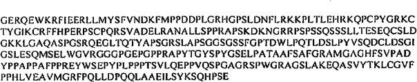
Fig. 8 A and 8B have described the nucleotide sequence (SEQ ID NO:1) and the people DEFA6 amino acid sequence of polypeptide (SEQ ID NO:2) of coding people DEFA6 polypeptide.
Fig. 9 A and 9B have described the nucleotide sequence (SEQ ID NO:3) and the people DEFA5 amino acid sequence of polypeptide (SEQ ID NO:4) of coding people DEFA5 polypeptide.Fig. 9 C and 9D have described the nucleotide sequence (SEQ ID NO:210) and the people DEFB14 amino acid sequence of polypeptide (SEQ IDNO:211) of coding people DEFB14 polypeptide.
Figure 10 A and 10B have described the nucleotide sequence (SEQ ID NO:5) and the people IL3RA amino acid sequence of polypeptide (SEQ ID NO:6) of coding people IL3RA polypeptide.
Figure 11 A and 11B have described the nucleotide sequence (SEQ ID NO:7) and the people IL2RA amino acid sequence of polypeptide (SEQ ID NO:8) of coding people IL2RA polypeptide.
Figure 12 A and 12B have described the nucleotide sequence (SEQ ID NO:9) and the people REG3G amino acid sequence of polypeptide (SEQ ID NO:10) of coding people REG3G polypeptide.
Figure 13 A and 13B have described the nucleotide sequence (SEQ ID NO:11) and the people REG1B amino acid sequence of polypeptide (SEQ ID NO:12) of coding people REG1B polypeptide.
Figure 14 A and 14B have described the nucleotide sequence (SEQ ID NO:13) and the people KCND3 amino acid sequence of polypeptide (SEQ ID NO:14) of coding people KCND3 polypeptide.
Figure 15 A and 15B have described the nucleotide sequence (SEQ ID NO:15) and the people MIP-3a amino acid sequence of polypeptide (SEQ ID NO:16) of coding people MIP-3a polypeptide.
Figure 16 A and 16B have described the nucleotide sequence (SEQ ID NO:17) and the people ECGF1 amino acid sequence of polypeptide (SEQ ID NO:18) of coding people ECGF1 polypeptide.
Figure 17 A and 17B have described the nucleotide sequence (SEQ ID NO:19) and the people IL1B amino acid sequence of polypeptide (SEQ ID NO:20) of coding people IL1B polypeptide.
Figure 18 A and 18B have described the nucleotide sequence (SEQ ID NO:21) and the people MIP2BGRO-g amino acid sequence of polypeptide (SEQ ID NO:22) of coding people MIP2BGRO-g polypeptide.
Figure 19 A and 19B have described the nucleotide sequence (SEQ ID NO:23) and the people CXCL1 amino acid sequence of polypeptide (SEQ ID NO:24) of coding people CXCL1 polypeptide.
Figure 20 A and 20B have described the nucleotide sequence (SEQ ID NO:25) and the people IAP1 amino acid sequence of polypeptide (SEQ ID NO:26) of coding people IAP1 polypeptide.
Figure 21 A and 21B have described the nucleotide sequence (SEQ ID NO:27) and the people CASP5 amino acid sequence of polypeptide (SEQ ID NO:28) of coding people CASP5 polypeptide.
Figure 22 A and 22B have described the nucleotide sequence (SEQ ID NO:29) and the people DMBT1 amino acid sequence of polypeptide (SEQ ID NO:30) of coding people DMBT1 polypeptide.
Figure 23 A and 23B have described the nucleotide sequence (SEQ ID NO:31) and the people PCDH17 amino acid sequence of polypeptide (SEQ ID NO:32) of coding people PCDH17 polypeptide.
Figure 24 A and 24B have described the nucleotide sequence (SEQ ID NO:33) and the people IFITM1 amino acid sequence of polypeptide (SEQ ID NO:34) of coding people IFITM1 polypeptide.
Figure 25 A and 25B have described the nucleotide sequence (SEQ ID NO:35) and the people PDZK1IP1 amino acid sequence of polypeptide (SEQ ID NO:36) of coding people PDZK1IP1 polypeptide.
Figure 26 A and 26B have described the nucleotide sequence (SEQ ID NO:37) and the people IRTA2 amino acid sequence of polypeptide (SEQ ID NO:38) of coding people IRTA2 polypeptide.
Figure 27 A and 27B have described the nucleotide sequence (SEQ ID NO:39) and the people SLC40A1 amino acid sequence of polypeptide (SEQ ID NO:40) of coding people SLC40A1 polypeptide.
Figure 28 A and 28B have described the nucleotide sequence (SEQ ID NO:41) and the people IGHV4-4 amino acid sequence of polypeptide (SEQ ID NO:42) of coding people IGHV4-4 polypeptide.
Figure 29 A and 29B have described the nucleotide sequence (SEQ ID NO:43) and the people REG3G amino acid sequence of polypeptide (SEQ ID NO:44) of coding people REG3G polypeptide.
Figure 30 A and 30B have described the nucleotide sequence (SEQ ID NO:45) and the people AQP9 amino acid sequence of polypeptide (SEQ ID NO:46) of coding people AQP9 polypeptide.
Figure 31 A and 31B have described the nucleotide sequence (SEQ ID NO:47) and the people OLFM4 amino acid sequence of polypeptide (SEQ ID NO:48) of coding people OLFM4 polypeptide.
Figure 32 A and 32B have described the nucleotide sequence (SEQ ID NO:49) and the people S100A9 amino acid sequence of polypeptide (SEQ ID NO:50) of coding people S100A9 polypeptide.
Figure 33 A and 33B have described the nucleotide sequence (SEQ ID NO:51) and the people UNC5CL amino acid sequence of polypeptide (SEQ ID NO:52) of coding people UNC5CL polypeptide.
Figure 34 A and 34B have described the nucleotide sequence (SEQ ID NO:53) and the people GPR110 amino acid sequence of polypeptide (SEQ ID NO:54) of coding people GPR110 polypeptide.
Figure 35 A and 35B have described the nucleotide sequence (SEQ ID NO:55) and the people HLA-G amino acid sequence of polypeptide (SEQ ID NO:56) of coding people HLA-G polypeptide.
Figure 36 A and 36B have described the nucleotide sequence (SEQ ID NO:57) and the people TAP1 amino acid sequence of polypeptide (SEQ ID NO:58) of coding people TAP1 polypeptide.
Figure 37 A and 37B have described the nucleotide sequence (SEQ ID NO:59) and the people MAP3K8 amino acid sequence of polypeptide (SEQ ID NO:60) of coding people MAP3K8 polypeptide.
Figure 38 A and 38B have described the nucleotide sequence (SEQ IDNO:61) and the people UBD|GABBR1 amino acid sequence of polypeptide (SEQ ID NO:62) of coding people UBD|GABBR1 polypeptide.
Figure 39 A and 39B have described the nucleotide sequence (SEQ ID NO:63) and the people DHX57 amino acid sequence of polypeptide (SEQ ID NO:64) of coding people DHX57 polypeptide.
Figure 40 A and 40B have described the nucleotide sequence (SEQ ID NO:65) and the people MA amino acid sequence of polypeptide (SEQ ID NO:66) of coding people MA polypeptide.
Figure 41 A and 41B have described the nucleotide sequence (SEQ ID NO:67) and the people IGLJCOR18 amino acid sequence of polypeptide (SEQ ID NO:68) of coding people IGLJCOR18 polypeptide.
Figure 42 A and 42B have described the nucleotide sequence (SEQ ID NO:69) and the people HLA-G amino acid sequence of polypeptide (SEQ ID NO:70) of coding people HLA-G polypeptide.
Figure 43 A and 43B have described the nucleotide sequence (SEQ ID NO:71) and the people SAA1 amino acid sequence of polypeptide (SEQ ID NO:72) of coding people SAA1 polypeptide.
Figure 44 A and 44B have described the nucleotide sequence (SEQ ID NO:73) and the people TAP2 amino acid sequence of polypeptide (SEQ ID NO:74) of coding people TAP2 polypeptide.
Figure 45 A and 45B have described the nucleotide sequence (SEQ ID NO:75) and the people PCAA17448 amino acid sequence of polypeptide (SEQ ID NO:76) of coding people PCAA17448 polypeptide.
Figure 46 A and 46B have described the nucleotide sequence (SEQ ID NO:77) and the people LCN2 amino acid sequence of polypeptide (SEQ ID NO:78) of coding people LCN2 polypeptide.
Figure 47 A and 47B have described the nucleotide sequence (SEQ ID NO:79) and the people ZBP1 amino acid sequence of polypeptide (SEQ ID NO:80) of coding people ZBP1 polypeptide.
Figure 48 A and 48B have described the nucleotide sequence (SEQ ID NO:81) and the people TNIP3 amino acid sequence of polypeptide (SEQ ID NO:82) of coding people TNIP3 polypeptide.
Figure 49 A and 49B have described the nucleotide sequence (SEQ ID NO:83) and the people ZC3H12A amino acid sequence of polypeptide (SEQ ID NO:84) of coding people ZC3H12A polypeptide.
Figure 50 A and 50B have described the nucleotide sequence (SEQ ID NO:85) and the people CHI3L1 amino acid sequence of polypeptide (SEQ ID NO:86) of coding people CHI3L1 polypeptide.
Figure 51 A and 51B have described the nucleotide sequence (SEQ ID NO:87) and the people FCGR3A amino acid sequence of polypeptide (SEQ ID NO:88) of coding people FCGR3A polypeptide.
Figure 52 A and 52B have described the nucleotide sequence (SEQ ID NO:89) and the people SAMD9L amino acid sequence of polypeptide (SEQ ID NO:90) of coding people SAMD9L polypeptide.
Figure 53 A and 53B have described the nucleotide sequence (SEQ ID NO:91) and the people MMP9 amino acid sequence of polypeptide (SEQ ID NO:92) of coding people MMP9 polypeptide.
Figure 54 A and 54B have described the nucleotide sequence (SEQ ID NO:93) and the people MMP7 amino acid sequence of polypeptide (SEQ ID NO:94) of coding people MMP7 polypeptide.
Figure 55 A and 55B have described the nucleotide sequence (SEQ ID NO:95) and the people BF amino acid sequence of polypeptide (SEQ ID NO:96) of coding people BF polypeptide.
Figure 56 A and 56B have described the nucleotide sequence (SEQ ID NO:97) and the people S100P amino acid sequence of polypeptide (SEQ ID NO:98) of coding people S100P polypeptide.
Figure 57 A and 57B have described the nucleotide sequence (SEQ ID NO:99) and the people GRO amino acid sequence of polypeptide (SEQ ID NO:100) of coding people GRO polypeptide.
Figure 58 A and 58B have described the nucleotide sequence (SEQ ID NO:101) and the people INDO amino acid sequence of polypeptide (SEQ ID NO:102) of coding people INDO polypeptide.
Figure 59 A and 59B have described the nucleotide sequence (SEQ ID NO:103) and the people TRIM22 amino acid sequence of polypeptide (SEQ ID NO:104) of coding people TRIM22 polypeptide.
Figure 60 A and 60B have described the nucleotide sequence (SEQ ID NO:105) and the people SAA2 amino acid sequence of polypeptide (SEQ ID NO:106) of coding people SAA2 polypeptide.
Figure 61 A and 61B have described the nucleotide sequence (SEQ ID NO:107) and the people NEU4 amino acid sequence of polypeptide (SEQ ID NO:108) of coding people NEU4 polypeptide.
Figure 62 A and 62B have described the nucleotide sequence (SEQ IDNO:109) and the people IRTA2/FCRH5 amino acid sequence of polypeptide (SEQ ID NO:110) of coding people IRTA2/FCRH5 polypeptide.
Figure 63 A and 63B have described the nucleotide sequence (SEQ ID NO:111) and the people IGLJCOR18 amino acid sequence of polypeptide (SEQ ID NO:112) of coding people IGLJCOR18 polypeptide.
Figure 64 A and 64B have described the nucleotide sequence (SEQ ID NO:113) and the people IGHV4-4 amino acid sequence of polypeptide (SEQ ID NO:114) of coding people IGHV4-4 polypeptide.
Figure 65 A and 65B have described the nucleotide sequence (SEQ ID NO:115) and the people MM9 amino acid sequence of polypeptide (SEQ ID NO:116) of coding people MMP9 polypeptide.
Figure 66 A and 66B have described the nucleotide sequence (SEQ ID NO:117) and the people GRO amino acid sequence of polypeptide (SEQ ID NO:118) of coding people GRO polypeptide.
Figure 67 A and 67B have described the nucleotide sequence (SEQ IDNO:119) and the people MIP2BGRO-g amino acid sequence of polypeptide (SEQ ID NO:120) of coding people MIP2BGRO-g polypeptide.
Figure 68 A and 68B have described the nucleotide sequence (SEQ ID NO:121) and the people IL1B amino acid sequence of polypeptide (SEQ ID NO:122) of coding people IL1B polypeptide.
Figure 69 A and 69B have described the nucleotide sequence (SEQ ID NO:123) and the people IL3RA amino acid sequence of polypeptide (SEQ ID NO:124) of coding people IL3RA polypeptide.
Figure 70 A and 70B have described the nucleotide sequence (SEQ ID NO:125) and the people CASP1 amino acid sequence of polypeptide (SEQ ID NO:126) of coding people CASP1 polypeptide.
Figure 71 A and 71B have described the nucleotide sequence (SEQ ID NO:127) and the people BV8 amino acid sequence of polypeptide (SEQ ID NO:128) of coding people BV8 polypeptide.
Figure 72 A and 72B have described the nucleotide sequence (SEQ ID NO:129) and the people HDAC7A amino acid sequence of polypeptide (SEQ ID NO:130) of coding human DAC7A polypeptide.
Figure 73 A and 73B have described the nucleotide sequence (SEQ ID NO:131) and the people ACVRL1 amino acid sequence of polypeptide (SEQ ID NO:132) of coding people ACVRL1 polypeptide.
Figure 74 A and 74B have described the nucleotide sequence (SEQ ID NO:133) and the people NR4A1 amino acid sequence of polypeptide (SEQ ID NO:134) of coding people NR4A1 polypeptide.
Figure 75 A and 75B have described the nucleotide sequence (SEQ ID NO:135) and the people K5B amino acid sequence of polypeptide (SEQ ID NO:136) of coding people K5B polypeptide.
Figure 76 A and 76B have described the nucleotide sequence (SEQ ID NO:137) and the people SILV amino acid sequence of polypeptide (SEQ ID NO:138) of coding people SILV polypeptide.
Figure 77 A and 77B have described the nucleotide sequence (SEQ ID NO:139) and the people IRAK3 amino acid sequence of polypeptide (SEQ ID NO:140) of coding people IRAK3 polypeptide.
Figure 78 A and 78B have described the nucleotide sequence (SEQ ID NO:141) and the people IL-4 amino acid sequence of polypeptide (SEQ ID NO:142) of coding people IL-4 polypeptide.
Figure 79 A and 79B have described the nucleotide sequence (SEQ ID NO:143) and the people IL-13 amino acid sequence of polypeptide (SEQ ID NO:144) of coding people IL-13 polypeptide.
Figure 80 A and 80B have described the nucleotide sequence (SEQ ID NO:145) and the people RAD50 amino acid sequence of polypeptide (SEQ ID NO:146) of coding people RAD50 polypeptide.
Figure 81 A and 81B have described the nucleotide sequence (SEQ ID NO:147) and the people IL-5 amino acid sequence of polypeptide (SEQ ID NO:148) of coding people IL-5 polypeptide.
Figure 82 A and 82B have described the nucleotide sequence (SEQ ID NO:149) and the people IRF1 amino acid sequence of polypeptide (SEQ ID NO:150) of coding people IRF1 polypeptide.
Figure 83 A and 83B have described the nucleotide sequence (SEQ ID NO:151) and the people PDLIM4 amino acid sequence of polypeptide (SEQ ID NO:152) of coding people PDLIM4 polypeptide.
Figure 84 A and 84B have described the nucleotide sequence (SEQ ID NO:153) and the people CSF2 amino acid sequence of polypeptide (SEQ ID NO:154) of coding people CSF2 polypeptide.
Figure 85 A and 85B have described the nucleotide sequence (SEQ ID NO:155) and the people IL-3 amino acid sequence of polypeptide (SEQ ID NO:156) of coding people IL-3 polypeptide.
Figure 86 A and 86B have described the nucleotide sequence (SEQ ID NO:157) and the people MMP3 amino acid sequence of polypeptide (SEQ ID NO:158) of coding people MMP3 polypeptide.
Figure 87 A and 87B have described the nucleotide sequence (SEQ ID NO:159) and the people IL-8 amino acid sequence of polypeptide (SEQ ID NO:160) of coding people IL-8 polypeptide.
Figure 88 A and 88B have described the nucleotide sequence (SEQ ID NO:161) and the people TLR4 amino acid sequence of polypeptide (SEQ ID NO:162) of coding people TLR4 polypeptide.
Figure 89 A and 89B have described the nucleotide sequence (SEQ ID NO:163) and the people HLA-DRB1 amino acid sequence of polypeptide (SEQ ID NO:164) of coding people HLA-DRB1 polypeptide.
Figure 90 A and 90B have described the nucleotide sequence (SEQ ID NO:165) and the people MMP19 amino acid sequence of polypeptide (SEQ ID NO:166) of coding people MMP19 polypeptide.
Figure 91 A and 91B have described the nucleotide sequence (SEQ ID NO:167) and the people TIMP1 amino acid sequence of polypeptide (SEQ ID NO:168) of coding people TIMP1 polypeptide.
Figure 92 A and 92B have described the nucleotide sequence (SEQ ID NO:169) and the people Elfin amino acid sequence of polypeptide (SEQ ID NO:170) of coding people Elfin polypeptide.
Figure 93 A and 93B have described the nucleotide sequence (SEQ ID NO:171) and the people CXCL1 amino acid sequence of polypeptide (SEQ ID NO:172) of coding people CXCL1 polypeptide.
Figure 94 A and 94B have described the nucleotide sequence (SEQ IDNO:173) and the people DFKZP586A0522 amino acid sequence of polypeptide (SEQ ID NO:174) of coding people DFKZP586A0522 polypeptide.
Figure 95 A and 95B have described the nucleotide sequence (SEQ ID NO:175) and the people SLC39A5 amino acid sequence of polypeptide (SEQ ID NO:176) of coding people SLC39A5 polypeptide.
Figure 96 A and 96B have described the nucleotide sequence (SEQ ID NO:177) and the people GLI-1 amino acid sequence of polypeptide (SEQ ID NO:178) of coding people GLI-1 polypeptide.
Figure 97 A and 97B have described the nucleotide sequence (SEQ ID NO:179) and the people HMGA2 amino acid sequence of polypeptide (SEQ ID NO:180) of coding people HMGA2 polypeptide.
Figure 98 A and 98B have described the nucleotide sequence (SEQ ID NO:181) and the people SLC22A5 amino acid sequence of polypeptide (SEQ ID NO:182) of coding people SLC22A5 polypeptide.
Figure 99 A and 99B have described the nucleotide sequence (SEQ ID NO:183) and the people SLC22A4 amino acid sequence of polypeptide (SEQ ID NO:184) of coding people SLC22A4 polypeptide.
Figure 100 A and 100B have described the nucleotide sequence (SEQ ID NO:185) and the people P4HA2 amino acid sequence of polypeptide (SEQ ID NO:186) of coding people P4HA2 polypeptide.
Figure 101 A and 101B have described the nucleotide sequence (SEQ ID NO:187) and the people TSLP amino acid sequence of polypeptide (SEQ ID NO:188) of coding people TSLP polypeptide.
Figure 102 A and 102B have described the nucleotide sequence (SEQ IDNO:189) and human microtubulin α 5/ α 3 amino acid sequence of polypeptide (SEQ ID NO:190) of coding human microtubulin α 5/ α 3 polypeptide.
Figure 103 A and 103B have described the nucleotide sequence (SEQ IDNO:191) and human microtubulin α 6 amino acid sequence of polypeptide (SEQ ID NO:192) of coding human microtubulin α 6 polypeptide.
Figure 104 has shown the meta analysis (meta-analysis) of use Mantel-Haenszel method to non-synonym (non-synonymous) GLI1 SNP rs2228226 in Scotland, Cambridge and the Sweden.
Figure 105 has shown that Q1100E destroys the proteic conserved regions of GLI1 and reduces the GLI1 transcriptional activity.
Figure 106 has shown the expression of Hedgehog (HH) signal conductive members in health adult's colon (HC) and the ulcerative colitis (UC).
Figure 107 has shown the result, wherein Gli1+/-animal handles the back at DSS and shows dead, serious clinical symptom and lose weight significantly.
Figure 108 shown Gli1+/-animal replys DSS and handles and to represent the more serious enteritis than the brood young baby of WT.
Figure 109 shown Gli1+ after DSS handles/-and the cytokine analysis of WT mouse represented the intensive pro-inflammatory cytokine and activated.
Figure 110 A-B has shown that (A) coding people contains the nucleotide sequence (LOC342959) and (B) the people ARRDC5 amino acid sequence of polypeptide of 5 (ARRDC5) in arrestin (arrestin) territory.
Figure 111 has shown and human ataxia albumen 3 sample albumen (ataxin 3-like, ATXN3L) Dui Ying nucleotide sequence.
Figure 112 A-B has shown the nucleotide sequence (LOC92552) of (A) coding Human Fallicle-Stimulating Hormone's acceptor (FSHR) and (B) people FSHR amino acid sequence of polypeptide.
Figure 113 A-B has shown the nucleotide sequence of (A) coding human blood platelets derived growth factor receptor α polypeptide (PDGFRA) and (B) people PDGFRA amino acid sequence of polypeptide.
Figure 114 A-B has shown the nucleic acid sequence encoding of (A) people transforming growth factor-beta 3 (TGFB3) and (B) people TGFB3 amino acid sequence of polypeptide.
Figure 115 A-B has shown that (A) coding people contains 8 (potassium channeltetramerisation domain containing 8, nucleotide sequence KCTD8) and (B) the people KCTD8 amino acid sequence of polypeptide in potassium channel four dimerization territories.
Figure 116 A-B has shown the nucleotide sequence of (A) coding human transglutaminase 4 (TGM4) and (B) people TGM4 amino acid sequence of polypeptide.
Figure 117 A-B has shown the nucleotide sequence of (A) coding people TPD52L3 oncoprotein D52 sample 3 (NYD-SP25) and (B) people NYD-SP25 amino acid sequence of polypeptide.
Figure 118 has shown with misc_RNA (C3orf53), the nucleotide sequence of FLJ33651 correspondence.
Figure 119 shown with karyomit(e) 10 on (EMX2OS) corresponding nucleotide sequence of EMX2 opposite strand (not coded protein).
Figure 120 A-B has shown (A) coding people all-body configuration MMTV integration site family, member's 16 (WNT16) nucleotide sequence and (B) people WNT16 amino acid sequence of polypeptide.
Figure 121 A-C has shown that (A-B) coding people sprouty is relevant, contains the nucleotide sequence and (C) aminoacid sequence of people SPRED2 of 2 (SPRED2) in EVH1 territory.
Figure 122 A-C has shown the nucleotide sequence of (A-B) coding human chromosome 16 open reading-frame (ORF)s 65 (C16orf65) and (C) aminoacid sequence of human chromosome 16 open reading-frame (ORF)s 65 (C16orf65).
Figure 123 A-B has shown the nucleotide sequence of (A) coding human chromosome 12 open reading-frame (ORF)s 2 (C12orf2) and (B) aminoacid sequence of human chromosome 12 open reading-frame (ORF)s 2 (C12orf2).
Figure 124 A-B has shown the nucleotide sequence of (A) coding people multiple pdz domain albumen (MPDZ) and (B) aminoacid sequence of people MPDZ.
Figure 125 A-B has shown nucleotide sequence of (A) coding people phenylalanine-tRNA synthetic enzyme 2 (FARS2) and (B) aminoacid sequence of people FARS2.
Figure 126 A-B has shown (A) coding people Caspase 8, the apoptosis be correlated with nucleotide sequence of L-Cysteine HCL Anhydrous (CASP8) and (B) aminoacid sequence of people CASP8.
Figure 127 A-B shown (A) coding people 5 '-phosphonuclease, born of the same parents outer (ecto) are the nucleotide sequence and (B) aminoacid sequence of people NT5E of (NT5E) (CD73).
Figure 128 has shown and the corresponding nucleotide sequence of people's teratoma derivative growth factor 3 (TDGF3).
Figure 129 A-B has shown the nucleotide sequence of (A) coding people butyrophilin sample 3 (BTNL3) and (B) aminoacid sequence of people BTNL3.
Figure 130 A-B has shown the nucleotide sequence of (A) coding people S100A8 and (B) aminoacid sequence of people S100A8.
Figure 131 A-B has shown the nucleotide sequence of (A) coding people CCL20 and (B) aminoacid sequence of people CCL20.
Detailed Description Of The Invention
A.
Definition
Unless otherwise defined, technology used herein has identical implication with scientific terminology with the common understanding of one skilled in the art of the present invention. Provide the generality guidance of employed many terms among the application take Publication about Document as those skilled in the art: Singleton et al., Dictionary ofMicrobiology and Molecular Biology, second edition, J.Wiley ﹠ Sons (New York, N.Y.1994), and March, Advanced Organic Chemistry Reactions, Mechanisms andStructure, the 4th edition, John Wiley ﹠ Sons (New York, N.Y.1992).
One skilled in the art will realize that method many to described herein is similar with material or be equal to method and material, they can use in practice of the present invention. In fact, the present invention is limited to absolutely not described method and material. For the present invention, hereinafter defined following term.
Term " inflammatory bowel disease " or " IBD " are as the general designation of ulcerative colitis and Crohn's disease. Although these two kinds of diseases are commonly considered as two kinds of various disease, but their common trait, such as near the leukocytic focal accumulation spot shape necrosis of superficial epithelium, the gland crypts, and the number increase of intraepithelial lymphocyte (IEL) and chronic macrophage subset, have reason to think that they can be used as a disease group and treat.
Term " Crohn's disease " or " CD " are used in reference to the illness that relates to the intestines and stomach chronic inflammation in this article. Crow engler's related inflammation affects intestines usually, but can betide from mouthful any position to anus. The difference of CD and UC is that inflammation passes all layers extension of intestines wall, and relates to mesenterium and lymph node. This disease usually is discontinuous, and namely serious ill intestinal segment is what to separate with apparent anosis zone. In CD, the intestines wall also thickens, and this can cause obturation, and the formation in fistula and crack is common. As used herein, CD can be one or more in several CD types, includes but not limited to ileocolitis (affecting ileum and large intestine); Ileitis (affecting ileum); Gastroduodenal CD (inflammation in stomach and the duodenum); Jejunoileitis (the spot sample spot of inflammation in the jejunum); With Crow engler (granulomatous) colitis (only affecting large intestine).
Term " ulcerative colitis " or " UC " are used in reference to the illness that relates to large intestine and rectum inflammation in this article. In UC patient, the inflammatory reaction that relates generally to mucous membrane of colon is arranged. This inflammation is normally even and continuous, does not have the zone between two parties of normal mucosa. Surface mucomembranous cell and crypts epithelium and submucosa relate to the inflammatory reaction with neutrophil cell infiltration. Finally, this reaction advances to epithelial damage and epithelial cell loss usually, causes many places ulcer, fibrillatable, the dysplasia of colon and vertically shrink back (retraction).
Term " inactivity " IBD is used in this article expression and before is diagnosed as IBD but the current IBD that goes down that is at individuality. This consists of contrast with " activity " IBD, and in the later case, diagnosis of case has IBD but not yet receives treatment. In addition, activity IBD can be the recurrence after the previous IBD that diagnoses and treat enters go down (namely becoming inactivity IBD). This type of recurrence also can be called IBD " burst (flare up) " in this article. Mammalian subject with activity autoimmune disease (such as IBD) can stand burst, i.e. state of an illness activity increases the weight of or a period of time of corresponding glucose recovery. Burst can be replied severe infections, allergy (allegic reactions), physiological stress (physical stress), emotional trauma, operation or environmental factor and be taken place.
Term " regulation and control " is used in this article the RNA molecule of the expression of expression gene or encode one or more protein or protein subunit or is equal to the level of RNA molecule or the activity of one or more protein or protein subunit raises or downward modulation, so that expression, level or activity are greater than or less than in the non-existent situation of adjusting control agent is viewed.
Term " inhibition ", " downward modulation ", " expressing not enough " and " reduction " are used interchangeably, and represent a kind of expression of gene or the RNA molecule of encode one or more protein or protein subunit or be equal to the level of RNA molecule or the activity of one or more protein or protein subunit reduces with respect to one or more contrasts (such as for example one or more positives and/or negative control).
Term " rise " or " cross express " are used for representing a kind of expression of gene or the RNA molecule of encode one or more protein or protein subunit or be equal to the RNA molecular level or the activity of one or more protein or protein subunit raises with respect to one or more contrasts (such as for example one or more positives and/or negative control).
Term " diagnosis " refers to identify molecule or pathological state, disease or illness when being used for this paper, such as identifying IBD.
Term " prognosis " refers to when being used for this paper predict that IBD forms or the possibility of progress, comprises postoperative autoimmunity burst and recurrence. In case prognostic factor refers to those patients and form IBD, affect Patients on Recurrence rate and result, the variable relevant with the IBD natural history. May comprise for example abdominal mass (abdominal mass) or tenderness, fash, arthroncus, canker sore and borborygmus (enteron aisle has the rumble or splashes sound) by the clinical parameter relevant with relatively poor prognosis. Prognostic factor can be used for the patient is included into the subgroup with different baseline risks of recurrence.
" pathology " of IBD comprises that all jeopardize the phenomenon of patient's health and happiness. IBD pathology mainly is attributed to immune abnormal activation in the intestines, and this can cause chronic or acute inflammation in the situation without any known exotic antigen, and ulcer subsequently. Clinically, IBD is characterized as various performance, usually causes chronic, uncertain process. Bloody diarrhea and stomachache are usually with generating heat and losing weight. Anaemia is common, as serious fatigue. Joint performance and the dysfunction of liver of scope from arthralgia to the acute arthritis is usually relevant with IBD. During IBD acute " attack ", work and other normal activity are normally impossible, and the patient usually is in hospital.
Cause of disease the unknown of these diseases, and initial infringement is not yet clear definite; Yet, near the leukocytic focal accumulation spot shape necrosis of superficial epithelium, gland crypts, and the number increase of intraepithelial lymphocyte and some macrophage subset be described to the early changes of inferring, especially in Crohn's disease.
Term " treatment " or " processing " pointer to the therapeutic treatment of IBD and preventative or precaution measure the two, wherein target be prevention or slow down (alleviating) for pathology illness or illness. The experimenter who needs the experimenter for the treatment of to comprise to suffer from for a long time IBD and and the experimenter that tends to suffer from IBD maybe to prevent the experimenter of IBD. In case make the IBD diagnosis by method disclosed herein, therapeutic purpose is induced exactly and is kept and goes down so.
Be suitable for those skilled in the art will know that as the various medicaments of " IBD therapeutic agent ". As described in this article, this type of medicament includes but not limited to aminosalicylate, corticosteroid and immunodepressant.
Term " specimen " refers to have from suspection the sample of the mammalian subject that IBD, the known IBD of having or the known IBD of being in go down. Specimen can be derived from the various sources in the mammalian subject, include but not limited to blood, seminal fluid, serum, urine, marrow, mucous membrane, tissue, etc.
Term " contrast " or " control sample " refer to expect that wherein negative findings helps the negative control of the positive findings in the related specimen. Be suitable for that contrast of the present invention includes but not limited to known sample with gene expression of normal level, the sample that obtains from the mammalian subject of the known IBD of not having and the sample that obtains from known normal mammalian subject. Contrast also can be that certainly previous diagnosis has IBD also to accept treatment, the current sample that is in experimenter's acquisition of going down for IBD; And this type of is useful to impinging upon in any IBD recurrence of determining to be among the experimenter of disappearing. In addition, contrast can be contain with specimen in the Normocellular sample of contained cell with same origin. Those skilled in the art can understand other contrast that is suitable for using in the present invention.
Term " microarray " refers to the array element that can hybridize, and the preferred polynucleotide probe is positioned at the ordered arrangement on the matrix.
Term " polynucleotides " is often referred to any polybribonucleotide or polydeoxyribonucleotide when using with odd number or plural number, it can be RNA or the DNA of unmodified, or the RNA or the DNA that modify. So, for instance, polynucleotides as defined herein include but not limited to strand and double-stranded DNA, comprise the DNA of strand and double stranded region, strand and double-stranded RNA and comprise strand and the RNA of double stranded region and comprise DNA and the hybrid molecule of RNA, and DNA wherein and RNA can be strands or more generally double-stranded or comprise strand and double stranded region. In addition, when using in this article, three sequences that term " polynucleotides " refers to comprise RNA or DNA or comprises simultaneously RNA and DNA. Chain in this zone can be from identical molecule or different molecules. This zone can comprise the whole of one or more described molecules, but more typically only is the zone of some described molecules. One of the molecule in triple helix district is oligonucleotides normally. Term " polynucleotides " clearly comprises cDNA. This term comprises DNA (comprising cDNA) and the RNA of the base that contains one or more modifications. So, have because of the DNA of stability or other former thereby adorned main chain or RNA be this term " polynucleotides " that mean herein. In addition, comprise rare bases (such as inosine) or DNA or the RNA of the base (such as the base of tritiate) of modifying are included in the scope of term " polynucleotides " as defined herein. Generally speaking, all chemistry, enzyme and/or the metabolism modified forms of the polynucleotides of unmodified contained in term " polynucleotides ", and the chemical species of virus and the distinctive DNA of cell (comprising simple cell and complex cell) and RNA.
Term " oligonucleotides " refers to the polynucleotides of relatively lacking, and includes but not limited to strand deoxyribonucleotide, strand or double-stranded ribonucleotide, RNA:DNA heterocomplex, and double-stranded DNA. Oligonucleotides such as the ssDNA probe oligonucleotides, usually synthesizes by chemical method, for example the automated oligonucleotide synthesizer of commodity in use. Yet oligonucleotides can be by multiple other method preparation, comprises the technology of extracorporeal recombinant DNA mediation and by the expression of DNA in the Cell and organism body.
Term " gene of differential expression ", " genetic expression of difference " and their synonym are used interchangeably; finger is with respect to its expression in normal or contrast experimenter; its expression in ill (particularly IBD, such as UC or CD) experimenter is activated to the gene of higher or lower level.This term comprises that also its expression in the different steps of same disease is activated to the gene of higher or lower level.The gene that it is to be further understood that differential expression can be activated on nucleic acid level or protein level or suppress, and maybe can carry out alternative splicing to produce different polypeptide products.Such difference can be by for example mRNA level, polypeptide the change of surface expression, secretion or other distribution (partitioning) confirmed.The genetic expression of difference can comprise the expression between two or more genes of comparison or their gene product, or the expression ratio between two or more genes of comparison or their gene product, or even two kinds of different processed products of more same gene, it is between normal subjects and ill (particularly IBD) experimenter or be different between the different steps in same disease.Differential expression for example is included between normal and diseased cells or at the iuntercellular that has experienced various disease incident or disease stage, and gene or its expression product have quantitative in time or cell expressing are graphic and difference qualitatively.For the purposes of the present invention, when in normal and ill experimenter, exist between the given expression of gene or in the various disease developmental stage ill experimenter at least about 2 times, preferably at least about 4 times, more preferably at least about 6 times, during most preferably at least about 10 times of differences, just think to have " genetic expression of difference ".
" cross and express " about the term of rna transcription thing and be used in reference to by to the determined transcript level of reference mRNA level standardization, described can be detected all transcripts or specific mRNA reference set in the sample with reference to mRNA.
Phrase " gene amplification " refers to form the gene of multiple copied or the process of gene fragment in specific cells or clone.Duplicate field (section of the DNA of amplification) is commonly referred to " amplicon ".Generally speaking, the amount of the messenger RNA(mRNA) that is produced (mRNA), i.e. the level of genetic expression, also the ratio in the copy number that is formed by expressed specific gene increases.
Generally speaking, term " mark " or " biomarker " refer to the appraisable physical location on the karyomit(e), and such as restriction endonuclease recognition site or gene, its succession can be monitored.Mark can be the genetic expression district that is called " gene expression markers ", or does not have some DNA sections of known encoding function.As used herein, " IBD mark " refers to those cited in the table 1,2 and 3 genes.
" severity " of hybridization determined by those of ordinary skills easily, and depend on probe length usually, wash temperature and salt concn calculate by rule of thumb.Generally speaking, long probe needs higher temperature to be used for correct annealing, and short probe then needs lower temperature.When complementary strand was present in the environment that is lower than their melting temperature(Tm)s, denatured DNA annealed ability was again depended in hybridization usually.Expectation homology degree between probe and the sequence that can hybridize is high more, just can use high more relative temperature.As a result of, higher relative temperature trends towards making reaction conditions strict more, and lower temperature is then more not strict.For the subsidiary details of the severity of hybridization and explain " Current Protocols in Molecular Biology " referring to Ausubel etc., Wiley Interscience Publishers, (1995).
As defined herein, " stringent condition " or " high stringent condition " is generally: (1) uses low ionic strength and high temperature for washing, 0.015M sodium-chlor/0.0015M Trisodium Citrate/0.1% sodium lauryl sulphate for example, 50 ℃; (2) during hybridizing, use denaturing agent, such as methane amide, for example, 50% (v/v) methane amide and 0.1% bovine serum albumin(BSA)/0.1%Ficoll/0.1% polyvinylpyrrolidone/have 750mM sodium-chlor, the 50mM sodium phosphate buffer pH 6.5 of 75mM Trisodium Citrate, 42 ℃, or (3) use 50% methane amide, 5x SSC (0.75M NaCl, 0.075M Trisodium Citrate), 50mM sodium phosphate (pH6.8), 0.1% trisodium phosphate, 5x DenhardtShi solution, the salmon sperm DNA of supersound process (50 μ g/ml), 0.1%SDS and 10% sulfuric acid dextran, 42 ℃, 42 ℃ in 0.2x SSC (sodium chloride/sodium citrate), wash in 50% methane amide, then 55 ℃ of high strict washings of forming by the 0.1x SSC that contains EDTA.
" medium stringent condition " can be defined as " Molecular Cloning:ALaboratory Manual " (the New York:Cold Spring Harbor Press as Sambrook etc., 1989) described, and comprise and use compare with mentioned above those undemanding washing soln and hybridization conditions (for example temperature, ionic strength and %SDS).An example of medium stringent condition is to contain: 20% methane amide, 5x SSC (150mM NaCl, the 15mM trisodium citrate), be incubated overnight in 37 ℃ in the solution of the salmon sperm DNA of the shearing of 50mM sodium phosphate (pH7.6), 5xDenhardtShi solution, 10% sulfuric acid dextran and 20mg/ml sex change, in 1x SSC, wash filter membrane in about 37-50 ℃ then.Those skilled in the art will be appreciated that how to come attemperation, ionic strength etc. according to the needs that adapt to such as factors such as probe length.
In linguistic context of the present invention, address any specific gene concentrate cited " at least a ", " at least two kinds ", " at least five kinds ", etc. gene mean arbitrary combination or any combination and all combinations of cited gene.
Term " montage " and " RNA montage " are used interchangeably, and refer to remove intron and connect exon to produce the RNA processing of ripe mRNA, and wherein ripe mRNA has the continuous programming code sequence and moves into eukaryotic tenuigenin.
In theory, term " exon " refers to be embodied in any split gene section (B.Lewin.Genes IV Cell Press, Cambridge Mass.1990) in the mature rna product.In theory, term " intron " refer to be transcribed but exon montage by will being arranged in its either side to any DNA section of being removed from transcript together.In operation, exon sequence appears in the mRNA sequence of gene, and gene limits by numbering referring to SEQ ID.In operation, intron sequences is the intervening sequence that is arranged in the genomic dna of gene, and its both sides are exon sequence and have GT and AG montage consensus sequence on their 5 ' and 3 ' border.
" RNA interfering " or " siRNA (siRNA) " refers to that length is usually less than about 30 Nucleotide and reduce the double stranded rna molecule of expression of target gene.RNA interfering can be used currently known methods to identify and synthesize (Shi Y, Trends in Genetics 19 (1): 9-12 (2003); WO 2003/056012; WO2003/064621), and the siRNA library can obtain by commercial sources, Dharmacon for example, Lafayette, Colorado.
" native sequences " polypeptide refers to and the polypeptide that has same acid sequence derived from natural polypeptide, comprises natural existence or allelic variant.This type of native sequences polypeptide can separate from nature, perhaps can generate by reorganization or synthesizing mean.So, the native sequences polypeptide can have and naturally has human polypeptides, mouse polypeptide or from the amino acid sequence of polypeptide of any other mammals species.
Term " antibody " uses with broad sense in this article, clearly covers monoclonal antibody, polyclonal antibody, multi-specificity antibody (for example bi-specific antibody), and antibody fragment, as long as they show desired biological activity.The antibody at one or more IBD marks disclosed herein is specifically contained in the present invention.This antibody-like can be called " anti-IBD mark antibody ".
Term " monoclonal antibody " refers to when being used for this paper from a group antibody that obtains of the antibody of homogeneity basically, it is identical and/or in conjunction with identical epi-position promptly to constitute each antibody of colony, the issuable possibility variant, this type of variant generally exists with indivisible in the process of manufacture order clonal antibody.This type of monoclonal antibody typically comprises the antibody that comprises in conjunction with the peptide sequence of target thing, and its thing that hits is the more to select single target thing to obtain in conjunction with the process of peptide sequence in the peptide sequence by comprising comforming in conjunction with peptide sequence.
Monoclonal antibody clearly comprises " chimeric " antibody in this article, wherein the part of heavy chain and/or light chain with derived from specific species or belong to the identical or homology of corresponding sequence in the antibody of specific antibodies classification or subclass, and the remainder of chain with derived from another species or belong to the identical or homology of corresponding sequence in the antibody of another antibody classification or subclass, and the fragment of this antibody-like, as long as they show desired biological activity (U.S. Patent No. 4,816,567 and Morrison et al., Proc.Natl.Acad.Sci.USA 81:6851-6855 (1984)).Interested herein chimeric antibody comprises and comprising derived from the variable domain antigen binding sequence of non-human primate (for example Old World monkey class (Old World Monkey), ape etc.) and " primatesization " antibody of human constant region sequence, and " humanized antibody ".
" humanization " form of inhuman (for example rodents) antibody refers to that bottom line comprises the chimeric antibody derived from the sequence of non-human immunoglobulin.Largely, humanized antibody refers to the immunoglobulin (Ig) that the hypervariable region residue in the human normal immunoglobulin (receptor antibody) is replaced with the hypervariable region residue of inhuman species (donor antibody) such as mouse, rat, rabbit or non-human primates with expectation specificity, avidity and ability.
" complete antibody " refers to comprise the antibody in two antigen binding domains and Fc district in this article.Preferably, complete antibody has functional Fc district.
" antibody fragment " comprises the part of complete antibody, preferably comprises its antigen binding domain.The example of antibody fragment comprises Fab, Fab ', F (ab ')
2With the Fv fragment; Double antibody (diabody); Linear antibody; The single-chain antibody molecule; Reach the multi-specificity antibody that forms by antibody fragment.
" natural antibody " refers to common about 150, the 000 daltonian different tetramer glycoprotein that are made of two identical light (L) chains and two identical weights (H) chain.Every light chain is connected with heavy chain by a covalent disulfide bonds, and the number of disulfide linkage changes between the heavy chain of different immunoglobulin (Ig) isotypes.Every heavy chain and light chain also have the intrachain disulfide bond of rule at interval.Every heavy chain at one end has a variable domain (V
H), then be a plurality of constant domains.Every light chain at one end has a variable domain (V
L), and the other end is a constant domain.The constant domain of light chain is arranged in first constant domain of heavy chain, and the variable domain of light chain is arranged in the variable domain of heavy chain.Think that specified amino acid residues forms the interface between light chain and heavy chain variable domain.
Term " variable " refers to that some part sequence difference between antibody in the variable domain is extensive and is used for combination and the specific truth of every kind of specific antibodies to its specific antigen.Yet variability is not the whole variable domain that is uniformly distributed in antibody.It concentrates on three sections that are called the hypervariable region in light chain and the heavy chain variable domain.In the variable domain more the part of high conservative be called framework region (FR).Each self-contained four FR of the variable domain of natural heavy chain and light chain, they take the beta-pleated sheet conformation mostly, connect and form in some situation three hypervariable regions connections of a beta-pleated sheet structure part by the formation ring-type.Hypervariable region in every chain is by very approaching the keeping together of FR, and facilitate the formation of the antigen binding site of antibody (referring to Kabat et al. with the hypervariable region of another chain, Sequences of Proteins of ImmunologicalInterest, the 5th edition, Public Health Service, National Institute of Health, Bethesda, MD. (1991)).
Term " hypervariable region ", " HVR " or " HV " refer to alterable height on the antibody variable domains sequence and/or form the zone of the ring that defines on the structure when being used for this paper.Usually, antibody comprises six HVR: three in VH (H1, H2, H3), three in VL (L1, L2, L3).In natural antibody, H3 and L3 show the maximum diversity of these six HVR, and think that particularly H3 brings into play unique effect in accurate specificity giving antibody.Referring to for example Xu et al.Immunity 13:37-45 (2000); Johnson and Wu are in Methods in Molecular Biology 248:1-25 (Lo, ed., HumanPress, Totowa, NJ, 2003).In fact, the natural camelid of the existence antibody of only being made up of heavy chain is that function arranged and stable when lacking light chain.Referring to for example Hamers-Casterman et al., Nature363:446-448 (1993) and Sheriff et al., Nature Struct.Biol.3:733-736 (1996).
Use and contain the narration of many hypervariable regions herein.Kabat complementary determining region (CDR) is based on sequence variations and be the most frequently used (Kabat et al., Sequences of Proteins of ImmunologicalInterest, 5th Ed.Public Health Service, National Institutes of Health, Bethesda, MD. (1991)).Chothia changes the position (Chothia and Lesk J.Mol.Biol.196:901-917 (1987)) that refers to structure ring into.The end of Chothia CDR-H1 ring changes (seeing below) between H32 and H34 when using Kabat coding rule numbering, depend on that (this is because the Kabat numbering plan is placed insertion at H35A and H35B place for the length of ring; If 35A and 35B do not exist, should finish at 32 places by ring so; If have only 35A to exist, should finish at 33 places by ring so; If 35A and 35B exist, should encircle so and finish) at 34 places.AbM represents the hypervariable region trading off between Kabat CDR and the Chothia structure ring, and obtains the use of the AbM antibody modeling software of Oxford Molecular." contact " hypervariable region is based on to the analysis of obtainable compound crystal structure.Hereinafter write down in these hypervariable regions the residue of each.
Ring Kabat AbM Chothia contact
---- ----------- ---------- ----------- ----------
L1 L24-L34 L24-L34 L24-L34 L30-L36
L2 L50-L56 L50-L56 L50-L56 L46-L55
L3 L89-L97 L89-L97 L89-L97 L89-L96
H1 H31-H35B H26-H35B H26-H32,33 or 34 H30-H35B
(Kabat numbering)
H1 H31-H35 H26-H35 H26-H32 H30-H35
(Chothia numbering)
H2 H50-H65 H50-H58 H52-H56 H47-H58
H3 H95-H102 H95-H102 H95-H102 H93-H101
The hypervariable region can comprise following " hypervariable region of extension ": the 26-35B (H1) among 24-36 among the VL or 24-34 (L1), 46-56 or 50-56 (L2) and 89-97 (L3) and the VH, 50-65,47-65 or 49-65 (H2) and 93-102,94-102 or 95-102 (H3).The hypervariable region of these extensions is the combination of Kabat and Chothia definition normally, and it can be chosen wantonly and further comprise the residue that uses the contact definition to identify.For in these definition each, the variable domain residue is according to Kabat etc., the numbering that sees above.
Produce two identical Fabs with papain digestion antibody, be called " Fab " fragment, have an antigen binding site separately, and remaining " Fc " fragment, its title has reflected that it is easy to the crystalline ability.Pepsin produces a F (ab ')
2Fragment, it has two antigen binding sites and still can crosslinked antigen.
" Fv " is the minimum antibody fragment that comprises complete antigen recognition and antigen binding site.This zone is made up of the dimer in a heavy chain variable domain of tight, non-covalent bonded and a light chain variable territory.Just in this structure, three hypervariable regions of each variable domain interact and at V
H-V
LDefine an antigen binding site on the dimer surface.Antibody is given together with antigen-binding specificity in six hypervariable regions.Yet,, be that avidity is lower than complete binding site even single variable domain (or only comprise specific three CDR of antigen half Fv) also has the ability of identification and conjugated antigen.
The Fab fragment also comprises the constant domain of light chain and first constant domain (CH1) of heavy chain.The segmental difference of Fab ' fragment and Fab is that the C-terminal of heavy chain CH1 structural domain has increased the minority residue, comprises the one or more halfcystines from antibody hinge region.Fab '-SH is the appellation of constant domain cysteine residues wherein being carried the Fab ' of at least one free sulphur alcohol radical herein.F (ab ')
2Antibody fragment is as there being the paired Fab ' fragment of hinge cysteine to generate in pairs between Fab ' fragment at first.Also know other chemical coupling form of antibody fragment.
According to the aminoacid sequence of its constant domain, can be included into a kind of in two kinds of distinct types from " light chain " of the antibody of any invertebrate species, be called card handkerchief (κ) and lambda (λ).
Term " Fc district " is used to define the C-end regions of heavy chain immunoglobulin in this article, comprises native sequences Fc district and variant Fc regions.Though the border in heavy chain immunoglobulin Fc district can change, human IgG heavy chain Fc district is normally defined the section to C-terminal from the amino-acid residue of its Cys226 or Pro230 position.The terminal Methionin of the C-in Fc district (residue 447 is according to the EU numbering system) can be eliminated, and for example in the process of production or antibody purification, perhaps carries out the recombined engineering transformation by the nucleic acid to the encoding antibody heavy chain.Thereby the complete antibody composition can comprise antibody population that all K447 residues all are eliminated, antibody population that none K447 residue is eliminated or mix the antibody that the K447 residue is arranged and do not had the antibody population of the antibody of K447 residue.
Except as otherwise noted, the residue numbering of heavy chain immunoglobulin is as Kabat et al. herein, Sequences of Proteins of Immunological Interest, 5th Ed.Public Health Service, National Institutes of Health, Bethesda, the numbering of the EU index among the MD (1991)." as the EU index among the Kabat " refers to the residue numbering of human IgG1 EU antibody.
" native sequences Fc district " comprises the identical aminoacid sequence of aminoacid sequence with the Fc district that finds at occurring in nature.Native sequences people Fc district comprises native sequences human IgG1 Fc district (non-A and A allotype); Native sequences human IgG2 Fc district; Native sequences human IgG 3 Fc districts; With native sequences human IgG 4 Fc districts; And the natural variant that exists.
" variant Fc regions " comprise since at least one place amino acid modified (a preferred place or many places amino acid replacement) and with the different aminoacid sequence in native sequences Fc district.Preferably, variant Fc regions is compared with native sequences Fc district or compared with the Fc district of parent's polypeptide has at least one place amino acid replacement, for example have about 1 place to about 10 place's amino acid replacements in native sequences Fc district or in the Fc district of parent's polypeptide, preferred about 1 place is to about 5 place's amino acid replacements.Variant Fc regions Fc district preferred in this article and native sequences Fc district and/or parent's polypeptide has the homology at least about 80%, most preferably has homology at least about 90% with them, more preferably has homology at least about 95% with them.
According to the aminoacid sequence of its heavy chain constant domain, complete antibody can be included into different classes.Complete antibody has five big class: IgA, IgD, IgE, IgG and IgM, and wherein some can be further divided into subclass (isotype), for example IgG1, IgG2, IgG3, IgG4, IgA1 and IgA2.Heavy chain constant domain that will be corresponding with inhomogeneous antibody is called α, δ, ε, γ and μ respectively.The subunit structure of inhomogeneous immunoglobulin (Ig) and three-dimensional structure are well-known.
" strand Fv " or " scFv " antibody fragment comprise the V of antibody
HAnd V
LStructural domain, wherein these structural domains are present on the polypeptide chain.Preferably, the Fv polypeptide is at V
HWith V
LFurther comprise peptide linker between the structural domain, it makes scFv can form the desired structure of conjugated antigen.About the summary of scFv referring to Pl ü ckthun, in " The Pharmacology of Monoclonal Antibodies ", the 113rd volume, Rosenburg and Moore compile, Springer-Verlag, New York, 269-315 page or leaf, 1994.
Term " double antibody " refers to have the small-sized antibody fragment of two antigen binding sites, and this fragment is at same polypeptide chain (V
H-V
L) in comprise continuous heavy chain variable domain (V
H) and light chain variable territory (V
L).Can not match between two structural domains on same the chain by using too short joint to make, force the complementary structure territory pairing of these structural domains and another chain, thereby produce two antigen binding sites.Double antibody is more complete is recorded in for example EP 404,097; WO 93/11161; Hollinger et al., Proc.Natl.Acad.Sci.USA 90:6444-6448 (1993).
" naked antibody (exposed antibody) " refers to the antibody of not coupling heterologous molecule such as small molecules or radioactively labelled substance.
" isolating " antibody refers to identify and from a kind of composition of its natural surroundings separately and/or the antibody that reclaims.The contaminative composition of its natural surroundings refers to disturb the diagnosis of this antibody or the material of therepic use, can comprise the solute of enzyme, hormone and other protein properties or nonprotein character.In preferred embodiments, with antibody purification to (1) mensuration according to the Lowry method, antibody weight surpasses 95%, most preferably weight surpasses 99%, (2) be enough to by using the rotary-cup type sequenator to obtain the N-end of at least 15 residues or the degree of internal amino acid sequence, or (3) reach homogeneity according to the SDS-PAGE under reductibility or the irreducibility condition and use Coomassie blue or preferred silver dyeing.Since at least a composition of antibody natural surroundings can not exist, isolated antibody comprises the original position antibody in the reconstitution cell so.Yet isolated antibody will prepare by at least one purification step usually.
" affinity maturation " antibody refers to have in one or more hypervariable regions of antibody a place or many places change, cause this antibody that antigenic avidity is compared the antibody that improves to some extent with the parental antibody that does not have these changes.The antibody of preferred affinity maturation has nmole or even the avidity to target antigen of picomole magnitude.The antibody of affinity maturation can generate by rules known in the art.Marks et al., Bio/Technology 10:779-783 (1992) have put down in writing the affinity maturation that is undertaken by VH and the reorganization of VL structural domain.Following document has been put down in writing the random mutagenesis of HVR and/or framework residue: Barbas et al., Proc.Nat.Acad.Sci.USA 91:3809-3813 (1994); Schier et al., Gene 169:147-155 (1995); Yelton et al., J.Immunol.155:1994-2004 (1995); Jackson et al., J.Immunol.154 (7): 3310-9 (1995); Hawkins et al., J.Mol.Biol.226:889-896 (1992).
" aminoacid sequence variant " antibody refers to have the antibody of the aminoacid sequence different with the essential species antibody-like in this article.Usually, the aminoacid sequence variant will have homology at least about 70% with the essential species antibody-like, and preferably, they will be with the essential species antibody-like at least about 80%, more preferably at least about 90% homology.Some inner or contiguous position has alternative, deletion and/or adds the aminoacid sequence variant at essential species antibody-like aminoacid sequence.Herein the example of aminoacid sequence variant comprise acid variant (for example deacylated tRNA amine antibody variants), basic variations, have at the antibody that has the leading extension of N-terminal (for example VHS-) on one bar or two light chains, on one bar or two heavy chains the terminal lysine residue of C-antibody, etc., and comprise the combination of heavy chain and/or light-chain amino acid sequence variation.Interested especially herein antibody variants is the antibody that comprises the leading extension of N-terminal on one bar or two light chains, optionally also comprises other aminoacid sequence and/or glycosylation difference with respect to the essential species antibody-like.
" glycosylation variant " antibody refers to be attached with one or more carbohydrate modules and described carbohydrate and the different antibody of one or more carbohydrate modules that is attached to the essential species antibody-like in this article.Herein the example of glycosylation variant comprise its Fc district be attached with G1 or G2 oligosaccharide structure replace the antibody, one bar of G0 oligosaccharide structure or antibody that two light chains are attached with one or both carbohydrate modules, one bar or two heavy chains do not adhere to the carbohydrate module antibody, etc., and the combination that changes of glycosylation.
If antibody has Fc district, then one of antibody or two heavy chains, for example residue 299 places (298, the Eu residue is numbered) can be attached with oligosaccharide structure.For Pertuzumab, G0 is main oligosaccharide structure, and the amount of other oligosaccharide structure such as the G0-F, the G-1 that find in the Pertuzumab composition, Man5, Man6, G1-1, G1 (1-6), G1 (1-3) and G2 is less.
Except as otherwise noted, " G1 oligosaccharide structure " comprises G1, G1-1, G1 (1-6) and G1 (1-3) structure in this article.
" the leading extension of N-terminal " refers to the one or more amino-acid residues at the N-terminal leader sequence of the N-terminal existence of any one or more heavy chain of antibody or light chain in this article.The leading extension of exemplary N-terminal comprises three amino-acid residues, VHS or form by it, and they are present on one of antibody variants or all two light chains.
" deacylated tRNA amine " antibody refers to that wherein one or more asparagine residues are derivatized to for example antibody of aspartic acid, succinimide or different aspartic acid.
B.1
General description of the present invention
Except as otherwise noted, enforcement of the present invention will be adopted molecular biology (comprising recombinant technology), microbiology, cytobiology and biochemical routine techniques, and these are all in the technical scope of this area.These technology are fully explained in the literature, such as " Molecular Cloning:A LaboratoryManual ", the 2nd edition (Sambrook et al., 1989); " Oligonucleotide Synthesis " (M.J.Gait compiles, 1984); " Animal Cell Culture " (R.I.Freshney compiles, 1987); " Methods inEnzymology " (Academic Press, Inc.); " Handbook of Experimental Immunology ", the 4th edition (D.M.Weir and C.C.Blackwell compile, Blackwell Science Inc., 1987); " GeneTransfer Vectors for Mammalian Cells " (J.M.Miller and M.P.Calos compile, 1987); " Current Protocols in Molecular Biology " (F.M.Ausubel et al. compiles, 1987); And " PCR:The Polymerase Chain Reaction ", (Mullis et al. compiles, 1994).
As mentioned above, the detection of IBD or to diagnose current be that the various categorizing systems of observed multiple variable obtain among the patient by relying on.The present invention is based on the evaluation of the gene relevant with IBD.Thereby this type of expression of gene level can be served as diagnosis marker and be identified IBD patient.As discussing among the embodiment, in IBD patient, observe the differential expression of several genes.So, according to the present invention, the table 1A-B, 2 and 3A-B in cited gene be accredited as differential expression in IBD.
Table 1A provides the tabulation of the gene that raises in IBD.
Table 1A


Table 1B provides the tabulation of the gene of reducing in IBD.


Table 2 provides the tabulation of the gene that raises in IBD.These genes are from (IRIS) gene sets evaluation (6:319-331 (2005) is by addressing complete income this paper for Abbas, A.et al.Genes andImmunity) of immunne response computer (immune response in silico).
Table 2

Table 3A provides in IBD and to have raised and based on the tabulation of the gene of the experimental identification of describing among the embodiment 2.Locus/the karyomit(e) of gene is provided in some cases.
Table 3A

Table 3B provides in IBD downward modulation and based on the tabulation of the gene of the experimental identification of describing among the embodiment 2.Locus/the karyomit(e) of gene is provided in some cases.
Table 3B

A.
Biomarker of the present invention
The invention provides several genes cited among table 1A, 1B, 2,3A and the 3B and express mark or biomarker at IBD.In one embodiment of the invention, described biomarker is suitable for using (as described in this article) in the mark group.The described group of mark that can comprise one or more from table 1A; One or more are from the mark of table 1B; Mark from table 2; One or more are from the mark of table 3A; With one or more marks from table 3B.In addition, the present invention is also contained from the mark group of showing among 1A, 1B, 2,3A and the 3B two or more multinomial selection.For example, group can contain one or more mark and one or more marks from table 1B from table 1A; Perhaps one or more are from the mark of table 1A with from the mark of table 2; Perhaps one or more are from the mark of table 1B with from the mark of table 2; Perhaps one or more are from the table mark of 1A and one or more marks from table 3A, etc.Those of ordinary skills can understand the various combinations from the biomarker of showing 1-3 that are suitable for using in described herein group.
In one embodiment of the invention, a preferred IBD mark collection that identifies by microarray analysis is included in the mark that raises among the IBD.Preferably, described rise mark collection comprises DEFA5 (SEQ ID NO:3-4), DEFA6 (SEQ ID NO:1-2), TNIP3 (SEQ IDNO:81-82), REG3-γ (SEQ ID NO:9-10), MMP7 (SEQ ID NO:93-94) and the SAA1 (SEQ ID NO:71-72) among the table 1A; And IL-8 (SEQ ID NO:159-160), Keratin sulfate 5B (K5B) (SEQ ID NO:135-136), SLC22A4 (SEQ ID NO:183-184), SLC22A5 (SEQ IDNO:181-182), MMP3 (SEQ ID NO:157-158) and MMP19 (SEQ IDNO:165-166) among the table 3A.A kind of preferred downward modulation mark is the GLI-1 (SEQ ID NO:175-176) among the table 3B.As described in this article the biomarker group can comprise in these marks a kind of, surpass a kind of or all.Perhaps, described mark collection comprises from 1,2,3,4,5 in the mark shown in the table 1A, 6 kind, and/or from 1 in the mark shown in 1,2,3,4,5 in the mark shown in the table 3A, 6 kind and/or the table 3B or 2 kind.
As unique identification thing or any combination, above the member of the table that is provided preferably uses in prognosis of the present invention and diagnostic assay method.IBD mark of the present invention is difference expression gene or gene region.Can determine according to the rna transcription thing or the expression product level that detect by one or more methods that hereinafter are described in further detail with respect to the expression of the level of difference of contrast from one or more marks in the specimen of mammalian subject.
Based on normal cell with from the evidence of the differential expression of rna transcription thing in the cell of mammalian subject, the invention provides the gene marker of IBD with IBD.IBD mark provided by the invention and relevant information allow that the physician makes wise more treatment decision-making, and customize the IBD treatment according to the needs of individual patients, make thus treatment be benefited maximization, the patient is minimized the exposure that does not provide any remarkable income and usually carry the unnecessary treatment of serious risk owing to toxic side effect.
Multiple analysis thing genetic expression test energy measurement relates in several relevant physiological processes or the composition cell characteristic one or more expression of gene levels of each.In some cases, the predictive ability of this test reaches so its effectiveness can be calculated than described each expression of gene value and result's raising of must assigning to of height correlation more for the expression values of the individual acquisition of each gene by use.For example, put down in writing the calculating of the quantitative score (recurrence score) of the possibility that recurs in prediction estrogen receptor positive, the tubercle negative breast cancer among the patent application No.20050048542 that the U.S. has announced.Being used to calculate this type of equation that recurs score can divide into groups gene, so that the predictive value maximization of recurrence score.The grouping of gene can to small part based on the knowledge of the contribution of physiological function or composition cell characteristic being implemented about them, all as discussed above.In addition, the formation of group can be convenient to the mathematics weighting of each expression values to the contribution of recurrence score.Represent the powers and functions that add of the gene group of physiological process or composition cell characteristic to reflect the contribution of this process or feature to IBD pathology and clinical effectiveness.Thereby, one important aspect, the present invention also provides the specific gene group that is identified herein, they are to make up more reliable and strong prediction of result thing at random than institute's genes identified individuality or gene together the time.
In addition, based on determining of recurrence score, can select to divide the subgroup that is in any particular value into the recurrence score with the patient, this value of there all patients in given range can classify as and belong to the particular risk group.So, selected value can limit and have corresponding or higher or more low-risk patient's subgroup.
The effectiveness of gene marker in prediction IBD forms or makes progress may not be that this mark is exclusive.Have with the alternative test mark of alternative mark of the similar closely expression pattern of fc-specific test FC mark or with the test mark and use, and the macro-forecast effectiveness of test is had slight influence.Two kinds of genes similar expression pattern closely can be derived from these two kinds of genes and all relate to particular procedure and/or regulate under the control at common.The present invention specifically comprises and contains this type of alternative gene or gene set use in the methods of the invention.
Prediction provided by the invention IBD forms and/or the mark of progress and relevant information are included in the testing drug compound in screening and are used for the treatment of among the patient in IBD patient's the clinical trial of effect and also have effectiveness.
Prediction IBD provided by the invention exists, forms and/or the mark and the relevant information conduct of progress determine whether suitable standard is useful in the IBD treatment.For example, in from the specimen of individuality in the situation with respect to the control sample differential expression, the IBD treatment may suit at test result indication IBD mark.Described individuality can be that the individuality of the individuality of the known IBD of not having, the known IBD of having, previous diagnosis have IBD and accepting the individuality of IBD treatment or previous diagnosis has IBD and undergos surgery to solve the patient of IBD.In addition, the method for the treatment of IBD is contained in the present invention.As described below, diagnostic method of the present invention can further comprise the step of the mammalian subject that specimen is provided being used the IBD therapeutical agent, wherein observes the differential expression of one or more IBD marks with respect to contrast in described specimen.Therefore, this type of methods of treatment can comprise that (a) determines the existence of IBD in the mammalian subject, and (b) mammalian subject is used the IBD therapeutical agent.
In another embodiment, use IBD mark and relevant information to design or generate the active reagent of regulatory gene transcript or its expression product.Described reagent can include but not limited to sense-rna, little restricted RNA (siRNA), ribozyme, mono-clonal or polyclonal antibody.In another embodiment, in (screening) assay method, use described gene or its transcript, or more particularly, use the expression product of described transcript to identify medical compounds, wherein said medical compounds is used to develop the medicine of treatment IBD.
In various embodiments of the present invention, hereinafter described the multiple technologies way can be used for measuring disclosed expression of gene level.In specific embodiment, can comprise exon, intron, protein epitope and protein active about each expression of gene level of various characteristic measurements of gene expression product.In other embodiments, can for example analyze and infer the expression of gene level according to the structural analysis of gene according to the gene promoter methylation pattern.
B.
Diagnostic method of the present invention
The invention provides the method that detects or diagnose the IBD in the mammalian subject based on the differential expression of IBD mark.In one embodiment, described method comprises uses one group of IBD mark, as discussed above.This group can comprise one or more IBD marks that are selected from table 1-3.
In some embodiments, described IBD mark group can comprise at least a IBD mark, at least two kinds of IBD marks, at least three kinds of IBD marks, at least four kinds of IBD marks, at least five kinds of IBD marks, at least 6 kinds of IBD marks, at least 7 kinds of IBD marks, at least 8 kinds of IBD marks, at least 9 kinds of IBD marks, at least 10 kinds of IBD marks, at least 11 kinds of IBD marks, at least 12 kinds of IBD marks, at least 13 kinds of IBD marks, at least 14 kinds of IBD marks, at least 15 kinds of IBD marks, at least 16 kinds of IBD marks, at least 17 kinds of IBD marks, at least 18 kinds of IBD marks, at least 19 kinds of IBD marks, or at least 20 kinds of IBD marks.In one embodiment, described group comprises that increment is 5 mark.In another embodiment, described group comprises that increment is 10 mark.Described group can be included among the IBD with respect to contrasting the IBD mark of expressing, expressing insufficient IBD mark with respect to contrast or not only crossed with respect to contrast in IBD and expressed but also express insufficient IBD mark in IBD.In a preferred embodiment, described group is included in one or more marks that raise among the CD and one or more marks of reducing in CD.In another preferred embodiment, described group is included in one or more marks that raise among the UC and one or more marks of reducing in UC.
In another embodiment, of the present invention group can be included among the reactivity IBD with respect to contrasting the IBD mark of expressing, expressing insufficient IBD mark with respect to contrast or not only crossed with respect to contrast in reactivity IBD and expressed but also express insufficient IBD mark in reactivity IBD.In another embodiment, of the present invention group can be included among the inactivity IBD with respect to contrasting the IBD mark of expressing, expressing insufficient IBD mark with respect to contrast or not only crossed with respect to contrast in inactivity IBD and expressed but also express insufficient IBD mark in inactivity IBD.In a preferred embodiment, described reactivity IBD is CD.In another preferred embodiment, described inactivity IBD is CD.
In a preferred embodiment, described diagnosis or the method that detects the existence of IBD in the mammalian subject comprise in the specimen of determining to obtain with respect to the expression level in the contrast differential expression level from rna transcription thing or its expression product of one group of IBD mark in the experimenter, wherein the expression of level of difference indication obtains the existence of IBD among the experimenter of specimen.Differential expression in the specimen can be higher and/or lower with respect to contrast, as discussed in this article.
The existence that one or more genes that provide in above tabulating in the biological sample that the patient obtains or corresponding RNA molecule or coded protein are indicated IBD among the patient with respect to the differential expression or the activity of contrast.Contrast can be the gene (positive control) that raises (or downward modulation) in IBD patient that for example exist in the same cell, known.Perhaps/in addition, contrast can be the expression level (negative control) of homologous genes in the normal cell of same cell type.The all right stdn of expression level is for example with respect to the expression level of housekeeping gene such as glyceraldehyde-3-phosphate dehydrogenase (GAPDH) and/or beta-actin or with respect to all expression of gene levels in institute's specimen.In one embodiment, if it be in intermediate value or more than, for example compare with other sample of same type, think that then the expression of one or more said gene is positive expressions.The intermediate value expression level can be determined when measuring genetic expression in essence, perhaps can formerly determine.These and other method is well known in the art, and is conspicuous for those skilled in the art.
The patient's who is used to identify IBD method is provided herein.In this patient group, IBD patient can comprise in the biological sample of the cell that the patient obtains one or more genes, corresponding RNA molecule or coded protein expression level by mensuration and identify.Described biological sample can be a biopsy for example, as described in this article.
Method of the present invention is paid close attention to IBD diagnostic assay method and formation method.In one embodiment, described assay method is to use antibody described herein to implement.The present invention also provides for proteinic detection and quantitative useful various immunologic assay methods.These assay methods are implemented with various immunologic assay method forms well known in the art, include but not limited to various types of radioimmunoassays, enzyme-linked immunosorbent assay (ELISA), enzyme linked immunological fluorometry (ELIFA), or the like.In addition, the present invention also provides and can detect with developed by molecule described herein is the radioimmunological imaging method of the IBD of feature, includes but not limited to use the radiation scintillography method through traget antibody.This type of assay method is useful in the detection, monitoring, diagnosis and the prognosis that are the IBD of feature with one or more developed by molecule described herein clinically.
Another aspect of the present invention relates to the method that is used to identify the cell of expressing molecule described herein.The expression preface type of molecule described herein makes it to become the diagnosis marker of IBD.Thereby the expression status of described molecule provides for the multiple factor of prediction (comprise among susceptibility, progression rates and/or reactivity IBD to the terminal stage of a disease or the inactivity IBD symptom suddenly and serious attack, i.e. burst) Useful Information.
In one embodiment, the invention provides the method that detects IBD.Make from the specimen of mammalian subject and respectively contact anti-IBD mark antibody or its fragment with control sample from known normal mammalian.Measure the level of IBD marker expression, and obtain IBD in the mammalian subject of specimen with respect to the expression indication of the level of difference of control sample in the specimen.In some embodiments, the level determination of IBD marker expression is for comparing the expression level height in shining in the specimen, and wherein the existence of IBD among the experimenter who obtains specimen is indicated in the expression of higher level.In another embodiment, in the specimen level determination of IBD marker expression for comparison according in expression level low, wherein the expression of lower level indication obtains the existence of IBD among the experimenter of specimen.
In another embodiment, the IBD that detects by the inventive method is recurrence or the burst of IBD in the mammalian subject.
In preferred embodiments, adopt described method to detect before to be defined as having IBD and accept the burst of IBD in the mammalian subject of IBD treatment (such as pharmacotherapy or surgical protocols) or the recurrence of IBD.After the initial detecting of IBD, can be from finding that the mammalian subject with IBD obtains other specimen.Described other sample can be after gathering initial sample several hours, several days, a few week or some months obtain.Those skilled in the art can understand be used to obtain this type of other sample (can comprise second, third, the the 4th, the 5th, the 6th, etc. specimen) suitable scheme.Make the initial testing sample contact anti-IBD mark antibody with other sample (control sample perhaps described herein).Measure IBD marker expression level, and the expression indication of comparing level of difference in other specimen with the initial testing sample obtains burst or the recurrence of IBD in the mammalian subject of specimen.
On the one hand, method of the present invention is devoted to determination step.In one embodiment, described determination step comprises the expression level of measuring one or more IBD marks in the specimen with respect to contrast.Be typically, measurement IBD marker expression level as described herein relates to by implementing one or more technology described herein analyzes the differential expression of IBD mark to specimen with respect to contrast.Expression level data comparing difference expression level to self-test sample and contrast acquisition.In another embodiment, described determination step further comprises among checkout sample and the experimenter of contrast expression data with assessment acquisition specimen whether having IBD.
In some embodiments, described determination step comprises the use of the software program of being carried out by suitable processor, and it is used for following purpose: the level of difference of (i) measuring specimen and contrast IBD marker expression; And/or (ii) analyze the data that the level of difference that measures IBD marker expression in test agent and the contrast of testing oneself obtains.Appropriate software and program are known in the art, and can be purchased.Described program can be included in the software, and described software is stored on the tangible media, such as CD-ROM, floppy disk, hard disk drive, DVD or the internal memory that links with treater, but those of ordinary skills can easily understand, whole procedure or its part can be carried out by the device beyond the treater, and/or are included in firmware and/or the specialized hardware with known manner.
For example, after determination step, write down measuring result, discovery, diagnosis, prediction and/or treatment suggestion usually, and notification technique personnel, physician and/or patient.In certain embodiments, can use computer to notify the client concerned, such as patient and/or attending doctor with this type of information.In some embodiments, can in country different or restriction, implement assay method or assay determination result with the country of advise fate or diagnosis or restriction.
In a preferred embodiment, notify the experimenter finishing diagnosis, prediction and/or the treatment suggestion that one or more IBD marker expression levels disclosed herein of measuring in as quickly as possible will be according to the test subject with one or more IBD marks herein after measuring and draw diagnosis and/or prediction make.Can notify the experimenter with described result and/or relevant information by experimenter's attending doctor.Perhaps, can directly notify test subject with described result, comprise communication (such as e-mail) or phone written, electronic form by any means of communication.Can make things convenient for communication by using computer, such as in the situation of e-mail communication.In certain embodiments, comprise the result of diagnostic test and/or the conclusion that draws according to test and/or the communication of the treatment suggestion that draws according to test can use the computer hardware that the field of telecommunications technician is familiar with and the combination of software to generate and be delivered to the experimenter automatically.An example towards the communication system that keeps healthy is recorded in U.S. Patent No. 6,283,761; Yet, the invention is not restricted to utilize the method for this particular communication system.In some embodiment of the inventive method, all or certain methods step (comprising the mensuration of sample, the diagnosis of disease and the communication of measurement result or diagnosis) can be carried out in different (for example foreign country) restriction.
The invention provides the differential expression that is used for detecting the tissue relevant (including but not limited to ascending colon tissue, descending colon tissue, sigmoid colon tissue and terminal ileum tissue) IBD mark with gi tract; And other biological sample (such as serum, seminal fluid, bone, prostate gland, urine, cellular preparations, or the like) in the assay method of expression.The method that is used to detect IBD mark differential expression also is known, and comprises for example immunoprecipitation, immunohistochemical analysis, Western engram analysis, molecule binding assay, ELISA, ELIFA or the like.For example, a kind of detection of biological imitates, and the method for IBD mark differential expression comprises the recombinant protein that at first makes sample contact anti-IBD mark antibody, the reactive fragment of its IBD mark or comprise the antigen binding domain of anti-IBD mark antibody in the product; The combination of IBD marker protein in the test sample then.
In various embodiments of the present invention, the multiple technologies way can be used for measuring disclosed expression of gene level, include but not limited to RT-PCR, microarray, genetic expression successive analysis (SAGE) and the gene expression analysis by extensive parallel signature order-checking (MPSS), they can go through hereinafter.In specific embodiment, can comprise exon, intron, protein epitope and protein active about each expression of gene level of various characteristic measurements of gene expression product.In other embodiments, can for example analyze and infer the expression of gene level according to the structural analysis of gene according to the gene promoter methylation pattern.
C.
Methods of treatment of the present invention
The invention provides treatment has the methods of treatment of the IBD among the experimenter who needs, comprises the existence that detects IBD in the mammalian subject by diagnostic method described herein, then mammalian subject is used the IBD therapeutical agent.Those of ordinary skills can understand the various IBD therapeutical agents that can be suitable for using in the present invention (referring to St Clair Jones, Hospital Pharmacist, May 2006, Vol.13; Pages161-166 is by addressing complete income this paper).The IBD methods of treatment is contained in the present invention, wherein to there being the experimenter who needs to use one or more IBD therapeutical agents.In one embodiment, described IBD therapeutical agent is one or more aminosalicylates, reflunomide and immunosuppressor.In a preferred embodiment, described aminosalicylate is one of sulfasalazine (sulfasalazine), olsalazine (olsalazine), mesalazine (mesalamine), Balsalazide (balsalazide) and An Sake (asacol).In another preferred embodiment, use multiple aminosalicylate altogether, such as the combination of sulfasalazine and olsalazine.In other embodiment preferred, described reflunomide can be budesonide (budesonide), prednisone (prednisone), prednisolone (prednisolone), methylprednisolone (methylprednisolone), Ismipur (6-mercaptopurine) (6-MP), azathioprine (azathioprine), methotrexate (methotrexate) and ciclosporin (cyclosporin).In other embodiment preferred, described IBD therapeutical agent can be a microbiotic, such as Ciprofloxacin (ciprofloxacin) and/or metronidazole (metronidazole); Or based on the medicament of antibody, such as infliximab

Being generally used for treating the IBD therapeutical agent patient, that toxicity is minimum is aminosalicylate.The common sulfasalazine (Azulfidine) of using four times in one day is made up of the bioactive molecule aminosalicylate (5-ASA) that connects sulfapyridine by the nitrogen key.Anaerobic bacterium in the colon splits the nitrogen key to discharge active 5-ASA.Yet at least 20% patient can not tolerate sulfapyridine, because it is with significantly side effect is relevant, such as reversible cacospermia, maldigestion or to the transformation reactions of sulfanilamide (SN) composition.These side effects reduce in taking the patient of olsalazine.Yet sulfasalazine and olsalazine all can not effectively be treated the small intestine inflammation.Developed other 5-ASA preparaton (for example mesalazine and An Sake) that in small intestine, discharges.The 5-ASA therapy needs 6-8 week to show complete effect in the normal circumstances.Give the patient that the 5-ASA therapy is not responded or have the prescription that the patient of serious disease more opens reflunomide.Yet this is the short-term therapy, and can not be used as supportive care.Realize clinical going down with reflunomide in week at 2-4, yet side effect is significant, and comprises Cushing face Huang (goldface), face hair, serious emotional lability and insomnia.To the response of sulfasalazine and 5-aminosalicylate preparation, be poor in CD, be that moderate arrives slight (fair to mild) in early days in the ulcerative colitis, and in severe UC, be poor.If these medicaments have been failed, attempt strong immunosuppressor so usually, such as ciclosporin, prednisone, Ismipur or azathioprine (in liver, changing into Ismipur).For CD patient, must carefully monitor the use of reflunomide and other immunosuppressor, because be derived from the risk height of the interior septicemia of abdomen of fistula common in this disease and abscess.About 25% IBD patient can need operation (colectomy) during lysis.
The IBD treatment can comprise surgical protocols, includes but not limited to enterectomy, anastomosis, colectomy, proctocolectomy and ostomy, or its arbitrary combination.
Outside medicine medical science and operation, also attempted unconventional IBD treatment, such as trophotherapy.For example,
 (a kind of half basic recipe (semi-elemental formula)) demonstrated as the steroid prednisolone effective.Sanderson?et?al.,Arch.Dis.Child.51:123-7(1987)。Yet the relatively costly and common taste of half basic recipe is poor, so their use is restricted.Also attempted mixing the symptom that whole proteinic trophotherapy alleviates IBD.Giafer?et?al.,Lancet?335:816-9(1990)。U.S. Patent No. 5,461,033 has put down in writing the use from isolating acid casein of milk and TGF-2.Beattie et al., Aliment.Pharmacol.Ther.8:1-6 (1994) have put down in writing casein use in the IBD children in infant formulas (milk powder).U.S.P.5,952,295 have put down in writing the use of casein at the intestines preparaton that is used for the IBD treatment.Yet though trophotherapy is nontoxic, it is a kind of palliative treatment, does not treat the reason that causes disease.
The IBD methods of treatment is contained in the present invention, comprises for example external, ex vivo (ex vivo) and interior therapeutic method.The invention provides in detecting the experimenter the relevant IBD morbid state of expression (such as raising and/or lower IBD marker expression) with one or more IBD marks disclosed herein, for treatment IBD useful method among the experimenter who needs is arranged afterwards.In a preferred embodiment, described method comprises that (a) determines that in the specimen that described experimenter obtains (i) coding is selected from one or more nucleic acid of table 1, one or more polypeptide of 2 or 3; Or (ii) in the table 1,2 and 3 expression level of the rna transcription thing of listed one or more genes or expression product higher and/or lower with respect to the expression level in the contrast, wherein said higher and/or lower expression level indication obtains the existence of IBD among the experimenter of specimen; And (b) described experimenter is used the IBD therapeutical agent of significant quantity.Described determining step (a) can comprise the expression of measuring multiple IBD mark.
Described methods of treatment comprises the IBD therapeutical agent that detects IBD and the experimenter of this type of treatment of needs is used significant quantity.In some embodiments, IBD morbid state and the rising of one or more IBD marker expression and/or low relevant.
On the one hand, the invention provides the method that is used for the treatment of or prevents IBD, this method comprises existence that detects IBD among the experimenter and the IBD therapeutical agent of the experimenter being used significant quantity.Should be appreciated that and in described methods of treatment, to use any suitable IBD therapeutical agent, comprise aminosalicylate, reflunomide and immunosuppressor, as discussed in this article.
In any method in this article, can use second medicament (wherein said single IBD therapeutical agent herein is first medicament) of significant quantity to experimenter or patient with single IBD therapeutical agent discussed herein, it is the another kind of promoting agent that can treat the illness among the experimenter who needs treatment.For example, aminosalicylate can be used altogether with reflunomide, immunosuppressor or other aminosalicylate.The type of described second medicament depends on multiple factor, the type of first medicament that comprise type, its severity, patient's the situation of IBD and age, is adopted and dosage, etc.
Use this type of treatments of first and second medicaments to comprise co-administered (wherein in same preparaton or the preparaton that separates, comprising two or more medicaments), and separate administration, in this case, using of first medicament can occur in before the using of second medicament and/or afterwards.Generally speaking, described second medicament can be after using first medicament in 48 hours, perhaps behind first medicament, use in 24 hours or in 12 hours or in 3-12 hour, perhaps can use its preferably about 1-2 days, about 2-3 days, about 3-4 days, about 4-5 days, about 5-6 days or about 6-7 days in the following period of time of selecting in advance.
First and second medicaments can with first and second medicaments simultaneously, sequential or alternately use, perhaps when non-responsiveness with the other therapies while, sequential or alternately use.So, second medicament co-administered comprises uses the using altogether of preparaton separately or single pharmaceutical formulation (using simultaneously), reach the sequential of arbitrary order and use, wherein preferably have all (two or more) medicaments of for some time and bring into play its biologic activity simultaneously.All these second medicaments can combinations with one another or are used with first medicament separately, and therefore statement " second medicament " does not mean that when being used for this paper it is unique medicament beyond first medicament.So, second medicament needs not to be a kind of medicament, forms but can comprise above a kind of this type of medicament or by it.Listed these second medicaments of this paper use with about 1-99% of the dosage of the dosage identical with first medicament and the route of administration or first medicament usually.If use this type of second medicament really, the amount when so preferably there is not first medicament in they to be lower than is used, especially in the follow-up administration after the initial administration of first medicament, so that the side effect that elimination or reduction cause thus.
Comprise that in method of the present invention using one or more IBD therapeutical agents treats or prevent in the situation of IBD, may want associating dispenser step and surgical protocols especially, this surgical protocols is also in order to treat or to prevent IBD and implement.The IBD surgical protocols that the present invention is contained includes but not limited to enterectomy, anastomosis, colectomy, proctocolectomy and ostomy, or its arbitrary combination.For example, IBD therapeutical agent described herein can be united in treatment plan with colectomy, for example when treatment IBD.This type of conjoint therapy comprises and separate administration, and in this case, the using of IBD therapeutical agent can occur in before the surgical protocols and/or afterwards.
The combination of one or more IBD therapeutical agents of usefulness described herein; Or the treatment carried out of the combination of one or more IBD therapeutical agents and surgical protocols preferably causes the improvement of the Sx of IBD.For example, this type of therapy can cause accepting the improvement among the experimenter of IBD therapeutical agent treatment plan and surgical protocols, and reducing as the severity by IBD pathology is proved.
The IBD therapeutical agent is used by any suitable means, comprises in parenteral, subcutaneous, intraperitoneal, the lung and in the nose, and uses (if wishing topical therapeutic) in the damage.The parenteral infusion comprises intramuscular, intravenously, intra-arterial, intraperitoneal or subcutaneous administration.Dosed administration can pass through any suitable path, and for example by injection, such as intravenously or subcutaneous injection, this part depends on describedly uses of short duration or for a long time.
Can the mode consistent prepare with good medical practice, dosed administration and use the IBD therapeutic agent compositions that uses according to the inventive method.The factor of considering in this content comprises the clinical condition of the concrete illness of being treated, the concrete Mammals of being treated, individual patients, the cause of illness, position, the method for dispenser, the schedule of dispenser and the other factors that the medical science practitioner knows of delivery medicament.Be not essential but optional with first medicament with one or more other medicaments described herein (for example second, third, the 4th, etc. medicament) prepare.The significant quantity of this type of other medicament depends on the type and the other factors discussed above of amount, illness or the treatment of first medicament that exists in the preparaton.These are normally with used same dose above with use the path and use, or about 1-99% of used dosage so far.
In order to prevent or treat IBD, the suitable dosage of IBD therapeutical agent (when using in combination individually or with other medicament) can depend on type, the disease of the type of disease to be treated, IBD therapeutical agent severity and process, to use described IBD therapeutical agent be in order to prevent or for therapeutic purpose, previous therapy, patient's clinical history with to the replying of IBD therapeutical agent, and attending doctor's judgement.Once or in a series of treatments described IBD therapeutical agent suitably is applied to the patient.Type and severity according to disease, the initial candidate dosage that the patient is used can be the IBD therapeutical agent of about 1ug/kg to 15mg/kg (for example 0.1mg/kg-10mg/kg), for example, or by using that one or many separates, or pass through continuous infusion.A kind of typical every day, dosage range can be about 1ug/kg to 100mg/kg or bigger, and this depends on factor mentioned above.For continuing using repeatedly of a couple of days or longer time, according to situation, generally understand continued treatment, suppress until the expectation that takes place disease symptoms.A kind of exemplary IBD dosage of therapeutic agent can be at about 0.05mg/kg to the scope of about 10mg/kg.So, can use the potion or the multi-agent of about 0.5mg/kg, 2.0mg/kg, 4.0mg/kg or 10mg/kg (or its arbitrary combination) to the patient.This type of dosage can intermittently be used, for example weekly or per three weeks (for example make the patient accept about 2 doses to about 20 doses, or for example about 6 doses of IBD therapeutical agents).Can use the higher original upload dosage of potion, follow-up potion or multi-agent are than low dosage.Exemplary dosage regimen comprises the original upload dosage of using about 4mg/kg, the maintenance dose of follow-up about weekly 2mg/kgIBD therapeutical agent.Yet other dosage also may be useful.The process of this therapy is easy to monitor by routine techniques and assay method.
B.2.
Genetic expression preface type analysis
The method of generally speaking, genetic expression preface type analysis can be divided into two and organize greatly: based on method and other method based on biochemistry detection or polynucleotide order-checking of multi-nucleotide hybrid analysis.Become known for the method that mRNA in the sample expresses quantitative the most normal use in this area and comprise Northern trace and in situ hybridization (Parker ﹠amp; Barnes, Methods in Molecular Biology 106:247-283 (1999)); RNA enzyme protection assay method (Hod, Biotechniques 13:852-854 (1992)); And RT-polymerase chain reaction (RT-PCR) (Weis etc., Trends in Genetics 8:263-264 (1992)).Perhaps, can adopt the antibody that to discern specific duplex (comprise DNA duplex, RNA duplex, and DNA-RNA heterozygosis duplex or DNA-protein duplex).The whole bag of tricks that is used to measure mRNA or protein expression includes but not limited to genetic expression preface type analysis, polymerase chain reaction (PCR) (comprising quantitative PCR in real time (qRT-PCR)), can be by commercial equipment, follow the microarray analysis that the scheme (such as by using Affymetrix GenChip technology) of manufacturers is implemented, genetic expression successive analysis (SAGE) (Velculescu et al., Science 270:484-487 (1995); And Velculescu et al., Cell88:243-51 (1997)), MassARRAY, gene expression analysis (Brenner et al. by extensive parallel signature order-checking (MPSS), Nature Biotechnology 18:630-634 (2000)), proteomics, immunohistochemistry (IHC), etc.Preferably, quantitative to mRNA.This type of mRNA analyzes preferred use polymerase chain reaction (PCR) technology, or implements by microarray analysis.In the situation that adopts PCR, a kind of preferred PCR form is quantitative PCR in real time (qRT-PCR).
A.
Reverse transcriptase PCR (RT-PCR)
In above listed technology, the sensitiveest and the most flexibly quantivative approach be RT-PCR, it can be used among the different sample groups of comparison, mRNA level in the normal and specimen tissue, characterize the genetic expression pattern, distinguish closely-related mRNA, and analyze the RNA structure.
The first step is from target sample separation mRNA.Parent material is typically respectively from the isolating total RNA of colon's biopsy.So, can be from multiple tissue isolation of RNA, include but not limited to terminal ileum, the ascending colon, descending colon and sigmoid colon.In addition, the colon of acquisition biopsy can be from colon regions inflammation and/or not inflammation.
In one embodiment, mRNA is from defined biopsy acquisition as mentioned, and wherein biopsy samples obtains from left-sided colon or from right colon.As used herein, " left-sided colon " refers to sigmoid colon (sigmoideum) and proctosigmoid (rectosigmoideum), and " right colon " refers to " caecum ".
The universal method that is used to extract mRNA is well-known in the art, is disclosed in the molecular biological standard textbook, comprises people such as Ausubel, " Current Protocols of Molecular Biology ", John Wiley and Sons, 1997.Particularly, RNA separates purification kit, damping fluid group and the proteolytic enzyme can use from commercial manufacturers such as Qiagen and carries out according to the specification sheets of manufacturers.Total RNA from tissue sample can use RNA Stat-60 (Tel-Test) to separate.Can separate by for example cesium chloride density gradient centrifugation from the RNA of biopsy preparation.
Because the template that RNA can not serve as PCR, the first step of carrying out genetic expression preface type analysis by RT-PCR is that RNA template reverse transcription is become cDNA, then is its exponential amplification in the PCR reaction.Two kinds of the most frequently used reversed transcriptive enzymes are bird myeloblastic leukemia viral reverse transcriptase (AMV-RT) and Moloney (Moloney) murine leukemia virus reverse transcriptase (MMLV-RT).The reverse transcription step is typically used Auele Specific Primer, sexamer or the initiation of oligo-dT primer at random, depends on circumstances and the target of expressing the preface type analysis.For example, the RNA of extraction can use GeneAmp RNA PCR test kit (Perkin Elmer, CA USA) follow the specification sheets reverse transcription of manufacturers.Deutero-cDNA can be used for follow-up PCR reaction as template then.
Although the PCR step can be used multiple heat-staple DNA dependent dna-polymerases, typically adopt the Taq archaeal dna polymerase, it has 5 '-3 ' nuclease and lacks 3 '-5 ' correction endonuclease enzymic activity.So,
 PCR typically utilize 5 of Taq or Tth polysaccharase '-nuclease comes hydrolysis and its target amplicon bonded hybridization probe, but can use any enzyme with equivalence 5 ' nuclease.Use two kinds of Oligonucleolide primers to generate amplicon, be typically PCR reaction.The third oligonucleotide or probe design are used to detect the nucleotide sequence between preceding two kinds of PCR primers.Probe is that the Taq archaeal dna polymerase is inextensible and with report fluorescence dye and cancellation fluorochrome label.Two kinds of dyestuffs as they on probe the position near the time, any laser induced emission from the report dyestuff is subjected to the cancellation of quencher dyes.During amplified reaction, the Taq archaeal dna polymerase cuts probe in template dependency mode.The gained probe fragment dissociates in solution, no longer is subjected to the effect of the quenching effect of second fluorophore from the signal of discharge report dyestuff.Whenever a synthetic new molecule just discharges the report dyestuff of a molecule, and the detection of cancellation report dyestuff does not provide the basis that data are quantitatively set forth.


5 '-nuclease assay method data are expressed as Ct at first, or cycle threshold (threshold cycle).Just as discussed above, at each record fluorescent value and the amount of the product of amplification when representing to amplified reaction this cycle period.The point that records the fluorescent signal of statistical significance first is cycle threshold (Ct).
Minimum for influence wrong and the sample room variation is reduced to, use the internal standard thing to carry out RT-PCR usually.Ideal internal standard thing is expressed with constant level between different tissues, is not tested the influence of processing.The most frequent RNA that is used for the genetic expression style standardsization is the mRNA of housekeeping gene glyceraldehyde-3-phosphate dehydrogenase (GAPDH) and P-Actin muscle.
A kind of up-to-date variation of RT-PCR technology is a real-time quantitative PCR, and it generates probe (promptly by the fluorescence of double-tagging
 Probe) measures the accumulation of PCR product.PCR in real time and quantitative competitive PCR (wherein using the internal competition thing of every kind of target sequence to carry out stdn) and quantitative comparison PCR (stdn gene that comprises in the use sample or housekeeping gene are for the usefulness of RT-PCR) are compatible.More details are consulted for example Held et al., Genome Research 6:986-994 (1996).
According to one aspect of the present invention, PCR primer and probe are based on the intron sequences design that exists in the gene to be amplified.In this embodiment, the first step of primer/probe design is to describe intragenic intron sequences.This can be undertaken by the available software of the public, such as by Kent, and W.J., the DNA BLAT software or the BLAST software of Genome Res.12 (4): 656-64 (2002) exploitation comprise its variation.Subsequent step is followed PCR primer and the probe design method of setting up fully.
For fear of non-specific signal, importantly when design primer and probe, cover the tumor-necrosis factor glycoproteins in (mask) intron.This can be easy to by using Repeat Masker program to realize, this program can be passed through the online acquisition of Baylor College Medicine (Baylor College of Medicine), and it is at repeat element library screening dna sequence dna and return the search sequence of wherein having covered repeat element.Can use any commercialization or the intron sequences by the obtainable primer of other approach public/after the probe design bag will be covered to be used to design primer and probe sequence then, such as Primer Express (Applied Biosystems); MGBassay-by-design (Applied Biosystems); (Steve Rozen and Helen J.Skaletsky (2000) Primer3 uses for general user and biologist programmer on the World Wide Web Primer3.At Krawetz S, in " Bioinformatics Methods and Protocols:Methods inMolecular Biology " that Misener S compiles, Humana Press, Totowa, N.J., 365-386 page or leaf).
The greatest factor of considering in the PCR design of primers comprise primer length, melting temperature(Tm) (meltingtemperature, Tm), G/C content, specificity, complementary primer sequence and 3 '-end sequence.Generally speaking, best long usually 17-30 the base of PCR primer comprises about 20-80% such as for example about 50-60%G+C base.Typical preferred Tm between 50 and 80 ℃, for example about 50-70 ℃.
About the further guidance of PCR primer and probe design referring to for example Dieffenbach C.W.etal., " General Concepts for PCR Primer Design " (universal of PCR design of primers), at " PCR Primer, A Laboratory Manual " in, Cold Spring Harbor LaboratoryPress, New York, 1995, the 133-155 pages or leaves; Innis and Gelfand, " Optimization ofPCRs " (optimization of PCR), in " PCR Protocols, A Guide to Methods and Applications ", CRC Press, London, 1994, the 5-11 pages or leaves; And Plasterer, T.N., Primerselect:Primerand probe design.Methods Mol.Biol.70:520-527 (1997) clearly is collected herein by reference its complete disclosure.
The technology of more PCR-based comprises for example mRNA differential display mRNA (Liang and Pardee, Science257:967-971 (1992)); Amplified fragment length polymorphism (iAFLP) (Kawamoto et al., GenomeRes.12:1305-1312 (1999)); BeadArray
TMTechnology (Illumina, San Diego, CA; Oliphant et al,, Discovery of Markers for Disease (Biotechniques supplementary issue), in June, 2002; Ferguson et al., Analytical Chemistry 72:5618 (2000)); Be used to detect the pearl array (BADGE) of genetic expression, microballoon (the Luminex Corp. of commodity in use Luminex100 LabMAP system and polychrome coding in the genetic expression rapid test method, Austin, TX) (Yang et al., Genome Res.11:1888-1898 (2001)); And high the covering expressed preface type analysis (HiCEP) (Fukumura et al., Nucl.Acids.Res.31 (16) e94 (2003)).
B.
Microarray
Differential gene expression also can use microarray technology to identify or verify.So, can use microarray technology in fresh or paraffin-embedded tissue, to measure IBD Expression of Related Genes preface type.In this method, coating (plate) or arrangement (array) polynucleotide sequence interested (comprising cDNA and oligonucleotide) on the microchip substrate.The sequence of arrangement is hybridized with the specificity DNA probing needle from cells of interest or tissue.As in the RT-PCR method, the source of mRNA is typically from biopsy or the experimenter that has IBD certainly obtains the isolating total RNA of cell-derived clone and corresponding healthy tissues or clone.So, RNA can separate from multiple colon or based on the clone of colon.
In a specific embodiments of microarray technology, the cDNA of pcr amplification clone's inset is applied to substrate with closely spaced array.Preferably, at least 10,000 kind of nucleotide sequence is applied to substrate.The microarray gene that is fixed on the microchip with every kind of 10,000 compositions is suitable for hybridize under stringent condition.Fluorescently-labeled cDNA probe mixes fluorescent nucleotide can the reverse transcription by the RNA that extracts from tissue of interest and generates.Each the DNA spot on mark cDNA probe and array that is applied to chip is hybridized with specificity.The strict cleaning with behind the probe of removing non-specific binding is by the confocal laser microscopy or by other detection method such as CCD photographic camera scanning chip.The abundance of quantitatively allowing the assessment corresponding mRNA of the hybridization of every kind of array composition.Rely on dual coloured fluorescence, separately the cDNA probe and the array paired cross of mark that will generate from two kinds of RNA sources.So measure relative abundance simultaneously from the transcript in two kinds of sources of specifying gene corresponding to every kind.The hybridization of miniaturization scale provides the facility and the rapid evaluation of a large amount of genetic expression patterns.These class methods demonstrated desired sensitivity of rare transcript that detection expresses with each cell minority copy and reproducible detection expression level (Schena et al., Proc.Natl.Acad.Sci.USA 93 (2): 106-149 (1996)) at least about 2 times of differences.But microarray analysis commodity in use equipment is followed the scheme of manufacturers and is carried out, such as the human full genome microarray technology of microarray technology that uses Affymetrix GENCHIP technology or Incyte or Agilent.
C.
Genetic expression successive analysis (SAGE)
Genetic expression successive analysis (SAGE) is to allow simultaneously and a large amount of genetic transcription things of quantitative analysis and do not need to provide for every kind of transcript a kind of method of indivedual hybridization probes.At first, generate a kind of short sequence label (about 10-14bp) comprise the information that is enough to a kind of transcript of unique evaluation, obtain if this label is a unique position in every kind of transcript.Then, many transcripts are coupled together to form length, continuous molecule, they can check order, with the identity (identity) that discloses multiple label simultaneously.Any transcript group's expression pattern can be by measuring individual tag abundance and identify that the gene corresponding with every kind of label comes qualitative assessment.More details are referring to for example Velculescu et al., Science 270:484-487 (1995); Velculescu et al., Cell 88:243-51 (1997).
D.
The MassARRAY technology
At Sequenom, Inc. (San Diego, CA) in Kai Fa the genetic expression preface type analysis method based on MassARRAY, after isolation of RNA and reverse transcription, in the cDNA that is obtained, mix synthetic dna molecular (competition thing), mate in its cDNA district at all positions except that single base and target, and serve as the internal standard thing.This cDNA/ competition thing mixture is carried out the pcr amplification shrimp alkaline phosphotase (SAP) of going forward side by side behind the performing PCR handle, it causes the dephosphorylation of residual nucleus thuja acid.Behind the alkaline phosphatase enzyme-deactivating, the PCR product from competition thing and cDNA is carried out primer extension, it produces for competition thing-and distinct a large amount of signals of cDNA-deutero-PCR product (mass signal).Behind the purifying, these products are assigned on the chip array, this chip array is pre-loaded to have the auxiliary laser desorption ionisation flight time mass spectrum of matrix of use (MALDI-TOF MS) to analyze desirable ingredients.Quantitative by the ratio of analyzing peak area in the mass spectrum that is produced then to the cDNA that exists in the reaction.More details are referring to for example Dingand Cantor, Proc.Natl.Acad.Sci.USA 100:3059-3064 (2003).
E.
The gene expression analysis that is undertaken by massive parallel signature order-checking (MPSS)
By Brenner et al., this method of Nature Biotechnology 18:630-634 (2000) record is a kind of sequence measurement of joining together based on the signature order-checking of gel and the millions of body outer clone of template on the 5 micron diameter microballons that separate not.At first, the microballon storehouse by body outer clone constructed dna template.Then, in the groove that flows, (be typically greater than 3x10 with high-density
6Microballon/cm
2) assembling contains the planar array of template microballon.Use does not require the signature sequence measurement based on fluorescence that dna fragmentation is separated, and analyzes the free-end of the template of cloning on each microballon simultaneously.This method has demonstrated in single operation simultaneously and provides accurately from the number in yeast cDNA library in 100,000 gene signature sequence.
The many parts of journal articles of delivering have provided the step of using fixed, paraffin-embedded tissue to be used for the representative solution of genetic expression preface type analysis as the RNA source, comprise mRNA separation, purifying, primer extension and amplification (Godfrey et al. for example, J.Molec.Diagnostics 2:84-91 (2000); Spechtet al., Am.J.Pathol.158:419-29 (2001)).In brief, a kind of exemplary process is by the about 10 micron thickness section beginning that cuts out paraffin-embedded tissue sample.Then, extract mRNA, and remove deproteinize and DNA.The universal method that is used to extract mRNA is well-known in the art, is disclosed in the molecular biological standard textbook, comprises people such as Ausubel, " Current Protocols of MolecularBiology ", John Wiley and Sons, 1997.Be used for being disclosed in for example Rupp and Locker, Lab Invest.56:A67 (1987) from the method for paraffin-embedded tissue extraction RNA; De Andr é s et al., BioTechniques 18:42044 (1995).Particularly, RNA separates purification kit, damping fluid group and the proteolytic enzyme can use from commercial manufacturers such as Qiagen and carries out according to the specification sheets of manufacturers.For example, the total RNA from culturing cell can use Qiagen RNeasy micro-column to separate.Other commercialization RNA separating kit comprises
Global DNA and RNA purification kit (
Madison, WI) and paraffin mass RNA separating kit (Ambion, Inc.).Total RNA from tissue sample can use RNA Stat-60 (Tel-Test) to separate.Can separate by for example cesium chloride density gradient centrifugation from the RNA of tissue preparation.After analyzing RNA concentration, can comprise that where necessary RNA repairs and/or amplification step, and use gene specific promotor reverse transcription RNA, follow by PCR.Preferably, use and quantitative competitive PCR (wherein using the internal competition thing of every kind of target sequence to carry out stdn) and all compatible PCR in real time of quantitative comparison PCR (stdn gene that comprises in the use sample or housekeeping gene are for the usefulness of RT-PCR).More details are for example consulted " PCR:The PolymeraseChain Reaction ", Mullis et al., eds., 1994; With Held et al., Genome Research 6:986-994 (1996).At last, based on the characteristic genetic expression pattern of identifying in institute's check sample, analytical data is to identify the optimum handling option useful to the patient.
F.
Immunohistochemical methods
The immunohistochemical methods method also is applicable to the expression level that detects IBD mark of the present invention.So, use every kind of specific antibody of mark or antiserum(antisera), preferred polyclonal antiserum, most preferably monoclonal antibody detects expression.Antibody can self detect by direct traget antibody, for example uses radioactively labelled substance, fluorescent marker, hapten-marked thing such as vitamin H or enzyme such as horseradish peroxidase or alkaline phosphatase.Perhaps, unmarked one anti-with through mark two anti-associating uses, comprise anti-specific antiserum(antisera), polyclonal antiserum or a monoclonal antibody.Immunohistochemical methods scheme and test kit are well-known in the art, and can obtain by commercial sources.
Also can measure expression level, for example use various types of immunoassays or proteomic techniques at protein level.
In immunoassay, detect target diagnostic proteins mark in conjunction with the antibody of mark by using specificity.But use the detection module traget antibody usually.Can utilize many markers, generally can be divided into following a few class:
Radio isotope, such as
35S,
14C,
125I,
3H and
131I.For example, can use Current Protocolsin Immunology, volume 1 and 2, volumes such as Coligen, Wiley-Interscience, New York, New York, the technology of describing among Pubs.1991 labelled with radioisotope antibody, and can use scintillation counting to measure radioactivity.
Fluorescent marker is such as utilizing rare earth inner complex (europium inner complex) or fluorescein and derivative, rhodamine and derivative thereof, dansyl, Liz amine, phycoerythrin and texas Red.For example, can use Current Protocols in Immunology, the technology that discloses in seeing above makes fluorescent marker and antibody coupling.Can use photofluorometer that fluorescence is carried out quantitatively.
Can utilize the summary that relevant some in them are provided in various enzyme-substrate markers and the U.S. Patent No. 4,275,149.The general catalysis of enzyme can be used the chemically changed of the chromogenic substrate of multiple technologies measurement.For example, enzyme can catalysis can be by metric measurement the color change of substrate.Perhaps, enzyme can change the fluorescence or the chemoluminescence of substrate.Above described to be used for fluorescence changed and carried out quantitative technology.Chemical luminous substrate becomes excited electronic state by chemical reaction, can launch the light that can measure (for example using the chemoluminescence meter) or energy is provided to fluorescent receptor then.The example of enzyme labelling thing comprises luciferase (for example Lampyridea luciferase and bacteriofluorescein enzyme; U.S. Patent No. 4,737,456), luciferin, 2,3-dihydro phthalazine diketone class, malate dehydrogenase (malic acid dehydrogenase), urease, peroxidase such as horseradish peroxidase (HRPO), alkaline phosphatase, beta-galactosidase enzymes, glucoamylase, N,O-Diacetylmuramidase, carbohydrate oxidase (for example glucose oxidase, galactose oxidase and glucose-6-phosphate dehydrogenase (G6PD)), heterocycle oxydase (such as uriKoxidase and XOD), lactoperoxidase, microperoxisome etc.Be used to make the technical description of enzyme and antibody coupling in (1981) Methods for the Preparation ofEnzyme-Antibody Conjugates for use in Enzyme Immunoassay such as O ' Sullivan, Methods inEnzym., J.Langone and H.Van Vunakis compile, Academic Press, New York, 73:147-166.
The example of enzyme-substrate combination for example comprises: horseradish peroxidase (HRPO), it is substrate with the hydrogen peroxide, wherein the hydrogen peroxide oxidation dyestuff former (for example O-Phenylene Diamine (OPD) or 3,3 ', 5,5 '-tetramethyl biphenyl amine hydrochlorate (TMB)); Alkaline phosphatase (AP) with as the p-nitrophenyl phosphoric acid ester of chromogenic substrate; And beta-D-galactosidase (β-D-Gal) and chromogenic substrate (for example p-nitrophenyl-β-D-galactoside) or fluorogenic substrate 4-methyl umbrella shape base-β-D-galactoside.
Those skilled in the art can utilize many other enzyme-substrate combinations.Relevant their generality summary is referring to U.S. Patent No. 4,275, and 149 and 4,318,980.
Sometimes, with marker and antibody indirect coupling.Those of skill in the art understand the multiple technologies that realize this purpose.For example, can be with antibody and vitamin H coupling, and can be with any and the plain coupling of affinity in the above-mentioned three major types marker, or vice versa.Vitamin H selective binding affinity element, thus marker can with antibody with this indirect mode coupling.Perhaps, in order to realize the indirect coupling of marker and antibody, with antibody and small-sized haptens (for example digoxin) coupling, and with one of above-mentioned dissimilar marker and antihapten antibody (for example anti digoxin antibody) coupling.Thus, can realize the indirect coupling of marker and antibody.
In the immunoassay of other various ways, antibody need not mark, and can use and detect its existence in conjunction with this antibody through traget antibody.
So, diagnostic immunoassay herein can be any known assay method form, for example comprises competitive binding assay method, direct and indirect sandwich assay method, and immunoprecipitation assay.Zola,Monoclonal?Antibodies:A?Manual?of?Techniques,pp.147-158,CRC?Press,Inc.,1987。
The competitive binding assay method depends on the ability that combines limited amount antibody through the mark standard substance with the competition of specimen analyte.Antigen amount in the specimen is inversely proportional to the standard substance amount that is bonded to antibody.Obtain the amount of bonded standard substance for the ease of mensuration, generally before or after competition, antibody is not dissolved, thereby can be easily the standard substance that are bonded to antibody and analyte be separated with analyte with unconjugated standard substance still.
Sandwich/sandwich assay method relates to uses two kinds of antibody, and they can both be in conjunction with proteinic different immunogenicity parts to be detected or epi-position.In sandwich/sandwich assay method, the specimen analyte is secured to the combination of the first antibody on the solid support, and after this second antibody is bonded to analyte, so forms insoluble ternary complex.Referring to for example U.S. Patent No. 4,376,110.Second antibody self can be but (a directly sandwich assay) with the detection module mark, measures (sandwich assay indirectly) but perhaps can use through the AIA of detection module mark.For example, one type sandwich assay is the ELISA assay method, but detection module wherein is an enzyme.
G.
Proteomics
Term " protein group " is defined as and puts exist in the sample (for example tissue, organism or cell culture) proteinic all sometime.Proteomics comprises the global change (being also referred to as " marking protein group ") of protein expression in the study sample etc.Proteomics comprises the following steps: that typically (1) by two-dimensional gel electrophoresis (2-D PAGE) with the range protein in the sample separately; (2) identify the range protein that from gel, reclaims, for example by mass spectrum or N-end sequencing; And (3) use the bioinformatic analysis data.Proteomics method is that the useful of other genetic expression preface type analysis method replenished, and can use separately or unites other method, is used to detect the product of mark of the present invention.
H.
5 of reverse transcription '-multiple gene specific initiation
RT-PCR needs reverse transcription test rna group as the first step.The primer that is most commonly used to reverse transcription is few dT, and it is operational excellence when RNA is complete.Yet when RNA highly ruptured, this primer can be invalid.
The present invention includes the use of gene-specific primer, its length is roughly 20 bases, and best Tm is between about 58 ℃ and 60 ℃.These primers also can serve as the reverse primer that drives the PCR DNA cloning.
Another kind of way is based on using at random sexamer as cDNA synthetic primer.Yet we use the method for several genes Auele Specific Primer to be better than using the known way of sexamer at random experimental results show that.
I.
Promoter methylation is analyzed
Multiple being used for rna transcription thing (gene expression analysis) or their protein translation product quantitative methods has been discussed in this article.Also can be according to inferring the expression of gene level, such as the methylation state of for example gene promoter and other regulatory element and the acetylize state of histone about the information of chromatin Structure.
Particularly, the influence of the methylation state of promotor is subjected to the expression of gene level of this promotor adjusting.The abnormal methylation of specific gene promotor relates to expressing regulates, such as the silence of for example tumor suppressor gene.So, the inspection to the gene promoter methylation state can be used as the direct quantized surrogate of rna level.
Designed the method that several are used to measure the methylation state of specific DNA element, comprise methylation status of PTEN promoter (Herman J.G.et al. (1996) Methylation-specific PCR:a novel PCRassay for methylation status of CpG islands.Proc.Natl Acad.Sci.USA.93,9821-9826) and the hydrosulphite dna sequencing (Frommer M.et al. (1992) A genomicsequencing protocol that yields a positive display of 5-methylcytosine residues inindividual DNA strands.Proc.Natl Acad.Sci.USA.89,1827-1831).Recently, based on the technology of microarray be used to characterize the promoter methylation state (Chen C.M. (2003) Methylation target array for rapid analysis of CpG island hypermethylation inmultiple tissue genomes.Am.J.Pathol.163,37-45).
J.
The coexpression of gene
Another aspect of the present invention is that identified gene is expressed cluster (gene expression cluster).Use statistical analysis known in the art to analyze expression data, but identified gene is expressed cluster thus, comprises based on the dependency of Pearson correlation coefficient and analyzes (Pearson K.and Lee A. (1902) Biometrika 2,357) in pairs.
In one embodiment, the expression cluster of identifying herein is included in the gene (Fig. 1) that raises in the left-sided colon.
In another embodiment, the expression cluster of identifying herein is included in the gene (Fig. 1) that raises in the right colon.
In one embodiment, the expression cluster of identifying herein is included in the gene (Fig. 1) that raises in the terminal ileum.
In other embodiments, the expression cluster of identifying herein is included in IBD2 locus (table 7) or the gene in IBD5 locus (table 8).
In some embodiments, the expression cluster of identifying herein comprises the gene that is categorized under the immunne response.
In other embodiments, the expression cluster of identifying herein comprises the gene during being categorized in wound replys.
K.
Based on the PCR primer of intron and the design of probe
According to one aspect of the present invention, PCR primer and probe are based on the intron sequences design that exists in the gene to be amplified.Thereby the first step of primer/probe design is to describe intragenic intron sequences.This can be undertaken by the available software of the public, such as by Kent, and W.J., the DNA BLAT software or the BLAST software of Genome Res.12 (4): 656-64 (2002) exploitation comprise its variation.Subsequent step is followed PCR primer and the probe design method of setting up fully.
For fear of non-specific signal, importantly when design primer and probe, cover the tumor-necrosis factor glycoproteins in (mask) intron.This can be easy to by using Repeat Masker program to realize, this program can be passed through the online acquisition of Baylor College Medicine (Baylor College of Medicine), and it is at repeat element library screening dna sequence dna and return the search sequence of wherein having covered repeat element.Can use any commercialization or the intron sequences by the obtainable primer of other approach public/after the probe design bag will be covered to be used to design primer and probe sequence then, such as Primer Express (Applied Biosystems); MGBassay-by-design (Applied Biosystems); (Steve Rozen and Helen J.Skaletsky (2000) Primer3 uses for general user and biologist programmer on the World Wide Web Primer3.At Krawetz S, in " Bioinformatics Methods and Protocols:Methods inMolecular Biology " that Misener S compiles, Humana Press, Totowa, N.J., 365-386 page or leaf).
The greatest factor of considering in the PCR design of primers comprise primer length, melting temperature(Tm) (meltingtemperature, Tm), G/C content, specificity, complementary primer sequence and 3 '-end sequence.Generally speaking, best long usually 17-30 the base of PCR primer comprises about 20-80% such as for example about 50-60%G+C base.Typical preferred Tm between 50 and 80 ℃, for example about 50-70 ℃.
About the further guidance of PCR primer and probe design referring to for example Dieffenbach C.W.etal., " General Concepts for PCR Primer Design " (universal of PCR design of primers), at " PCR Primer, A Laboratory Manual " in, Cold Spring Harbor LaboratoryPress, New York, 1995, the 133-155 pages or leaves; Innis and Gelfand, " Optimization ofPCRs " (optimization of PCR), in " PCR Protocols, A Guide to Methods and Applications ", CRC Press, London, 1994, the 5-11 pages or leaves; And Plasterer, T.N., Primerselect:Primerand probe design.Methods Mol.Biol.70:520-527 (1997) clearly is collected herein by reference its complete disclosure.
L.
IBD gene sets, the gene polyadenylation signal sequence of being measured, and the clinical application of gene expression data
An importance of the present invention is that the expression that utilizes some gene of colon to be measured provides diagnostic message.For this purpose, must proofread and correct the difference of the quality variation of the amount of the RNA that (stdn) measured and employed RNA.Therefore, some stdn gene is measured and mixed to this assay method usually, comprises well-known housekeeping gene, such as the expression of GAPDH and Cyp1.Perhaps, can come stdn (global criteria method) based on the signal average or the intermediate value (Ct) of all determined genes or its large-scale inferior collection.For each gene, in turn the amount of the measured standardized patient mRNA of colon is organized with suitable that existing amount compares in the reference set.The number of organizing in this reference set (N) should be enough high consistent basically to guarantee different reference set (generally) behavior performance.If meet this condition, the relative quantity of the gene that the identity that is present in the single colon in the specific collection so just can remarkably influenced not be measured.Generally speaking, organize reference set by at least about 30, preferably form at least about 40 different IBD tissue samples.Unless indication is arranged in addition, the normalized expression level of the tissue/patient of each mRNA/ test should be expressed as the percentage ratio of the expression level that records in reference set.More particularly, the reference set of IBD sample number enough high (for example 40) distribution that produced each mRNA kind standardization.Measured level drops on a certain hundredths in this scope in specific sample to be analyzed, and it can be measured by method well-known in the art.Below, unless indication is arranged in addition, when mentioning the expression of gene level, although always do not stipulate clearly, but supposition is the normalized expression with respect to reference set.
M.
The generation of antibody
The present invention further provides anti-IBD mark antibody.Exemplary antibody comprises polyclone, mono-clonal, humanization, dual specific and allos coupling antibody.As discussed in this article, described antibody can reach in some cases in the diagnostic method at IBD, uses in the IBD methods of treatment.
(1) polyclonal antibody
Polyclonal antibody preferably generates by injection related antigen of repeatedly subcutaneous (sc) or intraperitoneal (ip) in animal and adjuvant.Use difunctional or derivatization reagent, for example maleimide benzoyl sulfosuccinimide ester (by the cysteine residues coupling), N-hydroxy-succinamide (passing through lysine residue), glutaraldehyde, succinyl oxide, SOCl
2, or R
1N=C=NR (wherein R and R
1Be different alkyl), with related antigen with immunogenic protein coupling is arranged in treating immune species may be useful, keyhole for example
 Hemocyanin, serum albumin, bovine thyroglobulin or Trypsin inhibitor SBTI.
By will be for example 100 μ g or 5 μ g protein or conjugate (being respectively applied for rabbit or mouse) with the Freund's complete adjuvant of 3 times of volumes mixed and with this solution intradermal injection in a plurality of positions, animal is carried out immunity at antigen, immunogenic conjugate or derivative.After one month,, animal is carried out booster immunization with peptide or the conjugate of original bulk 1/5-1/10 in the Freund's complete adjuvant by the subcutaneous injection at a plurality of positions.After 7-14 days, gather the blood of animal, and measure the antibody titers of serum.Animal is carried out booster immunization, reach platform (plateau) up to titre.Preferably, with animal with same antigen but carry out booster immunization with different proteins and/or the conjugate that obtains by different linking agent couplings.Conjugate also can prepare as the protein blend compound in reconstitution cell is cultivated.Equally, suitably use flocculation agent such as alum to come enhancing immunity to reply.
(2) monoclonal antibody
This area has several different methods to can be used for preparing herein monoclonal antibody.For example, monoclonal antibody can be used at first by Kohler etc., the hybridoma method of Nature 256:495 (1975) record, prepares by recombinant DNA method (United States Patent (USP) the 4th, 816, No. 567).
In hybridoma method, immune mouse or other suitable host animal such as hamster, generate the lymphocyte that maybe can generate following antibody to cause as mentioned above, and described antibody is used for the specificity combination in the protein of immunity.Perhaps, can be at external immune lymphocyte.Then, use suitable fusogen such as polyoxyethylene glycol that lymphocyte and myeloma cell are merged, to form hybridoma (Goding, Monoclonal Antibodies:Principles and Practice, pp.59-103, Academic Press, 1986).
The hybridoma of so preparation inoculate in suitable medium and cultivated, and described substratum preferably contains one or more materials that parent myeloma cell that inhibition do not merge grows or survives.For example, if parent myeloma cell lacks enzyme hypoxanthine guanine phosphoribosyltransferase (HGPRT or HPRT), the substratum that then is used for hybridoma typically will contain xanthoglobulin, aminopterin and thymidine (HAT substratum), and these materials stop the growth of HGPRT deficient cells.
Preferred myeloma cell is that those efficiently merge, support the stable high level of selected antibody-producting cell to generate antibody and to such as substratum sensitivities such as HAT substratum.In these cells, preferred myeloma cell line is a rat bone marrow tumour system, can restrain institute's cell distribution center (SalkInstitute Cell Distribution Center from Sol such as those, San Diego, California, USA) MOPC-21 of Huo Deing and MPC-11 mouse tumor and can be from American type culture collection (American Type CultureCollection, Rockville, Maryland, USA) SP-2 of Huo Deing or X63-Ag8-653 are cell-derived.Human myeloma and mouse-people's allos myeloma cell line is used to generate human monoclonal antibodies (Kozbor, J.Immunol.133:3001 (1984) also on the books; Brodeur etc., Monoclonal AntibodyProduction Techniques and Applications, pp.51-63, Marcel Dekker, Inc., NewYork, 1987).
The substratum that can grow just therein to hybridoma is measured the generation at antigenic monoclonal antibody.Preferably, by immunoprecipitation or by external binding assay,, measure the binding specificity of the monoclonal antibody that generates by hybridoma such as radioimmunoassay (RIA) or enzyme-linked immunosorbent assay (ELISA).
The binding affinity of monoclonal antibody can be by Munson for example etc., and the Scatchard of Anal.Biochem.107:220 (1980) analyzes and measures.
Evaluation obtain generating have the expectation specificity, avidity and/or active antibody hybridoma after, described clone can carry out subclone by the limiting dilution flow process, and cultivate (Goding by standard method, Monoclonal Antibodies:Principles and Practice, pp.59-103, AcademicPress, 1986).The substratum that is suitable for this purpose comprises for example D-MEM or RPMI-1640 substratum.In addition, hybridoma can carry out culturing in vivo as ascitic tumor in animal.
Can pass through conventional antibody purifying flow process,, subclone excretory monoclonal antibody and substratum, ascites or serum suitably be separated such as for example albumin A-Sepharose, hydroxyapatite, gel electrophoresis, dialysis or affinity chromatography.
The DNA of coding monoclonal antibody is easy to use old process to separate and order-checking (for example by use can specificity in conjunction with the oligonucleotide probe of the gene of encode murine antibody heavy chain and light chain).With the preferred source of hybridoma as this type of DNA.In case separate, DNA can be placed expression vector, then with this expression vector transfection to not generating in addition in the host cell of antibody protein, such as Bacillus coli cells, ape and monkey COS cell, Chinese hamster ovary (CHO) cell or myeloma cell, in recombinant host cell, to obtain the synthetic of monoclonal antibody.The recombinant expressed summary paper of DNA in bacterium about encoding antibody comprises Skerra etc., Curr.Opinion in Immunol.5:256-262 (1993) and Pl ü ckthun, Immunol.Revs.130:151-188 (1992).
In another embodiment, can separate monoclonal antibody or antibody fragment in the phage antibody library of the technique construction that Nature 348:552-554 (1990) is put down in writing from using McCafferty etc.Clackson etc., Nature 352:624-628 (1991) and Marks etc., J.Mol.Biol.222:581-597 (1991) have put down in writing the use phage library respectively and have separated mouse and people's antibody.Follow-up publication has been put down in writing by chain reorganization (Marks etc., Bio/Technology 10:779-783 (1992)), and recombinate as the strategy (Waterhouse etc. that make up very large phage library in combination infection and the body, Nuc.Acids Res.21:2265-2266 (1993)), generate people's antibody of high-affinity (nM scope).So, these technology are the feasible alternative methods that are used to separate traditional monoclonal antibody hybridoma technology of monoclonal antibody.
All right modifying DNA for example replaces homology mouse sequence (United States Patent (USP) the 4th, 816, No. 567 by the encoding sequence that substitutes promptly choose heavy chain and light chain constant domain; Morrison etc., Proc.Natl.Acad.Sci.USA 81:6851 (1984)), or by the whole of NIg polypeptide or part encoding sequence are engaged with the immunoglobulin coding sequence covalency.
Be typically, substitute the constant domain of antibody with this type of NIg polypeptide, perhaps substitute the variable domain of an antigen binding site of antibody with them, producing chimeric bivalent antibody, it comprises a kind of antigen is had a specific antigen binding site and synantigen is not had specific another antigen binding site.
(3) humanized antibody
This area has been put down in writing and has been used for the humanized method with the non-human antibody.Preferably, humanized antibody has one or more amino-acid residues of introducing from inhuman source.These inhuman amino-acid residues are often referred to as " input " residue, and they typically take from " input " variable domain.Humanization can be followed Winter and colleague's thereof method basically and carry out (Jones etc., Nature 321:522-525 (1986); Riechmann etc., Nature 332:323-327 (1988); Verhoeyen etc., Science 239:1534-1536 (1988)), by using the corresponding human antibody sequence of hypervariable region sequence replacing.Therefore, this type of " humanization " antibody is chimeric antibody (United States Patent (USP) the 4th, 816, No. 567), wherein is less than whole people's variable domain in essence and uses the corresponding sequence from inhuman species to substitute.In practice, humanized antibody is typically residue alternate people antibody from similar site in the rodents antibody of some of them hypervariable region residue and some possible FR residues.An example that is used for the treatment of the humanized antibody of IBD is infliximab (infliximab)
 A kind of through engineering approaches mouse people chimeric mAb.This antibodies cytokine TNF-α and stop it in conjunction with its acceptor to trigger and to keep inflammatory response.Infliximab be used for the treatment of CD and UC the two.
Be used to make up the selection of people's variable domain of humanized antibody, comprise light chain and heavy chain the two, antigenicity is extremely important for reducing.According to so-called " the suitableeest " (best-fit) method, the whole library of known people's variable domain sequence is screened with the variable domain sequence of rodents antibody.Select people's framework region (FR) (Sims etc., the J.Immunol.151:2296 (1993) as humanized antibody then with the immediate human sequence of rodents; Chothia etc., J.Mol.Biol.196:901 (1987)).Another kind method is used the consensus sequence deutero-specific frame district by everyone antibody of specific light chain or heavy chain subgroup.Same framework can be used for several different humanized antibodies (Carter etc., Proc.Natl.Acad.Sci.USA 89:4285 (1992); Presta etc., J.Immunol.151:2623 (1993)).
What is more important, antibody keep behind humanization antigenic high-affinity and other favourable biological characteristics.In order to realize this goal, according to a kind of preferable methods, the method for analyzing parental array and each ways makes conceptual researches humanization product by the three-dimensional model that uses parent and humanization sequence prepares humanized antibody.Three-dimensional immunoglobulin (Ig) model is that the public is obtainable, and is familiar with by those skilled in the art.Can obtain the computer program of the possible three-dimensional conformation structure of diagram and the selected candidate's immunoglobulin sequences of demonstration.Check that these display images are allowed and analyze residue may act in candidate's immunoglobulin sequences functionating, promptly analyzing influence candidate immunoglobulin (Ig) is in conjunction with the residue of its antigenic ability.Like this, can from acceptor and list entries, select the FR residue and make up, thereby obtain expectation antibody feature, improve such as avidity to target antigen.Generally speaking, the hypervariable region residue directly and relating to of essence the antigen bonded is influenced.
Various forms of humanized antibodies have been imagined.For example, humanized antibody can be an antibody fragment, and such as Fab, its optional and one or more cytotoxic agent coupling is to produce immune conjugate.Perhaps, humanized antibody can be a complete antibody, such as complete IgG1 antibody.
(4) people's antibody
As humanized alternative method, can generate people's antibody.For example, might be created on the transgenic animal (for example mouse) that can when immunity, generate the complete complete or collected works of people's antibody under the situation that lacks endogenous immunoglobulin (Ig) generation now.For example, put down in writing heavy chain of antibody joining region (J in the chimeric and germ line mutation mouse
H) deletion of isozygotying of the gene inhibition fully that causes endogenous antibody to generate.Shifting a large amount of ethnic groups in this type of germ line mutation mouse is that immunoglobulin gene will cause generating people's antibody when antigen is attacked.Referring to for example Jakobovits etc., Proc.Natl.Acad.Sci.USA 90:2551 (1993); Jakobovits etc., Nature 362:255-258 (1993); Bruggermann etc., Year in Immuno.7:33 (1993); Reach United States Patent (USP) the 5th, 591, No. 669, the 5th, 589, No. 369 and the 5th, 545, No. 807.Perhaps, display technique of bacteriophage (McCafferty etc., Nature 348:552-553 (1990)) is used in external from generating people's antibody and antibody fragment from the immunoglobulin variable of epidemic disease donor (V) domain gene complete or collected works rather.According to this technology, antibody V domain gene is cloned in the main or less important coat protein gene of filobactivirus such as M13 or fd in the mode that meets reading frame, and on the phage particle surface, is shown as the functional antibodies fragment.Because the filobactivirus particle comprises the single stranded DNA copy of phage genome, be the selection that carries out on the basis also cause the encoding selection of gene of the antibody of showing those characteristics with the functional performance of antibody.So, some characteristics of phage simulation B cell.Phage display can carry out in a variety of forms; Relevant summary is referring to for example Johnson, Kevin S. and Chiswell, David J., Current Opinion inStructural Biology 3:564-571 (1993).Several sources of V constant gene segment C can be used for phage display.Clackson etc., Nature 352:624-628 (1991) from derived from separate the combinatorial library at random through the small-sized V gene of immune mouse spleen obtain a large amount of different anti-
Oxazolone antibody.Can follow Marks etc. in essence, J.Mol.Biol.222:581-597 (1991) or Griffith etc., EMBO is (1993) technology of being put down in writing J.12:725-734, is made up V gene complete or collected works and is separated at the antibody of synantigen (comprising autoantigen) not in a large number by not immune people's donor.Also can be referring to United States Patent (USP) the 5th, 565, No. 332 and the 5th, 573, No. 905.
As mentioned above, also can be by the external activation B cell antibody (referring to United States Patent (USP) the 5th, 567, No. 610 and the 5th, 229, No. 275) of being grown up next life.
(5) antibody fragment
The multiple technologies that are used to generate the antibody fragment that comprises one or more antigen binding domains have been developed.Traditionally, derive these fragments (referring to for example Morimoto etc., Journal of Biochemical and Biophysical Methods 24:107-117 (1992) by the proteolytic digestion complete antibody; Brennan etc., Science 229:81 (1985)).Yet, can directly generate these fragments now by recombinant host cell.For example, can be from phage antibody library discussed above the separation antibody fragment.Perhaps, can be directly reclaim Fab '-SH fragment and chemical coupling to form F (ab ') from intestinal bacteria
2Fragment (Carter etc., Bio/Technology 10:163-167 (1992)).According to another kind of method, can directly separate F (ab ') from the recombinant host cell culture
2Fragment.Other technology that is used to generate antibody fragment will be conspicuous to skilled practitioner.In other embodiments, the antibody of selection is strand Fv fragment (scFv).Referring to WO 93/16185; United States Patent (USP) the 5th, 571, No. 894; United States Patent (USP) the 5th, 587, No. 458.Antibody fragment can also be " a linear antibody ", for example as United States Patent (USP) the 5th, 641, is put down in writing in No. 870.This type of linear antibody fragment can be monospecific or dual specific.
(6) bi-specific antibody
Bi-specific antibody refers to have the antibody to the binding specificity of at least two kinds of different epi-positions.Exemplary bi-specific antibody can be in conjunction with two kinds of different epi-positions of IBD marker protein.Bi-specific antibody also can be used for medicament is positioned to express the cell of IBD marker protein.
These antibody have IBD mark brachium conjunctivum and in conjunction with the arm of medicament (for example aminosalicylate).Bi-specific antibody can be prepared into full length antibody or antibody fragment (F (ab ') for example
2Bi-specific antibody).
The method that is used to make up bi-specific antibody is known in the art.The tradition of total length bi-specific antibody generates the coexpression based on two pairs of heavy chain immunoglobulin-light chains, and wherein two kinds of chains have different specificity (Millstein etc., Nature 305:537-539 (1983)).Because the random assignment of heavy chain immunoglobulin and light chain, these hybridomas (four source hybridomas (quadroma)) generate the potential mixture of 10 kinds of different antibodies molecules, wherein have only a kind of correct dual specific structure that has.Usually quite trouble and product yield poorly the purifying of the correct molecule that is undertaken by the affinity chromatography step.WO 93/08829 and Traunecker etc., J.10:3655-3659 EMBO has disclosed similar flow process in (1991).
According to a kind of diverse ways, the antibody variable domains and the immunoglobulin (Ig) constant domain sequence that will have expectation binding specificity (antibody-antigen binding site) merge.Merging preferred the use comprises to the heavy chain immunoglobulin constant domain in small part hinge, CH2 and CH3 district.Preferably, at least a fusions, first CH (CH1) comprises light chain in conjunction with necessary site.The DNA of heavy chain immunoglobulin fusions and (if desired) light chain immunoglobulin of will encoding inserts expression vector separately, and cotransfection is in the appropriate host organism.The embodiment of optimum yield is provided when the three peptide species chain ratios that are used for making up do not wait, and this provides great handiness for adjusting the segmental mutual ratio of three peptide species.Yet, express when causing high yield with same ratio or when this ratio does not have special meaning at least two peptide species chains, the encoding sequence of two kinds or all three peptide species chains might be inserted same expression vector.
In a preferred embodiment of this method, bi-specific antibody is made of (second binding specificity is provided) heterozygosis heavy chain immunoglobulin that has first binding specificity on the arm and the heterozygosis heavy chain immunoglobulin-light chain on another arm.Because the light chain immunoglobulin only existence in half bispecific molecule provides isolating convenient approach, therefore find this unsymmetrical structure be convenient to will expectation the dual specific mixture make up with undesired immunoglobulin chain and separate.This method is disclosed in WO94/04690.About the further details that generate bi-specific antibody referring to for example Suresh etc., Methodsin Enzymology 121:210 (1986).
According to United States Patent (USP) the 5th, 731, the another kind of method of record can be transformed the interface between a pair of antibody molecule in No. 168, with the per-cent maximization of the heterodimer that will reclaim from the reconstitution cell culture.Preferred interface comprises the C of partial antibody constant domain at least
H3 structural domains.In the method, one or more p1 amino acid side chains at the interface of first antibody molecule are replaced with larger side chains (for example tyrosine or tryptophane).By big amino acid side chain is replaced with less amino acid side chain (for example L-Ala or Threonine), on the interface of second antibody molecule, produce compensatory " cavity " at the same or similar size of bulky side chain.This provides the mechanism that improves heterodimer output than other undesired end product such as homodimer.
Bi-specific antibody comprises crosslinked or " allos link coupled " antibody.For example, a kind of antibody in the allos conjugate can with affinity plain coupling, another kind of antibody and vitamin H coupling.For example, this antibody-like is proposed to be used in the undesired cell of immune system cell target (United States Patent (USP) the 4th, 676, No. 980), and is used for the treatment of HIV infection (WO 91/00360, WO 92/200373 and EP 03089).Can use any cross-linking method easily to prepare allos coupling antibody.Suitable crosslinking agent is well-known in the art, and is disclosed in United States Patent (USP) the 4th, 676 together with many crosslinking technologicals, No. 980.
Also put down in writing the technology that generates bi-specific antibody from antibody fragment in the document.For example, can use chemistry to connect and prepare bi-specific antibody.Brennan etc., Science 229:81 (1985) have put down in writing by proteolysis cutting complete antibody to generate F (ab ')
2Segmental flow process.These fragments are reduced under the situation that has two mercaptan complexing agent Sodium metaarsenites, with two mercaptan of stablizing vicinity and the formation that prevents intermolecular disulfide bond.Change the Fab ' fragment that produces into sulfo-nitrobenzoyl acid esters (TNB) derivative then.Then the reduction of one of Fab '-TNB derivative by mercaptoethylamine reverted to Fab '-mercaptan again, and mix, to form bi-specific antibody with the another kind of Fab '-TNB derivative of equimolar amount.The bi-specific antibody that produces can be used as the selectivity immobilized reagent of enzyme.
Also put down in writing from the directly preparation and the multiple technologies of separating bispecific antibody fragment of reconstitution cell culture.For example, used leucine zipper to generate bi-specific antibody.Kostelny etc., J.Immunol.148 (5): 1547-1553 (1992).To be connected with the Fab ' part of two kinds of different antibodies by gene fusion from the proteic leucine zipper peptide of Fos and Jun.The antibody homodimer forms monomer in hinge area reduction, then oxidation and form the antibody heterodimer again.This method also can be used for generating the antibody homodimer.By Hollinger etc., " double antibody " technology of Proc.Natl.Acad.Sci.USA 90:6444-6448 (1993) record provides the replacement mechanism that makes up bispecific antibody fragment.This fragment comprises the heavy chain variable domain (V that links to each other by joint
H) and light chain variable territory (V
L), described joint is too short to make and can not match between two structural domains on same the chain.Therefore, force a V on the fragment
HAnd V
LComplementary V on structural domain and another fragment
LAnd V
HThe structural domain pairing forms two antigen binding sites thus.Also reported by using strand Fv (sFv) dimer to make up the another kind of strategy of bispecific antibody fragment.Referring to Gruber etc., J.Immunol.152:5368 (1994).
Imagined and had two antibody of tiring of surpassing.For example, can prepare three-specific antibody.Tutt etc., J.Immunol.147:60 (1991).
(7) other amino acid sequence modifications
Imagined the amino acid sequence modifications of antibody described herein.For example, may wish to improve binding affinity and/or other biological characteristics of antibody.By synthesize the aminoacid sequence variant for preparing antibody with suitable Nucleotide variation introducing antibody nucleic acid or by peptide.This type of modification comprises for example interior residue deletion and/or the insertion and/or alternative of antibody aminoacid sequence.As long as final construction has desired characteristics, can delete, insertion and alternate arbitrary combination to be to obtain final construction.Amino acid changes the translation post-treatment that also can change antibody, such as the number or the position that change glycosylation site.
Can be used for identifying in the antibody being called " alanine scanning mutagenesis " as some residue of preferred mutagenesis position or a kind of method in zone, as Cunningham and Wells, Science 244:1081-1085 (1989) is put down in writing.Here, a residue or one group of target residue (for example charged residue have been identified, such as arginine, aspartic acid, Histidine, Methionin and L-glutamic acid), and alternative with neutral or electronegative amino acid (most preferably L-Ala or poly-L-Ala), to influence amino acid and antigenic interaction.Then by or alternate site introduced more or other variant, weigh those to substituting the amino acid position that shows function sensitive.So, be predetermined though introduce the site of variant amino acid sequence, the character of sudden change itself does not need to be predetermined.For example,, carry out alanine scanning mutagenesis or random mutagenesis at target codon or zone in order to analyze consequence in the sudden change of given site, and active to expressed antibody variants screening expectation.
Aminoacid sequence inserts the fusion comprise amino and/or C-terminal, and length range, and inserts in the sequence of single or multiple amino-acid residues to the polypeptide that comprises up to a hundred or more residues from a residue.The terminal example that inserts comprises antibody with the terminal methionyl residue of N-or the antibody that merges with the cytotoxicity polypeptide.Other of antibody molecule inserts N-or terminal enzyme (for example being used for ADEPT) or the polypeptide of raising antibody serum transformation period of merging of C-that variant is included in antibody.
Another kind of variant is the amino acid replacement variant.These variants have at least one amino-acid residue to replace with different residues in antibody molecule.The most interested site that substitutes mutagenesis comprises the hypervariable region, but has also imagined the FR change.Conservative substituting is displayed in Table 1 under title " preferred substituting ".If this type of substitutes the change cause biologic activity, can introduce in the following table the more material alterations that is called " illustration substitutes " so, or as hereinafter about the further describing of amino acid kind, and screening product.
| Original residue | Illustration substitutes | Preferred substituting |
| Ala(A) | ?val;leu;ile | val |
| Arg(R) | ?lys;gln;asn | lys |
| Asn(N) | ?gln;his;lys;arg | gln |
| Asp(D) | ?glu | glu |
| Cys(C) | ?ser | ser |
| Gln(Q) | ?asn | asn |
| Glu(E) | ?asp | asp |
| Gly(G) | ?pro;ala | ala |
| His(H) | ?asn;gln;lys;arg | arg |
| Ile(I) | Leu; Val; Met; Ala; Phe; Nor-leucine | leu |
| Leu(L) | Nor-leucine; Ile; Val; Met; Ala; Phe | ile |
| Lys(K) | ?arg;gln;asn | arg |
| Met(M) | ?leu;phe;ile | leu |
| Phe(F) | ?leu;val;ile;ala;tyr | leu |
| Pro(P) | ?ala | ala |
| Ser(S) | ?thr | thr |
| Thr(T) | ?ser | ser |
| Trp(W) | ?tyr;phe | tyr |
| Tyr(Y) | ?trp;phe;thr;ser | phe |
| Val(V) | Ile; Leu; Met; Phe; Ala; Nor-leucine | leu |
The substance of antagonist biological characteristics is modified by remarkable different substituting on the effect that is chosen in the following aspect of maintenance and is finished: (a) structure of the polypeptide main chain of replacement area, for example as pleated sheet or helical conformation, (b) electric charge or the hydrophobicity of target site punishment, or (c) volume of side chain.Similarity according to its side chain characteristic, can be with the following grouping (A.L.Lehninger of amino acid, in:Biochemistry, 2nd ed., pp.73-75, Worth Publishers, New York, 1975): nonpolar, Ala (A), Val (V), Leu (L), Ile (I), Pro (P), Phe (F), Trp (W), Met (M); Uncharged, polar, Gly (G), Ser (S), Thr (T), Cys (C), Tyr (Y), Asn (N), Gln (Q); Tart, Asp (D), Glu (E); With alkalescence, Lys (K), Arg (R), His (H).
Perhaps,, can there be a following grouping of residue with natural based on common side chain characteristic: hydrophobic, nor-leucine, Met, Ala, Val, Leu, Ile; Neutral, hydrophilic, Cys, Ser, Thr, Asn, Gln; Tart, Asp, Glu; Alkalescence, His, Lys, Arg; Influence the residue of chain orientation, Gly, Pro; With aromatic, Trp, Tyr, Phe.
Non-ly conservative substitute the member that will need with one of these classifications and replace another classification.
Any not relating to, keep the cysteine residues of the correct conformation of antibody also alternative, uses Serine usually, with the oxidative stability of improving molecule with prevent crosslinked unusually.On the contrary, can in antibody, add the halfcystine key to improve its stability (particularly when antibody is antibody fragment such as Fv fragment).
A particularly preferred class alternative variations relates to one or more hypervariable regions residue of alternative parental antibody (for example humanization or people's antibody).Usually, select to be used for the further gained variant of developing will have improvement with respect to the parental antibody that produces them biological characteristics.A kind of facilitated method that produces this type of alternative variations relates to the affinity maturation that uses phage display.Briefly, with several sites, hypervariable region (for example 6-7 site) sudden change, produce all possible amino acid replacement in each site.So the antibody variants that produces is illustrated on the filobactivirus particle with the unit price form, as with the fusions of the M13 gene III product of each particle internal packing.As disclosed herein the variant of phage display is screened its biologic activity (for example binding affinity) then.In order to identify the site, candidate hypervariable region that is used to modify, can carry out alanine scanning mutagenesis to identify antigen in conjunction with hypervariable region residue with significant contribution.Perhaps/in addition, analyze the crystalline structure of antigen-antibody complex, to identify that the point of contact between antibody and the IBD marker protein may be useful.This type of contact residues and contiguous residue are to carry out the alternate candidate locus according to the technology that this paper describes in detail.In case produce this type of variant, with regard to as described herein this group variant is screened, can be chosen in the antibody that has good characteristic in one or more related assays methods and be used for further exploitation.
Also imagined engineering reform antibody (U.S. is published US2002/0004587A1, Miller etc.) with three or more (preferred four) functional antigen binding site.
The nucleic acid molecule of encoding antibody aminoacid sequence variant can prepare by the several different methods that this area is known.These methods include but not limited to separate (the natural situation that has an aminoacid sequence variant) from natural origin, perhaps carry out oligonucleotide mediated (or fixed point) mutagenesis, PCR mutagenesis and cassette mutagenesis by the antibody to the variant of early stage preparation or non-variant pattern and prepare.
B.3
Test kit of the present invention
The material that is used for method of the present invention is suitable for preparing the test kit that produces according to well-known rules.So, the invention provides the test kit that comprises reagent, described reagent can comprise and being used for about the quantitative gene specific of the disclosed expression of gene of IBD or gene Selection probe and/or primer.This type of test kit can randomly comprise and be used for extracting the reagent of RNA and/or being used for the reagent that RNA increases from sample (particularly fixed, paraffin-embedded tissue sample).In addition, test kit can randomly comprise reagent and differentiate description or label or operation instruction, about they application in the method for the invention.Test kit can comprise container (comprising the microtiter plate that is applicable to automatization execution the inventive method), each container is equipped with one or more all ingredients that are used for the inventive method (existing with spissated form usually), comprises ready-formed microarray for example, damping fluid, suitable Nucleotide triphosphoric acid (for example dATP, dCTP, dGTP and dTTP; Or rATP, rCTP, rGTP and UTP), reversed transcriptive enzyme, archaeal dna polymerase, RNA polymerase and one or more probes of the present invention and primer (for example poly (T) or the random primer that are connected to the promotor that RNA polymerase reacts of suitable length).
B.4
Report of the present invention
When being implemented when being used for the commercialization diagnostic purpose, method of the present invention produces the report or the summary of a normalized expression level about one or more selected genes usually.Method of the present invention will produce a report, and it comprises experimenter that diagnosis has an IBD before the surgical protocols of any IBD of being used for the treatment of and the prediction of clinical effectiveness afterwards.Method of the present invention and report can further comprise report is stored in the database.Perhaps, this method can further generate a record and padding data in this record in experimenter's database.In one embodiment, be reported as the report of paper spare, in another embodiment, be reported as the sense of hearing (auditory) report, in another embodiment, be reported as electronical record.Contain this report and be provided for doctor and/or patient.The reception of this report can further comprise sets up with the server that comprises data and report that network is connected and requested data and report from this server.
Method provided by the invention also can be on the whole or partly automatization carry out.
Also can implement all aspects of the present invention, make outside disclosed gene and/or replace disclosed gene, in prognosis or prediction experiment, comprise a limited number of other gene with disclosed gene co-expressing, for example by high Pearson (Pearson) gene that relation conefficient confirmed.
Described the present invention, will more easily understand the present invention by reference the following example, described embodiment provides as example, and the part intention limits the present invention by any way.
Embodiment
Embodiment 1: microarray analysis
Clinically, IBD is characterized as various performance, usually causes chronic, uncertain process.Bloody diarrhea and stomachache are usually with generating heat and losing weight.Anaemia is common, as serious fatigue.Joint performance and the dysfunction of liver of scope from arthrodynia to the acute arthritis usually accompanies with IBD.IBD patient also has the colorectal carcinoma risk of comparing rising with general groups.During IBD acute " attack ", work and other normal activity are normally impossible, and the patient usually is in hospital.
Although the cause of IBD is still unknown, have been found that several factors, relevant with it such as heredity, infection with the immunology susceptibility.IBD is much common in Caucasian (white people), especially Jew descendant.The chronic inflammatory diseases character of this situation is impelled the possible infection reason of further investigation.Although found the acutely inflamed factor that excites, do not found the factor that causes the chronic inflammatory diseases relevant as yet with IBD.IBD is that the hypothesis of autoimmune disease obtains the existing joint sacroiliitis of intestines appearance of previous described IBD and treats the support that IBD obtains known active response with medicament such as suprarenal gland glucocorticosteroid, ciclosporin and the azathioprine of known containment immunne response.In addition, gi tract more are exposed to potential antigenicity substance such as from the protein of food, bacterial by-products (LPS) etc. continuously than any other organ of body.The hypotype of IBD has UC and CD.
The difference of CD and UC is that inflammation passes all layers extension of intestines wall, and involves mesentery and lymphoglandula.CD can influence gastral any position, and mouth is to anus certainly.This disease usually is discontinuous, and promptly serious ill intestinal segment is what to separate with apparent anosis zone.In CD, the intestines wall also thickens, and this can cause obturation.In addition, the formation in fistula and crack is common.
UC and CD diagnose by the splanchnoscopy that shows involved area usually.Yet using to the molecular pathology of UC of microarray technology brought dawn.Delivered in 2000 one research use microarray technology analyzed 8 UC patients (Dieckgraefe et al., Physiol.Genomics 2000; 4:1-11).In this research, analyzed 6500 kinds of genes, and the result has confirmed before to relate to the pathogenetic gene of UC (being IL-1, IL-1RA and IL-8) expression rising.Splanchnoscopy diagnosis is united with the ability of gathering the biopsy of gripping mucous membrane and is allowed that the investigator uses microarray analysis further to diagnose UC, and allows that the investigator analyzes affected tissue and healthy tissues and analyzes tissue from the patient in a big way with different severity.Microarray analysis (Langman et al., Gastroent.2004 are carried out in biopsy to colon and the impregnable zone of terminal ileum macroscopic view; 127:26-40).Langman has analyzed 22,283 kinds of genes, and finds to relate to cell toxicide gene (such as pregnane X acceptor and MDR1) significantly downward modulation in UC patient's colon, but the expression of these genes in CD patient do not change.
The nucleic acid microarray that usually contains thousands of gene orders is for identifying that the gene of comparing differential expression in illing tissue with their normal counterpart is useful.Use nucleic acid microarray, the test of self-test in the future and control tissue sample and contrast mRNA sample reverse transcription and mark are to generate the cDNA probe.Then with the cDNA probe hybridization on solid support immobilized nucleic acid array.It is known that array configurations becomes each member's of this array sequence and position.For example, the known gene sets of expressing in some morbid state can be arranged on the solid support.Indicate the sample of this probe of deriving to express this gene through label probe to specific array member's hybridization.If the hybridization signal of probe that comes self-test (for example diseased tissue) sample identifies so and cross the gene of expressing in diseased tissue greater than the hybridization signal of probe from contrast, healthy tissues sample.This result's implication is to cross expressed protein in diseased tissue be useful as the diagnosis marker that disease condition exists not only, and the treatment target thing for the treatment of as disease condition also is useful.
The methodology of nucleic acid hybridization and microarray technology is well known in the art.In an example, the concrete preparation of the nucleic acid that is used to hybridize and probe, slide glass and hybridization conditions be the PCT patent application serial number PCT/US01/10482 (by addressing income this paper) that submits to March 30 calendar year 2001 of write up all.
Use microarray analysis to seek the gene of comparing mistake expression in CD with the normal bowel tissue.For this research, 67 CD patients have been raised and 31 control patients are accepted colonoscopy.When colonoscopy, use simple clinical colitis activity index (SCCAI) assess patient symptom (Walmsley et al., Gut.1998; 43:29-32).To be defined as SCCAI be not 2 or lower to the display organization static disease of learning inflammation.Have histology active disease acute or chronic inflammatory diseases and be defined as SCCAI greater than 2.Determine severity (the Lennard-Jones Scand.J.Gastroent.1989 of CD self by the standard of Leonard-Jones; 170:2-6).CD patient provides (well phenotyped) biopsy of knowing phenotype, is used for analyzing at molecular level the pathways of inflammation of CD, has so identified new candidate gene and potential approach for therapeutic intervention.From each anatomical location collection pairing biopsy.
All biopsies are stored in-70 ℃, until being ready to isolation of RNA.With biopsy 600 μ l RLT damping fluids (+BME) in homogenate, and use Qiagen
TMThe DNA enzyme is handled and is followed the isolation of RNA that instructs of manufacturers on mini post of Rneasy (Qiagen) and the post.Behind the isolation of RNA, use RiboGreen
TM(MolecularProbes) it is quantitative to RNA to follow the guidance of manufacturers, and on sepharose checked for integrity.The RNA of mark sufficient quantity supplies microarray analysis usefulness, and at proprietary Genentech microarray and Affymetrics
TMMove sample on the microarray.Relatively it is expressed in the UC tissue than the gene that raises in normal bowel, the biopsy that coupling is organized from the normal bowel and the CD of same patient.This result of experiment show as table 1A, 1B and 2A (above providing) in as shown in nucleic acid in CD and/or UC organize, compare differential expression with healthy tissues.
Cited gene represents minimum 1.5 times differential expression among the table 1A-1B, but also observes acceptable probe hybridization intensity.Cited gene has about 0.65 minimum group of interior Pearson association in the table of discovery 2, and observes 3 times genetic expression rise.
In particular, table cited SEQ ID NO representative significantly polynucleotide and encoded polypeptides thereof of the expression of rises/mistake in CD and/or UC among the 1A and 2.
Table is cited SEQ ID NO representative significantly insufficient polynucleotide of downward modulation/expression and encoded polypeptides thereof in CD and/or UC among the 1B.
Cited SEQ ID NO among the table 3A: representative significantly polynucleotide and encoded polypeptides thereof of rises/mistake expression in UC.
Cited SEQ ID NO among the table 3B: the representative significantly insufficient polynucleotide of downward modulation/expression and encoded polypeptides thereof in UC.
Embodiment 2: by the sign of microarray analysis to unique intestines genetic expression preface type in the ulcerative colitis
Microarray analysis is allowed the comprehensive image that obtains genetic expression at cell levels.The purpose of this research is the difference intestines genetic expression in investigation patients of ulcerative colitis (UC) and the contrast.
Method: studied 67 UC and 31 contrast experimenters (the non-inflammatory enteropathy patients of 23 normal and 8 inflammation).From 5 particular anatomical station acquisition pairing splanchnoscopy biopsies, extract and histology usefulness for RNA.Use the Agilent platform in 215 parts of biopsies, to analyze 41058 kinds of expressed sequence tag.Carry out results verification by PCR in real time and immunohistochemistry.The result: in the normal healthy controls biopsy, cluster analysis (cluster analysis) shows the difference (χ 2=25.1, p<0.0001) of genetic expression between the right side and the left-sided colon.Development gene HOXA13 (p=2.3x10
-16), HOXB13 (p<1x10
-45), GLI1 (p=4.0x10
-24) and GLI3 (p=2.1x10
-28) mainly drive this separation.When all UC biopsies relatively and contrast biopsy, 143 kinds of sequences have in the UC biopsy>and 1.5 multiple changes (0.01>p>10
-45), and 54 kinds of sequences have<-1.5 multiple changes (0.01>p>10
-20).The gene that difference raises in UC comprises SAA1 (p<10
-45), α defensin, DEFA5 and 6 (be respectively p=0.00003 and p=6.95x10
-7), MMP3 (p=5.6x10
-10) and MMP7 (p=2.3x10
-7).By immunohistochemistry and in situ hybridization DEFA5 and 6 expression that raise are further characterized the cell metaplasia to Paneth.Child analysis to IBD2 and IBD5 locus and ABC transporter gene discloses the multiple gene that difference is regulated in the UC biopsy.Conclusion: these data relate to the candidate gene of establishment in a plurality of new gene families and the UC pathogenesis, and tolerable characterizes potential treatment target.
The genome range that the purpose of current research is to use the microarray gene expression analysis to investigate in the splanchnoscopy mucous membrane biopsy of UC patient and contrast is expressed.Reach the pathways of inflammation of further describing among the UC in order to solve previous contradiction, comprise than significantly more patient and biopsy in the previous research.
Material and method
Patient and contrast.67 UC patients have been raised and 31 control patients are accepted colonoscopy.The demography that has shown them in the table 4.
Table 4
| UC | |
| Patient's number | 67 |
| Male/female | 33/34 |
| Median age during diagnosis (year) | 37 |
| The time length intermediate value (year) of following the tracks of | 7.8 |
| The disease grouping | |
| New diagnosis (1) | 8 |
| Static disease (2) | 41 |
| Active disease (3) | 18 |
| Disease degree during endoscopy | |
| Rectitis | 15 |
| L side colitis | 27 |
| Colitis widely | 25 |
| The |
6 |
| The |
5 |
| The 5-ASA therapy | 40 |
| The |
10 |
| Immunosuppressive therapy (AZA, 6MP, MTX, MMF) | 11 |
67 UC patients have been raised and 31 control patients are accepted colonoscopy (table 4).All UC patients go to be positioned at the ambulatorium of Western General Hospital (Edinburg), and standard (Lennard-Jones JE.Sc and the J Gastroenterol Suppl1989 of Lennard-Jones are followed in the diagnosis of UC; 170:2-6).Collect phenotypic data, comprise demography, diagnosis date, disease location, illness behavior, progress, the outer performance of intestines, operation technique, current medication, smoking history, joint symptom, family history and race by interview and case-record review.When colonoscopy, use simple clinical colitis activity index (SCCAI) assess patient symptom (Walmsley et al.Gut.1998; 43:29-32).
If colonoscopy takes place when presenting their index and they have accepted to be less than oral/IV therapy of 24 hours, the patient is recorded as " the new diagnosis " with UC so.It is 2 or littler that the immobilized disease is defined as SCCAI, and histology shows not have inflammation or slight chronic inflammatory diseases, and acute illness be defined as SCCAI greater than 2 and histology show acute or chronic inflammatory diseases.
11 is the male sex in the contrast, and 20 is the women, and median age 43 during splanchnoscopy.6 people have the normal colonoscopy of colorectal carcinoma screening in the contrast, 9 contrasts have the symptom consistent with irritable bowel syndrome and have normal colonoscopy investigation, and 7 patients have the colonoscopy of another index and obtained the normal biopsy of histology.8 control patients have the colon biopsy (1 official holiday membranous colitis, 1 routine diverticulitis, 1 routine loeschiasis, 2 routine microcosmic colitis, 1 routine eosinocyte infiltration, 2 routine divergent shape lymph sample aggregation and gastro-enteritis histories) of unusual inflammation.Obtained written Informed Consent Form from all patients.The local research of Lothian Ethics Committee has ratified research approach: REC 04/S1103/22.
Biopsy is collected.Confirm anatomical position by experienced operator, endoscope inserts distance and Scope Guide is used in the endoscope location
TMFrom each anatomical position collection pairing biopsy.A biopsy is used for histological examination, and another part quick freezing in liquid nitrogen is extracted usefulness for RNA.By experienced gastroenteric pathology scholar to every part of biopsy on histology, be classified as the evidence that do not have inflammation, the biopsy of evidence and mainly be the chronic inflammatory cell instillation or only be that acute inflammation and acute inflammation cellular infiltration thing are arranged with chronic inflammatory diseases.139 parts of pairing UC biopsies and 76 parts of matched control biopsies have been collected.Shown in the table 5 from the number of pairing biopsy each anatomical position, in UC patient and the contrast.
Table 5
| UC(n=67) | Contrast (n=31) | |
| Pairing biopsy sum | 139 | ?76 |
| |
4 | ?6 |
| The ascending colon | 33 | ?17 |
| Descending colon | 35 | ?23 |
| The sigmoid colon biopsy | 57 | ?27 |
| In analyzing, remove | 10 | ?3 |
RNA separates.Biopsy is weighed between 0.2mg and 16.5mg, and the weight intermediate value is 5.5mg.Use separates the micro-scheme of total RNA from animal tissues, and (Qiagen, Valencia CA) extract total RNA according to the instruction of manufacturers from every part of biopsy.In order to assess purity and integrity, with 2100 biological analysers use Pico LabChip reagent collection (Agilent Technologies, Palo Alto, CA) the total RNA of 1 μ L of the every duplicate samples of assessment of Agilent technologies.
Microarray analysis.Use low RNA input fluorescence linear amplification scheme (Agilent Technologies, Palo Alto, CA) the total RNA of amplification 1 μ g.Carry out t7 rna polymerase single-wheel linear amplification so that cyanine-3 and cyanine-5 marker are mixed cRNA.Use the mini test kit of RNeasy (Qiagen, Valencia, CA) purifying cRNA.Use NanoDrop ND-1000 spectrophotometer (NanoDrop Technologies, Wilmington, Delaware) quantitative 1 μ l cRNA.
750ng shines (Stratagene with the general genseng of cyanine-3 mark, La Jolla, CA) cRNA and 750ng ruptured 30 minutes in 60 ℃ with the specimen cRNA of cyanine-5 mark, be loaded into Agilent total man's genome microarray (Agilent technologies afterwards, Palo Alto, CA) on.Sample was hybridized 18 hours with the constant speed rotation in 60 ℃.(Agilent technologies, Palo Alto CA), clean microarray, drying, and scanning on the Agilent scanner according to the scheme of manufacturers.Use 7.5 editions feature extraction software (Agilent Technologies, Palo Alto, CA) the analysis microarray images file of Agilent.The distribution of the log intensity of every duplicate samples is drawn, and outlier (outlier) sample (promptly apart from average greater than 2 standard deviations) is got rid of outside analyzing.Use these standards, 10 parts of UC samples and 3 parts of control samples are identified as outlier.
PCR in real time.(SAA1, IL8, DEFA5, DEFA6, MMP3, MMP7, S100A8 and TLR4) carried out the analysis of checking PCR in real time to 8 kinds of genes.After layering is with the SAA1 and IL-8 expression that represent certain limit, select 10 parts of the biopsy of normal healthy controls sigmoid colon, the biopsy of 9 parts of static UC sigmoid colons and 11 parts of UC sigmoid colon biopsies to represent the various disease group with acute (6 parts of biopsies) or chronic (5 parts of biopsies) inflammatory cell infiltration thing with normal histology.
Before RT-PCR analyzes, use MessageAmp
TM(Ambion technologies, Austin TX) carry out 1 RNA amplification cycles to II aRNA amplification kit scheme.(La Jolla, Ca USA) implement reverse transcription PCR to 50ng RNA to use the StratageneMX4000 type then.It is inner that (GenentechInc, South San Francisco CA) make TaqMan primer and probe.The sequence of forward probe, reverse probe and TaqMan probe is as follows:
SAA1, forward-agcgatgccagagagaata, oppositely-and ggaagtgattggggtctttg, Taq-ctttggccatggtgcggagg, [SEQ ID NO:235]
IL-8, forward-actcccagtcttgtcattgc, oppositely-and caagtttcaaccagcaagaa, Taq-tgtgttggtagtgctgtgttgaattacgg, [SEQ ID NO:236]
DEFA5, forward-gctacccgtgagtccctct, oppositely-and tcttgcactgctttggtttc, Taq-tgtgtgaaatcagtggccgcct, [SEQ ID NO:237]
DEFA6, forward-agagctttgggctcaacaag, oppositely-and atgacagtgcaggtcccata, Taq-cacttgccattgcagaaggtcctg, [SEQ ID NO:238]
MMP3, forward-aagggaacttgagcgtgaat, oppositely-and gagtgcttccccttctcttg, Taq-ggcattcaaatgggctgctgc, [SEQ ID NO:239]
MMP7, forward-cacttcgatgaggatgaacg, oppositely-and gtcccatacccaaagaatgg, Taq-ctggacggatggtagcagtctaggga, [SEQ ID NO:240]
S100A8, forward-ttgaccgagctggagaaag, oppositely-and tcaggtcatccctgtagacg, Taq-tccctgataaaggggaatttccatgc[SEQ ID NO:241], and
TLR4, forward-agagccgctggtgtatcttt, oppositely-and ccttctgcaggacaatgaag, Taq-tggcagtttctgagcagtcgtgc[SEQ ID NO:242].
The PCR condition comprise 48 ℃ 30 minutes, 95 ℃ kept 10 minutes, then were to unwind and 60 ℃ of annealing/extensions in 1 minute 95 ℃ of 30 seconds of 40 round-robin.By calculating the product absolute quantitation with respect to the RPL19 stdn.Use SAS and JMP software (SAS, North Carolina) analytical results.
The in situ hybridization of defensin α 5.
The PCR design of primers become amplification cross over the 318bp DEFA5 fragment of NM_021010 nt 55-372 (on-5 ' catcccttgctgccattct[SEQ ID NO:243] and time-5 ' gaccttgaactgaatcttgc[SEQ IDNO:244]).Primer comprises the T7 of 27 Nucleotide of encoding or the extension in T3 rna polymerase promoter site, with allow from amplified production respectively in-vitro transcription justice or antisense probe are arranged.With splanchnoscopy biopsy fixing and paraffin embedding in 10% neutral buffered formalin.With the thick section of 5 μ m deparaffnize, in 10ug/ml Proteinase K (Amresco) in 37 ℃ of deproteinated 45 minutes, and (the Jubb et al.Methods Mol Biol.2006 that further processes in situ hybridization as discussed previously; 326:255-264).Will
33The have justice and the antisense probe hybridization of p-UTP mark are extremely cut into slices, and promptly spend the night in 55 ℃.The following probe of removing not hybridization, promptly in 20 μ g/mlRNA enzyme A in 37 ℃ of incubations 30 minutes, then in 55 ℃ of high rigorous cleanings 2 hours in 0.1X SSC, and via the classification ethanol dehydration.Slide glass is immersed in the NTB nuclear trace emulsion (Eastman Kodak), in the sealed plastic Glass carrier box of siccative is housed,, develops and with phenodin and eosin counterstaining in 4 ℃ of 4 weeks of exposure.
The immunohistochemistry of anti-human lysozyme of rabbit and the anti-people's defensin of rabbit α 6
Formalin fixed, paraffin-embedded tissue slice is rehydrated, the endogenous peroxidase activity of cancellation afterwards (KPL, Gathersburg, MD) and block the plain and vitamin H of affinity (Vector, Burlingame, CA).Section was sealed 30 minutes with 10% normal goats serum among the PBS that contains 3%BSA.Then tissue slice and anti-was arised from the room temperature incubation 60 minutes, with biotinylated two anti-incubations 30 minutes, and at ABC reagent (Vector, Burlingame, CA) middle incubation is 30 minutes, then at metal enhanced DAB (Pierce, Rockford, IL) middle incubation is 5 minutes.To cut into slices then with MayerShi haematoxylin redyeing look.Employed one anti-be the anti-human lysozyme of 5.0 μ g/ml rabbit (Dako, Carpinteria, CA) and the anti-people DEFA6 of 5.0 μ g/ml rabbit (α Diagnostics, SanAntonio, TX).Employed two anti-be the anti-tame rabbit igg of the biotinylated goat of 7.5 μ g/ml (Vector, Burlingame, CA).DEFA6 α dyeing requires to repair high pH with the target thing, and (CA) in 99 ℃ of pre-treatment 20 minutes, N,O-Diacetylmuramidase dyeing does not need pre-treatment for Dako, Carpenteria.All other steps are implemented in room temperature.
Data analysis.(Rosetta Inpharmatics Seattle) analyzes microarray data to use Rosetta Resolver software.Determine the significance,statistical of microarray data by the unpaired t check of StudentShi.P<0.01 and multiple variation are greater than or less than 1.5 and think that statistics is significant.Use Rosetta Resolver computed in software multiple delta data.Use Ingenuity software (Ingenuity Systems, Mountain View, CA) analyzing gene body.Use Mann-Whitney U to check and analyze the PCR in real time data.P<0.05 is thought significantly.
The result
Anatomical position is to the influence of the genetic expression in healthy colon and the terminal ileum.Do not cluster and analyze 56 parts of normal biopsies of the histology from control patients by there being the supervision classification.Observe the clear separation that is realized by anatomical position, wherein 25/25 part of biopsy of a side of dendrogram is from left-sided colon (descending colon or sigmoid colon), and 20/31 part of biopsy of the opposite side of dendrogram is from the ascending colon (χ
2=25.1, p<0.0001) (Fig. 1).6/6 part of terminal ileum biopsy clusters together.Biopsy from individual patients does not cluster together.Driving cause viewedly cluster, the gene of differential expression relates generally to the GI road between the right side and the left-sided colon fetal development: HOXA13 (FC+4.93, p=2.3x10
-16), HOXB13 (FC+16.96, p<1x10
-45), GLI1 (FC+2.2, p=4.0x10
-24), and GLI3 (FC+2.3, p=2.1x10
-28) in left-sided colon, all raise.
To the analysis of expressing in UC and the contrast biopsy.
Use nothing supervision classification to cluster, we can't be from the biopsy of distinguishing from UC patient and control patients.In addition, do not observe cluster (clustering) based on the biopsy inflammatory conditions.Unique observed clustering betides biopsy from terminal ileum, and UC clusters with the contrast biopsy and is in the same place.When all UC biopsies (129 parts) relatively and contrast biopsy (73 parts), the multiple that 143 kinds of sequences have in the UC biopsy greater than 1.5 changes (0.01>p>10
-45), and the multiple that 54 kinds of sequences have less than 1.5 changes (0.01>p>10
-20) (data not shown).
Serum amyloid A 1 (SAA1) is to raise maximum genes (multiple variation (FC)+8.19, p<10
-45).Other noticeable up-regulated gene is S100A8 (FC+3.50, p=2.3x10
-17), S100A9 (FC+3.06, p=4.1x10
-13), α defensin α 5 (DEFA5) (FC+3.25, p=0.00003) and α 6 (DEFA6) (FC+2.18, p=6.95x10
-7) and matrix metalloproteinase MMP3 (FC+2.17, p=5.6x10
-10) and MMP7 (FC+2.29, p=2.3x10
-7).
The gene of differential expression is compared in discovery with normal patient in UC patient tabulation is found in table 1A, 1B and 2A, as mentioned above.
The differential gene expression that has shown the multiple candidate gene between experiment in the table 6 above one.Table 6 has shown the multiple variation, and has shown multiple heterogeneic p value in four different experiments.The parenthetic number that has shown the biopsy of being analyzed in every experiment.Gene of interest in this table is observed remarkable, the consistent variation of expressing between experiment above one.

When considering the biology system, the gene ontology analysis that relates to the gene of differential expression between UC and contrast biopsy has shown that the advantage (preponderance) of the gene of differential expression relates to immunne response and (returns 48 kinds of genes in the gene under the immunne response, p=2.1x10 for 679 kinds altogether
-9, OR 2.61, CI1.85-3.56) and to the replying of wound (return to 30 kinds of genes in the gene under the replying of wound, p=6.42x10 for 359 kinds altogether
-9, OR 3.14, CI 2.09-4.53).
The analysis of expressing in the colon biopsy to sigmoid colon biopsy among the patient with static UC and not inflammation.For the influence of expression in the biopsy that does not relatively have the acute inflammation signal and elimination anatomical variation, 22 parts of Sigmoidocolic biopsy and 18 parts of histology normal control sigmoid colon biopsies from the histology evidence that does not have inflammation from UC patient are compared.The multiple that 102 kinds of sequences have greater than 1.5 changes (0.01>p>4.77x10
-13), and the multiple that 84 kinds of sequences have less than 1.5 changes (0.01>p>1.8x10
-21) (data not shown).
The gene that raises comprise defensin β 14 (FC+2.11, p=0.00002) and SAA1 (FC+2.01, p=0.00024).The gene of interested downward modulation comprise HLA-DRB1 (FC-3.0, p=0.0010) and TSLP (FC-2.73, p=2.7x10
-10) (table 6).
Analysis to the expression in the contrast biopsy of sigmoid colon biopsy among the patient with reactivity UC and inflammation.35 parts are compared from the expression of the sigmoid colon biopsy of UC patient's histology inflammation and the contrast sigmoid colon biopsy of 8 parts of histology inflammation.More serious inflammation in the reflection UC biopsy multiplely relates to the gene that acute inflammation replys compare rise, SAA1 (FC+17.5, p=2.9x10 with the contrast biopsy in the UC biopsy
-21), MMP3 (FC+11.0, p=1.22x10
-37), MMP7 (FC+7.31, p=4.9x10
-24) and IL-8 (FC+6.36, p=9.27x10
-17) (table 6).The multiple that has greater than 1.5 of 623 kinds of sequences changes altogether, and 509 kinds of sequences have-1.5 or littler multiple change (p<0.01) (data not shown).
The UC sigmoid colon biopsy to not inflammation of inflammation.Between sigmoid colon UC biopsies 35 parts of histology inflammation and 22 parts of not inflammation relatively during expression signal, in the biopsy of inflammation, the multiple that 700 kinds of sequences have greater than 1.5 changes (0.01>p>1x10
-45), and the multiple that 518 kinds of sequences have less than 1.5 changes (0.01>p>1x10
-45) (data not shown).
Significantly the gene that raises comprises SAA1 (FC+16.51, p=<10
-45), TNFAIP3 interaction protein 3 (TNIP3) (FC+10.5, p=1x10
-38), DEFA5 (FC+8.44, p=<10
-45), DEFA6 (FC+6.72, p=4.16x10
-19) and regeneration pancreas islet deutero-3 γ (REG3 γ) (FC+6.99, p=<10
-45).
Analysis to specific gene family- α defensin 5 and 6.
Further analyze the expression of multiple gene of interest, considered degree of inflammation in anatomical position and the UC sample.When analyzing DEFA5 and DEFA6, being expressed between the different anatomic position in the UC biopsy of normal control and not inflammation is similar, and high expression level is arranged in terminal ileum, and expresses along with the biopsy position becomes and more and more far reduce (Fig. 2) from colon.
In Fig. 2, that the expression of every part of array sample is general in drawing at Agilent.Every part of splanchnoscopy biopsy is according to patient's states, biopsy inflammatory conditions and dissection position separating.The average expression level of every kind of anatomical position is with blue line.In the terminal ileum of the contrast and the UC sample of not inflammation, see high α defensin 5 (little figure A) and 6 (B) (DEFA5 and DEFA6) expression level.The colon that obtains biopsy is far away more, and the expression in these 2 groups reduces many more.In acute and chronically inflamed UC sample and in the control sample of the lower inflammation of degree, DEFA5 and DEFA6 express and spread all over ascending, descending and sigmoid colon has remarkable rising, UC sample DEFA5 (FC+8.44, p=<10 to not inflammation of sigmoid colon inflammation
-45), DEFA6 (FC+6.72, p=4.16x10
-19).
In acute and chronically inflamed UC biopsy, DEFA5 and DEFA6 express and spread all over ascending, descending and sigmoid colon has remarkable rise (table 6).
Compare with the UC biopsy of not inflammation, the MMP3 and the MMP7 that observe rising in acute and chronically inflamed UC biopsy express UC sample MMP3 (FC+8.15, the p=2.3x10 to not inflammation of sigmoid colon inflammation
-35) and MMP7 (FC+5.53, p=1.0x10
-23) (Fig. 3).
In Fig. 3, that the expression of every part of array sample is general in drawing at Agilent.Every part of splanchnoscopy biopsy is according to patient's states, biopsy inflammatory conditions and dissection position separating.The average expression level of every kind of anatomical position is with blue line.Compare with the UC biopsy of not inflammation, the MMP3 (little figure A) and the MMP7 (B) that observe rising in acute and chronically inflamed UC biopsy express UC sample MMP3 (FC+8.15, the p=2.3x10 to not inflammation of sigmoid colon inflammation
-35) and MMP7 (FC+5.53, p=1.0x10
-23).On the contrary, when analyzing control sample inflammation and not inflammation, observe that the expression level of MMP3 and MMP7 reduces in the contrast biopsy of inflammation (be respectively FC-1.62, p=0.012 and FC-2.0, p=0.0002).
ATP is in conjunction with box (ABC) translocator family and xenobiotic (Xenobiotic) transcriptional.In order further to investigate this gene family, analyzed expression pattern from the probe of representing 48 kinds of open genes and crucial mediators thereof (PXR, FXR, LXR and CAR).When in all UC and contrast biopsy, comparing these genes, find that 7 kinds of genes compare remarkable downward modulation, ABCA1 (p=0.01), ABCA8 (p=0.0064), ABCB1 (p=0.0091), ABCC6 (p=0.0050), ABCB7 (p=0.0068), ABCF1 (p=0.0005) and ABCF2 (p<0.00001) with control sample in the UC sample.Have only the probe of a kind of ABCB2 of representative in UC, significantly to raise (p=0.0048).
Find that multiple ABC gene compares downward modulation (data not shown) with normal patient in IBD patient.Observed variation shows as main UC biopsy driving by inflammation in ABCB1 expresses, and they compare remarkable downward modulation (FC-1.82, p=5.6x10 with the UC biopsy of not inflammation in the sigmoid colon
-6) (table 6).What is interesting is, in any analysis (comprising disease location and movable), do not observe PXR expression difference between UC and the contrast.
RT-PCR analyzes.In 8 kinds of situations that the gene that relates to arranged according to the microarray expression of results, use 10 parts to have the normal healthy controls colon sigmoid colon biopsy of normal histology, 9 parts of immobilized UC sigmoid colon biopsies and 11 parts of UC sigmoid colon biopsies and carried out the analysis of checking PCR in real time with acute (6 parts of biopsies) or chronic (5 parts of biopsies) inflammatory cell infiltration thing.In the biopsy of the UC of inflammation sigmoid colon, observe the SAA1 that compares rising with the UC sigmoid colon biopsy of biopsy of normal control sigmoid colon and not inflammation and express (being respectively p=0.041 and p=0.044).Confirmed also that in the biopsy of the UC of inflammation sigmoid colon the IL-8 that compares rising with contrast sigmoid colon biopsy expresses (p=0.031), and observed in the biopsy of the UC of inflammation sigmoid colon, to have and compare trend (p=0.089) that higher IL-8 expresses with the UC biopsy of not inflammation (Fig. 4).
Fig. 4 has shown the PCR in real time expression data, and relatively 10 parts of normal healthy controls sigmoid colon biopsies with normal histology, 9 parts of immobilized UC sigmoid colon biopsies and 11 parts have the expression in the UC sigmoid colon biopsy of acute or chronic inflammatory cell infiltration thing.The relatively expression of SAA1 (A), IL-8 (B), defensin α 5 (C) and defensin α (D) between contrast and UC biopsy inflammation and not inflammation.In every width of cloth figure, shown the standard error bar with green.
In the biopsy of the UC of inflammation sigmoid colon, observe DEFA5 and the DEFA6 expression (Fig. 4) of comparing rising with the UC sigmoid colon biopsy (being respectively p=0.0008 and p=0.0005) of not inflammation with contrast sigmoid colon biopsy (being respectively p=0.0002 and p=0.0001).When checking MMP7 (p=0.0005), S100A8 (p=0.0029) and TLR4 (p=0.019), in the biopsy of the UC of inflammation sigmoid colon, also observe the expression (Fig. 5) of comparing rising with the UC sigmoid colon biopsy of not inflammation.
Fig. 5 has shown the PCR in real time expression data, and relatively 10 parts of normal healthy controls sigmoid colon biopsies with normal histology, 9 parts of immobilized UC sigmoid colon biopsies and 11 parts have the expression in the UC sigmoid colon biopsy of acute or chronic inflammatory cell infiltration thing.The relatively expression of MMP3 (A), MMP7 (B), S100A8 (C) and TLR4 (D) between different patient's groups.In every width of cloth figure, shown the standard error bar with green.When analyzing UC sigmoid colon inflammation, not inflammation biopsy and the biopsy of contrast sigmoid colon, do not observe the noticeable change that MMP3 expresses.
In situ hybridization and immunohistochemistry.
For excessive DEFA5 in the colon of further investigating UC patient and 6 cellular localizations of expressing, in one group of biopsy of UC patient and contrast, carry out in situ hybridization and immunohistochemistry (Fig. 6 and 7).
Fig. 6 has shown the terminal ileum biopsy in situ hybridization of DEFA5, has shown the strong hybridization in the base portion crypts, and is consistent with the Paneth cellular localization.In the little figure terminal ileum in top (TI), antisense probe has shown the strong hybridization in the base portion crypts, and is consistent with the Paneth cellular localization.In the little figure terminal ileum in bottom (TI), with there being justice contrast probe not observe significant hybridization.Little figure A has shown the sigmoid colon biopsy of the control patients of not inflammation.Little figure B, C and D have shown strong, the many kitchen ranges hybridization in the base portion crypts zone of UC sigmoid colon biopsy, and be consistent with the Paneth cell metaplasia.In the UC biopsy that sigmoid colon is gathered, observe strong, the many kitchen ranges hybridization in the base portion crypts zone of these biopsies, and this can be consistent with the Paneth cell metaplasia.In the contrast biopsy of not inflammation, do not observe this phenomenon.
Fig. 7 has shown the terminal ileum biopsy in situ hybridization of DEFA6.In little figure A, the terminal ileum immunohistochemistry has shown the positive staining in the base portion crypts, and is consistent with the Paneth cellular localization.In little figure B and C, in the control patients of not inflammation, do not observe significant dyeing.In little figure D, E and F, strong, the many kitchen range dyeing in the base portion crypts zone of UC sigmoid colon biopsy, consistent with the Paneth cell metaplasia.The immunohistochemistry of DEFA6 has confirmed in sigmoid colon UC biopsy, observes dyeing in the base portion crypts zone of these biopsies, and is consistent with the Paneth cell metaplasia.Once more, in the contrast biopsy of inflammation, do not observe this phenomenon (Fig. 7).
Expression of gene in the IBD2 locus.Use to limit the mark of IBD2 locus, we identify 526 kinds and represents the interior gene of this locus on the karyomit(e) 12 or the Agilent probe of expressed sequence tag.When the UC sigmoid colon biopsy of the biopsy of more acute and chronically inflamed UC sigmoid colon and not inflammation, 12 kinds of probes have multiple to be changed and is greater than or less than 1.5 expression, p<0.01 (table 7).
Table 7

526 kinds of Analysis and Identification that are positioned at the expression probe of IBD2 locus are gone out 12 kinds of probes that significant difference is regulated when the biopsy of the UC of inflammation sigmoid colon is compared with the UC sigmoid colon biopsy of not inflammation.
Table 3A (above providing) enumerated those find in IBD patient to compare with normal patient rise, from the gene of IBD2 locus on the karyomit(e) 12.
Table 3B (above providing) enumerated those find in IBD patient to compare with normal patient downward modulation, from the gene of IBD2 locus on the karyomit(e) 12.
In the UC of inflammation sigmoid colon sample, be subjected to interested candidate gene that difference regulates comprise Keratin sulfate 5B (FC+2.38, p=0.0004), MMP19 (FC+1.95, p=0.0084), GLI 1 (FC-1.54, p=7.3x10
-9), interleukin-1 receptor associated kinase 3 (FC+1.74, p=3.1x10
-15) and interleukin-1 receptor associated kinase M (FC+1.71, p=0.0014).
When the contrast sigmoid colon biopsy of UC sigmoid colon biopsy of removing acute inflammation signal and more not inflammation and not inflammation, there is not sequence to have to be greater than or less than 1.5 multiple variation.Yet significantly the gene of downward modulation comprises tubulin α 5 (FC-1.32, p=9.0x10
-6) and tubulin α 6 (FC-1.45, p=1.2x10
-5), promptly relate to the molecule of microtubule cytoskeleton and completeness of intestinal barrier.
Expression of gene in the IBD5 locus.In UC biopsy normal healthy controls, not inflammation and inflammation, identify and relatively represent the Agilent probe (table 8) of 11 kinds of genes in the IBD5 locus.Table 8 has shown that the multiple of the expression of gene in the IBD5 locus changes, compare and according to the stratified UC patient of observed inflammation degree in their sigmoid colon biopsy.When the UC sigmoid colon biopsy of UC sigmoid colon biopsy of comparing inflammation and not inflammation, observe the remarkable downward modulation of organic cation transporter SLC22A4 and SLC22A5.

That table 3A (above providing) has enumerated is that those find to raise, from the gene of IBD5 locus on the karyomit(e) 5.
When IRF1 in comparing inflammation and UC sigmoid colon not inflammation biopsy and PDLIM4 express, observe multiple rising remarkable but consistent in the expression and (be respectively FC+1.12, p=2.9x10
-6And FC+1.14, p=0.00039).
Table 3B (above providing) enumerated those find in IBD patient to compare with normal patient downward modulation, from the gene of IBD5 locus on the karyomit(e) 5.SLC22A5 (OCTN2) reduces (FC-1.26, p=3.37x10 compared with the control in the UC biopsy
-6), and when the UC sigmoid colon biopsy of UC sigmoid colon biopsy of comparing inflammation and not inflammation, reduce (FC-1.50, p=2.2x10
-6).Compare also downward modulation (FC-1.79, p=1.5x10 in the UC sigmoid colon biopsy that is expressed in inflammation of SLC22A4 (OCTN1) with the UC sigmoid colon biopsy of not inflammation
-9), yet, in all UC biopsies relatively during with contrast, do not observe variation (FC-1.18, p=0.11).
Genetic expression and disease activity.Relatively from the sigmoid colon biopsy (8 parts of biopsies) of accepting endoscopic new diagnosis UC patient with from the sigmoid colon biopsy with the patient who establishes UC (18 parts of biopsies).In the UC sigmoid colon biopsy of new diagnosis, 861 kinds of sequences raise, and multiple changes greater than 1.5 and p<0.01, and 373 kinds of sequences downward modulations, and multiple changes less than 1.5 and p<0.01 (data not shown).
3 kinds are raised maximum genes in the patient of new diagnosis be to select albumen E (FC+10.95, p=1.8x10
-6), CCL19 (FC+7.42, p=4.7x10
-15) and IL-8 (FC+7.32, p=2.9x10
-7), and the gene of downward modulation comprises S100P (FC-6.4, p=2.0x10
-9).
Relatively from simple clinical colitis activity index (SCCAI) greater than 2 UC patient's sigmoid colon biopsy (27 parts of biopsies) be 2 or littler UC patient's sigmoid colon biopsy (30 parts of biopsies) from SCCAI.813 kinds of sequences raise, and 444 kinds of sequence downward modulations.In raising maximum genes, there are those to relate to that acute inflammation replys, IL-8 (FC+8.86, p=4.4x10
-12), MMP3 (FC+6.5, p=3.2x10
-19), MMP7 (FC+5.3, p=3.4x10
-21) and DEFA6 (FC+4.5, p=2.7x10
-12).The gene of downward modulation comprise UGT2B15 (FC-4.5, p=0.0013), UGT2B17 (FC-2.8, p=0.0059) and UGT2B10 (FC-2.2, p=0.0038), all members of UGT family relate to cell detoxifcation and secrete.
That table 3A (above providing) has enumerated is that other identifies in this research, observe the gene of comparing rise in UC patient with normal patient, and show 3B (above providing) enumerated that other identifies in this research, observe the gene of in UC patient, comparing downward modulation with normal patient.
Discuss
This research has presented the strictest microarray analysis of being reported so far, with the relatively intestines genetic expression among the UC patient of normal healthy controls and patient with other colonic inflammation cause.Data provide the important information about the dissection pattern of genetic expression in the healthy colon, in right and left-sided colon interesting difference are arranged.In addition, the genetic expression for the distinctive imbalance of UC provides strong evidence, and generally these data provide valuable about in the normal physiological stable state with the UC pathologic process during the opinion of genetic expression.
The strength of this data set be the capacity of the research carried out, the careful assessment of disease phenotype, every part of biopsy anatomical position the note shelves and avoid merging and mix influence due to the sample.Thus, we can eliminate suitable lot of background variability (the Marshall E.Science2004 that hinders previous research; 306:630-631; Ioannidis JP.Lancet 2005; 365:454-455).
In addition, intensity potential vital of originally having researched and solved non-specific acute inflammation signature mixes influence.Compared immobilized biopsy and contrast, and we can obtain from these analyses valuable to the pathogenetic opinion of UC from UC patient.In addition, this research provides a part of obtaining among the patient of the disease with new diagnosis, has overcome and the change of treatment gene expression related.Another strength of our research is the following fact, be that the PCR in real time analysis has confirmed that as one man the remarkable expression of all candidate genes interested changes except that a kind of, verified microarray data forcefully and substantially improved the degree of confidence relevant with unscrambling data.
This is that microarray studies show that the expression gradient of several genes along healthy adult colon first.These results with form contrast from Costello and colleague's thereof data, the latter does not observe significant difference (CostelloCM, the et al.PLoS Med 2005 that expresses pattern relatively from caecum, transverse colon, descending colon and Sigmoidocolic biopsy the time; 2:e199).But at the observed difference description below of these data centralizations, i.e. the control sample of not inflammation is only checked in our analysis, and Costello investigation contrast and ill patient.
The gene (HOX family and hedgehog signal transduction path) that relates to development pathway shows as and is subjected to difference along the dissection length of healthy colon and regulates maximum.HOXA13 has been presented in the growth of postcloacal gut and has brought into play crucial effects, and the sudden change in this gene causes unusual (the De Santaet al.Development.2002 of urogenital; 129:551-561), and what is interesting is, shown that HOXB13 is expressed in from downward modulation (Jung et al.Br J Cancer.2005 in the colorectum tumour of far-end left-sided colon; 92:2233-2239).
The hedgehog signal transduction path is for the normal growth of people's gastro-intestinal system and to grow also be vital, and the multiple congenital disease that influence intestines is owing to sudden change (the Lees et al.Gastroenterology.2005 in the gene that relates to this approach; 129:1696-1710).GLI-1 is one of main effects device molecule of hedgehog signal transduction path, and the GLI-1 gene is positioned at the IBD2 locus, and promptly one before shows zone (the Parkes et al.Am J Hum Genet.2000 relevant with UC; 67:1605-1610; Satsangi, etal, Nat.Genet.1996; 14:199-202).Also can point out GLI-1 in the UC of inflammation biopsy, to compare downward modulation from the data of this research with biopsy from UC patient's not inflammation.Recently from sudden change and (the Lees et al.Gastroenterology.2006 of the strong association between the UC in the data presentation GLI-1 gene of our unit; 130:A52).
In addition, the Notes of Key Data PPAR β that is delivered by Varnat and colleague thereof recently bears by the expression of downward modulation Indianhedgehog (the another kind of main effects device molecule in the hedgehog signal transduction path) and regulates Paneth cytodifferentiation (Vamat et al.Gastroenterology.2006; 131:538-553).Our data presentation α defensin 5 is compared rise with the biopsy from the not inflammation of UC patient and contrast with 6 in the patient's of the UC with inflammation colon, and immuning tissue is active and in situ hybridization shows that this is mainly mediated by the Paneth cell metaplasia.Interested is to infer that in UC patient still undetermined so far other defective can cause the imbalance of Paneth cytodifferentiation, Paneth cell metaplasia, α defensin 5 and 6 to express rising and mucous membrane inflammation in the Hedgehog signal transduction path.
Really, the data that show that α defensin 5 and 6 raises in UC patient are arranged in the most irritating observations in this research.α defensin 5 and 6 is a small cation protein, they are parts of innate immune responses, and have strong antimicrobial property (the Ouellette AJ.Springer Semin Immunopathol.2005 at Gram-positive and gram negative bacterium; 27:133-146).They are stored in the Paneth cell as molecule is former, and the Paneth cell mainly is confined to terminal ileum in healthy colon, and are cut into activated antimicrobial peptide (Ghosh et al.NatImmunol.2002 by trypsinase when being released into mucous membrane; 3:583-590).
In our data centralization, in the contrast of not inflammation and UC patient's terminal ileum biopsy, observe high-caliber α defensin and express.Control patients and have among the patient of immobilized UC expression level with the position that obtains biopsy become from colon more and more (ascending colon, descending colon and sigmoid colon) far away descend.Yet, in the UC of inflammation biopsy, observe the expression of α defensin 5 and 6 the two rising at the anatomical position of each biopsy.Lawrance and colleague thereof have also write down defensin α 5 and 6 and raised (Lawrance et al.Hum Mol Genet.2001 compared with the control in UC patients; 10:445-456), though RNA be extract from the excision thing and also do not provide details about the anatomical position of these samples.
Check that recently α defensin 5 can point out expression level to compare reduction with contrast terminal ileum tissue with 6 data of expressing in CD patient's terminal ileum, no matter degree of inflammation, and this may responsible (Wehkamp et al.Proc Natl Acad Sci USA.2005 to observed terminal ileum phenotype in CD; 102:18129-18134).By seeking the relevant crucial variant of function that changes with disease susceptibility and these peptides in these genes; may solve in observed α defensin 5 of our data centralization and 6 risings of expressing is that the former discovery relevant with disease incidence mechanism resembles, and still the previous impaired epithelial surface of protection avoids the secondary phenomenon that microorganism is invaded.
Interestedly be that many but non-all our results extensively meet two parts of milestones about IBD array paper that declines.With from Lawrance and colleague (Lawrance et al.Hum Mol Genet.2001 thereof; Data consistent 10:445-456), we have shown the rise of S100A8 and A9 and α defensin 5 and 6 among the UC.Yet, in this research, reach in the probe of differential expression in the IBD2 locus and in Lawrance, do not observe overlapping.Dieckgraefe and colleague thereof (Dieckgraefe et al.Physiol Genomics.2000; 4:1-11) observe the rise of multiple MMP gene,, and what is interesting is that the member of REG family demonstrates in UC patient's colon and raises with our data consistent, the chances are as the result of Paneth cell metaplasia.
The downward modulation of our data centralization ABCB1 is significant especially, and with consistent (the Dieckgraefe et al.Physiol Genomics.2000 of early microarray data from Langman, Dieckgraefe and Lawrance and colleague thereof; 4:1-11; Lawrance et al.Hum Mol Genet.2001; 10:445-456; Langmann et al.Gastroenterology.2004; 127:26-40).Outside this, the ABCB1 that we observe among the UC expresses the gradient of showing reduction, and wherein genetic expression is minimum in sigmoid colon.This expression pattern perhaps with get in touch UC in the hypothesis of P-glycoprotein (gene product of ABCB1) loss of function consistent, and meet the clinical of UC and present.Current data illustration the importance of ABCB1 in the disease incidence mechanism of UC, (the Moodie et al.Glucocorticoid access and action in the rat colon:expressionand regulation of multidrug resistance 1a gene (mdr1a) that is emphasized as nearest heredity and animal performance data, glucocorticoid receptor (GR), mineralocorticoid receptor (MR) and 11-beta-hydroxysteroiddehydrogenase type 2 (11BHDS2) .197 ed.2003).
ABCB1 coding P-glycoprotein 170, a kind of defence of intestines barrier and metabolic epithelium translocator that effluxes of xenobiotic of relating to.Relevant is that when analysis was shared the whole proteinoid of homology with ABCB1, other 6 kinds (12.5%) significantly lacked of proper care in UC in the proteic 48 kinds of genes of abc transport; Point out the vital role of this proteinoid in this pathogenesis of UC.
Different with the data that produced by Langmann, any expression that we do not observe transcriptional pregnane-X acceptor changes.These negative data are consistent with the genetic research of using the haplotype labelling method to carry out in IBD colony in the Edinburg, (Ho et al.Hum MolGenet.2006 relevant between ABCB1 gene and UC; 15:797-805), still between pregnane-X acceptor and UC, there is not association (Ho GT et al.Gut.2006; 55:1676-1677).The each side of research and design soluble at us data and Langmann and colleague's thereof data between difference in observed this content because the inflammatory conditions and the anatomical position of biopsy have been considered in our analysis.In our data centralization, we do not merge sample, so carry out a large amount of microarraies, and Langmann and colleague thereof merge terminal ileum and colon biopsy, and the also soluble different result of this species diversity in this methodology.
Matrix metalloproteinase (MMP) constitute to surpass the family of 23 kinds of zinc dependent enzymes, they relate to that system takes place and tissue remodeling during effector molecule in latter stage (the Brinckerhoff etal.Nat Rev Mol Cell Biol.2002 that degrades of extracellular matrix composition; 3:207-214).With our data consistent, compare remarkable rising (the Heuschkel et al.Gut.2000 that MMP3 expresses with the biopsy of not inflammation with the contrast biopsy in the biopsy that studies show that inflammation among the previous IBD patient; 47:57-62; Von LBet al.Gut.2000; 47:63-73).MMP3-/-data of mouse also proved bacterium remove postpone, the compensatory rising of MMP7 and CD4
+T LR to lamina propria reduces (Li et al.J Immunol.2004; 173:5171-5179).In addition, nearest data have also shown via the pro-inflammatory effect of the MMP3 of CXCL7 mediation, have caused that the dose-dependently neutrophilic granulocyte is raised (Kruidenier etal.Gastroenterology.2006 in the colon cell line; 130:127-136).
In our data centralization, we observe in the contrast of our inflammation with our contrast of not inflammation and compare the reduction that MMP3 expresses, and prompting observed MMP3 and MMP7 change of Expression in UC may be disease specific.The transmission disequilibrium check of the 5A variant of MMP3 has shown with CD but not UC related that this does not reproduce (Pender et al.J MedGenet.2004 in Britain colony in the Germany colony; 41:e112).
We also use this data set to come the relative expression of analyzing and positioning to the gene of susceptible gene seat, and described susceptible gene seat connects by the genome range analysis, particularly in IBD2 and IBD5.Notebook data can be useful in gene identification, the result who replenishes Fine Mapping research and the current genome range case-control study that carries out.In this content, quite interested is to look back our data about the IBD5 locus, the IBD5 locus cross over one initial in 2000 by the cytokine genome range scanning discovery, that contain multiple attracting candidate gene bunch (Rioux et al.Am JHum Genet.2000; 66:1863-1870; Ma et al.Inflamm Bowel Dis.1999; 5:271-278).The close linkage imbalance of crossing over the chain spacing of IBD5 has limited previous genetic research, and does not have enough power to identify susceptible gene (Waller, et al.Gut.2006 that this zone is interior; 55:809-814; Fisher et al.Hum Mutat.2006; 27:778-785; Noble et al.Gastroenterology.2005; 129:1854-1864).
When relatively the UC biopsy is with the contrast biopsy, observe the downward modulation of the positional candidate gene of coding organic cation transporter SLC22A5 (OCTN2), and in the biopsy of the UC of inflammation sigmoid colon, compare and observe SLC22A4 (OCTN1) and the two downward modulation of SLC22A5 with the sigmoid colon biopsy of not inflammation.The expression that these controversial data can point out these genes to reduce may relate to the UC pathogenesis eventually, and these data can be kept the interest to these genes thus.Two kinds of variants that Peltekova and colleague thereof propose these genes are that ((207G → C) gives disease susceptibility (the Peltekova et al.Nat Genet.2004 to CD to SLC22A4 for 1672C → T) and SLC22A5 variant; 36:471-475), yet, do not observe any variation (data not shown) that wild-type and TC isozygoty and express between the patient when in this research relatively, having measured expression in the genotypic small number of patients for these sudden changes.The expression of IRF-1 and PDLIM4 (all being the possible candidate of seeming in the IBD5) is also lacked of proper care, and has emphasized at present the relevant uncertainty of locus therewith.In the IBD2 locus, concentrate the gene that identifies a series of imbalances at notebook data, wherein have only GLI 1 to carry out detailed mutation analysis.
In a word, these data have strictly characterized complete genomic expression in the terminal ileum of UC patient and contrast and the colon.This research provides the neodoxy about the regionality variation of genetic expression in the healthy colon, but also has extended previous research about UC considerably.These data have been identified the multiple crucial instrumentality of intestinal inflammation, particularly α defensin family, hedgehog signal transduction molecule and matrix metalloproteinase.
Embodiment 3: immunohistochemistry
DefA6 in the IBD biopsy expresses.
As expection, our studies show that do not have DefA6 dyeing in the colon biopsy from the non-IBD control patients of the histology evidence that does not have inflammation.We have also assessed an example and have had the non-IBD control patients of microcosmic colitis diagnosis.In this patient, DefA6 is present in the sigmoid colon crypts epithelial cell.
In patients of ulcerative colitis, 21 patient has dispersive or clusters in the crypts epithelial cell of sigmoid colon, descending colon, transverse colon or rectum DefA6 dyeing.The histology evidence that 20 people have chronic or chronic-reactivity inflammation in 21 positive patients in their biopsy.It mainly is acute (neutrophilic granulocyte) inflammation that the residue patient has.None has the biopsy of not inflammation to have the painted patient of positive DefA6 in colon.
There are 18 patients of ulcerative colitis in colonic epithelium, not have the painted evidence of DefA6.These patients most (10) do not have the histology evidence of inflammation in biopsy.Among the residue patient 6 people to have mainly be the inflammation (acute inflammation) of neutrophilic granulocyte, chronic/chronic-reactivity inflammation that 2 people have.
In a word, the DefA6 in the ulcerative colitis shows as relevant with observed local inflammation state in biopsy.None has DefA6 dyeing the biopsy of not inflammation.In addition, the patient's ratio with chronic or chronic-reactivity inflammation has acutely inflamed patient and more likely has positive DefA6 dyeing.
Experimental design: the scoring of inflammatory conditions is based on inflammatory cell type advantage: neutrophilic granulocyte advantage=acute inflammation; Neutrophilic granulocyte and single nuclear inflammatory cell=chronic active; And mainly be single nuclear inflammatory cell=chronic.
Table 9 has shown the histology discovery.The expression of DefA6 is shown as "-", "+" or " ++ ", and corresponding inflammation score is provided in some cases.
Table 9
| The patient | The HP numbering | Tissue | ?DefA6 | Inflammatory conditions |
| 115 | HP-19891 | Sigmoid colon | ?+ | Microcosmic colitis |
| 119 | HP-19893 | Descending colon | ?- | |
| 122 | HP-19898 | Sigmoid colon | ?- | |
| 124 | HP-19899 | Sigmoid colon | ?- | |
| 125 | HP-19900 | Sigmoid colon | ?NA | This is the section of terminal ileum |
| 125 | HP-19901 | Sigmoid colon | ?- | |
| 130 | HP-19905 | Rectum | ?- | |
| 131 | HP-19906 | Sigmoid colon | ?- | |
| 135 | HP-19908 | Sigmoid colon | ?- | |
| 136 | HP-19909 | Sigmoid colon | ?- |
| 222 | HP-19912 | Rectum | ?+ | Chronic active |
| 223 | HP-19914 | Sigmoid colon | ?+ | Chronic |
| 224 | HP-19916 | Sigmoid colon | ?+ | The inferior limit chronic active |
| 225 | HP-19919 | Sigmoid colon | ?- | NIP |
| 226 | HP-19920 | Descending colon | - | NIP |
| 227 | HP-19922 | Sigmoid colon | + | The inferior limit chronic active |
| 230 | HP-19924 | Sigmoid colon | - | NIP |
| 230 | HP-19925 | Rectum | - | NIP |
| 231 | HP-19926 | Descending colon | - | Acute inflammation |
| 233 | HP-19927 | Sigmoid colon | - | NIP |
| 234 | HP-19928 | Descending colon | - | Inferior limit chronic active HP |
| 235 | HP-19929 | Rectum | - | Acute inflammation |
| 238 | HP-19930 | Sigmoid colon | + | Chronic active |
| 239 | HP-19932 | Rectum | + | Inferior limit is chronic |
| 240 | HP-19933 | Rectum | - | Inferior limit is acute |
| 241 | HP-19934 | Rectum | - | NIP |
| 244 | HP-19935 | Sigmoid colon | - | NIP |
| 246 | HP-19936 | Sigmoid colon | + | Inferior limit is chronic |
| 247 | HP-19937 | Rectum | + | Inferior limit is chronic |
| 248 | HP-19938 | Rectum | + | Inferior limit is chronic |
| 249 | HP-19939 | Rectum | + | Chronic active |
| 250 | HP-19940 | Sigmoid colon | + | Chronic active |
| 251 | HP-19941 | Rectum | + | Chronic active |
| 253 | HP-19942 | Sigmoid colon | + | Chronic active |
| 255 | HP-19943 | Sigmoid colon | NA | Segmentation section (fragmented section) |
| 256 | HP-19944 | Sigmoid colon | + | Chronic active |
| 257 | HP-19947 | Rectum | ++ | Chronic active |
| 258 | HP-19948 | Rectum | + | Chronic |
| 259 | HP-19949 | Sigmoid colon | ++ | Chronic active |
| 260 | HP-19950 | Sigmoid colon | + | Chronic active |
| 261 | HP-19951 | Sigmoid colon | - | Slightly acute |
| 262 | HP-19952 | Sigmoid colon | - | Acute inflammation |
| 262 | HP-19953 | Rectum | - | NIP |
| 263 | HP-19954 | Rectum | - | NIP |
| ?265 | HP-19955 | Sigmoid colon | + | Chronic active |
| ?266 | HP-19956 | Rectum | + | Acute inflammation |
| ?267 | HP-19957 | Transverse colon | ++ | Chronic active |
| ?270 | HP-19958 | Rectum | - | Chronic active |
| ?245 | HP-19965 | Sigmoid colon | - | NIP |
| ?245 | HP-19966 | Rectum | - | Acute inflammation |
Embodiment 4: be the analysis of GLI1 variation to kind
Ulcerative colitis (UC) is, a height popular polygene chronic inflammatory bowel disease (IBD) relevant with considerable sickness rate with Crohn disease (CD).Hedgehog (HH) signal transduction path gi tract growth, stable state and pernicious in the performance keying action.All system that we identify in IBD patient among the GLI1 makes a variation (in the IBD2 joining region, 12q13).Because the GLI1 albumen that this IBD related variants encoding function reduces, whether we have tested the active mouse that reduces of Gli1 to chemically induced colitis susceptible.Use gene range cells phenotypic marker method, independently checking kind to be that GLI1 makes a variation in IBD patient and the normal healthy controls colony (totalling over 5000 individualities) from Northern Europe (Scotland, England and Sweden) three.In the log-probability analysis, in Scotland and England, GLI1 is relevant with IBD, mainly is UC (p<0.0001).Relate to strongly a non-synonym SNP in the GLI1 exons 12 (rs2228226C → G) (Q1100E), merge odds ratio and be 1.194 (C.I.=1.09-1.31, p=0.0002).In the luciferase assay method, the GLI1 variant is tested transcriptional activity external.Q1100E drops near the conservative motif of GLI1 C end; Variant GLI albumen is in the external trans-activation function that represents reduction.In complementary expression study, we notice that the colon HH among the UC patient replys (comprising GLI1, PTCH and HHIP) downward modulation.At last, to Gli1+/-mouse test brings out the susceptibility of colitis to DSS.Record clinical response, histology and inflammatory cytokine are expressed.Compare with wild-type, Gli1+/-mouse forms serious enteritis rapidly, has considerable M ﹠ M.Local medullary cell is shown as the direct target thing of HH signal, and cytokine-expressing research has disclosed the strong rise of IL-12, IL-17 and IL-23 in this model.Via the conduction of the HH signal of GLI1 is needed to the correct regulation and control of the intestines response of acute inflammation challenge.The GLI1 function that reduces is tended to the IBD pathogenesis, points out new treatment approach.
Ulcerative colitis (UC; MIM 191390) and Crohn disease (CD; MIM 266600) be that the height popular is (in Northern Europe and North America per 100,000 philtrum has 200-400 example [Loftus EV, Jr. (2004) Gastroenterology 126:1504-1517]) and the chronic recurrent inflammatory bowel (IBD) relevant with considerable sickness rate.Do not understand the cause of disease-pathogenesis accurately as yet, but the maincenter importance [Xavier RJ, Podolsky DK (2007) Nature 448:427-434] to the response of enterobacteria of several evidences hint hosts imbalance.It is relevant multigenic disease that epidemic data, detailed molecular studies are pointed out UC and CD strongly with nearest genome range association study, and they share some susceptible gene seats (IL-23R, IL-12B and NKX2.3[Wellcome Trust Case Control Consortium (2007) Nature447:661-678; Duerr et al. (2006) Science 314:1461-1463; Parkes et al. (2007) Nat Genet Jul; 39 (7): 830-2.Epub 2007 Jun 6]), still in other locus place difference: NOD2, ATG16L1 and IRGM are specific CD genes; Relevant [Parkeset al. (2007) the Nat Genet Jul of ECM1 locus with UC; 39 (7): 830-2.Epub 2007 Jun 6; Fisher et al. (2008) .Nat Genet 40 (6): 710-2.Epub 2008 Apr 27; Hampe et al. (2007) Nat Genet 39:207-211; Hugot et al. (2001) .Nature 411:599-603; Ogura Y et al. (2001) .Nature411:603-606].IBD2 locus on the karyomit(e) 12q13 (OMIM 601458) evaluation [Satsangi et al. (1996) .Nat Genet 14:199-202] in UK locus scope scanning (the peak LOD at D12S83 place must be divided into 5.47) at first, relate to UC and CD patient the two.The IBD2 that studies show that afterwards has major contribution to UC, disease widely particularly, but may be with a kind of more accessory mode [Achkar et al. (2006) .Am J Gastroenterol 101:572-580 to the contribution of CD susceptibility; Parkes Met al. (2000) Am J Hum Genet 67:1605-1610].A kind of strong candidate gene that is positioned to the IBD2 locus is GLI1, one of relevant GLI transcription factor of three kinds of transduction secretion hedgehog (HH) signals.HH signal conduction intestines growth, stable state and pernicious in be crucial, but in IBD, do not scrutinize [Lees et al. (2005) Gastroenterology 129:1696-1710] as yet.In the intestines of growing, Sonic (SHH) and Indian Hedgehog (IHH) provide the paracrine signal to mesenchyme acceptor patched (PTCH) from epithelium.PTCH instructs through membranin smoothened (SMO) and zinc finger transcription factor GLI1, GLI2 and GLI3 control HH signal transduction and organizes pattern and cell fate [Madison et al. (2005) Development 132:279-289].Chronic injury, inflammation and reparation are the critical aspects of IBD, therefore relevant is, the HH approach is concentrated these processes that relate in several other tissues, comprises muscle [Pola et al. (2003) Circulation 108:479-485], liver [Jung et al. (2008) Gastroenterology 134:1532-1543; Omenetti et al. (2008) Gut May; 134 (5): 1532-43.Epub 2008 Feb14.] and lung [Stewart et al. (2003) J Pathol 199:488-495; Watkins et al. (2003) Nature 422:313-317].Really, the conduction of HH signal can be brought into play keying action in inflammatory response, because SHH grows [El Andaloussi et al. (2006) .Nat Immunol 7:418-426], adult CD4+T cell activation [Lowrey et al. (2002) J Immunol 169:1869-1875 for the T lymphocyte; Stewart et al. (2002) J Immunol 169:5451-5457], and spleen in medullary cell maturation [Varas et al., (2008) J Leukoc Biol Jun; 83 (6): 1476-83.Epub 2008 Mar 11] be vital.In the inflammatory diseases (comprising Ba Leiteshi oesophagus, chronic gastritis and IBD) of intestines, do not record the imbalance [Nielsen et al., (2004) Lab Invest 84:1631-1642] of the member of HH approach.Use the microarray gene expression analysis of colonoscopy biopsy, we have proved recently in the intestinal mucosa of GLI1 in the UC of inflammation and to have compared downward modulation [Noble et al. (2008) Gut.Oct with the sample of not inflammation; 57 (10): 1398-405.Epub 2008 Jun 3].In order further to explore possible association between GLI1 and the IBD susceptibility, we use gene range cells phenotypic marker method to check the haplotype variation at GLI1 locus place in several Northern Europe colonies.We identify remarkable related with IBD, the non-synonym SNP in the strong hint GLI1C petiolarea.The external functional analysis of relevant variant shows the GLI1 trans-activation and reduces by 50%, has proved that this may be the functional variant that improves disease risks.These find that the functional GLI1 albumen that makes us suppose that dosage reduces together may play a significant role in colonic inflammation.For the direct survey this point, we with dextran sulfate sodium (DSS) attack Gli1+/-mouse and the brood young baby of wild-type (WT) thereof to be to bring out acute enteritis.Gli1+/-mouse forms serious colitis rapidly, and prompt facility Gli1 activity is vital for the response to inflammatory stimulus among the mouse and the mankind.
Experimenter and sample.Table 10 provides the detailed demography and phenotypic data (the HC-normal healthy controls of Scotland and Cambridge IBD colony; The CD-Crohn disease; The UC-ulcerative colitis; IC-colitis).
Table 10

Genotype detection.Scotland and Sweden: the technology of saltouing and the Nucleon test kit autoblood sample extraction genomic dna that use improvement as described earlier.Use the Taqman system to obtain genotype, distinguish for allelotrope and use; Described assay method can be used as Taqman SNP gene type assay method (7900HT sequence detection system; Applied Biosystems) derive from Applied Biosystems, just the genotype of SNP rs10783819, rs3809114, rs507562, rs542278, rs730560, rs1669296, rs775322 is measured on the Illumina platform.Check the accuracy of each Taqman assay method, consistence 100% by the replicate analysis in 5% case.For all SNP, the genotype distribution consistent with the Hardy-Weinberg balance (p>0.01) in the control population.For not obtaining genotype by direct order-checking by that SNP everywhere that obtains.Cambridge: use the Nucleon test kit to extract DNA.Use Taqman diallele compartment system to use ABI 7900HT analyser (Applied Biosystems) to implement the genotype detection of CD case and contrast.Use 1536 SNP Golden Gate pearl arrays (Illumina) to implement the genotype detection of UC case.By the genotype of 92 UC cases mensuration SNP rs2228224 and rs2228226 being assessed the consistence between each platform, concordance rate is respectively 100% and 97.9%, and does not have the evidence of systemic bias between each platform.For all SNP, the genotype in case and the control population distribute consistent with the Hardy-Weinberg balance (p>0.01).
The genetic expression that obtains by microarray and QPCR.Employed patient's group is made up of 67 UC patients, 53 CD patients and 31 normal healthy controls (HC) in the microarray research.About the generation and the QPCR methodology of demography, microarray data collection, referring to [Noble et al. (2008) Gut.Oct; 57 (10): 1398-405.Epub 2008 Jun 3].
Bringing out and histologic analysis of the colitis that DSS brings out.Many groups 2-4 the wild-type Gli1+/lacZ or the brood young baby's contrast of wild-type (background is C57BL/6) that give to mix in the cage were used in drinking-water 3%DSS4-6 days.The consumption of DSS WT and Gli1+/-do not have significant difference (data not shown) between the animal.Gli1+/-animal be tending towards than the brood young baby of their WT heavier (Gli1+/-average of animal is 28g, and the WT animal is 24g).Therefore, with Gli1+/-animal and the brood young baby of WT test the WT C57B1/6 animal (n=4) that handle, weight-matched with DSS.Between the C57B1/6 of brood young baby of WT and weight-matched, there is not notable difference.Every day is to all animal monitoring diarrhoea, bloody stool and lose weight.Clinical score is as follows: 0=does not have symptom, 1=diarrhoea, 2=bloody stool, the serious proctorrhagia of 4=and falling ill to the degree of motionless/death.For histology, one section large intestine of all animal equal length and position is organized in 4% paraformaldehyde fixedly spends the night, dehydration is used the paraffin seepage, and is cut into slices with 5 μ m.Slide glass is dyeed with phenodin/eosin, and by tissue-derived unwitting gastroenteric pathology scholar (HA) is marked on histology.
Protein mutagenesis and luciferase assay method.From Image clone #3531657 amplification GLI1 E1100, and be cloned into pCMVTag2b.The scheme that GLI1 Q1100 is to use QuikChange II site-directed mutagenesis test kit (Stratagene) to follow manufacturers obtains by inducing point mutation.The plasmid of coding 8xGli-luciferase, m8x Gli-luciferase (the Gli site of sudden change) and Gli2 Δ N is Andrzej doctor's Dlugosz a present.With 293T cell bed board in 12 orifice plates, with 0.7 μ g/ hole transcription factor, 0.4 μ g/ hole report plasmid and 2ng/ hole pRL-TK Renilla (Promega) transfection, and after transfection, used dual luciferase report test kit (Promega) to follow the program analysis luciferase expression of manufacturers in 36 hours.Each hole Lampyridea luciferase expression with respect to the Renilla stdn, and is relatively calculated multiple by the 8xGli-luciferase with independent transfection and changed.
Cytokine-expressing QPCR.Use Trizol to collect total colectomy mRNA, then use the mini test kit of RNeasy (Qiagen) to carry out the RNA cleaning with dnase digestion.Use the synthetic cDNA of iScript cDNA synthetic agent box (Biorad), and on Biorad iCycler machine, implement SybrGreen QPCR.With respect to the GAPDH stdn, and use StudentShi T check to implement statistical study expression level.
Statistical study.Use greatest hope algorithm (expectation-maximization algorithm) to infer the haplotype frequency of tSNP, and these be used for verification unit type frequency case and contrast whether different, as performed in EH and the PM program.Calculate test statistic 2* (In (L case)+In (L contrast)-In (L case/L contrast)), it has the χ that degree of freedom is n-1
2Distribute (the wherein number of the possible haplotype of n=), and obtain experience p value for 10,000 times by changing data sequence.Use Haploview program v3.2 (www.hapmap.org) inspection unit type.Use χ
2Or the FisherShi rigorous examination implements individual snp analysis, provides two tail p values in due course and presents odds ratio (OR) with 95% fiducial interval (C.I.).Use the Mantel-Haenszel method to use the meta analysis (meta-analysis) of fixed-effect model (fixed effects model) (R-software package) enforcement to SNP rs2228226.The details of the calculating of false positive report probability are provided in compensation process.Use Mann-Whitney U check and Kruskal-Wallis check analysis to express the preface type, suppose that all data sets are distribution free distribution (GraphPad Prism 4, GraphPad SoftwareInc.).
The result
Gene scope variation among the GLI1 is relevant with IBD, and is attributable to the non-synonym SNP (rs2228226) in the colony of Scotland.
Identify the single nucleotide polymorphism of mark mark more than four (tSNP; r
2〉=0.8) (rs3817474, rs2228225, rs2228224, and rs2228226) describes the haplotype variation of GLI1, detects the haplotype of frequency>1%.We have measured the genotype (table 10) of these 4 tSNP in Scotland IBD colony that is made up of the normal healthy controls of 474 UC and 335 CD cases and 1364 better couplings.We use model-free analysis [Zhao et al. (2000) Hum Hered 50:133-139] to check the related of GLI1 and IBD susceptibility then.In the colony of Scotland, we have proved IBD (p<0.0001) and UC (p<0.0001) camber significant association, and have the border significance and related (p=0.03) CD.Individual evaluation unit type frequency in Haploview has analytically been described 3 kinds of common haplotypes (data not shown).By measuring the piece that the genotype that is selected from other 7 haplotype mark SNP of II phase HapMap data among 166 CD and 170 the UC patients comes mark to adjoin, we have confirmed that this effect is confined to the GLI1 gene.This has confirmed not extend into exist (data not shown) of the GLI1 that adjoins the leap unit type block in the gene (INHBE and ARHGAP9).
Table 11 has shown the less important gene frequency of the non-synonym SNP of GLI1 rs2228226 (tSNP4) in Scotland, England and Sweden's normal healthy controls (HC), inflammatory bowel (IBD), Crohn disease (CD) and the ulcerative colitis (UC).
Table 11


Provided this three colonies in each IBD to HC, UC to HC and CD χ to HC
2The odds ratio of analyzing and two tail p value.Fig. 1 has presented the meta analysis of these data.Supplementary material (supplementary table 1 and 2) has described the evaluation unit type frequency in all three colonies and the full genotype data of Scotland colony in detail.
The related non-synonym SNP (rs2228226C → G that largely is attributable in the GLI1 exons 12 of haplotype check and log-probability analysis; TSNP4).Rs2228226 and IBD (gene frequency OR=1.23, C.I.1.07-1.40, p=0.0026; Homozygote OR=1.56, C.I.1.15-2.11, p=0.0047), CD (gene frequency OR=1.30, C.I.1.08-1.55, p=0.0053, homozygote OR=1.79, C.I.1.21-2.65, p=0.0048) and UC (gene frequency OR=1.19, C.I.1.01-1.39 is p=0.04) about (table 11 and not data presented).The allele-specific dose response that these Notes of Key Data odds ratio homozygotes are bigger than heterozygote patient.Directly order-checking fails to identify any new SNP to the screen mutation of GLI1 coding region.From not having association (data not shown) between 7 kinds of dbSNP other GLI1 variants and the IBD.
GLI1 is associated in the reproduction in two independent Northern Europe IBD groups and the meta analysis.We attempt to reproduce these discoveries in other colonies then.In big IBD group (n=928 example UC, 737 routine CD, 83 routine indefinite colitis and 589 routine HC), in IBD (p=0.009) and UC (p<0.0001), reproduced related with GLI1 by the log-probability analysis from Cambridge, England.In this colony, rs2228226 and IBD (OR 1.17, C.I.1.00-1.36, p=0.042) and UC (OR 1.21, and C.I.1.03-1.42 is p=0.017) relevant, but with CD irrelevant (table 11).As the situation in Scotland, there be not related (data not shown) with tSNPs1-3.In less Sweden's group (n=770), the rs2228226 non-remarkable trend related with IBD arranged, and (OR 1.14, C.I.0.90-1.45) (table 11).
Figure 104 has shown the meta analysis that uses the Mantel-Haenszel method with the fixed effect model IBD case in Scotland, England and the Sweden and normal healthy controls to be carried out, confirmed that (OR 1.194 with rs2228226 related, C.I.1.089-1.309, p=0.0002).This meta analysis uses the Mantel-Haenszel method, about the non-synonym GLI1 SNP rs2228226 (tSNP4) in Scotland, Cambridge and the Sweden (n=5352 name individuality).The evidence (p=0.825) that does not have rs2228226 contribution unhomogeneity between 3 groups.Recognize the current problem of delivering about false positive in the genetic association research, by adopting false positive report probability (FPRP) method of Wacholder et al. (2004) J Natl Cancer Inst 96:434-442 record, the related representative that we have assessed the disease risks that finds in the meta analysis with GLI1 SNP rs2228226 is (but not false positive) related probability very.This provides these and finds that the true estimated probability of finding of representative is 92% (FPRP<0.08).This method is designed to avoid over-read unlikely to represent the statistically significant discovery of true positives, clearly supports to understand these data but provide in our research.
By the GLI1 variant of rs2228226 coding external be functional defect activating the GLI responsiveness aspect transcribing.Rs2228226C-→ G is a missense mutation in the GLI1 exons 12, and the coding glutamine is to the variation (Q1100E) of L-glutamic acid.
Figure 105 has shown that Q1100E destroys the proteic conserved regions of GLI1 and reduces the GLI1 transcriptional activity.A) conservative property in known function territory in the Gli1 albumen.Schematically illustrate Sufu combination, DNA combination and trans-activation domain [Yoon et al. (1998) the J Biol Chem 273:3496-3501 of previous record; 27Kinzler et al. (1988) Nature 332:371-374; Kogerman et al. (1999) Nat Cell Biol 1:312-319].The amino acid conservative property that has presented each structural domain with the shade of numeral and the bar below structural domain.The district [Huntzicker et al. (2006) Genes Dev 20:276-281] of the known adjusting of red frame indication GLI1 protein stability.Known trans-activation domain is adjoined in the conservative C end territory that comprises Q1100E.B) comparison of mammals Gli1 PROTEIN C end.This district (AA 1080-1106) is conservative at mammals pedigree camber.C, D) GLI1 Q1100 has similar cellular localization with E1100 in the 293T cell.E) compare with GLI1 Q1100, GLI1 E1100 is being a defective aspect the driving activation 8xGli-luciferase reporter.Gli2 Δ N is the strong activator of 8xGli-luciferase and serves as GLI1 activated positive control.M8xGli-luciferase construction only contains mutant Gli binding site and serves as negative control.Test (N=18) that data presented are to use two kinds of different plasmid prepared products to carry out in triplicate from 6.
Sudden change drops on that (Figure 105 is [Yoon et al. (1998) J Biol Chem 273:3496-3501] a) near mammals GLI1 PROTEIN C end, the generally acknowledged trans-activation domain the very conservative motif.The Q1100 residue is from 100% conservative (Figure 105 b) in all Mammalss of being checked.In order to assess the functional consequence of Q1100E sudden change, we are transfected into GLI1 Q1100 or variant GLI1 E1100 in the 293T cell.Between these GLI1 variants, do not detect the difference of expression level or cellular localization; These two kinds of protein all are easy to detect (Figure 105 c-d) in the nuclear of transfectional cell.We further assess the ability [Saitsu et al. (2005) Dev Dyn 232:282-292] that every kind of variant activates the GLI reporter 8xGli-luciferase that fully characterizes.We utilize the positive control [Roessler et al. (2005) Hum Mol Genet 14:2181-2188] of Gli Δ 2N (the strong activator of 8xGli-luciferase) as the GLI1 transcriptional activity.Though these two kinds of GLI1 variants all activate the 8xGli-luciferase to more than the baseline, WT GLI1 Q1100 as transcriptional activator than the more effective twice of variant GLI1 E1100 many (Figure 105 e).
The Hedgehog pathway activities is lacked of proper care in colonic inflammation.We had reported before that GLI1 expressed the far-end colon than proximal colonic height [Noble et al. (2008) Gut.Oct in the mankind; 57 (10): 1398-405.Epub 2008 Jun 3].
Figure 106 has shown Hedgehog (HH) the signal conductive members expression in health adult's colon (HC) and the ulcerative colitis (UC).A) Patched (PTCH), Hedgehog interaction protein (HHIP) and GLI1 mRNA level be along the length of health adult's colon, from the ascending colon (AC) to descending colon (DC) and sigmoid colon (SC) and raise.B) the HH protein expression in the last eventually intestinal cells that breaks up of surface of internal cavity is big in the far-end colon than in near-end.C) to the quantitative analysis of Indian hedgehog (IHH), PTCH, GLI1 and the HHIP mRNA level in UC, compare with the HC of not inflammation.For the gradient that identifies along the length of healthy colon (a-b) is described, data have only been shown from SC.The QPCR data that present IHH are not because there is this gene on the Agilent micro-array chip.The disease sample is subdivided into (N-I) of not inflammation and (I) tissue of inflammation.DHH, PTCH2, GLI2, GLI3, SUFU or the DISP1 level in UC or CD is compared with HC or non-IBD inflammation and is not changed (data not shown).To the analytical proof of SHHmRNA with the slight rising of the expression level of inflammation-related, with the known expression consistent (data not shown) [Lowrey et al. (2002) J Immunol 169:1869-1875] of SHH in inflammatory cell.
Each data point is drawn, and sea line is represented the intermediate value of each data set.The P value that is presented is to draw from Kruskal-Wallis check (the relatively level among AC, DC and the SC) and Mann-Whitney U check (UC is to HC (N-I)).
Extension Computer Analysis to this microarray data collection has proved that now the mRNA transcript of PTCH and HHIP and HH albumen have reflected this expression gradient (Figure 106 a-b).GLI1, PTCH and HHIP are the approach response elements, its expression level prediction pathway activities.GLI1 (p=0.0003), PTCH (p=0.002) and HHIP (p=0.0003) in the UC of inflammation, compare low (Figure 106 c) with HC from equivalent locations.IHH is lower in UC, no matter inflammation (p=0.02).GLI1 is expressed among the CD than HC low (p=0.004), no matter inflammatory conditions (data not shown), also relevant with the CD in Scotland in view of the GLI1 variation, this is a noticeable discovery.PTCH in the CD of inflammation, compare low (p=0.007) with the CD of not inflammation and HC.GLI1 and PTCH the two in non-IBD inflammation than HC lower (data not shown).These digital proofs the overall downward modulation of HH pathway activities (comprising GLI1, PTCH and HHIP) in the colonic inflammation zone.
Gli1+/-animal response handles the enteritis of bringing out by 3%DSS and represents sickness rate dead and that raise.The analyzed in vitro of GLI1 1100E variant proved with WT GLI1 compared trans-activation functional defect 50%, and our genetic analysis proved the allele-specific dose response that the medium reduction of prompting GLI1 function is enough to tend to the enteritis disease.For this hypothesis of special detection, we with Gli1+/-mouse [Park et al. (2000) Development 127:1593-1605] and the brood young baby of WT thereof handle 6 days to bring out acute enteritis with 3%DSS.Gli1+/-animal be subjected to that DSS handles rapidly and have a strong impact on.
Figure 107 has shown the result, wherein Gli1+/-animal handles the back at DSS and shows dead, serious clinical symptom and lose weight significantly.A) WT animal 100% survival (N=14) in 6 days processing phases.Almost 50% Gli1+/-6 days 3%DSS of animal (4/9) response handle dead.B) Gli1+/-animal represents after 4 or 6 days 3%DSS handle than the remarkable more serious symptom of WT animal.1=diarrhoea, 2=bloody diarrhea, hemorrhage/death that 4=is serious.Each point is represented animal individual, and solid line is presented at observed average in each group.C) Gli1+/-animal (N=9) more promptly loses weight than the brood young baby of their WT (N=10).*=p<0.05。
After 6 days, 94 death of merely hitting, and 3 morbidities that performance is serious among the survivor have significant proctorrhagia and almost completely motionless (Figure 107 a).On the contrary, WT animal (N=14) is none death in the 5th and 6 day after processing, can both be movable and show less morbidity.Gli1+/-animal to the occurred bloody diarrhea in 4 days and loses weight significantly, and the WT animal forms clinical sign or measurable losing weight (Figure 107 b-c) up to the 6th talent.
Gli1+/-the animal response DSS processing formation colon pathology more serious than the brood young baby of WT.We checked from Gli1+/-and WT animal, 4 and 6 days DSS handle the colon that gather the back.Gli1+/-animal more promptly forms serious tissue injury than WT.
Figure 108 shown Gli1+/-animal response DSS handles performance than the more serious enteritis of the brood young baby of WT.A) WT animal (N=4) shows slight colonic inflammation but does not form substantial epithelium or ulcerative inflammation venereal disease reason in 4 days DSS handle.B) Gli1+/-animal (N=4) formation significant inflammatory infiltration, epithelial damage and ulcer in 4 days DSS handle.C-D) Gli1+/-animal response 3%DSS handles and forms enteritis widely, in their very long one section mucous membrane of colon serious epithelial damage (N=9) arranged.E) 6 days DSS handle the back histological score that damage is carried out to colon under unwitting situation.In every animal, the tissue from middle part colon and the colon descendens sl. of far-end is marked.Gli1+/-animal (N=6) has than more overall inflammatory lesions of WT animal (N=6) and more long focus (10+ crypts unit is influenced).Each some representative observed focus number in animal individual; Solid line is presented at observed average in each group.The animal of red some indication analysis of cells factor expression.F) inherent mucous membrane medullary cell directly respond Hh signal conduction and Gli1+/-express LacZ in the stable state colon of animal.The arrow indication has the cell of LacZ positive nucleus and Cd11b film.
After 4 days, the WT colon shows the evidence of inflammatory activity, but have the minority destructive lesion (Figure 108 a), and Gli1+/-significant extensively inflammatory infiltration and destructive colonic ulcer (Figure 108 b) are arranged in the mouse.After 6 days, the Gli1+ of survival/-inflammation in the animal is more serious significantly.Gli1+/-inflammation infringement number (Figure 108 e), size and invasive (Figure 108 c-e) in the animal is significantly bigger.In a word, these results have proved that the susceptibility that the allelic loss of single Gli1 causes DSS is handled raises, and are reflected as serious enteritis pathology and tangible clinical sign.
The intestines medullary cell directly responds the HH signal.We have proved HH signal conduction paracrine [Madison et al. (2005) Development 132:279-28914] in mouse intestines and colon specially.The Gli1+/lacZ animal that manifests Hh responsiveness cell is easily allowed in utilization, and we have confirmed to express in the inflammatory cell of Gli1 in mouse [Park et al. (2000) Development 127:1593-1605] at this.In immobilized adult colon, the CD11b of resident lamina propria is positive, and medullary cell is expressed LacZ, and therefore directly responds Hh signal (Figure 108 e).
Gli1+/-animal has the IL-23p19 and the expression of proinflammatory cytokines of rising.
Figure 109 shown DSS handle back Gli1+/-and the cytokine analysis proof intensive pro-inflammatory cytokine of WT mouse activate.A) 6 days 3%DSS handle back WT and Gli1+/-cytokine-expressing in the animal (N=4).To go up in Y-axis (Gli1+/-mouse) and X-axis (WT animal) with respect to the standardized cytokine-expressing of GAPDH and draw.Shown the gene expression dose that changes in the remarkable mode of statistics with asterisk.Dotted line diagonal lines Trendline indication WT and Gli1+/-mouse between identical expression level.B) show Gli1+/-table (*=p<0.05) that average cell factor expression, standard deviation and the multiple compared with the WT contrast in the animal change.Several pro-inflammatory cytokines Gli1+/-raise in the animal, but anti-inflammatory cytokines mostly and does not change.
The cytokine-expressing QPCR that the total colectomy tissue of gathering behind the DSS in 6 days is carried out checks and has confirmed histology and clinical data; Gli1+/-animal compares the inflammation (Figure 109) that represents highly significant with WT.We detect Gli1+/-detect the TH1 cytokine strong expression of (comprising IFN γ) in the animal, in view of the high-lighting of TH1 cell in the severity of inflammation in these animals and the colitis that DSS brings out, this is not astonishing.We do not detect Gli1+/-and the WT animal between the significant difference of TGF β and IL-10, the downward modulation of prompting anti-inflammatory cytokines is not the main mechanism of inflammation enhanced in this model.Gli1+/-animal in the cytokine expressed of topnotch be IL-23p19, the lymphocytic differentiation of known its driving TH17, the TH17 lymphocyte is crucial mediators [Hue et al. (2006) the J Exp Med 203:2473-2483 of inflammation in the several systems (comprising IBD); Yen et al. (2006) J Clin Invest 116:1310-1316].IL-12 and IL-17 (with the closely-related cytokine of IL-23) Gli1+/-also raise in the animal.These data are particularly importants, because recently IL-23 approach and human [Duerr et al. (2006) Science 314:1461-1463] and mouse [Yen et al. (2006) J Clin Invest 116:1310-1316] the IBD pathogenesis in the two is effectively connected.Our data this crucial pathways of inflammation is provided and the strong inflammation seen with GLI1 dosage that reduces or function between potential the contact.
Discuss
Provide article one evidence to prove that complete HH signal conduction is vital in the intestines response of Mammals to inflammation challenge in these data that present, and the GLI1 function that reduces relate to the IBD pathogenesis.We have confirmed to reduce in the colonic inflammation of HH signal transduction path in the mankind.We have identified a kind of and the concrete GLI1 variant UC/IBD height correlation, and proved this variant proteins external be functional defect as transcriptional activator.At last, we have proved that mouse Gli1 reduces by 50% enteritis to DSS that causes raising and replys, and significantly raise with the IL-23 approach.The not only clear hint of these discoveries is to pathogenetic understanding of IBD and therapeutic interferential possibility, and they know first that also having described the HH signal conducts and the function of GLI1 in enteritis.In Scotland and England in the two, in the GLI1 gene our detected succession variation relevant with IBD and UC, by the meta analysis that surpasses 5000 individualities having been confirmed the discovery about rs2228226, odds ratio is 1.19.In this research, seen the evidence of the influence in CD, but main influence is clearly relevant with UC.The degree of this association meets the size that influences of record in the multinomial nearest complex disease genetics research (comprising CD, colorectal carcinoma [Tenesa et al. (2008) NatGenet 40:631-637] and coeliac disease [Hunt et al. (2008) Nat Genet 40:395-402]) fully.The significance level that is obtained satisfies the standard for gene-center research proposal, p<10
-4-10
-6[Burton et al. (2007) Nat Genet 39:1329-1337; Thomas et al. (2004) J Natl Cancer Inst 96:421-423].Three Northern Europe colonies being studied had before proved the similar contribution of other IBD susceptible gene/locus, comprised NOD2 and IBD5[Gaya et al. (2006) Lancet367:1271-1284].Though the less important gene frequency of this SNP closely similar in Scotland and Sweden (30.3% and 30.6%), they differ 3.9% (30.3% and 26.4%) between Scotland and Cambridge.This species diversity and consistent [Wellcome Trust Case Control Consortium (2007) Nature 447:661-678] that multinomial SNP recorded to analyzing for colony's layering in the nearest WTCCC research.Though the effort that we check order once more is accredited as the unique coding variant relevant with IBD with rs2228226, it is the possibility that variant also can be facilitated the IBD risk that haplotype analysis and log-probability analysis have proposed other kind.These formally explorations-specifically be intron variant, the sub-effect of remote activation and/or copy number variation.In this linguistic context, several complex disease genes (comprise NOD2[Hugot etal. (2001) .Nature 411:599-603; Ogura Y et al. (2001) .Nature 411:603-606]) have a multinomial independent mutation of giving disease risks, the some diseases gene does not have sudden change (the IRGM[Parkes et al.Am J Hum Genet.2000 among the CD for example of reason character in encoding sequence; 67:1605-16105]), and synonym SNP may relevant with functional effect [Kimchi-Sarfaty etal. (2007) Science 315:525-528].Rs2228226C → G coding glutamine becomes the variation (Q1100E) of L-glutamic acid.Our vitro data has proved that it is the subfunction transcriptional activator that GLI1 1100E compares with WT GLI1, although it is suitably synthetic and localized.The Q1100E sudden change causes the remarkable change in electrical charge in the conserved regions that is close to the known trans-activation of GLI1 district; This variation can directly change transactivation activity, destroys the structure of trans-activation domain, or influences protein stabilization [Huntzicker et al. (2006) Genes Dev 20:276-281], thereby reduce active.Our Notes of Key Data GLI1 reduction active or amount also produces strong phenotype.Have only the Gli1+ of Gli1 WT level 50%/-animal responds medium stimulation and forms serious inflammation rapidly.These data (as far as our knowledge goes, being the description first of phenotype that the Gli1 function is reduced) [Park et al. (2000) Development 127:1593-1605] have proved that Hh replys supplies the keying action of bringing into play fully in the protection that avoids inflammatory diseases.In addition, our vitro data has proved that GLI1 E1100 can activate some Gli and reply, and prompting is under steady state conditions, and GLI1 E1100 can fully carry out.Yet, to Gli1+/-situation in the animal is similar, inflammation stress condition under, GLI1 E1100 can only bring into play the function of WT GLI1 level 50%.Procatarxis to inflammation in not clear these systems is the direct result that the HH signal transduction reduces in the inflammatory cell, or reflects the effect of low HH signal to the matrix target cell, and the matrix target cell discharges the signal that influences the epithelial lining integrity then.Addressing this problem can be a critical path of future studies.We have shown that the HH approach can directly change innate immune responses via the signal conduction that reaches marrow sample target cell.This discovery and the data consistent [Varas et al., (2008) the J Leukoc BiolJun that prove Hh signal conduction keying action in the medullary cell maturation in spleen recently; 83 (6): 1476-83.Epub 2008 Mar 1123].What is interesting is that the medullary cell group energy comes difference to change enteritis environment [Denning et al. (2007) NatImmunol 8:1086-1094] via the great effect to the IL-23/IL-17 approach.In a word, the direct HH that DSS handles back congenital immunity group in our the proof Gli1/-animal replys and the discovery prompting HH signal conduction of the IL-23 that raises can promote the tolerogenesis phenotype in normal circumstances in the mucous membrane medullary cell; The HH signal transduction that reduces can change in these cells and trigger inflammatory response.
In a word, we have proved the expression of growing the change of signal transduction path among the IBD first at this, have opened new study route.In addition, we have shown that these effects are that part is determined by heredity, hint that on evidence GLI1 is the IBD2 gene, and have identified a kind of specific variants with transcriptional activity of reduction.The functional dependency of Gli1 is by having obtained proof in the face of Gli1 concentration reduces the serious enteritis that formed at 50% o'clock in the mouse colitis model of having set up.Integrate the strong importance of HH pathway activation in intestinal mucosa is replied the protection of inflammatory stimulus of the strong support of these data.These are found the pathogenesis of UC and important implications [Lees et al. (2006) Gastroenterology 131:1657-1658] may be arranged the chronic inflammatory diseases of other form.
Embodiment 5: from the IBD genetic expression preface type of whole blood sample
Express the preface type from the genome range of people's splanchnoscopy colon biopsy and begin to help pathogenic pathways of inflammation (Noble CL et al.Gut, 2008 Jun 3.[electronics are disclosed in before the printing]) in cell levels analysis IBD.The whole blood genome range is expressed the preface type can replenish the diagnosis that conventional endoscope inspection technology is helped IBD-Crohn disease (CD) and ulcerative colitis (UC).The purpose of this research is that investigation whole blood genetic expression preface type attempts to distinguish IBD-CD, UC patient and contrast (HC).
The patient.21 UC, 19 CD and 10 contrasts (HC) have been studied.2 people are new diagnosis among the UC patient, and 15 people have static disease, and 4 people have active disease.In CD, the new diagnosis of 1 example, 11 routine static diseases and 7 routine active diseases have been investigated.
Method.Use the Agilent platform in 50 parts of whole blood samples, to analyze 41058 kinds of expressed sequence tag (representing 33296 kinds of genes).Use separates the micro-scheme of total RNA from animal tissues, and (CA) autoblood extracts total RNA for Qiagen, Valencia.Carry out t7 rna polymerase single-wheel linear amplification so that cyanine-3 and cyanine-5 marker are mixed cRNA.Sample was hybridized 18 hours in the constant speed rotation in 60 ℃.According to the scheme of manufacturers, clean microarray, drying, and on the Agilent scanner, scan.Use 7.5 editions feature extraction software of Agilent to analyze the microarray images file.Use the general human reference of Stratagene with the gene stdn.
To all IBD and HC patient, and the multiple of expressing change>or<1.5 probe uses the analysis that clusters, HC patient is more popular on a side of tree derivation, 1/21 to 9/29 (p=0.02 OR 1.8) (data not shown).Do not observe the difference of CD and UC sample distribution.With all IBD samples when comparing, the multiple that 493 kinds of sequences have greater than 1.5 changes (1.7x10
-41And the multiple that 595 kinds of sequences have less than 1.5 changes (4.0x10<p<0.01),
-40<p<0.01).When relatively CD and UC, the multiple that 293 kinds of sequences have greater than 1.5 changes (5.4x10
-27And the multiple that 301 kinds of sequences have less than 1.5 changes (5.2x10<p<0.01),
-18<p<0.01).
By using one group of 10 kinds of rise and gene order downward modulation, we can be with>90% sensitivity from contrast prediction IBD sample.Hereinafter table 12 has been enumerated the gene that difference is regulated in IBD compared with the control in 20.
Table 12
| The sequence title | Sequence code | Multiple changes | The P value |
| LOC342959(AARDC5) | A_32_P30271 | 4.92527 | 8.62E-18 |
| A_24_P910246(ATXN3L) | A_24_P910246 | 2.92405 | 1.41E-07 |
| LOC92552(FSHR) | A_23_P361744 | 2.78494 | 4.12E-13 |
| PDGFRA | A_23_P332536 | 2.7178 | 0.00004 |
| TGFB3 | A_24_P373096 | 2.67234 | 5.89E-09 |
| KCTD8 | A_23_P94902 | 2.65574 | 0.00005 |
| TGM4 | A_23_P41241 | 2.64262 | 0.0005 |
| NYD-SP25 | A_24_P309216 | 2.62905 | 7.76E-15 |
| FLJ33651 | A_24_P306032 | 2.62873 | 0.0034 |
| EMX2OS | A_24_P892472 | 2.55683 | 0.00029 |
| WNT16 | A_23_P134601 | -2.96773 | 3.46E-10 |
| SPRED2 | A_24_P315535 | -2.97751 | 3.12E-16 |
| MGC50721(C16orf65) | A_23_P412508 | -3.06148 | 0.0088 |
| C12orf2 | A_24_P78556 | -3.17916 | 0.0129 |
| MPDZ | A_23_P396328 | -3.19437 | 1.87E-40 |
| FARS2 | A_24_P456422 | -3.35391 | 0.00097 |
| CASP8 | A_24_P157087 | -3.41412 | 3.90E-09 |
| NT5E | A_24_P316430 | -3.63046 | 0 |
| TDGF3 | A_24_P179646 | -3.71928 | 7.31E-23 |
| BTNL3 | A_23_P158297 | -5.22514 | 2.09E-12 |
The whole blood genome range is expressed signature and is provided starting point for distinguishing IBD patient and contrast, and this can provide IBD the supplemental diagnostics evidence of diagnosis.
Claims (45)
1. the method for existence of inflammatory bowel (IBD) in the diagnosis mammalian subject, comprise and determine coding SEQ ID NO:2 in the specimen that described experimenter obtains, 4,6,8,10,12,14,16,18,20,22,24,26,28,30,32,34,36,38,40,42,44,46,48,50,52,54,56,58,60,62,64,66,68,70,72,74,76,78,80,82,84,86,88,90,92,94,96,98,100,102,104,106,110,112,114,116,118,120,122,124,126,128,130,132,134,136,138,140,142,144,146,148,150,152,154,156,158,160,162,164,166,168,170,172,194,197,199,201,203,205,207, with 230 arbitrary shown in the expression of nucleic acids level of polypeptide higher with respect to the expression level in the contrast, the expression of wherein said higher level indication obtains the existence of IBD among the experimenter of specimen.
2. the method for existence of inflammatory bowel (IBD) in the diagnosis mammalian subject, comprise determine in the specimen that described experimenter obtains coding SEQ ID NO:108,174,176,178,180,182,184,186,188,190,192,211,213,215,217,219,221,223,225 and 228 arbitrary shown in the expression of nucleic acids level of polypeptide lower with respect to the expression level in the contrast, the expression indication of wherein said lower level obtains the existence of IBD among the experimenter of specimen.
3. claim 1 or 2 method, wherein said mammalian subject is people experimenter.
4. the method for claim 3, the evidence of wherein said expression level are that the method by genetic expression preface type analysis obtains.
5. the method for claim 3, wherein said method is based on the method for PCR.
6. the method for claim 4, wherein said expression level is standardized with respect to the expression level of one or more reference genes or its expression product.
7. claim 1 or 2 method comprise the evidence of the expression level of determining at least two kinds of described genes or its expression product.
8. claim 1 or 2 method comprise the evidence of the expression level of determining at least three kinds of described genes or its expression product.
9. claim 1 or 2 method comprise the evidence of the expression level of determining at least four kinds of described genes or its expression product.
10. claim 1 or 2 method comprise the evidence of the expression level of determining at least five kinds of described genes or its expression product.
11. the method for claim 1 or 2 further comprises the step of creating the report that gathers described IBD detection.
12. the method for claim 1 or 2, wherein said IBD is a ulcerative colitis.
13. the method for claim 1 or 2, wherein said IBD is a Crohn disease.
14. the method for claim 1 or 2, wherein said IBD is ulcerative colitis and Crohn disease.
15. the method for claim 1 or 2, wherein said specimen is from colon's biopsy.
16. the method for claim 15, wherein said biopsy is from being selected from down the tissue of organizing: terminal ileum, the ascending colon, descending colon and sigmoid colon.
17. the method for claim 15, wherein said biopsy is from the colon regions of inflammation.
18. the method for claim 15, wherein said biopsy is from the colon regions of not inflammation.
19. the method for claim 1 or 2, wherein said determining step is indicated the recurrence of IBD in the described mammalian subject, and the previous diagnosis of wherein said mammalian subject has IBD also to accept treatment for the IBD of described previous diagnosis.
20. the method for claim 19, wherein said treatment comprises operation.
21. the method for claim 1 or 2, wherein said determining step is indicated the burst of IBD described in the described mammalian subject.
22. a treatment has the method for inflammatory bowel in the needed mammalian subject (IBD), this method comprises the following steps:
(a) determine coding SEQ ID NO:2 in the specimen that described experimenter obtains, 4,6,8,10,12,14,16,18,20,22,24,26,28,30,32,34,36,38,40,42,44,46,48,50,52,54,56,58,60,62,64,66,68,70,72,74,76,78,80,82,84,86,88,90,92,94,96,98,100,102,104,106,110,112,114,116,118,120,122,124,126,128,130,132,134,136,138,140,142,144,146,148,150,152,154,156,158,160,162,164,166,168,170,172,194,197,199,201,203,205,207, with 230 arbitrary shown in the expression of nucleic acids level of polypeptide higher with respect to the expression level in the contrast, the expression of wherein said higher level indication obtains the existence of IBD among the experimenter of specimen; And
(b) described experimenter is used the IBD therapeutical agent of significant quantity.
23. a treatment has the method for inflammatory bowel in the needed mammalian subject (IBD), this method comprises the following steps:
(a) determine in the specimen that described experimenter obtains coding SEQ ID NO:108,174,176,178,180,182,184,186,188,190,192,211,213,215,217,219,221,223,225 and 228 arbitrary shown in the expression of nucleic acids level of polypeptide lower with respect to the expression level in the contrast, the expression indication of wherein said lower level obtains the existence of IBD among the experimenter of specimen; And
(b) described experimenter is used the IBD therapeutical agent of significant quantity.
24. the method for claim 22 or 23, wherein said mammalian subject is people experimenter.
25. being the methods by genetic expression preface type analysis, the method for claim 24, the evidence of wherein said expression level obtain.
26. the method for claim 24, wherein said method is based on the method for PCR.
27. the method for claim 25, wherein said expression level are standardized with respect to the expression level of one or more reference genes or its expression product.
28. the method for claim 22 or 23 comprises the evidence of the expression level of determining at least two kinds of described genes or its expression product.
29. the method for claim 22 or 23 comprises the evidence of the expression level of determining at least three kinds of described genes or its expression product.
30. the method for claim 22 or 23 comprises the evidence of the expression level of determining at least four kinds of described genes or its expression product.
31. the method for claim 22 or 23 comprises the evidence of the expression level of determining at least five kinds of described genes or its expression product.
32. the method for claim 22 or 23 further comprises the step of creating the report that gathers described IBD detection.
33. the method for claim 22 or 23, wherein said IBD is a ulcerative colitis.
34. the method for claim 22 or 23, wherein said IBD is a Crohn disease.
35. the method for claim 22 or 23, wherein said IBD is ulcerative colitis and Crohn disease.
36. the method for claim 22 or 23, wherein said specimen is from colon's biopsy.
37. the method for claim 36, wherein said biopsy is from being selected from down the tissue of organizing: terminal ileum, the ascending colon, descending colon and sigmoid colon.
38. the method for claim 36, wherein said biopsy is from the colon regions of inflammation.
39. the method for claim 36, wherein said biopsy is from the colon regions of not inflammation.
40. the method for claim 22 or 23, wherein said determining step is indicated the recurrence of IBD in the described mammalian subject, and the previous diagnosis of wherein said mammalian subject has IBD also to accept treatment for the IBD of described previous diagnosis.
41. the method for claim 40, wherein said treatment comprises operation.
42. the method for claim 22 or 23, wherein said determining step is indicated the burst of IBD described in the described mammalian subject.
43. the method for claim 22 or 23, wherein said IBD therapeutical agent is an aminosalicylate.
44. the method for claim 22 or 23, wherein said IBD therapeutical agent is a reflunomide.
45. the method for claim 22 or 23, wherein said IBD therapeutical agent is an immunosuppressor.
Applications Claiming Priority (7)
| Application Number | Priority Date | Filing Date | Title |
|---|---|---|---|
| US99120307P | 2007-11-29 | 2007-11-29 | |
| US60/991,203 | 2007-11-29 | ||
| PCT/US2008/064562 WO2008147900A2 (en) | 2007-05-22 | 2008-05-22 | Gene expression markers for inflammatory bowel disease |
| USPCT/US2008/064562 | 2008-05-22 | ||
| US19226808P | 2008-09-17 | 2008-09-17 | |
| US61/192,268 | 2008-09-17 | ||
| PCT/US2008/085045 WO2009073565A2 (en) | 2007-11-29 | 2008-11-26 | Gene expression markers for inflammatory bowel disease |
Publications (1)
| Publication Number | Publication Date |
|---|---|
| CN101970689A true CN101970689A (en) | 2011-02-09 |
Family
ID=40459795
Family Applications (1)
| Application Number | Title | Priority Date | Filing Date |
|---|---|---|---|
| CN2008801258460A Pending CN101970689A (en) | 2007-11-29 | 2008-11-26 | Gene expression markers for inflammatory bowel disease |
Country Status (9)
| Country | Link |
|---|---|
| EP (1) | EP2261367A3 (en) |
| JP (1) | JP2011509071A (en) |
| KR (1) | KR20110015409A (en) |
| CN (1) | CN101970689A (en) |
| AU (1) | AU2008334095A1 (en) |
| CA (1) | CA2706729A1 (en) |
| IL (2) | IL206014A (en) |
| MX (1) | MX2010005893A (en) |
| WO (1) | WO2009073565A2 (en) |
Cited By (8)
| Publication number | Priority date | Publication date | Assignee | Title |
|---|---|---|---|---|
| CN103336914A (en) * | 2013-05-31 | 2013-10-02 | 中国人民解放军国防科学技术大学 | Method and device for extracting meta biomarkers |
| CN108893488A (en) * | 2018-06-19 | 2018-11-27 | 刘志强 | A kind of recombinant plasmid, DNA vaccination and its preparation method and application |
| CN109477145A (en) * | 2016-07-05 | 2019-03-15 | 剑桥企业有限公司 | The biomarker of inflammatory bowel disease |
| CN110241116A (en) * | 2019-05-21 | 2019-09-17 | 中国医学科学院放射医学研究所 | A kind of circular rna and the application in promotion DNA damage is repaired |
| CN110577953A (en) * | 2018-06-11 | 2019-12-17 | 深圳华大生命科学研究院 | gene mutant and application thereof |
| WO2020000436A1 (en) * | 2018-06-29 | 2020-01-02 | 深圳市博奥康生物科技有限公司 | Grna targeting sequence for specifically targeting human indo gene and use thereof |
| CN110719960A (en) * | 2017-03-23 | 2020-01-21 | 梅哈里医学院 | Methods for diagnosing and treating inflammatory bowel disease |
| CN111936508A (en) * | 2018-03-29 | 2020-11-13 | 横向知识产权私人有限公司 | Cyclic peptides and uses thereof |
Families Citing this family (12)
| Publication number | Priority date | Publication date | Assignee | Title |
|---|---|---|---|---|
| CA2768711A1 (en) * | 2009-07-20 | 2011-01-27 | Genentech, Inc. | Gene expression markers for crohn's disease |
| WO2011042030A1 (en) * | 2009-10-06 | 2011-04-14 | Tallinn University Of Technology | Inhibition or activation of serine/threonine ulk3 kinase activity |
| WO2011127351A1 (en) * | 2010-04-09 | 2011-10-13 | Exagen Diagnostics, Inc. | Biomarkers for ulcerative colitis and crohn's disease |
| MX2012014096A (en) * | 2010-06-04 | 2013-04-19 | Nestec Sa | Methods for improving inflammatory bowel disease diagnosis. |
| KR101394197B1 (en) * | 2012-08-17 | 2014-05-30 | 관동대학교산학협력단 | Method of providing the information of single nucleotide polymorphism associated with inflammatory bowel disease |
| WO2014139182A1 (en) | 2013-03-15 | 2014-09-18 | Shenzhen Hightide Biopharmaceutical, Ltd. | Compositions and methods of using islet neogenesis peptides and analogs thereof |
| EP3186391B1 (en) * | 2014-08-28 | 2020-05-27 | The Provost, Fellows, Foundation Scholars, & the other members of Board, of the College of Holy and Undiv. Trinity of Queen Elizabeth near Dublin | Methods and kit for identification of cancer |
| JP6192177B2 (en) * | 2015-05-25 | 2017-09-06 | ヤマサ醤油株式会社 | Use of S100A9 as a biomarker for inflammatory bowel disease |
| KR20220153109A (en) * | 2016-03-17 | 2022-11-17 | 세다르스-신나이 메디칼 센터 | Methods of diagnosing inflammatory bowel disease through rnaset2 |
| CN111032053A (en) * | 2017-06-20 | 2020-04-17 | 梅哈里医学院 | DEFA 5-targeting antibodies and assays for diagnosis and treatment of inflammatory bowel disease |
| US20210262032A1 (en) * | 2018-07-15 | 2021-08-26 | Rambam Med-Tech Ltd. | Determining responders to inflammation treatment |
| US20210057050A1 (en) | 2019-08-23 | 2021-02-25 | Insilico Medicine Ip Limited | Workflow for generating compounds with biological activity against a specific biological target |
Citations (3)
| Publication number | Priority date | Publication date | Assignee | Title |
|---|---|---|---|---|
| WO2001029269A2 (en) * | 1999-10-21 | 2001-04-26 | Case Western Reserve University | Gene expression profiling of inflammatory bowel disease |
| US20030198640A1 (en) * | 1994-11-07 | 2003-10-23 | Human Genome Sciences, Inc. | Methods and compositions for treating inflammatory bowel diseases relating to human tumor necrosis factor-gamma-beta |
| WO2006063133A2 (en) * | 2004-12-06 | 2006-06-15 | The Johns Hopkins University | Biomarker for inflammatory bowel disease |
Family Cites Families (36)
| Publication number | Priority date | Publication date | Assignee | Title |
|---|---|---|---|---|
| FR2413974A1 (en) | 1978-01-06 | 1979-08-03 | David Bernard | DRYER FOR SCREEN-PRINTED SHEETS |
| US4275149A (en) | 1978-11-24 | 1981-06-23 | Syva Company | Macromolecular environment control in specific receptor assays |
| US4318980A (en) | 1978-04-10 | 1982-03-09 | Miles Laboratories, Inc. | Heterogenous specific binding assay employing a cycling reactant as label |
| US4376110A (en) | 1980-08-04 | 1983-03-08 | Hybritech, Incorporated | Immunometric assays using monoclonal antibodies |
| US4816567A (en) | 1983-04-08 | 1989-03-28 | Genentech, Inc. | Recombinant immunoglobin preparations |
| US4737456A (en) | 1985-05-09 | 1988-04-12 | Syntex (U.S.A.) Inc. | Reducing interference in ligand-receptor binding assays |
| US4676980A (en) | 1985-09-23 | 1987-06-30 | The United States Of America As Represented By The Secretary Of The Department Of Health And Human Services | Target specific cross-linked heteroantibodies |
| US5567610A (en) | 1986-09-04 | 1996-10-22 | Bioinvent International Ab | Method of producing human monoclonal antibodies and kit therefor |
| GB8823869D0 (en) | 1988-10-12 | 1988-11-16 | Medical Res Council | Production of antibodies |
| US5175384A (en) | 1988-12-05 | 1992-12-29 | Genpharm International | Transgenic mice depleted in mature t-cells and methods for making transgenic mice |
| DE3920358A1 (en) | 1989-06-22 | 1991-01-17 | Behringwerke Ag | BISPECIFIC AND OLIGO-SPECIFIC, MONO- AND OLIGOVALENT ANTI-BODY CONSTRUCTS, THEIR PRODUCTION AND USE |
| ATE144793T1 (en) | 1989-06-29 | 1996-11-15 | Medarex Inc | BISPECIFIC REAGENTS FOR AIDS THERAPY |
| US5229275A (en) | 1990-04-26 | 1993-07-20 | Akzo N.V. | In-vitro method for producing antigen-specific human monoclonal antibodies |
| US5571894A (en) | 1991-02-05 | 1996-11-05 | Ciba-Geigy Corporation | Recombinant antibodies specific for a growth factor receptor |
| EP0586505A1 (en) | 1991-05-14 | 1994-03-16 | Repligen Corporation | Heteroconjugate antibodies for treatment of hiv infection |
| ES2110986T3 (en) | 1991-08-12 | 1998-03-01 | Nestle Sa | FOOD COMPOSITION. |
| ES2136092T3 (en) | 1991-09-23 | 1999-11-16 | Medical Res Council | PROCEDURES FOR THE PRODUCTION OF HUMANIZED ANTIBODIES. |
| US5587458A (en) | 1991-10-07 | 1996-12-24 | Aronex Pharmaceuticals, Inc. | Anti-erbB-2 antibodies, combinations thereof, and therapeutic and diagnostic uses thereof |
| WO1993008829A1 (en) | 1991-11-04 | 1993-05-13 | The Regents Of The University Of California | Compositions that mediate killing of hiv-infected cells |
| CA2122732C (en) | 1991-11-25 | 2008-04-08 | Marc D. Whitlow | Multivalent antigen-binding proteins |
| DE69333807T2 (en) | 1992-02-06 | 2006-02-02 | Chiron Corp., Emeryville | MARKERS FOR CANCER AND BIOSYNTHETIC BINDEPROTEIN THEREFOR |
| JP3571337B2 (en) | 1992-02-11 | 2004-09-29 | セル ジェネシス,インコーポレーテッド | Homozygous gene conjugation by gene targeting |
| US5573905A (en) | 1992-03-30 | 1996-11-12 | The Scripps Research Institute | Encoded combinatorial chemical libraries |
| CA2140280A1 (en) | 1992-08-17 | 1994-03-03 | Avi J. Ashkenazi | Bispecific immunoadhesins |
| US6283761B1 (en) | 1992-09-08 | 2001-09-04 | Raymond Anthony Joao | Apparatus and method for processing and/or for providing healthcare information and/or healthcare-related information |
| US5731168A (en) | 1995-03-01 | 1998-03-24 | Genentech, Inc. | Method for making heteromultimeric polypeptides |
| US5641870A (en) | 1995-04-20 | 1997-06-24 | Genentech, Inc. | Low pH hydrophobic interaction chromatography for antibody purification |
| CA2223198A1 (en) | 1997-01-14 | 1998-07-14 | Societe Des Produits Nestle S.A. | Composition and method for treatment of inflammatory conditions of the gastro-intestinal tract |
| US20010010924A1 (en) * | 1997-03-14 | 2001-08-02 | Keith Charles Deen | Tumor necrosis factor related receptor, tr6 polynecleotides |
| NZ513294A (en) * | 1999-03-04 | 2004-04-30 | Human Genome Sciences Inc | Tumor necrosis factor receptors 6alpha and 6beta |
| NZ521540A (en) | 2000-04-11 | 2004-09-24 | Genentech Inc | Multivalent antibodies and uses therefor |
| GB0130955D0 (en) | 2001-12-24 | 2002-02-13 | Cancer Res Ventures | Expression system |
| EP1572902B1 (en) | 2002-02-01 | 2014-06-11 | Life Technologies Corporation | HIGH POTENCY siRNAS FOR REDUCING THE EXPRESSION OF TARGET GENES |
| ES2651849T3 (en) | 2003-07-10 | 2018-01-30 | Genomic Health, Inc. | Expression profile and test algorithm for cancer prognosis |
| WO2007081486A2 (en) * | 2005-12-15 | 2007-07-19 | Ventria Bioscience | Oral administration of defensins to treat intestinal diseases |
| HUE026542T2 (en) * | 2008-02-19 | 2016-06-28 | The Children's Hospital Of Philadelphia | Identification of pediatric onset inflammatory bowel disease loci and methods of use thereof for the diagnosis and treatment of the same |
-
2008
- 2008-11-26 JP JP2010536200A patent/JP2011509071A/en active Pending
- 2008-11-26 WO PCT/US2008/085045 patent/WO2009073565A2/en active Application Filing
- 2008-11-26 CA CA2706729A patent/CA2706729A1/en not_active Abandoned
- 2008-11-26 KR KR1020107014096A patent/KR20110015409A/en not_active Application Discontinuation
- 2008-11-26 EP EP10008262A patent/EP2261367A3/en not_active Withdrawn
- 2008-11-26 AU AU2008334095A patent/AU2008334095A1/en not_active Abandoned
- 2008-11-26 CN CN2008801258460A patent/CN101970689A/en active Pending
- 2008-11-26 MX MX2010005893A patent/MX2010005893A/en not_active Application Discontinuation
-
2010
- 2010-05-27 IL IL206014A patent/IL206014A/en not_active IP Right Cessation
-
2014
- 2014-06-29 IL IL233432A patent/IL233432A0/en unknown
Patent Citations (3)
| Publication number | Priority date | Publication date | Assignee | Title |
|---|---|---|---|---|
| US20030198640A1 (en) * | 1994-11-07 | 2003-10-23 | Human Genome Sciences, Inc. | Methods and compositions for treating inflammatory bowel diseases relating to human tumor necrosis factor-gamma-beta |
| WO2001029269A2 (en) * | 1999-10-21 | 2001-04-26 | Case Western Reserve University | Gene expression profiling of inflammatory bowel disease |
| WO2006063133A2 (en) * | 2004-12-06 | 2006-06-15 | The Johns Hopkins University | Biomarker for inflammatory bowel disease |
Non-Patent Citations (2)
| Title |
|---|
| CLAUDIO ET AL: "Expression of the genes dualoxidase2,lipocalin 2 and regenerating islet-derived 1 alpha in Crohn’s disease", 《SCAND J GASTROENTEROL》 * |
| LAWRANCE I C ET AL: "Ulcerative colitis and Crohn"s disease:distinctive gene expression profiles and novel susceptibility candidate genes", 《HUMAN MOLECULAR GENETICS》 * |
Cited By (13)
| Publication number | Priority date | Publication date | Assignee | Title |
|---|---|---|---|---|
| CN103336914A (en) * | 2013-05-31 | 2013-10-02 | 中国人民解放军国防科学技术大学 | Method and device for extracting meta biomarkers |
| CN103336914B (en) * | 2013-05-31 | 2016-05-25 | 中国人民解放军国防科学技术大学 | Method and the device of biomarker assembled in a kind of extraction |
| CN109477145B (en) * | 2016-07-05 | 2021-04-09 | 剑桥企业有限公司 | Biomarkers for inflammatory bowel disease |
| CN109477145A (en) * | 2016-07-05 | 2019-03-15 | 剑桥企业有限公司 | The biomarker of inflammatory bowel disease |
| CN110719960A (en) * | 2017-03-23 | 2020-01-21 | 梅哈里医学院 | Methods for diagnosing and treating inflammatory bowel disease |
| CN110719960B (en) * | 2017-03-23 | 2024-03-08 | 梅哈里医学院 | Methods for diagnosing and treating inflammatory bowel disease |
| CN111936508A (en) * | 2018-03-29 | 2020-11-13 | 横向知识产权私人有限公司 | Cyclic peptides and uses thereof |
| CN110577953A (en) * | 2018-06-11 | 2019-12-17 | 深圳华大生命科学研究院 | gene mutant and application thereof |
| CN110577953B (en) * | 2018-06-11 | 2024-02-20 | 深圳华大生命科学研究院 | Gene mutant and application thereof |
| CN108893488A (en) * | 2018-06-19 | 2018-11-27 | 刘志强 | A kind of recombinant plasmid, DNA vaccination and its preparation method and application |
| WO2020000436A1 (en) * | 2018-06-29 | 2020-01-02 | 深圳市博奥康生物科技有限公司 | Grna targeting sequence for specifically targeting human indo gene and use thereof |
| CN110241116A (en) * | 2019-05-21 | 2019-09-17 | 中国医学科学院放射医学研究所 | A kind of circular rna and the application in promotion DNA damage is repaired |
| CN110241116B (en) * | 2019-05-21 | 2023-02-07 | 中国医学科学院放射医学研究所 | Circular RNA and application thereof in promoting DNA damage repair |
Also Published As
| Publication number | Publication date |
|---|---|
| EP2261367A3 (en) | 2011-03-23 |
| MX2010005893A (en) | 2011-03-04 |
| KR20110015409A (en) | 2011-02-15 |
| IL233432A0 (en) | 2014-08-31 |
| IL206014A0 (en) | 2010-11-30 |
| EP2261367A2 (en) | 2010-12-15 |
| WO2009073565A3 (en) | 2009-09-24 |
| CA2706729A1 (en) | 2009-06-11 |
| JP2011509071A (en) | 2011-03-24 |
| IL206014A (en) | 2014-07-31 |
| AU2008334095A2 (en) | 2010-07-15 |
| AU2008334095A1 (en) | 2009-06-11 |
| WO2009073565A2 (en) | 2009-06-11 |
Similar Documents
| Publication | Publication Date | Title |
|---|---|---|
| CN101970689A (en) | Gene expression markers for inflammatory bowel disease | |
| Liu et al. | Genetics and pathogenesis of inflammatory bowel disease | |
| US20140154275A1 (en) | Gene expression markers for crohn's disease | |
| CN107099581B (en) | Methods of prognosing, diagnosing and treating idiopathic pulmonary fibrosis | |
| EP1934377B1 (en) | Methods for identifying biomarkers useful in diagnosis of biological states | |
| US20090186034A1 (en) | Gene expression markers for inflammatory bowel disease | |
| AU2011352167A1 (en) | Molecular profiling for cancer | |
| US20140179620A1 (en) | Gene expression markers for inflammatory bowel disease | |
| AU2015229270B2 (en) | Method for identifying kidney allograft recipients at risk for chronic injury | |
| WO2013066369A2 (en) | Methods for detecting graft-versus-host disease | |
| TW200839238A (en) | Methods and devices for diagnosis of appendicitis | |
| US20140037618A1 (en) | Method of treating autoimmune inflammatory disorders using il-23r loss-of-function mutants | |
| JP2008539728A (en) | Bladder cancer biomarker and use thereof | |
| US20180171389A1 (en) | Method of treating crohn's disease | |
| JP2015529808A (en) | A novel method for detecting resistance to chemotherapy in patients with lung cancer | |
| Manolakis et al. | Readressing the role of Toll-like receptor-4 alleles in inflammatory bowel disease: colitis, smoking, and seroreactivity | |
| US20090176245A1 (en) | Rheumatoid Arthritis Test Method and Treating Method | |
| WO2008079406A2 (en) | Gene expression markers for inflammatory bowel disease | |
| CN101627128A (en) | Bladder cancer biomarkers and uses thereof | |
| Asaoka et al. | Differential transcriptome patterns for acute cellular rejection in recipients with recurrent hepatitis C after liver transplantation | |
| US20210278417A1 (en) | Targeted hd5 antibody and assay methods for diagnosing and treating inflammatory bowel disease | |
| CN116761896A (en) | Method for determining patients with refractory osteosarcoma at the time of diagnosis and improving their outcome by providing new therapies | |
| WO2019195769A1 (en) | Methods of diagnosing and treating aggressive cutaneous t-cell lymphomas | |
| Rismo | Mucosal cytokine profiles as biomarkers in inflammatory bowel disease-Translating molecular biology into clinical medicine |
Legal Events
| Date | Code | Title | Description |
|---|---|---|---|
| C06 | Publication | ||
| PB01 | Publication | ||
| C10 | Entry into substantive examination | ||
| SE01 | Entry into force of request for substantive examination | ||
| C02 | Deemed withdrawal of patent application after publication (patent law 2001) | ||
| WD01 | Invention patent application deemed withdrawn after publication |
Application publication date: 20110209 |