WO2025129183A1 - Lipid nanoparticles comprising encapsulated proteins and uses thereof - Google Patents
Lipid nanoparticles comprising encapsulated proteins and uses thereof Download PDFInfo
- Publication number
- WO2025129183A1 WO2025129183A1 PCT/US2024/060379 US2024060379W WO2025129183A1 WO 2025129183 A1 WO2025129183 A1 WO 2025129183A1 US 2024060379 W US2024060379 W US 2024060379W WO 2025129183 A1 WO2025129183 A1 WO 2025129183A1
- Authority
- WO
- WIPO (PCT)
- Prior art keywords
- lipid
- nanoparticle
- acid
- anionic
- lnps
- Prior art date
- Legal status (The legal status is an assumption and is not a legal conclusion. Google has not performed a legal analysis and makes no representation as to the accuracy of the status listed.)
- Pending
Links
Classifications
-
- A—HUMAN NECESSITIES
- A61—MEDICAL OR VETERINARY SCIENCE; HYGIENE
- A61K—PREPARATIONS FOR MEDICAL, DENTAL OR TOILETRY PURPOSES
- A61K9/00—Medicinal preparations characterised by special physical form
- A61K9/48—Preparations in capsules, e.g. of gelatin, of chocolate
- A61K9/50—Microcapsules having a gas, liquid or semi-solid filling; Solid microparticles or pellets surrounded by a distinct coating layer, e.g. coated microspheres, coated drug crystals
- A61K9/51—Nanocapsules; Nanoparticles
- A61K9/5107—Excipients; Inactive ingredients
- A61K9/5123—Organic compounds, e.g. fats, sugars
-
- C—CHEMISTRY; METALLURGY
- C07—ORGANIC CHEMISTRY
- C07K—PEPTIDES
- C07K16/00—Immunoglobulins [IG], e.g. monoclonal or polyclonal antibodies
-
- C—CHEMISTRY; METALLURGY
- C07—ORGANIC CHEMISTRY
- C07K—PEPTIDES
- C07K16/00—Immunoglobulins [IG], e.g. monoclonal or polyclonal antibodies
- C07K16/18—Immunoglobulins [IG], e.g. monoclonal or polyclonal antibodies against material from animals or humans
- C07K16/32—Immunoglobulins [IG], e.g. monoclonal or polyclonal antibodies against material from animals or humans against translation products of oncogenes
-
- A—HUMAN NECESSITIES
- A61—MEDICAL OR VETERINARY SCIENCE; HYGIENE
- A61K—PREPARATIONS FOR MEDICAL, DENTAL OR TOILETRY PURPOSES
- A61K38/00—Medicinal preparations containing peptides
Definitions
- lipid nanoparticles encapsulated protein-lipid nanoparticles (EP-LNPs)) that include an anionic lipid component and comprise a hydrophilic protein that is encapsulated in the lumen of the lipid nanoparticles.
- the lipid nanoparticle comprises an anionic lipid component, wherein the lipid nanoparticle comprises a hydrophilic polypeptide that is encapsulated in the lumen of the lipid nanoparticle.
- the lipid nanoparticle is stable for at least one week at about 2 °C to about 8 °C (e.g., 4 °C).
- the anionic lipid component comprises an anionic phospholipid or a non-phosphate containing lipid.
- Some lipid nanoparticles comprise one or more anionic lipids.
- the one or more anionic lipids are selected from the group consisting of but not limited to DMPG (1,2-Dimyristoyl-sn-glycero-3-phospho-rac-(1- glycerol)), DMPGA (1,2-Dimyristoyl-sn-glycero-3-phosphate), DHP (dihexadecyl phosphate), diether PG, 1,2-dilauroly-sn-glycero-3 phosphate, 1,2 dipalmitoyl-sn-glycero-3-PA, palmitoyl- 2-oleoyl-sn-glycero-3-PA, phosphatidic acid, lysophosphatidic acid, Dipalmitoyl phosphatidylidyli
- the lipid nanoparticle comprises a multitailed lipid (e.g., three or more lipid tails), for example, SM102.
- the one or more anionic lipids comprise a fatty acid.
- the fatty acid can be selected from the group consisting of stearic acid, oleic acid, octanoic acid, nananoic acid, decanoic acid, lauric acid, palmitic acid, 8,11-eicosdiynooic acid, undecanoic acid, docosanoic acid, hexadecanedioic acid, heptadecanoic acid, arachidic acid, pentacosanoic acid, hexacosanoic acid, octacosnoic acid, triacontanoic acid, tetradecanoic acid, lignoceic acid, tridecanoic acid, nonadecanoic acid, pentadecanoic acid, tricosanoic acid, distearin, didecanoylglycerol, dioctaoylglycerol, diethylene glycerol
- the anionic lipid component comprises between about 10% (w/w) and about 70% (w/w), between about 10% (w/w) and about 60% (w/w), between about 10% (w/w) and about 50% (w/w), between about 10% (w/w) and about 40% (w/w), between about 10% (w/w) and about 30% (w/w), or between about 10% (w/w) and about 20% (w/w)of the total lipid of the nanoparticle.
- the lipid nanoparticle further comprises a cationic lipid component.
- the cationic lipid component is between about 5% (w/w) and 50% (w/w).
- the cationic lipid component is at least about 50%, (w/w) 40% (w/w) , 30% (w/w) , 20% (w/w) , 15% (w/w) , 10% (w/w), or 5% (w/w) of the total lipid in the nanoparticle.
- the lipid nanoparticle further comprises a pegylated lipid component.
- pegylated lipid component is between about 0.5% (w/w) and about 25% (w/w) of the total lipid of the nanoparticle.
- the pegylated lipid component is at least about 25% (w/w), 20% (w/w), 15% (w/w), 10% (w/w), 5% (w/w), 2% (w/w), 1% (w/w), or 0.5% (w/w) of the total lipid of the nanoparticle. In some embodiments, the pegylated lipid component is at least about 1% to about 5% of the total lipid of the nanoparticle.
- the lipid nanoparticle comprises about 25% (w/w) to about 60% (w/w) anionic lipid, about 5% (w/w) to about 15% (w/w) cationic lipid, about 25% (w/w) to about 45% (w/w) cholesterol, and about 1% (w/w) to about 5% (w/w) pegylated lipid.
- the lipid nanoparticle comprises about 25% (w/w) to about 50% (w/w) anionic lipid, about 5% (w/w) to about 15% (w/w) cationic lipid, about 25% (w/w) to about 45% (w/w) cholesterol, and about 1% (w/w) to about 5% (w/w) pegylated lipid.
- the lipid nanoparticle comprises about 40% (w/w) to about 60% (w/w) anionic lipid, about 5% (w/w) to about 10% (w/w) cationic lipid, about 25% (w/w) to about 45% (w/w) cholesterol, and about 1% (w/w) to about 2% (w/w) pegylated lipid.
- the lipid nanoparticle comprises about 40% (w/w) to about 60% (w/w) anionic lipid, about 5% (w/w) to about 10% (w/w) cationic lipid, about 25% (w/w) to about 45% (w/w) cholesterol, and about 1% (w/w) to about 2% (w/w) pegylated lipid.
- the anionic lipid comprises DMPG; the cationic lipid comprises 1,2-distearoyl-sn-glycero-3-phosphocholine (DSPC); and the pegylated lipid comprises [3-[2(2-methoxyethoxy) ethoxyl]-2-tetradecanoyloxypropyl tetradecanoate (DMG)-polyethylene glycol (PEG)-2000.
- the hydrophilic polypeptide to total lipid ratio (w/w) is between about 1:10 and about 1:100. In some embodiments, the hydrophilic polypeptide to total lipid ratio (w/w) is between about 1:10 and about 1:20.
- compositions comprising a lipid nanoparticle or populations of lipid nanoparticles described herein.
- the composition comprises a population of lipid nanoparticles described herein and a solvent.
- the solvent e.g., a lipid solvent
- TFE trifluoroethanol
- compositions comprising any of the lipid nanoparticles described herein and a pharmaceutical carrier are also provided.
- at least about 60%, 70%, 80%, or 90% of the hydrophilic polypeptide remains encapsulated in the lumen of the lipid nanoparticle(s) for at least one week when stored at about 4 °C.
- the hydrophilic polypeptide is an enzyme, an antibody, a binding protein (e.g., a pathogen receptor), a peptide antimetabolite, or a chemotherapeutic peptide.
- the enzyme is an endonuclease, for example, a CRISPR-Cas endonuclease.
- a method for producing a lipid nanoparticle comprising an encapsulated hydrophilic polypeptide comprising comprising (a) combining an aqueous polypeptide solution and an organic lipid solution comprising an anionic lipid to form a liposome complex, wherein the aqueous protein solution has a pH of about 4.0 to about 8.0 (b) exchanging the aqueous protein solution comprising the liposome complex with a neutral buffer having a pH of about 7.0 to about 7.5 to form lipid nanoparticles comprising an encapsulated hydrophilic polypeptide; and collecting the lipid nanoparticles comprising an encapsulated hydrophilic polypeptide.
- the aqueous polypeptide solution has a pH of about 4.5 to about 8.0, a pH of about 4.5 to about 7.0, a pH of about 4.5 to about 6.5, a pH of about 4.5 to about 6.0, a pH of about 4.5 to about 5.5, a pH of about 4.5 to about 5.0, a pH of about 5.0 to about 8.0, a pH of about 5.0 to about 7.5, a pH of about 5.0 to about 7.0, a pH of about 5.0 to about 6.5, or a pH of about 5.0 to about 6.0
- the exchanging step comprises dialyzing the liposome complex in a neutral buffer having a pH of about 7.0 to about 7.5.
- microfluidics can be used to make any of the lipid nanoparticles described herein.
- a method making any of the lipid nanoparticles described herein comprising (a) pumping an aqueous polypeptide solution and an organic lipid solution comprising an anionic lipid through a microfluidic chip to form a liposome complex, wherein the aqueous protein solution has a pH of about 5.0 to about 7.0 (b) exchanging the aqueous protein solution comprising the liposome complex with a neutral buffer having a pH of about 7.0 to about 7.5 to form lipid nanoparticles comprising an encapsulated hydrophilic polypeptide; and collecting the lipid nanoparticles comprising an encapsulated hydrophilic polypeptide.
- the exchanging step comprises dialyzing the liposome complex in a neutral buffer having a pH of about 7.0 to about 7.5.
- the anionic lipid comprises an anionic phospholipid or a non- phosphate containing lipid.
- the organic lipid solution comprises one or more anionic lipids.
- the one or more anionic lipids are selected from the group consisting of but not limited to DMPG (1,2-Dimyristoyl-sn-glycero-3-phospho-rac-(1- glycerol)), DMPGA (1,2-Dimyristoyl-sn-glycero-3-phosphate), DHP (dihexadecyl phosphate), diether PG, 1,2-dilauroly-sn-glycero-3 phosphate, 1,2 dipalmitoyl-sn-glycero-3-PA, palmitoyl- 2-oleoyl-sn-glycero-3-PA, phosphatidic acid, lysophosphatidic acid, Dipalmitoyl phosphatidylinositol, phosphatidylinositol phosphates, 9A1P9.
- DMPG 1,2-Dimyristoyl-sn-glycero-3-phospho-rac-(1- gly
- the one or more anionic lipids comprise a fatty acid.
- the fatty acid can be selected from the group consisting of stearic acid, oleic acid, octanoic acid, nananoic acid, decanoic acid, lauric acid, palmitic acid, 8,11- eicosdiynooic acid, undecanoic acid, docosanoic acid, hexadecanedioic acid, heptadecanoic acid, arachidic acid, pentacosanoic acid, hexacosanoic acid, octacosnoic acid, triacontanoic acid, tetradecanoic acid, lignoceic acid, tridecanoic acid, nonadecanoic acid, pentadecanoic acid, tricosanoic acid, distearin, didecanoylglycerol,
- the anionic lipid comprises between about 10% (w/w) and about 70% (w/w), between about 10% (w/w) and about 60% (w/w), between about 10% (w/w) and about 50% (w/w), between about 10% (w/w) and about 40% (w/w), between about 10% (w/w) and about 30% (w/w), or between about 10% (w/w) and about 20% (w/w)of the total lipid of the nanoparticles.
- the organic lipid solution further comprises a cationic lipid component.
- the cationic lipid component is at least about 98%, 95%, 90%, 80%, 70%, 60%, 50%, 40%, or 30% of the total lipid in the nanoparticle.
- the organic lipid solution further comprises a pegylated lipid component.
- the pegylated lipid component is at least about 25%, 20%, 15%, 10%, 5%, 2%, 1%, or 0.5% of the total lipid of the nanoparticle.
- the hydrophilic polypeptide to total lipid ratio (w/w) is between about 1:10 and about 1:100.
- the aqueous polypeptide solution and the organic lipid solution are pumped through the microfluidic chip at about a 3:1 (v/v) to 2:1 (v/v) ratio. In some methods, the aqueous polypeptide solution and the organic lipid solution are pumped through the microfluidic chip at a combined rate of about 3 mL/min. In some methods, the aqueous polypeptide solution and the organic lipid solution are pumped through the microfluidic chip at a combined rate of about 2 mL/min. In some methods, the aqueous polypeptide solution is pumped through the microfluidic chip at a rate of about 1.5 ml/min.
- the organic lipid solution is pumped through the microfluidic chip at a rate of about 3.0 ml/min to about 0.5 ml/min (e.g., about 2.0 ml/min to about 0.5 ml/min).
- Any of the methods provided herein can further comprise collecting the lipid nanoparticles. Some methods further comprise combining the lipid nanoparticles with a pharmaceutically acceptable carrier. [0031] Also provided is a population of lipid nanoparticles produced by any of the methods described herein.
- a method for introducing a polypeptide into a cell comprising contacting the cell in vitro, ex vivo or in vivo with any lipid nanoparticle, any population of lipid nanoparticles or any described herein.
- a method for treating cancer in a subject comprising administering to the subject any lipid nanoparticle, any population of lipid nanoparticles or any composition comprising lipid nanoparticles described herein, wherein the hydrophilic polypeptide is a chemotherapeutic polypeptide.
- chemotherapeutic polypeptide is an antibody.
- the antibody is Trastuzumab.
- a method for editing the genome of a cell comprising contacting the cell in vitro, ex vivo or in vivo with any lipid nanoparticle, any population of lipid nanoparticles or any composition comprising any of the lipid nanoparticles described herein, wherein the hydrophilic polypeptide is a CRISPR-associated endonuclease.
- the CRISPR-associated endonuclease is bound to a guide RNA (gRNA) in a ribonucleoprotein (an RNP).
- gRNA guide RNA
- RNP ribonucleoprotein
- FIG. 1A shows the effect of pH on protein charge in a model for the efficient packing of proteins within lipid complexes employing an anionic phospholipid component. Proteins are amphoteric poly-ions with basic (+), acidic (-), and hydrophobic amino acid residues on their surfaces.
- FIG. 1B depicts models of luminally encapsulated protein-lipid complexes, EP- LNPs. At pH 5.5 the protein polycations interact electrochemically with anionic lipids.
- FIG. 1C shows the structure of DMPG (also known as DMGP 14:0 PG (1,2- dimyristoyl-sn-glycero-3-phospho-(1'-rac-glycerol) sodium salt)), an exemplary anionic phospho-lipid for use in any of the protein packaging methods described herein.
- DMPG also known as DMGP 14:0 PG (1,2- dimyristoyl-sn-glycero-3-phospho-(1'-rac-glycerol) sodium salt
- anionic phospho-lipid for use in any of the protein packaging methods described herein.
- FIG.2A shows the active efficient packaging of red fluorescent mCherry protein into EP-LNPs (mCher-LNPs) as mediated by a DMPG rich lipid mixture. Visible red color of mCher-LNPs trapped by a 300,000 MWCO spin filter and colorless filtrate are shown. Packaging was performed at pH 5.5. This experiment demonstrated the efficient packaging of protein into EP-LNPs using an exemplary DMPG lipid rich mixture. Nearly all of the fluorescent protein appears to be in the LNP fraction.
- FIG.2B shows red fluorescence of mCher-LNPs trapped by a 300,000 MWCO spin filter (labeled LNPs) and lack of fluorescent filtrate below when packaged at pH 5.5 and 7.0, as indicated.2,300 uL of dialyzed mCher-LNPs were spun down to approximately 250 uL.
- FIG.2C shows the Percent packaging efficiency (calculated using Equation #1 in the Examples) and percent of fluorescence recovered in LNPs (calculated using Equation #2 in the Examples) are presented.
- FIG. 3A shows the active, efficient packaging of blue and green fluorescent proteins.
- FIG.3B shows the green fluorescence of Venus-LNPs trapped by a 300,000 MWCO spin filter and moderate fluorescence below in filtrate when packaged at pH 7.0.
- FIG.3C shows percent packaging efficiency (calculated using Equation #1 in the Examples) and the percent of fluorescence recovered in LNP + filtrate (calculated using Equation #2 in the Examples) when packaged at both pH 5.5 and 7.0, are presented).
- FIG.4A shows size distribution of Protein-LNPs. The nanometer diameter (nm) size distribution profiles of mCherry-LNPs, EBFP2-LNPs, Venus-LNPs, BSA-LNPs, and IgG- LNPs are shown.
- FIG.4B summarizes the mean diameter size and distribution of Protein-LNP.
- FIG.5A shows a comparison of % efficiency of protein packaging for EP-LNPs preparations made with anionic phospholipid-rich mixtures vs those made with ionizable cationic amino phospholipid-rich mixtures. The efficiency of mCherry packaging was examined using four lipid mixtures, two mixtures with 50 mol % anionic phospholipids DHP and DMPGA and two mixtures with 50 mol % ionizable amino phospholipids SM102 and DSPC.
- FIG.5B shows the percent fluorescent protein packaging efficiency in LNPs from FIG.5A, as estimated from quantitative fluorescence data (calculated using Equation #1 in the Examples).
- FIGS.6A-6D show incubation of A549 lung cancer cells with fluorescent EP-LNPs. A through D.
- A549 cells were incubated with Venus-LNPs and mCher-LNPs delivering 1:100 w/v protein and an equivalent amount of Venus and mCherry protein in their growth media. After incubating for 16 hr under standard growth conditions the cells were washed.
- a 50-micron size bar is shown.
- FIGS.6E-6F show incubation of R. delemar sporangiospores with mCher-LNPs and mCherry protein, respectively, for 9 hrs and viewed without washing.
- R. delemar cells were viewed bottom up combined brightfield microscopy and the appropriate FITC (ex470/em525) and TxRed (ex560/em630) fluorescent channels, with a 20X NA 0.45 NA lens.
- FIG.6G is a scatter bar plot comparing the total fluorescent pixels per image of LNPs to protein taken up by the cells as shown in FIG.6A to 6F. P values or PMW values and fold differences are indicated.
- FIG.6H Show incubation of R. delemar sporangiospores with mCher-LNPs and mCherry protein, respectively, for 5 hr and still in the germling stage.
- FIG. 7A shows packing of trastuzumab. Rhodamine conjugated TRZ-Rhod and IgG-Rhod were efficiently package in LNPs.
- FIG. 7B shows that trastuzumab retains its antigen binding activities after packaging in TRZ-LNPs.
- TRZ-Rhod and TRZ-Rhod from lysed TRZ-LNPs bind equivalently to fixed HER2+ SBBR3 breast cancer cells.
- FIG.8A shows that TRZ-Rhod-LNPs were taken up by live HER2- A549 cells, but TRZ-Rhod was not.
- FIG.8C shows 60X magnification optical slices of A549 cells after 2 hr incubation with TRZ-Rhod-LNPs (1:100, protein: media, w/v).
- Right images are from the blue, fluorescent channel to identify nuclei. Le, leading edge of cells; Nu, nuclei; ve, vesicles.
- FIG.8D shows 60X magnification optical slices of A549 cells after 24 hr incubation with TRZ-Rhod-LNPs (1:100, protein: media, w/v). Exposures for left images: Bright field 20 msec, blue fluorescence of Hoechst-stained nuclei 100 msec, LNP red fluorescence 70 msec. The total brightness was enhanced to reveal the Le. Right images are from the blue, fluorescent channel to identify nuclei. Le, leading edge of cells; Nu, nuclei; ve, vesicles. [0060] FIG.9A shows TRZ-Rhod-LNP treatment of HER2+ SKBR3 cells.
- FIG.9B is a scatter bar plot comparing the number of cells per 20X microscope field with a multi-vesical phenotype for the two treatments shown in panel A.
- FIG.9C shows images of SKBR3 cells grown overnight in rich media containing TRZ-Rhod, TRZ-Rhod-LNPs, IgG-Rhod, and IgG-Rhod-LNPs (protein: media 1:200 w:vol), washed and photographed live at 20X magnification.
- FIG. 9D is a scatter bar plot comparing the area of red fluorescence for the treatments shown in FIG.9C. Also shown are the fold difference from the mean and P value determined using a T.Test.
- FIG.9E shows images of SKBR3 cells grown overnight in rich media containing TRZ-Rhod, TRZ-Rhod-LNPs, IgG-Rhod, and IgG-Rhod-LNPs (protein: media 1:200 w:vol), washed and photographed live at 20X magnification.
- FIG. 9D is a scatter bar plot comparing the area of red fluorescence for the treatments shown in FIG.
- FIG. 9F. is a scatter bar plot comparing the area of red fluorescence for the treatments shown in FIG.9E.
- FIG.9G is a scatter bar plot showing the residual live-cell metabolic activity of SKBR3 cells after treatment with TRZ-Rhod or TRZ-Rhod-LNPs as measured with CellTiter Blue reagent (TRZ: media, 1:1000 and 1:200 w:v). Size bars indicate the degree of magnification in photographs.
- FIG.10A shows that fluorescence activity of three fluorescent proteins was more efficiently recovered in EP-LNPs when the lipid mixture was prepared in Solvent #2 than Solvent #1.
- FIG.10B shows the packaging statistics for the encapsulated proteins of FIG.10A.
- the percent packaging efficiency (calculated using Equation #1 in the Examples) and the percent of the input fluorescence recovered in LNP + filtrate (calculated using Equation #2 in the Examples) were compared between preparations prepared in Solvent #2 (top) to Solvent #1 (bottom).
- FIG.11A shows the efficiency of protein packaging as a function of anionic lipid concentration in the lipid mix used during packaging.
- a preparation of mCher-LNPs as the mol% DMPG was varied from 50% down to zero in lipid mixtures prepared in Solvent #2 (TFE:MeOH 4:1 v:v) of mCherry at pH 5.5.
- mol % of DMPG was reduced from 50% down to 37.5, 25%,10%, 5% and 0%, while the % of the ionizable amino phospholipid DSPC was increased correspondingly.
- the mCher-LNPs were trapped on 300,000 MWCO spin filter and un-encapsulated fluorescent proteins passed into the filtrate.
- Packaging was again performed with the standard protein: lipid w:w ratio of 1:20.
- the mol % of the DMPG component dropped the amount of mCherry encapsulated visually dropped with the most noticeable decrease occurring when the DMPG concentration fell below 25 mol %.
- FIG.11B is a table showing the % efficiency of LNP packaging for the samples in panel A as estimated by Equation #1 described in the Examples.
- FIG.11C is a graph showing % efficiency of LNP packaging data from FIG.11B plotted as a function of the mol:mol ratios of DMPG molecules to mCherry polypeptides as calculated by Equation #5 described in the Examples.
- FIGS.12A-12D show the structures of four other lipids used as the dominant lipids in four other lipid mixtures tested for the microfluidic preparation of EP-LNPs.
- A. DHP and C. DMPGA are anionic phospho lipids that produced lipid mixtures which were effective in preparing EP-LNPs.
- B. SM-102 and D. DSPC are ionizable cationic amino phospho lipids that produced lipid mixtures which were completely ineffective in preparing EP-LNPs.
- FIG.13 shows that loading at mCherry at protein:lipid w:w ratios in excess of 1:20 produced unpackaged denatured protein. Immediately after dialysis into TAS2 buffer, the mCherry-LNP sample was centrifuged at 600 x g for 5 min. The white precipitate of denatured protein at the bottom of both tubes is indicated by the arrow. Liposomes do not pellet under such low g-forces and short centrifugation times.
- FIGS.14A-D show the results of a two hr incubation of lung cancer cells A549 with two fluorescent EP-LNPs.
- A549 cells were incubated with Venus-LNPs and mCher-LNPs delivering 1:100 w/v protein and an equivalent amount of Venus and mCherry protein in their growth media. After incubating for 2 hr under standard growth conditions the cells were washed, and viewed bottom using combined bright field and fluorescent channels FITC (ex470/em525) and TxRed (ex560/em630).
- FIGS.14E and 14F are scatter bar plots comparing the total fluorescent pixels per image for LNPs to protein taken up by the cells. The two EP-LNPs are taken up 8- to 10-fold more efficiently than the unpackaged proteins even after cells are only exposed to them for a few hrs. P values and fold differences are indicated. Compare these results to the overnight transfection shown in Fig.6.
- FIG.15A shows the Packaging of TRZ-Rhod and IgG-Rhod into EP-LNPs at pH 5.5 using lipids dissolved in Solvent #2.300,000 MWCO spin column separations of TRZ- Rhod-LNPs and IgG-Rhod-LNPs from filtrates with unpackage proteins are shown.
- FIG.15B is a table showing packaging statistics for the samples shown in FIG.15A. % efficiency loading (as calculated using Equation #1 in the Examples) and % total fluorescence recovered in the combined LNP and filtrate fractions as compared to input fluorescence (as calculated using Equation #2 in the Examples).
- FIG.16A shows a stack of optical slices showing TRZ-Rhod-LNPs uptake by live HER2 A549 cells.60X magnification optical slices of A549 cells after 2 hr incubation with TRZ-Rhod-LNPs. Exposures: Bright field 20 msec to show leading edges, blue fluorescence of Hoechst-stained nuclei 100 msec, red fluorescence 800 msec (2hr sample) for LNPs. The total brightness was enhanced significantly to reveal the Le in all images. One image from blue, fluorescent channel is presented to better identify nuclei. Le, leading edge of cells; Nu, nuclei; ve, vesicles.
- FIG.16B shows a stack of optical slices showing TRZ-Rhod-LNPs uptake by live HER2 A549 cells.60X magnification optical slices of A549 cells after 24 hr incubation with TRZ-Rhod-LNPs. Exposures: Bright field 20 msec to show leading edges, blue fluorescence of Hoechst-stained nuclei 100 msec, red fluorescence 70 msec (24hr sample) for LNPs. The total brightness was enhanced significantly to reveal the Le in all images. One image from blue, fluorescent channel is presented to better identify nuclei. Le, leading edge of cells; Nu, nuclei; ve, vesicles.
- the ovalbumin lipid nanoparticles they produced were unstable, with half-lives for protein loss from particles ranging from 5 to 80 hrs, making them unsuitable for therapeutic applications.
- Forbes et al. did not show formation of a lipid particle with a closed, encapsulating membrane, as their products were likely protein-lipid complexes (Fig 1B, Left), from which proteins can leak out.
- the EP-LNPs described herein provide a significant advance in the field of protein delivery as EP-LNPs are stable lipid nanoparticles (Fig. 1B, Right) from which encapsulated proteins cannot easily escape.
- EP-LNP Encapsulated Protein Lipid Nanoparticles
- EP-LNPs lipid nanoparticles
- the EP-LNPs provided herein can penetrate mammalian cell membranes and fungal cells walls to effectively deliver a wide variety of proteins having diverse molecular weights and isoelectric points to in vitro, ex vivo or in vivo cells.
- the proteins encapsulated in the lumen of the EP-LNPs are released into the cell so that they can interact with intracellular targets (e.g., cytosolic, endocytic, organellar or nuclear targets), thus expanding the therapeutic capabilities of many proteins.
- intracellular targets e.g., cytosolic, endocytic, organellar or nuclear targets
- the proteins encapsulated in the EP-LNPs maintain their activities after storage (e..g, for at least one week at 2 °C to about 8 °C). Therefore, the EP-LNPs provided herein can extend the half- lives and efficacy of protein therapeutics.
- lipid nanoparticles can be liposomes or non-liposomal lipid nanoparticles (for example, lipid nanoparticles with a non-aqueous core (LNPs), such as EP- LNPs).
- LNPs non-aqueous core
- EP-LNPs are distinguishable from protein-lipid complexes in that, unlike protein-lipid complexes, EP-LNPs encapsulate the hydrophilic protein in the lumen of the EP-LNP so that it cannot easily leak out from the lipid nanoparticle.
- EP-LNPs can comprise one or more micelles comprising the hydrophilic polypeptide, wherein the one or more micelles are encapsulated in the lumen of the EP-LNPs.
- stable, stability, and stabilization refer to the resistance of LNPs to chemical or physical changes (eg, degradation, particle size changes, agglomeration, encapsulation changes, protein leakage from LNPs, etc.).
- stable EP-LNP or stable EP- LNPs are EP-LNPs wherein at least about 50% of the hydrophilic protein remains encapsulated in the lumen of the EP-LNPs, and wherein the encapsulated hydrophilic protein has at least about 90% biochemical activity after being stored for least one week (i.e., at least about 168 hours) at about 2 ° C to about 8 °C (e.g., 4 °C).
- At least about 50% of the hydrophilic protein remains encapsulated in the lumen of the stable EP-LNPs, and has at least about 90% biochemical activity after being stored for at least one week at about 2 ° C to about 8 °C (e.g., 4 °C), as compared to the amount and activity of the hydrophilic protein encapsulated in the lumen of a reference preparation of EP-LNPs when the EP-LNPs were first made.
- the reference preparation has given conditions of manufacture, preparation, transport and/or storage.
- biochemical activity or biochemically active refers to one or more activities normally associated with the hydrophilic protein (e.g., enzymatic activity, ligand binding, etc.).
- One of skill in the art can readily calculate the amount of hydrophilic protein encapsulated in EP-LNPs, for example, by lysing the EP-LNPs to determine the amount of hydrophilic protein that is encapsulated in the EP-LNPs after being stored at about 2 ° C to about 8 °C (e.g., 4 °C), for about one week, two weeks, three weeks, four weeks, etc.
- This amount can be compared to the amount of hydrophilic protein encapsulated in the EP-LNPs when the EP-LNPs were first made (i.e., a reference preparation of EP-LNPs with a known amount of encapsulated protein) to determine what percentage of the hydrophilic protein remained encapsulated after a specified period of time.
- EP-LNPs hydrophilic protein encapsulated in reference EP-LNPs that have been stored for about one week, at about 2 ° C to about 8 °C (e.g., 4 °C), after the EP-LNPs were made.
- Methods for determining the amount of protein encapsulated in the EP-LNPs before and after storage, including the Lowry method, are detailed in the Examples.
- At least about 50%, 55%, 60%, 65%, 70%, 75%, 80%, 85%, 90%, 95% or 99% of the hydrophilic protein remains encapsulated in the lumen of the EP- LNPs, when stored for at least one week at about 2 ° C to about 8 °C (e.g., 4 °C).
- the hydrophilic protein retained in the lumen of the EP-LNPs has at least about 90%, 95%, or 99% biochemical activity after being stored for at least one week at about 2 ° C to about 8 °C (e.g., 4 °C).
- stable EP-LNPs retain at least about 50%, 55%, 60%, 65%, 70%, 75%, 80%, 85%, 90%, 95% of the hydrophilic protein in the lumen of the EP-LNPs, wherein the hydrophilic protein has at least about 90%, 95%, or 99% biochemical activity, for at least one week, two weeks, three weeks, one month, two months, three months, four months, five months, six months, seven months, eight months, nine months, ten months, eleven months, one year, two years, or three years, when stored at about 2 ° C to about 8 °C (e.g., 4 °C).
- stable EP-LNPs stored at about 2 ° C to about 8 °C retain at least about 50%, 55%, 60%, 65%, 70%, 75%, 80%, 85%, 90%, 95% of the hydrophilic protein encapsulated in the lumen of the EP-LNPs, for at least one month, two months, three months, four months, five months, six months, seven months, eight months, nine months, ten months, eleven months, one year, two years, or three years.
- the hydrophilic protein has at least about 90%, 95%, or 99% biochemical activity after being stored for at least one month, two months, three months, four months, five months, six months, seven months, eight months, nine months, ten months, eleven months, one year, two years, or three years at about 2 ° C to about 8 °C (e.g., 4 °C).
- stable EP-LNPs retain at least about 60%, 65%, 70%, 75%, 80%, 85%, 90%, 95% of the hydrophilic protein encapsulated in the lumen of EP-LNPs, wherein the hydrophilic protein has at least about 90%, 95%, or 99% biochemical activity, for at least one month, two months, three months, four months, five months, six months, seven months, eight months, nine months, ten months, eleven months, one year, two years, or three years, when stored at 4°C.
- stable EP-LNPs retain at least about 70%, 75%, 80%, 85%, 90%, 95% of the hydrophilic protein encapsulated in the lumen of EP-LNPs, wherein the hydrophilic protein has at least about 90%, 95%, or 99% biochemical activity, within the lumen of the EP-LNP, for at least one month, two months, three months, four months, five months, six months, seven months, eight months, nine months, ten months, eleven months, one year, two years, or three years, when stored at 4°C.
- the anionic lipid component comprises an anionic phospholipid or a non-phosphate containing lipid.
- lipid nanoparticles comprise one or more anionic lipids.
- the anionic lipod remains anionic at any physiological pH, for example, between a pH of about .
- the one or more anionic lipids are selected from the group consisting of but not limited to DMPG (1,2-Dimyristoyl-sn-glycero-3-phospho-rac-(1- glycerol)), DMPGA (1,2-Dimyristoyl-sn-glycero-3-phosphate), DHP (dihexadecyl phosphate), diether PG, 1,2-dilauroly-sn-glycero-3 phosphate, 1,2 dipalmitoyl-sn-glycero-3-PA, palmitoyl- 2-oleoyl-sn-glycero-3-PA, phosphatidic acid, lysophosphatidic acid, Dipalmitoyl phosphatidylinositol
- the one or more anionic lipids comprise a fatty acid.
- the fatty acid can be selected from the group consisting of stearic acid, oleic acid, octanoic acid, nananoic acid, decanoic aicd, lauric acid, palmitic acid, 8,11-eicosdiynooic acid, undecanoic acid, docosanoic acid, hexadecanedioic acid, heptadecanoic acid, arachidic acid, pentacosanoic acid, hexacosanoic acid, octacosnoic acid, triacontanoic acid, tetradecanoic acid, lignoceic acid, tridecanoic acid, nonadecanoic acid, pentadecanoic acid, tricosanoic acid, distearin, didecan
- the anionic lipid component comprises between about 10% (w/w) and about 70% (w/w), between about 10% (w/w) and about 60% (w/w), between about 10% (w/w) and about 50% (w/w), between about 10% (w/w) and about 40% (w/w), between about 10% (w/w) and about 30% (w/w), or between about 10% (w/w) and about 20% (w/w) of the total lipid of the nanoparticle.
- the anionic lipid component can comprise about 10%, 11%, 12%, 13%, 14%, 15%, 16%, 17%, 18%, 19%, 20%, 21%, 22%, 23%, 24%, 25%, 26%, 27%, 28%, 29%, 30%, 31%, 32%, 33%, 34%, 35%, 36%, 37%, 38%, 39%, 40%, 41%, 42%, 43%, 44%, 45%, 46%, 47%, 48%, 49%, 50%, 51%, 52%, 53%, 54%, 55%, 56%, 57%, 58%, 59%, or 60% (w/w) of the total lipid of the nanoparticle.
- the lipid nanoparticle further comprises a cationic lipid component.
- the cationic lipid component is at least about 98%, 95%, 90%, 80%, 70%, 60%, 50%, 40%, or 30% (w/w) of the total lipid in the nanoparticle.
- the lipid nanoparticle further comprises a pegylated lipid component. In some embodiments, the pegylated lipid component is at least about 25%, 20%, 15%, 10%, 5%, 2%, 1%, or 0.5% of the total lipid of the nanoparticle.
- the lipid nanoparticle comprises about 40% to about 50% anionic lipid, about 5% to about 15% cationic lipid, about 35% to about 45% cholesterol, and about 1% to about 5% pegylated lipid.
- the lipid nanoparticle comprises about 40% to about 50% anionic lipid, about 5% to about 15% cationic lipid, about 35% to about 45% cholesterol, and about 1% to about 2% pegylated lipid.
- the lipid nanoparticle comprises about 40% to about 50% DMPG, about 5% to about 15% DSPC, about 35% to about 45% cholesterol, and about 1% to about 2% DMG-PEG-2000.
- the hydrophilic polypeptide to total lipid ratio (w/w) is between about 1:10 and about 1:100.
- the hydrophilic polypeptide to total lipid ratio (w/w) can be about 1:10, 1:15, 1:20, 1:25, 1:30, 1:35, 1:40, 1:45, 1:50, 1:55, 1:60, 1:65, 1:70, 1:75, 1:80, 1:85, 1:90, 1:100 (w/w) or any other ratio (w/w) in between these ratios.
- the composition comprises a population of lipid nanoparticles described herein and a solvent.
- the solvent comprises trifluoroethanol (TFE).
- the solvent comprises TFE and methanol, for example, at about a 4:1 ratio.
- Compositions comprising any of the lipid nanoparticles described herein and a pharmaceutical carrier, for example, a pharmaceutical carrier having a pH or about 7.0 to about 7.5, are also provided.
- the hydrophilic polypeptide is an enzyme, an antibody, a peptide antimetabolite, a binding protein (e.g., a pathogen receptor), or a chemotherapeutic peptide, to name a few.
- the enzyme is an endonuclease, for example, a CRISPR-associated endonuclease.
- polypeptide, peptide, and protein are used interchangeably herein to refer to a polymer of amino acid residues.
- the terms encompass amino acid chains of any length, including full-length proteins, wherein the amino acid residues are linked by covalent peptide bonds.
- a hydrophilic protein is a protein that is soluble in an aqueous solution having a pH of about 4.0 to about 8.0 (e.g., about 5.0 to about 7.5).
- the protein is soluble in an aqueous solution having a pH of about 4.0, 4.1, 4.2, 4.3, 4.4, 4.5, 4.6, 4.7, 4.8, 4.9, 5.0, 5.1, 5.2, 5.3, 5.4, 5.5, 5.6, 5.7, 5.8, 5.9, 6.0, 6.1, 6.2, 6.3, 6.4, 6.5, 6.6, 6.7, 6.8, 6.9, 7.0, 7.1, 7.2, 7.3, 7.4, 7.5, 7.6., 7.7, 7.8, 7.9, or 8.0.
- the protein is soluble in an aqueous solution having a pH of about 5.0, 5.1, 5.2, 5.3, 5.4, 5.5, 5.6, 5.7, 5.8, 5.9, 6.0, 6.1, 6.2, 6.3, 6.4, 6.5, 6.6, 6.7, 6.8, 6.9, 7.0, 7.1, 7.2, 7.3, 7.4, or 7.5.
- Exemplary antibodies that can be encapsulated in the EP-LNPs described herein include, but are not limited to, Rituximab, Cetuximab, Trastuzumab, Ustekinumab, Pembrolizumab, Adalimumab, Omalizumab, Bevacizumab, Exulizumab, Denozuab, Necitumumab, Avelumab, Caplacizumab, Burosumab, Caplictizumab, Ipllimumab, Atezolizumab.
- Therapeutic peptides can also be encapsulated in the EP-LNPs described herein.
- GLP-1 receptor GLP-2 receptor
- GC-C receptor Calcitonin receptor
- VIP1 receptor OT receptor
- TRH receptor TRH receptor
- MC receptors PTH1 receptor
- Gunylate cyclase C NPR-A, AT1 receptor, Beta-2 receptor, gp41, GHRH receptor, N-type calcium channels receptor, Thrombopoietin receptor, Pulmonary surfactant, Somatostatin receptor, MC1 receptor, Tirzepatide, Lutetium 177Lu Vipvotide Tetraxetan, and Terlipressin.
- Enyzmes that can be encapsulated in the EP-LNPs described herein include, but are not limited to, Collagenase, Asparginase, Anti-inhibitor coagulation complex, Alteplase, Pegademase, Alglucerase, Factor IX complex, Dornase, Reteplase, Coagulaton factor VIIa, Rasburicase, Agalsidase, Hyaluronidase, Galfulfase, Antithrombin alpha, Botulinum Toxin A, Vasopressin, asparginase, Taliglucerase alpha, Vestronidase alpha, endonuclease (e.g., a gene editing endonuclease such as, for example, a CRISPR-Cas endonuclease).
- endonuclease e.g., a gene editing endonuclease such as, for example, a CRISPR-Cas
- a ribonucleoprotein complex comprising an sgRNA and gene editing enzyme, such as, for example, a CRISPR-Cas9 endonuclease, can be encapsulated in the EP-LNPs described herein.
- a CRISPR-Cas system refers to a widespread class of bacterial systems for defense against foreign nucleic acid. CRISPR/Cas systems are found in a wide range of eubacterial and archaeal organisms. CRISPR/Cas systems include type I, II, and III sub-types.
- Wild-type type II CRISPR/Cas systems utilize an RNA-mediated nuclease, for example, Cas9, in complex with guide and activating RNA to recognize and cleave foreign nucleic acid.
- Guide RNAs having the activity of both a guide RNA and an activating RNA are also known in the art. In some cases, such dual activity guide RNAs are referred to as a single guide RNA (sgRNA).
- sgRNA single guide RNA
- Cas9 homologs are found in a wide variety of eubacteria, including, but not limited to bacteria of the following taxonomic groups: Actinobacteria, Aquificae, Bacteroidetes- Chlorobi, Chlamydiae-Verrucomicrobia, Chlroflexi, Cyanobacteria, Firmicutes, Proteobacteria, Spirochaetes, and Thermotogae.
- An exemplary Cas9 protein is the Streptococcus pyogenes Cas9 protein. Additional Cas9 proteins and homologs thereof are described in, e.g., Chylinksi, et al., RNA Biol.
- Cas9 refers to an RNA-mediated nuclease (e.g., of bacterial or archeal orgin, or derived therefrom).
- RNA-mediated nucleases include the foregoing Cas9 proteins and homologs thereof.
- Other RNA-mediated nucleases include Cpf1 (See, e.g., Zetsche et al., Cell, Volume 163, Issue 3, p759–771, 22 October 2015) and homologs thereof.
- ribonucleoprotein complex and the like refers to a complex between a targeted nuclease, for example, Cas9, and a crRNA (e.g., guide RNA or single guide RNA), the Cas9 protein and a trans-activating crRNA (tracrRNA), the Cas9 protein and a guide RNA, or a combination thereof (e.g., a complex containing the Cas9 protein, a tracrRNA, and a crRNA guide RNA).
- a Cas9 nuclease can be subsitututed with a Cpf1 nuclease or any other guided nuclease.
- the RNP further comprises a donor nucleic acid template, for example, a nucleic acid sequence for correcting a mutation in the genome of cell.
- a donor nucleic acid template for example, a nucleic acid sequence for correcting a mutation in the genome of cell.
- the phrase “modifying” in the context of modifying a genome of a cell refers to inducing a structural change in the sequence of the genome at a target genomic region.
- the modifying can take the form of inserting a nucleotide sequence into the genome of the cell.
- EP-LNPs As shown in the Examples, the inventors discovered that, at acidic pH, for example, at about pH 5.5, protein polycations interact electrochemically with anionic lipids. At this pH, microfluidic chip processing assembles unstable protein-lipid complexes as shown in FIG.1B (left panel). Exchanging the acidic buffer with a neutral buffer (e.g., a buffer having a pH of about 7.0 to about 7.5), for example, by dialyzing the unstable unstable protein-lipid complexes converts most proteins to polyanions or to their equivalents and resolves unstable protein-lipid complexes into stable LNPs comprising proteins that are encapsulated in the lumen of the LNP.
- a neutral buffer e.g., a buffer having a pH of about 7.0 to about 7.5
- a method for producing a lipid nanoparticle comprising an encapsulated hydrophilic polypeptide comprising (a) combining an aqueous polypeptide solution and an organic lipid solution comprising an anionic lipid to form a liposome complex, wherein the aqueous polypeptide solution has a pH of about 4.0 to about 8.0; and (b) exchanging the aqueous solution comprising the liposome complex of step (a) with a neutral buffer having a pH of about 6.5 to about 8.0 (e.g., about 7.0 to about 7.5) to form LNPs comprising encapsulated hydrophilic polypeptide (EN-LNPs); and collecting the lipid nanoparticles comprising encapsulated hydrophilic proteins (EP-LNPs).
- the aqueous polypeptide solution and the organic lipid solution are combined by pumping an aqueous polypeptide solution and an organic lipid solution comprising an anionic lipid through a microfluidic chip to form a liposome complex, wherein the aqueous polypeptide solution has a pH of about 4.0 to about 8.0 (e.g. about 5.0 to about 7.0).
- the methods of making LNPs are not limited to microfluidic methods and encompass any method of combining an aqueous polypeptide solution and an organic lipid solution to form a liposome complex, wherein the aqueous polypeptide solution has a pH of about 4.0 to about 8.0 (e.g.
- the aqueous polypeptide solution in step (a) has a pH of about 4.5 to about 8.0, a pH of about 4.5 to about 7.0, a pH of about 4.5 to about 6.5, a pH of about 4.5 to about 6.0, a pH of about 4.5 to about 5.5, a pH of about 4.5 to about 5.0, a pH of about 5.0 to about 8.0, a pH of about 5.0 to about 7.5, a pH of about 5.0 to about 7.0, a pH of about 5.0 to about 6.5, or a pH of about 5.0 to about 6.0
- the exchanging step comprises dialyzing the liposome complex with a neutral buffer having a pH of about 7.0 to about 7.5.
- the method can further comprise storing the EP-LNPs in a buffer having a pH of about 7.0 to about 7.5.
- the efficiency of packaging can be at least about 30%, 40%, 50%, 60%, 70%, 80%, 90% or any percentage in between these percentages. Methods for calculating efficiency are described in the Examples.
- Microfluidic devices are known in the art and provide the ability to controllably and rapidly mix fluids at the nanoliter scale with precise control over temperature, residence times, and solute concentrations. See, for example, Maeki et al. “Microfluidic technologies and devices for lipid nanoparticle-based RNA delivery,” J. Control Release April: 344: 80-96 (2022); and Jaradat et al.
- the aqueous protein solution has a pH of about 4.9 to about 7.2. In some embodiment, the aqueous protein solution has a pH of about 4.9, 5.0.5.1, 5.2, 5.3, 5.4, 5.5, 5.6, 5.7, 5.8, 5.9, 6.0, 6.1, 6.2, 6.3, 6.4, 6.5, 6.6, 6.7, 6.8, 6.9, 7.0, 7.1 or 7.2. In some embodiments, the neutral buffer has a pH of about 7.1, 7.2, 7.3, 7.4 or 7.5.
- the anionic lipid comprises an anionic phospholipid or a non- phosphate containing lipid.
- the organic lipid solution comprises one or more anionic lipids.
- the one or more anionic lipids are selected from the group consisting of but not limited to DMPG (1,2-Dimyristoyl-sn-glycero-3-phospho-rac-(1- glycerol)), DMPGA (1,2-Dimyristoyl-sn-glycero-3-phosphate), DHP (dihexadecyl phosphate), diether PG, 1,2-dilauroly-sn-glycero-3 phosphate, 1,2 dipalmitoyl-sn-glycero-3-PA, palmitoyl- 2-oleoyl-sn-glycero-3-PA, phosphatidic acid, lysophosphatidic acid, Dipalmitoyl phosphatidylidyli
- the one or more anionic lipids comprise a fatty acid.
- the fatty acid can be selected from the group consisting of stearic acid, oleic acid, octanoic acid, nananoic acid, decanoic aicd, lauric acid, palmitic acid, 8,11- eicosdiynooic acid, undecanoic acid, docosanoic acid, hexadecanedioic acid, heptadecanoic acid, arachidic acid, pentacosanoic acid, hexacosanoic acid, octacosnoic acid, triacontanoic acid, tetradecanoic acid, lignoceic acid, tridecanoic acid, nonadecanoic acid, pentadecanoic acid, tricosanoic acid, distearin, didecanoylgly
- the anionic lipid comprises between about 10% (w/w) and about 70% (w/w), between about 10% (w/w) and about 60% (w/w), between about 10% (w/w) and about 50% (w/w), between about 10% (w/w) and about 40% (w/w), between about 10% (w/w) and about 30% (w/w), or between about 10% (w/w) and about 20% (w/w) of the organic lipid solution.
- w/w stands for weight/weight or weight-percent concentration, representing the weight of a solute relative to the total weight of the solution. It is also the proportion of a particular substance within a mixture, as measured by weight or mass.
- the organic lipid solution further comprises a cationic lipid component.
- the cationic lipid component is at least about 98%, 95%, 90%, 80%, 70%, 60%, 50%, 40%, or 30% of the organic lipid solution.
- the lipid nanoparticle further comprises a multitailed lipid component (three or more tails) such as SM102 (heptadecan-9-yl 8-((2-hydroxyethyl)(6-oxo- 6-(undecyloxy)hexyl)amino)octanoate), BP lipid 101 (Heptadecan-9-yl 6-((6-(heptyloxy)-6- oxohexyl)(2-hydroxyethyl)amino)hexanoate), BP Lipid 102 (BroadPharm), BP Lipid 103 (Heptadecan-9-yl 6-((2-hydroxyethyl)(6-oxo-6-(undecyloxy)hexyl)amino)hexanoate), YK- 009 (BoradPharm) or other multitailed lipid such as described in Liuet al.
- SM102 heptadecan-9-yl 8-
- the organic lipid solution further comprises a pegylated lipid component.
- the pegylated lipid component is at least about 25%, 20%, 15%, 10%, 5%, 2%, 1%, or 0.5% of the organic lipid solution.
- the hydrophilic polypeptide to total lipid ratio (w/w) is between about 1:10 and about 1:100. In some methods, the hydrophilic polypeptide to total lipid ratio (w/w) is between about 1:10 and about 1:20.
- the polypeptide is an enzyme, an antibody, a peptide antimetabolite, a binding protein (e.g., a pathogen receptor), or a chemotherapeutic peptide.
- the enzyme is an endonuclease, for example, a CRISPR-associated endonuclease. Examples of enzymes, antibodies, peptide antimetabolites, and chemotherapeutic peptides are described above.
- the aqueous polypeptide solution and the organic lipid solution are pumped through the microfluidic chip at about a 3:1 (v/v) ratio. In some methods, the aqueous polypeptide solution and the organic lipid solution are pumped through the microfluidic chip at a combined rate of about 2 mL/min. In some methods, the aqueous polypeptide solution is pumped through the microfluidic chip at a rate of about 1.5 ml/min. In some methods, the organic lipid solution is pumped through the microfluidic chip at a rate of about 0.5 ml/min. Any of the methods provided herein can further comprise collecting the lipid nanoparticles.
- Some methods further comprise combining the lipid nanoparticles with a pharmaceutically acceptable carrier. Some methods further comprise storing the lipid nanoparticles at about 2 °C to about 8 °C (e.g., 4 °C). In some embodiments, the EP-LNPs are stored at about 4 ° C. or lower, for example, about ⁇ 150 °C. to about 0 °C, or about ⁇ 80 °C. to about ⁇ 20 °C.
- the lipid nanoparticles are concentrated and/or lyophilized. Steps for removing unpackaged lipid nanoparticles from packaged lipid nanoparticles can also be performed.
- lipid nanoparticles produced by any of the methods described herein.
- Methods of Using EP-LNPs Methods for introducing polypeptides into a cell or a population of cells are also provided. The methods comprise contacting a cell(s) in vitro, ex vivo or in vivo with any lipid nanoparticle, or any population of lipid nanoparticles described herein.
- the phrase “introducing” in the context of introducing a polypeptide or RNP comprising a polypeptide refers to the translocation of the polypeptide sequence or the RNP from outside a cell to inside the cell.
- introducing refers to translocation of the polypeptide or RNP from outside the cell to inside the nucleus of the cell.
- the EP- LNPs described herein are readily taken up by cells to introduce a polypeptide or RNP into the cytoplasm, nucleus or other intracellular compartment of the cell.
- Various methods of introducing any of the LNPs described herein are contemplated, including but not limited to, electroporation, contact with nanowires or nanotubes, receptor mediated internalization, translocation via cell penetrating peptides, lipid mediated translocation, and the like.
- a cell can be any in vitro, ex vivo or in vivo cell, for example, an in vitro, ex vivo or in vivo human cell. These include but are not limited to skin cells, pancreatic cells, liver cells, bone cells, cancer cells, blood cells, muscle cells, endothelial cells, neurons, and nerve cells, to name a few.
- the cell is a T cell or a cell capable of differentiating into a T cell, for example, a T cell that expresses a TCR receptor molecule. These include hematopoietic stem cells and cells derived from hematopoietic stem cells.
- a method for treating cancer in a subject comprising administering to the subject any lipid nanoparticle, any population of lipid nanoparticles or any composition comprising lipid nanoparticles described herein, wherein the hydrophilic polypeptide is a chemotherapeutic polypeptide.
- the chemotherapeutic polypeptide is an antibody.
- chemotherapeutic antibodies include, but are not limited to, Atezolizumab (Tecentriq), Avelumab (Bavencio), Dostarlizumab (Jemperli), Durvalumab (Imfinzi), Ipilimumab (Yervoy), Nivolumab (Opdivo), Pembrolizumab (Keytruda), Panitumumab, and Trastuzumab, to name a few.
- the subject has HER2+ breast cancer, and EP-LNP comprising encapsulated Trastuzemab are administered to the subject.
- the subject has colorectal cancer and EP-LNPs comprising Panitumumab are administered to the subject.
- Other conditions can be treated using the EP-LNPs described herein.
- EP-LNPs comprising collagenase can be administered to a subject having Dupuytren’s contractions.
- EP-LNPs comprising Coagulation factor VIIa can be administered to a subject having hemophilia to stop bleeding injuries.
- diabete insipidus can be treated by administering EP-LNPs comprising vasopressin to the subject.
- EP-LNPs comprising aliglucerase alpha can be used to treat type 1 Gaucher disease.
- EP-LNPs comprising GLP-1 receptor
- EP- LNPs comprising N-type calcium channel peptide inhibitor
- EP-LNPs comprising VIP1 receptor can be administered to a subject to treat erectile disfunction.
- subject is meant an individual. The subject can be an adult subject or a pediatric subject. Pediatric subjects include subjects ranging in age from birth to eighteen years of age.
- pediatric subjects of less than about 10 years of age, five years of age, two years of age, one year of age, six months of age, three months of age, one month of age, one week of age or one day of age are also included as subjects.
- the subject is an animal, for example, a mammal such as a primate, and, more preferably, a human.
- Non- human primates are subjects as well.
- the term subject includes domesticated animals, such as cats, dogs, etc., livestock (for example, cattle, horses, pigs, sheep, goats, etc.) and laboratory animals (for example, ferret, chinchilla, mouse, rabbit, rat, gerbil, guinea pig, etc.).
- treat, treating, and treatment refer to a method of reducing or delaying one or more effects or symptoms of a disease or disorder.
- the subject can be diagnosed with a disease.
- Treatment can also refer to a method of reducing the underlying pathology rather than just the symptoms.
- the effect of the administration to the subject can have the effect of, but is not limited to, reducing one or more symptoms of the disease, a reduction in the severity of the disease, the complete ablation of the disease, or a delay in the onset or worsening of one or more symptoms.
- a disclosed method is considered to be a treatment if there is about a 10% reduction in one or more symptoms of the disease in a subject when compared to the subject prior to treatment or when compared to a control subject or control value.
- the reduction can be about a 10, 20, 30, 40, 50, 60, 70, 80, 90, 100%, or any amount of reduction in between.
- Genome editing [0074] Water soluble polycationic RNPs described herein should associate with anionic phospholipids during microfluidic packaging into RNP lipid complexes. This association should be strengthened by the fact that Cas nucleases are strongly cationic and made more so in acid packaging buffers (e.g., pH 6.0) used in the methods provided herein.
- the RNP lipid complexes can be dialyzed into neutral stabilization buffers (e.g., 7.5) to remove organic solvents, which force the lipid complexes into LNPs.
- neutral stabilization buffers e.g., 7.5
- the guide and tracer RNAs or sgRNA should be buried deep in the RNP and their phosphodiester backbone will likely not be exposed on the RNP surface. Therefore, the sgRNA/CAS9 RNP should be packaged effectively.
- a method for editing the genome of a cell comprising contacting the cell in vitro, ex vivo or in vivo with any lipid nanoparticle, any population of lipid nanoparticles or any composition comprising any of the lipid nanoparticles described herein, wherein an RNP comprising a CRISPR- associated endonuclease, e.g. a CRISPR-Cas endonuclease, and a guide RNA (e.g., sgRNA) is encapsulated in the lumen of the nanparticles.
- a CRISPR-associated endonuclease e.g. a CRISPR-Cas endonuclease
- a guide RNA e.g., sgRNA
- the method comprises (a) obtaining cells from the subject; b) modifying the cells using any of the lipid nanoparticles described herein, wherein an RNP comprising a CRISPR-associated endonuclease, e.g. a CRISPR-Cas endonuclease, and a guide RNA (e.g., sgRNA) is encapsulated in the lumen of the lipid nanoparticles; and c) administering the modified cells to the subject.
- CRISPR-associated endonuclease e.g. a CRISPR-Cas endonuclease
- a guide RNA e.g., sgRNA
- bone marrow stem cells can be obtained from a subject and modified to repair or replace a gene associated with a hemoglobinopathy, prior to administering the modified cells to the subject. These methods can also be used to repair mutations, for example sickle cell mutaitions, or CFTR gene mutations that cause cystic fibrosis.
- the immune cells of a subject having AIDS can be modified to remove viral DNA (e.g., HIV sequences) inserted into the gemone of the immune cells.
- a cell can be any cell, for example, a human cell.
- the cell is a T cell or a cell capable of differentiating into a T cell, for example, a T cell that expresses a TCR receptor molecule.
- T cell capable of differentiating into a T cell, for example, a T cell that expresses a TCR receptor molecule.
- TCR receptor molecule a T cell that expresses a TCR receptor molecule.
- hematopoietic stem cells and cells derived from hematopoietic stem cells.
- the phrase “hematopoietic stem cell” refers to a type of stem cell that can give rise to a blood cell.
- Hematopoietic stem cells can give rise to cells of the myeloid or lymphoid lineages, or a combination thereof. Hematopoietic stem cells are predominantly found in the bone marrow, although they can be isolated from peripheral blood, or a fraction thereof.
- the phrase “hematopoietic cell” refers to a cell derived from a hematopoietic stem cell. The hematopoietic cell may be obtained or provided by isolation from an organism, system, organ, or tissue (e.g., blood, or a fraction thereof). Alternatively, a hematopoietic stem cell can be isolated and the hematopoietic cell obtained or provided by differentiating the stem cell.
- Hematopoietic cells include cells with limited potential to differentiate into further cell types. Such hematopoietic cells include, but are not limited to, multipotent progenitor cells, lineage-restricted progenitor cells, common myeloid progenitor cells, granulocyte-macrophage progenitor cells, or megakaryocyte-erythroid progenitor cells. Hematopoietic cells include cells of the lymphoid and myeloid lineages, such as lymphocytes, erythrocytes, granulocytes, monocytes, and thrombocytes.
- the hematopoietic cell is an immune cell, such as a T cell, B cell, macrophage, a natural killer (NK) cell or dendritic cell.
- the cell is an innate immune cell.
- a T cell refers to a lymphoid cell that expresses a T cell receptor molecule.
- T cells include human alpha beta ( ⁇ ) T cells and human gamma delta ( ⁇ ) T cells.
- T cells include, but are not limited to, na ⁇ ve T cells, stimulated T cells, primary T cells (e.g., uncultured), cultured T cells, immortalized T cells, helper T cells, cytotoxic T cells, memory T cells, regulatory T cells, natural killer T cells, combinations thereof, or sub-populations thereof.
- T cells can be CD4+, CD8+, or CD4+ and CD8+.
- T cells can also be CD4-, CD8-, or CD4- and CD8-.
- T cells can be helper cells, for example helper cells of type TH1, TH2, TH3, TH9, TH17, or TFH.
- T cells can be cytotoxic T cells. Regulatory T cells can be FOXP3+ or FOXP3-.
- T cells can be alpha/beta T cells or gamma/delta T cells.
- the T cell is a CD4+CD25hiCD127lo regulatory T cell.
- the T cell is a regulatory T cell selected from the group consisting of type 1 regulatory (Tr1), TH3, CD8+CD28-, Treg17, and Qa-1 restricted T cells, or a combination or sub-population thereof.
- the T cell is a FOXP3+ T cell.
- the T cell is a CD4+CD25loCD127hi effector T cell.
- the T cell is a CD4+CD25loCD127hiCD45RAhiCD45RO- na ⁇ ve T cell.
- a T cell can be a recombinant T cell that has been genetically manipulated.
- the phrase “primary” in the context of a primary cell is a cell that has not been transformed or immortalized. Such primary cells can be cultured, sub-cultured, or passaged a limited number of times (e.g., cultured 0, 1, 2, 3, 4, 5, 6, 7, 8, 9, 10, 11, 12, 13, 14, 15, 16, 17, 18, 19, or 20 times). In some cases, the primary cells are adapted to in vitro culture conditions. In some cases, the primary cells are isolated from an organism, system, organ, or tissue, optionally sorted, and utilized directly without culturing or sub-culturing. In some cases, the primary cells are stimulated, activated, or differentiated.
- primary T cells can be activated by contact with (e.g., culturing in the presence of) CD3, CD28 agonists, IL-2, IFN- ⁇ , or a combination thereof.
- Pharmaceutical Compositions are defined as any amount necessary to produce a desired physiologic response, for example, treating or preventing cancer.
- the dosage ranges for administration are those large enough to produce the desired effect in which one or more symptoms of the disease or disorder are affected (e.g., reduced or delayed).
- the dosage should not be so large as to cause substantial adverse side effects, such as unwanted cross-reactions, unwanted cell death, and the like.
- the dosage will vary with the type of inhibitor, the species, age, body weight, general health, sex and diet of the subject, the mode and time of administration, rate of excretion, drug combination, and severity of the particular condition and can be determined by one of skill in the art.
- the dosage can be adjusted by the individual physician in the event of any contraindications. Dosages can vary and can be administered in one dose or multiple doses administered daily or at extended intervals.
- Any of the lipid nanoparticles described herein can be provided in a composition, for example, a pharmaceutical composition.
- the composition can include one or more lipid nanoparticles (e.g., EP-LNPs) disclosed herein.
- compositions include, for example, a pharmaceutical composition comprising a therapeutically effective amount of any of the EP- LNPs described herein and a pharmaceutical carrier.
- carrier means a compound, composition, substance, or structure that, when in combination with a compound or composition, aids or facilitates preparation, storage, administration, delivery, effectiveness, selectivity, or any other feature of the compound or composition for its intended use or purpose.
- a carrier can be selected to minimize any degradation of the active ingredient and to minimize any adverse side effects in the subject.
- Such pharmaceutically acceptable carriers include sterile biocompatible pharmaceutical carriers, including, but not limited to, saline, buffered saline, artificial cerebral spinal fluid, dextrose, and water.
- the pharmaceutical carrier has an aqueous buffer having a pH of about 7.0 to about 7.5.
- Pharmaceutical compositions comprising any of the lipid nanoparticles described herein can be prepared according to standard techniques and further comprise a pharmaceutically acceptable carrier. Generally, normal saline will be employed as the pharmaceutically acceptable carrier.
- compositions include, e.g., water, buffered water, or saline, 0.4% saline, 0.3% glycine, dextrose, and the like, including glycoproteins for enhanced stability, such as albumin, lipoprotein, and globulin. These compositions are usually sterile.
- the pharmaceutical compositions can also contain a pharmaceutically acceptable excipient. Such excipients include any pharmaceutical agent that does not itself induce an immune response harmful to the individual receiving the composition, and which may be administered without undue toxicity.
- Pharmaceutically acceptable excipients include, but are not limited to, liquids such as water, saline, glycerol, sugars and ethanol.
- Pharmaceutically acceptable salts can be included therein, for example, mineral acid salts such as hydrochlorides, hydrobromides, phosphates, sulfates, and the like; and the salts of organic acids such as acetates, propionates, malonates, benzoates, and the like. Additionally, auxiliary substances, such as wetting or emulsifying agents, pH buffering substances, and the like, may be present in such vehicles.
- mineral acid salts such as hydrochlorides, hydrobromides, phosphates, sulfates, and the like
- organic acids such as acetates, propionates, malonates, benzoates, and the like
- auxiliary substances such as wetting or emulsifying agents, pH buffering substances, and the like, may be present in such vehicles.
- the preparation of pharmaceutically acceptable carriers, excipients and formulations containing these materials is described in, e.g., Remington: The Science and Practice of Pharmacy, 22nd edition, Loyd V. Allen
- Aqueous solutions can be packaged for use or filtered under aseptic conditions and lyophilized, the lyophilized preparation being combined with a sterile aqueous solution prior to administration.
- the compositions can contain pharmaceutically acceptable auxiliary substances as required to approximate physiological conditions, such as pH adjusting and buffering agents, tonicity adjusting agents and the like, for example, sodium acetate, sodium lactate, sodium chloride, potassium chloride, and calcium chloride.
- the lipid nanoparticle suspension may include lipid-protective agents which protect lipids against free- radical and lipid-peroxidative damages on storage. Lipophilic free-radical quenchers, such as alpha tocopherol and water-soluble iron-specific chelators, such as ferrioxamine, are suitable.
- the concentration of the lipid nanoparticles in the pharmaceutical formulations can vary widely, i.e., from less than about 0.05%, usually at or at least about 2-5% to as much as 10 to 30% by weight and will be selected primarily by fluid volumes, viscosities, in accordance with the particular mode of administration selected. Or the lipid nanoparticles may be dried or lyophilized and resuspended to a desired concentration in water or buffers at time of use.
- the amount of lipid nanoparticles or the amount of active agent in the lipid nanoparticles administered depends upon the particular label used, the disease state being diagnosed and the judgment of the clinician but is generally between about 0.01 and about 150 mg of polypeptide per kilogram of body weight, preferably between about 0.1 and about 20 mg/kg of body weight, about 0.1 to about 10 mg/kg of body weight or about 0.1 to about 5 mg/kg of body weight, which may be administered in a single dose or in the form of individual doses, such as from 1 to 4 times per day. Administration can be performed for 1, 2, 3, 4, 5, 6, 7, 8, 9, 10, 11, 12, 13,14, 15, 16, 17, 18, 19, 20 or more days. One of skill in the art would adjust the dosage as described below based on specific characteristics of the agent and the subject receiving it.
- the methods provided herein optionally further include administering an effective amount of a second therapeutic agent or therapy to the subject.
- the second therapeutic agent or therapy can be administered to the subject prior to, simultaneously with, or subsequent to administration of lipid nanoparticles.
- the compositions disclosed herein are administered in a number of ways depending on whether local or systemic treatment is desired, and on the area to be treated.
- the compositions are administered via any of several routes of administration, including orally, intranasally, via inhalation, via nebulizer, parenterally, intravenously, intraperitoneally, intracranially, intraspinally, intrathecally, intraventricularly, intramuscularly, subcutaneously, intracavity or transdermally.
- compositions can also be delivered locally to the area in need of treatment, for example by topical application or local injection.
- the pharmaceutical compositions can also be delivered via pump or at a surgical site.
- Effective doses for any of the administration methods described herein can be extrapolated from dose- response curves derived from in vitro or animal model test systems.
- Articles “a” and “an” are used herein to refer to one or to more than one (i.e. at least one) of the grammatical object of the article.
- an element means at least one element and can include more than one element.
- a lipid nanoparticle comprising an anionic lipid component, wherein the anionic lipid component comprises between about 10% (w/w) and about 70% (w/w) of the total lipid of the nanoparticle, wherein the lipid nanoparticle comprises a hydrophilic polypeptide that is encapsulated in the lumen of the lipid nanoparticle.
- the lipid nanoparticle of embodiment 1 or 2 wherein the anionic lipid component comprises an anionic phospholipid.
- the one or more anionic lipids are selected from the group consisting of but not limited to DMPG (1,2- Dimyristoyl-sn-glycero-3-phospho-rac-(1-glycerol)), DMPGA (1,2-Dimyristoyl-sn- glycero-3-phosphate), DHP (dihexadecyl phosphate), diether PG, 1,2-dilauroly-sn- glycero-3 phosphate, 1,2 dipalmitoyl-sn-glycero-3-PA, palmitoyl-2-oleoyl-sn-glycero- 3-PA, phosphatidic acid, lysophosphatidic acid, Dipalmitoyl phosphatidylinositol, phosphatidy
- the fatty acid is selected from the group consisting of stearic acid, oleic acid, octanoic acid, nananoic acid, decanoic aicd, lauric acid, palmitic acid, 8,11-eicosdiynooic acid, undecanoic acid, docosanoic acid, hexadecanedioic acid, heptadecanoic acid, arachidic acid, pentacosanoic acid, hexacosanoic acid, octacosnoic acid, triacontanoic acid, tetradecanoic acid, lignoceic acid, tridecanoic acid, nonadecanoic acid, pentadecanoic acid, tricosanoic acid, distearin, didecan
- lipid nanoparticle of any one of embodiments 1-8 wherein the anionic lipid component comprises between about 10% (w/w) and about 60% (w/w), between about 15% (w/w) and about 60% (w/w), between about 20% (w/w) and about 60% (w/w), between about 25% (w/w) and about 60% (w/w), between about 30% (w/w) and about 60% (w/w), between about 35% (w/w) and about 60% (w/w), between about 40% (w/w) and about 60% (w/w), between about 45% (w/w) and about 60% (w/w), or between about 50% (w/w) and about 60% (w/w) of the total lipid of the nanoparticle. 10.
- the lipid nanoparticle of any one of embodiments 1-15 wherein the anionic lipid component comprises between about 10% (w/w) and about 40% (w/w) of the total lipid of the nanoparticle. 17.
- the lipid nanoparticle of any one of embodiments 1-16 wherein the anionic lipid component comprises between about 10% (w/w) and about 30% (w/w) of the total lipid of the nanoparticle.
- the lipid nanoparticle of any one of embodiments 1-17, wherein the anionic lipid component comprises between about 10% (w/w) and about 20% (w/w) of the total lipid of the nanoparticle.
- the lipid nanoparticle of any one of embodiments 1-18, wherein the lipid nanoparticle further comprises a cationic lipid component. 20.
- the pegylated lipid component is at least about 25% (w/w), 20% (w/w), 15% (w/w), 10% (w/w), 5% (w/w), 2% (w/w), 1%(w/w), or 0.5% (w/w), of the total lipid of the nanoparticle. 24.
- 25. The lipid nanoparticle of any one of embodiments 1-24, wherein the lipid nanoparticle comprises about 40% to about 60% anionic lipid, about 5% to about 15% cationic lipid, about 25% to about 45% cholesterol, and about 1% to about 2% pegylated lipid. 26.
- DMG 1,2-distearoyl-sn-glycero-3- phosphocholine
- PEG polyethylene glycol
- 35. A population of lipid nanoparticles comprising the lipid nanoparticle of any one of embodiments 1-34.
- 36. A composition comprising: a. the population of embodiment 35; and b. a pharmaceutically acceptable carrier.
- the pharmaceutically acceptable carrier is a buffer having a pH of about 7.0 to about 7.5. 38.
- the hydrophilic polypeptide is an enzyme, an antibody, a peptide antimetabolite, or a chemotherapeutic peptide.
- the enzyme is an endonuclease.
- a method for producing a lipid nanoparticle comprising an encapsulated hydrophilic polypeptide comprising: a.
- aqueous polypeptide solution combining an aqueous polypeptide solution and an organic lipid solution comprising an anionic lipid to form a liposome complex, wherein the aqueous polypeptide solution has a pH of about 4.0 to about 8.0; b. exchanging the solution comprising the liposome complex with a neutral buffer having a pH of about 6.5 to about 8.0 (e.g., 7.0-7.5) to form lipid nanoparticles comprising an encapsulated hydrophilic polypeptide (EP-LNPs); and c. collecting the EP-LNPs. 47. The method of embodiment 46, wherein the aqueous protein (i.e., polypeptide) solution has a pH of about 4.9 to about 7.2. 48.
- aqueous protein solution has a pH of about 4.9, 5.0.5.1, 5.2, 5.3, 5.4, 5.5, 5.6, 5.7, 5.8, 5.9, 6.0, 6.1, 6.2, 6.3, 6.4, 6.5, 6.6, 6.7, 6.8, 6.9, 7.0, 7.1 or 7.2. 49.
- the neutral buffer has a pH of about 7.1, 7.2, 7.3, 7.4 or 7.5. 50.
- any one of embodiments 46-50 wherein the the aqueous polypeptide solution has has a pH of about 4.5 to about 8.0, a pH of about 4.5 to about 7.0, a pH of about 4.5 to about 6.5, a pH of about 4.5 to about 6.0, a pH of about 4.5 to about 5.5, a pH of about 4.5 to about 5.0, a pH of about 5.0 to about 8.0, a pH of about 5.0 to about 7.5, a pH of about 5.0 to about 7.0, a pH of about 5.0 to about 6.5, or a pH of about 5.0 to about 6.0. 52.
- the organic lipid solution comprises trifluoroethanol (TFE).
- TFE trifluoroethanol
- the exchanging step comprises dialyzing the liposome complex with a neutral buffer having a pH of about 7.0 to about 7.5.
- the anionic lipid component comprises an anionic phospholipid.
- the anionic lipid comprises a non-phosphate containing lipid.
- the anionic lipid comprises one or more anionic lipids. 58.
- the one or more anionic lipids are selected from the group consisting of but not limited to DMPG (1,2-Dimyristoyl-sn-glycero-3- phospho-rac-(1-glycerol)), DMPGA (1,2-Dimyristoyl-sn-glycero-3-phosphate), DHP (dihexadecyl phosphate), diether PG, 1,2-dilauroly-sn-glycero-3 phosphate, 1,2 dipalmitoyl-sn-glycero-3-PA, palmitoyl-2-oleoyl-sn-glycero-3-PA, phosphatidic acid, lysophosphatidic acid, Dipalmitoyl phosphatidylinositol, phosphatidylinositol phosphates, 9A1P9.
- DMPG 1,2-Dimyristoyl-sn-glycero-3- phospho-rac-(1-gly
- the one or more anionic lipids comprise a fatty acid.
- the fatty acid is selected from the group consisting of stearic acid, oleic acid, octanoic acid, nananoic acid, decanoic aicd, lauric acid, palmitic acid, 8,11-eicosdiynooic acid, undecanoic acid, docosanoic acid, hexadecanedioic acid, heptadecanoic acid, arachidic acid, pentacosanoic acid, hexacosanoic acid, octacosnoic acid, triacontanoic acid, tetradecanoic acid, lignoceic acid, tridecanoic acid, nonadecanoic acid, pentadecanoic acid, tricosanoic acid, distearin, did
- any one of embodiments 46-60, wherein the anionic lipid comprises between about 10% (w/w) and about 70% (w/w) of the total lipid of the nanoparticle.
- the anionic lipid comprises between about 10% (w/w) and about 60% (w/w) of the total lipid of the nanoparticle.
- the anionic lipid comprises between about 10% (w/w) and about 50% (w/w) of the total lipid of the nanoparticle. 64.
- any one of embodiments 46-63 wherein the anionic lipid comprises between about 10% (w/w) and about 40% (w/w) of the total lipid of the nanoparticle.
- 65 The method of any one of embodiments 46-64, wherein the anionic lipid comprises between about 10% (w/w) and about 30% (w/w) of the total lipid of the nanoparticle.
- 66 The method of any one of embodiments 46-65, wherein the anionic lipid comprises between about 10% (w/w) and about 20% (w/w) of the total lipid of the nanoparticle.
- the organic lipid solution further comprises a cationic lipid.
- the cationic lipid is at least about 50%(w/w), 40% (w/w), 30% (w/w), 20% (w/w), 15% (w/w), 10% (w/w), or 5% (w/w) of the total lipid in the nanoparticle.
- the organic lipid solution further comprises a pegylated lipid.
- the pegylated lipid is at least about 25% (w/w), 20% (w/w), 15% (w/w), 10%(w/w), 5% (w/w), 2% (w/w), 1% (w/w), or 0.5% (w/w), of the total lipid of the nanoparticle.
- the method of embodiment 74, wherein the aqueous polypeptide solution is pumped through the microfluidic chip at a rate of about 1.5 ml/min.
- 76 The method of embodiment 75, wherein the organic lipid solution is pumped through the microfluidic chip at a rate of about 0.5 ml/min. 77.
- chemotherapeutic polypeptide is an antibody.
- the antibody is Trastuzumab.
- a method for editing the genome of a cell comprising contacting the cell in vitro, ex vivo or in vivo with the lipid nanoparticle, population of lipid nanoparticles or composition of any one of embodiments 1-45, wherein the hydrophilic polypeptide is a CRISPR-Cas endonuclease.
- the lipid nanoparticle of embodiment 84, wherein the multitailed lipid is selected from the group consisting of SM102 (heptadecan-9-yl 8-((2-hydroxyethyl)(6-oxo-6- (undecyloxy)hexyl)amino)octanoate), BP lipid 101 (Heptadecan-9-yl 6-((6- (heptyloxy)-6-oxohexyl)(2-hydroxyethyl)amino) hexanoate), BP Lipid 102, and BP Lipid 103 (Heptadecan-9-yl6-((2-hydroxyethyl)(6-oxo-6- (undecyloxy)hexyl)amino)hexanoate), YK-009.
- SM102 heptadecan-9-yl 8-((2-hydroxyethyl)(6-oxo-6- (undecyloxy)he
- the lipid nanoparticle of embodiment 85 wherein the multitailed lipid component of the lipid nanoparticle is between about 5% (w/w) and about 35% (w/w), between about 5% (w/w) and about 30% (w/w), between about 5% (w/w) and about 25% (w/w), between about 5% (w/w) to about 20% (w/w), between about 5% (w/w) to about 15% (w/w), bewteen about about 5% (w/w) to about 10% (w/w), or between about 5% (w/w) and about 7% (w/w).
- Proteins are amphoteric poly-ionic macromolecules.
- the cationic domains of proteins can interact most strongly with anionic lipids during microfluidic chip mixing (FIG.1A, left). At which time, the lipid coated protein can be concentrated into unstable protein-lipid complexes (FIG.1B, left). Dialysis of lipid-protein complexes into neutral buffers can convert proteins into a more polyanionic state and transform protein-lipid complexes into closed EP-LNPs (Fig. 1B, right). It is also possible to have multiple micellar compartments within one LNP. Once in the lumen of a LNP, proteins should remain stably packaged. The change in pH should not alter the negative charge on phospholipids but changes the surface charge of proteins to more neutral values.
- the efficiency of protein packaging depends upon the ionizable amphoteric nature of proteins. This is in sharp contrast to the ionizable amphoteric nature of specialized cationic amino phospholipids essential to the microfluidic chip assembly of anionic mRNA into mRNA-LNPs such as recently employed for SARS Cov2 vaccine (Schoenmaker et al., "mRNA-lipid nanoparticle COVID-19 vaccines: Structure and stability," International Journal of Pharmaceutics 601, 120586 (2021)).
- SARS Cov2 vaccine Schoenmaker et al., "mRNA-lipid nanoparticle COVID-19 vaccines: Structure and stability," International Journal of Pharmaceutics 601, 120586 (2021)
- mRNA-LNPs the solute, mRNA, remains an anion throughout packaging and the ionizable cationic lipid is neutralized as mRNA-LNPs are dialyzed into neutral buffers.
- DMPG sodium salt (a.k.a. DMPG, CAS 200880-40-6, BroadPharm (San Diego, CA), BP-26310);
- DMG-PEG- 2000 (3-(2-(2-methoxyethoxy) ethoxy)-2-tetradecanoyloxypropyl) tetradecanoate (CAS 156543-00-9, BroadPharm, Cat.# BP-25496);
- DSPC 1,2-distearoyl-sn-glycero-3- phosphocholine (CAS 816-94-4, Millipore Sigma Cat.# P1138 (Burlington, MA); and Cholesterol (Chol, Millipore Sigma CAS 57-88-5, Cat# PHR1533).
- Bovine serum albumin (BSA, Sigma, #A7906) has a molecular weight (MW) of 66.3 kDa and a pI of 5.3 (Fologea et al., "Electrical characterization of protein molecules by a solid-state nanopore,” Appl Phys Lett 91, 539011-539013 (2007)); goat IgG immunoglobulins (Sigma Aldrich, I9140) is a mixture of proteins with MWs of approximately 148 kDa and pI values ranging from 6.3 to 8.7 (Yang et al., "IgG Charge: Practical and Biological Implications.
- Trastuzumab (TRZ, Fisher Scientific, Cat# 502269445, LT1500-1MG) is an IgG1 with a MW of 145 kDa and an approximate pI of 8.7 (Miranda- Hernandez et al., "Pharmacokinetic Comparability of a Biosimilar Trastuzumab Anticipated from Its Physicochemical and Biological Characterization. Biomed Res Int, 874916. 10.1155/2015/874916 (2015)).
- TRZ binds to the HER2 receptor (Human Epidermal growth factor Receptor 2) in HER2+ neoplasms (Nuti et al., 2011) and on HER2+ breast cancer cells grown in vitro such as SKBR3 with a dissociation constant (Kd) of approximately 3 nM (i.e., 44 ng TRZ/mL) (Chan et al., "The pharmaceutical stability of trastuzumab after short-term storage at room temperature assessed by analytical techniques and tumour imaging by microSPECT/CT,” Int J Pharm 588, 119786 (2020)).
- Kd dissociation constant
- One of the lipid mixtures contained DMPG, DSPC, Cholesterol, and DMG-PEG- 2000 at mol percent ratios of 50:10:38.5:1.5 dissolved in either Solvent #1 (chloroform:ethanol, TCM:EtOH, 1:1 v:v) or Solvent #2 (trifluoroethanol:methanol, TFE:MeOH, 4:1 v:v) (Table 2).
- An exemplary lipid mixture contained DMPG 14:0 PG (1,2-dimyristoyl-sn- glycero-3-phospho-(1’-rac-glycerol) sodium salt) (CAS 200880-40-6)), DSPC (1,2-distearoyl- sn-glycero-3-phosphocholine) (CAS 816-94-4), Cholesterol (CAS 57-88-5), and DMG-PEG- 2000 (3-(2-(2-methoxyethoxy) ethoxy)-2-tetradecanoyloxypropyl) tetradecanoate (CAS 156543-00-9) at mol percent ratios of about 50:10:38.5:1.5 dissolved in either Solvent #1 (chloroform:ethanol, TCM:EtOH, 1:1 v:v) or Solvent #2 (trifluoroethanol:methanol, TFE:MeOH, 4:1 v:v).
- a typical microfluidic protein packaging experiment contained 4 mg of total lipids and the
- protein stocks were diluted to approximately 200 ⁇ g/3 mL in one of two buffers: A5.5 (25 mM pH 5.5 sodium acetate buffer with fresh 2 mM beta mercaptoethanol) or PBS (Dulbecco’s Phosphate Buffered Saline, pH 7.0, Corning, DPBS #21- 031-CV).
- A5.5 25 mM pH 5.5 sodium acetate buffer with fresh 2 mM beta mercaptoethanol
- PBS Dulbecco’s Phosphate Buffered Saline, pH 7.0, Corning, DPBS #21- 031-CV.
- the w:w ratio of protein to lipid was maintained at 1:20, except where noted.
- the SM-102 mix contained 50 mol % of the ionizable cationic amino phospholipid SM-102 (CAS 2089251-47-6, Broad Pharm # BP-25499, heptadecan-9-yl 8-(2-hydroxyethyl- (6-oxo-6-undecoxyhexyl)amino octanoate).
- This lipid mixture was identical in all lipid components and mol % ratios to the lipids used in Moderna’s Spike mRNA-LNP vaccine.
- a second lipid mixture, the DSPC mix which was dominated by the zwitterion ionizable amino phospholipid DSPC (CAS 816-94-4, 1,2-distearoyl-sn-glycero-3- phosphocholine, FIG.12D), with DSPC:Chl:DMG-PEG-2000 at mol percent ratios of 50:45:5.
- DMPG was replaced with 50 mol % of one of two phospholipids, DHP (CAS 2197-63-9, BroadPharm # BP-29565, Dihexadecyl Phosphate, Broad Pharm) or DMPGA (CAS 80724-31-8, BroadPharm # BP-29572, 1,2- dimyristoyl-sn-glycero-3-PA sodium salt), respectively.
- DHP CAS 2197-63-9, BroadPharm # BP-29565, Dihexadecyl Phosphate, Broad Pharm
- DMPGA CAS 80724-31-8, BroadPharm # BP-29572, 1,2- dimyristoyl-sn-glycero-3-PA sodium salt
- TRZ 1,000 ⁇ g, 6.8 nmoles
- goat IgG 1,000 ⁇ g, 6.8 nmoles
- a ten- molar excess of Rhodamine-NHS, 68 nmoles (Thermo Fisher #46406 (Waltham, MA)) dissolved in 2 ⁇ L of DMSO was slowly added to the antibody samples with continuous gentle stirring at 23 °C over a 1 min period. After 1 hr, the reactions were stopped by the addition of 1/10 th volume of 1 M glycine (pH 8.0).
- the mCherry isoform (pI 6.3) at acidic pH would be more cationic and should have a strong electrochemical interaction with anionic lipid DMPG and therefore package more efficiently as compared to packaging at pH 7.0.
- the protein-lipid complexes were dialyzed into TAS2, a pH 7.5 sucrose stabilization buffer. This eliminated the organic solvent, making mCherry less cationic, and converted protein-lipid complexes into EP-LNPs (Fig. 1B).
- the mCherry EP-LNP preparation was concentrated and washed through a 300,000 MWCO spin filter (Fig.1).
- Equation #1 The packaging efficiencies (Equation #1) for BSA and IgG were 94% and 95% at pH 5.5, and 82% and 67% at pH 7.0 (Table 5). Again, the lower packaging efficiency at pH 7.0 supports a model in which pH impacts protein packaging, although BSA was packaged more efficiently than expected at pH 7.0 based on its low pI. Using standard curves for the pure proteins, it was estimated that 60% to 98% of the proteins were recovered as EP-LNPs, respectively. Some flocculant material was observed in the BSA sample, presumably denatured protein, that was removed after dialysis and prior to spin filter concentration, which could explain the reduced recovery of BSA.
- RNA-LNPs Previous studies on microfluidic preparation of RNA-LNPs also suggested it produced a wide variety of particle morphologies 11. Mean particle diameters from TEM were estimated at 83.4, 77.9 and 113 nm, respectively. TEM analysis of an independent preparation of mCher-LNPs gave a diameter size estimate of 71 nm. LNPs were also examined on a Malvern Zetasizer (Model ZEN3600), which uses dynamic light scattering (DLS) to characterize particle populations. This instrument requires very high particle concentrations (1012/mL). The average particle sizes for mCher- LNPs and Venus-LNPs were estimated at approximately 200 to 450 nm, respectively. Replicate experiments with other EP-LNP preparations gave similar or even larger particle size estimates.
- DLS dynamic light scattering
- the cationic SM102 and DSPC lipid mixes package little if any mCherry, with more than 99% of the fluorescence in the filtrate.
- DHP and DMPGA anionic lipid mixtures had mCherry packaging efficiencies of 97.7% and 28.2%, respectively.
- anionic lipids promote active protein loading into EP-LNPs. Based on these results, ionizable cationic amino phospholipids do not promote loading.
- Live human A549 lung cancer cells were incubated 16 hr with Venus-LNPs, mCherry-LNPs, and the corresponding unpackaged proteins at 1:1,000 w:v protein:growth medium ratios.
- the cells were viewed live by epifluorescence microscopy (Fig.6). Nearly all cells exposed to Venus- LNPs or mCher-LNPs showed significant green fluorescence or red fluorescence (Fig.6A, 6C), respectively. Fine granular fluorescence was distributed throughout the cells, indicating that the EP-LNPs passed efficiently through their plasma membrane. Only a few of the cells treated with the equivalent concentrations of unpackaged Venus or mCherry showed detectable fluorescence(Fig.6B, 6D).
- sporangiospores were similarly treated with mCher-LNPs and germinated for only 5 hr and again viewed without washing (Fig.6H). These germling-stage cells were also highly fluorescent and showed little background from residual fluorescent mCher-LNPs remaining in the media. Thus, these EP-LNPs were rapidly taken up by fungal cells.
- TRZ-LNPs Trastuzumab in TRZ-LNPs
- the anti-HER2 IgG1 monoclonal antibody, Trastuzumab (TRZ) and goat IgG were used to examine the potential of EP-LNPs to deliver therapeutic antibodies into target cells and for the efficient delivery of TRZ to alter cancer cell phenotype(s).
- TRZ binds to and inactivates the membrane receptor HER2.
- HER2 overexpression also promotes breast cancer tumorigenesis by suppressing autophagy.
- RNAi knockdown of HER2 expression and an autophagy-inducing peptide restore some level of starvation-induced autophagy to various HER2+ cells including SKBR3 cells (Vega-Rub ⁇ n-de-Celis et al., 2018). Therefore, blocking HER2 activity, TRZ and/or TRZ-LNPs could restore autophagy.
- Red fluorescent rhodamine B was conjugated to TRZ and IgG. TRZ-Rhod bound strongly to the plasma membrane of fixed HER2+ SKBR3 breast cancer cells, as expected.
- TRZ-Rhod-LNPs were prepared as described above with proteins dissolved in pH 5.5 acetate buffer and lipids dissolved in Solvent #1 (TCM: MeOH 3:1). To examine if packaging damaged the activity of TRZ-Rhod, the TRZ-Rhod-LNPs were lysed and rhodamine fluorescence and immunological activity were assayed. The packaged TRZ-Rhod retained its fluorescence and was soluble, but it no longer bound to SKBR3 cells.
- the packaging efficiency (Equation #1) was 76% and 51%, respectively, and 98% and 95% of the total fluorescence was recovered in the combined LNP and filtrate fractions (FIGS.15A-B).
- the HER2- lung cancer cell line A549 was employed, because A549 cells are HER2- and thus will not have the complication of TRZ-Rhod binding to HER2 surface protein. This allowed EP-LNP uptake assessment without worrying about turnover of membrane bound by TRZ-Rhod.
- A549 cells were truly HER2-, fixed cells were stained with TRZ-Rhod. As expected, no TRZ-Rhod cell binding was observed.
- A549 cells were treated overnight with TRZ-Rhod protein and TRZ-Rhod-LNP delivering 1:200 w/v protein to the media.
- FIG.5C & D One optical slice out of each stack of 7 slices from the 2 hr and 24 treatments is shown in FIG.5C & D, respectively.
- An adjacent image of the Hoechst-stained nuclei (Nu) is shown on the right to define the number of cells in the image and help identify cell boundaries.
- Nu Hoechst-stained nuclei
- the 2nd slice from the bottom (6th down from the top) was selected, because it was one of the slices with the maximum number of in-focus TRZ-Rhod-LNPs (Fig.5D).
- the vast majority of red fluorescent TRZ-Rhod- LNPs were in observed in the cytoplasm, some near the Le, but most were located further within the cells and co-distributed with cellular vesicles (ve) seen as white spots and nuclei in the same focal plane.
- the LNPs appeared to be excluded from nuclei.
- the EP-LNPs were taken up rapidly.
- the 24 hr sample gives more robust evidence of the cytoplasmic distribution of LNPs.
- HER2 overexpression as occurring in HER2+ cancer cells like SKBR3 cells, inhibits starvation-induced autophagy (Vega-Rub ⁇ n-de-Celis et al., 2018).
- HER2+ cells do not develop a significant autophagosome phenotype when switched to growth on minimal media. Therefore, it was proposed that TRZ-Rhod and/or TRZ-Rhod-LNPs would restore starvation induced autophagy to HER2+ cells by reducing HER2 activity.
- SKBR3 cells were transferred to a minimal salt media containing TRZ-Rhod or TRZ-Rhod-LNPs (1:200 w:v TRZ:media), incubated for 3 hr, washed twice, and photographed live.
- TRZ-Rhod bound primarily to the cell surface.
- Fig.9A left
- Fig. 9A right
- Fig. 9A right
- experiment #2 the effect of overnight treatment of SKBR3 cells growing in normal rich growth media with TRZ-Rhod and TRZ-Rhod-LNPs was studied. Goat IgG-Rhod and goat IgG-Rhod-LNPs were included as controls. TRZ-Rhod entered cells (Fig.9C), likely due to endocytosis of antibody-HER2 complexes from the plasma membrane into endosomes (Rudkouskaya et al., 2020). In comparison, only extremely weak intracellular fluorescence was detected for the control IgG-Rhod (Fig.9C).
- TRZ-Rhod The staining pattern by TRZ-Rhod, TRZ-Rhod-LNPs, and IgG-Rhod-LNP is granular (Fig.9C), unlike the multi-vesicle phenotype of starved cells (Fig.9A).
- TRZ therapeutic anti-tumor activities are dependent upon cellular components of the immune system (Nuti et al., "Immune effects of trastuzumab," J Cancer 2, 317-23 (2011)), which are not present during the treatment of in vitro grown HER2+ cells.
- TRZ has only limited ability to inhibit growth or induce cell death of HER2+ cells in vitro, with dose-dependent inhibition of metabolic activity leveling off at 25% in most reports and with no confirmed IC 50 for inhibition or cell death ever having been reached (Barok et al., "Trastuzumab-DM1 causes tumour growth inhibition by mitotic catastrophe in trastuzumab-resistant breast cancer cells in vivo," Breast Cancer Res 13, R46 (2011)).
- SKBR3 cells were incubated with unpackaged TRZ-Rhod and TRZ-Rhod-LNPs delivering TRZ at 1:1,000 w:v and 1:200 w:v for 72 hr. Then, cells were assayed for metabolic activity with CTB reagent.
- the 50 mol % DMPG in DMPG mix was replaced by the same mol % of these alternate lipids.
- the lipid mixes rich in both anionic lipids DHP and DMPGA were effective at packaging mCherry, while both SM102 and DSPC, were ineffective.
- lipid mixtures dominated by anionic lipids DHP and DMPGA had mCherry packaging efficiencies of 97.7% and 28.2%, respectively. Based on these results, ionizable cationic amino phospholipids do not promote protein loading. However, anionic phospholipids promote active protein loading into EP-LNPs.
- Preparations prepared in Solvent #2 had encapsulated approximately 31 molecules of mCherry, 18 molecules of BSA, and 7 molecules each of IgG and TRZ per EP- LNP.
- Methods for the efficient microfluidic packaging of active protein into the lumen of stable EP-LNPs were developed, thus providing, for the first time, LNPs and populations of LNPS that can encapsulate proteins for therapeutic and research applications.
- proteins dissolved in a pH 5.5 buffer and lipid mixtures rich in an anionic lipid dissolved in Solvent #2, TFE:MeOH 4:1 allowed packaging of six proteins with diverse MWs and isoelectric proteins, Even low percentages of the anionic lipid DMPG in the lipid mix promoted significantly efficient protein loading.
- EP-LNPs were stable and retained the majority of the encapsulated protein in the lumen of the EP-LNPs after a few months of storage (Table 6).
- Table 6 It was also shown that EP-LNPs were taken up efficiently by two human cancer cell lines. EP-LNPs were also taken up efficiently by R. delemar fungal cells, which was surprising considering Mucor species have a thick cell wall composed of diverse and crosslinked polysaccharides.
- the unpackaged proteins were taken up order(s) of magnitude less efficiently by human and fungal cells alike. Uptake of the unpackaged proteins was barely detectable, except in the case of TRZ, which binds to the plasma membrane HER2 receptor of HER2+ SKBR3 cancer cells.
- microfluidic encapsulation of several proteins at acidic and/or neutral pH using a lipid mixture comprising an exemplary anionic phospholipid, i.e., DMPG 14:0 PG (1,2-dimyristoyl-sn-glycero-3-phospho-(1'-rac-glycerol) dissolved in two organic solvent mixtures, was achieved.
- the packaged proteins ranged over 5-fold in their molecular weights (MW) and two-fold in their isoelectric points (i.e., pI values).
- the proteins were effectively packaged using methods comprising protein loading at pH 5.5 and 7.0. Further, all of the proteins were efficiently loaded into stable EP-LNPs and retained their activities.
- GGTACCATGGCTCACCATCACCACCACCATGTAGGAACGGGTAGCGGTA AAGGCAAGGGTTCTGGATCCGGTATGGTTAGCAAAGGTGAGGAAGACAACATGG CAATTATTAAGGAGTTCATGCGTTTTAAAGTGCACATGGAAGGCTCCGTGAACGGT CACGAATTCGAAATCGAAGGTGAGGGCGAGGGCCGTCCGTACGAGGGTACGCAA ACCGCGAAATTGAAGGTGACCAAAGGTGGCCCACTGCCTTTTGCGTGGGATATCC TGTCGCCGCAGTTTATGTATGGTAGCAAAGCATACGTTAAGCACCCGGCGGACATC CCGGATTACCTGAAGTTGAGTTTTCCGGAAGGCTTCAAATGGGAGCGCGTGATGA ATTTCGAGGACGGCGGTGTCGTGACTGTAACCCAAGACAGCTCCCTGCAGGATGG CGAGTTCATCTACAAGGTGAAATTACGTGGTA
Landscapes
- Health & Medical Sciences (AREA)
- Chemical & Material Sciences (AREA)
- Organic Chemistry (AREA)
- Medicinal Chemistry (AREA)
- Life Sciences & Earth Sciences (AREA)
- General Health & Medical Sciences (AREA)
- Biophysics (AREA)
- Bioinformatics & Cheminformatics (AREA)
- Genetics & Genomics (AREA)
- Immunology (AREA)
- Molecular Biology (AREA)
- Proteomics, Peptides & Aminoacids (AREA)
- Biochemistry (AREA)
- Engineering & Computer Science (AREA)
- Physics & Mathematics (AREA)
- Oncology (AREA)
- Biomedical Technology (AREA)
- Nanotechnology (AREA)
- Optics & Photonics (AREA)
- Pharmacology & Pharmacy (AREA)
- Epidemiology (AREA)
- Animal Behavior & Ethology (AREA)
- Public Health (AREA)
- Veterinary Medicine (AREA)
- Medicinal Preparation (AREA)
- Medicines That Contain Protein Lipid Enzymes And Other Medicines (AREA)
Abstract
Provided herein are lipid nanoparticles comprising an anionic lipid component, wherein the lipid nanoparticles comprise a hydrophilic protein that is encapsulated in the lumen of the lipid nanoparticles. Methods of making and using the lipid nanoparticles are also provided. Also included are methods of editing the genome of a cell using the lipid nanoparticles provided herein.
Description
LIPID NANOPARTICLES COMPRISING ENCAPSULATED PROTEINS AND USES THEREOF CROSS-REFERENCE TO RELATED APPLICATION [0001] This application claims the benefit of and priority to U.S. provisional Application No.63/610,180, filed on December 14, 2024, which is hereby incorporated by reference in its entirety for all purposes. STATEMENT AS TO RIGHTS TO INVENTIONS MADE UNDER FEDERALLY SPONSORED RESEARCH AND DEVELOPMENT [0002] This invention was made with government support under grant no. R01 AI162989 awarded by the National Institutes of Health. The government has certain rights in the invention. BACKGROUND [0003] Since the development of recombinant DNA technology enabled gene cloning nearly 50 years ago, hundreds of proteins have been produced with therapeutic potential. The dozens of FDA approved protein pharmaceuticals include monoclonal antibodies (e.g., Bevacizumab, Panitumumab), fusion proteins (e.g., Etanercept, Alefacept), and native human proteins (e.g., erythropoietin, insulin, clotting factors) as well as protein reagents used as diagnostics. The activity of these proteins depends primarily upon their having extracellular activities such as binding to cell surface receptors or soluble proteins in serum. Given these constraints, current methods for delivering therapeutic proteins to intracellular targets are limited. SUMMARY [0004] Provided herein are lipid nanoparticles (encapsulated protein-lipid nanoparticles (EP-LNPs)) that include an anionic lipid component and comprise a hydrophilic protein that is encapsulated in the lumen of the lipid nanoparticles. In some embodiments, the lipid nanoparticle comprises an anionic lipid component, wherein the lipid nanoparticle comprises a hydrophilic polypeptide that is encapsulated in the lumen of the lipid nanoparticle. In some
embodiments, the lipid nanoparticle is stable for at least one week at about 2 °C to about 8 °C (e.g., 4 °C). [0005] In some embodiments, the anionic lipid component comprises an anionic phospholipid or a non-phosphate containing lipid. Some lipid nanoparticles comprise one or more anionic lipids. [0006] In some embodiments, the one or more anionic lipids are selected from the group consisting of but not limited to DMPG (1,2-Dimyristoyl-sn-glycero-3-phospho-rac-(1- glycerol)), DMPGA (1,2-Dimyristoyl-sn-glycero-3-phosphate), DHP (dihexadecyl phosphate), diether PG, 1,2-dilauroly-sn-glycero-3 phosphate, 1,2 dipalmitoyl-sn-glycero-3-PA, palmitoyl- 2-oleoyl-sn-glycero-3-PA, phosphatidic acid, lysophosphatidic acid, Dipalmitoyl phosphatidylinositol, phosphatidylinositol phosphates, 9A1P9. [0007] In some embodiments, the lipid nanoparticle comprises a multitailed lipid (e.g., three or more lipid tails), for example, SM102. [0008] In some embodiments, the one or more anionic lipids comprise a fatty acid. In any of the lipid nanoparticles described herein, the fatty acid can be selected from the group consisting of stearic acid, oleic acid, octanoic acid, nananoic acid, decanoic acid, lauric acid, palmitic acid, 8,11-eicosdiynooic acid, undecanoic acid, docosanoic acid, hexadecanedioic acid, heptadecanoic acid, arachidic acid, pentacosanoic acid, hexacosanoic acid, octacosnoic acid, triacontanoic acid, tetradecanoic acid, lignoceic acid, tridecanoic acid, nonadecanoic acid, pentadecanoic acid, tricosanoic acid, distearin, didecanoylglycerol, dioctaoylglycerol, diethylene glycerol monostearate, and trilaurin. [0009] In some embodiments, the anionic lipid component comprises between about 10% (w/w) and about 70% (w/w), between about 10% (w/w) and about 60% (w/w), between about 10% (w/w) and about 50% (w/w), between about 10% (w/w) and about 40% (w/w), between about 10% (w/w) and about 30% (w/w), or between about 10% (w/w) and about 20% (w/w)of the total lipid of the nanoparticle. [0010] In some embodiments, the lipid nanoparticle further comprises a cationic lipid component. For example, in some lipid nanoparticles, the cationic lipid component is between about 5% (w/w) and 50% (w/w). In some embodiments, , the cationic lipid component is at least about 50%, (w/w) 40% (w/w) , 30% (w/w) , 20% (w/w) , 15% (w/w) , 10% (w/w), or 5% (w/w) of the total lipid in the nanoparticle. [0011] In some embodiments, the lipid nanoparticle further comprises a pegylated lipid component. In some embodiments, pegylated lipid component is between about 0.5% (w/w) and about 25% (w/w) of the total lipid of the nanoparticle. In some embodiments, the pegylated
lipid component is at least about 25% (w/w), 20% (w/w), 15% (w/w), 10% (w/w), 5% (w/w), 2% (w/w), 1% (w/w), or 0.5% (w/w) of the total lipid of the nanoparticle. In some embodiments, the pegylated lipid component is at least about 1% to about 5% of the total lipid of the nanoparticle. [0012] In some embodiments, the lipid nanoparticle comprises about 25% (w/w) to about 60% (w/w) anionic lipid, about 5% (w/w) to about 15% (w/w) cationic lipid, about 25% (w/w) to about 45% (w/w) cholesterol, and about 1% (w/w) to about 5% (w/w) pegylated lipid. [0013] In some embodiments, the lipid nanoparticle comprises about 25% (w/w) to about 50% (w/w) anionic lipid, about 5% (w/w) to about 15% (w/w) cationic lipid, about 25% (w/w) to about 45% (w/w) cholesterol, and about 1% (w/w) to about 5% (w/w) pegylated lipid. [0014] In some embodiments, the lipid nanoparticle comprises about 40% (w/w) to about 60% (w/w) anionic lipid, about 5% (w/w) to about 10% (w/w) cationic lipid, about 25% (w/w) to about 45% (w/w) cholesterol, and about 1% (w/w) to about 2% (w/w) pegylated lipid. [0015] In some embodiments, the lipid nanoparticle comprises about 40% (w/w) to about 60% (w/w) anionic lipid, about 5% (w/w) to about 10% (w/w) cationic lipid, about 25% (w/w) to about 45% (w/w) cholesterol, and about 1% (w/w) to about 2% (w/w) pegylated lipid. [0016] In some embodiments, the anionic lipid comprises DMPG; the cationic lipid comprises 1,2-distearoyl-sn-glycero-3-phosphocholine (DSPC); and the pegylated lipid comprises [3-[2(2-methoxyethoxy) ethoxyl]-2-tetradecanoyloxypropyl tetradecanoate (DMG)-polyethylene glycol (PEG)-2000. [0017] In some embodiments, the hydrophilic polypeptide to total lipid ratio (w/w) is between about 1:10 and about 1:100. In some embodiments, the hydrophilic polypeptide to total lipid ratio (w/w) is between about 1:10 and about 1:20. [0018] Also provided herein is a population of any of the lipid nanoparticles described herein and a composition comprising a lipid nanoparticle or populations of lipid nanoparticles described herein. In some embodiments, the composition comprises a population of lipid nanoparticles described herein and a solvent. In some cases, the solvent (e.g., a lipid solvent) comprises trifluoroethanol (TFE). Compositions comprising any of the lipid nanoparticles described herein and a pharmaceutical carrier are also provided. In some embodiments, at least about 60%, 70%, 80%, or 90% of the hydrophilic polypeptide remains encapsulated in the lumen of the lipid nanoparticle(s) for at least one week when stored at about 4 °C. [0019] In some embodiments, the hydrophilic polypeptide is an enzyme, an antibody, a binding protein (e.g., a pathogen receptor), a peptide antimetabolite, or a chemotherapeutic
peptide. In some embodiments, the enzyme is an endonuclease, for example, a CRISPR-Cas endonuclease. [0020] Also provided are methods for producing any of the lipid nanoparticles desribed herein. For example, provided herein is a method for producing a lipid nanoparticle comprising an encapsulated hydrophilic polypeptide comprising comprising (a) combining an aqueous polypeptide solution and an organic lipid solution comprising an anionic lipid to form a liposome complex, wherein the aqueous protein solution has a pH of about 4.0 to about 8.0 (b) exchanging the aqueous protein solution comprising the liposome complex with a neutral buffer having a pH of about 7.0 to about 7.5 to form lipid nanoparticles comprising an encapsulated hydrophilic polypeptide; and collecting the lipid nanoparticles comprising an encapsulated hydrophilic polypeptide. [0021] In some methods, the aqueous polypeptide solution has a pH of about 4.5 to about 8.0, a pH of about 4.5 to about 7.0, a pH of about 4.5 to about 6.5, a pH of about 4.5 to about 6.0, a pH of about 4.5 to about 5.5, a pH of about 4.5 to about 5.0, a pH of about 5.0 to about 8.0, a pH of about 5.0 to about 7.5, a pH of about 5.0 to about 7.0, a pH of about 5.0 to about 6.5, or a pH of about 5.0 to about 6.0 [0022] In some methods, the exchanging step comprises dialyzing the liposome complex in a neutral buffer having a pH of about 7.0 to about 7.5. In some embodiments, microfluidics can be used to make any of the lipid nanoparticles described herein. Also provided is a method making any of the lipid nanoparticles described herein comprising (a) pumping an aqueous polypeptide solution and an organic lipid solution comprising an anionic lipid through a microfluidic chip to form a liposome complex, wherein the aqueous protein solution has a pH of about 5.0 to about 7.0 (b) exchanging the aqueous protein solution comprising the liposome complex with a neutral buffer having a pH of about 7.0 to about 7.5 to form lipid nanoparticles comprising an encapsulated hydrophilic polypeptide; and collecting the lipid nanoparticles comprising an encapsulated hydrophilic polypeptide. In some methods, the exchanging step comprises dialyzing the liposome complex in a neutral buffer having a pH of about 7.0 to about 7.5. [0023] In some methods, the anionic lipid comprises an anionic phospholipid or a non- phosphate containing lipid. In some methods, the organic lipid solution comprises one or more anionic lipids. [0024] In some methods, the one or more anionic lipids are selected from the group consisting of but not limited to DMPG (1,2-Dimyristoyl-sn-glycero-3-phospho-rac-(1- glycerol)), DMPGA (1,2-Dimyristoyl-sn-glycero-3-phosphate), DHP (dihexadecyl phosphate),
diether PG, 1,2-dilauroly-sn-glycero-3 phosphate, 1,2 dipalmitoyl-sn-glycero-3-PA, palmitoyl- 2-oleoyl-sn-glycero-3-PA, phosphatidic acid, lysophosphatidic acid, Dipalmitoyl phosphatidylinositol, phosphatidylinositol phosphates, 9A1P9. [0025] In some methods, the one or more anionic lipids comprise a fatty acid. In any of the methods described herein, the fatty acid can be selected from the group consisting of stearic acid, oleic acid, octanoic acid, nananoic acid, decanoic acid, lauric acid, palmitic acid, 8,11- eicosdiynooic acid, undecanoic acid, docosanoic acid, hexadecanedioic acid, heptadecanoic acid, arachidic acid, pentacosanoic acid, hexacosanoic acid, octacosnoic acid, triacontanoic acid, tetradecanoic acid, lignoceic acid, tridecanoic acid, nonadecanoic acid, pentadecanoic acid, tricosanoic acid, distearin, didecanoylglycerol, dioctaoylglycerol, diethylene glycerol monostearate, and trilaurin. [0026] In some methods, the anionic lipid comprises between about 10% (w/w) and about 70% (w/w), between about 10% (w/w) and about 60% (w/w), between about 10% (w/w) and about 50% (w/w), between about 10% (w/w) and about 40% (w/w), between about 10% (w/w) and about 30% (w/w), or between about 10% (w/w) and about 20% (w/w)of the total lipid of the nanoparticles. [0027] In some methods, the organic lipid solution further comprises a cationic lipid component. In some examples, the cationic lipid component is at least about 98%, 95%, 90%, 80%, 70%, 60%, 50%, 40%, or 30% of the total lipid in the nanoparticle. [0028] In some methods, the organic lipid solution further comprises a pegylated lipid component. In some methods, the pegylated lipid component is at least about 25%, 20%, 15%, 10%, 5%, 2%, 1%, or 0.5% of the total lipid of the nanoparticle. [0029] In some methods, the hydrophilic polypeptide to total lipid ratio (w/w) is between about 1:10 and about 1:100. [0030] In some methods, the aqueous polypeptide solution and the organic lipid solution are pumped through the microfluidic chip at about a 3:1 (v/v) to 2:1 (v/v) ratio. In some methods, the aqueous polypeptide solution and the organic lipid solution are pumped through the microfluidic chip at a combined rate of about 3 mL/min. In some methods, the aqueous polypeptide solution and the organic lipid solution are pumped through the microfluidic chip at a combined rate of about 2 mL/min. In some methods, the aqueous polypeptide solution is pumped through the microfluidic chip at a rate of about 1.5 ml/min. In some methods, the organic lipid solution is pumped through the microfluidic chip at a rate of about 3.0 ml/min to about 0.5 ml/min (e.g., about 2.0 ml/min to about 0.5 ml/min). Any of the methods provided
herein can further comprise collecting the lipid nanoparticles. Some methods further comprise combining the lipid nanoparticles with a pharmaceutically acceptable carrier. [0031] Also provided is a population of lipid nanoparticles produced by any of the methods described herein. [0032] Further provided is a method for introducing a polypeptide into a cell comprising contacting the cell in vitro, ex vivo or in vivo with any lipid nanoparticle, any population of lipid nanoparticles or any described herein. [0033] Also provided is a method for treating cancer in a subject comprising administering to the subject any lipid nanoparticle, any population of lipid nanoparticles or any composition comprising lipid nanoparticles described herein, wherein the hydrophilic polypeptide is a chemotherapeutic polypeptide. In some methods, chemotherapeutic polypeptide is an antibody. In some methods, the antibody is Trastuzumab. [0034] Further provided is a method for editing the genome of a cell comprising contacting the cell in vitro, ex vivo or in vivo with any lipid nanoparticle, any population of lipid nanoparticles or any composition comprising any of the lipid nanoparticles described herein, wherein the hydrophilic polypeptide is a CRISPR-associated endonuclease. In some embodiments, the CRISPR-associated endonuclease is bound to a guide RNA (gRNA) in a ribonucleoprotein (an RNP). DESCRIPTION OF THE FIGURES [0035] The present application includes the following figures. The figures are intended to illustrate certain embodiments and/or features of the compositions and methods, and to supplement any description(s) of the compositions and methods. The figures do not limit the scope of the compositions and methods, unless the written description expressly indicates that such is the case. [0036] FIG. 1A shows the effect of pH on protein charge in a model for the efficient packing of proteins within lipid complexes employing an anionic phospholipid component. Proteins are amphoteric poly-ions with basic (+), acidic (-), and hydrophobic amino acid residues on their surfaces. At acidic pH (e.g., 5.5, left) most proteins, such as, for example, an IgG antibody, are polycations and should associate electrochemically with an anionic lipid, while hydrophobic regions (not shown) could associate with the aliphatic moiety of the lipid. Dialysis to pH 7.5 converts the protein to a polyanion (right), which may no longer associate as well with anionic lipid. The charge on the anionic phospholipid will remain the same at both pH values.
[0037] FIG. 1B depicts models of luminally encapsulated protein-lipid complexes, EP- LNPs. At pH 5.5 the protein polycations interact electrochemically with anionic lipids. Interactions between the hydrophobic end of the lipid and hydrophobic domains of the protein are also likely (not shown). Microfluidic chip processing assembles unstable protein-lipid complexes. Dialysis to pH 7.0 converts most proteins to polyanions or to equivalents and resolves protein-lipid complexes into more stable EP-LNPs. [0038] FIG. 1C shows the structure of DMPG (also known as DMGP 14:0 PG (1,2- dimyristoyl-sn-glycero-3-phospho-(1'-rac-glycerol) sodium salt)), an exemplary anionic phospho-lipid for use in any of the protein packaging methods described herein. DMPG has a glycerol phosphate head group coupled to a glycerol diester linkage to two C14 saturated tails. [0039] FIG.2A shows the active efficient packaging of red fluorescent mCherry protein into EP-LNPs (mCher-LNPs) as mediated by a DMPG rich lipid mixture. Visible red color of mCher-LNPs trapped by a 300,000 MWCO spin filter and colorless filtrate are shown. Packaging was performed at pH 5.5. This experiment demonstrated the efficient packaging of protein into EP-LNPs using an exemplary DMPG lipid rich mixture. Nearly all of the fluorescent protein appears to be in the LNP fraction. [0040] FIG.2B shows red fluorescence of mCher-LNPs trapped by a 300,000 MWCO spin filter (labeled LNPs) and lack of fluorescent filtrate below when packaged at pH 5.5 and 7.0, as indicated.2,300 uL of dialyzed mCher-LNPs were spun down to approximately 250 uL. [0041] FIG.2C shows the Percent packaging efficiency (calculated using Equation #1 in the Examples) and percent of fluorescence recovered in LNPs (calculated using Equation #2 in the Examples) are presented. [0042] FIG. 3A shows the active, efficient packaging of blue and green fluorescent proteins. Packaging of blue, fluorescent EBFP2 and green fluorescent Venus into EP-LNPs, EBFP2-LNPs and Venus-LNPs, respectively, mediated by an exemplary DMPG lipid mixture, was performed. Blue fluorescence of EBFP2-LNPs trapped by a 300,000 MWCO spin filter and fluorescent filtrate below, when packaged at pH 7.0, is shown. A spin filter when non- fluorescent BSA was packaged is shown on the right to help resolve protein fluorescence from background blue fluorescence of the plastic tubes. [0043] FIG.3B shows the green fluorescence of Venus-LNPs trapped by a 300,000 MWCO spin filter and moderate fluorescence below in filtrate when packaged at pH 7.0. A spin filter when non-fluorescent BSA was packaged is shown on the right to help resolve protein fluorescence from background blue fluorescence of the plastic tubes.
[0044] FIG.3C shows percent packaging efficiency (calculated using Equation #1 in the Examples) and the percent of fluorescence recovered in LNP + filtrate (calculated using Equation #2 in the Examples) when packaged at both pH 5.5 and 7.0, are presented). [0045] FIG.4A shows size distribution of Protein-LNPs. The nanometer diameter (nm) size distribution profiles of mCherry-LNPs, EBFP2-LNPs, Venus-LNPs, BSA-LNPs, and IgG- LNPs are shown. [0046] FIG.4B summarizes the mean diameter size and distribution of Protein-LNP. The Mean diameter in nanometers (nm) for the five LNP types estimated from the distribution profiles and the standard deviation from the Mean (STD). [0047] FIG.5A shows a comparison of % efficiency of protein packaging for EP-LNPs preparations made with anionic phospholipid-rich mixtures vs those made with ionizable cationic amino phospholipid-rich mixtures. The efficiency of mCherry packaging was examined using four lipid mixtures, two mixtures with 50 mol % anionic phospholipids DHP and DMPGA and two mixtures with 50 mol % ionizable amino phospholipids SM102 and DSPC. Red fluorescence of mCherry-LNPs trapped by a 300,000 MWCO spin filter and/or of free mCherry protein in the filtrate below for the four lipid mixtures. This figure illustrates that ionizable cationic amino phospholipid rich mixtures lacking an anionic lipid component do not allow efficient packaging of protein. [0048] FIG.5B shows the percent fluorescent protein packaging efficiency in LNPs from FIG.5A, as estimated from quantitative fluorescence data (calculated using Equation #1 in the Examples). [0049] FIGS.6A-6D show incubation of A549 lung cancer cells with fluorescent EP-LNPs. A through D. A549 cells were incubated with Venus-LNPs and mCher-LNPs delivering 1:100 w/v protein and an equivalent amount of Venus and mCherry protein in their growth media. After incubating for 16 hr under standard growth conditions the cells were washed. A. Venus- LNPs. B. Venus protein. C. mCher-LNPs. D. mCherry protein. Exposures: Bright field 2% light intensity 15 msec. FITC 82% 660 msec, TxRed 100% 800 msec. A 50-micron size bar is shown. A549 cells were viewed bottom up combined brightfield microscopy and the appropriate FITC (ex470/em525) and TxRed (ex560/em630) fluorescent channels, with a 20X NA 0.45 NA lens. [0050] FIGS.6E-6F show incubation of R. delemar sporangiospores with mCher-LNPs and mCherry protein, respectively, for 9 hrs and viewed without washing. R. delemar cells were viewed bottom up combined brightfield microscopy and the appropriate FITC (ex470/em525) and TxRed (ex560/em630) fluorescent channels, with a 20X NA 0.45 NA lens.
[0051] FIG.6G is a scatter bar plot comparing the total fluorescent pixels per image of LNPs to protein taken up by the cells as shown in FIG.6A to 6F. P values or PMW values and fold differences are indicated. [0052] FIG.6H. Show incubation of R. delemar sporangiospores with mCher-LNPs and mCherry protein, respectively, for 5 hr and still in the germling stage. [0053] FIG. 7A shows packing of trastuzumab. Rhodamine conjugated TRZ-Rhod and IgG-Rhod were efficiently package in LNPs. Red fluorescence of TRZ-Rhod-LNPs and IgG- Rhod-LNPs were trapped by a 300,000 MWCO spin filter and the filtrates containing the unpackaged proteins are nearly colorless. Packaging was performed in A5.5 buffer using lipids in Solvent #2 as described in the Examples. [0054] FIG. 7B shows that trastuzumab retains its antigen binding activities after packaging in TRZ-LNPs. TRZ-Rhod and TRZ-Rhod from lysed TRZ-LNPs bind equivalently to fixed HER2+ SBBR3 breast cancer cells. Binding was performed with TRZ-Rhod at 1:100 w/v and images taken at 20X (NA 0.8) using 100 msec exposures Ex560/Em630. [0055] FIG.7C shows quantification of red fluorescent binding using AreaPipe. A scatter bar plot compares the level of binding between the two samples and shows them to be equivalent (P=0.76). Whiskers show the standard errors from the mean. N=10 images. [0056] FIG.8A shows that TRZ-Rhod-LNPs were taken up by live HER2- A549 cells, but TRZ-Rhod was not. HER2 negative A549 lung cancer cells were incubated with TRZ-Rhod- LNPs and TRZ-Rhod at 1:200 w/v TRZ protein overnight. Washed live cells were photographed bottom up at 10X using combined low transmitted light (3% intensity, 30 msec) and red fluorescence (100% intensity, 1,300 msec) exposures. A size bar indicates the degree of magnification. [0057] FIG.8B shows quantification of the area of red fluorescence in randomly taken original images (N=10 for each). The fold difference and P value between TRZ-Rhod-LNP and TRZ-Rhod treated samples are indicated along with whiskers showing the standard errors. [0058] FIG.8C shows 60X magnification optical slices of A549 cells after 2 hr incubation with TRZ-Rhod-LNPs (1:100, protein: media, w/v).. Exposures for left images: Bright field 20 msec, blue fluorescence of Hoechst-stained nuclei 100 msec, LNP red fluorescence 800 msec. The total brightness was enhanced to reveal the Le. Right images are from the blue, fluorescent channel to identify nuclei. Le, leading edge of cells; Nu, nuclei; ve, vesicles. [0059] FIG.8D shows 60X magnification optical slices of A549 cells after 24 hr incubation with TRZ-Rhod-LNPs (1:100, protein: media, w/v). Exposures for left images: Bright field 20 msec, blue fluorescence of Hoechst-stained nuclei 100 msec, LNP red fluorescence 70 msec.
The total brightness was enhanced to reveal the Le. Right images are from the blue, fluorescent channel to identify nuclei. Le, leading edge of cells; Nu, nuclei; ve, vesicles. [0060] FIG.9A shows TRZ-Rhod-LNP treatment of HER2+ SKBR3 cells. SKBR3 cells were switched from rich media to minimal starvation media containing either TRZ-Rhod or TRZ-Rhod-LNPs (TRZ: media 1:200 w: vol) and incubated for 3 hr, washed and photographed at 20X magnification. Inserts show a representative portion of the images enlarged 2.7-fold to better observe the multi-vesicle phenotype of TRZ-Rhod-LNP treated cells. [0061] FIG.9B is a scatter bar plot comparing the number of cells per 20X microscope field with a multi-vesical phenotype for the two treatments shown in panel A. [0062] FIG.9C shows images of SKBR3 cells grown overnight in rich media containing TRZ-Rhod, TRZ-Rhod-LNPs, IgG-Rhod, and IgG-Rhod-LNPs (protein: media 1:200 w:vol), washed and photographed live at 20X magnification. [0063] FIG. 9D is a scatter bar plot comparing the area of red fluorescence for the treatments shown in FIG.9C. Also shown are the fold difference from the mean and P value determined using a T.Test. [0064] FIG.9E. provides images of SKBR3 cells grown 72 hr in rich media containing TRZ-Rhod and TRZ-Rhod-LNPs (TRZ at 1:200 w: v), washed and photographed at 10X magnification. [0065] FIG. 9F. is a scatter bar plot comparing the area of red fluorescence for the treatments shown in FIG.9E. [0066] FIG.9G is a scatter bar plot showing the residual live-cell metabolic activity of SKBR3 cells after treatment with TRZ-Rhod or TRZ-Rhod-LNPs as measured with CellTiter Blue reagent (TRZ: media, 1:1000 and 1:200 w:v). Size bars indicate the degree of magnification in photographs. In scatter bar plots (N= 8 to 10), the fold differences and P or PMW values for comparisons between samples are indicated along with whiskers showing standard errors. [0067] FIG.10A shows that fluorescence activity of three fluorescent proteins was more efficiently recovered in EP-LNPs when the lipid mixture was prepared in Solvent #2 than Solvent #1. After preparing EBFP2-LNPs, Venus-LNPs, and mCher-LNPs at pH 5.5 with a DMPG lipid mixture in Solvent #1 (TCM: EtOH 1:1, v:v) or Solvent #2 (TFE:MeOH 4:1 v:v) the LNPs were trapped on 300,000 MWCO spin filter and un-encapsulated fluorescent proteins passed into the filtrate. [0068] FIG.10B shows the packaging statistics for the encapsulated proteins of FIG.10A. The percent packaging efficiency (calculated using Equation #1 in the Examples) and the
percent of the input fluorescence recovered in LNP + filtrate (calculated using Equation #2 in the Examples) were compared between preparations prepared in Solvent #2 (top) to Solvent #1 (bottom). [0069] FIG.11A shows the efficiency of protein packaging as a function of anionic lipid concentration in the lipid mix used during packaging. A preparation of mCher-LNPs as the mol% DMPG was varied from 50% down to zero in lipid mixtures prepared in Solvent #2 (TFE:MeOH 4:1 v:v) of mCherry at pH 5.5. As the mol % of DMPG was reduced from 50% down to 37.5, 25%,10%, 5% and 0%, while the % of the ionizable amino phospholipid DSPC was increased correspondingly. The mCher-LNPs were trapped on 300,000 MWCO spin filter and un-encapsulated fluorescent proteins passed into the filtrate. Packaging was again performed with the standard protein: lipid w:w ratio of 1:20. As the mol % of the DMPG component dropped, the amount of mCherry encapsulated visually dropped with the most noticeable decrease occurring when the DMPG concentration fell below 25 mol %. [0070] Fig.11B is a table showing the % efficiency of LNP packaging for the samples in panel A as estimated by Equation #1 described in the Examples. [0071] FIG.11C is a graph showing % efficiency of LNP packaging data from FIG.11B plotted as a function of the mol:mol ratios of DMPG molecules to mCherry polypeptides as calculated by Equation #5 described in the Examples. [0072] FIGS.12A-12D show the structures of four other lipids used as the dominant lipids in four other lipid mixtures tested for the microfluidic preparation of EP-LNPs. A. DHP and C. DMPGA are anionic phospho lipids that produced lipid mixtures which were effective in preparing EP-LNPs. B. SM-102 and D. DSPC are ionizable cationic amino phospho lipids that produced lipid mixtures which were completely ineffective in preparing EP-LNPs. [0073] FIG.13 shows that loading at mCherry at protein:lipid w:w ratios in excess of 1:20 produced unpackaged denatured protein. Immediately after dialysis into TAS2 buffer, the mCherry-LNP sample was centrifuged at 600 x g for 5 min. The white precipitate of denatured protein at the bottom of both tubes is indicated by the arrow. Liposomes do not pellet under such low g-forces and short centrifugation times. [0074] FIGS.14A-D show the results of a two hr incubation of lung cancer cells A549 with two fluorescent EP-LNPs. A549 cells were incubated with Venus-LNPs and mCher-LNPs delivering 1:100 w/v protein and an equivalent amount of Venus and mCherry protein in their growth media. After incubating for 2 hr under standard growth conditions the cells were washed, and viewed bottom using combined bright field and fluorescent channels FITC (ex470/em525) and TxRed (ex560/em630). A. Venus-LNPs. B. Venus unencapsulated protein.
C. mCher-LNPs. D. mCherry unencapsulated protein.20X NA 0.45 lens. Exposures: Bright field 3% light intensity 25 msec. FITC 82% 660 msec, TxRed 100% 1155 msec. A 50 micron size bar is shown. The point of this figure is to show that EP-LNPs are taken up rapidly. [0075] FIGS.14E and 14F are scatter bar plots comparing the total fluorescent pixels per image for LNPs to protein taken up by the cells. The two EP-LNPs are taken up 8- to 10-fold more efficiently than the unpackaged proteins even after cells are only exposed to them for a few hrs. P values and fold differences are indicated. Compare these results to the overnight transfection shown in Fig.6. [0076] FIG.15A shows the Packaging of TRZ-Rhod and IgG-Rhod into EP-LNPs at pH 5.5 using lipids dissolved in Solvent #2.300,000 MWCO spin column separations of TRZ- Rhod-LNPs and IgG-Rhod-LNPs from filtrates with unpackage proteins are shown. [0077] FIG.15B is a table showing packaging statistics for the samples shown in FIG.15A. % efficiency loading (as calculated using Equation #1 in the Examples) and % total fluorescence recovered in the combined LNP and filtrate fractions as compared to input fluorescence (as calculated using Equation #2 in the Examples). [0078] FIG.16A shows a stack of optical slices showing TRZ-Rhod-LNPs uptake by live HER2 A549 cells.60X magnification optical slices of A549 cells after 2 hr incubation with TRZ-Rhod-LNPs. Exposures: Bright field 20 msec to show leading edges, blue fluorescence of Hoechst-stained nuclei 100 msec, red fluorescence 800 msec (2hr sample) for LNPs. The total brightness was enhanced significantly to reveal the Le in all images. One image from blue, fluorescent channel is presented to better identify nuclei. Le, leading edge of cells; Nu, nuclei; ve, vesicles. [0079] FIG.16B shows a stack of optical slices showing TRZ-Rhod-LNPs uptake by live HER2 A549 cells.60X magnification optical slices of A549 cells after 24 hr incubation with TRZ-Rhod-LNPs. Exposures: Bright field 20 msec to show leading edges, blue fluorescence of Hoechst-stained nuclei 100 msec, red fluorescence 70 msec (24hr sample) for LNPs. The total brightness was enhanced significantly to reveal the Le in all images. One image from blue, fluorescent channel is presented to better identify nuclei. Le, leading edge of cells; Nu, nuclei; ve, vesicles. [0080] FIG.17 shows transmission electron microscopy of mCher-LNPs, IgG-LNPs, and Empty LNPs after negative staining with phosphotungstic acid. Most LNPs had either a homogeneous interior or more darkly stained interior. Less commonly one LNP(s) encapsulating a smaller one (white arrows) was observed. Bottom right panel. A histogram of the nanometer (nm) size distribution of LNP diameters is presented as a scatter bar plot.
Approximately 10 size measurements were made from each of 4 TEM images for each bar (N=40 each bar). Whiskers and line indicate standard error from the mean. DETAILED DESCRIPTION [0081] The following description recites various aspects and embodiments of the present compositions and methods. No particular embodiment is intended to define the scope of the compositions and methods. Rather, the embodiments merely provide non-limiting examples of various compositions and methods that are at least included within the scope of the disclosed compositions and methods. The description is to be read from the perspective of one of ordinary skill in the art; therefore, information well known to the skilled artisan is not necessarily included. [0082] Unless defined otherwise, all technical and scientific terms used herein have the same meaning as commonly understood by one of ordinary skill in the art to which this disclosure belongs. All patents, patent applications and publications referred to throughout the disclosure herein are incorporated by reference in their entirety. Introduction [0083] Since the first eukaryotic protein encoding DNA sequences were cloned in plasmid expression vectors and their recombinant protein products were produced in vitro almost 50 years ago, hundreds of protein reagents have been developed for use in basic research and for applications in medicine and agriculture. However, delivery methods for pharmaceutical proteins in the clinic have changed little, since pancreas extracts enriched for insulin were first injected into patients to treat diabetes a hundred years ago. Currently, therapeutic proteins are administered primarily by subcutaneous injection or intravenous infusion. Yet, the cell’s plasma membrane is a major barrier to the efficient cellular uptake of therapeutic proteins and injected proteins are often degraded rapidly. Inefficient cellular uptake results in short half-lives, low bioavailability and reduced efficacy (See, for example, Yang and Hinner, “Getting across the cell membrane: an overview for small molecules, peptides, and proteins,” Methods Mol Biol 1266, 29-53 (2015)). [0084] Researchers have been trying to package hydrophilic proteins into lipid complexes, LNPs, and liposomes for half a century with the goal of improving cellular delivery. The lipid mixtures employed are dominated by ionizable cationic amino phospholipids and the proteins are commonly dissolved in neutral buffers. Most used classical organic chemical techniques such as thin film hydration and rehydration, cycles of freeze thawing, and/or sonication. In another example, Forbes et al. (“Rapid and scale-independent microfluidic manufacture of
liposomes entrapping protein incorporating in-line purification and at-line size monitoring,” Int J Pharm 556, 68-81 (2019)) reported the packaging of ovalbumin into liposomes using lipid mixtures rich in one of four ionizable amino phospholipids such as DSPC. They reported initial ovalbumin entrapment efficiencies of 10 to 25% for all four lipid mixtures using microfluidics, whereas packaging by sonication or extrusion were completely ineffective. However, the ovalbumin lipid nanoparticles they produced were unstable, with half-lives for protein loss from particles ranging from 5 to 80 hrs, making them unsuitable for therapeutic applications. Notably, Forbes et al. did not show formation of a lipid particle with a closed, encapsulating membrane, as their products were likely protein-lipid complexes (Fig 1B, Left), from which proteins can leak out. The EP-LNPs described herein provide a significant advance in the field of protein delivery as EP-LNPs are stable lipid nanoparticles (Fig. 1B, Right) from which encapsulated proteins cannot easily escape. Encapsulated Protein Lipid Nanoparticles (EP-LNP) [0085] Provided herein are lipid nanoparticles (EP-LNPs) that include an anionic lipid component and comprise a hydrophilic protein that is encapsulated in the lumen of the lipid nanoparticles. The EP-LNPs provided herein can penetrate mammalian cell membranes and fungal cells walls to effectively deliver a wide variety of proteins having diverse molecular weights and isoelectric points to in vitro, ex vivo or in vivo cells. After cellular uptake, the proteins encapsulated in the lumen of the EP-LNPs are released into the cell so that they can interact with intracellular targets (e.g., cytosolic, endocytic, organellar or nuclear targets), thus expanding the therapeutic capabilities of many proteins. As shown in the Examples, the proteins encapsulated in the EP-LNPs maintain their activities after storage (e..g, for at least one week at 2 °C to about 8 °C). Therefore, the EP-LNPs provided herein can extend the half- lives and efficacy of protein therapeutics. As used throughout, the encapsulate, encapsulated or encapsulation means encapsulation of a protein or polypeptide in the lumen of any of the lipid nanoparticles described herein. [0086] As used throughout, lipid nanoparticles can be liposomes or non-liposomal lipid nanoparticles (for example, lipid nanoparticles with a non-aqueous core (LNPs), such as EP- LNPs). EP-LNPs are distinguishable from protein-lipid complexes in that, unlike protein-lipid complexes, EP-LNPs encapsulate the hydrophilic protein in the lumen of the EP-LNP so that it cannot easily leak out from the lipid nanoparticle. It is understood that EP-LNPs can comprise one or more micelles comprising the hydrophilic polypeptide, wherein the one or more micelles are encapsulated in the lumen of the EP-LNPs.
[0087] As used throughout, the terms stable, stability, and stabilization refer to the resistance of LNPs to chemical or physical changes (eg, degradation, particle size changes, agglomeration, encapsulation changes, protein leakage from LNPs, etc.). [0088] In the compositions and methods provided herein, stable EP-LNP or stable EP- LNPs are EP-LNPs wherein at least about 50% of the hydrophilic protein remains encapsulated in the lumen of the EP-LNPs, and wherein the encapsulated hydrophilic protein has at least about 90% biochemical activity after being stored for least one week (i.e., at least about 168 hours) at about 2 ° C to about 8 °C (e.g., 4 °C). [0089] In some embodiments, at least about 50% of the hydrophilic protein remains encapsulated in the lumen of the stable EP-LNPs, and has at least about 90% biochemical activity after being stored for at least one week at about 2 ° C to about 8 °C (e.g., 4 °C), as compared to the amount and activity of the hydrophilic protein encapsulated in the lumen of a reference preparation of EP-LNPs when the EP-LNPs were first made. Optionally, the reference preparation has given conditions of manufacture, preparation, transport and/or storage. As used herein, the term biochemical activity or biochemically active refers to one or more activities normally associated with the hydrophilic protein (e.g., enzymatic activity, ligand binding, etc.). [0090] One of skill in the art can readily calculate the amount of hydrophilic protein encapsulated in EP-LNPs, for example, by lysing the EP-LNPs to determine the amount of hydrophilic protein that is encapsulated in the EP-LNPs after being stored at about 2 ° C to about 8 °C (e.g., 4 °C), for about one week, two weeks, three weeks, four weeks, etc. This amount can be compared to the amount of hydrophilic protein encapsulated in the EP-LNPs when the EP-LNPs were first made (i.e., a reference preparation of EP-LNPs with a known amount of encapsulated protein) to determine what percentage of the hydrophilic protein remained encapsulated after a specified period of time. [0091] In some cases, the amount of hydrophilic protein that is encapsulated in the EP- LNPs after being stored at about 2 ° C to about 8 °C (e.g., 4 °C), for about two weeks, three weeks, four weeks, etc. is compared to the amount of hydrophilic protein encapsulated in reference EP-LNPs that have been stored for about one week, at about 2 ° C to about 8 °C (e.g., 4 °C), after the EP-LNPs were made. Methods for determining the amount of protein encapsulated in the EP-LNPs before and after storage, including the Lowry method, are detailed in the Examples. [0092] In some embodiments, at least about 50%, 55%, 60%, 65%, 70%, 75%, 80%, 85%, 90%, 95% or 99% of the hydrophilic protein remains encapsulated in the lumen of the EP-
LNPs, when stored for at least one week at about 2 ° C to about 8 °C (e.g., 4 °C). In some embodiments, the hydrophilic protein retained in the lumen of the EP-LNPs has at least about 90%, 95%, or 99% biochemical activity after being stored for at least one week at about 2 ° C to about 8 °C (e.g., 4 °C). [0093] In some embodiments, stable EP-LNPs retain at least about 50%, 55%, 60%, 65%, 70%, 75%, 80%, 85%, 90%, 95% of the hydrophilic protein in the lumen of the EP-LNPs, wherein the hydrophilic protein has at least about 90%, 95%, or 99% biochemical activity, for at least one week, two weeks, three weeks, one month, two months, three months, four months, five months, six months, seven months, eight months, nine months, ten months, eleven months, one year, two years, or three years, when stored at about 2 ° C to about 8 °C (e.g., 4 °C). [0094] In some embodiments, stable EP-LNPs stored at about 2 ° C to about 8 °C (e.g., 4 °C) retain at least about 50%, 55%, 60%, 65%, 70%, 75%, 80%, 85%, 90%, 95% of the hydrophilic protein encapsulated in the lumen of the EP-LNPs, for at least one month, two months, three months, four months, five months, six months, seven months, eight months, nine months, ten months, eleven months, one year, two years, or three years. In some embodiments, the hydrophilic protein has at least about 90%, 95%, or 99% biochemical activity after being stored for at least one month, two months, three months, four months, five months, six months, seven months, eight months, nine months, ten months, eleven months, one year, two years, or three years at about 2 ° C to about 8 °C (e.g., 4 °C). [0095] In some embodiments, stable EP-LNPs retain at least about 60%, 65%, 70%, 75%, 80%, 85%, 90%, 95% of the hydrophilic protein encapsulated in the lumen of EP-LNPs, wherein the hydrophilic protein has at least about 90%, 95%, or 99% biochemical activity, for at least one month, two months, three months, four months, five months, six months, seven months, eight months, nine months, ten months, eleven months, one year, two years, or three years, when stored at 4°C. [0096] In some embodiments, stable EP-LNPs retain at least about 70%, 75%, 80%, 85%, 90%, 95% of the hydrophilic protein encapsulated in the lumen of EP-LNPs, wherein the hydrophilic protein has at least about 90%, 95%, or 99% biochemical activity, within the lumen of the EP-LNP, for at least one month, two months, three months, four months, five months, six months, seven months, eight months, nine months, ten months, eleven months, one year, two years, or three years, when stored at 4°C. [0097] In some embodiments, the anionic lipid component comprises an anionic phospholipid or a non-phosphate containing lipid. Some lipid nanoparticles comprise one or
more anionic lipids. In some embodiments, the anionic lipod remains anionic at any physiological pH, for example, between a pH of about . [0098] In some embodiments, the one or more anionic lipids are selected from the group consisting of but not limited to DMPG (1,2-Dimyristoyl-sn-glycero-3-phospho-rac-(1- glycerol)), DMPGA (1,2-Dimyristoyl-sn-glycero-3-phosphate), DHP (dihexadecyl phosphate), diether PG, 1,2-dilauroly-sn-glycero-3 phosphate, 1,2 dipalmitoyl-sn-glycero-3-PA, palmitoyl- 2-oleoyl-sn-glycero-3-PA, phosphatidic acid, lysophosphatidic acid, Dipalmitoyl phosphatidylinositol, phosphatidylinositol phosphates, 9A1P9. [0099] In some embodiments, the one or more anionic lipids comprise a fatty acid. In any of the lipid nanoparticles described herein, the fatty acid can be selected from the group consisting of stearic acid, oleic acid, octanoic acid, nananoic acid, decanoic aicd, lauric acid, palmitic acid, 8,11-eicosdiynooic acid, undecanoic acid, docosanoic acid, hexadecanedioic acid, heptadecanoic acid, arachidic acid, pentacosanoic acid, hexacosanoic acid, octacosnoic acid, triacontanoic acid, tetradecanoic acid, lignoceic acid, tridecanoic acid, nonadecanoic acid, pentadecanoic acid, tricosanoic acid, distearin, didecanoylglycerol, dioctaoylglycerol, diethylene glycerol monostearate, and trilaurin. [0100] In some embodiments, the anionic lipid component comprises between about 10% (w/w) and about 70% (w/w), between about 10% (w/w) and about 60% (w/w), between about 10% (w/w) and about 50% (w/w), between about 10% (w/w) and about 40% (w/w), between about 10% (w/w) and about 30% (w/w), or between about 10% (w/w) and about 20% (w/w) of the total lipid of the nanoparticle. For example, the anionic lipid component can comprise about 10%, 11%, 12%, 13%, 14%, 15%, 16%, 17%, 18%, 19%, 20%, 21%, 22%, 23%, 24%, 25%, 26%, 27%, 28%, 29%, 30%, 31%, 32%, 33%, 34%, 35%, 36%, 37%, 38%, 39%, 40%, 41%, 42%, 43%, 44%, 45%, 46%, 47%, 48%, 49%, 50%, 51%, 52%, 53%, 54%, 55%, 56%, 57%, 58%, 59%, or 60% (w/w) of the total lipid of the nanoparticle. [0101] In some embodiments, the lipid nanoparticle further comprises a cationic lipid component. For example, in some lipid nanoparticles, the cationic lipid component is at least about 98%, 95%, 90%, 80%, 70%, 60%, 50%, 40%, or 30% (w/w) of the total lipid in the nanoparticle. [0102] In some embodiments, the lipid nanoparticle further comprises a pegylated lipid component. In some embodiments, the pegylated lipid component is at least about 25%, 20%, 15%, 10%, 5%, 2%, 1%, or 0.5% of the total lipid of the nanoparticle.
[0103] In some embodiments, the lipid nanoparticle comprises about 40% to about 50% anionic lipid, about 5% to about 15% cationic lipid, about 35% to about 45% cholesterol, and about 1% to about 5% pegylated lipid. [0104] In some embodiments, the lipid nanoparticle comprises about 40% to about 50% anionic lipid, about 5% to about 15% cationic lipid, about 35% to about 45% cholesterol, and about 1% to about 2% pegylated lipid. [0105] In some embodiments, the lipid nanoparticle comprises about 40% to about 50% DMPG, about 5% to about 15% DSPC, about 35% to about 45% cholesterol, and about 1% to about 2% DMG-PEG-2000. [0106] In some embodiments, the hydrophilic polypeptide to total lipid ratio (w/w) is between about 1:10 and about 1:100. For example, the hydrophilic polypeptide to total lipid ratio (w/w) can be about 1:10, 1:15, 1:20, 1:25, 1:30, 1:35, 1:40, 1:45, 1:50, 1:55, 1:60, 1:65, 1:70, 1:75, 1:80, 1:85, 1:90, 1:100 (w/w) or any other ratio (w/w) in between these ratios. [0107] Also provided are populations of any of the lipid nanoparticles described herein as well as compositions comprising a lipid nanoparticle or populations of lipid nanoparticles described herein. In some embodiments, the composition comprises a population of lipid nanoparticles described herein and a solvent. In some embodiments, the solvent comprises trifluoroethanol (TFE). In some embodiments, the solvent comprises TFE and methanol, for example, at about a 4:1 ratio. Compositions comprising any of the lipid nanoparticles described herein and a pharmaceutical carrier, for example, a pharmaceutical carrier having a pH or about 7.0 to about 7.5, are also provided. [0108] In some embodiments, the hydrophilic polypeptide is an enzyme, an antibody, a peptide antimetabolite, a binding protein (e.g., a pathogen receptor), or a chemotherapeutic peptide, to name a few. In some embodiments, the enzyme is an endonuclease, for example, a CRISPR-associated endonuclease. The terms polypeptide, peptide, and protein are used interchangeably herein to refer to a polymer of amino acid residues. As used herein, the terms encompass amino acid chains of any length, including full-length proteins, wherein the amino acid residues are linked by covalent peptide bonds. As used herein, a hydrophilic protein is a protein that is soluble in an aqueous solution having a pH of about 4.0 to about 8.0 (e.g., about 5.0 to about 7.5). In some embodiments, the protein is soluble in an aqueous solution having a pH of about 4.0, 4.1, 4.2, 4.3, 4.4, 4.5, 4.6, 4.7, 4.8, 4.9, 5.0, 5.1, 5.2, 5.3, 5.4, 5.5, 5.6, 5.7, 5.8, 5.9, 6.0, 6.1, 6.2, 6.3, 6.4, 6.5, 6.6, 6.7, 6.8, 6.9, 7.0, 7.1, 7.2, 7.3, 7.4, 7.5, 7.6., 7.7, 7.8, 7.9, or 8.0. In some embodiments, the protein is soluble in an aqueous solution having a pH of
about 5.0, 5.1, 5.2, 5.3, 5.4, 5.5, 5.6, 5.7, 5.8, 5.9, 6.0, 6.1, 6.2, 6.3, 6.4, 6.5, 6.6, 6.7, 6.8, 6.9, 7.0, 7.1, 7.2, 7.3, 7.4, or 7.5. [0109] Exemplary antibodies that can be encapsulated in the EP-LNPs described herein include, but are not limited to, Rituximab, Cetuximab, Trastuzumab, Ustekinumab, Pembrolizumab, Adalimumab, Omalizumab, Bevacizumab, Exulizumab, Denozuab, Necitumumab, Avelumab, Caplacizumab, Burosumab, Caplictizumab, Ipllimumab, Atezolizumab. [0110] Therapeutic peptides can also be encapsulated in the EP-LNPs described herein. These include, but are not limited to, GLP-1 receptor, GLP-2 receptor, GC-C receptor, Calcitonin receptor, VIP1 receptor, OT receptor, TRH receptor, MC receptors, PTH1 receptor, Gunylate cyclase C, NPR-A, AT1 receptor, Beta-2 receptor, gp41, GHRH receptor, N-type calcium channels receptor, Thrombopoietin receptor, Pulmonary surfactant, Somatostatin receptor, MC1 receptor, Tirzepatide, Lutetium 177Lu Vipvotide Tetraxetan, and Terlipressin. [0111] Enyzmes that can be encapsulated in the EP-LNPs described herein include, but are not limited to, Collagenase, Asparginase, Anti-inhibitor coagulation complex, Alteplase, Pegademase, Alglucerase, Factor IX complex, Dornase, Reteplase, Coagulaton factor VIIa, Rasburicase, Agalsidase, Hyaluronidase, Galfulfase, Antithrombin alpha, Botulinum Toxin A, Vasopressin, asparginase, Taliglucerase alpha, Vestronidase alpha, endonuclease (e.g., a gene editing endonuclease such as, for example, a CRISPR-Cas endonuclease). [0112] In some embodiments, a ribonucleoprotein complex (RNP) comprising an sgRNA and gene editing enzyme, such as, for example, a CRISPR-Cas9 endonuclease, can be encapsulated in the EP-LNPs described herein. [0113] As used herein, a CRISPR-Cas system refers to a widespread class of bacterial systems for defense against foreign nucleic acid. CRISPR/Cas systems are found in a wide range of eubacterial and archaeal organisms. CRISPR/Cas systems include type I, II, and III sub-types. Wild-type type II CRISPR/Cas systems utilize an RNA-mediated nuclease, for example, Cas9, in complex with guide and activating RNA to recognize and cleave foreign nucleic acid. Guide RNAs having the activity of both a guide RNA and an activating RNA are also known in the art. In some cases, such dual activity guide RNAs are referred to as a single guide RNA (sgRNA). [0114] Cas9 homologs are found in a wide variety of eubacteria, including, but not limited to bacteria of the following taxonomic groups: Actinobacteria, Aquificae, Bacteroidetes- Chlorobi, Chlamydiae-Verrucomicrobia, Chlroflexi, Cyanobacteria, Firmicutes, Proteobacteria, Spirochaetes, and Thermotogae. An exemplary Cas9 protein is the
Streptococcus pyogenes Cas9 protein. Additional Cas9 proteins and homologs thereof are described in, e.g., Chylinksi, et al., RNA Biol. 2013 May 1; 10(5): 726–737 ; Nat. Rev. Microbiol. 2011 June; 9(6): 467-477; Hou, et al., Proc Natl Acad Sci U S A. 2013 Sep 24;110(39):15644-9; Sampson et al., Nature.2013 May 9;497(7448):254-7; and Jinek, et al., Science.2012 Aug 17;337(6096):816-21. Variants of any of the Cas9 nucleases provided herein can be optimized for efficient activity or enhanced stability in the host cell. Thus, engineered Cas9 nucleases are also contemplated. See, for example, “Slaymaker et al., “Rationally engineered Cas9 nucleases with improved specificity,” Science 351 (6268): 84-88 (2016)). [0115] As used herein, the term Cas9 refers to an RNA-mediated nuclease (e.g., of bacterial or archeal orgin, or derived therefrom). Exemplary RNA-mediated nucleases include the foregoing Cas9 proteins and homologs thereof. Other RNA-mediated nucleases include Cpf1 (See, e.g., Zetsche et al., Cell, Volume 163, Issue 3, p759–771, 22 October 2015) and homologs thereof. As used herein, the term “ribonucleoprotein” complex and the like refers to a complex between a targeted nuclease, for example, Cas9, and a crRNA (e.g., guide RNA or single guide RNA), the Cas9 protein and a trans-activating crRNA (tracrRNA), the Cas9 protein and a guide RNA, or a combination thereof (e.g., a complex containing the Cas9 protein, a tracrRNA, and a crRNA guide RNA). It is understood that in any of the embodiments described herein, a Cas9 nuclease can be subsitututed with a Cpf1 nuclease or any other guided nuclease. [0116] Any of the RNPs provided herein can be used to modify the genome of a cell. In some embodiments, the RNP further comprises a donor nucleic acid template, for example, a nucleic acid sequence for correcting a mutation in the genome of cell. As used herein, the phrase “modifying” in the context of modifying a genome of a cell refers to inducing a structural change in the sequence of the genome at a target genomic region. For example, the modifying can take the form of inserting a nucleotide sequence into the genome of the cell. Methods of Making EP-LNPs [0117] As shown in the Examples, the inventors discovered that, at acidic pH, for example, at about pH 5.5, protein polycations interact electrochemically with anionic lipids. At this pH, microfluidic chip processing assembles unstable protein-lipid complexes as shown in FIG.1B (left panel). Exchanging the acidic buffer with a neutral buffer (e.g., a buffer having a pH of about 7.0 to about 7.5), for example, by dialyzing the unstable unstable protein-lipid complexes converts most proteins to polyanions or to their equivalents and resolves unstable protein-lipid complexes into stable LNPs comprising proteins that are encapsulated in the lumen of the LNP.
[0118] Also provided is a method for producing a lipid nanoparticle comprising an encapsulated hydrophilic polypeptide comprising (a) combining an aqueous polypeptide solution and an organic lipid solution comprising an anionic lipid to form a liposome complex, wherein the aqueous polypeptide solution has a pH of about 4.0 to about 8.0; and (b) exchanging the aqueous solution comprising the liposome complex of step (a) with a neutral buffer having a pH of about 6.5 to about 8.0 (e.g., about 7.0 to about 7.5) to form LNPs comprising encapsulated hydrophilic polypeptide (EN-LNPs); and collecting the lipid nanoparticles comprising encapsulated hydrophilic proteins (EP-LNPs). [0119] In some methods, the aqueous polypeptide solution and the organic lipid solution are combined by pumping an aqueous polypeptide solution and an organic lipid solution comprising an anionic lipid through a microfluidic chip to form a liposome complex, wherein the aqueous polypeptide solution has a pH of about 4.0 to about 8.0 (e.g. about 5.0 to about 7.0). It is understood that the methods of making LNPs provided herein are not limited to microfluidic methods and encompass any method of combining an aqueous polypeptide solution and an organic lipid solution to form a liposome complex, wherein the aqueous polypeptide solution has a pH of about 4.0 to about 8.0 (e.g. about 5.0 to about 7.0). [0120] In some embodiments, the aqueous polypeptide solution in step (a) has a pH of about 4.5 to about 8.0, a pH of about 4.5 to about 7.0, a pH of about 4.5 to about 6.5, a pH of about 4.5 to about 6.0, a pH of about 4.5 to about 5.5, a pH of about 4.5 to about 5.0, a pH of about 5.0 to about 8.0, a pH of about 5.0 to about 7.5, a pH of about 5.0 to about 7.0, a pH of about 5.0 to about 6.5, or a pH of about 5.0 to about 6.0 [0121] In some embodiments, the exchanging step comprises dialyzing the liposome complex with a neutral buffer having a pH of about 7.0 to about 7.5. The method can further comprise storing the EP-LNPs in a buffer having a pH of about 7.0 to about 7.5. [0122] In the methods provided herein, the efficiency of packaging can be at least about 30%, 40%, 50%, 60%, 70%, 80%, 90% or any percentage in between these percentages. Methods for calculating efficiency are described in the Examples. [0123] Microfluidic devices are known in the art and provide the ability to controllably and rapidly mix fluids at the nanoliter scale with precise control over temperature, residence times, and solute concentrations. See, for example, Maeki et al. “Microfluidic technologies and devices for lipid nanoparticle-based RNA delivery,” J. Control Release April: 344: 80-96 (2022); and Jaradat et al. “Microfluidics Technology for the Design and Formulation of Nanomedicines,” Nanomaterials 11(12): 2440 (2021)).
[0124] In some embodiments, the aqueous protein solution has a pH of about 4.9 to about 7.2. In some embodiment, the aqueous protein solution has a pH of about 4.9, 5.0.5.1, 5.2, 5.3, 5.4, 5.5, 5.6, 5.7, 5.8, 5.9, 6.0, 6.1, 6.2, 6.3, 6.4, 6.5, 6.6, 6.7, 6.8, 6.9, 7.0, 7.1 or 7.2. In some embodiments, the neutral buffer has a pH of about 7.1, 7.2, 7.3, 7.4 or 7.5. [0125] In some methods, the anionic lipid comprises an anionic phospholipid or a non- phosphate containing lipid. In some methods, the organic lipid solution comprises one or more anionic lipids. [0126] In some methods, the one or more anionic lipids are selected from the group consisting of but not limited to DMPG (1,2-Dimyristoyl-sn-glycero-3-phospho-rac-(1- glycerol)), DMPGA (1,2-Dimyristoyl-sn-glycero-3-phosphate), DHP (dihexadecyl phosphate), diether PG, 1,2-dilauroly-sn-glycero-3 phosphate, 1,2 dipalmitoyl-sn-glycero-3-PA, palmitoyl- 2-oleoyl-sn-glycero-3-PA, phosphatidic acid, lysophosphatidic acid, Dipalmitoyl phosphatidylinositol, phosphatidylinositol phosphates, 9A1P9. [0127] In some methods, the one or more anionic lipids comprise a fatty acid. In any of the methods described herein, the fatty acid can be selected from the group consisting of stearic acid, oleic acid, octanoic acid, nananoic acid, decanoic aicd, lauric acid, palmitic acid, 8,11- eicosdiynooic acid, undecanoic acid, docosanoic acid, hexadecanedioic acid, heptadecanoic acid, arachidic acid, pentacosanoic acid, hexacosanoic acid, octacosnoic acid, triacontanoic acid, tetradecanoic acid, lignoceic acid, tridecanoic acid, nonadecanoic acid, pentadecanoic acid, tricosanoic acid, distearin, didecanoylglycerol, dioctaoylglycerol, diethylene glycerol monostearate, and trilaurin. [0128] In some methods, the anionic lipid comprises between about 10% (w/w) and about 70% (w/w), between about 10% (w/w) and about 60% (w/w), between about 10% (w/w) and about 50% (w/w), between about 10% (w/w) and about 40% (w/w), between about 10% (w/w) and about 30% (w/w), or between about 10% (w/w) and about 20% (w/w) of the organic lipid solution. As used throughout, “w/w” stands for weight/weight or weight-percent concentration, representing the weight of a solute relative to the total weight of the solution. It is also the proportion of a particular substance within a mixture, as measured by weight or mass. For example, if the total mass of a 100g solution is made up of 30g substance A in 70g of water, this would be expressed as substance A 30% w/w. As used throughout the term mole % or mol % is defined as the moles of a component divided by total moles of all of the components in the lipid nanoparticle.
[0129] In some methods, the organic lipid solution further comprises a cationic lipid component. In some examples, the cationic lipid component is at least about 98%, 95%, 90%, 80%, 70%, 60%, 50%, 40%, or 30% of the organic lipid solution. [0130] In some embodiments, the lipid nanoparticle further comprises a multitailed lipid component (three or more tails) such as SM102 (heptadecan-9-yl 8-((2-hydroxyethyl)(6-oxo- 6-(undecyloxy)hexyl)amino)octanoate), BP lipid 101 (Heptadecan-9-yl 6-((6-(heptyloxy)-6- oxohexyl)(2-hydroxyethyl)amino)hexanoate), BP Lipid 102 (BroadPharm), BP Lipid 103 (Heptadecan-9-yl 6-((2-hydroxyethyl)(6-oxo-6-(undecyloxy)hexyl)amino)hexanoate), YK- 009 (BoradPharm) or other multitailed lipid such as described in Liuet al. “Membrane- destabilizing ionizable phospholipids for organ-selective mRNA delivery and CRISPR-Cas gene editing. Nat Mater, 20, 701-710 (2021). [0131] In some methods, the organic lipid solution further comprises a pegylated lipid component. In some methods, the pegylated lipid component is at least about 25%, 20%, 15%, 10%, 5%, 2%, 1%, or 0.5% of the organic lipid solution. [0132] In some methods, the hydrophilic polypeptide to total lipid ratio (w/w) is between about 1:10 and about 1:100. In some methods, the hydrophilic polypeptide to total lipid ratio (w/w) is between about 1:10 and about 1:20. In some methods, at least about 50%, 60%, 70%, 80%, 90%, 95%, or any percentage in between these percentages, of the polypeptide in the aqueous solution pumped through the microfluidic device is packaged into the EP-LNPs. [0133] In some methods, the polypeptide is an enzyme, an antibody, a peptide antimetabolite, a binding protein (e.g., a pathogen receptor), or a chemotherapeutic peptide. In some embodiments, the enzyme is an endonuclease, for example, a CRISPR-associated endonuclease. Examples of enzymes, antibodies, peptide antimetabolites, and chemotherapeutic peptides are described above. [0134] In some methods, the aqueous polypeptide solution and the organic lipid solution are pumped through the microfluidic chip at about a 3:1 (v/v) ratio. In some methods, the aqueous polypeptide solution and the organic lipid solution are pumped through the microfluidic chip at a combined rate of about 2 mL/min. In some methods, the aqueous polypeptide solution is pumped through the microfluidic chip at a rate of about 1.5 ml/min. In some methods, the organic lipid solution is pumped through the microfluidic chip at a rate of about 0.5 ml/min. Any of the methods provided herein can further comprise collecting the lipid nanoparticles. Some methods further comprise combining the lipid nanoparticles with a pharmaceutically acceptable carrier. Some methods further comprise storing the lipid nanoparticles at about 2 °C to about 8 °C (e.g., 4 °C). In some embodiments, the EP-LNPs are
stored at about 4 ° C. or lower, for example, about −150 °C. to about 0 °C, or about −80 °C. to about −20 °C. (for example, about 0 °C, −5 °C., −10 °C., −15 °C., −20 ° C, -25 °C, -30 °C, -40 °C, -50 °C, -60 °C, -70 °C, -80 °C, -90 °C, -100 °C, -110 °C, -120 °C, -130 °C, -130 °C or -140 °C ). In some methods, the lipid nanoparticles are concentrated and/or lyophilized. Steps for removing unpackaged lipid nanoparticles from packaged lipid nanoparticles can also be performed. [0135] Also provided is a population of lipid nanoparticles produced by any of the methods described herein. Methods of Using EP-LNPs [0066] Methods for introducing polypeptides into a cell or a population of cells are also provided. The methods comprise contacting a cell(s) in vitro, ex vivo or in vivo with any lipid nanoparticle, or any population of lipid nanoparticles described herein. As used herein, the phrase “introducing” in the context of introducing a polypeptide or RNP comprising a polypeptide refers to the translocation of the polypeptide sequence or the RNP from outside a cell to inside the cell. In some cases, introducing refers to translocation of the polypeptide or RNP from outside the cell to inside the nucleus of the cell. As shown in the Examples, the EP- LNPs described herein are readily taken up by cells to introduce a polypeptide or RNP into the cytoplasm, nucleus or other intracellular compartment of the cell. Various methods of introducing any of the LNPs described herein are contemplated, including but not limited to, electroporation, contact with nanowires or nanotubes, receptor mediated internalization, translocation via cell penetrating peptides, lipid mediated translocation, and the like. [0067] As used herein, a cell can be any in vitro, ex vivo or in vivo cell, for example, an in vitro, ex vivo or in vivo human cell. These include but are not limited to skin cells, pancreatic cells, liver cells, bone cells, cancer cells, blood cells, muscle cells, endothelial cells, neurons, and nerve cells, to name a few. In some examples, the cell is a T cell or a cell capable of differentiating into a T cell, for example, a T cell that expresses a TCR receptor molecule. These include hematopoietic stem cells and cells derived from hematopoietic stem cells. Methods of Treatment [0068] Also provided is a method for treating cancer in a subject comprising administering to the subject any lipid nanoparticle, any population of lipid nanoparticles or any composition comprising lipid nanoparticles described herein, wherein the hydrophilic polypeptide is a
chemotherapeutic polypeptide. In some methods, the chemotherapeutic polypeptide is an antibody. Exemplary chemotherapeutic antibodies include, but are not limited to, Atezolizumab (Tecentriq), Avelumab (Bavencio), Dostarlizumab (Jemperli), Durvalumab (Imfinzi), Ipilimumab (Yervoy), Nivolumab (Opdivo), Pembrolizumab (Keytruda), Panitumumab, and Trastuzumab, to name a few. [0069] In some methods, the subject has HER2+ breast cancer, and EP-LNP comprising encapsulated Trastuzemab are administered to the subject. In some methods, the subject has colorectal cancer and EP-LNPs comprising Panitumumab are administered to the subject. [0070] Other conditions can be treated using the EP-LNPs described herein. For example, EP-LNPs comprising collagenase can be administered to a subject having Dupuytren’s contractions. In another example, EP-LNPs comprising Coagulation factor VIIa can be administered to a subject having hemophilia to stop bleeding injuries. In another example, diabete insipidus can be treated by administering EP-LNPs comprising vasopressin to the subject. EP-LNPs comprising aliglucerase alpha can be used to treat type 1 Gaucher disease. [0071] Other conditions, such as type 2 diabetes melitus can be treated by administering EP-LNPs comprising GLP-1 receptor to a subject having type 2 diabetes melitus. Further, EP- LNPs comprising N-type calcium channel peptide inhibitor can be administered to a subject to manage severe chronic pain. EP-LNPs comprising VIP1 receptor can be administered to a subject to treat erectile disfunction. [0072] As used throughout, by subject is meant an individual. The subject can be an adult subject or a pediatric subject. Pediatric subjects include subjects ranging in age from birth to eighteen years of age. Thus, pediatric subjects of less than about 10 years of age, five years of age, two years of age, one year of age, six months of age, three months of age, one month of age, one week of age or one day of age are also included as subjects. Preferably, the subject is an animal, for example, a mammal such as a primate, and, more preferably, a human. Non- human primates are subjects as well. The term subject includes domesticated animals, such as cats, dogs, etc., livestock (for example, cattle, horses, pigs, sheep, goats, etc.) and laboratory animals (for example, ferret, chinchilla, mouse, rabbit, rat, gerbil, guinea pig, etc.). Thus, veterinary uses and medical formulations are contemplated herein. [0073] Throughout, treat, treating, and treatment refer to a method of reducing or delaying one or more effects or symptoms of a disease or disorder. The subject can be diagnosed with a disease. Treatment can also refer to a method of reducing the underlying pathology rather than just the symptoms. The effect of the administration to the subject can have the effect of, but is not limited to, reducing one or more symptoms of the disease, a reduction in the severity of the
disease, the complete ablation of the disease, or a delay in the onset or worsening of one or more symptoms. For example, a disclosed method is considered to be a treatment if there is about a 10% reduction in one or more symptoms of the disease in a subject when compared to the subject prior to treatment or when compared to a control subject or control value. Thus, the reduction can be about a 10, 20, 30, 40, 50, 60, 70, 80, 90, 100%, or any amount of reduction in between. Genome editing [0074] Water soluble polycationic RNPs described herein should associate with anionic phospholipids during microfluidic packaging into RNP lipid complexes. This association should be strengthened by the fact that Cas nucleases are strongly cationic and made more so in acid packaging buffers (e.g., pH 6.0) used in the methods provided herein. Once the RNP is packaged using any of the methods described herein, the RNP lipid complexes can be dialyzed into neutral stabilization buffers (e.g., 7.5) to remove organic solvents, which force the lipid complexes into LNPs. The guide and tracer RNAs or sgRNA should be buried deep in the RNP and their phosphodiester backbone will likely not be exposed on the RNP surface. Therefore, the sgRNA/CAS9 RNP should be packaged effectively. Therefore, provided herein is a method for editing the genome of a cell comprising contacting the cell in vitro, ex vivo or in vivo with any lipid nanoparticle, any population of lipid nanoparticles or any composition comprising any of the lipid nanoparticles described herein, wherein an RNP comprising a CRISPR- associated endonuclease, e.g. a CRISPR-Cas endonuclease, and a guide RNA (e.g., sgRNA) is encapsulated in the lumen of the nanparticles. [0136] In some embodiments, the method comprises (a) obtaining cells from the subject; b) modifying the cells using any of the lipid nanoparticles described herein, wherein an RNP comprising a CRISPR-associated endonuclease, e.g. a CRISPR-Cas endonuclease, and a guide RNA (e.g., sgRNA) is encapsulated in the lumen of the lipid nanoparticles; and c) administering the modified cells to the subject. These methods can be used, for example, to knock out PD1 in immune cells obtained from the subject, and then administering the modified cells to the subject. In other methods, bone marrow stem cells can be obtained from a subject and modified to repair or replace a gene associated with a hemoglobinopathy, prior to administering the modified cells to the subject. These methods can also be used to repair mutations, for example sickle cell mutaitions, or CFTR gene mutations that cause cystic fibrosis. In another example, the immune cells of a subject having AIDS can be modified to remove viral DNA (e.g., HIV sequences) inserted into the gemone of the immune cells.
[0075] As set forth above, a cell can be any cell, for example, a human cell. These include but are not limited to skin cells, pancreatic cells, liver cells, bone cells, cancer cells, blood cells, muscle cells, endothelial cells, neurons, and nerve cells, to name a few. In some examples, the cell is a T cell or a cell capable of differentiating into a T cell, for example, a T cell that expresses a TCR receptor molecule. These include hematopoietic stem cells and cells derived from hematopoietic stem cells. [0076] As used herein, the phrase “hematopoietic stem cell” refers to a type of stem cell that can give rise to a blood cell. Hematopoietic stem cells can give rise to cells of the myeloid or lymphoid lineages, or a combination thereof. Hematopoietic stem cells are predominantly found in the bone marrow, although they can be isolated from peripheral blood, or a fraction thereof. As used herein, the phrase “hematopoietic cell” refers to a cell derived from a hematopoietic stem cell. The hematopoietic cell may be obtained or provided by isolation from an organism, system, organ, or tissue (e.g., blood, or a fraction thereof). Alternatively, a hematopoietic stem cell can be isolated and the hematopoietic cell obtained or provided by differentiating the stem cell. Hematopoietic cells include cells with limited potential to differentiate into further cell types. Such hematopoietic cells include, but are not limited to, multipotent progenitor cells, lineage-restricted progenitor cells, common myeloid progenitor cells, granulocyte-macrophage progenitor cells, or megakaryocyte-erythroid progenitor cells. Hematopoietic cells include cells of the lymphoid and myeloid lineages, such as lymphocytes, erythrocytes, granulocytes, monocytes, and thrombocytes. In some embodiments, the hematopoietic cell is an immune cell, such as a T cell, B cell, macrophage, a natural killer (NK) cell or dendritic cell. In some embodiments the cell is an innate immune cell. [0077] As used herein, a T cell refers to a lymphoid cell that expresses a T cell receptor molecule. T cells include human alpha beta (αβ) T cells and human gamma delta (γδ) T cells. T cells include, but are not limited to, naïve T cells, stimulated T cells, primary T cells (e.g., uncultured), cultured T cells, immortalized T cells, helper T cells, cytotoxic T cells, memory T cells, regulatory T cells, natural killer T cells, combinations thereof, or sub-populations thereof. T cells can be CD4+, CD8+, or CD4+ and CD8+. T cells can also be CD4-, CD8-, or CD4- and CD8-. T cells can be helper cells, for example helper cells of type TH1, TH2, TH3, TH9, TH17, or TFH. T cells can be cytotoxic T cells. Regulatory T cells can be FOXP3+ or FOXP3-. T cells can be alpha/beta T cells or gamma/delta T cells. In some cases, the T cell is a CD4+CD25hiCD127lo regulatory T cell. In some cases, the T cell is a regulatory T cell selected from the group consisting of type 1 regulatory (Tr1), TH3, CD8+CD28-, Treg17, and Qa-1 restricted T cells, or a combination or sub-population thereof. In some cases, the T cell is a
FOXP3+ T cell. In some cases, the T cell is a CD4+CD25loCD127hi effector T cell. In some cases, the T cell is a CD4+CD25loCD127hiCD45RAhiCD45RO- naïve T cell. A T cell can be a recombinant T cell that has been genetically manipulated. [0078] As used herein, the phrase “primary” in the context of a primary cell is a cell that has not been transformed or immortalized. Such primary cells can be cultured, sub-cultured, or passaged a limited number of times (e.g., cultured 0, 1, 2, 3, 4, 5, 6, 7, 8, 9, 10, 11, 12, 13, 14, 15, 16, 17, 18, 19, or 20 times). In some cases, the primary cells are adapted to in vitro culture conditions. In some cases, the primary cells are isolated from an organism, system, organ, or tissue, optionally sorted, and utilized directly without culturing or sub-culturing. In some cases, the primary cells are stimulated, activated, or differentiated. For example, primary T cells can be activated by contact with (e.g., culturing in the presence of) CD3, CD28 agonists, IL-2, IFN- γ, or a combination thereof. Pharmaceutical Compositions [0079] The term effective amount, as used throughout, is defined as any amount necessary to produce a desired physiologic response, for example, treating or preventing cancer. The dosage ranges for administration are those large enough to produce the desired effect in which one or more symptoms of the disease or disorder are affected (e.g., reduced or delayed). The dosage should not be so large as to cause substantial adverse side effects, such as unwanted cross-reactions, unwanted cell death, and the like. Generally, the dosage will vary with the type of inhibitor, the species, age, body weight, general health, sex and diet of the subject, the mode and time of administration, rate of excretion, drug combination, and severity of the particular condition and can be determined by one of skill in the art. The dosage can be adjusted by the individual physician in the event of any contraindications. Dosages can vary and can be administered in one dose or multiple doses administered daily or at extended intervals. [0080] Any of the lipid nanoparticles described herein can be provided in a composition, for example, a pharmaceutical composition. The composition can include one or more lipid nanoparticles (e.g., EP-LNPs) disclosed herein. Optionally, the composition comprising one or more lipid nanoparticles is in a kit. Pharmaceutical compositions include, for example, a pharmaceutical composition comprising a therapeutically effective amount of any of the EP- LNPs described herein and a pharmaceutical carrier. The term carrier means a compound, composition, substance, or structure that, when in combination with a compound or composition, aids or facilitates preparation, storage, administration, delivery, effectiveness, selectivity, or any other feature of the compound or composition for its intended use or purpose.
For example, a carrier can be selected to minimize any degradation of the active ingredient and to minimize any adverse side effects in the subject. Such pharmaceutically acceptable carriers include sterile biocompatible pharmaceutical carriers, including, but not limited to, saline, buffered saline, artificial cerebral spinal fluid, dextrose, and water. In some embodiments, the pharmaceutical carrier has an aqueous buffer having a pH of about 7.0 to about 7.5. [0081] Pharmaceutical compositions comprising any of the lipid nanoparticles described herein can be prepared according to standard techniques and further comprise a pharmaceutically acceptable carrier. Generally, normal saline will be employed as the pharmaceutically acceptable carrier. Other suitable carriers include, e.g., water, buffered water, or saline, 0.4% saline, 0.3% glycine, dextrose, and the like, including glycoproteins for enhanced stability, such as albumin, lipoprotein, and globulin. These compositions are usually sterile. The pharmaceutical compositions can also contain a pharmaceutically acceptable excipient. Such excipients include any pharmaceutical agent that does not itself induce an immune response harmful to the individual receiving the composition, and which may be administered without undue toxicity. Pharmaceutically acceptable excipients include, but are not limited to, liquids such as water, saline, glycerol, sugars and ethanol. Pharmaceutically acceptable salts can be included therein, for example, mineral acid salts such as hydrochlorides, hydrobromides, phosphates, sulfates, and the like; and the salts of organic acids such as acetates, propionates, malonates, benzoates, and the like. Additionally, auxiliary substances, such as wetting or emulsifying agents, pH buffering substances, and the like, may be present in such vehicles. The preparation of pharmaceutically acceptable carriers, excipients and formulations containing these materials is described in, e.g., Remington: The Science and Practice of Pharmacy, 22nd edition, Loyd V. Allen et al, editors, Pharmaceutical Press (2012). [0082] Aqueous solutions can be packaged for use or filtered under aseptic conditions and lyophilized, the lyophilized preparation being combined with a sterile aqueous solution prior to administration. The compositions can contain pharmaceutically acceptable auxiliary substances as required to approximate physiological conditions, such as pH adjusting and buffering agents, tonicity adjusting agents and the like, for example, sodium acetate, sodium lactate, sodium chloride, potassium chloride, and calcium chloride. Additionally, the lipid nanoparticle suspension may include lipid-protective agents which protect lipids against free- radical and lipid-peroxidative damages on storage. Lipophilic free-radical quenchers, such as alpha tocopherol and water-soluble iron-specific chelators, such as ferrioxamine, are suitable. [0083] The concentration of the lipid nanoparticles in the pharmaceutical formulations can vary widely, i.e., from less than about 0.05%, usually at or at least about 2-5% to as much as
10 to 30% by weight and will be selected primarily by fluid volumes, viscosities, in accordance with the particular mode of administration selected. Or the lipid nanoparticles may be dried or lyophilized and resuspended to a desired concentration in water or buffers at time of use. The amount of lipid nanoparticles or the amount of active agent in the lipid nanoparticles administered depends upon the particular label used, the disease state being diagnosed and the judgment of the clinician but is generally between about 0.01 and about 150 mg of polypeptide per kilogram of body weight, preferably between about 0.1 and about 20 mg/kg of body weight, about 0.1 to about 10 mg/kg of body weight or about 0.1 to about 5 mg/kg of body weight, which may be administered in a single dose or in the form of individual doses, such as from 1 to 4 times per day. Administration can be performed for 1, 2, 3, 4, 5, 6, 7, 8, 9, 10, 11, 12, 13,14, 15, 16, 17, 18, 19, 20 or more days. One of skill in the art would adjust the dosage as described below based on specific characteristics of the agent and the subject receiving it. [0084] The methods provided herein optionally further include administering an effective amount of a second therapeutic agent or therapy to the subject. The second therapeutic agent or therapy can be administered to the subject prior to, simultaneously with, or subsequent to administration of lipid nanoparticles. [0085] The compositions disclosed herein are administered in a number of ways depending on whether local or systemic treatment is desired, and on the area to be treated. The compositions are administered via any of several routes of administration, including orally, intranasally, via inhalation, via nebulizer, parenterally, intravenously, intraperitoneally, intracranially, intraspinally, intrathecally, intraventricularly, intramuscularly, subcutaneously, intracavity or transdermally. Pharmaceutical compositions can also be delivered locally to the area in need of treatment, for example by topical application or local injection. The pharmaceutical compositions can also be delivered via pump or at a surgical site. Effective doses for any of the administration methods described herein can be extrapolated from dose- response curves derived from in vitro or animal model test systems. [0137] Articles “a” and “an” are used herein to refer to one or to more than one (i.e. at least one) of the grammatical object of the article. By way of example, “an element” means at least one element and can include more than one element. [0138] The use of any and all examples or exemplary language (e.g., “such as”) provided herein, is intended merely to better illustrate the invention and does not pose a limitation on the scope of the invention unless otherwise claimed. [0139] The terms “may,” “may be,” “can,” and “can be,” and related terms are intended to convey that the subject matter involved is optional (that is, the subject matter is present in some
examples and is not present in other examples), not a reference to a capability of the subject matter or to a probability, unless the context clearly indicates otherwise. [0140] “About” is used to provide flexibility to a numerical range endpoint by providing that a given value may be “slightly above” or “slightly below” the endpoint without affecting the desired result. [0141] The terms “optional” and “optionally” mean that the subsequently described event, circumstance, or material may or may not occur or be present, and that the description includes instances where the event, circumstance, or material occurs or is present as well as instances where it does not occur or is not present. [0142] The use herein of the terms "including," "comprising," or "having," and variations thereof, is meant to encompass the elements listed thereafter and equivalents thereof as well as additional elements. Embodiments recited as "including," "comprising,” or "having" certain elements are also contemplated as "consisting essentially of and "consisting of those certain elements. As used herein, “and/or” refers to and encompasses any and all possible combinations of one or more of the associated listed items, as well as the lack of combinations where interpreted in the alternative (“or”). [0143] As used herein, the transitional phrase "consisting essentially of" (and grammatical variants) is to be interpreted as encompassing the recited materials or steps "and those that do not materially affect the basic and novel characteristic(s)" of the claimed invention. See, In re Herz, 537 F.2d 549, 551-52, 190 U.S.P.Q.461, 463 (CCPA 1976) (emphasis in the original); see also MPEP §2111.03. Thus, the term "consisting essentially of" as used herein should not be interpreted as equivalent to "comprising." [0144] Recitation of ranges of values herein are merely intended to serve as a shorthand method of referring individually to each separate value falling within the range, unless otherwise-Indicated herein, and each separate value is incorporated into the specification as if it were individually recited herein. For example, if a concentration range is stated as 10% to 60%, it is intended that values such as 12% to 40%, 10% to 30%, or 15% to 35%, etc., are expressly enumerated in this specification. These are only examples of what is specifically intended, and all possible combinations of numerical values between and including the lowest value and the highest value enumerated are to be considered to be expressly stated in this disclosure. [0145] Disclosed are materials, compositions, and components that can be used for, can be used in conjunction with, can be used in preparation for, or are products of the disclosed methods and compositions. These and other materials are disclosed herein, and it is understood
that when combinations, subsets, interactions, groups, etc. of these materials are disclosed that while specific reference of each various individual and collective combinations and permutations of these compounds may not be explicitly disclosed, each is specifically contemplated and described herein. For example, if a method is disclosed and discussed and a number of modifications that can be made to a number of molecules including in the method are discussed, each and every combination and permutation of the method, and the modifications that are possible are specifically contemplated unless specifically indicated to the contrary. Likewise, any subset or combination of these is also specifically contemplated and disclosed. This concept applies to all aspects of this disclosure including, but not limited to, steps in methods using the disclosed compositions. Thus, if there are a variety of additional steps that can be performed, it is understood that each of these additional steps can be performed with any specific method steps or combination of method steps of the disclosed methods, and that each such combination or subset of combinations is specifically contemplated and should be considered disclosed. Embodiments 1. A lipid nanoparticle comprising an anionic lipid component, wherein the anionic lipid component comprises between about 10% (w/w) and about 70% (w/w) of the total lipid of the nanoparticle, wherein the lipid nanoparticle comprises a hydrophilic polypeptide that is encapsulated in the lumen of the lipid nanoparticle. 2. ,The lipid nanoparticle of embodiment 1, wherein the lipid nanoparticle is stable for at least one week at about 2 °C to about 8 °C. 3. The lipid nanoparticle of embodiment 1 or 2, wherein the anionic lipid component comprises an anionic phospholipid. 4. The lipid nanoparticle of any one of embodiments 1-3, wherein the anionic lipid component comprises a non-phosphate containing lipid. 5. The lipid nanoparticle of any one of embodiments 1-4, wherein the anionic lipid component comprises one or more anionic lipids. 6. The lipid nanoparticle of any one of embodiment 1-5, wherein the one or more anionic lipids are selected from the group consisting of but not limited to DMPG (1,2-
Dimyristoyl-sn-glycero-3-phospho-rac-(1-glycerol)), DMPGA (1,2-Dimyristoyl-sn- glycero-3-phosphate), DHP (dihexadecyl phosphate), diether PG, 1,2-dilauroly-sn- glycero-3 phosphate, 1,2 dipalmitoyl-sn-glycero-3-PA, palmitoyl-2-oleoyl-sn-glycero- 3-PA, phosphatidic acid, lysophosphatidic acid, Dipalmitoyl phosphatidylinositol, phosphatidylinositol phosphates, 9A1P9. 7. The lipid nanoparticle of embodiment 6, wherein the one or more anionic lipids comprise a fatty acid. 8. The lipid nanoparticle of embodiment 7, wherein the fatty acid is selected from the group consisting of stearic acid, oleic acid, octanoic acid, nananoic acid, decanoic aicd, lauric acid, palmitic acid, 8,11-eicosdiynooic acid, undecanoic acid, docosanoic acid, hexadecanedioic acid, heptadecanoic acid, arachidic acid, pentacosanoic acid, hexacosanoic acid, octacosnoic acid, triacontanoic acid, tetradecanoic acid, lignoceic acid, tridecanoic acid, nonadecanoic acid, pentadecanoic acid, tricosanoic acid, distearin, didecanoylglycerol, dioctaoylglycerol, diethylene glycerol monostearate, and trilaurin. 9. The lipid nanoparticle of any one of embodiments 1-8, wherein the anionic lipid component comprises between about 10% (w/w) and about 60% (w/w), between about 15% (w/w) and about 60% (w/w), between about 20% (w/w) and about 60% (w/w), between about 25% (w/w) and about 60% (w/w), between about 30% (w/w) and about 60% (w/w), between about 35% (w/w) and about 60% (w/w), between about 40% (w/w) and about 60% (w/w), between about 45% (w/w) and about 60% (w/w), or between about 50% (w/w) and about 60% (w/w) of the total lipid of the nanoparticle. 10. The lipid nanoparticle of any one of embodiments 1-9, wherein the anionic lipid component comprises between about 40% (w/w) and about 60% (w/w) of the total lipid of the nanoparticle. 11. The lipid nanoparticle of any one of embodiments 1-10, wherein the anionic lipid component comprises between about 45% (w/w) and about 60% (w/w) of the total lipid of the nanoparticle.
12. The lipid nanoparticle of any one of embodiments 1-11, wherein the anionic lipid component comprises between about 45% (w/w) and about 55% (w/w) of the total lipid of the nanoparticle. 13. The lipid nanoparticle of any one of embodiments 1-12, wherein the anionic lipid component comprises between about 45% (w/w) and about 50% (w/w) of the total lipid of the nanoparticle. 14. The lipid nanoparticle of any one of embodiments 1-13, wherein the anionic lipid component comprises between about 48% (w/w) and about 52% (w/w) of the total lipid of the nanoparticle. 15. The lipid nanoparticle of any one of embodiments 1-14, wherein the anionic lipid component comprises between about 10% (w/w) and about 50% (w/w) of the total lipid of the nanoparticle. 16. The lipid nanoparticle of any one of embodiments 1-15, wherein the anionic lipid component comprises between about 10% (w/w) and about 40% (w/w) of the total lipid of the nanoparticle. 17. The lipid nanoparticle of any one of embodiments 1-16, wherein the anionic lipid component comprises between about 10% (w/w) and about 30% (w/w) of the total lipid of the nanoparticle. 18. The lipid nanoparticle of any one of embodiments 1-17, wherein the anionic lipid component comprises between about 10% (w/w) and about 20% (w/w) of the total lipid of the nanoparticle. 19. The lipid nanoparticle of any one of embodiments 1-18, wherein the lipid nanoparticle further comprises a cationic lipid component. 20. The lipid nanoparticle of embodiment 19, wherein the cationic lipid component is between about 5% (w/w) and about 50% (w/w) of the total lipid in the nanoparticle.
21. The lipid nanoparticle of embodiment 20, wherein the cationic lipid component is between about 7.5% (w/w) and about 50% (w/w), between about 5% (w/w) and about 15% (w/w), between about 10% (w/w) and about 20% (w/w), between about 10% (w/w) and about 15% (w/w), between about 10% (w/w) and about 50% (w/w), between about 10% (w/w) and about 50% (w/w), between about 15% (w/w) and about 50% (w/w), between about 20% (w/w) and about 50% (w/w), between about 25% (w/w) and about 50% (w/w), between about 30% (w/w) and about 50% (w/w), between about 35% (w/w) and about 50% (w/w), or between about 40% (w/w) and about 50% (w/w) of the total lipid in the nanoparticle. 22. The lipid nanoparticle of any one of embodiments 1-21, wherein the lipid nanoparticle further comprises a pegylated lipid component. 23. The lipid nanoparticle of embodiment 22, wherein the pegylated lipid component is at least about 25% (w/w), 20% (w/w), 15% (w/w), 10% (w/w), 5% (w/w), 2% (w/w), 1%(w/w), or 0.5% (w/w), of the total lipid of the nanoparticle. 24. The lipid nanoparticle of any one of embodiments 1-23, wherein the lipid nanoparticle comprises about 25% (w/w) to about 65% (w/w) anionic lipid, about 5% (w/w) to about 15% (w/w) cationic lipid, about 20% (w/w) to about 45% (w/w) cholesterol, and about 1% (w/w) to about 5% (w/w) pegylated lipid. 25. The lipid nanoparticle of any one of embodiments 1-24, wherein the lipid nanoparticle comprises about 40% to about 60% anionic lipid, about 5% to about 15% cationic lipid, about 25% to about 45% cholesterol, and about 1% to about 2% pegylated lipid. 26. The lipid nanoparticle of any one of embodiments 1-25, wherein the lipid nanoparticle comprises about 55%-60% (w/w) anionic lipid, about 10% -15% (w/w) cationic lipid, about 25%-30% cholesterol, and about 1.0 -1.5% (w/w) pegylated lipid 27. The lipid nanoparticle of any one of embodiments 24-26, wherein the anionic lipid comprises DMPG, wherein the cationic lipid comprises 1,2-distearoyl-sn-glycero-3- phosphocholine (DSPC), and wherein the pegylated lipid comprises [3-[2(2- methoxyethoxy) ethoxyl]-2-tetradecanoyloxypropyl tetradecanoate (DMG)- polyethylene glycol (PEG)-2000.
28. The lipid nanoparticle of any of embodiments 1-27, wherein the hydrophilic polypeptide to total lipid ratio (w/w) is between about 1:10 and about 1:100. 29. The lipid nanoparticle of any of embodiments 1-28, wherein the hydrophilic polypeptide to total lipid ratio (w/w) is between about 1:10 and about 1:75. 30. The lipid nanoparticle of any of embodiments 1-29, wherein the hydrophilic polypeptide to total lipid ratio (w/w) is between about 1:10 and about 1:50. 31. The lipid nanoparticle of any of embodiments 1-30, wherein the hydrophilic polypeptide to total lipid ratio (w/w) is between about 1:10 and about 1:40. 32. The lipid nanoparticle of any of embodiments 1-31, wherein the hydrophilic polypeptide to total lipid ratio (w/w) is between about 1:10 and about 1:30. 33. The lipid nanoparticle of any of embodiments 1-32, wherein the hydrophilic polypeptide to total lipid ratio (w/w) is between about 1:10 and about 1:25. 34. The lipid nanoparticle of any of embodiments 1-33, wherein the hydrophilic polypeptide to total lipid ratio (w/w) is between about 1:10 and about 1:20. 35. A population of lipid nanoparticles comprising the lipid nanoparticle of any one of embodiments 1-34. 36. A composition comprising: a. the population of embodiment 35; and b. a pharmaceutically acceptable carrier. 37. The composition of embodiment 36, wherein the pharmaceutically acceptable carrier is a buffer having a pH of about 7.0 to about 7.5.
38. The composition of any one of embodiments 36-37, wherein at least about 50%, 60%, 70%, 80%, or 90% of the hydrophilic polypeptide remains encapsulated in the lumen of the lipid nanoparticles for at least one week at about 2 °C to about 8 °C . 39. The composition of any one of embodiments 36-38, wherein at least about 50%, 60%, 70%, 80%, or 90% of the hydrophilic polypeptide remains encapsulated in the lumen of the lipid nanoparticles for at least two weeks at about 2 °C to about 8 °C . 40. The composition of any one of embodiments 36-39, wherein at least about 50%, 60%, 70%, 80%, or 90% of the hydrophilic polypeptide remains encapsulated in the lumen of the lipid nanoparticles for at least three weeks at about 2 °C to about 8 °C . 41. The composition of any one of embodiments 36-40, wherein at least about 50%, 60%, 70%, 80%, or 90% of the hydrophilic polypeptide remains encapsulated in the lumen of the lipid nanoparticles for at least one month at about 2 °C to about 8 °C . 42. The composition of embodiment 41, wherein the hydrophilic polypeptide that remains encapsulated in the lumen of the lipid nanoparticles retains at least 90% activity. 43. The lipid nanoparticle or composition of any one of embodiments 1-42, wherein the hydrophilic polypeptide is an enzyme, an antibody, a peptide antimetabolite, or a chemotherapeutic peptide. 44. The lipid nanoparticle or composition of embodiment 43, wherein the enzyme is an endonuclease. 45. The lipid nanoparticle or composition of embodiment 44, wherein the endonuclease is a CRISPR-associated endonuclease. 46. A method for producing a lipid nanoparticle comprising an encapsulated hydrophilic polypeptide comprising: a. combining an aqueous polypeptide solution and an organic lipid solution comprising an anionic lipid to form a liposome complex, wherein the aqueous polypeptide solution has a pH of about 4.0 to about 8.0;
b. exchanging the solution comprising the liposome complex with a neutral buffer having a pH of about 6.5 to about 8.0 (e.g., 7.0-7.5) to form lipid nanoparticles comprising an encapsulated hydrophilic polypeptide (EP-LNPs); and c. collecting the EP-LNPs. 47. The method of embodiment 46, wherein the aqueous protein (i.e., polypeptide) solution has a pH of about 4.9 to about 7.2. 48. The method of embodiment 47, wherein the aqueous protein solution has a pH of about 4.9, 5.0.5.1, 5.2, 5.3, 5.4, 5.5, 5.6, 5.7, 5.8, 5.9, 6.0, 6.1, 6.2, 6.3, 6.4, 6.5, 6.6, 6.7, 6.8, 6.9, 7.0, 7.1 or 7.2. 49. The method of any one of embodiments, 46-48, wherein the neutral buffer has a pH of about 7.1, 7.2, 7.3, 7.4 or 7.5. 50. The method of any one of embodiments 46-52, wherein the aqueous polypeptide solution and the organic lipid solution comprising an anionic lipid are combined by pumping an aqueous polypeptide solution and an organic lipid solution comprising an anionic lipid through a microfluidic chip to form a liposome complex. 51. The method of any one of embodiments 46-50, wherein the the aqueous polypeptide solution has has a pH of about 4.5 to about 8.0, a pH of about 4.5 to about 7.0, a pH of about 4.5 to about 6.5, a pH of about 4.5 to about 6.0, a pH of about 4.5 to about 5.5, a pH of about 4.5 to about 5.0, a pH of about 5.0 to about 8.0, a pH of about 5.0 to about 7.5, a pH of about 5.0 to about 7.0, a pH of about 5.0 to about 6.5, or a pH of about 5.0 to about 6.0. 52. The method of any one of embodiments 46-51, wherein the neutral buffer has a pH of about 7.0 to about 7.5. 53. The method of any one of embodiments 46-52, wherein the organic lipid solution comprises trifluoroethanol (TFE). 54. The method of any one of embodiments 46-53, wherein the exchanging step comprises
dialyzing the liposome complex with a neutral buffer having a pH of about 7.0 to about 7.5. 55. The method of any one of embodiments 46-54, wherein the anionic lipid component comprises an anionic phospholipid. 56. The method of any one of embodiments 49-55, wherein the anionic lipid comprises a non-phosphate containing lipid. 57. The method of any one of embodiments 46-56, wherein the anionic lipid comprises one or more anionic lipids. 58. The method of embodiment 57, wherein the one or more anionic lipids are selected from the group consisting of but not limited to DMPG (1,2-Dimyristoyl-sn-glycero-3- phospho-rac-(1-glycerol)), DMPGA (1,2-Dimyristoyl-sn-glycero-3-phosphate), DHP (dihexadecyl phosphate), diether PG, 1,2-dilauroly-sn-glycero-3 phosphate, 1,2 dipalmitoyl-sn-glycero-3-PA, palmitoyl-2-oleoyl-sn-glycero-3-PA, phosphatidic acid, lysophosphatidic acid, Dipalmitoyl phosphatidylinositol, phosphatidylinositol phosphates, 9A1P9. 59. The method of embodiment 57 or 58, wherein the one or more anionic lipids comprise a fatty acid. 60. The method of embodiment 59, wherein the fatty acid is selected from the group consisting of stearic acid, oleic acid, octanoic acid, nananoic acid, decanoic aicd, lauric acid, palmitic acid, 8,11-eicosdiynooic acid, undecanoic acid, docosanoic acid, hexadecanedioic acid, heptadecanoic acid, arachidic acid, pentacosanoic acid, hexacosanoic acid, octacosnoic acid, triacontanoic acid, tetradecanoic acid, lignoceic acid, tridecanoic acid, nonadecanoic acid, pentadecanoic acid, tricosanoic acid, distearin, didecanoylglycerol, dioctaoylglycerol, diethylene glycerol monostearate, and trilaurin. 61. The method of any one of embodiments 46-60, wherein the anionic lipid comprises between about 10% (w/w) and about 70% (w/w) of the total lipid of the nanoparticle.
62. The method of any one of embodiments 46-61, wherein the anionic lipid comprises between about 10% (w/w) and about 60% (w/w) of the total lipid of the nanoparticle. 63. The method of any one of embodiments 46-62, wherein the anionic lipid comprises between about 10% (w/w) and about 50% (w/w) of the total lipid of the nanoparticle. 64. The method of any one of embodiments 46-63, wherein the anionic lipid comprises between about 10% (w/w) and about 40% (w/w) of the total lipid of the nanoparticle. 65. The method of any one of embodiments 46-64, wherein the anionic lipid comprises between about 10% (w/w) and about 30% (w/w) of the total lipid of the nanoparticle. 66. The method of any one of embodiments 46-65, wherein the anionic lipid comprises between about 10% (w/w) and about 20% (w/w) of the total lipid of the nanoparticle. 67. The method of any one of embodiments 46-66, wherein the organic lipid solution further comprises a cationic lipid. 68. The method of embodiment 67, wherein the cationic lipid is at least about 50%(w/w), 40% (w/w), 30% (w/w), 20% (w/w), 15% (w/w), 10% (w/w), or 5% (w/w) of the total lipid in the nanoparticle. 69. The method of any one of embodiments 46-68, wherein the organic lipid solution further comprises a pegylated lipid. 70. The method of embodiment 69, wherein the pegylated lipid is at least about 25% (w/w), 20% (w/w), 15% (w/w), 10%(w/w), 5% (w/w), 2% (w/w), 1% (w/w), or 0.5% (w/w), of the total lipid of the nanoparticle. 71. The method of any of embodiments 46-70, wherein the hydrophilic polypeptide to total lipid ratio (w/w) is between about 1:10 and about 1:100.
72. The method of embodiment 71, wherein the hydrophilic polypeptide to total lipid ratio (w/w) is between about 1:10 and about 1:30. 73. The method of any one of embodiments 46-72, wherein the aqueous polypeptide solution and the organic lipid solution are pumped through the microfluidic chip at about a 3:1 (v/v) ratio. 74. The method of embodiment 73, wherein the aqueous polypeptide solution and the organic lipid solution are pumped through the microfluidic chip at a combined rate of about 2 mL/min. 75. The method of embodiment 74, wherein the aqueous polypeptide solution is pumped through the microfluidic chip at a rate of about 1.5 ml/min. 76. The method of embodiment 75, wherein the organic lipid solution is pumped through the microfluidic chip at a rate of about 0.5 ml/min. 77. A population of lipid nanoparticles produced by the method of any one of embodiments 46-76. 78. A method of producing the lipid nanoparticle of any one of embodiments 1-48, comprising using the method of any one of embodiments 46-76. 79. A method for introducing a polypeptide into a cell comprising contacting the cell in vitro, ex vivo or in vivo with the lipid nanoparticle, population of lipid nanoparticles or composition of any one of embodiments 1-45 or embodiment 77. 80. A method for treating cancer in a subject comprising administering to the subject the lipid nanoparticle, population of lipid nanoparticles or composition of any one of embodiments 1-45, wherein the hydrophilic polypeptide is a chemotherapeutic polypeptide. 81. The method of embodiment 80, wherein the chemotherapeutic polypeptide is an antibody.
82. The method of embodiment 81, wherein the antibody is Trastuzumab. 83. A method for editing the genome of a cell comprising contacting the cell in vitro, ex vivo or in vivo with the lipid nanoparticle, population of lipid nanoparticles or composition of any one of embodiments 1-45, wherein the hydrophilic polypeptide is a CRISPR-Cas endonuclease. 84. The lipid nanoparticle of any one of embodiments 1-34, wherein the lipid nanoparticle further comprises a multitailed lipid (e.g., two, three, four or more tails). 85. The lipid nanoparticle of embodiment 84, wherein the multitailed lipid is selected from the group consisting of SM102 (heptadecan-9-yl 8-((2-hydroxyethyl)(6-oxo-6- (undecyloxy)hexyl)amino)octanoate), BP lipid 101 (Heptadecan-9-yl 6-((6- (heptyloxy)-6-oxohexyl)(2-hydroxyethyl)amino) hexanoate), BP Lipid 102, and BP Lipid 103 (Heptadecan-9-yl6-((2-hydroxyethyl)(6-oxo-6- (undecyloxy)hexyl)amino)hexanoate), YK-009. 86. The lipid nanoparticle of embodiment 85, wherein the multitailed lipid component of the lipid nanoparticle is between about 5% (w/w) and about 35% (w/w), between about 5% (w/w) and about 30% (w/w), between about 5% (w/w) and about 25% (w/w), between about 5% (w/w) to about 20% (w/w), between about 5% (w/w) to about 15% (w/w), bewteen about about 5% (w/w) to about 10% (w/w), or between about 5% (w/w) and about 7% (w/w). EXAMPLES [0146] Proteins are amphoteric poly-ionic macromolecules. At physiological pH values, the vast majority behave as positively charged poly-cations. This property is due to the surface presentation of more amino acid side chains with positively charged amines (lysine, arginine, histidine) than amino acid side chains with negatively charged carboxylic acids (aspartic acid, glutamic acid). Further, moderately acidic buffers have the potential to make proteins more cationic by titrating some of the uncharged amines, such as exposed histidine residues, to cations. It was hypothesized that anionic phospholipids would associate with the cationic surfaces of proteins to enable the luminal encapsulation of protein polycations during microfluidic chip assembly of protein-lipid complexes and LNPs as shown in FIG.1. At acidic
pH values, the cationic domains of proteins can interact most strongly with anionic lipids during microfluidic chip mixing (FIG.1A, left). At which time, the lipid coated protein can be concentrated into unstable protein-lipid complexes (FIG.1B, left). Dialysis of lipid-protein complexes into neutral buffers can convert proteins into a more polyanionic state and transform protein-lipid complexes into closed EP-LNPs (Fig. 1B, right). It is also possible to have multiple micellar compartments within one LNP. Once in the lumen of a LNP, proteins should remain stably packaged. The change in pH should not alter the negative charge on phospholipids but changes the surface charge of proteins to more neutral values. In this process, the efficiency of protein packaging depends upon the ionizable amphoteric nature of proteins. This is in sharp contrast to the ionizable amphoteric nature of specialized cationic amino phospholipids essential to the microfluidic chip assembly of anionic mRNA into mRNA-LNPs such as recently employed for SARS Cov2 vaccine (Schoenmaker et al., "mRNA-lipid nanoparticle COVID-19 vaccines: Structure and stability," International Journal of Pharmaceutics 601, 120586 (2021)). In the case of mRNA-LNPs, the solute, mRNA, remains an anion throughout packaging and the ionizable cationic lipid is neutralized as mRNA-LNPs are dialyzed into neutral buffers. Reagents [0147] The following reagents were purchased from the indicated sources: DMPG sodium salt, (a.k.a. DMPG, CAS 200880-40-6, BroadPharm (San Diego, CA), BP-26310); DMG-PEG- 2000: (3-(2-(2-methoxyethoxy) ethoxy)-2-tetradecanoyloxypropyl) tetradecanoate (CAS 156543-00-9, BroadPharm, Cat.# BP-25496); DSPC: 1,2-distearoyl-sn-glycero-3- phosphocholine (CAS 816-94-4, Millipore Sigma Cat.# P1138 (Burlington, MA); and Cholesterol (Chol, Millipore Sigma CAS 57-88-5, Cat# PHR1533). [0148] Six proteins with diverse properties were packaged (Table 1) into LNPs. Bovine serum albumin (BSA, Sigma, #A7906) has a molecular weight (MW) of 66.3 kDa and a pI of 5.3 (Fologea et al., "Electrical characterization of protein molecules by a solid-state nanopore," Appl Phys Lett 91, 539011-539013 (2007)); goat IgG immunoglobulins (Sigma Aldrich, I9140) is a mixture of proteins with MWs of approximately 148 kDa and pI values ranging from 6.3 to 8.7 (Yang et al., "IgG Charge: Practical and Biological Implications. Antibodies (Basel) 8, 10.3390/antib8010024 (2019)); Trastuzumab (TRZ, Fisher Scientific, Cat# 502269445, LT1500-1MG) is an IgG1 with a MW of 145 kDa and an approximate pI of 8.7 (Miranda- Hernandez et al., "Pharmacokinetic Comparability of a Biosimilar Trastuzumab Anticipated from Its Physicochemical and Biological Characterization. Biomed Res Int, 874916.
10.1155/2015/874916 (2015)). The isoform of Trastuzumab employed is a recombinant human IgG1 biosimilar of Herceptin® with identical variable regions to the human therapeutic antibody 4D5-8 and human IgG1 constant regions and used in the clinic. TRZ binds to the HER2 receptor (Human Epidermal growth factor Receptor 2) in HER2+ neoplasms (Nuti et al., 2011) and on HER2+ breast cancer cells grown in vitro such as SKBR3 with a dissociation constant (Kd) of approximately 3 nM (i.e., 44 ng TRZ/mL) (Chan et al., "The pharmaceutical stability of trastuzumab after short-term storage at room temperature assessed by analytical techniques and tumour imaging by microSPECT/CT," Int J Pharm 588, 119786 (2020)). [0149] Three fluorescent proteins, blue EBFP2, green Venus, and red mCherry, were also examined. The genes encoding His6 tagged isoforms of EBFP2, Venus, and mCherry were synthesized and cloned under control of a Galactose regulated T7 promoter in pET45b+ plasmid vector by GenScript. These three proteins were expressed in Nico21 E. coli cells (New England Biolabs, # C2529H) following IPTG induction and purified using Ni(I) resin affinity (Qiagen, #30210) following the resin manufacturer’s recommended protocol for native proteins, eluting with increasing concentrations of imidazole at pH 8.0, and storing them at 4°C (See, for example, Ambati et al., "Dectin-2-Targeted Antifungal Liposomes Exhibit Enhanced Efficacy. mSphere 4, 1-16 (2019a); Ambati et al., "Dectin-1-Targeted Antifungal Liposomes Exhibit Enhanced Efficacy. mSphere 4, 1-15. 10.1128/mSphere.00025-19 (2019b)). To examine the potentially broad utility of these methods for encapsulating proteins into the lumen of LNPs with anionic lipids, six diverse proteins ranging from 28 kDa to 148 kDa, with isoelectric points from 4.5 to 8.7 (Table 1), were used. Their Ex/Em fluorescence spectra values and/or OD A280 extinction coefficients are listed and were used to quantify packaging efficiencies. [0150] Table 1. Protein properties

 a Sequences of the fluorescent protein isoforms are provided below. t Theoretical pI using amino acid composition ExPASy ProtParam Gasteiger et al., “Protein Identification and Analysis Tools on the Expasy Server,” (In) John M. Walker (ed): The Proteomics Protocols Handbook, Humana Press (2005); pp.571-607 e Experimentally determined pI. [0151] One of the lipid mixtures contained DMPG, DSPC, Cholesterol, and DMG-PEG- 2000 at mol percent ratios of 50:10:38.5:1.5 dissolved in either Solvent #1 (chloroform:ethanol, TCM:EtOH, 1:1 v:v) or Solvent #2 (trifluoroethanol:methanol, TFE:MeOH, 4:1 v:v) (Table 2). [0152] An exemplary lipid mixture contained DMPG 14:0 PG (1,2-dimyristoyl-sn- glycero-3-phospho-(1’-rac-glycerol) sodium salt) (CAS 200880-40-6)), DSPC (1,2-distearoyl- sn-glycero-3-phosphocholine) (CAS 816-94-4), Cholesterol (CAS 57-88-5), and DMG-PEG- 2000 (3-(2-(2-methoxyethoxy) ethoxy)-2-tetradecanoyloxypropyl) tetradecanoate (CAS 156543-00-9) at mol percent ratios of about 50:10:38.5:1.5 dissolved in either Solvent #1 (chloroform:ethanol, TCM:EtOH, 1:1 v:v) or Solvent #2 (trifluoroethanol:methanol, TFE:MeOH, 4:1 v:v). A typical microfluidic protein packaging experiment contained 4 mg of total lipids and the μmoles of each lipid is indicated.
[0153] Table 2. Composition of DMPG lipid mixture
 [0154] While the chloroform-rich solvent was very effective at promoting protein loading, it was not as effective in maintaining the florescence of fluorescent proteins and the immunological activity of TRZ. This was addressed by delivering lipids dissolved in Solvent #2. The lipid stock was stored at 4°C, warmed to 23°C just before use, and diluted to a concentration of approximately 4 mg total lipid per mL in Solvent #1 or #2 for microfluidics. [0155] Just prior to packaging, protein stocks were diluted to approximately 200 μg/3 mL in one of two buffers: A5.5 (25 mM pH 5.5 sodium acetate buffer with fresh 2 mM beta mercaptoethanol) or PBS (Dulbecco’s Phosphate Buffered Saline, pH 7.0, Corning, DPBS #21- 031-CV). The w:w ratio of protein to lipid was maintained at 1:20, except where noted. In some experiments, mCherry dissolved in two other buffers at pH 6.0, 25 mM sodium MES 2-(N- morpholino) ethane sulfonic acid and 25 mM sodium citrate, was used. Both buffers were ineffective at promoting efficient protein loading. [0156] Four other lipid mixtures were prepared in which the dominant anionic DMPG component was substituted with another lipid. The structures of these other lipids are shown in FIGS.12A-D.
[0157] The SM-102 mix contained 50 mol % of the ionizable cationic amino phospholipid SM-102 (CAS 2089251-47-6, Broad Pharm # BP-25499, heptadecan-9-yl 8-(2-hydroxyethyl- (6-oxo-6-undecoxyhexyl)amino octanoate). This lipid mixture was identical in all lipid components and mol % ratios to the lipids used in Moderna’s Spike mRNA-LNP vaccine. [0158] A second lipid mixture, the DSPC mix, which was dominated by the zwitterion ionizable amino phospholipid DSPC (CAS 816-94-4, 1,2-distearoyl-sn-glycero-3- phosphocholine, FIG.12D), with DSPC:Chl:DMG-PEG-2000 at mol percent ratios of 50:45:5. This is quite similar to a lipid composition as that of preformed liposomes from FormuMax (# F20203A) (Sunnyvale, CA). [0159] In the two other anionic lipid mixtures, DMPG was replaced with 50 mol % of one of two phospholipids, DHP (CAS 2197-63-9, BroadPharm # BP-29565, Dihexadecyl Phosphate, Broad Pharm) or DMPGA (CAS 80724-31-8, BroadPharm # BP-29572, 1,2- dimyristoyl-sn-glycero-3-PA sodium salt), respectively. These four lipid mixtures were dissolved in Solvent #1. Conjugation of Rhodamine to TRZ and IgG [0160] TRZ (1,000 μg, 6.8 nmoles) and goat IgG (1,000 μg, 6.8 nmoles) were dialyzed into freshly prepared 0.1 M sodium carbonate pH 8.3 and the volume adjusted to 200 μL. A ten- molar excess of Rhodamine-NHS, 68 nmoles (Thermo Fisher #46406 (Waltham, MA)) dissolved in 2 μL of DMSO was slowly added to the antibody samples with continuous gentle stirring at 23 °C over a 1 min period. After 1 hr, the reactions were stopped by the addition of 1/10th volume of 1 M glycine (pH 8.0). After 10 min, the reactions were dialyzed overnight into 1,000 volumes of ½ strength PBS to remove remaining traces of non-conjugated Rhodamine and stored at 4°C in the same buffer. The stability of TRZ activity to conjugation with NHS reactive tags has been reported previously (Rudkouskaya et al., "Quantification of Trastuzumab-HER2 Engagement In Vitro and In Vivo," Molecules 25, 10.3390/molecules25245976 (2020)) and was confirmed experimentally herein. Liposome assembly. [0161] Protein-lipid complexes were prepared on medium volume scale microfluidic instrument Nanoparticle PG-SYN-F NanoGenerator Flex-M (PreciGenome, San Jose, CA). The company’s general instructions for set up and operation of the instrument were followed and their microfluidic chips (CHP-MIX-4) were used. Prior to the chip attachment to the
product reservoir, the product well was primed with 5 μL of ethanol, as per their instructions, to prevent backflow. The aqueous protein and organic lipid mix were pumped through the microfluidic chip at a 3:1 v:v ratio, respectively, at a combined rate of 2 mL/min. Liposome complexes in the product chamber were immediately transferred to a dialysis cassette (Thermo Sci. #A52971) and dialyzed for 3 hr into 100 volumes of cold pH 7.5 TAS2 buffer (20 mM Tris base, 13 mM glacial acetic acid, 9% w/v sucrose), and stored at 4°C. The only exception to this assembly protocol was made to accommodate the acid instability of fluorescent proteins, wherein, enough 1 M pH 8.0 Tris buffer was added to the dialysis cassette to immediately neutralize the pH 5.5 packaging buffer, prior to initiating dialysis. After each run, the system was flushed with water and anhydrous ethanol, before shutting the instrument down or for a second microfluidics experiment. [0162] Once the liposome assembly program is initiated, the FLEX-M instrument does not pump either aqueous or organic sample through the microfluidic chip until both channels are delivering reagents as programmed, in this exemplary case at a 3:1 ratio and a total flow rate of 2 mL/min (i.e., 1.5 mL/min for the aqueous phase and 0.5 mL/min for the organic phase). At the beginning and end of the run, when organic and aqueous channels do not meet these requirements, the excess reagents are discharged into a waste reservoir. This feature of the NanoGenerator ensures that packaging within the chip adheres to the selected parameters. Typically, 75 to 80% of protein reagents were recovered in the product reservoir. Because this excluded material was not included in the product (a mixture of EP-LNPs and unpackaged protein), the output volume was measured after each run and this mechanical loss of protein was accounted for before making calculations of packaging efficiency. Assays of protein packaging efficiency [0163] Dialyzed EP-LNPs were concentrated and washed free of unbound protein by centrifugation at 6,000 x g for 60 min at 4°C through a 300,000 MWCO VIVASPIN 2 spin filter (Sartorius #VS0251 (Gottingen, Niedersachsen, Germany)). If even a trace of visibly denatured protein was present after dialysis, it was removed from the EP-LNP preparation prior to spin filter concentration by centrifugation for 2 min at 2,000 x g. LNPs do not sediment unless significantly higher levels of centrifugal force are used. This step prevented denatured protein from clogging the spin filter, which would have dramatically extended filtration times. It was confirmed that spin filters did not retain significant amounts of free protein, including the largest proteins, BSA and IgG, in TAS2 buffer. The first flow through (filtrate) was saved to estimate un-encapsulated free protein. Because LNPs cause light scattering, which
compromises fluorescence and A280 absorbance measurements, samples of the various EP- LNPs were lysed by incubating for 30 min at 37°C in 0.5 % Triton X100 (BioRad #161-0407 (Hercules, CA))(Li et al., "Payload distribution and capacity of mRNA lipid nanoparticles," Nat. Commun. 13, 5561. 10.1038/s41467-022-33157-4 (2022)). Fluorescence was examined in a 2-fold dilution series in PBS in a 96 well plate. The fluorescence of EBFP2, Venus, and mCherry were recorded at Ex356/Em461, Ex512/Em552 and Ex 587/Em613, respectively, and both TRZ-Rhod-LNPs and IgG-Rhod at Ex540/Em580 on an Agilent BioTek Synergy Hl Multimode reader (Fisher Sci., Cat# 11129533 (Waltham, MA).
[0154] While the chloroform-rich solvent was very effective at promoting protein loading, it was not as effective in maintaining the florescence of fluorescent proteins and the immunological activity of TRZ. This was addressed by delivering lipids dissolved in Solvent #2. The lipid stock was stored at 4°C, warmed to 23°C just before use, and diluted to a concentration of approximately 4 mg total lipid per mL in Solvent #1 or #2 for microfluidics. [0155] Just prior to packaging, protein stocks were diluted to approximately 200 μg/3 mL in one of two buffers: A5.5 (25 mM pH 5.5 sodium acetate buffer with fresh 2 mM beta mercaptoethanol) or PBS (Dulbecco’s Phosphate Buffered Saline, pH 7.0, Corning, DPBS #21- 031-CV). The w:w ratio of protein to lipid was maintained at 1:20, except where noted. In some experiments, mCherry dissolved in two other buffers at pH 6.0, 25 mM sodium MES 2-(N- morpholino) ethane sulfonic acid and 25 mM sodium citrate, was used. Both buffers were ineffective at promoting efficient protein loading. [0156] Four other lipid mixtures were prepared in which the dominant anionic DMPG component was substituted with another lipid. The structures of these other lipids are shown in FIGS.12A-D.
[0157] The SM-102 mix contained 50 mol % of the ionizable cationic amino phospholipid SM-102 (CAS 2089251-47-6, Broad Pharm # BP-25499, heptadecan-9-yl 8-(2-hydroxyethyl- (6-oxo-6-undecoxyhexyl)amino octanoate). This lipid mixture was identical in all lipid components and mol % ratios to the lipids used in Moderna’s Spike mRNA-LNP vaccine. [0158] A second lipid mixture, the DSPC mix, which was dominated by the zwitterion ionizable amino phospholipid DSPC (CAS 816-94-4, 1,2-distearoyl-sn-glycero-3- phosphocholine, FIG.12D), with DSPC:Chl:DMG-PEG-2000 at mol percent ratios of 50:45:5. This is quite similar to a lipid composition as that of preformed liposomes from FormuMax (# F20203A) (Sunnyvale, CA). [0159] In the two other anionic lipid mixtures, DMPG was replaced with 50 mol % of one of two phospholipids, DHP (CAS 2197-63-9, BroadPharm # BP-29565, Dihexadecyl Phosphate, Broad Pharm) or DMPGA (CAS 80724-31-8, BroadPharm # BP-29572, 1,2- dimyristoyl-sn-glycero-3-PA sodium salt), respectively. These four lipid mixtures were dissolved in Solvent #1. Conjugation of Rhodamine to TRZ and IgG [0160] TRZ (1,000 μg, 6.8 nmoles) and goat IgG (1,000 μg, 6.8 nmoles) were dialyzed into freshly prepared 0.1 M sodium carbonate pH 8.3 and the volume adjusted to 200 μL. A ten- molar excess of Rhodamine-NHS, 68 nmoles (Thermo Fisher #46406 (Waltham, MA)) dissolved in 2 μL of DMSO was slowly added to the antibody samples with continuous gentle stirring at 23 °C over a 1 min period. After 1 hr, the reactions were stopped by the addition of 1/10th volume of 1 M glycine (pH 8.0). After 10 min, the reactions were dialyzed overnight into 1,000 volumes of ½ strength PBS to remove remaining traces of non-conjugated Rhodamine and stored at 4°C in the same buffer. The stability of TRZ activity to conjugation with NHS reactive tags has been reported previously (Rudkouskaya et al., "Quantification of Trastuzumab-HER2 Engagement In Vitro and In Vivo," Molecules 25, 10.3390/molecules25245976 (2020)) and was confirmed experimentally herein. Liposome assembly. [0161] Protein-lipid complexes were prepared on medium volume scale microfluidic instrument Nanoparticle PG-SYN-F NanoGenerator Flex-M (PreciGenome, San Jose, CA). The company’s general instructions for set up and operation of the instrument were followed and their microfluidic chips (CHP-MIX-4) were used. Prior to the chip attachment to the
product reservoir, the product well was primed with 5 μL of ethanol, as per their instructions, to prevent backflow. The aqueous protein and organic lipid mix were pumped through the microfluidic chip at a 3:1 v:v ratio, respectively, at a combined rate of 2 mL/min. Liposome complexes in the product chamber were immediately transferred to a dialysis cassette (Thermo Sci. #A52971) and dialyzed for 3 hr into 100 volumes of cold pH 7.5 TAS2 buffer (20 mM Tris base, 13 mM glacial acetic acid, 9% w/v sucrose), and stored at 4°C. The only exception to this assembly protocol was made to accommodate the acid instability of fluorescent proteins, wherein, enough 1 M pH 8.0 Tris buffer was added to the dialysis cassette to immediately neutralize the pH 5.5 packaging buffer, prior to initiating dialysis. After each run, the system was flushed with water and anhydrous ethanol, before shutting the instrument down or for a second microfluidics experiment. [0162] Once the liposome assembly program is initiated, the FLEX-M instrument does not pump either aqueous or organic sample through the microfluidic chip until both channels are delivering reagents as programmed, in this exemplary case at a 3:1 ratio and a total flow rate of 2 mL/min (i.e., 1.5 mL/min for the aqueous phase and 0.5 mL/min for the organic phase). At the beginning and end of the run, when organic and aqueous channels do not meet these requirements, the excess reagents are discharged into a waste reservoir. This feature of the NanoGenerator ensures that packaging within the chip adheres to the selected parameters. Typically, 75 to 80% of protein reagents were recovered in the product reservoir. Because this excluded material was not included in the product (a mixture of EP-LNPs and unpackaged protein), the output volume was measured after each run and this mechanical loss of protein was accounted for before making calculations of packaging efficiency. Assays of protein packaging efficiency [0163] Dialyzed EP-LNPs were concentrated and washed free of unbound protein by centrifugation at 6,000 x g for 60 min at 4°C through a 300,000 MWCO VIVASPIN 2 spin filter (Sartorius #VS0251 (Gottingen, Niedersachsen, Germany)). If even a trace of visibly denatured protein was present after dialysis, it was removed from the EP-LNP preparation prior to spin filter concentration by centrifugation for 2 min at 2,000 x g. LNPs do not sediment unless significantly higher levels of centrifugal force are used. This step prevented denatured protein from clogging the spin filter, which would have dramatically extended filtration times. It was confirmed that spin filters did not retain significant amounts of free protein, including the largest proteins, BSA and IgG, in TAS2 buffer. The first flow through (filtrate) was saved to estimate un-encapsulated free protein. Because LNPs cause light scattering, which
compromises fluorescence and A280 absorbance measurements, samples of the various EP- LNPs were lysed by incubating for 30 min at 37°C in 0.5 % Triton X100 (BioRad #161-0407 (Hercules, CA))(Li et al., "Payload distribution and capacity of mRNA lipid nanoparticles," Nat. Commun. 13, 5561. 10.1038/s41467-022-33157-4 (2022)). Fluorescence was examined in a 2-fold dilution series in PBS in a 96 well plate. The fluorescence of EBFP2, Venus, and mCherry were recorded at Ex356/Em461, Ex512/Em552 and Ex 587/Em613, respectively, and both TRZ-Rhod-LNPs and IgG-Rhod at Ex540/Em580 on an Agilent BioTek Synergy Hl Multimode reader (Fisher Sci., Cat# 11129533 (Waltham, MA).
[0164] The percent packaging efficiency was estimated by taking the ratio of amount of fluorescent protein measured in the lysed EP-LNP fraction relative to that in the EP-LNP fraction plus the filtrate x 100 as shown in Equation #1. The values recorded for the first well in each 2-fold dilution series, which contained protein in 0.5% Triton, often fell slightly off the Log2 plot for amount of protein relative to the rest of the dilution series and were omitted from making estimates of encapsulation efficiencies.

[0165] The yield of protein recovered from microfluidics was calculated using a standard curve generated from a dilution series of the unpackaged protein. In particular, the percent efficiency of protein recovered in EP -LNPs and filtrate together was estimated using Equation #2.

[0166] The non-fluorescent IgG-LNPs and BSA-LNPs were lysed in 0.5% Triton X100. However, Triton interfered with the sensitive Qubit fluorescent protein assay (ThermoFisher #Q33211). Therefore, the amount of protein in IgG-LNPs and BSA-LNPs and filtrates were quantified using the Lowry assay (Lowry et al., "Protein measurement with the Folin phenol reagent," J Biol Chem 193, 265-75 (1951)), which employs the Folin & Ciocalteau’s Phenol Reagent. The assay was performed as described in the original publication, except that the
volumes of all reagents were proportionately reduced so that the protein sample volume was 30 μL and the final volume was 230 μL per well in a 96-well microtiter plate. A 2-fold dilution series was assayed. Absorbance of the developed reagent color was read at A600 in a BioTek Synergy H1 instrument. Percent encapsulation efficiency was estimated using Equation #1. Prior to the assay, both the IgG-LNPs and BSA-LNPs were concentrated to 200 μL on the Vivaspin 2 spin 300,000 MWCO filters and their filtrates were further concentrated to 200 μL on 10 kDa MWCO spin filters. Higher initial protein concentrations enabled more accurate Lowry assays. Nanoparticle characterization, payload capacity and DMPG concentration [0167] The size distribution of EP-LNPs was determined on a NanoFCM Flow Nanoanalyzer (NanoFCM INC., China). NanoFCM Silica Nanospheres Cocktail #1 (S16M- Exo, diameter 68-155 nm) size standards (107-1012 particles/ml) were used to standardize the instrument and ensure the accuracy of EP-LNP size estimates. [0168] The protein payload capacity was calculated in numbers of protein molecules per LNP for various EP-LNPs (Table 3 and Table 4) assuming a mean LNP diameter of 70 nm and an estimated 2.5 lipid molecules per nm2 in a lipid mono layer membrane. [0169] Table 3. Protein payload-Dalton MW of one 70 nanometers LNP.
 [0170] Table 4. Protein payload based on 1:20 protein:lipid w:w ratio per LNP
 [0171] First, Dalton (Da) molecular weight (MW in atomic mass units) of the lipid mono layer membrane of a diameter of 70 nm LNP was calculated using Equation #3 (Table 3). From the product of the surface area in nm2 of a 70 nm sphere and 2.5 lipid molecules per nm2 of a mono-layer membrane (3) the number of lipid molecules per LNP (38,500) were obtained. The mol weight average in Da (4) of the lipids in the DMPG lipid mixture in Table 2 was determined as 665 Da. From the product of the number of lipid molecules per LNP and the mol weight average of the lipids, (5) an estimate of the MW of one empty LNP (25,600,000 Da) was obtained (Table 3).
[0172] The payload capacity in numbers of protein molecules per LNP was then estimated using Equation #4 (Table 4), which is based on the MW of one LNP, the MW of each protein, corrected for the percent loading efficiency and the percent of total protein recovered, which are expressed as fractions in this equation and finally adjusted for the proteinlipid loading ratio of 1:20 (w:w). It was estimated that an average of 31 molecules of mCherry, 18 molecules of BSA, and 7 molecules each of IgG and TRZ per EP -LNP and been encapsulate in each LNP (Table 4). At this 1 :20 w:w proteimlipid ratio, we may have approached the limit of how much protein can be packaged in our EP-LNPs, considering that excess protein was not packaged and appeared to be denatured when we used 2-fold and 3 -fold higher w:w ratios of mCherry to lipid.
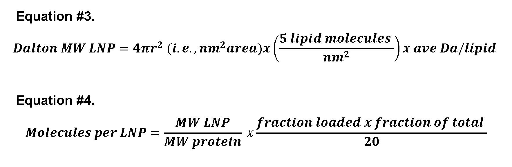
[0173] The relationship between the mole % of DMPG in the lipid mixture and both the % efficiency of packaging and the mole to mole ratios of DMPG to protein packaged was quantified. Independent lipid preparations were prepared with different mole % levels of DMPG. The % packaging efficiency was determined using Equation #1. The mole-to-mole ratios of DMPG lipid to mCherry protein in the subsequent LNP packaging experiments were calculated using the following equation.
Equation #5.

Comparing transfections with EP-LNPs to unencapsulated protein
[0174] A549 HER2- lung cancer cells (American Type Culture Collection, ATCC CRM-
CCL-185 (Manassas, VA) (Otis et al., "Dendrimer antibody conjugate to target and image HER-2 overexpressing cancer cells" Oncotarget 7, 36002-36013 (2016)) and SKBR3 (SK-BR- 3, ATCC HTB-30) HER2+ breast cancer cells were grown in RPMI-1640 media lacking phenol red dye (Sigma-Aldrich, Cat# R8755, St. Louis, MO, USA) supplemented with 10% fetal bovine serum at 37°C in a 5% CO2 atmosphere. A549 cells were grown in 24 well plates and treated with Venus-LNPs, mCher-LNPs, unpackaged Venus, and unpackaged mCherry delivering 10 pg/mL (1 : 100 w/v) overnight or 1 pg/mL (1 : 1000 w/v) for 2 hrs. Cells were then washed and photographed.
[0175] SKBR3 HER2+ breast cancer cells grown on home-made reusable glass chamber slides (Choudhury et al., "Dectin-3 -targeted antifungal liposomes efficiently bind and kill diverse fungal pathogens," Mol Microbiol 10.1111/mmi.15174 (2023)) in RPMI were used to confirm that rhodamine conjugated TRZ-Rhod antibody bound to cells. Cells were washed with PBS and fixed for 30 min with 4% fresh formalin in PBS, washed thrice with PBS, blocked with 0.5% BSAin PBS for 30 min, treated with TRZ-Rhod or lysed TRZ-Rhod-LNPs (1:100 w:v TRZ) in PBS for 30 min, and washed twice. The fluorescence of TRZ-Rhod labeled cells was imaged top down through a coverslip.
[0176] Three assays were performed on live SKBR3 cells to screen for the possibility that TRZ-LNPs showed improved anticancer cell activities relative to unpackaged TRZ antibody. (1) Cells grown in chamber slides in RPMI and then switched to Earl’s Balanced Salt Solution (EBSS) (Sigma-Aldrich E7510) minimal media containing TRZ-Rhod and TRZ-Rhod-LNP (1:200 w:v proteimmedia). Cells were incubated 3 hr, washed twice in minimal media, and photographed top down. (2) Cells grown in RPMI in 24 well plates were treated with TRZ- Rhod and TRZ-Rhod-LNP (1:200 w:v proteimmedia) and were incubated overnight, washed twice and photographed bottom up. (3) Cells plated at low density to 96 well plates were grown overnight in RPMI, and treated in replicate wells with TRZ-Rhod and TRZ-Rhod-LNPs with TRZ at 1 : 1,000 and 1 :200 w:v. After an additional 72 hr of growth, residual metabolic activity was compared to mock treated controls, using CellTiter Blue reagent (CTB, Promega; Cat.# G8080; Madison, WI, USA), using the manufacturer’s protocol. Cells were incubated for 60 min in the presence of reagent before fluorescence was assayed at Ex567/Em593. CTB
measures reductase activity that is dependent upon an intact electron transport chain in mitochondria in live cells. [0177] EP-LNP delivery into the fungus Rhizopus delemar strain 99-880 (formerly R. oryzae or R. arrhizus, ATCC MYA-4621) was tested, because it has a cell wall as an additional barrier compared to mammalian cells, and also because its cells are relatively large which would enhance analysis of fluorescent EP-LNP uptake (Meagher et al., "DectiSomes: C-Type Lectin Receptor-Targeted Liposomes as Pan-Antifungal Drugs Advanced Drug Delivery Reviews 196, 1-20 (2023)). Cells were stored as sporangiospores in di-water at 4°C and grown at 37°C in RPMI-1640 media lacking phenol red dye (Sigma-Aldrich, Cat# R8755) with the addition of 0.165M MOPS (3-(N-morpholino)propanesulfonic acid) (Sigma-Aldrich, Cat# M1254) adjusted to pH 7.0. Ninety-six-well microtiter plates were seeded with 1,000 sporangiospores in 100 μL of media per well. Cells were treated immediately with mCherry or mCherry-LNPs delivering protein at a 1:1,000 w/v ratio and incubated at 37°C. Phenotypes were observed after 9 hr of growth before extensive hyphal growth interfered with imaging. Because R. delemar does not efficiently adhere to microtiter plates and does adhere to micropipette tips, they were not washed free of reagents. The matrix of hyphal cells in small microtiter wells stabilized the cells for microscopy. Microscopy and photography [0178] Photographic images of fluorescent EP-LNPs captured on spin filters and their corresponding flow through were taken using the camera on an iPhone 13 in a dark room while illuminating at A302 with UV light (Entela, Upland, CA, UVM-57 handheld lamp, 95-0104- 01)). [0179] Human and fungal cells were photographed on an ECHO REVOLVE R4 microscope (VWR (Radnor, PA), #RVSF1000) using a 10X 0.3 N.A. or 20X 0.45 NA lens bottom up through plastic microtiter plates using combinations of visible light and epifluorescence (TxRED Ex560/Em630 or FITC Ex470/Em525) or using chamber slides top down through a 0.17 mm thick coverslip with a 20X 0.8 numerical aperture (NA) lens. To quantify fluorescent data, 10 images were taken for each treatment (N=10). The area of TxRED or FITC channel fluorescence was captured from raw image data was quantified using our recently developed AreaPipe subroutine within CellProfiler (v 4.2.5) (Choudhury et al., 2023). This program automates fluorescent image area data using ImageJ (v A5.3.a). A value of 5 pixels was assigned to those images wherein zero pixels were detected, so that fold differences and P values could be estimated.
Statistics and graphics [0180] The data from AreaPipe for estimating % efficiency of protein loading, the mole- to-mole ratio of DMPG to mCherry and the number of molecules of protein per LNP (Equations #1 through #5) were processed in Microsoft Excel (version 16.76) and in some cases moved into Graph Pad Prism Version 10.0.2 (LaJolla, CA), where Scatter bar plots and xy plots were prepared. P values were estimated using the Student’s T Test program, T.Test, in Excel. When data from one or both members of a comparison contained non-parametric data (i.e., not normally distributed), more conservative Mann Whitney PMW values were estimated in Prism. The proteins and lipids employed in the microfluidic assembly of EP-LNPs [0181] Microfluidic packaging of six proteins ranging from 28 kDa to 148 kDa with isoelectric points ranging from 4.5 to 8.7 (Table 1) was studied. Just prior to LNP packaging, proteins were diluted in one of two buffers: pH 5.5 A5.5 buffer or pH 7.0 PBS buffer. An exemplary lipid mixture (a.k.a., DMPG mix, Table 2) contained 50 mol % DMPG (Fig.1C), an anionic phospholipid with potential to sequester cationic proteins during the assembly of protein-lipid complexes (Fig.1A, 1B) along with other helper lipids. The DMPG lipid mixture was prepared in one of two solvents: #1 TCM:EtOH 1:2 v:v or #2 TFE:MeOH 4:1 v:v. Preparation of EP-LNPs [0182] Packaging was performed at protein: lipid w:w ratio of 1:20. In some experiments, mCherry was used because its red color enables visual monitoring of the protein’s distribution between the EP-LNP and unencapsulated protein in aqueous fractions and its fluorescence enables sensitive quantitative monitoring of EP-LNP packaging efficiencies. Based on the model described above, the mCherry isoform (pI 6.3) at acidic pH (e.g., 5.5) would be more cationic and should have a strong electrochemical interaction with anionic lipid DMPG and therefore package more efficiently as compared to packaging at pH 7.0. Immediately after packaging, the protein-lipid complexes were dialyzed into TAS2, a pH 7.5 sucrose stabilization buffer. This eliminated the organic solvent, making mCherry less cationic, and converted protein-lipid complexes into EP-LNPs (Fig. 1B). The mCherry EP-LNP preparation was concentrated and washed through a 300,000 MWCO spin filter (Fig.1). Using a pH of 5.5, a high percentage of the mCherry protein was captured in the EP-LNP fraction in the upper chamber and little protein was observed in the filtrate in the lower chamber based both on
visual examination and fluorescence imaging (Fig. 2A). Using Equation #1 and the fluorescence of the two fractions, % efficiency of packaging was estimated at 96.7% (Fig.2C). [0183] In subsequent experiments, the microfluidic packaging of mCherry dissolved in pH 5.5 and in pH 7.0 buffers was compared. It was found that packaging was highly efficient with efficiencies of packaging of 98.1% for pH 5.5 and 97.5% for pH 7.0 (Fig. 2C). The % of mCherry fluorescence recovered was estimated using Equation #2. Even though nearly all the fluorescent protein was packaged relative to fluorescent protein in the filtrate, only 20% and 71% of the protein fluorescence was recovered at pH 5.5 and 7.0, respectively. The dramatically reduced recovery of fluorescence at pH 5.5 relative to 7.0 is visually obvious (Fig. 2B). Replicates of this experiment gave similar results. The results raised two issues: (1) The packaging efficiency of mCherry at pH 7.0 was as high as that at pH 5.5 and indicating that acidic pH would improve packaging efficiency due to the amphoteric nature of the protein, if such a benefit existed. (2) It was unclear why little of the total starting fluorescence was recovered for the sample packaged at pH 5.5 as mCherry fluorescence should be stable at pH 5.5 (Doherty et al., "Stage-specific fluorescence intensity of GFP and mCherry during sporulation In Bacillus Subtilis," BMC Research Notes 3, 303. 10.1186/1756-0500-3-303 (2010)). [0184] To determine if the packaging process and high % efficiency was unique to mCherry, blue fluorescent EBFP2 (theoretical pI =6.6) and green fluorescent Venus (theoretical pI=6.2) were packaged in pH 5.5 and 7.0 buffers using the DMPG lipid mix. At pH 5.5, packaging efficiencies of EBFP2 and Venus proteins based on fluorescence were relatively high, 85% and 41%, respectively (Fig.3). At pH 7.0 the packaging efficiency of EBFP2 and Venus dropped markedly to 2.5% and 23%, respectively. This result supported the working hypothesis, suggesting that the amphoteric nature of these two proteins and their polycationic isoforms at acidic pH 5.5 favored efficient packaging of active proteins in EP- LNPs, relative to less efficient packaging when the proteins were more anionic during packaging at pH 7.0. However, when packaged at pH 5.5, only 4.5% and 2% of EBFP2 and Venus of their starting fluorescence was recovered in the EP-LNP and filtrate fractions, respectively. When packaged at pH 7.0, 40% and 98% of the starting fluorescence were recovered between the EP-LNP and filtrate fractions (Equation #2), respectively. The fluorescence of the EP-LNP fraction at pH 5.5 was not visible, only the pH 7.0 samples are shown in Fig.3A. Non-fluorescent control EP-LNP samples (right side, Fig.3A and 3B) are shown for comparison to reveal the blue background color of the spin columns under UV illumination.
[0185] BSA and goat IgG (Table 5) were then packaged at pH 5.5 and 7.0. BSA was included because it has a relatively low pI of 5.3. Therefore, it might not be efficiently packaged in the A5.5 buffer based on the model shown in FIG.1. The packaging efficiencies (Equation #1) for BSA and IgG were 94% and 95% at pH 5.5, and 82% and 67% at pH 7.0 (Table 5). Again, the lower packaging efficiency at pH 7.0 supports a model in which pH impacts protein packaging, although BSA was packaged more efficiently than expected at pH 7.0 based on its low pI. Using standard curves for the pure proteins, it was estimated that 60% to 98% of the proteins were recovered as EP-LNPs, respectively. Some flocculant material was observed in the BSA sample, presumably denatured protein, that was removed after dialysis and prior to spin filter concentration, which could explain the reduced recovery of BSA. [0186] The packaging of BSA (BSA-LNPs) and IgG (IgG-LNPs) into EP-LNPs at pH 5.5 and pH 7.0 as mediated by a DMPG lipid mixture in Solvent #1 is shown in Table 5. The protein content of LNPs and filtrate was assayed using the Lowry method. Percent efficiency of loading was estimated using Equation #1 and % total protein recovered by Equation #2. Both proteins were packaged more efficiently at pH 5.5 than at 7.0. [0187] Table 5. Packaging of BSA and IgG
 Size of EP-LNPs and protein loading capacity [0188] The nanometer diameter sizes and size distributions of the five EP-LNPs samples prepared at pH 7.0 were determined using a flow nanoparticle analyzer (Fig.4). The Mean diameters for mCherry-, EBFP2-, Venus-, BSA- and IgG-LNPs, ranged from 66.5 to 73.1 nm (Fig.4B) with an average of 69.4 nm. Each type of EP-LNPs showed a reasonably bimodal size distribution around their Mean value (Fig.4A). Fresh preparations of mCher-LNPs, IgG- LNPs and Empty-LNPs were analyzed by Transmission electron microscope (TEM). Representative TEM images of the phosphotungstic acid (PTA) stained LNPs are shown in FIG. 17. To make these measurements, each image was dragged into ImageJ. Then the following was selected: Analyze > Set Scale (3296 pixels for frame width and 1560 nm which
was estimated from the size bar in the image) > the line tool was dragged across each particle’s diameter > selected Measure to record the nm diameter of each particle in a file. The measurement data file was transferred to Excel to reformat the data and the nm diameter data was plotted in GraphPad Prism. A P value is given for the nm size comparison of IgG-LNPs with Empty LNPs. [0189] The particles appeared nearly spherical. Most of the LNPs appeared to be single particles with a homogeneous interior or a more darkly stained interior and less uncommonly, there was evidence of one LNP encapsulating another smaller one. Previous studies on microfluidic preparation of RNA-LNPs also suggested it produced a wide variety of particle morphologies 11. Mean particle diameters from TEM were estimated at 83.4, 77.9 and 113 nm, respectively. TEM analysis of an independent preparation of mCher-LNPs gave a diameter size estimate of 71 nm. LNPs were also examined on a Malvern Zetasizer (Model ZEN3600), which uses dynamic light scattering (DLS) to characterize particle populations. This instrument requires very high particle concentrations (1012/mL). The average particle sizes for mCher- LNPs and Venus-LNPs were estimated at approximately 200 to 450 nm, respectively. Replicate experiments with other EP-LNP preparations gave similar or even larger particle size estimates. It is likely these larger size estimates by DLS are due to the aggregation of LNPs into multimers, resulting from the orders of magnitude higher particle concentrations used for DLS relative to the other two methods (flow and electron microscopy). Therefore, the smaller size estimates from the flow instrument and TEM were used. The Zeta potential for the anionic LNPs was approximately -34 mV, as compared to +45 mV for typical cationic mRNA-LNPs 11. A negative Zeta potential value for the EP-LNPs was to be expected, considering they are composed of 50 mol % phospholipid, DMPG. [0190] Whether the 1:20 w:w ratio of protein to lipid approached the capacity for protein loading similar was tested. When the ratio for proteins was 2- or 3-fold higher (e.g., protein: lipid w:w ratios of 2:20 or 3:20), there was only a minimal improvement in the amount of protein loaded when loaded using the pH 7.0 buffer. A significant portion of the excess protein appeared to be denatured after dialysis and was spun down as a visible pellet (FIG.13). It is likely that protein not protected in the aqueous lumen of an EP-LNP was denatured via direct exposure to organic solvents. Therefore, protein:lipid ratios of about 1:20 (w: w) were used. Generalizing the essential role of anionic lipids in EP-LNP packaging [0191] The ability of two other anionic phospholipids (DHP and DMPGA) and two ionizable cationic amino phospholipids (SM102 and DSPC) to promote efficient loading of
protein into LNPs was examined. Their chemical structures are shown in FIGS.12A-D. It was found that both anionic lipids DHP and DMPGA were effective at packaging mCherry, while both cationic lipids SM102 and DSPC were not, based on visual examination of the spin filters (Fig.5A). This was further verified by quantitative fluorescence assays (Fig.5B). The cationic SM102 and DSPC lipid mixes package little if any mCherry, with more than 99% of the fluorescence in the filtrate. By contrast, DHP and DMPGA anionic lipid mixtures had mCherry packaging efficiencies of 97.7% and 28.2%, respectively. In short, it appears anionic lipids promote active protein loading into EP-LNPs. Based on these results, ionizable cationic amino phospholipids do not promote loading. Transient transfection of cancer and fungal cells with fluorescent EP-LNPs [0192] Whether EP-LNPs could efficiently penetrate cells was investigated. Live human A549 lung cancer cells were incubated 16 hr with Venus-LNPs, mCherry-LNPs, and the corresponding unpackaged proteins at 1:1,000 w:v protein:growth medium ratios. The cells were viewed live by epifluorescence microscopy (Fig.6). Nearly all cells exposed to Venus- LNPs or mCher-LNPs showed significant green fluorescence or red fluorescence (Fig.6A, 6C), respectively. Fine granular fluorescence was distributed throughout the cells, indicating that the EP-LNPs passed efficiently through their plasma membrane. Only a few of the cells treated with the equivalent concentrations of unpackaged Venus or mCherry showed detectable fluorescence(Fig.6B, 6D). Quantification based on fluorescence revealed that uptake of Venus- LNPs and mCher-LNPs were 23-fold (P=1.8x10⁻⁵) and 147-fold P=4.9x10⁻⁹) greater than for the corresponding unpackaged protein (Fig.6G). Clearly, packaging these proteins in LNPs improved their delivery into cells. Even after only a 2 hr exposure to these reagents, cells took up significant amounts of the two LNPs (FIGS.14A-F) with the uptake of Venus-LNPs and mCher-LNPs 10-fold (P=1.66x10⁻⁶) and 8-fold P=1.13x10⁻⁵) higher than that for the unpackaged proteins. [0193] Whether the dramatically improved delivery of EP-LNPs over unpackaged protein extended to cells with a cell wall was tested. The fungal pathogen R. delemar was chosen, as its relatively large cell size facilitates image analysis. Sporangiospores of R. delemar were mixed with mCher-LNPs and mCherry at the same low protein concentration (1:1000 w/v) in rich media. After 9 hr of growth, R. delemar hyphae took up significant amounts of the red fluorescent mCher-LNPs, while the unpackaged mCherry treated sample showed little or no detectable fluorescence (Fig.6E & 6F). The cells treated with mCher-LNPs were 1,170-fold (PMW=0.0001) more fluorescent than those treated with unpackaged mCherry (Fig. 6G).
Because these live cell samples were not washed before being photographed, it is possible that the unpackaged mCherry protein was degraded by fungal proteases secreted into the media. In a parallel experiment, sporangiospores were similarly treated with mCher-LNPs and germinated for only 5 hr and again viewed without washing (Fig.6H). These germling-stage cells were also highly fluorescent and showed little background from residual fluorescent mCher-LNPs remaining in the media. Thus, these EP-LNPs were rapidly taken up by fungal cells. Trastuzumab in TRZ-LNPs [0194] Next, the anti-HER2 IgG1 monoclonal antibody, Trastuzumab (TRZ) and goat IgG were used to examine the potential of EP-LNPs to deliver therapeutic antibodies into target cells and for the efficient delivery of TRZ to alter cancer cell phenotype(s). TRZ binds to and inactivates the membrane receptor HER2. In addition to its roles in promoting cell proliferation, migration, and invasion, HER2 overexpression also promotes breast cancer tumorigenesis by suppressing autophagy. Blocking HER2’s interaction with Beclin 1, an activator of autophagy, dramatically reduces tumorigenesis (Vega-Rubín-de-Celis et al., "Increased autophagy blocks HER2-mediated breast tumorigenesis," Proc Natl Acad Sci U S A 115, 4176-4181 (2018)). It was not known if extracellular signaling by HER2 or intercellular signaling after HER2 is endocytosed blocks autophagy. Starvation rapidly induces an autophagy phenotype in most eukaryotic cells, but HER2 overexpression blocks SKBR3 breast cancer cells from this starvation response. Both RNAi knockdown of HER2 expression and an autophagy-inducing peptide restore some level of starvation-induced autophagy to various HER2+ cells including SKBR3 cells (Vega-Rubín-de-Celis et al., 2018). Therefore, blocking HER2 activity, TRZ and/or TRZ-LNPs could restore autophagy. [0195] Red fluorescent rhodamine B was conjugated to TRZ and IgG. TRZ-Rhod bound strongly to the plasma membrane of fixed HER2+ SKBR3 breast cancer cells, as expected. TRZ-Rhod-LNPs were prepared as described above with proteins dissolved in pH 5.5 acetate buffer and lipids dissolved in Solvent #1 (TCM: MeOH 3:1). To examine if packaging damaged the activity of TRZ-Rhod, the TRZ-Rhod-LNPs were lysed and rhodamine fluorescence and immunological activity were assayed. The packaged TRZ-Rhod retained its fluorescence and was soluble, but it no longer bound to SKBR3 cells. [0196] As chloroform in the TCM: EtOH Solvent #1 mix was used to dissolve the lipids, it is possible that the TCM component, an effective protein denaturant, may have altered the conformation of the antibody’s variable region(s) necessary for HER2 binding. The TCM:
EtOH (Solvent #1) was replaced as a lipid solvent during packaging. Lipids were dissolved in Solvent #2 (TFE:MeOH 4:1). TRZ-Rhod and IgG-Rhod in pH 5.5 buffer were packaged into EP-LNPs using Solvent #2 (Fig.7). The packaging efficiency (Equation #1) was 76% and 51%, respectively, and 98% and 95% of the total fluorescence was recovered in the combined LNP and filtrate fractions (FIGS.15A-B). After lysis of the TRZ-Rhod-LNPs, the antibody binding activity of TRZ-Rhod toward fixed SKBR3 cells was indistinguishable from that of the starting TRZ-Rhod protein (Fig.7B, labeling with TRZ at 1:200 w/v), which was further confirmed by quantification of the fluorescence of these cells (Fig.7C, P=0.76). To determine if packaging of TRZ in a EP-LNP increased penetration through the mammalian cell membrane, the HER2- lung cancer cell line A549 was employed, because A549 cells are HER2- and thus will not have the complication of TRZ-Rhod binding to HER2 surface protein. This allowed EP-LNP uptake assessment without worrying about turnover of membrane bound by TRZ-Rhod. First, to confirm that A549 cells were truly HER2-, fixed cells were stained with TRZ-Rhod. As expected, no TRZ-Rhod cell binding was observed. Next, A549 cells were treated overnight with TRZ-Rhod protein and TRZ-Rhod-LNP delivering 1:200 w/v protein to the media. TRZ- Rhod-LNPs were taken up by 100% of the cells, while only traces of TRZ-Rhod were seen and only in a few cells per microscopic field (Fig.8A). Quantification of red fluorescence indicated that cellular uptake of TRZ-Rhod-LNP was 1,100-fold (P=9.6x10⁻¹¹) greater than for TRZ- Rhod (Fig.8B). Further, evidence that the EP-LNPs were taken into the cytoplasm of cells and were not located on the cell surface was obtain by taking 60X magnification through-focus stack of images of live A549 cells incubated for 2 hr and 24 hr with TRZ-Rhod-LNPs. Cells were co-stained with Hoechst to identify nuclei. Weak bright field exposures were included to identify the leading edges (Le) of the cells. One optical slice out of each stack of 7 slices from the 2 hr and 24 treatments is shown in FIG.5C & D, respectively. An adjacent image of the Hoechst-stained nuclei (Nu) is shown on the right to define the number of cells in the image and help identify cell boundaries. For the 2 hr treatment the 5th slice from the bottom (second down from the top) was selected, because it contained the maximum number of in-focus TRZ- Rhod-LNPs (Fig.5C). For the 24 hr treatment the 2nd slice from the bottom (6th down from the top) was selected, because it was one of the slices with the maximum number of in-focus TRZ-Rhod-LNPs (Fig.5D). In the 2 hr sample, the vast majority of red fluorescent TRZ-Rhod- LNPs were in observed in the cytoplasm, some near the Le, but most were located further within the cells and co-distributed with cellular vesicles (ve) seen as white spots and nuclei in the same focal plane. The LNPs appeared to be excluded from nuclei. Clearly, the EP-LNPs were taken up rapidly. The 24 hr sample gives more robust evidence of the cytoplasmic
distribution of LNPs. These cells contained at least an order of magnitude more red fluorescent liposomes and the exposure times in the red fluorescent channel had to be reduced more than 10-fold. TRZ-Rhod-LNPs are rarely seen near the Le, even though the Le represents the largest surface area of the cells. It is worth noting that the in-focus LNPs appear to have moved from the second slice down from the top at 2 hr to the 5th slice down from the top by 24 hr, following their progression into the cell interior. All the optical slices in the two stacks are shown together FIGS 16A-16B. Scanning through these slices, from the bottom slice adjacent to the glass slide to the top slice at the top of the cells, visually demonstrates that essentially all of the in-focus TRZ-Rhod-LNPs are in the same planes as in-focus vesicles and nuclei, and rarely located near the Le. [0197] Three different experiments were used to examine possible biological activities of TRZ-Rhod-LNPs in HER2+ cells (Fig.9). Experiment #1 relies upon the fact that most cultured mammalian cells produce a multi-vesicle autophagy-related starvation phenotype within hours of being transferred from rich media to minimal salts media (Vega-Rubín-de-Celis et al., 2018). However, HER2 overexpression, as occurring in HER2+ cancer cells like SKBR3 cells, inhibits starvation-induced autophagy (Vega-Rubín-de-Celis et al., 2018). In other words, HER2+ cells do not develop a significant autophagosome phenotype when switched to growth on minimal media. Therefore, it was proposed that TRZ-Rhod and/or TRZ-Rhod-LNPs would restore starvation induced autophagy to HER2+ cells by reducing HER2 activity. SKBR3 cells were transferred to a minimal salt media containing TRZ-Rhod or TRZ-Rhod-LNPs (1:200 w:v TRZ:media), incubated for 3 hr, washed twice, and photographed live. As expected, TRZ-Rhod bound primarily to the cell surface. However, only a few to several of the TRZ-Rhod treated cells in each image showed a discernable multi-vesicle phenotype (Fig.9A, left). By contrast, nearly every TRZ-Rhod-LNP treated cell showed a strong distinct muti-vesicle phenotype (Fig. 9A, right). The percent of cells per microscopic field that shown a discernable number of autophagosome like vesicles were scored for 10 random images of each treatment. Ten-fold more of the TRZ-Rhod-LNP treated cells showed the multi-vesicle phenotype than the TRZ- Rhod treated cells (Fig.9B, P=6.6x10⁻²⁰). It was not known if the HER2-dependent block of autophagy in HER2+ cells originated from HER2 activity in the plasma membrane or intracellular activities. These results suggest that intracellular HER2 activity in HER2 overexpressing cells may be critical for the suppression of the multi-vesicle autophagosome phenotype in response to starvation. [0198] In experiment #2, the effect of overnight treatment of SKBR3 cells growing in normal rich growth media with TRZ-Rhod and TRZ-Rhod-LNPs was studied. Goat IgG-Rhod
and goat IgG-Rhod-LNPs were included as controls. TRZ-Rhod entered cells (Fig.9C), likely due to endocytosis of antibody-HER2 complexes from the plasma membrane into endosomes (Rudkouskaya et al., 2020). In comparison, only extremely weak intracellular fluorescence was detected for the control IgG-Rhod (Fig.9C). TRZ-Rhod-LNPs stained the cells even stronger than TRZ-Rhod (Fig.9C-D, 1.6-fold, P=0.02). Further, it appears that a significantly greater amount of TRZ-Rhod from the EP-LNPs was re-distributed in intercellular compartment than that from the un-encapsulated protein. Every cell examined showed some staining by both TRZ reagents. IgG-Rhod-LNPs were also taken up by every cell and unpackaged protein by only a few cells (Fig.9C bottom and 9D), consistent with the above results in A549 cells. The staining pattern by TRZ-Rhod, TRZ-Rhod-LNPs, and IgG-Rhod-LNP is granular (Fig.9C), unlike the multi-vesicle phenotype of starved cells (Fig.9A). [0199] To understand Experiment #3, it is important to recognize that TRZ’s therapeutic anti-tumor activities are dependent upon cellular components of the immune system (Nuti et al., "Immune effects of trastuzumab," J Cancer 2, 317-23 (2011)), which are not present during the treatment of in vitro grown HER2+ cells. TRZ has only limited ability to inhibit growth or induce cell death of HER2+ cells in vitro, with dose-dependent inhibition of metabolic activity leveling off at 25% in most reports and with no confirmed IC50 for inhibition or cell death ever having been reached (Barok et al., "Trastuzumab-DM1 causes tumour growth inhibition by mitotic catastrophe in trastuzumab-resistant breast cancer cells in vivo," Breast Cancer Res 13, R46 (2011)). SKBR3 cells were incubated with unpackaged TRZ-Rhod and TRZ-Rhod-LNPs delivering TRZ at 1:1,000 w:v and 1:200 w:v for 72 hr. Then, cells were assayed for metabolic activity with CTB reagent. Although there was a significant amount of residual TRZ-Rhod fluorescence in every cell with unpackaged TRZ-Rhod (Fig.9E), there were 1.7 fold higher fluorescence in the TRZ-Rhod-LNP sample (Fig.9F, P=3.0x10⁻⁵). Relative to mock treated control cells, both TRZ-Rhod and TRZ-Rhod-LNPs delivered at 1:200 w/v reduced the live cell metabolic activity of SKBR3 cells by 25 to 30% (P=0.0023 to 0.0006) (Fig.9G), consistent with previous studies. But no significant difference was observed between EP-LNP and un- encapsulated TRZ-Rhod. Biological replicates of the three TRZ experiments in SKBR3 cells gave similar results. Further characterization using DMPG, Solvent #2, and a protein to lipid ratio of about 1:20 [0200] Having shown that the DMPG mix in Solvent #2 and that TRZ at pH 5.5 preserved the immunological activity of TRZ, this packaging method was tested for EBFP2, Venus, and mCherry. A significant amount of all three fluorescent proteins were retained in the EP-LNPs
fractions relative to the filtrates (Fig.10). Their packaging efficiencies were 32%, 27%, and 79%, respectively (Fig. 10B top, Equation #1). More importantly, the level of recovery of fluorescence in the combined LNP and filtrate was 60%, 98%, and 98%, respectively (Equation #2). Thus, for all three proteins, packaging using Solvent #2 improved recovery of total fluorescence (4 to 61-fold more) than Solvent #1 (Fig. 10B, bottom). The improved preservation of protein fluorescence (or activity) is likely due to TFE in Solvent #2 being less denaturing relative to the TCM component of Solvent #1. [0201] In order to validate the importance of anionic lipids in preparing EP-LNPs, the ability of two other anionic phospholipids, DHP and DMPGA, and the ionizable cationic amino phospholipids SM102 and DSPC, both zwitterions, to promote efficient loading of protein into LNPs was analyzed. The 50 mol % DMPG in DMPG mix was replaced by the same mol % of these alternate lipids. The lipid mixes rich in both anionic lipids DHP and DMPGA were effective at packaging mCherry, while both SM102 and DSPC, were ineffective. The lipid mixtures dominated by SM102 and DSPC packaged little if any mCherry (<1% packing efficiency). By contrast, lipid mixtures dominated by anionic lipids DHP and DMPGA had mCherry packaging efficiencies of 97.7% and 28.2%, respectively. Based on these results, ionizable cationic amino phospholipids do not promote protein loading. However, anionic phospholipids promote active protein loading into EP-LNPs. [0202] The mol % of DMPG in the lipid mix that was necessary for efficient protein packaging of mCherry was estimated. Solvent #2 contained 50 mol % DMPG. Lipid mixtures in which the mol % of DMPG was reduced from 50% down to 37.5, 25%,10%, 5% and 0%, while the % of the ionizable amino phospholipid DSPC was increased correspondingly were tested. Packaging was again performed with the standard protein:lipid w:w ratio of about 1:20. As the mol % of the DMPG component dropped, the amount of mCherry encapsulated visually dropped, with the most noticeable decrease occurring when the DMPG concentration fell below 25 mol % (Fig.11). Relative to the lipid mix with 50 mol % DMPG, the % efficiency of LNP packaging dropped from 96%, to 94%, 80%, 57%, 29%, and 0.59% respectively (Fig.11B). The mole:mole ratios of DMPG to mCherry were estimated in each packaging experiment using Equation #5. Close to 100% efficiency of protein loading was achieved with 37.5 and 50 mol % DMPG (top right of Fig. 11C), where the mole:mole DMPG:mCherry ratios are approximately 37.5:1 and 50:1. When the mole:mole ratio of DMPG to mCherry was as low as 5:1 (bottom left of the graph, DMPG at 5 mol 5 in the mix, 5 mol % DMPG), packaging efficiency was still 29%. Thus, even when only 5 molecules of DMPG were associated with one mCherry polypeptide, a significant amount of protein packaging occurred. These results
indicate that only several anionic lipid molecules need to be associated with each protein polypeptide for efficient packaging. Long term stability of EP-LNPs [0203] Long term stability is desired for any practical use of EP-LNPs. The retention of proteins in Venus-LNPs, mCher-LNPs, and TRZ-Rhod-LNPs was determined the day after they had been prepared at pH 5.5 in Solvent #2 and again after these preparations had been stored at 4°C in TAS2 buffer for 3 months (Table 6) by again centrifuging through a 300,000 MWCO spin filter. It was found that 83 to 98% of each sample’s total fluorescence was retained in the LNP fraction (Equation #1) and that the samples had retained 60% to 98% of their total original fluorescence (Equation #2). Thus, EP-LNPs prepared by this method are stable when stored at 4°C. [0204] Preparing EP-LNPs that were stable for an extended period was a goal that is essential for most practical applications. The retention of proteins in preparations of Venus-LNPs, mCher-LNPs, and TRZ-Rhod-LNPs was measured the day after they had been prepared at pH 5.5 in Solvent #2. After these preparations had been stored at 4°C in TAS2 buffer for 3 months they were centrifuged again through a 300,000 MWCO spin filter and fluorescence in the LNP and filtrate fractions were again examined. It was found that 83 to 98% of each sample’s total fluorescence was retained in the LNP fraction (e.g., not lost to the surrounding media due to lysis of the LNP) and that the samples had retained 60% to 98% of their total original fluorescence. Thus, EP-LNPs prepared by this method were stable when stored at about 4°C. [0205] Table 6. Stability of EP-LNPs -Retention of fluorescent protein after 3 months storage.
Size of EP-LNPs and protein loading capacity [0188] The nanometer diameter sizes and size distributions of the five EP-LNPs samples prepared at pH 7.0 were determined using a flow nanoparticle analyzer (Fig.4). The Mean diameters for mCherry-, EBFP2-, Venus-, BSA- and IgG-LNPs, ranged from 66.5 to 73.1 nm (Fig.4B) with an average of 69.4 nm. Each type of EP-LNPs showed a reasonably bimodal size distribution around their Mean value (Fig.4A). Fresh preparations of mCher-LNPs, IgG- LNPs and Empty-LNPs were analyzed by Transmission electron microscope (TEM). Representative TEM images of the phosphotungstic acid (PTA) stained LNPs are shown in FIG. 17. To make these measurements, each image was dragged into ImageJ. Then the following was selected: Analyze > Set Scale (3296 pixels for frame width and 1560 nm which
was estimated from the size bar in the image) > the line tool was dragged across each particle’s diameter > selected Measure to record the nm diameter of each particle in a file. The measurement data file was transferred to Excel to reformat the data and the nm diameter data was plotted in GraphPad Prism. A P value is given for the nm size comparison of IgG-LNPs with Empty LNPs. [0189] The particles appeared nearly spherical. Most of the LNPs appeared to be single particles with a homogeneous interior or a more darkly stained interior and less uncommonly, there was evidence of one LNP encapsulating another smaller one. Previous studies on microfluidic preparation of RNA-LNPs also suggested it produced a wide variety of particle morphologies 11. Mean particle diameters from TEM were estimated at 83.4, 77.9 and 113 nm, respectively. TEM analysis of an independent preparation of mCher-LNPs gave a diameter size estimate of 71 nm. LNPs were also examined on a Malvern Zetasizer (Model ZEN3600), which uses dynamic light scattering (DLS) to characterize particle populations. This instrument requires very high particle concentrations (1012/mL). The average particle sizes for mCher- LNPs and Venus-LNPs were estimated at approximately 200 to 450 nm, respectively. Replicate experiments with other EP-LNP preparations gave similar or even larger particle size estimates. It is likely these larger size estimates by DLS are due to the aggregation of LNPs into multimers, resulting from the orders of magnitude higher particle concentrations used for DLS relative to the other two methods (flow and electron microscopy). Therefore, the smaller size estimates from the flow instrument and TEM were used. The Zeta potential for the anionic LNPs was approximately -34 mV, as compared to +45 mV for typical cationic mRNA-LNPs 11. A negative Zeta potential value for the EP-LNPs was to be expected, considering they are composed of 50 mol % phospholipid, DMPG. [0190] Whether the 1:20 w:w ratio of protein to lipid approached the capacity for protein loading similar was tested. When the ratio for proteins was 2- or 3-fold higher (e.g., protein: lipid w:w ratios of 2:20 or 3:20), there was only a minimal improvement in the amount of protein loaded when loaded using the pH 7.0 buffer. A significant portion of the excess protein appeared to be denatured after dialysis and was spun down as a visible pellet (FIG.13). It is likely that protein not protected in the aqueous lumen of an EP-LNP was denatured via direct exposure to organic solvents. Therefore, protein:lipid ratios of about 1:20 (w: w) were used. Generalizing the essential role of anionic lipids in EP-LNP packaging [0191] The ability of two other anionic phospholipids (DHP and DMPGA) and two ionizable cationic amino phospholipids (SM102 and DSPC) to promote efficient loading of
protein into LNPs was examined. Their chemical structures are shown in FIGS.12A-D. It was found that both anionic lipids DHP and DMPGA were effective at packaging mCherry, while both cationic lipids SM102 and DSPC were not, based on visual examination of the spin filters (Fig.5A). This was further verified by quantitative fluorescence assays (Fig.5B). The cationic SM102 and DSPC lipid mixes package little if any mCherry, with more than 99% of the fluorescence in the filtrate. By contrast, DHP and DMPGA anionic lipid mixtures had mCherry packaging efficiencies of 97.7% and 28.2%, respectively. In short, it appears anionic lipids promote active protein loading into EP-LNPs. Based on these results, ionizable cationic amino phospholipids do not promote loading. Transient transfection of cancer and fungal cells with fluorescent EP-LNPs [0192] Whether EP-LNPs could efficiently penetrate cells was investigated. Live human A549 lung cancer cells were incubated 16 hr with Venus-LNPs, mCherry-LNPs, and the corresponding unpackaged proteins at 1:1,000 w:v protein:growth medium ratios. The cells were viewed live by epifluorescence microscopy (Fig.6). Nearly all cells exposed to Venus- LNPs or mCher-LNPs showed significant green fluorescence or red fluorescence (Fig.6A, 6C), respectively. Fine granular fluorescence was distributed throughout the cells, indicating that the EP-LNPs passed efficiently through their plasma membrane. Only a few of the cells treated with the equivalent concentrations of unpackaged Venus or mCherry showed detectable fluorescence(Fig.6B, 6D). Quantification based on fluorescence revealed that uptake of Venus- LNPs and mCher-LNPs were 23-fold (P=1.8x10⁻⁵) and 147-fold P=4.9x10⁻⁹) greater than for the corresponding unpackaged protein (Fig.6G). Clearly, packaging these proteins in LNPs improved their delivery into cells. Even after only a 2 hr exposure to these reagents, cells took up significant amounts of the two LNPs (FIGS.14A-F) with the uptake of Venus-LNPs and mCher-LNPs 10-fold (P=1.66x10⁻⁶) and 8-fold P=1.13x10⁻⁵) higher than that for the unpackaged proteins. [0193] Whether the dramatically improved delivery of EP-LNPs over unpackaged protein extended to cells with a cell wall was tested. The fungal pathogen R. delemar was chosen, as its relatively large cell size facilitates image analysis. Sporangiospores of R. delemar were mixed with mCher-LNPs and mCherry at the same low protein concentration (1:1000 w/v) in rich media. After 9 hr of growth, R. delemar hyphae took up significant amounts of the red fluorescent mCher-LNPs, while the unpackaged mCherry treated sample showed little or no detectable fluorescence (Fig.6E & 6F). The cells treated with mCher-LNPs were 1,170-fold (PMW=0.0001) more fluorescent than those treated with unpackaged mCherry (Fig. 6G).
Because these live cell samples were not washed before being photographed, it is possible that the unpackaged mCherry protein was degraded by fungal proteases secreted into the media. In a parallel experiment, sporangiospores were similarly treated with mCher-LNPs and germinated for only 5 hr and again viewed without washing (Fig.6H). These germling-stage cells were also highly fluorescent and showed little background from residual fluorescent mCher-LNPs remaining in the media. Thus, these EP-LNPs were rapidly taken up by fungal cells. Trastuzumab in TRZ-LNPs [0194] Next, the anti-HER2 IgG1 monoclonal antibody, Trastuzumab (TRZ) and goat IgG were used to examine the potential of EP-LNPs to deliver therapeutic antibodies into target cells and for the efficient delivery of TRZ to alter cancer cell phenotype(s). TRZ binds to and inactivates the membrane receptor HER2. In addition to its roles in promoting cell proliferation, migration, and invasion, HER2 overexpression also promotes breast cancer tumorigenesis by suppressing autophagy. Blocking HER2’s interaction with Beclin 1, an activator of autophagy, dramatically reduces tumorigenesis (Vega-Rubín-de-Celis et al., "Increased autophagy blocks HER2-mediated breast tumorigenesis," Proc Natl Acad Sci U S A 115, 4176-4181 (2018)). It was not known if extracellular signaling by HER2 or intercellular signaling after HER2 is endocytosed blocks autophagy. Starvation rapidly induces an autophagy phenotype in most eukaryotic cells, but HER2 overexpression blocks SKBR3 breast cancer cells from this starvation response. Both RNAi knockdown of HER2 expression and an autophagy-inducing peptide restore some level of starvation-induced autophagy to various HER2+ cells including SKBR3 cells (Vega-Rubín-de-Celis et al., 2018). Therefore, blocking HER2 activity, TRZ and/or TRZ-LNPs could restore autophagy. [0195] Red fluorescent rhodamine B was conjugated to TRZ and IgG. TRZ-Rhod bound strongly to the plasma membrane of fixed HER2+ SKBR3 breast cancer cells, as expected. TRZ-Rhod-LNPs were prepared as described above with proteins dissolved in pH 5.5 acetate buffer and lipids dissolved in Solvent #1 (TCM: MeOH 3:1). To examine if packaging damaged the activity of TRZ-Rhod, the TRZ-Rhod-LNPs were lysed and rhodamine fluorescence and immunological activity were assayed. The packaged TRZ-Rhod retained its fluorescence and was soluble, but it no longer bound to SKBR3 cells. [0196] As chloroform in the TCM: EtOH Solvent #1 mix was used to dissolve the lipids, it is possible that the TCM component, an effective protein denaturant, may have altered the conformation of the antibody’s variable region(s) necessary for HER2 binding. The TCM:
EtOH (Solvent #1) was replaced as a lipid solvent during packaging. Lipids were dissolved in Solvent #2 (TFE:MeOH 4:1). TRZ-Rhod and IgG-Rhod in pH 5.5 buffer were packaged into EP-LNPs using Solvent #2 (Fig.7). The packaging efficiency (Equation #1) was 76% and 51%, respectively, and 98% and 95% of the total fluorescence was recovered in the combined LNP and filtrate fractions (FIGS.15A-B). After lysis of the TRZ-Rhod-LNPs, the antibody binding activity of TRZ-Rhod toward fixed SKBR3 cells was indistinguishable from that of the starting TRZ-Rhod protein (Fig.7B, labeling with TRZ at 1:200 w/v), which was further confirmed by quantification of the fluorescence of these cells (Fig.7C, P=0.76). To determine if packaging of TRZ in a EP-LNP increased penetration through the mammalian cell membrane, the HER2- lung cancer cell line A549 was employed, because A549 cells are HER2- and thus will not have the complication of TRZ-Rhod binding to HER2 surface protein. This allowed EP-LNP uptake assessment without worrying about turnover of membrane bound by TRZ-Rhod. First, to confirm that A549 cells were truly HER2-, fixed cells were stained with TRZ-Rhod. As expected, no TRZ-Rhod cell binding was observed. Next, A549 cells were treated overnight with TRZ-Rhod protein and TRZ-Rhod-LNP delivering 1:200 w/v protein to the media. TRZ- Rhod-LNPs were taken up by 100% of the cells, while only traces of TRZ-Rhod were seen and only in a few cells per microscopic field (Fig.8A). Quantification of red fluorescence indicated that cellular uptake of TRZ-Rhod-LNP was 1,100-fold (P=9.6x10⁻¹¹) greater than for TRZ- Rhod (Fig.8B). Further, evidence that the EP-LNPs were taken into the cytoplasm of cells and were not located on the cell surface was obtain by taking 60X magnification through-focus stack of images of live A549 cells incubated for 2 hr and 24 hr with TRZ-Rhod-LNPs. Cells were co-stained with Hoechst to identify nuclei. Weak bright field exposures were included to identify the leading edges (Le) of the cells. One optical slice out of each stack of 7 slices from the 2 hr and 24 treatments is shown in FIG.5C & D, respectively. An adjacent image of the Hoechst-stained nuclei (Nu) is shown on the right to define the number of cells in the image and help identify cell boundaries. For the 2 hr treatment the 5th slice from the bottom (second down from the top) was selected, because it contained the maximum number of in-focus TRZ- Rhod-LNPs (Fig.5C). For the 24 hr treatment the 2nd slice from the bottom (6th down from the top) was selected, because it was one of the slices with the maximum number of in-focus TRZ-Rhod-LNPs (Fig.5D). In the 2 hr sample, the vast majority of red fluorescent TRZ-Rhod- LNPs were in observed in the cytoplasm, some near the Le, but most were located further within the cells and co-distributed with cellular vesicles (ve) seen as white spots and nuclei in the same focal plane. The LNPs appeared to be excluded from nuclei. Clearly, the EP-LNPs were taken up rapidly. The 24 hr sample gives more robust evidence of the cytoplasmic
distribution of LNPs. These cells contained at least an order of magnitude more red fluorescent liposomes and the exposure times in the red fluorescent channel had to be reduced more than 10-fold. TRZ-Rhod-LNPs are rarely seen near the Le, even though the Le represents the largest surface area of the cells. It is worth noting that the in-focus LNPs appear to have moved from the second slice down from the top at 2 hr to the 5th slice down from the top by 24 hr, following their progression into the cell interior. All the optical slices in the two stacks are shown together FIGS 16A-16B. Scanning through these slices, from the bottom slice adjacent to the glass slide to the top slice at the top of the cells, visually demonstrates that essentially all of the in-focus TRZ-Rhod-LNPs are in the same planes as in-focus vesicles and nuclei, and rarely located near the Le. [0197] Three different experiments were used to examine possible biological activities of TRZ-Rhod-LNPs in HER2+ cells (Fig.9). Experiment #1 relies upon the fact that most cultured mammalian cells produce a multi-vesicle autophagy-related starvation phenotype within hours of being transferred from rich media to minimal salts media (Vega-Rubín-de-Celis et al., 2018). However, HER2 overexpression, as occurring in HER2+ cancer cells like SKBR3 cells, inhibits starvation-induced autophagy (Vega-Rubín-de-Celis et al., 2018). In other words, HER2+ cells do not develop a significant autophagosome phenotype when switched to growth on minimal media. Therefore, it was proposed that TRZ-Rhod and/or TRZ-Rhod-LNPs would restore starvation induced autophagy to HER2+ cells by reducing HER2 activity. SKBR3 cells were transferred to a minimal salt media containing TRZ-Rhod or TRZ-Rhod-LNPs (1:200 w:v TRZ:media), incubated for 3 hr, washed twice, and photographed live. As expected, TRZ-Rhod bound primarily to the cell surface. However, only a few to several of the TRZ-Rhod treated cells in each image showed a discernable multi-vesicle phenotype (Fig.9A, left). By contrast, nearly every TRZ-Rhod-LNP treated cell showed a strong distinct muti-vesicle phenotype (Fig. 9A, right). The percent of cells per microscopic field that shown a discernable number of autophagosome like vesicles were scored for 10 random images of each treatment. Ten-fold more of the TRZ-Rhod-LNP treated cells showed the multi-vesicle phenotype than the TRZ- Rhod treated cells (Fig.9B, P=6.6x10⁻²⁰). It was not known if the HER2-dependent block of autophagy in HER2+ cells originated from HER2 activity in the plasma membrane or intracellular activities. These results suggest that intracellular HER2 activity in HER2 overexpressing cells may be critical for the suppression of the multi-vesicle autophagosome phenotype in response to starvation. [0198] In experiment #2, the effect of overnight treatment of SKBR3 cells growing in normal rich growth media with TRZ-Rhod and TRZ-Rhod-LNPs was studied. Goat IgG-Rhod
and goat IgG-Rhod-LNPs were included as controls. TRZ-Rhod entered cells (Fig.9C), likely due to endocytosis of antibody-HER2 complexes from the plasma membrane into endosomes (Rudkouskaya et al., 2020). In comparison, only extremely weak intracellular fluorescence was detected for the control IgG-Rhod (Fig.9C). TRZ-Rhod-LNPs stained the cells even stronger than TRZ-Rhod (Fig.9C-D, 1.6-fold, P=0.02). Further, it appears that a significantly greater amount of TRZ-Rhod from the EP-LNPs was re-distributed in intercellular compartment than that from the un-encapsulated protein. Every cell examined showed some staining by both TRZ reagents. IgG-Rhod-LNPs were also taken up by every cell and unpackaged protein by only a few cells (Fig.9C bottom and 9D), consistent with the above results in A549 cells. The staining pattern by TRZ-Rhod, TRZ-Rhod-LNPs, and IgG-Rhod-LNP is granular (Fig.9C), unlike the multi-vesicle phenotype of starved cells (Fig.9A). [0199] To understand Experiment #3, it is important to recognize that TRZ’s therapeutic anti-tumor activities are dependent upon cellular components of the immune system (Nuti et al., "Immune effects of trastuzumab," J Cancer 2, 317-23 (2011)), which are not present during the treatment of in vitro grown HER2+ cells. TRZ has only limited ability to inhibit growth or induce cell death of HER2+ cells in vitro, with dose-dependent inhibition of metabolic activity leveling off at 25% in most reports and with no confirmed IC50 for inhibition or cell death ever having been reached (Barok et al., "Trastuzumab-DM1 causes tumour growth inhibition by mitotic catastrophe in trastuzumab-resistant breast cancer cells in vivo," Breast Cancer Res 13, R46 (2011)). SKBR3 cells were incubated with unpackaged TRZ-Rhod and TRZ-Rhod-LNPs delivering TRZ at 1:1,000 w:v and 1:200 w:v for 72 hr. Then, cells were assayed for metabolic activity with CTB reagent. Although there was a significant amount of residual TRZ-Rhod fluorescence in every cell with unpackaged TRZ-Rhod (Fig.9E), there were 1.7 fold higher fluorescence in the TRZ-Rhod-LNP sample (Fig.9F, P=3.0x10⁻⁵). Relative to mock treated control cells, both TRZ-Rhod and TRZ-Rhod-LNPs delivered at 1:200 w/v reduced the live cell metabolic activity of SKBR3 cells by 25 to 30% (P=0.0023 to 0.0006) (Fig.9G), consistent with previous studies. But no significant difference was observed between EP-LNP and un- encapsulated TRZ-Rhod. Biological replicates of the three TRZ experiments in SKBR3 cells gave similar results. Further characterization using DMPG, Solvent #2, and a protein to lipid ratio of about 1:20 [0200] Having shown that the DMPG mix in Solvent #2 and that TRZ at pH 5.5 preserved the immunological activity of TRZ, this packaging method was tested for EBFP2, Venus, and mCherry. A significant amount of all three fluorescent proteins were retained in the EP-LNPs
fractions relative to the filtrates (Fig.10). Their packaging efficiencies were 32%, 27%, and 79%, respectively (Fig. 10B top, Equation #1). More importantly, the level of recovery of fluorescence in the combined LNP and filtrate was 60%, 98%, and 98%, respectively (Equation #2). Thus, for all three proteins, packaging using Solvent #2 improved recovery of total fluorescence (4 to 61-fold more) than Solvent #1 (Fig. 10B, bottom). The improved preservation of protein fluorescence (or activity) is likely due to TFE in Solvent #2 being less denaturing relative to the TCM component of Solvent #1. [0201] In order to validate the importance of anionic lipids in preparing EP-LNPs, the ability of two other anionic phospholipids, DHP and DMPGA, and the ionizable cationic amino phospholipids SM102 and DSPC, both zwitterions, to promote efficient loading of protein into LNPs was analyzed. The 50 mol % DMPG in DMPG mix was replaced by the same mol % of these alternate lipids. The lipid mixes rich in both anionic lipids DHP and DMPGA were effective at packaging mCherry, while both SM102 and DSPC, were ineffective. The lipid mixtures dominated by SM102 and DSPC packaged little if any mCherry (<1% packing efficiency). By contrast, lipid mixtures dominated by anionic lipids DHP and DMPGA had mCherry packaging efficiencies of 97.7% and 28.2%, respectively. Based on these results, ionizable cationic amino phospholipids do not promote protein loading. However, anionic phospholipids promote active protein loading into EP-LNPs. [0202] The mol % of DMPG in the lipid mix that was necessary for efficient protein packaging of mCherry was estimated. Solvent #2 contained 50 mol % DMPG. Lipid mixtures in which the mol % of DMPG was reduced from 50% down to 37.5, 25%,10%, 5% and 0%, while the % of the ionizable amino phospholipid DSPC was increased correspondingly were tested. Packaging was again performed with the standard protein:lipid w:w ratio of about 1:20. As the mol % of the DMPG component dropped, the amount of mCherry encapsulated visually dropped, with the most noticeable decrease occurring when the DMPG concentration fell below 25 mol % (Fig.11). Relative to the lipid mix with 50 mol % DMPG, the % efficiency of LNP packaging dropped from 96%, to 94%, 80%, 57%, 29%, and 0.59% respectively (Fig.11B). The mole:mole ratios of DMPG to mCherry were estimated in each packaging experiment using Equation #5. Close to 100% efficiency of protein loading was achieved with 37.5 and 50 mol % DMPG (top right of Fig. 11C), where the mole:mole DMPG:mCherry ratios are approximately 37.5:1 and 50:1. When the mole:mole ratio of DMPG to mCherry was as low as 5:1 (bottom left of the graph, DMPG at 5 mol 5 in the mix, 5 mol % DMPG), packaging efficiency was still 29%. Thus, even when only 5 molecules of DMPG were associated with one mCherry polypeptide, a significant amount of protein packaging occurred. These results
indicate that only several anionic lipid molecules need to be associated with each protein polypeptide for efficient packaging. Long term stability of EP-LNPs [0203] Long term stability is desired for any practical use of EP-LNPs. The retention of proteins in Venus-LNPs, mCher-LNPs, and TRZ-Rhod-LNPs was determined the day after they had been prepared at pH 5.5 in Solvent #2 and again after these preparations had been stored at 4°C in TAS2 buffer for 3 months (Table 6) by again centrifuging through a 300,000 MWCO spin filter. It was found that 83 to 98% of each sample’s total fluorescence was retained in the LNP fraction (Equation #1) and that the samples had retained 60% to 98% of their total original fluorescence (Equation #2). Thus, EP-LNPs prepared by this method are stable when stored at 4°C. [0204] Preparing EP-LNPs that were stable for an extended period was a goal that is essential for most practical applications. The retention of proteins in preparations of Venus-LNPs, mCher-LNPs, and TRZ-Rhod-LNPs was measured the day after they had been prepared at pH 5.5 in Solvent #2. After these preparations had been stored at 4°C in TAS2 buffer for 3 months they were centrifuged again through a 300,000 MWCO spin filter and fluorescence in the LNP and filtrate fractions were again examined. It was found that 83 to 98% of each sample’s total fluorescence was retained in the LNP fraction (e.g., not lost to the surrounding media due to lysis of the LNP) and that the samples had retained 60% to 98% of their total original fluorescence. Thus, EP-LNPs prepared by this method were stable when stored at about 4°C. [0205] Table 6. Stability of EP-LNPs -Retention of fluorescent protein after 3 months storage.
 Protein payload [0206] Protein payload capacity was estimated as numbers of protein molecules per individual EP-LNP for four example proteins varying five-fold in their MW using Equation #3 and Equation #4. Preparations prepared in Solvent #2 had encapsulated approximately 31 molecules of mCherry, 18 molecules of BSA, and 7 molecules each of IgG and TRZ per EP- LNP. Conclusion [0207] Methods for the efficient microfluidic packaging of active protein into the lumen of stable EP-LNPs were developed, thus providing, for the first time, LNPs and populations of LNPS that can encapsulate proteins for therapeutic and research applications. In some methods, proteins dissolved in a pH 5.5 buffer and lipid mixtures rich in an anionic lipid dissolved in Solvent #2, TFE:MeOH 4:1 allowed packaging of six proteins with diverse MWs and isoelectric proteins, Even low percentages of the anionic lipid DMPG in the lipid mix promoted significantly efficient protein loading. Further, the EP-LNPs were stable and retained the majority of the encapsulated protein in the lumen of the EP-LNPs after a few months of storage (Table 6). [0208] It was also shown that EP-LNPs were taken up efficiently by two human cancer cell lines. EP-LNPs were also taken up efficiently by R. delemar fungal cells, which was surprising considering Mucor species have a thick cell wall composed of diverse and crosslinked polysaccharides. By contrast, the unpackaged proteins were taken up order(s) of magnitude less efficiently by human and fungal cells alike. Uptake of the unpackaged proteins was barely detectable, except in the case of TRZ, which binds to the plasma membrane HER2 receptor of HER2+ SKBR3 cancer cells. [0209] The dramatic success of the experiments described herein suggests that microfluidics employing anionic lipid has distinct advantages for preparing EP-LNPs and represent a significant advance over previous efforts to package proteins in LNPs. [0210] The experiments described herein show that different proteins ranging over five- fold in their MWs and varying widely in their pI values could be loaded into LNPs at efficiencies approaching 90%. Considering the variations in the charged and hydrophobic amino acids composing the surface of different proteins, wide variation in packaging efficiencies among different proteins was expected. However, all proteins tested were efficiently packaged. Therefore, efficient luminal packaging of medically relevant active proteins inside EP-LNPs should enable a wide range of new therapeutic options.
[0211] To summarize, microfluidic encapsulation of several proteins at acidic and/or neutral pH, using a lipid mixture comprising an exemplary anionic phospholipid, i.e., DMPG 14:0 PG (1,2-dimyristoyl-sn-glycero-3-phospho-(1'-rac-glycerol) dissolved in two organic solvent mixtures, was achieved. The packaged proteins ranged over 5-fold in their molecular weights (MW) and two-fold in their isoelectric points (i.e., pI values). The proteins were effectively packaged using methods comprising protein loading at pH 5.5 and 7.0. Further, all of the proteins were efficiently loaded into stable EP-LNPs and retained their activities. Sequences [0212] mCherry protein (isoform of UFQ89828.1) G residue added at the end to aid in PacI cloning.259 a.a. MW 28,922. Prot Param estimated A280 OD of 1.189 for 1.0 mg/mL. pI 6.32. [0213] MAHHHHHHVGTGSGKGKGSGSGMVSKGEEDNMAIIKEFMRFKVHMEGS VNGHEFEIEGEGEGRPYEGTQTAKLKVTKGGPLPFAWDILSPQFMYGSKAYVKHPAD IPDYLKLSFPEGFKWERVMNFEDGGVVTVTQDSSLQDGEFIYKVKLRGTNFPSDGPV MQKKTMGWEASSERMYPEDGALKGEIKQRLKLKDGGHYDAEVKTTYKAKKPVQL PGAYNVNIKLDITSHNEDYTIVEQYERAEGRHSTGGMDELYKG (SEQ ID NO: 1) [0214] mCherry DNA, Codon optimized Cherry DNA sequence from GenScript with a GGT codon added at the end for compatibility with PacI cloning site. KpnI and PacI underlined. Start codon in green and stop in red. [0215] GGTACCATGGCTCACCATCACCACCACCATGTAGGAACGGGTAGCGGTA AAGGCAAGGGTTCTGGATCCGGTATGGTTAGCAAAGGTGAGGAAGACAACATGG CAATTATTAAGGAGTTCATGCGTTTTAAAGTGCACATGGAAGGCTCCGTGAACGGT CACGAATTCGAAATCGAAGGTGAGGGCGAGGGCCGTCCGTACGAGGGTACGCAA ACCGCGAAATTGAAGGTGACCAAAGGTGGCCCACTGCCTTTTGCGTGGGATATCC TGTCGCCGCAGTTTATGTATGGTAGCAAAGCATACGTTAAGCACCCGGCGGACATC CCGGATTACCTGAAGTTGAGTTTTCCGGAAGGCTTCAAATGGGAGCGCGTGATGA ATTTCGAGGACGGCGGTGTCGTGACTGTAACCCAAGACAGCTCCCTGCAGGATGG CGAGTTCATCTACAAGGTGAAATTACGTGGTACGAATTTCCCGAGCGATGGTCCGG TTATGCAAAAAAAGACCATGGGTTGGGAAGCGAGCTCTGAACGCATGTACCCGGA AGACGGGGCTCTGAAGGGCGAGATCAAGCAGCGTCTGAAACTCAAGGACGGCGG TCATTATGATGCTGAGGTTAAAACCACCTATAAAGCCAAAAAGCCGGTCCAGCTGC CGGGTGCCTACAATGTTAACATTAAGCTGGATATTACCAGCCATAACGAGGACTAC ACCATTGTTGAACAGTATGAGAGAGCGGAGGGCCGTCATAGCACCGGCGGCATGG ATGAATTGTATAAGGTTAATTAA (SEQ ID NO: 2) [0216] EBFP2 protein (isoform of UFQ89826.1) G residue added at the end to aid in PacI cloning.263 a.a.
[0217] MAHHHHHHVGTGSGKGKGSGSGMRKGEELFTGVVPILVELDGDVNGHK FSVRGEGEGDATNGKLTLKFICTTGKLPVPWPTLVTTLTYGVQCFARYPDHMKQHDF FKSAMPEGYVQERTISFKDDGTYKTRAEVKFEGDTLVNRIELKGIDFKEDGNILGHKL EYNFNSHNVYITADKQKNGIKANFKIRHNVEDGSVQLADHYQQNTPIGDGPVLLPDN HYLSTQSVLSKDPNEKRDHMVLLEFVTAAGITHGMDELYKRPG (SEQ ID NO: 3) [0218] EBFP2 DNA. Codon optimized EBFP2 DNA sequence from GenScript with a GGT codon added at the end for compatibility with PacI KpnI cloning site stated as flanking sequece. [0219] GGTACCATGGCTCACCACCATCACCACCATGTAGGAACGGGCAGTGGC AAGGGCAAGGGTTCTGGTTCTGGTATGCGCAAAGGCGAGGAATTGTTTACCGGTG TTGTTCCGATTCTGGTCGAGCTGGATGGCGATGTCAACGGTCACAAATTCAGCGTT CGTGGTGAGGGCGAGGGCGACGCTACCAATGGCAAGCTGACCCTTAAGTTCATCT GCACCACCGGTAAGCTGCCAGTTCCGTGGCCTACACTCGTGACGACCCTCACGTA CGGCGTTCAATGTTTTGCCCGTTATCCGGACCACATGAAACAGCATGATTTCTTCA AAAGCGCAATGCCGGAAGGCTACGTGCAAGAACGCACCATTAGCTTTAAGGACG ACGGAACCTACAAGACCCGTGCGGAAGTTAAATTCGAGGGCGACACGTTGGTGA ATCGTATTGAATTGAAGGGTATCGATTTTAAGGAGGACGGAAACATTTTAGGTCAC AAGCTGGAGTATAACTTCAACAGCCATAACGTCTATATCACCGCTGATAAACAAAA GAACGGCATCAAAGCGAATTTCAAAATCCGCCACAACGTGGAGGACGGCTCCGT GCAGCTGGCAGATCACTACCAGCAGAATACCCCGATTGGTGACGGTCCGGTGCTG CTGCCGGACAACCATTACCTGAGCACCCAGAGCGTACTGTCCAAAGATCCGAATG AAAAAAGAGATCACATGGTGCTGTTGGAATTTGTTACTGCGGCGGGTATCACCCAT GGTATGGATGAACTGTATAAACGTCCGGGTTAATTAA (SEQ ID NO: 4) [0220] VENUS protein (Isoform of AKA95335.1. G residue added at the end to aid in PacI cloning. [0221] MAHHHHHHVGTGSGKGKGSGSGVSKGEELFTGVVPILVELDGDVNGHKF SVSGEGEGDATYGKLTLKLICTTGKLPVPWPTLVTTLGYGLQCFARYPDHMKQHDFF KSAMPEGYVQERTIFFKDDGNYKTRAEVKFEGDTLVNRIELKGIDFKEDGNILGHKL EYNYNSHNVYITADKQKNGIKANFKIRHNIEDGGVQLADHYQQNTPIGDGPVLLPDN HYLSYQSALSKDPNEKRDHMVLLEFVTAAGITLGMDESRG (SEQ ID NO: 5) [0222] Venus DNA Codon optimized sequence 780 bp. Added the final Gly codon GGT, wherein the T accommodates the first T of a PacI sequence (T TTA TTA A making an in-frame stop codon TTA. [0223] GGTACCATGGCTCACCACCATCACCACCATGTAGGAACTGGTTCCGGCA AGGGTAAGGGCTCTGGTTCTGGCGTCTCCAAAGGCGAGGAACTGTTTACCGGCGT GGTGCCGATTCTGGTTGAACTGGATGGCGACGTCAACGGTCACAAGTTCAGCGTT AGCGGTGAGGGCGAGGGCGACGCGACCTATGGTAAACTTACCCTGAAACTGATTT GCACCACCGGTAAGCTGCCAGTTCCGTGGCCTACGTTGGTAACTACGTTGGGCTA
CGGCCTGCAGTGTTTCGCCCGTTATCCGGACCACATGAAACAGCATGATTTCTTTA AGTCCGCTATGCCGGAAGGTTACGTTCAAGAGCGCACCATTTTTTTCAAGGATGAT GGTAACTACAAAACCCGTGCGGAAGTGAAATTCGAGGGTGACACCTTGGTGAAC CGTATTGAGCTCAAGGGCATCGACTTCAAAGAAGACGGCAACATTCTGGGTCATA AGCTTGAGTATAACTACAACAGCCATAATGTTTATATCACGGCGGATAAACAAAAA AACGGCATCAAAGCTAATTTTAAGATCCGTCATAACATCGAGGACGGTGGTGTGCA GCTGGCAGATCACTACCAGCAGAATACCCCGATTGGTGACGGTCCGGTTTTGTTAC CGGATAATCACTACCTGAGCTATCAAAGCGCGCTGTCGAAGGACCCGAATGAAAA ACGCGACCACATGGTGCTGTTGGAATTTGTTACCGCGGCAGGTATCACCCTGGGTA TGGATGAAAGCAGAGGTTAATTAA (SEQ ID NO: 6)
Protein payload [0206] Protein payload capacity was estimated as numbers of protein molecules per individual EP-LNP for four example proteins varying five-fold in their MW using Equation #3 and Equation #4. Preparations prepared in Solvent #2 had encapsulated approximately 31 molecules of mCherry, 18 molecules of BSA, and 7 molecules each of IgG and TRZ per EP- LNP. Conclusion [0207] Methods for the efficient microfluidic packaging of active protein into the lumen of stable EP-LNPs were developed, thus providing, for the first time, LNPs and populations of LNPS that can encapsulate proteins for therapeutic and research applications. In some methods, proteins dissolved in a pH 5.5 buffer and lipid mixtures rich in an anionic lipid dissolved in Solvent #2, TFE:MeOH 4:1 allowed packaging of six proteins with diverse MWs and isoelectric proteins, Even low percentages of the anionic lipid DMPG in the lipid mix promoted significantly efficient protein loading. Further, the EP-LNPs were stable and retained the majority of the encapsulated protein in the lumen of the EP-LNPs after a few months of storage (Table 6). [0208] It was also shown that EP-LNPs were taken up efficiently by two human cancer cell lines. EP-LNPs were also taken up efficiently by R. delemar fungal cells, which was surprising considering Mucor species have a thick cell wall composed of diverse and crosslinked polysaccharides. By contrast, the unpackaged proteins were taken up order(s) of magnitude less efficiently by human and fungal cells alike. Uptake of the unpackaged proteins was barely detectable, except in the case of TRZ, which binds to the plasma membrane HER2 receptor of HER2+ SKBR3 cancer cells. [0209] The dramatic success of the experiments described herein suggests that microfluidics employing anionic lipid has distinct advantages for preparing EP-LNPs and represent a significant advance over previous efforts to package proteins in LNPs. [0210] The experiments described herein show that different proteins ranging over five- fold in their MWs and varying widely in their pI values could be loaded into LNPs at efficiencies approaching 90%. Considering the variations in the charged and hydrophobic amino acids composing the surface of different proteins, wide variation in packaging efficiencies among different proteins was expected. However, all proteins tested were efficiently packaged. Therefore, efficient luminal packaging of medically relevant active proteins inside EP-LNPs should enable a wide range of new therapeutic options.
[0211] To summarize, microfluidic encapsulation of several proteins at acidic and/or neutral pH, using a lipid mixture comprising an exemplary anionic phospholipid, i.e., DMPG 14:0 PG (1,2-dimyristoyl-sn-glycero-3-phospho-(1'-rac-glycerol) dissolved in two organic solvent mixtures, was achieved. The packaged proteins ranged over 5-fold in their molecular weights (MW) and two-fold in their isoelectric points (i.e., pI values). The proteins were effectively packaged using methods comprising protein loading at pH 5.5 and 7.0. Further, all of the proteins were efficiently loaded into stable EP-LNPs and retained their activities. Sequences [0212] mCherry protein (isoform of UFQ89828.1) G residue added at the end to aid in PacI cloning.259 a.a. MW 28,922. Prot Param estimated A280 OD of 1.189 for 1.0 mg/mL. pI 6.32. [0213] MAHHHHHHVGTGSGKGKGSGSGMVSKGEEDNMAIIKEFMRFKVHMEGS VNGHEFEIEGEGEGRPYEGTQTAKLKVTKGGPLPFAWDILSPQFMYGSKAYVKHPAD IPDYLKLSFPEGFKWERVMNFEDGGVVTVTQDSSLQDGEFIYKVKLRGTNFPSDGPV MQKKTMGWEASSERMYPEDGALKGEIKQRLKLKDGGHYDAEVKTTYKAKKPVQL PGAYNVNIKLDITSHNEDYTIVEQYERAEGRHSTGGMDELYKG (SEQ ID NO: 1) [0214] mCherry DNA, Codon optimized Cherry DNA sequence from GenScript with a GGT codon added at the end for compatibility with PacI cloning site. KpnI and PacI underlined. Start codon in green and stop in red. [0215] GGTACCATGGCTCACCATCACCACCACCATGTAGGAACGGGTAGCGGTA AAGGCAAGGGTTCTGGATCCGGTATGGTTAGCAAAGGTGAGGAAGACAACATGG CAATTATTAAGGAGTTCATGCGTTTTAAAGTGCACATGGAAGGCTCCGTGAACGGT CACGAATTCGAAATCGAAGGTGAGGGCGAGGGCCGTCCGTACGAGGGTACGCAA ACCGCGAAATTGAAGGTGACCAAAGGTGGCCCACTGCCTTTTGCGTGGGATATCC TGTCGCCGCAGTTTATGTATGGTAGCAAAGCATACGTTAAGCACCCGGCGGACATC CCGGATTACCTGAAGTTGAGTTTTCCGGAAGGCTTCAAATGGGAGCGCGTGATGA ATTTCGAGGACGGCGGTGTCGTGACTGTAACCCAAGACAGCTCCCTGCAGGATGG CGAGTTCATCTACAAGGTGAAATTACGTGGTACGAATTTCCCGAGCGATGGTCCGG TTATGCAAAAAAAGACCATGGGTTGGGAAGCGAGCTCTGAACGCATGTACCCGGA AGACGGGGCTCTGAAGGGCGAGATCAAGCAGCGTCTGAAACTCAAGGACGGCGG TCATTATGATGCTGAGGTTAAAACCACCTATAAAGCCAAAAAGCCGGTCCAGCTGC CGGGTGCCTACAATGTTAACATTAAGCTGGATATTACCAGCCATAACGAGGACTAC ACCATTGTTGAACAGTATGAGAGAGCGGAGGGCCGTCATAGCACCGGCGGCATGG ATGAATTGTATAAGGTTAATTAA (SEQ ID NO: 2) [0216] EBFP2 protein (isoform of UFQ89826.1) G residue added at the end to aid in PacI cloning.263 a.a.
[0217] MAHHHHHHVGTGSGKGKGSGSGMRKGEELFTGVVPILVELDGDVNGHK FSVRGEGEGDATNGKLTLKFICTTGKLPVPWPTLVTTLTYGVQCFARYPDHMKQHDF FKSAMPEGYVQERTISFKDDGTYKTRAEVKFEGDTLVNRIELKGIDFKEDGNILGHKL EYNFNSHNVYITADKQKNGIKANFKIRHNVEDGSVQLADHYQQNTPIGDGPVLLPDN HYLSTQSVLSKDPNEKRDHMVLLEFVTAAGITHGMDELYKRPG (SEQ ID NO: 3) [0218] EBFP2 DNA. Codon optimized EBFP2 DNA sequence from GenScript with a GGT codon added at the end for compatibility with PacI KpnI cloning site stated as flanking sequece. [0219] GGTACCATGGCTCACCACCATCACCACCATGTAGGAACGGGCAGTGGC AAGGGCAAGGGTTCTGGTTCTGGTATGCGCAAAGGCGAGGAATTGTTTACCGGTG TTGTTCCGATTCTGGTCGAGCTGGATGGCGATGTCAACGGTCACAAATTCAGCGTT CGTGGTGAGGGCGAGGGCGACGCTACCAATGGCAAGCTGACCCTTAAGTTCATCT GCACCACCGGTAAGCTGCCAGTTCCGTGGCCTACACTCGTGACGACCCTCACGTA CGGCGTTCAATGTTTTGCCCGTTATCCGGACCACATGAAACAGCATGATTTCTTCA AAAGCGCAATGCCGGAAGGCTACGTGCAAGAACGCACCATTAGCTTTAAGGACG ACGGAACCTACAAGACCCGTGCGGAAGTTAAATTCGAGGGCGACACGTTGGTGA ATCGTATTGAATTGAAGGGTATCGATTTTAAGGAGGACGGAAACATTTTAGGTCAC AAGCTGGAGTATAACTTCAACAGCCATAACGTCTATATCACCGCTGATAAACAAAA GAACGGCATCAAAGCGAATTTCAAAATCCGCCACAACGTGGAGGACGGCTCCGT GCAGCTGGCAGATCACTACCAGCAGAATACCCCGATTGGTGACGGTCCGGTGCTG CTGCCGGACAACCATTACCTGAGCACCCAGAGCGTACTGTCCAAAGATCCGAATG AAAAAAGAGATCACATGGTGCTGTTGGAATTTGTTACTGCGGCGGGTATCACCCAT GGTATGGATGAACTGTATAAACGTCCGGGTTAATTAA (SEQ ID NO: 4) [0220] VENUS protein (Isoform of AKA95335.1. G residue added at the end to aid in PacI cloning. [0221] MAHHHHHHVGTGSGKGKGSGSGVSKGEELFTGVVPILVELDGDVNGHKF SVSGEGEGDATYGKLTLKLICTTGKLPVPWPTLVTTLGYGLQCFARYPDHMKQHDFF KSAMPEGYVQERTIFFKDDGNYKTRAEVKFEGDTLVNRIELKGIDFKEDGNILGHKL EYNYNSHNVYITADKQKNGIKANFKIRHNIEDGGVQLADHYQQNTPIGDGPVLLPDN HYLSYQSALSKDPNEKRDHMVLLEFVTAAGITLGMDESRG (SEQ ID NO: 5) [0222] Venus DNA Codon optimized sequence 780 bp. Added the final Gly codon GGT, wherein the T accommodates the first T of a PacI sequence (T TTA TTA A making an in-frame stop codon TTA. [0223] GGTACCATGGCTCACCACCATCACCACCATGTAGGAACTGGTTCCGGCA AGGGTAAGGGCTCTGGTTCTGGCGTCTCCAAAGGCGAGGAACTGTTTACCGGCGT GGTGCCGATTCTGGTTGAACTGGATGGCGACGTCAACGGTCACAAGTTCAGCGTT AGCGGTGAGGGCGAGGGCGACGCGACCTATGGTAAACTTACCCTGAAACTGATTT GCACCACCGGTAAGCTGCCAGTTCCGTGGCCTACGTTGGTAACTACGTTGGGCTA
CGGCCTGCAGTGTTTCGCCCGTTATCCGGACCACATGAAACAGCATGATTTCTTTA AGTCCGCTATGCCGGAAGGTTACGTTCAAGAGCGCACCATTTTTTTCAAGGATGAT GGTAACTACAAAACCCGTGCGGAAGTGAAATTCGAGGGTGACACCTTGGTGAAC CGTATTGAGCTCAAGGGCATCGACTTCAAAGAAGACGGCAACATTCTGGGTCATA AGCTTGAGTATAACTACAACAGCCATAATGTTTATATCACGGCGGATAAACAAAAA AACGGCATCAAAGCTAATTTTAAGATCCGTCATAACATCGAGGACGGTGGTGTGCA GCTGGCAGATCACTACCAGCAGAATACCCCGATTGGTGACGGTCCGGTTTTGTTAC CGGATAATCACTACCTGAGCTATCAAAGCGCGCTGTCGAAGGACCCGAATGAAAA ACGCGACCACATGGTGCTGTTGGAATTTGTTACCGCGGCAGGTATCACCCTGGGTA TGGATGAAAGCAGAGGTTAATTAA (SEQ ID NO: 6)
Claims
What is claimed is: 1. A lipid nanoparticle comprising an anionic lipid component, wherein the anionic lipid component comprises between about 10% (w/w) and about 70% (w/w) of the total lipid of the nanoparticle, wherein the lipid nanoparticle comprises a hydrophilic polypeptide that is encapsulated in the lumen of the lipid nanoparticle, and wherein the lipid nanoparticle is stable for at least one week at about 2 °C to about 8 °C.
2. The lipid nanoparticle of claim 1, wherein the anionic lipid component comprises an anionic phospholipid.
3. The lipid nanoparticle of claim 1 or 2, wherein the anionic lipid component comprises a non-phosphate containing lipid.
4. The lipid nanoparticle of any one of claims 1-3, wherein the anionic lipid component comprises one or more anionic lipids.
5. The lipid nanoparticle of any one of claim 4, wherein the one or more anionic lipids are selected from the group consisting of but not limited to DMPG (1,2-Dimyristoyl-sn- glycero-3-phospho-rac-(1-glycerol)), DMPGA (1,2-Dimyristoyl-sn-glycero-3- phosphate), DHP (dihexadecyl phosphate), diether PG, 1,2-dilauroly-sn-glycero-3 phosphate, 1,2 dipalmitoyl-sn-glycero-3-PA, palmitoyl-2-oleoyl-sn-glycero-3-PA, phosphatidic acid, lysophosphatidic acid, Dipalmitoyl phosphatidylinositol, phosphatidylinositol phosphates, 9A1P9.
6. The lipid nanoparticle of claim 5, wherein the one or more anionic lipids comprise a fatty acid.
7. The lipid nanoparticle of claim 6, wherein the fatty acid is selected from the group consisting of stearic acid, oleic acid, octanoic acid, nananoic acid, decanoic aicd, lauric acid, palmitic acid, 8,11-eicosdiynooic acid, undecanoic acid, docosanoic acid, hexadecanedioic acid, heptadecanoic acid, arachidic acid, pentacosanoic acid, hexacosanoic acid, octacosnoic acid, triacontanoic acid, tetradecanoic acid, lignoceic acid, tridecanoic acid, nonadecanoic acid, pentadecanoic acid, tricosanoic acid,
distearin, didecanoylglycerol, dioctaoylglycerol, diethylene glycerol monostearate, and trilaurin.
8. The lipid nanoparticle of any one of claims 1-7, wherein the anionic lipid component comprises between about 10% (w/w) and about 60% (w/w) of the total lipid of the nanoparticle.
9. The lipid nanoparticle of any one of claims 1-8, wherein the anionic lipid component comprises between about 40% (w/w) and about 60% (w/w) of the total lipid of the nanoparticle.
10. The lipid nanoparticle of any one of claims 1-9, wherein the anionic lipid component comprises between about 45% (w/w) and about 60% (w/w) of the total lipid of the nanoparticle.
11. The lipid nanoparticle of any one of claims 1-10, wherein the anionic lipid component comprises between about 45% (w/w) and about 55% (w/w) of the total lipid of the nanoparticle.
12. The lipid nanoparticle of any one of claims 1-11 wherein the anionic lipid component comprises between about 45% (w/w) and about 50% (w/w) of the total lipid of the nanoparticle.
13. The lipid nanoparticle of any one of claims 1-12, wherein the anionic lipid component comprises between about 48% (w/w) and about 52% (w/w) of the total lipid of the nanoparticle.
14. The lipid nanoparticle of any one of claims 1-13, wherein the anionic lipid component comprises between about 10% (w/w) and about 50% (w/w) of the total lipid of the nanoparticle.
15. The lipid nanoparticle of any one of claims 1-14, wherein the anionic lipid component comprises between about 10% (w/w) and about 40% (w/w) of the total lipid of the nanoparticle.
16. The lipid nanoparticle of any one of claims 1-15, wherein the anionic lipid component comprises between about 10% (w/w) and about 30% (w/w) of the total lipid of the nanoparticle.
17. The lipid nanoparticle of any one of claims 1-16, wherein the anionic lipid component comprises between about 10% (w/w) and about 20% (w/w) of the total lipid of the nanoparticle.
18. The lipid nanoparticle of any one of claims 1-17, wherein the lipid nanoparticle further comprises a cationic lipid component.
19. The lipid nanoparticle of claim 18, wherein the cationic lipid component is at least between about 5% (w/w) and 75% (w/w) of, at least between 5% (w/w) and 50% (w/w), or at least between 5% (w/w) and 25% (w/w) the total lipid in the nanoparticle.
20. The lipid nanoparticle of any one of claims 1-19, wherein the lipid nanoparticle further comprises a pegylated lipid component.
21. The lipid nanoparticle of claim 20 wherein the pegylated lipid component is at least about 25% (w/w), 20% (w/w), 15% (w/w), 10% (w/w), 5% (w/w), 2% (w/w), 1% (w/w), or 0.5% (w/w)of the total lipid of the nanoparticle.
22. The lipid nanoparticle of any one of claims 1-21, wherein the lipid nanoparticle comprises about 25% (w/w) to about 50% (w/w) anionic lipid, about 5% (w/w) to about 15% (w/w) cationic lipid, about 35% (w/w) to about 45% (w/w) cholesterol, and about 1% to about 5% (w/w) pegylated lipid.
23. The lipid nanoparticle of any one of claims 1-22, wherein the lipid nanoparticle comprises about 40% (w/w) to about 50% (w/w) anionic lipid, about 5% (w/w) to about 10% (w/w) cationic lipid, about 35% (w/w) to about 45% (w/w) cholesterol, and about 1% (w/w) to about 2% (w/w) pegylated lipid.
24. The lipid nanoparticle of any one of claims 1-23, wherein the lipid nanoparticle comprises about 50% (w/w) anionic lipid, about 10% (w/w) cationic lipid, about 38.5% cholesterol (w/w), and about 1.5% (w/w) pegylated lipid.
25. The lipid nanoparticle of any one of claims 1-24, wherein the lipid nanoparticle further comprises a multi-tailed lipid.
26. The lipid nanoparticle of claim 24, wherein the multitailed lipid is about 5% (w/w) to about 35% (w/w) of the total lipid nanoparticle composition.
27. The lipid nanoparticle of any one of claims 25-27, wherein the multitailed lipid is SM102.
28. The lipid nanoparticle of any one of claims 22-27, wherein the anionic lipid comprises DMPG, wherein the cationic lipid comprises 1,2-distearoyl-sn-glycero-3- phosphocholine (DSPC), and wherein the pegylated lipid comprises [3-[2(2- methoxyethoxy) ethoxyl]-2-tetradecanoyloxypropyl tetradecanoate (DMG)- polyethylene glycol (PEG)-2000.
29. The lipid nanoparticle of any of claims 1-28, wherein the hydrophilic polypeptide to total lipid ratio (w/w) is between about 1:10 and about 1:100.
30. The lipid nanoparticle of any of claims 1-29, wherein the hydrophilic polypeptide to total lipid ratio (w/w) is between about 1:10 and about 1:75.
31. The lipid nanoparticle of any of claims 1-30, wherein the hydrophilic polypeptide to total lipid ratio (w/w) is between about 1:10 and about 1:50.
32. The lipid nanoparticle of any of claims 1-31, wherein the hydrophilic polypeptide to total lipid ratio (w/w) is between about 1:10 and about 1:40.
33. The lipid nanoparticle of any of claims 1-32, wherein the hydrophilic polypeptide to total lipid ratio (w/w) is between about 1:10 and about 1:30.
34. The lipid nanoparticle of any of claims 1-33, wherein the hydrophilic polypeptide to total lipid ratio (w/w) is between about 1:10 and about 1:25.
35. The lipid nanoparticle of any of claims 1-34, wherein the hydrophilic polypeptide to total lipid ratio (w/w) is between about 1:10 and about 1:20.
36. A population of lipid nanoparticles comprising the lipid nanoparticle of any one of claims 1-35.
37. A composition comprising: a. the population of claim 36; and b. a pharmaceutically acceptable carrier.
38. The composition of claim 37, wherein the pharmaceutically acceptable carrier is a buffer having a pH of about 7.0 to about 7.5.
39. The composition of any one of claims 37-38, wherein at least about 50%, 60%, 70%, 80%, or 90% of the hydrophilic polypeptide remains encapsulated in the lumen of the lipid nanoparticles for at least one week at about 2 °C to about 8 °C .
40. The composition of any one of claims 37-39, wherein at least about 50%, 60%, 70%, 80%, or 90% of the hydrophilic polypeptide remains encapsulated in the lumen of the lipid nanoparticles for at least two weeks at about 2 °C to about 8 °C .
41. The composition of any one of claims 37-40, wherein at least about 50%, 60%, 70%, 80%, or 90% of the hydrophilic polypeptide remains encapsulated in the lumen of the lipid nanoparticles for at least three weeks at about 2 °C to about 8 °C .
42. The composition of any one of claims 37-41, wherein at least about 50%, 60%, 70%, 80%, or 90% of the hydrophilic polypeptide remains encapsulated in the lumen of the lipid nanoparticles for at least one month at about 2 °C to about 8 °C .
43. The composition of claim 42, wherein the hydrophilic polypeptide that remains encapsulated in the lumen of the lipid nanoparticles retains at least 90% activity.
44. The lipid nanoparticle or composition of any one of claims 1-43, wherein the hydrophilic polypeptide is an enzyme, an antibody, a peptide antimetabolite, or a chemotherapeutic peptide.
45. The lipid nanoparticle or composition of claim 44, wherein the enzyme is an endonuclease.
46. The lipid nanoparticle or composition of claim 45, wherein the endonuclease is a CRISPR-associated endonuclease.
47. A method for producing a lipid nanoparticle comprising an encapsulated hydrophilic polypeptide comprising: a. combining an aqueous polypeptide solution and an organic lipid solution comprising an anionic lipid to form a liposome complex, wherein the aqueous polypeptide solution has a pH of about 4.0 to about 8.0; b. exchanging the solution comprising the liposome complex with a neutral buffer having a pH of about 6.5 to about 8.0 to form lipid nanoparticles comprising an encapsulated hydrophilic polypeptide (EP-LNPs); and c. collecting the EP-LNPs.
48. The method of claim 47, wherein the the aqueous polypeptide solution has a pH of about 4.5 to about 8.0, a pH of about 4.5 to about 7.0, a pH of about 4.5 to about 6.5, a pH of about 4.5 to about 6.0, a pH of about 4.5 to about 5.5, a pH of about 4.5 to about 5.0, a pH of about 5.0 to about 8.0, a pH of about 5.0 to about 7.5, a pH of about 5.0 to about 7.0, a pH of about 5.0 to about 6.5, or a pH of about 5.0 to about 6.0.
49. The method of any one of claims 44 -48, wherein the aqueous polypeptide solution and the organic lipid solution comprising an anionic lipid are combined by pumping an aqueous polypeptide solution and an organic lipid solution comprising an anionic lipid through a microfluidic chip to form a liposome complex.
50. The method of any one of claims 47-49, wherein the the aqueous polypeptide solution has a pH of about 5.0 to about 7.0.
51. The method of claim 50, wherein the neutral buffer has a pH of about 7.0 to about 7.5.
52. The method of any one of claims claim 47-51, wherein the organic lipid solution comprises trifluoroethanol (TFE).
53. The method of any one of claims 47-52, wherein the exchanging step comprises dialyzing the liposome complex with a neutral buffer having a pH of about 7.0 to about 7.5.
54. The method of any one of claims 47-53, wherein the anionic lipid component comprises an anionic phospholipid.
55. The method of any one of claims 47-54, wherein the anionic lipid comprises a non- phosphate containing lipid.
56. The method of any one of claims 47-55, wherein the anionic lipid comprises one or more anionic lipids.
57. The method of claim 56, wherein the one or more anionic lipids are selected from the group consisting of but not limited to DMPG (1,2-Dimyristoyl-sn-glycero-3-phospho- rac-(1-glycerol)), DMPGA (1,2-Dimyristoyl-sn-glycero-3-phosphate), DHP (dihexadecyl phosphate), diether PG, 1,2-dilauroly-sn-glycero-3 phosphate, 1,2 dipalmitoyl-sn-glycero-3-PA, palmitoyl-2-oleoyl-sn-glycero-3-PA, phosphatidic acid, lysophosphatidic acid, Dipalmitoyl phosphatidylinositol, phosphatidylinositol phosphates, 9A1P9.
58. The method of claim 56 or 57, wherein the one or more anionic lipids comprise a fatty acid.
59. The method of claim 58, wherein the fatty acid is selected from the group consisting of stearic acid, oleic acid, octanoic acid, nananoic acid, decanoic aicd, lauric acid, palmitic acid, 8,11-eicosdiynooic acid, undecanoic acid, docosanoic acid, hexadecanedioic acid, heptadecanoic acid, arachidic acid, pentacosanoic acid, hexacosanoic acid, octacosnoic acid, triacontanoic acid, tetradecanoic acid, lignoceic acid, tridecanoic acid,
nonadecanoic acid, pentadecanoic acid, tricosanoic acid, distearin, didecanoylglycerol, dioctaoylglycerol, diethylene glycerol monostearate, and trilaurin.
60. The method of any one of claims 47-59, wherein the anionic lipid comprises between about 10% (w/w) and about 70% (w/w) of the total lipid of the nanoparticle.
61. The method of any one of claims 47-60, wherein the anionic lipid comprises between about 10% (w/w) and about 60% (w/w) of the total lipid of the nanoparticle.
62. The method of any one of claims 47-61, wherein the anionic lipid comprises between about 10% (w/w) and about 50% (w/w) of the total lipid of the nanoparticle.
63. The method of any one of claims 47-62, wherein the anionic lipid comprises between about 10% (w/w) and about 40% (w/w) of the total lipid of the nanoparticle.
64. The method of any one of claims 47-63, wherein the anionic lipid comprises between about 10% (w/w) and about 30% (w/w) of the total lipid of the nanoparticle.
65. The method of any one of claims 47-64, wherein the anionic lipid comprises between about 10% (w/w) and about 20% (w/w) of the total lipid of the nanoparticle.
66. The method of any one of claims 47-65, wherein the organic lipid solution further comprises a cationic lipid.
67. The method of claim 66, wherein the cationic lipid is at least about 50% (w/w), 40% (w/w), 30% (w/w), 25% (w/w), 20% (w/w), 15% (w/w), 10% (w/w), or 5% (w/w) of the total lipid in the nanoparticle.
68. The method of any one of claims 47-67, wherein the organic lipid solution further comprises a pegylated lipid.
69. The method of claim 68, wherein the pegylated lipid is at least about 25% (w/w), 20% (w/w), 15% (w/w), 10% (w/w), 5% (w/w), 2% (w/w), 1% (w/w), or 0.5% (w/w) of the total lipid of the nanoparticle.
70. The method of any of claims 47-69, wherein the hydrophilic polypeptide to total lipid ratio (w/w) is between about 1:10 and about 1:100.
71. The method of claim 70, wherein the hydrophilic polypeptide to total lipid ratio (w/w) is between about 1:10 and about 1:30.
72. The method of claim 71, wherein the hydrophilic polypeptide to total lipid ratio (w/w) is between about 1:10 and about 1:20.
73. The method of any one of claims 47-72, wherein the aqueous polypeptide solution and the organic lipid solution are pumped through the microfluidic chip at about a 3:1 (v/v) ratio.
74. The method of claim 73, wherein the aqueous polypeptide solution and the organic lipid solution are pumped through the microfluidic chip at a combined rate of about 2 mL/min.
75. The method of claim 74, wherein the aqueous polypeptide solution is pumped through the microfluidic chip at a rate of about 1.5 ml/min.
76. The method of claim 75, wherein the organic lipid solution is pumped through the microfluidic chip at a rate of about 0.5 ml/min.
77. A population of lipid nanoparticles produced by the method of any one of claims 47-76.
78. A method for introducing a polypeptide into a cell comprising contacting the cell in vitro, ex vivo or in vivo with the lipid nanoparticle, population of lipid nanoparticles or composition of any one of claims 1-46.
79. A method for treating cancer in a subject comprising administering to the subject the lipid nanoparticle, population of lipid nanoparticles or composition of any one of claims 1-46, wherein the hydrophilic polypeptide is a chemotherapeutic polypeptide.
80. The method of claim 79, wherein the chemotherapeutic polypeptide is an antibody.
81. The method of claim 80, wherein the antibody is Trastuzumab.
82. A method for editing the genome of a cell comprising contacting the cell in vitro, ex vivo or in vivo with the lipid nanoparticle, population of lipid nanoparticles or composition of any one of claims 1-46, wherein the hydrophilic polypeptide is a CRISPR-Cas endonuclease.
Applications Claiming Priority (2)
| Application Number | Priority Date | Filing Date | Title |
|---|---|---|---|
| US202363610180P | 2023-12-14 | 2023-12-14 | |
| US63/610,180 | 2023-12-14 |
Publications (1)
| Publication Number | Publication Date |
|---|---|
| WO2025129183A1 true WO2025129183A1 (en) | 2025-06-19 |
Family
ID=96058572
Family Applications (1)
| Application Number | Title | Priority Date | Filing Date |
|---|---|---|---|
| PCT/US2024/060379 Pending WO2025129183A1 (en) | 2023-12-14 | 2024-12-16 | Lipid nanoparticles comprising encapsulated proteins and uses thereof |
Country Status (1)
| Country | Link |
|---|---|
| WO (1) | WO2025129183A1 (en) |
Citations (2)
| Publication number | Priority date | Publication date | Assignee | Title |
|---|---|---|---|---|
| WO2013105101A1 (en) * | 2012-01-13 | 2013-07-18 | Department Of Biotechnology | Solid lipid nanoparticles entrapping hydrophilic/ amphiphilic drug and a process for preparing the same |
| US20200093936A1 (en) * | 2018-09-21 | 2020-03-26 | The Trustees Of The University Of Pennsylvania | Therapeutic Targeting of Lipid Nanoparticles |
-
2024
- 2024-12-16 WO PCT/US2024/060379 patent/WO2025129183A1/en active Pending
Patent Citations (2)
| Publication number | Priority date | Publication date | Assignee | Title |
|---|---|---|---|---|
| WO2013105101A1 (en) * | 2012-01-13 | 2013-07-18 | Department Of Biotechnology | Solid lipid nanoparticles entrapping hydrophilic/ amphiphilic drug and a process for preparing the same |
| US20200093936A1 (en) * | 2018-09-21 | 2020-03-26 | The Trustees Of The University Of Pennsylvania | Therapeutic Targeting of Lipid Nanoparticles |
Non-Patent Citations (1)
| Title |
|---|
| HUIXIA LV HUI-XIA, ZHEN-HAI ZHANG, YIN-LONG ZHANG, XU WANG, XI-RU XU, JIAN-PING ZHOU, WEI HUO: "Solid lipid nanoparticles modified with stearic acid–octaarginine for oral administration of insulin", INTERNATIONAL JOURNAL OF NANOMEDICINE, pages 3333, XP055480722, DOI: 10.2147/IJN.S31711 * |
Similar Documents
| Publication | Publication Date | Title |
|---|---|---|
| Ye et al. | Targeted delivery of chlorogenic acid by mannosylated liposomes to effectively promote the polarization of TAMs for the treatment of glioblastoma | |
| DE60035211T2 (en) | Continuous release microspheres | |
| CN111569082B (en) | Oral delivery system for protein-loaded polypeptide drug exosomes | |
| CN1406140A (en) | Nano capsule encapsulation system and method | |
| JP2023545886A (en) | lipid nanoparticles | |
| EP3372226B1 (en) | Targeted hydrophobic anti-tumour drug nanoformulation and preparation method thereof | |
| KR20160110410A (en) | Hybridosomes, compositions comprising the same, processes for their production and uses thereof | |
| CN103857387A (en) | Membrane-encapsulated nanoparticles and methods of use | |
| Eygeris et al. | Preformed vesicle approach to LNP manufacturing enhances retinal mRNA delivery | |
| DE202016008594U1 (en) | A novel pharmaceutical composition comprising particles comprising a complex of a double-stranded polyribonucleotide and a polyalkyleneimine | |
| CN104334194A (en) | PH-sensitive carrier and method for production thereof, pH-sensitive medicine and pH-sensitive pharmaceutical composition each containing said carrier, and culture method using said ph-sensitive medicine or said ph-sensitive pharmaceutical composition | |
| WO2020086871A1 (en) | Functionalized nanoparticle formulations for oral drug delivery | |
| Tian et al. | Platelets camouflaged nanovehicle improved bladder cancer immunotherapy by triggering pyroptosis | |
| Chang et al. | The comparison of protein-entrapped liposomes and lipoparticles: preparation, characterization, and efficacy of cellular uptake | |
| KR20250056147A (en) | Cationic lipid nanoparticles for mRNA vaccine | |
| DE60026571T2 (en) | FLOW PROCESS FOR THE PRODUCTION OF MICROPARTICLES | |
| US20190142761A1 (en) | Agent delivery system | |
| WO2025129183A1 (en) | Lipid nanoparticles comprising encapsulated proteins and uses thereof | |
| CN104173301B (en) | A kind of Alprostadil freeze-dried breast and preparation method thereof | |
| JP6238366B2 (en) | Lipid membrane structure encapsulating bacterial cell component dispersible in nonpolar solvent and method for producing the same | |
| CN115025054A (en) | Preparation method of nano composition with lactoferrin as carrier | |
| Jederstrom et al. | Formulating insulin for oral administration: preparation of hyaluronan-insulin complex | |
| Ambati et al. | Anionic lipids direct efficient microfluidic encapsulation of stable and functionally active proteins in lipid nanoparticles | |
| WO2025260432A1 (en) | Cell membrane encapsulated lipid nanoparticle, preparation method therefor, and use thereof | |
| CN115177589B (en) | Paclitaxel brain-targeted liposome and preparation method and application thereof |
Legal Events
| Date | Code | Title | Description |
|---|---|---|---|
| 121 | Ep: the epo has been informed by wipo that ep was designated in this application |
Ref document number: 24905074 Country of ref document: EP Kind code of ref document: A1 |