WO2025035155A1 - Foxo inhibition to augment gastrointestinal function and paneth cell function - Google Patents
Foxo inhibition to augment gastrointestinal function and paneth cell function Download PDFInfo
- Publication number
- WO2025035155A1 WO2025035155A1 PCT/US2024/041987 US2024041987W WO2025035155A1 WO 2025035155 A1 WO2025035155 A1 WO 2025035155A1 US 2024041987 W US2024041987 W US 2024041987W WO 2025035155 A1 WO2025035155 A1 WO 2025035155A1
- Authority
- WO
- WIPO (PCT)
- Prior art keywords
- intestinal
- enteroids
- human
- foxo
- cells
- Prior art date
- Legal status (The legal status is an assumption and is not a legal conclusion. Google has not performed a legal analysis and makes no representation as to the accuracy of the status listed.)
- Pending
Links
Classifications
-
- C—CHEMISTRY; METALLURGY
- C40—COMBINATORIAL TECHNOLOGY
- C40B—COMBINATORIAL CHEMISTRY; LIBRARIES, e.g. CHEMICAL LIBRARIES
- C40B30/00—Methods of screening libraries
- C40B30/06—Methods of screening libraries by measuring effects on living organisms, tissues or cells
-
- A—HUMAN NECESSITIES
- A61—MEDICAL OR VETERINARY SCIENCE; HYGIENE
- A61P—SPECIFIC THERAPEUTIC ACTIVITY OF CHEMICAL COMPOUNDS OR MEDICINAL PREPARATIONS
- A61P1/00—Drugs for disorders of the alimentary tract or the digestive system
-
- C—CHEMISTRY; METALLURGY
- C12—BIOCHEMISTRY; BEER; SPIRITS; WINE; VINEGAR; MICROBIOLOGY; ENZYMOLOGY; MUTATION OR GENETIC ENGINEERING
- C12N—MICROORGANISMS OR ENZYMES; COMPOSITIONS THEREOF; PROPAGATING, PRESERVING, OR MAINTAINING MICROORGANISMS; MUTATION OR GENETIC ENGINEERING; CULTURE MEDIA
- C12N5/00—Undifferentiated human, animal or plant cells, e.g. cell lines; Tissues; Cultivation or maintenance thereof; Culture media therefor
- C12N5/06—Animal cells or tissues; Human cells or tissues
- C12N5/0602—Vertebrate cells
- C12N5/0679—Cells of the gastro-intestinal tract
-
- C—CHEMISTRY; METALLURGY
- C12—BIOCHEMISTRY; BEER; SPIRITS; WINE; VINEGAR; MICROBIOLOGY; ENZYMOLOGY; MUTATION OR GENETIC ENGINEERING
- C12N—MICROORGANISMS OR ENZYMES; COMPOSITIONS THEREOF; PROPAGATING, PRESERVING, OR MAINTAINING MICROORGANISMS; MUTATION OR GENETIC ENGINEERING; CULTURE MEDIA
- C12N5/00—Undifferentiated human, animal or plant cells, e.g. cell lines; Tissues; Cultivation or maintenance thereof; Culture media therefor
- C12N5/06—Animal cells or tissues; Human cells or tissues
- C12N5/0697—Artificial constructs associating cells of different lineages, e.g. tissue equivalents
-
- G—PHYSICS
- G01—MEASURING; TESTING
- G01N—INVESTIGATING OR ANALYSING MATERIALS BY DETERMINING THEIR CHEMICAL OR PHYSICAL PROPERTIES
- G01N33/00—Investigating or analysing materials by specific methods not covered by groups G01N1/00 - G01N31/00
- G01N33/48—Biological material, e.g. blood, urine; Haemocytometers
- G01N33/50—Chemical analysis of biological material, e.g. blood, urine; Testing involving biospecific ligand binding methods; Immunological testing
- G01N33/5005—Chemical analysis of biological material, e.g. blood, urine; Testing involving biospecific ligand binding methods; Immunological testing involving human or animal cells
- G01N33/5008—Chemical analysis of biological material, e.g. blood, urine; Testing involving biospecific ligand binding methods; Immunological testing involving human or animal cells for testing or evaluating the effect of chemical or biological compounds, e.g. drugs, cosmetics
- G01N33/5082—Supracellular entities, e.g. tissue, organisms
-
- G—PHYSICS
- G01—MEASURING; TESTING
- G01N—INVESTIGATING OR ANALYSING MATERIALS BY DETERMINING THEIR CHEMICAL OR PHYSICAL PROPERTIES
- G01N33/00—Investigating or analysing materials by specific methods not covered by groups G01N1/00 - G01N31/00
- G01N33/48—Biological material, e.g. blood, urine; Haemocytometers
- G01N33/50—Chemical analysis of biological material, e.g. blood, urine; Testing involving biospecific ligand binding methods; Immunological testing
- G01N33/5005—Chemical analysis of biological material, e.g. blood, urine; Testing involving biospecific ligand binding methods; Immunological testing involving human or animal cells
- G01N33/5008—Chemical analysis of biological material, e.g. blood, urine; Testing involving biospecific ligand binding methods; Immunological testing involving human or animal cells for testing or evaluating the effect of chemical or biological compounds, e.g. drugs, cosmetics
- G01N33/5082—Supracellular entities, e.g. tissue, organisms
- G01N33/5088—Supracellular entities, e.g. tissue, organisms of vertebrates
-
- C—CHEMISTRY; METALLURGY
- C12—BIOCHEMISTRY; BEER; SPIRITS; WINE; VINEGAR; MICROBIOLOGY; ENZYMOLOGY; MUTATION OR GENETIC ENGINEERING
- C12N—MICROORGANISMS OR ENZYMES; COMPOSITIONS THEREOF; PROPAGATING, PRESERVING, OR MAINTAINING MICROORGANISMS; MUTATION OR GENETIC ENGINEERING; CULTURE MEDIA
- C12N2501/00—Active agents used in cell culture processes, e.g. differentation
- C12N2501/40—Regulators of development
- C12N2501/415—Wnt; Frizzeled
-
- C—CHEMISTRY; METALLURGY
- C12—BIOCHEMISTRY; BEER; SPIRITS; WINE; VINEGAR; MICROBIOLOGY; ENZYMOLOGY; MUTATION OR GENETIC ENGINEERING
- C12N—MICROORGANISMS OR ENZYMES; COMPOSITIONS THEREOF; PROPAGATING, PRESERVING, OR MAINTAINING MICROORGANISMS; MUTATION OR GENETIC ENGINEERING; CULTURE MEDIA
- C12N2501/00—Active agents used in cell culture processes, e.g. differentation
- C12N2501/40—Regulators of development
- C12N2501/42—Notch; Delta; Jagged; Serrate
-
- C—CHEMISTRY; METALLURGY
- C12—BIOCHEMISTRY; BEER; SPIRITS; WINE; VINEGAR; MICROBIOLOGY; ENZYMOLOGY; MUTATION OR GENETIC ENGINEERING
- C12N—MICROORGANISMS OR ENZYMES; COMPOSITIONS THEREOF; PROPAGATING, PRESERVING, OR MAINTAINING MICROORGANISMS; MUTATION OR GENETIC ENGINEERING; CULTURE MEDIA
- C12N2501/00—Active agents used in cell culture processes, e.g. differentation
- C12N2501/60—Transcription factors
-
- C—CHEMISTRY; METALLURGY
- C12—BIOCHEMISTRY; BEER; SPIRITS; WINE; VINEGAR; MICROBIOLOGY; ENZYMOLOGY; MUTATION OR GENETIC ENGINEERING
- C12N—MICROORGANISMS OR ENZYMES; COMPOSITIONS THEREOF; PROPAGATING, PRESERVING, OR MAINTAINING MICROORGANISMS; MUTATION OR GENETIC ENGINEERING; CULTURE MEDIA
- C12N2501/00—Active agents used in cell culture processes, e.g. differentation
- C12N2501/70—Enzymes
- C12N2501/72—Transferases [EC 2.]
- C12N2501/727—Kinases (EC 2.7.)
-
- C—CHEMISTRY; METALLURGY
- C12—BIOCHEMISTRY; BEER; SPIRITS; WINE; VINEGAR; MICROBIOLOGY; ENZYMOLOGY; MUTATION OR GENETIC ENGINEERING
- C12N—MICROORGANISMS OR ENZYMES; COMPOSITIONS THEREOF; PROPAGATING, PRESERVING, OR MAINTAINING MICROORGANISMS; MUTATION OR GENETIC ENGINEERING; CULTURE MEDIA
- C12N2513/00—3D culture
-
- G—PHYSICS
- G01—MEASURING; TESTING
- G01N—INVESTIGATING OR ANALYSING MATERIALS BY DETERMINING THEIR CHEMICAL OR PHYSICAL PROPERTIES
- G01N2500/00—Screening for compounds of potential therapeutic value
- G01N2500/10—Screening for compounds of potential therapeutic value involving cells
Definitions
- Paneth cells maintain homeostasis by both protecting from pathogens and shaping the colonizing microbiota composition, via the copious production of antimicrobial proteins and peptides, including a- defensins.
- Paneth cells provide trophic factors for coresident stem cells in the crypts of Lieberkuhn.
- a-defensin 5 (DEFA5/HD5) and -6 (DEFA6/HD6) are abundant secretory products of Paneth cells and provide key non-redundant innate immune functions.
- Multiple lines of evidence suggest that Paneth cell dysfunction and reduced expression of their a-defensins may increase susceptibility to enteric disease, including ileal Crohn's disease.
- a method for producing intestinal organoids that involves obtaining intestinal stem cells from a subject and culturing the intestinal stem cells in a Wnt3A- rich growth medium comprising an effective amount of a Forkhead box-0 transcription factor (FOXO) inhibitor to induce expression of one or more Paneth cell products.
- a Wnt3A- rich growth medium comprising an effective amount of a Forkhead box-0 transcription factor (FOXO) inhibitor to induce expression of one or more Paneth cell products.
- FOXO Forkhead box-0 transcription factor
- intestinal stem cells are derived from the subject by isolating epithelial crypts comprising the intestinal stem cells from donor intestinal tissue, such as small intestinal tissue and/or colon tissue.
- intestinal stem cells are derived from the subject by directionally differentiating somatic cells from donor cells or tissue into induced pluripotent stem cells (iPSCs) and then directionally differentiating the iPSCs into the intestinal stem cells.
- iPSCs induced pluripotent stem cells
- the one or more Paneth cell products are secretory antimicrobial products, such as o-defensins.
- the one or more Paneth cell products are selected from the group consisting of DEFA5, DEFA6, ITLN2, REG3A, and PLA2G2A.
- the effective amount of a FOXO inhibitor induces expression of the Paneth cell products to levels comparable to primary small intestinal biopsies.
- FOXO inhibitors are known in the art and include 5-amino-7-(cyclohexylamino)-1- ethyl-6-fluoro-4-oxo-1 ,4-dihydroquinoline-3-carboxylic acid (AS1842856).
- AS1842856 5-amino-7-(cyclohexylamino)-1- ethyl-6-fluoro-4-oxo-1 ,4-dihydroquinoline-3-carboxylic acid
- AS1842856 5-amino-7-(cyclohexylamino)-1- ethyl-6-fluoro-4-oxo-1 ,4-dihydroquinoline-3-carboxylic acid
- the FOXO inhibitor is a silencing oligonucleotide.
- the sequence of FOXO is known and in silico methods are available to design silencing oligonucleotides, such as antisense, siRNA, or gRNA silencing oligonucleotides.
- the enteroids are inverted with the luminal cells on the outside.
- this can be accomplished by dissolving the basement membrane matrix (e.g., Matrigel) surrounding the organoids and culturing them without further dissociation in liquid medium.
- the organoids invert such that the epithelial cell polarity is reversed from the usual configuration in 3D organoid culture (i.e., apical surface of cells facing outwards and basolateral surface facing inwards).
- 3D organoid culture i.e., apical surface of cells facing outwards and basolateral surface facing inwards.
- the method further involves contacting the enteroids with one or more microbes to approximate the physiological environment of a gut in vivo. For example, this can involve applying conditioned media (from FOXO-inhibitor treated vs. untreated organoid culture) to an assortment of microbes to ascertain whether there might be selective antimicrobial activity. If so, the identified microbes can be co-cultured with inverted (apical-out) organoids and host-microbe interaction assayed (e.g. competitive index, microbial binding to mucosal surfaces, changes in gene expression).
- conditioned media from FOXO-inhibitor treated vs. untreated organoid culture
- the identified microbes can be co-cultured with inverted (apical-out) organoids and host-microbe interaction assayed (e.g. competitive index, microbial binding to mucosal surfaces, changes in gene expression).
- host-microbe interaction e.g. competitive index, microbial binding to mucosal surfaces,
- the intestinal dysbiosis is a comorbidity of inflammatory bowel disease (I BD), irritable bowel syndrome (IBS), neurodegenerative diseases, autism spectrum disorder (ASD), obesity, cancer, or diabetes.
- I BD inflammatory bowel disease
- IBS irritable bowel syndrome
- ASD autism spectrum disorder
- Also disclosed herein is a screening method that involves culturing a plurality of intestinal organoids produced by a method disclosed herein in a Wnt3A-rich growth medium; contacting the organoids with a candidate agent; and assaying the organoids for one or more effects on the epithelial cell physiology, such as barrier integrity, mitochondrial function and dynamics, production of secretory products, distribution of epithelial cell populations within organoids, cell metabolism, or any combination thereof.
- the candidate agent is an antibiotic.
- the method can involve assaying the enteroids for an effect of the antibiotic on the microbiome compared to a control.
- the donor intestinal tissue is from a biopsy or iPSCs of a subject with an intestinal disease, and the methods shows that the candidate agent improves epithelial cell physiology, further comprising treating the subject with the candidate agent.
- Figures 1A and 1 B show human enteroids lose expression of Paneth cell a- defensins.
- ACTB
- Actb p-actin; Defa3: a-defensin 3; Defa5: a-defensin 5; Defa20 a-defensin 20; Defa21 : a-defensin 21 ; Defa22: a-defensin 22; Defa23: a-defensin 23; Defa24: a-defensin 24; Defa26: a-defensin 26; Lyz1 : lysozyme (Paneth- cell paralog); Itlnl : intelectin-1 ; Reg3g: regenerating islet-derived protein 3 gamma.
- the qRT- PCR values are expressed as absolute
- Figures 2A to 2D show FOXO inhibition restores a-defensin expression in human enteroids to levels comparable to native tissue.
- Figure 2B shows Masson’s trichrome staining of human jejunum (arrows: Biebrich scarlet/acid fusion-stained Paneth cell granules), human a-defensin 5 (HD5) fluorescence immunohistochemistry present in Paneth cells of human jejunum, and in untreated vs. AS1842856-treated (1 pM) human enteroids. Scale bars: light microscopy: 50 pm (40x) and confocal microscopy: 25 pm (100x).
- FIG. 2C shows human Paneth cell secretory effector expression profile in human enteroids either untreated (CTRL) or treated with human recombinant WNT3A (100 ng/mL), human recombinant IL-22 (2 ng/mL).
- FOXOi FOXO inhibitor AS1842856; ODM: IntestiCult Organoid Differentiation Medium Human; ACTB: p-actin; LYZ: lysozyme; DEFA5: human a-defensin 5; DEFA6: human a-defensin 6; ITLN2: intelectin-2; REG3A: regenerating family member 3 alpha; PGLA2G2A: group 2 secretory phospholipase A2.
- the qRT-PCR values in Figures 2A, 2C and 2D are expressed as absolute quantity of target mRNA transcript per 10 ng of RNA ⁇ Wehkamp, 2006 #88 ⁇ , plotted on a logarithmic scale.
- Figure 4 shows altered morphology of human enteroids treated for five days with mitochondrial complex I inhibitors rotenone (80 nM) and metformin (1 mM). Representative images of live enteroids in Matrigel were taken using a 10x objective.
- Figure 5 shows mitochondrial mass in SSC-high versus SSC-low human enteroid cells.
- MTG MitoTracker Green
- SSC Io side-scatter-low cell population
- SSC hi side-scatter- high cell population.
- FOXO inhibitor AS1842856. Representative images of live enteroids in Matrigel taken at 10x.
- FIG. 7 shows FOXO inhibitor AS1842856 restores alpha-defensin expression in human enteroids to levels comparable to human tissue. Values are expressed as absolute quantity of target mRNA transcript per 10 ng of RNA and plotted on a logarithmic scale.
- FOXOi AS1842856; LYZ: lysozyme; DEFA5: human alpha-defensin 5; DEFA6: human alpha-defensin 6; ITLN2: intelectin-2.
- Figure 8 shows ratio of DEFA5 and DEFA6 to LYZ mRNA expression in human enteroids treated with FOXO inhibitor AS1842856. Values are expressed as a ratio of the absolute quantities of DEFA5 or DEFA6 mRNA transcript per 10 ng of RNA to LYZ mRNA transcript per 10 ng of RNA.
- FOXOi AS1842856; LYZ: lysozyme; DEFA5: human alpha- defensin 5; DEFA6: human alpha-defensin 6.
- Figure 9 shows kinetics of DEFA5 and LYZ expression after treatment with FOXO inhibitor AS1842856. Values are expressed as a ratio of the absolute quantities of DEFA5 or LYZ mRNA transcript per 10 ng of RNA relative to ACTB mRNA transcript per 10 ng of RNA and plotted on a logarithmic scale.
- FOXOi AS1842856; ACTB: [3-actin; LYZ: lysozyme; DEFA5: human alpha-defensin 5.
- Figure 10 shows fluorescence immunohistochemistry of human small intestine.
- LYZ lysozyme
- FIG 11 shows fluorescence immunohistochemistry of human enteroids treated with FOXO inhibitor AS1842856.
- FOXOi AS1842856;
- LYZ lysozyme;
- HD5 human alpha- defensin 5.
- Immunofluorescent colocalization of LYZ and DEFA5 staining in enteroids derived from human distal ileum. Scale bars: 40x 50 pm.
- Figure 12 shows quantification of HD5+ cells in AS1842856-treated enteroids using flow cytometry. Enteroids derived from human distal ileum were dissociated into a singlecell suspension and stained with monoclonal mouse anti-HD5 conjugated to Alexa Fluor 647.
- FOXOi AS1842856; HD5: human alpha-defensin 5.
- Figure 13 shows quantification of average mitochondrial mass in AS1842856- treated enteroids using flow cytometry. Enteroids derived from human distal ileum were dissociated into a single-cell suspension and stained with MltoTracker Green. FOXOi: AS1842856; MFI: median fluorescence intensity.
- Figures 14A to 14G show human enteroids lose expression of Paneth cell a- defensins. All values are plotted on a logarithmic scale.
- RT-qPCR values for relative quantification are normalized to ACTB (P-actin); values for absolute quantification are expressed as quantity of target mRNA transcript per 10 ng of RNA.
- Figures 15A to 151 show FOXO inhibition restores a-defensin expression in human enteroids to levels comparable to native tissue. All values are plotted on a logarithmic scale.
- FOXOi FOXO inhibitor AS1842856.
- Figure 15E shows absolute quantification (biological replicates, n - 5) of human Paneth cell secretory effectors in untreated vs. AS1842856-treated (1 pM) human enteroids.
- Figures 15H and 151 show human a- defensin 5 (DEFA5) fluorescence immunohistochemistry in untreated ( Figure 15A) vs.
- DEFA5 human a- defensin 5
- Embodiments of the present disclosure will employ, unless otherwise indicated, techniques of chemistry, biology, and the like, which are within the skill of the art.
- organoid refers to a three-dimensional culture system propagated from intestinal stem cells isolated from human surgical specimens, endoscopic biopsies, autopsy specimens, induced pluripotent stem cells, or a combination thereof.
- enter refers to such culture systems propagated from stem cells derived specifically from crypts of the small intestine.
- colon refers to such culture systems propagated from stem cells derived specifically from crypts of the large intestine.
- Organoids can be produced according to known methods using known Wnt3A-rich growth media.
- the organoids are crypt-derived enteroids.
- the organoids are crypt- derived colonoids.
- the organoids are derived from induced pluripotent stem cells (iPSCs).
- the starting material for the organoids is one or more biopsies from a mammal.
- the tissue comprises stem cells that have the capacity for regenerating and differentiating into the specific cell types that make up the intestinal epithelium.
- the stem cells are isolated from intestinal crypts.
- the source of tissue for the generation of the organoids is small intestine, colon, stomach, esophagus, or a combination thereof.
- the tissue may come from surgically resected intestinal tissues, endoscopic biopsies, or autopsy specimens.
- the source of tissue for the generation of the organoids is somatic cells reprogrammed to generate induced pluripotent stem cells (iPSCs) then directionally differentiated into progenitor cells positive for intestinal markers.
- iPSCs induced pluripotent stem cells
- the iPSCs may be exposed to one or more growth factors such as activin A, Wnt3A, CHIR99021 (GSK3[3 inhibitor), FGF2, FGF4, FGF7, R-spondin, EGF, and/or noggin, to sequentially derive definitive endoderm, specify hindgut, and promote intestinal specification and crypt formation.
- growth factors such as activin A, Wnt3A, CHIR99021 (GSK3[3 inhibitor), FGF2, FGF4, FGF7, R-spondin, EGF, and/or noggin
- the cultures are generated upon exposure of intestinal cells of isolated crypts that contain stem cells or a combination of stem cells and Paneth cells or intestinal progenitor cells directionally differentiated from iPSCs to one or more growth factors.
- growth factors include Wnt3A, nicotinamide, R-spondin-1 , noggin, epidermal growth factor (EGF), gastrin, laminin-D1 , laminin-D2, an inhibitor of Aik (such as A- 83-01), an inhibitor of p38 (such as SB202190), fibroblast growth factor 10, or a combination thereof.
- the media for the generation and maintenance of the cultures may comprise standard basal media or media comprising suitable levels of one or more growth factors (such as EGF, noggin, R-spondin, Wnt3A, nicotinamide, SB202190, and/or acetylcysteine).
- growth factors such as EGF, noggin, R-spondin, Wnt3A, nicotinamide, SB202190, and/or acetylcysteine.
- isolated normal intestinal crypts or tumor fragments are embedded or grown on top of a solid biological matrix similar to an endogenous basement membrane (such as Matrigel®, Geltrex® or Cultrex® matrix) or a synthetic or semi-synthetic hydrogel.
- Cultures of normal tissue are supported by a cocktail of growth factors to activate the signaling pathways necessary for the renewal of stem cells. This includes stimulation of WNT and EGF signalling (with WNT3A, R-spondin and EGF) and inhibition of BMP, TGF-beta and p38 signalling (with noggin, A83-01 and SB202190).
- WNT and EGF signalling with WNT3A, R-spondin and EGF
- BMP TGF-beta and p38 signalling
- Examples of methods of generating organoids for use in cultivation systems of the disclosure may also be as follows: intestinal fragments or biopsy intestinal sample fragments are obtained or generated and washed with buffer (such as PBS) until the supernatant is clear, optionally incubated in a buffer that comprises EDTA, and then the fragments are vigorously resuspended to isolate intestinal crypts. Following a resuspension/sedimentation procedure, supernatants comprising crypts are optionally subject to procedures to separate crypts into single cells. These crypts or single stem cells are expanded as 3D cultures by embedment in a gelatinous protein mixture (such as Matrigel® or hydrogels), followed by polymerization.
- a gelatinous protein mixture such as Matrigel® or hydrogels
- the cells in the three-dimensional cultures may be cryopreserved for future reconstitution, may be propagated for further 3D culture, or may be dissociated and may be plated onto monolayers on top of a thin coating of Matrigel® or collagen or other such substrates for forming monolayer cultures.
- Cultures in either 3D or monolayer (2D) format can be differentiated by withdrawal of Wnt3a, for example, which then results in the appearance of most of the cells representative of the intestinal epithelium being produced.
- Both non-differentiated and differentiated cultures can be treated and/or infected, in certain embodiments, but in particular cases only differentiated cultures may be treated and/or infected.
- the polarity of the cells in the three-dimensional cultures is reversed by everting the organoids from a basal-out to an apical-out configuration, allowing access to the apical surface of the epithelium.
- the polarity reversal is performed by removal of extracellular matrix proteins.
- the polarity reversal is performed by exposure of organoids to a [31 integrin function-blocking antibody.
- organoid media a set of growth factors that include R- spondins and BMP signalling antagonists such as Noggin or Gremlin 1.
- the organoid growth medium comprises one or more of DMEM/F12, HEPES, B27 supplement, N2 supplement, nicotinamide, N-acetyl-L-cysteine, Wnt3A, R-spondin-2, EGF, noggin, A83-01 (TGF kinase/active receptor-like kinase (ALK 5) inhibitor), SB202190 (p38 MAP kinase inhibitor), penicillin-streptomycin, normocin, or primocin.
- DMEM/F12 HEPES
- B27 supplement N2 supplement
- nicotinamide N-acetyl-L-cysteine
- Wnt3A N-acetyl-L-cysteine
- R-spondin-2 R-spondin-2
- EGF noggin
- A83-01 TGF kinase/active receptor-like kinase (ALK 5) inhibitor
- SB202190 p38 MAP
- the organoid growth medium comprises advanced Dulbecco’s modified Eagle medium F12 (or an equivalent thereof), supplemented with penicillin/streptomycin, 10 mmol/L HEPES, Glutamax, 1X N2, 1X B27, and 1 mmol/L N- acetylcysteine, and further containing the following optimized growth factor combinations: murine EGF for murine intestinal adenomas, ENR (murine EGF, murine noggin, human R- spondin-1) for murine small intestinal crypts, WENR (recombinant human Wnt-3A or Wnt-3A conditioned medium + ENR) for murine colonic crypts, human intestinal stem cells (HISC; WENR + gastrin + nicotinamide + A83-01 + SB202190) for human small intestinal/colonic crypts, and HISC + human fibroblast growth factor 10 for Barrett’s epithelium.
- murine EGF for murine intestinal adenomas
- the organoid growth medium is a conditioned medium (CM) from a supportive cell line comprising a combination of primary culture media (e.g., advanced Dulbecco’s modified Eagle medium F12 supplemented with 20% fetal bovine serum, 2 mM L-glutamine, 100 units/mL penicillin and 0.1 mg/ ml_ streptomycin) with conditioned media derived from a cell line or lines, such as L-WRN (ATCC #CRL-3276), engineered to secrete supportive growth factors such as Wnt3a, R-spondin 1, R-spondin 3, and/or noggin.
- primary culture media e.g., advanced Dulbecco’s modified Eagle medium F12 supplemented with 20% fetal bovine serum, 2 mM L-glutamine, 100 units/mL penicillin and 0.1 mg/ ml_ streptomycin
- conditioned media derived from a cell line or lines
- L-WRN ATCC #CRL-3276
- the organoid growth medium is IntestiCultTM Organoid Growth Medium (StemCell TechnologiesTM). In some embodiments, the organoid growth medium is IntestiCultTM Organoid Differentiation Medium (StemCell TechnologiesTM).
- the organoid cultures can further contain one or more microbes or functionally active fraction(s) or component(s) thereof.
- the purpose of the one or more microbes can be to approximate the physiological environment of a gut in vivo.
- the present systems may be used for culturing any kind of microbe with the organoids and/or within organoid medium supernatant to reproduce or approximate an in vivo gut. In doing so, the system provides a means for testing or characterizing conditions associated with a gut-microbe interaction. Such characterization of the interaction could lead to testing one or more therapies for a disease state that may or may not be associated with that particular gut-microbe interaction.
- the microbes are micro-injected into the central lumen of the organoid tissue.
- the microbes are cultured in cell-free supernatant derived from organoid growth medium aspirated from organoid cultures.
- the microbes are co-cultured with organoids in the organoid growth medium.
- the system is re-usable. For example, one may re-use the system following suitable treatment of the system with appropriate antibiotic(s) to remove the previous microbe(s).
- one or more microbes are placed into the system because they are part of a healthy gut environment, and it is desired to be analyzed as such. In some embodiments, one or more microbes are placed into the system because they are part of a diseased gut environment, and they are desired to be analyzed as such. In certain embodiments, one or more microbes are therapeutic for an individual, and such a microbe is placed into an established system already having one or more microbes that recreate either a healthy gut environment (for example, to test toxicity of the therapy on the healthy tissue) or already having one or more microbes that recreate a diseased gut environment (for example for testing therapeutic efficacy on the diseased tissue).
- the source of the microbe may or may not be the same source as the cells that generate the organoid.
- the microbe may be bacteria, viruses, fungi, or a combination thereof.
- a source of fungi for cultivation includes human clinical samples, samples from other mammals (e.g., primates, bovine, canine, feline, porcine). Examples of gut fungi include at least Wickerhamomyces, Candida, Cyberlindnera, Debaryomyces, Sporopachydermia, Eurotiales, and a mixture thereof.
- a source virus for cultivation includes human clinical samples, samples from other mammals (e.g., primates, bovine, canine, feline, porcine, canine) environmental surfaces, foods, liquids, and other environmental surfaces (e.g., sewage, sludge).
- mammals e.g., primates, bovine, canine, feline, porcine, canine
- environmental surfaces e.g., foods, liquids, and other environmental surfaces (e.g., sewage, sludge).
- the cultivating systems, methods, and/or compositions of the present disclosure may be used in any strain, genotype, or variant of any virus that infects the gastrointestinal tract of a mammal.
- the mammal is a human, bovine, pig, primate, feline, or canine.
- the method involves adding a Forkhead box-0 transcription factor (FOXO) inhibitor to the organoid growth medium to induce expression of one or more Paneth cell products.
- FOXO Forkhead box-0 transcription factor
- FOXO designates a Forkhead box protein O from any species, in particular human or murine.
- FOXO also encompasses species variants, homologues, substantially homologous variants (either naturally occurring or synthetic), allelic forms, mutant forms, and equivalents thereof, including conservative substitutions, additions, deletions therein not adversely affecting the structure or function of the protein.
- the FoxO1 protein has 652 amino acids, its sequence is that disclosed under Genbank accession number EDL35224.1 and is encoded by a gene of sequence disclosed under Genbank accession number NM_019739.3.
- FOXO1 protein has 655 amino acids, its amino acid sequence is that disclosed under Genbank accession number AAH70065.3 and is encoded by a gene of sequence disclosed under Genbank accession number NM_002015.3.
- the FoxO3 protein has 672 amino acids, its sequence is that disclosed under Genbank accession number AAD42107.1 and is encoded by a gene of sequence disclosed under Genbank accession number AF114259.1.
- FOXO3 protein has 673 amino acids, its amino acid sequence is that disclosed under Genbank accession number AAC39592.1 and is encoded by a gene of sequence disclosed under Genbank accession number AF032886.1.
- FOXO inhibitors defines herewith a molecule that inhibits completely or partially the activity or expression of a Forkhead box-0 transcription factor (FOXO) protein, e.g. by directly targeting a FOXO protein and/or targeting its binding partners, its target genes or the signaling networks controlling FOXO expression.
- FOXO inhibitors may include direct inhibitors of FOXO activity as well as modulators of FOXO family binding partners (including the androgen receptor, estrogen receptor and smad3), modulators of FOXO family target genes (including p15, p21 and p27) and modulators of the signalling networks controlling FOXO family expression (including Skp2).
- FOXO inhibitor is intended to include, but is not limited to, molecules which neutralize the effect of a FOXO, in particular its function as a transcription factor.
- FOXO binding partners include: androgen receptor, p-catenin, constitutive androstane receptor, Cs1 , C/EBPa, C/EPBp, estrogen receptor, FoxG1 , FSH receptor, HNF4, HOXA5, HOXA10, MYC, myocardin, PGC-1a, PPARa, PPARy, PregnaneX receptor, progesterone receptor, retinoic acid receptor, RUNX3, smad3, smad4, STAT3, thyroid hormone receptor (van der Vos and Coffer, 2008, Oncogene 27:2289-2299).
- FOXO family target genes include: BIM-1 , bNIP3, Bcl-6, FasL, Trail (cell death), catalase, MnSOD, PA26 (detoxification); GADD45, DDB1 (DNA repair), p27KIP1 , GADD45, p21 CIP1 , p130, Cyclin G2 (cell cycle arrest), G6Pase, PEPCK (glucose metabolism), NPY, AgRP (energy homeostasis), BTG-1 , p21 CIP1 (differentiation), atrogin-1 (atrophy) (Greer and Brunet, 2005, Oncogene, 24(50):7410-25).
- Modulators of signaling networks controlling FOXO expression include Skp2.
- FOXO inhibitors may include small molecules, peptides, peptidomimetics, chimeric proteins, natural or unnatural proteins, nucleic acids or nucleic acid derived polymers such as DNA and RNA aptamers, guide RNAs (gRNAs), siRNAs (small interfering RNAs), shRNAs (short hairpin RNAs), anti-sense nucleic acid, microRNA (miRNA), or complementary DNA (cDNA), PNAs (Peptide Nucleic Acids), or LNAs (Locked Nucleic Acids), fusion proteins with FOXO1 antagonizing activities, antibody antagonists such as neutralizing anti-FOXO1 antibodies, or gene therapy vectors driving the expression of such FOXO1 inhibitors.
- nucleic acids or nucleic acid derived polymers such as DNA and RNA aptamers, guide RNAs (gRNAs), siRNAs (small interfering RNAs), shRNAs (short hairpin RNAs), anti-sense nucleic acid
- FOXO1 inhibitors 5-amino-7-(cyclohexylamino)-1-ethyl-6-fluoro-4-oxo-1 ,4- dihydroquinoline-3-carboxylic acid (AS1842856), 1-cyclopentyl-6-fluoro-4-oxo-7-(tetrahydro-2H- pyran-3-ylamino)-1 ,4-dihydro-quinoline-3-carboxylic acid (AS1841674), 7-(cyclohexylamino)-6- fluoro-4-oxo-1-(prop-1-en-2-yl)-1 ,4-dihydroquinoline-3-carboxylic acid (AS1838489), 7- (cyclohexylamino)-6-fluoro-1-(3-fluoroprop-1-en-2-yl)-4-oxo-1 ,4-dihydroquinoline-3-carboxylic acid (AS1837976), 7-(cyclohexyl
- siRNAs or shRNAs targeting FOXO1 examples include siRNA #6242 (Alikhani et al., 2005, J. Biol. Chem. 280: 12096-12102) and examples of antibodies directed against FOXO1 include antibody #9454 (Kanao et al., 2012, PloS ONE 7(2), e30958), antibodies H128 and ac11350 (Liu et al., PLoS ONE 8(2), e58913).
- FOXO1 inhibitors also include molecules which inhibit the proper nuclear localization of FOXO1 such as, for instance, proteins encoded by any one of the genes selected from the group consisting of: serum/glucocorticoid regulated kinase (Accession No.: BC016616), FK506 binding protein 8 (Acc. No.: BC003739), apolipoprotein A-V (Acc. No.: BC011198), stratifin (Acc. No.: BC000995), translocation protein 1 (Acc. No.: BC012035), eukaryotic translation elongation factor 1 alpha 1 (Acc. No.: BC010735), lymphocyte cytosolic protein 2 (Acc.
- BC016618 sulphide quinone reductase-like (Acc. No.: BC011153), serum/glucocorticoid regulated kinase- like (Acc. No.: BC015326), tyrosine 3-monooxygenase/tryptophan 5-monooxygenase activation protein, zeta polypeptide (Acc. No.: BC003623), tyrosine 3-monooxygenase/tryptophan 5- monooxygenase activation protein, gamma polypeptide (Acc. No.: BC020963) as described in Table 2 of US 2009/0156523.
- the FOXO inhibitors are FOXO1 inhibitors. In some embodiments the FOXO inhibitors are FOXO3 inhibitors. In some embodiments, the FOXO inhibitors are inhibitors of FOXO1 and FOXO3 (e.g. dual inhibitors) such as for example AS1842856.
- the FOXO inhibitor is 5-amino-7-(cyclohexylamino)-1- ethyl-6-fluoro-4-oxo-1 ,4-dihydroquinoline-3-carboxylic acid (AS1842856), having the formula:
- the FOXO inhibitor is selinexor having the formula:
- the FOXO inhibitor is ETP-45658 having the formula:
- the FOXO inhibitor is dactolisib having the formula:
- the FOXO inhibitor is vistusertib having the formula:
- the FOXO inhibitor is capivasertib having the formula:
- the FOXO inhibitor is carbenoxolone having the formula:
- the FOXO inhibitor is SJ000131336 having the formula:
- the FOXO inhibitor is tanzawaic acid D having the formula:
- the FOXO inhibitor is Compound 13 having the formula:
- the FOXO inhibitor is Compound 8 having the formula
- the FOXO inhibitor is a peptide having the amino acid sequence LTLRKEPASEIAQSILEAYSQNGWANRRSGGKRPPPRRRQRRKKRG (SEQ ID NO:39).
- compositions comprising one or more FOXO inhibitors in a pharmaceutically acceptable composition.
- the compositions disclosed can be used therapeutically in combination with a pharmaceutically acceptable carrier.
- pharmaceutically acceptable is meant a material that is not biologically or otherwise undesirable, i.e. , the material may be administered to a subject, along with the nucleic acid or vector, without causing any undesirable biological effects or interacting in a deleterious manner with any of the other components of the pharmaceutical composition in which it is contained.
- the carrier would naturally be selected to minimize any degradation of the active ingredient and to minimize any adverse side effects in the subject, as would be well known to one of skill in the art.
- the materials may be in solution, suspension (for example, incorporated into microparticles, liposomes, or cells). These may be targeted to a particular cell type via antibodies, receptors, or receptor ligands.
- the following references are examples of the use of this technology to target specific proteins to tumor tissue (Senter, et al., Bioconjugate Chem., 2:447-451 , (1991); Bagshawe, K.D., Br. J. Cancer, 60:275-281 , (1989); Bagshawe, et al., Br. J.
- Vehicles such as “stealth” and other antibody conjugated liposomes (including lipid mediated drug targeting to colonic carcinoma), receptor mediated targeting of DNA through cell specific ligands, lymphocyte directed tumor targeting, and highly specific therapeutic retroviral targeting of murine glioma cells in vivo.
- the internalization pathways serve a variety of functions, such as nutrient uptake, removal of activated proteins, clearance of macromolecules, opportunistic entry of viruses and toxins, dissociation and degradation of ligand, and receptor-level regulation. Many receptors follow more than one intracellular pathway, depending on the cell type, receptor concentration, type of ligand, ligand valency, and ligand concentration. Molecular and cellular mechanisms of receptor-mediated endocytosis has been reviewed (Brown and Greene, DNA and Cell Biology 10:6, 399-409 (1991)).
- Suitable carriers and their formulations are described in Remington: The Science and Practice of Pharmacy (19th ed.) ed. A.R. Gennaro, Mack Publishing Company, Easton, PA 1995.
- an appropriate amount of a pharmaceutically-acceptable salt is used in the formulation to render the formulation isotonic.
- the pharmaceutically-acceptable carrier include, but are not limited to, saline, Ringer's solution and dextrose solution.
- the pH of the solution is preferably from about 5 to about 8, and more preferably from about 7 to about 7.5.
- Further carriers include sustained release preparations such as semipermeable matrices of solid hydrophobic polymers containing the antibody, which matrices are in the form of shaped articles, e.g., films, liposomes or microparticles. It will be apparent to those persons skilled in the art that certain carriers may be more preferable depending upon, for instance, the route of administration and concentration of composition being administered.
- compositions are known to those skilled in the art. These most typically would be standard carriers for administration of drugs to humans, including solutions such as sterile water, saline, and buffered solutions at physiological pH. The compositions can be administered intramuscularly or subcutaneously. Other compounds will be administered according to standard procedures used by those skilled in the art. [0085] Pharmaceutical compositions may include carriers, thickeners, diluents, buffers, preservatives, surface active agents and the like in addition to the molecule of choice. Pharmaceutical compositions may also include one or more active ingredients such as antimicrobial agents, anti-inflammatory agents, anesthetics, and the like.
- Preparations for parenteral administration include sterile aqueous or nonaqueous solutions, suspensions, and emulsions.
- non-aqueous solvents are propylene glycol, polyethylene glycol, vegetable oils such as olive oil, and injectable organic esters such as ethyl oleate.
- Aqueous carriers include water, alcoholic/aqueous solutions, emulsions or suspensions, including saline and buffered media.
- Parenteral vehicles include sodium chloride solution, Ringer's dextrose, dextrose and sodium chloride, lactated Ringer's, or fixed oils.
- Intravenous vehicles include fluid and nutrient replenishers, electrolyte replenishers (such as those based on Ringer's dextrose), and the like. Preservatives and other additives may also be present such as, for example, antimicrobials, antioxidants, chelating agents, and inert gases and the like.
- Formulations for topical administration may include ointments, lotions, creams, gels, drops, suppositories, sprays, liquids and powders.
- Conventional pharmaceutical carriers, aqueous, powder or oily bases, thickeners and the like may be necessary or desirable.
- compositions for oral administration include powders or granules, suspensions or solutions in water or non-aqueous media, capsules, sachets, or tablets. Thickeners, flavorings, diluents, emulsifiers, dispersing aids or binders may be desirable.
- compositions may potentially be administered as a pharmaceutically acceptable acid- or base- addition salt, formed by reaction with inorganic acids such as hydrochloric acid, hydrobromic acid, perchloric acid, nitric acid, thiocyanic acid, sulfuric acid, and phosphoric acid, and organic acids such as formic acid, acetic acid, propionic acid, glycolic acid, lactic acid, pyruvic acid, oxalic acid, malonic acid, succinic acid, maleic acid, and fumaric acid, or by reaction with an inorganic base such as sodium hydroxide, ammonium hydroxide, potassium hydroxide, and organic bases such as mono-, di-, trialkyl and aryl amines and substituted ethanolamines.
- inorganic acids such as hydrochloric acid, hydrobromic acid, perchloric acid, nitric acid, thiocyanic acid, sulfuric acid, and phosphoric acid
- organic acids such as formic acid, acetic acid, propionic acid, glyco
- the intestinal dysbiosis is a comorbidity of inflammatory bowel disease (I BD), irritable bowel syndrome (IBS), neurodegenerative diseases, autism spectrum disorder (ASD), obesity, cancer, or diabetes.
- I BD inflammatory bowel disease
- IBS irritable bowel syndrome
- ASD autism spectrum disorder
- obesity cancer
- cancer cancer
- diabetes a sample from an individual that is known to have or that is suspected of having an intestinal dysbiosis.
- a method for treating a subject with an intestinal dysbiosis associated with Paneth cell dysfunction involves administering to the subject an effective amount of a forkhead box-0 transcription factor (FOXO) inhibitor to induce expression of one or more Paneth cell products.
- the intestinal dysbiosis is a comorbidity of inflammatory bowel disease (IBD), irritable bowel syndrome (IBS), neurodegenerative diseases, autism spectrum disorder (ASD), obesity, cancer, or diabetes.
- IBD inflammatory bowel disease
- IBS irritable bowel syndrome
- ASD autism spectrum disorder
- obesity cancer
- a sample from an individual that is known to have or that is suspected of having an intestinal dysbiosis.
- compositions including pharmaceutical composition, may be administered in a number of ways depending on whether local or systemic treatment is desired, and on the area to be treated.
- the disclosed compositions can be administered intravenously, intraperitoneally, intramuscularly, subcutaneously, intracavity, or transdermally.
- the compositions may be administered orally, parenterally (e.g., intravenously), by intramuscular injection, by intraperitoneal injection, transdermally, extracorporeally, ophthalmically, vaginally, rectally, intranasally, topically or the like, including topical intranasal administration or administration by inhalant.
- Parenteral administration of the composition is generally characterized by injection.
- Injectables can be prepared in conventional forms, either as liquid solutions or suspensions, solid forms suitable for solution of suspension in liquid prior to injection, or as emulsions.
- a revised approach for parenteral administration involves use of a slow release or sustained release system such that a constant dosage is maintained.
- the disclosed organoid system can allow (i) the determination of whether a microbe(s)s presence in the Gl tract is beneficial or pathogenic; (ii) evaluation of host-microbe interactions, functions, and/or biologically relevant responses pertaining to said interaction; (iii) discovery and elucidation of the molecular mechanisms that regulate host-microbe interaction; (iv) detection of a genetic profile that is known or unknown to be related to enteric disease; (v) providing an individual with personalized medicine methodology to evaluate the effectiveness of treatments.
- the organoid system disclosed herein recapitulates the physiological environment of the gastrointestinal (Gl) tract. Studies have shown altered phenotypes and gene expression of gut tissues compared to those cultured at physiological oxygen levels. Thus, being able to model the host gut tissue-microbe interactions under biologically relevant oxygen conditions is useful to understanding host gut tissue-microbe interactions and to develop and/or better understand therapies that strive to restore this interaction.
- the disclosed organoid system may be used for research purposes, for therapy or diagnostic identification purposes, for identifying host-microbe relationships, and so forth.
- any microbe e.g. bacteria, virus or fungi
- the cultivation system provides for the development of a therapy to restore the normal host-microbe relationship in the intestinal tract.
- enteroids can be cocultured with microbes in an environment that biologically replicates the oxygen requirements of the Gl tract.
- one can cultivate the anaerobic microbes and/or enteroids in a physiologically relevant oxygen environment to improve reproducibility of studies and better determine therapy efficacy.
- the effects of cultivating microbes and enteroids in a physiologically relevant oxygen environment produced a robust gene expression profile and cell barrier integrity.
- Example 1 FOXO inhibition rescues a-defensin expression in human intestinal organoids
- Paneth cell gene expression programs is critically dependent on WNT signaling, and specifically, WNT signaling has been shown to regulate a-defensin expression through TCF transcription factors.
- WNT signaling has been shown to regulate a-defensin expression through TCF transcription factors.
- mouse small intestinal Paneth cells directly provide Wnt3 to intestinal stem cells in organoid culture
- WNT ligands are not produced by human Paneth cells, and must be supplemented in human intestinal organoid media.
- culture media was supplemented with recombinant human WNT3A (200 ng/ml) but observed no change in DEFA5 or DEFA6 expression (Figure 2C).
- WNT3A hydrophobicity of WNT3A has been found to create spatiotemporal concentration gradients in organoid cultures, requiring recurrent administration of recombinant WNT3A every three hours to obtain a homogenous distribution throughout the hydrogel. Supplementing culture media with WNT3A in this manner (100ng/ml every three hours for a total of 72 hours) did not result in induction of DEFA5 or DEFA6 expression. WNT/TCF signaling depends on the availability of p-catenin to translocate into the nucleus.
- a GSK-3P kinase inhibitor (CHIR99021 , 2.5 pM) was added to the culture media to reduce p-catenin degradation but found no change in a-defensin expression, suggesting that insufficiency of p-catenin did not explain the reduced expression of a-defensins.
- two WNT target genes ephrin type-B receptor 3 (EPHB3) and stem cell marker LGR5
- EPHB3A-treated and untreated enteroids were highly expressed in both WNT3A-treated and untreated enteroids, indicating that WNT signaling was sufficient for transcription of TCF target genes in the culture conditions, yet a-defensin expression was impaired.
- ATOH1 is a master regulatory transcription factor required for the secretory lineage of the intestinal epithelium, and its expression is repressed by Notch signaling. Therefore, Paneth cell lineage specification requires Notch inhibition.
- Notch activity was suppressing the expression of a-defensins
- human enteroids were treated with DAPT, an antagonist of Notch signaling via y-secretase complex inhibition.
- DAPT treatment even at concentrations 2.5-fold greater than typically effective in enteroid cultures, did not rescue a-defensin expression.
- the cell-permeable oxo-dihydroquinoline AS1842856 (5-amino-7-(cyclohexylamino)-1-ethyl-6-fluoro-4-oxo-1 ,4-dihydroquinoline-3- carboxylic acid) inhibits the transcriptional activity of FOXO through direct binding to the active (unmodified Ser256) form of FOXO.
- the treatment of enteroids with AS1842856 (1 pM) restored the expression of DEFA5 and DEFA6 within five days to levels comparable to those in small intestinal biopsies ( Figure 2A), yielding a magnitude of restorative induction approximately 10,000 to 100,000-fold.
- Paneth cells constitutively produce vast quantities of a-defensins and other antimicrobials.
- a key function ascribed to Paneth cell a-defensins is to shape the composition of the intestinal microbiota. While the microbiota can significantly contribute to physiology in the healthy host, perturbations in microbial composition (i.e. , dysbiosis) may contribute to the pathogenesis of a wide variety of chronic diseases, including IBD, obesity, diabetes, neurodegenerative diseases, autism, and cancer.
- Reduced expression of Paneth cell a- defensins has been reported in ileal Crohn’s disease and proposed to contribute to the dysbiosis in this disease.
- Biopsy specimens were obtained from study participants who met the criteria for constipation, diarrhea or IBS on the Gl history and symptom surveys. Both written and informed consent was obtained from a legal guardian for all pediatric study participants prior to data and sample collection.
- Adult surgical specimens were obtained from study participants who met NIH criteria for bariatric surgery. Inclusion criteria included age between 18-65 years, dyslipidemia controlled with medication, and either impaired glucose metabolism or type 2 diabetes. Both written and informed consent was obtained from all study participants prior to data and sample collection.
- Biopsies from terminal ileum or colon were placed in a 15 ml conical tube with 10 ml of DMEM/F-12 culture media (supplemented with penicillin/streptomycin) and stored on ice immediately upon collection.
- Patient-derived enteroids (ileum-derived) and colonoids (colon- derived) were cultured within 12 hr of collection, according to the I ntesticult Organoid Growth Medium protocol (StemCell Technologies, Vancouver Canada; document #DX21423). Briefly, biopsies were washed twice with 10 ml of ice-cold PBS, transferred to a 1 .5 ml microcentrifuge tube and minced using sterile scissors.
- Tissue fragments were incubated on ice in 10 ml Gentle Cell Dissociation Reagent (StemCell Technologies; #07174) with gentle shaking (approximately 40 rpm) for 30 min. Tissue fragments were then vigorously pipetted to release crypts from tissue and passed through a 70 pm cell strainer (Corning, Corning NY; #352350). Crypts were counted and the volume adjusted to resuspend approximately 1000 crypts per 50 ml growth-factor- reduced, phenol-red-free Matrigel (Corning; #356231).
- a 50 pl dome of Matrigel-crypt suspension was plated per well of a pre-warmed 24-well flat-bottom tissue-culture-treated plate (Corning; #38017) and incubated at 37°C for 15 min before the addition of culture media.
- 750 pl of I ntestiCult Organoid Growth Medium Human was added to each well of Matrigel-crypt suspension and plates were incubated at 37°C and 5% CO2 A full medium exchange was performed every 48-72 hr.
- organoids were cultured in I ntestiCult Organoid Growth Medium for 7 d before performing medium exchanges with I ntestiCult Organoid Differentiation Medium Human (StemCell Technologies; #100-0214). Enteroids with fewer than ten passages were used for all experiments.
- Crypts were released after vigorous shaking in cold PBS and passed through a 70 pm cell strainer (Corning; #352350). Crypts were counted, resuspended, plated, and incubated exactly as described above for the human organoids. Murine enteroids were harvested after four weeks in culture.
- RNAIater (lnvitrogen;ThermoFischer Scientific, Waltham MA; #AM7020) and incubated overnight at 4°C prior to long term storage at -20°C.
- RNA was isolated as previously described in detail, using guanidine thiocyanate/cesium chloride gradient. Isolated RNA pellets were washed with phenol/chloroform, dissolved in 70% ETOH, and stored at -80°C.
- RNA from human and murine enteroids was extracted using the Qiagen RNeasy Plus Mini kit (Qiagen, Germantown MD; #74134) according to the manufacturer’s instructions. Isolated RNA was quantified with ultraviolet absorption spectrometry at 260 nm using a NanoDrop spectrophotometer (NanoDrop Products/ThermoFischer Scientific), and then reverse transcribed to cDNA using the SuperScriptTM III First-Strand Synthesis System (ThermoFischer Scientific; #18080051). cDNA was purified using a Qiagen PCR purification kit (Qiagen; #28106) and diluted to 10 ng/pl based on the input concentration of the total RNA.
- Qiagen PCR purification kit Qiagen; #28106
- qRT-PCR quantitative real-time PCR
- target primers were combined with cDNA templates corresponding to 10 ng of specimen RNA, and reactions were monitored using a thermal cycler (Roche Diagnostics Lightcycler 2.0) and SYBR Green (LightCycler FastStart SYBR Green I mix; Roche #12239264001) product detection, as previously described. Absolute quantification of specific mRNA from tissue was determined by extrapolation of the detection threshold (crossing point) to the crossing point for gene-specific external plasmid cDNA standards. Reproducibility assessments of this quantitative approach were previously reported to be approximately 10-15%.
- Oligonucleotide primers were designed using MacVector Software (https://macvector.com/), or as previously reported: ACTB (f: TGATGGTGGGCATGGGTCAG (SEQ ID NO:1), r: CGTGCTCGATGGGGTACTTCAG (SEQ ID NO:2)), LYZ (f: AAAACCCCAGGAGCAGTTAAT (SEQ ID NO:3), r: CAACCCTCTTTGCACAAGCT (SEQ ID NO:4)), DEFA5 (f TGGGGAAGACAACCAGGACC (SEQ ID NO:5), r:
- CCTCTATTGCAGCGACGA (SEQ ID NO:26)), Defa22 (F: AGCAGCCAGGGGAAGAG (SEQ ID NO:27), R: CCTCTATTGCAGCGACGT (SEQ ID NO:28)), Defa23 (F: TCTGGTATGCTATTGTAGAAC (SEQ ID NO:29), R: GACAGCAGAGCGTGTATA (SEQ ID NQ:30)), Defa24 (F: GATCTGGTATGCTATTGTAGAG (SEQ ID NO:31), R: GACAGCAGAGCATGTACAA (SEQ ID NO:32)), Defa26 (F: ATTGTAGAAAAAGAGGCTGTAC (SEQ ID NO:33), R: AGCAGAGTGTGTACATTAAATG (SEQ ID NO:34)), Itlnl (F:
- ACCGCACCTTCACTGGCTTC SEQ ID NO:35
- R CCAACACTTTCCTTCTCCGTATTTC
- Reg3g F: CCTCAGGACATCTTGTGTC (SEQ ID NO:37), R: TCCACCTCTGTTGGGTTCA (SEQ ID NO:38)
- Conditions for qRT-PCR Initial denaturation at 95°C for 10 min, followed by 45 cycles with each cycle consisting of denaturation, 95°C for 15 s; annealing at 60°C for 5 s; and extension at 72°C for 10 s. Following the cycle runs, samples were denatured to establish the melting temperature(s) of the PCR product. The sample melt temperatures were compared to that of the internal standard to confirm template specificity.
- Antigen retrieval was performed by incubating slides overnight in a water bath at 60°C in sealed glass Coplin jars containing Tris-EDTA buffer (10 mM Tris base and 1 mM EDTA; pH 9.0).
- Tris-EDTA buffer 10 mM Tris base and 1 mM EDTA; pH 9.0.
- specimen slides were equilibrated in PBS for 20 min and blocked with 5% goat serum (in PBS) for >30 min prior to overnight incubation at 4°C with mouse monoclonal anti-HD5 (isotype lgG2a).
- slides were washed two times in PBS (10 min each) prior to incubation with goat anti-mouse IgG Alexa Fluor Plus 647 (ThermoFisher Scientific; #A32728) secondary antibody for 1-2 hr at room temperature.
- the slides were rinsed in PBS and stained with DAPI using the TrueVIEW Autofluorescence Quenching kit (Vector Laboratories, Burlingame CA; #SP-8400-15) according to the manufacturer’s protocol.
- specimens were fixed in aqueous paraformaldehyde (4% w/v), washed in PBS containing 0.1 % v/v Tween-20, blocked in buffer containing 0.1% Triton-X and 0.2% BSA in PBS for 15 min at 4°C, then incubated overnight at 4°C with mouse monoclonal anti-HD5 antibody (isotype lgG2a). After washing in PBS, specimens were incubated overnight at 4°C with goat anti-mouse IgG Alexa Fluor Plus 647 (Thermo Fisher Scientific; #A32728).
- Tissues were washed and incubated for 60 min at room temperature with 2 pM Hoescht 33258 (Abeam; Cambridge United Kingdom; #AB176759), then washed again in PBS and suspended in 60% (v/v) glycerol with 2.5 M fructose for mounting.
- Light microscopy images were obtained using an Olympus BX51 microscope (Olympus, Center Valley CA).
- Immunohistochemistry images were acquired using a Leica SP8 STED 3X confocal microscope (Leica Microsystems Inc., Buffalo Grove, IL). Acquired Z-stacks and figure images were generated using Fiji Imaged (version 1.0) software.
- Wnt activation may not induce alpha-defensins in enteroids
- TCF4 transcription factor 4
- APC adenomatous polyposis coli
- axin adenomatous polyposis coli
- GSK-3 glycogen-synthase kinase 3
- Wnt ligands promiscuously induce heterodimerization of Frizzled (Fzd) family receptors with their co-receptors Lrp5/6 on the cell surface, which in turn inhibits the APC complex, preventing the degradation of -catenin and allowing p-catenin to translocate into the nucleus and interact with transcription factors/lymphoid enhancing factors (TCF/LEF) to induce transcription of specific target genes.
- Frizzled Frizzled family receptors
- Paneth cell gene programs are critically dependent on Wnt/TCF signaling and Wnt signaling has been shown to regulate alpha-defensin expression through TCF factors. Mediated through three distinct TCF binding sites, TCF-1 is thought to cooperate with p-catenin to activate DEFA5 (HD5) and DEFA6 (HD6) transcription.
- Lysozyme is not regulated by the Wnt/TCF pathway. It was thus hypothesized that inadequate Wnt signaling might contribute to the Paneth cell secretory profile that we observed in human enteroids, which express high levels of lysozyme and strikingly low levels of alpha-defensins.
- Murine enteroid Paneth cells secrete trophic factors including Wnt3a, whereas human enteroid Paneth cells do not.
- Wnt ligands are thought to be short-range signals, binding to the basolateral membrane of neighboring stem cells through direct contact and traveling away from their Paneth-cell source through stem cell division and consequent dilution of this plasma membrane “ligand reservoir”.
- the effects of this gradient may potentially contribute to the finding that chimeric mouse-human enteroid cultures, in which murine Paneth cells were cultured in close proximity with human intestinal stem cells, induced a modest increase in DEFA5 mRNA expression.
- Wnt ligands are by necessity included in standard human enteroid culture media such as Intesticult Organoid Growth Medium (IOGM, StemCell Technologies) to sustain intestinal stem cells. It remains possible, however, that intestinal stem cells and Paneth cells require different levels of Wnt activation.
- IOGM Intesticult Organoid Growth Medium
- Recombinant Wnt3a protein rapidly loses its activity and cannot stably supplement culture media.
- the instability of Wnt3a and limitations on its diffusion (due to its hydrophobic nature) has been found to produce a spatiotemporal gradient in organoid culture domes, with administration of Wnt3a every three hours necessary to obtain a homogenous distribution throughout the extracellular matrix hydrogel. Due to the known instability of Wnt proteins, an experiment was conducted during which 100 ng/mL of recombinant human Wnt3a was added to the media — every three hours for 72 hours of culture — before harvesting enteroids.
- the goal was to minimize the limitations of Wnt3a diffusion into the extracellular matrix hydrogel by adding 100 ng/mL of recombinant human Wnt3a directly to the Matrigel during routine passaging before supplementing with media fortified with an additional 100 ng/mL of Wnt3a for seven days.
- No changes were observed in DEFA5 expression as a result of either treatment.
- neither higher levels of Wnt3a, nor increased frequency of supplementation with exogenous Wnt3a appeared to affect the expression of alpha-defensins in human enteroids.
- Ephrin type-B receptor 3 (EphB3) is strongly expressed on cells in the crypt base where Wnt levels are highest.
- EphB3 was found to be expressed in the crypt at levels over threefold greater than in the villus.
- EphB3 mRNA was indeed found to be expressed at high levels, with expression similar to p-actin.
- Wnt target gene stem cell marker Lgr5
- stem cell marker Lgr5 Another Wnt target gene, stem cell marker Lgr5
- Wnt signaling and p-catenin levels may be sufficient to activate transcription of TCF target genes including EphB3 and Lgr5 in our enteroid system, yet alpha-defensin expression remains impaired.
- EphB3 Interactions between ephrin ligands and their Eph receptors regulate a variety of biological processes including cell migration and boundary formation during development and intestinal homeostasis.
- Wnt-p-catenin-TCF signaling controls the expression of the receptors EphB2/B3 along the crypt-villus axis, such that EphB2 and EphB3 are strongly expressed at the bottom of the crypt where Wnt signaling is high.
- EphB3 is predominantly expressed by Paneth cells in this niche.
- EphB3 dictates the positioning of Paneth cells at the crypt base, and aberrant Paneth cell positioning occurs when EphB signaling is disrupted in a genetic knockout model. Paneth cells mispositioned in the villus due to deficient EphB signaling appear to express lysozyme.
- Notch signaling pathway is a highly-conserved cellular communication mechanism by which signals are transmitted between adjacent cells (i.e. , juxtacrine signaling) to influence cell fate during development, as well as homeostasis.
- Notch ligands e.g. DLL, Jagged
- Notch receptors e.g. DLL, Jagged
- Paneth cells express Notch ligand DII4, supporting the growth and regulation of neighboring Lgr5+ stem cells, which require stimulation through their Notchl receptors. Notch inhibition, on the other hand, controls the absorptive versus secretory designation of progenitor cells.
- ATOH1 is a master regulatory transcription factor for the secretory lineage of the intestinal epithelium, and its activity is repressed by Notch signaling. Paneth cells, prominent members of the secretory lineage, therefore require Notch inhibition to adopt their secretory phenotype.
- Notch agonists e.g. Jagged
- Notch inhibitors such as DAPT
- Mitochondrial complex I inhibition does not restore alpha-defensins in enteroids
- Lgr5+ stem cells and Paneth cells have been found to utilize different metabolic programs.
- the Lgr5+ CBCs exhibit high mitochondrial activity, whereas Paneth cells support stem cell function by providing lactate to sustain the mitochondrial oxidative phosphorylation of stem cells. Lactate is thought to be a byproduct of glycolytic metabolism in Paneth cells.
- intestinal stem cells have been shown to contain ample and elaborate mitochondrial networks, the mitochondria in Paneth cells appear small and sparse.
- impaired mitochondrial function in intestinal stem cells has been found to drive their differentiation into Paneth cells that are dysfunctional.
- metformin a widely-prescribed oral anti-diabetic agent with pleiotropic effects, taken by over 150 million people annually.
- the specific molecular mechanism of action remains incompletely known despite its use for over 60 years, but among its well-recognized targets, metformin can inhibit mitochondrial respiratory chain complex I.
- Biodistribution studies have found that metformin does not accumulate dramatically in tissues; however, the highest levels of metformin are observed in the gastrointestinal tract, with preferential accumulation in the distal small intestine allowing for millimolar concentrations, IQ- 50 times that of plasma, at 24 hours post administration. These millimolar concentrations have been shown by multiple laboratories to consistently inhibit complex I in isolated mitochondria.
- Rotenone the most commonly used complex I inhibitor is a lipophilic botanical compound that strongly inhibits complex I via specific binding to the subunit ND1 , preventing electron transfer from complex I to ubiquinone and functionally blocking oxidative phosphorylation. Exposure to rotenone inhibits complex I and generates reactive oxygen species in multiple cell types.
- enteroids treated with complex I inhibitors appeared notably different from untreated enteroids. After five days of treatment with either rotenone or metformin, enteroids appeared smaller and darker, with a greater proportion of enteroids exhibiting a thickened columnar epithelium typical of differentiated cells, compared to the thinner-walled epithelium typical of enteroids with a high proportion of proliferating stem cells (Figure 4). However, there was no detectable change in expression levels of DEFA5.
- Mitochondrial mass in the granular side-scatter-high cells was modest, only 11 % lower than in the side-scatter-low cells (likely stem cells and progenitor cells), a far smaller decrease than would be expected based on confocal and electron microscopy images of mitochondrial structure in native intestinal tissue.
- Mitochondrial function has been found to play an important role in epithelial cell stemless, differentiation, and lineage commitment, with the metabolic activity of intestinal epithelial cells playing a key role during cellular transitions and cell fate decisions
- Dynamic organelles with a highly-variable shape and size, mitochondria are controlled by the opposing processes of fusion and fission.
- the family of forkhead box O (FOXO) transcription factors are thought to be important regulators of mitochondrial homeostasis, and mitochondrial fission mediated through FOXO1/3 signaling has been suggested to define the differentiation of intestinal stem cells into Paneth cells in mice.
- AS1842856 is a cell-permeable oxodihydroquinoline that inhibits FOXO activity.
- AS1842856 preferentially inhibits the transcriptional activity of FOXO1 , as compared to the functionally-related FOXO3a and FOXO4, with inhibition approximately 70%, 20%, and 3%, respectively, at a dose of 100 nM in human hepatoma HepG2 cells.
- the inhibition is attributed to direct binding of the active (unphosphorylated Ser256) form of FOXO1
- Enteroids were treated for seven days with AS1842856 at a concentration of 2uM; extrapolating from published IC50 values in rat hepatoma Fao cells, this concentration is estimated to almost fully inhibit FOXO1 and exceed IC50 for FOXO3a and FOXO4.
- Lysozyme mRNA expression which had appeared to possibly exceed tissue levels in untreated enteroids, returned to tissue levels with AS1842856 treatment ( Figure 7).
- the corresponding mRNA ratio of DEFA5 to LYZ in AS 1842856- treated enteroids approximated that of tissue as well ( Figure 8).
- alpha-defensin induction a 10,000-fold to 100,000-fold change in mRNA expression levels.
- alpha-defensin expression is remarkably stable, with changes on the order of 3- to 10- fold often considered significant.
- HD5 protein is expressed in AS1842856-treated enteroids
- HD5 is stored in Paneth cells as a propeptide (aa 20-94) and released as mature peptide subsequent to the enzymatic activity of trypsin, which is co-secreted by Paneth cells and efficiently cleaves HD5 propeptide.
- the commercially-available monoclonal antibody 8C8 recognizes propeptide and partially processed forms of HD5, but is unable to bind mature peptide.
- a mouse monoclonal anti-HD5 antibody (HD5A/65.1.3) has been developed that detects both HD5 propeptide and mature peptide, enabling us to detect HD5 in any form that it might be present in enteroid tissues.
- FOXO inhibition may reduce mitochondrial mass
- Enteroids treated with AS 1842856 had a notably reduced mitochondrial mass, with a reduction of approximately 66% per live cell on average as measured by the median fluorescence intensity of MitoTracker Green (Figure 13). Due to the possible degranulation observed during fluorescence immunohistochemistry ( Figure 11), these results were not dichotomized by side-scatter as in Figure 6.
- Tissue fragments were incubated on ice in 10 ml Gentle Cell Dissociation Reagent (StemCell Technologies; #07174) with gentle shaking ( ⁇ 40 rpm) for 30 min. Tissue fragments were then vigorously pipetted to release crypts from tissue and passed through a 70 pm cell strainer (Corning, Corning NY; #352350). Crypts were counted and the volume adjusted to resuspend approximately 1000 crypts per 50 pl growth-factor-reduced, phenol-red-free Matrigel (Corning; #356231).
- a 50 pl dome of Matrigel-crypt suspension was plated per well of a pre-warmed 24- well flat-bottom tissue-culture-treated plate (Corning; #38017) and incubated at 37°C for 15 min before the addition of culture media.
- 750 pl of IntestiCult Organoid Growth Medium Human (StemCell Technologies; #06010) was added to each well of Matrigel-crypt suspension and plates were incubated at 37°C and 5% CO 2 .
- a full medium exchange was performed every 48- 72 hr.
- enteroids were cultured in IntestiCult Organoid Growth Medium for 7 d before performing medium exchanges with IntestiCult Organoid Differentiation Medium Human (StemCell Technologies; #100-0214).
- intestinal sections were harvested, opened longitudinally, washed, cut into pieces, rinsed with cold PBS, and incubated in 2 mM EDTA at 4°C for 30 min with gentle shaking. Crypts were released after vigorous shaking in cold PBS and passed through a 70 pm cell strainer (Corning; #352350). Crypts were counted, resuspended, plated, and incubated as described above for human enteroids.
- Enteroid treatment Human enteroids were treated with recombinant human WNT3A (100-200 ng/ml, R&D Systems, Minneapolis MN; #5036- WN), recombinant human IL- 22 (2-50 ng/ml, Peprotech, Rocky Hill NJ; #200-22), or AS1842856 (FOXO inhibitor, 100 nM-1 pM, Tocris Bioscience, Bristol United Kingdom; #4265). Culture media was exchanged every 24 h (WNT3A) or 48 h (AS1842856, IL-22). Enteroids were harvested for RNA isolation after five days of treatment, except for treatment with recombinant human IL-22, which was administered for seven days as previously described (He et al., Cell Stem Cell 2022 29:1718-1720).
- RNA isolation and cDNA synthesis RNA from human and mouse enteroids was extracted using a Qiagen RNeasy Plus Mini kit (Qiagen, Germantown MD; #74134) according to the manufacturer’s instructions. Isolated RNA was dissolved in 70% EtOH and stored at -80°C. Isolated RNA was quantified using a Qubit 3.0 Fluorometer (Invitrogen, Pub No.
- RNA samples were purified using a Qiagen PCR purification kit (Qiagen; #28106) and diluted to 10 ng/pl based on the input concentration of the total RNA.
- RNAIater Invitrogen; ThermoFisher Scientific, Waltham MA; #AM7020
- RNAIater Invitrogen; ThermoFisher Scientific, Waltham MA; #AM7020
- RNAIater Invitrogen; ThermoFisher Scientific, Waltham MA; #AM7020
- RNAIater Invitrogen; ThermoFisher Scientific, Waltham MA; #AM7020
- Isolated RNA pellets were washed with phenol/chloroform, dissolved in 70% EtOH, and stored at -80°C. Isolated RNA was quantified with ultraviolet absorption spectrometry at 260 nm using a NanoDrop spectrophotometer (NanoDrop Products/ThemnoFisher Scientific), and then reverse-transcribed to cDNA using the SuperScriptTM III First-Strand Synthesis System (ThermoFisher Scientific; #18080051). cDNA was purified using a Qiagen PCR purification kit (Qiagen; #28106) and diluted to 10 ng/pl based on the input concentration of the total RNA.
- Qiagen Qiagen PCR purification kit
- Reverse transcription quantitative real-time PCR For relative quantification using reverse transcription quantitative real-time PCR (RT-qPCR) (Bustin et al., Clin Chem 2009 55:611-622), TaqMan hydrolysis probes were combined with cDNA templates corresponding to 10 ng of sample RNA, and reactions were monitored using a StepOnePlusTM Real-Time PCR System instrument (Applied Biosystems # 4376600) and TaqMan Fast Advanced Master Mix (Applied Biosystems #4444557) according to the manufacturer’s protocol (Pub. No. MAN0025706).
- RT-qPCR reverse transcription quantitative real-time PCR
- TaqMan gene expression assays selected such that either primers or probes were exon-spanning, were specific to the following targets: ACTB (Hs01060665_g1), LYZ (Hs00426232_m1), DEFA5 (Hs00360716_m1), DEFA6 (Hs00427001_m1), ITLN2 Hs00365614_m1), REG3A (Hs00170171_m1), PLA2G2A (Hs00179898_m1), FOXO1 (HS00231106_m1), FOXO3 (HS00818121_m1), FOXO4 (HS00172973_m1), BCL6 (Hs00153368_m1), CAT (Hs00156308_m1), G6PC (Hs_02802676_m1), and PCK1 (Hs00159918_m1).
- ACTB Hs01060665_g1
- LYZ Hs00426232
- ACTB [3-actin; LYZ: lysozyme; DEFA5: human a-defensin 5; DEFA6: human a-defensin 6; ITLN2: intelectin-2; REG3A: regenerating family member 3 alpha; PLA2G2A: secretory phospholipase A2. All gene expression assays were determined to have amplification efficiencies of 100% ( ⁇ 10%), no-template controls were performed for all assays, and reactions were carried out in triplicate.
- RT-qPCR Conditions for RT-qPCR were: an initial UNG incubation at 50°C for 2 min, then Taq polymerase activation at 95°C for 20 s, followed by 40 cycles of 95°C for 1 s (denaturation), and 60°C for 20 s (annealing and extension). Data were captured using StepOneTM software v2.2.2 (Applied Biosystems).
- target primers were combined with cDNA templates corresponding to 10 ng of specimen RNA, and reactions were monitored using a thermal cycler (Roche Diagnostics LightCycler 2.0) and SYBR Green (LightCycler FastStart SYBR Green I mix; Roche #12239264001) product detection, as previously described (Wehkamp et al., Proc Natl Acad Sci U S A 2005 102:18129-18134; Wehkamp et al., FEBS Lett 2006 580:5344-5350; Castillo et al., Sci Rep 2019 9:13115; Nonnecke et al., FASEB J 2022 36:e22200).
- a thermal cycler Roche Diagnostics LightCycler 2.0
- SYBR Green LightCycler FastStart SYBR Green I mix; Roche #12239264001
- Absolute quantification of specific mRNA from tissue was determined by extrapolation of the detection threshold (crossing point) to the crossing point for gene-specific external plasmid cDNA standards. Reproducibility assessments of this quantitative approach were previously reported to be approximately 10- 15% (5). Oligonucleotide primers were as previously reported (4, 5, 7, 8).
- Conditions for RT- qPCR Initial denaturation at 95°C for 10 min, followed by 40 cycles with each cycle consisting of denaturation at 95°C for 15 s; annealing at 60°C for 5 s; and extension at 72°C for 10 s. Following the cycle runs, samples were denatured to establish the melting temperature(s) of the PCR product. The sample melt temperatures were compared to that of the internal standard to confirm template specificity.
- Tissues were washed and incubated for 60 min at room temperature with 2 pM Hoescht 33258 (Abeam; Cambridge United Kingdom; #AB176759), then washed again in PBS and suspended in 60% (v/v) glycerol with 2.5 M fructose for mounting.
- Small intestine surgical specimens (used for reference images in Figure 15) were fixed in aqueous paraformaldehyde (4% w/v), paraffin-embedded, sectioned (4 - 5 pM), and mounted on X-traTM positive-charged slides (Leica Biosystems, Wetzlar Germany).
- Sections were cleared in xylene, washed in denatured 95% ETOH, peroxidase inactivated in MeOH with 3% v/v H2O2, and rehydrated in 70% ETOH and then distilled H 2 O.
- Antigen retrieval was performed by incubating slides overnight in a water bath at 60°C in sealed glass Coplin jars containing Tris- EDTA buffer (10 mM Tris base and 1 mM EDTA; pH 9.0).
- specimen slides were equilibrated in PBS for 20 min and blocked with 5% goat serum (in PBS) for >30 min prior to overnight incubation at 4°C with mouse monoclonal anti-DEFA5/HD5 (CB65.1.3, isotype lgG2a). Following overnight incubation, slides were washed two times in PBS (10 min each) prior to incubation with goat anti-mouse IgG Alexa Fluor Plus 647 (ThermoFisher Scientific; #A32728) secondary antibody for 1-2 hr at room temperature.
- the slides were rinsed in PBS and stained with DAPI using the TrueVIEW Autofluorescence Quenching kit (Vector Laboratories, Burlingame CA; #SP-8400-15) according to the manufacturer’s protocol.
- Light microscopy images were obtained using an Olympus BX51 microscope (Olympus, Center Valley CA).
- Immunohistochemistry images were acquired using a Leica SP8 STED 3X confocal microscope (Leica Microsystems Inc., Buffalo Grove, IL).
- Fig. 14A The mRNA levels of highly abundant Paneth cell secretory products in human mucosal biopsies (Fig. 14A) were consistent with those previously described for small intestinal tissue (Wehkamp et al., Proc. Natl. Acad. Sci. U.S.A. 2005 102:18129-18134; Nonnecke et al., FASEB J. 2022 36:e22200), yet the gene expression profile of enteroids (Fig. 14A) deviated significantly.
- Paneth cell secretory product profiles of murine small intestinal tissue were compared with murine enteroids.
- Murine enteroids recapitulated (within 10-fold) the a-defensin expression profile found in murine ileal tissue (Fig. 14C), suggesting that the deficiency in Paneth cell a-defensin production in human enteroids is not due to in vitro culture conditions per se.
- the Paneth cell gene expression program is critically dependent on WNT signaling (van Es et al., Nat. Cell Biol.
- a-defensins such as IL-22 at 2 ng/mL (He et al., Cell Stem Cell 2022 29:1718-1720) or 50 ng/mL did not appreciably raise the levels of DEFA5 or DEFA6 mRNA (Fig. 14F), especially when compared to the expression profile of native tissue (Fig. 14A and 14G).
- FOXO could be related to the loss of Paneth cell secretory products in enteroid culture.
- AS1842856 inhibits the transcriptional activity of FOXO through direct binding to the active (unmodified Ser256) form (Nagashima et al., Mol. Pharmacol. 78, 961-970 (2010).).
- F0X01, F0X03, and F0X04 were confirmed to be expressed in human enteroids (Fig.
- Paneth cell products REG3A, PLA2G2A (group 2 secretory phospholipase A2), and ITLN2 were also significantly upregulated by AS1842856 ( ⁇ 6, 000-fold, ⁇ 400-fold, and ⁇ 60-fold, respectively), yet not fully restored to tissue levels (Fig. 15C-15E and 15G), suggesting that their regulation may involve independent pathways.
- a key function ascribed to Paneth cell a-defensins is to shape the composition of the intestinal microbiota (Bevins, et al. Nat. Rev. Microbiol. 2011 9:356-368). While the microbiota can significantly contribute to physiology in the healthy host, perturbations in microbial composition (i.e. , dysbiosis) may contribute to the pathogenesis of a wide variety of chronic diseases, including ASD, inflammatory bowel disease (IBD), obesity, diabetes, and cancer (DeGruttola, et al. Inflamm. Bowel Dis. 2016 22:1137-1150).
Landscapes
- Health & Medical Sciences (AREA)
- Engineering & Computer Science (AREA)
- Life Sciences & Earth Sciences (AREA)
- Biomedical Technology (AREA)
- Chemical & Material Sciences (AREA)
- Bioinformatics & Cheminformatics (AREA)
- Organic Chemistry (AREA)
- Biotechnology (AREA)
- Cell Biology (AREA)
- Immunology (AREA)
- General Health & Medical Sciences (AREA)
- Molecular Biology (AREA)
- Biochemistry (AREA)
- Wood Science & Technology (AREA)
- Hematology (AREA)
- Zoology (AREA)
- Medicinal Chemistry (AREA)
- Urology & Nephrology (AREA)
- Microbiology (AREA)
- Genetics & Genomics (AREA)
- Tropical Medicine & Parasitology (AREA)
- Chemical Kinetics & Catalysis (AREA)
- Toxicology (AREA)
- Food Science & Technology (AREA)
- General Chemical & Material Sciences (AREA)
- Physics & Mathematics (AREA)
- Analytical Chemistry (AREA)
- General Physics & Mathematics (AREA)
- Pathology (AREA)
- General Engineering & Computer Science (AREA)
- Gastroenterology & Hepatology (AREA)
- Nuclear Medicine, Radiotherapy & Molecular Imaging (AREA)
- Pharmacology & Pharmacy (AREA)
- Animal Behavior & Ethology (AREA)
- Public Health (AREA)
- Veterinary Medicine (AREA)
- Measuring Or Testing Involving Enzymes Or Micro-Organisms (AREA)
Abstract
Disclosed herein is a method for producing intestinal organoids that involves obtaining intestinal stem cells from a subject and culturing the intestinal stem cells in a Wnt3A-rich growth medium comprising an effective amount of a Forkhead box-O transcription factor (FOXO) inhibitor to induce expression of one or more Paneth cell products. Also disclosed herein is a composition involving a plurality of intestinal organoids produced by the methods disclosed herein. Also disclosed are methods for treating a subject with an intestinal dysbiosis associated with Paneth cell dysfunction. Also disclosed herein are screening methods that involves culturing a plurality of intestinal organoids produced by a method disclosed herein in a Wnt3A-rich growth medium; contacting the organoids with a candidate agent; and assaying the organoids for one or more effects on the epithelial cell physiology.
Description
FOXO INHIBITION TO AUGMENT GASTROINTESTINAL FUNCTION AND PANETH CELL FUNCTION
CROSS-REFERENCE TO RELATED APPLICATIONS
[0001] This application claims benefit of U.S. Provisional Application No. 63/518,671 , filed August 10, 2024, which is hereby incorporated herein by reference in its entirety. STATEMENT OF GOVERNMENT INTEREST
[0002] This invention was made with Government Support under Grant No. HD090214 awarded by the National Institutes of Health and Grant No. W91XWH1810681 awarded by the Department of Defense. The Government has certain rights in the invention.
SEQUENCE LISTING
[0003] This application contains a sequence listing filed in ST.26 format entitled “320317-2090 Sequence Listing” created on August 1 , 2024, and having 35,221 bytes. The content of the sequence listing is incorporated herein in its entirety.
BACKGROUND OF THE INVENTION
[0004] As specialized secretory cells of the small intestine, Paneth cells maintain homeostasis by both protecting from pathogens and shaping the colonizing microbiota composition, via the copious production of antimicrobial proteins and peptides, including a- defensins. In addition, Paneth cells provide trophic factors for coresident stem cells in the crypts of Lieberkuhn. In humans, a-defensin 5 (DEFA5/HD5) and -6 (DEFA6/HD6) are abundant secretory products of Paneth cells and provide key non-redundant innate immune functions. Multiple lines of evidence suggest that Paneth cell dysfunction and reduced expression of their a-defensins may increase susceptibility to enteric disease, including ileal Crohn's disease.
[0005] The culture of intestinal stem cells as organoids has transformed the field of gastrointestinal biology since its inception in 2009. Despite wide acceptance of this method, and the importance of Paneth cell antimicrobial peptides to intestinal homeostasis and barrier defense, studies have relied extensively on lysozyme (LYZ) as the sole marker of human Paneth cells. Few studies have detected a-defensin expression in human enteroids, and experimental approaches have yet to recapitulate tissue-level expression patterns of these abundant effectors.
SUMMARY OF THE INVENTION
[0006] Disclosed herein is a method for producing intestinal organoids that involves obtaining intestinal stem cells from a subject and culturing the intestinal stem cells in a Wnt3A-
rich growth medium comprising an effective amount of a Forkhead box-0 transcription factor (FOXO) inhibitor to induce expression of one or more Paneth cell products.
[0007] In some embodiments, intestinal stem cells are derived from the subject by isolating epithelial crypts comprising the intestinal stem cells from donor intestinal tissue, such as small intestinal tissue and/or colon tissue.
[0008] In some embodiments, intestinal stem cells are derived from the subject by directionally differentiating somatic cells from donor cells or tissue into induced pluripotent stem cells (iPSCs) and then directionally differentiating the iPSCs into the intestinal stem cells.
[0009] In some embodiments, the one or more Paneth cell products are secretory antimicrobial products, such as o-defensins. For example, in some embodiments, the one or more Paneth cell products are selected from the group consisting of DEFA5, DEFA6, ITLN2, REG3A, and PLA2G2A.
[0010] In some embodiments, the effective amount of a FOXO inhibitor induces expression of the Paneth cell products to levels comparable to primary small intestinal biopsies.
[0011] FOXO inhibitors are known in the art and include 5-amino-7-(cyclohexylamino)-1- ethyl-6-fluoro-4-oxo-1 ,4-dihydroquinoline-3-carboxylic acid (AS1842856). For example, in some embodiments, the FOXO inhibitor is and the effective amount of AS1842856 is at least 100 nM.
[0012] In some embodiments, the FOXO inhibitor is a silencing oligonucleotide. The sequence of FOXO is known and in silico methods are available to design silencing oligonucleotides, such as antisense, siRNA, or gRNA silencing oligonucleotides.
[0013] In some embodiments, the enteroids are inverted with the luminal cells on the outside. For example, this can be accomplished by dissolving the basement membrane matrix (e.g., Matrigel) surrounding the organoids and culturing them without further dissociation in liquid medium. Within 2-3 days, the organoids invert such that the epithelial cell polarity is reversed from the usual configuration in 3D organoid culture (i.e., apical surface of cells facing outwards and basolateral surface facing inwards). Co, et al. Cell Rep. 2019 26(9):2509-2520.e4 is incorporated by reference for methods to control enteroid polarity.
[0014] In some embodiments, the method further involves contacting the enteroids with one or more microbes to approximate the physiological environment of a gut in vivo. For example, this can involve applying conditioned media (from FOXO-inhibitor treated vs. untreated organoid culture) to an assortment of microbes to ascertain whether there might be selective antimicrobial activity. If so, the identified microbes can be co-cultured with inverted (apical-out) organoids and host-microbe interaction assayed (e.g. competitive index, microbial binding to mucosal surfaces, changes in gene expression).
[0015] Also disclosed herein is a composition involving a plurality of intestinal organoids produced by the methods disclosed herein.
[0016] Also disclosed is a method for treating a subject with an intestinal dysbiosis associated with Paneth cell dysfunction, the method involving implanting into an intestine of the subject an intestinal organoids composition disclosed herein. For example, in some embodiments, the intestinal dysbiosis is a comorbidity of inflammatory bowel disease (I BD), irritable bowel syndrome (IBS), neurodegenerative diseases, autism spectrum disorder (ASD), obesity, cancer, or diabetes.
[0017] Also disclosed herein is a screening method that involves culturing a plurality of intestinal organoids produced by a method disclosed herein in a Wnt3A-rich growth medium; contacting the organoids with a candidate agent; and assaying the organoids for one or more effects on the epithelial cell physiology, such as barrier integrity, mitochondrial function and dynamics, production of secretory products, distribution of epithelial cell populations within organoids, cell metabolism, or any combination thereof.
[0018] For example, in some embodiments the candidate agent is an antibiotic. In these embodiments, the method can involve assaying the enteroids for an effect of the antibiotic on the microbiome compared to a control.
[0019] In some embodiments, the donor intestinal tissue is from a biopsy or iPSCs of a subject with an intestinal disease, and the methods shows that the candidate agent improves epithelial cell physiology, further comprising treating the subject with the candidate agent.
[0020] The details of one or more embodiments of the invention are set forth in the accompanying drawings and the description below. Other features, objects, and advantages of the invention will be apparent from the description and drawings, and from the claims.
BRIEF DESCRIPTION OF FIGURES
[0021] Figures 1A and 1 B show human enteroids lose expression of Paneth cell a- defensins. Figure 1A shows expression profile of human Paneth cell secretory effectors in tissue sections from small intestine, distal ileum biopsies, and respective matched enteroids (n = 8-10 specimens). ACTB: |3-actin; LYZ: lysozyme; DEFA5: human a-defensin 5; DEFA6: human a- defensin 6; ITLN2: intelectin-2; REG3A: regenerating family member 3 alpha; PI.A2G2A: secretory phospholipase A2. Figure 1 B shows expression profile of murine Paneth cell secretory effectors in distal 6 cm small intestine (n = 7 mice) and respective murine enteroids (n = 5 mice) generated from topographically matched tissue specimens. Actb: p-actin; Defa3: a-defensin 3; Defa5: a-defensin 5; Defa20 a-defensin 20; Defa21 : a-defensin 21 ; Defa22: a-defensin 22; Defa23: a-defensin 23; Defa24: a-defensin 24; Defa26: a-defensin 26; Lyz1 : lysozyme (Paneth-
cell paralog); Itlnl : intelectin-1 ; Reg3g: regenerating islet-derived protein 3 gamma. The qRT- PCR values are expressed as absolute quantity of target mRNA transcript per 10 ng of RNA {Wehkamp, 2006 #88; Castillo, 2019 #113}, plotted on a logarithmic scale.
[0022] Figures 2A to 2D show FOXO inhibition restores a-defensin expression in human enteroids to levels comparable to native tissue. Figure 2A shows expression profile of human Paneth cell secretory effectors in untreated vs. AS1842856-treated (1 pM) human enteroids cultured in standard growth medium (biological replicates, n = 5 independent experiments) and differentiation medium (biological replicates, n = 3 independent experiments). Figure 2B shows Masson’s trichrome staining of human jejunum (arrows: Biebrich scarlet/acid fusion-stained Paneth cell granules), human a-defensin 5 (HD5) fluorescence immunohistochemistry present in Paneth cells of human jejunum, and in untreated vs. AS1842856-treated (1 pM) human enteroids. Scale bars: light microscopy: 50 pm (40x) and confocal microscopy: 25 pm (100x). Figure 2C shows human Paneth cell secretory effector expression profile in human enteroids either untreated (CTRL) or treated with human recombinant WNT3A (100 ng/mL), human recombinant IL-22 (2 ng/mL). Figure 2D shows human Paneth cell expression profile in untreated vs. AS1842856-treated (2 pM) colonoids (biological replicates, n = 5 independent experiments). FOXOi: FOXO inhibitor AS1842856; ODM: IntestiCult Organoid Differentiation Medium Human; ACTB: p-actin; LYZ: lysozyme; DEFA5: human a-defensin 5; DEFA6: human a-defensin 6; ITLN2: intelectin-2; REG3A: regenerating family member 3 alpha; PGLA2G2A: group 2 secretory phospholipase A2. The qRT-PCR values in Figures 2A, 2C and 2D are expressed as absolute quantity of target mRNA transcript per 10 ng of RNA {Wehkamp, 2006 #88}, plotted on a logarithmic scale. Statistical analysis: *p < 0.05, **p < 0.01 , ***p < 0.001 , ****p < 0.0001 , ns = non-significant, nd = non-detectable.
[0023] Figure 3 shows EphB3 mRNA expression in human enteroids cultured using Intesticult Organoid Growth Medium (StemCell Technologies) for ten days. Target gene expression is relative to reference [3-actin gene (ACTB). n=3. EphB3: ephrin type-B receptor 3.
[0024] Figure 4 shows altered morphology of human enteroids treated for five days with mitochondrial complex I inhibitors rotenone (80 nM) and metformin (1 mM). Representative images of live enteroids in Matrigel were taken using a 10x objective.
[0025] Figure 5 shows mitochondrial mass in SSC-high versus SSC-low human enteroid cells. MTG: MitoTracker Green; SSC Io: side-scatter-low cell population; SSC hi: side-scatter- high cell population. Two-tailed paired t-test, n=3, *p<0.05.
[0026] Figure 6 shows altered morphology of human enteroids treated with 2 uM AS1842856 for seven days. FOXO inhibitor: AS1842856. Representative images of live enteroids in Matrigel taken at 10x.
[0027] Figure 7 shows FOXO inhibitor AS1842856 restores alpha-defensin expression in human enteroids to levels comparable to human tissue. Values are expressed as absolute quantity of target mRNA transcript per 10 ng of RNA and plotted on a logarithmic scale. FOXOi: AS1842856; LYZ: lysozyme; DEFA5: human alpha-defensin 5; DEFA6: human alpha-defensin 6; ITLN2: intelectin-2.
[0028] Figure 8 shows ratio of DEFA5 and DEFA6 to LYZ mRNA expression in human enteroids treated with FOXO inhibitor AS1842856. Values are expressed as a ratio of the absolute quantities of DEFA5 or DEFA6 mRNA transcript per 10 ng of RNA to LYZ mRNA transcript per 10 ng of RNA. FOXOi: AS1842856; LYZ: lysozyme; DEFA5: human alpha- defensin 5; DEFA6: human alpha-defensin 6.
[0029] Figure 9 shows kinetics of DEFA5 and LYZ expression after treatment with FOXO inhibitor AS1842856. Values are expressed as a ratio of the absolute quantities of DEFA5 or LYZ mRNA transcript per 10 ng of RNA relative to ACTB mRNA transcript per 10 ng of RNA and plotted on a logarithmic scale. FOXOi: AS1842856; ACTB: [3-actin; LYZ: lysozyme; DEFA5: human alpha-defensin 5.
[0030] Figure 10 shows fluorescence immunohistochemistry of human small intestine. LYZ: lysozyme; HD5: human alpha-defensin 5. Immunofluorescent colocalization of LYZ and HD5 staining in Paneth cells in the jejunum. Scale bars: 40x = 50 pm and 100x = 25 pm.
[0031] Figure 11 shows fluorescence immunohistochemistry of human enteroids treated with FOXO inhibitor AS1842856. FOXOi: AS1842856; LYZ: lysozyme; HD5: human alpha- defensin 5. Immunofluorescent colocalization of LYZ and DEFA5 staining in enteroids derived from human distal ileum. Scale bars: 40x = 50 pm.
[0032] Figure 12 shows quantification of HD5+ cells in AS1842856-treated enteroids using flow cytometry. Enteroids derived from human distal ileum were dissociated into a singlecell suspension and stained with monoclonal mouse anti-HD5 conjugated to Alexa Fluor 647. FOXOi: AS1842856; HD5: human alpha-defensin 5.
[0033] Figure 13 shows quantification of average mitochondrial mass in AS1842856- treated enteroids using flow cytometry. Enteroids derived from human distal ileum were dissociated into a single-cell suspension and stained with MltoTracker Green. FOXOi: AS1842856; MFI: median fluorescence intensity.
[0034] Figures 14A to 14G show human enteroids lose expression of Paneth cell a- defensins. All values are plotted on a logarithmic scale. Figure 14A shows absolute quantification (biological replicates, n = 8) and Figure 14B shows relative quantification (biological replicates, n = 9) of human Paneth cell secretory effectors in terminal ileum biopsies and respective matched enteroids. Figure 14C shows expression profile of murine Paneth cell secretory effectors in distal 6-cm small intestine (n = 7 mice) and respective murine enteroids (n = 5 mice) generated from topographically matched tissue specimens from C57BL/6N mice. Figure 14D shows relative quantification and Figure 14E shows absolute quantification of human Paneth cell secretory effectors in human enteroids (biological replicates, n = 3) either untreated (control) or treated with human recombinant WNT3A (100 ng/mL or 200 ng/mL). Figure 14F shows relative quantification and Figure 14G shows absolute quantification of human Paneth cell secretory effectors in human enteroids (biological replicates, n = 4) either untreated (control) or treated with human recombinant IL-22 (2 ng/mL or 50 ng/mL). RT-qPCR values for relative quantification are normalized to ACTB (P-actin); values for absolute quantification are expressed as quantity of target mRNA transcript per 10 ng of RNA. Statistical analysis: *P < 0.05, **P < 0.01 , ***P < 0.001 , ****p < 0.0001 , ns = nonsignificant. Error bars represent SEM.
[0035] Figures 15A to 151 show FOXO inhibition restores a-defensin expression in human enteroids to levels comparable to native tissue. All values are plotted on a logarithmic scale. FOXOi: FOXO inhibitor AS1842856. Figure 15A shows relative quantification of FOXO1, FOXO3, and FOXO4 mRNA in untreated (control) vs. AS1842856-treated (1 pM) human enteroids (n = 16 independent experiments). Figure 15B shows relative quantification of FOXO target genes in untreated vs. AS1842856-treated (1 pM) human enteroids (n = 7-15 independent experiments). Figure 15C shows relative quantification (biological replicates, n = 7) of human Paneth cell secretory effectors in untreated vs. AS 1842856- treated (1 pM) human enteroids. Figure 15D shows relative quantification (biological replicates, n = 7) of human Paneth cell secretory effectors in terminal ileum biopsies vs. AS1842856-treated (1 pM) human enteroids. Figure 15E shows absolute quantification (biological replicates, n - 5) of human Paneth cell secretory effectors in untreated vs. AS1842856-treated (1 pM) human enteroids. Figure 15F shows relative quantification (biological replicates, n = 3) of human Paneth cell secretory effectors in human enteroids treated with 100 nM to 1 pM of AS1842856. Figure 15G shows relative quantification (biological replicates, n = 6) and absolute quantification (biological replicates, n = 3) of human Paneth cell secretory effectors in untreated vs. AS1842856-treated (1 pM) human enteroids in differentiation media (DM). Figures 15H and 151 show human a-
defensin 5 (DEFA5) fluorescence immunohistochemistry in untreated (Figure 15A) vs.
AS 1842856- treated (1 pM) (Figure 15B) human enteroids. Human jejunal tissue specimens shown at left for reference: Masson’s Trichrome staining (arrows indicate base of crypts) and fluorescence immunohistochemistry of DEFA5-positive Paneth cell granules. Scale bars: light microscopy: 50 pm (40x); confocal microscopy: 25 pm (100x). RT-qPCR values for relative quantification are normalized to ACTB ( -actin); values for absolute quantification are expressed as quantity of target mRNA transcript per 10 ng of RNA. Statistical analysis: *P < 0.05, **P < 0.01, ***P < 0.001 , ****p < 0.0001 , ns = nonsignificant. Error bars represent SEM.
[0036] Figure 16 shows relative quantification of TCF4 (also known as TCF7L2) in untreated vs. AS1842856-treated (1 pM) human enteroids (n = 14 independent experiments). RT-qPCR values are normalized to ACTB (P-actin).
DETAILED DESCRIPTION
[0037] Before the present disclosure is described in greater detail, it is to be understood that this disclosure is not limited to particular embodiments described, and as such may, of course, vary. It is also to be understood that the terminology used herein is for the purpose of describing particular embodiments only, and is not intended to be limiting, since the scope of the present disclosure will be limited only by the appended claims.
[0038] Where a range of values is provided, it is understood that each intervening value, to the tenth of the unit of the lower limit unless the context clearly dictates otherwise, between the upper and lower limit of that range and any other stated or intervening value in that stated range, is encompassed within the disclosure. The upper and lower limits of these smaller ranges may independently be included in the smaller ranges and are also encompassed within the disclosure, subject to any specifically excluded limit in the stated range. Where the stated range includes one or both of the limits, ranges excluding either or both of those included limits are also included in the disclosure.
[0039] Unless defined otherwise, all technical and scientific terms used herein have the same meaning as commonly understood by one of ordinary skill in the art to which this disclosure belongs. Although any methods and materials similar or equivalent to those described herein can also be used in the practice or testing of the present disclosure, the preferred methods and materials are now described.
[0040] All publications and patents cited in this specification are herein incorporated by reference as if each individual publication or patent were specifically and individually indicated to be incorporated by reference and are incorporated herein by reference to disclose and describe the methods and/or materials in connection with which the publications are cited. The
citation of any publication is for its disclosure prior to the filing date and should not be construed as an admission that the present disclosure is not entitled to antedate such publication by virtue of prior disclosure. Further, the dates of publication provided could be different from the actual publication dates that may need to be independently confirmed.
[0041] As will be apparent to those of skill in the art upon reading this disclosure, each of the individual embodiments described and illustrated herein has discrete components and features which may be readily separated from or combined with the features of any of the other several embodiments without departing from the scope or spirit of the present disclosure. Any recited method can be carried out in the order of events recited or in any other order that is logically possible.
[0042] Embodiments of the present disclosure will employ, unless otherwise indicated, techniques of chemistry, biology, and the like, which are within the skill of the art.
[0043] The following examples are put forth so as to provide those of ordinary skill in the art with a complete disclosure and description of how to perform the methods and use the probes disclosed and claimed herein. Efforts have been made to ensure accuracy with respect to numbers (e.g., amounts, temperature, etc.), but some errors and deviations should be accounted for. Unless indicated otherwise, parts are parts by weight, temperature is in °C, and pressure is at or near atmospheric. Standard temperature and pressure are defined as 20 °C and 1 atmosphere.
[0044] Before the embodiments of the present disclosure are described in detail, it is to be understood that, unless otherwise indicated, the present disclosure is not limited to particular materials, reagents, reaction materials, manufacturing processes, or the like, as such can vary. It is also to be understood that the terminology used herein is for purposes of describing particular embodiments only, and is not intended to be limiting. It is also possible in the present disclosure that steps can be executed in different sequence where this is logically possible.
Definitions
[0045] It must be noted that, as used in the specification and the appended claims, the singular forms “a,” “an,” and “the” include plural referents unless the context clearly dictates otherwise.
[0046] The term “organoid” as used herein refers to a three-dimensional culture system propagated from intestinal stem cells isolated from human surgical specimens, endoscopic biopsies, autopsy specimens, induced pluripotent stem cells, or a combination thereof. The term “enteroid” as used herein refers to such culture systems propagated from stem cells derived specifically from crypts of the small intestine. The term “colonoid” as used herein refers
to such culture systems propagated from stem cells derived specifically from crypts of the large intestine.
Organoids
[0047] Disclosed herein is a system for making and culturing intestinal organoids (e.g., enteroids and/or colonoids). Organoids can be produced according to known methods using known Wnt3A-rich growth media. In some embodiments, the enteroids that are jejunal, duodenal, ileal, or a combination thereof. In some embodiments, the colonoids that are cecal, ascending, transverse, descending, sigmoid, or a combination thereof. In some embodiments, the organoids are crypt-derived enteroids. In some embodiments, the organoids are crypt- derived colonoids. In some embodiments, the organoids are derived from induced pluripotent stem cells (iPSCs). In at least some cases, the starting material for the organoids is one or more biopsies from a mammal. In particular embodiments, the tissue comprises stem cells that have the capacity for regenerating and differentiating into the specific cell types that make up the intestinal epithelium. In specific embodiments, the stem cells are isolated from intestinal crypts. In certain embodiments, the source of tissue for the generation of the organoids is small intestine, colon, stomach, esophagus, or a combination thereof. The tissue may come from surgically resected intestinal tissues, endoscopic biopsies, or autopsy specimens. In certain embodiments, the source of tissue for the generation of the organoids is somatic cells reprogrammed to generate induced pluripotent stem cells (iPSCs) then directionally differentiated into progenitor cells positive for intestinal markers. Takahashi, et al. Stem Cell Reports. 2018 10(1):314-328 is incorporated by reference for the teaching of methods to produce organoids from iPSCs. In at least some cases, the iPSCs may be exposed to one or more growth factors such as activin A, Wnt3A, CHIR99021 (GSK3[3 inhibitor), FGF2, FGF4, FGF7, R-spondin, EGF, and/or noggin, to sequentially derive definitive endoderm, specify hindgut, and promote intestinal specification and crypt formation.
[0048] In at least some cases, the cultures are generated upon exposure of intestinal cells of isolated crypts that contain stem cells or a combination of stem cells and Paneth cells or intestinal progenitor cells directionally differentiated from iPSCs to one or more growth factors. Specific examples of growth factors include Wnt3A, nicotinamide, R-spondin-1 , noggin, epidermal growth factor (EGF), gastrin, laminin-D1 , laminin-D2, an inhibitor of Aik (such as A- 83-01), an inhibitor of p38 (such as SB202190), fibroblast growth factor 10, or a combination thereof. The media for the generation and maintenance of the cultures may comprise standard basal media or media comprising suitable levels of one or more growth factors (such as EGF, noggin, R-spondin, Wnt3A, nicotinamide, SB202190, and/or acetylcysteine).
[0049] Organoid culture conditions for intestinal tissues, both normal and malignant, have been reported (Sato, T., et al., (2009) Nature, 459(7244): p. 262-5; Sato, T. and H. Clevers, (2013) Science, 2013. 340(6137): p. 1190-4). Briefly, isolated normal intestinal crypts or tumor fragments are embedded or grown on top of a solid biological matrix similar to an endogenous basement membrane (such as Matrigel®, Geltrex® or Cultrex® matrix) or a synthetic or semi-synthetic hydrogel. Cultures of normal tissue are supported by a cocktail of growth factors to activate the signaling pathways necessary for the renewal of stem cells. This includes stimulation of WNT and EGF signalling (with WNT3A, R-spondin and EGF) and inhibition of BMP, TGF-beta and p38 signalling (with noggin, A83-01 and SB202190). Over several days, cell clusters grow into mature and self-organizing organoids, containing polarized cells and representative epithelial architecture. For growth of organoids from tumor tissue, key growth factors such as WNT3A are omitted from the media as colorectal cancers have mutations that constitutively activate this pathway. This approach permits selective growth of tumor cells as normal cells cannot grow in media that lack WNT.
[0050] Examples of methods of generating organoids for use in cultivation systems of the disclosure may also be as follows: intestinal fragments or biopsy intestinal sample fragments are obtained or generated and washed with buffer (such as PBS) until the supernatant is clear, optionally incubated in a buffer that comprises EDTA, and then the fragments are vigorously resuspended to isolate intestinal crypts. Following a resuspension/sedimentation procedure, supernatants comprising crypts are optionally subject to procedures to separate crypts into single cells. These crypts or single stem cells are expanded as 3D cultures by embedment in a gelatinous protein mixture (such as Matrigel® or hydrogels), followed by polymerization. After further expansion in growth media, the cells in the three-dimensional cultures may be cryopreserved for future reconstitution, may be propagated for further 3D culture, or may be dissociated and may be plated onto monolayers on top of a thin coating of Matrigel® or collagen or other such substrates for forming monolayer cultures. Cultures in either 3D or monolayer (2D) format can be differentiated by withdrawal of Wnt3a, for example, which then results in the appearance of most of the cells representative of the intestinal epithelium being produced. Both non-differentiated and differentiated cultures can be treated and/or infected, in certain embodiments, but in particular cases only differentiated cultures may be treated and/or infected. In some embodiments, the polarity of the cells in the three-dimensional cultures is reversed by everting the organoids from a basal-out to an apical-out configuration, allowing access to the apical surface of the epithelium. In certain embodiments, the polarity reversal is performed by
removal of extracellular matrix proteins. In certain embodiments, the polarity reversal is performed by exposure of organoids to a [31 integrin function-blocking antibody.
[0051] The media formulations necessary for deriving and sustaining organoids from epithelial tissues such as prostate, colon, gastric, liver, pancreas, and others have been established. Critical components of organoid media are a set of growth factors that include R- spondins and BMP signalling antagonists such as Noggin or Gremlin 1.
[0052] In some embodiments, the organoid growth medium comprises one or more of DMEM/F12, HEPES, B27 supplement, N2 supplement, nicotinamide, N-acetyl-L-cysteine, Wnt3A, R-spondin-2, EGF, noggin, A83-01 (TGF kinase/active receptor-like kinase (ALK 5) inhibitor), SB202190 (p38 MAP kinase inhibitor), penicillin-streptomycin, normocin, or primocin.
[0053] In some embodiments, the organoid growth medium comprises advanced Dulbecco’s modified Eagle medium F12 (or an equivalent thereof), supplemented with penicillin/streptomycin, 10 mmol/L HEPES, Glutamax, 1X N2, 1X B27, and 1 mmol/L N- acetylcysteine, and further containing the following optimized growth factor combinations: murine EGF for murine intestinal adenomas, ENR (murine EGF, murine noggin, human R- spondin-1) for murine small intestinal crypts, WENR (recombinant human Wnt-3A or Wnt-3A conditioned medium + ENR) for murine colonic crypts, human intestinal stem cells (HISC; WENR + gastrin + nicotinamide + A83-01 + SB202190) for human small intestinal/colonic crypts, and HISC + human fibroblast growth factor 10 for Barrett’s epithelium.
[0054] In some embodiments, the organoid growth medium is a conditioned medium (CM) from a supportive cell line comprising a combination of primary culture media (e.g., advanced Dulbecco’s modified Eagle medium F12 supplemented with 20% fetal bovine serum, 2 mM L-glutamine, 100 units/mL penicillin and 0.1 mg/ ml_ streptomycin) with conditioned media derived from a cell line or lines, such as L-WRN (ATCC #CRL-3276), engineered to secrete supportive growth factors such as Wnt3a, R-spondin 1, R-spondin 3, and/or noggin. Miyoshi, et al. Nature Protocols. 2013 8:2471-2482 is incorporated by reference for the teaching of methods for organoid culture and organoid growth factor media.
[0055] In some embodiments, the organoid growth medium is IntestiCult™ Organoid Growth Medium (StemCell Technologies™). In some embodiments, the organoid growth medium is IntestiCult™ Organoid Differentiation Medium (StemCell Technologies™).
[0056] In particular embodiments, the organoid cultures can further contain one or more microbes or functionally active fraction(s) or component(s) thereof. The purpose of the one or more microbes can be to approximate the physiological environment of a gut in vivo.
[0057] The present systems may be used for culturing any kind of microbe with the organoids and/or within organoid medium supernatant to reproduce or approximate an in vivo gut. In doing so, the system provides a means for testing or characterizing conditions associated with a gut-microbe interaction. Such characterization of the interaction could lead to testing one or more therapies for a disease state that may or may not be associated with that particular gut-microbe interaction. In some embodiments, the microbes are micro-injected into the central lumen of the organoid tissue. In some embodiments, the microbes are cultured in cell-free supernatant derived from organoid growth medium aspirated from organoid cultures. In some embodiments, the microbes are co-cultured with organoids in the organoid growth medium. In specific embodiments, the system is re-usable. For example, one may re-use the system following suitable treatment of the system with appropriate antibiotic(s) to remove the previous microbe(s).
[0058] In some embodiments, one or more microbes are placed into the system because they are part of a healthy gut environment, and it is desired to be analyzed as such. In some embodiments, one or more microbes are placed into the system because they are part of a diseased gut environment, and they are desired to be analyzed as such. In certain embodiments, one or more microbes are therapeutic for an individual, and such a microbe is placed into an established system already having one or more microbes that recreate either a healthy gut environment (for example, to test toxicity of the therapy on the healthy tissue) or already having one or more microbes that recreate a diseased gut environment (for example for testing therapeutic efficacy on the diseased tissue).
[0059] In some embodiments, the source of the microbe may or may not be the same source as the cells that generate the organoid. The microbe may be bacteria, viruses, fungi, or a combination thereof. In some embodiments, a source of fungi for cultivation includes human clinical samples, samples from other mammals (e.g., primates, bovine, canine, feline, porcine). Examples of gut fungi include at least Wickerhamomyces, Candida, Cyberlindnera, Debaryomyces, Sporopachydermia, Eurotiales, and a mixture thereof.
[0060] In some embodiments, a source virus for cultivation includes human clinical samples, samples from other mammals (e.g., primates, bovine, canine, feline, porcine, canine) environmental surfaces, foods, liquids, and other environmental surfaces (e.g., sewage, sludge).
[0061] The cultivating systems, methods, and/or compositions of the present disclosure may be used in any strain, genotype, or variant of any virus that infects the gastrointestinal tract of a mammal. In specific embodiments, the mammal is a human, bovine, pig, primate, feline, or canine.
FOXO Inhibitors
[0062] As disclosed herein, the method involves adding a Forkhead box-0 transcription factor (FOXO) inhibitor to the organoid growth medium to induce expression of one or more Paneth cell products.
[0063] As used herewith, the term “FOXO” designates a Forkhead box protein O from any species, in particular human or murine. As used herein, the term FOXO also encompasses species variants, homologues, substantially homologous variants (either naturally occurring or synthetic), allelic forms, mutant forms, and equivalents thereof, including conservative substitutions, additions, deletions therein not adversely affecting the structure or function of the protein.
[0064] In mice, the FoxO1 protein has 652 amino acids, its sequence is that disclosed under Genbank accession number EDL35224.1 and is encoded by a gene of sequence disclosed under Genbank accession number NM_019739.3. In humans, FOXO1 protein has 655 amino acids, its amino acid sequence is that disclosed under Genbank accession number AAH70065.3 and is encoded by a gene of sequence disclosed under Genbank accession number NM_002015.3.
[0065] In mice, the FoxO3 protein has 672 amino acids, its sequence is that disclosed under Genbank accession number AAD42107.1 and is encoded by a gene of sequence disclosed under Genbank accession number AF114259.1. In humans, FOXO3 protein has 673 amino acids, its amino acid sequence is that disclosed under Genbank accession number AAC39592.1 and is encoded by a gene of sequence disclosed under Genbank accession number AF032886.1.
[0066] The term “FOXO inhibitors” defines herewith a molecule that inhibits completely or partially the activity or expression of a Forkhead box-0 transcription factor (FOXO) protein, e.g. by directly targeting a FOXO protein and/or targeting its binding partners, its target genes or the signaling networks controlling FOXO expression. FOXO inhibitors may include direct inhibitors of FOXO activity as well as modulators of FOXO family binding partners (including the androgen receptor, estrogen receptor and smad3), modulators of FOXO family target genes (including p15, p21 and p27) and modulators of the signalling networks controlling FOXO family expression (including Skp2). Thus, the term “FOXO inhibitor” is intended to include, but is not limited to, molecules which neutralize the effect of a FOXO, in particular its function as a transcription factor. FOXO binding partners include: androgen receptor, p-catenin, constitutive androstane receptor, Cs1 , C/EBPa, C/EPBp, estrogen receptor, FoxG1 , FSH receptor, HNF4, HOXA5, HOXA10, MYC, myocardin, PGC-1a, PPARa, PPARy, PregnaneX receptor,
progesterone receptor, retinoic acid receptor, RUNX3, smad3, smad4, STAT3, thyroid hormone receptor (van der Vos and Coffer, 2008, Oncogene 27:2289-2299). FOXO family target genes include: BIM-1 , bNIP3, Bcl-6, FasL, Trail (cell death), catalase, MnSOD, PA26 (detoxification); GADD45, DDB1 (DNA repair), p27KIP1 , GADD45, p21 CIP1 , p130, Cyclin G2 (cell cycle arrest), G6Pase, PEPCK (glucose metabolism), NPY, AgRP (energy homeostasis), BTG-1 , p21 CIP1 (differentiation), atrogin-1 (atrophy) (Greer and Brunet, 2005, Oncogene, 24(50):7410-25). Modulators of signaling networks controlling FOXO expression include Skp2.
[0067] FOXO inhibitors may include small molecules, peptides, peptidomimetics, chimeric proteins, natural or unnatural proteins, nucleic acids or nucleic acid derived polymers such as DNA and RNA aptamers, guide RNAs (gRNAs), siRNAs (small interfering RNAs), shRNAs (short hairpin RNAs), anti-sense nucleic acid, microRNA (miRNA), or complementary DNA (cDNA), PNAs (Peptide Nucleic Acids), or LNAs (Locked Nucleic Acids), fusion proteins with FOXO1 antagonizing activities, antibody antagonists such as neutralizing anti-FOXO1 antibodies, or gene therapy vectors driving the expression of such FOXO1 inhibitors. For example, FOXO1 inhibitors 5-amino-7-(cyclohexylamino)-1-ethyl-6-fluoro-4-oxo-1 ,4- dihydroquinoline-3-carboxylic acid (AS1842856), 1-cyclopentyl-6-fluoro-4-oxo-7-(tetrahydro-2H- pyran-3-ylamino)-1 ,4-dihydro-quinoline-3-carboxylic acid (AS1841674), 7-(cyclohexylamino)-6- fluoro-4-oxo-1-(prop-1-en-2-yl)-1 ,4-dihydroquinoline-3-carboxylic acid (AS1838489), 7- (cyclohexylamino)-6-fluoro-1-(3-fluoroprop-1-en-2-yl)-4-oxo-1 ,4-dihydroquinoline-3-carboxylic acid (AS1837976), 7-(cyclohexylamino)-1-(cyclopent-3-en-1-yl)-6-fluoro-4-oxo-1,4-dihydro- quinoline-3-carboxylic acid (AS1805469) and 7-(cyclohexylamino)-6-fluoro-5-methyl-4-oxo-1- (pentan-3-yl)-1 ,4-dihydroquinoline-3-carboxylic acid (AS1846102), as well as small interfering RNA (siRNA), short hairpin RNA (shRNA). Examples of siRNAs or shRNAs targeting FOXO1 include siRNA #6242 (Alikhani et al., 2005, J. Biol. Chem. 280: 12096-12102) and examples of antibodies directed against FOXO1 include antibody #9454 (Kanao et al., 2012, PloS ONE 7(2), e30958), antibodies H128 and ac11350 (Liu et al., PLoS ONE 8(2), e58913). FOXO1 inhibitors also include molecules which inhibit the proper nuclear localization of FOXO1 such as, for instance, proteins encoded by any one of the genes selected from the group consisting of: serum/glucocorticoid regulated kinase (Accession No.: BC016616), FK506 binding protein 8 (Acc. No.: BC003739), apolipoprotein A-V (Acc. No.: BC011198), stratifin (Acc. No.: BC000995), translocation protein 1 (Acc. No.: BC012035), eukaryotic translation elongation factor 1 alpha 1 (Acc. No.: BC010735), lymphocyte cytosolic protein 2 (Acc. No.: BC016618), sulphide quinone reductase-like (Acc. No.: BC011153), serum/glucocorticoid regulated kinase- like (Acc. No.: BC015326), tyrosine 3-monooxygenase/tryptophan 5-monooxygenase activation
protein, zeta polypeptide (Acc. No.: BC003623), tyrosine 3-monooxygenase/tryptophan 5- monooxygenase activation protein, gamma polypeptide (Acc. No.: BC020963) as described in Table 2 of US 2009/0156523.
[0068] In some embodiments, the FOXO inhibitors are FOXO1 inhibitors. In some embodiments the FOXO inhibitors are FOXO3 inhibitors. In some embodiments, the FOXO inhibitors are inhibitors of FOXO1 and FOXO3 (e.g. dual inhibitors) such as for example AS1842856.
[0069] In some embodiments, the FOXO inhibitor is 5-amino-7-(cyclohexylamino)-1- ethyl-6-fluoro-4-oxo-1 ,4-dihydroquinoline-3-carboxylic acid (AS1842856), having the formula:

[0070] In some embodiments, the FOXO inhibitor is selinexor having the formula:

[0071] In some embodiments, the FOXO inhibitor is ETP-45658 having the formula:

[0072] In some embodiments, the FOXO inhibitor is dactolisib having the formula:

[0073] In some embodiments, the FOXO inhibitor is vistusertib having the formula:

[0074] In some embodiments, the FOXO inhibitor is capivasertib having the formula:

[0075] In some embodiments, the FOXO inhibitor is carbenoxolone having the formula:

[0076] In some embodiments, the FOXO inhibitor is SJ000131336 having the formula:
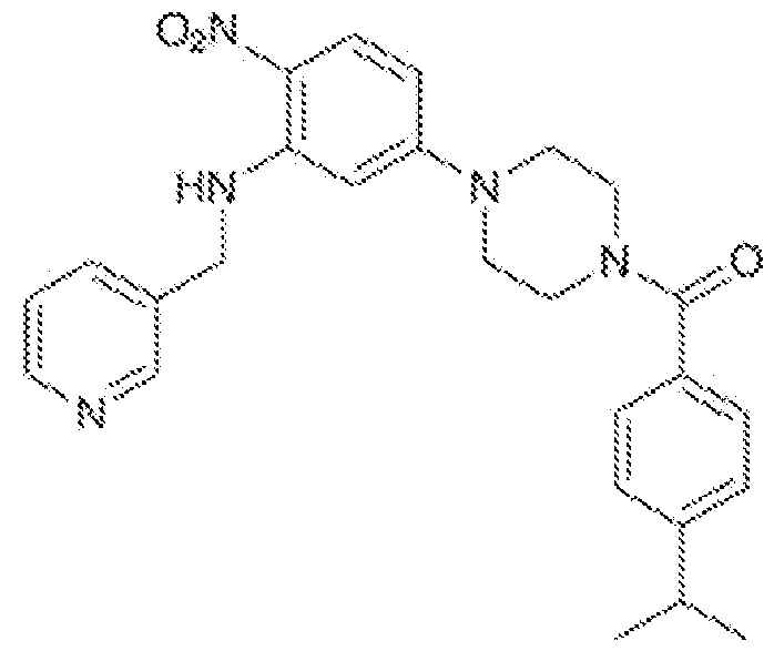
[0077] In some embodiments, the FOXO inhibitor is tanzawaic acid D having the formula:

[0078] In some embodiments, the FOXO inhibitor is Compound 13 having the formula:

[0079] In some embodiments, the FOXO inhibitor is Compound 8 having the formula
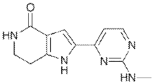
[0080] In some embodiments, the FOXO inhibitor is a peptide having the amino acid sequence LTLRKEPASEIAQSILEAYSQNGWANRRSGGKRPPPRRRQRRKKRG (SEQ ID NO:39).
Pharmaceutical Compositions
[0081] Disclosed herein are pharmaceutical compositions comprising one or more FOXO inhibitors in a pharmaceutically acceptable composition. For example, the compositions disclosed can be used therapeutically in combination with a pharmaceutically acceptable carrier. By “pharmaceutically acceptable’’ is meant a material that is not biologically or otherwise undesirable, i.e. , the material may be administered to a subject, along with the nucleic acid or vector, without causing any undesirable biological effects or interacting in a deleterious manner with any of the other components of the pharmaceutical composition in which it is contained. The carrier would naturally be selected to minimize any degradation of the active ingredient and to minimize any adverse side effects in the subject, as would be well known to one of skill in the art.
[0082] The materials may be in solution, suspension (for example, incorporated into microparticles, liposomes, or cells). These may be targeted to a particular cell type via antibodies, receptors, or receptor ligands. The following references are examples of the use of this technology to target specific proteins to tumor tissue (Senter, et al., Bioconjugate Chem., 2:447-451 , (1991); Bagshawe, K.D., Br. J. Cancer, 60:275-281 , (1989); Bagshawe, et al., Br. J. Cancer, 58:700-703, (1988); Senter, et al., Bioconjugate Chem., 4:3-9, (1993); Battelli, et al., Cancer Immunol. Immunother., 35:421-425, (1992); Pietersz and McKenzie, Immunolog.
Reviews, 129:57-80, (1992); and Roffler, et al., Biochem. Pharmacol, 42:2062-2065, (1991)). Vehicles such as “stealth” and other antibody conjugated liposomes (including lipid mediated drug targeting to colonic carcinoma), receptor mediated targeting of DNA through cell specific ligands, lymphocyte directed tumor targeting, and highly specific therapeutic retroviral targeting of murine glioma cells in vivo. The following references are examples of the use of this technology to target specific proteins to tumor tissue (Hughes et al., Cancer Research, 49:6214- 6220, (1989); and Litzinger and Huang, Biochimica et Biophysica Acta, 1104:179-187, (1992)). In general, receptors are involved in pathways of endocytosis, either constitutive or ligand induced. These receptors cluster in clathrin-coated pits, enter the cell via clathrin-coated vesicles, pass through an acidified endosome in which the receptors are sorted, and then either recycle to the cell surface, become stored intracellularly, or are degraded in lysosomes. The internalization pathways serve a variety of functions, such as nutrient uptake, removal of activated proteins, clearance of macromolecules, opportunistic entry of viruses and toxins, dissociation and degradation of ligand, and receptor-level regulation. Many receptors follow more than one intracellular pathway, depending on the cell type, receptor concentration, type of ligand, ligand valency, and ligand concentration. Molecular and cellular mechanisms of receptor-mediated endocytosis has been reviewed (Brown and Greene, DNA and Cell Biology 10:6, 399-409 (1991)).
[0083] Suitable carriers and their formulations are described in Remington: The Science and Practice of Pharmacy (19th ed.) ed. A.R. Gennaro, Mack Publishing Company, Easton, PA 1995. Typically, an appropriate amount of a pharmaceutically-acceptable salt is used in the formulation to render the formulation isotonic. Examples of the pharmaceutically-acceptable carrier include, but are not limited to, saline, Ringer's solution and dextrose solution. The pH of the solution is preferably from about 5 to about 8, and more preferably from about 7 to about 7.5. Further carriers include sustained release preparations such as semipermeable matrices of solid hydrophobic polymers containing the antibody, which matrices are in the form of shaped articles, e.g., films, liposomes or microparticles. It will be apparent to those persons skilled in the art that certain carriers may be more preferable depending upon, for instance, the route of administration and concentration of composition being administered.
[0084] Pharmaceutical carriers are known to those skilled in the art. These most typically would be standard carriers for administration of drugs to humans, including solutions such as sterile water, saline, and buffered solutions at physiological pH. The compositions can be administered intramuscularly or subcutaneously. Other compounds will be administered according to standard procedures used by those skilled in the art.
[0085] Pharmaceutical compositions may include carriers, thickeners, diluents, buffers, preservatives, surface active agents and the like in addition to the molecule of choice. Pharmaceutical compositions may also include one or more active ingredients such as antimicrobial agents, anti-inflammatory agents, anesthetics, and the like.
[0086] Preparations for parenteral administration include sterile aqueous or nonaqueous solutions, suspensions, and emulsions. Examples of non-aqueous solvents are propylene glycol, polyethylene glycol, vegetable oils such as olive oil, and injectable organic esters such as ethyl oleate. Aqueous carriers include water, alcoholic/aqueous solutions, emulsions or suspensions, including saline and buffered media. Parenteral vehicles include sodium chloride solution, Ringer's dextrose, dextrose and sodium chloride, lactated Ringer's, or fixed oils. Intravenous vehicles include fluid and nutrient replenishers, electrolyte replenishers (such as those based on Ringer's dextrose), and the like. Preservatives and other additives may also be present such as, for example, antimicrobials, antioxidants, chelating agents, and inert gases and the like.
[0087] Formulations for topical administration may include ointments, lotions, creams, gels, drops, suppositories, sprays, liquids and powders. Conventional pharmaceutical carriers, aqueous, powder or oily bases, thickeners and the like may be necessary or desirable.
[0088] Compositions for oral administration include powders or granules, suspensions or solutions in water or non-aqueous media, capsules, sachets, or tablets. Thickeners, flavorings, diluents, emulsifiers, dispersing aids or binders may be desirable.
[0089] Some of the compositions may potentially be administered as a pharmaceutically acceptable acid- or base- addition salt, formed by reaction with inorganic acids such as hydrochloric acid, hydrobromic acid, perchloric acid, nitric acid, thiocyanic acid, sulfuric acid, and phosphoric acid, and organic acids such as formic acid, acetic acid, propionic acid, glycolic acid, lactic acid, pyruvic acid, oxalic acid, malonic acid, succinic acid, maleic acid, and fumaric acid, or by reaction with an inorganic base such as sodium hydroxide, ammonium hydroxide, potassium hydroxide, and organic bases such as mono-, di-, trialkyl and aryl amines and substituted ethanolamines.
Therapeutic Uses
[0090] Disclosed herein is a method for treating a subject with an intestinal dysbiosis associated with Paneth cell dysfunction that involves implanting into to an intestine of the subject a plurality of organoids disclosed herein. For example, in some embodiments, the intestinal dysbiosis is a comorbidity of inflammatory bowel disease (I BD), irritable bowel syndrome (IBS), neurodegenerative diseases, autism spectrum disorder (ASD), obesity, cancer,
or diabetes. In some embodiments, a sample from an individual that is known to have or that is suspected of having an intestinal dysbiosis.
[0091] Also disclosed herein is a method for treating a subject with an intestinal dysbiosis associated with Paneth cell dysfunction that involves administering to the subject an effective amount of a forkhead box-0 transcription factor (FOXO) inhibitor to induce expression of one or more Paneth cell products. For example, in some embodiments, the intestinal dysbiosis is a comorbidity of inflammatory bowel disease (IBD), irritable bowel syndrome (IBS), neurodegenerative diseases, autism spectrum disorder (ASD), obesity, cancer, or diabetes. In some embodiments, a sample from an individual that is known to have or that is suspected of having an intestinal dysbiosis.
[0092] The herein disclosed compositions, including pharmaceutical composition, may be administered in a number of ways depending on whether local or systemic treatment is desired, and on the area to be treated. For example, the disclosed compositions can be administered intravenously, intraperitoneally, intramuscularly, subcutaneously, intracavity, or transdermally. The compositions may be administered orally, parenterally (e.g., intravenously), by intramuscular injection, by intraperitoneal injection, transdermally, extracorporeally, ophthalmically, vaginally, rectally, intranasally, topically or the like, including topical intranasal administration or administration by inhalant.
[0093] Parenteral administration of the composition, if used, is generally characterized by injection. Injectables can be prepared in conventional forms, either as liquid solutions or suspensions, solid forms suitable for solution of suspension in liquid prior to injection, or as emulsions. A revised approach for parenteral administration involves use of a slow release or sustained release system such that a constant dosage is maintained.
Ex vivo Uses
[0094] In some embodiments, the disclosed organoid system can allow (i) the determination of whether a microbe(s)s presence in the Gl tract is beneficial or pathogenic; (ii) evaluation of host-microbe interactions, functions, and/or biologically relevant responses pertaining to said interaction; (iii) discovery and elucidation of the molecular mechanisms that regulate host-microbe interaction; (iv) detection of a genetic profile that is known or unknown to be related to enteric disease; (v) providing an individual with personalized medicine methodology to evaluate the effectiveness of treatments.
[0095] The organoid system disclosed herein recapitulates the physiological environment of the gastrointestinal (Gl) tract. Studies have shown altered phenotypes and gene expression of gut tissues compared to those cultured at physiological oxygen levels. Thus,
being able to model the host gut tissue-microbe interactions under biologically relevant oxygen conditions is useful to understanding host gut tissue-microbe interactions and to develop and/or better understand therapies that strive to restore this interaction.
[0096] The disclosed organoid system may be used for research purposes, for therapy or diagnostic identification purposes, for identifying host-microbe relationships, and so forth. In particular embodiments, one can cultivate any microbe (e.g. bacteria, virus or fungi) for their robust replication and passaging to study and/or test such microbes in relation to worldwide disease. One can use the systems to characterize cellular processes and pathways to obtain information on targets exploited by the host-microbe interaction for physiological responses and/or pathogenesis. One can also assess methods and/or compositions (such as therapies and/or diet) that can affect the beneficial host-microbe interaction and such activity can be measured for effectiveness of restoring a normal host-microbe relationship. In addition, one can also cultivate the microbes along with the host gut tissue to understand the pathological phenotype of the host gut tissue. In a specific embodiment, the cultivation system provides for the development of a therapy to restore the normal host-microbe relationship in the intestinal tract.
[0097] To better understand host-microbe interactions in Gl tract, enteroids can be cocultured with microbes in an environment that biologically replicates the oxygen requirements of the Gl tract. In additional embodiments, one can cultivate the anaerobic microbes and/or enteroids in a physiologically relevant oxygen environment to improve reproducibility of studies and better determine therapy efficacy. In specific embodiments of the methods, the effects of cultivating microbes and enteroids in a physiologically relevant oxygen environment produced a robust gene expression profile and cell barrier integrity.
[0098] A number of embodiments of the invention have been described. Nevertheless, it will be understood that various modifications may be made without departing from the spirit and scope of the invention. Accordingly, other embodiments are within the scope of the following claims. EXAMPLES
Example 1 : FOXO inhibition rescues a-defensin expression in human intestinal organoids
Results and Discussion
[0099] The mRNA levels of Paneth cell products in small intestinal biopsies closely matched the profile of resected small intestinal tissue, consistent with previous reports. Enteroids derived from paired biopsies retained LYZ expression; however, DEFA5, DEFA6,
ITLN2, and REG3A mRNA was present at approximately 10,000 to 100,000-fold lower levels than in respective biopsies (Figure 1A).
[0100] To investigate whether this loss of a-defensin expression was specific to human enteroid culture, the Paneth cell profiles of murine small intestinal tissue were compared with murine small intestinal organoids. It was found that even after four weeks in culture, a timepoint when a-defensin expression in human enteroids has long since plummeted, murine enteroids recapitulated (within 10-fold) the a-defensin expression profile found in murine ileal tissue (Figure 1 B). These results suggest that the deficiency in Paneth cell a-defensin production in human enteroids is not due to in vitro culture conditions per se.
[0101] The Paneth cell gene expression programs is critically dependent on WNT signaling, and specifically, WNT signaling has been shown to regulate a-defensin expression through TCF transcription factors. While mouse small intestinal Paneth cells directly provide Wnt3 to intestinal stem cells in organoid culture, WNT ligands are not produced by human Paneth cells, and must be supplemented in human intestinal organoid media. To investigate whether an insufficiency in WNT ligands may explain the absence of a-defensin expression in human enteroids, culture media was supplemented with recombinant human WNT3A (200 ng/ml) but observed no change in DEFA5 or DEFA6 expression (Figure 2C). The hydrophobicity of WNT3A has been found to create spatiotemporal concentration gradients in organoid cultures, requiring recurrent administration of recombinant WNT3A every three hours to obtain a homogenous distribution throughout the hydrogel. Supplementing culture media with WNT3A in this manner (100ng/ml every three hours for a total of 72 hours) did not result in induction of DEFA5 or DEFA6 expression. WNT/TCF signaling depends on the availability of p-catenin to translocate into the nucleus. To increase p-catenin activity, a GSK-3P kinase inhibitor (CHIR99021 , 2.5 pM) was added to the culture media to reduce p-catenin degradation but found no change in a-defensin expression, suggesting that insufficiency of p-catenin did not explain the reduced expression of a-defensins. Moreover, two WNT target genes, ephrin type-B receptor 3 (EPHB3) and stem cell marker LGR5, were highly expressed in both WNT3A-treated and untreated enteroids, indicating that WNT signaling was sufficient for transcription of TCF target genes in the culture conditions, yet a-defensin expression was impaired.
[0102] ATOH1 is a master regulatory transcription factor required for the secretory lineage of the intestinal epithelium, and its expression is repressed by Notch signaling. Therefore, Paneth cell lineage specification requires Notch inhibition. To test if Notch activity was suppressing the expression of a-defensins, human enteroids were treated with DAPT, an antagonist of Notch signaling via y-secretase complex inhibition. However, DAPT treatment,
even at concentrations 2.5-fold greater than typically effective in enteroid cultures, did not rescue a-defensin expression. Taken together, these results suggest that neither WNT/p- catenin/TCF signaling nor Notch signaling underlie the dramatic loss of Paneth cell a-defensin expression in human enteroids. In addition, no other treatments reported to affect mRNA expression of a-defensins, such as IL-22 (Figure 2C) or FGF9, to appreciably raise the levels of DEFA5 or DEFA6 mRNA when compared to that of native tissue.
[0103] Knockdown of Foxo1/3 signaling in a genetic mouse model promoted secretory cell differentiation. In canonical FOXO signaling, activation of serine/threonine kinase Akt results in FOXO phosphorylation at three conserved serine residues, creating a binding site for the 14- 3-3 chaperone proteins, which in turn facilitate the export of FOXOs from the nucleus to the cytoplasm thereby inhibiting FOXO function. The cell-permeable oxo-dihydroquinoline AS1842856 (5-amino-7-(cyclohexylamino)-1-ethyl-6-fluoro-4-oxo-1 ,4-dihydroquinoline-3- carboxylic acid) inhibits the transcriptional activity of FOXO through direct binding to the active (unmodified Ser256) form of FOXO. Remarkably, the treatment of enteroids with AS1842856 (1 pM) restored the expression of DEFA5 and DEFA6 within five days to levels comparable to those in small intestinal biopsies (Figure 2A), yielding a magnitude of restorative induction approximately 10,000 to 100,000-fold. In organoid differentiation media, AS1842856 treatment of enteroids yielded a similar fold-change induction in a-defensins (Figure 20). Additionally, it was found that addition of AS1842856 to colonoids induced the expression of DEFA5 and DEFA6, with absolute levels of mRNA lower than that observed in AS1842856-treated enteroids (Figure 2D). Using fluorescence immunohistochemistry, high levels of HD5 peptide were detected in human enteroids treated with AS1842856, whereas HD5 was essentially undetectable in untreated enteroids (Figure 2B). Like DEFA5 and DEFA6, REG3A expression was markedly induced by AS1842856 treatment (Figure 2A, 2C, 2D). Interestingly, although AS1842856 restores a-defensin mRNA to tissue levels, the expression of ITLN2, another abundant Paneth cell product, was not similarly rescued suggesting its regulation may include an independent pathway.
[0104] Paneth cells constitutively produce vast quantities of a-defensins and other antimicrobials. A key function ascribed to Paneth cell a-defensins is to shape the composition of the intestinal microbiota. While the microbiota can significantly contribute to physiology in the healthy host, perturbations in microbial composition (i.e. , dysbiosis) may contribute to the pathogenesis of a wide variety of chronic diseases, including IBD, obesity, diabetes, neurodegenerative diseases, autism, and cancer. Reduced expression of Paneth cell a- defensins has been reported in ileal Crohn’s disease and proposed to contribute to the
dysbiosis in this disease. The discovery that treatment with the FOXO inhibitor AS1842856 reconstitutes native-tissue levels of human a-defensins in patient-derived intestinal organoids suggests that the FOXO signaling axis might prove a valuable therapeutic target to augment Paneth cell function.
Materials and Methods
Human samples and ethics statement
[0105] Biopsy specimens were obtained from study participants who met the criteria for constipation, diarrhea or IBS on the Gl history and symptom surveys. Both written and informed consent was obtained from a legal guardian for all pediatric study participants prior to data and sample collection. Adult surgical specimens were obtained from study participants who met NIH criteria for bariatric surgery. Inclusion criteria included age between 18-65 years, dyslipidemia controlled with medication, and either impaired glucose metabolism or type 2 diabetes. Both written and informed consent was obtained from all study participants prior to data and sample collection.
Crypt isolation and primary organoid culture
[0106] Biopsies from terminal ileum or colon were placed in a 15 ml conical tube with 10 ml of DMEM/F-12 culture media (supplemented with penicillin/streptomycin) and stored on ice immediately upon collection. Patient-derived enteroids (ileum-derived) and colonoids (colon- derived) were cultured within 12 hr of collection, according to the I ntesticult Organoid Growth Medium protocol (StemCell Technologies, Vancouver Canada; document #DX21423). Briefly, biopsies were washed twice with 10 ml of ice-cold PBS, transferred to a 1 .5 ml microcentrifuge tube and minced using sterile scissors. Tissue fragments were incubated on ice in 10 ml Gentle Cell Dissociation Reagent (StemCell Technologies; #07174) with gentle shaking (approximately 40 rpm) for 30 min. Tissue fragments were then vigorously pipetted to release crypts from tissue and passed through a 70 pm cell strainer (Corning, Corning NY; #352350). Crypts were counted and the volume adjusted to resuspend approximately 1000 crypts per 50 ml growth-factor- reduced, phenol-red-free Matrigel (Corning; #356231). A 50 pl dome of Matrigel-crypt suspension was plated per well of a pre-warmed 24-well flat-bottom tissue-culture-treated plate (Corning; #38017) and incubated at 37°C for 15 min before the addition of culture media. 750 pl of I ntestiCult Organoid Growth Medium Human (StemCell Technologies; #06010) was added to each well of Matrigel-crypt suspension and plates were incubated at 37°C and 5% CO2 A full medium exchange was performed every 48-72 hr. For experiments using differentiation media, organoids were cultured in I ntestiCult Organoid Growth Medium for 7 d before performing medium exchanges with I ntestiCult Organoid Differentiation Medium Human (StemCell
Technologies; #100-0214). Enteroids with fewer than ten passages were used for all experiments.
Murine enteroid culture
[0107] All procedures were approved by the Institutional Animal Care and Use Committee at UC Davis. C57BL/6N mice were humanely euthanized under deep anesthesia following intraperitoneal injection with ketamine/xylazine (100/10 mg/kg body weight). Enteroids were derived from the distal 6 cm of small intestine from 12-week-old male littermates (C57BL/6NTac; Taconic Biosciences, Germantown NY). Crypts were isolated and cultured as previously described. Briefly, intestinal sections were harvested, opened longitudinally, washed, cut into pieces, rinsed with cold PBS, and incubated in 2 mM EDTA at 4°C for 30 min with gentle shaking. Crypts were released after vigorous shaking in cold PBS and passed through a 70 pm cell strainer (Corning; #352350). Crypts were counted, resuspended, plated, and incubated exactly as described above for the human organoids. Murine enteroids were harvested after four weeks in culture.
RNA isolation
[0108] Human jejunal surgical specimens, ileal biopsies, and mouse small intestine (most distal 6 cm) were placed in RNAIater (lnvitrogen;ThermoFischer Scientific, Waltham MA; #AM7020) and incubated overnight at 4°C prior to long term storage at -20°C. RNA was isolated as previously described in detail, using guanidine thiocyanate/cesium chloride gradient. Isolated RNA pellets were washed with phenol/chloroform, dissolved in 70% ETOH, and stored at -80°C. RNA from human and murine enteroids was extracted using the Qiagen RNeasy Plus Mini kit (Qiagen, Germantown MD; #74134) according to the manufacturer’s instructions. Isolated RNA was quantified with ultraviolet absorption spectrometry at 260 nm using a NanoDrop spectrophotometer (NanoDrop Products/ThermoFischer Scientific), and then reverse transcribed to cDNA using the SuperScript™ III First-Strand Synthesis System (ThermoFischer Scientific; #18080051). cDNA was purified using a Qiagen PCR purification kit (Qiagen; #28106) and diluted to 10 ng/pl based on the input concentration of the total RNA.
Quantitative real-time PCR
[0109] For quantitative real-time PCR (qRT-PCR), target primers were combined with cDNA templates corresponding to 10 ng of specimen RNA, and reactions were monitored using a thermal cycler (Roche Diagnostics Lightcycler 2.0) and SYBR Green (LightCycler FastStart SYBR Green I mix; Roche #12239264001) product detection, as previously described. Absolute quantification of specific mRNA from tissue was determined by extrapolation of the detection threshold (crossing point) to the crossing point for gene-specific external plasmid cDNA
standards. Reproducibility assessments of this quantitative approach were previously reported to be approximately 10-15%. Oligonucleotide primers were designed using MacVector Software (https://macvector.com/), or as previously reported: ACTB (f: TGATGGTGGGCATGGGTCAG (SEQ ID NO:1), r: CGTGCTCGATGGGGTACTTCAG (SEQ ID NO:2)), LYZ (f: AAAACCCCAGGAGCAGTTAAT (SEQ ID NO:3), r: CAACCCTCTTTGCACAAGCT (SEQ ID NO:4)), DEFA5 (f TGGGGAAGACAACCAGGACC (SEQ ID NO:5), r:
TTCGGCAATAGCAGGTGGC (SEQ ID NO:6)), DEFA 6 (f: GCTTATGAGGCTGATGCCCAG (SEQ ID NO:7), r: GGCAAGTGAAAGCCCTTGTTG (SEQ ID NO:8)), ITLN2 (f: GCCTCCTCCTTTTCTTCCCTGCCTAG (SEQ ID NO:9), r: GGTCTGGTAGACAACACCATTCTCG (SEQ ID NO: 10)), REG3A (f: CCCACTGCTATGCCTTGTTTTTGTC (SEQ ID NO:11), r: ACTGCTACTCCACTCCCAACCTTCTC (SEQ ID NO: 12)), and PLA2G2A (f:
CGCACTCAGTTATGGCTTCTACG (SEQ ID NO: 13), r: AGGTGATTCTGCTCCCCGAG (SEQ ID NO:14)). Murine oligonucleotide primers were as previously reported {Wehkamp, 2007 #117; Menendez, 2013 #236; Castillo, 2019 #113}: Actb (F: GCTGAGAGGGAAATCGTGCGTG (SEQ ID NO:15), R: CCAGGGAGGAAGAGGATGCGG (SEQ ID NO:16)), Lyz1 (F: GCCAAGGTCTACAATCGTTGTGAGTTG (SEQ ID NO: 17), R:
CAGTCAGCCAGCTTGACACCACG (SEQ ID NO: 18)), Defa3 (F: ATCTGGTATGCTATTGTAGAAA (SEQ ID NO: 19), R: GTGGCCTCAGTACTCATGT (SEQ ID NO:20)), Defa5 (F: TCAAAAAAGCTGATATGCTATTG (SEQ ID NO:21), R: AGCTGCAGCAGAATACGAAAG (SEQ ID NO:22)), Defa20 (F: GAGAGATCTGATATGCTATTG (SEQ ID NO:23), R: AGAACAAAAGTCGTCCTGAG (SEQ ID NO:24)), Defa21 (F: GAGAGATCTGATCTGCCTTTG (SEQ ID NO:25), R:
CCTCTATTGCAGCGACGA (SEQ ID NO:26)), Defa22 (F: AGCAGCCAGGGGAAGAG (SEQ ID NO:27), R: CCTCTATTGCAGCGACGT (SEQ ID NO:28)), Defa23 (F: TCTGGTATGCTATTGTAGAAC (SEQ ID NO:29), R: GACAGCAGAGCGTGTATA (SEQ ID NQ:30)), Defa24 (F: GATCTGGTATGCTATTGTAGAG (SEQ ID NO:31), R: GACAGCAGAGCATGTACAA (SEQ ID NO:32)), Defa26 (F: ATTGTAGAAAAAGAGGCTGTAC (SEQ ID NO:33), R: AGCAGAGTGTGTACATTAAATG (SEQ ID NO:34)), Itlnl (F:
ACCGCACCTTCACTGGCTTC (SEQ ID NO:35), R: CCAACACTTTCCTTCTCCGTATTTC (SEQ ID NO:36)), and Reg3g (F: CCTCAGGACATCTTGTGTC (SEQ ID NO:37), R: TCCACCTCTGTTGGGTTCA (SEQ ID NO:38)). Conditions for qRT-PCR: Initial denaturation at 95°C for 10 min, followed by 45 cycles with each cycle consisting of denaturation, 95°C for 15 s; annealing at 60°C for 5 s; and extension at 72°C for 10 s. Following the cycle runs, samples
were denatured to establish the melting temperature(s) of the PCR product. The sample melt temperatures were compared to that of the internal standard to confirm template specificity.
Enteroid treatment
[0110] Human enteroids were treated with recombinant human WNT3A (100-400 ng/ml, R&D Systems, Minneapolis MN; #5036-WN), recombinant human FGF9 (50 ng/ml, R&D Systems; #273-F9), recombinant human IL-22 (2 ng/ml, Peprotech, Rocky Hill NJ; #200-22), AS1842856 (FOXO inhibitor, 1-2 pM, Tocris Bioscience, Bristol United Kingdom; #4265). Culture media was exchanged every 24 h (WNT3A) or 48 h (AS1842856, IL-22, FGF9). Enteroids were harvested for RNA isolation after five days of treatment, except for treatment with recombinant human IL-22, which was administered for 7 d as previously described.
Microscopy
[0111] Small intestine surgical specimens were fixed in aqueous paraformaldehyde (4% w/v), paraffin-embedded, sectioned (4 - 5 pM), and mounted on X-tra™ positive-charged slides (Leica Biosystems, Wetzlar Germany). Sections were cleared in xylene, washed in denatured 95% ETOH, peroxidase inactivated in MeOH with 3% v/v H2O2, and rehydrated in 70% ETOH and then distilled H2O. Antigen retrieval was performed by incubating slides overnight in a water bath at 60°C in sealed glass Coplin jars containing Tris-EDTA buffer (10 mM Tris base and 1 mM EDTA; pH 9.0). For immunofluorescence staining of tissue, specimen slides were equilibrated in PBS for 20 min and blocked with 5% goat serum (in PBS) for >30 min prior to overnight incubation at 4°C with mouse monoclonal anti-HD5 (isotype lgG2a). Following overnight incubation, slides were washed two times in PBS (10 min each) prior to incubation with goat anti-mouse IgG Alexa Fluor Plus 647 (ThermoFisher Scientific; #A32728) secondary antibody for 1-2 hr at room temperature. The slides were rinsed in PBS and stained with DAPI using the TrueVIEW Autofluorescence Quenching kit (Vector Laboratories, Burlingame CA; #SP-8400-15) according to the manufacturer’s protocol. For enteroids, specimens were fixed in aqueous paraformaldehyde (4% w/v), washed in PBS containing 0.1 % v/v Tween-20, blocked in buffer containing 0.1% Triton-X and 0.2% BSA in PBS for 15 min at 4°C, then incubated overnight at 4°C with mouse monoclonal anti-HD5 antibody (isotype lgG2a). After washing in PBS, specimens were incubated overnight at 4°C with goat anti-mouse IgG Alexa Fluor Plus 647 (Thermo Fisher Scientific; #A32728). Tissues were washed and incubated for 60 min at room temperature with 2 pM Hoescht 33258 (Abeam; Cambridge United Kingdom; #AB176759), then washed again in PBS and suspended in 60% (v/v) glycerol with 2.5 M fructose for mounting. Light microscopy images were obtained using an Olympus BX51 microscope (Olympus, Center Valley CA). Immunohistochemistry images were acquired using a Leica SP8
STED 3X confocal microscope (Leica Microsystems Inc., Buffalo Grove, IL). Acquired Z-stacks and figure images were generated using Fiji Imaged (version 1.0) software.
Statistics
[0112] Statistical analysis and data graphing were performed using GraphPad Prism software (version 9.3.1 , GraphPad Software, San Diego CA). Gene expression data (i.e., absolute copy number of mRNA transcript) was Log10 transformed prior to statistical analysis by T-test with Welch’s correction (mean comparisons between control and treated samples). Non-transformed data is displayed in figures.
Example 2. Mechanistic pathways and processes relevant for the differentiation and function of Paneth cells
Results
Wnt activation may not induce alpha-defensins in enteroids
[0113] The transduction of Wnt signaling through p-catenin and transcription factor 4 (TCF4) is indispensable for homeostasis in the intestinal epithelium. In the absence of Wnt signals, an intracellular protein complex — including adenomatous polyposis coli (APC), axin, and glycogen-synthase kinase 3|3 (GSK-3 ) — phosphorylates constitutively expressed p- catenin and promotes its ubiquitination and degradation by the proteasome. However, through canonical Wnt signaling, Wnt ligands promiscuously induce heterodimerization of Frizzled (Fzd) family receptors with their co-receptors Lrp5/6 on the cell surface, which in turn inhibits the APC complex, preventing the degradation of -catenin and allowing p-catenin to translocate into the nucleus and interact with transcription factors/lymphoid enhancing factors (TCF/LEF) to induce transcription of specific target genes.
[0114] Paneth cell gene programs are critically dependent on Wnt/TCF signaling and Wnt signaling has been shown to regulate alpha-defensin expression through TCF factors. Mediated through three distinct TCF binding sites, TCF-1 is thought to cooperate with p-catenin to activate DEFA5 (HD5) and DEFA6 (HD6) transcription.
[0115] Lysozyme, on the other hand, is not regulated by the Wnt/TCF pathway. It was thus hypothesized that inadequate Wnt signaling might contribute to the Paneth cell secretory profile that we observed in human enteroids, which express high levels of lysozyme and strikingly low levels of alpha-defensins.
[0116] Murine enteroid Paneth cells secrete trophic factors including Wnt3a, whereas human enteroid Paneth cells do not. In the intestinal stem cell niche, Wnt ligands are thought to be short-range signals, binding to the basolateral membrane of neighboring stem cells through direct contact and traveling away from their Paneth-cell source through stem cell division and
consequent dilution of this plasma membrane “ligand reservoir”. The effects of this gradient may potentially contribute to the finding that chimeric mouse-human enteroid cultures, in which murine Paneth cells were cultured in close proximity with human intestinal stem cells, induced a modest increase in DEFA5 mRNA expression.
[0117] Without the endogenous production of Wnt ligands by human Paneth cells, Wnt ligands are by necessity included in standard human enteroid culture media such as Intesticult Organoid Growth Medium (IOGM, StemCell Technologies) to sustain intestinal stem cells. It remains possible, however, that intestinal stem cells and Paneth cells require different levels of Wnt activation. To investigate whether a relative deficiency of Wnt ligands in the IOGM might explain the altered Paneth-cell secretory product profile in human enteroids, an additional 100 ng/mL recombinant human Wnt3a was included in organoid growth media and enteroids cultured for seven days; the quantity of Wnt ligand additionally supplemented was the amount described in defined media developed for human intestinal organoid culture. However, no change in DEFA5 expression was observed as a result of this treatment, nor with a supplemental treatment of 400 ng/mL.
[0118] Recombinant Wnt3a protein rapidly loses its activity and cannot stably supplement culture media. The instability of Wnt3a and limitations on its diffusion (due to its hydrophobic nature) has been found to produce a spatiotemporal gradient in organoid culture domes, with administration of Wnt3a every three hours necessary to obtain a homogenous distribution throughout the extracellular matrix hydrogel. Due to the known instability of Wnt proteins, an experiment was conducted during which 100 ng/mL of recombinant human Wnt3a was added to the media — every three hours for 72 hours of culture — before harvesting enteroids. In another experiment, the goal was to minimize the limitations of Wnt3a diffusion into the extracellular matrix hydrogel by adding 100 ng/mL of recombinant human Wnt3a directly to the Matrigel during routine passaging before supplementing with media fortified with an additional 100 ng/mL of Wnt3a for seven days. No changes were observed in DEFA5 expression as a result of either treatment. Thus, neither higher levels of Wnt3a, nor increased frequency of supplementation with exogenous Wnt3a, appeared to affect the expression of alpha-defensins in human enteroids.
[0119] Wnt/TCF signal transduction is limited by the availability of p-catenin to translocate into the nucleus, which is regulated by both its degradation and its production. To decrease the degradation of [3-catenin, the effects of CHIR99021 , a GSK-3|3 kinase inhibitor, was tested by adding it to the human enteroid culture media for seven days, but found no induction of DEFA5 (data not shown).
[0120] Consistent with findings, inhibition of GSK-3P kinase activity using lithium chloride was reported to both reduce the degradation of p-catenin and induced TCF4/p-catenin- mediated transcriptional activity in Caco2 cells, but this treatment did not induce DEFA5 mRNA expression. It was concluded that activation of p-catenin/TCF4 alone was insufficient to induce the expression of Paneth cell lineage markers, including DEFA5 and DEFA6. However, it was also reported that activation of fibroblast growth factor receptor-3 (FGFR-3) signaling, by the addition of its ligand fibroblast growth factor 9 (FGF9), induced sustained high levels of p- catenin and significantly induced DEFA5 and DEFA6.
[0121] Accordingly, 50 ng/mL of recombinant FGF9 was added to enteroid culture media daily for three days, as the induction of de novo alpha-defensin expression in Caco2 cells was found to require three days of stimulation using this concentration of ligand. Like with CHIR99021 treatment, it was found that recombinant human FGF9 supplementation to enteroid culture media did not alter DEFA5 mRNA expression in human enteroids. Together, these findings suggest that insufficient production of p-catenin in this model system does not explain the reduced expression of alpha-defensins.
[0122] As a positive control to confirm the sufficiency of Wnt-p-catenin-TCF signaling in organoid culture, alternative relevant target genes were identified to analyze. Ephrin type-B receptor 3 (EphB3) is strongly expressed on cells in the crypt base where Wnt levels are highest. In a study using laser-capture microdissection to parse human ileal tissues into crypt versus villus compartments, EphB3 was found to be expressed in the crypt at levels over threefold greater than in the villus. In the human enteroids, EphB3 mRNA was indeed found to be expressed at high levels, with expression similar to p-actin. Another Wnt target gene, stem cell marker Lgr5, was also found to be expressed in enteroids. Thus, it appears that Wnt signaling and p-catenin levels may be sufficient to activate transcription of TCF target genes including EphB3 and Lgr5 in our enteroid system, yet alpha-defensin expression remains impaired.
[0123] Interactions between ephrin ligands and their Eph receptors regulate a variety of biological processes including cell migration and boundary formation during development and intestinal homeostasis. In the intestine, Wnt-p-catenin-TCF signaling controls the expression of the receptors EphB2/B3 along the crypt-villus axis, such that EphB2 and EphB3 are strongly expressed at the bottom of the crypt where Wnt signaling is high. EphB3 is predominantly expressed by Paneth cells in this niche. In contrast, Notch signaling promotes generation of ephrinBI-positive cells in the villus, regulating the formation of the ephrinB1-EphB2 boundary at the crypt-villus border in the intestinal epithelium. A bidirectional repulsion resulting from EphB- ephrinBI signaling thus controls cell compartmentalization along the crypt-villus axis.
[0124] EphB3 dictates the positioning of Paneth cells at the crypt base, and aberrant Paneth cell positioning occurs when EphB signaling is disrupted in a genetic knockout model. Paneth cells mispositioned in the villus due to deficient EphB signaling appear to express lysozyme. In a Fz5LoxP/LoxP-K19Cre mouse model, mispositioned Paneth cells were found to lack detectable levels of EphB3 but still expressed lysozyme (without alpha-defensin), whereas Paneth cells in their proper location at the crypt base expressed EphB3, lysozyme, and the alpha-defensin cryptdin-1. Since EphB3 signaling appears to be correlated with the expression of alpha-defensin, but not lysozyme in mice, it was hypothesized that the EphB3 signaling axis might be relevant for the altered Paneth-cell expression of lysozyme and alpha-defensins in human enteroids.
[0125] There are differences in mouse and human enteroid cultures, e.g. the morphology and cellular representation of enteroids of each species is notably disparate: both crypt and villus populations of the intestinal epithelium are represented in murine enteroids, whereas primarily crypt-resident cells that exist under high-Wnt conditions are represented in human enteroids. While expression of EphB3 was confirmed in human enteroids (Figure 3), it was plausible that human Paneth cells in the enteroids might lack the signal normally provided by ephrinBI , since unlike murine enteroids, human enteroids lack villus cells that express this ligand. Accordingly, deficient signaling through EphB3 receptors might underlie the altered Paneth cell expression of lysozyme and alpha-defensins in human enteroids.
[0126] To test this idea, 1 pg/mL of recombinant human ephrinBI was added to both Matrigel and culture media for seven days. However, no changes were observed in mRNA expression of DEFA5 in the human organoid model.
[0127] Taken together, these results support that insufficient Wnt-p-catenin-TCF signaling in the human enteroid system is not responsible for the paucity of alpha-defensin expression in enteroid Paneth cells.
Notch inhibition may not induce alpha-defensins in enteroids
[0128] In addition to activation of Wnt/p-catenin signaling, Paneth cell lineage specification requires the inactivation of Notch signaling. The Notch signaling pathway is a highly-conserved cellular communication mechanism by which signals are transmitted between adjacent cells (i.e. , juxtacrine signaling) to influence cell fate during development, as well as homeostasis. In the canonical Notch signaling pathway, Notch ligands (e.g. DLL, Jagged) bind to Notch receptors, which triggers receptor proteolysis by y-secretase, thereby releasing the Notch intracellular domain (NICD). The NICD fragment then travels to the nucleus and contributes to a transcription complex that activates downstream target genes.
[0129] Paneth cells express Notch ligand DII4, supporting the growth and regulation of neighboring Lgr5+ stem cells, which require stimulation through their Notchl receptors. Notch inhibition, on the other hand, controls the absorptive versus secretory designation of progenitor cells. ATOH1 is a master regulatory transcription factor for the secretory lineage of the intestinal epithelium, and its activity is repressed by Notch signaling. Paneth cells, prominent members of the secretory lineage, therefore require Notch inhibition to adopt their secretory phenotype.
[0130] To explore whether Notch activity may be limiting the expression of alpha- defensins, human enteroids were treated with DAPT, an inhibitor of the y-secretase complex. However, even at concentrations of 2.5-fold more DAPT than is typically used in enteroid culture, no changes in the expression of DEFA5 were observed.
[0131] One methodological consideration is that Notch agonists (e.g. Jagged) may be present in IOGM. This would interfere with the experimental addition of Notch inhibitors such as DAPT. Since the proprietary formulation of the commercial IOGM product is not accessible, preparing in-house a defined culture media might be necessary to address this possibility before drawing firm conclusions regarding the role of Notch signaling on the expression of Paneth cell alpha-defensins.
Mitochondrial complex I inhibition does not restore alpha-defensins in enteroids
[0132] In the intestinal epithelium, Lgr5+ stem cells and Paneth cells have been found to utilize different metabolic programs. The Lgr5+ CBCs exhibit high mitochondrial activity, whereas Paneth cells support stem cell function by providing lactate to sustain the mitochondrial oxidative phosphorylation of stem cells. Lactate is thought to be a byproduct of glycolytic metabolism in Paneth cells. Thus, while intestinal stem cells have been shown to contain ample and elaborate mitochondrial networks, the mitochondria in Paneth cells appear small and sparse. Moreover, impaired mitochondrial function in intestinal stem cells has been found to drive their differentiation into Paneth cells that are dysfunctional.
[0133] It was hypothesized that mitochondrial function of Paneth cells in enteroids might be abnormally high in enteroid culture, potentially impairing canonical cell functions. To explore this possibility, mitochondrial respiration was reduced in enteroid culture utilizing molecules known to inhibit mitochondrial complex I, a large and complex component of the respiratory chain.
[0134] One such molecule is metformin, a widely-prescribed oral anti-diabetic agent with pleiotropic effects, taken by over 150 million people annually. The specific molecular mechanism of action remains incompletely known despite its use for over 60 years, but among its well-recognized targets, metformin can inhibit mitochondrial respiratory chain complex I.
Biodistribution studies have found that metformin does not accumulate dramatically in tissues; however, the highest levels of metformin are observed in the gastrointestinal tract, with preferential accumulation in the distal small intestine allowing for millimolar concentrations, IQ- 50 times that of plasma, at 24 hours post administration. These millimolar concentrations have been shown by multiple laboratories to consistently inhibit complex I in isolated mitochondria.
[0135] Rotenone, the most commonly used complex I inhibitor is a lipophilic botanical compound that strongly inhibits complex I via specific binding to the subunit ND1 , preventing electron transfer from complex I to ubiquinone and functionally blocking oxidative phosphorylation. Exposure to rotenone inhibits complex I and generates reactive oxygen species in multiple cell types.
[0136] These two complex I inhibitors were titrated to inhibit mitochondrial membrane potential by approximately 40%, a level of inhibition found to be meaningful for cellular function, and added to the culture media of human enteroids.
[0137] The morphology of enteroids treated with complex I inhibitors appeared notably different from untreated enteroids. After five days of treatment with either rotenone or metformin, enteroids appeared smaller and darker, with a greater proportion of enteroids exhibiting a thickened columnar epithelium typical of differentiated cells, compared to the thinner-walled epithelium typical of enteroids with a high proportion of proliferating stem cells (Figure 4). However, there was no detectable change in expression levels of DEFA5.
[0138] During the process of titrating complex I inhibitors, an unexpected phenomenon was observed. As previously mentioned, intestinal stem cells exhibit large and complex mitochondrial networks, whereas Paneth cells contain small and sparse mitochondria. We used MitoTracker Green (MTG) dye, which measures mitochondrial mass independent of mitochondrial membrane potential, to analyze human enteroids, expecting much lower mitochondrial mass in Paneth cells compared to ISCs in human enteroids. MTG signal in the side-scatter-high (granular) cells, which are likely Paneth cells, was compared to side-scatter- low cells, which are likely stem cells and progenitor cells (Figure 5). Mitochondrial mass in the granular side-scatter-high cells (likely Paneth cells) was modest, only 11 % lower than in the side-scatter-low cells (likely stem cells and progenitor cells), a far smaller decrease than would be expected based on confocal and electron microscopy images of mitochondrial structure in native intestinal tissue.
FOXO inhibition restores alpha-defensin expression in enteroids
[0139] Due to the surprising observation that highly-granular cells in the enteroid population had a mitochondrial phenotype inconsistent with that expected of Paneth cells, it was
hypothesized that abnormalities in mitochondrial dynamics might contribute to the reduction of alpha-defensins in human enteroid Paneth cells.
[0140] Mitochondrial function has been found to play an important role in epithelial cell stemless, differentiation, and lineage commitment, with the metabolic activity of intestinal epithelial cells playing a key role during cellular transitions and cell fate decisions Dynamic organelles with a highly-variable shape and size, mitochondria are controlled by the opposing processes of fusion and fission. The family of forkhead box O (FOXO) transcription factors are thought to be important regulators of mitochondrial homeostasis, and mitochondrial fission mediated through FOXO1/3 signaling has been suggested to define the differentiation of intestinal stem cells into Paneth cells in mice.
[0141] In canonical FOXO signaling, insulin or growth factor stimulation first activates the phosphoinositide 3-kinase (PI3K)/Akt pathway. The activation of serine/threonine kinase Akt (also known as protein kinase B or PKB) then inhibits FOXO function by phosphorylating FOXOs at three conserved residues, creating a binding site for the 14-3-3 chaperone proteins. Binding of 14-3-3 proteins facilitates the export of FOXOs from the nucleus to the cytoplasm. Knockdown of Foxo1/3 signaling in a genetic mouse model was found to induce mitochondrial fission, promoting secretory cell differentiation.
[0142] It was hypothesized that inhibiting FOXO signaling in human enteroids might allow Paneth cells to undergo a mitochondrial fission process characteristic of their in vivo state, and that the change in mitochondrial dynamics might play a role in the regulation of alpha- defensin expression.
[0143] AS1842856 is a cell-permeable oxodihydroquinoline that inhibits FOXO activity. AS1842856 preferentially inhibits the transcriptional activity of FOXO1 , as compared to the functionally-related FOXO3a and FOXO4, with inhibition approximately 70%, 20%, and 3%, respectively, at a dose of 100 nM in human hepatoma HepG2 cells. The inhibition is attributed to direct binding of the active (unphosphorylated Ser256) form of FOXO1
[0144] Enteroids were treated for seven days with AS1842856 at a concentration of 2uM; extrapolating from published IC50 values in rat hepatoma Fao cells, this concentration is estimated to almost fully inhibit FOXO1 and exceed IC50 for FOXO3a and FOXO4.
[0145] Control enteroids primarily exhibited the typical thin-walled cystic appearance typical of human enteroids grown in standard culture media, whereas AS 1842856- treated organoids appeared smaller, denser, and with a higher proportion of enteroids exhibiting primarily columnar epithelium (Figure 6), similar to the morphology observed after treatment with the mitochondrial complex I inhibitors metformin and rotenone (Figure 4).
[0146] However, divergent from results with mitochondrial complex I inhibitors, it was found that treatment of enteroids with FOXO inhibitor AS1842856 restored the expression of the Paneth cell secretory products DEFA5 and DEFA6 to levels comparable to primary small intestinal biopsies (Figure 7). Lysozyme mRNA expression, which had appeared to possibly exceed tissue levels in untreated enteroids, returned to tissue levels with AS1842856 treatment (Figure 7). The corresponding mRNA ratio of DEFA5 to LYZ in AS 1842856- treated enteroids approximated that of tissue as well (Figure 8).
[0147] Interestingly, although AS1842856 restores alpha-defensin mRNA to tissue levels, the expression of ITLN2, another highly-abundant Paneth cell product, was not increased along with DEFA5 and DEFA6, and appears to be regulated independently from both lysozyme and alpha-defensins.
[0148] It is worth noting the magnitude of the alpha-defensin induction observed upon AS1842856 treatment: a 10,000-fold to 100,000-fold change in mRNA expression levels. Typically, alpha-defensin expression is remarkably stable, with changes on the order of 3- to 10- fold often considered significant.
[0149] The induction of alpha-defensins by AS1842856 was not immediate, but rather occurred over the course of three to seven days; at three days, DEFA5 mRNA was found to be at intermediate levels before reaching full tissue levels by seven days of treatment (Figure 9). The kinetics of this induction may be suggestive of a cellular reprogramming process in Paneth cells induced by AS1842856 exposure.
HD5 protein is expressed in AS1842856-treated enteroids
[0150] Although DEFA5 mRNA expression levels were significantly upregulated after AS1842856 treatment, it remained possible that the mRNA may not be translated into HD5 protein in enteroids.
[0151] HD5 is stored in Paneth cells as a propeptide (aa 20-94) and released as mature peptide subsequent to the enzymatic activity of trypsin, which is co-secreted by Paneth cells and efficiently cleaves HD5 propeptide. The commercially-available monoclonal antibody 8C8 recognizes propeptide and partially processed forms of HD5, but is unable to bind mature peptide. A mouse monoclonal anti-HD5 antibody (HD5A/65.1.3) has been developed that detects both HD5 propeptide and mature peptide, enabling us to detect HD5 in any form that it might be present in enteroid tissues.
[0152] Co-localization of a commercial antibody recognizing human lysozyme (Dako/Agilent; #A0099) was validated with the HD5 antibody HD5A/65.1.3 in human small intestinal tissue using fluorescence immunohistochemistry.
[0153] Lysozyme is a well-established and commonly-used Paneth cell marker and we found it to be present in small intestinal crypts, as expected (Figure 10). HD5 protein was also detected in crypts, with compiled images indicating a high degree of overlap in the cellular localization of these two proteins within Paneth cell granules (Figure 10).
[0154] Next, these same antibodies to HD5 and lysozyme were used to perform fluorescence immunohistochemistry in human enteroids treated with FOXO inhibitor AS1842856. AS1842856-treated enteroids produce detectable levels of HD5, with protein appearing to accumulate within the interior lumen of these tissues (Figure 11).
[0155] To estimate the percentage of HD5+ cells in AS1842856-treated enteroids, the mouse monoclonal HD5A/65.1 .3 antibody was conjugated to Alexa Fluor 647. This conjugated HD5 mAb was used to stain control and treated enteroids in flow-cytometric analysis. AS1842856-treated enteroids were estimated to contain 7.8% HD5+ cells on average (Figure 12). As noted in Figure 11 , HD5 protein appears to aggregate in the enteroid lumen, suggesting that HD5-containing granules may have been secreted during the culture and processing of enteroids in these images. As the replenishment of Paneth-cell granules after degranulation is estimated to take approximately 21 hours, it is possible that our analysis underestimates the true number of Paneth cells in these tissues, as Paneth cells may have degranulated during processing and would no longer contain intracellular HD5 protein. Future studies using secretory inhibitors will be conducted to attempt more accurate quantification of HD5+ Paneth cells represented in AS1842856-treated enteroids.
FOXO inhibition may reduce mitochondrial mass
[0156] Since a potential consequence of FOXO inhibition is promoting mitochondrial fission, the mitochondrial mass of cells in enteroids after AS1842856 treatment was estimated using MitoTracker Green, which localizes to mitochondria regardless of mitochondrial membrane activity.
[0157] Enteroids treated with AS 1842856 had a notably reduced mitochondrial mass, with a reduction of approximately 66% per live cell on average as measured by the median fluorescence intensity of MitoTracker Green (Figure 13). Due to the possible degranulation observed during fluorescence immunohistochemistry (Figure 11), these results were not dichotomized by side-scatter as in Figure 6.
Example 3. FOXO inhibition rescues a-defensin expression in human intestinal organoids
Methods
[0158] Human samples and ethics statement. This study was approved by institutional review boards for the State of California and the University of California, Davis (IRB #1400430, “Regulatory Immune Mechanisms and Gastrointestinal Comorbidity in ASD”). All patients provided informed consent. Both written and informed consent was obtained from a legal guardian for all pediatric study participants prior to data and sample collection, in accordance with the UC Davis IRB protocol. Biopsy specimens were obtained from study participants who met the criteria for constipation, diarrhea or IBS on the Gl history and symptom surveys (Drossman, Gastroenterology 2006 130:1377-1390). Samples used for this work were solely from patients without an autism spectrum disorder (ASD) diagnosis. As only small quantities of biopsy tissue were collected from patients in this study, for the reference images of small intestinal tissue in Figure 15, adult jejunal specimens were obtained from a separate study involving participants who underwent Roux-en-Y gastric bypass for medically-complicated obesity (IRB #228969. "Genetic and Biomedical Characteristics of Human Adipose Tissue”). These specimens were generated from tissue created during closure of the jejunojejunostomy that would normally be discarded. Study participants were between 18-65 years of age and met clinical criteria for bariatric surgery as a medical necessity. Informed consent was obtained from all study participants prior to data and sample collection and documented in accordance with the UC Davis IRB protocol.
[0159] Human crypt isolation and enteroid culture. Patient biopsies from terminal ileum were placed in a 15 ml conical tube with 10 ml of DMEM/F-12 culture media (supplemented with penicillin/streptomycin) and stored on ice immediately upon collection. Intestinal organoids derived from ileal tissue (enteroids) were cultured within 12 hr of collection, according to the IntestiCult Organoid Growth Medium protocol (StemCell Technologies, Vancouver Canada; document #DX21423). Briefly, biopsies were washed twice with 10 ml of ice-cold PBS, transferred to a 1.5 ml microcentrifuge tube and minced using sterile scissors. Tissue fragments were incubated on ice in 10 ml Gentle Cell Dissociation Reagent (StemCell Technologies; #07174) with gentle shaking (~40 rpm) for 30 min. Tissue fragments were then vigorously pipetted to release crypts from tissue and passed through a 70 pm cell strainer (Corning, Corning NY; #352350). Crypts were counted and the volume adjusted to resuspend approximately 1000 crypts per 50 pl growth-factor-reduced, phenol-red-free Matrigel (Corning; #356231). A 50 pl dome of Matrigel-crypt suspension was plated per well of a pre-warmed 24-
well flat-bottom tissue-culture-treated plate (Corning; #38017) and incubated at 37°C for 15 min before the addition of culture media. 750 pl of IntestiCult Organoid Growth Medium Human (StemCell Technologies; #06010) was added to each well of Matrigel-crypt suspension and plates were incubated at 37°C and 5% CO2. A full medium exchange was performed every 48- 72 hr. For experiments using differentiation media, enteroids were cultured in IntestiCult Organoid Growth Medium for 7 d before performing medium exchanges with IntestiCult Organoid Differentiation Medium Human (StemCell Technologies; #100-0214).
[0160] Murine crypt isolation and enteroid culture. All procedures were approved by the Institutional Animal Care and Use Committee at UC Davis. C57BL/6N mice were humanely euthanized under deep anesthesia following intraperitoneal injection with ketamine/xylazine (100/10 mg/kg body weight). Enteroids were derived from the distal 6 cm of small intestine from 12-week-old male littermates (C57BL/6NTac; Taconic Biosciences, Germantown NY). Crypts were isolated and cultured as previously described (Sato et al., Nature 2009459:262-265). Briefly, intestinal sections were harvested, opened longitudinally, washed, cut into pieces, rinsed with cold PBS, and incubated in 2 mM EDTA at 4°C for 30 min with gentle shaking. Crypts were released after vigorous shaking in cold PBS and passed through a 70 pm cell strainer (Corning; #352350). Crypts were counted, resuspended, plated, and incubated as described above for human enteroids.
[0161] Enteroid treatment. Human enteroids were treated with recombinant human WNT3A (100-200 ng/ml, R&D Systems, Minneapolis MN; #5036- WN), recombinant human IL- 22 (2-50 ng/ml, Peprotech, Rocky Hill NJ; #200-22), or AS1842856 (FOXO inhibitor, 100 nM-1 pM, Tocris Bioscience, Bristol United Kingdom; #4265). Culture media was exchanged every 24 h (WNT3A) or 48 h (AS1842856, IL-22). Enteroids were harvested for RNA isolation after five days of treatment, except for treatment with recombinant human IL-22, which was administered for seven days as previously described (He et al., Cell Stem Cell 2022 29:1718-1720).
[0162] RNA isolation and cDNA synthesis. RNA from human and mouse enteroids was extracted using a Qiagen RNeasy Plus Mini kit (Qiagen, Germantown MD; #74134) according to the manufacturer’s instructions. Isolated RNA was dissolved in 70% EtOH and stored at -80°C. Isolated RNA was quantified using a Qubit 3.0 Fluorometer (Invitrogen, Pub No. MAN0010866) and/or with ultraviolet absorption spectrometry at 260 nm using a NanoDrop spectrophotometer (ThermoFisher Scientific) and reverse-transcribed to cDNA using SuperScript™ IV VILO™ Master Mix with ezDNase™ (Invitrogen #11766050) or SuperScript™ III First-Strand Synthesis System (Invitrogen #18080051). cDNA was purified using a Qiagen PCR purification kit (Qiagen; #28106) and diluted to 10 ng/pl based on the input concentration of the total RNA.
Human ileal biopsies and mouse small intestine (distal 6 cm) were placed in RNAIater (Invitrogen; ThermoFisher Scientific, Waltham MA; #AM7020) and incubated overnight at 4°C prior to long term storage at -20°C, after which RNA was isolated as previously described in detail (Wehkamp et al., Proc Natl Acad Sci U S A 2005 102:18129-18134; Wehkamp et al., FEBS Lett 2006 580:5344-5350; Castillo et al., Sci Rep 2019 9:13115; Nonnecke et al., Sci Rep 2021 11 : 12889; Nonnecke et al., FASEB J 2022 36:e22200), using guanidine thiocyanate/cesium chloride gradient. Isolated RNA pellets were washed with phenol/chloroform, dissolved in 70% EtOH, and stored at -80°C. Isolated RNA was quantified with ultraviolet absorption spectrometry at 260 nm using a NanoDrop spectrophotometer (NanoDrop Products/ThemnoFisher Scientific), and then reverse-transcribed to cDNA using the SuperScript™ III First-Strand Synthesis System (ThermoFisher Scientific; #18080051). cDNA was purified using a Qiagen PCR purification kit (Qiagen; #28106) and diluted to 10 ng/pl based on the input concentration of the total RNA.
[0163] Reverse transcription quantitative real-time PCR. For relative quantification using reverse transcription quantitative real-time PCR (RT-qPCR) (Bustin et al., Clin Chem 2009 55:611-622), TaqMan hydrolysis probes were combined with cDNA templates corresponding to 10 ng of sample RNA, and reactions were monitored using a StepOnePlus™ Real-Time PCR System instrument (Applied Biosystems # 4376600) and TaqMan Fast Advanced Master Mix (Applied Biosystems #4444557) according to the manufacturer’s protocol (Pub. No. MAN0025706). TaqMan gene expression assays, selected such that either primers or probes were exon-spanning, were specific to the following targets: ACTB (Hs01060665_g1), LYZ (Hs00426232_m1), DEFA5 (Hs00360716_m1), DEFA6 (Hs00427001_m1), ITLN2 Hs00365614_m1), REG3A (Hs00170171_m1), PLA2G2A (Hs00179898_m1), FOXO1 (HS00231106_m1), FOXO3 (HS00818121_m1), FOXO4 (HS00172973_m1), BCL6 (Hs00153368_m1), CAT (Hs00156308_m1), G6PC (Hs_02802676_m1), and PCK1 (Hs00159918_m1). ACTB: [3-actin; LYZ: lysozyme; DEFA5: human a-defensin 5; DEFA6: human a-defensin 6; ITLN2: intelectin-2; REG3A: regenerating family member 3 alpha; PLA2G2A: secretory phospholipase A2. All gene expression assays were determined to have amplification efficiencies of 100% (±10%), no-template controls were performed for all assays, and reactions were carried out in triplicate. Conditions for RT-qPCR were: an initial UNG incubation at 50°C for 2 min, then Taq polymerase activation at 95°C for 20 s, followed by 40 cycles of 95°C for 1 s (denaturation), and 60°C for 20 s (annealing and extension). Data were captured using StepOne™ software v2.2.2 (Applied Biosystems). For absolute quantification using RT-qPCR, target primers were combined with cDNA templates corresponding to 10 ng of
specimen RNA, and reactions were monitored using a thermal cycler (Roche Diagnostics LightCycler 2.0) and SYBR Green (LightCycler FastStart SYBR Green I mix; Roche #12239264001) product detection, as previously described (Wehkamp et al., Proc Natl Acad Sci U S A 2005 102:18129-18134; Wehkamp et al., FEBS Lett 2006 580:5344-5350; Castillo et al., Sci Rep 2019 9:13115; Nonnecke et al., FASEB J 2022 36:e22200). Absolute quantification of specific mRNA from tissue was determined by extrapolation of the detection threshold (crossing point) to the crossing point for gene-specific external plasmid cDNA standards. Reproducibility assessments of this quantitative approach were previously reported to be approximately 10- 15% (5). Oligonucleotide primers were as previously reported (4, 5, 7, 8). Conditions for RT- qPCR: Initial denaturation at 95°C for 10 min, followed by 40 cycles with each cycle consisting of denaturation at 95°C for 15 s; annealing at 60°C for 5 s; and extension at 72°C for 10 s. Following the cycle runs, samples were denatured to establish the melting temperature(s) of the PCR product. The sample melt temperatures were compared to that of the internal standard to confirm template specificity.
[0164] Microscopy. Human enteroids were fixed in aqueous paraformaldehyde (4% w/v), washed in PBS containing 0.1% v/v Tween-20, blocked in buffer containing 0.1 % Triton-X and 0.2% BSA in PBS for 15 min at 4°C, then incubated overnight at 4°C with mouse monoclonal anti-DEFA5/HD5 antibody (CB65.1.3, isotype lgG2a). After washing in PBS, specimens were incubated overnight at 4°C with goat anti-mouse IgG Alexa Fluor Plus 647 (Thermo Fisher Scientific; #A32728). Tissues were washed and incubated for 60 min at room temperature with 2 pM Hoescht 33258 (Abeam; Cambridge United Kingdom; #AB176759), then washed again in PBS and suspended in 60% (v/v) glycerol with 2.5 M fructose for mounting. Small intestine surgical specimens (used for reference images in Figure 15) were fixed in aqueous paraformaldehyde (4% w/v), paraffin-embedded, sectioned (4 - 5 pM), and mounted on X-tra™ positive-charged slides (Leica Biosystems, Wetzlar Germany). Sections were cleared in xylene, washed in denatured 95% ETOH, peroxidase inactivated in MeOH with 3% v/v H2O2, and rehydrated in 70% ETOH and then distilled H2O. Antigen retrieval was performed by incubating slides overnight in a water bath at 60°C in sealed glass Coplin jars containing Tris- EDTA buffer (10 mM Tris base and 1 mM EDTA; pH 9.0). For immunofluorescence staining of tissue, specimen slides were equilibrated in PBS for 20 min and blocked with 5% goat serum (in PBS) for >30 min prior to overnight incubation at 4°C with mouse monoclonal anti-DEFA5/HD5 (CB65.1.3, isotype lgG2a). Following overnight incubation, slides were washed two times in PBS (10 min each) prior to incubation with goat anti-mouse IgG Alexa Fluor Plus 647 (ThermoFisher Scientific; #A32728) secondary antibody for 1-2 hr at room temperature. The
slides were rinsed in PBS and stained with DAPI using the TrueVIEW Autofluorescence Quenching kit (Vector Laboratories, Burlingame CA; #SP-8400-15) according to the manufacturer’s protocol. Light microscopy images were obtained using an Olympus BX51 microscope (Olympus, Center Valley CA). Immunohistochemistry images were acquired using a Leica SP8 STED 3X confocal microscope (Leica Microsystems Inc., Buffalo Grove, IL).
Acquired Z-stacks and figure images were generated using Fiji Imaged (version 1.0) software (Schindelin et al., Nature Methods 2012 9:676-682).
[0165] Statistics. Relative quantification (normalized to ACTB) and statistical analysis (T-test with Benjamini-Hochberg correction) were performed using the Applied Biosystems Relative Quantification Module based on the Livak method (Livak, et al. Methods 2001 25:402- 408) as PCR amplification efficiencies of target and reference genes were 100% (±10%). The maximum allowed Cq value included in calculations was 40 (approximately 1 cycle greater than the highest Cq detected in experiments using TaqMan primer/probe gene expression assays). Statistical analysis for absolute quantification and data graphing were performed using GraphPad Prism software (version 9.4.1, GraphPad Software, San Diego CA). The absolute copy number of mRNA transcript was Iog10-transformed prior to statistical analysis by T-test with Welch’s correction (mean comparisons between control and treated samples), with nontransformed data displayed in figures.
Results and Discussion
[0166] The mRNA levels of highly abundant Paneth cell secretory products in human mucosal biopsies (Fig. 14A) were consistent with those previously described for small intestinal tissue (Wehkamp et al., Proc. Natl. Acad. Sci. U.S.A. 2005 102:18129-18134; Nonnecke et al., FASEB J. 2022 36:e22200), yet the gene expression profile of enteroids (Fig. 14A) deviated significantly. Relative quantification of mRNA for each target showed that enteroids derived from paired biopsies retained LYZ expression while DEFA5, DEFA6, intelectin-2 (ITLN2), and regenerating family member 3 alpha (REG3A) mRNA was present at ~10,000 to ~100, 000-fold lower levels than in respective biopsies (Fig. 14B).
[0167] To investigate whether this loss of a-defensin expression was specific to human enteroid culture, the Paneth cell secretory product profiles of murine small intestinal tissue were compared with murine enteroids. Murine enteroids recapitulated (within 10-fold) the a-defensin expression profile found in murine ileal tissue (Fig. 14C), suggesting that the deficiency in Paneth cell a-defensin production in human enteroids is not due to in vitro culture conditions per se.
[0168] The Paneth cell gene expression program is critically dependent on WNT signaling (van Es et al., Nat. Cell Biol. 2005 7:381-386), which has been shown to regulate a- defensin expression through TCF (T cell factor) transcription factors (Wehkamp, et al. Front Immunol. 2020 11 :646; van Es et al., Nat. Cell Biol. 2005 7:381-386). While mouse small intestinal Paneth cells directly provide Wnt3 to intestinal stem cells in organoid culture (Sato et al., Nature 2011 469:415-418), WNT ligands are not produced by human Paneth cells (Lahar et al., PLoS One 2011 6:e26890) and must be supplemented in human intestinal organoid media. To investigate whether an insufficiency in WNT ligands may explain the absence of a-defensin expression in human enteroids, culture media was supplemented with recombinant human WNT3A (100 or 200 ng/mL) but observed no change in DEFA5 or DEFA6 expression (Fig. 14D and 14E). Two WNT target genes, ephrin type-B receptor 3 (EPHB3) and leucine rich repeat containing G protein-coupled receptor (LGR5), were highly expressed in both WNT3A-treated and untreated enteroids, indicating that WNT signaling was sufficient for transcription of TCF target genes in the culture conditions, yet a-defensin expression was impaired. In addition, other treatments reported to affect mRNA expression of a-defensins, such as IL-22 at 2 ng/mL (He et al., Cell Stem Cell 2022 29:1718-1720) or 50 ng/mL did not appreciably raise the levels of DEFA5 or DEFA6 mRNA (Fig. 14F), especially when compared to the expression profile of native tissue (Fig. 14A and 14G).
[0169] Knockdown of Foxo1/3 signaling in a genetic mouse model was found to promote secretory cell differentiation (Ludikhuize et al., Cell Metab. 2020 32: 889-900. e7), and a cell- permeable inhibitor of FOXO1 , AS1842856 (5-amino-7-(cyclohexylamino)-1-ethyl-6-fluoro-4- oxo-1 , 4-dihydro-quinoline-3-carboxylic acid) induced the secretory lineage of enteroendocrine cells (Zeve et al., Nat. Commun. 2022 13:261). It was therefore hypothesized that FOXO could be related to the loss of Paneth cell secretory products in enteroid culture. AS1842856 inhibits the transcriptional activity of FOXO through direct binding to the active (unmodified Ser256) form (Nagashima et al., Mol. Pharmacol. 78, 961-970 (2010).). F0X01, F0X03, and F0X04 were confirmed to be expressed in human enteroids (Fig. 15A), and expression of canonical FOXO1 target genes glucose-6-phosphatase catalytic subunit (G6PC), and phosphoenolpyruvate carboxykinase 1 (PCK1), but not FOXO3 target gene catalase (CAT) or FOXO3/4 target gene B cell lymphoma 6 (BCL6), were reduced after treatment with AS1842856 (Fig. 15B).
[0170] Remarkably, the treatment of enteroids with FOXO inhibitor AS1842856 at 1 pM increased the expression of DEFA5 and DEFA6 >100,000-fold (Fig. 15C) to levels not significantly different from small intestinal biopsies (Fig. 15D), representing a restorative
induction of DEFA5 and DEFA6 (Fig. 15C-15E) compared to untreated enteroids (Fig. 14A and 14B). Treatment with 500 nM or 1 pM concentrations of AS1842856 were found to mediate this restoration of a-defensins (Fig. 15F). AS1842856 treatment of enteroids cultured in differentiation media yielded an induction of a-defensins similar in magnitude (Fig. 15G). Fluorescence immunohistochemistry confirmed that while DEFA5 was minimally produced in untreated enteroids (Fig. 15H), high levels of DEFA5 peptide were produced in human enteroids treated with AS1842856 (Fig. 151).
[0171] Paneth cell products REG3A, PLA2G2A (group 2 secretory phospholipase A2), and ITLN2 (Nonnecke et al., FASEB J. 2022 36:e22200) were also significantly upregulated by AS1842856 (~6, 000-fold, ~400-fold, and ~60-fold, respectively), yet not fully restored to tissue levels (Fig. 15C-15E and 15G), suggesting that their regulation may involve independent pathways.
[0172] A key function ascribed to Paneth cell a-defensins is to shape the composition of the intestinal microbiota (Bevins, et al. Nat. Rev. Microbiol. 2011 9:356-368). While the microbiota can significantly contribute to physiology in the healthy host, perturbations in microbial composition (i.e. , dysbiosis) may contribute to the pathogenesis of a wide variety of chronic diseases, including ASD, inflammatory bowel disease (IBD), obesity, diabetes, and cancer (DeGruttola, et al. Inflamm. Bowel Dis. 2016 22:1137-1150). Reduced expression of Paneth cell a-defensins has been reported in ileal Crohn’s disease and proposed to contribute to the dysbiosis in this disease (Wehkamp, et al. Front Immunol. 2020 11 :646). This novel discovery that treatment with the FOXO inhibitor AS1842856 reconstitutes native-tissue levels of human a-defensins in patient-derived intestinal organoids suggests that the FOXO signaling axis might prove a valuable therapeutic target to augment Paneth cell function.
[0173] Unless defined otherwise, all technical and scientific terms used herein have the same meanings as commonly understood by one of skill in the art to which the disclosed invention belongs. Publications cited herein and the materials for which they are cited are specifically incorporated by reference.
[0174] Those skilled in the art will recognize, or be able to ascertain using no more than routine experimentation, many equivalents to the specific embodiments of the invention described herein. Such equivalents are intended to be encompassed by the following claims.
Claims
1. A method for producing intestinal organoids, comprising
(a) obtaining intestinal stem cells from a subject by:
(i) isolating epithelial crypts comprising the intestinal stem cells from donor intestinal tissue, or
(ii) directionally differentiating somatic cells from donor cells or tissue into induced pluripotent stem cells (iPSCs) and then directionally differentiating the iPSCs into the intestinal stem cells; and
(b) culturing the intestinal stem cells in a Wnt3A-rich growth medium comprising an effective amount of a Forkhead box-0 transcription factor (FOXO) inhibitor to induce expression of one or more Paneth cell products.
2. The method of claim 1 , wherein the one or more Paneth cell products are secretory antimicrobial products.
3. The method of claim 2, wherein the Paneth cell products are a-defensins.
4. The method of claim 3, wherein the one or more Paneth cell products are selected from the group consisting of DEFA5, DEFA6, ITLN2, REG3A, and PLA2G2A.
5. The method of any one of claims 2 to 4, wherein the effective amount of a FOXO inhibitor induces expression of the Paneth cell products to levels comparable to primary small intestinal biopsies.
6. The method of any one of claims 1 to 5, wherein the FOXO inhibitor is 5-amino-7- (cyclohexylamino)-1-ethyl-6-fluoro-4-oxo-1 ,4-dihydroquinoline-3-carboxylic acid (AS1842856).
7. The method of claim 6, wherein the effective amount of AS1842856 is at least 100 nM.
8. The method of any one of claims 1 to 5, wherein the FOXO inhibitor is a silencing oligonucleotide.
9. The method of any one of claims 1 to 8, wherein the donor intestinal tissue is small intestinal tissue.
10. The method of any one of claims 1 to 8, wherein the donor intestinal tissue is colon tissue.
11. The method of any one of claims 1 to 8, wherein step (a) comprises directionally differentiating somatic cells in donor cells or tissue into induced pluripotent stem cells (iPSCs).
12. The method of any one of claims 1 to 11, wherein the enteroids are inverted with the luminal cells on the outside.
13. The method of any one of claims 1 to 12, further comprising contacting the enteroids with one or more microbes to approximate the physiological environment of a gut in vivo.
14. The method of any one of claims 1 to 13, wherein the organoid is an enteroid, colonoid, or a combination thereof.
15. A composition comprising a plurality of intestinal organoids produced by the method of any one of claims 1 to 14.
16. A method for treating a subject with an intestinal dysbiosis associated with Paneth cell dysfunction, comprising implanting into an intestine of the subject the composition of claim 15.
17. The method of claim 16, wherein the intestinal dysbiosis is a comorbidity of inflammatory bowel disease (IBD), irritable bowel syndrome (IBS), neurodegenerative disease, autism spectrum disorder (ASD), obesity, cancer, or diabetes.
18. A screening method comprising
(a) culturing a plurality of intestinal organoids produced by the method of any one of claims 1 to 14 in a Wnt3A-rich growth medium;
(b) contacting the organoids with a candidate agent; and
(c) assaying the organoids for one or more effects on the epithelial cell physiology.
19. The screening method of claim 18, wherein the candidate agent is an antibiotic.
20. The screening method of claim 19, wherein step (c) comprises assaying the enteroids for an effect on the microbiome compared to a control.
21. The screening method of claim 18, wherein the donor intestinal tissue is from a biopsy or iPSCs of a subject with an intestinal disease, wherein step (c) shows that the candidate agent improves epithelial cell physiology, further comprising treating the subject with the candidate agent.
22. The screening method of claim 18, wherein step (c) comprises epithelial barrier integrity, mitochondrial function and dynamics, production of secretory products, distribution of epithelial cell populations within organoids, cell metabolism, or any combination thereof.
23. The method of any one of claims 18 to 22, wherein the intestinal organoid is an enteroid, colonoid, or a combination thereof.
24. A method for treating a subject with an intestinal dysbiosis associated with Paneth cell dysfunction, comprising administering to the subject an effective amount of a composition comprising a forkhead box-0 transcription factor (FOXO) inhibitor.
25. The method of claim 24, wherein the FOXO inhibitor is 5-amino-7-(cyclohexylamino)-1- ethyl-6-fluoro-4-oxo-1 ,4-dihydroquinoline-3-carboxylic acid (AS1842856), having the formula:

26. The method of claim 24, wherein the FOXO inhibitor is a silencing oligonucleotide.
27. The method of any one of claims 24 to 26, wherein the intestinal dysbiosis associated with Paneth cell dysfunction is a comorbidity of inflammatory bowel disease (IBD), irritable bowel syndrome (IBS), intestinal injury, neurodegenerative diseases, autism spectrum disorder (ASD), obesity, cancer, or diabetes.
Applications Claiming Priority (2)
| Application Number | Priority Date | Filing Date | Title |
|---|---|---|---|
| US202363518671P | 2023-08-10 | 2023-08-10 | |
| US63/518,671 | 2023-08-10 |
Publications (1)
| Publication Number | Publication Date |
|---|---|
| WO2025035155A1 true WO2025035155A1 (en) | 2025-02-13 |
Family
ID=94535318
Family Applications (1)
| Application Number | Title | Priority Date | Filing Date |
|---|---|---|---|
| PCT/US2024/041987 Pending WO2025035155A1 (en) | 2023-08-10 | 2024-08-12 | Foxo inhibition to augment gastrointestinal function and paneth cell function |
Country Status (1)
| Country | Link |
|---|---|
| WO (1) | WO2025035155A1 (en) |
Citations (3)
| Publication number | Priority date | Publication date | Assignee | Title |
|---|---|---|---|---|
| US20190153395A1 (en) * | 2016-02-01 | 2019-05-23 | Cedars-Sinai Medical Center | Systems and methods for growth of intestinal cells in microfluidic devices |
| US20190367882A1 (en) * | 2016-12-05 | 2019-12-05 | Children's Hospital Medical Center | Colonic organoids and methods of making and using same |
| US20220041988A1 (en) * | 2019-04-26 | 2022-02-10 | The Regents Of The University Of Colorado, A Body Corporate | Generation of functional and patient-specific thymic tissue in vivo from induced pluripotent stem cells |
-
2024
- 2024-08-12 WO PCT/US2024/041987 patent/WO2025035155A1/en active Pending
Patent Citations (3)
| Publication number | Priority date | Publication date | Assignee | Title |
|---|---|---|---|---|
| US20190153395A1 (en) * | 2016-02-01 | 2019-05-23 | Cedars-Sinai Medical Center | Systems and methods for growth of intestinal cells in microfluidic devices |
| US20190367882A1 (en) * | 2016-12-05 | 2019-12-05 | Children's Hospital Medical Center | Colonic organoids and methods of making and using same |
| US20220041988A1 (en) * | 2019-04-26 | 2022-02-10 | The Regents Of The University Of Colorado, A Body Corporate | Generation of functional and patient-specific thymic tissue in vivo from induced pluripotent stem cells |
Non-Patent Citations (4)
| Title |
|---|
| HE GUI-WEI; LIN LIN; DEMARTINO JEFF; ZHENG XUAN; STALIAROVA NADZEYA; DAYTON TALYA; BEGTHEL HARRY; VAN DE WETERING WILLINE J.; BODE: "Optimized human intestinal organoid model reveals interleukin-22-dependency of paneth cell formation", CELL STEM CELL, ELSEVIER, CELL PRESS, AMSTERDAM, NL, vol. 29, no. 9, 23 August 2022 (2022-08-23), AMSTERDAM, NL , pages 1333, XP087169688, ISSN: 1934-5909, DOI: 10.1016/j.stem.2022.08.002 * |
| MENESES ANDRE, SCHNEEBERGER KERSTIN, KRUITWAGEN HEDWIG, PENNING LOUIS, VAN STEENBEEK FRANK, BURGENER IWAN, SPEE BART: "Intestinal Organoids—Current and Future Applications", DERMATOLOGY DEPARTMENT, INTERACTION CELL ENVIRONMENT, VETAGRO SUP, UNIVERSITÉ DE LYON, UPSP 2016.A104, 69280 MARCY L’ETOILE, FRANCE, MDPI, vol. 3, no. 4, pages 31, XP093280463, ISSN: 2306-7381, DOI: 10.3390/vetsci3040031 * |
| SERENA J., ENGA,, ERIC B. NONNECKE , ARTHUR J. DE LORIMIER , MOHAMED R. ALI , RENÉE M. TSOLIS , CHARLES L. BEVINS , AND PAUL ASHWO: "FOXO inhibition rescues α-defensin expression in human intestinal organoids", PROCEEDINGS OF THE NATIONAL ACADEMY OF SCIENCES (PNAS), NATIONAL ACADEMY OF SCIENCES, vol. 120, no. 47, 13 November 2023 (2023-11-13), pages e2312453120 - e2312453120-3, XP093280471, ISSN: 0027-8424, DOI: 10.1073/pnas.2312453120 * |
| ZEVE DANIEL, STAS ERIC, DE SOUSA CASAL JOSHUA, MANNAM PRABHATH, QI WANSHU, YIN XIAOLEI, DUBOIS SARAH, SHAH MANASVI S., SYVERSON ER: "Robust differentiation of human enteroendocrine cells from intestinal stem cells", NATURE COMMUNICATIONS, NATURE PUBLISHING GROUP, UK, vol. 13, no. 1, 1 January 2022 (2022-01-01), UK, pages 261 - 261, XP009561287, ISSN: 2041-1723, DOI: 10.1038/s41467-021-27901-5 * |
Similar Documents
| Publication | Publication Date | Title |
|---|---|---|
| Chen et al. | Extracellular vesicles derived from Akkermansia muciniphila promote placentation and mitigate preeclampsia in a mouse model | |
| Jahangiri et al. | MSC-Derived exosomes suppress colorectal cancer cell proliferation and metastasis via miR-100/mTOR/miR-143 pathway | |
| Zhang et al. | Organoids transplantation attenuates intestinal ischemia/reperfusion injury in mice through L-Malic acid-mediated M2 macrophage polarization | |
| Saraswati et al. | Identification of a pro-angiogenic functional role for FSP1-positive fibroblast subtype in wound healing | |
| DeBerge et al. | Hypoxia-inducible factors individually facilitate inflammatory myeloid metabolism and inefficient cardiac repair | |
| Sémont et al. | Mesenchymal stem cell therapy stimulates endogenous host progenitor cells to improve colonic epithelial regeneration | |
| Choi et al. | Cancer-initiating cells in human pancreatic cancer organoids are maintained by interactions with endothelial cells | |
| WO2020257648A1 (en) | Microlumenal targeting of cancer cells | |
| Chen et al. | M1 bone marrow‐derived macrophage‐Derived extracellular vesicles inhibit angiogenesis and myocardial regeneration following myocardial infarction via the MALAT1/MicroRNA‐25‐3p/CDC42 axis | |
| Koskela et al. | Localization and expression of inducible nitric oxide synthase in biopsies from patients with interstitial cystitis | |
| WO2023060066A2 (en) | Supermere nanoparticles and methods of isolation and use thereof | |
| Wang et al. | Cardiac fibroblast BAG3 regulates TGFBR2 signaling and fibrosis in dilated cardiomyopathy | |
| Wu et al. | Endothelial cell-derived extracellular vesicles modulate the therapeutic efficacy of mesenchymal stem cells through IDH2/TET pathway in ARDS | |
| Yang et al. | Human umbilical cord mesenchymal stem cell-derived extracellular vesicles loaded with TFCP2 activate Wnt/β-catenin signaling to alleviate preeclampsia | |
| US11344534B2 (en) | Cholinergic agonism for the treatment of pancreatic cancer | |
| Tan et al. | YAP promotes fibrosis by regulating macrophage to myofibroblast transdifferentiation and M2 polarization in chronic pancreatitis | |
| Baulies et al. | SOX2 drives fetal reprogramming and reversible dormancy in colorectal cancer | |
| Xing et al. | Preliminary analysis of stem cell-like cells in human neuroblastoma | |
| Shen et al. | Microplastics impair wound healing via NAT10-mediated epigenetic dysregulation of FASN-PI3K/AKT signaling | |
| Wu et al. | Lysophosphatidylcholine-induced aberrant adipogenesis in mesenchymal stem cells and impaired antibacterial function in adipocytes of creeping fat | |
| Ural | Immunocarcinogenesis: Extracellular Vesicles From Macrophages Mediate Inflammation and Tumorigenesis in Colitis-Associated Cancer | |
| CN115161396A (en) | Application of PPIP5K2 and compound thereof in regulating and controlling ovarian cancer progression | |
| Stejskalova et al. | Induction of cervical dysfunction associated with preterm birth by IL-1 and dysbiotic microbiome revealed in human endocervix chips | |
| Klochkova | Role of Autophagy and Aging in Homeostasis of Esophageal Epithelium | |
| Vinothkumar et al. | MEX3A control of mitochondrial fitness is essential for ovarian clear cell carcinoma tumorigenesis and liver metastasis |
Legal Events
| Date | Code | Title | Description |
|---|---|---|---|
| 121 | Ep: the epo has been informed by wipo that ep was designated in this application |
Ref document number: 24852923 Country of ref document: EP Kind code of ref document: A1 |
|
| NENP | Non-entry into the national phase |
Ref country code: DE |