WO2025015294A2 - Modified leaf-branch compost cutinase polypeptides with improved polyethylene terephthalate degrading activity - Google Patents
Modified leaf-branch compost cutinase polypeptides with improved polyethylene terephthalate degrading activity Download PDFInfo
- Publication number
- WO2025015294A2 WO2025015294A2 PCT/US2024/037863 US2024037863W WO2025015294A2 WO 2025015294 A2 WO2025015294 A2 WO 2025015294A2 US 2024037863 W US2024037863 W US 2024037863W WO 2025015294 A2 WO2025015294 A2 WO 2025015294A2
- Authority
- WO
- WIPO (PCT)
- Prior art keywords
- lcc
- seq
- polypeptide
- nucleic acid
- modified
- Prior art date
- Legal status (The legal status is an assumption and is not a legal conclusion. Google has not performed a legal analysis and makes no representation as to the accuracy of the status listed.)
- Pending
Links
Classifications
-
- C—CHEMISTRY; METALLURGY
- C12—BIOCHEMISTRY; BEER; SPIRITS; WINE; VINEGAR; MICROBIOLOGY; ENZYMOLOGY; MUTATION OR GENETIC ENGINEERING
- C12N—MICROORGANISMS OR ENZYMES; COMPOSITIONS THEREOF; PROPAGATING, PRESERVING, OR MAINTAINING MICROORGANISMS; MUTATION OR GENETIC ENGINEERING; CULTURE MEDIA
- C12N9/00—Enzymes; Proenzymes; Compositions thereof; Processes for preparing, activating, inhibiting, separating or purifying enzymes
- C12N9/14—Hydrolases (3)
- C12N9/16—Hydrolases (3) acting on ester bonds (3.1)
-
- C—CHEMISTRY; METALLURGY
- C12—BIOCHEMISTRY; BEER; SPIRITS; WINE; VINEGAR; MICROBIOLOGY; ENZYMOLOGY; MUTATION OR GENETIC ENGINEERING
- C12Y—ENZYMES
- C12Y301/00—Hydrolases acting on ester bonds (3.1)
- C12Y301/01—Carboxylic ester hydrolases (3.1.1)
-
- C—CHEMISTRY; METALLURGY
- C07—ORGANIC CHEMISTRY
- C07K—PEPTIDES
- C07K2319/00—Fusion polypeptide
- C07K2319/60—Fusion polypeptide containing spectroscopic/fluorescent detection, e.g. green fluorescent protein [GFP]
Definitions
- PET Poly(ethylene terephthalate)
- Natural enzymes that can break down PET have been engineered and evolved for improved function, including properties such as thermostability, catalytic performance, and substrate/product tolerance, that would potentially allow them to be commercially viable.
- One of the most efficient PET hydrolases is LCC-ICCG, a quadruple mutant of leaf-branch compost cutinase (LCC), a thermotolerant PET hydrolase (Sulaiman et al., Appl. Environ.
- LCC leaf-branch compost cutinase
- the at least one amino acid substitutions include VI 181, A149V, L159E, and V202I; P38L, L117P, and A149V; P38L, L117P, A149V, and S247L; P38L, Y61C, M91I, L117P, A149V, and S247L; or P38L, Y61C, M91L, L117P, A149V, Q224H, S247L, and T256I.
- the polypeptide further includes amino acid substitutions Y127G, D238C, F243I, and S283C and/or H218Y.
- the modified LCC polypeptide includes or consists of the amino acid sequence of one of SEQ ID NO: 2, SEQ ID NO: 3, SEQ ID NO: 4, SEQ ID NO: 5, or SEQ ID NO: 6.
- nucleic acid molecules encoding the disclosed modified LCC polypeptides are provided.
- the nucleic acid encodes a polypeptide including one or more of P38L, Y61C, M91I, L117P, V118I, A149V, L159E, V202I, Q224H, S247C, and T256I.
- the nucleic acid encodes a polypeptide including VI 181, A149V, L159E, and V202I; P38L, L117P, and A149V; P38L, L117P, A149V, and S247L; P38L, Y61C, M91I, L117P, A149V, and S247L; or P38L, Y61C, M91L, L117P, A149V, Q224H, S247L, and T256I.
- the nucleic acid includes or consists of the nucleic acid sequence of one of SEQ ID NO: 8, SEQ ID NO: 9, SEQ ID NO: 10, SEQ ID NO: 1 1, or SEQ ID NO: 12.
- vectors including a disclosed nucleic acid encoding a modified LCC polypeptide and vectors including the nucleic acids.
- the vector is a plasmid vector.
- Host cells including a disclosed vector are also provided.
- the host cell is a bacterial cell, such as an Escherichia coli cell.
- fusion proteins including a LCC polypeptide fused to a reporter protein are provided.
- the reporter protein is a green fluorescent protein or a portion thereof.
- the reporter protein is a GFP11 tag.
- the LCC polypeptide portion of the fusion protein includes or consists of the amino acid sequence of SEQ ID NO: 1, SEQ ID NO: 2, SEQ ID NO: 3, SEQ ID NO: 4, SEQ ID NO: 5, or SEQ ID NO: 6.
- Nucleic acid molecules encoding the fusion proteins and vectors including the nucleic acid molecules are also provided.
- FIG. IF Otherwise, improved performers were used as parents for the next round of directed evolution, toward the goal of iteratively improving enzymes for higher performance vs. selection pressures.
- FIGS. 2A-2E show HTP co- screening assay for screening PET hydrolase libraries.
- FIG. 2A Principle of split GFP complementation. Soluble, highly expressed enzymes (tagged with GFP11), when complemented with GFP1-10, yielded a fluorescent signal, while aggregated, insoluble, or poorly-expressed enzymes showed little to no fluorescent signal upon complementation.
- FIG. 2B Enzyme quantification using split GFP. On plates, a gel imager with fluorescence detection (or other camera with fluorescence detection) visualized the amount of enzyme expressed via GFP complementation after cell lysis. Relative GFP fluorescence gave a qualitative comparison of soluble enzyme amount expressed. In solution, a plate reader measured enzyme expressed in cell lysates in microwell plate formats.
- FIG. 2C Coscreening of enzyme activity, expression, and solubility levels via BHET screening plates and GFP complementation, (i) The E. coli library was plated on a semi-permeable membrane and grown overnight on LB agar plates, then induced by moving the membrane to an LB agar plate with IPTG.
- FIG. 2D Images of a plate showing how the co-screening assay was done for a small portion of LCC library,
- a BHET agar plate was shown initially as opaque. After incubation with colony lysates, clearing zones began to appear (detected by colorimetric blot). Following the reaction, GFP 1-10 complementation yielded a green fluorescence signal proportional to amount of enzyme released from each partially-lysed colony.
- circled colonies showed high activity/low expression, high activity/high expression, low activity/high expression, orlow activity/low expression
- Cells expressing sfCherry in pET21b(+) were seeded into libraries, plated, and lysed along with enzyme library.
- FIG. 7A Performance of LCC-WT, LCC-ICCG, and LCC-F2 mutant on high crystallinity PET powder, after 2 rounds of directed evolution.
- FIG. 7B Performance of LCC- ICCG and LCC evolved mutants (after 3 and 4 rounds of directed evolution) LCC-F6, LCC-B8, and LCC-C9 on high crystallinity PET powder.
- FIG. 7C Performance of LCC-ICCG and LCC evolved mutants (after 3 and 4 rounds of directed evolution) LCC-F6, LCC-B8, and LCC-C9 (blue) on amorphous PET film.
- FIG. 7D Performance of LCC-ICCG, LCC-C9, and LCC-LANL.
- FIG. 7E Map of the evolutionary trajectory of the final mutant LCC-LANL.
- the parent LCC-WT was evolved with mutations over 5 rounds of directed evolution, with each round gaining beneficial mutations, to yield LCC-LANL. Beginning of round 3, mutations for LCC-ICCG were introduced to the library for shuffling.
- FIG. 7F An example showing how the fine screening was performed on BHET agar plate for a 96-well cell culture plate. Colonies selected from the bulk library screen in round 4 were grown out in a 96-well plate format and replica plated for screening.
- FIGS. 9A-9H show activity of enzymes at 65°C and 70°C on amorphous PET film. PET deconstruction reactions were monitored over 12 hours (observing initial rate) for enzymes LCC- LANL (triangles) and LCC-ICCG (circles). Enzyme loading was 0.7 mg/g, with 2.9% (w/v) PET coupons in 100 mM sodium phosphate buffer, pH 8.
- FIG. 9A Activity of enzymes measured with UV absorbance, quantifying aggregate aromatic products, expressed in terms of equivalents of BHET at 65°C.
- FIG. 9B TPA concentration for the reaction at 65°C.
- FIG. 9C MHET concentration for the reaction at 65°C.
- FIG. 9D Sum of aromatic products for the reaction at 65°C.
- FIG. 9A Activity of enzymes measured with UV absorbance, quantifying aggregate aromatic products, expressed in terms of equivalents of BHET at 65°C.
- FIG. 9B TPA concentration for the reaction at 65°C.
- FIG. 9C MHET concentration
- FIG. 13B TPA concentration at 65°C.
- FIG. 13C MHET concentration at 65°C.
- FIG. 13D Sum of aromatic products at 65°C.
- FIG. 13E Aggregate aromatic products at 68°C.
- FIG. 13F TPA concentration at 68°C.
- FIG. 13G MHET concentration at 68°C.
- FIG. 13H Sum of aromatic products at 68°C.
- FIG. 131 Aggregate aromatic products at 70°C.
- FIG. 13J TPA concentration at 70°C.
- FIG. 13K MHET concentration at 70°C.
- FIG. 13L Sum of aromatic products at 70°C.
- FIGS, 15A-15L show activity of enzymes at varied temperatures with high crystallinity PET powder.
- PET deconstruction reactions were monitored over 12 hours (observing initial rate) for enzymes LCC-LANL (triangles) and LCC-ICCG (circles).
- Enzyme loading was 0.7 mg/g, with 2.9% (w/v) PET powder (amorphous) in 100 mM sodium phosphate buffer, pH 8.
- Reactions were quantified using UV absorbance (measuring aggregate aromatic products, expressed in terms of equivalents of BHET) or HPLC analysis, quantifying monomers TPA, MHET, and the sum of aromatic products TPA, MHET, and BHET.
- FIG. 15A Aggregate aromatic products at 65°C.
- FIG. 15B TPA concentration at 65°C.
- FIG. 15C MHET concentration at 65°C.
- FIG. 15D Sum of aromatic products at 65 °C.
- FIG. 15E Aggregate aromatic products at 68°C.
- FIG. 15F TPA concentration at 68°C.
- FIG. 15G MHET concentration at 68°C.
- FIG. 15H Sum of aromatic products at 68°C.
- FIG. 151 Aggregate aromatic products at 70°C.
- FIG. 15J TPA concentration at 70°C.
- FIG. 15K MHET concentration at 70°C.
- FIG. 15L Sum of aromatic products at 70°C.
- FIG. 17 shows Rosetta score distributions of LCC-ICCG, LCC-LANL and the individual mutants constituting LCC-LANL. Shaded areas indicate population density and lines extend to scores conformational extrema.
- nucleic and amino acid sequences listed in the accompanying sequence listing are shown using standard letter abbreviations for nucleotide bases, and single letter code for amino acids, as defined in 37 C.F.R. 1.822. Only one strand of each nucleic acid sequence is shown, but the complementary strand is understood as included by any reference to the displayed strand.
- SEQ ID NO: 1 is the amino acid sequence of an exemplary wild type LCC enzyme: MSNPYQRGPNPTRSALTADGPFSVATYTVSRLSVSGFGGGVIYYPTGTSLTFGGIAM SPGYTADASSLAWLGRRLASHGFVVLVINTNSRFDYPDSRASQLSAALNYLRTSSPS AVRARLDANRLAVAGHSMGGGGTLRIAEQNPSLKAAVPLTPWHTDKTFNTSVPVL IVGAEADTVAPVSQHAIPFYQNLPSTTPKVYVELDNASHFAPNSNNAAISVYTISWM KLWVDNDTRYRQFLCNVNDPALSDFRTNNRHCQ
- SEQ ID NO: 2 is the amino acid sequence of LCC-F2 (modified amino acids in bold type): MSNPYQRGPNPTRSALTADGPFSVATYTVSRLSVSGFGGGVIYYPTGTSLTFGGIAM SPGYTADASSLAWLGRRLASHGFVVLIINTNSRFDYPDSRASQLSAALNYLRTSSPS VVRARLDANREAVAGHSMGGGGTLRIAEQNPSLKAAVPLTPWHTDKTFNTSVPILI VGAEADTVAPVSQHAIPFYQNLPSTTPKVYVELDNASHFAPNSNNAAISVYTISWM KLWVDNDTRYRQFLCNVNDPALSDFRTNNRHCQ
- SEQ ID NO: 3 is the amino acid sequence of LCC-F6 (modified amino acids in bold type; LCC-IGG modifications in bold and italic type): MSNLYQRGPNPTRSALTADGPFSVATYTVSRLSVSGFGGGVIYYPTGTSLTFGGIA
- SEQ ID NO: 4 is the amino acid sequence of LCC-B8 (modified amino acids in bold type;
- SEQ ID NO: 5 is the amino acid sequence of LCC-C9 (modified amino acids in bold type;
- SEQ ID NO: 6 is the amino acid sequence of LCC-LANL (modified amino acids in bold type; LCC-IGG modifications in bold and italic type):
- SEQ ID NO: 7 is the amino acid sequence of LCC-ICCG (modified amino acids compared to wild type in bold and italic type):
- SEQ ID NO: 8 is a nucleic acid sequence encoding an exemplary wild type LCC enzyme:
- SEQ ID NO: 9 is a nucleic acid sequence encoding LCC-F2:
- SEQ ID NO: 10 is a nucleic acid sequence encoding LCC-F6: ATGTCTAACCTGTACCAGCGCGGACCGAACCCGACCCGTTCTGCGTTAACCGCT
- SEQ ID NO: 11 is a nucleic acid sequence encoding LCC-B8:
- SEQ ID NO: 12 is a nucleic acid sequence encoding LCC-C9: ATGTCTAACCTGTACCAGCGCGGACCGAACCCGACCCGTTCTGCGTTAACCGCT
- SEQ ID NO: 13 is a nucleic acid sequence encoding LCC-LANL:
- SEQ ID NO: 14 is a nucleic acid sequence encoding LCC-ICCG:
- SEQ ID NO: 15 is the nucleic acid sequence of an expression cassette for LCC-ICCG- GFP11 (nucleotides 1-19, T7 promoter; nucleotides 20-44, lac operator; nucleotides 59-81, RBS; underlined, restriction sites Ndel and BamHl; nucleotides 89-865, LCC-1CCG; nucleotides 872- 901, linker; nucleotides 902-949, GFP11; nucleotides 1052-1099, T7 terminator):
- SEQ ID NO: 16 is the amino acid sequence of an exemplary wild type LCC polypeptide, including the signal peptide:
- PET poly(ethylene terephthalate)
- LCC leaf-branch compost cutinase
- a protein includes singular or plural proteins and can be considered equivalent to the phrase “at least one protein.”
- the term “comprises” means “includes.” It is further to be understood that any and all base sizes or amino acid sizes, and all molecular weight or molecular mass values, given for nucleic acids or polypeptides are approximate, and are provided for descriptive purposes, unless otherwise indicated. Although many methods and materials similar or equivalent to those described herein can be used, particular suitable methods and materials are described herein. In case of conflict, the present specification, including explanations of terms, will control. In addition, the materials, methods, and examples are illustrative only and not intended to be limiting.
- Heterologous Originating from a different genetic source or species.
- a gene or nucleic acid that is heterologous to a prokaryotic cell originates from an organism or species other than the prokaryotic cell in which it is expressed or in a different genetic location, orientation, or in any other way modified from its natural sequence and location in the genome.
- Methods for introducing a heterologous gene or nucleic acid in a cell or organism are well known in the art, for example transformation with a nucleic acid, including electroporation, lipofection, particle gun acceleration, and homologous recombination.
- Isolated An “isolated” biological component (such as a nucleic acid molecule, protein, or cell) has been substantially separated or purified away from other biological components in the cell of the organism, or the organism itself, in which the component occurs, such as other chromosomal and extra-chromosomal DNA and RNA, proteins and cells.
- Nucleic acid molecules and proteins that have been “isolated” include nucleic acid molecules and proteins purified by standard purification methods. The term also embraces nucleic acid molecules and proteins prepared by recombinant expression in a host cell as well as chemically synthesized nucleic acid molecules and proteins.
- isolated does not imply that the component is free of trace contamination, and can include molecules that are at least 50% isolated, such as at least 50%, 55%, 60%, 65%, 70%, 75%, 80%, 85%, 90%, 95%, 98%, 99%, or even 100% isolated.
- Leaf-branch compost cutinase A cutinase homolog enzyme originally discovered and isolated by Sulaiman et al. (Appl. Environ. Microbiol., 78: 1556-1562, 2012) through a metagenomic study from leaf-branch compost.
- LCC has PET-degrading activity and exhibits high thermostability, making it desirable for industrial application.
- Exemplary wild type LCC amino acid sequences are provided as SEQ ID NOs: 1 and 16, and an exemplary wild type nucleic acid sequence is provided as SEQ ID NO: 8.
- a “modified” nucleic acid or polypeptide is one that has a sequence that is not naturally occurring or has a sequence that is made by an artificial combination of two or more otherwise separated segments of sequence.
- a modified nucleic acid or polypeptide may be produced by chemical synthesis or artificial manipulation of isolated segments of nucleic acids, e.g., by genetic engineering or genetic editing techniques.
- a first nucleic acid is operably linked to a second nucleic acid when the first nucleic acid is placed in a functional relationship with the second nucleic acid.
- a regulatory element is operably linked to a coding sequence if the regulatory element affects the transcription or expression of the coding sequence.
- Regulatory elements include regions such as promoters or portions thereof (such as -35 and/or -10 sites), transcription factor binding sites, operators, terminators and the like, that may be located upstream or downstream of a coding sequence.
- Promoters are sequences of DNA near the 5' end of a gene that act as a binding site for RNA polymerase, and from which transcription is initiated.
- a promoter includes necessary nucleic acid sequences near the start site of transcription, such as, in the case of a polymerase II type promoter, a TATA element.
- a promoter includes an enhancer.
- a promoter includes a repressor element.
- a vector “transduces” a cell when it transfers nucleic acid into the cell.
- a cell is “transformed” by a nucleic acid transduced into the cell when the DNA becomes stably replicated by the cell, either by incorporation of the nucleic acid into the cellular genome, or by episomal replication.
- transformation encompasses all techniques by which a nucleic acid molecule is introduced into such a cell, including transformation with plasmid vectors, and introduction of naked DNA by electroporation, lipofection, and particle gun acceleration.
- Vector A nucleic acid molecule that can be introduced into a host cell, thereby producing a transformed or transduced host cell.
- Recombinant DNA vectors are vectors including recombinant DNA.
- a vector can include nucleic acid sequences that permit it to replicate in a host cell, such as an origin of replication.
- a vector can also include one or more selectable marker genes, a cloning site for introduction of heterologous nucleic acids, a promoter (for example for expression of an operably linked nucleic acid), and/or other genetic elements known in the art.
- Vectors include plasmid vectors, including plasmids for expression in Gram negative and Gram positive bacterial cells. Exemplary vectors include those for use in E. coli.
- modified leaf-branch compost cutinase (LCC) polypeptides are provided herein.
- the modified LCC polypeptides have improved qualities (such as increased PET hydrolase activity) compared to a wild type LCC (such as SEQ ID NO: 1 or SEQ ID NO: 16) or a previously described modified LCC (such as LCC-ICCG or SEQ ID NO: 7).
- the modified LCC polypeptide includes one or more amino acids substitutions (such as 1, 2, 3, 4, 5, 6, 7, 8, 10, 11, 12, 13, 14, 15, 16, 17, 18, 19, 20, or more substitutions).
- the amino acid substitutions include substitution(s) at one or more of positions corresponding to amino acids 38, 61, 91, 117, 118, 149, 159, 202, 224, 247, and 256 of SEQ ID NO: 16.
- the amino acid substitutions include one or more of P38L, Y61C, M91I, LI 17P, VI 181, Al 49V, L159E, V202I, Q224H, S247C, and T256I.
- the modified LCC polypeptide includes amino acid substitutions VI 181, A149V, L159E, and V202I; P38L, LI 17P, and A149V; P38L, LI 17P, A149V, and S247L; P38L, Y61C, M91I, L117P, A149V, and S247L; or P38L, Y61C, M91L, L117P, A149V, Q224H, S247L, and T256I.
- the disclosed modified LCC polypeptides further include amino acid substitutions Y 127G, D238C, F243I, and S283C.
- the modified LCC polypeptide further includes an amino acid substitution H218Y.
- the modified LCC polypeptides include amino acid substitutions P38L, L117P, Y127G, A149V, D238C, F243I, and S283C; P38L, LI 17P, Y127G, A149V, D238C, F243I, S247L, and S283C; P38L, Y61C, M91I, L117P, Y127G, A149V, D238C, F243I, S247L, and S283C; or P38L, Y61C, M91L, L117P, Y127G, A149V, H218Y, Q224H, D238C, F243I, S247L, T256I, and S283C.
- the modified LCC polypeptide does not include a signal sequence (e.g., a “mature” LCC polypeptide). In some examples, the modified LCC polypeptide does not include a signal sequence, but includes a methionine as the first amino acid. In other examples, the modified LCC polypeptide includes a signal sequence, such as the signal sequence from a “precursor” LCC polypeptide (for example, GenBank Accession No. G9BY57.1, incorporated by reference as present in GenBank on July 2, 2024).
- a “precursor” LCC polypeptide for example, GenBank Accession No. G9BY57.1, incorporated by reference as present in GenBank on July 2, 2024.
- the modified LCC polypeptide includes amino acid substitutions VI 181, A149V, L159E, and V202I and has an amino acid sequence with at least 85% sequence identity (such as at least 85%, 86%, 87%, 88%, 89%, 90%, 91%, 92%, 93%, 94%, 95%, 96%, 97%, 98%, 99%, or 100% identity) to SEQ ID NO: 2.
- the modified LCC polypeptide includes or consists of the amino acid sequence of SEQ ID NO: 2.
- the modified LCC polypeptide includes amino acid substitutions P38L, L117P, Y127G, A149V, D238C, F243I, and S283C and has an amino acid sequence with at least 85% sequence identity (such as at least 85%, 86%, 87%, 88%, 89%, 90%, 91%, 92%, 93%, 94%, 95%, 96%, 97%, 98%, 99%, or 100% identity) to SEQ ID NO: 3.
- the modified LCC polypeptide includes or consists of the amino acid sequence of SEQ ID NO: 3.
- the modified LCC polypeptide includes amino acid substitutions P38L, L117P, Y127G, A149V, D238C, F243I, S247L, and S283C and has an amino acid sequence with at least 85% sequence identity (such as at least 85%, 86%, 87%, 88%, 89%, 90%, 91%, 92%, 93%, 94%, 95%, 96%, 97%, 98%, 99%, or 100% identity) to SEQ ID NO: 4.
- the modified LCC polypeptide includes or consists of the amino acid sequence of SEQ ID NO: 4.
- the modified LCC polypeptide includes amino acid substitutions P38L, Y61C, M91I, LI 17P, Y127G, A149V, D238C, F243I, S247L, and S283C and has an amino acid sequence with at least 85% sequence identity (such as at least 85%, 86%, 87%, 88%, 89%, 90%, 91%, 92%, 93%, 94%, 95%, 96%, 97%, 98%, 99%, or 100% identity) to SEQ ID NO: 5.
- the modified LCC polypeptide includes or consists of the amino acid sequence of SEQ ID NO: 5.
- the modified LCC polypeptide includes amino acid substitutions P38L, Y61C, M91L, L117P, Y127G, A149V, H218Y, Q224H, D238C, F243I, S247L, T256I, and S283C and has an amino acid sequence with at least 85% sequence identity (such as at least 85%, 86%, 87%, 88%, 89%, 90%, 91%, 92%, 93%, 94%, 95%, 96%, 97%, 98%, 99%, or 100% identity) to SEQ ID NO: 6.
- the modified LCC polypeptide includes or consists of the amino acid sequence of SEQ ID NO: 6.
- a modified LCC polypeptide has increased polyethylene terephthalate hydrolase activity compared to a control, such as a wild type LCC polypeptide (such as SEQ ID NO: 1 or SEQ ID NO: 16) or LCC-ICCG (such as SEQ ID NO: 7).
- the increased polyethylene terephthalate hydrolase activity includes increased activity against PET or a model substrate (such as bis(2 -hydroxyethyl) terephthalate (BHET) or impranil) compared to a wild type LCC polypeptide (such as SEQ ID NO: 1) or LCC-ICCG (such as SEQ ID NO: 7).
- the increased polyethylene terephthalate hydrolase activity includes increased production of ethylene glycol (EG), terephthalic acid (TP A), mono(2-hydroxyethyl) terephthalic acid (MHET), and/or BHET from PET compared to a wild type LCC polypeptide (such as SEQ ID NO: 1 or SEQ ID NO: 16) or LCC-ICCG (such as SEQ ID NO: 7).
- EG ethylene glycol
- TP A terephthalic acid
- MHET mono(2-hydroxyethyl) terephthalic acid
- BHET BHET from PET compared to a wild type LCC polypeptide (such as SEQ ID NO: 1 or SEQ ID NO: 16) or LCC-ICCG (such as SEQ ID NO: 7).
- a modified LCC polypeptide has an increase of in PET hydrolase activity of at least 10%, at least 20%, at least 30%, at least 40%, at least 50%, or more compared to a control (such as a wild type LCC polypeptide (such as SEQ ID NO: 1 or SEQ ID NO: 16) or LCC-ICCG (such as SEQ ID NO: 7).
- a control such as a wild type LCC polypeptide (such as SEQ ID NO: 1 or SEQ ID NO: 16) or LCC-ICCG (such as SEQ ID NO: 7).
- the modified LCC polypeptide has increased thermostability compared to a control, such as a wild type LCC polypeptide (such as SEQ ID NO: 1 or SEQ ID NO: 16) or LCC-ICCG (such as SEQ ID NO: 7).
- the modified LCC polypeptide has increased expression compare compared to a control, such as a wild type LCC polypeptide (such as SEQ ID NO: 1 or SEQ ID NO: 16) or LCC-ICCG polypeptide (such as SEQ ID NO: 7).
- a control such as a wild type LCC polypeptide (such as SEQ ID NO: 1 or SEQ ID NO: 16) or LCC-ICCG polypeptide (such as SEQ ID NO: 7).
- fusion proteins including a LCC polypeptide fused to a reporter protein.
- the LCC polypeptide is a wild type LCC polypeptide.
- the LCC polypeptide is a modified LCC polypeptide disclosed herein.
- the LCC polypeptide is an LCC-ICCG polypeptide.
- the LCC polypeptide has at least 85% sequence identity (such as at least 85%, 86%, 87%, 88%, 89%, 90%, 91%, 92%, 93%, 94%, 95%, 96%, 97%, 98%, 99%, or 100% identity) to the amino acid sequence of any one of SEQ ID NO: 1, SEQ ID NO: 2, SEQ ID NO: 3, SEQ ID NO: 4, SEQ ID NO: 5, SEQ ID NO: 6, or SEQ ID NO: 7.
- the LCC polypeptide includes or consists of any one of SEQ ID NO: 1, SEQ ID NO: 2, SEQ ID NO: 3, SEQ ID NO: 4, SEQ ID NO: 5, SEQ ID NO: 6, or SEQ ID NO: 7.
- the reporter protein is a fluorescent protein or a portion thereof. In some examples, the reporter is a fluorescent protein or a portion thereof. Exemplar ⁇ ' fluorescent proteins include green fluorescent protein (GFP), a superfolder GFP (sfGFP), eGFP, red fluorescent protein (RFP), superfolder RFP (sfRFP), mCherry, or sfCherry, mStrawberry, mOrange, or dTomato). In other examples, the reporter protein is a split or fragmented protein that can be used in a protein fragment complementation assay (PCA).
- PCA protein fragment complementation assay
- the reporter protein is an enzyme, for example, P-lactamase, horseradish peroxidase, [3-galactosidase, luciferase, or dihydrofolate reductase.
- the reporter protein is a green fluorescent protein (GFP) or a portion thereof.
- GFP protein or portion thereof is a GFP11 tag. Exemplary, non-limiting split GFP systems are described in U.S. Pat. No. 9,081,014 and U.S. Pat. Publ. No. 2015/0099271.
- the reporter protein is linked to the C-terminus of LCC. In other examples, the reporter protein is linked to the N-terminus of LCC.
- nucleic acids and vectors encoding a disclosed modified LCC polypeptide encodes a modified LCC polypeptide including one or more amino acids substitutions (such as 1, 2, 3, 4, 5, 6, 7, 8, 10, 11, 12, 13, 14, 15, 16, 17, 18, 19, 20, or more substitutions).
- nucleic acid encodes a modified LCC polypeptide with one or more amino acid substitutions including substitution(s) at one or more of positions corresponding to amino acids 38, 61, 91, 1 17, 118, 149, 159, 202, 224, 247, and 256 of SEQ ID NO: 16.
- the nucleic acid encodes a polypeptide with amino acid substitution(s) including one or more of P38L, Y61C, M91I, LI 17P, VI 181, A149V, L159E, V202I, Q224H, S247C, and T256I.
- the nucleic acid encodes a modified LCC polypeptide including amino acid substitutions VI 181, A149V, L159E, and V202I; P38L, LI 17P, and A149V; P38L, LI 17P, A149V, and S247L; P38L, Y61C, M91I, LI 17P, A149V, and S247L; or P38L, Y61C, M91L, LI 17P, A149V, Q224H, S247L, and T256I.
- a modified LCC polypeptide including amino acid substitutions VI 181, A149V, L159E, and V202I; P38L, LI 17P, and A149V; P38L, LI 17P, A149V, and S247L; P38L, Y61C, M91I, LI 17P, A149V, Q224H, S247L, and T256I.
- nucleic acid encodes a modified LCC polypeptide further including amino acid substitutions Y127G, D238C, F243I, and S283C. In other examples, the nucleic acid encodes a modified LCC polypeptide further including an amino acid substitution H218Y.
- the nucleic acid encodes a modified LCC polypeptide including amino acid substitutions P38L, L117P, Y127G, A149V, D238C, F243I, and S283C; P38L, L117P, Y127G, A149V, D238C, F243I, S247L, and S283C; P38L, Y61C, M91I, L117P, Y127G, A149V, D238C, F243I, S247L, and S283C; or P38L, Y61C, M91L, L117P, Y127G, A149V, H218Y, Q224H, D238C, F243I, S247L, T256I, and S283C.
- the modified LCC polypeptide encoded by the nucleic acid does not include a signal sequence (e.g. , encodes a “mature” LCC polypeptide). In some examples, the modified LCC polypeptide encoded by the nucleic acid does not include a signal sequence, but includes a methionine as the first amino acid. In other examples, the modified LCC polypeptide encoded by the nucleic acid includes a signal sequence, such as the signal sequence from a “precursor” LCC polypeptide (for example, GenBank Accession No. G9BY57.1, incorporated by reference as present in GenBank on July 2, 2024).
- a signal sequence such as the signal sequence from a “precursor” LCC polypeptide (for example, GenBank Accession No. G9BY57.1, incorporated by reference as present in GenBank on July 2, 2024).
- the nucleic acid encodes a modified LCC polypeptide including amino acid substitutions VI 181, A149V, L159E, and V202I and having an amino acid sequence with at least 85% sequence identity (such as at least 85%, 86%, 87%, 88%, 89%, 90%, 91%, 92%, 93%, 94%, 95%, 96%, 97%, 98%, 99%, or 100% identity) to SEQ ID NO: 2.
- the nucleic acid encodes a modified LCC polypeptide including or consisting of the amino acid sequence of SEQ ID NO: 2.
- the nucleic acid encodes a modified LCC polypeptide with amino acid substitutions V118I, A149V, L159E, and V202I and the nucleic acid sequence has at least 80% sequence identity (such as at least 80%, 81%, 82%, 83%, 84%, 85%, 86%, 87%, 88%, 89%, 90%, 91%, 92%, 93%, 94%, 95%, 96%, 97%, 98%, 99%, or 100% identity) to SEQ ID NO: 8.
- the nucleic acid includes or consists of the nucleic acid sequence of SEQ ID NO: 8.
- the nucleic acid encodes a modified LCC polypeptide including amino acid substitutions P38L, LI 17P, Y127G, A149V, D238C, F243I, and S283C and having an amino acid sequence with at least 85% sequence identity (such as at least 85%, 86%, 87%, 88%, 89%, 90%, 91%, 92%, 93%, 94%, 95%, 96%, 97%, 98%, 99%, or 100% identity) to SEQ ID NO: 3.
- the nucleic acid encodes a modified LCC polypeptide including or consisting of the amino acid sequence of SEQ ID NO: 3.
- the nucleic acid encodes a modified LCC polypeptide with amino acid substitutions P38L, L117P, Y127G, A149V, D238C, F243I, and S283C and the nucleic acid sequence has at least 80% sequence identity (such as at least 80%, 81%, 82%, 83%, 84%, 85%, 86%, 87%, 88%, 89%, 90%, 91%, 92%, 93%, 94%, 95%, 96%, 97%, 98%, 99%, or 100% identity) to SEQ ID NO: 9.
- the nucleic acid includes or consists of the nucleic acid sequence of SEQ ID NO: 9.
- the nucleic acid encodes a modified LCC polypeptide including amino acid substitutions P38L, LI 17P, Y127G, A149V, D238C, F243I, S247L, and S283C and having an amino acid sequence with at least 85% sequence identity (such as at least 85%, 86%, 87%, 88%, 89%, 90%, 91%, 92%, 93%, 94%, 95%, 96%, 97%, 98%, 99%, or 100% identity) to SEQ ID NO: 4.
- the nucleic acid encodes a modified LCC polypeptide including or consisting of the amino acid sequence of SEQ ID NO: 4.
- the nucleic acid encodes a modified LCC polypeptide with amino acid substitutions P38L, LI 17P, Y 127G, A149V, D238C, F243I, S247L, and S283C and the nucleic acid sequence has at least 80% sequence identity (such as at least 80%, 81%, 82%, 83%, 84%, 85%, 86%, 87%, 88%, 89%, 90%, 91%, 92%, 93%, 94%, 95%, 96%, 97%, 98%, 99%, or 100% identity) to SEQ ID NO: 10.
- the nucleic acid includes or consists of the nucleic acid sequence of SEQ ID NO: 10.
- the nucleic acid encodes a modified LCC polypeptide including amino acid substitutions P38L, Y61C, M91I, L117P, Y127G, A149V, D238C, F243I, S247L, and S283C and having an amino acid sequence with at least 85% sequence identity (such as at least 85%, 86%, 87%, 88%, 89%, 90%, 91%, 92%, 93%, 94%, 95%, 96%, 97%, 98%, 99%, or 100% identity) to SEQ ID NO: 5.
- the nucleic acid encodes a modified LCC polypeptide including or consisting of the amino acid sequence of SEQ ID NO: 5.
- the nucleic acid encodes a modified LCC polypeptide with amino acid substitutions P38L, Y61C, M91I, LI 17P, Y127G, A149V, D238C, F243I, S247L, and S283C and the nucleic acid sequence has at least 80% sequence identity (such as at least 80%, 81%, 82%, 83%, 84%, 85%, 86%, 87%, 88%, 89%, 90%, 91%, 92%, 93%, 94%, 95%, 96%, 97%, 98%, 99%, or 100% identity) to SEQ ID NO: 11.
- the nucleic acid includes or consists of the nucleic acid sequence of SEQ ID NO: 11.
- the nucleic acid encodes a modified LCC polypeptide including amino acid substitutions P38L, Y61C, M91L, L117P, Y127G, A149V, H218Y, Q224H, D238C, F243I, S247L, T256I, and S283C and having an amino acid sequence with at least 85% sequence identity (such as at least 85%, 86%, 87%, 88%, 89%, 90%, 91%, 92%, 93%, 94%, 95%, 96%, 97%, 98%, 99%, or 100% identity) to SEQ ID NO: 6.
- the nucleic acid encodes a modified LCC polypeptide including or consisting of the amino acid sequence of SEQ ID NO: 6.
- the nucleic acid encodes a modified LCC polypeptide with amino acid substitutions P38L, Y61C, M91L, L117P, Y127G, A149V, H218Y, Q224H, D238C, F243I, S247L, T256I, and S283C and the nucleic acid sequence has at least 80% sequence identity (such as at least 80%, 81 %, 82%, 83%, 84%, 85%, 86%, 87%, 88%, 89%, 90%, 91 %, 92%, 93%, 94%, 95%, 96%, 97%, 98%, 99%, or 100% identity) to SEQ ID NO: 12.
- the nucleic acid includes or consists of the nucleic acid sequence of SEQ ID NO: 12.
- a nucleic acid encoding a modified LCC polypeptide provided herein is incorporated into a vector, into an autonomously replicating plasmid or virus, or into the genomic DNA of a host cell, or which exists as a separate molecule independent of other sequences.
- the vector also includes one or more of an origin of replication, a nucleic acid encoding a replication initiator protein (such as RepA or a temperature-sensitive RepA), a nucleic acid encoding an antibiotic resistance gene (such as chloramphenicol resistance or apramycin resistance), a transcription terminator (such as a dual transcription terminator), or other features.
- the vector encodes a replicase.
- these portions of the vector can be altered (for example, modified or replaced) with other appropriate components.
- Vectors for cloning, replication, and/or expression of the disclosed nucleic acid molecules include bacterial plasmids, such as bacterial cloning or expression plasmids.
- bacterial plasmids such as bacterial cloning or expression plasmids.
- Exemplary bacterial plasmids into which the nucleic acids can be cloned include E.
- coli plasmids such as pBR322, pUC plasmids (such as pUC18 or pUC19), pBluescript, pACYC184, pCDl, pGEM® plasmids (such as pGEM®-3, pGEM®-4, pGEM-T® plasmids; Promega, Madison, WI), TA-cloning vectors, such as pCR® plasmids (for example, pCR® II, pCR® 2.1, or pCR® 4 plasmids; Life Technologies, Grand Island, NY) or pcDNA plasmids (for example pcDNATM3.1 or pcDNATM3.3 plasmids; Life Technologies).
- pCR® plasmids for example, pCR® II, pCR® 2.1, or pCR® 4 plasmids; Life Technologies, Grand Island, NY
- pcDNA plasmids for example pc
- the vector includes a heterologous promoter which allows protein expression in bacteria.
- exemplary vectors include pET vectors (for example, pET21b), pDESTTM vectors (Life Technologies), pRSET vectors (Life Technologies), pBAD vectors, and pQE vectors (Qiagen).
- the disclosed nucleic acids can also be cloned into B. subtilis plasmids, for example, pTA1060 and pHT plasmids (such as pHTOl, pHT43, or pHT315 plasmids).
- the vector is a broad host range vector, such as pBTBX vectors (Prior et al., Biotechnol. Bioeng.
- the vector is based on vector pBTL-2 (Addgene plasmid # 22806) or vector pBAVIK- PT5-g/p (Addgene plasmid # 26702), or pCRAl (Kotrba et al., Biochem. Biophys. Res. Commun.
- host cells including a nucleic acid encoding one or more of the disclosed modified LCC polypeptides or fusion proteins, or vectors including a nucleic acid encoding one or more of the disclosed modified LCC polypeptides or fusion proteins.
- the cells are bacterial cells.
- Bacterial cells are available from numerous sources, including commercial sources known to those skilled in the art, such as the American Type Culture Collection (ATCC; Manassas, VA). Commercial sources of cells used for recombinant protein expression also provide instructions for usage of such cells.
- the bacterial cells are Escherichia coli cells.
- the host cells are yeast cells (such as Pichia pastoris, for example, P. pastoris GS115) or thermophilic bacterial cells (such as Geobacillus).
- the nucleic acid encoding a modified LCC polypeptide or fusion protein, or vector including a nucleic acid encoding the modified LCC polypeptide or fusion protein is introduced extrachromosomally and replicated within the host cell.
- a double homologous recombination event occurs and the one or more genes are inserted into the genome of the host cell.
- Transformation of a bacterial cell with recombinant DNA can be carried out by techniques known to those skilled in the art.
- competent cells which are capable of DNA uptake can be prepared from cells harvested after exponential growth phase and subsequently treated by the CaCL method using procedures well known in the art.
- MgCh or RbCl can be used.
- Bacteria can also be transformed by methods including heat shock, electroporation, conjugation, or transduction.
- the methods include degrading polyethylene terephthalate (PET), the methods including contacting PET with the modified LCC polypeptide of claim 1 under conditions sufficient for degrading the PET.
- the methods are performed in a bioreactor, for example with amorphous or crystalline PET.
- the conditions sufficient for degrading PET include pH 6-9 (such as pH 6, pH 6.5, pH 7, pH 7.5, pH 8, pH 8.5, or pH 9) and/or temperatures of 65-72°C (such as 65°C, 68°C, 70°C, or 72°C).
- Exemplary conditions sufficient for degrading PET are provided in Example 4.
- One of ordinary skill in the art can determine additional suitable conditions.
- Oligonucleotides were purchased from Integrated DNA Technologies. Genes were synthesized by Twist Biosciences. Sanger sequencing was performed by Genewiz. Enzymes were purchased from New England Biolabs. Amorphous PET films (product # ES301445; 2.0% crystallinity) and high crystallinity PET powder (product #ES 306031 ; 41.8% crystallinity) were purchased from Goodfellow Cambridge Ltd. A micronized amorphous powder was produced from the PET film by cryo-milling, first in a SM300 cutting mill (Retsch), then in a ZM200 centrifugal mill (Retsch), as described previously (Erickson et al., Nature Commun.
- Plasmids were transformed into E. coli BL21 Gold (DE3) cells (B F ompT hsdS(rB mB ) dcm + Tet r gal k(DE3) endA Hte; New England Biolabs). Chemical transformation was used for routine cloning, whereas in-house electrocompetent cells were used for library transformations. Cells were routinely cultured either using LB Miller agar or LB Miller liquid media, with relevant selection (carbenicillin, 100 qg mL ] ).
- Subcloning into the pET21b(+) expression vector was done by amplification of selected genes with the Ndel and Xhol sites, followed by digestion and ligation into pET21b(+) between the respective restriction sites to create a protein with a non-cleavable C-terminal His 6 tag.
- HTP co-screening assay E. coli libraries were first plated on Durapore PVDF 0.45 pm 47 mm membrane filters (product HVLP14250; Merck Millipore) on LB agar plates with carbenicillin. Cells were plated at approximately a 2.5xl0 5 dilution from an 1.0 ODeoo freezer cell stock to provide a well-spread density of cells on the plate. Plates were then grown overnight at 30°C. Following, membranes were transferred onto LB agar plates with carbenicillin and IPTG (1 mM) and incubated for 2 hours at 37°C. Membranes were then placed onto BHET screening plates. Small holes were poked through each membrane and plate to allow easy re-alignment of membranes to plates later.
- the first step to casting a BHET screening plate was to make a final 2.5% (w/v) agarose in 100 mM sodium phosphate, pH 8 buffer solution.
- BHET solution (at a working concentration of 500 mM BHET in 100% DMSO) was then added to the agarose solution (in 50 mL total aliquots) to the appropriate concentration (ranging from 20-120 mM BHET), then mixed well, pouring into a 50 mm petri dish (Fisher Scientific), then cooled.
- VWR Hybridization Ovens VWR; model 5420 were used. Reaction time varied from 5 to 24 hours.
- GFP1-10 was refolded from inclusion bodies then incubated on a nutator for 4 hours to overnight. Longer incubation times with GFP1-10 can increase fluorescent signal, but also result in high background. Plates were then imaged using a ChemiDoc MP Imager (Bio-Rad) for detecting both colorimetric blot and Alexa 488 signals.
- Membranes containing partially-lysed colonies were then re-aligned on BHET screening plates and colonies were picked into 96- well plates containing LB medium based on the clearing zone appearance (indicator of enzyme activity) and fluorescent signals (indicator of amount of enzyme presented) for subsequent fine screening.
- sfCherry was used as a reference.
- a DNA fragment encoding for sfCherry was cloned into pET21b(+) via Ndel and Xhol.
- the plasmid was transformed into E. coli BL21 Gold (DE3) cells, diluted 1 :5000 into enzyme libraries, and plated on Durapore membranes as described above.
- membranes were removed and BHET screening plates were imaged using a ChemiDoc MP Imager (Bio-Rad) with Alexa 546 blot analysis to check the homogeneity of red fluorescent intensity - an indicator of homogeneous colony lysis.
- BHET hydrolysis was monitored over time by taking images of the BHET agar plates with a Bio-Rad ChemiDoc MP under colorimetric blot every hour after incubation for the first 6 hours and then for 15 hours, 24 hours, 36 hours and up to 48 hours. Colonies chosen from this fine screening were labeled as putative improved variants.
- Putative improved variants were expressed in small-scale, 1-5 mL of LB. Overnight cultures of colonies were seeded 1:100 volume into LB media with antibiotic in either a 96 deep-well or 24-well plates (Fisher Scientific) and grown to 0.6-0.8 ODeoo at 37°C, 250 rpm. Cultures were then induced with IPTG (1 mM) and grown for an additional 5 hours at 37°C, 250 rpm.
- lysis buffer [20 mM Tris-HCl pH 8, 300 mM NaCl] and lysed by sonication with a Fisherbrand Model 50 Sonic Dismembrator (Fisher Scientific). Sonication was 5x20 seconds, on ice, centrifuging at 14,000 rpm for 3 minutes at 4 °C between cycles, with a final centrifuge for 30 minutes to clarify cell lysate. Enzyme concentration was quantified by complementation of the cell lysates with GFP1-10 and measure fluorescent intensity using a plate reader.
- BHET equivalent concentrations were determined from a standard curve of absorbance of diluted BHET solutions. Promising enzyme variants were grown out and plasmids were isolated and sequenced. Plasmids from any promising variants were used as parents for additional rounds of evolution (via DNA shuffling).
- Putative hits were then selected after fine screening on BHET agar plates at 20 mM and 40 mM concentrations at 65°C, up to 24 hours reaction time. Selected enzyme variants were then pooled together as parents for the second round of directed evolution, with the coarse screening assay done at 40 mM BHET concentration, at 68°C for 7 hours, and the fine screening assay performed on 40 mM and 60 mM BHET agar plates at 68°C and monitored for up to 24 hours reaction time. Validation assays were performed for enzyme variants selected from the second round along with the starting temple LCC-WT and the benchmark LCC-ICCG.
- a disulfide bond (D238C-S283C) was added to the best variant from the second round (LCC-F2) and LCC-P38L using single point mutagenesis.
- the third round of evolution was done with coarse screening performed on 60 mM BHET agar plates, at 70°C for 20 hours, with subsequent fine screening performed on 60 mM and 80 mM BHET agar plates at 70°C for up to 48 hours.
- the LCC-F6 variant selected from the third round was used as a template and the fourth round of directed evolution was performed on 80 mM BHET agar plates at 70°C for 20 hours, with fine screening validated at both 80 mM and 100 mM BHET agar plates at 70°C for up to 48 hours.
- thermostability properties was screened in the final round of directed evolution by preheat treatment at 80°C for 1 hour of BHET agar plates in coarse screening, prior to the BHET hydrolysis reaction at 70°C.
- BHET concentrations were also increased to 90 mM in coarse screening with a longer reaction time of 20 hours.
- Fine screening was performed with both 90 mM and 120 mM BHET agar plates with up to 48 hours incubation at 70°C.
- the variant LCC-LANL was selected for final characterization.
- pellets were thawed and resuspended in 30 mL cold column buffer [20 mM Tris-HCl pH 8, 300 mM NaCl, 10% (v/v) glycerol]. Pellets were then sonicated using a Branson Digital Sonifier 450 at 80% amplitude for 10 minutes on ice, maintaining the temperature below 20°C. Lysate was clarified by centrifugation for 1 hour at 4°C and 40,000 x g. Lysate was filtered with a 0.45 pm syringe filter and loaded onto 2.5 mL resin equilibrated with column buffer. Lysate was incubated with the resin, rocking at 4°C overnight. Purification was performed manually.
- Purity of purified proteins was > 90% (evaluated with Image Lab, Bio-Rad). Enzymes were then buffer exchanged using an Amicon 10 kDa cutoff filter (Millipore Sigma) with [20 mM Tris-HCl pH 8, 300 mM NaCl], using the manufacturer’s protocol. Aliquots of the enzymes were stored at -80°C until used. Protein concentration was quantified by Pierce BCA Protein Assay (Fisher Scientific) using the manufacturer’s protocol.
- Protein thermostability assay Enzymes in cell lysates were normalized to the same concentration of 1 pM, and incubated for 1 hour in a thermal cycler (MJ Research model PTC-200) at a range of temperatures, from 50°C to 90°C, in reaction buffer
- Enzyme differential scanning calorimetry Enzyme denaturation thermograms were acquired by differential scanning calorimetry (DSC) on a MicroCai PEAQ-DSC Automated instrument (Malvern Panalytical). Immediately prior to analysis, the samples were purified by size exclusion chromatography on a HiLoad Superdex 75 pg column (Cytiva) pre-equilibrated with [50 mM NaH2PO4/Na2HPO4, 100 mM NaCl, pH 7.5]. For each enzyme, thermograms were recorded in low feedback mode with the temperature raised from 50 to 110°C at six different ramp rates: 0.1, 0.2, 0.4, 0.8, 1.6, or 3.2°C/min.
- Time points were taken at 2, 4, 6, 8, 10, 12, and 24 hours, incubating at the reaction temperature.
- Samples were taken for absorbance measurement and HPLC analysis.
- HPLC analysis samples were immediately diluted 50% (v/v) with cold methanol and then filtered using a 0.2 pm plate filter using MultiScreen HTS Filter Plates (Millipore Sigma; product MSGVN2250). Absorbance measurement was performed as above. Samples were stored at -20°C until analysis. As appropriate, samples for absorbance and HPLC analysis were diluted with ultrapure water. All reactions were performed in triplicate.
- Monomers of TPA, MHET, and BHET were quantified by HPLC using an Agilent Technologies Infinity II 1260, equipped with a G7115A diode array detector (DAD), detecting signal at 240 nm.
- 10 p L of sample maintained at 10°C were injected onto a Phenomenex Luna Cl 8(2) (100 A, 150 mm x 4.6 mm, 5 pm) at 40 °C.
- the mobile phase consisted of (A) 20 mM phosphoric acid in ultrapure water and (B) 100% methanol.
- the flow rate was a constant 1.2 mL/min for a total time of 10 min per sample.
- a calibration curve from 0.1 to 500 mg/L, was used for each analyte to determine concentrations. All reactions were performed in triplicate.
- Enzyme maximal rates were determined from TPA and sum of aromatic products plots from HPLC quantification. Best- fit lines and parameters were determined from GraphPad Prism using nonlinear regression, fitting with least squares regression. Each replicate was considered as an individual point for fitting and slopes were additionally compared for significant difference using an extra sum-of-squares F test (p ⁇ 0.05). All slopes (for each enzyme in a given experiment) with different slopes differed significantly, with p ⁇ 0.0001. Goodness of fit was determined from R 2 values.
- PET hydrolysis in pH-con trolled bioreactors Enzymatic PET hydrolysis reactions at 100 mL scale were carried out in duplicate using Applikon MiniBio bioreactor systems with 250 mL glass vessels (Getinge AB) equipped with one marine impeller. Amorphous PET film of 0.25 mm thickness (Goodfellow) was cut into approximately 10 x 10 mm squares, washed with 70% EtOH, and incubated at 40°C until completely dry. These PET film squares were added to the reactor at 2.9 % (w/v) solids loading (i.e., 2.9 g substrate in total) suspended in either 100 mM sodium phosphate buffer, pH 8 or 50 mM sodium phosphate buffer, pH 7.5 with 100 mM NaCl.
- the suspension was pre-equilibrated to 65 °C with stirring at 400 rpm.
- the reaction was initiated by the addition of enzyme to 0.7 mg/g PET (2.03 mg enzyme in total).
- Depolymerization reactions proceeded for 48 hours with continuous pH control through the intermittent addition of 6 M NaOH using a peristaltic pump control module (Applikon my-Control).
- a peristaltic pump control module Applikon my-Control.
- 16.5 % (w/v) solids loading experiments at 68°C the same process was used but with an increased enzyme loading of 1 mg/g PET (16.5 mg enzyme in total), and a lengthened run time of either 72 hours on amorphous PET powder, or 96 hours on amorphous PET film.
- any remaining substrate was recovered by filtration through a Whatman glass microfiber filter (Cytiva) using a Buchner funnel.
- the retained solid residue was washed with ultrapure water to remove any precipitated salts, and dried at 40 °C overnight prior to obtaining the residual dry weight, from which the percentage mass loss was calculated.
- FIGS. 1 A-1F An overview of the PET hydrolase engineering platform is shown in FIGS. 1 A-1F. Briefly, the platform consists of four components: i) creating a large, random mutagenesis library via DNA shuffling (FIG.
- HTP bis(2-hydroxyethyl) terephthalate
- FIG. IB bis(2-hydroxyethyl) terephthalate
- FIG. 1C a validation ( ⁇ 10 2 variants) screening assay with cell lysates using either amorphous PET film or high crystallinity PET powder substrates to identify enzyme variants with higher activity on PET substrates selected from (ii) (with improved BHET hydrolysis) (FIG. 1C), and iv) thorough characterization of final enzyme optima (1 to 10 variants) using purified enzymes with various PET substrates (FIG.
- HTP co-screening assay assessed the large libraries of enzymes variants at the colony level for activity and solubility on model substrate agar plates.
- FIGS. 2A-2E A detailed depiction of the HTP co-screening assay is shown in FIGS. 2A-2E. Specifically, it used two simultaneous steps: 1) assessing enzyme solubility and concentration by split GFP complementation and 2) evaluating activity by reaction with BHET as a model substrate.
- Each of the enzymes in the libraries generated were genetically tagged with GFP1 1, a P-strand 11 of split GFP via a linker (FIG. 2A).
- Tagging enzymes with GFP11 allowed for rapid quantification of expressed and soluble enzyme by measuring the concentration in crude cell lysates, in solutions, or on agar plates, via green fluorescence readout (FIG. 2B), eliminating the need for protein purification during the screening steps.
- GFP 11 -tagged enzymes were complemented with GFP1-10 (GFP P-strands 1-10; Cabantous et al., Nature Biotechnology 23: 102-107, 2005), full length GFP reassembled to generate green fluorescence, which were directly correlated to the concentration of enzyme present (brighter green fluorescence corresponded to higher enzyme concentration).
- Enzyme libraries were additionally co-screened for activity on BHET agar plates based on clearing zones generated from BHET hydrolysis (FIG.
- enzyme libraries were first grown on semi-permeable membranes on LB agar plates overnight, then expressed by transferring the membranes to plates containing IPTG for induction. Membranes were then moved to BHET agar screening plates, colonies were partially lysed by spraying with BugBuster, allowing the cell lysates to diffuse through the membranes onto the BHET agar plates, and membranes were finally returned to LB agar plates and stored at 4°C for later colony picking. The BHET agar plates were then incubated at reaction temperature (65-70°C) for 5 to 20 hours.
- Coarse screening was first performed on individual colonies from plated libraries ( ⁇ 2xl0 colonies/single plates, FIG. 6), then colonies were selected based on improved clearing zones and/or brighter green fluorescence intensities. These variants were then validated and further screened (fine screening) as described above (for both split GFP complementation and BHET hydrolysis), except that cell cultures were now grown out in 96-well plate formats ( ⁇ I0 2 ) alongside cells expressing a starting enzyme and/or LCC-ICCG and stamped on a membrane using a replicator tool (FIG. IB).
- BHET hydrolysis was evaluated on two BHET agar plates at the reaction temperature, one at the same concentration that was used for the coarse screening assay, and the other was at an increased BHET concentration (20-40 mM higher concentration than on the coarse screening plates) and incubated for longer reaction time (up to 48 hours) to ensure that thermostability properties were interrogated during selection process.
- BHET hydrolysis was monitored by taking images of the BHET agar plates with a Bio-Rad ChemiDoc MP under colorimetric blot every hour after incubation for the first 6 hours and then for 15 hours, 24 hours, 36 hours, and up to 48 hours. Enzyme variants that displayed increasingly higher BHET hydrolysis (bigger clearing zones) throughout the reaction time compared to starting templates were selected as putative hits for activity validation on actual PET substrates.
- enzyme libraries on BHET agar plates were also screened for thermostability by pre-treatment of BHET agar plates at elevated temperatures, 80°C prior to BHET hydrolysis reactions.
- different selection pressures were implemented after each round of evolution including a) increasing BHET concentrations from 20- 120 mM, b) increasing reaction temperatures (65-70 °C) and duration (5 to 48 hours), and c) adding pre-heat treatment at 80 °C before enzyme reaction on BHET plates.
- Improved enzyme variants ( ⁇ 10 1 - 10 2 ) selected from the above HTP co-screening assay were next evaluated for catalytic performance on either amorphous PET film coupons or high crystallinity PET powder (2.0% and 41.8% crystallinity, respectively, sourced from Goodfellow) (FIG. 1C).
- Enzymes were expressed in small-scale (1 to 5 mL cell culture) and were quantified via split GFP fluorescence using cell lysates, using a standard curve of fluorescence intensity of a standard protein (sulfite reductase) expressed with GFP 11 with known concentrations. Enzyme concentrations in cell lysates were normalized to 1 p.M and diluted 10-fold in reactions (0.
- This example describes efficiency of the developed platform to improve the catalytic activity of a benchmark PET hydrolase, LCC-1CCG.
- LCC wildtype LCC-WT
- Libraries of DNA fragments containing random mutations were created using DNA shuffling, cloned into the pET21b(+)-GFPl 1 screening vector with a C-terminal GFP11 tag, and transformed into E. coli.
- Enzyme libraries were then screened using the HTP co-screening assay, first with coarse screening, followed by fine screening. In the first round, the library was screened on 20 mM BHET agar plates at 65 °C after 5 hours of reaction. Putative hits were selected after fine screening on BHET agar plates at 20 mM and 40 mM concentrations at 65°C, up to 24 hours reaction time.
- Selected enzyme variants from the first round mostly contained single mutations including P38L, VI 181, L159Q that displayed higher activity (larger clearing zones) on BHET agar plates compared to LCC-WT. These variants were then pooled together as parents for the second round of directed evolution, with the coarse screening assay done at an increased BHET concentration (40 mM), increased temperature (68°C) for a longer reaction time (7 hours), and the fine screening assay performed on 40 mM and 60 mM BHET agar plates at 68°C and monitored for up to 24 hours reaction time.
- the goal was to engineer new LCC variants with enhanced hydrolysis performance on amorphous PET coupons (ubiquitous in lab-scale PET hydrolase testing) relative to the benchmark LCC-ICCG.
- LCC-F6 LCC-F6 as a template, the fourth round of directed evolution was performed on 80 mM BHET agar plates at 70°C for 20 hours, with fine screening validated at both 80 mM and 100 mM BHET agar plates at 70°C for up to 48 hours.
- LCC-B8 containing mutations P38L, L117P, A149V, S247L, and LCC-C9, containing mutations P38L, Y61C, M9H, LI 17P, A149V, S247L (in addition to the mutations present in the LCC-ICCG parent, Y127G, D238C, F243I, S283C).
- LCC-B8 displayed a 5.2-fold higher aromatic product release and a 5.3-fold higher maximal rate compared to LCC-ICCG, while LCC-C9 had about 1 l.O-fold higher aromatic product release and a 10.6-fold higher maximal rate (FIGS.
- the variant LCC-LANL was selected for final characterization after being expressed and purified from 1 L cell culture (FIG. 8).
- LCC-LANL contained 9 mutations P38L, Y61C, M91I, L117P, A 149V, H218Y, Q224H, S247L, and T256I, in addition to those present in the LCC-ICCG scaffold.
- LCC-WT LCC-WT
- LCC- F2 LCC-F6, LCC-B8, LCC-C9
- LCC-LANL LCC-ICCG
- Table 1 summarizes the best variants obtained from each round of directed evolution, along with their performance compared to LCC-WT or LCC-ICCG, and a map of the evolutionary trajectory of the final variant, LCC-LANL is shown in FIG. 7E.
- a BHET agar plate obtained from the fine screening showing a comparison of the BHET hydrolysis clearing zones of LCC-B8 against LCC- ICCG is shown in (FIG. 7F).
- LCC-LANL had a significantly increased maximal catalytic rate compared to the parent enzyme, LCC-ICCG, in hydrolysis of amorphous PET film coupons at 68°C (p ⁇ 0.0001, for both TPA and sum of aromatic products) (FIGS. 3A-3E). Analysis was performed with UV absorbance and HPLC aromatic monomer quantification (FIG. 3 A), with the two methods giving similar results (FIGS. 3 A-3E).
- LCC-LANL showed an initial rate of 1.21 g L -1 h 1 TPA and 1.77 g L 1 h 1 aromatic products, compared to 0.88 g L 1 h 1 TPA and 1.30 g L 1 h 1 aromatic products for LCC-ICCG (FIGS. 3C, 3E). Product ratios for both enzymes were approximately equal and constant over time (about 70% TPA) (FIGS. 3C, 3D).
- LCC-LANL While initial reactions were performed with the conditions at which LCC-ICCG was reported to have the highest activity (68°C, 100 mM phosphate buffer, pH 8, with amorphous PET film), further evaluation of LCC-LANL (and LCC-ICCG) was carried out at different temperatures and for longer reaction times, beyond initial periods to measure the maximum rates. Towards this goal of thoroughly evaluating the enzymes, LCC-LANL and LCC-ICCG were additionally tested at 65°C (FIGS. 9A-9D) and 70°C (FIGS. 9E-9H). Similar to the results at 68°C, LCC-LANL exhibited a higher maximal rate than LCC-ICCG at 65°C (p ⁇ 0.0001, for both TPA and sum of aromatic products) (FIGS.
- LCC-LANL In order to evaluate LCC-LANL’s performance on the various PET substrates that these enzymes would likely encounter in industrial applications, the hydrolytic activity of LCC-LANL was evaluated on alternative forms of PET, namely, milled amorphous and high crystallinity PET powders. While higher crystallinity PET substrates, which are more recalcitrant to enzymatic degradation compared to lower crystallinity substrates and better represent post-consumer waste, were not the focus of these efforts, they present additional engineering goals for application of the directed evolution platform.
- LCC-LANL was tested alongside LCC-ICCG in pH-controlled bioreactors, with the same conditions as the small-scale experiments indicated above: 100 mM sodium phosphate buffer, pH 8, 2.9% (w/v) PET, 0.7 mg enzyme/g PET at 65°C (FIG. 4A).
- LCC-LANL significantly outperformed LCC-ICCG in depolymerization of amorphous PET film coupons. Both enzyme variants exhibited a lag before their maximum substrate hydrolysis rate was attained.
- LCC-LANL showed a 38% higher maximal rate than LCC-ICCG, and near-complete conversion of PET to hydrolytic products, with approximately 24% hydrolysis in 12 hours, compared to 12% by LCC-ICCG, and over 80% hydrolysis by 36 hours, compared to approximately 65% by LCC-ICCG.
- LCC-LANL achieved 94.5% polymer mass loss, compared to 83.5% by LCC-ICCG. It is likely that the new mutations in the LCC-LANL construct change the pH optimum of the enzyme, making it less capable of tolerating the acidification in small-scale, batch reactions; however, such conditions were eliminated in the bioreactor experiments.
- LCC-LANL was compared to LCC-ICCG in pH-controlled bioreactors at 50 mM sodium phosphate buffer, pH 7.5 with 100 mM NaCl, 2.9% (w/v) PET, 0.7 mg enzyme/g PET at 65 °C (FIG. 4B). Similar to the previous experiment, LCC-LANL showed higher performance compared to LCC-ICCG, with 9% higher maximal hydrolysis rate r the parent, and a significantly improved hydrolysis profile.
- LCC-LANL showed approximately 24% hydrolysis in 12 hours, compared to 6% by LCC-LCCG, and over 80% hydrolysis by 36 hours, compared to only about 50% by LCC-ICCG in 36 hours.
- the mass losses were comparable to the original conditions, with 94% mass loss by 48 hours by LCC-LANL and 84% by LCC-ICCG.
- LCC-LANL The structure of LCC-LANL was modeled using the Rosetta Macromolecular modeling suite. Most of the mutations obtained from directed evolution led to increased solvent exposed hydrophobicity.
- the S247L mutation particularly was shown to increase the hydrophobic surface area near the active site of the enzyme (FIGS. 5A-5B). While this could destabilize the protein structure, it may be favorable in the presence of substrate, which is primarily hydrophobic.
- the H218Y mutation (FIGS. 5C-5D), also observed by Cribari et al. Journal of the American Chemical Society 145:27380-37389, 2023) was found to add hydrophilic surface area to the enzyme and may help with protein stability.
- This mutation also improved surface packing by reducing exposed hydrophobic surface area, establishing a near-ideal cation-pi interaction geometry and thereby, removed a potential site for non-productive PET binding.
- this mutation removed a titratable and strong charge-charge repulsion interaction between H218 with KI 94, given their tight packing. Indeed, the distance between KI 94 and H218 was found to increase by 0.3 A, compared to the original solved structure (PDB:8JMP) upon running the RosettaRelax energy minimization program.
- the predicted configuration of K194 in LCC-LANL may also allow for better solvation, which is likely for lysine, one of the most hydrophilic residues and not participating in an ion pair interaction.
- the M91I and T256I mutations may be synergistic in establishing a more uniformly hydrophobic core, although with the creation of several voids. These may lead to increased mobility and therefore increased catalytic efficiency of LCC-LANL enzyme at lower temperatures, 65°C, compared to 68°C and 70°C. However, the voids may also decrease thermostability.
- the LI 17P mutation was predicted to be destabilizing as it removed backbone hydrogen bonding and created cavities in the protein core directly and through lever-arm effects on the backbone (Table 2). These may be worthwhile target sites to evolve in continued evolution cycles on the LCC- LANL template. Conformational ensembles of the individual mutations, the LCC-LANL mutant, and the
- LCC-ICCG mutant produced contrasting predictions for stability.
- the Rosetta score distributions for these models are shown in FIG. 17.
- An analysis of LCC-LANL individual mutations and how these mutations may affect the enzyme from a structural perspective are provided in Table 2.
- the greater stabilization upon binding to PET may manifest in higher binding affinity, and more frequent productive binding may explain the higher catalytic efficiency observed in bioreactor experiments with amorphous PET film coupons. This may also explain the differences in the maximal rates of LCC-LANL v.v.
- LCC-ICCG in cell lysates compared to purified protein samples due to more unspecific binding in cell lysates, which may have resulted in more frequent binding of LCC-LANL to PET film coupons than LCC-ICCG.
Landscapes
- Chemical & Material Sciences (AREA)
- Organic Chemistry (AREA)
- Life Sciences & Earth Sciences (AREA)
- Health & Medical Sciences (AREA)
- Wood Science & Technology (AREA)
- Bioinformatics & Cheminformatics (AREA)
- Genetics & Genomics (AREA)
- Engineering & Computer Science (AREA)
- Zoology (AREA)
- General Engineering & Computer Science (AREA)
- General Health & Medical Sciences (AREA)
- Biochemistry (AREA)
- Molecular Biology (AREA)
- Medicinal Chemistry (AREA)
- Biomedical Technology (AREA)
- Biotechnology (AREA)
- Microbiology (AREA)
- Enzymes And Modification Thereof (AREA)
- Micro-Organisms Or Cultivation Processes Thereof (AREA)
- Processing Of Solid Wastes (AREA)
- Peptides Or Proteins (AREA)
Abstract
Modified leaf-branch compost cutinase (LCC) polypeptides with improved polyethylene terephthalate degrading activity are provided. Nucleic acids and vectors encoding the modified LCC polypeptides are also provided. Also provided are fusion proteins including a LCC polypeptide fused to a reporter protein, such as a green fluorescent protein or portion thereof, and nucleic acids and vectors encoding the fusion proteins.
Description
MODIFIED LEAF-BRANCH COMPOST CUTINASE POLYPEPTIDES WITH
IMPROVED POLYETHYLENE TEREPHTHALATE DEGRADING ACTIVITY

EXPORT CONTROLLED INFORMATION
This document contains technical data, the export of which is restricted by the Arms Export Control Act (22 U.S.C. §2751 , et seq.), the Atomic Energy Act of 1954, as amended (42 U.S.C. §2011), or the Export Administration Act of 1979, as amended (50 U.S.C. §2401, et seq.) Violations of these laws may result in severe Administrative, civil, or criminal penalties.
MODIFIED LEAF-BRANCH COMPOST CUTINASE POLYPEPTIDES WITH IMPROVED POLYETHYLENE TEREPHTHALATE DEGRADING ACTIVITY
CROSS REFERENCE TO RELATED APPLICATIONS
This application claims priority to U.S. Provisional Application No. 63/526,278, filed July 12, 2023; U.S. Provisional Application No. 63/641,318, filed May 1, 2024; and U.S. Provisional Application No. 63/643,053, filed May 6, 2024; each of which is incorporated by reference in its entirety.
FIELD
This disclosure relates to modified enzymes that degrade polyethylene terephthalate with improved activities, particularly modified leaf-branch compost cutinase enzymes.
ACKNOWLEDGMENT OF GOVERNMENT SUPPORT
This invention was made with government support under 89233218CNA000001 awarded by the National Nuclear Security Administration. The government has certain rights in the invention.
SEQUENCE LISTING INCORPORATION BY REFERENCE
The Sequence Listing is submitted as an XML file in the form of the file named “8472- 110491-05_Sequence_Listing.xml” (23,480 bytes), which was created on July 11, 2024, which is incorporated by reference herein.
BACKGROUND
Poly(ethylene terephthalate) (PET), prevalent in packaging and textiles, is one of the most highly produced plastics globally and a major contributor to plastic pollution. Natural enzymes that can break down PET have been engineered and evolved for improved function, including properties such as thermostability, catalytic performance, and substrate/product tolerance, that would potentially allow them to be commercially viable. One of the most efficient PET hydrolases is LCC-ICCG, a quadruple mutant of leaf-branch compost cutinase (LCC), a thermotolerant PET hydrolase (Sulaiman et al., Appl. Environ. Microbiol., 78:1556-1562, 2012) that was conferred with enhanced thermostability and activity through rational design of a disulfide bond and saturation mutagenesis near the active site (Tournier et al., Nature 580:216-219, 2020). LCC-ICCG efficiently deconstructs PET to its monomers, ethylene glycol (EG) and terephthalic acid (TP A),
via hydrolysis near the glass transition temperature (Tg) of PET (~70 °C). Recovered monomers were then used to resynthesize PET polymers, demonstrating the potential for these enzymes to be used in industrial PET recycling. However, there remains a need to rapidly screen, evolve, and characterize improved PET hydrolase enzymes such as LCC, thus contributing toward more efficient enzymatic PET depolymerization reactions for improved recycling processes.
SUMMARY
Provided herein are modified leaf-branch compost cutinase (LCC) polypeptides with improved activity, such as increased degradation of PET compared to wild-type or previously described LCC enzymes, such as increased polyethylene terephthalate hydrolase activity compared to SEQ ID NO: 1 or SEQ ID NO: 16.
In some aspects a modified LCC polypeptide of the disclosure includes at least one amino acid substitutions at one or more of positions corresponding to amino acids 38, 61, 91, 117, 118, 149, 159, 202, 224, 247, and 256 of SEQ ID NO: 16. In some examples, the amino acid substitution includes one or more of P38L, Y61C, M91I, L117P, VI 181, A149V, L159E, V202I, Q224H, S247C, and T256I. In particular examples, the at least one amino acid substitutions include VI 181, A149V, L159E, and V202I; P38L, L117P, and A149V; P38L, L117P, A149V, and S247L; P38L, Y61C, M91I, L117P, A149V, and S247L; or P38L, Y61C, M91L, L117P, A149V, Q224H, S247L, and T256I. In additional examples, the polypeptide further includes amino acid substitutions Y127G, D238C, F243I, and S283C and/or H218Y. In some examples, the modified LCC polypeptide includes or consists of the amino acid sequence of one of SEQ ID NO: 2, SEQ ID NO: 3, SEQ ID NO: 4, SEQ ID NO: 5, or SEQ ID NO: 6.
In additional aspects, nucleic acid molecules encoding the disclosed modified LCC polypeptides are provided. In some examples, the nucleic acid encodes a polypeptide including one or more of P38L, Y61C, M91I, L117P, V118I, A149V, L159E, V202I, Q224H, S247C, and T256I. In particular examples, the nucleic acid encodes a polypeptide including VI 181, A149V, L159E, and V202I; P38L, L117P, and A149V; P38L, L117P, A149V, and S247L; P38L, Y61C, M91I, L117P, A149V, and S247L; or P38L, Y61C, M91L, L117P, A149V, Q224H, S247L, and T256I. In some examples, the nucleic acid includes or consists of the nucleic acid sequence of one of SEQ ID NO: 8, SEQ ID NO: 9, SEQ ID NO: 10, SEQ ID NO: 1 1, or SEQ ID NO: 12.
Also provided are vectors including a disclosed nucleic acid encoding a modified LCC polypeptide and vectors including the nucleic acids. In some examples, the vector is a plasmid vector. Host cells including a disclosed vector are also provided. In some examples, the host cell is a bacterial cell, such as an Escherichia coli cell.
In additional aspects, fusion proteins including a LCC polypeptide fused to a reporter protein are provided. In some examples, the reporter protein is a green fluorescent protein or a portion thereof. In one example, the reporter protein is a GFP11 tag. In particular examples, the LCC polypeptide portion of the fusion protein includes or consists of the amino acid sequence of SEQ ID NO: 1, SEQ ID NO: 2, SEQ ID NO: 3, SEQ ID NO: 4, SEQ ID NO: 5, or SEQ ID NO: 6. Nucleic acid molecules encoding the fusion proteins and vectors including the nucleic acid molecules are also provided.
The foregoing and other features of the disclosure will become more apparent from the following detailed description, which proceeds with reference to the accompanying figures.
BRIEF DESCRIPTION OF THE DRAWINGS
FIGS. 1A-1F show PET hydrolase engineering platform. FIG. 1A: Enzyme (DNA) libraries were constructed from the open reading frame encoding for a starting enzyme, or selected variants from previous rounds of evolution, by DNA shuffling, cloned into the pET21b(+)-GFPl l screening vector with a C-terminal GFP11 tag in frame with the enzyme gene, and transformed into the expression host, Escherichia coli (E. coli). FIG. IB: Enzymes were screened using the HTP coscreening assay. First, (1) the bulk library (>104 variants) was screened, then (2) putative hits (~ 102) were verified. Enzyme libraries were expressed by first plating and growing the cell library on semi-permeable membranes on LB agar plates overnight, then transferring the membranes to plates including P-D-l -thiogalactopyranoside (IPTG) for induction. Membranes were then moved to bis(2-hydroxyethyl) terephthalate (BHET) agar screening plates, colonies were partially lysed, and membranes were returned to LB agar plates for later colony picking. This allowed the cell lysate to diffuse through the membrane onto the BHET agar plate. The BHET screening plates were then incubated at the reaction temperature, 65-70°C until clearing zones (indicating BHET hydrolysis) appeared or if desired, the plates were pre-heat treated at 80°C before progressing to the hydrolytic reaction. Following reaction and complementation with GFP1-10, improved enzyme variants, chosen based on higher BHET activity (larger clearing zones) and/or better expression levels (higher fluorescence intensities) were traced to the original colonies grown on the membranes, allowing for colonies to be picked. FIG. 1C: Putative improved variants were tested on either amorphous PET film or high crystallinity PET powder in suspension in validation assays (- I O1 -102 variants). Enzyme variants were expressed in small-scale (1-5 mL), cell pellets were sonicated, and enzymes were quantified in crude cell lysates using split-GFP complementation with known protein standards. Cell lysates were then normalized based on quantified enzyme concentration and were added to reactions with PET substrate. Enzymatic activity was monitored
over time with PET breakdown products measured via UV absorbance. FIG. ID: Identified improved performers were isolated and sequenced, while poor performers were discarded. After several rounds of directed evolution, if an enzyme was significantly improved (e.g., with higher activity, higher thermostability), it was chosen for final characterization (FIG. IE) including large- scale (liter scale) expression, purification, and performance assessment with HPLC -based product quantitation, and measurement of PET hydrolysis in pH-controlled bioreactors. FIG. IF: Otherwise, improved performers were used as parents for the next round of directed evolution, toward the goal of iteratively improving enzymes for higher performance vs. selection pressures.
FIGS. 2A-2E show HTP co- screening assay for screening PET hydrolase libraries. FIG. 2A: Principle of split GFP complementation. Soluble, highly expressed enzymes (tagged with GFP11), when complemented with GFP1-10, yielded a fluorescent signal, while aggregated, insoluble, or poorly-expressed enzymes showed little to no fluorescent signal upon complementation. FIG. 2B: Enzyme quantification using split GFP. On plates, a gel imager with fluorescence detection (or other camera with fluorescence detection) visualized the amount of enzyme expressed via GFP complementation after cell lysis. Relative GFP fluorescence gave a qualitative comparison of soluble enzyme amount expressed. In solution, a plate reader measured enzyme expressed in cell lysates in microwell plate formats. Enzyme in lysates were then quantified using a standard curve of a known protein with known concentrations. FIG. 2C: Coscreening of enzyme activity, expression, and solubility levels via BHET screening plates and GFP complementation, (i) The E. coli library was plated on a semi-permeable membrane and grown overnight on LB agar plates, then induced by moving the membrane to an LB agar plate with IPTG. (ii) The membrane was then moved to the BHET agar screening plate and partially lysed, allowing enzymes (tagged with GFP11) to diffuse into the BHET agar plate, (iii) During incubation at the reaction temperature (following a heat treatment at higher temperature), clearing (transparent) zones began to appear, indicating hydrolysis of the BHET model substrate, (iv) Following reaction, GFP1-10 was added to the plates and incubated to complement the GFP11 of the enzymes, (v) Fluorescence zones appeared as functional GFP associated, (vi) Colonies with improved expression, solubility, and/or activity were chosen based on fluorescence and colorimetric (clearing zones) assays after the membrane was re-oriented on the plates. FIG. 2D: Images of a plate showing how the co-screening assay was done for a small portion of LCC library, (i) A BHET agar plate was shown initially as opaque. After incubation with colony lysates, clearing zones began to appear (detected by colorimetric blot). Following the reaction, GFP 1-10 complementation yielded a green fluorescence signal proportional to amount of enzyme released from each partially-lysed colony. Here, circled colonies showed high activity/low expression, high
activity/high expression, low activity/high expression, orlow activity/low expression, (ii) Cells expressing sfCherry in pET21b(+) were seeded into libraries, plated, and lysed along with enzyme library. Approximately equal fluorescence indicates colonies were homogenously lysed across plates. Images were taken with a Bio-Rad ChemiDoc MP with colorimetric blot, Alexa 488 (GFP), and Alexa 546 (sfCherry) analysis. FIG. 2E: A schematic illustration of how split GFP complementation was used to measure enzyme thermostability in cell lysates by quantifying enzyme amount in samples before and after heat treatments at various temperatures.
FIGS. 3A-3E show PET hydrolysis by LCC-LANL and LCC-ICCG. FIG. 3A: Analysis workflow for PET hydrolysis over time. Enzymes were incubated with amorphous PET coupons (3 mm circles, 0.25 mm thick, 2.5 mg each), with samples taken over time to measure aromatic product release. Products were quantified either by UV absorbance, to measure aggregate soluble aromatic products, or HPLC, to quantify monomers terephthalic acid (TP A), mono(2-hydroxyethyl) terephthalic acid (MHET), and BHET. FIG. 3B: PET deconstruction by engineered LCC-LANL (triangles) and LCC-ICCG (circles), with UV absorbance analysis. Aggregate product concentration as BHET equivalents is shown for timepoints taken over 12 hours with purified enzymes (0.7 mg enzyme/g PET) with 2.9% (w/v) PET film coupons at 68 °C in 100 mM sodium phosphate buffer, pH 8. FIGS. 3C-3E: HPLC analysis of the reaction, quantifying TPA concentration (FIG. 3C), MHET concentration (FIG. 3D), and sum of aromatic products (FIG. 3E). Best-fit lines (equations and R2 values shown) are shown as dotted lines 2 to 8 hours. Data points show the average of n = 3 replicates, with error bars ±1 S.D; p-values for different slopes all < 0.0001.
FIGS. 4A-4D show PET hydrolysis by LCC-LANL and LCC-ICCG in pH-controlled bioreactors. Enzymes LCC-ICCG and LCC-LANL were added to reactions of PET coupons (2.9% or 16.5% (w/v)) in 100 mL bioreactors. Enzyme loading was 0.7 mg/g for 2.9% (w/v) PET and 1 mg/g for 16.5% (w/v) PET. Bioreactors were monitored for hydrolysis of PET, with total mass loss observed after the end of the reaction. Reactions were stirred at 400 rpm, with either 100 mM sodium phosphate pH 8 reaction buffer at 65 °C with 2.9% amorphous PET film coupons (FIG. 4A); 50 mM sodium phosphate buffer pH 7.5 with 100 mM NaCl, at 65°C with 2.9% amorphous PET film coupons (FIG. 4B); 100 mM sodium phosphate pH 8 reaction buffer at 68°C with 16.5% amorphous PET film coupons (FIG. 4C); or 100 mM sodium phosphate pH 8 reaction buffer at 68 °C with 16.5% amorphous PET powder (FIG. 4D). Data points show average of n = 2 bioreactors, while dotted lines represent ±1 S.D. For each profile, a circle marks the center of the 3-hour window exhibiting the highest hydrolysis rate.
FIGS. 5A-5F show modeling of LCC-LANL and LCC-ICCG. FIG. 5A: Surface hydrophobicity rendering for LCC-ICCG active site. FIG. 5B: Surface hydrophobicity rendering for LCC-LANL active site. The lighter grey color indicates higher hydrophobicity near the active site in LCC-LANL. The L247 mutation also closed off an alternate binding site for the substrate. FIG. 5C: Tight packing of K194 and H218 in LCC-ICCG. FIG. 5D: The H218Y mutation in LCC- LANL improved packing by establishing close to ideal geometry for a canonical cation-pi interaction. FIG. 5E: Packing in the vicinity of M91 and T256 in LCC-ICCG and FIG. 5F: Improved core hydrophobicity in LCC-LANL due to M91 I and T256I mutations with an enlarged cavity likely due to the LI 17P mutation (longer distance between 191 and 1256).
FIG. 6 shows plates used to screen libraries in rounds of directed evolution. E. coll libraries were plated at a density of approximately 2,000 cfu/plate (left) on Durapore membranes on LB plates. The darkened area of the plate shows the area of the membrane, on which the majority of the library is plated. Libraries were then screened on BHET screening plates (right). In each round of directed evolution, with 10 plates, the method screened approximately 20,000 colonies for enzyme activity/expression. This method is readily scalable. In several rounds of directed evolution, upwards of ~105 colonies were screened in total. Increasing cell density or number of library plates (e.g., with the aid of automation), this method could scale to screen libraries 106 colonies or more.
FIGS. 7A-7F show evolutionary trajectory of LCC-LANL. FIGS. 7A-7D: Performance of evolved LCC mutants v.?. benchmarks in reactions with either high crystallinity PET powder or amorphous PET film coupons at 70 °C. Enzyme loading was 0.1 pM, with 2.9% (w/v) amorphous PET coupons or 1% (w/v) high crystallinity PET powder in 100 mM sodium phosphate buffer, pH 8. Activity was measured after 6 hours of reaction via UV absorbance and expressed as units of equivalents of BHET. Variants are shown across the bottom axes. Values are shown corresponding to each column, with each column displaying the mean of n = 3 replicates and error bars representing ±1 S.D. FIG. 7A: Performance of LCC-WT, LCC-ICCG, and LCC-F2 mutant on high crystallinity PET powder, after 2 rounds of directed evolution. FIG. 7B: Performance of LCC- ICCG and LCC evolved mutants (after 3 and 4 rounds of directed evolution) LCC-F6, LCC-B8, and LCC-C9 on high crystallinity PET powder. FIG. 7C: Performance of LCC-ICCG and LCC evolved mutants (after 3 and 4 rounds of directed evolution) LCC-F6, LCC-B8, and LCC-C9 (blue) on amorphous PET film. FIG. 7D: Performance of LCC-ICCG, LCC-C9, and LCC-LANL. LCC- LANL was the resultant mutant after 5 rounds of directed evolution. FIG. 7E: Map of the evolutionary trajectory of the final mutant LCC-LANL. The parent LCC-WT was evolved with mutations over 5 rounds of directed evolution, with each round gaining beneficial mutations, to
yield LCC-LANL. Beginning of round 3, mutations for LCC-ICCG were introduced to the library for shuffling. FIG. 7F: An example showing how the fine screening was performed on BHET agar plate for a 96-well cell culture plate. Colonies selected from the bulk library screen in round 4 were grown out in a 96-well plate format and replica plated for screening. Colonies were partially lysed on a 80 mM BHET screening plate, then image of the plate was taken after 4 hours of incubation at 70°C. Enzyme LCC-B8 (upper circle, at well position A6) showed bigger clearing zone corresponding to higher BHET hydrolysis activity than LCC-ICCG (lower circle, at well position H6).
FIG. 8 shows SDS PAGE gel of purified proteins. Samples of purified LCC-LANL (lanes 3 and 7) and LCC-ICCG (b lanes 5 and 9) proteins were run on SDS PAGE gels alongside marker (lane 1 - molecular weights denoted). Upon boiling in Laemmli sample buffer, LCC-LANL appeared to fragment or run as two similarly-sized bands, whereas LCC-ICCG did not. Samples of LCC-LANL in Laemmli sample buffer without boiling showed a distinct protein band at the expected molecular weight, whereas, unboiled, LCC-ICCG did not give a resolved band.
FIGS. 9A-9H show activity of enzymes at 65°C and 70°C on amorphous PET film. PET deconstruction reactions were monitored over 12 hours (observing initial rate) for enzymes LCC- LANL (triangles) and LCC-ICCG (circles). Enzyme loading was 0.7 mg/g, with 2.9% (w/v) PET coupons in 100 mM sodium phosphate buffer, pH 8. FIG. 9A: Activity of enzymes measured with UV absorbance, quantifying aggregate aromatic products, expressed in terms of equivalents of BHET at 65°C. FIG. 9B: TPA concentration for the reaction at 65°C. FIG. 9C: MHET concentration for the reaction at 65°C. FIG. 9D: Sum of aromatic products for the reaction at 65°C. FIG. 9E: Activity measured with UV absorbance, expressed in terms of equivalents of BHET for the reaction at 70°C. FIG. 9F: TPA concentration for the reaction at 70°C. FIG. 9G: MHET concentration for the reaction at 70°C. FIG. 9H: Sum of aromatic products for the reaction at 70°C. As appropriate, maximum rates were quantified using best- fit lines (equations and R2 values shown) as dotted lines 2 to 8 hours. Data points show the average of n = 3 replicates, with error bars ±1 S.D; p-values for different slopes all < 0.0001.
FIGS. 10A-10L show time-course reactions of enzymes at varied temperatures with amorphous PET film. PET deconstruction reactions were monitored over 24 hours for enzymes LCC-LANL (triangles) and LCC-ICCG (circles). Enzyme loading was 0.7 mg/g, with 2.9% (w/v) PET coupons in 100 mM sodium phosphate buffer, pH 8. Reactions were quantified using UV absorbance (measuring aggregate aromatic products, expressed in terms of equivalents of BHET) or HPLC analysis, quantifying monomers TPA, MHET, and the sum of aromatic products TPA, MHET, and BHET. FIG. 10A: Aggregate aromatic products at 65°C. FIG. 10B: TPA
concentration at 65°C. FIG. IOC: MHET concentration at 65°C. FIG. 10D: Sum of aromatic products at 65°C. FIG. 10E: Aggregate aromatic products at 68°C. FIG. 10F: TPA concentration at 68°C. FIG. 10G: MHET concentration at 68°C. FIG. 10H: Sum of aromatic products at 68°C. FIG. 101: Aggregate aromatic products at 70°C. FIG. 10J: TPA concentration at 70°C. FIG. 10K: MHET concentration at 70°C. FIG. 10L: Sum of aromatic products at 70°C. Data points show the average of n = 3 replicates, with error bars ±1 S.D.
FIGS. 1 lA-1 IB show enzyme kinetic stability analysis from variable scan-rate differential scanning calorimetry (DSC). FIG. 1 1 A: Overlaid DSC thermograms for LCC-LANL and LCC- ICCG recorded with the indicated temperature scan rates. The increased susceptibility to denaturation in LCC-LANL is evident from the lower apparent melting temperatures (apparent Tm) which he approximately at the maximum heat capacity (CP) value for each profile. FIG. 1 IB: Energetic analysis of the DSC thermograms using an irreversible, native-to-denatured kinetic model. For each enzyme, the energy barrier (Eact) to denaturation is plot against the temperature ( act) at which the barrier is overcome with unit frequency (i.e. 1 s'1). The error bars indicate the parameter confidences as given by the program CalFitter v2.0.
FIG. 12 shows protein survivability of LCC, LCC-1CCG, and LCC-LANL. Data is shown for enzymes LCC-WT, LCC-ICCG, and LCC-LANL in cell lysates. Fraction of protein retained after heat treatment at a given temperature was determined by quantifying enzyme retained in solution through split-GFP complementation, before and after heat treatment for 1 hr. Enzymes were diluted to 1 pM in 100 mM sodium phosphate buffer prior to heating. Points display the average of n = 3 reactions, while error bars display ±1 S.D.
FIGS. 13A-13L show activity of enzymes at varied temperatures with amorphous PET powder. PET deconstruction reactions were monitored over 12 hours (observing initial rate) for enzymes LCC-LANL (triangles) and LCC-ICCG (circles). Enzyme loading was 0.7 mg/g, with 2.9% (w/v) PET powder (amorphous) in 100 mM sodium phosphate buffer, pH 8. Reactions were quantified using UV absorbance (measuring aggregate aromatic products, expressed in terms of equivalents of BHET) or HPLC analysis, quantifying monomers TPA, MHET, and the sum of aromatic products TPA, MHET, and BHET. FIG. 13 A: Aggregate aromatic products at 65 °C.
FIG. 13B: TPA concentration at 65°C. FIG. 13C: MHET concentration at 65°C. FIG. 13D: Sum of aromatic products at 65°C. FIG. 13E: Aggregate aromatic products at 68°C. FIG. 13F: TPA concentration at 68°C. FIG. 13G: MHET concentration at 68°C. FIG. 13H: Sum of aromatic products at 68°C. FIG. 131: Aggregate aromatic products at 70°C. FIG. 13J: TPA concentration at 70°C. FIG. 13K: MHET concentration at 70°C. FIG. 13L: Sum of aromatic products at 70°C. As appropriate, maximum rates are quantified using best-fit lines (equations and R2 values shown) as
dotted lines 0 to 2 hours. Data points show the average of n = 3 replicates, with error bars ±1 S.D; p-values for different slopes all < 0.0001.
FIGS. 14A-14L show time-course reactions of enzymes at varied temperatures with amorphous PET powder. PET deconstruction reactions were monitored over 24 hours for enzymes LCC-LANL (triangles) and LCC-ICCG (circles). Enzyme loading was 0.7 mg/g, with 2.9% (w/v) PET powder (amorphous) in 100 mM sodium phosphate buffer, pH 8. Reactions were quantified using UV absorbance (measuring aggregate aromatic products, expressed in terms of equivalents of BHET) or HPLC analysis, quantifying monomers TPA, MHET, and the sum of aromatic products TPA, MHET, and BHET. FIG. 14A: Aggregate aromatic products at 65°C. FIG. 14B: TPA concentration at 65°C. FIG. 14C: MHET concentration at 65°C. FIG. 14D: Sum of aromatic products at 65°C. FIG. 14E: Aggregate aromatic products at 68°C. FIG. 14F: TPA concentration at 68°C. FIG. 14G: MHET concentration at 68°C. FIG. 14H: Sum of aromatic products at 68°C. FIG. 141: Aggregate aromatic products at 70°C. FIG. 14J: TPA concentration at 70°C. FIG. 14K: MHET concentration at 70°C. FIG. 14L: Sum of aromatic products at 70°C. Data points show the average of n = 3 replicates, with error bars ±1 S.D.
FIGS, 15A-15L show activity of enzymes at varied temperatures with high crystallinity PET powder. PET deconstruction reactions were monitored over 12 hours (observing initial rate) for enzymes LCC-LANL (triangles) and LCC-ICCG (circles). Enzyme loading was 0.7 mg/g, with 2.9% (w/v) PET powder (amorphous) in 100 mM sodium phosphate buffer, pH 8. Reactions were quantified using UV absorbance (measuring aggregate aromatic products, expressed in terms of equivalents of BHET) or HPLC analysis, quantifying monomers TPA, MHET, and the sum of aromatic products TPA, MHET, and BHET. FIG. 15A: Aggregate aromatic products at 65°C. FIG. 15B: TPA concentration at 65°C. FIG. 15C: MHET concentration at 65°C. FIG. 15D: Sum of aromatic products at 65 °C. FIG. 15E: Aggregate aromatic products at 68°C. FIG. 15F: TPA concentration at 68°C. FIG. 15G: MHET concentration at 68°C. FIG. 15H: Sum of aromatic products at 68°C. FIG. 151: Aggregate aromatic products at 70°C. FIG. 15J: TPA concentration at 70°C. FIG. 15K: MHET concentration at 70°C. FIG. 15L: Sum of aromatic products at 70°C. As appropriate, maximum rates are quantified using best-fit lines (equations and R2 values shown) as dotted lines 0 to 2 hours. Data points show the average of n = 3 replicates, with error bars ±1 S.D; p-values for different slopes all < 0.0001.
FIGS. 16A-16L show time-course reactions of enzymes at varied temperatures with high crystallinity PET powder. PET deconstruction reactions were monitored over 24 hours for enzymes LCC-LANL (triangles) and LCC-ICCG (circles). Enzyme loading was 0.7 mg/g, with 2.9% (w/v) PET powder (high crystallinity) in 100 mM sodium phosphate buffer, pH 8. Reactions
were quantified using UV absorbance (measuring aggregate aromatic products, expressed in terms of equivalents of BHET) or HPLC analysis, quantifying monomers TPA, MHET, and the sum of aromatic products TPA, MHET, and BHET. FIG. 16 A: Aggregate aromatic products at 65 °C.
FIG. 16B: TPA concentration at 65°C. FIG. 16C: MHET concentration at 65°C. FIG. 16D: Sum of aromatic products at 65°C. FIG. 16E: Aggregate aromatic products at 68°C. FIG. 16F: TPA concentration at 68°C. FIG. 16G: MHET concentration at 68°C. FIG. 16H: Sum of aromatic products at 68°C. FIG. 161: Aggregate aromatic products at 70°C. FIG. 16J: TPA concentration at 70°C. FIG. 16K: MHET concentration at 70°C. FIG. 16L: Sum of aromatic products at 70°C.
Data points show the average of n = 3 replicates, with error bars ±1 S.D.
FIG. 17 shows Rosetta score distributions of LCC-ICCG, LCC-LANL and the individual mutants constituting LCC-LANL. Shaded areas indicate population density and lines extend to scores conformational extrema.
SEQUENCE LISTING
The nucleic and amino acid sequences listed in the accompanying sequence listing are shown using standard letter abbreviations for nucleotide bases, and single letter code for amino acids, as defined in 37 C.F.R. 1.822. Only one strand of each nucleic acid sequence is shown, but the complementary strand is understood as included by any reference to the displayed strand.
In the accompanying sequence listing:
SEQ ID NO: 1 is the amino acid sequence of an exemplary wild type LCC enzyme: MSNPYQRGPNPTRSALTADGPFSVATYTVSRLSVSGFGGGVIYYPTGTSLTFGGIAM SPGYTADASSLAWLGRRLASHGFVVLVINTNSRFDYPDSRASQLSAALNYLRTSSPS AVRARLDANRLAVAGHSMGGGGTLRIAEQNPSLKAAVPLTPWHTDKTFNTSVPVL IVGAEADTVAPVSQHAIPFYQNLPSTTPKVYVELDNASHFAPNSNNAAISVYTISWM KLWVDNDTRYRQFLCNVNDPALSDFRTNNRHCQ
SEQ ID NO: 2 is the amino acid sequence of LCC-F2 (modified amino acids in bold type): MSNPYQRGPNPTRSALTADGPFSVATYTVSRLSVSGFGGGVIYYPTGTSLTFGGIAM SPGYTADASSLAWLGRRLASHGFVVLIINTNSRFDYPDSRASQLSAALNYLRTSSPS VVRARLDANREAVAGHSMGGGGTLRIAEQNPSLKAAVPLTPWHTDKTFNTSVPILI VGAEADTVAPVSQHAIPFYQNLPSTTPKVYVELDNASHFAPNSNNAAISVYTISWM KLWVDNDTRYRQFLCNVNDPALSDFRTNNRHCQ
SEQ ID NO: 3 is the amino acid sequence of LCC-F6 (modified amino acids in bold type; LCC-IGG modifications in bold and italic type):
MSNLYQRGPNPTRSALTADGPFSVATYTVSRLSVSGFGGGVIYYPTGTSLTFGGIA
MSPGYTADASSLAWLGRRLASHGFVVPVINTNSRFDGPDSRASQLSAALNYLRTSS PSVVRARLDANRLAVAGHSMGGGGTLRIAEQNPSLKAAVPLTPWHTDKTFNTSVP VLIVGAEADTVAPVSQHAIPFYQNLPSTTPKVYVELCNASHZAPNSNNAAISVYTIS WMKLWVDNDTRYRQFLCNVNDPALCDFRTNNRHCQ
SEQ ID NO: 4 is the amino acid sequence of LCC-B8 (modified amino acids in bold type;
LCC-IGG modifications in bold and italic type):
MSNLYQRGPNPTRSALTADGPFSVATYTVSRLSVSGFGGGVIYYPTGTSLTFGGIA
MSPGYTADASSLAWLGRRLASHGFVVPVINTNSRFDGPDSRASQLSAALNYLRTSS PSVVRARLDANRLAVAGHSMGGGGTLRIAEQNPSLKAAVPLTPWHTDKTFNTSVP VLIVGAEADTVAPVSQHAIPFYQNLPSTTPKVYVELCN ASH/APNLNNAAIS VYTIS WMKLWVDNDTRYRQFLCNVNDPALCDFRTNNRHCQ
SEQ ID NO: 5 is the amino acid sequence of LCC-C9 (modified amino acids in bold type;
LCC-IGG modifications in bold and italic type):
MSNLYQRGPNPTRSALTADGPFSVATCTVSRLSVSGFGGGVIYYPTGTSLTFGGIAI
SPGYTADASSLAWLGRRLASHGFVVPVINTNSRFDGPDSRASQLSAALNYLRTSSPS
VVRARLDANRLAVAGHSMGGGGTLRIAEQNPSLKAAVPLTPWHTDKTFNTSVPVL IVGAEADTVAPVSQHAIPFYQNLPSTTPKVYVELCNASHZAPNLNNAAISVYTISWM KLWVDNDTRYRQFLCNVNDPALCDFRTNNRHCQ
SEQ ID NO: 6 is the amino acid sequence of LCC-LANL (modified amino acids in bold type; LCC-IGG modifications in bold and italic type):
MSNLYQRGPNPTRSALTADGPFSVATCTVSRLSVSGFGGGVIYYPTGTSLTFGGIAI
SPGYTADASSLAWLGRRLASHGFVVPVINTNSRFDGPDSRASQLSAALNYLRTSSPS
VVRARLDANRLAVAGHSMGGGGTLRIAEQNPSLKAAVPLTPWHTDKTFNTSVPVL
IVGAEADTVAPVSQYAIPFYHNLPSTTPKVYVELCNASH/APNLNNAAISVYIISWM KLWVDNDTRYRQFLCNVNDPALCDFRTNNRHCQ
SEQ ID NO: 7 is the amino acid sequence of LCC-ICCG (modified amino acids compared to wild type in bold and italic type):
MSNPYQRGPNPTRSALTADGPFSVATYTVSRLSVSGFGGGVIYYPTGTSLTFGGIAM
SPGYTADASSLAWLGRRLASHGFVVLVINTNSRFDGPDSRASQLSAALNYLRTSSPS AVRARLDANRLAVAGHSMGGGGTLRIAEQNPSLKAAVPLTPWHTDKTFNTSVPVL IVGAEADTVAPVSQHAIPFYQNLPSTTPKVYVELCNASHZAPNSNNAAIS VYTIS WM KLWVDNDTRYRQFLCNVNDPALCDFRTNNRHCQ
SEQ ID NO: 8 is a nucleic acid sequence encoding an exemplary wild type LCC enzyme:
ATGTCTAACCCGTACCAGCGCGGACCGAACCCGACCCGTTCTGCGTTAACCGCT
GATGGTCCGTTTTCCGTGGCTACCTACACCGTTTCTCGTCTGTCCGTTTCCGGTT
TTGGTGGTGGTGTTATCTACTATCCGACTGGTACCTCTCTGACCTTCGGCGGTAT
CGCGATGTCCCCGGGTTACACCGCTGATGCTTCCTCTCTGGCGTGGCTGGGTCG
TCGCCTGGCGAGCCACGGTTTTGTTGTTCTGGTTATCAACACGAACTCTCGTTTC
GACTATCCGGACTCCCGTGCCTCGCAACTGTCTGCTGCGCTGAACTACCTGCGT
ACGTCGTCACCTTCAGCGGTCCGTGCACGCCTGGATGCCAATCGTCTGGCTGTG
GCGGGTCACAGCATGGGCGGTGGCGGTACCCTGCGTATTGCTGAACAGAACCC
GTCCCTGAAAGCTGCAGTGCCACTGACTCCGTGGCATACCGACAAAACGTTCAA
CACCAGTGTTCCGGTACTGATCGTAGGCGCAGAAGCGGACACCGTAGCACCGG
TTTCCCAGCACGCAATCCCGTTCTACCAGAACCTGCCGAGCACCACTCCAAAAG
TATACGTTGAACTGGACAACGCCTCGCACTTCGCTCCGAACTCGAACAACGCTG
CGATTAGCGTGTACACCATCTCCTGGATGAAACTGTGGGTTGATAACGATACCC
GTTATCGCCAATTCCTGTGTAACGTGAACGATCCGGCTCTCTCAGATTTTCGTAC
CAACAACCGTCATTGCCAA
SEQ ID NO: 9 is a nucleic acid sequence encoding LCC-F2:
ATGTCTAACCCGTACCAGCGCGGACCGAACCCGACCCGTTCTGCGTTAACCGCT
GATGGTCCGTTTTCCGTAGCTACCTACACCGTTTCTCGTCTGTCCGTTTCCGGTT
TTGGTGGTGGTGTTATCTACTATCCGACTGGTACCTCTCTGACCTTCGGCGGTAT
CGCGATGTCCCCGGGTTACACCGCTGATGCTTCCTCTCTGGCGTGGCTGGGTCG
TCGCCTGGCGAGCCACGGTTTTGTTGTTCTGATTATCAACACGAACTCTCGTTTC
GACTATCCGGACTCCCGTGCCTCGCAACTGTCTGCTGCGCTGAACTACCTGCGT
ACGTCGTCACCTTCAGTGGTCCGTGCACGCCTGGATGCCAATCGTGAGGCTGTG
GCGGGTCACAGCATGGGCGGTGGCGGTACCCTGCGTATTGCTGAACAGAACCC
GTCCCTGAAAGCTGCAGTGCCACTGACTCCGTGGCATACCGATAAAACGTTCAA
CACCAGTGTTCCGATACTGATCGTAGGCGCAGAAGCGGACACCGTAGCACCGG
TTTCCCAGCACGCAATCCCGTTCTACCAGAACCTGCCGAGCACCACTCCAAAAG
TATACGTTGAACTGGACAACGCCTCGCACTTCGCTCCGAATTCGAATAACGCTG
CGATTAGCGTGTACACCATCTCCTGGATGAAACTGTGGGTTGATAACGATACCC
GTTATCGCCAATTCCTGTGTAACGTGAACGATCCGGCTCTCTCAGATTTTCGTAC
CAACAACCGTCATTGCCAA
SEQ ID NO: 10 is a nucleic acid sequence encoding LCC-F6:
ATGTCTAACCTGTACCAGCGCGGACCGAACCCGACCCGTTCTGCGTTAACCGCT
GATGGTCCGTTTTCCGTGGCTACCTACACCGTTTCTCGTCTGTCCGTTTCCGGTT
TTGGTGGTGGTGTTATCTACTATCCGACTGGTACCTCTCTGACCTTCGGCGGTAT
CGCGATGTCCCCGGGTTACACCGCTGATGCTTCCTCCCTGGCGTGGCTGGGTCG
TCGCCTGGCGAGCCACGGTTTTGTTGTTCCGGTTATCAACACGAACTCTCGTTTC
GACGGCCCGGACTCCCGTGCCTCGCAACTGTCTGCTGCGCTGAACTACCTGCGT
ACGTCGTCACCTTCAGTGGTCCGTGCACGCCTGGATGCCAATCGTCTGGCTGTG
GCGGGTCACAGCATGGGCGGTGGCGGTACCCTGCGTATTGCAGAACAGAACCC
GTCCCTGAAAGCTGCAGTGCCACTGACTCCGTGGCATACCGACAAAACGTTCAA
CACCAGTGTTCCGGTACTGATCGTAGGCGCAGAAGCGGACACCGTAGCACCGG
TTTCCCAGCACGCAATCCCGTTCTACCAGAACCTGCCGAGCACCACTCCAAAAG
TATACGTTGAACTGTGCAACGCCTCGCACATTGCTCCGAACTCGAACAACGCTG
CGATTAGCGTGTACACCATCTCCTGGATGAAACTGTGGGTTGATAACGATACCC
GTTATCGCCAATTCCTGTGTAACGTGAACGATCCGGCTCTCTGCGATTTTCGTAC
CAACAACCGTCATTGCCAA
SEQ ID NO: 11 is a nucleic acid sequence encoding LCC-B8:
ATGTCTAACCTGTACCAGCGCGGACCGAACCCGACCCGTTCTGCGTTAACCGCT
GATGGTCCATTTTCCGTGGCTACCTACACCGTTTCTCGTCTGTCCGTTTCCGGTT
TTGGTGGTGGTGTTATCTACTATCCGACTGGTACCTCTCTGACCTTCGGCGGTAT
CGCGATGTCCCCGGGTTACACCGCTGATGCTTCCTCCCTGGCGTGGCTGGGTCG
TCGCCTGGCGAGCCACGGTTTTGTTGTTCCGGTTATCAACACGAACTCTCGTTTC
GACGGCCCGGACTCCCGTGCCTCGCAACTGTCTGCTGCGCTGAACTACCTGCGT
ACGTCGTCACCTTCAGTGGTCCGTGCACGCCTGGATGCCAATCGTCTGGCTGTG
GCGGGTCACAGCATGGGCGGTGGCGGTACCCTGCGTATTGCAGAACAGAACCC
GTCCCTGAAAGCTGCAGTGCCACTGACTCCGTGGCATACCGACAAAACGTTCAA
CACCAGTGTTCCGGTACTGATCGTAGGCGCAGAAGCGGACACCGTAGCACCGG
TTTCCCAGCACGCAATCCCGTTCTACCAGAACCTGCCGAGCACCACTCCAAAAG
TATACGTTGAACTGTGCAACGCCTCGCACATTGCTCCGAACTTGAACAACGCTG
CGATTAGCGTGTACACCATCTCCTGGATGAAACTGTGGGTTGATAACGATACCC
GTTATCGCCAATTCCTGTGTAACGTGAACGATCCGGCTCTCTGCGATTTTCGTAC
CAACAACCGTCATTGCCAA
SEQ ID NO: 12 is a nucleic acid sequence encoding LCC-C9:
ATGTCTAACCTGTACCAGCGCGGACCGAACCCGACCCGTTCTGCGTTAACCGCT
GATGGTCCGTTTTCCGTGGCTACCTGCACCGTTTCTCGTCTGTCCGTTTCCGGTT
TTGGTGGTGGTGTTATCTACTATCCGACTGGTACCTCTCTGACCTTCGGCGGTAT
CGCGATATCCCCGGGTTACACCGCTGATGCTTCCTCCCTGGCGTGGCTGGGTCG
TCGCCTGGCGAGCCACGGTTTTGTTGTTCCGGTTATCAACACGAACTCTCGTTTC
GACGGCCCGGACTCCCGTGCCTCGCAACTGTCTGCTGCGCTGAACTACCTGCGT
ACGTCGTCACCTTCAGTGGTCCGTGCACGCCTGGATGCCAATCGTCTGGCTGTG
GCGGGTCACAGCATGGGCGGTGGCGGTACCCTGCGTATTGCAGAACAGAACCC
GTCCCTGAAAGCTGCAGTGCCACTGACTCCGTGGCATACCGACAAAACGTTCAA
CACCAGTGTTCCGGTACTGATCGTAGGCGCAGAAGCGGACACCGTAGCACCGG
TTTCCCAGCACGCAATCCCGTTCTACCAGAACCTGCCGAGCACCACTCCAAAAG
TATACGTTGAACTGTGCAACGCCTCGCACATTGCTCCGAACTTGAACAACGCTG
CGATTAGCGTGTACACCATCTCCTGGATGAAACTGTGGGTTGATAACGATACCC
GTTATCGCCAATTCCTGTGTAACGTGAACGATCCGGCTCTCTGCGATTTTCGTAC
CAACAACCGTCATTGCCAA
SEQ ID NO: 13 is a nucleic acid sequence encoding LCC-LANL:
ATGTCTAACCTGTACCAGCGCGGACCGAACCCGACCCGTTCTGCGTTAACCGCT
GATGGTCCGTTTTCCGTGGCTACCTOCACCGTTTCTCGTCTGTCCGTTTCCGGTT
TTGGTGGTGGTGTTATCTACTATCCGACTGGTACCTCTCTGACCTTCGGCGGTAT
CGCGATATCCCCGGGTTACACCGCTGATGCTTCCTCCCTGGCGTGGCTGGGTCG
TCGCCTGGCGAGCCACGGTTTTGTTGTTCCGGTTATCAACACGAACTCTCGTTTC
GACGGCCCGGACTCCCGTGCCTCGCAACTGTCTGCTGCGCTGAACTACCTGCGT
ACGTCGTCACCTTCAGTGGTCCGTGCACGCCTGGATGCCAATCGTCTGGCTGTG
GCGGGTCACAGCATGGGCGGTGGCGGTACCCTGCGTATTGCAGAACAGAACCC
GTCCCTGAAAGCTGCAGTGCCACTGACTCCGTGGCATACCGACAAAACG1TCAA
CACCAGTGTTCCGGTACTGATCGTAGGCGCAGAAGCGGACACCGTAGCACCGG
TTTCCCAGTACGCAATCCCGTTCTACCATAACCTGCCGAGCACCACTCCAAAAG
TATACGTTGAACTGTGCAACGCCTCGCACATTGCTCCGAACTTGAACAACGCTG
CGATTAGCGTGTACATCATCTCCTGGATGAAACTGTGGGTTGATAACGATACCC
GTTATCGCCAATTCCTGTGTAACGTGAACGATCCGGCTCTCTGCGATTTTCGTAC
CAACAACCGTCATTGCCAA
SEQ ID NO: 14 is a nucleic acid sequence encoding LCC-ICCG:
ATGTCTAACCCGTACCAGCGCGGACCGAACCCGACCCGTTCTGCGTTAACCGCT
GATGGTCCGTTTTCCGTGGCTACCTACACCGTTTCTCGTCTGTCCGTTTCCGGTT
TTGGTGGTGGTGTTATCTACTATCCGACTGGTACCTCTCTGACCTTCGGCGGTAT CGCGATGTCCCCGGGTTACACCGCTGATGCTTCCTCTCTGGCGTGGCTGGGTCG TCGCCTGGCGAGCCACGGTTTTGTTGTTCTGGTTATCAACACGAACTCTCGTTTC GACGGCCCGGACTCCCGTGCCTCGCAACTGTCTGCTGCGCTGAACTACCTGCGT ACGTCGTCACCTTCAGCGGTCCGTGCACGCCTGGATGCCAATCGTCTGGCTGTG GCGGGTCACAGCATGGGCGGTGGCGGTACCCTGCGTATTGCTGAACAGAACCC GTCCCTGAAAGCTGCAGTGCCACTGACTCCGTGGCATACCGACAAAACGTTCA ACACCAGTGTTCCGGTACTGATCGTAGGCGCAGAAGCGGACACCGTAGCACCG GTTTCCCAGCACGCAATCCCGTTCTACCAGAACCTGCCGAGCACCACTCCAAAA GTATACGTTGAACTGTGCAACGCCTCGCACATTGCTCCGAACTCGAACAACGCT GCGATTAGCGTGTACACCATCTCCTGGATGAAACTGTGGGTTGATAACGATACC CGTTATCGCCAATTCCTGTGTAACGTGAACGATCCGGCTCTCTGCGATTTTCGT ACCAACAACCGTCATTGCCAA
SEQ ID NO: 15 is the nucleic acid sequence of an expression cassette for LCC-ICCG- GFP11 (nucleotides 1-19, T7 promoter; nucleotides 20-44, lac operator; nucleotides 59-81, RBS; underlined, restriction sites Ndel and BamHl; nucleotides 89-865, LCC-1CCG; nucleotides 872- 901, linker; nucleotides 902-949, GFP11; nucleotides 1052-1099, T7 terminator):
TAATACGACTCACTATAGGGGAATTGTGAGCGGATAACAATTCCCCTCTAGAAA
TAATTTTGTTTAACTTTAAGAAGGAGATATACATATGTCTAACCCGTACCAGCG CGGACCGAACCCGACCCGTTCTGCGTTAACCGCTGATGGTCCGTTTTCCGTGGC TACCTACACCGTTTCTCGTCTGTCCGTTTCCGGTTTTGGTGGTGGTGTTATCTACT ATCCGACTGGTACCTCTCTGACCTTCGGCGGTATCGCGATGTCCCCGGGTTACA CCGCTGATGCTTCCTCTCTGGCGTGGCTGGGTCGTCGCCTGGCGAGCCACGGTT TTGTTGTTCTGGTTATCAACACGAACTCTCGTTTCGACGGCCCGGACTCCCGTGC CTCGCAACTGTCTGCTGCGCTGAACTACCTGCGTACGTCGTCACCTTCAGCGGT CCGTGCACGCCTGGATGCCAATCGTCTGGCTGTGGCGGGTCACAGCATGGGCGG TGGCGGTACCCTGCGTATTGCTGAACAGAACCCGTCCCTGAAAGCTGCAGTGCC ACTGACTCCGTGGCATACCGACAAAACGTTCAACACCAGTGTTCCGGTACTGAT CGTAGGCGCAGAAGCGGACACCGTAGCACCGGTTTCCCAGCACGCAATCCCGT TCTACCAGAACCTGCCGAGCACCACTCCAAAAGTATACGTTGAACTGTGCAACG CCTCGCACATTGCTCCGAACTCGAACAACGCTGCGATTAGCGTGTACACCATCT CCTGGATGAAACTGTGGGTTGATAACGATACCCGTTATCGCCAATTCCTGTGTA ACGTGAACGATCCGGCTCTCTGCGATTTTCGTACCAACAACCGTCATTGCCAAG GATCCGATGGAGGGTCTGGTGGCGGATCAACTAGTCGTGACCACATGGTCCTTC
ATGAGTACGTAAATGCTGCTGGGATTACATAAGGTACCTAACTCGAGCACCACC ACCACCACCACTGAGATCCGGCTGCTAACAAAGCCCGAAAGGAAGCTGAGTTG GCTGCTGCCACCGCTGAGCAATAACTAGCATAACCCCTTGGGGCCTCTAAACGG GTCTTGAGGGGTTTTTTG
SEQ ID NO: 16 is the amino acid sequence of an exemplary wild type LCC polypeptide, including the signal peptide:
MDGVLWRVRTAALMAALLALAAWALVWASPSVEAQSNPYQRGPNPTRSALTAD GPFSVATYTVSRLSVSGFGGGVIYYPTGTSLTFGGTAMSPGYTADASSLAWLGRRL ASHGFVVLVINTNSRFDYPDSRASQLSAALNYLRTSSPSAVRARLDANRLAVAGHS MGGGGTLRIAEQNPSLKAAVPLTPWHTDKTFNTSVPVLIVGAEADTVAPVSQHAIP FYQNLPSTTPKVYVELDNASHFAPNSNNAAISVYTISWMKLWVDNDTRYRQFLCN VNDPALSDFRTNNRHCQ
DETAILED DESCRIPTION
The ability of enzymes to hydrolyze bonds in the ubiquitous polyester, poly(ethylene terephthalate) (PET), has enabled the potential for bio-industrial recycling of this waste plastic. To date, many of these PET hydrolases have been engineered for improved function and stability, but current screening methods have limitations in screening extensive libraries, including in high temperature conditions. As described herein, a platform that can simultaneously interrogate PET hydrolase libraries of 104-105 variants (per round) for protein solubility, thermostability, and activity via paired, plate-based split green fluorescent protein and model substrate screens was applied to improve the performance of a benchmark PET hydrolase, leaf-branch compost cutinase (LCC), by directed evolution. Provided are engineered enzymes exhibiting higher performance relative to the parent on amorphous PET film coupon substrates in pH-controlled bioreactors at both 65°C and 68°C.
I. Summary of Terms
Unless otherwise noted, technical terms are used according to conventional usage. Definitions of many common terms in molecular biology may be found in Krebs et al. (eds.), Lewin’s genes XII, published by Jones & Bartlett Learning, 2017. As used herein, the singular forms “a,” “an,” and “the,” refer to both the singular as well as plural, unless the context clearly indicates otherwise. For example, the term “a protein” includes singular or plural proteins and can be considered equivalent to the phrase “at least one protein.” As used herein, the term “comprises” means “includes.” It is further to be understood that any and all base sizes or amino acid sizes, and
all molecular weight or molecular mass values, given for nucleic acids or polypeptides are approximate, and are provided for descriptive purposes, unless otherwise indicated. Although many methods and materials similar or equivalent to those described herein can be used, particular suitable methods and materials are described herein. In case of conflict, the present specification, including explanations of terms, will control. In addition, the materials, methods, and examples are illustrative only and not intended to be limiting.
To facilitate review of the various aspects, the following explanations of terms are provided: Heterologous: Originating from a different genetic source or species. A gene or nucleic acid that is heterologous to a prokaryotic cell originates from an organism or species other than the prokaryotic cell in which it is expressed or in a different genetic location, orientation, or in any other way modified from its natural sequence and location in the genome. Methods for introducing a heterologous gene or nucleic acid in a cell or organism are well known in the art, for example transformation with a nucleic acid, including electroporation, lipofection, particle gun acceleration, and homologous recombination.
Isolated: An “isolated” biological component (such as a nucleic acid molecule, protein, or cell) has been substantially separated or purified away from other biological components in the cell of the organism, or the organism itself, in which the component occurs, such as other chromosomal and extra-chromosomal DNA and RNA, proteins and cells. Nucleic acid molecules and proteins that have been “isolated” include nucleic acid molecules and proteins purified by standard purification methods. The term also embraces nucleic acid molecules and proteins prepared by recombinant expression in a host cell as well as chemically synthesized nucleic acid molecules and proteins.
It is understood that the term “isolated” does not imply that the component is free of trace contamination, and can include molecules that are at least 50% isolated, such as at least 50%, 55%, 60%, 65%, 70%, 75%, 80%, 85%, 90%, 95%, 98%, 99%, or even 100% isolated.
Leaf-branch compost cutinase: A cutinase homolog enzyme originally discovered and isolated by Sulaiman et al. (Appl. Environ. Microbiol., 78: 1556-1562, 2012) through a metagenomic study from leaf-branch compost. LCC has PET-degrading activity and exhibits high thermostability, making it desirable for industrial application. Exemplary wild type LCC amino acid sequences are provided as SEQ ID NOs: 1 and 16, and an exemplary wild type nucleic acid sequence is provided as SEQ ID NO: 8.
Modified: A “modified” nucleic acid or polypeptide is one that has a sequence that is not naturally occurring or has a sequence that is made by an artificial combination of two or more otherwise separated segments of sequence. A modified nucleic acid or polypeptide may be
produced by chemical synthesis or artificial manipulation of isolated segments of nucleic acids, e.g., by genetic engineering or genetic editing techniques.
Operably linked: A first nucleic acid is operably linked to a second nucleic acid when the first nucleic acid is placed in a functional relationship with the second nucleic acid. For instance, a regulatory element is operably linked to a coding sequence if the regulatory element affects the transcription or expression of the coding sequence. Regulatory elements include regions such as promoters or portions thereof (such as -35 and/or -10 sites), transcription factor binding sites, operators, terminators and the like, that may be located upstream or downstream of a coding sequence.
Promoter: Promoters are sequences of DNA near the 5' end of a gene that act as a binding site for RNA polymerase, and from which transcription is initiated. A promoter includes necessary nucleic acid sequences near the start site of transcription, such as, in the case of a polymerase II type promoter, a TATA element. In one embodiment, a promoter includes an enhancer. In another embodiment, a promoter includes a repressor element.
Transduced and Transformed: A vector “transduces” a cell when it transfers nucleic acid into the cell. A cell is “transformed” by a nucleic acid transduced into the cell when the DNA becomes stably replicated by the cell, either by incorporation of the nucleic acid into the cellular genome, or by episomal replication. As used herein, the term transformation encompasses all techniques by which a nucleic acid molecule is introduced into such a cell, including transformation with plasmid vectors, and introduction of naked DNA by electroporation, lipofection, and particle gun acceleration.
Vector: A nucleic acid molecule that can be introduced into a host cell, thereby producing a transformed or transduced host cell. Recombinant DNA vectors are vectors including recombinant DNA. A vector can include nucleic acid sequences that permit it to replicate in a host cell, such as an origin of replication. A vector can also include one or more selectable marker genes, a cloning site for introduction of heterologous nucleic acids, a promoter (for example for expression of an operably linked nucleic acid), and/or other genetic elements known in the art. Vectors include plasmid vectors, including plasmids for expression in Gram negative and Gram positive bacterial cells. Exemplary vectors include those for use in E. coli.
II. Modified LCC Polypeptides and Fusion Proteins
Provided herein are modified leaf-branch compost cutinase (LCC) polypeptides. In some aspects, the modified LCC polypeptides have improved qualities (such as increased PET hydrolase
activity) compared to a wild type LCC (such as SEQ ID NO: 1 or SEQ ID NO: 16) or a previously described modified LCC (such as LCC-ICCG or SEQ ID NO: 7).
In some aspects, the modified LCC polypeptide includes one or more amino acids substitutions (such as 1, 2, 3, 4, 5, 6, 7, 8, 10, 11, 12, 13, 14, 15, 16, 17, 18, 19, 20, or more substitutions). In some examples, the amino acid substitutions include substitution(s) at one or more of positions corresponding to amino acids 38, 61, 91, 117, 118, 149, 159, 202, 224, 247, and 256 of SEQ ID NO: 16. In some examples, the amino acid substitutions include one or more of P38L, Y61C, M91I, LI 17P, VI 181, Al 49V, L159E, V202I, Q224H, S247C, and T256I. In particular examples, the modified LCC polypeptide includes amino acid substitutions VI 181, A149V, L159E, and V202I; P38L, LI 17P, and A149V; P38L, LI 17P, A149V, and S247L; P38L, Y61C, M91I, L117P, A149V, and S247L; or P38L, Y61C, M91L, L117P, A149V, Q224H, S247L, and T256I.
In additional aspects, the disclosed modified LCC polypeptides further include amino acid substitutions Y 127G, D238C, F243I, and S283C. In other examples, the modified LCC polypeptide further includes an amino acid substitution H218Y. In some examples, the modified LCC polypeptides include amino acid substitutions P38L, L117P, Y127G, A149V, D238C, F243I, and S283C; P38L, LI 17P, Y127G, A149V, D238C, F243I, S247L, and S283C; P38L, Y61C, M91I, L117P, Y127G, A149V, D238C, F243I, S247L, and S283C; or P38L, Y61C, M91L, L117P, Y127G, A149V, H218Y, Q224H, D238C, F243I, S247L, T256I, and S283C.
In some aspects, the modified LCC polypeptide does not include a signal sequence (e.g., a “mature” LCC polypeptide). In some examples, the modified LCC polypeptide does not include a signal sequence, but includes a methionine as the first amino acid. In other examples, the modified LCC polypeptide includes a signal sequence, such as the signal sequence from a “precursor” LCC polypeptide (for example, GenBank Accession No. G9BY57.1, incorporated by reference as present in GenBank on July 2, 2024).
In some examples, the modified LCC polypeptide includes amino acid substitutions VI 181, A149V, L159E, and V202I and has an amino acid sequence with at least 85% sequence identity (such as at least 85%, 86%, 87%, 88%, 89%, 90%, 91%, 92%, 93%, 94%, 95%, 96%, 97%, 98%, 99%, or 100% identity) to SEQ ID NO: 2. In other examples, the modified LCC polypeptide includes or consists of the amino acid sequence of SEQ ID NO: 2.
In further examples, the modified LCC polypeptide includes amino acid substitutions P38L, L117P, Y127G, A149V, D238C, F243I, and S283C and has an amino acid sequence with at least 85% sequence identity (such as at least 85%, 86%, 87%, 88%, 89%, 90%, 91%, 92%, 93%, 94%,
95%, 96%, 97%, 98%, 99%, or 100% identity) to SEQ ID NO: 3. In other examples, the modified LCC polypeptide includes or consists of the amino acid sequence of SEQ ID NO: 3.
In additional examples, the modified LCC polypeptide includes amino acid substitutions P38L, L117P, Y127G, A149V, D238C, F243I, S247L, and S283C and has an amino acid sequence with at least 85% sequence identity (such as at least 85%, 86%, 87%, 88%, 89%, 90%, 91%, 92%, 93%, 94%, 95%, 96%, 97%, 98%, 99%, or 100% identity) to SEQ ID NO: 4. In other examples, the modified LCC polypeptide includes or consists of the amino acid sequence of SEQ ID NO: 4.
In other examples, the modified LCC polypeptide includes amino acid substitutions P38L, Y61C, M91I, LI 17P, Y127G, A149V, D238C, F243I, S247L, and S283C and has an amino acid sequence with at least 85% sequence identity (such as at least 85%, 86%, 87%, 88%, 89%, 90%, 91%, 92%, 93%, 94%, 95%, 96%, 97%, 98%, 99%, or 100% identity) to SEQ ID NO: 5. In other examples, the modified LCC polypeptide includes or consists of the amino acid sequence of SEQ ID NO: 5.
In further examples, the modified LCC polypeptide includes amino acid substitutions P38L, Y61C, M91L, L117P, Y127G, A149V, H218Y, Q224H, D238C, F243I, S247L, T256I, and S283C and has an amino acid sequence with at least 85% sequence identity (such as at least 85%, 86%, 87%, 88%, 89%, 90%, 91%, 92%, 93%, 94%, 95%, 96%, 97%, 98%, 99%, or 100% identity) to SEQ ID NO: 6. In other examples, the modified LCC polypeptide includes or consists of the amino acid sequence of SEQ ID NO: 6.
In some aspects, a modified LCC polypeptide has increased polyethylene terephthalate hydrolase activity compared to a control, such as a wild type LCC polypeptide (such as SEQ ID NO: 1 or SEQ ID NO: 16) or LCC-ICCG (such as SEQ ID NO: 7). In some examples, the increased polyethylene terephthalate hydrolase activity includes increased activity against PET or a model substrate (such as bis(2 -hydroxyethyl) terephthalate (BHET) or impranil) compared to a wild type LCC polypeptide (such as SEQ ID NO: 1) or LCC-ICCG (such as SEQ ID NO: 7). In other examples, the increased polyethylene terephthalate hydrolase activity includes increased production of ethylene glycol (EG), terephthalic acid (TP A), mono(2-hydroxyethyl) terephthalic acid (MHET), and/or BHET from PET compared to a wild type LCC polypeptide (such as SEQ ID NO: 1 or SEQ ID NO: 16) or LCC-ICCG (such as SEQ ID NO: 7). In some examples, a modified LCC polypeptide has an increase of in PET hydrolase activity of at least 10%, at least 20%, at least 30%, at least 40%, at least 50%, or more compared to a control (such as a wild type LCC polypeptide (such as SEQ ID NO: 1 or SEQ ID NO: 16) or LCC-ICCG (such as SEQ ID NO: 7). In other examples, the modified LCC polypeptide has increased thermostability compared to a control, such as a wild type LCC polypeptide (such as SEQ ID NO: 1 or SEQ ID NO: 16) or LCC-ICCG (such as
SEQ ID NO: 7). In further examples, the modified LCC polypeptide has increased expression compare compared to a control, such as a wild type LCC polypeptide (such as SEQ ID NO: 1 or SEQ ID NO: 16) or LCC-ICCG polypeptide (such as SEQ ID NO: 7). Exemplary methods for determining PET hydrolase activity, thermostability, and/or expression are provided in Example 1. Additional methods can be identified by one of ordinary skill in the art.
Also provided are fusion proteins including a LCC polypeptide fused to a reporter protein. In some aspects the LCC polypeptide is a wild type LCC polypeptide. In other aspects the LCC polypeptide is a modified LCC polypeptide disclosed herein. In further aspects the LCC polypeptide is an LCC-ICCG polypeptide. In some aspects, the LCC polypeptide has at least 85% sequence identity (such as at least 85%, 86%, 87%, 88%, 89%, 90%, 91%, 92%, 93%, 94%, 95%, 96%, 97%, 98%, 99%, or 100% identity) to the amino acid sequence of any one of SEQ ID NO: 1, SEQ ID NO: 2, SEQ ID NO: 3, SEQ ID NO: 4, SEQ ID NO: 5, SEQ ID NO: 6, or SEQ ID NO: 7. In other examples, the LCC polypeptide includes or consists of any one of SEQ ID NO: 1, SEQ ID NO: 2, SEQ ID NO: 3, SEQ ID NO: 4, SEQ ID NO: 5, SEQ ID NO: 6, or SEQ ID NO: 7.
In some aspects, the reporter protein is a fluorescent protein or a portion thereof. In some examples, the reporter is a fluorescent protein or a portion thereof. Exemplar}' fluorescent proteins include green fluorescent protein (GFP), a superfolder GFP (sfGFP), eGFP, red fluorescent protein (RFP), superfolder RFP (sfRFP), mCherry, or sfCherry, mStrawberry, mOrange, or dTomato). In other examples, the reporter protein is a split or fragmented protein that can be used in a protein fragment complementation assay (PCA). In further examples, the reporter protein is an enzyme, for example, P-lactamase, horseradish peroxidase, [3-galactosidase, luciferase, or dihydrofolate reductase. In one example, the reporter protein is a green fluorescent protein (GFP) or a portion thereof. In a particular example, the GFP protein or portion thereof is a GFP11 tag. Exemplary, non-limiting split GFP systems are described in U.S. Pat. No. 9,081,014 and U.S. Pat. Publ. No. 2015/0099271. In some examples, the reporter protein is linked to the C-terminus of LCC. In other examples, the reporter protein is linked to the N-terminus of LCC.
III. Nucleic Acids and Vectors
Also provided herein are nucleic acids and vectors encoding a disclosed modified LCC polypeptide. In some aspects, the nucleic acid encodes a modified LCC polypeptide including one or more amino acids substitutions (such as 1, 2, 3, 4, 5, 6, 7, 8, 10, 11, 12, 13, 14, 15, 16, 17, 18, 19, 20, or more substitutions). In some examples, the nucleic acid encodes a modified LCC polypeptide with one or more amino acid substitutions including substitution(s) at one or more of positions corresponding to amino acids 38, 61, 91, 1 17, 118, 149, 159, 202, 224, 247, and 256 of
SEQ ID NO: 16. In some examples, the nucleic acid encodes a polypeptide with amino acid substitution(s) including one or more of P38L, Y61C, M91I, LI 17P, VI 181, A149V, L159E, V202I, Q224H, S247C, and T256I. In particular examples, the nucleic acid encodes a modified LCC polypeptide including amino acid substitutions VI 181, A149V, L159E, and V202I; P38L, LI 17P, and A149V; P38L, LI 17P, A149V, and S247L; P38L, Y61C, M91I, LI 17P, A149V, and S247L; or P38L, Y61C, M91L, LI 17P, A149V, Q224H, S247L, and T256I.
In additional aspects, the nucleic acid encodes a modified LCC polypeptide further including amino acid substitutions Y127G, D238C, F243I, and S283C. In other examples, the nucleic acid encodes a modified LCC polypeptide further including an amino acid substitution H218Y. In some examples, the nucleic acid encodes a modified LCC polypeptide including amino acid substitutions P38L, L117P, Y127G, A149V, D238C, F243I, and S283C; P38L, L117P, Y127G, A149V, D238C, F243I, S247L, and S283C; P38L, Y61C, M91I, L117P, Y127G, A149V, D238C, F243I, S247L, and S283C; or P38L, Y61C, M91L, L117P, Y127G, A149V, H218Y, Q224H, D238C, F243I, S247L, T256I, and S283C.
In some aspects, the modified LCC polypeptide encoded by the nucleic acid does not include a signal sequence (e.g. , encodes a “mature” LCC polypeptide). In some examples, the modified LCC polypeptide encoded by the nucleic acid does not include a signal sequence, but includes a methionine as the first amino acid. In other examples, the modified LCC polypeptide encoded by the nucleic acid includes a signal sequence, such as the signal sequence from a “precursor” LCC polypeptide (for example, GenBank Accession No. G9BY57.1, incorporated by reference as present in GenBank on July 2, 2024).
In some examples, the nucleic acid encodes a modified LCC polypeptide including amino acid substitutions VI 181, A149V, L159E, and V202I and having an amino acid sequence with at least 85% sequence identity (such as at least 85%, 86%, 87%, 88%, 89%, 90%, 91%, 92%, 93%, 94%, 95%, 96%, 97%, 98%, 99%, or 100% identity) to SEQ ID NO: 2. In other examples, the nucleic acid encodes a modified LCC polypeptide including or consisting of the amino acid sequence of SEQ ID NO: 2. In particular examples, the nucleic acid encodes a modified LCC polypeptide with amino acid substitutions V118I, A149V, L159E, and V202I and the nucleic acid sequence has at least 80% sequence identity (such as at least 80%, 81%, 82%, 83%, 84%, 85%, 86%, 87%, 88%, 89%, 90%, 91%, 92%, 93%, 94%, 95%, 96%, 97%, 98%, 99%, or 100% identity) to SEQ ID NO: 8. In additional examples, the nucleic acid includes or consists of the nucleic acid sequence of SEQ ID NO: 8.
In further examples, the nucleic acid encodes a modified LCC polypeptide including amino acid substitutions P38L, LI 17P, Y127G, A149V, D238C, F243I, and S283C and having an amino
acid sequence with at least 85% sequence identity (such as at least 85%, 86%, 87%, 88%, 89%, 90%, 91%, 92%, 93%, 94%, 95%, 96%, 97%, 98%, 99%, or 100% identity) to SEQ ID NO: 3. In other examples, the nucleic acid encodes a modified LCC polypeptide including or consisting of the amino acid sequence of SEQ ID NO: 3. In particular examples, the nucleic acid encodes a modified LCC polypeptide with amino acid substitutions P38L, L117P, Y127G, A149V, D238C, F243I, and S283C and the nucleic acid sequence has at least 80% sequence identity (such as at least 80%, 81%, 82%, 83%, 84%, 85%, 86%, 87%, 88%, 89%, 90%, 91%, 92%, 93%, 94%, 95%, 96%, 97%, 98%, 99%, or 100% identity) to SEQ ID NO: 9. In additional examples, the nucleic acid includes or consists of the nucleic acid sequence of SEQ ID NO: 9.
In additional examples, the nucleic acid encodes a modified LCC polypeptide including amino acid substitutions P38L, LI 17P, Y127G, A149V, D238C, F243I, S247L, and S283C and having an amino acid sequence with at least 85% sequence identity (such as at least 85%, 86%, 87%, 88%, 89%, 90%, 91%, 92%, 93%, 94%, 95%, 96%, 97%, 98%, 99%, or 100% identity) to SEQ ID NO: 4. In other examples, the nucleic acid encodes a modified LCC polypeptide including or consisting of the amino acid sequence of SEQ ID NO: 4. In particular examples, the nucleic acid encodes a modified LCC polypeptide with amino acid substitutions P38L, LI 17P, Y 127G, A149V, D238C, F243I, S247L, and S283C and the nucleic acid sequence has at least 80% sequence identity (such as at least 80%, 81%, 82%, 83%, 84%, 85%, 86%, 87%, 88%, 89%, 90%, 91%, 92%, 93%, 94%, 95%, 96%, 97%, 98%, 99%, or 100% identity) to SEQ ID NO: 10. In additional examples, the nucleic acid includes or consists of the nucleic acid sequence of SEQ ID NO: 10.
In other examples, the nucleic acid encodes a modified LCC polypeptide including amino acid substitutions P38L, Y61C, M91I, L117P, Y127G, A149V, D238C, F243I, S247L, and S283C and having an amino acid sequence with at least 85% sequence identity (such as at least 85%, 86%, 87%, 88%, 89%, 90%, 91%, 92%, 93%, 94%, 95%, 96%, 97%, 98%, 99%, or 100% identity) to SEQ ID NO: 5. In other examples, the nucleic acid encodes a modified LCC polypeptide including or consisting of the amino acid sequence of SEQ ID NO: 5. In particular examples, the nucleic acid encodes a modified LCC polypeptide with amino acid substitutions P38L, Y61C, M91I, LI 17P, Y127G, A149V, D238C, F243I, S247L, and S283C and the nucleic acid sequence has at least 80% sequence identity (such as at least 80%, 81%, 82%, 83%, 84%, 85%, 86%, 87%, 88%, 89%, 90%, 91%, 92%, 93%, 94%, 95%, 96%, 97%, 98%, 99%, or 100% identity) to SEQ ID NO: 11. In additional examples, the nucleic acid includes or consists of the nucleic acid sequence of SEQ ID NO: 11.
In further examples, the nucleic acid encodes a modified LCC polypeptide including amino acid substitutions P38L, Y61C, M91L, L117P, Y127G, A149V, H218Y, Q224H, D238C, F243I,
S247L, T256I, and S283C and having an amino acid sequence with at least 85% sequence identity (such as at least 85%, 86%, 87%, 88%, 89%, 90%, 91%, 92%, 93%, 94%, 95%, 96%, 97%, 98%, 99%, or 100% identity) to SEQ ID NO: 6. In other examples, the nucleic acid encodes a modified LCC polypeptide including or consisting of the amino acid sequence of SEQ ID NO: 6. In particular examples, the nucleic acid encodes a modified LCC polypeptide with amino acid substitutions P38L, Y61C, M91L, L117P, Y127G, A149V, H218Y, Q224H, D238C, F243I, S247L, T256I, and S283C and the nucleic acid sequence has at least 80% sequence identity (such as at least 80%, 81 %, 82%, 83%, 84%, 85%, 86%, 87%, 88%, 89%, 90%, 91 %, 92%, 93%, 94%, 95%, 96%, 97%, 98%, 99%, or 100% identity) to SEQ ID NO: 12. In additional examples, the nucleic acid includes or consists of the nucleic acid sequence of SEQ ID NO: 12.
In some aspects, a nucleic acid encoding a modified LCC polypeptide provided herein is incorporated into a vector, into an autonomously replicating plasmid or virus, or into the genomic DNA of a host cell, or which exists as a separate molecule independent of other sequences. In some non-limiting examples, the vector also includes one or more of an origin of replication, a nucleic acid encoding a replication initiator protein (such as RepA or a temperature-sensitive RepA), a nucleic acid encoding an antibiotic resistance gene (such as chloramphenicol resistance or apramycin resistance), a transcription terminator (such as a dual transcription terminator), or other features. In particular examples, the vector encodes a replicase. One of ordinary skill in the art will recognize that these portions of the vector can be altered (for example, modified or replaced) with other appropriate components.
Vectors for cloning, replication, and/or expression of the disclosed nucleic acid molecules include bacterial plasmids, such as bacterial cloning or expression plasmids. Exemplary bacterial plasmids into which the nucleic acids can be cloned include E. coli plasmids, such as pBR322, pUC plasmids (such as pUC18 or pUC19), pBluescript, pACYC184, pCDl, pGEM® plasmids (such as pGEM®-3, pGEM®-4, pGEM-T® plasmids; Promega, Madison, WI), TA-cloning vectors, such as pCR® plasmids (for example, pCR® II, pCR® 2.1, or pCR® 4 plasmids; Life Technologies, Grand Island, NY) or pcDNA plasmids (for example pcDNA™3.1 or pcDNA™3.3 plasmids; Life Technologies). In some examples, the vector includes a heterologous promoter which allows protein expression in bacteria. Exemplary vectors include pET vectors (for example, pET21b), pDEST™ vectors (Life Technologies), pRSET vectors (Life Technologies), pBAD vectors, and pQE vectors (Qiagen). The disclosed nucleic acids can also be cloned into B. subtilis plasmids, for example, pTA1060 and pHT plasmids (such as pHTOl, pHT43, or pHT315 plasmids). In some examples, the vector is a broad host range vector, such as pBTBX vectors (Prior et al., Biotechnol. Bioeng. 106:326-332, 2010), pBTL vectors (Lynch et al., Biotechnol. Bioeng. 94:151- 158, 2006),
or BAV1K vectors (Murin et al., Appl. Env. Microbiol. 78:280-283, 2012). In some examples, the vector is based on vector pBTL-2 (Addgene plasmid # 22806) or vector pBAVIK- PT5-g/p (Addgene plasmid # 26702), or pCRAl (Kotrba et al., Biochem. Biophys. Res. Commun. 289: 1307- 1313, 2001) or pSFK6 (Nakamura et al., Plasmid 56:179-186, 2006), or pBLl or pUL330 (Santamaria et al, J. Gen. Microbiol. 130:2237-2246, 1984), or pEKO or pEKExl (Eikmanns et al., Gene 102:93-98, 1991) or their derivatives such as pEKEx2 (JBEI-7909) and pEKEx3.
IV. Host Cells
Also provided are host cells including a nucleic acid encoding one or more of the disclosed modified LCC polypeptides or fusion proteins, or vectors including a nucleic acid encoding one or more of the disclosed modified LCC polypeptides or fusion proteins. In some aspects, the cells are bacterial cells. Bacterial cells are available from numerous sources, including commercial sources known to those skilled in the art, such as the American Type Culture Collection (ATCC; Manassas, VA). Commercial sources of cells used for recombinant protein expression also provide instructions for usage of such cells. In particular non-limiting examples, the bacterial cells are Escherichia coli cells. In other examples the host cells are yeast cells (such as Pichia pastoris, for example, P. pastoris GS115) or thermophilic bacterial cells (such as Geobacillus).
In some examples, the nucleic acid encoding a modified LCC polypeptide or fusion protein, or vector including a nucleic acid encoding the modified LCC polypeptide or fusion protein is introduced extrachromosomally and replicated within the host cell. In other examples, after introduction of the plasmid, a double homologous recombination event occurs and the one or more genes are inserted into the genome of the host cell.
Transformation of a bacterial cell with recombinant DNA can be carried out by techniques known to those skilled in the art. Where the host is bacterial, competent cells which are capable of DNA uptake can be prepared from cells harvested after exponential growth phase and subsequently treated by the CaCL method using procedures well known in the art. Alternatively, MgCh or RbCl can be used. Bacteria can also be transformed by methods including heat shock, electroporation, conjugation, or transduction.
V. Methods of Use
Also provided are methods of use of the disclosed modified LCC polypeptides. In some aspects, the methods include degrading polyethylene terephthalate (PET), the methods including contacting PET with the modified LCC polypeptide of claim 1 under conditions sufficient for degrading the PET. In some examples, the methods are performed in a bioreactor, for example
with amorphous or crystalline PET. In some examples, the conditions sufficient for degrading PET include pH 6-9 (such as pH 6, pH 6.5, pH 7, pH 7.5, pH 8, pH 8.5, or pH 9) and/or temperatures of 65-72°C (such as 65°C, 68°C, 70°C, or 72°C). Exemplary conditions sufficient for degrading PET are provided in Example 4. One of ordinary skill in the art can determine additional suitable conditions.
EXAMPLES
The following examples are provided to illustrate particular features of certain aspects of the disclosure, but the scope of the claims should not be limited to those features exemplified.
Example 1 Methods
This example describes the methods utilized in the experiments described in Examples 2-5. Unless otherwise noted, materials were purchased from the following vendors.
Oligonucleotides were purchased from Integrated DNA Technologies. Genes were synthesized by Twist Biosciences. Sanger sequencing was performed by Genewiz. Enzymes were purchased from New England Biolabs. Amorphous PET films (product # ES301445; 2.0% crystallinity) and high crystallinity PET powder (product #ES 306031 ; 41.8% crystallinity) were purchased from Goodfellow Cambridge Ltd. A micronized amorphous powder was produced from the PET film by cryo-milling, first in a SM300 cutting mill (Retsch), then in a ZM200 centrifugal mill (Retsch), as described previously (Erickson et al., Nature Commun. 13:7850, 2022), but using a ring sieve with a larger pore size (0.5 mm) in the second step. The powder was thoroughly dried at 45 °C for over 24 hours before use as a substrate. Chemicals were purchased from Fisher Scientific or Millipore Sigma. Kits were purchased from Qiagen. Data analysis and curation was performed in Microsoft Excel, GraphPad Prism, and Agilent OpenLab CDS. Sequencing and gene design was performed using ApE (M. Wayne Davis) and SnapGene (Dotmatics).
Cloning, mutagenesis, and library creation: A codon-optimized gene from previous reports (Erickson el al., Nature Commun. 13:7850, 2022) encoding LCC-ICCG was obtained and subcloned into our pET21b(+)-GFPl 1 screening vector via Ndel and BamHI sites. Briefly, pET21b(+)-GFPl 1 was created by moving a staffer containing Ndel/BamHI-linker-GFPl 1 fragment into pET21b(+) (Novagen, Millipore Sigma) via Ndel and Xhol sites. DNA sequence showing LCC-ICCG inserted in pET21b(+)-GFPl 1 is SEQ ID NO: 15. Plasmids were transformed into E. coli BL21 Gold (DE3) cells (B F ompT hsdS(rB mB ) dcm+ Tetr gal k(DE3) endA Hte; New England Biolabs). Chemical transformation was used for routine cloning, whereas in-house
electrocompetent cells were used for library transformations. Cells were routinely cultured either using LB Miller agar or LB Miller liquid media, with relevant selection (carbenicillin, 100 qg mL ]). Subcloning into the pET21b(+) expression vector was done by amplification of selected genes with the Ndel and Xhol sites, followed by digestion and ligation into pET21b(+) between the respective restriction sites to create a protein with a non-cleavable C-terminal His6 tag.
Libraries were created using a DNA shuffling protocol. Briefly, template genes were amplified by Q5 DNA Polymerase, then fragmented by DNase! (Invitrogen). DNA fragments were then re-assembled and amplified using Exo(-) Pfu DNA Polymerase (Agilent). Full-length library gene fragments were cloned into pET21b(+)-GFPl l via Ndel and BamHI sites following digestion with the appropriate restriction enzymes and ligation with T4 DNA Ligase at 16°C overnight. The ligated library was transformed into electrocompetent E. coli BL21(DE3) Gold cells, which was selected for on LB Miller plates with carbenicillin. Plates were streaked of all colonies into liquid media, and 1.0 ODeoo stocks were made prior to screening.
HTP co-screening assay: E. coli libraries were first plated on Durapore PVDF 0.45 pm 47 mm membrane filters (product HVLP14250; Merck Millipore) on LB agar plates with carbenicillin. Cells were plated at approximately a 2.5xl05 dilution from an 1.0 ODeoo freezer cell stock to provide a well-spread density of cells on the plate. Plates were then grown overnight at 30°C. Following, membranes were transferred onto LB agar plates with carbenicillin and IPTG (1 mM) and incubated for 2 hours at 37°C. Membranes were then placed onto BHET screening plates. Small holes were poked through each membrane and plate to allow easy re-alignment of membranes to plates later. The first step to casting a BHET screening plate was to make a final 2.5% (w/v) agarose in 100 mM sodium phosphate, pH 8 buffer solution. BHET solution (at a working concentration of 500 mM BHET in 100% DMSO) was then added to the agarose solution (in 50 mL total aliquots) to the appropriate concentration (ranging from 20-120 mM BHET), then mixed well, pouring into a 50 mm petri dish (Fisher Scientific), then cooled.
For coarse screening, libraries of colonies were partially lysed by spraying with BugBuster (Millipore) 2-3 times from a spray bottle, rotating the plate to ensure even coverage and lysis of colonies. After each spray, plates were allowed to dry. Once dry, membranes were removed and stored at 4°C on LB agar plates. BHET screening plates were then wrapped to prevent evaporation, then incubated at relevant heat treatment and screening temperatures. For all high-temperature incubations, VWR Hybridization Ovens (VWR; model 5420) were used. Reaction time varied from 5 to 24 hours. Following reaction, concentrated solutions of refolded GFP1-10 in [100 mM Tris-HCl pH 7.4, 150 mM NaCl, 10% (v/v) glycerol] (TNG buffer) were put on BHET screening plates. GFP1-10 was refolded from inclusion bodies then incubated on a nutator for 4 hours to
overnight. Longer incubation times with GFP1-10 can increase fluorescent signal, but also result in high background. Plates were then imaged using a ChemiDoc MP Imager (Bio-Rad) for detecting both colorimetric blot and Alexa 488 signals. Membranes containing partially-lysed colonies were then re-aligned on BHET screening plates and colonies were picked into 96- well plates containing LB medium based on the clearing zone appearance (indicator of enzyme activity) and fluorescent signals (indicator of amount of enzyme presented) for subsequent fine screening.
For validation of colony lysis homogeneity on BHET screening plates, sfCherry was used as a reference. A DNA fragment encoding for sfCherry was cloned into pET21b(+) via Ndel and Xhol. The plasmid was transformed into E. coli BL21 Gold (DE3) cells, diluted 1 :5000 into enzyme libraries, and plated on Durapore membranes as described above. Following lysis on the membranes, membranes were removed and BHET screening plates were imaged using a ChemiDoc MP Imager (Bio-Rad) with Alexa 546 blot analysis to check the homogeneity of red fluorescent intensity - an indicator of homogeneous colony lysis.
For fine screening, colonies with improved performance selected from libraries were grown in 96-well plates overnight at 37°C, 250 rpm, then replica stamped (Boekel Scientific; model 140500) onto Durapore membranes. The screening process was repeated as above, except, here, 8 p L of BugBuster was pipetted onto each colony blots for cell lysis. For each 96-well plate cell culture, BHET hydrolysis was evaluated on two BHET agar plates, one at the same concentration that was used for the HTP co-screening assay, and the other was at increased BHET concentration (20-40 mM higher concentration than on the screening plates) and incubated for longer reaction time (up to 48 hours). BHET hydrolysis was monitored over time by taking images of the BHET agar plates with a Bio-Rad ChemiDoc MP under colorimetric blot every hour after incubation for the first 6 hours and then for 15 hours, 24 hours, 36 hours and up to 48 hours. Colonies chosen from this fine screening were labeled as putative improved variants.
Screening and validation of putative hits’ catalytic performance on PET substrate: Putative improved variants (and benchmarks) were expressed in small-scale, 1-5 mL of LB. Overnight cultures of colonies were seeded 1:100 volume into LB media with antibiotic in either a 96 deep-well or 24-well plates (Fisher Scientific) and grown to 0.6-0.8 ODeoo at 37°C, 250 rpm. Cultures were then induced with IPTG (1 mM) and grown for an additional 5 hours at 37°C, 250 rpm. Cultures were pelleted at 3,500 rpm for 20 minutes, media was removed, and pellets were resuspended in 500 pL of lysis buffer [20 mM Tris-HCl pH 8, 300 mM NaCl] and lysed by sonication with a Fisherbrand Model 50 Sonic Dismembrator (Fisher Scientific). Sonication was 5x20 seconds, on ice, centrifuging at 14,000 rpm for 3 minutes at 4 °C between cycles, with a final centrifuge for 30 minutes to clarify cell lysate.
Enzyme concentration was quantified by complementation of the cell lysates with GFP1-10 and measure fluorescent intensity using a plate reader. Briefly, 20 p L of purified sulfide reductase- GFP11 construct with known concentration or 20 pL of cell lysates were added to Coming MaxiSorp 96- well plates and 180 pL of refolded GFP1-10 in TNG buffer were added and incubated overnight at room temperature with gentle shaking. Proteins were quantified via a standard curve from 2-fold serial dilutions of a purified sulfide reductase-GFPl 1 construct (from 0.11 to 14.26 pM). Background fluorescence was subtracted from all samples using an equal volume of the cell lysate of an expression construct lacking the GFP1 1 tag [LCC in pET21b(+)] as a reference. Fluorescence was measured using a Tecan M Plex Plate Reader (excitation: 488 nm, emission: 520 nm). GFP1-10 complementation was performed in triplicate to verify protein concentration.
All proteins were normalized to 1 pM using lysis buffer and diluted 1:10 (to 0.1 pM) in reactions containing 100 mM sodium phosphate buffer, pH 8, and 0.92% (w/v) amorphous PET coupons (Goodfellow Cambridge Ltd. product # ES301445; 2.0% crystallinity) as 3 mm hole- punched circles (approximately 2.5 mg each; Fiskars) or 1% high crystallinity PET powder (Goodfellow Cambridge Ltd. product #ES306031; 41.8% crystallinity). Reactions were then incubated in deep-well 96-well plates at 68°C, with aliquots drawn at each time point: 2, 4, 6, 8, 10, 12, and 24 hours. To prevent evaporation over long reaction times, plates were sealed with aluminum plate foils (Thermo Scientific, product 232698) and wrapped in plastic. Edge wells of 96-well plates were not used to prevent inconsistent evaporation of samples. At timepoints, new foils were used to re-seal plates. No detectable evaporation occurred within 24-hour reaction times. As necessary, results in 96-well plates could be verified with samples/reactions in single, air-tight cryo-vials (Simport Scientific; product T309-2A). Absorbance at 240 nm was measured using a Tecan M Plex Plate Reader to detect aggregate aromatic products released, with baseline (t=0) absorbance for each enzyme subtracted from timepoints. BHET equivalent concentrations were determined from a standard curve of absorbance of diluted BHET solutions. Promising enzyme variants were grown out and plasmids were isolated and sequenced. Plasmids from any promising variants were used as parents for additional rounds of evolution (via DNA shuffling).
Engineering LCC: A DNA fragment encoding LCC-WT was used as starting template to create random mutation libraries, cloned into pET21b(+)-GFPl 1 vector and transformed into E. coli as described above. Enzyme libraries were then screened using the HTP co-screening assay with coarse screening first performed, followed by fine screening. In the first round, the library was coarse screened on 20 mM BHET agar plates at 65 °C after 5 hours of reaction. Colonies that displayed higher BHET hydrolytic activity (larger clearing zones) and/or higher enzyme solubility
(brighter green fluorescence) were picked and grown in 96- well plate format as previously described. Putative hits were then selected after fine screening on BHET agar plates at 20 mM and 40 mM concentrations at 65°C, up to 24 hours reaction time. Selected enzyme variants were then pooled together as parents for the second round of directed evolution, with the coarse screening assay done at 40 mM BHET concentration, at 68°C for 7 hours, and the fine screening assay performed on 40 mM and 60 mM BHET agar plates at 68°C and monitored for up to 24 hours reaction time. Validation assays were performed for enzyme variants selected from the second round along with the starting temple LCC-WT and the benchmark LCC-ICCG.
A disulfide bond (D238C-S283C) was added to the best variant from the second round (LCC-F2) and LCC-P38L using single point mutagenesis. The third round of evolution was done with coarse screening performed on 60 mM BHET agar plates, at 70°C for 20 hours, with subsequent fine screening performed on 60 mM and 80 mM BHET agar plates at 70°C for up to 48 hours. To engineer new LCC variants with enhanced hydrolysis performance on amorphous PET coupons, the LCC-F6 variant selected from the third round was used as a template and the fourth round of directed evolution was performed on 80 mM BHET agar plates at 70°C for 20 hours, with fine screening validated at both 80 mM and 100 mM BHET agar plates at 70°C for up to 48 hours.
The thermostability properties was screened in the final round of directed evolution by preheat treatment at 80°C for 1 hour of BHET agar plates in coarse screening, prior to the BHET hydrolysis reaction at 70°C. BHET concentrations were also increased to 90 mM in coarse screening with a longer reaction time of 20 hours. Fine screening was performed with both 90 mM and 120 mM BHET agar plates with up to 48 hours incubation at 70°C. After the fifth round of directed evolution, the variant LCC-LANL was selected for final characterization.
Protein expression and purification: All proteins were expressed in the pET21b(+) vector using Hisf, tag purification with Co TALON Resin (Takara Bio). Fresh colonies of each cell stock were picked, streaked out on LB selection plates, and grown overnight in LB media at 37°C, 250 rpm. Cultures were then inoculated 1 :1000 into 500 mL 2XYT media with carbenicillin, grown to 0.6-0.8 ODeoo at 37°C, 250 rpm, and induced with 1 mM IPTG after being cooled for 10 minutes on ice or at 4°C. Cultures were then grown for an additional 16-20 hours at 20°C, 150 rpm. Cells were then harvested for 20 minutes at 3,500 rpm and stored at -80°C until purification.
For purification, pellets were thawed and resuspended in 30 mL cold column buffer [20 mM Tris-HCl pH 8, 300 mM NaCl, 10% (v/v) glycerol]. Pellets were then sonicated using a Branson Digital Sonifier 450 at 80% amplitude for 10 minutes on ice, maintaining the temperature below 20°C. Lysate was clarified by centrifugation for 1 hour at 4°C and 40,000 x g. Lysate was filtered with a 0.45 pm syringe filter and loaded onto 2.5 mL resin equilibrated with column buffer. Lysate
was incubated with the resin, rocking at 4°C overnight. Purification was performed manually. Lysate was discarded and resin was washed with 15 column volumes (C V) of column buffer [20 mM Tris-HCl pH 8, 300 mM NaCl, 10% (v/v) glycerol], 10 CVs of column buffer with 5 mM imidazole, 5 CVs of column buffer with 10 mM imidazole, and finally eluted with 5 CVs with column buffer with 250 mM imidazole. Eluted protein was verified for correct size and purity by SDS-PAGE gel by running alongside Protein Kaleidoscope Protein Standards (Bio-Rad). As necessary, purified protein samples were diluted in Laemmli Buffer (lx concentration) and were either not boiled or boiled at 100°C for 20 minutes before loading on a gel. Purity of purified proteins was > 90% (evaluated with Image Lab, Bio-Rad). Enzymes were then buffer exchanged using an Amicon 10 kDa cutoff filter (Millipore Sigma) with [20 mM Tris-HCl pH 8, 300 mM NaCl], using the manufacturer’s protocol. Aliquots of the enzymes were stored at -80°C until used. Protein concentration was quantified by Pierce BCA Protein Assay (Fisher Scientific) using the manufacturer’s protocol.
Protein thermostability assay: Enzymes in cell lysates were normalized to the same concentration of 1 pM, and incubated for 1 hour in a thermal cycler (MJ Research model PTC-200) at a range of temperatures, from 50°C to 90°C, in reaction buffer | lOOmM sodium phosphate pH 8] in PCR tubes. Following heat treatment, samples were transferred to 1.5 mL microtubes, and centrifuged at 14,000 x g for 3 minutes. Supernatant was used to quantify the amount of soluble enzyme remaining by diluting 1:10 in a solution of GFP1-10 in TNG buffer and incubated for 4 hours to overnight, shaking, at room temperature, in the wells of Coming MaxiSorp 96-well plates (Coming, NY, USA). Background fluorescence was subtracted from all samples. Fluorescence was measured using a Tecan M Plex Plate Reader (ex: 488 nm, em: 520 nm). All samples were performed in triplicate. Remaining protein was compared to initial concentrations.
Enzyme differential scanning calorimetry: Enzyme denaturation thermograms were acquired by differential scanning calorimetry (DSC) on a MicroCai PEAQ-DSC Automated instrument (Malvern Panalytical). Immediately prior to analysis, the samples were purified by size exclusion chromatography on a HiLoad Superdex 75 pg column (Cytiva) pre-equilibrated with [50 mM NaH2PO4/Na2HPO4, 100 mM NaCl, pH 7.5]. For each enzyme, thermograms were recorded in low feedback mode with the temperature raised from 50 to 110°C at six different ramp rates: 0.1, 0.2, 0.4, 0.8, 1.6, or 3.2°C/min. Buffer subtraction and baseline correction was performed using the instrument’s control and analysis software. Each enzyme’s thermogram datasets were fit to a single-step, irreversible unfolding model (native-to-denatured) using the CalFitter v2.0 webserver which provides Eact, the activation energy for unfolding, and Tact, the temperature at which one enzyme molecule per second denatures.
Small-scale PET hydrolysis reactions: Reactions were performed with 0.7 mg enzyme/g PET and 2.9% (w/v) loading PET in 500 |iL evaporation-proof cryo-vials (Simport Scientific; product T309-2A). Reactions were composed of PET, enzymes (diluted with lysis buffer), and 100 mM sodium phosphate buffer, pH 8. Time points were taken at 2, 4, 6, 8, 10, 12, and 24 hours, incubating at the reaction temperature. Samples were taken for absorbance measurement and HPLC analysis. For HPLC analysis, samples were immediately diluted 50% (v/v) with cold methanol and then filtered using a 0.2 pm plate filter using MultiScreen HTS Filter Plates (Millipore Sigma; product MSGVN2250). Absorbance measurement was performed as above. Samples were stored at -20°C until analysis. As appropriate, samples for absorbance and HPLC analysis were diluted with ultrapure water. All reactions were performed in triplicate.
Monomer quantification: Monomers of TPA, MHET, and BHET were quantified by HPLC using an Agilent Technologies Infinity II 1260, equipped with a G7115A diode array detector (DAD), detecting signal at 240 nm. 10 p L of sample maintained at 10°C were injected onto a Phenomenex Luna Cl 8(2) (100 A, 150 mm x 4.6 mm, 5 pm) at 40 °C. The mobile phase consisted of (A) 20 mM phosphoric acid in ultrapure water and (B) 100% methanol. The flow rate was a constant 1.2 mL/min for a total time of 10 min per sample. An A:B gradient program was used, as follows: 80%:20% at t = 0 min; a gradient to 35%:65% by t = 7.5 min; and held constant at 80%:20% from t = 7.51 min to 10 min. A calibration curve, from 0.1 to 500 mg/L, was used for each analyte to determine concentrations. All reactions were performed in triplicate.
Analysis of enzyme maximal rate: Enzyme maximal rates were determined from TPA and sum of aromatic products plots from HPLC quantification. Best- fit lines and parameters were determined from GraphPad Prism using nonlinear regression, fitting with least squares regression. Each replicate was considered as an individual point for fitting and slopes were additionally compared for significant difference using an extra sum-of-squares F test (p < 0.05). All slopes (for each enzyme in a given experiment) with different slopes differed significantly, with p < 0.0001. Goodness of fit was determined from R2 values.
PET hydrolysis in pH-con trolled bioreactors: Enzymatic PET hydrolysis reactions at 100 mL scale were carried out in duplicate using Applikon MiniBio bioreactor systems with 250 mL glass vessels (Getinge AB) equipped with one marine impeller. Amorphous PET film of 0.25 mm thickness (Goodfellow) was cut into approximately 10 x 10 mm squares, washed with 70% EtOH, and incubated at 40°C until completely dry. These PET film squares were added to the reactor at 2.9 % (w/v) solids loading (i.e., 2.9 g substrate in total) suspended in either 100 mM sodium phosphate buffer, pH 8 or 50 mM sodium phosphate buffer, pH 7.5 with 100 mM NaCl. The suspension was pre-equilibrated to 65 °C with stirring at 400 rpm. The reaction was initiated by the
addition of enzyme to 0.7 mg/g PET (2.03 mg enzyme in total). Depolymerization reactions proceeded for 48 hours with continuous pH control through the intermittent addition of 6 M NaOH using a peristaltic pump control module (Applikon my-Control). For the 16.5 % (w/v) solids loading experiments at 68°C, the same process was used but with an increased enzyme loading of 1 mg/g PET (16.5 mg enzyme in total), and a lengthened run time of either 72 hours on amorphous PET powder, or 96 hours on amorphous PET film. At the end of each reaction, any remaining substrate was recovered by filtration through a Whatman glass microfiber filter (Cytiva) using a Buchner funnel. The retained solid residue was washed with ultrapure water to remove any precipitated salts, and dried at 40 °C overnight prior to obtaining the residual dry weight, from which the percentage mass loss was calculated.
Sequence analysis: The sequence of LCC-ICCG was used to initiate an automatically converging jackhammer (Finn et al., Nucl. Acids Res. 43: W30-W38, 2015) search against the UnireflOO (Suzek et al., Bioinformatics 31 :926-932, 2014) database to identify homologous sequence clusters, with an E- value cutoff of IE-50. The full sequences of the hits were aligned using ClustalOmega, and compared to check if the mutations discovered in our directed evolution campaign were novel or found in existing natural homologs.
Structural analysis: The X-ray crystallographic structure of LCC-ICCG in complex with 1 ,4-butanediol terephthalate (PDB:8JMP) was used as a starting point for molecular modeling. Homology models of the active form (A165S), individual point mutations, and the final mutant incorporating the individual mutations, were generated and analyzed using the FastDesign protocol in the Rosetta Macromolecular modeling suite. A tetramer of ethylene terephthalate was computationally generated and used in flexible docking against the original LCC-ICCG structure and the homology model structure of the final mutant. Additional analyses were performed, pertaining to the occurrence of voids and unsatisfied hydrogen bonds in the enzyme structure and the protein-substrate interface. For the interface, shape and charge complementarity as well as molecular strains and clashes were evaluated. All molecular renderings and visual analyses were performed in PyMOL 2.5.7 (Schrodinger). MOLE2.0 was used to evaluate voids, tunnels and channels in protein structures.
Example 2
Screening Platform and Co- Screening Assay for PET Hydrolase Libraries
Towards directed evolution via random mutagenesis, a screening platform that can efficiently assess PET hydrolase libraries of >104 variants simultaneously, per evolution round, for multiple properties that are deterministic of enzyme function and process viability including protein
expression and solubility, thermostability near the PET Tg, and catalytic performance was developed. An overview of the PET hydrolase engineering platform is shown in FIGS. 1 A-1F. Briefly, the platform consists of four components: i) creating a large, random mutagenesis library via DNA shuffling (FIG. 1A), ii) a HTP (>104 variants) colony-level co-screening assay to interrogate large libraries simultaneously for activity, expression, and solubility of enzyme variants on bis(2-hydroxyethyl) terephthalate (BHET) model substrate agar plates (FIG. IB), followed by iii) a validation (~102 variants) screening assay with cell lysates using either amorphous PET film or high crystallinity PET powder substrates to identify enzyme variants with higher activity on PET substrates selected from (ii) (with improved BHET hydrolysis) (FIG. 1C), and iv) thorough characterization of final enzyme optima (1 to 10 variants) using purified enzymes with various PET substrates (FIG. IE). Integrated into the directed evolution platform, this enabled generation of random mutagenesis libraries from starting enzyme scaffolds, screen them under different selection pressures (e.g., increased temperature, increased substrate concentration), select improved performers, and verify their improved properties and sequences (FIGS. ID, IF), with each round of evolution taking approximately 6-8 weeks. Improved performers were pooled to proceed to further rounds of directed evolution as parents, beginning the directed evolution cycle anew. After several rounds of directed evolution, final enzyme optima were selected for detailed performance analysis using lower-throughput, more analytical methods, including aromatic monomer product quantification by HPLC and measurement of PET hydrolysis in pH-controlled bioreactors.
An important component of this platform was the HTP co-screening assay that assessed the large libraries of enzymes variants at the colony level for activity and solubility on model substrate agar plates. A detailed depiction of the HTP co-screening assay is shown in FIGS. 2A-2E. Specifically, it used two simultaneous steps: 1) assessing enzyme solubility and concentration by split GFP complementation and 2) evaluating activity by reaction with BHET as a model substrate. Each of the enzymes in the libraries generated were genetically tagged with GFP1 1, a P-strand 11 of split GFP via a linker (FIG. 2A). Tagging enzymes with GFP11 allowed for rapid quantification of expressed and soluble enzyme by measuring the concentration in crude cell lysates, in solutions, or on agar plates, via green fluorescence readout (FIG. 2B), eliminating the need for protein purification during the screening steps. When GFP 11 -tagged enzymes were complemented with GFP1-10 (GFP P-strands 1-10; Cabantous et al., Nature Biotechnology 23: 102-107, 2005), full length GFP reassembled to generate green fluorescence, which were directly correlated to the concentration of enzyme present (brighter green fluorescence corresponded to higher enzyme concentration). Enzyme libraries were additionally co-screened for activity on BHET agar plates based on clearing zones generated from BHET hydrolysis (FIG. 2C). It was hypothesized that an
enzyme with high activity on BHET would have high activity on PET, so prior to testing on PET substrates (which is more difficult to set up in HTP assays), it was possible to select for high- performing variants by using this model substrate.
As shown in FIG. 2C, enzyme libraries were first grown on semi-permeable membranes on LB agar plates overnight, then expressed by transferring the membranes to plates containing IPTG for induction. Membranes were then moved to BHET agar screening plates, colonies were partially lysed by spraying with BugBuster, allowing the cell lysates to diffuse through the membranes onto the BHET agar plates, and membranes were finally returned to LB agar plates and stored at 4°C for later colony picking. The BHET agar plates were then incubated at reaction temperature (65-70°C) for 5 to 20 hours. Coupling the BHET hydrolysis reactions and split GFP complementation assays allowed quick and precise selection of colonies that exhibited higher enzyme activity (larger clearing zones) and/or better expression levels (higher fluorescence intensities) by aligning BHET screening plates to the original colonies grown on the membranes, allowing for colonies to be picked (FIGS. 2C-2D). Selected colonies were pre-labeled on the BHET agar plates by analyzing the images of BHET agar plates taken with a Bio-Rad ChemiDoc MP under colorimetric blot and Alexa 488 detection settings for larger clearing zones and/or brighter green fluorescence intensities (FIG. 2D). BHET hydrolysis was stopped by storing BHET screening plates at 4°C until ready for colony picking.
Coarse screening was first performed on individual colonies from plated libraries (~2xl0 colonies/single plates, FIG. 6), then colonies were selected based on improved clearing zones and/or brighter green fluorescence intensities. These variants were then validated and further screened (fine screening) as described above (for both split GFP complementation and BHET hydrolysis), except that cell cultures were now grown out in 96-well plate formats (~I02) alongside cells expressing a starting enzyme and/or LCC-ICCG and stamped on a membrane using a replicator tool (FIG. IB). For each 96-well plate of colonies picked, BHET hydrolysis was evaluated on two BHET agar plates at the reaction temperature, one at the same concentration that was used for the coarse screening assay, and the other was at an increased BHET concentration (20-40 mM higher concentration than on the coarse screening plates) and incubated for longer reaction time (up to 48 hours) to ensure that thermostability properties were interrogated during selection process. BHET hydrolysis was monitored by taking images of the BHET agar plates with a Bio-Rad ChemiDoc MP under colorimetric blot every hour after incubation for the first 6 hours and then for 15 hours, 24 hours, 36 hours, and up to 48 hours. Enzyme variants that displayed increasingly higher BHET hydrolysis (bigger clearing zones) throughout the reaction time
compared to starting templates were selected as putative hits for activity validation on actual PET substrates.
In addition, enzyme libraries on BHET agar plates were also screened for thermostability by pre-treatment of BHET agar plates at elevated temperatures, 80°C prior to BHET hydrolysis reactions. Throughout the directed evolution process, different selection pressures were implemented after each round of evolution including a) increasing BHET concentrations from 20- 120 mM, b) increasing reaction temperatures (65-70 °C) and duration (5 to 48 hours), and c) adding pre-heat treatment at 80 °C before enzyme reaction on BHET plates.
Improved enzyme variants ( ~ 101 - 102) selected from the above HTP co-screening assay were next evaluated for catalytic performance on either amorphous PET film coupons or high crystallinity PET powder (2.0% and 41.8% crystallinity, respectively, sourced from Goodfellow) (FIG. 1C). Enzymes were expressed in small-scale (1 to 5 mL cell culture) and were quantified via split GFP fluorescence using cell lysates, using a standard curve of fluorescence intensity of a standard protein (sulfite reductase) expressed with GFP 11 with known concentrations. Enzyme concentrations in cell lysates were normalized to 1 p.M and diluted 10-fold in reactions (0. 1 pM enzyme; 500 pL reaction) with amorphous PET film coupons or high crystallinity PET powder in microwell plates. Activity, measured as total aromatic products, was measured over time by monitoring UV absorbance. Enzyme thermostability was measured for cell lysates by quantifying the enzyme amount in samples before and after heat treatments at various temperatures using the split GFP complementation assay (FIG. 2E). Any identified improved performers were then used as parents for the next round of directed evolution, or, if determined as having met the desired engineering goals, were expressed, purified, and more thoroughly characterized using HPLC and pH-controlled bioreactors.
Example 3 Engineering LCC Using HTP Screening Platform
This example describes efficiency of the developed platform to improve the catalytic activity of a benchmark PET hydrolase, LCC-1CCG.
Directed evolution was first initiated using LCC wildtype (LCC-WT) as the starting template. Libraries of DNA fragments containing random mutations were created using DNA shuffling, cloned into the pET21b(+)-GFPl 1 screening vector with a C-terminal GFP11 tag, and transformed into E. coli. Enzyme libraries were then screened using the HTP co-screening assay, first with coarse screening, followed by fine screening. In the first round, the library was screened on 20 mM
BHET agar plates at 65 °C after 5 hours of reaction. Putative hits were selected after fine screening on BHET agar plates at 20 mM and 40 mM concentrations at 65°C, up to 24 hours reaction time. Selected enzyme variants from the first round mostly contained single mutations including P38L, VI 181, L159Q that displayed higher activity (larger clearing zones) on BHET agar plates compared to LCC-WT. These variants were then pooled together as parents for the second round of directed evolution, with the coarse screening assay done at an increased BHET concentration (40 mM), increased temperature (68°C) for a longer reaction time (7 hours), and the fine screening assay performed on 40 mM and 60 mM BHET agar plates at 68°C and monitored for up to 24 hours reaction time. After two rounds of directed evolution, a variant, LCC-F2, with mutations VI 181, A149V, L159E, V202I that exhibited higher activity compared to LCC-WT and comparable enzyme solubility/concentration on BHET agar plates was obtained. Validation assays, performed using enzymes in cell lysates normalized to a final concentration of 0.1 pM, showed that LCC-F2 had -13% higher activity toward high crystallinity PET powder compared to LCC-WT after 6 hours of reaction (FIG. 7A). However, this variant showed -28% lower activity compared to LCC- ICCG on the same substrate (FIG. 7A).
The addition of a disulfide bond in LCC-WT (D238C-S283C) was found to increase enzyme thermostability (Wei et al., Biotechnology and Bioengineering 113:1658-1665, 2016), so to improve LCC-F2’s performance, the same disulfide bond was added to the LCC-F2 and LCC-P38L variants and including LCC-ICCG as one of the starting scaffolds shuffled in the next round of directed evolution. After the third round of evolution (coarse screening was performed on 60 mM BHET agar plates, 70°C reaction temperature for 20 hours, with subsequent fine screening performed on 60 mM and 80 mM BHET agar plates at 70°C for up to 48 hours), a variant, LCC-F6, containing mutations P38L, LI 17P, A149V, in addition to the LCC-ICCG mutations (Y 127G, D238C, F243I, S283C) was selected, as it displayed comparable activity to LCC-ICCG on both amorphous PET film coupon and high crystallinity PET powder substrates after 6 hours of reaction (FIGS. 7B-7C). This variant also had similar expression and solubility level compared to LCC- ICCG.
The goal was to engineer new LCC variants with enhanced hydrolysis performance on amorphous PET coupons (ubiquitous in lab-scale PET hydrolase testing) relative to the benchmark LCC-ICCG. Using LCC-F6 as a template, the fourth round of directed evolution was performed on 80 mM BHET agar plates at 70°C for 20 hours, with fine screening validated at both 80 mM and 100 mM BHET agar plates at 70°C for up to 48 hours. This evolution round yielded two top mutants: LCC-B8, containing mutations P38L, L117P, A149V, S247L, and LCC-C9, containing mutations P38L, Y61C, M9H, LI 17P, A149V, S247L (in addition to the mutations present in the
LCC-ICCG parent, Y127G, D238C, F243I, S283C). After 6 hours of reaction with amorphous PET film coupons, LCC-B8 displayed a 5.2-fold higher aromatic product release and a 5.3-fold higher maximal rate compared to LCC-ICCG, while LCC-C9 had about 1 l.O-fold higher aromatic product release and a 10.6-fold higher maximal rate (FIGS. 7C-7D). The two variants, however, displayed lower activities on high crystallinity PET powder, with -16% and 22% less aromatic products compared to LCC-ICCG, respectively (FIG. 7B). These two variants, LCC-B8 and LCC- C9, were pooled together and one more round of directed evolution was performed.
To enhance the thermostability properties of the new enzyme variants, pre-heat treatment at 80°C for 1 hour was implemented prior to BHET hydrolysis reaction at 70°C. BHET concentrations were also increased to 90 mM and 120 mM with longer reaction time of 20 hours and up to 48 hours for the coarse screening and the fine screening assays, respectively. After the fifth round of directed evolution, the variant LCC-LANL was selected for final characterization after being expressed and purified from 1 L cell culture (FIG. 8). LCC-LANL contained 9 mutations P38L, Y61C, M91I, L117P, A 149V, H218Y, Q224H, S247L, and T256I, in addition to those present in the LCC-ICCG scaffold. The DNA and amino acid sequences of LCC-WT, LCC- F2, LCC-F6, LCC-B8, LCC-C9, LCC-LANL and LCC-ICCG are provided as SEQ ID NOs: 1-14. Table 1 summarizes the best variants obtained from each round of directed evolution, along with their performance compared to LCC-WT or LCC-ICCG, and a map of the evolutionary trajectory of the final variant, LCC-LANL is shown in FIG. 7E. A BHET agar plate obtained from the fine screening showing a comparison of the BHET hydrolysis clearing zones of LCC-B8 against LCC- ICCG is shown in (FIG. 7F).
Table 1. Summary of evolutionary trajectory mutants
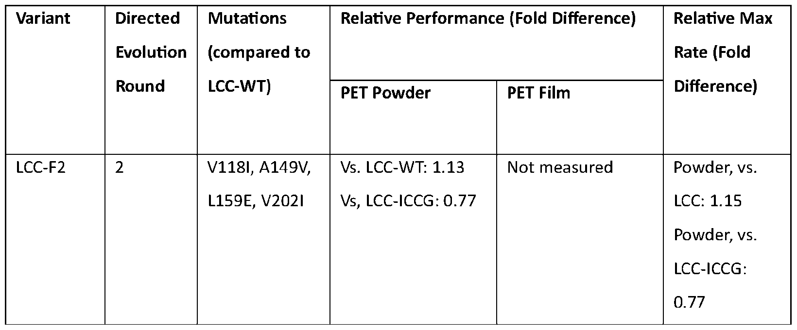

Example 4 Characterization of LCC-LANL
Following testing to characterize the evolved, purified enzyme, LCC-LANL, its performance was compared to that of the benchmark and parent, LCC-ICCG. LCC-LANL had a significantly increased maximal catalytic rate compared to the parent enzyme, LCC-ICCG, in hydrolysis of amorphous PET film coupons at 68°C (p < 0.0001, for both TPA and sum of aromatic products) (FIGS. 3A-3E). Analysis was performed with UV absorbance and HPLC aromatic monomer quantification (FIG. 3 A), with the two methods giving similar results (FIGS. 3 A-3E). LCC-LANL showed an initial rate of 1.21 g L-1 h 1 TPA and 1.77 g L 1 h 1 aromatic products,
compared to 0.88 g L 1 h 1 TPA and 1.30 g L 1 h 1 aromatic products for LCC-ICCG (FIGS. 3C, 3E). Product ratios for both enzymes were approximately equal and constant over time (about 70% TPA) (FIGS. 3C, 3D).
While initial reactions were performed with the conditions at which LCC-ICCG was reported to have the highest activity (68°C, 100 mM phosphate buffer, pH 8, with amorphous PET film), further evaluation of LCC-LANL (and LCC-ICCG) was carried out at different temperatures and for longer reaction times, beyond initial periods to measure the maximum rates. Towards this goal of thoroughly evaluating the enzymes, LCC-LANL and LCC-ICCG were additionally tested at 65°C (FIGS. 9A-9D) and 70°C (FIGS. 9E-9H). Similar to the results at 68°C, LCC-LANL exhibited a higher maximal rate than LCC-ICCG at 65°C (p < 0.0001, for both TPA and sum of aromatic products) (FIGS. 9A-9D), with strong agreement between UV absorbance measurements and HPLC monomer quantification. However, at 70°C, LCC-LANL showed a statistically insignificant increase in maximal hydrolysis rate compared to LCC-ICCG (p = 0.0904 for TPA; p = 0.4077 for sum of aromatic products) and with diminishing activity beginning after 8 hours of incubation (FIGS. 9E-9H). The increase in maximal rates for LCC-LANL vs. LCC-ICCG, however, was not as large when using purified proteins as when using crude cell lysates with normalized enzyme concentrations (FIG. 9D).
Expanding the analysis beyond the periods to observe initial and maximal rates, hydrolysis up to 24 hours was measured (FIGS. 10A-10L). Similar diminished activity was observed at 12 hours or earlier for LCC-LANL, while LCC-ICCG continued to show depolymerization continuing to 24 hours. At 65 °C and 68°C, however, further activity increases by LCC-ICCG (post-24 hours) did not result in substantially more total product release than LCC-LANL, only 11% and 8% higher at 65°C and 68°C, respectively (FIGS. 10D, 10H). Despite its significantly increased maximal rate compared to LCC-ICCG, LCC-LANL seemed to plateau in activity by 12 hours. Decreased thermostability or product inhibition were possible explanations. In screening for LCC-LANL variants, heat treatment at 80°C for 1 hour was implemented as a selection pressure to maintain the high thermostability of LCC-ICCG (Tm = 94.5 °C) and LCC-ICCG was demonstrated to not experience product inhibition.
Differential scanning calorimetry analysis (FIGS. 11A-1 IB) revealed that the thermal unfolding of each enzyme is essentially an irreversible process, with LCC-LANL and LCC-ICCG exhibiting comparable energy barriers (E'act) to unfolding from the native to denatured state. However, LCC-LANL can overcome this energy barrier with a given frequency (1 s’1) at a temperature (Tact) approximately 10°C lower than LCC-ICCG, and hence has lower kinetic stability
than the latter variant. Notwithstanding this property, evaluating the thermostability of LCC-LANL using the split GFP complementation assay (FIG. 2E), it was observed that, while LCC-LANL enzyme experienced decreased survivability above 72°C, at lower temperatures, LCC-LANL showed similar thermostability as LCC-ICCG, with essentially 100% protein retained after heat treatment at 72 °C in both cases (FIG. 12).
In order to evaluate LCC-LANL’s performance on the various PET substrates that these enzymes would likely encounter in industrial applications, the hydrolytic activity of LCC-LANL was evaluated on alternative forms of PET, namely, milled amorphous and high crystallinity PET powders. While higher crystallinity PET substrates, which are more recalcitrant to enzymatic degradation compared to lower crystallinity substrates and better represent post-consumer waste, were not the focus of these efforts, they present additional engineering goals for application of the directed evolution platform.
On the amorphous PET powder substrate (FIGS. 13A-13L), it was observed that the maximal rates of LCC-LANL and LCC-ICCG exceeded those observed for amorphous PET films (as coupons, un-milled). This trend persisted across all three reaction temperatures (FIGS. 3B-3E, FIGS. 9A-9H, FIGS. 13A-13L). In addition, a noteworthy observation was a shortened lag period for the powder substrates, with maximal rates occurring within 2 hours. However, activity appeared to plateau for powder substrates by both LCC-LANL and LCC-ICCG, with lower total hydrolysis over shorter (FIGS. 3B-3E, FIGS. 9A-9H, FIGS. 13A-13L) and longer (FIGS. 10A-10L, FIGS. 14A-14L) reaction times. Additionally, the same significant increases in maximal hydrolysis rate for amorphous PET powders were not observed as with films for LCC-LANL compared to LCC-ICCG (for sum of aromatic products: 65 °C, p = 0.0127; 68 °C, p = 0.190; 70 °C, p = 0.1345). While at 65 °C, there were approximately 2-fold increases in rate for LCC-LANL over LCC-ICCG, the rates at 68°C and 70°C (the regime at which LCC-ICCG was reported to have the highest activities) were not significantly increased for LCC-LANL (FIGS. 13A-13L).
In reactions with high crystallinity PET powder, maximal rates (FIGS. 15A-15L) and total hydrolysis over time (FIGS. 16A-16L) were largely reduced compared to the other, amorphous PET substrates, and no significant improvement in maximal rates was observed for LCC-LANL over LCC-ICCG (for sum of aromatic products: 65 °C, p = 0.0961; 68 °C, p = 0.5053; 70 °C, p = 0.5866), as well as no lag period, with maximal rates occurring within 2 hours. The hydrolysis rates of LCC-LANL and LCC-ICCG were comparable up to 12 hours at all temperatures, although LCC-ICCG appeared to outcompete LCC-LANL by 24 hours, with approximately 50% higher activities. This result was expected because amorphous PET film coupons (not high crystallinity
PET powder) were used as the main substrate screened in the validation assays to select for enzyme variants with enhanced hydrolysis performance on amorphous PET film coupons.
To further evaluate the performance of the variant and validate the directed evolution platform upon scale-up, LCC-LANL was tested alongside LCC-ICCG in pH-controlled bioreactors, with the same conditions as the small-scale experiments indicated above: 100 mM sodium phosphate buffer, pH 8, 2.9% (w/v) PET, 0.7 mg enzyme/g PET at 65°C (FIG. 4A). LCC-LANL significantly outperformed LCC-ICCG in depolymerization of amorphous PET film coupons. Both enzyme variants exhibited a lag before their maximum substrate hydrolysis rate was attained. Notably, LCC-LANL showed a 38% higher maximal rate than LCC-ICCG, and near-complete conversion of PET to hydrolytic products, with approximately 24% hydrolysis in 12 hours, compared to 12% by LCC-ICCG, and over 80% hydrolysis by 36 hours, compared to approximately 65% by LCC-ICCG. By the end of the 2-day reaction, LCC-LANL achieved 94.5% polymer mass loss, compared to 83.5% by LCC-ICCG. It is likely that the new mutations in the LCC-LANL construct change the pH optimum of the enzyme, making it less capable of tolerating the acidification in small-scale, batch reactions; however, such conditions were eliminated in the bioreactor experiments.
To test enzyme variant performance at alternate, more industrially-relevant conditions, requiring less buffer constraints and reaction at lower pH, both of which would contribute to improved process economics, LCC-LANL was compared to LCC-ICCG in pH-controlled bioreactors at 50 mM sodium phosphate buffer, pH 7.5 with 100 mM NaCl, 2.9% (w/v) PET, 0.7 mg enzyme/g PET at 65 °C (FIG. 4B). Similar to the previous experiment, LCC-LANL showed higher performance compared to LCC-ICCG, with 9% higher maximal hydrolysis rate r the parent, and a significantly improved hydrolysis profile. LCC-LANL showed approximately 24% hydrolysis in 12 hours, compared to 6% by LCC-LCCG, and over 80% hydrolysis by 36 hours, compared to only about 50% by LCC-ICCG in 36 hours. The mass losses were comparable to the original conditions, with 94% mass loss by 48 hours by LCC-LANL and 84% by LCC-ICCG.
High PET solids loadings are necessary to ensure optimal productivity (high product yield) of PET degradation reactions. Additional bioreactor runs were performed at 68°C with increased PET loadings, 16.5% (w/v), either as amorphous PET film coupons or milled amorphous PET powder (with enzyme loading at 1 mg/g PET). With amorphous PET film coupons at this substrate loading, LCC-LANL maintained improved performance compared to LCC-ICCG, with a 43% higher maximal rate (that occurred 13 hours sooner, demonstrating a shortened lag period), nearly twice the conversion after 24 hours, and higher extent of hydrolysis up to 96 hours (FIG. 4C).
Milling the amorphous PET coupons to a powder appeared to eliminate the lag period for both enzymes and increased extents of hydrolysis and total mass loss by roughly 10% by 72 hours. Additionally, in this reaction, with 16.5% amorphous PET powder, LCC-ICCG outcompeted LCC- LANL by up to 12% (occurring at ~33 hours) (FIG. 4D). However, maximal rates and total mass loss were comparable for both enzymes. Specifically, LCC-ICCG had an 8% higher maximal rate, with both enzymes reaching this at ~ 1.5 hours, both LCC-ICCG and LCC-LANL yielded similar mass losses and hydrolysis extents by 72 hours at 68°C. While the engineering goal was to develop enzymes with enhanced hydrolysis of amorphous PET film coupons here, it appears possible that eliminating the need for substrate pre-treatment (milling) could be a promising route to improving process economics, while only taking a meager reduction in overall enzyme performance.
Example 5 Structural Analysis of LCC-LANL
The structure of LCC-LANL was modeled using the Rosetta Macromolecular modeling suite. Most of the mutations obtained from directed evolution led to increased solvent exposed hydrophobicity. The S247L mutation particularly was shown to increase the hydrophobic surface area near the active site of the enzyme (FIGS. 5A-5B). While this could destabilize the protein structure, it may be favorable in the presence of substrate, which is primarily hydrophobic. The H218Y mutation (FIGS. 5C-5D), also observed by Cribari et al. Journal of the American Chemical Society 145:27380-37389, 2023) was found to add hydrophilic surface area to the enzyme and may help with protein stability. This mutation also improved surface packing by reducing exposed hydrophobic surface area, establishing a near-ideal cation-pi interaction geometry and thereby, removed a potential site for non-productive PET binding. In addition, this mutation removed a titratable and strong charge-charge repulsion interaction between H218 with KI 94, given their tight packing. Indeed, the distance between KI 94 and H218 was found to increase by 0.3 A, compared to the original solved structure (PDB:8JMP) upon running the RosettaRelax energy minimization program. The predicted configuration of K194 in LCC-LANL may also allow for better solvation, which is likely for lysine, one of the most hydrophilic residues and not participating in an ion pair interaction.
The remainder of the mutations were further away from the active site. Among these, the M91I and T256I mutations (FIGS. 5E-5F) may be synergistic in establishing a more uniformly hydrophobic core, although with the creation of several voids. These may lead to increased mobility and therefore increased catalytic efficiency of LCC-LANL enzyme at lower temperatures, 65°C, compared to 68°C and 70°C. However, the voids may also decrease thermostability. The
LI 17P mutation was predicted to be destabilizing as it removed backbone hydrogen bonding and created cavities in the protein core directly and through lever-arm effects on the backbone (Table 2). These may be worthwhile target sites to evolve in continued evolution cycles on the LCC- LANL template. Conformational ensembles of the individual mutations, the LCC-LANL mutant, and the
LCC-ICCG mutant produced contrasting predictions for stability. The Rosetta score distributions for these models are shown in FIG. 17. An analysis of LCC-LANL individual mutations and how these mutations may affect the enzyme from a structural perspective are provided in Table 2. Table 2. Mutations observed in LCC-LANL and their predicted roles from structural analysis


Flexible ligand docking simulations revealed several stable non-productive binding modes for the PET substrate near the active site of LCC-ICCG. Out of the 100 predicted best binding modes, 60 were productive, having substrate atoms within 4 A of the catalytic residues, S165, D210 and H242. Notably, all 100 predicted best binding modes were productive for LCC-LANL.
The Rosetta scores, as well as its component terms, were statistically analyzed for these modes and the entire ensemble. The analyses revealed that the difference in total Rosetta score (total_score), between the docked and free states, was higher for LCC-LANL than for LCC-ICCG (Table 3). This suggests that LCC-LANL is stabilized from binding to PET more than LCC-ICCG. The greater stabilization upon binding to PET may manifest in higher binding affinity, and more frequent productive binding may explain the higher catalytic efficiency observed in bioreactor experiments with amorphous PET film coupons. This may also explain the differences in the maximal rates of LCC-LANL v.v. LCC-ICCG in cell lysates compared to purified protein samples due to more unspecific binding in cell lysates, which may have resulted in more frequent binding of LCC-LANL to PET film coupons than LCC-ICCG. The same trend, however, was not observed for amorphous PET powder, potentially because of surface structures and charges changing upon being milled. More detailed studies on LCC-LANL’ s interaction with this substrate type may
provide valuable insights on how LCC-LANL was conferred with higher hydrolytic activity on amorphous films, but not powders.
Table 3. Total energy scores (rounded off to 1 significant digit) of the docked state, free state and the differences between docked and free states, of LCC-ICCG and LCC-LANL.

It will be apparent that the precise details of the methods or compositions described may be varied or modified without departing from the spirit of the described aspects of the disclosure. We claim all such modifications and variations that fall within the scope and spirit of the claims below.
Claims
1. A modified leaf-branch compost cutinase (LCC) polypeptide comprising at least one amino acid substitution at one or more of positions corresponding to amino acids 38, 61, 91, 117, 118, 149, 159, 202, 224, 247, and 256 of SEQ ID NO: 16.
2. The modified LCC polypeptide of claim 1 , wherein the at least one amino acid substitution comprises one or more of P38L, Y61C, M91I, LI 17P, VI 181, A 149V, L159E, V202T, Q224H, S247C, and T256I.
3. The modified LCC polypeptide of claim 2, wherein the at least one amino acid substitutions comprise: a) VI 181, A149V, L159E, and V202I; b) P38L, LI 17P, and A149V; c) P38L, LI 17P, A149V, and S247L; d) P38L, Y61C, M91I, LI 17P, A149V, and S247L; or e) P38L, Y61C, M91L, L117P, A149V, Q224H, S247L, and T256I.
4. The modified LCC polypeptide of claim 1 , wherein the polypeptide further comprises amino acid substitutions Y127G, D238C, F243I, and S283C and/or an amino acid substitution H218Y.
5. The modified LCC polypeptide of claim 4, wherein the polypeptide comprises or consists of the amino acid sequence of SEQ ID NO: 2, SEQ ID NO: 3, SEQ ID NO: 4, SEQ ID NO: 5, or SEQ ID NO: 6.
6. The modified LCC polypeptide of claim 1 , wherein the modified polypeptide has increased polyethylene terephthalate hydrolase activity compared to SEQ ID NO: 1.
7. A nucleic acid molecule encoding the modified LCC polypeptide of claim 1.
8. The nucleic acid molecule of claim 6, wherein the nucleic acid encodes a polypeptide comprising one or more of P38L, Y61C, M91I, L117P, VI 181, A149V, L159E, V202I, Q224H, S247C, and T256I.
9. The nucleic acid molecule of claim 8, wherein the nucleic acid encodes a polypeptide comprising: a) VI 181, A149V, L159E, and V202I; b) P38L, LI 17P, and A149V; c) P38L, LI 17P, A149V, and S247L; d) P38L, Y61C, M91I, LI 17P, A149V, and S247L; or e) P38L, Y61C, M91L, LI 17P, A149V, Q224H, S247L, and T256I.
10. The nucleic acid molecule of claim 8, wherein the nucleic acid comprises or consists of the nucleic acid sequence of SEQ ID NO: 8, SEQ ID NO: 9, SEQ ID NO: 10, SEQ ID NO: 11, or SEQ ID NO: 12.
11. A vector comprising the nucleic acid of claim 7.
12. The vector of claim 11, wherein the vector is a plasmid vector.
13. An isolated host cell comprising the vector of claim 12.
14. The isolated host cell of claim 13, wherein the host cell is a bacterial cell.
15. A fusion protein comprising a leaf-branch compost cutinase (LCC) polypeptide fused to a reporter protein.
16. The fusion protein of claim 15, wherein the reporter protein is a green fluorescent protein (GFP) or portion thereof.
17. The fusion protein of claim 16, wherein the GFP or portion thereof comprises a GFP11 tag.
18. The fusion protein of claim 15, wherein the LCC polypeptide comprises or consists of the amino acid sequence of SEQ ID NO: 1, SEQ ID NO: 2, SEQ ID NO: 3, SEQ ID NO: 4, SEQ ID NO: 5, or SEQ ID NO: 6.
19. A nucleic acid molecule encoding the fusion protein of claim 15, or a vector comprising a nucleic acid encoding the fusion protein.
20. A method of degrading polyethylene terephthalate (PET), comprising: contacting PET with the modified LCC polypeptide of claim 1 under conditions sufficient for degrading the PET.
Applications Claiming Priority (6)
| Application Number | Priority Date | Filing Date | Title |
|---|---|---|---|
| US202363526278P | 2023-07-12 | 2023-07-12 | |
| US63/526,278 | 2023-07-12 | ||
| US202463641318P | 2024-05-01 | 2024-05-01 | |
| US63/641,318 | 2024-05-01 | ||
| US202463643053P | 2024-05-06 | 2024-05-06 | |
| US63/643,053 | 2024-05-06 |
Publications (2)
| Publication Number | Publication Date |
|---|---|
| WO2025015294A2 true WO2025015294A2 (en) | 2025-01-16 |
| WO2025015294A3 WO2025015294A3 (en) | 2025-06-19 |
Family
ID=94216388
Family Applications (3)
| Application Number | Title | Priority Date | Filing Date |
|---|---|---|---|
| PCT/US2024/037855 Pending WO2025015288A1 (en) | 2023-07-12 | 2024-07-12 | Methods to improve enzymes that degrade polyethylene terephthalate |
| PCT/US2024/037897 Pending WO2025015302A2 (en) | 2023-07-12 | 2024-07-12 | Modified polyester hydrolase polypeptides with improved polyethylene terephthalate degrading activity |
| PCT/US2024/037863 Pending WO2025015294A2 (en) | 2023-07-12 | 2024-07-12 | Modified leaf-branch compost cutinase polypeptides with improved polyethylene terephthalate degrading activity |
Family Applications Before (2)
| Application Number | Title | Priority Date | Filing Date |
|---|---|---|---|
| PCT/US2024/037855 Pending WO2025015288A1 (en) | 2023-07-12 | 2024-07-12 | Methods to improve enzymes that degrade polyethylene terephthalate |
| PCT/US2024/037897 Pending WO2025015302A2 (en) | 2023-07-12 | 2024-07-12 | Modified polyester hydrolase polypeptides with improved polyethylene terephthalate degrading activity |
Country Status (1)
| Country | Link |
|---|---|
| WO (3) | WO2025015288A1 (en) |
Families Citing this family (1)
| Publication number | Priority date | Publication date | Assignee | Title |
|---|---|---|---|---|
| CN120210156B (en) * | 2025-05-27 | 2025-11-18 | 中国科学院大学 | A method for the degradation of polyester materials using thermally stable lignin esterases from leaf and branch compost based on rare earth ion coordination modification. |
Family Cites Families (9)
| Publication number | Priority date | Publication date | Assignee | Title |
|---|---|---|---|---|
| CA3145635A1 (en) * | 2019-07-11 | 2021-01-14 | Carbios | Esterases and uses thereof |
| US11300509B2 (en) * | 2020-06-17 | 2022-04-12 | 9412-1126 Québec Inc. | Cleavable linkers for protein translation reporting |
| GB202013063D0 (en) * | 2020-08-21 | 2020-10-07 | Nuclera Nucleics Ltd | Real-time monitoring of in vitro protein synthesis |
| CN112159478B (en) * | 2020-09-28 | 2023-02-24 | 江南大学 | A fusion protein and its application in degrading polymers |
| CN115125225B (en) * | 2021-03-25 | 2023-07-04 | 湖北大学 | PET degrading enzymes with improved thermostability |
| US20250368971A1 (en) * | 2021-08-11 | 2025-12-04 | Biometis Technology, Inc. | Enzymatic Degradation of Polyethylene Terephthalate |
| JP7732636B2 (en) * | 2021-10-13 | 2025-09-02 | 国立大学法人静岡大学 | Protein, polynucleotide, recombinant vector, transformant, composition for decomposing polyethylene terephthalate, and method for producing recycled products |
| CN114636682A (en) * | 2022-02-25 | 2022-06-17 | 山东大学 | A high-throughput assay method and application of PET hydrolase activity based on fluorescence detection |
| CN120283054A (en) * | 2022-12-09 | 2025-07-08 | 麒麟控股株式会社 | Protein having polyethylene terephthalate decomposition activity and method for decomposing polyethylene terephthalate |
-
2024
- 2024-07-12 WO PCT/US2024/037855 patent/WO2025015288A1/en active Pending
- 2024-07-12 WO PCT/US2024/037897 patent/WO2025015302A2/en active Pending
- 2024-07-12 WO PCT/US2024/037863 patent/WO2025015294A2/en active Pending
Also Published As
| Publication number | Publication date |
|---|---|
| WO2025015302A3 (en) | 2025-05-08 |
| WO2025015294A3 (en) | 2025-06-19 |
| WO2025015302A2 (en) | 2025-01-16 |
| WO2025015288A1 (en) | 2025-01-16 |
Similar Documents
| Publication | Publication Date | Title |
|---|---|---|
| Groseclose et al. | A high-throughput screening platform for engineering poly (ethylene terephthalate) hydrolases | |
| WO2025015294A2 (en) | Modified leaf-branch compost cutinase polypeptides with improved polyethylene terephthalate degrading activity | |
| CN118562763A (en) | PHL7 enzyme mutant and application thereof | |
| Irla et al. | Establishment of a functional system for recombinant production of secreted proteins at 50á░ C in the thermophilic Bacillus methanolicus | |
| CN106191083A (en) | Xylanase mutant that a kind of specific enzyme activity improves and encoding gene and application | |
| US20190024069A1 (en) | Heat-resistant agarase and monosaccharide production method using same | |
| Ming et al. | Enhancing the thermostability of carboxypeptidase A by a multiple computer-aided rational design based on amino acids preferences at β-turns | |
| CN105018443B (en) | A kind of epoxide hydrolase mutant and preparation method thereof | |
| Groseclose et al. | Engineering PHL7 for improved poly (ethylene terephthalate) depolymerization via rational design and directed evolution | |
| Wu et al. | Characterization of maltose‐binding protein–fused heparinases with enhanced thermostability by application of rigid and flexible linkers | |
| Park et al. | Probing an interfacial surface in the cyanide dihydratase from Bacillus pumilus, a spiral forming nitrilase | |
| CN109321549A (en) | A kind of directional modification enzyme of heparinase I with improved specific enzyme activity, molecular modification method and expression engineering bacteria | |
| CN103756989A (en) | Method for producing bile salt hydrolase by fermentation of twin-arginine signal peptide and application thereof | |
| Li et al. | Screening signal peptidase based on split-GFP assembly technology to promote the secretion of alkaline protease AprE in Bacillus amyloliquefaciens | |
| Sekiya et al. | Structural and biochemical characterization of the Clostridium perfringens-specific Zn2+-dependent amidase endolysin, Psa, catalytic domain | |
| CN119320765A (en) | High-activity pectase DS-5 and preparation and application thereof | |
| CN114958792B (en) | Recombinant expression type nicotinamide phosphoribosyltransferase mutant and application thereof | |
| WO2022253962A1 (en) | Mutant rnase e for enhancing recombinant protein expression | |
| Wang et al. | Enhancing the stress resistance of nitrile hydratase from Rhodococcus ruber via SpyTag/SpyCatcher-mediated α-and β-subunits ligation | |
| Li et al. | Synergistic engineering of Nattokinase terminal and calcium-binding regions for enhanced catalytic performance and Oral stability | |
| Bolstad et al. | Development of a Vibrio natriegens-based plate-clearing assay for rapid screening of PET-hydrolyzing enzymes | |
| CN120624406B (en) | A pectinase mutant, its production method and application | |
| Laksmi et al. | Expression and purification of Bst polymerase using a rhamnose-inducible system in Escherichia coli | |
| Shrestha et al. | Cloning and expression of pectinase gene from two forest soil bacteria, Streptomyces sp. | |
| US20190002864A1 (en) | Pectinases with Improved Thermostability |
Legal Events
| Date | Code | Title | Description |
|---|---|---|---|
| NENP | Non-entry into the national phase |
Ref country code: DE |