WO2025007069A2 - Imaging probes and uses thereof - Google Patents
Imaging probes and uses thereof Download PDFInfo
- Publication number
- WO2025007069A2 WO2025007069A2 PCT/US2024/036269 US2024036269W WO2025007069A2 WO 2025007069 A2 WO2025007069 A2 WO 2025007069A2 US 2024036269 W US2024036269 W US 2024036269W WO 2025007069 A2 WO2025007069 A2 WO 2025007069A2
- Authority
- WO
- WIPO (PCT)
- Prior art keywords
- hydrolase
- activated
- imaging agent
- biomolecule
- moiety
- Prior art date
- Legal status (The legal status is an assumption and is not a legal conclusion. Google has not performed a legal analysis and makes no representation as to the accuracy of the status listed.)
- Ceased
Links
Classifications
-
- C—CHEMISTRY; METALLURGY
- C09—DYES; PAINTS; POLISHES; NATURAL RESINS; ADHESIVES; COMPOSITIONS NOT OTHERWISE PROVIDED FOR; APPLICATIONS OF MATERIALS NOT OTHERWISE PROVIDED FOR
- C09B—ORGANIC DYES OR CLOSELY-RELATED COMPOUNDS FOR PRODUCING DYES, e.g. PIGMENTS; MORDANTS; LAKES
- C09B11/00—Diaryl- or thriarylmethane dyes
- C09B11/04—Diaryl- or thriarylmethane dyes derived from triarylmethanes, i.e. central C-atom is substituted by amino, cyano, alkyl
Definitions
- a compound including a hydrolase-activated covalent labeling moiety and (a) a hydrolase-activated imaging agent moiety, or (b) an affinity ligand or click chemistry reactive moiety is provided.
- kits including the compound described herein including embodiments thereof, a first nucleic acid encoding a first non-functional portion of a functional hydrolase and a second nucleic acid including a second non-functional portion of the functional hydrolase, wherein the first non-functional portion and the second non-functional portion may be combined to form the functional hydrolase.
- kits including the compound described herein including embodiments thereof including a first nucleic acid encoding a first portion of a functional beta lactamase and a second nucleic acid including a second portion of the functional beta lactamase, wherein the first portion and the second portion may be combined to form the functional beta lactamase.
- a method of detecting a proximal interaction between a first biomolecule and a second biomolecule including: (a) proximally localizing a first biomolecule hydrolase portion conjugate and a second biomolecule hydrolase portion conjugate, wherein the first biomolecule hydrolase portion conjugate includes the first biomolecule covalently bound to a first portion of a functional hydrolase and wherein the second biomolecule hydrolase portion conjugate includes the second biomolecule covalently bound to a second portion of the functional hydrolase, thereby forming the functional hydrolase; (b) contacting the functional hydrolase with a compound described herein including embodiments thereof and allowing the functional hydrolase to activate the hydrolase-activated covalent labeling moiety thereby forming a functional covalent labeling moiety, and allowing the covalent labeling moiety to covalently bind to a biomolecule thereby forming a labeled biomolecule; and (c) detecting the labeled biomolecule thereby detecting a proximal interaction between the
- a method of detecting a proximal interaction between a first biomolecule and a second biomolecule including: (a) proximally localizing a first biomolecule hydrolase portion conjugate including and a second biomolecule hydrolase portion conjugate, wherein the first biomolecule hydrolase portion conjugate includes the first biomolecule covalently bound to first portion of a functional esterase and the second biomolecule hydrolase portion conjugate includes the second biomolecule covalently bound to a second portion of the functional esterase thereby forming a complex including the functional esterase and the first biomolecule bound to the second biomolecule; (b)contacting the complex with a compound described herein including embodiments thereof and allowing the functional esterase to activate the esterase-activated imaging agent thereby forming a functional imaging agent; and (c) detecting the functional imaging agent thereby detecting an interaction between the first biomolecule and the second biomolecule.
- a method of detecting a proximal interaction between a first biomolecule and a second biomolecule including: (a) proximally localizing a first biomolecule hydrolase portion conjugate including and a second biomolecule hydrolase portion conjugate, wherein the first biomolecule hydrolase portion conjugate includes the first biomolecule covalently bound to first portion of a functional beta lactamase and the second biomolecule hydrolase portion conjugate includes the second biomolecule covalently bound to a second portion of the functional beta lactamase thereby forming a complex including the functional beta lactamase and the first biomolecule bound to the second biomolecule; (b) contacting the complex with a compound described herein including embodiments thereof and allowing the functional beta lactamase to activate the beta lactamase-activated imaging agent thereby forming a functional imaging agent; and (c) detecting the functional imaging agent thereby detecting an interaction between the first biomolecule and the second biomolecule.
- a method of detecting a biomolecule in cell or organism including: (a) contacting the cell or organism with a compound described herein including embodiments thereof, wherein the cell or organism includes a hydrolase fusion protein, the hydrolase fusion protein including a hydrolase protein portion and a subject protein portion, allowing the hydrolase protein portion to activate the hydrolase-activated covalent labeling moiety thereby forming a functional covalent labeling moiety, and allowing the covalent labeling moiety to covalently bind to a biomolecule thereby forming a labeled biomolecule; and (b) detecting the labeled biomolecule thereby detecting a proximal localization between the subject protein and the biomolecule.
- a method of detecting a biomolecule in cell or organism including: (a) contacting the cell or organism with a compound described herein including embodiments thereof, wherein the cell or organism includes a esterase fusion protein, the esterase fusion protein including an esterase portion and a subject protein portion, allowing the esterase protein portion to activate the esterase-activated covalent labeling moiety thereby forming a functional covalent labeling moiety, and allowing the covalent labeling moiety to covalently bind to a biomolecule thereby forming a labeled biomolecule; and (b) detecting the labeled biomolecule thereby detecting a proximal localization between the subject protein and the biomolecule.
- a method of detecting a subject protein in cell or organism including: (a) contacting the cell or organism with a compound described herein including embodiments thereof, wherein the cell or organism includes a beta-lactamase fusion protein, the beta-lactamase fusion protein including a beta-lactamase protein portion and a subject protein portion, allowing the beta-lactamase protein portion to activate the beta- lactamase-activated covalent labeling moiety thereby forming a functional covalent labeling moiety, and allowing the covalent labeling moiety to covalently bind to a biomolecule thereby forming a labeled biomolecule; and (b) detecting the labeled biomolecule thereby detecting a proximal interaction between the first biomolecule and the second biomolecule.
- FIG.1 shows a schematic of the compounds and methods described herein.
- FIG.2 shows the caged fluorescein (CF) dye. CF dye, CF probe, and CF are used interchangeably herein.
- FIG.3 shows the QM1-CF probe, which is a fusion between caged fluorescein (CF) and the quinone methide 1 (QM1) covalent moiety. DYENAMICS probe QM1-CF, QM1-CF, and QM1-CF probe are used interchangeably herein.
- FIG.4 shows a schematic of the activation of a quinone methide-caged fluorescein DYENAMICS probe QM1-CF by BS2 (bottom).
- FIG.5A-5D show live cell imaging with QM1-CF and CF. Snapshots of NLS-BS2 Halo transfected cells captured at different time intervals post CF (FIG.5A) and QM1-CF (FIG. 5B) addition to cells.
- Top panel (FIGS.5A-5B) is JF-594 (Janelia Fluor 549) labeling of the Halo-Tag fusion protein
- bottom panel (FIGS.5A-5B) is the fluorescein activated by BS2 esterase fusion.
- FIG.6A-6B shows a comparison between BS2-activated CF and BS2-activated QM1- CF in fixed cells.
- U2OS cells ATCC HTB-96
- the HaloTag fusion was detected by incubating cells labeled with 200 nM JF646 (Janelia Fluor 646) ligand for 10 min at 37 °C.10 ⁇ M of CF or QM1-CF was added for 15 minutes.
- FIG.6A shows CF fluorescence pre-fixation (top panel and post-fixation (bottom)
- FIG.6B shows QM1-CF fluorescence pre-fixation (top panel) and post-fixation (bottom).
- FIGS.7A-7C show results from experiments in which HEK293T cells were transfected with full length BS2 esterase with and without QM1-CF.
- FIG.7A shows experimental conditions for each of the samples.
- FIG.7B shows fluorescent labeling of the proteome with QM1-CF using a Typhoon scanner.
- FIG.7C shows total protein concentrations using Coomassie staining.
- FIG.8 shows data from in vitro experiments of recombinant BS2 activation of QM1- alkyne labeling of BSA.
- FIG.9 shows a schematic of in vivo experiments in cells that express genetically encoded BS2 and become labeled when incubated with QM1-alkyne probe. After labeling cells are fixed and then click chemistry with azide Alexa-Fluor 488 (azide-AF488) to visualize the labeling of the QM1-alkyne probe.
- FIG.10 shows results from in vivo experiments with the QM1-alkyne probe.
- FIG.11 shows a schematic of in vitro experiments with BS2-dependent activation of QM1-alkyne.
- FIG.12 shows a LiCOR gel from in vivo experiments with BS2-dependent activation of QM1-alkyne and time-dependent labeling of the proteins.
- FIG.13 shows a schematic for NLS-HALO-BS2 experiment in vivo with QM1-alkyne followed by quantitative proteomics to identify QM1-labeled proteins.
- FIG.13 shows the ten largest differences in protein abundance between the NLS-BS2-HaloTag or NLS-VHH-HaloTag cells treated with QM1-alkyne, which demonstrates the enrichment of specific proteins labeled by BS2-activated QM1-alkyne.
- FIG.14 shows QM1-alkyne labeling of cysteine amino acids of BSA.
- Starred peptides have a QM1-modification of 203.0708 m/z to demonstrate the site of labeling on BSA.
- the same peptide was modified with a single QM1-alkyne (row 6) or two QM1-alkyne molecules (row 4) demonstrating covalent labeling of BSA peptides and multiple labeling events on BSA with QM1-alkyne.
- BS2, QM1-alkyne, and BSA were mixed with PBS in a 50 ⁇ l reaction and shaken at 800 rpm for 20 min at 25 oC. Acetone precipitation was then performed.
- FIG.15 shows the structure of the QM1-azide probe, demonstrating the modularity of the click chemistry handle in the DYENAMICS chemical probes.
- the QM1-azide enables copper-free click chemistry using the Alexa Fluor 488-DBCO molecule to fluorescently label the probe.
- FIGS.16A-16B shows experiments using QM1-azide probe. NLS-BS2 Halo (FIG.
- FIG.16A 16A and NLS-VHH-Halo (FIG.16B) (negative control) transfected cells were detected with Halo-ligand JF-646 (left panel) and labeled with QM1-azide.
- Cells were fixed and click chemistry performed with DBCO-488 to detect BS2 activated QM1-azide labeling (FIG.16A, right panel).
- BS2 is necessary to activate the QM1-azide chemical probe and the background is very low.
- the QM1-azide probe shows the modularity of DYENAMICS probes wherein the exchange of the click group enables the use of different chemistries to label the probe.
- FIG.17 shows quinone methide alkyne probes: QM1-alkyne, QM2-alkyne, QM3- alkyne, and QM4-alkyne.
- FIG.18 shows in vitro BS2 experiments with all four QM alkynes probes at 10 ⁇ M with bovine serum albumin (BSA).
- BSA bovine serum albumin
- Lane 1 Vhh + QM1-alkyne + BSA
- Lane 2 BS2 + QM1- alkyne + BSA + PMSF
- Lane 3 BS2 + QM1-alkyne + BSA
- Lane 4 BS2 + QM2-alkyne + BSA
- Lane 5 BS2 + QM3-alkyne + BSA
- Lane 6 BS2 + QM4-alkyne + BSA.
- the gel was imaged on LI-COR Odyssey imager at 800 nm after click chemistry of each sample with the IR800 CW azide to enable visualization of protein labeling.
- FIGS.19A-19B show a comparison of different doses QM1-alkyne and QM3-alkyne (probes) with BSA and BS2 in vitro.
- Gels were imaged on a LiCOR at 800 nm after click chemistry of each sample with the IR800 CW azide to enable visualization of protein labeling (FIG.19A) and stained with Coomassie Blue (FIG.19B).
- FIGS.20A-20C show results from an in vivo live cell labeling with QM1-4-alkyne probes followed by click chemistry.
- U2OS cells were transfected with NLS-BS2-HaloTag or NLS-VHH-HaloTag (negative control.
- Halo-tag fusion protein transfected cells were detected by incubating cells with Halo-JF646 (top panel-Red).
- the cells were treated with QM1- (FIG. 20A), QM2- (FIG.20B), and QM4-alkyne (FIG.20C) at 50 ⁇ M, 10 ⁇ M, 2 ⁇ M concentrations for 10 minutes at 37 °C.
- Click chemistry was then performed with Alexa-Fluor 488 azide (middle panel) and nuclear DNA counterstained with DAPI (bottom panel). Images were captured on a Nikon A1R confocal microscope.
- FIGS.21A-21B show BS2 activity post fixation of cells.
- U2OS cells were transfected with NLS-BS2-HaloTag or NLS-VHH-HaloTag (negative control).
- Halo-tag fusion protein transfected cells were detected by incubating cells with Halo-JF646 (top panel-Red). Cells were fixed at 24 hours post transfection with 4% Paraformaldehyde for 20 min, and then washed with PBS. Cells were then incubated with CP1 (10 ⁇ M) (FIG.21A) or CP2 (10 ⁇ M) (FIG.21B) in PBS for 20 min. Images were captured on a Nikon A1R confocal microscope.
- FIGS.22A-22G show plasmid maps for plasmids described herein including, pEF1- nBS2-mCherry-CBX4_AI3 MAP (FIG.22A), pcDNA6.2 Orf3 linker nBS2 Map (FIG.22B), pcDNA6.2 Orf3 linker cBS2 Map (FIG.22C), pLJ043_PB-RSV-H2B-BS2fl-Halo Map (FIG.
- FIGS.23A-23C show two coupled (CP) probes and their activation by BS2.
- CP1 FIG. 23A
- CP2 FIG.23B
- CP1 and CP2 are two advanced chemical probes that contain a caged fluorophore and a caged quinone methide.
- FIGS.24A-24B show results from in vitro labeling of BSA with BS2-activated CP1 and CP2.
- FIG.24A fluorescent labeling of both BSA and BS2 by CP1 and CP2 was observed to demonstrate BS2-activation of CP1 and CP2. The labeling require on the chemical probe and BS2 and were concentration dependent with more labeling observed with higher amounts of probe and BS2.
- FIG.24B total BSA levels are the same for all lanes, showing that differences in fluorescence are due to differences in labeling and not BSA starting amount.
- FIGS.25A-25B shows results from in vitro labeling of BSA with CP1 and CP2 for different times (5-120 minutes).
- FIG.25A fluorescent labeling of both BSA and BS2 by CP1 and CP2 was observed. The labeling was time-dependent with more labeling observed at 120 minutes.
- FIG.25B total BSA levels are the same for all lanes.
- FIGS.26A-26B show results from in vitro labeling of oligo ssRNA by CP1 and CP2.
- FIG.26A fluorescent labeling of ssRNA is seen with CP1 (lanes 4 and 5) and CP2 (lanes 8 and 9). The control without BS2 (lane 3 and 7) show no fluorescent substrate RNA labeling.
- FIG. 26B total RNA remains consistent for all lanes, showing that differences in fluorescence are due to differences in labeling and not starting amount of ssRNA.
- FIGS.27A-27B show results from in vitro labeling of dsDNA by CP2.
- FIG.27A fluorescent labeling of ssDNA and annealed dsDNA by CP2.
- FIG.27B total DNA remains consistent between corresponding lanes, showing that differences in fluorescence are due to differences in labeling and not substrate starting amount.
- FIG.28 shows the CP2-alkyne (CP2A) probe.
- CP2A is a chemical probe that comprises a BS2-activated electrophile, a BS2-activated fluorophore, and a click handle.
- CP2A allows for live cell imaging, proximity labeling, and click chemistry.
- CP2A, CP2-A, CP2-alkyne, and CP2-Alkyn are used interchangeably herein.
- FIGS.29A-29B show results from CP2A and CP2 labeling of ssRNA in vitro.
- FIG. 29A fluorescent labeling of ssRNA is seen with dose dependence on CP2A (lanes 3 and 4) and CP2 (lanes 6 and 7).
- FIGS.30A-30B show results of CP2A labeling of double-stranded and single-stranded DNA in vitro.
- FIG.30A fluorescent labeling of ssDNA (lanes 3 and 5) is seen as well as fainter labeling of dsDNA by CP2A (lane 7).
- FIG.30B the total DNA concentrations are the same in all lanes.
- FIG.31 shows results from CPA2 time-dependent labeling of ssDNA. On the left, time-dependent fluorescent labeling of ssDNA by activated CP2A is observed.
- FIGS.32A-32B shows results from CP2A labeling of BSA in vitro.
- FIG.32A fluorescent labeling of both BSA and BS2 is observed with a time dependence and a dose dependence with CP2A.
- the control without BS2 in the last lane shows no fluorescent labeling, which tells us that the caged fluorophore and electrophile are specifically activated by BS2, unlike CP2 which hydrolyzes and labels to a small extent even in the absence of BS2.
- FIG.32B total BSA concentrations were the same in all lanes.
- FIG.33 shows the experimental workflow forhte samples in FIGS.34A-34B.
- FIGS.34A-34B show results from BSA labeling and enrichment with CP2A in vitro.
- FIG.34A shows fluorescent labeling of BSA imaged on a Typhoon scanner.
- Lane 1 CP2A- labeled BSA, protein precipitation, click biotin azide, no lysate, streptavidin enrichment, elution.
- Lane 2 CP2A-labeled BSA, protein precipitation, click biotin azide, plus lysate, streptavidin enrichment, elution.
- Lane 3 CP2A-labeled BSA, protein precipitation, click biotin azide, protein precipitation, no lysate, streptavidin enrichment, elution.
- Lane 4 CP2A-labeled BSA, protein precipitation, click biotin azide, protein precipitation, plus lysate, streptavidin enrichment, elution.
- Lane 5 CP2A-labeled BSA, add PMSF to inhibit BS2, add cell lysate, click biotin azide, protein precipitation, streptavidin enrichment, elution. This is the enrichment of the samples from lanes 8 and 9, which demonstrates that CP2A-labeled BSA can be reacted with biotin-azide in a complex cell lysate and then specifically enriched with streptavidin beads.
- Lane 6 CP2A- labeled BSA, protein precipitation, click biotin azide.
- FIG.34B shows total protein concentration using Coomassie staining.
- FIG.35 shows an exemplary caged-imaging probe as described herein.
- FIGS.36A-36C shows results from live cell imaging of BS2-dependent activation and proximity in cell labeling by coupled probes.
- FIGS.36A-36B Representative zoom images of live cell imaging data upon addition of Dyenamics probe CP2. Still images of cells expressing NLS-BS2fl-Halo (FIG.36A) and NLS-VHH-Halo (FIG.36B) at time-point 0 (before) and 30 minutes after the addition of CP2 (50 ⁇ M compound).
- the fluorescein signal is from the BS2- activated DYENAMICS (CP2) probe (50 ⁇ M compound, left); HaloTag signal (Halo, middle), Merged channel (Merge, right).
- FIG.36C NLS-BS2fl-Halo and NLS-VHH-Halo plotted graphs normalized intensity of the CP2 fluorescence (compound) and HaloTag (Halo) signal over time.
- Solid line are NLS-BS2fl-Halo samples, dotted lines are VHH-Halo.
- NLS-VHH- Halo transfected cells treated with CP2 n 22. Imaging methods are described in Example 24. These data show BS2-activation of coupled fluorescein electrophile probes in vivo via live imaging.
- FIGS.37A-37C show results from live cell imaging of BS2 dependent activation and proximity in cell labeling by trifunctional coupled probe, CP2-alkyne.
- FIGS.37A-37B Representative zoom images of live cell imaging data upon addition of Dyenamics probe CP2- alkyne. Still images of cell expressing NLS-BS2fl-Halo (FIG.37A) and NLS-VHH-Halo (FIG.
- CP2A 50 ⁇ M compound
- the fluorescein signal is from the BS2-activated DYENAMICS (CP2A) probe (50 ⁇ M compound, left); HaloTag signal (Halo; middle), Merged channel (Merge; right). Imaging was performed as described (Example 25) with 9 minutes intervals for fluorescein-(488 nm) and HaloTag signal (637nm) for 30 minutes post addition of 50 ⁇ M Dyenamics probe (CP2A).
- FIG.37C NLS- BS2fl-Halo and NLS-VHH-Halo plotted graphs normalized intensity of Compound and HaloTag (Halo) signal over time.
- Solid line are NLS-BS2fl-Halo samples, dotted lines are VHH-Halo.
- FIGS.38A-38B show click chemistry labeling of CP2A-labeled biomolecules in BS2- expressing cells. Representative zoom images of cells expressing NLS-BS2fl-Halo (FIG.38A) or NLS-VHH-Halo (FIG.38B) that had been labeled with CP2A (fluorescein; second from left), JF-646 (fourth from left) and click chemistry Azide-594 (third from left).
- FIGS.39A-39B show an example of live cell imaging of BS2-dependent activation and proximity labeling of histones and nuclear proteins in H2B-BS2-HaloTag fusion expressing cells and membrane proteins in CAAX-BS2 fusion expressing cells with CP2A.
- Fluorescein first panel from left
- HaloTag signal fourth panel from left).
- FIG.40 shows a schematic representation of Histone H2B-BS2-HaloTag fusion and proximity labeling.
- the H2B-BS2-HaloTag fusion will be localized in the nucleus and assemble with H2A/H3/H4 into nucleosome/DNA particles. Adding CP2A will result in CP2A diffusing into the nucleus, where BS2 will activate it.
- activated CP2A will covalently label proximal histone nucleosome octamer proteins, such as H2A, H3, H4 see Molecular biology of the cell (4th ed.). New York: Garland Science. p.207.
- FIGS.41A-41F show results from an experiment labeling histones with CPA2.
- FIG. 41A A scheme showing the workflow for the enrichment of histones from cells including the soluble and acid extract samples which are analyzed on the following panels.
- FIG.41B SDS- PAGE analysis and Coomassie staining of the soluble and acid extract samples shows that the histones are enriched in the acid extract samples.
- FIG.41C Scanning of the gel to detect the HaloTag-JF646 ligands demonstrated the expression of NLS-BS2-Halo, NLS-Vhh-Halo, and H2B-BS2-HaloTag in the soluble and acid extraction fraction. The overall expression of all three of these proteins is similar but they fractionate differently with nls-BS2-Halo and NLS-Vhh-Halo mostly partitioning into the soluble nuclear fraction while H2B-BS2-HaloTag is enriched in the acid extracted chromatin/histone fraction (as expected).
- FIG.41D Scanning the gel to identify CP2A (fluorescein)-labeled proteins show that the NLS-BS2-Halo and H2B-BS2-HaloTag are self- labeled with fluorescein, as is expected since the BS2 can self-label.
- CP2A-treated acid extract there are also clearly visible fluorescent bands at their characteristic molecular weight and banding pattern, indicating the proximity covalent labeling of H2A and H3 by CP2A by H2B-BS2-HaloTag in interacting nucleosomes.
- FIG.41E We validated the identity of those proteins by transferring the gel to a membrane and blotting with H2A and then H3, which indicate that the histones are running at the correct molecular weight.
- FIG.41F Overlap of the H2A Western blot with the CP2-treated acid extracted samples establishes the identity of the CP2A-labeled protein as histones.
- FIG.42 shows results from a toxicity assessment of BS2 expressing cells treated with CP2-alkyne (CP2A).
- the top panel shows JF646 labeled Histone H2B-BS2-HaloTag staining and localization.
- JF-646 labeled H2B-BS2-HaloTag localizes to the nucleus and upon mitosis, condensed chromosomes.
- the bottom panel shows a merge of JF646 fluorescence and H2B-BS2-HaloTag fluorescein labeled proximal biomolecules labeling.
- FIG.43 shows the structure of BS2.
- FIGS.44A-44C show results from rapamycin activation of BS2 by regulating the interaction between C-terminal split BS2 (CBS2)-FKBP-HaloTag and N-terminal split BS2 (NBS2)-FRB-mScarlet fusion proteins.
- FIG.44A U2OS cells were transfected with CBS2- FKBP-HaloTag plasmid alone.
- CBS2-FKBP-HaloTag was validated by detecting JF646 fluorescence (right panel). These cells were treated with CP2 and rapamycin, but since they do not express NBS2-FRB, no BS2 activity and no fluorescein signal is observed.
- FIG.44B U2OS cells were transfected with CBS2-FKBP-HaloTag plasmid and nBS2-FRB- mScarlet plasmid. Expression of CBS2-FKBP-HaloTag detected JF646 (right panel) and expression of nBS2-FRB-mScarlet was validated by detection of mScarlet. These cells are treated with CP2 but no rapamycin.
- FIG.44C U2OS cells were transfected with CBS2-FKBP-HaloTag plasmid and nBS2-FRB-mScarlet plasmid. Expression of CBS2-FKBP-HaloTag detected by detection of JF646 (right panel) and expression of nBS2-FRB-mScarlet was validated by detection of mScarlet. These cells are treated with CP2 and rapamycin.
- FIGS.45A-45D show results from experiments labeling E4-ORF3 polymers with QM1-alkyne.
- FIG.45A QM1-alkyne labeled E4-ORF3 polymers visualized by click chemistry with click-azide-488.
- FIG.45B E4-ORF3 labeled and visualized with E4-ORF3 antibodies.
- FIG.45C Colocalization of covalent QM1-alkyne proximity labeled molecules with E4-ORF3 polymer assemblies.
- FIG.45D Nucleus visualization with Hoechst. [0066]
- FIGS.46A-46C show that co-expression of N- and C-split BS2 fragment fusions with E4-ORF3 and CBX4, respectively, reconstitutes BS2 and upon addition of QM1-alkyne, activates covalent labeling of E4-ORF3/CBX4 polymer assemblies together with proximal biomolecules, which can be visualized by click chemistry.
- U2OS cells were transfected with either E4-ORF3-NBS2 (FIG.46A), CBS2-Halo-CBX4 (FIG.46B) alone, or both together (FIG. 46C)).
- HaloTag-CBX4 was visualized with JF646 ligand (FIGS.46A-46C, middle panels).
- Cells were incubated with 10 ⁇ M of QM1-alkyne, fixed and QM1-alkyne labeled cellular biomolecules visualized by click chemistry with click-azide-488 (FIGS.46A-46C, right panels).
- E4-ORF3 was detected by anti-E4-ORF3 antibodies in immunofluorescence (FIGS.46A-46C, left panels).
- FIGS.47A-47F show results from experiments in which split BS2 esterase activity is reconstituted by interactions between ORF3 interacting proteins.
- U2OS cells were transfected with either E4-ORF3-NBS2 (FIG.47A), CBS2-Halo-Ring1b (FIG.47B), or both at a ratio of 1:1 (FIG.47C), and either E4-ORF3-NBS2 (FIG.47D), CBS2-Halo-NBS1 (FIG.47E), or both at a ratio of 1:1 (FIG.47F).
- HaloTag was visualized with JF646 ligand (middle panels).
- FIGS.48A-48C show results from live imaging in which split BS2 esterase activity is reconstituted.
- U2OS cells were transfected with cBS2-Halo-CBX4 alone (FIG.48A), or co- transfected with E4-ORF3-NBS2 and CBS2-Halo-CBX4 fusions at a ratio of 1:1 (FIG.48B).
- live cell imaging was performed for 45 minutes at short time intervals.
- JF- 646 was used to fluorescently label cBS2-Halo-CBX4 (middle panels) expressing cells.50 ⁇ M of CP2-alkyne (CP2A) was added after first time intervals.
- FIG.48C Zoom of split BS2 dependent activation of CP2A fluorescein labeling of E4-ORF3/CBX4 nuclear assemblies (top). Intensity Line plot of multi-fluorescent signal co-localization (bottom). CBX4/E4-ORF3 co-assemble into a nuclear polymer, which reconstitutes BS2 activity, labeling the E4-ORF3 polymer and proximal interacting biomolecules with fluorescein, which can be visualized in live imaging.
- FIGS.49A-49B show results showing labeling of biomolecules in proximity to UV- induced DNA damage.
- U2OS cells were co-transfected with cBS2-Halo-RAD18 and nBS2- mCherry-PCNA at a ratio of 1:1. Cells were untreated (FIG.49A) or irradiated with 20 J/m2 (FIG.49B) and then incubated for 1h at 37 °C.
- FIG.49A In S phase numerous small RAD18 foci appear. At these sites, only 20% of RAD18 colocalizes with PCNA, but do not interact to reconstitute BS2 activity and labeling by QM1-alkyne (FIG.49B) .
- FIG.50 shows an exemplary DYENAMICS imaging probe described herein.
- FIG.51 shows an exemplary DYENAMICS imaging probe (CP2-BCN) described herein that can undergo click chemistry under copper-free conditions.
- CP2-BCN includes bicyclo[6.1.0]nonyne (BCN) as the click chemistry group, but a variety of other copper-free click chemistry groups can also be used because of the modular structure of the DYENAMICS probes.
- FIG.52 shows CP2-BCN labeling of BSA. Experiments were performed for the indicated concentrations of CP2-BCN and times (1, 5, 20, 30, and 60 min) with recombinant BS2 under the same conditions used for FIGS.24, 25, and 32.
- FIGS.53A-53C show CP2-BCN labeling of nucleic acids. Experiments were performed for the indicated concentrations of CP2-BCN and times (1, 5, 20, 30, and 60 min) with recombinant BS2 under the same conditions used for FIGS.26, 27, 29, 30, and 31. These data demonstrate that the modular structure of DYENAMICS probes allows us to modify the linker and click chemistry group while maintaining nucleic acid labeling.
- FIGS.54A-54B show time-dependent activation of fluorescence by BS2 using either CP2-A or CP2-BCN.
- H2B-BS2-Halo left panels
- NLS-VHH- Halo right panels
- CP2-A CP2-A
- CP2-BCN CP2-BCN
- H2B-BS2 cells FIGS.54A-54B, left panels, black line
- NLS-VHH cells FIGS.54A-54B, right panels, black line
- the Halo was labeled with the JF-549 ligand and gave a steady signal in all the samples (dotted line).
- FIGS.55A-55B show CP2-BCN proximity labeling of proximal biomolecules in vivo.
- U2OS cells were transfected with BS2-HaloTag-OMM (outer mitochondrial membrane marker protein; FIG.55A) or BS2-HaloTag-ERM (ER membrane protein marker; FIG.55B).
- HaloTag was labeled by incubating cells with 200 nM Halo-JF549 for 20 minutes.
- CP2-BCN was added at 50uM for 20 minutes.
- FIGS.56A-56B show Cp2-BCN labeling of proximal biomolecules in cellular compartments is non-toxic and maintained through cell division.
- FIG.56A H2B-BS2-HaloTag and CP2-BCN labeling and live imaging.
- FIG.56B Live imaging of BS2-HaloTag-OMM CP2- BCN labeling of mitochondrion biomolecules.
- FIGS.57A-57B shows CP2-BCN labeling of protein followed by copper-free click chemistry in lysates with methyl tetrazine biotin to attach a biotin to the protein through the DYENAMICS probe in cell lysates.
- FIGS.58A-58B shows CP2-BCN labeling of protein followed by copper-free click chemistry in lysates with methyl tetrazine TAMRA (TMR) to attach a TMR to the protein through the DYENAMICS probe in cell lysates.
- TMR methyl tetrazine TAMRA
- FIGS.59A-59F show representative data from in cell copper-free click labeling of BS2-CP2-BCN labeled biomolecules with tetrazine compounds.
- FIG.60 shows a schematic for RAD18/Rad6 ubiquitination of PCNA upon DNA damage, such as irradiation, which RAD18/Rad6 assembly catalyzes K63-linked multi- ubiquitination of PCNA.
- Rad18 is an E3 ubiquitin ligase. Rad18 stably interacts with the UBC 2 protein, Rad6. Upon irradiation, Rad18/Rad6 are recruited to sites of DNA breaks where they interact, modulate and modify DNA damage repair and replication proteins.
- FIGS.61A-61C show results from Rad18 proximity labeling experiments.
- FIGS.62A-62C show quantitative proteomics of streptavidin enriched Rad18-BS2 biotin-labeled interacting proteins in vivo.
- FIG.62A Schematic of the NLS-BS2-Halo-RAD18 cells treated with CP2-BCN to label proximal proteins, followed by cell lysis, biotin labeling, and streptavidin enrichment to provide samples for quantitative proteomics.
- FIG.62B The table shows data from four conditions: 1. untransfected controls + CP2-BCN, 2. BS2-Halo-CAAX + CP2-BCN, 3. NLS-BS2-Halo-RAD18 + irradiation + CP2-BCN, and 4.
- alkyl by itself or as part of another substituent, means, unless otherwise stated, a straight (i.e., unbranched) or branched carbon chain (or carbon), or combination thereof, which may be fully saturated, mono- or polyunsaturated and can include mono-, di- and multivalent radicals.
- the alkyl may include a designated number of carbons (e.g., C 1 -C 10 means one to ten carbons).
- the alkyl is fully saturated. In embodiments, the alkyl is monounsaturated. In embodiments, the alkyl is polyunsaturated. Alkyl is an uncyclized chain. Examples of saturated hydrocarbon radicals include, but are not limited to, groups such as methyl, ethyl, n-propyl, isopropyl, n-butyl, t-butyl, isobutyl, sec-butyl, methyl, homologs and isomers of, for example, n-pentyl, n-hexyl, n-heptyl, n-octyl, and the like.
- An unsaturated alkyl group is one having one or more double bonds or triple bonds.
- Examples of unsaturated alkyl groups include, but are not limited to, vinyl, 2-propenyl, crotyl, 2-isopentenyl, 2-(butadienyl), 2,4-pentadienyl, 3-(1,4-pentadienyl), ethynyl, 1- and 3-propynyl, 3-butynyl, and the higher homologs and isomers.
- An alkoxy is an alkyl attached to the remainder of the molecule via an oxygen linker (-O-).
- An alkyl moiety may be an alkenyl moiety.
- An alkyl moiety may be an alkynyl moiety.
- An alkenyl includes one or more double bonds.
- An alkynyl includes one or more triple bonds.
- alkylene by itself or as part of another substituent, means, unless otherwise stated, a divalent radical derived from an alkyl, as exemplified, but not limited by, - CH2CH2CH2CH2-.
- an alkyl (or alkylene) group will have from 1 to 24 carbon atoms, with those groups having 10 or fewer carbon atoms being preferred herein.
- a “lower alkyl” or “lower alkylene” is a shorter chain alkyl or alkylene group, generally having eight or fewer carbon atoms.
- alkenylene by itself or as part of another substituent, means, unless otherwise stated, a divalent radical derived from an alkene.
- the alkylene is fully saturated. In embodiments, the alkylene is monounsaturated. In embodiments, the alkylene is polyunsaturated. An alkenylene includes one or more double bonds. An alkynylene includes one or more triple bonds. [0089]
- heteroalkyl by itself or in combination with another term, means, unless otherwise stated, a stable straight or branched chain, or combinations thereof, including at least one carbon atom and at least one heteroatom (e.g., O, N, P, Si, and S), and wherein the nitrogen and sulfur atoms may optionally be oxidized, and the nitrogen heteroatom may optionally be quaternized.
- heteroatom(s) e.g., O, N, S, Si, or P
- the heteroatom(s) may be placed at any interior position of the heteroalkyl group or at the position at which the alkyl group is attached to the remainder of the molecule.
- Heteroalkyl is an uncyclized chain.
- a heteroalkyl moiety may include one heteroatom (e.g., O, N, S, Si, or P).
- a heteroalkyl moiety may include two optionally different heteroatoms (e.g., O, N, S, Si, or P).
- a heteroalkyl moiety may include three optionally different heteroatoms (e.g., O, N, S, Si, or P).
- a heteroalkyl moiety may include four optionally different heteroatoms (e.g., O, N, S, Si, or P).
- a heteroalkyl moiety may include five optionally different heteroatoms (e.g., O, N, S, Si, or P).
- a heteroalkyl moiety may include up to 8 optionally different heteroatoms (e.g., O, N, S, Si, or P).
- the term “heteroalkenyl,” by itself or in combination with another term, means, unless otherwise stated, a heteroalkyl including at least one double bond.
- a heteroalkenyl may optionally include more than one double bond and/or one or more triple bonds in additional to the one or more double bonds.
- heteroalkynyl by itself or in combination with another term, means, unless otherwise stated, a heteroalkyl including at least one triple bond.
- a heteroalkynyl may optionally include more than one triple bond and/or one or more double bonds in additional to the one or more triple bonds.
- the heteroalkyl is fully saturated.
- the heteroalkyl is monounsaturated.
- the heteroalkyl is polyunsaturated.
- the term “heteroalkylene,” by itself or as part of another substituent means, unless otherwise stated, a divalent radical derived from heteroalkyl, as exemplified, but not limited by, -CH2-CH2-S-CH2-CH2- and -CH2-S-CH2-CH2-NH-CH2-.
- heteroatoms can also occupy either or both of the chain termini (e.g., alkyleneoxy, alkylenedioxy, alkyleneamino, alkylenediamino, and the like). Still further, for alkylene and heteroalkylene linking groups, no orientation of the linking group is implied by the direction in which the formula of the linking group is written. For example, the formula -C(O)2R'- represents both -C(O) 2 R'- and -R'C(O) 2 -.
- heteroalkyl groups include those groups that are attached to the remainder of the molecule through a heteroatom, such as - C(O)R', -C(O)NR', -NR'R'', -OR', -SR', and/or -SO 2 R'.
- heteroalkyl is recited, followed by recitations of specific heteroalkyl groups, such as -NR'R'' or the like, it will be understood that the terms heteroalkyl and -NR'R'' are not redundant or mutually exclusive. Rather, the specific heteroalkyl groups are recited to add clarity.
- heteroalkyl should not be interpreted herein as excluding specific heteroalkyl groups, such as -NR'R'' or the like.
- heteroalkenylene by itself or as part of another substituent, means, unless otherwise stated, a divalent radical derived from a heteroalkene.
- heteroalkynylene by itself or as part of another substituent, means, unless otherwise stated, a divalent radical derived from a heteroalkyne.
- the heteroalkylene is fully saturated.
- the heteroalkylene is monounsaturated.
- the heteroalkylene is polyunsaturated.
- a heteroalkenylene includes one or more double bonds.
- a heteroalkynylene includes one or more triple bonds.
- cycloalkyl examples include, but are not limited to, cyclopropyl, cyclobutyl, cyclopentyl, cyclohexyl, 1-cyclohexenyl, 3-cyclohexenyl, cycloheptyl, and the like.
- heterocycloalkyl examples include, but are not limited to, 1-(1,2,5,6- tetrahydropyridyl), 1-piperidinyl, 2-piperidinyl, 3-piperidinyl, 4-morpholinyl, 3-morpholinyl, tetrahydrofuran-2-yl, tetrahydrofuran-3-yl, tetrahydrothien-2-yl, tetrahydrothien-3-yl, 1- piperazinyl, 2-piperazinyl, and the like.
- the cycloalkyl is fully saturated.
- the cycloalkyl is monounsaturated.
- the cycloalkyl is polyunsaturated.
- the heterocycloalkyl is fully saturated.
- the heterocycloalkyl is monounsaturated.
- the heterocycloalkyl is polyunsaturated.
- cycloalkyl means a monocyclic, bicyclic, or a multicyclic cycloalkyl ring system.
- monocyclic ring systems are cyclic hydrocarbon groups containing from 3 to 8 carbon atoms, where such groups can be saturated or unsaturated, but not aromatic.
- cycloalkyl groups are fully saturated.
- a bicyclic or multicyclic cycloalkyl ring system refers to multiple rings fused together wherein at least one of the fused rings is a cycloalkyl ring and wherein the multiple rings are attached to the parent molecular moiety through any carbon atom contained within a cycloalkyl ring of the multiple rings.
- a cycloalkyl is a cycloalkenyl.
- the term “cycloalkenyl” is used in accordance with its plain ordinary meaning.
- a cycloalkenyl is a monocyclic, bicyclic, or a multicyclic cycloalkenyl ring system.
- a bicyclic or multicyclic cycloalkenyl ring system refers to multiple rings fused together wherein at least one of the fused rings is a cycloalkenyl ring and wherein the multiple rings are attached to the parent molecular moiety through any carbon atom contained within a cycloalkenyl ring of the multiple rings.
- heterocycloalkyl means a monocyclic, bicyclic, or a multicyclic heterocycloalkyl ring system.
- heterocycloalkyl groups are fully saturated.
- a bicyclic or multicyclic heterocycloalkyl ring system refers to multiple rings fused together wherein at least one of the fused rings is a heterocycloalkyl ring and wherein the multiple rings are attached to the parent molecular moiety through any atom contained within a heterocycloalkyl ring of the multiple rings.
- halo or “halogen,” by themselves or as part of another substituent, mean, unless otherwise stated, a fluorine, chlorine, bromine, or iodine atom. Additionally, terms such as “haloalkyl” are meant to include monohaloalkyl and polyhaloalkyl.
- halo(C 1 -C 4 )alkyl includes, but is not limited to, fluoromethyl, difluoromethyl, trifluoromethyl, 2,2,2-trifluoroethyl, 4-chlorobutyl, 3-bromopropyl, and the like.
- acyl means, unless otherwise stated, -C(O)R where R is a substituted or unsubstituted alkyl, substituted or unsubstituted cycloalkyl, substituted or unsubstituted heteroalkyl, substituted or unsubstituted heterocycloalkyl, substituted or unsubstituted aryl, or substituted or unsubstituted heteroaryl.
- aryl means, unless otherwise stated, a polyunsaturated, aromatic, hydrocarbon substituent, which can be a single ring or multiple rings (preferably from 1 to 3 rings) that are fused together (i.e., a fused ring aryl) or linked covalently.
- a fused ring aryl refers to multiple rings fused together wherein at least one of the fused rings is an aryl ring and wherein the multiple rings are attached to the parent molecular moiety through any carbon atom contained within an aryl ring of the multiple rings.
- heteroaryl refers to aryl groups (or rings) that contain at least one heteroatom such as N, O, or S, wherein the nitrogen and sulfur atoms are optionally oxidized, and the nitrogen atom(s) are optionally quaternized.
- heteroaryl includes fused ring heteroaryl groups (i.e., multiple rings fused together wherein at least one of the fused rings is a heteroaromatic ring and wherein the multiple rings are attached to the parent molecular moiety through any atom contained within a heteroaromatic ring of the multiple rings).
- a 5,6-fused ring heteroarylene refers to two rings fused together, wherein one ring has 5 members and the other ring has 6 members, and wherein at least one ring is a heteroaryl ring.
- a 6,6-fused ring heteroarylene refers to two rings fused together, wherein one ring has 6 members and the other ring has 6 members, and wherein at least one ring is a heteroaryl ring.
- a 6,5-fused ring heteroarylene refers to two rings fused together, wherein one ring has 6 members and the other ring has 5 members, and wherein at least one ring is a heteroaryl ring.
- a heteroaryl group can be attached to the remainder of the molecule through a carbon or heteroatom.
- Non-limiting examples of aryl and heteroaryl groups include phenyl, naphthyl, pyrrolyl, pyrazolyl, pyridazinyl, triazinyl, pyrimidinyl, imidazolyl, pyrazinyl, purinyl, oxazolyl, isoxazolyl, thiazolyl, furyl, thienyl, pyridyl, pyrimidyl, benzothiazolyl, benzoxazoyl benzimidazolyl, benzofuran, isobenzofuranyl, indolyl, isoindolyl, benzothiophenyl, isoquinolyl, quinoxalinyl, quinolyl, 1-naphthyl, 2-naphthyl, 4-biphenyl, 1-pyrrolyl, 2-pyrrolyl, 3-pyrrolyl, 3- pyrazolyl, 2-imidazolyl, 4-imid
- Substituents for each of the above noted aryl and heteroaryl ring systems are selected from the group of acceptable substituents described below.
- a heteroaryl group substituent may be -O- bonded to a ring heteroatom nitrogen.
- a fused ring heterocyloalkyl-aryl is an aryl fused to a heterocycloalkyl.
- a fused ring heterocycloalkyl-heteroaryl is a heteroaryl fused to a heterocycloalkyl.
- a fused ring heterocycloalkyl-cycloalkyl is a heterocycloalkyl fused to a cycloalkyl.
- a fused ring heterocycloalkyl-heterocycloalkyl is a heterocycloalkyl fused to another heterocycloalkyl.
- Fused ring heterocycloalkyl-aryl, fused ring heterocycloalkyl-heteroaryl, fused ring heterocycloalkyl- cycloalkyl, or fused ring heterocycloalkyl-heterocycloalkyl may each independently be unsubstituted or substituted with one or more of the substituents described herein.
- Spirocyclic rings are two or more rings wherein adjacent rings are attached through a single atom.
- the individual rings within spirocyclic rings may be identical or different.
- Individual rings in spirocyclic rings may be substituted or unsubstituted and may have different substituents from other individual rings within a set of spirocyclic rings.
- Possible substituents for individual rings within spirocyclic rings are the possible substituents for the same ring when not part of spirocyclic rings (e.g., substituents for cycloalkyl or heterocycloalkyl rings).
- Spirocyclic rings may be substituted or unsubstituted cycloalkyl, substituted or unsubstituted cycloalkylene, substituted or unsubstituted heterocycloalkyl or substituted or unsubstituted heterocycloalkylene and individual rings within a spirocyclic ring group may be any of the immediately previous list, including having all rings of one type (e.g. all rings being substituted heterocycloalkylene wherein each ring may be the same or different substituted heterocycloalkylene).
- heterocyclic spirocyclic rings means a spirocyclic rings wherein at least one ring is a heterocyclic ring and wherein each ring may be a different ring.
- substituted spirocyclic rings means that at least one ring is substituted, and each substituent may optionally be different.
- alkylsulfonyl means a moiety having the formula -S(O 2 )-R', where R' is a substituted or unsubstituted alkyl group as defined above. R' may have a specified number of carbons (e.g., “C 1 -C 4 alkylsulfonyl”).
- alkylarylene as an arylene moiety covalently bonded to an alkylene moiety (also referred to herein as an alkylene linker). In embodiments, the alkylarylene group has the formula: [0104] An alkylarylene moiety may be substituted (e.g.
- the alkylarylene is unsubstituted.
- Each of the above terms e.g., “alkyl,” “heteroalkyl,” “cycloalkyl,” “heterocycloalkyl,” “aryl,” and “heteroaryl” includes both substituted and unsubstituted forms of the indicated radical. Preferred substituents for each type of radical are provided below.
- R, R', R'', R'', and R''' each preferably independently refer to hydrogen, substituted or unsubstituted heteroalkyl, substituted or unsubstituted cycloalkyl, substituted or unsubstituted heterocycloalkyl, substituted or unsubstituted aryl (e.g., aryl substituted with 1-3 halogens), substituted or unsubstituted heteroaryl, substituted or unsubstituted alkyl, alkoxy, or thioalkoxy groups, or arylalkyl groups.
- aryl e.g., aryl substituted with 1-3 halogens
- substituted or unsubstituted heteroaryl substituted or unsubstituted alkyl, alkoxy, or thioalkoxy groups, or arylalkyl groups.
- each of the R groups is independently selected as are each R', R'', R''', and R''' group when more than one of these groups is present.
- R' and R'' are attached to the same nitrogen atom, they can be combined with the nitrogen atom to form a 4-, 5-, 6-, or 7-membered ring.
- -NR'R'' includes, but is not limited to, 1-pyrrolidinyl and 4-morpholinyl.
- alkyl is meant to include groups including carbon atoms bound to groups other than hydrogen groups, such as haloalkyl (e.g., -CF3 and -CH2CF3) and acyl (e.g., -C(O)CH3, -C(O)CF3, -C(O)CH2OCH3, and the like).
- haloalkyl e.g., -CF3 and -CH2CF3
- acyl e.g., -C(O)CH3, -C(O)CF3, -C(O)CH2OCH3, and the like.
- each of the R groups is independently selected as are each R', R'', R'', and R''' groups when more than one of these groups is present.
- Substituents for rings e.g. cycloalkyl, heterocycloalkyl, aryl, heteroaryl, cycloalkylene, heterocycloalkylene, arylene, or heteroarylene
- substituents on the ring may be depicted as substituents on the ring rather than on a specific atom of a ring (commonly referred to as a floating substituent).
- the substituent may be attached to any of the ring atoms (obeying the rules of chemical valency) and in the case of fused rings or spirocyclic rings, a substituent depicted as associated with one member of the fused rings or spirocyclic rings (a floating substituent on a single ring), may be a substituent on any of the fused rings or spirocyclic rings (a floating substituent on multiple rings).
- the multiple substituents may be on the same atom, same ring, different atoms, different fused rings, different spirocyclic rings, and each substituent may optionally be different.
- a point of attachment of a ring to the remainder of a molecule is not limited to a single atom (a floating substituent)
- the attachment point may be any atom of the ring and in the case of a fused ring or spirocyclic ring, any atom of any of the fused rings or spirocyclic rings while obeying the rules of chemical valency.
- a ring, fused rings, or spirocyclic rings contain one or more ring heteroatoms and the ring, fused rings, or spirocyclic rings are shown with one more floating substituents (including, but not limited to, points of attachment to the remainder of the molecule), the floating substituents may be bonded to the heteroatoms.
- the ring heteroatoms are shown bound to one or more hydrogens (e.g. a ring nitrogen with two bonds to ring atoms and a third bond to a hydrogen) in the structure or formula with the floating substituent, when the heteroatom is bonded to the floating substituent, the substituent will be understood to replace the hydrogen, while obeying the rules of chemical valency.
- Two or more substituents may optionally be joined to form aryl, heteroaryl, cycloalkyl, or heterocycloalkyl groups.
- Such so-called ring-forming substituents are typically, though not necessarily, found attached to a cyclic base structure.
- the ring-forming substituents are attached to adjacent members of the base structure.
- two ring-forming substituents attached to adjacent members of a cyclic base structure create a fused ring structure.
- the ring-forming substituents are attached to a single member of the base structure.
- two ring-forming substituents attached to a single member of a cyclic base structure create a spirocyclic structure.
- the ring- forming substituents are attached to non-adjacent members of the base structure.
- Two of the substituents on adjacent atoms of the aryl or heteroaryl ring may optionally form a ring of the formula -T-C(O)-(CRR')q-U-, wherein T and U are independently -NR-, -O-, - CRR'-, or a single bond, and q is an integer of from 0 to 3.
- two of the substituents on adjacent atoms of the aryl or heteroaryl ring may optionally be replaced with a substituent of the formula -A-(CH2)r-B-, wherein A and B are independently -CRR'-, -O-, -NR-, -S-, -S(O) -, - S(O) 2 -, -S(O) 2 NR'-, or a single bond, and r is an integer of from 1 to 4.
- One of the single bonds of the new ring so formed may optionally be replaced with a double bond.
- two of the substituents on adjacent atoms of the aryl or heteroaryl ring may optionally be replaced with a substituent of the formula -(CRR')s-X'- (C''R''R'')d-, where s and d are independently integers of from 0 to 3, and X' is -O-, -NR'-, -S-, -S(O)-, -S(O) 2 -, or -S(O) 2 NR'-.
- R, R', R'', and R''' are preferably independently selected from hydrogen, substituted or unsubstituted alkyl, substituted or unsubstituted heteroalkyl, substituted or unsubstituted cycloalkyl, substituted or unsubstituted heterocycloalkyl, substituted or unsubstituted aryl, and substituted or unsubstituted heteroaryl.
- heteroatom or “ring heteroatom” are meant to include oxygen (O), nitrogen (N), sulfur (S), phosphorus (P), and silicon (Si).
- a “substituent group,” as used herein, means a group selected from the following moieties: (A) oxo, halogen, -CCl 3 , -CBr 3 , -CF 3 , -CI 3 , -CH 2 Cl, -CH 2 Br, -CH 2 F, -CH 2 I, -CHCl 2 , -CHBr2, -CHF2, -CHI2, -CN, -OH, -NH2, -COOH, -CONH2, -NO2, -SH, -SO3H, -SO 4 H, -SO 2 NH 2 , ⁇ NHNH 2 , ⁇ ONH 2 , ⁇ NHC(O)NHNH 2 , -NHC(O)NH 2 , -NHSO 2 H, -NHC(O)H, -NHC(O)OH, -NHOH, -OCCl 3 , -OCF 3 , -OCBr 3
- a “size-limited substituent” or “ size-limited substituent group,” as used herein, means a group selected from all of the substituents described above for a “substituent group,” wherein each substituted or unsubstituted alkyl is a substituted or unsubstituted C1-C20 alkyl, each substituted or unsubstituted heteroalkyl is a substituted or unsubstituted 2 to 20 membered heteroalkyl, each substituted or unsubstituted cycloalkyl is a substituted or unsubstituted C 3 -C 8 cycloalkyl, each substituted or unsubstituted heterocycloalkyl is a substituted or unsubstituted 3 to 8 membered heterocycloalkyl, each substituted or unsubstituted aryl is a substituted or unsubstituted C6-C10 aryl, and each substituted or unsubstituted heteroaryl is
- a “lower substituent” or “ lower substituent group,” as used herein, means a group selected from all of the substituents described above for a “substituent group,” wherein each substituted or unsubstituted alkyl is a substituted or unsubstituted C 1 -C 8 alkyl, each substituted or unsubstituted heteroalkyl is a substituted or unsubstituted 2 to 8 membered heteroalkyl, each substituted or unsubstituted cycloalkyl is a substituted or unsubstituted C 3 -C 7 cycloalkyl, each substituted or unsubstituted heterocycloalkyl is a substituted or unsubstituted 3 to 7 membered heterocycloalkyl, each substituted or unsubstituted aryl is a substituted or unsubstituted phenyl, and each substituted or unsubstituted heteroaryl is a substituted or un
- each substituted group described in the compounds herein is substituted with at least one substituent group. More specifically, in some embodiments, each substituted alkyl, substituted heteroalkyl, substituted cycloalkyl, substituted heterocycloalkyl, substituted aryl, substituted heteroaryl, substituted alkylene, substituted heteroalkylene, substituted cycloalkylene, substituted heterocycloalkylene, substituted arylene, and/or substituted heteroarylene described in the compounds herein are substituted with at least one substituent group. In other embodiments, at least one or all of these groups are substituted with at least one size-limited substituent group.
- each substituted or unsubstituted alkyl may be a substituted or unsubstituted C1-C20 alkyl
- each substituted or unsubstituted heteroalkyl is a substituted or unsubstituted 2 to 20 membered heteroalkyl
- each substituted or unsubstituted cycloalkyl is a substituted or unsubstituted C3-C8 cycloalkyl
- each substituted or unsubstituted heterocycloalkyl is a substituted or unsubstituted 3 to 8 membered heterocycloalkyl
- each substituted or unsubstituted aryl is a substituted or unsubstituted C6-C10 aryl
- each substituted or unsubstituted heteroaryl is a substituted or unsubstituted 5
- each substituted or unsubstituted alkylene is a substituted or unsubstituted C 1 -C 20 alkylene
- each substituted or unsubstituted heteroalkylene is a substituted or unsubstituted 2 to 20 membered heteroalkylene
- each substituted or unsubstituted cycloalkylene is a substituted or unsubstituted C 3 -C 8 cycloalkylene
- each substituted or unsubstituted heterocycloalkylene is a substituted or unsubstituted 3 to 8 membered heterocycloalkylene
- each substituted or unsubstituted arylene is a substituted or unsubstituted C6-C10 arylene
- each substituted or unsubstituted heteroarylene is a substituted or unsubstituted 5 to 10 membered heteroarylene.
- each substituted or unsubstituted alkyl is a substituted or unsubstituted C1-C8 alkyl
- each substituted or unsubstituted heteroalkyl is a substituted or unsubstituted 2 to 8 membered heteroalkyl
- each substituted or unsubstituted cycloalkyl is a substituted or unsubstituted C3-C7 cycloalkyl
- each substituted or unsubstituted heterocycloalkyl is a substituted or unsubstituted 3 to 7 membered heterocycloalkyl
- each substituted or unsubstituted aryl is a substituted or unsubstituted C6-C10 aryl
- each substituted or unsubstituted heteroaryl is a substituted or unsubstituted 5 to 9 membered heteroaryl.
- each substituted or unsubstituted alkylene is a substituted or unsubstituted C1-C8 alkylene
- each substituted or unsubstituted heteroalkylene is a substituted or unsubstituted 2 to 8 membered heteroalkylene
- each substituted or unsubstituted cycloalkylene is a substituted or unsubstituted C 3 -C 7 cycloalkylene
- each substituted or unsubstituted heterocycloalkylene is a substituted or unsubstituted 3 to 7 membered heterocycloalkylene
- each substituted or unsubstituted arylene is a substituted or unsubstituted C 6 -C 10 arylene
- each substituted or unsubstituted heteroarylene is a substituted or unsubstituted 5 to 9 membered heteroarylene.
- the compound is a chemical species set forth in the Examples section, figures, or tables below.
- a substituted or unsubstituted moiety e.g., substituted or unsubstituted alkyl, substituted or unsubstituted heteroalkyl, substituted or unsubstituted cycloalkyl, substituted or unsubstituted heterocycloalkyl, substituted or unsubstituted aryl, substituted or unsubstituted heteroaryl, substituted or unsubstituted alkylene, substituted or unsubstituted heteroalkylene, substituted or unsubstituted cycloalkylene, substituted or unsubstituted heterocycloalkylene, substituted or unsubstituted arylene, and/or substituted or unsubstituted heteroarylene) is unsubstituted (e.g., is an unsubstituted alkyl, unsubstituted cycloalkyl, substituted
- a substituted or unsubstituted moiety e.g., substituted or unsubstituted alkyl, substituted or unsubstituted heteroalkyl, substituted or unsubstituted cycloalkyl, substituted or unsubstituted heterocycloalkyl, substituted or unsubstituted aryl, substituted or unsubstituted heteroaryl, substituted or unsubstituted alkylene, substituted or unsubstituted heteroalkylene, substituted or unsubstituted cycloalkylene, substituted or unsubstituted heterocycloalkylene, substituted or unsubstituted arylene, and/or substituted or unsubstituted heteroarylene) is substituted (e.g., is a substituted alkyl, substituted heteroalkyl, substituted cycloalkyl, substituted heterocycloalkyl, substituted aryl, substituted heteroaryl, substituted alky
- a substituted moiety e.g., substituted alkyl, substituted heteroalkyl, substituted cycloalkyl, substituted heterocycloalkyl, substituted aryl, substituted heteroaryl, substituted alkylene, substituted heteroalkylene, substituted cycloalkylene, substituted heterocycloalkylene, substituted arylene, and/or substituted heteroarylene
- is substituted with at least one substituent group wherein if the substituted moiety is substituted with a plurality of substituent groups, each substituent group may optionally be different. In embodiments, if the substituted moiety is substituted with a plurality of substituent groups, each substituent group is different.
- a substituted moiety e.g., substituted alkyl, substituted heteroalkyl, substituted cycloalkyl, substituted heterocycloalkyl, substituted aryl, substituted heteroaryl, substituted alkylene, substituted heteroalkylene, substituted cycloalkylene, substituted heterocycloalkylene, substituted arylene, and/or substituted heteroarylene
- is substituted with at least one size-limited substituent group wherein if the substituted moiety is substituted with a plurality of size-limited substituent groups, each size-limited substituent group may optionally be different.
- each size-limited substituent group is different.
- a substituted moiety e.g., substituted alkyl, substituted heteroalkyl, substituted cycloalkyl, substituted heterocycloalkyl, substituted aryl, substituted heteroaryl, substituted alkylene, substituted heteroalkylene, substituted cycloalkylene, substituted heterocycloalkylene, substituted arylene, and/or substituted heteroarylene
- each lower substituent group is different.
- a substituted moiety e.g., substituted alkyl, substituted heteroalkyl, substituted cycloalkyl, substituted heterocycloalkyl, substituted aryl, substituted heteroaryl, substituted alkylene, substituted heteroalkylene, substituted cycloalkylene, substituted heterocycloalkylene, substituted arylene, and/or substituted heteroarylene
- each substituent group, size-limited substituent group, and/or lower substituent group is different.
- Certain compounds of the present disclosure possess asymmetric carbon atoms (optical or chiral centers) or double bonds; the enantiomers, racemates, diastereomers, tautomers, geometric isomers, stereoisomeric forms that may be defined, in terms of absolute stereochemistry, as (R)-or (S)- or, as (D)- or (L)- for amino acids, and individual isomers are encompassed within the scope of the present disclosure.
- the compounds of the present disclosure do not include those that are known in art to be too unstable to synthesize and/or isolate.
- the present disclosure is meant to include compounds in racemic and optically pure forms.
- Optically active (R)- and (S)-, or (D)- and (L)-isomers may be prepared using chiral synthons or chiral reagents, or resolved using conventional techniques.
- the compounds described herein contain olefinic bonds or other centers of geometric asymmetry, and unless specified otherwise, it is intended that the compounds include both E and Z geometric isomers.
- the term “isomers” refers to compounds having the same number and kind of atoms, and hence the same molecular weight, but differing in respect to the structural arrangement or configuration of the atoms.
- the term “tautomer,” as used herein, refers to one of two or more structural isomers which exist in equilibrium and which are readily converted from one isomeric form to another. [0126] It will be apparent to one skilled in the art that certain compounds of this disclosure may exist in tautomeric forms, all such tautomeric forms of the compounds being within the scope of the disclosure.
- structures depicted herein are also meant to include all stereochemical forms of the structure; i.e., the R and S configurations for each asymmetric center. Therefore, single stereochemical isomers as well as enantiomeric and diastereomeric mixtures of the present compounds are within the scope of the disclosure.
- structures depicted herein are also meant to include compounds which differ only in the presence of one or more isotopically enriched atoms. For example, compounds having the present structures except for the replacement of a hydrogen by a deuterium or tritium, or the replacement of a carbon by 13 C- or 14 C-enriched carbon are within the scope of this disclosure.
- the compounds of the present disclosure may also contain unnatural proportions of atomic isotopes at one or more of the atoms that constitute such compounds.
- the compounds may be radiolabeled with radioactive isotopes, such as for example tritium ( 3 H), iodine-125 ( 125 I), or carbon-14 ( 14 C). All isotopic variations of the compounds of the present disclosure, whether radioactive or not, are encompassed within the scope of the present disclosure.
- radioactive isotopes such as for example tritium ( 3 H), iodine-125 ( 125 I), or carbon-14 ( 14 C). All isotopic variations of the compounds of the present disclosure, whether radioactive or not, are encompassed within the scope of the present disclosure.
- bioconjugate and “bioconjugate linker” refers to the resulting association between atoms or molecules of “bioconjugate reactive groups” or “bioconjugate reactive moieties”. The association can be direct or indirect.
- a conjugate between a first bioconjugate reactive group e.g., –NH2, –C(O)OH, –N- hydroxysuccinimide, or –maleimide
- a second bioconjugate reactive group e.g., sulfhydryl, sulfur-containing amino acid, amine, amine sidechain containing amino acid, or carboxylate
- covalent bond or linker e.g. a first linker of second linker
- indirect e.g., by non-covalent bond (e.g. electrostatic interactions (e.g. ionic bond, hydrogen bond, halogen bond), van der Waals interactions (e.g.
- bioconjugates or bioconjugate linkers are formed using bioconjugate chemistry (i.e. the association of two bioconjugate reactive groups) including, but are not limited to nucleophilic substitutions (e.g., reactions of amines and alcohols with acyl halides, active esters), electrophilic substitutions (e.g., enamine reactions) and additions to carbon-carbon and carbon- heteroatom multiple bonds (e.g., Michael reaction, Diels-Alder addition).
- bioconjugate chemistry i.e. the association of two bioconjugate reactive groups
- nucleophilic substitutions e.g., reactions of amines and alcohols with acyl halides, active esters
- electrophilic substitutions e.g., enamine reactions
- additions to carbon-carbon and carbon- heteroatom multiple bonds e.g., Michael reaction, Diels-Alder addition.
- the first bioconjugate reactive group e.g., maleimide moiety
- the second bioconjugate reactive group e.g. a sulfhydryl
- the first bioconjugate reactive group (e.g., haloacetyl moiety) is covalently attached to the second bioconjugate reactive group (e.g. a sulfhydryl).
- the first bioconjugate reactive group (e.g., pyridyl moiety) is covalently attached to the second bioconjugate reactive group (e.g. a sulfhydryl).
- the first bioconjugate reactive group e.g., –N- hydroxysuccinimide moiety
- is covalently attached to the second bioconjugate reactive group (e.g. an amine).
- the first bioconjugate reactive group (e.g., maleimide moiety) is covalently attached to the second bioconjugate reactive group (e.g. a sulfhydryl).
- the first bioconjugate reactive group (e.g., –sulfo–N-hydroxysuccinimide moiety) is covalently attached to the second bioconjugate reactive group (e.g. an amine).
- bioconjugate reactive moieties used for bioconjugate chemistries herein include, for example: (a) carboxyl groups and various derivatives thereof including, but not limited to, N-hydroxysuccinimide esters, N-hydroxybenztriazole esters, acid halides, acyl imidazoles, thioesters, p-nitrophenyl esters, alkyl, alkenyl, alkynyl and aromatic esters; (b) hydroxyl groups which can be converted to esters, ethers, aldehydes, etc.
- haloalkyl groups wherein the halide can be later displaced with a nucleophilic group such as, for example, an amine, a carboxylate anion, thiol anion, carbanion, or an alkoxide ion, thereby resulting in the covalent attachment of a new group at the site of the halogen atom;
- dienophile groups which are capable of participating in Diels-Alder reactions such as, for example, maleimido or maleimide groups;
- aldehyde or ketone groups such that subsequent derivatization is possible via formation of carbonyl derivatives such as, for example, imines, hydrazones, semicarbazones or oximes, or via such mechanisms as Grignard addition or alkyllithium addition;
- sulfonyl halide groups for subsequent reaction with amines, for example, to form sulfonamides;
- thiol groups which can be converted to disulf
- bioconjugate reactive groups can be chosen such that they do not participate in, or interfere with, the chemical stability of the conjugate described herein. Alternatively, a reactive functional group can be protected from participating in the crosslinking reaction by the presence of a protecting group.
- the bioconjugate comprises a molecular entity derived from the reaction of an unsaturated bond, such as a maleimide, and a sulfhydryl group.
- an unsaturated bond such as a maleimide
- a sulfhydryl group a molecular entity derived from the reaction of an unsaturated bond, such as a maleimide, and a sulfhydryl group.
- Useful reactive moieties or functional groups used for conjugate chemistries include, for example: (a) carboxyl groups and various derivatives thereof including, but not limited to, N- hydroxysuccinimide esters, N-hydroxybenztriazole esters, acid halides, acyl imidazoles, thioesters, p-nitrophenyl esters, alkyl, alkenyl, alkynyl and aromatic esters; (b) hydroxyl groups which can be converted to esters, ethers, aldehydes, etc.
- haloalkyl groups wherein the halide can be later displaced with a nucleophilic group such as, for example, an amine, a carboxylate anion, thiol anion, carbanion, or an alkoxide ion, thereby resulting in the covalent attachment of a new group at the site of the halogen atom;
- dienophile groups which are capable of participating in Diels-Alder reactions such as, for example, maleimido groups;
- aldehyde or ketone groups such that subsequent derivatization is possible via formation of carbonyl derivatives such as, for example, imines, hydrazones, semicarbazones or oximes, or via such mechanisms as Grignard addition or alkyllithium addition;
- sulfonyl halide groups for subsequent reaction with amines, for example, to form sulfonamides;
- thiol groups which can be converted to disulfides, re
- Conjugates described herein may be synthesized using bioconjugate or conjugate chemistry.
- Conjugate chemistry includes coupling two molecules together to form an adduct. Conjugation may be a covalent modification.
- Currently favored classes of conjugate chemistry reactions available with reactive known reactive groups are those which proceed under relatively mild conditions. These include, but are not limited to nucleophilic substitutions (e.g., reactions of amines and alcohols with acyl halides, active esters), electrophilic substitutions (e.g., enamine reactions) and additions to carbon-carbon and carbon-heteroatom multiple bonds (e.g., Michael reaction, Diels-Alder addition).
- the bioconjugation reaction is a click chemistry reaction (Angewandte Chemie International Edition 40 (11): 2004–2021).
- the bioconjugation reaction is a Huisgen cyclization of azides.
- the bioconjugation reaction is a copper catalyzed Huisgen cyclization of azides. In embodiments, the bioconjugation reaction is a copper-catalyzed azide-alkyne 1,3-dipolar cycloaddition (CuAAC). In embodiments, the bioconjugation reaction is a Staudinger ligation. In embodiments, the bioconjugation reaction is a traceless Staudinger. In embodiments, the bioconjugation reaction is a carbonyl condensation. In embodiments, the bioconjugation reaction is a copper-free azide alkyne Huisgen cycloaddition.
- CuAAC copper-catalyzed azide-alkyne 1,3-dipolar cycloaddition
- the bioconjugation reaction is a strain-promoted alkyne azide cycloaddition (SPAAC).
- the bioconjugation reaction is an inverse Diels-Alder reaction of tetrazines with strained alkenes or alkynes.
- the bioconjugate moiety is a click chemistry moiety.
- Click chemistry includes reactions such as, but not limited to, copper catalyzed azide-alkyne cycloaddition (CuAAC); strain-promoted azide-alkyne cycloaddition (SPAAC) also known as copper-free click chemistry; strain- promoted alkyne-nitrone cycloaddition (SPANC); alkyne hydrothiolation; and alkene hydrothiolation.
- CuAAC copper catalyzed azide-alkyne cycloaddition
- SPAAC strain-promoted azide-alkyne cycloaddition
- SPANC strain- promoted alkyne-nitrone cycloaddition
- alkene hydrothiolation Click chemistry using copper as a catalyst often includes a Cu(I) stabilizing ligand that is labile.
- the ligand can stabilize or protect the Cu(I) ion from oxidizing from the reactive Cu(I) to Cu(II) and can also act as a proton acceptor reducing or eliminating requirement of a base in the reaction.
- Click chemistry reactions and click chemistry reactive moieties are well known in the art (Rudolf et al., Curr Opin Chem Biol, 2013; 17(1):110-7; Parker & Pratt, Cell, 2020; 180(4):605-32).
- the term “affinity ligand” is used herein according to its plain ordinary meaning and refers to bioconjugate moiety capable of binding its cognate binding partner with high affinity.
- the bioconjugate moiety cognate pair includes the binding pairs of streptavidin and biotin, maltose and maltose binding protein, glutathione and glutathione S-transferase, chitin and chitin binding protein, an aptamer and its antigen, SpyCatcher and SpyTag, or an antibody and its antigen.
- the bioconjugate moiety cognate pair includes the binding pair of streptavidin and biotin.
- the bioconjugate moiety cognate pair includes the binding pair of maltose and maltose binding protein.
- the bioconjugate moiety cognate pair includes the binding pair of glutathione and glutathione S-transferase.
- the bioconjugate moiety cognate pair includes the binding pair of chitin and chitin binding protein. In embodiments, the bioconjugate moiety cognate pair includes the binding pair of an aptamer and its antigen. In embodiments, the bioconjugate moiety cognate pair includes the binding pair of SpyCatcher and SpyTag. In embodiments, the bioconjugate moiety cognate pair includes the binding pair of an antibody and its antigen.
- the affinity ligand is a biotin, a desthiobiotin, an ALFA tag, a FLAG tag, an HA tag, a His tag, a SNAP tag, a CLIP tag, or a Halo tag.
- Analog is used in accordance with its plain ordinary meaning within Chemistry and Biology and refers to a chemical compound that is structurally similar to another compound (i.e., a so-called “reference” compound) but differs in composition, e.g., in the replacement of one atom by an atom of a different element, or in the presence of a particular functional group, or the replacement of one functional group by another functional group, or the absolute stereochemistry of one or more chiral centers of the reference compound.
- the terms "a” or "an,” as used in herein means one or more.
- substituted with a[n] means the specified group may be substituted with one or more of any or all of the named substituents.
- a group such as an alkyl or heteroaryl group
- the group may contain one or more unsubstituted C1-C20 alkyls, and/or one or more unsubstituted 2 to 20 membered heteroalkyls.
- R-substituted where a moiety is substituted with an R substituent, the group may be referred to as “R-substituted.” Where a moiety is R-substituted, the moiety is substituted with at least one R substituent and each R substituent is optionally different. Where a particular R group is present in the description of a chemical genus (such as Formula (I)), a Roman alphabetic symbol may be used to distinguish each appearance of that particular R group. For example, where multiple R 13 substituents are present, each R 13 substituent may be distinguished as R 13.A , R 13.B , R 13.C , R 13.D , etc., wherein each of R 13.A , R 13.B , R 13.C , R 13.D , etc.
- a “detectable agent” or “detectable moiety” is a composition, substance, element, or compound; or moiety thereof; detectable by appropriate means such as spectroscopic, photochemical, biochemical, immunochemical, chemical, magnetic resonance imaging, or other physical means.
- useful detectable agents include 18 F, 32 P, 33 P, 45 Ti, 47 Sc, 52 Fe, 59 Fe, 62 Cu, 64 Cu, 67 Cu, 67 Ga, 68 Ga, 77 As, 86 Y, 90 Y. 89 Sr, 89 Zr, 94 Tc, 94 Tc, 99m Tc, 99 Mo, 105 Pd, 105 Rh, 111 Ag, 111 In, 123 I, 124 I, 125 I, 131 I, 142 Pr, 143 Pr, 149 Pm, 153 Sm, 154-1581 Gd, 161 Tb, 166 Dy, 166 Ho, 169 Er, 175 Lu, 177 Lu, 186 Re, 188 Re, 189 Re, 194 Ir, 198 Au, 199 Au, 211 At, 211 Pb, 212 Bi, 212 Pb, 213 Bi, 223 Ra, 225 Ac, Cr, V, Mn, Fe, Co, Ni, Cu, La, Ce, Pr, Nd, Pm,
- fluorescent dyes include fluorescent dyes), electron-dense reagents, enzymes (e.g., as commonly used in an ELISA), biotin, digoxigenin, paramagnetic molecules, paramagnetic nanoparticles, ultrasmall superparamagnetic iron oxide (“USPIO”) nanoparticles, USPIO nanoparticle aggregates, superparamagnetic iron oxide (“SPIO”) nanoparticles, SPIO nanoparticle aggregates, monocrystalline iron oxide nanoparticles, monocrystalline iron oxide, nanoparticle contrast agents, liposomes or other delivery vehicles containing Gadolinium chelate (“Gd-chelate”) molecules, Gadolinium, radioisotopes, radionuclides (e.g.
- microbubbles e.g. including microbubble shells including albumin, galactose, lipid, and/or polymers; microbubble gas core including air, heavy gas(es), perfluorcarbon, nitrogen, octafluoropropane, perflexane lipid microsphere, perflutren, etc.
- iodinated contrast agents e.g.
- a detectable moiety is a monovalent detectable agent or a detectable agent capable of forming a bond with another composition.
- Radioactive substances e.g., radioisotopes
- Radioactive substances include, but are not limited to, 18 F, 32 P, 33 P, 45 Ti, 47 Sc, 52 Fe, 59 Fe, 62 Cu, 64 Cu, 67 Cu, 67 Ga, 68 Ga, 77 As, 86 Y, 90 Y.
- Paramagnetic ions that may be used as additional imaging agents in accordance with the embodiments of the disclosure include, but are not limited to, ions of transition and lanthanide metals (e.g. metals having atomic numbers of 21-29, 42, 43, 44, or 57-71). These metals include ions of Cr, V, Mn, Fe, Co, Ni, Cu, La, Ce, Pr, Nd, Pm, Sm, Eu, Gd, Tb, Dy, Ho, Er, Tm, Yb and Lu. [0144] Descriptions of compounds of the present disclosure are limited by principles of chemical bonding known to those skilled in the art.
- substitutions are selected so as to comply with principles of chemical bonding and to give compounds which are not inherently unstable and/or would be known to one of ordinary skill in the art as likely to be unstable under ambient conditions, such as aqueous, neutral, and several known physiological conditions.
- a heterocycloalkyl or heteroaryl is attached to the remainder of the molecule via a ring heteroatom in compliance with principles of chemical bonding known to those skilled in the art thereby avoiding inherently unstable compounds.
- salt refers to acid or base salts of the compounds used in the methods of the present invention.
- acceptable salts are mineral acid (hydrochloric acid, hydrobromic acid, phosphoric acid, and the like) salts, organic acid (acetic acid, propionic acid, glutamic acid, citric acid and the like) salts, quaternary ammonium (methyl iodide, ethyl iodide, and the like) salts.
- bind and “bound” as used herein is used in accordance with its plain and ordinary meaning and refers to the association between atoms or molecules. The association can be direct or indirect. For example, bound atoms or molecules may be bound, e.g., by covalent bond, linker (e.g.
- first linker or second linker e.g. electrostatic interactions (e.g. ionic bond, hydrogen bond, halogen bond), van der Waals interactions (e.g. dipole-dipole, dipole-induced dipole, London dispersion), ring stacking (pi effects), hydrophobic interactions and the like).
- electrostatic interactions e.g. ionic bond, hydrogen bond, halogen bond
- van der Waals interactions e.g. dipole-dipole, dipole-induced dipole, London dispersion
- ring stacking pi effects
- hydrophobic interactions and the like e.g. electrostatic interactions (e.g. ionic bond, hydrogen bond, halogen bond), van der Waals interactions (e.g. dipole-dipole, dipole-induced dipole, London dispersion), ring stacking (pi effects), hydrophobic interactions and the like).
- a “chemical linker,” as provided herein, is a covalent linker, a non-covalent linker, a peptide or peptidyl linker (a linker including a peptide moiety), a nucleic acid linker, a polymer, a cleavable peptide linker, a substituted or unsubstituted alkylene, substituted or unsubstituted heteroalkylene, substituted or unsubstituted cycloalkylene, substituted or unsubstituted heterocycloalkylene, substituted or unsubstituted arylene or substituted or unsubstituted heteroarylene or any combination thereof.
- the chemical linker as provided herein may be a bond, -O-, -S-, -C(O)-, -C(O)O-, -C(O)NH-, -S(O)2NH-, -NH-, -NHC(O)NH-, substituted (e.g., substituted with a substituent group, a size-limited substituent or a lower substituent group) or unsubstituted alkylene, substituted (e.g., substituted with a substituent group, a size-limited substituent or a lower substituent group) or unsubstituted heteroalkylene, substituted (e.g., substituted with a substituent group, a size-limited substituent or a lower substituent group) or unsubstituted cycloalkylene, substituted (e.g., substituted with a substituent group, a size-limited substituent or a lower substituent group) or unsubstituted heterocycloalkylene,
- the chemical linker as provided herein may be a bond, -O-, -S-, -C(O)-, -C(O)O-, -C(O)NH-, -S(O)2NH-, -NH-, -NHC(O)NH-, -C-O-O- substituted or unsubstituted (e.g., C1-C20, C1-C10, C1-C5) alkylene, substituted or unsubstituted (e.g., 2 to 20 membered, 2 to 10 membered, 2 to 5 membered) heteroalkylene, substituted or unsubstituted (e.g., C3-C8, C3-C6, C3-C5) cycloalkylene, substituted or unsubstituted (e.g., 3 to 8 membered, 3 to 6 membered, 3 to 5 membered) heterocycloalkylene, substituted or unsubstituted (e.g.
- the chemical linker is a covalent linker.
- the chemical linker is a hydrocarbon linker.
- the chemical linker is a cleavable peptide linker.
- a chemical linker as provided herein may include a plurality of chemical moieties, wherein each of the plurality of chemical moieties is chemically different.
- the chemical linker may be a non-covalent linker. Examples of non-covalent linkers include without limitation, ionic bonds, hydrogen bonds, halogen bonds, van der Waals interactions (e.g. dipole-dipole, dipole-induced dipole, London dispersion), ring stacking (pi effects), and hydrophobic interactions.
- a chemical linker is formed using conjugate chemistry including, but not limited to nucleophilic substitutions (e.g., reactions of amines and alcohols with acyl halides, active esters), electrophilic substitutions (e.g., enamine reactions) and additions to carbon-carbon and carbon-heteroatom multiple bonds (e.g., Michael reaction, Diels-Alder addition).
- nucleophilic substitutions e.g., reactions of amines and alcohols with acyl halides, active esters
- electrophilic substitutions e.g., enamine reactions
- additions to carbon-carbon and carbon-heteroatom multiple bonds e.g., Michael reaction, Diels-Alder addition.
- a moiety is capable of binding a target
- the moiety is capable of binding with a Kd of less than about 10 ⁇ M, 5 ⁇ M, 1 ⁇ M, 500 nM, 250 nM, 100 nM, 75 nM, 50 nM, 25 nM, 15 nM, 10 nM, 5 nM, 1 nM, or about 0.1 nM.
- conjugated when referring to two moieties means the two moieties are bonded, wherein the bond or bonds connecting the two moieties may be covalent or non-covalent.
- the two moieties are covalently bonded to each other (e.g.
- the two moieties are non-covalently bonded (e.g. through ionic bond(s), van der Waal’s bond(s)/interactions, hydrogen bond(s), polar bond(s), or combinations or mixtures thereof).
- Nucleic acid refers to nucleotides (e.g., deoxyribonucleotides or ribonucleotides) and polymers thereof in either single-, double- or multiple-stranded form, or complements thereof; or nucleosides (e.g., deoxyribonucleosides or ribonucleosides). In embodiments, “nucleic acid” does not include nucleosides.
- polynucleotide oligonucleotide,” “oligo” or the like refer, in the usual and customary sense, to a linear sequence of nucleotides.
- nucleoside refers, in the usual and customary sense, to a glycosylamine including a nucleobase and a five-carbon sugar (ribose or deoxyribose).
- nucleosides include cytidine, uridine, adenosine, guanosine, thymidine and inosine.
- nucleotide refers, in the usual and customary sense, to a single unit of a polynucleotide, i.e., a monomer. Nucleotides can be ribonucleotides, deoxyribonucleotides, or modified versions thereof.
- polynucleotides contemplated herein include single and double stranded DNA, single and double stranded RNA, and hybrid molecules having mixtures of single and double stranded DNA and RNA.
- nucleic acid e.g. polynucleotides contemplated herein include any types of RNA, e.g. mRNA, siRNA, miRNA, and guide RNA and any types of DNA, genomic DNA, plasmid DNA, and minicircle DNA, and any fragments thereof.
- duplex in the context of polynucleotides refers, in the usual and customary sense, to double strandedness. Nucleic acids can be linear or branched.
- nucleic acids can be a linear chain of nucleotides or the nucleic acids can be branched, e.g., such that the nucleic acids comprise one or more arms or branches of nucleotides.
- the branched nucleic acids are repetitively branched to form higher ordered structures such as dendrimers and the like.
- Nucleic acids, including e.g., nucleic acids with a phosphothioate backbone can include one or more reactive moieties.
- the term reactive moiety includes any group capable of reacting with another molecule, e.g., a nucleic acid or polypeptide through covalent, non-covalent or other interactions.
- the nucleic acid can include an amino acid reactive moiety that reacts with an amino acid on a protein or polypeptide through a covalent, non-covalent or other interaction.
- the terms also encompass nucleic acids containing known nucleotide analogs or modified backbone residues or linkages, which are synthetic, naturally occurring, and non- naturally occurring, which have similar binding properties as the reference nucleic acid, and which are metabolized in a manner similar to the reference nucleotides.
- Examples of such analogs include, without limitation, phosphodiester derivatives including, e.g., phosphoramidate, phosphorodiamidate, phosphorothioate (also known as phosphothioate having double bonded sulfur replacing oxygen in the phosphate), phosphorodithioate, phosphonocarboxylic acids, phosphonocarboxylates, phosphonoacetic acid, phosphonoformic acid, methyl phosphonate, boron phosphonate, or O-methylphosphoroamidite linkages (see Eckstein, OLIGONUCLEOTIDES AND ANALOGUES: A PRACTICAL APPROACH, Oxford University Press) as well as modifications to the nucleotide bases such as in 5-methyl cytidine or pseudouridine.; and peptide nucleic acid backbones and linkages.
- phosphodiester derivatives including, e.g., phosphoramidate, phosphorodiamidate, phosphorothioate (also known as phosphothio
- nucleic acids include those with positive backbones; non-ionic backbones, modified sugars, and non-ribose backbones (e.g. phosphorodiamidate morpholino oligos or locked nucleic acids (LNA) as known in the art), including those described in U.S. Patent Nos.5,235,033 and 5,034,506, and Chapters 6 and 7, ASC Symposium Series 580, CARBOHYDRATE MODIFICATIONS IN ANTISENSE RESEARCH, Sanghui & Cook, eds. Nucleic acids containing one or more carbocyclic sugars are also included within one definition of nucleic acids.
- LNA locked nucleic acids
- Modifications of the ribose-phosphate backbone may be done for a variety of reasons, e.g., to increase the stability and half-life of such molecules in physiological environments or as probes on a biochip.
- Mixtures of naturally occurring nucleic acids and analogs can be made; alternatively, mixtures of different nucleic acid analogs, and mixtures of naturally occurring nucleic acids and analogs may be made.
- the internucleotide linkages in DNA are phosphodiester, phosphodiester derivatives, or a combination of both.
- Nucleic acids can include nonspecific sequences.
- nonspecific sequence refers to a nucleic acid sequence that contains a series of residues that are not designed to be complementary to or are only partially complementary to any other nucleic acid sequence.
- a nonspecific nucleic acid sequence is a sequence of nucleic acid residues that does not function as an inhibitory nucleic acid when contacted with a cell or organism.
- a polynucleotide is typically composed of a specific sequence of four nucleotide bases: adenine (A); cytosine (C); guanine (G); and thymine (T) (uracil (U) for thymine (T) when the polynucleotide is RNA).
- polynucleotide sequence is the alphabetical representation of a polynucleotide molecule; alternatively, the term may be applied to the polynucleotide molecule itself.
- This alphabetical representation can be input into databases in a computer having a central processing unit and used for bioinformatics applications such as functional genomics and homology searching.
- Polynucleotides may optionally include one or more non-standard nucleotide(s), nucleotide analog(s) and/or modified nucleotides.
- complement refers to a nucleotide (e.g., RNA or DNA) or a sequence of nucleotides capable of base pairing with a complementary nucleotide or sequence of nucleotides.
- a complement may include a sequence of nucleotides that base pair with corresponding complementary nucleotides of a second nucleic acid sequence.
- the nucleotides of a complement may partially or completely match the nucleotides of the second nucleic acid sequence. Where the nucleotides of the complement completely match each nucleotide of the second nucleic acid sequence, the complement forms base pairs with each nucleotide of the second nucleic acid sequence. Where the nucleotides of the complement partially match the nucleotides of the second nucleic acid sequence only some of the nucleotides of the complement form base pairs with nucleotides of the second nucleic acid sequence.
- Examples of complementary sequences include coding and a non-coding sequences, wherein the non-coding sequence contains complementary nucleotides to the coding sequence and thus forms the complement of the coding sequence.
- a further example of complementary sequences are sense and antisense sequences, wherein the sense sequence contains complementary nucleotides to the antisense sequence and thus forms the complement of the antisense sequence.
- the complementarity of sequences may be partial, in which only some of the nucleic acids match according to base pairing, or complete, where all the nucleic acids match according to base pairing.
- two sequences that are complementary to each other may have a specified percentage of nucleotides that are the same (i.e., about 60% identity, preferably 65%, 70%, 75%, 80%, 85%, 90%, 91%, 92%, 93%, 94%, 95%, 96%, 97%, 98%, 99%, or higher identity over a specified region).
- amino acid refers to naturally occurring and synthetic amino acids, as well as amino acid analogs and amino acid mimetics that function in a manner similar to the naturally occurring amino acids.
- Naturally occurring amino acids are those encoded by the genetic code, as well as those amino acids that are later modified, e.g., hydroxyproline, ⁇ -carboxyglutamate, and O-phosphoserine.
- Amino acid analogs refers to compounds that have the same basic chemical structure as a naturally occurring amino acid, i.e., an ⁇ carbon that is bound to a hydrogen, a carboxyl group, an amino group, and an R group, e.g., homoserine, norleucine, methionine sulfoxide, methionine methyl sulfonium. Such analogs have modified R groups (e.g., norleucine) or modified peptide backbones, but retain the same basic chemical structure as a naturally occurring amino acid.
- Amino acid mimetics refers to chemical compounds that have a structure that is different from the general chemical structure of an amino acid, but that functions in a manner similar to a naturally occurring amino acid.
- non-naturally occurring amino acid and “unnatural amino acid” refer to amino acid analogs, synthetic amino acids, and amino acid mimetics which are not found in nature.
- Amino acids may be referred to herein by either their commonly known three letter symbols or by the one-letter symbols recommended by the IUPAC-IUB Biochemical Nomenclature Commission. Nucleotides, likewise, may be referred to by their commonly accepted single-letter codes.
- polypeptide and “protein” are used interchangeably herein to refer to a polymer of amino acid residues, wherein the polymer may In embodiments be conjugated to a moiety that does not consist of amino acids.
- amino acid polymers in which one or more amino acid residue is an artificial chemical mimetic of a corresponding naturally occurring amino acid, as well as to naturally occurring amino acid polymers and non-naturally occurring amino acid polymers.
- a "fusion protein” refers to a chimeric protein encoding two or more separate protein sequences that are recombinantly expressed as a single moiety.
- An amino acid or nucleotide base "position" is denoted by a number that sequentially identifies each amino acid (or nucleotide base) in the reference sequence based on its position relative to the N-terminus (or 5'-end).
- the amino acid residue number in a test sequence determined by simply counting from the N-terminus will not necessarily be the same as the number of its corresponding position in the reference sequence.
- the amino acid residue number in a test sequence determined by simply counting from the N-terminus will not necessarily be the same as the number of its corresponding position in the reference sequence.
- that insertion will not correspond to a numbered amino acid position in the reference sequence.
- residues corresponding to a specific position in a protein e.g., BS2
- a protein e.g., BS2
- identity and location of residues corresponding to specific positions of the protein are identified in other protein sequences aligning to the protein.
- a selected residue in a selected protein corresponds to glutamic acid at position 138 when the selected residue occupies the same essential spatial or other structural relationship as a glutamic acid at position 138.
- the position in the aligned selected protein aligning with glutamic acid 138 is the to correspond to glutamic acid 138.
- a three dimensional structural alignment can also be used, e.g., where the structure of the selected protein is aligned for maximum correspondence with the glutamic acid at position 138, and the overall structures compared.
- an amino acid that occupies the same essential position as glutamic acid 138 in the structural model is said to correspond to the glutamic acid 138 residue.
- nucleic acid sequences “conservatively modified variants” refers to those nucleic acids that encode identical or essentially identical amino acid sequences. Because of the degeneracy of the genetic code, a number of nucleic acid sequences will encode any given protein. For instance, the codons GCA, GCC, GCG and GCU all encode the amino acid alanine. Thus, at every position where an alanine is specified by a codon, the codon can be altered to any of the corresponding codons described without altering the encoded polypeptide. Such nucleic acid variations are "silent variations," which are one species of conservatively modified variations.
- Every nucleic acid sequence herein which encodes a polypeptide also describes every possible silent variation of the nucleic acid.
- each codon in a nucleic acid except AUG, which is ordinarily the only codon for methionine, and TGG, which is ordinarily the only codon for tryptophan
- TGG which is ordinarily the only codon for tryptophan
- amino acid sequences one of skill will recognize that individual substitutions, deletions or additions to a nucleic acid, peptide, polypeptide, or protein sequence which alters, adds or deletes a single amino acid or a small percentage of amino acids in the encoded sequence is a "conservatively modified variant" where the alteration results in the substitution of an amino acid with a chemically similar amino acid. Conservative substitution tables providing functionally similar amino acids are well known in the art. Such conservatively modified variants are in addition to and do not exclude polymorphic variants, interspecies homologs, and alleles of the disclosure.
- the following eight groups each contain amino acids that are conservative substitutions for one another: 1) Alanine (A), Glycine (G); 2) Aspartic acid (D), Glutamic acid (E); 3) Asparagine (N), Glutamine (Q); 4) Arginine (R), Lysine (K); 5) Isoleucine (I), Leucine (L), Methionine (M), Valine (V); 6) Phenylalanine (F), Tyrosine (Y), Tryptophan (W); 7) Serine (S), Threonine (T); and 8) Cysteine (C), Methionine (M) (see, e.g., Creighton, Proteins (1984)).
- nucleic acids or polypeptide sequences refer to two or more sequences or subsequences that are the same or have a specified percentage of amino acid residues or nucleotides that are the same (i.e., about 60% identity, preferably 65%, 70%, 75%, 80%, 85%, 90%, 91%, 92%, 93%, 94%, 95%, 96%, 97%, 98%, 99%, or higher identity over a specified region, when compared and aligned for maximum correspondence over a comparison window or designated region) as measured using a BLAST or BLAST 2.0 sequence comparison algorithms with default parameters described below, or by manual alignment and visual inspection (see, e.g., NCBI web site http://www.ncbi.nlm.nih.gov/BLAST/ or the like).
- sequences are then said to be “substantially identical.”
- This definition also refers to, or may be applied to, the compliment of a test sequence.
- the definition also includes sequences that have deletions and/or additions, as well as those that have substitutions.
- the preferred algorithms can account for gaps and the like.
- identity exists over a region that is at least about 25 amino acids or nucleotides in length, or more preferably over a region that is 50-100 amino acids or nucleotides in length.
- Percentage of sequence identity is determined by comparing two optimally aligned sequences over a comparison window, wherein the portion of the polynucleotide or polypeptide sequence in the comparison window may comprise additions or deletions (i.e., gaps) as compared to the reference sequence (which does not comprise additions or deletions) for optimal alignment of the two sequences. The percentage is calculated by determining the number of positions at which the identical nucleic acid base or amino acid residue occurs in both sequences to yield the number of matched positions, dividing the number of matched positions by the total number of positions in the window of comparison and multiplying the result by 100 to yield the percentage of sequence identity.
- a “comparison window”, as used herein, includes reference to a segment of any one of the number of contiguous positions selected from the group consisting of, e.g., a full length sequence or from 20 to 600, about 50 to about 200, or about 100 to about 150 amino acids or nucleotides in which a sequence may be compared to a reference sequence of the same number of contiguous positions after the two sequences are optimally aligned.
- Methods of alignment of sequences for comparison are well-known in the art. Optimal alignment of sequences for comparison can be conducted, e.g., by the local homology algorithm of Smith and Waterman (1970) Adv. Appl.
- Math.2:482c by the homology alignment algorithm of Needleman and Wunsch (1970) J. Mol. Biol.48:443, by the search for similarity method of Pearson and Lipman (1988) Proc. Nat’l. Acad. Sci. USA 85:2444, by computerized implementations of these algorithms (GAP, BESTFIT, FASTA, and TFASTA in the Wisconsin Genetics Software Package, Genetics Computer Group, 575 Science Dr., Madison, WI), or by manual alignment and visual inspection (see, e.g., Ausubel et al., Current Protocols in Molecular Biology (1995 supplement)).
- HSPs high scoring sequence pairs
- T is referred to as the neighborhood word score threshold (Altschul et al., supra).
- These initial neighborhood word hits act as seeds for initiating searches to find longer HSPs containing them.
- the word hits are extended in both directions along each sequence for as far as the cumulative alignment score can be increased.
- Cumulative scores are calculated using, for nucleotide sequences, the parameters M (reward score for a pair of matching residues; always > 0) and N (penalty score for mismatching residues; always ⁇ 0). For amino acid sequences, a scoring matrix is used to calculate the cumulative score.
- Extension of the word hits in each direction are halted when: the cumulative alignment score falls off by the quantity X from its maximum achieved value; the cumulative score goes to zero or below, due to the accumulation of one or more negative-scoring residue alignments; or the end of either sequence is reached.
- the BLAST algorithm parameters W, T, and X determine the sensitivity and speed of the alignment.
- the BLASTP program uses as defaults a word length of 3, and expectation (E) of 10, and the BLOSUM62 scoring matrix (see Henikoff and Henikoff (1989) Proc.
- the BLAST algorithm also performs a statistical analysis of the similarity between two sequences (see, e.g., Karlin and Altschul (1993) Proc. Natl. Acad. Sci. USA 90:5873-5787).
- One measure of similarity provided by the BLAST algorithm is the smallest sum probability (P(N)), which provides an indication of the probability by which a match between two nucleotide or amino acid sequences would occur by chance.
- a nucleic acid is considered similar to a reference sequence if the smallest sum probability in a comparison of the test nucleic acid to the reference nucleic acid is less than about 0.2, more preferably less than about 0.01, and most preferably less than about 0.001.
- An indication that two nucleic acid sequences or polypeptides are substantially identical is that the polypeptide encoded by the first nucleic acid is immunologically cross reactive with the antibodies raised against the polypeptide encoded by the second nucleic acid, as described below.
- a polypeptide is typically substantially identical to a second polypeptide, for example, where the two peptides differ only by conservative substitutions.
- Antibodies are large, complex molecules (molecular weight of ⁇ 150,000 or about 1320 amino acids) with intricate internal structure.
- a natural antibody molecule contains two identical pairs of polypeptide chains, each pair having one light chain and one heavy chain. Each light chain and heavy chain in turn consists of two regions: a variable (“V”) region, involved in binding the target antigen, and a constant (“C”) region that interacts with other components of the immune system.
- V variable
- C constant
- the light and heavy chain variable regions (also referred to herein as light chain variable (VL) domain and heavy chain variable (VH) domain, respectively) come together in 3-dimensional space to form a variable region that binds the antigen (for example, a receptor on the surface of a cell).
- VL light chain variable
- VH heavy chain variable
- two types of light chain are known: kappa chain (VK or V ⁇ ), encoded by the immunoglobulin kappa locus on chromosome 2, and the lambda chain (V ⁇ ), encoded by the immunoglobulin lambda locus on chromosome 22.
- VK or V ⁇ kappa chain
- V ⁇ the lambda chain
- Within each light or heavy chain variable region there are three short segments (averaging 10 amino acids in length) called the complementarity determining regions (“CDRs”).
- an “antibody variant” as provided herein refers to a polypeptide capable of binding to an antigen and including one or more structural domains (e.g., light chain variable domain, heavy chain variable domain) of an antibody or fragment thereof.
- Non-limiting examples of antibody variants include single-domain antibodies or nanobodies, monospecific Fab2, bispecific Fab2, trispecific Fab3, monovalent IgGs, scFv, bispecific antibodies, bispecific diabodies, trispecific triabodies, scFv-Fc, minibodies, IgNAR, V-NAR, hcIgG, VhH, or peptibodies.
- a “peptibody” as provided herein refers to a peptide moiety attached (through a covalent or non-covalent linker) to the Fc domain of an antibody.
- Further non-limiting examples of antibody variants known in the art include antibodies produced by cartilaginous fish or camelids.
- CDR L1 refers to the complementarity determining regions (CDR) 1, 2, and 3 of the variable light (L) chain of an antibody.
- variable light chain provided herein includes in N-terminal to C- terminal direction a CDR L1, a CDR L2 and a CDR L3.
- CDR H1", CDR H2" and CDR H3 refer to the complementarity determining regions (CDR) 1, 2, and 3 of the variable heavy (H) chain of an antibody.
- the variable heavy chain provided herein includes in N-terminal to C-terminal direction a CDR H1, a CDR H2 and a CDR H3.
- variable light chain includes in N-terminal to C-terminal direction a FR L1, a FR L2, a FR L3 and a FR L4.
- FR H4 as provided herein are used according to their common meaning in the art and refer to the framework regions (FR) 1, 2, 3 and 4 of the variable heavy (H) chain of an antibody.
- variable heavy chain includes in N-terminal to C-terminal direction a FR H1, a FR H2, a FR H3 and a FR H4.
- An exemplary immunoglobulin (antibody) structural unit comprises a tetramer. Each tetramer is composed of two identical pairs of polypeptide chains, each pair having one “light” (about 25 kD) and one “heavy” chain (about 50-70 kD). The N-terminus of each chain defines a variable region of about 100 to 110 or more amino acids primarily responsible for antigen recognition.
- variable light chain (VL), variable light chain (VL) domain or light chain variable region and variable heavy chain (VH), variable heavy chain (VH) domain or heavy chain variable region refer to these light and heavy chain regions, respectively.
- variable light chain (VL), variable light chain (VL) domain and light chain variable region as referred to herein may be used interchangeably.
- variable heavy chain (VH), variable heavy chain (VH) domain and heavy chain variable region as referred to herein may be used interchangeably.
- the Fc i.e. fragment crystallizable region
- the Fc region By binding to specific proteins, the Fc region ensures that each antibody generates an appropriate immune response for a given antigen.
- the Fc region also binds to various cell receptors, such as Fc receptors, and other immune molecules, such as complement proteins.
- the term "antibody” is used according to its commonly known meaning in the art. Antibodies exist, e.g., as intact immunoglobulins or as a number of well-characterized fragments produced by digestion with various peptidases. Thus, for example, pepsin digests an antibody below the disulfide linkages in the hinge region to produce F(ab)'2, a dimer of Fab which itself is a light chain joined to VH-CH1 by a disulfide bond.
- the F(ab)'2 may be reduced under mild conditions to break the disulfide linkage in the hinge region, thereby converting the F(ab)' 2 dimer into a Fab' monomer.
- the Fab' monomer is essentially Fab with part of the hinge region (see Fundamental Immunology (Paul ed., 3d ed.1993). While various antibody fragments are defined in terms of the digestion of an intact antibody, one of skill will appreciate that such fragments may be synthesized de novo either chemically or by using recombinant DNA methodology.
- antibody also includes antibody fragments either produced by the modification of whole antibodies, or those synthesized de novo using recombinant DNA methodologies (e.g., single chain Fv) or those identified using phage display libraries (see, e.g., McCafferty et al., Nature 348:552-554 (1990)).
- antibody as referred to herein further includes antibody variants such as single domain antibodies.
- an antibody includes a single monomeric variable antibody domain.
- the antibody includes a variable light chain (VL) domain or a variable heavy chain (VH) domain.
- the antibody is a variable light chain (VL) domain or a variable heavy chain (VH) domain.
- VL variable light chain
- VH variable heavy chain
- No.4,946,778 can be adapted to produce antibodies to polypeptides of this invention.
- transgenic mice, or other organisms such as other mammals may be used to express humanized antibodies.
- phage display technology can be used to identify antibodies and heteromeric Fab fragments that specifically bind to selected antigens (see, e.g., McCafferty et al., Nature 348:552-554 (1990); Marks et al., Biotechnology 10:779-783 (1992)).
- a single-chain variable fragment is typically a fusion protein of the variable regions of the heavy (VH) and light chains (VL) of immunoglobulins, connected with a short linker peptide of 10 to about 25 amino acids.
- the linker may usually be rich in glycine for flexibility, as well as serine or threonine for solubility.
- the linker can either connect the N- terminus of the VH with the C-terminus of the VL, or vice versa.
- the epitope of a mAb is the region of its antigen to which the mAb binds. Two antibodies bind to the same or overlapping epitope if each competitively inhibits (blocks) binding of the other to the antigen.
- a 1x, 5x, 10x, 20x or 100x excess of one antibody inhibits binding of the other by at least 30% but preferably 50%, 75%, 90% or even 99% as measured in a competitive binding assay (see, e.g., Junghans et al., Cancer Res.50:1495, 1990).
- two antibodies have the same epitope if essentially all amino acid mutations in the antigen that reduce or eliminate binding of one antibody reduce or eliminate binding of the other.
- Two antibodies have overlapping epitopes if some amino acid mutations that reduce or eliminate binding of one antibody reduce or eliminate binding of the other.
- the genes encoding the heavy and light chains of an antibody of interest can be cloned from a cell, e.g., the genes encoding a monoclonal antibody can be cloned from a hybridoma and used to produce a recombinant monoclonal antibody.
- Gene libraries encoding heavy and light chains of monoclonal antibodies can also be made from hybridoma or plasma cells. Random combinations of the heavy and light chain gene products generate a large pool of antibodies with different antigenic specificity (see, e.g., Kuby, Immunology (3rd ed. 1997)).
- Techniques for the production of single chain antibodies or recombinant antibodies U.S. Patent 4,946,778, U.S.
- Patent No.4,816,567) can be adapted to produce antibodies to polypeptides of this invention.
- transgenic mice, or other organisms such as other mammals may be used to express humanized or human antibodies (see, e.g., U.S. Patent Nos.5,545,807; 5,545,806; 5,569,825; 5,625,126; 5,633,425; 5,661,016, Marks et al., Bio/Technology 10:779- 783 (1992); Lonberg et al., Nature 368:856-859 (1994); Morrison, Nature 368:812-13 (1994); Fishwild et al., Nature Biotechnology 14:845-51 (1996); Neuberger, Nature Biotechnology 14:826 (1996); and Lonberg & Huszar, Intern.
- phage display technology can be used to identify antibodies and heteromeric Fab fragments that specifically bind to selected antigens (see, e.g., McCafferty et al., Nature 348:552-554 (1990); Marks et al., Biotechnology 10:779-783 (1992)).
- Antibodies can also be made bispecific, i.e., able to recognize two different antigens (see, e.g., WO 93/08829, Traunecker et al., EMBO J. 10:3655-3659 (1991); and Suresh et al., Methods in Enzymology 121:210 (1986)).
- Antibodies can also be heteroconjugates, e.g., two covalently joined antibodies, or immunotoxins (see, e.g., U.S. Patent No.4,676,980 , WO 91/00360; WO 92/200373; and EP 03089).
- heteroconjugates e.g., two covalently joined antibodies, or immunotoxins.
- Humanized antibodies are further described in, e.g., Winter and Milstein (1991) Nature 349:293.
- a humanized antibody has one or more amino acid residues introduced into it from a source which is non-human.
- humanized antibodies are chimeric antibodies (U.S. Patent No. 4,816,567), wherein substantially less than an intact human variable domain has been substituted by the corresponding sequence from a non-human species.
- humanized antibodies are typically human antibodies in which some CDR residues and possibly some FR residues are substituted by residues from analogous sites in rodent antibodies.
- polynucleotides comprising a first sequence coding for humanized immunoglobulin framework regions and a second sequence set coding for the desired immunoglobulin complementarity determining regions can be produced synthetically or by combining appropriate cDNA and genomic DNA segments.
- Human constant region DNA sequences can be isolated in accordance with well known procedures from a variety of human cells.
- a "chimeric antibody” is an antibody molecule in which (a) the constant region, or a portion thereof, is altered, replaced or exchanged so that the antigen binding site (variable region) is linked to a constant region of a different or altered class, effector function and/or species, or an entirely different molecule which confers new properties to the chimeric antibody, e.g., an enzyme, toxin, hormone, growth factor, drug, etc.; or (b) the variable region, or a portion thereof, is altered, replaced or exchanged with a variable region having a different or altered antigen specificity.
- the preferred antibodies of, and for use according to the invention include humanized and/or chimeric monoclonal antibodies.
- the specified antibodies bind to a particular protein at least two times the background and more typically more than 10 to 100 times background.
- Specific binding to an antibody under such conditions requires an antibody that is selected for its specificity for a particular protein.
- polyclonal antibodies can be selected to obtain only a subset of antibodies that are specifically immunoreactive with the selected antigen and not with other proteins.
- a variety of immunoassay formats may be used to select antibodies specifically immunoreactive with a particular protein.
- solid-phase ELISA immunoassays are routinely used to select antibodies specifically immunoreactive with a protein (see, e.g., Harlow & Lane, Using Antibodies, A Laboratory Manual (1998) for a description of immunoassay formats and conditions that can be used to determine specific immunoreactivity).
- a "ligand” refers to an agent, e.g., a polypeptide or other molecule, capable of binding to a receptor or antibody, antibody variant, antibody region or fragment thereof.
- antibody-drug conjugate refers to a therapeutic agent conjugated or otherwise covalently bound to to an antibody.
- the named protein includes any of the protein’s naturally occurring forms, variants or homologs that maintain the protein transcription factor activity (e.g., within at least 50%, 80%, 90%, 95%, 96%, 97%, 98%, 99% or 100% activity compared to the native protein).
- variants or homologs have at least 90%, 95%, 96%, 97%, 98%, 99% or 100% amino acid sequence identity across the whole sequence or a portion of the sequence (e.g. a 50, 100, 150 or 200 continuous amino acid portion) compared to a naturally occurring form.
- the protein is the protein as identified by its NCBI sequence reference.
- the protein is the protein as identified by its NCBI sequence reference, homolog or functional fragment thereof.
- hydrolase is used herein according to its plain ordinary meaning and refers to an enzyme uses an OH- and a H + to break a chemical bond.
- esterase is used herein according to its plain ordinary meaning and refers to a hydrolase enzyme that catalyzes hydrolysis at an ester bond.
- the esterase is a Bacillus subtilis esterase.
- the esterase is a porcine liver esterase.
- Bacillus subtilis esterase and “BS2” as used herein include any of the recombinant or naturally-occurring forms of the Bacillus subtilis esterase, or variants or homologs thereof that maintain BS2 activity (e.g. within at least 50%, 80%, 90%, 95%, 96%, 97%, 98%, 99% or 100% activity compared to BS2).
- the variants or homologs have at least 75%, 80%, 85%, 90%, 95%, 96%, 97%, 98%, 99% or 100% amino acid sequence identity across the whole sequence or a portion of the sequence (e.g. a 50, 100, 150 or 200 continuous amino acid portion) compared to a naturally occurring BS2 protein.
- the BS2 protein is substantially identical to the protein identified by the UniProt reference number P37967 or a variant or homolog having substantial identity thereto.
- BS2 includes the amino acid sequence of SEQ ID NO:13.
- BS2 including the amino acid sequence of SEQ ID NO:13 is referred to herein as full length BS2 or BS2fl.
- NBS2, N-BS2, and nBS2 are used interchangeably and refer to an N- terminal fragment of BS2.
- NBS2 includes the amino acid sequence of SEQ ID NO:14.
- CBS2, C-BS2, and cBS2 are used interchangeably and refer to a C- terminal fragment of BS2.
- CBS2 includes the amino acid sequence of SEQ ID NO:15.
- the terms “porcine liver esterase,” “pig liver esterase,” and “PLE” as used herein include any of the recombinant or naturally-occurring forms of the porcine liver esterase, or variants or homologs thereof that maintain PLE activity (e.g. within at least 50%, 80%, 90%, 95%, 96%, 97%, 98%, 99% or 100% activity compared to PLE).
- the variants or homologs have at least 75%, 80%, 85%, 90%, 95%, 96%, 97%, 98%, 99% or 100% amino acid sequence identity across the whole sequence or a portion of the sequence (e.g.
- the PLE protein is substantially identical to the protein identified by the UniProt reference number Q29550 or a variant or homolog having substantial identity thereto.
- lipase is used herein according to its plain ordinary meaning and refers to a hydrolase enzyme that catalyzes hydrolysis of a fat.
- phosphatase is used herein according to its plain ordinary meaning and refers to a hydrolase enzyme that catalyzes hydrolysis at a phosphoester bond.
- aminodase is used herein according to its plain ordinary meaning and refers to a hydrolase enzyme that catalyzes hydrolysis at an amide bond.
- sulfatase is used herein according to its plain ordinary meaning and refers to a hydrolase enzyme that catalyzes hydrolysis at a sulfate ester bond.
- glycosidase is used herein according to its plain ordinary meaning and refers to a hydrolase enzyme that catalyzes hydrolysis at a glycosidic bond.
- deacetylase is used herein according to its plain ordinary meaning and refers to a hydrolase enzyme that catalyzes hydrolysis at an acetyl bond.
- thioesterase is used herein according to its plain ordinary meaning and refers to a hydrolase enzyme that catalyzes hydrolysis at a thioester bond.
- beta lactamase is used herein according to its plain ordinary meaning and refers to a hydrolase enzyme that catalyzes hydrolysis at a beta lactam bond.
- hydrolase-activated imaging agent is used herein according to its plain ordinary meaning and refers to a compound or chemical moiety including a hydrolase-cleavable bond and is detectable (e.g., capable of being imaged) upon cleavage of the hydrolase-cleavable bond by a hydrolase (e.g., activated by a hydrolase).
- the hydrolase-activated imaging agent is not detectable or not significantly detectable prior to cleavage of the hydrolase- cleavable bond by a hydrolase.
- the hydrolase-activated imaging agent is used to visualize the location, structure, or function of a target biomolecule upon cleavage of the hydrolase-cleavable bond by a hydrolase.
- the hydrolase-activated imaging agent is a hydrolase-activated fluorescent imaging agent.
- the hydrolase- activated fluorescent imaging agent is a chemical fluorophore after cleavage of the hydrolase cleavable bond by a hydrolase.
- the terms hydrolase-activated imaging agent and caged chemical fluorophore are used interchangeably herein.
- the term “esterase-activated imaging agent” is used herein according to its plain ordinary meaning and refers to a compound or chemical moiety including an esterase-cleavable bond and is detectable (e.g., capable of being imaged) upon cleavage of the esterase-cleavable bond by an esterase (e.g., activated by an esterase).
- the esterase-activated imaging agent is not detectable or not significantly detectable prior to cleavage of the esterase- cleavable bond by a hydrolase. In embodiments, the esterase-activated imaging agent is used to visualize the location, structure, or function of a target biomolecule upon cleavage of the hydrolase-cleavable bond by an esterase. In embodiments, the esterase-activated imaging agent is an esterase-activated fluorescent imaging agent. In embodiments, the esterase-activated fluorescent imaging agent is a chemical fluorophore after cleavage of the esterase-cleavable bond by an esterase.
- lipase-activated imaging agent is used herein according to its plain ordinary meaning and refers to a compound or chemical moiety including a lipase-cleavable bond and is detectable (e.g., capable of being imaged) upon cleavage of the lipase-cleavable bond by a lipase (e.g., activated by a lipase).
- the hydrolase-activated imaging agent is not detectable or not significantly detectable prior to cleavage of the lipase-cleavable bond by a lipase.
- the hydrolase-activated imaging agent is used to visualize the location, structure, or function of a target biomolecule upon cleavage of the lipase-cleavable bond by a lipase.
- the lipase-activated imaging agent is a lipase-activated fluorescent imaging agent.
- the lipase-activated fluorescent imaging agent is a chemical fluorophore after cleavage of the lipase cleavable bond by a lipase.
- phosphatase-activated imaging agent is used herein according to its plain ordinary meaning and refers to a compound or chemical moiety including a phosphatase- cleavable bond and is detectable (e.g., capable of being imaged) upon cleavage of the phosphatase-cleavable bond by a phosphatase (e.g., activated by a phosphatase).
- the phosphatase-activated imaging agent is not detectable or not significantly detectable prior to cleavage of the phosphatase-cleavable bond by a phosphatase.
- the phosphatase-activated imaging agent is used to visualize the location, structure, or function of a target biomolecule upon cleavage of the phosphatase-cleavable bond by a phosphatase.
- the phosphatase-activated imaging agent is a phosphatase- activated fluorescent imaging agent.
- the phosphatase-activated fluorescent imaging agent is a chemical fluorophore after cleavage of the phosphatase-cleavable bond by a phosphatase.
- amidase-activated imaging agent is used herein according to its plain ordinary meaning and refers to a compound or chemical moiety including an amidase-cleavable bond and is detectable (e.g., capable of being imaged) upon cleavage of the amidase-cleavable bond by an amidase (e.g., activated by an amidase).
- the amidase-activated imaging agent is not detectable or not significantly detectable prior to cleavage of the amidase- cleavable bond by an amidase.
- the amidase-activated imaging agent is used to visualize the location, structure, or function of a target biomolecule upon cleavage of the amidase-cleavable bond by an amidase.
- the amidase-activated imaging agent is an amidase-activated fluorescent imaging agent.
- the amidase-activated fluorescent imaging agent is a chemical fluorophore after cleavage of the amidase-cleavable bond by an amidase.
- sulfatase-activated imaging agent is used herein according to its plain ordinary meaning and refers to a compound or chemical moiety including a sulfatase-cleavable bond and is detectable (e.g., capable of being imaged) upon cleavage of the sulfatase-cleavable bond by a sulfatase (e.g., activated by a sulfatase).
- the sulfatase-activated imaging agent is not detectable or not significantly detectable prior to cleavage of the sulfatase- cleavable bond by a sulfatase.
- the sulfatase-activated imaging agent is used to visualize the location, structure, or function of a target biomolecule upon cleavage of the sulfatase-cleavable bond by a sulfatase.
- the sulfatase-activated imaging agent is a sulfatase-activated fluorescent imaging agent.
- the sulfatase-activated fluorescent imaging agent is a chemical fluorophore after cleavage of the sulfatase-cleavable bond by a sulfatase.
- glycosidase-activated imaging agent is used herein according to its plain ordinary meaning and refers to a compound or chemical moiety including a glycosidase- cleavable bond and is detectable (e.g., capable of being imaged) upon cleavage of the glycosidase-cleavable bond by a glycosidase (e.g., activated by a glycosidase).
- the glycosidase-activated imaging agent is not detectable or not significantly detectable prior to cleavage of the glycosidase-cleavable bond by a glycosidase.
- the glycosidase- activated imaging agent is used to visualize the location, structure, or function of a target biomolecule upon cleavage of the glycosidase-cleavable bond by a glycosidase.
- the glycosidase-activated imaging agent is a glycosidase-activated fluorescent imaging agent.
- the glycosidase-activated fluorescent imaging agent is a chemical fluorophore after cleavage of the glycosidase-cleavable bond by a glycosidase.
- deacetylase-activated imaging agent is used herein according to its plain ordinary meaning and refers to a compound or chemical moiety including a deacetylase- cleavable bond and is detectable (e.g., capable of being imaged) upon cleavage of the deacetylase-cleavable bond by a deacetylase (e.g., activated by a deacetylase).
- the deacetylase-activated imaging agent is not detectable or not significantly detectable prior to cleavage of the deacetylase-cleavable bond by a deacetylase.
- the deacetylase- activated imaging agent is used to visualize the location, structure, or function of a target biomolecule upon cleavage of the deacetylase-cleavable bond by a deacetylase.
- the deacetylase-activated imaging agent is a deacetylase-activated fluorescent imaging agent.
- the deacetylase-activated fluorescent imaging agent is a chemical fluorophore after cleavage of the deacetylase-cleavable bond by a deacetylase.
- thioesterase-activated imaging agent is used herein according to its plain ordinary meaning and refers to a compound or chemical moiety including a thioesterase- cleavable bond and is detectable (e.g., capable of being imaged) upon cleavage of the thioesterase-cleavable bond by a thioesterase (e.g., activated by a thioesterase).
- the thioesterase-activated imaging agent is not detectable or not significantly detectable prior to cleavage of the thioesterase-cleavable bond by a thioesterase.
- the thioesterase- activated imaging agent is used to visualize the location, structure, or function of a target biomolecule upon cleavage of the thioesterase-cleavable bond by a thioesterase.
- the thioesterase-activated imaging agent is a thioesterase-activated fluorescent imaging agent.
- the thioesterase-activated fluorescent imaging agent is a chemical fluorophore after cleavage of the thioesterase-cleavable bond by a thioesterase.
- beta lactamase-activated imaging agent is used herein according to its plain ordinary meaning and refers to a compound or chemical moiety including a beta lactamase- cleavable bond and is detectable (e.g., capable of being imaged) upon cleavage of the beta lactamase-cleavable bond by a beta lactamase (e.g., activated by a beta lactamase).
- the beta lactamase-activated imaging agent is not detectable or not significantly detectable prior to cleavage of the beta lactamase-cleavable bond by a beta lactamase.
- the beta lactamase-activated imaging agent is used to visualize the location, structure, or function of a target biomolecule upon cleavage of the beta lactamase-cleavable bond by a beta lactamase.
- the beta lactamase-activated imaging agent is a beta lactamase-activated fluorescent imaging agent.
- the beta lactamase-activated fluorescent imaging agent is a chemical fluorophore after cleavage of the beta lactamase- cleavable bond by a beta lactamase.
- labile moiety or “labile motif” is used herein according to its plain ordinary meaning and refers to a chemical moiety or a chemical motif that can be removed or displaced from a compound or biomolecule.
- the terms labile moiety and labile motif are used interchangeably herein.
- the labile moiety is removed or displaced from the compound or the biomolecule due to enzymatic activity.
- the labile moiety removed or displaced by an enzyme is an enzyme labile moiety.
- the enzyme labile moiety is a hydrolase labile moiety, an esterase labile moiety, a lipase labile moiety, a phosphatase labile moiety, an amidase labile moiety, a sulfatase labile moiety, a glycosidase labile moiety, a deacetylase labile moiety a thiesterase labile moiety, or a beta lactamase labile moiety.
- the enzyme labile moiety is a hydrolase labile moiety.
- the enzyme labile moiety is an esterase labile moiety.
- the enzyme labile moiety is a lipase labile moiety. In embodiments, the enzyme labile moiety is a phosphatase labile moiety. In embodiments, the enzyme labile moiety is an amidase labile moiety. In embodiments, the enzyme labile moiety is a sulfatase labile moiety, a glycosidase labile moiety. In embodiments, the enzyme labile moiety is a deacetylase labile moiety a thiesterase labile moiety. In embodiments, the enzyme labile moiety is a beta lactamase labile moiety.
- hydrolase labile moiety is used herein according to its plain ordinary meaning and refers to a chemical moiety or chemical motif attached to a hydrolase-activated imaging agent or a hydrolase-activated covalent labeling moiety via a hydrolase-cleavable bond.
- the hydrolase labile moiety includes a cyclopropyl methyl moiety, a formyl moiety, an alkyl moiety, a phenyl moiety, a benzyl moiety, a cinnamyl moiety, an amino acid moiety, a branched alkyl chain moiety, a tert-butyl moiety, a sec-butyl moiety, or an adamantyl moiety.
- the hydrolase labile moiety includes a cyclopropyl methyl moiety.
- the hydrolase labile moiety includes a formyl moiety.
- the hydrolase labile moiety includes an alkyl moiety.
- the hydrolase labile moiety includes a phenyl moiety. In embodiments, the hydrolase labile moiety includes a benzyl moiety. In embodiments, the hydrolase labile moiety includes a cinnamyl moiety. In embodiments, the hydrolase labile moiety includes an amino acid moiety. In embodiments, the hydrolase labile moiety includes a branched alkyl chain moiety. In embodiments, the hydrolase labile moiety includes a tert-butyl moiety. In embodiments, the hydrolase labile moiety includes a sec-butyl moiety. In embodiments, the hydrolase labile moiety includes an adamantyl moiety.
- the hydrolase labile moiety includes a cyclopropyl methyl ester moiety, a formyl ester moiety, an alkyl ester moiety, a phenyl ester moiety, a benzyl ester moiety, a cinnamyl ester moiety, an amino acid ester moiety, a branched alkyl chain ester moiety, a tert-butyl ester moiety, a sec-butyl ester moiety, or an adamantyl ester moiety.
- the hydrolase labile moiety includes an adamantyl moiety.
- the hydrolase labile moiety is a cyclopropyl methyl ester moiety, a formyl ester moiety, an alkyl ester moiety, a phenyl ester moiety, a benzyl ester moiety, a cinnamyl ester moiety, an amino acid ester moiety, a branched alkyl chain ester moiety, a tert-butyl ester moiety, a sec-butyl ester moiety, or an adamantyl ester moiety.
- the hydrolase labile moiety is a cyclopropyl methyl ester moiety.
- the hydrolase labile moiety is a formyl ester moiety. In embodiments, the hydrolase labile moiety is an alkyl ester moiety. In embodiments, the hydrolase labile moiety is a phenyl ester moiety. In embodiments, the hydrolase labile moiety is a benzyl ester moiety. In embodiments, the hydrolase labile moiety is a cinnamyl ester moiety. In embodiments, the hydrolase labile moiety is an amino acid ester moiety. In embodiments, the hydrolase labile moiety is a branched alkyl chain ester moiety.
- the hydrolase labile moiety is a tert-butyl ester moiety. In embodiments, the hydrolase labile moiety is a sec-butyl ester moiety. In embodiments, the hydrolase labile moiety is an adamantyl ester moiety. Hydrolase labile moieties are well known in the art (Tian et al., PNAS, 2012, 109(13):4756-61). [0216] The term “xanthene imaging agent” is used herein according to its plain ordinary meaning and refers to chemical fluorophore which contains a xanthene tricyclic motif. In embodiments, the xanthene tricyclic motif is a 9H-Xanthene.
- the xanthene tricyclic motif includes the chemical formula CH2[C6H4]2O. In embodiments, the xanthene tricyclic motif includes the formula: In embodiments, the xanthene imaging agent includes a fluorescein, an eosin, a rhodamine, an erythrosine, a Rose Bengal. In embodiments, the xanthene imaging agent includes a fluorescein. In embodiments, the xanthene imaging agent includes an eosin. In embodiments, the xanthene imaging agent includes a rhodamine. In embodiments, the xanthene imaging agent includes an erythrosine.
- the xanthene imaging agent includes a Rose Bengal.
- the xanthene imaging agent is a fluorescein imaging agent, an eosin imaging agent, or a rhodamine imaging agent.
- the xanthene imaging agent is a fluorescein imaging agent.
- the xanthene imaging agent is an eosin imaging agent.
- the xanthene imaging agent is a rhodamine imaging agent.
- the xanthene imaging agent is an erythrosine imaging agent.
- the xanthene imaging agent is a Rose Bengal imaging agent.
- the xanthene imaging agent includes labile moiety or a labile motif that motif that decreases, inhibits, or disrupts the xanthene imaging agent’s fluorescent signal.
- the xanthene imaging agent is a caged xanthene imaging agent.
- fluorescein imaging agent is used herein according to its plain ordinary meaning and refers to a xanthene tricyclic motif-containing chemical fluorophore.
- the fluorescein imaging agent is 3′,6′-dihydroxyspiro[isobenzofuran-1(3H),9′- [9H]xanthen]-3-one.
- the fluorescein imaging agent includes the formula: In embodiments, the fluorescein imaging agent is the formula: In embodiments, the fluorescein imaging agent includes a labile moiety or motif that motif that decreases, inhibits, or disrupts the fluorescein imaging agent’s fluorescent signal. In embodiments, the fluorescein imaging agent is a caged fluorescein imaging agent. [0218] The term “fluorescein precursor moiety” is used herein according to its plain ordinary meaning and refers to a xanthene tricyclic motif-containing moiety. In embodiments, the fluorescein precursor moiety includes the formula: .
- rhodamine imaging agent is used herein according to its plain ordinary meaning and refers to a xanthene tricyclic motif-containing chemical fluorophore.
- the rhodamine imaging agent is a rhodamine 6G imaging agent, a rhodamine 123 imaging agent, or a rhodamine B imaging agent.
- the rhodamine imaging agent is a rhodamine 6G imaging agent.
- the rhodamine imaging agent is a rhodamine 123 imaging agent.
- the rhodamine imaging agent is a rhodamine B imaging agent.
- the rhodamine 6G imagining agent is 9-[2-(Ethoxycarbonyl)phenyl]-N- ethyl-6-(ethylamino)-2,7-dimethyl-3H-xanthen-3-iminium chloride.
- the rhodamine 123 imagining agent is 7-Amino-10-[2-(methoxycarbonyl)phenyl]-2H-xanthene-2- iminium chloride.
- the rhodamine B imagining agent is 9-(2-Carboxyphenyl)-6- (diethylamino)-N,N-diethyl-3H-xanthen-3-iminium chloride.
- the rhodamine imaging agent includes the formula In embodiments, the rhodamine imaging agent is the formula In embodiments, the rhodamine imaging agent includes labile moiety or a labile motif that motif that decreases, inhibits, or disrupts the rhodamine imaging agent’s fluorescent signal. In embodiments, the rhodamine imaging agent is a caged rhodamine imaging agent.
- eosin imaging agent is used herein according to its plain ordinary meaning and refers to a xanthene tricyclic motif-containing chemical fluorophore.
- the eosin imaging agent binds or interacts with basic biomolecules containing an arginine or a lysine.
- the eosin imaging agent is an eosin Y imaging agent or an eosin B imaging agent.
- the eosin imaging agent is an eosin Y imaging agent.
- the eosin Y imaging agent is 2-(2,4,5,7-tetrabromo-6-oxido-3-oxo-3H-xanthen-9-yl)benzoate.
- the eosin Y imaging agent includes the formula:
- the eosin imaging agent is an eosin B imaging agent.
- the eosin B imaging agent is 4′,5′-dibromo-3′,6′-dihydroxy-2′,7′-dinitro-1-spiro[isobenzofuran-3,9′- xanthene]one.
- the eosin B imaging agent includes the formula: .
- the eosin imaging agent includes labile moiety or a labile motif that motif that decreases, inhibits, or disrupts the eosin imaging agent’s fluorescent signal.
- the eosin imaging agent is a caged eosin imaging agent.
- erythrosine imaging agent is used herein according to its plain ordinary meaning and refers to an organoiodine chemical fluorophore which contains a xanthene tricyclic motif.
- the erythrosine imaging agent is a derivative of fluorone.
- the erythrosine imaging agent is 2-(6-Hydroxy-2,4,5,7-tetraiodo-3-oxo-xanthen-9- yl)benzoic acid).
- the erythrosine imaging agent includes the formula: .
- the erythrosine imaging agent is the formula: In embodiments, the erythrosine imaging agent includes labile moiety or a labile motif that motif that decreases, inhibits, or disrupts the erythrosine imaging agent’s fluorescent signal. In embodiments, the erythrosine imaging agent is a caged erythrosine imaging agent.
- the term “Rose Bengal imaging agent” is used herein according to its plain ordinary meaning and refers to a xanthene tricyclic motif-containing chemical fluorophore.
- the Rose Bengal imaging agent is 4,5,6,7-Tetrachloro-3′,6′-dihydroxy-2′,4′,5′,7′- tetraiodo-3H-spiro[[2]benzofuran-1,9′-xanthen]-3-on.
- the Rose Bengal imaging agent includes the formula: .
- the Rose Bengal imaging agent is the formula:
- the Rose Bengal imaging agent includes labile moiety or a labile motif that motif that decreases, inhibits, or disrupts the Rose Bengal imaging agent’s fluorescent signal.
- the Rose Bengal imaging agent is a caged Rose Bengal imaging agent.
- the term “bodipy imaging agent” is used herein according to its plain ordinary meaning and refers to a chemical fluorophore comprising a BODIPY motif.
- the BODIPY motif includes a boron difluoride group (BF2) and a dipyrromethene group (e.g., C9H7N2).
- the bodipy imaging agent is includes the formula C9H7BN2F2.
- the bodipy imaging agent is 5,5-Difluoro-5H-4 ⁇ 5 -dipyrrolo[1,2-c:2′,1′- f][1,3,2]diazaborinin-4-ylium-5-uide.
- the bodipy imaging agent includes the formula: In embodiments, the bodipy imaging agent is the formula: . In embodiments, the bodipy imaging agent includes labile moiety or a labile motif that motif that decreases, inhibits, or disrupts the bodipy imaging agent’s fluorescent signal. In embodiments, the bodipy imaging agent is a caged bodipy imaging agent.
- the term “cyanine imaging agent” is used herein according to its plain ordinary meaning and refers to a polymethine chemical fluorophore. In embodiments, the cyanine imaging agent is a tetramethylindo(di)-carbocyanine.
- the cyanine imaging agent is a Cy3 imaging agent, a Cy5 imaging agent, or a Cy7 imaging agent.
- the cyanine imaging agent is a Cy3 imaging agent.
- the Cy3 imaging agent includes the formula:
- the cyanine imaging agent is a Cy5 imaging agent.
- the Cy5 imaging agent includes the formula:
- the cyanine imaging agent is a Cy7 imaging agent.
- the Cy7 imaging agent includes the formula:
- the cyanine imaging agent includes labile moiety or a labile motif that motif that decreases, inhibits, or disrupts the cyznine imaging agent’s fluorescent signal.
- the cyanine imaging agent is a caged cyanine imaging agent.
- the term “coumarin imaging agent” is used herein according to its plain ordinary meaning and refers to a chemical fluorophore which contains an aromatic coumarin motif.
- the aromatic coumarin motif is a 2H-Chromen-2-one.
- the aromatic coumarin motif includes the chemical formula C9H6O2.
- the aromatic coumarin motif includes the formula:
- the aromatic coumarin motif is the formula:
- the coumarin imaging agent includes labile moiety or a labile motif that motif that decreases, inhibits, or disrupts the coumarin imaging agent’s fluorescent signal.
- the coumarin imaging agent is a caged coumarin imaging agent.
- covalent labeling moiety is used herein according to its plain ordinary meaning and refers to a label or detectable moiety that forms a covalent bond with a biomolecule, thereby labeling the biomolecule.
- hydrolase-activated covalent labeling moiety is used herein according to its plain ordinary meaning and refers to covalent labeling moiety that is capable of forming a covalent bond with a biomolecule upon cleavage of a hydrolase-cleavable bond by a hydrolase.
- the hydrolase-activated covalent labeling moiety includes a hydrolase-cleavable bond and is capable of covalently binding a biomolecule upon cleavage of the hydrolase- cleavable bond by a hydrolase (e.g., activated by a hydrolase).
- a hydrolase e.g., activated by a hydrolase.
- esterase-activated covalent labeling moiety is used herein according to its plain ordinary meaning and refers to covalent labeling moiety that is capable of forming a covalent bond with a biomolecule upon cleavage of an esterase-cleavable bond by an esterase.
- the hydrolase-activated covalent labeling moiety includes an esterase-cleavable bond and is capable of covalently binding a biomolecule upon cleavage of the esterase-cleavable bond by an esterase (e.g., activated by an esterase).
- an esterase e.g., activated by an esterase.
- lipase-activated covalent labeling moiety is used herein according to its plain ordinary meaning and refers to covalent labeling moiety that is capable of forming a covalent bond with a biomolecule upon cleavage of a lipase-cleavable bond by a lipase.
- the lipase-activated covalent labeling moiety includes a lipase-cleavable bond and is capable of covalently binding a biomolecule upon cleavage of the lipase-cleavable bond by a lipase (e.g., activated by a lipase).
- a lipase e.g., activated by a lipase.
- phosphatase-activated covalent labeling moiety is used herein according to its plain ordinary meaning and refers to covalent labeling moiety that is capable of forming a covalent bond with a biomolecule upon cleavage of a phosphatase-cleavable bond by a phosphatase.
- the phosphatase-activated covalent labeling moiety includes a phosphatase-cleavable bond and is capable of covalently binding a biomolecule upon cleavage of the phosphatase-cleavable bond by a phosphatase (e.g., activated by a phosphatase).
- a phosphatase e.g., activated by a phosphatase.
- the term “amidase-activated covalent labeling moiety” is used herein according to its plain ordinary meaning and refers to covalent labeling moiety that is capable of forming a covalent bond with a biomolecule upon cleavage of an amidase-cleavable bond by an amidase.
- the amidase-activated covalent labeling moiety includes an amidase-cleavable bond and is capable of covalently binding a biomolecule upon cleavage of the amidase-cleavable bond by an amidase (e.g., activated by an amidase).
- an amidase e.g., activated by an amidase.
- the term “sulfatase-activated covalent labeling moiety is used herein according to its plain ordinary meaning and refers to covalent labeling moiety that is capable of forming a covalent bond with a biomolecule upon cleavage of a sulfatase-cleavable bond by a sulfatase.
- the sulfatase-activated covalent labeling moiety includes a sulfatase-cleavable bond and is capable of covalently binding a biomolecule upon cleavage of the sulfatase- cleavable bond by a sulfatase (e.g., activated by a sulfatase).
- a sulfatase e.g., activated by a sulfatase.
- the term “glycosidase-activated covalent labeling moiety” is used herein according to its plain ordinary meaning and refers to covalent labeling moiety that is capable of forming a covalent bond with a biomolecule upon cleavage of a glycosidase-cleavable bond by a glycosidase.
- the glycosidase-activated covalent labeling moiety includes a glycosidase-cleavable bond and is capable of covalently binding a biomolecule upon cleavage of the glycosidase-cleavable bond by a glycosidase (e.g., activated by a glycosidase).
- a glycosidase e.g., activated by a glycosidase.
- deacetylase-activated covalent labeling moiety is used herein according to its plain ordinary meaning and refers to covalent labeling moiety that is capable of forming a covalent bond with a biomolecule upon cleavage of a deacetylase-cleavable bond by a deacetylase.
- the deacetylase-activated covalent labeling moiety includes a deacetylase-cleavable bond and is capable of covalently binding a biomolecule upon cleavage of the deacetylase-cleavable bond by a deacetylase (e.g., activated by a deacetylase).
- a deacetylase e.g., activated by a deacetylase.
- thioesterase-activated covalent labeling moiety is used herein according to its plain ordinary meaning and refers to covalent labeling moiety that is capable of forming a covalent bond with a biomolecule upon cleavage of a thioesterase-cleavable bond by a thioesterase.
- the thioesterase-activated covalent labeling moiety includes a thioesterase-cleavable bond and is capable of covalently binding a biomolecule upon cleavage of the thioesterase-cleavable bond by a thioesterase (e.g., activated by a thioesterase).
- a thioesterase e.g., activated by a thioesterase.
- the beta lactamase-activated covalent labeling moiety includes a beta lactamase-cleavable bond and is capable of covalently binding a biomolecule upon cleavage of the beta lactamase-cleavable bond by a beta lactamase (e.g., activated by a beta lactamase).
- the term “Caxx” or “CAXX” is used herein according to its plain ordinary meaning and refers to an amino acid sequence comprising the sequence of SEQ ID NO:11.
- the CAXX amino acid sequence directs post-translational modification.
- the C is a cysteine.
- the AA are two aliphatic residues.
- the X represents any C-terminal amino acid residue.
- CAAX or “CAAX” is used herein according to its plain ordinary meaning and refers to an amino acid sequence comprising the sequence of SEQ ID NO:12. In embodiments, the CAAX amino acid sequence directs post-translational modification. In embodiments, the C is a cysteine. In embodiments, the A is an aliphatic residue. In embodiments, the XX represents any two C-terminal amino acid residues.
- gene means the segment of DNA involved in producing a protein; it includes regions preceding and following the coding region (leader and trailer) as well as intervening sequences (introns) between individual coding segments (exons).
- the leader, the trailer as well as the introns include regulatory elements that are necessary during the transcription and the translation of a gene.
- a "protein gene product” is a protein expressed from a particular gene.
- the terms "plasmid”, “vector” or “expression vector” refer to a nucleic acid molecule that encodes for genes and/or regulatory elements necessary for the expression of genes. Expression of a gene from a plasmid can occur in cis or in trans. If a gene is expressed in cis, the gene and the regulatory elements are encoded by the same plasmid. Expression in trans refers to the instance where the gene and the regulatory elements are encoded by separate plasmids.
- transfection can be used interchangeably and are defined as a process of introducing a nucleic acid molecule or a protein to a cell.
- Nucleic acids are introduced to a cell using non-viral or viral-based methods.
- the nucleic acid molecules may be gene sequences encoding complete proteins or functional portions thereof.
- Non-viral methods of transfection include any appropriate transfection method that does not use viral DNA or viral particles as a delivery system to introduce the nucleic acid molecule into the cell.
- Exemplary non-viral transfection methods include calcium phosphate transfection, liposomal transfection, nucleofection, sonoporation, transfection through heat shock, magnetifection and electroporation.
- the nucleic acid molecules are introduced into a cell using electroporation following standard procedures well known in the art.
- any useful viral vector may be used in the methods described herein.
- viral vectors include, but are not limited to retroviral, adenoviral, lentiviral and adeno-associated viral vectors.
- the nucleic acid molecules are introduced into a cell using a retroviral vector following standard procedures well known in the art.
- the terms ′′transfection′′ or ′′transduction′′ also refer to introducing proteins into a cell from the external environment. Typically, transduction or transfection of a protein relies on attachment of a peptide or protein capable of crossing the cell membrane to the protein of interest.
- a "label” or a “detectable moiety” is a composition detectable by spectroscopic, photochemical, biochemical, immunochemical, chemical, or other physical means.
- useful labels include 32P, fluorescent dyes, electron-dense reagents, enzymes (e.g., as commonly used in an ELISA), biotin, digoxigenin, or haptens and proteins or other entities which can be made detectable, e.g., by incorporating a radiolabel into a peptide or antibody specifically reactive with a target peptide.
- the agent may be reacted with another long-tailed reagent having a long tail with one or more chelating groups attached to the long tail for binding to these ions.
- the long tail may be a polymer such as a polylysine, polysaccharide, or other derivatized or derivatizable chain having pendant groups to which the metals or ions may be added for binding.
- chelating groups examples include, but are not limited to, ethylenediaminetetraacetic acid (EDTA), diethylenetriaminepentaacetic acid (DTPA), DOTA, NOTA, NETA, TETA, porphyrins, polyamines, crown ethers, bis-thiosemicarbazones, polyoximes, and like groups.
- EDTA ethylenediaminetetraacetic acid
- DTPA diethylenetriaminepentaacetic acid
- DOTA DOTA
- NOTA NETA
- TETA NETA
- porphyrins polyamines
- crown ethers bis-thiosemicarbazones
- polyoximes and like groups.
- the chelate is normally linked to the PSMA antibody or functional antibody fragment by a group, which enables the formation of a bond to the molecule with minimal loss of immunoreactivity and minimal aggregation and/or internal cross-linking.
- chelates when complexed with non-radioactive metals, such as manganese, iron and gadolinium are useful for MRI, when used along with the antibodies and carriers described herein.
- Macrocyclic chelates such as NOTA, DOTA, and TETA are of use with a variety of metals and radiometals including, but not limited to, radionuclides of gallium, yttrium and copper, respectively.
- Other ring-type chelates such as macrocyclic polyethers, which are of interest for stably binding nuclides, such as 223 Ra for RAIT may be used.
- chelating moieties may be used to attach a PET imaging agent, such as an Al- 18 F complex, to a targeting molecule for use in PET analysis.
- a "labeled biomolecule” is one that is bound, either covalently, through a linker or a chemical bond, or noncovalently, through ionic, van der Waals, electrostatic, or hydrogen bonds to a label such that the presence of the labeled biomolecule (e.g. protein, polypeptide, nucleic acid, glycan, cell membrane) may be detected by detecting the presence of the label bound to the labeled biomolecule.
- the labeled biomolecule e.g. protein, polypeptide, nucleic acid, glycan, cell membrane
- the labeling may take place through post-translational modifications on the protein such as carbohydrates, sugars, or glycans or through epigenetic/epigenetic events on nucleic acids.
- the labeling may take place through cleavage of a hydrolase- cleavable bond by a hydrolase (e.g., activated by a hydrolase).
- the labeling may take place though activation of a hydrolase-activated covalent labeling moiety upon cleavage of a hydrolase-cleavable bond by a hydrolase (e.g., activated by a hydrolase).
- Contacting is used in accordance with its plain ordinary meaning and refers to the process of allowing at least two distinct species (e.g. antibodies and antigens) to become sufficiently proximal to react, interact, or physically touch. It should be appreciated, however, that the resulting reaction product can be produced directly from a reaction between the added reagents or from an intermediate from one or more of the added reagents which can be produced in the reaction mixture.
- the term "contacting" may include allowing two species to react, interact, or physically touch, wherein the two species may be, for example, a pharmaceutical composition as provided herein and a cell. In embodiments contacting includes, for example, allowing a pharmaceutical composition as described herein to interact with a cell.
- a "cell” as used herein refers to a cell carrying out metabolic or other function sufficient to preserve or replicate its genomic DNA. A cell can be identified by well-known methods in the art including, for example, presence of an intact membrane, staining by a particular dye, ability to produce progeny or, in the case of a gamete, ability to combine with a second gamete to produce a viable offspring.
- Cells may include prokaryotic and eukaryotic cells.
- Prokaryotic cells include but are not limited to bacteria.
- Eukaryotic cells include, but are not limited to, yeast cells and cells derived from plants and animals, for example mammalian, insect (e.g., spodoptera) and human cells.
- recombinant cells express genes that are not found within the native (non-recombinant) form of the cell or express native genes that are otherwise abnormally expressed, under expressed or not expressed at all.
- Transgenic cells and plants are those that express a heterologous gene or coding sequence, typically as a result of recombinant methods.
- isolated when applied to a nucleic acid or protein, denotes that the nucleic acid or protein is essentially free of other cellular components with which it is associated in the natural state. It can be, for example, in a homogeneous state and may be in either a dry or aqueous solution.
- nucleic acid comprises two or more subsequences that are not found in the same relationship to each other in nature.
- the nucleic acid is typically recombinantly produced, having two or more sequences from unrelated genes arranged to make a new functional nucleic acid, e.g., a promoter from one source and a coding region from another source.
- heterologous protein indicates that the protein comprises two or more subsequences that are not found in the same relationship to each other in nature (e.g., a fusion protein).
- exogenous refers to a molecule or substance (e.g., a compound, nucleic acid or protein) that originates from outside a given cell or organism.
- an "exogenous promoter” as referred to herein is a promoter that does not originate from the cell or organism it is expressed by.
- endogenous or endogenous promoter refers to a molecule or substance that is native to, or originates within, a given cell or organism.
- inhibition means negatively affecting (e.g., decreasing proliferation) or killing the cell.
- inhibition refers to reduction of a disease or symptoms of disease (e.g., cancer, cancer cell proliferation).
- inhibition includes, at least in part, partially or totally blocking stimulation, decreasing, preventing, or delaying activation, or inactivating, desensitizing, or down-regulating signal transduction or enzymatic activity or the amount of a protein (e.g. a cancer-associated protein).
- an “inhibitor” is a compound or protein that inhibits a receptor or another protein, e.g.,, by binding, partially or totally blocking, decreasing, preventing, delaying, inactivating, desensitizing, or down-regulating activity (e.g., a receptor activity or a protein activity).
- expression includes any step involved in the production of the polypeptide including, but not limited to, transcription, post-transcriptional modification, translation, post- translational modification, and secretion. Expression can be detected using conventional techniques for detecting protein (e.g., ELISA, Western blotting, flow cytometry, immunofluorescence, immunohistochemistry, etc.).
- Bio sample refers to materials obtained from or derived from a subject or patient.
- a biological sample includes sections of tissues such as biopsy and autopsy samples, and frozen sections taken for histological purposes.
- Such samples include bodily fluids such as blood and blood fractions or products (e.g., serum, plasma, platelets, red blood cells, and the like), sputum, tissue, cultured cells (e.g., primary cultures, explants, and transformed cells) stool, urine, synovial fluid, joint tissue, synovial tissue, synoviocytes, fibroblast-like synoviocytes, macrophage-like synoviocytes, immune cells, hematopoietic cells, fibroblasts, macrophages, T cells, etc.
- bodily fluids such as blood and blood fractions or products (e.g., serum, plasma, platelets, red blood cells, and the like), sputum, tissue, cultured cells (e.g., primary cultures, explants, and transformed cells) stool, urine, synovial fluid, joint tissue
- a biological sample is typically obtained from a eukaryotic organism, such as a mammal such as a primate e.g., chimpanzee or human; cow; dog; cat; a rodent, e.g., guinea pig, rat, mouse; rabbit; or a bird; reptile; or fish.
- a “control” or “standard control” refers to a sample, measurement, or value that serves as a reference, usually a known reference, for comparison to a test sample, measurement, or value.
- a test sample can be taken from a patient suspected of having a given disease (e.g. cancer) and compared to a known normal (non-diseased) individual (e.g. a standard control subject).
- a standard control can also represent an average measurement or value gathered from a population of similar individuals (e.g. standard control subjects) that do not have a given disease (i.e. standard control population), e.g., healthy individuals with a similar medical background, same age, weight, etc.
- a standard control value can also be obtained from the same individual, e.g. from an earlier-obtained sample from the patient prior to disease onset.
- a control can be devised to compare therapeutic benefit based on pharmacological data (e.g., half- life) or therapeutic measures (e.g., comparison of side effects). Controls are also valuable for determining the significance of data. For example, if values for a given parameter are widely variant in controls, variation in test samples will not be considered as significant.
- Standard controls can be designed for assessment of any number of parameters (e.g. RNA levels, protein levels, specific cell types, specific bodily fluids, specific tissues, etc). [0256] One of skill in the art will understand which standard controls are most appropriate in a given situation and be able to analyze data based on comparisons to standard control values. Standard controls are also valuable for determining the significance (e.g. statistical significance) of data. For example, if values for a given parameter are widely variant in standard controls, variation in test samples will not be considered as significant.
- IMAGING PROBE COMPOUNDS In an aspect is provided a compound including a hydrolase-activated imaging agent covalently bound to: (a) a hydrolase-activated covalent labeling moiety or (b) an affinity ligand or click chemistry reactive moiety.
- the compound includes a hydrolase- activated imaging agent covalently bound to a hydrolase-activated labeling moiety. In embodiments, the compound includes a hydrolase-activated imaging agent covalently bound to an affinity ligand. In embodiments, the compound includes a hydrolase-activated imaging agent covalently bound to a click chemistry reactive moiety. [0259] In embodiments, the hydrolase-activated imaging agent is a hydrolase-activated fluorescent imaging agent.
- the hydrolase-activated imaging agent includes —(CH2)n –C(O)OR 9 , wherein n is 0 to 4; and R 9 is hydrogen, –CCl 3 , –CBr 3 , –CF 3 , –CI 3 , –CHCl 2 , –CHBr 2 , –CHF 2 , – CHI2, –CH2Cl, –CH2Br, –CH2F, –CH2I, –CN, –OH, –NH2, –COOH, –CONH2, –OCCl3, –OCF3, –OCBr 3 , –OCI 3 , –OCHCl 2 , –OCHBr 2 , –OCHI 2 , –OCHF 2 , –OCH 2 Cl, –OCH 2 Br, –OCH 2 I, – OCH2F, -CH2C6H5, a substituted or unsubstituted
- the hydrolase-activated imaging agent includes —(CH2)n –C(O)OR 9 , wherein n is 0; and R 9 is hydrogen, —CCl3, –CBr3, –CF3, –CI3, –CHCl2, –CHBr2, –CHF2, –CHI2, –CH 2 Cl, –CH 2 Br, –CH 2 F, –CH 2 I, –CN, –OH, –NH 2 , –COOH, –CONH 2 , –OCCl 3 , –OCF 3 , – OCBr3, –OCI3, –OCHCl2, –OCHBr2, –OCHI2, –OCHF2, –OCH2Cl, –OCH2Br, –OCH2I, – OCH 2 F, -CH 2 C 6 H 5 , a substituted or unsubstituted alkyl (e.g., C 1 -C 8 , C
- the hydrolase-activated imaging agent includes –(CH2)n –C(O)OR 9 , wherein n is 0; and R 9 is hydrogen. In embodiments, the hydrolase-activated imaging agent includes –(CH2)n –C(O)OR 9 , wherein n is 0; and R 9 is –CCl 3 . In embodiments, the hydrolase- activated imaging agent includes –(CH2)n –C(O)OR 9 , wherein n is 0; and R 9 is –CBr3. In embodiments, the hydrolase-activated imaging agent includes –(CH2)n –C(O)OR 9 , wherein n is 0; and R 9 is –CF3.
- the hydrolase-activated imaging agent includes –(CH2)n – C(O)OR 9 , wherein n is 0; and R 9 is –CI 3 .
- the hydrolase-activated imaging agent includes –(CH2)n –C(O)OR 9 , wherein n is 0; and R 9 is –CHCl2.
- the hydrolase- activated imaging agent includes –(CH2)n –C(O)OR 9 , wherein n is 0; and R 9 is –CHBr 2 .
- the hydrolase-activated imaging agent includes –(CH2)n –C(O)OR 9 , wherein n is 0; and R 9 is –CHF2.
- the hydrolase-activated imaging agent includes –(CH2)n – C(O)OR 9 , wherein n is 0; and R 9 is –CHI2. In embodiments, the hydrolase-activated imaging agent includes –(CH2)n –C(O)OR 9 , wherein n is 0; and R 9 is –CH2Cl. In embodiments, the hydrolase-activated imaging agent includes –(CH2)n –C(O)OR 9 , wherein n is 0; and R 9 is – CH2Br. In embodiments, the hydrolase-activated imaging agent includes –(CH2)n –C(O)OR 9 , wherein n is 0; and R 9 is –CH 2 F.
- the hydrolase-activated imaging agent includes –(CH2)n –C(O)OR 9 , wherein n is 0; and R 9 is –CH2I.
- the hydrolase- activated imaging agent includes –(CH2)n –C(O)OR 9 , wherein n is 0; and R 9 is –CN.
- the hydrolase-activated imaging agent includes –(CH2)n –C(O)OR 9 , wherein n is 0; and R 9 is –OH.
- the hydrolase-activated imaging agent includes –(CH2)n – C(O)OR 9 , wherein n is 0; and R 9 is –NH2.
- the hydrolase-activated imaging agent includes –(CH2)n –C(O)OR 9 , wherein n is 0; and R 9 is —COOH. In embodiments, the hydrolase-activated imaging agent includes –(CH2)n –C(O)OR 9 , wherein n is 0; and R 9 is – CONH 2 . In embodiments, the hydrolase-activated imaging agent includes –(CH2)n –C(O)OR 9 , wherein n is 0; and R 9 is –OCCl3. In embodiments, the hydrolase-activated imaging agent includes –(CH2)n –C(O)OR 9 , wherein n is 0; and R 9 is –OCF 3 .
- the hydrolase- activated imaging agent includes –(CH2)n –C(O)OR 9 , wherein n is 0; and R 9 is –OCBr3. In embodiments, the hydrolase-activated imaging agent includes –(CH2)n –C(O)OR 9 , wherein n is 0; and R 9 is –OCI3. In embodiments, the hydrolase-activated imaging agent includes –(CH2)n – C(O)OR 9 , wherein n is 0; and R 9 is –OCHCl 2 . In embodiments, the hydrolase-activated imaging agent includes –(CH2)n –C(O)OR 9 , wherein n is 0; and R 9 is –OCHBr2.
- the hydrolase-activated imaging agent includes –(CH2)n –C(O)OR 9 , wherein n is 0; and R 9 is – OCHI2. In embodiments, the hydrolase-activated imaging agent includes –(CH2)n –C(O)OR 9 , wherein n is 0; and R 9 is –OCHF 2 . In embodiments, the hydrolase-activated imaging agent includes –(CH2)n –C(O)OR 9 , wherein n is 0; and R 9 is –OCH2Cl. In embodiments, the hydrolase-activated imaging agent includes –(CH2)n –C(O)OR 9 , wherein n is 0; and R 9 is – OCH2Br.
- the hydrolase-activated imaging agent includes –(CH2)n –C(O)OR 9 , wherein n is 0; and R 9 is —OCH 2 I. In embodiments, the hydrolase-activated imaging agent includes –(CH2)n –C(O)OR 9 , wherein n is 0; and R 9 is –OCH2F. In embodiments, the hydrolase- activated imaging agent includes –(CH2)n –C(O)OR 9 , wherein n is 0; and R 9 is -CH 2 C 6 H 5 .
- the hydrolase-activated imaging agent includes –(CH2)n –C(O)OR 9 , wherein n is 0; and R 9 is a substituted alkyl (e.g., C1-C8, C1-C6, C1-C4, or C1-C2).
- the hydrolase-activated imaging agent includes –(CH2)n –C(O)OR 9 , wherein n is 0; and R 9 is an unsubstituted alkyl (e.g., C1-C8, C1-C6, C1-C4, or C1-C2).
- the hydrolase- activated imaging agent includes –(CH2)n –C(O)OR 9 , wherein n is 0; and R 9 is a substituted heteroalkyl (e.g., 2 to 8 membered, 2 to 6 membered, 4 to 6 membered, 2 to 3 membered, or 4 to 5 membered).
- the hydrolase-activated imaging agent includes –(CH2)n – C(O)OR 9 , wherein n is 0; and R 9 is unsubstituted heteroalkyl (e.g., 2 to 8 membered, 2 to 6 membered, 4 to 6 membered, 2 to 3 membered, or 4 to 5 membered).
- the hydrolase-activated imaging agent includes –(CH2)n –C(O)OR 9 , wherein n is 0; and R 9 is a substituted cycloalkyl (e.g., C3-C8, C3-C6, C4-C6, or C5-C6).
- the hydrolase- activated imaging agent includes –(CH2)n –C(O)OR 9 , wherein n is 0; and R 9 is an unsubstituted cycloalkyl (e.g., C3-C8, C3-C6, C4-C6, or C5-C6).
- the hydrolase-activated imaging agent includes –(CH2)n –C(O)OR 9 , wherein n is 0; and R 9 is a substituted heterocycloalkyl (e.g., 3 to 8 membered, 3 to 6 membered, 4 to 6 membered, 4 to 5 membered, or 5 to 6 membered).
- the hydrolase-activated imaging agent includes –(CH2)n –C(O)OR 9 , wherein n is 0; and R 9 is unsubstituted heterocycloalkyl (e.g., 3 to 8 membered, 3 to 6 membered, 4 to 6 membered, 4 to 5 membered, or 5 to 6 membered).
- the hydrolase-activated imaging agent includes –(CH2)n –C(O)OR 9 , wherein n is 0; and R 9 is a substituted aryl (e.g., C 6 -C 10 or phenyl).
- the hydrolase-activated imaging agent includes –(CH2)n –C(O)OR 9 , wherein n is 0; and R 9 is an unsubstituted aryl (e.g., C6-C10 or phenyl).
- the hydrolase-activated imaging agent includes –(CH2)n –C(O)OR 9 , wherein n is 0; and R 9 is a substituted heteroaryl (e.g., 5 to 10 membered, 5 to 9 membered, or 5 to 6 membered).
- the hydrolase-activated imaging agent includes –(CH2)n – C(O)OR 9 , wherein n is 0; and R 9 is an unsubstituted heteroaryl (e.g., 5 to 10 membered, 5 to 9 membered, or 5 to 6 membered).
- R 9A and R 9B substituents bonded to the same nitrogen atom may optionally be joined to form a substituted heterocycloalkyl (e.g., 3 to 8 membered, 3 to 6 membered, 4 to 6 membered, 4 to 5 membered, or 5 to 6 membered).
- R 9A and R 9B substituents bonded to the same nitrogen atom may optionally be joined to form an unsubstituted heterocycloalkyl (e.g., 3 to 8 membered, 3 to 6 membered, 4 to 6 membered, 4 to 5 membered, or 5 to 6 membered).
- R 9A and R 9B substituents bonded to the same nitrogen atom may optionally be joined to form a substituted heteroaryl (e.g., 5 to 10 membered, 5 to 9 membered, or 5 to 6 membered).
- R 9A and R 9B substituents bonded to the same nitrogen atom may optionally be joined to form an unsubstituted heteroaryl (e.g., 5 to 10 membered, 5 to 9 membered, or 5 to 6 membered).
- the hydrolase-activated imaging agent includes —(CH2)n –C(O)OR 9 , wherein n is 1; and R 9 is hydrogen, —CCl3, –CBr3, –CF3, –CI3, –CHCl2, –CHBr2, –CHF2, –CHI2, –CH2Cl, –CH2Br, –CH2F, –CH2I, –CN, –OH, –NH2, –COOH, –CONH2, –OCCl3, –OCF3, – OCBr3, –OCI3, –OCHCl2, –OCHBr2, –OCHI2, –OCHF2, –OCH2Cl, –OCH2Br, –OCH2I, – OCH2F, -CH2C6H5, a substituted or unsubstituted alkyl (e.g., C1-C8, C1-C6, C1-C4, or C1
- R 9 is hydrogen,
- the hydrolase-activated imaging agent includes –(CH2)n –C(O)OR 9 , wherein n is 1; and R 9 is hydrogen. In embodiments, the hydrolase-activated imaging agent includes –(CH2)n –C(O)OR 9 , wherein n is 1; and R 9 is –CCl3. In embodiments, the hydrolase- activated imaging agent includes –(CH2)n –C(O)OR 9 , wherein n is 1; and R 9 is –CBr 3 . In embodiments, the hydrolase-activated imaging agent includes –(CH2)n –C(O)OR 9 , wherein n is 1; and R 9 is –CF 3 .
- the hydrolase-activated imaging agent includes –(CH2)n – C(O)OR 9 , wherein n is 1; and R 9 is –CI3. In embodiments, the hydrolase-activated imaging agent includes –(CH2)n –C(O)OR 9 , wherein n is 1; and R 9 is –CHCl 2 . In embodiments, the hydrolase- activated imaging agent includes –(CH2)n –C(O)OR 9 , wherein n is 1; and R 9 is –CHBr2. In embodiments, the hydrolase-activated imaging agent includes –(CH2)n –C(O)OR 9 , wherein n is 0; and R 9 is –CHF2.
- the hydrolase-activated imaging agent includes –(CH2)n – C(O)OR 9 , wherein n is 1; and R 9 is –CHI 2 .
- the hydrolase-activated imaging agent includes –(CH2)n –C(O)OR 9 , wherein n is 1; and R 9 is –CH2Cl.
- the hydrolase-activated imaging agent includes –(CH2)n –C(O)OR 9 , wherein n is 1; and R 9 is – CH2Br.
- the hydrolase-activated imaging agent includes –(CH2)n –C(O)OR 9 , wherein n is 1; and R 9 is –CH2F.
- the hydrolase-activated imaging agent includes –(CH2)n –C(O)OR 9 , wherein n is 1; and R 9 is –CH2I.
- the hydrolase- activated imaging agent includes –(CH2)n –C(O)OR 9 , wherein n is 1; and R 9 is –CN.
- the hydrolase-activated imaging agent includes –(CH2)n –C(O)OR 9 , wherein n is 1; and R 9 is –OH.
- the hydrolase-activated imaging agent includes –(CH2)n – C(O)OR 9 , wherein n is 1; and R 9 is –NH 2 .
- the hydrolase-activated imaging agent includes –(CH2)n –C(O)OR 9 , wherein n is 1; and R 9 is —COOH. In embodiments, the hydrolase-activated imaging agent includes –(CH2)n –C(O)OR 9 , wherein n is 1; and R 9 is – CONH2. In embodiments, the hydrolase-activated imaging agent includes –(CH2)n –C(O)OR 9 , wherein n is 1; and R 9 is –OCCl 3 . In embodiments, the hydrolase-activated imaging agent includes –(CH2)n –C(O)OR 9 , wherein n is 1; and R 9 is –OCF3.
- the hydrolase- activated imaging agent includes –(CH2)n –C(O)OR 9 , wherein n is 1; and R 9 is –OCBr 3 .
- the hydrolase-activated imaging agent includes –(CH2)n –C(O)OR 9 , wherein n is 1; and R 9 is –OCI 3 .
- the hydrolase-activated imaging agent includes –(CH2)n – C(O)OR 9 , wherein n is 1; and R 9 is –OCHCl2.
- the hydrolase-activated imaging agent includes –(CH2)n –C(O)OR 9 , wherein n is 1; and R 9 is –OCHBr 2 .
- the hydrolase-activated imaging agent includes –(CH2)n –C(O)OR 9 , wherein n is 1; and R 9 is – OCHI 2 .
- the hydrolase-activated imaging agent includes –(CH2)n –C(O)OR 9 , wherein n is 1; and R 9 is –OCHF2.
- the hydrolase-activated imaging agent includes –(CH2)n –C(O)OR 9 , wherein n is 1; and R 9 is –OCH 2 Cl.
- the hydrolase-activated imaging agent includes –(CH2)n –C(O)OR 9 , wherein n is 1; and R 9 is – OCH 2 Br.
- the hydrolase-activated imaging agent includes –(CH2)n –C(O)OR 9 , wherein n is 1; and R 9 is –OCH2I. In embodiments, the hydrolase-activated imaging agent includes –(CH2)n –C(O)OR 9 , wherein n is 1; and R 9 is –OCH 2 F. In embodiments, the hydrolase- activated imaging agent includes –(CH2)n –C(O)OR 9 , wherein n is 1; and R 9 is -CH2C6H5.
- the hydrolase-activated imaging agent includes –(CH2)n –C(O)OR 9 , wherein n is 0; and R 9 is a substituted alkyl (e.g., C1-C8, C1-C6, C1-C4, or C1-C2).
- the hydrolase-activated imaging agent includes –(CH2)n –C(O)OR 9 , wherein n is 1; and R 9 is an unsubstituted alkyl (e.g., C1-C8, C1-C6, C1-C4, or C1-C2).
- the hydrolase- activated imaging agent includes –(CH2)n –C(O)OR 9 , wherein n is 1; and R 9 is a substituted heteroalkyl (e.g., 2 to 8 membered, 2 to 6 membered, 4 to 6 membered, 2 to 3 membered, or 4 to 5 membered).
- the hydrolase-activated imaging agent includes –(CH2)n – C(O)OR 9 , wherein n is 1; and R 9 is unsubstituted heteroalkyl (e.g., 2 to 8 membered, 2 to 6 membered, 4 to 6 membered, 2 to 3 membered, or 4 to 5 membered).
- the hydrolase-activated imaging agent includes –(CH2)n –C(O)OR 9 , wherein n is 1; and R 9 is a substituted cycloalkyl (e.g., C 3 -C 8 , C 3 -C 6 , C 4 -C 6 , or C 5 -C 6 ).
- the hydrolase- activated imaging agent includes –(CH2)n –C(O)OR 9 , wherein n is 1; and R 9 is an unsubstituted cycloalkyl (e.g., C 3 -C 8 , C 3 -C 6 , C 4 -C 6 , or C 5 -C 6 ).
- the hydrolase-activated imaging agent includes –(CH2)n –C(O)OR 9 , wherein n is 1; and R 9 is a substituted heterocycloalkyl (e.g., 3 to 8 membered, 3 to 6 membered, 4 to 6 membered, 4 to 5 membered, or 5 to 6 membered).
- the hydrolase-activated imaging agent includes –(CH2)n –C(O)OR 9 , wherein n is 1; and R 9 is unsubstituted heterocycloalkyl (e.g., 3 to 8 membered, 3 to 6 membered, 4 to 6 membered, 4 to 5 membered, or 5 to 6 membered).
- the hydrolase-activated imaging agent includes –(CH2)n –C(O)OR 9 , wherein n is 1; and R 9 is a substituted aryl (e.g., C6-C10 or phenyl).
- the hydrolase-activated imaging agent includes –(CH2)n –C(O)OR 9 , wherein n is 1; and R 9 is an unsubstituted aryl (e.g., C 6 -C 10 or phenyl).
- the hydrolase-activated imaging agent includes –(CH2)n –C(O)OR 9 , wherein n is 1; and R 9 is a substituted heteroaryl (e.g., 5 to 10 membered, 5 to 9 membered, or 5 to 6 membered).
- the hydrolase-activated imaging agent includes –(CH2)n – C(O)OR 9 , wherein n is 1; and R 9 is an unsubstituted heteroaryl (e.g., 5 to 10 membered, 5 to 9 membered, or 5 to 6 membered).
- R 9A and R 9B substituents bonded to the same nitrogen atom may optionally be joined to form a substituted heterocycloalkyl (e.g., 3 to 8 membered, 3 to 6 membered, 4 to 6 membered, 4 to 5 membered, or 5 to 6 membered).
- R 9A and R 9B substituents bonded to the same nitrogen atom may optionally be joined to form an unsubstituted heterocycloalkyl (e.g., 3 to 8 membered, 3 to 6 membered, 4 to 6 membered, 4 to 5 membered, or 5 to 6 membered).
- R 9A and R 9B substituents bonded to the same nitrogen atom may optionally be joined to form a substituted heteroaryl (e.g., 5 to 10 membered, 5 to 9 membered, or 5 to 6 membered).
- R 9A and R 9B substituents bonded to the same nitrogen atom may optionally be joined to form an unsubstituted heteroaryl (e.g., 5 to 10 membered, 5 to 9 membered, or 5 to 6 membered).
- the hydrolase-activated imaging agent includes —(CH2)n –C(O)OR 9 , wherein n is 2; and R 9 is hydrogen, —CCl3, –CBr3, –CF3, –CI3, –CHCl2, –CHBr2, –CHF2, –CHI2, –CH 2 Cl, –CH 2 Br, –CH 2 F, –CH 2 I, –CN, –OH, –NH 2 , –COOH, –CONH 2 , –OCCl 3 , –OCF 3 , – OCBr3, –OCI3, –OCHCl2, –OCHBr2, –OCHI2, –OCHF2, –OCH2Cl, –OCH2Br, –OCH2I, – OCH2F, -CH2C6H5, a substituted or unsubstituted alkyl (e.g., C1-C8, C1-C6, C1
- the hydrolase-activated imaging agent includes –(CH2)n –C(O)OR 9 , wherein n is 2; and R 9 is hydrogen. In embodiments, the hydrolase-activated imaging agent includes –(CH2)n –C(O)OR 9 , wherein n is 2; and R 9 is –CCl3. In embodiments, the hydrolase- activated imaging agent includes –(CH2)n –C(O)OR 9 , wherein n is 2; and R 9 is –CBr 3 . In embodiments, the hydrolase-activated imaging agent includes –(CH2)n –C(O)OR 9 , wherein n is 2; and R 9 is –CF 3 .
- the hydrolase-activated imaging agent includes –(CH2)n – C(O)OR 9 , wherein n is 2; and R 9 is –CI3. In embodiments, the hydrolase-activated imaging agent includes –(CH2)n –C(O)OR 9 , wherein n is 2; and R 9 is –CHCl 2 . In embodiments, the hydrolase- activated imaging agent includes –(CH2)n –C(O)OR 9 , wherein n is 2; and R 9 is –CHBr2. In embodiments, the hydrolase-activated imaging agent includes –(CH2)n –C(O)OR 9 , wherein n is 2; and R 9 is –CHF2.
- the hydrolase-activated imaging agent includes –(CH2)n – C(O)OR 9 , wherein n is 2; and R 9 is –CHI 2 .
- the hydrolase-activated imaging agent includes –(CH2)n –C(O)OR 9 , wherein n is 2; and R 9 is –CH2Cl.
- the hydrolase-activated imaging agent includes –(CH2)n –C(O)OR 9 , wherein n is 2; and R 9 is – CH2Br.
- the hydrolase-activated imaging agent includes –(CH2)n –C(O)OR 9 , wherein n is 2; and R 9 is –CH 2 F.
- the hydrolase-activated imaging agent includes –(CH2)n –C(O)OR 9 , wherein n is 2; and R 9 is –CH2I.
- the hydrolase- activated imaging agent includes –(CH2)n –C(O)OR 9 , wherein n is 2; and R 9 is –CN.
- the hydrolase-activated imaging agent includes –(CH2)n –C(O)OR 9 , wherein n is 2; and R 9 is –OH.
- the hydrolase-activated imaging agent includes –(CH2)n – C(O)OR 9 , wherein n is 2; and R 9 is –NH2.
- the hydrolase-activated imaging agent includes –(CH2)n –C(O)OR 9 , wherein n is 2; and R 9 is —COOH. In embodiments, the hydrolase-activated imaging agent includes –(CH2)n –C(O)OR 9 , wherein n is 2; and R 9 is – CONH2. In embodiments, the hydrolase-activated imaging agent includes –(CH2)n –C(O)OR 9 , wherein n is 2; and R 9 is –OCCl 3 . In embodiments, the hydrolase-activated imaging agent includes –(CH2)n –C(O)OR 9 , wherein n is 2; and R 9 is –OCF3.
- the hydrolase- activated imaging agent includes –(CH2)n –C(O)OR 9 , wherein n is 2; and R 9 is –OCBr 3 .
- the hydrolase-activated imaging agent includes –(CH2)n –C(O)OR 9 , wherein n is 2; and R 9 is –OCI 3 .
- the hydrolase-activated imaging agent includes –(CH2)n – C(O)OR 9 , wherein n is 2; and R 9 is –OCHCl2.
- the hydrolase-activated imaging agent includes –(CH2)n –C(O)OR 9 , wherein n is 2; and R 9 is –OCHBr 2 .
- the hydrolase-activated imaging agent includes –(CH2)n –C(O)OR 9 , wherein n is 2; and R 9 is – OCHI 2 .
- the hydrolase-activated imaging agent includes –(CH2)n –C(O)OR 9 , wherein n is 2; and R 9 is –OCHF2.
- the hydrolase-activated imaging agent includes –(CH2)n –C(O)OR 9 , wherein n is 2; and R 9 is –OCH 2 Cl.
- the hydrolase-activated imaging agent includes –(CH2)n –C(O)OR 9 , wherein n is 2; and R 9 is – OCH 2 Br.
- the hydrolase-activated imaging agent includes –(CH2)n –C(O)OR 9 , wherein n is 2; and R 9 is –OCH2I. In embodiments, the hydrolase-activated imaging agent includes –(CH2)n –C(O)OR 9 , wherein n is 2; and R 9 is –OCH 2 F. In embodiments, the hydrolase- activated imaging agent includes –(CH2)n –C(O)OR 9 , wherein n is 2; and R 9 is -CH2C6H5.
- the hydrolase-activated imaging agent includes –(CH2)n –C(O)OR 9 , wherein n is 2; and R 9 is a substituted alkyl (e.g., C1-C8, C1-C6, C1-C4, or C1-C2).
- the hydrolase-activated imaging agent includes –(CH2)n –C(O)OR 9 , wherein n is 2; and R 9 is an unsubstituted alkyl (e.g., C1-C8, C1-C6, C1-C4, or C1-C2).
- the hydrolase- activated imaging agent includes –(CH2)n –C(O)OR 9 , wherein n is 2; and R 9 is a substituted heteroalkyl (e.g., 2 to 8 membered, 2 to 6 membered, 4 to 6 membered, 2 to 3 membered, or 4 to 5 membered).
- the hydrolase-activated imaging agent includes –(CH2)n – C(O)OR 9 , wherein n is 2; and R 9 is unsubstituted heteroalkyl (e.g., 2 to 8 membered, 2 to 6 membered, 4 to 6 membered, 2 to 3 membered, or 4 to 5 membered).
- the hydrolase-activated imaging agent includes –(CH2)n –C(O)OR 9 , wherein n is 2; and R 9 is a substituted cycloalkyl (e.g., C 3 -C 8 , C 3 -C 6 , C 4 -C 6 , or C 5 -C 6 ).
- the hydrolase- activated imaging agent includes –(CH2)n –C(O)OR 9 , wherein n is 2; and R 9 is an unsubstituted cycloalkyl (e.g., C3-C8, C3-C6, C4-C6, or C5-C6).
- the hydrolase-activated imaging agent includes –(CH2)n –C(O)OR 9 , wherein n is 2; and R 9 is a substituted heterocycloalkyl (e.g., 3 to 8 membered, 3 to 6 membered, 4 to 6 membered, 4 to 5 membered, or 5 to 6 membered).
- the hydrolase-activated imaging agent includes –(CH2)n –C(O)OR 9 , wherein n is 2; and R 9 is unsubstituted heterocycloalkyl (e.g., 3 to 8 membered, 3 to 6 membered, 4 to 6 membered, 4 to 5 membered, or 5 to 6 membered).
- the hydrolase-activated imaging agent includes –(CH2)n –C(O)OR 9 , wherein n is 2; and R 9 is a substituted aryl (e.g., C6-C10 or phenyl).
- the hydrolase-activated imaging agent includes –(CH2)n –C(O)OR 9 , wherein n is 2; and R 9 is an unsubstituted aryl (e.g., C 6 -C 10 or phenyl).
- the hydrolase-activated imaging agent includes –(CH2)n –C(O)OR 9 , wherein n is 2; and R 9 is a substituted heteroaryl (e.g., 5 to 10 membered, 5 to 9 membered, or 5 to 6 membered).
- the hydrolase-activated imaging agent includes –(CH2)n – C(O)OR 9 , wherein n is 2; and R 9 is an unsubstituted heteroaryl (e.g., 5 to 10 membered, 5 to 9 membered, or 5 to 6 membered).
- R 9A and R 9B substituents bonded to the same nitrogen atom may optionally be joined to form a substituted heterocycloalkyl (e.g., 3 to 8 membered, 3 to 6 membered, 4 to 6 membered, 4 to 5 membered, or 5 to 6 membered).
- R 9A and R 9B substituents bonded to the same nitrogen atom may optionally be joined to form an unsubstituted heterocycloalkyl (e.g., 3 to 8 membered, 3 to 6 membered, 4 to 6 membered, 4 to 5 membered, or 5 to 6 membered).
- R 9A and R 9B substituents bonded to the same nitrogen atom may optionally be joined to form a substituted heteroaryl (e.g., 5 to 10 membered, 5 to 9 membered, or 5 to 6 membered).
- R 9A and R 9B substituents bonded to the same nitrogen atom may optionally be joined to form an unsubstituted heteroaryl (e.g., 5 to 10 membered, 5 to 9 membered, or 5 to 6 membered).
- the hydrolase-activated imaging agent includes —(CH2)n –C(O)OR 9 , wherein n is 3; and R 9 is hydrogen, —CCl3, –CBr3, –CF3, –CI3, –CHCl2, –CHBr2, –CHF2, –CHI2, –CH 2 Cl, –CH 2 Br, –CH 2 F, –CH 2 I, –CN, –OH, –NH 2 , –COOH, –CONH 2 , –OCCl 3 , –OCF 3 , – OCBr3, –OCI3, –OCHCl2, –OCHBr2, –OCHI2, –OCHF2, –OCH2Cl, –OCH2Br, –OCH2I, – OCH 2 F, -CH 2 C 6 H 5 , a substituted or unsubstituted alkyl (e.g., C 1 -C 8 , C 1
- the hydrolase-activated imaging agent includes –(CH2)n –C(O)OR 9 , wherein n is 3; and R 9 is hydrogen. In embodiments, the hydrolase-activated imaging agent includes –(CH2)n –C(O)OR 9 , wherein n is 3; and R 9 is –CCl3. In embodiments, the hydrolase- activated imaging agent includes –(CH2)n –C(O)OR 9 , wherein n is 3; and R 9 is –CBr 3 . In embodiments, the hydrolase-activated imaging agent includes –(CH2)n –C(O)OR 9 , wherein n is 3; and R 9 is –CF 3 .
- the hydrolase-activated imaging agent includes –(CH2)n – C(O)OR 9 , wherein n is 3; and R 9 is –CI3. In embodiments, the hydrolase-activated imaging agent includes –(CH2)n –C(O)OR 9 , wherein n is 3; and R 9 is –CHCl 2 . In embodiments, the hydrolase- activated imaging agent includes –(CH2)n –C(O)OR 9 , wherein n is 3; and R 9 is –CHBr2. In embodiments, the hydrolase-activated imaging agent includes –(CH2)n –C(O)OR 9 , wherein n is 3; and R 9 is –CHF2.
- the hydrolase-activated imaging agent includes –(CH2)n – C(O)OR 9 , wherein n is 3; and R 9 is –CHI 2 .
- the hydrolase-activated imaging agent includes –(CH2)n –C(O)OR 9 , wherein n is 3; and R 9 is –CH2Cl.
- the hydrolase-activated imaging agent includes –(CH2)n –C(O)OR 9 , wherein n is 3; and R 9 is – CH2Br.
- the hydrolase-activated imaging agent includes –(CH2)n –C(O)OR 9 , wherein n is 3; and R 9 is –CH 2 F.
- the hydrolase-activated imaging agent includes –(CH2)n –C(O)OR 9 , wherein n is 3; and R 9 is —CH2I.
- the hydrolase- activated imaging agent includes –(CH2)n –C(O)OR 9 , wherein n is 3; and R 9 is –CN.
- the hydrolase-activated imaging agent includes –(CH2)n –C(O)OR 9 , wherein n is 3; and R 9 is –OH.
- the hydrolase-activated imaging agent includes –(CH2)n – C(O)OR 9 , wherein n is 3; and R 9 is –NH2.
- the hydrolase-activated imaging agent includes –(CH2)n –C(O)OR 9 , wherein n is 3; and R 9 is —COOH. In embodiments, the hydrolase-activated imaging agent includes –(CH2)n –C(O)OR 9 , wherein n is 3; and R 9 is – CONH 2 . In embodiments, the hydrolase-activated imaging agent includes –(CH2)n –C(O)OR 9 , wherein n is 3; and R 9 is –OCCl3. In embodiments, the hydrolase-activated imaging agent includes –(CH2)n –C(O)OR 9 , wherein n is 3; and R 9 is –OCF 3 .
- the hydrolase- activated imaging agent includes –(CH2)n –C(O)OR 9 , wherein n is 3; and R 9 is –OCBr3.
- the hydrolase-activated imaging agent includes –(CH2)n –C(O)OR 9 , wherein n is 3; and R 9 is –OCI3.
- the hydrolase-activated imaging agent includes –(CH2)n – C(O)OR 9 , wherein n is 3; and R 9 is –OCHCl2.
- the hydrolase-activated imaging agent includes –(CH2)n –C(O)OR 9 , wherein n is 3; and R 9 is –OCHBr 2 .
- the hydrolase-activated imaging agent includes –(CH2)n –C(O)OR 9 , wherein n is 3; and R 9 is – OCHI 2 .
- the hydrolase-activated imaging agent includes –(CH2)n –C(O)OR 9 , wherein n is 3; and R 9 is –OCHF2.
- the hydrolase-activated imaging agent includes –(CH2)n –C(O)OR 9 , wherein n is 3; and R 9 is –OCH 2 Cl.
- the hydrolase-activated imaging agent includes –(CH2)n –C(O)OR 9 , wherein n is 3; and R 9 is – OCH 2 Br.
- the hydrolase-activated imaging agent includes –(CH2)n –C(O)OR 9 , wherein n is 3; and R 9 is –OCH2I. In embodiments, the hydrolase-activated imaging agent includes –(CH2)n –C(O)OR 9 , wherein n is 3; and R 9 is –OCH 2 F. In embodiments, the hydrolase- activated imaging agent includes –(CH2)n –C(O)OR 9 , wherein n is 3; and R 9 is -CH2C6H5.
- the hydrolase-activated imaging agent includes –(CH2)n –C(O)OR 9 , wherein n is 3; and R 9 is a substituted alkyl (e.g., C1-C8, C1-C6, C1-C4, or C1-C2).
- the hydrolase-activated imaging agent includes –(CH2)n –C(O)OR 9 , wherein n is 3; and R 9 is an unsubstituted alkyl (e.g., C1-C8, C1-C6, C1-C4, or C1-C2).
- the hydrolase- activated imaging agent includes –(CH2)n –C(O)OR 9 , wherein n is 3; and R 9 is a substituted heteroalkyl (e.g., 2 to 8 membered, 2 to 6 membered, 4 to 6 membered, 2 to 3 membered, or 4 to 5 membered).
- the hydrolase-activated imaging agent includes –(CH2)n – C(O)OR 9 , wherein n is 3; and R 9 is unsubstituted heteroalkyl (e.g., 2 to 8 membered, 2 to 6 membered, 4 to 6 membered, 2 to 3 membered, or 4 to 5 membered).
- the hydrolase-activated imaging agent includes –(CH2)n –C(O)OR 9 , wherein n is 3; and R 9 is a substituted cycloalkyl (e.g., C 3 -C 8 , C 3 -C 6 , C 4 -C 6 , or C 5 -C 6 ).
- the hydrolase- activated imaging agent includes –(CH2)n –C(O)OR 9 , wherein n is 3; and R 9 is an unsubstituted cycloalkyl (e.g., C 3 -C 8 , C 3 -C 6 , C 4 -C 6 , or C 5 -C 6 ).
- the hydrolase-activated imaging agent includes –(CH2)n –C(O)OR 9 , wherein n is 3; and R 9 is a substituted heterocycloalkyl (e.g., 3 to 8 membered, 3 to 6 membered, 4 to 6 membered, 4 to 5 membered, or 5 to 6 membered).
- the hydrolase-activated imaging agent includes –(CH2)n –C(O)OR 9 , wherein n is 3; and R 9 is unsubstituted heterocycloalkyl (e.g., 3 to 8 membered, 3 to 6 membered, 4 to 6 membered, 4 to 5 membered, or 5 to 6 membered).
- the hydrolase-activated imaging agent includes –(CH2)n –C(O)OR 9 , wherein n is 3; and R 9 is a substituted aryl (e.g., C6-C10 or phenyl).
- the hydrolase-activated imaging agent includes –(CH2)n –C(O)OR 9 , wherein n is 3; and R 9 is an unsubstituted aryl (e.g., C6-C10 or phenyl).
- the hydrolase-activated imaging agent includes –(CH2)n –C(O)OR 9 , wherein n is 3; and R 9 is a substituted heteroaryl (e.g., 5 to 10 membered, 5 to 9 membered, or 5 to 6 membered).
- the hydrolase-activated imaging agent includes –(CH2)n – C(O)OR 9 , wherein n is 3; and R 9 is an unsubstituted heteroaryl (e.g., 5 to 10 membered, 5 to 9 membered, or 5 to 6 membered).
- R 9A and R 9B substituents bonded to the same nitrogen atom may optionally be joined to form a substituted heterocycloalkyl (e.g., 3 to 8 membered, 3 to 6 membered, 4 to 6 membered, 4 to 5 membered, or 5 to 6 membered).
- R 9A and R 9B substituents bonded to the same nitrogen atom may optionally be joined to form an unsubstituted heterocycloalkyl (e.g., 3 to 8 membered, 3 to 6 membered, 4 to 6 membered, 4 to 5 membered, or 5 to 6 membered).
- R 9A and R 9B substituents bonded to the same nitrogen atom may optionally be joined to form a substituted heteroaryl (e.g., 5 to 10 membered, 5 to 9 membered, or 5 to 6 membered).
- R 9A and R 9B substituents bonded to the same nitrogen atom may optionally be joined to form an unsubstituted heteroaryl (e.g., 5 to 10 membered, 5 to 9 membered, or 5 to 6 membered).
- the hydrolase-activated imaging agent includes —(CH2)n –C(O)OR 9 , wherein n is 4; and R 9 is hydrogen, –CCl3, –CBr3, –CF3, –CI3, –CHCl2, –CHBr2, –CHF2, –CHI2, –CH 2 Cl, –CH 2 Br, –CH 2 F, –CH 2 I, –CN, –OH, –NH 2 , –COOH, –CONH 2 , –OCCl 3 , –OCF 3 , – OCBr3, –OCI3, –OCHCl2, –OCHBr2, –OCHI2, –OCHF2, –OCH2Cl, –OCH2Br, –OCH2I, – OCH 2 F, -CH 2 C 6 H 5 , a substituted or unsubstituted alkyl (e.g., C 1 -C 8 , C 1
- the hydrolase-activated imaging agent includes –(CH2)n –C(O)OR 9 , wherein n is 4; and R 9 is hydrogen.
- the hydrolase-activated imaging agent includes –(CH2)n –C(O)OR 9 , wherein n is 4; and R 9 is –CCl3.
- the hydrolase- activated imaging agent includes –(CH2)n –C(O)OR 9 , wherein n is 4; and R 9 is –CBr3.
- the hydrolase-activated imaging agent includes –(CH2)n –C(O)OR 9 , wherein n is 4; and R 9 is –CF3.
- the hydrolase-activated imaging agent includes –(CH2)n – C(O)OR 9 , wherein n is 4; and R 9 is –CI 3 .
- the hydrolase-activated imaging agent includes –(CH2)n –C(O)OR 9 , wherein n is 4; and R 9 is –CHCl2.
- the hydrolase- activated imaging agent includes –(CH2)n –C(O)OR 9 , wherein n is 4; and R 9 is –CHBr 2 .
- the hydrolase-activated imaging agent includes –(CH2)n –C(O)OR 9 , wherein n is 4; and R 9 is –CHF 2 .
- the hydrolase-activated imaging agent includes –(CH2)n – C(O)OR 9 , wherein n is 4; and R 9 is –CHI2. In embodiments, the hydrolase-activated imaging agent includes –(CH2)n –C(O)OR 9 , wherein n is 4; and R 9 is –CH 2 Cl. In embodiments, the hydrolase-activated imaging agent includes –(CH2)n –C(O)OR 9 , wherein n is 4; and R 9 is – CH 2 Br. In embodiments, the hydrolase-activated imaging agent includes –(CH2)n –C(O)OR 9 , wherein n is 4; and R 9 is –CH2F.
- the hydrolase-activated imaging agent includes –(CH2)n –C(O)OR 9 , wherein n is 4; and R 9 is –CH 2 I.
- the hydrolase- activated imaging agent includes –(CH2)n –C(O)OR 9 , wherein n is 4; and R 9 is –CN.
- the hydrolase-activated imaging agent includes –(CH2)n –C(O)OR 9 , wherein n is 4; and R 9 is –OH.
- the hydrolase-activated imaging agent includes –(CH2)n – C(O)OR 9 , wherein n is 4; and R 9 is –NH 2 .
- the hydrolase-activated imaging agent includes –(CH2)n –C(O)OR 9 , wherein n is 4; and R 9 is —COOH. In embodiments, the hydrolase-activated imaging agent includes –(CH2)n –C(O)OR 9 , wherein n is 4; and R 9 is – CONH2. In embodiments, the hydrolase-activated imaging agent includes –(CH2)n –C(O)OR 9 , wherein n is 4; and R 9 is –OCCl 3 . In embodiments, the hydrolase-activated imaging agent includes –(CH2)n –C(O)OR 9 , wherein n is 4; and R 9 is –OCF3.
- the hydrolase- activated imaging agent includes –(CH2)n –C(O)OR 9 , wherein n is 4; and R 9 is –OCBr 3 .
- the hydrolase-activated imaging agent includes –(CH2)n –C(O)OR 9 , wherein n is 4; and R 9 is –OCI 3 .
- the hydrolase-activated imaging agent includes –(CH2)n – C(O)OR 9 , wherein n is 4; and R 9 is –OCHCl2.
- the hydrolase-activated imaging agent includes –(CH2)n –C(O)OR 9 , wherein n is 4; and R 9 is –OCHBr 2 .
- the hydrolase-activated imaging agent includes –(CH2)n –C(O)OR 9 , wherein n is 4; and R 9 is – OCHI2. In embodiments, the hydrolase-activated imaging agent includes –(CH2)n –C(O)OR 9 , wherein n is 4; and R 9 is –OCHF2. In embodiments, the hydrolase-activated imaging agent includes –(CH2)n –C(O)OR 9 , wherein n is 4; and R 9 is –OCH2Cl. In embodiments, the hydrolase-activated imaging agent includes –(CH2)n –C(O)OR 9 , wherein n is 4; and R 9 is – OCH 2 Br.
- the hydrolase-activated imaging agent includes –(CH2)n –C(O)OR 9 , wherein n is 4; and R 9 is –OCH2I. In embodiments, the hydrolase-activated imaging agent includes –(CH2)n –C(O)OR 9 , wherein n is 4; and R 9 is –OCH 2 F. In embodiments, the hydrolase- activated imaging agent includes –(CH2)n –C(O)OR 9 , wherein n is 4; and R 9 is -CH2C6H5.
- the hydrolase-activated imaging agent includes –(CH2)n –C(O)OR 9 , wherein n is 4; and R 9 is a substituted alkyl (e.g., C1-C8, C1-C6, C1-C4, or C1-C2).
- the hydrolase-activated imaging agent includes –(CH2)n –C(O)OR 9 , wherein n is 4; and R 9 is an unsubstituted alkyl (e.g., C1-C8, C1-C6, C1-C4, or C1-C2).
- the hydrolase- activated imaging agent includes –(CH2)n –C(O)OR 9 , wherein n is 4; and R 9 is a substituted heteroalkyl (e.g., 2 to 8 membered, 2 to 6 membered, 4 to 6 membered, 2 to 3 membered, or 4 to 5 membered).
- the hydrolase-activated imaging agent includes –(CH2)n – C(O)OR 9 , wherein n is 4; and R 9 is unsubstituted heteroalkyl (e.g., 2 to 8 membered, 2 to 6 membered, 4 to 6 membered, 2 to 3 membered, or 4 to 5 membered).
- the hydrolase-activated imaging agent includes –(CH2)n –C(O)OR 9 , wherein n is 4; and R 9 is a substituted cycloalkyl (e.g., C 3 -C 8 , C 3 -C 6 , C 4 -C 6 , or C 5 -C 6 ).
- the hydrolase- activated imaging agent includes –(CH2)n –C(O)OR 9 , wherein n is 4; and R 9 is an unsubstituted cycloalkyl (e.g., C 3 -C 8 , C 3 -C 6 , C 4 -C 6 , or C 5 -C 6 ).
- the hydrolase-activated imaging agent includes –(CH2)n –C(O)OR 9 , wherein n is 4; and R 9 is a substituted heterocycloalkyl (e.g., 3 to 8 membered, 3 to 6 membered, 4 to 6 membered, 4 to 5 membered, or 5 to 6 membered).
- the hydrolase-activated imaging agent includes –(CH2)n –C(O)OR 9 , wherein n is 4; and R 9 is unsubstituted heterocycloalkyl (e.g., 3 to 8 membered, 3 to 6 membered, 4 to 6 membered, 4 to 5 membered, or 5 to 6 membered).
- the hydrolase-activated imaging agent includes –(CH2)n –C(O)OR 9 , wherein n is 4; and R 9 is a substituted aryl (e.g., C6-C10 or phenyl).
- the hydrolase-activated imaging agent includes –(CH2)n –C(O)OR 9 , wherein n is 4; and R 9 is an unsubstituted aryl (e.g., C 6 -C 10 or phenyl).
- the hydrolase-activated imaging agent includes –(CH2)n –C(O)OR 9 , wherein n is 4; and R 9 is a substituted heteroaryl (e.g., 5 to 10 membered, 5 to 9 membered, or 5 to 6 membered).
- the hydrolase-activated imaging agent includes –(CH2)n – C(O)OR 9 , wherein n is 4; and R 9 is an unsubstituted heteroaryl (e.g., 5 to 10 membered, 5 to 9 membered, or 5 to 6 membered).
- R 9A and R 9B substituents bonded to the same nitrogen atom may optionally be joined to form a substituted heterocycloalkyl (e.g., 3 to 8 membered, 3 to 6 membered, 4 to 6 membered, 4 to 5 membered, or 5 to 6 membered).
- R 9A and R 9B substituents bonded to the same nitrogen atom may optionally be joined to form an unsubstituted heterocycloalkyl (e.g., 3 to 8 membered, 3 to 6 membered, 4 to 6 membered, 4 to 5 membered, or 5 to 6 membered).
- R 9A and R 9B substituents bonded to the same nitrogen atom may optionally be joined to form a substituted heteroaryl (e.g., 5 to 10 membered, 5 to 9 membered, or 5 to 6 membered). In embodiments, R 9A and R 9B substituents bonded to the same nitrogen atom may optionally be joined to form an unsubstituted heteroaryl (e.g., 5 to 10 membered, 5 to 9 membered, or 5 to 6 membered). [0271] In embodiments, R 9 is hydrogen. In embodiments, R 9 is –CCl 3 . In embodiments, R 9 is – CBr3. In embodiments, R 9 is –CF3.
- R 9 is –CI3. In embodiments, R 9 is –CHCl2. In embodiments, R 9 is –CHBr 2 . In embodiments, R 9 is –CHF 2 . In embodiments, R 9 is –CHI 2 . In embodiments, R 9 is –CH2Cl. In embodiments, R 9 is –CHvBr. In embodiments, R 9 is –CH2F. In embodiments, R 9 is –CH 2 I. In embodiments, R 9 is –CN. In embodiments, R 9 is –OH. In embodiments, R 9 is –NH2. In embodiments, R 9 is –COOH. In embodiments, R 9 is –CONHv.
- R 9 is –OCCl 3 . In embodiments, R 9 is –OCF 3 . In embodiments, R 9 is –OCBr 3 . In embodiments, R 9 is –OCI3. In embodiments, R 9 is –OCHCl2. In embodiments, R 9 is –OCHBr2. In embodiments, R 9 is –OCHI 2 . In embodiments, R 9 is –OCHF 2 . In embodiments, R 9 is – OCH2Cl. In embodiments, R 9 is –OCH2Br. In embodiments, R 9 is –OCH2I. In embodiments, R 9 is –OCH 2 F. In embodiments, R 9 is –OCH 2 I.
- R 9 is -CH 2 C 6 H 5 .
- R 9 is a substituted alkyl (e.g., C1-C8, C1-C6, C1-C4, or C1-C2).
- R 9 is an unsubstituted alkyl (e.g., C 1 -C 8 , C 1 -C 6 , C 1 -C 4 , or C 1 -C 2 ).
- R 9 is a substituted heteroalkyl (e.g., 2 to 8 membered, 2 to 6 membered, 4 to 6 membered, 2 to 3 membered, or 4 to 5 membered).
- R 9 is an unsubstituted heteroalkyl (e.g., 2 to 8 membered, 2 to 6 membered, 4 to 6 membered, 2 to 3 membered, or 4 to 5 membered).
- R 9 is a substituted cycloalkyl (e.g., C 3 -C 8 , C 3 -C 6 , C 4 -C 6 , or C 5 -C 6 ).
- R 9 is an unsubstituted cycloalkyl (e.g., C3-C8, C3-C6, C4-C6, or C5-C6).
- R 9 is a substituted heterocycloalkyl (e.g., 3 to 8 membered, 3 to 6 membered, 4 to 6 membered, 4 to 5 membered, or 5 to 6 membered). In embodiments, R 9 is an unsubstituted heterocycloalkyl (e.g., 3 to 8 membered, 3 to 6 membered, 4 to 6 membered, 4 to 5 membered, or 5 to 6 membered). In embodiments, R 9 is a substituted aryl (e.g., C6-C10 or phenyl). In embodiments, R 9 is an unsubstituted aryl (e.g., C6-C10 or phenyl).
- R 9 is a substituted heteroaryl (e.g., 5 to 10 membered, 5 to 9 membered, or 5 to 6 membered); R 9A and R 9B substituents bonded to the same nitrogen atom may optionally be joined to form a substituted or unsubstituted heterocycloalkyl (e.g., 3 to 8 membered, 3 to 6 membered, 4 to 6 membered, 4 to 5 membered, or 5 to 6 membered) or substituted or unsubstituted heteroaryl (e.g., 5 to 10 membered, 5 to 9 membered, or 5 to 6 membered).
- a substituted or unsubstituted heterocycloalkyl e.g., 3 to 8 membered, 3 to 6 membered, 4 to 6 membered, 4 to 5 membered, or 5 to 6 membered
- substituted or unsubstituted heteroaryl e.g., 5 to 10 membered, 5 to 9 member
- R 9 is an unsubstituted heteroaryl (e.g., 5 to 10 membered, 5 to 9 membered, or 5 to 6 membered); R 9A and R 9B substituents bonded to the same nitrogen atom may optionally be joined to form a substituted or unsubstituted heterocycloalkyl (e.g., 3 to 8 membered, 3 to 6 membered, 4 to 6 membered, 4 to 5 membered, or 5 to 6 membered) or substituted or unsubstituted heteroaryl (e.g., 5 to 10 membered, 5 to 9 membered, or 5 to 6 membered).
- a substituted or unsubstituted heterocycloalkyl e.g., 3 to 8 membered, 3 to 6 membered, 4 to 6 membered, 4 to 5 membered, or 5 to 6 membered
- substituted or unsubstituted heteroaryl e.g., 5 to 10 membered, 5
- a substituted R 9 (e.g., substituted alkyl, substituted heteroalkyl, substituted cycloalkyl, substituted heterocycloalkyl, substituted aryl, and/or substituted heteroaryl) is substituted with at least one substituent group, size-limited substituent group, or lower substituent group; wherein if the substituted R 9 is substituted with a plurality of groups selected from substituent groups, size-limited substituent groups, and lower substituent groups; each substituent group, size-limited substituent group, and/or lower substituent group may optionally be different. In embodiments, when R 9 is substituted, it is substituted with at least one substituent group.
- the hydrolase-activated imaging agent includes a hydrolase labile moiety having the formula: [0274] In embodiments, the hydrolase-activated imaging agent forms a fluorescein imaging agent, a rhodamine imaging agent, a silicone-rhodamine imaging agent, a cyanine imaging agent, a coumarin imaging agent, an eosin imaging agent, an erythrosine imaging agent, a Rose Bengal imaging agent, a bodipy imaging agent, or a xanthene imaging agent upon activation by a hydrolase.
- the hydrolase-activated imaging agent forms a fluorescein imaging agent upon activation by a hydrolase. In embodiments, the hydrolase-activated imaging agent forms a rhodamine imaging agent upon activation by a hydrolase. In embodiments, the hydrolase-activated imaging agent forms a silicone-rhodamine imaging agent upon activation by a hydrolase. In embodiments, the hydrolase-activated imaging agent forms a cyanine imaging agent upon activation by a hydrolase. In embodiments, the hydrolase-activated imaging agent forms a coumarin imaging agent upon activation by a hydrolase. In embodiments, the hydrolase- activated imaging agent forms an eosin imaging agent upon activation by a hydrolase.
- the hydrolase-activated imaging agent forms an erythrosine imaging agent upon activation by a hydrolase. In embodiments, the hydrolase-activated imaging agent forms a Rose Bengal imaging agent upon activation by a hydrolase. In embodiments, the hydrolase-activated imaging agent forms a bodipy imaging agent upon activation by a hydrolase. In embodiments, the hydrolase-activated imaging agent forms a xanthene imaging agent upon activation by a hydrolase. [0275] In embodiments, the hydrolase-activated imaging agent includes a fluorescein precursor moiety having the formula: wherein R 1 and R 2 are independently a hydrolase labile moiety. In embodiments, R 1 is a hydrolase labile moiety.
- R 2 is a hydrolase labile moiety.
- R 1 and R 2 are hydrolase labile moieties.
- R 1 and R 2 are the same hydrolase labile moieties.
- R 1 and R 2 are different hydrolase labile moieties.
- the hydrolase-activated covalent labeling moiety is a hydrolase- activated covalent biomolecule binding moiety.
- the hydrolase-activated covalent labeling moiety has the formula: wherein R 3 is a hydrolase labile moiety.
- the hydrolase-activated covalent labeling moiety includes the formula: wherein R 3 is a hydrolase labile moiety.
- the hydrolase-activated covalent labeling moiety includes the formula: In embodiments, the hydrolase-activated covalent labeling moiety includes the formula: In embodiments, the hydrolase-activated covalent labeling moiety includes the formula: In embodiments, the hydrolase-activated covalent labeling moiety includes the formula: In embodiments, the hydrolase-activated covalent labeling moiety includes the formula: In embodiments, the hydrolase-activated covalent labeling moiety includes the formula: In embodiments, the hydrolase-activated covalent labeling moiety includes the formula: In embodiments, the hydrolase-activated covalent labeling moiety includes the formula: In embodiments, the hydrolase-activated covalent labeling moiety includes the formula: In embodiments, the hydrolase- activated covalent labeling moiety includes the formula: .
- the hydrolase-activated covalent labeling moiety includes the formula: In embodiments, the hydrolase-activated covalent labeling moiety includes the formula: In embodiments, the hydrolase-activated covalent labeling moiety includes the formula: . In embodiments, the hydrolase-activated covalent labeling moiety has the formula: In embodiments, the hydrolase-activated covalent labeling moiety has the formula: . In embodiments, the hydrolase-activated covalent labeling moiety has the formula: In embodiments, the hydrolase-activated covalent labeling moiety has the formula: In embodiments, the hydrolase-activated covalent labeling moiety has the formula: In embodiments, the hydrolase-activated covalent labeling moiety has the formula: .
- the hydrolase-activated covalent labeling moiety has the formula: In embodiments, the hydrolase-activated covalent labeling moiety has the formula: . In embodiments, the hydrolase-activated covalent labeling moiety has the formula: In embodiments, the hydrolase-activated covalent labeling moiety has the formula: . In embodiments, the hydrolase-activated covalent labeling moiety has the formula: In embodiments, the hydrolase- activated covalent labeling moiety has the formula: .
- the hydrolase-activated covalent labeling moiety has the formula: In embodiments, the hydrolase-activated covalent labeling moiety has the formula: [0278]
- the click chemistry reactive moiety includes an azide, an alkene, an alkyne, a cyclooctyne, an activated alkyne, an electron-deficient alkyne, an aryne, an amine, a diene, a dienophile, a dithioester, an enone, a maleimide, a para-fluoro, a strained alkyne, a tetrazine, a tetrazole, a terminal alkyne, a thiol, a bicyclononyne (BCN), a dibenzocyclooctyne (DIBO), a difluorinated cyclooctyne (DIFO), or a biarylazacyclooo
- the click chemistry reactive moiety includes an azide. In embodiments, the click chemistry reactive moiety includes an alkene. In embodiments, the click chemistry reactive moiety includes an alkyne. In embodiments, the click chemistry reactive moiety includes a cyclooctyne. In embodiments, the click chemistry reactive moiety includes an activated alkyne. In embodiments, the click chemistry reactive moiety includes an electron-deficient alkyne. In embodiments, the click chemistry reactive moiety includes an aryne. In embodiments, the click chemistry reactive moiety includes an amine. In embodiments, the click chemistry reactive moiety includes a diene. In embodiments, the click chemistry reactive moiety includes a dienophile.
- the click chemistry reactive moiety includes a dithioester. In embodiments, the click chemistry reactive moiety includes an enone. In embodiments, the click chemistry reactive moiety includes a maleimide. In embodiments, the click chemistry reactive moiety includes a para-fluoro. In embodiments, the click chemistry reactive moiety includes a strained alkyne. In embodiments, the click chemistry reactive moiety includes a tetrazine. In embodiments, the click chemistry reactive moiety includes a tetrazole. In embodiments, the click chemistry reactive moiety includes a terminal alkyne. In embodiments, the click chemistry reactive moiety includes a thiol.
- the click chemistry reactive moiety includes a bicyclononyne (BCN). In embodiments, the click chemistry reactive moiety includes a dibenzocyclooctyne (DIBO). In embodiments, the click chemistry reactive moiety includes a difluorinated cyclooctyne (DIFO). In embodiments, the click chemistry reactive moiety includes a biarylazacyclooctynone (BARAC). [0279] In embodiments, the click chemistry reactive moiety has the formula: [0280] In embodiments, the click chemistry reactive moiety includes a linker. In embodiments, the linker is about 4 to about 20 atoms long.
- the linker is about 5 to about 20 atoms long. In embodiments, the linker is about 6 to about 20 atoms long. In embodiments, the linker is about 7 to about 20 atoms long. In embodiments, the linker is about 8 to about 20 atoms long. In embodiments, the linker is about 9 to about 20 atoms long. In embodiments, the linker is about 10 to about 20 atoms long. In embodiments, the linker is about 11 to about 20 atoms long. In embodiments, the linker is about 12 to about 20 atoms long. In embodiments, the linker is about 13 to about 20 atoms long. In embodiments, the linker is about 14 to about 20 atoms long.
- the linker is about 15 to about 20 atoms long. In embodiments, the linker is about 16 to about 20 atoms long. In embodiments, the linker is about 17 to about 20 atoms long. In embodiments, the linker is about 18 to about 20 atoms long. In embodiments, the linker is about 19 to about 20 atoms long. [0281] In embodiments, the linker is about 4 to about 19 atoms long. In embodiments, the linker is about 4 to about 18 atoms long. In embodiments, the linker is about 4 to about 17 atoms long. In embodiments, the linker is about 4 to about 16 atoms long. In embodiments, the linker is about 4 to about 15 atoms long.
- the linker is about 4 to about 14 atoms long. In embodiments, the linker is about 4 to about 13 atoms long. In embodiments, the linker is about 4 to about 12 atoms long. In embodiments, the linker is about 4 to about 11 atoms long. In embodiments, the linker is about 4 to about 10 atoms long. In embodiments, the linker is about 4 to about 9 atoms long. In embodiments, the linker is about 4 to about 8 atoms long. In embodiments, the linker is about 4 to about 7 atoms long. In embodiments, the linker is about 4 to about 6 atoms long. In embodiments, the linker is about 4 to about 5 atoms long.
- the linker is 4 to 20 atoms long. In embodiments, the linker is 5 to 20 atoms long. In embodiments, the linker is 6 to 20 atoms long. In embodiments, the linker is 7 to 20 atoms long. In embodiments, the linker is 8 to 20 atoms long. In embodiments, the linker is 9 to 20 atoms long. In embodiments, the linker is 10 to 20 atoms long. In embodiments, the linker is 11 to 20 atoms long. In embodiments, the linker is 12 to 20 atoms long. In embodiments, the linker is 13 to 20 long. In embodiments, the linker is about 14 to about 20 atoms long.
- the linker is 15 to 20 atoms long. In embodiments, the linker is 16 to 20 atoms long. In embodiments, the linker is 17 to 20 atoms long. In embodiments, the linker is about 18 to about 20 atoms long. In embodiments, the linker is 19 to 20 atoms long. [0283] In embodiments, the linker is 4 to 19 atoms long. In embodiments, the linker is 4 to about 18 long. In embodiments, the linker is 4 to 17 atoms long. In embodiments, the linker is 4 to 16 atoms long. In embodiments, the linker is 4 to 15 atoms long. In embodiments, the linker is 4 to 14 atoms long.
- the linker is 4 to 13 atoms long. In embodiments, the linker is 4 to 12 atoms long. In embodiments, the linker is 4 to 11 atoms long. In embodiments, the linker is 4 to 10 atoms long. In embodiments, the linker is 4 to 9 atoms long. In embodiments, the linker is 4 to 8 atoms long. In embodiments, the linker is 4 to 7 atoms long. In embodiments, the linker is 4 to 6 atoms long. In embodiments, the linker is 4 to 5 atoms long. [0284] In embodiments, the linker is 4 atoms long. In embodiments, the linker is 5 atoms long. In embodiments, the linker is 6 atoms long.
- the linker is 7 atoms long. In embodiments, the linker is 8 atoms long. In embodiments, the linker is 9 atoms long. In embodiments, the linker is 10 atoms long. In embodiments, the linker is 11 atoms long. In embodiments, the linker is 12 atoms long. In embodiments, the linker is 13 atoms long. In embodiments, the linker is 14 atoms long. In embodiments, the linker is 15 atoms long. In embodiments, the linker is 16 atoms long. In embodiments, the linker is 17 atoms long. In embodiments, the linker is 18 atoms long. In embodiments, the linker is 19 atoms long.
- the linker is 20 atoms long. [0285] In embodiments, the linker is a bond, -O-, -S-, -C(O)-, -C(O)O-, -C(O)NH-, -S(O)2NH-, -NH-, -NHC(O)NH-, -C-O-O- substituted or unsubstituted (e.g., C1-C20, C1-C10, C1-C5) alkylene, substituted or unsubstituted (e.g., 2 to 20 membered, 2 to 10 membered, 2 to 5 membered) heteroalkylene, substituted or unsubstituted (e.g., C3-C8, C3-C6, C3-C5) cycloalkylene, substituted or unsubstituted (e.g., 3 to 8 membered, 3 to 6 membered, 3 to 5 membered) heterocycloalkylene
- the linker is a bond. In embodiments, the linker is -O-. In embodiments, the linker is -S-. In embodiments, the linker is - C(O)-. In embodiments, the linker is -C(O)O-. In embodiments, the linker is -C(O)NH-. In embodiments, the linker is -S(O)2NH-. In embodiments, the linker is -NH-. In embodiments, the linker is -NHC(O)NH-. In embodiments, the linker is -C-O-O-.
- the linker is a substituted (e.g., C1-C20, C1-C10, C1-C5) alkylene. In embodiments, the linker is an unsubstituted (e.g., C 1 -C 20 , C 1 -C 10 , C 1 -C 5 ) alkylene. In embodiments, the linker is a substituted (e.g., 2 to 20 membered, 2 to 10 membered, 2 to 5 membered) heteroalkylene. In embodiments, the linker is an unsubstituted (e.g., 2 to 20 membered, 2 to 10 membered, 2 to 5 membered) heteroalkylene.
- the linker is a substituted (e.g., C3-C8, C3-C6, C3-C5) cycloalkylene. In embodiments, the linker is an unsubstituted (e.g., C 3 -C 8 , C 3 -C 6 , C 3 -C 5 ) cycloalkylene. In embodiments, the linker is a substituted (e.g., 3 to 8 membered, 3 to 6 membered, 3 to 5 membered) heterocycloalkylene. In embodiments, the linker is an unsubstituted (e.g., 3 to 8 membered, 3 to 6 membered, 3 to 5 membered) heterocycloalkylene.
- the linker is a substituted (e.g., C 6 -C 10 , C 6 -C 8 , C 6 -C 5 ) arylene .
- the linker is an unsubstituted (e.g., C6-C10, C6-C8, C6-C5) arylene.
- the linker is a substituted (e.g., 5 to 10 membered, 5 to 8 membered, 5 to 6 membered,) heteroarylene .
- the linker is an unsubstituted (e.g., 5 to 10 membered, 5 to 8 membered, 5 to 6 membered,) heteroarylene.
- the linker is a polymeric make-up of the groups listed above such as polyamides. In embodiments, the linker is a polymeric make-up of the groups listed above such as polyethlyneglycols. In embodiments, the linker is a polymeric make-up of the groups listed above such as linked alkyl chains.
- the affinity ligand is a biotin, a desthiobiotin, an ALFA tag, a FLAG tag, an HA tag, a His tag, a SNAP tag, a CLIP tag, or a Halo tag. In embodiments, the affinity ligand is a biotin. In embodiments, the affinity ligand is a desthiobiotin.
- the affinity ligand is an ALFA tag. In embodiments, the affinity ligand is a FLAG tag. In embodiments, the affinity ligand is an HA tag. In embodiments, the affinity ligand is a His tag. In embodiments, the affinity ligand is a SNAP tag. In embodiments, the affinity ligand is a CLIP tag. In embodiments, the affinity ligand is a Halo tag.
- the biomolecule is a nucleic acid, a polynucleotide, a protein, a peptide, a polypeptide, an amino acid, a carbohydrate, a lipid, an antibody or fragment thereof, a nanobody, a kinase, a receptor, an organelle, a parasite, or a virus.
- the biomolecule is a nucleic acid.
- the biomolecule is a polynucleotide.
- the biomolecule is a protein.
- the biomolecule is a peptide.
- the biomolecule is a polypeptide.
- the biomolecule is an amino acid.
- the biomolecule is a carbohydrate. In embodiments, the biomolecule is a lipid. In embodiments, the biomolecule is an antibody or fragment thereof. In embodiments, the biomolecule is a nanobody. In embodiments, the biomolecule is a kinase. In embodiments, the biomolecule is a receptor. In embodiments, the biomolecule is an organelle. In embodiments, the biomolecule is a parasite. In embodiments, the biomolecule is a virus. [0288] In another aspect is provided a compound including a hydrolase-activated covalent labeling moiety and (a) a hydrolase-activated imaging agent moiety, or (b) an affinity ligand or click chemistry reactive moiety.
- the hydrolase-activated covalent labeling moiety is —CH 2 -F or –CH- F2. In embodiments, the hydrolase-activated covalent labeling moiety is –CH2-F. In embodiments, the hydrolase-activated covalent labeling moiety is –CH-F 2 . [0290] In embodiments, the hydrolase-activated imaging agent has the formula:
- R 1 and R 2 are independently a hydrolase labile moiety, an affinity ligand or click chemistry reactive moiety, wherein at least one of R 1 or R 2 is a hydrolase labile moiety;
- R 4 is hydrogen, a substituted or unsubstituted alkyl, a substituted or unsubstituted heteroalkyl, a substituted or unsubstituted cycloalkyl, a substituted or unsubstituted heterocycloalkyl, a substituted or unsubstituted aryl or a substituted or unsubstituted heteroaryl, an affinity ligand or click chemistry reactive moiety;
- R 5 , R 6 , R 7 , and R 8 are independently hydrogen, -CCl 3 , -CBr 3 , -CF 3 , -CI 3 , -CHCl 2 , -CHBr 2 , -CHF 2 , -CHI 2 , -CH 2 Cl, -
- a substituted R 5 , R 6 , R 7 , or R 8 (e.g., substituted alkyl, substituted heteroalkyl, substituted cycloalkyl, substituted heterocycloalkyl, substituted aryl, and/or substituted heteroaryl) is substituted with at least one substituent group, size-limited substituent group, or lower substituent group; wherein if the substituted R 5 , R 6 , R 7 , or R 8 is substituted with a plurality of groups selected from substituent groups, size-limited substituent groups, and lower substituent groups; each substituent group, size-limited substituent group, and/or lower substituent group may optionally be different.
- R 1 is a hydrolase labile moiety, an affinity ligand or click chemistry reactive moiety.
- R 1 is a hydrolase labile moiety.
- R 1 is an affinity ligand.
- R 1 is a click chemistry reactive moiety.
- R 2 is a hydrolase labile moiety, an affinity ligand or click chemistry reactive moiety. In embodiments, R 2 is a hydrolase labile moiety. In embodiments, R 2 is an affinity ligand. In embodiments, R 2 is a click chemistry reactive moiety. In embodiments, R 1 and R 2 are hydrolase labile moieties, an affinity ligand or click chemistry reactive moiety. In embodiments, R 1 and R 2 are hydrolase labile moieties. In embodiments, R 1 and R 2 are the same hydrolase labile moieties. In embodiments, R 1 and R 2 are different hydrolase labile moieties. In embodiments, R 1 and R 2 are affinity ligands.
- R 1 and R 2 are the same affinity ligands. In embodiments, R 1 and R 2 are different affinity ligands. In embodiments, R 1 and R 2 are click chemistry reactive moieties. In embodiments, R 1 and R 2 are the same click chemistry reactive moieties. In embodiments, R 1 and R 2 are different click chemistry reactive moieties. In embodiments, at least one of R 1 or R 2 are a hydrolase labile moiety.
- R 4 is hydrogen, a substituted or unsubstituted alkyl, a substituted or unsubstituted heteroalkyl, a substituted or unsubstituted cycloalkyl, a substituted or unsubstituted heterocycloalkyl, a substituted or unsubstituted aryl or a substituted or unsubstituted heteroaryl, an affinity ligand or click chemistry reactive moiety.
- R 4 is hydrogen.
- R 4 is a substituted alkyl (e.g., C1-C8, C1-C6, C1-C4, or C1-C2).
- R 4 is an unsubstituted alkyl (e.g., C 1 -C 8 , C 1 -C 6 , C 1 -C 4 , or C 1 -C 2 ).
- R 4 is a substituted heteroalkyl (e.g., 2 to 8 membered, 2 to 6 membered, 4 to 6 membered, 2 to 3 membered, or 4 to 5 membered).
- R 4 is an unsubstituted heteroalkyl (e.g., 2 to 8 membered, 2 to 6 membered, 4 to 6 membered, 2 to 3 membered, or 4 to 5 membered).
- R 4 is a substituted cycloalkyl (e.g., C3-C8, C3-C6, C4-C6, or C5-C6).
- R 4 is an unsubstituted cycloalkyl (e.g., C3-C8, C3-C6, C4-C6, or C5-C6).
- R 4 is a substituted heterocycloalkyl (e.g., 3 to 8 membered, 3 to 6 membered, 4 to 6 membered, 4 to 5 membered, or 5 to 6 membered).
- R 4 is an unsubstituted heterocycloalkyl (e.g., 3 to 8 membered, 3 to 6 membered, 4 to 6 membered, 4 to 5 membered, or 5 to 6 membered). In embodiments, R 4 is a substituted aryl (e.g., C 6 -C 10 or phenyl). In embodiments, R 4 is an unsubstituted aryl (e.g., C6-C10 or phenyl).
- R 4 is a substituted heteroaryl (e.g., 5 to 10 membered, 5 to 9 membered, or 5 to 6 membered); R 4A and R 4B substituents bonded to the same nitrogen atom may optionally be joined to form a substituted or unsubstituted heterocycloalkyl (e.g., 3 to 8 membered, 3 to 6 membered, 4 to 6 membered, 4 to 5 membered, or 5 to 6 membered) or substituted or unsubstituted heteroaryl (e.g., 5 to 10 membered, 5 to 9 membered, or 5 to 6 membered).
- a substituted or unsubstituted heterocycloalkyl e.g., 3 to 8 membered, 3 to 6 membered, 4 to 6 membered, 4 to 5 membered, or 5 to 6 membered
- substituted or unsubstituted heteroaryl e.g., 5 to 10 membered, 5 to 9 member
- R 4 is an unsubstituted heteroaryl (e.g., 5 to 10 membered, 5 to 9 membered, or 5 to 6 membered); R 4A and R 4B substituents bonded to the same nitrogen atom may optionally be joined to form a substituted or unsubstituted heterocycloalkyl (e.g., 3 to 8 membered, 3 to 6 membered, 4 to 6 membered, 4 to 5 membered, or 5 to 6 membered) or substituted or unsubstituted heteroaryl (e.g., 5 to 10 membered, 5 to 9 membered, or 5 to 6 membered).
- R 4 is an affinity ligand.
- R 4 is a click chemistry reactive moiety.
- a substituted R 4 e.g., substituted alkyl, substituted heteroalkyl, substituted cycloalkyl, substituted heterocycloalkyl, substituted aryl, and/or substituted heteroaryl
- R 4 is substituted with at least one substituent group, size-limited substituent group, or lower substituent group; wherein if the substituted R 4 is substituted with a plurality of groups selected from substituent groups, size-limited substituent groups, and lower substituent groups; each substituent group, size-limited substituent group, and/or lower substituent group may optionally be different.
- R 4 when R 4 is substituted, it is substituted with at least one substituent group.
- R 4 when R 4 is substituted, it is substituted with at least one size- limited substituent group. In embodiments, when R 4 is substituted, it is substituted with at least one lower substituent group.
- R 5 , R 6 , R 7 , and R 8 are independently hydrogen, -CCl 3 , -CBr 3 , -CF 3 , -CI 3 , -CHCl 2 , -CHBr 2 , -CHF 2 , -CHI 2 , -CH 2 Cl, -CH 2 Br, -CH 2 F, -C H2I, -CN, -OH, -NH2, -COOH, -CONH2, -OCCl3, -OCF3, -OCBr3, -OCI3, -OCHCl2, -OCHBr2, - OCHI2, -OCHF2, -OCH2Cl, -OCH2Br, -OCH2I, -OCH2F,
- R 5 , R 6 , R 7 , and R 8 are independently hydrogen. In embodiments, R 5 , R 6 , R 7 , and R 8 are independently -CCl3. In embodiments, R 5 , R 6 , R 7 , and R 8 are independently -CBr 3 . In embodiments, R 5 , R 6 , R 7 , and R 8 are independently -CF3. In embodiments, R 5 , R 6 , R 7 , and R 8 are independently -CI3. In embodiments, R 5 , R 6 , R 7 , and R 8 are independently -CHCl 2 . In embodiments, R 5 and R 6 are independently -CHBr2.
- R 5 , R 6 , R 7 , and R 8 are independently -CHF2. In embodiments, v are independently -CHI 2 . In embodiments, R 5 , R 6 , R 7 , and R 8 are independently -CH2Cl. In embodiments, v are independently -CH2Br. In embodiments, R 5 , R 6 , R 7 , and R 8 are independently -CH 2 F. In embodiments, R 5 , R 6 , R 7 , and R 8 are independently -CH2I. In embodiments, R 5 , R 6 , R 7 , and R 8 are independently -CN. In embodiments, R 5 , R 6 , R 7 , and R 8 are independently -OH.
- R 5 , R 6 , R 7 , and R 8 are independently -NH2. In embodiments, R 5 , R 6 , R 7 , and R 8 are independently -COOH. In embodiments, R 5 , R 6 , R 7 , and R 8 are independently -CONH 2 . In embodiments, R 5 , R 6 , R 7 , and R 8 are independently -OCCl3. In embodiments, R 5 , R 6 , R 7 , and R 8 are independently -OCF3. In embodiments, R 5 , R 6 , R 7 , and R 8 are independently -OCBr 3 . In embodiments, R 5 , R 6 , R 7 , and R 8 are independently -OCI3.
- R 5 , R 6 , R 7 , and R 8 are independently -OCHCl2. In embodiments, R 5 , R 6 , R 7 , and R 8 are independently -OCHBr 2 . In embodiments, R 5 , R 6 , R 7 , and R 8 are independently -OCHI2. In embodiments, R 5 , R 6 , R 7 , and R 8 are independently -OCHF2. In embodiments, R 5 , R 6 , R 7 , and R 8 are independently -OCH 2 Cl. In embodiments, R 5 , R 6 , R 7 , and R 8 are independently -OCH2Br.
- R 5 , R 6 , R 7 , and R 8 are independently -OCH2I. In embodiments, R 5 , R 6 , R 7 , and R 8 are independently -OCH2F. In embodiments, R 5 , R 6 , R 7 , and R 8 are independently -OC(O)CH2C6H5. In embodiments, R 5 , R 6 , R 7 , and R 8 are independently a substituted alkyl (e.g., C1-C8, C1-C6, C1-C4, or C1-C2).
- R 5 , R 6 , R 7 , and R 8 are independently an unsubstituted alkyl (e.g., C1-C8, C1-C6, C1-C4, or C1-C2). In embodiments, R 5 , R 6 , R 7 , and R 8 are independently a substituted heteroalkyl (e.g., 2 to 8 membered, 2 to 6 membered, 4 to 6 membered, 2 to 3 membered, or 4 to 5 membered).
- R 5 , R 6 , R 7 , and R 8 are independently an unsubstituted heteroalkyl (e.g., 2 to 8 membered, 2 to 6 membered, 4 to 6 membered, 2 to 3 membered, or 4 to 5 membered).
- R 5 , R 6 , R 7 , and R 8 are independently a substituted cycloalkyl (e.g., C 3 -C 8 , C 3 -C 6 , C 4 -C 6 , or C 5 -C 6 ).
- R 5 , R 6 , R 7 , and R 8 are independently an unsubstituted cycloalkyl (e.g., C3-C8, C3- C 6 , C 4 -C 6 , or C 5 -C 6 ).
- R 5 , R 6 , R 7 , and R 8 are independently a substituted heterocycloalkyl (e.g., 3 to 8 membered, 3 to 6 membered, 4 to 6 membered, 4 to 5 membered, or 5 to 6 membered).
- R 5 , R 6 , R 7 , and R 8 are independently an unsubstituted heterocycloalkyl (e.g., 3 to 8 membered, 3 to 6 membered, 4 to 6 membered, 4 to 5 membered, or 5 to 6 membered).
- R 5 , R 6 , R 7 , and R 8 are independently a substituted aryl (e.g., C6-C10 or phenyl).
- R 5 is an unsubstituted aryl (e.g., C6-C10 or phenyl).
- R 5 , R 6 , R 7 , and R 8 are independently a substituted heteroaryl (e.g., 5 to 10 membered, 5 to 9 membered, or 5 to 6 membered); R 5A and R 5B ; R 6A and R 6B ; R 7A and R 7B ; or R 8A and R 8B substituents bonded to the same nitrogen atom may optionally be joined to form a substituted or unsubstituted heterocycloalkyl (e.g., 3 to 8 membered, 3 to 6 membered, 4 to 6 membered, 4 to 5 membered, or 5 to 6 membered) or substituted or unsubstituted heteroaryl (e.g., 5 to 10 membered, 5 to 9 membered, or 5 to 6 membered).
- a substituted heteroaryl e.g., 5 to 10 membered, 5 to 9 membered, or 5 to 6 membered
- R 5A and R 5B e.g
- R 5 , R 6 , R 7 , and R 8 are independently an unsubstituted heteroaryl (e.g., 5 to 10 membered, 5 to 9 membered, or 5 to 6 membered); R 5A and R 5B ; R 6A and R 6B ; R 7A and R 7B ; or R 8A and R 8B substituents bonded to the same nitrogen atom may optionally be joined to form a substituted or unsubstituted heterocycloalkyl (e.g., 3 to 8 membered, 3 to 6 membered, 4 to 6 membered, 4 to 5 membered, or 5 to 6 membered) or substituted or unsubstituted heteroaryl (e.g., 5 to 10 membered, 5 to 9 membered, or 5 to 6 membered).
- a substituted or unsubstituted heterocycloalkyl e.g., 3 to 8 membered, 3 to 6 membered, 4 to 6 membered,
- R 5 , R 6 , R 7 , and R 8 are independently a hydrolase-activated covalent labeling moiety. In embodiments, at least one of R 5 , R 6 , R 7 , and R 8 are a hydrolase-activated covalent labeling moiety.
- a substituted R 5 , R 6 , R 7 , and R 8 (e.g., substituted alkyl, substituted heteroalkyl, substituted cycloalkyl, substituted heterocycloalkyl, substituted aryl, and/or substituted heteroaryl) is substituted with at least one substituent group, size-limited substituent group, or lower substituent group; wherein if the substituted R 5 , R 6 , R 7 , and R 8 is substituted with a plurality of groups selected from substituent groups, size-limited substituent groups, and lower substituent groups; each substituent group, size-limited substituent group, and/or lower substituent group may optionally be different.
- R 5 , R 6 , R 7 , and R 8 when R 5 , R 6 , R 7 , and R 8 is substituted, it is substituted with at least one substituent group. In embodiments, when R 5 , R 6 , R 7 , and R 8 is substituted, it is substituted with at least one size-limited substituent group. In embodiments, when R 5 , R 6 , R 7 , and R 8 is substituted, it is substituted with at least one lower substituent group.
- R 5 is hydrogen, -CCl3, -CBr3, -CF3, -CI3, -CHCl2, -CHBr2, -CHF2, -CHI2, -CH2Cl, -CH2Br, -CH2F, -C H 2 I, -CN, -OH, -NH 2 , -COOH, -CONH 2 , -OCCl 3 , -OCF 3 , -OCBr 3 , -OCI 3 , -OCHCl 2 , -OCHBr 2 , - OCHI2, -OCHF2, -OCH2Cl, -OCH2Br, -OCH2I, -OCH2F, -OC(O)CH2C6H5, a substituted or unsubstituted alkyl, a substituted or unsubstituted heteroalkyl, a substituted or unsubstituted cycloalkyl, a
- R 5 is hydrogen. In embodiments, R 5 is -CCl3. In embodiments, R 5 is -CBr3. In embodiments, R 5 is -CF 3 . In embodiments, R 5 is -CI 3 . In embodiments, R 5 is -CHCl 2 . In embodiments, R 5 is -CHBr2. In embodiments, R 5 is -CHF2. In embodiments, R 5 is -CHI2. In embodiments, R 5 is -CH 2 Cl. In embodiments, R 5 is -CH 2 Br. In embodiments, R 5 is -CH 2 F. In embodiments, R 5 is -CH2I. In embodiments, R 5 is -CN. In embodiments, R 5 is -OH.
- R 5 is -NH 2 . In embodiments, R 5 is -COOH. In embodiments, R 5 is -CONH 2 . In embodiments, R 5 is -OCCl3. In embodiments, R 5 is -OCF3. In embodiments, R 5 is -OCBr3. In embodiments, R 5 is -OCI 3 . In embodiments, R 5 is -OCHCl 2 . In embodiments, R 5 is -OCHBr 2 . In embodiments, R 5 is -OCHI2. In embodiments, R 5 is -OCHF2. In embodiments, R 5 is -OCH2Cl. In embodiments, R 5 is -OCH 2 Br. In embodiments, R 5 is -OCH 2 I.
- R 5 is -OCH 2 F. In embodiments, R 5 is -OC(O)CH2C6H5. In embodiments, R 5 is a substituted alkyl (e.g., C1-C8, C1- C 6 , C 1 -C 4 , or C 1 -C 2 ). In embodiments, R 5 is an unsubstituted alkyl (e.g., C 1 -C 8 , C 1 -C 6 , C 1 -C 4 , or C1-C2). In embodiments, R 5 is a substituted heteroalkyl (e.g., 2 to 8 membered, 2 to 6 membered, 4 to 6 membered, 2 to 3 membered, or 4 to 5 membered).
- R 5 is a substituted alkyl (e.g., 2 to 8 membered, 2 to 6 membered, 4 to 6 membered, 2 to 3 membered, or 4 to 5 membered).
- R 5 is an unsubstituted heteroalkyl (e.g., 2 to 8 membered, 2 to 6 membered, 4 to 6 membered, 2 to 3 membered, or 4 to 5 membered).
- R 5 is a substituted cycloalkyl (e.g., C 3 -C 8 , C 3 -C 6 , C 4 -C 6 , or C 5 - C6).
- R 5 is an unsubstituted cycloalkyl (e.g., C3-C8, C3-C6, C4-C6, or C5-C6).
- R 5 is a substituted heterocycloalkyl (e.g., 3 to 8 membered, 3 to 6 membered, 4 to 6 membered, 4 to 5 membered, or 5 to 6 membered). In embodiments, R 5 is an unsubstituted heterocycloalkyl (e.g., 3 to 8 membered, 3 to 6 membered, 4 to 6 membered, 4 to 5 membered, or 5 to 6 membered). In embodiments, R 5 is a substituted aryl (e.g., C 6 -C 10 or phenyl). In embodiments, R 5 is an unsubstituted aryl (e.g., C6-C10 or phenyl).
- R 5 is a substituted heteroaryl (e.g., 5 to 10 membered, 5 to 9 membered, or 5 to 6 membered); R 5A and R 5B substituents bonded to the same nitrogen atom may optionally be joined to form a substituted or unsubstituted heterocycloalkyl (e.g., 3 to 8 membered, 3 to 6 membered, 4 to 6 membered, 4 to 5 membered, or 5 to 6 membered) or substituted or unsubstituted heteroaryl (e.g., 5 to 10 membered, 5 to 9 membered, or 5 to 6 membered).
- a substituted or unsubstituted heterocycloalkyl e.g., 3 to 8 membered, 3 to 6 membered, 4 to 6 membered, 4 to 5 membered, or 5 to 6 membered
- substituted or unsubstituted heteroaryl e.g., 5 to 10 membered, 5 to 9 member
- R 5 is an unsubstituted heteroaryl (e.g., 5 to 10 membered, 5 to 9 membered, or 5 to 6 membered); R 5A and R 5B substituents bonded to the same nitrogen atom may optionally be joined to form a substituted or unsubstituted heterocycloalkyl (e.g., 3 to 8 membered, 3 to 6 membered, 4 to 6 membered, 4 to 5 membered, or 5 to 6 membered) or substituted or unsubstituted heteroaryl (e.g., 5 to 10 membered, 5 to 9 membered, or 5 to 6 membered).
- R 5 is a hydrolase-activated covalent labeling moiety.
- a substituted R 5 (e.g., substituted alkyl, substituted heteroalkyl, substituted cycloalkyl, substituted heterocycloalkyl, substituted aryl, and/or substituted heteroaryl) is substituted with at least one substituent group, size-limited substituent group, or lower substituent group; wherein if the substituted R 5 is substituted with a plurality of groups selected from substituent groups, size-limited substituent groups, and lower substituent groups; each substituent group, size-limited substituent group, and/or lower substituent group may optionally be different. In embodiments, when R 5 is substituted, it is substituted with at least one substituent group.
- R 5 when R 5 is substituted, it is substituted with at least one size- limited substituent group. In embodiments, when R 5 is substituted, it is substituted with at least one lower substituent group.
- R 6 is hydrogen, -CCl 3 , -CBr 3 , -CF 3 , -CI 3 , -CHCl 2 , -CHBr 2 , -CHF 2 , -CHI 2 , -CH 2 Cl, -CH 2 Br, -CH 2 F, -C H2I, -CN, -OH, -NH2, -COOH, -CONH2, -OCCl3, -OCF3, -OCBr3, -OCI3, -OCHCl2, -OCHBr2, - OCHI 2 , -OCHF 2 , -OCH 2 Cl, -OCH 2 Br, -OCH 2 I, -OCH 2 F, -OC(O)CH 2 C 6 H 5
- R 6 is hydrogen. In embodiments, R 6 is -CCl3. In embodiments, R 6 is -CBr3. In embodiments, R 6 is -CF 3 . In embodiments, R 6 is -CI 3 . In embodiments, R 6 is -CHCl 2 . In embodiments, R 6 is -CHBr2. In embodiments, R 6 is -CHF2. In embodiments, R 6 is -CHI2. In embodiments, R 6 is -CH 2 Cl. In embodiments, R 6 is -CH 2 Br. In embodiments, R 6 is -CH 2 F. In embodiments, R 6 is -CH2I. In embodiments, R 6 is -CN. In embodiments, R 6 is -OH.
- R 6 is -NH 2 . In embodiments, R 6 is -COOH. In embodiments, R 6 is -CONH 2 . In embodiments, R 6 is -OCCl3. In embodiments, R 6 is -OCF3. In embodiments, R 6 is -OCBr3. In embodiments, R 6 is -OCI 3 . In embodiments, R 6 is -OCHCl 2 . In embodiments, R 6 is -OCHBr 2 . In embodiments, R 6 is -OCHI2. In embodiments, R 6 is -OCHF2. In embodiments, R 6 is -OCH2Cl. In embodiments, R 6 is -OCH 2 Br. In embodiments, R 6 is -OCH 2 I.
- R 6 is -OCH 2 F. In embodiments, R 6 is -OC(O)CH2C6H5. In embodiments, R 6 is a substituted alkyl (e.g., C1-C8, C1- C 6 , C 1 -C 4 , or C 1 -C 2 ). In embodiments, R 6 is an unsubstituted alkyl (e.g., C 1 -C 8 , C 1 -C 6 , C 1 -C 4 , or C1-C2). In embodiments, R 6 is a substituted heteroalkyl (e.g., 2 to 8 membered, 2 to 6 membered, 4 to 6 membered, 2 to 3 membered, or 4 to 5 membered).
- R 6 is a substituted alkyl (e.g., 2 to 8 membered, 2 to 6 membered, 4 to 6 membered, 2 to 3 membered, or 4 to 5 membered).
- R 6 is an unsubstituted heteroalkyl (e.g., 2 to 8 membered, 2 to 6 membered, 4 to 6 membered, 2 to 3 membered, or 4 to 5 membered).
- R 6 is a substituted cycloalkyl (e.g., C 3 -C 8 , C 3 -C 6 , C 4 -C 6 , or C 5 - C6).
- R 6 is an unsubstituted cycloalkyl (e.g., C3-C8, C3-C6, C4-C6, or C5-C6).
- R 6 is a substituted heterocycloalkyl (e.g., 3 to 8 membered, 3 to 6 membered, 4 to 6 membered, 4 to 5 membered, or 5 to 6 membered). In embodiments, R 6 is an unsubstituted heterocycloalkyl (e.g., 3 to 8 membered, 3 to 6 membered, 4 to 6 membered, 4 to 5 membered, or 5 to 6 membered). In embodiments, R 6 is a substituted aryl (e.g., C6-C10 or phenyl). In embodiments, R 6 is an unsubstituted aryl (e.g., C 6 -C 10 or phenyl).
- R 6 is a substituted heteroaryl (e.g., 5 to 10 membered, 5 to 9 membered, or 5 to 6 membered); R 6A and R 6B substituents bonded to the same nitrogen atom may optionally be joined to form a substituted or unsubstituted heterocycloalkyl (e.g., 3 to 8 membered, 3 to 6 membered, 4 to 6 membered, 4 to 5 membered, or 5 to 6 membered) or substituted or unsubstituted heteroaryl (e.g., 5 to 10 membered, 5 to 9 membered, or 5 to 6 membered).
- a substituted or unsubstituted heterocycloalkyl e.g., 3 to 8 membered, 3 to 6 membered, 4 to 6 membered, 4 to 5 membered, or 5 to 6 membered
- substituted or unsubstituted heteroaryl e.g., 5 to 10 membered, 5 to 9 member
- R 6 is an unsubstituted heteroaryl (e.g., 5 to 10 membered, 5 to 9 membered, or 5 to 6 membered); R 6A and R 6B substituents bonded to the same nitrogen atom may optionally be joined to form a substituted or unsubstituted heterocycloalkyl (e.g., 3 to 8 membered, 3 to 6 membered, 4 to 6 membered, 4 to 5 membered, or 5 to 6 membered) or substituted or unsubstituted heteroaryl (e.g., 5 to 10 membered, 5 to 9 membered, or 5 to 6 membered).
- R 6 is a hydrolase-activated covalent labeling moiety.
- a substituted R 6 (e.g., substituted alkyl, substituted heteroalkyl, substituted cycloalkyl, substituted heterocycloalkyl, substituted aryl, and/or substituted heteroaryl) is substituted with at least one substituent group, size-limited substituent group, or lower substituent group; wherein if the substituted R 6 is substituted with a plurality of groups selected from substituent groups, size-limited substituent groups, and lower substituent groups; each substituent group, size-limited substituent group, and/or lower substituent group may optionally be different. In embodiments, when R 6 is substituted, it is substituted with at least one substituent group.
- R 6 when R 6 is substituted, it is substituted with at least one size- limited substituent group. In embodiments, when R 6 is substituted, it is substituted with at least one lower substituent group.
- R 7 is hydrogen, -CCl3, -CBr3, -CF3, -CI3, -CHCl2, -CHBr2, -CHF2, -CHI2, -CH2Cl, -CH2Br, -CH2F, -C H 2 I, -CN, -OH, -NH 2 , -COOH, -CONH 2 , -OCCl 3 , -OCF 3 , -OCBr 3 , -OCI 3 , -OCHCl 2 , -OCHBr 2 , - OCHI2, -OCHF2, -OCH2Cl, -OCH2Br, -OCH2I, -OCH2F, -OC(O)CH2C6H5,
- R 7 is hydrogen. In embodiments, R 7 is -CCl3. In embodiments, R 7 is -CBr3. In embodiments, R 7 is -CF 3 . In embodiments, R 7 is -CI 3 . In embodiments, R 7 is -CHCl 2 . In embodiments, R 7 is -CHBr2. In embodiments, R 7 is -CHF2. In embodiments, R 7 is -CHI2. In embodiments, R 7 is -CH 2 Cl. In embodiments, R 7 is -CH 2 Br. In embodiments, R 7 is -CH 2 F. In embodiments, R 7 is -CH2I. In embodiments, R 7 is -CN. In embodiments, R 7 is -OH.
- R 7 is -NH 2 . In embodiments, R 7 is -COOH. In embodiments, R 7 is -CONH 2 . In embodiments, R 7 is -OCCl3. In embodiments, R 7 is -OCF3. In embodiments, R 7 is -OCBr3. In embodiments, R 7 is -OCI 3 . In embodiments, R 7 is -OCHCl 2 . In embodiments, R 7 is -OCHBr 2 . In embodiments, R 7 is -OCHI2. In embodiments, R 7 is -OCHF2. In embodiments, R 7 is -OCH2Cl. In embodiments, R 7 is -OCH 2 Br. In embodiments, R 7 is -OCH 2 I.
- R 7 is -OCH 2 F. In embodiments, R 7 is -OC(O)CH2C6H5. In embodiments, R 7 is a substituted alkyl (e.g., C1-C8, C1- C6, C1-C4, or C1-C2). In embodiments, R 7 is an unsubstituted alkyl (e.g., C1-C8, C1-C6, C1-C4, or C1-C2). In embodiments, R 7 is a substituted heteroalkyl (e.g., 2 to 8 membered, 2 to 6 membered, 4 to 6 membered, 2 to 3 membered, or 4 to 5 membered).
- R 7 is an unsubstituted heteroalkyl (e.g., 2 to 8 membered, 2 to 6 membered, 4 to 6 membered, 2 to 3 membered, or 4 to 5 membered).
- R 7 is a substituted cycloalkyl (e.g., C3-C8, C3-C6, C4-C6, or C5- C 6 ).
- R 7 is an unsubstituted cycloalkyl (e.g., C 3 -C 8 , C 3 -C 6 , C 4 -C 6 , or C 5 -C 6 ).
- R 7 is a substituted heterocycloalkyl (e.g., 3 to 8 membered, 3 to 6 membered, 4 to 6 membered, 4 to 5 membered, or 5 to 6 membered). In embodiments, R 7 is an unsubstituted heterocycloalkyl (e.g., 3 to 8 membered, 3 to 6 membered, 4 to 6 membered, 4 to 5 membered, or 5 to 6 membered). In embodiments, R 7 is a substituted aryl (e.g., C 6 -C 10 or phenyl). In embodiments, R 7 is an unsubstituted aryl (e.g., C6-C10 or phenyl).
- R 7 is a substituted heteroaryl (e.g., 5 to 10 membered, 5 to 9 membered, or 5 to 6 membered); R 7A and R 7B substituents bonded to the same nitrogen atom may optionally be joined to form a substituted or unsubstituted heterocycloalkyl (e.g., 3 to 8 membered, 3 to 6 membered, 4 to 6 membered, 4 to 5 membered, or 5 to 6 membered) or substituted or unsubstituted heteroaryl (e.g., 5 to 10 membered, 5 to 9 membered, or 5 to 6 membered).
- a substituted or unsubstituted heterocycloalkyl e.g., 3 to 8 membered, 3 to 6 membered, 4 to 6 membered, 4 to 5 membered, or 5 to 6 membered
- substituted or unsubstituted heteroaryl e.g., 5 to 10 membered, 5 to 9 member
- R 7 is an unsubstituted heteroaryl (e.g., 5 to 10 membered, 5 to 9 membered, or 5 to 6 membered); R 7A and R 7B substituents bonded to the same nitrogen atom may optionally be joined to form a substituted or unsubstituted heterocycloalkyl (e.g., 3 to 8 membered, 3 to 6 membered, 4 to 6 membered, 4 to 5 membered, or 5 to 6 membered) or substituted or unsubstituted heteroaryl (e.g., 5 to 10 membered, 5 to 9 membered, or 5 to 6 membered).
- R 7 is a hydrolase-activated covalent labeling moiety.
- a substituted R 7 (e.g., substituted alkyl, substituted heteroalkyl, substituted cycloalkyl, substituted heterocycloalkyl, substituted aryl, and/or substituted heteroaryl) is substituted with at least one substituent group, size-limited substituent group, or lower substituent group; wherein if the substituted R 7 is substituted with a plurality of groups selected from substituent groups, size-limited substituent groups, and lower substituent groups; each substituent group, size-limited substituent group, and/or lower substituent group may optionally be different. In embodiments, when R 7 is substituted, it is substituted with at least one substituent group.
- R 7 when R 7 is substituted, it is substituted with at least one size- limited substituent group. In embodiments, when R 7 is substituted, it is substituted with at least one lower substituent group.
- R 8 is hydrogen, -CCl3, -CBr3, -CF3, -CI3, -CHCl2, -CHBr2, -CHF2, -CHI2, -CH2Cl, -CH2Br, -CH2F, -C H2I, -CN, -OH, -NH2, -COOH, -CONH2, -OCCl3, -OCF3, -OCBr3, -OCI3, -OCHCl2, -OCHBr2, - OCHI2, -OCHF2, -OCH2Cl, -OCH2Br, -OCH2I, -OCH2F, -OC(O)CH2C6H5, a substituted or unsubstituted alkyl, a
- R 8 is hydrogen. In embodiments, R 8 is -CCl 3 . In embodiments, R 8 is -CBr 3 . In embodiments, R 8 is -CF3. In embodiments, R 8 is -CI3. In embodiments, R 8 is -CHCl2. In embodiments, R 8 is -CHBr 2 . In embodiments, R 8 is -CHF 2 . In embodiments, R 8 is -CHI 2 . In embodiments, R 8 is -CH2Cl. In embodiments, R 8 is -CH2Br. In embodiments, R 8 is -CH2F. In embodiments, R 8 is -CH 2 I. In embodiments, R 8 is -CN. In embodiments, R 8 is -OH.
- R 8 is -NH2. In embodiments, R 8 is -COOH. In embodiments, R 8 is -CONH2. In embodiments, R 8 is -OCCl 3 . In embodiments, R 8 is -OCF 3 . In embodiments, R 8 is -OCBr 3 . In embodiments, R 8 is -OCI3. In embodiments, R 8 is -OCHCl2. In embodiments, R 8 is -OCHBr2. In embodiments, R 8 is -OCHI 2 . In embodiments, R 8 is -OCHF 2 . In embodiments, R 8 is -OCH 2 Cl. In embodiments, R 8 is -OCH2Br. In embodiments, R 8 is -OCH2I.
- R 8 is -OCH2F. In embodiments, R 8 is -OC(O)CH 2 C 6 H 5 . In embodiments, R 8 is a substituted alkyl (e.g., C 1 -C 8 , C 1 - C6, C1-C4, or C1-C2). In embodiments, R 8 is an unsubstituted alkyl (e.g., C1-C8, C1-C6, C1-C4, or C 1 -C 2 ). In embodiments, R 8 is a substituted heteroalkyl (e.g., 2 to 8 membered, 2 to 6 membered, 4 to 6 membered, 2 to 3 membered, or 4 to 5 membered).
- R 8 is an unsubstituted heteroalkyl (e.g., 2 to 8 membered, 2 to 6 membered, 4 to 6 membered, 2 to 3 membered, or 4 to 5 membered).
- R 8 is a substituted cycloalkyl (e.g., C3-C8, C3-C6, C4-C6, or C5- C 6 ).
- R 8 is an unsubstituted cycloalkyl (e.g., C 3 -C 8 , C 3 -C 6 , C 4 -C 6 , or C 5 -C 6 ).
- R 8 is a substituted heterocycloalkyl (e.g., 3 to 8 membered, 3 to 6 membered, 4 to 6 membered, 4 to 5 membered, or 5 to 6 membered). In embodiments, R 8 is an unsubstituted heterocycloalkyl (e.g., 3 to 8 membered, 3 to 6 membered, 4 to 6 membered, 4 to 5 membered, or 5 to 6 membered). In embodiments, R 8 is a substituted aryl (e.g., C 6 -C 10 or phenyl). In embodiments, R 8 is an unsubstituted aryl (e.g., C6-C10 or phenyl).
- R 8 is a substituted heteroaryl (e.g., 5 to 10 membered, 5 to 9 membered, or 5 to 6 membered); R 8A and R 8B substituents bonded to the same nitrogen atom may optionally be joined to form a substituted or unsubstituted heterocycloalkyl (e.g., 3 to 8 membered, 3 to 6 membered, 4 to 6 membered, 4 to 5 membered, or 5 to 6 membered) or substituted or unsubstituted heteroaryl (e.g., 5 to 10 membered, 5 to 9 membered, or 5 to 6 membered).
- a substituted or unsubstituted heterocycloalkyl e.g., 3 to 8 membered, 3 to 6 membered, 4 to 6 membered, 4 to 5 membered, or 5 to 6 membered
- substituted or unsubstituted heteroaryl e.g., 5 to 10 membered, 5 to 9 member
- R 8 is an unsubstituted heteroaryl (e.g., 5 to 10 membered, 5 to 9 membered, or 5 to 6 membered); R 8A and R 8B substituents bonded to the same nitrogen atom may optionally be joined to form a substituted or unsubstituted heterocycloalkyl (e.g., 3 to 8 membered, 3 to 6 membered, 4 to 6 membered, 4 to 5 membered, or 5 to 6 membered) or substituted or unsubstituted heteroaryl (e.g., 5 to 10 membered, 5 to 9 membered, or 5 to 6 membered).
- R 8 is a hydrolase-activated covalent labeling moiety.
- a substituted R 8 (e.g., substituted alkyl, substituted heteroalkyl, substituted cycloalkyl, substituted heterocycloalkyl, substituted aryl, and/or substituted heteroaryl) is substituted with at least one substituent group, size-limited substituent group, or lower substituent group; wherein if the substituted R 8 is substituted with a plurality of groups selected from substituent groups, size-limited substituent groups, and lower substituent groups; each substituent group, size-limited substituent group, and/or lower substituent group may optionally be different. In embodiments, when R 8 is substituted, it is substituted with at least one substituent group.
- the hydrolase-activated imaging agent is an esterase-activated imaging agent, a lipase-activated imaging agent, a phosphatase-activated imaging agent, an amidase- activated imaging agent, a sulfatase-activated imaging agent, a glycosidase-activated imaging agent, a deacetylase-activated imaging agent, a thioesterase-activated imaging agent, or a beta lactamase-activated imaging agent.
- the hydrolase-activated imaging agent is an esterase-activated imaging agent. In embodiments, the hydrolase-activated imaging agent is a lipase-activated imaging agent. In embodiments, the hydrolase-activated imaging agent is a phosphatase-activated imaging agent. In embodiments, the hydrolase-activated imaging agent is an amidase-activated imaging agent. In embodiments, the hydrolase-activated imaging agent is a sulfatase-activated imaging agent. In embodiments, the hydrolase-activated imaging agent is a glycosidase-activated imaging agent. In embodiments, the hydrolase-activated imaging agent is a deacetylase-activated imaging agent.
- the hydrolase-activated imaging agent is a thioesterase-activated imaging agent. In embodiments, the hydrolase-activated imaging agent is a beta lactamase-activated imaging agent.
- the hydrolase-activated covalent labeling moiety is an esterase- activated covalent labeling moiety, a lipase-activated covalent labeling moiety, a phosphatase- activated covalent labeling moiety, an amidase-activated covalent labeling moiety, a sulfatase- activated covalent labeling moiety, a glycosidase-activated covalent labeling moiety, a deacetylase-activated covalent labeling moiety, a thioesterase-activated covalent labeling moiety, or a beta lactamase-activated covalent labeling moiety.
- the hydrolase-activated covalent labeling moiety is an esterase-activated covalent labeling moiety. In embodiments, the hydrolase-activated covalent labeling moiety is a lipase-activated covalent labeling moiety. In embodiments, the hydrolase-activated covalent labeling moiety is a phosphatase-activated covalent labeling moiety. In embodiments, the hydrolase-activated covalent labeling moiety is an amidase-activated covalent labeling moiety. In embodiments, the hydrolase-activated covalent labeling moiety is a sulfatase-activated covalent labeling moiety.
- the hydrolase- activated covalent labeling moiety is a glycosidase-activated covalent labeling moiety. In embodiments, the hydrolase-activated covalent labeling moiety is a deacetylase-activated covalent labeling moiety. In embodiments, the hydrolase-activated covalent labeling moiety is a thioesterase-activated covalent labeling moiety. In embodiments, the hydrolase-activated covalent labeling moiety is a beta lactamase-activated covalent labeling moiety.
- the compound includes the formula: wherein R 1 and R 2 are hydrolase labile moieties; R 4 is a click chemistry reactive moiety; R 5 and R 6 are hydrolase-activated covalent labeling moieties; and R 7 and R 8 are hydrogen.
- the hydrolase labile moieties have the formula: .
- the click chemistry reactive moiety includes the formula
- the hydrolase-activated covalent labeling moieties are -CH2F.
- the compound includes the formula: wherein R 1 and R 2 have the formula: ; R 4 includes the formula: ; R 5 and R 6 are -CH2 F; and R 7 and R 8 are hydrogen.
- the compound includes the formula:
- R 1 and R 2 are hydrolase labile moieties; R 4 is a click chemistry reactive moiety; R 5 and R 6 are hydrolase-activated covalent labeling moieties; and R 7 and R 8 are hydrogen.
- the hydrolase labile moieties have the formula: .
- the click chemistry reactive moiety includes the formula
- the hydrolase-activated covalent labeling moieties are -CHF2.
- the compound includes the formula: wherein R 1 and R 2 have the formula: ; R 4 includes the formula: R 5 and R 6 are - 7 8 CHF2; and R and R are hydrogen.
- the compound includes the formula: wherein R 1 is a hydrolase labile moiety; R 4 is a click chemistry reactive moiety; R 5 and R 6 are hydrolase-activated covalent labeling moieties; and R 2 , R 7 , and R 8 are hydrogen.
- the hydrolase labile moieties have the formula: .
- the click chemistry reactive moiety includes the formula
- the hydrolase-activated covalent labeling moieties are -CH 2 F.
- the compound includes the formula:
- R 1 has the formula
- R 4 includes the formula: R 5 and R 6 are -CH 2 F; and R 2 , R 7 , a 8 nd R are hydrogen.
- the compound includes the formula: wherein R 1 is a hydrolase labile moiety; R 4 is a click chemistry reactive moiety; R 5 and R 6 are hydrolase-activated covalent labeling moieties; and R 2 , R 7 , and R 8 are hydrogen.
- the hydrolase labile moieties have the formula:
- the click chemistry reactive moiety includes the formula
- the hydrolase-activated covalent labeling moieties are -CHF2.
- the compound includes the formula: wherein R 1 has the formula 4 ; R includes the formula: ; R 5 and R 6 are -CHF2 ; and R 2 , R 7 , and R 8 are hydrogen. [0311] In embodiments, the compound includes the formula:
- R 2 is a hydr oiety 5 6 ; R and R are hydrolase-activated covalent labeling moieties; and R 1 , R 7 , and R 8 are hydrogen.
- the hydrolase labile moieties have the formula .
- the click chemistry reactive moiety includes the formul .
- the hydrolase-activated covalent labeling moieties are - ments, the hydrolase-activated covalent labeling moieties are -CH 2 F.
- the compound includes the formula: wherein R 2 has the formula ; R 4 includes the formula: ; R 5 and R 6 are -CH2F; and R 1 , R 7 , and R 8 are hydrogen.
- the compound includes the formula: , wherein R 2 is a hydr y; y oiety; R 5 and R 6 are hydrolase-activated covalent labeling moieties; and R 1 , R 7 , and R 8 are hydrogen.
- the hydrolase labile moieties have the formula .
- the click chemistry reactive moiety includes the formu .
- the hydrolase-activated covalent labeling moieties are -CHF 2 .
- the compound includes the formula:
- R 2 has the formul ;
- R 4 includes the formula: ;
- R 5 and R 6 are -CHF 2 ; and
- R 1 , R 7 , and R 8 are hydrogen.
- the compound includes the formula: , wherein R 1 and R 2 a re hydrolase labile moieties;
- R 4 is a click chemistry reactive moiety;
- R 5 is a hydrolase-activated covalent labeling moieties;
- R 6 , R 7 , and R 8 are hydrogen.
- the hydrolase labile moieties have the formula .
- the click chemistry reactive moiety includes the formu .
- the hydrolase-activated covalent labeling moieties are ments, the compound includes the formula: , wherein R 1 and R 2 have the formula ; R 4 includes the formula ; R 5 is -CH2F; and R 6 , R 7 , and R 8 are hydrogen. In embodiments, the compound includes the formula:
- R 1 and R 2 a active moiety
- R 5 is a hydrolase-activated covalent labeling moieties
- R 6 , R 7 , and R 8 are hydrogen.
- the hydrolase labile moieties have the formula .
- the click chemistry reactive moiety includes the formu .
- the hydrolase-activated covalent labeling moieties are ments, the compound includes the formula: , wherein R 1 and R 2 have the formula ; R 4 includes the formula ; R 5 is -CHF2; and R 6 , R 7 , and R 8 are hydrogen.
- the compound includes the formula: , wherein R 1 and R 2 a re hydrolase labile moieties; R is a click chemistry reactive moiety; R 6 is a hydrolase-activated covalent labeling moieties; and R 5 , R 7 , and R 8 are hydrogen.
- the hydrolase labile moieties have the formula .
- the click chemistry reactive moiety includes the formu .
- the hydrolase-activated covalent labeling moieties are CH 2 F.
- the compound includes the formula:
- R 1 and R 2 have the formula: ; R 4 includes the formula ; R 6 is -CH 2 F; and R 5 , R 7 , and R 8 are hydrogen.
- the compound includes the formula: , wherein R 1 and R 2 are hydrolase labile moieties; R 4 is a click chemistry reactive moiety; R 6 is a hydrolase-activated covalent labeling moieties; and R 5 , R 7 , and R 8 are hydrogen.
- the hydrolase labile moieties have the formula .
- the click chemistry reactive moiety includes the formul .
- the hydrolase-activated covalent labeling moieties are - ments, the compound includes the formula: wherein R 1 and R 2 have the formul ; R 4 includes the formula ; R 6 is -CHF2; and R 5 , R 7 , and R 8 are hydrogen.
- the compound includes a hydrolase- activated covalent labeling moiety bound to a hydrolase-activated imaging agent covalently.
- the compound includes a hydrolase-activated covalent labeling moiety bound to an affinity ligand. In embodiments, the compound includes a hydrolase-activated covalent labeling moiety bound to a click chemistry reactive moiety.
- the compound includes an esterase-activated imaging agent covalently bound to a click chemistry reactive moiety.
- the esterase-activated imaging agent or esterase-activated covalent labeling moiety is activated by a Bacillus subtilis esterase.
- the esterase- activated imaging agent is activated by a Bacillus subtilis esterase.
- the esterase- activated covalent labeling moiety is activated by a Bacillus subtilis esterase.
- the Bacillus subtilis esterase includes the amino acid sequence of SEQ ID NO:13, SEQ ID NO:14, or SEQ ID NO:15.
- the Bacillus subtilis esterase includes the amino acid sequence of SEQ ID NO:13.
- the Bacillus subtilis esterase includes the amino acid sequence of SEQ ID NO:14. In embodiments, the Bacillus subtilis esterase includes the amino acid sequence of SEQ ID NO:15. In embodiments, the Bacillus subtilis esterase is the amino acid sequence of SEQ ID NO:13, SEQ ID NO:14, or SEQ ID NO:15. In embodiments, the Bacillus subtilis esterase is the amino acid sequence of SEQ ID NO:13. In embodiments, the Bacillus subtilis esterase is the amino acid sequence of SEQ ID NO:14. In embodiments, the Bacillus subtilis esterase is the amino acid sequence of SEQ ID NO:15.
- the esterase-activated imaging agent or esterase-activated covalent labeling moiety is activated by contacting a biomolecule esterase conjugate.
- the biomolecule esterase conjugate includes the biomolecule covalently bound to a functional esterase.
- the functional esterase is a Bacillus subtilis esterase.
- the esterase-activated imaging agent or esterase-activated covalent labeling moiety is activated by contacting a functional esterase conjugate complex.
- the functional esterase conjugate complex is formed by proximally localizing a first biomolecule esterase portion conjugate and a second biomolecule esterase portion conjugate.
- the first biomolecule esterase portion conjugate includes the first biomolecule covalently bound to a first portion of the functional esterase and the second biomolecule esterase portion conjugate includes the second biomolecule covalently bound to a second portion of the functional esterase.
- the functional esterase complex includes the functional esterase and the first biomolecule bound to the second biomolecule.
- the first portion of the functional esterase includes the amino acid sequence of SEQ ID NO:14 or SEQ ID NO:15.
- the first portion of the functional esterase includes the amino acid sequence of SEQ ID NO:14.
- the first portion of the functional esterase includes the amino acid sequence of SEQ ID NO:15.
- the first portion of the functional esterase is the amino acid sequence of SEQ ID NO:14 or SEQ ID NO:15. In embodiments, the first portion of the functional esterase is the amino acid sequence of SEQ ID NO:14. In embodiments, the first portion of the functional esterase is the amino acid sequence of SEQ ID NO:15. In embodiments, the second portion of the functional esterase includes the amino acid sequence of SEQ ID NO:14 or SEQ ID NO:15. In embodiments, the second portion of the functional esterase includes the amino acid sequence of SEQ ID NO:14. In embodiments, the second portion of the functional esterase includes the amino acid sequence of SEQ ID NO:15.
- the second portion of the functional esterase is the amino acid sequence of SEQ ID NO:14 or SEQ ID NO:15. In embodiments, the second portion of the functional esterase is the amino acid sequence of SEQ ID NO:14. In embodiments, the second portion of the functional esterase is the amino acid sequence of SEQ ID NO:15.
- the compound includes an esterase-activated covalent labeling moiety covalently bound to an affinity ligand. In embodiments, the compound includes an esterase-activated covalent labeling moiety covalently bound to a click chemistry reactive moiety. [0321] In embodiments, the esterase-activated covalent labeling moiety is –CH 2 -F or –CH-F 2 . In embodiments, the esterase-activated covalent labeling moiety is –CH2-F. In embodiments, the esterase-activated covalent labeling moiety is –CH-F 2 . [0322] In embodiments, the esterase-activated covalent labeling moiety or the esterase- activated imaging agent is activated by a Bacillus subtilis esterase.
- the esterase- activated covalent labeling moiety is activated by a Bacillus subtilis esterase.
- the esterase-activated imaging agent is activated by a Bacillus subtilis esterase.
- the Bacillus subtilis esterase includes the amino acid sequence of SEQ ID NO:13, SEQ ID NO:14, or SEQ ID NO:15.
- the Bacillus subtilis esterase includes the amino acid sequence of SEQ ID NO:13.
- the Bacillus subtilis esterase includes the amino acid sequence of SEQ ID NO:14.
- the Bacillus subtilis esterase includes the amino acid sequence of SEQ ID NO:15.
- the Bacillus subtilis esterase is the amino acid sequence of SEQ ID NO:13, SEQ ID NO:14, or SEQ ID NO:15. In embodiments, the Bacillus subtilis esterase is the amino acid sequence of SEQ ID NO:13. In embodiments, the Bacillus subtilis esterase is the amino acid sequence of SEQ ID NO:14. In embodiments, the Bacillus subtilis esterase is the amino acid sequence of SEQ ID NO:15. [0323] In embodiments, the esterase-activated covalent labeling moiety or the esterase- activated imaging agent is activated by contacting a biomolecule esterase conjugate. In embodiments, the biomolecule esterase conjugate includes the biomolecule covalently bound to a functional esterase.
- the functional esterase is a Bacillus subtilis esterase.
- the esterase-activated imaging agent or esterase-activated covalent labeling moiety is activated by contacting a functional esterase conjugate complex.
- the functional esterase conjugate complex is formed by proximally localizing a first biomolecule esterase portion conjugate and a second biomolecule esterase portion conjugate.
- the first biomolecule esterase portion conjugate includes the first biomolecule covalently bound to a first portion of the functional esterase and the second biomolecule esterase portion conjugate includes the second biomolecule covalently bound to a second portion of the functional esterase.
- the functional esterase complex includes the functional esterase and the first biomolecule bound to the second biomolecule.
- the functional esterase is a Bacillus subtilis esterase.
- the first portion of the functional esterase includes the amino acid sequence of SEQ ID NO:14 or SEQ ID NO:15.
- the first portion of the functional esterase includes the amino acid sequence of SEQ ID NO:14.
- the first portion of the functional esterase includes the amino acid sequence of SEQ ID NO:15.
- the first portion of the functional esterase is the amino acid sequence of SEQ ID NO:14 or SEQ ID NO:15.
- the first portion of the functional esterase is the amino acid sequence of SEQ ID NO:14.
- the first portion of the functional esterase is the amino acid sequence of SEQ ID NO:15.
- the second portion of the functional esterase includes the amino acid sequence of SEQ ID NO:14 or SEQ ID NO:15. In embodiments, the second portion of the functional esterase includes the amino acid sequence of SEQ ID NO:14. In embodiments, the second portion of the functional esterase includes the amino acid sequence of SEQ ID NO:15. In embodiments, the second portion of the functional esterase is the amino acid sequence of SEQ ID NO:14 or SEQ ID NO:15. In embodiments, the second portion of the functional esterase is the amino acid sequence of SEQ ID NO:14. In embodiments, the second portion of the functional esterase is the amino acid sequence of SEQ ID NO:15.
- the compound includes a beta lactamase-activated imaging agent covalently bound to a beta lactamase-activated covalent labeling moiety.
- the compound includes a beta lactamase-activated imaging agent covalently bound to an affinity ligand.
- the compound includes a beta lactamase-activated imaging agent covalently bound to a click chemistry reactive moiety.
- the compound includes a beta lactamase-activated covalent labeling moiety covalently bound to a beta lactamase-activated imaging agent.
- the compound includes a beta lactamase-activated covalent labeling moiety covalently bound to an affinity ligand.
- the compound includes a beta lactamase-activated covalent labeling moiety covalently bound to click chemistry reactive moiety.
- the beta lactamase-activated covalent labeling moiety is –CH2-F or – CH-F 2 . In embodiments, the beta lactamase-activated covalent labeling moiety is –CH 2 -F. In embodiments, the beta lactamase-activated covalent labeling moiety is –CH-F2.
- the compound is a membrane permeable compound. KITS [0329] In an aspect is provided a kit including the compound described herein including embodiments thereof and a nucleic acid encoding a functional hydrolase.
- kits including the compound described herein including embodiments thereof, a first nucleic acid encoding a first non-functional portion of a functional hydrolase and a second nucleic acid including a second non-functional portion of the functional hydrolase, wherein the first non-functional portion and the second non-functional portion may be combined to form the functional hydrolase.
- the functional hydrolase is an esterase, a phosphatase, an amidase, a sulfatase, a glycosidase, a deacetylase, a deacetylase, a thioesterase or a beta lactamase.
- the functional hydrolase is an esterase.
- the functional hydrolase is a phosphatase. In embodiments, the functional hydrolase is an amidase. In embodiments, the functional hydrolase is a sulfatase. In embodiments, the functional hydrolase is a glycosidase. In embodiments, the functional hydrolase is a deacetylase. In embodiments, the functional hydrolase is a deacetylase. In embodiments, the functional hydrolase is a thioesterase. In embodiments, the functional hydrolase is a beta lactamase.
- kits including the compound described herein including embodiments thereof including a first nucleic acid encoding a first portion of a functional esterase and a second nucleic acid including a second portion of the functional esterase, wherein the first portion and the second portion may be combined to form the functional esterase.
- the functional esterase is a Bacillus subtilis esterase.
- the functional esterase is a Bacillus subtilis esterase.
- the first portion of the functional esterase includes the amino acid sequence of SEQ ID NO:14 or SEQ ID NO:15.
- the first portion of the functional esterase includes the amino acid sequence of SEQ ID NO:14.
- the first portion of the functional esterase includes the amino acid sequence of SEQ ID NO:15. In embodiments, the first portion of the functional esterase is the amino acid sequence of SEQ ID NO:14 or SEQ ID NO:15. In embodiments, the first portion of the functional esterase is the amino acid sequence of SEQ ID NO:14. In embodiments, the first portion of the functional esterase is the amino acid sequence of SEQ ID NO:15. In embodiments, the second portion of the functional esterase includes the amino acid sequence of SEQ ID NO:14 or SEQ ID NO:15. In embodiments, the second portion of the functional esterase includes the amino acid sequence of SEQ ID NO:14. In embodiments, the second portion of the functional esterase includes the amino acid sequence of SEQ ID NO:15.
- the second portion of the functional esterase is the amino acid sequence of SEQ ID NO:14 or SEQ ID NO:15. In embodiments, the second portion of the functional esterase is the amino acid sequence of SEQ ID NO:14. In embodiments, the second portion of the functional esterase is the amino acid sequence of SEQ ID NO:15. [0333] In another aspect is provided a kit including the compound described herein including embodiments thereof including a first nucleic acid encoding a first portion of a functional beta lactamase and a second nucleic acid including a second portion of the functional beta lactamase, wherein the first portion the second portion may be combined to form the functional beta lactamase.
- a method of detecting a proximal interaction between a first biomolecule and a second biomolecule including: (a) proximally localizing a first biomolecule hydrolase portion conjugate and a second biomolecule hydrolase portion conjugate, wherein the first biomolecule hydrolase portion conjugate includes the first biomolecule covalently bound to a first portion of a functional hydrolase and wherein the second biomolecule hydrolase portion conjugate includes the second biomolecule covalently bound to a second portion of the functional hydrolase, thereby forming the functional hydrolase; (b) contacting the functional hydrolase with a compound described herein including embodiments thereof and allowing the functional hydrolase to activate the hydrolase-activated covalent labeling moiety thereby forming a functional covalent labeling moiety, and allowing the covalent labeling moiety to covalently bind to a biomolecule thereby forming a labeled biomolecule; and (c) detecting the labeled biomolecule thereby detecting a
- the method further includes: (d) proximally localizing a plurality of first biomolecule hydrolase portion conjugates and a plurality of second biomolecule hydrolase portion conjugates, thereby forming a plurality of the functional hydrolases; and (e) contacting the plurality of the functional hydrolases with a plurality of compounds described herein including embodiments thereof and allowing the plurality of the functional hydrolases to activate the plurality of the hydrolase-activated covalent labeling moieties thereby forming a plurality of the functional covalent labeling moieties, and allowing the plurality of the covalent labeling moieties to covalently bind to a plurality of the biomolecules thereby forming a plurality of the labeled biomolecules.
- the method further includes: (f) detecting the plurality of the labeled biomolecules thereby detecting a proximal interaction between the plurality of the first biomolecules and the plurality of the second biomolecules.
- the biomolecule is a nucleic acid, a polynucleotide, a protein, a peptide, a polypeptide, an amino acid, a carbohydrate, a lipid, an antibody or fragment thereof, a nanobody, a kinase, a receptor, an organelle, a parasite, or a virus.
- the biomolecule is a nucleic acid.
- the biomolecule is a polynucleotide.
- the biomolecule is a protein. In embodiments, the biomolecule is a peptide. In embodiments, the biomolecule is a polypeptide. In embodiments, the biomolecule is an amino acid, a carbohydrate. In embodiments, the biomolecule is a lipid, an antibody or fragment thereof. In embodiments, the biomolecule is a nanobody. In embodiments, the biomolecule is a kinase. In embodiments, the biomolecule is a receptor. In embodiments, the biomolecule is an organelle. In embodiments, the biomolecule is a parasite. In embodiments, the biomolecule is a virus.
- a method of detecting a proximal interaction between a first biomolecule and a second biomolecule including: (a) proximally localizing a first biomolecule hydrolase portion conjugate including and a second biomolecule hydrolase portion conjugate, wherein the first biomolecule hydrolase portion conjugate includes the first biomolecule covalently bound to first portion of a functional esterase and the second biomolecule hydrolase portion conjugate includes the second biomolecule covalently bound to a second portion of the functional esterase thereby forming a complex including the functional esterase and the first biomolecule bound to the second biomolecule; (b) contacting the complex with a compound described herein including embodiments thereof and allowing the functional esterase to activate the esterase-activated imaging agent thereby forming a functional imaging agent; and (c) detecting the functional imaging agent thereby detecting an interaction between the first biomolecule and the second biomolecule.
- the method further includes: (d) proximally localizing a plurality of the first biomolecule hydrolase portion conjugates and a plurality of the second biomolecule hydrolase portion conjugates, thereby forming a plurality of the complexes including the functional esterase and the first biomolecule bound to the second biomolecule; and (e) contacting the plurality of the complexes with a plurality of compounds described herein including embodiments thereof and allowing the functional esterases to activate a plurality of the esterase- activated imaging agents thereby forming a plurality of functional imaging agents.
- the method further includes: (f) detecting the plurality of the functional imaging agents thereby detecting interaction between the plurality of the first biomolecules and the plurality of the second biomolecules.
- the method occurs in a cell. In embodiments, the method occurs on or near the surface of a cell. In embodiments, the method occurs on the surface of a cell. In embodiments, the method occurs near the surface of a cell. In embodiments, the cell is a living cell. In embodiments, the cell is a dead cell. In embodiments, the cell is a fixed cell. In embodiments, the fixed cell is fixed with formaldehyde. In embodiments, the fixed cell is fixed with paraformaldehyde.
- the cell is a mammal cell. In embodiments, the cell is a human cell. In embodiments, the cell is a bacterial cell. In embodiments, the cell is a plant cell. [0343] In embodiments, the method occurs in a tissue. In embodiments, the tissue is a living tissue. In embodiments, the tissue is a dead tissue. In embodiments, the tissue is a fixed tissue. In embodiments, the fixed tissue is fixed with formaldehyde. In embodiments, the fixed tissue is fixed with paraformaldehyde. [0344] In embodiments, the method occurs in an organism. In embodiments, the organism is a mammal. In embodiments, the organism is a human. In embodiments, the organism is a bacteria.
- the organism is a plant.
- the biomolecule is a nucleic acid, a polynucleotide, a protein, a peptide, a polypeptide, an amino acid, a carbohydrate, a lipid, an antibody or fragment thereof, a nanobody, a kinase, a receptor, an organelle, a parasite, or a virus.
- the biomolecule is a nucleic acid.
- the biomolecule is a polynucleotide.
- the biomolecule is a protein.
- the biomolecule is a peptide.
- the biomolecule is a polypeptide.
- the biomolecule is an amino acid, a carbohydrate. In embodiments, the biomolecule is a lipid, an antibody or fragment thereof. In embodiments, the biomolecule is a nanobody. In embodiments, the biomolecule is a kinase. In embodiments, the biomolecule is a receptor. In embodiments, the biomolecule is an organelle. In embodiments, the biomolecule is a parasite. In embodiments, the biomolecule is a virus.
- a method of detecting a proximal interaction between a first biomolecule and a second biomolecule including: (a) proximally localizing a first biomolecule hydrolase portion conjugate including and a second biomolecule hydrolase portion conjugate, wherein the first biomolecule hydrolase portion conjugate includes the first biomolecule covalently bound to first portion of a functional beta lactamase and the second biomolecule hydrolase portion conjugate includes the second biomolecule covalently bound to a second portion of the functional beta lactamase thereby forming a complex including the functional beta lactamase and the first biomolecule bound to the second biomolecule; (b) contacting the complex with a compound described herein including embodiments thereof and allowing the functional beta lactamase to activate the beta lactamase-activated imaging agent thereby forming a functional imaging agent; and (c) detecting the functional imaging agent thereby detecting an interaction between the first biomolecule and the second biomolecule.
- the method further includes: (d) proximally localizing a plurality of the first biomolecule hydrolase portion conjugates and a plurality of the second biomolecule hydrolase portion conjugates, thereby forming a plurality of the complexes including the functional beta lactamase and the first biomolecule bound to the second biomolecule; and (e) contacting the plurality of the complexes with a plurality of compounds described herein including embodiments thereof and allowing the functional beta lactamases to activate a plurality of the beta lactamase-activated imaging agents thereby forming a plurality of functional imaging agents.
- the method further includes: (f) detecting the plurality of the functional imaging agents thereby detecting interaction between the plurality of the first biomolecules and the plurality of the second biomolecules.
- the method occurs in a cell. In embodiments, the method occurs on or near the surface of a cell. In embodiments, the method occurs on the surface of a cell. In embodiments, the method occurs near the surface of a cell. In embodiments, the cell is a living cell. In embodiments, the cell is a dead cell. In embodiments, the cell is a fixed cell. In embodiments, the fixed cell is fixed with formaldehyde. In embodiments, the fixed cell is fixed with paraformaldehyde.
- the cell is a mammal cell. In embodiments, the cell is a human cell. In embodiments, the cell is a bacterial cell. In embodiments, the cell is a plant cell. [0351] In embodiments, the method occurs in a tissue. In embodiments, the tissue is a living tissue. In embodiments, the tissue is a dead tissue. In embodiments, the tissue is a fixed tissue. In embodiments, the fixed tissue is fixed with formaldehyde. In embodiments, the fixed tissue is fixed with paraformaldehyde. [0352] In embodiments, the method occurs in an organism. In embodiments, the organism is a mammal. In embodiments, the organism is a human. In embodiments, the organism is a bacteria.
- the organism is a plant.
- the biomolecule is a nucleic acid, a polynucleotide, a protein, a peptide, a polypeptide, an amino acid, a carbohydrate, a lipid, an antibody or fragment thereof, a nanobody, a kinase, a receptor, an organelle, a parasite, or a virus.
- the biomolecule is a nucleic acid.
- the biomolecule is a polynucleotide.
- the biomolecule is a protein.
- the biomolecule is a peptide.
- the biomolecule is a polypeptide.
- the biomolecule is an amino acid, a carbohydrate. In embodiments, the biomolecule is a lipid, an antibody or fragment thereof. In embodiments, the biomolecule is a nanobody. In embodiments, the biomolecule is a kinase. In embodiments, the biomolecule is a receptor. In embodiments, the biomolecule is an organelle. In embodiments, the biomolecule is a parasite. In embodiments, the biomolecule is a virus.
- a method of detecting a biomolecule in cell or organism including: (a) contacting the cell or organism with a compound described herein including embodiments thereof, wherein the cell or organism includes a hydrolase fusion protein, the hydrolase fusion protein including a hydrolase protein portion and a subject protein portion, allowing the hydrolase protein portion to activate the hydrolase-activated covalent labeling moiety thereby forming a functional covalent labeling moiety, and allowing the covalent labeling moiety to covalently bind to a biomolecule thereby forming a labeled biomolecule; and (b) detecting the labeled biomolecule thereby detecting a proximal localization between the subject protein and the biomolecule.
- the method occurs in a cell. In embodiments, the method occurs on or near the surface of a cell. In embodiments, the method occurs on the surface of a cell. In embodiments, the method occurs near the surface of a cell. In embodiments, the cell is a living cell. In embodiments, the cell is a dead cell. In embodiments, the cell is a fixed cell. In embodiments, the fixed cell is fixed with formaldehyde. In embodiments, the fixed cell is fixed with paraformaldehyde. [0356] In embodiments, the cell is a mammal cell. In embodiments, the cell is a human cell. In embodiments, the cell is a bacterial cell. In embodiments, the cell is a plant cell.
- the method occurs in a tissue.
- the tissue is a living tissue.
- the tissue is a dead tissue.
- the tissue is a fixed tissue.
- the fixed tissue is fixed with formaldehyde.
- the fixed tissue is fixed with paraformaldehyde.
- the method occurs in an organism.
- the organism is a mammal.
- the organism is a human.
- the organism is a bacteria.
- the organism is a plant.
- the biomolecule is a nucleic acid, a polynucleotide, a protein, a peptide, a polypeptide, an amino acid, a carbohydrate, a lipid, an antibody or fragment thereof, a nanobody, a kinase, a receptor, an organelle, a parasite, or a virus.
- the biomolecule is a nucleic acid.
- the biomolecule is a polynucleotide.
- the biomolecule is a protein.
- the biomolecule is a peptide.
- the biomolecule is a polypeptide.
- the biomolecule is an amino acid, a carbohydrate.
- the biomolecule is a lipid, an antibody or fragment thereof. In embodiments, the biomolecule is a nanobody. In embodiments, the biomolecule is a kinase. In embodiments, the biomolecule is a receptor. In embodiments, the biomolecule is an organelle. In embodiments, the biomolecule is a parasite. In embodiments, the biomolecule is a virus.
- a method of detecting a biomolecule in cell or organism including: (a) contacting the cell or organism with a compound described herein including embodiments thereof, wherein the cell or organism includes a esterase fusion protein, the esterase fusion protein including an esterase portion and a subject protein portion, allowing the esterase protein portion to activate the esterase-activated covalent labeling moiety thereby forming a functional covalent labeling moiety, and allowing the covalent labeling moiety to covalently bind to a biomolecule thereby forming a labeled biomolecule; and (b) detecting the labeled biomolecule thereby detecting a proximal localization between the subject protein and the biomolecule.
- the method occurs in a cell. In embodiments, the method occurs on or near the surface of a cell. In embodiments, the method occurs on the surface of a cell. In embodiments, the method occurs near the surface of a cell. In embodiments, the cell is a living cell. In embodiments, the cell is a dead cell. In embodiments, the cell is a fixed cell. In embodiments, the fixed cell is fixed with formaldehyde. In embodiments, the fixed cell is fixed with paraformaldehyde. [0362] In embodiments, the cell is a mammal cell. In embodiments, the cell is a human cell. In embodiments, the cell is a bacterial cell. In embodiments, the cell is a plant cell.
- the method occurs in a tissue.
- the tissue is a living tissue.
- the tissue is a dead tissue.
- the tissue is a fixed tissue.
- the fixed tissue is fixed with formaldehyde.
- the fixed tissue is fixed with paraformaldehyde.
- the method occurs in an organism.
- the organism is a mammal.
- the organism is a human.
- the organism is a bacteria.
- the organism is a plant.
- the biomolecule is a nucleic acid, a polynucleotide, a protein, a peptide, a polypeptide, an amino acid, a carbohydrate, a lipid, an antibody or fragment thereof, a nanobody, a kinase, a receptor, an organelle, a parasite, or a virus.
- the biomolecule is a nucleic acid.
- the biomolecule is a polynucleotide.
- the biomolecule is a protein.
- the biomolecule is a peptide.
- the biomolecule is a polypeptide.
- the biomolecule is an amino acid, a carbohydrate.
- the biomolecule is a lipid, an antibody or fragment thereof. In embodiments, the biomolecule is a nanobody. In embodiments, the biomolecule is a kinase. In embodiments, the biomolecule is a receptor. In embodiments, the biomolecule is an organelle. In embodiments, the biomolecule is a parasite. In embodiments, the biomolecule is a virus.
- a method of detecting a subject protein in cell or organism including: (a) contacting the cell or organism with a compound described herein including embodiments thereof, wherein the cell or organism includes a beta-lactamase fusion protein, the beta-lactamase fusion protein including a beta-lactamase protein portion and a subject protein portion, allowing the beta-lactamase protein portion to activate the beta- lactamase-activated covalent labeling moiety thereby forming a functional covalent labeling moiety, and allowing the covalent labeling moiety to covalently bind to a biomolecule thereby forming a labeled biomolecule; and (b) detecting the labeled biomolecule thereby detecting a proximal interaction between the first biomolecule and the second biomolecule.
- the method occurs in a cell. In embodiments, the method occurs on or near the surface of a cell. In embodiments, the method occurs on the surface of a cell. In embodiments, the method occurs near the surface of a cell. In embodiments, the cell is a living cell. In embodiments, the cell is a dead cell. In embodiments, the cell is a fixed cell. In embodiments, the fixed cell is fixed with formaldehyde. In embodiments, the fixed cell is fixed with paraformaldehyde. [0368] In embodiments, the cell is a mammal cell. In embodiments, the cell is a human cell. In embodiments, the cell is a bacterial cell. In embodiments, the cell is a plant cell.
- the method occurs in a tissue.
- the tissue is a living tissue.
- the tissue is a dead tissue.
- the tissue is a fixed tissue.
- the fixed tissue is fixed with formaldehyde.
- the fixed tissue is fixed with paraformaldehyde.
- the method occurs in an organism.
- the organism is a mammal.
- the organism is a human.
- the organism is a bacteria.
- the organism is a plant.
- the biomolecule is a nucleic acid, a polynucleotide, a protein, a peptide, a polypeptide, an amino acid, a carbohydrate, a lipid, an antibody or fragment thereof, a nanobody, a kinase, a receptor, an organelle, a parasite, or a virus.
- the biomolecule is a nucleic acid.
- the biomolecule is a polynucleotide.
- the biomolecule is a protein.
- the biomolecule is a peptide.
- the biomolecule is a polypeptide.
- the biomolecule is an amino acid, a carbohydrate.
- the biomolecule is a lipid, an antibody or fragment thereof. In embodiments, the biomolecule is a nanobody. In embodiments, the biomolecule is a kinase. In embodiments, the biomolecule is a receptor. In embodiments, the biomolecule is an organelle. In embodiments, the biomolecule is a parasite. In embodiments, the biomolecule is a virus. [0372] For the methods provided herein microscopy methods may be used for the assessment of, for example, labeling of a biomolecule.
- Non-limiting examples of microscopy techniques useful for the methods provided herein including embodiments thereof include, wide field microscopy bright field microscopy, phase contrast microscopy, differential interference contrast microscopy, single- or multi-photon fluorescence microscopy, fluorescence microscopy, photoacoustic microscopy, luminescence microscopy, Raman scattering microscopy, two- dimensional microscopy, or three-dimensional microscopy.
- the microscopy techniques utilize transmitted illumination, bright field illumination, epi- illumination, dark field illumination, wide field illumination, point-scanning illumination, line-scanning illumination, spinning disk illumination, speckled illumination, or patterned illumination.
- the microscopy is single-photo fluorescence microscopy.
- the microscopy is multi- photon fluorescence microscopy. In embodiments, the microscopy is fluorescence microscopy.
- the cell pellet was resuspended in 2mL purification buffer (PBS, 1mM DTT, 0.005% IGEPAL-CA630) for BS2 and 0.75mL for Vhh. Protease inhibitors were not added as it could inactivate BS2. Samples were then sonicated with microtip on ice for 3 intervals of 10s with 10s rest in between, then centrifuged at 10000xg for 15min at 4C, after which supernatant was collected.400uL HaloLink resin slurry was transferred to 15 mL conical tube for BS2 and 150 ⁇ L for Vhh.
- PBS purification buffer
- 1mM DTT 1mM DTT
- IGEPAL-CA630 IGEPAL-CA630
- Tubes were centrifuged at 1500xg for 5 min and the supernatant was discarded, and wash with PBS was repeated 5 times.2 mL purification buffer was added to tubes and mixed for 5min on an end-over-end tube rotator, centrifuged at 1500xg for 5min, and the supernatant was discarded. The cell lysate was then added to the resin and incubated overnight at 4 °C.10uL lysate or collected medium was saved for determining the expression level and binding efficiency of the HaloTag-fusion protein (also referred to herein as Halo or Halo-Tag) to the resin.
- HaloTag-fusion protein also referred to herein as Halo or Halo-Tag
- BS2 was cloned into the pH6HTC His6HaloTag® T7 Vector (Promega) and then heat shock-transformed into E. coli Rosetta2(DE3) competent cells (Novagen). The transformed cells were grown in 25ml LB media (EMD Millipore) containing 100 ⁇ g/ml Ampicillin (Lab Scientific Inc.) at 37 °C with 220 rpm shaking until the OD600 reached 0.6.
- the culture was then induced by 1M IPTG (Lab Scientific Inc.) and incubated at 15 °C for 16 hr with 180 rpm shaking. The cells were collected by centrifugation (5000 rpm, 20 min) and stored at -80 °C until further use.
- the recombinant BS2 was purified by a sequential HaloTag and HisTag purification strategy. The purification began by treating the cell pellets with 5 ml lysis buffer (GibcoTM PBS buffer, BME 5mM, pH 7.4) and thawing on ice for 30 min.
- the cell suspension was stirred until the sample formed a homogeneous suspension, and then sonicated using Misonix Sonicator 3000 model (1.5s on/1.5s off pulses for 30s intervals, 3 times, allowing 5 min cooling time on ice between each interval), which thoroughly lysed the cells.
- the cell debris was removed by centrifugation at 15000 rpm for 30 min.
- the supernatant cell lysate was filtered through 0.45 ⁇ m filters and loaded onto the 2 ml of HaloLinkTM Resin (equilibrated with lysis buffer in a 15ml conical tube).
- the cell lysate and beads were mixed well by inverting then tube 3-4 times and then placing the tube onto a tube rotator for end-over-end mixing overnight at 4°C (Or 1 hour at room temperature).
- the sample was centrifuged at 1,000 ⁇ g for 5 minutes to remove the supernatant, and the resin was washed with 10ml of lysis buffer by inverting the tube until the sample was thoroughly mixed.
- the sample was then centrifuged at 1,000 ⁇ g for 5 minutes and the supernatant was discarded.
- the wash step was repeated a total of three times.
- the BS2 was eluted from the beads by addition of 1 ml of cleavage solution, which consists of 66 ⁇ l of ProTEV Plus-Promega in 1.1ml of lysis buffer, to the settled resin and the resin and cleavage solution was thoroughly mixed by pipetting the sample to homogeneity.
- the tube containing the cleavage reaction was mixed end-over-end overnight at 4°C (or 1 hour at room temperature) using a rotator.
- the cleavage mixture was then centrifuged at 2,000–3,000 ⁇ g for 5 minutes and the supernatant was transferred to a 15 ml conical tube.
- the protein concentration could be obtained by Bradford assay, or by measuring the 280 nm absorbance (A280) and calculated using Beer’s law. The extinction coefficient could be obtained from the literature or using ProtParam tool from the ExPASy proteomics server. The typical protein yield was 6 mg from 25 mL of cell culture.
- Beads were reacted with 200ng bovine serum albumin (BSA) and 10uM QM1-alkyne in 25uL PBS for 10min. After spinning out beads, chloroform-methanol precipitation of BSA was performed.
- BSA bovine serum albumin
- the protein pellet was reconstituted in 50uL PBS, then 1uL of CuSO4 (50mM stock in water), 1uL IR800 CW azide (1.25 mM in DMSO, made in-house from 10 mM stock), 1uL of Tris(2-carboxyethyl)phosphine (TCEP, 50mM in water – make fresh each time), and 3uL of Tris[(1-benzyl-1H-1,2,3-triazol-4-yl)methyl]amine (TBTA, 1.7mM in DMSO-tBuOH (1:4 v/v) was added for 1h at room temperature 17uL of 4X SDS sample buffer was added without boiling.
- TCEP Tris(2-carboxyethyl)phosphine
- TBTA Tris[(1-benzyl-1H-1,2,3-triazol-4-yl)methyl]amine
- QM1-CF probe [0393] QM1-CF (FIG.3) is a modular chemical probe that combines QM1 with the caged fluorescein (FIG.2). The quinone methide and fluorescence of this compound are activated by BS2 since they are both caged by cyclopropyl methyl esters. We obtained preliminary data in cells with this probe described herein.
- QM1-CF has the following features: 1) it is cell permeable, which is surprising for a large molecule; 2) the quinone methide covalently labels cells and proteins; 3) by using Halo-BS2 labeled with HaloTag-JF646 which is not amplified, we can see how much stronger the signal can get for a probe that continues to get activated over time, which increases the sensitivity of the methods described herein.
- Example 3 Live cell imaging with QM1-CF compared to CF [0395] Methods: U2OS cells were transfected with 250 ng of NLS-BS2-Halo or NLS-VHH- Halo plasmid expression constructs.
- HaloTag fusions were detected by incubating cells labeled with 200 nM JF646 ligand for 10 min at 37 °C.10 ⁇ M of CF (see ACS Cent. Sci.2019, 5, 1768 ⁇ 1776, High Content Screening: A Powerful Approach to Systems Cell Biology and Drug Discovery (2006): 195-208, Nature methods 12.3 (2015): 244-250, and Proc Natl Acad. Sci. 2012, 109(13), 4756-4761) or QM1-CF were added at time point 0, and cells were imaged for HaloLigand-JF646 and fluorescein at one-minute intervals over 15 minutes on Nikon A1R confocal microscope. Different gains were used.
- Example 5 QM1-CF fluorescently labels cellular biomolecules [0401]
- cells were labeled with QM1-CF and protein lysates extracted and run on an SDS- PAGE gel. Fluorescein labeled proteins were detected in gel on Typhoon scanner and are specific to cell lysates where qm1-cf was added. Protein labeling was equal as evidenced by the Coomassie stained gel. Note these data also use 293 cells, imaging data was in U2OS cells, and show that BS2 /chemical probes can be used in any mammalian cell and are orthogonal to cellular esterases in cells of different tissue origins.
- HEK293 cells were transfected with an empty plasmid or plasmid that would express BS2. After waiting 24-48 hours the cells were treated with vehicle (0.2% DMSO) or QM1-CF for 30 minutes or 4 hours. The cells were then extracted/lysed and analyzed by SDS-PAGE and then scanned on the Typhoon scanner. The data shows labeling of the proteome with QM1-CF in a BS2-dependant manner (FIG.7B). The Coomassie stained gel shows that protein concentrations were identical between samples (FIG.7C).
- Example 6 Experiments with BS2 and the QM1-alkyne probe [0403] These experiments used either pulled down BS2 expressed in cells or recombinant BS2 purified from E. Coli. BSA does not interact with BSA. Here we tested the properties of the QM1-alkyne probe and its ability to label another protein when they are incubated together at high concentration. [0404] BS2- or Vhh-bound beads were then reacted with 200ng BSA and 10uM QM1-alkyne in 25uL PBS for 10min. After spinning out beads, chloroform-methanol precipitation of BSA was performed.
- the protein pellet was solubilized in 50uL PBS, then 1uL of CuSO4 (50mM stock in water), 1uL IR800 CW azide (1.25 mM in DMSO, made in-house from 10 mM stock), 1uL of Tris(2-carboxyethyl)phosphine (TCEP, 50mM in water – make fresh each time), and 3uL of Tris[(1-benzyl-1H-1,2,3-triazol-4-yl)methyl]amine (TBTA, 1.7mM in DMSO-tBuOH (1:4 v/v) was added for 1h at room temperature 17uL of 4X SDS sample buffer was added without boiling.
- TCEP Tris(2-carboxyethyl)phosphine
- TBTA Tris[(1-benzyl-1H-1,2,3-triazol-4-yl)methyl]amine
- the sample was loaded onto 4-12% BoltTM Bis-Tris Plus Mini Protein Gel and ran at 160V for 1hr.
- the gel was imaged on LI-COR Odyssey imager at 800nm to validate BSA labeling by QM1-alkyne and the subsequent clicking of IR800CW dye to the alkyne handle.
- the QM1-alkyne probe series tested showed that we can covalently label proteins (BSA) in vitro and shows that the label can be detected an SDS PAGE gel by clicking on fluorescent dyes. Thus, these data also show the functionality of the chemical probe in vitro.
- BS2 is required for covalently labeling proximal biomolecules in living cells via esterase dependent activation of electrophile, as evidenced by no fluorescent signal in VHH- HALO transfected cells, in contrast to BS2-Halo cells (FIG.10, bottom).
- the labeled biomolecules and cells can be detected via click handle in same probe, and in this case detected by clicking on an alexa-488 azide group.
- Example 8 In vitro experiments using QM1-alkyne [0410]
- the cells were washed, scraped and lysed in PBS.
- the lysates were treated under click chemistry conditions with IR800CW-azide for 1 hr at 25 °C.
- Example 9 QM1-Alkyne labeling of proximal proteins
- U2OS cells were transfected with NLS-BS2-HaloTag or NLS-VHH-HaloTag.
- Halo-tag BS2 or VHH cells were labeled and detected by incubating cells with Halo-JF646.
- QM1-alkyne was then added at 10uM for 10 minutes cells. The cells were washed, scraped, and then homogenized in PBS to afford a lysate.
- Halo-tag expressing cells were detected and labeled with 200 nM JF646 ligand for 10min at 37 °C.
- Cells were incubated with 10 ⁇ M of QM1-azide for 10 minutes, washed twice with PBS and fixed with 4% PFA for 20 min at RT. Cells were washed three times with 3% BSA in PBS.
- Click reaction was performed for 30 minutes at RT in the dark with working solution (450 ⁇ l Invitrogen Click-IT imaging kit 1x reaction buffer, 1 ⁇ M dibenzocyclooctyne (DBCO)-488, 10 mM sodium ascorbate in total volume of 500 ⁇ l).
- DBCO dibenzocyclooctyne
- Example 11 In vitro reactions with BS2, QM1-alkyne, and BSA [0416] BS2- or Vhh-bound beads were then reacted with 200ng BSA and 10uM of QM1- alkyne in 25uL PBS for 10 min. After spinning out beads, chloroform-methanol precipitation of BSA was performed.
- the protein pellet was reconstituted in 50 uL PBS, then 1uL of CuSO4 (50mM stock in water), 1uL IR800 CW azide (1.25 mM in DMSO, made in- house from 10 mM stock), 1uL of Tris(2-carboxyethyl)phosphine (TCEP, 50mM in water – make fresh each time), and 3uL of Tris[(1-benzyl-1H-1,2,3-triazol-4-yl)methyl]amine (TBTA, 1.7mM in DMSO-tBuOH (1:4 v/v) was added for 1 h at room temperature 17 ⁇ L of 4X SDS sample buffer was added without boiling.
- TCEP Tris(2-carboxyethyl)phosphine
- TBTA Tris[(1-benzyl-1H-1,2,3-triazol-4-yl)methyl]amine
- the sample was loaded onto 4-12% BoltTM Bis-Tris Plus Mini Protein Gel and ran at 160V for 1hr.
- the gel (FIG.18) was imaged on LI-COR Odyssey imager at 800nm to validate BSA labeling by the chemical probe and the subsequent clicking of IR800CW dye to alkyne handle.
- QM1-alkyne labeling is BS2-activity dependent as Vhh or PMSF-treated (a small- molecule BS2 inhibitor) prevents BSA labeling. Structural differences in probe structure leads to differences in reactivity indicating that we can tune reactivity.
- Example 12 In vitro reactions with BS2, QM1-alkyne, QM3-alkyne, and BSA [0418] 2 ug/mL BS2, 10 ug/mL BSA and 2 uM, 10 uM or 50 uM of probe or DMSO were mixed with PBS to bring the volume to 50 ul, shaken at 800 rpm for 10 min at 25 oC.200 ul of cold acetone was added to each reaction. The resulting solution was mixed well, kept at -20 oC for at least 30 min, then spun at top speed for 20 min at 4 oC. The supernatant was removed. The protein pellet was air-dried and then resuspended in 50 ul PBS.
- the gel was imaged on LI-COR Odyssey imager at 800 nm to validate BSA labeling by the probe (and subsequent clicking of IR800CW dye to probe on BSA; FIG.19A), then stained with Coomassie blue and imaged again at 800 nm for total protein (FIG.19B), in which BSA band is consistent for all samples.
- FIG.19A The gel was imaged on LI-COR Odyssey imager at 800 nm to validate BSA labeling by the probe (and subsequent clicking of IR800CW dye to probe on BSA; FIG.19A), then stained with Coomassie blue and imaged again at 800 nm for total protein (FIG.19B), in which BSA band is consistent for all samples.
- QM1- and QM3-alkyne QM1- and QM3-alkyne
- Example 13 In vivo live cell labeling with QM1-, 2-, and 4-alkyne followed by click chemistry and imaging [0420] Four QM chemical probes were tested at 3 different concentrations by addition to transiently transfected U2OS cells for 10 minutes followed by click chemistry. U2OS cells were transfected with PEI (1:3 ratio) using the 3xflag-NLS-Halo-BS2 or 3xflag-NLS-Halo-vhh (control) plasmids.
- the cells were treated with QM1- (FIG.20A), QM2- (FIG.20B), and QM4- alkyne (FIG.20C) at 50 ⁇ M, 10 ⁇ M, 2 ⁇ M concentrations for 10 minutes at 37 °C. The cells were then washed three times with PBS and then fixed with PFA for 20min at RT. Cells were then treated with AF488-azide (azide Alexa Fluor 488) via click chemistry. The cells were then imaged by fluorescence microscopy.
- CP1 activated fluorescein labels not only HALO-BS2 transfected cells (FIG.21A,left column panels), but can diffuse and spread to adjacent cells. This is consistent with the different reactivity of CP1’s QM moiety that has longer labeling distance. Thus, it can be used to identify BS2 expressing/labeled cells and closely interacting adjacent cells in both live as well as fixed cells and tissues. CP2 signal was more reactive and labeled cells that express BS2 directly but not adjacent cells at the same concentration and time scale.
- BS2 enzyme activity is not destroyed by fixation. This feature would enable live cell imaging and then orthogonal probes to be used post fixation in fixed cells and tissues.
- the post-fixation activity of BS2 opens up many additional applications and will enable many types of chemistry to be used-as many probes that could be envisaged are not membrane permeable or compatible with live cell labeling. However, they could be used in this context in fixed cells and orthogonal to labeling in living cells.
- Example 15 Plasmids Generated [0424]
- the following BS2 bacterial and/or mammalian expression constructs were also designed, cloned and tested via standard molecular biology and cloning techniques, which include restriction enzymes, Gibson (see Nature methods 6.5 (2009): 343-345) or gateway assembly methods (see Expert Opin Drug Discov.2007 Apr;2(4):571-89). All plasmids were verified by sequencing.
- BS2 constructs were expressed via mammalian promoters, such as CMV or EF1alpha in standard plasmid cloning vectors, known in art, or derivatives thereof, such as pcDNA.3/,6, piggyBac plasmids. Bacterial expression plasmids included pet21 and petDuet.
- BS2 fusions comprised fusions of the holoenzyme and/or N and C terminal fragments. Constructs comprised and tested different linker Gly-Ser or other amino acid linker lengths between BS2 and heterologous protein fusions, and/or epitope tags. Epitope tags include flag, myc-tag, alpha-tag, His6 tag.
- BS2 fusions were to wildtype and mutant proteins. [0427] These included BS2 fusions to different fluorophores, such as MCherry. mScarlet or Halo-tag, BS2 fusions to single chain antibodies and camelid VHH nanobodies that recognize, for example green fluorescent protein (GFP), alpha-tag, BS2 fusions to cellular and bacterial transcription factors, for example rtTA and p53, BS2 fusions to chromatin remodeling and interacting factors/enzymes, including polycomb complex proteins-Ring1B, CBX2, CBX4, chromodomain proteins, histone acetyl-transferases, viral proteins such as Adenovirus E4-ORF3, DNA damage proteins, such NBS1, subcellular localization sequences (such as nuclear localization sequence (NLS)), nucleolar localization sequence (NoLS), membrane localization sequences (CAAX), ubiquitin ligases, such as MDM2 and Rad18, ferritin fusions
- Micropeptide/smallORF genes such as Cyren (Nature.2017 Sep 20;549(7673):548-552).
- Example 17 In vitro labeling of BSA with CP1 and CP2 shows time dependent labeling [0430] CP1 or CP2, 10 ug/mL BSA, and BS2 were mixed with PBS in a 50 ⁇ l reaction and shaken at 800 rpm for different times min at 25 oC . Acetone precipitation was then performed. 40 ⁇ L 1X SDS sample buffer containing 2.5% ⁇ -mercaptoethanol was added to the protein pellet without boiling.20 uL sample was loaded onto 10-well 4-12% BoltTM Bis-Tris Plus Mini Protein Gel and ran at 160 V for 1 hr.
- the sequence of ssDNA1 is 5’- GTTCCGCGTGGTTCCAAAAATCTTCCTGTCATGGTGTGGAT-3’ (SEQ ID NO:9).
- the sequence of ssDNA2 is 5’- ATCCACACCATGACAGGAAGATTTTTGGAACCACGCGGAAC-3’ (SEQ ID NO:10).
- CP2A (FIG.28) or CP2 were mixed with 10ug/mL BS2, 5uM ssRNA1, and PBS to bring the volume to 20 ul, and then shaken at 600 rpm for 20 min at 37 C. RNA clean-up using Zymo Oligo Clean & Concentrator was performed. Adding Novex Hi-Density TBE Sample Buffer (5X) to samples and using 20/100 ladder, 20% TBE gel was run for 175V for 75min. The gel was visualized on a Typhoon scanner with a Cy2 filter (488nm laser) for fluorescent RNA labeling (FIG.29A), and stained with SYBR gold for total RNA visualization (FIG.29B).
- CP2 and CP2A are BS2-dependent fluorescent RNA labeling agents.
- Example 20 CP2A in vitro dsDNA and ssDNA labeling
- CP2A, CP1, and CP2 for its ability to an DNA molecule in vitro.
- 50 uM probe was mixed with 10 ug/mL BS2, 5 uM ssDNA (ssDNA1 or ssDNA2) or annealed dsDNA, shaken at 800 rpm, 25 oC for 20 min. DNA clean-up using Zymo Oligo Clean & Concentrator was performed.
- Lanes 1-5 are the 2 nd elution (E2) of #1-5, by adding 50uL 4% SDS in TBS to resin and boiling at 95C for 5min.
- Lanes 6-9 are input A-C (see table in previous slide and lane designations below).
- Lane 9 is input C boiled at 95 °C for 5 min before SDS-PAGE analysis.
- elution condition 1 (E1) was not able to elute labeled BSA from the resin (as seen in empty lanes to the right of the ladder and to the left of lane 1 in Coomassie stained gel; FIG.34B).
- C (lanes 8 and 9) is the pre-pulldown input of 5-E2 (lane 5) (FIGS.34A-34B). This shows that we were able to successfully enrich CP2A labeled BSA from whole cell lysate.
- CP2A labeling of BSA the subsequent clicking of biotin azide to alkyne handle of CP2A in whole cell lysate, and enrichment of CP2A-labeled BSA from cell lysates.
- Example 24 Live-cell imaging and time-dependent labeling of the coupled probe CP2 and CP2A
- U2OS cells were transfected with indicated plasmids.
- cells were incubated with fluorescent-Halo-tag ligand dyes, such as JF-646 (FIGS.36A-37C)
- FIGS.36A-37C fluorescent-Halo-tag ligand dyes
- Cells were incubated with CP2 at 50 ⁇ M compound and imaged over time. Still images of cell expressing NLS-BS2fl-Halo (A) and NLS-VHH-Halo (B) at time-point 0 (before) and 30 minutes after addition pf CP2 (FIGS.36A-36B).
- Example 25 Click chemistry labeling of CP2-alkyne labeled biomolecules in BS2 expressing cells
- Cells were transfected with NLS-BS2fl-Halo (FIG.38A) or NLS-VHH-Halo (FIG. 38B), stained with 0.2 ⁇ M HaloTag for 30 minutes incubated with 10 or 50 ⁇ M CP2-alkyne in colorless culture medium. Cells were fixed with PFA and click chemistry was performed as described, with 0.5 ⁇ M AZDye-594-Picolyl-Azide.
- NLSO-VHH-BS2 and NLS-BS2 cells were also imaged as controls (data not shown). Live cell imaging was performed for 40 minutes. [0460] Imaging was performed at minute intervals for fluorescein (488 nm) and HaloTag signal (637 nm) for a duration of 40 minutes post addition of 50 ⁇ M Dyenamics probe. [0461] Imaging data was analyzed with FiJi (ImageJ) image processing software. Images at 0, 20, and 40 minute time-points were selected as representation for both channels as well as merged. Intensity over time was tracked and plotted with normalized intensity correction. Images Representative images from these experiments are shown in FIGS.39A-39B.
- Histones are nuclear proteins that assembles into nucleosomes to compact genomic DNA (see Molecular biology of the cell (4th ed.). New York: Garland Science. p.207. ISBN 978-0-8153-4072-0 and Nature.389 (6648): 251–260.).
- CP2-alkyne fluorescein labels nuclear biomolecules in H2B-BS2-HaloTag transfected cells (FIG.39A).
- BS2-Caxx fusions label plasma membrane biomolecules (FIG.39B).
- the CAXX signal is the target of farnesyl transferases that localize BS2 to the plasma membrane.
- Histone H2B-BS2-HaloTag fusions and CP2A proximity labeling [0464] Histones are the basic structural units of DNA packaging in eukaryotes. The major histone protein sub-types, H2A, H2B, H3 and H4 interact and assemble to form an 11nm DNA- nucleosome particle to compact DNA into chromatin in the nucleus (see Molecular biology of the cell (4th ed.).
- U2OS cells were transiently transfected with H2B-BS2-HaloTag, Nuclear localized NLS-BS2-Halo, or NLS-Vhh-Halo using lipofectamine 3000.
- the addition of the HaloLigand- JF646 and imaging was used to detect HaloTag fusions and confirm transfection efficiency prior to lysis and biochemical fractionation.
- CP2A 50 uM was added to the cells for 20 min at 37 °C. Cells were washed with PBS, scraped with ice-cold PBS supplemented with protease inhibitor (EDTA-free), and centrifuged at 5000 rpm.
- the acid extracted supernatant is the histone fraction and was neutralized with 10 ⁇ l 1 M Tris pH 9.0.20 ⁇ g of total protein was loaded onto a 12% Bis-Tris SDS-PAGE gel.4x SDS buffer was added as a loading buffer. Gels were scanned at 488nm to detect fluorescein (CP2A) labeled proteins, 633nm to detect Halo-tag labeled BS2 fusions. Total protein was visualized by Coomassie staining. Membranes were transferred and Western blotted with H3 and H2A antibodies. Results from these experiments are shown in FIGS.41A-41F.
- CP2-alkyne labeling could induce DNA damage or stress responses that would arrest cells, trigger apoptosis and prevent cell division.
- U2OS cells expressing H2B-BS2- HaloTag were treated with CP2-alkyne (CP2A), and imaged via fluorescent live cell imaging for 24 hours.
- CP2A CP2-alkyne
- U2OS cells stably expressing H2B-BS2-HaloTag and WT U2OS cells were seeded. Cells were either treated with 10 ⁇ M CP2A for 10 minutes, and then washed twice with PBS. As a control, cells were also treated with an equivalent amount of DMSO (vehicle).
- Example 29 Split BS2 [0471]
- the BS2 esterase can be split into an N-terminal and C-terminal portion that will fold into an active BS2 when they are proximal, such as when fused to other proteins that bind each other (FIG.43). These fragments must not interact because this would lead to background activity.
- Reported splits of BS2 that are active show the split occurring after amino acid 94 (reference ACS Cent. Sci.2019, 5, 1768 ⁇ 1776); there may be others that still need to be identified.
- Another example of split proteins and protein complementation systems are split GFPs (see Nat Biotechnol.2005 Jan;23(1):102-7).
- NBS2 corresponds to amino acids 1-94 of BS2
- CBS2 corresponds to amino acids 95-397 of BS2.
- These can be fused to any proteins for use in the current method.
- Example 30 Split BS2 reconstitution with FKBP-rapamycin-FRB interaction [0472] The FKBP-rapamycin-FRB interaction is commonly used as a tool for ligand-inducible heterodimerization of fusion proteins, where proteins are fused to FKBP and FRB and adding rapamycin brings those proteins next to each other.
- 500 ng of indicated plasmids were transfected using the PEI transfection reagent with a 1:3 ratio of DNA:reagent ( ⁇ g: ⁇ l).
- 500 ng of each plasmid was transfected (total 1000 ng).
- cells were labeled with 200 nM HaloTag-JF646 ligand [Tocris #6468] for 10min at 37 °C.
- Live cell imaging was performed on a Nikon A1R SIM confocal with 40x PlanFluor objective [Nikon cat# MRH01401], fluorescent lasers: 404, 499, 561 & 637 nm (laser power 0.2-2.0) and filter cubes: DAPI [ET-DAPI cat# C175817], GFP [ET-GFP cat# C175818], DsRED [ET-DsRED cat# C175820] and Cy5 [ET-Cy5 cat# C168859].12-bit images were recorded with 1024x1024 pixels with zoom ranging 1-3x.
- Example 31 Split BS2 esterase fusions with cellular and viral proteins
- Split Bs2 esterase fusions with cellular and viral proteins that co-assemble into oligomeric scaffolds and filaments reconstitute esterase activity and proximity labeling of biomolecules when incubated with Dyenamics probes: Adenovirus E4_ORF3 fusions.
- Adenovirus 5 E4-ORF3 forms a dimer (see Cell 151, 304–319 (2012) and Methods 2015 Nov 15;90:39-48.).
- E4-ORF3 dimers assemble via both non-reciprocal and reciprocal exchanges of their C-terminal B strands to form a ‘non-repeating’ polymer.
- E4-ORF3 fibers are disordered weaves of linear and branched oligomer threads that form a multivalent matrix and compartmentalize the nucleus (see Cell 151, 304–319 (2012)).
- E4-ORF3 also targets, mislocalizes and disrupts additional tumor suppressor protein complexes, including PML bodies and the MRE11/NBS1/RAD50 DNA damage complex, and sequesters them in an insoluble polymer matrix (see Nature 466, 1076–1081 (2010), The EMBO journal 28, 652–662 (2009), Journal of virology 81, 4264–4271 (2007), Journal of virology 82, 7325–7335 (2008), J Virol.2015, 89(20):10260-72., Journal of virology 80, 3042–3049 (2006), Cell 162, 987–1002 (2015)., and Cell cycle 10, 883–894 (2011), and Trends Cell Biol 19, 692–704 (2009)).
- E4-ORF3 nuclear polymer structure and dynamic assembly of E4- ORF3 dimers provides an ideal system to test the split BS2 enzyme and Dyenamics probes labeling of proximal biomolecules.
- U2OS cells were transfected with E4-ORF3-nBS2 and E4-ORF3-cBS2 at a ratio of 4:1. Cells were incubated with 10 ⁇ M of QM1-alkyne, fixed and QM1-Alkyne labeled E4-ORF3 polymers visualized by click chemistry with click-Azide-488 (FIG.45A).
- E4-ORF3 was then labeled and visualized by anti-E4-ORF3 antibodies in immunofluorescence (FIG.45B), demonstrating the colocalization of covalent QM1-alkyne proximity labeled molecules with E4- ORF3 polymer assemblies (FIG.45C). The nucleus was visualized with Hoechst (FIG.45D). In enlarged dotted rectangle specified that the E4-ORF3 polymer labeled with Azide-488 and ORF3 antibody. [0479] When E4-ORF3-nBS2 is co-transfected at a 1:4 ratio with E4-ORF3-cBS2, ORF3 is able to co-assemble into a polymer structure and reconstitute BS2 esterase activity.
- N-BS2-E4-ORF3 polymers When we co-expressed C-BS2 with N-BS2 ORF3 fusion at 1:4 stoichiometry, some N-BS2-E4-ORF3 polymers were able to assemble. More remarkable still, is that BS2 enzymatic activity is reconstituted, even at this ratio, and enabled E4-ORF3 polymerization to be dynamically labeled in live cell imaging experiments with QM1-CF. Furthermore, we show that QM1-alkynes fluorescently and covalently labeled E4-ORF3 and cellular protein nuclear matrix interactions.
- E4-ORF3 fibers are disordered weaves of linear and branched oligomer threads that form a multivalent matrix (see Cell.2012, 151(2), 304-319.). E4-ORF3’s rapid and dynamic polymerization in the nucleus creates a detergent insoluble matrix that defies efforts to date to identify genomic and protein targets through standard proteomics, crosslinking or chromatin immunoprecipitation (ChIP).
- HRP horseradish peroxidase
- E4-ORF3 targets, mislocalizes and disrupts additional tumor suppressor protein complexes, including PML bodies and the MRE11/NBS1/RAD50 DNA damage complex (see Nature 466, 1076–1081 (2010), The EMBO journal 28, 652–662 (2009), Journal of virology 81, 4264–4271 (2007), Journal of virology 82, 7325–7335 (2008), J Virol.2015, 89(20):10260-72., Journal of virology 80, 3042–3049 (2006), Cell 162, 987–1002 (2015)., and Cell cycle 10, 883– 894 (2011), and Trends Cell Biol 19, 692–704 (2009)).
- E4-ORF3 mislocalizes Polycomb complex proteins, such as, Ring1B, CBX2 and CBX4.
- NBS1B, CBX4 and known interacting protein, NBS1 reconstitute split BS2 esterase activity, covalently labeling the E4-ORF3 nuclear polymer assembly in dynamic live cell imaging with CP2-Alkyne.
- E4-ORF3 cells were washed once with 3% BSA in PBS, washed with 2 mM Sodium Azide in PBS for 20 min and washed once with PBS.
- E4-ORF3 cells were permeabilized with PBS supplemented with 0.2% Triton for 10min, blocked for 30 minutes with PBS supplemented with 0.5% of IgG-free BSA, and incubated with an antibody against E4-ORF3 at 1:200 ratio for 1 hour at RT.
- Cells were washed three times for 5 minutes at RT, then incubated with secondary antibody Goat-anti-rat Alexa 647 [Invitrogen # A-21247] at 1:1000 ratio for 1 hour at RT. Images were taken with Nikon Confocal Microscope A1R.
- E4-ORF3 fibers are disordered weaves of linear and branched oligomer threads that form a multivalent matrix, which mislocalizes, interacts with and sequesters the Polycomb group protein complex, including CBX4.
- CBX4 reconstitute split BS2 esterase activity, covalently labelling the E4- ORF3 /CBX4 polymer assembly with QM1-alkyne (FIG.46A-46C).
- the E4-ORF3 nuclear polymer assemblies range from 20 nm - 700 nm, in diameter in the nucleus.
- E4-ORF3 To visualize E4-ORF3, cells were permeabilized with PBS supplemented with 0.2% Triton for 10min, blocked for 30 minutes with PBS supplemented with 0.5% of IgG-free BSA, and incubated with an antibody against E4-ORF3 at 1:200 ratio for 1 hour at RT. Cells were washed 3 times for 5 minutes at RT, then incubated with 2ndary antibody Goat-anti-rat Alexa 647 [Invitrogen # A-21247] at 1:1000 ratio for 1 hour at RT. Images were taken with Nikon Confocal Microscope A1R. Representative images from these experiments are shown in FIGS. 47A-47F.
- Example 34 Live imaging with split BS2
- U2OS cells were transfected with 250 ng of the indicated plasmids using lipofectamine 3000 [Invitrogen # L3000001]. For co-transfection, 250 ng of each plasmid was transfected (a total of 500 ng).
- E4-ORF3 fibers assemble disordered linear and branched oligomer threads that inactivate critical cellular targets in the nucleus (see Cell 151, 304–319 (2012), Nature 466, 1076–1081 (2010), The EMBO journal 28, 652–662 (2009), Journal of virology 81, 4264–4271 (2007), Journal of virology 82, 7325–7335 (2008), J Virol.2015, 89(20):10260-72., Journal of virology 80, 3042–3049 (2006), Cell 162, 987–1002 (2015)., and Cell cycle 10, 883–894 (2011), and Trends Cell Biol 19, 692–704 (2009)).
- CBX4 is part of the polycomb complex and in the absence of E4-ORF3 is nuclear diffuse or localized to punctate polycomb bodies in U2OS cells.
- E4-ORF3 targets and mislocalizes CBX4 into the multivalent nuclear polymer structure that it assembles in the nucleus over time.
- the dynamic assembly and labeling of proximal biomolecules over time can be visualized at different time intervals. Furthermore, these data demonstrate that Dyenamics probes, are able to label directly interacting and proximal interacting proteins across different time and length scales in living cells.
- the E4- ORF3 polymer assemblies range from 20 nm – 500 nm in diameter, which are labeled with fluorescein, as evidenced by the scale bar. [0491]
- These data enable the dynamic assembly of multivalent phase separated compartments and polymers to be visualized fluorescently, labeled covalently and recorded in living cells. Temporal and proximity dependent interactions can be subsequently identified via downstream proteomics/genomics workflows. The latter is enabled by the CP2A probe.
- PCNA forms a homo-trimer that encircles double-stranded DNA that operates as a sliding clamp to keep the DNA polymerase machinery firmly on the DNA during DNA replication, which is critical for faithful duplication of eukaryotic genomes.
- the ubiquitin ligase RAD18 is involved in DNA repair processes.
- PCNA interacts with, and is a substrate of, RAD18 (see EMBO J.23, 3886–3896 (2004) and Biophysics 44, 207–228 (2015)).
- RAD18 see EMBO J.23, 3886–3896 (2004) and Biophysics 44, 207–228 (2015).
- monoubiquitylation of PCNA by the Rad6-Rad18 complex recruits specific translesion synthesis polymerases that can incorporate nucleotides in the strand opposite the site of the DNA lesions (see EMBO J.23, 3886–3896 (2004) and Biophysics 44, 207–228 (2015)).
- Dyenamics platform simplifies protein detection, visualization and identification of biomolecular interactions in vitro and in vivo, across length and time scales. Hydrolase enzyme fusion amplifies the fluorescent and imaging based detection of protein expression and interactions, without overexpression. In addition, Dyenamics allows for fluorescent and imaging based detection of short-lived and rare protein-protein interactions with a dynamic range of several orders.
- the platform can be used to detect, record, identify and quantify biomolecular and their interactions across multiple length and timescales in vitro and in vivo.
- the platform comprises a holo-enzyme, split-fragments and a substrate library of mono, bi and tri- functional probes that can be used together or alone, depending on the application.
- Lytic method Amplification and sensitive multicolor fluorescent based detection of hydrolase tagged proteins and their interacting proteins in cell lysates with extraordinarily signal to noise.
- Covalent labeling and pull down of hydrolase tagged biomolecules and their interactions in complex mixes, as well as cell and tissue lysates Amplification and sensitive multicolor fluorescent based detection of hydrolase tagged proteins and their interacting proteins in cell lysates with extraordinarily signal to noise.
- Covalent self-labeling with click chemistry handles can be used to attach an affinity tag for pull downs, enrichment and identification of proteins in proteomic workflows, genomic DNA interactions in chromatin pulldowns and CHIP workflows, RNA interactions and transcriptomics.
- Small molecule/peptide screening of hydrolase tagged biomolecules and their interactions in complex mixes, as well as cell and tissue lysates Small molecule agonist and antagonists of protein stability, conformation and interactions can be screened in living cells, complexes mixes and lysates with inexpensive signal to noise. Covalent self-labeling with click chemistry handles can be used to attach an affinity tag for pull downs, enrichment and identification of proteins in proteomic workflows, genomic DNA interactions in chromatin pulldowns and CHIP workflows, RNA interactions and transcriptomics.
- Extracellular labels Detection of extracellular hydrolase tagged proteins and interactions at cell surface and secreted proteins.
- the interactions of cell surface proteins and secreted proteins can be labeled using the covalent Dyenamics probes technologies and identified in proteomics, genomics, and cell based workflows.
- In gel and Blotting system Hydrolase fusions and suite of Dyenamics probes can be used to covalently label and detect tagged protein and interacting proteins on a blot.
- Intracellular Overexpressed and knock in of hydrolase fusions to endogenous gene loci enables detection and labeling of proximal biomolecular interactions in live and fixed cells and tissues.
- Receptor internalization and ligand interactions, identification can be detected, labeled and recorded in living cells and tissues using bifunctional and trifunctional probes.
- Example 38 In cell copper-free click labeling of BS2-CP2-BCN labeled biomolecules with tetrazine compounds [0512] To visualize esterase dependent activation and labeling of proximal biomolecules with CP2-BCN and the functionality of the click chemistry module to be labeled in cell, via copper free click chemistry, and biotin/streptavidin pulldown workflows, we performed copper free labeling with CP2-BCN in cells transfected with NLS-BS2-HaloTag and NLS-BS2-Halotag- PCNA. [0513] Methods: U2OS cells were transfected with NLS-BS2-Halotag-PCNA and NLS-BS2- Halotag expressing plasmids.
- Halotag was labeled by incubating cells with 200 nM JF549. Cells were then labeled with 50 ⁇ M CP2-BCN for 20 min. Copper free live cell click labeling was performed by incubating cells with 5 ⁇ M TMR-tetrazine or TMR-methyltetrazine (FIGS.59A- 59F, middle panels) for 2, 10 and 20 minutes. Cells were then fixed with 4% PFA, nuclei were counterstained with DAPI and imaged by fluorescent microscopy. Laser power and gain setting is indicated in the images. Scale bar is 50 ⁇ m.
- Methyltetrazine copper free click chemistry showed increased fluorescent signal and decreased background relative to tetrazine copper free click chemistry, which can be useful for downstream proteomics and genomics affinity purification.
- Example 39 Rad18 proximity labeling proteomics [0516] Methods: U2OS cells were transfected with NLS-BS2-Halo-RAD18 or a control BS2- Halo-CAAX plasmid. Halo-tag were visualized by adding 200 nM JF646 Halo-ligand for 20 minutes. RAD18 transfected cells were left untreated or irradiated with 10Gy. At 30 minutes posit irradiation, cells were treated with CP2-BCN for 20 minutes. Cells were washed 5 times to remove excess probe.
- RAD18 transfected cells were left untreated or irradiated with 10Gy. At 30 minutes post irradiation, cells were treated with CP2-BCN for 20 minutes. Cells were washed 5 times to remove excess probe. [0525] Whole cell lysates were prepared in 20 mM Hepes (pH 7.9), 0.42 M NaCl, 1.5 mM MgCl2,1mM dithiothreitol,0.2 % NP40, 25% (v/v) glycerol, Complete mini w/o EDTA (Roche) on ice for 1 hour, followed by benzonase treatment for 2 hours on ice.
- P Embodiment 1 A compound comprising a hydrolase-activated imaging agent covalently bound to: (a) a hydrolase-activated covalent labeling moiety or (b) an affinity ligand or click chemistry reactive moiety.
- P Embodiment 2 The compound of P embodiment 1, wherein said hydrolase- activated imaging agent is a hydrolase-activated fluorescent imaging agent.
- P Embodiment 3. The compound of P embodiment 1 or 2, wherein said hydrolase- activated imaging agent comprises —(CH2)n OC(O)R 9 , wherein n is 0 to 4; and R 9 is hydrogen, – CCl3, –CBr3, –CF3, –CI3, –CHCl2, –CHBr2, –CHF2, –CHI2, –CH2Cl, –CH2Br, –CH2F, –CH2I, – CN, –OH, –NH 2 , –COOH, –CONH 2 , –OCCl 3 , –OCF 3 , –OCBr 3 , –OCI 3 , –OCHCl 2 , –OCHBr 2 , – OCHI2, –OCHF2, –OCH2Cl
- P Embodiment 4 The compound of any one of P embodiments 1-3, wherein said hydrolase-activated imaging agent comprises a hydrolase labile moiety having the formula: [0530] P Embodiment 5.
- said hydrolase-activated imaging agent forms a fluorescein imaging agent, a rhodamine imaging agent, a silicone-rhodamine imaging agent, a cyanine imaging agent, a coumarin imaging agent, an eosin imaging agent, an erythrosine imaging agent, a Rose Bengal imaging agent, a bodipy imaging agent, or a xanthene imaging agent upon activation by a hydrolase.
- P Embodiment 6. The compound of any one of P embodiments 1 to 5, wherein said hydrolase-activated imaging agent comprises a fluorescein precursor moiety having the formula:
- R 1 and R 2 are independently a hydrolase labile moiety.
- P Embodiment 7 The compound of any one of P embodiments 1 to 6, wherein said hydrolase-activated covalent labeling moiety is a hydrolase-activated covalent biomolecule binding moiety.
- P Embodiment 8. The compound of any one of P embodiments 1 to 7, wherein said hydrolase-activated covalent labeling moiety has the formula: 3 wherein R is a hydrolase labile moiety.
- P Embodiment 9. A compound comprising a hydrolase-activated covalent labeling moiety and (a) a hydrolase-activated imaging agent moiety, or (b) an affinity ligand or click chemistry reactive moiety.
- P Embodiment 10 The compound of P embodiment 2 wherein said hydrolase- activated covalent labeling moiety is –CH2-F or –CH-F2.
- P Embodiment 11 The compound of P embodiment 9 or 10, wherein said hydrolase- activated imaging agent has the formula: wherein R 1 and R 2 are independently a hydrolase labile moiety, an affinity ligand or click chemistry reactive moiety, wherein at least one of R 1 or R 2 is a hydrolase labile moiety; R 4 is hydrogen, substituted or unsubstituted alkyl, substituted or unsubstituted heteroalkyl, substituted or unsubstituted cycloalkyl, substituted or unsubstituted heterocycloalkyl, substituted or unsubstituted aryl or substituted or unsubstituted heteroaryl, an affinity ligand or click chemistry reactive moiety; R 5 , R 6 , R 7 , and R 8 are independently hydrogen, –CCl3, –CBr3, –CF3, –CI3, – CHCl2, –CHBr2, –CHF2, –CHI2, –CH2Cl,
- P Embodiment 12 The compound of any one of P embodiments 1 to 11, wherein said hydrolase-activated imaging agent is an esterase-activated imaging agent, a lipase-activated imaging agent, a phosphatase-activated imaging agent, an amidase-activated imaging agent, a sulfatase-activated imaging agent, a glycosidase-activated imaging agent, a deacetylase-activated imaging agent, a thioesterase-activated imaging agent, or a beta lactamase-activated imaging agent.
- said hydrolase-activated imaging agent is an esterase-activated imaging agent, a lipase-activated imaging agent, a phosphatase-activated imaging agent, an amidase-activated imaging agent, a sulfatase-activated imaging agent, a glycosidase-activated imaging agent, a deacetylase-activated imaging agent, a thioesterase-activated imaging agent, or
- hydrolase-activated covalent labeling moiety is an esterase-activated covalent labeling moiety, a lipase-activated covalent labeling moiety, a phosphatase-activated covalent labeling moiety, an amidase-activated covalent labeling moiety, a sulfatase-activated covalent labeling moiety, a glycosidase-activated covalent labeling moiety, a deacetylase-activated covalent labeling moiety, a thioesterase-activated covalent labeling moiety, or a beta lactamase-activated covalent labeling moiety.
- P Embodiment 15 A compound comprising an esterase-activated imaging agent covalently bound to: (a) an esterase-activated covalent labeling moiety or (b) an affinity ligand or click chemistry reactive moiety.
- P Embodiment 16 The compound of P embodiment 15, wherein said esterase- activated imaging agent or esterase-activated covalent labeling moiety is activated by a Bacillus subtilis esterase.
- P Embodiment 18 The compound of P embodiment 17, wherein said esterase- activated covalent labeling moiety is –CH 2 -F or –CH-F 2 .
- P Embodiment 19 The compound of P embodiment 17 or 18, wherein said esterase- activated covalent labeling moiety or said esterase-activated imaging agent is activated by a Bacillus subtilis esterase.
- P Embodiment 20 The compound of P embodiment 17 or 18, wherein said esterase- activated covalent labeling moiety or said esterase-activated imaging agent is activated by a Bacillus subtilis esterase.
- P Embodiment 21 A compound comprising a beta lactamase-activated covalent labeling moiety covalently bound to: (a) a beta lactamase-activated imaging agent or (b) an affinity ligand or click chemistry reactive moiety.
- P Embodiment 22 The compound of P embodiment 21, wherein said beta lactamase- activated covalent labeling moiety is –CH2-F or –CH-F2.
- P Embodiment 24 A kit comprising the compound of P embodiment 1 and a nucleic acid encoding a functional hydrolase.
- P Embodiment 25 A kit comprising the compound of P embodiment 1, a first nucleic acid encoding a first non-functional portion of a functional hydrolase and a second nucleic acid comprising a second non-functional portion of said functional hydrolase, wherein said first non- functional portion and said second non-functional portion may be combined to form said functional hydrolase.
- P Embodiment 26 A kit comprising the compound of P embodiment 1, a first nucleic acid encoding a first non-functional portion of a functional hydrolase and a second nucleic acid comprising a second non-functional portion of said functional hydrolase, wherein said first non- functional portion and said second non-functional portion may be combined to form said functional hydrolase.
- P Embodiment 27 A kit comprising the compound of P embodiment 1 comprising a first nucleic acid encoding a first portion of a functional beta lactamase and a second nucleic acid comprising a second portion of said functional beta lactamase, wherein said first portion and said second portion may be combined to form said functional beta lactamase.
- a method of detecting a proximal interaction between a first biomolecule and a second biomolecule comprising: (a) proximally localizing a first biomolecule hydrolase portion conjugate and a second biomolecule hydrolase portion conjugate, wherein said first biomolecule hydrolase portion conjugate comprises said first biomolecule covalently bound to a first portion of a functional hydrolase and wherein said second biomolecule hydrolase portion conjugate comprises said second biomolecule covalently bound to a second portion of said functional hydrolase, thereby forming said functional hydrolase; (b) contacting said functional hydrolase with a compound of any one of P embodiments 1 to 13 and allowing said functional hydrolase to activate said hydrolase-activated covalent labeling moiety thereby forming a functional covalent labeling moiety, and allowing said covalent labeling moiety to covalently bind to a biomolecule thereby forming a labeled biomolecule; and (c) detecting said labeled biomolecule thereby detecting a proximal interaction between said first biomolecule and said second biomolecule
- P Embodiment 29 The method of P embodiment 28, further comprising: (d) proximally localizing a plurality of first biomolecule hydrolase portion conjugates and a plurality of second biomolecule hydrolase portion conjugates, thereby forming a plurality of said functional hydrolases; and (e) contacting said plurality of said functional hydrolases with a plurality of compounds of any one of P embodiments 1 to 14 and allowing said plurality of said functional hydrolases to activate said plurality of said hydrolase-activated covalent labeling moieties thereby forming a plurality of said functional covalent labeling moieties, and allowing said plurality of said covalent labeling moieties to covalently bind to a plurality of said biomolecules thereby forming a plurality of said labeled biomolecules.
- P Embodiment 30 The method of P embodiment 29, further comprising: (f) detecting said plurality of said labeled biomolecules thereby detecting a proximal interaction between said plurality of said first biomolecules and said plurality of said second biomolecules. [0556] P Embodiment 31.
- a method of detecting a proximal interaction between a first biomolecule and a second biomolecule comprising: (a) proximally localizing a first biomolecule hydrolase portion conjugate comprising and a second biomolecule hydrolase portion conjugate, wherein said first biomolecule hydrolase portion conjugate comprises said first biomolecule covalently bound to first portion of a functional esterase and said second biomolecule hydrolase portion conjugate comprises said second biomolecule covalently bound to a second portion of said functional esterase thereby forming a complex comprising said functional esterase and said first biomolecule bound to said second biomolecule; (b) contacting said complex with a compound of any of P embodiments 15-19 and allowing said functional esterase to activate said esterase-activated imaging agent thereby forming a functional imaging agent; and (c) detecting said functional imaging agent thereby detecting an interaction between said first biomolecule and said second biomolecule.
- P Embodiment 32 The method of P embodiment 31, further comprising: (d) proximally localizing a plurality of said first biomolecule hydrolase portion conjugates and a plurality of said second biomolecule hydrolase portion conjugates, thereby forming a plurality of said complexes comprising said functional esterase and said first biomolecule bound to said second biomolecule; and (e) contacting said plurality of said complexes with a plurality of compounds of any of P embodiments 15-19 and allowing said functional esterases to activate a plurality of said esterase-activated imaging agents thereby forming a plurality of functional imaging agents.
- P Embodiment 33 The method of P embodiment 31, further comprising: (d) proximally localizing a plurality of said first biomolecule hydrolase portion conjugates and a plurality of said second biomolecule hydrolase portion conjugates, thereby forming a plurality of said complexes comprising said functional esterase and said first biomolecule bound to said second biomolecule; and (e) contacting said plurality of said complexes with
- P Embodiment 34 The method of any one of P embodiments 31-33, wherein the method occurs in a cell.
- P Embodiment 35 The method of P embodiment 34, wherein the cell is a mammal cell.
- P Embodiment 36 The method of P embodiment 34 or 35, wherein the cell is a human cell.
- P Embodiment 37 The method of any one of P embodiments 31-33, wherein the method occurs in an organism.
- P Embodiment 38 The method of any one of P embodiments 31-33, wherein the method occurs in an organism.
- P Embodiment 40 A method of detecting a proximal interaction between a first biomolecule and a second biomolecule, the method comprising: (a) proximally localizing a first biomolecule hydrolase portion conjugate comprising and a second biomolecule hydrolase portion conjugate, wherein said first biomolecule hydrolase portion conjugate comprises said first biomolecule covalently bound to first portion of a functional beta lactamase and said second biomolecule hydrolase portion conjugate comprises said second biomolecule covalently bound to a second portion of said functional beta lactamase thereby forming a complex comprising said functional beta lactamase and said first biomolecule bound to said second biomolecule; (b) contacting said complex with a compound of P embodiment 20 or 21 and allowing said functional beta lactamase to activate said beta lactamase-activated imaging agent thereby
- P Embodiment 41 The method of P embodiment 40, further comprising: (d) proximally localizing a plurality of said first biomolecule hydrolase portion conjugates and a plurality of said second biomolecule hydrolase portion conjugates, thereby forming a plurality of said complexes comprising said functional beta lactamase and said first biomolecule bound to said second biomolecule; and (e) contacting said plurality of said complexes with a plurality of compounds of P embodiment 20 or 21 and allowing said functional beta lactamases to activate a plurality of said beta lactamase-activated imaging agents thereby forming a plurality of functional imaging agents.
- P Embodiment 42 The method of P embodiment 40, further comprising: (d) proximally localizing a plurality of said first biomolecule hydrolase portion conjugates and a plurality of said second biomolecule hydrolase portion conjugates, thereby forming a plurality of said complexes comprising said functional beta lactamase and said first biomolecule bound to said second biomolecule; and (e)
- P Embodiment 43 The method of any one of P embodiments 40-42, wherein the method occurs in a cell.
- P Embodiment 44 The method of P embodiment 43, wherein the cell is a mammal cell.
- P Embodiment 45 The method of P embodiment 43 or 44, wherein the cell is a human cell.
- P Embodiment 46 The method of any one of P embodiments 40-42, wherein the method occurs in an organism.
- P Embodiment 47 The method of any one of P embodiments 40-42, wherein the method occurs in an organism.
- P Embodiment 46 The method of P embodiment 46, wherein the organism is a mammal.
- P Embodiment 48 The method of P embodiment 46 or 47, wherein the organism is a human.
- P Embodiment 49 A method of detecting a biomolecule in cell or organism, the method comprising: (a) contacting the cell or organism with a compound of any one of P embodiments 1 to 14, wherein said cell or organism comprises a hydrolase fusion protein, said hydrolase fusion protein comprising a hydrolase protein portion and a subject protein portion, allowing said hydrolase protein portion to activate said hydrolase-activated covalent labeling moiety thereby forming a functional covalent labeling moiety, and allowing said covalent labeling moiety to covalently bind to a biomolecule thereby forming a labeled biomolecule; and (b) detecting said labeled biomolecule thereby detecting a proximal localization between said subject protein and said biomolecule.
- P Embodiment 50 The method of P embodiment 49, wherein the method occurs in a cell.
- P Embodiment 51 The method of P embodiment 50, wherein the cell is a mammal cell.
- P Embodiment 52 The method of P embodiment 50 or 51, wherein the cell is a human cell.
- P Embodiment 53 The method of P embodiment 49, wherein the method occurs in an organism.
- P Embodiment 54 The method of P embodiment 53, wherein the organism is a mammal.
- P Embodiment 55 The method of P embodiment 53 or 54, wherein the organism is a human.
- P Embodiment 56 The method of P embodiment 53 or 54, wherein the organism is a human.
- a method of detecting a biomolecule in cell or organism comprising: (a) contacting the cell or organism with a compound of any one of P embodiments 1 to 14, wherein said cell or organism comprises a esterase fusion protein, said esterase fusion protein comprising an esterase portion and a subject protein portion, allowing said esterase protein portion to activate said esterase-activated covalent labeling moiety thereby forming a functional covalent labeling moiety, and allowing said covalent labeling moiety to covalently bind to a biomolecule thereby forming a labeled biomolecule; and (b) detecting said labeled biomolecule thereby detecting a proximal localization between said subject protein and said biomolecule.
- P Embodiment 56 The method of P embodiment 56, wherein the method occurs in a cell.
- P Embodiment 58 The method of P embodiment 57, wherein the cell is a mammal cell.
- P Embodiment 59 The method of P embodiment 57 or 58, wherein the cell is a human cell.
- P Embodiment 60 The method of P embodiment 56, wherein the method occurs in an organism.
- P Embodiment 61 The method of P embodiment 60, wherein the organism is a mammal.
- P Embodiment 62 The method of P embodiment 60 or 61, wherein the organism is a human.
- P Embodiment 63 The method of P embodiment 60 or 61, wherein the organism is a human.
- a method of detecting a subject protein in cell or organism comprising: (a) contacting the cell or organism with a compound of any one of P embodiments 1 to 14, wherein said cell or organism comprises a beta-lactamase fusion protein, said beta-lactamase fusion protein comprising a beta-lactamase protein portion and a subject protein portion, allowing said beta-lactamase protein portion to activate said beta-lactamase- activated covalent labeling moiety thereby forming a functional covalent labeling moiety, and allowing said covalent labeling moiety to covalently bind to a biomolecule thereby forming a labeled biomolecule; and (b) detecting said labeled biomolecule thereby detecting a proximal interaction between said first biomolecule and said second biomolecule.
- P Embodiment 64 The method of P embodiment 63, wherein the method occurs in a cell.
- P Embodiment 65 The method of P embodiment 64, wherein the cell is a mammal cell.
- P Embodiment 66 The method of P embodiment 64 or 65, wherein the cell is a human cell.
- P Embodiment 67 The method of P embodiment 63, wherein the method occurs in an organism.
- P Embodiment 68 The method of P embodiment 67, wherein the organism is a mammal.
- P Embodiment 69 The method of P embodiment 67 or 68, wherein the organism is a human.
- Embodiment 1 A compound comprising a hydrolase-activated imaging agent covalently bound to: (a) a hydrolase-activated covalent labeling moiety or (b) an affinity ligand or click chemistry reactive moiety.
- Embodiment 2. The compound of embodiment 1, wherein said hydrolase-activated imaging agent is a hydrolase-activated fluorescent imaging agent.
- hydrolase- activated imaging agent comprises —(CH2)n OC(O)R 9 , wherein n is 0 to 4; and R 9 is hydrogen, — CCl3, –CBr3, –CF3, –CI3, –CHCl2, –CHBr2, –CHF2, –CHI2, –CH2Cl, –CH2Br, –CH2F, –CH2I, – CN, –OH, –NH 2 , –COOH, –CONH 2 , –OCCl 3 , –OCF 3 , –OCBr 3 , –OCI 3 , –OCHCl 2 , –OCHBr 2 , – OCHI2, –OCHF2, –OCH2Cl, –OCH2Br, –OCH2I, –OCH2F, –CH2C6H5, a substituted or unsubstituted alkyl, a substituted or unsubstituted al
- Embodiment 4 The compound of any one of embodiments 1-3, wherein said hydrolase-activated imaging agent comprises a hydrolase labile moiety having the formula: [0599] Embodiment 5.
- Embodiment 6. The compound of any one of embodiments 1 to 5, wherein said hydrolase-activated imaging agent comprises a fluorescein precursor moiety having the formula:
- Embodiment 7 The compound of any one of embodiments 1 to 6, wherein said hydrolase-activated covalent labeling moiety is a hydrolase-activated covalent biomolecule binding moiety.
- Embodiment 8 The compound of any one of embodiments 1 to 7, wherein said hydrolase-activated covalent labeling moiety has the formula: 3 wherein R is a hydrolase labile moiety.
- Embodiment 9. A compound comprising a hydrolase-activated covalent labeling moiety and (a) a hydrolase-activated imaging agent moiety, or (b) an affinity ligand or click chemistry reactive moiety.
- Embodiment 10 The compound of embodiment 2 wherein said hydrolase-activated covalent labeling moiety is –CH2-F or –CH-F2.
- Embodiment 11 The compound of embodiment 9 or 10, wherein said hydrolase- activated imaging agent has the formula: wherein R 1 and R 2 are independently a hydrolase labile moiety, an affinity ligand or click chemistry reactive moiety, wherein at least one of R 1 or R 2 is a hydrolase labile moiety; R 4 is hydrogen, substituted or unsubstituted alkyl, substituted or unsubstituted heteroalkyl, substituted or unsubstituted cycloalkyl, substituted or unsubstituted heterocycloalkyl, substituted or unsubstituted aryl or substituted or unsubstituted heteroaryl, an affinity ligand or click chemistry reactive moiety; R 5 , R 6 , R 7 , and R 8 are independently hydrogen, –CCl3, –CBr3, –CF3, –CI3, – CHCl2, –CHBr2, –CHF2, –CHI2, –CH2Cl, –
- Embodiment 12 The compound of any one of embodiments 1 to 11, wherein said click chemistry reactive moiety comprises an azide, an alkene, an alkyne, a cyclooctyne, an activated alkyne, an electron-deficient alkyne, an aryne, an amine, a diene, a dienophile, a dithioester, an enone, a maleimide, a para-fluoro, a strained alkyne, a tetrazine, a tetrazole, a terminal alkyne, or a thiol.
- Embodiment 13 Embodiment 13
- Embodiment 14 The compound of any one of embodiments 1 to 13, wherein said hydrolase-activated imaging agent is an esterase-activated imaging agent, a lipase-activated imaging agent, a phosphatase-activated imaging agent, an amidase-activated imaging agent, a sulfatase-activated imaging agent, a glycosidase-activated imaging agent, a deacetylase-activated imaging agent, a thioesterase-activated imaging agent, or a beta lactamase-activated imaging agent.
- Embodiment 15 Embodiment 15.
- hydrolase-activated covalent labeling moiety is an esterase-activated covalent labeling moiety, a lipase-activated covalent labeling moiety, a phosphatase-activated covalent labeling moiety, an amidase-activated covalent labeling moiety, a sulfatase-activated covalent labeling moiety, a glycosidase-activated covalent labeling moiety, a deacetylase-activated covalent labeling moiety, a thioesterase-activated covalent labeling moiety, or a beta lactamase-activated covalent labeling moiety.
- Embodiment 17 A compound comprising an esterase-activated imaging agent covalently bound to: (a) an esterase-activated covalent labeling moiety or (b) an affinity ligand or click chemistry reactive moiety.
- Embodiment 18 The compound of embodiment 17, wherein said esterase-activated imaging agent or esterase-activated covalent labeling moiety is activated by a Bacillus subtilis esterase.
- Embodiment 20 The compound of embodiment 19, wherein said esterase-activated covalent labeling moiety is –CH2-F or –CH-F2.
- Embodiment 21 The compound of embodiment 19 or 20, wherein said esterase- activated covalent labeling moiety or said esterase-activated imaging agent is activated by a Bacillus subtilis esterase.
- Embodiment 22 Embodiment 22.
- Embodiment 23 A compound comprising a beta lactamase-activated covalent labeling moiety covalently bound to: (a) a beta lactamase-activated imaging agent or (b) an affinity ligand or click chemistry reactive moiety.
- Embodiment 24 The compound of embodiment 23, wherein said beta lactamase- activated covalent labeling moiety is –CH2-F or –CH-F2.
- Embodiment 26 A kit comprising the compound of embodiment 1 and a nucleic acid encoding a functional hydrolase.
- Embodiment 27 A kit comprising the compound of embodiment 1, a first nucleic acid encoding a first non-functional portion of a functional hydrolase and a second nucleic acid comprising a second non-functional portion of said functional hydrolase, wherein said first non- functional portion and said second non-functional portion may be combined to form said functional hydrolase.
- Embodiment 28 A kit comprising the compound of embodiment 1, a first nucleic acid encoding a first non-functional portion of a functional hydrolase and a second nucleic acid comprising a second non-functional portion of said functional hydrolase, wherein said first non- functional portion and said second non-functional portion may be combined to form said functional hydrolase.
- a kit comprising the compound of embodiment 1 comprising a first nucleic acid encoding a first portion of a functional esterase and a second nucleic acid comprising a second portion of said functional esterase, wherein said first portion and said second portion may be combined to form said functional esterase.
- Embodiment 29 A kit comprising the compound of embodiment 1 comprising a first nucleic acid encoding a first portion of a functional beta lactamase and a second nucleic acid comprising a second portion of said functional beta lactamase, wherein said first portion and said second portion may be combined to form said functional beta lactamase.
- Embodiment 30 Embodiment 30.
- a method of detecting a proximal interaction between a first biomolecule and a second biomolecule comprising: (a) proximally localizing a first biomolecule hydrolase portion conjugate and a second biomolecule hydrolase portion conjugate, wherein said first biomolecule hydrolase portion conjugate comprises said first biomolecule covalently bound to a first portion of a functional hydrolase and wherein said second biomolecule hydrolase portion conjugate comprises said second biomolecule covalently bound to a second portion of said functional hydrolase, thereby forming said functional hydrolase; (b) contacting said functional hydrolase with a compound of any one of embodiments 1 to 15 and allowing said functional hydrolase to activate said hydrolase-activated covalent labeling moiety thereby forming a functional covalent labeling moiety, and allowing said covalent labeling moiety to covalently bind to a biomolecule thereby forming a labeled biomolecule; and (c) detecting said labeled biomolecule thereby detecting a proximal interaction between said first biomolecule and said second biomolecule
- Embodiment 31 The method of embodiment 30, further comprising: (d) proximally localizing a plurality of first biomolecule hydrolase portion conjugates and a plurality of second biomolecule hydrolase portion conjugates, thereby forming a plurality of said functional hydrolases; and (e) contacting said plurality of said functional hydrolases with a plurality of compounds of any one of embodiments 1 to 16 and allowing said plurality of said functional hydrolases to activate said plurality of said hydrolase-activated covalent labeling moieties thereby forming a plurality of said functional covalent labeling moieties, and allowing said plurality of said covalent labeling moieties to covalently bind to a plurality of said biomolecules thereby forming a plurality of said labeled biomolecules.
- Embodiment 32 The method of embodiment 31, further comprising: (f) detecting said plurality of said labeled biomolecules thereby detecting a proximal interaction between said plurality of said first biomolecules and said plurality of said second biomolecules. [0627] Embodiment 33.
- a method of detecting a proximal interaction between a first biomolecule and a second biomolecule comprising: (a) proximally localizing a first biomolecule hydrolase portion conjugate comprising and a second biomolecule hydrolase portion conjugate, wherein said first biomolecule hydrolase portion conjugate comprises said first biomolecule covalently bound to first portion of a functional esterase and said second biomolecule hydrolase portion conjugate comprises said second biomolecule covalently bound to a second portion of said functional esterase thereby forming a complex comprising said functional esterase and said first biomolecule bound to said second biomolecule; (b) contacting said complex with a compound of any one of embodiments 17-21 and allowing said functional esterase to activate said esterase-activated imaging agent thereby forming a functional imaging agent; and (c) detecting said functional imaging agent thereby detecting an interaction between said first biomolecule and said second biomolecule.
- Embodiment 34 The method of embodiment 33, further comprising: (d) proximally localizing a plurality of said first biomolecule hydrolase portion conjugates and a plurality of said second biomolecule hydrolase portion conjugates, thereby forming a plurality of said complexes comprising said functional esterase and said first biomolecule bound to said second biomolecule; and (e) contacting said plurality of said complexes with a plurality of compounds of any one of embodiments 17-21 and allowing said functional esterases to activate a plurality of said esterase-activated imaging agents thereby forming a plurality of functional imaging agents.
- Embodiment 35 Embodiment 35.
- Embodiment 36 The method of any one of embodiments 33-35, wherein the method occurs in a cell.
- Embodiment 37 The method of embodiment 36, wherein the cell is a mammal cell.
- Embodiment 38 The method of embodiment 36 or 37, wherein the cell is a human cell.
- Embodiment 39 The method of any one of embodiments 33-35, wherein the method occurs in an organism.
- Embodiment 40 Embodiment 40.
- Embodiment 42 A method of detecting a proximal interaction between a first biomolecule and a second biomolecule, the method comprising: (a) proximally localizing a first biomolecule hydrolase portion conjugate comprising and a second biomolecule hydrolase portion conjugate, wherein said first biomolecule hydrolase portion conjugate comprises said first biomolecule covalently bound to first portion of a functional beta lactamase and said second biomolecule hydrolase portion conjugate comprises said second biomolecule covalently bound to a second portion of said functional beta lactamase thereby forming a complex comprising said functional beta lactamase and said first biomolecule bound to said second biomolecule; (b) contacting said complex with a compound of embodiment 22 or 23 and allowing said functional beta lactamase to activate said beta lactamase-activated imaging agent thereby forming a functional
- Embodiment 43 The method of embodiment 42, further comprising: (d) proximally localizing a plurality of said first biomolecule hydrolase portion conjugates and a plurality of said second biomolecule hydrolase portion conjugates, thereby forming a plurality of said complexes comprising said functional beta lactamase and said first biomolecule bound to said second biomolecule; and (e) contacting said plurality of said complexes with a plurality of compounds of embodiment 22 or 23 and allowing said functional beta lactamases to activate a plurality of said beta lactamase-activated imaging agents thereby forming a plurality of functional imaging agents.
- Embodiment 44 Embodiment 44.
- Embodiment 45 The method of any one of embodiments 42-44, wherein the method occurs in a cell.
- Embodiment 46 The method of embodiment 45, wherein the cell is a mammal cell.
- Embodiment 47 The method of embodiment 45 or 46, wherein the cell is a human cell.
- Embodiment 48 The method of any one of embodiments 42-44, wherein the method occurs in an organism.
- Embodiment 49 Embodiment 49.
- Embodiment 51 A method of detecting a biomolecule in cell or organism, the method comprising: (a) contacting the cell or organism with a compound of any one of embodiments 1 to 16, wherein said cell or organism comprises a hydrolase fusion protein, said hydrolase fusion protein comprising a hydrolase protein portion and a subject protein portion, allowing said hydrolase protein portion to activate said hydrolase-activated covalent labeling moiety thereby forming a functional covalent labeling moiety, and allowing said covalent labeling moiety to covalently bind to a biomolecule thereby forming a labeled biomolecule; and (b) detecting said labeled biomolecule thereby detecting a proximal localization between said subject protein and said biomolecule.
- Embodiment 52 The method of embodiment 51, wherein the method occurs in a cell.
- Embodiment 53 The method of embodiment 52, wherein the cell is a mammal cell.
- Embodiment 54 The method of embodiment 52 or 53, wherein the cell is a human cell.
- Embodiment 55 The method of embodiment 51, wherein the method occurs in an organism.
- Embodiment 56 The method of embodiment 55, wherein the organism is a mammal.
- Embodiment 57 The method of embodiment 55 or 56, wherein the organism is a human.
- Embodiment 58 Embodiment 58.
- a method of detecting a biomolecule in cell or organism comprising: (a) contacting the cell or organism with a compound of any one of embodiments 1 to 16, wherein said cell or organism comprises a esterase fusion protein, said esterase fusion protein comprising an esterase portion and a subject protein portion, allowing said esterase protein portion to activate said esterase-activated covalent labeling moiety thereby forming a functional covalent labeling moiety, and allowing said covalent labeling moiety to covalently bind to a biomolecule thereby forming a labeled biomolecule; and (b) detecting said labeled biomolecule thereby detecting a proximal localization between said subject protein and said biomolecule.
- Embodiment 60 The method of embodiment 59, wherein the cell is a mammal cell.
- Embodiment 61 The method of embodiment 59 or 60, wherein the cell is a human cell.
- Embodiment 62 The method of embodiment 58, wherein the method occurs in an organism.
- Embodiment 63 The method of embodiment 62, wherein the organism is a mammal.
- Embodiment 64 The method of embodiment 62 or 63, wherein the organism is a human.
- Embodiment 65 Embodiment 65.
- a method of detecting a subject protein in cell or organism comprising: (a) contacting the cell or organism with a compound of any one of embodiments 1 to 16, wherein said cell or organism comprises a beta-lactamase fusion protein, said beta-lactamase fusion protein comprising a beta-lactamase protein portion and a subject protein portion, allowing said beta-lactamase protein portion to activate said beta-lactamase- activated covalent labeling moiety thereby forming a functional covalent labeling moiety, and allowing said covalent labeling moiety to covalently bind to a biomolecule thereby forming a labeled biomolecule; and (b) detecting said labeled biomolecule thereby detecting a proximal interaction between said first biomolecule and said second biomolecule.
- Embodiment 66 The method of embodiment 65, wherein the method occurs in a cell.
- Embodiment 67 The method of embodiment 66, wherein the cell is a mammal cell.
- Embodiment 68 The method of embodiment 66 or 67, wherein the cell is a human cell.
- Embodiment 69 The method of embodiment 65, wherein the method occurs in an organism.
- Embodiment 70 The method of embodiment 69, wherein the organism is a mammal.
- Embodiment 71 The method of embodiments 69 or 70, wherein the organism is a human.
Landscapes
- Chemical & Material Sciences (AREA)
- Organic Chemistry (AREA)
- Medicines Containing Antibodies Or Antigens For Use As Internal Diagnostic Agents (AREA)
- Measuring Or Testing Involving Enzymes Or Micro-Organisms (AREA)
- Investigating Or Analysing Biological Materials (AREA)
Abstract
Provided herein are, inter alia, are imaging probe compounds and methods of use thereof.
Description
IMAGING PROBES AND USES THEREOF RELATED APPLICATION DATA [0001] This application claims the benefit of priority under 35 U.S.C. § 119(e) of U.S. Patent Application No.63/511,607, filed on June 30, 2023, which is hereby incorporated by reference in its entirety and for all purposes. SEQUENCE LISTING [0002] The material in the accompanying Sequence Listing is hereby incorporated by reference in its entirety. The accompanying file, named “061809-501001WO_SL_ST26.xml” was created on June 28, 2024, and is 91,448 bytes in size. BACKGROUND [0003] The dynamic interactions of different proteins, RNAs and DNA sequences determine cell and organismal fate. In cells, the combinatorial assembly and disassembly of dynamic protein and DNA complexes can be too few or too fast to be observed via imaging of fluorophore gene fusions. In situ hybridization, genomics and proteomics enable thousands of different biomolecules in populations and even single cells to be cataloged. However, they do not reveal the dynamics and interactions of these molecules, which is what determines biological functions and ultimately cell fate. Understanding, how, why, when and where molecules come together in living systems, across different spatiotemporal length scales, is fundamental to understanding all processes of life. Biomolecular interactions within living systems are often very dynamic and transient in nature, and fall apart after a few seconds. Seeing such interactions is hard enough but identifying the proteins, DNA, and RNAs that were there at the time has been impossible. Provided herein, inter alia, are compounds, kits and methods of use thereof to address these and other problems in the art. BRIEF SUMMARY [0004] In an aspect is provided a compound including a hydrolase-activated imaging agent covalently bound to: (a) a hydrolase-activated covalent labeling moiety or (b) an affinity ligand or click chemistry reactive moiety.
[0005] In another aspect is provided a compound including a hydrolase-activated covalent labeling moiety and (a) a hydrolase-activated imaging agent moiety, or (b) an affinity ligand or click chemistry reactive moiety. [0006] In another aspect is provided a compound including a hydrolase-activated covalent labeling moiety bound to: (a) a hydrolase-activated imaging agent covalently or (b) an affinity ligand or click chemistry reactive moiety. [0007] In another aspect is provided a compound including an esterase-activated imaging agent covalently bound to: (a) an esterase-activated covalent labeling moiety or (b) an affinity ligand or click chemistry reactive moiety. [0008] In another aspect is provided a compound including an esterase-activated covalent labeling moiety covalently bound to: (a) an esterase-activated imaging agent or (b) an affinity ligand or click chemistry reactive moiety. [0009] In another aspect is provided a compound including a beta lactamase-activated imaging agent covalently bound to: (a) a beta lactamase-activated covalent labeling moiety or (b) an affinity ligand or click chemistry reactive moiety. [0010] In another aspect is provided a compound including a beta lactamase-activated covalent labeling moiety covalently bound to: (a) a beta lactamase-activated imaging agent or (b) an affinity ligand or click chemistry reactive moiety. [0011] In another aspect is provided a kit including the compound described herein including embodiments thereof and a nucleic acid encoding a functional hydrolase. [0012] In another aspect is provided a kit including the compound described herein including embodiments thereof, a first nucleic acid encoding a first non-functional portion of a functional hydrolase and a second nucleic acid including a second non-functional portion of the functional hydrolase, wherein the first non-functional portion and the second non-functional portion may be combined to form the functional hydrolase. [0013] In another aspect is provided a kit including the compound described herein including embodiments thereof including a first nucleic acid encoding a first portion of a functional esterase and a second nucleic acid including a second portion of the functional esterase, wherein the first portion and the second portion may be combined to form the functional esterase.
[0014] In another aspect is provided a kit including the compound described herein including embodiments thereof including a first nucleic acid encoding a first portion of a functional beta lactamase and a second nucleic acid including a second portion of the functional beta lactamase, wherein the first portion and the second portion may be combined to form the functional beta lactamase. [0015] In another aspect is provided a method of detecting a proximal interaction between a first biomolecule and a second biomolecule, the method including: (a) proximally localizing a first biomolecule hydrolase portion conjugate and a second biomolecule hydrolase portion conjugate, wherein the first biomolecule hydrolase portion conjugate includes the first biomolecule covalently bound to a first portion of a functional hydrolase and wherein the second biomolecule hydrolase portion conjugate includes the second biomolecule covalently bound to a second portion of the functional hydrolase, thereby forming the functional hydrolase; (b) contacting the functional hydrolase with a compound described herein including embodiments thereof and allowing the functional hydrolase to activate the hydrolase-activated covalent labeling moiety thereby forming a functional covalent labeling moiety, and allowing the covalent labeling moiety to covalently bind to a biomolecule thereby forming a labeled biomolecule; and (c) detecting the labeled biomolecule thereby detecting a proximal interaction between the first biomolecule and the second biomolecule. [0016] In another aspect is provided a method of detecting a proximal interaction between a first biomolecule and a second biomolecule, the method including: (a) proximally localizing a first biomolecule hydrolase portion conjugate including and a second biomolecule hydrolase portion conjugate, wherein the first biomolecule hydrolase portion conjugate includes the first biomolecule covalently bound to first portion of a functional esterase and the second biomolecule hydrolase portion conjugate includes the second biomolecule covalently bound to a second portion of the functional esterase thereby forming a complex including the functional esterase and the first biomolecule bound to the second biomolecule; (b)contacting the complex with a compound described herein including embodiments thereof and allowing the functional esterase to activate the esterase-activated imaging agent thereby forming a functional imaging agent; and (c) detecting the functional imaging agent thereby detecting an interaction between the first biomolecule and the second biomolecule.
[0017] In another aspect is provided a method of detecting a proximal interaction between a first biomolecule and a second biomolecule, the method including: (a) proximally localizing a first biomolecule hydrolase portion conjugate including and a second biomolecule hydrolase portion conjugate, wherein the first biomolecule hydrolase portion conjugate includes the first biomolecule covalently bound to first portion of a functional beta lactamase and the second biomolecule hydrolase portion conjugate includes the second biomolecule covalently bound to a second portion of the functional beta lactamase thereby forming a complex including the functional beta lactamase and the first biomolecule bound to the second biomolecule; (b) contacting the complex with a compound described herein including embodiments thereof and allowing the functional beta lactamase to activate the beta lactamase-activated imaging agent thereby forming a functional imaging agent; and (c) detecting the functional imaging agent thereby detecting an interaction between the first biomolecule and the second biomolecule. [0018] In another aspect is provided a method of detecting a biomolecule in cell or organism, the method including: (a) contacting the cell or organism with a compound described herein including embodiments thereof, wherein the cell or organism includes a hydrolase fusion protein, the hydrolase fusion protein including a hydrolase protein portion and a subject protein portion, allowing the hydrolase protein portion to activate the hydrolase-activated covalent labeling moiety thereby forming a functional covalent labeling moiety, and allowing the covalent labeling moiety to covalently bind to a biomolecule thereby forming a labeled biomolecule; and (b) detecting the labeled biomolecule thereby detecting a proximal localization between the subject protein and the biomolecule. [0019] In another aspect is provided a method of detecting a biomolecule in cell or organism, the method including: (a) contacting the cell or organism with a compound described herein including embodiments thereof, wherein the cell or organism includes a esterase fusion protein, the esterase fusion protein including an esterase portion and a subject protein portion, allowing the esterase protein portion to activate the esterase-activated covalent labeling moiety thereby forming a functional covalent labeling moiety, and allowing the covalent labeling moiety to covalently bind to a biomolecule thereby forming a labeled biomolecule; and (b) detecting the labeled biomolecule thereby detecting a proximal localization between the subject protein and the biomolecule.
[0020] In another aspect is provided a method of detecting a subject protein in cell or organism, the method including: (a) contacting the cell or organism with a compound described herein including embodiments thereof, wherein the cell or organism includes a beta-lactamase fusion protein, the beta-lactamase fusion protein including a beta-lactamase protein portion and a subject protein portion, allowing the beta-lactamase protein portion to activate the beta- lactamase-activated covalent labeling moiety thereby forming a functional covalent labeling moiety, and allowing the covalent labeling moiety to covalently bind to a biomolecule thereby forming a labeled biomolecule; and (b) detecting the labeled biomolecule thereby detecting a proximal interaction between the first biomolecule and the second biomolecule. BRIEF DESCRIPTION OF THE DRAWINGS [0021] FIG.1 shows a schematic of the compounds and methods described herein. [0022] FIG.2 shows the caged fluorescein (CF) dye. CF dye, CF probe, and CF are used interchangeably herein. [0023] FIG.3 shows the QM1-CF probe, which is a fusion between caged fluorescein (CF) and the quinone methide 1 (QM1) covalent moiety. DYENAMICS probe QM1-CF, QM1-CF, and QM1-CF probe are used interchangeably herein. [0024] FIG.4 shows a schematic of the activation of a quinone methide-caged fluorescein DYENAMICS probe QM1-CF by BS2 (bottom). [0025] FIG.5A-5D show live cell imaging with QM1-CF and CF. Snapshots of NLS-BS2 Halo transfected cells captured at different time intervals post CF (FIG.5A) and QM1-CF (FIG. 5B) addition to cells. Top panel (FIGS.5A-5B) is JF-594 (Janelia Fluor 549) labeling of the Halo-Tag fusion protein and bottom panel (FIGS.5A-5B) is the fluorescein activated by BS2 esterase fusion. The Vhh-HaloTag negative control plasmid transfected cells is shown at the 15 minute timepoint post incubation with CF (FIG.5C) and QM1-CF (FIG.5D). [0026] FIG.6A-6B shows a comparison between BS2-activated CF and BS2-activated QM1- CF in fixed cells. U2OS cells (ATCC HTB-96) were transfected with 250 ng of NLS-BS2-Halo constructs. The HaloTag fusion was detected by incubating cells labeled with 200 nM JF646 (Janelia Fluor 646) ligand for 10 min at 37 °C.10 µM of CF or QM1-CF was added for 15 minutes. After live imaging, cells were fixed 4% paraformaldehyde. Cell and fluorescein fluorescence images were acquired on Nikon A1R confocal microscope. FIG.6A shows CF
fluorescence pre-fixation (top panel and post-fixation (bottom) FIG.6B shows QM1-CF fluorescence pre-fixation (top panel) and post-fixation (bottom). [0027] FIGS.7A-7C show results from experiments in which HEK293T cells were transfected with full length BS2 esterase with and without QM1-CF. FIG.7A shows experimental conditions for each of the samples. FIG.7B shows fluorescent labeling of the proteome with QM1-CF using a Typhoon scanner. FIG.7C shows total protein concentrations using Coomassie staining. [0028] FIG.8 shows data from in vitro experiments of recombinant BS2 activation of QM1- alkyne labeling of BSA. [0029] FIG.9 shows a schematic of in vivo experiments in cells that express genetically encoded BS2 and become labeled when incubated with QM1-alkyne probe. After labeling cells are fixed and then click chemistry with azide Alexa-Fluor 488 (azide-AF488) to visualize the labeling of the QM1-alkyne probe. [0030] FIG.10 shows results from in vivo experiments with the QM1-alkyne probe. U2OS cells were transfected with NLS-BS2-HaloTag or NLS-VHH-HaloTag BS2-HaloTag. HaloTag BS2 or VHH cells were labeled and detected by incubating cells with Halo-JF646. QM1-alkyne was then added at 10 µM for 10 minutes to cells. The cells were washed and fixed with 4% PFA. Click chemistry was then performed with azide-AF488 and imaged on Nikon A1R confocal microscope. [0031] FIG.11 shows a schematic of in vitro experiments with BS2-dependent activation of QM1-alkyne. [0032] FIG.12 shows a LiCOR gel from in vivo experiments with BS2-dependent activation of QM1-alkyne and time-dependent labeling of the proteins. [0033] FIG.13 shows a schematic for NLS-HALO-BS2 experiment in vivo with QM1-alkyne followed by quantitative proteomics to identify QM1-labeled proteins. FIG.13 shows the ten largest differences in protein abundance between the NLS-BS2-HaloTag or NLS-VHH-HaloTag cells treated with QM1-alkyne, which demonstrates the enrichment of specific proteins labeled by BS2-activated QM1-alkyne.
[0034] FIG.14 shows QM1-alkyne labeling of cysteine amino acids of BSA. Starred peptides have a QM1-modification of 203.0708 m/z to demonstrate the site of labeling on BSA. The same peptide was modified with a single QM1-alkyne (row 6) or two QM1-alkyne molecules (row 4) demonstrating covalent labeling of BSA peptides and multiple labeling events on BSA with QM1-alkyne. BS2, QM1-alkyne, and BSA were mixed with PBS in a 50 µl reaction and shaken at 800 rpm for 20 min at 25 ºC. Acetone precipitation was then performed. The samples were then analyzed by LC-MS proteomics using a trypsin digest and modified peptides were identified by including the differential modification of the QM1 adduct in the search parameters. QM1- alkyne labeling of individual sites on BSA is detectable via proteomics. [0035] FIG.15 shows the structure of the QM1-azide probe, demonstrating the modularity of the click chemistry handle in the DYENAMICS chemical probes. The QM1-azide enables copper-free click chemistry using the Alexa Fluor 488-DBCO molecule to fluorescently label the probe. [0036] FIGS.16A-16B shows experiments using QM1-azide probe. NLS-BS2 Halo (FIG. 16A) and NLS-VHH-Halo (FIG.16B) (negative control) transfected cells were detected with Halo-ligand JF-646 (left panel) and labeled with QM1-azide. Cells were fixed and click chemistry performed with DBCO-488 to detect BS2 activated QM1-azide labeling (FIG.16A, right panel). BS2 is necessary to activate the QM1-azide chemical probe and the background is very low. The QM1-azide probe shows the modularity of DYENAMICS probes wherein the exchange of the click group enables the use of different chemistries to label the probe. [0037] FIG.17 shows quinone methide alkyne probes: QM1-alkyne, QM2-alkyne, QM3- alkyne, and QM4-alkyne. [0038] FIG.18 shows in vitro BS2 experiments with all four QM alkynes probes at 10 µM with bovine serum albumin (BSA). Lane 1: Vhh + QM1-alkyne + BSA, Lane 2: BS2 + QM1- alkyne + BSA + PMSF, Lane 3: BS2 + QM1-alkyne + BSA, Lane 4: BS2 + QM2-alkyne + BSA, Lane 5: BS2 + QM3-alkyne + BSA, Lane 6: BS2 + QM4-alkyne + BSA. The gel was imaged on LI-COR Odyssey imager at 800 nm after click chemistry of each sample with the IR800 CW azide to enable visualization of protein labeling. [0039] FIGS.19A-19B show a comparison of different doses QM1-alkyne and QM3-alkyne (probes) with BSA and BS2 in vitro. Gels were imaged on a LiCOR at 800 nm after click
chemistry of each sample with the IR800 CW azide to enable visualization of protein labeling (FIG.19A) and stained with Coomassie Blue (FIG.19B). [0040] FIGS.20A-20C show results from an in vivo live cell labeling with QM1-4-alkyne probes followed by click chemistry. U2OS cells were transfected with NLS-BS2-HaloTag or NLS-VHH-HaloTag (negative control. Halo-tag fusion protein transfected cells were detected by incubating cells with Halo-JF646 (top panel-Red). The cells were treated with QM1- (FIG. 20A), QM2- (FIG.20B), and QM4-alkyne (FIG.20C) at 50 µM, 10 µM, 2 µM concentrations for 10 minutes at 37 °C. Click chemistry was then performed with Alexa-Fluor 488 azide (middle panel) and nuclear DNA counterstained with DAPI (bottom panel). Images were captured on a Nikon A1R confocal microscope. [0041] FIGS.21A-21B show BS2 activity post fixation of cells. U2OS cells were transfected with NLS-BS2-HaloTag or NLS-VHH-HaloTag (negative control). Halo-tag fusion protein transfected cells were detected by incubating cells with Halo-JF646 (top panel-Red). Cells were fixed at 24 hours post transfection with 4% Paraformaldehyde for 20 min, and then washed with PBS. Cells were then incubated with CP1 (10 µM) (FIG.21A) or CP2 (10 µM) (FIG.21B) in PBS for 20 min. Images were captured on a Nikon A1R confocal microscope. [0042] FIGS.22A-22G show plasmid maps for plasmids described herein including, pEF1- nBS2-mCherry-CBX4_AI3 MAP (FIG.22A), pcDNA6.2 Orf3 linker nBS2 Map (FIG.22B), pcDNA6.2 Orf3 linker cBS2 Map (FIG.22C), pLJ043_PB-RSV-H2B-BS2fl-Halo Map (FIG. 22D), pLJ042_pEF1a-H2Ba-BS2fl-Halo Map (FIG.22E), pEFI_3Flag_NLS_BS2_Halo_sequenced Map (FIG.22F), and pEF1a_3Flag_NLS_Vhh_Halo_Map (FIG.22G). [0043] FIGS.23A-23C show two coupled (CP) probes and their activation by BS2. CP1 (FIG. 23A) and CP2 (FIG.23B) are two advanced chemical probes that contain a caged fluorophore and a caged quinone methide. The coupled probes are activated by BS2 to simultaneously uncage the fluorophore and quinone methide electrophile. The scheme (FIG.23C) depicts uncaging of both esters but in practice the uncaging of a single ester would suffice for this method. [0044] FIGS.24A-24B show results from in vitro labeling of BSA with BS2-activated CP1 and CP2. FIG.24A: fluorescent labeling of both BSA and BS2 by CP1 and CP2 was observed to
demonstrate BS2-activation of CP1 and CP2. The labeling require on the chemical probe and BS2 and were concentration dependent with more labeling observed with higher amounts of probe and BS2. FIG.24B: total BSA levels are the same for all lanes, showing that differences in fluorescence are due to differences in labeling and not BSA starting amount. [0045] FIGS.25A-25B shows results from in vitro labeling of BSA with CP1 and CP2 for different times (5-120 minutes). FIG.25A: fluorescent labeling of both BSA and BS2 by CP1 and CP2 was observed. The labeling was time-dependent with more labeling observed at 120 minutes. FIG.25B: total BSA levels are the same for all lanes. [0046] FIGS.26A-26B show results from in vitro labeling of oligo ssRNA by CP1 and CP2. FIG.26A: fluorescent labeling of ssRNA is seen with CP1 (lanes 4 and 5) and CP2 (lanes 8 and 9). The control without BS2 (lane 3 and 7) show no fluorescent substrate RNA labeling. FIG. 26B: total RNA remains consistent for all lanes, showing that differences in fluorescence are due to differences in labeling and not starting amount of ssRNA. [0047] FIGS.27A-27B show results from in vitro labeling of dsDNA by CP2. FIG.27A: fluorescent labeling of ssDNA and annealed dsDNA by CP2. FIG.27B: total DNA remains consistent between corresponding lanes, showing that differences in fluorescence are due to differences in labeling and not substrate starting amount. [0048] FIG.28 shows the CP2-alkyne (CP2A) probe. CP2A is a chemical probe that comprises a BS2-activated electrophile, a BS2-activated fluorophore, and a click handle. CP2A allows for live cell imaging, proximity labeling, and click chemistry. CP2A, CP2-A, CP2-alkyne, and CP2-Alkyn are used interchangeably herein. [0049] FIGS.29A-29B show results from CP2A and CP2 labeling of ssRNA in vitro. FIG. 29A: fluorescent labeling of ssRNA is seen with dose dependence on CP2A (lanes 3 and 4) and CP2 (lanes 6 and 7). The control without BS2 in lane 5 shows no fluorescent substrate RNA labeling. FIG.29B: the total RNA concentrations are the same in all lanes. [0050] FIGS.30A-30B show results of CP2A labeling of double-stranded and single-stranded DNA in vitro. FIG.30A: fluorescent labeling of ssDNA (lanes 3 and 5) is seen as well as fainter labeling of dsDNA by CP2A (lane 7). FIG.30B: the total DNA concentrations are the same in all lanes.
[0051] FIG.31 shows results from CPA2 time-dependent labeling of ssDNA. On the left, time-dependent fluorescent labeling of ssDNA by activated CP2A is observed. On the right, the total DNA concentrations are the same in all lanes. [0052] FIGS.32A-32B shows results from CP2A labeling of BSA in vitro. FIG.32A: fluorescent labeling of both BSA and BS2 is observed with a time dependence and a dose dependence with CP2A. The control without BS2 in the last lane shows no fluorescent labeling, which tells us that the caged fluorophore and electrophile are specifically activated by BS2, unlike CP2 which hydrolyzes and labels to a small extent even in the absence of BS2. FIG.32B: total BSA concentrations were the same in all lanes. [0053] FIG.33 shows the experimental workflow forhte samples in FIGS.34A-34B. [0054] FIGS.34A-34B show results from BSA labeling and enrichment with CP2A in vitro. FIG.34A shows fluorescent labeling of BSA imaged on a Typhoon scanner. Lane 1: CP2A- labeled BSA, protein precipitation, click biotin azide, no lysate, streptavidin enrichment, elution. Lane 2: CP2A-labeled BSA, protein precipitation, click biotin azide, plus lysate, streptavidin enrichment, elution. Lane 3: CP2A-labeled BSA, protein precipitation, click biotin azide, protein precipitation, no lysate, streptavidin enrichment, elution. Lane 4: CP2A-labeled BSA, protein precipitation, click biotin azide, protein precipitation, plus lysate, streptavidin enrichment, elution. Lane 5: CP2A-labeled BSA, add PMSF to inhibit BS2, add cell lysate, click biotin azide, protein precipitation, streptavidin enrichment, elution. This is the enrichment of the samples from lanes 8 and 9, which demonstrates that CP2A-labeled BSA can be reacted with biotin-azide in a complex cell lysate and then specifically enriched with streptavidin beads. Lane 6: CP2A- labeled BSA, protein precipitation, click biotin azide. Lane 7: CP2A-labeled BSA, protein precipitation, click biotin azide, protein precipitation. Lane 8: CP2A-labeled BSA, add PMSF to inhibit BS2, add cell lysate, click biotin azide. Lane 9: CP2A-labeled BSA, add PMSF to inhibit BS2, add cell lysate, click biotin azide. FIG.34B shows total protein concentration using Coomassie staining. [0055] FIG.35 shows an exemplary caged-imaging probe as described herein. [0056] FIGS.36A-36C shows results from live cell imaging of BS2-dependent activation and proximity in cell labeling by coupled probes. FIGS.36A-36B: Representative zoom images of live cell imaging data upon addition of Dyenamics probe CP2. Still images of cells expressing
NLS-BS2fl-Halo (FIG.36A) and NLS-VHH-Halo (FIG.36B) at time-point 0 (before) and 30 minutes after the addition of CP2 (50 µM compound). The fluorescein signal is from the BS2- activated DYENAMICS (CP2) probe (50 µM compound, left); HaloTag signal (Halo, middle), Merged channel (Merge, right). FIG.36C: NLS-BS2fl-Halo and NLS-VHH-Halo plotted graphs normalized intensity of the CP2 fluorescence (compound) and HaloTag (Halo) signal over time. Solid line are NLS-BS2fl-Halo samples, dotted lines are VHH-Halo. Data is collected from 2 regions within the dish, NLS-BS2fl-Halo transfected cells treated with CP2 n=18. NLS-VHH- Halo transfected cells treated with CP2 n=22. Imaging methods are described in Example 24. These data show BS2-activation of coupled fluorescein electrophile probes in vivo via live imaging. These data demonstrate that the coupled probes are membrane permeable, activated and amplified in BS2 dependent manner and compatible with live imaging. Similar data and conclusions were obtained and made in studies with CP1. [0057] FIGS.37A-37C show results from live cell imaging of BS2 dependent activation and proximity in cell labeling by trifunctional coupled probe, CP2-alkyne. FIGS.37A-37B: Representative zoom images of live cell imaging data upon addition of Dyenamics probe CP2- alkyne. Still images of cell expressing NLS-BS2fl-Halo (FIG.37A) and NLS-VHH-Halo (FIG. 37B) at time-point 0 (before) and 30 minutes after the addition of CP2A (50 µM compound). The fluorescein signal is from the BS2-activated DYENAMICS (CP2A) probe (50 µM compound, left); HaloTag signal (Halo; middle), Merged channel (Merge; right). Imaging was performed as described (Example 25) with 9 minutes intervals for fluorescein-(488 nm) and HaloTag signal (637nm) for 30 minutes post addition of 50 µM Dyenamics probe (CP2A). FIG.37C: NLS- BS2fl-Halo and NLS-VHH-Halo plotted graphs normalized intensity of Compound and HaloTag (Halo) signal over time. Solid line are NLS-BS2fl-Halo samples, dotted lines are VHH-Halo. Data is collected from 2 regions within the dish, NLS-BS2fl-Halo transfected cells treated with CP2-alkyne n=21. Imaging data was analyzed with FiJi (ImageJ) image processing software. Images before and at the 30 minute time point were selected as representation for both channels as well as merged. Intensity over time was tracked and plotted with normalized intensity correction. Imaging methods are described in Example 25. These data show BS2-dependent activation of trifunctional coupled fluorescein electrophile probes in vivo in cells via live imaging. These data demonstrate that the coupled probes are membrane permeable, activated and amplified in BS2 dependent manner and compatible with live imaging.
[0058] FIGS.38A-38B show click chemistry labeling of CP2A-labeled biomolecules in BS2- expressing cells. Representative zoom images of cells expressing NLS-BS2fl-Halo (FIG.38A) or NLS-VHH-Halo (FIG.38B) that had been labeled with CP2A (fluorescein; second from left), JF-646 (fourth from left) and click chemistry Azide-594 (third from left). [0059] FIGS.39A-39B show an example of live cell imaging of BS2-dependent activation and proximity labeling of histones and nuclear proteins in H2B-BS2-HaloTag fusion expressing cells and membrane proteins in CAAX-BS2 fusion expressing cells with CP2A. Representative still snapshot images of live cell imaging data of H2B-Bs2-HaloTag (FIG.39A) and CAAX-BS2 HaloTag (FIG.39B) at 0-, 20- and 40-minute intervals post addition of the Dyenamics probe CP2A. Fluorescein (first panel from left); HaloTag signal (fourth panel from left). [0060] FIG.40 shows a schematic representation of Histone H2B-BS2-HaloTag fusion and proximity labeling. The H2B-BS2-HaloTag fusion will be localized in the nucleus and assemble with H2A/H3/H4 into nucleosome/DNA particles. Adding CP2A will result in CP2A diffusing into the nucleus, where BS2 will activate it. In H2B-BS2-HaloTag nucleosomes, activated CP2A will covalently label proximal histone nucleosome octamer proteins, such as H2A, H3, H4 see Molecular biology of the cell (4th ed.). New York: Garland Science. p.207. ISBN 978-0-8153- 4072-0 and Nature.389 (6648): 251–260.). After cell lysis, the covalent bond between CP2A and the proteins in the proximity of H2B-BS2-HaloTag can be visualized directly at their characteristic molecular weight in acid extracted fractions by SDS-PAGE followed by fluorescence imaging of the gel. [0061] FIGS.41A-41F show results from an experiment labeling histones with CPA2. FIG. 41A: A scheme showing the workflow for the enrichment of histones from cells including the soluble and acid extract samples which are analyzed on the following panels. FIG.41B: SDS- PAGE analysis and Coomassie staining of the soluble and acid extract samples shows that the histones are enriched in the acid extract samples. FIG.41C: Scanning of the gel to detect the HaloTag-JF646 ligands demonstrated the expression of NLS-BS2-Halo, NLS-Vhh-Halo, and H2B-BS2-HaloTag in the soluble and acid extraction fraction. The overall expression of all three of these proteins is similar but they fractionate differently with nls-BS2-Halo and NLS-Vhh-Halo mostly partitioning into the soluble nuclear fraction while H2B-BS2-HaloTag is enriched in the acid extracted chromatin/histone fraction (as expected). These proteins were expressed equally in both the CP2A-treated and no probe samples. FIG.41D: Scanning the gel to identify CP2A
(fluorescein)-labeled proteins show that the NLS-BS2-Halo and H2B-BS2-HaloTag are self- labeled with fluorescein, as is expected since the BS2 can self-label. For the CP2A-treated acid extract, there are also clearly visible fluorescent bands at their characteristic molecular weight and banding pattern, indicating the proximity covalent labeling of H2A and H3 by CP2A by H2B-BS2-HaloTag in interacting nucleosomes. Since these labeled histones were not fused to BS2 their enriched labeling, compared to nuclear but non nucleosome interacting H2B-BS2- HaloTag fusion, is due to their interactions and proximity to H2B-BS2-HaloTag in 11 nm nucleosome particles demonstrating that this system labels proximal biomolecules in living cells see Molecular biology of the cell (4th ed.). New York: Garland Science. p.207. ISBN 978-0- 8153-4072-0 and Nature.389 (6648): 251–260.). FIG.41E: We validated the identity of those proteins by transferring the gel to a membrane and blotting with H2A and then H3, which indicate that the histones are running at the correct molecular weight. FIG.41F: Overlap of the H2A Western blot with the CP2-treated acid extracted samples establishes the identity of the CP2A-labeled protein as histones. [0062] FIG.42 shows results from a toxicity assessment of BS2 expressing cells treated with CP2-alkyne (CP2A). U2OS cells stably expressing H2B-BS2-HaloTag and WT U2OS cells were labeled with Halo-ligand JF-646 (red) and then treated at t=0 with 10 µM CP2A for 10 minutes, as indicated by arrow, and washed twice with PBS to remove excess CP2A. Cells were then continuously imaged for 24 hours. Snapshots are shown for various time points, as indicated. The images show a representative image of a BS2-H2B-HaloTag expressing cell, labeled with CP2A, that undergoes cell division and mitosis at approximately 6 hours. Furthermore, we show that the H2B-BS2-HaloTag proximal fluorescein biomolecules labeled between t=10 and t=20 minutes are ‘remembered’ through cell division. The top panel shows JF646 labeled Histone H2B-BS2-HaloTag staining and localization. JF-646 labeled H2B-BS2-HaloTag localizes to the nucleus and upon mitosis, condensed chromosomes. The bottom panel shows a merge of JF646 fluorescence and H2B-BS2-HaloTag fluorescein labeled proximal biomolecules labeling. These data demonstrate that covalent labeling of proximal biomolecules by BS2 fusions are compatible with continuous live imaging and does not induce cell cycle arrest or apoptosis. [0063] FIG.43 shows the structure of BS2. [0064] FIGS.44A-44C show results from rapamycin activation of BS2 by regulating the interaction between C-terminal split BS2 (CBS2)-FKBP-HaloTag and N-terminal split BS2
(NBS2)-FRB-mScarlet fusion proteins. FIG.44A: U2OS cells were transfected with CBS2- FKBP-HaloTag plasmid alone. The expression of CBS2-FKBP-HaloTag was validated by detecting JF646 fluorescence (right panel). These cells were treated with CP2 and rapamycin, but since they do not express NBS2-FRB, no BS2 activity and no fluorescein signal is observed. FIG.44B: U2OS cells were transfected with CBS2-FKBP-HaloTag plasmid and nBS2-FRB- mScarlet plasmid. Expression of CBS2-FKBP-HaloTag detected JF646 (right panel) and expression of nBS2-FRB-mScarlet was validated by detection of mScarlet. These cells are treated with CP2 but no rapamycin. There is no BS2 activity in these cells as FRB-FKBP interactions are not induced by rapamycin, and no fluorescein signal is generated. FIG.44C: U2OS cells were transfected with CBS2-FKBP-HaloTag plasmid and nBS2-FRB-mScarlet plasmid. Expression of CBS2-FKBP-HaloTag detected by detection of JF646 (right panel) and expression of nBS2-FRB-mScarlet was validated by detection of mScarlet. These cells are treated with CP2 and rapamycin. The cells show a strong fluorescein signal at 22 minutes indicating that there is BS2 activity, and that this activity is dependent on the rapamycin induced interaction between FKBP and FRB that brings the two BS2 fragments (NBS2 and CBS2) together to generate BS2 activity that uncages and activates CP2 fluorescence and can be detected by fluorescein in live cell imaging. [0065] FIGS.45A-45D show results from experiments labeling E4-ORF3 polymers with QM1-alkyne. FIG.45A: QM1-alkyne labeled E4-ORF3 polymers visualized by click chemistry with click-azide-488. FIG.45B: E4-ORF3 labeled and visualized with E4-ORF3 antibodies. FIG.45C: Colocalization of covalent QM1-alkyne proximity labeled molecules with E4-ORF3 polymer assemblies. FIG.45D: Nucleus visualization with Hoechst. [0066] FIGS.46A-46C show that co-expression of N- and C-split BS2 fragment fusions with E4-ORF3 and CBX4, respectively, reconstitutes BS2 and upon addition of QM1-alkyne, activates covalent labeling of E4-ORF3/CBX4 polymer assemblies together with proximal biomolecules, which can be visualized by click chemistry. U2OS cells were transfected with either E4-ORF3-NBS2 (FIG.46A), CBS2-Halo-CBX4 (FIG.46B) alone, or both together (FIG. 46C)). HaloTag-CBX4 was visualized with JF646 ligand (FIGS.46A-46C, middle panels). Cells were incubated with 10 µM of QM1-alkyne, fixed and QM1-alkyne labeled cellular biomolecules visualized by click chemistry with click-azide-488 (FIGS.46A-46C, right panels).
E4-ORF3 was detected by anti-E4-ORF3 antibodies in immunofluorescence (FIGS.46A-46C, left panels). [0067] FIGS.47A-47F show results from experiments in which split BS2 esterase activity is reconstituted by interactions between ORF3 interacting proteins. U2OS cells were transfected with either E4-ORF3-NBS2 (FIG.47A), CBS2-Halo-Ring1b (FIG.47B), or both at a ratio of 1:1 (FIG.47C), and either E4-ORF3-NBS2 (FIG.47D), CBS2-Halo-NBS1 (FIG.47E), or both at a ratio of 1:1 (FIG.47F). HaloTag was visualized with JF646 ligand (middle panels). Cells were incubated with 10 µM of QM1-alkyne, fixed and covalently labeled QM1-alkyne labeled cellular biomolecules detected and visualized by click chemistry labeling with click-azide-488 (right panels). E4-ORF3 was then labeled and visualized by anti-E4-ORF3 antibodies in immunofluorescence (left panels). In co-transfected cells, E4-ORF3 mislocalized Ring1b (FIG. 47C) and NBS1 (FIG.47F), showing polymer formation colocalizing with ORF-nBS2 that facilitates reconstitution of split BS2 esterase activity labeled with Azide-488 (FIGS.47C and 47F). [0068] FIGS.48A-48C show results from live imaging in which split BS2 esterase activity is reconstituted. U2OS cells were transfected with cBS2-Halo-CBX4 alone (FIG.48A), or co- transfected with E4-ORF3-NBS2 and CBS2-Halo-CBX4 fusions at a ratio of 1:1 (FIG.48B). At 24h post transfection, live cell imaging was performed for 45 minutes at short time intervals. JF- 646 was used to fluorescently label cBS2-Halo-CBX4 (middle panels) expressing cells.50 µM of CP2-alkyne (CP2A) was added after first time intervals. T=0, prior to CP2A addition.40 min post CP2A addition. (FIG.48C) Zoom of split BS2 dependent activation of CP2A fluorescein labeling of E4-ORF3/CBX4 nuclear assemblies (top). Intensity Line plot of multi-fluorescent signal co-localization (bottom). CBX4/E4-ORF3 co-assemble into a nuclear polymer, which reconstitutes BS2 activity, labeling the E4-ORF3 polymer and proximal interacting biomolecules with fluorescein, which can be visualized in live imaging. Covalently labeled CP2A labeled E4- ORF3 polymers and cellular biomolecules were detected at the fluorescein channel (activated CP2A), demonstrating the co-localization of covalent CP2A proximity labeled molecules with E4-ORF3 polymer assemblies. [0069] FIGS.49A-49B show results showing labeling of biomolecules in proximity to UV- induced DNA damage. U2OS cells were co-transfected with cBS2-Halo-RAD18 and nBS2- mCherry-PCNA at a ratio of 1:1. Cells were untreated (FIG.49A) or irradiated with 20 J/m2
(FIG.49B) and then incubated for 1h at 37 °C. Cells were then incubated with 10 µM of QM1- alkyne for 10 minutes. Cells were fixed and covalently labeled QM1-alkyne PCNA-RAD18 interactions and proximal biomolecules visualized and detected by click chemistry labeling with click-azide-488 (FIGS.49A-49B, left panels). FIG.49A: In S phase numerous small RAD18 foci appear. At these sites, only 20% of RAD18 colocalizes with PCNA, but do not interact to reconstitute BS2 activity and labeling by QM1-alkyne (FIG.49B) . However, upon UV irradiation and DNA damage induction, both RAD18 and PCNA are recruited and directly interact at the sites of UV damaged DNA sites (see EMBO J.23, 3886–3896 (2004) and Biophysics 44, 207–228 (2015)), as evidenced by localized and intense Alexa-488 labeled foci in nucleus that overlap with PCNA and RAD18 fluorescence. The interaction of PCNA and RAD18 at sites of DNA damage reconstitutes the BS2 activity leading to the labeling of proximal and dynamically interacting biomolecules with QM1-alkyne at the sites of DNA damage in living cells. [0070] FIG.50 shows an exemplary DYENAMICS imaging probe described herein. [0071] FIG.51 shows an exemplary DYENAMICS imaging probe (CP2-BCN) described herein that can undergo click chemistry under copper-free conditions. CP2-BCN includes bicyclo[6.1.0]nonyne (BCN) as the click chemistry group, but a variety of other copper-free click chemistry groups can also be used because of the modular structure of the DYENAMICS probes. [0072] FIG.52 shows CP2-BCN labeling of BSA. Experiments were performed for the indicated concentrations of CP2-BCN and times (1, 5, 20, 30, and 60 min) with recombinant BS2 under the same conditions used for FIGS.24, 25, and 32. These data demonstrate that the modular structure of DYENAMICS probes allows us to modify the the linker and click chemistry group while maintaining protein labeling. [0073] FIGS.53A-53C show CP2-BCN labeling of nucleic acids. Experiments were performed for the indicated concentrations of CP2-BCN and times (1, 5, 20, 30, and 60 min) with recombinant BS2 under the same conditions used for FIGS.26, 27, 29, 30, and 31. These data demonstrate that the modular structure of DYENAMICS probes allows us to modify the linker and click chemistry group while maintaining nucleic acid labeling. [0074] FIGS.54A-54B show time-dependent activation of fluorescence by BS2 using either CP2-A or CP2-BCN. Cells were transfected with H2B-BS2-Halo (left panels) or NLS-VHH-
Halo (right panels) as a control. The addition of CP2-A (FIG.54A) or CP2-BCN (FIG.54B) leads to an increased fluorescein signal in H2B-BS2 cells (FIGS.54A-54B, left panels, black line) as compared to NLS-VHH cells (FIGS.54A-54B, right panels, black line), demonstrating BS2 hydrolysis and uncaging of the latent fluorophore. The Halo was labeled with the JF-549 ligand and gave a steady signal in all the samples (dotted line). [0075] FIGS.55A-55B show CP2-BCN proximity labeling of proximal biomolecules in vivo. BS2 fusions to Mitochondrion (FIG.55A) and ER (FIG.55B) membrane proteins fluorescently and covalently label proximal biomolecules in cellular compartments/organelles in vivo. U2OS cells were transfected with BS2-HaloTag-OMM (outer mitochondrial membrane marker protein; FIG.55A) or BS2-HaloTag-ERM (ER membrane protein marker; FIG.55B). HaloTag was labeled by incubating cells with 200 nM Halo-JF549 for 20 minutes. CP2-BCN was added at 50uM for 20 minutes. The cells were washed and fixed with 4% PFA. DNA was counterstained with DAPI, and images acquired on Nikon. These data demonstrate that BS2 fusions to proteins that localize to the mitochondrion (OMM) and ER (ER) membrane, when incubated with CP2- BCN, catalyze the fluorescent (fluorescein) and covalently labeling of proximal biomolecules in respective organelle specific compartments, as evidenced by the distinct and characteristic colocalization of fluorescein with Halo-tag mitochondrion and ER fluorescence (Halo-JF549). [0076] FIGS.56A-56B show Cp2-BCN labeling of proximal biomolecules in cellular compartments is non-toxic and maintained through cell division. FIG.56A: H2B-BS2-HaloTag and CP2-BCN labeling and live imaging. FIG.56B: Live imaging of BS2-HaloTag-OMM CP2- BCN labeling of mitochondrion biomolecules. [0077] FIGS.57A-57B shows CP2-BCN labeling of protein followed by copper-free click chemistry in lysates with methyl tetrazine biotin to attach a biotin to the protein through the DYENAMICS probe in cell lysates. In these experiments, we labeled bovine serum albumin (BSA) and then placed BS2-CP2-BCN labeled BSA at 10 µg/mL into cell lysates (0.5 mg/mL) (FIG.57A, fluorescence). These lysates were then treated with methyl tetrazine biotin at different concentrations and times under click chemistry. The attachment of biotin to probe- labeled protein was assessed by a streptavidin western blot (FIG.57B). These data demonstrate that the modular structure of DYENAMICS probes allows us to modify the linker and click chemistry group while maintaining protein labeling, and we can then attach this molecule to biotin using copper-free click chemistry conditions.
[0078] FIGS.58A-58B shows CP2-BCN labeling of protein followed by copper-free click chemistry in lysates with methyl tetrazine TAMRA (TMR) to attach a TMR to the protein through the DYENAMICS probe in cell lysates. In these experiments, we labeled bovine serum albumin (BSA) and then placed BS2-CP2-BCN labeled BSA at 10 µg/mL into cell lysates (0.5 mg/mL) (FIG.58A, fluorescein fluorescence). These lysates were then treated with methyl tetrazine TMR at different concentrations and times under click chemistry. The attachment of TMR to probe-labeled protein was assessed by a streptavidin western blot (FIG.58B). These data demonstrate that the modular structure of DYENAMICS probes allows us to modify the linker and click chemistry group while maintaining protein labeling, and we can then attach this molecule to TMR using copper-free click chemistry conditions. Furthermore, the ability to label these probes with biotin or TMR demonstrate the modularity and flexibility of the copper-free click chemistry labeling. [0079] FIGS.59A-59F show representative data from in cell copper-free click labeling of BS2-CP2-BCN labeled biomolecules with tetrazine compounds. [0080] FIG.60 shows a schematic for RAD18/Rad6 ubiquitination of PCNA upon DNA damage, such as irradiation, which RAD18/Rad6 assembly catalyzes K63-linked multi- ubiquitination of PCNA. Rad18 is an E3 ubiquitin ligase. Rad18 stably interacts with the UBC 2 protein, Rad6. Upon irradiation, Rad18/Rad6 are recruited to sites of DNA breaks where they interact, modulate and modify DNA damage repair and replication proteins. [0081] FIGS.61A-61C show results from Rad18 proximity labeling experiments. [0082] FIGS.62A-62C show quantitative proteomics of streptavidin enriched Rad18-BS2 biotin-labeled interacting proteins in vivo. FIG.62A: Schematic of the NLS-BS2-Halo-RAD18 cells treated with CP2-BCN to label proximal proteins, followed by cell lysis, biotin labeling, and streptavidin enrichment to provide samples for quantitative proteomics. FIG.62B: The table shows data from four conditions: 1. untransfected controls + CP2-BCN, 2. BS2-Halo-CAAX + CP2-BCN, 3. NLS-BS2-Halo-RAD18 + irradiation + CP2-BCN, and 4. NLS-BS2-Halo-RAD18 no irradiation + CP2-BCN. Lysates were click chemistry labeled with methyl tetrazine biotin and then enriched with streptavidin beads. These samples were the analyzed by quantitative proteomics (table shows spectral counts). Table shown spectral counts of some representative (known and novel) interacting Rad18 interacting proteins both in basal conditions and upon
gamma irradiation. FIG.62C: In addition, STRING analysis of the enriched proteins shows that Rad18 labels protein complexes. These data demonstrate that BS2 and DYENAMICS probes both fluorescently label interacting biomolecules in vivo and subsequently enriched by the click chemistry handle, as was designed, and subsequently identified in genomics and proteomics workflows. DETAILED DESCRIPTION DEFINITIONS [0083] While various embodiments and aspects of the present invention are shown and described herein, it will be obvious to those skilled in the art that such embodiments and aspects are provided by way of example only. Numerous variations, changes, and substitutions will now occur to those skilled in the art without departing from the invention. It should be understood that various alternatives to the embodiments of the invention described herein may be employed in practicing the invention. [0084] The section headings used herein are for organizational purposes only and are not to be construed as limiting the subject matter described. All documents, or portions of documents, cited in the application including, without limitation, patents, patent applications, articles, books, manuals, and treatises are hereby expressly incorporated by reference in their entirety for any purpose. [0085] The abbreviations used herein have their conventional meaning within the chemical and biological arts. The chemical structures and formulae set forth herein are constructed according to the standard rules of chemical valency known in the chemical arts. [0086] Where substituent groups are specified by their conventional chemical formulae, written from left to right, they equally encompass the chemically identical substituents that would result from writing the structure from right to left, e.g., -CH2O- is equivalent to -OCH2-. [0087] The term “alkyl,” by itself or as part of another substituent, means, unless otherwise stated, a straight (i.e., unbranched) or branched carbon chain (or carbon), or combination thereof, which may be fully saturated, mono- or polyunsaturated and can include mono-, di- and multivalent radicals. The alkyl may include a designated number of carbons (e.g., C1-C10 means one to ten carbons). In embodiments, the alkyl is fully saturated. In embodiments, the alkyl is monounsaturated. In embodiments, the alkyl is polyunsaturated. Alkyl is an uncyclized chain.
Examples of saturated hydrocarbon radicals include, but are not limited to, groups such as methyl, ethyl, n-propyl, isopropyl, n-butyl, t-butyl, isobutyl, sec-butyl, methyl, homologs and isomers of, for example, n-pentyl, n-hexyl, n-heptyl, n-octyl, and the like. An unsaturated alkyl group is one having one or more double bonds or triple bonds. Examples of unsaturated alkyl groups include, but are not limited to, vinyl, 2-propenyl, crotyl, 2-isopentenyl, 2-(butadienyl), 2,4-pentadienyl, 3-(1,4-pentadienyl), ethynyl, 1- and 3-propynyl, 3-butynyl, and the higher homologs and isomers. An alkoxy is an alkyl attached to the remainder of the molecule via an oxygen linker (-O-). An alkyl moiety may be an alkenyl moiety. An alkyl moiety may be an alkynyl moiety. An alkenyl includes one or more double bonds. An alkynyl includes one or more triple bonds. [0088] The term “alkylene,” by itself or as part of another substituent, means, unless otherwise stated, a divalent radical derived from an alkyl, as exemplified, but not limited by, - CH2CH2CH2CH2-. Typically, an alkyl (or alkylene) group will have from 1 to 24 carbon atoms, with those groups having 10 or fewer carbon atoms being preferred herein. A “lower alkyl” or “lower alkylene” is a shorter chain alkyl or alkylene group, generally having eight or fewer carbon atoms. The term “alkenylene,” by itself or as part of another substituent, means, unless otherwise stated, a divalent radical derived from an alkene. In embodiments, the alkylene is fully saturated. In embodiments, the alkylene is monounsaturated. In embodiments, the alkylene is polyunsaturated. An alkenylene includes one or more double bonds. An alkynylene includes one or more triple bonds. [0089] The term “heteroalkyl,” by itself or in combination with another term, means, unless otherwise stated, a stable straight or branched chain, or combinations thereof, including at least one carbon atom and at least one heteroatom (e.g., O, N, P, Si, and S), and wherein the nitrogen and sulfur atoms may optionally be oxidized, and the nitrogen heteroatom may optionally be quaternized. The heteroatom(s) (e.g., O, N, S, Si, or P) may be placed at any interior position of the heteroalkyl group or at the position at which the alkyl group is attached to the remainder of the molecule. Heteroalkyl is an uncyclized chain. Examples include, but are not limited to: -CH2- CH2-O-CH3, -CH2-CH2-NH-CH3, -CH2-CH2-N(CH3)-CH3, -CH2-S-CH2-CH3, -CH2-S-CH2, - S(O)-CH3, -CH2-CH2-S(O)2-CH3, -CH=CH-O-CH3, -Si(CH3)3, -CH2-CH=N-OCH3, -CH=CH- N(CH3)-CH3, -O-CH3, -O-CH2-CH3, and -CN. Up to two or three heteroatoms may be consecutive, such as, for example, -CH2-NH-OCH3 and -CH2-O-Si(CH3)3. A heteroalkyl moiety
may include one heteroatom (e.g., O, N, S, Si, or P). A heteroalkyl moiety may include two optionally different heteroatoms (e.g., O, N, S, Si, or P). A heteroalkyl moiety may include three optionally different heteroatoms (e.g., O, N, S, Si, or P). A heteroalkyl moiety may include four optionally different heteroatoms (e.g., O, N, S, Si, or P). A heteroalkyl moiety may include five optionally different heteroatoms (e.g., O, N, S, Si, or P). A heteroalkyl moiety may include up to 8 optionally different heteroatoms (e.g., O, N, S, Si, or P). The term “heteroalkenyl,” by itself or in combination with another term, means, unless otherwise stated, a heteroalkyl including at least one double bond. A heteroalkenyl may optionally include more than one double bond and/or one or more triple bonds in additional to the one or more double bonds. The term “heteroalkynyl,” by itself or in combination with another term, means, unless otherwise stated, a heteroalkyl including at least one triple bond. A heteroalkynyl may optionally include more than one triple bond and/or one or more double bonds in additional to the one or more triple bonds. In embodiments, the heteroalkyl is fully saturated. In embodiments, the heteroalkyl is monounsaturated. In embodiments, the heteroalkyl is polyunsaturated. [0090] Similarly, the term “heteroalkylene,” by itself or as part of another substituent, means, unless otherwise stated, a divalent radical derived from heteroalkyl, as exemplified, but not limited by, -CH2-CH2-S-CH2-CH2- and -CH2-S-CH2-CH2-NH-CH2-. For heteroalkylene groups, heteroatoms can also occupy either or both of the chain termini (e.g., alkyleneoxy, alkylenedioxy, alkyleneamino, alkylenediamino, and the like). Still further, for alkylene and heteroalkylene linking groups, no orientation of the linking group is implied by the direction in which the formula of the linking group is written. For example, the formula -C(O)2R'- represents both -C(O)2R'- and -R'C(O)2-. As described above, heteroalkyl groups, as used herein, include those groups that are attached to the remainder of the molecule through a heteroatom, such as - C(O)R', -C(O)NR', -NR'R'', -OR', -SR', and/or -SO2R'. Where “heteroalkyl” is recited, followed by recitations of specific heteroalkyl groups, such as -NR'R'' or the like, it will be understood that the terms heteroalkyl and -NR'R'' are not redundant or mutually exclusive. Rather, the specific heteroalkyl groups are recited to add clarity. Thus, the term “heteroalkyl” should not be interpreted herein as excluding specific heteroalkyl groups, such as -NR'R'' or the like. The term “heteroalkenylene,” by itself or as part of another substituent, means, unless otherwise stated, a divalent radical derived from a heteroalkene. The term “heteroalkynylene” by itself or as part of another substituent, means, unless otherwise stated, a divalent radical derived from a heteroalkyne. In embodiments, the heteroalkylene is fully saturated. In embodiments, the
heteroalkylene is monounsaturated. In embodiments, the heteroalkylene is polyunsaturated. A heteroalkenylene includes one or more double bonds. A heteroalkynylene includes one or more triple bonds. [0091] The terms “cycloalkyl” and “heterocycloalkyl,” by themselves or in combination with other terms, mean, unless otherwise stated, cyclic versions of “alkyl” and “heteroalkyl,” respectively. Cycloalkyl and heterocycloalkyl are not aromatic. Additionally, for heterocycloalkyl, a heteroatom can occupy the position at which the heterocycle is attached to the remainder of the molecule. Examples of cycloalkyl include, but are not limited to, cyclopropyl, cyclobutyl, cyclopentyl, cyclohexyl, 1-cyclohexenyl, 3-cyclohexenyl, cycloheptyl, and the like. Examples of heterocycloalkyl include, but are not limited to, 1-(1,2,5,6- tetrahydropyridyl), 1-piperidinyl, 2-piperidinyl, 3-piperidinyl, 4-morpholinyl, 3-morpholinyl, tetrahydrofuran-2-yl, tetrahydrofuran-3-yl, tetrahydrothien-2-yl, tetrahydrothien-3-yl, 1- piperazinyl, 2-piperazinyl, and the like. A “cycloalkylene” and a “heterocycloalkylene,” alone or as part of another substituent, means a divalent radical derived from a cycloalkyl and heterocycloalkyl, respectively. In embodiments, the cycloalkyl is fully saturated. In embodiments, the cycloalkyl is monounsaturated. In embodiments, the cycloalkyl is polyunsaturated. In embodiments, the heterocycloalkyl is fully saturated. In embodiments, the heterocycloalkyl is monounsaturated. In embodiments, the heterocycloalkyl is polyunsaturated. [0092] In embodiments, the term “cycloalkyl” means a monocyclic, bicyclic, or a multicyclic cycloalkyl ring system. In embodiments, monocyclic ring systems are cyclic hydrocarbon groups containing from 3 to 8 carbon atoms, where such groups can be saturated or unsaturated, but not aromatic. In embodiments, cycloalkyl groups are fully saturated. A bicyclic or multicyclic cycloalkyl ring system refers to multiple rings fused together wherein at least one of the fused rings is a cycloalkyl ring and wherein the multiple rings are attached to the parent molecular moiety through any carbon atom contained within a cycloalkyl ring of the multiple rings. [0093] In embodiments, a cycloalkyl is a cycloalkenyl. The term “cycloalkenyl” is used in accordance with its plain ordinary meaning. In embodiments, a cycloalkenyl is a monocyclic, bicyclic, or a multicyclic cycloalkenyl ring system. A bicyclic or multicyclic cycloalkenyl ring system refers to multiple rings fused together wherein at least one of the fused rings is a cycloalkenyl ring and wherein the multiple rings are attached to the parent molecular moiety through any carbon atom contained within a cycloalkenyl ring of the multiple rings.
[0094] In embodiments, the term “heterocycloalkyl” means a monocyclic, bicyclic, or a multicyclic heterocycloalkyl ring system. In embodiments, heterocycloalkyl groups are fully saturated. A bicyclic or multicyclic heterocycloalkyl ring system refers to multiple rings fused together wherein at least one of the fused rings is a heterocycloalkyl ring and wherein the multiple rings are attached to the parent molecular moiety through any atom contained within a heterocycloalkyl ring of the multiple rings. [0095] The terms “halo” or “halogen,” by themselves or as part of another substituent, mean, unless otherwise stated, a fluorine, chlorine, bromine, or iodine atom. Additionally, terms such as “haloalkyl” are meant to include monohaloalkyl and polyhaloalkyl. For example, the term “halo(C1-C4)alkyl” includes, but is not limited to, fluoromethyl, difluoromethyl, trifluoromethyl, 2,2,2-trifluoroethyl, 4-chlorobutyl, 3-bromopropyl, and the like. [0096] The term “acyl” means, unless otherwise stated, -C(O)R where R is a substituted or unsubstituted alkyl, substituted or unsubstituted cycloalkyl, substituted or unsubstituted heteroalkyl, substituted or unsubstituted heterocycloalkyl, substituted or unsubstituted aryl, or substituted or unsubstituted heteroaryl. [0097] The term “aryl” means, unless otherwise stated, a polyunsaturated, aromatic, hydrocarbon substituent, which can be a single ring or multiple rings (preferably from 1 to 3 rings) that are fused together (i.e., a fused ring aryl) or linked covalently. A fused ring aryl refers to multiple rings fused together wherein at least one of the fused rings is an aryl ring and wherein the multiple rings are attached to the parent molecular moiety through any carbon atom contained within an aryl ring of the multiple rings. The term “heteroaryl” refers to aryl groups (or rings) that contain at least one heteroatom such as N, O, or S, wherein the nitrogen and sulfur atoms are optionally oxidized, and the nitrogen atom(s) are optionally quaternized. Thus, the term “heteroaryl” includes fused ring heteroaryl groups (i.e., multiple rings fused together wherein at least one of the fused rings is a heteroaromatic ring and wherein the multiple rings are attached to the parent molecular moiety through any atom contained within a heteroaromatic ring of the multiple rings). A 5,6-fused ring heteroarylene refers to two rings fused together, wherein one ring has 5 members and the other ring has 6 members, and wherein at least one ring is a heteroaryl ring. Likewise, a 6,6-fused ring heteroarylene refers to two rings fused together, wherein one ring has 6 members and the other ring has 6 members, and wherein at least one ring is a heteroaryl ring. And a 6,5-fused ring heteroarylene refers to two rings fused together,
wherein one ring has 6 members and the other ring has 5 members, and wherein at least one ring is a heteroaryl ring. A heteroaryl group can be attached to the remainder of the molecule through a carbon or heteroatom. Non-limiting examples of aryl and heteroaryl groups include phenyl, naphthyl, pyrrolyl, pyrazolyl, pyridazinyl, triazinyl, pyrimidinyl, imidazolyl, pyrazinyl, purinyl, oxazolyl, isoxazolyl, thiazolyl, furyl, thienyl, pyridyl, pyrimidyl, benzothiazolyl, benzoxazoyl benzimidazolyl, benzofuran, isobenzofuranyl, indolyl, isoindolyl, benzothiophenyl, isoquinolyl, quinoxalinyl, quinolyl, 1-naphthyl, 2-naphthyl, 4-biphenyl, 1-pyrrolyl, 2-pyrrolyl, 3-pyrrolyl, 3- pyrazolyl, 2-imidazolyl, 4-imidazolyl, pyrazinyl, 2-oxazolyl, 4-oxazolyl, 2-phenyl-4-oxazolyl, 5- oxazolyl, 3-isoxazolyl, 4-isoxazolyl, 5-isoxazolyl, 2-thiazolyl, 4-thiazolyl, 5-thiazolyl, 2-furyl, 3- furyl, 2-thienyl, 3-thienyl, 2-pyridyl, 3-pyridyl, 4-pyridyl, 2-pyrimidyl, 4-pyrimidyl, 5- benzothiazolyl, purinyl, 2-benzimidazolyl, 5-indolyl, 1-isoquinolyl, 5-isoquinolyl, 2- quinoxalinyl, 5-quinoxalinyl, 3-quinolyl, and 6-quinolyl. Substituents for each of the above noted aryl and heteroaryl ring systems are selected from the group of acceptable substituents described below. An “arylene” and a “heteroarylene,” alone or as part of another substituent, mean a divalent radical derived from an aryl and heteroaryl, respectively. A heteroaryl group substituent may be -O- bonded to a ring heteroatom nitrogen. [0098] A fused ring heterocyloalkyl-aryl is an aryl fused to a heterocycloalkyl. A fused ring heterocycloalkyl-heteroaryl is a heteroaryl fused to a heterocycloalkyl. A fused ring heterocycloalkyl-cycloalkyl is a heterocycloalkyl fused to a cycloalkyl. A fused ring heterocycloalkyl-heterocycloalkyl is a heterocycloalkyl fused to another heterocycloalkyl. Fused ring heterocycloalkyl-aryl, fused ring heterocycloalkyl-heteroaryl, fused ring heterocycloalkyl- cycloalkyl, or fused ring heterocycloalkyl-heterocycloalkyl may each independently be unsubstituted or substituted with one or more of the substituents described herein. [0099] Spirocyclic rings are two or more rings wherein adjacent rings are attached through a single atom. The individual rings within spirocyclic rings may be identical or different. Individual rings in spirocyclic rings may be substituted or unsubstituted and may have different substituents from other individual rings within a set of spirocyclic rings. Possible substituents for individual rings within spirocyclic rings are the possible substituents for the same ring when not part of spirocyclic rings (e.g., substituents for cycloalkyl or heterocycloalkyl rings). Spirocyclic rings may be substituted or unsubstituted cycloalkyl, substituted or unsubstituted cycloalkylene, substituted or unsubstituted heterocycloalkyl or substituted or unsubstituted heterocycloalkylene
and individual rings within a spirocyclic ring group may be any of the immediately previous list, including having all rings of one type (e.g. all rings being substituted heterocycloalkylene wherein each ring may be the same or different substituted heterocycloalkylene). When referring to a spirocyclic ring system, heterocyclic spirocyclic rings means a spirocyclic rings wherein at least one ring is a heterocyclic ring and wherein each ring may be a different ring. When referring to a spirocyclic ring system, substituted spirocyclic rings means that at least one ring is substituted, and each substituent may optionally be different. [0100] The symbol “ ” denotes the point of attachment of a chemical moiety to the remainder of a molecule or chemical formula. [0101] The term “oxo,” as used herein, means an oxygen that is double bonded to a carbon atom. [0102] The term “alkylsulfonyl,” as used herein, means a moiety having the formula -S(O2)-R', where R' is a substituted or unsubstituted alkyl group as defined above. R' may have a specified number of carbons (e.g., “C1-C4 alkylsulfonyl”). [0103] The term “alkylarylene” as an arylene moiety covalently bonded to an alkylene moiety (also referred to herein as an alkylene linker). In embodiments, the alkylarylene group has the formula:
 [0104] An alkylarylene moiety may be substituted (e.g. with a substituent group) on the alkylene moiety or the arylene linker (e.g. at carbons 2, 3, 4, or 6) with halogen, oxo, -N3, -CF3, - CCl3, -CBr3, -CI3, -CN, -CHO, -OH, -NH2, -COOH, -CONH2, -NO2, -SH, -SO2CH3 -SO3H, , - OSO3H, -SO2NH2, ^NHNH2, ^ONH2, ^NHC(O)NHNH2, substituted or unsubstituted C1-C5 alkyl or substituted or unsubstituted 2 to 5 membered heteroalkyl). In embodiments, the alkylarylene is unsubstituted. [0105] Each of the above terms (e.g., “alkyl,” “heteroalkyl,” “cycloalkyl,” “heterocycloalkyl,” “aryl,” and “heteroaryl”) includes both substituted and unsubstituted forms of the indicated radical. Preferred substituents for each type of radical are provided below.
[0106] Substituents for the alkyl and heteroalkyl radicals (including those groups often referred to as alkylene, alkenyl, heteroalkylene, heteroalkenyl, alkynyl, cycloalkyl, heterocycloalkyl, cycloalkenyl, and heterocycloalkenyl) can be one or more of a variety of groups selected from, but not limited to, -OR', =O, =NR', =N-OR', -NR'R'', -SR', -halogen, -SiR'R''R''', -OC(O)R', - C(O)R', -CO2R', -CONR'R'', -OC(O)NR'R'', -NR''C(O)R', -NR'-C(O)NR''R''', -NR''C(O)2R', -NR- C(NR'R''R''')=NR'''', -NR-C(NR'R'')=NR''', -S(O)R', -S(O)2R', -S(O)2NR'R'', -NRSO2R', ^NR'NR''R''', ^ONR'R'', ^NR'C(O)NR''NR'''R'''', -CN, -NO2, -NR'SO2R'', -NR'C(O)R'', - NR'C(O)-OR'', -NR'OR'', in a number ranging from zero to (2m'+1), where m' is the total number of carbon atoms in such radical. R, R', R'', R''', and R'''' each preferably independently refer to hydrogen, substituted or unsubstituted heteroalkyl, substituted or unsubstituted cycloalkyl, substituted or unsubstituted heterocycloalkyl, substituted or unsubstituted aryl (e.g., aryl substituted with 1-3 halogens), substituted or unsubstituted heteroaryl, substituted or unsubstituted alkyl, alkoxy, or thioalkoxy groups, or arylalkyl groups. When a compound described herein includes more than one R group, for example, each of the R groups is independently selected as are each R', R'', R''', and R'''' group when more than one of these groups is present. When R' and R'' are attached to the same nitrogen atom, they can be combined with the nitrogen atom to form a 4-, 5-, 6-, or 7-membered ring. For example, -NR'R'' includes, but is not limited to, 1-pyrrolidinyl and 4-morpholinyl. From the above discussion of substituents, one of skill in the art will understand that the term “alkyl” is meant to include groups including carbon atoms bound to groups other than hydrogen groups, such as haloalkyl (e.g., -CF3 and -CH2CF3) and acyl (e.g., -C(O)CH3, -C(O)CF3, -C(O)CH2OCH3, and the like). [0107] Similar to the substituents described for the alkyl radical, substituents for the aryl and heteroaryl groups are varied and are selected from, for example: -OR', -NR'R'', -SR', -halogen, - SiR'R''R''', -OC(O)R', -C(O)R', -CO2R', -CONR'R'', -OC(O)NR'R'', -NR''C(O)R', -NR'- C(O)NR''R''', -NR''C(O)2R', -NR-C(NR'R''R''')=NR'''', -NR-C(NR'R'')=NR''', -S(O)R', -S(O)2R', - S(O)2NR'R'', -NRSO2R', ^NR'NR''R''', ^ONR'R'', ^NR'C(O)NR''NR'''R'''', -CN, -NO2, -R', -N3, - CH(Ph)2, fluoro(C1-C4)alkoxy, and fluoro(C1-C4)alkyl, -NR'SO2R'', -NR'C(O)R'', -NR'C(O)- OR'', -NR'OR'', in a number ranging from zero to the total number of open valences on the aromatic ring system; and where R', R'', R''', and R'''' are preferably independently selected from hydrogen, substituted or unsubstituted alkyl, substituted or unsubstituted heteroalkyl, substituted or unsubstituted cycloalkyl, substituted or unsubstituted heterocycloalkyl, substituted or unsubstituted aryl, and substituted or unsubstituted heteroaryl. When a compound described
herein includes more than one R group, for example, each of the R groups is independently selected as are each R', R'', R''', and R'''' groups when more than one of these groups is present. [0108] Substituents for rings (e.g. cycloalkyl, heterocycloalkyl, aryl, heteroaryl, cycloalkylene, heterocycloalkylene, arylene, or heteroarylene) may be depicted as substituents on the ring rather than on a specific atom of a ring (commonly referred to as a floating substituent). In such a case, the substituent may be attached to any of the ring atoms (obeying the rules of chemical valency) and in the case of fused rings or spirocyclic rings, a substituent depicted as associated with one member of the fused rings or spirocyclic rings (a floating substituent on a single ring), may be a substituent on any of the fused rings or spirocyclic rings (a floating substituent on multiple rings). When a substituent is attached to a ring, but not a specific atom (a floating substituent), and a subscript for the substituent is an integer greater than one, the multiple substituents may be on the same atom, same ring, different atoms, different fused rings, different spirocyclic rings, and each substituent may optionally be different. Where a point of attachment of a ring to the remainder of a molecule is not limited to a single atom (a floating substituent), the attachment point may be any atom of the ring and in the case of a fused ring or spirocyclic ring, any atom of any of the fused rings or spirocyclic rings while obeying the rules of chemical valency. Where a ring, fused rings, or spirocyclic rings contain one or more ring heteroatoms and the ring, fused rings, or spirocyclic rings are shown with one more floating substituents (including, but not limited to, points of attachment to the remainder of the molecule), the floating substituents may be bonded to the heteroatoms. Where the ring heteroatoms are shown bound to one or more hydrogens (e.g. a ring nitrogen with two bonds to ring atoms and a third bond to a hydrogen) in the structure or formula with the floating substituent, when the heteroatom is bonded to the floating substituent, the substituent will be understood to replace the hydrogen, while obeying the rules of chemical valency. [0109] Two or more substituents may optionally be joined to form aryl, heteroaryl, cycloalkyl, or heterocycloalkyl groups. Such so-called ring-forming substituents are typically, though not necessarily, found attached to a cyclic base structure. In one embodiment, the ring-forming substituents are attached to adjacent members of the base structure. For example, two ring- forming substituents attached to adjacent members of a cyclic base structure create a fused ring structure. In another embodiment, the ring-forming substituents are attached to a single member of the base structure. For example, two ring-forming substituents attached to a single member of
a cyclic base structure create a spirocyclic structure. In yet another embodiment, the ring- forming substituents are attached to non-adjacent members of the base structure. [0110] Two of the substituents on adjacent atoms of the aryl or heteroaryl ring may optionally form a ring of the formula -T-C(O)-(CRR')q-U-, wherein T and U are independently -NR-, -O-, - CRR'-, or a single bond, and q is an integer of from 0 to 3. Alternatively, two of the substituents on adjacent atoms of the aryl or heteroaryl ring may optionally be replaced with a substituent of the formula -A-(CH2)r-B-, wherein A and B are independently -CRR'-, -O-, -NR-, -S-, -S(O) -, - S(O)2-, -S(O)2NR'-, or a single bond, and r is an integer of from 1 to 4. One of the single bonds of the new ring so formed may optionally be replaced with a double bond. Alternatively, two of the substituents on adjacent atoms of the aryl or heteroaryl ring may optionally be replaced with a substituent of the formula -(CRR')s-X'- (C''R''R''')d-, where s and d are independently integers of from 0 to 3, and X' is -O-, -NR'-, -S-, -S(O)-, -S(O)2-, or -S(O)2NR'-. The substituents R, R', R'', and R''' are preferably independently selected from hydrogen, substituted or unsubstituted alkyl, substituted or unsubstituted heteroalkyl, substituted or unsubstituted cycloalkyl, substituted or unsubstituted heterocycloalkyl, substituted or unsubstituted aryl, and substituted or unsubstituted heteroaryl. [0111] As used herein, the terms “heteroatom” or “ring heteroatom” are meant to include oxygen (O), nitrogen (N), sulfur (S), phosphorus (P), and silicon (Si). [0112] A “substituent group,” as used herein, means a group selected from the following moieties: (A) oxo, halogen, -CCl3, -CBr3, -CF3, -CI3, -CH2Cl, -CH2Br, -CH2F, -CH2I, -CHCl2, -CHBr2, -CHF2, -CHI2, -CN, -OH, -NH2, -COOH, -CONH2, -NO2, -SH, -SO3H, -SO4H, -SO2NH2, ^NHNH2, ^ONH2, ^NHC(O)NHNH2, -NHC(O)NH2, -NHSO2H, -NHC(O)H, -NHC(O)OH, -NHOH, -OCCl3, -OCF3, -OCBr3, -OCI3,-OCHCl2, -OCHBr2, -OCHI2, -OCHF2, -N3, unsubstituted alkyl (e.g., C1-C8 alkyl, C1-C6 alkyl, or C1-C4 alkyl), unsubstituted heteroalkyl (e.g., 2 to 8 membered heteroalkyl, 2 to 6 membered heteroalkyl, or 2 to 4 membered heteroalkyl), unsubstituted cycloalkyl (e.g., C3-C8 cycloalkyl, C3-C6 cycloalkyl, or C5-C6 cycloalkyl), unsubstituted heterocycloalkyl (e.g., 3 to 8 membered heterocycloalkyl, 3 to 6 membered heterocycloalkyl, or 5 to 6 membered heterocycloalkyl), unsubstituted aryl (e.g., C6-C10 aryl, C10 aryl, or phenyl), or
unsubstituted heteroaryl (e.g., 5 to 10 membered heteroaryl, 5 to 9 membered heteroaryl, or 5 to 6 membered heteroaryl), and (B) alkyl (e.g., C1-C8 alkyl, C1-C6 alkyl, or C1-C4 alkyl), heteroalkyl (e.g., 2 to 8 membered heteroalkyl, 2 to 6 membered heteroalkyl, or 2 to 4 membered heteroalkyl), cycloalkyl (e.g., C3-C8 cycloalkyl, C3-C6 cycloalkyl, or C5-C6 cycloalkyl), heterocycloalkyl (e.g., 3 to 8 membered heterocycloalkyl, 3 to 6 membered heterocycloalkyl, or 5 to 6 membered heterocycloalkyl), aryl (e.g., C6-C10 aryl, C10 aryl, or phenyl), heteroaryl (e.g., 5 to 10 membered heteroaryl, 5 to 9 membered heteroaryl, or 5 to 6 membered heteroaryl), substituted with at least one substituent selected from: (i) oxo, halogen, -CCl3, -CBr3, -CF3, -CI3, -CH2Cl, -CH2Br, -CH2F, -CH2I, -CHCl2, -CHBr2, -CHF2, -CHI2, -CN, -OH, -NH2, -COOH, -CONH2, -NO2, -SH, -SO3H, -SO4H, -SO2NH2, ^NHNH2, ^ONH2, ^NHC(O)NHNH2, -NHC(O)NH2, -NHSO2H, -NHC(O)H, -NHC(O)OH, -NHOH, -OCCl3, -OCF3, -OCBr3, -OCI3,-OCHCl2, -OCHBr2, -OCHI2, -OCHF2, -N3, unsubstituted alkyl (e.g., C1-C8 alkyl, C1-C6 alkyl, or C1-C4 alkyl), unsubstituted heteroalkyl (e.g., 2 to 8 membered heteroalkyl, 2 to 6 membered heteroalkyl, or 2 to 4 membered heteroalkyl), unsubstituted cycloalkyl (e.g., C3-C8 cycloalkyl, C3-C6 cycloalkyl, or C5-C6 cycloalkyl), unsubstituted heterocycloalkyl (e.g., 3 to 8 membered heterocycloalkyl, 3 to 6 membered heterocycloalkyl, or 5 to 6 membered heterocycloalkyl), unsubstituted aryl (e.g., C6-C10 aryl, C10 aryl, or phenyl), or unsubstituted heteroaryl (e.g., 5 to 10 membered heteroaryl, 5 to 9 membered heteroaryl, or 5 to 6 membered heteroaryl), and (ii) alkyl (e.g., C1-C8 alkyl, C1-C6 alkyl, or C1-C4 alkyl), heteroalkyl (e.g., 2 to 8 membered heteroalkyl, 2 to 6 membered heteroalkyl, or 2 to 4 membered heteroalkyl), cycloalkyl (e.g., C3-C8 cycloalkyl, C3-C6 cycloalkyl, or C5-C6 cycloalkyl), heterocycloalkyl (e.g., 3 to 8 membered heterocycloalkyl, 3 to 6 membered heterocycloalkyl, or 5 to 6 membered heterocycloalkyl), aryl (e.g., C6-C10 aryl, C10 aryl, or phenyl), heteroaryl (e.g., 5 to 10 membered heteroaryl, 5 to 9 membered heteroaryl, or 5 to 6 membered heteroaryl), substituted with at least one substituent selected from: (a) oxo, halogen, -CCl3, -CBr3, -CF3, -CI3, -CH2Cl, -CH2Br, -CH2F, -CH2I, -CHCl2, -CHBr2, -CHF2, -CHI2, -CN, -OH, -NH2, -COOH, -CONH2, -NO2, -SH,
-SO3H, -SO4H, -SO2NH2, ^NHNH2, ^ONH2, ^NHC(O)NHNH2, -NHC(O)NH2, -NHSO2H, -NHC(O)H, -NHC(O)OH, -NHOH, -OCCl3, -OCF3, -OCBr3, -OCI3, -OCHCl2, -OCHBr2, -OCHI2, -OCHF2, -N3, unsubstituted alkyl (e.g., C1-C8 alkyl, C1- C6 alkyl, or C1-C4 alkyl), unsubstituted heteroalkyl (e.g., 2 to 8 membered heteroalkyl, 2 to 6 membered heteroalkyl, or 2 to 4 membered heteroalkyl), unsubstituted cycloalkyl (e.g., C3-C8 cycloalkyl, C3-C6 cycloalkyl, or C5-C6 cycloalkyl), unsubstituted heterocycloalkyl (e.g., 3 to 8 membered heterocycloalkyl, 3 to 6 membered heterocycloalkyl, or 5 to 6 membered heterocycloalkyl), unsubstituted aryl (e.g., C6-C10 aryl, C10 aryl, or phenyl), or unsubstituted heteroaryl (e.g., 5 to 10 membered heteroaryl, 5 to 9 membered heteroaryl, or 5 to 6 membered heteroaryl), and (b) alkyl (e.g., C1-C8 alkyl, C1-C6 alkyl, or C1-C4 alkyl), heteroalkyl (e.g., 2 to 8 membered heteroalkyl, 2 to 6 membered heteroalkyl, or 2 to 4 membered heteroalkyl), cycloalkyl (e.g., C3-C8 cycloalkyl, C3-C6 cycloalkyl, or C5-C6 cycloalkyl), heterocycloalkyl (e.g., 3 to 8 membered heterocycloalkyl, 3 to 6 membered heterocycloalkyl, or 5 to 6 membered heterocycloalkyl), aryl (e.g., C6-C10 aryl, C10 aryl, or phenyl), heteroaryl (e.g., 5 to 10 membered heteroaryl, 5 to 9 membered heteroaryl, or 5 to 6 membered heteroaryl), substituted with at least one substituent selected from: oxo, halogen, -CCl3, -CBr3, -CF3, -CI3, -CH2Cl, -CH2Br, -CH2F, -CH2I, -CHCl2, -CHBr2, -CHF2, -CHI2, -CN, -OH, -NH2, -COOH, -CONH2, -N O2, -SH, -SO3H, -SO4H, -SO2NH2, ^NHNH2, ^ONH2, ^NHC(O)NHNH2, -NHC(O)NH2, -NHSO2H, -NHC(O)H, -NHC(O)OH, -NHOH, -OCCl3, -OCF3, -OCBr3, -OCI3,-OCHCl2, -OCHBr2, -OCHI2, -OCHF2, -N3, unsubstituted alkyl (e.g., C1-C8 alkyl, C1-C6 alkyl, or C1-C4 alkyl), unsubstituted heteroalkyl (e.g., 2 to 8 membered heteroalkyl, 2 to 6 membered heteroalkyl, or 2 to 4 membered heteroalkyl), unsubstituted cycloalkyl (e.g., C3-C8 cycloalkyl, C3-C6 cycloalkyl, or C5-C6 cycloalkyl), unsubstituted heterocycloalkyl (e.g., 3 to 8 membered heterocycloalkyl, 3 to 6 membered heterocycloalkyl, or 5 to 6 membered heterocycloalkyl), unsubstituted aryl (e.g., C6-C10 aryl, C10 aryl, or phenyl), or unsubstituted heteroaryl (e.g., 5 to 10 membered heteroaryl, 5 to 9 membered heteroaryl, or 5 to 6 membered heteroaryl).
[0113] A “size-limited substituent” or “ size-limited substituent group,” as used herein, means a group selected from all of the substituents described above for a “substituent group,” wherein each substituted or unsubstituted alkyl is a substituted or unsubstituted C1-C20 alkyl, each substituted or unsubstituted heteroalkyl is a substituted or unsubstituted 2 to 20 membered heteroalkyl, each substituted or unsubstituted cycloalkyl is a substituted or unsubstituted C3-C8 cycloalkyl, each substituted or unsubstituted heterocycloalkyl is a substituted or unsubstituted 3 to 8 membered heterocycloalkyl, each substituted or unsubstituted aryl is a substituted or unsubstituted C6-C10 aryl, and each substituted or unsubstituted heteroaryl is a substituted or unsubstituted 5 to 10 membered heteroaryl. [0114] A “lower substituent” or “ lower substituent group,” as used herein, means a group selected from all of the substituents described above for a “substituent group,” wherein each substituted or unsubstituted alkyl is a substituted or unsubstituted C1-C8 alkyl, each substituted or unsubstituted heteroalkyl is a substituted or unsubstituted 2 to 8 membered heteroalkyl, each substituted or unsubstituted cycloalkyl is a substituted or unsubstituted C3-C7 cycloalkyl, each substituted or unsubstituted heterocycloalkyl is a substituted or unsubstituted 3 to 7 membered heterocycloalkyl, each substituted or unsubstituted aryl is a substituted or unsubstituted phenyl, and each substituted or unsubstituted heteroaryl is a substituted or unsubstituted 5 to 6 membered heteroaryl. [0115] In some embodiments, each substituted group described in the compounds herein is substituted with at least one substituent group. More specifically, in some embodiments, each substituted alkyl, substituted heteroalkyl, substituted cycloalkyl, substituted heterocycloalkyl, substituted aryl, substituted heteroaryl, substituted alkylene, substituted heteroalkylene, substituted cycloalkylene, substituted heterocycloalkylene, substituted arylene, and/or substituted heteroarylene described in the compounds herein are substituted with at least one substituent group. In other embodiments, at least one or all of these groups are substituted with at least one size-limited substituent group. In other embodiments, at least one or all of these groups are substituted with at least one lower substituent group. [0116] In other embodiments of the compounds herein, each substituted or unsubstituted alkyl may be a substituted or unsubstituted C1-C20 alkyl, each substituted or unsubstituted heteroalkyl is a substituted or unsubstituted 2 to 20 membered heteroalkyl, each substituted or unsubstituted cycloalkyl is a substituted or unsubstituted C3-C8 cycloalkyl, each substituted or unsubstituted
heterocycloalkyl is a substituted or unsubstituted 3 to 8 membered heterocycloalkyl, each substituted or unsubstituted aryl is a substituted or unsubstituted C6-C10 aryl, and/or each substituted or unsubstituted heteroaryl is a substituted or unsubstituted 5 to 10 membered heteroaryl. In some embodiments of the compounds herein, each substituted or unsubstituted alkylene is a substituted or unsubstituted C1-C20 alkylene, each substituted or unsubstituted heteroalkylene is a substituted or unsubstituted 2 to 20 membered heteroalkylene, each substituted or unsubstituted cycloalkylene is a substituted or unsubstituted C3-C8 cycloalkylene, each substituted or unsubstituted heterocycloalkylene is a substituted or unsubstituted 3 to 8 membered heterocycloalkylene, each substituted or unsubstituted arylene is a substituted or unsubstituted C6-C10 arylene, and/or each substituted or unsubstituted heteroarylene is a substituted or unsubstituted 5 to 10 membered heteroarylene. [0117] In some embodiments, each substituted or unsubstituted alkyl is a substituted or unsubstituted C1-C8 alkyl, each substituted or unsubstituted heteroalkyl is a substituted or unsubstituted 2 to 8 membered heteroalkyl, each substituted or unsubstituted cycloalkyl is a substituted or unsubstituted C3-C7 cycloalkyl, each substituted or unsubstituted heterocycloalkyl is a substituted or unsubstituted 3 to 7 membered heterocycloalkyl, each substituted or unsubstituted aryl is a substituted or unsubstituted C6-C10 aryl, and/or each substituted or unsubstituted heteroaryl is a substituted or unsubstituted 5 to 9 membered heteroaryl. In some embodiments, each substituted or unsubstituted alkylene is a substituted or unsubstituted C1-C8 alkylene, each substituted or unsubstituted heteroalkylene is a substituted or unsubstituted 2 to 8 membered heteroalkylene, each substituted or unsubstituted cycloalkylene is a substituted or unsubstituted C3-C7 cycloalkylene, each substituted or unsubstituted heterocycloalkylene is a substituted or unsubstituted 3 to 7 membered heterocycloalkylene, each substituted or unsubstituted arylene is a substituted or unsubstituted C6-C10 arylene, and/or each substituted or unsubstituted heteroarylene is a substituted or unsubstituted 5 to 9 membered heteroarylene. In some embodiments, the compound is a chemical species set forth in the Examples section, figures, or tables below. [0118] In embodiments, a substituted or unsubstituted moiety (e.g., substituted or unsubstituted alkyl, substituted or unsubstituted heteroalkyl, substituted or unsubstituted cycloalkyl, substituted or unsubstituted heterocycloalkyl, substituted or unsubstituted aryl, substituted or unsubstituted heteroaryl, substituted or unsubstituted alkylene, substituted or unsubstituted heteroalkylene,
substituted or unsubstituted cycloalkylene, substituted or unsubstituted heterocycloalkylene, substituted or unsubstituted arylene, and/or substituted or unsubstituted heteroarylene) is unsubstituted (e.g., is an unsubstituted alkyl, unsubstituted heteroalkyl, unsubstituted cycloalkyl, unsubstituted heterocycloalkyl, unsubstituted aryl, unsubstituted heteroaryl, unsubstituted alkylene, unsubstituted heteroalkylene, unsubstituted cycloalkylene, unsubstituted heterocycloalkylene, unsubstituted arylene, and/or unsubstituted heteroarylene, respectively). In embodiments, a substituted or unsubstituted moiety (e.g., substituted or unsubstituted alkyl, substituted or unsubstituted heteroalkyl, substituted or unsubstituted cycloalkyl, substituted or unsubstituted heterocycloalkyl, substituted or unsubstituted aryl, substituted or unsubstituted heteroaryl, substituted or unsubstituted alkylene, substituted or unsubstituted heteroalkylene, substituted or unsubstituted cycloalkylene, substituted or unsubstituted heterocycloalkylene, substituted or unsubstituted arylene, and/or substituted or unsubstituted heteroarylene) is substituted (e.g., is a substituted alkyl, substituted heteroalkyl, substituted cycloalkyl, substituted heterocycloalkyl, substituted aryl, substituted heteroaryl, substituted alkylene, substituted heteroalkylene, substituted cycloalkylene, substituted heterocycloalkylene, substituted arylene, and/or substituted heteroarylene, respectively). [0119] In embodiments, a substituted moiety (e.g., substituted alkyl, substituted heteroalkyl, substituted cycloalkyl, substituted heterocycloalkyl, substituted aryl, substituted heteroaryl, substituted alkylene, substituted heteroalkylene, substituted cycloalkylene, substituted heterocycloalkylene, substituted arylene, and/or substituted heteroarylene) is substituted with at least one substituent group, wherein if the substituted moiety is substituted with a plurality of substituent groups, each substituent group may optionally be different. In embodiments, if the substituted moiety is substituted with a plurality of substituent groups, each substituent group is different. [0120] In embodiments, a substituted moiety (e.g., substituted alkyl, substituted heteroalkyl, substituted cycloalkyl, substituted heterocycloalkyl, substituted aryl, substituted heteroaryl, substituted alkylene, substituted heteroalkylene, substituted cycloalkylene, substituted heterocycloalkylene, substituted arylene, and/or substituted heteroarylene) is substituted with at least one size-limited substituent group, wherein if the substituted moiety is substituted with a plurality of size-limited substituent groups, each size-limited substituent group may optionally be
different. In embodiments, if the substituted moiety is substituted with a plurality of size-limited substituent groups, each size-limited substituent group is different. [0121] In embodiments, a substituted moiety (e.g., substituted alkyl, substituted heteroalkyl, substituted cycloalkyl, substituted heterocycloalkyl, substituted aryl, substituted heteroaryl, substituted alkylene, substituted heteroalkylene, substituted cycloalkylene, substituted heterocycloalkylene, substituted arylene, and/or substituted heteroarylene) is substituted with at least one lower substituent group, wherein if the substituted moiety is substituted with a plurality of lower substituent groups, each lower substituent group may optionally be different. In embodiments, if the substituted moiety is substituted with a plurality of lower substituent groups, each lower substituent group is different. [0122] In embodiments, a substituted moiety (e.g., substituted alkyl, substituted heteroalkyl, substituted cycloalkyl, substituted heterocycloalkyl, substituted aryl, substituted heteroaryl, substituted alkylene, substituted heteroalkylene, substituted cycloalkylene, substituted heterocycloalkylene, substituted arylene, and/or substituted heteroarylene) is substituted with at least one substituent group, size-limited substituent group, or lower substituent group; wherein if the substituted moiety is substituted with a plurality of groups selected from substituent groups, size-limited substituent groups, and lower substituent groups; each substituent group, size- limited substituent group, and/or lower substituent group may optionally be different. In embodiments, if the substituted moiety is substituted with a plurality of groups selected from substituent groups, size-limited substituent groups, and lower substituent groups; each substituent group, size-limited substituent group, and/or lower substituent group is different. [0123] Certain compounds of the present disclosure possess asymmetric carbon atoms (optical or chiral centers) or double bonds; the enantiomers, racemates, diastereomers, tautomers, geometric isomers, stereoisomeric forms that may be defined, in terms of absolute stereochemistry, as (R)-or (S)- or, as (D)- or (L)- for amino acids, and individual isomers are encompassed within the scope of the present disclosure. The compounds of the present disclosure do not include those that are known in art to be too unstable to synthesize and/or isolate. The present disclosure is meant to include compounds in racemic and optically pure forms. Optically active (R)- and (S)-, or (D)- and (L)-isomers may be prepared using chiral synthons or chiral reagents, or resolved using conventional techniques. When the compounds
described herein contain olefinic bonds or other centers of geometric asymmetry, and unless specified otherwise, it is intended that the compounds include both E and Z geometric isomers. [0124] As used herein, the term “isomers” refers to compounds having the same number and kind of atoms, and hence the same molecular weight, but differing in respect to the structural arrangement or configuration of the atoms. [0125] The term “tautomer,” as used herein, refers to one of two or more structural isomers which exist in equilibrium and which are readily converted from one isomeric form to another. [0126] It will be apparent to one skilled in the art that certain compounds of this disclosure may exist in tautomeric forms, all such tautomeric forms of the compounds being within the scope of the disclosure. [0127] Unless otherwise stated, structures depicted herein are also meant to include all stereochemical forms of the structure; i.e., the R and S configurations for each asymmetric center. Therefore, single stereochemical isomers as well as enantiomeric and diastereomeric mixtures of the present compounds are within the scope of the disclosure. [0128] Unless otherwise stated, structures depicted herein are also meant to include compounds which differ only in the presence of one or more isotopically enriched atoms. For example, compounds having the present structures except for the replacement of a hydrogen by a deuterium or tritium, or the replacement of a carbon by 13C- or 14C-enriched carbon are within the scope of this disclosure. [0129] The compounds of the present disclosure may also contain unnatural proportions of atomic isotopes at one or more of the atoms that constitute such compounds. For example, the compounds may be radiolabeled with radioactive isotopes, such as for example tritium (3H), iodine-125 (125I), or carbon-14 (14C). All isotopic variations of the compounds of the present disclosure, whether radioactive or not, are encompassed within the scope of the present disclosure. [0130] It should be noted that throughout the application that alternatives are written in Markush groups, for example, each amino acid position that contains more than one possible amino acid. It is specifically contemplated that each member of the Markush group should be
considered separately, thereby comprising another embodiment, and the Markush group is not to be read as a single unit. [0131] As used herein, the terms “bioconjugate” and “bioconjugate linker” refers to the resulting association between atoms or molecules of “bioconjugate reactive groups” or “bioconjugate reactive moieties”. The association can be direct or indirect. For example, a conjugate between a first bioconjugate reactive group (e.g., –NH2, –C(O)OH, –N- hydroxysuccinimide, or –maleimide) and a second bioconjugate reactive group (e.g., sulfhydryl, sulfur-containing amino acid, amine, amine sidechain containing amino acid, or carboxylate) provided herein can be direct, e.g., by covalent bond or linker (e.g. a first linker of second linker), or indirect, e.g., by non-covalent bond (e.g. electrostatic interactions (e.g. ionic bond, hydrogen bond, halogen bond), van der Waals interactions (e.g. dipole-dipole, dipole-induced dipole, London dispersion), ring stacking (pi effects), hydrophobic interactions and the like). In embodiments, bioconjugates or bioconjugate linkers are formed using bioconjugate chemistry (i.e. the association of two bioconjugate reactive groups) including, but are not limited to nucleophilic substitutions (e.g., reactions of amines and alcohols with acyl halides, active esters), electrophilic substitutions (e.g., enamine reactions) and additions to carbon-carbon and carbon- heteroatom multiple bonds (e.g., Michael reaction, Diels-Alder addition). These and other useful reactions are discussed in, for example, March, ADVANCED ORGANIC CHEMISTRY, 3rd Ed., John Wiley & Sons, New York, 1985; Hermanson, BIOCONJUGATE TECHNIQUES, Academic Press, San Diego, 1996; and Feeney et al., MODIFICATION OF PROTEINS; Advances in Chemistry Series, Vol.198, American Chemical Society, Washington, D.C., 1982. In embodiments, the first bioconjugate reactive group (e.g., maleimide moiety) is covalently attached to the second bioconjugate reactive group (e.g. a sulfhydryl). In embodiments, the first bioconjugate reactive group (e.g., haloacetyl moiety) is covalently attached to the second bioconjugate reactive group (e.g. a sulfhydryl). In embodiments, the first bioconjugate reactive group (e.g., pyridyl moiety) is covalently attached to the second bioconjugate reactive group (e.g. a sulfhydryl). In embodiments, the first bioconjugate reactive group (e.g., –N- hydroxysuccinimide moiety) is covalently attached to the second bioconjugate reactive group (e.g. an amine). In embodiments, the first bioconjugate reactive group (e.g., maleimide moiety) is covalently attached to the second bioconjugate reactive group (e.g. a sulfhydryl). In embodiments, the first bioconjugate reactive group (e.g., –sulfo–N-hydroxysuccinimide moiety) is covalently attached to the second bioconjugate reactive group (e.g. an amine).
[0132] Useful bioconjugate reactive moieties used for bioconjugate chemistries herein include, for example: (a) carboxyl groups and various derivatives thereof including, but not limited to, N-hydroxysuccinimide esters, N-hydroxybenztriazole esters, acid halides, acyl imidazoles, thioesters, p-nitrophenyl esters, alkyl, alkenyl, alkynyl and aromatic esters; (b) hydroxyl groups which can be converted to esters, ethers, aldehydes, etc. (c) haloalkyl groups wherein the halide can be later displaced with a nucleophilic group such as, for example, an amine, a carboxylate anion, thiol anion, carbanion, or an alkoxide ion, thereby resulting in the covalent attachment of a new group at the site of the halogen atom; (d) dienophile groups which are capable of participating in Diels-Alder reactions such as, for example, maleimido or maleimide groups; (e) aldehyde or ketone groups such that subsequent derivatization is possible via formation of carbonyl derivatives such as, for example, imines, hydrazones, semicarbazones or oximes, or via such mechanisms as Grignard addition or alkyllithium addition; (f) sulfonyl halide groups for subsequent reaction with amines, for example, to form sulfonamides; (g) thiol groups, which can be converted to disulfides, reacted with acyl halides, or bonded to metals such as gold, or react with maleimides; (h) amine or sulfhydryl groups (e.g., present in cysteine), which can be, for example, acylated, alkylated or oxidized; (i) alkenes, which can undergo, for example, cycloadditions, acylation, Michael addition, etc; (j) epoxides, which can react with, for example, amines and hydroxyl compounds; (k) phosphoramidites and other standard functional groups useful in nucleic acid synthesis; (l) metal silicon oxide bonding;
(m) metal bonding to reactive phosphorus groups (e.g. phosphines) to form, for example, phosphate diester bonds; (n) azides coupled to alkynes using copper catalyzed cycloaddition click chemistry; and (o) biotin conjugate can react with avidin or streptavidin to form an avidin-biotin complex or streptavidin-biotin complex. [0133] The bioconjugate reactive groups can be chosen such that they do not participate in, or interfere with, the chemical stability of the conjugate described herein. Alternatively, a reactive functional group can be protected from participating in the crosslinking reaction by the presence of a protecting group. In embodiments, the bioconjugate comprises a molecular entity derived from the reaction of an unsaturated bond, such as a maleimide, and a sulfhydryl group. [0134] Useful reactive moieties or functional groups used for conjugate chemistries (including "click chemistries" as known in the art) herein include, for example: (a) carboxyl groups and various derivatives thereof including, but not limited to, N- hydroxysuccinimide esters, N-hydroxybenztriazole esters, acid halides, acyl imidazoles, thioesters, p-nitrophenyl esters, alkyl, alkenyl, alkynyl and aromatic esters; (b) hydroxyl groups which can be converted to esters, ethers, aldehydes, etc. (c) haloalkyl groups wherein the halide can be later displaced with a nucleophilic group such as, for example, an amine, a carboxylate anion, thiol anion, carbanion, or an alkoxide ion, thereby resulting in the covalent attachment of a new group at the site of the halogen atom; (d) dienophile groups which are capable of participating in Diels-Alder reactions such as, for example, maleimido groups; (e) aldehyde or ketone groups such that subsequent derivatization is possible via formation of carbonyl derivatives such as, for example, imines, hydrazones, semicarbazones or oximes, or via such mechanisms as Grignard addition or alkyllithium addition; (f) sulfonyl halide groups for subsequent reaction with amines, for example, to form sulfonamides;
(g) thiol groups, which can be converted to disulfides, reacted with acyl halides, or bonded to metals such as gold; (h) amine or sulfhydryl groups, which can be, for example, acylated, alkylated or oxidized; (i) alkenes, which can undergo, for example, cycloadditions, acylation, Michael addition, etc.; (j) epoxides, which can react with, for example, amines and hydroxyl compounds; (k) phosphoramidites and other standard functional groups useful in nucleic acid synthesis; (l) metal silicon oxide bonding; (m) metal bonding to reactive phosphorus groups (e.g. phosphines) to form, for example, phosphate diester bonds; and (n) sulfones, for example, vinyl sulfone. [0135] Chemical synthesis of compositions by joining small modular units using conjugate (“click”) chemistry is well known in the art and described, for example, in H. C. Kolb, M. G. Finn and K. B. Sharpless ((2001). "Click Chemistry: Diverse Chemical Function from a Few Good Reactions". Angewandte Chemie International Edition 40 (11): 2004–2021); R. A. Evans ((2007). "The Rise of Azide–Alkyne 1,3-Dipolar 'Click' Cycloaddition and its Application to Polymer Science and Surface Modification". Australian Journal of Chemistry 60 (6): 384–395; W.C. Guida et al. Med. Res. Rev. p 31996; Spiteri, Christian and Moses, John E. ((2010). "Copper-Catalyzed Azide–Alkyne Cycloaddition: Regioselective Synthesis of 1,4,5- Trisubstituted 1,2,3-Triazoles". Angewandte Chemie International Edition 49 (1): 31–33); Hoyle, Charles E. and Bowman, Christopher N. ((2010). "Thiol–Ene Click Chemistry". Angewandte Chemie International Edition 49 (9): 1540–1573); Blackman, Melissa L. and Royzen, Maksim and Fox, Joseph M. ((2008). "Tetrazine Ligation: Fast Bioconjugation Based on Inverse- Electron-Demand Diels−Alder Reactivity". Journal of the American Chemical Society 130 (41): 13518–13519); Devaraj, Neal K. and Weissleder, Ralph and Hilderbrand, Scott A. ((2008). "Tetrazine Based Cycloadditions: Application to Pretargeted Live Cell Labeling". Bioconjugate Chemistry 19 (12): 2297–2299); Stöckmann, Henning; Neves, Andre; Stairs, Shaun; Brindle,
Kevin; Leeper, Finian ((2011). "Exploring isonitrile-based click chemistry for ligation with biomolecules". Organic & Biomolecular Chemistry),; “Selective Functionalization of a Genetically Encoded Alkene-Containing Protein via ‘Photoclick Chemistry’ in Bacterial Cells”. Journal of the American Chemical Society 130:9654-9655; Song, Wenjiao and Wang, Yizhong and Qu, Jun and Lin, Qing (2008). “Cu-free click cycloaddition reactions in chemical biology”. Chem Soc Rev 39(4): 1272-1279, Jewett, John C. and Bertozzi, Carolyn R. (2010). “Light- Triggered Click Chemistry”. Chemical Reviews 2021121 (12), 6991-7031, Srikanth Kumar, Gangnam and Lin, Qing all of which are hereby incorporated by reference in their entirety and for all purposes. [0136] Conjugates described herein may be synthesized using bioconjugate or conjugate chemistry. Conjugate chemistry includes coupling two molecules together to form an adduct. Conjugation may be a covalent modification. Currently favored classes of conjugate chemistry reactions available with reactive known reactive groups are those which proceed under relatively mild conditions. These include, but are not limited to nucleophilic substitutions (e.g., reactions of amines and alcohols with acyl halides, active esters), electrophilic substitutions (e.g., enamine reactions) and additions to carbon-carbon and carbon-heteroatom multiple bonds (e.g., Michael reaction, Diels-Alder addition). These and other useful reactions are discussed in, for example, March, ADVANCED ORGANIC CHEMISTRY, 3rd Ed., John Wiley & Sons, New York, 1985; Hermanson, BIOCONJUGATE TECHNIQUES, Academic Press, San Diego, 1996; and Feeney et al., MODIFICATION OF PROTEINS; Advances in Chemistry Series, Vol.198, American Chemical Society, Washington, D.C., 1982. In embodiments, the bioconjugation reaction is a click chemistry reaction (Angewandte Chemie International Edition 40 (11): 2004–2021). In embodiments, the bioconjugation reaction is a Huisgen cyclization of azides. In embodiments, the bioconjugation reaction is a copper catalyzed Huisgen cyclization of azides. In embodiments, the bioconjugation reaction is a copper-catalyzed azide-alkyne 1,3-dipolar cycloaddition (CuAAC). In embodiments, the bioconjugation reaction is a Staudinger ligation. In embodiments, the bioconjugation reaction is a traceless Staudinger. In embodiments, the bioconjugation reaction is a carbonyl condensation. In embodiments, the bioconjugation reaction is a copper-free azide alkyne Huisgen cycloaddition. In embodiments, the bioconjugation reaction is a strain-promoted alkyne azide cycloaddition (SPAAC). In embodiments, the bioconjugation reaction is an inverse Diels-Alder reaction of tetrazines with strained alkenes or alkynes. In embodiments, the bioconjugate moiety is a click chemistry moiety.
[0137] The terms “click chemistry” and “click reaction” are used interchangeably herein and are intended to be consistent with their use in the art. Generally, click chemistry reactions are fast (e.g., quick to completion of reaction), simple, easily purified, and regiospecific. Click chemistry includes reactions such as, but not limited to, copper catalyzed azide-alkyne cycloaddition (CuAAC); strain-promoted azide-alkyne cycloaddition (SPAAC) also known as copper-free click chemistry; strain- promoted alkyne-nitrone cycloaddition (SPANC); alkyne hydrothiolation; and alkene hydrothiolation. Click chemistry using copper as a catalyst often includes a Cu(I) stabilizing ligand that is labile. Without being bound by any particular theory, the ligand can stabilize or protect the Cu(I) ion from oxidizing from the reactive Cu(I) to Cu(II) and can also act as a proton acceptor reducing or eliminating requirement of a base in the reaction. Click chemistry reactions and click chemistry reactive moieties are well known in the art (Rudolf et al., Curr Opin Chem Biol, 2013; 17(1):110-7; Parker & Pratt, Cell, 2020; 180(4):605-32). [0138] The term “affinity ligand” is used herein according to its plain ordinary meaning and refers to bioconjugate moiety capable of binding its cognate binding partner with high affinity. In embodiments, the bioconjugate moiety cognate pair includes the binding pairs of streptavidin and biotin, maltose and maltose binding protein, glutathione and glutathione S-transferase, chitin and chitin binding protein, an aptamer and its antigen, SpyCatcher and SpyTag, or an antibody and its antigen. In embodiments, the bioconjugate moiety cognate pair includes the binding pair of streptavidin and biotin. In embodiments, the bioconjugate moiety cognate pair includes the binding pair of maltose and maltose binding protein. In embodiments, the bioconjugate moiety cognate pair includes the binding pair of glutathione and glutathione S-transferase. In embodiments, the bioconjugate moiety cognate pair includes the binding pair of chitin and chitin binding protein. In embodiments, the bioconjugate moiety cognate pair includes the binding pair of an aptamer and its antigen. In embodiments, the bioconjugate moiety cognate pair includes the binding pair of SpyCatcher and SpyTag. In embodiments, the bioconjugate moiety cognate pair includes the binding pair of an antibody and its antigen. In embodiments, the affinity ligand is a biotin, a desthiobiotin, an ALFA tag, a FLAG tag, an HA tag, a His tag, a SNAP tag, a CLIP tag, or a Halo tag. [0139] “Analog,” “analogue,” or “derivative” is used in accordance with its plain ordinary meaning within Chemistry and Biology and refers to a chemical compound that is structurally
similar to another compound (i.e., a so-called “reference” compound) but differs in composition, e.g., in the replacement of one atom by an atom of a different element, or in the presence of a particular functional group, or the replacement of one functional group by another functional group, or the absolute stereochemistry of one or more chiral centers of the reference compound. [0140] The terms "a" or "an," as used in herein means one or more. In addition, the phrase "substituted with a[n]," as used herein, means the specified group may be substituted with one or more of any or all of the named substituents. For example, where a group, such as an alkyl or heteroaryl group, is "substituted with an unsubstituted C1-C20 alkyl, or unsubstituted 2 to 20 membered heteroalkyl," the group may contain one or more unsubstituted C1-C20 alkyls, and/or one or more unsubstituted 2 to 20 membered heteroalkyls. [0141] Moreover, where a moiety is substituted with an R substituent, the group may be referred to as “R-substituted.” Where a moiety is R-substituted, the moiety is substituted with at least one R substituent and each R substituent is optionally different. Where a particular R group is present in the description of a chemical genus (such as Formula (I)), a Roman alphabetic symbol may be used to distinguish each appearance of that particular R group. For example, where multiple R13 substituents are present, each R13 substituent may be distinguished as R13.A, R13.B, R13.C, R13.D, etc., wherein each of R13.A, R13.B, R13.C, R13.D, etc. is defined within the scope of the definition of R13 and optionally differently. Where an R moiety, group, or substituent as disclosed herein is attached through the representation of a single bond and the R moiety, group, or substituent is oxo, a person having ordinary skill in the art will immediately recognize that the oxo is attached through a double bond in accordance with the normal rules of chemical valency. [0142] A “detectable agent” or “detectable moiety” is a composition, substance, element, or compound; or moiety thereof; detectable by appropriate means such as spectroscopic, photochemical, biochemical, immunochemical, chemical, magnetic resonance imaging, or other physical means. For example, useful detectable agents include 18F, 32P, 33P, 45Ti, 47Sc, 52Fe, 59Fe, 62Cu, 64Cu, 67Cu, 67Ga, 68Ga, 77As, 86Y, 90Y.89Sr, 89Zr, 94Tc, 94Tc, 99mTc, 99Mo, 105Pd, 105Rh, 111Ag, 111In, 123I, 124I, 125I, 131I, 142Pr, 143Pr, 149Pm, 153Sm, 154-1581Gd, 161Tb, 166Dy, 166Ho, 169Er, 175Lu, 177Lu, 186Re, 188Re, 189Re, 194Ir, 198Au, 199Au, 211At, 211Pb, 212Bi, 212Pb, 213Bi, 223Ra, 225Ac, Cr, V, Mn, Fe, Co, Ni, Cu, La, Ce, Pr, Nd, Pm, Sm, Eu, Gd, Tb, Dy, Ho, Er, Tm, Yb, Lu, 32P, fluorophore (e.g. fluorescent dyes), electron-dense reagents, enzymes (e.g., as commonly used in an ELISA), biotin, digoxigenin, paramagnetic molecules, paramagnetic nanoparticles, ultrasmall
superparamagnetic iron oxide ("USPIO") nanoparticles, USPIO nanoparticle aggregates, superparamagnetic iron oxide ("SPIO") nanoparticles, SPIO nanoparticle aggregates, monocrystalline iron oxide nanoparticles, monocrystalline iron oxide, nanoparticle contrast agents, liposomes or other delivery vehicles containing Gadolinium chelate ("Gd-chelate") molecules, Gadolinium, radioisotopes, radionuclides (e.g. carbon-11, nitrogen-13, oxygen-15, fluorine-18, rubidium-82), fluorodeoxyglucose (e.g. fluorine-18 labeled), any gamma ray emitting radionuclides, positron-emitting radionuclide, radiolabeled glucose, radiolabeled water, radiolabeled ammonia, biocolloids, microbubbles (e.g. including microbubble shells including albumin, galactose, lipid, and/or polymers; microbubble gas core including air, heavy gas(es), perfluorcarbon, nitrogen, octafluoropropane, perflexane lipid microsphere, perflutren, etc.), iodinated contrast agents (e.g. iohexol, iodixanol, ioversol, iopamidol, ioxilan, iopromide, diatrizoate, metrizoate, ioxaglate), barium sulfate, thorium dioxide, gold, gold nanoparticles, gold nanoparticle aggregates, fluorophores, two-photon fluorophores, or haptens and proteins or other entities which can be made detectable, e.g., by incorporating a radiolabel into a peptide or antibody specifically reactive with a target peptide. A detectable moiety is a monovalent detectable agent or a detectable agent capable of forming a bond with another composition. [0143] Radioactive substances (e.g., radioisotopes) that may be used as imaging and/or labeling agents in accordance with the embodiments of the disclosure include, but are not limited to, 18F, 32P, 33P, 45Ti, 47Sc, 52Fe, 59Fe, 62Cu, 64Cu, 67Cu, 67Ga, 68Ga, 77As, 86Y, 90Y.89Sr, 89Zr, 94Tc, 94Tc, 99mTc, 99Mo, 105Pd, 105Rh, 111Ag, 111In, 123I, 124I, 125I, 131I, 142Pr, 143Pr, 149Pm, 153Sm, 154- 1581Gd, 161Tb, 166Dy, 166Ho, 169Er, 175Lu, 177Lu, 186Re, 188Re, 189Re, 194Ir, 198Au, 199Au, 211At, 211Pb, 212Bi, 212Pb, 213Bi, 223Ra and 225Ac. Paramagnetic ions that may be used as additional imaging agents in accordance with the embodiments of the disclosure include, but are not limited to, ions of transition and lanthanide metals (e.g. metals having atomic numbers of 21-29, 42, 43, 44, or 57-71). These metals include ions of Cr, V, Mn, Fe, Co, Ni, Cu, La, Ce, Pr, Nd, Pm, Sm, Eu, Gd, Tb, Dy, Ho, Er, Tm, Yb and Lu. [0144] Descriptions of compounds of the present disclosure are limited by principles of chemical bonding known to those skilled in the art. Accordingly, where a group may be substituted by one or more of a number of substituents, such substitutions are selected so as to comply with principles of chemical bonding and to give compounds which are not inherently unstable and/or would be known to one of ordinary skill in the art as likely to be unstable under
ambient conditions, such as aqueous, neutral, and several known physiological conditions. For example, a heterocycloalkyl or heteroaryl is attached to the remainder of the molecule via a ring heteroatom in compliance with principles of chemical bonding known to those skilled in the art thereby avoiding inherently unstable compounds. [0145] As used herein, the term “salt” refers to acid or base salts of the compounds used in the methods of the present invention. Illustrative examples of acceptable salts are mineral acid (hydrochloric acid, hydrobromic acid, phosphoric acid, and the like) salts, organic acid (acetic acid, propionic acid, glutamic acid, citric acid and the like) salts, quaternary ammonium (methyl iodide, ethyl iodide, and the like) salts. [0146] The terms “bind” and “bound” as used herein is used in accordance with its plain and ordinary meaning and refers to the association between atoms or molecules. The association can be direct or indirect. For example, bound atoms or molecules may be bound, e.g., by covalent bond, linker (e.g. a first linker or second linker), or non-covalent bond (e.g. electrostatic interactions (e.g. ionic bond, hydrogen bond, halogen bond), van der Waals interactions (e.g. dipole-dipole, dipole-induced dipole, London dispersion), ring stacking (pi effects), hydrophobic interactions and the like). [0147] A “chemical linker,” as provided herein, is a covalent linker, a non-covalent linker, a peptide or peptidyl linker (a linker including a peptide moiety), a nucleic acid linker, a polymer, a cleavable peptide linker, a substituted or unsubstituted alkylene, substituted or unsubstituted heteroalkylene, substituted or unsubstituted cycloalkylene, substituted or unsubstituted heterocycloalkylene, substituted or unsubstituted arylene or substituted or unsubstituted heteroarylene or any combination thereof. [0148] The chemical linker as provided herein may be a bond, -O-, -S-, -C(O)-, -C(O)O-, -C(O)NH-, -S(O)2NH-, -NH-, -NHC(O)NH-, substituted (e.g., substituted with a substituent group, a size-limited substituent or a lower substituent group) or unsubstituted alkylene, substituted (e.g., substituted with a substituent group, a size-limited substituent or a lower substituent group) or unsubstituted heteroalkylene, substituted (e.g., substituted with a substituent group, a size-limited substituent or a lower substituent group) or unsubstituted cycloalkylene, substituted (e.g., substituted with a substituent group, a size-limited substituent or a lower substituent group) or unsubstituted heterocycloalkylene, substituted (e.g., substituted
with a substituent group, a size-limited substituent or a lower substituent group) or unsubstituted arylene or substituted (e.g., substituted with a substituent group, a size-limited substituent or a lower substituent group) or unsubstituted heteroarylene. [0149] The chemical linker as provided herein may be a bond, -O-, -S-, -C(O)-, -C(O)O-, -C(O)NH-, -S(O)2NH-, -NH-, -NHC(O)NH-, -C-O-O- substituted or unsubstituted (e.g., C1-C20, C1-C10, C1-C5) alkylene, substituted or unsubstituted (e.g., 2 to 20 membered, 2 to 10 membered, 2 to 5 membered) heteroalkylene, substituted or unsubstituted (e.g., C3-C8, C3-C6, C3-C5) cycloalkylene, substituted or unsubstituted (e.g., 3 to 8 membered, 3 to 6 membered, 3 to 5 membered) heterocycloalkylene, substituted or unsubstituted (e.g., C6-C10, C6-C8, C6-C5) arylene or substituted or unsubstituted (e.g., 5 to 10 membered, 5 to 8 membered, 5 to 6 membered,) heteroarylene, or polymeric make-ups of the groups listed above such as polyamides, polyethlyneglycols, or linked alkyl chains. [0150] In embodiments, the chemical linker is a covalent linker. In embodiments, the chemical linker is a hydrocarbon linker. In embodiments, the chemical linker is a cleavable peptide linker. [0151] Thus, a chemical linker as provided herein may include a plurality of chemical moieties, wherein each of the plurality of chemical moieties is chemically different. Alternatively, the chemical linker may be a non-covalent linker. Examples of non-covalent linkers include without limitation, ionic bonds, hydrogen bonds, halogen bonds, van der Waals interactions (e.g. dipole-dipole, dipole-induced dipole, London dispersion), ring stacking (pi effects), and hydrophobic interactions. In embodiments, a chemical linker is formed using conjugate chemistry including, but not limited to nucleophilic substitutions (e.g., reactions of amines and alcohols with acyl halides, active esters), electrophilic substitutions (e.g., enamine reactions) and additions to carbon-carbon and carbon-heteroatom multiple bonds (e.g., Michael reaction, Diels-Alder addition). [0152] The term “capable of binding” as used herein refers to a moiety (e.g. a compound as described herein) that is able to measurably bind to a target (e.g., a biomolecule, protein, nucleic acid, etc.). In embodiments, where a moiety is capable of binding a target, the moiety is capable of binding with a Kd of less than about 10 µM, 5 µM, 1 µM, 500 nM, 250 nM, 100 nM, 75 nM, 50 nM, 25 nM, 15 nM, 10 nM, 5 nM, 1 nM, or about 0.1 nM.
[0153] As used herein, the term "conjugated” when referring to two moieties means the two moieties are bonded, wherein the bond or bonds connecting the two moieties may be covalent or non-covalent. In embodiments, the two moieties are covalently bonded to each other (e.g. directly or through a covalently bonded intermediary). In embodiments, the two moieties are non-covalently bonded (e.g. through ionic bond(s), van der Waal’s bond(s)/interactions, hydrogen bond(s), polar bond(s), or combinations or mixtures thereof). [0154] Unless defined otherwise, technical and scientific terms used herein have the same meaning as commonly understood by a person of ordinary skill in the art. See, e.g., Singleton et al., DICTIONARY OF MICROBIOLOGY AND MOLECULAR BIOLOGY 2nd ed., J. Wiley & Sons (New York, NY 1994); Sambrook et al., MOLECULAR CLONING, A LABORATORY MANUAL, Cold Springs Harbor Press (Cold Springs Harbor, NY 1989). Any methods, devices and materials similar or equivalent to those described herein can be used in the practice of this invention. The following definitions are provided to facilitate understanding of certain terms used frequently herein and are not meant to limit the scope of the present disclosure. [0155] "Nucleic acid" refers to nucleotides (e.g., deoxyribonucleotides or ribonucleotides) and polymers thereof in either single-, double- or multiple-stranded form, or complements thereof; or nucleosides (e.g., deoxyribonucleosides or ribonucleosides). In embodiments, “nucleic acid” does not include nucleosides. The terms “polynucleotide,” “oligonucleotide,” “oligo” or the like refer, in the usual and customary sense, to a linear sequence of nucleotides. The term “nucleoside” refers, in the usual and customary sense, to a glycosylamine including a nucleobase and a five-carbon sugar (ribose or deoxyribose). Non limiting examples, of nucleosides include cytidine, uridine, adenosine, guanosine, thymidine and inosine. The term “nucleotide” refers, in the usual and customary sense, to a single unit of a polynucleotide, i.e., a monomer. Nucleotides can be ribonucleotides, deoxyribonucleotides, or modified versions thereof. Examples of polynucleotides contemplated herein include single and double stranded DNA, single and double stranded RNA, and hybrid molecules having mixtures of single and double stranded DNA and RNA. Examples of nucleic acid, e.g. polynucleotides contemplated herein include any types of RNA, e.g. mRNA, siRNA, miRNA, and guide RNA and any types of DNA, genomic DNA, plasmid DNA, and minicircle DNA, and any fragments thereof. The term “duplex” in the context of polynucleotides refers, in the usual and customary sense, to double strandedness. Nucleic acids can be linear or branched. For example, nucleic acids can be a linear chain of nucleotides
or the nucleic acids can be branched, e.g., such that the nucleic acids comprise one or more arms or branches of nucleotides. Optionally, the branched nucleic acids are repetitively branched to form higher ordered structures such as dendrimers and the like. [0156] Nucleic acids, including e.g., nucleic acids with a phosphothioate backbone, can include one or more reactive moieties. As used herein, the term reactive moiety includes any group capable of reacting with another molecule, e.g., a nucleic acid or polypeptide through covalent, non-covalent or other interactions. By way of example, the nucleic acid can include an amino acid reactive moiety that reacts with an amino acid on a protein or polypeptide through a covalent, non-covalent or other interaction. [0157] The terms also encompass nucleic acids containing known nucleotide analogs or modified backbone residues or linkages, which are synthetic, naturally occurring, and non- naturally occurring, which have similar binding properties as the reference nucleic acid, and which are metabolized in a manner similar to the reference nucleotides. Examples of such analogs include, without limitation, phosphodiester derivatives including, e.g., phosphoramidate, phosphorodiamidate, phosphorothioate (also known as phosphothioate having double bonded sulfur replacing oxygen in the phosphate), phosphorodithioate, phosphonocarboxylic acids, phosphonocarboxylates, phosphonoacetic acid, phosphonoformic acid, methyl phosphonate, boron phosphonate, or O-methylphosphoroamidite linkages (see Eckstein, OLIGONUCLEOTIDES AND ANALOGUES: A PRACTICAL APPROACH, Oxford University Press) as well as modifications to the nucleotide bases such as in 5-methyl cytidine or pseudouridine.; and peptide nucleic acid backbones and linkages. Other analog nucleic acids include those with positive backbones; non-ionic backbones, modified sugars, and non-ribose backbones (e.g. phosphorodiamidate morpholino oligos or locked nucleic acids (LNA) as known in the art), including those described in U.S. Patent Nos.5,235,033 and 5,034,506, and Chapters 6 and 7, ASC Symposium Series 580, CARBOHYDRATE MODIFICATIONS IN ANTISENSE RESEARCH, Sanghui & Cook, eds. Nucleic acids containing one or more carbocyclic sugars are also included within one definition of nucleic acids. Modifications of the ribose-phosphate backbone may be done for a variety of reasons, e.g., to increase the stability and half-life of such molecules in physiological environments or as probes on a biochip. Mixtures of naturally occurring nucleic acids and analogs can be made; alternatively, mixtures of different nucleic acid analogs, and mixtures of naturally occurring nucleic acids and analogs may be made. In
embodiments, the internucleotide linkages in DNA are phosphodiester, phosphodiester derivatives, or a combination of both. [0158] Nucleic acids can include nonspecific sequences. As used herein, the term "nonspecific sequence" refers to a nucleic acid sequence that contains a series of residues that are not designed to be complementary to or are only partially complementary to any other nucleic acid sequence. By way of example, a nonspecific nucleic acid sequence is a sequence of nucleic acid residues that does not function as an inhibitory nucleic acid when contacted with a cell or organism. [0159] A polynucleotide is typically composed of a specific sequence of four nucleotide bases: adenine (A); cytosine (C); guanine (G); and thymine (T) (uracil (U) for thymine (T) when the polynucleotide is RNA). Thus, the term “polynucleotide sequence” is the alphabetical representation of a polynucleotide molecule; alternatively, the term may be applied to the polynucleotide molecule itself. This alphabetical representation can be input into databases in a computer having a central processing unit and used for bioinformatics applications such as functional genomics and homology searching. Polynucleotides may optionally include one or more non-standard nucleotide(s), nucleotide analog(s) and/or modified nucleotides. [0160] The term “complement,” as used herein, refers to a nucleotide (e.g., RNA or DNA) or a sequence of nucleotides capable of base pairing with a complementary nucleotide or sequence of nucleotides. As described herein and commonly known in the art the complementary (matching) nucleotide of adenosine is thymidine and the complementary (matching) nucleotide of guanosine is cytosine. Thus, a complement may include a sequence of nucleotides that base pair with corresponding complementary nucleotides of a second nucleic acid sequence. The nucleotides of a complement may partially or completely match the nucleotides of the second nucleic acid sequence. Where the nucleotides of the complement completely match each nucleotide of the second nucleic acid sequence, the complement forms base pairs with each nucleotide of the second nucleic acid sequence. Where the nucleotides of the complement partially match the nucleotides of the second nucleic acid sequence only some of the nucleotides of the complement form base pairs with nucleotides of the second nucleic acid sequence. Examples of complementary sequences include coding and a non-coding sequences, wherein the non-coding sequence contains complementary nucleotides to the coding sequence and thus forms the complement of the coding sequence. A further example of complementary sequences are sense
and antisense sequences, wherein the sense sequence contains complementary nucleotides to the antisense sequence and thus forms the complement of the antisense sequence. [0161] As described herein the complementarity of sequences may be partial, in which only some of the nucleic acids match according to base pairing, or complete, where all the nucleic acids match according to base pairing. Thus, two sequences that are complementary to each other, may have a specified percentage of nucleotides that are the same (i.e., about 60% identity, preferably 65%, 70%, 75%, 80%, 85%, 90%, 91%, 92%, 93%, 94%, 95%, 96%, 97%, 98%, 99%, or higher identity over a specified region). [0162] The term "amino acid" refers to naturally occurring and synthetic amino acids, as well as amino acid analogs and amino acid mimetics that function in a manner similar to the naturally occurring amino acids. Naturally occurring amino acids are those encoded by the genetic code, as well as those amino acids that are later modified, e.g., hydroxyproline, γ-carboxyglutamate, and O-phosphoserine. Amino acid analogs refers to compounds that have the same basic chemical structure as a naturally occurring amino acid, i.e., an α carbon that is bound to a hydrogen, a carboxyl group, an amino group, and an R group, e.g., homoserine, norleucine, methionine sulfoxide, methionine methyl sulfonium. Such analogs have modified R groups (e.g., norleucine) or modified peptide backbones, but retain the same basic chemical structure as a naturally occurring amino acid. Amino acid mimetics refers to chemical compounds that have a structure that is different from the general chemical structure of an amino acid, but that functions in a manner similar to a naturally occurring amino acid. The terms “non-naturally occurring amino acid” and “unnatural amino acid” refer to amino acid analogs, synthetic amino acids, and amino acid mimetics which are not found in nature. [0163] Amino acids may be referred to herein by either their commonly known three letter symbols or by the one-letter symbols recommended by the IUPAC-IUB Biochemical Nomenclature Commission. Nucleotides, likewise, may be referred to by their commonly accepted single-letter codes. [0164] The terms "polypeptide," "peptide" and "protein" are used interchangeably herein to refer to a polymer of amino acid residues, wherein the polymer may In embodiments be conjugated to a moiety that does not consist of amino acids. The terms apply to amino acid polymers in which one or more amino acid residue is an artificial chemical mimetic of a
corresponding naturally occurring amino acid, as well as to naturally occurring amino acid polymers and non-naturally occurring amino acid polymers. A "fusion protein" refers to a chimeric protein encoding two or more separate protein sequences that are recombinantly expressed as a single moiety. [0165] An amino acid or nucleotide base "position" is denoted by a number that sequentially identifies each amino acid (or nucleotide base) in the reference sequence based on its position relative to the N-terminus (or 5'-end). Due to deletions, insertions, truncations, fusions, and the like that must be considered when determining an optimal alignment, in general the amino acid residue number in a test sequence determined by simply counting from the N-terminus will not necessarily be the same as the number of its corresponding position in the reference sequence. For example, in a case where a variant has a deletion relative to an aligned reference sequence, there will be no amino acid in the variant that corresponds to a position in the reference sequence at the site of deletion. Where there is an insertion in an aligned reference sequence, that insertion will not correspond to a numbered amino acid position in the reference sequence. In the case of truncations or fusions there can be stretches of amino acids in either the reference or aligned sequence that do not correspond to any amino acid in the corresponding sequence. [0166] The terms "numbered with reference to" or "corresponding to," when used in the context of the numbering of a given amino acid or polynucleotide sequence, refers to the numbering of the residues of a specified reference sequence when the given amino acid or polynucleotide sequence is compared to the reference sequence. An amino acid residue in a protein "corresponds" to a given residue when it occupies the same essential structural position within the protein as the given residue. One skilled in the art will immediately recognize the identity and location of residues corresponding to a specific position in a protein (e.g., BS2) in other proteins with different numbering systems. For example, by performing a simple sequence alignment with a protein (e.g., BS2) the identity and location of residues corresponding to specific positions of the protein are identified in other protein sequences aligning to the protein. For example, a selected residue in a selected protein corresponds to glutamic acid at position 138 when the selected residue occupies the same essential spatial or other structural relationship as a glutamic acid at position 138. In some embodiments, where a selected protein is aligned for maximum homology with a protein, the position in the aligned selected protein aligning with glutamic acid 138 is the to correspond to glutamic acid 138. Instead of a primary sequence
alignment, a three dimensional structural alignment can also be used, e.g., where the structure of the selected protein is aligned for maximum correspondence with the glutamic acid at position 138, and the overall structures compared. In this case, an amino acid that occupies the same essential position as glutamic acid 138 in the structural model is said to correspond to the glutamic acid 138 residue. [0167] "Conservatively modified variants" applies to both amino acid and nucleic acid sequences. With respect to particular nucleic acid sequences, "conservatively modified variants" refers to those nucleic acids that encode identical or essentially identical amino acid sequences. Because of the degeneracy of the genetic code, a number of nucleic acid sequences will encode any given protein. For instance, the codons GCA, GCC, GCG and GCU all encode the amino acid alanine. Thus, at every position where an alanine is specified by a codon, the codon can be altered to any of the corresponding codons described without altering the encoded polypeptide. Such nucleic acid variations are "silent variations," which are one species of conservatively modified variations. Every nucleic acid sequence herein which encodes a polypeptide also describes every possible silent variation of the nucleic acid. One of skill will recognize that each codon in a nucleic acid (except AUG, which is ordinarily the only codon for methionine, and TGG, which is ordinarily the only codon for tryptophan) can be modified to yield a functionally identical molecule. Accordingly, each silent variation of a nucleic acid which encodes a polypeptide is implicit in each described sequence. [0168] As to amino acid sequences, one of skill will recognize that individual substitutions, deletions or additions to a nucleic acid, peptide, polypeptide, or protein sequence which alters, adds or deletes a single amino acid or a small percentage of amino acids in the encoded sequence is a "conservatively modified variant" where the alteration results in the substitution of an amino acid with a chemically similar amino acid. Conservative substitution tables providing functionally similar amino acids are well known in the art. Such conservatively modified variants are in addition to and do not exclude polymorphic variants, interspecies homologs, and alleles of the disclosure. [0169] The following eight groups each contain amino acids that are conservative substitutions for one another: 1) Alanine (A), Glycine (G);
2) Aspartic acid (D), Glutamic acid (E); 3) Asparagine (N), Glutamine (Q); 4) Arginine (R), Lysine (K); 5) Isoleucine (I), Leucine (L), Methionine (M), Valine (V); 6) Phenylalanine (F), Tyrosine (Y), Tryptophan (W); 7) Serine (S), Threonine (T); and 8) Cysteine (C), Methionine (M) (see, e.g., Creighton, Proteins (1984)). [0170] The terms "identical" or percent "identity," in the context of two or more nucleic acids or polypeptide sequences, refer to two or more sequences or subsequences that are the same or have a specified percentage of amino acid residues or nucleotides that are the same (i.e., about 60% identity, preferably 65%, 70%, 75%, 80%, 85%, 90%, 91%, 92%, 93%, 94%, 95%, 96%, 97%, 98%, 99%, or higher identity over a specified region, when compared and aligned for maximum correspondence over a comparison window or designated region) as measured using a BLAST or BLAST 2.0 sequence comparison algorithms with default parameters described below, or by manual alignment and visual inspection (see, e.g., NCBI web site http://www.ncbi.nlm.nih.gov/BLAST/ or the like). Such sequences are then said to be "substantially identical." This definition also refers to, or may be applied to, the compliment of a test sequence. The definition also includes sequences that have deletions and/or additions, as well as those that have substitutions. As described below, the preferred algorithms can account for gaps and the like. Preferably, identity exists over a region that is at least about 25 amino acids or nucleotides in length, or more preferably over a region that is 50-100 amino acids or nucleotides in length. [0171] "Percentage of sequence identity" is determined by comparing two optimally aligned sequences over a comparison window, wherein the portion of the polynucleotide or polypeptide sequence in the comparison window may comprise additions or deletions (i.e., gaps) as compared to the reference sequence (which does not comprise additions or deletions) for optimal alignment of the two sequences. The percentage is calculated by determining the number of positions at which the identical nucleic acid base or amino acid residue occurs in both sequences
to yield the number of matched positions, dividing the number of matched positions by the total number of positions in the window of comparison and multiplying the result by 100 to yield the percentage of sequence identity. [0172] A "comparison window", as used herein, includes reference to a segment of any one of the number of contiguous positions selected from the group consisting of, e.g., a full length sequence or from 20 to 600, about 50 to about 200, or about 100 to about 150 amino acids or nucleotides in which a sequence may be compared to a reference sequence of the same number of contiguous positions after the two sequences are optimally aligned. Methods of alignment of sequences for comparison are well-known in the art. Optimal alignment of sequences for comparison can be conducted, e.g., by the local homology algorithm of Smith and Waterman (1970) Adv. Appl. Math.2:482c, by the homology alignment algorithm of Needleman and Wunsch (1970) J. Mol. Biol.48:443, by the search for similarity method of Pearson and Lipman (1988) Proc. Nat’l. Acad. Sci. USA 85:2444, by computerized implementations of these algorithms (GAP, BESTFIT, FASTA, and TFASTA in the Wisconsin Genetics Software Package, Genetics Computer Group, 575 Science Dr., Madison, WI), or by manual alignment and visual inspection (see, e.g., Ausubel et al., Current Protocols in Molecular Biology (1995 supplement)). [0173] An example of an algorithm that is suitable for determining percent sequence identity and sequence similarity are the BLAST and BLAST 2.0 algorithms, which are described in Altschul et al. (1977) Nuc. Acids Res.25:3389-3402, and Altschul et al. (1990) J. Mol. Biol. 215:403-410, respectively. Software for performing BLAST analyses is publicly available through the National Center for Biotechnology Information (http://www.ncbi.nlm.nih.gov/). This algorithm involves first identifying high scoring sequence pairs (HSPs) by identifying short words of length W in the query sequence, which either match or satisfy some positive-valued threshold score T when aligned with a word of the same length in a database sequence. T is referred to as the neighborhood word score threshold (Altschul et al., supra). These initial neighborhood word hits act as seeds for initiating searches to find longer HSPs containing them. The word hits are extended in both directions along each sequence for as far as the cumulative alignment score can be increased. Cumulative scores are calculated using, for nucleotide sequences, the parameters M (reward score for a pair of matching residues; always > 0) and N (penalty score for mismatching residues; always < 0). For amino acid sequences, a scoring
matrix is used to calculate the cumulative score. Extension of the word hits in each direction are halted when: the cumulative alignment score falls off by the quantity X from its maximum achieved value; the cumulative score goes to zero or below, due to the accumulation of one or more negative-scoring residue alignments; or the end of either sequence is reached. The BLAST algorithm parameters W, T, and X determine the sensitivity and speed of the alignment. The BLASTN program (for nucleotide sequences) uses as defaults a word length (W) of 11, an expectation (E) or 10, M=5, N=-4 and a comparison of both strands. For amino acid sequences, the BLASTP program uses as defaults a word length of 3, and expectation (E) of 10, and the BLOSUM62 scoring matrix (see Henikoff and Henikoff (1989) Proc. Natl. Acad. Sci. USA 89:10915) alignments (B) of 50, expectation (E) of 10, M=5, N=-4, and a comparison of both strands. [0174] The BLAST algorithm also performs a statistical analysis of the similarity between two sequences (see, e.g., Karlin and Altschul (1993) Proc. Natl. Acad. Sci. USA 90:5873-5787). One measure of similarity provided by the BLAST algorithm is the smallest sum probability (P(N)), which provides an indication of the probability by which a match between two nucleotide or amino acid sequences would occur by chance. For example, a nucleic acid is considered similar to a reference sequence if the smallest sum probability in a comparison of the test nucleic acid to the reference nucleic acid is less than about 0.2, more preferably less than about 0.01, and most preferably less than about 0.001. [0175] An indication that two nucleic acid sequences or polypeptides are substantially identical is that the polypeptide encoded by the first nucleic acid is immunologically cross reactive with the antibodies raised against the polypeptide encoded by the second nucleic acid, as described below. Thus, a polypeptide is typically substantially identical to a second polypeptide, for example, where the two peptides differ only by conservative substitutions. Another indication that two nucleic acid sequences are substantially identical is that the two molecules or their complements hybridize to each other under stringent conditions, as described below. Yet another indication that two nucleic acid sequences are substantially identical is that the same primers can be used to amplify the sequence. [0176] Antibodies are large, complex molecules (molecular weight of ~150,000 or about 1320 amino acids) with intricate internal structure. A natural antibody molecule contains two identical pairs of polypeptide chains, each pair having one light chain and one heavy chain. Each light
chain and heavy chain in turn consists of two regions: a variable (“V”) region, involved in binding the target antigen, and a constant (“C”) region that interacts with other components of the immune system. The light and heavy chain variable regions (also referred to herein as light chain variable (VL) domain and heavy chain variable (VH) domain, respectively) come together in 3-dimensional space to form a variable region that binds the antigen (for example, a receptor on the surface of a cell). In human, two types of light chain are known: kappa chain (VK or Vκ), encoded by the immunoglobulin kappa locus on chromosome 2, and the lambda chain (Vλ), encoded by the immunoglobulin lambda locus on chromosome 22. Within each light or heavy chain variable region, there are three short segments (averaging 10 amino acids in length) called the complementarity determining regions (“CDRs”). The six CDRs in an antibody variable domain (three from the light chain and three from the heavy chain) fold up together in 3- dimensional space to form the actual antibody binding site which docks onto the target antigen. The position and length of the CDRs have been precisely defined by Kabat, E. et al., Sequences of Proteins of Immunological Interest, U.S. Department of Health and Human Services, 1983, 1987. The part of a variable region not contained in the CDRs is called the framework ("FR"), which forms the environment for the CDRs. [0177] An “antibody variant” as provided herein refers to a polypeptide capable of binding to an antigen and including one or more structural domains (e.g., light chain variable domain, heavy chain variable domain) of an antibody or fragment thereof. Non-limiting examples of antibody variants include single-domain antibodies or nanobodies, monospecific Fab2, bispecific Fab2, trispecific Fab3, monovalent IgGs, scFv, bispecific antibodies, bispecific diabodies, trispecific triabodies, scFv-Fc, minibodies, IgNAR, V-NAR, hcIgG, VhH, or peptibodies. A “peptibody” as provided herein refers to a peptide moiety attached (through a covalent or non-covalent linker) to the Fc domain of an antibody. Further non-limiting examples of antibody variants known in the art include antibodies produced by cartilaginous fish or camelids. A general description of antibodies from camelids and the variable regions thereof and methods for their production, isolation, and use may be found in references WO97/49805 and WO 97/49805 which are incorporated by reference herein in their entirety and for all purposes. Likewise, antibodies from cartilaginous fish and the variable regions thereof and methods for their production, isolation, and use may be found in WO2005/118629, which is incorporated by reference herein in its entirety and for all purposes.
[0178] The terms "CDR L1", "CDR L2" and "CDR L3" as provided herein refer to the complementarity determining regions (CDR) 1, 2, and 3 of the variable light (L) chain of an antibody. In embodiments, the variable light chain provided herein includes in N-terminal to C- terminal direction a CDR L1, a CDR L2 and a CDR L3. Likewise, the terms "CDR H1", "CDR H2" and "CDR H3" as provided herein refer to the complementarity determining regions (CDR) 1, 2, and 3 of the variable heavy (H) chain of an antibody. In embodiments, the variable heavy chain provided herein includes in N-terminal to C-terminal direction a CDR H1, a CDR H2 and a CDR H3. [0179] The terms "FR L1", "FR L2", "FR L3" and "FR L4" as provided herein are used according to their common meaning in the art and refer to the framework regions (FR) 1, 2, 3 and 4 of the variable light (L) chain of an antibody. In embodiments, the variable light chain provided herein includes in N-terminal to C-terminal direction a FR L1, a FR L2, a FR L3 and a FR L4. Likewise, the terms "FR H1", "FR H2", "FR H3" and "FR H4" as provided herein are used according to their common meaning in the art and refer to the framework regions (FR) 1, 2, 3 and 4 of the variable heavy (H) chain of an antibody. In embodiments, the variable heavy chain provided herein includes in N-terminal to C-terminal direction a FR H1, a FR H2, a FR H3 and a FR H4. [0180] An exemplary immunoglobulin (antibody) structural unit comprises a tetramer. Each tetramer is composed of two identical pairs of polypeptide chains, each pair having one “light” (about 25 kD) and one “heavy” chain (about 50-70 kD). The N-terminus of each chain defines a variable region of about 100 to 110 or more amino acids primarily responsible for antigen recognition. The terms variable light chain (VL), variable light chain (VL) domain or light chain variable region and variable heavy chain (VH), variable heavy chain (VH) domain or heavy chain variable region refer to these light and heavy chain regions, respectively. The terms variable light chain (VL), variable light chain (VL) domain and light chain variable region as referred to herein may be used interchangeably. The terms variable heavy chain (VH), variable heavy chain (VH) domain and heavy chain variable region as referred to herein may be used interchangeably. The Fc (i.e. fragment crystallizable region) is the "base" or "tail" of an immunoglobulin and is typically composed of two heavy chains that contribute two or three constant domains depending on the class of the antibody. By binding to specific proteins, the Fc region ensures that each antibody generates an appropriate immune response for a given antigen.
The Fc region also binds to various cell receptors, such as Fc receptors, and other immune molecules, such as complement proteins. [0181] The term "antibody" is used according to its commonly known meaning in the art. Antibodies exist, e.g., as intact immunoglobulins or as a number of well-characterized fragments produced by digestion with various peptidases. Thus, for example, pepsin digests an antibody below the disulfide linkages in the hinge region to produce F(ab)'2, a dimer of Fab which itself is a light chain joined to VH-CH1 by a disulfide bond. The F(ab)'2 may be reduced under mild conditions to break the disulfide linkage in the hinge region, thereby converting the F(ab)'2 dimer into a Fab' monomer. The Fab' monomer is essentially Fab with part of the hinge region (see Fundamental Immunology (Paul ed., 3d ed.1993). While various antibody fragments are defined in terms of the digestion of an intact antibody, one of skill will appreciate that such fragments may be synthesized de novo either chemically or by using recombinant DNA methodology. Thus, the term antibody, as used herein, also includes antibody fragments either produced by the modification of whole antibodies, or those synthesized de novo using recombinant DNA methodologies (e.g., single chain Fv) or those identified using phage display libraries (see, e.g., McCafferty et al., Nature 348:552-554 (1990)). The term “antibody” as referred to herein further includes antibody variants such as single domain antibodies. Thus, in embodiments an antibody includes a single monomeric variable antibody domain. Thus, in embodiments, the antibody, includes a variable light chain (VL) domain or a variable heavy chain (VH) domain. In embodiments, the antibody is a variable light chain (VL) domain or a variable heavy chain (VH) domain. [0182] For preparation of monoclonal or polyclonal antibodies, any technique known in the art can be used (see, e.g., Kohler & Milstein, Nature 256:495-497 (1975); Kozbor et al., Immunology Today 4:72 (1983); Cole et al., pp.77-96 in Monoclonal Antibodies and Cancer Therapy (1985)). "Monoclonal" antibodies (mAb) refer to antibodies derived from a single clone. Techniques for the production of single chain antibodies (U.S. Pat. No.4,946,778) can be adapted to produce antibodies to polypeptides of this invention. Also, transgenic mice, or other organisms such as other mammals, may be used to express humanized antibodies. Alternatively, phage display technology can be used to identify antibodies and heteromeric Fab fragments that specifically bind to selected antigens (see, e.g., McCafferty et al., Nature 348:552-554 (1990); Marks et al., Biotechnology 10:779-783 (1992)).
[0183] A single-chain variable fragment (scFv) is typically a fusion protein of the variable regions of the heavy (VH) and light chains (VL) of immunoglobulins, connected with a short linker peptide of 10 to about 25 amino acids. The linker may usually be rich in glycine for flexibility, as well as serine or threonine for solubility. The linker can either connect the N- terminus of the VH with the C-terminus of the VL, or vice versa. [0184] The epitope of a mAb is the region of its antigen to which the mAb binds. Two antibodies bind to the same or overlapping epitope if each competitively inhibits (blocks) binding of the other to the antigen. That is, a 1x, 5x, 10x, 20x or 100x excess of one antibody inhibits binding of the other by at least 30% but preferably 50%, 75%, 90% or even 99% as measured in a competitive binding assay (see, e.g., Junghans et al., Cancer Res.50:1495, 1990). Alternatively, two antibodies have the same epitope if essentially all amino acid mutations in the antigen that reduce or eliminate binding of one antibody reduce or eliminate binding of the other. Two antibodies have overlapping epitopes if some amino acid mutations that reduce or eliminate binding of one antibody reduce or eliminate binding of the other. [0185] For preparation of suitable antibodies of the invention and for use according to the invention, e.g., recombinant, monoclonal, or polyclonal antibodies, many techniques known in the art can be used (see, e.g., Kohler & Milstein, Nature 256:495-497 (1975); Kozbor et al., Immunology Today 4: 72 (1983); Cole et al., pp.77-96 in Monoclonal Antibodies and Cancer Therapy, Alan R. Liss, Inc. (1985); Coligan, Current Protocols in Immunology (1991); Harlow & Lane, Antibodies, A Laboratory Manual (1988); and Goding, Monoclonal Antibodies: Principles and Practice (2d ed.1986)). The genes encoding the heavy and light chains of an antibody of interest can be cloned from a cell, e.g., the genes encoding a monoclonal antibody can be cloned from a hybridoma and used to produce a recombinant monoclonal antibody. Gene libraries encoding heavy and light chains of monoclonal antibodies can also be made from hybridoma or plasma cells. Random combinations of the heavy and light chain gene products generate a large pool of antibodies with different antigenic specificity (see, e.g., Kuby, Immunology (3rd ed. 1997)). Techniques for the production of single chain antibodies or recombinant antibodies (U.S. Patent 4,946,778, U.S. Patent No.4,816,567) can be adapted to produce antibodies to polypeptides of this invention. Also, transgenic mice, or other organisms such as other mammals, may be used to express humanized or human antibodies (see, e.g., U.S. Patent Nos.5,545,807; 5,545,806; 5,569,825; 5,625,126; 5,633,425; 5,661,016, Marks et al., Bio/Technology 10:779-
783 (1992); Lonberg et al., Nature 368:856-859 (1994); Morrison, Nature 368:812-13 (1994); Fishwild et al., Nature Biotechnology 14:845-51 (1996); Neuberger, Nature Biotechnology 14:826 (1996); and Lonberg & Huszar, Intern. Rev. Immunol.13:65-93 (1995)). Alternatively, phage display technology can be used to identify antibodies and heteromeric Fab fragments that specifically bind to selected antigens (see, e.g., McCafferty et al., Nature 348:552-554 (1990); Marks et al., Biotechnology 10:779-783 (1992)). Antibodies can also be made bispecific, i.e., able to recognize two different antigens (see, e.g., WO 93/08829, Traunecker et al., EMBO J. 10:3655-3659 (1991); and Suresh et al., Methods in Enzymology 121:210 (1986)). Antibodies can also be heteroconjugates, e.g., two covalently joined antibodies, or immunotoxins (see, e.g., U.S. Patent No.4,676,980 , WO 91/00360; WO 92/200373; and EP 03089). [0186] Methods for humanizing or primatizing non-human antibodies are well known in the art (e.g., U.S. Patent Nos.4,816,567; 5,530,101; 5,859,205; 5,585,089; 5,693,761; 5,693,762; 5,777,085; 6,180,370; 6,210,671; and 6,329,511; WO 87/02671; EP Patent Application 0173494; Jones et al. (1986) Nature 321:522; and Verhoyen et al. (1988) Science 239:1534). Humanized antibodies are further described in, e.g., Winter and Milstein (1991) Nature 349:293. Generally, a humanized antibody has one or more amino acid residues introduced into it from a source which is non-human. These non-human amino acid residues are often referred to as import residues, which are typically taken from an import variable domain. Humanization can be essentially performed following the method of Winter and co-workers (see, e.g., Morrison et al., PNAS USA, 81:6851-6855 (1984), Jones et al., Nature 321:522-525 (1986); Riechmann et al., Nature 332:323-327 (1988); Morrison and Oi, Adv. Immunol., 44:65-92 (1988), Verhoeyen et al., Science 239:1534-1536 (1988) and Presta, Curr. Op. Struct. Biol.2:593-596 (1992), Padlan, Molec. Immun., 28:489-498 (1991); Padlan, Molec. Immun., 31(3):169-217 (1994)), by substituting rodent CDRs or CDR sequences for the corresponding sequences of a human antibody. Accordingly, such humanized antibodies are chimeric antibodies (U.S. Patent No. 4,816,567), wherein substantially less than an intact human variable domain has been substituted by the corresponding sequence from a non-human species. In practice, humanized antibodies are typically human antibodies in which some CDR residues and possibly some FR residues are substituted by residues from analogous sites in rodent antibodies. For example, polynucleotides comprising a first sequence coding for humanized immunoglobulin framework regions and a second sequence set coding for the desired immunoglobulin complementarity determining regions can be produced synthetically or by combining appropriate cDNA and genomic DNA
segments. Human constant region DNA sequences can be isolated in accordance with well known procedures from a variety of human cells. [0187] A "chimeric antibody" is an antibody molecule in which (a) the constant region, or a portion thereof, is altered, replaced or exchanged so that the antigen binding site (variable region) is linked to a constant region of a different or altered class, effector function and/or species, or an entirely different molecule which confers new properties to the chimeric antibody, e.g., an enzyme, toxin, hormone, growth factor, drug, etc.; or (b) the variable region, or a portion thereof, is altered, replaced or exchanged with a variable region having a different or altered antigen specificity. The preferred antibodies of, and for use according to the invention include humanized and/or chimeric monoclonal antibodies. [0188] The phrase "specifically (or selectively) binds" to an antibody or "specifically (or selectively) immunoreactive with," when referring to a protein or peptide, refers to a binding reaction that is determinative of the presence of the protein, often in a heterogeneous population of proteins and other biologics. Thus, under designated immunoassay conditions, the specified antibodies bind to a particular protein at least two times the background and more typically more than 10 to 100 times background. Specific binding to an antibody under such conditions requires an antibody that is selected for its specificity for a particular protein. For example, polyclonal antibodies can be selected to obtain only a subset of antibodies that are specifically immunoreactive with the selected antigen and not with other proteins. This selection may be achieved by subtracting out antibodies that cross-react with other molecules. A variety of immunoassay formats may be used to select antibodies specifically immunoreactive with a particular protein. For example, solid-phase ELISA immunoassays are routinely used to select antibodies specifically immunoreactive with a protein (see, e.g., Harlow & Lane, Using Antibodies, A Laboratory Manual (1998) for a description of immunoassay formats and conditions that can be used to determine specific immunoreactivity). [0189] A "ligand" refers to an agent, e.g., a polypeptide or other molecule, capable of binding to a receptor or antibody, antibody variant, antibody region or fragment thereof. [0190] Techniques for conjugating therapeutic agents to antibodies are well known (see, e.g., Arnon et al., "Monoclonal Antibodies For Immunotargeting Of Drugs In Cancer Therapy", in Monoclonal Antibodies And Cancer Therapy, Reisfeld et al. (eds.), pp.243-56 (Alan R. Liss,
Inc.1985); Hellstrom et al., “Antibodies For Drug Delivery”in Controlled Drug Delivery (2nd Ed.), Robinson et al. (eds.), pp.623-53 (Marcel Dekker, Inc.1987); Thorpe, "Antibody Carriers Of Cytotoxic Agents In Cancer Therapy: A Review" in Monoclonal Antibodies ‘84: Biological And Clinical Applications, Pinchera et al. (eds.), pp.475-506 (1985); and Thorpe et al., "The Preparation And Cytotoxic Properties Of Antibody-Toxin Conjugates", Immunol. Rev., 62:119- 58 (1982)). As used herein, the term “antibody-drug conjugate” or “ADC” refers to a therapeutic agent conjugated or otherwise covalently bound to to an antibody. [0191] For specific proteins described herein, the named protein includes any of the protein’s naturally occurring forms, variants or homologs that maintain the protein transcription factor activity (e.g., within at least 50%, 80%, 90%, 95%, 96%, 97%, 98%, 99% or 100% activity compared to the native protein). In some embodiments, variants or homologs have at least 90%, 95%, 96%, 97%, 98%, 99% or 100% amino acid sequence identity across the whole sequence or a portion of the sequence (e.g. a 50, 100, 150 or 200 continuous amino acid portion) compared to a naturally occurring form. In other embodiments, the protein is the protein as identified by its NCBI sequence reference. In other embodiments, the protein is the protein as identified by its NCBI sequence reference, homolog or functional fragment thereof. [0192] The term “hydrolase” is used herein according to its plain ordinary meaning and refers to an enzyme uses an OH- and a H+ to break a chemical bond. [0193] The term “esterase” is used herein according to its plain ordinary meaning and refers to a hydrolase enzyme that catalyzes hydrolysis at an ester bond. In embodiments, the esterase is a Bacillus subtilis esterase. In embodiments, the esterase is a porcine liver esterase. [0194] The terms “Bacillus subtilis esterase” and “BS2” as used herein include any of the recombinant or naturally-occurring forms of the Bacillus subtilis esterase, or variants or homologs thereof that maintain BS2 activity (e.g. within at least 50%, 80%, 90%, 95%, 96%, 97%, 98%, 99% or 100% activity compared to BS2). In some aspects, the variants or homologs have at least 75%, 80%, 85%, 90%, 95%, 96%, 97%, 98%, 99% or 100% amino acid sequence identity across the whole sequence or a portion of the sequence (e.g. a 50, 100, 150 or 200 continuous amino acid portion) compared to a naturally occurring BS2 protein. In embodiments, the BS2 protein is substantially identical to the protein identified by the UniProt reference number P37967 or a variant or homolog having substantial identity thereto. In embodiments,
BS2 includes the amino acid sequence of SEQ ID NO:13. In one further embodiments, BS2 including the amino acid sequence of SEQ ID NO:13 is referred to herein as full length BS2 or BS2fl. In embodiments, NBS2, N-BS2, and nBS2 are used interchangeably and refer to an N- terminal fragment of BS2. In embodiments, NBS2 includes the amino acid sequence of SEQ ID NO:14. In embodiments, CBS2, C-BS2, and cBS2 are used interchangeably and refer to a C- terminal fragment of BS2. In embodiments, CBS2 includes the amino acid sequence of SEQ ID NO:15. [0195] The terms “porcine liver esterase,” “pig liver esterase,” and “PLE” as used herein include any of the recombinant or naturally-occurring forms of the porcine liver esterase, or variants or homologs thereof that maintain PLE activity (e.g. within at least 50%, 80%, 90%, 95%, 96%, 97%, 98%, 99% or 100% activity compared to PLE). In some aspects, the variants or homologs have at least 75%, 80%, 85%, 90%, 95%, 96%, 97%, 98%, 99% or 100% amino acid sequence identity across the whole sequence or a portion of the sequence (e.g. a 50, 100, 150 or 200 continuous amino acid portion) compared to a naturally occurring PLE protein. In embodiments, the PLE protein is substantially identical to the protein identified by the UniProt reference number Q29550 or a variant or homolog having substantial identity thereto. [0196] The term “lipase” is used herein according to its plain ordinary meaning and refers to a hydrolase enzyme that catalyzes hydrolysis of a fat. [0197] The term “phosphatase” is used herein according to its plain ordinary meaning and refers to a hydrolase enzyme that catalyzes hydrolysis at a phosphoester bond. [0198] The term “amidase” is used herein according to its plain ordinary meaning and refers to a hydrolase enzyme that catalyzes hydrolysis at an amide bond. [0199] The term “sulfatase” is used herein according to its plain ordinary meaning and refers to a hydrolase enzyme that catalyzes hydrolysis at a sulfate ester bond. [0200] The term “glycosidase” is used herein according to its plain ordinary meaning and refers to a hydrolase enzyme that catalyzes hydrolysis at a glycosidic bond. [0201] The term “deacetylase” is used herein according to its plain ordinary meaning and refers to a hydrolase enzyme that catalyzes hydrolysis at an acetyl bond.
[0202] The term “thioesterase” is used herein according to its plain ordinary meaning and refers to a hydrolase enzyme that catalyzes hydrolysis at a thioester bond. [0203] The term “beta lactamase” is used herein according to its plain ordinary meaning and refers to a hydrolase enzyme that catalyzes hydrolysis at a beta lactam bond. [0204] The term “hydrolase-activated imaging agent” is used herein according to its plain ordinary meaning and refers to a compound or chemical moiety including a hydrolase-cleavable bond and is detectable (e.g., capable of being imaged) upon cleavage of the hydrolase-cleavable bond by a hydrolase (e.g., activated by a hydrolase). In embodiments, the hydrolase-activated imaging agent is not detectable or not significantly detectable prior to cleavage of the hydrolase- cleavable bond by a hydrolase. In embodiments, the hydrolase-activated imaging agent is used to visualize the location, structure, or function of a target biomolecule upon cleavage of the hydrolase-cleavable bond by a hydrolase. In embodiments, the hydrolase-activated imaging agent is a hydrolase-activated fluorescent imaging agent. In embodiments, the hydrolase- activated fluorescent imaging agent is a chemical fluorophore after cleavage of the hydrolase cleavable bond by a hydrolase. In embodiments, the terms hydrolase-activated imaging agent and caged chemical fluorophore are used interchangeably herein. [0205] The term “esterase-activated imaging agent” is used herein according to its plain ordinary meaning and refers to a compound or chemical moiety including an esterase-cleavable bond and is detectable (e.g., capable of being imaged) upon cleavage of the esterase-cleavable bond by an esterase (e.g., activated by an esterase). In embodiments, the esterase-activated imaging agent is not detectable or not significantly detectable prior to cleavage of the esterase- cleavable bond by a hydrolase. In embodiments, the esterase-activated imaging agent is used to visualize the location, structure, or function of a target biomolecule upon cleavage of the hydrolase-cleavable bond by an esterase. In embodiments, the esterase-activated imaging agent is an esterase-activated fluorescent imaging agent. In embodiments, the esterase-activated fluorescent imaging agent is a chemical fluorophore after cleavage of the esterase-cleavable bond by an esterase. [0206] The term “lipase-activated imaging agent” is used herein according to its plain ordinary meaning and refers to a compound or chemical moiety including a lipase-cleavable bond and is detectable (e.g., capable of being imaged) upon cleavage of the lipase-cleavable bond by a lipase
(e.g., activated by a lipase). In embodiments, the hydrolase-activated imaging agent is not detectable or not significantly detectable prior to cleavage of the lipase-cleavable bond by a lipase. In embodiments, the hydrolase-activated imaging agent is used to visualize the location, structure, or function of a target biomolecule upon cleavage of the lipase-cleavable bond by a lipase. In embodiments, the lipase-activated imaging agent is a lipase-activated fluorescent imaging agent. In embodiments, the lipase-activated fluorescent imaging agent is a chemical fluorophore after cleavage of the lipase cleavable bond by a lipase. [0207] The term “phosphatase-activated imaging agent” is used herein according to its plain ordinary meaning and refers to a compound or chemical moiety including a phosphatase- cleavable bond and is detectable (e.g., capable of being imaged) upon cleavage of the phosphatase-cleavable bond by a phosphatase (e.g., activated by a phosphatase). In embodiments, the phosphatase-activated imaging agent is not detectable or not significantly detectable prior to cleavage of the phosphatase-cleavable bond by a phosphatase. In embodiments, the phosphatase-activated imaging agent is used to visualize the location, structure, or function of a target biomolecule upon cleavage of the phosphatase-cleavable bond by a phosphatase. In embodiments, the phosphatase-activated imaging agent is a phosphatase- activated fluorescent imaging agent.. In embodiments, the phosphatase-activated fluorescent imaging agent is a chemical fluorophore after cleavage of the phosphatase-cleavable bond by a phosphatase. [0208] The term “amidase-activated imaging agent” is used herein according to its plain ordinary meaning and refers to a compound or chemical moiety including an amidase-cleavable bond and is detectable (e.g., capable of being imaged) upon cleavage of the amidase-cleavable bond by an amidase (e.g., activated by an amidase). In embodiments, the amidase-activated imaging agent is not detectable or not significantly detectable prior to cleavage of the amidase- cleavable bond by an amidase. In embodiments, the amidase-activated imaging agent is used to visualize the location, structure, or function of a target biomolecule upon cleavage of the amidase-cleavable bond by an amidase. In embodiments, the amidase-activated imaging agent is an amidase-activated fluorescent imaging agent.. In embodiments, the amidase-activated fluorescent imaging agent is a chemical fluorophore after cleavage of the amidase-cleavable bond by an amidase.
[0209] The term “sulfatase-activated imaging agent” is used herein according to its plain ordinary meaning and refers to a compound or chemical moiety including a sulfatase-cleavable bond and is detectable (e.g., capable of being imaged) upon cleavage of the sulfatase-cleavable bond by a sulfatase (e.g., activated by a sulfatase). In embodiments, the sulfatase-activated imaging agent is not detectable or not significantly detectable prior to cleavage of the sulfatase- cleavable bond by a sulfatase. In embodiments, the sulfatase-activated imaging agent is used to visualize the location, structure, or function of a target biomolecule upon cleavage of the sulfatase-cleavable bond by a sulfatase. In embodiments, the sulfatase-activated imaging agent is a sulfatase-activated fluorescent imaging agent. In embodiments, the sulfatase-activated fluorescent imaging agent is a chemical fluorophore after cleavage of the sulfatase-cleavable bond by a sulfatase. [0210] The term “glycosidase-activated imaging agent” is used herein according to its plain ordinary meaning and refers to a compound or chemical moiety including a glycosidase- cleavable bond and is detectable (e.g., capable of being imaged) upon cleavage of the glycosidase-cleavable bond by a glycosidase (e.g., activated by a glycosidase). In embodiments, the glycosidase-activated imaging agent is not detectable or not significantly detectable prior to cleavage of the glycosidase-cleavable bond by a glycosidase. In embodiments, the glycosidase- activated imaging agent is used to visualize the location, structure, or function of a target biomolecule upon cleavage of the glycosidase-cleavable bond by a glycosidase. In embodiments, the glycosidase-activated imaging agent is a glycosidase-activated fluorescent imaging agent. In embodiments, the glycosidase-activated fluorescent imaging agent is a chemical fluorophore after cleavage of the glycosidase-cleavable bond by a glycosidase. [0211] The term “deacetylase-activated imaging agent” is used herein according to its plain ordinary meaning and refers to a compound or chemical moiety including a deacetylase- cleavable bond and is detectable (e.g., capable of being imaged) upon cleavage of the deacetylase-cleavable bond by a deacetylase (e.g., activated by a deacetylase). In embodiments, the deacetylase-activated imaging agent is not detectable or not significantly detectable prior to cleavage of the deacetylase-cleavable bond by a deacetylase. In embodiments, the deacetylase- activated imaging agent is used to visualize the location, structure, or function of a target biomolecule upon cleavage of the deacetylase-cleavable bond by a deacetylase. In embodiments, the deacetylase-activated imaging agent is a deacetylase-activated fluorescent imaging agent.. In
embodiments, the deacetylase-activated fluorescent imaging agent is a chemical fluorophore after cleavage of the deacetylase-cleavable bond by a deacetylase. [0212] The term “thioesterase-activated imaging agent” is used herein according to its plain ordinary meaning and refers to a compound or chemical moiety including a thioesterase- cleavable bond and is detectable (e.g., capable of being imaged) upon cleavage of the thioesterase-cleavable bond by a thioesterase (e.g., activated by a thioesterase). In embodiments, the thioesterase-activated imaging agent is not detectable or not significantly detectable prior to cleavage of the thioesterase-cleavable bond by a thioesterase. In embodiments, the thioesterase- activated imaging agent is used to visualize the location, structure, or function of a target biomolecule upon cleavage of the thioesterase-cleavable bond by a thioesterase. In embodiments, the thioesterase-activated imaging agent is a thioesterase-activated fluorescent imaging agent. In embodiments, the thioesterase-activated fluorescent imaging agent is a chemical fluorophore after cleavage of the thioesterase-cleavable bond by a thioesterase. [0213] The term “beta lactamase-activated imaging agent” is used herein according to its plain ordinary meaning and refers to a compound or chemical moiety including a beta lactamase- cleavable bond and is detectable (e.g., capable of being imaged) upon cleavage of the beta lactamase-cleavable bond by a beta lactamase (e.g., activated by a beta lactamase). In embodiments, the beta lactamase-activated imaging agent is not detectable or not significantly detectable prior to cleavage of the beta lactamase-cleavable bond by a beta lactamase. In embodiments, the beta lactamase-activated imaging agent is used to visualize the location, structure, or function of a target biomolecule upon cleavage of the beta lactamase-cleavable bond by a beta lactamase. In embodiments, the beta lactamase-activated imaging agent is a beta lactamase-activated fluorescent imaging agent. In embodiments, the beta lactamase-activated fluorescent imaging agent is a chemical fluorophore after cleavage of the beta lactamase- cleavable bond by a beta lactamase. [0214] The term “labile moiety” or “labile motif” is used herein according to its plain ordinary meaning and refers to a chemical moiety or a chemical motif that can be removed or displaced from a compound or biomolecule. The terms labile moiety and labile motif are used interchangeably herein. In embodiments, the labile moiety is removed or displaced from the compound or the biomolecule due to enzymatic activity. In embodiments, the labile moiety removed or displaced by an enzyme is an enzyme labile moiety. In embodiments, the enzyme
labile moiety is a hydrolase labile moiety, an esterase labile moiety, a lipase labile moiety, a phosphatase labile moiety, an amidase labile moiety, a sulfatase labile moiety, a glycosidase labile moiety, a deacetylase labile moiety a thiesterase labile moiety, or a beta lactamase labile moiety. In embodiments, the enzyme labile moiety is a hydrolase labile moiety. In embodiments, the enzyme labile moiety is an esterase labile moiety. In embodiments, the enzyme labile moiety is a lipase labile moiety. In embodiments, the enzyme labile moiety is a phosphatase labile moiety. In embodiments, the enzyme labile moiety is an amidase labile moiety. In embodiments, the enzyme labile moiety is a sulfatase labile moiety, a glycosidase labile moiety. In embodiments, the enzyme labile moiety is a deacetylase labile moiety a thiesterase labile moiety. In embodiments, the enzyme labile moiety is a beta lactamase labile moiety. [0215] The term “hydrolase labile moiety” is used herein according to its plain ordinary meaning and refers to a chemical moiety or chemical motif attached to a hydrolase-activated imaging agent or a hydrolase-activated covalent labeling moiety via a hydrolase-cleavable bond. In embodiments, the hydrolase labile moiety includes a cyclopropyl methyl moiety, a formyl moiety, an alkyl moiety, a phenyl moiety, a benzyl moiety, a cinnamyl moiety, an amino acid moiety, a branched alkyl chain moiety, a tert-butyl moiety, a sec-butyl moiety, or an adamantyl moiety. In embodiments, the hydrolase labile moiety includes a cyclopropyl methyl moiety. In embodiments, the hydrolase labile moiety includes a formyl moiety. In embodiments, the hydrolase labile moiety includes an alkyl moiety. In embodiments, the hydrolase labile moiety includes a phenyl moiety. In embodiments, the hydrolase labile moiety includes a benzyl moiety. In embodiments, the hydrolase labile moiety includes a cinnamyl moiety. In embodiments, the hydrolase labile moiety includes an amino acid moiety. In embodiments, the hydrolase labile moiety includes a branched alkyl chain moiety. In embodiments, the hydrolase labile moiety includes a tert-butyl moiety. In embodiments, the hydrolase labile moiety includes a sec-butyl moiety. In embodiments, the hydrolase labile moiety includes an adamantyl moiety. In embodiments, the hydrolase labile moiety includes a cyclopropyl methyl ester moiety, a formyl ester moiety, an alkyl ester moiety, a phenyl ester moiety, a benzyl ester moiety, a cinnamyl ester moiety, an amino acid ester moiety, a branched alkyl chain ester moiety, a tert-butyl ester moiety, a sec-butyl ester moiety, or an adamantyl ester moiety. In embodiments, the hydrolase labile moiety includes an adamantyl moiety. In embodiments, the hydrolase labile moiety is a cyclopropyl methyl ester moiety, a formyl ester moiety, an alkyl ester moiety, a phenyl ester moiety, a benzyl ester moiety, a cinnamyl ester moiety, an amino acid ester moiety, a branched
alkyl chain ester moiety, a tert-butyl ester moiety, a sec-butyl ester moiety, or an adamantyl ester moiety. In embodiments, the hydrolase labile moiety is a cyclopropyl methyl ester moiety. In embodiments, the hydrolase labile moiety is a formyl ester moiety. In embodiments, the hydrolase labile moiety is an alkyl ester moiety. In embodiments, the hydrolase labile moiety is a phenyl ester moiety. In embodiments, the hydrolase labile moiety is a benzyl ester moiety. In embodiments, the hydrolase labile moiety is a cinnamyl ester moiety. In embodiments, the hydrolase labile moiety is an amino acid ester moiety. In embodiments, the hydrolase labile moiety is a branched alkyl chain ester moiety. In embodiments, the hydrolase labile moiety is a tert-butyl ester moiety. In embodiments, the hydrolase labile moiety is a sec-butyl ester moiety. In embodiments, the hydrolase labile moiety is an adamantyl ester moiety. Hydrolase labile moieties are well known in the art (Tian et al., PNAS, 2012, 109(13):4756-61). [0216] The term “xanthene imaging agent” is used herein according to its plain ordinary meaning and refers to chemical fluorophore which contains a xanthene tricyclic motif. In embodiments, the xanthene tricyclic motif is a 9H-Xanthene. In embodiments, the xanthene tricyclic motif includes the chemical formula CH2[C6H4]2O. In embodiments, the xanthene tricyclic motif includes the formula:
[0104] An alkylarylene moiety may be substituted (e.g. with a substituent group) on the alkylene moiety or the arylene linker (e.g. at carbons 2, 3, 4, or 6) with halogen, oxo, -N3, -CF3, - CCl3, -CBr3, -CI3, -CN, -CHO, -OH, -NH2, -COOH, -CONH2, -NO2, -SH, -SO2CH3 -SO3H, , - OSO3H, -SO2NH2, ^NHNH2, ^ONH2, ^NHC(O)NHNH2, substituted or unsubstituted C1-C5 alkyl or substituted or unsubstituted 2 to 5 membered heteroalkyl). In embodiments, the alkylarylene is unsubstituted. [0105] Each of the above terms (e.g., “alkyl,” “heteroalkyl,” “cycloalkyl,” “heterocycloalkyl,” “aryl,” and “heteroaryl”) includes both substituted and unsubstituted forms of the indicated radical. Preferred substituents for each type of radical are provided below.
[0106] Substituents for the alkyl and heteroalkyl radicals (including those groups often referred to as alkylene, alkenyl, heteroalkylene, heteroalkenyl, alkynyl, cycloalkyl, heterocycloalkyl, cycloalkenyl, and heterocycloalkenyl) can be one or more of a variety of groups selected from, but not limited to, -OR', =O, =NR', =N-OR', -NR'R'', -SR', -halogen, -SiR'R''R''', -OC(O)R', - C(O)R', -CO2R', -CONR'R'', -OC(O)NR'R'', -NR''C(O)R', -NR'-C(O)NR''R''', -NR''C(O)2R', -NR- C(NR'R''R''')=NR'''', -NR-C(NR'R'')=NR''', -S(O)R', -S(O)2R', -S(O)2NR'R'', -NRSO2R', ^NR'NR''R''', ^ONR'R'', ^NR'C(O)NR''NR'''R'''', -CN, -NO2, -NR'SO2R'', -NR'C(O)R'', - NR'C(O)-OR'', -NR'OR'', in a number ranging from zero to (2m'+1), where m' is the total number of carbon atoms in such radical. R, R', R'', R''', and R'''' each preferably independently refer to hydrogen, substituted or unsubstituted heteroalkyl, substituted or unsubstituted cycloalkyl, substituted or unsubstituted heterocycloalkyl, substituted or unsubstituted aryl (e.g., aryl substituted with 1-3 halogens), substituted or unsubstituted heteroaryl, substituted or unsubstituted alkyl, alkoxy, or thioalkoxy groups, or arylalkyl groups. When a compound described herein includes more than one R group, for example, each of the R groups is independently selected as are each R', R'', R''', and R'''' group when more than one of these groups is present. When R' and R'' are attached to the same nitrogen atom, they can be combined with the nitrogen atom to form a 4-, 5-, 6-, or 7-membered ring. For example, -NR'R'' includes, but is not limited to, 1-pyrrolidinyl and 4-morpholinyl. From the above discussion of substituents, one of skill in the art will understand that the term “alkyl” is meant to include groups including carbon atoms bound to groups other than hydrogen groups, such as haloalkyl (e.g., -CF3 and -CH2CF3) and acyl (e.g., -C(O)CH3, -C(O)CF3, -C(O)CH2OCH3, and the like). [0107] Similar to the substituents described for the alkyl radical, substituents for the aryl and heteroaryl groups are varied and are selected from, for example: -OR', -NR'R'', -SR', -halogen, - SiR'R''R''', -OC(O)R', -C(O)R', -CO2R', -CONR'R'', -OC(O)NR'R'', -NR''C(O)R', -NR'- C(O)NR''R''', -NR''C(O)2R', -NR-C(NR'R''R''')=NR'''', -NR-C(NR'R'')=NR''', -S(O)R', -S(O)2R', - S(O)2NR'R'', -NRSO2R', ^NR'NR''R''', ^ONR'R'', ^NR'C(O)NR''NR'''R'''', -CN, -NO2, -R', -N3, - CH(Ph)2, fluoro(C1-C4)alkoxy, and fluoro(C1-C4)alkyl, -NR'SO2R'', -NR'C(O)R'', -NR'C(O)- OR'', -NR'OR'', in a number ranging from zero to the total number of open valences on the aromatic ring system; and where R', R'', R''', and R'''' are preferably independently selected from hydrogen, substituted or unsubstituted alkyl, substituted or unsubstituted heteroalkyl, substituted or unsubstituted cycloalkyl, substituted or unsubstituted heterocycloalkyl, substituted or unsubstituted aryl, and substituted or unsubstituted heteroaryl. When a compound described
herein includes more than one R group, for example, each of the R groups is independently selected as are each R', R'', R''', and R'''' groups when more than one of these groups is present. [0108] Substituents for rings (e.g. cycloalkyl, heterocycloalkyl, aryl, heteroaryl, cycloalkylene, heterocycloalkylene, arylene, or heteroarylene) may be depicted as substituents on the ring rather than on a specific atom of a ring (commonly referred to as a floating substituent). In such a case, the substituent may be attached to any of the ring atoms (obeying the rules of chemical valency) and in the case of fused rings or spirocyclic rings, a substituent depicted as associated with one member of the fused rings or spirocyclic rings (a floating substituent on a single ring), may be a substituent on any of the fused rings or spirocyclic rings (a floating substituent on multiple rings). When a substituent is attached to a ring, but not a specific atom (a floating substituent), and a subscript for the substituent is an integer greater than one, the multiple substituents may be on the same atom, same ring, different atoms, different fused rings, different spirocyclic rings, and each substituent may optionally be different. Where a point of attachment of a ring to the remainder of a molecule is not limited to a single atom (a floating substituent), the attachment point may be any atom of the ring and in the case of a fused ring or spirocyclic ring, any atom of any of the fused rings or spirocyclic rings while obeying the rules of chemical valency. Where a ring, fused rings, or spirocyclic rings contain one or more ring heteroatoms and the ring, fused rings, or spirocyclic rings are shown with one more floating substituents (including, but not limited to, points of attachment to the remainder of the molecule), the floating substituents may be bonded to the heteroatoms. Where the ring heteroatoms are shown bound to one or more hydrogens (e.g. a ring nitrogen with two bonds to ring atoms and a third bond to a hydrogen) in the structure or formula with the floating substituent, when the heteroatom is bonded to the floating substituent, the substituent will be understood to replace the hydrogen, while obeying the rules of chemical valency. [0109] Two or more substituents may optionally be joined to form aryl, heteroaryl, cycloalkyl, or heterocycloalkyl groups. Such so-called ring-forming substituents are typically, though not necessarily, found attached to a cyclic base structure. In one embodiment, the ring-forming substituents are attached to adjacent members of the base structure. For example, two ring- forming substituents attached to adjacent members of a cyclic base structure create a fused ring structure. In another embodiment, the ring-forming substituents are attached to a single member of the base structure. For example, two ring-forming substituents attached to a single member of
a cyclic base structure create a spirocyclic structure. In yet another embodiment, the ring- forming substituents are attached to non-adjacent members of the base structure. [0110] Two of the substituents on adjacent atoms of the aryl or heteroaryl ring may optionally form a ring of the formula -T-C(O)-(CRR')q-U-, wherein T and U are independently -NR-, -O-, - CRR'-, or a single bond, and q is an integer of from 0 to 3. Alternatively, two of the substituents on adjacent atoms of the aryl or heteroaryl ring may optionally be replaced with a substituent of the formula -A-(CH2)r-B-, wherein A and B are independently -CRR'-, -O-, -NR-, -S-, -S(O) -, - S(O)2-, -S(O)2NR'-, or a single bond, and r is an integer of from 1 to 4. One of the single bonds of the new ring so formed may optionally be replaced with a double bond. Alternatively, two of the substituents on adjacent atoms of the aryl or heteroaryl ring may optionally be replaced with a substituent of the formula -(CRR')s-X'- (C''R''R''')d-, where s and d are independently integers of from 0 to 3, and X' is -O-, -NR'-, -S-, -S(O)-, -S(O)2-, or -S(O)2NR'-. The substituents R, R', R'', and R''' are preferably independently selected from hydrogen, substituted or unsubstituted alkyl, substituted or unsubstituted heteroalkyl, substituted or unsubstituted cycloalkyl, substituted or unsubstituted heterocycloalkyl, substituted or unsubstituted aryl, and substituted or unsubstituted heteroaryl. [0111] As used herein, the terms “heteroatom” or “ring heteroatom” are meant to include oxygen (O), nitrogen (N), sulfur (S), phosphorus (P), and silicon (Si). [0112] A “substituent group,” as used herein, means a group selected from the following moieties: (A) oxo, halogen, -CCl3, -CBr3, -CF3, -CI3, -CH2Cl, -CH2Br, -CH2F, -CH2I, -CHCl2, -CHBr2, -CHF2, -CHI2, -CN, -OH, -NH2, -COOH, -CONH2, -NO2, -SH, -SO3H, -SO4H, -SO2NH2, ^NHNH2, ^ONH2, ^NHC(O)NHNH2, -NHC(O)NH2, -NHSO2H, -NHC(O)H, -NHC(O)OH, -NHOH, -OCCl3, -OCF3, -OCBr3, -OCI3,-OCHCl2, -OCHBr2, -OCHI2, -OCHF2, -N3, unsubstituted alkyl (e.g., C1-C8 alkyl, C1-C6 alkyl, or C1-C4 alkyl), unsubstituted heteroalkyl (e.g., 2 to 8 membered heteroalkyl, 2 to 6 membered heteroalkyl, or 2 to 4 membered heteroalkyl), unsubstituted cycloalkyl (e.g., C3-C8 cycloalkyl, C3-C6 cycloalkyl, or C5-C6 cycloalkyl), unsubstituted heterocycloalkyl (e.g., 3 to 8 membered heterocycloalkyl, 3 to 6 membered heterocycloalkyl, or 5 to 6 membered heterocycloalkyl), unsubstituted aryl (e.g., C6-C10 aryl, C10 aryl, or phenyl), or
unsubstituted heteroaryl (e.g., 5 to 10 membered heteroaryl, 5 to 9 membered heteroaryl, or 5 to 6 membered heteroaryl), and (B) alkyl (e.g., C1-C8 alkyl, C1-C6 alkyl, or C1-C4 alkyl), heteroalkyl (e.g., 2 to 8 membered heteroalkyl, 2 to 6 membered heteroalkyl, or 2 to 4 membered heteroalkyl), cycloalkyl (e.g., C3-C8 cycloalkyl, C3-C6 cycloalkyl, or C5-C6 cycloalkyl), heterocycloalkyl (e.g., 3 to 8 membered heterocycloalkyl, 3 to 6 membered heterocycloalkyl, or 5 to 6 membered heterocycloalkyl), aryl (e.g., C6-C10 aryl, C10 aryl, or phenyl), heteroaryl (e.g., 5 to 10 membered heteroaryl, 5 to 9 membered heteroaryl, or 5 to 6 membered heteroaryl), substituted with at least one substituent selected from: (i) oxo, halogen, -CCl3, -CBr3, -CF3, -CI3, -CH2Cl, -CH2Br, -CH2F, -CH2I, -CHCl2, -CHBr2, -CHF2, -CHI2, -CN, -OH, -NH2, -COOH, -CONH2, -NO2, -SH, -SO3H, -SO4H, -SO2NH2, ^NHNH2, ^ONH2, ^NHC(O)NHNH2, -NHC(O)NH2, -NHSO2H, -NHC(O)H, -NHC(O)OH, -NHOH, -OCCl3, -OCF3, -OCBr3, -OCI3,-OCHCl2, -OCHBr2, -OCHI2, -OCHF2, -N3, unsubstituted alkyl (e.g., C1-C8 alkyl, C1-C6 alkyl, or C1-C4 alkyl), unsubstituted heteroalkyl (e.g., 2 to 8 membered heteroalkyl, 2 to 6 membered heteroalkyl, or 2 to 4 membered heteroalkyl), unsubstituted cycloalkyl (e.g., C3-C8 cycloalkyl, C3-C6 cycloalkyl, or C5-C6 cycloalkyl), unsubstituted heterocycloalkyl (e.g., 3 to 8 membered heterocycloalkyl, 3 to 6 membered heterocycloalkyl, or 5 to 6 membered heterocycloalkyl), unsubstituted aryl (e.g., C6-C10 aryl, C10 aryl, or phenyl), or unsubstituted heteroaryl (e.g., 5 to 10 membered heteroaryl, 5 to 9 membered heteroaryl, or 5 to 6 membered heteroaryl), and (ii) alkyl (e.g., C1-C8 alkyl, C1-C6 alkyl, or C1-C4 alkyl), heteroalkyl (e.g., 2 to 8 membered heteroalkyl, 2 to 6 membered heteroalkyl, or 2 to 4 membered heteroalkyl), cycloalkyl (e.g., C3-C8 cycloalkyl, C3-C6 cycloalkyl, or C5-C6 cycloalkyl), heterocycloalkyl (e.g., 3 to 8 membered heterocycloalkyl, 3 to 6 membered heterocycloalkyl, or 5 to 6 membered heterocycloalkyl), aryl (e.g., C6-C10 aryl, C10 aryl, or phenyl), heteroaryl (e.g., 5 to 10 membered heteroaryl, 5 to 9 membered heteroaryl, or 5 to 6 membered heteroaryl), substituted with at least one substituent selected from: (a) oxo, halogen, -CCl3, -CBr3, -CF3, -CI3, -CH2Cl, -CH2Br, -CH2F, -CH2I, -CHCl2, -CHBr2, -CHF2, -CHI2, -CN, -OH, -NH2, -COOH, -CONH2, -NO2, -SH,
-SO3H, -SO4H, -SO2NH2, ^NHNH2, ^ONH2, ^NHC(O)NHNH2, -NHC(O)NH2, -NHSO2H, -NHC(O)H, -NHC(O)OH, -NHOH, -OCCl3, -OCF3, -OCBr3, -OCI3, -OCHCl2, -OCHBr2, -OCHI2, -OCHF2, -N3, unsubstituted alkyl (e.g., C1-C8 alkyl, C1- C6 alkyl, or C1-C4 alkyl), unsubstituted heteroalkyl (e.g., 2 to 8 membered heteroalkyl, 2 to 6 membered heteroalkyl, or 2 to 4 membered heteroalkyl), unsubstituted cycloalkyl (e.g., C3-C8 cycloalkyl, C3-C6 cycloalkyl, or C5-C6 cycloalkyl), unsubstituted heterocycloalkyl (e.g., 3 to 8 membered heterocycloalkyl, 3 to 6 membered heterocycloalkyl, or 5 to 6 membered heterocycloalkyl), unsubstituted aryl (e.g., C6-C10 aryl, C10 aryl, or phenyl), or unsubstituted heteroaryl (e.g., 5 to 10 membered heteroaryl, 5 to 9 membered heteroaryl, or 5 to 6 membered heteroaryl), and (b) alkyl (e.g., C1-C8 alkyl, C1-C6 alkyl, or C1-C4 alkyl), heteroalkyl (e.g., 2 to 8 membered heteroalkyl, 2 to 6 membered heteroalkyl, or 2 to 4 membered heteroalkyl), cycloalkyl (e.g., C3-C8 cycloalkyl, C3-C6 cycloalkyl, or C5-C6 cycloalkyl), heterocycloalkyl (e.g., 3 to 8 membered heterocycloalkyl, 3 to 6 membered heterocycloalkyl, or 5 to 6 membered heterocycloalkyl), aryl (e.g., C6-C10 aryl, C10 aryl, or phenyl), heteroaryl (e.g., 5 to 10 membered heteroaryl, 5 to 9 membered heteroaryl, or 5 to 6 membered heteroaryl), substituted with at least one substituent selected from: oxo, halogen, -CCl3, -CBr3, -CF3, -CI3, -CH2Cl, -CH2Br, -CH2F, -CH2I, -CHCl2, -CHBr2, -CHF2, -CHI2, -CN, -OH, -NH2, -COOH, -CONH2, -N O2, -SH, -SO3H, -SO4H, -SO2NH2, ^NHNH2, ^ONH2, ^NHC(O)NHNH2, -NHC(O)NH2, -NHSO2H, -NHC(O)H, -NHC(O)OH, -NHOH, -OCCl3, -OCF3, -OCBr3, -OCI3,-OCHCl2, -OCHBr2, -OCHI2, -OCHF2, -N3, unsubstituted alkyl (e.g., C1-C8 alkyl, C1-C6 alkyl, or C1-C4 alkyl), unsubstituted heteroalkyl (e.g., 2 to 8 membered heteroalkyl, 2 to 6 membered heteroalkyl, or 2 to 4 membered heteroalkyl), unsubstituted cycloalkyl (e.g., C3-C8 cycloalkyl, C3-C6 cycloalkyl, or C5-C6 cycloalkyl), unsubstituted heterocycloalkyl (e.g., 3 to 8 membered heterocycloalkyl, 3 to 6 membered heterocycloalkyl, or 5 to 6 membered heterocycloalkyl), unsubstituted aryl (e.g., C6-C10 aryl, C10 aryl, or phenyl), or unsubstituted heteroaryl (e.g., 5 to 10 membered heteroaryl, 5 to 9 membered heteroaryl, or 5 to 6 membered heteroaryl).
[0113] A “size-limited substituent” or “ size-limited substituent group,” as used herein, means a group selected from all of the substituents described above for a “substituent group,” wherein each substituted or unsubstituted alkyl is a substituted or unsubstituted C1-C20 alkyl, each substituted or unsubstituted heteroalkyl is a substituted or unsubstituted 2 to 20 membered heteroalkyl, each substituted or unsubstituted cycloalkyl is a substituted or unsubstituted C3-C8 cycloalkyl, each substituted or unsubstituted heterocycloalkyl is a substituted or unsubstituted 3 to 8 membered heterocycloalkyl, each substituted or unsubstituted aryl is a substituted or unsubstituted C6-C10 aryl, and each substituted or unsubstituted heteroaryl is a substituted or unsubstituted 5 to 10 membered heteroaryl. [0114] A “lower substituent” or “ lower substituent group,” as used herein, means a group selected from all of the substituents described above for a “substituent group,” wherein each substituted or unsubstituted alkyl is a substituted or unsubstituted C1-C8 alkyl, each substituted or unsubstituted heteroalkyl is a substituted or unsubstituted 2 to 8 membered heteroalkyl, each substituted or unsubstituted cycloalkyl is a substituted or unsubstituted C3-C7 cycloalkyl, each substituted or unsubstituted heterocycloalkyl is a substituted or unsubstituted 3 to 7 membered heterocycloalkyl, each substituted or unsubstituted aryl is a substituted or unsubstituted phenyl, and each substituted or unsubstituted heteroaryl is a substituted or unsubstituted 5 to 6 membered heteroaryl. [0115] In some embodiments, each substituted group described in the compounds herein is substituted with at least one substituent group. More specifically, in some embodiments, each substituted alkyl, substituted heteroalkyl, substituted cycloalkyl, substituted heterocycloalkyl, substituted aryl, substituted heteroaryl, substituted alkylene, substituted heteroalkylene, substituted cycloalkylene, substituted heterocycloalkylene, substituted arylene, and/or substituted heteroarylene described in the compounds herein are substituted with at least one substituent group. In other embodiments, at least one or all of these groups are substituted with at least one size-limited substituent group. In other embodiments, at least one or all of these groups are substituted with at least one lower substituent group. [0116] In other embodiments of the compounds herein, each substituted or unsubstituted alkyl may be a substituted or unsubstituted C1-C20 alkyl, each substituted or unsubstituted heteroalkyl is a substituted or unsubstituted 2 to 20 membered heteroalkyl, each substituted or unsubstituted cycloalkyl is a substituted or unsubstituted C3-C8 cycloalkyl, each substituted or unsubstituted
heterocycloalkyl is a substituted or unsubstituted 3 to 8 membered heterocycloalkyl, each substituted or unsubstituted aryl is a substituted or unsubstituted C6-C10 aryl, and/or each substituted or unsubstituted heteroaryl is a substituted or unsubstituted 5 to 10 membered heteroaryl. In some embodiments of the compounds herein, each substituted or unsubstituted alkylene is a substituted or unsubstituted C1-C20 alkylene, each substituted or unsubstituted heteroalkylene is a substituted or unsubstituted 2 to 20 membered heteroalkylene, each substituted or unsubstituted cycloalkylene is a substituted or unsubstituted C3-C8 cycloalkylene, each substituted or unsubstituted heterocycloalkylene is a substituted or unsubstituted 3 to 8 membered heterocycloalkylene, each substituted or unsubstituted arylene is a substituted or unsubstituted C6-C10 arylene, and/or each substituted or unsubstituted heteroarylene is a substituted or unsubstituted 5 to 10 membered heteroarylene. [0117] In some embodiments, each substituted or unsubstituted alkyl is a substituted or unsubstituted C1-C8 alkyl, each substituted or unsubstituted heteroalkyl is a substituted or unsubstituted 2 to 8 membered heteroalkyl, each substituted or unsubstituted cycloalkyl is a substituted or unsubstituted C3-C7 cycloalkyl, each substituted or unsubstituted heterocycloalkyl is a substituted or unsubstituted 3 to 7 membered heterocycloalkyl, each substituted or unsubstituted aryl is a substituted or unsubstituted C6-C10 aryl, and/or each substituted or unsubstituted heteroaryl is a substituted or unsubstituted 5 to 9 membered heteroaryl. In some embodiments, each substituted or unsubstituted alkylene is a substituted or unsubstituted C1-C8 alkylene, each substituted or unsubstituted heteroalkylene is a substituted or unsubstituted 2 to 8 membered heteroalkylene, each substituted or unsubstituted cycloalkylene is a substituted or unsubstituted C3-C7 cycloalkylene, each substituted or unsubstituted heterocycloalkylene is a substituted or unsubstituted 3 to 7 membered heterocycloalkylene, each substituted or unsubstituted arylene is a substituted or unsubstituted C6-C10 arylene, and/or each substituted or unsubstituted heteroarylene is a substituted or unsubstituted 5 to 9 membered heteroarylene. In some embodiments, the compound is a chemical species set forth in the Examples section, figures, or tables below. [0118] In embodiments, a substituted or unsubstituted moiety (e.g., substituted or unsubstituted alkyl, substituted or unsubstituted heteroalkyl, substituted or unsubstituted cycloalkyl, substituted or unsubstituted heterocycloalkyl, substituted or unsubstituted aryl, substituted or unsubstituted heteroaryl, substituted or unsubstituted alkylene, substituted or unsubstituted heteroalkylene,
substituted or unsubstituted cycloalkylene, substituted or unsubstituted heterocycloalkylene, substituted or unsubstituted arylene, and/or substituted or unsubstituted heteroarylene) is unsubstituted (e.g., is an unsubstituted alkyl, unsubstituted heteroalkyl, unsubstituted cycloalkyl, unsubstituted heterocycloalkyl, unsubstituted aryl, unsubstituted heteroaryl, unsubstituted alkylene, unsubstituted heteroalkylene, unsubstituted cycloalkylene, unsubstituted heterocycloalkylene, unsubstituted arylene, and/or unsubstituted heteroarylene, respectively). In embodiments, a substituted or unsubstituted moiety (e.g., substituted or unsubstituted alkyl, substituted or unsubstituted heteroalkyl, substituted or unsubstituted cycloalkyl, substituted or unsubstituted heterocycloalkyl, substituted or unsubstituted aryl, substituted or unsubstituted heteroaryl, substituted or unsubstituted alkylene, substituted or unsubstituted heteroalkylene, substituted or unsubstituted cycloalkylene, substituted or unsubstituted heterocycloalkylene, substituted or unsubstituted arylene, and/or substituted or unsubstituted heteroarylene) is substituted (e.g., is a substituted alkyl, substituted heteroalkyl, substituted cycloalkyl, substituted heterocycloalkyl, substituted aryl, substituted heteroaryl, substituted alkylene, substituted heteroalkylene, substituted cycloalkylene, substituted heterocycloalkylene, substituted arylene, and/or substituted heteroarylene, respectively). [0119] In embodiments, a substituted moiety (e.g., substituted alkyl, substituted heteroalkyl, substituted cycloalkyl, substituted heterocycloalkyl, substituted aryl, substituted heteroaryl, substituted alkylene, substituted heteroalkylene, substituted cycloalkylene, substituted heterocycloalkylene, substituted arylene, and/or substituted heteroarylene) is substituted with at least one substituent group, wherein if the substituted moiety is substituted with a plurality of substituent groups, each substituent group may optionally be different. In embodiments, if the substituted moiety is substituted with a plurality of substituent groups, each substituent group is different. [0120] In embodiments, a substituted moiety (e.g., substituted alkyl, substituted heteroalkyl, substituted cycloalkyl, substituted heterocycloalkyl, substituted aryl, substituted heteroaryl, substituted alkylene, substituted heteroalkylene, substituted cycloalkylene, substituted heterocycloalkylene, substituted arylene, and/or substituted heteroarylene) is substituted with at least one size-limited substituent group, wherein if the substituted moiety is substituted with a plurality of size-limited substituent groups, each size-limited substituent group may optionally be
different. In embodiments, if the substituted moiety is substituted with a plurality of size-limited substituent groups, each size-limited substituent group is different. [0121] In embodiments, a substituted moiety (e.g., substituted alkyl, substituted heteroalkyl, substituted cycloalkyl, substituted heterocycloalkyl, substituted aryl, substituted heteroaryl, substituted alkylene, substituted heteroalkylene, substituted cycloalkylene, substituted heterocycloalkylene, substituted arylene, and/or substituted heteroarylene) is substituted with at least one lower substituent group, wherein if the substituted moiety is substituted with a plurality of lower substituent groups, each lower substituent group may optionally be different. In embodiments, if the substituted moiety is substituted with a plurality of lower substituent groups, each lower substituent group is different. [0122] In embodiments, a substituted moiety (e.g., substituted alkyl, substituted heteroalkyl, substituted cycloalkyl, substituted heterocycloalkyl, substituted aryl, substituted heteroaryl, substituted alkylene, substituted heteroalkylene, substituted cycloalkylene, substituted heterocycloalkylene, substituted arylene, and/or substituted heteroarylene) is substituted with at least one substituent group, size-limited substituent group, or lower substituent group; wherein if the substituted moiety is substituted with a plurality of groups selected from substituent groups, size-limited substituent groups, and lower substituent groups; each substituent group, size- limited substituent group, and/or lower substituent group may optionally be different. In embodiments, if the substituted moiety is substituted with a plurality of groups selected from substituent groups, size-limited substituent groups, and lower substituent groups; each substituent group, size-limited substituent group, and/or lower substituent group is different. [0123] Certain compounds of the present disclosure possess asymmetric carbon atoms (optical or chiral centers) or double bonds; the enantiomers, racemates, diastereomers, tautomers, geometric isomers, stereoisomeric forms that may be defined, in terms of absolute stereochemistry, as (R)-or (S)- or, as (D)- or (L)- for amino acids, and individual isomers are encompassed within the scope of the present disclosure. The compounds of the present disclosure do not include those that are known in art to be too unstable to synthesize and/or isolate. The present disclosure is meant to include compounds in racemic and optically pure forms. Optically active (R)- and (S)-, or (D)- and (L)-isomers may be prepared using chiral synthons or chiral reagents, or resolved using conventional techniques. When the compounds
described herein contain olefinic bonds or other centers of geometric asymmetry, and unless specified otherwise, it is intended that the compounds include both E and Z geometric isomers. [0124] As used herein, the term “isomers” refers to compounds having the same number and kind of atoms, and hence the same molecular weight, but differing in respect to the structural arrangement or configuration of the atoms. [0125] The term “tautomer,” as used herein, refers to one of two or more structural isomers which exist in equilibrium and which are readily converted from one isomeric form to another. [0126] It will be apparent to one skilled in the art that certain compounds of this disclosure may exist in tautomeric forms, all such tautomeric forms of the compounds being within the scope of the disclosure. [0127] Unless otherwise stated, structures depicted herein are also meant to include all stereochemical forms of the structure; i.e., the R and S configurations for each asymmetric center. Therefore, single stereochemical isomers as well as enantiomeric and diastereomeric mixtures of the present compounds are within the scope of the disclosure. [0128] Unless otherwise stated, structures depicted herein are also meant to include compounds which differ only in the presence of one or more isotopically enriched atoms. For example, compounds having the present structures except for the replacement of a hydrogen by a deuterium or tritium, or the replacement of a carbon by 13C- or 14C-enriched carbon are within the scope of this disclosure. [0129] The compounds of the present disclosure may also contain unnatural proportions of atomic isotopes at one or more of the atoms that constitute such compounds. For example, the compounds may be radiolabeled with radioactive isotopes, such as for example tritium (3H), iodine-125 (125I), or carbon-14 (14C). All isotopic variations of the compounds of the present disclosure, whether radioactive or not, are encompassed within the scope of the present disclosure. [0130] It should be noted that throughout the application that alternatives are written in Markush groups, for example, each amino acid position that contains more than one possible amino acid. It is specifically contemplated that each member of the Markush group should be
considered separately, thereby comprising another embodiment, and the Markush group is not to be read as a single unit. [0131] As used herein, the terms “bioconjugate” and “bioconjugate linker” refers to the resulting association between atoms or molecules of “bioconjugate reactive groups” or “bioconjugate reactive moieties”. The association can be direct or indirect. For example, a conjugate between a first bioconjugate reactive group (e.g., –NH2, –C(O)OH, –N- hydroxysuccinimide, or –maleimide) and a second bioconjugate reactive group (e.g., sulfhydryl, sulfur-containing amino acid, amine, amine sidechain containing amino acid, or carboxylate) provided herein can be direct, e.g., by covalent bond or linker (e.g. a first linker of second linker), or indirect, e.g., by non-covalent bond (e.g. electrostatic interactions (e.g. ionic bond, hydrogen bond, halogen bond), van der Waals interactions (e.g. dipole-dipole, dipole-induced dipole, London dispersion), ring stacking (pi effects), hydrophobic interactions and the like). In embodiments, bioconjugates or bioconjugate linkers are formed using bioconjugate chemistry (i.e. the association of two bioconjugate reactive groups) including, but are not limited to nucleophilic substitutions (e.g., reactions of amines and alcohols with acyl halides, active esters), electrophilic substitutions (e.g., enamine reactions) and additions to carbon-carbon and carbon- heteroatom multiple bonds (e.g., Michael reaction, Diels-Alder addition). These and other useful reactions are discussed in, for example, March, ADVANCED ORGANIC CHEMISTRY, 3rd Ed., John Wiley & Sons, New York, 1985; Hermanson, BIOCONJUGATE TECHNIQUES, Academic Press, San Diego, 1996; and Feeney et al., MODIFICATION OF PROTEINS; Advances in Chemistry Series, Vol.198, American Chemical Society, Washington, D.C., 1982. In embodiments, the first bioconjugate reactive group (e.g., maleimide moiety) is covalently attached to the second bioconjugate reactive group (e.g. a sulfhydryl). In embodiments, the first bioconjugate reactive group (e.g., haloacetyl moiety) is covalently attached to the second bioconjugate reactive group (e.g. a sulfhydryl). In embodiments, the first bioconjugate reactive group (e.g., pyridyl moiety) is covalently attached to the second bioconjugate reactive group (e.g. a sulfhydryl). In embodiments, the first bioconjugate reactive group (e.g., –N- hydroxysuccinimide moiety) is covalently attached to the second bioconjugate reactive group (e.g. an amine). In embodiments, the first bioconjugate reactive group (e.g., maleimide moiety) is covalently attached to the second bioconjugate reactive group (e.g. a sulfhydryl). In embodiments, the first bioconjugate reactive group (e.g., –sulfo–N-hydroxysuccinimide moiety) is covalently attached to the second bioconjugate reactive group (e.g. an amine).
[0132] Useful bioconjugate reactive moieties used for bioconjugate chemistries herein include, for example: (a) carboxyl groups and various derivatives thereof including, but not limited to, N-hydroxysuccinimide esters, N-hydroxybenztriazole esters, acid halides, acyl imidazoles, thioesters, p-nitrophenyl esters, alkyl, alkenyl, alkynyl and aromatic esters; (b) hydroxyl groups which can be converted to esters, ethers, aldehydes, etc. (c) haloalkyl groups wherein the halide can be later displaced with a nucleophilic group such as, for example, an amine, a carboxylate anion, thiol anion, carbanion, or an alkoxide ion, thereby resulting in the covalent attachment of a new group at the site of the halogen atom; (d) dienophile groups which are capable of participating in Diels-Alder reactions such as, for example, maleimido or maleimide groups; (e) aldehyde or ketone groups such that subsequent derivatization is possible via formation of carbonyl derivatives such as, for example, imines, hydrazones, semicarbazones or oximes, or via such mechanisms as Grignard addition or alkyllithium addition; (f) sulfonyl halide groups for subsequent reaction with amines, for example, to form sulfonamides; (g) thiol groups, which can be converted to disulfides, reacted with acyl halides, or bonded to metals such as gold, or react with maleimides; (h) amine or sulfhydryl groups (e.g., present in cysteine), which can be, for example, acylated, alkylated or oxidized; (i) alkenes, which can undergo, for example, cycloadditions, acylation, Michael addition, etc; (j) epoxides, which can react with, for example, amines and hydroxyl compounds; (k) phosphoramidites and other standard functional groups useful in nucleic acid synthesis; (l) metal silicon oxide bonding;
(m) metal bonding to reactive phosphorus groups (e.g. phosphines) to form, for example, phosphate diester bonds; (n) azides coupled to alkynes using copper catalyzed cycloaddition click chemistry; and (o) biotin conjugate can react with avidin or streptavidin to form an avidin-biotin complex or streptavidin-biotin complex. [0133] The bioconjugate reactive groups can be chosen such that they do not participate in, or interfere with, the chemical stability of the conjugate described herein. Alternatively, a reactive functional group can be protected from participating in the crosslinking reaction by the presence of a protecting group. In embodiments, the bioconjugate comprises a molecular entity derived from the reaction of an unsaturated bond, such as a maleimide, and a sulfhydryl group. [0134] Useful reactive moieties or functional groups used for conjugate chemistries (including "click chemistries" as known in the art) herein include, for example: (a) carboxyl groups and various derivatives thereof including, but not limited to, N- hydroxysuccinimide esters, N-hydroxybenztriazole esters, acid halides, acyl imidazoles, thioesters, p-nitrophenyl esters, alkyl, alkenyl, alkynyl and aromatic esters; (b) hydroxyl groups which can be converted to esters, ethers, aldehydes, etc. (c) haloalkyl groups wherein the halide can be later displaced with a nucleophilic group such as, for example, an amine, a carboxylate anion, thiol anion, carbanion, or an alkoxide ion, thereby resulting in the covalent attachment of a new group at the site of the halogen atom; (d) dienophile groups which are capable of participating in Diels-Alder reactions such as, for example, maleimido groups; (e) aldehyde or ketone groups such that subsequent derivatization is possible via formation of carbonyl derivatives such as, for example, imines, hydrazones, semicarbazones or oximes, or via such mechanisms as Grignard addition or alkyllithium addition; (f) sulfonyl halide groups for subsequent reaction with amines, for example, to form sulfonamides;
(g) thiol groups, which can be converted to disulfides, reacted with acyl halides, or bonded to metals such as gold; (h) amine or sulfhydryl groups, which can be, for example, acylated, alkylated or oxidized; (i) alkenes, which can undergo, for example, cycloadditions, acylation, Michael addition, etc.; (j) epoxides, which can react with, for example, amines and hydroxyl compounds; (k) phosphoramidites and other standard functional groups useful in nucleic acid synthesis; (l) metal silicon oxide bonding; (m) metal bonding to reactive phosphorus groups (e.g. phosphines) to form, for example, phosphate diester bonds; and (n) sulfones, for example, vinyl sulfone. [0135] Chemical synthesis of compositions by joining small modular units using conjugate (“click”) chemistry is well known in the art and described, for example, in H. C. Kolb, M. G. Finn and K. B. Sharpless ((2001). "Click Chemistry: Diverse Chemical Function from a Few Good Reactions". Angewandte Chemie International Edition 40 (11): 2004–2021); R. A. Evans ((2007). "The Rise of Azide–Alkyne 1,3-Dipolar 'Click' Cycloaddition and its Application to Polymer Science and Surface Modification". Australian Journal of Chemistry 60 (6): 384–395; W.C. Guida et al. Med. Res. Rev. p 31996; Spiteri, Christian and Moses, John E. ((2010). "Copper-Catalyzed Azide–Alkyne Cycloaddition: Regioselective Synthesis of 1,4,5- Trisubstituted 1,2,3-Triazoles". Angewandte Chemie International Edition 49 (1): 31–33); Hoyle, Charles E. and Bowman, Christopher N. ((2010). "Thiol–Ene Click Chemistry". Angewandte Chemie International Edition 49 (9): 1540–1573); Blackman, Melissa L. and Royzen, Maksim and Fox, Joseph M. ((2008). "Tetrazine Ligation: Fast Bioconjugation Based on Inverse- Electron-Demand Diels−Alder Reactivity". Journal of the American Chemical Society 130 (41): 13518–13519); Devaraj, Neal K. and Weissleder, Ralph and Hilderbrand, Scott A. ((2008). "Tetrazine Based Cycloadditions: Application to Pretargeted Live Cell Labeling". Bioconjugate Chemistry 19 (12): 2297–2299); Stöckmann, Henning; Neves, Andre; Stairs, Shaun; Brindle,
Kevin; Leeper, Finian ((2011). "Exploring isonitrile-based click chemistry for ligation with biomolecules". Organic & Biomolecular Chemistry),; “Selective Functionalization of a Genetically Encoded Alkene-Containing Protein via ‘Photoclick Chemistry’ in Bacterial Cells”. Journal of the American Chemical Society 130:9654-9655; Song, Wenjiao and Wang, Yizhong and Qu, Jun and Lin, Qing (2008). “Cu-free click cycloaddition reactions in chemical biology”. Chem Soc Rev 39(4): 1272-1279, Jewett, John C. and Bertozzi, Carolyn R. (2010). “Light- Triggered Click Chemistry”. Chemical Reviews 2021121 (12), 6991-7031, Srikanth Kumar, Gangnam and Lin, Qing all of which are hereby incorporated by reference in their entirety and for all purposes. [0136] Conjugates described herein may be synthesized using bioconjugate or conjugate chemistry. Conjugate chemistry includes coupling two molecules together to form an adduct. Conjugation may be a covalent modification. Currently favored classes of conjugate chemistry reactions available with reactive known reactive groups are those which proceed under relatively mild conditions. These include, but are not limited to nucleophilic substitutions (e.g., reactions of amines and alcohols with acyl halides, active esters), electrophilic substitutions (e.g., enamine reactions) and additions to carbon-carbon and carbon-heteroatom multiple bonds (e.g., Michael reaction, Diels-Alder addition). These and other useful reactions are discussed in, for example, March, ADVANCED ORGANIC CHEMISTRY, 3rd Ed., John Wiley & Sons, New York, 1985; Hermanson, BIOCONJUGATE TECHNIQUES, Academic Press, San Diego, 1996; and Feeney et al., MODIFICATION OF PROTEINS; Advances in Chemistry Series, Vol.198, American Chemical Society, Washington, D.C., 1982. In embodiments, the bioconjugation reaction is a click chemistry reaction (Angewandte Chemie International Edition 40 (11): 2004–2021). In embodiments, the bioconjugation reaction is a Huisgen cyclization of azides. In embodiments, the bioconjugation reaction is a copper catalyzed Huisgen cyclization of azides. In embodiments, the bioconjugation reaction is a copper-catalyzed azide-alkyne 1,3-dipolar cycloaddition (CuAAC). In embodiments, the bioconjugation reaction is a Staudinger ligation. In embodiments, the bioconjugation reaction is a traceless Staudinger. In embodiments, the bioconjugation reaction is a carbonyl condensation. In embodiments, the bioconjugation reaction is a copper-free azide alkyne Huisgen cycloaddition. In embodiments, the bioconjugation reaction is a strain-promoted alkyne azide cycloaddition (SPAAC). In embodiments, the bioconjugation reaction is an inverse Diels-Alder reaction of tetrazines with strained alkenes or alkynes. In embodiments, the bioconjugate moiety is a click chemistry moiety.
[0137] The terms “click chemistry” and “click reaction” are used interchangeably herein and are intended to be consistent with their use in the art. Generally, click chemistry reactions are fast (e.g., quick to completion of reaction), simple, easily purified, and regiospecific. Click chemistry includes reactions such as, but not limited to, copper catalyzed azide-alkyne cycloaddition (CuAAC); strain-promoted azide-alkyne cycloaddition (SPAAC) also known as copper-free click chemistry; strain- promoted alkyne-nitrone cycloaddition (SPANC); alkyne hydrothiolation; and alkene hydrothiolation. Click chemistry using copper as a catalyst often includes a Cu(I) stabilizing ligand that is labile. Without being bound by any particular theory, the ligand can stabilize or protect the Cu(I) ion from oxidizing from the reactive Cu(I) to Cu(II) and can also act as a proton acceptor reducing or eliminating requirement of a base in the reaction. Click chemistry reactions and click chemistry reactive moieties are well known in the art (Rudolf et al., Curr Opin Chem Biol, 2013; 17(1):110-7; Parker & Pratt, Cell, 2020; 180(4):605-32). [0138] The term “affinity ligand” is used herein according to its plain ordinary meaning and refers to bioconjugate moiety capable of binding its cognate binding partner with high affinity. In embodiments, the bioconjugate moiety cognate pair includes the binding pairs of streptavidin and biotin, maltose and maltose binding protein, glutathione and glutathione S-transferase, chitin and chitin binding protein, an aptamer and its antigen, SpyCatcher and SpyTag, or an antibody and its antigen. In embodiments, the bioconjugate moiety cognate pair includes the binding pair of streptavidin and biotin. In embodiments, the bioconjugate moiety cognate pair includes the binding pair of maltose and maltose binding protein. In embodiments, the bioconjugate moiety cognate pair includes the binding pair of glutathione and glutathione S-transferase. In embodiments, the bioconjugate moiety cognate pair includes the binding pair of chitin and chitin binding protein. In embodiments, the bioconjugate moiety cognate pair includes the binding pair of an aptamer and its antigen. In embodiments, the bioconjugate moiety cognate pair includes the binding pair of SpyCatcher and SpyTag. In embodiments, the bioconjugate moiety cognate pair includes the binding pair of an antibody and its antigen. In embodiments, the affinity ligand is a biotin, a desthiobiotin, an ALFA tag, a FLAG tag, an HA tag, a His tag, a SNAP tag, a CLIP tag, or a Halo tag. [0139] “Analog,” “analogue,” or “derivative” is used in accordance with its plain ordinary meaning within Chemistry and Biology and refers to a chemical compound that is structurally
similar to another compound (i.e., a so-called “reference” compound) but differs in composition, e.g., in the replacement of one atom by an atom of a different element, or in the presence of a particular functional group, or the replacement of one functional group by another functional group, or the absolute stereochemistry of one or more chiral centers of the reference compound. [0140] The terms "a" or "an," as used in herein means one or more. In addition, the phrase "substituted with a[n]," as used herein, means the specified group may be substituted with one or more of any or all of the named substituents. For example, where a group, such as an alkyl or heteroaryl group, is "substituted with an unsubstituted C1-C20 alkyl, or unsubstituted 2 to 20 membered heteroalkyl," the group may contain one or more unsubstituted C1-C20 alkyls, and/or one or more unsubstituted 2 to 20 membered heteroalkyls. [0141] Moreover, where a moiety is substituted with an R substituent, the group may be referred to as “R-substituted.” Where a moiety is R-substituted, the moiety is substituted with at least one R substituent and each R substituent is optionally different. Where a particular R group is present in the description of a chemical genus (such as Formula (I)), a Roman alphabetic symbol may be used to distinguish each appearance of that particular R group. For example, where multiple R13 substituents are present, each R13 substituent may be distinguished as R13.A, R13.B, R13.C, R13.D, etc., wherein each of R13.A, R13.B, R13.C, R13.D, etc. is defined within the scope of the definition of R13 and optionally differently. Where an R moiety, group, or substituent as disclosed herein is attached through the representation of a single bond and the R moiety, group, or substituent is oxo, a person having ordinary skill in the art will immediately recognize that the oxo is attached through a double bond in accordance with the normal rules of chemical valency. [0142] A “detectable agent” or “detectable moiety” is a composition, substance, element, or compound; or moiety thereof; detectable by appropriate means such as spectroscopic, photochemical, biochemical, immunochemical, chemical, magnetic resonance imaging, or other physical means. For example, useful detectable agents include 18F, 32P, 33P, 45Ti, 47Sc, 52Fe, 59Fe, 62Cu, 64Cu, 67Cu, 67Ga, 68Ga, 77As, 86Y, 90Y.89Sr, 89Zr, 94Tc, 94Tc, 99mTc, 99Mo, 105Pd, 105Rh, 111Ag, 111In, 123I, 124I, 125I, 131I, 142Pr, 143Pr, 149Pm, 153Sm, 154-1581Gd, 161Tb, 166Dy, 166Ho, 169Er, 175Lu, 177Lu, 186Re, 188Re, 189Re, 194Ir, 198Au, 199Au, 211At, 211Pb, 212Bi, 212Pb, 213Bi, 223Ra, 225Ac, Cr, V, Mn, Fe, Co, Ni, Cu, La, Ce, Pr, Nd, Pm, Sm, Eu, Gd, Tb, Dy, Ho, Er, Tm, Yb, Lu, 32P, fluorophore (e.g. fluorescent dyes), electron-dense reagents, enzymes (e.g., as commonly used in an ELISA), biotin, digoxigenin, paramagnetic molecules, paramagnetic nanoparticles, ultrasmall
superparamagnetic iron oxide ("USPIO") nanoparticles, USPIO nanoparticle aggregates, superparamagnetic iron oxide ("SPIO") nanoparticles, SPIO nanoparticle aggregates, monocrystalline iron oxide nanoparticles, monocrystalline iron oxide, nanoparticle contrast agents, liposomes or other delivery vehicles containing Gadolinium chelate ("Gd-chelate") molecules, Gadolinium, radioisotopes, radionuclides (e.g. carbon-11, nitrogen-13, oxygen-15, fluorine-18, rubidium-82), fluorodeoxyglucose (e.g. fluorine-18 labeled), any gamma ray emitting radionuclides, positron-emitting radionuclide, radiolabeled glucose, radiolabeled water, radiolabeled ammonia, biocolloids, microbubbles (e.g. including microbubble shells including albumin, galactose, lipid, and/or polymers; microbubble gas core including air, heavy gas(es), perfluorcarbon, nitrogen, octafluoropropane, perflexane lipid microsphere, perflutren, etc.), iodinated contrast agents (e.g. iohexol, iodixanol, ioversol, iopamidol, ioxilan, iopromide, diatrizoate, metrizoate, ioxaglate), barium sulfate, thorium dioxide, gold, gold nanoparticles, gold nanoparticle aggregates, fluorophores, two-photon fluorophores, or haptens and proteins or other entities which can be made detectable, e.g., by incorporating a radiolabel into a peptide or antibody specifically reactive with a target peptide. A detectable moiety is a monovalent detectable agent or a detectable agent capable of forming a bond with another composition. [0143] Radioactive substances (e.g., radioisotopes) that may be used as imaging and/or labeling agents in accordance with the embodiments of the disclosure include, but are not limited to, 18F, 32P, 33P, 45Ti, 47Sc, 52Fe, 59Fe, 62Cu, 64Cu, 67Cu, 67Ga, 68Ga, 77As, 86Y, 90Y.89Sr, 89Zr, 94Tc, 94Tc, 99mTc, 99Mo, 105Pd, 105Rh, 111Ag, 111In, 123I, 124I, 125I, 131I, 142Pr, 143Pr, 149Pm, 153Sm, 154- 1581Gd, 161Tb, 166Dy, 166Ho, 169Er, 175Lu, 177Lu, 186Re, 188Re, 189Re, 194Ir, 198Au, 199Au, 211At, 211Pb, 212Bi, 212Pb, 213Bi, 223Ra and 225Ac. Paramagnetic ions that may be used as additional imaging agents in accordance with the embodiments of the disclosure include, but are not limited to, ions of transition and lanthanide metals (e.g. metals having atomic numbers of 21-29, 42, 43, 44, or 57-71). These metals include ions of Cr, V, Mn, Fe, Co, Ni, Cu, La, Ce, Pr, Nd, Pm, Sm, Eu, Gd, Tb, Dy, Ho, Er, Tm, Yb and Lu. [0144] Descriptions of compounds of the present disclosure are limited by principles of chemical bonding known to those skilled in the art. Accordingly, where a group may be substituted by one or more of a number of substituents, such substitutions are selected so as to comply with principles of chemical bonding and to give compounds which are not inherently unstable and/or would be known to one of ordinary skill in the art as likely to be unstable under
ambient conditions, such as aqueous, neutral, and several known physiological conditions. For example, a heterocycloalkyl or heteroaryl is attached to the remainder of the molecule via a ring heteroatom in compliance with principles of chemical bonding known to those skilled in the art thereby avoiding inherently unstable compounds. [0145] As used herein, the term “salt” refers to acid or base salts of the compounds used in the methods of the present invention. Illustrative examples of acceptable salts are mineral acid (hydrochloric acid, hydrobromic acid, phosphoric acid, and the like) salts, organic acid (acetic acid, propionic acid, glutamic acid, citric acid and the like) salts, quaternary ammonium (methyl iodide, ethyl iodide, and the like) salts. [0146] The terms “bind” and “bound” as used herein is used in accordance with its plain and ordinary meaning and refers to the association between atoms or molecules. The association can be direct or indirect. For example, bound atoms or molecules may be bound, e.g., by covalent bond, linker (e.g. a first linker or second linker), or non-covalent bond (e.g. electrostatic interactions (e.g. ionic bond, hydrogen bond, halogen bond), van der Waals interactions (e.g. dipole-dipole, dipole-induced dipole, London dispersion), ring stacking (pi effects), hydrophobic interactions and the like). [0147] A “chemical linker,” as provided herein, is a covalent linker, a non-covalent linker, a peptide or peptidyl linker (a linker including a peptide moiety), a nucleic acid linker, a polymer, a cleavable peptide linker, a substituted or unsubstituted alkylene, substituted or unsubstituted heteroalkylene, substituted or unsubstituted cycloalkylene, substituted or unsubstituted heterocycloalkylene, substituted or unsubstituted arylene or substituted or unsubstituted heteroarylene or any combination thereof. [0148] The chemical linker as provided herein may be a bond, -O-, -S-, -C(O)-, -C(O)O-, -C(O)NH-, -S(O)2NH-, -NH-, -NHC(O)NH-, substituted (e.g., substituted with a substituent group, a size-limited substituent or a lower substituent group) or unsubstituted alkylene, substituted (e.g., substituted with a substituent group, a size-limited substituent or a lower substituent group) or unsubstituted heteroalkylene, substituted (e.g., substituted with a substituent group, a size-limited substituent or a lower substituent group) or unsubstituted cycloalkylene, substituted (e.g., substituted with a substituent group, a size-limited substituent or a lower substituent group) or unsubstituted heterocycloalkylene, substituted (e.g., substituted
with a substituent group, a size-limited substituent or a lower substituent group) or unsubstituted arylene or substituted (e.g., substituted with a substituent group, a size-limited substituent or a lower substituent group) or unsubstituted heteroarylene. [0149] The chemical linker as provided herein may be a bond, -O-, -S-, -C(O)-, -C(O)O-, -C(O)NH-, -S(O)2NH-, -NH-, -NHC(O)NH-, -C-O-O- substituted or unsubstituted (e.g., C1-C20, C1-C10, C1-C5) alkylene, substituted or unsubstituted (e.g., 2 to 20 membered, 2 to 10 membered, 2 to 5 membered) heteroalkylene, substituted or unsubstituted (e.g., C3-C8, C3-C6, C3-C5) cycloalkylene, substituted or unsubstituted (e.g., 3 to 8 membered, 3 to 6 membered, 3 to 5 membered) heterocycloalkylene, substituted or unsubstituted (e.g., C6-C10, C6-C8, C6-C5) arylene or substituted or unsubstituted (e.g., 5 to 10 membered, 5 to 8 membered, 5 to 6 membered,) heteroarylene, or polymeric make-ups of the groups listed above such as polyamides, polyethlyneglycols, or linked alkyl chains. [0150] In embodiments, the chemical linker is a covalent linker. In embodiments, the chemical linker is a hydrocarbon linker. In embodiments, the chemical linker is a cleavable peptide linker. [0151] Thus, a chemical linker as provided herein may include a plurality of chemical moieties, wherein each of the plurality of chemical moieties is chemically different. Alternatively, the chemical linker may be a non-covalent linker. Examples of non-covalent linkers include without limitation, ionic bonds, hydrogen bonds, halogen bonds, van der Waals interactions (e.g. dipole-dipole, dipole-induced dipole, London dispersion), ring stacking (pi effects), and hydrophobic interactions. In embodiments, a chemical linker is formed using conjugate chemistry including, but not limited to nucleophilic substitutions (e.g., reactions of amines and alcohols with acyl halides, active esters), electrophilic substitutions (e.g., enamine reactions) and additions to carbon-carbon and carbon-heteroatom multiple bonds (e.g., Michael reaction, Diels-Alder addition). [0152] The term “capable of binding” as used herein refers to a moiety (e.g. a compound as described herein) that is able to measurably bind to a target (e.g., a biomolecule, protein, nucleic acid, etc.). In embodiments, where a moiety is capable of binding a target, the moiety is capable of binding with a Kd of less than about 10 µM, 5 µM, 1 µM, 500 nM, 250 nM, 100 nM, 75 nM, 50 nM, 25 nM, 15 nM, 10 nM, 5 nM, 1 nM, or about 0.1 nM.
[0153] As used herein, the term "conjugated” when referring to two moieties means the two moieties are bonded, wherein the bond or bonds connecting the two moieties may be covalent or non-covalent. In embodiments, the two moieties are covalently bonded to each other (e.g. directly or through a covalently bonded intermediary). In embodiments, the two moieties are non-covalently bonded (e.g. through ionic bond(s), van der Waal’s bond(s)/interactions, hydrogen bond(s), polar bond(s), or combinations or mixtures thereof). [0154] Unless defined otherwise, technical and scientific terms used herein have the same meaning as commonly understood by a person of ordinary skill in the art. See, e.g., Singleton et al., DICTIONARY OF MICROBIOLOGY AND MOLECULAR BIOLOGY 2nd ed., J. Wiley & Sons (New York, NY 1994); Sambrook et al., MOLECULAR CLONING, A LABORATORY MANUAL, Cold Springs Harbor Press (Cold Springs Harbor, NY 1989). Any methods, devices and materials similar or equivalent to those described herein can be used in the practice of this invention. The following definitions are provided to facilitate understanding of certain terms used frequently herein and are not meant to limit the scope of the present disclosure. [0155] "Nucleic acid" refers to nucleotides (e.g., deoxyribonucleotides or ribonucleotides) and polymers thereof in either single-, double- or multiple-stranded form, or complements thereof; or nucleosides (e.g., deoxyribonucleosides or ribonucleosides). In embodiments, “nucleic acid” does not include nucleosides. The terms “polynucleotide,” “oligonucleotide,” “oligo” or the like refer, in the usual and customary sense, to a linear sequence of nucleotides. The term “nucleoside” refers, in the usual and customary sense, to a glycosylamine including a nucleobase and a five-carbon sugar (ribose or deoxyribose). Non limiting examples, of nucleosides include cytidine, uridine, adenosine, guanosine, thymidine and inosine. The term “nucleotide” refers, in the usual and customary sense, to a single unit of a polynucleotide, i.e., a monomer. Nucleotides can be ribonucleotides, deoxyribonucleotides, or modified versions thereof. Examples of polynucleotides contemplated herein include single and double stranded DNA, single and double stranded RNA, and hybrid molecules having mixtures of single and double stranded DNA and RNA. Examples of nucleic acid, e.g. polynucleotides contemplated herein include any types of RNA, e.g. mRNA, siRNA, miRNA, and guide RNA and any types of DNA, genomic DNA, plasmid DNA, and minicircle DNA, and any fragments thereof. The term “duplex” in the context of polynucleotides refers, in the usual and customary sense, to double strandedness. Nucleic acids can be linear or branched. For example, nucleic acids can be a linear chain of nucleotides
or the nucleic acids can be branched, e.g., such that the nucleic acids comprise one or more arms or branches of nucleotides. Optionally, the branched nucleic acids are repetitively branched to form higher ordered structures such as dendrimers and the like. [0156] Nucleic acids, including e.g., nucleic acids with a phosphothioate backbone, can include one or more reactive moieties. As used herein, the term reactive moiety includes any group capable of reacting with another molecule, e.g., a nucleic acid or polypeptide through covalent, non-covalent or other interactions. By way of example, the nucleic acid can include an amino acid reactive moiety that reacts with an amino acid on a protein or polypeptide through a covalent, non-covalent or other interaction. [0157] The terms also encompass nucleic acids containing known nucleotide analogs or modified backbone residues or linkages, which are synthetic, naturally occurring, and non- naturally occurring, which have similar binding properties as the reference nucleic acid, and which are metabolized in a manner similar to the reference nucleotides. Examples of such analogs include, without limitation, phosphodiester derivatives including, e.g., phosphoramidate, phosphorodiamidate, phosphorothioate (also known as phosphothioate having double bonded sulfur replacing oxygen in the phosphate), phosphorodithioate, phosphonocarboxylic acids, phosphonocarboxylates, phosphonoacetic acid, phosphonoformic acid, methyl phosphonate, boron phosphonate, or O-methylphosphoroamidite linkages (see Eckstein, OLIGONUCLEOTIDES AND ANALOGUES: A PRACTICAL APPROACH, Oxford University Press) as well as modifications to the nucleotide bases such as in 5-methyl cytidine or pseudouridine.; and peptide nucleic acid backbones and linkages. Other analog nucleic acids include those with positive backbones; non-ionic backbones, modified sugars, and non-ribose backbones (e.g. phosphorodiamidate morpholino oligos or locked nucleic acids (LNA) as known in the art), including those described in U.S. Patent Nos.5,235,033 and 5,034,506, and Chapters 6 and 7, ASC Symposium Series 580, CARBOHYDRATE MODIFICATIONS IN ANTISENSE RESEARCH, Sanghui & Cook, eds. Nucleic acids containing one or more carbocyclic sugars are also included within one definition of nucleic acids. Modifications of the ribose-phosphate backbone may be done for a variety of reasons, e.g., to increase the stability and half-life of such molecules in physiological environments or as probes on a biochip. Mixtures of naturally occurring nucleic acids and analogs can be made; alternatively, mixtures of different nucleic acid analogs, and mixtures of naturally occurring nucleic acids and analogs may be made. In
embodiments, the internucleotide linkages in DNA are phosphodiester, phosphodiester derivatives, or a combination of both. [0158] Nucleic acids can include nonspecific sequences. As used herein, the term "nonspecific sequence" refers to a nucleic acid sequence that contains a series of residues that are not designed to be complementary to or are only partially complementary to any other nucleic acid sequence. By way of example, a nonspecific nucleic acid sequence is a sequence of nucleic acid residues that does not function as an inhibitory nucleic acid when contacted with a cell or organism. [0159] A polynucleotide is typically composed of a specific sequence of four nucleotide bases: adenine (A); cytosine (C); guanine (G); and thymine (T) (uracil (U) for thymine (T) when the polynucleotide is RNA). Thus, the term “polynucleotide sequence” is the alphabetical representation of a polynucleotide molecule; alternatively, the term may be applied to the polynucleotide molecule itself. This alphabetical representation can be input into databases in a computer having a central processing unit and used for bioinformatics applications such as functional genomics and homology searching. Polynucleotides may optionally include one or more non-standard nucleotide(s), nucleotide analog(s) and/or modified nucleotides. [0160] The term “complement,” as used herein, refers to a nucleotide (e.g., RNA or DNA) or a sequence of nucleotides capable of base pairing with a complementary nucleotide or sequence of nucleotides. As described herein and commonly known in the art the complementary (matching) nucleotide of adenosine is thymidine and the complementary (matching) nucleotide of guanosine is cytosine. Thus, a complement may include a sequence of nucleotides that base pair with corresponding complementary nucleotides of a second nucleic acid sequence. The nucleotides of a complement may partially or completely match the nucleotides of the second nucleic acid sequence. Where the nucleotides of the complement completely match each nucleotide of the second nucleic acid sequence, the complement forms base pairs with each nucleotide of the second nucleic acid sequence. Where the nucleotides of the complement partially match the nucleotides of the second nucleic acid sequence only some of the nucleotides of the complement form base pairs with nucleotides of the second nucleic acid sequence. Examples of complementary sequences include coding and a non-coding sequences, wherein the non-coding sequence contains complementary nucleotides to the coding sequence and thus forms the complement of the coding sequence. A further example of complementary sequences are sense
and antisense sequences, wherein the sense sequence contains complementary nucleotides to the antisense sequence and thus forms the complement of the antisense sequence. [0161] As described herein the complementarity of sequences may be partial, in which only some of the nucleic acids match according to base pairing, or complete, where all the nucleic acids match according to base pairing. Thus, two sequences that are complementary to each other, may have a specified percentage of nucleotides that are the same (i.e., about 60% identity, preferably 65%, 70%, 75%, 80%, 85%, 90%, 91%, 92%, 93%, 94%, 95%, 96%, 97%, 98%, 99%, or higher identity over a specified region). [0162] The term "amino acid" refers to naturally occurring and synthetic amino acids, as well as amino acid analogs and amino acid mimetics that function in a manner similar to the naturally occurring amino acids. Naturally occurring amino acids are those encoded by the genetic code, as well as those amino acids that are later modified, e.g., hydroxyproline, γ-carboxyglutamate, and O-phosphoserine. Amino acid analogs refers to compounds that have the same basic chemical structure as a naturally occurring amino acid, i.e., an α carbon that is bound to a hydrogen, a carboxyl group, an amino group, and an R group, e.g., homoserine, norleucine, methionine sulfoxide, methionine methyl sulfonium. Such analogs have modified R groups (e.g., norleucine) or modified peptide backbones, but retain the same basic chemical structure as a naturally occurring amino acid. Amino acid mimetics refers to chemical compounds that have a structure that is different from the general chemical structure of an amino acid, but that functions in a manner similar to a naturally occurring amino acid. The terms “non-naturally occurring amino acid” and “unnatural amino acid” refer to amino acid analogs, synthetic amino acids, and amino acid mimetics which are not found in nature. [0163] Amino acids may be referred to herein by either their commonly known three letter symbols or by the one-letter symbols recommended by the IUPAC-IUB Biochemical Nomenclature Commission. Nucleotides, likewise, may be referred to by their commonly accepted single-letter codes. [0164] The terms "polypeptide," "peptide" and "protein" are used interchangeably herein to refer to a polymer of amino acid residues, wherein the polymer may In embodiments be conjugated to a moiety that does not consist of amino acids. The terms apply to amino acid polymers in which one or more amino acid residue is an artificial chemical mimetic of a
corresponding naturally occurring amino acid, as well as to naturally occurring amino acid polymers and non-naturally occurring amino acid polymers. A "fusion protein" refers to a chimeric protein encoding two or more separate protein sequences that are recombinantly expressed as a single moiety. [0165] An amino acid or nucleotide base "position" is denoted by a number that sequentially identifies each amino acid (or nucleotide base) in the reference sequence based on its position relative to the N-terminus (or 5'-end). Due to deletions, insertions, truncations, fusions, and the like that must be considered when determining an optimal alignment, in general the amino acid residue number in a test sequence determined by simply counting from the N-terminus will not necessarily be the same as the number of its corresponding position in the reference sequence. For example, in a case where a variant has a deletion relative to an aligned reference sequence, there will be no amino acid in the variant that corresponds to a position in the reference sequence at the site of deletion. Where there is an insertion in an aligned reference sequence, that insertion will not correspond to a numbered amino acid position in the reference sequence. In the case of truncations or fusions there can be stretches of amino acids in either the reference or aligned sequence that do not correspond to any amino acid in the corresponding sequence. [0166] The terms "numbered with reference to" or "corresponding to," when used in the context of the numbering of a given amino acid or polynucleotide sequence, refers to the numbering of the residues of a specified reference sequence when the given amino acid or polynucleotide sequence is compared to the reference sequence. An amino acid residue in a protein "corresponds" to a given residue when it occupies the same essential structural position within the protein as the given residue. One skilled in the art will immediately recognize the identity and location of residues corresponding to a specific position in a protein (e.g., BS2) in other proteins with different numbering systems. For example, by performing a simple sequence alignment with a protein (e.g., BS2) the identity and location of residues corresponding to specific positions of the protein are identified in other protein sequences aligning to the protein. For example, a selected residue in a selected protein corresponds to glutamic acid at position 138 when the selected residue occupies the same essential spatial or other structural relationship as a glutamic acid at position 138. In some embodiments, where a selected protein is aligned for maximum homology with a protein, the position in the aligned selected protein aligning with glutamic acid 138 is the to correspond to glutamic acid 138. Instead of a primary sequence
alignment, a three dimensional structural alignment can also be used, e.g., where the structure of the selected protein is aligned for maximum correspondence with the glutamic acid at position 138, and the overall structures compared. In this case, an amino acid that occupies the same essential position as glutamic acid 138 in the structural model is said to correspond to the glutamic acid 138 residue. [0167] "Conservatively modified variants" applies to both amino acid and nucleic acid sequences. With respect to particular nucleic acid sequences, "conservatively modified variants" refers to those nucleic acids that encode identical or essentially identical amino acid sequences. Because of the degeneracy of the genetic code, a number of nucleic acid sequences will encode any given protein. For instance, the codons GCA, GCC, GCG and GCU all encode the amino acid alanine. Thus, at every position where an alanine is specified by a codon, the codon can be altered to any of the corresponding codons described without altering the encoded polypeptide. Such nucleic acid variations are "silent variations," which are one species of conservatively modified variations. Every nucleic acid sequence herein which encodes a polypeptide also describes every possible silent variation of the nucleic acid. One of skill will recognize that each codon in a nucleic acid (except AUG, which is ordinarily the only codon for methionine, and TGG, which is ordinarily the only codon for tryptophan) can be modified to yield a functionally identical molecule. Accordingly, each silent variation of a nucleic acid which encodes a polypeptide is implicit in each described sequence. [0168] As to amino acid sequences, one of skill will recognize that individual substitutions, deletions or additions to a nucleic acid, peptide, polypeptide, or protein sequence which alters, adds or deletes a single amino acid or a small percentage of amino acids in the encoded sequence is a "conservatively modified variant" where the alteration results in the substitution of an amino acid with a chemically similar amino acid. Conservative substitution tables providing functionally similar amino acids are well known in the art. Such conservatively modified variants are in addition to and do not exclude polymorphic variants, interspecies homologs, and alleles of the disclosure. [0169] The following eight groups each contain amino acids that are conservative substitutions for one another: 1) Alanine (A), Glycine (G);
2) Aspartic acid (D), Glutamic acid (E); 3) Asparagine (N), Glutamine (Q); 4) Arginine (R), Lysine (K); 5) Isoleucine (I), Leucine (L), Methionine (M), Valine (V); 6) Phenylalanine (F), Tyrosine (Y), Tryptophan (W); 7) Serine (S), Threonine (T); and 8) Cysteine (C), Methionine (M) (see, e.g., Creighton, Proteins (1984)). [0170] The terms "identical" or percent "identity," in the context of two or more nucleic acids or polypeptide sequences, refer to two or more sequences or subsequences that are the same or have a specified percentage of amino acid residues or nucleotides that are the same (i.e., about 60% identity, preferably 65%, 70%, 75%, 80%, 85%, 90%, 91%, 92%, 93%, 94%, 95%, 96%, 97%, 98%, 99%, or higher identity over a specified region, when compared and aligned for maximum correspondence over a comparison window or designated region) as measured using a BLAST or BLAST 2.0 sequence comparison algorithms with default parameters described below, or by manual alignment and visual inspection (see, e.g., NCBI web site http://www.ncbi.nlm.nih.gov/BLAST/ or the like). Such sequences are then said to be "substantially identical." This definition also refers to, or may be applied to, the compliment of a test sequence. The definition also includes sequences that have deletions and/or additions, as well as those that have substitutions. As described below, the preferred algorithms can account for gaps and the like. Preferably, identity exists over a region that is at least about 25 amino acids or nucleotides in length, or more preferably over a region that is 50-100 amino acids or nucleotides in length. [0171] "Percentage of sequence identity" is determined by comparing two optimally aligned sequences over a comparison window, wherein the portion of the polynucleotide or polypeptide sequence in the comparison window may comprise additions or deletions (i.e., gaps) as compared to the reference sequence (which does not comprise additions or deletions) for optimal alignment of the two sequences. The percentage is calculated by determining the number of positions at which the identical nucleic acid base or amino acid residue occurs in both sequences
to yield the number of matched positions, dividing the number of matched positions by the total number of positions in the window of comparison and multiplying the result by 100 to yield the percentage of sequence identity. [0172] A "comparison window", as used herein, includes reference to a segment of any one of the number of contiguous positions selected from the group consisting of, e.g., a full length sequence or from 20 to 600, about 50 to about 200, or about 100 to about 150 amino acids or nucleotides in which a sequence may be compared to a reference sequence of the same number of contiguous positions after the two sequences are optimally aligned. Methods of alignment of sequences for comparison are well-known in the art. Optimal alignment of sequences for comparison can be conducted, e.g., by the local homology algorithm of Smith and Waterman (1970) Adv. Appl. Math.2:482c, by the homology alignment algorithm of Needleman and Wunsch (1970) J. Mol. Biol.48:443, by the search for similarity method of Pearson and Lipman (1988) Proc. Nat’l. Acad. Sci. USA 85:2444, by computerized implementations of these algorithms (GAP, BESTFIT, FASTA, and TFASTA in the Wisconsin Genetics Software Package, Genetics Computer Group, 575 Science Dr., Madison, WI), or by manual alignment and visual inspection (see, e.g., Ausubel et al., Current Protocols in Molecular Biology (1995 supplement)). [0173] An example of an algorithm that is suitable for determining percent sequence identity and sequence similarity are the BLAST and BLAST 2.0 algorithms, which are described in Altschul et al. (1977) Nuc. Acids Res.25:3389-3402, and Altschul et al. (1990) J. Mol. Biol. 215:403-410, respectively. Software for performing BLAST analyses is publicly available through the National Center for Biotechnology Information (http://www.ncbi.nlm.nih.gov/). This algorithm involves first identifying high scoring sequence pairs (HSPs) by identifying short words of length W in the query sequence, which either match or satisfy some positive-valued threshold score T when aligned with a word of the same length in a database sequence. T is referred to as the neighborhood word score threshold (Altschul et al., supra). These initial neighborhood word hits act as seeds for initiating searches to find longer HSPs containing them. The word hits are extended in both directions along each sequence for as far as the cumulative alignment score can be increased. Cumulative scores are calculated using, for nucleotide sequences, the parameters M (reward score for a pair of matching residues; always > 0) and N (penalty score for mismatching residues; always < 0). For amino acid sequences, a scoring
matrix is used to calculate the cumulative score. Extension of the word hits in each direction are halted when: the cumulative alignment score falls off by the quantity X from its maximum achieved value; the cumulative score goes to zero or below, due to the accumulation of one or more negative-scoring residue alignments; or the end of either sequence is reached. The BLAST algorithm parameters W, T, and X determine the sensitivity and speed of the alignment. The BLASTN program (for nucleotide sequences) uses as defaults a word length (W) of 11, an expectation (E) or 10, M=5, N=-4 and a comparison of both strands. For amino acid sequences, the BLASTP program uses as defaults a word length of 3, and expectation (E) of 10, and the BLOSUM62 scoring matrix (see Henikoff and Henikoff (1989) Proc. Natl. Acad. Sci. USA 89:10915) alignments (B) of 50, expectation (E) of 10, M=5, N=-4, and a comparison of both strands. [0174] The BLAST algorithm also performs a statistical analysis of the similarity between two sequences (see, e.g., Karlin and Altschul (1993) Proc. Natl. Acad. Sci. USA 90:5873-5787). One measure of similarity provided by the BLAST algorithm is the smallest sum probability (P(N)), which provides an indication of the probability by which a match between two nucleotide or amino acid sequences would occur by chance. For example, a nucleic acid is considered similar to a reference sequence if the smallest sum probability in a comparison of the test nucleic acid to the reference nucleic acid is less than about 0.2, more preferably less than about 0.01, and most preferably less than about 0.001. [0175] An indication that two nucleic acid sequences or polypeptides are substantially identical is that the polypeptide encoded by the first nucleic acid is immunologically cross reactive with the antibodies raised against the polypeptide encoded by the second nucleic acid, as described below. Thus, a polypeptide is typically substantially identical to a second polypeptide, for example, where the two peptides differ only by conservative substitutions. Another indication that two nucleic acid sequences are substantially identical is that the two molecules or their complements hybridize to each other under stringent conditions, as described below. Yet another indication that two nucleic acid sequences are substantially identical is that the same primers can be used to amplify the sequence. [0176] Antibodies are large, complex molecules (molecular weight of ~150,000 or about 1320 amino acids) with intricate internal structure. A natural antibody molecule contains two identical pairs of polypeptide chains, each pair having one light chain and one heavy chain. Each light
chain and heavy chain in turn consists of two regions: a variable (“V”) region, involved in binding the target antigen, and a constant (“C”) region that interacts with other components of the immune system. The light and heavy chain variable regions (also referred to herein as light chain variable (VL) domain and heavy chain variable (VH) domain, respectively) come together in 3-dimensional space to form a variable region that binds the antigen (for example, a receptor on the surface of a cell). In human, two types of light chain are known: kappa chain (VK or Vκ), encoded by the immunoglobulin kappa locus on chromosome 2, and the lambda chain (Vλ), encoded by the immunoglobulin lambda locus on chromosome 22. Within each light or heavy chain variable region, there are three short segments (averaging 10 amino acids in length) called the complementarity determining regions (“CDRs”). The six CDRs in an antibody variable domain (three from the light chain and three from the heavy chain) fold up together in 3- dimensional space to form the actual antibody binding site which docks onto the target antigen. The position and length of the CDRs have been precisely defined by Kabat, E. et al., Sequences of Proteins of Immunological Interest, U.S. Department of Health and Human Services, 1983, 1987. The part of a variable region not contained in the CDRs is called the framework ("FR"), which forms the environment for the CDRs. [0177] An “antibody variant” as provided herein refers to a polypeptide capable of binding to an antigen and including one or more structural domains (e.g., light chain variable domain, heavy chain variable domain) of an antibody or fragment thereof. Non-limiting examples of antibody variants include single-domain antibodies or nanobodies, monospecific Fab2, bispecific Fab2, trispecific Fab3, monovalent IgGs, scFv, bispecific antibodies, bispecific diabodies, trispecific triabodies, scFv-Fc, minibodies, IgNAR, V-NAR, hcIgG, VhH, or peptibodies. A “peptibody” as provided herein refers to a peptide moiety attached (through a covalent or non-covalent linker) to the Fc domain of an antibody. Further non-limiting examples of antibody variants known in the art include antibodies produced by cartilaginous fish or camelids. A general description of antibodies from camelids and the variable regions thereof and methods for their production, isolation, and use may be found in references WO97/49805 and WO 97/49805 which are incorporated by reference herein in their entirety and for all purposes. Likewise, antibodies from cartilaginous fish and the variable regions thereof and methods for their production, isolation, and use may be found in WO2005/118629, which is incorporated by reference herein in its entirety and for all purposes.
[0178] The terms "CDR L1", "CDR L2" and "CDR L3" as provided herein refer to the complementarity determining regions (CDR) 1, 2, and 3 of the variable light (L) chain of an antibody. In embodiments, the variable light chain provided herein includes in N-terminal to C- terminal direction a CDR L1, a CDR L2 and a CDR L3. Likewise, the terms "CDR H1", "CDR H2" and "CDR H3" as provided herein refer to the complementarity determining regions (CDR) 1, 2, and 3 of the variable heavy (H) chain of an antibody. In embodiments, the variable heavy chain provided herein includes in N-terminal to C-terminal direction a CDR H1, a CDR H2 and a CDR H3. [0179] The terms "FR L1", "FR L2", "FR L3" and "FR L4" as provided herein are used according to their common meaning in the art and refer to the framework regions (FR) 1, 2, 3 and 4 of the variable light (L) chain of an antibody. In embodiments, the variable light chain provided herein includes in N-terminal to C-terminal direction a FR L1, a FR L2, a FR L3 and a FR L4. Likewise, the terms "FR H1", "FR H2", "FR H3" and "FR H4" as provided herein are used according to their common meaning in the art and refer to the framework regions (FR) 1, 2, 3 and 4 of the variable heavy (H) chain of an antibody. In embodiments, the variable heavy chain provided herein includes in N-terminal to C-terminal direction a FR H1, a FR H2, a FR H3 and a FR H4. [0180] An exemplary immunoglobulin (antibody) structural unit comprises a tetramer. Each tetramer is composed of two identical pairs of polypeptide chains, each pair having one “light” (about 25 kD) and one “heavy” chain (about 50-70 kD). The N-terminus of each chain defines a variable region of about 100 to 110 or more amino acids primarily responsible for antigen recognition. The terms variable light chain (VL), variable light chain (VL) domain or light chain variable region and variable heavy chain (VH), variable heavy chain (VH) domain or heavy chain variable region refer to these light and heavy chain regions, respectively. The terms variable light chain (VL), variable light chain (VL) domain and light chain variable region as referred to herein may be used interchangeably. The terms variable heavy chain (VH), variable heavy chain (VH) domain and heavy chain variable region as referred to herein may be used interchangeably. The Fc (i.e. fragment crystallizable region) is the "base" or "tail" of an immunoglobulin and is typically composed of two heavy chains that contribute two or three constant domains depending on the class of the antibody. By binding to specific proteins, the Fc region ensures that each antibody generates an appropriate immune response for a given antigen.
The Fc region also binds to various cell receptors, such as Fc receptors, and other immune molecules, such as complement proteins. [0181] The term "antibody" is used according to its commonly known meaning in the art. Antibodies exist, e.g., as intact immunoglobulins or as a number of well-characterized fragments produced by digestion with various peptidases. Thus, for example, pepsin digests an antibody below the disulfide linkages in the hinge region to produce F(ab)'2, a dimer of Fab which itself is a light chain joined to VH-CH1 by a disulfide bond. The F(ab)'2 may be reduced under mild conditions to break the disulfide linkage in the hinge region, thereby converting the F(ab)'2 dimer into a Fab' monomer. The Fab' monomer is essentially Fab with part of the hinge region (see Fundamental Immunology (Paul ed., 3d ed.1993). While various antibody fragments are defined in terms of the digestion of an intact antibody, one of skill will appreciate that such fragments may be synthesized de novo either chemically or by using recombinant DNA methodology. Thus, the term antibody, as used herein, also includes antibody fragments either produced by the modification of whole antibodies, or those synthesized de novo using recombinant DNA methodologies (e.g., single chain Fv) or those identified using phage display libraries (see, e.g., McCafferty et al., Nature 348:552-554 (1990)). The term “antibody” as referred to herein further includes antibody variants such as single domain antibodies. Thus, in embodiments an antibody includes a single monomeric variable antibody domain. Thus, in embodiments, the antibody, includes a variable light chain (VL) domain or a variable heavy chain (VH) domain. In embodiments, the antibody is a variable light chain (VL) domain or a variable heavy chain (VH) domain. [0182] For preparation of monoclonal or polyclonal antibodies, any technique known in the art can be used (see, e.g., Kohler & Milstein, Nature 256:495-497 (1975); Kozbor et al., Immunology Today 4:72 (1983); Cole et al., pp.77-96 in Monoclonal Antibodies and Cancer Therapy (1985)). "Monoclonal" antibodies (mAb) refer to antibodies derived from a single clone. Techniques for the production of single chain antibodies (U.S. Pat. No.4,946,778) can be adapted to produce antibodies to polypeptides of this invention. Also, transgenic mice, or other organisms such as other mammals, may be used to express humanized antibodies. Alternatively, phage display technology can be used to identify antibodies and heteromeric Fab fragments that specifically bind to selected antigens (see, e.g., McCafferty et al., Nature 348:552-554 (1990); Marks et al., Biotechnology 10:779-783 (1992)).
[0183] A single-chain variable fragment (scFv) is typically a fusion protein of the variable regions of the heavy (VH) and light chains (VL) of immunoglobulins, connected with a short linker peptide of 10 to about 25 amino acids. The linker may usually be rich in glycine for flexibility, as well as serine or threonine for solubility. The linker can either connect the N- terminus of the VH with the C-terminus of the VL, or vice versa. [0184] The epitope of a mAb is the region of its antigen to which the mAb binds. Two antibodies bind to the same or overlapping epitope if each competitively inhibits (blocks) binding of the other to the antigen. That is, a 1x, 5x, 10x, 20x or 100x excess of one antibody inhibits binding of the other by at least 30% but preferably 50%, 75%, 90% or even 99% as measured in a competitive binding assay (see, e.g., Junghans et al., Cancer Res.50:1495, 1990). Alternatively, two antibodies have the same epitope if essentially all amino acid mutations in the antigen that reduce or eliminate binding of one antibody reduce or eliminate binding of the other. Two antibodies have overlapping epitopes if some amino acid mutations that reduce or eliminate binding of one antibody reduce or eliminate binding of the other. [0185] For preparation of suitable antibodies of the invention and for use according to the invention, e.g., recombinant, monoclonal, or polyclonal antibodies, many techniques known in the art can be used (see, e.g., Kohler & Milstein, Nature 256:495-497 (1975); Kozbor et al., Immunology Today 4: 72 (1983); Cole et al., pp.77-96 in Monoclonal Antibodies and Cancer Therapy, Alan R. Liss, Inc. (1985); Coligan, Current Protocols in Immunology (1991); Harlow & Lane, Antibodies, A Laboratory Manual (1988); and Goding, Monoclonal Antibodies: Principles and Practice (2d ed.1986)). The genes encoding the heavy and light chains of an antibody of interest can be cloned from a cell, e.g., the genes encoding a monoclonal antibody can be cloned from a hybridoma and used to produce a recombinant monoclonal antibody. Gene libraries encoding heavy and light chains of monoclonal antibodies can also be made from hybridoma or plasma cells. Random combinations of the heavy and light chain gene products generate a large pool of antibodies with different antigenic specificity (see, e.g., Kuby, Immunology (3rd ed. 1997)). Techniques for the production of single chain antibodies or recombinant antibodies (U.S. Patent 4,946,778, U.S. Patent No.4,816,567) can be adapted to produce antibodies to polypeptides of this invention. Also, transgenic mice, or other organisms such as other mammals, may be used to express humanized or human antibodies (see, e.g., U.S. Patent Nos.5,545,807; 5,545,806; 5,569,825; 5,625,126; 5,633,425; 5,661,016, Marks et al., Bio/Technology 10:779-
783 (1992); Lonberg et al., Nature 368:856-859 (1994); Morrison, Nature 368:812-13 (1994); Fishwild et al., Nature Biotechnology 14:845-51 (1996); Neuberger, Nature Biotechnology 14:826 (1996); and Lonberg & Huszar, Intern. Rev. Immunol.13:65-93 (1995)). Alternatively, phage display technology can be used to identify antibodies and heteromeric Fab fragments that specifically bind to selected antigens (see, e.g., McCafferty et al., Nature 348:552-554 (1990); Marks et al., Biotechnology 10:779-783 (1992)). Antibodies can also be made bispecific, i.e., able to recognize two different antigens (see, e.g., WO 93/08829, Traunecker et al., EMBO J. 10:3655-3659 (1991); and Suresh et al., Methods in Enzymology 121:210 (1986)). Antibodies can also be heteroconjugates, e.g., two covalently joined antibodies, or immunotoxins (see, e.g., U.S. Patent No.4,676,980 , WO 91/00360; WO 92/200373; and EP 03089). [0186] Methods for humanizing or primatizing non-human antibodies are well known in the art (e.g., U.S. Patent Nos.4,816,567; 5,530,101; 5,859,205; 5,585,089; 5,693,761; 5,693,762; 5,777,085; 6,180,370; 6,210,671; and 6,329,511; WO 87/02671; EP Patent Application 0173494; Jones et al. (1986) Nature 321:522; and Verhoyen et al. (1988) Science 239:1534). Humanized antibodies are further described in, e.g., Winter and Milstein (1991) Nature 349:293. Generally, a humanized antibody has one or more amino acid residues introduced into it from a source which is non-human. These non-human amino acid residues are often referred to as import residues, which are typically taken from an import variable domain. Humanization can be essentially performed following the method of Winter and co-workers (see, e.g., Morrison et al., PNAS USA, 81:6851-6855 (1984), Jones et al., Nature 321:522-525 (1986); Riechmann et al., Nature 332:323-327 (1988); Morrison and Oi, Adv. Immunol., 44:65-92 (1988), Verhoeyen et al., Science 239:1534-1536 (1988) and Presta, Curr. Op. Struct. Biol.2:593-596 (1992), Padlan, Molec. Immun., 28:489-498 (1991); Padlan, Molec. Immun., 31(3):169-217 (1994)), by substituting rodent CDRs or CDR sequences for the corresponding sequences of a human antibody. Accordingly, such humanized antibodies are chimeric antibodies (U.S. Patent No. 4,816,567), wherein substantially less than an intact human variable domain has been substituted by the corresponding sequence from a non-human species. In practice, humanized antibodies are typically human antibodies in which some CDR residues and possibly some FR residues are substituted by residues from analogous sites in rodent antibodies. For example, polynucleotides comprising a first sequence coding for humanized immunoglobulin framework regions and a second sequence set coding for the desired immunoglobulin complementarity determining regions can be produced synthetically or by combining appropriate cDNA and genomic DNA
segments. Human constant region DNA sequences can be isolated in accordance with well known procedures from a variety of human cells. [0187] A "chimeric antibody" is an antibody molecule in which (a) the constant region, or a portion thereof, is altered, replaced or exchanged so that the antigen binding site (variable region) is linked to a constant region of a different or altered class, effector function and/or species, or an entirely different molecule which confers new properties to the chimeric antibody, e.g., an enzyme, toxin, hormone, growth factor, drug, etc.; or (b) the variable region, or a portion thereof, is altered, replaced or exchanged with a variable region having a different or altered antigen specificity. The preferred antibodies of, and for use according to the invention include humanized and/or chimeric monoclonal antibodies. [0188] The phrase "specifically (or selectively) binds" to an antibody or "specifically (or selectively) immunoreactive with," when referring to a protein or peptide, refers to a binding reaction that is determinative of the presence of the protein, often in a heterogeneous population of proteins and other biologics. Thus, under designated immunoassay conditions, the specified antibodies bind to a particular protein at least two times the background and more typically more than 10 to 100 times background. Specific binding to an antibody under such conditions requires an antibody that is selected for its specificity for a particular protein. For example, polyclonal antibodies can be selected to obtain only a subset of antibodies that are specifically immunoreactive with the selected antigen and not with other proteins. This selection may be achieved by subtracting out antibodies that cross-react with other molecules. A variety of immunoassay formats may be used to select antibodies specifically immunoreactive with a particular protein. For example, solid-phase ELISA immunoassays are routinely used to select antibodies specifically immunoreactive with a protein (see, e.g., Harlow & Lane, Using Antibodies, A Laboratory Manual (1998) for a description of immunoassay formats and conditions that can be used to determine specific immunoreactivity). [0189] A "ligand" refers to an agent, e.g., a polypeptide or other molecule, capable of binding to a receptor or antibody, antibody variant, antibody region or fragment thereof. [0190] Techniques for conjugating therapeutic agents to antibodies are well known (see, e.g., Arnon et al., "Monoclonal Antibodies For Immunotargeting Of Drugs In Cancer Therapy", in Monoclonal Antibodies And Cancer Therapy, Reisfeld et al. (eds.), pp.243-56 (Alan R. Liss,
Inc.1985); Hellstrom et al., “Antibodies For Drug Delivery”in Controlled Drug Delivery (2nd Ed.), Robinson et al. (eds.), pp.623-53 (Marcel Dekker, Inc.1987); Thorpe, "Antibody Carriers Of Cytotoxic Agents In Cancer Therapy: A Review" in Monoclonal Antibodies ‘84: Biological And Clinical Applications, Pinchera et al. (eds.), pp.475-506 (1985); and Thorpe et al., "The Preparation And Cytotoxic Properties Of Antibody-Toxin Conjugates", Immunol. Rev., 62:119- 58 (1982)). As used herein, the term “antibody-drug conjugate” or “ADC” refers to a therapeutic agent conjugated or otherwise covalently bound to to an antibody. [0191] For specific proteins described herein, the named protein includes any of the protein’s naturally occurring forms, variants or homologs that maintain the protein transcription factor activity (e.g., within at least 50%, 80%, 90%, 95%, 96%, 97%, 98%, 99% or 100% activity compared to the native protein). In some embodiments, variants or homologs have at least 90%, 95%, 96%, 97%, 98%, 99% or 100% amino acid sequence identity across the whole sequence or a portion of the sequence (e.g. a 50, 100, 150 or 200 continuous amino acid portion) compared to a naturally occurring form. In other embodiments, the protein is the protein as identified by its NCBI sequence reference. In other embodiments, the protein is the protein as identified by its NCBI sequence reference, homolog or functional fragment thereof. [0192] The term “hydrolase” is used herein according to its plain ordinary meaning and refers to an enzyme uses an OH- and a H+ to break a chemical bond. [0193] The term “esterase” is used herein according to its plain ordinary meaning and refers to a hydrolase enzyme that catalyzes hydrolysis at an ester bond. In embodiments, the esterase is a Bacillus subtilis esterase. In embodiments, the esterase is a porcine liver esterase. [0194] The terms “Bacillus subtilis esterase” and “BS2” as used herein include any of the recombinant or naturally-occurring forms of the Bacillus subtilis esterase, or variants or homologs thereof that maintain BS2 activity (e.g. within at least 50%, 80%, 90%, 95%, 96%, 97%, 98%, 99% or 100% activity compared to BS2). In some aspects, the variants or homologs have at least 75%, 80%, 85%, 90%, 95%, 96%, 97%, 98%, 99% or 100% amino acid sequence identity across the whole sequence or a portion of the sequence (e.g. a 50, 100, 150 or 200 continuous amino acid portion) compared to a naturally occurring BS2 protein. In embodiments, the BS2 protein is substantially identical to the protein identified by the UniProt reference number P37967 or a variant or homolog having substantial identity thereto. In embodiments,
BS2 includes the amino acid sequence of SEQ ID NO:13. In one further embodiments, BS2 including the amino acid sequence of SEQ ID NO:13 is referred to herein as full length BS2 or BS2fl. In embodiments, NBS2, N-BS2, and nBS2 are used interchangeably and refer to an N- terminal fragment of BS2. In embodiments, NBS2 includes the amino acid sequence of SEQ ID NO:14. In embodiments, CBS2, C-BS2, and cBS2 are used interchangeably and refer to a C- terminal fragment of BS2. In embodiments, CBS2 includes the amino acid sequence of SEQ ID NO:15. [0195] The terms “porcine liver esterase,” “pig liver esterase,” and “PLE” as used herein include any of the recombinant or naturally-occurring forms of the porcine liver esterase, or variants or homologs thereof that maintain PLE activity (e.g. within at least 50%, 80%, 90%, 95%, 96%, 97%, 98%, 99% or 100% activity compared to PLE). In some aspects, the variants or homologs have at least 75%, 80%, 85%, 90%, 95%, 96%, 97%, 98%, 99% or 100% amino acid sequence identity across the whole sequence or a portion of the sequence (e.g. a 50, 100, 150 or 200 continuous amino acid portion) compared to a naturally occurring PLE protein. In embodiments, the PLE protein is substantially identical to the protein identified by the UniProt reference number Q29550 or a variant or homolog having substantial identity thereto. [0196] The term “lipase” is used herein according to its plain ordinary meaning and refers to a hydrolase enzyme that catalyzes hydrolysis of a fat. [0197] The term “phosphatase” is used herein according to its plain ordinary meaning and refers to a hydrolase enzyme that catalyzes hydrolysis at a phosphoester bond. [0198] The term “amidase” is used herein according to its plain ordinary meaning and refers to a hydrolase enzyme that catalyzes hydrolysis at an amide bond. [0199] The term “sulfatase” is used herein according to its plain ordinary meaning and refers to a hydrolase enzyme that catalyzes hydrolysis at a sulfate ester bond. [0200] The term “glycosidase” is used herein according to its plain ordinary meaning and refers to a hydrolase enzyme that catalyzes hydrolysis at a glycosidic bond. [0201] The term “deacetylase” is used herein according to its plain ordinary meaning and refers to a hydrolase enzyme that catalyzes hydrolysis at an acetyl bond.
[0202] The term “thioesterase” is used herein according to its plain ordinary meaning and refers to a hydrolase enzyme that catalyzes hydrolysis at a thioester bond. [0203] The term “beta lactamase” is used herein according to its plain ordinary meaning and refers to a hydrolase enzyme that catalyzes hydrolysis at a beta lactam bond. [0204] The term “hydrolase-activated imaging agent” is used herein according to its plain ordinary meaning and refers to a compound or chemical moiety including a hydrolase-cleavable bond and is detectable (e.g., capable of being imaged) upon cleavage of the hydrolase-cleavable bond by a hydrolase (e.g., activated by a hydrolase). In embodiments, the hydrolase-activated imaging agent is not detectable or not significantly detectable prior to cleavage of the hydrolase- cleavable bond by a hydrolase. In embodiments, the hydrolase-activated imaging agent is used to visualize the location, structure, or function of a target biomolecule upon cleavage of the hydrolase-cleavable bond by a hydrolase. In embodiments, the hydrolase-activated imaging agent is a hydrolase-activated fluorescent imaging agent. In embodiments, the hydrolase- activated fluorescent imaging agent is a chemical fluorophore after cleavage of the hydrolase cleavable bond by a hydrolase. In embodiments, the terms hydrolase-activated imaging agent and caged chemical fluorophore are used interchangeably herein. [0205] The term “esterase-activated imaging agent” is used herein according to its plain ordinary meaning and refers to a compound or chemical moiety including an esterase-cleavable bond and is detectable (e.g., capable of being imaged) upon cleavage of the esterase-cleavable bond by an esterase (e.g., activated by an esterase). In embodiments, the esterase-activated imaging agent is not detectable or not significantly detectable prior to cleavage of the esterase- cleavable bond by a hydrolase. In embodiments, the esterase-activated imaging agent is used to visualize the location, structure, or function of a target biomolecule upon cleavage of the hydrolase-cleavable bond by an esterase. In embodiments, the esterase-activated imaging agent is an esterase-activated fluorescent imaging agent. In embodiments, the esterase-activated fluorescent imaging agent is a chemical fluorophore after cleavage of the esterase-cleavable bond by an esterase. [0206] The term “lipase-activated imaging agent” is used herein according to its plain ordinary meaning and refers to a compound or chemical moiety including a lipase-cleavable bond and is detectable (e.g., capable of being imaged) upon cleavage of the lipase-cleavable bond by a lipase
(e.g., activated by a lipase). In embodiments, the hydrolase-activated imaging agent is not detectable or not significantly detectable prior to cleavage of the lipase-cleavable bond by a lipase. In embodiments, the hydrolase-activated imaging agent is used to visualize the location, structure, or function of a target biomolecule upon cleavage of the lipase-cleavable bond by a lipase. In embodiments, the lipase-activated imaging agent is a lipase-activated fluorescent imaging agent. In embodiments, the lipase-activated fluorescent imaging agent is a chemical fluorophore after cleavage of the lipase cleavable bond by a lipase. [0207] The term “phosphatase-activated imaging agent” is used herein according to its plain ordinary meaning and refers to a compound or chemical moiety including a phosphatase- cleavable bond and is detectable (e.g., capable of being imaged) upon cleavage of the phosphatase-cleavable bond by a phosphatase (e.g., activated by a phosphatase). In embodiments, the phosphatase-activated imaging agent is not detectable or not significantly detectable prior to cleavage of the phosphatase-cleavable bond by a phosphatase. In embodiments, the phosphatase-activated imaging agent is used to visualize the location, structure, or function of a target biomolecule upon cleavage of the phosphatase-cleavable bond by a phosphatase. In embodiments, the phosphatase-activated imaging agent is a phosphatase- activated fluorescent imaging agent.. In embodiments, the phosphatase-activated fluorescent imaging agent is a chemical fluorophore after cleavage of the phosphatase-cleavable bond by a phosphatase. [0208] The term “amidase-activated imaging agent” is used herein according to its plain ordinary meaning and refers to a compound or chemical moiety including an amidase-cleavable bond and is detectable (e.g., capable of being imaged) upon cleavage of the amidase-cleavable bond by an amidase (e.g., activated by an amidase). In embodiments, the amidase-activated imaging agent is not detectable or not significantly detectable prior to cleavage of the amidase- cleavable bond by an amidase. In embodiments, the amidase-activated imaging agent is used to visualize the location, structure, or function of a target biomolecule upon cleavage of the amidase-cleavable bond by an amidase. In embodiments, the amidase-activated imaging agent is an amidase-activated fluorescent imaging agent.. In embodiments, the amidase-activated fluorescent imaging agent is a chemical fluorophore after cleavage of the amidase-cleavable bond by an amidase.
[0209] The term “sulfatase-activated imaging agent” is used herein according to its plain ordinary meaning and refers to a compound or chemical moiety including a sulfatase-cleavable bond and is detectable (e.g., capable of being imaged) upon cleavage of the sulfatase-cleavable bond by a sulfatase (e.g., activated by a sulfatase). In embodiments, the sulfatase-activated imaging agent is not detectable or not significantly detectable prior to cleavage of the sulfatase- cleavable bond by a sulfatase. In embodiments, the sulfatase-activated imaging agent is used to visualize the location, structure, or function of a target biomolecule upon cleavage of the sulfatase-cleavable bond by a sulfatase. In embodiments, the sulfatase-activated imaging agent is a sulfatase-activated fluorescent imaging agent. In embodiments, the sulfatase-activated fluorescent imaging agent is a chemical fluorophore after cleavage of the sulfatase-cleavable bond by a sulfatase. [0210] The term “glycosidase-activated imaging agent” is used herein according to its plain ordinary meaning and refers to a compound or chemical moiety including a glycosidase- cleavable bond and is detectable (e.g., capable of being imaged) upon cleavage of the glycosidase-cleavable bond by a glycosidase (e.g., activated by a glycosidase). In embodiments, the glycosidase-activated imaging agent is not detectable or not significantly detectable prior to cleavage of the glycosidase-cleavable bond by a glycosidase. In embodiments, the glycosidase- activated imaging agent is used to visualize the location, structure, or function of a target biomolecule upon cleavage of the glycosidase-cleavable bond by a glycosidase. In embodiments, the glycosidase-activated imaging agent is a glycosidase-activated fluorescent imaging agent. In embodiments, the glycosidase-activated fluorescent imaging agent is a chemical fluorophore after cleavage of the glycosidase-cleavable bond by a glycosidase. [0211] The term “deacetylase-activated imaging agent” is used herein according to its plain ordinary meaning and refers to a compound or chemical moiety including a deacetylase- cleavable bond and is detectable (e.g., capable of being imaged) upon cleavage of the deacetylase-cleavable bond by a deacetylase (e.g., activated by a deacetylase). In embodiments, the deacetylase-activated imaging agent is not detectable or not significantly detectable prior to cleavage of the deacetylase-cleavable bond by a deacetylase. In embodiments, the deacetylase- activated imaging agent is used to visualize the location, structure, or function of a target biomolecule upon cleavage of the deacetylase-cleavable bond by a deacetylase. In embodiments, the deacetylase-activated imaging agent is a deacetylase-activated fluorescent imaging agent.. In
embodiments, the deacetylase-activated fluorescent imaging agent is a chemical fluorophore after cleavage of the deacetylase-cleavable bond by a deacetylase. [0212] The term “thioesterase-activated imaging agent” is used herein according to its plain ordinary meaning and refers to a compound or chemical moiety including a thioesterase- cleavable bond and is detectable (e.g., capable of being imaged) upon cleavage of the thioesterase-cleavable bond by a thioesterase (e.g., activated by a thioesterase). In embodiments, the thioesterase-activated imaging agent is not detectable or not significantly detectable prior to cleavage of the thioesterase-cleavable bond by a thioesterase. In embodiments, the thioesterase- activated imaging agent is used to visualize the location, structure, or function of a target biomolecule upon cleavage of the thioesterase-cleavable bond by a thioesterase. In embodiments, the thioesterase-activated imaging agent is a thioesterase-activated fluorescent imaging agent. In embodiments, the thioesterase-activated fluorescent imaging agent is a chemical fluorophore after cleavage of the thioesterase-cleavable bond by a thioesterase. [0213] The term “beta lactamase-activated imaging agent” is used herein according to its plain ordinary meaning and refers to a compound or chemical moiety including a beta lactamase- cleavable bond and is detectable (e.g., capable of being imaged) upon cleavage of the beta lactamase-cleavable bond by a beta lactamase (e.g., activated by a beta lactamase). In embodiments, the beta lactamase-activated imaging agent is not detectable or not significantly detectable prior to cleavage of the beta lactamase-cleavable bond by a beta lactamase. In embodiments, the beta lactamase-activated imaging agent is used to visualize the location, structure, or function of a target biomolecule upon cleavage of the beta lactamase-cleavable bond by a beta lactamase. In embodiments, the beta lactamase-activated imaging agent is a beta lactamase-activated fluorescent imaging agent. In embodiments, the beta lactamase-activated fluorescent imaging agent is a chemical fluorophore after cleavage of the beta lactamase- cleavable bond by a beta lactamase. [0214] The term “labile moiety” or “labile motif” is used herein according to its plain ordinary meaning and refers to a chemical moiety or a chemical motif that can be removed or displaced from a compound or biomolecule. The terms labile moiety and labile motif are used interchangeably herein. In embodiments, the labile moiety is removed or displaced from the compound or the biomolecule due to enzymatic activity. In embodiments, the labile moiety removed or displaced by an enzyme is an enzyme labile moiety. In embodiments, the enzyme
labile moiety is a hydrolase labile moiety, an esterase labile moiety, a lipase labile moiety, a phosphatase labile moiety, an amidase labile moiety, a sulfatase labile moiety, a glycosidase labile moiety, a deacetylase labile moiety a thiesterase labile moiety, or a beta lactamase labile moiety. In embodiments, the enzyme labile moiety is a hydrolase labile moiety. In embodiments, the enzyme labile moiety is an esterase labile moiety. In embodiments, the enzyme labile moiety is a lipase labile moiety. In embodiments, the enzyme labile moiety is a phosphatase labile moiety. In embodiments, the enzyme labile moiety is an amidase labile moiety. In embodiments, the enzyme labile moiety is a sulfatase labile moiety, a glycosidase labile moiety. In embodiments, the enzyme labile moiety is a deacetylase labile moiety a thiesterase labile moiety. In embodiments, the enzyme labile moiety is a beta lactamase labile moiety. [0215] The term “hydrolase labile moiety” is used herein according to its plain ordinary meaning and refers to a chemical moiety or chemical motif attached to a hydrolase-activated imaging agent or a hydrolase-activated covalent labeling moiety via a hydrolase-cleavable bond. In embodiments, the hydrolase labile moiety includes a cyclopropyl methyl moiety, a formyl moiety, an alkyl moiety, a phenyl moiety, a benzyl moiety, a cinnamyl moiety, an amino acid moiety, a branched alkyl chain moiety, a tert-butyl moiety, a sec-butyl moiety, or an adamantyl moiety. In embodiments, the hydrolase labile moiety includes a cyclopropyl methyl moiety. In embodiments, the hydrolase labile moiety includes a formyl moiety. In embodiments, the hydrolase labile moiety includes an alkyl moiety. In embodiments, the hydrolase labile moiety includes a phenyl moiety. In embodiments, the hydrolase labile moiety includes a benzyl moiety. In embodiments, the hydrolase labile moiety includes a cinnamyl moiety. In embodiments, the hydrolase labile moiety includes an amino acid moiety. In embodiments, the hydrolase labile moiety includes a branched alkyl chain moiety. In embodiments, the hydrolase labile moiety includes a tert-butyl moiety. In embodiments, the hydrolase labile moiety includes a sec-butyl moiety. In embodiments, the hydrolase labile moiety includes an adamantyl moiety. In embodiments, the hydrolase labile moiety includes a cyclopropyl methyl ester moiety, a formyl ester moiety, an alkyl ester moiety, a phenyl ester moiety, a benzyl ester moiety, a cinnamyl ester moiety, an amino acid ester moiety, a branched alkyl chain ester moiety, a tert-butyl ester moiety, a sec-butyl ester moiety, or an adamantyl ester moiety. In embodiments, the hydrolase labile moiety includes an adamantyl moiety. In embodiments, the hydrolase labile moiety is a cyclopropyl methyl ester moiety, a formyl ester moiety, an alkyl ester moiety, a phenyl ester moiety, a benzyl ester moiety, a cinnamyl ester moiety, an amino acid ester moiety, a branched
alkyl chain ester moiety, a tert-butyl ester moiety, a sec-butyl ester moiety, or an adamantyl ester moiety. In embodiments, the hydrolase labile moiety is a cyclopropyl methyl ester moiety. In embodiments, the hydrolase labile moiety is a formyl ester moiety. In embodiments, the hydrolase labile moiety is an alkyl ester moiety. In embodiments, the hydrolase labile moiety is a phenyl ester moiety. In embodiments, the hydrolase labile moiety is a benzyl ester moiety. In embodiments, the hydrolase labile moiety is a cinnamyl ester moiety. In embodiments, the hydrolase labile moiety is an amino acid ester moiety. In embodiments, the hydrolase labile moiety is a branched alkyl chain ester moiety. In embodiments, the hydrolase labile moiety is a tert-butyl ester moiety. In embodiments, the hydrolase labile moiety is a sec-butyl ester moiety. In embodiments, the hydrolase labile moiety is an adamantyl ester moiety. Hydrolase labile moieties are well known in the art (Tian et al., PNAS, 2012, 109(13):4756-61). [0216] The term “xanthene imaging agent” is used herein according to its plain ordinary meaning and refers to chemical fluorophore which contains a xanthene tricyclic motif. In embodiments, the xanthene tricyclic motif is a 9H-Xanthene. In embodiments, the xanthene tricyclic motif includes the chemical formula CH2[C6H4]2O. In embodiments, the xanthene tricyclic motif includes the formula:
 In embodiments, the xanthene imaging agent includes a fluorescein, an eosin, a rhodamine, an erythrosine, a Rose Bengal. In embodiments, the xanthene imaging agent includes a fluorescein. In embodiments, the xanthene imaging agent includes an eosin. In embodiments, the xanthene imaging agent includes a rhodamine. In embodiments, the xanthene imaging agent includes an erythrosine. In embodiments, the xanthene imaging agent includes a Rose Bengal. In embodiments, the xanthene imaging agent is a fluorescein imaging agent, an eosin imaging agent, or a rhodamine imaging agent. In embodiments, the xanthene imaging agent is a fluorescein imaging agent. In embodiments, the xanthene imaging agent is an eosin imaging agent. In embodiments, the xanthene imaging agent is a rhodamine imaging agent. In embodiments, the xanthene imaging agent is an erythrosine imaging agent. In embodiments, the xanthene imaging agent is a Rose Bengal imaging agent. In embodiments, the xanthene imaging agent includes labile moiety or a labile motif that motif that decreases, inhibits, or disrupts the xanthene imaging agent’s fluorescent signal. In embodiments, the xanthene imaging agent is a caged xanthene imaging agent.
[0217] The term “fluorescein imaging agent” is used herein according to its plain ordinary meaning and refers to a xanthene tricyclic motif-containing chemical fluorophore. In embodiments, the fluorescein imaging agent is 3′,6′-dihydroxyspiro[isobenzofuran-1(3H),9′- [9H]xanthen]-3-one. In embodiments, the fluorescein imaging agent includes the formula:
In embodiments, the xanthene imaging agent includes a fluorescein, an eosin, a rhodamine, an erythrosine, a Rose Bengal. In embodiments, the xanthene imaging agent includes a fluorescein. In embodiments, the xanthene imaging agent includes an eosin. In embodiments, the xanthene imaging agent includes a rhodamine. In embodiments, the xanthene imaging agent includes an erythrosine. In embodiments, the xanthene imaging agent includes a Rose Bengal. In embodiments, the xanthene imaging agent is a fluorescein imaging agent, an eosin imaging agent, or a rhodamine imaging agent. In embodiments, the xanthene imaging agent is a fluorescein imaging agent. In embodiments, the xanthene imaging agent is an eosin imaging agent. In embodiments, the xanthene imaging agent is a rhodamine imaging agent. In embodiments, the xanthene imaging agent is an erythrosine imaging agent. In embodiments, the xanthene imaging agent is a Rose Bengal imaging agent. In embodiments, the xanthene imaging agent includes labile moiety or a labile motif that motif that decreases, inhibits, or disrupts the xanthene imaging agent’s fluorescent signal. In embodiments, the xanthene imaging agent is a caged xanthene imaging agent.
[0217] The term “fluorescein imaging agent” is used herein according to its plain ordinary meaning and refers to a xanthene tricyclic motif-containing chemical fluorophore. In embodiments, the fluorescein imaging agent is 3′,6′-dihydroxyspiro[isobenzofuran-1(3H),9′- [9H]xanthen]-3-one. In embodiments, the fluorescein imaging agent includes the formula:
 In embodiments, the fluorescein imaging agent is the formula:
 In embodiments, the fluorescein imaging agent includes a labile moiety or motif that motif that decreases, inhibits, or disrupts the fluorescein imaging agent’s fluorescent signal. In embodiments, the fluorescein imaging agent is a caged fluorescein imaging agent. [0218] The term “fluorescein precursor moiety” is used herein according to its plain ordinary meaning and refers to a xanthene tricyclic motif-containing moiety. In embodiments, the fluorescein precursor moiety includes the formula:
 . [0219] The term “rhodamine imaging agent” is used herein according to its plain ordinary meaning and refers to a xanthene tricyclic motif-containing chemical fluorophore. In embodiments, the rhodamine imaging agent is a rhodamine 6G imaging agent, a rhodamine 123 imaging agent, or a rhodamine B imaging agent. In embodiments, the rhodamine imaging agent is a rhodamine 6G imaging agent. In embodiments, the rhodamine imaging agent is a rhodamine
123 imaging agent. In embodiments, the rhodamine imaging agent is a rhodamine B imaging agent. In embodiments, the rhodamine 6G imagining agent is 9-[2-(Ethoxycarbonyl)phenyl]-N- ethyl-6-(ethylamino)-2,7-dimethyl-3H-xanthen-3-iminium chloride. In embodiments, the rhodamine 123 imagining agent is 7-Amino-10-[2-(methoxycarbonyl)phenyl]-2H-xanthene-2- iminium chloride. In embodiments, the rhodamine B imagining agent is 9-(2-Carboxyphenyl)-6- (diethylamino)-N,N-diethyl-3H-xanthen-3-iminium chloride. In embodiments, the rhodamine imaging agent includes the formula
. [0219] The term “rhodamine imaging agent” is used herein according to its plain ordinary meaning and refers to a xanthene tricyclic motif-containing chemical fluorophore. In embodiments, the rhodamine imaging agent is a rhodamine 6G imaging agent, a rhodamine 123 imaging agent, or a rhodamine B imaging agent. In embodiments, the rhodamine imaging agent is a rhodamine 6G imaging agent. In embodiments, the rhodamine imaging agent is a rhodamine
123 imaging agent. In embodiments, the rhodamine imaging agent is a rhodamine B imaging agent. In embodiments, the rhodamine 6G imagining agent is 9-[2-(Ethoxycarbonyl)phenyl]-N- ethyl-6-(ethylamino)-2,7-dimethyl-3H-xanthen-3-iminium chloride. In embodiments, the rhodamine 123 imagining agent is 7-Amino-10-[2-(methoxycarbonyl)phenyl]-2H-xanthene-2- iminium chloride. In embodiments, the rhodamine B imagining agent is 9-(2-Carboxyphenyl)-6- (diethylamino)-N,N-diethyl-3H-xanthen-3-iminium chloride. In embodiments, the rhodamine imaging agent includes the formula
 In embodiments, the rhodamine imaging agent is the formula
 In embodiments, the rhodamine imaging agent includes labile moiety or a labile motif that motif that decreases, inhibits, or disrupts the rhodamine imaging agent’s fluorescent signal. In embodiments, the rhodamine imaging agent is a caged rhodamine imaging agent. [0220] The term “eosin imaging agent” is used herein according to its plain ordinary meaning and refers to a xanthene tricyclic motif-containing chemical fluorophore. In embodiments, the eosin imaging agent binds or interacts with basic biomolecules containing an arginine or a lysine. In embodiments, the eosin imaging agent is an eosin Y imaging agent or an eosin B imaging agent. In embodiments, the eosin imaging agent is an eosin Y imaging agent. In embodiments, the eosin Y imaging agent is 2-(2,4,5,7-tetrabromo-6-oxido-3-oxo-3H-xanthen-9-yl)benzoate. In
B embodiments, the eosin Y imaging agent includes the formula:
In embodiments, the rhodamine imaging agent includes labile moiety or a labile motif that motif that decreases, inhibits, or disrupts the rhodamine imaging agent’s fluorescent signal. In embodiments, the rhodamine imaging agent is a caged rhodamine imaging agent. [0220] The term “eosin imaging agent” is used herein according to its plain ordinary meaning and refers to a xanthene tricyclic motif-containing chemical fluorophore. In embodiments, the eosin imaging agent binds or interacts with basic biomolecules containing an arginine or a lysine. In embodiments, the eosin imaging agent is an eosin Y imaging agent or an eosin B imaging agent. In embodiments, the eosin imaging agent is an eosin Y imaging agent. In embodiments, the eosin Y imaging agent is 2-(2,4,5,7-tetrabromo-6-oxido-3-oxo-3H-xanthen-9-yl)benzoate. In
B embodiments, the eosin Y imaging agent includes the formula:
 In embodiments, the eosin imaging agent is an eosin B imaging agent. In embodiments, the eosin B imaging agent is 4′,5′-dibromo-3′,6′-dihydroxy-2′,7′-dinitro-1-spiro[isobenzofuran-3,9′- xanthene]one. In embodiments, the eosin B imaging agent includes the formula:
 . In embodiments, the eosin imaging agent includes labile moiety or a labile motif that motif that decreases, inhibits, or disrupts the eosin imaging agent’s fluorescent signal. In embodiments, the eosin imaging agent is a caged eosin imaging agent. [0221] The term “erythrosine imaging agent” is used herein according to its plain ordinary meaning and refers to an organoiodine chemical fluorophore which contains a xanthene tricyclic motif. In embodiments, the erythrosine imaging agent is a derivative of fluorone. In embodiments, the erythrosine imaging agent is 2-(6-Hydroxy-2,4,5,7-tetraiodo-3-oxo-xanthen-9- yl)benzoic acid). In embodiments, the erythrosine imaging agent includes the formula:
 . In embodiments, the erythrosine imaging agent is the formula:
 In embodiments, the erythrosine imaging agent includes labile moiety or a labile motif that motif that decreases, inhibits, or disrupts the erythrosine imaging agent’s fluorescent signal. In embodiments, the erythrosine imaging agent is a caged erythrosine imaging agent. [0222] The term “Rose Bengal imaging agent” is used herein according to its plain ordinary meaning and refers to a xanthene tricyclic motif-containing chemical fluorophore. In embodiments, the Rose Bengal imaging agent is 4,5,6,7-Tetrachloro-3′,6′-dihydroxy-2′,4′,5′,7′- tetraiodo-3H-spiro[[2]benzofuran-1,9′-xanthen]-3-on. In embodiments, the Rose Bengal imaging agent includes the formula:
 . In embodiments, the Rose Bengal imaging agent is the formula:
 In embodiments, the Rose Bengal imaging agent includes labile moiety or a labile motif that motif that decreases, inhibits, or disrupts the Rose Bengal imaging agent’s fluorescent signal. In embodiments, the Rose Bengal imaging agent is a caged Rose Bengal imaging agent. [0223] The term “bodipy imaging agent” is used herein according to its plain ordinary meaning and refers to a chemical fluorophore comprising a BODIPY motif. In embodiments, the BODIPY motif includes a boron difluoride group (BF2) and a dipyrromethene group (e.g., C9H7N2). In embodiments, the bodipy imaging agent is includes the formula C9H7BN2F2. In
embodiments, the bodipy imaging agent is 5,5-Difluoro-5H-4λ5-dipyrrolo[1,2-c:2′,1′- f][1,3,2]diazaborinin-4-ylium-5-uide. In embodiments, the bodipy imaging agent includes the formula:
In embodiments, the Rose Bengal imaging agent includes labile moiety or a labile motif that motif that decreases, inhibits, or disrupts the Rose Bengal imaging agent’s fluorescent signal. In embodiments, the Rose Bengal imaging agent is a caged Rose Bengal imaging agent. [0223] The term “bodipy imaging agent” is used herein according to its plain ordinary meaning and refers to a chemical fluorophore comprising a BODIPY motif. In embodiments, the BODIPY motif includes a boron difluoride group (BF2) and a dipyrromethene group (e.g., C9H7N2). In embodiments, the bodipy imaging agent is includes the formula C9H7BN2F2. In
embodiments, the bodipy imaging agent is 5,5-Difluoro-5H-4λ5-dipyrrolo[1,2-c:2′,1′- f][1,3,2]diazaborinin-4-ylium-5-uide. In embodiments, the bodipy imaging agent includes the formula:
 In embodiments, the bodipy imaging agent is the formula: . In embodiments, the bodipy imaging agent includes labile moiety or a
 labile motif that motif that decreases, inhibits, or disrupts the bodipy imaging agent’s fluorescent signal. In embodiments, the bodipy imaging agent is a caged bodipy imaging agent. [0224] The term “cyanine imaging agent” is used herein according to its plain ordinary meaning and refers to a polymethine chemical fluorophore. In embodiments, the cyanine imaging agent is a tetramethylindo(di)-carbocyanine. In embodiments, the cyanine imaging agent is a Cy3 imaging agent, a Cy5 imaging agent, or a Cy7 imaging agent. In embodiments, the cyanine imaging agent is a Cy3 imaging agent. In embodiments, the Cy3 imaging agent includes the formula:
 In embodiments, the cyanine imaging agent is a Cy5 imaging agent. In embodiments, the Cy5 imaging agent includes the formula:
 In embodiments, the cyanine imaging agent is a Cy7 imaging agent. In embodiments, the Cy7 imaging agent includes the formula: In embodiments, the cyanine imaging agent includes
 labile moiety or a labile motif that motif that decreases, inhibits, or disrupts the cyznine imaging
agent’s fluorescent signal. In embodiments, the cyanine imaging agent is a caged cyanine imaging agent. [0225] The term “coumarin imaging agent” is used herein according to its plain ordinary meaning and refers to a chemical fluorophore which contains an aromatic coumarin motif. In embodiments, the aromatic coumarin motif is a 2H-Chromen-2-one. In embodiments, the aromatic coumarin motif includes the chemical formula C9H6O2. In embodiments, the aromatic coumarin motif includes the formula:
labile moiety or a labile motif that motif that decreases, inhibits, or disrupts the cyznine imaging
agent’s fluorescent signal. In embodiments, the cyanine imaging agent is a caged cyanine imaging agent. [0225] The term “coumarin imaging agent” is used herein according to its plain ordinary meaning and refers to a chemical fluorophore which contains an aromatic coumarin motif. In embodiments, the aromatic coumarin motif is a 2H-Chromen-2-one. In embodiments, the aromatic coumarin motif includes the chemical formula C9H6O2. In embodiments, the aromatic coumarin motif includes the formula:
 In embodiments, the aromatic coumarin motif is the formula:
 In embodiments, the coumarin imaging agent includes labile moiety or a labile motif that motif that decreases, inhibits, or disrupts the coumarin imaging agent’s fluorescent signal. In embodiments, the coumarin imaging agent is a caged coumarin imaging agent. [0226] The term “covalent labeling moiety” is used herein according to its plain ordinary meaning and refers to a label or detectable moiety that forms a covalent bond with a biomolecule, thereby labeling the biomolecule. [0227] The term “hydrolase-activated covalent labeling moiety” is used herein according to its plain ordinary meaning and refers to covalent labeling moiety that is capable of forming a covalent bond with a biomolecule upon cleavage of a hydrolase-cleavable bond by a hydrolase. In embodiments, the hydrolase-activated covalent labeling moiety includes a hydrolase-cleavable bond and is capable of covalently binding a biomolecule upon cleavage of the hydrolase- cleavable bond by a hydrolase (e.g., activated by a hydrolase). [0228] The term “esterase-activated covalent labeling moiety” is used herein according to its plain ordinary meaning and refers to covalent labeling moiety that is capable of forming a covalent bond with a biomolecule upon cleavage of an esterase-cleavable bond by an esterase. In embodiments, the hydrolase-activated covalent labeling moiety includes an esterase-cleavable bond and is capable of covalently binding a biomolecule upon cleavage of the esterase-cleavable bond by an esterase (e.g., activated by an esterase).
[0229] The term “lipase-activated covalent labeling moiety” is used herein according to its plain ordinary meaning and refers to covalent labeling moiety that is capable of forming a covalent bond with a biomolecule upon cleavage of a lipase-cleavable bond by a lipase. In embodiments, the lipase-activated covalent labeling moiety includes a lipase-cleavable bond and is capable of covalently binding a biomolecule upon cleavage of the lipase-cleavable bond by a lipase (e.g., activated by a lipase). [0230] The term “phosphatase-activated covalent labeling moiety” is used herein according to its plain ordinary meaning and refers to covalent labeling moiety that is capable of forming a covalent bond with a biomolecule upon cleavage of a phosphatase-cleavable bond by a phosphatase. In embodiments, the phosphatase-activated covalent labeling moiety includes a phosphatase-cleavable bond and is capable of covalently binding a biomolecule upon cleavage of the phosphatase-cleavable bond by a phosphatase (e.g., activated by a phosphatase). [0231] The term “amidase-activated covalent labeling moiety” is used herein according to its plain ordinary meaning and refers to covalent labeling moiety that is capable of forming a covalent bond with a biomolecule upon cleavage of an amidase-cleavable bond by an amidase. In embodiments, the amidase-activated covalent labeling moiety includes an amidase-cleavable bond and is capable of covalently binding a biomolecule upon cleavage of the amidase-cleavable bond by an amidase (e.g., activated by an amidase). [0232] The term “sulfatase-activated covalent labeling moiety is used herein according to its plain ordinary meaning and refers to covalent labeling moiety that is capable of forming a covalent bond with a biomolecule upon cleavage of a sulfatase-cleavable bond by a sulfatase. In embodiments, the sulfatase-activated covalent labeling moiety includes a sulfatase-cleavable bond and is capable of covalently binding a biomolecule upon cleavage of the sulfatase- cleavable bond by a sulfatase (e.g., activated by a sulfatase). [0233] The term “glycosidase-activated covalent labeling moiety” is used herein according to its plain ordinary meaning and refers to covalent labeling moiety that is capable of forming a covalent bond with a biomolecule upon cleavage of a glycosidase-cleavable bond by a glycosidase. In embodiments, the glycosidase-activated covalent labeling moiety includes a glycosidase-cleavable bond and is capable of covalently binding a biomolecule upon cleavage of the glycosidase-cleavable bond by a glycosidase (e.g., activated by a glycosidase).
[0234] The term “deacetylase-activated covalent labeling moiety” is used herein according to its plain ordinary meaning and refers to covalent labeling moiety that is capable of forming a covalent bond with a biomolecule upon cleavage of a deacetylase-cleavable bond by a deacetylase. In embodiments, the deacetylase-activated covalent labeling moiety includes a deacetylase-cleavable bond and is capable of covalently binding a biomolecule upon cleavage of the deacetylase-cleavable bond by a deacetylase (e.g., activated by a deacetylase). [0235] The term “thioesterase-activated covalent labeling moiety” is used herein according to its plain ordinary meaning and refers to covalent labeling moiety that is capable of forming a covalent bond with a biomolecule upon cleavage of a thioesterase-cleavable bond by a thioesterase. In embodiments, the thioesterase-activated covalent labeling moiety includes a thioesterase-cleavable bond and is capable of covalently binding a biomolecule upon cleavage of the thioesterase-cleavable bond by a thioesterase (e.g., activated by a thioesterase). [0236] The term “beta lactamase-activated covalent labeling moiety” is used herein according to its plain ordinary meaning and refers to covalent labeling moiety that is capable of forming a covalent bond with a biomolecule upon cleavage of a beta lactamase-cleavable bond by a beta lactamase. In embodiments, the beta lactamase-activated covalent labeling moiety includes a beta lactamase-cleavable bond and is capable of covalently binding a biomolecule upon cleavage of the beta lactamase-cleavable bond by a beta lactamase (e.g., activated by a beta lactamase). [0237] The term “Caxx” or “CAXX” is used herein according to its plain ordinary meaning and refers to an amino acid sequence comprising the sequence of SEQ ID NO:11. In embodiments, the CAXX amino acid sequence directs post-translational modification. In embodiments, the C is a cysteine. In embodiments, the AA are two aliphatic residues. In embodiments, the X represents any C-terminal amino acid residue. [0238] The term “Caax” or “CAAX” is used herein according to its plain ordinary meaning and refers to an amino acid sequence comprising the sequence of SEQ ID NO:12. In embodiments, the CAAX amino acid sequence directs post-translational modification. In embodiments, the C is a cysteine. In embodiments, the A is an aliphatic residue. In embodiments, the XX represents any two C-terminal amino acid residues. [0239] The term "gene" means the segment of DNA involved in producing a protein; it includes regions preceding and following the coding region (leader and trailer) as well as
intervening sequences (introns) between individual coding segments (exons). The leader, the trailer as well as the introns include regulatory elements that are necessary during the transcription and the translation of a gene. Further, a "protein gene product" is a protein expressed from a particular gene. [0240] The terms "plasmid", "vector" or "expression vector" refer to a nucleic acid molecule that encodes for genes and/or regulatory elements necessary for the expression of genes. Expression of a gene from a plasmid can occur in cis or in trans. If a gene is expressed in cis, the gene and the regulatory elements are encoded by the same plasmid. Expression in trans refers to the instance where the gene and the regulatory elements are encoded by separate plasmids. [0241] The terms "transfection", "transduction", "transfecting" or "transducing" can be used interchangeably and are defined as a process of introducing a nucleic acid molecule or a protein to a cell. Nucleic acids are introduced to a cell using non-viral or viral-based methods. The nucleic acid molecules may be gene sequences encoding complete proteins or functional portions thereof. Non-viral methods of transfection include any appropriate transfection method that does not use viral DNA or viral particles as a delivery system to introduce the nucleic acid molecule into the cell. Exemplary non-viral transfection methods include calcium phosphate transfection, liposomal transfection, nucleofection, sonoporation, transfection through heat shock, magnetifection and electroporation. In some embodiments, the nucleic acid molecules are introduced into a cell using electroporation following standard procedures well known in the art. For viral-based methods of transfection any useful viral vector may be used in the methods described herein. Examples for viral vectors include, but are not limited to retroviral, adenoviral, lentiviral and adeno-associated viral vectors. In some embodiments, the nucleic acid molecules are introduced into a cell using a retroviral vector following standard procedures well known in the art. The terms ″transfection″ or ″transduction″ also refer to introducing proteins into a cell from the external environment. Typically, transduction or transfection of a protein relies on attachment of a peptide or protein capable of crossing the cell membrane to the protein of interest. See, e.g., Ford et al. (2001) Gene Therapy 8:1-4 and Prochiantz (2007) Nat. Methods 4:119-20. [0242] A "label" or a "detectable moiety" is a composition detectable by spectroscopic, photochemical, biochemical, immunochemical, chemical, or other physical means. For example, useful labels include 32P, fluorescent dyes, electron-dense reagents, enzymes (e.g., as commonly
used in an ELISA), biotin, digoxigenin, or haptens and proteins or other entities which can be made detectable, e.g., by incorporating a radiolabel into a peptide or antibody specifically reactive with a target peptide. Any appropriate method known in the art for conjugating an antibody to the label may be employed, e.g., using methods described in Hermanson, Bioconjugate Techniques 1996, Academic Press, Inc., San Diego. [0243] When the label or detectable moiety is a radioactive metal or paramagnetic ion, the agent may be reacted with another long-tailed reagent having a long tail with one or more chelating groups attached to the long tail for binding to these ions. The long tail may be a polymer such as a polylysine, polysaccharide, or other derivatized or derivatizable chain having pendant groups to which the metals or ions may be added for binding. Examples of chelating groups that may be used according to the disclosure include, but are not limited to, ethylenediaminetetraacetic acid (EDTA), diethylenetriaminepentaacetic acid (DTPA), DOTA, NOTA, NETA, TETA, porphyrins, polyamines, crown ethers, bis-thiosemicarbazones, polyoximes, and like groups. The chelate is normally linked to the PSMA antibody or functional antibody fragment by a group, which enables the formation of a bond to the molecule with minimal loss of immunoreactivity and minimal aggregation and/or internal cross-linking. The same chelates, when complexed with non-radioactive metals, such as manganese, iron and gadolinium are useful for MRI, when used along with the antibodies and carriers described herein. Macrocyclic chelates such as NOTA, DOTA, and TETA are of use with a variety of metals and radiometals including, but not limited to, radionuclides of gallium, yttrium and copper, respectively. Other ring-type chelates such as macrocyclic polyethers, which are of interest for stably binding nuclides, such as 223Ra for RAIT may be used. In certain embodiments, chelating moieties may be used to attach a PET imaging agent, such as an Al-18F complex, to a targeting molecule for use in PET analysis. [0244] A "labeled biomolecule" is one that is bound, either covalently, through a linker or a chemical bond, or noncovalently, through ionic, van der Waals, electrostatic, or hydrogen bonds to a label such that the presence of the labeled biomolecule (e.g. protein, polypeptide, nucleic acid, glycan, cell membrane) may be detected by detecting the presence of the label bound to the labeled biomolecule. Alternatively, methods using high affinity interactions may achieve the same results where one of a pair of binding partners binds to the other, e.g., biotin, streptavidin. In embodiments, the labeling may take place through post-translational modifications on the
protein such as carbohydrates, sugars, or glycans or through epigenetic/epigenetic events on nucleic acids. In embodiments, the labeling may take place through cleavage of a hydrolase- cleavable bond by a hydrolase (e.g., activated by a hydrolase). In embodiments, the labeling may take place though activation of a hydrolase-activated covalent labeling moiety upon cleavage of a hydrolase-cleavable bond by a hydrolase (e.g., activated by a hydrolase). [0245] "Contacting" is used in accordance with its plain ordinary meaning and refers to the process of allowing at least two distinct species (e.g. antibodies and antigens) to become sufficiently proximal to react, interact, or physically touch. It should be appreciated, however, that the resulting reaction product can be produced directly from a reaction between the added reagents or from an intermediate from one or more of the added reagents which can be produced in the reaction mixture. [0246] The term "contacting" may include allowing two species to react, interact, or physically touch, wherein the two species may be, for example, a pharmaceutical composition as provided herein and a cell. In embodiments contacting includes, for example, allowing a pharmaceutical composition as described herein to interact with a cell. [0247] A "cell" as used herein, refers to a cell carrying out metabolic or other function sufficient to preserve or replicate its genomic DNA. A cell can be identified by well-known methods in the art including, for example, presence of an intact membrane, staining by a particular dye, ability to produce progeny or, in the case of a gamete, ability to combine with a second gamete to produce a viable offspring. Cells may include prokaryotic and eukaryotic cells. Prokaryotic cells include but are not limited to bacteria. Eukaryotic cells include, but are not limited to, yeast cells and cells derived from plants and animals, for example mammalian, insect (e.g., spodoptera) and human cells. [0248] The term "recombinant" when used with reference, e.g., to a cell, nucleic acid, protein, or vector, indicates that the cell, nucleic acid, protein or vector, has been modified by the introduction of a heterologous nucleic acid or protein or the alteration of a native nucleic acid or protein, or that the cell is derived from a cell so modified. Thus, for example, recombinant cells express genes that are not found within the native (non-recombinant) form of the cell or express native genes that are otherwise abnormally expressed, under expressed or not expressed at all.
Transgenic cells and plants are those that express a heterologous gene or coding sequence, typically as a result of recombinant methods. [0249] The term "isolated", when applied to a nucleic acid or protein, denotes that the nucleic acid or protein is essentially free of other cellular components with which it is associated in the natural state. It can be, for example, in a homogeneous state and may be in either a dry or aqueous solution. Purity and homogeneity are typically determined using analytical chemistry techniques such as polyacrylamide gel electrophoresis or high performance liquid chromatography. A protein that is the predominant species present in a preparation is substantially purified. [0250] The term "heterologous" when used with reference to portions of a nucleic acid indicates that the nucleic acid comprises two or more subsequences that are not found in the same relationship to each other in nature. For instance, the nucleic acid is typically recombinantly produced, having two or more sequences from unrelated genes arranged to make a new functional nucleic acid, e.g., a promoter from one source and a coding region from another source. Similarly, a heterologous protein indicates that the protein comprises two or more subsequences that are not found in the same relationship to each other in nature (e.g., a fusion protein). [0251] The term "exogenous" refers to a molecule or substance (e.g., a compound, nucleic acid or protein) that originates from outside a given cell or organism. For example, an "exogenous promoter" as referred to herein is a promoter that does not originate from the cell or organism it is expressed by. Conversely, the term "endogenous" or "endogenous promoter" refers to a molecule or substance that is native to, or originates within, a given cell or organism. [0252] As defined herein, the term "inhibition", "inhibit", "inhibiting" and the like in reference to cell proliferation (e.g., cancer cell proliferation) means negatively affecting (e.g., decreasing proliferation) or killing the cell. In some embodiments, inhibition refers to reduction of a disease or symptoms of disease (e.g., cancer, cancer cell proliferation). Thus, inhibition includes, at least in part, partially or totally blocking stimulation, decreasing, preventing, or delaying activation, or inactivating, desensitizing, or down-regulating signal transduction or enzymatic activity or the amount of a protein (e.g. a cancer-associated protein). Similarly an "inhibitor" is a compound or protein that inhibits a receptor or another protein, e.g.,, by binding, partially or totally blocking,
decreasing, preventing, delaying, inactivating, desensitizing, or down-regulating activity (e.g., a receptor activity or a protein activity). [0253] The term "expression" includes any step involved in the production of the polypeptide including, but not limited to, transcription, post-transcriptional modification, translation, post- translational modification, and secretion. Expression can be detected using conventional techniques for detecting protein (e.g., ELISA, Western blotting, flow cytometry, immunofluorescence, immunohistochemistry, etc.). [0254] “Biological sample” or “sample” refer to materials obtained from or derived from a subject or patient. A biological sample includes sections of tissues such as biopsy and autopsy samples, and frozen sections taken for histological purposes. Such samples include bodily fluids such as blood and blood fractions or products (e.g., serum, plasma, platelets, red blood cells, and the like), sputum, tissue, cultured cells (e.g., primary cultures, explants, and transformed cells) stool, urine, synovial fluid, joint tissue, synovial tissue, synoviocytes, fibroblast-like synoviocytes, macrophage-like synoviocytes, immune cells, hematopoietic cells, fibroblasts, macrophages, T cells, etc. A biological sample is typically obtained from a eukaryotic organism, such as a mammal such as a primate e.g., chimpanzee or human; cow; dog; cat; a rodent, e.g., guinea pig, rat, mouse; rabbit; or a bird; reptile; or fish. [0255] A “control” or “standard control” refers to a sample, measurement, or value that serves as a reference, usually a known reference, for comparison to a test sample, measurement, or value. For example, a test sample can be taken from a patient suspected of having a given disease (e.g. cancer) and compared to a known normal (non-diseased) individual (e.g. a standard control subject). A standard control can also represent an average measurement or value gathered from a population of similar individuals (e.g. standard control subjects) that do not have a given disease (i.e. standard control population), e.g., healthy individuals with a similar medical background, same age, weight, etc. A standard control value can also be obtained from the same individual, e.g. from an earlier-obtained sample from the patient prior to disease onset. For example, a control can be devised to compare therapeutic benefit based on pharmacological data (e.g., half- life) or therapeutic measures (e.g., comparison of side effects). Controls are also valuable for determining the significance of data. For example, if values for a given parameter are widely variant in controls, variation in test samples will not be considered as significant. One of skill will recognize that standard controls can be designed for assessment of any number of
parameters (e.g. RNA levels, protein levels, specific cell types, specific bodily fluids, specific tissues, etc). [0256] One of skill in the art will understand which standard controls are most appropriate in a given situation and be able to analyze data based on comparisons to standard control values. Standard controls are also valuable for determining the significance (e.g. statistical significance) of data. For example, if values for a given parameter are widely variant in standard controls, variation in test samples will not be considered as significant. [0257] It is understood that the examples and embodiments described herein are for illustrative purposes only and that various modifications or changes in light thereof will be suggested to persons skilled in the art and are to be included within the spirit and purview of this application and scope of the appended claims. All publications, patents, and patent applications cited herein are hereby incorporated by reference in their entirety for all purposes. IMAGING PROBE COMPOUNDS [0258] In an aspect is provided a compound including a hydrolase-activated imaging agent covalently bound to: (a) a hydrolase-activated covalent labeling moiety or (b) an affinity ligand or click chemistry reactive moiety. In embodiments, the compound includes a hydrolase- activated imaging agent covalently bound to a hydrolase-activated labeling moiety. In embodiments, the compound includes a hydrolase-activated imaging agent covalently bound to an affinity ligand. In embodiments, the compound includes a hydrolase-activated imaging agent covalently bound to a click chemistry reactive moiety. [0259] In embodiments, the hydrolase-activated imaging agent is a hydrolase-activated fluorescent imaging agent. [0260] In embodiments, the hydrolase-activated imaging agent includes –(CH2)n –C(O)OR9, wherein n is 0 to 4; and R9 is hydrogen, –CCl3, –CBr3, –CF3, –CI3, –CHCl2, –CHBr2, –CHF2, – CHI2, –CH2Cl, –CH2Br, –CH2F, –CH2I, –CN, –OH, –NH2, –COOH, –CONH2, –OCCl3, –OCF3, –OCBr3, –OCI3, –OCHCl2, –OCHBr2, –OCHI2, –OCHF2, –OCH2Cl, –OCH2Br, –OCH2I, – OCH2F, -CH2C6H5, a substituted or unsubstituted alkyl (e.g., C1-C8, C1-C6, C1-C4, or C1-C2), a substituted or unsubstituted heteroalkyl (e.g., 2 to 8 membered, 2 to 6 membered, 4 to 6 membered, 2 to 3 membered, or 4 to 5 membered), a substituted or unsubstituted cycloalkyl (e.g., C3-C8, C3-C6, C4-C6, or C5-C6), a substituted or unsubstituted heterocycloalkyl (e.g., 3 to 8
membered, 3 to 6 membered, 4 to 6 membered, 4 to 5 membered, or 5 to 6 membered), a substituted or unsubstituted aryl (e.g., C6-C10 or phenyl), or a substituted or unsubstituted heteroaryl (e.g., 5 to 10 membered, 5 to 9 membered, or 5 to 6 membered); R9A and R9B substituents bonded to the same nitrogen atom may optionally be joined to form a substituted or unsubstituted heterocycloalkyl (e.g., 3 to 8 membered, 3 to 6 membered, 4 to 6 membered, 4 to 5 membered, or 5 to 6 membered) or substituted or unsubstituted heteroaryl (e.g., 5 to 10 membered, 5 to 9 membered, or 5 to 6 membered). [0261] In embodiments, the hydrolase-activated imaging agent includes –(CH2)n –C(O)OR9, wherein n is 0; and R9 is hydrogen, –CCl3, –CBr3, –CF3, –CI3, –CHCl2, –CHBr2, –CHF2, –CHI2, –CH2Cl, –CH2Br, –CH2F, –CH2I, –CN, –OH, –NH2, –COOH, –CONH2, –OCCl3, –OCF3, – OCBr3, –OCI3, –OCHCl2, –OCHBr2, –OCHI2, –OCHF2, –OCH2Cl, –OCH2Br, –OCH2I, – OCH2F, -CH2C6H5, a substituted or unsubstituted alkyl (e.g., C1-C8, C1-C6, C1-C4, or C1-C2), a substituted or unsubstituted heteroalkyl (e.g., 2 to 8 membered, 2 to 6 membered, 4 to 6 membered, 2 to 3 membered, or 4 to 5 membered), a substituted or unsubstituted cycloalkyl (e.g., C3-C8, C3-C6, C4-C6, or C5-C6), a substituted or unsubstituted heterocycloalkyl (e.g., 3 to 8 membered, 3 to 6 membered, 4 to 6 membered, 4 to 5 membered, or 5 to 6 membered), a substituted or unsubstituted aryl (e.g., C6-C10 or phenyl), or a substituted or unsubstituted heteroaryl (e.g., 5 to 10 membered, 5 to 9 membered, or 5 to 6 membered); R9A and R9B substituents bonded to the same nitrogen atom may optionally be joined to form a substituted or unsubstituted heterocycloalkyl (e.g., 3 to 8 membered, 3 to 6 membered, 4 to 6 membered, 4 to 5 membered, or 5 to 6 membered) or substituted or unsubstituted heteroaryl (e.g., 5 to 10 membered, 5 to 9 membered, or 5 to 6 membered). [0262] In embodiments, the hydrolase-activated imaging agent includes –(CH2)n –C(O)OR9, wherein n is 0; and R9 is hydrogen. In embodiments, the hydrolase-activated imaging agent includes –(CH2)n –C(O)OR9, wherein n is 0; and R9 is –CCl3. In embodiments, the hydrolase- activated imaging agent includes –(CH2)n –C(O)OR9, wherein n is 0; and R9 is –CBr3. In embodiments, the hydrolase-activated imaging agent includes –(CH2)n –C(O)OR9, wherein n is 0; and R9 is –CF3. In embodiments, the hydrolase-activated imaging agent includes –(CH2)n – C(O)OR9, wherein n is 0; and R9 is –CI3. In embodiments, the hydrolase-activated imaging agent includes –(CH2)n –C(O)OR9, wherein n is 0; and R9 is –CHCl2. In embodiments, the hydrolase- activated imaging agent includes –(CH2)n –C(O)OR9, wherein n is 0; and R9 is –CHBr2. In
embodiments, the hydrolase-activated imaging agent includes –(CH2)n –C(O)OR9, wherein n is 0; and R9 is –CHF2. In embodiments, the hydrolase-activated imaging agent includes –(CH2)n – C(O)OR9, wherein n is 0; and R9 is –CHI2. In embodiments, the hydrolase-activated imaging agent includes –(CH2)n –C(O)OR9, wherein n is 0; and R9 is –CH2Cl. In embodiments, the hydrolase-activated imaging agent includes –(CH2)n –C(O)OR9, wherein n is 0; and R9 is – CH2Br. In embodiments, the hydrolase-activated imaging agent includes –(CH2)n –C(O)OR9, wherein n is 0; and R9 is –CH2F. In embodiments, the hydrolase-activated imaging agent includes –(CH2)n –C(O)OR9, wherein n is 0; and R9 is –CH2I. In embodiments, the hydrolase- activated imaging agent includes –(CH2)n –C(O)OR9, wherein n is 0; and R9 is –CN. In embodiments, the hydrolase-activated imaging agent includes –(CH2)n –C(O)OR9, wherein n is 0; and R9 is –OH. In embodiments, the hydrolase-activated imaging agent includes –(CH2)n – C(O)OR9, wherein n is 0; and R9 is –NH2. In embodiments, the hydrolase-activated imaging agent includes –(CH2)n –C(O)OR9, wherein n is 0; and R9 is –COOH. In embodiments, the hydrolase-activated imaging agent includes –(CH2)n –C(O)OR9, wherein n is 0; and R9 is – CONH2. In embodiments, the hydrolase-activated imaging agent includes –(CH2)n –C(O)OR9, wherein n is 0; and R9 is –OCCl3. In embodiments, the hydrolase-activated imaging agent includes –(CH2)n –C(O)OR9, wherein n is 0; and R9 is –OCF3. In embodiments, the hydrolase- activated imaging agent includes –(CH2)n –C(O)OR9, wherein n is 0; and R9 is –OCBr3. In embodiments, the hydrolase-activated imaging agent includes –(CH2)n –C(O)OR9, wherein n is 0; and R9 is –OCI3. In embodiments, the hydrolase-activated imaging agent includes –(CH2)n – C(O)OR9, wherein n is 0; and R9 is –OCHCl2. In embodiments, the hydrolase-activated imaging agent includes –(CH2)n –C(O)OR9, wherein n is 0; and R9 is –OCHBr2. In embodiments, the hydrolase-activated imaging agent includes –(CH2)n –C(O)OR9, wherein n is 0; and R9 is – OCHI2. In embodiments, the hydrolase-activated imaging agent includes –(CH2)n –C(O)OR9, wherein n is 0; and R9 is –OCHF2. In embodiments, the hydrolase-activated imaging agent includes –(CH2)n –C(O)OR9, wherein n is 0; and R9 is –OCH2Cl. In embodiments, the hydrolase-activated imaging agent includes –(CH2)n –C(O)OR9, wherein n is 0; and R9 is – OCH2Br. In embodiments, the hydrolase-activated imaging agent includes –(CH2)n –C(O)OR9, wherein n is 0; and R9 is –OCH2I. In embodiments, the hydrolase-activated imaging agent includes –(CH2)n –C(O)OR9, wherein n is 0; and R9 is –OCH2F. In embodiments, the hydrolase- activated imaging agent includes –(CH2)n –C(O)OR9, wherein n is 0; and R9 is -CH2C6H5. In embodiments, the hydrolase-activated imaging agent includes –(CH2)n –C(O)OR9, wherein n is
0; and R9 is a substituted alkyl (e.g., C1-C8, C1-C6, C1-C4, or C1-C2). In embodiments, the hydrolase-activated imaging agent includes –(CH2)n –C(O)OR9, wherein n is 0; and R9 is an unsubstituted alkyl (e.g., C1-C8, C1-C6, C1-C4, or C1-C2). In embodiments, the hydrolase- activated imaging agent includes –(CH2)n –C(O)OR9, wherein n is 0; and R9 is a substituted heteroalkyl (e.g., 2 to 8 membered, 2 to 6 membered, 4 to 6 membered, 2 to 3 membered, or 4 to 5 membered). In embodiments, the hydrolase-activated imaging agent includes –(CH2)n – C(O)OR9, wherein n is 0; and R9 is unsubstituted heteroalkyl (e.g., 2 to 8 membered, 2 to 6 membered, 4 to 6 membered, 2 to 3 membered, or 4 to 5 membered). In embodiments, the hydrolase-activated imaging agent includes –(CH2)n –C(O)OR9, wherein n is 0; and R9 is a substituted cycloalkyl (e.g., C3-C8, C3-C6, C4-C6, or C5-C6). In embodiments, the hydrolase- activated imaging agent includes –(CH2)n –C(O)OR9, wherein n is 0; and R9 is an unsubstituted cycloalkyl (e.g., C3-C8, C3-C6, C4-C6, or C5-C6). In embodiments, the hydrolase-activated imaging agent includes –(CH2)n –C(O)OR9, wherein n is 0; and R9 is a substituted heterocycloalkyl (e.g., 3 to 8 membered, 3 to 6 membered, 4 to 6 membered, 4 to 5 membered, or 5 to 6 membered). In embodiments, the hydrolase-activated imaging agent includes –(CH2)n –C(O)OR9, wherein n is 0; and R9 is unsubstituted heterocycloalkyl (e.g., 3 to 8 membered, 3 to 6 membered, 4 to 6 membered, 4 to 5 membered, or 5 to 6 membered). In embodiments, the hydrolase-activated imaging agent includes –(CH2)n –C(O)OR9, wherein n is 0; and R9 is a substituted aryl (e.g., C6-C10 or phenyl). In embodiments, the hydrolase-activated imaging agent includes –(CH2)n –C(O)OR9, wherein n is 0; and R9 is an unsubstituted aryl (e.g., C6-C10 or phenyl). In embodiments, the hydrolase-activated imaging agent includes –(CH2)n –C(O)OR9, wherein n is 0; and R9 is a substituted heteroaryl (e.g., 5 to 10 membered, 5 to 9 membered, or 5 to 6 membered). In embodiments, the hydrolase-activated imaging agent includes –(CH2)n – C(O)OR9, wherein n is 0; and R9 is an unsubstituted heteroaryl (e.g., 5 to 10 membered, 5 to 9 membered, or 5 to 6 membered). In embodiments, R9A and R9B substituents bonded to the same nitrogen atom may optionally be joined to form a substituted heterocycloalkyl (e.g., 3 to 8 membered, 3 to 6 membered, 4 to 6 membered, 4 to 5 membered, or 5 to 6 membered). In embodiments, R9A and R9B substituents bonded to the same nitrogen atom may optionally be joined to form an unsubstituted heterocycloalkyl (e.g., 3 to 8 membered, 3 to 6 membered, 4 to 6 membered, 4 to 5 membered, or 5 to 6 membered). In embodiments, R9A and R9B substituents bonded to the same nitrogen atom may optionally be joined to form a substituted heteroaryl (e.g., 5 to 10 membered, 5 to 9 membered, or 5 to 6 membered). In embodiments, R9A and R9B
substituents bonded to the same nitrogen atom may optionally be joined to form an unsubstituted heteroaryl (e.g., 5 to 10 membered, 5 to 9 membered, or 5 to 6 membered). [0263] In embodiments, the hydrolase-activated imaging agent includes –(CH2)n –C(O)OR9, wherein n is 1; and R9 is hydrogen, –CCl3, –CBr3, –CF3, –CI3, –CHCl2, –CHBr2, –CHF2, –CHI2, –CH2Cl, –CH2Br, –CH2F, –CH2I, –CN, –OH, –NH2, –COOH, –CONH2, –OCCl3, –OCF3, – OCBr3, –OCI3, –OCHCl2, –OCHBr2, –OCHI2, –OCHF2, –OCH2Cl, –OCH2Br, –OCH2I, – OCH2F, -CH2C6H5, a substituted or unsubstituted alkyl (e.g., C1-C8, C1-C6, C1-C4, or C1-C2), a substituted or unsubstituted heteroalkyl (e.g., 2 to 8 membered, 2 to 6 membered, 4 to 6 membered, 2 to 3 membered, or 4 to 5 membered), a substituted or unsubstituted cycloalkyl (e.g., C3-C8, C3-C6, C4-C6, or C5-C6), a substituted or unsubstituted heterocycloalkyl (e.g., 3 to 8 membered, 3 to 6 membered, 4 to 6 membered, 4 to 5 membered, or 5 to 6 membered), a substituted or unsubstituted aryl (e.g., C6-C10 or phenyl), or a substituted or unsubstituted heteroaryl (e.g., 5 to 10 membered, 5 to 9 membered, or 5 to 6 membered); R9A and R9B substituents bonded to the same nitrogen atom may optionally be joined to form a substituted or unsubstituted heterocycloalkyl (e.g., 3 to 8 membered, 3 to 6 membered, 4 to 6 membered, 4 to 5 membered, or 5 to 6 membered) or substituted or unsubstituted heteroaryl (e.g., 5 to 10 membered, 5 to 9 membered, or 5 to 6 membered). [0264] In embodiments, the hydrolase-activated imaging agent includes –(CH2)n –C(O)OR9, wherein n is 1; and R9 is hydrogen. In embodiments, the hydrolase-activated imaging agent includes –(CH2)n –C(O)OR9, wherein n is 1; and R9 is –CCl3. In embodiments, the hydrolase- activated imaging agent includes –(CH2)n –C(O)OR9, wherein n is 1; and R9 is –CBr3. In embodiments, the hydrolase-activated imaging agent includes –(CH2)n –C(O)OR9, wherein n is 1; and R9 is –CF3. In embodiments, the hydrolase-activated imaging agent includes –(CH2)n – C(O)OR9, wherein n is 1; and R9 is –CI3. In embodiments, the hydrolase-activated imaging agent includes –(CH2)n –C(O)OR9, wherein n is 1; and R9 is –CHCl2. In embodiments, the hydrolase- activated imaging agent includes –(CH2)n –C(O)OR9, wherein n is 1; and R9 is –CHBr2. In embodiments, the hydrolase-activated imaging agent includes –(CH2)n –C(O)OR9, wherein n is 0; and R9 is –CHF2. In embodiments, the hydrolase-activated imaging agent includes –(CH2)n – C(O)OR9, wherein n is 1; and R9 is –CHI2. In embodiments, the hydrolase-activated imaging agent includes –(CH2)n –C(O)OR9, wherein n is 1; and R9 is –CH2Cl. In embodiments, the hydrolase-activated imaging agent includes –(CH2)n –C(O)OR9, wherein n is 1; and R9 is –
CH2Br. In embodiments, the hydrolase-activated imaging agent includes –(CH2)n –C(O)OR9, wherein n is 1; and R9 is –CH2F. In embodiments, the hydrolase-activated imaging agent includes –(CH2)n –C(O)OR9, wherein n is 1; and R9 is –CH2I. In embodiments, the hydrolase- activated imaging agent includes –(CH2)n –C(O)OR9, wherein n is 1; and R9 is –CN. In embodiments, the hydrolase-activated imaging agent includes –(CH2)n –C(O)OR9, wherein n is 1; and R9 is –OH. In embodiments, the hydrolase-activated imaging agent includes –(CH2)n – C(O)OR9, wherein n is 1; and R9 is –NH2. In embodiments, the hydrolase-activated imaging agent includes –(CH2)n –C(O)OR9, wherein n is 1; and R9 is –COOH. In embodiments, the hydrolase-activated imaging agent includes –(CH2)n –C(O)OR9, wherein n is 1; and R9 is – CONH2. In embodiments, the hydrolase-activated imaging agent includes –(CH2)n –C(O)OR9, wherein n is 1; and R9 is –OCCl3. In embodiments, the hydrolase-activated imaging agent includes –(CH2)n –C(O)OR9, wherein n is 1; and R9 is –OCF3. In embodiments, the hydrolase- activated imaging agent includes –(CH2)n –C(O)OR9, wherein n is 1; and R9 is –OCBr3. In embodiments, the hydrolase-activated imaging agent includes –(CH2)n –C(O)OR9, wherein n is 1; and R9 is –OCI3. In embodiments, the hydrolase-activated imaging agent includes –(CH2)n – C(O)OR9, wherein n is 1; and R9 is –OCHCl2. In embodiments, the hydrolase-activated imaging agent includes –(CH2)n –C(O)OR9, wherein n is 1; and R9 is –OCHBr2. In embodiments, the hydrolase-activated imaging agent includes –(CH2)n –C(O)OR9, wherein n is 1; and R9 is – OCHI2. In embodiments, the hydrolase-activated imaging agent includes –(CH2)n –C(O)OR9, wherein n is 1; and R9 is –OCHF2. In embodiments, the hydrolase-activated imaging agent includes –(CH2)n –C(O)OR9, wherein n is 1; and R9 is –OCH2Cl. In embodiments, the hydrolase-activated imaging agent includes –(CH2)n –C(O)OR9, wherein n is 1; and R9 is – OCH2Br. In embodiments, the hydrolase-activated imaging agent includes –(CH2)n –C(O)OR9, wherein n is 1; and R9 is –OCH2I. In embodiments, the hydrolase-activated imaging agent includes –(CH2)n –C(O)OR9, wherein n is 1; and R9 is –OCH2F. In embodiments, the hydrolase- activated imaging agent includes –(CH2)n –C(O)OR9, wherein n is 1; and R9 is -CH2C6H5. In embodiments, the hydrolase-activated imaging agent includes –(CH2)n –C(O)OR9, wherein n is 0; and R9 is a substituted alkyl (e.g., C1-C8, C1-C6, C1-C4, or C1-C2). In embodiments, the hydrolase-activated imaging agent includes –(CH2)n –C(O)OR9, wherein n is 1; and R9 is an unsubstituted alkyl (e.g., C1-C8, C1-C6, C1-C4, or C1-C2). In embodiments, the hydrolase- activated imaging agent includes –(CH2)n –C(O)OR9, wherein n is 1; and R9 is a substituted heteroalkyl (e.g., 2 to 8 membered, 2 to 6 membered, 4 to 6 membered, 2 to 3 membered, or 4 to
5 membered). In embodiments, the hydrolase-activated imaging agent includes –(CH2)n – C(O)OR9, wherein n is 1; and R9 is unsubstituted heteroalkyl (e.g., 2 to 8 membered, 2 to 6 membered, 4 to 6 membered, 2 to 3 membered, or 4 to 5 membered). In embodiments, the hydrolase-activated imaging agent includes –(CH2)n –C(O)OR9, wherein n is 1; and R9 is a substituted cycloalkyl (e.g., C3-C8, C3-C6, C4-C6, or C5-C6). In embodiments, the hydrolase- activated imaging agent includes –(CH2)n –C(O)OR9, wherein n is 1; and R9 is an unsubstituted cycloalkyl (e.g., C3-C8, C3-C6, C4-C6, or C5-C6). In embodiments, the hydrolase-activated imaging agent includes –(CH2)n –C(O)OR9, wherein n is 1; and R9 is a substituted heterocycloalkyl (e.g., 3 to 8 membered, 3 to 6 membered, 4 to 6 membered, 4 to 5 membered, or 5 to 6 membered). In embodiments, the hydrolase-activated imaging agent includes –(CH2)n –C(O)OR9, wherein n is 1; and R9 is unsubstituted heterocycloalkyl (e.g., 3 to 8 membered, 3 to 6 membered, 4 to 6 membered, 4 to 5 membered, or 5 to 6 membered). In embodiments, the hydrolase-activated imaging agent includes –(CH2)n –C(O)OR9, wherein n is 1; and R9 is a substituted aryl (e.g., C6-C10 or phenyl). In embodiments, the hydrolase-activated imaging agent includes –(CH2)n –C(O)OR9, wherein n is 1; and R9 is an unsubstituted aryl (e.g., C6-C10 or phenyl). In embodiments, the hydrolase-activated imaging agent includes –(CH2)n –C(O)OR9, wherein n is 1; and R9 is a substituted heteroaryl (e.g., 5 to 10 membered, 5 to 9 membered, or 5 to 6 membered). In embodiments, the hydrolase-activated imaging agent includes –(CH2)n – C(O)OR9, wherein n is 1; and R9 is an unsubstituted heteroaryl (e.g., 5 to 10 membered, 5 to 9 membered, or 5 to 6 membered). In embodiments, R9A and R9B substituents bonded to the same nitrogen atom may optionally be joined to form a substituted heterocycloalkyl (e.g., 3 to 8 membered, 3 to 6 membered, 4 to 6 membered, 4 to 5 membered, or 5 to 6 membered). In embodiments, R9A and R9B substituents bonded to the same nitrogen atom may optionally be joined to form an unsubstituted heterocycloalkyl (e.g., 3 to 8 membered, 3 to 6 membered, 4 to 6 membered, 4 to 5 membered, or 5 to 6 membered). In embodiments, R9A and R9B substituents bonded to the same nitrogen atom may optionally be joined to form a substituted heteroaryl (e.g., 5 to 10 membered, 5 to 9 membered, or 5 to 6 membered). In embodiments, R9A and R9B substituents bonded to the same nitrogen atom may optionally be joined to form an unsubstituted heteroaryl (e.g., 5 to 10 membered, 5 to 9 membered, or 5 to 6 membered). [0265] In embodiments, the hydrolase-activated imaging agent includes –(CH2)n –C(O)OR9, wherein n is 2; and R9 is hydrogen, –CCl3, –CBr3, –CF3, –CI3, –CHCl2, –CHBr2, –CHF2, –CHI2, –CH2Cl, –CH2Br, –CH2F, –CH2I, –CN, –OH, –NH2, –COOH, –CONH2, –OCCl3, –OCF3, –
OCBr3, –OCI3, –OCHCl2, –OCHBr2, –OCHI2, –OCHF2, –OCH2Cl, –OCH2Br, –OCH2I, – OCH2F, -CH2C6H5, a substituted or unsubstituted alkyl (e.g., C1-C8, C1-C6, C1-C4, or C1-C2), a substituted or unsubstituted heteroalkyl (e.g., 2 to 8 membered, 2 to 6 membered, 4 to 6 membered, 2 to 3 membered, or 4 to 5 membered), a substituted or unsubstituted cycloalkyl (e.g., C3-C8, C3-C6, C4-C6, or C5-C6), a substituted or unsubstituted heterocycloalkyl (e.g., 3 to 8 membered, 3 to 6 membered, 4 to 6 membered, 4 to 5 membered, or 5 to 6 membered), a substituted or unsubstituted aryl (e.g., C6-C10 or phenyl), or a substituted or unsubstituted heteroaryl (e.g., 5 to 10 membered, 5 to 9 membered, or 5 to 6 membered); R9A and R9B substituents bonded to the same nitrogen atom may optionally be joined to form a substituted or unsubstituted heterocycloalkyl (e.g., 3 to 8 membered, 3 to 6 membered, 4 to 6 membered, 4 to 5 membered, or 5 to 6 membered) or substituted or unsubstituted heteroaryl (e.g., 5 to 10 membered, 5 to 9 membered, or 5 to 6 membered). [0266] In embodiments, the hydrolase-activated imaging agent includes –(CH2)n –C(O)OR9, wherein n is 2; and R9 is hydrogen. In embodiments, the hydrolase-activated imaging agent includes –(CH2)n –C(O)OR9, wherein n is 2; and R9 is –CCl3. In embodiments, the hydrolase- activated imaging agent includes –(CH2)n –C(O)OR9, wherein n is 2; and R9 is –CBr3. In embodiments, the hydrolase-activated imaging agent includes –(CH2)n –C(O)OR9, wherein n is 2; and R9 is –CF3. In embodiments, the hydrolase-activated imaging agent includes –(CH2)n – C(O)OR9, wherein n is 2; and R9 is –CI3. In embodiments, the hydrolase-activated imaging agent includes –(CH2)n –C(O)OR9, wherein n is 2; and R9 is –CHCl2. In embodiments, the hydrolase- activated imaging agent includes –(CH2)n –C(O)OR9, wherein n is 2; and R9 is –CHBr2. In embodiments, the hydrolase-activated imaging agent includes –(CH2)n –C(O)OR9, wherein n is 2; and R9 is –CHF2. In embodiments, the hydrolase-activated imaging agent includes –(CH2)n – C(O)OR9, wherein n is 2; and R9 is –CHI2. In embodiments, the hydrolase-activated imaging agent includes –(CH2)n –C(O)OR9, wherein n is 2; and R9 is –CH2Cl. In embodiments, the hydrolase-activated imaging agent includes –(CH2)n –C(O)OR9, wherein n is 2; and R9 is – CH2Br. In embodiments, the hydrolase-activated imaging agent includes –(CH2)n –C(O)OR9, wherein n is 2; and R9 is –CH2F. In embodiments, the hydrolase-activated imaging agent includes –(CH2)n –C(O)OR9, wherein n is 2; and R9 is –CH2I. In embodiments, the hydrolase- activated imaging agent includes –(CH2)n –C(O)OR9, wherein n is 2; and R9 is –CN. In embodiments, the hydrolase-activated imaging agent includes –(CH2)n –C(O)OR9, wherein n is 2; and R9 is –OH. In embodiments, the hydrolase-activated imaging agent includes –(CH2)n –
C(O)OR9, wherein n is 2; and R9 is –NH2. In embodiments, the hydrolase-activated imaging agent includes –(CH2)n –C(O)OR9, wherein n is 2; and R9 is –COOH. In embodiments, the hydrolase-activated imaging agent includes –(CH2)n –C(O)OR9, wherein n is 2; and R9 is – CONH2. In embodiments, the hydrolase-activated imaging agent includes –(CH2)n –C(O)OR9, wherein n is 2; and R9 is –OCCl3. In embodiments, the hydrolase-activated imaging agent includes –(CH2)n –C(O)OR9, wherein n is 2; and R9 is –OCF3. In embodiments, the hydrolase- activated imaging agent includes –(CH2)n –C(O)OR9, wherein n is 2; and R9 is –OCBr3. In embodiments, the hydrolase-activated imaging agent includes –(CH2)n –C(O)OR9, wherein n is 2; and R9 is –OCI3. In embodiments, the hydrolase-activated imaging agent includes –(CH2)n – C(O)OR9, wherein n is 2; and R9 is –OCHCl2. In embodiments, the hydrolase-activated imaging agent includes –(CH2)n –C(O)OR9, wherein n is 2; and R9 is –OCHBr2. In embodiments, the hydrolase-activated imaging agent includes –(CH2)n –C(O)OR9, wherein n is 2; and R9 is – OCHI2. In embodiments, the hydrolase-activated imaging agent includes –(CH2)n –C(O)OR9, wherein n is 2; and R9 is –OCHF2. In embodiments, the hydrolase-activated imaging agent includes –(CH2)n –C(O)OR9, wherein n is 2; and R9 is –OCH2Cl. In embodiments, the hydrolase-activated imaging agent includes –(CH2)n –C(O)OR9, wherein n is 2; and R9 is – OCH2Br. In embodiments, the hydrolase-activated imaging agent includes –(CH2)n –C(O)OR9, wherein n is 2; and R9 is –OCH2I. In embodiments, the hydrolase-activated imaging agent includes –(CH2)n –C(O)OR9, wherein n is 2; and R9 is –OCH2F. In embodiments, the hydrolase- activated imaging agent includes –(CH2)n –C(O)OR9, wherein n is 2; and R9 is -CH2C6H5. In embodiments, the hydrolase-activated imaging agent includes –(CH2)n –C(O)OR9, wherein n is 2; and R9 is a substituted alkyl (e.g., C1-C8, C1-C6, C1-C4, or C1-C2). In embodiments, the hydrolase-activated imaging agent includes –(CH2)n –C(O)OR9, wherein n is 2; and R9 is an unsubstituted alkyl (e.g., C1-C8, C1-C6, C1-C4, or C1-C2). In embodiments, the hydrolase- activated imaging agent includes –(CH2)n –C(O)OR9, wherein n is 2; and R9 is a substituted heteroalkyl (e.g., 2 to 8 membered, 2 to 6 membered, 4 to 6 membered, 2 to 3 membered, or 4 to 5 membered). In embodiments, the hydrolase-activated imaging agent includes –(CH2)n – C(O)OR9, wherein n is 2; and R9 is unsubstituted heteroalkyl (e.g., 2 to 8 membered, 2 to 6 membered, 4 to 6 membered, 2 to 3 membered, or 4 to 5 membered). In embodiments, the hydrolase-activated imaging agent includes –(CH2)n –C(O)OR9, wherein n is 2; and R9 is a substituted cycloalkyl (e.g., C3-C8, C3-C6, C4-C6, or C5-C6). In embodiments, the hydrolase- activated imaging agent includes –(CH2)n –C(O)OR9, wherein n is 2; and R9 is an unsubstituted
cycloalkyl (e.g., C3-C8, C3-C6, C4-C6, or C5-C6). In embodiments, the hydrolase-activated imaging agent includes –(CH2)n –C(O)OR9, wherein n is 2; and R9 is a substituted heterocycloalkyl (e.g., 3 to 8 membered, 3 to 6 membered, 4 to 6 membered, 4 to 5 membered, or 5 to 6 membered). In embodiments, the hydrolase-activated imaging agent includes –(CH2)n –C(O)OR9, wherein n is 2; and R9 is unsubstituted heterocycloalkyl (e.g., 3 to 8 membered, 3 to 6 membered, 4 to 6 membered, 4 to 5 membered, or 5 to 6 membered). In embodiments, the hydrolase-activated imaging agent includes –(CH2)n –C(O)OR9, wherein n is 2; and R9 is a substituted aryl (e.g., C6-C10 or phenyl). In embodiments, the hydrolase-activated imaging agent includes –(CH2)n –C(O)OR9, wherein n is 2; and R9 is an unsubstituted aryl (e.g., C6-C10 or phenyl). In embodiments, the hydrolase-activated imaging agent includes –(CH2)n –C(O)OR9, wherein n is 2; and R9 is a substituted heteroaryl (e.g., 5 to 10 membered, 5 to 9 membered, or 5 to 6 membered). In embodiments, the hydrolase-activated imaging agent includes –(CH2)n – C(O)OR9, wherein n is 2; and R9 is an unsubstituted heteroaryl (e.g., 5 to 10 membered, 5 to 9 membered, or 5 to 6 membered). In embodiments, R9A and R9B substituents bonded to the same nitrogen atom may optionally be joined to form a substituted heterocycloalkyl (e.g., 3 to 8 membered, 3 to 6 membered, 4 to 6 membered, 4 to 5 membered, or 5 to 6 membered). In embodiments, R9A and R9B substituents bonded to the same nitrogen atom may optionally be joined to form an unsubstituted heterocycloalkyl (e.g., 3 to 8 membered, 3 to 6 membered, 4 to 6 membered, 4 to 5 membered, or 5 to 6 membered). In embodiments, R9A and R9B substituents bonded to the same nitrogen atom may optionally be joined to form a substituted heteroaryl (e.g., 5 to 10 membered, 5 to 9 membered, or 5 to 6 membered). In embodiments, R9A and R9B substituents bonded to the same nitrogen atom may optionally be joined to form an unsubstituted heteroaryl (e.g., 5 to 10 membered, 5 to 9 membered, or 5 to 6 membered). [0267] In embodiments, the hydrolase-activated imaging agent includes –(CH2)n –C(O)OR9, wherein n is 3; and R9 is hydrogen, –CCl3, –CBr3, –CF3, –CI3, –CHCl2, –CHBr2, –CHF2, –CHI2, –CH2Cl, –CH2Br, –CH2F, –CH2I, –CN, –OH, –NH2, –COOH, –CONH2, –OCCl3, –OCF3, – OCBr3, –OCI3, –OCHCl2, –OCHBr2, –OCHI2, –OCHF2, –OCH2Cl, –OCH2Br, –OCH2I, – OCH2F, -CH2C6H5, a substituted or unsubstituted alkyl (e.g., C1-C8, C1-C6, C1-C4, or C1-C2), a substituted or unsubstituted heteroalkyl (e.g., 2 to 8 membered, 2 to 6 membered, 4 to 6 membered, 2 to 3 membered, or 4 to 5 membered), a substituted or unsubstituted cycloalkyl (e.g., C3-C8, C3-C6, C4-C6, or C5-C6), a substituted or unsubstituted heterocycloalkyl (e.g., 3 to 8 membered, 3 to 6 membered, 4 to 6 membered, 4 to 5 membered, or 5 to 6 membered), a
substituted or unsubstituted aryl (e.g., C6-C10 or phenyl), or a substituted or unsubstituted heteroaryl (e.g., 5 to 10 membered, 5 to 9 membered, or 5 to 6 membered); R9A and R9B substituents bonded to the same nitrogen atom may optionally be joined to form a substituted or unsubstituted heterocycloalkyl (e.g., 3 to 8 membered, 3 to 6 membered, 4 to 6 membered, 4 to 5 membered, or 5 to 6 membered) or substituted or unsubstituted heteroaryl (e.g., 5 to 10 membered, 5 to 9 membered, or 5 to 6 membered). [0268] In embodiments, the hydrolase-activated imaging agent includes –(CH2)n –C(O)OR9, wherein n is 3; and R9 is hydrogen. In embodiments, the hydrolase-activated imaging agent includes –(CH2)n –C(O)OR9, wherein n is 3; and R9 is –CCl3. In embodiments, the hydrolase- activated imaging agent includes –(CH2)n –C(O)OR9, wherein n is 3; and R9 is –CBr3. In embodiments, the hydrolase-activated imaging agent includes –(CH2)n –C(O)OR9, wherein n is 3; and R9 is –CF3. In embodiments, the hydrolase-activated imaging agent includes –(CH2)n – C(O)OR9, wherein n is 3; and R9 is –CI3. In embodiments, the hydrolase-activated imaging agent includes –(CH2)n –C(O)OR9, wherein n is 3; and R9 is –CHCl2. In embodiments, the hydrolase- activated imaging agent includes –(CH2)n –C(O)OR9, wherein n is 3; and R9 is –CHBr2. In embodiments, the hydrolase-activated imaging agent includes –(CH2)n –C(O)OR9, wherein n is 3; and R9 is –CHF2. In embodiments, the hydrolase-activated imaging agent includes –(CH2)n – C(O)OR9, wherein n is 3; and R9 is –CHI2. In embodiments, the hydrolase-activated imaging agent includes –(CH2)n –C(O)OR9, wherein n is 3; and R9 is –CH2Cl. In embodiments, the hydrolase-activated imaging agent includes –(CH2)n –C(O)OR9, wherein n is 3; and R9 is – CH2Br. In embodiments, the hydrolase-activated imaging agent includes –(CH2)n –C(O)OR9, wherein n is 3; and R9 is –CH2F. In embodiments, the hydrolase-activated imaging agent includes –(CH2)n –C(O)OR9, wherein n is 3; and R9 is –CH2I. In embodiments, the hydrolase- activated imaging agent includes –(CH2)n –C(O)OR9, wherein n is 3; and R9 is –CN. In embodiments, the hydrolase-activated imaging agent includes –(CH2)n –C(O)OR9, wherein n is 3; and R9 is –OH. In embodiments, the hydrolase-activated imaging agent includes –(CH2)n – C(O)OR9, wherein n is 3; and R9 is –NH2. In embodiments, the hydrolase-activated imaging agent includes –(CH2)n –C(O)OR9, wherein n is 3; and R9 is –COOH. In embodiments, the hydrolase-activated imaging agent includes –(CH2)n –C(O)OR9, wherein n is 3; and R9 is – CONH2. In embodiments, the hydrolase-activated imaging agent includes –(CH2)n –C(O)OR9, wherein n is 3; and R9 is –OCCl3. In embodiments, the hydrolase-activated imaging agent includes –(CH2)n –C(O)OR9, wherein n is 3; and R9 is –OCF3. In embodiments, the hydrolase-
activated imaging agent includes –(CH2)n –C(O)OR9, wherein n is 3; and R9 is –OCBr3. In embodiments, the hydrolase-activated imaging agent includes –(CH2)n –C(O)OR9, wherein n is 3; and R9 is –OCI3. In embodiments, the hydrolase-activated imaging agent includes –(CH2)n – C(O)OR9, wherein n is 3; and R9 is –OCHCl2. In embodiments, the hydrolase-activated imaging agent includes –(CH2)n –C(O)OR9, wherein n is 3; and R9 is –OCHBr2. In embodiments, the hydrolase-activated imaging agent includes –(CH2)n –C(O)OR9, wherein n is 3; and R9 is – OCHI2. In embodiments, the hydrolase-activated imaging agent includes –(CH2)n –C(O)OR9, wherein n is 3; and R9 is –OCHF2. In embodiments, the hydrolase-activated imaging agent includes –(CH2)n –C(O)OR9, wherein n is 3; and R9 is –OCH2Cl. In embodiments, the hydrolase-activated imaging agent includes –(CH2)n –C(O)OR9, wherein n is 3; and R9 is – OCH2Br. In embodiments, the hydrolase-activated imaging agent includes –(CH2)n –C(O)OR9, wherein n is 3; and R9 is –OCH2I. In embodiments, the hydrolase-activated imaging agent includes –(CH2)n –C(O)OR9, wherein n is 3; and R9 is –OCH2F. In embodiments, the hydrolase- activated imaging agent includes –(CH2)n –C(O)OR9, wherein n is 3; and R9 is -CH2C6H5. In embodiments, the hydrolase-activated imaging agent includes –(CH2)n –C(O)OR9, wherein n is 3; and R9 is a substituted alkyl (e.g., C1-C8, C1-C6, C1-C4, or C1-C2). In embodiments, the hydrolase-activated imaging agent includes –(CH2)n –C(O)OR9, wherein n is 3; and R9 is an unsubstituted alkyl (e.g., C1-C8, C1-C6, C1-C4, or C1-C2). In embodiments, the hydrolase- activated imaging agent includes –(CH2)n –C(O)OR9, wherein n is 3; and R9 is a substituted heteroalkyl (e.g., 2 to 8 membered, 2 to 6 membered, 4 to 6 membered, 2 to 3 membered, or 4 to 5 membered). In embodiments, the hydrolase-activated imaging agent includes –(CH2)n – C(O)OR9, wherein n is 3; and R9 is unsubstituted heteroalkyl (e.g., 2 to 8 membered, 2 to 6 membered, 4 to 6 membered, 2 to 3 membered, or 4 to 5 membered). In embodiments, the hydrolase-activated imaging agent includes –(CH2)n –C(O)OR9, wherein n is 3; and R9 is a substituted cycloalkyl (e.g., C3-C8, C3-C6, C4-C6, or C5-C6). In embodiments, the hydrolase- activated imaging agent includes –(CH2)n –C(O)OR9, wherein n is 3; and R9 is an unsubstituted cycloalkyl (e.g., C3-C8, C3-C6, C4-C6, or C5-C6). In embodiments, the hydrolase-activated imaging agent includes –(CH2)n –C(O)OR9, wherein n is 3; and R9 is a substituted heterocycloalkyl (e.g., 3 to 8 membered, 3 to 6 membered, 4 to 6 membered, 4 to 5 membered, or 5 to 6 membered). In embodiments, the hydrolase-activated imaging agent includes –(CH2)n –C(O)OR9, wherein n is 3; and R9 is unsubstituted heterocycloalkyl (e.g., 3 to 8 membered, 3 to 6 membered, 4 to 6 membered, 4 to 5 membered, or 5 to 6 membered). In embodiments, the
hydrolase-activated imaging agent includes –(CH2)n –C(O)OR9, wherein n is 3; and R9 is a substituted aryl (e.g., C6-C10 or phenyl). In embodiments, the hydrolase-activated imaging agent includes –(CH2)n –C(O)OR9, wherein n is 3; and R9 is an unsubstituted aryl (e.g., C6-C10 or phenyl). In embodiments, the hydrolase-activated imaging agent includes –(CH2)n –C(O)OR9, wherein n is 3; and R9 is a substituted heteroaryl (e.g., 5 to 10 membered, 5 to 9 membered, or 5 to 6 membered). In embodiments, the hydrolase-activated imaging agent includes –(CH2)n – C(O)OR9, wherein n is 3; and R9 is an unsubstituted heteroaryl (e.g., 5 to 10 membered, 5 to 9 membered, or 5 to 6 membered). In embodiments, R9A and R9B substituents bonded to the same nitrogen atom may optionally be joined to form a substituted heterocycloalkyl (e.g., 3 to 8 membered, 3 to 6 membered, 4 to 6 membered, 4 to 5 membered, or 5 to 6 membered). In embodiments, R9A and R9B substituents bonded to the same nitrogen atom may optionally be joined to form an unsubstituted heterocycloalkyl (e.g., 3 to 8 membered, 3 to 6 membered, 4 to 6 membered, 4 to 5 membered, or 5 to 6 membered). In embodiments, R9A and R9B substituents bonded to the same nitrogen atom may optionally be joined to form a substituted heteroaryl (e.g., 5 to 10 membered, 5 to 9 membered, or 5 to 6 membered). In embodiments, R9A and R9B substituents bonded to the same nitrogen atom may optionally be joined to form an unsubstituted heteroaryl (e.g., 5 to 10 membered, 5 to 9 membered, or 5 to 6 membered). [0269] In embodiments, the hydrolase-activated imaging agent includes –(CH2)n –C(O)OR9, wherein n is 4; and R9 is hydrogen, –CCl3, –CBr3, –CF3, –CI3, –CHCl2, –CHBr2, –CHF2, –CHI2, –CH2Cl, –CH2Br, –CH2F, –CH2I, –CN, –OH, –NH2, –COOH, –CONH2, –OCCl3, –OCF3, – OCBr3, –OCI3, –OCHCl2, –OCHBr2, –OCHI2, –OCHF2, –OCH2Cl, –OCH2Br, –OCH2I, – OCH2F, -CH2C6H5, a substituted or unsubstituted alkyl (e.g., C1-C8, C1-C6, C1-C4, or C1-C2), a substituted or unsubstituted heteroalkyl (e.g., 2 to 8 membered, 2 to 6 membered, 4 to 6 membered, 2 to 3 membered, or 4 to 5 membered), a substituted or unsubstituted cycloalkyl (e.g., C3-C8, C3-C6, C4-C6, or C5-C6), a substituted or unsubstituted heterocycloalkyl (e.g., 3 to 8 membered, 3 to 6 membered, 4 to 6 membered, 4 to 5 membered, or 5 to 6 membered), a substituted or unsubstituted aryl (e.g., C6-C10 or phenyl), or a substituted or unsubstituted heteroaryl (e.g., 5 to 10 membered, 5 to 9 membered, or 5 to 6 membered); R9A and R9B substituents bonded to the same nitrogen atom may optionally be joined to form a substituted or unsubstituted heterocycloalkyl (e.g., 3 to 8 membered, 3 to 6 membered, 4 to 6 membered, 4 to 5 membered, or 5 to 6 membered) or substituted or unsubstituted heteroaryl (e.g., 5 to 10 membered, 5 to 9 membered, or 5 to 6 membered).
[0270] In embodiments, the hydrolase-activated imaging agent includes –(CH2)n –C(O)OR9, wherein n is 4; and R9 is hydrogen. In embodiments, the hydrolase-activated imaging agent includes –(CH2)n –C(O)OR9, wherein n is 4; and R9 is –CCl3. In embodiments, the hydrolase- activated imaging agent includes –(CH2)n –C(O)OR9, wherein n is 4; and R9 is –CBr3. In embodiments, the hydrolase-activated imaging agent includes –(CH2)n –C(O)OR9, wherein n is 4; and R9 is –CF3. In embodiments, the hydrolase-activated imaging agent includes –(CH2)n – C(O)OR9, wherein n is 4; and R9 is –CI3. In embodiments, the hydrolase-activated imaging agent includes –(CH2)n –C(O)OR9, wherein n is 4; and R9 is –CHCl2. In embodiments, the hydrolase- activated imaging agent includes –(CH2)n –C(O)OR9, wherein n is 4; and R9 is –CHBr2. In embodiments, the hydrolase-activated imaging agent includes –(CH2)n –C(O)OR9, wherein n is 4; and R9 is –CHF2. In embodiments, the hydrolase-activated imaging agent includes –(CH2)n – C(O)OR9, wherein n is 4; and R9 is –CHI2. In embodiments, the hydrolase-activated imaging agent includes –(CH2)n –C(O)OR9, wherein n is 4; and R9 is –CH2Cl. In embodiments, the hydrolase-activated imaging agent includes –(CH2)n –C(O)OR9, wherein n is 4; and R9 is – CH2Br. In embodiments, the hydrolase-activated imaging agent includes –(CH2)n –C(O)OR9, wherein n is 4; and R9 is –CH2F. In embodiments, the hydrolase-activated imaging agent includes –(CH2)n –C(O)OR9, wherein n is 4; and R9 is –CH2I. In embodiments, the hydrolase- activated imaging agent includes –(CH2)n –C(O)OR9, wherein n is 4; and R9 is –CN. In embodiments, the hydrolase-activated imaging agent includes –(CH2)n –C(O)OR9, wherein n is 4; and R9 is –OH. In embodiments, the hydrolase-activated imaging agent includes –(CH2)n – C(O)OR9, wherein n is 4; and R9 is –NH2. In embodiments, the hydrolase-activated imaging agent includes –(CH2)n –C(O)OR9, wherein n is 4; and R9 is –COOH. In embodiments, the hydrolase-activated imaging agent includes –(CH2)n –C(O)OR9, wherein n is 4; and R9 is – CONH2. In embodiments, the hydrolase-activated imaging agent includes –(CH2)n –C(O)OR9, wherein n is 4; and R9 is –OCCl3. In embodiments, the hydrolase-activated imaging agent includes –(CH2)n –C(O)OR9, wherein n is 4; and R9 is –OCF3. In embodiments, the hydrolase- activated imaging agent includes –(CH2)n –C(O)OR9, wherein n is 4; and R9 is –OCBr3. In embodiments, the hydrolase-activated imaging agent includes –(CH2)n –C(O)OR9, wherein n is 4; and R9 is –OCI3. In embodiments, the hydrolase-activated imaging agent includes –(CH2)n – C(O)OR9, wherein n is 4; and R9 is –OCHCl2. In embodiments, the hydrolase-activated imaging agent includes –(CH2)n –C(O)OR9, wherein n is 4; and R9 is –OCHBr2. In embodiments, the hydrolase-activated imaging agent includes –(CH2)n –C(O)OR9, wherein n is 4; and R9 is –
OCHI2. In embodiments, the hydrolase-activated imaging agent includes –(CH2)n –C(O)OR9, wherein n is 4; and R9 is –OCHF2. In embodiments, the hydrolase-activated imaging agent includes –(CH2)n –C(O)OR9, wherein n is 4; and R9 is –OCH2Cl. In embodiments, the hydrolase-activated imaging agent includes –(CH2)n –C(O)OR9, wherein n is 4; and R9 is – OCH2Br. In embodiments, the hydrolase-activated imaging agent includes –(CH2)n –C(O)OR9, wherein n is 4; and R9 is –OCH2I. In embodiments, the hydrolase-activated imaging agent includes –(CH2)n –C(O)OR9, wherein n is 4; and R9 is –OCH2F. In embodiments, the hydrolase- activated imaging agent includes –(CH2)n –C(O)OR9, wherein n is 4; and R9 is -CH2C6H5. In embodiments, the hydrolase-activated imaging agent includes –(CH2)n –C(O)OR9, wherein n is 4; and R9 is a substituted alkyl (e.g., C1-C8, C1-C6, C1-C4, or C1-C2). In embodiments, the hydrolase-activated imaging agent includes –(CH2)n –C(O)OR9, wherein n is 4; and R9 is an unsubstituted alkyl (e.g., C1-C8, C1-C6, C1-C4, or C1-C2). In embodiments, the hydrolase- activated imaging agent includes –(CH2)n –C(O)OR9, wherein n is 4; and R9 is a substituted heteroalkyl (e.g., 2 to 8 membered, 2 to 6 membered, 4 to 6 membered, 2 to 3 membered, or 4 to 5 membered). In embodiments, the hydrolase-activated imaging agent includes –(CH2)n – C(O)OR9, wherein n is 4; and R9 is unsubstituted heteroalkyl (e.g., 2 to 8 membered, 2 to 6 membered, 4 to 6 membered, 2 to 3 membered, or 4 to 5 membered). In embodiments, the hydrolase-activated imaging agent includes –(CH2)n –C(O)OR9, wherein n is 4; and R9 is a substituted cycloalkyl (e.g., C3-C8, C3-C6, C4-C6, or C5-C6). In embodiments, the hydrolase- activated imaging agent includes –(CH2)n –C(O)OR9, wherein n is 4; and R9 is an unsubstituted cycloalkyl (e.g., C3-C8, C3-C6, C4-C6, or C5-C6). In embodiments, the hydrolase-activated imaging agent includes –(CH2)n –C(O)OR9, wherein n is 4; and R9 is a substituted heterocycloalkyl (e.g., 3 to 8 membered, 3 to 6 membered, 4 to 6 membered, 4 to 5 membered, or 5 to 6 membered). In embodiments, the hydrolase-activated imaging agent includes –(CH2)n –C(O)OR9, wherein n is 4; and R9 is unsubstituted heterocycloalkyl (e.g., 3 to 8 membered, 3 to 6 membered, 4 to 6 membered, 4 to 5 membered, or 5 to 6 membered). In embodiments, the hydrolase-activated imaging agent includes –(CH2)n –C(O)OR9, wherein n is 4; and R9 is a substituted aryl (e.g., C6-C10 or phenyl). In embodiments, the hydrolase-activated imaging agent includes –(CH2)n –C(O)OR9, wherein n is 4; and R9 is an unsubstituted aryl (e.g., C6-C10 or phenyl). In embodiments, the hydrolase-activated imaging agent includes –(CH2)n –C(O)OR9, wherein n is 4; and R9 is a substituted heteroaryl (e.g., 5 to 10 membered, 5 to 9 membered, or 5 to 6 membered). In embodiments, the hydrolase-activated imaging agent includes –(CH2)n –
C(O)OR9, wherein n is 4; and R9 is an unsubstituted heteroaryl (e.g., 5 to 10 membered, 5 to 9 membered, or 5 to 6 membered). In embodiments, R9A and R9B substituents bonded to the same nitrogen atom may optionally be joined to form a substituted heterocycloalkyl (e.g., 3 to 8 membered, 3 to 6 membered, 4 to 6 membered, 4 to 5 membered, or 5 to 6 membered). In embodiments, R9A and R9B substituents bonded to the same nitrogen atom may optionally be joined to form an unsubstituted heterocycloalkyl (e.g., 3 to 8 membered, 3 to 6 membered, 4 to 6 membered, 4 to 5 membered, or 5 to 6 membered). In embodiments, R9A and R9B substituents bonded to the same nitrogen atom may optionally be joined to form a substituted heteroaryl (e.g., 5 to 10 membered, 5 to 9 membered, or 5 to 6 membered). In embodiments, R9A and R9B substituents bonded to the same nitrogen atom may optionally be joined to form an unsubstituted heteroaryl (e.g., 5 to 10 membered, 5 to 9 membered, or 5 to 6 membered). [0271] In embodiments, R9 is hydrogen. In embodiments, R9 is –CCl3. In embodiments, R9 is – CBr3. In embodiments, R9 is –CF3. In embodiments, R9 is –CI3. In embodiments, R9 is –CHCl2. In embodiments, R9 is –CHBr2. In embodiments, R9 is –CHF2. In embodiments, R9 is –CHI2. In embodiments, R9 is –CH2Cl. In embodiments, R9 is –CHvBr. In embodiments, R9 is –CH2F. In embodiments, R9 is –CH2I. In embodiments, R9 is –CN. In embodiments, R9 is –OH. In embodiments, R9 is –NH2. In embodiments, R9 is –COOH. In embodiments, R9 is –CONHv. In embodiments, R9 is –OCCl3. In embodiments, R9 is –OCF3. In embodiments, R9 is –OCBr3. In embodiments, R9 is –OCI3. In embodiments, R9 is –OCHCl2. In embodiments, R9 is –OCHBr2. In embodiments, R9 is –OCHI2. In embodiments, R9 is –OCHF2. In embodiments, R9 is – OCH2Cl. In embodiments, R9 is –OCH2Br. In embodiments, R9 is –OCH2I. In embodiments, R9 is –OCH2F. In embodiments, R9 is –OCH2I. In embodiments, R9 is -CH2C6H5. In embodiments, R9 is a substituted alkyl (e.g., C1-C8, C1-C6, C1-C4, or C1-C2). In embodiments, R9 is an unsubstituted alkyl (e.g., C1-C8, C1-C6, C1-C4, or C1-C2). In embodiments, R9 is a substituted heteroalkyl (e.g., 2 to 8 membered, 2 to 6 membered, 4 to 6 membered, 2 to 3 membered, or 4 to 5 membered). In embodiments, R9 is an unsubstituted heteroalkyl (e.g., 2 to 8 membered, 2 to 6 membered, 4 to 6 membered, 2 to 3 membered, or 4 to 5 membered). In embodiments, R9 is a substituted cycloalkyl (e.g., C3-C8, C3-C6, C4-C6, or C5-C6). In embodiments, R9 is an unsubstituted cycloalkyl (e.g., C3-C8, C3-C6, C4-C6, or C5-C6). In embodiments, R9 is a substituted heterocycloalkyl (e.g., 3 to 8 membered, 3 to 6 membered, 4 to 6 membered, 4 to 5 membered, or 5 to 6 membered). In embodiments, R9 is an unsubstituted heterocycloalkyl (e.g., 3 to 8 membered, 3 to 6 membered, 4 to 6 membered, 4 to 5 membered, or 5 to 6 membered). In
embodiments, R9 is a substituted aryl (e.g., C6-C10 or phenyl). In embodiments, R9 is an unsubstituted aryl (e.g., C6-C10 or phenyl). In embodiments, R9 is a substituted heteroaryl (e.g., 5 to 10 membered, 5 to 9 membered, or 5 to 6 membered); R9A and R9B substituents bonded to the same nitrogen atom may optionally be joined to form a substituted or unsubstituted heterocycloalkyl (e.g., 3 to 8 membered, 3 to 6 membered, 4 to 6 membered, 4 to 5 membered, or 5 to 6 membered) or substituted or unsubstituted heteroaryl (e.g., 5 to 10 membered, 5 to 9 membered, or 5 to 6 membered). In embodiments, R9 is an unsubstituted heteroaryl (e.g., 5 to 10 membered, 5 to 9 membered, or 5 to 6 membered); R9A and R9B substituents bonded to the same nitrogen atom may optionally be joined to form a substituted or unsubstituted heterocycloalkyl (e.g., 3 to 8 membered, 3 to 6 membered, 4 to 6 membered, 4 to 5 membered, or 5 to 6 membered) or substituted or unsubstituted heteroaryl (e.g., 5 to 10 membered, 5 to 9 membered, or 5 to 6 membered). [0272] In embodiments, a substituted R9 (e.g., substituted alkyl, substituted heteroalkyl, substituted cycloalkyl, substituted heterocycloalkyl, substituted aryl, and/or substituted heteroaryl) is substituted with at least one substituent group, size-limited substituent group, or lower substituent group; wherein if the substituted R9 is substituted with a plurality of groups selected from substituent groups, size-limited substituent groups, and lower substituent groups; each substituent group, size-limited substituent group, and/or lower substituent group may optionally be different. In embodiments, when R9 is substituted, it is substituted with at least one substituent group. In embodiments, when R9 is substituted, it is substituted with at least one size- limited substituent group. In embodiments, when R9 is substituted, it is substituted with at least one lower substituent group. [0273] In embodiments, the hydrolase-activated imaging agent includes a hydrolase labile moiety having the formula:
In embodiments, the coumarin imaging agent includes labile moiety or a labile motif that motif that decreases, inhibits, or disrupts the coumarin imaging agent’s fluorescent signal. In embodiments, the coumarin imaging agent is a caged coumarin imaging agent. [0226] The term “covalent labeling moiety” is used herein according to its plain ordinary meaning and refers to a label or detectable moiety that forms a covalent bond with a biomolecule, thereby labeling the biomolecule. [0227] The term “hydrolase-activated covalent labeling moiety” is used herein according to its plain ordinary meaning and refers to covalent labeling moiety that is capable of forming a covalent bond with a biomolecule upon cleavage of a hydrolase-cleavable bond by a hydrolase. In embodiments, the hydrolase-activated covalent labeling moiety includes a hydrolase-cleavable bond and is capable of covalently binding a biomolecule upon cleavage of the hydrolase- cleavable bond by a hydrolase (e.g., activated by a hydrolase). [0228] The term “esterase-activated covalent labeling moiety” is used herein according to its plain ordinary meaning and refers to covalent labeling moiety that is capable of forming a covalent bond with a biomolecule upon cleavage of an esterase-cleavable bond by an esterase. In embodiments, the hydrolase-activated covalent labeling moiety includes an esterase-cleavable bond and is capable of covalently binding a biomolecule upon cleavage of the esterase-cleavable bond by an esterase (e.g., activated by an esterase).
[0229] The term “lipase-activated covalent labeling moiety” is used herein according to its plain ordinary meaning and refers to covalent labeling moiety that is capable of forming a covalent bond with a biomolecule upon cleavage of a lipase-cleavable bond by a lipase. In embodiments, the lipase-activated covalent labeling moiety includes a lipase-cleavable bond and is capable of covalently binding a biomolecule upon cleavage of the lipase-cleavable bond by a lipase (e.g., activated by a lipase). [0230] The term “phosphatase-activated covalent labeling moiety” is used herein according to its plain ordinary meaning and refers to covalent labeling moiety that is capable of forming a covalent bond with a biomolecule upon cleavage of a phosphatase-cleavable bond by a phosphatase. In embodiments, the phosphatase-activated covalent labeling moiety includes a phosphatase-cleavable bond and is capable of covalently binding a biomolecule upon cleavage of the phosphatase-cleavable bond by a phosphatase (e.g., activated by a phosphatase). [0231] The term “amidase-activated covalent labeling moiety” is used herein according to its plain ordinary meaning and refers to covalent labeling moiety that is capable of forming a covalent bond with a biomolecule upon cleavage of an amidase-cleavable bond by an amidase. In embodiments, the amidase-activated covalent labeling moiety includes an amidase-cleavable bond and is capable of covalently binding a biomolecule upon cleavage of the amidase-cleavable bond by an amidase (e.g., activated by an amidase). [0232] The term “sulfatase-activated covalent labeling moiety is used herein according to its plain ordinary meaning and refers to covalent labeling moiety that is capable of forming a covalent bond with a biomolecule upon cleavage of a sulfatase-cleavable bond by a sulfatase. In embodiments, the sulfatase-activated covalent labeling moiety includes a sulfatase-cleavable bond and is capable of covalently binding a biomolecule upon cleavage of the sulfatase- cleavable bond by a sulfatase (e.g., activated by a sulfatase). [0233] The term “glycosidase-activated covalent labeling moiety” is used herein according to its plain ordinary meaning and refers to covalent labeling moiety that is capable of forming a covalent bond with a biomolecule upon cleavage of a glycosidase-cleavable bond by a glycosidase. In embodiments, the glycosidase-activated covalent labeling moiety includes a glycosidase-cleavable bond and is capable of covalently binding a biomolecule upon cleavage of the glycosidase-cleavable bond by a glycosidase (e.g., activated by a glycosidase).
[0234] The term “deacetylase-activated covalent labeling moiety” is used herein according to its plain ordinary meaning and refers to covalent labeling moiety that is capable of forming a covalent bond with a biomolecule upon cleavage of a deacetylase-cleavable bond by a deacetylase. In embodiments, the deacetylase-activated covalent labeling moiety includes a deacetylase-cleavable bond and is capable of covalently binding a biomolecule upon cleavage of the deacetylase-cleavable bond by a deacetylase (e.g., activated by a deacetylase). [0235] The term “thioesterase-activated covalent labeling moiety” is used herein according to its plain ordinary meaning and refers to covalent labeling moiety that is capable of forming a covalent bond with a biomolecule upon cleavage of a thioesterase-cleavable bond by a thioesterase. In embodiments, the thioesterase-activated covalent labeling moiety includes a thioesterase-cleavable bond and is capable of covalently binding a biomolecule upon cleavage of the thioesterase-cleavable bond by a thioesterase (e.g., activated by a thioesterase). [0236] The term “beta lactamase-activated covalent labeling moiety” is used herein according to its plain ordinary meaning and refers to covalent labeling moiety that is capable of forming a covalent bond with a biomolecule upon cleavage of a beta lactamase-cleavable bond by a beta lactamase. In embodiments, the beta lactamase-activated covalent labeling moiety includes a beta lactamase-cleavable bond and is capable of covalently binding a biomolecule upon cleavage of the beta lactamase-cleavable bond by a beta lactamase (e.g., activated by a beta lactamase). [0237] The term “Caxx” or “CAXX” is used herein according to its plain ordinary meaning and refers to an amino acid sequence comprising the sequence of SEQ ID NO:11. In embodiments, the CAXX amino acid sequence directs post-translational modification. In embodiments, the C is a cysteine. In embodiments, the AA are two aliphatic residues. In embodiments, the X represents any C-terminal amino acid residue. [0238] The term “Caax” or “CAAX” is used herein according to its plain ordinary meaning and refers to an amino acid sequence comprising the sequence of SEQ ID NO:12. In embodiments, the CAAX amino acid sequence directs post-translational modification. In embodiments, the C is a cysteine. In embodiments, the A is an aliphatic residue. In embodiments, the XX represents any two C-terminal amino acid residues. [0239] The term "gene" means the segment of DNA involved in producing a protein; it includes regions preceding and following the coding region (leader and trailer) as well as
intervening sequences (introns) between individual coding segments (exons). The leader, the trailer as well as the introns include regulatory elements that are necessary during the transcription and the translation of a gene. Further, a "protein gene product" is a protein expressed from a particular gene. [0240] The terms "plasmid", "vector" or "expression vector" refer to a nucleic acid molecule that encodes for genes and/or regulatory elements necessary for the expression of genes. Expression of a gene from a plasmid can occur in cis or in trans. If a gene is expressed in cis, the gene and the regulatory elements are encoded by the same plasmid. Expression in trans refers to the instance where the gene and the regulatory elements are encoded by separate plasmids. [0241] The terms "transfection", "transduction", "transfecting" or "transducing" can be used interchangeably and are defined as a process of introducing a nucleic acid molecule or a protein to a cell. Nucleic acids are introduced to a cell using non-viral or viral-based methods. The nucleic acid molecules may be gene sequences encoding complete proteins or functional portions thereof. Non-viral methods of transfection include any appropriate transfection method that does not use viral DNA or viral particles as a delivery system to introduce the nucleic acid molecule into the cell. Exemplary non-viral transfection methods include calcium phosphate transfection, liposomal transfection, nucleofection, sonoporation, transfection through heat shock, magnetifection and electroporation. In some embodiments, the nucleic acid molecules are introduced into a cell using electroporation following standard procedures well known in the art. For viral-based methods of transfection any useful viral vector may be used in the methods described herein. Examples for viral vectors include, but are not limited to retroviral, adenoviral, lentiviral and adeno-associated viral vectors. In some embodiments, the nucleic acid molecules are introduced into a cell using a retroviral vector following standard procedures well known in the art. The terms ″transfection″ or ″transduction″ also refer to introducing proteins into a cell from the external environment. Typically, transduction or transfection of a protein relies on attachment of a peptide or protein capable of crossing the cell membrane to the protein of interest. See, e.g., Ford et al. (2001) Gene Therapy 8:1-4 and Prochiantz (2007) Nat. Methods 4:119-20. [0242] A "label" or a "detectable moiety" is a composition detectable by spectroscopic, photochemical, biochemical, immunochemical, chemical, or other physical means. For example, useful labels include 32P, fluorescent dyes, electron-dense reagents, enzymes (e.g., as commonly
used in an ELISA), biotin, digoxigenin, or haptens and proteins or other entities which can be made detectable, e.g., by incorporating a radiolabel into a peptide or antibody specifically reactive with a target peptide. Any appropriate method known in the art for conjugating an antibody to the label may be employed, e.g., using methods described in Hermanson, Bioconjugate Techniques 1996, Academic Press, Inc., San Diego. [0243] When the label or detectable moiety is a radioactive metal or paramagnetic ion, the agent may be reacted with another long-tailed reagent having a long tail with one or more chelating groups attached to the long tail for binding to these ions. The long tail may be a polymer such as a polylysine, polysaccharide, or other derivatized or derivatizable chain having pendant groups to which the metals or ions may be added for binding. Examples of chelating groups that may be used according to the disclosure include, but are not limited to, ethylenediaminetetraacetic acid (EDTA), diethylenetriaminepentaacetic acid (DTPA), DOTA, NOTA, NETA, TETA, porphyrins, polyamines, crown ethers, bis-thiosemicarbazones, polyoximes, and like groups. The chelate is normally linked to the PSMA antibody or functional antibody fragment by a group, which enables the formation of a bond to the molecule with minimal loss of immunoreactivity and minimal aggregation and/or internal cross-linking. The same chelates, when complexed with non-radioactive metals, such as manganese, iron and gadolinium are useful for MRI, when used along with the antibodies and carriers described herein. Macrocyclic chelates such as NOTA, DOTA, and TETA are of use with a variety of metals and radiometals including, but not limited to, radionuclides of gallium, yttrium and copper, respectively. Other ring-type chelates such as macrocyclic polyethers, which are of interest for stably binding nuclides, such as 223Ra for RAIT may be used. In certain embodiments, chelating moieties may be used to attach a PET imaging agent, such as an Al-18F complex, to a targeting molecule for use in PET analysis. [0244] A "labeled biomolecule" is one that is bound, either covalently, through a linker or a chemical bond, or noncovalently, through ionic, van der Waals, electrostatic, or hydrogen bonds to a label such that the presence of the labeled biomolecule (e.g. protein, polypeptide, nucleic acid, glycan, cell membrane) may be detected by detecting the presence of the label bound to the labeled biomolecule. Alternatively, methods using high affinity interactions may achieve the same results where one of a pair of binding partners binds to the other, e.g., biotin, streptavidin. In embodiments, the labeling may take place through post-translational modifications on the
protein such as carbohydrates, sugars, or glycans or through epigenetic/epigenetic events on nucleic acids. In embodiments, the labeling may take place through cleavage of a hydrolase- cleavable bond by a hydrolase (e.g., activated by a hydrolase). In embodiments, the labeling may take place though activation of a hydrolase-activated covalent labeling moiety upon cleavage of a hydrolase-cleavable bond by a hydrolase (e.g., activated by a hydrolase). [0245] "Contacting" is used in accordance with its plain ordinary meaning and refers to the process of allowing at least two distinct species (e.g. antibodies and antigens) to become sufficiently proximal to react, interact, or physically touch. It should be appreciated, however, that the resulting reaction product can be produced directly from a reaction between the added reagents or from an intermediate from one or more of the added reagents which can be produced in the reaction mixture. [0246] The term "contacting" may include allowing two species to react, interact, or physically touch, wherein the two species may be, for example, a pharmaceutical composition as provided herein and a cell. In embodiments contacting includes, for example, allowing a pharmaceutical composition as described herein to interact with a cell. [0247] A "cell" as used herein, refers to a cell carrying out metabolic or other function sufficient to preserve or replicate its genomic DNA. A cell can be identified by well-known methods in the art including, for example, presence of an intact membrane, staining by a particular dye, ability to produce progeny or, in the case of a gamete, ability to combine with a second gamete to produce a viable offspring. Cells may include prokaryotic and eukaryotic cells. Prokaryotic cells include but are not limited to bacteria. Eukaryotic cells include, but are not limited to, yeast cells and cells derived from plants and animals, for example mammalian, insect (e.g., spodoptera) and human cells. [0248] The term "recombinant" when used with reference, e.g., to a cell, nucleic acid, protein, or vector, indicates that the cell, nucleic acid, protein or vector, has been modified by the introduction of a heterologous nucleic acid or protein or the alteration of a native nucleic acid or protein, or that the cell is derived from a cell so modified. Thus, for example, recombinant cells express genes that are not found within the native (non-recombinant) form of the cell or express native genes that are otherwise abnormally expressed, under expressed or not expressed at all.
Transgenic cells and plants are those that express a heterologous gene or coding sequence, typically as a result of recombinant methods. [0249] The term "isolated", when applied to a nucleic acid or protein, denotes that the nucleic acid or protein is essentially free of other cellular components with which it is associated in the natural state. It can be, for example, in a homogeneous state and may be in either a dry or aqueous solution. Purity and homogeneity are typically determined using analytical chemistry techniques such as polyacrylamide gel electrophoresis or high performance liquid chromatography. A protein that is the predominant species present in a preparation is substantially purified. [0250] The term "heterologous" when used with reference to portions of a nucleic acid indicates that the nucleic acid comprises two or more subsequences that are not found in the same relationship to each other in nature. For instance, the nucleic acid is typically recombinantly produced, having two or more sequences from unrelated genes arranged to make a new functional nucleic acid, e.g., a promoter from one source and a coding region from another source. Similarly, a heterologous protein indicates that the protein comprises two or more subsequences that are not found in the same relationship to each other in nature (e.g., a fusion protein). [0251] The term "exogenous" refers to a molecule or substance (e.g., a compound, nucleic acid or protein) that originates from outside a given cell or organism. For example, an "exogenous promoter" as referred to herein is a promoter that does not originate from the cell or organism it is expressed by. Conversely, the term "endogenous" or "endogenous promoter" refers to a molecule or substance that is native to, or originates within, a given cell or organism. [0252] As defined herein, the term "inhibition", "inhibit", "inhibiting" and the like in reference to cell proliferation (e.g., cancer cell proliferation) means negatively affecting (e.g., decreasing proliferation) or killing the cell. In some embodiments, inhibition refers to reduction of a disease or symptoms of disease (e.g., cancer, cancer cell proliferation). Thus, inhibition includes, at least in part, partially or totally blocking stimulation, decreasing, preventing, or delaying activation, or inactivating, desensitizing, or down-regulating signal transduction or enzymatic activity or the amount of a protein (e.g. a cancer-associated protein). Similarly an "inhibitor" is a compound or protein that inhibits a receptor or another protein, e.g.,, by binding, partially or totally blocking,
decreasing, preventing, delaying, inactivating, desensitizing, or down-regulating activity (e.g., a receptor activity or a protein activity). [0253] The term "expression" includes any step involved in the production of the polypeptide including, but not limited to, transcription, post-transcriptional modification, translation, post- translational modification, and secretion. Expression can be detected using conventional techniques for detecting protein (e.g., ELISA, Western blotting, flow cytometry, immunofluorescence, immunohistochemistry, etc.). [0254] “Biological sample” or “sample” refer to materials obtained from or derived from a subject or patient. A biological sample includes sections of tissues such as biopsy and autopsy samples, and frozen sections taken for histological purposes. Such samples include bodily fluids such as blood and blood fractions or products (e.g., serum, plasma, platelets, red blood cells, and the like), sputum, tissue, cultured cells (e.g., primary cultures, explants, and transformed cells) stool, urine, synovial fluid, joint tissue, synovial tissue, synoviocytes, fibroblast-like synoviocytes, macrophage-like synoviocytes, immune cells, hematopoietic cells, fibroblasts, macrophages, T cells, etc. A biological sample is typically obtained from a eukaryotic organism, such as a mammal such as a primate e.g., chimpanzee or human; cow; dog; cat; a rodent, e.g., guinea pig, rat, mouse; rabbit; or a bird; reptile; or fish. [0255] A “control” or “standard control” refers to a sample, measurement, or value that serves as a reference, usually a known reference, for comparison to a test sample, measurement, or value. For example, a test sample can be taken from a patient suspected of having a given disease (e.g. cancer) and compared to a known normal (non-diseased) individual (e.g. a standard control subject). A standard control can also represent an average measurement or value gathered from a population of similar individuals (e.g. standard control subjects) that do not have a given disease (i.e. standard control population), e.g., healthy individuals with a similar medical background, same age, weight, etc. A standard control value can also be obtained from the same individual, e.g. from an earlier-obtained sample from the patient prior to disease onset. For example, a control can be devised to compare therapeutic benefit based on pharmacological data (e.g., half- life) or therapeutic measures (e.g., comparison of side effects). Controls are also valuable for determining the significance of data. For example, if values for a given parameter are widely variant in controls, variation in test samples will not be considered as significant. One of skill will recognize that standard controls can be designed for assessment of any number of
parameters (e.g. RNA levels, protein levels, specific cell types, specific bodily fluids, specific tissues, etc). [0256] One of skill in the art will understand which standard controls are most appropriate in a given situation and be able to analyze data based on comparisons to standard control values. Standard controls are also valuable for determining the significance (e.g. statistical significance) of data. For example, if values for a given parameter are widely variant in standard controls, variation in test samples will not be considered as significant. [0257] It is understood that the examples and embodiments described herein are for illustrative purposes only and that various modifications or changes in light thereof will be suggested to persons skilled in the art and are to be included within the spirit and purview of this application and scope of the appended claims. All publications, patents, and patent applications cited herein are hereby incorporated by reference in their entirety for all purposes. IMAGING PROBE COMPOUNDS [0258] In an aspect is provided a compound including a hydrolase-activated imaging agent covalently bound to: (a) a hydrolase-activated covalent labeling moiety or (b) an affinity ligand or click chemistry reactive moiety. In embodiments, the compound includes a hydrolase- activated imaging agent covalently bound to a hydrolase-activated labeling moiety. In embodiments, the compound includes a hydrolase-activated imaging agent covalently bound to an affinity ligand. In embodiments, the compound includes a hydrolase-activated imaging agent covalently bound to a click chemistry reactive moiety. [0259] In embodiments, the hydrolase-activated imaging agent is a hydrolase-activated fluorescent imaging agent. [0260] In embodiments, the hydrolase-activated imaging agent includes –(CH2)n –C(O)OR9, wherein n is 0 to 4; and R9 is hydrogen, –CCl3, –CBr3, –CF3, –CI3, –CHCl2, –CHBr2, –CHF2, – CHI2, –CH2Cl, –CH2Br, –CH2F, –CH2I, –CN, –OH, –NH2, –COOH, –CONH2, –OCCl3, –OCF3, –OCBr3, –OCI3, –OCHCl2, –OCHBr2, –OCHI2, –OCHF2, –OCH2Cl, –OCH2Br, –OCH2I, – OCH2F, -CH2C6H5, a substituted or unsubstituted alkyl (e.g., C1-C8, C1-C6, C1-C4, or C1-C2), a substituted or unsubstituted heteroalkyl (e.g., 2 to 8 membered, 2 to 6 membered, 4 to 6 membered, 2 to 3 membered, or 4 to 5 membered), a substituted or unsubstituted cycloalkyl (e.g., C3-C8, C3-C6, C4-C6, or C5-C6), a substituted or unsubstituted heterocycloalkyl (e.g., 3 to 8
membered, 3 to 6 membered, 4 to 6 membered, 4 to 5 membered, or 5 to 6 membered), a substituted or unsubstituted aryl (e.g., C6-C10 or phenyl), or a substituted or unsubstituted heteroaryl (e.g., 5 to 10 membered, 5 to 9 membered, or 5 to 6 membered); R9A and R9B substituents bonded to the same nitrogen atom may optionally be joined to form a substituted or unsubstituted heterocycloalkyl (e.g., 3 to 8 membered, 3 to 6 membered, 4 to 6 membered, 4 to 5 membered, or 5 to 6 membered) or substituted or unsubstituted heteroaryl (e.g., 5 to 10 membered, 5 to 9 membered, or 5 to 6 membered). [0261] In embodiments, the hydrolase-activated imaging agent includes –(CH2)n –C(O)OR9, wherein n is 0; and R9 is hydrogen, –CCl3, –CBr3, –CF3, –CI3, –CHCl2, –CHBr2, –CHF2, –CHI2, –CH2Cl, –CH2Br, –CH2F, –CH2I, –CN, –OH, –NH2, –COOH, –CONH2, –OCCl3, –OCF3, – OCBr3, –OCI3, –OCHCl2, –OCHBr2, –OCHI2, –OCHF2, –OCH2Cl, –OCH2Br, –OCH2I, – OCH2F, -CH2C6H5, a substituted or unsubstituted alkyl (e.g., C1-C8, C1-C6, C1-C4, or C1-C2), a substituted or unsubstituted heteroalkyl (e.g., 2 to 8 membered, 2 to 6 membered, 4 to 6 membered, 2 to 3 membered, or 4 to 5 membered), a substituted or unsubstituted cycloalkyl (e.g., C3-C8, C3-C6, C4-C6, or C5-C6), a substituted or unsubstituted heterocycloalkyl (e.g., 3 to 8 membered, 3 to 6 membered, 4 to 6 membered, 4 to 5 membered, or 5 to 6 membered), a substituted or unsubstituted aryl (e.g., C6-C10 or phenyl), or a substituted or unsubstituted heteroaryl (e.g., 5 to 10 membered, 5 to 9 membered, or 5 to 6 membered); R9A and R9B substituents bonded to the same nitrogen atom may optionally be joined to form a substituted or unsubstituted heterocycloalkyl (e.g., 3 to 8 membered, 3 to 6 membered, 4 to 6 membered, 4 to 5 membered, or 5 to 6 membered) or substituted or unsubstituted heteroaryl (e.g., 5 to 10 membered, 5 to 9 membered, or 5 to 6 membered). [0262] In embodiments, the hydrolase-activated imaging agent includes –(CH2)n –C(O)OR9, wherein n is 0; and R9 is hydrogen. In embodiments, the hydrolase-activated imaging agent includes –(CH2)n –C(O)OR9, wherein n is 0; and R9 is –CCl3. In embodiments, the hydrolase- activated imaging agent includes –(CH2)n –C(O)OR9, wherein n is 0; and R9 is –CBr3. In embodiments, the hydrolase-activated imaging agent includes –(CH2)n –C(O)OR9, wherein n is 0; and R9 is –CF3. In embodiments, the hydrolase-activated imaging agent includes –(CH2)n – C(O)OR9, wherein n is 0; and R9 is –CI3. In embodiments, the hydrolase-activated imaging agent includes –(CH2)n –C(O)OR9, wherein n is 0; and R9 is –CHCl2. In embodiments, the hydrolase- activated imaging agent includes –(CH2)n –C(O)OR9, wherein n is 0; and R9 is –CHBr2. In
embodiments, the hydrolase-activated imaging agent includes –(CH2)n –C(O)OR9, wherein n is 0; and R9 is –CHF2. In embodiments, the hydrolase-activated imaging agent includes –(CH2)n – C(O)OR9, wherein n is 0; and R9 is –CHI2. In embodiments, the hydrolase-activated imaging agent includes –(CH2)n –C(O)OR9, wherein n is 0; and R9 is –CH2Cl. In embodiments, the hydrolase-activated imaging agent includes –(CH2)n –C(O)OR9, wherein n is 0; and R9 is – CH2Br. In embodiments, the hydrolase-activated imaging agent includes –(CH2)n –C(O)OR9, wherein n is 0; and R9 is –CH2F. In embodiments, the hydrolase-activated imaging agent includes –(CH2)n –C(O)OR9, wherein n is 0; and R9 is –CH2I. In embodiments, the hydrolase- activated imaging agent includes –(CH2)n –C(O)OR9, wherein n is 0; and R9 is –CN. In embodiments, the hydrolase-activated imaging agent includes –(CH2)n –C(O)OR9, wherein n is 0; and R9 is –OH. In embodiments, the hydrolase-activated imaging agent includes –(CH2)n – C(O)OR9, wherein n is 0; and R9 is –NH2. In embodiments, the hydrolase-activated imaging agent includes –(CH2)n –C(O)OR9, wherein n is 0; and R9 is –COOH. In embodiments, the hydrolase-activated imaging agent includes –(CH2)n –C(O)OR9, wherein n is 0; and R9 is – CONH2. In embodiments, the hydrolase-activated imaging agent includes –(CH2)n –C(O)OR9, wherein n is 0; and R9 is –OCCl3. In embodiments, the hydrolase-activated imaging agent includes –(CH2)n –C(O)OR9, wherein n is 0; and R9 is –OCF3. In embodiments, the hydrolase- activated imaging agent includes –(CH2)n –C(O)OR9, wherein n is 0; and R9 is –OCBr3. In embodiments, the hydrolase-activated imaging agent includes –(CH2)n –C(O)OR9, wherein n is 0; and R9 is –OCI3. In embodiments, the hydrolase-activated imaging agent includes –(CH2)n – C(O)OR9, wherein n is 0; and R9 is –OCHCl2. In embodiments, the hydrolase-activated imaging agent includes –(CH2)n –C(O)OR9, wherein n is 0; and R9 is –OCHBr2. In embodiments, the hydrolase-activated imaging agent includes –(CH2)n –C(O)OR9, wherein n is 0; and R9 is – OCHI2. In embodiments, the hydrolase-activated imaging agent includes –(CH2)n –C(O)OR9, wherein n is 0; and R9 is –OCHF2. In embodiments, the hydrolase-activated imaging agent includes –(CH2)n –C(O)OR9, wherein n is 0; and R9 is –OCH2Cl. In embodiments, the hydrolase-activated imaging agent includes –(CH2)n –C(O)OR9, wherein n is 0; and R9 is – OCH2Br. In embodiments, the hydrolase-activated imaging agent includes –(CH2)n –C(O)OR9, wherein n is 0; and R9 is –OCH2I. In embodiments, the hydrolase-activated imaging agent includes –(CH2)n –C(O)OR9, wherein n is 0; and R9 is –OCH2F. In embodiments, the hydrolase- activated imaging agent includes –(CH2)n –C(O)OR9, wherein n is 0; and R9 is -CH2C6H5. In embodiments, the hydrolase-activated imaging agent includes –(CH2)n –C(O)OR9, wherein n is
0; and R9 is a substituted alkyl (e.g., C1-C8, C1-C6, C1-C4, or C1-C2). In embodiments, the hydrolase-activated imaging agent includes –(CH2)n –C(O)OR9, wherein n is 0; and R9 is an unsubstituted alkyl (e.g., C1-C8, C1-C6, C1-C4, or C1-C2). In embodiments, the hydrolase- activated imaging agent includes –(CH2)n –C(O)OR9, wherein n is 0; and R9 is a substituted heteroalkyl (e.g., 2 to 8 membered, 2 to 6 membered, 4 to 6 membered, 2 to 3 membered, or 4 to 5 membered). In embodiments, the hydrolase-activated imaging agent includes –(CH2)n – C(O)OR9, wherein n is 0; and R9 is unsubstituted heteroalkyl (e.g., 2 to 8 membered, 2 to 6 membered, 4 to 6 membered, 2 to 3 membered, or 4 to 5 membered). In embodiments, the hydrolase-activated imaging agent includes –(CH2)n –C(O)OR9, wherein n is 0; and R9 is a substituted cycloalkyl (e.g., C3-C8, C3-C6, C4-C6, or C5-C6). In embodiments, the hydrolase- activated imaging agent includes –(CH2)n –C(O)OR9, wherein n is 0; and R9 is an unsubstituted cycloalkyl (e.g., C3-C8, C3-C6, C4-C6, or C5-C6). In embodiments, the hydrolase-activated imaging agent includes –(CH2)n –C(O)OR9, wherein n is 0; and R9 is a substituted heterocycloalkyl (e.g., 3 to 8 membered, 3 to 6 membered, 4 to 6 membered, 4 to 5 membered, or 5 to 6 membered). In embodiments, the hydrolase-activated imaging agent includes –(CH2)n –C(O)OR9, wherein n is 0; and R9 is unsubstituted heterocycloalkyl (e.g., 3 to 8 membered, 3 to 6 membered, 4 to 6 membered, 4 to 5 membered, or 5 to 6 membered). In embodiments, the hydrolase-activated imaging agent includes –(CH2)n –C(O)OR9, wherein n is 0; and R9 is a substituted aryl (e.g., C6-C10 or phenyl). In embodiments, the hydrolase-activated imaging agent includes –(CH2)n –C(O)OR9, wherein n is 0; and R9 is an unsubstituted aryl (e.g., C6-C10 or phenyl). In embodiments, the hydrolase-activated imaging agent includes –(CH2)n –C(O)OR9, wherein n is 0; and R9 is a substituted heteroaryl (e.g., 5 to 10 membered, 5 to 9 membered, or 5 to 6 membered). In embodiments, the hydrolase-activated imaging agent includes –(CH2)n – C(O)OR9, wherein n is 0; and R9 is an unsubstituted heteroaryl (e.g., 5 to 10 membered, 5 to 9 membered, or 5 to 6 membered). In embodiments, R9A and R9B substituents bonded to the same nitrogen atom may optionally be joined to form a substituted heterocycloalkyl (e.g., 3 to 8 membered, 3 to 6 membered, 4 to 6 membered, 4 to 5 membered, or 5 to 6 membered). In embodiments, R9A and R9B substituents bonded to the same nitrogen atom may optionally be joined to form an unsubstituted heterocycloalkyl (e.g., 3 to 8 membered, 3 to 6 membered, 4 to 6 membered, 4 to 5 membered, or 5 to 6 membered). In embodiments, R9A and R9B substituents bonded to the same nitrogen atom may optionally be joined to form a substituted heteroaryl (e.g., 5 to 10 membered, 5 to 9 membered, or 5 to 6 membered). In embodiments, R9A and R9B
substituents bonded to the same nitrogen atom may optionally be joined to form an unsubstituted heteroaryl (e.g., 5 to 10 membered, 5 to 9 membered, or 5 to 6 membered). [0263] In embodiments, the hydrolase-activated imaging agent includes –(CH2)n –C(O)OR9, wherein n is 1; and R9 is hydrogen, –CCl3, –CBr3, –CF3, –CI3, –CHCl2, –CHBr2, –CHF2, –CHI2, –CH2Cl, –CH2Br, –CH2F, –CH2I, –CN, –OH, –NH2, –COOH, –CONH2, –OCCl3, –OCF3, – OCBr3, –OCI3, –OCHCl2, –OCHBr2, –OCHI2, –OCHF2, –OCH2Cl, –OCH2Br, –OCH2I, – OCH2F, -CH2C6H5, a substituted or unsubstituted alkyl (e.g., C1-C8, C1-C6, C1-C4, or C1-C2), a substituted or unsubstituted heteroalkyl (e.g., 2 to 8 membered, 2 to 6 membered, 4 to 6 membered, 2 to 3 membered, or 4 to 5 membered), a substituted or unsubstituted cycloalkyl (e.g., C3-C8, C3-C6, C4-C6, or C5-C6), a substituted or unsubstituted heterocycloalkyl (e.g., 3 to 8 membered, 3 to 6 membered, 4 to 6 membered, 4 to 5 membered, or 5 to 6 membered), a substituted or unsubstituted aryl (e.g., C6-C10 or phenyl), or a substituted or unsubstituted heteroaryl (e.g., 5 to 10 membered, 5 to 9 membered, or 5 to 6 membered); R9A and R9B substituents bonded to the same nitrogen atom may optionally be joined to form a substituted or unsubstituted heterocycloalkyl (e.g., 3 to 8 membered, 3 to 6 membered, 4 to 6 membered, 4 to 5 membered, or 5 to 6 membered) or substituted or unsubstituted heteroaryl (e.g., 5 to 10 membered, 5 to 9 membered, or 5 to 6 membered). [0264] In embodiments, the hydrolase-activated imaging agent includes –(CH2)n –C(O)OR9, wherein n is 1; and R9 is hydrogen. In embodiments, the hydrolase-activated imaging agent includes –(CH2)n –C(O)OR9, wherein n is 1; and R9 is –CCl3. In embodiments, the hydrolase- activated imaging agent includes –(CH2)n –C(O)OR9, wherein n is 1; and R9 is –CBr3. In embodiments, the hydrolase-activated imaging agent includes –(CH2)n –C(O)OR9, wherein n is 1; and R9 is –CF3. In embodiments, the hydrolase-activated imaging agent includes –(CH2)n – C(O)OR9, wherein n is 1; and R9 is –CI3. In embodiments, the hydrolase-activated imaging agent includes –(CH2)n –C(O)OR9, wherein n is 1; and R9 is –CHCl2. In embodiments, the hydrolase- activated imaging agent includes –(CH2)n –C(O)OR9, wherein n is 1; and R9 is –CHBr2. In embodiments, the hydrolase-activated imaging agent includes –(CH2)n –C(O)OR9, wherein n is 0; and R9 is –CHF2. In embodiments, the hydrolase-activated imaging agent includes –(CH2)n – C(O)OR9, wherein n is 1; and R9 is –CHI2. In embodiments, the hydrolase-activated imaging agent includes –(CH2)n –C(O)OR9, wherein n is 1; and R9 is –CH2Cl. In embodiments, the hydrolase-activated imaging agent includes –(CH2)n –C(O)OR9, wherein n is 1; and R9 is –
CH2Br. In embodiments, the hydrolase-activated imaging agent includes –(CH2)n –C(O)OR9, wherein n is 1; and R9 is –CH2F. In embodiments, the hydrolase-activated imaging agent includes –(CH2)n –C(O)OR9, wherein n is 1; and R9 is –CH2I. In embodiments, the hydrolase- activated imaging agent includes –(CH2)n –C(O)OR9, wherein n is 1; and R9 is –CN. In embodiments, the hydrolase-activated imaging agent includes –(CH2)n –C(O)OR9, wherein n is 1; and R9 is –OH. In embodiments, the hydrolase-activated imaging agent includes –(CH2)n – C(O)OR9, wherein n is 1; and R9 is –NH2. In embodiments, the hydrolase-activated imaging agent includes –(CH2)n –C(O)OR9, wherein n is 1; and R9 is –COOH. In embodiments, the hydrolase-activated imaging agent includes –(CH2)n –C(O)OR9, wherein n is 1; and R9 is – CONH2. In embodiments, the hydrolase-activated imaging agent includes –(CH2)n –C(O)OR9, wherein n is 1; and R9 is –OCCl3. In embodiments, the hydrolase-activated imaging agent includes –(CH2)n –C(O)OR9, wherein n is 1; and R9 is –OCF3. In embodiments, the hydrolase- activated imaging agent includes –(CH2)n –C(O)OR9, wherein n is 1; and R9 is –OCBr3. In embodiments, the hydrolase-activated imaging agent includes –(CH2)n –C(O)OR9, wherein n is 1; and R9 is –OCI3. In embodiments, the hydrolase-activated imaging agent includes –(CH2)n – C(O)OR9, wherein n is 1; and R9 is –OCHCl2. In embodiments, the hydrolase-activated imaging agent includes –(CH2)n –C(O)OR9, wherein n is 1; and R9 is –OCHBr2. In embodiments, the hydrolase-activated imaging agent includes –(CH2)n –C(O)OR9, wherein n is 1; and R9 is – OCHI2. In embodiments, the hydrolase-activated imaging agent includes –(CH2)n –C(O)OR9, wherein n is 1; and R9 is –OCHF2. In embodiments, the hydrolase-activated imaging agent includes –(CH2)n –C(O)OR9, wherein n is 1; and R9 is –OCH2Cl. In embodiments, the hydrolase-activated imaging agent includes –(CH2)n –C(O)OR9, wherein n is 1; and R9 is – OCH2Br. In embodiments, the hydrolase-activated imaging agent includes –(CH2)n –C(O)OR9, wherein n is 1; and R9 is –OCH2I. In embodiments, the hydrolase-activated imaging agent includes –(CH2)n –C(O)OR9, wherein n is 1; and R9 is –OCH2F. In embodiments, the hydrolase- activated imaging agent includes –(CH2)n –C(O)OR9, wherein n is 1; and R9 is -CH2C6H5. In embodiments, the hydrolase-activated imaging agent includes –(CH2)n –C(O)OR9, wherein n is 0; and R9 is a substituted alkyl (e.g., C1-C8, C1-C6, C1-C4, or C1-C2). In embodiments, the hydrolase-activated imaging agent includes –(CH2)n –C(O)OR9, wherein n is 1; and R9 is an unsubstituted alkyl (e.g., C1-C8, C1-C6, C1-C4, or C1-C2). In embodiments, the hydrolase- activated imaging agent includes –(CH2)n –C(O)OR9, wherein n is 1; and R9 is a substituted heteroalkyl (e.g., 2 to 8 membered, 2 to 6 membered, 4 to 6 membered, 2 to 3 membered, or 4 to
5 membered). In embodiments, the hydrolase-activated imaging agent includes –(CH2)n – C(O)OR9, wherein n is 1; and R9 is unsubstituted heteroalkyl (e.g., 2 to 8 membered, 2 to 6 membered, 4 to 6 membered, 2 to 3 membered, or 4 to 5 membered). In embodiments, the hydrolase-activated imaging agent includes –(CH2)n –C(O)OR9, wherein n is 1; and R9 is a substituted cycloalkyl (e.g., C3-C8, C3-C6, C4-C6, or C5-C6). In embodiments, the hydrolase- activated imaging agent includes –(CH2)n –C(O)OR9, wherein n is 1; and R9 is an unsubstituted cycloalkyl (e.g., C3-C8, C3-C6, C4-C6, or C5-C6). In embodiments, the hydrolase-activated imaging agent includes –(CH2)n –C(O)OR9, wherein n is 1; and R9 is a substituted heterocycloalkyl (e.g., 3 to 8 membered, 3 to 6 membered, 4 to 6 membered, 4 to 5 membered, or 5 to 6 membered). In embodiments, the hydrolase-activated imaging agent includes –(CH2)n –C(O)OR9, wherein n is 1; and R9 is unsubstituted heterocycloalkyl (e.g., 3 to 8 membered, 3 to 6 membered, 4 to 6 membered, 4 to 5 membered, or 5 to 6 membered). In embodiments, the hydrolase-activated imaging agent includes –(CH2)n –C(O)OR9, wherein n is 1; and R9 is a substituted aryl (e.g., C6-C10 or phenyl). In embodiments, the hydrolase-activated imaging agent includes –(CH2)n –C(O)OR9, wherein n is 1; and R9 is an unsubstituted aryl (e.g., C6-C10 or phenyl). In embodiments, the hydrolase-activated imaging agent includes –(CH2)n –C(O)OR9, wherein n is 1; and R9 is a substituted heteroaryl (e.g., 5 to 10 membered, 5 to 9 membered, or 5 to 6 membered). In embodiments, the hydrolase-activated imaging agent includes –(CH2)n – C(O)OR9, wherein n is 1; and R9 is an unsubstituted heteroaryl (e.g., 5 to 10 membered, 5 to 9 membered, or 5 to 6 membered). In embodiments, R9A and R9B substituents bonded to the same nitrogen atom may optionally be joined to form a substituted heterocycloalkyl (e.g., 3 to 8 membered, 3 to 6 membered, 4 to 6 membered, 4 to 5 membered, or 5 to 6 membered). In embodiments, R9A and R9B substituents bonded to the same nitrogen atom may optionally be joined to form an unsubstituted heterocycloalkyl (e.g., 3 to 8 membered, 3 to 6 membered, 4 to 6 membered, 4 to 5 membered, or 5 to 6 membered). In embodiments, R9A and R9B substituents bonded to the same nitrogen atom may optionally be joined to form a substituted heteroaryl (e.g., 5 to 10 membered, 5 to 9 membered, or 5 to 6 membered). In embodiments, R9A and R9B substituents bonded to the same nitrogen atom may optionally be joined to form an unsubstituted heteroaryl (e.g., 5 to 10 membered, 5 to 9 membered, or 5 to 6 membered). [0265] In embodiments, the hydrolase-activated imaging agent includes –(CH2)n –C(O)OR9, wherein n is 2; and R9 is hydrogen, –CCl3, –CBr3, –CF3, –CI3, –CHCl2, –CHBr2, –CHF2, –CHI2, –CH2Cl, –CH2Br, –CH2F, –CH2I, –CN, –OH, –NH2, –COOH, –CONH2, –OCCl3, –OCF3, –
OCBr3, –OCI3, –OCHCl2, –OCHBr2, –OCHI2, –OCHF2, –OCH2Cl, –OCH2Br, –OCH2I, – OCH2F, -CH2C6H5, a substituted or unsubstituted alkyl (e.g., C1-C8, C1-C6, C1-C4, or C1-C2), a substituted or unsubstituted heteroalkyl (e.g., 2 to 8 membered, 2 to 6 membered, 4 to 6 membered, 2 to 3 membered, or 4 to 5 membered), a substituted or unsubstituted cycloalkyl (e.g., C3-C8, C3-C6, C4-C6, or C5-C6), a substituted or unsubstituted heterocycloalkyl (e.g., 3 to 8 membered, 3 to 6 membered, 4 to 6 membered, 4 to 5 membered, or 5 to 6 membered), a substituted or unsubstituted aryl (e.g., C6-C10 or phenyl), or a substituted or unsubstituted heteroaryl (e.g., 5 to 10 membered, 5 to 9 membered, or 5 to 6 membered); R9A and R9B substituents bonded to the same nitrogen atom may optionally be joined to form a substituted or unsubstituted heterocycloalkyl (e.g., 3 to 8 membered, 3 to 6 membered, 4 to 6 membered, 4 to 5 membered, or 5 to 6 membered) or substituted or unsubstituted heteroaryl (e.g., 5 to 10 membered, 5 to 9 membered, or 5 to 6 membered). [0266] In embodiments, the hydrolase-activated imaging agent includes –(CH2)n –C(O)OR9, wherein n is 2; and R9 is hydrogen. In embodiments, the hydrolase-activated imaging agent includes –(CH2)n –C(O)OR9, wherein n is 2; and R9 is –CCl3. In embodiments, the hydrolase- activated imaging agent includes –(CH2)n –C(O)OR9, wherein n is 2; and R9 is –CBr3. In embodiments, the hydrolase-activated imaging agent includes –(CH2)n –C(O)OR9, wherein n is 2; and R9 is –CF3. In embodiments, the hydrolase-activated imaging agent includes –(CH2)n – C(O)OR9, wherein n is 2; and R9 is –CI3. In embodiments, the hydrolase-activated imaging agent includes –(CH2)n –C(O)OR9, wherein n is 2; and R9 is –CHCl2. In embodiments, the hydrolase- activated imaging agent includes –(CH2)n –C(O)OR9, wherein n is 2; and R9 is –CHBr2. In embodiments, the hydrolase-activated imaging agent includes –(CH2)n –C(O)OR9, wherein n is 2; and R9 is –CHF2. In embodiments, the hydrolase-activated imaging agent includes –(CH2)n – C(O)OR9, wherein n is 2; and R9 is –CHI2. In embodiments, the hydrolase-activated imaging agent includes –(CH2)n –C(O)OR9, wherein n is 2; and R9 is –CH2Cl. In embodiments, the hydrolase-activated imaging agent includes –(CH2)n –C(O)OR9, wherein n is 2; and R9 is – CH2Br. In embodiments, the hydrolase-activated imaging agent includes –(CH2)n –C(O)OR9, wherein n is 2; and R9 is –CH2F. In embodiments, the hydrolase-activated imaging agent includes –(CH2)n –C(O)OR9, wherein n is 2; and R9 is –CH2I. In embodiments, the hydrolase- activated imaging agent includes –(CH2)n –C(O)OR9, wherein n is 2; and R9 is –CN. In embodiments, the hydrolase-activated imaging agent includes –(CH2)n –C(O)OR9, wherein n is 2; and R9 is –OH. In embodiments, the hydrolase-activated imaging agent includes –(CH2)n –
C(O)OR9, wherein n is 2; and R9 is –NH2. In embodiments, the hydrolase-activated imaging agent includes –(CH2)n –C(O)OR9, wherein n is 2; and R9 is –COOH. In embodiments, the hydrolase-activated imaging agent includes –(CH2)n –C(O)OR9, wherein n is 2; and R9 is – CONH2. In embodiments, the hydrolase-activated imaging agent includes –(CH2)n –C(O)OR9, wherein n is 2; and R9 is –OCCl3. In embodiments, the hydrolase-activated imaging agent includes –(CH2)n –C(O)OR9, wherein n is 2; and R9 is –OCF3. In embodiments, the hydrolase- activated imaging agent includes –(CH2)n –C(O)OR9, wherein n is 2; and R9 is –OCBr3. In embodiments, the hydrolase-activated imaging agent includes –(CH2)n –C(O)OR9, wherein n is 2; and R9 is –OCI3. In embodiments, the hydrolase-activated imaging agent includes –(CH2)n – C(O)OR9, wherein n is 2; and R9 is –OCHCl2. In embodiments, the hydrolase-activated imaging agent includes –(CH2)n –C(O)OR9, wherein n is 2; and R9 is –OCHBr2. In embodiments, the hydrolase-activated imaging agent includes –(CH2)n –C(O)OR9, wherein n is 2; and R9 is – OCHI2. In embodiments, the hydrolase-activated imaging agent includes –(CH2)n –C(O)OR9, wherein n is 2; and R9 is –OCHF2. In embodiments, the hydrolase-activated imaging agent includes –(CH2)n –C(O)OR9, wherein n is 2; and R9 is –OCH2Cl. In embodiments, the hydrolase-activated imaging agent includes –(CH2)n –C(O)OR9, wherein n is 2; and R9 is – OCH2Br. In embodiments, the hydrolase-activated imaging agent includes –(CH2)n –C(O)OR9, wherein n is 2; and R9 is –OCH2I. In embodiments, the hydrolase-activated imaging agent includes –(CH2)n –C(O)OR9, wherein n is 2; and R9 is –OCH2F. In embodiments, the hydrolase- activated imaging agent includes –(CH2)n –C(O)OR9, wherein n is 2; and R9 is -CH2C6H5. In embodiments, the hydrolase-activated imaging agent includes –(CH2)n –C(O)OR9, wherein n is 2; and R9 is a substituted alkyl (e.g., C1-C8, C1-C6, C1-C4, or C1-C2). In embodiments, the hydrolase-activated imaging agent includes –(CH2)n –C(O)OR9, wherein n is 2; and R9 is an unsubstituted alkyl (e.g., C1-C8, C1-C6, C1-C4, or C1-C2). In embodiments, the hydrolase- activated imaging agent includes –(CH2)n –C(O)OR9, wherein n is 2; and R9 is a substituted heteroalkyl (e.g., 2 to 8 membered, 2 to 6 membered, 4 to 6 membered, 2 to 3 membered, or 4 to 5 membered). In embodiments, the hydrolase-activated imaging agent includes –(CH2)n – C(O)OR9, wherein n is 2; and R9 is unsubstituted heteroalkyl (e.g., 2 to 8 membered, 2 to 6 membered, 4 to 6 membered, 2 to 3 membered, or 4 to 5 membered). In embodiments, the hydrolase-activated imaging agent includes –(CH2)n –C(O)OR9, wherein n is 2; and R9 is a substituted cycloalkyl (e.g., C3-C8, C3-C6, C4-C6, or C5-C6). In embodiments, the hydrolase- activated imaging agent includes –(CH2)n –C(O)OR9, wherein n is 2; and R9 is an unsubstituted
cycloalkyl (e.g., C3-C8, C3-C6, C4-C6, or C5-C6). In embodiments, the hydrolase-activated imaging agent includes –(CH2)n –C(O)OR9, wherein n is 2; and R9 is a substituted heterocycloalkyl (e.g., 3 to 8 membered, 3 to 6 membered, 4 to 6 membered, 4 to 5 membered, or 5 to 6 membered). In embodiments, the hydrolase-activated imaging agent includes –(CH2)n –C(O)OR9, wherein n is 2; and R9 is unsubstituted heterocycloalkyl (e.g., 3 to 8 membered, 3 to 6 membered, 4 to 6 membered, 4 to 5 membered, or 5 to 6 membered). In embodiments, the hydrolase-activated imaging agent includes –(CH2)n –C(O)OR9, wherein n is 2; and R9 is a substituted aryl (e.g., C6-C10 or phenyl). In embodiments, the hydrolase-activated imaging agent includes –(CH2)n –C(O)OR9, wherein n is 2; and R9 is an unsubstituted aryl (e.g., C6-C10 or phenyl). In embodiments, the hydrolase-activated imaging agent includes –(CH2)n –C(O)OR9, wherein n is 2; and R9 is a substituted heteroaryl (e.g., 5 to 10 membered, 5 to 9 membered, or 5 to 6 membered). In embodiments, the hydrolase-activated imaging agent includes –(CH2)n – C(O)OR9, wherein n is 2; and R9 is an unsubstituted heteroaryl (e.g., 5 to 10 membered, 5 to 9 membered, or 5 to 6 membered). In embodiments, R9A and R9B substituents bonded to the same nitrogen atom may optionally be joined to form a substituted heterocycloalkyl (e.g., 3 to 8 membered, 3 to 6 membered, 4 to 6 membered, 4 to 5 membered, or 5 to 6 membered). In embodiments, R9A and R9B substituents bonded to the same nitrogen atom may optionally be joined to form an unsubstituted heterocycloalkyl (e.g., 3 to 8 membered, 3 to 6 membered, 4 to 6 membered, 4 to 5 membered, or 5 to 6 membered). In embodiments, R9A and R9B substituents bonded to the same nitrogen atom may optionally be joined to form a substituted heteroaryl (e.g., 5 to 10 membered, 5 to 9 membered, or 5 to 6 membered). In embodiments, R9A and R9B substituents bonded to the same nitrogen atom may optionally be joined to form an unsubstituted heteroaryl (e.g., 5 to 10 membered, 5 to 9 membered, or 5 to 6 membered). [0267] In embodiments, the hydrolase-activated imaging agent includes –(CH2)n –C(O)OR9, wherein n is 3; and R9 is hydrogen, –CCl3, –CBr3, –CF3, –CI3, –CHCl2, –CHBr2, –CHF2, –CHI2, –CH2Cl, –CH2Br, –CH2F, –CH2I, –CN, –OH, –NH2, –COOH, –CONH2, –OCCl3, –OCF3, – OCBr3, –OCI3, –OCHCl2, –OCHBr2, –OCHI2, –OCHF2, –OCH2Cl, –OCH2Br, –OCH2I, – OCH2F, -CH2C6H5, a substituted or unsubstituted alkyl (e.g., C1-C8, C1-C6, C1-C4, or C1-C2), a substituted or unsubstituted heteroalkyl (e.g., 2 to 8 membered, 2 to 6 membered, 4 to 6 membered, 2 to 3 membered, or 4 to 5 membered), a substituted or unsubstituted cycloalkyl (e.g., C3-C8, C3-C6, C4-C6, or C5-C6), a substituted or unsubstituted heterocycloalkyl (e.g., 3 to 8 membered, 3 to 6 membered, 4 to 6 membered, 4 to 5 membered, or 5 to 6 membered), a
substituted or unsubstituted aryl (e.g., C6-C10 or phenyl), or a substituted or unsubstituted heteroaryl (e.g., 5 to 10 membered, 5 to 9 membered, or 5 to 6 membered); R9A and R9B substituents bonded to the same nitrogen atom may optionally be joined to form a substituted or unsubstituted heterocycloalkyl (e.g., 3 to 8 membered, 3 to 6 membered, 4 to 6 membered, 4 to 5 membered, or 5 to 6 membered) or substituted or unsubstituted heteroaryl (e.g., 5 to 10 membered, 5 to 9 membered, or 5 to 6 membered). [0268] In embodiments, the hydrolase-activated imaging agent includes –(CH2)n –C(O)OR9, wherein n is 3; and R9 is hydrogen. In embodiments, the hydrolase-activated imaging agent includes –(CH2)n –C(O)OR9, wherein n is 3; and R9 is –CCl3. In embodiments, the hydrolase- activated imaging agent includes –(CH2)n –C(O)OR9, wherein n is 3; and R9 is –CBr3. In embodiments, the hydrolase-activated imaging agent includes –(CH2)n –C(O)OR9, wherein n is 3; and R9 is –CF3. In embodiments, the hydrolase-activated imaging agent includes –(CH2)n – C(O)OR9, wherein n is 3; and R9 is –CI3. In embodiments, the hydrolase-activated imaging agent includes –(CH2)n –C(O)OR9, wherein n is 3; and R9 is –CHCl2. In embodiments, the hydrolase- activated imaging agent includes –(CH2)n –C(O)OR9, wherein n is 3; and R9 is –CHBr2. In embodiments, the hydrolase-activated imaging agent includes –(CH2)n –C(O)OR9, wherein n is 3; and R9 is –CHF2. In embodiments, the hydrolase-activated imaging agent includes –(CH2)n – C(O)OR9, wherein n is 3; and R9 is –CHI2. In embodiments, the hydrolase-activated imaging agent includes –(CH2)n –C(O)OR9, wherein n is 3; and R9 is –CH2Cl. In embodiments, the hydrolase-activated imaging agent includes –(CH2)n –C(O)OR9, wherein n is 3; and R9 is – CH2Br. In embodiments, the hydrolase-activated imaging agent includes –(CH2)n –C(O)OR9, wherein n is 3; and R9 is –CH2F. In embodiments, the hydrolase-activated imaging agent includes –(CH2)n –C(O)OR9, wherein n is 3; and R9 is –CH2I. In embodiments, the hydrolase- activated imaging agent includes –(CH2)n –C(O)OR9, wherein n is 3; and R9 is –CN. In embodiments, the hydrolase-activated imaging agent includes –(CH2)n –C(O)OR9, wherein n is 3; and R9 is –OH. In embodiments, the hydrolase-activated imaging agent includes –(CH2)n – C(O)OR9, wherein n is 3; and R9 is –NH2. In embodiments, the hydrolase-activated imaging agent includes –(CH2)n –C(O)OR9, wherein n is 3; and R9 is –COOH. In embodiments, the hydrolase-activated imaging agent includes –(CH2)n –C(O)OR9, wherein n is 3; and R9 is – CONH2. In embodiments, the hydrolase-activated imaging agent includes –(CH2)n –C(O)OR9, wherein n is 3; and R9 is –OCCl3. In embodiments, the hydrolase-activated imaging agent includes –(CH2)n –C(O)OR9, wherein n is 3; and R9 is –OCF3. In embodiments, the hydrolase-
activated imaging agent includes –(CH2)n –C(O)OR9, wherein n is 3; and R9 is –OCBr3. In embodiments, the hydrolase-activated imaging agent includes –(CH2)n –C(O)OR9, wherein n is 3; and R9 is –OCI3. In embodiments, the hydrolase-activated imaging agent includes –(CH2)n – C(O)OR9, wherein n is 3; and R9 is –OCHCl2. In embodiments, the hydrolase-activated imaging agent includes –(CH2)n –C(O)OR9, wherein n is 3; and R9 is –OCHBr2. In embodiments, the hydrolase-activated imaging agent includes –(CH2)n –C(O)OR9, wherein n is 3; and R9 is – OCHI2. In embodiments, the hydrolase-activated imaging agent includes –(CH2)n –C(O)OR9, wherein n is 3; and R9 is –OCHF2. In embodiments, the hydrolase-activated imaging agent includes –(CH2)n –C(O)OR9, wherein n is 3; and R9 is –OCH2Cl. In embodiments, the hydrolase-activated imaging agent includes –(CH2)n –C(O)OR9, wherein n is 3; and R9 is – OCH2Br. In embodiments, the hydrolase-activated imaging agent includes –(CH2)n –C(O)OR9, wherein n is 3; and R9 is –OCH2I. In embodiments, the hydrolase-activated imaging agent includes –(CH2)n –C(O)OR9, wherein n is 3; and R9 is –OCH2F. In embodiments, the hydrolase- activated imaging agent includes –(CH2)n –C(O)OR9, wherein n is 3; and R9 is -CH2C6H5. In embodiments, the hydrolase-activated imaging agent includes –(CH2)n –C(O)OR9, wherein n is 3; and R9 is a substituted alkyl (e.g., C1-C8, C1-C6, C1-C4, or C1-C2). In embodiments, the hydrolase-activated imaging agent includes –(CH2)n –C(O)OR9, wherein n is 3; and R9 is an unsubstituted alkyl (e.g., C1-C8, C1-C6, C1-C4, or C1-C2). In embodiments, the hydrolase- activated imaging agent includes –(CH2)n –C(O)OR9, wherein n is 3; and R9 is a substituted heteroalkyl (e.g., 2 to 8 membered, 2 to 6 membered, 4 to 6 membered, 2 to 3 membered, or 4 to 5 membered). In embodiments, the hydrolase-activated imaging agent includes –(CH2)n – C(O)OR9, wherein n is 3; and R9 is unsubstituted heteroalkyl (e.g., 2 to 8 membered, 2 to 6 membered, 4 to 6 membered, 2 to 3 membered, or 4 to 5 membered). In embodiments, the hydrolase-activated imaging agent includes –(CH2)n –C(O)OR9, wherein n is 3; and R9 is a substituted cycloalkyl (e.g., C3-C8, C3-C6, C4-C6, or C5-C6). In embodiments, the hydrolase- activated imaging agent includes –(CH2)n –C(O)OR9, wherein n is 3; and R9 is an unsubstituted cycloalkyl (e.g., C3-C8, C3-C6, C4-C6, or C5-C6). In embodiments, the hydrolase-activated imaging agent includes –(CH2)n –C(O)OR9, wherein n is 3; and R9 is a substituted heterocycloalkyl (e.g., 3 to 8 membered, 3 to 6 membered, 4 to 6 membered, 4 to 5 membered, or 5 to 6 membered). In embodiments, the hydrolase-activated imaging agent includes –(CH2)n –C(O)OR9, wherein n is 3; and R9 is unsubstituted heterocycloalkyl (e.g., 3 to 8 membered, 3 to 6 membered, 4 to 6 membered, 4 to 5 membered, or 5 to 6 membered). In embodiments, the
hydrolase-activated imaging agent includes –(CH2)n –C(O)OR9, wherein n is 3; and R9 is a substituted aryl (e.g., C6-C10 or phenyl). In embodiments, the hydrolase-activated imaging agent includes –(CH2)n –C(O)OR9, wherein n is 3; and R9 is an unsubstituted aryl (e.g., C6-C10 or phenyl). In embodiments, the hydrolase-activated imaging agent includes –(CH2)n –C(O)OR9, wherein n is 3; and R9 is a substituted heteroaryl (e.g., 5 to 10 membered, 5 to 9 membered, or 5 to 6 membered). In embodiments, the hydrolase-activated imaging agent includes –(CH2)n – C(O)OR9, wherein n is 3; and R9 is an unsubstituted heteroaryl (e.g., 5 to 10 membered, 5 to 9 membered, or 5 to 6 membered). In embodiments, R9A and R9B substituents bonded to the same nitrogen atom may optionally be joined to form a substituted heterocycloalkyl (e.g., 3 to 8 membered, 3 to 6 membered, 4 to 6 membered, 4 to 5 membered, or 5 to 6 membered). In embodiments, R9A and R9B substituents bonded to the same nitrogen atom may optionally be joined to form an unsubstituted heterocycloalkyl (e.g., 3 to 8 membered, 3 to 6 membered, 4 to 6 membered, 4 to 5 membered, or 5 to 6 membered). In embodiments, R9A and R9B substituents bonded to the same nitrogen atom may optionally be joined to form a substituted heteroaryl (e.g., 5 to 10 membered, 5 to 9 membered, or 5 to 6 membered). In embodiments, R9A and R9B substituents bonded to the same nitrogen atom may optionally be joined to form an unsubstituted heteroaryl (e.g., 5 to 10 membered, 5 to 9 membered, or 5 to 6 membered). [0269] In embodiments, the hydrolase-activated imaging agent includes –(CH2)n –C(O)OR9, wherein n is 4; and R9 is hydrogen, –CCl3, –CBr3, –CF3, –CI3, –CHCl2, –CHBr2, –CHF2, –CHI2, –CH2Cl, –CH2Br, –CH2F, –CH2I, –CN, –OH, –NH2, –COOH, –CONH2, –OCCl3, –OCF3, – OCBr3, –OCI3, –OCHCl2, –OCHBr2, –OCHI2, –OCHF2, –OCH2Cl, –OCH2Br, –OCH2I, – OCH2F, -CH2C6H5, a substituted or unsubstituted alkyl (e.g., C1-C8, C1-C6, C1-C4, or C1-C2), a substituted or unsubstituted heteroalkyl (e.g., 2 to 8 membered, 2 to 6 membered, 4 to 6 membered, 2 to 3 membered, or 4 to 5 membered), a substituted or unsubstituted cycloalkyl (e.g., C3-C8, C3-C6, C4-C6, or C5-C6), a substituted or unsubstituted heterocycloalkyl (e.g., 3 to 8 membered, 3 to 6 membered, 4 to 6 membered, 4 to 5 membered, or 5 to 6 membered), a substituted or unsubstituted aryl (e.g., C6-C10 or phenyl), or a substituted or unsubstituted heteroaryl (e.g., 5 to 10 membered, 5 to 9 membered, or 5 to 6 membered); R9A and R9B substituents bonded to the same nitrogen atom may optionally be joined to form a substituted or unsubstituted heterocycloalkyl (e.g., 3 to 8 membered, 3 to 6 membered, 4 to 6 membered, 4 to 5 membered, or 5 to 6 membered) or substituted or unsubstituted heteroaryl (e.g., 5 to 10 membered, 5 to 9 membered, or 5 to 6 membered).
[0270] In embodiments, the hydrolase-activated imaging agent includes –(CH2)n –C(O)OR9, wherein n is 4; and R9 is hydrogen. In embodiments, the hydrolase-activated imaging agent includes –(CH2)n –C(O)OR9, wherein n is 4; and R9 is –CCl3. In embodiments, the hydrolase- activated imaging agent includes –(CH2)n –C(O)OR9, wherein n is 4; and R9 is –CBr3. In embodiments, the hydrolase-activated imaging agent includes –(CH2)n –C(O)OR9, wherein n is 4; and R9 is –CF3. In embodiments, the hydrolase-activated imaging agent includes –(CH2)n – C(O)OR9, wherein n is 4; and R9 is –CI3. In embodiments, the hydrolase-activated imaging agent includes –(CH2)n –C(O)OR9, wherein n is 4; and R9 is –CHCl2. In embodiments, the hydrolase- activated imaging agent includes –(CH2)n –C(O)OR9, wherein n is 4; and R9 is –CHBr2. In embodiments, the hydrolase-activated imaging agent includes –(CH2)n –C(O)OR9, wherein n is 4; and R9 is –CHF2. In embodiments, the hydrolase-activated imaging agent includes –(CH2)n – C(O)OR9, wherein n is 4; and R9 is –CHI2. In embodiments, the hydrolase-activated imaging agent includes –(CH2)n –C(O)OR9, wherein n is 4; and R9 is –CH2Cl. In embodiments, the hydrolase-activated imaging agent includes –(CH2)n –C(O)OR9, wherein n is 4; and R9 is – CH2Br. In embodiments, the hydrolase-activated imaging agent includes –(CH2)n –C(O)OR9, wherein n is 4; and R9 is –CH2F. In embodiments, the hydrolase-activated imaging agent includes –(CH2)n –C(O)OR9, wherein n is 4; and R9 is –CH2I. In embodiments, the hydrolase- activated imaging agent includes –(CH2)n –C(O)OR9, wherein n is 4; and R9 is –CN. In embodiments, the hydrolase-activated imaging agent includes –(CH2)n –C(O)OR9, wherein n is 4; and R9 is –OH. In embodiments, the hydrolase-activated imaging agent includes –(CH2)n – C(O)OR9, wherein n is 4; and R9 is –NH2. In embodiments, the hydrolase-activated imaging agent includes –(CH2)n –C(O)OR9, wherein n is 4; and R9 is –COOH. In embodiments, the hydrolase-activated imaging agent includes –(CH2)n –C(O)OR9, wherein n is 4; and R9 is – CONH2. In embodiments, the hydrolase-activated imaging agent includes –(CH2)n –C(O)OR9, wherein n is 4; and R9 is –OCCl3. In embodiments, the hydrolase-activated imaging agent includes –(CH2)n –C(O)OR9, wherein n is 4; and R9 is –OCF3. In embodiments, the hydrolase- activated imaging agent includes –(CH2)n –C(O)OR9, wherein n is 4; and R9 is –OCBr3. In embodiments, the hydrolase-activated imaging agent includes –(CH2)n –C(O)OR9, wherein n is 4; and R9 is –OCI3. In embodiments, the hydrolase-activated imaging agent includes –(CH2)n – C(O)OR9, wherein n is 4; and R9 is –OCHCl2. In embodiments, the hydrolase-activated imaging agent includes –(CH2)n –C(O)OR9, wherein n is 4; and R9 is –OCHBr2. In embodiments, the hydrolase-activated imaging agent includes –(CH2)n –C(O)OR9, wherein n is 4; and R9 is –
OCHI2. In embodiments, the hydrolase-activated imaging agent includes –(CH2)n –C(O)OR9, wherein n is 4; and R9 is –OCHF2. In embodiments, the hydrolase-activated imaging agent includes –(CH2)n –C(O)OR9, wherein n is 4; and R9 is –OCH2Cl. In embodiments, the hydrolase-activated imaging agent includes –(CH2)n –C(O)OR9, wherein n is 4; and R9 is – OCH2Br. In embodiments, the hydrolase-activated imaging agent includes –(CH2)n –C(O)OR9, wherein n is 4; and R9 is –OCH2I. In embodiments, the hydrolase-activated imaging agent includes –(CH2)n –C(O)OR9, wherein n is 4; and R9 is –OCH2F. In embodiments, the hydrolase- activated imaging agent includes –(CH2)n –C(O)OR9, wherein n is 4; and R9 is -CH2C6H5. In embodiments, the hydrolase-activated imaging agent includes –(CH2)n –C(O)OR9, wherein n is 4; and R9 is a substituted alkyl (e.g., C1-C8, C1-C6, C1-C4, or C1-C2). In embodiments, the hydrolase-activated imaging agent includes –(CH2)n –C(O)OR9, wherein n is 4; and R9 is an unsubstituted alkyl (e.g., C1-C8, C1-C6, C1-C4, or C1-C2). In embodiments, the hydrolase- activated imaging agent includes –(CH2)n –C(O)OR9, wherein n is 4; and R9 is a substituted heteroalkyl (e.g., 2 to 8 membered, 2 to 6 membered, 4 to 6 membered, 2 to 3 membered, or 4 to 5 membered). In embodiments, the hydrolase-activated imaging agent includes –(CH2)n – C(O)OR9, wherein n is 4; and R9 is unsubstituted heteroalkyl (e.g., 2 to 8 membered, 2 to 6 membered, 4 to 6 membered, 2 to 3 membered, or 4 to 5 membered). In embodiments, the hydrolase-activated imaging agent includes –(CH2)n –C(O)OR9, wherein n is 4; and R9 is a substituted cycloalkyl (e.g., C3-C8, C3-C6, C4-C6, or C5-C6). In embodiments, the hydrolase- activated imaging agent includes –(CH2)n –C(O)OR9, wherein n is 4; and R9 is an unsubstituted cycloalkyl (e.g., C3-C8, C3-C6, C4-C6, or C5-C6). In embodiments, the hydrolase-activated imaging agent includes –(CH2)n –C(O)OR9, wherein n is 4; and R9 is a substituted heterocycloalkyl (e.g., 3 to 8 membered, 3 to 6 membered, 4 to 6 membered, 4 to 5 membered, or 5 to 6 membered). In embodiments, the hydrolase-activated imaging agent includes –(CH2)n –C(O)OR9, wherein n is 4; and R9 is unsubstituted heterocycloalkyl (e.g., 3 to 8 membered, 3 to 6 membered, 4 to 6 membered, 4 to 5 membered, or 5 to 6 membered). In embodiments, the hydrolase-activated imaging agent includes –(CH2)n –C(O)OR9, wherein n is 4; and R9 is a substituted aryl (e.g., C6-C10 or phenyl). In embodiments, the hydrolase-activated imaging agent includes –(CH2)n –C(O)OR9, wherein n is 4; and R9 is an unsubstituted aryl (e.g., C6-C10 or phenyl). In embodiments, the hydrolase-activated imaging agent includes –(CH2)n –C(O)OR9, wherein n is 4; and R9 is a substituted heteroaryl (e.g., 5 to 10 membered, 5 to 9 membered, or 5 to 6 membered). In embodiments, the hydrolase-activated imaging agent includes –(CH2)n –
C(O)OR9, wherein n is 4; and R9 is an unsubstituted heteroaryl (e.g., 5 to 10 membered, 5 to 9 membered, or 5 to 6 membered). In embodiments, R9A and R9B substituents bonded to the same nitrogen atom may optionally be joined to form a substituted heterocycloalkyl (e.g., 3 to 8 membered, 3 to 6 membered, 4 to 6 membered, 4 to 5 membered, or 5 to 6 membered). In embodiments, R9A and R9B substituents bonded to the same nitrogen atom may optionally be joined to form an unsubstituted heterocycloalkyl (e.g., 3 to 8 membered, 3 to 6 membered, 4 to 6 membered, 4 to 5 membered, or 5 to 6 membered). In embodiments, R9A and R9B substituents bonded to the same nitrogen atom may optionally be joined to form a substituted heteroaryl (e.g., 5 to 10 membered, 5 to 9 membered, or 5 to 6 membered). In embodiments, R9A and R9B substituents bonded to the same nitrogen atom may optionally be joined to form an unsubstituted heteroaryl (e.g., 5 to 10 membered, 5 to 9 membered, or 5 to 6 membered). [0271] In embodiments, R9 is hydrogen. In embodiments, R9 is –CCl3. In embodiments, R9 is – CBr3. In embodiments, R9 is –CF3. In embodiments, R9 is –CI3. In embodiments, R9 is –CHCl2. In embodiments, R9 is –CHBr2. In embodiments, R9 is –CHF2. In embodiments, R9 is –CHI2. In embodiments, R9 is –CH2Cl. In embodiments, R9 is –CHvBr. In embodiments, R9 is –CH2F. In embodiments, R9 is –CH2I. In embodiments, R9 is –CN. In embodiments, R9 is –OH. In embodiments, R9 is –NH2. In embodiments, R9 is –COOH. In embodiments, R9 is –CONHv. In embodiments, R9 is –OCCl3. In embodiments, R9 is –OCF3. In embodiments, R9 is –OCBr3. In embodiments, R9 is –OCI3. In embodiments, R9 is –OCHCl2. In embodiments, R9 is –OCHBr2. In embodiments, R9 is –OCHI2. In embodiments, R9 is –OCHF2. In embodiments, R9 is – OCH2Cl. In embodiments, R9 is –OCH2Br. In embodiments, R9 is –OCH2I. In embodiments, R9 is –OCH2F. In embodiments, R9 is –OCH2I. In embodiments, R9 is -CH2C6H5. In embodiments, R9 is a substituted alkyl (e.g., C1-C8, C1-C6, C1-C4, or C1-C2). In embodiments, R9 is an unsubstituted alkyl (e.g., C1-C8, C1-C6, C1-C4, or C1-C2). In embodiments, R9 is a substituted heteroalkyl (e.g., 2 to 8 membered, 2 to 6 membered, 4 to 6 membered, 2 to 3 membered, or 4 to 5 membered). In embodiments, R9 is an unsubstituted heteroalkyl (e.g., 2 to 8 membered, 2 to 6 membered, 4 to 6 membered, 2 to 3 membered, or 4 to 5 membered). In embodiments, R9 is a substituted cycloalkyl (e.g., C3-C8, C3-C6, C4-C6, or C5-C6). In embodiments, R9 is an unsubstituted cycloalkyl (e.g., C3-C8, C3-C6, C4-C6, or C5-C6). In embodiments, R9 is a substituted heterocycloalkyl (e.g., 3 to 8 membered, 3 to 6 membered, 4 to 6 membered, 4 to 5 membered, or 5 to 6 membered). In embodiments, R9 is an unsubstituted heterocycloalkyl (e.g., 3 to 8 membered, 3 to 6 membered, 4 to 6 membered, 4 to 5 membered, or 5 to 6 membered). In
embodiments, R9 is a substituted aryl (e.g., C6-C10 or phenyl). In embodiments, R9 is an unsubstituted aryl (e.g., C6-C10 or phenyl). In embodiments, R9 is a substituted heteroaryl (e.g., 5 to 10 membered, 5 to 9 membered, or 5 to 6 membered); R9A and R9B substituents bonded to the same nitrogen atom may optionally be joined to form a substituted or unsubstituted heterocycloalkyl (e.g., 3 to 8 membered, 3 to 6 membered, 4 to 6 membered, 4 to 5 membered, or 5 to 6 membered) or substituted or unsubstituted heteroaryl (e.g., 5 to 10 membered, 5 to 9 membered, or 5 to 6 membered). In embodiments, R9 is an unsubstituted heteroaryl (e.g., 5 to 10 membered, 5 to 9 membered, or 5 to 6 membered); R9A and R9B substituents bonded to the same nitrogen atom may optionally be joined to form a substituted or unsubstituted heterocycloalkyl (e.g., 3 to 8 membered, 3 to 6 membered, 4 to 6 membered, 4 to 5 membered, or 5 to 6 membered) or substituted or unsubstituted heteroaryl (e.g., 5 to 10 membered, 5 to 9 membered, or 5 to 6 membered). [0272] In embodiments, a substituted R9 (e.g., substituted alkyl, substituted heteroalkyl, substituted cycloalkyl, substituted heterocycloalkyl, substituted aryl, and/or substituted heteroaryl) is substituted with at least one substituent group, size-limited substituent group, or lower substituent group; wherein if the substituted R9 is substituted with a plurality of groups selected from substituent groups, size-limited substituent groups, and lower substituent groups; each substituent group, size-limited substituent group, and/or lower substituent group may optionally be different. In embodiments, when R9 is substituted, it is substituted with at least one substituent group. In embodiments, when R9 is substituted, it is substituted with at least one size- limited substituent group. In embodiments, when R9 is substituted, it is substituted with at least one lower substituent group. [0273] In embodiments, the hydrolase-activated imaging agent includes a hydrolase labile moiety having the formula:
 [0274] In embodiments, the hydrolase-activated imaging agent forms a fluorescein imaging agent, a rhodamine imaging agent, a silicone-rhodamine imaging agent, a cyanine imaging agent,
a coumarin imaging agent, an eosin imaging agent, an erythrosine imaging agent, a Rose Bengal imaging agent, a bodipy imaging agent, or a xanthene imaging agent upon activation by a hydrolase. In embodiments, the hydrolase-activated imaging agent forms a fluorescein imaging agent upon activation by a hydrolase. In embodiments, the hydrolase-activated imaging agent forms a rhodamine imaging agent upon activation by a hydrolase. In embodiments, the hydrolase-activated imaging agent forms a silicone-rhodamine imaging agent upon activation by a hydrolase. In embodiments, the hydrolase-activated imaging agent forms a cyanine imaging agent upon activation by a hydrolase. In embodiments, the hydrolase-activated imaging agent forms a coumarin imaging agent upon activation by a hydrolase. In embodiments, the hydrolase- activated imaging agent forms an eosin imaging agent upon activation by a hydrolase. In embodiments, the hydrolase-activated imaging agent forms an erythrosine imaging agent upon activation by a hydrolase. In embodiments, the hydrolase-activated imaging agent forms a Rose Bengal imaging agent upon activation by a hydrolase. In embodiments, the hydrolase-activated imaging agent forms a bodipy imaging agent upon activation by a hydrolase. In embodiments, the hydrolase-activated imaging agent forms a xanthene imaging agent upon activation by a hydrolase. [0275] In embodiments, the hydrolase-activated imaging agent includes a fluorescein precursor moiety having the formula:
[0274] In embodiments, the hydrolase-activated imaging agent forms a fluorescein imaging agent, a rhodamine imaging agent, a silicone-rhodamine imaging agent, a cyanine imaging agent,
a coumarin imaging agent, an eosin imaging agent, an erythrosine imaging agent, a Rose Bengal imaging agent, a bodipy imaging agent, or a xanthene imaging agent upon activation by a hydrolase. In embodiments, the hydrolase-activated imaging agent forms a fluorescein imaging agent upon activation by a hydrolase. In embodiments, the hydrolase-activated imaging agent forms a rhodamine imaging agent upon activation by a hydrolase. In embodiments, the hydrolase-activated imaging agent forms a silicone-rhodamine imaging agent upon activation by a hydrolase. In embodiments, the hydrolase-activated imaging agent forms a cyanine imaging agent upon activation by a hydrolase. In embodiments, the hydrolase-activated imaging agent forms a coumarin imaging agent upon activation by a hydrolase. In embodiments, the hydrolase- activated imaging agent forms an eosin imaging agent upon activation by a hydrolase. In embodiments, the hydrolase-activated imaging agent forms an erythrosine imaging agent upon activation by a hydrolase. In embodiments, the hydrolase-activated imaging agent forms a Rose Bengal imaging agent upon activation by a hydrolase. In embodiments, the hydrolase-activated imaging agent forms a bodipy imaging agent upon activation by a hydrolase. In embodiments, the hydrolase-activated imaging agent forms a xanthene imaging agent upon activation by a hydrolase. [0275] In embodiments, the hydrolase-activated imaging agent includes a fluorescein precursor moiety having the formula:
 wherein R1 and R2 are independently a hydrolase labile moiety. In embodiments, R1 is a hydrolase labile moiety. In embodimnts, R2 is a hydrolase labile moiety. In embodiments, R1 and R2 are hydrolase labile moieties. In embodiments, R1 and R2 are the same hydrolase labile moieties. In embodiments, R1 and R2 are different hydrolase labile moieties.
[0276] In embodiments, the hydrolase-activated covalent labeling moiety is a hydrolase- activated covalent biomolecule binding moiety. In embodiments, the hydrolase-activated covalent labeling moiety has the formula:
wherein R1 and R2 are independently a hydrolase labile moiety. In embodiments, R1 is a hydrolase labile moiety. In embodimnts, R2 is a hydrolase labile moiety. In embodiments, R1 and R2 are hydrolase labile moieties. In embodiments, R1 and R2 are the same hydrolase labile moieties. In embodiments, R1 and R2 are different hydrolase labile moieties.
[0276] In embodiments, the hydrolase-activated covalent labeling moiety is a hydrolase- activated covalent biomolecule binding moiety. In embodiments, the hydrolase-activated covalent labeling moiety has the formula:
 wherein R3 is a hydrolase labile moiety. In embodiments, the hydrolase-activated covalent labeling moiety includes the formula:
 wherein R3 is a hydrolase labile moiety. [0277] In embodiments, the hydrolase-activated covalent labeling moiety includes the formula:
 In embodiments, the hydrolase-activated
 covalent labeling moiety includes the formula:
 In embodiments, the hydrolase- activated covalent labeling moiety includes the formula:
 In embodiments, the hydrolase-activated covalent labeling moiety includes the formula:
 In embodiments, the hydrolase-activated covalent labeling moiety includes the formula:
 In embodiments, the hydrolase-activated covalent labeling moiety includes the formula:
 In embodiments, the hydrolase-activated covalent labeling moiety includes the formula:
 In embodiments, the hydrolase-activated covalent labeling moiety includes the formula: In embodiments, the hydrolase-
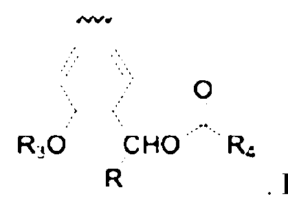 activated covalent labeling moiety includes the formula: . In embodiments,
 the hydrolase-activated covalent labeling moiety includes the formula:
 In embodiments, the hydrolase-activated covalent labeling moiety includes the formula:
 In embodiments, the hydrolase-activated covalent labeling moiety includes the formula:
 In embodiments, the hydrolase-activated covalent labeling moiety includes the formula:
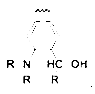 . In embodiments, the hydrolase-activated covalent labeling moiety has the formula:

 In embodiments, the
 hydrolase-activated covalent labeling moiety has the formula:
 . In embodiments, the hydrolase-activated covalent labeling moiety has the formula:
 In embodiments, the hydrolase-activated covalent labeling moiety has the formula:
 In embodiments, the hydrolase-activated covalent labeling moiety has the formula:
 . In embodiments, the hydrolase-activated covalent labeling moiety has the formula:
 In embodiments, the hydrolase-activated covalent labeling moiety has the formula:
 . In embodiments, the hydrolase-activated covalent labeling moiety has the formula:
 In embodiments, the hydrolase-activated covalent labeling moiety has the formula:
 . In embodiments, the hydrolase-activated
covalent labeling moiety has the formula:
. In embodiments, the hydrolase-activated
covalent labeling moiety has the formula:
 In embodiments, the hydrolase- activated covalent labeling moiety has the formula:
 . In embodiments, the hydrolase-activated covalent labeling moiety has the formula:
 In embodiments, the hydrolase-activated covalent labeling moiety has the formula:
 [0278] In embodiments, the click chemistry reactive moiety includes an azide, an alkene, an alkyne, a cyclooctyne, an activated alkyne, an electron-deficient alkyne, an aryne, an amine, a diene, a dienophile, a dithioester, an enone, a maleimide, a para-fluoro, a strained alkyne, a tetrazine, a tetrazole, a terminal alkyne, a thiol, a bicyclononyne (BCN), a dibenzocyclooctyne (DIBO), a difluorinated cyclooctyne (DIFO), or a biarylazacyclooctynone (BARAC). In embodiments, the click chemistry reactive moiety includes an azide. In embodiments, the click chemistry reactive moiety includes an alkene. In embodiments, the click chemistry reactive moiety includes an alkyne. In embodiments, the click chemistry reactive moiety includes a cyclooctyne. In embodiments, the click chemistry reactive moiety includes an activated alkyne. In embodiments, the click chemistry reactive moiety includes an electron-deficient alkyne. In embodiments, the click chemistry reactive moiety includes an aryne. In embodiments, the click chemistry reactive moiety includes an amine. In embodiments, the click chemistry reactive moiety includes a diene. In embodiments, the click chemistry reactive moiety includes a dienophile. In embodiments, the click chemistry reactive moiety includes a dithioester. In embodiments, the click chemistry reactive moiety includes an enone. In embodiments, the click chemistry reactive moiety includes a maleimide. In embodiments, the click chemistry reactive
moiety includes a para-fluoro. In embodiments, the click chemistry reactive moiety includes a strained alkyne. In embodiments, the click chemistry reactive moiety includes a tetrazine. In embodiments, the click chemistry reactive moiety includes a tetrazole. In embodiments, the click chemistry reactive moiety includes a terminal alkyne. In embodiments, the click chemistry reactive moiety includes a thiol. In embodiments, the click chemistry reactive moiety includes a bicyclononyne (BCN). In embodiments, the click chemistry reactive moiety includes a dibenzocyclooctyne (DIBO). In embodiments, the click chemistry reactive moiety includes a difluorinated cyclooctyne (DIFO). In embodiments, the click chemistry reactive moiety includes a biarylazacyclooctynone (BARAC). [0279] In embodiments, the click chemistry reactive moiety has the formula:
[0278] In embodiments, the click chemistry reactive moiety includes an azide, an alkene, an alkyne, a cyclooctyne, an activated alkyne, an electron-deficient alkyne, an aryne, an amine, a diene, a dienophile, a dithioester, an enone, a maleimide, a para-fluoro, a strained alkyne, a tetrazine, a tetrazole, a terminal alkyne, a thiol, a bicyclononyne (BCN), a dibenzocyclooctyne (DIBO), a difluorinated cyclooctyne (DIFO), or a biarylazacyclooctynone (BARAC). In embodiments, the click chemistry reactive moiety includes an azide. In embodiments, the click chemistry reactive moiety includes an alkene. In embodiments, the click chemistry reactive moiety includes an alkyne. In embodiments, the click chemistry reactive moiety includes a cyclooctyne. In embodiments, the click chemistry reactive moiety includes an activated alkyne. In embodiments, the click chemistry reactive moiety includes an electron-deficient alkyne. In embodiments, the click chemistry reactive moiety includes an aryne. In embodiments, the click chemistry reactive moiety includes an amine. In embodiments, the click chemistry reactive moiety includes a diene. In embodiments, the click chemistry reactive moiety includes a dienophile. In embodiments, the click chemistry reactive moiety includes a dithioester. In embodiments, the click chemistry reactive moiety includes an enone. In embodiments, the click chemistry reactive moiety includes a maleimide. In embodiments, the click chemistry reactive
moiety includes a para-fluoro. In embodiments, the click chemistry reactive moiety includes a strained alkyne. In embodiments, the click chemistry reactive moiety includes a tetrazine. In embodiments, the click chemistry reactive moiety includes a tetrazole. In embodiments, the click chemistry reactive moiety includes a terminal alkyne. In embodiments, the click chemistry reactive moiety includes a thiol. In embodiments, the click chemistry reactive moiety includes a bicyclononyne (BCN). In embodiments, the click chemistry reactive moiety includes a dibenzocyclooctyne (DIBO). In embodiments, the click chemistry reactive moiety includes a difluorinated cyclooctyne (DIFO). In embodiments, the click chemistry reactive moiety includes a biarylazacyclooctynone (BARAC). [0279] In embodiments, the click chemistry reactive moiety has the formula:
 [0280] In embodiments, the click chemistry reactive moiety includes a linker. In embodiments, the linker is about 4 to about 20 atoms long. In embodiments, the linker is about 5 to about 20 atoms long. In embodiments, the linker is about 6 to about 20 atoms long. In embodiments, the linker is about 7 to about 20 atoms long. In embodiments, the linker is about 8 to about 20 atoms long. In embodiments, the linker is about 9 to about 20 atoms long. In embodiments, the linker is about 10 to about 20 atoms long. In embodiments, the linker is about 11 to about 20 atoms long. In embodiments, the linker is about 12 to about 20 atoms long. In embodiments, the linker is about 13 to about 20 atoms long. In embodiments, the linker is about 14 to about 20 atoms long. In embodiments, the linker is about 15 to about 20 atoms long. In embodiments, the linker is about 16 to about 20 atoms long. In embodiments, the linker is about 17 to about 20 atoms long. In embodiments, the linker is about 18 to about 20 atoms long. In embodiments, the linker is about 19 to about 20 atoms long. [0281] In embodiments, the linker is about 4 to about 19 atoms long. In embodiments, the linker is about 4 to about 18 atoms long. In embodiments, the linker is about 4 to about 17 atoms long. In embodiments, the linker is about 4 to about 16 atoms long. In embodiments, the linker is about 4 to about 15 atoms long. In embodiments, the linker is about 4 to about 14 atoms long. In embodiments, the linker is about 4 to about 13 atoms long. In embodiments, the linker is about 4 to about 12 atoms long. In embodiments, the linker is about 4 to about 11 atoms long. In embodiments, the linker is about 4 to about 10 atoms long. In embodiments, the linker is about 4
to about 9 atoms long. In embodiments, the linker is about 4 to about 8 atoms long. In embodiments, the linker is about 4 to about 7 atoms long. In embodiments, the linker is about 4 to about 6 atoms long. In embodiments, the linker is about 4 to about 5 atoms long. [0282] In embodiments, the linker is 4 to 20 atoms long. In embodiments, the linker is 5 to 20 atoms long. In embodiments, the linker is 6 to 20 atoms long. In embodiments, the linker is 7 to 20 atoms long. In embodiments, the linker is 8 to 20 atoms long. In embodiments, the linker is 9 to 20 atoms long. In embodiments, the linker is 10 to 20 atoms long. In embodiments, the linker is 11 to 20 atoms long. In embodiments, the linker is 12 to 20 atoms long. In embodiments, the linker is 13 to 20 long. In embodiments, the linker is about 14 to about 20 atoms long. In embodiments, the linker is 15 to 20 atoms long. In embodiments, the linker is 16 to 20 atoms long. In embodiments, the linker is 17 to 20 atoms long. In embodiments, the linker is about 18 to about 20 atoms long. In embodiments, the linker is 19 to 20 atoms long. [0283] In embodiments, the linker is 4 to 19 atoms long. In embodiments, the linker is 4 to about 18 long. In embodiments, the linker is 4 to 17 atoms long. In embodiments, the linker is 4 to 16 atoms long. In embodiments, the linker is 4 to 15 atoms long. In embodiments, the linker is 4 to 14 atoms long. In embodiments, the linker is 4 to 13 atoms long. In embodiments, the linker is 4 to 12 atoms long. In embodiments, the linker is 4 to 11 atoms long. In embodiments, the linker is 4 to 10 atoms long. In embodiments, the linker is 4 to 9 atoms long. In embodiments, the linker is 4 to 8 atoms long. In embodiments, the linker is 4 to 7 atoms long. In embodiments, the linker is 4 to 6 atoms long. In embodiments, the linker is 4 to 5 atoms long. [0284] In embodiments, the linker is 4 atoms long. In embodiments, the linker is 5 atoms long. In embodiments, the linker is 6 atoms long. In embodiments, the linker is 7 atoms long. In embodiments, the linker is 8 atoms long. In embodiments, the linker is 9 atoms long. In embodiments, the linker is 10 atoms long. In embodiments, the linker is 11 atoms long. In embodiments, the linker is 12 atoms long. In embodiments, the linker is 13 atoms long. In embodiments, the linker is 14 atoms long. In embodiments, the linker is 15 atoms long. In embodiments, the linker is 16 atoms long. In embodiments, the linker is 17 atoms long. In embodiments, the linker is 18 atoms long. In embodiments, the linker is 19 atoms long. In embodiments, the linker is 20 atoms long.
[0285] In embodiments, the linker is a bond, -O-, -S-, -C(O)-, -C(O)O-, -C(O)NH-, -S(O)2NH-, -NH-, -NHC(O)NH-, -C-O-O- substituted or unsubstituted (e.g., C1-C20, C1-C10, C1-C5) alkylene, substituted or unsubstituted (e.g., 2 to 20 membered, 2 to 10 membered, 2 to 5 membered) heteroalkylene, substituted or unsubstituted (e.g., C3-C8, C3-C6, C3-C5) cycloalkylene, substituted or unsubstituted (e.g., 3 to 8 membered, 3 to 6 membered, 3 to 5 membered) heterocycloalkylene, substituted or unsubstituted (e.g., C6-C10, C6-C8, C6-C5) arylene or substituted or unsubstituted (e.g., 5 to 10 membered, 5 to 8 membered, 5 to 6 membered,) heteroarylene, or polymeric make-ups of the groups listed above such as polyamides, polyethlyneglycols, or linked alkyl chains. In embodiments, the linker is a bond. In embodiments, the linker is -O-. In embodiments, the linker is -S-. In embodiments, the linker is - C(O)-. In embodiments, the linker is -C(O)O-. In embodiments, the linker is -C(O)NH-. In embodiments, the linker is -S(O)2NH-. In embodiments, the linker is -NH-. In embodiments, the linker is -NHC(O)NH-. In embodiments, the linker is -C-O-O-. In embodiments, the linker is a substituted (e.g., C1-C20, C1-C10, C1-C5) alkylene. In embodiments, the linker is an unsubstituted (e.g., C1-C20, C1-C10, C1-C5) alkylene. In embodiments, the linker is a substituted (e.g., 2 to 20 membered, 2 to 10 membered, 2 to 5 membered) heteroalkylene. In embodiments, the linker is an unsubstituted (e.g., 2 to 20 membered, 2 to 10 membered, 2 to 5 membered) heteroalkylene. In embodiments, the linker is a substituted (e.g., C3-C8, C3-C6, C3-C5) cycloalkylene. In embodiments, the linker is an unsubstituted (e.g., C3-C8, C3-C6, C3-C5) cycloalkylene. In embodiments, the linker is a substituted (e.g., 3 to 8 membered, 3 to 6 membered, 3 to 5 membered) heterocycloalkylene. In embodiments, the linker is an unsubstituted (e.g., 3 to 8 membered, 3 to 6 membered, 3 to 5 membered) heterocycloalkylene. In embodiments, the linker is a substituted (e.g., C6-C10, C6-C8, C6-C5) arylene . In embodiments, the linker is an unsubstituted (e.g., C6-C10, C6-C8, C6-C5) arylene. In embodiments, the linker is a substituted (e.g., 5 to 10 membered, 5 to 8 membered, 5 to 6 membered,) heteroarylene . In embodiments, the linker is an unsubstituted (e.g., 5 to 10 membered, 5 to 8 membered, 5 to 6 membered,) heteroarylene. In embodiments, the linker is a polymeric make-up of the groups listed above such as polyamides. In embodiments, the linker is a polymeric make-up of the groups listed above such as polyethlyneglycols. In embodiments, the linker is a polymeric make-up of the groups listed above such as linked alkyl chains. [0286] In embodiments, the affinity ligand is a biotin, a desthiobiotin, an ALFA tag, a FLAG tag, an HA tag, a His tag, a SNAP tag, a CLIP tag, or a Halo tag. In embodiments, the affinity
ligand is a biotin. In embodiments, the affinity ligand is a desthiobiotin. In embodiments, the affinity ligand is an ALFA tag. In embodiments, the affinity ligand is a FLAG tag. In embodiments, the affinity ligand is an HA tag. In embodiments, the affinity ligand is a His tag. In embodiments, the affinity ligand is a SNAP tag. In embodiments, the affinity ligand is a CLIP tag. In embodiments, the affinity ligand is a Halo tag. [0287] In embodiments, the biomolecule is a nucleic acid, a polynucleotide, a protein, a peptide, a polypeptide, an amino acid, a carbohydrate, a lipid, an antibody or fragment thereof, a nanobody, a kinase, a receptor, an organelle, a parasite, or a virus. In embodiments, the biomolecule is a nucleic acid. In embodiments, the biomolecule is a polynucleotide. In embodiments, the biomolecule is a protein. In embodiments, the biomolecule is a peptide. In embodiments, the biomolecule is a polypeptide. In embodiments, the biomolecule is an amino acid. In embodiments, the biomolecule is a carbohydrate. In embodiments, the biomolecule is a lipid. In embodiments, the biomolecule is an antibody or fragment thereof. In embodiments, the biomolecule is a nanobody. In embodiments, the biomolecule is a kinase. In embodiments, the biomolecule is a receptor. In embodiments, the biomolecule is an organelle. In embodiments, the biomolecule is a parasite. In embodiments, the biomolecule is a virus. [0288] In another aspect is provided a compound including a hydrolase-activated covalent labeling moiety and (a) a hydrolase-activated imaging agent moiety, or (b) an affinity ligand or click chemistry reactive moiety. [0289] In embodiments, the hydrolase-activated covalent labeling moiety is –CH2-F or –CH- F2. In embodiments, the hydrolase-activated covalent labeling moiety is –CH2-F. In embodiments, the hydrolase-activated covalent labeling moiety is –CH-F2. [0290] In embodiments, the hydrolase-activated imaging agent has the formula:
[0280] In embodiments, the click chemistry reactive moiety includes a linker. In embodiments, the linker is about 4 to about 20 atoms long. In embodiments, the linker is about 5 to about 20 atoms long. In embodiments, the linker is about 6 to about 20 atoms long. In embodiments, the linker is about 7 to about 20 atoms long. In embodiments, the linker is about 8 to about 20 atoms long. In embodiments, the linker is about 9 to about 20 atoms long. In embodiments, the linker is about 10 to about 20 atoms long. In embodiments, the linker is about 11 to about 20 atoms long. In embodiments, the linker is about 12 to about 20 atoms long. In embodiments, the linker is about 13 to about 20 atoms long. In embodiments, the linker is about 14 to about 20 atoms long. In embodiments, the linker is about 15 to about 20 atoms long. In embodiments, the linker is about 16 to about 20 atoms long. In embodiments, the linker is about 17 to about 20 atoms long. In embodiments, the linker is about 18 to about 20 atoms long. In embodiments, the linker is about 19 to about 20 atoms long. [0281] In embodiments, the linker is about 4 to about 19 atoms long. In embodiments, the linker is about 4 to about 18 atoms long. In embodiments, the linker is about 4 to about 17 atoms long. In embodiments, the linker is about 4 to about 16 atoms long. In embodiments, the linker is about 4 to about 15 atoms long. In embodiments, the linker is about 4 to about 14 atoms long. In embodiments, the linker is about 4 to about 13 atoms long. In embodiments, the linker is about 4 to about 12 atoms long. In embodiments, the linker is about 4 to about 11 atoms long. In embodiments, the linker is about 4 to about 10 atoms long. In embodiments, the linker is about 4
to about 9 atoms long. In embodiments, the linker is about 4 to about 8 atoms long. In embodiments, the linker is about 4 to about 7 atoms long. In embodiments, the linker is about 4 to about 6 atoms long. In embodiments, the linker is about 4 to about 5 atoms long. [0282] In embodiments, the linker is 4 to 20 atoms long. In embodiments, the linker is 5 to 20 atoms long. In embodiments, the linker is 6 to 20 atoms long. In embodiments, the linker is 7 to 20 atoms long. In embodiments, the linker is 8 to 20 atoms long. In embodiments, the linker is 9 to 20 atoms long. In embodiments, the linker is 10 to 20 atoms long. In embodiments, the linker is 11 to 20 atoms long. In embodiments, the linker is 12 to 20 atoms long. In embodiments, the linker is 13 to 20 long. In embodiments, the linker is about 14 to about 20 atoms long. In embodiments, the linker is 15 to 20 atoms long. In embodiments, the linker is 16 to 20 atoms long. In embodiments, the linker is 17 to 20 atoms long. In embodiments, the linker is about 18 to about 20 atoms long. In embodiments, the linker is 19 to 20 atoms long. [0283] In embodiments, the linker is 4 to 19 atoms long. In embodiments, the linker is 4 to about 18 long. In embodiments, the linker is 4 to 17 atoms long. In embodiments, the linker is 4 to 16 atoms long. In embodiments, the linker is 4 to 15 atoms long. In embodiments, the linker is 4 to 14 atoms long. In embodiments, the linker is 4 to 13 atoms long. In embodiments, the linker is 4 to 12 atoms long. In embodiments, the linker is 4 to 11 atoms long. In embodiments, the linker is 4 to 10 atoms long. In embodiments, the linker is 4 to 9 atoms long. In embodiments, the linker is 4 to 8 atoms long. In embodiments, the linker is 4 to 7 atoms long. In embodiments, the linker is 4 to 6 atoms long. In embodiments, the linker is 4 to 5 atoms long. [0284] In embodiments, the linker is 4 atoms long. In embodiments, the linker is 5 atoms long. In embodiments, the linker is 6 atoms long. In embodiments, the linker is 7 atoms long. In embodiments, the linker is 8 atoms long. In embodiments, the linker is 9 atoms long. In embodiments, the linker is 10 atoms long. In embodiments, the linker is 11 atoms long. In embodiments, the linker is 12 atoms long. In embodiments, the linker is 13 atoms long. In embodiments, the linker is 14 atoms long. In embodiments, the linker is 15 atoms long. In embodiments, the linker is 16 atoms long. In embodiments, the linker is 17 atoms long. In embodiments, the linker is 18 atoms long. In embodiments, the linker is 19 atoms long. In embodiments, the linker is 20 atoms long.
[0285] In embodiments, the linker is a bond, -O-, -S-, -C(O)-, -C(O)O-, -C(O)NH-, -S(O)2NH-, -NH-, -NHC(O)NH-, -C-O-O- substituted or unsubstituted (e.g., C1-C20, C1-C10, C1-C5) alkylene, substituted or unsubstituted (e.g., 2 to 20 membered, 2 to 10 membered, 2 to 5 membered) heteroalkylene, substituted or unsubstituted (e.g., C3-C8, C3-C6, C3-C5) cycloalkylene, substituted or unsubstituted (e.g., 3 to 8 membered, 3 to 6 membered, 3 to 5 membered) heterocycloalkylene, substituted or unsubstituted (e.g., C6-C10, C6-C8, C6-C5) arylene or substituted or unsubstituted (e.g., 5 to 10 membered, 5 to 8 membered, 5 to 6 membered,) heteroarylene, or polymeric make-ups of the groups listed above such as polyamides, polyethlyneglycols, or linked alkyl chains. In embodiments, the linker is a bond. In embodiments, the linker is -O-. In embodiments, the linker is -S-. In embodiments, the linker is - C(O)-. In embodiments, the linker is -C(O)O-. In embodiments, the linker is -C(O)NH-. In embodiments, the linker is -S(O)2NH-. In embodiments, the linker is -NH-. In embodiments, the linker is -NHC(O)NH-. In embodiments, the linker is -C-O-O-. In embodiments, the linker is a substituted (e.g., C1-C20, C1-C10, C1-C5) alkylene. In embodiments, the linker is an unsubstituted (e.g., C1-C20, C1-C10, C1-C5) alkylene. In embodiments, the linker is a substituted (e.g., 2 to 20 membered, 2 to 10 membered, 2 to 5 membered) heteroalkylene. In embodiments, the linker is an unsubstituted (e.g., 2 to 20 membered, 2 to 10 membered, 2 to 5 membered) heteroalkylene. In embodiments, the linker is a substituted (e.g., C3-C8, C3-C6, C3-C5) cycloalkylene. In embodiments, the linker is an unsubstituted (e.g., C3-C8, C3-C6, C3-C5) cycloalkylene. In embodiments, the linker is a substituted (e.g., 3 to 8 membered, 3 to 6 membered, 3 to 5 membered) heterocycloalkylene. In embodiments, the linker is an unsubstituted (e.g., 3 to 8 membered, 3 to 6 membered, 3 to 5 membered) heterocycloalkylene. In embodiments, the linker is a substituted (e.g., C6-C10, C6-C8, C6-C5) arylene . In embodiments, the linker is an unsubstituted (e.g., C6-C10, C6-C8, C6-C5) arylene. In embodiments, the linker is a substituted (e.g., 5 to 10 membered, 5 to 8 membered, 5 to 6 membered,) heteroarylene . In embodiments, the linker is an unsubstituted (e.g., 5 to 10 membered, 5 to 8 membered, 5 to 6 membered,) heteroarylene. In embodiments, the linker is a polymeric make-up of the groups listed above such as polyamides. In embodiments, the linker is a polymeric make-up of the groups listed above such as polyethlyneglycols. In embodiments, the linker is a polymeric make-up of the groups listed above such as linked alkyl chains. [0286] In embodiments, the affinity ligand is a biotin, a desthiobiotin, an ALFA tag, a FLAG tag, an HA tag, a His tag, a SNAP tag, a CLIP tag, or a Halo tag. In embodiments, the affinity
ligand is a biotin. In embodiments, the affinity ligand is a desthiobiotin. In embodiments, the affinity ligand is an ALFA tag. In embodiments, the affinity ligand is a FLAG tag. In embodiments, the affinity ligand is an HA tag. In embodiments, the affinity ligand is a His tag. In embodiments, the affinity ligand is a SNAP tag. In embodiments, the affinity ligand is a CLIP tag. In embodiments, the affinity ligand is a Halo tag. [0287] In embodiments, the biomolecule is a nucleic acid, a polynucleotide, a protein, a peptide, a polypeptide, an amino acid, a carbohydrate, a lipid, an antibody or fragment thereof, a nanobody, a kinase, a receptor, an organelle, a parasite, or a virus. In embodiments, the biomolecule is a nucleic acid. In embodiments, the biomolecule is a polynucleotide. In embodiments, the biomolecule is a protein. In embodiments, the biomolecule is a peptide. In embodiments, the biomolecule is a polypeptide. In embodiments, the biomolecule is an amino acid. In embodiments, the biomolecule is a carbohydrate. In embodiments, the biomolecule is a lipid. In embodiments, the biomolecule is an antibody or fragment thereof. In embodiments, the biomolecule is a nanobody. In embodiments, the biomolecule is a kinase. In embodiments, the biomolecule is a receptor. In embodiments, the biomolecule is an organelle. In embodiments, the biomolecule is a parasite. In embodiments, the biomolecule is a virus. [0288] In another aspect is provided a compound including a hydrolase-activated covalent labeling moiety and (a) a hydrolase-activated imaging agent moiety, or (b) an affinity ligand or click chemistry reactive moiety. [0289] In embodiments, the hydrolase-activated covalent labeling moiety is –CH2-F or –CH- F2. In embodiments, the hydrolase-activated covalent labeling moiety is –CH2-F. In embodiments, the hydrolase-activated covalent labeling moiety is –CH-F2. [0290] In embodiments, the hydrolase-activated imaging agent has the formula:


, wherein R2 is a hydr oiety 5 6
 ; R and R are hydrolase-activated covalent labeling moieties; and R1, R7, and R8 are hydrogen. In embodiments, the hydrolase labile moieties have the formula . In
 embodiments, the click chemistry reactive moiety includes the formul . In embodiments, the hydrolase-activated covalent labeling moieties are -
 ments, the hydrolase-activated covalent labeling moieties are -CH2F. In embodiments, the compound includes the formula:
 wherein R2 has the formula ; R4 includes the formula:
 ; R5 and R6 are -CH2F; and R1, R7, and R8 are hydrogen.
 [ ] diments, the compound includes the formula: , wherein R2 is a hydr
 y; y oiety; R5 and R6 are hydrolase-activated covalent labeling moieties; and R1, R7, and R8 are hydrogen. In embodiments, the hydrolase labile moieties have the formula . In
 embodiments, the click chemistry reactive moiety includes the formu . In embodiments, the hydrolase-activated covalent labeling moieties are
 -CHF2. In embodiments, the compound includes the formula:


, wherein R1 and R2 a active moiety; R5 is a
 hydrolase-activated covalent labeling moieties; and R6, R7, and R8 are hydrogen. In embodiments, the hydrolase labile moieties have the formula . In
 embodiments, the click chemistry reactive moiety includes the formu . In embodiments, the hydrolase-activated covalent labeling moieties are
 ments, the compound includes the formula: ,
 wherein R1 and R2 have the formula ; R4 includes the formula
 ; R5 is -CHF2; and R6, R7, and R8 are hydrogen.
 diments, the compound includes the formula: , wherein R1 and R2 a
 re hydrolase labile moieties; R is a click chemistry reactive moiety; R6 is a hydrolase-activated covalent labeling moieties; and R5, R7, and R8 are hydrogen. In embodiments, the hydrolase labile moieties have the formula . In
 embodiments, the click chemistry reactive moiety includes the formu . In embodiments, the hydrolase-activated covalent labeling moieties are
 CH2F. In embodiments, the compound includes the formula:
,
 wherein R1 and R2 have the formula: ; R4 includes the formula
 ; R6 is -CH2F; and R5, R7, and R8 are hydrogen.
 [0315] In embodiments, the compound includes the formula: , wherein R1 and R2
 are hydrolase labile moieties; R4 is a click chemistry reactive moiety; R6 is a hydrolase-activated covalent labeling moieties; and R5, R7, and R8 are hydrogen. In
embodiments, the hydrolase labile moieties have the formula . In
are hydrolase labile moieties; R4 is a click chemistry reactive moiety; R6 is a hydrolase-activated covalent labeling moieties; and R5, R7, and R8 are hydrogen. In
embodiments, the hydrolase labile moieties have the formula . In
 embodiments, the click chemistry reactive moiety includes the formul . In embodiments, the hydrolase-activated covalent labeling moieties are - ments, the
 compound includes the formula:
 wherein R1 and R2 have the formul ; R4 includes the formula
 ; R6 is -CHF2; and R5, R7, and R8 are hydrogen.
 [0316] In another aspect is provided a compound including a hydrolase-activated covalent labeling moiety bound to: (a) a hydrolase-activated imaging agent covalently or (b) an affinity ligand or click chemistry reactive moiety. In embodiments, the compound includes a hydrolase- activated covalent labeling moiety bound to a hydrolase-activated imaging agent covalently. In embodiments, the compound includes a hydrolase-activated covalent labeling moiety bound to
an affinity ligand. In embodiments, the compound includes a hydrolase-activated covalent labeling moiety bound to a click chemistry reactive moiety. [0317] In another aspect is provided a compound including an esterase-activated imaging agent covalently bound to: (a) an esterase-activated covalent labeling moiety or (b) an affinity ligand or click chemistry reactive moiety. In embodiments, the compound includes an esterase-activated imaging agent covalently bound to an esterase-activated covalent labeling moiety. In embodiments, the compound includes an esterase-activated imaging agent covalently bound to an affinity ligand. In embodiments, the compound includes an esterase-activated imaging agent covalently bound to a click chemistry reactive moiety. [0318] In embodiments, the esterase-activated imaging agent or esterase-activated covalent labeling moiety is activated by a Bacillus subtilis esterase. In embodiments, the esterase- activated imaging agent is activated by a Bacillus subtilis esterase. In embodiments, the esterase- activated covalent labeling moiety is activated by a Bacillus subtilis esterase. In embodiments, the Bacillus subtilis esterase includes the amino acid sequence of SEQ ID NO:13, SEQ ID NO:14, or SEQ ID NO:15. In embodiments, the Bacillus subtilis esterase includes the amino acid sequence of SEQ ID NO:13. In embodiments, the Bacillus subtilis esterase includes the amino acid sequence of SEQ ID NO:14. In embodiments, the Bacillus subtilis esterase includes the amino acid sequence of SEQ ID NO:15. In embodiments, the Bacillus subtilis esterase is the amino acid sequence of SEQ ID NO:13, SEQ ID NO:14, or SEQ ID NO:15. In embodiments, the Bacillus subtilis esterase is the amino acid sequence of SEQ ID NO:13. In embodiments, the Bacillus subtilis esterase is the amino acid sequence of SEQ ID NO:14. In embodiments, the Bacillus subtilis esterase is the amino acid sequence of SEQ ID NO:15. [0319] In embodiments, the esterase-activated imaging agent or esterase-activated covalent labeling moiety is activated by contacting a biomolecule esterase conjugate. In embodiments, the biomolecule esterase conjugate includes the biomolecule covalently bound to a functional esterase. In embodiments, the functional esterase is a Bacillus subtilis esterase. In embodiments, the esterase-activated imaging agent or esterase-activated covalent labeling moiety is activated by contacting a functional esterase conjugate complex. In embodiments, the functional esterase conjugate complex is formed by proximally localizing a first biomolecule esterase portion conjugate and a second biomolecule esterase portion conjugate. In embodiments, the first biomolecule esterase portion conjugate includes the first biomolecule covalently bound to a first
portion of the functional esterase and the second biomolecule esterase portion conjugate includes the second biomolecule covalently bound to a second portion of the functional esterase. In embodiments, the functional esterase complex includes the functional esterase and the first biomolecule bound to the second biomolecule. In embodiments, the first portion of the functional esterase includes the amino acid sequence of SEQ ID NO:14 or SEQ ID NO:15. In embodiments, the first portion of the functional esterase includes the amino acid sequence of SEQ ID NO:14. In embodiments, the first portion of the functional esterase includes the amino acid sequence of SEQ ID NO:15. In embodiments, the first portion of the functional esterase is the amino acid sequence of SEQ ID NO:14 or SEQ ID NO:15. In embodiments, the first portion of the functional esterase is the amino acid sequence of SEQ ID NO:14. In embodiments, the first portion of the functional esterase is the amino acid sequence of SEQ ID NO:15. In embodiments, the second portion of the functional esterase includes the amino acid sequence of SEQ ID NO:14 or SEQ ID NO:15. In embodiments, the second portion of the functional esterase includes the amino acid sequence of SEQ ID NO:14. In embodiments, the second portion of the functional esterase includes the amino acid sequence of SEQ ID NO:15. In embodiments, the second portion of the functional esterase is the amino acid sequence of SEQ ID NO:14 or SEQ ID NO:15. In embodiments, the second portion of the functional esterase is the amino acid sequence of SEQ ID NO:14. In embodiments, the second portion of the functional esterase is the amino acid sequence of SEQ ID NO:15. [0320] In another aspect is provided a compound including an esterase-activated covalent labeling moiety covalently bound to: (a) an esterase-activated imaging agent or (b) an affinity ligand or click chemistry reactive moiety. In embodiments, the compound includes an esterase- activated covalent labeling moiety covalently bound to an esterase-activated imaging agent. In embodiments, the compound includes an esterase-activated covalent labeling moiety covalently bound to an affinity ligand. In embodiments, the compound includes an esterase-activated covalent labeling moiety covalently bound to a click chemistry reactive moiety. [0321] In embodiments, the esterase-activated covalent labeling moiety is –CH2-F or –CH-F2. In embodiments, the esterase-activated covalent labeling moiety is –CH2-F. In embodiments, the esterase-activated covalent labeling moiety is –CH-F2. [0322] In embodiments, the esterase-activated covalent labeling moiety or the esterase- activated imaging agent is activated by a Bacillus subtilis esterase. In embodiments, the esterase-
activated covalent labeling moiety is activated by a Bacillus subtilis esterase. In embodiments, the esterase-activated imaging agent is activated by a Bacillus subtilis esterase. In embodiments, the Bacillus subtilis esterase includes the amino acid sequence of SEQ ID NO:13, SEQ ID NO:14, or SEQ ID NO:15. In embodiments, the Bacillus subtilis esterase includes the amino acid sequence of SEQ ID NO:13. In embodiments, the Bacillus subtilis esterase includes the amino acid sequence of SEQ ID NO:14. In embodiments, the Bacillus subtilis esterase includes the amino acid sequence of SEQ ID NO:15. In embodiments, the Bacillus subtilis esterase is the amino acid sequence of SEQ ID NO:13, SEQ ID NO:14, or SEQ ID NO:15. In embodiments, the Bacillus subtilis esterase is the amino acid sequence of SEQ ID NO:13. In embodiments, the Bacillus subtilis esterase is the amino acid sequence of SEQ ID NO:14. In embodiments, the Bacillus subtilis esterase is the amino acid sequence of SEQ ID NO:15. [0323] In embodiments, the esterase-activated covalent labeling moiety or the esterase- activated imaging agent is activated by contacting a biomolecule esterase conjugate. In embodiments, the biomolecule esterase conjugate includes the biomolecule covalently bound to a functional esterase. In embodiments, the functional esterase is a Bacillus subtilis esterase. In embodiments, the esterase-activated imaging agent or esterase-activated covalent labeling moiety is activated by contacting a functional esterase conjugate complex. In embodiments, the functional esterase conjugate complex is formed by proximally localizing a first biomolecule esterase portion conjugate and a second biomolecule esterase portion conjugate. In embodiments, the first biomolecule esterase portion conjugate includes the first biomolecule covalently bound to a first portion of the functional esterase and the second biomolecule esterase portion conjugate includes the second biomolecule covalently bound to a second portion of the functional esterase. In embodiments, the functional esterase complex includes the functional esterase and the first biomolecule bound to the second biomolecule. [0324] In embodiments, the functional esterase is a Bacillus subtilis esterase. In embodiments, the first portion of the functional esterase includes the amino acid sequence of SEQ ID NO:14 or SEQ ID NO:15. In embodiments, the first portion of the functional esterase includes the amino acid sequence of SEQ ID NO:14. In embodiments, the first portion of the functional esterase includes the amino acid sequence of SEQ ID NO:15. In embodiments, the first portion of the functional esterase is the amino acid sequence of SEQ ID NO:14 or SEQ ID NO:15. In embodiments, the first portion of the functional esterase is the amino acid sequence of SEQ ID
NO:14. In embodiments, the first portion of the functional esterase is the amino acid sequence of SEQ ID NO:15. In embodiments, the second portion of the functional esterase includes the amino acid sequence of SEQ ID NO:14 or SEQ ID NO:15. In embodiments, the second portion of the functional esterase includes the amino acid sequence of SEQ ID NO:14. In embodiments, the second portion of the functional esterase includes the amino acid sequence of SEQ ID NO:15. In embodiments, the second portion of the functional esterase is the amino acid sequence of SEQ ID NO:14 or SEQ ID NO:15. In embodiments, the second portion of the functional esterase is the amino acid sequence of SEQ ID NO:14. In embodiments, the second portion of the functional esterase is the amino acid sequence of SEQ ID NO:15. [0325] In another aspect is provided a compound including a beta lactamase-activated imaging agent covalently bound to: (a) a beta lactamase-activated covalent labeling moiety or (b) an affinity ligand or click chemistry reactive moiety. In embodiments, the compound includes a beta lactamase-activated imaging agent covalently bound to a beta lactamase-activated covalent labeling moiety. In embodiments, the compound includes a beta lactamase-activated imaging agent covalently bound to an affinity ligand. In embodiments, the compound includes a beta lactamase-activated imaging agent covalently bound to a click chemistry reactive moiety. [0326] In another aspect is provided a compound including a beta lactamase-activated covalent labeling moiety covalently bound to: (a) a beta lactamase-activated imaging agent or (b) an affinity ligand or click chemistry reactive moiety. In embodiments, the compound includes a beta lactamase-activated covalent labeling moiety covalently bound to a beta lactamase-activated imaging agent. In embodiments, the compound includes a beta lactamase-activated covalent labeling moiety covalently bound to an affinity ligand. In embodiments, the compound includes a beta lactamase-activated covalent labeling moiety covalently bound to click chemistry reactive moiety. [0327] In embodiments, the beta lactamase-activated covalent labeling moiety is –CH2-F or – CH-F2. In embodiments, the beta lactamase-activated covalent labeling moiety is –CH2-F. In embodiments, the beta lactamase-activated covalent labeling moiety is –CH-F2. [0328] In embodiments, the compound is a membrane permeable compound.
KITS [0329] In an aspect is provided a kit including the compound described herein including embodiments thereof and a nucleic acid encoding a functional hydrolase. [0330] In another aspect is provided a kit including the compound described herein including embodiments thereof, a first nucleic acid encoding a first non-functional portion of a functional hydrolase and a second nucleic acid including a second non-functional portion of the functional hydrolase, wherein the first non-functional portion and the second non-functional portion may be combined to form the functional hydrolase. [0331] In embodiments, the functional hydrolase is an esterase, a phosphatase, an amidase, a sulfatase, a glycosidase, a deacetylase, a deacetylase, a thioesterase or a beta lactamase. In embodiments, the functional hydrolase is an esterase. In embodiments, the functional hydrolase is a phosphatase. In embodiments, the functional hydrolase is an amidase. In embodiments, the functional hydrolase is a sulfatase. In embodiments, the functional hydrolase is a glycosidase. In embodiments, the functional hydrolase is a deacetylase. In embodiments, the functional hydrolase is a deacetylase. In embodiments, the functional hydrolase is a thioesterase. In embodiments, the functional hydrolase is a beta lactamase. [0332] In another aspect is provided a kit including the compound described herein including embodiments thereof including a first nucleic acid encoding a first portion of a functional esterase and a second nucleic acid including a second portion of the functional esterase, wherein the first portion and the second portion may be combined to form the functional esterase. In embodiments, the functional esterase is a Bacillus subtilis esterase. In embodiments, the functional esterase is a Bacillus subtilis esterase. In embodiments, the first portion of the functional esterase includes the amino acid sequence of SEQ ID NO:14 or SEQ ID NO:15. In embodiments, the first portion of the functional esterase includes the amino acid sequence of SEQ ID NO:14. In embodiments, the first portion of the functional esterase includes the amino acid sequence of SEQ ID NO:15. In embodiments, the first portion of the functional esterase is the amino acid sequence of SEQ ID NO:14 or SEQ ID NO:15. In embodiments, the first portion of the functional esterase is the amino acid sequence of SEQ ID NO:14. In embodiments, the first portion of the functional esterase is the amino acid sequence of SEQ ID NO:15. In embodiments, the second portion of the functional esterase includes the amino acid sequence of SEQ ID NO:14 or SEQ ID NO:15. In embodiments, the second portion of the functional esterase
includes the amino acid sequence of SEQ ID NO:14. In embodiments, the second portion of the functional esterase includes the amino acid sequence of SEQ ID NO:15. In embodiments, the second portion of the functional esterase is the amino acid sequence of SEQ ID NO:14 or SEQ ID NO:15. In embodiments, the second portion of the functional esterase is the amino acid sequence of SEQ ID NO:14. In embodiments, the second portion of the functional esterase is the amino acid sequence of SEQ ID NO:15. [0333] In another aspect is provided a kit including the compound described herein including embodiments thereof including a first nucleic acid encoding a first portion of a functional beta lactamase and a second nucleic acid including a second portion of the functional beta lactamase, wherein the first portion the second portion may be combined to form the functional beta lactamase. METHODS OF USE [0334] In an aspect is provided a method of detecting a proximal interaction between a first biomolecule and a second biomolecule, the method including: (a) proximally localizing a first biomolecule hydrolase portion conjugate and a second biomolecule hydrolase portion conjugate, wherein the first biomolecule hydrolase portion conjugate includes the first biomolecule covalently bound to a first portion of a functional hydrolase and wherein the second biomolecule hydrolase portion conjugate includes the second biomolecule covalently bound to a second portion of the functional hydrolase, thereby forming the functional hydrolase; (b) contacting the functional hydrolase with a compound described herein including embodiments thereof and allowing the functional hydrolase to activate the hydrolase-activated covalent labeling moiety thereby forming a functional covalent labeling moiety, and allowing the covalent labeling moiety to covalently bind to a biomolecule thereby forming a labeled biomolecule; and (c) detecting the labeled biomolecule thereby detecting a proximal interaction between the first biomolecule and the second biomolecule. [0335] In embodiments, the method further includes: (d) proximally localizing a plurality of first biomolecule hydrolase portion conjugates and a plurality of second biomolecule hydrolase portion conjugates, thereby forming a plurality of the functional hydrolases; and (e) contacting the plurality of the functional hydrolases with a plurality of compounds described herein including embodiments thereof and allowing the plurality of the functional hydrolases to activate the plurality of the hydrolase-activated covalent labeling moieties thereby forming a plurality of
the functional covalent labeling moieties, and allowing the plurality of the covalent labeling moieties to covalently bind to a plurality of the biomolecules thereby forming a plurality of the labeled biomolecules. [0336] In embodiments, the method further includes: (f) detecting the plurality of the labeled biomolecules thereby detecting a proximal interaction between the plurality of the first biomolecules and the plurality of the second biomolecules. [0337] In embodiments, the biomolecule is a nucleic acid, a polynucleotide, a protein, a peptide, a polypeptide, an amino acid, a carbohydrate, a lipid, an antibody or fragment thereof, a nanobody, a kinase, a receptor, an organelle, a parasite, or a virus. In embodiments, the biomolecule is a nucleic acid. In embodiments, the biomolecule is a polynucleotide. In embodiments, the biomolecule is a protein. In embodiments, the biomolecule is a peptide. In embodiments, the biomolecule is a polypeptide. In embodiments, the biomolecule is an amino acid, a carbohydrate. In embodiments, the biomolecule is a lipid, an antibody or fragment thereof. In embodiments, the biomolecule is a nanobody. In embodiments, the biomolecule is a kinase. In embodiments, the biomolecule is a receptor. In embodiments, the biomolecule is an organelle. In embodiments, the biomolecule is a parasite. In embodiments, the biomolecule is a virus. [0338] In another aspect is provided a method of detecting a proximal interaction between a first biomolecule and a second biomolecule, the method including: (a) proximally localizing a first biomolecule hydrolase portion conjugate including and a second biomolecule hydrolase portion conjugate, wherein the first biomolecule hydrolase portion conjugate includes the first biomolecule covalently bound to first portion of a functional esterase and the second biomolecule hydrolase portion conjugate includes the second biomolecule covalently bound to a second portion of the functional esterase thereby forming a complex including the functional esterase and the first biomolecule bound to the second biomolecule; (b) contacting the complex with a compound described herein including embodiments thereof and allowing the functional esterase to activate the esterase-activated imaging agent thereby forming a functional imaging agent; and (c) detecting the functional imaging agent thereby detecting an interaction between the first biomolecule and the second biomolecule.
[0339] In embodiments, the method further includes: (d) proximally localizing a plurality of the first biomolecule hydrolase portion conjugates and a plurality of the second biomolecule hydrolase portion conjugates, thereby forming a plurality of the complexes including the functional esterase and the first biomolecule bound to the second biomolecule; and (e) contacting the plurality of the complexes with a plurality of compounds described herein including embodiments thereof and allowing the functional esterases to activate a plurality of the esterase- activated imaging agents thereby forming a plurality of functional imaging agents. [0340] In embodiments, the method further includes: (f) detecting the plurality of the functional imaging agents thereby detecting interaction between the plurality of the first biomolecules and the plurality of the second biomolecules. [0341] In embodiments, the method occurs in a cell. In embodiments, the method occurs on or near the surface of a cell. In embodiments, the method occurs on the surface of a cell. In embodiments, the method occurs near the surface of a cell. In embodiments, the cell is a living cell. In embodiments, the cell is a dead cell. In embodiments, the cell is a fixed cell. In embodiments, the fixed cell is fixed with formaldehyde. In embodiments, the fixed cell is fixed with paraformaldehyde. [0342] In embodiments, the cell is a mammal cell. In embodiments, the cell is a human cell. In embodiments, the cell is a bacterial cell. In embodiments, the cell is a plant cell. [0343] In embodiments, the method occurs in a tissue. In embodiments, the tissue is a living tissue. In embodiments, the tissue is a dead tissue. In embodiments, the tissue is a fixed tissue. In embodiments, the fixed tissue is fixed with formaldehyde. In embodiments, the fixed tissue is fixed with paraformaldehyde. [0344] In embodiments, the method occurs in an organism. In embodiments, the organism is a mammal. In embodiments, the organism is a human. In embodiments, the organism is a bacteria. In embodiments, the organism is a plant. [0345] In embodiments, the biomolecule is a nucleic acid, a polynucleotide, a protein, a peptide, a polypeptide, an amino acid, a carbohydrate, a lipid, an antibody or fragment thereof, a nanobody, a kinase, a receptor, an organelle, a parasite, or a virus. In embodiments, the biomolecule is a nucleic acid. In embodiments, the biomolecule is a polynucleotide. In embodiments, the biomolecule is a protein. In embodiments, the biomolecule is a peptide. In
embodiments, the biomolecule is a polypeptide. In embodiments, the biomolecule is an amino acid, a carbohydrate. In embodiments, the biomolecule is a lipid, an antibody or fragment thereof. In embodiments, the biomolecule is a nanobody. In embodiments, the biomolecule is a kinase. In embodiments, the biomolecule is a receptor. In embodiments, the biomolecule is an organelle. In embodiments, the biomolecule is a parasite. In embodiments, the biomolecule is a virus. [0346] In another aspect is provided a method of detecting a proximal interaction between a first biomolecule and a second biomolecule, the method including: (a) proximally localizing a first biomolecule hydrolase portion conjugate including and a second biomolecule hydrolase portion conjugate, wherein the first biomolecule hydrolase portion conjugate includes the first biomolecule covalently bound to first portion of a functional beta lactamase and the second biomolecule hydrolase portion conjugate includes the second biomolecule covalently bound to a second portion of the functional beta lactamase thereby forming a complex including the functional beta lactamase and the first biomolecule bound to the second biomolecule; (b) contacting the complex with a compound described herein including embodiments thereof and allowing the functional beta lactamase to activate the beta lactamase-activated imaging agent thereby forming a functional imaging agent; and (c) detecting the functional imaging agent thereby detecting an interaction between the first biomolecule and the second biomolecule. [0347] In embodiments, the method further includes: (d) proximally localizing a plurality of the first biomolecule hydrolase portion conjugates and a plurality of the second biomolecule hydrolase portion conjugates, thereby forming a plurality of the complexes including the functional beta lactamase and the first biomolecule bound to the second biomolecule; and (e) contacting the plurality of the complexes with a plurality of compounds described herein including embodiments thereof and allowing the functional beta lactamases to activate a plurality of the beta lactamase-activated imaging agents thereby forming a plurality of functional imaging agents. [0348] In embodiments, the method further includes: (f) detecting the plurality of the functional imaging agents thereby detecting interaction between the plurality of the first biomolecules and the plurality of the second biomolecules.
[0349] In embodiments, the method occurs in a cell. In embodiments, the method occurs on or near the surface of a cell. In embodiments, the method occurs on the surface of a cell. In embodiments, the method occurs near the surface of a cell. In embodiments, the cell is a living cell. In embodiments, the cell is a dead cell. In embodiments, the cell is a fixed cell. In embodiments, the fixed cell is fixed with formaldehyde. In embodiments, the fixed cell is fixed with paraformaldehyde. [0350] In embodiments, the cell is a mammal cell. In embodiments, the cell is a human cell. In embodiments, the cell is a bacterial cell. In embodiments, the cell is a plant cell. [0351] In embodiments, the method occurs in a tissue. In embodiments, the tissue is a living tissue. In embodiments, the tissue is a dead tissue. In embodiments, the tissue is a fixed tissue. In embodiments, the fixed tissue is fixed with formaldehyde. In embodiments, the fixed tissue is fixed with paraformaldehyde. [0352] In embodiments, the method occurs in an organism. In embodiments, the organism is a mammal. In embodiments, the organism is a human. In embodiments, the organism is a bacteria. In embodiments, the organism is a plant. [0353] In embodiments, the biomolecule is a nucleic acid, a polynucleotide, a protein, a peptide, a polypeptide, an amino acid, a carbohydrate, a lipid, an antibody or fragment thereof, a nanobody, a kinase, a receptor, an organelle, a parasite, or a virus. In embodiments, the biomolecule is a nucleic acid. In embodiments, the biomolecule is a polynucleotide. In embodiments, the biomolecule is a protein. In embodiments, the biomolecule is a peptide. In embodiments, the biomolecule is a polypeptide. In embodiments, the biomolecule is an amino acid, a carbohydrate. In embodiments, the biomolecule is a lipid, an antibody or fragment thereof. In embodiments, the biomolecule is a nanobody. In embodiments, the biomolecule is a kinase. In embodiments, the biomolecule is a receptor. In embodiments, the biomolecule is an organelle. In embodiments, the biomolecule is a parasite. In embodiments, the biomolecule is a virus. [0354] In another aspect is provided a method of detecting a biomolecule in cell or organism, the method including: (a) contacting the cell or organism with a compound described herein including embodiments thereof, wherein the cell or organism includes a hydrolase fusion protein, the hydrolase fusion protein including a hydrolase protein portion and a subject protein portion,
allowing the hydrolase protein portion to activate the hydrolase-activated covalent labeling moiety thereby forming a functional covalent labeling moiety, and allowing the covalent labeling moiety to covalently bind to a biomolecule thereby forming a labeled biomolecule; and (b) detecting the labeled biomolecule thereby detecting a proximal localization between the subject protein and the biomolecule. [0355] In embodiments, the method occurs in a cell. In embodiments, the method occurs on or near the surface of a cell. In embodiments, the method occurs on the surface of a cell. In embodiments, the method occurs near the surface of a cell. In embodiments, the cell is a living cell. In embodiments, the cell is a dead cell. In embodiments, the cell is a fixed cell. In embodiments, the fixed cell is fixed with formaldehyde. In embodiments, the fixed cell is fixed with paraformaldehyde. [0356] In embodiments, the cell is a mammal cell. In embodiments, the cell is a human cell. In embodiments, the cell is a bacterial cell. In embodiments, the cell is a plant cell. [0357] In embodiments, the method occurs in a tissue. In embodiments, the tissue is a living tissue. In embodiments, the tissue is a dead tissue. In embodiments, the tissue is a fixed tissue. In embodiments, the fixed tissue is fixed with formaldehyde. In embodiments, the fixed tissue is fixed with paraformaldehyde. [0358] In embodiments, the method occurs in an organism. In embodiments, the organism is a mammal. In embodiments, the organism is a human. In embodiments, the organism is a bacteria. In embodiments, the organism is a plant. [0359] In embodiments, the biomolecule is a nucleic acid, a polynucleotide, a protein, a peptide, a polypeptide, an amino acid, a carbohydrate, a lipid, an antibody or fragment thereof, a nanobody, a kinase, a receptor, an organelle, a parasite, or a virus. In embodiments, the biomolecule is a nucleic acid. In embodiments, the biomolecule is a polynucleotide. In embodiments, the biomolecule is a protein. In embodiments, the biomolecule is a peptide. In embodiments, the biomolecule is a polypeptide. In embodiments, the biomolecule is an amino acid, a carbohydrate. In embodiments, the biomolecule is a lipid, an antibody or fragment thereof. In embodiments, the biomolecule is a nanobody. In embodiments, the biomolecule is a kinase. In embodiments, the biomolecule is a receptor. In embodiments, the biomolecule is an
organelle. In embodiments, the biomolecule is a parasite. In embodiments, the biomolecule is a virus. [0360] In another aspect is provided a method of detecting a biomolecule in cell or organism, the method including: (a) contacting the cell or organism with a compound described herein including embodiments thereof, wherein the cell or organism includes a esterase fusion protein, the esterase fusion protein including an esterase portion and a subject protein portion, allowing the esterase protein portion to activate the esterase-activated covalent labeling moiety thereby forming a functional covalent labeling moiety, and allowing the covalent labeling moiety to covalently bind to a biomolecule thereby forming a labeled biomolecule; and (b) detecting the labeled biomolecule thereby detecting a proximal localization between the subject protein and the biomolecule. [0361] In embodiments, the method occurs in a cell. In embodiments, the method occurs on or near the surface of a cell. In embodiments, the method occurs on the surface of a cell. In embodiments, the method occurs near the surface of a cell. In embodiments, the cell is a living cell. In embodiments, the cell is a dead cell. In embodiments, the cell is a fixed cell. In embodiments, the fixed cell is fixed with formaldehyde. In embodiments, the fixed cell is fixed with paraformaldehyde. [0362] In embodiments, the cell is a mammal cell. In embodiments, the cell is a human cell. In embodiments, the cell is a bacterial cell. In embodiments, the cell is a plant cell. [0363] In embodiments, the method occurs in a tissue. In embodiments, the tissue is a living tissue. In embodiments, the tissue is a dead tissue. In embodiments, the tissue is a fixed tissue. In embodiments, the fixed tissue is fixed with formaldehyde. In embodiments, the fixed tissue is fixed with paraformaldehyde. [0364] In embodiments, the method occurs in an organism. In embodiments, the organism is a mammal. In embodiments, the organism is a human. In embodiments, the organism is a bacteria. In embodiments, the organism is a plant. [0365] In embodiments, the biomolecule is a nucleic acid, a polynucleotide, a protein, a peptide, a polypeptide, an amino acid, a carbohydrate, a lipid, an antibody or fragment thereof, a nanobody, a kinase, a receptor, an organelle, a parasite, or a virus. In embodiments, the biomolecule is a nucleic acid. In embodiments, the biomolecule is a polynucleotide. In
embodiments, the biomolecule is a protein. In embodiments, the biomolecule is a peptide. In embodiments, the biomolecule is a polypeptide. In embodiments, the biomolecule is an amino acid, a carbohydrate. In embodiments, the biomolecule is a lipid, an antibody or fragment thereof. In embodiments, the biomolecule is a nanobody. In embodiments, the biomolecule is a kinase. In embodiments, the biomolecule is a receptor. In embodiments, the biomolecule is an organelle. In embodiments, the biomolecule is a parasite. In embodiments, the biomolecule is a virus. [0366] In another aspect is provided a method of detecting a subject protein in cell or organism, the method including: (a) contacting the cell or organism with a compound described herein including embodiments thereof, wherein the cell or organism includes a beta-lactamase fusion protein, the beta-lactamase fusion protein including a beta-lactamase protein portion and a subject protein portion, allowing the beta-lactamase protein portion to activate the beta- lactamase-activated covalent labeling moiety thereby forming a functional covalent labeling moiety, and allowing the covalent labeling moiety to covalently bind to a biomolecule thereby forming a labeled biomolecule; and (b) detecting the labeled biomolecule thereby detecting a proximal interaction between the first biomolecule and the second biomolecule. [0367] In embodiments, the method occurs in a cell. In embodiments, the method occurs on or near the surface of a cell. In embodiments, the method occurs on the surface of a cell. In embodiments, the method occurs near the surface of a cell. In embodiments, the cell is a living cell. In embodiments, the cell is a dead cell. In embodiments, the cell is a fixed cell. In embodiments, the fixed cell is fixed with formaldehyde. In embodiments, the fixed cell is fixed with paraformaldehyde. [0368] In embodiments, the cell is a mammal cell. In embodiments, the cell is a human cell. In embodiments, the cell is a bacterial cell. In embodiments, the cell is a plant cell. [0369] In embodiments, the method occurs in a tissue. In embodiments, the tissue is a living tissue. In embodiments, the tissue is a dead tissue. In embodiments, the tissue is a fixed tissue. In embodiments, the fixed tissue is fixed with formaldehyde. In embodiments, the fixed tissue is fixed with paraformaldehyde.
[0370] In embodiments, the method occurs in an organism. In embodiments, the organism is a mammal. In embodiments, the organism is a human. In embodiments, the organism is a bacteria. In embodiments, the organism is a plant. [0371] In embodiments, the biomolecule is a nucleic acid, a polynucleotide, a protein, a peptide, a polypeptide, an amino acid, a carbohydrate, a lipid, an antibody or fragment thereof, a nanobody, a kinase, a receptor, an organelle, a parasite, or a virus. In embodiments, the biomolecule is a nucleic acid. In embodiments, the biomolecule is a polynucleotide. In embodiments, the biomolecule is a protein. In embodiments, the biomolecule is a peptide. In embodiments, the biomolecule is a polypeptide. In embodiments, the biomolecule is an amino acid, a carbohydrate. In embodiments, the biomolecule is a lipid, an antibody or fragment thereof. In embodiments, the biomolecule is a nanobody. In embodiments, the biomolecule is a kinase. In embodiments, the biomolecule is a receptor. In embodiments, the biomolecule is an organelle. In embodiments, the biomolecule is a parasite. In embodiments, the biomolecule is a virus. [0372] For the methods provided herein microscopy methods may be used for the assessment of, for example, labeling of a biomolecule. Non-limiting examples of microscopy techniques useful for the methods provided herein including embodiments thereof include, wide field microscopy bright field microscopy, phase contrast microscopy, differential interference contrast microscopy, single- or multi-photon fluorescence microscopy, fluorescence microscopy, photoacoustic microscopy, luminescence microscopy, Raman scattering microscopy, two- dimensional microscopy, or three-dimensional microscopy. In embodiments, the microscopy techniques utilize transmitted illumination, bright field illumination, epi- illumination, dark field illumination, wide field illumination, point-scanning illumination, line-scanning illumination, spinning disk illumination, speckled illumination, or patterned illumination. In embodiments, the microscopy is single-photo fluorescence microscopy. In embodiments, the microscopy is multi- photon fluorescence microscopy. In embodiments, the microscopy is fluorescence microscopy. EXAMPLES Example 1: General Methods and Materials [0373] Preparing bead-bound BS2 and Vhh for in vitro reactions with BSA.
[0374] U2OS cells were grown to 80-95% confluency and transfected in a 10-well plate with pEF1-3xFLAG-NLS-BS2-Halo or NLS-Vhh-Halo. The medium was removed, cells were gently washed with cold PBS then scraped and collected in 3mL PBS. Cells were centrifuged at 200xg for 5 min and the supernatant was discarded. The cell pellet was resuspended in 2mL purification buffer (PBS, 1mM DTT, 0.005% IGEPAL-CA630) for BS2 and 0.75mL for Vhh. Protease inhibitors were not added as it could inactivate BS2. Samples were then sonicated with microtip on ice for 3 intervals of 10s with 10s rest in between, then centrifuged at 10000xg for 15min at 4C, after which supernatant was collected.400uL HaloLink resin slurry was transferred to 15 mL conical tube for BS2 and 150 µL for Vhh. Tubes were centrifuged at 1500xg for 5 min and the supernatant was discarded, and wash with PBS was repeated 5 times.2 mL purification buffer was added to tubes and mixed for 5min on an end-over-end tube rotator, centrifuged at 1500xg for 5min, and the supernatant was discarded. The cell lysate was then added to the resin and incubated overnight at 4 °C.10uL lysate or collected medium was saved for determining the expression level and binding efficiency of the HaloTag-fusion protein (also referred to herein as Halo or Halo-Tag) to the resin. The resin was washed three times with 2mL PBS to remove DTT, then centrifuged at 1500 x g for 5min, and the supernatant was discarded. [0375] Recombinant expression and purification of full-length BS2. [0376] BS2 was cloned into the pH6HTC His6HaloTag® T7 Vector (Promega) and then heat shock-transformed into E. coli Rosetta2(DE3) competent cells (Novagen). The transformed cells were grown in 25ml LB media (EMD Millipore) containing 100 μg/ml Ampicillin (Lab Scientific Inc.) at 37 °C with 220 rpm shaking until the OD600 reached 0.6. The culture was then induced by 1M IPTG (Lab Scientific Inc.) and incubated at 15 °C for 16 hr with 180 rpm shaking. The cells were collected by centrifugation (5000 rpm, 20 min) and stored at -80 °C until further use. [0377] The recombinant BS2 was purified by a sequential HaloTag and HisTag purification strategy. The purification began by treating the cell pellets with 5 ml lysis buffer (Gibco™ PBS buffer, BME 5mM, pH 7.4) and thawing on ice for 30 min. The cell suspension was stirred until the sample formed a homogeneous suspension, and then sonicated using Misonix Sonicator 3000 model (1.5s on/1.5s off pulses for 30s intervals, 3 times, allowing 5 min cooling time on ice between each interval), which thoroughly lysed the cells. The cell debris was removed by centrifugation at 15000 rpm for 30 min. The supernatant cell lysate was filtered through 0.45 μm
filters and loaded onto the 2 ml of HaloLink™ Resin (equilibrated with lysis buffer in a 15ml conical tube). The cell lysate and beads were mixed well by inverting then tube 3-4 times and then placing the tube onto a tube rotator for end-over-end mixing overnight at 4°C (Or 1 hour at room temperature). The sample was centrifuged at 1,000 × g for 5 minutes to remove the supernatant, and the resin was washed with 10ml of lysis buffer by inverting the tube until the sample was thoroughly mixed. The sample was then centrifuged at 1,000 × g for 5 minutes and the supernatant was discarded. The wash step was repeated a total of three times. [0378] Next, the BS2 was eluted from the beads by addition of 1 ml of cleavage solution, which consists of 66µl of ProTEV Plus-Promega in 1.1ml of lysis buffer, to the settled resin and the resin and cleavage solution was thoroughly mixed by pipetting the sample to homogeneity. The tube containing the cleavage reaction was mixed end-over-end overnight at 4°C (or 1 hour at room temperature) using a rotator. The cleavage mixture was then centrifuged at 2,000–3,000 × g for 5 minutes and the supernatant was transferred to a 15 ml conical tube. An additional 1ml of lysis buffer was added to the resin and mixed well by inverting the tube, centrifuged at 2,000- 3,000 × g for 5 minutes, and the supernatant was combined with the first sample. [0379] HisLink™ Resin (Promega, 50 µl of 50% HisLink™ Resin) was added to the sample and end-over-end rotated at room temperature for 20 minutes to remove any residual HaloTag protein. The sample was then centrifuged at 1,000 × g for 5 minutes and the supernatant was transferred to another tube. The supernatant contained purified recombinant BS2 protein, which was stored in pH 7.4 PBS butter with BME 5 mM and 5% glycerol at -80 °C. The protein concentration could be obtained by Bradford assay, or by measuring the 280 nm absorbance (A280) and calculated using Beer’s law. The extinction coefficient could be obtained from the literature or using ProtParam tool from the ExPASy proteomics server. The typical protein yield was 6 mg from 25 mL of cell culture. [0380] Reactions of BSA with QM-alkyne probes and bead bound BS2. [0381] Beads were reacted with 200ng bovine serum albumin (BSA) and 10uM QM1-alkyne in 25uL PBS for 10min. After spinning out beads, chloroform-methanol precipitation of BSA was performed. For the click reaction, the protein pellet was reconstituted in 50uL PBS, then 1uL of CuSO4 (50mM stock in water), 1uL IR800 CW azide (1.25 mM in DMSO, made in-house from 10 mM stock), 1uL of Tris(2-carboxyethyl)phosphine (TCEP, 50mM in water – make fresh
each time), and 3uL of Tris[(1-benzyl-1H-1,2,3-triazol-4-yl)methyl]amine (TBTA, 1.7mM in DMSO-tBuOH (1:4 v/v) was added for 1h at room temperature 17uL of 4X SDS sample buffer was added without boiling. The sample was loaded onto 4-12% Bolt™ Bis-Tris Plus Mini Protein Gel and ran at 160V for 1hr. The gel was imaged on LI-COR Odyssey imager at 800nm to validate BSA labeling by QM1-alkyne and the subsequent clicking of IR800CW dye to the alkyne handle. [0382] In cell labeling with Chemical probes. [0383] U2OS cells were transfected with indicated plasmids to detect Halo-Tag-fusion expressing/transfected cells, cells were incubated with fluorescent-Halo-tag ligand dyes. [0384] Cells were then incubated with esterase dependent ‘caged’ probes, at different concentrations, with a range from 2-50 µM compound and for different times, ranging from 2- minutes to hours, as indicated. To visualize esterase dependent activation and labeling of proximal biomolecules and the functionality of the azide/alkyne handle in the same label to be detected and enriched via affinity purification, click chemistry was performed with different clickable fluorophores and/or biotin. [0385] Click chemistry fluorescent in cell labeling. [0386] After incubation with chemical probes at various concentrations and time intervals, cells were briefly washed with 1x PBS-/- and fixed with 4% paraformaldehyde (PFA). Cells were rinsed three times with 3% BSA/PBS-/- followed by click chemistry according to the standard protocol known in the art and commercially available reagents (e.g. ThermoFisher cat# C10337). Different click-fluorescent substrate dyes and/or biotin were used, for example Azide- Alexa-488. In another example, 0.5-10 µM AZDye 594 Picolyl Azide (or other if noted) click dye (Click Chemistry Tools cat# 1296-1) was sued as incubated for various times from 2-10 minutes at RT. After click labeling, cells were rinsed with 3% BSA/PBS-/- and incubated for 20 minutes with 2mM Sodium Azide/PBS, followed by a rinse with PBS. In some cases, nuclear DNA was counterstained with 5 µg/ml DAPI nuclear staining (Thermo Scientific cat# 62248), followed by imaging on various microscopes. [0387] Live cell imaging.
[0388] U2OS wild-type cells were transfected with indicated plasmids.24-48 hours post- transfection, cells were incubated with 0.2 µM JF646 HaloTag dye for 30 minutes to detect and fluorescently label VHH or BS2-Halo-tag fusion expressing cells. Cells were then incubated with esterase dependent ‘caged’ probes, at different concentrations, with a range from 2-50 µM compound and for different times, ranging from 2-minutes to hours, as indicated. Live cell imaging at different wavelengths was performed at various time intervals. Different gains were used, depending on the fluorescence intensity and labeling. [0389] Confocal microscopy [0390] Live cell- and click chemistry imaging were performed on a Nikon A1R SIM confocal with 40x PlanFluor objective (Nikon cat# MRH01401), fluorescent lasers: 404, 499, 561 & 637 nm (laser power 0.2-2.0) and filter cubes: DAPI [ET-DAPI cat# C175817], GFP [ET-GFP cat# C175818], DsRED [ET-DsRED cat# C175820] and Cy5 [ET-Cy5 cat# C168859].12-bit images were recorded with 1024x1024 pixels with zoom ranging 1-3x. For live cell samples, cells were imaged with a z-stack at multiple locations, with initial steady-state imaging prior to compound addition. [0391] Reactions of BSA with Chemical probes and BS2. [0392] Different probes, 10ug/mL BS2 and 20ug/mL BSA were reacted in PBS for different durations at 37 °C and shaken at 800rpm. The volume of each reaction is 50 µL. All reactions were stopped at the same time with addition of 17 µL 4X SDS sample buffer without boiling. 20uL sample was loaded onto 17-well 4-12% Bolt™ Bis-Tris Plus Mini Protein Gel and run at 165V for 1hr. The gel was visualized on Typhoon scanner with Cy2 filter (488nm laser) for fluorescent protein labeling, and stained with Coomassie blue for scanning on LI-COR Odyssey imager at 800nm for total protein. Example 2: QM1-CF probe [0393] QM1-CF (FIG.3) is a modular chemical probe that combines QM1 with the caged fluorescein (FIG.2). The quinone methide and fluorescence of this compound are activated by BS2 since they are both caged by cyclopropyl methyl esters. We obtained preliminary data in cells with this probe described herein. [0394] Our experiments demonstrate QM1-CF has the following features: 1) it is cell permeable, which is surprising for a large molecule; 2) the quinone methide covalently labels
cells and proteins; 3) by using Halo-BS2 labeled with HaloTag-JF646 which is not amplified, we can see how much stronger the signal can get for a probe that continues to get activated over time, which increases the sensitivity of the methods described herein. Example 3: Live cell imaging with QM1-CF compared to CF [0395] Methods: U2OS cells were transfected with 250 ng of NLS-BS2-Halo or NLS-VHH- Halo plasmid expression constructs. HaloTag fusions were detected by incubating cells labeled with 200 nM JF646 ligand for 10 min at 37 °C.10 µM of CF (see ACS Cent. Sci.2019, 5, 1768−1776, High Content Screening: A Powerful Approach to Systems Cell Biology and Drug Discovery (2006): 195-208, Nature methods 12.3 (2015): 244-250, and Proc Natl Acad. Sci. 2012, 109(13), 4756-4761) or QM1-CF were added at time point 0, and cells were imaged for HaloLigand-JF646 and fluorescein at one-minute intervals over 15 minutes on Nikon A1R confocal microscope. Different gains were used. [0396] Conclusions: These data demonstrate that CF and QM1-CF are membrane permeable and BS2 expressing cells rapidly uncaged both CF (FIG.5A) and QM1-CF (FIG.5B). In contrast, VHH-Halo expressing cells, which do not have the BS2 esterase fusion, do not activate fluorescein fluorescence for either CF (FIG.5C) or QM1-CF (FIG.5D) after 15 minutes. [0397] These data also demonstrate that BS2 continues to amplify and activate the caged fluorescein compounds over time, and is not saturated, as evidenced by the increase of the fluorescent signal over time while control cells that lack BS2 expression (HaloTag only) are dark. [0398] These live cell imaging experiments demonstrate membrane permeability of probes, compatibility with imaging and labeling of proximal biomolecules in living cells. In addition, they show BS2 dependent activation and enzymatic amplification over time. This shows time dependent labeling and that we can tune labeling depending on how long the probe is incubated without saturation. Moreover, these data demonstrate that unlike peroxides, CM substrate and QM electrophile (covalent binding moiety) do not kill cells and, and covalent labeling is compatible with continuous live imaging, with no toxicity over time interval shown. These experiments (FIGS.5A-5D) show that only HaloTag BS2 expressing cells give fluorescent cells (bottom panels), and untransfected cells in the same field have no fluorescent signal. Finally,
these experiments show that the esterase is orthogonal to mammalian esterases and affords a high signal-to-noise. Example 4: Comparison of CF and QM1-CF in fixed cells [0399] U2OS cells were transfected with 250 ng of NLS-BS2-Halo or NLS-VHH-Halo plasmid expression constructs. HaloTag fusions were detected by incubating cells labeled with 200 nM JF646 ligand for 10 min at 37 °C (see ACS Chem Biol.2008, 3(6),373-82.).10 µM of CF or QM1-CF were added for 15 minutes. Post live imaging, cells were fixed 4% paraformaldehyde. Cell and fluorescence images were captured for Halo-tag and fluorescein on Nikon A1R confocal microscope pre-fixation and post-fixation. [0400] Paraformaldehyde (PFA) fixation crosslinks renders the plasma membrane permeable to small molecules that are not covalently labeling biomolecules within cells, such as CF. We show that upon PFA fixation, the CF fluorescence is lost as the CF now readily diffuses out of the cell (FIG.6A). In contrast, BS2 activated fluorescein with an electrophile, QM1-CF, covalently labels cellular biomolecules, which are retained and labeled in cell post PFA fixation (FIG.6B). Example 5: QM1-CF fluorescently labels cellular biomolecules [0401] Here cells were labeled with QM1-CF and protein lysates extracted and run on an SDS- PAGE gel. Fluorescein labeled proteins were detected in gel on Typhoon scanner and are specific to cell lysates where qm1-cf was added. Protein labeling was equal as evidenced by the Coomassie stained gel. Note these data also use 293 cells, imaging data was in U2OS cells, and show that BS2 /chemical probes can be used in any mammalian cell and are orthogonal to cellular esterases in cells of different tissue origins. [0402] HEK293 cells were transfected with an empty plasmid or plasmid that would express BS2. After waiting 24-48 hours the cells were treated with vehicle (0.2% DMSO) or QM1-CF for 30 minutes or 4 hours. The cells were then extracted/lysed and analyzed by SDS-PAGE and then scanned on the Typhoon scanner. The data shows labeling of the proteome with QM1-CF in a BS2-dependant manner (FIG.7B). The Coomassie stained gel shows that protein concentrations were identical between samples (FIG.7C).
Example 6: Experiments with BS2 and the QM1-alkyne probe [0403] These experiments used either pulled down BS2 expressed in cells or recombinant BS2 purified from E. Coli. BSA does not interact with BSA. Here we tested the properties of the QM1-alkyne probe and its ability to label another protein when they are incubated together at high concentration. [0404] BS2- or Vhh-bound beads were then reacted with 200ng BSA and 10uM QM1-alkyne in 25uL PBS for 10min. After spinning out beads, chloroform-methanol precipitation of BSA was performed. For the click reaction, the protein pellet was solubilized in 50uL PBS, then 1uL of CuSO4 (50mM stock in water), 1uL IR800 CW azide (1.25 mM in DMSO, made in-house from 10 mM stock), 1uL of Tris(2-carboxyethyl)phosphine (TCEP, 50mM in water – make fresh each time), and 3uL of Tris[(1-benzyl-1H-1,2,3-triazol-4-yl)methyl]amine (TBTA, 1.7mM in DMSO-tBuOH (1:4 v/v) was added for 1h at room temperature 17uL of 4X SDS sample buffer was added without boiling. The sample was loaded onto 4-12% Bolt™ Bis-Tris Plus Mini Protein Gel and ran at 160V for 1hr. The gel was imaged on LI-COR Odyssey imager at 800nm to validate BSA labeling by QM1-alkyne and the subsequent clicking of IR800CW dye to the alkyne handle. [0405] The QM1-alkyne probe series tested showed that we can covalently label proteins (BSA) in vitro and shows that the label can be detected an SDS PAGE gel by clicking on fluorescent dyes. Thus, these data also show the functionality of the chemical probe in vitro. [0406] We also showed that the labeling was not only BS2 dependent, as it did not occur in Vhh-HALO control BSA samples, and that it could also be inhibited by a chemical/small molecule esterase inhibitor, such as PMSF (FIG.8). [0407] We also showed QM1 labeling in cells for imaging and for proteomics via click chemistry. Example 7: In vivo experiments using QM1-alkyne [0408] After incubation with chemical probes at various concentrations and time intervals, cells were briefly washed with 1x PBS-/- and fixed with 4% paraformaldehyde (PFA). Cells were rinsed 3 times with 3% BSA/PBS-/- followed by click chemistry according to standard protocol known in the art and commercially available reagents [e.g. ThermoFisher cat# C10337] . Different click-fluorescent substrate dyes and/or biotin were used, for example Azide-Alexa-
488. In some cases, nuclear DNA was counterstained with 5 µg/ml 4’,6-diamidino-2- phenylindole (DAPI) nuclear staining [Thermo Scientific cat# 62248}, followed by imaging on various microscopes. [0409] BS2 is required for covalently labeling proximal biomolecules in living cells via esterase dependent activation of electrophile, as evidenced by no fluorescent signal in VHH- HALO transfected cells, in contrast to BS2-Halo cells (FIG.10, bottom). The labeled biomolecules and cells can be detected via click handle in same probe, and in this case detected by clicking on an alexa-488 azide group. Example 8: In vitro experiments using QM1-alkyne [0410] We exposed U2OS cells transfected with pEF1-3xFLAG-NLS-BS2-Halo with DMSO (lane 3; FIG.12), CF (lane 4; FIG.12), QM1-alkyne at 3 and 20min (lane 5 and 6; FIG.12). The cells were washed, scraped and lysed in PBS. The lysates were treated under click chemistry conditions with IR800CW-azide for 1 hr at 25 °C. Samples were mixed with 4X SDS loading buffer without boiling.20 µL (~20 µg) of each sample was loaded onto Invitrogen BOLT 4-12% Bis-Tris Plus and ran at 165V for 42 minutes. Gel was imaged using LI-COR Odyssey imager. [0411] Lanes DMSO and CF (FIG.12) are background for the click chemistry. Time dependent labeling can be seen between QM1-alkyne at 3 min and 20 min (lanes QM1-alk (3 min) and QM1-alk (20 min)) (FIG.12). Example 9: QM1-Alkyne labeling of proximal proteins [0412] U2OS cells were transfected with NLS-BS2-HaloTag or NLS-VHH-HaloTag. Halo-tag BS2 or VHH cells were labeled and detected by incubating cells with Halo-JF646. QM1-alkyne was then added at 10uM for 10 minutes cells. The cells were washed, scraped, and then homogenized in PBS to afford a lysate. We performed a click reaction with 1mM CuSO4, 20uM biotin azide, 1mM TCEP, and 100uM TBTA in PBS. Performed chloroform-methanol protein precipitation which was then solubilized in PBS and added to Pierce™ High-Capacity Streptavidin Agarose resin to mix evenly, and using wide-bore 200 µL pipette tip, added 25uL slurry (12.5uL beads) and pipetted wash up and down. Centrifuged 500xg 1min, removed liquid. Rotated 1h, centrifuged 500xg 1min and then washed twice with 500uL TBS, 0.1% Tween 20, 0.01% SDS, rotated 10min, spun 500xg 1min and removed liquid. Eluted (E2) with 50uL 4% SDS in TBS at 95C for 5min. Samples were then analyzed by LC-MS proteomics and proteins
were quantified by spectral counts to identify proteins enriched in the NLS-BS2-HaloTag compared to the NLS-VHH-HaloTag samples (FIG.13). [0413] Biomolecules labeled by the BS2-activated probes can be enriched and analyzed by – omics. In this case, proteomics, but we can use a similar workflow for DNA and RNA labeling using genomics. Example 10: Copper free click chemistry with QM1-azide with DBCO-488 [0414] U2OS cells were transfected with 250 ng of NLS-BS2-Halo or NLS-VHH-Halo constructs. At 24h post transfection, Halo-tag expressing cells were detected and labeled with 200 nM JF646 ligand for 10min at 37 °C. Cells were incubated with 10 µM of QM1-azide for 10 minutes, washed twice with PBS and fixed with 4% PFA for 20 min at RT. Cells were washed three times with 3% BSA in PBS. Click reaction was performed for 30 minutes at RT in the dark with working solution (450 µl Invitrogen Click-IT imaging kit 1x reaction buffer, 1 µM dibenzocyclooctyne (DBCO)-488, 10 mM sodium ascorbate in total volume of 500 µl). Cells were washed once with 3% BSA in PBS, washed with 2 mM Sodium Azide in PBS-/- for 20min and washed once with PBS. Cells were then imaged using confocal microscopy (FIG.16). [0415] Here we used QM1-azide to demonstrate that we can use different click chemistry handles for the probe and the labeling and clicking still work. Instead of copper click chemistry we used the 488-DBCO which reacts directly with the azide. Again, BS2 is necessary to activate QM1-azide probe and the background is very low. Example 11: In vitro reactions with BS2, QM1-alkyne, and BSA [0416] BS2- or Vhh-bound beads were then reacted with 200ng BSA and 10uM of QM1- alkyne in 25uL PBS for 10 min. After spinning out beads, chloroform-methanol precipitation of BSA was performed. For the click reaction, the protein pellet was reconstituted in 50 uL PBS, then 1uL of CuSO4 (50mM stock in water), 1uL IR800 CW azide (1.25 mM in DMSO, made in- house from 10 mM stock), 1uL of Tris(2-carboxyethyl)phosphine (TCEP, 50mM in water – make fresh each time), and 3uL of Tris[(1-benzyl-1H-1,2,3-triazol-4-yl)methyl]amine (TBTA, 1.7mM in DMSO-tBuOH (1:4 v/v) was added for 1 h at room temperature 17 µL of 4X SDS sample buffer was added without boiling. The sample was loaded onto 4-12% Bolt™ Bis-Tris Plus Mini Protein Gel and ran at 160V for 1hr. The gel (FIG.18) was imaged on LI-COR
Odyssey imager at 800nm to validate BSA labeling by the chemical probe and the subsequent clicking of IR800CW dye to alkyne handle. [0417] QM1-alkyne labeling is BS2-activity dependent as Vhh or PMSF-treated (a small- molecule BS2 inhibitor) prevents BSA labeling. Structural differences in probe structure leads to differences in reactivity indicating that we can tune reactivity. Example 12: In vitro reactions with BS2, QM1-alkyne, QM3-alkyne, and BSA [0418] 2 ug/mL BS2, 10 ug/mL BSA and 2 uM, 10 uM or 50 uM of probe or DMSO were mixed with PBS to bring the volume to 50 ul, shaken at 800 rpm for 10 min at 25 ºC.200 ul of cold acetone was added to each reaction. The resulting solution was mixed well, kept at -20 ºC for at least 30 min, then spun at top speed for 20 min at 4 ºC. The supernatant was removed. The protein pellet was air-dried and then resuspended in 50 ul PBS. For the click reaction, 1uL of 50 mM CuSO4 in water, 1uL of 1.25 mM IR800CW azide in DMSO, 1 uL of 50 mM TCEP made fresh in water, and 3 uL of 1.7 mM TBTA in DMSO-tBuOH (1:4 v/v) were added to each sample for 1h at 25 ºC.17uL 4X SDS sample buffer containing 10% β-mercaptoethanol was added to the reaction. No boiling was performed.35 uL sample was then loaded onto 10-well 4- 12% Bolt™ Bis-Tris Plus Mini Protein Gel and ran at 160 V for 1 hr 16 min. The gel was imaged on LI-COR Odyssey imager at 800 nm to validate BSA labeling by the probe (and subsequent clicking of IR800CW dye to probe on BSA; FIG.19A), then stained with Coomassie blue and imaged again at 800 nm for total protein (FIG.19B), in which BSA band is consistent for all samples. [0419] We observed a dose-dependence of probe (QM1- and QM3-alkyne) on BS2-activated labeling. Example 13: In vivo live cell labeling with QM1-, 2-, and 4-alkyne followed by click chemistry and imaging [0420] Four QM chemical probes were tested at 3 different concentrations by addition to transiently transfected U2OS cells for 10 minutes followed by click chemistry. U2OS cells were transfected with PEI (1:3 ratio) using the 3xflag-NLS-Halo-BS2 or 3xflag-NLS-Halo-vhh (control) plasmids. The cells were treated with QM1- (FIG.20A), QM2- (FIG.20B), and QM4- alkyne (FIG.20C) at 50 µM, 10 µM, 2 µM concentrations for 10 minutes at 37 °C. The cells were then washed three times with PBS and then fixed with PFA for 20min at RT. Cells were
then treated with AF488-azide (azide Alexa Fluor 488) via click chemistry. The cells were then imaged by fluorescence microscopy. [0421] QM1-Alkyne and QM2-Alkyne treated cells showed BS2-dependent azide-488 signals with a clear dose dependence.50 µM treated samples showed the brightest fluorescein (azide- 488) signals in cells expressing BS2, while there was no signal in VHH expressing cells (control). Example 14: BS2 activity post cell fixation with 4% PFA [0422] U2OS cells were transfected with PEI with the following plasmids pEF1-3xFlag-NLS- BS2-Halo (0.5 µg ) or pEF1-3xFlag-NLS-vhh-Halo (0.5 µg, negative control). Cells were fixed at 24 hours post transfection with 4% Paraformaldehyde for 20min, and then washed with PBS. Cells were then incubated with CP1 (10 µM) (FIG.21A) or CP2 (10 µM) (FIG.21B) in PBS for 20min. Cells were then imaged for BS2 dependent fluorescence activation of the probes using a NIKON A1R microscope. Images were captured at different gains, as indicated. [0423] These data demonstrated that CP1 and CP2 fluorescein is only activated in BS2 expressing cells. Furthermore, we show that BS2 enzyme activity was retained post fixation. These data also demonstrated the different properties of Dynamic Enzymatic Amplifiable Imaging-omics (Dyenamics) probes and how they can be tuned for different applications. For example, CP1 activated fluorescein labels not only HALO-BS2 transfected cells (FIG.21A,left column panels), but can diffuse and spread to adjacent cells. This is consistent with the different reactivity of CP1’s QM moiety that has longer labeling distance. Thus, it can be used to identify BS2 expressing/labeled cells and closely interacting adjacent cells in both live as well as fixed cells and tissues. CP2 signal was more reactive and labeled cells that express BS2 directly but not adjacent cells at the same concentration and time scale. This was unexpected that BS2 enzyme activity is not destroyed by fixation. This feature would enable live cell imaging and then orthogonal probes to be used post fixation in fixed cells and tissues. The post-fixation activity of BS2 opens up many additional applications and will enable many types of chemistry to be used-as many probes that could be envisaged are not membrane permeable or compatible with live cell labeling. However, they could be used in this context in fixed cells and orthogonal to labeling in living cells.
Example 15: Plasmids Generated [0424] In addition to the examples shown (FIGS.22A-22G), the following BS2 bacterial and/or mammalian expression constructs were also designed, cloned and tested via standard molecular biology and cloning techniques, which include restriction enzymes, Gibson (see Nature methods 6.5 (2009): 343-345) or gateway assembly methods (see Expert Opin Drug Discov.2007 Apr;2(4):571-89). All plasmids were verified by sequencing. [0425] BS2 constructs were expressed via mammalian promoters, such as CMV or EF1alpha in standard plasmid cloning vectors, known in art, or derivatives thereof, such as pcDNA.3/,6, piggyBac plasmids. Bacterial expression plasmids included pet21 and petDuet. [0426] BS2 fusions comprised fusions of the holoenzyme and/or N and C terminal fragments. Constructs comprised and tested different linker Gly-Ser or other amino acid linker lengths between BS2 and heterologous protein fusions, and/or epitope tags. Epitope tags include flag, myc-tag, alpha-tag, His6 tag. BS2 fusions were to wildtype and mutant proteins. [0427] These included BS2 fusions to different fluorophores, such as MCherry. mScarlet or Halo-tag, BS2 fusions to single chain antibodies and camelid VHH nanobodies that recognize, for example green fluorescent protein (GFP), alpha-tag, BS2 fusions to cellular and bacterial transcription factors, for example rtTA and p53, BS2 fusions to chromatin remodeling and interacting factors/enzymes, including polycomb complex proteins-Ring1B, CBX2, CBX4, chromodomain proteins, histone acetyl-transferases, viral proteins such as Adenovirus E4-ORF3, DNA damage proteins, such NBS1, subcellular localization sequences (such as nuclear localization sequence (NLS)), nucleolar localization sequence (NoLS), membrane localization sequences (CAAX), ubiquitin ligases, such as MDM2 and Rad18, ferritin fusions, DNA replication proteins and enzymes, such as PCNA, histones, such as H2B, oligomers, such as E4- ORF3 and Lamin, RNA polymerase components and Mediator complex, such as Rpb1,RNA binding proteins, such as MCP (Nat Chem Biol.2015 Dec;11(12):909-16.),. Micropeptide/smallORF genes, such as Cyren (Nature.2017 Sep 20;549(7673):548-552). CMV-ORF3-mCherry-nBS2 pEF1-cBS2-Halo-NBS1 pEF1-nBS2-mCherry-CBX4 pEF1-nBS2-mCherry-PCNA pEF1-nBS2-mCherry-RAD18 pEF1a_3Flag_NLS_Vhh_Halo
pEFI_3Flag_NLS_BS2_Halo pLJ039_pEF1-nBS2S94-FRB-mScarletH pLJ040_pEF1a-NLS-FKBP-K95cBS2-HALO pLJ042_pEF1a-H2Ba-BS2fl-Halo pEF1-3XFLAG-NLS-TetR-BS2_C CMV-ORF3-mCherry-SL-nBS2 KJ128_BS2_FLAG_NoLS MCP_cBS2 MCP_nBS2 Myc_MCP_cBS2 MycTag_rttA_linker_nBS2 pcDNA6.2 Orf3 linker cBS2 pcDNA6.2 Orf3 linker nBS2 pCMVd-3XFLAG-NLS-BS2_C-HaloTag-MDM2 pCMVd-3XFLAG-NLS-BS2_C-HaloTag-MDM2[17-125] pCMVd-3XFLAG-NLS-BS2_C-HaloTag-P300[CH3] pCMVd-3XFLAG-NLS-BS2_N-mCherry-TP53 pCMVd-3XFLAG-NLS-BS2_N-mCherry-TP53[1-40] pEF1-3flagNLS-nBS2-VP16-Halo-Ferritin pEF1-3XFLAG-NLS-BS2_C-HaloTag-MDM2 pEF1-3XFLAG-NLS-BS2_C-HaloTag-P300[CH3] pEF1-3XFLAG-NLS-BS2_C-HaloTag-P300_CH3_ pEF1-3XFLAG-NLS-BS2_C-MDM2 pEF1-3XFLAG-NLS-BS2_N-mCherry-HMGA1 pEF1-3XFLAG-NLS-BS2_N-mCherry-horse FTH1 pEF1-3XFLAG-NLS-BS2_N-mCherry-horse_FTH1 pEF1-3XFLAG-NLS-BS2_N-mCherry-TP53 pEF1-3XFLAG-NLS-BS2_N-mCherry-TP53[1-40] pEF1-3XFLAG-NLS-BS2_N-TP53[1-40] pEF1-3XFLAG-NLS-BS2N-TP53 pEF1-3XFLAG-NLS-CBX5-HaloTag-BS2_C pEF1-3XFLAG-NLS-Chromodomain (from HP1a)-HaloTag-full length BS2 pEF1-3XFLAG-NLS-full length BS2-HaloTag pEF1-3XFLAG-NLS-lacI-BS2_C pEF1-3XFLAG-NLS-lacI-BS2_N pEF1-cBS2-Halo-CBX2 pEF1-cBS2-Halo-CBX2dCbox pEF1-cBS2-Halo-CBX4_ pEF1-cBS2-Halo-CBX4dCbox_ pEF1-cBS2-Halo-NBS1_AI3 pEF1-cBS2-Halo-PCNA
pEF1-cBS2-Halo-RAD18 pEF1-cBS2-Halo-RNF2 pEF1-FLAG-flBS2-Halo pEF1-FLAG-flBS2-Halo-NbFerA3 pEF1-FLAG-flBS2-Halo-NbFerC5 pEF1-FLAG-flBS2-Halo-NbLaminAc pEF1-FLAG-flBS2-mCherry-NbFerA3 pEF1-FLAG-flBS2-mCherry-NbFerC5 pEF1-nBS2-mCherry-CBX2_AI3 pEF1-nBS2-mCherry-CBX4_AI3 pEF1-nBS2-mCherry-PCNA-AI2 pEF1-nBS2-mCherry-RAD18_AI2 pEF1a-3xFLAG-CYREN-BS2 pEF1a-NLSrtTA-NLS-mCherry-VP16 pEF1a_ddFKBP12_BS2 pEF1a_LaminAC_BS2 pEF1a_LaminAC_cBS2 pEF1a_LaminAC_nBS2 pEF1a_MycTag_rttA_linker2_nBS2 pEF1a_MycTag_rttA_nBS2 pET21a-flBS2-His6 pETDuet-1-flBS2 pLJ043_PB-RSV-H2B-BS2fl-Halo pLJ044_PB3-UbC-rttA-Rev-EF1-Puro-TetO-minCMV-H2Ba-BS2fl-Halo- SV40-ployA pPGK-3XFLAG-NLS-BS2_C-HaloTag-P300[CH3] pPGK-3XFLAG-NLS-BS2_N-mCherry-TP53[1-40] pLJ033_pEF1a-NLS-BS2fl-Halo_cl6 pLJ034_pEF1a-NLS-VHH-Halo_cl2 pLJ039_pEF1-nBS2S94-FRB-mScarletI pLJ047_pEF1-cBS2-Halo-PCNA_K164R_cl1 pLJ048_pEF1-nBS2-mCherry-PCNA_K164R pLJ049_pEF1a-NLS-MCP-BS2fl-Halo pLJ053_pEF1-BS2fl-Halo-HRas_cl10 pLJ054_PB-RSV-NLS-VHH-Halo_cl1 pLJAAA_pcDNA6.2_mCherry-NbALFA pLJXXX_pEF1-ALFA-BS2fl-NLS-PCNA pLJXXX_pEF1-ALFA-BS2fl-PCNA-NLS pLJXXX_pEF1-ALFA-cBS2-NLS-PCNA pLJXXX_pEF1-ALFA-cBS2-PCNA-NLS pLJXXX_pEF1-NLS-ALFA-BS2fl-PCNA
pLJXXX_pEF1-NLS-ALFA-cBS2-PCNA pLJXXX_pEF1-NLS-ALFA-PCNA pLJYYY_pEF1-NLS-ALFA-BS2fl-PCNA pLJYYY_pEF1-NLS-ALFA-cBS2-PCNA pLJYYY_pEF1-NLS-ALFA-VHH-PCNA pLJYYY_pEF1a-NLS-MCP-nBS2-Halo-NLS-MCP-cBS2 Example 16: In vitro labeling of BSA with recombinant BS2 and the coupled probes CP1 and CP2 [0428] Different concentrations of BS2, CP1/CP2, and 10 ug/mL BSA were mixed with PBS in a 50 ul reaction and shaken at 800 rpm for 20 min at 25 ºC . Acetone precipitation was then performed.40 uL 1X SDS sample buffer containing 2.5% β-mercaptoethanol was added to the protein pellet without boiling.20 uL sample was loaded onto 10-well 4-12% Bolt™ Bis-Tris Plus Mini Protein Gel and ran at 160 V for 1 hr. The gel was visualized on Typhoon scanner with Cy2 filter (488nm laser) for fluorescent protein labeling, and stained with Coomassie blue to be scanned on LI-COR Odyssey imager at 800nm for total protein. [0429] CP1 and CP2 are BS2-dependent fluorescent protein labeling agents, and they showed a dose dependence (see FIG.24A, 100 vs 10 uM probe). Example 17: In vitro labeling of BSA with CP1 and CP2 shows time dependent labeling [0430] CP1 or CP2, 10 ug/mL BSA, and BS2 were mixed with PBS in a 50 µl reaction and shaken at 800 rpm for different times min at 25 ºC . Acetone precipitation was then performed. 40 µL 1X SDS sample buffer containing 2.5% β-mercaptoethanol was added to the protein pellet without boiling.20 uL sample was loaded onto 10-well 4-12% Bolt™ Bis-Tris Plus Mini Protein Gel and ran at 160 V for 1 hr. The gel was visualized on Typhoon scanner with Cy2 filter (488nm laser) for fluorescent protein labeling (FIG.25A) and stained with Coomassie blue to be scanned on LI-COR Odyssey imager at 800 nm for total protein (FIG.25B). [0431] These experiments demonstrated CP1 and CP2 are BS2-dependent fluorescent protein labeling agents, and their labeling increases with time. Example 18: In vitro labeling of nucleic acids [0432] We tested the CP1 and CP2 probes for their ability to label an RNA molecule in vitro. A 41 nt RNA oligo (ssRNA1) was used as the substrate. Its sequence is 5’- GUUCCGCGUGGUUCCAAAAAUCUUCCUGUCAUGGUGUGGAU-3’ (SEQ ID NO:8).
[0433] In a 20 ul reaction, 10 uM or 50 uM CP1 or CP2 was mixed with 10 ug/mL BS2, 5 uM ssRNA1, PBS, and shaken at 600 rpm for 20 min at 37 ºC. The samples were cleaned up using Zymo RNA Clean & Concentrator to remove BS2 and excess probes and eluted with 25 ul H2O. Then 5 ul sample was mixed with 5 ul 2X TBE urea sample buffer, incubated at 70 C for 3 min, loaded on 15% Novex™ TBE-Urea gel and ran for 180 V for 1 h. The gel was visualized on a Typhoon scanner with a Cy2 filter (488nm laser) for fluorescent RNA labeling (FIG.26A) and stained with SYBR gold for total RNA visualization (FIG.26B). [0434] Similar methods were used to test the ability of CP2 to label a double-stranded DNA. The reagents and concentrations for this experiment are shown below in Table 1. Two 41 nt DNA oligos (ssDNA1 and ssDNA2) were used as the substrate. The sequence of ssDNA1 is 5’- GTTCCGCGTGGTTCCAAAAATCTTCCTGTCATGGTGTGGAT-3’ (SEQ ID NO:9). The sequence of ssDNA2 is 5’- ATCCACACCATGACAGGAAGATTTTTGGAACCACGCGGAAC-3’ (SEQ ID NO:10). Oligos were annealed using the following protocol 1) Mix 9 ul 200 uM ssDNA1 + 9 ul 200 uM ssDNA2 + 2 ul 10x annealing buffer in a PCR tube => 20 ul mix, take 5 ul out as control (ssDNA mix), use the rest 15 ul for annealing 2) Incubate the microtube at 95 °C for 5 min. Allow the microtube to slowly cool to room temperature (<60 min). [0435] The gel was visualized on a Typhoon scanner for fluorescent RNA labeling (FIG.27A) and stained with SYBR gold for total DNA visualization (FIG.27B). Table 1. Sample preparation for dsDNA labeling
[0316] In another aspect is provided a compound including a hydrolase-activated covalent labeling moiety bound to: (a) a hydrolase-activated imaging agent covalently or (b) an affinity ligand or click chemistry reactive moiety. In embodiments, the compound includes a hydrolase- activated covalent labeling moiety bound to a hydrolase-activated imaging agent covalently. In embodiments, the compound includes a hydrolase-activated covalent labeling moiety bound to
an affinity ligand. In embodiments, the compound includes a hydrolase-activated covalent labeling moiety bound to a click chemistry reactive moiety. [0317] In another aspect is provided a compound including an esterase-activated imaging agent covalently bound to: (a) an esterase-activated covalent labeling moiety or (b) an affinity ligand or click chemistry reactive moiety. In embodiments, the compound includes an esterase-activated imaging agent covalently bound to an esterase-activated covalent labeling moiety. In embodiments, the compound includes an esterase-activated imaging agent covalently bound to an affinity ligand. In embodiments, the compound includes an esterase-activated imaging agent covalently bound to a click chemistry reactive moiety. [0318] In embodiments, the esterase-activated imaging agent or esterase-activated covalent labeling moiety is activated by a Bacillus subtilis esterase. In embodiments, the esterase- activated imaging agent is activated by a Bacillus subtilis esterase. In embodiments, the esterase- activated covalent labeling moiety is activated by a Bacillus subtilis esterase. In embodiments, the Bacillus subtilis esterase includes the amino acid sequence of SEQ ID NO:13, SEQ ID NO:14, or SEQ ID NO:15. In embodiments, the Bacillus subtilis esterase includes the amino acid sequence of SEQ ID NO:13. In embodiments, the Bacillus subtilis esterase includes the amino acid sequence of SEQ ID NO:14. In embodiments, the Bacillus subtilis esterase includes the amino acid sequence of SEQ ID NO:15. In embodiments, the Bacillus subtilis esterase is the amino acid sequence of SEQ ID NO:13, SEQ ID NO:14, or SEQ ID NO:15. In embodiments, the Bacillus subtilis esterase is the amino acid sequence of SEQ ID NO:13. In embodiments, the Bacillus subtilis esterase is the amino acid sequence of SEQ ID NO:14. In embodiments, the Bacillus subtilis esterase is the amino acid sequence of SEQ ID NO:15. [0319] In embodiments, the esterase-activated imaging agent or esterase-activated covalent labeling moiety is activated by contacting a biomolecule esterase conjugate. In embodiments, the biomolecule esterase conjugate includes the biomolecule covalently bound to a functional esterase. In embodiments, the functional esterase is a Bacillus subtilis esterase. In embodiments, the esterase-activated imaging agent or esterase-activated covalent labeling moiety is activated by contacting a functional esterase conjugate complex. In embodiments, the functional esterase conjugate complex is formed by proximally localizing a first biomolecule esterase portion conjugate and a second biomolecule esterase portion conjugate. In embodiments, the first biomolecule esterase portion conjugate includes the first biomolecule covalently bound to a first
portion of the functional esterase and the second biomolecule esterase portion conjugate includes the second biomolecule covalently bound to a second portion of the functional esterase. In embodiments, the functional esterase complex includes the functional esterase and the first biomolecule bound to the second biomolecule. In embodiments, the first portion of the functional esterase includes the amino acid sequence of SEQ ID NO:14 or SEQ ID NO:15. In embodiments, the first portion of the functional esterase includes the amino acid sequence of SEQ ID NO:14. In embodiments, the first portion of the functional esterase includes the amino acid sequence of SEQ ID NO:15. In embodiments, the first portion of the functional esterase is the amino acid sequence of SEQ ID NO:14 or SEQ ID NO:15. In embodiments, the first portion of the functional esterase is the amino acid sequence of SEQ ID NO:14. In embodiments, the first portion of the functional esterase is the amino acid sequence of SEQ ID NO:15. In embodiments, the second portion of the functional esterase includes the amino acid sequence of SEQ ID NO:14 or SEQ ID NO:15. In embodiments, the second portion of the functional esterase includes the amino acid sequence of SEQ ID NO:14. In embodiments, the second portion of the functional esterase includes the amino acid sequence of SEQ ID NO:15. In embodiments, the second portion of the functional esterase is the amino acid sequence of SEQ ID NO:14 or SEQ ID NO:15. In embodiments, the second portion of the functional esterase is the amino acid sequence of SEQ ID NO:14. In embodiments, the second portion of the functional esterase is the amino acid sequence of SEQ ID NO:15. [0320] In another aspect is provided a compound including an esterase-activated covalent labeling moiety covalently bound to: (a) an esterase-activated imaging agent or (b) an affinity ligand or click chemistry reactive moiety. In embodiments, the compound includes an esterase- activated covalent labeling moiety covalently bound to an esterase-activated imaging agent. In embodiments, the compound includes an esterase-activated covalent labeling moiety covalently bound to an affinity ligand. In embodiments, the compound includes an esterase-activated covalent labeling moiety covalently bound to a click chemistry reactive moiety. [0321] In embodiments, the esterase-activated covalent labeling moiety is –CH2-F or –CH-F2. In embodiments, the esterase-activated covalent labeling moiety is –CH2-F. In embodiments, the esterase-activated covalent labeling moiety is –CH-F2. [0322] In embodiments, the esterase-activated covalent labeling moiety or the esterase- activated imaging agent is activated by a Bacillus subtilis esterase. In embodiments, the esterase-
activated covalent labeling moiety is activated by a Bacillus subtilis esterase. In embodiments, the esterase-activated imaging agent is activated by a Bacillus subtilis esterase. In embodiments, the Bacillus subtilis esterase includes the amino acid sequence of SEQ ID NO:13, SEQ ID NO:14, or SEQ ID NO:15. In embodiments, the Bacillus subtilis esterase includes the amino acid sequence of SEQ ID NO:13. In embodiments, the Bacillus subtilis esterase includes the amino acid sequence of SEQ ID NO:14. In embodiments, the Bacillus subtilis esterase includes the amino acid sequence of SEQ ID NO:15. In embodiments, the Bacillus subtilis esterase is the amino acid sequence of SEQ ID NO:13, SEQ ID NO:14, or SEQ ID NO:15. In embodiments, the Bacillus subtilis esterase is the amino acid sequence of SEQ ID NO:13. In embodiments, the Bacillus subtilis esterase is the amino acid sequence of SEQ ID NO:14. In embodiments, the Bacillus subtilis esterase is the amino acid sequence of SEQ ID NO:15. [0323] In embodiments, the esterase-activated covalent labeling moiety or the esterase- activated imaging agent is activated by contacting a biomolecule esterase conjugate. In embodiments, the biomolecule esterase conjugate includes the biomolecule covalently bound to a functional esterase. In embodiments, the functional esterase is a Bacillus subtilis esterase. In embodiments, the esterase-activated imaging agent or esterase-activated covalent labeling moiety is activated by contacting a functional esterase conjugate complex. In embodiments, the functional esterase conjugate complex is formed by proximally localizing a first biomolecule esterase portion conjugate and a second biomolecule esterase portion conjugate. In embodiments, the first biomolecule esterase portion conjugate includes the first biomolecule covalently bound to a first portion of the functional esterase and the second biomolecule esterase portion conjugate includes the second biomolecule covalently bound to a second portion of the functional esterase. In embodiments, the functional esterase complex includes the functional esterase and the first biomolecule bound to the second biomolecule. [0324] In embodiments, the functional esterase is a Bacillus subtilis esterase. In embodiments, the first portion of the functional esterase includes the amino acid sequence of SEQ ID NO:14 or SEQ ID NO:15. In embodiments, the first portion of the functional esterase includes the amino acid sequence of SEQ ID NO:14. In embodiments, the first portion of the functional esterase includes the amino acid sequence of SEQ ID NO:15. In embodiments, the first portion of the functional esterase is the amino acid sequence of SEQ ID NO:14 or SEQ ID NO:15. In embodiments, the first portion of the functional esterase is the amino acid sequence of SEQ ID
NO:14. In embodiments, the first portion of the functional esterase is the amino acid sequence of SEQ ID NO:15. In embodiments, the second portion of the functional esterase includes the amino acid sequence of SEQ ID NO:14 or SEQ ID NO:15. In embodiments, the second portion of the functional esterase includes the amino acid sequence of SEQ ID NO:14. In embodiments, the second portion of the functional esterase includes the amino acid sequence of SEQ ID NO:15. In embodiments, the second portion of the functional esterase is the amino acid sequence of SEQ ID NO:14 or SEQ ID NO:15. In embodiments, the second portion of the functional esterase is the amino acid sequence of SEQ ID NO:14. In embodiments, the second portion of the functional esterase is the amino acid sequence of SEQ ID NO:15. [0325] In another aspect is provided a compound including a beta lactamase-activated imaging agent covalently bound to: (a) a beta lactamase-activated covalent labeling moiety or (b) an affinity ligand or click chemistry reactive moiety. In embodiments, the compound includes a beta lactamase-activated imaging agent covalently bound to a beta lactamase-activated covalent labeling moiety. In embodiments, the compound includes a beta lactamase-activated imaging agent covalently bound to an affinity ligand. In embodiments, the compound includes a beta lactamase-activated imaging agent covalently bound to a click chemistry reactive moiety. [0326] In another aspect is provided a compound including a beta lactamase-activated covalent labeling moiety covalently bound to: (a) a beta lactamase-activated imaging agent or (b) an affinity ligand or click chemistry reactive moiety. In embodiments, the compound includes a beta lactamase-activated covalent labeling moiety covalently bound to a beta lactamase-activated imaging agent. In embodiments, the compound includes a beta lactamase-activated covalent labeling moiety covalently bound to an affinity ligand. In embodiments, the compound includes a beta lactamase-activated covalent labeling moiety covalently bound to click chemistry reactive moiety. [0327] In embodiments, the beta lactamase-activated covalent labeling moiety is –CH2-F or – CH-F2. In embodiments, the beta lactamase-activated covalent labeling moiety is –CH2-F. In embodiments, the beta lactamase-activated covalent labeling moiety is –CH-F2. [0328] In embodiments, the compound is a membrane permeable compound.
KITS [0329] In an aspect is provided a kit including the compound described herein including embodiments thereof and a nucleic acid encoding a functional hydrolase. [0330] In another aspect is provided a kit including the compound described herein including embodiments thereof, a first nucleic acid encoding a first non-functional portion of a functional hydrolase and a second nucleic acid including a second non-functional portion of the functional hydrolase, wherein the first non-functional portion and the second non-functional portion may be combined to form the functional hydrolase. [0331] In embodiments, the functional hydrolase is an esterase, a phosphatase, an amidase, a sulfatase, a glycosidase, a deacetylase, a deacetylase, a thioesterase or a beta lactamase. In embodiments, the functional hydrolase is an esterase. In embodiments, the functional hydrolase is a phosphatase. In embodiments, the functional hydrolase is an amidase. In embodiments, the functional hydrolase is a sulfatase. In embodiments, the functional hydrolase is a glycosidase. In embodiments, the functional hydrolase is a deacetylase. In embodiments, the functional hydrolase is a deacetylase. In embodiments, the functional hydrolase is a thioesterase. In embodiments, the functional hydrolase is a beta lactamase. [0332] In another aspect is provided a kit including the compound described herein including embodiments thereof including a first nucleic acid encoding a first portion of a functional esterase and a second nucleic acid including a second portion of the functional esterase, wherein the first portion and the second portion may be combined to form the functional esterase. In embodiments, the functional esterase is a Bacillus subtilis esterase. In embodiments, the functional esterase is a Bacillus subtilis esterase. In embodiments, the first portion of the functional esterase includes the amino acid sequence of SEQ ID NO:14 or SEQ ID NO:15. In embodiments, the first portion of the functional esterase includes the amino acid sequence of SEQ ID NO:14. In embodiments, the first portion of the functional esterase includes the amino acid sequence of SEQ ID NO:15. In embodiments, the first portion of the functional esterase is the amino acid sequence of SEQ ID NO:14 or SEQ ID NO:15. In embodiments, the first portion of the functional esterase is the amino acid sequence of SEQ ID NO:14. In embodiments, the first portion of the functional esterase is the amino acid sequence of SEQ ID NO:15. In embodiments, the second portion of the functional esterase includes the amino acid sequence of SEQ ID NO:14 or SEQ ID NO:15. In embodiments, the second portion of the functional esterase
includes the amino acid sequence of SEQ ID NO:14. In embodiments, the second portion of the functional esterase includes the amino acid sequence of SEQ ID NO:15. In embodiments, the second portion of the functional esterase is the amino acid sequence of SEQ ID NO:14 or SEQ ID NO:15. In embodiments, the second portion of the functional esterase is the amino acid sequence of SEQ ID NO:14. In embodiments, the second portion of the functional esterase is the amino acid sequence of SEQ ID NO:15. [0333] In another aspect is provided a kit including the compound described herein including embodiments thereof including a first nucleic acid encoding a first portion of a functional beta lactamase and a second nucleic acid including a second portion of the functional beta lactamase, wherein the first portion the second portion may be combined to form the functional beta lactamase. METHODS OF USE [0334] In an aspect is provided a method of detecting a proximal interaction between a first biomolecule and a second biomolecule, the method including: (a) proximally localizing a first biomolecule hydrolase portion conjugate and a second biomolecule hydrolase portion conjugate, wherein the first biomolecule hydrolase portion conjugate includes the first biomolecule covalently bound to a first portion of a functional hydrolase and wherein the second biomolecule hydrolase portion conjugate includes the second biomolecule covalently bound to a second portion of the functional hydrolase, thereby forming the functional hydrolase; (b) contacting the functional hydrolase with a compound described herein including embodiments thereof and allowing the functional hydrolase to activate the hydrolase-activated covalent labeling moiety thereby forming a functional covalent labeling moiety, and allowing the covalent labeling moiety to covalently bind to a biomolecule thereby forming a labeled biomolecule; and (c) detecting the labeled biomolecule thereby detecting a proximal interaction between the first biomolecule and the second biomolecule. [0335] In embodiments, the method further includes: (d) proximally localizing a plurality of first biomolecule hydrolase portion conjugates and a plurality of second biomolecule hydrolase portion conjugates, thereby forming a plurality of the functional hydrolases; and (e) contacting the plurality of the functional hydrolases with a plurality of compounds described herein including embodiments thereof and allowing the plurality of the functional hydrolases to activate the plurality of the hydrolase-activated covalent labeling moieties thereby forming a plurality of
the functional covalent labeling moieties, and allowing the plurality of the covalent labeling moieties to covalently bind to a plurality of the biomolecules thereby forming a plurality of the labeled biomolecules. [0336] In embodiments, the method further includes: (f) detecting the plurality of the labeled biomolecules thereby detecting a proximal interaction between the plurality of the first biomolecules and the plurality of the second biomolecules. [0337] In embodiments, the biomolecule is a nucleic acid, a polynucleotide, a protein, a peptide, a polypeptide, an amino acid, a carbohydrate, a lipid, an antibody or fragment thereof, a nanobody, a kinase, a receptor, an organelle, a parasite, or a virus. In embodiments, the biomolecule is a nucleic acid. In embodiments, the biomolecule is a polynucleotide. In embodiments, the biomolecule is a protein. In embodiments, the biomolecule is a peptide. In embodiments, the biomolecule is a polypeptide. In embodiments, the biomolecule is an amino acid, a carbohydrate. In embodiments, the biomolecule is a lipid, an antibody or fragment thereof. In embodiments, the biomolecule is a nanobody. In embodiments, the biomolecule is a kinase. In embodiments, the biomolecule is a receptor. In embodiments, the biomolecule is an organelle. In embodiments, the biomolecule is a parasite. In embodiments, the biomolecule is a virus. [0338] In another aspect is provided a method of detecting a proximal interaction between a first biomolecule and a second biomolecule, the method including: (a) proximally localizing a first biomolecule hydrolase portion conjugate including and a second biomolecule hydrolase portion conjugate, wherein the first biomolecule hydrolase portion conjugate includes the first biomolecule covalently bound to first portion of a functional esterase and the second biomolecule hydrolase portion conjugate includes the second biomolecule covalently bound to a second portion of the functional esterase thereby forming a complex including the functional esterase and the first biomolecule bound to the second biomolecule; (b) contacting the complex with a compound described herein including embodiments thereof and allowing the functional esterase to activate the esterase-activated imaging agent thereby forming a functional imaging agent; and (c) detecting the functional imaging agent thereby detecting an interaction between the first biomolecule and the second biomolecule.
[0339] In embodiments, the method further includes: (d) proximally localizing a plurality of the first biomolecule hydrolase portion conjugates and a plurality of the second biomolecule hydrolase portion conjugates, thereby forming a plurality of the complexes including the functional esterase and the first biomolecule bound to the second biomolecule; and (e) contacting the plurality of the complexes with a plurality of compounds described herein including embodiments thereof and allowing the functional esterases to activate a plurality of the esterase- activated imaging agents thereby forming a plurality of functional imaging agents. [0340] In embodiments, the method further includes: (f) detecting the plurality of the functional imaging agents thereby detecting interaction between the plurality of the first biomolecules and the plurality of the second biomolecules. [0341] In embodiments, the method occurs in a cell. In embodiments, the method occurs on or near the surface of a cell. In embodiments, the method occurs on the surface of a cell. In embodiments, the method occurs near the surface of a cell. In embodiments, the cell is a living cell. In embodiments, the cell is a dead cell. In embodiments, the cell is a fixed cell. In embodiments, the fixed cell is fixed with formaldehyde. In embodiments, the fixed cell is fixed with paraformaldehyde. [0342] In embodiments, the cell is a mammal cell. In embodiments, the cell is a human cell. In embodiments, the cell is a bacterial cell. In embodiments, the cell is a plant cell. [0343] In embodiments, the method occurs in a tissue. In embodiments, the tissue is a living tissue. In embodiments, the tissue is a dead tissue. In embodiments, the tissue is a fixed tissue. In embodiments, the fixed tissue is fixed with formaldehyde. In embodiments, the fixed tissue is fixed with paraformaldehyde. [0344] In embodiments, the method occurs in an organism. In embodiments, the organism is a mammal. In embodiments, the organism is a human. In embodiments, the organism is a bacteria. In embodiments, the organism is a plant. [0345] In embodiments, the biomolecule is a nucleic acid, a polynucleotide, a protein, a peptide, a polypeptide, an amino acid, a carbohydrate, a lipid, an antibody or fragment thereof, a nanobody, a kinase, a receptor, an organelle, a parasite, or a virus. In embodiments, the biomolecule is a nucleic acid. In embodiments, the biomolecule is a polynucleotide. In embodiments, the biomolecule is a protein. In embodiments, the biomolecule is a peptide. In
embodiments, the biomolecule is a polypeptide. In embodiments, the biomolecule is an amino acid, a carbohydrate. In embodiments, the biomolecule is a lipid, an antibody or fragment thereof. In embodiments, the biomolecule is a nanobody. In embodiments, the biomolecule is a kinase. In embodiments, the biomolecule is a receptor. In embodiments, the biomolecule is an organelle. In embodiments, the biomolecule is a parasite. In embodiments, the biomolecule is a virus. [0346] In another aspect is provided a method of detecting a proximal interaction between a first biomolecule and a second biomolecule, the method including: (a) proximally localizing a first biomolecule hydrolase portion conjugate including and a second biomolecule hydrolase portion conjugate, wherein the first biomolecule hydrolase portion conjugate includes the first biomolecule covalently bound to first portion of a functional beta lactamase and the second biomolecule hydrolase portion conjugate includes the second biomolecule covalently bound to a second portion of the functional beta lactamase thereby forming a complex including the functional beta lactamase and the first biomolecule bound to the second biomolecule; (b) contacting the complex with a compound described herein including embodiments thereof and allowing the functional beta lactamase to activate the beta lactamase-activated imaging agent thereby forming a functional imaging agent; and (c) detecting the functional imaging agent thereby detecting an interaction between the first biomolecule and the second biomolecule. [0347] In embodiments, the method further includes: (d) proximally localizing a plurality of the first biomolecule hydrolase portion conjugates and a plurality of the second biomolecule hydrolase portion conjugates, thereby forming a plurality of the complexes including the functional beta lactamase and the first biomolecule bound to the second biomolecule; and (e) contacting the plurality of the complexes with a plurality of compounds described herein including embodiments thereof and allowing the functional beta lactamases to activate a plurality of the beta lactamase-activated imaging agents thereby forming a plurality of functional imaging agents. [0348] In embodiments, the method further includes: (f) detecting the plurality of the functional imaging agents thereby detecting interaction between the plurality of the first biomolecules and the plurality of the second biomolecules.
[0349] In embodiments, the method occurs in a cell. In embodiments, the method occurs on or near the surface of a cell. In embodiments, the method occurs on the surface of a cell. In embodiments, the method occurs near the surface of a cell. In embodiments, the cell is a living cell. In embodiments, the cell is a dead cell. In embodiments, the cell is a fixed cell. In embodiments, the fixed cell is fixed with formaldehyde. In embodiments, the fixed cell is fixed with paraformaldehyde. [0350] In embodiments, the cell is a mammal cell. In embodiments, the cell is a human cell. In embodiments, the cell is a bacterial cell. In embodiments, the cell is a plant cell. [0351] In embodiments, the method occurs in a tissue. In embodiments, the tissue is a living tissue. In embodiments, the tissue is a dead tissue. In embodiments, the tissue is a fixed tissue. In embodiments, the fixed tissue is fixed with formaldehyde. In embodiments, the fixed tissue is fixed with paraformaldehyde. [0352] In embodiments, the method occurs in an organism. In embodiments, the organism is a mammal. In embodiments, the organism is a human. In embodiments, the organism is a bacteria. In embodiments, the organism is a plant. [0353] In embodiments, the biomolecule is a nucleic acid, a polynucleotide, a protein, a peptide, a polypeptide, an amino acid, a carbohydrate, a lipid, an antibody or fragment thereof, a nanobody, a kinase, a receptor, an organelle, a parasite, or a virus. In embodiments, the biomolecule is a nucleic acid. In embodiments, the biomolecule is a polynucleotide. In embodiments, the biomolecule is a protein. In embodiments, the biomolecule is a peptide. In embodiments, the biomolecule is a polypeptide. In embodiments, the biomolecule is an amino acid, a carbohydrate. In embodiments, the biomolecule is a lipid, an antibody or fragment thereof. In embodiments, the biomolecule is a nanobody. In embodiments, the biomolecule is a kinase. In embodiments, the biomolecule is a receptor. In embodiments, the biomolecule is an organelle. In embodiments, the biomolecule is a parasite. In embodiments, the biomolecule is a virus. [0354] In another aspect is provided a method of detecting a biomolecule in cell or organism, the method including: (a) contacting the cell or organism with a compound described herein including embodiments thereof, wherein the cell or organism includes a hydrolase fusion protein, the hydrolase fusion protein including a hydrolase protein portion and a subject protein portion,
allowing the hydrolase protein portion to activate the hydrolase-activated covalent labeling moiety thereby forming a functional covalent labeling moiety, and allowing the covalent labeling moiety to covalently bind to a biomolecule thereby forming a labeled biomolecule; and (b) detecting the labeled biomolecule thereby detecting a proximal localization between the subject protein and the biomolecule. [0355] In embodiments, the method occurs in a cell. In embodiments, the method occurs on or near the surface of a cell. In embodiments, the method occurs on the surface of a cell. In embodiments, the method occurs near the surface of a cell. In embodiments, the cell is a living cell. In embodiments, the cell is a dead cell. In embodiments, the cell is a fixed cell. In embodiments, the fixed cell is fixed with formaldehyde. In embodiments, the fixed cell is fixed with paraformaldehyde. [0356] In embodiments, the cell is a mammal cell. In embodiments, the cell is a human cell. In embodiments, the cell is a bacterial cell. In embodiments, the cell is a plant cell. [0357] In embodiments, the method occurs in a tissue. In embodiments, the tissue is a living tissue. In embodiments, the tissue is a dead tissue. In embodiments, the tissue is a fixed tissue. In embodiments, the fixed tissue is fixed with formaldehyde. In embodiments, the fixed tissue is fixed with paraformaldehyde. [0358] In embodiments, the method occurs in an organism. In embodiments, the organism is a mammal. In embodiments, the organism is a human. In embodiments, the organism is a bacteria. In embodiments, the organism is a plant. [0359] In embodiments, the biomolecule is a nucleic acid, a polynucleotide, a protein, a peptide, a polypeptide, an amino acid, a carbohydrate, a lipid, an antibody or fragment thereof, a nanobody, a kinase, a receptor, an organelle, a parasite, or a virus. In embodiments, the biomolecule is a nucleic acid. In embodiments, the biomolecule is a polynucleotide. In embodiments, the biomolecule is a protein. In embodiments, the biomolecule is a peptide. In embodiments, the biomolecule is a polypeptide. In embodiments, the biomolecule is an amino acid, a carbohydrate. In embodiments, the biomolecule is a lipid, an antibody or fragment thereof. In embodiments, the biomolecule is a nanobody. In embodiments, the biomolecule is a kinase. In embodiments, the biomolecule is a receptor. In embodiments, the biomolecule is an
organelle. In embodiments, the biomolecule is a parasite. In embodiments, the biomolecule is a virus. [0360] In another aspect is provided a method of detecting a biomolecule in cell or organism, the method including: (a) contacting the cell or organism with a compound described herein including embodiments thereof, wherein the cell or organism includes a esterase fusion protein, the esterase fusion protein including an esterase portion and a subject protein portion, allowing the esterase protein portion to activate the esterase-activated covalent labeling moiety thereby forming a functional covalent labeling moiety, and allowing the covalent labeling moiety to covalently bind to a biomolecule thereby forming a labeled biomolecule; and (b) detecting the labeled biomolecule thereby detecting a proximal localization between the subject protein and the biomolecule. [0361] In embodiments, the method occurs in a cell. In embodiments, the method occurs on or near the surface of a cell. In embodiments, the method occurs on the surface of a cell. In embodiments, the method occurs near the surface of a cell. In embodiments, the cell is a living cell. In embodiments, the cell is a dead cell. In embodiments, the cell is a fixed cell. In embodiments, the fixed cell is fixed with formaldehyde. In embodiments, the fixed cell is fixed with paraformaldehyde. [0362] In embodiments, the cell is a mammal cell. In embodiments, the cell is a human cell. In embodiments, the cell is a bacterial cell. In embodiments, the cell is a plant cell. [0363] In embodiments, the method occurs in a tissue. In embodiments, the tissue is a living tissue. In embodiments, the tissue is a dead tissue. In embodiments, the tissue is a fixed tissue. In embodiments, the fixed tissue is fixed with formaldehyde. In embodiments, the fixed tissue is fixed with paraformaldehyde. [0364] In embodiments, the method occurs in an organism. In embodiments, the organism is a mammal. In embodiments, the organism is a human. In embodiments, the organism is a bacteria. In embodiments, the organism is a plant. [0365] In embodiments, the biomolecule is a nucleic acid, a polynucleotide, a protein, a peptide, a polypeptide, an amino acid, a carbohydrate, a lipid, an antibody or fragment thereof, a nanobody, a kinase, a receptor, an organelle, a parasite, or a virus. In embodiments, the biomolecule is a nucleic acid. In embodiments, the biomolecule is a polynucleotide. In
embodiments, the biomolecule is a protein. In embodiments, the biomolecule is a peptide. In embodiments, the biomolecule is a polypeptide. In embodiments, the biomolecule is an amino acid, a carbohydrate. In embodiments, the biomolecule is a lipid, an antibody or fragment thereof. In embodiments, the biomolecule is a nanobody. In embodiments, the biomolecule is a kinase. In embodiments, the biomolecule is a receptor. In embodiments, the biomolecule is an organelle. In embodiments, the biomolecule is a parasite. In embodiments, the biomolecule is a virus. [0366] In another aspect is provided a method of detecting a subject protein in cell or organism, the method including: (a) contacting the cell or organism with a compound described herein including embodiments thereof, wherein the cell or organism includes a beta-lactamase fusion protein, the beta-lactamase fusion protein including a beta-lactamase protein portion and a subject protein portion, allowing the beta-lactamase protein portion to activate the beta- lactamase-activated covalent labeling moiety thereby forming a functional covalent labeling moiety, and allowing the covalent labeling moiety to covalently bind to a biomolecule thereby forming a labeled biomolecule; and (b) detecting the labeled biomolecule thereby detecting a proximal interaction between the first biomolecule and the second biomolecule. [0367] In embodiments, the method occurs in a cell. In embodiments, the method occurs on or near the surface of a cell. In embodiments, the method occurs on the surface of a cell. In embodiments, the method occurs near the surface of a cell. In embodiments, the cell is a living cell. In embodiments, the cell is a dead cell. In embodiments, the cell is a fixed cell. In embodiments, the fixed cell is fixed with formaldehyde. In embodiments, the fixed cell is fixed with paraformaldehyde. [0368] In embodiments, the cell is a mammal cell. In embodiments, the cell is a human cell. In embodiments, the cell is a bacterial cell. In embodiments, the cell is a plant cell. [0369] In embodiments, the method occurs in a tissue. In embodiments, the tissue is a living tissue. In embodiments, the tissue is a dead tissue. In embodiments, the tissue is a fixed tissue. In embodiments, the fixed tissue is fixed with formaldehyde. In embodiments, the fixed tissue is fixed with paraformaldehyde.
[0370] In embodiments, the method occurs in an organism. In embodiments, the organism is a mammal. In embodiments, the organism is a human. In embodiments, the organism is a bacteria. In embodiments, the organism is a plant. [0371] In embodiments, the biomolecule is a nucleic acid, a polynucleotide, a protein, a peptide, a polypeptide, an amino acid, a carbohydrate, a lipid, an antibody or fragment thereof, a nanobody, a kinase, a receptor, an organelle, a parasite, or a virus. In embodiments, the biomolecule is a nucleic acid. In embodiments, the biomolecule is a polynucleotide. In embodiments, the biomolecule is a protein. In embodiments, the biomolecule is a peptide. In embodiments, the biomolecule is a polypeptide. In embodiments, the biomolecule is an amino acid, a carbohydrate. In embodiments, the biomolecule is a lipid, an antibody or fragment thereof. In embodiments, the biomolecule is a nanobody. In embodiments, the biomolecule is a kinase. In embodiments, the biomolecule is a receptor. In embodiments, the biomolecule is an organelle. In embodiments, the biomolecule is a parasite. In embodiments, the biomolecule is a virus. [0372] For the methods provided herein microscopy methods may be used for the assessment of, for example, labeling of a biomolecule. Non-limiting examples of microscopy techniques useful for the methods provided herein including embodiments thereof include, wide field microscopy bright field microscopy, phase contrast microscopy, differential interference contrast microscopy, single- or multi-photon fluorescence microscopy, fluorescence microscopy, photoacoustic microscopy, luminescence microscopy, Raman scattering microscopy, two- dimensional microscopy, or three-dimensional microscopy. In embodiments, the microscopy techniques utilize transmitted illumination, bright field illumination, epi- illumination, dark field illumination, wide field illumination, point-scanning illumination, line-scanning illumination, spinning disk illumination, speckled illumination, or patterned illumination. In embodiments, the microscopy is single-photo fluorescence microscopy. In embodiments, the microscopy is multi- photon fluorescence microscopy. In embodiments, the microscopy is fluorescence microscopy. EXAMPLES Example 1: General Methods and Materials [0373] Preparing bead-bound BS2 and Vhh for in vitro reactions with BSA.
[0374] U2OS cells were grown to 80-95% confluency and transfected in a 10-well plate with pEF1-3xFLAG-NLS-BS2-Halo or NLS-Vhh-Halo. The medium was removed, cells were gently washed with cold PBS then scraped and collected in 3mL PBS. Cells were centrifuged at 200xg for 5 min and the supernatant was discarded. The cell pellet was resuspended in 2mL purification buffer (PBS, 1mM DTT, 0.005% IGEPAL-CA630) for BS2 and 0.75mL for Vhh. Protease inhibitors were not added as it could inactivate BS2. Samples were then sonicated with microtip on ice for 3 intervals of 10s with 10s rest in between, then centrifuged at 10000xg for 15min at 4C, after which supernatant was collected.400uL HaloLink resin slurry was transferred to 15 mL conical tube for BS2 and 150 µL for Vhh. Tubes were centrifuged at 1500xg for 5 min and the supernatant was discarded, and wash with PBS was repeated 5 times.2 mL purification buffer was added to tubes and mixed for 5min on an end-over-end tube rotator, centrifuged at 1500xg for 5min, and the supernatant was discarded. The cell lysate was then added to the resin and incubated overnight at 4 °C.10uL lysate or collected medium was saved for determining the expression level and binding efficiency of the HaloTag-fusion protein (also referred to herein as Halo or Halo-Tag) to the resin. The resin was washed three times with 2mL PBS to remove DTT, then centrifuged at 1500 x g for 5min, and the supernatant was discarded. [0375] Recombinant expression and purification of full-length BS2. [0376] BS2 was cloned into the pH6HTC His6HaloTag® T7 Vector (Promega) and then heat shock-transformed into E. coli Rosetta2(DE3) competent cells (Novagen). The transformed cells were grown in 25ml LB media (EMD Millipore) containing 100 μg/ml Ampicillin (Lab Scientific Inc.) at 37 °C with 220 rpm shaking until the OD600 reached 0.6. The culture was then induced by 1M IPTG (Lab Scientific Inc.) and incubated at 15 °C for 16 hr with 180 rpm shaking. The cells were collected by centrifugation (5000 rpm, 20 min) and stored at -80 °C until further use. [0377] The recombinant BS2 was purified by a sequential HaloTag and HisTag purification strategy. The purification began by treating the cell pellets with 5 ml lysis buffer (Gibco™ PBS buffer, BME 5mM, pH 7.4) and thawing on ice for 30 min. The cell suspension was stirred until the sample formed a homogeneous suspension, and then sonicated using Misonix Sonicator 3000 model (1.5s on/1.5s off pulses for 30s intervals, 3 times, allowing 5 min cooling time on ice between each interval), which thoroughly lysed the cells. The cell debris was removed by centrifugation at 15000 rpm for 30 min. The supernatant cell lysate was filtered through 0.45 μm
filters and loaded onto the 2 ml of HaloLink™ Resin (equilibrated with lysis buffer in a 15ml conical tube). The cell lysate and beads were mixed well by inverting then tube 3-4 times and then placing the tube onto a tube rotator for end-over-end mixing overnight at 4°C (Or 1 hour at room temperature). The sample was centrifuged at 1,000 × g for 5 minutes to remove the supernatant, and the resin was washed with 10ml of lysis buffer by inverting the tube until the sample was thoroughly mixed. The sample was then centrifuged at 1,000 × g for 5 minutes and the supernatant was discarded. The wash step was repeated a total of three times. [0378] Next, the BS2 was eluted from the beads by addition of 1 ml of cleavage solution, which consists of 66µl of ProTEV Plus-Promega in 1.1ml of lysis buffer, to the settled resin and the resin and cleavage solution was thoroughly mixed by pipetting the sample to homogeneity. The tube containing the cleavage reaction was mixed end-over-end overnight at 4°C (or 1 hour at room temperature) using a rotator. The cleavage mixture was then centrifuged at 2,000–3,000 × g for 5 minutes and the supernatant was transferred to a 15 ml conical tube. An additional 1ml of lysis buffer was added to the resin and mixed well by inverting the tube, centrifuged at 2,000- 3,000 × g for 5 minutes, and the supernatant was combined with the first sample. [0379] HisLink™ Resin (Promega, 50 µl of 50% HisLink™ Resin) was added to the sample and end-over-end rotated at room temperature for 20 minutes to remove any residual HaloTag protein. The sample was then centrifuged at 1,000 × g for 5 minutes and the supernatant was transferred to another tube. The supernatant contained purified recombinant BS2 protein, which was stored in pH 7.4 PBS butter with BME 5 mM and 5% glycerol at -80 °C. The protein concentration could be obtained by Bradford assay, or by measuring the 280 nm absorbance (A280) and calculated using Beer’s law. The extinction coefficient could be obtained from the literature or using ProtParam tool from the ExPASy proteomics server. The typical protein yield was 6 mg from 25 mL of cell culture. [0380] Reactions of BSA with QM-alkyne probes and bead bound BS2. [0381] Beads were reacted with 200ng bovine serum albumin (BSA) and 10uM QM1-alkyne in 25uL PBS for 10min. After spinning out beads, chloroform-methanol precipitation of BSA was performed. For the click reaction, the protein pellet was reconstituted in 50uL PBS, then 1uL of CuSO4 (50mM stock in water), 1uL IR800 CW azide (1.25 mM in DMSO, made in-house from 10 mM stock), 1uL of Tris(2-carboxyethyl)phosphine (TCEP, 50mM in water – make fresh
each time), and 3uL of Tris[(1-benzyl-1H-1,2,3-triazol-4-yl)methyl]amine (TBTA, 1.7mM in DMSO-tBuOH (1:4 v/v) was added for 1h at room temperature 17uL of 4X SDS sample buffer was added without boiling. The sample was loaded onto 4-12% Bolt™ Bis-Tris Plus Mini Protein Gel and ran at 160V for 1hr. The gel was imaged on LI-COR Odyssey imager at 800nm to validate BSA labeling by QM1-alkyne and the subsequent clicking of IR800CW dye to the alkyne handle. [0382] In cell labeling with Chemical probes. [0383] U2OS cells were transfected with indicated plasmids to detect Halo-Tag-fusion expressing/transfected cells, cells were incubated with fluorescent-Halo-tag ligand dyes. [0384] Cells were then incubated with esterase dependent ‘caged’ probes, at different concentrations, with a range from 2-50 µM compound and for different times, ranging from 2- minutes to hours, as indicated. To visualize esterase dependent activation and labeling of proximal biomolecules and the functionality of the azide/alkyne handle in the same label to be detected and enriched via affinity purification, click chemistry was performed with different clickable fluorophores and/or biotin. [0385] Click chemistry fluorescent in cell labeling. [0386] After incubation with chemical probes at various concentrations and time intervals, cells were briefly washed with 1x PBS-/- and fixed with 4% paraformaldehyde (PFA). Cells were rinsed three times with 3% BSA/PBS-/- followed by click chemistry according to the standard protocol known in the art and commercially available reagents (e.g. ThermoFisher cat# C10337). Different click-fluorescent substrate dyes and/or biotin were used, for example Azide- Alexa-488. In another example, 0.5-10 µM AZDye 594 Picolyl Azide (or other if noted) click dye (Click Chemistry Tools cat# 1296-1) was sued as incubated for various times from 2-10 minutes at RT. After click labeling, cells were rinsed with 3% BSA/PBS-/- and incubated for 20 minutes with 2mM Sodium Azide/PBS, followed by a rinse with PBS. In some cases, nuclear DNA was counterstained with 5 µg/ml DAPI nuclear staining (Thermo Scientific cat# 62248), followed by imaging on various microscopes. [0387] Live cell imaging.
[0388] U2OS wild-type cells were transfected with indicated plasmids.24-48 hours post- transfection, cells were incubated with 0.2 µM JF646 HaloTag dye for 30 minutes to detect and fluorescently label VHH or BS2-Halo-tag fusion expressing cells. Cells were then incubated with esterase dependent ‘caged’ probes, at different concentrations, with a range from 2-50 µM compound and for different times, ranging from 2-minutes to hours, as indicated. Live cell imaging at different wavelengths was performed at various time intervals. Different gains were used, depending on the fluorescence intensity and labeling. [0389] Confocal microscopy [0390] Live cell- and click chemistry imaging were performed on a Nikon A1R SIM confocal with 40x PlanFluor objective (Nikon cat# MRH01401), fluorescent lasers: 404, 499, 561 & 637 nm (laser power 0.2-2.0) and filter cubes: DAPI [ET-DAPI cat# C175817], GFP [ET-GFP cat# C175818], DsRED [ET-DsRED cat# C175820] and Cy5 [ET-Cy5 cat# C168859].12-bit images were recorded with 1024x1024 pixels with zoom ranging 1-3x. For live cell samples, cells were imaged with a z-stack at multiple locations, with initial steady-state imaging prior to compound addition. [0391] Reactions of BSA with Chemical probes and BS2. [0392] Different probes, 10ug/mL BS2 and 20ug/mL BSA were reacted in PBS for different durations at 37 °C and shaken at 800rpm. The volume of each reaction is 50 µL. All reactions were stopped at the same time with addition of 17 µL 4X SDS sample buffer without boiling. 20uL sample was loaded onto 17-well 4-12% Bolt™ Bis-Tris Plus Mini Protein Gel and run at 165V for 1hr. The gel was visualized on Typhoon scanner with Cy2 filter (488nm laser) for fluorescent protein labeling, and stained with Coomassie blue for scanning on LI-COR Odyssey imager at 800nm for total protein. Example 2: QM1-CF probe [0393] QM1-CF (FIG.3) is a modular chemical probe that combines QM1 with the caged fluorescein (FIG.2). The quinone methide and fluorescence of this compound are activated by BS2 since they are both caged by cyclopropyl methyl esters. We obtained preliminary data in cells with this probe described herein. [0394] Our experiments demonstrate QM1-CF has the following features: 1) it is cell permeable, which is surprising for a large molecule; 2) the quinone methide covalently labels
cells and proteins; 3) by using Halo-BS2 labeled with HaloTag-JF646 which is not amplified, we can see how much stronger the signal can get for a probe that continues to get activated over time, which increases the sensitivity of the methods described herein. Example 3: Live cell imaging with QM1-CF compared to CF [0395] Methods: U2OS cells were transfected with 250 ng of NLS-BS2-Halo or NLS-VHH- Halo plasmid expression constructs. HaloTag fusions were detected by incubating cells labeled with 200 nM JF646 ligand for 10 min at 37 °C.10 µM of CF (see ACS Cent. Sci.2019, 5, 1768−1776, High Content Screening: A Powerful Approach to Systems Cell Biology and Drug Discovery (2006): 195-208, Nature methods 12.3 (2015): 244-250, and Proc Natl Acad. Sci. 2012, 109(13), 4756-4761) or QM1-CF were added at time point 0, and cells were imaged for HaloLigand-JF646 and fluorescein at one-minute intervals over 15 minutes on Nikon A1R confocal microscope. Different gains were used. [0396] Conclusions: These data demonstrate that CF and QM1-CF are membrane permeable and BS2 expressing cells rapidly uncaged both CF (FIG.5A) and QM1-CF (FIG.5B). In contrast, VHH-Halo expressing cells, which do not have the BS2 esterase fusion, do not activate fluorescein fluorescence for either CF (FIG.5C) or QM1-CF (FIG.5D) after 15 minutes. [0397] These data also demonstrate that BS2 continues to amplify and activate the caged fluorescein compounds over time, and is not saturated, as evidenced by the increase of the fluorescent signal over time while control cells that lack BS2 expression (HaloTag only) are dark. [0398] These live cell imaging experiments demonstrate membrane permeability of probes, compatibility with imaging and labeling of proximal biomolecules in living cells. In addition, they show BS2 dependent activation and enzymatic amplification over time. This shows time dependent labeling and that we can tune labeling depending on how long the probe is incubated without saturation. Moreover, these data demonstrate that unlike peroxides, CM substrate and QM electrophile (covalent binding moiety) do not kill cells and, and covalent labeling is compatible with continuous live imaging, with no toxicity over time interval shown. These experiments (FIGS.5A-5D) show that only HaloTag BS2 expressing cells give fluorescent cells (bottom panels), and untransfected cells in the same field have no fluorescent signal. Finally,
these experiments show that the esterase is orthogonal to mammalian esterases and affords a high signal-to-noise. Example 4: Comparison of CF and QM1-CF in fixed cells [0399] U2OS cells were transfected with 250 ng of NLS-BS2-Halo or NLS-VHH-Halo plasmid expression constructs. HaloTag fusions were detected by incubating cells labeled with 200 nM JF646 ligand for 10 min at 37 °C (see ACS Chem Biol.2008, 3(6),373-82.).10 µM of CF or QM1-CF were added for 15 minutes. Post live imaging, cells were fixed 4% paraformaldehyde. Cell and fluorescence images were captured for Halo-tag and fluorescein on Nikon A1R confocal microscope pre-fixation and post-fixation. [0400] Paraformaldehyde (PFA) fixation crosslinks renders the plasma membrane permeable to small molecules that are not covalently labeling biomolecules within cells, such as CF. We show that upon PFA fixation, the CF fluorescence is lost as the CF now readily diffuses out of the cell (FIG.6A). In contrast, BS2 activated fluorescein with an electrophile, QM1-CF, covalently labels cellular biomolecules, which are retained and labeled in cell post PFA fixation (FIG.6B). Example 5: QM1-CF fluorescently labels cellular biomolecules [0401] Here cells were labeled with QM1-CF and protein lysates extracted and run on an SDS- PAGE gel. Fluorescein labeled proteins were detected in gel on Typhoon scanner and are specific to cell lysates where qm1-cf was added. Protein labeling was equal as evidenced by the Coomassie stained gel. Note these data also use 293 cells, imaging data was in U2OS cells, and show that BS2 /chemical probes can be used in any mammalian cell and are orthogonal to cellular esterases in cells of different tissue origins. [0402] HEK293 cells were transfected with an empty plasmid or plasmid that would express BS2. After waiting 24-48 hours the cells were treated with vehicle (0.2% DMSO) or QM1-CF for 30 minutes or 4 hours. The cells were then extracted/lysed and analyzed by SDS-PAGE and then scanned on the Typhoon scanner. The data shows labeling of the proteome with QM1-CF in a BS2-dependant manner (FIG.7B). The Coomassie stained gel shows that protein concentrations were identical between samples (FIG.7C).
Example 6: Experiments with BS2 and the QM1-alkyne probe [0403] These experiments used either pulled down BS2 expressed in cells or recombinant BS2 purified from E. Coli. BSA does not interact with BSA. Here we tested the properties of the QM1-alkyne probe and its ability to label another protein when they are incubated together at high concentration. [0404] BS2- or Vhh-bound beads were then reacted with 200ng BSA and 10uM QM1-alkyne in 25uL PBS for 10min. After spinning out beads, chloroform-methanol precipitation of BSA was performed. For the click reaction, the protein pellet was solubilized in 50uL PBS, then 1uL of CuSO4 (50mM stock in water), 1uL IR800 CW azide (1.25 mM in DMSO, made in-house from 10 mM stock), 1uL of Tris(2-carboxyethyl)phosphine (TCEP, 50mM in water – make fresh each time), and 3uL of Tris[(1-benzyl-1H-1,2,3-triazol-4-yl)methyl]amine (TBTA, 1.7mM in DMSO-tBuOH (1:4 v/v) was added for 1h at room temperature 17uL of 4X SDS sample buffer was added without boiling. The sample was loaded onto 4-12% Bolt™ Bis-Tris Plus Mini Protein Gel and ran at 160V for 1hr. The gel was imaged on LI-COR Odyssey imager at 800nm to validate BSA labeling by QM1-alkyne and the subsequent clicking of IR800CW dye to the alkyne handle. [0405] The QM1-alkyne probe series tested showed that we can covalently label proteins (BSA) in vitro and shows that the label can be detected an SDS PAGE gel by clicking on fluorescent dyes. Thus, these data also show the functionality of the chemical probe in vitro. [0406] We also showed that the labeling was not only BS2 dependent, as it did not occur in Vhh-HALO control BSA samples, and that it could also be inhibited by a chemical/small molecule esterase inhibitor, such as PMSF (FIG.8). [0407] We also showed QM1 labeling in cells for imaging and for proteomics via click chemistry. Example 7: In vivo experiments using QM1-alkyne [0408] After incubation with chemical probes at various concentrations and time intervals, cells were briefly washed with 1x PBS-/- and fixed with 4% paraformaldehyde (PFA). Cells were rinsed 3 times with 3% BSA/PBS-/- followed by click chemistry according to standard protocol known in the art and commercially available reagents [e.g. ThermoFisher cat# C10337] . Different click-fluorescent substrate dyes and/or biotin were used, for example Azide-Alexa-
488. In some cases, nuclear DNA was counterstained with 5 µg/ml 4’,6-diamidino-2- phenylindole (DAPI) nuclear staining [Thermo Scientific cat# 62248}, followed by imaging on various microscopes. [0409] BS2 is required for covalently labeling proximal biomolecules in living cells via esterase dependent activation of electrophile, as evidenced by no fluorescent signal in VHH- HALO transfected cells, in contrast to BS2-Halo cells (FIG.10, bottom). The labeled biomolecules and cells can be detected via click handle in same probe, and in this case detected by clicking on an alexa-488 azide group. Example 8: In vitro experiments using QM1-alkyne [0410] We exposed U2OS cells transfected with pEF1-3xFLAG-NLS-BS2-Halo with DMSO (lane 3; FIG.12), CF (lane 4; FIG.12), QM1-alkyne at 3 and 20min (lane 5 and 6; FIG.12). The cells were washed, scraped and lysed in PBS. The lysates were treated under click chemistry conditions with IR800CW-azide for 1 hr at 25 °C. Samples were mixed with 4X SDS loading buffer without boiling.20 µL (~20 µg) of each sample was loaded onto Invitrogen BOLT 4-12% Bis-Tris Plus and ran at 165V for 42 minutes. Gel was imaged using LI-COR Odyssey imager. [0411] Lanes DMSO and CF (FIG.12) are background for the click chemistry. Time dependent labeling can be seen between QM1-alkyne at 3 min and 20 min (lanes QM1-alk (3 min) and QM1-alk (20 min)) (FIG.12). Example 9: QM1-Alkyne labeling of proximal proteins [0412] U2OS cells were transfected with NLS-BS2-HaloTag or NLS-VHH-HaloTag. Halo-tag BS2 or VHH cells were labeled and detected by incubating cells with Halo-JF646. QM1-alkyne was then added at 10uM for 10 minutes cells. The cells were washed, scraped, and then homogenized in PBS to afford a lysate. We performed a click reaction with 1mM CuSO4, 20uM biotin azide, 1mM TCEP, and 100uM TBTA in PBS. Performed chloroform-methanol protein precipitation which was then solubilized in PBS and added to Pierce™ High-Capacity Streptavidin Agarose resin to mix evenly, and using wide-bore 200 µL pipette tip, added 25uL slurry (12.5uL beads) and pipetted wash up and down. Centrifuged 500xg 1min, removed liquid. Rotated 1h, centrifuged 500xg 1min and then washed twice with 500uL TBS, 0.1% Tween 20, 0.01% SDS, rotated 10min, spun 500xg 1min and removed liquid. Eluted (E2) with 50uL 4% SDS in TBS at 95C for 5min. Samples were then analyzed by LC-MS proteomics and proteins
were quantified by spectral counts to identify proteins enriched in the NLS-BS2-HaloTag compared to the NLS-VHH-HaloTag samples (FIG.13). [0413] Biomolecules labeled by the BS2-activated probes can be enriched and analyzed by – omics. In this case, proteomics, but we can use a similar workflow for DNA and RNA labeling using genomics. Example 10: Copper free click chemistry with QM1-azide with DBCO-488 [0414] U2OS cells were transfected with 250 ng of NLS-BS2-Halo or NLS-VHH-Halo constructs. At 24h post transfection, Halo-tag expressing cells were detected and labeled with 200 nM JF646 ligand for 10min at 37 °C. Cells were incubated with 10 µM of QM1-azide for 10 minutes, washed twice with PBS and fixed with 4% PFA for 20 min at RT. Cells were washed three times with 3% BSA in PBS. Click reaction was performed for 30 minutes at RT in the dark with working solution (450 µl Invitrogen Click-IT imaging kit 1x reaction buffer, 1 µM dibenzocyclooctyne (DBCO)-488, 10 mM sodium ascorbate in total volume of 500 µl). Cells were washed once with 3% BSA in PBS, washed with 2 mM Sodium Azide in PBS-/- for 20min and washed once with PBS. Cells were then imaged using confocal microscopy (FIG.16). [0415] Here we used QM1-azide to demonstrate that we can use different click chemistry handles for the probe and the labeling and clicking still work. Instead of copper click chemistry we used the 488-DBCO which reacts directly with the azide. Again, BS2 is necessary to activate QM1-azide probe and the background is very low. Example 11: In vitro reactions with BS2, QM1-alkyne, and BSA [0416] BS2- or Vhh-bound beads were then reacted with 200ng BSA and 10uM of QM1- alkyne in 25uL PBS for 10 min. After spinning out beads, chloroform-methanol precipitation of BSA was performed. For the click reaction, the protein pellet was reconstituted in 50 uL PBS, then 1uL of CuSO4 (50mM stock in water), 1uL IR800 CW azide (1.25 mM in DMSO, made in- house from 10 mM stock), 1uL of Tris(2-carboxyethyl)phosphine (TCEP, 50mM in water – make fresh each time), and 3uL of Tris[(1-benzyl-1H-1,2,3-triazol-4-yl)methyl]amine (TBTA, 1.7mM in DMSO-tBuOH (1:4 v/v) was added for 1 h at room temperature 17 µL of 4X SDS sample buffer was added without boiling. The sample was loaded onto 4-12% Bolt™ Bis-Tris Plus Mini Protein Gel and ran at 160V for 1hr. The gel (FIG.18) was imaged on LI-COR
Odyssey imager at 800nm to validate BSA labeling by the chemical probe and the subsequent clicking of IR800CW dye to alkyne handle. [0417] QM1-alkyne labeling is BS2-activity dependent as Vhh or PMSF-treated (a small- molecule BS2 inhibitor) prevents BSA labeling. Structural differences in probe structure leads to differences in reactivity indicating that we can tune reactivity. Example 12: In vitro reactions with BS2, QM1-alkyne, QM3-alkyne, and BSA [0418] 2 ug/mL BS2, 10 ug/mL BSA and 2 uM, 10 uM or 50 uM of probe or DMSO were mixed with PBS to bring the volume to 50 ul, shaken at 800 rpm for 10 min at 25 ºC.200 ul of cold acetone was added to each reaction. The resulting solution was mixed well, kept at -20 ºC for at least 30 min, then spun at top speed for 20 min at 4 ºC. The supernatant was removed. The protein pellet was air-dried and then resuspended in 50 ul PBS. For the click reaction, 1uL of 50 mM CuSO4 in water, 1uL of 1.25 mM IR800CW azide in DMSO, 1 uL of 50 mM TCEP made fresh in water, and 3 uL of 1.7 mM TBTA in DMSO-tBuOH (1:4 v/v) were added to each sample for 1h at 25 ºC.17uL 4X SDS sample buffer containing 10% β-mercaptoethanol was added to the reaction. No boiling was performed.35 uL sample was then loaded onto 10-well 4- 12% Bolt™ Bis-Tris Plus Mini Protein Gel and ran at 160 V for 1 hr 16 min. The gel was imaged on LI-COR Odyssey imager at 800 nm to validate BSA labeling by the probe (and subsequent clicking of IR800CW dye to probe on BSA; FIG.19A), then stained with Coomassie blue and imaged again at 800 nm for total protein (FIG.19B), in which BSA band is consistent for all samples. [0419] We observed a dose-dependence of probe (QM1- and QM3-alkyne) on BS2-activated labeling. Example 13: In vivo live cell labeling with QM1-, 2-, and 4-alkyne followed by click chemistry and imaging [0420] Four QM chemical probes were tested at 3 different concentrations by addition to transiently transfected U2OS cells for 10 minutes followed by click chemistry. U2OS cells were transfected with PEI (1:3 ratio) using the 3xflag-NLS-Halo-BS2 or 3xflag-NLS-Halo-vhh (control) plasmids. The cells were treated with QM1- (FIG.20A), QM2- (FIG.20B), and QM4- alkyne (FIG.20C) at 50 µM, 10 µM, 2 µM concentrations for 10 minutes at 37 °C. The cells were then washed three times with PBS and then fixed with PFA for 20min at RT. Cells were
then treated with AF488-azide (azide Alexa Fluor 488) via click chemistry. The cells were then imaged by fluorescence microscopy. [0421] QM1-Alkyne and QM2-Alkyne treated cells showed BS2-dependent azide-488 signals with a clear dose dependence.50 µM treated samples showed the brightest fluorescein (azide- 488) signals in cells expressing BS2, while there was no signal in VHH expressing cells (control). Example 14: BS2 activity post cell fixation with 4% PFA [0422] U2OS cells were transfected with PEI with the following plasmids pEF1-3xFlag-NLS- BS2-Halo (0.5 µg ) or pEF1-3xFlag-NLS-vhh-Halo (0.5 µg, negative control). Cells were fixed at 24 hours post transfection with 4% Paraformaldehyde for 20min, and then washed with PBS. Cells were then incubated with CP1 (10 µM) (FIG.21A) or CP2 (10 µM) (FIG.21B) in PBS for 20min. Cells were then imaged for BS2 dependent fluorescence activation of the probes using a NIKON A1R microscope. Images were captured at different gains, as indicated. [0423] These data demonstrated that CP1 and CP2 fluorescein is only activated in BS2 expressing cells. Furthermore, we show that BS2 enzyme activity was retained post fixation. These data also demonstrated the different properties of Dynamic Enzymatic Amplifiable Imaging-omics (Dyenamics) probes and how they can be tuned for different applications. For example, CP1 activated fluorescein labels not only HALO-BS2 transfected cells (FIG.21A,left column panels), but can diffuse and spread to adjacent cells. This is consistent with the different reactivity of CP1’s QM moiety that has longer labeling distance. Thus, it can be used to identify BS2 expressing/labeled cells and closely interacting adjacent cells in both live as well as fixed cells and tissues. CP2 signal was more reactive and labeled cells that express BS2 directly but not adjacent cells at the same concentration and time scale. This was unexpected that BS2 enzyme activity is not destroyed by fixation. This feature would enable live cell imaging and then orthogonal probes to be used post fixation in fixed cells and tissues. The post-fixation activity of BS2 opens up many additional applications and will enable many types of chemistry to be used-as many probes that could be envisaged are not membrane permeable or compatible with live cell labeling. However, they could be used in this context in fixed cells and orthogonal to labeling in living cells.
Example 15: Plasmids Generated [0424] In addition to the examples shown (FIGS.22A-22G), the following BS2 bacterial and/or mammalian expression constructs were also designed, cloned and tested via standard molecular biology and cloning techniques, which include restriction enzymes, Gibson (see Nature methods 6.5 (2009): 343-345) or gateway assembly methods (see Expert Opin Drug Discov.2007 Apr;2(4):571-89). All plasmids were verified by sequencing. [0425] BS2 constructs were expressed via mammalian promoters, such as CMV or EF1alpha in standard plasmid cloning vectors, known in art, or derivatives thereof, such as pcDNA.3/,6, piggyBac plasmids. Bacterial expression plasmids included pet21 and petDuet. [0426] BS2 fusions comprised fusions of the holoenzyme and/or N and C terminal fragments. Constructs comprised and tested different linker Gly-Ser or other amino acid linker lengths between BS2 and heterologous protein fusions, and/or epitope tags. Epitope tags include flag, myc-tag, alpha-tag, His6 tag. BS2 fusions were to wildtype and mutant proteins. [0427] These included BS2 fusions to different fluorophores, such as MCherry. mScarlet or Halo-tag, BS2 fusions to single chain antibodies and camelid VHH nanobodies that recognize, for example green fluorescent protein (GFP), alpha-tag, BS2 fusions to cellular and bacterial transcription factors, for example rtTA and p53, BS2 fusions to chromatin remodeling and interacting factors/enzymes, including polycomb complex proteins-Ring1B, CBX2, CBX4, chromodomain proteins, histone acetyl-transferases, viral proteins such as Adenovirus E4-ORF3, DNA damage proteins, such NBS1, subcellular localization sequences (such as nuclear localization sequence (NLS)), nucleolar localization sequence (NoLS), membrane localization sequences (CAAX), ubiquitin ligases, such as MDM2 and Rad18, ferritin fusions, DNA replication proteins and enzymes, such as PCNA, histones, such as H2B, oligomers, such as E4- ORF3 and Lamin, RNA polymerase components and Mediator complex, such as Rpb1,RNA binding proteins, such as MCP (Nat Chem Biol.2015 Dec;11(12):909-16.),. Micropeptide/smallORF genes, such as Cyren (Nature.2017 Sep 20;549(7673):548-552). CMV-ORF3-mCherry-nBS2 pEF1-cBS2-Halo-NBS1 pEF1-nBS2-mCherry-CBX4 pEF1-nBS2-mCherry-PCNA pEF1-nBS2-mCherry-RAD18 pEF1a_3Flag_NLS_Vhh_Halo
pEFI_3Flag_NLS_BS2_Halo pLJ039_pEF1-nBS2S94-FRB-mScarletH pLJ040_pEF1a-NLS-FKBP-K95cBS2-HALO pLJ042_pEF1a-H2Ba-BS2fl-Halo pEF1-3XFLAG-NLS-TetR-BS2_C CMV-ORF3-mCherry-SL-nBS2 KJ128_BS2_FLAG_NoLS MCP_cBS2 MCP_nBS2 Myc_MCP_cBS2 MycTag_rttA_linker_nBS2 pcDNA6.2 Orf3 linker cBS2 pcDNA6.2 Orf3 linker nBS2 pCMVd-3XFLAG-NLS-BS2_C-HaloTag-MDM2 pCMVd-3XFLAG-NLS-BS2_C-HaloTag-MDM2[17-125] pCMVd-3XFLAG-NLS-BS2_C-HaloTag-P300[CH3] pCMVd-3XFLAG-NLS-BS2_N-mCherry-TP53 pCMVd-3XFLAG-NLS-BS2_N-mCherry-TP53[1-40] pEF1-3flagNLS-nBS2-VP16-Halo-Ferritin pEF1-3XFLAG-NLS-BS2_C-HaloTag-MDM2 pEF1-3XFLAG-NLS-BS2_C-HaloTag-P300[CH3] pEF1-3XFLAG-NLS-BS2_C-HaloTag-P300_CH3_ pEF1-3XFLAG-NLS-BS2_C-MDM2 pEF1-3XFLAG-NLS-BS2_N-mCherry-HMGA1 pEF1-3XFLAG-NLS-BS2_N-mCherry-horse FTH1 pEF1-3XFLAG-NLS-BS2_N-mCherry-horse_FTH1 pEF1-3XFLAG-NLS-BS2_N-mCherry-TP53 pEF1-3XFLAG-NLS-BS2_N-mCherry-TP53[1-40] pEF1-3XFLAG-NLS-BS2_N-TP53[1-40] pEF1-3XFLAG-NLS-BS2N-TP53 pEF1-3XFLAG-NLS-CBX5-HaloTag-BS2_C pEF1-3XFLAG-NLS-Chromodomain (from HP1a)-HaloTag-full length BS2 pEF1-3XFLAG-NLS-full length BS2-HaloTag pEF1-3XFLAG-NLS-lacI-BS2_C pEF1-3XFLAG-NLS-lacI-BS2_N pEF1-cBS2-Halo-CBX2 pEF1-cBS2-Halo-CBX2dCbox pEF1-cBS2-Halo-CBX4_ pEF1-cBS2-Halo-CBX4dCbox_ pEF1-cBS2-Halo-NBS1_AI3 pEF1-cBS2-Halo-PCNA
pEF1-cBS2-Halo-RAD18 pEF1-cBS2-Halo-RNF2 pEF1-FLAG-flBS2-Halo pEF1-FLAG-flBS2-Halo-NbFerA3 pEF1-FLAG-flBS2-Halo-NbFerC5 pEF1-FLAG-flBS2-Halo-NbLaminAc pEF1-FLAG-flBS2-mCherry-NbFerA3 pEF1-FLAG-flBS2-mCherry-NbFerC5 pEF1-nBS2-mCherry-CBX2_AI3 pEF1-nBS2-mCherry-CBX4_AI3 pEF1-nBS2-mCherry-PCNA-AI2 pEF1-nBS2-mCherry-RAD18_AI2 pEF1a-3xFLAG-CYREN-BS2 pEF1a-NLSrtTA-NLS-mCherry-VP16 pEF1a_ddFKBP12_BS2 pEF1a_LaminAC_BS2 pEF1a_LaminAC_cBS2 pEF1a_LaminAC_nBS2 pEF1a_MycTag_rttA_linker2_nBS2 pEF1a_MycTag_rttA_nBS2 pET21a-flBS2-His6 pETDuet-1-flBS2 pLJ043_PB-RSV-H2B-BS2fl-Halo pLJ044_PB3-UbC-rttA-Rev-EF1-Puro-TetO-minCMV-H2Ba-BS2fl-Halo- SV40-ployA pPGK-3XFLAG-NLS-BS2_C-HaloTag-P300[CH3] pPGK-3XFLAG-NLS-BS2_N-mCherry-TP53[1-40] pLJ033_pEF1a-NLS-BS2fl-Halo_cl6 pLJ034_pEF1a-NLS-VHH-Halo_cl2 pLJ039_pEF1-nBS2S94-FRB-mScarletI pLJ047_pEF1-cBS2-Halo-PCNA_K164R_cl1 pLJ048_pEF1-nBS2-mCherry-PCNA_K164R pLJ049_pEF1a-NLS-MCP-BS2fl-Halo pLJ053_pEF1-BS2fl-Halo-HRas_cl10 pLJ054_PB-RSV-NLS-VHH-Halo_cl1 pLJAAA_pcDNA6.2_mCherry-NbALFA pLJXXX_pEF1-ALFA-BS2fl-NLS-PCNA pLJXXX_pEF1-ALFA-BS2fl-PCNA-NLS pLJXXX_pEF1-ALFA-cBS2-NLS-PCNA pLJXXX_pEF1-ALFA-cBS2-PCNA-NLS pLJXXX_pEF1-NLS-ALFA-BS2fl-PCNA
pLJXXX_pEF1-NLS-ALFA-cBS2-PCNA pLJXXX_pEF1-NLS-ALFA-PCNA pLJYYY_pEF1-NLS-ALFA-BS2fl-PCNA pLJYYY_pEF1-NLS-ALFA-cBS2-PCNA pLJYYY_pEF1-NLS-ALFA-VHH-PCNA pLJYYY_pEF1a-NLS-MCP-nBS2-Halo-NLS-MCP-cBS2 Example 16: In vitro labeling of BSA with recombinant BS2 and the coupled probes CP1 and CP2 [0428] Different concentrations of BS2, CP1/CP2, and 10 ug/mL BSA were mixed with PBS in a 50 ul reaction and shaken at 800 rpm for 20 min at 25 ºC . Acetone precipitation was then performed.40 uL 1X SDS sample buffer containing 2.5% β-mercaptoethanol was added to the protein pellet without boiling.20 uL sample was loaded onto 10-well 4-12% Bolt™ Bis-Tris Plus Mini Protein Gel and ran at 160 V for 1 hr. The gel was visualized on Typhoon scanner with Cy2 filter (488nm laser) for fluorescent protein labeling, and stained with Coomassie blue to be scanned on LI-COR Odyssey imager at 800nm for total protein. [0429] CP1 and CP2 are BS2-dependent fluorescent protein labeling agents, and they showed a dose dependence (see FIG.24A, 100 vs 10 uM probe). Example 17: In vitro labeling of BSA with CP1 and CP2 shows time dependent labeling [0430] CP1 or CP2, 10 ug/mL BSA, and BS2 were mixed with PBS in a 50 µl reaction and shaken at 800 rpm for different times min at 25 ºC . Acetone precipitation was then performed. 40 µL 1X SDS sample buffer containing 2.5% β-mercaptoethanol was added to the protein pellet without boiling.20 uL sample was loaded onto 10-well 4-12% Bolt™ Bis-Tris Plus Mini Protein Gel and ran at 160 V for 1 hr. The gel was visualized on Typhoon scanner with Cy2 filter (488nm laser) for fluorescent protein labeling (FIG.25A) and stained with Coomassie blue to be scanned on LI-COR Odyssey imager at 800 nm for total protein (FIG.25B). [0431] These experiments demonstrated CP1 and CP2 are BS2-dependent fluorescent protein labeling agents, and their labeling increases with time. Example 18: In vitro labeling of nucleic acids [0432] We tested the CP1 and CP2 probes for their ability to label an RNA molecule in vitro. A 41 nt RNA oligo (ssRNA1) was used as the substrate. Its sequence is 5’- GUUCCGCGUGGUUCCAAAAAUCUUCCUGUCAUGGUGUGGAU-3’ (SEQ ID NO:8).
[0433] In a 20 ul reaction, 10 uM or 50 uM CP1 or CP2 was mixed with 10 ug/mL BS2, 5 uM ssRNA1, PBS, and shaken at 600 rpm for 20 min at 37 ºC. The samples were cleaned up using Zymo RNA Clean & Concentrator to remove BS2 and excess probes and eluted with 25 ul H2O. Then 5 ul sample was mixed with 5 ul 2X TBE urea sample buffer, incubated at 70 C for 3 min, loaded on 15% Novex™ TBE-Urea gel and ran for 180 V for 1 h. The gel was visualized on a Typhoon scanner with a Cy2 filter (488nm laser) for fluorescent RNA labeling (FIG.26A) and stained with SYBR gold for total RNA visualization (FIG.26B). [0434] Similar methods were used to test the ability of CP2 to label a double-stranded DNA. The reagents and concentrations for this experiment are shown below in Table 1. Two 41 nt DNA oligos (ssDNA1 and ssDNA2) were used as the substrate. The sequence of ssDNA1 is 5’- GTTCCGCGTGGTTCCAAAAATCTTCCTGTCATGGTGTGGAT-3’ (SEQ ID NO:9). The sequence of ssDNA2 is 5’- ATCCACACCATGACAGGAAGATTTTTGGAACCACGCGGAAC-3’ (SEQ ID NO:10). Oligos were annealed using the following protocol 1) Mix 9 ul 200 uM ssDNA1 + 9 ul 200 uM ssDNA2 + 2 ul 10x annealing buffer in a PCR tube => 20 ul mix, take 5 ul out as control (ssDNA mix), use the rest 15 ul for annealing 2) Incubate the microtube at 95 °C for 5 min. Allow the microtube to slowly cool to room temperature (<60 min). [0435] The gel was visualized on a Typhoon scanner for fluorescent RNA labeling (FIG.27A) and stained with SYBR gold for total DNA visualization (FIG.27B). Table 1. Sample preparation for dsDNA labeling
 [0436] These experiments demonstrated that CP2 is a BS2-dependent fluorescent RNA and DNA labeling agent.
Example 19: CP2A in vitro ssRNA labeling [0437] We tested the CP2 and CP2-alkyne (CP2A) chemical probes for their ability to label an RNA molecule in vitro. [0438] Different concentrations of CP2A (FIG.28) or CP2 were mixed with 10ug/mL BS2, 5uM ssRNA1, and PBS to bring the volume to 20 ul, and then shaken at 600 rpm for 20 min at 37 C. RNA clean-up using Zymo Oligo Clean & Concentrator was performed. Adding Novex Hi-Density TBE Sample Buffer (5X) to samples and using 20/100 ladder, 20% TBE gel was run for 175V for 75min. The gel was visualized on a Typhoon scanner with a Cy2 filter (488nm laser) for fluorescent RNA labeling (FIG.29A), and stained with SYBR gold for total RNA visualization (FIG.29B). [0439] These experiments demonstrated CP2 and CP2A are BS2-dependent fluorescent RNA labeling agents. Example 20: CP2A in vitro dsDNA and ssDNA labeling [0440] We tested CP2A, CP1, and CP2 for its ability to an DNA molecule in vitro. [0441] In a 20 ul reaction, 50 uM probe was mixed with 10 ug/mL BS2, 5 uM ssDNA (ssDNA1 or ssDNA2) or annealed dsDNA, shaken at 800 rpm, 25 ºC for 20 min. DNA clean-up using Zymo Oligo Clean & Concentrator was performed. Then the sample was mixed with 5X Novex Hi-Density TBE Sample Buffer, loaded on 20% Novex™ TBE gel and ran for 175 V for 75 min. The gel was visualized on a Typhoon scanner with a Cy2 filter (488nm laser) for fluorescent DNA labeling (FIG.30A), and stained with SYBR gold for total DNA visualization (FIG.30B). [0442] These experiments demonstrated CP2 and CP2A are BS2-activated fluorescent DNA labeling agents. Example 21: Time-dependent CP2A-labeling of ssDNA labeling [0443] We tested the trifunctional CP2A probe for time-dependent ssDNA labeling. [0444] 10 uM CP2A was reacted with 1ug/mL BS2 and 5uM 41nt ssDNA in 20uL PBS for different reaction durations at 37 °C, shaken at 800rpm. All reactions were stopped at the same time and DNA clean-up using Zymo Oligo Clean & Concentrator was performed. Adding Novex Hi-Density TBE Sample Buffer (5X) to samples and using 20/100 ladder, 20% TBE gel was ran
for 170V for 75min. The gel was visualized on a Typhoon scanner with a Cy2 filter (488nm laser) for fluorescent DNA labeling, and stained with SYBR gold for total DNA visualization (FIG.31). [0445] These experiments demonstrated CP2A is activated by BS2 and labels single-stranded DNA in vitro with increasing labeling over time. Example 22: BS2-activated CP2A labels BSA in vitro [0446] We tested the trifunctional CP2A probe for its ability to label the BSA protein in vitro. [0447] Different concentrations of CP2A, 10ug/mL BS2, and 20ug/mL BSA were reacted in PBS for different time durations at 37 °C and shaken at 800rpm. The volume of each reaction is 50 uL. All reactions were stopped at the same time with the addition of 17uL 4X SDS sample buffer without boiling.20uL sample was loaded onto 17-well 4-12% Bolt™ Bis-Tris Plus Mini Protein Gel and ran at 165V for 1hr. The gel was visualized on Typhoon scanner with Cy2 filter (488nm laser) for fluorescent protein labeling, and stained with Coomassie blue for scanning on LI-COR Odyssey imager at 800nm for total protein (FIG.32). [0448] These experiments demonstrated CP2A is a BS2-dependent fluorescent protein labeling agent, and shows dose and time dependence. Example 23: CP2A labeled BSA can undergo click chemistry with biotin azide and be enriched from cell lysates. [0449] Five replicates of the same reaction containing BSA, BS2 and CP2A were combined and allowed to react for 30min at 37 °C in vitro. These samples were treated with chloroform and methanol to promote protein precipitation, and the pellet was resuspended in PBS (these samples correspond to columns 1-4 in FIG.33). Whole cell lysate from HEK293 cells was added to the last sample followed by sonication (column 5 in FIG.33). All samples underwent click chemistry with 1mM CuSO4, 60uM biotin azide, 1mM TCEP, and 100uM TBTA in PBS. The samples were then processed via different workflows as outlined in FIG.33. These biotin labeled samples were enriched using Vortexed Pierce™ High Capacity Streptavidin Agarose resin and then Eluted (E2) with 50uL 4% SDS in TBS at 95C for 5min. The eluant was then analyzed by SDS-PAGE using two 15-well 4-12% Bolt™ Bis-Tris Plus Mini Protein Gel at 165V for 1h. The gel was visualized on Typhoon scanner with Cy2 filter (488nm laser) to measure BS2-activated CP2A protein labeling (FIG.34A) and stained with Coomassie blue for
scanning on LI-COR Odyssey imager at 800nm for total protein (FIG.34B). A schematic of the experimental workflow and sample preparation is shown in FIG.33. [0450] We enriched the CP2A labeled BSA from whole cell lysate by clicking with biotin azide followed by streptavidin pulldown to indicate that CP2A labeled proteins can be enriched from cellular lysates. [0451] Lanes 1-5 (FIGS.34A-34B) are the 2nd elution (E2) of #1-5, by adding 50uL 4% SDS in TBS to resin and boiling at 95C for 5min. Lanes 6-9 (FIGS.34A-34B) are input A-C (see table in previous slide and lane designations below). Lane 9 (FIGS.34A-34B) is input C boiled at 95 °C for 5 min before SDS-PAGE analysis. Without heating, elution condition 1 (E1) was not able to elute labeled BSA from the resin (as seen in empty lanes to the right of the ladder and to the left of lane 1 in Coomassie stained gel; FIG.34B). [0452] C (lanes 8 and 9) is the pre-pulldown input of 5-E2 (lane 5) (FIGS.34A-34B). This shows that we were able to successfully enrich CP2A labeled BSA from whole cell lysate. Thus CP2A labeling of BSA, the subsequent clicking of biotin azide to alkyne handle of CP2A in whole cell lysate, and enrichment of CP2A-labeled BSA from cell lysates. Example 24: Live-cell imaging and time-dependent labeling of the coupled probe CP2 and CP2A [0453] U2OS cells were transfected with indicated plasmids. To detect Halo-Tag-fusion expressing/transfected cells, cells were incubated with fluorescent-Halo-tag ligand dyes, such as JF-646 (FIGS.36A-37C) [0454] Cells were incubated with CP2 at 50 µM compound and imaged over time. Still images of cell expressing NLS-BS2fl-Halo (A) and NLS-VHH-Halo (B) at time-point 0 (before) and 30 minutes after addition pf CP2 (FIGS.36A-36B). Probe fluorescence (left), HaloTag signal (middle), Merge (right) merged channel. Plots of the NLS-BS2fl-Halo and NLS-VHH-Halo data (normalized intensity) of activated CP2 (fluorescein; Compound) and HaloTag (Halo) signal over time. Solid lines are NLS-BS2fl-Halo samples, dotted lines are VHH-Halo. Data is collected from 2 regions within the dish, NLS-BS2fl-Halo transfected cells treated with CP2 n=18. NLS-VHH-Halo transfected cells treated with CP2 n=22 (FIG.36C). [0455] Cells were incubated with CP2A at 50 µM compound and imaged over time. Still images of cell expressing NLS-BS2fl-Halo (A) and NLS-VHH-Halo (B) at time-point 0 (before)
and 30 minutes after addition pf CP2A (FIG.37A-37B). Probe fluorescence (left), HaloTag signal (middle), Merge (right) merged channel. Plots of the NLS-BS2fl-Halo and NLS-VHH- Halo data (normalized intensity) of activated CP2A (fluorescein; Compound) and HaloTag (Halo) signal over time. Solid lines are NLS-BS2fl-Halo samples, dotted lines are VHH-Halo. Data is collected from 2 regions within the dish, NLS-BS2fl-Halo transfected cells treated with CP2A n=21 (FIG.37C). [0456] These data showed BS2 dependent activation of trifunctional coupled fluorescein electrophile probes in vivo in cells via live imaging. These data demonstrated that the coupled probes are membrane permeable, activated and amplified in Bs2 dependent manner and compatible with live imaging. Example 25: Click chemistry labeling of CP2-alkyne labeled biomolecules in BS2 expressing cells [0457] Cells were transfected with NLS-BS2fl-Halo (FIG.38A) or NLS-VHH-Halo (FIG. 38B), stained with 0.2 µM HaloTag for 30 minutes incubated with 10 or 50 µM CP2-alkyne in colorless culture medium. Cells were fixed with PFA and click chemistry was performed as described, with 0.5 µM AZDye-594-Picolyl-Azide. [0458] We show that BS2 expressing cells activated CP2-alkyne fluorescein labeling of proximal molecules, which also have a functional click group that can be detected and imaged in this example via click chemistry and Azide-594 (Alexa Fluor 594 Azide). The fluorescein and azide signals overlapped, as expected. Similar results and conclusions were made with H2B- BS2-HaloTag expressing cells. Example 26: Fluorescent Intensity of DYENAMICS Probe Caged Proteins [0459] Cells were transfected with either H2B-BS2-HaloTag or CAAX-BS2-HaloTag fusions and labeled with JF646 and CP2-alkyne. NLSO-VHH-BS2 and NLS-BS2 cells were also imaged as controls (data not shown). Live cell imaging was performed for 40 minutes. [0460] Imaging was performed at minute intervals for fluorescein (488 nm) and HaloTag signal (637 nm) for a duration of 40 minutes post addition of 50 µM Dyenamics probe. [0461] Imaging data was analyzed with FiJi (ImageJ) image processing software. Images at 0, 20, and 40 minute time-points were selected as representation for both channels as well as
merged. Intensity over time was tracked and plotted with normalized intensity correction. Images Representative images from these experiments are shown in FIGS.39A-39B. [0462] These data showed BS2-cellular protein fusions dependent activation of trifunctional coupled fluorescein electrophile probes in vivo in cells via live imaging. These data demonstrate that the coupled probes are membrane permeable, activated and amplified in Bs2 dependent manner emanating from their respective cellular compartments (nuclear versus plasma membrane) and compatible with continuous live imaging. In contrast, APEX and hydrogen peroxide are highly toxic and kill live cells at concentrations required for proximity labeling with biotin, which is also not compatible with live fluorescence dynamic imaging, as here. [0463] In this example, BS2 was fused to the cellular Histone 2B (H2B or H2Ba) gene together with Halo-tag. Histones are nuclear proteins that assembles into nucleosomes to compact genomic DNA (see Molecular biology of the cell (4th ed.). New York: Garland Science. p.207. ISBN 978-0-8153-4072-0 and Nature.389 (6648): 251–260.). We show that CP2-alkyne fluorescein labels nuclear biomolecules in H2B-BS2-HaloTag transfected cells (FIG.39A). In addition, and by comparison, we show that BS2-Caxx fusions label plasma membrane biomolecules (FIG.39B). The CAXX signal is the target of farnesyl transferases that localize BS2 to the plasma membrane. This can be readily observed, by the intense fluorescein labeling of membrane ruffles and adhesion sin the CAXX-BS2 expressing cells. These data demonstrate that BS2 fusions to cellular proteins selectively labels proximal biomolecules in their associated interactomes and cellular compartments. Example 27: Histone H2B-BS2-HaloTag fusions and CP2A proximity labeling [0464] Histones are the basic structural units of DNA packaging in eukaryotes. The major histone protein sub-types, H2A, H2B, H3 and H4 interact and assemble to form an 11nm DNA- nucleosome particle to compact DNA into chromatin in the nucleus (see Molecular biology of the cell (4th ed.). New York: Garland Science. p.207. ISBN 978-0-8153-4072-0 and Nature.389 (6648): 251–260). [0465] Here we fused BS2 to Histone H2B to determine if proximal interacting histones in nucleosome octamers are covalently labeled by BS2 and Dyenamics probes in living cells. A representative schematic is shown in FIG.40 (see Molecular biology of the cell (4th ed.). New York: Garland Science. p.207. ISBN 978-0-8153-4072-0 and Nature.389 (6648): 251–260.).
[0466] U2OS cells were transiently transfected with H2B-BS2-HaloTag, Nuclear localized NLS-BS2-Halo, or NLS-Vhh-Halo using lipofectamine 3000. The addition of the HaloLigand- JF646 and imaging was used to detect HaloTag fusions and confirm transfection efficiency prior to lysis and biochemical fractionation. CP2A (50 uM) was added to the cells for 20 min at 37 °C. Cells were washed with PBS, scraped with ice-cold PBS supplemented with protease inhibitor (EDTA-free), and centrifuged at 5000 rpm. The cell pellet was lysed with NP40 lysis buffer (50 mM Tris pH=8, 150 mM NaCl, 5 mM EDTA, 0.5% Nonidet P-40, a protease inhibitor cocktail (complete-EDTA free) and incubated for 30 minutes on ice. Lysed cells were pelleted at 10,000 rpm for 5 minutes. The supernatant was kept as a nuclear soluble fraction. The insoluble chromatin pellet was dissolved in 90 µl 0.1N HCl and incubated for 1 hour on ice, then centrifuged at 13,000 rpm for 10 minutes. The acid extracted supernatant is the histone fraction and was neutralized with 10 µl 1 M Tris pH 9.0.20 µg of total protein was loaded onto a 12% Bis-Tris SDS-PAGE gel.4x SDS buffer was added as a loading buffer. Gels were scanned at 488nm to detect fluorescein (CP2A) labeled proteins, 633nm to detect Halo-tag labeled BS2 fusions. Total protein was visualized by Coomassie staining. Membranes were transferred and Western blotted with H3 and H2A antibodies. Results from these experiments are shown in FIGS.41A-41F. [0467] The experiments demonstrated CP2A is activated by BS2-fused proteins to preferentially label proximal biomolecules in living cells as evidenced by the labeling of histone proteins with the H2B-BS2-HaloTag. Example 28: Dyenamics probes are non-toxic, compatible with live cell imaging and proximal labeled proteins are 'remembered through cell division [0468] Here we determine if the expression of BS2 or BS2 dependent activation of Dyenamics probes and covalent labeling of proximal biomolecules via an electrophile would be compatible with continuous live cell imaging over an extended period, as well as cell division. For example, CP2-alkyne labeling could induce DNA damage or stress responses that would arrest cells, trigger apoptosis and prevent cell division. To test this, U2OS cells expressing H2B-BS2- HaloTag were treated with CP2-alkyne (CP2A), and imaged via fluorescent live cell imaging for 24 hours. [0469] In a 96 well plate, U2OS cells stably expressing H2B-BS2-HaloTag and WT U2OS cells were seeded. Cells were either treated with 10 µM CP2A for 10 minutes, and then washed
twice with PBS. As a control, cells were also treated with an equivalent amount of DMSO (vehicle). Cells were imaged on the Cytation5 imaging platform every 20 minutes for 24 hours. Representative data is shown in FIG.42. [0470] 10 µM CP2-Alkyn treated H2B-BS2-HaloTag cells were able to go through the cell cycle and cells were healthy post 24 hours of CP2A treatment shown in images (FIG.42). These data demonstrate that covalent labeling of proximal biomolecules by Bs2 fusions compatible with continuous live imaging and does not induce cell cycle arrest or apoptosis. These data also demonstrate that fluorescein labeled proximal biomolecules to BS2 tagged cellular proteins are recorded at the time and then remembered through time and cell division. Example 29: Split BS2 [0471] The BS2 esterase can be split into an N-terminal and C-terminal portion that will fold into an active BS2 when they are proximal, such as when fused to other proteins that bind each other (FIG.43). These fragments must not interact because this would lead to background activity. Reported splits of BS2 that are active show the split occurring after amino acid 94 (reference ACS Cent. Sci.2019, 5, 1768−1776); there may be others that still need to be identified. Another example of split proteins and protein complementation systems are split GFPs (see Nat Biotechnol.2005 Jan;23(1):102-7). Thus, in our fusions, NBS2 corresponds to amino acids 1-94 of BS2, and CBS2 corresponds to amino acids 95-397 of BS2. These can be fused to any proteins for use in the current method. Example 30: Split BS2 reconstitution with FKBP-rapamycin-FRB interaction [0472] The FKBP-rapamycin-FRB interaction is commonly used as a tool for ligand-inducible heterodimerization of fusion proteins, where proteins are fused to FKBP and FRB and adding rapamycin brings those proteins next to each other. We fused BS2 fragments, NBS2 and CBS2, to FKBP and FRB and expressed these fusion proteins in U2OS cells to control the proximity of the BS2 fragments and determine whether the fluorescent chemical probe can report on the proximity of the two fusion proteins. The addition of rapamycin promotes the formation of a ternary complex with FKBP-rapamycin-FRB and, in the process, brings the two BS2 fragments proximal, where they can interact and activate the imaging probe. A comparison of the rapamycin-treated to untreated cells shows that BS2 is more active in rapamycin-treated cells because of the higher concentrations of the activated imaging probe.
[0473] U2OS cells were cultured in 35mm dishes. The following day, 500 ng of indicated plasmids (nBS2-FRB-mScarlet and/or cBS2-FKBP-Halotag) were transfected using the PEI transfection reagent with a 1:3 ratio of DNA:reagent (µg:µl). For co-transfection, 500 ng of each plasmid was transfected (total 1000 ng). At 24 hours post transfection, cells were labeled with 200 nM HaloTag-JF646 ligand [Tocris #6468] for 10min at 37 °C. Live cell imaging was performed on a Nikon A1R SIM confocal with 40x PlanFluor objective [Nikon cat# MRH01401], fluorescent lasers: 404, 499, 561 & 637 nm (laser power 0.2-2.0) and filter cubes: DAPI [ET-DAPI cat# C175817], GFP [ET-GFP cat# C175818], DsRED [ET-DsRED cat# C175820] and Cy5 [ET-Cy5 cat# C168859].12-bit images were recorded with 1024x1024 pixels with zoom ranging 1-3x. For live cell samples, cells were imaged with z-stack at multiple locations, with initial steady-state imaging prior to compound addition followed by 6.25 minute intervals for 50 minutes.50 µM of CP2 was added after the first image. At 6 minutes post CP2 addition, 100 nM of rapamycin was added and images were captured at 6.25 minutes intervals for 50 minutes. Representative images from these experiments are shown in FIGS.44A-44C. [0474] Rapamycin induced FRB and FKBP interactions reconstituted split BS2 activity in nBS2-FRB-mScarlet and cBS2-FKBP-Halo expressing cells, which activates CP2 fluorescein that can be detected in dynamic live cell imaging. Example 31: Split BS2 esterase fusions with cellular and viral proteins [0475] Split Bs2 esterase fusions with cellular and viral proteins that co-assemble into oligomeric scaffolds and filaments reconstitute esterase activity and proximity labeling of biomolecules when incubated with Dyenamics probes: Adenovirus E4_ORF3 fusions. [0476] Adenovirus 5 E4-ORF3 forms a dimer (see Cell 151, 304–319 (2012) and Methods 2015 Nov 15;90:39-48.). E4-ORF3 dimers assemble via both non-reciprocal and reciprocal exchanges of their C-terminal B strands to form a ‘non-repeating’ polymer. Previously, using internal miniSOG fusions, we demonstrated that E4-ORF3 fibers are disordered weaves of linear and branched oligomer threads that form a multivalent matrix and compartmentalize the nucleus (see Cell 151, 304–319 (2012)). E4-ORF3 also targets, mislocalizes and disrupts additional tumor suppressor protein complexes, including PML bodies and the MRE11/NBS1/RAD50 DNA damage complex, and sequesters them in an insoluble polymer matrix (see Nature 466, 1076–1081 (2010), The EMBO journal 28, 652–662 (2009), Journal of virology 81, 4264–4271 (2007), Journal of virology 82, 7325–7335 (2008), J Virol.2015, 89(20):10260-72., Journal of
virology 80, 3042–3049 (2006), Cell 162, 987–1002 (2015)., and Cell cycle 10, 883–894 (2011), and Trends Cell Biol 19, 692–704 (2009)). [0477] The highly distinct E4-ORF3 nuclear polymer structure and dynamic assembly of E4- ORF3 dimers provides an ideal system to test the split BS2 enzyme and Dyenamics probes labeling of proximal biomolecules. [0478] U2OS cells were transfected with E4-ORF3-nBS2 and E4-ORF3-cBS2 at a ratio of 4:1. Cells were incubated with 10 µM of QM1-alkyne, fixed and QM1-Alkyne labeled E4-ORF3 polymers visualized by click chemistry with click-Azide-488 (FIG.45A). E4-ORF3 was then labeled and visualized by anti-E4-ORF3 antibodies in immunofluorescence (FIG.45B), demonstrating the colocalization of covalent QM1-alkyne proximity labeled molecules with E4- ORF3 polymer assemblies (FIG.45C). The nucleus was visualized with Hoechst (FIG.45D). In enlarged dotted rectangle specified that the E4-ORF3 polymer labeled with Azide-488 and ORF3 antibody. [0479] When E4-ORF3-nBS2 is co-transfected at a 1:4 ratio with E4-ORF3-cBS2, ORF3 is able to co-assemble into a polymer structure and reconstitute BS2 esterase activity. When incubated with Dyenamics probes, the E4-ORF3 oligomer dependent reconstitution of esterase activity uncages and covalently labels ORF3 and proximal biomolecules incorporated into the ORF3 scaffold, as evidenced by intense and localized QM1-Alkyne labeling of E4-ORF3 polymer assemblies in the nucleus. These data demonstrate the proximal labeling and colocalization of covalent QM1-alkyne proximity labeled molecules with E4-ORF3 polymer assemblies. [0480] Canonical split protein fusions, e.g. GFP, require 1:1 stoichiometry, the remarkable enzymatic activity and assembly of split BS2 endow unique properties. When we co-expressed C-BS2 with N-BS2 ORF3 fusion at 1:4 stoichiometry, some N-BS2-E4-ORF3 polymers were able to assemble. More remarkable still, is that BS2 enzymatic activity is reconstituted, even at this ratio, and enabled E4-ORF3 polymerization to be dynamically labeled in live cell imaging experiments with QM1-CF. Furthermore, we show that QM1-alkynes fluorescently and covalently labeled E4-ORF3 and cellular protein nuclear matrix interactions.
Example 32: ORF3 interacting proteins [0481] E4-ORF3 fibers are disordered weaves of linear and branched oligomer threads that form a multivalent matrix (see Cell.2012, 151(2), 304-319.). E4-ORF3’s rapid and dynamic polymerization in the nucleus creates a detergent insoluble matrix that defies efforts to date to identify genomic and protein targets through standard proteomics, crosslinking or chromatin immunoprecipitation (ChIP). Proximity based proteomics approaches with an engineered ascorbate peroxidase (Apex; see Nat Methods.2015, 12(1), 51-54) or an engineered biotin ligase (TurboID; see Nat Protoc.2020, 15(12), 3971-3999) fusions to E4-ORF3 disrupt E4-ORF3 assembly and functions. Horseradish peroxidase (HRP)- coupled anti- E4-ORF3 antibodies can only be used in fixed cells, do not penetrate the 50-700 nm E4-ORF3 mesh and fail to reveal the critical yet elusive targets of E4-ORF3 in the dynamics of polymer assembly, de novo heterochromatin assembly at specific genomic sites and silencing of cellular tumor suppressor pathways. [0482] E4-ORF3 targets, mislocalizes and disrupts additional tumor suppressor protein complexes, including PML bodies and the MRE11/NBS1/RAD50 DNA damage complex (see Nature 466, 1076–1081 (2010), The EMBO journal 28, 652–662 (2009), Journal of virology 81, 4264–4271 (2007), Journal of virology 82, 7325–7335 (2008), J Virol.2015, 89(20):10260-72., Journal of virology 80, 3042–3049 (2006), Cell 162, 987–1002 (2015)., and Cell cycle 10, 883– 894 (2011), and Trends Cell Biol 19, 692–704 (2009)). Using a candidate screen, we have also revealed that E4-ORF3 mislocalizes Polycomb complex proteins, such as, Ring1B, CBX2 and CBX4. In the following examples, we show that N-BS2 fragment fusions to E4-ORF3 and C- BS2 fragment fusions to CBX2, Ring1B, CBX4 and known interacting protein, NBS1, reconstitute split BS2 esterase activity, covalently labeling the E4-ORF3 nuclear polymer assembly in dynamic live cell imaging with CP2-Alkyne. We also show that QM1-alkyne labels the proximal E4-ORF3 and either NBS1, CBX4, RING1B colocalized/assembled proximal interactome, which can be clearly visualized and detected in PFA fixed cells by click-azide-488. The E4-ORF3 nuclear polymer assemblies ranges from 20 nm - 700 nm, in diameter in the nucleus. These data also demonstrate that split BS2 fusions can be used to label proximal interacting biomolecules at larger length scales. [0483] U2OS cells were cultured in 35mm dishes. The following day, 250 ng of indicated plasmids were transfected with PEI transfection reagent with at 1:3 ratio of DNA:reagent
(µg:µl). At 24 hour post transfection, cell were labeled with 200 nM JF646 ligand [Tocris #6468] for 10min at 37 °C followed by 10 µM of QM1-azide for 10 minutes at 37 °C, washed twice with PBS-/- and fixed with 4% PFA for 20min at RT. Cells were washed three times with 3% BSA in PBS-/-. Click reaction was performed for 30 minutes at RT in the dark with a working solution from the Invitrogen Click-IT imaging kit [Invitrogen #C10337]. Cells were washed once with 3% BSA in PBS, washed with 2 mM Sodium Azide in PBS for 20 min and washed once with PBS. To visualize E4-ORF3, cells were permeabilized with PBS supplemented with 0.2% Triton for 10min, blocked for 30 minutes with PBS supplemented with 0.5% of IgG-free BSA, and incubated with an antibody against E4-ORF3 at 1:200 ratio for 1 hour at RT. Cells were washed three times for 5 minutes at RT, then incubated with secondary antibody Goat-anti-rat Alexa 647 [Invitrogen # A-21247] at 1:1000 ratio for 1 hour at RT. Images were taken with Nikon Confocal Microscope A1R. [0484] E4-ORF3 fibers are disordered weaves of linear and branched oligomer threads that form a multivalent matrix, which mislocalizes, interacts with and sequesters the Polycomb group protein complex, including CBX4. We show that N-BS2 fragment fusions to E4-ORF3 and C- BS2 fragment fusions, CBX4 reconstitute split BS2 esterase activity, covalently labelling the E4- ORF3 /CBX4 polymer assembly with QM1-alkyne (FIG.46A-46C). [0485] The E4-ORF3 nuclear polymer assemblies range from 20 nm - 700 nm, in diameter in the nucleus. These data demonstrate that split BS2 fusions can be used to label proximal interacting biomolecules at larger length scales. Example 33: Reconstituting split BS2 esterase activity with ORF3 interacting proteins [0486] U2OS cells were cultured in 35mm dishes. The following day, 250 ng of indicated plasmids were transfected with PEI transfection reagent with a 1:3 ratio of DNA:reagent (µg:µl). At 24 hour post transfection, cells were labeled with 200 nM JF646 ligand [Tocris #6468] for 10min at 37 °C followed by 10 µM of QM1-azide for 10 minutes at 37 °C, washed twice with PBS-/- and fixed with 4% PFA for 20min at RT. Cells were washed three times with 3% BSA in PBS-/-. Click reaction was performed for 30 minutes at RT in the dark with a working solution from the Invitrogen Click-IT imaging kit [Invitrogen #C10337]. Cells were washed once with 3% BSA in PBS, washed with 2 mM Sodium Azide in PBS for 20min and washed once with PBS-/-. To visualize E4-ORF3, cells were permeabilized with PBS supplemented with 0.2% Triton for 10min, blocked for 30 minutes with PBS supplemented with 0.5% of IgG-free BSA,
and incubated with an antibody against E4-ORF3 at 1:200 ratio for 1 hour at RT. Cells were washed 3 times for 5 minutes at RT, then incubated with 2ndary antibody Goat-anti-rat Alexa 647 [Invitrogen # A-21247] at 1:1000 ratio for 1 hour at RT. Images were taken with Nikon Confocal Microscope A1R. Representative images from these experiments are shown in FIGS. 47A-47F. [0487] We show that N-BS2 fragment fusions to E4-ORF3 and C-BS2 fragment fusions to E4- ORF3 interacting proteins, Ring1B, CBX4 and NBS1, reconstituted split BS2 esterase activity, covalently labelling the E4-ORF3 nuclear polymer assembly with QM1-alkyne. Example 34: Live imaging with split BS2 [0488] Co-expression of N and C split BS2 fragment fusions with E4-ORF3 and CBX4, respectively, reconstitutes BS2 and upon addition of CP2-alkyne, activates fluorescent and covalent labeling of E4-ORF3/CBX4 polymer assembly together with proximal biomolecules, which can be visualized in dynamic live cell imaging. [0489] U2OS cells were transfected with 250 ng of the indicated plasmids using lipofectamine 3000 [Invitrogen # L3000001]. For co-transfection, 250 ng of each plasmid was transfected (a total of 500 ng). At 24 hour post transfection, cell were labeled with 200 nM JF646 ligand [Tocris #6148] for 10min at 37 °C. Live cell imaging were performed on a Nikon A1R SIM confocal with 40x PlanFluor objective [Nikon cat# MRH01401], fluorescent lasers: 404, 499, 561 & 637 nm (laser power 0.2-2.0) and filter cubes: DAPI [ET-DAPI cat# C175817], GFP [ET- GFP cat# C175818], DsRED [ET-DsRED cat# C175820] and Cy5 [ET-Cy5 cat# C168859].12- bit images were recorded with 1024x1024 pixels with zoom ranging 1-3x. For live cell samples, cells were imaged with z-stack at multiple locations, with initial steady-state imaging prior to compound addition followed by 5 minute intervals for 45 minutes.50 µM of CP2-alkyne (CP2A) was added after the first time intervals. Representative images from these experiments are shown in FIGS.48A-48C. [0490] E4-ORF3 fibers assemble disordered linear and branched oligomer threads that inactivate critical cellular targets in the nucleus (see Cell 151, 304–319 (2012), Nature 466, 1076–1081 (2010), The EMBO journal 28, 652–662 (2009), Journal of virology 81, 4264–4271 (2007), Journal of virology 82, 7325–7335 (2008), J Virol.2015, 89(20):10260-72., Journal of virology 80, 3042–3049 (2006), Cell 162, 987–1002 (2015)., and Cell cycle 10, 883–894 (2011),
and Trends Cell Biol 19, 692–704 (2009)). CBX4 is part of the polycomb complex and in the absence of E4-ORF3 is nuclear diffuse or localized to punctate polycomb bodies in U2OS cells. E4-ORF3 targets and mislocalizes CBX4 into the multivalent nuclear polymer structure that it assembles in the nucleus over time. We show that N and C BS2 split enzyme fragment fusions to E4-ORF3 and CBX4, respectively, co-assembly into a nuclear polymer, similar to wildtype proteins. The co-assembly of CBX4 and E4-ORF3 into a nuclear polymer reconstitutes BS2 enzyme activity, and upon incubation with CP2-alkyne, covalently and fluorescein labels the E4- ORF3/CBX4 polymer assembly and proximal biomolecules in the nucleus. This is readily shown by the intense co-localization of electrophile labeled fluorescein biomolecules with the Halo-tag- CBX4 fluorescence. The intensity line plot confirmed the co-localization of CBX4 protein signal (FIG.48C, dotted line) and CP2-Alkyn signal (FIG.48C, solid line). The dynamic assembly and labeling of proximal biomolecules over time can be visualized at different time intervals. Furthermore, these data demonstrate that Dyenamics probes, are able to label directly interacting and proximal interacting proteins across different time and length scales in living cells. The E4- ORF3 polymer assemblies range from 20 nm – 500 nm in diameter, which are labeled with fluorescein, as evidenced by the scale bar. [0491] These data enable the dynamic assembly of multivalent phase separated compartments and polymers to be visualized fluorescently, labeled covalently and recorded in living cells. Temporal and proximity dependent interactions can be subsequently identified via downstream proteomics/genomics workflows. The latter is enabled by the CP2A probe. Which can be biotin labeled and enriched using click chemistry biotin enrichment/click chemistry upon dynamic co- assembly in living cells, tissues or organisms. Example 35: Split BS2 and QM1-alkyne labeling of proximal biomolecules to UV-damaged DNA foci [0492] PCNA forms a homo-trimer that encircles double-stranded DNA that operates as a sliding clamp to keep the DNA polymerase machinery firmly on the DNA during DNA replication, which is critical for faithful duplication of eukaryotic genomes. The ubiquitin ligase RAD18 is involved in DNA repair processes. PCNA interacts with, and is a substrate of, RAD18 (see EMBO J.23, 3886–3896 (2004) and Biophysics 44, 207–228 (2015)). [0493] Upon UVC irradiation, monoubiquitylation of PCNA by the Rad6-Rad18 complex recruits specific translesion synthesis polymerases that can incorporate nucleotides in the strand
opposite the site of the DNA lesions (see EMBO J.23, 3886–3896 (2004) and Biophysics 44, 207–228 (2015)). [0494] In this study, by using the split BS2 enzyme system, and cBS2-RAD18 and nBS2- PCNA fusions, we show we can detect the dynamic interaction of PCNA and RAD18 that is induced upon UVC irradiation, and the labeling of proximal biomolecules at the DNA damage foci which assemble in the nucleus (see EMBO J.23, 3886–3896 (2004) and Biophysics 44, 207–228 (2015)). [0495] U2OS cells were cultured in 35mm dishes. The following day, 250 ng of indicated plasmids were transfected with lipofectamine 3000 transfection reagent [Invitrogen # L3000001]. At 24 hour post transfection, cells were labeled with 200 nM JF646 ligand for 10min at 37 °C. Cells were left untreated or irradiated with 20 J/m2 UV. Cells were then incubated for 1h at 37 °C.10 µM of QM1-alkyne was added to cells for 10 minutes. Cells were then washed twice with PBS-/- and fixed with 4% PFA for 20min at RT. Cells were washed three times with 3% BSA in PBS-/-. Click reaction was performed for 30 minutes at RT in the dark with a working solution from the Invitrogen Click-IT imaging kit [Invitrogen #C10337] and click- Azide-488. Cells were washed once with 3% BSA in PBS-/-, washed with 2 mM Sodium Azide in PBS-/- for 20min and washed once with PBS-/-. Images were taken with Nikon Confocal Microscope A1R. Representative images from these experiments are shown in FIGS.49A-49B. [0496] In this study, by using split BS2 system, cBS2-RAD18 and nBS2-PCNA, we detected the interaction of PCNA and RAD18 at sites of DNA damage that is induced upon UVC irradiation and labeling of proximal biomolecules in DNA damage foci in the nucleus (see EMBO J.23, 3886–3896 (2004) and Biophysics 44, 207–228 (2015)). [0497] Upon UV damage induction, both RAD18 and PCNA were recruited and interact at the sites of DNA, resulting in the dynamic assembly of multiprotein foci (see EMBO J.23, 3886– 3896 (2004) and Biophysics 44, 207–228 (2015)). The DNA damage dependent interaction of RAD19 and PCNA triggered reconstitution of the N and C split BS2 esterase activity, and upon incubation with QM1-alkyne for 10 minutes, covalently labeled the RAD18-PCNA interacting complex and proximate biomolecules with QM1-alkyne, which are fluorescently detected by Azide-488.
[0498] These data demonstrated the applications of the Dyenamics platform to record dynamic interactions that are induced in living cells, enabling them to be visualized in dynamic live imaging, recorded, and identified by covalently attached click chemistry handles in downstream genomics and proteomics workflows. Example 36 [0499] The Dyenamics platform simplifies protein detection, visualization and identification of biomolecular interactions in vitro and in vivo, across length and time scales. Hydrolase enzyme fusion amplifies the fluorescent and imaging based detection of protein expression and interactions, without overexpression. In addition, Dyenamics allows for fluorescent and imaging based detection of short-lived and rare protein-protein interactions with a dynamic range of several orders. This provides both a quantitative and qualitive readout of protein expression, interactions, and proximal interacting biomolecules, covalently labeled at a given time, which can then be identified by genomics and proteomics workflows. The platform can be used to detect, record, identify and quantify biomolecular and their interactions across multiple length and timescales in vitro and in vivo. The platform comprises a holo-enzyme, split-fragments and a substrate library of mono, bi and tri- functional probes that can be used together or alone, depending on the application. Other exemplary uses of the compounds and methods described herein include, but are not limited to: [0500] Lytic method: Amplification and sensitive multicolor fluorescent based detection of hydrolase tagged proteins and their interacting proteins in cell lysates with exquisite signal to noise. [0501] Covalent labeling and pull down of hydrolase tagged biomolecules and their interactions in complex mixes, as well as cell and tissue lysates: Amplification and sensitive multicolor fluorescent based detection of hydrolase tagged proteins and their interacting proteins in cell lysates with exquisite signal to noise. Covalent self-labeling with click chemistry handles can be used to attach an affinity tag for pull downs, enrichment and identification of proteins in proteomic workflows, genomic DNA interactions in chromatin pulldowns and CHIP workflows, RNA interactions and transcriptomics. [0502] Small molecule/peptide screening of hydrolase tagged biomolecules and their interactions in complex mixes, as well as cell and tissue lysates: Small molecule agonist and
antagonists of protein stability, conformation and interactions can be screened in living cells, complexes mixes and lysates with exquisite signal to noise. Covalent self-labeling with click chemistry handles can be used to attach an affinity tag for pull downs, enrichment and identification of proteins in proteomic workflows, genomic DNA interactions in chromatin pulldowns and CHIP workflows, RNA interactions and transcriptomics. [0503] Extracellular labels: Detection of extracellular hydrolase tagged proteins and interactions at cell surface and secreted proteins. Furthermore, the interactions of cell surface proteins and secreted proteins can be labeled using the covalent Dyenamics probes technologies and identified in proteomics, genomics, and cell based workflows. [0504] In gel and Blotting system: Hydrolase fusions and suite of Dyenamics probes can be used to covalently label and detect tagged protein and interacting proteins on a blot. [0505] Intracellular: Overexpressed and knock in of hydrolase fusions to endogenous gene loci enables detection and labeling of proximal biomolecular interactions in live and fixed cells and tissues. [0506] Receptor internalization and ligand interactions, identification can be detected, labeled and recorded in living cells and tissues using bifunctional and trifunctional probes. [0507] Quantify and label protein abundance, interactions and degradation dynamically over several orders of magnitude. [0508] This method can also be used for drug discovery, high-throughput screening, and chemical screening. Example 37: Cp2-BCN labeling of proximal biomolecules in cellular compartments [0509] Methods: U2OS cells were transfected with H2B-BS2-HaloTag or BS2-HaloTag-OMM (Outer Mitochondrial Membrane). HaloTag was visualized by adding 200 nM of JF549 for 20 minutes. Cells were treated with 10 μM CP2-BCN for 10 minutes, and then washed twice with PBS. Cells were imaged on the NIKON imaging platform every hour for 24 hours. [0510] Result: Here we show that cells transfected with H2B and OMM Mitochondrion BS2 esterase fusions when incubated with CP2-BCN label proximal biomolecules in organelle specific compartments, the nucleus and mitochondrion, respectively. CP2-BCN covalent
fluorescent proximity labeling is compatible with live cell imaging, non-toxic, and proximal biomolecules remain covalently and fluorescently labeled and remembered through cell division. [0511] Representative data is shown in FIGS.56A-56B. CP2-BCN selectively labels proximal biomolecules in the nucleus and mitochondrion, in H2B-BS2-HaloTag and BS2-HaloTag-OMM expressing cells, respectively. These data demonstrate that covalent labeling of proximal biomolecules with Dyenamics probes, such as CP2-BCN, by BS2 fusions are compatible with continuous live imaging and does not induce cell cycle arrest or apoptosis. These data also demonstrate that fluorescein labeled proximal biomolecules to BS2 tagged cellular proteins are recorded at the time and then remembered through time and cell division. Example 38: In cell copper-free click labeling of BS2-CP2-BCN labeled biomolecules with tetrazine compounds [0512] To visualize esterase dependent activation and labeling of proximal biomolecules with CP2-BCN and the functionality of the click chemistry module to be labeled in cell, via copper free click chemistry, and biotin/streptavidin pulldown workflows, we performed copper free labeling with CP2-BCN in cells transfected with NLS-BS2-HaloTag and NLS-BS2-Halotag- PCNA. [0513] Methods: U2OS cells were transfected with NLS-BS2-Halotag-PCNA and NLS-BS2- Halotag expressing plasmids. Halotag was labeled by incubating cells with 200 nM JF549. Cells were then labeled with 50 µM CP2-BCN for 20 min. Copper free live cell click labeling was performed by incubating cells with 5 µM TMR-tetrazine or TMR-methyltetrazine (FIGS.59A- 59F, middle panels) for 2, 10 and 20 minutes. Cells were then fixed with 4% PFA, nuclei were counterstained with DAPI and imaged by fluorescent microscopy. Laser power and gain setting is indicated in the images. Scale bar is 50 µm. [0514] Result: Cooper free click chemistry of CP2-BCN is demonstrated, as evidenced by the Click TMR-tetrazine/methytetrazine (FIGS.59A-59F, middle panels) fluorescent signals specificity to BS2-Halotag expressing cells. TMR-methyltetrazine treated cells showed increased fluorescent signal (e.g., intensity) and decreased background signal compared to TMR-tetrazine treated ccells. [0515] Conclusion: CP2-BCN probes enables tetrazine and methyltetrazine copper free click chemistry labeling of proximal biomolecules, which is compatible with live cell imaging and
downstream proteomics and genomics affinity purification. Methyltetrazine copper free click chemistry showed increased fluorescent signal and decreased background relative to tetrazine copper free click chemistry, which can be useful for downstream proteomics and genomics affinity purification. Example 39: Rad18 proximity labeling proteomics [0516] Methods: U2OS cells were transfected with NLS-BS2-Halo-RAD18 or a control BS2- Halo-CAAX plasmid. Halo-tag were visualized by adding 200 nM JF646 Halo-ligand for 20 minutes. RAD18 transfected cells were left untreated or irradiated with 10Gy. At 30 minutes posit irradiation, cells were treated with CP2-BCN for 20 minutes. Cells were washed 5 times to remove excess probe. [0517] Whole cell lysates were prepared in 20 mM Hepes (pH 7.9), 0.42 M NaCl, 1.5 mM MgCl2,1mM dithiothreitol,0.2 % NP40, 25% (v/v) glycerol, Complete mini w/o EDTA (Roche) on ice for 1 hour, followed by benzonase treatment for 2 hours on ice. Whole cell lysates were sonicated 5 times for 5 sec on ice followed by centrifugation for 30 minutes at 4 °C. [0518] Immunoprecipitation of NLS-BS2-Halo-RAD18 and BS2-Halo-CAAX was performed with Halo-trap agarose beads (ChromoTek) overnight at 4 °C. Halo-trap agarose beads were washed 5 times for 5 minutes with 20 mM Hepes-KOH (pH 7.5), 1.5 mM MgCl2, 150 mM NaCl, 0.5% (v/v) Nonidet P-40, Complete w/o EDTA (Roche) by rotating tubes at 4 °C. Halo- trap agarose beads-bound proteins were eluted by adding 2x SDS sample buffer and incubated at 95 °C for 10 minutes. [0519] Whole cell lysate (input) and IP were loaded onto 4-12% Bis-Tris SDS gel. Gels were imaged on the Typhoon to visualize CP2-BCN fluorescein labeled proteins directly by scanning at 488 nm (FIG.61A). Halo-tag signals labeled by JF549 Halo-ligand was detected by scanning at 560 nm (FIG.61B). The gel was transferred to PVDF membrane, blocked with Blotting- Grade Blocker (Bio-Rad #1706404) for 30 minutes, and incubated with HR6A(RAD6A) antibody (Novus Biologics #NB100-553) in 1:1000 dilution (FIG.61C). [0520] Results: CP2-BCN fluorescein labeled protein bands are specifically detected in samples transfected with a BS2 fusion protein and treated with CP2-BCN (FIG.61A). The arrows at the higher molecular weight region correspond to BS2-Halo-Rad18 and BS2-Halo- CAAX, which self-label and hence are detected by the fluorescein fluorescence (FIG.61A). The
same bands are also labeled with the HaloTag ligand JF549 as expected and detected by fluorescence in gel (FIG.61B). [0521] Additional fluorescein labeled proteins are also detected in the the BS2-fusion protein lysates and HaloTag IP samples. For example, a prominent fluorescein labeled band in Rad18 lanes 3 and to a lesser extend lane 4, which is not detected in the CAAX lane or the absence of CP2-BCN (lanes 1 and 5). One of the best known binding partners of Rad18 is Rad6, which is consistent with this molecular weight. We confirm the fluorescein band is Rad6 by Rad6 Western blot (FIG.61C). Furthermore, these data show that even though Rad6 is pulled down in Lane 5 it is not fluorescein labeled, as CP2-BCN was not added. This band was not detected in the CAAX lane 2 because it a specific interacting partner of Rad18. [0522] These data demonstrates that DYENAMICS probes covalently label proximal proteins due to Rad6 fluorescein fluorescence (FIG.61A), which validated by a Rad6 Western blot (FIG. 61C). [0523] Conclusion: These data demonstrate that BS2-fusions proximally label interacting proteins with fluorescent DYENAMICS probes. Example 40: Methods for Rad18 CP2-BCN Biotin click affinity enrichment [0524] Methods: U2OS cells were transfected with NLS-BS2-Halo-RAD18 or a control BS2- Halo-CAAX plasmid. Halo-tag were visualized by adding 200 nM JF646 Halo-ligand for 20 minutes. RAD18 transfected cells were left untreated or irradiated with 10Gy. At 30 minutes post irradiation, cells were treated with CP2-BCN for 20 minutes. Cells were washed 5 times to remove excess probe. [0525] Whole cell lysates were prepared in 20 mM Hepes (pH 7.9), 0.42 M NaCl, 1.5 mM MgCl2,1mM dithiothreitol,0.2 % NP40, 25% (v/v) glycerol, Complete mini w/o EDTA (Roche) on ice for 1 hour, followed by benzonase treatment for 2 hours on ice. Whole cell lysates were sonicated 5 times for 5 sec on ice followed by centrifugation for 30 minutes at 4C. Samples were desalted in a PD MidiTrap G-25 (Cytiva #28918008), clicked with 25 µM Biotin- Methyltetrazine for 1 hour at RT, desalted again in a G-25 column. Biotin enriched proteins were pulled down with streptavidin magnetic beads (Pierce #88816) overnight at 4 degrees. Streptavidin beads were washed 5 times for 5 minutes with 20 mM Hepes-KOH (pH 7.5), 1.5 mM MgCl2, 150 mM NaCl, 0.5% (v/v) Nonidet P-40, Complete w/o EDTA (Roche) by rotating
tubes at 4C. Streptavidin-bound proteins were eluted by adding 2x SDS sample buffer and analyzed by quantitative proteomics (FIGS.62B-62C). P EMBODIMENTS [0526] P Embodiment 1. A compound comprising a hydrolase-activated imaging agent covalently bound to: (a) a hydrolase-activated covalent labeling moiety or (b) an affinity ligand or click chemistry reactive moiety. [0527] P Embodiment 2. The compound of P embodiment 1, wherein said hydrolase- activated imaging agent is a hydrolase-activated fluorescent imaging agent. [0528] P Embodiment 3. The compound of P embodiment 1 or 2, wherein said hydrolase- activated imaging agent comprises –(CH2)n OC(O)R9, wherein n is 0 to 4; and R9 is hydrogen, – CCl3, –CBr3, –CF3, –CI3, –CHCl2, –CHBr2, –CHF2, –CHI2, –CH2Cl, –CH2Br, –CH2F, –CH2I, – CN, –OH, –NH2, –COOH, –CONH2, –OCCl3, –OCF3, –OCBr3, –OCI3, –OCHCl2, –OCHBr2, – OCHI2, –OCHF2, –OCH2Cl, –OCH2Br, –OCH2I, –OCH2F, –CH2C6H5, a substituted or unsubstituted alkyl, a substituted or unsubstituted heteroalkyl, a substituted or unsubstituted cycloalkyl, a substituted or unsubstituted heterocycloalkyl, a substituted or unsubstituted aryl, or a substituted or unsubstituted heteroaryl. [0529] P Embodiment 4. The compound of any one of P embodiments 1-3, wherein said hydrolase-activated imaging agent comprises a hydrolase labile moiety having the formula:
[0436] These experiments demonstrated that CP2 is a BS2-dependent fluorescent RNA and DNA labeling agent.
Example 19: CP2A in vitro ssRNA labeling [0437] We tested the CP2 and CP2-alkyne (CP2A) chemical probes for their ability to label an RNA molecule in vitro. [0438] Different concentrations of CP2A (FIG.28) or CP2 were mixed with 10ug/mL BS2, 5uM ssRNA1, and PBS to bring the volume to 20 ul, and then shaken at 600 rpm for 20 min at 37 C. RNA clean-up using Zymo Oligo Clean & Concentrator was performed. Adding Novex Hi-Density TBE Sample Buffer (5X) to samples and using 20/100 ladder, 20% TBE gel was run for 175V for 75min. The gel was visualized on a Typhoon scanner with a Cy2 filter (488nm laser) for fluorescent RNA labeling (FIG.29A), and stained with SYBR gold for total RNA visualization (FIG.29B). [0439] These experiments demonstrated CP2 and CP2A are BS2-dependent fluorescent RNA labeling agents. Example 20: CP2A in vitro dsDNA and ssDNA labeling [0440] We tested CP2A, CP1, and CP2 for its ability to an DNA molecule in vitro. [0441] In a 20 ul reaction, 50 uM probe was mixed with 10 ug/mL BS2, 5 uM ssDNA (ssDNA1 or ssDNA2) or annealed dsDNA, shaken at 800 rpm, 25 ºC for 20 min. DNA clean-up using Zymo Oligo Clean & Concentrator was performed. Then the sample was mixed with 5X Novex Hi-Density TBE Sample Buffer, loaded on 20% Novex™ TBE gel and ran for 175 V for 75 min. The gel was visualized on a Typhoon scanner with a Cy2 filter (488nm laser) for fluorescent DNA labeling (FIG.30A), and stained with SYBR gold for total DNA visualization (FIG.30B). [0442] These experiments demonstrated CP2 and CP2A are BS2-activated fluorescent DNA labeling agents. Example 21: Time-dependent CP2A-labeling of ssDNA labeling [0443] We tested the trifunctional CP2A probe for time-dependent ssDNA labeling. [0444] 10 uM CP2A was reacted with 1ug/mL BS2 and 5uM 41nt ssDNA in 20uL PBS for different reaction durations at 37 °C, shaken at 800rpm. All reactions were stopped at the same time and DNA clean-up using Zymo Oligo Clean & Concentrator was performed. Adding Novex Hi-Density TBE Sample Buffer (5X) to samples and using 20/100 ladder, 20% TBE gel was ran
for 170V for 75min. The gel was visualized on a Typhoon scanner with a Cy2 filter (488nm laser) for fluorescent DNA labeling, and stained with SYBR gold for total DNA visualization (FIG.31). [0445] These experiments demonstrated CP2A is activated by BS2 and labels single-stranded DNA in vitro with increasing labeling over time. Example 22: BS2-activated CP2A labels BSA in vitro [0446] We tested the trifunctional CP2A probe for its ability to label the BSA protein in vitro. [0447] Different concentrations of CP2A, 10ug/mL BS2, and 20ug/mL BSA were reacted in PBS for different time durations at 37 °C and shaken at 800rpm. The volume of each reaction is 50 uL. All reactions were stopped at the same time with the addition of 17uL 4X SDS sample buffer without boiling.20uL sample was loaded onto 17-well 4-12% Bolt™ Bis-Tris Plus Mini Protein Gel and ran at 165V for 1hr. The gel was visualized on Typhoon scanner with Cy2 filter (488nm laser) for fluorescent protein labeling, and stained with Coomassie blue for scanning on LI-COR Odyssey imager at 800nm for total protein (FIG.32). [0448] These experiments demonstrated CP2A is a BS2-dependent fluorescent protein labeling agent, and shows dose and time dependence. Example 23: CP2A labeled BSA can undergo click chemistry with biotin azide and be enriched from cell lysates. [0449] Five replicates of the same reaction containing BSA, BS2 and CP2A were combined and allowed to react for 30min at 37 °C in vitro. These samples were treated with chloroform and methanol to promote protein precipitation, and the pellet was resuspended in PBS (these samples correspond to columns 1-4 in FIG.33). Whole cell lysate from HEK293 cells was added to the last sample followed by sonication (column 5 in FIG.33). All samples underwent click chemistry with 1mM CuSO4, 60uM biotin azide, 1mM TCEP, and 100uM TBTA in PBS. The samples were then processed via different workflows as outlined in FIG.33. These biotin labeled samples were enriched using Vortexed Pierce™ High Capacity Streptavidin Agarose resin and then Eluted (E2) with 50uL 4% SDS in TBS at 95C for 5min. The eluant was then analyzed by SDS-PAGE using two 15-well 4-12% Bolt™ Bis-Tris Plus Mini Protein Gel at 165V for 1h. The gel was visualized on Typhoon scanner with Cy2 filter (488nm laser) to measure BS2-activated CP2A protein labeling (FIG.34A) and stained with Coomassie blue for
scanning on LI-COR Odyssey imager at 800nm for total protein (FIG.34B). A schematic of the experimental workflow and sample preparation is shown in FIG.33. [0450] We enriched the CP2A labeled BSA from whole cell lysate by clicking with biotin azide followed by streptavidin pulldown to indicate that CP2A labeled proteins can be enriched from cellular lysates. [0451] Lanes 1-5 (FIGS.34A-34B) are the 2nd elution (E2) of #1-5, by adding 50uL 4% SDS in TBS to resin and boiling at 95C for 5min. Lanes 6-9 (FIGS.34A-34B) are input A-C (see table in previous slide and lane designations below). Lane 9 (FIGS.34A-34B) is input C boiled at 95 °C for 5 min before SDS-PAGE analysis. Without heating, elution condition 1 (E1) was not able to elute labeled BSA from the resin (as seen in empty lanes to the right of the ladder and to the left of lane 1 in Coomassie stained gel; FIG.34B). [0452] C (lanes 8 and 9) is the pre-pulldown input of 5-E2 (lane 5) (FIGS.34A-34B). This shows that we were able to successfully enrich CP2A labeled BSA from whole cell lysate. Thus CP2A labeling of BSA, the subsequent clicking of biotin azide to alkyne handle of CP2A in whole cell lysate, and enrichment of CP2A-labeled BSA from cell lysates. Example 24: Live-cell imaging and time-dependent labeling of the coupled probe CP2 and CP2A [0453] U2OS cells were transfected with indicated plasmids. To detect Halo-Tag-fusion expressing/transfected cells, cells were incubated with fluorescent-Halo-tag ligand dyes, such as JF-646 (FIGS.36A-37C) [0454] Cells were incubated with CP2 at 50 µM compound and imaged over time. Still images of cell expressing NLS-BS2fl-Halo (A) and NLS-VHH-Halo (B) at time-point 0 (before) and 30 minutes after addition pf CP2 (FIGS.36A-36B). Probe fluorescence (left), HaloTag signal (middle), Merge (right) merged channel. Plots of the NLS-BS2fl-Halo and NLS-VHH-Halo data (normalized intensity) of activated CP2 (fluorescein; Compound) and HaloTag (Halo) signal over time. Solid lines are NLS-BS2fl-Halo samples, dotted lines are VHH-Halo. Data is collected from 2 regions within the dish, NLS-BS2fl-Halo transfected cells treated with CP2 n=18. NLS-VHH-Halo transfected cells treated with CP2 n=22 (FIG.36C). [0455] Cells were incubated with CP2A at 50 µM compound and imaged over time. Still images of cell expressing NLS-BS2fl-Halo (A) and NLS-VHH-Halo (B) at time-point 0 (before)
and 30 minutes after addition pf CP2A (FIG.37A-37B). Probe fluorescence (left), HaloTag signal (middle), Merge (right) merged channel. Plots of the NLS-BS2fl-Halo and NLS-VHH- Halo data (normalized intensity) of activated CP2A (fluorescein; Compound) and HaloTag (Halo) signal over time. Solid lines are NLS-BS2fl-Halo samples, dotted lines are VHH-Halo. Data is collected from 2 regions within the dish, NLS-BS2fl-Halo transfected cells treated with CP2A n=21 (FIG.37C). [0456] These data showed BS2 dependent activation of trifunctional coupled fluorescein electrophile probes in vivo in cells via live imaging. These data demonstrated that the coupled probes are membrane permeable, activated and amplified in Bs2 dependent manner and compatible with live imaging. Example 25: Click chemistry labeling of CP2-alkyne labeled biomolecules in BS2 expressing cells [0457] Cells were transfected with NLS-BS2fl-Halo (FIG.38A) or NLS-VHH-Halo (FIG. 38B), stained with 0.2 µM HaloTag for 30 minutes incubated with 10 or 50 µM CP2-alkyne in colorless culture medium. Cells were fixed with PFA and click chemistry was performed as described, with 0.5 µM AZDye-594-Picolyl-Azide. [0458] We show that BS2 expressing cells activated CP2-alkyne fluorescein labeling of proximal molecules, which also have a functional click group that can be detected and imaged in this example via click chemistry and Azide-594 (Alexa Fluor 594 Azide). The fluorescein and azide signals overlapped, as expected. Similar results and conclusions were made with H2B- BS2-HaloTag expressing cells. Example 26: Fluorescent Intensity of DYENAMICS Probe Caged Proteins [0459] Cells were transfected with either H2B-BS2-HaloTag or CAAX-BS2-HaloTag fusions and labeled with JF646 and CP2-alkyne. NLSO-VHH-BS2 and NLS-BS2 cells were also imaged as controls (data not shown). Live cell imaging was performed for 40 minutes. [0460] Imaging was performed at minute intervals for fluorescein (488 nm) and HaloTag signal (637 nm) for a duration of 40 minutes post addition of 50 µM Dyenamics probe. [0461] Imaging data was analyzed with FiJi (ImageJ) image processing software. Images at 0, 20, and 40 minute time-points were selected as representation for both channels as well as
merged. Intensity over time was tracked and plotted with normalized intensity correction. Images Representative images from these experiments are shown in FIGS.39A-39B. [0462] These data showed BS2-cellular protein fusions dependent activation of trifunctional coupled fluorescein electrophile probes in vivo in cells via live imaging. These data demonstrate that the coupled probes are membrane permeable, activated and amplified in Bs2 dependent manner emanating from their respective cellular compartments (nuclear versus plasma membrane) and compatible with continuous live imaging. In contrast, APEX and hydrogen peroxide are highly toxic and kill live cells at concentrations required for proximity labeling with biotin, which is also not compatible with live fluorescence dynamic imaging, as here. [0463] In this example, BS2 was fused to the cellular Histone 2B (H2B or H2Ba) gene together with Halo-tag. Histones are nuclear proteins that assembles into nucleosomes to compact genomic DNA (see Molecular biology of the cell (4th ed.). New York: Garland Science. p.207. ISBN 978-0-8153-4072-0 and Nature.389 (6648): 251–260.). We show that CP2-alkyne fluorescein labels nuclear biomolecules in H2B-BS2-HaloTag transfected cells (FIG.39A). In addition, and by comparison, we show that BS2-Caxx fusions label plasma membrane biomolecules (FIG.39B). The CAXX signal is the target of farnesyl transferases that localize BS2 to the plasma membrane. This can be readily observed, by the intense fluorescein labeling of membrane ruffles and adhesion sin the CAXX-BS2 expressing cells. These data demonstrate that BS2 fusions to cellular proteins selectively labels proximal biomolecules in their associated interactomes and cellular compartments. Example 27: Histone H2B-BS2-HaloTag fusions and CP2A proximity labeling [0464] Histones are the basic structural units of DNA packaging in eukaryotes. The major histone protein sub-types, H2A, H2B, H3 and H4 interact and assemble to form an 11nm DNA- nucleosome particle to compact DNA into chromatin in the nucleus (see Molecular biology of the cell (4th ed.). New York: Garland Science. p.207. ISBN 978-0-8153-4072-0 and Nature.389 (6648): 251–260). [0465] Here we fused BS2 to Histone H2B to determine if proximal interacting histones in nucleosome octamers are covalently labeled by BS2 and Dyenamics probes in living cells. A representative schematic is shown in FIG.40 (see Molecular biology of the cell (4th ed.). New York: Garland Science. p.207. ISBN 978-0-8153-4072-0 and Nature.389 (6648): 251–260.).
[0466] U2OS cells were transiently transfected with H2B-BS2-HaloTag, Nuclear localized NLS-BS2-Halo, or NLS-Vhh-Halo using lipofectamine 3000. The addition of the HaloLigand- JF646 and imaging was used to detect HaloTag fusions and confirm transfection efficiency prior to lysis and biochemical fractionation. CP2A (50 uM) was added to the cells for 20 min at 37 °C. Cells were washed with PBS, scraped with ice-cold PBS supplemented with protease inhibitor (EDTA-free), and centrifuged at 5000 rpm. The cell pellet was lysed with NP40 lysis buffer (50 mM Tris pH=8, 150 mM NaCl, 5 mM EDTA, 0.5% Nonidet P-40, a protease inhibitor cocktail (complete-EDTA free) and incubated for 30 minutes on ice. Lysed cells were pelleted at 10,000 rpm for 5 minutes. The supernatant was kept as a nuclear soluble fraction. The insoluble chromatin pellet was dissolved in 90 µl 0.1N HCl and incubated for 1 hour on ice, then centrifuged at 13,000 rpm for 10 minutes. The acid extracted supernatant is the histone fraction and was neutralized with 10 µl 1 M Tris pH 9.0.20 µg of total protein was loaded onto a 12% Bis-Tris SDS-PAGE gel.4x SDS buffer was added as a loading buffer. Gels were scanned at 488nm to detect fluorescein (CP2A) labeled proteins, 633nm to detect Halo-tag labeled BS2 fusions. Total protein was visualized by Coomassie staining. Membranes were transferred and Western blotted with H3 and H2A antibodies. Results from these experiments are shown in FIGS.41A-41F. [0467] The experiments demonstrated CP2A is activated by BS2-fused proteins to preferentially label proximal biomolecules in living cells as evidenced by the labeling of histone proteins with the H2B-BS2-HaloTag. Example 28: Dyenamics probes are non-toxic, compatible with live cell imaging and proximal labeled proteins are 'remembered through cell division [0468] Here we determine if the expression of BS2 or BS2 dependent activation of Dyenamics probes and covalent labeling of proximal biomolecules via an electrophile would be compatible with continuous live cell imaging over an extended period, as well as cell division. For example, CP2-alkyne labeling could induce DNA damage or stress responses that would arrest cells, trigger apoptosis and prevent cell division. To test this, U2OS cells expressing H2B-BS2- HaloTag were treated with CP2-alkyne (CP2A), and imaged via fluorescent live cell imaging for 24 hours. [0469] In a 96 well plate, U2OS cells stably expressing H2B-BS2-HaloTag and WT U2OS cells were seeded. Cells were either treated with 10 µM CP2A for 10 minutes, and then washed
twice with PBS. As a control, cells were also treated with an equivalent amount of DMSO (vehicle). Cells were imaged on the Cytation5 imaging platform every 20 minutes for 24 hours. Representative data is shown in FIG.42. [0470] 10 µM CP2-Alkyn treated H2B-BS2-HaloTag cells were able to go through the cell cycle and cells were healthy post 24 hours of CP2A treatment shown in images (FIG.42). These data demonstrate that covalent labeling of proximal biomolecules by Bs2 fusions compatible with continuous live imaging and does not induce cell cycle arrest or apoptosis. These data also demonstrate that fluorescein labeled proximal biomolecules to BS2 tagged cellular proteins are recorded at the time and then remembered through time and cell division. Example 29: Split BS2 [0471] The BS2 esterase can be split into an N-terminal and C-terminal portion that will fold into an active BS2 when they are proximal, such as when fused to other proteins that bind each other (FIG.43). These fragments must not interact because this would lead to background activity. Reported splits of BS2 that are active show the split occurring after amino acid 94 (reference ACS Cent. Sci.2019, 5, 1768−1776); there may be others that still need to be identified. Another example of split proteins and protein complementation systems are split GFPs (see Nat Biotechnol.2005 Jan;23(1):102-7). Thus, in our fusions, NBS2 corresponds to amino acids 1-94 of BS2, and CBS2 corresponds to amino acids 95-397 of BS2. These can be fused to any proteins for use in the current method. Example 30: Split BS2 reconstitution with FKBP-rapamycin-FRB interaction [0472] The FKBP-rapamycin-FRB interaction is commonly used as a tool for ligand-inducible heterodimerization of fusion proteins, where proteins are fused to FKBP and FRB and adding rapamycin brings those proteins next to each other. We fused BS2 fragments, NBS2 and CBS2, to FKBP and FRB and expressed these fusion proteins in U2OS cells to control the proximity of the BS2 fragments and determine whether the fluorescent chemical probe can report on the proximity of the two fusion proteins. The addition of rapamycin promotes the formation of a ternary complex with FKBP-rapamycin-FRB and, in the process, brings the two BS2 fragments proximal, where they can interact and activate the imaging probe. A comparison of the rapamycin-treated to untreated cells shows that BS2 is more active in rapamycin-treated cells because of the higher concentrations of the activated imaging probe.
[0473] U2OS cells were cultured in 35mm dishes. The following day, 500 ng of indicated plasmids (nBS2-FRB-mScarlet and/or cBS2-FKBP-Halotag) were transfected using the PEI transfection reagent with a 1:3 ratio of DNA:reagent (µg:µl). For co-transfection, 500 ng of each plasmid was transfected (total 1000 ng). At 24 hours post transfection, cells were labeled with 200 nM HaloTag-JF646 ligand [Tocris #6468] for 10min at 37 °C. Live cell imaging was performed on a Nikon A1R SIM confocal with 40x PlanFluor objective [Nikon cat# MRH01401], fluorescent lasers: 404, 499, 561 & 637 nm (laser power 0.2-2.0) and filter cubes: DAPI [ET-DAPI cat# C175817], GFP [ET-GFP cat# C175818], DsRED [ET-DsRED cat# C175820] and Cy5 [ET-Cy5 cat# C168859].12-bit images were recorded with 1024x1024 pixels with zoom ranging 1-3x. For live cell samples, cells were imaged with z-stack at multiple locations, with initial steady-state imaging prior to compound addition followed by 6.25 minute intervals for 50 minutes.50 µM of CP2 was added after the first image. At 6 minutes post CP2 addition, 100 nM of rapamycin was added and images were captured at 6.25 minutes intervals for 50 minutes. Representative images from these experiments are shown in FIGS.44A-44C. [0474] Rapamycin induced FRB and FKBP interactions reconstituted split BS2 activity in nBS2-FRB-mScarlet and cBS2-FKBP-Halo expressing cells, which activates CP2 fluorescein that can be detected in dynamic live cell imaging. Example 31: Split BS2 esterase fusions with cellular and viral proteins [0475] Split Bs2 esterase fusions with cellular and viral proteins that co-assemble into oligomeric scaffolds and filaments reconstitute esterase activity and proximity labeling of biomolecules when incubated with Dyenamics probes: Adenovirus E4_ORF3 fusions. [0476] Adenovirus 5 E4-ORF3 forms a dimer (see Cell 151, 304–319 (2012) and Methods 2015 Nov 15;90:39-48.). E4-ORF3 dimers assemble via both non-reciprocal and reciprocal exchanges of their C-terminal B strands to form a ‘non-repeating’ polymer. Previously, using internal miniSOG fusions, we demonstrated that E4-ORF3 fibers are disordered weaves of linear and branched oligomer threads that form a multivalent matrix and compartmentalize the nucleus (see Cell 151, 304–319 (2012)). E4-ORF3 also targets, mislocalizes and disrupts additional tumor suppressor protein complexes, including PML bodies and the MRE11/NBS1/RAD50 DNA damage complex, and sequesters them in an insoluble polymer matrix (see Nature 466, 1076–1081 (2010), The EMBO journal 28, 652–662 (2009), Journal of virology 81, 4264–4271 (2007), Journal of virology 82, 7325–7335 (2008), J Virol.2015, 89(20):10260-72., Journal of
virology 80, 3042–3049 (2006), Cell 162, 987–1002 (2015)., and Cell cycle 10, 883–894 (2011), and Trends Cell Biol 19, 692–704 (2009)). [0477] The highly distinct E4-ORF3 nuclear polymer structure and dynamic assembly of E4- ORF3 dimers provides an ideal system to test the split BS2 enzyme and Dyenamics probes labeling of proximal biomolecules. [0478] U2OS cells were transfected with E4-ORF3-nBS2 and E4-ORF3-cBS2 at a ratio of 4:1. Cells were incubated with 10 µM of QM1-alkyne, fixed and QM1-Alkyne labeled E4-ORF3 polymers visualized by click chemistry with click-Azide-488 (FIG.45A). E4-ORF3 was then labeled and visualized by anti-E4-ORF3 antibodies in immunofluorescence (FIG.45B), demonstrating the colocalization of covalent QM1-alkyne proximity labeled molecules with E4- ORF3 polymer assemblies (FIG.45C). The nucleus was visualized with Hoechst (FIG.45D). In enlarged dotted rectangle specified that the E4-ORF3 polymer labeled with Azide-488 and ORF3 antibody. [0479] When E4-ORF3-nBS2 is co-transfected at a 1:4 ratio with E4-ORF3-cBS2, ORF3 is able to co-assemble into a polymer structure and reconstitute BS2 esterase activity. When incubated with Dyenamics probes, the E4-ORF3 oligomer dependent reconstitution of esterase activity uncages and covalently labels ORF3 and proximal biomolecules incorporated into the ORF3 scaffold, as evidenced by intense and localized QM1-Alkyne labeling of E4-ORF3 polymer assemblies in the nucleus. These data demonstrate the proximal labeling and colocalization of covalent QM1-alkyne proximity labeled molecules with E4-ORF3 polymer assemblies. [0480] Canonical split protein fusions, e.g. GFP, require 1:1 stoichiometry, the remarkable enzymatic activity and assembly of split BS2 endow unique properties. When we co-expressed C-BS2 with N-BS2 ORF3 fusion at 1:4 stoichiometry, some N-BS2-E4-ORF3 polymers were able to assemble. More remarkable still, is that BS2 enzymatic activity is reconstituted, even at this ratio, and enabled E4-ORF3 polymerization to be dynamically labeled in live cell imaging experiments with QM1-CF. Furthermore, we show that QM1-alkynes fluorescently and covalently labeled E4-ORF3 and cellular protein nuclear matrix interactions.
Example 32: ORF3 interacting proteins [0481] E4-ORF3 fibers are disordered weaves of linear and branched oligomer threads that form a multivalent matrix (see Cell.2012, 151(2), 304-319.). E4-ORF3’s rapid and dynamic polymerization in the nucleus creates a detergent insoluble matrix that defies efforts to date to identify genomic and protein targets through standard proteomics, crosslinking or chromatin immunoprecipitation (ChIP). Proximity based proteomics approaches with an engineered ascorbate peroxidase (Apex; see Nat Methods.2015, 12(1), 51-54) or an engineered biotin ligase (TurboID; see Nat Protoc.2020, 15(12), 3971-3999) fusions to E4-ORF3 disrupt E4-ORF3 assembly and functions. Horseradish peroxidase (HRP)- coupled anti- E4-ORF3 antibodies can only be used in fixed cells, do not penetrate the 50-700 nm E4-ORF3 mesh and fail to reveal the critical yet elusive targets of E4-ORF3 in the dynamics of polymer assembly, de novo heterochromatin assembly at specific genomic sites and silencing of cellular tumor suppressor pathways. [0482] E4-ORF3 targets, mislocalizes and disrupts additional tumor suppressor protein complexes, including PML bodies and the MRE11/NBS1/RAD50 DNA damage complex (see Nature 466, 1076–1081 (2010), The EMBO journal 28, 652–662 (2009), Journal of virology 81, 4264–4271 (2007), Journal of virology 82, 7325–7335 (2008), J Virol.2015, 89(20):10260-72., Journal of virology 80, 3042–3049 (2006), Cell 162, 987–1002 (2015)., and Cell cycle 10, 883– 894 (2011), and Trends Cell Biol 19, 692–704 (2009)). Using a candidate screen, we have also revealed that E4-ORF3 mislocalizes Polycomb complex proteins, such as, Ring1B, CBX2 and CBX4. In the following examples, we show that N-BS2 fragment fusions to E4-ORF3 and C- BS2 fragment fusions to CBX2, Ring1B, CBX4 and known interacting protein, NBS1, reconstitute split BS2 esterase activity, covalently labeling the E4-ORF3 nuclear polymer assembly in dynamic live cell imaging with CP2-Alkyne. We also show that QM1-alkyne labels the proximal E4-ORF3 and either NBS1, CBX4, RING1B colocalized/assembled proximal interactome, which can be clearly visualized and detected in PFA fixed cells by click-azide-488. The E4-ORF3 nuclear polymer assemblies ranges from 20 nm - 700 nm, in diameter in the nucleus. These data also demonstrate that split BS2 fusions can be used to label proximal interacting biomolecules at larger length scales. [0483] U2OS cells were cultured in 35mm dishes. The following day, 250 ng of indicated plasmids were transfected with PEI transfection reagent with at 1:3 ratio of DNA:reagent
(µg:µl). At 24 hour post transfection, cell were labeled with 200 nM JF646 ligand [Tocris #6468] for 10min at 37 °C followed by 10 µM of QM1-azide for 10 minutes at 37 °C, washed twice with PBS-/- and fixed with 4% PFA for 20min at RT. Cells were washed three times with 3% BSA in PBS-/-. Click reaction was performed for 30 minutes at RT in the dark with a working solution from the Invitrogen Click-IT imaging kit [Invitrogen #C10337]. Cells were washed once with 3% BSA in PBS, washed with 2 mM Sodium Azide in PBS for 20 min and washed once with PBS. To visualize E4-ORF3, cells were permeabilized with PBS supplemented with 0.2% Triton for 10min, blocked for 30 minutes with PBS supplemented with 0.5% of IgG-free BSA, and incubated with an antibody against E4-ORF3 at 1:200 ratio for 1 hour at RT. Cells were washed three times for 5 minutes at RT, then incubated with secondary antibody Goat-anti-rat Alexa 647 [Invitrogen # A-21247] at 1:1000 ratio for 1 hour at RT. Images were taken with Nikon Confocal Microscope A1R. [0484] E4-ORF3 fibers are disordered weaves of linear and branched oligomer threads that form a multivalent matrix, which mislocalizes, interacts with and sequesters the Polycomb group protein complex, including CBX4. We show that N-BS2 fragment fusions to E4-ORF3 and C- BS2 fragment fusions, CBX4 reconstitute split BS2 esterase activity, covalently labelling the E4- ORF3 /CBX4 polymer assembly with QM1-alkyne (FIG.46A-46C). [0485] The E4-ORF3 nuclear polymer assemblies range from 20 nm - 700 nm, in diameter in the nucleus. These data demonstrate that split BS2 fusions can be used to label proximal interacting biomolecules at larger length scales. Example 33: Reconstituting split BS2 esterase activity with ORF3 interacting proteins [0486] U2OS cells were cultured in 35mm dishes. The following day, 250 ng of indicated plasmids were transfected with PEI transfection reagent with a 1:3 ratio of DNA:reagent (µg:µl). At 24 hour post transfection, cells were labeled with 200 nM JF646 ligand [Tocris #6468] for 10min at 37 °C followed by 10 µM of QM1-azide for 10 minutes at 37 °C, washed twice with PBS-/- and fixed with 4% PFA for 20min at RT. Cells were washed three times with 3% BSA in PBS-/-. Click reaction was performed for 30 minutes at RT in the dark with a working solution from the Invitrogen Click-IT imaging kit [Invitrogen #C10337]. Cells were washed once with 3% BSA in PBS, washed with 2 mM Sodium Azide in PBS for 20min and washed once with PBS-/-. To visualize E4-ORF3, cells were permeabilized with PBS supplemented with 0.2% Triton for 10min, blocked for 30 minutes with PBS supplemented with 0.5% of IgG-free BSA,
and incubated with an antibody against E4-ORF3 at 1:200 ratio for 1 hour at RT. Cells were washed 3 times for 5 minutes at RT, then incubated with 2ndary antibody Goat-anti-rat Alexa 647 [Invitrogen # A-21247] at 1:1000 ratio for 1 hour at RT. Images were taken with Nikon Confocal Microscope A1R. Representative images from these experiments are shown in FIGS. 47A-47F. [0487] We show that N-BS2 fragment fusions to E4-ORF3 and C-BS2 fragment fusions to E4- ORF3 interacting proteins, Ring1B, CBX4 and NBS1, reconstituted split BS2 esterase activity, covalently labelling the E4-ORF3 nuclear polymer assembly with QM1-alkyne. Example 34: Live imaging with split BS2 [0488] Co-expression of N and C split BS2 fragment fusions with E4-ORF3 and CBX4, respectively, reconstitutes BS2 and upon addition of CP2-alkyne, activates fluorescent and covalent labeling of E4-ORF3/CBX4 polymer assembly together with proximal biomolecules, which can be visualized in dynamic live cell imaging. [0489] U2OS cells were transfected with 250 ng of the indicated plasmids using lipofectamine 3000 [Invitrogen # L3000001]. For co-transfection, 250 ng of each plasmid was transfected (a total of 500 ng). At 24 hour post transfection, cell were labeled with 200 nM JF646 ligand [Tocris #6148] for 10min at 37 °C. Live cell imaging were performed on a Nikon A1R SIM confocal with 40x PlanFluor objective [Nikon cat# MRH01401], fluorescent lasers: 404, 499, 561 & 637 nm (laser power 0.2-2.0) and filter cubes: DAPI [ET-DAPI cat# C175817], GFP [ET- GFP cat# C175818], DsRED [ET-DsRED cat# C175820] and Cy5 [ET-Cy5 cat# C168859].12- bit images were recorded with 1024x1024 pixels with zoom ranging 1-3x. For live cell samples, cells were imaged with z-stack at multiple locations, with initial steady-state imaging prior to compound addition followed by 5 minute intervals for 45 minutes.50 µM of CP2-alkyne (CP2A) was added after the first time intervals. Representative images from these experiments are shown in FIGS.48A-48C. [0490] E4-ORF3 fibers assemble disordered linear and branched oligomer threads that inactivate critical cellular targets in the nucleus (see Cell 151, 304–319 (2012), Nature 466, 1076–1081 (2010), The EMBO journal 28, 652–662 (2009), Journal of virology 81, 4264–4271 (2007), Journal of virology 82, 7325–7335 (2008), J Virol.2015, 89(20):10260-72., Journal of virology 80, 3042–3049 (2006), Cell 162, 987–1002 (2015)., and Cell cycle 10, 883–894 (2011),
and Trends Cell Biol 19, 692–704 (2009)). CBX4 is part of the polycomb complex and in the absence of E4-ORF3 is nuclear diffuse or localized to punctate polycomb bodies in U2OS cells. E4-ORF3 targets and mislocalizes CBX4 into the multivalent nuclear polymer structure that it assembles in the nucleus over time. We show that N and C BS2 split enzyme fragment fusions to E4-ORF3 and CBX4, respectively, co-assembly into a nuclear polymer, similar to wildtype proteins. The co-assembly of CBX4 and E4-ORF3 into a nuclear polymer reconstitutes BS2 enzyme activity, and upon incubation with CP2-alkyne, covalently and fluorescein labels the E4- ORF3/CBX4 polymer assembly and proximal biomolecules in the nucleus. This is readily shown by the intense co-localization of electrophile labeled fluorescein biomolecules with the Halo-tag- CBX4 fluorescence. The intensity line plot confirmed the co-localization of CBX4 protein signal (FIG.48C, dotted line) and CP2-Alkyn signal (FIG.48C, solid line). The dynamic assembly and labeling of proximal biomolecules over time can be visualized at different time intervals. Furthermore, these data demonstrate that Dyenamics probes, are able to label directly interacting and proximal interacting proteins across different time and length scales in living cells. The E4- ORF3 polymer assemblies range from 20 nm – 500 nm in diameter, which are labeled with fluorescein, as evidenced by the scale bar. [0491] These data enable the dynamic assembly of multivalent phase separated compartments and polymers to be visualized fluorescently, labeled covalently and recorded in living cells. Temporal and proximity dependent interactions can be subsequently identified via downstream proteomics/genomics workflows. The latter is enabled by the CP2A probe. Which can be biotin labeled and enriched using click chemistry biotin enrichment/click chemistry upon dynamic co- assembly in living cells, tissues or organisms. Example 35: Split BS2 and QM1-alkyne labeling of proximal biomolecules to UV-damaged DNA foci [0492] PCNA forms a homo-trimer that encircles double-stranded DNA that operates as a sliding clamp to keep the DNA polymerase machinery firmly on the DNA during DNA replication, which is critical for faithful duplication of eukaryotic genomes. The ubiquitin ligase RAD18 is involved in DNA repair processes. PCNA interacts with, and is a substrate of, RAD18 (see EMBO J.23, 3886–3896 (2004) and Biophysics 44, 207–228 (2015)). [0493] Upon UVC irradiation, monoubiquitylation of PCNA by the Rad6-Rad18 complex recruits specific translesion synthesis polymerases that can incorporate nucleotides in the strand
opposite the site of the DNA lesions (see EMBO J.23, 3886–3896 (2004) and Biophysics 44, 207–228 (2015)). [0494] In this study, by using the split BS2 enzyme system, and cBS2-RAD18 and nBS2- PCNA fusions, we show we can detect the dynamic interaction of PCNA and RAD18 that is induced upon UVC irradiation, and the labeling of proximal biomolecules at the DNA damage foci which assemble in the nucleus (see EMBO J.23, 3886–3896 (2004) and Biophysics 44, 207–228 (2015)). [0495] U2OS cells were cultured in 35mm dishes. The following day, 250 ng of indicated plasmids were transfected with lipofectamine 3000 transfection reagent [Invitrogen # L3000001]. At 24 hour post transfection, cells were labeled with 200 nM JF646 ligand for 10min at 37 °C. Cells were left untreated or irradiated with 20 J/m2 UV. Cells were then incubated for 1h at 37 °C.10 µM of QM1-alkyne was added to cells for 10 minutes. Cells were then washed twice with PBS-/- and fixed with 4% PFA for 20min at RT. Cells were washed three times with 3% BSA in PBS-/-. Click reaction was performed for 30 minutes at RT in the dark with a working solution from the Invitrogen Click-IT imaging kit [Invitrogen #C10337] and click- Azide-488. Cells were washed once with 3% BSA in PBS-/-, washed with 2 mM Sodium Azide in PBS-/- for 20min and washed once with PBS-/-. Images were taken with Nikon Confocal Microscope A1R. Representative images from these experiments are shown in FIGS.49A-49B. [0496] In this study, by using split BS2 system, cBS2-RAD18 and nBS2-PCNA, we detected the interaction of PCNA and RAD18 at sites of DNA damage that is induced upon UVC irradiation and labeling of proximal biomolecules in DNA damage foci in the nucleus (see EMBO J.23, 3886–3896 (2004) and Biophysics 44, 207–228 (2015)). [0497] Upon UV damage induction, both RAD18 and PCNA were recruited and interact at the sites of DNA, resulting in the dynamic assembly of multiprotein foci (see EMBO J.23, 3886– 3896 (2004) and Biophysics 44, 207–228 (2015)). The DNA damage dependent interaction of RAD19 and PCNA triggered reconstitution of the N and C split BS2 esterase activity, and upon incubation with QM1-alkyne for 10 minutes, covalently labeled the RAD18-PCNA interacting complex and proximate biomolecules with QM1-alkyne, which are fluorescently detected by Azide-488.
[0498] These data demonstrated the applications of the Dyenamics platform to record dynamic interactions that are induced in living cells, enabling them to be visualized in dynamic live imaging, recorded, and identified by covalently attached click chemistry handles in downstream genomics and proteomics workflows. Example 36 [0499] The Dyenamics platform simplifies protein detection, visualization and identification of biomolecular interactions in vitro and in vivo, across length and time scales. Hydrolase enzyme fusion amplifies the fluorescent and imaging based detection of protein expression and interactions, without overexpression. In addition, Dyenamics allows for fluorescent and imaging based detection of short-lived and rare protein-protein interactions with a dynamic range of several orders. This provides both a quantitative and qualitive readout of protein expression, interactions, and proximal interacting biomolecules, covalently labeled at a given time, which can then be identified by genomics and proteomics workflows. The platform can be used to detect, record, identify and quantify biomolecular and their interactions across multiple length and timescales in vitro and in vivo. The platform comprises a holo-enzyme, split-fragments and a substrate library of mono, bi and tri- functional probes that can be used together or alone, depending on the application. Other exemplary uses of the compounds and methods described herein include, but are not limited to: [0500] Lytic method: Amplification and sensitive multicolor fluorescent based detection of hydrolase tagged proteins and their interacting proteins in cell lysates with exquisite signal to noise. [0501] Covalent labeling and pull down of hydrolase tagged biomolecules and their interactions in complex mixes, as well as cell and tissue lysates: Amplification and sensitive multicolor fluorescent based detection of hydrolase tagged proteins and their interacting proteins in cell lysates with exquisite signal to noise. Covalent self-labeling with click chemistry handles can be used to attach an affinity tag for pull downs, enrichment and identification of proteins in proteomic workflows, genomic DNA interactions in chromatin pulldowns and CHIP workflows, RNA interactions and transcriptomics. [0502] Small molecule/peptide screening of hydrolase tagged biomolecules and their interactions in complex mixes, as well as cell and tissue lysates: Small molecule agonist and
antagonists of protein stability, conformation and interactions can be screened in living cells, complexes mixes and lysates with exquisite signal to noise. Covalent self-labeling with click chemistry handles can be used to attach an affinity tag for pull downs, enrichment and identification of proteins in proteomic workflows, genomic DNA interactions in chromatin pulldowns and CHIP workflows, RNA interactions and transcriptomics. [0503] Extracellular labels: Detection of extracellular hydrolase tagged proteins and interactions at cell surface and secreted proteins. Furthermore, the interactions of cell surface proteins and secreted proteins can be labeled using the covalent Dyenamics probes technologies and identified in proteomics, genomics, and cell based workflows. [0504] In gel and Blotting system: Hydrolase fusions and suite of Dyenamics probes can be used to covalently label and detect tagged protein and interacting proteins on a blot. [0505] Intracellular: Overexpressed and knock in of hydrolase fusions to endogenous gene loci enables detection and labeling of proximal biomolecular interactions in live and fixed cells and tissues. [0506] Receptor internalization and ligand interactions, identification can be detected, labeled and recorded in living cells and tissues using bifunctional and trifunctional probes. [0507] Quantify and label protein abundance, interactions and degradation dynamically over several orders of magnitude. [0508] This method can also be used for drug discovery, high-throughput screening, and chemical screening. Example 37: Cp2-BCN labeling of proximal biomolecules in cellular compartments [0509] Methods: U2OS cells were transfected with H2B-BS2-HaloTag or BS2-HaloTag-OMM (Outer Mitochondrial Membrane). HaloTag was visualized by adding 200 nM of JF549 for 20 minutes. Cells were treated with 10 μM CP2-BCN for 10 minutes, and then washed twice with PBS. Cells were imaged on the NIKON imaging platform every hour for 24 hours. [0510] Result: Here we show that cells transfected with H2B and OMM Mitochondrion BS2 esterase fusions when incubated with CP2-BCN label proximal biomolecules in organelle specific compartments, the nucleus and mitochondrion, respectively. CP2-BCN covalent
fluorescent proximity labeling is compatible with live cell imaging, non-toxic, and proximal biomolecules remain covalently and fluorescently labeled and remembered through cell division. [0511] Representative data is shown in FIGS.56A-56B. CP2-BCN selectively labels proximal biomolecules in the nucleus and mitochondrion, in H2B-BS2-HaloTag and BS2-HaloTag-OMM expressing cells, respectively. These data demonstrate that covalent labeling of proximal biomolecules with Dyenamics probes, such as CP2-BCN, by BS2 fusions are compatible with continuous live imaging and does not induce cell cycle arrest or apoptosis. These data also demonstrate that fluorescein labeled proximal biomolecules to BS2 tagged cellular proteins are recorded at the time and then remembered through time and cell division. Example 38: In cell copper-free click labeling of BS2-CP2-BCN labeled biomolecules with tetrazine compounds [0512] To visualize esterase dependent activation and labeling of proximal biomolecules with CP2-BCN and the functionality of the click chemistry module to be labeled in cell, via copper free click chemistry, and biotin/streptavidin pulldown workflows, we performed copper free labeling with CP2-BCN in cells transfected with NLS-BS2-HaloTag and NLS-BS2-Halotag- PCNA. [0513] Methods: U2OS cells were transfected with NLS-BS2-Halotag-PCNA and NLS-BS2- Halotag expressing plasmids. Halotag was labeled by incubating cells with 200 nM JF549. Cells were then labeled with 50 µM CP2-BCN for 20 min. Copper free live cell click labeling was performed by incubating cells with 5 µM TMR-tetrazine or TMR-methyltetrazine (FIGS.59A- 59F, middle panels) for 2, 10 and 20 minutes. Cells were then fixed with 4% PFA, nuclei were counterstained with DAPI and imaged by fluorescent microscopy. Laser power and gain setting is indicated in the images. Scale bar is 50 µm. [0514] Result: Cooper free click chemistry of CP2-BCN is demonstrated, as evidenced by the Click TMR-tetrazine/methytetrazine (FIGS.59A-59F, middle panels) fluorescent signals specificity to BS2-Halotag expressing cells. TMR-methyltetrazine treated cells showed increased fluorescent signal (e.g., intensity) and decreased background signal compared to TMR-tetrazine treated ccells. [0515] Conclusion: CP2-BCN probes enables tetrazine and methyltetrazine copper free click chemistry labeling of proximal biomolecules, which is compatible with live cell imaging and
downstream proteomics and genomics affinity purification. Methyltetrazine copper free click chemistry showed increased fluorescent signal and decreased background relative to tetrazine copper free click chemistry, which can be useful for downstream proteomics and genomics affinity purification. Example 39: Rad18 proximity labeling proteomics [0516] Methods: U2OS cells were transfected with NLS-BS2-Halo-RAD18 or a control BS2- Halo-CAAX plasmid. Halo-tag were visualized by adding 200 nM JF646 Halo-ligand for 20 minutes. RAD18 transfected cells were left untreated or irradiated with 10Gy. At 30 minutes posit irradiation, cells were treated with CP2-BCN for 20 minutes. Cells were washed 5 times to remove excess probe. [0517] Whole cell lysates were prepared in 20 mM Hepes (pH 7.9), 0.42 M NaCl, 1.5 mM MgCl2,1mM dithiothreitol,0.2 % NP40, 25% (v/v) glycerol, Complete mini w/o EDTA (Roche) on ice for 1 hour, followed by benzonase treatment for 2 hours on ice. Whole cell lysates were sonicated 5 times for 5 sec on ice followed by centrifugation for 30 minutes at 4 °C. [0518] Immunoprecipitation of NLS-BS2-Halo-RAD18 and BS2-Halo-CAAX was performed with Halo-trap agarose beads (ChromoTek) overnight at 4 °C. Halo-trap agarose beads were washed 5 times for 5 minutes with 20 mM Hepes-KOH (pH 7.5), 1.5 mM MgCl2, 150 mM NaCl, 0.5% (v/v) Nonidet P-40, Complete w/o EDTA (Roche) by rotating tubes at 4 °C. Halo- trap agarose beads-bound proteins were eluted by adding 2x SDS sample buffer and incubated at 95 °C for 10 minutes. [0519] Whole cell lysate (input) and IP were loaded onto 4-12% Bis-Tris SDS gel. Gels were imaged on the Typhoon to visualize CP2-BCN fluorescein labeled proteins directly by scanning at 488 nm (FIG.61A). Halo-tag signals labeled by JF549 Halo-ligand was detected by scanning at 560 nm (FIG.61B). The gel was transferred to PVDF membrane, blocked with Blotting- Grade Blocker (Bio-Rad #1706404) for 30 minutes, and incubated with HR6A(RAD6A) antibody (Novus Biologics #NB100-553) in 1:1000 dilution (FIG.61C). [0520] Results: CP2-BCN fluorescein labeled protein bands are specifically detected in samples transfected with a BS2 fusion protein and treated with CP2-BCN (FIG.61A). The arrows at the higher molecular weight region correspond to BS2-Halo-Rad18 and BS2-Halo- CAAX, which self-label and hence are detected by the fluorescein fluorescence (FIG.61A). The
same bands are also labeled with the HaloTag ligand JF549 as expected and detected by fluorescence in gel (FIG.61B). [0521] Additional fluorescein labeled proteins are also detected in the the BS2-fusion protein lysates and HaloTag IP samples. For example, a prominent fluorescein labeled band in Rad18 lanes 3 and to a lesser extend lane 4, which is not detected in the CAAX lane or the absence of CP2-BCN (lanes 1 and 5). One of the best known binding partners of Rad18 is Rad6, which is consistent with this molecular weight. We confirm the fluorescein band is Rad6 by Rad6 Western blot (FIG.61C). Furthermore, these data show that even though Rad6 is pulled down in Lane 5 it is not fluorescein labeled, as CP2-BCN was not added. This band was not detected in the CAAX lane 2 because it a specific interacting partner of Rad18. [0522] These data demonstrates that DYENAMICS probes covalently label proximal proteins due to Rad6 fluorescein fluorescence (FIG.61A), which validated by a Rad6 Western blot (FIG. 61C). [0523] Conclusion: These data demonstrate that BS2-fusions proximally label interacting proteins with fluorescent DYENAMICS probes. Example 40: Methods for Rad18 CP2-BCN Biotin click affinity enrichment [0524] Methods: U2OS cells were transfected with NLS-BS2-Halo-RAD18 or a control BS2- Halo-CAAX plasmid. Halo-tag were visualized by adding 200 nM JF646 Halo-ligand for 20 minutes. RAD18 transfected cells were left untreated or irradiated with 10Gy. At 30 minutes post irradiation, cells were treated with CP2-BCN for 20 minutes. Cells were washed 5 times to remove excess probe. [0525] Whole cell lysates were prepared in 20 mM Hepes (pH 7.9), 0.42 M NaCl, 1.5 mM MgCl2,1mM dithiothreitol,0.2 % NP40, 25% (v/v) glycerol, Complete mini w/o EDTA (Roche) on ice for 1 hour, followed by benzonase treatment for 2 hours on ice. Whole cell lysates were sonicated 5 times for 5 sec on ice followed by centrifugation for 30 minutes at 4C. Samples were desalted in a PD MidiTrap G-25 (Cytiva #28918008), clicked with 25 µM Biotin- Methyltetrazine for 1 hour at RT, desalted again in a G-25 column. Biotin enriched proteins were pulled down with streptavidin magnetic beads (Pierce #88816) overnight at 4 degrees. Streptavidin beads were washed 5 times for 5 minutes with 20 mM Hepes-KOH (pH 7.5), 1.5 mM MgCl2, 150 mM NaCl, 0.5% (v/v) Nonidet P-40, Complete w/o EDTA (Roche) by rotating
tubes at 4C. Streptavidin-bound proteins were eluted by adding 2x SDS sample buffer and analyzed by quantitative proteomics (FIGS.62B-62C). P EMBODIMENTS [0526] P Embodiment 1. A compound comprising a hydrolase-activated imaging agent covalently bound to: (a) a hydrolase-activated covalent labeling moiety or (b) an affinity ligand or click chemistry reactive moiety. [0527] P Embodiment 2. The compound of P embodiment 1, wherein said hydrolase- activated imaging agent is a hydrolase-activated fluorescent imaging agent. [0528] P Embodiment 3. The compound of P embodiment 1 or 2, wherein said hydrolase- activated imaging agent comprises –(CH2)n OC(O)R9, wherein n is 0 to 4; and R9 is hydrogen, – CCl3, –CBr3, –CF3, –CI3, –CHCl2, –CHBr2, –CHF2, –CHI2, –CH2Cl, –CH2Br, –CH2F, –CH2I, – CN, –OH, –NH2, –COOH, –CONH2, –OCCl3, –OCF3, –OCBr3, –OCI3, –OCHCl2, –OCHBr2, – OCHI2, –OCHF2, –OCH2Cl, –OCH2Br, –OCH2I, –OCH2F, –CH2C6H5, a substituted or unsubstituted alkyl, a substituted or unsubstituted heteroalkyl, a substituted or unsubstituted cycloalkyl, a substituted or unsubstituted heterocycloalkyl, a substituted or unsubstituted aryl, or a substituted or unsubstituted heteroaryl. [0529] P Embodiment 4. The compound of any one of P embodiments 1-3, wherein said hydrolase-activated imaging agent comprises a hydrolase labile moiety having the formula:
 [0530] P Embodiment 5. The compound of any one of P embodiments 1 to 4, wherein said hydrolase-activated imaging agent forms a fluorescein imaging agent, a rhodamine imaging agent, a silicone-rhodamine imaging agent, a cyanine imaging agent, a coumarin imaging agent, an eosin imaging agent, an erythrosine imaging agent, a Rose Bengal imaging agent, a bodipy imaging agent, or a xanthene imaging agent upon activation by a hydrolase. [0531] P Embodiment 6. The compound of any one of P embodiments 1 to 5, wherein said hydrolase-activated imaging agent comprises a fluorescein precursor moiety having the formula:
wherein R1 and R2 are independently a
 hydrolase labile moiety. [0532] P Embodiment 7. The compound of any one of P embodiments 1 to 6, wherein said hydrolase-activated covalent labeling moiety is a hydrolase-activated covalent biomolecule binding moiety. [0533] P Embodiment 8. The compound of any one of P embodiments 1 to 7, wherein said hydrolase-activated covalent labeling moiety has the formula: 3
 wherein R is a hydrolase labile moiety. [0534] P Embodiment 9. A compound comprising a hydrolase-activated covalent labeling moiety and (a) a hydrolase-activated imaging agent moiety, or (b) an affinity ligand or click chemistry reactive moiety. [0535] P Embodiment 10. The compound of P embodiment 2 wherein said hydrolase- activated covalent labeling moiety is –CH2-F or –CH-F2.
[0536] P Embodiment 11. The compound of P embodiment 9 or 10, wherein said hydrolase- activated imaging agent has the formula:
 wherein R1 and R2 are independently a hydrolase labile moiety, an affinity ligand or click chemistry reactive moiety, wherein at least one of R1 or R2 is a hydrolase labile moiety; R4 is hydrogen, substituted or unsubstituted alkyl, substituted or unsubstituted heteroalkyl, substituted or unsubstituted cycloalkyl, substituted or unsubstituted heterocycloalkyl, substituted or unsubstituted aryl or substituted or unsubstituted heteroaryl, an affinity ligand or click chemistry reactive moiety; R5, R6, R7, and R8 are independently hydrogen, –CCl3, –CBr3, –CF3, –CI3, – CHCl2, –CHBr2, –CHF2, –CHI2, –CH2Cl, –CH2Br, –CH2F, –CH2I, –CN, –OH, –NH2, – COOH, –CONH2, –OCCl3, –OCF3, –OCBr3, –OCI3, –OCHCl2, –OCHBr2, –OCHI2, – OCHF2, –OCH2Cl, –OCH2Br, –OCH2I, –OCH2F, –OC(O)CH2C6H5, a substituted or unsubstituted alkyl, a substituted or unsubstituted heteroalkyl, a substituted or unsubstituted cycloalkyl, a substituted or unsubstituted heterocycloalkyl, a substituted or unsubstituted aryl, a substituted or unsubstituted heteroaryl or a hydrolase-activated covalent labeling moiety, wherein at least one of R5, R6, R7, and R8 are a hydrolase-activated covalent labeling moiety. [0537] P Embodiment 12. The compound of any one of P embodiments 1 to 11, wherein said hydrolase-activated imaging agent is an esterase-activated imaging agent, a lipase-activated imaging agent, a phosphatase-activated imaging agent, an amidase-activated imaging agent, a sulfatase-activated imaging agent, a glycosidase-activated imaging agent, a deacetylase-activated imaging agent, a thioesterase-activated imaging agent, or a beta lactamase-activated imaging agent. [0538] P Embodiment 13. The compound of any one of P embodiments 1 to 12, wherein said hydrolase-activated covalent labeling moiety is an esterase-activated covalent labeling moiety, a lipase-activated covalent labeling moiety, a phosphatase-activated covalent labeling moiety, an
amidase-activated covalent labeling moiety, a sulfatase-activated covalent labeling moiety, a glycosidase-activated covalent labeling moiety, a deacetylase-activated covalent labeling moiety, a thioesterase-activated covalent labeling moiety, or a beta lactamase-activated covalent labeling moiety. [0539] P Embodiment 14. A compound comprising a hydrolase-activated covalent labeling moiety bound to: (a) a hydrolase-activated imaging agent covalently or (b) an affinity ligand or click chemistry reactive moiety. [0540] P Embodiment 15. A compound comprising an esterase-activated imaging agent covalently bound to: (a) an esterase-activated covalent labeling moiety or (b) an affinity ligand or click chemistry reactive moiety. [0541] P Embodiment 16. The compound of P embodiment 15, wherein said esterase- activated imaging agent or esterase-activated covalent labeling moiety is activated by a Bacillus subtilis esterase. [0542] P Embodiment 17. A compound comprising an esterase-activated covalent labeling moiety covalently bound to: (a) an esterase-activated imaging agent or (b) an affinity ligand or click chemistry reactive moiety. [0543] P Embodiment 18. The compound of P embodiment 17, wherein said esterase- activated covalent labeling moiety is –CH2-F or –CH-F2. [0544] P Embodiment 19. The compound of P embodiment 17 or 18, wherein said esterase- activated covalent labeling moiety or said esterase-activated imaging agent is activated by a Bacillus subtilis esterase. [0545] P Embodiment 20. A compound comprising a beta lactamase-activated imaging agent covalently bound to: (a) a beta lactamase-activated covalent labeling moiety or (b) an affinity ligand or click chemistry reactive moiety. [0546] P Embodiment 21. A compound comprising a beta lactamase-activated covalent labeling moiety covalently bound to: (a) a beta lactamase-activated imaging agent or (b) an affinity ligand or click chemistry reactive moiety. [0547] P Embodiment 22. The compound of P embodiment 21, wherein said beta lactamase- activated covalent labeling moiety is –CH2-F or –CH-F2.
[0548] P Embodiment 23. The compound of any one of P embodiment 1 to 22, wherein said compound is a membrane permeable compound. [0549] P Embodiment 24. A kit comprising the compound of P embodiment 1 and a nucleic acid encoding a functional hydrolase. [0550] P Embodiment 25. A kit comprising the compound of P embodiment 1, a first nucleic acid encoding a first non-functional portion of a functional hydrolase and a second nucleic acid comprising a second non-functional portion of said functional hydrolase, wherein said first non- functional portion and said second non-functional portion may be combined to form said functional hydrolase. [0551] P Embodiment 26. A kit comprising the compound of P embodiment 1 comprising a first nucleic acid encoding a first portion of a functional esterase and a second nucleic acid comprising a second portion of said functional esterase, wherein said first portion and said second portion may be combined to form said functional esterase. [0552] P Embodiment 27. A kit comprising the compound of P embodiment 1 comprising a first nucleic acid encoding a first portion of a functional beta lactamase and a second nucleic acid comprising a second portion of said functional beta lactamase, wherein said first portion and said second portion may be combined to form said functional beta lactamase. [0553] P Embodiment 28. A method of detecting a proximal interaction between a first biomolecule and a second biomolecule, the method comprising: (a) proximally localizing a first biomolecule hydrolase portion conjugate and a second biomolecule hydrolase portion conjugate, wherein said first biomolecule hydrolase portion conjugate comprises said first biomolecule covalently bound to a first portion of a functional hydrolase and wherein said second biomolecule hydrolase portion conjugate comprises said second biomolecule covalently bound to a second portion of said functional hydrolase, thereby forming said functional hydrolase; (b) contacting said functional hydrolase with a compound of any one of P embodiments 1 to 13 and allowing said functional hydrolase to activate said hydrolase-activated covalent labeling moiety thereby forming a functional covalent labeling moiety, and allowing said covalent labeling moiety to covalently bind to a biomolecule thereby forming a labeled biomolecule; and (c) detecting said labeled biomolecule thereby detecting a proximal interaction between said first biomolecule and said second biomolecule.
[0554] P Embodiment 29. The method of P embodiment 28, further comprising: (d) proximally localizing a plurality of first biomolecule hydrolase portion conjugates and a plurality of second biomolecule hydrolase portion conjugates, thereby forming a plurality of said functional hydrolases; and (e) contacting said plurality of said functional hydrolases with a plurality of compounds of any one of P embodiments 1 to 14 and allowing said plurality of said functional hydrolases to activate said plurality of said hydrolase-activated covalent labeling moieties thereby forming a plurality of said functional covalent labeling moieties, and allowing said plurality of said covalent labeling moieties to covalently bind to a plurality of said biomolecules thereby forming a plurality of said labeled biomolecules. [0555] P Embodiment 30. The method of P embodiment 29, further comprising: (f) detecting said plurality of said labeled biomolecules thereby detecting a proximal interaction between said plurality of said first biomolecules and said plurality of said second biomolecules. [0556] P Embodiment 31. A method of detecting a proximal interaction between a first biomolecule and a second biomolecule, the method comprising: (a) proximally localizing a first biomolecule hydrolase portion conjugate comprising and a second biomolecule hydrolase portion conjugate, wherein said first biomolecule hydrolase portion conjugate comprises said first biomolecule covalently bound to first portion of a functional esterase and said second biomolecule hydrolase portion conjugate comprises said second biomolecule covalently bound to a second portion of said functional esterase thereby forming a complex comprising said functional esterase and said first biomolecule bound to said second biomolecule; (b) contacting said complex with a compound of any of P embodiments 15-19 and allowing said functional esterase to activate said esterase-activated imaging agent thereby forming a functional imaging agent; and (c) detecting said functional imaging agent thereby detecting an interaction between said first biomolecule and said second biomolecule. [0557] P Embodiment 32. The method of P embodiment 31, further comprising: (d) proximally localizing a plurality of said first biomolecule hydrolase portion conjugates and a plurality of said second biomolecule hydrolase portion conjugates, thereby forming a plurality of said complexes comprising said functional esterase and said first biomolecule bound to said second biomolecule; and (e) contacting said plurality of said complexes with a plurality of compounds of any of P embodiments 15-19 and allowing said functional esterases to activate a
plurality of said esterase-activated imaging agents thereby forming a plurality of functional imaging agents. [0558] P Embodiment 33. The method of P embodiment 32, further comprising: (f) detecting said plurality of said functional imaging agents thereby detecting interaction between said plurality of said first biomolecules and said plurality of said second biomolecules. [0559] P Embodiment 34. The method of any one of P embodiments 31-33, wherein the method occurs in a cell. [0560] P Embodiment 35. The method of P embodiment 34, wherein the cell is a mammal cell. [0561] P Embodiment 36. The method of P embodiment 34 or 35, wherein the cell is a human cell. [0562] P Embodiment 37. The method of any one of P embodiments 31-33, wherein the method occurs in an organism. [0563] P Embodiment 38. The method of P embodiment 37, wherein the organism is a mammal. [0564] P Embodiment 39. The method of P embodiment 37 or 38, wherein the organism is a human. [0565] P Embodiment 40. A method of detecting a proximal interaction between a first biomolecule and a second biomolecule, the method comprising: (a) proximally localizing a first biomolecule hydrolase portion conjugate comprising and a second biomolecule hydrolase portion conjugate, wherein said first biomolecule hydrolase portion conjugate comprises said first biomolecule covalently bound to first portion of a functional beta lactamase and said second biomolecule hydrolase portion conjugate comprises said second biomolecule covalently bound to a second portion of said functional beta lactamase thereby forming a complex comprising said functional beta lactamase and said first biomolecule bound to said second biomolecule; (b) contacting said complex with a compound of P embodiment 20 or 21 and allowing said functional beta lactamase to activate said beta lactamase-activated imaging agent thereby forming a functional imaging agent; and (c) detecting said functional imaging agent thereby detecting an interaction between said first biomolecule and said second biomolecule.
[0566] P Embodiment 41. The method of P embodiment 40, further comprising: (d) proximally localizing a plurality of said first biomolecule hydrolase portion conjugates and a plurality of said second biomolecule hydrolase portion conjugates, thereby forming a plurality of said complexes comprising said functional beta lactamase and said first biomolecule bound to said second biomolecule; and (e) contacting said plurality of said complexes with a plurality of compounds of P embodiment 20 or 21 and allowing said functional beta lactamases to activate a plurality of said beta lactamase-activated imaging agents thereby forming a plurality of functional imaging agents. [0567] P Embodiment 42. The method of P embodiment 41, further comprising: (f) detecting said plurality of said functional imaging agents thereby detecting interaction between said plurality of said first biomolecules and said plurality of said second biomolecules. [0568] P Embodiment 43. The method of any one of P embodiments 40-42, wherein the method occurs in a cell. [0569] P Embodiment 44. The method of P embodiment 43, wherein the cell is a mammal cell. [0570] P Embodiment 45. The method of P embodiment 43 or 44, wherein the cell is a human cell. [0571] P Embodiment 46. The method of any one of P embodiments 40-42, wherein the method occurs in an organism. [0572] P Embodiment 47. The method of P embodiment 46, wherein the organism is a mammal. [0573] P Embodiment 48. The method of P embodiment 46 or 47, wherein the organism is a human. [0574] P Embodiment 49. A method of detecting a biomolecule in cell or organism, the method comprising: (a) contacting the cell or organism with a compound of any one of P embodiments 1 to 14, wherein said cell or organism comprises a hydrolase fusion protein, said hydrolase fusion protein comprising a hydrolase protein portion and a subject protein portion, allowing said hydrolase protein portion to activate said hydrolase-activated covalent labeling moiety thereby forming a functional covalent labeling moiety, and allowing said covalent
labeling moiety to covalently bind to a biomolecule thereby forming a labeled biomolecule; and (b) detecting said labeled biomolecule thereby detecting a proximal localization between said subject protein and said biomolecule. [0575] P Embodiment 50. The method of P embodiment 49, wherein the method occurs in a cell. [0576] P Embodiment 51. The method of P embodiment 50, wherein the cell is a mammal cell. [0577] P Embodiment 52. The method of P embodiment 50 or 51, wherein the cell is a human cell. [0578] P Embodiment 53. The method of P embodiment 49, wherein the method occurs in an organism. [0579] P Embodiment 54. The method of P embodiment 53, wherein the organism is a mammal. [0580] P Embodiment 55. The method of P embodiment 53 or 54, wherein the organism is a human. [0581] P Embodiment 56. A method of detecting a biomolecule in cell or organism, the method comprising: (a) contacting the cell or organism with a compound of any one of P embodiments 1 to 14, wherein said cell or organism comprises a esterase fusion protein, said esterase fusion protein comprising an esterase portion and a subject protein portion, allowing said esterase protein portion to activate said esterase-activated covalent labeling moiety thereby forming a functional covalent labeling moiety, and allowing said covalent labeling moiety to covalently bind to a biomolecule thereby forming a labeled biomolecule; and (b) detecting said labeled biomolecule thereby detecting a proximal localization between said subject protein and said biomolecule. [0582] P Embodiment 57. The method of P embodiment 56, wherein the method occurs in a cell. [0583] P Embodiment 58. The method of P embodiment 57, wherein the cell is a mammal cell.
[0584] P Embodiment 59. The method of P embodiment 57 or 58, wherein the cell is a human cell. [0585] P Embodiment 60. The method of P embodiment 56, wherein the method occurs in an organism. [0586] P Embodiment 61. The method of P embodiment 60, wherein the organism is a mammal. [0587] P Embodiment 62. The method of P embodiment 60 or 61, wherein the organism is a human. [0588] P Embodiment 63. A method of detecting a subject protein in cell or organism, the method comprising: (a) contacting the cell or organism with a compound of any one of P embodiments 1 to 14, wherein said cell or organism comprises a beta-lactamase fusion protein, said beta-lactamase fusion protein comprising a beta-lactamase protein portion and a subject protein portion, allowing said beta-lactamase protein portion to activate said beta-lactamase- activated covalent labeling moiety thereby forming a functional covalent labeling moiety, and allowing said covalent labeling moiety to covalently bind to a biomolecule thereby forming a labeled biomolecule; and (b) detecting said labeled biomolecule thereby detecting a proximal interaction between said first biomolecule and said second biomolecule. [0589] P Embodiment 64. The method of P embodiment 63, wherein the method occurs in a cell. [0590] P Embodiment 65. The method of P embodiment 64, wherein the cell is a mammal cell. [0591] P Embodiment 66. The method of P embodiment 64 or 65, wherein the cell is a human cell. [0592] P Embodiment 67. The method of P embodiment 63, wherein the method occurs in an organism. [0593] P Embodiment 68. The method of P embodiment 67, wherein the organism is a mammal.
[0594] P Embodiment 69. The method of P embodiment 67 or 68, wherein the organism is a human. EMBODIMENTS [0595] Embodiment 1. A compound comprising a hydrolase-activated imaging agent covalently bound to: (a) a hydrolase-activated covalent labeling moiety or (b) an affinity ligand or click chemistry reactive moiety. [0596] Embodiment 2. The compound of embodiment 1, wherein said hydrolase-activated imaging agent is a hydrolase-activated fluorescent imaging agent. [0597] Embodiment 3. The compound of embodiment 1 or 2, wherein said hydrolase- activated imaging agent comprises –(CH2)n OC(O)R9, wherein n is 0 to 4; and R9 is hydrogen, – CCl3, –CBr3, –CF3, –CI3, –CHCl2, –CHBr2, –CHF2, –CHI2, –CH2Cl, –CH2Br, –CH2F, –CH2I, – CN, –OH, –NH2, –COOH, –CONH2, –OCCl3, –OCF3, –OCBr3, –OCI3, –OCHCl2, –OCHBr2, – OCHI2, –OCHF2, –OCH2Cl, –OCH2Br, –OCH2I, –OCH2F, –CH2C6H5, a substituted or unsubstituted alkyl, a substituted or unsubstituted heteroalkyl, a substituted or unsubstituted cycloalkyl, a substituted or unsubstituted heterocycloalkyl, a substituted or unsubstituted aryl, or a substituted or unsubstituted heteroaryl. [0598] Embodiment 4. The compound of any one of embodiments 1-3, wherein said hydrolase-activated imaging agent comprises a hydrolase labile moiety having the formula:
wherein R1 and R2 are independently a hydrolase labile moiety, an affinity ligand or click chemistry reactive moiety, wherein at least one of R1 or R2 is a hydrolase labile moiety; R4 is hydrogen, substituted or unsubstituted alkyl, substituted or unsubstituted heteroalkyl, substituted or unsubstituted cycloalkyl, substituted or unsubstituted heterocycloalkyl, substituted or unsubstituted aryl or substituted or unsubstituted heteroaryl, an affinity ligand or click chemistry reactive moiety; R5, R6, R7, and R8 are independently hydrogen, –CCl3, –CBr3, –CF3, –CI3, – CHCl2, –CHBr2, –CHF2, –CHI2, –CH2Cl, –CH2Br, –CH2F, –CH2I, –CN, –OH, –NH2, – COOH, –CONH2, –OCCl3, –OCF3, –OCBr3, –OCI3, –OCHCl2, –OCHBr2, –OCHI2, – OCHF2, –OCH2Cl, –OCH2Br, –OCH2I, –OCH2F, –OC(O)CH2C6H5, a substituted or unsubstituted alkyl, a substituted or unsubstituted heteroalkyl, a substituted or unsubstituted cycloalkyl, a substituted or unsubstituted heterocycloalkyl, a substituted or unsubstituted aryl, a substituted or unsubstituted heteroaryl or a hydrolase-activated covalent labeling moiety, wherein at least one of R5, R6, R7, and R8 are a hydrolase-activated covalent labeling moiety. [0537] P Embodiment 12. The compound of any one of P embodiments 1 to 11, wherein said hydrolase-activated imaging agent is an esterase-activated imaging agent, a lipase-activated imaging agent, a phosphatase-activated imaging agent, an amidase-activated imaging agent, a sulfatase-activated imaging agent, a glycosidase-activated imaging agent, a deacetylase-activated imaging agent, a thioesterase-activated imaging agent, or a beta lactamase-activated imaging agent. [0538] P Embodiment 13. The compound of any one of P embodiments 1 to 12, wherein said hydrolase-activated covalent labeling moiety is an esterase-activated covalent labeling moiety, a lipase-activated covalent labeling moiety, a phosphatase-activated covalent labeling moiety, an
amidase-activated covalent labeling moiety, a sulfatase-activated covalent labeling moiety, a glycosidase-activated covalent labeling moiety, a deacetylase-activated covalent labeling moiety, a thioesterase-activated covalent labeling moiety, or a beta lactamase-activated covalent labeling moiety. [0539] P Embodiment 14. A compound comprising a hydrolase-activated covalent labeling moiety bound to: (a) a hydrolase-activated imaging agent covalently or (b) an affinity ligand or click chemistry reactive moiety. [0540] P Embodiment 15. A compound comprising an esterase-activated imaging agent covalently bound to: (a) an esterase-activated covalent labeling moiety or (b) an affinity ligand or click chemistry reactive moiety. [0541] P Embodiment 16. The compound of P embodiment 15, wherein said esterase- activated imaging agent or esterase-activated covalent labeling moiety is activated by a Bacillus subtilis esterase. [0542] P Embodiment 17. A compound comprising an esterase-activated covalent labeling moiety covalently bound to: (a) an esterase-activated imaging agent or (b) an affinity ligand or click chemistry reactive moiety. [0543] P Embodiment 18. The compound of P embodiment 17, wherein said esterase- activated covalent labeling moiety is –CH2-F or –CH-F2. [0544] P Embodiment 19. The compound of P embodiment 17 or 18, wherein said esterase- activated covalent labeling moiety or said esterase-activated imaging agent is activated by a Bacillus subtilis esterase. [0545] P Embodiment 20. A compound comprising a beta lactamase-activated imaging agent covalently bound to: (a) a beta lactamase-activated covalent labeling moiety or (b) an affinity ligand or click chemistry reactive moiety. [0546] P Embodiment 21. A compound comprising a beta lactamase-activated covalent labeling moiety covalently bound to: (a) a beta lactamase-activated imaging agent or (b) an affinity ligand or click chemistry reactive moiety. [0547] P Embodiment 22. The compound of P embodiment 21, wherein said beta lactamase- activated covalent labeling moiety is –CH2-F or –CH-F2.
[0548] P Embodiment 23. The compound of any one of P embodiment 1 to 22, wherein said compound is a membrane permeable compound. [0549] P Embodiment 24. A kit comprising the compound of P embodiment 1 and a nucleic acid encoding a functional hydrolase. [0550] P Embodiment 25. A kit comprising the compound of P embodiment 1, a first nucleic acid encoding a first non-functional portion of a functional hydrolase and a second nucleic acid comprising a second non-functional portion of said functional hydrolase, wherein said first non- functional portion and said second non-functional portion may be combined to form said functional hydrolase. [0551] P Embodiment 26. A kit comprising the compound of P embodiment 1 comprising a first nucleic acid encoding a first portion of a functional esterase and a second nucleic acid comprising a second portion of said functional esterase, wherein said first portion and said second portion may be combined to form said functional esterase. [0552] P Embodiment 27. A kit comprising the compound of P embodiment 1 comprising a first nucleic acid encoding a first portion of a functional beta lactamase and a second nucleic acid comprising a second portion of said functional beta lactamase, wherein said first portion and said second portion may be combined to form said functional beta lactamase. [0553] P Embodiment 28. A method of detecting a proximal interaction between a first biomolecule and a second biomolecule, the method comprising: (a) proximally localizing a first biomolecule hydrolase portion conjugate and a second biomolecule hydrolase portion conjugate, wherein said first biomolecule hydrolase portion conjugate comprises said first biomolecule covalently bound to a first portion of a functional hydrolase and wherein said second biomolecule hydrolase portion conjugate comprises said second biomolecule covalently bound to a second portion of said functional hydrolase, thereby forming said functional hydrolase; (b) contacting said functional hydrolase with a compound of any one of P embodiments 1 to 13 and allowing said functional hydrolase to activate said hydrolase-activated covalent labeling moiety thereby forming a functional covalent labeling moiety, and allowing said covalent labeling moiety to covalently bind to a biomolecule thereby forming a labeled biomolecule; and (c) detecting said labeled biomolecule thereby detecting a proximal interaction between said first biomolecule and said second biomolecule.
[0554] P Embodiment 29. The method of P embodiment 28, further comprising: (d) proximally localizing a plurality of first biomolecule hydrolase portion conjugates and a plurality of second biomolecule hydrolase portion conjugates, thereby forming a plurality of said functional hydrolases; and (e) contacting said plurality of said functional hydrolases with a plurality of compounds of any one of P embodiments 1 to 14 and allowing said plurality of said functional hydrolases to activate said plurality of said hydrolase-activated covalent labeling moieties thereby forming a plurality of said functional covalent labeling moieties, and allowing said plurality of said covalent labeling moieties to covalently bind to a plurality of said biomolecules thereby forming a plurality of said labeled biomolecules. [0555] P Embodiment 30. The method of P embodiment 29, further comprising: (f) detecting said plurality of said labeled biomolecules thereby detecting a proximal interaction between said plurality of said first biomolecules and said plurality of said second biomolecules. [0556] P Embodiment 31. A method of detecting a proximal interaction between a first biomolecule and a second biomolecule, the method comprising: (a) proximally localizing a first biomolecule hydrolase portion conjugate comprising and a second biomolecule hydrolase portion conjugate, wherein said first biomolecule hydrolase portion conjugate comprises said first biomolecule covalently bound to first portion of a functional esterase and said second biomolecule hydrolase portion conjugate comprises said second biomolecule covalently bound to a second portion of said functional esterase thereby forming a complex comprising said functional esterase and said first biomolecule bound to said second biomolecule; (b) contacting said complex with a compound of any of P embodiments 15-19 and allowing said functional esterase to activate said esterase-activated imaging agent thereby forming a functional imaging agent; and (c) detecting said functional imaging agent thereby detecting an interaction between said first biomolecule and said second biomolecule. [0557] P Embodiment 32. The method of P embodiment 31, further comprising: (d) proximally localizing a plurality of said first biomolecule hydrolase portion conjugates and a plurality of said second biomolecule hydrolase portion conjugates, thereby forming a plurality of said complexes comprising said functional esterase and said first biomolecule bound to said second biomolecule; and (e) contacting said plurality of said complexes with a plurality of compounds of any of P embodiments 15-19 and allowing said functional esterases to activate a
plurality of said esterase-activated imaging agents thereby forming a plurality of functional imaging agents. [0558] P Embodiment 33. The method of P embodiment 32, further comprising: (f) detecting said plurality of said functional imaging agents thereby detecting interaction between said plurality of said first biomolecules and said plurality of said second biomolecules. [0559] P Embodiment 34. The method of any one of P embodiments 31-33, wherein the method occurs in a cell. [0560] P Embodiment 35. The method of P embodiment 34, wherein the cell is a mammal cell. [0561] P Embodiment 36. The method of P embodiment 34 or 35, wherein the cell is a human cell. [0562] P Embodiment 37. The method of any one of P embodiments 31-33, wherein the method occurs in an organism. [0563] P Embodiment 38. The method of P embodiment 37, wherein the organism is a mammal. [0564] P Embodiment 39. The method of P embodiment 37 or 38, wherein the organism is a human. [0565] P Embodiment 40. A method of detecting a proximal interaction between a first biomolecule and a second biomolecule, the method comprising: (a) proximally localizing a first biomolecule hydrolase portion conjugate comprising and a second biomolecule hydrolase portion conjugate, wherein said first biomolecule hydrolase portion conjugate comprises said first biomolecule covalently bound to first portion of a functional beta lactamase and said second biomolecule hydrolase portion conjugate comprises said second biomolecule covalently bound to a second portion of said functional beta lactamase thereby forming a complex comprising said functional beta lactamase and said first biomolecule bound to said second biomolecule; (b) contacting said complex with a compound of P embodiment 20 or 21 and allowing said functional beta lactamase to activate said beta lactamase-activated imaging agent thereby forming a functional imaging agent; and (c) detecting said functional imaging agent thereby detecting an interaction between said first biomolecule and said second biomolecule.
[0566] P Embodiment 41. The method of P embodiment 40, further comprising: (d) proximally localizing a plurality of said first biomolecule hydrolase portion conjugates and a plurality of said second biomolecule hydrolase portion conjugates, thereby forming a plurality of said complexes comprising said functional beta lactamase and said first biomolecule bound to said second biomolecule; and (e) contacting said plurality of said complexes with a plurality of compounds of P embodiment 20 or 21 and allowing said functional beta lactamases to activate a plurality of said beta lactamase-activated imaging agents thereby forming a plurality of functional imaging agents. [0567] P Embodiment 42. The method of P embodiment 41, further comprising: (f) detecting said plurality of said functional imaging agents thereby detecting interaction between said plurality of said first biomolecules and said plurality of said second biomolecules. [0568] P Embodiment 43. The method of any one of P embodiments 40-42, wherein the method occurs in a cell. [0569] P Embodiment 44. The method of P embodiment 43, wherein the cell is a mammal cell. [0570] P Embodiment 45. The method of P embodiment 43 or 44, wherein the cell is a human cell. [0571] P Embodiment 46. The method of any one of P embodiments 40-42, wherein the method occurs in an organism. [0572] P Embodiment 47. The method of P embodiment 46, wherein the organism is a mammal. [0573] P Embodiment 48. The method of P embodiment 46 or 47, wherein the organism is a human. [0574] P Embodiment 49. A method of detecting a biomolecule in cell or organism, the method comprising: (a) contacting the cell or organism with a compound of any one of P embodiments 1 to 14, wherein said cell or organism comprises a hydrolase fusion protein, said hydrolase fusion protein comprising a hydrolase protein portion and a subject protein portion, allowing said hydrolase protein portion to activate said hydrolase-activated covalent labeling moiety thereby forming a functional covalent labeling moiety, and allowing said covalent
labeling moiety to covalently bind to a biomolecule thereby forming a labeled biomolecule; and (b) detecting said labeled biomolecule thereby detecting a proximal localization between said subject protein and said biomolecule. [0575] P Embodiment 50. The method of P embodiment 49, wherein the method occurs in a cell. [0576] P Embodiment 51. The method of P embodiment 50, wherein the cell is a mammal cell. [0577] P Embodiment 52. The method of P embodiment 50 or 51, wherein the cell is a human cell. [0578] P Embodiment 53. The method of P embodiment 49, wherein the method occurs in an organism. [0579] P Embodiment 54. The method of P embodiment 53, wherein the organism is a mammal. [0580] P Embodiment 55. The method of P embodiment 53 or 54, wherein the organism is a human. [0581] P Embodiment 56. A method of detecting a biomolecule in cell or organism, the method comprising: (a) contacting the cell or organism with a compound of any one of P embodiments 1 to 14, wherein said cell or organism comprises a esterase fusion protein, said esterase fusion protein comprising an esterase portion and a subject protein portion, allowing said esterase protein portion to activate said esterase-activated covalent labeling moiety thereby forming a functional covalent labeling moiety, and allowing said covalent labeling moiety to covalently bind to a biomolecule thereby forming a labeled biomolecule; and (b) detecting said labeled biomolecule thereby detecting a proximal localization between said subject protein and said biomolecule. [0582] P Embodiment 57. The method of P embodiment 56, wherein the method occurs in a cell. [0583] P Embodiment 58. The method of P embodiment 57, wherein the cell is a mammal cell.
[0584] P Embodiment 59. The method of P embodiment 57 or 58, wherein the cell is a human cell. [0585] P Embodiment 60. The method of P embodiment 56, wherein the method occurs in an organism. [0586] P Embodiment 61. The method of P embodiment 60, wherein the organism is a mammal. [0587] P Embodiment 62. The method of P embodiment 60 or 61, wherein the organism is a human. [0588] P Embodiment 63. A method of detecting a subject protein in cell or organism, the method comprising: (a) contacting the cell or organism with a compound of any one of P embodiments 1 to 14, wherein said cell or organism comprises a beta-lactamase fusion protein, said beta-lactamase fusion protein comprising a beta-lactamase protein portion and a subject protein portion, allowing said beta-lactamase protein portion to activate said beta-lactamase- activated covalent labeling moiety thereby forming a functional covalent labeling moiety, and allowing said covalent labeling moiety to covalently bind to a biomolecule thereby forming a labeled biomolecule; and (b) detecting said labeled biomolecule thereby detecting a proximal interaction between said first biomolecule and said second biomolecule. [0589] P Embodiment 64. The method of P embodiment 63, wherein the method occurs in a cell. [0590] P Embodiment 65. The method of P embodiment 64, wherein the cell is a mammal cell. [0591] P Embodiment 66. The method of P embodiment 64 or 65, wherein the cell is a human cell. [0592] P Embodiment 67. The method of P embodiment 63, wherein the method occurs in an organism. [0593] P Embodiment 68. The method of P embodiment 67, wherein the organism is a mammal.
[0594] P Embodiment 69. The method of P embodiment 67 or 68, wherein the organism is a human. EMBODIMENTS [0595] Embodiment 1. A compound comprising a hydrolase-activated imaging agent covalently bound to: (a) a hydrolase-activated covalent labeling moiety or (b) an affinity ligand or click chemistry reactive moiety. [0596] Embodiment 2. The compound of embodiment 1, wherein said hydrolase-activated imaging agent is a hydrolase-activated fluorescent imaging agent. [0597] Embodiment 3. The compound of embodiment 1 or 2, wherein said hydrolase- activated imaging agent comprises –(CH2)n OC(O)R9, wherein n is 0 to 4; and R9 is hydrogen, – CCl3, –CBr3, –CF3, –CI3, –CHCl2, –CHBr2, –CHF2, –CHI2, –CH2Cl, –CH2Br, –CH2F, –CH2I, – CN, –OH, –NH2, –COOH, –CONH2, –OCCl3, –OCF3, –OCBr3, –OCI3, –OCHCl2, –OCHBr2, – OCHI2, –OCHF2, –OCH2Cl, –OCH2Br, –OCH2I, –OCH2F, –CH2C6H5, a substituted or unsubstituted alkyl, a substituted or unsubstituted heteroalkyl, a substituted or unsubstituted cycloalkyl, a substituted or unsubstituted heterocycloalkyl, a substituted or unsubstituted aryl, or a substituted or unsubstituted heteroaryl. [0598] Embodiment 4. The compound of any one of embodiments 1-3, wherein said hydrolase-activated imaging agent comprises a hydrolase labile moiety having the formula:
 [0599] Embodiment 5. The compound of any one of embodiments 1 to 4, wherein said hydrolase-activated imaging agent forms a fluorescein imaging agent, a rhodamine imaging agent, a silicone-rhodamine imaging agent, a cyanine imaging agent, a coumarin imaging agent, an eosin imaging agent, an erythrosine imaging agent, a Rose Bengal imaging agent, a bodipy imaging agent, or a xanthene imaging agent upon activation by a hydrolase. [0600] Embodiment 6. The compound of any one of embodiments 1 to 5, wherein said hydrolase-activated imaging agent comprises a fluorescein precursor moiety having the formula:


[0605] Embodiment 11. The compound of embodiment 9 or 10, wherein said hydrolase- activated imaging agent has the formula:
 wherein R1 and R2 are independently a hydrolase labile moiety, an affinity ligand or click chemistry reactive moiety, wherein at least one of R1 or R2 is a hydrolase labile moiety; R4 is hydrogen, substituted or unsubstituted alkyl, substituted or unsubstituted heteroalkyl, substituted or unsubstituted cycloalkyl, substituted or unsubstituted heterocycloalkyl, substituted or unsubstituted aryl or substituted or unsubstituted heteroaryl, an affinity ligand or click chemistry reactive moiety; R5, R6, R7, and R8 are independently hydrogen, –CCl3, –CBr3, –CF3, –CI3, – CHCl2, –CHBr2, –CHF2, –CHI2, –CH2Cl, –CH2Br, –CH2F, –CH2I, –CN, –OH, –NH2, – COOH, –CONH2, –OCCl3, –OCF3, –OCBr3, –OCI3, –OCHCl2, –OCHBr2, –OCHI2, – OCHF2, –OCH2Cl, –OCH2Br, –OCH2I, –OCH2F, –OC(O)CH2C6H5, a substituted or unsubstituted alkyl, a substituted or unsubstituted heteroalkyl, a substituted or unsubstituted cycloalkyl, a substituted or unsubstituted heterocycloalkyl, a substituted or unsubstituted aryl, a substituted or unsubstituted heteroaryl or a hydrolase-activated covalent labeling moiety, wherein at least one of R5, R6, R7, and R8 are a hydrolase-activated covalent labeling moiety. [0606] Embodiment 12. The compound of any one of embodiments 1 to 11, wherein said click chemistry reactive moiety comprises an azide, an alkene, an alkyne, a cyclooctyne, an activated alkyne, an electron-deficient alkyne, an aryne, an amine, a diene, a dienophile, a dithioester, an enone, a maleimide, a para-fluoro, a strained alkyne, a tetrazine, a tetrazole, a terminal alkyne, or a thiol.
[0607] Embodiment 13. The compound of any one of embodiments 1 to 12, wherein said click chemistry reactive moiety has the formula:
wherein R1 and R2 are independently a hydrolase labile moiety, an affinity ligand or click chemistry reactive moiety, wherein at least one of R1 or R2 is a hydrolase labile moiety; R4 is hydrogen, substituted or unsubstituted alkyl, substituted or unsubstituted heteroalkyl, substituted or unsubstituted cycloalkyl, substituted or unsubstituted heterocycloalkyl, substituted or unsubstituted aryl or substituted or unsubstituted heteroaryl, an affinity ligand or click chemistry reactive moiety; R5, R6, R7, and R8 are independently hydrogen, –CCl3, –CBr3, –CF3, –CI3, – CHCl2, –CHBr2, –CHF2, –CHI2, –CH2Cl, –CH2Br, –CH2F, –CH2I, –CN, –OH, –NH2, – COOH, –CONH2, –OCCl3, –OCF3, –OCBr3, –OCI3, –OCHCl2, –OCHBr2, –OCHI2, – OCHF2, –OCH2Cl, –OCH2Br, –OCH2I, –OCH2F, –OC(O)CH2C6H5, a substituted or unsubstituted alkyl, a substituted or unsubstituted heteroalkyl, a substituted or unsubstituted cycloalkyl, a substituted or unsubstituted heterocycloalkyl, a substituted or unsubstituted aryl, a substituted or unsubstituted heteroaryl or a hydrolase-activated covalent labeling moiety, wherein at least one of R5, R6, R7, and R8 are a hydrolase-activated covalent labeling moiety. [0606] Embodiment 12. The compound of any one of embodiments 1 to 11, wherein said click chemistry reactive moiety comprises an azide, an alkene, an alkyne, a cyclooctyne, an activated alkyne, an electron-deficient alkyne, an aryne, an amine, a diene, a dienophile, a dithioester, an enone, a maleimide, a para-fluoro, a strained alkyne, a tetrazine, a tetrazole, a terminal alkyne, or a thiol.
[0607] Embodiment 13. The compound of any one of embodiments 1 to 12, wherein said click chemistry reactive moiety has the formula:
 [0608] Embodiment 14. The compound of any one of embodiments 1 to 13, wherein said hydrolase-activated imaging agent is an esterase-activated imaging agent, a lipase-activated imaging agent, a phosphatase-activated imaging agent, an amidase-activated imaging agent, a sulfatase-activated imaging agent, a glycosidase-activated imaging agent, a deacetylase-activated imaging agent, a thioesterase-activated imaging agent, or a beta lactamase-activated imaging agent. [0609] Embodiment 15. The compound of any one of embodiments 1 to 14, wherein said hydrolase-activated covalent labeling moiety is an esterase-activated covalent labeling moiety, a lipase-activated covalent labeling moiety, a phosphatase-activated covalent labeling moiety, an amidase-activated covalent labeling moiety, a sulfatase-activated covalent labeling moiety, a glycosidase-activated covalent labeling moiety, a deacetylase-activated covalent labeling moiety, a thioesterase-activated covalent labeling moiety, or a beta lactamase-activated covalent labeling moiety. [0610] Embodiment 16. A compound comprising a hydrolase-activated covalent labeling moiety bound to: (a) a hydrolase-activated imaging agent covalently or (b) an affinity ligand or click chemistry reactive moiety. [0611] Embodiment 17. A compound comprising an esterase-activated imaging agent covalently bound to: (a) an esterase-activated covalent labeling moiety or (b) an affinity ligand or click chemistry reactive moiety. [0612] Embodiment 18. The compound of embodiment 17, wherein said esterase-activated imaging agent or esterase-activated covalent labeling moiety is activated by a Bacillus subtilis esterase. [0613] Embodiment 19. A compound comprising an esterase-activated covalent labeling moiety covalently bound to: (a) an esterase-activated imaging agent or (b) an affinity ligand or click chemistry reactive moiety.
[0614] Embodiment 20. The compound of embodiment 19, wherein said esterase-activated covalent labeling moiety is –CH2-F or –CH-F2. [0615] Embodiment 21. The compound of embodiment 19 or 20, wherein said esterase- activated covalent labeling moiety or said esterase-activated imaging agent is activated by a Bacillus subtilis esterase. [0616] Embodiment 22. A compound comprising a beta lactamase-activated imaging agent covalently bound to: (a) a beta lactamase-activated covalent labeling moiety or (b) an affinity ligand or click chemistry reactive moiety. [0617] Embodiment 23. A compound comprising a beta lactamase-activated covalent labeling moiety covalently bound to: (a) a beta lactamase-activated imaging agent or (b) an affinity ligand or click chemistry reactive moiety. [0618] Embodiment 24. The compound of embodiment 23, wherein said beta lactamase- activated covalent labeling moiety is –CH2-F or –CH-F2. [0619] Embodiment 25. The compound of any one of embodiment 1 to 24, wherein said compound is a membrane permeable compound. [0620] Embodiment 26. A kit comprising the compound of embodiment 1 and a nucleic acid encoding a functional hydrolase. [0621] Embodiment 27. A kit comprising the compound of embodiment 1, a first nucleic acid encoding a first non-functional portion of a functional hydrolase and a second nucleic acid comprising a second non-functional portion of said functional hydrolase, wherein said first non- functional portion and said second non-functional portion may be combined to form said functional hydrolase. [0622] Embodiment 28. A kit comprising the compound of embodiment 1 comprising a first nucleic acid encoding a first portion of a functional esterase and a second nucleic acid comprising a second portion of said functional esterase, wherein said first portion and said second portion may be combined to form said functional esterase. [0623] Embodiment 29. A kit comprising the compound of embodiment 1 comprising a first nucleic acid encoding a first portion of a functional beta lactamase and a second nucleic acid
comprising a second portion of said functional beta lactamase, wherein said first portion and said second portion may be combined to form said functional beta lactamase. [0624] Embodiment 30. A method of detecting a proximal interaction between a first biomolecule and a second biomolecule, the method comprising: (a) proximally localizing a first biomolecule hydrolase portion conjugate and a second biomolecule hydrolase portion conjugate, wherein said first biomolecule hydrolase portion conjugate comprises said first biomolecule covalently bound to a first portion of a functional hydrolase and wherein said second biomolecule hydrolase portion conjugate comprises said second biomolecule covalently bound to a second portion of said functional hydrolase, thereby forming said functional hydrolase; (b) contacting said functional hydrolase with a compound of any one of embodiments 1 to 15 and allowing said functional hydrolase to activate said hydrolase-activated covalent labeling moiety thereby forming a functional covalent labeling moiety, and allowing said covalent labeling moiety to covalently bind to a biomolecule thereby forming a labeled biomolecule; and (c) detecting said labeled biomolecule thereby detecting a proximal interaction between said first biomolecule and said second biomolecule. [0625] Embodiment 31. The method of embodiment 30, further comprising: (d) proximally localizing a plurality of first biomolecule hydrolase portion conjugates and a plurality of second biomolecule hydrolase portion conjugates, thereby forming a plurality of said functional hydrolases; and (e) contacting said plurality of said functional hydrolases with a plurality of compounds of any one of embodiments 1 to 16 and allowing said plurality of said functional hydrolases to activate said plurality of said hydrolase-activated covalent labeling moieties thereby forming a plurality of said functional covalent labeling moieties, and allowing said plurality of said covalent labeling moieties to covalently bind to a plurality of said biomolecules thereby forming a plurality of said labeled biomolecules. [0626] Embodiment 32. The method of embodiment 31, further comprising: (f) detecting said plurality of said labeled biomolecules thereby detecting a proximal interaction between said plurality of said first biomolecules and said plurality of said second biomolecules. [0627] Embodiment 33. A method of detecting a proximal interaction between a first biomolecule and a second biomolecule, the method comprising: (a) proximally localizing a first biomolecule hydrolase portion conjugate comprising and a second biomolecule hydrolase portion
conjugate, wherein said first biomolecule hydrolase portion conjugate comprises said first biomolecule covalently bound to first portion of a functional esterase and said second biomolecule hydrolase portion conjugate comprises said second biomolecule covalently bound to a second portion of said functional esterase thereby forming a complex comprising said functional esterase and said first biomolecule bound to said second biomolecule; (b) contacting said complex with a compound of any one of embodiments 17-21 and allowing said functional esterase to activate said esterase-activated imaging agent thereby forming a functional imaging agent; and (c) detecting said functional imaging agent thereby detecting an interaction between said first biomolecule and said second biomolecule. [0628] Embodiment 34. The method of embodiment 33, further comprising: (d) proximally localizing a plurality of said first biomolecule hydrolase portion conjugates and a plurality of said second biomolecule hydrolase portion conjugates, thereby forming a plurality of said complexes comprising said functional esterase and said first biomolecule bound to said second biomolecule; and (e) contacting said plurality of said complexes with a plurality of compounds of any one of embodiments 17-21 and allowing said functional esterases to activate a plurality of said esterase-activated imaging agents thereby forming a plurality of functional imaging agents. [0629] Embodiment 35. The method of embodiment 34, further comprising: (f) detecting said plurality of said functional imaging agents thereby detecting interaction between said plurality of said first biomolecules and said plurality of said second biomolecules. [0630] Embodiment 36. The method of any one of embodiments 33-35, wherein the method occurs in a cell. [0631] Embodiment 37. The method of embodiment 36, wherein the cell is a mammal cell. [0632] Embodiment 38. The method of embodiment 36 or 37, wherein the cell is a human cell. [0633] Embodiment 39. The method of any one of embodiments 33-35, wherein the method occurs in an organism. [0634] Embodiment 40. The method of embodiment 39, wherein the organism is a mammal.
[0635] Embodiment 41. The method of embodiment 39 or 40, wherein the organism is a human. [0636] Embodiment 42. A method of detecting a proximal interaction between a first biomolecule and a second biomolecule, the method comprising: (a) proximally localizing a first biomolecule hydrolase portion conjugate comprising and a second biomolecule hydrolase portion conjugate, wherein said first biomolecule hydrolase portion conjugate comprises said first biomolecule covalently bound to first portion of a functional beta lactamase and said second biomolecule hydrolase portion conjugate comprises said second biomolecule covalently bound to a second portion of said functional beta lactamase thereby forming a complex comprising said functional beta lactamase and said first biomolecule bound to said second biomolecule; (b) contacting said complex with a compound of embodiment 22 or 23 and allowing said functional beta lactamase to activate said beta lactamase-activated imaging agent thereby forming a functional imaging agent; and (c) detecting said functional imaging agent thereby detecting an interaction between said first biomolecule and said second biomolecule. [0637] Embodiment 43. The method of embodiment 42, further comprising: (d) proximally localizing a plurality of said first biomolecule hydrolase portion conjugates and a plurality of said second biomolecule hydrolase portion conjugates, thereby forming a plurality of said complexes comprising said functional beta lactamase and said first biomolecule bound to said second biomolecule; and (e) contacting said plurality of said complexes with a plurality of compounds of embodiment 22 or 23 and allowing said functional beta lactamases to activate a plurality of said beta lactamase-activated imaging agents thereby forming a plurality of functional imaging agents. [0638] Embodiment 44. The method of embodiment 43, further comprising: (f) detecting said plurality of said functional imaging agents thereby detecting interaction between said plurality of said first biomolecules and said plurality of said second biomolecules. [0639] Embodiment 45. The method of any one of embodiments 42-44, wherein the method occurs in a cell. [0640] Embodiment 46. The method of embodiment 45, wherein the cell is a mammal cell. [0641] Embodiment 47. The method of embodiment 45 or 46, wherein the cell is a human cell.
[0642] Embodiment 48. The method of any one of embodiments 42-44, wherein the method occurs in an organism. [0643] Embodiment 49. The method of embodiment 48, wherein the organism is a mammal. [0644] Embodiment 50. The method of embodiment 48 or 49, wherein the organism is a human. [0645] Embodiment 51. A method of detecting a biomolecule in cell or organism, the method comprising: (a) contacting the cell or organism with a compound of any one of embodiments 1 to 16, wherein said cell or organism comprises a hydrolase fusion protein, said hydrolase fusion protein comprising a hydrolase protein portion and a subject protein portion, allowing said hydrolase protein portion to activate said hydrolase-activated covalent labeling moiety thereby forming a functional covalent labeling moiety, and allowing said covalent labeling moiety to covalently bind to a biomolecule thereby forming a labeled biomolecule; and (b) detecting said labeled biomolecule thereby detecting a proximal localization between said subject protein and said biomolecule. [0646] Embodiment 52. The method of embodiment 51, wherein the method occurs in a cell. [0647] Embodiment 53. The method of embodiment 52, wherein the cell is a mammal cell. [0648] Embodiment 54. The method of embodiment 52 or 53, wherein the cell is a human cell. [0649] Embodiment 55. The method of embodiment 51, wherein the method occurs in an organism. [0650] Embodiment 56. The method of embodiment 55, wherein the organism is a mammal. [0651] Embodiment 57. The method of embodiment 55 or 56, wherein the organism is a human. [0652] Embodiment 58. A method of detecting a biomolecule in cell or organism, the method comprising: (a) contacting the cell or organism with a compound of any one of
embodiments 1 to 16, wherein said cell or organism comprises a esterase fusion protein, said esterase fusion protein comprising an esterase portion and a subject protein portion, allowing said esterase protein portion to activate said esterase-activated covalent labeling moiety thereby forming a functional covalent labeling moiety, and allowing said covalent labeling moiety to covalently bind to a biomolecule thereby forming a labeled biomolecule; and (b) detecting said labeled biomolecule thereby detecting a proximal localization between said subject protein and said biomolecule. [0653] Embodiment 59. The method of embodiment 58, wherein the method occurs in a cell. [0654] Embodiment 60. The method of embodiment 59, wherein the cell is a mammal cell. [0655] Embodiment 61. The method of embodiment 59 or 60, wherein the cell is a human cell. [0656] Embodiment 62. The method of embodiment 58, wherein the method occurs in an organism. [0657] Embodiment 63. The method of embodiment 62, wherein the organism is a mammal. [0658] Embodiment 64. The method of embodiment 62 or 63, wherein the organism is a human. [0659] Embodiment 65. A method of detecting a subject protein in cell or organism, the method comprising: (a) contacting the cell or organism with a compound of any one of embodiments 1 to 16, wherein said cell or organism comprises a beta-lactamase fusion protein, said beta-lactamase fusion protein comprising a beta-lactamase protein portion and a subject protein portion, allowing said beta-lactamase protein portion to activate said beta-lactamase- activated covalent labeling moiety thereby forming a functional covalent labeling moiety, and allowing said covalent labeling moiety to covalently bind to a biomolecule thereby forming a labeled biomolecule; and (b) detecting said labeled biomolecule thereby detecting a proximal interaction between said first biomolecule and said second biomolecule. [0660] Embodiment 66. The method of embodiment 65, wherein the method occurs in a cell.
[0661] Embodiment 67. The method of embodiment 66, wherein the cell is a mammal cell. [0662] Embodiment 68. The method of embodiment 66 or 67, wherein the cell is a human cell. [0663] Embodiment 69. The method of embodiment 65, wherein the method occurs in an organism. [0664] Embodiment 70. The method of embodiment 69, wherein the organism is a mammal. [0665] Embodiment 71. The method of embodiments 69 or 70, wherein the organism is a human.
[0608] Embodiment 14. The compound of any one of embodiments 1 to 13, wherein said hydrolase-activated imaging agent is an esterase-activated imaging agent, a lipase-activated imaging agent, a phosphatase-activated imaging agent, an amidase-activated imaging agent, a sulfatase-activated imaging agent, a glycosidase-activated imaging agent, a deacetylase-activated imaging agent, a thioesterase-activated imaging agent, or a beta lactamase-activated imaging agent. [0609] Embodiment 15. The compound of any one of embodiments 1 to 14, wherein said hydrolase-activated covalent labeling moiety is an esterase-activated covalent labeling moiety, a lipase-activated covalent labeling moiety, a phosphatase-activated covalent labeling moiety, an amidase-activated covalent labeling moiety, a sulfatase-activated covalent labeling moiety, a glycosidase-activated covalent labeling moiety, a deacetylase-activated covalent labeling moiety, a thioesterase-activated covalent labeling moiety, or a beta lactamase-activated covalent labeling moiety. [0610] Embodiment 16. A compound comprising a hydrolase-activated covalent labeling moiety bound to: (a) a hydrolase-activated imaging agent covalently or (b) an affinity ligand or click chemistry reactive moiety. [0611] Embodiment 17. A compound comprising an esterase-activated imaging agent covalently bound to: (a) an esterase-activated covalent labeling moiety or (b) an affinity ligand or click chemistry reactive moiety. [0612] Embodiment 18. The compound of embodiment 17, wherein said esterase-activated imaging agent or esterase-activated covalent labeling moiety is activated by a Bacillus subtilis esterase. [0613] Embodiment 19. A compound comprising an esterase-activated covalent labeling moiety covalently bound to: (a) an esterase-activated imaging agent or (b) an affinity ligand or click chemistry reactive moiety.
[0614] Embodiment 20. The compound of embodiment 19, wherein said esterase-activated covalent labeling moiety is –CH2-F or –CH-F2. [0615] Embodiment 21. The compound of embodiment 19 or 20, wherein said esterase- activated covalent labeling moiety or said esterase-activated imaging agent is activated by a Bacillus subtilis esterase. [0616] Embodiment 22. A compound comprising a beta lactamase-activated imaging agent covalently bound to: (a) a beta lactamase-activated covalent labeling moiety or (b) an affinity ligand or click chemistry reactive moiety. [0617] Embodiment 23. A compound comprising a beta lactamase-activated covalent labeling moiety covalently bound to: (a) a beta lactamase-activated imaging agent or (b) an affinity ligand or click chemistry reactive moiety. [0618] Embodiment 24. The compound of embodiment 23, wherein said beta lactamase- activated covalent labeling moiety is –CH2-F or –CH-F2. [0619] Embodiment 25. The compound of any one of embodiment 1 to 24, wherein said compound is a membrane permeable compound. [0620] Embodiment 26. A kit comprising the compound of embodiment 1 and a nucleic acid encoding a functional hydrolase. [0621] Embodiment 27. A kit comprising the compound of embodiment 1, a first nucleic acid encoding a first non-functional portion of a functional hydrolase and a second nucleic acid comprising a second non-functional portion of said functional hydrolase, wherein said first non- functional portion and said second non-functional portion may be combined to form said functional hydrolase. [0622] Embodiment 28. A kit comprising the compound of embodiment 1 comprising a first nucleic acid encoding a first portion of a functional esterase and a second nucleic acid comprising a second portion of said functional esterase, wherein said first portion and said second portion may be combined to form said functional esterase. [0623] Embodiment 29. A kit comprising the compound of embodiment 1 comprising a first nucleic acid encoding a first portion of a functional beta lactamase and a second nucleic acid
comprising a second portion of said functional beta lactamase, wherein said first portion and said second portion may be combined to form said functional beta lactamase. [0624] Embodiment 30. A method of detecting a proximal interaction between a first biomolecule and a second biomolecule, the method comprising: (a) proximally localizing a first biomolecule hydrolase portion conjugate and a second biomolecule hydrolase portion conjugate, wherein said first biomolecule hydrolase portion conjugate comprises said first biomolecule covalently bound to a first portion of a functional hydrolase and wherein said second biomolecule hydrolase portion conjugate comprises said second biomolecule covalently bound to a second portion of said functional hydrolase, thereby forming said functional hydrolase; (b) contacting said functional hydrolase with a compound of any one of embodiments 1 to 15 and allowing said functional hydrolase to activate said hydrolase-activated covalent labeling moiety thereby forming a functional covalent labeling moiety, and allowing said covalent labeling moiety to covalently bind to a biomolecule thereby forming a labeled biomolecule; and (c) detecting said labeled biomolecule thereby detecting a proximal interaction between said first biomolecule and said second biomolecule. [0625] Embodiment 31. The method of embodiment 30, further comprising: (d) proximally localizing a plurality of first biomolecule hydrolase portion conjugates and a plurality of second biomolecule hydrolase portion conjugates, thereby forming a plurality of said functional hydrolases; and (e) contacting said plurality of said functional hydrolases with a plurality of compounds of any one of embodiments 1 to 16 and allowing said plurality of said functional hydrolases to activate said plurality of said hydrolase-activated covalent labeling moieties thereby forming a plurality of said functional covalent labeling moieties, and allowing said plurality of said covalent labeling moieties to covalently bind to a plurality of said biomolecules thereby forming a plurality of said labeled biomolecules. [0626] Embodiment 32. The method of embodiment 31, further comprising: (f) detecting said plurality of said labeled biomolecules thereby detecting a proximal interaction between said plurality of said first biomolecules and said plurality of said second biomolecules. [0627] Embodiment 33. A method of detecting a proximal interaction between a first biomolecule and a second biomolecule, the method comprising: (a) proximally localizing a first biomolecule hydrolase portion conjugate comprising and a second biomolecule hydrolase portion
conjugate, wherein said first biomolecule hydrolase portion conjugate comprises said first biomolecule covalently bound to first portion of a functional esterase and said second biomolecule hydrolase portion conjugate comprises said second biomolecule covalently bound to a second portion of said functional esterase thereby forming a complex comprising said functional esterase and said first biomolecule bound to said second biomolecule; (b) contacting said complex with a compound of any one of embodiments 17-21 and allowing said functional esterase to activate said esterase-activated imaging agent thereby forming a functional imaging agent; and (c) detecting said functional imaging agent thereby detecting an interaction between said first biomolecule and said second biomolecule. [0628] Embodiment 34. The method of embodiment 33, further comprising: (d) proximally localizing a plurality of said first biomolecule hydrolase portion conjugates and a plurality of said second biomolecule hydrolase portion conjugates, thereby forming a plurality of said complexes comprising said functional esterase and said first biomolecule bound to said second biomolecule; and (e) contacting said plurality of said complexes with a plurality of compounds of any one of embodiments 17-21 and allowing said functional esterases to activate a plurality of said esterase-activated imaging agents thereby forming a plurality of functional imaging agents. [0629] Embodiment 35. The method of embodiment 34, further comprising: (f) detecting said plurality of said functional imaging agents thereby detecting interaction between said plurality of said first biomolecules and said plurality of said second biomolecules. [0630] Embodiment 36. The method of any one of embodiments 33-35, wherein the method occurs in a cell. [0631] Embodiment 37. The method of embodiment 36, wherein the cell is a mammal cell. [0632] Embodiment 38. The method of embodiment 36 or 37, wherein the cell is a human cell. [0633] Embodiment 39. The method of any one of embodiments 33-35, wherein the method occurs in an organism. [0634] Embodiment 40. The method of embodiment 39, wherein the organism is a mammal.
[0635] Embodiment 41. The method of embodiment 39 or 40, wherein the organism is a human. [0636] Embodiment 42. A method of detecting a proximal interaction between a first biomolecule and a second biomolecule, the method comprising: (a) proximally localizing a first biomolecule hydrolase portion conjugate comprising and a second biomolecule hydrolase portion conjugate, wherein said first biomolecule hydrolase portion conjugate comprises said first biomolecule covalently bound to first portion of a functional beta lactamase and said second biomolecule hydrolase portion conjugate comprises said second biomolecule covalently bound to a second portion of said functional beta lactamase thereby forming a complex comprising said functional beta lactamase and said first biomolecule bound to said second biomolecule; (b) contacting said complex with a compound of embodiment 22 or 23 and allowing said functional beta lactamase to activate said beta lactamase-activated imaging agent thereby forming a functional imaging agent; and (c) detecting said functional imaging agent thereby detecting an interaction between said first biomolecule and said second biomolecule. [0637] Embodiment 43. The method of embodiment 42, further comprising: (d) proximally localizing a plurality of said first biomolecule hydrolase portion conjugates and a plurality of said second biomolecule hydrolase portion conjugates, thereby forming a plurality of said complexes comprising said functional beta lactamase and said first biomolecule bound to said second biomolecule; and (e) contacting said plurality of said complexes with a plurality of compounds of embodiment 22 or 23 and allowing said functional beta lactamases to activate a plurality of said beta lactamase-activated imaging agents thereby forming a plurality of functional imaging agents. [0638] Embodiment 44. The method of embodiment 43, further comprising: (f) detecting said plurality of said functional imaging agents thereby detecting interaction between said plurality of said first biomolecules and said plurality of said second biomolecules. [0639] Embodiment 45. The method of any one of embodiments 42-44, wherein the method occurs in a cell. [0640] Embodiment 46. The method of embodiment 45, wherein the cell is a mammal cell. [0641] Embodiment 47. The method of embodiment 45 or 46, wherein the cell is a human cell.
[0642] Embodiment 48. The method of any one of embodiments 42-44, wherein the method occurs in an organism. [0643] Embodiment 49. The method of embodiment 48, wherein the organism is a mammal. [0644] Embodiment 50. The method of embodiment 48 or 49, wherein the organism is a human. [0645] Embodiment 51. A method of detecting a biomolecule in cell or organism, the method comprising: (a) contacting the cell or organism with a compound of any one of embodiments 1 to 16, wherein said cell or organism comprises a hydrolase fusion protein, said hydrolase fusion protein comprising a hydrolase protein portion and a subject protein portion, allowing said hydrolase protein portion to activate said hydrolase-activated covalent labeling moiety thereby forming a functional covalent labeling moiety, and allowing said covalent labeling moiety to covalently bind to a biomolecule thereby forming a labeled biomolecule; and (b) detecting said labeled biomolecule thereby detecting a proximal localization between said subject protein and said biomolecule. [0646] Embodiment 52. The method of embodiment 51, wherein the method occurs in a cell. [0647] Embodiment 53. The method of embodiment 52, wherein the cell is a mammal cell. [0648] Embodiment 54. The method of embodiment 52 or 53, wherein the cell is a human cell. [0649] Embodiment 55. The method of embodiment 51, wherein the method occurs in an organism. [0650] Embodiment 56. The method of embodiment 55, wherein the organism is a mammal. [0651] Embodiment 57. The method of embodiment 55 or 56, wherein the organism is a human. [0652] Embodiment 58. A method of detecting a biomolecule in cell or organism, the method comprising: (a) contacting the cell or organism with a compound of any one of
embodiments 1 to 16, wherein said cell or organism comprises a esterase fusion protein, said esterase fusion protein comprising an esterase portion and a subject protein portion, allowing said esterase protein portion to activate said esterase-activated covalent labeling moiety thereby forming a functional covalent labeling moiety, and allowing said covalent labeling moiety to covalently bind to a biomolecule thereby forming a labeled biomolecule; and (b) detecting said labeled biomolecule thereby detecting a proximal localization between said subject protein and said biomolecule. [0653] Embodiment 59. The method of embodiment 58, wherein the method occurs in a cell. [0654] Embodiment 60. The method of embodiment 59, wherein the cell is a mammal cell. [0655] Embodiment 61. The method of embodiment 59 or 60, wherein the cell is a human cell. [0656] Embodiment 62. The method of embodiment 58, wherein the method occurs in an organism. [0657] Embodiment 63. The method of embodiment 62, wherein the organism is a mammal. [0658] Embodiment 64. The method of embodiment 62 or 63, wherein the organism is a human. [0659] Embodiment 65. A method of detecting a subject protein in cell or organism, the method comprising: (a) contacting the cell or organism with a compound of any one of embodiments 1 to 16, wherein said cell or organism comprises a beta-lactamase fusion protein, said beta-lactamase fusion protein comprising a beta-lactamase protein portion and a subject protein portion, allowing said beta-lactamase protein portion to activate said beta-lactamase- activated covalent labeling moiety thereby forming a functional covalent labeling moiety, and allowing said covalent labeling moiety to covalently bind to a biomolecule thereby forming a labeled biomolecule; and (b) detecting said labeled biomolecule thereby detecting a proximal interaction between said first biomolecule and said second biomolecule. [0660] Embodiment 66. The method of embodiment 65, wherein the method occurs in a cell.
[0661] Embodiment 67. The method of embodiment 66, wherein the cell is a mammal cell. [0662] Embodiment 68. The method of embodiment 66 or 67, wherein the cell is a human cell. [0663] Embodiment 69. The method of embodiment 65, wherein the method occurs in an organism. [0664] Embodiment 70. The method of embodiment 69, wherein the organism is a mammal. [0665] Embodiment 71. The method of embodiments 69 or 70, wherein the organism is a human.
INFORMAL SEQUENCE LISTING






Claims
WHAT IS CLAIMED IS: 1 1. A compound comprising a hydrolase-activated imaging agent covalently 2 bound to: (a) a hydrolase-activated covalent labeling moiety or (b) an affinity ligand or click 3 chemistry reactive moiety. 1 2. The compound of claim 1, wherein said hydrolase-activated imaging agent 2 is a hydrolase-activated fluorescent imaging agent. 1 3. The compound of claim 1, wherein said hydrolase-activated imaging agent 2 comprises –(CH2)n-OC(O)R9, wherein 3 n is 0 to 4; and 4 R9 is hydrogen, –CCl3, –CBr3, –CF3, –CI3, –CHCl2, –CHBr2, –CHF2, –CHI2, – 5 CH2Cl, 6 -CH2Br, –CH2F, –CH2I, –CN, –OH, –NH2, –COOH, –CONH2, –OCCl3, –OCF3, –OCBr3, –OCI3, 7 -OCHCl2, –OCHBr2, –OCHI2, –OCHF2, –OCH2Cl, –OCH2Br, –OCH2I, –OCH2F, –CH2C6H5, a 8 substituted or unsubstituted alkyl, a substituted or unsubstituted heteroalkyl, a substituted or 9 unsubstituted cycloalkyl, a substituted or unsubstituted heterocycloalkyl, a substituted or 10 unsubstituted aryl, or a substituted or unsubstituted heteroaryl. 1 4. The compound of claim 1, wherein said hydrolase-activated imaging agent 2 comprises a hydrolase labile moiety having the formula: 3
 1 5. The compound of any one of claims 1 to 4, wherein said hydrolase- 2 activated imaging agent forms a fluorescein imaging agent, a rhodamine imaging agent, a 3 silicone-rhodamine imaging agent, a cyanine imaging agent, a coumarin imaging agent, an eosin 4 imaging agent, an erythrosine imaging agent, a Rose Bengal imaging agent, a bodipy imaging 5 agent, or a xanthene imaging agent upon activation by a hydrolase.
1 6. The compound of claim 1, wherein said hydrolase-activated imaging agent 2 comprises a fluorescein precursor moiety having the formula: 3
 4 wherein 5 R1 and R2 are independently a hydrolase labile moiety. 1 7. The compound of claim 1, wherein said hydrolase-activated covalent 2 labeling moiety is a hydrolase-activated covalent biomolecule binding moiety. 1 8. The compound of claims 1, wherein said hydrolase-activated covalent 2 labeling moiety has the formula: 3
 4 wherein 5 R3 is a hydrolase labile moiety. 1 9. A compound comprising a hydrolase-activated covalent labeling moiety 2 and (a) a hydrolase-activated imaging agent moiety, or (b) an affinity ligand or click chemistry 3 reactive moiety. 1 10. The compound of claim 2, wherein said hydrolase-activated covalent 2 labeling moiety is –CH2-F or –CH-F2.
1 11. The compound of claim 9 wherein said hydrolase-activated imaging agent 2 has the formula: 3
 4 wherein 5 R1 and R2 are independently a hydrolase labile moiety, an affinity ligand or click 6 chemistry reactive moiety, wherein at least one of R1 or R2 is a hydrolase labile 7 moiety; 8 R4 is hydrogen, substituted or unsubstituted alkyl, substituted or unsubstituted 9 heteroalkyl, substituted or unsubstituted cycloalkyl, substituted or 10 unsubstituted heterocycloalkyl, substituted or unsubstituted aryl or substituted 11 or unsubstituted heteroaryl, an affinity ligand or click chemistry reactive 12 moiety; 13 R5, R6, R7, and R8 are independently hydrogen, –CCl3, –CBr3, –CF3, –CI3, –14 CHCl2, –CHBr2, –CHF2, –CHI2, –CH2Cl, –CH2Br, –CH2F, –CH2I, –CN, –15 OH, –NH2, –COOH, –CONH2, –OCCl3, –OCF3, –OCBr3, –OCI3, –OCHCl2, –16 OCHBr2, –OCHI2, –OCHF2, –OCH2Cl, –OCH2Br, –OCH2I, –OCH2F, – 17 OC(O)CH2C6H5, a substituted or unsubstituted alkyl, a substituted or 18 unsubstituted heteroalkyl, a substituted or unsubstituted cycloalkyl, a 19 substituted or unsubstituted heterocycloalkyl, a substituted or unsubstituted 20 aryl, a substituted or unsubstituted heteroaryl or a hydrolase-activated 21 covalent labeling moiety, wherein at least one of R5, R6, R7, and R8 are a22 hydrolase-activated covalent labeling moiety
1 12. The compound of claim 1, wherein said click chemistry reactive moiety2 comprises an azide, an alkene, an alkyne, a cyclooctyne, an activated alkyne, an electron- 3 deficient alkyne, an aryne, an amine, a diene, a dienophile, a dithioester, an enone, a maleimide,4 a para-fluoro, a strained alkyne, a tetrazine, a tetrazole, a terminal alkyne, or a thiol. 1 13. The compound of claim 1, wherein said click chemistry reactive moiety 2 has the formula: 3
 1 14. The compound of claim 1, wherein said hydrolase-activated imaging agent 2 is an esterase-activated imaging agent, a lipase-activated imaging agent, a phosphatase-activated 3 imaging agent, an amidase-activated imaging agent, a sulfatase-activated imaging agent, a4 glycosidase-activated imaging agent, a deacetylase-activated imaging agent, a thioesterase- 5 activated imaging agent, or a beta lactamase-activated imaging agent. 1 15. The compound of claim 1, wherein said hydrolase-activated covalent 2 labeling moiety is an esterase-activated covalent labeling moiety, a lipase-activated covalent 3 labeling moiety, a phosphatase-activated covalent labeling moiety, an amidase-activated covalent 4 labeling moiety, a sulfatase-activated covalent labeling moiety, a glycosidase-activated covalent 5 labeling moiety, a deacetylase-activated covalent labeling moiety, a thioesterase-activated 6 covalent labeling moiety, or a beta lactamase-activated covalent labeling moiety. 1 16. A compound comprising a hydrolase-activated covalent labeling moiety 2 bound to: (a) a hydrolase-activated imaging agent covalently or (b) an affinity ligand or click 3 chemistry reactive moiety. 1 17. A compound comprising an esterase-activated imaging agent covalently 2 bound to: (a) an esterase-activated covalent labeling moiety or (b) an affinity ligand or click3 chemistry reactive moiety.
1 18. The compound of claim 17, wherein said esterase-activated imaging agent2 or esterase-activated covalent labeling moiety is activated by a Bacillus subtilis esterase. 1 19. A compound comprising an esterase-activated covalent labeling moiety 2 covalently bound to: (a) an esterase-activated imaging agent or (b) an affinity ligand or click3 chemistry reactive moiety. 1 20. The compound of claim 19, wherein said esterase-activated covalent 2 labeling moiety is –CH2-F or –CH-F2. 1 21. The compound of claim 19, wherein said esterase-activated covalent2 labeling moiety or said esterase-activated imaging agent is activated by a Bacillus subtilis3 esterase. 1 22. A compound comprising a beta lactamase-activated imaging agent 2 covalently bound to: (a) a beta lactamase-activated covalent labeling moiety or (b) an affinity 3 ligand or click chemistry reactive moiety. 1 23. A compound comprising a beta lactamase-activated covalent labeling 2 moiety covalently bound to: (a) a beta lactamase-activated imaging agent or (b) an affinity ligand 3 or click chemistry reactive moiety. 1 24. The compound of claim 23, wherein said beta lactamase-activated 2 covalent labeling moiety is –CH2-F or –CH-F2. 1 25. The compound of any one of claim 1, wherein said compound is a2 membrane permeable compound. 1 26. A kit comprising the compound of claim 1 and a nucleic acid encoding a 2 functional hydrolase. 1 27. A kit comprising the compound of claim 1, a first nucleic acid encoding a 2 first non-functional portion of a functional hydrolase and a second nucleic acid comprising a 3 second non-functional portion of said functional hydrolase, wherein said first non-functional 4 portion and said second non-functional portion may be combined to form said functional 5 hydrolase
1 28. A kit comprising the compound of claim 1 comprising a first nucleic acid 2 encoding a first portion of a functional esterase and a second nucleic acid comprising a second 3 portion of said functional esterase, wherein said first portion and said second portion may be 4 combined to form said functional esterase. 1 29. A kit comprising the compound of claim 1 comprising a first nucleic acid 2 encoding a first portion of a functional beta lactamase and a second nucleic acid comprising a 3 second portion of said functional beta lactamase, wherein said first portion and said second 4 portion may be combined to form said functional beta lactamase. 1 30. A method of detecting a proximal interaction between a first biomolecule 2 and a second biomolecule, the method comprising: 3 (a) proximally localizing a first biomolecule hydrolase portion conjugate and a 4 second biomolecule hydrolase portion conjugate, wherein said first 5 biomolecule hydrolase portion conjugate comprises said first biomolecule 6 covalently bound to a first portion of a functional hydrolase and wherein said 7 second biomolecule hydrolase portion conjugate comprises said second 8 biomolecule covalently bound to a second portion of said functional 9 hydrolase, thereby forming said functional hydrolase; 10 (b) contacting said functional hydrolase with a compound of claim 1 and allowing 11 said functional hydrolase to activate said hydrolase-activated covalent labeling 12 moiety thereby forming a functional covalent labeling moiety, and allowing 13 said covalent labeling moiety to covalently bind to a biomolecule thereby 14 forming a labeled biomolecule; and 15 (c) detecting said labeled biomolecule thereby detecting a proximal interaction 16 between said first biomolecule and said second biomolecule. 1 31. The method of claim 30, further comprising:
2 (d) proximally localizing a plurality of first biomolecule hydrolase portion 3 conjugates and a plurality of second biomolecule hydrolase portion 4 conjugates, thereby forming a plurality of said functional hydrolases; and 5 (e) contacting said plurality of said functional hydrolases with a plurality of 6 compounds of any one of claims 1 to 16 and allowing said plurality of said 7 functional hydrolases to activate said plurality of said hydrolase-activated 8 covalent labeling moieties thereby forming a plurality of said functional 9 covalent labeling moieties, and allowing said plurality of said covalent 10 labeling moieties to covalently bind to a plurality of said biomolecules11 thereby forming a plurality of said labeled biomolecules. 1 32. The method of claim 31, further comprising: 2 (f) detecting said plurality of said labeled biomolecules thereby detecting a 3 proximal interaction between said plurality of said first biomolecules and 4 said plurality of said second biomolecules. 1 33. A method of detecting a proximal interaction between a first biomolecule 2 and a second biomolecule, the method comprising: 3 (a) proximally localizing a first biomolecule hydrolase portion conjugate 4 comprising and a second biomolecule hydrolase portion conjugate, wherein 5 said first biomolecule hydrolase portion conjugate comprises said first 6 biomolecule covalently bound to first portion of a functional esterase and said 7 second biomolecule hydrolase portion conjugate comprises said second 8 biomolecule covalently bound to a second portion of said functional esterase 9 thereby forming a complex comprising said functional esterase and said first 10 biomolecule bound to said second biomolecule; 11 (b) contacting said complex with a compound of claim 17 and allowing said 12 functional esterase to activate said esterase-activated imaging agent thereby 13 forming a functional imaging agent; and
14 (c) detecting said functional imaging agent thereby detecting an interaction15 between said first biomolecule and said second biomolecule. 1 34. The method of claim 33, further comprising: 2 (d) proximally localizing a plurality of said first biomolecule hydrolase portion 3 conjugates and a plurality of said second biomolecule hydrolase portion 4 conjugates, thereby forming a plurality of said complexes comprising said 5 functional esterase and said first biomolecule bound to said second 6 biomolecule; and 7 (e) contacting said plurality of said complexes with a plurality of compounds of 8 claim 17 and allowing said functional esterases to activate a plurality of said 9 esterase-activated imaging agents thereby forming a plurality of functional10 imaging agents. 1 35. The method of claim 34, further comprising: 2 (f) detecting said plurality of said functional imaging agents thereby detecting 3 interaction between said plurality of said first biomolecules and said plurality 4 of said second biomolecules. 1 36. The method of claim 33, wherein the method occurs in a cell. 1 37. The method of claim 36, wherein the cell is a mammal cell. 1 38. The method of claim 36, wherein the cell is a human cell. 1 39. The method of claim 33, wherein the method occurs in an organism. 1 40. The method of claim 39, wherein the organism is a mammal. 1 41. The method of claim 39, wherein the organism is a human. 1 42. A method of detecting a proximal interaction between a first biomolecule 2 and a second biomolecule, the method comprising:
3 (a) proximally localizing a first biomolecule hydrolase portion conjugate 4 comprising and a second biomolecule hydrolase portion conjugate, wherein 5 said first biomolecule hydrolase portion conjugate comprises said first 6 biomolecule covalently bound to first portion of a functional beta lactamase 7 and said second biomolecule hydrolase portion conjugate comprises said 8 second biomolecule covalently bound to a second portion of said functional 9 beta lactamase thereby forming a complex comprising said functional beta 10 lactamase and said first biomolecule bound to said second biomolecule; 11 (b) contacting said complex with a compound of claim 22 and allowing said 12 functional beta lactamase to activate said beta lactamase-activated imaging 13 agent thereby forming a functional imaging agent; and 14 (c) detecting said functional imaging agent thereby detecting an interaction15 between said first biomolecule and said second biomolecule. 1 43. The method of claim 42, further comprising: 2 (d) proximally localizing a plurality of said first biomolecule hydrolase portion 3 conjugates and a plurality of said second biomolecule hydrolase portion 4 conjugates, thereby forming a plurality of said complexes comprising said 5 functional beta lactamase and said first biomolecule bound to said second 6 biomolecule; and 7 (e) contacting said plurality of said complexes with a plurality of compounds 8 of claim 22 and allowing said functional beta lactamases to activate a 9 plurality of said beta lactamase-activated imaging agents thereby forming a10 plurality of functional imaging agents. 1 44. The method of claim 43, further comprising: 2 (f) detecting said plurality of said functional imaging agents thereby detecting 3 interaction between said plurality of said first biomolecules and said plurality 4 of said second biomolecules.
1 45. The method of claim 42, wherein the method occurs in a cell. 1 46. The method of claim 45, wherein the cell is a mammal cell. 1 47. The method of claim 45, wherein the cell is a human cell. 1 48. The method of claim 42, wherein the method occurs in an organism. 1 49. The method of claim 48, wherein the organism is a mammal. 1 50. The method of claim 48, wherein the organism is a human. 1 51. A method of detecting a biomolecule in cell or organism, the method 2 comprising: 3 (a) contacting the cell or organism with a compound of claim 1, wherein said cell 4 or organism comprises a hydrolase fusion protein, said hydrolase fusion 5 protein comprising a hydrolase protein portion and a subject protein portion, 6 allowing said hydrolase protein portion to activate said hydrolase-activated 7 covalent labeling moiety thereby forming a functional covalent labeling 8 moiety, and allowing said covalent labeling moiety to covalently bind to a 9 biomolecule thereby forming a labeled biomolecule; and 10 (b) detecting said labeled biomolecule thereby detecting a proximal localization11 between said subject protein and said biomolecule. 1 52. The method of claim 51, wherein the method occurs in a cell. 1 53. The method of claim 52, wherein the cell is a mammal cell. 1 54. The method of claim 52, wherein the cell is a human cell. 1 55. The method of claim 51, wherein the method occurs in an organism. 1 56. The method of claim 55, wherein the organism is a mammal. 1 57. The method of claim 55, wherein the organism is a human.
1 58. A method of detecting a biomolecule in cell or organism, the method 2 comprising: 3 (a) contacting the cell or organism with a compound of claim 1, wherein said cell 4 or organism comprises a esterase fusion protein, said esterase fusion protein 5 comprising an esterase portion and a subject protein portion, allowing said 6 esterase protein portion to activate said esterase-activated covalent labeling 7 moiety thereby forming a functional covalent labeling moiety, and allowing 8 said covalent labeling moiety to covalently bind to a biomolecule thereby 9 forming a labeled biomolecule; and 10 (b) detecting said labeled biomolecule thereby detecting a proximal localization 11 between said subject protein and said biomolecule. 1 59. The method of claim 58, wherein the method occurs in a cell. 1 60. The method of claim 59, wherein the cell is a mammal cell. 1 61. The method of claim 59, wherein the cell is a human cell. 1 62. The method of claim 58, wherein the method occurs in an organism. 1 63. The method of claim 62, wherein the organism is a mammal. 1 64. The method of claim 62, wherein the organism is a human. 1 65. A method of detecting a subject protein in cell or organism, the method 2 comprising: 3 (a) contacting the cell or organism with a compound of claim 1, wherein said cell 4 or organism comprises a beta-lactamase fusion protein, said beta-lactamase 5 fusion protein comprising a beta-lactamase protein portion and a subject 6 protein portion, allowing said beta-lactamase protein portion to activate said 7 beta-lactamase-activated covalent labeling moiety thereby forming a 8 functional covalent labeling moiety, and allowing said covalent labeling
9 moiety to covalently bind to a biomolecule thereby forming a labeled 10 biomolecule; and 11 (b) detecting said labeled biomolecule thereby detecting a proximal interaction 12 between said first biomolecule and said second biomolecule. 1 66. The method of claim 65, wherein the method occurs in a cell. 1 67. The method of claim 66, wherein the cell is a mammal cell. 1 68. The method of claim 66, wherein the cell is a human cell. 1 69. The method of claim 65, wherein the method occurs in an organism. 1 70. The method of claim 69, wherein the organism is a mammal. 1 71. The method of claim 69, wherein the organism is a human.
Applications Claiming Priority (2)
| Application Number | Priority Date | Filing Date | Title |
|---|---|---|---|
| US202363511607P | 2023-06-30 | 2023-06-30 | |
| US63/511,607 | 2023-06-30 |
Publications (2)
| Publication Number | Publication Date |
|---|---|
| WO2025007069A2 true WO2025007069A2 (en) | 2025-01-02 |
| WO2025007069A3 WO2025007069A3 (en) | 2025-05-30 |
Family
ID=93939948
Family Applications (1)
| Application Number | Title | Priority Date | Filing Date |
|---|---|---|---|
| PCT/US2024/036269 Ceased WO2025007069A2 (en) | 2023-06-30 | 2024-06-28 | Imaging probes and uses thereof |
Country Status (1)
| Country | Link |
|---|---|
| WO (1) | WO2025007069A2 (en) |
Family Cites Families (3)
| Publication number | Priority date | Publication date | Assignee | Title |
|---|---|---|---|---|
| JPWO2006064914A1 (en) * | 2004-12-17 | 2008-06-12 | 独立行政法人産業技術総合研究所 | Neuraminidase inhibitor |
| CA3073096A1 (en) * | 2017-08-24 | 2019-02-28 | Commonwealth Scientific And Industrial Research Organisation | Bret sensor molecules for detecting hydrolases |
| US11169152B2 (en) * | 2017-09-15 | 2021-11-09 | Battelle Memorial Institute | Function-based probes for environmental microbiome analysis and methods of making and using the same |
-
2024
- 2024-06-28 WO PCT/US2024/036269 patent/WO2025007069A2/en not_active Ceased
Also Published As
| Publication number | Publication date |
|---|---|
| WO2025007069A3 (en) | 2025-05-30 |
Similar Documents
| Publication | Publication Date | Title |
|---|---|---|
| Eder et al. | Bicyclic peptides as a new modality for imaging and targeting of proteins overexpressed by tumors | |
| Shim et al. | Caspase-3/-7-specific metabolic precursor for bioorthogonal tracking of tumor apoptosis | |
| Cappella et al. | Cell proliferation method: click chemistry based on BrdU coupling for multiplex antibody staining | |
| AU2019231893B2 (en) | Bioreactive compositions and methods of use thereof | |
| Stangl et al. | Preclinical evaluation of the Hsp70 peptide tracer TPP-PEG24-DFO [89Zr] for tumor-specific PET/CT imaging | |
| Crowe et al. | A fluorescent Hsp90 probe demonstrates the unique association between extracellular Hsp90 and malignancy in vivo | |
| US9226952B2 (en) | Na/K-ATPase-derived peptide Src inhibitors and ouabain antagonists and uses thereof | |
| KR100973156B1 (en) | In situ chemistry methods for screening high affinity molecular imaging probes | |
| US9999689B2 (en) | Imaging beta cell mass | |
| Zhai et al. | Global profiling of functional histidines in live cells using small-molecule photosensitizer and chemical probe relay labelling | |
| Wuest et al. | Targeting lysyl oxidase for molecular imaging in breast cancer | |
| US20250283138A1 (en) | Bioreactive compounds and methods of use thereof | |
| US12493045B2 (en) | Multi-target crosslinkers and uses thereof | |
| WO2022232377A2 (en) | Bioreactive proteins containing unnatural amino acids | |
| Kong et al. | In vivo targeted delivery of antibodies into cancer cells with pH-responsive cell-penetrating poly (disulfide) s | |
| Tang et al. | A cofactor-induced repressive type of transcription factor condensation can be induced by synthetic peptides to suppress tumorigenesis | |
| WO2025007069A2 (en) | Imaging probes and uses thereof | |
| US20240252652A1 (en) | Proteins having unnatural amino acids and methods of use | |
| Tamura et al. | Pimonidazole-alkyne conjugate for sensitive detection of hypoxia by Cu-catalyzed click reaction | |
| US20160082133A1 (en) | Chemical exchange saturation transfer (cest) based mri using reporter genes and substrates and methods thereof | |
| Braun et al. | A cyclic-RGD-bioshuttle functionalized with TMZ by DARinv “Click Chemistry” targeted to αvβ3 integrin for therapy | |
| CN110638846B (en) | Use of RNase L inhibitors | |
| CN117098768A (en) | Bioreactive compounds and methods of use | |
| Park et al. | Leucine zipper-based SAIM imaging identifies therapeutic agents to disrupt the cancer cell glycocalyx for enhanced immunotherapy | |
| Silvestri et al. | Development of a Mesothelin-Binding Engineered Scaffold Protein as a Theranostic for Pleural Mesothelioma |
Legal Events
| Date | Code | Title | Description |
|---|---|---|---|
| 121 | Ep: the epo has been informed by wipo that ep was designated in this application |
Ref document number: 24833108 Country of ref document: EP Kind code of ref document: A2 |
|
| NENP | Non-entry into the national phase |
Ref country code: DE |