WO2024258913A1 - Treatment of solid tumors with a regimen of targeted radionuclide therapy and genetically engineered immune cell therapies - Google Patents

Treatment of solid tumors with a regimen of targeted radionuclide therapy and genetically engineered immune cell therapies Download PDF

Info

Publication number
WO2024258913A1
WO2024258913A1 PCT/US2024/033521 US2024033521W WO2024258913A1 WO 2024258913 A1 WO2024258913 A1 WO 2024258913A1 US 2024033521 W US2024033521 W US 2024033521W WO 2024258913 A1 WO2024258913 A1 WO 2024258913A1
Authority
WO
WIPO (PCT)
Prior art keywords
cell
cells
car
cancer
tumor
Prior art date
Legal status (The legal status is an assumption and is not a legal conclusion. Google has not performed a legal analysis and makes no representation as to the accuracy of the status listed.)
Ceased
Application number
PCT/US2024/033521
Other languages
French (fr)
Inventor
Zachary MORRIS
Christian Capitini
Quaovi SODJI
Current Assignee (The listed assignees may be inaccurate. Google has not performed a legal analysis and makes no representation or warranty as to the accuracy of the list.)
Wisconsin Alumni Research Foundation
Original Assignee
Wisconsin Alumni Research Foundation
Priority date (The priority date is an assumption and is not a legal conclusion. Google has not performed a legal analysis and makes no representation as to the accuracy of the date listed.)
Filing date
Publication date
Application filed by Wisconsin Alumni Research Foundation filed Critical Wisconsin Alumni Research Foundation
Priority to EP24824040.0A priority Critical patent/EP4727561A1/en
Priority to KR1020257041993A priority patent/KR20260026491A/en
Priority to IL325273A priority patent/IL325273A/en
Priority to CN202480054121.6A priority patent/CN121712515A/en
Priority to AU2024303304A priority patent/AU2024303304A1/en
Publication of WO2024258913A1 publication Critical patent/WO2024258913A1/en
Anticipated expiration legal-status Critical
Ceased legal-status Critical Current

Links

Classifications

    • AHUMAN NECESSITIES
    • A61MEDICAL OR VETERINARY SCIENCE; HYGIENE
    • A61PSPECIFIC THERAPEUTIC ACTIVITY OF CHEMICAL COMPOUNDS OR MEDICINAL PREPARATIONS
    • A61P35/00Antineoplastic agents
    • AHUMAN NECESSITIES
    • A61MEDICAL OR VETERINARY SCIENCE; HYGIENE
    • A61KPREPARATIONS FOR MEDICAL, DENTAL OR TOILETRY PURPOSES
    • A61K40/00Cellular immunotherapy
    • A61K40/10Cellular immunotherapy characterised by the cell type used
    • A61K40/11T-cells, e.g. tumour infiltrating lymphocytes [TIL] or regulatory T [Treg] cells; Lymphokine-activated killer [LAK] cells
    • AHUMAN NECESSITIES
    • A61MEDICAL OR VETERINARY SCIENCE; HYGIENE
    • A61KPREPARATIONS FOR MEDICAL, DENTAL OR TOILETRY PURPOSES
    • A61K40/00Cellular immunotherapy
    • A61K40/30Cellular immunotherapy characterised by the recombinant expression of specific molecules in the cells of the immune system
    • A61K40/31Chimeric antigen receptors [CAR]
    • AHUMAN NECESSITIES
    • A61MEDICAL OR VETERINARY SCIENCE; HYGIENE
    • A61KPREPARATIONS FOR MEDICAL, DENTAL OR TOILETRY PURPOSES
    • A61K51/00Preparations containing radioactive substances for use in therapy or testing in vivo
    • A61K51/02Preparations containing radioactive substances for use in therapy or testing in vivo characterised by the carrier, i.e. characterised by the agent or material covalently linked or complexing the radioactive nucleus
    • A61K51/04Organic compounds
    • A61K51/0404Lipids, e.g. triglycerides; Polycationic carriers
    • A61K51/0408Phospholipids
    • AHUMAN NECESSITIES
    • A61MEDICAL OR VETERINARY SCIENCE; HYGIENE
    • A61KPREPARATIONS FOR MEDICAL, DENTAL OR TOILETRY PURPOSES
    • A61K51/00Preparations containing radioactive substances for use in therapy or testing in vivo
    • A61K51/02Preparations containing radioactive substances for use in therapy or testing in vivo characterised by the carrier, i.e. characterised by the agent or material covalently linked or complexing the radioactive nucleus
    • A61K51/04Organic compounds
    • A61K51/0497Organic compounds conjugates with a carrier being an organic compounds
    • AHUMAN NECESSITIES
    • A61MEDICAL OR VETERINARY SCIENCE; HYGIENE
    • A61NELECTROTHERAPY; MAGNETOTHERAPY; RADIATION THERAPY; ULTRASOUND THERAPY
    • A61N5/00Radiation therapy
    • A61N5/10X-ray therapy; Gamma-ray therapy; Particle-irradiation therapy
    • A61N2005/1092Details
    • A61N2005/1096Elements inserted into the radiation path placed on the patient, e.g. bags, bolus, compensators
    • AHUMAN NECESSITIES
    • A61MEDICAL OR VETERINARY SCIENCE; HYGIENE
    • A61NELECTROTHERAPY; MAGNETOTHERAPY; RADIATION THERAPY; ULTRASOUND THERAPY
    • A61N5/00Radiation therapy
    • A61N5/10X-ray therapy; Gamma-ray therapy; Particle-irradiation therapy
    • A61N2005/1092Details
    • A61N2005/1098Enhancing the effect of the particle by an injected agent or implanted device

Definitions

  • the present disclosure is related to methods of treating solid tumors with a combination of targeted radionuclide therapy and CAR T Cell therapy.
  • Chimeric antigen receptor (CAR) T cell therapy represents a form of genetically engineered T cell therapy that has revolutionized cancer immunotherapy.
  • a CAR is composed of an extracellular domain which includes an antigenbinding domain taken from the single-chain variable fragment (scFv) of an antibody, a transmembrane domain, an intracellular domain comprised of a T cell signaling domain (CD3-zeta) and costimulatory domains such as 0X40, 4- IBB or CD28.
  • scFv single-chain variable fragment
  • CD3-zeta T cell signaling domain
  • costimulatory domains such as 0X40, 4- IBB or CD28.
  • CAR T cells targeting CD 19, or B cell maturation antigen are FDA-approved for the treatment of relapsed/refractory B cell lymphoma/leukemia and multiple myeloma respectively, following high rates of complete responses during clinical trials.
  • CAR T cell therapy has not been effective against non-hematological solid tumors for numerous reasons including exhaustion, lack of infiltration into the immunosuppressive tumor microenvironment (TME), and decreased antigen expression by tumor cells.
  • TAE immunosuppressive tumor microenvironment
  • a method of treating a solid tumor in a subject comprises administering to the subject a low dose of a targeted radiotherapy (TRT) agent, waiting a period of 1 to 60 days, and after waiting, administering to the subject a modified immune cell such as CAR T cells, CAR NK cells, CAR macrophages, and the like.
  • TRT targeted radiotherapy
  • FIGs. 1 A-C show third generation virus-free CRISPR GD2 CAR T cells.
  • (1A) Schematic of the domains of the 3 rd generation CAR.
  • the extracellular domain includes the scFV (VH and VL chains connected by a linker) and a hinge.
  • the intracellular domain is comprised of 2 co-stimulatory domains (CD28 and 0X40) and a signaling domain (CD3- Zeta). These two domains are connected by a transmembrane domain (CD28 transmembrane domain).
  • IB Schematic of the GD2 CAR construct inserted into the human T cell receptor alpha constant gene (TRAC).
  • (1C) Schematic of the GD2 CAR T cell used experimentally. It expresses the GD2 CAR but is devoid of T cell receptor due to the CRISPR knockout of the human TRAC gene.
  • scFv single-chain variable fragment
  • VH heavy chain
  • VL Light chain.
  • FIGs. 2A-C show 7 dose-dependent effect of actinium-225 and lutetium- 177 on the viability of GD2 CAR T cells.
  • 2A Experimental scheme: GD2 CAR T cells yvere incubated in cell culture medium containing free 225 Ac or 177 Lu with activities calculated to deliver radiation dose between 1-6 Gy by day 3. CAR T cells were harvested, and their viability was analyzed by flow' cytometry using a live/ dead staining.
  • 2C Both 225 Ac and 177 Lu led to a dose-dependent CAR T cells death.
  • FIGs. 3A-B show dose-independent effect of actinium-225 and lutetium-177 on GD2 CAR T cell cytotoxicity.
  • 3 A Experimental scheme: After irradiation of CAR T cells by 225 Ac or 177 Lu, the CAR T cells were harvested and co-cultured with the GD2- expressing human neuroblastoma cell line CHLA-20 for 24 hrs. The viability of the CHLA- 20 cells was analyzed by flow cytometry’.
  • FIGs 4A- C show 225 Ac and 177 Lu enhance the cytotoxicity of GD2 CAR T cells against the GD-2 expressing melanoma cell line M21.
  • 4A The human melanoma cell line M21 expresses GD2.
  • 4B Experimental scheme: After irradiation of CAR T cells by 225 Ac or 177 LU, the CAR T cells were harvested and co-cultured with the GD2-expressing human melanoma cell line M21 for 24 hrs. The viability' of the M21 cells was analyzed by flow cytometry.
  • FIGs. 5A-D show actinium-225 and lutetium-177 does not impact the expression of exhaustion and activation markers on GD2 CAR T cells.
  • 5 A Experimental scheme: GD2 CAR T cells were incubated in cell culture medium containing free 225 Ac or 177 LU with activities calculated to deliver radiation dose between 1-6 Gy by day 3. CAR T cells were harvested, and the expression of exhaustion and activation markers was analyzed by flow cytometry.
  • 5B Almost all irradiated GD2 CAR T cells do not upregulate the expression of the T cell exhaustion marker PD-1.
  • 5C Irradiation does not result in a major impact on the activation marker NKG2D.
  • 5D Similarly, no major impact is seen on the expression of the T cell activation marker CD69.
  • FIG. 6 shows the combination TRT and GD2 CAR-T cell therapy controls tumor growth in a xenograft model of neuroblastoma.
  • FIGs 7A-E show B7-H3 tumor antigen targeting and LMD killing via CAR-T cell and potential synergy with radiation (radiopharmaceutical therapy (RPT) and external beam radiation therapy (EBRT)).
  • RPT radiopharmaceutical therapy
  • EBRT external beam radiation therapy
  • 7 A The targetable tumor CAR-T antigen B7-H3 is expressed in >95% of cells for multiple cancer lines (SK-N-AS: neuroblastoma, LMD-BR3: breast cancer LMD).
  • 7C B7-H3 intensity of expression on LMD-BR3 tumor cells is
  • FIG. 8A shows imaging of luciferase-expressing IV injected SK-N-AS human neuroblastoma in NRG mice at 27 days after treatment with anti-B7-H3 CAR-T cells (5xl0 6 , IV) and/or 177 Lu-NM600 (50 pCi, IV). Only combination treatment eradicates tumor.
  • 8B In NRG mice with BR3 LMD, B7-H3 CAR-T cells injected intrathecally (5 x 10 6 cells) and low dose 2 Gy craniospinal irradiation (CSI) EBRT reduce tumor growth vs untransduced T cells (UTD) or CAR-T cells alone. (*:p ⁇ 0.05; **:p ⁇ 0.01. In prior studies (not shown), EBRT alone up to 5 Gy does not significantly slow BR3 tumor growth. [0014]
  • modified immune cell therapy e.g., CAR T cell therapy (FIG. 1 A)
  • CAR T cell therapy has not been effective against non-hematological solid tumors.
  • TRT targeted radionuclide therapy
  • radioactive compounds are selectively delivered to malignant cells using tumor homing ligands.
  • TRT enables the systemic delivery of radiation to all sites of disease in patients with metastatic disease and has demonstrated a survival benefit in patients with castration-resistant prostate cancer.
  • emerging data suggests that low-dose radiation delivered by TRT elicits a favorable immune response by activating and enhancing the infiltration of endogenous T cells into the TM.
  • TRT may help overcome some of the shortcomings of modified immune cell therapy against non- hematological solid tumors.
  • T cell receptor alpha constant gene (TRAC) knocked into the T cell receptor alpha constant gene (TRAC) locus resulting in GD2 CAR T cells devoid of T cell receptors (FIG. 1C).
  • RT Radiation therapy
  • RT increases the expression of MHC-I on tumor cells, potentially facilitating tumor recognition by a patient’s own tumor-specific T cells.
  • poorly immunogenic tumors such as pediatric neuroblastoma
  • low tumor mutation burden translates to few tumor-associated neoantigens and limited potential for endogenous T cell recognition.
  • Targeted radionuclide therapy is a growing class of cancer therapeutics that selectively deliver RT to malignant cells in vivo using a tumor-selective ligand (small molecule or antibody) labeled with a radionuclide.
  • a tumor-selective ligand small molecule or antibody
  • RT is delivered to TMEs throughout the body with markedly less toxicity than whole-body RT.
  • STING/cGAS pathway By activating the STING/cGAS pathway, low-dose TRT stimulates a pro-inflammatory response that enhances endogenous T cell infiltration and activation in the TME of solid tumors.
  • Exemplary TRT agents include metaiodobenzylguanidine (MIBG), where the iodine atom in the MIBG is a radioactive iodine isotope; radiolabeled tumor-targeting antibodies; a radioactive isotope of radium, such as Ra-223; and radioactive phospholipid ether metal chelates having the formula:
  • Ri includes (a) a chelating agent that is chelated to a metal atom, wherein the metal atom is an alpha, beta or Auger emitting metal isotope with a half-life of greater than 6 hours and less than 30 days or (b) a radioactive halogen isotope; a is 0 or 1; n is an integer from 12 to 30; m is 0 or 1; Y is — H, — OH, — COOH, — COOX, — OCOX, or — OX, wherein X is an alkyl or an arylalkyl; R 2 is — N + H 3 — N + H 2 Z, — N + HZ 2 , or — N + Z 3 , wherein each Z is independently an alkyl or an aryl; and b is 1 or 2.
  • R 1 when R 1 is (a) a chelating agent that is chelated to a metal atom, wherein the metal atom is an alpha, beta or Auger emitting metal isotope with a half-life of greater than 6 hours and less than 30 days
  • metal isotopes that could be used include Sc-47, Lu-177, Y-90, Ho-166, Re-186, Re-188, Cu-67, Au-199, Rh-105, Ra-223, Ac-225, Pb-212, and Th-227.
  • R 1 when R 1 is a (b) a radioactive halogen isotope, the radioactive halogen isotope is 123 I, 124 I, 125 I, 131 I, 211 At, 76 Br, or 77 Br.
  • a is 1 and m is 0.
  • n is 18.
  • R2 is — N + (CH3) 3 .
  • a is 1, m is 0, and n is 18.
  • the radioactive halogen isotope is 123 1, 124 1, 125 I, or 131 I
  • the radiohalogenated phospholipid ether is [ 123 I]-NM404, [ 124 I]-NM404, [ 125 I]-NM404, [ 131 I]-NM404, [ 211 At]-NM404, [ 76 Br]-NM404, or [ 77 Br]-NM404).
  • n is an integer from 12 to 30, and R 2 is — N + Z 3 , wherein each Z is independently an alkyl or an aryl: or (ii) m is 1, b is 1, n is an integer from 12 to 30, and R 2 is — N + Z 3 .
  • each Z is independently an alkyl or an aryl; or (iii) m is 0, b is 1, n is 18, and R 2 is — N + Z 3 , wherein each Z is independently an alkyl or an aryl; or (iv) m is 1, b is 1, n is 18, and R 2 is — N + Z 3 , wherein each Z is independently an alkyl or an aryl.
  • Exemplary chelating agents include 1,4,7,10-tetraazacyclododecane-1,4,7- triacetic acid (DO3A) or one of its derivatives; 1,4,7-triazacyclononane-l,4-diacetic acid (NODA) or one of its derivatives; 1,4,7-triazacyclononane-l,4,7-triacetic acid (NOTA) or one of its derivatives; 1,4,7,10-tetraazacyclododecane-l,4,7,10-tetraacetic acid1 (D1OTA) or one of its derivatives; 1,4,7-triazacyclononane, 1 -glutaric acid-4, 7-diacetic acid (NODAGA) or one of its derivatives; 1,4,7,10-tetraazacyclodecane, 1-glutaric acid-4,7, 10-triacetic acid (DOTAGA) or one of its derivatives; 1,4,8,11-tetraazacyclotetradecane
  • a is 1 (aliphatic aryl-alkyl chain). In other embodiments, a is 0 (aliphatic alkyl chain). In some embodiments, m is 1 (acylphospholipid series). In some such embodiments, n is an integer between 12 and 20. In some embodiments, Y is — OCOX, — COOX or — OX. In some embodiments, X is — CH 2 CH 3 or — CH 3 . In some embodiments, m is 0 (alkylphospholipid series). In some embodiments, b is 1. In some embodiments, n is 18.
  • R 2 is — N + Z 3 .
  • each Z is independently — CH 2 CH 3 or — CH 3 .
  • each Z is — CH 3 .
  • the chelating agent chelated to the metal atom is:
  • the chelating agent chelated to the metal atom is:
  • the phospholipid ether metal chelate in the phospholipid ether metal chelate structure, a is 1, b is 1, m is 0, n is 18, and R 2 is — N-(CH 3 ) 3
  • the phospholipid ether metal chelate is NM600 chelated to the metal atom, such as (but not limited to) 90 Y- NM600.
  • NM600 with 64 Cu as the metal atom is shown below:
  • the TRT agent is metaiodobenzylguanidine (MIBG), where the iodine atom in the MIBG is a radioactive iodine isotope.
  • MIBG metaiodobenzylguanidine
  • the TRT agent is a radiolabeled tumor-targeting antibody.
  • exemplary radiolabeled tumor-targeting antibodies include 177 Lu-girentuximab, rosopatamab radiolabeled with yttrium-90 and lutetium-177, 131 I-labetuzumab, panitumumab conjugated to ⁇ -emitter, 212 Pb, 90 Y labeled anti-MUCl antibody, nti-TAG-72 intact antibodies radiolabeled with iodine-131, yttrium-90, and lutehum-177, 90 Y-clivatuzumab tetraxetan, 31 I-labeled mAb 81C6, and the like.
  • the TRT agent is a radioactive isotope of radium such as radium-223 dichloride.
  • Radionuclides are radioactive atoms and historically for TRT patients have been prescribed a given activity (unit curie, Ci), which is the rate of disintegration or radioactive decay. For research and mechanism-driven therapeutic approaches, however, it the absorbed dose (unit gray: Gy) of RT that will be administered from a given activity is determined. This is because absorbed dose, which is defined as the energy absorbed per unit mass of tissue, mediates the biological effects of radionuclides.
  • a low dose of TRT agent can be used.
  • the radiation doses (Gy) described herein for tumors implanted in mice should translate to humans because the tumors implanted were derived from human cancer cell lines.
  • the optimal radiation dose can vary from one tumor to the other, however, in general, a low dose of a TRT agent is a dose that is less than the typical dose used for TRT monotherapy, that is, the dose of TRT expected to kill tumor cells.
  • low dose TRT is administered at a dose that is less than 10 %, specifically less than 6% and more specifically less than 5% of the dose used to kill tumor cells, which can be as high as 36 Gy.
  • a low dose of a TRT agent provides a 1 to 6 Gy, specifically 1 to 3 Gy radiation dose for a human patient, including a child or an adult.
  • T cells typically interact with the target of their endogenous T cell receptor (TCR), enabling highly specific responses against peptides presented in the context of human leukocyte antigen (HLA).
  • TCR T cell receptor
  • HLA human leukocyte antigen
  • Immune cells like T cells can be engineered to bind novel antigens and targets by inserting a new receptor with the desired specificity.
  • Genetically modified immune cells include an extracellular domain comprising an antigen recognition domain linked to a first intracellular domain through a first transmembrane domain.
  • the antigen recognition domain is a monoclonal antibody-derived single-chain variable fragment (scFv) capable of targeting an antigen such as a specific tumor-associated antigen.
  • the immune cell is a T-cell, a Natural Killer (NK) cell, an innate lymphoid cell, a Cytokine Induced Killer (CIK) cell, a hematopoietic progenitor cell, a peripheral blood (PB) derived immune cell, a bone marrow derived immune cell, a macrophage, or an umbilical cord blood (UCB) derived immune cell.
  • the immune cell is an embryonic or induced pluripotent stem cell (iPSC)-derived immune cell.
  • the immune cells are modified autologous cells isolated from a patient in need of cancer treatment, or modified cells from an allogeneic healthy donor with intent to treat a patient with cancer.
  • the foregoing immune cells can be genetically engineered to express a CAR.
  • the immune cells may be isolated from subjects, particularly mammalian subjects such as human subjects and companion animals.
  • the immune cells can be obtained from a subject of interest, such as a subject suspected of having a particular disease or condition, a subject suspected of having a predisposition to a particular disease or condition, or a subject who is undergoing therapy for a particular disease or condition.
  • the immune cells may be enriched/purified from any tissue where they reside including, but not limited to, blood (including blood collected by blood banks or cord blood banks), spleen, bone marrow, tissues removed and/or exposed during surgical procedures, and tissues obtained via biopsy procedures.
  • Tissues/organs from which the immune cells are enriched, isolated, and/or purified may be isolated from both living and non-living subjects, wherein the non-living subjects are organ donors.
  • the isolated immune cells may be used directly, or they can be stored for a period of time, such as by freezing.
  • the population of immune cells can be obtained from a subject in need of therapy or suffering from a disease associated with reduced immune cell activity. Thus, the cells will be autologous to the subject in need of therapy.
  • the population of immune cells can be obtained from a donor such as an allogenic healthy donor.
  • the immune cell population can be harvested from PB, cord blood, bone marrow, spleen, or any other organ/tissue in which immune cells reside in said subject or donor.
  • the immune cells can be isolated from a pool of subjects and/or donors, such as from pooled cord blood.
  • the population of immune cells can be derived from iPSCs and/or any other stem cell known in the art.
  • the iPSCs and/or stem cells used to derive the population of immune cells can be obtained from a subject in need of therapy or suffering from a disease associate with reduced immune cell activity, thus these iPSCs and/or stem cells will be autologous to the subject in need of therapy.
  • the iPSCs and/or stem cells can be obtained from a healthy donor and therefore be allogeneic to the subject in need of therapy.
  • the donor is preferably allogeneic, provided the cells obtained are subject- compatible in that they can be introduced into the subject.
  • Allogeneic donor cells may or may not be human leukocyte antigen (HLA)-compatible.
  • HLA human leukocyte antigen
  • allogeneic cells can be treated to reduce immunogenicity.
  • Immune cells and human T cells can be edited such that a CAR is inserted into the genome randomly using a viral vector (retrovirus, lenti virus, AAV, etc.) or inserted into a targeted region in the genome of a T cell using non-viral approaches (electroporation, mRNA, lipid nanoparticle, etc.) coupled with CRISPR/Cas9, TALEN or Zinc finger nucleases.
  • the CAR can be inserted into an endogenous T cell receptor alpha subunit constant gene (TRAC), or an endogenous T cell receptor beta subunit constant gene (TRBC).
  • a CAR comprises an antigen-specific extracellular domain (e.g., a single chain variable fragment [scFV] that can bind a surface-expressed antigen of a malignancy) coupled to an intracellular domain (e.g., CD28, 1COS, CD27, 4-1BB, 0X40, CD40L, CD3- ⁇ , or a combination thereof) by a transmembrane domain (e.g., derived from a CD4, CD8, CD28, CH2CH3, NKG2D, IgG or CD3- ⁇ transmembrane domain).
  • an antigen-specific extracellular domain e.g., a single chain variable fragment [scFV] that can bind a surface-expressed antigen of a malignancy
  • an intracellular domain e.g., CD28, 1COS, CD27, 4-1BB, 0X40, CD40L, CD3- ⁇ , or a combination thereof
  • a transmembrane domain e.g
  • the antigen-specific extracellular domain can also include a spacer linking the Vh and VL chains of the scFV, which can be the hinge region of IgGl and is sufficient for most scFv-based constructs.
  • the antigen-specific extracellular domain of a CAR recognizes and specifically binds an antigen, typically a surface-expressed antigen of a malignancy.
  • An antigen-specific extracellular domain specifically binds an antigen when, for example, it binds the antigen with an affinity constant or affinity of interaction (KD) between about 0. 1 pM to about 10 pM, specifically about 0.1 pM to about 1 ⁇ M, more specifically about 0. 1 pM to about 100 nM.
  • KD affinity constant or affinity of interaction
  • An antigen-specific extracellular domain suitable for use in a CAR may be any antigen- binding polypeptide, one or more scFv, or another antibody based recognition domain (cAb VHH (camelid antibody variable domains) or humanized versions thereof, IgNAR VH (shark antibody variable domains) and humanized versions thereof, sdAb VH (single domain antibody variable domains) and "‘camelized” antibody variable domains are suitable for use.
  • T cell receptor (TCR) based recognition domains such as single chain TCR may be used as well as ligands for cytokine receptors.
  • the present disclosure provides chimeric antigen receptors (CARs) that bind to an antigen of interest.
  • the CAR binds to a tumor antigen.
  • Any tumor antigen (antigenic peptide) can be used in the tumor-related embodiments described herein.
  • Sources of antigen include, but are not limited to, cancer proteins.
  • the antigen can be expressed as a peptide or as an intact protein or portion thereof. The intact protein or a portion thereof can be native or mutagenized.
  • Non-limiting examples of tumor antigens that are CAR targets include carbonic anhydrase IX (CAIX), carcinoembryonic antigen (CEA), CD8, CD7, CD10, CD19, CD20, CD22, CD30, CD33, CLL1, CD34, CD38, CD41, CD44, CD49f, CD56, CD74, CD133, CD138, CD123, CD44V6, an antigen of a cytomegalovirus (CMV) infected cell (e.g., a cell surface antigen), epithelial glycoprotein-2 (EGP-2), epithelial glycoprotein-40 (EGP-40), epithelial cell adhesion molecule (EpCAM), receptor tyrosine-protein kinases erb-B2,3,4 (erb-B2,3,4), folate-binding protein (FBP), fetal acetylcholine receptor (AChR), adult AChR subunits, folate receptor-a, Ganglioside G2 (GD2), Gan
  • the intracellular domain transmits the T cell activation signal.
  • the intracellular domain can increase CAR T cell cytokine production and facilitate T cell replication.
  • the intracellular domain reduces CAR T cell exhaustion, increases T cell antitumor activity, and enhances survival of CAR T cells in patients.
  • Exemplary intracellular domains comprise co-stimulatory domains, including those from CD27, CD28, CD137 or 4- IBB, CD154 or CD40L, CD244 or 2B4, CD278 or ICOS, CD134 or 0X40, CD3- ⁇ and combinations thereof, and signaling domains (also called cytotoxicity domains), including those from CD16, DAP10, DAP12, CD28, ICOS, CD27, 0X40, CD40L, CD3- ⁇ , and combinations thereof.
  • a costimulatory domain is derived from the intracellular signaling domains of costimulatory proteins that enhance cytokine production, proliferation, cytotoxicity, and/or persistence in vivo.
  • the antigen-specific extracellular domain is linked to the intracellular domain of the CAR by a transmembrane domain, e.g., derived from a CD4, CD8, CD28, CH2CH3 or NKG2D, IgG or CD3- ⁇ transmembrane domain.
  • the transmembrane domain traverses the cell membrane, anchors the CAR to the T cell surface, and connects the extracellular domain to the intracellular signaling domain, thus impacting expression of the CAR on the T cell surface.
  • CARs may also further comprise one or more spacers.
  • a spacer or hinge connects (i) the antigen-specific extracellular domain to the transmembrane domain, (ii) the transmembrane domain to a costimulatory domain, (Hi) a costimulatory domain to the intracellular domain, and/or (iv) the transmembrane domain to the intracellular domain.
  • a spacer domain e.g., IgGl, IgG2, IgG4, CD28, CD8
  • Suitable transmembrane domains, costimulatory domains, and spacers are known in the art.
  • the CAR comprises a tumor antigen binding domain that targets Ganglioside G2 (GD2), coupled to a transmembrane domain from CD8, CD28, CH2CH3 or NKG2D, coupled to an intracellular domain comprising a costimulatory domain from CD27, CD28, CD137, CD154, CD244, CD278, or a combination thereof, and a cytotoxicity domain from CD3 ⁇ , DAP 10, DAP 12, CD 16, or a combination thereof.
  • GD2 Ganglioside G2
  • the CAR can be inserted into the genome of the unmodified T cells using viral or non-viral methods.
  • the viral vectors include retroviruses (including lenti virus), adenovirus and adeno-associated virus. Production of CAR T cells using viral methods to insert the CAR are well-known in the art. Methods of making non-viral CAR T cell products are described in US2020/0000851; WO2021/173925 and WO2023/023635, incorporated herein by reference for their disclosure of making non-viral CAR T cell products.
  • Unmodified T cells include autologous T cells that are collected from a patient, such as a cancer patient, by peripheral blood draw or leukapheresis. Unmodified T cells can also include T cells from allogeneic healthy donors or induced pluripotent stem cells which can be used to produce universal T cells for administration to a patient. T cells are generally modified ex vivo, that is outside of the patient, and then the modified T cells such as CAR T cells are returned to the patient, such as by intravenous infusion, subcutaneous, intratumoral, intraperitoneal or intracerebral ventricular injection.
  • a Cas9 RNP and a non-viral double-stranded HDR template including the CAR are introduced into the unmodified T cells by electroporation to provide genome-edited T cells comprising the CAR.
  • Genome editing of the T cells can employ a CRISPR system, or Cas9 ribonucleoprotein.
  • CRISPR refers to the Clustered Regularly Interspaced Short Palindromic Repeats type II system used by bacteria and archaea for adaptive defense. This system enables bacteria and archaea to detect and silence foreign nucleic acids, e.g., from viruses or plasmids, in a sequence-specific manner.
  • guide RNA interacts with Cas9 and directs the nuclease activity of Cas9 to target DNA sequences complementary to those present in the guide RNA.
  • Guide RNA base pairs with complementary sequences in target DNA. Cas9 nuclease activity then generates a double-stranded break in the target DNA.
  • a method of treating a solid tumor in a subject comprises administering to the subject a low dose of a targeted radiotherapy ⁇ (TRT) agent, waiting a period of 1 to 60 days, and after waiting, administering to the subject a modified immune cell (e.g., chimeric antigen receptor (CAR) T cell) therapy.
  • TRT targeted radiotherapy ⁇
  • CAR chimeric antigen receptor
  • a modified immune cell for use in a method of treating a solid tumor in a subject, wherein the method comprises administering to the subject a low dose of a TRT agent, waiting a period of 1 to 60 days, and after waiting, administering to the subject the modified immune cell therapy.
  • a modified immune cell for use in a method of treating a solid tumor in a subject, the method comprising administering to the subject the modified immune cell, wherein the subject has been administered a low dose of a TRT agent 1 to 60 days prior to the administration of the modified immune cell.
  • a modified immune cell e.g., a CAR T cell
  • the method comprises administering to the subject a low dose of a TRT agent, waiting a period of 1 to 60 days, and after waiting, administering to the subject the modified immune cell.
  • the time period for waiting between therapies is based on the half-life of the TRT agent and the starting radiation dose. Because the TRT is expected to decrease the viability of the CAR T cells, the waiting period is an important part of the method. Exemplary waiting periods include 1 to 60 days, 1 to 30 days, 2 to 30 days, 2 to 20 days, 2 to 12 days, 3 to 10 days, 3 to 9 days or 3 to 6 days.
  • the subject is a mammalian subject, specifically a human or canine subject.
  • Non-limiting examples of the cancers presenting as malignant solid tumors include melanoma, neuroblastoma, lung cancer, adrenal cancer, colon cancer, colorectal cancer, ovarian cancer, prostate cancer, renal cell carcinoma, non-small cell lung cancer, head or neck cancer, bladder cancer, hepatocellular carcinoma, spinal chordoma, cholangiocarcinoma, liver cancer, subcutaneous cancer, squamous cell cancer of the skin or head and neck, intestinal cancer, retinoblastoma, cervical cancer, glioma, breast cancer, pancreatic cancer, soft tissue sarcomas, Ewings sarcoma, rhabdomyosarcoma, osteosarcoma, retinoblastoma, Wilms' tumor, medulloblastoma, ependymoma, pineoblastoma, peripheral neuroectodermal tumor, or germ cell tumor.
  • the disclosure is inclusive of the compounds described herein (including intermediates) in any of their pharmaceutically acceptable forms, including isomers (e.g., diastereomers and enantiomers), tautomers, salts, solvates, polymorphs, prodrugs, and the like. It should be understood that the term “compound’’ includes any or all of such forms, whether explicitly stated or not (although at times, “salts’” may be explicitly stated).
  • “Pharmaceutically acceptable” as used herein means that the compound or composition or carrier is suitable for administration to a subject to achieve the treatments described herein, without unduly deleterious side effects in light of the necessity of the treatment.
  • the term “effective amount,” as used herein, refers to the amount of the compounds or dosages that will elicit the biological or medical response of a subject, tissue or cell that is being sought by the researcher, veterinarian, medical doctor or other clinician.
  • pharmaceutically-acceptable carrier includes any and all dry powder, solvents, dispersion media, coatings, antibacterial and antifungal agents, isotonic agents, absorption delaying agents, and the like.
  • Pharmaceutically-acceptable carriers are materials, useful for the purpose of administering the compounds in the method of the present invention, which are preferably non-toxic, and may be solid, liquid, or gaseous materials, which are otherwise inert and pharmaceutically acceptable, and are compatible with the compounds described herein.
  • Such carriers include, without limitation, various lactose, mannitol, oils such as com oil, buffers such as PBS, saline, polyethylene glycol, glycerin, polypropylene glycol, dimethylsulfoxide, an amide such as dimethylacetamide, a protein such as albumin, and a detergent such as Tween 80, mono- and oligopolysaccharides such as glucose, lactose, cyclodextrins and starch.
  • oils such as com oil
  • buffers such as PBS, saline
  • polyethylene glycol such as glycerin
  • polypropylene glycol dimethylsulfoxide
  • dimethylsulfoxide dimethylsulfoxide
  • an amide such as dimethylacetamide
  • a protein such as albumin
  • a detergent such as Tween 80
  • mono- and oligopolysaccharides such as glucose, lactose, cyclodextrins and starch
  • '‘administering” or '‘administration,” as used herein, refers to providing the compound or pharmaceutical composition of the invention to a subject suffering from or at risk of the diseases or conditions to be treated or prevented.
  • a route of administration in pharmacology is the path by which a drug is taken into the body.
  • Routes of administration may be generally classified by the location at which the substance is applied. Common examples may include oral and intravenous administration. Routes can also be classified based on where the target of action is. Action may be topical (local), enteral (system-wide effect, but delivered through the gastrointestinal tract), or parenteral (systemic action, but delivered by routes other than the GI tract), via lung by inhalation.
  • One form of local administration is intratumoral (IT), whereby an agent is injected directly into, or adjacent to, a known tumor site.
  • a topical administration emphasizes local effect, and substance is applied directly where its action is desired. Sometimes, however, the term topical may be defined as applied to a localized area of the body or to the surface of a body part, without necessarily involving target effect of the substance, making the classification rather a variant of the classification based on application location.
  • the desired effect is systemic (non-local), substance is given via the digestive tract.
  • the desired effect is systemic, and substance is given by routes other than the digestive tract.
  • parenteral administrations may include intravenous (into a vein), e.g. many drugs, total parenteral nutrition intra-arterial (into an artery), e.g., vasodilator drugs in the treatment of vasospasm and thrombolytic drugs for treatment of embolism, intraosseous infusion (into the bone marrow), intra-muscular, intracerebral (into the brain parenchyma), intracerebroventricular (into cerebral ventricular system), intrathecal (an injection into the spinal canal), and subcutaneous (under the skin).
  • intraosseous infusion is, in effect, an indirect intravenous access because the bone marrow drains directly into the venous system.
  • Intraosseous infusion may be occasionally used for drugs and fluids in emergency medicine and pediatrics when intravenous access is difficult.
  • GD2 CAR T cells The virus-free CRISPR GD2 CAR T cells were manufactured as previously described in Mueller et al. 2022 J Immunother Cancer, 2022;10:e004446. doi: 10. 1136/jitc-2021 -004446 and cultured in ImmunoCult TM -XF T cell Expansion Medium supplemented with 500 U/rnL of IL-2 (Peprotech) and maintained 37 °C in 5% CO 2 .
  • TLDs Thermoluminescent dosimeters
  • the mean absorbed dose to cells was calculated using the Geant4 Monte Carlo toolkit and using an extension of RAPID.
  • a model of a flat bottom 6-well plate has been developed in Geant4 using manufacturing specifications where the diameter of each well is 36 mm, and the height of each well is 10.7 mm.
  • the cell volume can be defined as a thin water-equivalent layer at the bottom of the well.
  • In vitro GD2 CAR T cell irradiation The GD2 CAR T cells were irradiated with various doses of radiation delivered over 3 days by 177 Lu or 225 Ac diluted in 3 mL of ImmunoCultTM-XF T cell Expansion Medium. Doses of radiation delivered by 177 Lu were 1, 2 and 6 Gy whereas with 225 Ac, 1 and 2 Gy were delivered.
  • Flow cytometry Cells were harvested, washed with PBS, and resuspended into single cell solution in PBS as previously reported in the art. Fc blocking (Biolegend, 422302) and Live/Dead staining with Ghost DyeTM Red 780 (Tonbo Biosciences, 13-0865- T100) were performed for 10 minutes at 4°C.
  • the fluorophore-conjugated antibodies including anti-CD45-APC (Biolegend, 304012), anti-GD2-PE-Dazzle584 (Biolegend, 357320), anti-PD-l-PE (Biolegend, 329906), anti-CD69-BV510 (Biolegend, 310936), anti- NKG2D-BV605 (Biolegend, 320832) were incubated for 20 minutes at 4°C and washed with 2% FBS in PBS. The analysis of the sample was performed using the AttuneTM NxT Flow Cytometer (ThermoFisher) and the collected data was analyzed using FlowJo software.
  • EXAMPLE 1 RADIATION DELIVERED BY 177 Lu AND 225 Ac RESULTS IN A DOSE DEPENDENT GD2 CAR T CELL DEATH
  • EXAMPLE 2 RADIATION DELIVERED BY 177 Lu AND 225 Ac ENHANCES THE CYTOTOXIC ACTIVITY OF GD2 CAR T CELLS AGAINST THE GD2-EXPRESSING NEUROBLASTOMA CELL LINE CHLA-20
  • the impact of such radiation on the effector function of the GD2 CAR T cells was also determined.
  • the GD2 CAR T cells were co-cultured with the GD2-expressing neuroblastoma cell line CHLA-20 for 24 hrs.
  • the killing potential of the irradiated GD2 CAR T cells was determined by measuring the viability of CHLA-20 cells by flow cytometry (FIG. 3A).
  • the viable CHLA-20 cells were identified as CD45 negative and live/dead stain (Ghost DyeTM Red) negative (CD45-/ ghost DyeTM Red -).
  • non-irradiated GD2 CAR T cells displayed a potent cytotoxicity activity against CHLA-20 cells.
  • the viability of the CHLA-20 cells decreased to 6.9% after a 24 hrs. co-culture with GD2 CAR T cells compared to 52.2% in the absence of GD2 CAR T cells (FIG. 3B).
  • the irradiation of GD2 CAR T cells prior to co-culture with CHLA-20 cells enhanced their cytotoxic activity of the CAR T cells, resulting in a near complete eradication of CHLA-20 cells (FIG. 3B).
  • the enhanced cytotoxicity of the GD2 CAR T cell was independent of the type of particle emitted (a vs p particle) and radiation dose delivered (FIG. 3B).
  • the radiation-induced enhancement of GD2 CAR T cell cytotoxic activity was also observed against the human melanoma cell line M21 which also expresses GD2 (FIG. 4A-C).
  • EXAMPLE 3 RADIATION DELIVERED BY 177 Lu AND 225 Ac DOES NOT IMPACT THE EXPRESSION OF EXHAUSTION AND ACTIVATION MARKERS ON GD2 CAR T CELLS
  • TRT Due to the increasing therapeutic role of TRT in the clinical management of cancer and its immunostimulatory effects at low dose, the combination of TRT with CAR T cell therapy represents a therapeutic approach for selected patients with metastatic solid tumor.
  • ⁇ -emitters due to their higher LET (50-230 keV/ ⁇ m), induce 10-20 double strand DNA breaks (DSB) per 10 ⁇ m resulting in potent cytotoxicity, whereas ⁇ -emitters induce more single strand DNA breaks which are easily repaired.
  • a-particle emiters induce multiple DSBs that are in close vicinity resulting in the depletion of p53-binding protein 1 (53BP1), a DNA damage response protein, required for double strand DNA breaks.
  • p53-binding protein 1 p53-binding protein 1
  • Such depletion leads to insufficient amount of 53BP1 to adequately repair all DSBs, further accentuating the biological effectiveness of high LET a-particle emitters.
  • the low dose of radiation tested herein (1 or 2 Gy) delivered by a-emitter might not be ideal for a potential therapeutic combination with CAR T cells, making ⁇ -emitters the preferred candidates. Instead, 1 Gy delivered by a ⁇ - particle emitter may be ideal because all other radiation doses evaluated (2 or 6 Gy) enhanced CAR T cell cytotoxic function to the same extent while inducing higher rates of GD2 CAR T cell death.
  • This study provides the answers to key questions such as the optimal dose of radiation, type of radionuclide, timing and sequence of combination and enable the rational design of in vivo studies evaluating the combination of CART cell therapy and TRT.
  • the tumor volume was monitored until euthanasia is performed when tumor reaches 1500 mm 3 or ulceration is noted.
  • CART cells have had limited success against solid tumors. In patients with metastatic disease, it is not possible to irradiate all sites of disease with EBRT. TRT can irradiate all of sites of disease but at low doses is likely to be ineffective as a single agent. Giving TRT followed by CART cells can significantly enhance killing of metastatic solid tumors without impacting CART viability or proli feration.
  • LMD cerebrospinal fluid
  • CSF cerebrospinal fluid
  • LMD leptomeningeal disease
  • CSF cerebrospinal fluid
  • medulloblastoma the risk of CSF spread is sufficiently high that preemptive treatment is necessitated.
  • CSI cerebral irradiation
  • CSI EBRT In children, CSI EBRT additionally leads to stunting of grow th and cognitive impairment that can be severe. These toxicities arise because CSI EBRT treats not only the CSF space where LMD is located but also the entire brain, spinal cord, and vertebral bodies. This radiation of normal tissues leads to long-term permanent toxicities that compromise quality of life for survivors. Thus, there is enormous need for a new therapeutic approach that can effectively treat LMD with greater specificity and potency to eliminate cancer cells while minimizing effects on normal tissues around the CSF.
  • the methods described herein can provide effective and less toxic treatments for patients with LMD or high risk of CSF dissemination of cancer.
  • intrathecal delivery of a novel radiopharmaceutical therapy (RPT) will enable more effective delivery' of radiation to LMD while reducing toxicity by minimizing radiation of normal tissues compared to CSI EBRT.
  • RPT radiopharmaceutical therapy
  • CAR-T cell therapy will overcome the limitations of intrathecal administration of RPT alone.
  • CAR-T cells are standardly used for treatment of hematologic cancers, but have not yet proven effective against bulky solid tumors because of limited infiltration, activation, and persistence in established tumor microenvironments (TME).
  • EBRT can enhance the infiltration and expansion of CART cells in the TME of brain tumors in murine models, resulting in a potent and durable antitumor response mediated in part by the CAR T cells.
  • Increased CAR-T cell killing activity can be achieved without inducing exhaustion markers in vitro when tumor cells are treated with RPT.
  • RPT By immunomodulating the TME of tumor sites, generating inflammation in the TME, and increasing tumor cell susceptibility to CAR-T cell killing, it is hypothesized that RPT will exhibit cooperative therapeutic efficacy in conjunction with CAR-T cells.
  • the small absolute volume of disease in settings of LMD could be particularly well targeted by such a combination.
  • intrathecal CAR-T cells may encounter fewer inhibitory components of the host immune system in the CSF relative to blood as treatment via this route has yielded among the most promising clinical results to date against solid tumors.
  • these clinical studies demonstrate safety of intrathecal administration of CAR-T cells with limited and manageable toxicities.
  • B7-H3 tumor-specific antigen expression in cancer and CAR-T cell targeting is an immune checkpoint that is overexpressed on the cell surface of tumor cells in pediatric brain cancers, neuroblastoma, melanoma, sarcomas, and a variety of other cancers, yet B7-H3 has little to no expression on normal tissues.
  • B7-H3 is a is a pan-cancer antigen for targeting multiple forms of cancer with one CAR-T cell product. Nearly uniform cell surface expression of B7-H3 on the human BR3 LMD breast cancer cell line and the human SK-N-AS neuroblastoma cell line was observed by flow cytometry (FIG. 7A).
  • CAR-T cell targeting of B7-H3 + cancers CAR-T cells targeting human B7- H3 demonstrate preclinical efficacy against microscopic disease in neuroblastoma, medulloblastoma, and other xenograft tumor models in immunodeficient mice.
  • the B7-H3 CAR-T cells were designed as second-generation CD8H/Tm-CD28-CD3 ⁇ (28 ⁇ ) CAR format comprising the CD8a H/Tm sequences and CD28 and CD3£ signaling domains.
  • These B7- H3 CAR-T cells induced rapid and robust killing of LMD BR3 cells in vitro, compared to minimal or no killing from untransduced (UTD) control T cells (FIG. 7B).
  • Radiation promotes anti -tumor activity of anti-B7-H3 CAR-T cells.
  • the inflammatory effects of radiation including EBRT and RPT, promote tumor cell upregulation of cell surface expression of various immune checkpoint receptors.
  • EBRT and RPT promote tumor cell upregulation of cell surface expression of various immune checkpoint receptors.
  • B7-H3 CAR-T cell efficacy can be dependent upon high target antigen density on the surface of tumor cells. Induction of tumor cell surface expression of B7-H3 could be a mechanism for synergy between RPT and anti-B7-H3 CAR-T cells.
  • mice receiving a combination of anti-B7-H3 CAR-T cells and low dose CSI EBRT exhibit reduced LMD burden compared to UTC or CAR-T cell alone controls (FIG. 8B).

Landscapes

  • Health & Medical Sciences (AREA)
  • Life Sciences & Earth Sciences (AREA)
  • General Health & Medical Sciences (AREA)
  • Veterinary Medicine (AREA)
  • Public Health (AREA)
  • Animal Behavior & Ethology (AREA)
  • Chemical & Material Sciences (AREA)
  • Medicinal Chemistry (AREA)
  • Pharmacology & Pharmacy (AREA)
  • Epidemiology (AREA)
  • Physics & Mathematics (AREA)
  • Proteomics, Peptides & Aminoacids (AREA)
  • Optics & Photonics (AREA)
  • Biophysics (AREA)
  • Molecular Biology (AREA)
  • Organic Chemistry (AREA)
  • Nuclear Medicine, Radiotherapy & Molecular Imaging (AREA)
  • General Chemical & Material Sciences (AREA)
  • Chemical Kinetics & Catalysis (AREA)
  • Medicines That Contain Protein Lipid Enzymes And Other Medicines (AREA)

Abstract

Described herein is a method of treating a solid tumor in a subject including administering to the subject a low dose of a targeted radiotherapy (TRT) agent, waiting a period of 1 to 60 days, and after waiting, administering to the subject a chimeric antigen receptor (CAR) T cell therapy.

Description

TREATMENT OF SOLID TUMORS WITH A REGIMEN OF TARGETED RADIONUCLIDE THERAPY AND GENETICALLY ENGINEERED IMMUNE CELL THERAPIES
CROSS-REFERENCE TO RELATED APPLICATIONS
This application claims priority to U.S. Provisional Application 63/508,578 filed on June 16, 2023, which is incorporated herein by reference in its entirety.
FIELD OF THE DISCLOSURE
[0001] The present disclosure is related to methods of treating solid tumors with a combination of targeted radionuclide therapy and CAR T Cell therapy.
BACKGROUND
[0002] Chimeric antigen receptor (CAR) T cell therapy represents a form of genetically engineered T cell therapy that has revolutionized cancer immunotherapy. Structurally, a CAR is composed of an extracellular domain which includes an antigenbinding domain taken from the single-chain variable fragment (scFv) of an antibody, a transmembrane domain, an intracellular domain comprised of a T cell signaling domain (CD3-zeta) and costimulatory domains such as 0X40, 4- IBB or CD28. CAR T cells targeting CD 19, or B cell maturation antigen (BCMA) are FDA-approved for the treatment of relapsed/refractory B cell lymphoma/leukemia and multiple myeloma respectively, following high rates of complete responses during clinical trials.
[0003] Despite these successes against hematological malignancies, CAR T cell therapy has not been effective against non-hematological solid tumors for numerous reasons including exhaustion, lack of infiltration into the immunosuppressive tumor microenvironment (TME), and decreased antigen expression by tumor cells.
[0004] What is needed are novel treatment regimens including CAR T cell therapy for solid tumors.
BRIEF SUMMARY
[0005] In an aspect, a method of treating a solid tumor in a subject comprises administering to the subject a low dose of a targeted radiotherapy (TRT) agent, waiting a period of 1 to 60 days, and after waiting, administering to the subject a modified immune cell such as CAR T cells, CAR NK cells, CAR macrophages, and the like. BRIEF DESCRIPTION OF THE DRAWINGS
[0006] FIGs. 1 A-C show third generation virus-free CRISPR GD2 CAR T cells. (1A) Schematic of the domains of the 3rd generation CAR. The extracellular domain includes the scFV (VH and VL chains connected by a linker) and a hinge. The intracellular domain is comprised of 2 co-stimulatory domains (CD28 and 0X40) and a signaling domain (CD3- Zeta). These two domains are connected by a transmembrane domain (CD28 transmembrane domain). (IB) Schematic of the GD2 CAR construct inserted into the human T cell receptor alpha constant gene (TRAC). (1C) Schematic of the GD2 CAR T cell used experimentally. It expresses the GD2 CAR but is devoid of T cell receptor due to the CRISPR knockout of the human TRAC gene. scFv: single-chain variable fragment; VH: heavy chain; VL: Light chain.
[0007] FIGs. 2A-C show7 dose-dependent effect of actinium-225 and lutetium- 177 on the viability of GD2 CAR T cells. (2A) Experimental scheme: GD2 CAR T cells yvere incubated in cell culture medium containing free 225 Ac or 177Lu with activities calculated to deliver radiation dose between 1-6 Gy by day 3. CAR T cells were harvested, and their viability was analyzed by flow' cytometry using a live/ dead staining. (2B) Sample gating strategy of flow cytometry. Viable CAR T cells were determined as CD45+ and live/Dead- Ghost Dye™ red -. (2C) Both 225 Ac and 177Lu led to a dose-dependent CAR T cells death. One-way ANOVA, comparison *: 0.01; ***: 0.0001; ****<0.0001.
[0008] FIGs. 3A-B show dose-independent effect of actinium-225 and lutetium-177 on GD2 CAR T cell cytotoxicity. (3 A) Experimental scheme: After irradiation of CAR T cells by 225 Ac or 177Lu, the CAR T cells were harvested and co-cultured with the GD2- expressing human neuroblastoma cell line CHLA-20 for 24 hrs. The viability of the CHLA- 20 cells was analyzed by flow cytometry’. (3B) The exposure of GD2 CAR T cells to radiation delivered by radionuclide enhances their cytotoxicity against CHLA-20 cells, however such potentiation occurs in a dose-independent manner and irrespective of the type of radionuclide. One-w ay ANOVA, comparison ***<0.001; ****<0.0001; ns: not significant.
[0009] FIGs 4A- C show 225 Ac and 177Lu enhance the cytotoxicity of GD2 CAR T cells against the GD-2 expressing melanoma cell line M21. (4A) The human melanoma cell line M21 expresses GD2. (4B) Experimental scheme: After irradiation of CAR T cells by 225 Ac or 177LU, the CAR T cells were harvested and co-cultured with the GD2-expressing human melanoma cell line M21 for 24 hrs. The viability' of the M21 cells was analyzed by flow cytometry. (4C) The exposure of GD2 CAR T cells to radiation delivered by radionuclide enhances their cytotoxicity against M21 cells in a dose-independent manner and irrespective of the type of radionuclide.
[0010] FIGs. 5A-D show actinium-225 and lutetium-177 does not impact the expression of exhaustion and activation markers on GD2 CAR T cells. (5 A) Experimental scheme: GD2 CAR T cells were incubated in cell culture medium containing free 225Ac or 177LU with activities calculated to deliver radiation dose between 1-6 Gy by day 3. CAR T cells were harvested, and the expression of exhaustion and activation markers was analyzed by flow cytometry. (5B) Almost all irradiated GD2 CAR T cells do not upregulate the expression of the T cell exhaustion marker PD-1. (5C) Irradiation does not result in a major impact on the activation marker NKG2D. (5D) Similarly, no major impact is seen on the expression of the T cell activation marker CD69. One-way ANOVA, comparison; ns: not significant.
[0011] FIG. 6 shows the combination TRT and GD2 CAR-T cell therapy controls tumor growth in a xenograft model of neuroblastoma.
[0012] FIGs 7A-E show B7-H3 tumor antigen targeting and LMD killing via CAR-T cell and potential synergy with radiation (radiopharmaceutical therapy (RPT) and external beam radiation therapy (EBRT)). (7 A) The targetable tumor CAR-T antigen B7-H3 is expressed in >95% of cells for multiple cancer lines (SK-N-AS: neuroblastoma, LMD-BR3: breast cancer LMD). (7B) In vitro killing assay demonstrates specific CAR-T cell killing of LMD BR3 cells compared to untransduced (UTD) T cells (E:T=Effector: target cell ratio; *p<0.05, **p<0.01). (7C) B7-H3 intensity of expression on LMD-BR3 tumor cells is
225 increased following radiation (EBRT, 2 or 10 Gy, and Ac, 2 Gy over 24 hrs). (7D) Single cell cytokine profiling of LMD-BR3 cells demonstrated increased Inflammatory and chemo-
225 attractive cytokines following radiation (EBRT, 2 or 10 Gy, and Ac, 2 Gy over 24hrs). (7E) Radiation significantly increased killing potential of B7-H3 CAR T-cells against LMD- BR3 cells, compared to UTD T cells (*p<0.05).
[0013] FIG. 8A shows imaging of luciferase-expressing IV injected SK-N-AS human neuroblastoma in NRG mice at 27 days after treatment with anti-B7-H3 CAR-T cells (5xl06, IV) and/or 177Lu-NM600 (50 pCi, IV). Only combination treatment eradicates tumor. (8B) In NRG mice with BR3 LMD, B7-H3 CAR-T cells injected intrathecally (5 x 10⁶ cells) and low dose 2 Gy craniospinal irradiation (CSI) EBRT reduce tumor growth vs untransduced T cells (UTD) or CAR-T cells alone. (*:p<0.05; **:p<0.01. In prior studies (not shown), EBRT alone up to 5 Gy does not significantly slow BR3 tumor growth. [0014] The above-described and other features will be appreciated and understood by those skilled in the art from the following detailed description, drawings, and appended claims.
DETAILED DESCRIPTION
[0015] As explained in the Background, modified immune cell therapy, e.g., CAR T cell therapy (FIG. 1 A), has not been effective against non-hematological solid tumors. In targeted radionuclide therapy (TRT), radioactive compounds are selectively delivered to malignant cells using tumor homing ligands. TRT enables the systemic delivery of radiation to all sites of disease in patients with metastatic disease and has demonstrated a survival benefit in patients with castration-resistant prostate cancer. Furthermore, emerging data suggests that low-dose radiation delivered by TRT elicits a favorable immune response by activating and enhancing the infiltration of endogenous T cells into the TM. TRT may help overcome some of the shortcomings of modified immune cell therapy against non- hematological solid tumors. However, prior to testing this hypothesis in vivo, numerous questions remain such as the type of radionuclide, alpha (a) versus Beta (β) particle emitters, the timing and sequence of such combination which can impact the viability and function of modified immune cells. Described herein is the impact of actinium-225 (225Ac), an a-particle emitter, and lutetium- 177 (177Lu), a p-particle emitter on the viability, cytotoxic function and expression of exhaustion and activation markers of a third generation GD2 CAR T cell. These GD2 CAR T cells were produced using a virus-free CRISPR-based approach, with the CAR construct (FIG. IB) knocked into the T cell receptor alpha constant gene (TRAC) locus resulting in GD2 CAR T cells devoid of T cell receptors (FIG. 1C). The experiments provided herein provide proof of concept for a regimen of TRT followed by modified immune cell therapy for the treatment of solid tumors in subjects needing treatment for solid tumors.
TRT
[0016] Radiation therapy (RT) is used for both curative and palliative treatments in over 50% of cancer patients. By inducing potentially lethal DNA damage, RT triggers immunogenic tumor cell death characterized by the translocation of calreticulin to the plasma membrane and the release of ATP and HMGB1 protein in the extracellular milieu. This promotes migration and activation of immune cells in the tumor microenvironment (TME). At sublethal doses, RT also activates the STING/cGAS pathway in tumor cells and stroma resulting in a type I interferon (IFN) response and upregulation of immune cell adhesion molecules on tumor endothelial cells. Moreover, RT increases the expression of MHC-I on tumor cells, potentially facilitating tumor recognition by a patient’s own tumor-specific T cells. However, for poorly immunogenic tumors such as pediatric neuroblastoma, low tumor mutation burden translates to few tumor-associated neoantigens and limited potential for endogenous T cell recognition.
[0017] Targeted radionuclide therapy (TRT) is a growing class of cancer therapeutics that selectively deliver RT to malignant cells in vivo using a tumor-selective ligand (small molecule or antibody) labeled with a radionuclide. Following intravenous injection of a TRT agent, it accumulates in tumor and as the radionuclide undergoes decay, RT is delivered to TMEs throughout the body with markedly less toxicity than whole-body RT. By activating the STING/cGAS pathway, low-dose TRT stimulates a pro-inflammatory response that enhances endogenous T cell infiltration and activation in the TME of solid tumors.
[0018] Exemplary TRT agents include metaiodobenzylguanidine (MIBG), where the iodine atom in the MIBG is a radioactive iodine isotope; radiolabeled tumor-targeting antibodies; a radioactive isotope of radium, such as Ra-223; and radioactive phospholipid ether metal chelates having the formula:
Figure imgf000006_0001
Formula 1 or a salt thereof. Ri includes (a) a chelating agent that is chelated to a metal atom, wherein the metal atom is an alpha, beta or Auger emitting metal isotope with a half-life of greater than 6 hours and less than 30 days or (b) a radioactive halogen isotope; a is 0 or 1; n is an integer from 12 to 30; m is 0 or 1; Y is — H, — OH, — COOH, — COOX, — OCOX, or — OX, wherein X is an alkyl or an arylalkyl; R2 is — N+ H3 — N+H2Z, — N+HZ2, or — N+Z3, wherein each Z is independently an alkyl or an aryl; and b is 1 or 2. In some embodiments, when R1 is (a) a chelating agent that is chelated to a metal atom, wherein the metal atom is an alpha, beta or Auger emitting metal isotope with a half-life of greater than 6 hours and less than 30 days metal isotopes that could be used include Sc-47, Lu-177, Y-90, Ho-166, Re-186, Re-188, Cu-67, Au-199, Rh-105, Ra-223, Ac-225, Pb-212, and Th-227.
[0019] In some embodiments, when R1 is a (b) a radioactive halogen isotope, the radioactive halogen isotope is 123I, 124I, 125I, 131I, 211At, 76Br, or 77Br. In some embodiments, a is 1 and m is 0. In some embodiments, n is 18. In some embodiments, R2 is — N+(CH3)3. In some such embodiments, a is 1, m is 0, and n is 18. In some such embodiments, the radioactive halogen isotope is 1231, 1241, 125I, or 131I (the radiohalogenated phospholipid ether is [123I]-NM404, [124I]-NM404, [125I]-NM404, [131I]-NM404, [211At]-NM404, [76Br]-NM404, or [77Br]-NM404).
[0020] In an aspect, (i) m is 0, b is 1, n is an integer from 12 to 30, and R2 is — N+Z3, wherein each Z is independently an alkyl or an aryl: or (ii) m is 1, b is 1, n is an integer from 12 to 30, and R2 is — N+Z3. wherein each Z is independently an alkyl or an aryl; or (iii) m is 0, b is 1, n is 18, and R2 is — N+Z3, wherein each Z is independently an alkyl or an aryl; or (iv) m is 1, b is 1, n is 18, and R2 is — N+Z3, wherein each Z is independently an alkyl or an aryl.
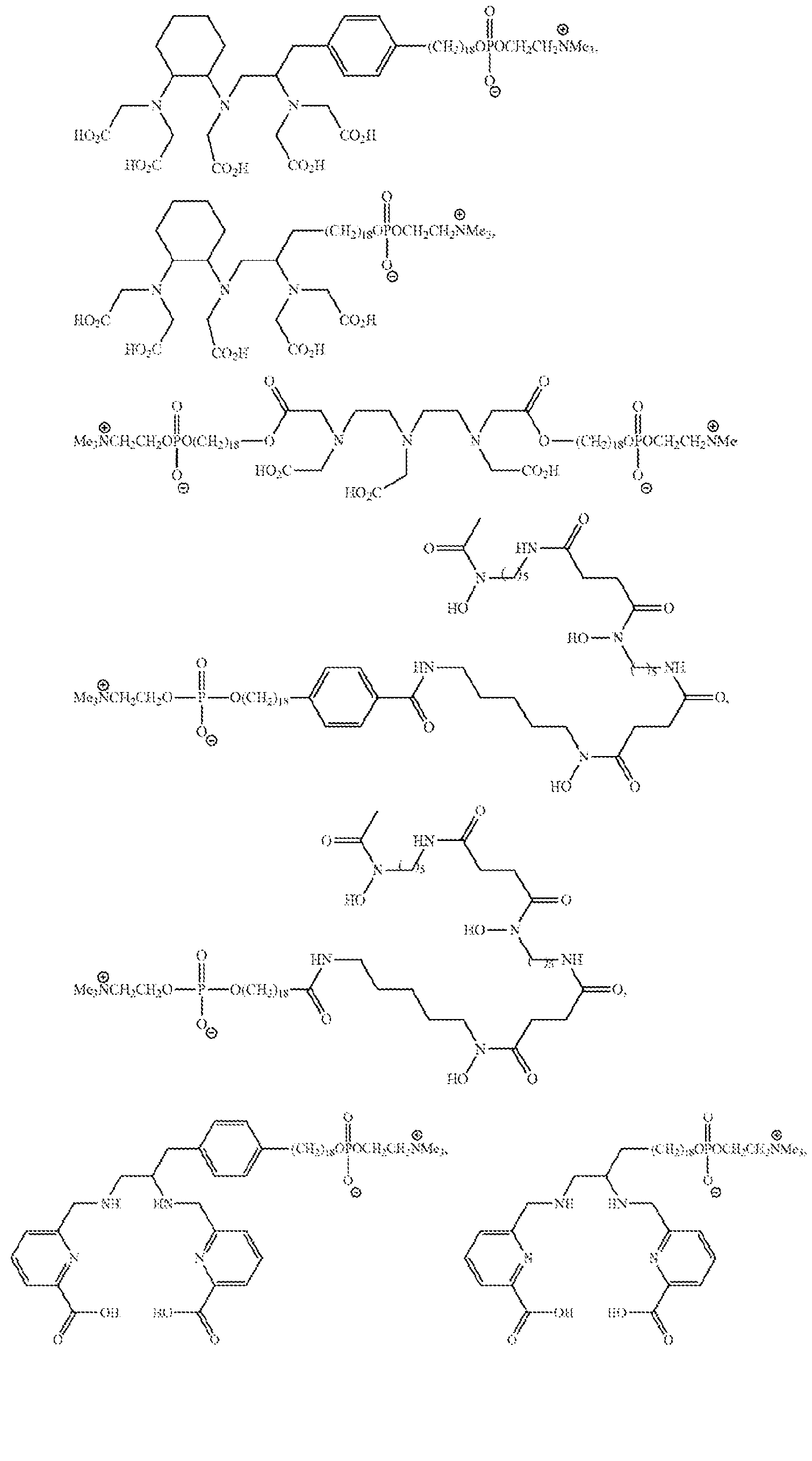
[0021] Exemplary chelating agents include 1,4,7,10-tetraazacyclododecane-1,4,7- triacetic acid (DO3A) or one of its derivatives; 1,4,7-triazacyclononane-l,4-diacetic acid (NODA) or one of its derivatives; 1,4,7-triazacyclononane-l,4,7-triacetic acid (NOTA) or one of its derivatives; 1,4,7,10-tetraazacyclododecane-l,4,7,10-tetraacetic acid1 (D1OTA) or one of its derivatives; 1,4,7-triazacyclononane, 1 -glutaric acid-4, 7-diacetic acid (NODAGA) or one of its derivatives; 1,4,7,10-tetraazacyclodecane, 1-glutaric acid-4,7, 10-triacetic acid (DOTAGA) or one of its derivatives; 1,4,8,11-tetraazacyclotetradecane- 1,4,8,11-tetraacetic acid (TETA) or one of its derivatives; 1,4,8,11-tetraazabicyclo[6.6.2]hexadecane-4,11- diacetic acid (CB-TE2A) or one of its derivatives; diethylene triamine pentaacetic acid (DTP A), its diester, or one of its derivatives; 2-cyclohexyl diethylene triamine pentaacetic acid (CHX-A"-DTPA) or one of its derivatives; deforoxamine (DFO) or one of its derivatives; 1,2-[[6-carboxypyridin-2-yl]methylamino]ethane (H2dedpa) or one of its derivatives; and DADA or one of its derivatives, wherein DADA has the structure:
Figure imgf000008_0001
In some embodiments, a is 1 (aliphatic aryl-alkyl chain). In other embodiments, a is 0 (aliphatic alkyl chain). In some embodiments, m is 1 (acylphospholipid series). In some such embodiments, n is an integer between 12 and 20. In some embodiments, Y is — OCOX, — COOX or — OX. In some embodiments, X is — CH2CH3 or — CH3. In some embodiments, m is 0 (alkylphospholipid series). In some embodiments, b is 1. In some embodiments, n is 18.
[0022] In some embodiments, R2 is — N+Z3. In some such embodiments, each Z is independently — CH2CH3 or — CH3. In some such embodiments, each Z is — CH3.
[0023] In some embodiments, the chelating agent chelated to the metal atom is:
Figure imgf000009_0001
Figure imgf000010_0001
[0024] In some embodiments, the chelating agent chelated to the metal atom is:
Figure imgf000011_0001
Figure imgf000012_0001
Figure imgf000013_0001
Figure imgf000014_0001
Figure imgf000015_0001
Figure imgf000016_0001
[0025] In some embodiments, in the phospholipid ether metal chelate structure, a is 1, b is 1, m is 0, n is 18, and R2 is — N-(CH3)3 In some such embodiments, the phospholipid ether metal chelate is NM600 chelated to the metal atom, such as (but not limited to)90Y- NM600. NM600 with 64Cu as the metal atom is shown below:
Figure imgf000016_0002
[0026] In another aspect, the TRT agent is metaiodobenzylguanidine (MIBG), where the iodine atom in the MIBG is a radioactive iodine isotope.
[0027] In another aspect, the TRT agent is a radiolabeled tumor-targeting antibody. Exemplary radiolabeled tumor-targeting antibodies include 177Lu-girentuximab, rosopatamab radiolabeled with yttrium-90 and lutetium-177, 131I-labetuzumab, panitumumab conjugated to α-emitter, 212Pb, 90Y labeled anti-MUCl antibody, nti-TAG-72 intact antibodies radiolabeled with iodine-131, yttrium-90, and lutehum-177, 90Y-clivatuzumab tetraxetan, 31I-labeled mAb 81C6, and the like.
[0028] In another aspect, the TRT agent is a radioactive isotope of radium such as radium-223 dichloride.
[0029] Radionuclides are radioactive atoms and historically for TRT patients have been prescribed a given activity (unit curie, Ci), which is the rate of disintegration or radioactive decay. For research and mechanism-driven therapeutic approaches, however, it the absorbed dose (unit gray: Gy) of RT that will be administered from a given activity is determined. This is because absorbed dose, which is defined as the energy absorbed per unit mass of tissue, mediates the biological effects of radionuclides.
[0030] In an aspect, in the methods described herein, a low dose of TRT agent can be used. Without being held to theory, the radiation doses (Gy) described herein for tumors implanted in mice should translate to humans because the tumors implanted were derived from human cancer cell lines. As is known in the art, the optimal radiation dose can vary from one tumor to the other, however, in general, a low dose of a TRT agent is a dose that is less than the typical dose used for TRT monotherapy, that is, the dose of TRT expected to kill tumor cells. In an aspect, low dose TRT is administered at a dose that is less than 10 %, specifically less than 6% and more specifically less than 5% of the dose used to kill tumor cells, which can be as high as 36 Gy.
[0031] In an aspect, a low dose of a TRT agent provides a 1 to 6 Gy, specifically 1 to 3 Gy radiation dose for a human patient, including a child or an adult.
MODDIFIED IMMUNE CELLS INCLUDING CAR T CELLS
[0032] Adoptive cell therapy with genetically modified immune cells such as CAR T cells has emerged as a novel immunotherapy approach. T cells typically interact with the target of their endogenous T cell receptor (TCR), enabling highly specific responses against peptides presented in the context of human leukocyte antigen (HLA). Immune cells like T cells can be engineered to bind novel antigens and targets by inserting a new receptor with the desired specificity. Genetically modified immune cells include an extracellular domain comprising an antigen recognition domain linked to a first intracellular domain through a first transmembrane domain. Typically for CARs, the antigen recognition domain is a monoclonal antibody-derived single-chain variable fragment (scFv) capable of targeting an antigen such as a specific tumor-associated antigen.
[0033] In some aspects, the immune cell is a T-cell, a Natural Killer (NK) cell, an innate lymphoid cell, a Cytokine Induced Killer (CIK) cell, a hematopoietic progenitor cell, a peripheral blood (PB) derived immune cell, a bone marrow derived immune cell, a macrophage, or an umbilical cord blood (UCB) derived immune cell. In some aspects, the immune cell is an embryonic or induced pluripotent stem cell (iPSC)-derived immune cell. In some aspects, wherein the immune cells are modified autologous cells isolated from a patient in need of cancer treatment, or modified cells from an allogeneic healthy donor with intent to treat a patient with cancer. The foregoing immune cells can be genetically engineered to express a CAR.
[0034] The immune cells may be isolated from subjects, particularly mammalian subjects such as human subjects and companion animals. The immune cells can be obtained from a subject of interest, such as a subject suspected of having a particular disease or condition, a subject suspected of having a predisposition to a particular disease or condition, or a subject who is undergoing therapy for a particular disease or condition. The immune cells may be enriched/purified from any tissue where they reside including, but not limited to, blood (including blood collected by blood banks or cord blood banks), spleen, bone marrow, tissues removed and/or exposed during surgical procedures, and tissues obtained via biopsy procedures. Tissues/organs from which the immune cells are enriched, isolated, and/or purified may be isolated from both living and non-living subjects, wherein the non-living subjects are organ donors. The isolated immune cells may be used directly, or they can be stored for a period of time, such as by freezing.
[0035] The population of immune cells can be obtained from a subject in need of therapy or suffering from a disease associated with reduced immune cell activity. Thus, the cells will be autologous to the subject in need of therapy. Alternatively, the population of immune cells can be obtained from a donor such as an allogenic healthy donor. The immune cell population can be harvested from PB, cord blood, bone marrow, spleen, or any other organ/tissue in which immune cells reside in said subject or donor. The immune cells can be isolated from a pool of subjects and/or donors, such as from pooled cord blood. The population of immune cells can be derived from iPSCs and/or any other stem cell known in the art. In some aspects, the iPSCs and/or stem cells used to derive the population of immune cells can be obtained from a subject in need of therapy or suffering from a disease associate with reduced immune cell activity, thus these iPSCs and/or stem cells will be autologous to the subject in need of therapy. Alternatively, the iPSCs and/or stem cells can be obtained from a healthy donor and therefore be allogeneic to the subject in need of therapy.
[0036] When the population of immune cells is obtained from a donor distinct from the subject, the donor is preferably allogeneic, provided the cells obtained are subject- compatible in that they can be introduced into the subject. Allogeneic donor cells may or may not be human leukocyte antigen (HLA)-compatible. To be rendered subject-compatible, allogeneic cells can be treated to reduce immunogenicity.
[0037] Immune cells and human T cells, for example, can be edited such that a CAR is inserted into the genome randomly using a viral vector (retrovirus, lenti virus, AAV, etc.) or inserted into a targeted region in the genome of a T cell using non-viral approaches (electroporation, mRNA, lipid nanoparticle, etc.) coupled with CRISPR/Cas9, TALEN or Zinc finger nucleases. The CAR can be inserted into an endogenous T cell receptor alpha subunit constant gene (TRAC), or an endogenous T cell receptor beta subunit constant gene (TRBC).
[0038] In an aspect, a CAR comprises an antigen-specific extracellular domain (e.g., a single chain variable fragment [scFV] that can bind a surface-expressed antigen of a malignancy) coupled to an intracellular domain (e.g., CD28, 1COS, CD27, 4-1BB, 0X40, CD40L, CD3-ξ , or a combination thereof) by a transmembrane domain (e.g., derived from a CD4, CD8, CD28, CH2CH3, NKG2D, IgG or CD3-ξ transmembrane domain).
[0039] The antigen-specific extracellular domain can also include a spacer linking the Vh and VL chains of the scFV, which can be the hinge region of IgGl and is sufficient for most scFv-based constructs.
[0040] The antigen-specific extracellular domain of a CAR recognizes and specifically binds an antigen, typically a surface-expressed antigen of a malignancy. An antigen-specific extracellular domain specifically binds an antigen when, for example, it binds the antigen with an affinity constant or affinity of interaction (KD) between about 0. 1 pM to about 10 pM, specifically about 0.1 pM to about 1 μM, more specifically about 0. 1 pM to about 100 nM. Methods for determining the affinity of interaction are known in the art. An antigen-specific extracellular domain suitable for use in a CAR may be any antigen- binding polypeptide, one or more scFv, or another antibody based recognition domain (cAb VHH (camelid antibody variable domains) or humanized versions thereof, IgNAR VH (shark antibody variable domains) and humanized versions thereof, sdAb VH (single domain antibody variable domains) and "‘camelized” antibody variable domains are suitable for use. In some instances, T cell receptor (TCR) based recognition domains such as single chain TCR may be used as well as ligands for cytokine receptors.
[0041] The present disclosure provides chimeric antigen receptors (CARs) that bind to an antigen of interest. In certain embodiments, the CAR binds to a tumor antigen. Any tumor antigen (antigenic peptide) can be used in the tumor-related embodiments described herein. Sources of antigen include, but are not limited to, cancer proteins. The antigen can be expressed as a peptide or as an intact protein or portion thereof. The intact protein or a portion thereof can be native or mutagenized. Non-limiting examples of tumor antigens that are CAR targets include carbonic anhydrase IX (CAIX), carcinoembryonic antigen (CEA), CD8, CD7, CD10, CD19, CD20, CD22, CD30, CD33, CLL1, CD34, CD38, CD41, CD44, CD49f, CD56, CD74, CD133, CD138, CD123, CD44V6, an antigen of a cytomegalovirus (CMV) infected cell (e.g., a cell surface antigen), epithelial glycoprotein-2 (EGP-2), epithelial glycoprotein-40 (EGP-40), epithelial cell adhesion molecule (EpCAM), receptor tyrosine-protein kinases erb-B2,3,4 (erb-B2,3,4), folate-binding protein (FBP), fetal acetylcholine receptor (AChR), adult AChR subunits, folate receptor-a, Ganglioside G2 (GD2), Ganglioside G3 (GD3), human Epidermal Grow th Factor Receptor 2 (HER-2), human telomerase reverse transcriptase (hTERT), Interleukin- 13 receptor subunit alpha-2 (IL- 13Ra2), K-light chain, kinase insert domain receptor (KDR), Lewis Y (LeY), LI cell adhesion molecule (L1CAM), melanoma antigen family A, 1 (MAGE-A1), Mucin 16 (MUC16), Mucin 1 (MUC1), Mesothelin (MSLN), Claudin-18.2, FAP, CA19, B7-H3, calreticulin, ERBB2, MAGEA3, p53, MARTI, GP100, Proteinase3 (PR1), Tyrosinase, Survivin, hTERT, EphA2, NK.G2D ligands, cancer-testis antigen NY -ESO- 1, oncofetal antigen (h5T4), prostate stem cell antigen (PSCA), prostate-specific membrane antigen (PSMA), ROR1, tumor-associated glycoprotein 72 (TAG-72), vascular endothelial growth factor R2 (VEGF-R2), Wilms tumor protein (WT-1), BCMA, NKCS1, EGF1R, EGFR, CD99, CD70, ADGRE2, CCR1, LILRB2, PRAME CCR4, CD5, CD3, TRBC1, TRBC2, TIM-3, Integrin B7, ICAM-1, CD70, Tim3, CLEC12A, ERBB, and combinations thereof.
[0042] The intracellular domain transmits the T cell activation signal. The intracellular domain can increase CAR T cell cytokine production and facilitate T cell replication. The intracellular domain reduces CAR T cell exhaustion, increases T cell antitumor activity, and enhances survival of CAR T cells in patients. Exemplary intracellular domains comprise co-stimulatory domains, including those from CD27, CD28, CD137 or 4- IBB, CD154 or CD40L, CD244 or 2B4, CD278 or ICOS, CD134 or 0X40, CD3-ξ and combinations thereof, and signaling domains (also called cytotoxicity domains), including those from CD16, DAP10, DAP12, CD28, ICOS, CD27, 0X40, CD40L, CD3-ξ, and combinations thereof. A costimulatory domain is derived from the intracellular signaling domains of costimulatory proteins that enhance cytokine production, proliferation, cytotoxicity, and/or persistence in vivo.
[0043] Typically, the antigen-specific extracellular domain is linked to the intracellular domain of the CAR by a transmembrane domain, e.g., derived from a CD4, CD8, CD28, CH2CH3 or NKG2D, IgG or CD3-ξ transmembrane domain. The transmembrane domain traverses the cell membrane, anchors the CAR to the T cell surface, and connects the extracellular domain to the intracellular signaling domain, thus impacting expression of the CAR on the T cell surface.
[0044] CARs may also further comprise one or more spacers. A spacer or hinge connects (i) the antigen-specific extracellular domain to the transmembrane domain, (ii) the transmembrane domain to a costimulatory domain, (Hi) a costimulatory domain to the intracellular domain, and/or (iv) the transmembrane domain to the intracellular domain. For example, inclusion of a spacer domain (e.g., IgGl, IgG2, IgG4, CD28, CD8) between the antigen-specific extracellular domain and the transmembrane domain may affect flexibility’ of the antigen-binding domain and thereby CAR function. Suitable transmembrane domains, costimulatory domains, and spacers are known in the art.
[0045] In a specific aspect, the CAR comprises a tumor antigen binding domain that targets Ganglioside G2 (GD2), coupled to a transmembrane domain from CD8, CD28, CH2CH3 or NKG2D, coupled to an intracellular domain comprising a costimulatory domain from CD27, CD28, CD137, CD154, CD244, CD278, or a combination thereof, and a cytotoxicity domain from CD3ξ, DAP 10, DAP 12, CD 16, or a combination thereof.
[0046] The CAR can be inserted into the genome of the unmodified T cells using viral or non-viral methods. The viral vectors include retroviruses (including lenti virus), adenovirus and adeno-associated virus. Production of CAR T cells using viral methods to insert the CAR are well-known in the art. Methods of making non-viral CAR T cell products are described in US2020/0000851; WO2021/173925 and WO2023/023635, incorporated herein by reference for their disclosure of making non-viral CAR T cell products.
[0047] Unmodified T cells include autologous T cells that are collected from a patient, such as a cancer patient, by peripheral blood draw or leukapheresis. Unmodified T cells can also include T cells from allogeneic healthy donors or induced pluripotent stem cells which can be used to produce universal T cells for administration to a patient. T cells are generally modified ex vivo, that is outside of the patient, and then the modified T cells such as CAR T cells are returned to the patient, such as by intravenous infusion, subcutaneous, intratumoral, intraperitoneal or intracerebral ventricular injection.
[0048] In an exemplary’ non-viral method as described in WO2021/173925, a Cas9 RNP and a non-viral double-stranded HDR template including the CAR are introduced into the unmodified T cells by electroporation to provide genome-edited T cells comprising the CAR.
[0049] Genome editing of the T cells can employ a CRISPR system, or Cas9 ribonucleoprotein. CRISPR refers to the Clustered Regularly Interspaced Short Palindromic Repeats type II system used by bacteria and archaea for adaptive defense. This system enables bacteria and archaea to detect and silence foreign nucleic acids, e.g., from viruses or plasmids, in a sequence-specific manner. In ty pe II systems, guide RNA interacts with Cas9 and directs the nuclease activity of Cas9 to target DNA sequences complementary to those present in the guide RNA. Guide RNA base pairs with complementary sequences in target DNA. Cas9 nuclease activity then generates a double-stranded break in the target DNA.
DOSING REGIMENS
[0050] In an aspect, a method of treating a solid tumor in a subject comprises administering to the subject a low dose of a targeted radiotherapy^ (TRT) agent, waiting a period of 1 to 60 days, and after waiting, administering to the subject a modified immune cell (e.g., chimeric antigen receptor (CAR) T cell) therapy.
[0051] In an aspect, included herein is a modified immune cell (e.g., a CAR T cell) for use in a method of treating a solid tumor in a subject, wherein the method comprises administering to the subject a low dose of a TRT agent, waiting a period of 1 to 60 days, and after waiting, administering to the subject the modified immune cell therapy.
[0052] In another aspect, a modified immune cell (e.g., a CAR T cell) for use in a method of treating a solid tumor in a subject, the method comprising administering to the subject the modified immune cell, wherein the subject has been administered a low dose of a TRT agent 1 to 60 days prior to the administration of the modified immune cell.
[0053] In another aspect, included is the use of a modified immune cell (e.g., a CAR T cell) in the manufacture of a medicament for a method of treating a solid tumor in a subject, wherein the method comprises administering to the subject a low dose of a TRT agent, waiting a period of 1 to 60 days, and after waiting, administering to the subject the modified immune cell.
[0054] The time period for waiting between therapies is based on the half-life of the TRT agent and the starting radiation dose. Because the TRT is expected to decrease the viability of the CAR T cells, the waiting period is an important part of the method. Exemplary waiting periods include 1 to 60 days, 1 to 30 days, 2 to 30 days, 2 to 20 days, 2 to 12 days, 3 to 10 days, 3 to 9 days or 3 to 6 days.
[0055] In an aspect, the subject is a mammalian subject, specifically a human or canine subject.
[0056] Non-limiting examples of the cancers presenting as malignant solid tumors that could treated using the disclosed method include melanoma, neuroblastoma, lung cancer, adrenal cancer, colon cancer, colorectal cancer, ovarian cancer, prostate cancer, renal cell carcinoma, non-small cell lung cancer, head or neck cancer, bladder cancer, hepatocellular carcinoma, spinal chordoma, cholangiocarcinoma, liver cancer, subcutaneous cancer, squamous cell cancer of the skin or head and neck, intestinal cancer, retinoblastoma, cervical cancer, glioma, breast cancer, pancreatic cancer, soft tissue sarcomas, Ewings sarcoma, rhabdomyosarcoma, osteosarcoma, retinoblastoma, Wilms' tumor, medulloblastoma, ependymoma, pineoblastoma, peripheral neuroectodermal tumor, or germ cell tumor.
[0057] The disclosure is inclusive of the compounds described herein (including intermediates) in any of their pharmaceutically acceptable forms, including isomers (e.g., diastereomers and enantiomers), tautomers, salts, solvates, polymorphs, prodrugs, and the like. It should be understood that the term “compound’’ includes any or all of such forms, whether explicitly stated or not (although at times, “salts’" may be explicitly stated).
[0058] “Pharmaceutically acceptable” as used herein means that the compound or composition or carrier is suitable for administration to a subject to achieve the treatments described herein, without unduly deleterious side effects in light of the necessity of the treatment.
[0059] The term “effective amount,” as used herein, refers to the amount of the compounds or dosages that will elicit the biological or medical response of a subject, tissue or cell that is being sought by the researcher, veterinarian, medical doctor or other clinician.
[0060] The term, “pharmaceutically-acceptable carrier” includes any and all dry powder, solvents, dispersion media, coatings, antibacterial and antifungal agents, isotonic agents, absorption delaying agents, and the like. Pharmaceutically-acceptable carriers are materials, useful for the purpose of administering the compounds in the method of the present invention, which are preferably non-toxic, and may be solid, liquid, or gaseous materials, which are otherwise inert and pharmaceutically acceptable, and are compatible with the compounds described herein. Examples of such carriers include, without limitation, various lactose, mannitol, oils such as com oil, buffers such as PBS, saline, polyethylene glycol, glycerin, polypropylene glycol, dimethylsulfoxide, an amide such as dimethylacetamide, a protein such as albumin, and a detergent such as Tween 80, mono- and oligopolysaccharides such as glucose, lactose, cyclodextrins and starch.
[0061] The term '‘administering” or '‘administration,” as used herein, refers to providing the compound or pharmaceutical composition of the invention to a subject suffering from or at risk of the diseases or conditions to be treated or prevented.
[0062] A route of administration in pharmacology is the path by which a drug is taken into the body. Routes of administration may be generally classified by the location at which the substance is applied. Common examples may include oral and intravenous administration. Routes can also be classified based on where the target of action is. Action may be topical (local), enteral (system-wide effect, but delivered through the gastrointestinal tract), or parenteral (systemic action, but delivered by routes other than the GI tract), via lung by inhalation. One form of local administration is intratumoral (IT), whereby an agent is injected directly into, or adjacent to, a known tumor site.
[0063] A topical administration emphasizes local effect, and substance is applied directly where its action is desired. Sometimes, however, the term topical may be defined as applied to a localized area of the body or to the surface of a body part, without necessarily involving target effect of the substance, making the classification rather a variant of the classification based on application location. In an enteral administration, the desired effect is systemic (non-local), substance is given via the digestive tract. In a parenteral administration, the desired effect is systemic, and substance is given by routes other than the digestive tract.
[0064] Examples of parenteral administrations may include intravenous (into a vein), e.g. many drugs, total parenteral nutrition intra-arterial (into an artery), e.g., vasodilator drugs in the treatment of vasospasm and thrombolytic drugs for treatment of embolism, intraosseous infusion (into the bone marrow), intra-muscular, intracerebral (into the brain parenchyma), intracerebroventricular (into cerebral ventricular system), intrathecal (an injection into the spinal canal), and subcutaneous (under the skin). Among them, intraosseous infusion is, in effect, an indirect intravenous access because the bone marrow drains directly into the venous system. Intraosseous infusion may be occasionally used for drugs and fluids in emergency medicine and pediatrics when intravenous access is difficult. [0065] The invention is further illustrated by the following non-limiting examples.
EXAMPLES
MATERIALS AND METHODS
[0066] Cell lines: The GD2-expressing CHLA-20 cells (human neuroblastoma cells) and M21 (human melanoma cells) were kindly donated by Dr. Mario Otto and Dr. Paul Sondel respectively and grown in Dulbecco’s Modified Eagle Medium (DMEM) with high glucose (Gibco) supplemented with 10% fetal bovine serum (Avantor) and 1% penicillinstreptomycin (Gibco). These cells were maintained at 37°C in 5% CO2. Cell line authentication was done using genomic short tandem repeat analysis (Idexx BioAnalytics) and by cell morphology per ATCC guidelines. Mycoplasma testing was performed on a regular basis to rule out contamination using the Mycoplasma Detection Kit My coStrip™ (InvivoGen).
[0067] GD2 CAR T cells: The virus-free CRISPR GD2 CAR T cells were manufactured as previously described in Mueller et al. 2022 J Immunother Cancer, 2022;10:e004446. doi: 10. 1136/jitc-2021 -004446 and cultured in ImmunoCultTM-XF T cell Expansion Medium supplemented with 500 U/rnL of IL-2 (Peprotech) and maintained 37 °C in 5% CO2.
[0068] In vitro dosimetry: All in vitro dosimetry studies were performed in a 6-well cell culture plate. Serial dilutions of various activity of free 177Lu or 225 Ac were made in cell culture medium and added to each well. Thermoluminescent dosimeters (TLDs) were placed under each well. For each radionuclide, the TLDs were harvested after 1 half-life and analyzed by the University of Wisconsin-Madison Radiation Calibration Laboratory (Calibration Cert # 1664.01). A standard curve was obtained and used to determine the activity of 177Lu or 225 Ac needed to deliver a given radiation dose, as previously reported in the art. To confirm the validity of this method, the mean absorbed dose to cells was calculated using the Geant4 Monte Carlo toolkit and using an extension of RAPID. A model of a flat bottom 6-well plate has been developed in Geant4 using manufacturing specifications where the diameter of each well is 36 mm, and the height of each well is 10.7 mm. The cell volume can be defined as a thin water-equivalent layer at the bottom of the well. [0069] In vitro GD2 CAR T cell irradiation: The GD2 CAR T cells were irradiated with various doses of radiation delivered over 3 days by 177Lu or 225 Ac diluted in 3 mL of ImmunoCult™-XF T cell Expansion Medium. Doses of radiation delivered by 177Lu were 1, 2 and 6 Gy whereas with 225 Ac, 1 and 2 Gy were delivered.
[0070] In vitro GD2 CAR T cell viability : Following the irradiation of the GD2 CAR T cells, their viability was determined by flow cytometry.
[0071] In vitro GD2 CAR T cells and tumor cells co-culture: After the GD2 CAR T cells were irradiated, they were harvested and washed 3 times with PBS and co-cultured with CHLA-20 cells with an effector to target (E:T) ratio of 10: 1 for 24 hrs.
[0072] Flow cytometry : Cells were harvested, washed with PBS, and resuspended into single cell solution in PBS as previously reported in the art. Fc blocking (Biolegend, 422302) and Live/Dead staining with Ghost Dye™ Red 780 (Tonbo Biosciences, 13-0865- T100) were performed for 10 minutes at 4°C. The fluorophore-conjugated antibodies including anti-CD45-APC (Biolegend, 304012), anti-GD2-PE-Dazzle584 (Biolegend, 357320), anti-PD-l-PE (Biolegend, 329906), anti-CD69-BV510 (Biolegend, 310936), anti- NKG2D-BV605 (Biolegend, 320832) were incubated for 20 minutes at 4°C and washed with 2% FBS in PBS. The analysis of the sample was performed using the Attune™ NxT Flow Cytometer (ThermoFisher) and the collected data was analyzed using FlowJo software.
[0073] Statistical analysis: All statistical analysis were performed in Prism 9 (GraphPad Software). Two-way ANOVA with Tukey’s multiple comparisons test was used for comparison between multiple groups.
EXAMPLE 1: RADIATION DELIVERED BY 177Lu AND 225 Ac RESULTS IN A DOSE DEPENDENT GD2 CAR T CELL DEATH
[0074] Combining in vivo TRT and CAR T cell therapy will lead to the exposure of CAR T cells to radiation, which we hypothesize will be detrimental to their viability. As such, to evaluate the effect of radiation delivered by TRT on CAR T cells, GD2 CAR T cells were exposed to various doses of radiation delivered by 177Lu or 225 Ac. Six-well plates containing GD2 CAR T cells were incubated in a medium containing specific activity of 177LU or 225 Ac needed to deliver radiation dose of 1, 2 or 6 Gy by day 3. Following the delivery of the radiation, the GD2 CAR T cells were harvested, and their viability was evaluated by flow cytometry (FIG. 2A) by using live/dead staining (See sample gating strategy; FIG. 2B). A dose dependent GD2 CAR T cell death is observed with both radionuclides. However, 177Lu appears to be less cytotoxic compared to 225 Ac. One Gy of radiation delivered by 225Ac resulted in nearly a 3 -fold decrease in the viability of GD2 CAR T cells compared to the non-irradiated, whereas with 2 Gy of 225 Ac such viability7 dropped to 2.34% (FIG. 2C). After the delivery of 1 Gy of radiation by 177Lu, compared to the nonirradiated, the viability of GD2 CAR T cells decreased from 25% to 19.6% and with 2 and 6 Gy, such viability dropped to 16.1% and 12.2% respectively (FIG. 2C).
EXAMPLE 2: RADIATION DELIVERED BY 177Lu AND 225 Ac ENHANCES THE CYTOTOXIC ACTIVITY OF GD2 CAR T CELLS AGAINST THE GD2-EXPRESSING NEUROBLASTOMA CELL LINE CHLA-20
[0075] In addition to evaluating the effect of radionuclide-delivered radiation on the viability of the GD2 CAR T cells, the impact of such radiation on the effector function of the GD2 CAR T cells was also determined. To this end, following irradiation, the GD2 CAR T cells were co-cultured with the GD2-expressing neuroblastoma cell line CHLA-20 for 24 hrs. The killing potential of the irradiated GD2 CAR T cells was determined by measuring the viability of CHLA-20 cells by flow cytometry (FIG. 3A). The viable CHLA-20 cells were identified as CD45 negative and live/dead stain (Ghost Dye™ Red) negative (CD45-/ Ghost Dye™ Red -). As expected, non-irradiated GD2 CAR T cells displayed a potent cytotoxicity activity against CHLA-20 cells. The viability of the CHLA-20 cells decreased to 6.9% after a 24 hrs. co-culture with GD2 CAR T cells compared to 52.2% in the absence of GD2 CAR T cells (FIG. 3B). The irradiation of GD2 CAR T cells prior to co-culture with CHLA-20 cells enhanced their cytotoxic activity of the CAR T cells, resulting in a near complete eradication of CHLA-20 cells (FIG. 3B). However, the enhanced cytotoxicity of the GD2 CAR T cell was independent of the type of particle emitted (a vs p particle) and radiation dose delivered (FIG. 3B). The radiation-induced enhancement of GD2 CAR T cell cytotoxic activity was also observed against the human melanoma cell line M21 which also expresses GD2 (FIG. 4A-C).
EXAMPLE 3: RADIATION DELIVERED BY 177Lu AND 225 Ac DOES NOT IMPACT THE EXPRESSION OF EXHAUSTION AND ACTIVATION MARKERS ON GD2 CAR T CELLS
[0076] To further characterize the effect of 225Ac or 177Lu-delivered radiation on GD2 CAR T cells and understand the underlaying mechanism of the radiation-induced enhancement of GD2 CAR T cells, the expression of the T cell exhaustion marker PD-1, activation marker CD9 and activating receptor NKG2D were evaluated by flow cytometry. As previously described, following the irradiation of the GD2 CAR T cells with the various radiation doses of 225 Ac or 177Lu, the GD2 CAR T cells were harvested, and the expression of cell surface markers was determined by flow cytometry (FIG. 5A). Compared to nonirradiated GD2 CAR T cells, 1 Gy delivered by 225Ac resulted in an increased expression of PD-1, whereas radiation (1, 2, 6 Gy) delivered by 177Lu exhibited a trend toward a decrease in PD-1 expression. However, the changes in PD-1 expression following irradiation were not statistically significant (FIG. 5B).
[0077] A trend toward upregulation albeit non-significant was observed in the expression of CD69 and NK.G2D following the delivery of radiation by both radionuclides (FIG. 5C and 5D).
DISCUSSION OF EXAMPLES 1-3
[0078] Due to the increasing therapeutic role of TRT in the clinical management of cancer and its immunostimulatory effects at low dose, the combination of TRT with CAR T cell therapy represents a therapeutic approach for selected patients with metastatic solid tumor. As such, the in vitro effects of TRT on CAR T cell viability and function w as determined to identify their maximum tolerated radiation dose (delivered by radionuclide) and the radionuclide (α vs [β-emitter) capable of enhancing the effector function of CAR T cells while minimizing the deleterious effects on CAR T cell viability.
[0079] The dose dependent death of GD2 CAR T cells observed in response to radiation delivered by 177Lu and 225 Ac is expected because radiation is cytotoxic to lymphocytes especially CD8+ T cells from which GD2 CAR T cells are manufactured. Although such dose dependence death is present with radiation delivered by either radionuclide, the biological effect of a similar dose of radiation (1 Gy) delivered by the a- emitter 225 Ac is higher than that of the same dose delivered by the [β-emiter 177Lu. Without being held to theory, this difference in biological effect is attributable to the LET difference between these 2 radionuclides. Despite having a shorter range in tissue (0.05-0.08 mm) compared to [β-emitters (1-5 mm), α-emitters, due to their higher LET (50-230 keV/ μm), induce 10-20 double strand DNA breaks (DSB) per 10 μm resulting in potent cytotoxicity, whereas β-emitters induce more single strand DNA breaks which are easily repaired. It has been suggested in the art that a-particle emiters induce multiple DSBs that are in close vicinity resulting in the depletion of p53-binding protein 1 (53BP1), a DNA damage response protein, required for double strand DNA breaks. Such depletion leads to insufficient amount of 53BP1 to adequately repair all DSBs, further accentuating the biological effectiveness of high LET a-particle emitters.
[0080] In this study, the in vitro CART cell death induced by 1 Gy of 225Ac is only 2.2 times that of 177Lu (FIG. 2C) which is lower than that observed in the prior art. This discrepancy could be attributed to the different in vitro model used in this study (GD2 CAR T cells), the method used to access the therapeutic efficacy and to the fact that our in vitro dosimetn enabled the comparison of a similar dose of radiation delivered by a radionuclide instead of activity.
[0081] Although radiation delivered by external beam radiation therapy (EBRT) upregulates the expression of PD-1 on CD8+ T cells which can be detrimental CART cell function, surprisingly, such upregulation was not observed with 177Lu or 225 Ac. Without being held to theory, this may be due to the inherent design of this 3rd generation GD2 CAR with the CRISPR knock in of the CAR construct into the TRAC locus which yielded CART cells devoid of T cell receptor and may represent another therapeutic benefit of combining TRT instead of EBRT with CART cell therapy.
[0082] Radiation delivered by EBRT enhances the cytotoxic activity of CART cells including against antigen negative malignant cells. Prior to this study, such enhancement has not been demonstrated with TRT. It is conceivable that presence of the TRT in the milieu may have selected for the more radioresistant CART cells subclones with potent cytolytic activity.
[0083] Based on the in vitro studies, the low dose of radiation tested herein (1 or 2 Gy) delivered by a-emitter might not be ideal for a potential therapeutic combination with CAR T cells, making β -emitters the preferred candidates. Instead, 1 Gy delivered by a β - particle emitter may be ideal because all other radiation doses evaluated (2 or 6 Gy) enhanced CAR T cell cytotoxic function to the same extent while inducing higher rates of GD2 CAR T cell death. These observations suggest that with regards to the sequencing between TRT and CAR T cells combination, the infusion of CAR T cells should be performed after the administration of TRT to minimize the exposure of CAR T cells to radiation. While the in vitro study suggests that 1 Gy delivered by the β-emitter 177Lu might be ideal, the higher tested doses of 2 or 6 Gy may be considered if there is an adequate delay between the TRT delivery' and the CAR T cell infusion. This delay will enable the radioactive decay of the radionuclide such that when the CART cells are infused, the remaining activity of the radionuclide in the TME may only deliver a radiation dose of 1 Gy or less. Without being held to theory, it is speculated that using radiation dose higher than 1 Gy may be an attractive option as it will enable the tumoricidal effect of TRT on malignant cells to be harnessed, subsequently reducing the tumor volume against which CART cells have to be effective.
[0084] This study provides the answers to key questions such as the optimal dose of radiation, type of radionuclide, timing and sequence of combination and enable the rational design of in vivo studies evaluating the combination of CART cell therapy and TRT.
EXAMPLE 4: IN VIVO COMBINATION OF TRT AND GD2 CART CELLS
[0085] Ten million LUC-GFP CHLA-20 human neuroblastoma cells were injected subcutaneously onto the right flank of 6-8 weeks old male or female NRG mice. Tumor implantation was confirmed 5 days following the cells injection by bioluminescence using the In Vivo Imaging System (IVIS). The tumor volume was monitored and the mice were randomized once a volume of ~75 mm3 was reached into different treatment arms: 1) Control (No treatment; n=4); 2) Radiation only (1.75 Gy; n= 5) with 177Lu-NM600 administered intravenously 6 days after tumor injection; 3) GD2 CAR T cells (10 million cells; n=5) administered intravenously 15 days after tumor injection; 4) a combination therapy arm (n=8), with radiation (1.75 Gy) administered intravenously 6 days after tumor injection followed by GD2 CAR T cells (10 million cells) administered intravenously 9 days later (15 days after tumor injection). The tumor volume was monitored until euthanasia is performed when tumor reaches 1500 mm3 or ulceration is noted.
[0086] CART cells have had limited success against solid tumors. In patients with metastatic disease, it is not possible to irradiate all sites of disease with EBRT. TRT can irradiate all of sites of disease but at low doses is likely to be ineffective as a single agent. Giving TRT followed by CART cells can significantly enhance killing of metastatic solid tumors without impacting CART viability or proli feration.
EXAMPLE 5: CAR-T cell targeting of B7-H3+ cancers combined with radiotherapy
[0087] Metastatic spread of cancers into the cerebrospinal fluid (CSF), termed leptomeningeal disease (LMD), is a major clinical challenge for which current treatments are both insufficiently effective and excessively toxic. LMD occurs in 5-10% of adult patients with metastatic solid tumors and carnes a grave prognosis. In the absence of treatment, survival for these patients is typically measured in weeks. Patients wi th LMD can experience severe headaches and loss of vision, hearing, speech, and facial movement and sensation. In some pediatric patients with cancers, such as medulloblastoma, the risk of CSF spread is sufficiently high that preemptive treatment is necessitated. Currently, EBRT targeting the whole brain and spine (craniospinal irradiation, CSI) is the only proven effective treatment modality available for most patients with LMD or high risk of CSF dissemination of cancer. While CSI EBRT delays progression, palliates symptoms, and extends survival for these patients, LMD often recurs. To reduce the risk of side effects to the heart, lungs, esophagus, and oral cavity, patients with LMD are ideally treated with high precision proton beam radiation, however access to proton radiation is limited especially among rural and low income patient populations. If patients achieve durable control or prevention of LMD, they often experience severe late toxicities from CSI EBRT, even with proton beam radiation. These toxicities include memory' deficits and weakening of bones in the spine. In children, CSI EBRT additionally leads to stunting of grow th and cognitive impairment that can be severe. These toxicities arise because CSI EBRT treats not only the CSF space where LMD is located but also the entire brain, spinal cord, and vertebral bodies. This radiation of normal tissues leads to long-term permanent toxicities that compromise quality of life for survivors. Thus, there is enormous need for a new therapeutic approach that can effectively treat LMD with greater specificity and potency to eliminate cancer cells while minimizing effects on normal tissues around the CSF.
[0088] The methods described herein can provide effective and less toxic treatments for patients with LMD or high risk of CSF dissemination of cancer. The hypothesis is that intrathecal delivery of a novel radiopharmaceutical therapy (RPT) will enable more effective delivery' of radiation to LMD while reducing toxicity by minimizing radiation of normal tissues compared to CSI EBRT. Specifically, combining intrathecal administration of RPT and CAR-T cell therapy will overcome the limitations of intrathecal administration of RPT alone. CAR-T cells are standardly used for treatment of hematologic cancers, but have not yet proven effective against bulky solid tumors because of limited infiltration, activation, and persistence in established tumor microenvironments (TME). Recent studies, however, indicate promising therapeutic potential for intrathecally injected CAR-T cells against tumors of the central nervous system. Radiation can debulk and immunomodulate solid tumor TME to enhance T cell infiltration and activation, including in the CNS. Optimal dosing of RPT in combination with immunotherapies may be quite low and much different from optimal dosing of the same agent as a monotherapy. There is a potential cooperative effect of RPT in promoting solid tumor cell destruction by CAR-T cells, but only at very low doses of RPT. Described herein is the combination of intrathecal RPT with CAR-T cells to evaluate the curative potential of this combination for LMD.
[0089] EBRT can enhance the infiltration and expansion of CART cells in the TME of brain tumors in murine models, resulting in a potent and durable antitumor response mediated in part by the CAR T cells. Increased CAR-T cell killing activity can be achieved without inducing exhaustion markers in vitro when tumor cells are treated with RPT. By immunomodulating the TME of tumor sites, generating inflammation in the TME, and increasing tumor cell susceptibility to CAR-T cell killing, it is hypothesized that RPT will exhibit cooperative therapeutic efficacy in conjunction with CAR-T cells. The small absolute volume of disease in settings of LMD could be particularly well targeted by such a combination. Moreover, intrathecal CAR-T cells may encounter fewer inhibitory components of the host immune system in the CSF relative to blood as treatment via this route has yielded among the most promising clinical results to date against solid tumors. CAR-T cells infused intrathecally recently demonstrated anti-tumor responses in 3 of 4 patients with midline gliomas, resulting in increased pro-immune cytokines in CSF. Importantly, these clinical studies demonstrate safety of intrathecal administration of CAR-T cells with limited and manageable toxicities.
[0090] B7-H3 tumor-specific antigen expression in cancer and CAR-T cell targeting. The B7-H3 (CD276) antigen is an immune checkpoint that is overexpressed on the cell surface of tumor cells in pediatric brain cancers, neuroblastoma, melanoma, sarcomas, and a variety of other cancers, yet B7-H3 has little to no expression on normal tissues. B7-H3 is a is a pan-cancer antigen for targeting multiple forms of cancer with one CAR-T cell product. Nearly uniform cell surface expression of B7-H3 on the human BR3 LMD breast cancer cell line and the human SK-N-AS neuroblastoma cell line was observed by flow cytometry (FIG. 7A).
[0091] CAR-T cell targeting of B7-H3+ cancers. CAR-T cells targeting human B7- H3 demonstrate preclinical efficacy against microscopic disease in neuroblastoma, medulloblastoma, and other xenograft tumor models in immunodeficient mice. The B7-H3 CAR-T cells were designed as second-generation CD8H/Tm-CD28-CD3ξ (28ξ) CAR format comprising the CD8a H/Tm sequences and CD28 and CD3£ signaling domains. These B7- H3 CAR-T cells induced rapid and robust killing of LMD BR3 cells in vitro, compared to minimal or no killing from untransduced (UTD) control T cells (FIG. 7B).
[0092] Radiation promotes anti -tumor activity of anti-B7-H3 CAR-T cells. The inflammatory effects of radiation, including EBRT and RPT, promote tumor cell upregulation of cell surface expression of various immune checkpoint receptors. To evaluate whether radiation might affect tumor cell expression of B7-H3, 4 or 12 Gy EBRT or 12 Gy from 225 Ac were administered to BR3 LMD cells in vitro and 24 hrs later flow cytometry was performed. Radiation increased the expression of B7-H3 on the surface of tumor cells for this human breast cancer LMD model and this effect was maximized by the RPT agent in this preliminary’ analysis (FIG. 7C). These findings are important because B7-H3 CAR-T cell efficacy can be dependent upon high target antigen density on the surface of tumor cells. Induction of tumor cell surface expression of B7-H3 could be a mechanism for synergy between RPT and anti-B7-H3 CAR-T cells.
[0093] Knowing that radiation can induce inflammatory cytokine production in tumor cells, the secretion of cytokines from BR3 LMD cells following EBRT or 225 Ac radiation was studied. Using multi-cytokine single cell proteomic analysis with the Isoplexis system, in preliminary' studies, radiation increased the production of T cell stimulatory and inflammatory' cytokines by BR3 LMD cells and this was most prominent following 225 Ac radiation (FIG 7D). This suggest potential for RPT to enhance infiltration, activation, and persistence of CAR-T cells in this tumor model. Pre-treatment of LMD BR3 cells in vitro with 2 Gy EBRT at 24 hrs prior to addition of anti-B7-H3 CAR-T cells resulted in enhanced tumor cell killing, evidenced by efficacy at a lower effector: target cell (E:T) ratio in irradiated compared to control tumor cells (FIG. 7E). This suggests that low-dose radiation can promote susceptibility of these cells to CAR-T cell killing. Taken together, these preliminary' findings support the potential for a therapeutic synergy between RPT and CAR-T cells
[0094] Cooperative therapeutic efficacy of CAR-T cells and radiation in vivo. NRG mice were IV injected with luciferase-expressing SK-N-AS neuroblastoma cells to develop systemic metastases (predominantly in liver, not leptomeningeal disease (LMD) in this study). Upon confirmation of tumor engraftment, injection of 50 μCi 177Lu-NM600 (delivering an estimated 2 Gy tumor dose in this model) was done. Thirteen days later, these mice were IV infused with anti-B7-H3 CAR-T cells. Evidence of a cooperative therapeutic effect between low dose RPT and CAR-T cells was observed at day 27 after treatment initiation, with all mice receiving combination therapy appearing to be rendered disease free at this point based on bioluminescence luciferase imaging (FIG. 8A). In a separate study, LMD tumors were initiated via carotid artery injection of luciferase-expressing BR3 cells in NRG mice. At day 6, bioluminescence was performed to verify LMD growth and mice were randomized into treatment groups: control untransduced T cells (UTD), anti-B7-H3 CAR-T cells, or a combination of anti-B7-H3 CAR-T and 2 Gy CSI EBRT. EBRT was administered (Xstrahl CIX3 cabinet irradiator) on day 6 after engraftment and CAR-T or UTC cells (5X 10⁶) were intrathecally infused 24 hrs later. Previous experiments (not shown) have demonstrated no change in LMD growth with up to 5 Gy of CSI EBRT. In this study, results suggest that mice receiving a combination of anti-B7-H3 CAR-T cells and low dose CSI EBRT exhibit reduced LMD burden compared to UTC or CAR-T cell alone controls (FIG. 8B).
[0095] It is expected that these results will demonstrate dose- and time-dependent effect of RPT on B7-H3 expression and on the immune-relevant markers in the TME. Effective treatment regiments for each isotope delivered by NM600 in combination with CAR T cells will be identified. Optimal dosing of RPT for this combination is predicted to be at low dose levels and not at MTD, where radiation effects on CAR T cells may be detrimental to efficacy. Without being held to theoiy. in contrast to monotherapy, the higher biological effectiveness of an alpha-particle emitter may be counterproductive to combination with CAR-T cells and that combination with 177Lu-NM600 may be most effective in promoting CAR-T cell activity.
[0096] The use of the terms “a” and “an” and “the” and similar referents (especially in the context of the following claims) are to be construed to cover both the singular and the plural, unless otherwise indicated herein or clearly contradicted by context. The terms first, second etc. as used herein are not meant to denote any particular ordering, but simply for convenience to denote a plurality of, for example, layers. The terms “comprising”, “having”, “including”, and “containing” are to be construed as open-ended terms (i.e., meaning “including, but not limited to”) unless otherwise noted. Recitation of ranges of values are merely intended to serve as a shorthand method of referring individually to each separate value falling within the range, unless otherwise indicated herein, and each separate value is incorporated into the specification as if it were individually recited herein. The endpoints of all ranges are included within the range and independently combinable. All methods described herein can be performed in a suitable order unless otherwise indicated herein or otherwise clearly contradicted by context. The use of any and all examples, or exemplary language (e.g., “such as”), is intended merely to better illustrate the invention and does not pose a limitation on the scope of the invention unless otherwise claimed. No language in the specification should be construed as indicating any non-claimed element as essential to the practice of the invention as used herein. [0097] While the invention has been described with reference to an exemplary embodiment, it will be understood by those skilled in the art that various changes may be made and equivalents may be substituted for elements thereof without departing from the scope of the invention. In addition, many modifications may be made to adapt a particular situation or material to the teachings of the invention without departing from the essential scope thereof. Therefore, it is intended that the invention not be limited to the particular embodiment disclosed as the best mode contemplated for carrying out this invention, but that the invention will include all embodiments falling within the scope of the appended claims. Any combination of the above-described elements in all possible variations thereof is encompassed by the invention unless otherwise indicated herein or otherwise clearly contradicted by context.

Claims

1. A method of treating a solid tumor in a subject, comprising administering to the subject a low dose of a targeted radiotherapy (TRT) agent, waiting a period of 1 to 60 days, and after waiting, administering to the subject a modified immune cell therapy.
2. The method of claim 1, wherein the modified immune cell is a T-cell, a Natural Killer (NK) cell, an innate lymphoid cell, a Cytokine Induced Killer (CIK) cell, a hematopoietic progenitor cell, a peripheral blood (PB) derived immune cell, a bone marrow derived immune cell, a macrophage, or an umbilical cord blood (UCB) derived immune cell.
3. The method of claim 2, wherein the modified immune expresses a chimeric antigen receptor (CAR).
4. The method of claim 1, wherein the waiting period is 1 to 30 days.
5. The method of claim 1, wherein the low dose of the TRT agent is a 1-6 Gy radiation dose.
6. The method of claim 1, wherein the modified immune cell therapy comprises modified autologous cells isolated from a patient in need of cancer treatment, or modified cells from an allogeneic healthy donor.
7. The method of claim 3, wherein the CAR target comprises a tumor antigen selected from carbonic anhydrase IX (CAIX), carcinoembryonic antigen (CEA), CD8, CD7, CD10, CD19, CD20, CD22, CD30, CD33, CLL1, CD34, CD38, CD41, CD44, CD49f, CD56, CD74, CD133, CD138, CD123, CD44V6, an antigen of a cytomegalovirus (CMV) infected cell, epithelial glycoprotein-2 (EGP-2), epithelial glycoprotein-40 (EGP-40), epithelial cell adhesion molecule (EpCAM), receptor tyrosine-protein kinases erb-B2,3,4 (erb-B2,3,4), folate-binding protein (FBP), fetal acetylcholine receptor (AChR), adult AChR subunits, folate receptor-a, Ganglioside G2 (GD2), Ganglioside G3 (GD3), human Epidermal Grow th Factor Receptor 2 (HER-2), human telomerase reverse transcriptase (hTERT), Interleukin- 13 receptor subunit alpha-2 (IL-13Rα2), K-light chain, kinase insert domain receptor (KDR), Lewis Y (LeY), LI cell adhesion molecule (L1CAM), melanoma antigen family A, 1 (MAGE-A1), Mucin 16 (MUC16), Mucin 1 (MUC1), Mesothelin (MSLN), Claudin-18.2, FAP, CA19, B7-H3, calreticulin, ERBB2, MAGEA3, p53, MARTI, GP100, Proteinase3 (PR1), Tyrosinase, Survivin, hTERT, EphA2, NKG2D ligands, cancer-testis antigen NY-ESO-1, oncofetal antigen (h5T4), prostate stem cell antigen (PSCA), prostate- specific membrane antigen (PSMA), R0R1, tumor-associated glycoprotein 72 (TAG-72), vascular endothelial growth factor R2 (VEGF-R2), Wilms tumor protein (WT-1), BCMA, NK.CS1, EGF1R, EGFR, CD99, CD70, ADGRE2, CCR1, LILRB2, PRAME CCR4, CD5, CD3, TRBC1, TRBC2, TIM-3, Integnn B7, ICAM-1, CD70, Tim3, CLEC12A, ERBB, and combinations thereof.
8. The method of claim 3, wherein the CAR comprises a tumor antigen binding domain that targets Ganglioside G2 (GD2), coupled to a transmembrane domain from CD8, CD28, CH2CH3 or NKG2D, coupled to an intracellular domain comprising a costimulalory domain from CD27, CD28, CD 137, CD 154, CD244, CD278, or a combination thereof, and a cytotoxicity domain from CD3ξ , DAP 10, DAP 12, CD 16, or a combination thereof.
9. The method of claim 1, wherein the solid tumor is melanoma, neuroblastoma, lung cancer, adrenal cancer, colon cancer, colorectal cancer, ovarian cancer, prostate cancer, renal cell carcinoma, non-small cell lung cancer, head or neck cancer, bladder cancer, hepatocellular carcinoma, spinal chordoma, cholangiocarcinoma, liver cancer, subcutaneous cancer, squamous cell cancer of the skin or head and neck, intestinal cancer, retinoblastoma, cervical cancer, glioma, breast cancer, pancreatic cancer, soft tissue sarcomas, Ewings sarcoma, rhabdomyosarcoma, osteosarcoma, retinoblastoma, Wilms' tumor, medulloblastoma, ependymoma, pineoblastoma, peripheral neuroectodermal tumor, or germ cell tumor.
10. The method of claim 1, wherein the TRT agent is metaiodobenzylguanidine (MIBG), where the iodine atom in the MIBG is a radioactive iodine isotope; a radiolabeled tumor-targeting antibody; a radioactive isotope of radium; or a radioactive phospholipid ether metal chelate.
11. The method of claim 10, wherein the radioactive phospholipid ether metal chelate has the formula
Figure imgf000038_0001
Formula 1 wherein
Ri is s (a) a chelating agent that is chelated to a metal atom, wherein the metal atom is an alpha, beta or Auger emitting metal isotope with a half-life of greater than 6 hours and less than 30 days or (b) a radioactive halogen isotope, a is 0 or 1; n is an integer from 12 to 30; m is 0 or 1 ;
Y is — H, — OH, — COOH, — COOX, — OCOX, or — OX, wherein X is an alkyl or an arylalkyl;
R2is — N+H3, — N+H2Z, — N+HZ2, or — N+Z3, wherein each Z is independently an alkyl or an aryl, and b is 1 or 2.
12. The method of claim 11, wherein in Formula 1 (i) m is 0, b is 1, n is an integer from 12 to 30, and R2 is — N+Z3, wherein each Z is independently an alkyl or an ary l; or (ii) m is 1, b is 1, n is an integer from 12 to 30, and R2 is — N+Z3, wherein each Z is independently an alkyl or an aryl; or (iii) m is 0, b is 1, n is 18, and R2is — N+Z3, wherein each Z is independently an alkyl or an aryl; or (iv) m is 1, b is 1, n is 18, and R2 is — N+Z3, wherein each Z is independently an alkyl or an and.
13. The method of claim 11 , wherein
(1) the metal isotope is selected from the group consisting of Sc-47, Lu-177, Y-90, Ho-166, Re-186, Re-188, Cu-67, Au-199, Rh-105, Ra-223, Ac-225, Pb-212, and Th-227; or
(2) the radioactive halogen isotope is selected from the group consisting of 123I, 124I, 125I, 131I, 211 At, 77Br, and 76Br.
14. The method of claim 11, wherein the chelating agent is selected from the group consisting of 1,4,7,10-tetraazacyclododecane-l,4,7-triacetic acid (D03A) and its derivatives; 1,4,7-triazacyclononane-l,4-diacetic acid (NODA) and its derivatives; 1,4,7- triazacyclononane-1,4,7-triacetic acid (NOTA) and its derivatives; 1,4,7,10- tetraazacyclododecane-1,4,7,10-tetraacetic acid (DOTA) and its derivatives; 1,4,7- triazacyclononane, 1-glutaric acid-4, 7-diacetic acid (NODAGA) and its derivatives; 1,4,7,10- tetraazacy dodecane, 1 -glutaric acid-4,7, 10-triacetic acid (DOTAGA) and its derivatives; 1,4, 8,11 -tetraazacyclotetradecane- 1,4, 8, 11 -tetraacetic acid (TETA) and its derivatives;
1,4, 8,1 l-tetraazabicyclo[6.6.2]hexadecane- 4,11, -diacetic acid (CB-TE2A) and its derivatives; diethylene triamine pentaacetic acid (DTP A), its diester, and its derivatives; 2-cyclohexyl diethylene triamine pentaacetic acid (CHX-A"-DTPA) and its derivatives; deforoxamine (DFO) and its derivatives; 1,2-[[6-carboxypyridin-2-yl]methylamino]ethane (H2dedpa) and its derivatives; and DAD A and its derivatives, wherein DADA comprises the structure:
Figure imgf000039_0001
15. The method of claim 11, wherein the chelating agent chelated to the metal atom is selected from the group consisting of:
Figure imgf000040_0001
Figure imgf000041_0001
Figure imgf000042_0001
16. The method of claim 1 1 , wherein the radioactive phospholipid ether metal chelate has the formula selected from the group consisting of:
Figure imgf000042_0002
Figure imgf000043_0001
Figure imgf000044_0001
Figure imgf000045_0001
Figure imgf000046_0001
Figure imgf000047_0001
17. The method of claim 16, wherein the radioactive phospholipid ether metal chelate is NM600 chelated to the metal atom.
18. The method of claim 17, wherein the radioactive phospholipid ether metal chelate is 90Y-NM6OO.
19. The method of claim 1, wherein the modified immune cell is a CAR T cell comprising a B7-H3 tumor antigen, and the TRT agent is NM600.
20. The method of claim 19, wherein the subject has leptomeningeal disease (LMD).
21. The method of claim 1, wherein the modified immune cell is a CAR T cell comprising recognition of the ganglioside GD2 tumor antigen, and the TRT agent is NM600.
22. The method of claim 21, wherein the subject has neuroblastoma or another cancer expressing GD2.
PCT/US2024/033521 2023-06-16 2024-06-12 Treatment of solid tumors with a regimen of targeted radionuclide therapy and genetically engineered immune cell therapies Ceased WO2024258913A1 (en)

Priority Applications (5)

Application Number Priority Date Filing Date Title
EP24824040.0A EP4727561A1 (en) 2023-06-16 2024-06-12 Treatment of solid tumors with a regimen of targeted radionuclide therapy and genetically engineered immune cell therapies
KR1020257041993A KR20260026491A (en) 2023-06-16 2024-06-12 Treatment of solid tumors using targeted radionuclide therapy and genetically engineered immune cell therapy
IL325273A IL325273A (en) 2023-06-16 2024-06-12 Treatment of solid tumors with a regimen of targeted radionuclide therapy and genetically engineered immune cell therapies
CN202480054121.6A CN121712515A (en) 2023-06-16 2024-06-12 Treatment of solid tumors with regimens of targeted radionuclide therapy and genetically engineered immune cell therapy
AU2024303304A AU2024303304A1 (en) 2023-06-16 2024-06-12 Treatment of solid tumors with a regimen of targeted radionuclide therapy and genetically engineered immune cell therapies

Applications Claiming Priority (2)

Application Number Priority Date Filing Date Title
US202363508578P 2023-06-16 2023-06-16
US63/508,578 2023-06-16

Publications (1)

Publication Number Publication Date
WO2024258913A1 true WO2024258913A1 (en) 2024-12-19

Family

ID=93852587

Family Applications (1)

Application Number Title Priority Date Filing Date
PCT/US2024/033521 Ceased WO2024258913A1 (en) 2023-06-16 2024-06-12 Treatment of solid tumors with a regimen of targeted radionuclide therapy and genetically engineered immune cell therapies

Country Status (6)

Country Link
EP (1) EP4727561A1 (en)
KR (1) KR20260026491A (en)
CN (1) CN121712515A (en)
AU (1) AU2024303304A1 (en)
IL (1) IL325273A (en)
WO (1) WO2024258913A1 (en)

Citations (5)

* Cited by examiner, † Cited by third party
Publication number Priority date Publication date Assignee Title
US20180153974A1 (en) * 2014-12-19 2018-06-07 The Trustees Of The University Of Pennsylvania Combination Therapies With Recombinant Listeria Strains
WO2019094657A1 (en) * 2017-11-10 2019-05-16 Wisconsin Alumni Research Foundation Using targeted radiotherapy (trt) to drive anti-tumor immune response to immunotherapies
US20210309757A1 (en) * 2018-07-24 2021-10-07 Beijing Meikang Geno-Immune Biotechnology Co. Ltd. Gd2-based chimeric antigen receptor and application thereof
WO2022076743A1 (en) * 2020-10-07 2022-04-14 Board Of Regents, The University Of Texas System Induction by low dose radiation of cancer cell targets for cell-based or small molecule therapy
US20220202967A1 (en) * 2019-04-25 2022-06-30 Actinium Pharmaceuticals, Inc. Compositions and methods of immunodepletion for the treatment of malignant and non-malignant hematological diseases

Patent Citations (5)

* Cited by examiner, † Cited by third party
Publication number Priority date Publication date Assignee Title
US20180153974A1 (en) * 2014-12-19 2018-06-07 The Trustees Of The University Of Pennsylvania Combination Therapies With Recombinant Listeria Strains
WO2019094657A1 (en) * 2017-11-10 2019-05-16 Wisconsin Alumni Research Foundation Using targeted radiotherapy (trt) to drive anti-tumor immune response to immunotherapies
US20210309757A1 (en) * 2018-07-24 2021-10-07 Beijing Meikang Geno-Immune Biotechnology Co. Ltd. Gd2-based chimeric antigen receptor and application thereof
US20220202967A1 (en) * 2019-04-25 2022-06-30 Actinium Pharmaceuticals, Inc. Compositions and methods of immunodepletion for the treatment of malignant and non-malignant hematological diseases
WO2022076743A1 (en) * 2020-10-07 2022-04-14 Board Of Regents, The University Of Texas System Induction by low dose radiation of cancer cell targets for cell-based or small molecule therapy

Also Published As

Publication number Publication date
KR20260026491A (en) 2026-02-26
CN121712515A (en) 2026-03-20
AU2024303304A1 (en) 2026-01-08
IL325273A (en) 2026-02-01
EP4727561A1 (en) 2026-04-22

Similar Documents

Publication Publication Date Title
US12325756B2 (en) Use of trans-signaling approach in chimeric antigen receptors
JP7157839B2 (en) Combination of immunotherapy and cytokine control therapy for cancer treatment
CN109790222B (en) Antibody against claudin18A2 and application thereof
ES2961346T3 (en) Procedure to treat cancer with genetically modified T cells
CN105339498A (en) Targeting CD138 in cancer
Drougkas et al. Comprehensive clinical evaluation of CAR-T cell immunotherapy for solid tumors: a path moving forward or a dead end?
US20210198359A1 (en) Anti-cd45-based lymphodepletion methods and uses thereof in conjction with act-based cancer therapies
JP2025126170A (en) Compositions and methods of immunodepletion for treatment of malignant and non-malignant hematological diseases
Aghajani et al. Current approaches in glioblastoma multiforme immunotherapy
Esmaeilzadeh et al. State of the art in CAR-based therapy: In vivo CAR production as a revolution in cell-based cancer treatment
WO2022120010A1 (en) Desmoglein 2-directed chimeric antigen receptor (car) constructs and methods of use
AU2024303304A1 (en) Treatment of solid tumors with a regimen of targeted radionuclide therapy and genetically engineered immune cell therapies
US20240139241A1 (en) Compositions and methods for conditioning patients for cell therapy
US20250170180A1 (en) Treatment of cancers with a regimen of targeted radionuclide therapy and dual car t cell therapy
JP2022530100A (en) Compositions and Methods of Immune Depletion for the Treatment of Malignant and Non-Malignant Blood Diseases
Dadwal et al. From past to present: The evolution of immunotherapy and its modern modalities
EP4600264A1 (en) Engineered type 2 cytokine signalling to improve car-t cell effector functions
Siddiqui et al. In situ programming of CAR-macrophages via a polyplex nanomaterial and STING activation for triple-negative breast cancer immunotherapy
Pezeshki et al. Immunotherapy with activation of NK cells
WO2023196953A9 (en) Combinations of bispecific t cell engagers and methods of use thereof
Margolin Immunotherapy of Colorectal Cancer
HK40033090A (en) Anti-cd45-based lymphodepletion methods and uses thereof in conjunction with act-based cancer therapies
KR20220095228A (en) Methods of Producing Cytotoxic Effector Memory T Cells for the Treatment of T Cells in Cancer
John et al. Immunotherapy for Malignant Gliomas

Legal Events

Date Code Title Description
121 Ep: the epo has been informed by wipo that ep was designated in this application

Ref document number: 24824040

Country of ref document: EP

Kind code of ref document: A1

WWE Wipo information: entry into national phase

Ref document number: AU2024303304

Country of ref document: AU

ENP Entry into the national phase

Ref document number: 2025572192

Country of ref document: JP

Kind code of ref document: A

WWE Wipo information: entry into national phase

Ref document number: 325273

Country of ref document: IL

ENP Entry into the national phase

Ref document number: 2024303304

Country of ref document: AU

Date of ref document: 20240612

Kind code of ref document: A

WWE Wipo information: entry into national phase

Ref document number: 2024824040

Country of ref document: EP

NENP Non-entry into the national phase

Ref country code: DE

ENP Entry into the national phase

Ref document number: 2024824040

Country of ref document: EP

Effective date: 20260116

ENP Entry into the national phase

Ref document number: 2024824040

Country of ref document: EP

Effective date: 20260116

ENP Entry into the national phase

Ref document number: 2024824040

Country of ref document: EP

Effective date: 20260116

WWP Wipo information: published in national office

Ref document number: 325273

Country of ref document: IL

ENP Entry into the national phase

Ref document number: 2024824040

Country of ref document: EP

Effective date: 20260116

WWP Wipo information: published in national office

Ref document number: 2024824040

Country of ref document: EP