WO2024251952A1 - Biosensor - Google Patents
Biosensor Download PDFInfo
- Publication number
- WO2024251952A1 WO2024251952A1 PCT/EP2024/065742 EP2024065742W WO2024251952A1 WO 2024251952 A1 WO2024251952 A1 WO 2024251952A1 EP 2024065742 W EP2024065742 W EP 2024065742W WO 2024251952 A1 WO2024251952 A1 WO 2024251952A1
- Authority
- WO
- WIPO (PCT)
- Prior art keywords
- gel
- surgery
- biosensor
- anastomosis
- sensitive
- Prior art date
- Legal status (The legal status is an assumption and is not a legal conclusion. Google has not performed a legal analysis and makes no representation as to the accuracy of the status listed.)
- Ceased
Links
Classifications
-
- G—PHYSICS
- G01—MEASURING; TESTING
- G01N—INVESTIGATING OR ANALYSING MATERIALS BY DETERMINING THEIR CHEMICAL OR PHYSICAL PROPERTIES
- G01N33/00—Investigating or analysing materials by specific methods not covered by groups G01N1/00 - G01N31/00
- G01N33/48—Biological material, e.g. blood, urine; Haemocytometers
- G01N33/50—Chemical analysis of biological material, e.g. blood, urine; Testing involving biospecific ligand binding methods; Immunological testing
- G01N33/52—Use of compounds or compositions for colorimetric, spectrophotometric or fluorometric investigation, e.g. use of reagent paper and including single- and multilayer analytical elements
- G01N33/528—Atypical element structures, e.g. gloves, rods, tampons, toilet paper
-
- A—HUMAN NECESSITIES
- A61—MEDICAL OR VETERINARY SCIENCE; HYGIENE
- A61B—DIAGNOSIS; SURGERY; IDENTIFICATION
- A61B5/00—Measuring for diagnostic purposes; Identification of persons
- A61B5/145—Measuring characteristics of blood in vivo, e.g. gas concentration or pH-value ; Measuring characteristics of body fluids or tissues, e.g. interstitial fluid or cerebral tissue
- A61B5/14507—Measuring characteristics of blood in vivo, e.g. gas concentration or pH-value ; Measuring characteristics of body fluids or tissues, e.g. interstitial fluid or cerebral tissue specially adapted for measuring characteristics of body fluids other than blood
-
- A—HUMAN NECESSITIES
- A61—MEDICAL OR VETERINARY SCIENCE; HYGIENE
- A61B—DIAGNOSIS; SURGERY; IDENTIFICATION
- A61B5/00—Measuring for diagnostic purposes; Identification of persons
- A61B5/41—Detecting, measuring or recording for evaluating the immune or lymphatic systems
- A61B5/412—Detecting or monitoring sepsis
-
- A—HUMAN NECESSITIES
- A61—MEDICAL OR VETERINARY SCIENCE; HYGIENE
- A61B—DIAGNOSIS; SURGERY; IDENTIFICATION
- A61B5/00—Measuring for diagnostic purposes; Identification of persons
- A61B5/42—Detecting, measuring or recording for evaluating the gastrointestinal, the endocrine or the exocrine systems
-
- C—CHEMISTRY; METALLURGY
- C12—BIOCHEMISTRY; BEER; SPIRITS; WINE; VINEGAR; MICROBIOLOGY; ENZYMOLOGY; MUTATION OR GENETIC ENGINEERING
- C12Q—MEASURING OR TESTING PROCESSES INVOLVING ENZYMES, NUCLEIC ACIDS OR MICROORGANISMS; COMPOSITIONS OR TEST PAPERS THEREFOR; PROCESSES OF PREPARING SUCH COMPOSITIONS; CONDITION-RESPONSIVE CONTROL IN MICROBIOLOGICAL OR ENZYMOLOGICAL PROCESSES
- C12Q1/00—Measuring or testing processes involving enzymes, nucleic acids or microorganisms; Compositions therefor; Processes of preparing such compositions
-
- A—HUMAN NECESSITIES
- A61—MEDICAL OR VETERINARY SCIENCE; HYGIENE
- A61B—DIAGNOSIS; SURGERY; IDENTIFICATION
- A61B5/00—Measuring for diagnostic purposes; Identification of persons
- A61B5/145—Measuring characteristics of blood in vivo, e.g. gas concentration or pH-value ; Measuring characteristics of body fluids or tissues, e.g. interstitial fluid or cerebral tissue
- A61B5/14539—Measuring characteristics of blood in vivo, e.g. gas concentration or pH-value ; Measuring characteristics of body fluids or tissues, e.g. interstitial fluid or cerebral tissue for measuring pH
-
- A—HUMAN NECESSITIES
- A61—MEDICAL OR VETERINARY SCIENCE; HYGIENE
- A61B—DIAGNOSIS; SURGERY; IDENTIFICATION
- A61B5/00—Measuring for diagnostic purposes; Identification of persons
- A61B5/68—Arrangements of detecting, measuring or recording means, e.g. sensors, in relation to patient
- A61B5/6846—Arrangements of detecting, measuring or recording means, e.g. sensors, in relation to patient specially adapted to be brought in contact with an internal body part, i.e. invasive
- A61B5/6847—Arrangements of detecting, measuring or recording means, e.g. sensors, in relation to patient specially adapted to be brought in contact with an internal body part, i.e. invasive mounted on an invasive device
- A61B5/6866—Extracorporeal blood circuits, e.g. dialysis circuits
Definitions
- the present invention relates to the use of a biosensor that allows the detection of a postoperative leak with a patient ' s drain fluid, relating to an operation selected from the group consisting of pancreatic anastomosis , pancreatic resection, small colon anastomosis (also called small intestine anastomosis ) , large intestine anastomosis , colorectal surgery, colon bypass surgery, gastric bypass surgery, gastric resection surgery, cholecystectomy, bileduct surgery and oesophageal surgery, and in particular pancreatic anastomosis , small colon anastomosis ( small intestine anastomosis ) , colon bypass surgery and gastric bypass surgery .
- An anastomotic leak occurs when there is a breakdown or separation of the connection ( anastomosis ) between two segments of the gastrointestinal tract that were surgically j oined together .
- Drain fluid refers to the fluid that accumulates at the surgical site and drains out through a surgical drain or a wound .
- An anastomotic leak is a serious complication of surgery, in particular after gastrointestinal surgery, and the danger lies in the fact that it can cause severe infection, which could disseminate rapidly and adversely impact other organs .
- Mani festations of anastomotic leakage include fever, abdominal pain, nausea, vomiting, and tachycardia .
- Prompt diagnosis and treatment are crucial to prevent severe complications li ke sepsis that may culminate in fatalities .
- the standard therapy involves administering antibiotics to tackle the infectious agents , followed by surgical intervention aimed at rectifying the damaged anastomotic site.
- CT computer tomography
- Roser, M. V. et al (Roser et al. Evaluation of an Integrated Smart Sensor System for Real-Time Characterization and Digitalization of Postoperative Abdominal Drain Output: A Pilot Study, Surg. Innov. 2022, 29 (3) , 438-445) disclose a study which utilized spectroscopic techniques to detect biomarkers in drain fluid via in-line measurements. Absorption measurements were employed to assess a variety of biomarkers , and these results were subsequently correlated with laboratory measurements .
- Ben-David et al (Ben-David et al : Implantation of an impedance Sensor for Early Detection of Gastrointestinal Anastomotic Leaks . J . Surg . Res . 2022 , 278 , 49-56 ) describes a method which involves the implantation of biodegradable electrodes near the suture site within the peritoneal cavity to monitor the impedance changes caused by contact with gastric fluid .
- this technique has limitations in terms of invasiveness and spatial coverage since it can only detect leaks occurring in the vicinity of the electrodes and is susceptible to interference from pH changes that naturally occur in the body .
- Pasquardini , L . et al Pasquardini , L . et al : A Surface Plasmon Resonance Plastic Optical Fiber Biosensor for the Detection of Pancreatic Amylase in Surgically-Placed Drain Ef fluent .
- Sensors 2021 , 21 ( 10 ) , 3443 ) have outlined a method for detecting amylase by employing surface plasmon resonance in conj unction with amylase-speci fic antigens . This technique requires a speciali zed setup comprising a light source and an optical spectrophotometer .
- CN101473217 discloses optical sensors designed for assessing the age and/or quality of natural products , including both food items and cosmetic products .
- a device for analyzing the age or quality of natural products , consisting of a reflection layer, a nanoparticle layer, and a biodegradable polymer layer situated between the reflection layer and the nanoparticle layer .
- US 8278064 describes a protease-responsive biosensor that operates through the degradation of a hydrogel polymer coating on its surface .
- the methodology involves detecting a protease by introducing a biological sample to a detector that includes a substrate at least partially coated with a hydrogel-based synthetic polymer matrix .
- An electronic device i s necessary for the readout .
- GB 2 350 677 outlines a technique for the detection of enzymatic presence which involves exposing the sample under test to a substrate , a portion of which is coated with a biodegradable polymer film .
- An electronic device is necessary for the readout .
- WO 2022 / 031558 introduces a sensor designed to quanti fy enzyme activity .
- This sensor includes a degradable conductive pathway featuring a first end, a second end, and an intermediate segment situated between these two ends .
- the intermediate segment contains conductive particles embedded within it .
- An electronic device is necessary for the readout .
- US2016061830 reveals a biosensor aimed at detecting microorganisms within a sample , which encompasses a polymer matrix adorned with a fixed bacteriophage layer atop the matrix ' s surface . An electronic device is necessary for the readout .
- the problem of the present invention is to provide a real time monitoring of leak occurrence , progression and early detection of infection .
- a biosensor allows to detect a postoperative leak with a patient ' s drain fluid, relating to an operation selected from the group consisting of pancreatic anastomosis , pancreatic resection, small colon anastomosis (also called small intestine anastomosis ) , large intestine anastomosis , colorectal surgery , colon bypass surgery, gastric bypass surgery, gastric resection surgery, cholecystectomy, bileduct surgery and oesophageal surgery, and wherein the biosensor comprises at least one gel which is placed on a support layer, and wherein the at least one gel is sensitive to a digestive enzyme and comprises a colorant selected from the group consisting of an organic dye , an inorganic dye , an organic pigment and an inorganic pigment , and wherein said gel degrades upon contact with a drain fluid comprising one or more digestive enzymes allowing a visual detection of discoloration of the group consisting of pancreatic anasto
- This invention is based on the fact , that in a healthy individual there are no digestive enzymes found inside the peritoneal cavity . However, in the case of an anastomotic leak digestive enzymes are typically present .
- the drain fluid also called body fluid, is typically collected in a drainage bag after gastrointestinal surgery, to remove excess fluid from the surgical site .
- the detection with the biosensor according to the present invention takes always place ex vi vo, i . e . outside the body .
- the biosensor according to the present invention allows a real time monitoring of leak progression, by directly detecting the leak by the naked eye based on structural changes of the at least one gel by leak- indicating enzymes present in the drain fluid .
- Possible changes that may be detected are for example the disappearance of the gel , a color change of the gel or an opacity change of the gel . Due to the catalytic activity of enzymes and the fact that a digestive enzyme can react with multiple substrates the response can be ampli fied .
- the detection is rapid and requires no expensive supplementary instrumentation or calibration .
- the readout is easy to learn, reducing the need for speciali zed training or expertise .
- it since it does not rely on expensive electronic equipment , it is not dependent on a stable power supply, internet connectivity, or complex maintenance procedures .
- a measurement of inflammatory biomarkers is not necessary .
- the biosensor is noninvasive , as it is only employed with preexisting drains and does not come into contact with other parts inside the body and will therefore have fewer requirements that need to be met before regulatory agency approval .
- the biosensor according to the present invention is highly speci fic, as there is no response i f no leak is present .
- gel stands for a three-dimensional network of molecules or particles that trap a liquid phase such as water or drain fluid . Undigested, this network has a solid-like consistency, allowing it to maintain its shape and resist flow . Beside classical gels comprising a three-dimensional network and water as liquid phase , the term “gel” also includes lyophili zed gels or electrospun fibers and beads that form together with the drain fluid a gel .
- Biosensors according to the present invention comprising at least one lyophili zed gel have an enhanced stability, an extended shel f li fe , and a facil itated storage .
- the gel becomes more resistant to degradation, microbial growth, and chemical reactions .
- the resulting lyophili zed gel can be rehydrated and returned to its original gel state after contact with the drain fluid .
- the term "support layer” stands for a solid material that immobili zes the at least one gel . It may be out of any material as long as its stability is not negatively impacted during the use of the biosensor . This means that the support is stable to enzyme and bacteria degradation for at least 24h .
- the support layer has preferably a structure selected from the group consisting of holes , nets and sub-layers or a combination thereof . Holes for example hold the at least one gel in place and also allow a better di f fusion of the fluid which enhances the digestion . A net allows to cover one or both sides with the at least one gel which has also a positive influence on the di f fusion .
- the support layer can also comprise a set of di f ferent sublayers , for example a first sublayer can be used to hold the gel in place , a second sublayer can be used to improve readout and a third sublayer can serve as attachment means .
- pancreatic anastomosis small colon anastomosis (also called small intestine anastomosis ) , colon bypass surgery and gastric bypass surgery .
- drain fluid can vary depending on the type of surgery, the location of the surgical site , and the individual patient .
- drain fluid consists of a mixture of blood, lymphatic fluid, and serous fluid . Typically, it is free of digestive enzymes and bacteria .
- the drain fluid usually contains a variety of enzymes that are involved in the digestion of food . These enzymes comprise proteases , lipases , and amylases , which are secreted by the pancreas and released into the small intestine to break down proteins , fats , and carbohydrates .
- common proteases that can be found in the drain fluid are preferably selected from the group consisting of trypsin, chymotrypsin, carboxypeptidases , enterokinase , aminopeptidases and dipeptidyl peptidases , most preferably trypsin .
- trypsin- 1 trypsin-2 and trypsin-3 .
- common lipases that can be found in the drain fluid are preferably selected from the group consisting of pancreatic lipase , gastric lipase , bile salt-dependent lipase , phospholipase A2 , lysosomal lipase , hepatic lipase , lipoprotein lipase , hormonesensitive lipase , endothelial lipase and lingual lipase .
- pancreatic lipase related protein 2 and pancreatic lipase related protein 1 are pancreatic lipase related protein 1 .
- common amylases that can be found in the drain fluid after gastrointestinal surgery are preferably selected from the group consisting of salivary amylase and pancreatic amylase .
- alpha-amylases such as AMY1A, AMY1B, AMY1C, AMY2A and AMY2B .
- the support layer has at least partly a colored, opaque , structurally colored, transparent and/or patterned surface . Due to this surface , the readout can be signi ficantly improved as the contrast is enhanced .
- the surface may, for example , have a pattern with colored dots .
- the gel is arranged on the surface in such a way that dots are covered . When the gel dissolves , the dots become visible , which improves visibility .
- the at least one gel comprises a dye and/or a pigment .
- said at least one gel which is responsive to one of the human digestive enzymes is modi fied in such a way that it displays colors , which provide an optical readout through a color change upon digestion of the gel by the corresponding enzyme .
- the color change can be read out by the naked eye or a smartphone camera, not relying any expensive electronic equipment .
- the sensitivity as well as the response time of the material can be influenced .
- Examples are but not limited to the addition of low amounts of chemical crosslinker such as PEG-diacrylate during gel formation in order to enhance network stability .
- a higher number of functional groups given to the polymer during the synthesis of the hydrogel starting material e . g . more methacrylates on the starch.
- the biosensor comprises at least two , more preferably at least three di f ferent gels .
- di f ferent gel means that the gels are sensitive to di f ferent enzymes or di f ferent concentrations of enzymes .
- the biosensor according to the present invention can comprise for example an amylase sensitive gel and a lipase sensitive gel or an amylase sensitive gel and a trypsin sensitive gel . It is also possible to comprise two di f ferent amylase sensitive gels , wherein one amylase sensitive gel is very easily digestible in the presence of amylase and the second one is less digestible . Through this a semiquantitative enzyme concentration estimate can be obtained .
- the speci ficity and sensitivity of the biosensor can be signi ficantly increased .
- This allows to detect an anastomotic leak at an early stage .
- Early detection of an anastomotic leak can lead to faster initiation of appropriate treatment , such as antibiotics and drainage of the af fected area, which can prevent the spread of infection and minimi ze tissue damage .
- early intervention can prevent the need for more invasive procedures , such as reoperation or colostomy, which can be associated with increased morbidity and mortality .
- early detection and management of an anastomotic leak can facilitate earlier hospital discharge and reduce the length of hospital stay, leading to lower healthcare costs .
- the biosensor according to the present invention comprises two or more gels that are sensitive to di f ferent enzymes and two or more gels that can detect di f ferent concentrations of enzymes .
- the biosensor according to the present invention comprises at least one amylase sensitive gel , at least one lipase sensitive gel and at least one protease sensitive gel .
- the protease sensitive gel is preferably a trypsin sensitive gel .
- Amylase breaks down carbohydrates into smaller sugars such as glucose . It does this by cleaving the a- 1 , 4-glycosidic bonds in the carbohydrate molecules .
- Protease breaks down proteins into amino acids by cleaving the peptide bonds, while lipase breaks down lipids into fatty acids and glycerol by cleaving the ester bonds. Since their concentration can vary on the type and amount of the food consummated, the presence of said three different gels make the biosensor according to the present invention independent of the consummated food. It is important to note that a gel that is for example amylase sensitive, is stable in the presence of a lipase or a protease, thus it is highly selective .
- the gels comprise a conventional organic or inorganic dye or an organic or inorganic pigment. Due to the degradation of the gel a leaking takes place which either results in a disappearance of the gel, and/or in a discoloration in the place where the gel was before and/or a coloration of the drain liquid. This either results in discoloration at the site where the gel was previously situated, coloration of the liquid within the drain, or both. In other words, the breakdown of the gel can cause the colorant to both bleach the area where the gel was originally and stain the draining liquid simultaneously.
- suitable dyes and pigments include FD&C colors, such as blue no. 1 , blue no. 2, green no. 3, red no. 3, red no. 40, yellow no. 5, yellow no.
- natural colors such as caramel coloring, annatto, chlorophyllin, cochineal, betanin, turmeric, saffron, paprika, lycopene, elderberry juice, pandan, butterfly pea and the like
- titanium dioxide any suitable food colorant known to the skilled person.
- suitable pigments are iron oxide pigments, titanium dioxide, zinc oxide and carbon black.
- the biosensor according to the present invention comprises a gel which changes its color upon contact with a protease , i . e . , a protease sensitive gel .
- a protease i . e .
- the term "change of color” refers to any alteration in the appearance or hue of the gel , including a shi ft to a di f ferent color, a darkening or lightening of the original color, the appearance of new colors or color patterns , and the reduction or disappearance of color intensity ( i . e color loss which also includes complete disappearance of the color ) .
- Such a gel can be selected from the group consisting of gelatin, a gelatin-based gel , chemically crosslinked gelatin, chemically modi fied gelatin (such as methacrylated gelatin) gels comprising amino acid sequence crosslinkers , elastin, collagen, laminin, fibrin, silk fibroin, and globular proteins .
- Said gels have peptide bonds that are hydrolyzed by a protease resulting in the structural change that allows the optical readout .
- Said protease is preferably selected from the group consisting of a serine protease , a metalloprotease , a cysteine protease , an aspartic acid protease , and a glutamic acid protease , most preferably a serin protease and ideally trypsin .
- a gelatinbased gel is a type of gel that is formed by using gelatin as one gelling agent , i . e . , at least one gelling agent present in the gel is gelatin .
- a chemically crosslinked gelatin is a type of gelatin that has been modified by crosslinking .
- the crosslinking can be obtained for example by radical polymeri zation, chemical crosslinking agents , radiation crosslinking, thermal crosslinking and photochemical crosslinking .
- Possible crosslinking agents are for example formaldehyde or glutaraldehyde . These crosslinking agents react with the amino groups in the gelatin molecules , creating crosslinks between them and forming a three-dimensional network structure . All gels mentioned above are compatible with photonic colorants , dyes or pigments .
- the biosensor according to the present invention comprises a gel which changes its color upon contact with amylase .
- an amylase sensitive gel is preferably selected from the group consisting of starch, a starch-based gel , glycogen, a glycogen-based gel , dextrin, a dextrin-based gel , cyclodextrin, and a cyclodextrin-based gel or mixtures thereof .
- Said gels have a- 1 , 4-glycosidic bonds that are hydrolyzed by amylase resulting in the structural change that allows the optical readout . All gels mentioned above are compatible with photonic colorants , dyes or pigments .
- a starch-based gel is a type of gel that is formed by using starch-based gel as one gelling agent , i . e . , at least one gelling agent present in the gel is starch .
- An example of a starch-based gel is a gel comprising 5% methacrylated starch and 15% acrylamide . The same applies for glycogen-based gels , dextrin-based gels and cyclodextrin-based gels .
- the biosensor according to the present invention comprises a lipase sensitive gel .
- a lipase sensitive gel comprises preferably one or more compounds selected from the group consisting of plant oils , animal fats , phospholipids , monoacylglyceroles and diacylglycerols or mixtures thereof . All gels mentioned above are compatible with photonic colorants , dyes or pigments .
- plant oils includes derivatives coming from plant oils such as esters as well as lipids derived from plant sources . Examples of plant oils include crop oils , oilseeds and vegetable oils .
- Plant oils include , but are not limited to , flax, soybean, saf flower, sunflower, sesame , canola, rapeseed, j atropha, primrose , poppy, camelina, crambe , olive , coconut , palm, cotton, corn, and nut oils .
- animal fat relates to lipids derived from animals .
- animal fats include poultry fats , pork fat , horse fat , yel low grease , butter and tallow .
- phospholipids examples include soy lecithins or egg, phospholipids from bovine or swine brain or dermis , phosphatidyl choline , phosphatidyl serine , phosphatidyl ethanolamine in which the acyl groups may be the same or di f ferent and are mostly derived from palmitic, stearic, oleic, linoleic and linolenic acids .
- Monoacylglycerols are glycerol esters that contain a single long-chain fatty acid esteri fied to one of the three hydroxyl groups of the glycerol molecule .
- diacylglycerols are glycerol esters that contain two long-chain fatty acids esteri fied to two of the three hydroxyl groups of the glycerol molecule .
- a lipase sensitive gel is an oleogel .
- An oleogel is a type of gel that is formed by dispersing a liquid oil or fat within a solid matrix .
- the solid matrix is typically made up of an ethylcellulose or a combination of natural waxes , such as beeswax, and/or synthetic waxes , such as hydrogenated vegetable oil or microcrystalline wax . They are typically produced by melting the solid matrix ingredients and the liquid oil while stirring continuously . The mixture is then cooled to allow the solid matrix to form a gel .
- the texture and firmness of the oleogel can be adj usted by varying the type and amount of solid matrix ingredients used .
- the at least one gel is printed on a support layer in a speci fic pattern which allows an automate readout .
- a QR like code arises or disappears upon degradation of the gel .
- Such a pattern can be obtained for example by 3D print of the gel .
- the biosensor according to the present invention may be placed inside the drain system depending on the needs .
- the sensor may be placed in the drainage bag itsel f . It is compatible with all drainage bags commonly employed for postsurgical care after surgery .
- one sublayer of the biosensor according to the present invention may be attached to the outside of the drainage bag, said drainage bag comprising at least one opening through which the at least one gel on a further sublayer is inserted to the inside .
- the support layer of the biosensor according to the present invention may be equipped with drain bag perforating inlets , through which the at least one gel on a support layer is connected to the inside .
- a further embodiment relates to a drainage bag comprising a biosensor according to the present invention .
- a drainage bag is made of a flexible material , preferably of plastic .
- the biosensor can be placed into drainage bags routinely employed in post-surgical care .
- said drainage bag contains a stirring system (such as a continuously stirred tank reactor like system) , to maximi ze contact between drain liquid and sensor .
- the biosensor comprises attachment means that allow a firm attachment to the drainage bag, which allows a better visibility, since the drain fluid might be slightly colored .
- Possible attachment means are selected from the group consisting of adhesive strips , magnetic strips , hook-and-loop tapes , suction cups , and textured surfaces that grip into the bag (such as microneedles ) .
- the biosensor can be integrated in the surface of the bag wall , for example by gluing, vacuum suction, suturing, plasma fusion, heat fusion or gel adhesion .
- the biosensor can be attached to the drainage bag directly before use for example by a commercial adhesive , a clip, a clamp, a needle or by thread/wire .
- the biosensor floats freely in the drain bag .
- the biosensor according to the present invention can be produced in a very easy and an inexpensive manner .
- it includes a classical gel preparation .
- a further aspect of the present invention relates to the use of a biosensor as described above for the postoperative detection of a leak, in particular an anastomotic leak .
- a leak in particular an anastomotic leak .
- the biosensor according to the present invention is used after pancreatic anastomosis , pancreatic resection, small colon anastomosis (also called small intestine anastomosis ) , large intestine anastomosis , colorectal surgery , colon bypass surgery, gastric bypass surgery, gastric resection surgery, cholecystectomy, bileduct surgery and oesophageal surgery, and in particular pancreatic anastomosis , small colon anastomosis ( small intestine anastomosis ) , colon bypass surgery and gastric bypass surgery .
- the biosensor according to the present invention comprises at least one gel and a support layer on which the at least one gel is placed, whereby said gel degrades upon contact with a body fluid comprising one or more gut bacteria, resulting in a change of structure of the gel .
- the support layer immobili zes the gel and secures that it does not freely float in the drainage liquids and allows therefore a better readout by naked eye .
- the support layer helps the sensor to freely float .
- the drain fluid also called body fluid
- the biosensor allows a real time monitoring of leak progression, by directly detecting the leak by the naked eye based on structural changes of the at least one gel by leak-indicating bacteria present in the drain fluid . Possible changes that may be detected are for example the disappearance of the gel , a color change of the gel or an opacity change of the gel .
- the least one gel comprises a dye and/or a pigment .
- said at least one gel which is responsive to at least one gut bacteria is modi fied in such a way that it displays structural colors, which provide an optical readout through a color change upon digestion of the gel by the corresponding bacteria.
- the color change can be read out by the naked eye or a smartphone camera, not relying any expensive electronic equipment.
- Common bacteria that can be found in the drain fluid after gastrointestinal surgery are preferably selected from the group consisting of bacteroides fragilis, bacteroides melaninogenicus , bacteroides oralis, enterococcus faecalis, escherichia coli, enterobacter sp . , klebsiella sp .
- bifidobacterium bifidum staphylococcus aureus, lactobacillus, Clostridium perfringens, proteus mirabilis, Clostridium tetani, Clostridium septicum, pseudomonas aeruginosa, salmonella enterica, f aecalibacterium prausnitzii, peptostreptococcus sp . , peptococcus sp . , streptococcuss , staphylococcus, lactobacillus, peptostreptococcus.
- staphylococcus epidermidis staphylococcus aureus, staphylococcus warneri, streptococcus pyogenes, streptococcus mitis, cutibacterium acnes, corynebacterium spp . , acinetobacter johnsonii and pseudomonas aeruginosa.
- Some of these bacteria are part of the normal gut flora, but they can cause infections if they enter the bloodstream or other parts of the body, while others are known pathogenic bacteria.
- the expression derivative means compounds such as collagen, cellulose, lignin, chitin xylan, carragenans, alginate, fucoidan, chitosan or ulvan are chemically modified through reactions such as esterification, etherification, or hydroxyalkylation. Such reactions are known to the skilled person.
- Fig. 1 Schematic drawing of a drainage bag comprising the biosensor according to the present invention
- Fig. 2A and 2B Prototype of the biosensor according to the present invention
- Fig. 3 Biosensor according to the present invention, wherein the support layer has a surface with a pattern.
- Fig. 4 Biosensor according to the present invention, wherein the support layer comprises several sub-layers.
- Fig. 5 to 7 Gel digestion by different enzymes.
- Figure 9 Response of the different gels on the different enzymes
- Figures 10A to I OC Schematic drawing of a biosensor attacheable to a drainage bag
- FIGS 11A to E Di f ferent prototype designs and their features ;
- Figures 13A- 13B Data from a small clinical pilot trial with human samples ;
- Figure 14 Detailed images of gel-patches native and partially digested
- Figure 15 Manufacturing of patches for a biosensor according to the present invention.
- Figure 16 IR-spectra of gelatin, Gel-MA and its corresponding hydrogel ;
- Figure 17 IR-spectra of acrylamide , Starch and its corresponding starch-MA hydrogel ;
- Figure 18 IR-spectra of coconut oil , ethylcellulose and its corresponding oleogel ;
- Figure 19 A further simulated anastomotic leak
- Figure 20A Top view of one embodiment of the present invention where the sensing elements are encased in a 3D printed circular element printed on an also 3D printed support mesh .
- the white color corresponds to an amylase responsive hydrogel while the black color corresponds to hydrogels responsive to trypsin .
- Each circular sensing hydrogel has a di f ferent chemical modi fication making each circular element have a di f ferent enzymatic degradation time and resistance .
- the number of individually responsive elements is interchangeable and can be any number including one or higher ;
- Figure 20B Angular view of the sensor accordinging to present invention
- Figure 20C Side view of the sensor according to present invention .
- the circular elements protrude on both sides allowing more hydrogel attachment and safeguards the hydrogels from detaching from the backing due to movement of the sensor ;
- Figure 21 Placement of a sensor according to the present invention into a commercially available drain bag .
- the sensor is not attached to the bag but free floating;
- Figure 22 Placement of a sensor according to the present invention by means of heat fusing the sensor to the bag .
- Image shows a sensor with three trypsin responsive elements , each having di f ferent sensitivity towards trypsin through chemical modi fication;
- Figures 23A-D Additional temperature and pH dependent kinetic data of the gels responsive to trypsin and amylase according to the present invention.
- Figure 25 Kinetic data of 7% wt . and 10% wt . starch functionalized with glycidyl methacrylate
- FIG. 1 shows a schematic drawing of a drainage bag 4 comprising the biosensor 5 according to the present invention.
- the support layer of the biosensor comprises three different gels, a trypsin sensitive gel 1, an amylase sensitive gel 2 and a lipase sensitive gel 3.
- Figure 2 shows a prototype of the biosensor according to the present invention.
- the support layer of the biosensor comprises three different gels, a trypsin sensitive gel 1, an amylase sensitive gel 2 and a lipase sensitive gel 3.
- Figure 2A shows a biosensor according to the present invention before contact with a drain fluid.
- Figure 2B shows the biosensor after contact with a trypsin containing drain fluid. In figure 2B the red color of the trypsin sensitive gel is no longer visible, and the gel almost disappeared.
- Figure 3 shows a schematic drawing of a biosensor according to the present invention wherein the support layer has a surface with a pattern with 24 colored dots.
- Figure 4 shows a schematic drawing of a support layer 10 of the biosensor according to the present invention consisting of three sub-layers .
- a first sub-layer 11 is a gel attachment layer that holds the gel in place .
- a second sub-layer 12 is a detection layer or a visuali zation aid that improves the quality of the readout .
- a third sublayer 13 serves as attachment means to allow a firm attachment to the drainage bag .
- Figure 5 shows a gelatin-MA with and without PEG digestion .
- Figure 6 shows the amylase digestion of a starch MA.
- Figure 8 shows the LOD of the di f ferent enzymes .
- LOD refers to " limit of detection . " It represents the lowest concentration or amount of a substance that can be reliably distinguished or detected by the biosensor of the present invention within 24h .
- Figure 9 shows the selectivity of the di f ferent gels on the di f ferent enzymes .
- FIGS 10 A and 10B depict the profile and frontal perspectives , respectively, of one embodiment of the biosensor according to the present invention comprising three di f ferent gels , a trypsin sensitive gel 1 , an amylase sensitive gel 2 and a lipase sensitive gel 3 which are hold in place by a support layer 10 .
- the support layer 10 is equipped with drain bag perforating inlets 14A, 14B and 14C, through which the gels 1 , 2 , 3 on the support layer 10 are connected to the inside of the drainage bag 15 .
- Figures 11A to HE show di f ferent prototype designs and their features.
- Figure 11A shows a seamless integration of a biosensor according to the present invention into common drain bags.
- Figure 11B a trypsin spike inside drain fluid can be detected by naked eye (dotted circle - Gel- MA digested) .
- Figure 11C illustrates that a semiquantitative readout of different trypsin concentrations in drain fluid (after overnight incubation) through Gel-MA patches containing different amounts of PEG-DA (PEG diacrylate) is possible with the biosensor according to the present invention. The lower the PEG-DA content the less enzyme is necessary to digest the patches. Circles mark the patches remaining intact.
- Figure 11 D illustrates the visibility of the biosensor according to the present invention in drainage bag.
- FIG. 11D (II) No fluid in drain.
- Figures 11D (II) to 11D (IV) the drain is filled with liquids of various colors.
- (II) yellow from bile and digestive enzymes.
- (Ill) and (IV) pictures different shades of red.
- Gel-MA sensor is preferably colored with TiCt (or carbon black/ charcoal ) to make it better visible in red liquid in case of blood in drain.
- Figure HE) displays a biosensor according to the present invention with one missing element in the middle to show the support layer (also called backing) and its properties.
- Figure HE (I) shows a support layer with a reflecting support layer improving visibility after patch digestion.
- Figure HE (II) shows a support layer with a fluorescent support layer improving visibility after patch digestion.
- Figure HE (HI) shows a support layer with a QR- Code support layer improving visibility after patch digestion.
- Figure HE (IV) shows a support layer with a colored support layer improving visibility after patch digestion.
- Figure 12 presents a simulated anastomotic leak.
- Line (I) Simulation of leak occurring after 12h. Start with a clear fluid containing the biosensor according to the present invention (image (1) ) . For the first 12h no leak is present (amylase concentration (from pancreatin, contains also trypsin and lipase, additional bile (5.3 mg/ml) is added) is 0.5 U/ml with incubation in RT) and all patches remain intact (image (2) ) . After 12h a leak is simulated, and the concentration is elevated to 5 U/ml of amylase.
- Figures 13A and 13B illustrate data from a small clinical trial:
- Figure 13A shows an overview and the principle of sample handling.
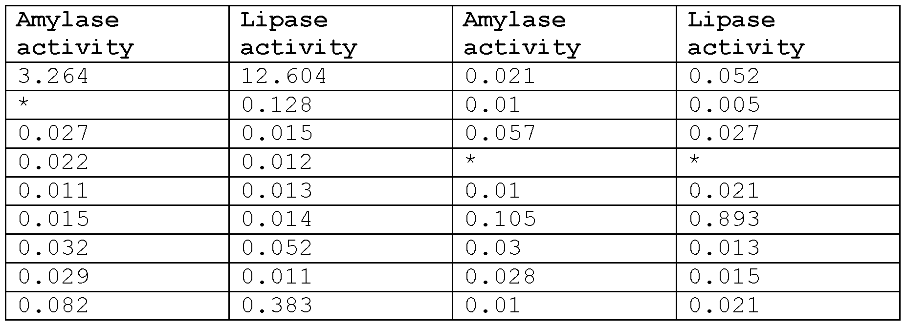
- the two graphs show the amylase and lipase activity values of 32 human samples collected from University Hospital Zurich and analysed by the Institute of Clinical Chemistry (IKC) of the University Hospital of Zurich. Samples were furthermore sorted into samples that contained a leak and samples that did not (after performing the measurements and unblinding) .
- Figure 13B shows ROC (receiver operating curve) of trypsin responsive elements (Data from experiments where Gel-MA patches with 1% PEG-DA were subjected to the human samples for 24h at 37°C.
- the patches were digested while in the case of no leak the patches remained intact) as well as of amylase and lipase responsive elements.
- the ROC was generated with the above shown data, as determined by IKC.
- Figure 14 (1) shows the native and partially digested Gel-MA patch
- Figure 14 (11) the native and partially digested starch- MA patch
- Figure 14 (111) the native and partially digested oleogel patch.
- partially digested means that parts of the patch are still visible, whereas when fully digested, the patch has completely disappeared.
- Figure 15 illustrates the manufacturing of biosensor active moieties in the form of black and white Gel-MA and oleogel patches according to the present invention.
- Figure 15A shows the manufacturing of thin film Starch-MA patches.
- Figure 15B shows the manufacturing of thin film oleogel patches.
- Figures 15C and D show the manufacturing of thin film Gel-MA patches.
- Figure 15E shows the manufacturing of thin film Gel-MA patches colored with active charcoal (left) and TiCt (right) to give a black and white colored patch respectively to make it better visible in red or brown liquid in case of blood in the drain .
- Figure 15F depicts thin film oleogel patches colored with active charcoal (left) and TiCt (right) to give a black and white colored patch respectively to make it better visible in red or brown liquid in case of blood in drain.
- Figure 16 shows an IR-spectra of gelatin, Gel-MA and its corresponding hydrogel.
- Figure 17 shows an IR-spectra of acrylamide, starch and its corresponding starch-MA hydrogel.
- Figure 18 shows an IR-spectra of coconut oil , ethylcellulose and its corresponding oleogel .
- Figure 19 shows the same experiment as illustrated in Figure 12 ( same sequence and concentration) but with closeup of biosensor outside drain .
- Figure 20 shows the top view of one embodiment of the present invention where the sensing elements are encased in a 3D printed circular element printed on an also 3D printed support mesh .
- the white color corresponds to an amylase responsive hydrogel while the black color corresponds to hydrogels responsive to trypsin .
- Each circular sensing hydrogel has a di f ferent chemical modi fication making each circular element have a di f ferent enzymatic degradation time and resistance .
- the number of individually responsive elements is interchangeable and can be any number including one or higher .
- Figure 20B shows the angular view of the sensor according to present invention .
- Figure 20C shows the s ide view of the sensor according to present invention .
- the circular elements protrude on both sides allowing more hydrogel attachment and safeguards the hydrogels from detaching from the backing due to movement of the sensor .
- Figure 21 shows the placement of a sensor according to the present invention into a commercially available drain bag .
- the sensor is not attached to the bag but free floating .
- Figure 22 shows the placement of a sensor according to the present invention by means of heat fusing the sensor to the bag .
- Image shows a sensor with three trypsin responsive elements, each having different sensitivity towards trypsin through chemical modification.
- Figures 23A-D shows additional temperature and pH dependent kinetic data of the gels responsive to trypsin and amylase according to the present invention.
- Figure 24 shows the kinetic data of 7% wt . starch functionalized with glycidyl methacrylate (50% degree of functionalization) at 37°C and room temperature.
- Figure 25 shows the kinetic data of 7% wt . and 10% wt . starch functionalized with glycidyl methacrylate (25% degree of functionalization) at 37°C and room temperature.
- a DMAP (4- (Dimethylamino) pyridine) catalyzed synthesis was performed, similar to the method used by Kessler et. al. to functionalize alcohols.
- 4g of starch from potatoes, SIGMA
- 150 ml of Milli-Q was added.
- 27 mg of 4- (Dimethylamino) pyridine (DMAP) was added and the mixture was heated to 50°C (inside the flask) for 15 minutes to achieve partial swelling of the starch granules.
- the mixture was cooled to 40°C and 1.25 ml Methacrylic Anhydride was added dropwise. During the whole reaction, the pH of the solution was carefully monitored. After two hours, the pH of the reaction has dropped to 4-5. By adding 5M NaOH, the pH was adjusted to 6. One hour later the pH was readjusted by addition of more 5M NaOH upon which the reaction pH was around 9. The reaction mixture was then allowed to stir overnight in the dark. The next day, the solution was pipetted into dialysis tubing (3.5kDa cutoff) and dialyzed against 10L of deionized water. The water was changed once after 6h and then left for 72h. After freezing the solution, it was freeze dried until a white powder was obtained.
- dialysis tubing 3.5kDa cutoff
- coconut oil was placed in a 25ml round bottom flask in an oilbath set to 175°C together with different weight percentages ethylcellulose (300cp) and active charcoal. The mixture was heated and stirred for 2h. Next, the oleogel mix was taken up by a glass pipette and spread thinly on a glass surface (a blade knife was used to create a thin uniform layer) . The oleogel was then cooled in the fridge for 2h before further use .
- ethylcellulose 300cp
- active charcoal active charcoal
- Gelatin-MA Gels were mixed as mentioned above (without PEG- DA) using gold nanoparticles (2x concentrated from synthesis) . The mix was poured onto a glass plate and a thin film applicator was used to create a thin uniform layer. After cooling in the fridge, the sheets were irradiated by UV light (see above) and a 9mm biopsy punch was used to cut out thin round patches. For the digestion experiments, only gold nanoparticle suspension was used to make the gels. Patches were then put into 12 well cell culture plates (TPP) filled with PBS (Dulbecco modified with CaC12 and MgC12) .
- TPP cell culture plates
- Trypsin from porcine pancreas was then added in the right concentration by adding the corresponding volume of stock (stock Img/ml) .
- the overall volume in the wells at the start of the experiment was 3ml for all samples. Tubes were then put either into an incubator set at 37 °C, where they were gently mixed with an orbital shaker or shaken at RT with the same speed.
- the digestion process was recorded automatically using a tablet and a timelapse app . Pictures were taken every 10 min and for each concentration triplicates were examined (see Figure 5) .
- a patch was counted as digested when it either fully disappeared or showed significant alterations (loss of color and or structure) compared to the control patches (PBS only) .
- the control patches did not significantly change over the observation period, while the digested patches gradually lost structure. First, they became softer and afterwards they lost the round shape, until they finally disappeared.
- Starch-MA For this Gel gold nanoparticles (2x concentrated from synthesis) were mixed with 5%wt. Starch-MA and 15%wt. Aam (Acrylamide) in a glas vial and put into an oven heated to 80°C. The vial was regularly vortex and heated for 30 min. Next 101vol. LAP (6.33mg/ml) is added and heating is continued for another 5 min. Then the mix was poured onto a preheated glass plate and a thin film applicator was used to create a thin uniform layer and the sheets were irradiated by UV light (see above) and a 9mm biopsy punch was used to cut out thin round patches.
- Aam Acrylamide
- Digestion experiments were conducted similar to Gelatin-MA with the difference being that the amylase (porcine pancreas llU/mg) was added as a powder directly to the PBS (Dulbecco modified with CaC12 and MgC12) for higher concentration (>10U/ml) while for the lower concentration a Img/ml stock was diluted accordingly.
- the overall volume in the wells at the start of the experiment was 3ml for all samples. Plates were then put either into an incubator set at 37°C, where they were mixed with an orbital shaker or shaken at RT with the same speed.
- the digestion process was recorded automatically using a tablet and a timelapse app . Pictures were taken every 10 min and for each concentration triplicates were examined .
- a patch was counted as digested when it either fully disappeared or showed signi ficant alterations ( loss of color and or structure ) compared to the control patches ( PBS only) .
- the control patches did not signi ficantly change over the observation period, while the digested patches gradually lost structure . First , they became softer and afterwards they lost the round shape , until they finally disappeared .
- Plant oleogels Oleogels were prepared as mentioned above . Patches were placed in 6 well plates filled with 6 ml PBS ( Dulbecco modi fied with CaC12 and MgC12 ) , Lipase ( from porcine pancreas ) in the corresponding concentration ( from Img/ml stock) and 32mg bile ( from porcine sources ) . Plates were then put either into an incubator set at 37 ° C, where they were mixed with an orbital shaker or shaken at RT with the same speed . The digestion process was recorded automatically using a tablet and a timelapse app . Pictures were taken every 10 min and for each concentration triplicates were examined .
- a patch was counted as digested when it either fully disappeared or showed signi ficant alterations ( loss of color and or structure ) compared to the control patches ( PBS only) .
- the control patches did not signi ficantly change over the observation period, while the digested patches gradually lost structure . First , they became softer and afterwards they lost the round shape , until they finally disappeared .
- Table 2 shows the amylase and lipase activity levels of samples where the patch did digest and disappear . All values are given as units per milliliter (U/ml ) .
- bilirubin content was measured .
- the rest of the sample (general 15-20ml ) was then put into a falcon tube into which a 500pl Gel-MA patch ( 1 % PEG-DA, only gold nanoparticle suspension) was thrown .
- the samples were then incubated overnight at 37 ° C while shaking gently on an orbital shaker . Digestion was determined the next day and again after 24h in the case of undigested patches .
- Prototype gel holders were first 3D printed and then used as negative molds for hot pressing plastic sheets over them .
- the molds were open on one side and had a hole over each of the patches on the other side .
- FIGs 2A and 2B 1 stands for a trypsin sensitive gel , 2 for an amylase sensitive gel and 3 for a lipase sensitive gel ) . Patches were then put into the plastics and a net was attached to the backside in order to avoid the patches to slip out of the mold .
- FIG. 2A shows a biosensor according to the present invention before contact with a drain fluid .
- Figure 2B shows the biosensor after contact with a trypsin containing drain fluid . In figure 2B the red color of the trypsin sensitive gel is no longer visible , and the gel almost disappeared . As neither amylase nor lipase were present in the drain fluid, said gels remain unchanged .
- a biosensor to detect postoperative leak with a patient ' s drain fluid, relating to an operation selected from the group consisting of pancreatic anastomosis , pancreatic resection, small colon anastomosis (also called small intestine anastomosis ) , large intestine anastomosis , colorectal surgery , colon bypass surgery, gastric bypass surgery, gastric resection surgery, cholecystectomy, bileduct surgery and oesophageal surgery, wherein the biosensor comprises at least one gel which is placed on a support layer, and wherein the at least one gel is sensitive to a human gut bacteria and comprises a colorant selected from the group consisting of an organic dye , an inorganic dye , an organic pigment and an inorganic pigment , and wherein said gel degrades upon contact with a drain fluid comprising one or more human bacteria allowing a visual detection of discoloration at the place where the gel was before and/or disappearance
Landscapes
- Health & Medical Sciences (AREA)
- Life Sciences & Earth Sciences (AREA)
- Engineering & Computer Science (AREA)
- Molecular Biology (AREA)
- Chemical & Material Sciences (AREA)
- Physics & Mathematics (AREA)
- Immunology (AREA)
- Biomedical Technology (AREA)
- General Health & Medical Sciences (AREA)
- Biophysics (AREA)
- Pathology (AREA)
- Surgery (AREA)
- Heart & Thoracic Surgery (AREA)
- Animal Behavior & Ethology (AREA)
- Medical Informatics (AREA)
- Public Health (AREA)
- Veterinary Medicine (AREA)
- Hematology (AREA)
- Organic Chemistry (AREA)
- Biochemistry (AREA)
- Zoology (AREA)
- Urology & Nephrology (AREA)
- Analytical Chemistry (AREA)
- Proteomics, Peptides & Aminoacids (AREA)
- Biotechnology (AREA)
- Microbiology (AREA)
- Wood Science & Technology (AREA)
- Optics & Photonics (AREA)
- Physiology (AREA)
- Gastroenterology & Hepatology (AREA)
- Bioinformatics & Cheminformatics (AREA)
- General Engineering & Computer Science (AREA)
- Genetics & Genomics (AREA)
- Endocrinology (AREA)
- Cell Biology (AREA)
- Vascular Medicine (AREA)
- Food Science & Technology (AREA)
- Medicinal Chemistry (AREA)
- General Physics & Mathematics (AREA)
- Measuring Or Testing Involving Enzymes Or Micro-Organisms (AREA)
Abstract
Description




Claims
Priority Applications (1)
| Application Number | Priority Date | Filing Date | Title |
|---|---|---|---|
| EP24730383.7A EP4724594A1 (en) | 2023-06-09 | 2024-06-07 | Biosensor |
Applications Claiming Priority (4)
| Application Number | Priority Date | Filing Date | Title |
|---|---|---|---|
| EP23178489.3 | 2023-06-09 | ||
| EP23178489 | 2023-06-09 | ||
| EP23203384.5 | 2023-10-13 | ||
| EP23203384 | 2023-10-13 |
Publications (1)
| Publication Number | Publication Date |
|---|---|
| WO2024251952A1 true WO2024251952A1 (en) | 2024-12-12 |
Family
ID=91375275
Family Applications (1)
| Application Number | Title | Priority Date | Filing Date |
|---|---|---|---|
| PCT/EP2024/065742 Ceased WO2024251952A1 (en) | 2023-06-09 | 2024-06-07 | Biosensor |
Country Status (2)
| Country | Link |
|---|---|
| EP (1) | EP4724594A1 (en) |
| WO (1) | WO2024251952A1 (en) |
Citations (7)
| Publication number | Priority date | Publication date | Assignee | Title |
|---|---|---|---|---|
| GB2350677A (en) | 1999-06-04 | 2000-12-06 | Cambridge Life Sciences | Enzyme detection |
| CN101473217A (en) | 2006-06-16 | 2009-07-01 | 维也纳大学 | Optical sensor and method for indicating the age or quality of a natural product |
| US8278064B2 (en) | 2006-10-16 | 2012-10-02 | Queen Mary & Westfield College | Method for detecting a protease |
| US20160061830A1 (en) | 2014-08-27 | 2016-03-03 | The Governors Of The University Of Alberta | Bacteriophage-based biosensor for microbial detection |
| CN111228627A (en) * | 2020-02-11 | 2020-06-05 | 中山大学附属第三医院 | Device and system for detecting intestinal fistula, bile fistula and pancreatic juice fistula |
| WO2022031558A1 (en) | 2020-08-03 | 2022-02-10 | The Regents Of The University Of Colorado, A Body Corporate | Method to sense the presence and quantities of microbes through the use of transient sensing materials |
| US20220265175A1 (en) * | 2019-03-26 | 2022-08-25 | Nerv Technology Inc. | Systems, methods and devices for predicting and detecting postoperative complications |
-
2024
- 2024-06-07 EP EP24730383.7A patent/EP4724594A1/en active Pending
- 2024-06-07 WO PCT/EP2024/065742 patent/WO2024251952A1/en not_active Ceased
Patent Citations (7)
| Publication number | Priority date | Publication date | Assignee | Title |
|---|---|---|---|---|
| GB2350677A (en) | 1999-06-04 | 2000-12-06 | Cambridge Life Sciences | Enzyme detection |
| CN101473217A (en) | 2006-06-16 | 2009-07-01 | 维也纳大学 | Optical sensor and method for indicating the age or quality of a natural product |
| US8278064B2 (en) | 2006-10-16 | 2012-10-02 | Queen Mary & Westfield College | Method for detecting a protease |
| US20160061830A1 (en) | 2014-08-27 | 2016-03-03 | The Governors Of The University Of Alberta | Bacteriophage-based biosensor for microbial detection |
| US20220265175A1 (en) * | 2019-03-26 | 2022-08-25 | Nerv Technology Inc. | Systems, methods and devices for predicting and detecting postoperative complications |
| CN111228627A (en) * | 2020-02-11 | 2020-06-05 | 中山大学附属第三医院 | Device and system for detecting intestinal fistula, bile fistula and pancreatic juice fistula |
| WO2022031558A1 (en) | 2020-08-03 | 2022-02-10 | The Regents Of The University Of Colorado, A Body Corporate | Method to sense the presence and quantities of microbes through the use of transient sensing materials |
Non-Patent Citations (6)
Also Published As
| Publication number | Publication date |
|---|---|
| EP4724594A1 (en) | 2026-04-15 |
Similar Documents
| Publication | Publication Date | Title |
|---|---|---|
| Li et al. | Wall porosity in isolated cells from food plants: Implications for nutritional functionality | |
| Wang et al. | Silk fibroin-based colorimetric microneedle patch for rapid detection of spoilage in packaged salmon samples | |
| US8518374B2 (en) | Personal care products with visual indicator of vaginitis | |
| Gatellier et al. | Digestion study of proteins from cooked meat using an enzymatic microreactor | |
| Li et al. | Hydrosoluble, UV-crosslinkable and injectable chitosan for patterned cell-laden microgel and rapid transdermal curing hydrogel in vivo | |
| CN103013822B (en) | Fast drug sensitivity detection kit for aquatic product vibrio | |
| Molnar et al. | Poly (amino acid) based fibrous membranes with tuneable in vivo biodegradation | |
| Zafaripour et al. | Fabrication and evaluation of 3D bio-scaffold wound dressings for monitoring of chronic pH wounds using fuzzy logic analysis | |
| PT2433138T (en) | Method for detecting a wound infection | |
| Fan et al. | A multi-functional 3D-printable gel-in-gel system for the delivery of probiotics to the intestine | |
| Zhu et al. | based chip integrated with vitrified silk fibroin microneedle array for colorimetric detection of histamine in food | |
| Xing et al. | Deep-learning-assisted chemo-responsive alizarin red S-based hydrogel sensor for the rapid freshness sensing of aquatic product | |
| EP4724594A1 (en) | Biosensor | |
| CN103893821B (en) | A kind of bionical composite patch and its production and use | |
| US20250208117A1 (en) | Superabsorbent Polymer based Biosensor Apparatus and Methods | |
| US11730405B2 (en) | Body fluid leakage detection aqueous composition | |
| WO2023180691A1 (en) | Superabsorbent Polymer based Biosensor Apparatus and Methods | |
| US9279140B2 (en) | Personal care products with visual indicator of vaginitis | |
| RU2788001C2 (en) | Aqueous composition for detection of seepage of physiological fluids | |
| Zhuang et al. | A radically simple, ingestible colorimetric biosensor pill for cost-effective, non-invasive monitoring of intestinal inflammation | |
| RU2429476C1 (en) | Diagnostic technique for arthrosis | |
| Christodoulides et al. | Tracking Fibrinolysis of Chandler Loop-Formed Whole Blood Clots Under Shear Flow in An In-Vitro Thrombolysis Model | |
| Ibrišimović et al. | Biomimetic PLGA sensor: proof of principle and application | |
| JPWO2000071167A1 (en) | Method for evaluating matrix metalloproteinase inhibitory activity | |
| HK40035614A (en) | A body fluid leakage detection aqueous composition |
Legal Events
| Date | Code | Title | Description |
|---|---|---|---|
| 121 | Ep: the epo has been informed by wipo that ep was designated in this application |
Ref document number: 24730383 Country of ref document: EP Kind code of ref document: A1 |
|
| WWE | Wipo information: entry into national phase |
Ref document number: 2024730383 Country of ref document: EP |
|
| NENP | Non-entry into the national phase |
Ref country code: DE |
|
| ENP | Entry into the national phase |
Ref document number: 2024730383 Country of ref document: EP Effective date: 20260109 |
|
| ENP | Entry into the national phase |
Ref document number: 2024730383 Country of ref document: EP Effective date: 20260109 |
|
| ENP | Entry into the national phase |
Ref document number: 2024730383 Country of ref document: EP Effective date: 20260109 |
|
| WWP | Wipo information: published in national office |
Ref document number: 2024730383 Country of ref document: EP |