WO2024206707A1 - Azyariv and azyariv conjugates - new tools for visualizing and characterizing arabinogalactan proteins - Google Patents
Azyariv and azyariv conjugates - new tools for visualizing and characterizing arabinogalactan proteins Download PDFInfo
- Publication number
- WO2024206707A1 WO2024206707A1 PCT/US2024/022083 US2024022083W WO2024206707A1 WO 2024206707 A1 WO2024206707 A1 WO 2024206707A1 US 2024022083 W US2024022083 W US 2024022083W WO 2024206707 A1 WO2024206707 A1 WO 2024206707A1
- Authority
- WO
- WIPO (PCT)
- Prior art keywords
- phenyl
- azyariv
- glucosyl
- small molecule
- chemical composition
- Prior art date
- Legal status (The legal status is an assumption and is not a legal conclusion. Google has not performed a legal analysis and makes no representation as to the accuracy of the status listed.)
- Ceased
Links
Classifications
-
- C—CHEMISTRY; METALLURGY
- C07—ORGANIC CHEMISTRY
- C07H—SUGARS; DERIVATIVES THEREOF; NUCLEOSIDES; NUCLEOTIDES; NUCLEIC ACIDS
- C07H15/00—Compounds containing hydrocarbon or substituted hydrocarbon radicals directly attached to hetero atoms of saccharide radicals
- C07H15/20—Carbocyclic rings
- C07H15/203—Monocyclic carbocyclic rings other than cyclohexane rings; Bicyclic carbocyclic ring systems
-
- C—CHEMISTRY; METALLURGY
- C07—ORGANIC CHEMISTRY
- C07H—SUGARS; DERIVATIVES THEREOF; NUCLEOSIDES; NUCLEOTIDES; NUCLEIC ACIDS
- C07H1/00—Processes for the preparation of sugar derivatives
-
- C—CHEMISTRY; METALLURGY
- C07—ORGANIC CHEMISTRY
- C07H—SUGARS; DERIVATIVES THEREOF; NUCLEOSIDES; NUCLEOTIDES; NUCLEIC ACIDS
- C07H19/00—Compounds containing a hetero ring sharing one ring hetero atom with a saccharide radical; Nucleosides; Mononucleotides; Anhydro-derivatives thereof
- C07H19/02—Compounds containing a hetero ring sharing one ring hetero atom with a saccharide radical; Nucleosides; Mononucleotides; Anhydro-derivatives thereof sharing nitrogen
- C07H19/04—Heterocyclic radicals containing only nitrogen atoms as ring hetero atom
- C07H19/23—Heterocyclic radicals containing two or more heterocyclic rings condensed among themselves or condensed with a common carbocyclic ring system, not provided for in groups C07H19/14 - C07H19/22
-
- G—PHYSICS
- G01—MEASURING; TESTING
- G01N—INVESTIGATING OR ANALYSING MATERIALS BY DETERMINING THEIR CHEMICAL OR PHYSICAL PROPERTIES
- G01N33/00—Investigating or analysing materials by specific methods not covered by groups G01N1/00 - G01N31/00
- G01N33/48—Biological material, e.g. blood, urine; Haemocytometers
- G01N33/50—Chemical analysis of biological material, e.g. blood, urine; Testing involving biospecific ligand binding methods; Immunological testing
- G01N33/53—Immunoassay; Biospecific binding assay; Materials therefor
- G01N33/569—Immunoassay; Biospecific binding assay; Materials therefor for microorganisms, e.g. protozoa, bacteria, viruses
- G01N33/56961—Plant cells or fungi
-
- G—PHYSICS
- G01—MEASURING; TESTING
- G01N—INVESTIGATING OR ANALYSING MATERIALS BY DETERMINING THEIR CHEMICAL OR PHYSICAL PROPERTIES
- G01N33/00—Investigating or analysing materials by specific methods not covered by groups G01N1/00 - G01N31/00
- G01N33/48—Biological material, e.g. blood, urine; Haemocytometers
- G01N33/50—Chemical analysis of biological material, e.g. blood, urine; Testing involving biospecific ligand binding methods; Immunological testing
- G01N33/58—Chemical analysis of biological material, e.g. blood, urine; Testing involving biospecific ligand binding methods; Immunological testing involving labelled substances
- G01N33/582—Chemical analysis of biological material, e.g. blood, urine; Testing involving biospecific ligand binding methods; Immunological testing involving labelled substances with fluorescent label
Definitions
- the embodiments of the present invention relate to Yariv reagents with improved functions, fluorescence, and chromophores to expand the study of arabinogalactan proteins, galactans, and glycoproteins in general.
- Yariv reagents with improved functions, fluorescence, and chromophores to expand the study of arabinogalactan proteins, galactans, and glycoproteins in general.
- an intelligent synthesis and design of Yariv reagents is presented and expanded methods are developed and tested.
- Arabinogalactan proteins are cell wall proteoglycans implicated in essential functions such as cell signaling, plant growth, and programmed cell death.
- the P-glucosyl Yariv reagents e.g., 1 ,3,5-tris (4-p-D-glucopyranosyloxyphenylazo)-2,4,6- trihydroxy-benzene] are red dyes which can specifically bind to and precipitate this class of plant AGPs.
- a p-glucosyl Yariv reagent is dissolved in ⁇ 1 mL of 0.15M NaCI. This solution is applied to a tissue section of the plant tissue for about one hour at room temperature (about 25°C).
- the tissue section, with the solution, is then examined by bright field microscopy.
- the AGPs will precipitate in the Yariv reagent to give a red stain, but the exact color can vary from brown-red to bright red depending on the plant tissue.
- the AGP's are known to be water-soluble, they may be lost during these procedures for tissue embedding.
- Several other problems occur with these methods including general sensitivity and signal to noise issues under the microscope.
- Another major issue is the method described above can be impossible to practice on live plants. What is urgently needed are new compounds and methods that enable sensitive studies of AGPs and glycoproteins in general for a variety of biomass, including living plants.
- the present invention solves many problems inherent in the prior art Yariv reagents that are currently in use.
- AGPs are known to be water-soluble, and they may be lost during the procedures for tissue embedding. This loss is detrimental to limit of detection (LOD) Sensitivity, variance, and signal to noise issues are found in the prior art (i.e., in view of this disclosure) Yariv reagents.
- LOD limit of detection
- New reagents are urgently needed with new compounds and methods that enable sensitive studies of AGPs and glycoproteins in general for, example, using highly sensitive fluorescent and absorbance techniques.
- the technology disclosed herein enables implementation with modern analytical instrumentation and computers, along with intricate visualization of the data.
- new synthetic methods applied to produce functionalizable analogs of various Yariv Reagents can be used for AGP Imaging using fluorescence microscopy.
- the present invention discloses a chemical composition
- a chemical composition comprising a molecular structure of formula 1 below: or a tautomer and/or an E/Z isomer thereof of formula 1 ; wherein each instance of R in formula 1 above independently comprises: wh (-0-) in formula 1 ; wherein Y comprises: -N3; ; wherein X' is an anion; wherein • above represents a single bond attached to any -OH or -0- of R; or wherein Y comprises a click chemistry functional group, near-IR dye, an IR dye, a fluorescent small molecule, a small molecule with a chromophore, a Raman active small molecule, or a small molecule with a maximum absorbance band in a range from about 200 nm to about 100 pm; and wherein a small molecule has a molecular weight in a non-salt form of less than about 1000 daltons (Da).
- a method for performing a fluorescence imaging of an arabinogalactan protein (AGP) or a glycosylated protein (glycoprotein) comprising the steps of: (1 ) contacting a material known to contain an AGP or a glycoprotein or suspected to contain an AGP or a glycoprotein with a reagent including AzYariv-Cy5, AzYariv-Cy5-X [3GlcYariv, or a combination thereof, whereby a binding between the AGP or glycoprotein and the reagent occurs; (2) directing an incident light or an incident electromagnetic radiation towards the binding, whereby a fluorescent emission occurs; and (3) gathering, observing, measuring, or acquiring the emission.
- AGP arabinogalactan protein
- glycosylated protein glycosylated protein
- a method for performing an absorbance imaging of an arabinogalactan protein (AGP) or a glycosylated protein (glycoprotein) comprising the steps of: (1 ) contacting a material known to contain an AGP or a glycoprotein or suspected to contain an AGP or a glycoprotein with a reagent including AzYariv-Cy5, AzYariv-Cy5-X‘, [3GlcYariv, or a combination thereof, whereby a binding between the AGP or glycoprotein and the reagent occurs; (2a) directing an incident light or an incident electromagnetic radiation towards the binding, whereby an electromagnetic absorbance occurs; and (3a) gathering, observing, measuring, or acquiring the absorbance.
- AGP arabinogalactan protein
- Glycoprotein glycosylated protein
- a method for performing a fluorescence imaging of an arabinogalactan protein (AGP) or a glycosylated protein (glycoprotein) can include any of the steps of: obtaining a material known to contain an AGP or a glycoprotein or suspected to contain an AGP or a glycoprotein; contacting the material with a reagent disclosed herein or including AzYariv-Cy5, AzYariv-Cy5-X [BGIcYariv, or a combination thereof, whereby a binding between the AGP or glycoprotein and the reagent occurs. Next is directing an incident light or an incident electromagnetic radiation towards the binding, whereby a fluorescent emission occurs.
- a gathering, observing, measuring, or acquiring the emission is executed, typically including a digitization using a computer and software.
- Computer graphics or other visualization can provide a decision whether or not to proceed with the data or to repeat the experiments using a different area of plant tissue.
- a method for performing an absorbance measurement on an arabinogalactan protein (AGP) or a glycosylated protein (glycoprotein) including the steps of: obtaining a material known to contain an AGP or a glycoprotein or suspected to contain an AGP or a glycoprotein; optionally placing the material on a suitable mount for absorbance wavelengths to pass through the sample; contacting the material with a binding reagent disclosed herein including a chromophore; and directing an incident light or an incident electromagnetic radiation towards the binding area, whereby an electromagnetic absorbance occurs.
- AGP arabinogalactan protein
- glycoprotein glycosylated protein
- FIG. 1A shows examples of small molecule plant cell wall probes Sirofluor, Calcofluor White, and [BGIcYariv.
- FIG. 1 B shows synthesis of compound 3 (shown at right).
- FIG. 10 shows synthesis of compound 7 (middle).
- FIG. 1D shows synthesis of AzYariv Reagent (shown at right).
- FIG. 1E shows example ATR IR spectra of AzYariv reagent and PGIcYariv reagent.
- FIG. 2A shows CD spectra of AzYariv and PGIcYariv.
- FIG. 2B shows an absorbance spectrum of AzYariv. All spectra obtained in water at [300 pM],
- FIG. 3B shows Table 1 ; Optimization of Click Reaction Conditions Between AzYariv and Cy5-DBCO.
- FIG. 3C shows a high- resolution mass spectrum for AzYariv-Cy5 acquired using +ESI (positive electrospray ionization).
- FIG. 3D shows a high-resolution mass spectrum for AzYariv (top) acquired using +ESI and predicted isotopic distribution for [M]+ (bottom).
- FIGs. 4A-4H show representative fluorescence images of AzYariv-Cy5 binding in agarose reverse gels with gum arabicAGP. Concentrations of GIcYariv and aGalYariv are 1.03 mM.
- FIG. 4A AzYariv-Cy5 (5 pM)/pGlcYariv
- FIG. 4B AzYariv-Cy5 (100 nM)/pGlcYariv
- FIG. 4C AzYariv-Cy5 (5 nM)/pGlcYariv
- FIG. 4D AzYariv-Cy5 (5 pM)/pGlcYariv Lyophilized
- FIG. 4A AzYariv-Cy5
- FIG. 4B AzYariv-Cy5 (100 nM)/pGlcYariv
- FIG. 4C AzYariv-Cy5 (5 nM)/pGlcYariv
- FIG. 4D AzYariv-C
- FIGs. 5A-5F shows representative confocal images of fixed maize leaves treated with Yariv reagents (FIGs. 5A-5D, imaged with 651 nm laser) and antibodies (FIG. 5E, FIG. 5F, imaged with 495 nm laser).
- FIG. 5A AzYariv-Cy5/pGlcYariv
- FIG. 5B zoom of AzYariv-Cy5/pGlcYariv.
- UE Upper epidermal cells
- P phloem
- X xylem
- BS bundle sheath
- LE lower epidermal cells
- FIG. 5A-5F shows representative confocal images of fixed maize leaves treated with Yariv reagents
- FIG. 5E imaged with 495 nm laser.
- FIG. 5A AzYariv-Cy5/pGlcYariv
- FIG. 5B zoom of AzYariv-Cy5/pGlcYariv.
- FIG. 5C shows a representative confocal image of fixed maize leaves imaged for fluorescein (conjugated secondary antibody used in immunofluorescence) A) autofluorescence of tissue and non-specific secondary antibody staining (no primary antibody control);
- FIG. 5H shows a representative confocal image of fixed maize leaves imaged for fluorescein (conjugated secondary antibody used in immunofluorescence) B) MAC207;
- FIG. 51 shows a representative confocal image of fixed maize leaves imaged for fluorescein (conjugated secondary antibody used in immunofluorescence)
- C) JIM13 C
- Scale bar 50 pm.
- Merged brightfield and confocal images are shown of (FIG. 5J) fixed maize leaves treated with pGIcYariv and (FIG. 5K) Fresh maize leaves treated with [3GlcYariv. Green signal (or lighter greyscale) is chlorophyll autofluorescence.
- Scale bar 50 pM.
- FIG. 6A, FIG. 6B, FIG. 60, and FIG. 6D show representative confocal images of fresh maize leaves treated with AzYariv-Cy5/[3GlcYariv (top, FIG. 6A, FIG. 6B) and AzYariv-Cy5/aGalYariv (bottom, FIG. 6C, FIG. 6D) imaged with 651 nm laser.
- UE Upper epidermal cells
- P phloem
- X xylem
- BS bundle sheath
- LE lower epidermal cells.
- Scale bar 50 pm.
- FIG. 7A shows an example synthesis of compound 4 (shown at right).
- FIG. 7B shows an example synthesis of compound 5 (shown at right).
- FIG. 7C shows an example synthesis of compound 6 (shown at right).
- FIG. 7D shows an example synthesis of compound 7 (shown at right).
- FIG. 8A shows example (prior art) 2 tautomer interconversions of formula 1
- FIG. 8B shows a continuation of the tautomer interconversions.
- FIG. 9 shows a major tautomer found and established by NMR data in DMSO.
- FIG. 10 shows an example of a novel formula 1 (left) and some examples of ‘R’ that were attached at right (prior art) from previous work 3 .
- FIG. 11 shows additional examples of ‘R’.
- FIG. 12 shows an example method 500 for performing a fluorescence imaging of an arabinogalactan protein (AGP) or a glycosylated protein (glycoprotein).
- AGP arabinogalactan protein
- glycoprotein glycosylated protein
- FIG. 13 shows an example method 600 for performing an absorbance measurement on an arabinogalactan protein (AGP) or a glycosylated protein (glycoprotein).
- AGP arabinogalactan protein
- glycoprotein glycosylated protein
- the term “approximately” or “about” in reference to a value or parameter are generally taken to include numbers that fall within a range of 5%, 10%, 15%, or 20% in either direction (greater than or less than) of the number unless otherwise stated or otherwise evident from the context (except where such number would be less than 0% or exceed 100% of a possible value).
- reference to “approximately” or “about” a value or parameter includes (and describes) embodiments that are directed to that value or parameter. For example, description referring to "about X” includes description of "X”.
- the term “or” means “and/or.”
- the term “and/or” as used in a phrase such as "A and/or B” herein is intended to include both A and B; A or B; A (alone); and B (alone).
- the term “and/or” as used in a phrase such as "A, B, and/or C” is intended to encompass each of the following embodiments: A, B, and C; A, B, or C; A or C; A or B; B or C; A and C; A and B; B and C; A (alone); B (alone); and C (alone).
- the term “consisting essentially of'” refers to those elements required for a given embodiment. The term permits the presence of additional elements that do not materially affect the basic and novel or functional characteristic(s) of that embodiment of the invention.
- the term “consisting essentially of” can also be exemplified by plain language provided in the claims.
- the technology can be described in reference to plants or living plants; however, the technology disclosed herein can be applied to any living thing, any material, or any substance in any physical form.
- the term “statistically significant” or “significantly” refers to statistical significance and generally means a two-standard deviation (2SD) or greater difference.
- the terms: “decrease”, “reduced”, “reduction”, or “inhibit” are all used herein to mean a decrease by a statistically significant amount.
- “reduce,” “reduction” or “decrease” or “inhibit” typically means a decrease by at least 10% as compared to a reference level (e.g., the absence of a given treatment or agent) and can include, for example, a decrease by at least about 10%, at least about 20%, at least about 25%, at least about 30%, at least about 35%, at least about 40%, at least about 45%, at least about 50%, at least about 55%, at least about 60%, at least about 65%, at least about 70%, at least about 75%, at least about 80%, at least about 85%, at least about 90%, at least about 95%, at least about 98%, at least about 99% , or more.
- “reduction” or “inhibition” does not encompass a complete inhibition or reduction as compared to
- the terms “increased”, “increase”, “enhance”, or “activate” are all used herein to mean an increase by a statically significant amount.
- the terms “increased”, “increase”, “enhance”, or “activate” can mean an increase of at least 10% as compared to a reference level, for example an increase of at least about 20%, or at least about 30%, or at least about 40%, or at least about 50%, or at least about 60%, or at least about 70%, or at least about 80%, or at least about 90% or up to and including a 100% increase or any increase between 10-100% as compared to a reference level, or at least about a 2-fold, or at least about a 3-fold, or at least about a 4-fold, or at least about a 5- fold or at least about a 10-fold increase, or any increase between 2-fold and 10-fold or greater as compared to a reference level.
- a “increase” is a statistically significant increase in such level.
- a small molecule is less than 1000 MW and that is not including a cation or anion for a salt form.
- a large molecule is not less than 1000 MW including biologies, oligonucleotides, peptides, oligosaccharides, and larger molecules. Any of the methods or compositions disclosed herein can be used as or in combination with small molecules and/or large molecules as discussed herein. [0045] As discussed above, unless otherwise defined herein, scientific and technical terms used in connection with the present application shall have the meanings that are commonly understood by those of ordinary skill in the art to which this disclosure belongs.
- the p-glucosyl Yariv reagents of the past art are red to brown-red dyes which can specifically bind to and precipitate plant AGPs.
- a solution of the Yariv reagent is typically mixed with sodium chloride and the solution is applied to a tissue section of the plant tissue at room temperature (about 25°C). The tissue section, with the solution, is then examined under a microscope to observe brown-red or red staining. Under the microscope, the AGPs will precipitate in the Yariv reagent to give a red stain, but the exact color can vary from brown-red to bright red depending on the plant tissue.
- the AGPs are known to be water-soluble, they may be lost during these procedures for tissue embedding.
- the technology herein provides small molecule fluorescent probes that bind selectively to plant cell wall polysaccharides.
- the new small molecule fluorescent probes have been instrumental in elucidating the localization and function of these glycans.
- Arabinogalactan proteins (AGPs) are cell wall proteoglycans implicated in essential functions such as cell signaling, plant growth, and programmed cell death.
- AGPs Arabinogalactan proteins
- Yariv reagents are the only small molecules that bind AGPs and have been used to study AGP function and isolate AGPs via precipitation of an AGP-Yariv complex.
- the Yariv reagents of the past are not fluorescent, rendering them ineffective for localization studies using fluorescence microscopy.
- a fluorescent version of a Yariv reagent that is capable of both binding as well as imaging AGPs would provide a powerful tool for studying AGPs in planta.
- the modified reagent binds gum arabic in in vitro binding assays when used in conjunction with the pGIcYariv reagent.
- Fluorescent imaging of AGPs in fixed maize leaf tissue enables localization of AGPs to cell walls in the leaf. Significantly, imaging can also be carried out using fresh tissue. This represents the first small molecule probe that can be used to visualize AGPs using fluorescence microscopy.
- Plant cell walls are comprised of an intricate assembly of polysaccharides including cellulose, hemicelluloses, and pectins, along with crosslinked lignins and proteoglycans such as arabinogalactan proteins (AGPs). 7 ’ 10 There is a pressing need for the development of tools that can image the complex and dynamic plant cell wall. 11 ’ 14 Fluorescence microscopy is a powerful method for visualizing components of the plant cell wall because of its high sensitivity and spatial resolution. Small molecule fluorescent dyes are vital compounds in elucidating the distribution of these components. For example, Sirofluor and Calcofluor White (FIG. 1A) are commonly used as fluorescent stains for callose and cellulose, respectively. FIG.
- AGPs are proteoglycans found throughout the plant, including in leaves, roots, stems, pollen tubes, and pollen grains. 15 AGPs are implicated in myriad functions ranging from plant growth, cellular signaling, programmed cell death, and many others. 16 ’ 18 AGPs contain protein domains rich in hydroxyproline residues that are glycosylated with long [3(1 - ⁇ 3)-linked galactan chains that make up the bulk of AGP mass. 1920 Shorter branching [3(1 — >6) galactan side chains are linked from the galactan main chains and are commonly decorated with arabinofuranose residues and other monosaccharides, with variations in the specific composition of the branches depending on the species and plant tissue.
- MAbs monoclonal antibodies
- Yariv reagents the set of dyes known as the Yariv reagents (vide infra). 15 While MAbs can provide bright images showing AGP decoration, they are highly epitope specific, and due to their large size, the antibodies may not be able to access all parts of the cell wall. 12 ’ 14 On the other hand, the Yariv reagents, dyes based on a tri-glycosylated phloroglucinol core (FIG. 1 A), bind AGPs but can only be visualized using the less sensitive brightfield microscopy.
- the AGP binding ability of the Yariv reagents depends on the structure of the sugar moieties attached to the aromatic core. 43 Yariv reagents bearing (3-D linked glucose ([3GlcYariv) or galactose bind strongly to AGPs and have been used to isolate AGPs from plants by precipitation of an AGP-Yariv complex. 21-23 Yariv reagents comprised of a-linked sugars or L sugars do not bind to AGPs. The Yariv reagents aggregate in solution and bind to the core [3(1 —> 3)- linked galactan backbone, which is postulated to be helical.
- FIG. 1 D Diazotization of 7, subsequent coupling with 3, followed by isolation and purification using a redissolution and reprecipitation protocol afforded AzYariv in 18% yield.
- the 1 H NMR spectrum of AzYariv indicates the presence of distinct anomeric and OH resonances derived from the glucosyl and 6-azido glucosyl moieties.
- the IR spectrum of AzYariv displays the characteristic azide stretch at 2100 cm -1 (FIG. 1 E). The IR spectrum was acquired using ATR IR, and FIG. 1 E shows example ATR IR spectra of AzYariv reagent and [3GlcYariv reagent.
- the CD spectrum of AzYariv exhibits two positive bisignate Cotton effects similar to other [3- D glycosyl Yariv reagents, with a lower intensity than the parent pGIcYariv (FIG. 2A).
- FIG. 2A To confirm that installation of the azide does not disrupt the AGP binding of AzYariv, we carried out reverse gel assays to determine binding.
- the precipitate halos formed by AzYariv indicate that it still binds gum arabic, although the halos are smaller than those observed for pGIcYariv, indicative of weaker binding (FIG. 3A, black boxes).
- FIG. 3C shows a high-resolution mass spectrum for AzYariv-Cy5 acquired using +ESI (positive electrospray ionization).
- FIG. 3D shows a high-resolution mass spectrum for AzYariv (top) acquired using +ESI and predicted isotopic distribution for [M]+ (bottom).
- the AzYariv-Cy5 conjugate was not further purified and was used as a 1 :4 mixture of AzYariv-Cy5/AzYariv in subsequent mixtures.
- FIGs. 4A-4H show representative fluorescence images of AzYariv-Cy5 binding in agarose reverse gels with gum arabic AGP. Concentrations of pGIcYariv and aGalYariv are 1.03 mM.
- FIG. 4A AzYariv-Cy5 (5 pM)/pGlcYariv
- FIG. 4B AzYariv-Cy5 (100 nM)/pGlcYariv
- FIG. 4A AzYariv-Cy5 (100 nM)/pGlcYariv
- the technology herein provides a novel lyophilized product.
- staining with LM2 which binds to oligogalactans terminated with glucuronic acid, shows weaker staining of the vasculature than staining with JIM4, for which the trisaccharide ([3-D-GlcA- (1 3)-a-D-GalA-(1 — > 2)-a-D-Rha is a strong binding epitope. 2930 Staining with
- AzYarivCy5/pGlcYariv is stronger in the inner wall of the upper epidermal cellsthan any of the other antibodies tested. Additionally, the AzYarivCy5/ [BGIcYariv exhibits a more consistent stronger staining pattern throughout the leaf, perhaps reflecting the fact that it preferentially binds to the conserved [3(1 —3) galactan backbone of AGPs.
- FIGs. 5A-5F shows representative confocal images of fixed maize leaves treated with Yariv reagents (FIGs. 5A-5D, imaged with 651 nm laser) and antibodies (FIG. 5E, FIG. 5F, imaged with 495 nm laser).
- FIG. 5A AzYariv-Cy5/
- FIG. 5B zoom of AzYariv-Cy5/pGlcYariv.
- UE Upper epidermal cells
- P phloem
- X xylem
- BS bundle sheath
- LE lower epidermal cells;
- FIG. 5A-5F shows representative confocal images of fixed maize leaves treated with Yariv reagents (FIGs. 5A-5D, imaged with 651 nm laser) and antibodies (FIG. 5E, FIG. 5F, imaged with 495 nm laser).
- FIG. 5A AzYariv-Cy5/
- FIG. 5C shows a representative confocal image of fixed maize leaves imaged for fluorescein (conjugated secondary antibody used in immunofluorescence) A) autofluorescence of tissue and non-specific secondary antibody staining (no primary antibody control);
- FIG. 5H shows a representative confocal image of fixed maize leaves imaged for fluorescein (conjugated secondary antibody used in immunofluorescence) B) MAC207;
- FIG. 5I shows a representative confocal image of fixed maize leaves imaged for fluorescein (conjugated secondary antibody used in immunofluorescence)
- C) JIM13 shows a representative confocal image of fixed maize leaves imaged for fluorescein (conjugated secondary antibody used in immunofluorescence)
- C) JIM13 shows a representative confocal image of fixed maize leaves imaged for fluorescein (conjugated secondary antibody used in immunofluorescence)
- FIG. 5K Fresh maize leaves treated with [SGIcYariv. Green signal (or lighter greyscale) is chlorophyll autofluorescence.
- Scale bar 50 pM.
- An advantage of using the Yariv reagents for visualization is the ability to perform staining experiments in fresh plant tissue. 31-33 These experiments are critical to determining AGP function in live cell assays and cannot be replicated by MAbs, which do not precipitate AGPs and require chemical fixation prior to imaging.
- FIG. 6A, FIG. 6B, FIG. 6C, and FIG. 6D show representative confocal images of fresh maize leaves treated with AzYariv-Cy5/[3GlcYariv (top, FIG. 6A, FIG. 6B) and AzYariv-Cy5/aGalYariv (bottom, FIG. 6C, FIG. 6D) imaged with 651 nm laser.
- UE Upper epidermal cells
- P phloem
- X xylem
- BS bundle sheath
- LE lower epidermal cells.
- Scale bar 50 pm.
- the utility of the AzYariv-Cy5 reagent as a tool for plant biology is seen in the results of imaging experiments in fixed maize leaves, which show distinct fluorescence emanating from the cell walls of phloem, epidermal, and bundle sheath cells. Imaging experiments with fresh leaves also show expected binding patterns as well as bright fluorescence from cell wall staining, providing the opportunity to use the AzYariv-Cy5 reagents for real-time imaging experiments.
- the present invention discloses a chemical composition comprising a molecular structure of formula 1 below: or a tautomer and/or an E/Z isomer thereof of formula 1 ; wherein each instance of R in formula 1 above independently comprises:
- a-D-galactosyl P-D-mannosyl, or a-D-mannosyl; wherein as shown above represents a bond from R to an oxygen atom (-O-) in formula 1 ;
- X' is an anion; wherein • above represents a single bond attached to any -OH or -0- of R; or wherein Y comprises a click chemistry functional group, near-IR dye, an IR dye, a fluorescent small molecule, a small molecule with a chromophore, a Raman active small molecule, or a small molecule with a maximum absorbance band in a range from about 200 nm to about 100 pm; and wherein a small molecule has a molecular weight in a non-salt form of less than about 1000 daltons (Da).
- formula 1 above is further comprising each instance of R independently comprises: phenyl(4)-O-p-D-glucosyl, phenyl(4)-O-a-D-glucosyl, phenyl(4)-O-p-L-glucosyl, phenyl(4)-O-p-D-galactosyl, phenyl(4)-O-a-D-galactosyl, phenyl(4)-O-p-D-xylosyl, phenyl(4)-O-a-D-xylosyl, phenyl(4)-O-a-D-xylosyl, phenyl(4)-
- the chemical composition is wherein a major tautomer of formula 1 is comprising a following tautomeric state:
- tautomer D1 of formula 1 (tautomer D1 of formula 1 ); or wherein the tautomer D1 comprises about more than 50% of the molecular structure, out of all formula 1 related structures, in the composition.
- the chemical composition disclosed above is further comprising wherein a tautomer and/or an E/Z isomer of formula 1 comprises:
- B1 (B1 ), (B2), or a combination of B1 and B2 thereof; wherein each instance of • in B1 and B2 represents -O-R or -O-R-Y in formula 1 disclosed above.
- the chemical composition disclosed above is further comprising wherein a tautomer and/or an E/Z isomer of formula 1 comprises:
- the chemical composition disclosed above is wherein the molecular structure comprises:
- the chemical composition disclosed above is made wherein X' comprises tetrafluoroborate [BF4] ", perchlorate [CICU]", Br", C“ (carbide), Cl", F", H” (hydride), I", N3", P3", O2", S2", Se2", acetate, formate, oxalate, cyanide/cyanate, carbonate, chlorate, chromate, dichromate, dihydrogen phosphate, hydrogen carbonate, hydrogen sulfate, hydrogen sulfite, hydroxide, hypochlorite, mono-hydrogen phosphate, nitrate, nitrite, perchlorate, permanganate, peroxide, phosphate, sulfate, sulfite, superoxide, thiosulfate, silicate, metasilicate, aluminum silicate, or a combination thereof.
- the chemical composition or the formula 1 disclosed above is wherein Y further comprises a
- the chemical composition or formula 1 disclosed above is wherein Y comprises an azide; and further comprising wherein the azide is A) operative for one or more click chemistry reactions with one or more cyclooctynes, wherein the azide B) can be modified using one or more other alkynes in a presence of a copper catalyst, wherein the azide, C) can be modified with one or more specialized phosphines via a Staudinger ligation, or a combination of A), B), and C).
- the chemical composition is wherein the composition with a [BGIcYariv or wherein the composition without a [SGIcYariv is operative to provide a selective association with or a selective binding to an arabinogalactan protein (AGP) or a glycosylated protein (glycoprotein) found in a cell wall of a plant; and wherein the composition does not substantially bind to non-AGP or to non-glycosylated proteins found in the cell wall of the plant.
- AGP arabinogalactan protein
- Glycoprotein glycosylated protein
- the chemical composition is wherein the molecular structure is operative to provide a fluorescence absorption maximum in the range from about 800 nm to about 10 pm or in a near infrared (NIR) range. According to some aspects, the chemical composition is wherein the molecular structure is operative to provide a fluorescence emission maximum in the range from about 800 nm to about 10 pm or in a near infrared (NIR) range. In some embodiments, the chemical composition is wherein the molecular structure is operative to provide a fluorescence absorption maximum in the range from about 200 nm to about 800 nm. According to some aspects, the chemical composition is wherein the molecular structure is operative to provide a fluorescence emission maximum in the range from about 200 nm to about 800 nm.
- a method of making AzYariv or other compositions herein comprising the steps of:
- the method of making above is further comprising the step of:
- the method of making is further comprising the step of: (4): whereby AzYariv-Cy5-X- is produced.
- X' comprises tetrafluoroborate [BF4] ", perchlorate [CIO4]", Br", C” (carbide), Cl", F", H” (hydride), I", Na", Pa", O2", S2", Se2", acetate, formate, oxalate, cyanide/cyanate, carbonate, chlorate, chromate, dichromate, dihydrogen phosphate, hydrogen carbonate, hydrogen sulfate, hydrogen sulfite, hydroxide, hypochlorite, mono-hydrogen phosphate, nitrate, nitrite, perchlorate, permanganate, peroxide, phosphate, sulfate, sulfite, superoxide, thiosulfate, silicate, metasilicate, aluminum silicate, or a combination thereof.
- the method of making is further comprising attaching a click chemistry functional group, near-IR dye, an IR dye, a fluorescent small molecule, a small molecule with a chromophore, a Raman active small molecule, or a small molecule with a maximum absorbance band in a range from about 200 nm to about 100 pm; and wherein the small molecule has a molecular weight in a non-salt form of less than about 1000 daltons (Da); from any atom on the small molecule to an -N3 of AzYariv, whereby a functionalized or a fluorescent AzYariv product is produced.
- a click chemistry functional group near-IR dye, an IR dye, a fluorescent small molecule, a small molecule with a chromophore, a Raman active small molecule, or a small molecule with a maximum absorbance band in a range from about 200 nm to about 100 pm
- the small molecule has a molecular weight in a non
- the method is wherein the attaching comprises a click chemistry reaction.
- the example method of making above is wherein the fluorescent small molecule comprises coumarin, biotin, a proximity labeling probe, 1 ,8-naphthalimide, a cyanine dye, fluorescein, rhodamine, a cyanine fluorophore, or a boron dipyrromethene difluoride (BODIPY).
- a method for performing a fluorescence imaging of an arabinogalactan protein (AGP) or a glycosylated protein (glycoprotein) comprising the steps of: (1 ) contacting a material known to contain an AGP or a glycoprotein or suspected to contain an AGP or a glycoprotein with a reagent including AzYariv-Cy5, AzYariv-Cy5-X _ , [SGIcYariv, or a combination thereof, whereby a binding between the AGP or glycoprotein and the reagent occurs; (2) directing an incident light or an incident electromagnetic radiation towards the binding, whereby a fluorescent emission occurs; and (3) gathering, observing, measuring, or acquiring the emission.
- AGP arabinogalactan protein
- glycosylated protein glycosylated protein
- a method for performing an absorbance imaging of an arabinogalactan protein (AGP) or a glycosylated protein (glycoprotein) comprising the steps of: (1 ) contacting a material known to contain an AGP or a glycoprotein or suspected to contain an AGP or a glycoprotein with a reagent including AzYariv-Cy5, AzYariv-Cy5-X _ , [SGIcYariv, or a combination thereof, whereby a binding between the AGP or glycoprotein and the reagent occurs; (2a) directing an incident light or an incident electromagnetic radiation towards the binding, whereby an electromagnetic absorbance occurs; and (3a) gathering, observing, measuring, or acquiring the absorbance.
- the absorbance can comprise a wavelength in a range or including a maximum absorbance band in the range from about 200 nm to about 100 pm.
- the material comprises a cell, a plant cell, a plant cell wall, or any combination thereof.
- the analysis methods are wherein the method begins an execution at any step on a living plant cell or a living plant cell wall.
- the methods are wherein a fluorescence absorption maximum wavelength is in a range from about 200 nm to about 10 pm; and a fluorescence emission maximum wavelength is in a range from about 200 nm to about 10 pm.
- the analysis methods are executed wherein a fluorescence absorption maximum wavelength is in a range from about 630 nm to about 650 nm; and a fluorescence emission maximum wavelength is in a range from about 655 nm to about 675 nm.
- the methods of analysis are executed further comprising a fluorescent microscope and/or slides are obtained, whereby the method further comprises a method of fluorescent microscopy.
- kits suitable for sale comprising any composition herein, comprising AzYariv, AzYariv-Cy5, AzYariv-Cy5-X- or any combination thereof.
- the kit is further comprising instructions in any media format.
- EXAMPLE 1 EXAMPLE EXPERIMENTAL PROCEDURES FOR SYNTHESES, BINDING ASSAYS, AND TISSUE STAINING
- the sodium nitrite solution was added to a 1 mL syringe fitted with a 4' 22- gauge needle clamped above the stirring reaction to perform a gravity -assisted dropwise addition.
- the reaction was stirred at 0 °C for 2 h.
- Phloroglucinol (939 mg, 7.45 mmol, 5 equiv) was added to a 5 mL Eppendorf tube and dissolved in 5 M NaOH (3 mL) and cooled in an ice bath.
- the phloroglucinol solution was added in one portion to the stirring diazotized sugar solution.
- the pH was adjusted to 10 and maintained by the addition of 5 M NaOH as necessary and stirred overnight.
- the reaction was acidified to pH 6 by the addition of 12 M HCI. After addition of HCI, a thin layer of precipitated material was present along the walls of the flask.
- the solution was filtered through a Hirsch funnel under reduced pressure and a white solid was collected. The solid was washed with several portions of water. The solid was allowed to dry on the filter paper and then discarded. The filtrate was charged with another 2 mL portion of 12 M HCI and the solution reached pH 2.
- Cold ethanol (200 mL) was added to the solution and a red precipitate was formed; the flask was placed in a freezer for 24 h to promote further precipitation from the solution. After 24 h, the red precipitate that was obtained was filtered through a Hirsh funnel to yield a red powder.
- the resulting dark red solid was transferred to a preweighed 1 dram vial and placed into a vacuum oven set to 75 °C.
- the R1 3 (360 mg, 90%) was obtained as a dark red powder.
- a second redissolution and reprecipitation was performed on the R1 material with 75 mL of methanol and 150 mL of diethyl ether to yield R2 3 (259 mg, 0.38 mmol, 72%) as a dark red powder.
- the nitrite solution was added to a syringe fitted with a 4" 22-gauge needle clamped above the stirring solution and allowed to drip into the reaction solution.
- the reaction was allowed to stir at 0 °C for 2 h.
- a 2 mL Eppendorf tube was charged with 3 (180 mg, 0.26 mmol, 1 equiv) and dissolved in 2 M NaOH (1.5 mL), the tube was then cooled to 0 °C.
- the Yariv solution was added dropwise to the stirring diazotized azido sugar solution over a period of 10 min.
- the solution which was at pH 5 after the addition was brought to pH 10 by the addition of 5 M NaOH.
- the reaction was allowed to reach room temperature and stirred overnight. After 18 h of stirring, the dark red reaction read pH 10.
- the solution was transferred to a 250 mL Erlenmeyer flask and acidified to pH 2 by the addition of 12 M HCI. 95% Ethanol (60 mL) was then added to the flask and a fine dark red precipitate was formed in the solution.
- the flask was covered with parafilm and placed in a freezer for 24 h to promote further precipitation from the solution.
- the solution was filtered under reduced pressure through a Hirsch funnel.
- the resulting dark red solid was allowed to dry for an hour in the filter before being crushed into a fine powder and transferred to a pre-weighed 20 mL scintillation vial.
- the vial was then placed into a vacuum oven next to a beaker with Drierite and dried overnight at room temperature.
- the crude AzYariv reagent (290 mg) was obtained as a fine dark red powder contaminated with EtOH and water.
- a custom-made 7-pin well mold was then affixed into the gel to create wells for the Yariv reagent to be added.
- the well molds were removed after 10 min once the gels were set.
- Solutions of the Yariv reagent were added to the wells using a 2.5 pL Hamilton Microlite PCG Syringe.
- Wells were charged with the appropriate volume of Yariv reagent (1 pL for most solutions, 1.5 pL for pure AzYariv reagent) and the gels were placed on a platform in a water basin to prevent the gels drying out. The gels were incubated overnight for 16 h before imaging. Gels were imaged on the Keyence All-in- One Fluorescence Microscope (BZ-X810) using the Texas Red filter (red channel) (Ex: 560/40; Em: 630/75).
- Lyophilized AzYariv-Cy5 was charged with DMSO (20 pL), a 2.06 mM solution of pGIcYariv in water (103 pL), and water (83 pL). The samples were then apportioned out into 23 PCR tubes (17 pL each). The PCR tubes were fixed into a holder and the holder was placed into a large lyophilizer flask. The samples were lyophilized overnight and removed, capped, and placed into a 50 mL centrifuge tube containing Drierite. The centrifuge tube was then sealed with parafilm and wrapped in aluminum foil to protect them from light. According to some aspects, a lyophilized product is provided by the technology herein.
- Cy5-labeled AzYariv was diluted to 1 pm in a 0.1 mM solution of either pGIcYariv (binding) or aGalYariv as a non-binding fluorescent control.
- a solution of 0.1 mM pGIcYariv without the Cy5 conjugated AzYariv spike-in was used as an additional control for these experiments.
- Electrospray ionization (ESI) mass spectra were obtained using a Thermo LCQ Deca XP Max ion trap mass spectrometer. Purified water was obtained from an EMD Millipore Direct-Q 3 Tap to Pure and Ultrapure Water Purification system.
- Circular dichroism (CD) and UV/vis measurements were performed in triplicate using a Jasco J815 spectropolarimeter. Temperature was controlled by a JASCO Peltier temperature control unit. Unless otherwise noted the concentration and volume of the Yariv reagent samples was 300 pM and 600 pL, respectively. The sample cell was kept at 20 °C. CD/UV-vis spectra were obtained in a Hellma analytics 2mm pathlength stoppered cuvette and corrected against a purified water standard. Wavelength readings ranged from 200 to 700 nm and were obtained at a speed of 100 nm/minute.
- N-Boc-p-am inophenyl-6-O-tosyl-p-D-glucopyranoside (see FIG. 7B) procedure adapted from Angew. Chem. Int. Ed. 2004. 43, 5338-5342.
- a 50 mL round bottom flask was charged with 4 (474 mg, 1.28 mmol, 1 equiv.) and the powder was dried by rotary evaporation with toluene. The process was performed three times before placing the flask under high vacuum for 1 hour. The flask was capped with a septum and placed under N2.
- the solution was charged with activated carbon ( ⁇ 10 mg) then filtered through a pad of celite to remove orange-colored impurities, then dried over anhydrous sodium sulfate.
- the solution was filtered through a Buchner funnel into a 1000 mL round bottom flask and concentrated in-vacuo to ca. 30 mL.
- the solution was then transferred to a pre-weighed 100 mL round bottom flask and concentrated to yield a fine white crystalline powder.
- the flask was then placed on high vacuum overnight.
- the crude tosyl sugar 5 (627 mg, 1.19 mmol, 94%) was obtained as a white powder and used in subsequent reaction without further purification.
- N-Boc-p-aminophenyl-6-deoxy-6-azido-[3-D-glucopyranoside (see FIG. 7C) procedure adapted from Angew. Chem. Int. Ed. 2004. 43, 5338-5342.
- a 50 mL round bottom flask was charged with a stir bar and N-Boc-p-aminophenyl-6-O-tosyl-[3D- glucopyranoside 5 (502 mg, 0.956 mmol, 1 equiv.).
- the flask was capped with a septum and N2 was delivered to the flask via a needle.
- Sodium azide is a highly toxic potentially explosive shock sensitive solid. Sodium azide forms toxic hydrazoic acid in the presence of mineral acid and explosive diazido or triazido methane in the presence of methylene chloride and chloroform respectively. All reactions involving sodium azide were performed behind a blast shield and solid sodium azide was weighed out using a plastic spatula. All waste generated from reactions with sodium azide were collected separately from other waste streams and kept basic by the addition of sodium bicarbonate. Care was taken to avoid evaporating solutions containing unreacted sodium azide to dryness.
- Trifluoroacetic acid (770 pL, 10 mmol, 40 equiv.) was added to a syringe and the air was expelled from the syringe.
- the needle was pierced through a rubber stopper and the syringe was placed into an ice container for 10 minutes.
- triethylsilane (0.1 mL, 0.6 mmol, 2.5 equiv.) was added via a syringe to the reaction flask and subsequently the trifluoroacetic was added through the septum in one addition.
- the reaction was stirred for 10 minutes at 0 °C and monitored by 1 H NMR spectroscopy.
- a PCR tube was charged with AzYariv-Cy5 stock solution (0.485 pL), a 2.06 mM stock solution of [3GlcYariv in water (5 pL) and water (4.515 pL) to a final concentration of 5 pM AzYariv-Cy5, 20 pM AzYariv, and 1 .03 mM pGIcYariv.
- 100 nM and 5 nM solutions of AzYariv-Cy5/[3GlcYariv were prepared by serial dilution of the 5 pM stock using a 2.06 mM solution of [SGIcYariv in water to maintain the concentration at 1 .03 mM.
- FIG. 12 shows an example method 500 for performing a fluorescence imaging of an arabinogalactan protein (AGP) or a glycosylated protein (glycoprotein).
- AGP arabinogalactan protein
- Glycoprotein glycosylated protein
- Step 505 shows obtaining a material known to contain an AGP or a glycoprotein or suspected to contain an AGP or a glycoprotein.
- Step 510 shows contacting the material with a reagent including AzYariv-Cy5, AzYariv-Cy5-X pGIcYariv, or a combination thereof, whereby a binding between the AGP or glycoprotein and the reagent occurs.
- Step 515 shows directing an incident light or an incident electromagnetic radiation towards the binding, whereby a fluorescent emission occurs.
- Step 520 shows gathering, observing, measuring, or acquiring the emission.
- Step 525 shows digitization using a computer and software (not shown).
- Step 526 shows a computer or other visualization and a decision whether or not to proceed with the data or to repeat 527 the experiment.
- Step 535 shows an optional step of corresponding or analyzing locations in the starting material, and step 540 shows an optional step of corresponding the data to biological data (for larger picture findings).
- FIG. 13 shows an example method 600 for performing an absorbance measurement on an arabinogalactan protein (AGP) or a glycosylated protein (glycoprotein).
- AGP arabinogalactan protein
- Glycoprotein glycosylated protein
- Step 630 is contacting the material with a binding reagent disclosed herein including a chromophore.
- Step 640 is directing an incident light or an incident electromagnetic radiation towards the binding, whereby an electromagnetic absorbance occurs.
- the absorbance is gathered, observed, measured, or acquired, and at step 660, the data is digitized and visualized.
- FIG. 8A A variety of tautomeric states are possible for the Yariv reagents (e.g., FIG. 8A, FIG. 8B).
- Yariv reagents are depicted in the literature as tautomer A, (FIG. 8A) a set of closely related structures reported by Lee, 2 et al. (Lee 2010) have been shown to adopt the D (FIG. 8B) form, with D1 as the major tautomer.
Landscapes
- Life Sciences & Earth Sciences (AREA)
- Health & Medical Sciences (AREA)
- Chemical & Material Sciences (AREA)
- Engineering & Computer Science (AREA)
- Molecular Biology (AREA)
- Organic Chemistry (AREA)
- Biotechnology (AREA)
- Immunology (AREA)
- Biochemistry (AREA)
- General Health & Medical Sciences (AREA)
- Hematology (AREA)
- Urology & Nephrology (AREA)
- Biomedical Technology (AREA)
- Microbiology (AREA)
- Cell Biology (AREA)
- Genetics & Genomics (AREA)
- Food Science & Technology (AREA)
- Analytical Chemistry (AREA)
- General Physics & Mathematics (AREA)
- Pathology (AREA)
- Physics & Mathematics (AREA)
- Medicinal Chemistry (AREA)
- Botany (AREA)
- Mycology (AREA)
- Tropical Medicine & Parasitology (AREA)
- Virology (AREA)
- Investigating Or Analysing Materials By The Use Of Chemical Reactions (AREA)
Abstract
Chemical syntheses for producing new Yariv reagent compositions with improved functions, fluorescence, and chromophores are disclosed herein. The new compositions are shown to expand the studies of arabinogalactan proteins (AGPs), galactans, and glycoproteins to a wider variety of samples, including fresh samples of tissues, and to provide additional benefits. The compositions and methods enable previously unobtainable studies in plant tissue staining and imaging including highly specific binding of AGPs, providing higher signal to noise, better limits of detection, and high specificity for targeted AGPs.
Description
AZYARIV AND AZYARIV CONJUGATES - NEW TOOLS FOR VISUALIZING AND CHARACTERIZING ARABINOGALACTAN PROTEINS
CROSS-REFERENCE TO RELATED APPLICATIONS
[0001] This application claims the benefit of priority to United States Provisional Application Number 63/493,295, filed March 30th, 2023, the disclosure of which is incorporated by reference as if fully set forth herein in its entirety.
FEDERALLY SPONSORED RESEARCH AND DEVELOPMENT
[0002] This invention was made with government support under grant number 1607554 awarded by the National Science Foundation. The government has certain rights in the invention.
FIELD OF THE INVENTION
[0003] The embodiments of the present invention relate to Yariv reagents with improved functions, fluorescence, and chromophores to expand the study of arabinogalactan proteins, galactans, and glycoproteins in general. In particular, an intelligent synthesis and design of Yariv reagents is presented and expanded methods are developed and tested.
BACKGROUND OF THE INVENTION
[0004] Arabinogalactan proteins (AGPs) are cell wall proteoglycans implicated in essential functions such as cell signaling, plant growth, and programmed cell death. The P-glucosyl Yariv reagents [e.g., 1 ,3,5-tris (4-p-D-glucopyranosyloxyphenylazo)-2,4,6- trihydroxy-benzene] are red dyes which can specifically bind to and precipitate this class of plant AGPs. To study plant tissues, typically, about 2mg of a p-glucosyl Yariv reagent is dissolved in ~1 mL of 0.15M NaCI. This solution is applied to a tissue section of the plant tissue for about one hour at room temperature (about 25°C). The tissue section,
with the solution, is then examined by bright field microscopy. Under the microscope, the AGPs will precipitate in the Yariv reagent to give a red stain, but the exact color can vary from brown-red to bright red depending on the plant tissue. As the AGP's are known to be water-soluble, they may be lost during these procedures for tissue embedding. Several other problems occur with these methods including general sensitivity and signal to noise issues under the microscope. Another major issue is the method described above can be impossible to practice on live plants. What is urgently needed are new compounds and methods that enable sensitive studies of AGPs and glycoproteins in general for a variety of biomass, including living plants.
BRIEF SUMMARY OF THE INVENTION
[0005] The following presents a simplified summary of the innovation in order to provide a basic understanding of some aspects of the invention. This summary is not an extensive overview of the invention. It is intended to neither identify key or critical elements of the invention nor delineate the scope of the invention. Its sole purpose is to present some concepts of the invention in a simplified form as a prelude to the more detailed description that is presented later.
[0006] In a summary of broad embodiments disclosed herein, the present invention solves many problems inherent in the prior art Yariv reagents that are currently in use. As discussed above, AGPs are known to be water-soluble, and they may be lost during the procedures for tissue embedding. This loss is detrimental to limit of detection (LOD) Sensitivity, variance, and signal to noise issues are found in the prior art (i.e., in view of this disclosure) Yariv reagents.
[0007] Several other problems occur with these past Yariv reagents and the associated methods including general sensitivity and signal to noise issues are inherent in commercially available Yariv reagents.1 The methods described above can be impossible to practice on live plants.
[0008] New reagents are urgently needed with new compounds and methods that enable sensitive studies of AGPs and glycoproteins in general for, example, using highly sensitive fluorescent and absorbance techniques. The technology disclosed herein
enables implementation with modern analytical instrumentation and computers, along with intricate visualization of the data. Herein are disclosed new synthetic methods applied to produce functionalizable analogs of various Yariv Reagents. The new reagents can be used for AGP Imaging using fluorescence microscopy. In some embodiments, the present invention discloses a chemical composition comprising a molecular structure of formula 1 below:
 or a tautomer and/or an E/Z isomer thereof of formula 1 ; wherein each instance of R in formula 1 above independently comprises:

 wh
wh
 (-0-) in formula 1 ; wherein Y comprises: -N3;

 ; wherein X' is an anion; wherein • above represents a single bond attached to any -OH or -0- of R; or wherein Y comprises a click chemistry functional group, near-IR dye, an IR dye, a fluorescent small
molecule, a small molecule with a chromophore, a Raman active small molecule, or a small molecule with a maximum absorbance band in a range from about 200 nm to about 100 pm; and wherein a small molecule has a molecular weight in a non-salt form of less than about 1000 daltons (Da).
; wherein X' is an anion; wherein • above represents a single bond attached to any -OH or -0- of R; or wherein Y comprises a click chemistry functional group, near-IR dye, an IR dye, a fluorescent small
molecule, a small molecule with a chromophore, a Raman active small molecule, or a small molecule with a maximum absorbance band in a range from about 200 nm to about 100 pm; and wherein a small molecule has a molecular weight in a non-salt form of less than about 1000 daltons (Da).
[0009] New findings concerning the tautomeric states of formula 1 above are disclosed herein (see Examples). Accordin g to some aspects, new E/Z isomers are provided.
[0010] Using formula 1 disclosed above, according to some aspects, a method for performing a fluorescence imaging of an arabinogalactan protein (AGP) or a glycosylated protein (glycoprotein) is provided herein, the method comprising the steps of: (1 ) contacting a material known to contain an AGP or a glycoprotein or suspected to contain an AGP or a glycoprotein with a reagent including AzYariv-Cy5, AzYariv-Cy5-X [3GlcYariv, or a combination thereof, whereby a binding between the AGP or glycoprotein and the reagent occurs; (2) directing an incident light or an incident electromagnetic radiation towards the binding, whereby a fluorescent emission occurs; and (3) gathering, observing, measuring, or acquiring the emission.
[0011] The methods disclosed herein are not limited to fluorescence and can be applied to luminescence, phosphorescence, radiation, and absorbance. For example, in some embodiments, a method for performing an absorbance imaging of an arabinogalactan protein (AGP) or a glycosylated protein (glycoprotein) is provided herein, the method comprising the steps of: (1 ) contacting a material known to contain an AGP or a glycoprotein or suspected to contain an AGP or a glycoprotein with a reagent including AzYariv-Cy5, AzYariv-Cy5-X‘, [3GlcYariv, or a combination thereof, whereby a binding between the AGP or glycoprotein and the reagent occurs; (2a) directing an incident light or an incident electromagnetic radiation towards the binding, whereby an electromagnetic absorbance occurs; and (3a) gathering, observing, measuring, or acquiring the absorbance.
[0012] According to some aspects, a method for performing a fluorescence imaging of an arabinogalactan protein (AGP) or a glycosylated protein (glycoprotein) can include any of the steps of: obtaining a material known to contain an AGP or a glycoprotein or suspected to contain an AGP or a glycoprotein; contacting the material with a reagent disclosed herein or including AzYariv-Cy5, AzYariv-Cy5-X [BGIcYariv, or a combination
thereof, whereby a binding between the AGP or glycoprotein and the reagent occurs. Next is directing an incident light or an incident electromagnetic radiation towards the binding, whereby a fluorescent emission occurs. Then a gathering, observing, measuring, or acquiring the emission is executed, typically including a digitization using a computer and software. Computer graphics or other visualization can provide a decision whether or not to proceed with the data or to repeat the experiments using a different area of plant tissue.
[0013] In some embodiments, a method for performing an absorbance measurement on an arabinogalactan protein (AGP) or a glycosylated protein (glycoprotein) is disclosed herein, the method including the steps of: obtaining a material known to contain an AGP or a glycoprotein or suspected to contain an AGP or a glycoprotein; optionally placing the material on a suitable mount for absorbance wavelengths to pass through the sample; contacting the material with a binding reagent disclosed herein including a chromophore; and directing an incident light or an incident electromagnetic radiation towards the binding area, whereby an electromagnetic absorbance occurs. Using digitizing sensors, the absorbance is gathered, observed, measured, or acquired. Finally, the digitized data is visualized.
[0014] Along with the compositions and methods, procedures for synthesizing the reagents are provided.
[0015] Other implementations are also described and recited herein. These and other features and advantages will be apparent from a reading of the following detailed description and a review of the associated drawings. It is to be understood that both the foregoing general description and the following detailed description are explanatory only and are not restrictive of aspects as claimed.
BRIEF DESCRIPTION OF THE DRAWINGS
[0016] Solely for the purpose of illustration, certain embodiments of the present invention are explained using examples in the drawings described below. It should be understood, however, that the invention is not limited to the precise arrangements, dimensions, and configurations shown. In the drawings:
[0017] FIG. 1A shows examples of small molecule plant cell wall probes Sirofluor, Calcofluor White, and [BGIcYariv. FIG. 1 B shows synthesis of compound 3 (shown at
right). FIG. 10 shows synthesis of compound 7 (middle). FIG. 1D shows synthesis of AzYariv Reagent (shown at right). FIG. 1E shows example ATR IR spectra of AzYariv reagent and PGIcYariv reagent.
[0018] FIG. 2A shows CD spectra of AzYariv and PGIcYariv. FIG. 2B shows an absorbance spectrum of AzYariv. All spectra obtained in water at [300 pM],
[0019] FIG. 3A shows a reverse gel assay with AzYariv-Cy5 doped into PGIcYariv [1 .03 mM] and relevant controls. Scale bar = 1 cm. FIG. 3B shows Table 1 ; Optimization of Click Reaction Conditions Between AzYariv and Cy5-DBCO. FIG. 3C shows a high- resolution mass spectrum for AzYariv-Cy5 acquired using +ESI (positive electrospray ionization). FIG. 3D shows a high-resolution mass spectrum for AzYariv (top) acquired using +ESI and predicted isotopic distribution for [M]+ (bottom).
[0020] FIGs. 4A-4H show representative fluorescence images of AzYariv-Cy5 binding in agarose reverse gels with gum arabicAGP. Concentrations of GIcYariv and aGalYariv are 1.03 mM. (FIG. 4A) AzYariv-Cy5 (5 pM)/pGlcYariv, (FIG. 4B) AzYariv-Cy5 (100 nM)/pGlcYariv, (FIG. 4C) AzYariv-Cy5 (5 nM)/pGlcYariv, (FIG. 4D) AzYariv-Cy5 (5 pM)/pGlcYariv Lyophilized, (FIG. 4E) pGIcYariv, (FIG. 4F) AzYariv-Cy5 (5 pM), (FIG. 4G) aGalYariv, (FIG. 4H) AzYariv-Cy5 (5 pM)/aGalYariv. Scale bar = 500 pm. FIG. 4I shows a representative fluorescent microscope image of a reverse gel treated with A) 1 .03 mM AzYariv (Scale bar = 500 pm) FIG. 4J shows a representative fluorescent microscope image of a reverse gel treated with B) 1 .03 mM (PGIcYariv + 5 pM Cy5-DBCO (Scale bar = 100 pm).
[0021] FIGs. 5A-5F shows representative confocal images of fixed maize leaves treated with Yariv reagents (FIGs. 5A-5D, imaged with 651 nm laser) and antibodies (FIG. 5E, FIG. 5F, imaged with 495 nm laser). (FIG. 5A) AzYariv-Cy5/pGlcYariv; (FIG. 5B) zoom of AzYariv-Cy5/pGlcYariv. UE = Upper epidermal cells, P = phloem, X = xylem, BS = bundle sheath, LE = lower epidermal cells; (FIG. 5C) AzYariv-Cy5/aGalYariv; (FIG. 5D) zoom of AzYariv-Cy5/aGalYariv; (FIG. 5E) LM2; (FIG. 5F) JIM4. Scale bar = 50 pm. FIG. 5G shows a representative confocal image of fixed maize leaves imaged for fluorescein (conjugated secondary antibody used in immunofluorescence) A) autofluorescence of tissue and non-specific secondary antibody staining (no primary antibody control); FIG. 5H shows a representative confocal image
of fixed maize leaves imaged for fluorescein (conjugated secondary antibody used in immunofluorescence) B) MAC207; FIG. 51 shows a representative confocal image of fixed maize leaves imaged for fluorescein (conjugated secondary antibody used in immunofluorescence) C) JIM13. Scale bar = 50 pm. Merged brightfield and confocal images are shown of (FIG. 5J) fixed maize leaves treated with pGIcYariv and (FIG. 5K) Fresh maize leaves treated with [3GlcYariv. Green signal (or lighter greyscale) is chlorophyll autofluorescence. Scale bar = 50 pM.
[0022] FIG. 6A, FIG. 6B, FIG. 60, and FIG. 6D show representative confocal images of fresh maize leaves treated with AzYariv-Cy5/[3GlcYariv (top, FIG. 6A, FIG. 6B) and AzYariv-Cy5/aGalYariv (bottom, FIG. 6C, FIG. 6D) imaged with 651 nm laser. UE = Upper epidermal cells, P = phloem, X = xylem, BS = bundle sheath, LE = lower epidermal cells. Scale bar = 50 pm.
[0023] FIG. 7A shows an example synthesis of compound 4 (shown at right). FIG. 7B shows an example synthesis of compound 5 (shown at right). FIG. 7C shows an example synthesis of compound 6 (shown at right). FIG. 7D shows an example synthesis of compound 7 (shown at right).
[0024] FIG. 8A shows example (prior art)2 tautomer interconversions of formula 1 , and FIG. 8B shows a continuation of the tautomer interconversions.
[0025] FIG. 9 shows a major tautomer found and established by NMR data in DMSO. [0026] FIG. 10 shows an example of a novel formula 1 (left) and some examples of ‘R’ that were attached at right (prior art) from previous work3.
[0027] FIG. 11 (prior art)4 shows additional examples of ‘R’.
[0028] FIG. 12 shows an example method 500 for performing a fluorescence imaging of an arabinogalactan protein (AGP) or a glycosylated protein (glycoprotein).
[0029] FIG. 13 shows an example method 600 for performing an absorbance measurement on an arabinogalactan protein (AGP) or a glycosylated protein (glycoprotein).
[0030] Any numbers or labels used in the figures can be interchanged with other methods and figures herein. In a case where the same label or number is utilized, it will be made clear by surrounding words what is referred to in the text.
[0031] All trademarks, images, likenesses, words, and depictions in the drawings and the disclosure are plainly in fair use and are provided solely for the purposes of illustration of the invention in view of an urgent need to treat subjects as further discussed in detail below.
DETAILED DESCRIPTION OF THE INVENTION
[0032] The subject innovation is now described in some instances, when necessary, with reference to the drawings. In the following description, for purposes of explanation, numerous specific details are set forth in order to provide a thorough understanding of the present invention. It may be evident, however, that the present invention may be practiced without these specific details. In other instances, well-known structures, methods, and devices are shown in block diagram form or with illustrations in order to facilitate describing the present invention. It is to be appreciated that certain aspects, modes, embodiments, variations and features of the invention are described below in various levels of detail in order to provide a substantial understanding of the present invention.
DEFINITIONS
[0033] For convenience, the meaning of some terms and phrases used in the specification, examples, and appended claims, are provided below. Unless stated otherwise, or implicit from context, the following terms and phrases include the meanings provided below. The definitions are provided to aid in describing particular embodiments, and are not intended to limit the claimed invention, because the scope of the invention is limited only by the claims. Unless otherwise defined, all technical and scientific terms used herein have the same meaning as commonly understood by one of ordinary skill in the art to which this invention belongs. If there is an apparent discrepancy between the usage of a term in the art and its definition provided herein, the definition provided within the specification shall prevail. In general, typical chemical terminology is found in the International Union of Pure and Applied Chemistry GoldBook5. This disclosure is purposefully presented in commonly understood words, known to a person of skill in the art, but Merriam-Webster’s Online Dictionary is used, when appropriate, for terms not specifically demonstrated herein or not known in the art6.
[0034] As used in this specification and the appended claims, the singular forms "a," "an" and "the" include plural referents unless the content clearly dictates otherwise. For example, reference to "a cell" includes a combination of two or more cells, and the like.
[0035] As used herein, the term "approximately" or "about" in reference to a value or parameter are generally taken to include numbers that fall within a range of 5%, 10%, 15%, or 20% in either direction (greater than or less than) of the number unless otherwise stated or otherwise evident from the context (except where such number would be less than 0% or exceed 100% of a possible value). As used herein, reference to "approximately" or "about" a value or parameter includes (and describes) embodiments that are directed to that value or parameter. For example, description referring to "about X" includes description of "X".
[0036] As used herein, the term “or” means “and/or.” The term "and/or" as used in a phrase such as "A and/or B" herein is intended to include both A and B; A or B; A (alone); and B (alone). Likewise, the term "and/or" as used in a phrase such as "A, B, and/or C" is intended to encompass each of the following embodiments: A, B, and C; A, B, or C; A or C; A or B; B or C; A and C; A and B; B and C; A (alone); B (alone); and C (alone).
[0037] As used herein, the term "comprising" means that other elements can also be present in addition to the defined elements presented. The use of "comprising" indicates inclusion rather than limitation. The term “including” can be interchanged with “comprising”.
[0038] The term "consisting of" refers to compositions, methods, and respective components thereof as described herein, which are exclusive of any element not recited in that description of the embodiment.
[0039] As used herein the term "consisting essentially of' refers to those elements required for a given embodiment. The term permits the presence of additional elements that do not materially affect the basic and novel or functional characteristic(s) of that embodiment of the invention. The term “consisting essentially of” can also be exemplified by plain language provided in the claims.
[0040] As used herein, the technology can be described in reference to plants or living plants; however, the technology disclosed herein can be applied to any living thing, any material, or any substance in any physical form.
[0041] The term "statistically significant" or "significantly" refers to statistical significance and generally means a two-standard deviation (2SD) or greater difference.
[0042] As used herein, the terms: “decrease”, “reduced”, “reduction”, or “inhibit” are all used herein to mean a decrease by a statistically significant amount. In some embodiments, “reduce,” “reduction" or “decrease" or “inhibit” typically means a decrease by at least 10% as compared to a reference level (e.g., the absence of a given treatment or agent) and can include, for example, a decrease by at least about 10%, at least about 20%, at least about 25%, at least about 30%, at least about 35%, at least about 40%, at least about 45%, at least about 50%, at least about 55%, at least about 60%, at least about 65%, at least about 70%, at least about 75%, at least about 80%, at least about 85%, at least about 90%, at least about 95%, at least about 98%, at least about 99% , or more. As used herein, “reduction” or “inhibition” does not encompass a complete inhibition or reduction as compared to a reference level. “Complete inhibition” is a 100% inhibition as compared to a reference level.
[0043] The terms “increased”, “increase”, “enhance”, or “activate” are all used herein to mean an increase by a statically significant amount. In some embodiments, the terms “increased”, “increase”, “enhance”, or “activate” can mean an increase of at least 10% as compared to a reference level, for example an increase of at least about 20%, or at least about 30%, or at least about 40%, or at least about 50%, or at least about 60%, or at least about 70%, or at least about 80%, or at least about 90% or up to and including a 100% increase or any increase between 10-100% as compared to a reference level, or at least about a 2-fold, or at least about a 3-fold, or at least about a 4-fold, or at least about a 5- fold or at least about a 10-fold increase, or any increase between 2-fold and 10-fold or greater as compared to a reference level. In the context of a marker or symptom, a “increase” is a statistically significant increase in such level.
[0044] As used herein, a small molecule is less than 1000 MW and that is not including a cation or anion for a salt form. A large molecule is not less than 1000 MW including biologies, oligonucleotides, peptides, oligosaccharides, and larger molecules. Any of the methods or compositions disclosed herein can be used as or in combination with small molecules and/or large molecules as discussed herein.
[0045] As discussed above, unless otherwise defined herein, scientific and technical terms used in connection with the present application shall have the meanings that are commonly understood by those of ordinary skill in the art to which this disclosure belongs. It should be understood that this invention is not limited to the particular methodology, protocols, and reagents, etc., described herein and as such can vary. The terminology used herein is for the purpose of describing particular embodiments only and is not intended to limit the scope of the present invention, which is defined solely by the claims. [0046] In the embodiments discussed and in any of the aspects, the disclosure described herein does not concern a process for cloning human beings, processes for modifying the germ line genetic identity of human beings, uses of human embryos for industrial or commercial purposes or processes for modifying the genetic identity of animals which are likely to cause them suffering without any substantial medical benefit to man or animal, and also animals resulting from such processes.
[0047] Other terms are defined herein within the description of the various aspects of the invention.
AZYARIV AND AZYARIV CONJUGATES - NEW TOOLS FOR VISUALIZING AND CHARACTERIZING ARABINOGALACTAN PROTEINS
[0048] The p-glucosyl Yariv reagents of the past art are red to brown-red dyes which can specifically bind to and precipitate plant AGPs. To study plant tissues a solution of the Yariv reagent is typically mixed with sodium chloride and the solution is applied to a tissue section of the plant tissue at room temperature (about 25°C). The tissue section, with the solution, is then examined under a microscope to observe brown-red or red staining. Under the microscope, the AGPs will precipitate in the Yariv reagent to give a red stain, but the exact color can vary from brown-red to bright red depending on the plant tissue. As the AGPs are known to be water-soluble, they may be lost during these procedures for tissue embedding. The technology herein provides small molecule fluorescent probes that bind selectively to plant cell wall polysaccharides. The new small molecule fluorescent probes have been instrumental in elucidating the localization and function of these glycans. Arabinogalactan proteins (AGPs) are cell wall proteoglycans implicated in essential functions such as cell signaling, plant growth, and programmed cell death. There is currently no small molecule probe capable of fluorescently labeling
AGPs. The Yariv reagents are the only small molecules that bind AGPs and have been used to study AGP function and isolate AGPs via precipitation of an AGP-Yariv complex. [0049] However, the Yariv reagents of the past are not fluorescent, rendering them ineffective for localization studies using fluorescence microscopy. A fluorescent version of a Yariv reagent that is capable of both binding as well as imaging AGPs would provide a powerful tool for studying AGPs in planta. Herein, we describe the synthesis of an azido analog of the Yariv reagent that can be further functionalized with a fluorophore to provide a glycoconjugate that binds AGPs and is fluorescent. We show that the modified reagent binds gum arabic in in vitro binding assays when used in conjunction with the pGIcYariv reagent. Fluorescent imaging of AGPs in fixed maize leaf tissue enables localization of AGPs to cell walls in the leaf. Significantly, imaging can also be carried out using fresh tissue. This represents the first small molecule probe that can be used to visualize AGPs using fluorescence microscopy.
1. INTRODUCTION
[0050] Plant cell walls are comprised of an intricate assembly of polysaccharides including cellulose, hemicelluloses, and pectins, along with crosslinked lignins and proteoglycans such as arabinogalactan proteins (AGPs).7’10 There is a pressing need for the development of tools that can image the complex and dynamic plant cell wall.11’14 Fluorescence microscopy is a powerful method for visualizing components of the plant cell wall because of its high sensitivity and spatial resolution. Small molecule fluorescent dyes are vital compounds in elucidating the distribution of these components. For example, Sirofluor and Calcofluor White (FIG. 1A) are commonly used as fluorescent stains for callose and cellulose, respectively. FIG. 1A shows examples of small molecule plant cell wall probes Sirofluor, Calcofluor White, and [BGIcYariv. However, not all cell wall components have corresponding small molecule fluorescent probes that can be used for imaging. We report here the result of our efforts to develop a method for fluorescently imaging AGPs using small molecules.
[0051] AGPs are proteoglycans found throughout the plant, including in leaves, roots, stems, pollen tubes, and pollen grains.15 AGPs are implicated in myriad functions ranging from plant growth, cellular signaling, programmed cell death, and many others.16’18 AGPs contain protein domains rich in hydroxyproline residues that are glycosylated with long
[3(1 -^3)-linked galactan chains that make up the bulk of AGP mass.1920 Shorter branching [3(1 — >6) galactan side chains are linked from the galactan main chains and are commonly decorated with arabinofuranose residues and other monosaccharides, with variations in the specific composition of the branches depending on the species and plant tissue.
[0052] There are currently two methods for investigating the localization of AGPs in planta — immunofluorescence using monoclonal antibodies (MAbs) and the set of dyes known as the Yariv reagents (vide infra).15 While MAbs can provide bright images showing AGP decoration, they are highly epitope specific, and due to their large size, the antibodies may not be able to access all parts of the cell wall.12’14 On the other hand, the Yariv reagents, dyes based on a tri-glycosylated phloroglucinol core (FIG. 1 A), bind AGPs but can only be visualized using the less sensitive brightfield microscopy. The AGP binding ability of the Yariv reagents depends on the structure of the sugar moieties attached to the aromatic core.43 Yariv reagents bearing (3-D linked glucose ([3GlcYariv) or galactose bind strongly to AGPs and have been used to isolate AGPs from plants by precipitation of an AGP-Yariv complex.21-23 Yariv reagents comprised of a-linked sugars or L sugars do not bind to AGPs. The Yariv reagents aggregate in solution and bind to the core [3(1 —> 3)- linked galactan backbone, which is postulated to be helical.2425 We have used circular dichroism (CD) spectroscopy to show that the Yariv reagent aggregates are also helical, and that the sense and magnitude of helicity correlate with AGP binding ability.3 The Yariv reagents are exceptionally selective for AGPs and do not bind strongly to other polysaccharides present in the plant cell wall. Given the specificity of Yariv reagents for AGPs, they are an excellent scaffold for designing a fluorescent probe for this important cell wall component.
[0053] In this work, we describe the first synthesis and use of a fluorescent version of a Yariv reagent, prepared using an azido analog of the parent Yariv reagent (AzYariv) as a key intermediate. Modification of AzYariv with an alkyne-linked fluorophore provides the target glycoconjugate. The modified Yariv reagent binds to AGPs in vitro using an agarose reverse gel assay. Most importantly, experiments using the fluorescently tagged Yariv reagent with maize leaves demonstrate the ability to visualize AGPs using fluorescence microscopy. These results validate the utility of the new probe for studying
AGPs and provide an opportunity for researchers to carry out more sophisticated imaging of AGP localization and function in planta.
2. RESULTS AND DISCUSSION
[0054] 2.1. Synthesis, and Characterization of AzYariv: Our design of a fluorescent Yariv reagent was inspired in inventive concept by the observation that the Yariv reagent prepared from 6-methoxy-D-glucose retains the ability to bind AGPs, while other analogs such as the 2-methoxy-D-glucose or L-glucose derivatives do not bind 4 This led to a concept that the 6-position on a [3-D-glucosyl residue could serve as the attachment point for a fluorescent chromophore. We also sought a linkage chemistry that would undergo facile functionalization and allow for a simple purification and isolation of the dye-adduct. The use of a 6-azido substituent was considered ideal as the azide can be readily functionalized via click reactions with alkynes, especially cyclooctynes.26 This is of particular interest as the strain-promoted azide alkyne cycloaddition (SPAAC) of cyclooctynes does not require the presence of additional reagents, which would need to be separated from the final adduct. Additionally, the absence of copper, which can be toxic to plants,27 is a further advantage of the SPAAC. Finally, a report of mixed Yariv reagents, where one of the glucosyl residues was substituted with another sugar or even a p-nitroaniline without complete loss of AGP binding, suggested that introduction of a single azido sugar substitution should not be deleterious.4 Thus, we set out to synthesize an azido analog of (3GlcYariv in which one of the glucosyl units has been replaced by a 6-deoxy-6-azido [3-D-glucosyl residue (AzYariv, FIG. 1 B, FIG. 1C, FIG. 1 D, Scheme 1 ).
[0055] In order to synthesize AzYariv, we required the bisglucosylated phloroglucinol adduct 3 as an intermediate (FIG. 1 B). This adduct, which is observed as a side-product in the synthesis of |3GlcYariv,28 could be obtained in a preparatively useful yield using a fivefold excess of phloroglucinol in the coupling reaction. Synthesis of the 6-deoxy-6- azidop-aminophenyl-[3-D-glucoside ( 7) was accomplished by Boc protection of the corresponding aniline ( 1) followed by tosylation, substitution with azide, and carbamate deprotection (FIG. 1C). Diazotization of 7, subsequent coupling with 3, followed by isolation and purification using a redissolution and reprecipitation protocol afforded AzYariv in 18% yield (FIG. 1 D). The 1 H NMR spectrum of AzYariv (see Examples) indicates the presence of distinct anomeric and OH resonances derived from the glucosyl
and 6-azido glucosyl moieties. The IR spectrum of AzYariv displays the characteristic azide stretch at 2100 cm-1 (FIG. 1 E). The IR spectrum was acquired using ATR IR, and FIG. 1 E shows example ATR IR spectra of AzYariv reagent and [3GlcYariv reagent. The CD spectrum of AzYariv exhibits two positive bisignate Cotton effects similar to other [3- D glycosyl Yariv reagents, with a lower intensity than the parent pGIcYariv (FIG. 2A). To confirm that installation of the azide does not disrupt the AGP binding of AzYariv, we carried out reverse gel assays to determine binding. The precipitate halos formed by AzYariv indicate that it still binds gum arabic, although the halos are smaller than those observed for pGIcYariv, indicative of weaker binding (FIG. 3A, black boxes). These findings are consistent with the recently reported correlation between the magnitude of the CD signal and AGP binding ability.3
[0056] 2.2. Optimization of Click Reaction Conditions. With the AzYariv reagent in hand, we turned our focus to the cycloaddition. The choice of fluorescent chromophore is limited by the strong 400 and 490 nm absorptions of the Yariv chromophore (FIG. 2B). We opted to proceed with the cyanine dye Cy5, which has absorption and emission maxima at 646 and 662 nm, respectively. The tris azo phloroglucinol Yariv chromophore does not strongly absorb at these wavelengths, reducing the potential for self-quenching. The cycloaddition of AzYariv with the commercially available conjugate of Cy5 linked to azodibenzocyclooctyne (Cy5-DBCO) was examined under a variety of solvent systems, times, and reagent ratios (FIG. 3B, Table 1 ). The reaction was first attempted with a 1 :2 cyclooctyne/azide ratio in DMSO/water (15:85) over 16 hours. After dilution of the reaction in water and extraction with dichloromethane, we observed that the organic solution was an intense blue color, indicative of unreacted Cy5-DBCO (FIG. 3B, Table 1 at trial 1 ). Conducting the reaction for a longer time did not reduce the intensity of the blue color (FIG. 3B, Table 1 at trial 2). Increasing the ratio of DMSO (1 :1 ) slightly reduced the blue color after work-up, suggesting that poor solubility of Cy5-DBCO in the reaction solution could be a factor contributing to the incomplete reaction (FIG. 3B, Table 1 , trial 3). Increasing temperature did not reduce the blue color in the organic phase, although performing the reaction in pure DMSO suggested increased conversion, as indicated by a fainter color after work-up (FIG. 3B, Table 1 , trials 4-6). Reducing the cyclooctyne/azide ratio from 1 :2 to 1 :5 greatly reduced the color after extraction, and after performing the
reaction for 4 hours, we found no blue color was washed out during extraction (FIG. 3B, Table 1 , trials 7, 8). High-resolution mass spectrometry of the reaction after work-up shows the expected [M+] ion at 1780.7686 Da, along with excess unreacted AzYariv (FIG. 3C, FIG. 3D). FIG. 3C shows a high-resolution mass spectrum for AzYariv-Cy5 acquired using +ESI (positive electrospray ionization). FIG. 3D shows a high-resolution mass spectrum for AzYariv (top) acquired using +ESI and predicted isotopic distribution for [M]+ (bottom). The AzYariv-Cy5 conjugate was not further purified and was used as a 1 :4 mixture of AzYariv-Cy5/AzYariv in subsequent mixtures.
[0057] 2.3. Agarose Gel Studies of AzYariv-Cy5-Gum Arabic Binding. To evaluate the binding efficiency of the fluorescently labeled AzYariv-Cy5, we carried out several binding experiments in agarose gels. We found that AzYariv-Cy5 does not exhibit an orange halo, indicating that it does not bind and precipitate gum arabic AGP (FIG. 3A). We speculate that the lack of binding of pure AzYariv-Cy5 could arise due to the poor water solubility of AzYariv-Cy5 and/or the lack of formation of aggregates required for binding. However, when AzYariv-Cy5 is formulated as a mixture with |3GlcYariv in percentages ranging from 0.1 to 5%, precipitate formation is consistently observed.
[0058] While the above results indicate halo formation in the reverse gel, they do not confirm that AzYariv-Cy5 has formed co-aggregates with |3GlcYariv and is present in the precipitated halos. To determine whether AzYariv-Cy5 was present in the aggregate, we imaged halos produced by the AzYariv-Cy5/ pGIcYariv mixture using fluorescence microscopy. Examination of the halos under a fluorescent microscope indicated the presence of a fluorescent ring where precipitate could be observed visually (FIGs. 4A-4H). A dilution series indicated that a fluorescent ring could be observed when AzYariv-Cy5 was present at concentrations of 100 nM and higher (FIG. 4A, FIG. 4B, FIG. 4C) As negative controls, we evaluated the fluorescence of wells containing only [BGIcYariv and AzYariv-Cy5 independently. Gratifyingly, we found no localized fluorescence in these experiments (FIG. 4E, FIG. 4F). The tris a-D-galactosyl Yariv reagent (aGalYariv), a non-AGP binding Yariv reagent, showed no binding and no fluorescence, as expected (FIG. 4G). Furthermore, no fluorescence was observed when AzYariv- Cy5 was added to aGalYariv (FIG. 4H). The fluorescence of gels with AzYariv alone and [BGIcYariv doped with Cy5-DBCO were also evaluated and found to be
negligible (FIG. 4I, FIG. 4J). No loss of binding was observed when a gel binding assay was carried out with a sample of the AzYariv-Cy5/[3GlcYariv mixture that had been lyophilized and reconstituted (FIG. 4D). This is a significant finding, as the ability to lyophilize and reconstitute the AzYariv-Cy5/[3GlcYariv mixture is advantageous for storage and shipping of this reagent. According to some aspects, a lyophilized product is provided by the technology herein. We were highly encouraged by these studies showing that the AzYariv-Cy5/ pGIcYariv combination binds to gum arabic AGP and displays bright fluorescence, so we turned our attention to imaging AGPs in plants using AzYariv-Cy5. FIGs. 4A-4H show representative fluorescence images of AzYariv-Cy5 binding in agarose reverse gels with gum arabic AGP. Concentrations of pGIcYariv and aGalYariv are 1.03 mM. (FIG. 4A) AzYariv-Cy5 (5 pM)/pGlcYariv, (FIG. 4B) AzYariv-Cy5 (100 nM)/pGlcYariv, (FIG. 4C) AzYariv-Cy5 (5 nM)/pGlcYariv, (FIG. 4D) AzYariv-Cy5 (5 pM)/pGlcYariv Lyophilized, (FIG. 4E) pGIcYariv, (FIG. 4F) AzYariv-Cy5 (5 pM), (FIG. 4G) aGalYariv, (FIG. 4H) AzYariv-Cy5 (5 pM)/aGalYariv. Scale bar = 500 pm. FIG. 4I shows a representative fluorescent microscope image of a reverse gel treated with A) 1 .03 mM AzYariv (Scale bar = 500 pm) FIG. 4J shows a representative fluorescent microscope image of a reverse gel treated with B) 1 .03 mM (PGIcYariv + 5 pM Cy5-DBCO (Scale bar = 100 pm). In some embodiments, the technology herein provides a novel lyophilized product.
[0059] 2.4 Fluorescence Microscopy of AzYariv-Cy5 in Maize Leaves. We carried out imaging studies in maize leaves to determine the ability of AzYariv-Cy5 to visualize AGP in planta. Fixed maize leaves were treated using a mixture of AzYariv-Cy5/|3GlcYariv reagents. Fluorescence was observed in the phloem, bundle sheath, as well as epidermal cell walls, regions where AGPs are expected to be found (FIG. 5A-FIG. 5F). As a negative control, imaging was also carried out using AzYariv- Cy5/aGalYariv reagents, which do not bind AGPs, showing no labeling of these features (FIG. 5A-FIG. 5F). Furthermore, experiments using only PGIcYariv, where binding is expected but fluorescence is not, show results similar to AzYariv-Cy5/ aGalYariv (FIG. 5J, FIG. 5K). Leaf autofluorescence is observed in both of these negative control experiments, but the intensity and localization differ from the fluorescence observed when AzYariv-Cy5/|3GlcYariv is used, providing evidence for specific labeling of AGPs with the
AzYariv-Cy5 probe. Additional confirmation of the AGP binding specificity of the AzYariv-Cy5/[3GlcYariv mixture was obtained by carrying out imaging experiments with various MAbs (JIM4, JIM13, LM2, and MAC207), corroborating the locations of AGPs in the stained samples (FIG. 5A-FIG. 5F; FIG. 5G-FIG. 5I). The antibodies vary slightly in their localization as they all bind to different AGP epitopes. For example, staining with LM2, which binds to oligogalactans terminated with glucuronic acid, shows weaker staining of the vasculature than staining with JIM4, for which the trisaccharide ([3-D-GlcA- (1 3)-a-D-GalA-(1 — > 2)-a-D-Rha is a strong binding epitope.2930 Staining with
AzYarivCy5/pGlcYariv is stronger in the inner wall of the upper epidermal cellsthan any of the other antibodies tested. Additionally, the AzYarivCy5/ [BGIcYariv exhibits a more consistent stronger staining pattern throughout the leaf, perhaps reflecting the fact that it preferentially binds to the conserved [3(1 —3) galactan backbone of AGPs.
[0060] FIGs. 5A-5F shows representative confocal images of fixed maize leaves treated with Yariv reagents (FIGs. 5A-5D, imaged with 651 nm laser) and antibodies (FIG. 5E, FIG. 5F, imaged with 495 nm laser). (FIG. 5A) AzYariv-Cy5/|3GlcYariv; (FIG. 5B) zoom of AzYariv-Cy5/pGlcYariv. UE = Upper epidermal cells, P = phloem, X = xylem, BS = bundle sheath, LE = lower epidermal cells; (FIG. 5C) AzYariv-Cy5/aGalYariv; (FIG. 5D) zoom of AzYariv-Cy5/aGalYariv; (FIG. 5E) LM2; (FIG. 5F) JIM4. Scale bar = 50 pm. FIG. 5G shows a representative confocal image of fixed maize leaves imaged for fluorescein (conjugated secondary antibody used in immunofluorescence) A) autofluorescence of tissue and non-specific secondary antibody staining (no primary antibody control); FIG. 5H shows a representative confocal image of fixed maize leaves imaged for fluorescein (conjugated secondary antibody used in immunofluorescence) B) MAC207; FIG. 5I shows a representative confocal image of fixed maize leaves imaged for fluorescein (conjugated secondary antibody used in immunofluorescence) C) JIM13. Scale bar = 50 pm. Merged brightfield and confocal images are shown of (FIG. 5J) fixed maize leaves treated with pGIcYariv and (FIG. 5K) Fresh maize leaves treated with [SGIcYariv. Green signal (or lighter greyscale) is chlorophyll autofluorescence. Scale bar = 50 pM.
[0061] An advantage of using the Yariv reagents for visualization is the ability to perform staining experiments in fresh plant tissue.31-33 These experiments are critical to
determining AGP function in live cell assays and cannot be replicated by MAbs, which do not precipitate AGPs and require chemical fixation prior to imaging. We evaluated the ability of AzYariv-Cy5 to stain fresh maize leaves (FIG. 6A, FIG. 6B, FIG. 60, FIG. 6D). The phloem and bundle sheath are stained strongly, as is also seen with fixed leaves. Surprisingly, fluorescence from the epidermal cell walls was considerably weaker, suggesting that AGPs in these cells might become more accessible to the Yariv reagent after fixing. Autofluorescence arising from chlorophyll can be seen in these images, but this is distinct from the bright fluorescence exhibited by AzYariv-Cy5 in the cell walls. Attemptsto increase the signal by varying staining time or prewashing the leaves were unsuccessful. Negative control experiments with both AzYariv-Cy5/aGalYariv and pGIcYariv indicated no fluorescence staining of the cell walls (FIG. 6A, FIG. 6B, FIG. 60, FIG. 6D, and FIG. 5K). These findings indicate that imaging with AzYariv-Cy5/[3GlcYariv is highly selective for AGPs and exhibits bright fluorescence in plant cell walls.
[0062] FIG. 6A, FIG. 6B, FIG. 6C, and FIG. 6D show representative confocal images of fresh maize leaves treated with AzYariv-Cy5/[3GlcYariv (top, FIG. 6A, FIG. 6B) and AzYariv-Cy5/aGalYariv (bottom, FIG. 6C, FIG. 6D) imaged with 651 nm laser. UE = Upper epidermal cells, P = phloem, X = xylem, BS = bundle sheath, LE = lower epidermal cells. Scale bar = 50 pm.
3. CONCLUSIONS
[0063] Small molecule probes capable of localizing polysaccharides and proteoglycans are powerful tools for unraveling the complexity of plant cell walls. We have developed the first fluorescent Yariv reagent, which binds AGPs. Our preliminary results with AzYariv-Cy5 show that these conjugated Yariv reagents, when combined with [BGIcYariv, bind to gum arabic in agarose reverse gel binding assays. The assays show fluorescence of AzYariv-Cy5 co-localized with the precipitate ring, and dim or no fluorescence is seen in negative controls using AzYariv-Cy5/aGalYariv mixtures or non- fluorescent Yariv reagents. The utility of the AzYariv-Cy5 reagent as a tool for plant biology is seen in the results of imaging experiments in fixed maize leaves, which show distinct fluorescence emanating from the cell walls of phloem, epidermal, and bundle sheath cells. Imaging experiments with fresh leaves also show expected binding patterns
as well as bright fluorescence from cell wall staining, providing the opportunity to use the AzYariv-Cy5 reagents for real-time imaging experiments.
[0064] Given the facility of azide functionalization using the SPAAC, there are a variety of additional conjugates of AzYariv that can be prepared. While the core Yariv reagent chromophore necessitates that any fluorescent chromophores that are clicked onto AzYariv have excitation and emission maxima at wavelengths greater than 600 nm, the presence of chlorophyll autofluorescence remains a concern at these wavelengths. The use of spectral unmixing34 or near-infrared (NIR) dyes can reduce these artefacts. Cycloaddition of deuterated fragments can enable imaging using coherent anti-Stokes Raman scattering microscopy, and the azide IR stretch can also be used directly for FT- IR imaging.3536
[0065] The detailed discussion above led to new inventive concepts and subsequent experiments that broadened the solutions disclosed herein. In some embodiments, (e.g., FIG. 10) the present invention discloses a chemical composition comprising a molecular structure of formula 1 below:
 or a tautomer and/or an E/Z isomer thereof of formula 1 ; wherein each instance of R in formula 1 above independently comprises:


[0066] According to some aspects (e.g., FIG. 11 ), formula 1 above is further comprising each instance of R independently comprises: phenyl(4)-O-p-D-glucosyl, phenyl(4)-O-a-D-glucosyl, phenyl(4)-O-p-L-glucosyl, phenyl(4)-O-p-D-galactosyl, phenyl(4)-O-a-D-galactosyl, phenyl(4)-O-p-D-xylosyl, phenyl(4)-O-a-D-xylosyl, phenyl(4)-
O-p-L-xylosyl, phenyl(4)-O-p-D-mannosyl,, phenyl(4)-O-o-D-mannosyl, phenyl(4)-O- a-D-talosyl, phenyl(4)-O-a-D-lyxosyl, phenyl(4)-S-|3-D-glucosyl, phenyl(4)-
S-p-D-galactosyl, phenyl(4)-/\//-/-|3-D-glucosyl, phenyl(4)-(C,)-[3-D-glucosyl, phenyl(4)-
C/-/2O-[3-D-glucosyl, phenyl(3)-O-p-D-glucosyl, phenyl(3)-O-[3-D-galactosyl, phenyl(2)-O-
P-D-glucosyl, phenyl(2)-O-[3-D-xylosyl, phenyl(2)-O-[3-D-galactosyl, 1 -naphthyl(4)-
O-p-D-glucosyl, phenyl(4)phenyl(4)-O-[3-D-glucosyl, phenyl(4)phenyl(4)-
O-p-D-galactosyl, phenyl(4)azophenyl(4)-O-p-D-glucosyl, phenyl(4)-O-p-D-(2- deoxygalactosyl), phenyl(4)-O-P-D-(2-deoxygalactosyl, 2-acetamido), phenyl(4)-O-a-D-(2-deoxygalactosyl, 2-acetamido), phenyl(4)-O-[3-D-(2-deoxyglucosyl), phenyl(4)-O-[3-D-(2-deoxyglucosyl, 2-acetamido), phenyl(4)-O-a-D-(2-deoxyglucosyl,
2-acetamido), phenyl(4)-O-[3-D-glucosyl-(2-OMe), phenyl(4)-O-[3-D-glucosyl-(3-OMe), phenyl(4)-O-p-D-glucosyl-(4-OMe), phenyl(4)-O-[3-D-glucosyl-(6-OMe), phenyl(4)-
O-P-D-glucosyl-(4-O-a-D-glucopyranosyl), phenyl(4)-O-[3-D-glucosyl-(4-O-
P-D-glucopyranosyl), phenyl(4)-O-[3-D-glucosyl-(4-O-[3-D-galactopyranosyl), phenyl(4)- O-[3-D-glucosyl-(2,3,4,6-tetra-OMe), or a combination thereof.
[0067] In some embodiments, the chemical composition is wherein a major tautomer of formula 1 is comprising a following tautomeric state:

(tautomer D1 of formula 1 ); or wherein the tautomer D1 comprises about more than 50% of the molecular structure, out of all formula 1 related structures, in the composition.
[0068] According to some aspects (e.g., FIG. 8A, FIG. 8B), the chemical composition disclosed above is further comprising wherein a tautomer and/or an E/Z isomer of formula 1 comprises:

(B1 ), (B2), or a combination of B1 and B2 thereof; wherein each instance of • in B1 and B2 represents -O-R or -O-R-Y in formula 1 disclosed above.
[0069] In some embodiments, the chemical composition disclosed above is further comprising wherein a tautomer and/or an E/Z isomer of formula 1 comprises:


(01 ), or a combination of C1 , C2, C3, C4, and D1 thereof; wherein each instance of • in C1 , C2, C3, C4, and D1 represents -0-R or -0-R-Y in formula 1 disclosed above.
[0070] In some embodiments, the chemical composition disclosed above is wherein the molecular structure comprises:


[0071] In some embodiments, the chemical composition disclosed above is made wherein X' comprises tetrafluoroborate [BF4] ", perchlorate [CICU]", Br", C“ (carbide), Cl", F", H" (hydride), I", N3", P3", O2", S2", Se2", acetate, formate, oxalate, cyanide/cyanate, carbonate, chlorate, chromate, dichromate, dihydrogen phosphate, hydrogen carbonate, hydrogen sulfate, hydrogen sulfite, hydroxide, hypochlorite, mono-hydrogen phosphate, nitrate, nitrite, perchlorate, permanganate, peroxide, phosphate, sulfate, sulfite, superoxide, thiosulfate, silicate, metasilicate, aluminum silicate, or a combination thereof. [0072] According to some aspects, the chemical composition or the formula 1 disclosed above is wherein Y further comprises a functionalization with one or more chromophores including deuterium, azides, alkynes, nitriles, or a combination thereof.
[0073] In some embodiments, the chemical composition or formula 1 disclosed above is wherein Y comprises an azide; and further comprising wherein the azide is A) operative for one or more click chemistry reactions with one or more cyclooctynes, wherein the azide B) can be modified using one or more other alkynes in a presence of a copper
catalyst, wherein the azide, C) can be modified with one or more specialized phosphines via a Staudinger ligation, or a combination of A), B), and C).
[0074] According to some aspects, the chemical composition is wherein the composition with a [BGIcYariv or wherein the composition without a [SGIcYariv is operative to provide a selective association with or a selective binding to an arabinogalactan protein (AGP) or a glycosylated protein (glycoprotein) found in a cell wall of a plant; and wherein the composition does not substantially bind to non-AGP or to non-glycosylated proteins found in the cell wall of the plant.
[0075] In some embodiments, the chemical composition is wherein the molecular structure is operative to provide a fluorescence absorption maximum in the range from about 800 nm to about 10 pm or in a near infrared (NIR) range. According to some aspects, the chemical composition is wherein the molecular structure is operative to provide a fluorescence emission maximum in the range from about 800 nm to about 10 pm or in a near infrared (NIR) range. In some embodiments, the chemical composition is wherein the molecular structure is operative to provide a fluorescence absorption maximum in the range from about 200 nm to about 800 nm. According to some aspects, the chemical composition is wherein the molecular structure is operative to provide a fluorescence emission maximum in the range from about 200 nm to about 800 nm.
[0076] In some embodiments, a method of making AzYariv or other compositions herein is disclosed, the method comprising the steps of:
(1 ): reacting a chemical structure comprising the following chemical structure:
 with sodium nitrite (NaNCh) in about 1.2 M HCI in water at about 0°C for about 120 minutes; whereby the following chemical structure is produced:

(2): reacting 8 from the product of step 1 above with bis-Yariv and 2M NaOH for about 16 hours, whereby the following AzYariv is produced:

[0077] According to some aspects, the method of making above is further comprising the step of:
(3):
 whereby AzYariv-Cy5 is produced.
[0078] In some embodiments, the method of making is further comprising the step of: (4):
whereby AzYariv-Cy5 is produced.
[0078] In some embodiments, the method of making is further comprising the step of: (4):
 whereby AzYariv-Cy5-X- is produced.
[0079] According to some aspects, the method of making above is executed wherein X' comprises tetrafluoroborate [BF4] ", perchlorate [CIO4]", Br", C" (carbide), Cl", F", H" (hydride), I", Na", Pa", O2", S2", Se2", acetate, formate, oxalate, cyanide/cyanate, carbonate, chlorate, chromate, dichromate, dihydrogen phosphate, hydrogen carbonate, hydrogen sulfate, hydrogen sulfite, hydroxide, hypochlorite, mono-hydrogen phosphate, nitrate, nitrite, perchlorate, permanganate, peroxide, phosphate, sulfate, sulfite, superoxide, thiosulfate, silicate, metasilicate, aluminum silicate, or a combination thereof. [0080] In some embodiments, the method of making is further comprising attaching a click chemistry functional group, near-IR dye, an IR dye, a fluorescent small molecule, a small molecule with a chromophore, a Raman active small molecule, or a small molecule with a maximum absorbance band in a range from about 200 nm to about 100 pm; and wherein the small molecule has a molecular weight in a non-salt form of less than about 1000 daltons (Da); from any atom on the small molecule to an -N3 of AzYariv, whereby a functionalized or a fluorescent AzYariv product is produced. In this example, according to some aspects, the method is wherein the attaching comprises a click chemistry reaction. [0081] According to some aspects, the example method of making above is wherein the fluorescent small molecule comprises coumarin, biotin, a proximity labeling probe, 1 ,8-naphthalimide, a cyanine dye, fluorescein, rhodamine, a cyanine fluorophore, or a boron dipyrromethene difluoride (BODIPY).
[0082] In some embodiments, a method for performing a fluorescence imaging of an arabinogalactan protein (AGP) or a glycosylated protein (glycoprotein) is disclosed herein, the method comprising the steps of: (1 ) contacting a material known to contain an AGP or a glycoprotein or suspected to contain an AGP or a glycoprotein with a reagent including AzYariv-Cy5, AzYariv-Cy5-X_, [SGIcYariv, or a combination thereof, whereby a binding between the AGP or glycoprotein and the reagent occurs; (2) directing an incident light or an incident electromagnetic radiation towards the binding, whereby a fluorescent emission occurs; and (3) gathering, observing, measuring, or acquiring the emission.
[0083] According to some aspects a method for performing an absorbance imaging of an arabinogalactan protein (AGP) or a glycosylated protein (glycoprotein) is disclosed herein, the method comprising the steps of: (1 ) contacting a material known to contain an AGP or a glycoprotein or suspected to contain an AGP or a glycoprotein with a reagent including AzYariv-Cy5, AzYariv-Cy5-X_, [SGIcYariv, or a combination thereof, whereby a binding between the AGP or glycoprotein and the reagent occurs; (2a) directing an incident light or an incident electromagnetic radiation towards the binding, whereby an electromagnetic absorbance occurs; and (3a) gathering, observing, measuring, or acquiring the absorbance. In some embodiments, the absorbance can comprise a wavelength in a range or including a maximum absorbance band in the range from about 200 nm to about 100 pm.
[0084] In the methods of analysis disclosed above, in some embodiments, the material comprises a cell, a plant cell, a plant cell wall, or any combination thereof.
[0085] According to some aspects, the analysis methods are wherein the method begins an execution at any step on a living plant cell or a living plant cell wall.
[0086] In some embodiments, in the methods of analysis, the methods are wherein a fluorescence absorption maximum wavelength is in a range from about 200 nm to about 10 pm; and a fluorescence emission maximum wavelength is in a range from about 200 nm to about 10 pm.
[0087] According to some aspects, the analysis methods are executed wherein a fluorescence absorption maximum wavelength is in a range from about 630 nm to about 650 nm; and a fluorescence emission maximum wavelength is in a range from about 655 nm to about 675 nm.
[0088] In some embodiments, the methods of analysis are executed further comprising a fluorescent microscope and/or slides are obtained, whereby the method further comprises a method of fluorescent microscopy.
[0089] In some embodiments, a kit suitable for sale is disclosed herein comprising any composition herein, comprising AzYariv, AzYariv-Cy5, AzYariv-Cy5-X- or any combination thereof. According to some aspects, the kit is further comprising instructions in any media format.
[0090] In any interpretation of the claims appended hereto, it is noted that no claims or claim elements are intended to invoke or be interpreted under 35 U.S.C. 112(f) unless the words “means for” or “step for” are explicitly used in the particular claim.
[0091] In general, any combination of disclosed features, components and methods described herein is possible. Steps of a method can be performed in any order that is physically possible.
[0092] All cited references are incorporated by reference herein. Although embodiments have been disclosed, it is not desired to be limited thereby. Rather, the scope should be determined only by the appended claims.
[0093] While various embodiments of the present disclosure have been described in detail, it is apparent that modifications and alterations of those embodiments will occur to those skilled in the art. However, it is to be expressly understood that such modifications and alterations are within the scope and spirit of the present disclosure, as set forth in the following claims.
[0094] The foregoing discussion of the disclosure has been presented for purposes of illustration and description. The foregoing is not intended to limit the disclosure to the form or forms disclosed herein. In the foregoing Detailed Description for example, various features of the disclosure are grouped together in one or more embodiments for the purpose of streamlining the disclosure. This method of disclosure is not to be interpreted as reflecting an intention that the claimed disclosure requires more features than are expressly recited in each claim. Rather, as the following claims reflect, inventive aspects lie in less than all features of a single foregoing disclosed embodiment. Thus, the following claims are hereby incorporated into this Detailed Description, with each claim standing on its own as a separate preferred embodiment of the disclosure.
[0095] Moreover, though the present disclosure has included description of one or more embodiments and certain variations and modifications, other variations and modifications are within the scope of the disclosure, e.g., as may be within the skill and knowledge of those in the art, after understanding the present disclosure. It is intended to obtain rights which include alternative embodiments to the extent permitted, including alternate, interchangeable and/or equivalent structures, functions, ranges or steps to those claimed, whether or not such alternate, interchangeable and/or equivalent structures, functions, ranges or steps are disclosed herein, and without intending to publicly dedicate any patentable subject matter.
[0096] The description of embodiments of the disclosure is not intended to be exhaustive or to limit the disclosure to the precise form disclosed. While specific embodiments of, and examples for, the disclosure are described herein for illustrative purposes, various equivalent modifications are possible within the scope of the disclosure, as those skilled in the relevant art will recognize. For example, while method steps or functions are presented in a given order, alternative embodiments may perform functions in a different order, or functions may be performed substantially concurrently. The teachings of the disclosure provided herein can be applied to other procedures or methods as appropriate. The various embodiments described herein can be combined to provide further embodiments. Aspects of the disclosure can be modified, if necessary, to employ the compositions, functions and concepts of the above references and application to provide yet further embodiments of the disclosure. Moreover, due to biological functional equivalency considerations, some changes can be made in protein structure without affecting the biological or chemical action in kind or amount. These and other changes can be made to the disclosure in light of the detailed description. All such modifications are intended to be included within the scope of the appended claims.
[0097] The technology described herein is further illustrated by the following examples which in no way should be construed as being further limiting. The Examples are provided to demonstrate examples of future planned work, which in some experiments is emergency work. Although methods and materials similar or equivalent to those described herein can be used in the practice or testing of this disclosure, suitable methods and materials are described below.
EXAMPLES
[0098] The invention now being generally described, it will be more readily understood by reference to the following examples which are included merely for purposes of illustration of certain aspects and embodiments of the present invention and are not intended to limit the invention.
EXAMPLE 1. EXAMPLE EXPERIMENTAL PROCEDURES FOR SYNTHESES, BINDING ASSAYS, AND TISSUE STAINING
4.1. 2,4-BIS((E)-(4-B-D-GLUCOSYLOXYPHENYL)DIAZENYL)- BENZENE-1 ,3,5-TRIOL (3).
[0099] 4.1.1. Procedure Modified from J. Org. Chem. 2020, 85, 16236-16242. A 250 mL round-bottom flask was charged with a stir bar, 4-aminophenyl-[3-D-glucose 1 (408 mg, 1.51 mmol, 1 equiv), and freshly prepared 1.2 M HCI (4.0 mL). The flask was stirred in an ice bath for 20 min. In a separate 2 mL Eppendorf tube sodium nitrite (106 mg, 1 .54 mmol, 1 equiv) was dissolved in water (0.3 mL) and was also placed into an ice bath. Once cooled, the sodium nitrite solution was added to a 1 mL syringe fitted with a 4' 22- gauge needle clamped above the stirring reaction to perform a gravity -assisted dropwise addition. The reaction was stirred at 0 °C for 2 h. Phloroglucinol (939 mg, 7.45 mmol, 5 equiv) was added to a 5 mL Eppendorf tube and dissolved in 5 M NaOH (3 mL) and cooled in an ice bath. The phloroglucinol solution was added in one portion to the stirring diazotized sugar solution. The pH was adjusted to 10 and maintained by the addition of 5 M NaOH as necessary and stirred overnight. The reaction was acidified to pH 6 by the addition of 12 M HCI. After addition of HCI, a thin layer of precipitated material was present along the walls of the flask. The solution was filtered through a Hirsch funnel under reduced pressure and a white solid was collected. The solid was washed with several portions of water. The solid was allowed to dry on the filter paper and then discarded. The filtrate was charged with another 2 mL portion of 12 M HCI and the solution reached pH 2. Cold ethanol (200 mL) was added to the solution and a red precipitate was formed; the flask was placed in a freezer for 24 h to promote further precipitation from the solution. After 24 h, the red precipitate that was obtained was filtered through a Hirsh funnel to yield a red powder. The powder was allowed to air-dry in the filter for an hour and then scraped and placed into a separate pre-weighed 20 mL scintillation vial. The vial was then
placed in a vacuum oven set to 75 °C for 24 h. Crude 3 (846 mg) was obtained as a fine red powder contaminated with solvent and phloroglucinol.
[0100] 4. 1.2. Redissolution and Reprecipitation. Crude 3 (400 mg) was deposited into a 500 mL Erlenmeyer flask and charged with methanol (100 mL). The solution was heated and vortexed until dissolution of the solid material. The solution was allowed to cool slightly and then charged with diethyl ether (250 mL), at which point a red precipitate was observed. The solution was centrifuged and the supernatant was discarded. The centrifuge tube was charged with diethyl ether (10 mL) and shaken until the red powder freely floated through the centrifuge tube. The solution was filtered through a Hirsch funnel and allowed to airdry for 1 h. The resulting dark red solid was transferred to a preweighed 1 dram vial and placed into a vacuum oven set to 75 °C. The R1 3 (360 mg, 90%) was obtained as a dark red powder. A second redissolution and reprecipitation was performed on the R1 material with 75 mL of methanol and 150 mL of diethyl ether to yield R2 3 (259 mg, 0.38 mmol, 72%) as a dark red powder. 1H NMR (600 MHz, DMSO-d6): 6 15.57 (s, 1 H, core Ar-OH), 7.61 (d, J = 8.4 Hz, 4H, Ar-CH ortho to sugar), 7.15-7.10 (m, 4H, Ar-CH ortho to azo), 5.77 (s, 1 H, core Ar-CH), 5.34 (d, J = 4.8 Hz, 2H, 2-OH), 5.10 (s, 2H, 3-OH), 5.03 (d, J = 5.1 Hz, 2H, 4-OH)) 4.89 (d, J = 7.7 Hz, 2H, H1 ), 4.59 (s, 2H, 6-OH), 3.68 (dd, J = 12.1 , 2.1 Hz, 2H, H6a), 3.48 (dd, J = 12.0, 5.6 Hz, 2H, H6b), 3.34 (ddd, J = 9.9, 4.8, 2.7 Hz, 1 H, H5), 3.31 (t, J = 8.9 Hz, 2H, H3), 3.24 (dd, J = 9.0, 7.7 Hz, 2H, H2), 3.18 (t, J = 9.2 Hz, 2H, H4). 13C NMR (151 MHz, DMSO-de): 5 176.8 (core Ar, C=O), 155.8 (Ar, C-O-sugar), 136.2 (Ar, C-N), 118.1 (Ar, C-C-N), 117.4 (Ar, C-C-O-sugar), 101.6 (core Ar, CH), 100.6 (C1 ), 77.1 (C5), 76.6 (C3), 73.3 (C2), 69.7 (C4), 60.7 (C6). HRMS (+ESI) m/z [M + H]+ calculated for C30H35N4O15, 691 .2093; found, 691.2093.
[0101] 4.2. AzYariv. A 25 mL round-bottom flask containing crude p-aminophenyl-6- deoxy-6-azido-[3-D-glucopyranoside trifluoroacetate (107 mg, 0.26 mmol, 1 equiv) was charged with a stir bar and freshly prepared 1.2 M HCI (1.5 mL). The flask was placed into a salt and ice bath at 0 °C and allowed to stir for 15 min. In a separate 2 mL Eppendorf tube, sodium nitrite (20 mg, 0.29 mmol, 1.1 equiv) was dissolved in water (0.25 mL) and placed into an ice bath. Once cooled, the nitrite solution was added to a syringe fitted with a 4" 22-gauge needle clamped above the stirring solution and allowed to drip into the
reaction solution. The reaction was allowed to stir at 0 °C for 2 h. After 2 h, a 2 mL Eppendorf tube was charged with 3 (180 mg, 0.26 mmol, 1 equiv) and dissolved in 2 M NaOH (1.5 mL), the tube was then cooled to 0 °C. Once cooled, the Yariv solution was added dropwise to the stirring diazotized azido sugar solution over a period of 10 min. The solution which was at pH 5 after the addition was brought to pH 10 by the addition of 5 M NaOH. The reaction was allowed to reach room temperature and stirred overnight. After 18 h of stirring, the dark red reaction read pH 10. The solution was transferred to a 250 mL Erlenmeyer flask and acidified to pH 2 by the addition of 12 M HCI. 95% Ethanol (60 mL) was then added to the flask and a fine dark red precipitate was formed in the solution. The flask was covered with parafilm and placed in a freezer for 24 h to promote further precipitation from the solution. The solution was filtered under reduced pressure through a Hirsch funnel. The resulting dark red solid was allowed to dry for an hour in the filter before being crushed into a fine powder and transferred to a pre-weighed 20 mL scintillation vial. The vial was then placed into a vacuum oven next to a beaker with Drierite and dried overnight at room temperature. The crude AzYariv reagent (290 mg) was obtained as a fine dark red powder contaminated with EtOH and water.
[0102] 4.2.1. Redissolution and Reprecipitation. The crude AzYariv reagent (290 mg) was added to a 50 mL round-bottom flask and dissolved in water (3 mL) while heating and vortexing. Cold 95% ethanol (50 mL) was then added to the solution and a dark red precipitate crashed out of solution. The solution was filtered through a Hirsch funnel under reduced pressure and allowed to air-dry for an hour in the funnel. The powder was then transferred to a pre-weighed 20 mL scintillation vial. The R1X AzYariv reagent (140 mg, 48%) was obtained as a dark red powder.
[0103] 4.2.2. Trituration with Methanol. The R1X AzYariv reagent (140 mg) was added to a 25 mL round-bottom flask and charged with boiling methanol (5 mL). The solution was filtered through a Hirsch funnel. The resulting black powder was further washed with four portions of boiling methanol (5 mL). The black powder was allowed to air-dry in the funnel for 1 h before being scraped into a pre-weighed 20 mL scintillation vial and being stored in a desiccator. The R1XM AzYariv reagent (47 mg, 36%, 18% from starting material) was obtained as a fine black powder. 1H NMR (600 MHz, DMSO-de): 5 15.92-15.87 (m, 3H, core Ar-OH), 7.69-7.66 (m, 6H, GlcAr-H ortho to azo & AzGIc Ar-H
ortho to azo), 7.19 (m, 6H, Glc Ar-H ortho to sugar & AzGIc Ar-H ortho to sugar), 5.50 (d, J = 5.0 Hz, 1 H, AzGIc C2- OH), 5.45 (d, J = 5.1 Hz, 1 H, AzGIc C4-OH), 5.40 (d, J = 5.0 Hz, 2H, Glc C2-OH), 5.36 (d, J = 4.5 Hz, 1 H, AzGIc C3-OH), 5.23 (d, J = 4.3 Hz, 2H, Glc C3-OH), 5.18 (d, J = 4.9 Hz, 2H, Glc C4-OH), 5.06 (d, J = 7.7 Hz, 1 H, AzGIc H1 ), 4.93 (d, J = 7.7 Hz, 2H, Glc H1 ), 4.67 (t, J = 5.9 Hz, 2H, Glc C6-OH), 3.69 (s, 4H, Glc H6a), 3.66 (d, J = 9.7 Hz, 1 H, AzGIc H5), 3.54-3.48 (m, 3H, Glc H6b & AzGIc H6a), 3.45 (dd, J = 13.3, 7.3 Hz, 2H, Az Glc H6b), 3.34-3.12 (m, 11 H, Glc H2, H3, H4, & H5; AzGIc H2, H3, H4). 13C NMR (151 MHz, DMSO-d6): 5 177.7 (core Ar-CO), 156.4 (Ar-C-GIc), 156.0 (Ar-C-AzGIc), 136.2 (AzGIc-Ar-C-Azo), 136.0 (Glc-Ar-C-Azo), 128.4 (core Ar-C-N), 118.5 (AzGIc and Glc Ar-C-C-N), 117.5 (AzGIc and Glc Ar-C-C-O-sugar), 100.5 (Glc C1 ), 100.1 (AzGIc C1 ), 77.1 (Glc C5), 76.6 (Glc C3), 76.1 (AzGIc C5), 75.1 (AzGIc C3), 73.3 (Glc C2), 73.1 (AzGIc C2), 70.7 (Glc C4), 69.6 (AzGIc C4), 60.6 (Glc C6), 51.4 (AzGIc C6). HRMS (+ESI) m/z [M + H]+ calcd for C42H47N9O20, 998.3016; found, 998.3002. ATR-IR cm-1 2200w (Azide Stretch).
[0104] 4.3. AzYariv-Cy5. A 200 pL PCR tube was charged with a 2.06 mM DMSO stock solution of AzYariv reagent (5 pL, 1.03 mM final concentration), a 2.9 mM DMSO stock solution of Cy5-DBCO (0.71 pL, 206 pM final concentration), and DMSO (4.29 pL). The tube was wrapped in foil to protect from light and allowed to stand for 20 h. The PCR tube was then charged with water (75 pL) and DMSO (25 pL) and transferred to a 1 .5 mL Eppendorf tube. The solution was washed three times with methylene chloride (100 pL) and the aqueous phase was added to a 1 dram vial and lyophilized to a powder. The vial was wrapped in foil to protect from light and placed into a freezer for storage. The AzYariv-Cy5 conjugate was not further purified and used as a 1 :4 mixture of AzYariv-Cy5:AzYariv. HRMS (+ESI) m/z [M]+ calcd for C95H106N13O22, 1780.7570; found, 1780.7896.
4.4. GEL BINDING ASSAYS.
[0105] 4.4.1. General Procedure for the Preparation of Agarose Reverse Gels. A 25 mL Erlenmeyer flask was charged with agarose (50 mg), NaCI (35 mg), and a 1 mg/mL stock solution of gum arabic AGP (250 pL). This was then diluted to 0.05 mg/mL by the addition of water (4.75 mL). The solution was heated in a microwave oven until boiling and then charged with a 0.02 (% w/v) NaN3 solution (50 pL). The hot solution was then
pipetted into a 12-well microplate (750 pL per well) using a p200 micropipette to prevent formation of bubbles. A custom-made 7-pin well mold was then affixed into the gel to create wells for the Yariv reagent to be added. The well molds were removed after 10 min once the gels were set. Solutions of the Yariv reagent were added to the wells using a 2.5 pL Hamilton Microlite PCG Syringe. Wells were charged with the appropriate volume of Yariv reagent (1 pL for most solutions, 1.5 pL for pure AzYariv reagent) and the gels were placed on a platform in a water basin to prevent the gels drying out. The gels were incubated overnight for 16 h before imaging. Gels were imaged on the Keyence All-in- One Fluorescence Microscope (BZ-X810) using the Texas Red filter (red channel) (Ex: 560/40; Em: 630/75).
[0106] 4.5. Preparation of Lyophilized pGlcYariv/AzYariv-Cy5 Mixtures.
Lyophilized AzYariv-Cy5 was charged with DMSO (20 pL), a 2.06 mM solution of pGIcYariv in water (103 pL), and water (83 pL). The samples were then apportioned out into 23 PCR tubes (17 pL each). The PCR tubes were fixed into a holder and the holder was placed into a large lyophilizer flask. The samples were lyophilized overnight and removed, capped, and placed into a 50 mL centrifuge tube containing Drierite. The centrifuge tube was then sealed with parafilm and wrapped in aluminum foil to protect them from light. According to some aspects, a lyophilized product is provided by the technology herein.
4.6. MAIZE LEAF STAINING AND IMAGING.
[0107] 4.6.1. General Procedure for Tissue Preparation and Histology. Chemically fixed tissue samples were prepared from mature maize leaf tissue by vacuum infiltration in a paraformaldehyde-based fixative solution and embedded in paraffin blocks as described previously.37 Microtome-sectioned (8 pm sections) tissue was deparaffinized and rehydrated in an ethanol-water gradient series as described by Ruzin.38 Fresh tissue was processed by hand sectioning mature maize leaf tissue under a dissection scope. Yariv staining of fresh and fixed tissue sections was performed by incubation of tissue sections on microscope slides for 4 h (fresh material) or 16 h (fixed material). Cy5-labeled AzYariv was diluted to 1 pm in a 0.1 mM solution of either pGIcYariv (binding) or aGalYariv as a non-binding fluorescent control. A solution of 0.1 mM pGIcYariv without
the Cy5 conjugated AzYariv spike-in was used as an additional control for these experiments.
[0108] Immunohistochemistry was performed according to Tsuda and Chuck.39 MAbs JIM13, LM2, JIM4, and MAC207 were sourced from University of Georgia Complex Carbohydrate Research Center. Primary antibodies were diluted 1 :10 and incubated for 1 h at room temperature; secondary antibodies were diluted 1 :100 and incubated for 2 h at room temperature. Samples were mounted in CitiFluor AF1 mounting media (Electron Microscopy Sciences, Hatfield PA).
[0109] 4.6.2. General Procedure for Confocal Microscopy of Maize Tissue Samples.
All samples were imaged on a Leica TCS SP8 confocal microscope system (Leica Microsystems, Germany) at the University of Missouri Advanced Light Microscopy Core facility. Identical microscope settings were used for each fluorescent probe and their respective controls. Cy5-labeled samples were imaged on a 20* dry objective or a 40 water immersion objective using 651 nm excitation laser, collecting fluorescence signal between 660 and 700 nm with a 1.0 AU pinhole diameter. FITC-labeled samples were imaged on a 40* water immersion objective using an excitation wavelength of 495 nm, collecting emission between 505 and 540 nm.
EXAMPLE 2. ADDITIONAL SYNTHETIC/EXPERIMENTAL METHODS AND TLC
[0110] General Experimental Methods: Thin layer chromatography (TLC) was carried out on Merck silica gel 60 F254 precoated glass plates. Compounds were visualized under a UV lamp. All NMR spectra were recorded on a Bruker Avance III HD Ascend 600 MHz instrument using DMSO-d6 as the solvent. 1 H NMR and 13C NMR spectra were referenced to residual solvent peaks.1 19F NMR spectra were referenced to internal standards, as noted. Coupling constants are reported in hertz (Hz), and chemical shifts are reported in parts per million (ppm). Structural assignments were made with additional information from gCOSY, gHSQC, and gHMBC experiments. Electrospray ionization (ESI) mass spectra were obtained using a Thermo LCQ Deca XP Max ion trap mass spectrometer. Purified water was obtained from an EMD Millipore Direct-Q 3 Tap to Pure and Ultrapure Water Purification system.
[0111] Circular dichroism (CD) and UV/vis measurements were performed in triplicate using a Jasco J815 spectropolarimeter. Temperature was controlled by a JASCO Peltier
temperature control unit. Unless otherwise noted the concentration and volume of the Yariv reagent samples was 300 pM and 600 pL, respectively. The sample cell was kept at 20 °C. CD/UV-vis spectra were obtained in a Hellma analytics 2mm pathlength stoppered cuvette and corrected against a purified water standard. Wavelength readings ranged from 200 to 700 nm and were obtained at a speed of 100 nm/minute. The measured ellipticity 0 was converted to molar ellipticity As = 0 / 32980*c*l, where ellipticity is given in mdeg, c is the concentration in mol»L'1 and I is the optical path length in cm.
[0112] A. Synthesis (see FIG. 7A) N-Boc-p-aminophenyl-p-D-glucopyranoside (Procedure Adapted from Eur. J. Org. Chem. 2018. 34, 4696-4704). A 25 mL round bottom flask was charged with a stir bar and p-aminophenyl-p-D-glucopyranoside (508 mg, 1.87 mmol, 1 equiv.). The material was dissolved in methanol (6.0 mL) and water (0.1 mL), and the flask was placed into an ice bath and stirred for 10 minutes, uncovered. (BOC)2O was melted in a water bath at 30°C then added to the solution via micropipette (0.472 mL, 2.06 mmol, 1.1 equiv.). The reaction was monitored by TLC (silica gel, 3:1 DCM/MeOH). After 90 minutes TLC indicated reaction completion and the product began precipitating from the solution. The solution was concentrated in vacuo to yield an orange/white coarse powder. The powder was triturated with diethyl ether (30 mL) and filtered under reduced pressure through a Hirsch funnel. This trituration was repeated with 4 more 10 mL portions of diethyl ether. The resulting white powder was allowed to dry on the filter paper before being transferred to a pre-weighed 1 - dram vial. The vial was then placed into a vacuum oven set to 75°C and dried for 24 hours under reduced pressure. The Boc protected amino sugar 4 (0.474 g, 1.28 mmol, 70%) was obtained as a white powder and used without further purification. 1 H NMR (600 MHz, DMSO-de) 5 9.17 (s, 1 H, Ar-N-H), 7.34 (d, J = 8.5 Hz, 2H, Ar-H ortho to Boc), 6.96 - 6.89 (m, 2H, Ar- H ortho to sugar), 5.27 (s, 1 H, C2-OH), 5.07 (s, 1 H, C3-OH), 5.00 (s, 1 H, C4-OH), 4.74 (d, J = 7.7 Hz, 1 H, H1 ), 4.55 (s, 1 H, C6-OH), 3.69 (d, J = 11 .7 Hz, 1 H, H6a), 3.46 (dd, J = 11.8, 5.8 Hz, 1 H, H6b), 3.30 - 3.22 (m, 2H, H3, H5), 3.22 - 3.17 (m, 1 H, H2), 3.14 (t, J = 9.2 Hz, 1 H, H4), 1.47 (s, 9H, t Bu-H). 13C NMR (151 MHz, DMSO-de) 5 153.3 (NH-C(O)- Ot Bu), 153.1 (Ar-C-O-Ssugar), 134.2 (Ar-CN), 119.8 (Ar-C-C-N-Boc), 117.0 (Ar-C-C-O- sugar), 101 .5 (C1 ), 79.2 (NH-C(O)-O-C), 77.5 (C5), 77.1 (C3), 73.8 (C2), 70.3 (C4), 61.2
(C6), 28.6 (fBu-CH3). HRMS (+ES I) m/z [M+NH4] + calcd for C17H29N2O8389.1924, found 389.1905.
[0113] N-Boc-p-am inophenyl-6-O-tosyl-p-D-glucopyranoside (see FIG. 7B) procedure adapted from Angew. Chem. Int. Ed. 2004. 43, 5338-5342. A 50 mL round bottom flask was charged with 4 (474 mg, 1.28 mmol, 1 equiv.) and the powder was dried by rotary evaporation with toluene. The process was performed three times before placing the flask under high vacuum for 1 hour. The flask was capped with a septum and placed under N2. A separate 10 mL round bottom flask was charged with p-toluenesulfonyl chloride (299 mg, 1.57 mmol, 1.2 equiv.) and the flask was capped with a septum and placed under N2. After 10 minutes dry pyridine was added to each flask via a syringe (7.5 mL, each). The flasks were then lowered into ice baths and allowed to cool for 10 minutes. The p- toluenesulfonyl chloride solution was removed from the ice bath and loaded into a 10 mL syringe fitted with a 22-gauge needle and added dropwise to the stirring solution of 4. Once the addition was complete, the ice bath was removed from the flask and the solution was allowed to warm to room temperature. Reaction progress was monitored by 1H NMR (600 MHz, DMSO-de). After 3 hours, 1 H NMR showed complete conversion of starting material to product. The reaction was diluted by the addition of ethyl acetate (100 mL) and added to a separatory funnel. The organic phase was washed with 1 M HCI (4x, 75 mL), water (75 mL), and brine (75 mL). The combined aqueous phases were then extracted with ethyl acetate (3x, 75 mL) and the organic phases were combined with the original organic extract. The solution was charged with activated carbon (~10 mg) then filtered through a pad of celite to remove orange-colored impurities, then dried over anhydrous sodium sulfate. The solution was filtered through a Buchner funnel into a 1000 mL round bottom flask and concentrated in-vacuo to ca. 30 mL. The solution was then transferred to a pre-weighed 100 mL round bottom flask and concentrated to yield a fine white crystalline powder. The flask was then placed on high vacuum overnight. The crude tosyl sugar 5 (627 mg, 1.19 mmol, 94%) was obtained as a white powder and used in subsequent reaction without further purification. 1 H NMR (600 MHz, DMSO-de) 5 9.20 (s, 1 H, Ar-N-H), 7.74 - 7.68 (m, 2H, tosyl Ar-H), 7.38 - 7.35 (m, 2H, tosyl Ar-H), 7.34 (d, J = 8.6 Hz, 2H, Ar-H ortho to Boc), 6.90 - 6.84 (m, 2H, Ar-H ortho to sugar), 5.36 (d, J = 5.2 Hz, 1 H, C2-OH), 5.30 (d, J = 5.5 Hz, 1 H, C3-OH), 5.18 (d, J = 5.1 Hz, 1 H, C4- OH), 4.76
(d, J = 7.7 Hz, 1 H, H1 ), 4.24 (dd, J = 10.7, 2.0 Hz, 1 H, H6a), 4.05 (dd, J = 10.7, 6.5 Hz, 1 H, H6b), 3.58 (ddd, J = 9.9, 6.5, 2.0 Hz, 1 H, H5), 3.23 (td, J = 8.9, 5.1 Hz, 1 H, H3), 3.19 - 3.14 (m, 1 H, H2), 3.09 (ddd, J = 9.9, 8.7, 5.4 Hz, 1 H, H4), 2.38 (s, 3H, tosyl CH3), 1 .47 (s, 9H, t Bu-H). 13C NMR (151 MHz, DMSO-de) 5 152.9 (NH-C(O)-Ot Bu), 152.2 (Ar-C-O- sugar), 144.8 (Ar-CS), 130.0 (Ar-C-N-H), 127.5 (Ar-C-CH3), 119.2 (Ar-C-C-N-H), 116.6 (Ar-C-C-O-sugar), 100.5 (C1 ), 76.1 (C3), 73.2 (C5), 73.0 (C2), 69.7 (C6), 69.3 (C4), 28.2 (t Bu-CH3), 21.0 (tosyl Ar-CH3). HRMS (+ESI) m/z [M+H]+ calcd for C24H32NO10S 527.1741 , found 527.1738.
[0114] N-Boc-p-aminophenyl-6-deoxy-6-azido-[3-D-glucopyranoside (see FIG. 7C) procedure adapted from Angew. Chem. Int. Ed. 2004. 43, 5338-5342. A 50 mL round bottom flask was charged with a stir bar and N-Boc-p-aminophenyl-6-O-tosyl-[3D- glucopyranoside 5 (502 mg, 0.956 mmol, 1 equiv.). The flask was capped with a septum and N2 was delivered to the flask via a needle. After 10 minutes dry dimethyl formamide (12 mL) was added to the reaction flask via a needle. The solution was stirred until dissolved then sodium azide (360 mg, 5.54 mmol, 5.8 equiv.) was added in one portion. The flask was lowered into an oil bath and the reaction was heated at 80°C for 4 hours. Reaction completion was judged by 1H NMR. (Note: 1 H NMR samples of the reaction were taken by dissolving a reaction aliquot in ethyl acetate and washing with sodium bicarbonate before removing the solvent. Samples were then dissolved in CD3OD.) The flask was removed from the oil bath and allowed to cool to room temperature. Ethyl acetate (100 mL) was added to the reaction flask then added to a 250 mL separatory funnel. The reaction flask was also washed with sodium bicarbonate (75 mL) and added to the separatory funnel. The organic layer was washed with sodium bicarbonate (7x, 75 mL). The combined aqueous layer was extracted with ethyl acetate (2x, 100 mL) and added to the original organic layer. The organic layer was dried over anhydrous sodium sulfate, filtered through a Buchner funnel, and transferred to a 500 mL round bottom flask. The solution was concentrated in vacuo to ca. 25 mL then transferred to a pre-weighed 100 mL round bottom flask. The solution was concentrated in vacuo to yield an orange oil. The oil was dried by rotary evaporation with toluene. This yielded 6 as coarse white powder contaminated with DMF. The solid was redissolved in ethyl acetate (25 mL) and washed with 0.5M HCI (10x, 25 mL) to remove DMF. The combined aqueous washes
were further extracted with ethyl acetate (2x, 100 mL) and added to the original organic layer. The solution was concentrated in vacuo then placed onto high vacuum for 24 hours. The crude protected azido sugar 6 (290 mg, 0.733 mmol, 77%) was obtained as a white powder and used without further purification. 1 H NMR (600 MHz, CD3OD) 6 7.32 (d, J =
8.5 Hz, 2H, Ar-H ortho to Boc), 7.08 - 7.01 (m, 2H, Ar-H ortho to sugar), 4.85 - 4.83 (m, 1 H, H1 ), 3.60 (dd, J = 13.1 , 2.3 Hz, 1 H, H6a), 3.56 (ddd, J = 9.3, 6.6, 2.3 Hz, 1 H, H5), 3.50 - 3.41 (m, 3H, H2, H6b, H3), 3.36 (td, J = 6.2, 3.1 Hz, 1 H, H4), 1 .53 (s, 9H, t Bu-H). 13C NMR (151 MHz, CD3OD 5 154.2 (NHC(O)-Ot Bu), 153.2 (Ar-C-O-sugar), 134.1 (Ar- C-N-H), 119.9 (Ar-C-C-N-H), 117.2 (Ar-C-C-Osugar), 101.7 (C1 ), 76.3 (C3), 75.4 (C5),
73.5 (C2), 70.8 (04), 51.4 (C6), 27.3 (t Bu-CH3). HRMS (+ESI) m/z [M+H]+ calcd for C17H25N4O7 397.1718, found 397.1707.
[0115] Safety: Sodium azide is a highly toxic potentially explosive shock sensitive solid. Sodium azide forms toxic hydrazoic acid in the presence of mineral acid and explosive diazido or triazido methane in the presence of methylene chloride and chloroform respectively. All reactions involving sodium azide were performed behind a blast shield and solid sodium azide was weighed out using a plastic spatula. All waste generated from reactions with sodium azide were collected separately from other waste streams and kept basic by the addition of sodium bicarbonate. Care was taken to avoid evaporating solutions containing unreacted sodium azide to dryness.
[0116] p-Aminophenyl-6-deoxy-6-azido-[3-D-glucopyranoside trifluoroacetate (see FIG. 7D) procedure Adapted from Org. Biomol. Chem. 2018, 16, 7588-7594. To a 10 mL 2 neck round bottom flask was added a stir bar and 6 (103 mg, 0.261 mmol, 1 equiv.). One opening of the flask was capped with a septum and the other opening was connected to a closed vacuum manifold to allow escape of gasses. The flask was purged with nitrogen via a needle through the septum for 10 minutes and placed into an ice bath. Trifluoroacetic acid (770 pL, 10 mmol, 40 equiv.) was added to a syringe and the air was expelled from the syringe. The needle was pierced through a rubber stopper and the syringe was placed into an ice container for 10 minutes. After 10 minutes triethylsilane (0.1 mL, 0.6 mmol, 2.5 equiv.) was added via a syringe to the reaction flask and subsequently the trifluoroacetic was added through the septum in one addition. The reaction was stirred for 10 minutes at 0 °C and monitored by 1 H NMR spectroscopy.
After 10 minutes the reaction showed completion by integrated 1 H NMR and the reaction was stopped by adding methanol (10 mL). The solution was transferred to a 25 mL round bottom flask and concentrated in vacuo. Concentration yielded an orange oil which was dried by rotary evaporation with toluene four times to yield a tan solid. The flask was then placed on high vacuum for 3 hours and used in the subsequent coupling step without further purification. Preparation of [3GlcYariv/AzYariv-Cy5 Mixtures for Dilution Experiment Lyophilized AzYariv-Cy5 was charged with DMSO (20 pL) to make a 0.103 mM stock solution. A PCR tube was charged with AzYariv-Cy5 stock solution (0.485 pL), a 2.06 mM stock solution of [3GlcYariv in water (5 pL) and water (4.515 pL) to a final concentration of 5 pM AzYariv-Cy5, 20 pM AzYariv, and 1 .03 mM pGIcYariv. 100 nM and 5 nM solutions of AzYariv-Cy5/[3GlcYariv were prepared by serial dilution of the 5 pM stock using a 2.06 mM solution of [SGIcYariv in water to maintain the concentration at 1 .03 mM.
EXAMPLE 3. TESTING OF FLUORESCENCE AND ABSORBANCE METHODS
[0117] Various fluorescence and absorbance methods are tested. Equipment such as microscopes, quartz windows, and digitizing systems are implemented.
[0118] FIG. 12 shows an example method 500 for performing a fluorescence imaging of an arabinogalactan protein (AGP) or a glycosylated protein (glycoprotein). Step 505 shows obtaining a material known to contain an AGP or a glycoprotein or suspected to contain an AGP or a glycoprotein. Step 510 shows contacting the material with a reagent including AzYariv-Cy5, AzYariv-Cy5-X pGIcYariv, or a combination thereof, whereby a binding between the AGP or glycoprotein and the reagent occurs. Step 515 shows directing an incident light or an incident electromagnetic radiation towards the binding, whereby a fluorescent emission occurs. Step 520 shows gathering, observing, measuring, or acquiring the emission. Step 525 shows digitization using a computer and software (not shown). Step 526 shows a computer or other visualization and a decision whether or not to proceed with the data or to repeat 527 the experiment. Step 535 shows an optional step of corresponding or analyzing locations in the starting material, and step 540 shows an optional step of corresponding the data to biological data (for larger picture findings).
[0119] FIG. 13 shows an example method 600 for performing an absorbance measurement on an arabinogalactan protein (AGP) or a glycosylated protein (glycoprotein). At step 610 is obtaining a material known to contain an AGP or a glycoprotein or suspected to contain an AGP or a glycoprotein. At step 620 is placing the material on a suitable mount for absorbance wavelengths to pass through the sample. At step 630 is contacting the material with a binding reagent disclosed herein including a chromophore. Step 640 is directing an incident light or an incident electromagnetic radiation towards the binding, whereby an electromagnetic absorbance occurs. At step 650 the absorbance is gathered, observed, measured, or acquired, and at step 660, the data is digitized and visualized.
EXAMPLE 4. TAUTOMER ISOLATIONS IN VARIOUS SOLVENTS
[0120] A variety of tautomeric states are possible for the Yariv reagents (e.g., FIG. 8A, FIG. 8B). Although Yariv reagents are depicted in the literature as tautomer A, (FIG. 8A) a set of closely related structures reported by Lee,2 et al. (Lee 2010) have been shown to adopt the D (FIG. 8B) form, with D1 as the major tautomer.
[0121] Based on DIS (deuterium isotope shift) studies, we have evidence (FIG. 9) that the major tautomer of the b-D-glucosyl Yariv reagent in DMSO-cfe is D. In the 13C NMR we observe a upfield shift of 165 ppb for carbon b, and do not observe large upfield shifts for carbon a. These shifts are consistent with tautomer D and are inconsistent with the other tautomers as being the major tautomer in solution. Based on symmetry arguments, and also by analogy to the Lee paper, the major D form present is D1 as shown in FIG. 9 with minor amounts of D2 (FIG. 8B). Since the b-D-glucosyl Yariv reagent forms aggregates in aqueous solution, resolved 13C spectra cannot be obtained, and we cannot carry out the analogous DIS experiment in water. In a prophetic example, intra-molecular hydrogen bonding of solute is disturbed by using combination solvents, and the aggregate issue is overcome.
[0122] In a prophetic example, all of the substituents shown in FIG. 11 and FIG. 10 are synthesized as R and Y groups in Formula 1 . It is suspected that subsequent tautomer investigations using a wide variety of R and Y support the major tautomer does not change in DMSO conditions.
[0123] The Examples provided above enable a person of skill in the chemical arts to practice the invention. The chemical structures are enabled, including the R and Y groups that have not been synthesized because of previous work.
REFERENCES:
1 Biosupplies. YARIV REAGENTS for Detection and Quantitation of Arabinogalactan-Proteins. https://www.biosupplies.com.au/wp-content/uploads/2023/10/100-2_3_4_5_6_8.pdf. Accessed March 5, 2024.
2 Lee H. Y., et al. Torsionally responsive C3-symmetric azo dyes: azo-hydrazone tautomerism, conformational switching, and application for chemical sensing. J Am Chem Soc. 2010;132(34):12133-12144.
3 Hoshing R., et al. The Chirality of Aggregated Yariv Reagents Correlates with Their AGP-
Binding Ability. Chembiochem. 2022;23(6):e202100532.
4 Jermyn M. A. Comparative Specificity of Concanavalin A and the Beta-Lectins. Aust J Plant
Physiol 1978;5:687-696.
5 IUPAC. International Union of Pure and Applied Chemistry GoldBook. https://goldbook.iupac.org/.
6 Merriam-Webster's Online Dictionary, https://www.merriam-webster.com/.
7 Cosgrove Daniel J. Growth of the plant cell wall. Nature Reviews Molecular Cell Biology.
2005;6(11):850-861.
8 Doblin Monika S., et al. Plant cell walls: the skeleton of the plant world. Functional Plant
Biology. 2010;37:357-381.
9 Burton R. A., et al. Heterogeneity in the chemistry, structure and function of plant cell walls.
Nat Chem Biol. 2010;6(10):724-732.
10 Zhang B., et al. The plant cell wall: Biosynthesis, construction, and functions. J Integr Plant
Biol. 2021 ;63(1):251-272.
11 Wallace Ian, Anderson Charles T. Small Molecule Probes for Plant Cell Wall Polysaccharide
Imaging. Frontiers in Plant Science. 2012;3.
12 Rydahl Maja G., et al. Report on the Current Inventory of the Toolbox for Plant Cell Wall
Analysis: Proteinaceous and Small Molecular Probes. Frontiers in Plant Science. 2018;9.
13 Voiniciuc C., et al. Monitoring Polysaccharide Dynamics in the Plant Cell Wall. Plant Physiol.
2018;176(4):2590-2600.
14 DeVree Brian T., et al. Current and future advances in fluorescence-based visualization of plant cell wall components and cell wall biosynthetic machineries. Biotechnology for Biofuels. 2021 ; 14(1): 78.
15 Classen B., et al. Arabinogalactan-proteins in spore-producing land plants. Carbohydr Polym.
2019;210:215-224.
16 Hromadova Dagmar, et al. Arabinogalactan Proteins in Plant Roots - An Update on Possible
Functions. Frontiers in Plant Science. 2021 ; 12.
17 Seifert Georg J. On the Potential Function of Type II Arabinogalactan O-Glycosylation in
Regulating the Fate of Plant Secretory Proteins. Frontiers in Plant Science. 2020;11.
18 Tan Li, et al. Arabinogalactan-proteins and the research challenges for these enigmatic plant cell surface proteoglycans. Frontiers in Plant Science. 2012;3.
19 Saeidy S., et al. Plants arabinogalactans: From structures to physico-chemical and biological properties. Biotechnology Advances. 2021 ;53:107771.
20 Leszczuk Agata, et al. Review: structure and modifications of arabinogalactan proteins
(AGPs). BMC Plant Biology. 2023;23(1):45.
21 Yariv J., et al. Precipitation of arabic acid and some seed polysaccharides by glycosylphenylazo dyes. Biochem J. 1967;105(1):1c-2c.
22 Jermyn MA, et al. A Class of Lectins Present in the Tissues of Seed Plants. Functional Plant
Biology. 1975;2(4):501-531.
23 Clarke AE, et al. Characterization and Localization of β-Lectins in Lower and Higher
Plants. Functional Plant Biology. 1978;5(5):707-722.
24 Kitazawa Kiminari, et al. -Galactosyl Yariv Reagent Binds to the [3-1 ,3-Galactan of
Arabinogalactan Proteins Plant Physiology. 2013; 161 (3): 1117-1126.
25 Pferovska Tereza, et al. Structural Basis of the Function of Yariv Reagent — An Important
Tool to Study Arabinogalactan Proteins. Frontiers in Molecular Biosciences. 2021 ;8.
26 Dommerholt J., et al. Strain-Promoted 1 ,3-Dipolar Cycloaddition of Cycloalkynes and Organic
Azides. Top Curr Chem (Cham). 2016;374(2):16.
27 Lombardi Lara, Sebastiani Luca. Copper toxicity in Prunus cerasifera: Growth and antioxidant enzymes responses of in vitro grown plants. Plant Science. 2005;168:797-802.
28 Hoshing Raghuraj, et al. An Improved Protocol for the Synthesis and Purification of Yariv
Reagents. The Journal of Organic Chemistry. 2020;85(24): 16236-16242.
29 Ruprecht C., et al. A Synthetic Glycan Microarray Enables Epitope Mapping of Plant Cell
Wall Glycan-Directed Antibodies. Plant Physiol. 2017;175(3):1094-1104.
30 Yates E. A., et al. Characterization of carbohydrate structural features recognized by anti- arabinogalactan-protein monoclonal antibodies. Glycobiology. 1996;6(2):131-139.
31 Willats W. G., Knox J. P. A role for arabinogalactan-proteins in plant cell expansion: evidence from studies on the interaction of beta-glucosyl Yariv reagent with seedlings of Arabidopsis thaliana. Plant J. 1996;9(6):919-925.
32 Jauh Guang Yuh, Lord Elizabeth M. Localization of pectins and arabinogalactan-proteins in lily (Lilium longiflorum L.) pollen tube and style, and their possible roles in pollination. Planta. 1996; 199(2):251-261.
33 Leszczuk A., et al. Analysis of AGP contribution to the dynamic assembly and mechanical properties of cell wall during pollen tube growth. Plant Sci. 2019;281 :9-18.
34 Acuha-Rodriguez J. P., et al. Live-cell fluorescence spectral imaging as a data science challenge. Biophys Rev. 2022;14(2):579-597.
35 Schulz Hartwig, Baranska Malgorzata. Identification and quantification of valuable plant substances by IR and Raman spectroscopy. Vibrational Spectroscopy. 2007;43(1):13- 25.
36 Zhao Y., et al. Advances in Imaging Plant Cell Walls. Trends Plant Sci. 2019;24(9):867-878.
37 Julius B. T., et al. Maize Brittle Stalk2-Like3, encoding a COBRA protein, functions in cell wall formation and carbohydrate partitioning. Plant Cell. 2021 ;33(10):3348-3366.
38 Ruzin S.E. Plant microtechnique and microscopy. Oxford University Press; 1999,
39 Krenacs L, et al. Heat-induced antigen retrieval for immunohistochemical reactions in routinely processed paraffin sections. Methods Mol Biol. 2010;588:103-119.
[0124] All patents and other publications; including literature references, issued patents, published patent applications, and co-pending patent applications; cited throughout this application are expressly incorporated herein by reference for the purpose of describing and disclosing, for example, the methodologies described in such publications that might be used in connection with the technology described herein. These
publications are provided solely for their disclosure prior to the filing date of the present application. Nothing in this regard should be construed as an admission that the inventors are not entitled to antedate such disclosure by virtue of prior invention or for any other reason. All statements as to the date or representation as to the contents of these documents is based on the information available to the applicants and does not constitute any admission as to the correctness of the dates or contents of these documents.
[0125] The foregoing written specification is considered to be sufficient to enable one skilled in the art to practice the present aspects and embodiments. The present aspects and embodiments are not to be limited in scope by examples provided, since the examples are intended as a single illustration of one aspect and other functionally equivalent embodiments are within the scope of the disclosure. Various modifications in addition to those shown and described herein will become apparent to those skilled in the art from the foregoing description and fall within the scope of the appended claims. The advantages and objects described herein are not necessarily encompassed by each embodiment. Those skilled in the art will recognize or be able to ascertain using no more than routine experimentation, many equivalents to the specific embodiments described herein. Such equivalents are intended to be encompassed by the following exemplary claims.
Claims
We claim:
1 . A chemical composition comprising a molecular structure of formula 1 :
 or a tautomer and/or an E/Z isomer thereof of formula 1 ; wherein each instance of R independently comprises:


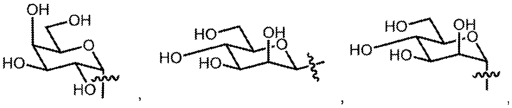 a-D-galactosyl, p-D-mannosyl, or a-D-mannosyl; wherein
 represents a bond from R to an oxygen atom (-O-) in formula 1 ;
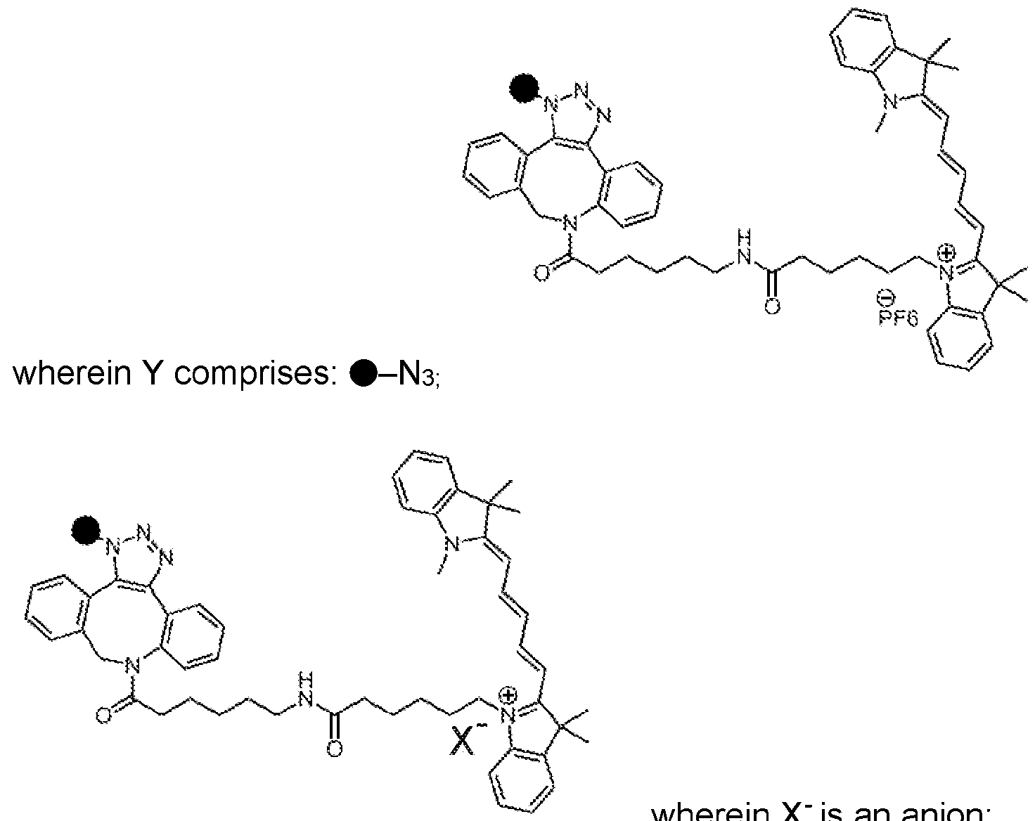 , wherein X is an anion; wherein • above represents a single bond attached to any -OH or -0- of R; or wherein Y comprises a click chemistry functional group, near-IR dye, an IR dye, a fluorescent small molecule, a small molecule with a chromophore, a Raman active small molecule, or a small molecule with a maximum absorbance band in a range from about 200 nm to about 100 pm attached to any -OH or -O- of R; and wherein a small molecule has a molecular weight in a non-salt form of less than about 1000 daltons (Da).
2. The chemical composition of claim 1 , wherein a major tautomer of formula 1 is
comprising a following tautomeric state:

(tautomer D1 of formula 1 ); or wherein the tautomer D1 above comprises about more than 50% of the molecular structure when the molecular structure is dissolved in DMSO.
3. The chemical composition of claim 1 , further comprising wherein a tautomer and/or an E/Z isomer of formula 1 comprises:
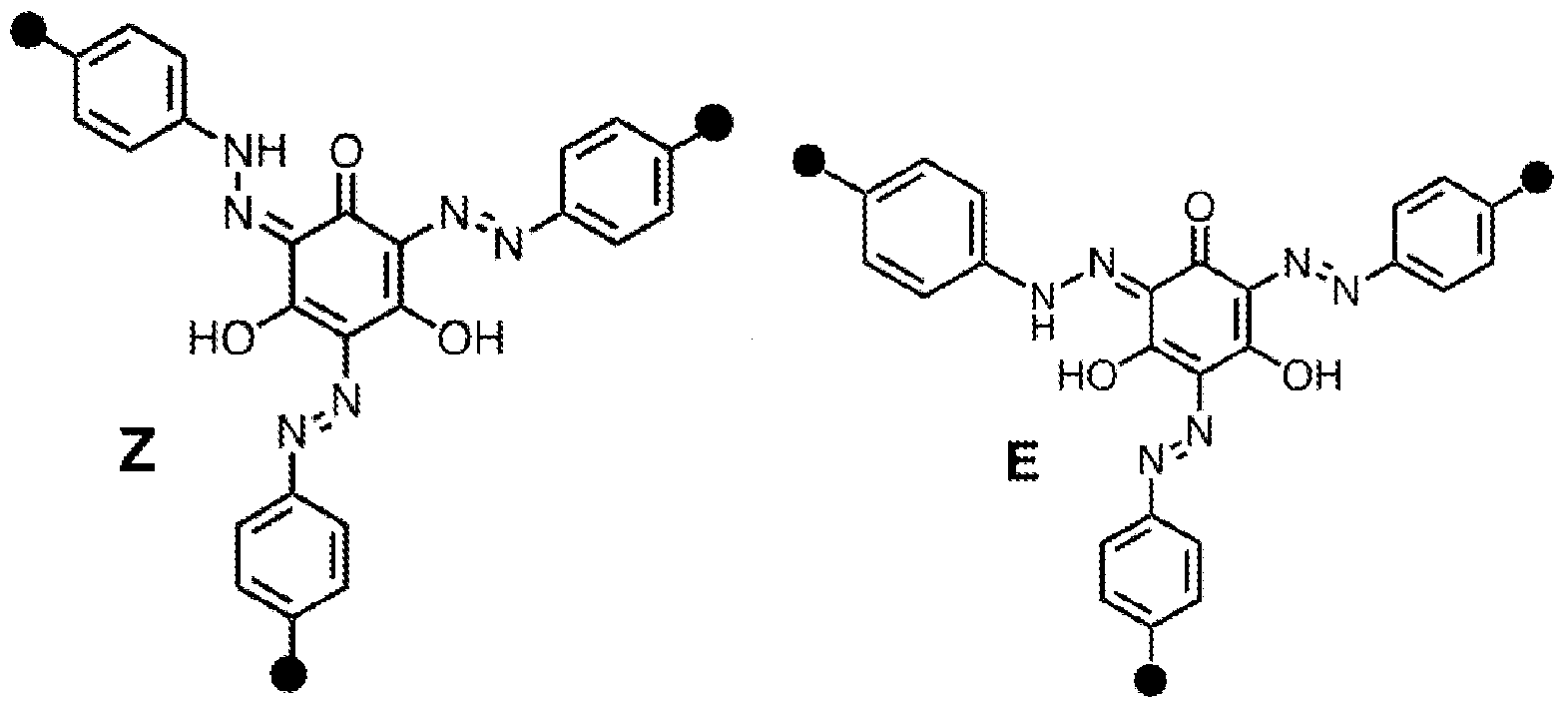 (B1), (B2), or a combination of B1 and B2 thereof; wherein each instance of • in B1 and B2 represents -O-R or -O-R-Y in formula 1 of claim 1 .
4. The chemical composition of claim 1 , further comprising wherein a tautomer and/or an E/Z isomer of formula 1 comprises:

(C3), (C4),

(01 ), or a combination of C1 , C2, C3, C4, and D1 thereof; wherein each instance of • in C1 ,
C2, C3, C4, and D1 represents -0-R or -0-R-Y in formula 1 of claim 1.
5. The chemical composition of claim 1 , wherein the molecular structure comprises:


6. The chemical composition of claim 1 , wherein X' comprises tetrafluoroborate [BF4] ", perchlorate [CIO4]-, Br", C" (carbide), Cl", F", H" (hydride), I", Ns", Ps", 02", S2", Se2", acetate, formate, oxalate, cyanide/cyanate, carbonate, chlorate, chromate, dichromate, dihydrogen phosphate, hydrogen carbonate, hydrogen sulfate, hydrogen sulfite, hydroxide, hypochlorite, mono-hydrogen phosphate, nitrate, nitrite, perchlorate, permanganate, peroxide, phosphate, sulfate, sulfite, superoxide, thiosulfate, silicate, metasilicate, aluminum silicate, or a combination thereof.
7. The chemical composition of claim 1 , further comprising wherein each instance of R independently comprises: phenyl(4)-O-p-D-glucosyl, phenyl(4)-O-a-D-glucosyl, phenyl(4)-O-[3-L-glucosyl, phenyl(4)-O-[3-D-galactosyl, phenyl(4)-O-o-D-galactosyl, phenyl(4)-O-p-D-xylosyl, phenyl(4)-O-a-D-xylosyl, phenyl(4)-O-|3-L-xylosyl, phenyl(4)-O-p-D-mannosyl, , phenyl(4)-O-a-D-mannosyl, phenyl(4)-O-a-D-talosyl, phenyl(4)-O-a-D-lyxosyl, phenyl(4)-S-[3-D-glucosyl, phenyl(4)-S-P-D-galactosyl, phenyl(4)-/V/-/-p-D-glucosyl, phenyl(4)-(C)-p-D-glucosyl, phenyl(4)-C/-/2O-[3-D-glucosyl, phenyl(3)-O-p-D-glucosyl, phenyl(3)-O-[3-D-galactosyl, phenyl(2)-O-|3-D-glucosyl, phenyl(2)-O-p-D-xylosyl, phenyl(2)-O-p-D-galactosyl, 1 -naphthyl(4)-O-[3-D-glucosyl, phenyl(4)phenyl(4)-O-p-D-glucosyl, phenyl(4)phenyl(4)-O-[3-D-galactosyl, phenyl(4)azophenyl(4)-O-[3-D-glucosyl, phenyl(4)-O-p-D-(2-deoxygalactosyl), phenyl(4)-O-|3-D-(2-deoxygalactosyl, 2-acetamido), phenyl(4)-O-a-D-(2-deoxygalactosyl, 2-acetamido), phenyl(4)-O-p-D-(2-deoxyglucosyl), phenyl(4)-O-P-D-(2-deoxyglucosyl, 2-acetamido), phenyl(4)-O-a-D-(2-deoxyglucosyl, 2-acetamido), phenyl(4)-O-p-D-glucosyl-(2-OMe), phenyl(4)-O-p-D-glucosyl-(3-OMe), phenyl(4)-0-P- D D-glucosyl-(4-OMe), phenyl(4)-O-P-D-glucosyl-(6-OMe),
phenyl(4)-O-P-D-glucosyl-(4-O-a-D-glucopyranosyl), phenyl(4)-O-[3-D-glucosyl-(4-O-|3-D-glucopyranosyl), phenyl(4)-O-p-D-glucosyl-(4-O-p-D-galactopyranosyl), phenyl(4)-O-|3-D-glucosyl-(2,3,4,6-tetra-OMe), or a combination thereof.
8. The chemical composition of claim 1 , wherein Y further comprises a functionalization with one or more chromophores including deuterium, azides, alkynes, nitriles, or a combination thereof.
9. The chemical composition of claim 1 , wherein Y comprises an azide; and further comprising wherein the azide is A) operative for one or more click chemistry reactions with one or more cyclooctynes, wherein the azide B) can be modified using one or more other alkynes in a presence of a copper catalyst, wherein the azide, C) can be modified with one or more specialized phosphines via a Staudinger ligation, or a combination of A), B), and C).
10. The chemical composition of claim 1 , wherein the composition with a pGIcYariv or wherein the composition without a pGIcYariv is operative to provide a selective association with or a selective binding to an arabinogalactan protein (AGP) or a glycosylated protein (glycoprotein) found in a cell wall of a plant; and wherein the composition does not substantially bind to non-AGP or to non-glycosylated proteins found in the cell wall of the plant.
11. The chemical composition of claim 1 , wherein the molecular structure is operative to provide a fluorescence absorption maximum in the range from about 800 nm to about 10 pm or in a near infrared (NIR) range.
12. The chemical composition of claim 1 , wherein the molecular structure is operative to provide a fluorescence emission maximum in the range from about 800 nm to about 10 pm or in a near infrared (NIR) range.
13. The chemical composition of claim 1 , wherein the molecular structure is operative to provide a fluorescence absorption maximum in the range from about 200 nm to about
800 nm.
14. The chemical composition of claim 1 , wherein the molecular structure is operative to provide a fluorescence emission maximum in the range from about 200 nm to about 800 nm.
15. A method of making AzYariv, the method comprising the steps of:
(1 ): reacting a chemical structure comprising the following chemical structure:
 with sodium nitrite (NaNCh) in about 1 .2 M HCI in water at about 0°C for about 120 minutes; whereby the following chemical structure is produced:

(2): Reacting 8 from the product of step 1 above with bis-Yariv and 2M NaOH for about 16 hours, whereby the following AzYariv is produced:

16. The method of claim 15, further comprising the step of:
(3):
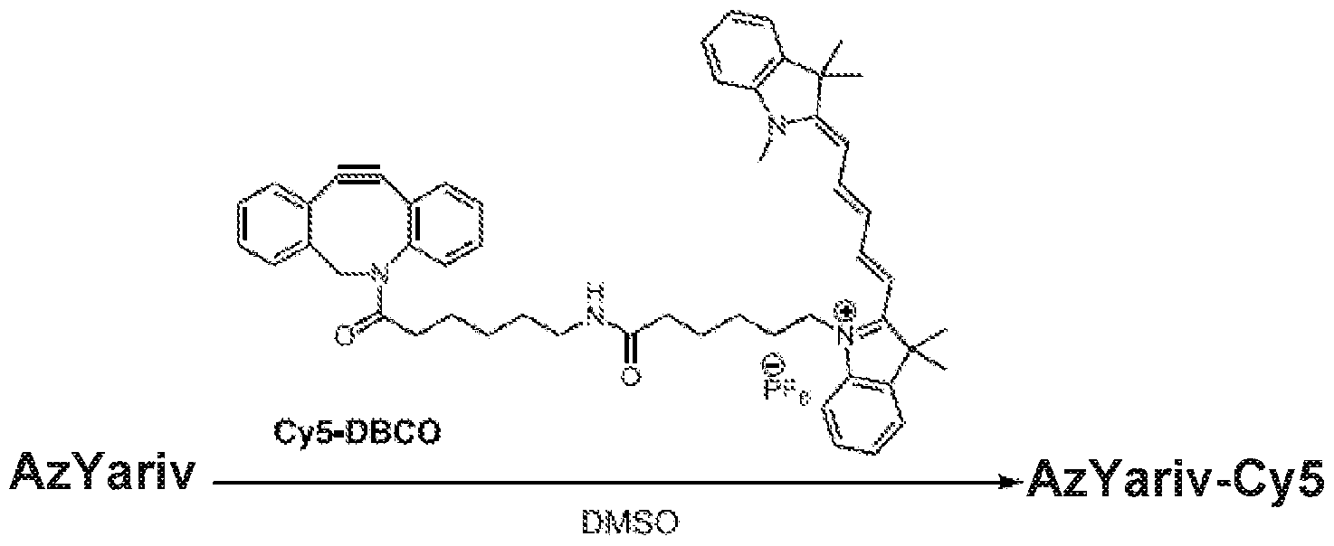 whereby AzYariv-Cy5 is produced.
17. The method of claim 15, further comprising the step of:
(4):
 whereby AzYariv-Cy5-X- is produced.
18. The method of claim 17, wherein X' comprises tetrafluoroborate [BF4] ", perchlorate [CIO4]", Br, C" (carbide), Cl", F", H" (hydride), I", N3", P3", O2", S2", Se2", acetate, formate, oxalate, cyanide/cyanate, carbonate, chlorate, chromate, dichromate, dihydrogen phosphate, hydrogen carbonate, hydrogen sulfate, hydrogen sulfite, hydroxide, hypochlorite, mono-hydrogen phosphate, nitrate, nitrite, perchlorate, permanganate, peroxide, phosphate, sulfate, sulfite, superoxide, thiosulfate, silicate, metasilicate, aluminum silicate, or a combination thereof.
19. The method of claim 15, further comprising attaching a click chemistry functional group, near-IR dye, an IR dye, a fluorescent small molecule, a small molecule with a chromophore, a Raman active small molecule, or a small molecule with a maximum absorbance band in a range from about 200 nm to about 100 pm; and wherein the small molecule has a molecular weight in a non-salt form of less than about 1000 daltons (Da); from any atom on the small molecule to an -N3 of AzYariv, whereby a functionalized or a fluorescent AzYariv product is produced.
20. The method of claim 19, wherein the attaching further comprises a click chemistry reaction.
21. The method of claim 19, wherein the fluorescent small molecule comprises coumarin, biotin, a proximity labeling probe, 1 ,8-naphthalimide, a cyanine dye, fluorescein, rhodamine, a cyanine fluorophore, or a boron dipyrromethene difluoride (BODIPY).
22. A method for performing a fluorescence imaging of an arabinogalactan protein (AGP) or a glycosylated protein (glycoprotein), the method comprising the steps of:
(1) contacting a material known to contain an AGP or a glycoprotein or suspected to contain an AGP or a glycoprotein with a reagent including AzYariv-Cy5, AzYariv-Cy5-X', [SGIcYariv, or a combination thereof, whereby a binding between the AGP or glycoprotein and the reagent occurs;
(2) directing an incident light or an incident electromagnetic radiation towards the binding, whereby a fluorescent emission occurs; and
(3) gathering, observing, measuring, or acquiring the emission.
23. The method of claim 22, further comprising wherein step (2) and step (3) are replaced by step (2a) and step (3a) below:
(2a) directing an incident light or an incident electromagnetic radiation towards the binding, whereby an electromagnetic absorbance occurs; and
(3a) gathering, observing, measuring, or acquiring the absorbance.
24. The method of claim 23, wherein the absorbance comprises a wavelength in a range or including a maximum absorbance band in the range from about 200 nm to about 100 pm.
25. The method of claim 22, wherein the material comprises a cell, a plant cell, a plant cell wall, or any combination thereof.
26. The method of claim 22, wherein the method begins an execution at any step on a
living plant cell or a living plant cell wall. 1. The method of claim 22, wherein a fluorescence absorption maximum wavelength is in a range from about 200 nm to about 10 pm; and a fluorescence emission maximum wavelength is in a range from about 200 nm to about 10 pm.
28. The method of claim 22, wherein a fluorescence absorption maximum wavelength is in a range from about 630 nm to about 650 nm; and a fluorescence emission maximum wavelength is in a range from about 655 nm to about 675 nm.
29. The method of claim 22, further comprising a fluorescent microscope and/or slides are obtained, whereby the method further comprises a method of fluorescent microscopy.
30. A kit suitable for sale comprising AzYariv, AzYariv-Cy5, AzYariv-Cy5-X-, or any combination thereof.
31 . The kit of claim 30, wherein one or more products are lyophilized, and whereby said lyophilization provides 1 ) a longer shelf-life and 2) a combination product.
Applications Claiming Priority (2)
| Application Number | Priority Date | Filing Date | Title |
|---|---|---|---|
| US202363493295P | 2023-03-30 | 2023-03-30 | |
| US63/493,295 | 2023-03-30 |
Publications (1)
| Publication Number | Publication Date |
|---|---|
| WO2024206707A1 true WO2024206707A1 (en) | 2024-10-03 |
Family
ID=92907064
Family Applications (1)
| Application Number | Title | Priority Date | Filing Date |
|---|---|---|---|
| PCT/US2024/022083 Ceased WO2024206707A1 (en) | 2023-03-30 | 2024-03-28 | Azyariv and azyariv conjugates - new tools for visualizing and characterizing arabinogalactan proteins |
Country Status (1)
| Country | Link |
|---|---|
| WO (1) | WO2024206707A1 (en) |
Citations (2)
| Publication number | Priority date | Publication date | Assignee | Title |
|---|---|---|---|---|
| CN101691450A (en) * | 2009-07-24 | 2010-04-07 | 华东师范大学 | Preparation method of beta-glucose-based-Yariv reagent |
| US20210128660A1 (en) * | 2017-01-26 | 2021-05-06 | Amorepacific Corporation | Composition for enhancing immunity, containing ginseng berry polysaccharides |
-
2024
- 2024-03-28 WO PCT/US2024/022083 patent/WO2024206707A1/en not_active Ceased
Patent Citations (2)
| Publication number | Priority date | Publication date | Assignee | Title |
|---|---|---|---|---|
| CN101691450A (en) * | 2009-07-24 | 2010-04-07 | 华东师范大学 | Preparation method of beta-glucose-based-Yariv reagent |
| US20210128660A1 (en) * | 2017-01-26 | 2021-05-06 | Amorepacific Corporation | Composition for enhancing immunity, containing ginseng berry polysaccharides |
Non-Patent Citations (2)
| Title |
|---|
| RUEDA SEBASTIAN, MCCUBBIN TYLER J., SHIEH MEG, HOSHING RAGHURAJ, BRAUN DAVID M., BASU AMIT: "A Functionalizable Analog of the Yariv Reagent for AGP Imaging using Fluorescence Microscopy", BIOCONJUGATE CHEMISTRY, AMERICAN CHEMICAL SOCIETY, US, vol. 34, no. 8, 16 August 2023 (2023-08-16), US , pages 1398 - 1406, XP093219909, ISSN: 1043-1802, DOI: 10.1021/acs.bioconjchem.3c00184 * |
| ZHOU LI HONG, WEIZBAUER RENATE A., SINGAMANENI SRIKANTH, XU FENG, GENIN GUY M., PICKARD BARBARA G.: "Structures formed by a cell membrane-associated arabinogalactan-protein on graphite or mica alone and with Yariv phenylglycosides", ANNALS OF BOTANY, ACADEMIC PRESS, LONDON., GB, vol. 114, no. 6, 1 October 2014 (2014-10-01), GB , pages 1385 - 1397, XP093219908, ISSN: 0305-7364, DOI: 10.1093/aob/mcu172 * |
Similar Documents
| Publication | Publication Date | Title |
|---|---|---|
| EP4137818B1 (en) | Use of divalent metals for enhancement of fluorescent signals | |
| Yuan et al. | A novel fluorescent probe for ratiometric detection of formaldehyde in real food samples, living tissues and zebrafish | |
| Xu et al. | A near-infrared reversible fluorescent probe for peroxynitrite and imaging of redox cycles in living cells | |
| Liu et al. | A rapid responsive colorimetric and near-infrared fluorescent turn-on probe for imaging exogenous and endogenous peroxynitrite in living cells | |
| Sun et al. | An efficient TP-FRET-based lysosome-targetable fluorescent probe for imaging peroxynitrite with two well-resolved emission channels in living cells, tissues and zebrafish | |
| Guo et al. | Turn-on fluorescence detection of β-glucuronidase using RhB@ MOF-5 as an ultrasensitive nanoprobe | |
| JP5215298B2 (en) | Solid phase detection of terminal monosaccharides cleaved from glycosylated substrates | |
| Yu et al. | Cu 2+-selective naked-eye and fluorescent probe: its crystal structure and application in bioimaging | |
| Tang et al. | A dual near-infrared pH fluorescent probe and its application in imaging of HepG2 cells | |
| Tang et al. | Highly sensitive and selective near-infrared fluorescent probe for zinc and its application to macrophage cells | |
| Geng et al. | An aqueous methylated chromenoquinoline-based fluorescent probe for instantaneous sensing of thiophenol with a red emission and a large Stokes shift | |
| JPH05507191A (en) | CMP-activated fluorescent sialic acid and its production method | |
| Arja et al. | Synthesis and Characterization of Novel Fluoro‐glycosylated Porphyrins that can be Utilized as Theranostic Agents | |
| Shen et al. | A mitochondria-targeting ratiometric fluorescent probe for the detection of hypochlorite based on the FRET strategy | |
| Zhou et al. | A new ratiometric two-photon fluorescent probe for imaging of lysosomes in living cells and tissues | |
| Kan et al. | A novel fluorescent probe of aluminium ions based on rhodamine derivatives and its application in biological imaging | |
| Doknic et al. | Synthesis and characterization of linker‐armed fucose‐based glycomimetics | |
| Wu et al. | Near-infrared light controlled fluorogenic labeling of glycoengineered sialic acids in vivo with upconverting photoclick nanoprobe | |
| You et al. | Imaging of zinc ions across diverse biological samples with a quinoline-based tris (2-pyridylmethyl) amine fluorescent probe | |
| Hjuler et al. | Preparation of glycoconjugates from unprotected carbohydrates for protein-binding studies | |
| WO2024206707A1 (en) | Azyariv and azyariv conjugates - new tools for visualizing and characterizing arabinogalactan proteins | |
| Siebold et al. | Fluoro‐Fucosylation Enables the Interrogation of the Lea‐LecB Interaction by BioNMR Spectroscopy | |
| Chepeleva et al. | 4-Benzyloxyflavonol glucoside as fluorescent indicator for b-glucosidase activity | |
| Hoshing et al. | An improved protocol for the synthesis and purification of Yariv reagents | |
| Ahmed et al. | Live-cell imaging of β-galactosidase based on a quinoline-malononitrile-derived AIE probe |
Legal Events
| Date | Code | Title | Description |
|---|---|---|---|
| 121 | Ep: the epo has been informed by wipo that ep was designated in this application |
Ref document number: 24781975 Country of ref document: EP Kind code of ref document: A1 |
|
| NENP | Non-entry into the national phase |
Ref country code: DE |
|
| 122 | Ep: pct application non-entry in european phase |
Ref document number: 24781975 Country of ref document: EP Kind code of ref document: A1 |