WO2024178232A2 - Expanding and improving nanobody repertoires: targeting sars-cov-2 - Google Patents
Expanding and improving nanobody repertoires: targeting sars-cov-2 Download PDFInfo
- Publication number
- WO2024178232A2 WO2024178232A2 PCT/US2024/016913 US2024016913W WO2024178232A2 WO 2024178232 A2 WO2024178232 A2 WO 2024178232A2 US 2024016913 W US2024016913 W US 2024016913W WO 2024178232 A2 WO2024178232 A2 WO 2024178232A2
- Authority
- WO
- WIPO (PCT)
- Prior art keywords
- seq
- binding
- rbd
- nanobodies
- cov
- Prior art date
- Legal status (The legal status is an assumption and is not a legal conclusion. Google has not performed a legal analysis and makes no representation as to the accuracy of the status listed.)
- Ceased
Links
Classifications
-
- C—CHEMISTRY; METALLURGY
- C07—ORGANIC CHEMISTRY
- C07K—PEPTIDES
- C07K16/00—Immunoglobulins [IG], e.g. monoclonal or polyclonal antibodies
- C07K16/08—Immunoglobulins [IG], e.g. monoclonal or polyclonal antibodies against material from viruses
- C07K16/10—RNA viruses
- C07K16/102—Coronaviridae (F)
- C07K16/104—Severe acute respiratory syndrome coronavirus 2 [SARS‐CoV‐2]
-
- C—CHEMISTRY; METALLURGY
- C07—ORGANIC CHEMISTRY
- C07K—PEPTIDES
- C07K2317/00—Immunoglobulins specific features
- C07K2317/20—Immunoglobulins specific features characterized by taxonomic origin
- C07K2317/22—Immunoglobulins specific features characterized by taxonomic origin from camelids, e.g. camel, llama or dromedary
-
- C—CHEMISTRY; METALLURGY
- C07—ORGANIC CHEMISTRY
- C07K—PEPTIDES
- C07K2317/00—Immunoglobulins specific features
- C07K2317/50—Immunoglobulins specific features characterized by immunoglobulin fragments
- C07K2317/56—Immunoglobulins specific features characterized by immunoglobulin fragments variable (Fv) region, i.e. VH and/or VL
- C07K2317/565—Complementarity determining region [CDR]
-
- C—CHEMISTRY; METALLURGY
- C07—ORGANIC CHEMISTRY
- C07K—PEPTIDES
- C07K2317/00—Immunoglobulins specific features
- C07K2317/50—Immunoglobulins specific features characterized by immunoglobulin fragments
- C07K2317/56—Immunoglobulins specific features characterized by immunoglobulin fragments variable (Fv) region, i.e. VH and/or VL
- C07K2317/569—Single domain, e.g. dAb, sdAb, VHH, VNAR or nanobody®
-
- C—CHEMISTRY; METALLURGY
- C07—ORGANIC CHEMISTRY
- C07K—PEPTIDES
- C07K2317/00—Immunoglobulins specific features
- C07K2317/70—Immunoglobulins specific features characterized by effect upon binding to a cell or to an antigen
- C07K2317/76—Antagonist effect on antigen, e.g. neutralization or inhibition of binding
-
- C—CHEMISTRY; METALLURGY
- C07—ORGANIC CHEMISTRY
- C07K—PEPTIDES
- C07K2317/00—Immunoglobulins specific features
- C07K2317/90—Immunoglobulins specific features characterized by (pharmaco)kinetic aspects or by stability of the immunoglobulin
- C07K2317/92—Affinity (KD), association rate (Ka), dissociation rate (Kd) or EC50 value
Definitions
- Said .xml copy, created February 22, 2024, is titled “076091_00170.xml” and is 180,066 bytes in size.
- BACKGROUND The COVID-19 pandemic, caused by Severe acute respiratory syndrome coronavirus 2 (SARS-CoV-2), has had a profound global impact the likes of which has not been seen in more than a century. The remarkably rapid development and distribution of vaccines undoubtedly saved many millions of lives; nevertheless, at the time of writing, mortality estimates range from 10 – 20 million, with additional profound long-lasting health impacts for many survivors.
- Spike is a homotrimer of an extensively glycosylated ⁇ 200 kDa protein composed of two major domains: S1, which contains the host receptor binding domain (RBD) that targets the angiotensin-converting enzyme 2 (ACE2) surface receptor on host cells; and S2, which upon host cell binding undergoes major conformational changes to enable viral – host membrane fusion, resulting in virus entry into the cytoplasm.
- S1 which contains the host receptor binding domain (RBD) that targets the angiotensin-converting enzyme 2 (ACE2) surface receptor on host cells
- ACE2 angiotensin-converting enzyme 2
- VoCs SARS-CoV-2 Variants of Concern
- Alpha, Beta, Gamma, Delta, Omicron and subvariants presents a significant barrier 3399-P41US.PRO -1- to attaining complete control of COVID-19.
- These VoCs usually have many Spike mutations (especially the RBD), and thus are relatively poorly neutralized by current vaccines and antibody therapies.
- monoclonal antibodies mAbs
- mAbs monoclonal antibodies
- mAbs are limited by challenges in the ease and cost of their large-scale manufacturing, distribution, and intravenous administration.
- An alternative to mAbs is a particular class of single domain antibodies termed “nanobodies”.
- Nanobodies are “mini-antibodies”, some 1/10 th the size of regular IgGs, derived from the variable domain (VHH) of variant heavy chain-only IgGs (HCAbs) found in camelids (e.g. llamas).
- VHH variable domain
- HCAbs variant heavy chain-only IgGs
- Each nanobody molecule is constructed of a single Ig fold, consisting of four framework regions (FRs) that intersperse and orient three complementarity determining regions (CDRs) that form the nanobody paratope. These regions are similar to FRs and CDRs of conventional antibodies.
- CDR3 is formed by VDJ recombination of germline DNA; CDR1 and CDR2 come from the germline V region, and all three CDRs are then subject to somatic hypermutation, with selection for improved binding affinity to antigens.
- VHH variable domain on a heavy chain
- Nanobodies have several attractive advantages over mAbs, including: fast on-rates leading to high overall affinities; characteristics of small molecules in terms of higher tissue penetration and accessibility to regions of Spike not accessible to the larger mAbs or occluded by glycosylation, greatly enhancing their potential to synergize in combination, a profound advantage they have over often poorly-synergizing conventional antibodies.
- nanobody binding can be disrupted by mutations in VoCs.
- mAbs and the advantages of nanobodies, a need remains for rapidly producing nanobodies that target the entirety of the Spike protein as a more effective therapy against the continuing emergence of new SARS-CoV-2 VoCs. The present disclosure addresses these and related needs.
- the present disclosure provides single domain antibodies that specifically recognize SARS-CoV-2 epitopes.
- the described antibodies are heavy chain only antibodies.
- the antibodies contain a single variable domain which comprises three complementarity- determining regions (CDRs).
- CDRs complementarity- determining regions
- the disclosure includes all heavy chain only antibodies that contain any CDR1, CDR2, and CDR3 disclosed herein, and may have residue changes in the non-CDR segments.
- a representative antibody sequence is SEQ ID NO:117.
- the disclosure provides the described antibodies, compositions comprising the described antibodies, and methods of using the described antibodies for prophylaxis or therapy for SARS-CoV-2 infections.
- Figure 1 Design of Yeast-Based Nanobody Screen for anti-SARS-CoV-2 Spike Domains. Schematic of our yeast display based strategy for generating, identifying, and characterizing large, diverse repertoires of nanobodies that bind the spike protein of SARS- CoV-2. The highest quality nanobodies were assayed for their ability to neutralize SARS- CoV-2 pseudovirus.
- FIG. 1 Boxed inset shows 2.8 ⁇ m magnetic beads conjugated with S1 protein from SARS-CoV-2 Spike, after a binding reaction with yeast displaying an anti-S1 nanobody (top) or a nonspecific control (bottom), after non-binding yeast were washed away.
- Figure 1 Sequence to determine VHH library are ATGGCTGAGGTGCAGTTGG (SEQ ID NO:172); ATGGCTCAGGTGCAGCTGG (SEQ ID NO:173); and ATGGCTGATGTGCAGTTGG (SEQ ID NO:174). Sequences to determine library of captured VHH are ATGGCTCAGGTGCAGCTGG (SEQ ID NO:175) and ATGGCCCAGGTGCAGCTGG (SEQ ID NO:176).
- Figures 2A and 2B are ATGGCTCAGGTGCAGCTGG (SEQ ID NO:175) and ATGGCCCAGGTGCAGCTGG (SEQ ID NO:176).
- FIG. 2A Fluorescence microscopy of a yeast competition assay.
- Figure 2B Plot of the coefficient of variation (CV) of yield for each yeast strain across the experiments in B against the Kd of their displayed nanobodies. As seen in the plot, the yields of yeast bearing low affinity nanobodies were highly variable specifically under competitive conditions. The yield of the highest-affinity Nb-bearing yeast, in contrast, were almost invariant because they always won the competition. Under non-competitive conditions, this differential was lost.
- Figures 3A through 3C Sequence Diversity of Nanobody Libraries.
- Nanobody sequences from the unselected yeast library were amplified and sequenced with Illumina Miseq and processed to minimize sequence errors, as described in Methods, yielding 1.2 ⁇ 10 6 distinct nanobody sequences.
- Framework regions (FRs) and complementarity-determining regions (CDRs) were determined in each encoded nanobody based on the alignment of (41). Unique sequences were determined and aligned by left justification of each region.
- Figure 3A A seqlogo (44,59) was generated (MATLAB seqlogo command). High variability in the three CDR regions is evident.
- Figure 3B Plot of proportion of non-consensus residues per residue across the library.
- FIG. 5A through 5C Screening for New Families of Anti-Spike Nanobodies.
- Figure 5A The read counts in the unselected and 1 ⁇ and 2 ⁇ selected libraries screened against either the S1, RBD or S2 domains of Spike from the entire llama 7704 nanobody cDNA display library (gray points) are plotted, as the log2 of these values + 1, as shown in Figures 4A, 4B.
- Sequences displaying different specificities were identified and selected from the graphical display as in Figures 4A, 4B, except here nanobodies specific for only the RBD subdomain of S1 were separately labeled from those that recognized the non-RBD portions of S1. Selection was by a differential polygon function allowing multiple criteria. For example, sequences in an enriched polygon for S1 but not for RBD defines nanobodies binding to the non-RBD subdomain of S1 (also see Introduction). Green: S1 non-RBD; blue: RBD; red: S2.
- FIG. 6A Testing the standard library against RBD variants Delta and Omicron. Dynabeads conjugated with RBD from the original SARS-CoV-2 and from the Delta and Omicron variants were employed for affinity purification of yeast display clones from the llama 7704 library. Two rounds of selection were carried out, as in Figures 4A, 4B, and 5A-5C. A clear overall reduction in binding was observed, though many clones still bound well to both variants.
- Results were analyzed based on the fate of CDR3 ‘groups’ (where within a group, a given CDR3 bound to a high diversity of CDR1,2 recombinants). Different behaviors were observed; three are illustrated with colored dots (same color scheme as Figures 7A-7C).
- the ‘NAAAW’ (SEQ ID NO:160) group (green) bound well to original and variant RBDs, essentially independent of the recombinant CDRs 1 and 2 it was attached to, suggesting strong ‘CDR3 dominance’ of the ‘NAAAW’ (SEQ ID NO:160) CDR3.
- the ‘IIDDY’ (SEQ ID NO:164) group exhibited essentially similar behavior when shuffled as when combined with its native CDR1,2 ( Figures 7A-7C): strong binding to original and Omicron, but clearly weaker binding to Delta.
- the ‘YERLAWD’ (SEQ: ID NO:159) recombinant group bound comparably to all variants, in contrast to its behavior with its native CDRs 1 and 2, which rendered it unable to bind Delta or Omicron.
- Figure 6B Effectiveness of the shuffle is shown by extracting all sequences bearing a specific CDR3 and examining sequence diversity of CDRs 1 and 2.
- Sequence logos show that highly diverse CDRs 1 and 2 are observed joined to each of three different CDR3’s, and the pattern of CDR1,2 diversity attached to the CDR3’s was essentially the same.
- the sequences on Figure 6B are YERLAWD (SEQ: ID NO:159), NAAAW (SEQ ID NO:160), and IIDDY (SEQ ID NO:164).
- Figures 7A through 7C Biophysical and Neutralization Properties of the Nanobodies. Thirty yeast display nanobodies targeting the S1-RBD, S1 non-RBD, and S2 portions of spike were functionally tested for neutralization of lentivirus pseudotyped with various SARS-CoV spikes and their biophysical properties characterized.
- FIG. 7A Neutralization data against Original, Delta and Omicron.
- Figure 7B Kd measurements of 21 of these nanobodies were determined using SPR and affinities plotted. Nanobodies were tested against Original, Delta and Omicron recombinant S1 or RBD. S1 non-RBD nanobodies were not tested against Omicron. Kd measurements for three of the ‘rescue’ constructs plotted at right.
- Figure 7C The Tm measurements of the nanobodies in (B) were determined using DSF and plotted.
- CoV2-YD-6 and CoV2-YD-38 resulted in two distinct melting peaks. Open circles indicate less proportion of this species in the sample. Tm measurements for three of the ‘rescue’ constructs plotted at right.
- Figure 8. Epitope Binning by Yeast Display. Dynabeads conjugated with RBD were blocked with monomer nanobodies representing the 7 epitope classes defined previously (20); with the soluble extracellular domain of the RBD target Ace2; or left unblocked. The 2 ⁇ -RBD-selected library from llama 7704 (Figs.4, 5, S1) was bound to these beads.
- the bound VHHs were sequenced, and enrichment/depletion upon blocking for each ‘CDR string’ (catenated CDR1/CDR2/CDR3) was calculated as log2 (readcount with blocked beads/readcount with unblocked beads).
- the sequences were filtered to remove PCR crossover artifacts (Methods).
- Left The resulting matrix of ⁇ 100,000 sequences X 8 blocking agents was filtered for readcount (at least 100 reads combining all blocking experiments) and clustered using the MATLAB hierarchical clustering algorithm; scale bar on left indicates log2 enrichment/depletion (above).
- NAAAW SEQ ID NO:160
- TALLS SEQ ID NO:162
- TADLY SEQ ID NO:163
- IIDDY SEQ ID NO:164
- TVDAQ SEQ ID NO:165
- AAHVN SEQ ID NO:166
- MATSEY SEQ ID NO:170
- GSDFGDH SEQ ID NO:171
- TVTDR SEQ ID NO:181
- Figure 9 Binding of nanobodies expressed in yeast to the different recombinant S1, RBD and S2 domains of SARS-Cov2 Spike protein (labeled S1, RBD, and S2 selection respectively) (20) conjugated to Dynabeads. Binding and washing were as described in Methods.
- Nanobody sequences were amplified and sequenced as in Figures 3A-3C.
- the CDR regions were extracted from each sequence, and concatenated in a ‘CDR string’.
- High-affinity binders were recovered from llama 5094, though apparently fewer than from 7704, reflecting the results of (20).
- the inventors carried out the same assay for these nanobodies and plotted the results as in Figure 4A, with the exception that to get sufficient representation CDR strings with up to 20% sequence variation in each CDR were accepted; each different color of the dots plotted represents a member of a family of a given mass spectrometry-positive nanobody.
- the same high specificity and recovery in yeast display for the 5094 clones was observed for the 7704 clones.
- FIGS 11A and 11B Comparison of different methods for screening the display libraries. The entire llama 7704 nanobody cDNA display library was screened against the RBD domain of Spike, and plotted as in Figure 9 (gray points). Two methods of screening were employed. The first was as described in Figure 1 and Methods, using two rounds of Dynabead selection.
- Figure 11A All sequences recovered are plotted as in Figure 9.
- Figure 11B Families of MS- positive sequences are plotted on top of the overall graph, as in Figure 10.
- Figure 12. Testing CDR families of mass-spectrometry positives against RBD variants Delta and Omicron. The data are plotted as in Figures 7A-7C, but plotting CDR families of the mass-spectrometry-positive clones; each different color of the dots plotted represents a different mass spectrometry-positive nanobody.
- Figure 13
- Diagram of the splice-overlap extension method for recombining CDRs Degenerate oligos priming in both directions from two highly conserved sequences within framework regions (FRs) 2 and 3, and end oligos tagged for recombination onto the yeast expression vector, were used to amplify fragments containing CDRs 1, 2 and 3 as well as flanking framework regions.
- the template used was a pool after 1 round of selection on RBD.
- the result of the sequential PCR reactions indicated is a random mix-and-match of the three CDRs and flanking framework regions.
- Figure 14 Epitope Binning Tests.
- Dynabeads conjugated with RBD were pre- blocked with saturating amounts of either monomer nanobodies S1-1 or S1-23, with unblocked Dynabeads as control.
- polynucleotide and amino acid sequences having from 80-99% similarity, inclusive, and including and all numbers and ranges of numbers there between, with the sequences provided here are included in the invention. All of the amino acid sequences described herein can include amino acid substitutions, such as conservative substitutions that do not adversely affect the function of the protein that comprises the amino acid sequences.
- the present specification also provides polynucleotides that hybridize under selective hybridization conditions to a polynucleotide that encodes a described antibody.
- a polynucleotide that can hybridize to a polynucleotide that encodes a described antibody has 70-100% complementarity, inclusive, and including all numbers and ranges there between, to the coding polynucleotide.
- Selective hybridization conditions under which a polynucleotide having at least 70% complementarity to a polynucleotide encoding a described antibody will be known by those skilled in the art.
- the degree of stringency can be controlled by one or more of temperature, ionic strength, pH, and the presence of a denaturing agent such as formamide.
- a polynucleotide that hybridize under selective hybridization to a coding polynucleotide can be present in an expression vector.
- the expression vector can be introduced into a cell of a cell culture to thereby express a described antibody.
- the antibody can be separated from the cell culture and used to produce a purified form of antibodies.
- the disclosure provides a VHH chain of an anti-Severe Acute Respiratory Syndrome Coronavirus 2 (SARS-CoV-2) nanobody, wherein the amino acid sequence of the VHH chain comprises any one of the sequences as set forth in SEQ ID NOs:117 – 153.
- SARS-CoV-2 anti-Severe Acute Respiratory Syndrome Coronavirus 2
- the disclosure expands the repertoire of VHH chains disclosed in PCT application no. PCT/US2021/047019, published as WO2022040603 on February 2, 2022, the entire disclosure of which is incorporated herein by reference, and beyond the constructs described in Fred D Mast, et al.
- VHH chains comprising amino acid sequences that have at least 90% sequence identity to any one of the contiguous sequences as set forth in SEQ ID NOs: 117 – 153.
- the nanobodies are referred to herein from time to time as “binding partners” in the plural and “binding partner” in the singular.
- the VHH chain can comprise an amino acid sequence comprising any one of the sequences as set forth in SEQ ID NOs:117 – 153.
- a VHH chain as in any one of SEQ ID NOs:117 – 153 may be combined with a VHH chain as in any one of SEQ ID NOs:1-116 to produce a multi-specific binding partner.
- the multi- specific binding partner is provided in the form of a dimer that can bind to two different epitopes of a viral spike protein.
- a VHH chain as in any one of SEQ ID NOs:117 – 153 can be incorporated into, for example, a chimeric antigen receptor, single- chain Diabodies (scDbs), single-chain variable fragment (scFv), and other antibody fragments that retain antigen binding function.
- a binding partner of this disclosure may be used in combination with a binding partner comprising a sequence of any one of SEQ ID NOs: 1-116.
- the combination may comprise at least two separate binding partners, or at least two antibodies may be combined into a single binding partner format to provide a multi-specific binding partner, such as a bi-specific antibody.
- a binding partner of this disclosure may be modified such that it is present in a fusion protein.
- an antigen binding segment of a binding partner may be present in a fusion protein, and/or a constant region may be a component of a fusion protein.
- a fusion protein comprises amino acids from at least two different proteins.
- Fusion proteins can be produced using any of a wide variety of standard molecular biology approaches, including but not necessarily limited to expression from any suitable expression vector.
- a binding partner described herein may be present in a fusion protein with a detectable protein, such as green fluorescent protein (GFP), enhanced GFP (eGFP), mCherry, and the like.
- GFP green fluorescent protein
- eGFP enhanced GFP
- mCherry a detectable protein
- an mRNA or chemically modified mRNA encoding any binding partner described herein can be delivered to cells such that the binding partner is translated by the cells.
- Pharmaceutical formulations containing binding partners are included in the disclosure and can be prepared by mixing them with one or more pharmaceutically acceptable carriers.
- Pharmaceutically acceptable carriers include solvents, dispersion media, isotonic agents, and the like.
- an effective amount of one or more binding partners is administered to an individual in need thereof.
- an effective amount is an amount that reduces one or more signs or symptoms of a disease and/or reduces the severity of the disease.
- An effective amount may also inhibit or prevent the onset of a disease or a disease relapse.
- a precise dosage can be selected by the individual physician in view of the patient to be treated. Dosage and administration can be adjusted to provide sufficient levels of binding partner to maintain the desired effect. Additional factors that may be taken into account include the severity and type of the disease state, age, weight, and gender of the patient, desired duration of treatment, method of administration, time and frequency of administration, drug combination(s), reaction sensitivities, and/or tolerance/response to therapy.
- a described binding partner is administered to an individual who has or is suspected of having or is at risk of contracting a SARS-CoV-2 infection.
- the SARS-CoV-2 infection is by any type of SARS-CoV-2 that is a variant of high consequence, a variant of concern, a variant of interest, or a variant being monitored.
- any described antibody can be administered for therapeutic or prophylactic purposes.
- one or more described antibodies can be administered to an individual who has been diagnosed with COVID-19.
- Binding partners and pharmaceutical compositions comprising the binding partners can be administered to an individual in need thereof using any suitable route, examples of which include intravenous, intramuscular, intraperitoneal, subcutaneous, oral, or inhalation routes.
- compositions may be introduced as a single administration or as multiple administrations or may be introduced in a continuous manner over a period of time.
- the administration(s) can be a pre-specified number of administrations or daily, weekly, or monthly administrations, which may be continuous or intermittent, as may be therapeutically indicated.
- the VHH chain can comprise an amino acid sequence comprising any one of the sequences as set forth in SEQ ID Nos: 117-120, 147, and 148.
- SEQ ID NOs: 117-120, 147, and 148 can be selective for binding to non-Receptor Binding Domain (RBD) of SARS-CoV-2 spike protein.
- RBD non-Receptor Binding Domain
- the disclosure provides for an immunoconjugate comprising: (a) a VHH chain as recited above; and (b) a conjugating part comprising a detectable marker, a drug, a radionuclide, or an enzyme, or a toxin.
- the VHH chain can comprise an amino acid sequence comprising at least 90% sequence identity to any one of the sequences as set forth in SEQ ID NOs: 150- 157.
- SEQ ID NOs: 150-157 are selective for binding to Receptor Binding Domain (RBD) of SARS-CoV-2 spike protein.
- the VHH chain can comprise an amino acid sequence comprising at least 90% sequence identity to any one of the sequences as set forth in SEQ ID NOs: 117- 120, 147, and 148.
- SEQ ID NOs: 117-120, 147, and 148 are selective for binding to non-Receptor Binding Domain (RBD) of SARS-CoV-2 spike protein.
- RBD non-Receptor Binding Domain
- the disclosure provides for an immunoconjugate comprising: (a) a VHH chain as described above; and (b) a conjugating part comprising a detectable marker, a drug, a radionuclide, or an enzyme. Such constructs may be used for therapeutic or diagnostic purposes.
- the disclosure provides for a pharmaceutical composition
- a pharmaceutical composition comprising: (a) a VHH chain as above or an immunoconjugate as described above; and (b) a pharmaceutically acceptable carrier.
- a pharmaceutical composition comprising: (a) a VHH chain as above or an immunoconjugate as described above; and (b) a pharmaceutically acceptable carrier.
- the terms “nanobody”, “nanobodies”, “SARS-CoV-2 nanobody”, or “SARS-CoV-2 nanobodies” are exchangeable and refer to nanobodies that specifically recognize and bind to the Spike protein of SARS-CoV-2 and to the Spike protein of any variants of concern (VoC).
- single domain antibody VHH
- nanobodies have the same meaning referring to a variable region of a heavy chain of an antibody, and construct a single domain antibody (VHH) consisting of only one heavy chain variable region. It is the smallest antigen-binding fragment with complete function.
- variable means that certain portions of the variable region in the nanobodies vary in sequences, which forms the binding and specificity of various specific antibodies to their particular antigen. However, variability is not uniformly distributed throughout the nanobody variable region. It is concentrated in three segments called complementarity-determining regions (CDRs) or hypervariable regions in the variable regions of the light and heavy chain.
- CDRs complementarity-determining regions
- variable region The more conserved part of the variable region is called the framework region (FR).
- the variable regions of the natural heavy and light chains each contain four FR regions, which are present in a ⁇ -folded configuration, joined by three CDRs which form a linking loop, and in some cases can form a partially ⁇ -folded structure.
- the CDRs in each chain are closely adjacent to the others by the FR regions and form an antigen- binding site of the nanobody with the CDRs of the other chain (see Kabat et al., NIH Publ. No.91-3242, Volume I, pages 647-669. (1991)).
- the constant regions are not directly involved in the binding of the nanobody to the antigen, but they exhibit different effects or functions, for example, in antibody-dependent cytotoxicity of the antibodies.
- the described antibodies may have changes from the sequences expressly described herein, including conservative amino acid substitutions in the framework regions.
- the heavy chain variable region of described nanobody comprises 3 complementary determining regions: CDR1, CDR2, and CDR3 having at least 95% sequence identity to a described CDR amino acid sequence.
- sequence similarity refers to the likeness between at least two sequences in comparison.
- Sequence identity refers to the number of characters that match exactly between the two different sequences.
- sequence identity addresses the degree of similarity of two sequences, such as protein sequences. Determination of sequence identity can be readily accomplished by persons of ordinary skill in the art using accepted algorithms and/or techniques.
- Sequence identity is typically determined by comparing two optimally aligned sequences over a comparison window, where the portion of the peptide sequence in the comparison window may comprise additions or deletions (i.e., gaps) as compared to the reference sequence (which does not comprise additions or deletions) for optimal alignment of the two sequences.
- the percentage is calculated by determining the number of positions at which the identical amino-acid residues occur in both sequences to yield the number of matched positions, dividing the number of matched positions by the total number of positions in the window of comparison and multiplying the result by 100 to yield the percentage of sequence identity.
- Various software driven algorithms are readily available, such as BLAST N or BLAST P to perform such comparisons. Any described antibody may be humanized. Methods for humanization are known in the art.
- human framework sequences are substituted for the described framework sequences, suitable human framework sequences being known in the art.
- EXAMPLES To produce the described nanobody repertoires, we employed a mass spectrometry- based approach (20,22) in which llamas were immunized with Spike constructs. This permits affinity maturation processes in vivo (33). High-throughput DNA sequencing of VHH libraries PCR-amplified from marrow lymphocyte cDNA from the immunized llamas in combination with mass spectrometric (MS) identification of high-affinity VHH regions derived from the serum of the same animal was performed.
- MS mass spectrometric
- VHH cDNA sequences Computational matching of MS- sequenced peptides to VHH cDNA sequences allowed high-confidence identification of sequences encoding high-affinity nanobodies.
- Genes encoding nanobodies were synthesized and expressed in bacteria, and nanobodies were purified, and characterized for their specificity and affinity.
- These V H H cDNA libraries also represent a resource that can be used for an orthogonal approach for nanobody production, employing display screening methods instead (34-37). This approach could discover additional nanobodies, and also serve as a platform to explore the specificity and VoC sensitivity of a large number of nanobodies in parallel.
- a robust and efficient yeast display method was designed and validated specifically for screening nanobodies, in particular against Spike (34,38).
- the present disclosure includes evaluation of the cDNA library made from immunized llamas (20), which was transferred into a nanobody display vector (34).
- a nanobody display vector 34
- yeast expressing high-affinity nanobodies displayed little variation in yield even under competitive conditions, because they ‘won’ the competition.
- each sequence to a representation consisting solely of its three CDR regions (CDRs 1,2,3) (referred to as a ‘CDR string’).
- CDRs 1,2,3 CDR regions 1,2,3
- Fig.9 shows the behavior of the entire library.
- the screen clearly distinguished specifically binding and non-binding clones; for example, biochemically defined S2-specific nanobodies bound to S2 beads but not S1 or RBD beads when expressed in yeast, and vice versa.
- biochemically defined S2-specific nanobodies bound to S2 beads but not S1 or RBD beads when expressed in yeast, and vice versa We plotted enrichment of these MS positive sequences after both 1 and 2 rounds of selection against their measured K D s (20). While the relationship was noisy, a statistically significant negative slope was observed (Fig.4B), especially for two rounds of selection (likely to be more competitive conditions based on our analysis of anti-GFPs above), once again confirming the competitive and affinity-sensitive nature of the screen.
- Fig.4B a statistically significant negative slope was observed (Fig.4B), especially for two rounds of selection (likely to be more competitive conditions based on our analysis of anti-GFPs above), once again confirming the competitive and affinity-sensitive nature of the screen.
- the S1-non-RBD clones are dominated by members of one family, defined by its ‘IAQY’ (SEQ ID NO:185) consensus CDR3 sequence (Fig.5B,C; family (i)). This family was missed in the MS-based approach (20), possibly because the CDR3 was too small for reliable peptide identification.
- numerous other new S1-specific families are present, for example the “RGLGRGLGFY” (SEQ ID NO:186) CDR3 consensus sequence (Fig 5B, family (ii)).
- the RBD domain isolated a large and diverse set of families.
- Fig 5B, family (v) contains the consensus CDR3 “TVDAQSDY” (SEQ ID NO:184), which is also found in the mass spectrometrically identified nanobody S1-RBD-38.
- Two large families identified here contain divergent relatives among the MS-identified nanobodies (20), the ‘‘LRSRFNAAAWTTEAAFDY’ ’ (SEQ ID NO:158) (previous MS-identified S1-RBD-6 and S1-RBD-31) and ‘YERLAWDTSTY’ (SEQ ID NO:177) families (previous MS-identified S1-RBD-35), the remaining four indicated families being completely novel.
- the S2-specific clones were also very diverse and not dominated by any single family (Fig.5B,C), with limited overlap with the mass spectrometrically identified clones (20).
- the screening method identified a large number of new clones recognizing different Spike domains. We then analyzed if these new clones had high affinity binding and strong antiviral activity when expressed as monomers. Testing the Library against the Major VoCs Delta and Omicron
- One of the greatest challenges to managing COVID-19 is the ability of the SARS- CoV-2 virus to mutate into new VoCs that can resist prevalent vaccines and therapeutics.
- An advantage of generating large repertoires of nanobodies is that one maximizes the likelihood of finding VoC-resistant, broadly specific nanobodies (20,47-49).
- the large ‘TVDAQSDY’ (SEQ ID NO:184) (Fig.5C, (v)) family binds comparably to the original SARS-CoV-2 and both variants.
- the ‘‘LRSRFNAAAWTTEAAFDY’ (SEQ ID NO:158) ‘NAAAW’ (SEQ ID NO:160) (Fig.5C, (iv)) family binds comparably to the original SARS-CoV-2 and the Delta VoC RBDs, and appears collectively slightly weaker against the Omicron RBD.
- YERLAWDTSTY SEQ ID NO:177
- YERLAWD YERLAWD
- SEQ: ID NO:159 Fig.5C, (iii)
- the ‘IIDDYGVQY’ (SEQ ID NO:178) (‘IIDDY’ IIDDY (SEQ ID NO:164)) (Fig.5C, (vi)) family (Fig.6A, ‘IIDDY’ (SEQ ID NO:164)) and a family not indicated on Fig.5 but comprising a family characterized by a ‘TADLYSDY’ (SEQ ID NO:179) (‘TADLY’) (SEQ ID NO:163) CDR3 sequence binds well to original SARS-CoV-2 and the Omicron variant but more weakly to the Delta variant, especially after two rounds of selection. These families contain considerable sequence diversity within them (Fig.5).
- DNA shuffling is an established in vitro method for improvement of binding or catalytic activity. Typically it is applied to a library of randomly point-mutagenized sequences that have been selected for improved activity, from which starting point splice- overlap-extension (SOE) PCR is carried out to produce mix-and-match recombinants.
- SOE starting point splice- overlap-extension
- VDJ recombination shares some features, but with the critical difference that only a single round of shuffling occurs rather than multiple rounds interleaved with the recombinations. Neither the extremely high density of recombination joins that can be attained by in vitro shuffling, and its highly multiparental nature, are shared by natural biological systems, to our knowledge. DNA shuffling has been applied to nanobodies with recombination between CDRs, with improvement of binding noted in the progeny (52-54). However, the specificity and number of potential parental sequences was not established. We started with the library selected on SARS-Cov2 RBD, carried out SOE recombining CDRs 1, 2 and 3 at random from that library (Fig.13).
- the shuffled YERLAWD (SEQ: ID NO:159) group in contrast, contained abundant members that bound equally well to Original and to Delta RBD, while remaining almost completely defective in Omicron binding.
- Examination of the sequences associated with this high Delta binding revealed specific enrichment of sequences highly similar to the native ‘NAAAW’ (SEQ ID NO:160) CDR1 and CDR2, recombined with the ‘YERLAWD’ (SEQ: ID NO:159) CDR3 (Fig.6B).
- the native ‘NAAAW’ (SEQ ID NO:160) family CDR1 and CDR2 have specific ability to bind to Delta RBD, independent of CDR3 content.
- a subset of the anti-S1 nanobodies were tested for binding affinity against recombinant S1 or RBD from either Original, Delta or Omicron (S1 non-RBD nanobodies were not tested against omicron) (Fig.7B); all bound strongly to Original, displaying affinities in the nM - pM range. Two of these failed to bind only Delta, two failed to bind only Omicron and CoV2-YD-33 and CoV2-YD-34 failed to bind both variants.
- CoV2-YD-10 neutralizes yet shows no binding to the RBD of omicron using SPR. It is possible the binding site of the nanobody may be slightly truncated in the RBD construct used for SPR resulting in the no binding result. Lastly, all showed a moderate to strong degree of thermal stability, typical of nanobodies (Fig.7C) (21).
- the nanobody binding in the yeast display screening correlates reasonably well with that seen in the biochemical assay of the corresponding expressed nanobody (Fig.7B).
- the differential affinities of the ‘YERLAWD’ (SEQ: ID NO:159) and ‘NAAAW’ (SEQ ID NO:160) nanobodies for Delta RBD in both yeast display screening (Fig.6) and in the biochemical assay of the corresponding expressed nanobody (Fig.7B) agree, as discussed in the previous section.
- ‘LAYVT’ SEQ ID NO:161) (CoV2-YD-7) shows no significant loss of affinity for either Delta or Omicron RBD in both yeast display screening and in the biochemical assay of the corresponding expressed nanobody.
- ‘TALLS’ (SEQ ID NO:162) (CoV2-YD-6) show partial loss of affinity for Delta RBD while retaining Omicron affinity in both yeast display screening and in the biochemical assay of the corresponding expressed nanobody.
- ‘TADLY’ (SEQ ID NO:163) (CoV-YD-9) shows complete loss of affinity for Delta RBD in both yeast display screening and in the biochemical assay of the corresponding expressed nanobody, while again retaining Omicron affinity in both assays.
- Epitope Mapping of the Nanobody Repertoire Another important characterization of any nanobody repertoire is to determine the different epitopes being recognized by each nanobody, as in the case of anti-SARS-CoV-2 Spike nanobodies, exploration of a larger epitope space increases the likelihood of discovering variant resistant, strongly neutralizing nanobodies (20). This is usually done by ‘epitope binning’: finding classes of nanobodies that reciprocally inhibit each others’ binding due to competition for the same epitope. Epitope binning is generally carried out by one-on- one competitions between pairs of nanobodies; therefore, the number of assays scales with the square of the number of nanobodies to test.
- the blocked beads were used to select binders from a library of 2-times-selected RBD binders (Figs.4, 5). V H H sequences from the bound population were determined, and the read counts of the sequences bound to RBD beads blocked with each nanobody were determined. The read count recovered from the blocked beads was divided by the read count from the unblocked beads, and the resulting ratios hierarchically clustered (Fig.8). These 7 epitope classes were selected to collectively encompass essentially all of the available RBD surface (20). Consistent with this, the majority of nanobodies in our population are inhibited by blocking the RBD beads with at least one of the seven nanobody classes (Fig.8). A minority of nanobodies were not so inhibited and may represent new epitope class(es).
- nanobodies fall into more than one epitope class, as defined by this assay. Thus, most nanobodies in class #1 are also in class #2, and vice versa; and a similar mutuality is seen between classes #3 and #4, which in turn contains a smaller subgroup that is also found in class #5 (Fig.8).
- the positions of the ‘founding’ epitopes for these classes was estimated previously from MS cross-linking data and escape mutants (20). Examination of the estimated position of these epitopes on RBD indeed indicates that there is significant overlap or adjacency between #1 and #2, and between #3, #4 (and even #5 or #6), consistent with steric clashes that could lead to the class overlaps observed (Fig.8) (20).
- sequence variants within families generally fell together on the clustergram, which was generated sequence-blind, based solely on the binding behavior in the 7 blocked populations. This result supports the similar behavior of almost all the sequence variants assigned to the families. For example, essentially all of the sequences in the ‘YERLAWD’ (SEQ: ID NO:159) CDR3 family were specifically blocked by #6, and essentially all of the sequences in the ‘NAAAW’ (SEQ ID NO:160) and ‘LAYVT’ (SEQ ID NO:161) CDR3 families (Figs. 5, 6) were specifically blocked by #7 (Fig.8). Many other smaller CDR3 families also displayed similar behaviors. Interestingly, classes #1 to #4 did not singly block any significant CDR3 families, but rather acted to block them in different combinations.
- CDR3 families were blocked by both #3 and #4, of note being the large CDR3 family characterized by the starting sequence TVDAQ (SEQ ID NO:165); however, there appears to be a range of behaviors in this large class (Fig.8). So, some CDR3 families are essentially exclusively blocked by #3 and #4, such as those CDR3 sequences characterized by CDR3s starting with ARDD (SEQ ID NO:180), ARNQ (SEQ ID NO:81), and WRYF (SEQ ID NO:182).
- a number of families are similarly strongly blocked by #3 and #4, but also partially blocked (to lesser or greater extents) by #5 (Fig.8); these include the large TVDAQ (SEQ ID NO:165), TALLS, (SEQ ID NO:162) and AAHVN (SEQ ID NO:166) CDR3 starting sequence families (Fig.8; see also Figs.5, 6). Then there are those nanobody CDR3 families almost equally strongly blocked by #3, #4 and #5, including S1-RBD-43 and related nanobodies (characterized by the CDR3 starting sequence AGHV (SEQ ID NO:167)).
- the third is that the method can be adapted to allow massively parallel epitope binning, a step for generating fully characterized nanobodies, including with diagnostic or therapeutic potential.
- the disclosure provides new nanobodies with strong neutralization activity that may used for treatments for the continuing fight against COVID-19 (20).
- METHODS Library construction Starting with B cell cDNA from the same immunized llamas described previously (20), we amplified V H H sequences with oligos providing flanking homology for cloning into the yeast display vector (34). We carried out gap-repair using a high-efficiency yeast transformation method (55),which in our hands yielded a maximum efficiency of colony recovery of ⁇ 1.5 * 10 ⁇ 7 colonies.
- Yeast were vitally fluorescently labeled on their cell surfaces as previously described (40). We used GFP-Dynabeads and yeast expressing surface anti-GFP to establish conditions for binding and washing. The optimal binding buffer we discovered is described below. A 1 hr binding of yeast to beads with rotation at 30°C was followed by 4-5 washes with purification of bead-bound cells on a magnet using a Dynal MPC-6 magnetic stand, with samples kept at 2 cm from the magnet, 5 min binding per wash. All yeast affinity captures were performed in 1% BSA (Fraction V, protease-free; GoldBio (St.
- Affinity capture with Miltenyi beads and subsequent fluorescence-activated cell sorting (FACS) were performed as described (34). Sequencing of nanobody clones in the purified yeast library After binding, beads with bound cells were transferred to ScMin-2% glucose and grown out for 14-48 hrs. Cells were pelleted, lysed with Zymolyase and DNA purified on Qiagen miniprep columns following manufacturer’s procedures. The DNA prep was amplified with sequencing primers and sequenced at the Rockefeller Genomics facility using an Illumina MiSeq, PE250 (early experiments), PE300 (most experiments; better sequence quality due to longer overlap between the paired reads).
- Nanobody cloning, expression and characterization Cloning, expression and purification of the nanobodies were performed as described (20).
- Computational methods Nanobody sequences were obtained by paired-end sequencing (300 bp readlength) using Illumina MiSeq. Since each nanobody sequence was potentially represented by exactly one pair of reads, it was important to filter the data for quality. The computation was as follows: for positions covered only by one of the two paired-end reads, the quality score for that position was the one assigned by MiSeq.
- the base call was that for the higher-quality-scored position, and the final score was the sum of quality scores for the two reads if the base call was the same, and the higher minus the lower score if the base call was different.
- the overall nominal probability of having no error anywhere in the sequence was then computed as 1 – Product(-Q/10), product taken over all positions in the sequence, where Q is the final quality score at each position, and a cutoff of 0.9 applied.
- a similar calculation was applied to sequences approximately encoding CDR1, CDR2 and CDR3, with a cutoff of 0.95.
- the CDR sequences were extracted from the complete nanobody sequence using the consensus FR region sequences from (41). Their consensus sequences were used to generate multiple alternate FR regions (usually with one or two substitutions each) and the best alignment to each FR (testing separately all of the candidate FR regions) was found. Sequences between FRs were assigned as CDRs 1,2,3. Subsequent computations were done using the ‘CDR string’ composed of the catenated CDR1,2,3 sequences. Due to minor FR variability there were approximately 1/3 as many CDR strings as full nanobody sequences. The data indicated strong concordance among nanobodies with the same CDR string, consistent with the known primacy of CDR sequence for binding specificity (see Introduction).
- CDR strings were ranked in order of abundance. The most abundant initiated a list of ‘native’ (non-crossover sequences). Subsequent (decreasing abundance) sequences were then examined for a good match in some CDRs to a sequence in the ‘native’ list combined with a bad match in other CDRs. Such cases were assigned to a list of ‘crossover’ sequences; others (either distinct in all three CDRs, or similar in all three CDRs, to members of the native list) were appended to the native list. All computations were carried out by MATLAB code, available upon request. Sequence logos and phylogenetic trees were calculated using built-in functions in the MATLAB Bioinformatics toolbox.
- Respective K on , K off , and K D values are shown for each component.
- b Curves were fit to two-state reaction model.
- Respective K on , K off , and K D values are shown for each binding state.
- cTwo peaks were observed in the melting curve. Tms for both are reported.
- Nanobody cocktails potently neutralize SARS-CoV-2 D614G N501Y variant and protect mice. Proc Natl Acad Sci U S A 118 50. Starr, T. N., Greaney, A. J., Hannon, W. W., Loes, A. N., Hauser, K., Dillen, J. R., Ferri, E., Farrell, A. G., Dadonaite, B., McCallum, M., Matreyek, K.
Landscapes
- Life Sciences & Earth Sciences (AREA)
- Chemical & Material Sciences (AREA)
- Virology (AREA)
- Health & Medical Sciences (AREA)
- Organic Chemistry (AREA)
- Biophysics (AREA)
- Biochemistry (AREA)
- General Health & Medical Sciences (AREA)
- Genetics & Genomics (AREA)
- Medicinal Chemistry (AREA)
- Molecular Biology (AREA)
- Proteomics, Peptides & Aminoacids (AREA)
- Immunology (AREA)
- Peptides Or Proteins (AREA)
- Preparation Of Compounds By Using Micro-Organisms (AREA)
Abstract
Provided are anti-Severe Acute Respiratory Syndrome Coronavirus 2 (SARS-CoV-2) VHH antibodies, and methods of making and using the VHH chain antibodies.
Description
Attorney Docket No.: 076091.00169 EXPANDING AND IMPROVING NANOBODY REPERTOIRES: TARGETING SARS-COV-2 CROSS REFERENCE TO RELATED APPLICATION This application claims priority to U.S. provisional application 63/486,316, filed February 22, 2023, the entire disclosure of which is incorporated herein by reference. STATEMENT REGRDING FEDERALLY SPONSORED RESEARCH This invention was made with government support under GM109824 awarded by the National Institutes of Health. The government has certain rights in the invention. SEQUENCE LISTING The instant application contains a sequence listing which has been submitted electronically in .xml format and is hereby incorporated by reference in its entirety. Said .xml copy, created February 22, 2024, is titled “076091_00170.xml” and is 180,066 bytes in size. BACKGROUND The COVID-19 pandemic, caused by Severe acute respiratory syndrome coronavirus 2 (SARS-CoV-2), has had a profound global impact the likes of which has not been seen in more than a century. The remarkably rapid development and distribution of vaccines undoubtedly saved many millions of lives; nevertheless, at the time of writing, mortality estimates range from 10 – 20 million, with additional profound long-lasting health impacts for many survivors. The disease appears to be transitioning to an endemic phase, thus presenting a serious worldwide health problem for the foreseeable future and demanding a large-scale ongoing implementation of new prophylactics and therapeutics. Major therapeutic strategies have utilized antibodies directed against the major Spike (S) surface envelope glycoprotein of the SARS-CoV-2 virion, various fragments of which are also the immunogens for most vaccines. Spike is a homotrimer of an extensively glycosylated ~200 kDa protein composed of two major domains: S1, which contains the host receptor binding domain (RBD) that targets the angiotensin-converting enzyme 2 (ACE2) surface receptor on host cells; and S2, which upon host cell binding undergoes major conformational changes to enable viral – host membrane fusion, resulting in virus entry into the cytoplasm. Thus, antibodies that target Spike, and particularly RBD – generated through vaccination or exogenously introduced - have the potential to block viral binding and entry into the cells of the host. Unfortunately, the continuing emergence of new SARS-CoV-2 Variants of Concern (VoCs; Alpha, Beta, Gamma, Delta, Omicron and subvariants) presents a significant barrier 3399-P41US.PRO -1-
to attaining complete control of COVID-19. These VoCs usually have many Spike mutations (especially the RBD), and thus are relatively poorly neutralized by current vaccines and antibody therapies. For instance, monoclonal antibodies (mAbs) have proven to be an effective therapeutic strategy, though sensitive to emerging variants. Moreover, mAbs are limited by challenges in the ease and cost of their large-scale manufacturing, distribution, and intravenous administration. An alternative to mAbs is a particular class of single domain antibodies termed “nanobodies”. Nanobodies are “mini-antibodies”, some 1/10th the size of regular IgGs, derived from the variable domain (VHH) of variant heavy chain-only IgGs (HCAbs) found in camelids (e.g. llamas). Each nanobody molecule is constructed of a single Ig fold, consisting of four framework regions (FRs) that intersperse and orient three complementarity determining regions (CDRs) that form the nanobody paratope. These regions are similar to FRs and CDRs of conventional antibodies. As with conventional antibodies, CDR3 is formed by VDJ recombination of germline DNA; CDR1 and CDR2 come from the germline V region, and all three CDRs are then subject to somatic hypermutation, with selection for improved binding affinity to antigens. In an example, a single variable domain on a heavy chain (VHH) is provided. Nanobodies have several attractive advantages over mAbs, including: fast on-rates leading to high overall affinities; characteristics of small molecules in terms of higher tissue penetration and accessibility to regions of Spike not accessible to the larger mAbs or occluded by glycosylation, greatly enhancing their potential to synergize in combination, a profound advantage they have over often poorly-synergizing conventional antibodies. They can be readily engineered, including humanization to minimize immunogenicity; they are highly denaturation-resistant, giving them long shelf lives and making them suitable for a broader range of delivery methods (e.g., via nebulization directly into lungs); and very low cost of production in bacterial or yeast expression systems. Like mAbs, nanobody binding can be disrupted by mutations in VoCs. In view of limitations of mAbs, and the advantages of nanobodies, a need remains for rapidly producing nanobodies that target the entirety of the Spike protein as a more effective therapy against the continuing emergence of new SARS-CoV-2 VoCs. The present disclosure addresses these and related needs.
BRIEF SUMMARY The present disclosure provides single domain antibodies that specifically recognize SARS-CoV-2 epitopes. The described antibodies are heavy chain only antibodies. The antibodies contain a single variable domain which comprises three complementarity- determining regions (CDRs). The disclosure includes all heavy chain only antibodies that contain any CDR1, CDR2, and CDR3 disclosed herein, and may have residue changes in the non-CDR segments. A representative antibody sequence is SEQ ID NO:117. The disclosure provides the described antibodies, compositions comprising the described antibodies, and methods of using the described antibodies for prophylaxis or therapy for SARS-CoV-2 infections. DESCRIPTION OF THE DRAWINGS The foregoing aspects and many of the attendant advantages of this invention will become more readily appreciated as the same become better understood by reference to the following detailed description, when taken in conjunction with the accompanying drawings, wherein: Figure 1. Design of Yeast-Based Nanobody Screen for anti-SARS-CoV-2 Spike Domains. Schematic of our yeast display based strategy for generating, identifying, and characterizing large, diverse repertoires of nanobodies that bind the spike protein of SARS- CoV-2. The highest quality nanobodies were assayed for their ability to neutralize SARS- CoV-2 pseudovirus. Figure adapted from (20); diagram of yeast display construct (bottom left) adapted from (34). Boxed inset shows 2.8 μm magnetic beads conjugated with S1 protein from SARS-CoV-2 Spike, after a binding reaction with yeast displaying an anti-S1 nanobody (top) or a nonspecific control (bottom), after non-binding yeast were washed away. Figure 1 Sequence to determine VHH library are ATGGCTGAGGTGCAGTTGG (SEQ ID NO:172); ATGGCTCAGGTGCAGCTGG (SEQ ID NO:173); and ATGGCTGATGTGCAGTTGG (SEQ ID NO:174). Sequences to determine library of captured VHH are ATGGCTCAGGTGCAGCTGG (SEQ ID NO:175) and ATGGCCCAGGTGCAGCTGG (SEQ ID NO:176). Figures 2A and 2B. Characterization of Competition Effects between Yeast Displaying Nanobodies of Different Defined Affinities. Figure 2A. Fluorescence microscopy of a yeast competition assay. Here, yeast displaying either LaG94-10 (Kd=2.9 pM) or LaG9 (Kd=3.9 nM) anti-GFP nanobodies were live surface-labeled with either Alexa594 (red) or Alexa350 (green) (40). These were mixed together in equal proportions (50:50:0,
‘competitive’ conditions; see text) with GFP-conjugated 2.8 μm magnetic beads (small green spheres), to which they bound (left panels; see also Figure 1). Right panel shows a low magnification field of the assay after several rounds of harvesting and washes, showing in this case the enrichment of the LaG94-10 displaying yeast; scale bar = 10 µm. Figure 2B. Plot of the coefficient of variation (CV) of yield for each yeast strain across the experiments in B against the Kd of their displayed nanobodies. As seen in the plot, the yields of yeast bearing low affinity nanobodies were highly variable specifically under competitive conditions. The yield of the highest-affinity Nb-bearing yeast, in contrast, were almost invariant because they always won the competition. Under non-competitive conditions, this differential was lost. Figures 3A through 3C. Sequence Diversity of Nanobody Libraries. Nanobody sequences from the unselected yeast library (from llama 7704; ~1.5 × 10⁷ independent clones) were amplified and sequenced with Illumina Miseq and processed to minimize sequence errors, as described in Methods, yielding 1.2 × 10⁶ distinct nanobody sequences. Framework regions (FRs) and complementarity-determining regions (CDRs) were determined in each encoded nanobody based on the alignment of (41). Unique sequences were determined and aligned by left justification of each region. Figure 3A. A seqlogo (44,59) was generated (MATLAB seqlogo command). High variability in the three CDR regions is evident. Figure 3B. Plot of proportion of non-consensus residues per residue across the library. At each position the sequence diversity was calculated (defined as the probability that two randomly chosen sequences are identical at the position (60). High variability in CDRs (red bars) is again observed, as well as lower but significant variation in the FRs (blue bars). Figure 3C. Plot of sequence diversity per residue across the library (60). The library was sorted into CDR3 ‘families’: groups of unique sequences (minimum family size 100) differing by no more than 20% in any of the seven regions (this criterion extracted about 15% of the unique sequences into 370 CDR3 families). Because this criterion results in high similarity of CDRs within a CDR3 family, it is very likely that the sequences in each CDR3 family derive from a unique VDJ recombination with subsequent somatic hypermutation. For each CDR3 family the sequence diversity was calculated, and the aggregate average diversity graphed as in B. As expected, the diversity within CDR3 families was significantly reduced compared to the whole library (note difference in y-axis scale), and the differential between FRs and CDRs largely lost. Figures 4A and 4B. Validation of the Yeast Display Method with Biochemically Identified Nanobodies. Figure 4A. Behavior of the yeast clones carrying CDRs matching the
mass spectrometry positive nanobodies (i.e., ‘MS positives’) in the Spike domain affinity selection assay. The read counts in the unselected and selected libraries for all these sequences are plotted on top of the plot of the overall results from the entire llama 7704 nanobody cDNA display library (gray points), as plots of the log2 of these values + 1, as shown in Figure 9.1× selection: one round of binding.2×: selection: yeast from the 1× selection were grown out, expression of the nanobody fusion re-induced, and the yeast were bound to the same antigen (see Results, Methods); screens were for binding to either the S1 domain (S1 selection), RBD domain (RBD selection), or S2 domain (S2 selection) of Spike. In green are nanobodies that were shown previously to bind to S1 but not RBD; in blue are those that bound to both RBD and S1; in red are binders to S2. Note that all these nanobodies bound with precisely the specificity based on prior biochemical characterization (20) with the exception of a small number that failed to bind in any of the selections. These ‘MS positives’ generally bound well in both the 1× and 2× selections. These results support the specificity and comprehensiveness of the yeast display procedure. Figure 4B. Correlation of yeast display binding with the affinity of their displayed nanobodies, for the MS-positives plotted in part A. Plot of enrichment (log2(bound)-log2(unselected) versus log10 (nanobody Kd). Top: 1× selection. Bottom: 2× selection. Pearson R values and associated P values by t-test are shown. The figure shows a noisy but significant correlation between binding affinity and enrichment by yeast display binding, especially for the more highly competitive 2× binding assay (see text). Figures 5A through 5C. Screening for New Families of Anti-Spike Nanobodies. Figure 5A. The read counts in the unselected and 1× and 2× selected libraries screened against either the S1, RBD or S2 domains of Spike from the entire llama 7704 nanobody cDNA display library (gray points) are plotted, as the log2 of these values + 1, as shown in Figures 4A, 4B. Sequences displaying different specificities were identified and selected from the graphical display as in Figures 4A, 4B, except here nanobodies specific for only the RBD subdomain of S1 were separately labeled from those that recognized the non-RBD portions of S1. Selection was by a differential polygon function allowing multiple criteria. For example, sequences in an enriched polygon for S1 but not for RBD defines nanobodies binding to the non-RBD subdomain of S1 (also see Introduction). Green: S1 non-RBD; blue: RBD; red: S2. The darker colors show the initial CDR families taken from the ~200 highest read counts sets; the lighter colors represent an extension of these CDR families to a broader set allowing up to 20% changes in any of the three CDR sequences, and above a minimum read count. Note that all these CDR families are bound with consistent specificity, indicating
that in general, sequence variations within the families (see Figures 3A-3C) do not strongly affect function. Figure 5B. To evaluate sequence diversity within these functional classes, the MATLAB command ‘phytree’ was employed to construct a neighbor-joining tree for the ~200 abundant CDR sequences in each class (i.e., using the sequences identified by the dark green, blue or red points in panel A). A wide diversity of sequences was observed, which sorted out into a more limited set of CDR families of related sequences. Families with generally larger representation are labeled with Roman numerals; black dots indicate sequences that were expressed and characterized as recombinant monomers (see below); these were chosen from the tree to sample broadly across the sequence space, while minimizing repetitive sampling of families with clones already characterized from the mass- spectrometrically identified nanobodies (20). Figure 5C. Sequence conservation within the CDR families indicated in (B) was evaluated using the MATLAB seqlogo command (44) applied to the CDR strings of the selected CDR families; shown are SeqLogos of (left to right in each SeqLogo) CDR1, CDR2 and CDR3. Figures 6A and 6B. Testing the Standard and Shuffled Libraries against RBD variants Delta and Omicron. Figure 6A. Testing the standard library against RBD variants Delta and Omicron. Dynabeads conjugated with RBD from the original SARS-CoV-2 and from the Delta and Omicron variants were employed for affinity purification of yeast display clones from the llama 7704 library. Two rounds of selection were carried out, as in Figures 4A, 4B, and 5A-5C. A clear overall reduction in binding was observed, though many clones still bound well to both variants. Plotted on top of the overall library result (gray points) are sequence families illustrating some of the main patterns of response to the variants are indicated as colored dots, with each color corresponding to the indicated family, defined here by their core CDR3 amino acid sequence. These families contain considerable sequence diversity within them (Figs.5A-5C), and represent much larger numbers of many other unrelated clones (often with lower representation in the library) that exhibit similar behaviors. B. Testing a shuffled anti-RBD library for CDR autonomy and rescued activity in recombinants. (Top). Binding of the shuffled library to RBD. One round of purification was carried out, followed by sequencing. Results were analyzed based on the fate of CDR3 ‘groups’ (where within a group, a given CDR3 bound to a high diversity of CDR1,2 recombinants). Different behaviors were observed; three are illustrated with colored dots (same color scheme as Figures 7A-7C). The ‘NAAAW’ (SEQ ID NO:160) group (green) bound well to original and variant RBDs, essentially independent of the recombinant CDRs 1 and 2 it was attached to, suggesting strong ‘CDR3 dominance’ of the ‘NAAAW’ (SEQ ID
NO:160) CDR3. Similarly, the ‘IIDDY’ (SEQ ID NO:164) group exhibited essentially similar behavior when shuffled as when combined with its native CDR1,2 (Figures 7A-7C): strong binding to original and Omicron, but clearly weaker binding to Delta. The ‘YERLAWD’ (SEQ: ID NO:159) recombinant group bound comparably to all variants, in contrast to its behavior with its native CDRs 1 and 2, which rendered it unable to bind Delta or Omicron. Figure 6B. Effectiveness of the shuffle is shown by extracting all sequences bearing a specific CDR3 and examining sequence diversity of CDRs 1 and 2. Sequence logos show that highly diverse CDRs 1 and 2 are observed joined to each of three different CDR3’s, and the pattern of CDR1,2 diversity attached to the CDR3’s was essentially the same. The sequences on Figure 6B are YERLAWD (SEQ: ID NO:159), NAAAW (SEQ ID NO:160), and IIDDY (SEQ ID NO:164). Figures 7A through 7C. Biophysical and Neutralization Properties of the Nanobodies. Thirty yeast display nanobodies targeting the S1-RBD, S1 non-RBD, and S2 portions of spike were functionally tested for neutralization of lentivirus pseudotyped with various SARS-CoV spikes and their biophysical properties characterized. (Red text indicates YERLAWD (SEQ: ID NO:159) family members, and blue text indicates NAAAW (SEQ ID NO:160) family members, used in the crossover ‘rescue’ studies; see text). Figure 7A. Neutralization data against Original, Delta and Omicron. Figure 7B. Kd measurements of 21 of these nanobodies were determined using SPR and affinities plotted. Nanobodies were tested against Original, Delta and Omicron recombinant S1 or RBD. S1 non-RBD nanobodies were not tested against Omicron. Kd measurements for three of the ‘rescue’ constructs plotted at right. Figure 7C. The Tm measurements of the nanobodies in (B) were determined using DSF and plotted. CoV2-YD-6 and CoV2-YD-38 (highlighted in gray) resulted in two distinct melting peaks. Open circles indicate less proportion of this species in the sample. Tm measurements for three of the ‘rescue’ constructs plotted at right. Figure 8. Epitope Binning by Yeast Display. Dynabeads conjugated with RBD were blocked with monomer nanobodies representing the 7 epitope classes defined previously (20); with the soluble extracellular domain of the RBD target Ace2; or left unblocked. The 2×-RBD-selected library from llama 7704 (Figs.4, 5, S1) was bound to these beads. The bound VHHs were sequenced, and enrichment/depletion upon blocking for each ‘CDR string’ (catenated CDR1/CDR2/CDR3) was calculated as log2 (readcount with blocked beads/readcount with unblocked beads). The sequences were filtered to remove PCR crossover artifacts (Methods). Left: The resulting matrix of ~100,000 sequences X 8 blocking agents was filtered for readcount (at least 100 reads combining all blocking experiments) and
clustered using the MATLAB hierarchical clustering algorithm; scale bar on left indicates log2 enrichment/depletion (above). Right: The location of sequences containing the indicated CDR3, or close relatives, was determined. Note that the hierarchical clustering was sequence- blind, so the clustering of related sequences implies similar blocking behavior across the family. Bottom: Representative nanobodies (yellow ribbon) binding to their respective epitopes (blue surface patch), as defined previously (20), are depicted on the receptor binding domain (RBD) of SARS-CoV-2 spike (PDB ID: 6M0J). The sequences on Figure 8 are NAAAW (SEQ ID NO:160), TALLS (SEQ ID NO:162), TADLY (SEQ ID NO:163), IIDDY (SEQ ID NO:164), TVDAQ (SEQ ID NO:165), AAHVN (SEQ ID NO:166), MATSEY (SEQ ID NO:170), GSDFGDH (SEQ ID NO:171), and TVTDR (SEQ ID NO:181). Figure 9. Binding of nanobodies expressed in yeast to the different recombinant S1, RBD and S2 domains of SARS-Cov2 Spike protein (labeled S1, RBD, and S2 selection respectively) (20) conjugated to Dynabeads. Binding and washing were as described in Methods. Nanobody sequences were amplified and sequenced as in Figures 3A-3C. The CDR regions were extracted from each sequence, and concatenated in a ‘CDR string’. The read counts for each unique CDR string in the unselected library and the selected libraries were compared and, after standardization to reads per million nanobody sequences, plotted (gray points). Because of the wide spread of values, the inventors plot the log2 of these values + 1 (the addition of 1 allows plotting of zero reads as the value 0). For all three antigens the inventors observe two lines of points, one above the x=y line and one below. Both lines trend upwards with increasing read count in the unselected library. The upper line was taken to represent specific binding (enriched; labeled with red oval as “specific binders”), and the lower line nonspecific binding (depleted; labeled “nonspecific binders”); the blue line (x=y) thus represents neither enrichment nor depletion. Both lines increase with a slope of one, probably simply because there are more yeast bearing that sequence due to differential abundance in the llama. The vertical displacement of the specific from the non-specific line represents the overall enrichment of specific over non-specific binding in our conditions, approximately 1000-fold. This is in agreement with the differential binding we achieved comparing yeast with a control surface nanobody to yeast with a high-affinity anti-GFP nanobody to Dynabeads bearing GFP (Figure 2).1× selection: one round of binding.2×: selection: yeast from the 1× selection were grown out, expression of the nanobody fusion re- induced, and the yeast were bound to the same antigen (see Methods). The main effect of this was to reduce the population of the lower (nonspecific) line.
Figure 10. Validation of yeast display method with MS-positives from another llama. In (20) the inventors searched for nanobodies in two llamas, 7704 (“Rocky”) and 5094 (“Marley”). High-affinity binders were recovered from llama 5094, though apparently fewer than from 7704, reflecting the results of (20). The inventors carried out the same assay for these nanobodies and plotted the results as in Figure 4A, with the exception that to get sufficient representation CDR strings with up to 20% sequence variation in each CDR were accepted; each different color of the dots plotted represents a member of a family of a given mass spectrometry-positive nanobody. The same high specificity and recovery in yeast display for the 5094 clones was observed for the 7704 clones. Note that in contrast to 7704, 2X binding was required for a clean result in some cases; this is likely due to the overall lower anti-Spike nanobody content in 5094 noted previously (20), which increases the level of non-specific binding in the 1×. Figures 11A and 11B. Comparison of different methods for screening the display libraries. The entire llama 7704 nanobody cDNA display library was screened against the RBD domain of Spike, and plotted as in Figure 9 (gray points). Two methods of screening were employed. The first was as described in Figure 1 and Methods, using two rounds of Dynabead selection. The second was following a published procedure with the first selection utilizing Miltenyi magnetic beads and biotin purification, which was followed by a second selection utilizing a FACS sorting protocol with Alexa-labeled RBD (34) (Methods). Figure 11A. All sequences recovered are plotted as in Figure 9. Figure 11B. Families of MS- positive sequences are plotted on top of the overall graph, as in Figure 10. Figure 12. Testing CDR families of mass-spectrometry positives against RBD variants Delta and Omicron. The data are plotted as in Figures 7A-7C, but plotting CDR families of the mass-spectrometry-positive clones; each different color of the dots plotted represents a different mass spectrometry-positive nanobody. Figure 13. Diagram of the splice-overlap extension method for recombining CDRs. Degenerate oligos priming in both directions from two highly conserved sequences within framework regions (FRs) 2 and 3, and end oligos tagged for recombination onto the yeast expression vector, were used to amplify fragments containing CDRs 1, 2 and 3 as well as flanking framework regions. The template used was a pool after 1 round of selection on RBD. The result of the sequential PCR reactions indicated is a random mix-and-match of the three CDRs and flanking framework regions. Figure 14. Epitope Binning Tests. Dynabeads conjugated with RBD were pre- blocked with saturating amounts of either monomer nanobodies S1-1 or S1-23, with
unblocked Dynabeads as control. Yeast expressing the anti-RBD nanobodies S1-1, or S1-23, or S1-RBD-38 (each belonging to different epitope classes as defined by (20)), or the anti- GFP nanobody LaG94-10, were bound to these beads. After binding and washing, the recovery of yeast was quantified by colony counts of serial dilutions on yeast medium. S1-1- expressing and S1-23-expressing yeast were blocked from binding by the cognate but not the non-cognate nanobody block. Figure 15. Crossover Epitope Binning. The sequences clustered in Figure 8 were curated computationally to eliminate a population of PCR crossover events (see Methods) - these are essentially similar to the ‘shuffled’ products made intentionally (Figures 6A, 6B). These crossover products were analyzed separately (left), and observed intriguing behavior in some cases. For example, while blockage by #6 and #7 was mutually exclusive in the native sequences (Figure 8), crossover products with CDR1 and 2 derived from the ‘YERLAWD’ (SEQ: ID NO:159) native sequence and CDR3 from the ‘NAAAW’ (SEQ ID NO:160)native family were blocked by both #6 and #7; the converse recombinants (CDR1/2 from ‘NAAAW’ (SEQ ID NO:160) and CDR3 from ‘YERLAWD’ (SEQ: ID NO:159)) behaved similarly (these sequences provide almost the entire signal in the boxed region in the figure). Numerous other examples of similar apparent ‘hybrid’ epitope classes were observed among the crossover recombinants. Biochemical analysis would be required to determine if such behavior represents a bona fide two-part epitope with CDR1/2 binding at one place and CDR3 binding at another; however, at present we cannot exclude that the ‘hybrid epitope classes’ are specific to the potentially multi-point attachment of polyvalent yeast to a polyvalent binding surface. DETAILED DESCRIPTION Unless defined otherwise, all technical and scientific terms used herein have the same meaning as commonly understood by one of ordinary skill in the art to which this invention belongs. Every numerical range given throughout this specification includes its upper and lower values, as well as every narrower numerical range that falls within it, as if such narrower numerical ranges were all expressly written herein. As used in the specification and the appended claims, the singular forms “a” "and” and “the" include plural referents unless the context clearly dictates otherwise. Ranges may be expressed herein as from “about” one particular value, and/or to “about” another particular value. When such a range is expressed, another example includes from the one particular
value and/or to the other particular value. Similarly, when values are expressed as approximations, by the use of the antecedent “about” it will be understood that the particular value forms another example. The term “about” in relation to a numerical value encompasses variations of +/-10%, +/- 5%, or +/- 1%. This disclosure includes every amino acid sequence described herein and all nucleotide sequences encoding the amino acid sequences. Every antibody sequence and antigen binding fragments of them are included. Polynucleotide and amino acid sequences having from 80-99% similarity, inclusive, and including and all numbers and ranges of numbers there between, with the sequences provided here are included in the invention. All of the amino acid sequences described herein can include amino acid substitutions, such as conservative substitutions that do not adversely affect the function of the protein that comprises the amino acid sequences. The present specification also provides polynucleotides that hybridize under selective hybridization conditions to a polynucleotide that encodes a described antibody. In a representative example, a polynucleotide that can hybridize to a polynucleotide that encodes a described antibody has 70-100% complementarity, inclusive, and including all numbers and ranges there between, to the coding polynucleotide. Selective hybridization conditions under which a polynucleotide having at least 70% complementarity to a polynucleotide encoding a described antibody will be known by those skilled in the art. The degree of stringency can be controlled by one or more of temperature, ionic strength, pH, and the presence of a denaturing agent such as formamide. A polynucleotide that hybridize under selective hybridization to a coding polynucleotide can be present in an expression vector. The expression vector can be introduced into a cell of a cell culture to thereby express a described antibody. The antibody can be separated from the cell culture and used to produce a purified form of antibodies. In one aspect the disclosure provides a VHH chain of an anti-Severe Acute Respiratory Syndrome Coronavirus 2 (SARS-CoV-2) nanobody, wherein the amino acid sequence of the VHH chain comprises any one of the sequences as set forth in SEQ ID NOs:117 – 153. The disclosure expands the repertoire of VHH chains disclosed in PCT application no. PCT/US2021/047019, published as WO2022040603 on February 2, 2022, the entire disclosure of which is incorporated herein by reference, and beyond the constructs described in Fred D Mast, et al. (2021) Highly synergistic combinations of nanobodies that target SARS-CoV-2 and are resistant to escape eLife 10:e73027.
The disclosure provides VHH chains comprising amino acid sequences that have at least 90% sequence identity to any one of the contiguous sequences as set forth in SEQ ID NOs: 117 – 153. The nanobodies are referred to herein from time to time as “binding partners” in the plural and “binding partner” in the singular. In some examples, the VHH chain can comprise an amino acid sequence comprising any one of the sequences as set forth in SEQ ID NOs:117 – 153. In examples, a VHH chain as in any one of SEQ ID NOs:117 – 153 may be combined with a VHH chain as in any one of SEQ ID NOs:1-116 to produce a multi-specific binding partner. In an example, the multi- specific binding partner is provided in the form of a dimer that can bind to two different epitopes of a viral spike protein. In examples, a VHH chain as in any one of SEQ ID NOs:117 – 153 can be incorporated into, for example, a chimeric antigen receptor, single- chain Diabodies (scDbs), single-chain variable fragment (scFv), and other antibody fragments that retain antigen binding function. In examples, a binding partner of this disclosure may be used in combination with a binding partner comprising a sequence of any one of SEQ ID NOs: 1-116. The combination may comprise at least two separate binding partners, or at least two antibodies may be combined into a single binding partner format to provide a multi-specific binding partner, such as a bi-specific antibody. In examples, a binding partner of this disclosure may be modified such that it is present in a fusion protein. In examples, an antigen binding segment of a binding partner may be present in a fusion protein, and/or a constant region may be a component of a fusion protein. In examples, a fusion protein comprises amino acids from at least two different proteins. Fusion proteins can be produced using any of a wide variety of standard molecular biology approaches, including but not necessarily limited to expression from any suitable expression vector. In examples, a binding partner described herein may be present in a fusion protein with a detectable protein, such as green fluorescent protein (GFP), enhanced GFP (eGFP), mCherry, and the like. In examples, as an alternative to an expression vector, an mRNA or chemically modified mRNA encoding any binding partner described herein can be delivered to cells such that the binding partner is translated by the cells. Pharmaceutical formulations containing binding partners are included in the disclosure and can be prepared by mixing them with one or more pharmaceutically acceptable carriers. Pharmaceutically acceptable carriers include solvents, dispersion media, isotonic agents, and the like.
In examples, an effective amount of one or more binding partners is administered to an individual in need thereof. In examples, an effective amount is an amount that reduces one or more signs or symptoms of a disease and/or reduces the severity of the disease. An effective amount may also inhibit or prevent the onset of a disease or a disease relapse. A precise dosage can be selected by the individual physician in view of the patient to be treated. Dosage and administration can be adjusted to provide sufficient levels of binding partner to maintain the desired effect. Additional factors that may be taken into account include the severity and type of the disease state, age, weight, and gender of the patient, desired duration of treatment, method of administration, time and frequency of administration, drug combination(s), reaction sensitivities, and/or tolerance/response to therapy. In examples, a described binding partner is administered to an individual who has or is suspected of having or is at risk of contracting a SARS-CoV-2 infection. In examples, the SARS-CoV-2 infection is by any type of SARS-CoV-2 that is a variant of high consequence, a variant of concern, a variant of interest, or a variant being monitored. In examples, any described antibody can be administered for therapeutic or prophylactic purposes. In examples, one or more described antibodies can be administered to an individual who has been diagnosed with COVID-19. Binding partners and pharmaceutical compositions comprising the binding partners can be administered to an individual in need thereof using any suitable route, examples of which include intravenous, intramuscular, intraperitoneal, subcutaneous, oral, or inhalation routes. The compositions may be introduced as a single administration or as multiple administrations or may be introduced in a continuous manner over a period of time. For example, the administration(s) can be a pre-specified number of administrations or daily, weekly, or monthly administrations, which may be continuous or intermittent, as may be therapeutically indicated. In some examples, the VHH chain can comprise an amino acid sequence comprising any one of the sequences as set forth in SEQ ID Nos: 117-120, 147, and 148. In some examples, SEQ ID NOs: 117-120, 147, and 148 can be selective for binding to non-Receptor Binding Domain (RBD) of SARS-CoV-2 spike protein. In another aspect, the disclosure provides for an immunoconjugate comprising: (a) a VHH chain as recited above; and (b) a conjugating part comprising a detectable marker, a drug, a radionuclide, or an enzyme, or a toxin. In some examples, the VHH chain can comprise an amino acid sequence comprising at least 90% sequence identity to any one of the sequences as set forth in SEQ ID NOs: 150-
157. In some examples, SEQ ID NOs: 150-157 are selective for binding to Receptor Binding Domain (RBD) of SARS-CoV-2 spike protein. In some examples, the VHH chain can comprise an amino acid sequence comprising at least 90% sequence identity to any one of the sequences as set forth in SEQ ID NOs: 117- 120, 147, and 148. In some examples, SEQ ID NOs: 117-120, 147, and 148 are selective for binding to non-Receptor Binding Domain (RBD) of SARS-CoV-2 spike protein. In another aspect, the disclosure provides for an immunoconjugate comprising: (a) a VHH chain as described above; and (b) a conjugating part comprising a detectable marker, a drug, a radionuclide, or an enzyme. Such constructs may be used for therapeutic or diagnostic purposes. In another aspect, the disclosure provides for a pharmaceutical composition comprising: (a) a VHH chain as above or an immunoconjugate as described above; and (b) a pharmaceutically acceptable carrier. As used herein, the terms “nanobody”, “nanobodies”, “SARS-CoV-2 nanobody”, or “SARS-CoV-2 nanobodies” are exchangeable and refer to nanobodies that specifically recognize and bind to the Spike protein of SARS-CoV-2 and to the Spike protein of any variants of concern (VoC). As used herein, the terms “single domain antibody (VHH)” and “nanobodies” have the same meaning referring to a variable region of a heavy chain of an antibody, and construct a single domain antibody (VHH) consisting of only one heavy chain variable region. It is the smallest antigen-binding fragment with complete function. As used herein, the term “variable” means that certain portions of the variable region in the nanobodies vary in sequences, which forms the binding and specificity of various specific antibodies to their particular antigen. However, variability is not uniformly distributed throughout the nanobody variable region. It is concentrated in three segments called complementarity-determining regions (CDRs) or hypervariable regions in the variable regions of the light and heavy chain. The more conserved part of the variable region is called the framework region (FR). The variable regions of the natural heavy and light chains each contain four FR regions, which are present in a β-folded configuration, joined by three CDRs which form a linking loop, and in some cases can form a partially β-folded structure. The CDRs in each chain are closely adjacent to the others by the FR regions and form an antigen- binding site of the nanobody with the CDRs of the other chain (see Kabat et al., NIH Publ. No.91-3242, Volume I, pages 647-669. (1991)). The constant regions are not directly involved in the binding of the nanobody to the antigen, but they exhibit different effects or
functions, for example, in antibody-dependent cytotoxicity of the antibodies. Thus, the described antibodies may have changes from the sequences expressly described herein, including conservative amino acid substitutions in the framework regions. In an example, the heavy chain variable region of described nanobody comprises 3 complementary determining regions: CDR1, CDR2, and CDR3 having at least 95% sequence identity to a described CDR amino acid sequence. As used herein, “sequence similarity” and “sequence identity” have their usual meaning in sequence alignment. For example, sequence similarity refers to the likeness between at least two sequences in comparison. Sequence identity refers to the number of characters that match exactly between the two different sequences. Unless the context clearly requires otherwise, throughout the description and the claims, the words “comprise,” “comprising,” and the like, are to be construed in an inclusive sense as opposed to an exclusive or exhaustive sense; that is to indicate, in the sense of “including, but not limited to.” Reference to sequence identity addresses the degree of similarity of two sequences, such as protein sequences. Determination of sequence identity can be readily accomplished by persons of ordinary skill in the art using accepted algorithms and/or techniques. Sequence identity is typically determined by comparing two optimally aligned sequences over a comparison window, where the portion of the peptide sequence in the comparison window may comprise additions or deletions (i.e., gaps) as compared to the reference sequence (which does not comprise additions or deletions) for optimal alignment of the two sequences. The percentage is calculated by determining the number of positions at which the identical amino-acid residues occur in both sequences to yield the number of matched positions, dividing the number of matched positions by the total number of positions in the window of comparison and multiplying the result by 100 to yield the percentage of sequence identity. Various software driven algorithms are readily available, such as BLAST N or BLAST P to perform such comparisons. Any described antibody may be humanized. Methods for humanization are known in the art. In an example, human framework sequences are substituted for the described framework sequences, suitable human framework sequences being known in the art. EXAMPLES To produce the described nanobody repertoires, we employed a mass spectrometry- based approach (20,22) in which llamas were immunized with Spike constructs. This permits
affinity maturation processes in vivo (33). High-throughput DNA sequencing of VHH libraries PCR-amplified from marrow lymphocyte cDNA from the immunized llamas in combination with mass spectrometric (MS) identification of high-affinity VHH regions derived from the serum of the same animal was performed. Computational matching of MS- sequenced peptides to VHH cDNA sequences allowed high-confidence identification of sequences encoding high-affinity nanobodies. Genes encoding nanobodies were synthesized and expressed in bacteria, and nanobodies were purified, and characterized for their specificity and affinity. These VHH cDNA libraries also represent a resource that can be used for an orthogonal approach for nanobody production, employing display screening methods instead (34-37). This approach could discover additional nanobodies, and also serve as a platform to explore the specificity and VoC sensitivity of a large number of nanobodies in parallel. Recently, a robust and efficient yeast display method was designed and validated specifically for screening nanobodies, in particular against Spike (34,38). In that work, a synthetic nanobody library was employed. In contrast, the present disclosure includes evaluation of the cDNA library made from immunized llamas (20), which was transferred into a nanobody display vector (34). We tuned the display approach and selected large repertoires of nanobodies that were specific for different domains of Spike and contained members that displayed high affinity and resistance to VoCs, and also employed DNA shuffling of the CDRs to generate variants with novel VoC specificities. This disclosure demonstrates that the yeast display method, either on its own or in parallel with the mass spectrometric method, can generate large nanobody repertoires that may be used therapeutics or prophylactic agents against COVID-19. Optimization of Yeast Display Screening A non-limiting depiction of a general pipeline design in Fig.1. We used a nanobody display vector (34), which gave efficient and selective binding of specific nanobodies to diverse targets (GFP, and the S1, RBD and S2 regions of Spike). Instead of published approaches such as rounds of fluorescence-activated cell sorting (FACS) (39), or Miltenyi magnetic bead and biotin-based purification followed by FACS (34), we employed Dynabeads coupled to antigen for all selection steps (Fig.1, inset). For most of these experiments we generated a set of four anti-GFP nanobodies in the display vector (22) with a broad range of known affinities, testing for binding of yeast expressing these to GFP- conjugated Dynabeads, as well as an irrelevant non-specific nanobody control. We also cloned three previously characterized anti-Spike nanobodies (20) into the yeast display vector
to test binding to various Spike domain-conjugated Dynabeads. We used all these well- characterized nanobodies as benchmarks to optimize conditions in terms of ease, specificity, and yield, trying for an optimal combination of high binding of specific nanobody-expressing yeast and low binding of control yeast carrying just the vector. We tested a range of binding buffers and conditions to optimize specific binding to these beads, taking advantage of the extreme robustness of yeast to even harsh binding conditions including high salt, as well as acidic and detergent washes. We found that a buffer, related to ones used for immunofluorescence microscopy and immunoblotting, gave excellent signal-to-background with these benchmarks (see Methods). Often, for specific binding, multiple beads were attached to each yeast cell, whereas no beads would be seen bound in the nonspecific control (Figs.1, 2A). Magnetic isolation of the Dynabead-binding yeast was found to give the highest yields while maintaining efficient removal of nonspecific yeast when the magnet was placed at a distance from the yeast washing suspension such that it took several minutes to fully harvest the beads; closer placement of the magnet led to loss of cells during the repeated washes. On each wash step the beads were fully resuspended with minimal displacement of specifically-attached yeast from them. We found that 4-5 wash steps were sufficient to achieve a 100-1000-fold enrichment of specific binders over the control in a single round of isolation, which was sufficient for clear identification of specific binders by comparison of sequence counts before and after binding (Fig.9). Determination of the Roles of Nanobody Affinity and Competition During Screening Competition has the potential to dramatically limit the repertoire, so we evaluated conditions where we could isolate high affinity nanobodies, but not at the expense of competition among yeast - an effect that could be exacerbated by avidity effects. Therefore, we used the model nanobody display strains to determine the avidity effects of the many nanobodies displayed per yeast cell, and competition between yeast cells displaying nanobodies of differing affinities for the same antigen. In order to distinguish the cells of strains displaying nanobodies of differing affinities, we covalently stained cell walls with vital fluorescent dyes (40) (Fig.2A). To examine the effects of competition, we mixed equal cell numbers of two strains of yeast each uniquely dyed and expressing one of two different anti-GFP nanobodies of differing affinities (22), and also unlabeled control cells expressing an irrelevant nanobody. These three cell populations were in ratios of 50:50:0 or 1:1:98 respectively, all at the same final cell density and volume; the amount of GFP-Dynabeads was also held constant. Experiments suggested that in the 50:50:0 condition, beads might be
limiting, since we observed depletion of free beads not bound to yeast in the final harvest (see Fig.1 for an example of bead depletion). In contrast, at the 1:1:98 ratios there appeared to be abundant beads remaining after binding and washes. Therefore, the experiment carried out at 50:50:0 is termed ‘competitive’ and the 1:1:98 is termed ‘noncompetitive’. To assess the effect of competition as a function of binding affinity, we quantified the variability of yield across all possible pairwise binding experiments (Fig.2B). The yield of yeast displaying low-affinity nanobodies was highly variable, because when competing with yeast displaying successively higher affinity nanobodies the yield of the former was increasingly depleted. This effect was far stronger in the competitive versus noncompetitive conditions. In contrast, yeast expressing high-affinity nanobodies displayed little variation in yield even under competitive conditions, because they ‘won’ the competition. These results clearly demonstrate that the yeast display system is sensitive to the monomer binding affinity of the displayed nanobodies. We find this result surprising due to avidity considerations, but the effect seems quite clear, and was further validated in testing anti-Spike nanobodies of known affinities (see below). This effect would become exacerbated under the increasingly competitive conditions that will arise through repeated rounds of panning characteristic of display methods, leading to isolation of the most competitive clones at the expense of other less competitive but still potentially valuable high affinity clones. Thus, we instead sought to take advantage of (i) starting with a hyper-immune animal and (ii) the high signal-to- background of optimized panning conditions to limit the screens to just two rounds of panning, which we found to be sufficient under these conditions to identify strong positive clones that outcompete non-specific or low affinity clones, not at the expense of outcompeting other valid high affinity clones. Examination of the Sequence Diversity of the Yeast Display Libraries. We used gap-repair in yeast to clone into the yeast display vector B-cell VHH cDNA from two llamas immunized with Spike proteins, 5094 (‘Marley’) and 7704 (‘Rocky’) (20). We obtained libraries of respectively ~5 x 10⁶ and ~1.5 x 10⁷ independent clones. We carried out next-generation sequencing on the inserts amplified from the transformed yeast. We observed highly diverse sequences, especially in the known hypervariable regions CDR1,2,3, and also considerable though lesser variation in the framework regions (Fig.3A, B). We noted that the library contained many ‘families’ of closely related but distinct sequences. Variability within families was reduced compared to variability in the library overall, but was still substantial even in the framework regions (Fig.3C). Positions of variation in framework regions within families were similar to the variable positions in the overall library. Without
intending to be bound by any particular theory, we believe this is likely due to these variable positions occupying loop or surface positions far from the antigen binding site in the nanobody structure, since this has been noted previously for positions of nanobody variation (41,42). This, in turn, suggests that diversity within these sequence families was generated under selection in the llama, implying that this variation is largely due to somatic hypermutation after ‘founding’ of the family by V-D-J recombination. But it remains possible that variation was artifactually introduced during cloning and sequencing. The multiple related sequences in these families effectively provide a large number of biological replicates for binding experiments, as shown below. Validation of the Yeast Display Method with Biochemically Identified Nanobodies We screened the yeast display libraries from the two Spike-immunized llamas using the protocol optimized with the control strains (above; see Methods). Dynabeads coupled to S1, RBD or S2 each bound to approximately 1% of the clones in the yeast library. To evaluate this binding reaction, we amplified and sequenced the selected clones, and compared the recovery of sequences to their representation in the unselected library shown in Fig.3. To simplify the analysis as well as to focus on the major paratope (i.e., epitope-binding) region of each nanobody, we reduced each sequence to a representation consisting solely of its three CDR regions (CDRs 1,2,3) (referred to as a ‘CDR string’). We plotted the log2 of read-count for each CDR string in the unselected library vs. the log2 of read count in the selected libraries (in all cases after correction to reads per million total sequenced). We selected over two rounds with the viral antigens coupled to Dynabeads (‘1X’ and ‘2X’). Fig.9 shows the behavior of the entire library. We observe two broad lines of plotted points, one showing strong enrichment (ratio of selected to unselected much greater than 1, shown by the line) and one showing strong depletion (below the line). Both broad lines have slopes of ~1, indicating a first classification of sequences into specific and non-specific binders, as indicated in red in Fig.9. The rising slope in both cases is due simply to more recovery of sequences that are more abundant in the initial library (showing why it is important to have sequences for the initial as well as the selected libraries). Similar plots were obtained for all three viral antigens (Fig.9) and both libraries (Fig.10). Previously, used a biochemical and mass spectrometry-based method and cloned and expressed 116 high-affinity anti-Spike nanobodies (20), here termed ‘MS positives’. We therefore used the biochemically characterized nanobodies as fiduciary markers to analyze their behavior when displayed by yeast. These nanobodies bound with the expected
specificity when expressed in yeast. Similar results were obtained with both animals (Figs. 4A and 10). For 5094, to increase representation due to lower overall anti-Spike nanobody levels in this animal as described previously (20), we also plotted close relatives (no more than 20% sequence divergence in any CDR) of the MS positives. The screen clearly distinguished specifically binding and non-binding clones; for example, biochemically defined S2-specific nanobodies bound to S2 beads but not S1 or RBD beads when expressed in yeast, and vice versa. We plotted enrichment of these MS positive sequences after both 1 and 2 rounds of selection against their measured KDs (20). While the relationship was noisy, a statistically significant negative slope was observed (Fig.4B), especially for two rounds of selection (likely to be more competitive conditions based on our analysis of anti-GFPs above), once again confirming the competitive and affinity-sensitive nature of the screen. Collectively, these results validate the screen’s ability to identify large numbers of bona fide high affinity Spike-binding nanobodies, as well as revealing two other key behaviors. First, members within a related family behave similarly, in terms of relative enrichment and specificity during panning (e.g., Figs.9 and 10). Second, some families came to dominate the final panned populations, particularly after two rounds of selection; for example, clones with a CDR3 containing the sequence string “GANAAH” (SEQ ID NO:183) made up more than 90% of the recovered sequences from 5094 with RBD- or S1-Dynabeads (Fig.10). However, overall diversity of the positive families was still preserved despite distortion of representation. We used this same benchmark to evaluate previously described panning methods (34,38), in which after a first round of magnetic bead purification, subsequent panning rounds utilize flow cytometry after binding of fluorescent RBD (Fig.11). We found the first step of the previously described method, with magnetic bead-based enrichment of binders, gave similar results to the presently described procedure. The second flow-cytometry based step effectively eliminated non-specific binders; however, we also noted that the broad representation observed with the described method was lost, and the recovered clones were strongly dominated by a single sequence (with identical CDRs to the MS positive S1-RBD- 38 (20)). The stringent removal of non-specific binders is likely important for screening naive libraries as in (34,38), but we find that it is not required in our context starting with VHH cDNA from hyperimmunized llamas, where our method clearly preserves the high diversity present in the original cDNA.
Screening for New Families of Anti-Spike Nanobodies We next analyzed the screens described above and in Fig.4 to discover novel nanobodies not previously identified by the mass spectrometric method, focusing mainly on the more diverse repertoire of animal 7704 (20,43). We selected for analysis, using a polygon function (see Methods), the ~200 most abundant and highly enriched clones from the first round of antigen selection specifically for that given antigen, and the CDR families for those ~200 clones were also selected (dark and light colors respectively; Fig.5A, upper row). These were found to comprise the most highly enriched clones from the second round of antigen selection, and preserved their antigen specificities (Fig.5A, lower row). We examined the sequence diversity and content of these positive classes by examination of neighbor-joining trees (Fig.5B). While a selection of sequences were similar or identical to the previously described nanobodies by the MS-based approach (20), we also observed many new sequences. We used CDR3 as a benchmark for defining a given family, because its generation by VDJ recombination is the unique clonal event founding a large cluster of related sequences by somatic hypermutation. The estimates for the number of families that recognize each spike domain vary depending on the sequence parameters used to define each family, the antigen in question, the cutoff for enrichment used during a panning round, the cutoff for minimum read count (to exclude spurious sequencing / PCR errors), and the minimum size of a family. We used similarities within CDR3 to define families, with each family being defined as being made of members related to each other within a certain value or less of sequence identity. For example, for animal 7704 with the more inclusive parameters of 70% or more CDR3 sequence identity and a minimum enrichment of twofold, we found 259 S2-specific families, 90 RBD-specific families also recognizing S1, and 60 S1 (non-RBD) families. There are a further 106 families, mostly low readcount, that recognize RBD but not S1, presumably antigens buried in the full Spike protein. It is clear that a very large number of anti-Spike families - often over one hundred per domain - can be identified by this approach. We constructed a neighbor joining tree, and though not a ‘phylogeny’ tree in the strictest sense, the nodes of the tree nevertheless correspond to the sequence families discussed above. Major representative families are denoted in Fig.5B with a Roman numeral and Seqlogos (44) were constructed for these families (Fig.5C). It can be seen that the CDR1,2,3 sequences are very diverse between families, but largely contain only minor variations within families. Moreover, a given family shows absolute antigen specificity, such that e.g. an RBD-specific family is not found enriching in an S2 screen. The S1-non-RBD
clones are dominated by members of one family, defined by its ‘IAQY’ (SEQ ID NO:185) consensus CDR3 sequence (Fig.5B,C; family (i)). This family was missed in the MS-based approach (20), possibly because the CDR3 was too small for reliable peptide identification. However, numerous other new S1-specific families are present, for example the “RGLGRGLGFY” (SEQ ID NO:186) CDR3 consensus sequence (Fig 5B, family (ii)). By contrast, and in agreement with its antigenic nature (12,20,45,46), the RBD domain isolated a large and diverse set of families. One of the largest families (Fig 5B, family (v)) contains the consensus CDR3 “TVDAQSDY” (SEQ ID NO:184), which is also found in the mass spectrometrically identified nanobody S1-RBD-38. Two large families identified here contain divergent relatives among the MS-identified nanobodies (20), the ‘‘LRSRFNAAAWTTEAAFDY’ ’ (SEQ ID NO:158) (previous MS-identified S1-RBD-6 and S1-RBD-31) and ‘YERLAWDTSTY’ (SEQ ID NO:177) families (previous MS-identified S1-RBD-35), the remaining four indicated families being completely novel. The S2-specific clones were also very diverse and not dominated by any single family (Fig.5B,C), with limited overlap with the mass spectrometrically identified clones (20). Collectively, the screening method identified a large number of new clones recognizing different Spike domains. We then analyzed if these new clones had high affinity binding and strong antiviral activity when expressed as monomers. Testing the Library against the Major VoCs Delta and Omicron One of the greatest challenges to managing COVID-19 is the ability of the SARS- CoV-2 virus to mutate into new VoCs that can resist prevalent vaccines and therapeutics. An advantage of generating large repertoires of nanobodies is that one maximizes the likelihood of finding VoC-resistant, broadly specific nanobodies (20,47-49). At the time of writing, two variants, and derivatives thereof, remain of significant concern, Delta and Omicron. We wanted to evaluate the utility of yeast display for analyzing the sensitivity of our nanobody repertoire to these variants, focusing on the RBD domain as it is the region of largest mutational variation (50,51), and the 7704 animal as it has the largest representation. We conjugated recombinant Delta and Omicron RBD to Dynabeads and tested for binding to the yeast library compared to the original SARS-CoV-2 strain’s RBD (two rounds of selection, with sequence analysis after each). Remarkably, overall, we observed that most sequences bound well to both variants (Fig.6A; Fig.7). For example, the large ‘TVDAQSDY’ (SEQ ID NO:184) (Fig.5C, (v)) family binds comparably to the original SARS-CoV-2 and both variants. The ‘‘LRSRFNAAAWTTEAAFDY’ (SEQ ID NO:158) ‘NAAAW’ (SEQ ID
NO:160) (Fig.5C, (iv)) family binds comparably to the original SARS-CoV-2 and the Delta VoC RBDs, and appears collectively slightly weaker against the Omicron RBD. The ‘YERLAWDTSTY’ (SEQ ID NO:177) (‘YERLAWD’) YERLAWD (SEQ: ID NO:159) (Fig.5C, (iii)) family binds well to the original SARS-CoV-2 but binding is essentially eliminated to both variants. The ‘IIDDYGVQY’ (SEQ ID NO:178) (‘IIDDY’ IIDDY (SEQ ID NO:164)) (Fig.5C, (vi)) family (Fig.6A, ‘IIDDY’ (SEQ ID NO:164)) and a family not indicated on Fig.5 but comprising a family characterized by a ‘TADLYSDY’ (SEQ ID NO:179) (‘TADLY’) (SEQ ID NO:163) CDR3 sequence binds well to original SARS-CoV-2 and the Omicron variant but more weakly to the Delta variant, especially after two rounds of selection. These families contain considerable sequence diversity within them (Fig.5). There are many other unrelated clones (often with lower representation in the library) that exhibit similar behaviors (Fig.6A), greatly expanding the useful repertoire. A similar behavior was seen for the ‘MS positives’ in our screening (Fig.12). This screening method is therefore potentially a rapid and straightforward way to further characterize the library for the VoC-specific sensitivities of the positive clones. Testing the Modularity of CDRs by DNA Shuffling DNA shuffling is an established in vitro method for improvement of binding or catalytic activity. Typically it is applied to a library of randomly point-mutagenized sequences that have been selected for improved activity, from which starting point splice- overlap-extension (SOE) PCR is carried out to produce mix-and-match recombinants. Two advantages ensue from this method compared to simple clonal descent by cycles of mutagenesis and selection. First, deleterious mutations hitchhiking with selected mutations are readily crossed away; second, combinations of positive mutations in different regions can be combined in a single jump through sequence space that might be highly unlikely to occur by single-mutation steps. Improved activity, and even new biological activities entirely lacking in the starting material, can potentially be found in the products. It is interesting that while point mutagenesis is a biological strategy naturally used in the process of somatic hypermutation to improve antibody affinity, DNA shuffling is much rarer in natural biological systems. VDJ recombination shares some features, but with the critical difference that only a single round of shuffling occurs rather than multiple rounds interleaved with the recombinations. Neither the extremely high density of recombination joins that can be attained by in vitro shuffling, and its highly multiparental nature, are shared by natural biological systems, to our knowledge.
DNA shuffling has been applied to nanobodies with recombination between CDRs, with improvement of binding noted in the progeny (52-54). However, the specificity and number of potential parental sequences was not established. We started with the library selected on SARS-Cov2 RBD, carried out SOE recombining CDRs 1, 2 and 3 at random from that library (Fig.13). We bound the recombinant library (containing about 106 members) to RBD-Dynabeads, and carried out next-generation sequencing on both the input shuffled library and the selected sequences. This allowed us to quantitatively evaluate the degree of enrichment over the shuffled unselected library. We expected that incompatibility of CDRs from entirely unrelated nanobodies would result in a large majority of the shuffled progeny having highly reduced binding activity; however, this was not observed. A high proportion of the shuffled library could bind to RBD- Dynabeads at least to some extent. We organized the data by first examining the fate of specific CDR3 sequences, examining what CDR1 and CDR2 sequences were associated with high or low binding to RBD (either the original sequence (henceforth here termed Original), or the Delta and Omicron variants, as described already for the initial library (Fig.6A)). In general, different CDR3 groups retained similar overall specificity of binding to Delta and Omicron to what was seen in the corresponding native families (Fig.6). However, a notable exception was observed with the ‘YERLAWD’ (SEQ: ID NO:159) CDR3. The native YERLAWD (SEQ: ID NO:159) family bound efficiently to Original but not to Delta or Omicron RBD. The shuffled YERLAWD (SEQ: ID NO:159) group, in contrast, contained abundant members that bound equally well to Original and to Delta RBD, while remaining almost completely defective in Omicron binding. Examination of the sequences associated with this high Delta binding revealed specific enrichment of sequences highly similar to the native ‘NAAAW’ (SEQ ID NO:160) CDR1 and CDR2, recombined with the ‘YERLAWD’ (SEQ: ID NO:159) CDR3 (Fig.6B). One explanation of this observation could be that the native ‘NAAAW’ (SEQ ID NO:160) family CDR1 and CDR2 have specific ability to bind to Delta RBD, independent of CDR3 content. However, direct examination of all shuffled products containing these CDR1,2 sequences shows that many fail to bind Delta RBD; binding requires specific CDR3s as well. So, fusion of CDR1,2 from the native NAAAW (SEQ ID NO:160) family to the YERLAWD CDR3 may create a fusion with effective multipoint attachment to the Delta RBD. We biochemically characterized this apparent ‘rescue’ of ‘YERLAWD’ (SEQ: ID NO:159) binding to Delta RBD by expressing four distinct ‘NAAAW’ (SEQ ID NO:160) native parents and two ‘YERLAWD’ (SEQ: ID NO:159) native parents, and recombinants
between them, as monomeric recombinant nanobodies (Fig.7B; see also next section). As expected from the yeast display data (Fig.6), the two ‘YERLAWD’ (SEQ: ID NO:159) parent nanobodies failed to bind Delta RBD while binding strongly to the Original RBD; and, the four ‘NAAAW’ (SEQ ID NO:160) parent nanobodies bound strongly to RBD from both strains (Fig.7B). However, these hybrid nanobodies bound less well to Original and Delta RBD than either of their parents (with approximately equal affinities to Original and Delta), failed to neutralize (not shown), and showed denaturation at relatively low temperatures (Fig. 7C). These defects could simply reflect minor folding incompatibilities in the shuffled construct. Nevertheless, the shuffling results overall suggest significant modularity in CDR function, and could provide a novel avenue to new specificities which could be useful in diverse contexts. Neutralization and Biophysical Characterization of the Isolated Nanobodies on VoCs We selected 30 abundant sequences that represented a cross-section of the major families (Fig.5; Supplementary Table 1), most of which were distinct from clones previously isolated (20). We used the bacterially expressed and purified monomers to determine binding affinities and neutralization capability, using assays we have employed previously (20); we also tested binding affinities and neutralization against Delta and Omicron variants (Fig.7). With regards to neutralization, of the 21 S1-targeting nanobodies tested (both RBD and non-RBD binding), all 21 were capable of neutralizing Original, while 10 were still able to neutralize both Delta and Omicron, although with reduced activity against the variants in some cases. We also tested 9 anti-S2 nanobodies for neutralization of Original; some neutralization activity was observed, although less efficient than for the better of the anti-S1 nanobodies. Such reduced neutralization was also true of our previously characterized anti-S2 nanobodies (20). A subset of the anti-S1 nanobodies were tested for binding affinity against recombinant S1 or RBD from either Original, Delta or Omicron (S1 non-RBD nanobodies were not tested against omicron) (Fig.7B); all bound strongly to Original, displaying affinities in the nM - pM range. Two of these failed to bind only Delta, two failed to bind only Omicron and CoV2-YD-33 and CoV2-YD-34 failed to bind both variants. Aside from CoV2-YD-10, all were in agreement with their failure to neutralize Delta, omicron or both strains, and some had a moderate to strong reduced binding to Delta and Omicron, again correlating approximately with their reduced neutralization for that strain (Fig.7A). CoV2- YD-10 neutralizes yet shows no binding to the RBD of omicron using SPR. It is possible the binding site of the nanobody may be slightly truncated in the RBD construct used for SPR
resulting in the no binding result. Lastly, all showed a moderate to strong degree of thermal stability, typical of nanobodies (Fig.7C) (21). Overall, the nanobody binding in the yeast display screening (Fig.6) correlates reasonably well with that seen in the biochemical assay of the corresponding expressed nanobody (Fig.7B). Thus, the differential affinities of the ‘YERLAWD’ (SEQ: ID NO:159) and ‘NAAAW’ (SEQ ID NO:160) nanobodies for Delta RBD in both yeast display screening (Fig.6) and in the biochemical assay of the corresponding expressed nanobody (Fig.7B) agree, as discussed in the previous section. ‘LAYVT’ (SEQ ID NO:161) (CoV2-YD-7) shows no significant loss of affinity for either Delta or Omicron RBD in both yeast display screening and in the biochemical assay of the corresponding expressed nanobody. ‘TALLS’ (SEQ ID NO:162) (CoV2-YD-6) show partial loss of affinity for Delta RBD while retaining Omicron affinity in both yeast display screening and in the biochemical assay of the corresponding expressed nanobody. ‘TADLY’ (SEQ ID NO:163) (CoV-YD-9) shows complete loss of affinity for Delta RBD in both yeast display screening and in the biochemical assay of the corresponding expressed nanobody, while again retaining Omicron affinity in both assays. One nanobody seems something of an exception: ‘IIDDY’ (SEQ ID NO:164) (CoV2-YD-8) has lost binding for Omicron RBD in the biochemical assay but no obvious reduction in the yeast display screening; perhaps the avidity effect of the display method compensates for the loss of affinity of the monomeric nanobody (20). For the bulk of the new nanobodies tested, the magnitudes of their binding affinities, neutralization potential, and thermal stabilities were comparable to the nanobodies characterized in (20). Epitope Mapping of the Nanobody Repertoire Another important characterization of any nanobody repertoire is to determine the different epitopes being recognized by each nanobody, as in the case of anti-SARS-CoV-2 Spike nanobodies, exploration of a larger epitope space increases the likelihood of discovering variant resistant, strongly neutralizing nanobodies (20). This is usually done by ‘epitope binning’: finding classes of nanobodies that reciprocally inhibit each others’ binding due to competition for the same epitope. Epitope binning is generally carried out by one-on- one competitions between pairs of nanobodies; therefore, the number of assays scales with the square of the number of nanobodies to test. It occurred to us that we could use the yeast display assay to determine nanobody sequences in a given epitope bin across the entire library in a single binding experiment. We tested this idea in a preliminary experiment in which we saturated RBD-Dynabeads with three different nanobodies, each previously shown
to bind to a distinct epitope (20). We then tested those beads for binding to yeast cultures, each expressing a single nanobody in a known biochemically defined epitope bin. Prior nanobody binding quantitatively blocked binding of yeast expressing nanobodies in the same epitope bin, but had essentially no effect on binding of yeast expressing nanobodies known to be in different epitope bins (Fig.14), thus validating the approach. To carry out parallel epitope binning for the entire yeast display library, we followed conditions from this preliminary experiment: RBD-Dynabeads were saturated with 7 different nanobodies, each determined to represent 7 epitope classes using an integrative mapping approach which incorporated epitope binning by competitive binding, then subdivided the biochemical epitope bins based on escape mutant and crosslinking-mass spectrometry data (20) (we will refer to these subdivided bins as ‘epitope classes’). As with biochemically defined epitope bins, the classes may overlap on the RBD surface. We also attempted to block with the soluble extracellular domain from ACE2. The blocked beads were used to select binders from a library of 2-times-selected RBD binders (Figs.4, 5). VHH sequences from the bound population were determined, and the read counts of the sequences bound to RBD beads blocked with each nanobody were determined. The read count recovered from the blocked beads was divided by the read count from the unblocked beads, and the resulting ratios hierarchically clustered (Fig.8). These 7 epitope classes were selected to collectively encompass essentially all of the available RBD surface (20). Consistent with this, the majority of nanobodies in our population are inhibited by blocking the RBD beads with at least one of the seven nanobody classes (Fig.8). A minority of nanobodies were not so inhibited and may represent new epitope class(es). Many nanobodies fall into more than one epitope class, as defined by this assay. Thus, most nanobodies in class #1 are also in class #2, and vice versa; and a similar mutuality is seen between classes #3 and #4, which in turn contains a smaller subgroup that is also found in class #5 (Fig.8). The positions of the ‘founding’ epitopes for these classes was estimated previously from MS cross-linking data and escape mutants (20). Examination of the estimated position of these epitopes on RBD indeed indicates that there is significant overlap or adjacency between #1 and #2, and between #3, #4 (and even #5 or #6), consistent with steric clashes that could lead to the class overlaps observed (Fig.8) (20). We checked the fidelity of this method by examining behaviors of nanobodies previously identified and classified (20). In total, that work placed 17 nanobodies in one of these seven epitope classes (20). Of those 17, 11 were found in the library; of those 11, 9 exhibited the expected result of specific prevention of binding to RBD beads blocked with the
nanobody defining the epitope class. The remaining 2 were less clear, with a related sequence found to one (S1-RBD-21) showing some inhibition in epitope class #6 though previously assigned to #4, and the other (S1-RBD-23) showing no inhibition though previously assigned to class #5. We further dissected nanobody binding behaviors in this assay by once again following particular nanobody families as defined by their CDRs (as above; Figs.5, 6). The sequence variants within families generally fell together on the clustergram, which was generated sequence-blind, based solely on the binding behavior in the 7 blocked populations. This result supports the similar behavior of almost all the sequence variants assigned to the families. For example, essentially all of the sequences in the ‘YERLAWD’ (SEQ: ID NO:159) CDR3 family were specifically blocked by #6, and essentially all of the sequences in the ‘NAAAW’ (SEQ ID NO:160) and ‘LAYVT’ (SEQ ID NO:161) CDR3 families (Figs. 5, 6) were specifically blocked by #7 (Fig.8). Many other smaller CDR3 families also displayed similar behaviors. Interestingly, classes #1 to #4 did not singly block any significant CDR3 families, but rather acted to block them in different combinations. Thus, many of the CDR3 families were blocked by both #3 and #4, of note being the large CDR3 family characterized by the starting sequence TVDAQ (SEQ ID NO:165); however, there appears to be a range of behaviors in this large class (Fig.8). So, some CDR3 families are essentially exclusively blocked by #3 and #4, such as those CDR3 sequences characterized by CDR3s starting with ARDD (SEQ ID NO:180), ARNQ (SEQ ID NO:81), and WRYF (SEQ ID NO:182). A number of families are similarly strongly blocked by #3 and #4, but also partially blocked (to lesser or greater extents) by #5 (Fig.8); these include the large TVDAQ (SEQ ID NO:165), TALLS, (SEQ ID NO:162) and AAHVN (SEQ ID NO:166) CDR3 starting sequence families (Fig.8; see also Figs.5, 6). Then there are those nanobody CDR3 families almost equally strongly blocked by #3, #4 and #5, including S1-RBD-43 and related nanobodies (characterized by the CDR3 starting sequence AGHV (SEQ ID NO:167)). Lastly, there are nanobodies that are blocked relatively equally by classes #3, #4, and #6, notably those with CDR3 sequences starting with VDLAP (SEQ ID NO:168) or ASKTT (SEQ ID NO:169) (Fig.8). Thus, the epitope classifications are not absolute: there are varying degrees of relative inhibition for each nanobody compared with its neighbors on the plot that indicate there are a very large number of discrete though frequently overlapping epitopes recognized by the population. In contrast, epitope classes that were deduced to be far apart on the RBD surface, such as #6 and #7, exhibit little or no overlap of depletion (intriguingly, analysis of crossover recombinants breaks this rule (Figure 15) for unknown reasons). Within these data
are results that also serve to highlight new families not discussed above, some with similar epitope class behaviors as the more common families, and also many that show new behaviors - and among them are also MS-identified with previously unknown specificity, such as close relatives of S1-RBD-16, which is blocked by both #5 and #7 epitope classes. These behaviors suggest that the population of nanobodies effectively ‘scans’ the available epitope space. CONCLUSIONS We show that the version of a yeast display method presented here is capable of generating a large repertoire of high affinity nanobodies. We also show three further adaptations of this method. The first is its use in combination with our previous biochemical / mass spectrometric methods (20,22). We show that in this way, one can significantly increase the total nanobody repertoire. Alternatively, we propose that one can adopt a “two factor” identification approach - only selecting nanobodies that are positive in both the biochemical / mass spectrometric and yeast display methods, these being virtually certain to be true positives with high affinities. Large repertoires are advantageous to maximize epitope space, affinity, the likelihood of obtaining a desired biological activity such as viral neutralization, and candidates for advantageous synergistic mixture or oligomers of nanobodies. The second is the ability to shuffle between different versions of each CDR, which may in certain cases generate nanobodies with new binding behaviors, and allows identification of CDR modularity across the library. The third is that the method can be adapted to allow massively parallel epitope binning, a step for generating fully characterized nanobodies, including with diagnostic or therapeutic potential. The disclosure provides new nanobodies with strong neutralization activity that may used for treatments for the continuing fight against COVID-19 (20). METHODS Library construction Starting with B cell cDNA from the same immunized llamas described previously (20), we amplified VHH sequences with oligos providing flanking homology for cloning into the yeast display vector (34). We carried out gap-repair using a high-efficiency yeast transformation method (55),which in our hands yielded a maximum efficiency of colony recovery of ~1.5 * 10^7 colonies. We used a diploid trp1- W303 strain as recipient, selecting on ScMin-2% glucose. We experienced sporadic culture contamination problems with
environmental fungi; we found that use of canavanine (ScMin+can+lys) controlled this problem to a manageable level due to the can1 canavanine-resistance mutation in W303. For control experiments we subcloned anti-GFP Nbs (22), also the anti-RBD nanobodies S1-1 and S1-23 (20), using standard cloning methods. Surface nanobody induction Stationary phase yeast were diluted 1:5 into 0.2% glucose-6%galactose in Sc-Min, grown overnight 30°C with rotation; supplemented with 10% volume fresh Sc-Min and an additional 3% galactose for another 24 hrs. These conditions gave strong induction of displayed nanobodies as detected using anti-GFP controls included in all experiments, so that we could detect surface nanobody by labeling cells with purified GFP. As described previously (56) we noted a substantial population of cells negative for GFP binding; plating experiments strongly suggested that these were due to plasmid loss events somehow induced by galactose incubation. Since these cells were plasmid-free they did not contribute to any downstream steps (outgrowth in selective medium or subsequent amplification of plasmid sequences), and therefore were inconsequential. Yeast Affinity Capture All antigens were conjugated to Thermo-Fisher Scientific (Waltham, MA) Dynabeads (M-270 epoxy 14301), following the manufacturer’s protocol, with minor adaptations (57,58); GFP was made in-house (22) and the appropriate His-tagged SARS-CoV-2 spike antigens were obtained from Sino Biologicals (Chesterbrook, PA): Spike RBD, S1, or S2 ECD (original strain); Spike RBD L452, T478K (Delta); and B.1.1.529 (Omicron) Spike RBD. Yeast were vitally fluorescently labeled on their cell surfaces as previously described (40). We used GFP-Dynabeads and yeast expressing surface anti-GFP to establish conditions for binding and washing. The optimal binding buffer we discovered is described below. A 1 hr binding of yeast to beads with rotation at 30°C was followed by 4-5 washes with purification of bead-bound cells on a magnet using a Dynal MPC-6 magnetic stand, with samples kept at 2 cm from the magnet, 5 min binding per wash. All yeast affinity captures were performed in 1% BSA (Fraction V, protease-free; GoldBio (St. Louis, MO)), 1x PBS (137 mM NaCl, 2.7 mM KCl, 10 mM Na2HPO4, 1.8 mM KH2PO4 pH to 7.4 with HCl / NaOH), 1% Tween-20 (Sigma Aldrich (St. Louis, MO)). For the epitope binning, affinity capture and subsequent library generation of the yeast was performed as described above, except that each 10 µl aliquot of the RBD-conjugated Dynabeads were pre-blocked with the
addition of 20 µg of the appropriate nanobody or Ace2 in 1% BSA, 1 x PBS, 0.1% Tween-20 rotating for 1 hr at room temperature. Affinity capture with Miltenyi beads and subsequent fluorescence-activated cell sorting (FACS) were performed as described (34). Sequencing of nanobody clones in the purified yeast library After binding, beads with bound cells were transferred to ScMin-2% glucose and grown out for 14-48 hrs. Cells were pelleted, lysed with Zymolyase and DNA purified on Qiagen miniprep columns following manufacturer’s procedures. The DNA prep was amplified with sequencing primers and sequenced at the Rockefeller Genomics facility using an Illumina MiSeq, PE250 (early experiments), PE300 (most experiments; better sequence quality due to longer overlap between the paired reads). Nanobody cloning, expression and characterization Cloning, expression and purification of the nanobodies, Surface Plasmon Resonance (SPR), Differential Scanning Fluorimetry (DSF) and SARS-CoV-2 Pseudovirus Neutralization Assays were performed as described (20). Computational methods Nanobody sequences were obtained by paired-end sequencing (300 bp readlength) using Illumina MiSeq. Since each nanobody sequence was potentially represented by exactly one pair of reads, it was important to filter the data for quality. The computation was as follows: for positions covered only by one of the two paired-end reads, the quality score for that position was the one assigned by MiSeq. For positions covered by both of the paired-end reads (i.e., both strands sequenced), the base call was that for the higher-quality-scored position, and the final score was the sum of quality scores for the two reads if the base call was the same, and the higher minus the lower score if the base call was different. The overall nominal probability of having no error anywhere in the sequence was then computed as 1 – Product(-Q/10), product taken over all positions in the sequence, where Q is the final quality score at each position, and a cutoff of 0.9 applied. In addition, a similar calculation was applied to sequences approximately encoding CDR1, CDR2 and CDR3, with a cutoff of 0.95. The CDR sequences were extracted from the complete nanobody sequence using the consensus FR region sequences from (41). Their consensus sequences were used to generate multiple alternate FR regions (usually with one or two substitutions each) and the best alignment to each FR (testing separately all of the candidate FR regions) was found. Sequences between FRs were assigned as CDRs 1,2,3. Subsequent computations were done
using the ‘CDR string’ composed of the catenated CDR1,2,3 sequences. Due to minor FR variability there were approximately 1/3 as many CDR strings as full nanobody sequences. The data indicated strong concordance among nanobodies with the same CDR string, consistent with the known primacy of CDR sequence for binding specificity (see Introduction). For all libraries, the number of sequences (nanobody or CDR string) was standardized to the total size of the library. Readcount was then adjusted to reads per million. The result was a table with rows corresponding to sequence and columns to standardized read count in a series of libraries. We created a custom viewer to compare readcount in various categories (e.g., in specified polygons in a 2-D graph; containing some CDR3 sequence; etc). The viewer employed log2(standardized readcount + 1); thus zero reads plotted at 0, 1 read at 1; higher readcounts plotted at approximately log2(readcount). Crossover sequences (almost surely derived by PCR template-switching within the highly homologous FR regions) were identified as follows. Sequences (CDR strings) were ranked in order of abundance. The most abundant initiated a list of ‘native’ (non-crossover sequences). Subsequent (decreasing abundance) sequences were then examined for a good match in some CDRs to a sequence in the ‘native’ list combined with a bad match in other CDRs. Such cases were assigned to a list of ‘crossover’ sequences; others (either distinct in all three CDRs, or similar in all three CDRs, to members of the native list) were appended to the native list. All computations were carried out by MATLAB code, available upon request. Sequence logos and phylogenetic trees were calculated using built-in functions in the MATLAB Bioinformatics toolbox. For the blocking experiment we used the standardized readcounts to calculate depletion/enrichment, based on #reads in blocked library / #reads in unblocked library. This was done after filtering out likely crossover products (see above). Table 1 – nanobody sequences – CDRs are shown in italics. The disclosure includes antibodies with each set of CDR1, CDR2, and CDR3, wherein variation of amino acid sequences before, between, and after each CDR sequence is permitted, such as by way of conservative amino acid substitutions. Each CDR described herein is considered to be a distinct sequence.
Table 1 Nanobody Sequence SEQ ID NO ID S11 MA L ESGGGL AGGSLRLSCAASGMPF YAM FR APGKEREF AA 1
 S1-19 MAQVQLVESGGGLVRAGGSLRLSCAASVSTFSSYAMGWYRQAPGNQRELVAGIS 14 PDGSTNYADSVKGRFTISRDNAKNTLVLQMNSLKSEDTAVYYCRIFLPEIPGRGSW
 S1-37 MAQVQLVESGGGLVQPGGSLRLSCVVSGFPLDFFAIGWFRQASGKEREWVSCISR 28 RDDYTSYVDSVNGRFTISRDNAENTVYLQMNSLKLEDTAVYYCAGVRTSSDTVCQ
 S1-58 MAQVQLVESGGGLVQAGGSLRLSCAASGSTFSAYSMGWYRQAPGKQRELVAAIS 42 SGGSTNYADSVKGRFTISRDNAKNTVYLQMNSLKPEDTAVYYCNTVGWDYRYDYP
 S1-RBD-11 MAQVQVVESGGGLVQAGGSLRLSCVASGSTDSNYVMGWYRQTAGKQREWVAS 56 INSGGETRSVDSVKGRFTISGDNAKNTVYLQMNSLKPEDTAVYYCFYERLAWDPST
 S1-RBD-27 MAQVHVVESGGGLVQAGGSLRLSCAASGRTFGIYNMGWFRQAPGKEREFVAAIT 70 GDASDTYYADSVKGRFTISRQNAKNTVFLQMDNLKPEDTAVYYCAATGAITRATM
 S1-RBD-44 MAQVQLVESGGGLVQAGGSLRLSCAASSSTVSNSPMDWFRQAPGKQREFVATV 84 QSGGNASYSYSVKGRFTISRDNAKKMVYLQMNSLKPEDTAVYYCHAADDRSDYW
 S2-9 MLQVQLVESGGGLVQAGGSLRLSCAASGSTFSTYGVAWYRQAPGKQRELVASINS 98 WGWINYADSVKGRFTISRDNAKNTVSLQMNSLKPEDTAVYTCNSQNSRGDNYW
 S2-42 MAQVQLVESGGGLVQAGGSLRLSCAASGRTFSWYNLAWFRQAPGKEFEFVGGIS 112 RTAGNTYYADSVKGRFTISEDNAKNTVYIQMDSLKPEDTAVYHCAADDRRMAAAT
 CoV2-YD-10 QVQLVESGGDLVQPGGSLRLSCAASGFTFSTYDMGWFRQGPGKEREFVAQIAPN 126 GITTYYADSVKGRFTISRDNAKRMVFLQMNSLKPEDTAVYFCAVDLAPWGSLKLRT
 CoV2-YD-24 QVQLVESGGGLVQAGGSLRLSCAASGITFSTYSMGWYRQAPGTQRELVARISSIGT 140 TNYADSVKGRFTISRDNAENTVSLQMNSLKPEDTADYYCKAESERWSWQYYWGQ
 CoV2-YD-38 DVQLVESGGGLVQAGDSLRLSCAASGRTFTTNAMGWFRQAPGKEREFVAAISWN 154 SGTTYYSDPVKGRFTISRDNAKNTVYLQMNSLKPGDTAVYYCTLRSRFNAYAWTTE
 CDR amino acid sequences described herein include the following, which may be a component of any described antibody: LRSRFNAAAWTTEAAFDY (SEQ ID NO:158) YERLAWD (SEQ: ID NO:159) NAAAW (SEQ ID NO:160) LAYVT (SEQ ID NO:161) TALLS (SEQ ID NO:162) TADLY (SEQ ID NO:163) IIDDY (SEQ ID NO:164) TVDAQ (SEQ ID NO:165) AAHVN (SEQ ID NO:166) AGHV (SEQ ID NO:167) VDLAP (SEQ ID NO:168) ASKTT (SEQ ID NO:169) MATSEY (SEQ ID NO:170) GSDFGDH (SEQ ID NO:171) YERLAWDTSTY (SEQ ID NO:177) IIDDYGVQY (SEQ ID NO:178) TADLYSDY (SEQ ID NO:179)
ARDD (SEQ ID NO:180) ARNQ (SEQ ID NO:181) WRYF (SEQ ID NO:182) GANAAH (SEQ ID NO:183) TVDAQSDY (SEQ ID NO:184) IAQY (SEQ ID NO:185) RGLGRGLGFY (SEQ ID NO:186) Table 2 – nanobody characterization ID Epitope S1 RBD
CDR amino acid sequences described herein include the following, which may be a component of any described antibody: LRSRFNAAAWTTEAAFDY (SEQ ID NO:158) YERLAWD (SEQ: ID NO:159) NAAAW (SEQ ID NO:160) LAYVT (SEQ ID NO:161) TALLS (SEQ ID NO:162) TADLY (SEQ ID NO:163) IIDDY (SEQ ID NO:164) TVDAQ (SEQ ID NO:165) AAHVN (SEQ ID NO:166) AGHV (SEQ ID NO:167) VDLAP (SEQ ID NO:168) ASKTT (SEQ ID NO:169) MATSEY (SEQ ID NO:170) GSDFGDH (SEQ ID NO:171) YERLAWDTSTY (SEQ ID NO:177) IIDDYGVQY (SEQ ID NO:178) TADLYSDY (SEQ ID NO:179)
ARDD (SEQ ID NO:180) ARNQ (SEQ ID NO:181) WRYF (SEQ ID NO:182) GANAAH (SEQ ID NO:183) TVDAQSDY (SEQ ID NO:184) IAQY (SEQ ID NO:185) RGLGRGLGFY (SEQ ID NO:186) Table 2 – nanobody characterization ID Epitope S1 RBD
 S1-17 non- – – RBD
 S1-50 non- 3.33E+05 1.39E-02 4.40E-09b No interaction detected RBD 334E03 394E04
 S1-RBD-10 RBD – – 1RBD11 RBD 222E 2 4E 4 1 2E11 2 E 4 E 4 1 E11
 S1-RBD-46 RBD – 4.69E+05 5.79E-04 1.23E-09 1RBD4 RBD 211E 4E 4 E 9
 a Curves were fit to a heterogeneous ligand model. Respective Kon, Koff, and KD values are shown for each component. bCurves were fit to two-state reaction model. Respective Kon, Koff, and KD values are shown for each binding state. cTwo peaks were observed in the melting curve. Tms for both are reported. “ – ” = not determined NA = no activity Table 2 (continued) ID Epitope S2 Tm (ºC) SARS-CoV-2 )
 S1-27 RBD – 54 19.5 (4.90) 12 RBD 1
 S1-66 non-RBD – – NA 1RBD RBD 2 41
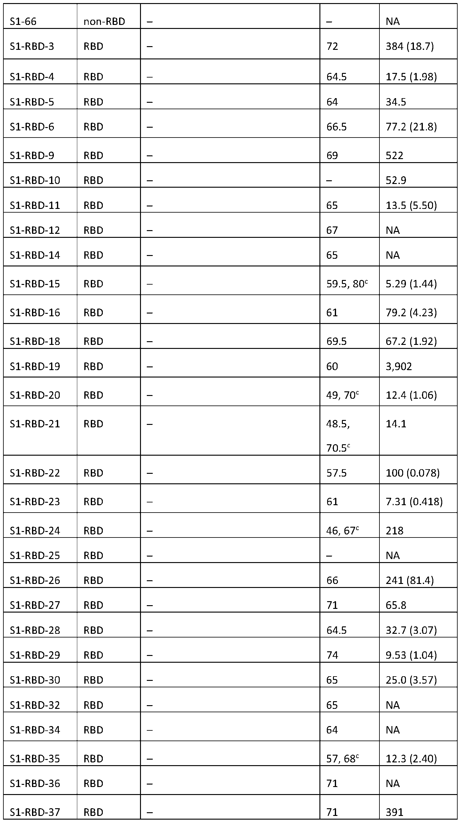 S1-RBD-38 RBD – 68.5 84.6 (22.7) 1RBD RBD
 S2-39 S2 – 58 NA 24 2 E 4 1E 141E 1 12 2
 CoV2-YD-20 S2 – –
 REFERENCES 1. Fauci, A. S. (2021) The story behind COVID-19 vaccines. Science 372, 109 2. Ball, P. (2021) The lightning-fast quest for COVID vaccines - and what it means for other diseases. Nature 589, 16-18 3. Adam, D. (2022) The pandemic’s true death toll: millions more than official counts. Nature 601, 312-315 4. Ritchie, H., Mathieu, E., Rodés-Guirao, L., Appel, C., Giattino, C., Esteban, O.- O., Hasell, J., Macdonal, B., Beltekian, D., and Roser, M. (2020) Coronavirus Pandemic (COVID-19). OurWorldInData.org 5. Shaman, J., and Galanti, M. (2020) Will SARS-CoV-2 become endemic? Science 370, 527-529 6. Antia, R., and Halloran, M. E. (2021) Transition to endemicity: Understanding COVID-19. Immunity 54, 2172-2176 7. Letko, M., Marzi, A., and Munster, V. (2020) Functional assessment of cell entry and receptor usage for SARS-CoV-2 and other lineage B betacoronaviruses. Nat Microbiol 5, 562-569 8. Watanabe, Y., Allen, J. D., Wrapp, D., McLellan, J. S., and Crispin, M. (2020) Site-specific glycan analysis of the SARS-CoV-2 spike. Science 369, 330-333 9. Hsieh, C. L., Goldsmith, J. A., Schaub, J. M., DiVenere, A. M., Kuo, H. C., Javanmardi, K., Le, K. C., Wrapp, D., Lee, A. G., Liu, Y., Chou, C. W., Byrne, P. O., Hjorth, C. K., Johnson, N. V., Ludes-Meyers, J., Nguyen, A. W., Park, J., Wang, N., Amengor, D., Lavinder, J. J., Ippolito, G. C., Maynard, J. A., Finkelstein, I. J., and McLellan, J. S. (2020) Structure-based design of prefusion-stabilized SARS-CoV-2 spikes. Science 369, 1501-1505 10. Zhou, P., Yang, X. L., Wang, X. G., Hu, B., Zhang, L., Zhang, W., Si, H. R., Zhu, Y., Li, B., Huang, C. L., Chen, H. D., Chen, J., Luo, Y., Guo, H., Jiang, R. D., Liu, M. Q., Chen, Y., Shen, X. R., Wang, X., Zheng, X. S., Zhao, K., Chen, Q. J., Deng, F., Liu, L. L., Yan, B., Zhan, F. X., Wang, Y. Y., Xiao, G. F., and Shi, Z. L. (2020) A pneumonia outbreak associated with a new coronavirus of probable bat origin. Nature 579, 270-273 11. Wrapp, D., Wang, N., Corbett, K. S., Goldsmith, J. A., Hsieh, C. L., Abiona, O., Graham, B. S., and McLellan, J. S. (2020) Cryo-EM structure of the 2019-nCoV spike in the prefusion conformation. Science 367, 1260-1263
12. Walls, A. C., Park, Y. J., Tortorici, M. A., Wall, A., McGuire, A. T., and Veesler, D. (2020) Structure, Function, and Antigenicity of the SARS-CoV-2 Spike Glycoprotein. Cell 181, 281-292 e286 13. Chmielewska, A. M., Czarnota, A., Bienkowska-Szewczyk, K., and Grzyb, K. (2021) Immune response against SARS-CoV-2 variants: the role of neutralization assays. NPJ Vaccines 6, 142 14. Planas, D., Veyer, D., Baidaliuk, A., Staropoli, I., Guivel-Benhassine, F., Rajah, M. M., Planchais, C., Porrot, F., Robillard, N., Puech, J., Prot, M., Gallais, F., Gantner, P., Velay, A., Le Guen, J., Kassis-Chikhani, N., Edriss, D., Belec, L., Seve, A., Courtellemont, L., Pere, H., Hocqueloux, L., Fafi-Kremer, S., Prazuck, T., Mouquet, H., Bruel, T., Simon-Loriere, E., Rey, F. A., and Schwartz, O. (2021) Reduced sensitivity of SARS-CoV-2 variant Delta to antibody neutralization. Nature 596, 276-280 15. Wilhelm, A., Widera, M., Grikscheit, K., Toptan, T., Schenk, B., Pallas, C., Metzler, M., Kohmer, N., Hoehl, S., Helfritz, F. A., Wolf, T., Goetsch, U., and Ciesek, S. (2021) Reduced Neutralization of SARS-CoV-2 Omicron Variant by Vaccine Sera and Monoclonal Antibodies. medRxiv 16. Cameroni, E., Bowen, J. E., Rosen, L. E., Saliba, C., Zepeda, S. K., Culap, K., Pinto, D., VanBlargan, L. A., De Marco, A., di Iulio, J., Zatta, F., Kaiser, H., Noack, J., Farhat, N., Czudnochowski, N., Havenar-Daughton, C., Sprouse, K. R., Dillen, J. R., Powell, A. E., Chen, A., Maher, C., Yin, L., Sun, D., Soriaga, L., Bassi, J., Silacci-Fregni, C., Gustafsson, C., Franko, N. M., Logue, J., Iqbal, N. T., Mazzitelli, I., Geffner, J., Grifantini, R., Chu, H., Gori, A., Riva, A., Giannini, O., Ceschi, A., Ferrari, P., Cippa, P. E., Franzetti-Pellanda, A., Garzoni, C., Halfmann, P. J., Kawaoka, Y., Hebner, C., Purcell, L. A., Piccoli, L., Pizzuto, M. S., Walls, A. C., Diamond, M. S., Telenti, A., Virgin, H. W., Lanzavecchia, A., Snell, G., Veesler, D., and Corti, D. (2022) Broadly neutralizing antibodies overcome SARS-CoV-2 Omicron antigenic shift. Nature 602, 664-670 17. Farid, S. S., Baron, M., Stamatis, C., Nie, W., and Coffman, J. (2020) Benchmarking biopharmaceutical process development and manufacturing cost contributions to R&D. MAbs 12, 1754999 18. Muyldermans, S. (2021) A guide to: generation and design of nanobodies. FEBS J 288, 2084-2102
19. Ciccarese, S., Burger, P. A., Ciani, E., Castelli, V., Linguiti, G., Plasil, M., Massari, S., Horin, P., and Antonacci, R. (2019) The Camel Adaptive Immune Receptors Repertoire as a Singular Example of Structural and Functional Genomics. Front Genet 10, 997 20. Mast, F. D., Fridy, P. C., Ketaren, N. E., Wang, J., Jacobs, E. Y., Olivier, J. P., Sanyal, T., Molloy, K. R., Schmidt, F., Rutkowska, M., Weisblum, Y., Rich, L. M., Vanderwall, E. R., Dambrauskas, N., Vigdorovich, V., Keegan, S., Jiler, J. B., Stein, M. E., Olinares, P. D. B., Herlands, L., Hatziioannou, T., Sather, D. N., Debley, J. S., Fenyo, D., Sali, A., Bieniasz, P. D., Aitchison, J. D., Chait, B. T., and Rout, M. P. (2021) Highly synergistic combinations of nanobodies that target SARS-CoV-2 and are resistant to escape. Elife 10 21. Muyldermans, S. (2013) Nanobodies: natural single-domain antibodies. Annu Rev Biochem 82, 775-797 22. Fridy, P. C., Li, Y., Keegan, S., Thompson, M. K., Nudelman, I., Scheid, J. F., Oeffinger, M., Nussenzweig, M. C., Fenyo, D., Chait, B. T., and Rout, M. P. (2014) A robust pipeline for rapid production of versatile nanobody repertoires. Nat Methods 11, 1253-1260 23. Xu, J., Xu, K., Jung, S., Conte, A., Lieberman, J., Muecksch, F., Lorenzi, J. C. C., Park, S., Schmidt, F., Wang, Z., Huang, Y., Luo, Y., Nair, M. S., Wang, P., Schulz, J. E., Tessarollo, L., Bylund, T., Chuang, G. Y., Olia, A. S., Stephens, T., Teng, I. T., Tsybovsky, Y., Zhou, T., Munster, V., Ho, D. D., Hatziioannou, T., Bieniasz, P. D., Nussenzweig, M. C., Kwong, P. D., and Casellas, R. (2021) Nanobodies from camelid mice and llamas neutralize SARS-CoV-2 variants. Nature 595, 278-282 24. Tang, Q., Owens, R. J., and Naismith, J. H. (2021) Structural Biology of Nanobodies against the Spike Protein of SARS-CoV-2. Viruses 13 25. Liu, H., Yuan, M., Huang, D., Bangaru, S., Zhao, F., Lee, C. D., Peng, L., Barman, S., Zhu, X., Nemazee, D., Burton, D. R., van Gils, M. J., Sanders, R. W., Kornau, H. C., Reincke, S. M., Pruss, H., Kreye, J., Wu, N. C., Ward, A. B., and Wilson, I. A. (2021) A combination of cross-neutralizing antibodies synergizes to prevent SARS-CoV-2 and SARS- CoV pseudovirus infection. Cell Host Microbe 29, 806-818 e806 26. Dong, J., Huang, B., Jia, Z., Wang, B., Gallolu Kankanamalage, S., Titong, A., and Liu, Y. (2020) Development of multi-specific humanized llama antibodies blocking SARS- CoV-2/ACE2 interaction with high affinity and avidity. Emerg Microbes Infect 9, 1034-1036 27. Van Heeke, G., Allosery, K., De Brabandere, V., De Smedt, T., Detalle, L., and de Fougerolles, A. (2017) Nanobodies(R) as inhaled biotherapeutics for lung diseases. Pharmacol Ther 169, 47-56
28. Nambulli, S., Xiang, Y., Tilston-Lunel, N. L., Rennick, L. J., Sang, Z., Klimstra, W. B., Reed, D. S., Crossland, N. A., Shi, Y., and Duprex, W. P. (2021) Inhalable Nanobody (PiN-21) prevents and treats SARS-CoV-2 infections in Syrian hamsters at ultra-low doses. Sci Adv 7 29. Revets, H., De Baetselier, P., and Muyldermans, S. (2005) Nanobodies as novel agents for cancer therapy. Expert Opin Biol Ther 5, 111-124 30. Jovcevska, I., and Muyldermans, S. (2020) The Therapeutic Potential of Nanobodies. BioDrugs 34, 11-26 31. Bannas, P., Hambach, J., and Koch-Nolte, F. (2017) Nanobodies and Nanobody-Based Human Heavy Chain Antibodies As Antitumor Therapeutics. Front Immunol 8, 1603 32. Peeling, R. W., and McNerney, R. (2014) Emerging technologies in point-of- care molecular diagnostics for resource-limited settings. Expert Rev Mol Diagn 14, 525-534 33. Thompson, M. K., Fridy, P. C., Keegan, S., Chait, B. T., Fenyo, D., and Rout, M. P. (2016) Optimizing selection of large animals for antibody production by screening immune response to standard vaccines. J Immunol Methods 430, 56-60 34. McMahon, C., Baier, A. S., Pascolutti, R., Wegrecki, M., Zheng, S., Ong, J. X., Erlandson, S. C., Hilger, D., Rasmussen, S. G. F., Ring, A. M., Manglik, A., and Kruse, A. C. (2018) Yeast surface display platform for rapid discovery of conformationally selective nanobodies. Nat Struct Mol Biol 25, 289-296 35. Romao, E., Morales-Yanez, F., Hu, Y., Crauwels, M., De Pauw, P., Hassanzadeh, G. G., Devoogdt, N., Ackaert, C., Vincke, C., and Muyldermans, S. (2016) Identification of Useful Nanobodies by Phage Display of Immune Single Domain Libraries Derived from Camelid Heavy Chain Antibodies. Curr Pharm Des 22, 6500-6518 36. Romao, E., Poignavent, V., Vincke, C., Ritzenthaler, C., Muyldermans, S., and Monsion, B. (2018) Construction of High-Quality Camel Immune Antibody Libraries. Methods Mol Biol 1701, 169-187 37. Roth, L., Krah, S., Klemm, J., Gunther, R., Toleikis, L., Busch, M., Becker, S., and Zielonka, S. (2020) Isolation of Antigen-Specific VHH Single-Domain Antibodies by Combining Animal Immunization with Yeast Surface Display. Methods Mol Biol 2070, 173- 189 38. Schoof, M., Faust, B., Saunders, R. A., Sangwan, S., Rezelj, V., Hoppe, N., Boone, M., Billesbolle, C. B., Puchades, C., Azumaya, C. M., Kratochvil, H. T., Zimanyi, M.,
Deshpande, I., Liang, J., Dickinson, S., Nguyen, H. C., Chio, C. M., Merz, G. E., Thompson, M. C., Diwanji, D., Schaefer, K., Anand, A. A., Dobzinski, N., Zha, B. S., Simoneau, C. R., Leon, K., White, K. M., Chio, U. S., Gupta, M., Jin, M., Li, F., Liu, Y., Zhang, K., Bulkley, D., Sun, M., Smith, A. M., Rizo, A. N., Moss, F., Brilot, A. F., Pourmal, S., Trenker, R., Pospiech, T., Gupta, S., Barsi-Rhyne, B., Belyy, V., Barile-Hill, A. W., Nock, S., Liu, Y., Krogan, N. J., Ralston, C. Y., Swaney, D. L., Garcia-Sastre, A., Ott, M., Vignuzzi, M., Consortium, Q. S. B., Walter, P., and Manglik, A. (2020) An ultrapotent synthetic nanobody neutralizes SARS-CoV-2 by stabilizing inactive Spike. Science 370, 1473-1479 39. Uchanski, T., Zogg, T., Yin, J., Yuan, D., Wohlkonig, A., Fischer, B., Rosenbaum, D. M., Kobilka, B. K., Pardon, E., and Steyaert, J. (2019) An improved yeast surface display platform for the screening of nanobody immune libraries. Sci Rep 9, 382 40. Timney, B. L., Tetenbaum-Novatt, J., Agate, D. S., Williams, R., Zhang, W., Chait, B. T., and Rout, M. P. (2006) Simple kinetic relationships and nonspecific competition govern nuclear import rates in vivo. J Cell Biol 175, 579-593 41. Mitchell, L. S., and Colwell, L. J. (2018) Comparative analysis of nanobody sequence and structure data. Proteins 86, 697-706 42. Zavrtanik, U., Lukan, J., Loris, R., Lah, J., and Hadzi, S. (2018) Structural Basis of Epitope Recognition by Heavy-Chain Camelid Antibodies. J Mol Biol 430, 4369-4386 43. Fridy, P. C., Thompson, M. K., Ketaren, N. E., and Rout, M. P. (2015) Engineered high-affinity nanobodies recognizing staphylococcal Protein A and suitable for native isolation of protein complexes. Anal Biochem 477, 92-94 44. Schneider, T. D., and Stephens, R. M. (1990) Sequence logos: a new way to display consensus sequences. Nucleic Acids Res 18, 6097-6100 45. Greaney, A. J., Starr, T. N., Gilchuk, P., Zost, S. J., Binshtein, E., Loes, A. N., Hilton, S. K., Huddleston, J., Eguia, R., Crawford, K. H. D., Dingens, A. S., Nargi, R. S., Sutton, R. E., Suryadevara, N., Rothlauf, P. W., Liu, Z., Whelan, S. P. J., Carnahan, R. H., Crowe, J. E., Jr., and Bloom, J. D. (2021) Complete Mapping of Mutations to the SARS-CoV- 2 Spike Receptor-Binding Domain that Escape Antibody Recognition. Cell Host Microbe 29, 44-57 e49 46. Wang, L., Shi, W., Chappell, J. D., Joyce, M. G., Zhang, Y., Kanekiyo, M., Becker, M. M., van Doremalen, N., Fischer, R., Wang, N., Corbett, K. S., Choe, M., Mason, R. D., Van Galen, J. G., Zhou, T., Saunders, K. O., Tatti, K. M., Haynes, L. M., Kwong, P. D., Modjarrad, K., Kong, W. P., McLellan, J. S., Denison, M. R., Munster, V. J., Mascola, J. R.,
and Graham, B. S. (2018) Importance of Neutralizing Monoclonal Antibodies Targeting Multiple Antigenic Sites on the Middle East Respiratory Syndrome Coronavirus Spike Glycoprotein To Avoid Neutralization Escape. J Virol 92 47. Xiang, Y., Nambulli, S., Xiao, Z., Liu, H., Sang, Z., Duprex, W. P., Schneidman-Duhovny, D., Zhang, C., and Shi, Y. (2020) Versatile and multivalent nanobodies efficiently neutralize SARS-CoV-2. Science 370, 1479-1484 48. Huo, J., Le Bas, A., Ruza, R. R., Duyvesteyn, H. M. E., Mikolajek, H., Malinauskas, T., Tan, T. K., Rijal, P., Dumoux, M., Ward, P. N., Ren, J., Zhou, D., Harrison, P. J., Weckener, M., Clare, D. K., Vogirala, V. K., Radecke, J., Moynie, L., Zhao, Y., Gilbert- Jaramillo, J., Knight, M. L., Tree, J. A., Buttigieg, K. R., Coombes, N., Elmore, M. J., Carroll, M. W., Carrique, L., Shah, P. N. M., James, W., Townsend, A. R., Stuart, D. I., Owens, R. J., and Naismith, J. H. (2020) Neutralizing nanobodies bind SARS-CoV-2 spike RBD and block interaction with ACE2. Nat Struct Mol Biol 27, 846-854 49. Pymm, P., Adair, A., Chan, L. J., Cooney, J. P., Mordant, F. L., Allison, C. C., Lopez, E., Haycroft, E. R., O’Neill, M. T., Tan, L. L., Dietrich, M. H., Drew, D., Doerflinger, M., Dengler, M. A., Scott, N. E., Wheatley, A. K., Gherardin, N. A., Venugopal, H., Cromer, D., Davenport, M. P., Pickering, R., Godfrey, D. I., Purcell, D. F. J., Kent, S. J., Chung, A. W., Subbarao, K., Pellegrini, M., Glukhova, A., and Tham, W. H. (2021) Nanobody cocktails potently neutralize SARS-CoV-2 D614G N501Y variant and protect mice. Proc Natl Acad Sci U S A 118 50. Starr, T. N., Greaney, A. J., Hannon, W. W., Loes, A. N., Hauser, K., Dillen, J. R., Ferri, E., Farrell, A. G., Dadonaite, B., McCallum, M., Matreyek, K. A., Corti, D., Veesler, D., Snell, G., and Bloom, J. D. (2022) Shifting mutational constraints in the SARS-CoV-2 receptor-binding domain during viral evolution. Science 377, 420-424 51. Greaney, A. J., Starr, T. N., Barnes, C. O., Weisblum, Y., Schmidt, F., Caskey, M., Gaebler, C., Cho, A., Agudelo, M., Finkin, S., Wang, Z., Poston, D., Muecksch, F., Hatziioannou, T., Bieniasz, P. D., Robbiani, D. F., Nussenzweig, M. C., Bjorkman, P. J., and Bloom, J. D. (2021) Mapping mutations to the SARS-CoV-2 RBD that escape binding by different classes of antibodies. Nat Commun 12, 4196 52. Sheedy, C., Yau, K. Y., Hirama, T., MacKenzie, C. R., and Hall, J. C. (2006) Selection, characterization, and CDR shuffling of naive llama single-domain antibodies selected against auxin and their cross-reactivity with auxinic herbicides from four chemical families. J Agric Food Chem 54, 3668-3678
53. Zupancic, J. M., Desai, A. A., and Tessier, P. M. (2022) Facile isolation of high- affinity nanobodies from synthetic libraries using CDR-swapping mutagenesis. STAR Protoc 3, 101101 54. Tsukahara, N., Murakami, A., Motohashi, M., Nakayama, H., Kondo, Y., Ito, Y., Azuma, T., and Kishimoto, H. (2022) An alpaca single-domain antibody (VHH) phage display library constructed by CDR shuffling provided high-affinity VHHs against desired protein antigens. Int Immunol 34, 421-434 55. Benatuil, L., Perez, J. M., Belk, J., and Hsieh, C. M. (2010) An improved yeast transformation method for the generation of very large human antibody libraries. Protein Eng Des Sel 23, 155-159 56. Zupancic, J. M., Desai, A. A., Schardt, J. S., Pornnoppadol, G., Makowski, E. K., Smith, M. D., Kennedy, A. A., Garcia de Mattos Barbosa, M., Cascalho, M., Lanigan, T. M., Tai, A. W., and Tessier, P. M. (2021) Directed evolution of potent neutralizing nanobodies against SARS-CoV-2 using CDR-swapping mutagenesis. Cell Chem Biol 28, 1379-1388 e1377 57. Obado, S. O., Field, M. C., Chait, B. T., and Rout, M. P. (2016) High-Efficiency Isolation of Nuclear Envelope Protein Complexes from Trypanosomes. Methods Mol Biol 1411, 67-80 58. Kim, S. J., Fernandez-Martinez, J., Nudelman, I., Shi, Y., Zhang, W., Raveh, B., Herricks, T., Slaughter, B. D., Hogan, J. A., Upla, P., Chemmama, I. E., Pellarin, R., Echeverria, I., Shivaraju, M., Chaudhury, A. S., Wang, J., Williams, R., Unruh, J. R., Greenberg, C. H., Jacobs, E. Y., Yu, Z., de la Cruz, M. J., Mironska, R., Stokes, D. L., Aitchison, J. D., Jarrold, M. F., Gerton, J. L., Ludtke, S. J., Akey, C. W., Chait, B. T., Sali, A., and Rout, M. P. (2018) Integrative structure and functional anatomy of a nuclear pore complex. Nature 555, 475-482 59. Shaner, M. C., Blair, I. M., and Schneider, T. D. (1993) Sequence logos: A powerful, yet simple, tool. Proceedings of the twenty-sixth annual Hawaii international conference on system sciences.. in Architecture and biotechnology computing (al., T. N. M. e. ed.), IEEE Computer Society Press, Los Alamitos, CA. pp 813–821 60. Nei, M., and Li, W. H. (1979) Mathematical model for studying genetic variation in terms of restriction endonucleases. Proc Natl Acad Sci U S A 76, 5269-5273
While illustrative examples have been illustrated and described, it will be appreciated that various changes can be made therein without departing from the spirit and scope of the invention.
REFERENCES 1. Fauci, A. S. (2021) The story behind COVID-19 vaccines. Science 372, 109 2. Ball, P. (2021) The lightning-fast quest for COVID vaccines - and what it means for other diseases. Nature 589, 16-18 3. Adam, D. (2022) The pandemic’s true death toll: millions more than official counts. Nature 601, 312-315 4. Ritchie, H., Mathieu, E., Rodés-Guirao, L., Appel, C., Giattino, C., Esteban, O.- O., Hasell, J., Macdonal, B., Beltekian, D., and Roser, M. (2020) Coronavirus Pandemic (COVID-19). OurWorldInData.org 5. Shaman, J., and Galanti, M. (2020) Will SARS-CoV-2 become endemic? Science 370, 527-529 6. Antia, R., and Halloran, M. E. (2021) Transition to endemicity: Understanding COVID-19. Immunity 54, 2172-2176 7. Letko, M., Marzi, A., and Munster, V. (2020) Functional assessment of cell entry and receptor usage for SARS-CoV-2 and other lineage B betacoronaviruses. Nat Microbiol 5, 562-569 8. Watanabe, Y., Allen, J. D., Wrapp, D., McLellan, J. S., and Crispin, M. (2020) Site-specific glycan analysis of the SARS-CoV-2 spike. Science 369, 330-333 9. Hsieh, C. L., Goldsmith, J. A., Schaub, J. M., DiVenere, A. M., Kuo, H. C., Javanmardi, K., Le, K. C., Wrapp, D., Lee, A. G., Liu, Y., Chou, C. W., Byrne, P. O., Hjorth, C. K., Johnson, N. V., Ludes-Meyers, J., Nguyen, A. W., Park, J., Wang, N., Amengor, D., Lavinder, J. J., Ippolito, G. C., Maynard, J. A., Finkelstein, I. J., and McLellan, J. S. (2020) Structure-based design of prefusion-stabilized SARS-CoV-2 spikes. Science 369, 1501-1505 10. Zhou, P., Yang, X. L., Wang, X. G., Hu, B., Zhang, L., Zhang, W., Si, H. R., Zhu, Y., Li, B., Huang, C. L., Chen, H. D., Chen, J., Luo, Y., Guo, H., Jiang, R. D., Liu, M. Q., Chen, Y., Shen, X. R., Wang, X., Zheng, X. S., Zhao, K., Chen, Q. J., Deng, F., Liu, L. L., Yan, B., Zhan, F. X., Wang, Y. Y., Xiao, G. F., and Shi, Z. L. (2020) A pneumonia outbreak associated with a new coronavirus of probable bat origin. Nature 579, 270-273 11. Wrapp, D., Wang, N., Corbett, K. S., Goldsmith, J. A., Hsieh, C. L., Abiona, O., Graham, B. S., and McLellan, J. S. (2020) Cryo-EM structure of the 2019-nCoV spike in the prefusion conformation. Science 367, 1260-1263
12. Walls, A. C., Park, Y. J., Tortorici, M. A., Wall, A., McGuire, A. T., and Veesler, D. (2020) Structure, Function, and Antigenicity of the SARS-CoV-2 Spike Glycoprotein. Cell 181, 281-292 e286 13. Chmielewska, A. M., Czarnota, A., Bienkowska-Szewczyk, K., and Grzyb, K. (2021) Immune response against SARS-CoV-2 variants: the role of neutralization assays. NPJ Vaccines 6, 142 14. Planas, D., Veyer, D., Baidaliuk, A., Staropoli, I., Guivel-Benhassine, F., Rajah, M. M., Planchais, C., Porrot, F., Robillard, N., Puech, J., Prot, M., Gallais, F., Gantner, P., Velay, A., Le Guen, J., Kassis-Chikhani, N., Edriss, D., Belec, L., Seve, A., Courtellemont, L., Pere, H., Hocqueloux, L., Fafi-Kremer, S., Prazuck, T., Mouquet, H., Bruel, T., Simon-Loriere, E., Rey, F. A., and Schwartz, O. (2021) Reduced sensitivity of SARS-CoV-2 variant Delta to antibody neutralization. Nature 596, 276-280 15. Wilhelm, A., Widera, M., Grikscheit, K., Toptan, T., Schenk, B., Pallas, C., Metzler, M., Kohmer, N., Hoehl, S., Helfritz, F. A., Wolf, T., Goetsch, U., and Ciesek, S. (2021) Reduced Neutralization of SARS-CoV-2 Omicron Variant by Vaccine Sera and Monoclonal Antibodies. medRxiv 16. Cameroni, E., Bowen, J. E., Rosen, L. E., Saliba, C., Zepeda, S. K., Culap, K., Pinto, D., VanBlargan, L. A., De Marco, A., di Iulio, J., Zatta, F., Kaiser, H., Noack, J., Farhat, N., Czudnochowski, N., Havenar-Daughton, C., Sprouse, K. R., Dillen, J. R., Powell, A. E., Chen, A., Maher, C., Yin, L., Sun, D., Soriaga, L., Bassi, J., Silacci-Fregni, C., Gustafsson, C., Franko, N. M., Logue, J., Iqbal, N. T., Mazzitelli, I., Geffner, J., Grifantini, R., Chu, H., Gori, A., Riva, A., Giannini, O., Ceschi, A., Ferrari, P., Cippa, P. E., Franzetti-Pellanda, A., Garzoni, C., Halfmann, P. J., Kawaoka, Y., Hebner, C., Purcell, L. A., Piccoli, L., Pizzuto, M. S., Walls, A. C., Diamond, M. S., Telenti, A., Virgin, H. W., Lanzavecchia, A., Snell, G., Veesler, D., and Corti, D. (2022) Broadly neutralizing antibodies overcome SARS-CoV-2 Omicron antigenic shift. Nature 602, 664-670 17. Farid, S. S., Baron, M., Stamatis, C., Nie, W., and Coffman, J. (2020) Benchmarking biopharmaceutical process development and manufacturing cost contributions to R&D. MAbs 12, 1754999 18. Muyldermans, S. (2021) A guide to: generation and design of nanobodies. FEBS J 288, 2084-2102
19. Ciccarese, S., Burger, P. A., Ciani, E., Castelli, V., Linguiti, G., Plasil, M., Massari, S., Horin, P., and Antonacci, R. (2019) The Camel Adaptive Immune Receptors Repertoire as a Singular Example of Structural and Functional Genomics. Front Genet 10, 997 20. Mast, F. D., Fridy, P. C., Ketaren, N. E., Wang, J., Jacobs, E. Y., Olivier, J. P., Sanyal, T., Molloy, K. R., Schmidt, F., Rutkowska, M., Weisblum, Y., Rich, L. M., Vanderwall, E. R., Dambrauskas, N., Vigdorovich, V., Keegan, S., Jiler, J. B., Stein, M. E., Olinares, P. D. B., Herlands, L., Hatziioannou, T., Sather, D. N., Debley, J. S., Fenyo, D., Sali, A., Bieniasz, P. D., Aitchison, J. D., Chait, B. T., and Rout, M. P. (2021) Highly synergistic combinations of nanobodies that target SARS-CoV-2 and are resistant to escape. Elife 10 21. Muyldermans, S. (2013) Nanobodies: natural single-domain antibodies. Annu Rev Biochem 82, 775-797 22. Fridy, P. C., Li, Y., Keegan, S., Thompson, M. K., Nudelman, I., Scheid, J. F., Oeffinger, M., Nussenzweig, M. C., Fenyo, D., Chait, B. T., and Rout, M. P. (2014) A robust pipeline for rapid production of versatile nanobody repertoires. Nat Methods 11, 1253-1260 23. Xu, J., Xu, K., Jung, S., Conte, A., Lieberman, J., Muecksch, F., Lorenzi, J. C. C., Park, S., Schmidt, F., Wang, Z., Huang, Y., Luo, Y., Nair, M. S., Wang, P., Schulz, J. E., Tessarollo, L., Bylund, T., Chuang, G. Y., Olia, A. S., Stephens, T., Teng, I. T., Tsybovsky, Y., Zhou, T., Munster, V., Ho, D. D., Hatziioannou, T., Bieniasz, P. D., Nussenzweig, M. C., Kwong, P. D., and Casellas, R. (2021) Nanobodies from camelid mice and llamas neutralize SARS-CoV-2 variants. Nature 595, 278-282 24. Tang, Q., Owens, R. J., and Naismith, J. H. (2021) Structural Biology of Nanobodies against the Spike Protein of SARS-CoV-2. Viruses 13 25. Liu, H., Yuan, M., Huang, D., Bangaru, S., Zhao, F., Lee, C. D., Peng, L., Barman, S., Zhu, X., Nemazee, D., Burton, D. R., van Gils, M. J., Sanders, R. W., Kornau, H. C., Reincke, S. M., Pruss, H., Kreye, J., Wu, N. C., Ward, A. B., and Wilson, I. A. (2021) A combination of cross-neutralizing antibodies synergizes to prevent SARS-CoV-2 and SARS- CoV pseudovirus infection. Cell Host Microbe 29, 806-818 e806 26. Dong, J., Huang, B., Jia, Z., Wang, B., Gallolu Kankanamalage, S., Titong, A., and Liu, Y. (2020) Development of multi-specific humanized llama antibodies blocking SARS- CoV-2/ACE2 interaction with high affinity and avidity. Emerg Microbes Infect 9, 1034-1036 27. Van Heeke, G., Allosery, K., De Brabandere, V., De Smedt, T., Detalle, L., and de Fougerolles, A. (2017) Nanobodies(R) as inhaled biotherapeutics for lung diseases. Pharmacol Ther 169, 47-56
28. Nambulli, S., Xiang, Y., Tilston-Lunel, N. L., Rennick, L. J., Sang, Z., Klimstra, W. B., Reed, D. S., Crossland, N. A., Shi, Y., and Duprex, W. P. (2021) Inhalable Nanobody (PiN-21) prevents and treats SARS-CoV-2 infections in Syrian hamsters at ultra-low doses. Sci Adv 7 29. Revets, H., De Baetselier, P., and Muyldermans, S. (2005) Nanobodies as novel agents for cancer therapy. Expert Opin Biol Ther 5, 111-124 30. Jovcevska, I., and Muyldermans, S. (2020) The Therapeutic Potential of Nanobodies. BioDrugs 34, 11-26 31. Bannas, P., Hambach, J., and Koch-Nolte, F. (2017) Nanobodies and Nanobody-Based Human Heavy Chain Antibodies As Antitumor Therapeutics. Front Immunol 8, 1603 32. Peeling, R. W., and McNerney, R. (2014) Emerging technologies in point-of- care molecular diagnostics for resource-limited settings. Expert Rev Mol Diagn 14, 525-534 33. Thompson, M. K., Fridy, P. C., Keegan, S., Chait, B. T., Fenyo, D., and Rout, M. P. (2016) Optimizing selection of large animals for antibody production by screening immune response to standard vaccines. J Immunol Methods 430, 56-60 34. McMahon, C., Baier, A. S., Pascolutti, R., Wegrecki, M., Zheng, S., Ong, J. X., Erlandson, S. C., Hilger, D., Rasmussen, S. G. F., Ring, A. M., Manglik, A., and Kruse, A. C. (2018) Yeast surface display platform for rapid discovery of conformationally selective nanobodies. Nat Struct Mol Biol 25, 289-296 35. Romao, E., Morales-Yanez, F., Hu, Y., Crauwels, M., De Pauw, P., Hassanzadeh, G. G., Devoogdt, N., Ackaert, C., Vincke, C., and Muyldermans, S. (2016) Identification of Useful Nanobodies by Phage Display of Immune Single Domain Libraries Derived from Camelid Heavy Chain Antibodies. Curr Pharm Des 22, 6500-6518 36. Romao, E., Poignavent, V., Vincke, C., Ritzenthaler, C., Muyldermans, S., and Monsion, B. (2018) Construction of High-Quality Camel Immune Antibody Libraries. Methods Mol Biol 1701, 169-187 37. Roth, L., Krah, S., Klemm, J., Gunther, R., Toleikis, L., Busch, M., Becker, S., and Zielonka, S. (2020) Isolation of Antigen-Specific VHH Single-Domain Antibodies by Combining Animal Immunization with Yeast Surface Display. Methods Mol Biol 2070, 173- 189 38. Schoof, M., Faust, B., Saunders, R. A., Sangwan, S., Rezelj, V., Hoppe, N., Boone, M., Billesbolle, C. B., Puchades, C., Azumaya, C. M., Kratochvil, H. T., Zimanyi, M.,
Deshpande, I., Liang, J., Dickinson, S., Nguyen, H. C., Chio, C. M., Merz, G. E., Thompson, M. C., Diwanji, D., Schaefer, K., Anand, A. A., Dobzinski, N., Zha, B. S., Simoneau, C. R., Leon, K., White, K. M., Chio, U. S., Gupta, M., Jin, M., Li, F., Liu, Y., Zhang, K., Bulkley, D., Sun, M., Smith, A. M., Rizo, A. N., Moss, F., Brilot, A. F., Pourmal, S., Trenker, R., Pospiech, T., Gupta, S., Barsi-Rhyne, B., Belyy, V., Barile-Hill, A. W., Nock, S., Liu, Y., Krogan, N. J., Ralston, C. Y., Swaney, D. L., Garcia-Sastre, A., Ott, M., Vignuzzi, M., Consortium, Q. S. B., Walter, P., and Manglik, A. (2020) An ultrapotent synthetic nanobody neutralizes SARS-CoV-2 by stabilizing inactive Spike. Science 370, 1473-1479 39. Uchanski, T., Zogg, T., Yin, J., Yuan, D., Wohlkonig, A., Fischer, B., Rosenbaum, D. M., Kobilka, B. K., Pardon, E., and Steyaert, J. (2019) An improved yeast surface display platform for the screening of nanobody immune libraries. Sci Rep 9, 382 40. Timney, B. L., Tetenbaum-Novatt, J., Agate, D. S., Williams, R., Zhang, W., Chait, B. T., and Rout, M. P. (2006) Simple kinetic relationships and nonspecific competition govern nuclear import rates in vivo. J Cell Biol 175, 579-593 41. Mitchell, L. S., and Colwell, L. J. (2018) Comparative analysis of nanobody sequence and structure data. Proteins 86, 697-706 42. Zavrtanik, U., Lukan, J., Loris, R., Lah, J., and Hadzi, S. (2018) Structural Basis of Epitope Recognition by Heavy-Chain Camelid Antibodies. J Mol Biol 430, 4369-4386 43. Fridy, P. C., Thompson, M. K., Ketaren, N. E., and Rout, M. P. (2015) Engineered high-affinity nanobodies recognizing staphylococcal Protein A and suitable for native isolation of protein complexes. Anal Biochem 477, 92-94 44. Schneider, T. D., and Stephens, R. M. (1990) Sequence logos: a new way to display consensus sequences. Nucleic Acids Res 18, 6097-6100 45. Greaney, A. J., Starr, T. N., Gilchuk, P., Zost, S. J., Binshtein, E., Loes, A. N., Hilton, S. K., Huddleston, J., Eguia, R., Crawford, K. H. D., Dingens, A. S., Nargi, R. S., Sutton, R. E., Suryadevara, N., Rothlauf, P. W., Liu, Z., Whelan, S. P. J., Carnahan, R. H., Crowe, J. E., Jr., and Bloom, J. D. (2021) Complete Mapping of Mutations to the SARS-CoV- 2 Spike Receptor-Binding Domain that Escape Antibody Recognition. Cell Host Microbe 29, 44-57 e49 46. Wang, L., Shi, W., Chappell, J. D., Joyce, M. G., Zhang, Y., Kanekiyo, M., Becker, M. M., van Doremalen, N., Fischer, R., Wang, N., Corbett, K. S., Choe, M., Mason, R. D., Van Galen, J. G., Zhou, T., Saunders, K. O., Tatti, K. M., Haynes, L. M., Kwong, P. D., Modjarrad, K., Kong, W. P., McLellan, J. S., Denison, M. R., Munster, V. J., Mascola, J. R.,
and Graham, B. S. (2018) Importance of Neutralizing Monoclonal Antibodies Targeting Multiple Antigenic Sites on the Middle East Respiratory Syndrome Coronavirus Spike Glycoprotein To Avoid Neutralization Escape. J Virol 92 47. Xiang, Y., Nambulli, S., Xiao, Z., Liu, H., Sang, Z., Duprex, W. P., Schneidman-Duhovny, D., Zhang, C., and Shi, Y. (2020) Versatile and multivalent nanobodies efficiently neutralize SARS-CoV-2. Science 370, 1479-1484 48. Huo, J., Le Bas, A., Ruza, R. R., Duyvesteyn, H. M. E., Mikolajek, H., Malinauskas, T., Tan, T. K., Rijal, P., Dumoux, M., Ward, P. N., Ren, J., Zhou, D., Harrison, P. J., Weckener, M., Clare, D. K., Vogirala, V. K., Radecke, J., Moynie, L., Zhao, Y., Gilbert- Jaramillo, J., Knight, M. L., Tree, J. A., Buttigieg, K. R., Coombes, N., Elmore, M. J., Carroll, M. W., Carrique, L., Shah, P. N. M., James, W., Townsend, A. R., Stuart, D. I., Owens, R. J., and Naismith, J. H. (2020) Neutralizing nanobodies bind SARS-CoV-2 spike RBD and block interaction with ACE2. Nat Struct Mol Biol 27, 846-854 49. Pymm, P., Adair, A., Chan, L. J., Cooney, J. P., Mordant, F. L., Allison, C. C., Lopez, E., Haycroft, E. R., O’Neill, M. T., Tan, L. L., Dietrich, M. H., Drew, D., Doerflinger, M., Dengler, M. A., Scott, N. E., Wheatley, A. K., Gherardin, N. A., Venugopal, H., Cromer, D., Davenport, M. P., Pickering, R., Godfrey, D. I., Purcell, D. F. J., Kent, S. J., Chung, A. W., Subbarao, K., Pellegrini, M., Glukhova, A., and Tham, W. H. (2021) Nanobody cocktails potently neutralize SARS-CoV-2 D614G N501Y variant and protect mice. Proc Natl Acad Sci U S A 118 50. Starr, T. N., Greaney, A. J., Hannon, W. W., Loes, A. N., Hauser, K., Dillen, J. R., Ferri, E., Farrell, A. G., Dadonaite, B., McCallum, M., Matreyek, K. A., Corti, D., Veesler, D., Snell, G., and Bloom, J. D. (2022) Shifting mutational constraints in the SARS-CoV-2 receptor-binding domain during viral evolution. Science 377, 420-424 51. Greaney, A. J., Starr, T. N., Barnes, C. O., Weisblum, Y., Schmidt, F., Caskey, M., Gaebler, C., Cho, A., Agudelo, M., Finkin, S., Wang, Z., Poston, D., Muecksch, F., Hatziioannou, T., Bieniasz, P. D., Robbiani, D. F., Nussenzweig, M. C., Bjorkman, P. J., and Bloom, J. D. (2021) Mapping mutations to the SARS-CoV-2 RBD that escape binding by different classes of antibodies. Nat Commun 12, 4196 52. Sheedy, C., Yau, K. Y., Hirama, T., MacKenzie, C. R., and Hall, J. C. (2006) Selection, characterization, and CDR shuffling of naive llama single-domain antibodies selected against auxin and their cross-reactivity with auxinic herbicides from four chemical families. J Agric Food Chem 54, 3668-3678
53. Zupancic, J. M., Desai, A. A., and Tessier, P. M. (2022) Facile isolation of high- affinity nanobodies from synthetic libraries using CDR-swapping mutagenesis. STAR Protoc 3, 101101 54. Tsukahara, N., Murakami, A., Motohashi, M., Nakayama, H., Kondo, Y., Ito, Y., Azuma, T., and Kishimoto, H. (2022) An alpaca single-domain antibody (VHH) phage display library constructed by CDR shuffling provided high-affinity VHHs against desired protein antigens. Int Immunol 34, 421-434 55. Benatuil, L., Perez, J. M., Belk, J., and Hsieh, C. M. (2010) An improved yeast transformation method for the generation of very large human antibody libraries. Protein Eng Des Sel 23, 155-159 56. Zupancic, J. M., Desai, A. A., Schardt, J. S., Pornnoppadol, G., Makowski, E. K., Smith, M. D., Kennedy, A. A., Garcia de Mattos Barbosa, M., Cascalho, M., Lanigan, T. M., Tai, A. W., and Tessier, P. M. (2021) Directed evolution of potent neutralizing nanobodies against SARS-CoV-2 using CDR-swapping mutagenesis. Cell Chem Biol 28, 1379-1388 e1377 57. Obado, S. O., Field, M. C., Chait, B. T., and Rout, M. P. (2016) High-Efficiency Isolation of Nuclear Envelope Protein Complexes from Trypanosomes. Methods Mol Biol 1411, 67-80 58. Kim, S. J., Fernandez-Martinez, J., Nudelman, I., Shi, Y., Zhang, W., Raveh, B., Herricks, T., Slaughter, B. D., Hogan, J. A., Upla, P., Chemmama, I. E., Pellarin, R., Echeverria, I., Shivaraju, M., Chaudhury, A. S., Wang, J., Williams, R., Unruh, J. R., Greenberg, C. H., Jacobs, E. Y., Yu, Z., de la Cruz, M. J., Mironska, R., Stokes, D. L., Aitchison, J. D., Jarrold, M. F., Gerton, J. L., Ludtke, S. J., Akey, C. W., Chait, B. T., Sali, A., and Rout, M. P. (2018) Integrative structure and functional anatomy of a nuclear pore complex. Nature 555, 475-482 59. Shaner, M. C., Blair, I. M., and Schneider, T. D. (1993) Sequence logos: A powerful, yet simple, tool. Proceedings of the twenty-sixth annual Hawaii international conference on system sciences.. in Architecture and biotechnology computing (al., T. N. M. e. ed.), IEEE Computer Society Press, Los Alamitos, CA. pp 813–821 60. Nei, M., and Li, W. H. (1979) Mathematical model for studying genetic variation in terms of restriction endonucleases. Proc Natl Acad Sci U S A 76, 5269-5273
While illustrative examples have been illustrated and described, it will be appreciated that various changes can be made therein without departing from the spirit and scope of the invention.
Claims
CLAIMS 1. A VHH chain of an anti-Severe Acute Respiratory Syndrome Coronavirus 2 (SARS-CoV-2) nanobody, wherein the amino acid sequence of the VHH chain comprises any one of the sequences as set forth in SEQ ID NOs: 117 – 153.
2. The VHH chain of claim 1, wherein SEQ ID NOs: 121-130, and 150-157 are selective for binding to Receptor Binding Domain (RBD) of SARS-CoV-2 spike protein.
3. The VHH chain of claim 1, wherein SEQ ID NOs: 117-120, 147, and 148 are selective for binding to non-Receptor Binding Domain (RBD) of SARS-CoV-2 spike protein.
4. The VHH chain of claim 1, wherein SEQ ID NOs: 131-146 are selective for binding to S2 domain of the SARS-CoV-2 spike protein.
5. An immunoconjugate comprising: (a) a VHH chain comprising a sequence selected from SEQ ID NOs: 117 – 153; and (b) a conjugating part comprising a detectable marker, a drug, a radionuclide, or an enzyme.
6. A pharmaceutical composition comprising: (a) a VHH chain comprising a sequence selected from SEQ ID NOs: 117 – 153; and (b) a pharmaceutically acceptable carrier.
7. A method comprising administering to an individual who has, is suspected of having, or is at risk of contracting a SARS-CoV-2 infection, a composition comprising a VHH chain of an anti-Severe Acute Respiratory Syndrome Coronavirus 2 (SARS-CoV-2, wherein the amino acid sequence of the VHH chain comprises any one of the sequences as set forth in SEQ ID NOs: 117 – 153.
8. The method of claim 7, wherein the individual has the SARS-CoV-2 infection.
9. The method of claim 8, wherein the individual has been diagnosed with COVID-19.
10. A polynucleotide encoding a VHH chain of an anti-Severe Acute Respiratory Syndrome Coronavirus 2 (SARS-CoV-2), wherein the amino acid sequence of the VHH chain comprises any one of the sequences as set forth in SEQ ID NOs: 117 – 153.
11. The polynucleotide of claim 10, wherein the polynucleotide is present in an expression vector.
12. A method comprising allowing expression of the expression vector of claim 11 by cells in a cell culture, and separating from the cell culture a VHH chain of an anti-Severe Acute Respiratory Syndrome Coronavirus 2 (SARS-CoV-2).
13. A polynucleotide that selectively hybridizes to a polynucleotide encoding an amino acid of any one of SEQ ID NOs: 117 – 153.
Applications Claiming Priority (2)
| Application Number | Priority Date | Filing Date | Title |
|---|---|---|---|
| US202363486316P | 2023-02-22 | 2023-02-22 | |
| US63/486,316 | 2023-02-22 |
Publications (2)
| Publication Number | Publication Date |
|---|---|
| WO2024178232A2 true WO2024178232A2 (en) | 2024-08-29 |
| WO2024178232A3 WO2024178232A3 (en) | 2024-10-24 |
Family
ID=92501801
Family Applications (1)
| Application Number | Title | Priority Date | Filing Date |
|---|---|---|---|
| PCT/US2024/016913 Ceased WO2024178232A2 (en) | 2023-02-22 | 2024-02-22 | Expanding and improving nanobody repertoires: targeting sars-cov-2 |
Country Status (1)
| Country | Link |
|---|---|
| WO (1) | WO2024178232A2 (en) |
Family Cites Families (3)
| Publication number | Priority date | Publication date | Assignee | Title |
|---|---|---|---|---|
| EP4200328A4 (en) * | 2020-08-21 | 2025-02-26 | The Rockefeller University | SINGLE-DOMAIN ANTIBODIES BINDING TO SARS-COV-2 |
| US20240254202A1 (en) * | 2020-12-22 | 2024-08-01 | Consejo Nacional De Investigaciones Científicas Y Técnicas (Conicet) | Llama-derived nanobodies binding the spike protein of novel coronavirus sars-cov-2 with neutralizing activity and application thereof |
| CN114763379B (en) * | 2021-06-30 | 2023-01-20 | 生物岛实验室 | Specific antibody of new coronavirus S protein, preparation method and application thereof |
-
2024
- 2024-02-22 WO PCT/US2024/016913 patent/WO2024178232A2/en not_active Ceased
Also Published As
| Publication number | Publication date |
|---|---|
| WO2024178232A3 (en) | 2024-10-24 |
Similar Documents
| Publication | Publication Date | Title |
|---|---|---|
| CN111995675B (en) | A monoclonal antibody against the RBD region of the new coronavirus SARS-CoV-2 spike protein and its application | |
| Tan et al. | Sequence signatures of two public antibody clonotypes that bind SARS-CoV-2 receptor binding domain | |
| JP7289562B2 (en) | Anti-BCMA single domain antibody and its application | |
| EP2454376B1 (en) | Simultaneous, integrated selection and evolution of antibody/protein performance and expression in production hosts | |
| Sheward et al. | Structural basis of broad SARS-CoV-2 cross-neutralization by affinity-matured public antibodies | |
| EP3435087B1 (en) | Novel methods of protein evolution | |
| CN106459186B (en) | Broadly neutralizing monoclonal antibodies against the ENV region of HIV-1V 2 | |
| Addetia et al. | Therapeutic and vaccine-induced cross-reactive antibodies with effector function against emerging Omicron variants | |
| US20240294613A1 (en) | Cross-neutralizing sars-cov2 antibodies | |
| CN118369338A (en) | Human antibodies and antibody combinations for synergistically neutralizing the novel coronavirus and their applications | |
| Cross et al. | Expanding and improving nanobody repertoires using a yeast display method: Targeting SARS-CoV-2 | |
| DiMuzio et al. | Unbiased interrogation of memory B cells from convalescent COVID-19 patients reveals a broad antiviral humoral response targeting SARS-CoV-2 antigens beyond the spike protein | |
| WO2024178232A2 (en) | Expanding and improving nanobody repertoires: targeting sars-cov-2 | |
| CN117203229A (en) | Coronavirus antibodies and their uses | |
| US20240166727A1 (en) | Human neutralizing antigen specific proteins for spike-rbd of sars-cov-2 | |
| Yang et al. | High-throughput saturation mutagenesis generates a high-affinity antibody against SARS-CoV-2 variants using protein surface display assay on a human cell | |
| JP2019535011A (en) | Cell marker | |
| Haslwanter et al. | A combination of RBD and NTD neutralizing antibodies limits the generation of SARS-CoV-2 spike neutralization-escape mutants | |
| US20220033479A1 (en) | Anti-hepatitis c virus antibodies | |
| Ren et al. | Potent Cross‐neutralizing Antibodies Reveal Vulnerabilities of Henipavirus Fusion Glycoprotein | |
| Chen et al. | Synthetic antibodies in infectious disease | |
| US20040067534A1 (en) | Methods of preparing improved agents by coevolution | |
| Hutchison | Characterizing Antibody Escape Variants in the Respiratory Syncytial Virus Fusion Glycoprotein | |
| Miura | Authorization to Submit Thesis | |
| US20210405026A1 (en) | Method for evaluating the presence of a viral reservoir, and evaluating the efficacy of a drug against said reservoir |
Legal Events
| Date | Code | Title | Description |
|---|---|---|---|
| NENP | Non-entry into the national phase |
Ref country code: DE |
|
| 122 | Ep: pct application non-entry in european phase |
Ref document number: 24761002 Country of ref document: EP Kind code of ref document: A2 |