WO2024142145A1 - 空気漏れ測定システム、空気漏れ測定用装置および空気漏れ測定方法 - Google Patents
空気漏れ測定システム、空気漏れ測定用装置および空気漏れ測定方法 Download PDFInfo
- Publication number
- WO2024142145A1 WO2024142145A1 PCT/JP2022/047869 JP2022047869W WO2024142145A1 WO 2024142145 A1 WO2024142145 A1 WO 2024142145A1 JP 2022047869 W JP2022047869 W JP 2022047869W WO 2024142145 A1 WO2024142145 A1 WO 2024142145A1
- Authority
- WO
- WIPO (PCT)
- Prior art keywords
- oxygen concentration
- air leak
- air
- measuring
- lungs
- Prior art date
- Legal status (The legal status is an assumption and is not a legal conclusion. Google has not performed a legal analysis and makes no representation as to the accuracy of the status listed.)
- Ceased
Links
Images



Classifications
-
- A—HUMAN NECESSITIES
- A61—MEDICAL OR VETERINARY SCIENCE; HYGIENE
- A61B—DIAGNOSIS; SURGERY; IDENTIFICATION
- A61B5/00—Measuring for diagnostic purposes; Identification of persons
- A61B5/08—Measuring devices for evaluating the respiratory organs
Definitions
- the present invention relates to an air leak measurement system, an air leak measurement device, and an air leak measurement method.
- Pulmonary fistula is a frequent complication following lung resection, and to prevent it, it is important to identify, repair, and evaluate the site of pulmonary fistula during surgery.
- a water seal test has been used as a method for identifying and evaluating the site of pulmonary fistula (see, for example, Patent Document 1). The water seal test evaluates air leakage by storing saline in the thoracic cavity to immerse the lungs, and then inflating the lungs, observing the size and amount of air bubbles that leak out from the lungs.
- the air leak measurement system of the present invention is characterized by having an oxygen concentration measurement means that is capable of measuring the time change in oxygen concentration at a desired position on the outer surface of the visceral pleura of the lung, and a display means that displays the time change in the oxygen concentration measured by the oxygen concentration measurement means.
- the air leak measurement system of the present invention can use an oxygen concentration measurement means to measure the change in oxygen concentration over time at a desired position on the outer surface of the visceral pleura of the lungs, and display the measured change in oxygen concentration over time using a display means.
- the air leak measurement system of the present invention can measure the change in oxygen concentration over time at a position on the outer surface of the visceral pleura of the lungs where gas leaks out of the lungs, and can measure the change in oxygen concentration over time that changes with breathing. This makes it possible to identify the location of air leaks due to pulmonary fistulas, etc., and evaluate air leaks.
- the air leak measurement system of the present invention can evaluate air leaks simply by measuring the oxygen concentration along the outer surface of the visceral pleura of the lungs using gases with different oxygen concentrations, and can evaluate air leaks from the lungs more easily and in a shorter time than conventional methods that use tracers, laser light, imaging devices, ICG aerosols, etc., or water seal tests. It is also minimally invasive and can be used during surgery. Furthermore, after a site such as a pulmonary fistula has been covered and repaired with fibrin glue or a polyglycolic acid sheet, it is undesirable to inject saline, making it difficult to perform conventional water seal tests, whereas the air leak measurement system of the present invention can easily be used for reevaluation and confirmation after repair.
- the oxygen concentration measurement means has an oxygen concentration measurement probe that is movable along the outer surface of the visceral pleura.
- the oxygen concentration measurement probe can be easily moved along the outer surface of the visceral pleura of the lung, making it easy to perform measurements at the desired position on the outer surface of the visceral pleura.
- the display means comprises, for example, a monitor, printer, etc.
- the air leak measurement system of the present invention may use a device for measuring oxygen concentration as an air leak measurement device for measuring air leaks from the visceral pleura of the lungs.
- a device for measuring oxygen concentration preferably has an oxygen concentration measurement means having an oxygen concentration measurement probe and capable of measuring the time change in oxygen concentration at a desired position, and a display means for displaying the time change in oxygen concentration measured by the oxygen concentration measurement means.
- a commercially available multi-respiratory function measurement device "FB-8010" manufactured by Cosmos Web Co., Ltd.
- the air leak measurement system of the present invention may use oxygen saturation, which can be converted from oxygen concentration, instead of oxygen concentration.
- the present invention provides an air leak measurement system, an air leak measurement device, and an air leak measurement method that can evaluate air leaks from the lungs relatively easily and in a short time.
- air leak measurement system 10 and air leak measurement method according to the present invention may use oxygen saturation, which can be converted from oxygen concentration, instead of oxygen concentration.
Landscapes
- Health & Medical Sciences (AREA)
- Life Sciences & Earth Sciences (AREA)
- Biomedical Technology (AREA)
- Heart & Thoracic Surgery (AREA)
- Pulmonology (AREA)
- Biophysics (AREA)
- Pathology (AREA)
- Engineering & Computer Science (AREA)
- Physiology (AREA)
- Physics & Mathematics (AREA)
- Medical Informatics (AREA)
- Molecular Biology (AREA)
- Surgery (AREA)
- Animal Behavior & Ethology (AREA)
- General Health & Medical Sciences (AREA)
- Public Health (AREA)
- Veterinary Medicine (AREA)
- Examining Or Testing Airtightness (AREA)
Abstract
【課題】比較的容易かつ短時間で肺からの空気漏れを評価することができる空気漏れ測定システム、空気漏れ測定用装置および空気漏れ測定方法を提供する。 【解決手段】酸素濃度測定手段21が、肺の臓側胸膜の外面の所望の位置での酸素濃度の時間変化を測定可能に設けられている。表示手段22が、酸素濃度測定手段21で測定された酸素濃度の時間変化を表示するよう設けられている。酸素濃度測定手段21は、臓側胸膜の外面に沿って移動可能に設けられた酸素濃度測定プローブ21aを有していることが好ましい。
Description
本発明は、空気漏れ測定システム、空気漏れ測定用装置および空気漏れ測定方法に関する。
胸部の手術により、肺の損傷によって肺瘻、すなわち空気漏れが発生することがある。肺瘻は、肺切除術後に頻度の高い合併症であり、予防するためには、術中の肺瘻部位の同定、修復、評価が重要である。従来、肺瘻部位の同定や評価を行う方法として、水封試験が行われている(例えば、特許文献1参照)。水封試験は、胸腔内に生理食塩水を貯留して肺を浸し、肺を膨らませたときに、肺から漏れ出る空気の泡の大きさや量を観察することにより、空気漏れを評価するものである。
しかし、水封試験は、生理食塩水で肺を浸す必要があり、煩雑で手間がかかるという問題があった。そこで、この問題を解決するために、胸腔内にミストや煙などのトレーサを停留させ、そこにレーザ光を出射させて、レーザ光がトレーサで散乱した状態を撮影することにより、空気漏れを可視化する方法が開発されている(例えば、特許文献2参照)。また、インドシアニングリーン(ICG)のエアロゾルを肺に吸入させ、近赤外線カメラを用いて肺からのICGの漏れを観測することにより、空気漏れを評価する方法も開発されている(例えば、非特許文献1参照)。
Naoya Yokota et al., "A New Method to Identify Air Leaks After Pulmonary Resection Using Indocyanine Green Aerosol", Ann. Thorac. Surg. [online], 2022, [令和4年9月21日検索], インターネット〈https://doi.org/10.1016/j.athoracsur.2022.03.058〉
しかしながら、特許文献2に記載の空気漏れを可視化する方法では、空気漏れの位置に対して、トレーサとレーザ光と撮像装置とを最適な位置関係にセットする必要があり、空気漏れを適切に評価可能な撮影を行うのが非常に難しいという課題があった。非特許文献1に記載の方法では、ICGのエアロゾル吸入が煩雑であり、安全性も未知数であるという課題があった。
本発明は、このような課題に着目してなされたもので、比較的容易かつ短時間で肺からの空気漏れを評価することができる空気漏れ測定システム、空気漏れ測定用装置および空気漏れ測定方法を提供することを目的とする。
上記目的を達成するために、本発明に係る空気漏れ測定システムは、肺の臓側胸膜の外面の所望の位置での酸素濃度の時間変化を測定可能に設けられた酸素濃度測定手段と、前記酸素濃度測定手段で測定された前記酸素濃度の時間変化を表示する表示手段とを有することを特徴とする。
本発明に係る空気漏れ測定システムは、酸素濃度測定手段により、肺の臓側胸膜の外面の所望の位置での酸素濃度の時間変化を測定し、測定した酸素濃度の時間変化を表示手段により表示することができる。本発明に係る空気漏れ測定システムは、肺の中の気体の酸素濃度を、空気中の酸素濃度と異なる濃度にしておくことにより、肺の臓側胸膜の外面のうち、肺から気体が漏れ出る位置で酸素濃度の時間変化を測定すると、呼吸に合わせて変化する酸素濃度の時間変化を測定することができる。これにより、肺瘻などによる空気漏れの位置を特定することができ、空気漏れを評価することができる。
このように、本発明に係る空気漏れ測定システムは、酸素濃度の異なる気体を使用して、肺の臓側胸膜の外面に沿って酸素濃度を測定していくだけで、空気漏れを評価することができ、トレーサやレーザ光、撮像装置、ICGのエアロゾルなどを使用する従来の方法や、水封試験と比べて、容易かつ短時間で肺からの空気漏れを評価することができる。また、低侵襲であり、術中での使用が可能である。また、肺瘻等の部位をフィブリン糊やポリグリコール酸シートで被覆して修復した後では、生理食塩水の注入は望ましくないため、従来の水封試験は行いにくいのに対し、本発明に係る空気漏れ測定システムは、修復後の再評価や確認にも容易に用いることができる。
本発明に係る空気漏れ測定システムで、前記酸素濃度測定手段は、前記臓側胸膜の外面に沿って移動可能に設けられた酸素濃度測定プローブを有していることが好ましい。この場合、酸素濃度測定プローブにより、肺の臓側胸膜の外面に沿って移動しやすく、臓側胸膜の外面の所望の位置で、容易に測定を行うことができる。また、例えば、酸素濃度測定プローブを内視鏡に内蔵することにより、胸腔鏡下手術などの際にも、胸腔内で容易に測定を行うことができる。本発明に係る空気漏れ測定システムで、表示手段は、例えばモニタやプリンタ等から成っていることが好ましい。
本発明に係る空気漏れ測定システムは、酸素濃度を測定するための装置を、肺の臓側胸膜からの空気漏れを測定するための空気漏れ測定用装置として利用してもよい。そのような装置としては、酸素濃度測定プローブを有し、所望の位置での酸素濃度の時間変化を測定可能に設けられた酸素濃度測定手段と、前記酸素濃度測定手段で測定された前記酸素濃度の時間変化を表示する表示手段とを有するものであることが好ましく、例えば、市販のマルチ呼吸機能測定装置「FB-8010」(株式会社コスモスウェブ製)などを好適に利用することができる。また、本発明に係る空気漏れ測定システムは、酸素濃度の代わりに、酸素濃度から換算可能な酸素飽和度を使用してもよい。
本発明に係る空気漏れ測定システムは、呼吸に合わせて、空気中の酸素濃度よりも高い酸素濃度の気体を、前記肺に周期的に送り込むよう設けられた気体供給手段を有することが好ましい。この場合、肺に送り込む気体として、ほとんど害がない程度の酸素濃度の気体を使用することにより、安全性が高く、容易に肺からの空気漏れを評価することができる。また、気体供給手段により、呼吸に合わせて、肺に周期的に気体を送り込むため、肺にほとんど負担がかからず安全性が高い。気体供給手段で肺に気体を送り込むときには、自然呼吸に合わせて送り込んでもよく、人工呼吸に合わせて送り込んでもよい。気体供給手段は、人工呼吸器であってもよい。
本発明に係る空気漏れ測定方法は、呼吸に合わせて、空気中の酸素濃度よりも高い酸素濃度の気体を、肺に周期的に送り込む気体供給ステップと、前記気体供給ステップで前記肺に前記気体を送り込んでいる間、前記肺の臓側胸膜の外面の所望の位置での酸素濃度の時間変化を測定する酸素濃度測定ステップと、前記酸素濃度測定ステップで測定された前記酸素濃度の時間変化を表示する表示ステップとを有することを特徴とする。
本発明に係る空気漏れ測定方法は、本発明に係る空気漏れ測定システムにより好適に実施することができる。本発明に係る空気漏れ測定方法では、肺の臓側胸膜の外面のうち、肺から気体が漏れ出る位置で酸素濃度の時間変化を測定することにより、呼吸に合わせて変化する酸素濃度の時間変化を測定することができる。これにより、肺瘻などによる空気漏れの位置を特定することができ、空気漏れを評価することができる。
本発明に係る空気漏れ測定方法によれば、空気中の酸素濃度よりも高い酸素濃度の気体を使用して、肺の臓側胸膜の外面に沿って酸素濃度を測定していくだけで、空気漏れを評価することができ、トレーサやレーザ光、撮像装置、ICGのエアロゾルなどを使用する従来の方法や、水封試験と比べて、容易かつ短時間で肺からの空気漏れを評価することができる。また、低侵襲である。
本発明によれば、比較的容易かつ短時間で肺からの空気漏れを評価することができる空気漏れ測定システム、空気漏れ測定用装置および空気漏れ測定方法を提供することができる。
以下、図面および実施例に基づいて、本発明の実施の形態について説明する。
図1は、本発明の実施の形態の空気漏れ測定システムを示している。
図1に示すように、本発明の実施の形態の空気漏れ測定システム10は、肺の臓側胸膜からの空気漏れを測定するものであり、空気漏れ測定用装置11と気体供給手段12とを有している。
図1は、本発明の実施の形態の空気漏れ測定システムを示している。
図1に示すように、本発明の実施の形態の空気漏れ測定システム10は、肺の臓側胸膜からの空気漏れを測定するものであり、空気漏れ測定用装置11と気体供給手段12とを有している。
空気漏れ測定用装置11は、酸素濃度測定手段21と表示手段22とを有している。酸素濃度測定手段21は、酸素濃度測定プローブ21aを有し、所望の位置での酸素濃度の時間変化を測定可能に設けられている。酸素濃度測定手段21は、肺の臓側胸膜からの空気漏れを測定する際には、酸素濃度測定プローブ21aを、肺の臓側胸膜の外面に沿って移動させることにより、肺の臓側胸膜の外面の所望の位置での酸素濃度の時間変化を測定可能になっている。表示手段22は、モニタから成り、酸素濃度測定手段21に接続され、酸素濃度測定手段21で測定された酸素濃度の時間変化を表示するよう設けられている。
気体供給手段12は、呼吸に合わせて、空気中の酸素濃度よりも高い酸素濃度の気体を、肺に周期的に送り込むよう設けられている。具体的な一例では、気体供給手段12は、人工呼吸器から成り、肺に周期的に気体を送り込むことにより人工呼吸を行うようになっている。
空気漏れ測定システム10は、本発明の実施の形態の空気漏れ測定方法を好適に実施することができる。すなわち、本発明の実施の形態の空気漏れ測定方法は、まず、気体供給手段12により、呼吸に合わせて、空気中の酸素濃度よりも高い酸素濃度の気体を、肺に周期的に送り込む。このとき、呼吸に合わせて、肺に周期的に気体を送り込むことにより、肺にほとんど負担がかからず、安全である。また、肺に送り込む気体として、ほとんど害がない程度の酸素濃度の気体を使用すればよく、安全かつ容易である。
次に、肺に気体を送り込んでいる間、空気漏れ測定用装置11により、肺の臓側胸膜の外面の所望の位置での酸素濃度の時間変化を測定する。このとき、酸素濃度測定プローブ21aが肺の臓側胸膜の外面に沿って移動しやすいため、臓側胸膜の外面の所望の位置で、容易に測定を行うことができる。次に、測定された酸素濃度の時間変化を、表示手段22により表示する。このとき、肺から気体が漏れ出る位置で酸素濃度の時間変化を測定すると、呼吸に合わせて変化する酸素濃度の時間変化を測定することができる。また、肺から気体が漏れ出ていない位置で酸素濃度を測定すると、酸素濃度の時間変化は認められない。これにより、肺瘻などによる空気漏れの位置を特定することができ、空気漏れを評価することができる。
このように、本発明の実施の形態の空気漏れ測定システム10および空気漏れ測定方法は、空気中の酸素濃度よりも高い酸素濃度の気体を使用して、肺の臓側胸膜の外面に沿って酸素濃度を測定していくだけで、空気漏れを評価することができ、トレーサやレーザ光、撮像装置、ICGのエアロゾルなどを使用する従来の方法や、水封試験と比べて、容易かつ短時間で肺からの空気漏れを評価することができる。また、低侵襲であり、術中での使用が可能である。
なお、本発明に係る空気漏れ測定システム10および空気漏れ測定方法は、酸素濃度の代わりに、酸素濃度から換算可能な酸素飽和度を使用してもよい。
体重30~35 kgの雄のヨークシャー種家畜ブタを4匹用いて、肺の臓側胸膜からの空気漏れの測定実験を行った。まず、ブタを、プロポフォール、レミフェンタニルにより全身麻酔し、気体供給手段12の人工呼吸器を繋いで、人工呼吸器管理とした。人工呼吸器の設定は、従量換気にて、TV(一回換気量)を 10 mL/kg、RR(換気回数)を 15/分、PEEP(呼気終末陽圧)を 5 cmH2O、FiO2(吸入気酸素濃度)を 0.25~1.0とし、両肺換気とした。
次に、全身麻酔下で、ブタに対して側臥位で側方開胸をし、図2に示すように、肺葉(左2葉、右3葉)ごとに腹側、背側の2箇所ずつ、1匹につき合計10箇所の肺瘻(図中の×印の位置)を作製した。肺瘻の作製では、肺の臓側胸膜の外面から、25Gの針を2mmの深さまで穿刺し、電気メスを用いて30Wで針に通電して、穿刺部位からの出血を凝固した。こうして、出血のないピンホール型の肺瘻(胸膜損傷)を形成した。なお、ブタを4匹用いたため、肺瘻は全部で40箇所である。
作製した40箇所の肺瘻に対して、あらかじめ水封試験を行い、エアリークの程度によって、Major、Moderate、Minorの3つの群に分類した。Majorは、バブルの直径が10 mm以上のもの、Moderateは、バブルの直径が3 mm以上10 mm未満のもの、Minorは、バブルの直径が3 mm未満またはバブルが非連続のものである。40箇所の肺瘻のうち、Majorが16箇所、Moderateが11箇所、Minorが12箇所であった。なお、1箇所は、測定データの記録エラーがあったため、以下の測定結果には採用していない。
空気漏れ測定用装置11を用いて、各肺瘻での酸素濃度の時間変化の測定を行った。空気漏れ測定用装置11として、市販のマルチ呼吸機能測定装置「FB-8010」(株式会社コスモスウェブ製)を用いた。酸素濃度測定プローブ21aは、装置に付属のエクステンションチューブ(X2、内径:1.1 mm)であり、そのチューブを、肺の臓側胸膜の外面に形成された各肺瘻の直上に置いて測定を行った。その測定結果の一例を、図3に示す。
図3に示すように、呼吸に合わせて、酸素濃度の時間変化が確認されており、測定位置で空気が漏れていることがわかる。このような酸素濃度の時間変化から、測定項目として、測定開始から最初の波の最高点に到達するまでの時間(図中の「time to detection」、以下、「同定時間」ともいう)、および、最大酸素濃度(図中の「Maximal O2」)を抽出した。なお、以下では、最大酸素濃度は、最大酸素飽和度に変換して使用した。
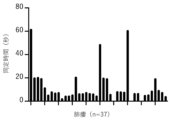
39箇所の肺瘻について測定された、測定開始から最初の波の最高点に到達するまでの時間(同定時間、time to detection)を、図4に示す。図4に示すように、39箇所のうちの37箇所(94.87%)で、肺瘻(胸膜損傷)を同定することができた。また、その同定時間の平均値は、13.54秒(95%信頼区間:8.75~18.33秒)であった。なお、同定できなかった2箇所は、いずれもMinorに分類されるものであった。
同定できた37箇所のうち、3つの群ごとに同定時間の分布を求め、図5に示す。図5に示すように、同定時間の平均値は、Majorで17.79秒、Moderateで9.36秒、Minorで11.34秒であり、Majorで長くなっていることが確認された。これは、Majorでは穴が大きい分、漏れ出る空気が水平方向に拡散してしまい、垂直方向(プローブの方向)に移動しにくいことや、穴が小さくなるほど、漏れ出る空気の圧力が高くなり、その空気がより遠くまで届くことなどが原因であると考えられる。
同定できた37箇所のうち、3つの群ごとに最大酸素飽和度の分布を求め、図6に示す。図6に示すように、Major、Moderate、Minorの3つの群の間で、最大酸素飽和度に有意差は認められなかった。37箇所の最大酸素飽和度の平均値は、339.3 mmHg(95%信頼区間:288.9~389.6 mmHg)であった。
同定できた37箇所のうち、肺葉ごとに最大酸素飽和度の分布を求め、図7に示す。図7に示すように、5つの肺葉[左上葉(LUL)、左下葉(LLL)、右上葉(RUL)、右中葉(RML)、右下葉(RLL)]の間で、最大酸素飽和度に有意差は認められなかった。
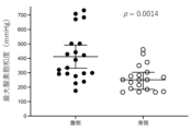
同定できた37箇所のうち、腹側および背側の最大酸素飽和度の分布を求め、図8に示す。図8に示すように、腹側の最大酸素飽和度の平均値は、411.9 mmHg(95%信頼区間:331.7~492.1 mmHg)、背側の最大酸素飽和度の平均値は、262.8 mmHg(95%信頼区間:220.1~305.5 mmHg)であり、腹側の最大酸素飽和度は、背側の最大酸素飽和度と比較して、有意に高値であることが確認された(p = 0.0014)。これは、ブタの胸腔内が狭く、背側が押し潰される格好となっていることから、背側には空気が入りにくいことや、背側の測定時に肺表面に対して垂直に酸素濃度測定プローブ21aを配置できなかったことなどが影響しているためであると考えられる。
10 空気漏れ測定システム
11 空気漏れ測定用装置
21 酸素濃度測定手段
21a 酸素濃度測定プローブ
22 表示手段
12 気体供給手段
11 空気漏れ測定用装置
21 酸素濃度測定手段
21a 酸素濃度測定プローブ
22 表示手段
12 気体供給手段
Claims (5)
- 肺の臓側胸膜の外面の所望の位置での酸素濃度の時間変化を測定可能に設けられた酸素濃度測定手段と、
前記酸素濃度測定手段で測定された前記酸素濃度の時間変化を表示する表示手段とを
有することを特徴とする空気漏れ測定システム。 - 前記酸素濃度測定手段は、前記臓側胸膜の外面に沿って移動可能に設けられた酸素濃度測定プローブを有していることを特徴とする請求項1記載の空気漏れ測定システム。
- 呼吸に合わせて、空気中の酸素濃度よりも高い酸素濃度の気体を、前記肺に周期的に送り込むよう設けられた気体供給手段を有することを特徴とする請求項1または2記載の空気漏れ測定システム。
- 肺の臓側胸膜からの空気漏れを測定するための空気漏れ測定用装置であって、
酸素濃度測定プローブを有し、所望の位置での酸素濃度の時間変化を測定可能に設けられた酸素濃度測定手段と、
前記酸素濃度測定手段で測定された前記酸素濃度の時間変化を表示する表示手段とを
有することを特徴とする空気漏れ測定用装置。 - 呼吸に合わせて、空気中の酸素濃度よりも高い酸素濃度の気体を、肺に周期的に送り込む気体供給ステップと、
前記気体供給ステップで前記肺に前記気体を送り込んでいる間、前記肺の臓側胸膜の外面の所望の位置での酸素濃度の時間変化を測定する酸素濃度測定ステップと、
前記酸素濃度測定ステップで測定された前記酸素濃度の時間変化を表示する表示ステップとを
有することを特徴とする空気漏れ測定方法。
Priority Applications (2)
| Application Number | Priority Date | Filing Date | Title |
|---|---|---|---|
| PCT/JP2022/047869 WO2024142145A1 (ja) | 2022-12-26 | 2022-12-26 | 空気漏れ測定システム、空気漏れ測定用装置および空気漏れ測定方法 |
| JP2024566936A JPWO2024142145A1 (ja) | 2022-12-26 | 2022-12-26 |
Applications Claiming Priority (1)
| Application Number | Priority Date | Filing Date | Title |
|---|---|---|---|
| PCT/JP2022/047869 WO2024142145A1 (ja) | 2022-12-26 | 2022-12-26 | 空気漏れ測定システム、空気漏れ測定用装置および空気漏れ測定方法 |
Publications (1)
| Publication Number | Publication Date |
|---|---|
| WO2024142145A1 true WO2024142145A1 (ja) | 2024-07-04 |
Family
ID=91716938
Family Applications (1)
| Application Number | Title | Priority Date | Filing Date |
|---|---|---|---|
| PCT/JP2022/047869 Ceased WO2024142145A1 (ja) | 2022-12-26 | 2022-12-26 | 空気漏れ測定システム、空気漏れ測定用装置および空気漏れ測定方法 |
Country Status (2)
| Country | Link |
|---|---|
| JP (1) | JPWO2024142145A1 (ja) |
| WO (1) | WO2024142145A1 (ja) |
Cited By (1)
| Publication number | Priority date | Publication date | Assignee | Title |
|---|---|---|---|---|
| CN119564959A (zh) * | 2025-02-08 | 2025-03-07 | 海宁市绿健医疗用品有限公司 | 胸腔漏气流量的监测方法、装置、电子设备及存储介质 |
Citations (5)
| Publication number | Priority date | Publication date | Assignee | Title |
|---|---|---|---|---|
| JP2011523363A (ja) * | 2008-05-01 | 2011-08-11 | スピレーション インコーポレイテッド | 直接型肺センサシステム、方法、および装置 |
| US20150374936A1 (en) * | 2014-06-25 | 2015-12-31 | Neomend, Inc. | Pleural air leak test system |
| JP2018519012A (ja) * | 2015-05-13 | 2018-07-19 | アトリウム メディカル コーポレーション | 胸腔ドレナージシステム |
| JP2019041780A (ja) * | 2017-08-29 | 2019-03-22 | 日本電気株式会社 | エアリーク発生原因判別装置及び方法 |
| JP2022048528A (ja) * | 2020-09-15 | 2022-03-28 | 学校法人昭和大学 | 空気漏れ可視化システム及び空気漏れ可視化方法 |
-
2022
- 2022-12-26 JP JP2024566936A patent/JPWO2024142145A1/ja active Pending
- 2022-12-26 WO PCT/JP2022/047869 patent/WO2024142145A1/ja not_active Ceased
Patent Citations (5)
| Publication number | Priority date | Publication date | Assignee | Title |
|---|---|---|---|---|
| JP2011523363A (ja) * | 2008-05-01 | 2011-08-11 | スピレーション インコーポレイテッド | 直接型肺センサシステム、方法、および装置 |
| US20150374936A1 (en) * | 2014-06-25 | 2015-12-31 | Neomend, Inc. | Pleural air leak test system |
| JP2018519012A (ja) * | 2015-05-13 | 2018-07-19 | アトリウム メディカル コーポレーション | 胸腔ドレナージシステム |
| JP2019041780A (ja) * | 2017-08-29 | 2019-03-22 | 日本電気株式会社 | エアリーク発生原因判別装置及び方法 |
| JP2022048528A (ja) * | 2020-09-15 | 2022-03-28 | 学校法人昭和大学 | 空気漏れ可視化システム及び空気漏れ可視化方法 |
Cited By (1)
| Publication number | Priority date | Publication date | Assignee | Title |
|---|---|---|---|---|
| CN119564959A (zh) * | 2025-02-08 | 2025-03-07 | 海宁市绿健医疗用品有限公司 | 胸腔漏气流量的监测方法、装置、电子设备及存储介质 |
Also Published As
| Publication number | Publication date |
|---|---|
| JPWO2024142145A1 (ja) | 2024-07-04 |
Similar Documents
| Publication | Publication Date | Title |
|---|---|---|
| US7883471B2 (en) | Minimally invasive determination of collateral ventilation in lungs | |
| Hunt et al. | Fiberoptic bronchoscopy in acute inhalation injury | |
| EP2349423B1 (en) | Ventilation system | |
| US8523782B2 (en) | Minimally invasive determination of collateral ventilation in lungs | |
| US20170239431A1 (en) | Device for evacuating and/or monitoring gas leaking from a patient during surgery or anesthetization | |
| CN102355920A (zh) | 射流通气导管 | |
| WO2024142145A1 (ja) | 空気漏れ測定システム、空気漏れ測定用装置および空気漏れ測定方法 | |
| Van Zundert et al. | Comparison of three disposable extraglottic airway devices in spontaneously breathing adults: the LMA-Unique™, the Soft Seal laryngeal mask, and the Cobra perilaryngeal airway | |
| Gunasekaran et al. | Comparison of gastric insufflation volume between Ambu AuraGain and ProSeal Laryngeal Mask Airway Using ultrasonography in patients undergoing general anesthesia: a randomized controlled trial | |
| TOKGÖZ et al. | Comparison of the efficacies of I-gel^{TM} and LMA-ProSeal^{TM} for airway management in pediatric patients | |
| Yokota et al. | A new method for the detection of air leaks using aerosolized indocyanine green | |
| Winterhalter et al. | The laryngeal tube for difficult airway management: a prospective investigation in patients with pharyngeal and laryngeal tumours | |
| Ihra et al. | Supralaryngeal tubeless combined high-frequency jet ventilation for laser surgery of the larynx and trachea | |
| EP1838217A2 (en) | Minimally invasive determination of collateral ventilation in lungs | |
| US20140142455A1 (en) | Minimally invasive determination of collateral ventilation in lungs | |
| Orloff et al. | The Hunsaker Mon-Jet ventilation tube for microlaryngeal surgery: optimal laryngeal exposure | |
| Scamman et al. | Supraglottic jet ventilation for laser surgery of the larynx in children | |
| Arnold et al. | Effect of extraluminal oxygen on carbon dioxide laser ignition of endotracheal tubes | |
| Cakan et al. | Ligasure Vessel Sealing System versus Harmonic Scalpel for Sutureless Nonanatomical Pulmonary Resections in a Rabbit Model: Which One Is Safer? | |
| Granell et al. | Use of Arndt bronchial blocker and the Fastrach laryngeal mask airway for differential lung ventilation in patient with thyroplasty: 19AP4-3 | |
| Borland | Airway management for CO2 laser surgery on the larynx: Venturi jet ventilation and alternatives | |
| Johans et al. | An insufflation device for anesthesia during subglottic carbon dioxide laser microsurgery in children | |
| Pota et al. | Double-lumen tube vs video assisted (ETview) uniblocker for single lung ventilation in distal oesophageal surgery: efficacy and safety: 19AP4-2 | |
| US12471800B2 (en) | Systems and methods for endobronchial diagnostics | |
| WO2026011254A1 (en) | System and method for detecting air leak from visceral pleura of the lung |
Legal Events
| Date | Code | Title | Description |
|---|---|---|---|
| 121 | Ep: the epo has been informed by wipo that ep was designated in this application |
Ref document number: 22969939 Country of ref document: EP Kind code of ref document: A1 |
|
| WWE | Wipo information: entry into national phase |
Ref document number: 2024566936 Country of ref document: JP |
|
| NENP | Non-entry into the national phase |
Ref country code: DE |
|
| 122 | Ep: pct application non-entry in european phase |
Ref document number: 22969939 Country of ref document: EP Kind code of ref document: A1 |