WO2024129554A2 - Combination therapy of a pd-1 antagonist, a tigit antagonist and an antibody drug conjugate that binds 191p4d12 protein for treating patients with cancer - Google Patents
Combination therapy of a pd-1 antagonist, a tigit antagonist and an antibody drug conjugate that binds 191p4d12 protein for treating patients with cancer Download PDFInfo
- Publication number
- WO2024129554A2 WO2024129554A2 PCT/US2023/083279 US2023083279W WO2024129554A2 WO 2024129554 A2 WO2024129554 A2 WO 2024129554A2 US 2023083279 W US2023083279 W US 2023083279W WO 2024129554 A2 WO2024129554 A2 WO 2024129554A2
- Authority
- WO
- WIPO (PCT)
- Prior art keywords
- amino acid
- seq
- antibody
- acid sequence
- set forth
- Prior art date
- Legal status (The legal status is an assumption and is not a legal conclusion. Google has not performed a legal analysis and makes no representation as to the accuracy of the status listed.)
- Ceased
Links
Classifications
-
- C—CHEMISTRY; METALLURGY
- C07—ORGANIC CHEMISTRY
- C07K—PEPTIDES
- C07K16/00—Immunoglobulins [IG], e.g. monoclonal or polyclonal antibodies
- C07K16/18—Immunoglobulins [IG], e.g. monoclonal or polyclonal antibodies against material from animals or humans
- C07K16/28—Immunoglobulins [IG], e.g. monoclonal or polyclonal antibodies against material from animals or humans against receptors, cell surface antigens or cell surface determinants
- C07K16/2803—Immunoglobulins [IG], e.g. monoclonal or polyclonal antibodies against material from animals or humans against receptors, cell surface antigens or cell surface determinants against the immunoglobulin superfamily
- C07K16/2818—Immunoglobulins [IG], e.g. monoclonal or polyclonal antibodies against material from animals or humans against receptors, cell surface antigens or cell surface determinants against the immunoglobulin superfamily against CD28 or CD152
-
- A—HUMAN NECESSITIES
- A61—MEDICAL OR VETERINARY SCIENCE; HYGIENE
- A61K—PREPARATIONS FOR MEDICAL, DENTAL OR TOILETRY PURPOSES
- A61K47/00—Medicinal preparations characterised by the non-active ingredients used, e.g. carriers or inert additives; Targeting or modifying agents chemically bound to the active ingredient
- A61K47/50—Medicinal preparations characterised by the non-active ingredients used, e.g. carriers or inert additives; Targeting or modifying agents chemically bound to the active ingredient the non-active ingredient being chemically bound to the active ingredient, e.g. polymer-drug conjugates
- A61K47/51—Medicinal preparations characterised by the non-active ingredients used, e.g. carriers or inert additives; Targeting or modifying agents chemically bound to the active ingredient the non-active ingredient being chemically bound to the active ingredient, e.g. polymer-drug conjugates the non-active ingredient being a modifying agent
- A61K47/62—Medicinal preparations characterised by the non-active ingredients used, e.g. carriers or inert additives; Targeting or modifying agents chemically bound to the active ingredient the non-active ingredient being chemically bound to the active ingredient, e.g. polymer-drug conjugates the non-active ingredient being a modifying agent the modifying agent being a protein, peptide or polyamino acid
- A61K47/65—Peptidic linkers, binders or spacers, e.g. peptidic enzyme-labile linkers
-
- A—HUMAN NECESSITIES
- A61—MEDICAL OR VETERINARY SCIENCE; HYGIENE
- A61K—PREPARATIONS FOR MEDICAL, DENTAL OR TOILETRY PURPOSES
- A61K47/00—Medicinal preparations characterised by the non-active ingredients used, e.g. carriers or inert additives; Targeting or modifying agents chemically bound to the active ingredient
- A61K47/50—Medicinal preparations characterised by the non-active ingredients used, e.g. carriers or inert additives; Targeting or modifying agents chemically bound to the active ingredient the non-active ingredient being chemically bound to the active ingredient, e.g. polymer-drug conjugates
- A61K47/51—Medicinal preparations characterised by the non-active ingredients used, e.g. carriers or inert additives; Targeting or modifying agents chemically bound to the active ingredient the non-active ingredient being chemically bound to the active ingredient, e.g. polymer-drug conjugates the non-active ingredient being a modifying agent
- A61K47/68—Medicinal preparations characterised by the non-active ingredients used, e.g. carriers or inert additives; Targeting or modifying agents chemically bound to the active ingredient the non-active ingredient being chemically bound to the active ingredient, e.g. polymer-drug conjugates the non-active ingredient being a modifying agent the modifying agent being an antibody, an immunoglobulin or a fragment thereof, e.g. an Fc-fragment
- A61K47/6801—Drug-antibody or immunoglobulin conjugates defined by the pharmacologically or therapeutically active agent
- A61K47/6803—Drugs conjugated to an antibody or immunoglobulin, e.g. cisplatin-antibody conjugates
- A61K47/68031—Drugs conjugated to an antibody or immunoglobulin, e.g. cisplatin-antibody conjugates the drug being an auristatin
-
- A—HUMAN NECESSITIES
- A61—MEDICAL OR VETERINARY SCIENCE; HYGIENE
- A61K—PREPARATIONS FOR MEDICAL, DENTAL OR TOILETRY PURPOSES
- A61K47/00—Medicinal preparations characterised by the non-active ingredients used, e.g. carriers or inert additives; Targeting or modifying agents chemically bound to the active ingredient
- A61K47/50—Medicinal preparations characterised by the non-active ingredients used, e.g. carriers or inert additives; Targeting or modifying agents chemically bound to the active ingredient the non-active ingredient being chemically bound to the active ingredient, e.g. polymer-drug conjugates
- A61K47/51—Medicinal preparations characterised by the non-active ingredients used, e.g. carriers or inert additives; Targeting or modifying agents chemically bound to the active ingredient the non-active ingredient being chemically bound to the active ingredient, e.g. polymer-drug conjugates the non-active ingredient being a modifying agent
- A61K47/68—Medicinal preparations characterised by the non-active ingredients used, e.g. carriers or inert additives; Targeting or modifying agents chemically bound to the active ingredient the non-active ingredient being chemically bound to the active ingredient, e.g. polymer-drug conjugates the non-active ingredient being a modifying agent the modifying agent being an antibody, an immunoglobulin or a fragment thereof, e.g. an Fc-fragment
- A61K47/6835—Medicinal preparations characterised by the non-active ingredients used, e.g. carriers or inert additives; Targeting or modifying agents chemically bound to the active ingredient the non-active ingredient being chemically bound to the active ingredient, e.g. polymer-drug conjugates the non-active ingredient being a modifying agent the modifying agent being an antibody, an immunoglobulin or a fragment thereof, e.g. an Fc-fragment the modifying agent being an antibody or an immunoglobulin bearing at least one antigen-binding site
- A61K47/6849—Medicinal preparations characterised by the non-active ingredients used, e.g. carriers or inert additives; Targeting or modifying agents chemically bound to the active ingredient the non-active ingredient being chemically bound to the active ingredient, e.g. polymer-drug conjugates the non-active ingredient being a modifying agent the modifying agent being an antibody, an immunoglobulin or a fragment thereof, e.g. an Fc-fragment the modifying agent being an antibody or an immunoglobulin bearing at least one antigen-binding site the antibody targeting a receptor, a cell surface antigen or a cell surface determinant
-
- A—HUMAN NECESSITIES
- A61—MEDICAL OR VETERINARY SCIENCE; HYGIENE
- A61K—PREPARATIONS FOR MEDICAL, DENTAL OR TOILETRY PURPOSES
- A61K47/00—Medicinal preparations characterised by the non-active ingredients used, e.g. carriers or inert additives; Targeting or modifying agents chemically bound to the active ingredient
- A61K47/50—Medicinal preparations characterised by the non-active ingredients used, e.g. carriers or inert additives; Targeting or modifying agents chemically bound to the active ingredient the non-active ingredient being chemically bound to the active ingredient, e.g. polymer-drug conjugates
- A61K47/51—Medicinal preparations characterised by the non-active ingredients used, e.g. carriers or inert additives; Targeting or modifying agents chemically bound to the active ingredient the non-active ingredient being chemically bound to the active ingredient, e.g. polymer-drug conjugates the non-active ingredient being a modifying agent
- A61K47/68—Medicinal preparations characterised by the non-active ingredients used, e.g. carriers or inert additives; Targeting or modifying agents chemically bound to the active ingredient the non-active ingredient being chemically bound to the active ingredient, e.g. polymer-drug conjugates the non-active ingredient being a modifying agent the modifying agent being an antibody, an immunoglobulin or a fragment thereof, e.g. an Fc-fragment
- A61K47/6835—Medicinal preparations characterised by the non-active ingredients used, e.g. carriers or inert additives; Targeting or modifying agents chemically bound to the active ingredient the non-active ingredient being chemically bound to the active ingredient, e.g. polymer-drug conjugates the non-active ingredient being a modifying agent the modifying agent being an antibody, an immunoglobulin or a fragment thereof, e.g. an Fc-fragment the modifying agent being an antibody or an immunoglobulin bearing at least one antigen-binding site
- A61K47/6851—Medicinal preparations characterised by the non-active ingredients used, e.g. carriers or inert additives; Targeting or modifying agents chemically bound to the active ingredient the non-active ingredient being chemically bound to the active ingredient, e.g. polymer-drug conjugates the non-active ingredient being a modifying agent the modifying agent being an antibody, an immunoglobulin or a fragment thereof, e.g. an Fc-fragment the modifying agent being an antibody or an immunoglobulin bearing at least one antigen-binding site the antibody targeting a determinant of a tumour cell
-
- A—HUMAN NECESSITIES
- A61—MEDICAL OR VETERINARY SCIENCE; HYGIENE
- A61K—PREPARATIONS FOR MEDICAL, DENTAL OR TOILETRY PURPOSES
- A61K47/00—Medicinal preparations characterised by the non-active ingredients used, e.g. carriers or inert additives; Targeting or modifying agents chemically bound to the active ingredient
- A61K47/50—Medicinal preparations characterised by the non-active ingredients used, e.g. carriers or inert additives; Targeting or modifying agents chemically bound to the active ingredient the non-active ingredient being chemically bound to the active ingredient, e.g. polymer-drug conjugates
- A61K47/51—Medicinal preparations characterised by the non-active ingredients used, e.g. carriers or inert additives; Targeting or modifying agents chemically bound to the active ingredient the non-active ingredient being chemically bound to the active ingredient, e.g. polymer-drug conjugates the non-active ingredient being a modifying agent
- A61K47/68—Medicinal preparations characterised by the non-active ingredients used, e.g. carriers or inert additives; Targeting or modifying agents chemically bound to the active ingredient the non-active ingredient being chemically bound to the active ingredient, e.g. polymer-drug conjugates the non-active ingredient being a modifying agent the modifying agent being an antibody, an immunoglobulin or a fragment thereof, e.g. an Fc-fragment
- A61K47/6889—Conjugates wherein the antibody being the modifying agent and wherein the linker, binder or spacer confers particular properties to the conjugates, e.g. peptidic enzyme-labile linkers or acid-labile linkers, providing for an acid-labile immuno conjugate wherein the drug may be released from its antibody conjugated part in an acidic, e.g. tumoural or environment
-
- C—CHEMISTRY; METALLURGY
- C07—ORGANIC CHEMISTRY
- C07K—PEPTIDES
- C07K16/00—Immunoglobulins [IG], e.g. monoclonal or polyclonal antibodies
- C07K16/18—Immunoglobulins [IG], e.g. monoclonal or polyclonal antibodies against material from animals or humans
- C07K16/28—Immunoglobulins [IG], e.g. monoclonal or polyclonal antibodies against material from animals or humans against receptors, cell surface antigens or cell surface determinants
- C07K16/2803—Immunoglobulins [IG], e.g. monoclonal or polyclonal antibodies against material from animals or humans against receptors, cell surface antigens or cell surface determinants against the immunoglobulin superfamily
-
- C—CHEMISTRY; METALLURGY
- C07—ORGANIC CHEMISTRY
- C07K—PEPTIDES
- C07K16/00—Immunoglobulins [IG], e.g. monoclonal or polyclonal antibodies
- C07K16/18—Immunoglobulins [IG], e.g. monoclonal or polyclonal antibodies against material from animals or humans
- C07K16/28—Immunoglobulins [IG], e.g. monoclonal or polyclonal antibodies against material from animals or humans against receptors, cell surface antigens or cell surface determinants
- C07K16/2893—Immunoglobulins [IG], e.g. monoclonal or polyclonal antibodies against material from animals or humans against receptors, cell surface antigens or cell surface determinants against CD52
-
- A—HUMAN NECESSITIES
- A61—MEDICAL OR VETERINARY SCIENCE; HYGIENE
- A61K—PREPARATIONS FOR MEDICAL, DENTAL OR TOILETRY PURPOSES
- A61K39/00—Medicinal preparations containing antigens or antibodies
- A61K2039/505—Medicinal preparations containing antigens or antibodies comprising antibodies
- A61K2039/507—Comprising a combination of two or more separate antibodies
Definitions
- T cell immunoreceptor with Ig and ITIM domains TAGIT
- PD-1 programmed death 1 protein
- ADC antibody drug conjugates
- PD-1 is recognized as an important player in immune regulation and the maintenance of peripheral tolerance. PD-1 is moderately expressed on naive T, B and NKT cells and up- regulated by T/B cell receptor signaling on lymphocytes, monocytes and myeloid cells (Sharpe et al. , The function of programmed cell death 1 and its ligands in regulating autoimmunity and infection. Nature Immunology. 8:239-245 (2007)).
- PD-L1 Two known ligands for PD-1, PD-L1 (B7-H1) and PD-L2 (B7-DC), are expressed in human cancers arising in various tissues.
- PD-L1 expression correlated with poor prognosis and reduced overall survival irrespective of subsequent treatment (Dong et al. , Nat Med. 8(8):793-800 (2002); Yang et al. Invest Ophthalmol Vis Sci. 49: 2518-2525 (2008); Ghebeh et al. Neoplasia 8: 190-198 (2006); Hamanishi et al., Proc. Natl.
- PD-1 expression on tumor infiltrating lymphocytes was found to mark dysfunctional T cells in breast cancer and melanoma (Ghebeh et al., BMC Cancer. 8:5714-15 (2008); Ahmadzadeh et al., Blood 114: 1537-1544 (2009)) and to correlate with poor prognosis in renal cancer (Thompson et al. , Clinical Cancer Research 15: 1757-1761 (2007)).
- PD-L1 expressing tumor cells interact with PD-1 expressing T cells to attenuate T cell activation and evasion of immune surveillance, thereby contributing to an impaired immune response against the tumor.
- Immune therapies targeting the PD-1 axis include monoclonal antibodies directed to the PD-1 receptor (KEYTRUDATM (pembrolizumab), Merck Sharp & Dohme LLC, Rahway, NJ. USA; OPDIVOTM (nivolumab), Bristol-Myers Squibb Company, Princeton, NJ, USA, and LIBTAYOTM (cemiplimab), Regeneron Pharmaceuticals, Inc., Tarrytown.
- KEYTRUDATM pembrolizumab
- Merck Sharp & Dohme LLC Rahway, NJ. USA
- OPDIVOTM nivolumab
- Bristol-Myers Squibb Company Princeton, NJ, USA
- LIBTAYOTM cemiplimab
- PD-L1 ligand MPDL3280A; TECENTRIQTM (atezolizumab), Genentech, San Francisco, CA, USA; IMFINZITM (durvalumab), AstraZeneca Pharmaceuticals LP, Wilmington, DE; BAVENCIOTM (avelumab), Merck KGaA, Darmstadt, Germany; JEMPERLITM (dostarlimab), GlaxoSmithKline Biologies LLC, Philadelphia, PA, USA). Both therapeutic approaches have demonstrated anti-tumor effects in numerous cancer types.
- TIGIT is an immunomodulatory receptor expressed primarily on activated T cells and NK cells. TIGIT is also known as VSIG9, VSTM3, and WUCAM. Its structure shows one extracellular immunoglobulin domain, a type 1 transmembrane region and two ITIM motifs. TIGIT forms part of a co-stimulatory network that consists of positive (CD226) and negative (TIGIT) immunomodulatory receptors on T cells, and ligands expressed on APCs (CD155 and CD112).
- TIGIT immunoreceptor tyrosine-based inhibition motif
- ITIM immunoreceptor tyrosine-based inhibition motif
- ligation of TIGIT by receptor-ligands CD155 and CD112 expressed by tumor cells or TAMS may contribute to the suppression of TCR-signaling and T cell activation, which is essential for mounting effective anti-tumor immunity.
- an antagonist antibody specific for TIGIT could inhibit the CD 155 and CD112 induced suppression of T cell responses and enhance anti -tumor immunity.
- 91P4D12 (which is also known as Nectin-4) is a 66 kDa type I transmembrane protein that belongs to the nectin family of adhesion molecules. It is composed of an extracellular domain (ECD) containing 3 immunoglobulin (Ig)-like subdomains, a transmembrane helix, and an intracellular region (Takai et al., Annu Rev Cell Dev Biol (2008); 24: 309-42).
- ECD extracellular domain
- Ig immunoglobulin
- Nectins are thought to mediate Ca2+-independent cell-cell adhesion via both homophilic and heterophilic trans-interactions at adherens junctions where they can recruit cadherins and modulate cytoskeletal rearrangements (Rikitake et al., Cell Mol Life Sci (2008); 65(2): 253-63). Sequence identify of Nectin-4 to other Nectin family members is low and ranges between 25%-30% in the ECD (Reymond et al., Biol Chem (2001); 276(46): 43205-15).
- the 3 Ig-like subdomains in the ECD of Nectin-4 are designated V, Cl and C2.
- the Cl domain is responsible for cis-interaction (homodimerization), while V domains of most Nectin molecules contribute to trans-interaction and cell-cell adhesion (Mandair et al.. Curr Top Deve Biol (2015); 112: 197-231; Takai et al. Nat Rev Mol Cell Biol (2008); 9(8): 603-15.).
- Nectin-4 was originally identified by bioinformatics and cloned from human trachea (Reymond et al., J Biol Chem (2001) 276(46): 43205-15 ). Nectin-4 was identified as markedly upregulated in urothelial cancer using suppression subtractive hybridization on a pool of urothelial cancer specimens. Characterization of expression in multiple tumor specimens, both at the RNA level and by immunohistochemistry (IHC), also demonstrated high levels of Nectin-4 in breast, pancreatic, lung, and other cancers (Challita-Eid et al., Cancer Res (2016); 76(10):3003- 13).
- Nectin-4 has been found to be expressed in multiple cancers, particularly urothelial, breast, lung, pancreatic, and ovarian cancers. Higher levels of expression are associated with disease progression and/or poor prognosis (Fabre-Lafay et al., BMC Cancer (2007); 7:73).
- the present disclosure provides methods, pharmaceutical compositions, uses and kits related to treating cancer, using a combination of therapeutic agents, e.g.. a combination of antibodies or antigen binding fragments thereof.
- the present disclosure provides methods of treating cancer, using a combination of a TIGIT antagonist (e.g, antibody (e.g., monoclonal antibody) or antigen binding fragment thereof), a PD-1 antagonist (e.g., antibody (e.g., monoclonal antibody) or antigen binding fragment thereof), and an antibody drug conjugate (ADC) that binds to 191P4D12.
- a TIGIT antagonist e.g, antibody (e.g., monoclonal antibody) or antigen binding fragment thereof
- a PD-1 antagonist e.g., antibody (e.g., monoclonal antibody) or antigen binding fragment thereof
- ADC antibody drug conjugate
- the present disclosure encompasses insights that certain combinations of immune checkpoint inhibitors (e.g., a TIGIT antagonist and a PD-1 antagonist) in combination with an antibody drug conjugate that binds to 191P4D12, as provided herein may enhance efficacy without significant added toxicity as compared with existing treatments.
- anti-TIGIT antagonistic antibodies and anti-PD-1 antagonistic antibodies might be enhanced if administered in combination with other approved or experimental cancer therapies, there are no clear guidelines as to which agent combined with the anti-TIGIT antagonistic antibodies and the anti-PD-1 antagonistic antibodies may be effective or in which patients the combination may enhance the efficacy of treatment.
- a combination of a TIGIT antagonist, a PD- 1 antagonist and an ADC comprising an anti-191P4D12 antibody provided herein may be beneficial because they have divergent metabolic pathways. For example, there may be no drug interactions from a combination of an TIGIT antagonist, a PD-1 antagonist and an ADC that binds to 191P4D12 and is conjugated to one or more units of monomethyl auristatin E (MMAE).
- MMAE monomethyl auristatin E
- the present disclosure provides methods of treating cancer (e.g., urothelial cancer, etc.) using a combination of a TIGIT antagonist, a PD-1 antagonist, and an ADC comprising an anti- 191P4D 12 antibody or antigen binding fragment thereof, wherein the anti-191P4D12 antibody or antigen binding fragment thereof binds to 191P4D12 and is conjugated to one or more units of monomethyl auristatin E (MMAE).
- MMAE monomethyl auristatin E
- kits including a TIGIT antagonist, a PD-1 antagonist, and an antibody drug conjugate (ADC) comprising an anti-191P4D12 antibody or antigen binding fragment thereof, wherein the anti-191P4D12 antibody or antigen binding fragment thereof binds to 191P4D12 and is conjugated to one or more units of monomethyl auristatin E (MMAE).
- ADC antibody drug conjugate
- a therapeutic combination for treating cancer e.g., urothelial cancer
- the therapeutic combination includes a TIGIT antagonist, a PD-1 antagonist, and an ADC comprising an anti-191 P4D 12 antibody or antigen binding fragment thereof, wherein the anti-191P4D 12 antibody or antigen binding fragment thereof binds, to 191P4D12 and is conjugated to one or more units of monomethyl auristatin E (MMAE).
- MMAE monomethyl auristatin E
- provided herein is a method of treating cancer, comprising administering to a human patient in need thereof:
- an antibody drug conjugate comprising an anti-191P4D12 antibody or antigen binding fragment thereof, wherein the anti-191P4D12 antibody or antigen binding fragment thereof binds to 191P4D12 and is conjugated to one or more units of monomethyl auristatin E (MMAE).
- MMAE monomethyl auristatin E
- therapeutic combinations for use in treating cancer comprising administering to a human patient in need thereof:
- an antibody drug conjugate comprising an anti-191P4D12 antibody or antigen binding fragment thereof, wherein the anti-191P4D12 antibody or antigen binding fragment thereof binds to 191P4D12 and is conjugated to one or more units of monomethyl auristatin E (MMAE).
- MMAE monomethyl auristatin E
- the cancer is selected from the group consisting of: melanoma, non-small cell lung cancer, head and neck squamous cell cancer, classical Hodgkin lymphoma, primary mediastinal large B-cell lymphoma, urothelial carcinoma, bladder cancer, microsatellite instability -high or mismatch repair deficient cancer, gastric cancer, esophageal cancer, cervical cancer, hepatocellular carcinoma.
- Merkel cell carcinoma, renal cell carcinoma, endometrial carcinoma, biliary 7 tract cancer a cancer characterized by a tumor having a high mutational burden, cutaneous squamous cell carcinoma, and triple negative breast cancer.
- kits comprising:
- an antibody drug conjugate comprising an anti-191P4D12 antibody or antigen binding fragment thereof, wherein the anti-191P4D12 antibody or antigen binding fragment thereof binds to 191P4D12 and is conjugated to one or more units of monomethyl auristatin E (MMAE).
- MMAE monomethyl auristatin E
- the kit further comprises instructions for administering to a human patient the TIGIT antagonist, the PD-1 antagonist, and the antibody drug conjugate comprising an anti-191P4D12 antibody.
- a therapeutic combination for treating cancer in a human patient wherein the therapeutic combination comprises:
- an antibody drug conjugate comprising an anti-191P4D12 antibody or antigen binding fragment thereof, wherein the anti-191P4D 12 antibody or antigen binding fragment thereof binds to 191P4D12 and is conjugated to one or more units of monomethyl auristatin E (MMAE).
- MMAE monomethyl auristatin E
- the subject is a human patient.
- compositions, kits, uses, or the combinations for use provided herein are useful for treating cancer.
- the PD-1 antagonist is an anti -human PD-1 monoclonal antibody or antigen binding fragment thereof.
- the PD-1 antagonist is an anti -human PD-L1 monoclonal antibody or antigen binding fragment thereof.
- the anti-human PD-1 monoclonal antibody is a humanized antibody .
- the anti -human PD-1 monoclonal antibody is a human antibody.
- the anti-human PD-L1 monoclonal antibody is a humanized antibody.
- the anti-human PD-L1 monoclonal antibody is a human antibody.
- the TIGIT antagonist is an anti-human TIGIT monoclonal antibody or antigen binding fragment thereof.
- the anti-human TIGIT monoclonal antibody is a humanized antibody.
- the anti-human TIGIT monoclonal antibody is a human antibody.
- the anti-PD-1 antibody is independently selected from pembrolizumab, nivolumab. cemiplimab, dostarlimab, sintilimab, tislelizumab, camrelizumab and toripalimab.
- the anti -human PD-1 monoclonal antibody is pembrolizumab.
- the anti-human PD-1 monoclonal antibody is nivolumab.
- the anti-human PD-1 monoclonal antibody is cemiplimab. In another embodiment of the methods, pharmaceutical compositions, kits, or uses provided herein, the anti-human PD-1 monoclonal antibody is dostarlimab.
- the anti -human PD-1 monoclonal antibody or antigen binding fragment thereof is pidilizumab (U.S. Pat. No. 7,332,582).
- the anti-human PD-1 monoclonal antibody or antigen binding fragment thereof is AMP-514 (Medlmmune LLC. Gaithersburg, MD).
- the anti -human PD-1 monoclonal antibody or antigen binding fragment thereof is PDR001 (U.S. Pat. No. 9,683,048).
- the anti-human PD-1 monoclonal antibody or antigen binding fragment thereof is BGB-A317 (U.S. Pat. No. 8,735,553).
- the anti-human PD-1 monoclonal antibody or antigen binding fragment thereof is MGA012 (MacroGenics, Rockville, MD).
- the anti-PDl antibody comprises:
- LC-CDR1 light chain complementarity determining regions
- LC-CDR2 complementarity determining regions
- HC heavy chain
- the anti-PDl antibody compnses a light chain (LC) complementarity determining regions (CDRs) LC-CDR1.
- LC-CDR2 and LC-CDR3 comprising a sequence of amino acids as set forth in SEQ ID NOs: 1, 2 and 3, respectively
- heavy chain (HC) CDRs HC-CDR1, HC-CDR2 and HC-CDR3 comprising a sequence of amino acids as set forth in SEQ ID NOs: 6, 7 and 8, respectively.
- the anti-PDl antibody comprises a heavy chain variable region comprising a sequence of amino acids as set forth in SEQ ID NO:9 and a light chain variable region comprising a sequence of amino acids as set forth in SEQ ID NO:4.
- the anti-PDl antibody comprises light (a) a heavy chain comprising or consisting of a sequence of amino acids as set forth in SEQ ID NO: 10 and (b) a light chain comprising or consisting of a sequence of amino acids as set forth in SEQ ID NO: 5.
- the human patient is administered about 200 mg, about 240 mg, or about 2 mg/kg pembrolizumab, and pembrolizumab is administered once every three weeks. In one embodiment, the human patient is administered about 200 mg pembrolizumab once every three weeks. In one embodiment, the human patient is administered about 240 mg pembrolizumab once every three weeks. In one embodiment, the human patient is administered about 2 mg/kg pembrolizumab once every three weeks.
- the human patient is administered about 400 mg pembrolizumab, and pembrolizumab is administered once every six weeks.
- the human patient is administered about 240 mg or about 3 mg/kg nivolumab once every two weeks, or about 480 mg nivolumab once every four weeks. In one specific embodiment, the human patient is administered about 240 mg nivolumab once every two weeks. In one specific embodiment, the human patient is administered about 3 mg/kg nivolumab once every’ two weeks. In one specific embodiment, the human patient is administered about 480 mg nivolumab once every four weeks.
- the human patient is administered about 350 mg cemiplimab, and cemiplimab is administered once every- three yveeks.
- the anti-human TIGIT monoclonal antibody comprises three light chain CDRs comprising CDRL1 having the amino acid sequence as set forth in SEQ ID NO: 111, CDRL2 having the amino acid sequence as set forth in SEQ ID NO: 112, and CDRL3 having the amino acid sequence as set forth in SEQ ID NO: 113 and three heavy chain CDRs comprising CDRH1 having the amino acid sequence as set forth in SEQ ID NO: 108, CDRH2 having the amino acid sequence as set forth in SEQ ID NO: 154, and CDRH3 having the amino acid sequence as set forth in SEQ ID NO: 110.
- the anti-human TIGIT monoclonal antibody or antigen binding fragment thereof comprises a heavy chain variable region comprising the amino acid sequence as set forth in SEQ ID NO: 148 and a light chain variable region comprising the amino acid sequence as set forth in SEQ ID NO: 152.
- the anti-human TIGIT monoclonal antibody or antigen binding fragment thereof comprises a light chain comprising or consisting of the amino acid sequence as set forth in SEQ ID NO: 294 or 317and a heavy chain comprising or consisting of the amino acid sequence as set forth in SEQ ID NO: 295 or 318.
- the PD-1 antagonist is pembrolizumab; and the TIGIT antagonist is a monoclonal antibody or antigen binding fragment thereof comprising three light chain CDRs comprising CDRL1 having the amino acid sequence as set forth in SEQ ID NO: 111, CDRL2 having the amino acid sequence as set forth in SEQ ID NO: 112, and CDRL3 having the amino acid sequence as set forth in SEQ ID NO: 113 and three heavy chain CDRs comprising CDRH1 having the amino acid sequence as set forth in SEQ ID NO: 108, CDRH2 having the amino acid sequence as set forth in SEQ ID NO: 154, and CDRH3 having the amino acid sequence as set forth in SEQ ID NO: 110.
- the human patient is administered about 200 mg of the anti-human TIGIT monoclonal antibody or antigen binding fragment thereof comprising a light chain comprising or consisting of the amino acid sequence as set forth in SEQ ID NO: 294 or 317 and a heavy chain comprising or consisting of the amino acid sequence as set forth in SEQ ID NO: 295 or 318, and the anti -human TIGIT monoclonal antibody or antigen binding fragment thereof is administered once every three weeks.
- the human patient is administered about 200 mg of the anti-human TIGIT monoclonal antibody or antigen binding fragment thereof once every three weeks. In one embodiment, the human patient is administered about 240 mg of the anti-human TIGIT monoclonal antibody or antigen binding fragment thereof once every three weeks. In one embodiment, the human patient is administered about 2 mg/kg of the anti-human TIGIT monoclonal antibody or antigen binding fragment thereof once every three weeks.
- the human patient is administered about 400 mg of the anti-human TIGIT monoclonal antibody or antigen binding fragment thereof comprising a light chain comprising or consisting of the amino acid sequence as set forth in SEQ ID NO: 294 or 317 and a heavy chain comprising or consisting of the amino acid sequence as set forth in SEQ ID NO: 295 or 318, and the anti-human TIGIT monoclonal antibody or antigen binding fragment thereof is administered once every 7 six weeks.
- the antibody drug conjugate comprising an anti-191P4D12 antibody or antigen binding fragment thereof comprises CDR-H1 comprising the amino acid sequence of SEQ ID NO: 9, CDR-H2 comprising the amino acid sequence of SEQ ID NO: 10, CDR-H3 comprising the amino acid sequence of SEQ ID NO: 11; CDR-L1 comprising the amino acid sequence of SEQ ID NO: 12, CDR-L2 comprising the amino acid sequence of SEQ ID NO: 13, and CDR-L3 comprising the amino acid sequence of SEQ ID NO: 14.
- the antibody drug conjugate comprising an anti-191P4D12 antibody or antigen binding fragment thereof comprises CDR-H1 comprising the amino acid sequence of SEQ ID NO: 16.
- CDR-H2 comprising the amino acid sequence of SEQ ID NO: 17, CDR-H3 comprising the amino acid sequence of SEQ ID NO: 18;
- CDR-L1 comprising the amino acid sequence of SEQ ID NO: 19;
- CDR-L2 comprising the amino acid sequence of SEQ ID NO:20, and CDR-L3 comprising the amino acid sequence of SEQ ID NO:21.
- the antibody drug conjugate comprising an anti-191P4D12 antibody or antigen binding fragment thereof comprises CDR-H1 consisting of the amino acid sequence of SEQ ID NO:9, CDR-H2 consisting of the amino acid sequence of SEQ ID NO: 10, CDR-H3 consisting of the amino acid sequence of SEQ ID NO:11; CDR-L1 consisting of the amino acid sequence of SEQ ID NO: 12, CDR-L2 consisting of the amino acid sequence of SEQ ID NO: 13, and CDR-L3 consisting of the amino acid sequence of SEQ ID NO: 14, or
- the antibody drug conjugate comprising an anti-191P4D12 antibody or antigen binding fragment thereof comprises CDR-H1 consisting of the amino acid sequence of SEQ ID NO: 16.
- CDR-H2 consisting of the amino acid sequence of SEQ ID NO: 17, CDR-H3 consisting of the amino acid sequence of SEQ ID NO: 18;
- CDR-L1 consisting of the amino acid sequence of SEQ ID NO: 19,
- CDR-L2 consisting of the amino acid sequence of SEQ ID NO:20, and CDR-L3 consisting of the amino acid sequence of SEQ ID NO:21.
- the antibody drug conjugate comprising an anti-191P4D12 antibody or antigen binding fragment thereof comprises a heavy chain variable region comprising the amino acid sequence of SEQ ID NO:22 and a light chain variable region comprising the amino acid sequence of SEQ ID NO:23.
- the anti-191P4D12 antibody comprises a heavy chain comprising the amino acid sequence ranging from the 20th amino acid (glutamic acid) to the 466th amino acid (lysine) of SEQ ID NO:7 and a light chain comprising the amino acid sequence ranging from the 23rd amino acid (aspartic acid) to the 236th amino acid (cysteine) of SEQ ID NO: 8.
- the antibody drug conjugate comprising an anti-191P4D12 antigen binding fragment is an Fab, F(ab')2, Fv or scFv.
- the antibody drug conjugate comprising an anti-191P4D12 antibody is a fully human antibody. In certain embodiments of the methods, kits, or uses provided herein, the antibody drug conjugate comprising an anti-191P4D12 antibody is an IgGl and light chain is a kappa light chain. In certain embodiments of the methods, kits, or uses provided herein, the antibody drug conjugate comprising an anti-I91P4D12 antibody or antigen binding fragment thereof is recombinantly produced. In certain embodiments of the methods, kits, or uses provided herein, the antibody drug conjugate comprising an anti-191P4D12 antibody or antigen binding fragment is conjugated to each unit of MMAE via a linker. In certain embodiments of the methods, kits, or uses provided herein, the linker is an enzyme-cleavable linker, and wherein the linker forms a bond with a sulfur atom of the antibody or antigen binding fragment thereof.
- the linker has a formula of: -Aa-Ww-Yy-; wherein -A- is a stretcher unit, a is O or 1; -W- is an amino acid unit, w is an integer ranging from 0 to 12; and -Y- is a spacer unit, y is 0, 1, or 2.
- the stretcher unit has the structure of Formula (1) below; the amino acid unit is valine-citrulline; and the spacer unit is a PAB group comprising the structure of Formula (2) below:
- the stretcher unit forms a bond with a sulfur atom of the antibody or antigen binding fragment thereof; and wherein the spacer unit is linked to MMAE via a carbamate group.
- the ADC comprises from 1 to 20 units of MMAE per antibody or antigen binding fragment thereof. In certain embodiments of the methods, kits, or uses provided herein, the ADC comprises from 1 to 10 units of MMAE per antibody or antigen binding fragment thereof. In certain embodiments of the methods, kits, or uses provided herein, the ADC comprises from 2 to 8 units of MMAE per antibody or antigen binding fragment thereof. In certain embodiments of the methods, kits, or uses provided herein, the ADC comprises from 3 to 5 units of MMAE per antibody or antigen binding fragment thereof.
- the ADC has the following structure: wherein L- represents the anti-191P4D12 antibody or antigen binding fragment thereof and p is from 1 to 10. In yet a further embodiment, the p is from 2 to 8. In yet a further embodiment, the p is from 3 to 5. In yet a further embodiment, the p is from 3 to 4. In yet a further embodiment, the p is 4. In yet a further embodiment, the average p value of the effective amount of the antibody drug conjugates is about 3.8.
- the ADC is administered to the subject at a dose of about 1 to about 10 mg/kg of the subject's body weight, about 1 to about 5 mg/kg of the subject's body weight, about 1 to about 2.5 mg/kg of the subject's body weight, or about 1 to about 1.25 mg/kg of the subject's body weight.
- the ADC is administered to the subject at a dose of about 0.25 mg/kg, about 0.5 mg/kg, about 0.75 mg/kg, about 1.0 mg/kg, about 1.25 mg/kg, about 1.5 mg/kg, about 1.75 mg/kg, about 2.0 mg/kg, about 2.25 mg/kg, or about 2.5 mg/kg of the subject's body weight.
- the ADC is administered to the subject at a dose of about 1 mg/kg of the subject's body weight.
- the ADC is administered to the subject at a dose of about 1.25 mg/kg of the subject's body weight.
- the ADC is administered to the subject by an intravenous (IV) injection or infusion.
- IV intravenous
- the ADC is administered to the subject by one or more an IV injections or infusions on up to 2 days of a 21- day treatment cycle (e.g. day 1 and day 8).
- the ADC is administered to the subject by an IV injection or infusion on days 1 and 8 of a 21 -day treatment cycle.
- the ADC is administered to the subject by an one or more IV injections or infusions, each over about 30 minutes on up to 2 days of a 21 -day treatment cycle.
- the ADC is administered by an IV injection or infusion over about 30 minutes on days 1 and 8 of a 21-day treatment cycle.
- the ADC is formulated in a pharmaceutical composition comprising L-histidine, polysorbate-20 (TWEEN- 20), and trehalose dehydrate.
- the ADC is formulated in a pharmaceutical composition comprising about 20 mM L-histidine, about 0.02% (w/v) TWEEN- 20, about 5.5% (w/v) trehalose dihydrate, and hydrochloride, and wherein the pH of the pharmaceutical composition is about 6.0 at 25°C.
- the ADC is formulated in a pharmaceutical composition comprising about 9 mM histidine, about 11 mM histidine hydrochloride monohydrate, about 0.02% (w/v) TWEEN-20, and about 5.5% (w/v) trehalose dihydrate, and wherein the pH of the pharmaceutical composition is about 6.0 at 25°C.
- the anti-191P4D12 antibody comprises a heavy chain comprising the amino acid sequence ranging from the 20th amino acid (glutamic acid) to the 466th amino acid (lysine) of SEQ ID NO: 300 and a light chain comprising the amino acid sequence ranging from the 23rd amino acid (aspartic acid) to the 236th amino acid (cysteine) of SEQ ID NO:301, wherein the ADC is administered at a dose of about 1.25 mg/kg of the subject's body weight, and wherein the dose is administered by an IV injection or infusion over about 30 minutes on days 1 and 8 of a 21 -day treatment cycle.
- the antibody drug conjugate comprises an anti-191P4D12 antibody or antigen binding fragment thereof, wherein the anti-191P4D12 antibody or antigen binding fragment thereof comprises a heavy chain variable region comprising complementarity determining regions (CDRs) comprising the amino acid sequences of the CDRs of the heavy chain variable region set forth in SEQ ID NO:315 and a light chain variable region comprising CDRs comprising the ammo acid sequences of the CDRs of the light chain variable region set forth in SEQ ID NO:316.
- CDRs complementarity determining regions
- a method of enhancing T cell activity comprising contacting the T cells with:
- an anti -human TIGIT antibody e.g, monoclonal antibody or antigen binding fragment thereof
- an anti -human PD-1 antibody e.g., monoclonal antibody or antigen binding fragment thereof;
- an antibody drug conjugate comprising an anti-191P4D12 antibody or antigen binding fragment thereof, wherein the anti-191P4D12 antibody or antigen binding fragment thereof binds to 191P4D12 and is conjugated to one or more units of monomethyl auristatin E (MMAE).
- MMAE monomethyl auristatin E
- the enhancement of T cell activity occurs in vitro. In other embodiments, the enhancement of T cell activity occurs in vivo.
- the enhancement is in a subject including but not limited to a human subject or human patient.
- the enhancement of T cell activity is measured by increased cytokine production. In other embodiments, the enhancement of T cell activity is measured by increased cell proliferation.
- a method of increasing cytokine production of T cells comprising contacting the T cells with:
- an anti -human TIGIT antibody e.g, monoclonal antibody or antigen binding fragment thereof
- an anti -human PD-1 antibody e.g., monoclonal antibody or antigen binding fragment thereof;
- an antibody drug conjugate comprising an anti-191P4D12 antibody or antigen binding fragment thereof, wherein the anti-191P4D12 antibody or antigen binding fragment thereof binds to 191P4D12 and is conjugated to one or more units of monomethyl auristatin E (MMAE).
- MMAE monomethyl auristatin E
- the increased cytokine production of T cells occurs in vitro. In other embodiments, the increased cytokine production of T cells occurs in vivo.
- the human patient is administered:
- an anti-human TIGIT antibody or antigen binding fragment thereof comprising three light chain CDRs: CDRL1 having the amino acid sequence as set forth in SEQ ID NO: 111, CDRL2 having the amino acid sequence as set forth in SEQ ID NO: 112, and CDRL3 having the amino acid sequence as set forth in SEQ ID NO: 113, and three heavy chain CDRs: CDRH1 having the amino acid sequence as set forth in SEQ ID NO: 108, CDRH2 having the amino acid sequence as set forth in SEQ ID NO: 154, and CDRH3 having the amino acid sequence as set forth in SEQ ID NO: 110 once every three weeks; and
- the human patient is administered:
- an anti-human TIGIT antibody or antigen binding fragment thereof comprising three light chain CDRs: CDRL1 having the amino acid sequence as set forth in SEQ ID NO: 111, CDRL2 having the amino acid sequence as set forth in SEQ ID NO: 112, and CDRL3 having the amino acid sequence as set forth in SEQ ID NO: 113, and three heavy chain CDRs: CDRH1 having the amino acid sequence as set forth in SEQ ID NO: 108, CDRH2 having the amino acid sequence as set forth in SEQ ID NO: 154, and CDRH3 having the amino acid sequence as set forth in SEQ ID NO: 110 once every three weeks:
- the human patient is administered:
- the human patient is administered:
- an anti-human TIGIT antibody or antigen binding fragment thereof comprising three light chain CDRs: CDRL1 having the amino acid sequence as set forth in SEQ ID NO: 111, CDRL2 having the amino acid sequence as set forth in SEQ ID NO: 112, and CDRL3 having the amino acid sequence as set forth in SEQ ID NO: 113, and three heavy chain CDRs: CDRH1 having the amino acid sequence as set forth in SEQ ID NO: 108, CDRH2 having the amino acid sequence as set forth in SEQ ID NO: 154, and CDRH3 having the amino acid sequence as set forth in SEQ ID NO: 110 once every three weeks; and
- the human patient is administered:
- an anti-human TIGIT antibody or antigen binding fragment thereof comprising three light chain CDRs: CDRL1 having the amino acid sequence as set forth in SEQ ID NO: 111, CDRL2 having the amino acid sequence as set forth in SEQ ID NO: 112, and CDRL3 having the amino acid sequence as set forth in SEQ ID NO: 113, and three heavy chain CDRs: CDRH1 having the amino acid sequence as set forth in SEQ ID NO: 108.
- CDRH2 having the amino acid sequence as set forth in SEQ ID NO: 154
- CDRH3 having the amino acid sequence as set forth in SEQ ID NO: 110 once every three weeks;
- the human patient is administered:
- an anti-human TIGIT antibody or antigen binding fragment thereof comprising three light chain CDRs: CDRL1 having the amino acid sequence as set forth in SEQ ID NO: 111, CDRL2 having the amino acid sequence as set forth in SEQ ID NO: 112, and CDRL3 having the amino acid sequence as set forth in SEQ ID NO: 113, and three heavy chain CDRs: CDRH1 having the amino acid sequence as set forth in SEQ ID NO: 108, CDRH2 having the amino acid sequence as set forth in SEQ ID NO: 154, and CDRH3 having the amino acid sequence as set forth in SEQ ID NO: 110 once every three weeks; and
- the human patient is administered:
- an anti-human TIGIT antibody or antigen binding fragment thereof comprising three light chain CDRs: CDRL1 having the amino acid sequence as set forth in SEQ ID NO: 111, CDRL2 having the amino acid sequence as set forth in SEQ ID NO: 112, and CDRL3 having the amino acid sequence as set forth in SEQ ID NO: 1 13, and three heavy' chain CDRs: CDRH1 having the amino acid sequence as set forth in SEQ ID NO: 108, CDRH2 having the amino acid sequence as set forth in SEQ ID NO: 154, and CDRH3 having the amino acid sequence as set forth in SEQ ID NO: 110 once every' six weeks; and
- a method of treating urothelial cancer comprising administering to a human patient in need thereof:
- an anti-human TIGIT antibody or antigen binding fragment thereof comprising three light chain CDRs: CDRL1 having the amino acid sequence as set forth in SEQ ID NO: 111, CDRL2 having the amino acid sequence as set forth in SEQ ID NO: 112, and CDRL3 having the amino acid sequence as set forth in SEQ ID NO: 113, and three heavy chain CDRs: CDRH1 having the amino acid sequence as set forth in SEQ ID NO: 108, CDRH2 having the amino acid sequence as set forth in SEQ ID NO: 154, and CDRH3 having the amino acid sequence as set forth in SEQ ID NO: 110 once every three weeks; and
- the anti-human TIGIT monoclonal antibody and the anti -human PD-1 monoclonal antibody are administered on the same day. In some embodiments, the anti-human TIGIT monoclonal antibody and the anti- human PD-1 monoclonal antibody are administered sequentially. In some embodiments, the antihuman TIGIT monoclonal antibody and the anti -human PD-1 monoclonal antibody are administered concurrently. In some embodiments, the anti-human TIGIT monoclonal antibody and the anti -human PD-1 monoclonal antibody are co-formulated.
- FIGS. 1 A-1E depict the nucleotide and amino acid sequences of nectin-4 protein (FIG. 1 A), the nucleotide and amino acid sequences of the heavy chain (FIG. IB) and light chain (FIG. 1C) of Ha22-2(2, 4)6.1, and the amino acid sequences of the heavy chain (FIG. ID) and light chain of Ha22-2(2, 4)6.1 (FIG. IE).
- FIG. 2 shows the clinical trial study schema
- FIG. 3 shows amino acid sequences of the light chain and heavy chain for an exemplary anti-PD-1 monoclonal antibody useful in the invention (SEQ ID NOs: 5 and 10, respectively).
- Light chain and heavy chain variable regions are underlined (SEQ ID NOs: 4 and 9, respectively) and CDRs are bold.
- ⁇ ‘About’’ when used to modify a numerically defined parameter e.g., the dose of an anti- TIGIT antibody or antigen binding fragment thereof, an anti-PD- 1 antibody or antigen binding fragment thereof, or of an antibody drug conjugate comprising an anti-191P4D12 antibody or antigen binding fragment thereof, wherein the anti-191P4D12 antibody or antigen binding fragment thereof binds to 191P4D12 and is conjugated to one or more units of monomethyl auristatin E (MMAE), or the length of treatment time with a combination therapy described herein) means that the parameter is within 20%.
- MMAE monomethyl auristatin E
- a dose of about 5 mg/kg may vary between 4.5 mg/kg and 5.5 mg/kg.
- the terms “at least one” item or “one or more” item each include a single item selected from the list as well as mixtures of two or more items selected from the list.
- “Comprising” or variations such as “comprise”, “comprises” or “comprised of’ are used throughout the specification and claims in an inclusive sense, i.e., to specify the presence of the stated features but not to preclude the presence or addition of further features that may materially enhance the operation or utility of any of the embodiments of the invention, unless the context requires otherwise due to express language or necessary implication.
- Consists essentially of and variations such as “consist essentially of’ or “consisting essentially of,” as used throughout the specification and claims, indicate the inclusion of any recited elements or group of elements, and the optional inclusion of other elements, of similar or different nature than the recited elements, that do not materially change the basic or novel properties of the specified dosage regimen, method, or composition.
- temperature ranges, percentages, ranges of equivalents, and the like described herein include the upper and lower limits of the range and any value in the continuum there between. Numerical values provided herein, and the use of the term “about”, may include variations of ⁇ 1%, ⁇ 2%, ⁇ 3%, ⁇ 4%, ⁇ 5%, ⁇ 10%, ⁇ 15%, and ⁇ 20% and their numerical equivalents. All ranges also are intended to include all included sub-ranges, although not necessarily explicitly set forth. For example, a range of 3 to 7 days is intended to include 3, 4, 5, 6, and 7 days.
- the term “or,” as used herein denotes alternatives that may, where appropriate, be combined; that is, the term “or” includes each listed alternative separately as well as their combination.
- administer refers to the act of injecting or otherwise physically delivering a substance as it exists outside the body (e.g, an anti-TIGIT antibody, an anti-PD-1 antibody, and of an antibody drug conjugate comprising an anti-191 P4D 12 antibody or antigen binding fragment thereof, wherein the anti-191 P4D 12 antibody or antigen binding fragment thereof binds to 191P4D12 and is conjugated to one or more units of monomethyl auristatin E (MMAE) as described herein) into a patient or subject, such as by oral, mucosal, intradermal, intravenous, subcutaneous, intramuscular delivery, and/or any other methods of physical delivery described herein or known in the art.
- MMAE monomethyl auristatin E
- the term “subject” refers to a mammal that has been the object of treatment, observation, or experiment.
- the mammal may be male or female.
- the mammal may be one or more selected from the group consisting of humans, bovine (e.g, cows), porcine (e.g, pigs), ovine (e.g., sheep), capra (e.g., goats), equine (e.g, horses), canine (e.g, domestic dogs), feline (e.g., house cats), lagomorph (e.g, rabbits), rodent (e.g., rats or mice), Procyon lotor (e.g., raccoons).
- the subject is human.
- subject in need thereof refers to a subject diagnosed with or suspected of having cancer as defined herein.
- the term “antibody” refers to any form of immunoglobulin molecule that exhibits the desired biological or binding activity. Thus, it is used in the broadest sense and specifically covers, but is not limited to, monoclonal antibodies (including full length monoclonal antibodies), polyclonal antibodies, multispecific antibodies (e.g, bispecific antibodies), humanized, fully human antibodies, and chimeric antibodies. “Parental antibodies” are antibodies obtained by exposure of an immune system to an antigen prior to modification of the antibodies for an intended use, such as humanization of an antibody for use as a human therapeutic. As used herein, the term “antibody” encompasses not only intact polyclonal or monoclonal antibodies, but also, unless otherwise specified, fusion proteins comprising an antigen binding fragment thereof that competes with the intact antibody for specific.
- the basic antibody structural unit comprises a tetramer.
- Each tetramer includes two identical pairs of polypeptide chains, each pair having one “light” (about 25 kDa) and one “heavy” chain (about 50-70 kDa).
- the amino-terminal portion of each chain includes a variable region of about 100 to 110 or more amino acids primarily responsible for antigen recognition.
- the variable regions of each light/heavy chain pair form the antibody binding site.
- an intact antibody has two binding sites.
- the carboxy-terminal portion of the heavy chain may define a constant region primarily responsible for effector function.
- human light chains are classified as kappa and lambda light chains.
- human heavy chains are typically classified as mu, delta, gamma, alpha, or epsilon, and define the antibody’s isotype as IgM, IgD. IgG, IgA. and IgE. respectively.
- the variable and constant regions are joined by a "‘J” region of about 12 or more amino acids, with the heavy chain also including a “D” region of about 10 more amino acids. See generally, Fundamental Immunology 7 Ch. 7 (Paul, W., ed., 2nd ed. Raven Press, N.Y. (1989).
- Variable regions or “V region” or “V chain” as used herein means the segment of IgG chains which is variable in sequence between different antibodies.
- a “variable region” of an antibody refers to the variable region of the antibody light chain or the variable region of the antibody heavy chain, either alone or in combination.
- the variable region of the heavy chain may be referred to as “VH.”
- the variable region of the light chain may be referred to as “Vi,.”
- variable regions of both the heavy and light chains comprise three hypervariable regions, also called complementarity determining regions (CDRs), which are located within relatively conserved framework regions (FR).
- CDRs complementarity determining regions
- FR framework regions
- the CDRs are usually aligned by the framework regions, enabling binding to a specific epitope.
- both light and heavy chains variable domains comprise FR1, CDR1, FR2, CDR2, FR3. CDR3. and FR4.
- the light chain CDRs are CDRL1. CDRL2 and CDRL3. respectively
- the heavy chain CDRs are CDRH1, CDRH2 and CDRH3, respectively.
- CDR refers to one of three hypervariable regions (Hl, H2, or H3) within the nonframework region of the antibody VH f>-sheet framework, or one of three hypervariable regions (LI, L2, or L3) within the non-framework region of the antibody VL 0-sheet framework. Accordingly, CDRs are variable region sequences interspersed within the framework region sequences. CDR regions are well known to those skilled in the art and have been defined by, for example, Kabat as the regions of most hypervariability within the antibody variable domains. CDR region sequences also have been defined structurally by Chothia as those residues that are not part of the conserved b-sheet framework, and thus are able to adapt to different conformation. Both terminologies are well recognized in the art.
- CDR region sequences have also been defined by AbM, Contact, and IMGT.
- the positions of CDRs within a canonical antibody variable region have been determined by comparison of numerous structures (Al-Lazikani et al., 1997, J. Mol. Biol. 273:927-48; Morea et al., 2000, Methods 20:267-79). Because the number of residues within a hypervariable region varies in different antibodies, additional residues relative to the canonical positions are conventionally numbered with a, b, c and so forth next to the residue number in the canonical variable region numbering scheme (Al-Lazikani et al., supra). Such nomenclature is similarly well known to those skilled in the art.
- the CDRs are as defined by the Kabat numbering system. In other embodiments, the CDRs are as defined by the IMGT numbering system. In yet other embodiments, the CDRs are as defined by the AbM numbering system. In still other embodiments, the CDRs are as defined by the Chothia numbering system. In yet other embodiments, the CDRs are as defined by the Contact numbering system.
- Chimeric antibody refers to an antibody in which a portion of the heavy and/or light chain contains sequences derived from a particular species (e.g, human) or belonging to a particular antibody class or subclass, while the remainder of the chain(s) is derived from another species (e.g, mouse) or belonging to another antibody class or subclass, as well as fragments of such antibodies, so long as they exhibit the desired biological activity.
- a particular species e.g, human
- another species e.g, mouse
- fragments of such antibodies so long as they exhibit the desired biological activity.
- Human antibody refers to an antibody that comprises human immunoglobulin protein sequences or derivatives thereof.
- a human antibody may contain murine carbohydrate chains if produced in a mouse, in a mouse cell, or in a hybridoma derived from a mouse cell.
- mouse antibody or rat antibody refer to an antibody that comprises only mouse or rat immunoglobulin sequences or derivatives thereof, respectively.
- Humanized antibody refers to forms of antibodies that contain sequences from nonhuman (e.g, murine) antibodies as well as human antibodies. Such antibodies contain minimal sequence derived from non-human immunoglobulin.
- the humanized antibody will comprise substantially all of at least one, and typically two, variable domains, in which all or substantially all of the hypervariable loops correspond to those of a non-human immunoglobulin and all or substantially all of the FR regions are those of a human immunoglobulin sequence.
- the humanized antibody optionally also will comprise at least a portion of an immunoglobulin constant region (Fc), typically that of a human immunoglobulin.
- Fc immunoglobulin constant region
- the prefix “hum”, “hu” or “h” may be added to antibody clone designations when necessary to distinguish humanized antibodies from parental rodent antibodies.
- the humanized forms of rodent antibodies will generally comprise the same CDR sequences of the parental rodent antibodies, although certain amino acid substitutions may be included to increase affinity, increase stability of the humanized antibody, or for other reasons.
- conventional (polyclonal) antibody preparations typically include a multitude of different antibodies having different amino acid sequences in their variable domains, particularly their CDRs, which are often specific for different epitopes.
- the modifier “monoclonal” indicates the character of the antibody as being obtained from a substantially homogeneous population of antibodies, and is not to be construed as requiring production of the antibody by any particular method.
- the monoclonal antibodies to be used in accordance with the present disclosure may be made by the hybridoma method first described by Kohler et al. (1975) Nature 256: 495, or may be made by recombinant DNA methods (see, e.g, U.S. Pat. No. 4,816,567).
- the “monoclonal antibodies” may also be isolated from phage antibody libraries using the techniques described in Clackson et al. (1991) Nature 352: 624-628 and Marks et al. (1991) J. Mol. Biol. 222: 581-597. for example. See also Presta (2005) J. Allergy Clin. Immunol. 116:731.
- antibody fragment or “antigen binding fragment” refers to a fragment of an antibody that retains the ability' to bind specifically to the antigen, e.g., fragments that retain one or more CDR regions and the ability to bind specifically to the antigen.
- An antibody that “specifically binds to” TIGIT or PD-1 is an antibody that exhibits preferential binding to TIGIT or PD-1 (as appropriate) as compared to other proteins, but this specificity does not require absolute binding specificity.
- An antibody is considered “specific” for its intended target if its binding is determinative of the presence of the target protein in a sample, e.g., without producing undesired results such as false positives.
- Antibodies, or binding fragments thereof will bind to the target protein with an affinity that is at least two-fold greater, preferably at least ten times greater, more preferably at least 20-times greater, and most preferably at least 100-times greater than the affinity with non-target proteins.
- Antigen binding portions include, for example, Fab. Fab’. F(ab’)2, Fd, Fv, fragments including CDRs, and single chain variable fragment antibodies (scFv), and polypeptides that contain at least a portion of an immunoglobulin that is sufficient to confer specific antigen binding to the antigen (e.g, TIGIT or PD-1).
- An antibody includes an antibody of any class, such as IgG, IgA, or IgM (or sub-class thereof), and the antibody need not be of any particular class.
- immunoglobulins can be assigned to different classes. There are five major classes of immunoglobulins: IgA, IgD. IgE, IgG, and IgM, and several of these may be further divided into subclasses (isotypes), e.g, IgGl, IgG2, IgG3, IgG4, IgAl, and IgA2.
- the heavy -chain constant regions that correspond to the different classes of immunoglobulins are called alpha, delta, epsilon, gamma, and mu, respectively.
- the subunit structures and three-dimensional configurations of different classes of immunoglobulins are well known.
- a target antigen is a structure to which an antibody can selectively bind.
- a target antigen may be a polypeptide, carbohydrate, nucleic acid, lipid, hapten, or other naturally occurring or synthetic compound.
- the target antigen is a polypeptide.
- an antigen is associated with a cell, for example, is present on or in a cell, for example, a cancer cell.
- an “intact” antibody is one comprising an antigen-binding site as well as a CL and at least heavy chain constant regions, CHI, CH2 and CH3.
- the constant regions may include human constant regions or amino acid sequence variants thereof.
- an intact antibody has one or more effector functions.
- immune response relates to any one or more of the following: specific immune response, non-specific immune response, both specific and non-specific response, innate response, primary immune response, adaptive immunity, secondary immune response, memory immune response, immune cell activation, immune cell-proliferation, immune cell differentiation, and cytokine expression.
- enteral route refers to the administration via any part of the gastrointestinal tract.
- enteral routes include oral, mucosal, buccal, and rectal route, or intragastric route.
- Parenteral route refers to a route of administration other than enteral route.
- parenteral routes of administration include intravenous, intramuscular, intradermal, intraperitoneal, intratumor, intravesical, intraarterial, intrathecal, intracapsular, intraorbital, intracardiac, transtracheal, intraarticular, subcapsular, subarachnoid, intraspinal, epidural and intrastemal.
- the therapeutic agents and compositions of the disclosure can be administered using any suitable method, such as by oral ingestion, nasogastric tube, gastrostomy tube, injection, infusion, implantable infusion pump, and osmotic pump.
- suitable route and method of administration may vary' depending on a number of factors such as the specific therapeutic agent being used, the rate of absorption desired, specific formulation or dosage form used, type or severity 7 of the disorder being treated, the specific site of action, and conditions of the patient, and can be readily selected by a person skilled in the art.
- “‘Chemotherapeutic agent” is a chemical compound useful in the treatment of cancer.
- Classes of chemotherapeutic agents include, but are not limited to: alkydating agents, antimetabolites, kinase inhibitors, spindle poison plant alkaloids, cytoxic/antitumor antibiotics, topoisomerase inhibitors, photosensitizers, anti-estrogens and selective estrogen receptor modulators (SERMs), anti-progesterones, estrogen receptor down -regulators (ERDs), estrogen receptor antagonists, leutinizing hormone-releasing hormone agonists, anti-androgens, aromatase inhibitors, EGFR inhibitors, VEGF inhibitors, and anti-sense oligonucleotides that inhibit expression of genes implicated in abnormal cell proliferation or tumor growth.
- Chemotherapeutic agents useful in the treatment methods of the present invention include cytostatic and/or cytotoxic agents.
- variant when used in relation to an antibody (e.g., an anti-TIGIT antibody or an anti-PD-1 antibody) or an amino acid region within the antibody may refer to a peptide or polypeptide comprising one or more (such as, for example, about 1 to about 25, about 1 to about 20, about 1 to about 15, about 1 to about 10. or about 1 to about 5) amino acid sequence substitutions, deletions, and/or additions as compared to a native or unmodified sequence.
- a variant of an anti-PD-1 antibody may result from one or more (such as, for example, about 1 to about 25, about 1 to about 20, about 1 to about 15, about 1 to about 10, or about 1 to about 5) changes to an amino acid sequence of a native or previously unmodified anti-PD-1 antibody.
- Variants may be naturally occurring or may be artificially constructed.
- Polypeptide variants may be prepared from the corresponding nucleic acid molecules encoding the variants.
- an antibody variant e.g., an anti-TIGIT antibody variant or an anti-PD- 1 antibody variant
- at least retains the antibody functional activity 7 binds to TIGIT and/or is antagonistic to TIGIT activity.
- an anti-PD-1 antibody variant binds to PD-1 and/or is antagonistic to PD-1 activity 7 .
- Constantly modified variants or “conservative substitution” refers to substitutions of amino acids in a protein with other amino acids having similar characteristics (e.g., charge, side-chain size, hydrophobicity /hydrophilicity, backbone conformation and rigidity 7 , etc. , such that the changes can frequently be made without altering the biological activity or other desired property of the protein, such as antigen affinity and/or specificity.
- Those of skill in this art recognize that, in general, single amino acid substitutions in non-essential regions of a polypeptide do not substantially alter biological activity (see, e.g., Watson et al. (1987) Molecular Biology of the Gene, The Benjamin/ Cummings Pub. Co., p. 224 (4th Ed.)).
- substitutions of structurally or functionally similar amino acids are less likely to disrupt biological activity. Exemplary conservative substitutions are set forth in Table 2 below.
- ⁇ ‘Homology” refers to sequence similarity between two polypeptide sequences when they are optimally aligned.
- a position in both of the two compared sequences is occupied by the same amino acid monomer subunit, e.g., if a position in a light chain CDR of two different Abs is occupied by alanine, then the two Abs are homologous at that position.
- the percent of homology is the number of homologous positions shared by the two sequences divided by the total number of positions compared x 100. For example, if 8 of 10 of the positions in two sequences are matched when the sequences are optimally aligned then the two sequences are 80% homologous.
- the comparison is made when two sequences are aligned to give maximum percent homology.
- the comparison can be performed by a BLAST algorithm wherein the parameters of the algorithm are selected to give the largest match between the respective sequences over the entire length of the respective reference sequences.
- BLAST ALGORITHMS Altschul, S.F., etal., (1990) J. Mol. Biol. 215:403-410; Gish, W , et al., (1993) Nature Genet. 3:266-272; Madden. T.L.. et al., (1996) Meth. Enzymol. 266: 131-141; Altschul. S.F., et al.. (1997) Nucleic Acids Res. 25:3389-3402; Zhang, J., et al., (1997) Genome Res. 7:649-656; Wootton, J.C., et al., (1993) Comput. Chem.
- RECIST 1.1 Response Criteria as used herein means the definitions set forth in Eisenhauer, E.A. et al., Eur. J. Cancer 45:228-247 (2009) for target lesions or nontarget lesions, as appropriate based on the context in which response is being measured.
- sustained response means a sustained therapeutic effect after cessation of treatment as described herein.
- the sustained response has a duration that is at least the same as the treatment duration, or at least 1.5. 2.0, 2.5 or 3 times longer than the treatment duration.
- Non-responder patient when referring to a specific anti-tumor response to treatment with a combination therapy described herein, means the patient did not exhibit the anti-tumor response.
- “Treat” or “treating” cancer as used herein means to administer a therapeutic combination of an anti -human PD-1 monoclonal antibody or antigen binding fragment thereof, an anti -human TIGIT monoclonal antibody or antigen binding fragment thereof, and an antibody drug conjugate comprising an anti-191P4D12 antibody or antigen binding fragment thereof, wherein the anti- 191P4D 12 antibody or antigen binding fragment thereof binds to 191P4D12 and is conjugated to one or more units of monomethyl auristatin E (MMAE), such as, e.g..
- MMAE monomethyl auristatin E
- an anti-human PD-1 monoclonal antibody or antigen binding fragment thereof an anti-human TIGIT monoclonal antibody or antigen binding fragment thereof, and an an antibody drug conjugate comprising an anti-191P4D12 antibody or antigen binding fragment thereof, wherein the anti-191P4D12 antibody or antigen binding fragment thereof binds to 191P4D12 and is conjugated to one or more units of monomethyl auristatin E (MMAE, to a subject having cancer or diagnosed with cancer to achieve at least one positive therapeutic effect, such as, for example, reduced number of cancer cells, reduced tumor size, reduced rate of cancer cell infiltration into peripheral organs, or reduced rate of tumor metastasis or tumor growth, comprising administration by oral, mucosal, intradermal, intravenous, subcutaneous, intramuscular delivery, and/or any other methods of physical delivery described herein or known in the art.
- MMAE monomethyl auristatin E
- the agents of the therapeutic combination are administered in an amount effective to alleviate one or more disease symptoms in the treated subject or population, whether by inducing the regression of or inhibiting the progression of such symptom(s) by any clinically measurable degree.
- the amount of the agents of the therapeutic combination that is effective to alleviate any particular disease symptom may vary according to factors such as the disease state, age, and weight of the patient, and the ability of the therapeutic combination to elicit a desired response in the subject. Whether a disease symptom has been alleviated can be assessed by any clinical measurement typically used by physicians or other skilled healthcare providers to assess the severity or progression status of that symptom.
- Treatment may include one or more of the following: inducing/increasing an antitumor immune response, decreasing the number of one or more tumor markers, halting or delaying the growth of a tumor or blood cancer or progression of disease such as cancer, stabilization of disease, inhibiting the growth or survival of tumor cells, eliminating or reducing the size of one or more cancerous lesions or tumors, decreasing the level of one or more tumor markers, ameliorating or abrogating the clinical manifestations of disease, reducing the severity or duration of the clinical symptoms, prolonging the survival or patient relative to the expected survival in a similar untreated patient, and inducing complete or partial remission of a cancerous condition, wherein the disease is cancer, and in certain embodiments wherein the cancer is selected from the group consisting of melanoma, non-small cell lung cancer, head and neck squamous cell cancer, classical Hodgkin lymphoma, primary mediastinal large B-cell lymphoma, urothelial carcinoma, microsatellite instability-high
- the amount of a therapeutic agent that is effective to alleviate any particular disease symptom may vary according to factors such as the disease state, age, and weight of the patient, and the ability of the drug to elicit a desired response in the subject. Whether a disease symptom has been alleviated can be assessed by any clinical measurement typically used by physicians or other skilled healthcare providers to assess the severity or progression status of that symptom.
- T/C 42% is the minimum level of anti-tumor activity'.
- the treatment achieved by a combination therapy of the disclosure is any of PR, CR, OR, PFS, DFS, and OS.
- PFS also referred to as “Time to Tumor Progression” indicates the length of time during and after treatment that the cancer does not grow , and includes the amount of time patients have experienced a CR or PR, as well as the amount of time patients have experienced SD.
- DFS refers to the length of time during and after treatment that the patient remains free of disease.
- OS refers to a prolongation in life expectancy as compared to naive or untreated individuals or patients.
- response to a combination therapy of the disclosure is any of PR, CR, PFS, DFS, or OR that is assessed using RECIST 1.1 response criteria.
- the treatment regimen for a combination therapy of the disclosure that is effective to treat a cancer patient may vary according to factors such as the disease state, age, and weight of the patient, and the ability' of the therapy to elicit an anti-cancer response in the subject. While an embodiment of any of the aspects of the disclosure may not be effective in achieving a positive therapeutic effect in every subject, it should do so in a statistically significant number of subjects as determined by any statistical test known in the art such as the Student’s t-test, the chi 2 -test. the U-test according to Mann and Whitney, the Kruskal-Wallis test (H-test), Jonckheere-Terpstra-test and the Wilcoxon- test.
- PD-1 antagonist means any chemical compound or biological molecule that blocks binding of PD-L1 expressed on a cancer cell to PD-1 expressed on an immune cell (T cell, B cell or NKT cell) and preferably also blocks binding of PD-L2 expressed on a cancer cell to the immune-cell expressed PD-1.
- Alternative names or synonyms for PD-1 and its ligands include: PDCD1, PD1, CD279 and SLEB2 for PD-1; PDCD1L1, PDL1. B7H1. B7-4. CD274 and B7-H for PD-L1; and PDCD1L2, PDL2.
- the PD-1 antagonist blocks binding of human PD-L1 to human PD-1, and preferably blocks binding of both human PD-L1 and PD-L2 to human PD-1.
- Human PD-1 amino acid sequences can be found in NCBI Locus No.: NP_005009.
- Human PD-L1 and PD-L2 amino acid sequences can be found in NCBI Locus No.: NP_054862 and NP_079515, respectively.
- “Pembrolizumab” (formerly known as MK-3475, SCH 900475 and lambrolizumab) alternatively referred to herein as “pembro.” is a humanized IgG4 mAb with the structure described in WHO Drug Information, Vol. 27, No. 2, pages 161-162 (2013) and which comprises the heavy and light chain amino acid sequences and CDRs described in Table 4. Pembrolizumab has been approved by the U.S. FDA as described in the Prescribing Information for KEYTRUDATM (Merck & Co., Inc., Rahway, NJ USA; initial U.S. approval 2014, updated November 2023).
- “Pembrolizumab variant” as used herein means a monoclonal antibody that comprises heavy chain and light chain sequences that are identical to those in pembrolizumab, except for having three, two or one conservative amino acid substitutions at positions that are located outside of the light chain CDRs and six, five, four, three, two or one conservative amino acid substitutions that are located outside of the heavy chain CDRs, e.g., the variant positions are located in the FR regions or the constant region, and optionally has a deletion of the C-terminal lysine residues of the heavy chain.
- pembrolizumab and a pembrolizumab variant comprise identical CDR sequences, but differ from each other due to having a conservative amino acid substitution at no more than three or six other positions in their full length light and heavy chain sequences, respectively.
- a pembrolizumab variant is substantially the same as pembrolizumab with respect to the following properties: binding affinity to PD-1 and ability to block the binding of each of PD-L1 and PD-L2 to PD-1.
- a hyphen (-) designates the point of attachment to the pendant molecule.
- 191P4D12 proteins and/or "191P4D12 related proteins” of the disclosure include those specifically identified herein (see. FIG. 1 A), as well as allelic variants, conservative substitution variants, analogs and homologs that can be isolated/generated and characterized without undue experimentation following the methods outlined herein or readily available in the art. Fusion proteins that combine parts of different 191P4D12 proteins or fragments thereof, as well as fusion proteins of a 191P4D12 protein and a heterologous polypeptide are also included. Such 191P4D12 proteins are collectively referred to as the 191P4D12-related proteins, the proteins of the disclosure, or 191P4D12.
- 191P4D12-related protein refers to a polypeptide fragment or a 191P4D12 protein sequence of 4, 5, 6, 7, 8, 9, 10, 1 1, 12, 13, 14, 15, 16, 17, 18, 19, 20, 21, 22, 23, 24, 25, or more than 25 amino acids; or, at least 30, 35, 40, 45, 50, 55, 60, 65, 70, 80, 85, 90, 95, 100, 105, 110, 115, 120, 125, 130, 135, 140, 145, 150, 155, 160, 165, 170, 175, 180, 185, 190, 195, 200, 225, 250, 275, 300, 325, 330, 335, 339 or more amino acids.
- 191P4D12 is used interchangeably with nectin-4.
- “Pharmaceutical formulation” refers to preparations that contain an active ingredient and one or more pharmaceutically acceptable excipients and that are in such form as to permit the active ingredients to be effective, and which contains no additional components which are toxic to the subjects to which the formulation would be administered.
- co-formulation refers to a formulation comprising two or more of therapeutic agents (i.e., two or more active ingredients).
- co-formulation comprises a TIGIT antagonist and a PD-1 antagonist.
- “Pharmaceutically acceptable” refers to excipients (vehicles, additives) and compositions that can reasonably be administered to a subject to provide an effective dose of the active ingredient employed and that are “generally regarded as safe” e.g., that are physiologically tolerable and do not typically produce an allergic or similar untoward reaction, such as gastric upset and the like, when administered to a human.
- this term refers to molecular entities and compositions approved by a regulatory agency of the federal or a state government or listed in the U.S. Pharmacopeia or another generally recognized pharmacopeia for use in animals, and more particularly in humans.
- pharmaceutically acceptable carrier refers to any inactive substance that is suitable for use in a formulation for the delivery of a therapeutic agent.
- a carrier may be an antiadherent, binder, coating, disintegrant, filler or diluent, preservative (such as antioxidant, antibacterial, or antifungal agent), sweetener, absorption delaying agent, wetting agent, emulsifying agent, buffer, and the like.
- Suitable pharmaceutically acceptable carriers include water, ethanol, polyols (such as glycerol, propylene glycol, polyethylene glycol, and the like), dextrose, vegetable oils (such as olive oil), saline, buffer, buffered saline, and isotonic agents such as sugars, polyalcohols, sorbitol, and sodium chloride.
- the terms “combination,” “combination therapy,” and “therapeutic combination” refer to treatments in which at least one anti -human TIGIT monoclonal antibody or antigen-binding fragment thereof, at least one anti -human PD-1 monoclonal antibody or antigenbinding fragment thereof, and ADC that binds to 191P4D12 and is conjugated to one or more units of monomethyl auristatin E (MMAE), such as, e.g., an anti-human PD-1 monoclonal antibody or antigen binding fragment thereof, an anti-human TIGIT monoclonal antibody or antigen binding fragment thereof, and ADC that binds to 191P4D12 and is conjugated to one or more units of monomethyl auristatin E (MMAE), and optionally additional therapeutic agents, each are administered to a patient in a coordinated manner, over an overlapping period of time as part of a treatment regimen.
- MMAE monomethyl auristatin E
- the period of treatment with the at least one anti-human TIGIT monoclonal antibody (or antigen-binding fragment thereof) is the period of time that a patient undergoes treatment with the anti-human TIGIT monoclonal antibody (or antigen-binding fragment thereof); that is, the period of time from the initial dosing with the anti-human TIGIT monoclonal antibody (or antigen-binding fragment thereof) through the final day of a treatment cycle.
- the period of treatment with the at least one antihuman PD-1 monoclonal antibody (or antigen-binding fragment thereof) is the period of time that a patient undergoes treatment with the anti -human PD-f monoclonal antibody (or antigen-binding fragment thereof); that is, the period of time from the initial dosing with the anti -human PD-1 monoclonal antibody (or antigen-binding fragment thereof) through the final day of a treatment cycle.
- the period of treatment with the ADC that binds to 191P4D12 and is conjugated to one or more units of monomethyl auristatin E (MMAE); is the period of time that a patient undergoes treatment with the ADC; that is, the period of time from the initial dosing with the ADC through the final day of a treatment cycle.
- the anti- TIGIT treatment overlaps by at least one day with the anti-PD-f treatment and overlaps by at least one day with the ADC treatment.
- the anti-TIGIT treatment, the anti-PD-1 treatment, and the ADC treatment are the same period of time.
- the anti-TIGIT treatment begins prior to the anti-PD-1 and/or the ADC treatment. In other embodiments, the anti-TIGIT treatment begins after the anti-PD-1 and/or the ADC treatment. In yet other embodiments, the anti-PD-1 treatment begins prior to the anti-TIGIT and/or the ADC treatment. In still other embodiments, the anti-PD-1 treatment begins after the anti-TIGIT and/or the ADC treatment. In some embodiments, the ADC treatment begins prior to the anti-PD-1 and/or the anti-TIGIT treatment. In other embodiments, the ADC treatment begins after the anti- PD-1 and/or the anti-TIGIT treatment.
- the anti-TIGIT treatment is terminated prior to termination of the anti-PD-1 and/or the ADC treatment. In other embodiments, the anti-TIGIT treatment is terminated after termination of the anti-PD-1 and/or the XX treatment. In yet other embodiments, the anti-PD-1 treatment is terminated prior to termination of the anti-TIGIT and/or the ADC treatment. In still other embodiments, the anti-PD- 1 treatment is terminated after termination of the anti-TIGIT and/or the ADC treatment. In certain embodiments, the ADC treatment is terminated prior to termination of the anti-PD-1 and/or the anti-TIGIT treatment. In other embodiments, the ADC treatment is terminated after termination of the anti-PD-1 and/or the anti-TIGIT treatment.
- treatment regimen “dosing protocol,” and “dosing regimen” are used interchangeably to refer to the dose and timing of administration of each therapeutic agent in a combination therapy of the disclosure.
- cancer refers to or describe the physiological condition in mammals that is typically characterized by unregulated cell growth.
- examples of cancer include but are not limited to, carcinoma, lymphoma, leukemia, blastoma, and sarcoma.
- cancers include, but are not limited to, squamous cell carcinoma, myeloma, small-cell lung cancer, non-small cell lung cancer, glioma, Hodgkin lymphoma, non-Hodgkin lymphoma, acute myeloid leukemia (AML), multiple myeloma, gastrointestinal (tract) cancer, renal cancer, ovarian cancer, liver cancer, lymphoblastic leukemia, lymphocytic leukemia, colorectal cancer, endometrial cancer, kidney cancer, prostate cancer, thyroid cancer, melanoma, chondrosarcoma, neuroblastoma, pancreatic cancer, glioblastoma multiforme, cervical cancer, brain cancer, stomach cancer, bladder cancer, biliary tract cancer, hepatoma, hepatocellular carcinoma, biliary cancer, esophageal cancer, breast cancer, triple negative breast cancer, colon carcinoma, and head and neck cancer.
- squamous cell carcinoma myeloma
- Tumor as it applies to a subject diagnosed with, or suspected of having, a cancer refers to a malignant or potentially malignant neoplasm or tissue mass of any size, and includes primary 7 tumors and secondary neoplasms.
- tumors include solid tumor (e.g, sarcoma (such as chondrosarcoma), carcinoma (such as colon carcinoma), blastoma (such as hepatoblastoma), etc. ) and blood tumor (e.g., leukemia (such as acute myeloid leukemia (AML)), lymphoma (such as DLBCL), multiple myeloma (MM), etc.).
- solid tumor e.g, sarcoma (such as chondrosarcoma), carcinoma (such as colon carcinoma), blastoma (such as hepatoblastoma), etc.
- blood tumor e.g., leukemia (such as acute myeloid leukemia (AML)), lymphoma (such as DLBCL), multiple
- Tumor burden also referred to as “tumor load”, refers to the total amount of tumor material distributed throughout the body. Tumor burden refers to the total number of cancer cells or the total size of tumor(s), throughout the body, including lymph nodes and bone narrow. Tumor burden can be determined by a variety of methods known in the art. such as, e.g., by measuring the dimensions of tumor(s) upon removal from the subject, e.g., using calipers, or while in the body using imaging techniques, e.g., ultrasound, bone scan, computed tomography (CT) or magnetic resonance imaging (MRI) scans.
- CT computed tomography
- MRI magnetic resonance imaging
- tumor volume refers to the total size of the tumor which can be measured as the length and width of a tumor.
- Tumor size may be determined by a variety 7 of methods known in the art, such as, e g., by measuring the dimensions of tumor(s) upon removal from the subject, e.g, using calipers, or while in the body using imaging techniques, e.g., bone scan, ultrasound. CT or MRI scans.
- the term "effective amount” refer to an amount of an anti-TIGIT antibody or antigen binding fragment, an anti-PD-1 antibody or antigen binding fragment of the invention, and an ADC that binds to 191P4D12 and is conjugated to one or more units of monomethyl auristatin E (MMAE), such as, e.g., an anti-human PD-1 monoclonal antibody or antigen binding fragment thereof, an anti-human TIGIT monoclonal antibody or antigen binding fragment thereof, and an ADC that binds to 191P4D12 and is conjugated to one or more units of monomethyl auristatin E (MMAE), that, when administered alone or in combination with an additional therapeutic agent to a cell, tissue, or subject, is effective to cause a measurable improvement in one or more symptoms of an infection or a disease, for example cancer or the progression of cancer.
- MMAE monomethyl auristatin E
- An effective amount further refers to that amount of the antibody or fragment sufficient to result in at least partial amelioration of symptoms, e.g., tumor shrinkage or elimination, lack of tumor growth, increased survival time.
- an effective dose refers to that ingredient alone.
- an effective dose refers to combined amounts of the active ingredients that result in the therapeutic effect, whether administered in combination, serially or simultaneously.
- An effective amount of a therapeutic may result in an improvement of a diagnostic measure or parameter by at least 10%; usually by at least 20%; preferably at least about 30%; more preferably at least 40%, and most preferably by at least 50%.
- An effective amount can also result in an improvement in a subjective measure in cases where subjective measures are used to assess disease severity'.
- Toxicity' and therapeutic efficacy of the antibodies or antigen binding fragments of the invention, administered alone or in combination with another therapeutic agent can be determined by any number of systems or means.
- the toxicity and therapeutic efficacy of the antibodies or antigen binding fragments or compounds of the invention can be determined by standard pharmaceutical procedures in cell cultures or experimental animals, e.g., for determining the LD50 (the dose lethal to 50% of the population) and the ED50 (the dose therapeutically effective in 50% of the population).
- the dose ratio between toxic and therapeutic effects is the therapeutic index (LD50/ ED50).
- the data obtained from these cell culture assays and animal studies can be used in formulating a range of dosage for use in human.
- the dosage of such compounds lies preferably within a range of circulating concentrations that include the ED50 with little or no toxicity.
- the dosage may vary within this range depending upon the dosage form employed and the route of administration.
- ORR or “objective response rate” refers in some embodiments to CR + PR
- ORR(week24) refers to CR and PR measured using irRECIST in each patient in a cohort after 24 weeks of anti -cancer treatment.
- Table 3 Abbreviations PD-1 Antagonists
- mAbs that bind to human PD-1 useful in the treatment methods, compositions, and uses of the invention, are described in US 7,521,051, US 8,008,449, and US 8,354,509.
- Specific anti-human PD-1 mAbs useful as the PD-1 antagonist in the treatment methods, compositions, and uses of the present invention include: pembrolizumab (formerly known as MK-3475, SCH 900475 and lambrolizumab).
- the humanized antibodies h409Al 1, h409A16 and h409A17 which are described in WO 2008/156712.
- PD-1 antagonists or anti -human PD-1 monoclonal antibodies that can be used in any of the methods, compositions, kits, and uses disclosed herein, including any chemical compound or biological molecule that blocks binding of PD-L1 to PD-1 and preferably also blocks binding of PD-L2 to PD-1.
- any monoclonal antibodies that bind to a PD-1 polypeptide, a PD-1 polypeptide fragment, a PD-1 peptide, or a PD-1 epitope and block the interaction between PD-1 and its ligand PD-L1 or PD-L2 can be used.
- the anti-human PD-1 monoclonal antibody binds to a PD-1 polypeptide, a PD-1 polypeptide fragment, a PD-1 peptide, or a PD-1 epitope and blocks the interaction between PD-1 and PD-L1.
- the antihuman PD-1 monoclonal antibody binds to a PD-1 polypeptide, a PD-1 polypeptide fragment, a PD-1 peptide, or a PD-1 epitope and blocks the interaction between PD-1 and PD-L2.
- the anti -human PD-1 monoclonal antibody binds to a PD-1 polypeptide, a PD-1 polypeptide fragment, a PD-1 peptide, or a PD-1 epitope and blocks the interaction between PD-1 and PD-L1 and the interaction between PD-1 and PD-L2.
- Any monoclonal antibodies that bind to a PD-Ll poly peptide, a PD-Ll polypeptide fragment, a PD-L1 peptide, or a PD-L1 epitope and block the interaction between PD-L1 and PD-1 can also be used.
- the anti -human PD-1 monoclonal antibody is selected from the group consisting of pembrolizumab, nivolumab, cemiplimab, dostarlimab, pidilizumab (U.S. Pat. No. 7,332,582), AMP-514 (Medlmmune LLC, Gaithersburg, MD), PDR001 (U.S. Pat. No. 9,683,048), BGB-A317 (U.S. Pat. No. 8,735,553), and MGA012 (MacroGenics, Rockville, MD).
- the anti -human PD-1 monoclonal antibody is pembrolizumab.
- the anti -human PD-1 monoclonal antibody is pembrolizumab. In another embodiment, the anti -human PD-1 monoclonal antibody is nivolumab. In another embodiment, the anti-human PD-1 monoclonal antibody is cemiplimab. In yet another embodiment, the antihuman PD-1 monoclonal antibody is pidilizumab. In one embodiment, the anti -human PD-1 monoclonal antibody is AMP-514. In another embodiment, the anti-human PD-1 monoclonal antibody is PDR001. In yet another embodiment, the anti-human PD-1 monoclonal antibody is BGB-A317. In still another embodiment, the anti-human PD-1 monoclonal antibody is MGA012.
- an anti -human PD-1 antibody or antigen binding fragment thereof for use in the methods, kits, uses and co-formulations of the invention comprises three light chain CDRs of CDRL1, CDRL2 and CDRL3 and/or three heavy chain CDRs of CDRH1, CDRH2 and CDRH3.
- CDRL1 has the amino acid sequence as set forth in SEQ ID NO:1 or a variant of the amino acid sequence as set forth in SEQ ID NO: 1
- CDRL2 has the amino acid sequence as set forth in SEQ ID NO:2 or a variant of the amino acid sequence as set forth in SEQ ID NO:2
- CDRL3 has the amino acid sequence as set forth in SEQ ID NO:3 or a variant of the amino acid sequence as set forth in SEQ ID NO:3.
- CDRH1 has the amino acid sequence as set forth in SEQ ID NO:6 or a variant of the amino acid sequence as set forth in SEQ ID NO:6
- CDRH2 has the amino acid sequence as set forth in SEQ ID NO: 7 or a variant of the amino acid sequence as set forth in SEQ ID NO: 7
- CDRH3 has the amino acid sequence as set forth in SEQ ID NO: 8 or a variant of the amino acid sequence as set forth in SEQ ID NO: 8.
- the three light chain CDRs have the amino acid sequences as set forth in SEQ ID NO: 1.
- SEQ ID NO:2. and SEQ ID NO:3 and the three heavy chain CDRs have the amino acid sequences as set forth in SEQ ID NO:6, SEQ ID NO:7 and SEQ ID NO:8.
- CDRL1 has the amino acid sequence as set forth in SEQ ID NO: 11 or a variant of the amino acid sequence as set forth in SEQ ID NO: 11
- CDRL2 has the amino acid sequence as set forth in SEQ ID NO: 12 or a variant of the amino acid sequence as set forth in SEQ ID NO: 12
- CDRL3 has the amino acid sequence as set forth in SEQ ID NO: 13 or a variant of the amino acid sequence as set forth in SEQ ID NO: 13.
- CDRH1 has the amino acid sequence as set forth in SEQ ID NO: 16 or a variant of the amino acid sequence as set forth in SEQ ID NO: 16
- CDRH2 has the amino acid sequence as set forth in SEQ ID NO: 17 or a variant of the amino acid sequence as set forth in SEQ ID NO: 17
- CDRH3 has the amino acid sequence as set forth in SEQ ID NO: 18 or a variant of the amino acid sequence as set forth in SEQ ID NO: 18.
- the three light chain CDRs have the amino acid sequences as set forth in SEQ ID NO: 1, SEQ ID NO:2, and SEQ ID NO:3 and the three heavy chain CDRs have the amino acid sequences as set forth in SEQ ID NO:6, SEQ ID NO:7 and SEQ ID NO:8.
- the three light chain CDRs have the amino acid sequences as set forth in SEQ ID NO: 11, SEQ ID NO: 12, and SEQ ID NO: 13 and the three heavy chain CDRs have the amino acid sequences as set forth in SEQ ID NO: 16, SEQ ID NO: 17 and SEQ ID NO:18.
- CDRL1 has the amino acid sequence as set forth in SEQ ID NO:21 or a variant of the amino acid sequence as set forth in SEQ ID NO:21
- CDRL2 has the amino acid sequence as set forth in SEQ ID NO:22 or a variant of the amino acid sequence as set forth in SEQ ID NO:22
- CDRL3 has the amino acid sequence as set forth in SEQ ID NO:23 or a variant of the amino acid sequence as set forth in SEQ ID NO:23.
- CDRH1 has the amino acid sequence as set forth in SEQ ID NO:24 or a variant of the amino acid sequence as set forth in SEQ ID NO:24
- CDRH2 has the amino acid sequence as set forth in SEQ ID NO: 25 or a variant of the amino acid sequence as set forth in SEQ ID NO:25
- CDRH3 has the amino acid sequence as set forth in SEQ ID NO:26 or a variant of the amino acid sequence as set forth in SEQ ID NO:26.
- the three light chain CDRs have the amino acid sequences as set forth in SEQ ID NO:21, SEQ ID NO:22, and SEQ ID NO:23 and the three heavy chain CDRs have the amino acid sequences as set forth in SEQ ID NO:24, SEQ ID NO:25 and SEQ ID NO:26.
- Some anti -human PD-1 antibody and antigen binding fragments of the invention comprise a light chain variable region and a heavy' chain variable region.
- the light chain variable region comprises the amino acid sequence as set forth in SEQ ID NON or a variant of the amino acid sequence as set forth in SEQ ID NON
- the heavy chain variable region comprises the amino acid sequence as set forth in SEQ ID NO: 9 or a variant of the amino acid sequence as set forth in SEQ ID NO:9.
- the light chain variable region comprises the amino acid sequence as set forth in SEQ ID NO: 14 or a variant of the amino acid sequence as set forth in SEQ ID NO: 14
- the heavy chain variable region comprises the amino acid sequence as set forth in SEQ ID NO: 19 or a variant of the amino acid sequence as set forth in SEQ ID NO: 19.
- the heavy chain variable region comprises the amino acid sequence as set forth in SEQ ID NO:27 or a variant of the amino acid sequence as set forth in SEQ ID NO:27 and the light chain variable region comprises the amino acid sequence as set forth in SEQ ID NO:28 or a variant of the amino acid sequence as set forth in SEQ ID NO:28, the amino acid sequence as set forth in SEQ ID NO:29 or a variant of the amino acid sequence as set forth in SEQ ID NO:29, or the amino acid sequence as set forth in SEQ ID NO:30 or a variant of the amino acid sequence as set forth in SEQ ID NO:30.
- a light chain variable region or heavy chain variable region sequence is identical to the reference sequence except having one. two, three, four or five amino acid substitutions.
- the substitutions are in the framework region (z.e., outside of the CDRs).
- one, two, three, four or five of the amino acid substitutions are conservative substitutions.
- the anti-human PD-1 antibody or antigen binding fragment comprises a light chain variable region comprising or consisting of the amino acid sequence as set forth in SEQ ID NO:4 and a heavy chain variable region comprising or consisting of the amino acid sequence as set forth in SEQ ID NO: 9.
- the anti-human PD-1 antibody or antigen binding fragment comprises a light chain variable region comprising or consisting of the amino acid sequence as set forth in SEQ ID NO: 14 and a heavy chain variable region comprising or consisting of the amino acid sequence as set forth in SEQ ID NO: 19.
- the anti -human PD-1 antibody or antigen binding fragment comprises a light chain variable region comprising or consisting of the amino acid sequence as set forth in SEQ ID NO:28 and a heavy chain variable region comprising or consisting of the amino acid sequence as set forth in SEQ ID NO:27.
- the anti-human PD-1 antibody or antigen binding fragment comprises a light chain variable region comprising or consisting of the amino acid sequence as set forth in SEQ ID NO:29 and a heavy chain variable region comprising or consisting of the amino acid sequence as set forth in SEQ ID NO:27.
- the antibody or antigen binding fragment comprises a light chain variable region comprising or consisting of the amino acid sequence as set forth in SEQ ID NO:30 and a heavy chain variable region comprising or consisting of the amino acid sequence as set forth in SEQ ID NO:27.
- the co-formulations, methods, kits or uses of the invention comprise an anti-human PD-1 antibody or antigen binding protein that has a VL domain and/or a VH domain with at least 95%, 90%, 85%, 80%, 75% or 50% sequence homology to one of the VL domains or VH domains described above, and exhibits specific binding to PD-1.
- the anti -human PD-1 antibody or antigen binding protein of the co-formulations of the invention comprises VL and VH domains having up to 1, 2, 3, 4. or 5 or more amino acid substitutions, and exhibits specific binding to PD-1.
- the PD-1 antagonist may be a full-length anti-PD-1 antibody or an antigen binding fragment thereof that specifically binds human PD-1.
- the PD-1 antagonist is a full-length anti-PD-1 antibody selected from any class of immunoglobulins, including IgM, IgG, IgD, IgA, and IgE.
- the antibody is an IgG antibody. Any isot pe of IgG can be used, including IgGi, IgG , IgG?. and IgG-i. Different constant domains may be appended to the VL and VH regions provided herein.
- a heavy chain constant domain other than IgGl may be used.
- IgGl antibodies provide for long half-life and for effector functions, such as complement activation and antibody-dependent cellular cytotoxicity, such activities may not be desirable for all uses of the antibody.
- an IgG4 constant domain for example, may be used.
- the PD-1 antagonist is an anti-PD-1 antibody comprising a light chain comprising or consisting of a sequence of amino acid residues as set forth in SEQ ID NO:5 and a heavy chain comprising or consisting of a sequence of amino acid residues as set forth in SEQ ID NO: 10.
- the PD-1 antagonist is an anti-PD-1 antibody comprising a light chain comprising or consisting of a sequence of amino acid residues as set forth in SEQ ID NO: 15 and a heavy chain comprising or consisting of a sequence of amino acid residues as set forth in SEQ ID NO:20.
- the PD-1 antagonist is an anti-PD-1 antibody comprising a light chain comprising or consisting of a sequence of amino acid residues as set forth in SEQ ID NO:32 and a heavy chain comprising or consisting of a sequence of amino acid residues as set forth in SEQ ID NO:31.
- the PD-1 antagonist is an anti-PD-1 antibody comprising a light chain comprising or consisting of a sequence of amino acid residues as set forth in SEQ ID NO: 33 and a heavy chain comprising or consisting of a sequence of amino acid residues as set forth in SEQ ID NO:31.
- the PD-1 antagonist is an anti-PD-1 antibody comprising a light chain comprising or consisting of a sequence of amino acid residues as set forth in SEQ ID NO:34 and a heavy chain comprising or consisting of a sequence of amino acid residues as set forth in SEQ ID NO:31.
- the PD-1 antagonist is pembrolizumab or a pembrolizumab biosimilar.
- the PD-1 antagonist is nivolumab or anivolumab biosimilar.
- amino acid sequence variants of the anti-PD-1 antibodies and antigen binding fragments of the invention and the anti-TIGIT antibodies and antigen binding fragments will have an amino acid sequence having at least 75% amino acid sequence identity with the amino acid sequence of a reference antibody or antigen binding fragment (e.g. heavy chain, light chain. VH. VL, or humanized sequence), more preferably at least 80%. more preferably at least 85%, more preferably at least 90%, and most preferably at least 95. 98. or 99%.
- a reference antibody or antigen binding fragment e.g. heavy chain, light chain. VH. VL, or humanized sequence
- Identity or homology with respect to a sequence is defined herein as the percentage of amino acid residues in the candidate sequence that are identical with the anti-PD-1 residues, after aligning the sequences and introducing gaps, if necessary, to achieve the maximum percent sequence identity, and not considering any conservative substitutions as part of the sequence identity. None of N-terminal, C-terminal, or internal extensions, deletions, or insertions into the antibody sequence shall be construed as affecting sequence identity or homology. Sequence identity refers to the degree to which the amino acids of two polypeptides are the same at equivalent positions when the two sequences are optimally aligned.
- Sequence identity can be determined using a BLAST algorithm wherein the parameters of the algorithm are selected to give the largest match between the respective sequences over the entire length of the respective reference sequences.
- the following references relate to BLAST algorithms often used for sequence analysis: BLAST ALGORITHMS: Altschul, S.F., et al., (1990) J. Mol. Biol. 215:403-410; Gish, W., et al., (1993) Nature Genet. 3:266-272; Madden, T.L., et al.. (1996) Meth. Enzymol. 266: 131-141; Altschul, S.F., et al.. (1997) Nucleic Acids Res.
- either class of light chain can be used in the compositions and methods herein.
- kappa, lambda, or variants thereof are useful in the present compositions and methods.
- the PD-1 antagonist in the combination therapy is pembrolizumab, or a pembrolizumab variant, that is administered in a liquid medicament at a dose selected from the group consisting of 1 mg/kg Q2W, 2 mg/kg Q2W, 3 mg/kg Q2W, 5 mg/kg Q2W, 10 mg/kg Q2W, 1 mg/kg Q3W, 2 mg/kg Q3W, 3 mg/kg Q3W, 5 mg/kg Q3W, 10 mg/kg Q3W and flat-dose equivalents of any of these doses, i.e., such as 200 mg Q3W or 400 mg Q6W.
- pembrolizumab is provided as a liquid medicament that comprises 25 mg/ml pembrolizumab, 7% (w/v) sucrose. 0.02% (w/v) polysorbate 80 in 10 mM histidine buffer pH 5.5.
- pembrolizumab is provided as a liquid medicament that comprises about 125 to about 200 mg/mL of pembrolizumab, or an antigen binding fragment thereof; about 10 mM histidine buffer; about 10 mM L-methionine, or a pharmaceutically acceptable salt thereof; about 7% (w/v) sucrose; and about 0.02 % (w/v) polysorbate 80.
- the selected dose of pembrolizumab is administered by IV infusion. In one embodiment, the selected dose of pembrolizumab is administered by IV infusion over a time period of between 25 and 40 minutes, or about 30 minutes. In other embodiments, the selected dose of pembrolizumab is administered by subcutaneous injection.
- anti-human TIGIT monoclonal antibodies or antigen binding fragments thereof that can be used in the methods, pharmaceutical compositions, kits, and uses disclosed herein. Any monoclonal antibodies that bind to a TIGIT polypeptide, a TIGIT polypeptide fragment, a TIGIT peptide, or a TIGIT epitope and block the interaction between TIGIT and its ligand CD155 and/or CD112 can be used. In some embodiments, the anti-human TIGIT monoclonal antibody binds to a TIGIT polypeptide, a TIGIT polypeptide fragment, a TIGIT peptide, or a TIGIT epitope and blocks the interaction between TIGIT and CD155.
- the anti -human TIGIT monoclonal antibody binds to a TIGIT polypeptide, a TIGIT polypeptide fragment, a TIGIT peptide, or a TIGIT epitope and blocks the interaction between TIGIT and CD112.
- the anti -human TIGIT monoclonal antibody binds to a TIGIT polypeptide, a TIGIT polypeptide fragment, a TIGIT peptide, or a TIGIT epitope and blocks the interaction between TIGIT and CD 155 and the interaction between TIGIT and CD112.
- the human constant region is selected from the group consisting of IgGl, IgG2, IgG3 and IgG4 constant regions, and in preferred embodiments, the human constant region is an IgGl or IgG4 constant region.
- anti-TIGIT antibody sequences are set forth below in Tables 6 and 7. Table 6. Exemplary anti-TIGIT antibodies
- an anti-TIGIT antibody or antigen binding fragment thereof comprises three light chain CDRs of CDRL1, CDRL2, and CDRL3 and/or three heavy chain CDRs of CDRH1, CDRH2, and CDRH3.
- the anti-TIGIT antibody or antigen binding fragment thereof comprises a CDRH1 having the amino acid sequence as set forth in SEQ ID NO:35, a CDRH2 having the amino acid sequence as set forth in SEQ ID NO:36, a CDRH3 having any of the amino acid sequences as set forth in SEQ ID NOs:37. 103, 104.
- CDRLl having the amino acid sequence as set forth in SEQ ID NO:38
- CDRL2 having any of the amino acid sequences as set forth in SEQ ID NOs:39, 89, 90, 91, 92, 93, 94, 95, 96, 97, or 161
- CDRL3 having any of the amino acid sequences as set forth in SEQ ID NOs:40, 98, 99, 100, 101, 102, or 162.
- the anti-TIGIT antibody or antigen binding fragment thereof comprises a CDRH1 having the amino acid sequence as set forth in SEQ ID NO:81, a CDRH2 having the amino acid sequence as set forth in SEQ ID NO:82, a CDRH3 having the amino acid sequence as set forth in SEQ ID NO: 83.
- the anti-TIGIT antibody or antigen binding fragment thereof comprises a CDRH1 having the amino acid sequence as set forth in SEQ ID NO: 108, a CDRH2 having any of the amino acid sequences as set forth in SEQ ID NOs: 109. 116, 117.
- a CDRH3 having any of the amino acid sequences as set forth in SEQ ID NOs: 110, 173, 174, 175, 176, 177, 178, 179, 180, 181, 182, 183, 184, 185, 186 or 187
- a CDRLl having the amino acid sequence as set forth in SEQ ID NO: 111
- a CDRL2 having any of the amino acid sequences as set forth in SEQ ID NOs: 112, 132, 133. 134, 135, 136, 137, 138, 139, 140, 141, 142 or 168
- a CDRL3 having the amino acid sequence as set forth in SEQ ID NO: 113.
- the anti-TIGIT antibody or antigen binding fragment thereof comprises a CDRH1 having the amino acid sequence as set forth in SEQ ID NO:35, a CDRH2 having the amino acid sequence as set forth in SEQ ID NO:36, a CDRH3 having the amino acid sequence as set forth in SEQ ID NO:37, a CDRL1 having the amino acid sequence as set forth in SEQ ID NO:38, a CDRL2 having the amino acid sequence as set forth in SEQ ID NO:39, and a CDRL3 having the amino acid sequence as set forth in SEQ ID NO:40.
- the anti-TIGIT antibody or antigen binding fragment thereof comprises CDRH1 having the amino acid sequence as set forth in SEQ ID NO: 108.
- a CDRH2 having any one of the amino acid sequences as set forth in SEQ ID NO: 109, 154 or 145
- a CDRH3 having the amino acid sequence as set forth in SEQ ID NO: 110
- a CDRL1 having the amino acid sequence as set forth in SEQ ID NO: 111
- a CDRL2 having the amino acid sequence as set forth in SEQ ID NO: 112
- the anti-TIGIT antibody or antigen binding fragment thereof comprises a CDRH1 having the amino acid sequence as set forth in SEQ ID NO: 108, a CDRH2 having the amino acid sequence as set forth in SEQ ID NO: 154, a CDRH3 having the amino acid sequence as set forth in SEQ ID NO: 110, a CDRL1 having the amino acid sequence as set forth in SEQ ID NO: 111, a CDRL2 having the amino acid sequence as set forth in SEQ ID NO: 112, and a CDRL3 having the amino acid sequence as set forth in SEQ ID NO: 113.
- the anti-TIGIT antibody or antigen binding fragment thereof comprises a light chain comprising or consisting of the amino acid sequence as set forth in SEQ ID NO:294 or 317 and a heavy chain comprising or consisting of the amino acid sequence as set forth in SEQ ID NO:295 or 318.
- the anti-TIGIT antibody or antigen binding fragment thereof comprises a variable heavy' chain region and a variable light chain variable region. In one embodiment, the anti-TIGIT antibody or antigen binding fragment thereof comprises a variable heavy chain region comprising the amino acid sequence as set forth in SEQ ID NO:41 and a variable light chain region comprising the amino acid sequence as set forth in SEQ ID NO:42. In one embodiment, the anti-TIGIT antibody or antigen binding fragment thereof comprises a variable heavy chain region comprising the amino acid sequence as set forth in SEQ ID NO:87 and a variable light chain region comprising the amino acid sequence as set forth in SEQ ID NO:88.
- the anti-TIGIT antibody or antigen binding fragment thereof comprises a variable heavy' chain region comprising the amino acid sequence as set forth in SEQ ID NO: 114 and a variable light chain region comprising the amino acid sequence as set forth in SEQ ID NO: 115.
- the anti-TIGIT antibody or antigen binding fragment thereof comprises a variable heavy' chain region comprising any of the amino acid sequences as set forth in SEQ ID NOs: 43-58, 65-75 and 87 and a variable light chain region comprising any one of the amino acid sequences as set forth in SEQ ID NOs: 59-64, 76-80 and 88.
- the anti-TIGIT antibody or antigen binding fragment thereof comprises a variable heavy’ chain region comprising any of the amino acid sequences as set forth in SEQ ID NOs: 144-149 and a variable light chain region comprising any of the amino acid sequences as set forth in SEQ ID NOs: 150-153.
- the anti-TIGIT antibody or antigen binding fragment thereof comprises a variable heavy chain region comprising a variable heavy chain region comprising the amino acid sequence as set forth in SEQ ID NO: 148 and a variable light chain region comprising the amino acid sequence as set forth in SEQ ID NO: 152.
- the anti-TIGIT antibody or antigen binding fragment thereof comprises a variable heavy’ chain region comprising the amino acid sequence as set forth in SEQ ID NO: 147 and a variable light chain region comprising the amino acid sequence as set forth in SEQ ID NO: 150.
- the anti-TIGIT antibody or antigen binding fragment thereof comprises a variable heavy' chain region comprising the amino acid sequence as set forth in SEQ ID NO: 148 and a variable light chain region comprising the amino acid sequence as set forth in SEQ ID NO: 153.
- the anti-TIGIT antibody or antigen binding fragment thereof comprises a variable heavy' chain region comprising the amino acid sequence as set forth in SEQ ID NO: 163 and a variable light chain region comprising the amino acid sequence as set forth in SEQ ID NO:165.
- the anti-TIGIT antibody or antigen binding fragment thereof comprises a variable heavy' chain region comprising the amino acid sequence as set forth in SEQ ID NO: 169 and a variable light chain region comprising the amino acid sequence as set forth in SEQ ID NO: 171.
- the anti-TIGIT antibody or antigen binding fragment thereof comprises a variable heavy chain region comprising the amino acid sequence as set forth in SEQ ID NO: 164 and a variable light chain region comprising the amino acid sequence as set forth in
- the anti-TIGIT antibody or antigen binding fragment thereof comprises a variable heavy chain region comprising the amino acid sequence as set forth in SEQ ID NO:170 and a variable light chain region comprising the amino acid sequence as set forth in SEQ ID NO: 172.
- the anti-TIGIT antibody or antigen binding fragment comprises a CDRH1 having the amino acid sequence as set forth in SEQ ID NO: 188, a CDRH2 having the amino acid sequence as set forth in SEQ ID NO: 189. a CDRH3 having any of the amino acid sequences as set forth in SEQ ID NOs:190, 220, 221, or 222, a CDRL1 having the amino acid sequence as set forth in SEQ ID NO: 191 , a CDRL2 having the amino acid sequence as set forth in SEQ ID NO: 192, and a CDRL3 having any of the amino acid sequences as set forth in SEQ ID NOs:193, 232, 233, 234, 235, 236, or 237.
- the anti-TIGIT antibody or antigen binding fragment thereof comprises a CDRH1 having the amino acid sequence as set forth in SEQ ID NO:204, a CDRH2 having any of the amino acid sequences as set forth in SEQ ID NOs: 205, 256, 257, 258, 259. 260, 261, 262, or 263, a CDRH3 having the amino acid sequence as set forth in SEQ ID NO:206, a CDRL1 having the amino acid sequence as set forth in SEQ ID NO:207, a CDRL2 having the amino acid sequence as set forth in SEQ ID NO:208, and a CDRL3 having the amino acid sequence as set forth in SEQ ID NO:209.
- the anti-TIGIT antibody or antigen binding fragment thereof comprises a variable heavy chain region and a variable light chain variable region. In one embodiment, the anti-TIGIT antibody or antigen binding fragment thereof comprises a variable heavy chain region comprising the amino acid sequence as set forth in SEQ ID NO: 194 and a variable light chain region comprising the amino acid sequence as set forth in SEQ ID NO: 195. In one embodiment, the anti-TIGIT antibody or antigen binding fragment thereof comprises a variable heavy chain region comprising the amino acid sequence as set forth in SEQ ID NO: 196 and a variable light chain region comprising the amino acid sequence as set forth in SEQ ID N0:200.
- the anti-TIGIT antibody or antigen binding fragment thereof comprises a variable heavy chain region comprising the amino acid sequence as set forth in SEQ ID NO: 210 and a variable light chain region comprising the amino acid sequence as set forth in SEQ ID NO:211.
- the anti-TIGIT antibody or antigen binding fragment thereof comprises a variable heavy chain region comprising the amino acid sequence as set forth in SEQ ID NO: 212 and a variable light chain region comprising the amino acid sequence as set forth in SEQ ID NO:216.
- the anti-TIGIT antibody or antigen binding fragment thereof comprises a variable heavy chain region comprising any of the amino acid sequences as set forth in SEQ ID NOs: 197, 198, 199. 223, 224, 225. 226, 227, 228. 229, 230, and 231 and a variable light chain region comprising any of the amino acid sequences as set forth in SEQ ID NOs: 201, 202, 203, 238, 239, 240, 241, 242, 243, 244, 245, 246, 247, 248, 249, 250, 251, 252, 253, 254, and 255.
- the anti-TIGIT antibody or antigen binding fragment thereof comprises a variable heavy chain region comprising any of the amino acid sequences as set forth in SEQ ID NOs: 213, 214. 215, 264, 265. 266, 267, 268, 269, 270. 271, 272, 273, 274, 275. 276, 277, 278. 279, 280, 281. 282, 283. 284, 285, and 286 and a variable light chain region comprising any of the amino acid sequences as set forth in SEQ ID NOs: 217, 218, and 219.
- the anti-TIGIT antibody is vibostolimab, which comprises three light chain CDRs: CDRL1 having the amino acid sequence as set forth in SEQ ID NO: 111, CDRL2 having the amino acid sequence as set forth in SEQ ID NO: 112, and CDRL3 having the amino acid sequence as set forth in SEQ ID NO: 113, and three heavy chain CDRs: CDRH1 having the amino acid sequence as set forth in SEQ ID NO: 108, CDRH2 having the amino acid sequence as set forth in SEQ ID NO: 154, and CDRH3 having the amino acid sequence as set forth in SEQ ID NO: 110.
- Vibostolimab is also referred to as MK-7684.
- Additional anti-TIGIT antibodies which may be used in the formulations described herein include those disclosed, for example, in PCT International Application No. WO 2016/106302; WO 2016/011264; and WO 2009/126688. Table 8. Exemplary Heavy Chain Sequences
- the anti-TIGIT antibody or antigen binding fragment thereof is an antibody comprising any of the variable heavy chains described above and any human heavy chain constant domain.
- the antibody or antigen binding fragment thereof of the invention is of the IgG isotype, and comprises a human IgGl, IgG2, IgG3 or IgG4 human heavy chain constant domain.
- the antibody or antigen binding fragment thereof of the invention comprises a human heavy chain IgGl constant domain (SEQ ID NO: 291) or a variant thereof, wherein the variant comprises up to 20 modified amino acid substitutions.
- the antibody or antigen binding fragment thereof of the invention is an antibody comprising a human heavy chain IgGl constant domain comprising the amino acid sequence as set forth in SEQ ID NO: 291.
- the antibody or antigen binding fragment thereof of the invention comprises a human heavy chain IgGl constant domain wherein the IgGl constant domain is afucosylated.
- the antibody or antigen binding fragment thereof of the invention comprises a human heavy chain IgG4 constant domain or a variant thereof, wherein the variant comprises up to 20 modified amino acid substitutions.
- the antibody or antigen binding fragment thereof of the invention comprises a human heavy chain IgG4 constant domain, wherein the amino acid at position 228 (using EU numbering scheme) has been substituted from Ser to Pro.
- the antibody or antigen binding fragment thereof of the invention comprises a human heavy chain IgG4 constant domain comprising the amino acid sequence as set forth in SEQ ID NO: 292.
- the anti-TIGIT antibody or antigen binding fragment thereof can comprise any of the variable light chains described above and human light chain constant domain.
- the antibody or antigen binding fragment thereof of the invention comprises a human kappa light chain constant domain or a variant thereof, wherein the variant comprises up to 20 modified amino acid substitutions.
- the antibody or antigen binding fragment thereof of the invention comprises a human lambda light chain constant domain or a variant thereof, wherein the variant comprises up to 20 modified amino acid substitutions.
- the antibody or antigen binding fragment thereof of the invention comprises a human kappa light chain constant domain comprising the amino acid sequence as set forth in SEQ ID NO: 293.
- the ADC used in the methods comprises or is an anti-191P4D12 ADC described herein and/or in US Patent No. 8.637,642, which is herein incorporated in its entirety by reference.
- the anti-191P4D12 antibody drug conjugate provided for the methods herein comprises an antibody or antigen binding fragment thereof that binds to 191P4D12 as provided herein, conjugated to one or more units of cytotoxic agents (drug units, or D) as provided herein.
- the cytotoxic agents (drug units, or D) can be covalently linked directly or via a linker unit (LU) as provided herein.
- the antibody drug conjugate compound has the following formula:
- L is the antibody unit, e.g., the anti-nectin-4 antibody or an antigen binding fragment thereof, and
- (LU-D) is a linker unit-drug unit moiety, wherein:
- LU- is a linker unit
- D is a drug unit having cytostatic or cytotoxic activity against a target cell; and p is an integer from 1 to 20.
- the antibody drug conjugate compound has the following formula: L - (Aa-Ww-Yy-D)p (II) or a pharmaceutically acceptable salt or solvate thereof, wherein:
- L is the antibody unit, e.g., the anti-nectin-4 antibody or an antigen binding fragment thereof;
- -A- is a stretcher unit, a is 0 or 1, each -W- is independently an amino acid unit, w is an integer ranging from 0 to 12.
- -Y- is a self-immolative spacer unit, y is 0, 1 or 2,
- D is a drug units having cytostatic or cytotoxic activity 7 against the target cell; and p is an integer from 1 to 20.
- a is 0 or 1
- w is 0 or 1
- y is 0, 1 or 2.
- a is 0 or 1
- w is 0 or 1
- y is 0 or 1.
- p ranges from 1 to 20, 1 to 19, 1 to 18. 1 to 17, 1 to 16, 1 to 15, 1 to 14, 1 to 13, 1 to 12, 1 to 1 1, 1 to 10, 1 to 9, 1 to 8, 1 to 7, 1 to 6, 1 to 5, 1 to 4, 1 to 3, or 1 to 2. In some embodiments, p ranges from 2 to 20, 2 to 19, 2 to 18, 2 to 17, 2 to 16, 2 to 15, 2 to 14, 2 to 13, 2 to 12, 2 to 11, 2 to 10, 2 to 9, 2 to 8, 2 to 7, 2 to 6, 2 to 5, 2 to 4 or 2 to 3.
- p ranges from 3 to 20, 3 to 19, 3 to 18, 3 to 17, 3 to 16, 3 to 15, 3 to 14, 3 to 13, 3 to 12, 3 to 11, 3 to 10, 3 to 9, 3 to 8, 3 to 7, 3 to 6, 3 to 5, or 3 to 4.
- p is about 1.
- p is about 2.
- p is about 3.
- p is about 4.
- p is about 3.8.
- p is about 5.
- p is about 6.
- p is about 7.
- p is about 8.
- p is about 9.
- p is about 10.
- p is about 11.
- p is about 12. In some embodiments, p is about 13. In some embodiments, p is about 14. In some embodiments, p is about 15. In some embodiments, p is about 16. In some embodiments, p is about 17. In some embodiments, p is about 18. In some embodiments, p is about 19. In some embodiments, p is about 20.
- y when w is not zero, y is 1 or 2. In some embodiments, when w is 1 to 12, y is 1 or 2. In some embodiments, w is 2 to 12 and y is 1 or 2. In some embodiments, a is 1 and w and y are 0.
- the cytotoxic agent as part of any of the ADCs provided herein for the methods comprises, consists of, or is MMAE.
- the drug loading is represented by p, the average number of drug molecules per antibody unit. Drug loading can range from 1 to 20 drugs (D) per antibody. The average number of drugs per antibody in preparation of conjugation reactions can be characterized by conventional means such as mass spectroscopy, ELISA assay, and HPLC. The quantitative distribution of antibody drug conjugates in terms of p can also be determined.
- separation, purification, and characterization of homogeneous antibody drug conjugates where p is a certain value from antibody drug conjugates with other drug loadings can be achieved by means such as reverse phase HPLC or electrophoresis.
- p is from 2 to 8.
- the ADC is enfortumab vedotin. In certain embodiments of the methods provided herein, the ADC is a biosimilar of enfortumab vedotin.
- the antibody or antigen binding fragment thereof that binds to nectin- 4-related proteins is an antibody or antigen binding fragment that specifically binds to nectin-4 protein comprising amino acid sequence of SEQ ID NO:295 or 318 (see FIG. 1 A).
- the corresponding cDNA encoding the 191P4D12 protein has a sequence of SEQ ID NO: 294 or 317 (see FIG. 1A).
- the antibody that specifically binds to nectin-4 protein comprising amino acid sequence of SEQ ID NO:295 or 318 includes antibodies that can bind to other nectin-4-related proteins.
- antibodies that bind nectin-4 protein comprising amino acid sequence of SEQ ID NO:295 or 318 can bind nectin-4-related proteins such as nectin-4 variants and the homologs or analogs thereof.
- the anti-nectin-4 antibody provided herein is a monoclonal antibody.
- the antibody comprises a heavy chain comprising an amino acid sequence of SEQ ID NO:297 (cDNA sequence of SEQ ID NO:296), and/or a light chain comprising an amino acid sequence of SEQ ID NO:299 (cDNA sequence of SEQ ID NO:298), as shown in FIGS. IB and 1C.
- the anti-nectin-4 antibody or antigen binding fragment thereof comprises a heavy chain variable region comprising complementarity determining regions (CDRs) comprising the amino acid sequences of the CDRs of the heavy chain variable region set forth in SEQ ID NO:315 (which is the amino acid sequence ranging from the 20th amino acid (glutamic acid) to the 136th amino acid (serine) of SEQ ID N0:300) and a light chain variable region comprising CDRs comprising the amino acid sequences of the CDRs of the light chain variable region set forth in SEQ ID NO:316 (which is the amino acid sequence ranging from the 23rd amino acid (aspartic acid) to the 130th amino acid (arginine) of SEQ ID NO:301).
- CDRs complementarity determining regions
- the anti-nectin-4 antibody or antigen binding fragment thereof comprises a heavy chain variable region comprising complementarity determining region 1 (CDR-H1), CDR-H2. and CDR-H3 comprising the amino acid sequences of the corresponding CDR-H1, CDR-H2, and CDR-H3 in the heavy chain variable region sequence set forth in SEQ ID NO:315 (which is the amino acid sequence ranging from the 20th amino acid (glutamic acid) to the 136th amino acid (serine) of SEQ ID NO:300) and a light chain variable region comprising CDR-L1, CDR-L2, and CDR-L3 comprising the amino acid sequences of the corresponding CDR-L1, CDR-L2, and CDR-L3 in the light chain variable region sequence set forth in SEQ ID NO:316 (which is the amino acid sequence ranging from the 23rd amino acid (aspartic acid) to the 130th amino acid (arginine) of SEQ ID NO:301).
- CDR-H1 complementarity determining
- the anti-nectin-4 antibody or antigen binding fragment thereof comprises a heavy chain variable region comprising complementarity determining regions (CDRs) consisting of the amino acid sequences of the CDRs of the heavy chain variable region set forth in SEQ ID NO: 315 (which is the amino acid sequence ranging from the 20th amino acid (glutamic acid) to the 136th amino acid (serine) of SEQ ID NO:300) and a light chain variable region comprising CDRs consisting of the amino acid sequences of the CDRs of the light chain variable region set forth in SEQ ID NO:316 (which is the amino acid sequence ranging from the 23rd amino acid (aspartic acid) to the 130th amino acid (arginine) of SEQ ID NO:301).
- CDRs complementarity determining regions
- the anti-nectin-4 antibody or antigen binding fragment thereof comprises a heavy chain variable region comprising complementarity determining region 1 (CDR-H1), CDR-H2, and CDR-H3 consisting of the amino acid sequences of the corresponding CDR-H1, CDR-H2, and CDR-H3 in the heavy chain variable region sequence set forth in SEQ ID NO: 315 (which is the amino acid sequence ranging from the 20th amino acid (glutamic acid) to the 136th amino acid (serine) of SEQ ID N0:300) and a light chain variable region comprising CDR-L1, CDR-L2, and CDR-L3 consisting of the amino acid sequences of the corresponding CDR-L1, CDR-L2, and CDR-L3 in the light chain variable region sequence set forth in SEQ ID NO:316 (which is the amino acid sequence ranging from the 23rd amino acid (aspartic acid) to the 130th amino acid (arginine) of SEQ ID NO:301).
- SEQ ID NO: 315
- CDR sequences can be determined according to well-known numbering systems. As described above, CDR regions are well-known to those skilled in the art and have been defined by well-known numbering systems. For example, the Kabat Complementarity Determining Regions (CDRs) are based on sequence variability and are the most commonly used (see, e.g., Kabat et al. , supra). Chothia refers instead to the location of the structural loops (see, e.g., Chothia and Lesk, 1987, J. Mol. Biol. 196:901-17).
- the end of the Chothia CDR-H1 loop when numbered using the Kabat numbering convention varies between H32 and H34 depending on the length of the loop (this is because the Kabat numbering scheme places the insertions at H35A and H35B; if neither 35 A nor 35B is present, the loop ends at 32; if only 35A is present, the loop ends at 33; if both 35 A and 35B are present, the loop ends at 34).
- the AbM hypervariable regions represent a compromise between the Kabat CDRs and Chothia structural loops, and are used by Oxford Molecular' s AbM antibody modeling software (see, e.g.. Antibody Engineering Vol. 2 (Kontermann and Dubel eds., 2d ed. 2010)).
- IMGT ImMunoGeneTics
- IG immunoglobulins
- TCR T-cell receptors
- MHC major histocompatibility complex
- the anti-nectin-4 antibody or antigen binding fragment thereof comprises a heavy chain variable region comprising CDRs (CDR-H1, CDR-H2, CDR-H3, CDR- Ll, CDR-L2, and CDR-L3) comprising the amino acid sequences of the CDRs of the heavy chain variable region set forth in SEQ ID NO:315 according to Kabat numbering and a light chain variable region comprising CDRs comprising the amino acid sequences of the CDRs of the light chain variable region set forth in SEQ ID NO:316 according to Kabat numbering.
- CDR-H1, CDR-H2, CDR-H3, CDR- Ll, CDR-L2, and CDR-L3 comprising the amino acid sequences of the CDRs of the heavy chain variable region set forth in SEQ ID NO:315 according to Kabat numbering
- a light chain variable region comprising CDRs comprising the amino acid sequences of the CDRs of the light chain variable region set forth in SEQ ID NO:316 according to Kabat numbering.
- the anti-nectin-4 antibody or antigen binding fragment thereof comprises a heavy chain variable region comprising CDRs (CDR-H1, CDR-H2,CDR-H3, CDR- Ll, CDR-L2, and CDR-L3) comprising the amino acid sequences of the CDRs of the heavy chain variable region set forth in SEQ ID NO:315 according to AbM numbering and a light chain variable region comprising CDRs comprising the amino acid sequences of the CDRs of the light chain variable region set forth in SEQ ID NO:316 according to AbM numbering.
- CDR-H1, CDR-H2,CDR-H3, CDR- Ll, CDR-L2, and CDR-L3 comprising the amino acid sequences of the CDRs of the heavy chain variable region set forth in SEQ ID NO:315 according to AbM numbering
- a light chain variable region comprising CDRs comprising the amino acid sequences of the CDRs of the light chain variable region set forth in SEQ ID NO:316 according to AbM numbering.
- the anti-nectin-4 antibody or antigen binding fragment thereof comprises a heavy chain variable region comprising CDRs (CDR-H1, CDR-H2, CDR-H3, CDR- Ll, CDR-L2, and CDR-L3) comprising the amino acid sequences of the CDRs of the heavy chain variable region set forth in SEQ ID NO:315 according to Chothia numbering and a light chain variable region comprising CDRs comprising the amino acid sequences of the CDRs of the light chain variable region set forth in SEQ ID NO:316 according to Chothia numbering.
- CDRs CDR-H1, CDR-H2, CDR-H3, CDR- Ll, CDR-L2, and CDR-L3
- the anti-nectin-4 antibody or antigen binding fragment thereof comprises a heavy chain variable region comprising CDRs (CDR-H1, CDR-H2, CDR-H3, CDR- Ll, CDR-L2, and CDR-L3) comprising the amino acid sequences of the CDRs of the heavy chain variable region set forth in SEQ ID NO:315 according to Contact numbering and a light chain variable region comprising CDRs comprising the amino acid sequences of the CDRs of the light chain variable region set forth in SEQ ID NO:316 according to Contact numbering.
- CDRs CDR-H1, CDR-H2, CDR-H3, CDR- Ll, CDR-L2, and CDR-L3
- the anti-nectin-4 antibody or antigen binding fragment thereof comprises a heavy chain variable region comprising CDRs (CDR-H1, CDR-H2, CDR-H3, CDR- Ll, CDR-L2, and CDR-L3) comprising the amino acid sequences of the CDRs of the heavy chain variable region set forth in SEQ ID NO:315 according to IMGT numbering and a light chain variable region comprising CDRs comprising the amino acid sequences of the CDRs of the light chain variable region set forth in SEQ ID NO:316 according to IMGT numbering.
- CDRs CDR-H1, CDR-H2, CDR-H3, CDR- Ll, CDR-L2, and CDR-L3
- the anti-nectin-4 antibody or antigen binding fragment thereof comprises a heavy chain variable region comprising CDRs (CDR-H1, CDR-H2, CDR-H3, CDR- Ll, CDR-L2, and CDR-L3) consisting of the amino acid sequences of the CDRs of the heavy chain variable region set forth in SEQ ID NO:315 according to Kabat numbering and a light chain variable region comprising CDRs consisting of the amino acid sequences of the CDRs of the light chain variable region set forth in SEQ ID NO:316 according to Kabat numbering.
- CDRs CDR-H1, CDR-H2, CDR-H3, CDR- Ll, CDR-L2, and CDR-L3
- the anti-nectin-4 antibody or antigen binding fragment thereof comprises a heavy chain variable region comprising CDRs (CDR-H1, CDR-H2, CDR-H3. CDR- Ll, CDR-L2, and CDR-L3) consisting of the amino acid sequences of the CDRs of the heavy chain variable region set forth in SEQ ID NO:315 according to AbM numbering and a light chain variable region comprising CDRs consisting of the amino acid sequences of the CDRs of the light chain variable region set forth in SEQ ID NO:316 according to AbM numbering.
- the anti-nectin-4 antibody or antigen binding fragment thereof comprises a heavy chain variable region comprising CDRs (CDR-H1, CDR-H2, CDR-H3. CDR- Ll, CDR-L2. and CDR-L3) consisting of the amino acid sequences of the CDRs of the heavy chain variable region set forth in SEQ ID NO:315 according to Chothia numbering and a light chain variable region comprising CDRs consisting of the amino acid sequences of the CDRs of the light chain variable region set forth in SEQ ID NO:316 according to Chothia numbering.
- CDRs CDR-H1, CDR-H2, CDR-H3.
- CDR- Ll, CDR-L2. and CDR-L3 consisting of the amino acid sequences of the CDRs of the heavy chain variable region set forth in SEQ ID NO:315 according to Chothia numbering
- a light chain variable region comprising CDRs consisting of the amino acid sequences of the CDRs of the light chain variable region set forth in SEQ
- the anti-nectin-4 antibody or antigen binding fragment thereof comprises a heavy chain variable region comprising CDRs (CDR-EI1, CDR-H2, CDR-H3, CDR- Ll, CDR-L2, and CDR-L3) consisting of the amino acid sequences of the CDRs of the heavy’ chain variable region set forth in SEQ ID NO:315 according to Contact numbering and a light chain variable region comprising CDRs consisting of the amino acid sequences of the CDRs of the light chain variable region set forth in SEQ ID NO:316 according to Contact numbering.
- CDR-EI1, CDR-H2, CDR-H3, CDR- Ll, CDR-L2, and CDR-L3 consisting of the amino acid sequences of the CDRs of the heavy’ chain variable region set forth in SEQ ID NO:315 according to Contact numbering
- a light chain variable region comprising CDRs consisting of the amino acid sequences of the CDRs of the light chain variable region set forth in SEQ ID NO:316 according to Contact
- the anti-nectin-4 antibody or antigen binding fragment thereof comprises a heavy chain variable region comprising CDRs (CDR-H1, CDR-H2, CDR-H3, CDR- Ll, CDR-L2, and CDR-L3) consisting of the amino acid sequences of the CDRs of the heavy' chain variable region set forth in SEQ ID NO:315 according to IMGT numbering and a light chain variable region comprising CDRs consisting of the amino acid sequences of the CDRs of the light chain variable region set forth in SEQ ID NO:316 according to IMGT numbering.
- CDRs CDR-H1, CDR-H2, CDR-H3, CDR- Ll, CDR-L2, and CDR-L3
- the CDR sequences according to different numbering systems can be readily determined, e.g., using online tools such as the one provided by Antigen receptor Numbering And Receptor Classification (ANARCI).
- ANARCI Antigen receptor Numbering And Receptor Classification
- the heavy chain CDR sequences within SEQ ID NO:315, and the light chain CDR sequences within SEQ ID NO:316 according to Kabat numbering as determined by ANARCI are listed in Table 9 below.
- the heavy chain CDR sequences within SEQ ID NO:315, and the light chain CDR sequences within SEQ ID NO:316 according to IMGT numbering as determined by ANARCI are listed in Table 10 below.
- the antibody or antigen binding fragment thereof comprises CDR- H1 comprising an amino acid sequence of SEQ ID NO:302, CDR-H2 comprising an amino acid sequence of SEQ ID NO: 303, CDR-H3 comprising an amino acid sequence of SEQ ID NO: 304, CDR-L1 comprising an amino acid sequence of SEQ ID NO: 305, CDR-L2 comprising an amino acid sequence of SEQ ID NO:306, and CDR-L3 comprising an amino acid sequence of SEQ ID NO: 307.
- the antibody or antigen binding fragment thereof comprises CDR- HI comprising an amino acid sequence of SEQ ID NO:309, CDR-H2 comprising an amino acid sequence of SEQ ID NO: 310, CDR-H3 comprising an amino acid sequence of SEQ ID NO: 311, CDR-L1 comprising an amino acid sequence of SEQ ID NO: 312, CDR-L2 comprising an amino acid sequence of SEQ ID NO:313, and CDR-L3 comprising an amino acid sequence of SEQ ID NO:314.
- the antibody or antigen binding fragment thereof comprises CDR- H1 consisting of an amino acid sequence of SEQ ID NO:302.
- CDR-H2 consisting of an amino acid sequence of SEQ ID NO: 303
- CDR-H3 consisting of an amino acid sequence of SEQ ID NO: 304
- CDR-L1 consisting of an amino acid sequence of SEQ ID NO: 305
- CDR-L2 consisting of an amino acid sequence of SEQ ID NO: 306
- CDR-L3 consisting of an amino acid sequence of SEQ ID NO: 307.
- the antibody or antigen binding fragment thereof comprises CDR- H1 consisting of an amino acid sequence of SEQ ID NO: 309, CDR-H2 consisting of an amino acid sequence of SEQ ID NO: 310, CDR-H3 consisting of an amino acid sequence of SEQ ID NO: 311.
- CDR-L1 consisting of an amino acid sequence of SEQ ID NO:312.
- CDR-L2 consisting of an amino acid sequence of SEQ ID NO:313, and CDR-L3 consisting of an amino acid sequence of SEQ ID NO: 314.
- the antibody or antigen binding fragment thereof comprises a heavy chain variable region comprising the amino acid sequence of SEQ ID NO:315 and a light chain variable region comprising the amino acid sequence of SEQ ID NO:316.
- the antibody or antigen binding fragment thereof comprises a heavy chain vanable region consisting of the amino acid sequence of SEQ ID NO:315 and a light chain variable region consisting of the amino acid sequence of SEQ ID NO:316.
- the antibody comprises a heavy chain comprising the amino acid sequence ranging from the 20th amino acid (glutamic acid) to the 466th amino acid (lysine) of SEQ ID NO:300 and a light chain comprising the amino acid sequence ranging from the 23rd amino acid (aspartic acid) to the 236th amino acid (cysteine) of SEQ ID NO:301.
- the antibody comprises a heavy chain consisting of the amino acid sequence ranging from the 20th amino acid (glutamic acid) to the 466th amino acid (lysine) of SEQ ID N0:300 and a light chain consisting of the amino acid sequence ranging from the 23rd amino acid (aspartic acid) to the 236th amino acid (cysteine) of SEQ ID NO:301.
- amino acid sequence modification(s) of antibodies described herein are contemplated.
- it may be desirable to optimize the binding affinity and/or other biological properties of the antibody including but not limited to specificity, thermostability, expression level, effector functions, glycosylation, reduced immunogenicity, or solubility 7 .
- antibody variants can be prepared.
- antibody variants can be prepared by introducing appropriate nucleotide changes into the encoding DNA, and/or by synthesis of the desired antibody or polypeptide.
- amino acid changes can alter post-translational processes of the antibody, such as changing the number or position of glycosylation sites or altering the membrane anchoring characteristics.
- the antibodies provided herein are chemically modified, for example, by the covalent attachment of any type of molecule to the antibody.
- the antibody derivatives can include antibodies that have been chemically modified, for example, by glycosylation, acetylation, pegylation. phosphorylation, amidation, derivatization by known protecting/blocking groups, proteolytic cleavage, linkage to a cellular ligand or other protein, etc. Any of numerous chemical modifications can be carried out by known techniques, including, but not limited to, specific chemical cleavage, acetylation, formulation, metabolic synthesis of tunicamycin. etc. Additionally, the antibody can contain one or more non-classical amino acids.
- Variations can be a substitution, deletion, or insertion of one or more codons encoding the single domain antibody or polypeptide that results in a change in the amino acid sequence as compared with the original antibody or polypeptide.
- Amino acid substitutions can be the result of replacing one amino acid with another amino acid comprising similar structural and/or chemical properties, such as the replacement of a leucine with a serine, e.g., conservative amino acid replacements.
- Standard techniques known to those of skill in the art can be used to introduce mutations in the nucleotide sequence encoding a molecule provided herein, including, for example, site-directed mutagenesis and PCR-mediated mutagenesis which results in amino acid substitutions.
- Insertions or deletions can optionally be in the range of about 1 to 5 amino acids.
- the substitution, deletion, or insertion includes fewer than 25 amino acid substitutions, few er than 20 amino acid substitutions, fewer than 1 amino acid substitutions, fewer than 10 amino acid substitutions, fewer than 5 amino acid substitutions, fewer than 4 amino acid substitutions, fewer than 3 amino acid substitutions, or fewer than 2 amino acid substitutions relative to the original molecule.
- the substitution is a conservative amino acid substitution made at one or more predicted non-essential amino acid residues. The variation allowed can be determined by systematically making insertions, deletions, or substitutions of amino acids in the sequence and testing the resulting variants for activity exhibited by the parental antibodies.
- Amino acid sequence insertions include amino- and/or carboxyl-terminal fusions ranging in length from one residue to polypeptides containing multiple residues, as well as intrasequence insertions of single or multiple amino acid residues.
- terminal insertions include an antibody with an N-terminal methionyl residue.
- Antibodies generated by conservative amino acid substitutions are included in the present disclosure.
- a conservative amino acid substitution an amino acid residue is replaced with an amino acid residue comprising a side chain with a similar charge.
- families of amino acid residues comprising side chains with similar charges have been defined in the art.
- amino acids with basic side chains e.g., lysine, arginine, histidine
- acidic side chains e.g., aspartic acid, glutamic acid
- uncharged polar side chains e.g., glycine, asparagine, glutamine, serine, threonine, tyrosine, cysteine
- nonpolar side chains e.g., alanine, valine, leucine, isoleucine, proline, phenylalanine, methionine, tryptophan
- beta-branched side chains e.g., threonine, valine, isoleucine
- aromatic side chains e.g., tyrosine, phenylalanine, try ptophan, histidine
- mutations can be introduced randomly along all or part of the coding sequence, such as by saturation mutagenesis, and the resultant mutants can be screened for biological activity to identify mutants that retain activity.
- the encoded protein can be expressed and the activity of the protein can be determined conservative (e.g., within an amino acid group with similar properties and/or side chains) substitutions can be made, so as to maintain or not significantly change the properties.
- Amino acids can be grouped according to similarities in the properties of their side chains (see, e.g., Lehninger, Biochemistry 73-75 (2d ed. 1975)): (1) non-polar: Ala (A), Vai (V), Leu (L), He (I), Pro (P), Phe (F), Trp (W), Met (M); (2) uncharged polar: Gly (G), Ser (S), Thr (T), Cys (C), Tyr (Y), Asn (N). Gin (Q); (3) acidic: Asp (D), Glu (E); and (4) basic: Lys (K), Arg (R), His(H).
- residues can be divided into groups based on common side-chain properties: (1) hydrophobic: Norleucine, Met, Ala, Vai, Leu, He; (2) neutral hydrophilic: Cys, Ser, Thr, Asn, Gin; (3) acidic: Asp, Glu; (4) basic: His, Lys, Arg; (5) residues that influence chain orientation: Gly, Pro; and (6) aromatic: Trp, Tyr, Phe.
- any cysteine residue not involved in maintaining the proper conformation of the antibody also can be substituted, for example, with another amino acid, such as alanine or serine, to improve the oxidative stability of the molecule and to prevent aberrant crosslinking.
- the variations can be made using methods known in the art such as oligonucleotide- mediated (site-directed) mutagenesis, alanine scanning, and PCR mutagenesis. Site-directed mutagenesis (see, e.g., Carter, 1986, Biochem J. 237: 1-7; and Zoller et al. , 1982, Nucl. Acids Res.
- cassette mutagenesis see, e.g., Wells et al., 1985, Gene 34:315-23
- other known techniques can be performed on the cloned DNA to produce the anti-anti-MSLN antibody variant DNA.
- Covalent modifications of antibodies are included within the scope of the present disclosure. Covalent modifications include reacting targeted amino acid residues of an antibody with an organic derivatizing agent that is capable of reacting with selected side chains or the N- or C- terminal residues of the antibody. Other modifications include deamidation of glutaminyl and asparaginyl residues to the corresponding glutamyl and aspartyl residues, respectively, hydroxylation of proline and lysine, phosphorylation of hydroxyl groups of seryl or threonyl residues, methylation of the a-amino groups of lysine, arginine, and histidine side chains (see, e.g., Creighton, Proteins: Structure and Molecular Properties 79-86 (1983)), acetylation of the N- terminal amine, and amidation of any C-terminal carboxyl group.
- the antibody or antigen binding fragment provided herein comprises a heavy chain having certain homology 7 or identity to the heavy chain as set forth in SEQ ID NO:300 and a light chain having certain homology 7 or identity 7 to the light chain as set forth in SEQ ID NO:301.
- Such embodiments of heavy /light chains with homology or identity 7 are further provided as follows.
- the antibody or antigen binding fragment provided herein comprises a heavy chain having more than 70% homology or identity to the heavy chain as set forth in SEQ ID NO:300.
- the antibody or antigen binding fragment provided herein comprises a heavy chain having more than 75% homology or identity 7 to the heavy chain as set forth in SEQ ID NO:300.
- the antibody or antigen binding fragment provided herein comprises a heavy chain having more than 80% homology 7 or identity to the heavy chain as set forth in SEQ ID NO:300. In some embodiments, the antibody or antigen binding fragment provided herein comprises a heavy chain having more than 85% homology or identity to the heavy chain as set forth in SEQ ID NO: 300. In some embodiments, the antibody or antigen binding fragment provided herein comprises a heavy chain having more than 90% homology or identity to the heavy chain as set forth in SEQ ID NO:300. In some embodiments, the antibody or antigen binding fragment provided herein comprises a heavy chain having more than 95% homology or identity to the heavy chain as set forth in SEQ ID NO:300.
- the antibody or antigen binding fragment provided herein comprises a heavy chain having any of the provided homology or identity to the heavy chain as set forth in SEQ ID NO:300. wherein the CDRs (CDR-H1, CDR-H2. and CDR-H3) are identical to the CDRs in the heavy chain as set forth in SEQ ID NO:300.
- the antibody or antigen binding fragment provided herein comprises a light chain having more than 70% homologj' or identity to the light chain as set forth in SEQ ID NO:301.
- the antibody or antigen binding fragment provided herein comprises a light chain having more than 75% homology or identity to the light chain as set forth in SEQ ID NO:301.
- the antibody or antigen binding fragment provided herein comprises a light chain having more than 80% homology or identity to the light chain as set forth in SEQ ID NO:301. In some embodiments, the antibody or antigen binding fragment provided herein comprises a light chain having more than 85% homology or identity to the light chain as set forth in SEQ ID NO:301. In some embodiments, the antibody or antigen binding fragment provided herein comprises a light chain having more than 90% homology or i dentil to the light chain as set forth in SEQ ID NO:301. In some embodiments, the antibody or antigen binding fragment provided herein comprises a light chain having more than 95% homology or identity to the light chain as set forth in SEQ ID NO: 301.
- the antibody or antigen binding fragment provided herein comprises a light chain having any of the provided homology or identity to the light chain as set forth in SEQ ID NO:301, wherein the CDRs (CDR-L1, CDR-L2, and CDR-L3) are identical to the CDRs in the light chain as set forth in SEQ ID NO:301.
- the antibody or antigen binding fragment provided herein comprises any homologous light chain and any homologous heavy chain as provided in this paragraph in any combination or permutation.
- the antibody or antigen binding fragment provided herein comprises a heavy chain variable region having certain homolog ⁇ ’ or identity to the heavy chain variable region as set forth in SEQ ID NO:315 and a light chain variable region having certain homology or identity to the light chain variable region as set forth in SEQ ID NO:316.
- Such embodiments of heavy chain variable regions and light chain variable regions with homolog ⁇ ’ or identity are further provided as follows.
- the antibody or antigen binding fragment provided herein comprises a heavy chain variable region having more than 70% homology or identity to the heavy chain variable region as set forth in SEQ ID NO:315.
- the antibody or antigen binding fragment provided herein comprises a heavy chain variable region having more than 75% homology or identify to the heavy chain variable region as set forth in SEQ ID NO:315. In some embodiments, the antibody or antigen binding fragment provided herein comprises a heavy chain variable region having more than 80% homology or identify to the heavy chain variable region as set forth in SEQ ID NO:315. In some embodiments, the antibody or antigen binding fragment provided herein comprises a heavy chain variable region having more than 85% homology or identify to the heavy chain variable region as set forth in SEQ ID NO:315. In some embodiments, the antibody or antigen binding fragment provided herein comprises a heavy chain variable region having more than 90% homology or identify to the heavy chain variable region as set forth in SEQ ID NO:315.
- the antibody or antigen binding fragment provided herein comprises a heavy chain variable region having more than 95% homology or identify to the heavy chain variable region as set forth in SEQ ID NO:315.
- the antibody or antigen binding fragment provided herein comprises a heavy chain variable region having any of the provided homology or identify to the heavy chain variable region as set forth in SEQ ID NO:315, wherein the CDRs (CDR-H1. CDR-H2, and CDR-H3) are identical to the CDRs in the heavy chain variable region as set forth in SEQ ID NO:315.
- the antibody or antigen binding fragment provided herein comprises a light chain variable region having more than 70% homology 7 or identify to the light chain variable region as set forth in SEQ ID NO:316.
- the antibody or antigen binding fragment provided herein comprises a light chain variable region having more than 75% homology or identify to the light chain variable region as set forth in SEQ ID NO:316. In some embodiments, the antibody or antigen binding fragment provided herein comprises a light chain variable region having more than 80% homology or identify to the light chain variable region as set forth in SEQ ID NO: 316. In some embodiments, the antibody or antigen binding fragment provided herein comprises a light chain variable region having more than 85% homology or identify to the light chain variable region as set forth in SEQ ID NO:316. In some embodiments, the antibody or antigen binding fragment provided herein comprises a light chain variable region having more than 90% homology or identify to the light chain variable region as set forth in SEQ ID NO:316.
- the antibody or antigen binding fragment provided herein comprises a light chain variable region having more than 95% homology or identify 7 to the light chain variable region as set forth in SEQ ID NO:316.
- the antibody or antigen binding fragment provided herein comprises a light chain variable region having any of the provided homology or identify to the light chain variable region as set forth in SEQ ID NO:316, wherein the CDRs (CDR-L1, CDR-L2, and CDR-L3) are identical to the CDRs in the light chain variable region as set forth in SEQ ID NO: 316.
- the antibody or antigen binding fragment provided herein comprises any homologous light chain variable region and any homologous heavy chain variable region as provided in this paragraph in any combination or permutation.
- the anti-nectin-4 antibody provided herein comprises heavy and light chain CDR regions of an antibody designated Ha22-2(2,4)6.1 produced by a hybridoma deposited under the American Type Culture Collection (ATCC) Accession NO: PTA-11267, or heavy and light chain CDR regions comprising amino acid sequences that are homologous to the amino acid sequences of the heavy and light chain CDR regions of Ha22-2(2, 4)6.1, and wherein the antibodies retain the desired functional properties of the anti-nectin-4 antibody designated Ha22-2(2, 4)6.1 produced by a hybridoma deposited under the American Type Culture Collection (ATCC) Accession NO: PTA-11267.
- ATCC American Type Culture Collection
- the anti-nectin-4 antibody provided herein comprises heavy and light chain CDR regions (CDR-H1, CDR-H2, CDR-H3, CDR-L1. CDR-L2, and CDR-L3) of an antibody designated Ha22-2(2,4)6. 1 produced by a hybridoma deposited under the American Type Culture Collection (ATCC) Accession NO: PTA-11267, or heavy and light chain CDR regions consisting of amino acid sequences that are homologous to the amino acid sequences of the heavy and light chain CDR regions of Ha22-2(2,4)6. 1, and wherein the antibodies retain the desired functional properties of the anti-nectin-4 antibody designated Ha22-2(2,4)6. 1 produced by a hybridoma deposited under the American Type Culture Collection (ATCC) Accession NO: PTA-11267.
- the antibody or antigen binding fragment thereof provided herein comprises a humanized heavy chain variable region and a humanized light chain variable region, wherein:
- the heavy chain variable region comprises CDRs (CDR-H1, CDR-H2, and CDR-H3) comprising the amino acid sequences of the heavy chain variable region CDRs set forth in the antibody produced by a hy bridoma deposited under the American Type Culture Collection (ATCC) Accession NO: PTA-11267;
- the light chain variable region comprises CDRs (CDR-L1, CDR-L2, and CDR-L3) comprising the amino acid sequences of the light chain variable region CDRs set forth in the antibody produced by a hybridoma deposited under the American Type Culture Collection (ATCC) Accession NO: PTA-11267.
- CDR-L1, CDR-L2, and CDR-L3 comprising the amino acid sequences of the light chain variable region CDRs set forth in the antibody produced by a hybridoma deposited under the American Type Culture Collection (ATCC) Accession NO: PTA-11267.
- the antibody or antigen binding fragment thereof provided herein comprises a humanized heavy chain variable region and a humanized light chain variable region, wherein: (a) the heavy chain variable region comprises CDRs (CDR-H1. CDR-H2. and CDR-H3) consisting of the amino acid sequences of the heavy chain variable region CDRs set forth in the antibody produced by a hybridoma deposited under the American Type Culture Collection (ATCC) Accession NO: PTA-11267;
- the light chain variable region comprises CDRs (CDR-L1, CDR-L2, and CDR-L3) consisting of the amino acid sequences of the light chain variable region CDRs set forth in the antibody produced by a hybridoma deposited under the American Type Culture Collection (ATCC) Accession NO: PTA-11267.
- CDR-L1, CDR-L2, and CDR-L3 consisting of the amino acid sequences of the light chain variable region CDRs set forth in the antibody produced by a hybridoma deposited under the American Type Culture Collection (ATCC) Accession NO: PTA-11267.
- the anti-nectin-4 antibody provided herein comprises heavy and light chain variable regions of an antibody designated Ha22-2(2,4)6. 1 produced by a hybridoma deposited under the American Type Culture Collection (ATCC) Accession NO: PTA-11267, or heavy and light variable regions comprising amino acid sequences that are homologous to the amino acid sequences of the heavy and light chain variable regions of Ha22-2(2,4)6. 1 , and wherein the antibodies retain the desired functional properties of the anti-nectin-4 antibody provided herein.
- the anti-nectin-4 antibody provided herein comprises heavy and light chain variable regions of an antibody designated Ha22-2(2,4)6.
- any subclass of constant region can be chosen.
- human IgGl constant region as the heavy chain constant region and human 1g kappa constant region as the light chain constant region can be used.
- the anti-nectin-4 antibody provided herein comprises heavy and light chains of an antibody designated Ha22-2(2,4)6. 1 produced by a hybridoma deposited under the American Type Culture Collection (ATCC) Accession NO: PTA-11267, or heavy' and light chains comprising amino acid sequences that are homologous to the amino acid sequences of the heavy and light chains of Ha22-2(2.4)6. 1. and wherein the antibodies retain the desired functional properties of the anti-nectin-4 antibody provided herein.
- the anti-nectin-4 antibody provided herein comprises heavy and light chains of an antibody designated Ha22-2(2,4)6.
- the antibody or antigen binding fragment thereof provided herein comprises a heavy chain variable region and a light chain variable region, wherein:
- the heavy chain variable region comprises an amino acid sequence that is at least 80% homologous or identical to the heavy chain variable region amino acid sequence of the antibody produced by a hybridoma deposited under the American Type Culture Collection
- the light chain variable region comprises an amino acid sequence that is at least 80% homologous or identical to the light chain variable region amino acid sequence of the antibody produced by a hybridoma deposited under the American Type Culture Collection
- the antibody or antigen binding fragment provided herein comprises a heavy chain variable region having certain homology’ or identity to the heavy chain variable region amino acid sequence of the antibody produced by a hybridoma deposited under the American Type Culture Collection (ATCC) Accession NO: PTA-11267 and a light chain variable region having certain homology or identity to the light chain variable region amino acid sequence of the antibody produced by a hybridoma deposited under the American Type Culture Collection (ATCC) Accession NO: PTA-11267.
- Such embodiments of heavy chain variable regions and light chain variable regions with homology or identity are further provided as follows.
- the heavy chain variable region comprises an amino acid sequence that is at least 85% homologous or identical to the heavy chain variable region amino acid sequence of the antibody produced by a hybridoma deposited under the American Type Culture Collection (ATCC) Accession NO: PTA-11267. In other embodiments, the heavy chain variable region comprises an amino acid sequence that is at least 90% homologous or identical to the heavy chain variable region amino acid sequence of the antibody produced by a hybridoma deposited under the American Type Culture Collection (ATCC) Accession NO: PTA-11267.
- the heavy chain variable region comprises an amino acid sequence that is at least 95% homologous or identical to the heavy chain variable region amino acid sequence of the antibody produced by a hybridoma deposited under the American Type Culture Collection (ATCC) Accession NO: PTA-11267.
- the heavy chain variable region can be 85%, 86%, 87%, 88%, 89%, 90%, 91%, 92%, 93%, 94%, 95%, 96%, 97%, 98% or 99% homologous or identical to the heavy chain variable region amino acid sequence of the antibody- produced by a hybridoma deposited under the American Type Culture Collection (ATCC) Accession NO: PTA-11267.
- the light chain variable region comprises an amino acid sequence that is at least 85% homologous or identical to the light chain variable region amino acid sequence of the antibody produced by a hybridoma deposited under the American Type Culture Collection (ATCC) Accession NO: PTA-11267. In other embodiments, the light chain variable region comprises an amino acid sequence that is at least 90% homologous or identical to the light chain variable region amino acid sequence of the antibody produced by a hybridoma deposited under the American Type Culture Collection (ATCC) Accession NO: PTA- 11267.
- the light chain variable region comprises an amino acid sequence that is at least 95% homologous or identical to the light chain variable region amino acid sequence of the antibody produced by a hybridoma deposited under the American Type Culture Collection (ATCC) Accession NO: PTA-11267.
- the light chain variable region can be 85%, 86%, 87%, 88%, 89%, 90%, 91 %, 92%, 93%, 94%, 95%, 96%, 97%, 98% or 99% homologous or identical to the light chain variable region amino acid sequence of the antibody produced by a hybridoma deposited under the American Type Culture Collection (ATCC) Accession NO: PTA-11267.
- the antibody or antigen binding fragment provided herein comprises any homologous light chain variable region and any homologous heavy chain variable region as provided in this paragraph in any combination or permutation.
- the antibody or antigen binding fragment thereof provided herein comprises a heavy chain and a light chain, wherein:
- the heavy chain comprises an amino acid sequence that is at least 80%homologous or identical to the heavy chain amino acid sequence of the antibody produced by a hybridoma deposited under the American Type Culture Collection (ATCC) Accession NO: PTA-11267; and
- the light chain comprises an amino acid sequence that is at least 80% homologous or identical to the light chain amino acid sequence of the antibody produced by a hybridoma deposited under the American Type Culture Collection (ATCC) Accession NO: PTA-11267.
- the antibody or antigen binding fragment provided herein comprises a heavy chain having certain homology or identity to the heavy chain amino acid sequence of the antibody produced by a hybridoma deposited under the American Type Culture Collection (ATCC) Accession NO: PTA-11267 and a light chain having certain homology or identity to the light chain amino acid sequence of the antibody produced by a hybridoma deposited under the American Type Culture Collection (ATCC) Accession NO: PTA- 11267.
- the heavy chain comprises an amino acid sequence that is at least 85% homologous or identical to the heavy chain amino acid sequence of the antibody produced by a hybridoma deposited under the American Type Culture Collection (ATCC) Accession NO: PTA-11267. In other embodiments, the heavy chain comprises an amino acid sequence that is at least 90% homologous or identical to the heavy chain amino acid sequence of the antibody produced by a hybridoma deposited under the American Type Culture Collection (ATCC) Accession NO: PTA-11267.
- the heavy chain comprises an amino acid sequence that is at least 95% homologous or identical to the heavy chain amino acid sequence of the antibody produced by a hybridoma deposited under the American Type Culture Collection (ATCC) Accession NO: PTA-11267.
- the heavy chain can be 85%. 86%. 87%. 88%. 89%. 90%. 91%. 92%. 93%. 94%. 95%. 96%. 97%, 98% or 99% homologous or identical to the heavy chain amino acid sequence of the antibody produced by a hybridoma deposited under the American Type Culture Collection (ATCC) Accession NO: PTA-11267.
- the light chain comprises an amino acid sequence that is at least 85% homologous or identical to the light chain amino acid sequence of the antibody produced by a hybridoma deposited under the American Type Culture Collection (ATCC) Accession NO: PTA-11267. In other embodiments, the light chain comprises an amino acid sequence that is at least 90% homologous or identical to the light chain amino acid sequence of the antibody produced by a hybridoma deposited under the American Type Culture Collection (ATCC) Accession NO: PTA-11267. In yet other embodiments, the light chain comprises an amino acid sequence that is at least 95% homologous or identical to the light chain amino acid sequence of the antibody produced by a hybridoma deposited under the American Type Culture Collection (ATCC) Accession NO: PTA-11267.
- the light chain can be 85%, 86%. 87%. 88%. 89%. 90%. 91%. 92%. 93%. 94%. 95%. 96%. 97%. 98% or 99% homologous or identical to the light chain amino acid sequence of the antibody produced by a hybridoma deposited under the American Type Culture Collection (ATCC) Accession NO: PTA- 11267.
- the antibody or antigen binding fragment provided herein comprises any homologous light chain and any homologous heavy chain as provided in this paragraph in any combination or permutation.
- the antibody or antigen binding fragment thereof provided herein binds to a specific epitope in 191P4D12. In some embodiments, the antibody or antigen binding fragment thereof provided herein binds to VC1 domain of 191P4D12. In some embodiments, the antibody or antigen binding fragment thereof provided herein binds to VC1 domain but not to C1C2 domain of 191P4D12. In some embodiments, the antibody or antigen binding fragment thereof provided herein binds to the 1st to 147th amino acid residues of 191P4D12. In some embodiments, the antibody or antigen binding fragment thereof provided herein binds to an epitope located in the 1st to 147th amino acid residues of 191P4D12.
- the antibody or antigen binding fragment thereof provided herein binds to the 1st to 10th amino acid residues of 191P4D12. In some embodiments, the antibody or antigen binding fragment thereof provided herein binds to the 11th to 20th amino acid residues of 191P4D12. In some embodiments, the antibody or antigen binding fragment thereof provided herein binds to the 21st to 30th amino acid residues of 191P4D12. In some embodiments, the antibody or antigen binding fragment thereof provided herein binds to the 31st to 40th amino acid residues of 191P4D12. In some embodiments, the antibody or antigen binding fragment thereof provided herein binds to the 41st to 50th amino acid residues of 191P4D12.
- the antibody or antigen binding fragment thereof provided herein binds to the 51st to 60th amino acid residues of 191P4D12. In some embodiments, the antibody or antigen binding fragment thereof provided herein binds to the 61st to 70th amino acid residues of 191P4D12. In some embodiments, the antibody or antigen binding fragment thereof provided herein binds to the 71st to 80th amino acid residues of 191P4D12. In some embodiments, the antibody or antigen binding fragment thereof provided herein binds to the 81st to 90th amino acid residues of 191P4D12.
- the antibody or antigen binding fragment thereof provided herein binds to the 91st to 100th amino acid residues of 191P4D12. In some embodiments, the antibody or antigen binding fragment thereof provided herein binds to the 101st to 110th amino acid residues of 191P4D12. In some embodiments, the antibody or antigen binding fragment thereof provided herein binds to the 111th to 120th amino acid residues of 191P4D12. In some embodiments, the antibody or antigen binding fragment thereof provided herein binds to the 121st to 130th amino acid residues of 191P4D12.
- the antibody or antigen binding fragment thereof provided herein binds to the 131st to 140th amino acid residues of 191P4D12. In some embodiments, the antibody or antigen binding fragment thereof provided herein binds to the 141st to 147th amino acid residues of 191P4D 12.
- the binding epitopes of certain embodiments the antibodies or antigen binding fragments thereof provided herein have been determined and described in WO 2012/047724, which is incorporated herein in its entirety by reference.
- the antibody or antigen binding fragment thereof provided herein binds to epitopes in 191P4D12 that are common between the 191P4D12 variants observed in human. In some embodiments, the antibody or antigen binding fragment thereof provided herein binds to epitopes in 191P4D12 that are common between the 191P4D12 polymorphism observed in human. In some embodiments, the antibody or antigen binding fragment thereof provided herein binds to epitopes in 191P4D12 that are common between the 191P4D12 polymorphism observed in human cancers.
- the antibody or antigen binding fragment thereof provided herein binds to epitopes in 191P4D12 that would bind, internalize, disrupt or modulate the biological function of 191P4D12 or 191P4D12 variants. In some embodiments, the antibody or antigen binding fragment thereof provided herein binds to epitopes in 191P4D12 that would disrupt the interaction between 191P4D12 with ligands, substrates, and binding partners.
- Engineered antibodies provided herein include those in which modifications have been made to framework residues within VH and/or VL (e.g. to improve the properties of the antibody). Typically, such framework modifications are made to decrease the immunogenicity of the antibody. For example, one approach is to "backmutate" one or more framework residues to the corresponding germline sequence. More specifically, an antibody that has undergone somatic mutation can contain framework residues that differ from the germline sequence from which the antibody is derived. Such residues can be identified by comparing the antibody framework sequences to the germline sequences from which the antibody is derived.
- the somatic mutations can be "backmutated” to the germline sequence by, for example, site-directed mutagenesis or PCR- mediated mutagenesis (e.g., “backmutated” from leucine to methionine).
- site-directed mutagenesis e.g., "backmutated” from leucine to methionine.
- PCR- mediated mutagenesis e.g., "backmutated” from leucine to methionine.
- Such “backmutated” antibodies are also intended to be encompassed by the disclosure.
- Another type of framework modification involves mutating one or more residues within the framework region, or even within one or more CDR regions, to remove T-cell epitopes to thereby reduce the potential immunogenicity of the antibody. This approach is also referred to as "deimmunization" and is described in further detail in U.S. Patent Publication No. 2003/0153043 by Carr et al.
- antibodies of the disclosure can be engineered to include modifications within the Fe region, typically to alter one or more functional properties of the antibody, such as serum half- life, complement fixation, Fe receptor binding, and/or antigen-dependent cellular cytotoxicity.
- modifications within the Fe region typically to alter one or more functional properties of the antibody, such as serum half- life, complement fixation, Fe receptor binding, and/or antigen-dependent cellular cytotoxicity.
- an anti-191P4D12 antibody provided herein can be chemically modified (e.g., one or more chemical moieties can be attached to the antibody) or be modified to alter its glycosylation, again to alter one or more functional properties of the antibody.
- the hinge region of CHI is modified such that the number of cysteine residues in the hinge region is altered, e.g., increased or decreased.
- This approach is described further in U.S. Pat. No. 5,677,425 by Bodmer et al.
- the number of cysteine residues in the hinge region of CHI is altered to, for example, facilitate assembly of the light and heavy chains or to increase or decrease the stability of the anti-191P4D12 antibody.
- the Fe hinge region of an antibody is mutated to decrease the biological half-life of the anti-191P4D12 antibody. More specifically, one or more amino acid mutations are introduced into the CH2-CH3 domain interface region of the Fe-hinge fragment such that the antibody has impaired Staphylococcyl protein A (SpA) binding relative to native Fe-hinge domain SpA binding.
- SpA Staphylococcyl protein A
- the anti-191P4D12 antibody is modified to increase its biological half-life.
- Various approaches are possible. For example, mutations can be introduced as described in U.S. Pat. No. 6,277,375 to Ward.
- the antibody can be altered within the CHI or CL region to contain a salvage receptor binding epitope taken from two loops of a CH2 domain of an Fc region of an IgG, as described in U.S. Pat. Nos. 5,869,046 and 6,121,022 by Presta et al.
- the Fc region is altered by replacing at least one amino acid residue with a different amino acid residue to alter the effector function(s) of the antibody.
- one or more amino acids selected from amino acid specific residues can be replaced with a different amino acid residue such that the antibody has an altered affinity for an effector ligand but retains the antigen-binding ability of the parent antibody.
- the effector ligand to which affinity is altered can be. for example, an Fe receptor or the CI component of complement. This approach is described in further detail in U.S. Pat. Nos. 5,624,821 and 5,648,260, both by Winter et al.
- Reactivity of the anti-191P4D12 antibodies with a 191P4D12-related protein can be established by a number of well-known means, including Western blot, immunoprecipitation, ELISA, and FACS analyses using, as appropriate, 191P4D12-related proteins, 191P4D12-expressing cells or extracts thereof.
- a 191P4D12 antibody or fragment thereof can be labeled with a detectable marker or conjugated to a second molecule. Suitable detectable markers include, but are not limited to, a radioisotope, a fluorescent compound, a bioluminescent compound, chemiluminescent compound, a metal chelator or an enzyme.
- bi-specific antibodies specific for two or more 191P4D12 epitopes are generated using methods generally known in the art.
- Homodimeric antibodies can also be generated by cross-linking techniques known in the art (e g., Wolff et al.. Cancer Res. 53: 2560-2565).
- the anti-191P4D12 antibody provided herein is an antibody compnsing heavy and light chain of an antibody designated Ha22-2(2,4)6.1.
- the heavy chain of Ha22-2(2,4)6. 1 consists of the amino acid sequence ranging from 20th E residue to the 466th K residue of SEQ ID NO: 300 and the light chain of Ha22-2(2,4)6. 1 consists of amino acid sequence ranging from 23rd D residue to the 236th C residue of SEQ ID NO: 301 sequence.
- the hybridoma producing the antibody designated Ha22-2(2,4)6. 1 was sent (via Federal Express) to the American Type Culture Collection (ATCC), P.O. Box 1549, Manassas, VA 20108 on 18-August-2010 and assigned Accession number PTA-11267. Additional embodiments of anti-nectin-4 antibody have been described in US Patent No. 8,637,642 and International Application No. PCT/US2019/056214 (Publication No. W02020/1 1 7373), both of which are hereby incorporated in their entireties by reference.
- the disclosure further provides various embodiments for the cytotoxic agent as part of the ADC for use in the methods.
- the cytotoxic agent as part of any of the ADCs provided herein for the methods comprises, consists of, or is a tubulin disrupting agent. In one embodiment, the cytotoxic agent is a tubulin disrupting agent.
- the tubulin disrupting agent is selected from the group consisting of a dolastatin, an auristatin, a hemiasterlin, a vinca alkaloid, a maytansinoid, an eribulin, a colchicine, a plocabulin, a phomopsin, an epothilone, a cryptophy cin, and a taxane.
- the tubulin disrupting agent is an auristatin.
- the auristatin is monomethyl auristatin E (MMAE), monomethyl auristatin F (MMAF), AFP, or auristain T.
- the auristatin is monomethyl auristatin E (MMAE).
- the cytotoxic agent as part of any of the ADCs provided herein for the methods comprises, consists of, or is any agent selected from the cytotoxic agents described in US Patent No. 8,637,642 and International Application No. PCT/US2019/056214 (Publication No. W02020/1 1 7373), both of which are hereby incorporated in their entireties by reference
- the auristatin is MMAE (wherein the wavy line indicates the covalent attachment to a linker of an antibody drug conjugate).
- an exemplary embodiment comprising MMAE and a linker component has the following structure (wherein L presents the antibody (e.g. anti-nectin-4 antibody or antigen binding fragment thereof) and p ranges from 1 to 12): In some embodiments of the formula described in the preceding paragraph, p ranges from 1 to 20. 1 to 19, 1 to 18, 1 to 17, 1 to 16, 1 to 15, 1 to 14, 1 to 13, 1 to 12, 1 to 11, 1 to 10, 1 to 9, 1 to 8, 1 to 7, 1 to 6, 1 to 5, 1 to 4, 1 to 3, or 1 to 2.
- p ranges from 2 to 20, 2 to 19, 2 to 18, 2 to 17, 2 to 16, 2 to 15, 2 to 14, 2 to 13, 2 to 12, 2 to 11, 2 to 10, 2 to 9, 2 to 8, 2 to 7, 2 to 6, 2 to 5, 2 to 4 or 2 to 3. In some embodiments of the formula described in the preceding paragraph, p ranges from 3 to 20, 3 to 19, 3 to 18, 3 to 17, 3 to 16, 3 to 15. 3 to 14, 3 to 13, 3 to 12, 3 to 11. 3 to 10, 3 to 9, 3 to 8, 3 to 7, 3 to 6. 3 to 5, or 3 to 4. In some embodiments of the formula described in the preceding paragraph, p is about 1. In some embodiments of the formula described in the preceding paragraph, p is about 2.
- p is about 3. In some embodiments of the formula described in the preceding paragraph, p is about 4. In some embodiments of the formula described in the preceding paragraph, p is about 3.8. In some embodiments of the formula described in the preceding paragraph, p is about 5. In some embodiments of the formula described in the preceding paragraph, p is about 6. In some embodiments of the formula described in the preceding paragraph, p is about 7. In some embodiments of the formula described in the preceding paragraph, p is about 8. In some embodiments of the formula described in the preceding paragraph, p is about 9. In some embodiments of the formula described in the preceding paragraph, p is about 10. In some embodiments of the formula described in the preceding paragraph, p is about 11.
- p is about 12. In some embodiments of the formula described in the preceding paragraph, p is about 13. In some embodiments of the formula described in the preceding paragraph, p is about 14. In some embodiments of the formula described in the preceding paragraph, p is about 15. In some embodiments of the formula described in the preceding paragraph, p is about 16. In some embodiments of the formula described in the preceding paragraph, p is about 17. In some embodiments of the formula described in the preceding paragraph, p is about 18. In some embodiments of the formula described in the preceding paragraph, p is about 19. In some embodiments of the formula described in the preceding paragraph, p is about 20.
- peptide-based drug units can be prepared by forming a peptide bond between two or more amino acids and/or peptide fragments.
- Such peptide bonds can be prepared, for example, according to the liquid phase synthesis method (see E. Schroder and K. Lubke, "The Peptides", volume 1, pp 76-136, 1965, Academic Press) that is well-known in the field of peptide chemistry'.
- the auristatin/dolastatin drug units can be prepared according to the methods of: US 5635483: US 5780588; Pettit et al. (1989) J. Am. Chem. Soc. 111:5463-5465; Pettit et al.
- the antibody drug conjugates comprise a linker unit between the drug unit (e.g., MMAE) and the antibody unit (e.g., the anti-191P4D12 antibody or antigen binding fragment thereof).
- the linker is cleavable under intracellular conditions, such that cleavage of the linker releases the drug unit from the antibody in the intracellular environment.
- the linker unit is not cleavable and the drug is released, for example, by antibody degradation.
- the linker is cleavable by a cleaving agent that is present in the intracellular environment (e.g., within a lysosome or endosome or caveolea). The linker can be.
- a peptidyl linker that is cleaved by an intracellular peptidase or protease enzyme, including, but not limited to, a lysosomal or endosomal protease.
- a peptidyl linker that is cleavable by the thiol-dependent protease cathepsin-B, which is highly expressed in cancerous tissue can be used (e.g., a Phe-Leu or a Gly-Phe-Leu-Gly linker (SEQ ID NO: 308)).
- the peptidyl linker is at least two amino acids long or at least three amino acids long.
- the cleavable linker is pH-sensitive, i.e., sensitive to hydrolysis at certain pH values.
- the pH-sensitive linker hydrolyzable under acidic conditions.
- an acid-labile linker that is hydrolyzable in the lysosome e g., a hydrazone, semicarbazone, thiosemicarbazone, cis- aconitic amide, orthoester, acetal, ketal, or the like
- the linker is cleavable under reducing conditions (e.g., a disulfide linker).
- disulfide linkers are known in the art, including, for example, those that can be formed using SATA (N- succinimidyl-S-acetylthioacetate), SPDP (N-succinimidyl-3-(2-pyridyldithio)propionate), SPDB (N-succinimidyl-3-(2-pyridyldithio)butyrate) and SMPT (N-succinimidyl-oxycarbonyl-alpha- methyl-alpha-(2-pyridyl-dithio)toluene), SPDB and SMPT.
- SATA N- succinimidyl-S-acetylthioacetate
- SPDP N-succinimidyl-3-(2-pyridyldithio)propionate
- SPDB N-succinimidyl-3-(2-pyridyldithio)butyrate
- SMPT N-succinimid
- a “linker unit” is a bifunctional compound that can be used to link a drug unit and an antibody unit to form an antibody drug conjugate.
- the linker unit has the formula: wherein: -A- is a stretcher unit. a is 0 or 1, each -W- is independently an amino acid unit, w is an integer ranging from 0 to 12,
- -Y- is a self-immolative spacer unit, and y is 0, 1 or 2.
- a is 0 or 1
- w is 0 or 1
- y is 0, 1 or 2.
- a is 0 or 1
- w is 0 or 1
- y is 0 or 1.
- w is 2 to 12 and y is 1 or 2.
- a is
- Embodiments of the antibody-drug conjugates can include: wherein w and y are each 0, 1 or 2, and, wherein w and y are each 0,
- provided herein are methods of treating cancer (e.g., urothelial) using a combination of a TIGIT antagonist, a PD-1 antagonist, and an antibody drug conjugate that binds 191P4D12.
- the TIGIT antagonist is an anti-TIGIT antibody or antigen binding fragment thereof.
- the PD-1 antagonist is an anti-PD-1 antibody or antigen binding fragment thereof.
- the method of treating cancer comprises administering to a human patient in need thereof:
- an antibody drug conjugate comprising an anti-191P4D12 antibody or antigen binding fragment thereof, wherein the anti-191P4D12 antibody or antigen binding fragment thereof binds to 191P4D12 and is conjugated to one or more units of monomethyl auristatin E (MMAE).
- MMAE monomethyl auristatin E
- the cancer is selected from the group consisting of: Cardiac cancers: sarcoma (angiosarcoma, fibrosarcoma, rhabdomyosarcoma, liposarcoma), myxoma, rhabdomyoma, fibroma, lipoma and teratoma; Lung: bronchogenic carcinoma (squamous cell, undifferentiated small cell, undifferentiated large cell, adenocarcinoma), alveolar (bronchiolar) carcinoma, bronchial adenoma, sarcoma, lymphoma, chondromatous hamartoma, mesothelioma; Gastrointestinal: esophagus (squamous cell carcinoma, adenocarcinoma, leiomyosarcoma, lymphoma), stomach (carcinoma, lymphoma, leiomyosarcoma), pancreas (ductal adenocar
- the cancer is selected from the group consisting of melanoma, non-small cell lung cancer, head and neck squamous cell cancer, classical Hodgkin lymphoma, primary mediastinal large B-cell lymphoma, urothelial carcinoma, microsatellite instability-high or mismatch repair deficient cancer, gastric cancer, esophageal cancer, cervical cancer, biliary tract cancer, hepatocellular carcinoma, Merkel cell carcinoma, renal cell carcinoma, endometrial carcinoma, a cancer characterized by a tumor having a high mutational burden, cutaneous squamous cell carcinoma, and triple negative breast cancer.
- the cancer is selected from the group consisting of urothelial cancer and bladder cancer (including locally advanced urothelial cancer, metastatic urothelial cancer, and locally advanced bladder cancer and metastatic bladder cancer).
- a combination of a TIG1T antagonist, a PD-1 antagonist, and an antibody drug conjugate that binds 191P4D12 may provide enhanced efficacy as compared with existing treatments.
- a method of treating urothelial cancer comprising administering to a human patient in need thereof:
- an antibody drug conjugate comprising an anti-191P4D12 antibody or antigen binding fragment thereof, wherein the anti-191P4D12 antibody or antigen binding fragment thereof binds to 191P4D12 and is conjugated to one or more units of monomethyl auristatin E (MMAE).
- MMAE monomethyl auristatin E
- the PD-1 antagonist is an anti -human PD-1 monoclonal antibody or antigen binding fragment thereof.
- the anti -human PD-1 monoclonal antibody is a human antibody.
- the anti -human PD-1 monoclonal antibody is a humanized antibody.
- the PD-1 antagonist is an anti-human PD-L1 monoclonal antibody or antigen binding fragment thereof.
- the anti-human PD-L1 monoclonal antibody is a human antibody. In other embodiments, the anti-human PD-L1 monoclonal antibody is a humanized antibody.
- the TIGIT antagonist is an anti-human TIGIT monoclonal antibody or antigen binding fragment thereof.
- the anti-human TIGIT monoclonal antibody is a human antibody.
- the anti -human TIGIT monoclonal antibody is a humanized antibody.
- an antibody drug conjugate comprising an anti-191P4D12 antibody or antigen binding fragment thereof, wherein the anti-191P4D12 antibody or antigen binding fragment thereof binds to 191P4D12 and is conjugated to one or more units of monomethyl auristatin E (MMAE).
- MMAE monomethyl auristatin E
- provided herein is a method for treating cancer, comprising administering to a human patient in need thereof:
- an antibody drug conjugate comprising an anti-191P4D12 antibody or antigen binding fragment thereof, wherein the anti-191P4D12 antibody or antigen binding fragment thereof binds to 191P4D12 and is conjugated to one or more units of monomethyl auristatin E (MMAE).
- MMAE monomethyl auristatin E
- provided herein is a method for treating cancer, comprising administering to a human patient in need thereof:
- an antibody drug conjugate comprising an anti-191P4D12 antibody or antigen binding fragment thereof, wherein the anti-191 P4D 12 antibody or antigen binding fragment thereof binds to 191P4D12 and is conjugated to one or more units of monomethyl auristatin E (MMAE).
- MMAE monomethyl auristatin E
- the anti -human PD-1 monoclonal antibody or antigen binding fragment thereof is pembrolizumab.
- ADC is enfortumab vedotin.
- the method for treating cancer comprises administering to a human patient in need thereof:
- an anti-TIGIT antibody or antigen binding fragment thereof comprises three light chain CDRs comprising CDRL1 having the amino acid sequence as set forth in SEQ ID NO: 111, CDRL2 having the amino acid sequence as set forth in SEQ ID NO: 112, and CDRL3 having the amino acid sequence as set forth in SEQ ID NO: 113 and three heavy chain CDRs comprising CDRH1 having the amino acid sequence as set forth in SEQ ID NO: 108, CDRH2 having the amino acid sequence as set forth in SEQ ID NO: 154, and CDRH3 having the amino acid sequence as set forth in SEQ ID NO: 110;
- the method for treating urothelial cancer comprises administering to a human patient in need thereof:
- an anti-TIGIT antibody or antigen binding fragment thereof comprises three light chain CDRs comprising CDRL1 having the amino acid sequence as set forth in SEQ ID NO: 111, CDRL2 having the amino acid sequence as set forth in SEQ ID NO: 112, and CDRL3 having the amino acid sequence as set forth in SEQ ID NO: 113 and three heavy chain CDRs comprising CDRH1 having the amino acid sequence as set forth in SEQ ID NO: 108, CDRH2 having the amino acid sequence as set forth in SEQ ID NO: 154, and CDRH3 having the amino acid sequence as set forth in SEQ ID NO: 110;
- the patient with cancer progressed after anti- PD-1 or anti-PD-Ll treatment. In one embodiment, the patient with cancer progressed after combination therapy of anti-PD-1 or anti-PD-Ll and anti-TIGIT treatment. In one embodiment, the patient with cancer has not received prior anti-PD-1 or anti-PD-Ll treatment.
- the methods, medicaments and uses of the invention may also comprise one or more additional therapeutic agents.
- the additional therapeutic agent may be. e.g., a chemotherapeutic, a biotherapeutic agent, an immunogenic agent (for example, attenuated cancerous cells, tumor antigens, antigen presenting cells such as dendritic cells pulsed with tumor derived antigen or nucleic acids, immune stimulating cytokines (for example, IL-2, IFNa2, GM-CSF), and cells transfected with genes encoding immune stimulating cytokines such as but not limited to GM- CSF).
- the specific dosage and dosage schedule of the additional therapeutic agent can further vary, and the optimal dose, dosing schedule and route of administration will be determined based upon the specific therapeutic agent that is being used.
- Each therapeutic agent in the methods, medicaments and uses of the invention may be administered either alone or in a medicament (also referred to herein as a pharmaceutical composition) that comprises the therapeutic agent and one or more pharmaceutically acceptable carriers, excipients and diluents, according to standard pharmaceutical practice.
- Each therapeutic agent in the methods, medicaments and uses of the invention may be administered simultaneously (i.e., in the same medicament), concurrently (i.e., in separate medicaments administered one right after the other in any order) or sequentially in any order.
- Sequential administration is particularly useful when the therapeutic agents in the combination therapy are in different dosage forms (one agent is a tablet or capsule and another agent is a sterile liquid) and/or are administered on different dosing schedules, e.g., a chemotherapeutic that is administered at least daily and a biotherapeutic that is administered less frequently, such as once weekly, once every two weeks, or once every' three weeks.
- the TIGIT antagonist is administered before administration of the PD-1 antagonist, while in other embodiments, the TIGIT antagonist is administered after administration of the PD-1 antagonist. In another embodiment, the TIGIT antagonist is administered concurrently with the PD-1 antagonist.
- a combination therapy of the invention can be administered to a human patient who has a cancer that tests positive for one or both of PD-L1 and PD-L2, and preferably tests positive for PD-L1 expression.
- PD-L1 expression is detected using a diagnostic anti -human PD-L1 antibody, or antigen binding fragment thereof, in an IHC assay on an FFPE or frozen tissue section of a tumor sample removed from the patient.
- the patient’s physician would order a diagnostic test to determine PD-L1 expression in a tumor tissue sample removed from the patient prior to initiation of treatment with the PD-1 antagonist, the TIGIT antagonist and/or the ADC, but it is envisioned that the physician could order the first or subsequent diagnostic tests at any time after initiation of treatment, such as for example after completion of a treatment cycle.
- the PD-L 1 expression is measured by the PD-L1 IHC 22C3 pharmDx assay.
- the patient has a Mononuclear Inflammatory Density Score for PD-L1 expression >2.
- the patient has a Mononuclear Inflammatory Density Score for PD-L1 expression >3.
- the patient has a Mononuclear Inflammatory Density Score for PD-L1 expression >4.
- Tumor Proportion Score for PD-L1 expression is used for selection of non-small cell lung cancer patients.
- the patient has a Tumor Proportion Score for PD-L1 expression >1%.
- the patient has a Tumor Proportion Score for PD-L1 expression >10%.
- the patient has a Tumor Proportion Score for PD-L1 expression >20%.
- the patient has a Tumor Proportion Score for PD-L1 expression >30%.
- the patient has a Tumor Proportion Score for PD-L1 expression >50%.
- the patient has a Combined Positive Score for PD-L1 expression >1%. In a further embodiment, the patient has a Combined Positive Score for PD-L1 expression between 1 and 20 %. In a further embodiment, the patient has a Combined Positive Score for PD-L1 expression > 2%. In a further embodiment, the patient has a Combined Positive Score for PD-L1 expression > 5%. In yet a further embodiment, the patient has a Combined Positive Score for PD-L1 expression > 10%. In a further embodiment the patient has a Combined Positive Score for PD-L1 expression > 15%. In yet a further embodiment, the patient has a Combined Positive Score for PD-L1 expression > 20%.
- the combination therapy is for first line treatment. In another embodiment, the combination therapy is for second or third line treatment.
- dosing regimens and routes of administration for treating cancer e.g., urothelial
- a TIGIT antagonist e.g., an anti-TIGIT monoclonal antibody or antigen binding fragment thereof
- a PD-1 antagonist e.g., an anti-PD-1 monoclonal antibody or antigen binding fragment thereof
- an ADC comprising an anti- 191P4D12 antibody or antigen binding fragment thereof, wherein the anti-191P4D12 antibody or antigen binding fragment thereof binds to 191P4D12 and is conjugated to one or more units of monomethyl auristatin E (MMAE).
- MMAE monomethyl auristatin E
- the anti-TIGIT antibody e.g., anti-TIGIT monoclonal antibody or antigen binding fragment thereof
- the anti-PD-1 antibody e.g.. anti-PD-1 monoclonal antibody
- an ADC comprising an anti-191P4D12 antibody or antigen binding fragment thereof, disclosed herein
- doses administered e.g, daily, 1-7 times per week, weekly, bi-weekly, tri-weekly, every four weeks, every five weeks, every 6 weeks, monthly, bimonthly, quarterly, semiannually, annually, etc.
- Doses may be administered, e.g., intravenously, subcutaneously, topically, orally, nasally, rectally, intramuscular, intracerebrally, intraspinally, or by inhalation.
- the doses are administered intravenously.
- the doses are administered subcutaneously.
- the doses are administered orally.
- the anti-TIGIT antibody (e.g., anti-TIGIT monoclonal antibody) thereof is administered subcutaneously or intravenously, on a weekly, biweekly, triweekly, every 4 weeks, every 5 weeks, every 6 weeks, every 8 weeks, every 9 weeks, every 10 weeks, every 12 weeks, monthly, bimonthly, or quarterly basis at about 10, about 20, about 50, about 80, about 100, about 200, about 300, about 400, about 500, about 1000 or about 2500 mg/subject.
- a weekly, biweekly, triweekly every 4 weeks, every 5 weeks, every 6 weeks, every 8 weeks, every 9 weeks, every 10 weeks, every 12 weeks, monthly, bimonthly, or quarterly basis at about 10, about 20, about 50, about 80, about 100, about 200, about 300, about 400, about 500, about 1000 or about 2500 mg/subject.
- the dose of the anti-TIGIT antibody is from about 0.01 mg/kg to about 50 mg/kg. from about 0.05 mg/kg to about 25 mg/kg, from about 0. 1 mg/kg to about 10 mg/kg. from about 0.2 mg/kg to about 9 mg/kg. from about 0.3 mg/kg to about 8 mg/kg.
- the dose of the anti -TI GIT antibody or antigen binding fragment thereof is from about 10 mg to about 500 mg. from about 25 mg to about 500 mg, from about 50 mg to about 500 mg.
- the dose of the anti-PD-1 monoclonal antibody or antigen binding fragment thereof is about 50 mg, about 75 mg, about 100 mg, about 125 mg, about 150 mg, about 175 mg, about 200 mg, about 225 mg, about 240 mg, about 250 mg. about 300 mg, about 400 mg, or about 500 mg.
- the anti-TIGIT antibody (e.g, anti-TIGIT monoclonal antibody) comprises three light chain CDRs comprising CDRL1 having the amino acid sequence as set forth in SEQ ID NO: 111, CDRL2 having the amino acid sequence as set forth in SEQ ID NO: 112, and CDRL3 having the amino acid sequence as set forth in SEQ ID NO: 113 and three heavy chain CDRs comprising CDRH1 having the amino acid sequence as set forth in SEQ ID NO: 108, CDRH2 having the amino acid sequence as set forth in SEQ ID NO: 154, and CDRH3 having the amino acid sequence as set forth in SEQ ID NO: 1 10, the human patient is administered about 100 mg.
- the human patient is administered about 200 mg anti-TIGIT antibody (e.g, anti-TIGIT monoclonal antibody) once every three weeks.
- the human patient is administered 240 mg anti-TIGIT antibody (e.g, anti-TIGIT monoclonal antibody) once every three weeks.
- the human patient is administered 2 mg/kg anti-TIGIT antibody (e.g, anti-TIGIT monoclonal antibody) once every three weeks. In one embodiment, the human patient is administered 400 mg anti-TIGIT antibody (e.g., anti-TIGIT monoclonal antibody) once every' three weeks.
- anti-TIGIT antibody e.g., anti-TIGIT monoclonal antibody
- the anti-TIGIT antibody (e.g, anti-TIGIT monoclonal antibody) comprises three light chain CDRs comprising CDRL1 having the amino acid sequence as set forth in SEQ ID NO: 111, CDRL2 having the amino acid sequence as set forth in SEQ ID NO: 112.
- the human patient is administered 400 mg anti -TI GIT antibody (e.g, anti-TIGIT monoclonal antibody), and the anti-TIGIT antibody (e.g., anti-TIGIT monoclonal antibody) is administered once every six weeks.
- anti -TI GIT antibody e.g, anti-TIGIT monoclonal antibody
- anti-TIGIT antibody e.g., anti-TIGIT monoclonal antibody
- the anti-TIGIT antibody (e.g, anti-TIGIT monoclonal antibody) comprises three light chain CDRs comprising CDRL1 having the amino acid sequence as set forth in SEQ ID NO: 111, CDRL2 having the amino acid sequence as set forth in SEQ ID NO: 112, and CDRL3 having the amino acid sequence as set forth in SEQ ID NO: 113 and three heavy chain CDRs comprising CDRH1 having the amino acid sequence as set forth in SEQ ID NO: 108, CDRH2 having the amino acid sequence as set forth in SEQ ID NO: 154, and CDRH3 having the amino acid sequence as set forth in SEQ ID NO: 110.
- the human patient is administered 200 mg, 240 mg.
- the human patient is administered 200 mg anti-TIGIT antibody (e.g., anti-TIGIT monoclonal antibody) once every six weeks.
- the human patient is administered 240 mg anti-TIGIT antibody (e.g., anti-TIGIT monoclonal antibody) once every' six weeks.
- the human patient is administered 400 mg anti-TIGIT antibody (e.g., anti-TIGIT monoclonal antibody) once every six weeks.
- the human patient is administered about 480 mg anti-TIGIT antibody (e.g, anti-TIGIT monoclonal antibody) once every six weeks. In one embodiment, the human patient is administered 720 mg anti-TIGIT antibody' (e.g., anti-TIGIT monoclonal antibody) once every' six weeks. In one embodiment, the human patient is administered 2 mg/kg anti-TIGIT antibody (e.g., anti-TIGIT monoclonal antibody) once every six weeks.
- anti-TIGIT antibody e.g, anti-TIGIT monoclonal antibody
- the human patient is administered about 480 mg anti-TIGIT antibody (e.g, anti-TIGIT monoclonal antibody) once every six weeks. In one embodiment, the human patient is administered 720 mg anti-TIGIT antibody' (e.g., anti-TIGIT monoclonal antibody) once every' six weeks. In one embodiment, the human patient is administered 2 mg/kg anti-TIGIT antibody (e.g., anti-TIGIT monoclo
- the anti-PD-1 antibody e.g, anti-PD-1 monoclonal antibody
- the antigen binding fragment thereof is administered subcutaneously or intravenously, on a weekly, biweekly, triweekly, every 4 weeks, every 5 weeks, every 6 weeks, monthly, bimonthly, or quarterly basis at about 10, about 20, about 50, about 80, about 100, about 200, about 300, about 400, about 500, about 1000 or about 2500 mg/subject.
- the dose of the anti-PD-1 antibody (e.g., anti-PD-1 monoclonal antibody) or antigen binding fragment thereof is from about 0.01 mg/kg to about 50 mg/kg, from about 0.05 mg/kg to about 25 mg/kg, from about 0.
- the dose of the anti-PD-1 antibody e.g.. anti-PD-1 monoclonal antibody or antigen binding fragment thereof is from about 10 mg to about 500 mg, from about 25 mg to about 500 mg, from about 50 mg to about 500 mg, from about 100 mg to about 500 mg, from about 200 mg to about 500 mg, from about 150 mg to about 250 mg, from about 175 mg to about 250 mg, from about 200 mg to about 250 mg, from about 150 mg to about 240 mg. from about 175 mg to about 240 mg, or from about 200 mg to about 240 mg.
- the anti-PD-1 antibody e.g. anti-PD-1 monoclonal antibody
- the dose of the anti-PD-1 antibody is from about 10 mg to about 500 mg, from about 25 mg to about 500 mg, from about 50 mg to about 500 mg, from about 100 mg to about 500 mg, from about 200 mg to about 500 mg, from about 150 mg to about 250 mg, from about 175 mg to about 250 mg, from about 200 mg to about 250 mg, from about 150 mg to about 240 mg. from
- the dose of the anti-PD-1 antibody is about 50 mg, about 75 mg, about 100 mg, about 125 mg, about 150 mg, about 175 mg, about 200 mg, about 225 mg, about 240 mg, about 250 mg, about 300 mg, about 400 mg, or about 500 mg.
- the anti -human PD-1 antibody e.g., anti-PD-1 monoclonal antibody
- the human patient is administered about 200 mg, about 240 mg. about 400 mg, about 480 mg, or about 2 mg/kg pembrolizumab.
- pembrolizumab is administered once every three or six weeks.
- the human patient is administered about 200 mg pembrolizumab once every three weeks.
- the human patient is administered about 240 mg pembrolizumab once every three weeks.
- the human patient is administered 2 mg/kg pembrolizumab once every three weeks.
- the human patient is administered 400 mg pembrolizumab once every three weeks.
- the anti -human PD-1 monoclonal antibody or antigen binding fragment thereof is pembrolizumab.
- the human patient is administered 400 mg pembrolizumab, and pembrolizumab is administered once every six weeks.
- the anti -human PD-1 monoclonal antibody or antigen binding fragment thereof is pembrolizumab.
- the human patient is administered about 200 mg, about 240 mg, about 400 mg, about 480 mg, or about 2 mg/kg pembrolizumab, and pembrolizumab is administered once every six weeks.
- the human patient is administered about 200 mg pembrolizumab once every six weeks.
- the human patient is administered about 240 mg pembrolizumab once every six weeks.
- the human patient is administered about 400 mg pembrolizumab once every six weeks.
- the human patient is administered 480 mg pembrolizumab once every' six weeks.
- the human patient is administered 2 mg/kg pembrolizumab once every six weeks.
- pembrolizumab is provided as a liquid medicament that comprises 25 mg/ml pembrolizumab. 7% (w/v) sucrose, 0.02% (w/v) polysorbate 80 in 10 mM histidine buffer pH 5.5.
- pembrolizumab is provided as a liquid medicament that comprises about 125 to about 200 mg/mL of pembrolizumab, or an antigen binding fragment thereof; about 10 mM histidine buffer; about 10 mM L-methionine, or a pharmaceutically acceptable salt thereof; about 7% (w/v) sucrose; and about 0.02 % (w/v) polysorbate 80.
- the selected dose of pembrolizumab is administered by IV infusion. In one embodiment, the selected dose of pembrolizumab is administered by IV infusion over a time period of between 25 and 40 minutes, or about 30 minutes. In other embodiments, the selected dose of pembrolizumab is administered by subcutaneous injection.
- the anti-TIGIT antibody, or antigen binding fragment thereof, and the anti-PD-1 antibody, or antigen binding fragment thereof are administered to the patient once every approximately six weeks for 12 weeks or more.
- the anti-TIGIT antibody, or antigen binding fragment and the anti-PD-1 antibody, or antigen binding fragment thereof are administered to the patient once every six weeks for 18 weeks or more, 24 weeks or more. 30 weeks or more. 36 weeks or more, 42 weeks or more, 48 weeks or more, 54 weeks or more, 60 weeks or more, 66 weeks or more, 72 weeks or more, 78 weeks or more, 84 weeks or more, or 90 weeks or more.
- the administration occurs on the same day.
- the anti-TIGIT antibody, or antigen binding fragment thereof, and the anti-PD-1 antibody, or antigen binding fragment thereof are administered on the same day simultaneously (e.g. in a single formulation, a co-formulation or concurrently as separate formulations).
- the anti-TIGIT antibody or antigen binding fragment thereof and the anti-PD-1 antibody or antigen binding fragment thereof are administered sequentially on the same day (e.g., as separate formulations), in either order.
- the anti-TIGIT antibody or antigen binding fragment thereof is administered first.
- the anti-PD- 1 antibody or antigen binding fragment thereof is administered first.
- the anti -human PD-1 monoclonal antibody or antigen binding fragment thereof is pembrolizumab
- the human patient is administered about 200 mg pembrolizumab
- pembrolizumab is administered once every three weeks.
- the anti -human PD-1 monoclonal antibody or antigen binding fragment thereof is pembrolizumab
- the human patient is administered about 400 mg pembrolizumab
- pembrolizumab is administered once every six weeks.
- the anti -human PD-1 monoclonal antibody or antigen binding fragment thereof is nivolumab
- the human patient is administered about 240 mg or about 3 mg/kg nivolumab
- nivolumab is administered once every two weeks.
- the human patient is administered about 240 mg nivolumab once every two weeks.
- the human patient is administered about 3 mg/kg nivolumab once every' two weeks.
- the anti -human PD-1 monoclonal antibody or antigen binding fragment thereof is nivolumab
- the human patient is administered about 480 mg nivolumab
- nivolumab is administered once every four weeks.
- the anti -human PD-1 monoclonal antibody or antigen binding fragment thereof is cemiplimab
- the human patient is administered about 350 mg cemiplimab
- cemiplimab is administered once every three weeks.
- an anti-TIGIT antibody’ and anti-PD-1 antibody are co-formulated.
- an anti-TIGIT antibody and anti-PD-1 antibody are co-formulated.
- Drug loading is represented by p and is the average number of drug units per antibody in a molecule. Drug loading can range from 1 to 20 drug units (D) per antibody.
- the ADCs provided herein include collections of antibodies or antigen binding fragments conjugated with a range of drug units, e.g., from 1 to 20.
- the average number of drug units per antibody in preparations of ADC from conjugation reactions can be characterized by conventional means such as mass spectroscopy and, ELISA assay.
- the quantitative distribution of ADC in terms of p can also be determined. In some instances, separation, purification, and characterization of homogeneous ADC where p is a certain value from ADC with other drug loadings can be achieved by means such as electrophoresis.
- the drug loading for an ADC provided herein ranges from 1 to 20. In certain embodiments, the drug loading for an ADC provided herein ranges from 1 to 18. In certain embodiments, the drug loading for an ADC provided herein ranges from 1 to 15. In certain embodiments, the drug loading for an ADC provided herein ranges from 1 to 12. In certain embodiments, the drug loading for an ADC provided herein ranges from 1 to 10. In certain embodiments, the drug loading for an ADC provided herein ranges from 1 to 9. In certain embodiments, the drug loading for an ADC provided herein ranges from 1 to 8. In certain embodiments, the drug loading for an ADC provided herein ranges from 1 to 7. In certain embodiments, the drug loading for an ADC provided herein ranges from 1 to 6.
- the drug loading for an ADC provided herein ranges from 1 to 5. In certain embodiments, the drug loading for an ADC provided herein ranges from 1 to 4. In certain embodiments, the drug loading for an ADC provided herein ranges from 1 to 3. In certain embodiments, the drug loading for an ADC provided herein ranges from 2 to 12. In certain embodiments, the drug loading for an ADC provided herein ranges from 2 to 10. In certain embodiments, the drug loading for an ADC provided herein ranges from 2 to 9. In certain embodiments, the drug loading for an ADC provided herein ranges from 2 to 8. In certain embodiments, the drug loading for an ADC provided herein ranges from 2 to 7. In certain embodiments, the drug loading for an ADC provided herein ranges from 2 to 6.
- the drug loading for an ADC provided herein ranges from 2 to 5. In certain embodiments, the drug loading for an ADC provided herein ranges from 2 to 4. In certain embodiments, the drug loading for an ADC provided herein ranges from 3 to 12. In certain embodiments, the drug loading for an ADC provided herein ranges from 3 to 10. In certain embodiments, the drug loading for an ADC provided herein ranges from 3 to 9. In certain embodiments, the drug loading for an ADC provided herein ranges from 3 to 8. In certain embodiments, the drug loading for an ADC provided herein ranges from 3 to 7. In certain embodiments, the drug loading for an ADC provided herein ranges from 3 to 6. In certain embodiments, the drug loading for an ADC provided herein ranges from 3 to 5. In certain embodiments, the drug loading for an ADC provided herein ranges from 3 to 4.
- the drug loading for an ADC provided herein ranges from 1 to about 8; from about 2 to about 6; from about 3 to about 5; from about 3 to about 4; from about 3. 1 to about 3.9; from about 3.2 to about 3.8; from about 3.2 to about 3.7; from about 3.2 to about 3.6; from about 3.3 to about 3.8; or from about 3.3 to about 3.7.
- the drug loading for an ADC provided herein is about 1 , about 2, about 3, about 4, about 5, about 6, about 7, about 8, about 9, about 10, about 11, about 12, or more. In some embodiments, the drug loading for an ADC provided herein is about 3.1, about 3.2, about 3.3, about 3.4. about 3.5, about 3.6. about 3.7, about 3.8. or about 3.9.
- the drug loading for an ADC provided herein ranges from 2 to 20, 2 to 19, 2 to 18, 2 to 17, 2 to 16, 2 to 15, 2 to 14, or 2 to 13. In some embodiments, the drug loading for an ADC provided herein ranges from 3 to 20, 3 to 19, 3 to 18, 3 to 17, 3 to 16, 3 to 15, 3 to 14, or 3 to 13. In some embodiments, the drug loading for an ADC provided herein is about 1. In some embodiments, the drug loading for an ADC provided herein is about 2. In some embodiments, the drug loading for an ADC provided herein is about 3. In some embodiments, the drug loading for an ADC provided herein is about 4. In some embodiments, the drug loading for an ADC provided herein is about 3.8.
- the drug loading for an ADC provided herein is about 5. In some embodiments, the drug loading for an ADC provided herein is about 6. In some embodiments, the drug loading for an ADC provided herein is about 7. In some embodiments, the drug loading for an ADC provided herein is about 8. In some embodiments, the drug loading for an ADC provided herein is about 9. In some embodiments, the drug loading for an ADC provided herein is about 10. In some embodiments, the drug loading for an ADC provided herein is about 11. In some embodiments, the drug loading for an ADC provided herein is about 12. In some embodiments, the drug loading for an ADC provided herein is about 13. In some embodiments, the drug loading for an ADC provided herein is about 14.
- the drug loading for an ADC provided herein is about 15. In some embodiments, the drug loading for an ADC provided herein is about 16. In some embodiments, the drug loading for an ADC provided herein is about 17. In some embodiments, the drug loading for an ADC provided herein is about 18. In some embodiments, the drug loading for an ADC provided herein is about 19. In some embodiments, the drug loading for an ADC provided herein is about 20.
- an antibody can contain, for example, lysine residues that do not react with the drug-linker intermediate or linker reagent.
- antibodies do not contain many free and reactive cysteine thiol groups which can be linked to a drug unit; indeed most cysteine thiol residues in antibodies exist as disulfide bridges.
- an antibody can be reduced with a reducing agent such as dithiothreitol (DTT) or tricarbonylethylphosphine (TCEP), under partial or total reducing conditions, to generate reactive cysteine thiol groups.
- DTT dithiothreitol
- TCEP tricarbonylethylphosphine
- an antibody is subjected to denaturing conditions to reveal reactive nucleophilic groups such as lysine or cysteine.
- the linker unit or a drug unit is conjugated via a lysine residue on the antibody unit.
- the linker unit or a drug unit is conjugated via a cysteine residue on the antibody unit.
- the amino acid that attaches to a linker unit or a drug unit is in the heavy chain of an antibody or antigen binding fragment thereof. In some embodiments, the amino acid that attaches to a linker unit or a drug unit is in the light chain of an antibody or antigen binding fragment thereof. In some embodiments, the amino acid that attaches to a linker unit or a drug unit is in the hinge region of an antibody or antigen binding fragment thereof. In some embodiments, the amino acid that attaches to a linker unit or a drug unit is in the Fc region of an antibody or antigen binding fragment thereof.
- the amino acid that attaches to a linker unit or a drug unit is in the constant region (e.g., CHI, CH2, or CH3 of a heavy chain, or CHI of a light chain) of an antibody or antigen binding fragment thereof.
- the amino acid that attaches to a linker unit or a drug unit is in the VH framework regions of an antibody or antigen binding fragment thereof.
- the amino acid that attaches to a linker unit or a drug unit is in the VL framework regions of an antibody or antigen binding fragment thereof.
- the loading (drug/antibody ratio) of an ADC can be controlled in different ways, e.g., by: (i) limiting the molar excess of drug-linker intermediate or linker reagent relative to antibody, (ii) limiting the conjugation reaction time or temperature, (iii) partial or limiting reductive conditions for cysteine thiol modification, (iv) engineering by recombinant techniques the amino acid sequence of the antibody such that the number and position of cysteine residues is modified for control of the number and/or position of linker-drug attachments (such as thioMab or thioFab prepared as disclosed herein and in W02006/034488 (herein incorporated by reference in its entirety)).
- linker-drug attachments such as thioMab or thioFab prepared as disclosed herein and in W02006/034488 (herein incorporated by reference in its entirety)
- the resulting product is a mixture of ADC compounds with a distribution of one or more drug unit attached to an antibody unit.
- the average number of drugs per antibody can be calculated from the mixture by a dual ELISA antibody assay, which is specific for antibody and specific for the drug.
- Individual ADC molecules can be identified in the mixture by mass spectroscopy and separated by HPLC, e.g., hydrophobic interaction chromatography (see. e.g., Hamblett. K.J., et al.
- a homogeneous ADC with a single loading value can be isolated from the conjugation mixture by electrophoresis or chromatography.
- the antibody drug conjugate for the methods provided herein is AGS-22M6E, which is prepared according to the methods described in US Patent No. 8,637,642 and has the following formula: wherein Lis Ha22-2(2, 4)6.1 and p is from 1 to 20.
- p ranges from 1 to 20, 1 to 10, 1 to 9, 1 to 8, 1 to 7, 1 to 6, 1 to 5, 1 to 4, 1 to 3, or 1 to 2. In some embodiments, p ranges from 2 to 10, 2 to 9, 2 to 8, 2 to 7, 2 to 6, 2 to 5, 2 to 4 or 2 to 3. In other embodiments, p is about 1. In other embodiments, p is about 2. In other embodiments, p is about 3. In other embodiments, p is about 4. In other embodiments, p is about 5. In other embodiments, p is about 6. In other embodiments, p is about 7. In other embodiments, p is about 8. In other embodiments, p is about 9. In other embodiments, p is about 10. In some embodiments, p is about 3.1.
- p is about 3.2. In some embodiments, p is about 3.3. In some embodiments, p is about 3.4. In some embodiments, p is about 3.5. In other embodiments, p is about 3.6. In some embodiments, p is about 3.7. In some embodiments, p is about 3.8. In some embodiments, p is about 3.9. In some embodiments, p is about 4.0. In some embodiments, p is about 4.1. In some embodiments, p is about 4.2. In some embodiments, p is about 4.3. In some embodiments, p is about 4.4. In some embodiments, p is about 4.5. In other embodiments, p is about 4.6. In some embodiments, p is about 4.7. In some embodiments, p is about 4.8. In some embodiments, p is about 4.9. In some embodiments, p is about 5.0.
- the ADC used in the methods provided herein is enfortumab vedotin.
- Enfortumab vedotin is an ADC comprised of a fully human immunoglobulin G 1 kappa (IgG IK) antibody conjugated to the microtubule-disrupting agent (MMAE) via a protease- cleavable linker (Challita-Eid PM et al., Cancer Res.
- Enfortumab vedotin induces antitumor activity 7 by binding to 191P4D12 protein on the cell surface leading to internalization of the ADC-191P4D12 complex, which then traffics to the lysosomal compartment where MMAE is released via proteolytic cleavage of the linker. Intracellular release of MMAE subsequently disrupts tubulin polymerization resulting in G2/M phase cell cycle arrest and apoptotic cell death (Francisco JA et al.. Blood. 2003 Aug 15; 102(4): 1458-65).
- AGS-22M6E is an ADC derived from a murine hybridoma cell line.
- Enfortumab vedotin is a Chinese hamster ovary 7 (CHO) cell line-derived equivalent of AGS-22M6E ADC and is an exemplary product used for human treatment.
- Enfortumab vedotin has the same amino acid sequence, linker and cytotoxic drug as AGS-22M6E.
- the comparability between enfortumab vedotin and AGS-22M6E was confirmed through extensive analytical and biological characterization studies, such as binding affinity to 191P4D12, in vitro cytotoxicity, and in vivo antitumor activity.
- the ADC provided herein is enfortumab vedotin, also known as EV, PADCEV, AGS-22M6E, AGS-22C3E, ASG-22C3E.
- the enfortumab vedotin includes an anti- 191P4D12 antibody, wherein the antibody or antigen binding fragment thereof comprises a heavy 7 chain comprising amino acid residue 20 to amino acid residue 466 of SEQ ID NO: 300 and a light chain comprising amino acid residue 23 to amino acid residue 236 of SEQ ID NO:301.
- Enfortumab vedotin is a Nectin-4 directed antibody -drug conjugate (ADC) comprised of a fully human anti-nectin-4 IgGl kappa monoclonal antibody 7 (AGS-22C3) conjugated to the small molecule microtubule disrupting agent, monomethyl auristatin E (MMAE) via a protease- cleavable maleimidocaproyl valine-citrulline (vc) linker (SGD-1006). Conjugation takes place on cysteine residues that comprise the interchain disulfide bonds of the antibody to yield a product wi th a drug-to-antibody ratio of approximately 3.8: 1. The molecular weight is approximately 152 kDa.
- MMAE Approximately 4 molecules of MMAE are attached to each antibody molecule.
- Enfortumab vedotin is produced by chemical conjugation of the antibody and small molecule components.
- the antibody is produced by mammalian (Chinese hamster ovary) cells and the small molecule components are produced by chemical synthesis.
- the human patient is administered:
- an anti-TIGIT antibody or antigen binding fragment thereof comprising three light chain CDRs comprising CDRL1 having the amino acid sequence as set forth in SEQ ID NO: 111, CDRL2 having the amino acid sequence as set forth in SEQ ID NO: 112, and CDRL3 having the amino acid sequence as set forth in SEQ ID NO: 113 and three heavy chain CDRs comprising CDRH1 having the amino acid sequence as set forth in SEQ ID NO: 108, CDRH2 having the amino acid sequence as set forth in SEQ ID NO: 154, and CDRH3 having the amino acid sequence as set forth in SEQ ID NO: 110;
- the human patient is administered:
- an anti-TIGIT antibody or antigen binding fragment thereof comprising three light chain CDRs comprising CDRL1 having the amino acid sequence as set forth in SEQ ID NO: 111, CDRL2 having the amino acid sequence as set forth in SEQ ID NO: 112, and CDRL3 having the amino acid sequence as set forth in SEQ ID NO: 113 and three heavy chain CDRs comprising CDRH1 having the amino acid sequence as set forth in SEQ ID NO: 108, CDRH2 having the amino acid sequence as set forth in SEQ ID NO: 154, and CDRH3 having the amino acid sequence as set forth in SEQ ID NO: 110;
- the human patient is administered:
- the human patient is administered:
- an anti-TIGIT antibody or antigen binding fragment thereof comprising three light chain CDRs comprising CDRL1 having the amino acid sequence as set forth in SEQ ID NO: 111, CDRL2 having the amino acid sequence as set forth in SEQ ID NO: 112, and CDRL3 having the amino acid sequence as set forth in SEQ ID NO: 113 and three heavy chain CDRs comprising CDRH1 having the amino acid sequence as set forth in SEQ ID NO: 108, CDRH2 having the amino acid sequence as set forth in SEQ ID NO: 154, and CDRH3 having the amino acid sequence as set forth in SEQ ID NO: 110;
- the human patient is administered: (a) about 400 mg of an anti-TIGIT antibody or antigen binding fragment thereof comprising three light chain CDRs comprising CDRL1 having the amino acid sequence as set forth in SEQ ID NO: 111, CDRL2 having the amino acid sequence as set forth in SEQ ID NO: 112, and CDRL3 having the amino acid sequence as set forth in SEQ ID NO: 113 and three heavy chain CDRs comprising CDRH1 having the amino acid sequence as set forth in SEQ ID NO: 108, CDRH2 having the amino acid sequence as set forth in SEQ ID NO: 154, and CDRH3 having the amino acid sequence as set forth in SEQ ID NO: 110;
- the human patient is administered:
- an anti-TIGIT antibody or antigen binding fragment thereof comprising three light chain CDRs comprising CDRL1 having the amino acid sequence as set forth in SEQ ID NO: 111, CDRL2 having the amino acid sequence as set forth in SEQ ID NO: 112, and CDRL3 having the amino acid sequence as set forth in SEQ ID NO: 113 and three heavy chain CDRs comprising CDRH1 having the amino acid sequence as set forth in SEQ ID NO: 108, CDRH2 having the amino acid sequence as set forth in SEQ ID NO: 154, and CDRH3 having the amino acid sequence as set forth in SEQ ID NO: 110 once every three weeks;
- (c) about 1.25 mg/kg enfortumab vedotin; wherein each (a) and (b) are administered once every three weeks, and wherein (c) is administered once every three weeks on days 1 and 8 every cycle.
- (a) and (b) are co-formulated or co-administered.
- the human patient is administered:
- an anti-TIGIT antibody or antigen binding fragment thereof comprising three light chain CDRs comprising CDRL1 having the amino acid sequence as set forth in SEQ ID NO: 111, CDRL2 having the amino acid sequence as set forth in SEQ ID NO: 112, and CDRL3 having the amino acid sequence as set forth in SEQ ID NO: 113 and three heavy chain CDRs comprising CDRH1 having the amino acid sequence as set forth in SEQ ID NO: 108, CDRH2 having the amino acid sequence as set forth in SEQ ID NO: 154, and CDRH3 having the amino acid sequence as set forth in SEQ ID NO: 110 once every three weeks;
- a co-formulation of pembrolizumab and of an anti-TIGIT antibody or antigen binding fragment thereof comprising three light chain CDRs comprising CDRL1 having the amino acid sequence as set forth in SEQ ID NO: 111, CDRL2 having the amino acid sequence as set forth in SEQ ID NO: 112, and CDRL3 having the amino acid sequence as set forth in SEQ ID NO: 113 and three heavy 7 chain CDRs comprising CDRH1 having the amino acid sequence as set forth in SEQ ID NO: 108.
- CDRH2 having the amino acid sequence as set forth in SEQ ID NO: 154, and CDRH3 having the amino acid sequence as set forth in SEQ ID NO: 110 are administered by IV infusion.
- the co-formulation is administered for about 30 minutes every three weeks. In some embodiments, the co-formulation is administered for from about 25 to about 40 minutes every three weeks.
- At least one of the therapeutic agents e.g, the anti-TIGIT monoclonal antibody or binding fragment thereof, the anti-PD- 1 monoclonal antibody or binding fragment thereof, or the ADC that binds to 191P4D12 and is conjugated to one or more units of monomethyl auristatin E (MMAE)).
- MMAE monomethyl auristatin E
- the patient receives a lower total amount of at least one of the therapeutic agents (e.g, the anti-TIGIT monoclonal antibody or binding fragment thereof, the anti-PD- 1 monoclonal antibody or binding fragment thereof, or the ADC that binds to 191P4D12 and is conjugated to one or more units of monomethyl auristatin E (MMAE)) in the combination therapy than when the agent is used as monotherapy, e.g., smaller doses, less frequent doses, and/or shorter treatment duration.
- the therapeutic agents e.g, the anti-TIGIT monoclonal antibody or binding fragment thereof, the anti-PD- 1 monoclonal antibody or binding fragment thereof, or the ADC that binds to 191P4D12 and is conjugated to one or more units of monomethyl auristatin E (MMAE)
- a combination therapy disclosed herein may be used prior to or following surgery to remove a tumor and may be used prior to, during, or after radiation treatment.
- a combination therapy disclosed herein is administered to a patient who has not previously been treated with a biotherapeutic or chemotherapeutic agent, z. ⁇ ?., is treatment-naive.
- the combination therapy is administered to a patient who failed to achieve a sustained response after prior therapy with the biotherapeutic or chemotherapeutic agent, i.e.. is treatment-experienced.
- the therapeutic combination disclosed herein may be used in combination with one or more other active agents, including but not limited to, other anti-cancer agents that are used in the prevention, treatment, control, amelioration, or reduction of risk of a particular disease or condition (e.g., cancer).
- Such other active agents may be administered, by a route and in an amount commonly used therefor, contemporaneously or sequentially with one or more of the therapeutic agents in the combinations disclosed herein.
- the one or more additional active agents may be co-administered with the anti-TIGIT monoclonal antibody or antigen binding fragment thereof, the anti-PD-1 monoclonal antibody or antigen binding fragment thereof, or the ADC that binds to 191P4D12 and is conjugated to one or more units of monomethyl auristatin E (MMAE).
- the additional active agent(s) can be administered in a single dosage form with one or more co-administered agent selected from the anti-TIGIT monoclonal antibody or antigen binding fragment thereof, the anti-PD-1 monoclonal antibody or antigen binding fragment thereof, and the ADC that binds to 191P4D12 and is conjugated to one or more units of monomethyl auristatin E (MMAE).
- the additional active agent(s) can also be administered in separate dosage form(s) from the dosage forms containing the anti-TIGIT monoclonal antibody or antigen binding fragment thereof, the anti-PD-1 monoclonal antibody or antigen binding fragment thereof, or the ADC that binds to 191P4D12 and is conjugated to one or more units of monomethyl auristatin E (MMAE).
- MMAE monomethyl auristatin E
- compositions comprising the therapeutic agents disclosed herein (e.g, a TIGIT antagonist, a PD-1 antagonist, and an ADC that binds to 191P4D12 and is conjugated to one or more units of monomethyl auristatin E (MMAE)).
- therapeutic agents disclosed herein e.g, a TIGIT antagonist, a PD-1 antagonist, and an ADC that binds to 191P4D12 and is conjugated to one or more units of monomethyl auristatin E (MMAE)).
- MMAE monomethyl auristatin E
- the pharmaceutical composition further comprises a pharmaceutically acceptable carrier.
- compositions comprising an anti-human TIGIT antibody (e.g., anti- TIGIT monoclonal antibody) or antigen binding fragment thereof, an anti-human PD-1 antibody (e.g., anti-PD-1 monoclonal antibody) or antigen binding fragment thereof, and an ADC that binds to 191P4D12 and is conjugated to one or more units of monomethyl auristatin E (MMAE), can be prepared for storage by mixing the antibodies or compounds having the desired degree of purity with optionally physiologically acceptable carriers, excipients, or stabilizers (see, e.g., Remington, Remington ’s Pharmaceutical Sciences (18 th ed. 1980)) in the form of aqueous solutions or lyophilized or other dried forms.
- an anti-human TIGIT antibody e.g., anti- TIGIT monoclonal antibody
- an anti-human PD-1 antibody e.g., anti-PD-1 monoclonal antibody
- ADC that binds to 191P4D12 and is
- compositions include for instance, solvents, bulking agents, buffering agents, tonicity adjusting agents, and preservatives (see, e.g., Pramanick et al., Pharma Times, 45:65-77, 2013).
- the pharmaceutical compositions may comprise an excipient that functions as one or more of a solvent, a bulking agent, a buffering agent, and a tonicity adjusting agent (e.g., sodium chloride in saline may serve as both an aqueous vehicle and a tonicity adjusting agent).
- the pharmaceutical compositions comprise an aqueous vehicle as a solvent.
- Suitable vehicles include for instance sterile water, saline solution, phosphate buffered saline, and Ringer's solution.
- the composition is isotonic.
- the pharmaceutical compositions may comprise a bulking agent.
- Bulking agents are particularly useful when the pharmaceutical composition is to be lyophilized before administration.
- the bulking agent is a protectant that aids in the stabilization and prevention of degradation of the active agents during freeze or spray dry ing and/or during storage.
- Suitable bulking agents are sugars (mono-, di- and polysaccharides) such as sucrose, lactose, trehalose, mannitol, sorbital, glucose and raffinose.
- the pharmaceutical compositions may comprise a buffering agent.
- Buffering agents control pH to inhibit degradation of the active agent during processing, storage and optionally reconstitution.
- Suitable buffers include for instance salts comprising acetate, citrate, phosphate or sulfate.
- Other suitable buffers include for instance amino acids such as arginine, glycine, histidine, and lysine.
- the buffering agent may further comprise hydrochloric acid or sodium hydroxide.
- the buffering agent maintains the pH of the composition within a range of 4 to 9.
- the pH is greater than (lower limit) 4, 5, 6. 7 or 8.
- the pH is less than (upper limit) 9, 8, 7, 6 or 5. That is, the pH is in the range of from about 4 to 9 in which the lower limit is less than the upper limit.
- compositions may comprise a tonicity adjusting agent.
- Suitable tonicity adjusting agents include for instance dextrose, glycerol, sodium chloride, glycerin and mannitol.
- the pharmaceutical compositions may comprise a preservative. Suitable preservatives include for instance antioxidants and antimicrobial agents. How ever, in preferred embodiments, the pharmaceutical composition is prepared under sterile conditions and is in a single use container, and thus does not necessitate inclusion of a preservative.
- the pharmaceutically acceptable carriers, excipients, or stabilizers are non-toxic to the cell or mammalian being exposed thereto at the dosage and concentrations employed.
- the pharmaceutically acceptable carrier is an aqueous pH buffered solution.
- pharmaceutically acceptable earners include buffers, such as phosphate, citrate, acetate, and other organic acids; antioxidants, such as ascorbic acid; low molecular weight (e.g, fewer than about 10 amino acid residues) polypeptide; proteins, such as serum albumin, gelatin, or immunoglobulin; hydrophilic polymers, such as polyvinylpyrrolidone; amino acids, such as glycine, glutamine, asparagine, arginine, or lysine; monosaccharides, disaccharides, and other carbohydrates, including glucose, mannose, or dextrins; chelating agents, such as EDTA; sugar alcohols, such as mannitol or sorbitol; salt-forming counterions, such as sodium; and/
- the pharmaceutically acceptable carriers can also refer to a diluent, adjuvate (e.g, Freund’s adjuvate (complete or incomplete)), excipient, or vehicle.
- adjuvate e.g, Freund’s adjuvate (complete or incomplete)
- excipient or vehicle.
- Such carriers can be sterile liquids, such as water and oils, including those of petroleum, animal, vegetable, or synthetic origin, such as peanut oil, soybean oil, mineral oil, sesame oil, and the like. Water is an exemplary carrier when a composition (e.g., a pharmaceutical composition) is administered intravenously.
- Saline solutions and aqueous dextrose and glycerol solutions can also be employed as liquid carriers, particularly for injectable solutions.
- Suitable excipients include starch, glucose, lactose, sucrose, gelatin, malt, rice, flour, chalk, silica gel, sodium stearate, glycerol monostearate, talc, sodium chloride, dried skim milk, glycerol, propylene, glycol, water, ethanol, and the like.
- the composition if desired, can also contain minor amounts of wetting or emulsifying agents, or pH buffering agents.
- Compositions can take the form of solutions, suspensions, emulsions, tablets, pills, capsules, powders, sustained-release formulations, and the like.
- Enfortumab vedotin injection is provided as a sterile, preservative-free, white to off-white lyophilized powder in single-dose vials for intravenous use.
- Enfortumab vedotin is supplied as a 20 mg per vial and a 30 mg per vial and requires reconstitution with Sterile Water for Injection, USP, (2.3 mL and 3.3 mL, respectively) resulting in a clear to slightly opalescent, colorless to slightly yellow 7 solution with a final concentration of 10 mg/mL. After reconstitution, each vial allows the withdrawal of 2 mL (20 mg) and 3 mL (30 mg).
- Each mL of reconstituted solution contains 10 mg of enfortumab vedotin, histidine (1.4 mg), histidine hydrochloride monohydrate (2.31 mg), polysorbate 20 (0.2 mg) and trehalose dihydrate (55 mg) with a pH of 6.0.
- a medicament comprising an anti-PD-1 antibody as the PD-1 antagonist may be provided as a liquid formulation or prepared by reconstituting a lyophilized powder with sterile water for injection prior to use.
- PCT Intematinal application publ. no. WO 2012/135408 describes the preparation of liquid and lyophilized medicaments comprising pembrolizumab that are suitable for use in the present invention.
- a medicament comprising pembrolizumab is provided in a glass vial that contains about 100 mg of pembrolizumab in 4 ml of solution.
- Each 1 mL of solution contains 25 mg of pembrolizumab and is formulated in: L-histidine (1.55 mg), polysorbate 80 (0.2 mg), sucrose (70 mg), and Water for Injection, USP.
- L-histidine 1.55 mg
- polysorbate 80 0.2 mg
- sucrose 70 mg
- Water for Injection USP.
- the solution requires dilution for IV infusion.
- a medicament comprising an anti-TIGIT antibody as the TIGIT antagonist may be provided as a liquid formulation or prepared by reconstituting a lyophilized powder with sterile water for injection prior to use.
- the liquid formulation comprises about XX.
- the medicament is a co-formulation of an anti-TIGIT antibody or antigen binding fragment and an anti-PD-1 antibody or antigen binding fragment with XX
- kits comprising the therapeutic agents disclosed herein (e g., a TIGIT antagonist, a PD-1 antagonist, and an ADC that binds to 191P4D12 and is conjugated to one or more units of monomethyl auristatin E (MMAE)) or pharmaceutical compositions thereof, packaged into suitable packaging material.
- a kit optionally includes a label or packaging insert that include a description of the components or instructions for use in vitro, in vivo, or ex vivo, of the components therein.
- the kit comprises
- an antibody drug conjugate comprising an anti-191P4D12 antibody or antigen binding fragment thereof, wherein the anti-191P4D12 antibody or antigen binding fragment thereof binds to 191P4D12 and is conjugated to one or more units of monomethyl auristatin E (MMAE).
- MMAE monomethyl auristatin E
- the kit further comprises instructions for administering to a human patient the TIGIT antagonist, the PD-1 antagonist, and the ADC that binds to 191P4D12.
- the TIGIT antagonist is an anti-TIGIT monoclonal antibody or antigen-binding fragment thereof.
- the PD-1 antagonist is an anti-PD-1 monoclonal antibody or antigen-binding fragment thereof.
- the PD- 1 antagonist is an anti-PD-Ll monoclonal antibody or antigen-binding fragment thereof.
- the kit comprises: (a) one or more dosages of an anti-TIGIT antibody (e.g., anti-TIGIT monoclonal antibody) or antigen binding fragment thereof; (b) one or more dosages of an anti-PD-1 antibody (e.g., anti-PD-1 monoclonal antibody or antigen binding fragment thereof; (c) one or more dosages of an ADC that binds to 191P4D12 and is conjugated to one or more units of monomethyl auristatin E (MMAE); and (d) instructions for administering to a human patient the anti-human TIGIT antibody (e.g, anti -TI GIT monoclonal antibody) or antigen binding fragment thereof, the anti-human PD-1 antibody (e.g.. anti-PD-1 monoclonal antibody) or antigen binding fragment thereof, and the ADC that binds to 191P4D12.
- an anti-TIGIT antibody e.g., anti-TIGIT monoclonal antibody
- an anti-PD-1 antibody e.g
- the anti-PD-1 monoclonal antibody or antigen binding fragment thereof is pembrolizumab. In some embodiments, the anti-PD-1 monoclonal antibody or antigen binding fragment thereof is nivolumab. In some embodiments, the anti-PD-1 monoclonal antibody or antigen binding fragment thereof is cemiplimab.
- kits for the anti-TIGIT antibody (e.g.. anti-TIGIT monoclonal antibody), the anti-PD-1 antibody (e.g., anti-PD-1 monoclonal antibody), or the ADC that binds to 191P4D12; described herein can be used in the kits herein.
- a kit comprises dosages of each component sufficient for a certain period of treatment (e.g., 3, 6, 12, or 24 weeks, etc.).
- kits can comprise a dosage of about 200 mg pembrolizumab, a dosage of about 200 mg anti-TIGIT antibody, and 2 dosages of about 1.25 mg/kg (or equivalent amount of an ADC that binds to 191P4D12 and is conjugated to one or more units of monomethyl auristatin E (MMAE)), which are sufficient for a 3-week treatment.
- MMAE monomethyl auristatin E
- kits can also comprise a dosage of about 400 mg pembrolizumab, 1 dosage of about 400 mg anti-TIGIT antibody, and 4 dosages of about 1.25 mg/kg (or equivalent amount of an ADC that binds to 191P4D12 and is conjugated to one or more units of monomethyl auristatin E (MMAE)), which are sufficient for a 6-week treatment.
- MMAE monomethyl auristatin E
- the kit comprises means for separately retaining the components, such as a container, divided bottle, or divided foil packet.
- a kit of this disclosure can be used for administration of different dosage forms, for example, oral and parenteral, for administration of the separate compositions at different dosage intervals, or for titration of the separate compositions against one another.
- a therapeutic combination for treating cancer e.g, urothelial
- the therapeutic combination comprises:
- an antibody drug conjugate comprising an anti-191 P4D 12 antibody or antigen binding fragment thereof, wherein the anti-191 P4D 12 antibody or antigen binding fragment thereof binds to 191P4D12 and is conjugated to one or more units of monomethyl auristatin E (MMAE).
- MMAE monomethyl auristatin E
- the cancer is selected from the group consisting of melanoma, non-small cell lung cancer, head and neck squamous cell cancer, classical Hodgkin lymphoma, primary mediastinal large B-cell lymphoma, urothelial carcinoma, microsatellite instability-high or mismatch repair deficient cancer, gastric cancer, esophageal cancer, cervical cancer, biliary tract cancer, hepatocellular carcinoma, Merkel cell carcinoma, renal cell carcinoma, endometrial carcinoma, a cancer characterized by a tumor having a high mutational burden, cutaneous squamous cell carcinoma, and triple negative breast cancer.
- the cancer is selected from the group consisting of urothelial cancer and bladder cancer (including locally advanced urothelial cancer, metastatic urothelial cancer, and locally advanced bladder cancer and metastatic bladder cancer).
- the cancer is metastatic. In some embodiments, the cancer is relapsed. In other embodiments, the cancer is refractory. In yet other embodiments, the cancer is relapsed and refractory.
- the cancer is urothelial cancer.
- a therapeutic combination for treating endometrial cancer in a human patient wherein the therapeutic combination comprises:
- an antibody drug conjugate comprising an anti-191P4D12 antibody or antigen binding fragment thereof, wherein the anti-191 P4D 12 antibody or antigen binding fragment thereof binds to 191P4D12 and is conjugated to one or more units of monomethyl auristatin E (MMAE).
- MMAE monomethyl auristatin E
- a therapeutic combination for treating HCC in a human patient wherein the therapeutic combination comprises:
- an antibody drug conjugate comprising an anti-191P4D12 antibody or antigen binding fragment thereof, wherein the anti-191 P4D 12 antibody or antigen binding fragment thereof binds to 191P4D12 and is conjugated to one or more units of monomethyl auristatin E (MMAE).
- MMAE monomethyl auristatin E
- a therapeutic combination for treating cancer wherein the therapeutic combination comprises:
- the TIGIT antagonist is an anti-human TIGIT monoclonal antibody or antigen binding fragment thereof.
- the anti-human TIGIT monoclonal antibody is a human antibody. In other embodiments, the anti -human TIGIT monoclonal antibody is a humanized antibody.
- the PD-1 antagonist is an anti -human PD-1 monoclonal antibody or antigen binding fragment thereof.
- the anti -human PD-1 monoclonal antibody is a human antibody.
- the anti -human PD-1 monoclonal antibody is a humanized antibody.
- a therapeutic combination for treating cancer wherein the therapeutic combination comprises:
- an antibody drug conjugate comprising an anti-191P4D12 antibody or antigen binding fragment thereof, wherein the anti-191P4D12 antibody or antigen binding fragment thereof binds to 191P4D12 and is conjugated to one or more units of monomethyl auristatin E (MMAE).
- MMAE monomethyl auristatin E
- a therapeutic combination for treating cancer wherein the therapeutic combination comprises:
- an antibody drug conjugate comprising an anti-191P4D12 antibody or antigen binding fragment thereof, wherein the anti-191P4D12 antibody or antigen binding fragment thereof binds to 191P4D12 and is conjugated to one or more units of monomethyl auristatin E (MMAE).
- MMAE monomethyl auristatin E
- a therapeutic combination for treating cancer wherein the therapeutic combination comprises:
- an antibody drug conjugate comprising an anti-191P4D12 antibody or antigen binding fragment thereof, wherein the anti-191P4D12 antibody or antigen binding fragment thereof binds to 191P4D12 and is conjugated to one or more units of monomethyl auristatin E (MMAE).
- MMAE monomethyl auristatin E
- the anti-PD-1 monoclonal antibody or antigen binding fragment thereof is pembrolizumab. In some embodiments of the uses provided herein, the anti-PD-1 monoclonal antibody or antigen binding fragment thereof is nivolumab. In some embodiments of the uses provided herein, the anti-PD-1 monoclonal antibody or antigen binding fragment thereof is cemiplimab.
- the anti-TIGIT antibody or antigen binding fragment thereof comprises three light chain CDRs comprising CDRL1 having the amino acid sequence as set forth in SEQ ID NO: 111, CDRL2 having the amino acid sequence as set forth in SEQ ID NO: 112, and CDRL3 having the amino acid sequence as set forth in SEQ ID NO: 113 and three heavy chain CDRs comprising CDRH1 having the amino acid sequence as set forth in SEQ ID NO: 108, CDRH2 having the amino acid sequence as set forth in SEQ ID NO: 154, and CDRH3 having the amino acid sequence as set forth in SEQ ID NO: 110.
- the anti-TIGIT antibody or antigen binding fragment thereof comprises a heavy chain variable region comprising the amino acid sequence as set forth in SEQ ID NO: 148 and a light chain variable region comprising the amino acid sequence as set forth in SEQ ID NO: 152.
- the anti-human T1G1T monoclonal antibody or antigen binding fragment thereof comprises a light chain comprising or consisting of the amino acid sequence as set forth in SEQ ID NO: 294 or 317 and a heavy chain comprising or consisting of the amino acid sequence as set forth in SEQ ID NO:295 or 318.
- a therapeutic combination for treating cancer wherein the therapeutic combination comprises:
- (a) anti-TIGIT antibody or antigen binding fragment thereof comprises three light chain CDRs comprising CDRL1 having the amino acid sequence as set forth in SEQ ID NO: 111, CDRL2 having the amino acid sequence as set forth in SEQ ID NO: 112, and CDRL3 having the amino acid sequence as set forth in SEQ ID NO: 113 and three heavy chain CDRs comprising CDRH1 having the amino acid sequence as set forth in SEQ ID NO: 108, CDRH2 having the amino acid sequence as set forth in SEQ ID NO: 154, and CDRH3 having the amino acid sequence as set forth in SEQ ID NO: 110;
- a therapeutic combination for treating cancer wherein the therapeutic combination comprises:
- (a) anti-TIGIT antibody or antigen binding fragment thereof comprises three light chain CDRs comprising CDRL1 having the amino acid sequence as set forth in SEQ ID NO: 111, CDRL2 having the amino acid sequence as set forth in SEQ ID NO: 112, and CDRL3 having the amino acid sequence as set forth in SEQ ID NO: 113 and three heavy chain CDRs comprising CDRH1 having the amino acid sequence as set forth in SEQ ID NO: 108, CDRH2 having the amino acid sequence as set forth in SEQ ID NO: 154, and CDRH3 having the amino acid sequence as set forth in SEQ ID NO: 110;
- a therapeutic combination for treating cancer wherein the therapeutic combination comprises:
- (a) anti-TIGIT antibody or antigen binding fragment thereof comprises three light chain CDRs comprising CDRL1 having the amino acid sequence as set forth in SEQ ID NO: 111, CDRL2 having the amino acid sequence as set forth in SEQ ID NO: 112, and CDRL3 having the amino acid sequence as set forth in SEQ ID NO: 113 and three heavy chain CDRs comprising CDRH1 having the amino acid sequence as set forth in SEQ ID NO: 108, CDRH2 having the amino acid sequence as set forth in SEQ ID NO: 154, and CDRH3 having the amino acid sequence as set forth in SEQ ID NO: 110;
- a therapeutic combination for treating endometrial cancer wherein the therapeutic combination comprises:
- anti-TIGIT antibody or antigen binding fragment thereof comprises three light chain CDRs comprising CDRL1 having the amino acid sequence as set forth in SEQ ID NO:
- CDRL2 having the amino acid sequence as set forth in SEQ ID NO: 112
- CDRL3 having the amino acid sequence as set forth in SEQ ID NO: 113 and three heavy chain CDRs comprising CDRH1 having the amino acid sequence as set forth in SEQ ID NO: 108, CDRH2 having the amino acid sequence as set forth in SEQ ID NO: 154, and CDRH3 having the amino acid sequence as set forth in SEQ ID NO: 110;
- a therapeutic combination for treating endometrial cancer wherein the therapeutic combination comprises:
- (a) anti-TIGIT antibody or antigen binding fragment thereof comprises three light chain CDRs comprising CDRL1 having the amino acid sequence as set forth in SEQ ID NO: 111, CDRL2 having the amino acid sequence as set forth in SEQ ID NO: 112, and CDRL3 having the amino acid sequence as set forth in SEQ ID NO: 113 and three heavy chain CDRs comprising CDRH1 having the amino acid sequence as set forth in SEQ ID NO: 108.
- CDRH2 having the amino acid sequence as set forth in SEQ ID NO: 154, and CDRH3 having the amino acid sequence as set forth in SEQ ID NO: 110;
- a therapeutic combination for treating endometrial cancer wherein the therapeutic combination comprises:
- (a) anti-TIGIT antibody or antigen binding fragment thereof comprises three light chain CDRs comprising CDRL1 having the amino acid sequence as set forth in SEQ ID NO: 111, CDRL2 having the amino acid sequence as set forth in SEQ ID NO: 112, and CDRL3 having the amino acid sequence as set forth in SEQ ID NO: 113 and three heavy chain CDRs comprising CDRH1 having the amino acid sequence as set forth in SEQ ID NO: 108, CDRH2 having the amino acid sequence as set forth in SEQ ID NO: 154, and CDRH3 having the amino acid sequence as set forth in SEQ ID NO: 110;
- a therapeutic combination in the manufacture of a medicament for treating cancer, wherein the therapeutic combination comprises:
- (a) anti-TIGIT antibody or antigen binding fragment thereof comprises three light chain CDRs comprising CDRL1 having the amino acid sequence as set forth in SEQ ID NO: 111, CDRL2 having the amino acid sequence as set forth in SEQ ID NO: 112. and CDRL3 having the amino acid sequence as set forth in SEQ ID NO: 113 and three heavy chain CDRs comprising CDRH1 having the amino acid sequence as set forth in SEQ ID NO: 108, CDRH2 having the amino acid sequence as set forth in SEQ ID NO: 154, and CDRH3 having the amino acid sequence as set forth in SEQ ID NO: 110;
- a method for treating cancer in an individual comprising co-administering a therapeutic combination, wherein the therapeutic combination comprises:
- (a) anti-TIGIT antibody or antigen binding fragment thereof comprises three light chain CDRs comprising CDRL1 having the amino acid sequence as set forth in SEQ ID NO: 111, CDRL2 having the amino acid sequence as set forth in SEQ ID NO: 112. and CDRL3 having the amino acid sequence as set forth in SEQ ID NO: 113 and three heavy chain CDRs comprising CDRH1 having the amino acid sequence as set forth in SEQ ID NO: 108, CDRH2 having the amino acid sequence as set forth in SEQ ID NO: 154, and CDRH3 having the amino acid sequence as set forth in SEQ ID NO: 110;
- Animals can be immunized with cells bearing the antigen of interest. Splenocytes can then be isolated from the immunized animals, and the splenocytes can fused with a myeloma cell line to produce a hybridoma (see, e.g., Meyaard et al. (1997) Immunity 7:283-290; Wright et al. (2000) Immunity 13:233-242; Preston et al., supra,' Kaithamana et al. (1999) J. Immunol. 163:5157-5164).
- Antibodies can be conjugated, e.g., to small drug molecules, enzymes, liposomes, polyethylene glycol (PEG). Antibodies are useful for therapeutic, diagnostic, kit or other purposes, and include antibodies coupled, e.g., to dyes, radioisotopes, enzymes, or metals, e.g., colloidal gold (see, e.g., Le Doussal et al. (1991) J. Immunol. 146: 169-175; Gibellini et cd. (1998) J. Immunol. 160:3891-3898; Hsing and Bishop (1999) J. Immunol. 162:2804-2811; Everts et al. (2002) ./. Immunol. 168:883-889).
- PEG polyethylene glycol
- Fluorescent reagents suitable for modifying nucleic acids, including nucleic acid primers and probes, polypeptides, and antibodies, for use, e.g., as diagnostic reagents, are available (Molecular Probesy (2003) Catalogue, Molecular Probes, Inc., Eugene. OR; Sigma- Aldrich (2003) Catalogue, St. Louis, MO). Standard methods of histology of the immune system are described (see, e.g.. Muller- Harmelink (ed.) (1986) Human Thymus: Histopathology and Pathology, Springer Verlag, New York, NY; Hiatt, et al. (2000) Color Atlas of Histology, Lippincott, Williams, and Wilkins, Phila, PA; Louis, et al. (2002) Basic Histology: Text and Atlas, McGraw-Hill, New York, NY).
- Analytical methods suitable for evaluating the product stability include size exclusion chromatography (SEC), dynamic light scattering test (DLS), differential scanning calorimetery (DSC), iso-asp quantification, potency, UV at 340 nm, UV spectroscopy, and Fourier-transform infrared spectroscopy (FT1R).
- SEC size exclusion chromatography
- DSC differential scanning calorimetery
- iso-asp quantification potency
- UV at 340 nm UV at 340 nm
- UV spectroscopy and Fourier-transform infrared spectroscopy (FT1R).
- SEC J. Pharm. Scien.. 83: 1645-1650, (1994); Pharm. Res., 11 :485 (1994); J. Pharm. Bio. Anal., 15:1928 (1997); J. Pharm. Bio. Anal., 14: 1133-1140 (1986)
- the iso-asp content in the samples is measured using the Isoquant Isoaspartate Detection System (Promega).
- the kit uses the enzyme Protein Isoaspartyl Methyltransferase (PIMT) to specifically detect the presence of isoaspartic acid residues in a target protein.
- PIMT catalyzes the transfer of a methyl group from S-adenosyl-L-methionine to isoaspartic acid at the.alpha - carboxyl position, generating S-adenosyl-L-homocysteine (SAH) in the process.
- SAH S-adenosyl-L-homocysteine
- the potency or bioidentity of an antibody can be measured by its ability to bind to its antigen.
- the specific binding of an antibody to its antigen can be quantitated by any method known to those skilled in the art, for example, an immunoassay, such as ELISA (enzy me-linked immunosorbant assay).
- Example 1 Clinical Trial of Administering a Combination of Pembrolizumab and Enfortumab Vedotin (EV) in combination with Vibostolimab (MK-7684) in patients with advanced urothelial carcinoma.
- EV Vedotin
- MK-7684 Vibostolimab
- Part 1 is intended to evaluate Arm A (MK-4280A plus EV) and Arm B (MK-7684A plus EV) versus Arm C (pembrolizumab plus EV).
- Patients are identified as having locally advanced (Stage IIIB) or metastatic (Stage IV) urothelial carcinoma. The indication is locally advanced/unresectable or metastatic urothelial carcinoma previously untreated for their advanced disease.
- Participants in Arm A will receive EV at 1.25 mg/kg, administered as an IV infusion over approximately 30 minutes on Days 1 and 8 of every 7 3-week cycle, and MK-4280A (800 mg MK-4280/200 mg pembrolizumab) as an IV infusion on Day 1 of every 7 3-week cycle (Q3W), after completion of the EV infusion.
- MK-4280A 800 mg MK-4280/200 mg pembrolizumab
- Participants in Arm B will receive EV at 1.25 mg/kg, administered as an IV infusion on Days 1 and 8 of every 3-week cycle, and MK-7684A (200 mg MK-7684/200 mg pembrolizumab) as an IV infusion on Day 1 of every 3-week cycle (Q3W). after completion of the EV infusion.
- MK-7684A 200 mg MK-7684/200 mg pembrolizumab
- Participants in Arm C will receive EV at 1.25 mg/kg, administered as an IV infusion on Days 1 and 8 of every 3-week cycle, and pembrolizumab 200 mg as an IV infusion on Day 1 of every 7 3-week cycle (Q3W), after completion of the EV infusion.
- MK-4280A Arm A
- MK-7684A Arm B
- a delay of 15 minutes between the MK-4280A or MK-7684A and the EV infusion may be considered if the Cycle 1 Day 1 infusion was well tolerated.
- Route of Administration is IV.
- MK-4280A plus EV (Arm A): Dose Level 0: MK-4280 800 mg plus MK-3475 200 mg plus EV 1.25 mg/kg Q3W
- MK-7684A plus EV(Arm B) Does Level 0: MK-7684 200 mg plus MK-3475 200 mg plus EV
- AST or ALT >8x ULN or AST or ALT >5x ULN for >14 days
- the DLT definition may exclude:
- the first Safety' IA will be conducted after approximately 60 participants are enrolled on Arms A, B, and C (approximately 20 participants on each arm) or approximately 6 months have elapsed since the first participant was enrolled, whichever is first, and the second Safety IA will be conducted at the time of the Efficacy IA.
- the main purpose of these safety interim analyses is to evaluate the safety’ and tolerability of EV plus MK-4280A and EV plus MK-7684A by the iDMC.
- the safety of the experimental Arm A and Arm B will be evaluated during the safety lead-in within the context of the safety profile of Arm C as well as within the known safety profile for the combination of pembrolizumab and EV (see Section 2.2.2.5).
- the Sponsor will monitor for any AE not consistent with the frequency and characteristics of the adverse events described with the combination of pembrolizumab and EV.
- rigorous medical monitoring will be conducted by the Sponsor to ensure participants’ safety. Participants will be evaluated by the investigator in the clinic per the SoA, and regularly scheduled teleconferences will be held with the Sponsor and investigators during the Safety Lead-in Phase.
- the iDMC will review the safety data to make recommendations on the safety and tolerability of Arms A and B.
- Arm C safety data will be provided to the iDMC to help review the safety data of Arms A and B.
- the iDMC may consider:
- Additional iDMC safety reviews will occur at regular intervals during the substudy (e.g., every 6 months), scheduled incorporating advice from the iDMC regarding timing.
- an Efficacy IA will be conducted after approximately 120 participants (including participants enrolled during the Safety Lead-in Phase) are enrolled (approximately 40 participants enrolled per Arm) and have a minimum of 18 weeks efficacy follow -up (at least 2 post-baseline scans).
- BICR per RECIST 1.1 will be used to assess the primary 7 endpoint ORR, secondary endpoint DOR, and exploratory endpoint DRR in Arms A. B, and C. ORR will be evaluated per investigator assessment as an exploratory endpoint. Refer to Section 3 for details. PFS will not be evaluated at this IA and hypothesis testing on PFS will not be performed. As a result of this IA, further development of Arms A and/or B may be considered, either as part of a separate study or within Part 2.
- Part 2 If the decision is made to conduct Part 2, additional participants will be enrolled in Arms A and/or B and C, as described in Section 4 1.1.2. Only in that case, a Part 2 FA. including participants from both Part 1 and Part 2. will be conducted.
- the Part 2 FA will be event-driven and will occur once approximately 187 PFS events from both Part 1 and Part 2 have occurred on Arm A and/or Arm B and Arm C. It is projected that the number of PFS events mentioned above would be reached approximately 18 months after all Part 2 participants have been randomized to the substudy.
- the primary objective of Part 2 is to compare PFS (per RECIST 1.1 by BICR) between Arm A and/or B and Arm C. OS will also be tested at 2.5% ty pe I error (one-sided) after PFS is positive at 2.5% type I error (one-sided).
- AE monitoring will be ongoing throughout the study. AEs will be graded in severity according to the guidelines outlined in the NCI CTCAE 5.0.
- Treatment with MK-4280A, MK-7684A or pembrolizumab will continue for a maximum of 35 doses ( ⁇ 2 years), or until a discontinuation criterion is met. Discontinuation of treatment with MK-4280A in Arm A, MK-7684A in Arm B, or pembrolizumab in Arm C may be considered for participants who have attained a confirmed RECIST 1.1 CR per local assessment and have been treated for at least 4 doses (4 cycles) of MK-4280A or MK-7684A or pembrolizumab, with at least 2 additional doses beyond the date when the initial CR was declared. There is no upper limit to the number of cycles of EV permitted. To date in metastatic urothelial carcinoma, EV has been administered until disease progression or unacceptable toxicity.
- All participants will be permitted to continue study treatment beyond PD per RECIST 1.1 as assessed by investigator, as long as the treating investigator considers that the participant may experience clinical benefit with continued treatment, and the participant is clinically stable and tolerating study intervention, until PD is verified by BICR.
- Continuation of study treatment beyond BICR-verified PD may occur after consultation with the Sponsor. Communication with the Sponsor is required before stopping study intervention, imaging, or starting new anticancer therapy in a participant that does not have BICR-verified PD.
Landscapes
- Health & Medical Sciences (AREA)
- Chemical & Material Sciences (AREA)
- Immunology (AREA)
- Life Sciences & Earth Sciences (AREA)
- Engineering & Computer Science (AREA)
- Bioinformatics & Cheminformatics (AREA)
- General Health & Medical Sciences (AREA)
- Medicinal Chemistry (AREA)
- Organic Chemistry (AREA)
- Public Health (AREA)
- Animal Behavior & Ethology (AREA)
- Epidemiology (AREA)
- Pharmacology & Pharmacy (AREA)
- Veterinary Medicine (AREA)
- Proteomics, Peptides & Aminoacids (AREA)
- Biophysics (AREA)
- Biochemistry (AREA)
- Genetics & Genomics (AREA)
- Molecular Biology (AREA)
- Cell Biology (AREA)
- Acyclic And Carbocyclic Compounds In Medicinal Compositions (AREA)
- Medicines That Contain Protein Lipid Enzymes And Other Medicines (AREA)
- Medicines Containing Antibodies Or Antigens For Use As Internal Diagnostic Agents (AREA)
- Peptides Or Proteins (AREA)
- Pharmaceuticals Containing Other Organic And Inorganic Compounds (AREA)
Abstract
Provided herein are methods of treating cancer, which comprise administering to a human patient in need thereof: (a) a TIGIT antagonist; (b) a PD-1 antagonist; and (c) an antibody drug conjugate comprising an anti-191P4D12 antibody or antigen binding fragment thereof, wherein the anti-191P4D12 antibody or antigen binding fragment thereof binds to 191P4D12 and is conjugated to one or more units of monomethyl auristatin E (MMAE). Also provided are kits containing such agents and uses of therapeutic combinations of such agents for the treatment of cancer.
Description
COMBINATION THERAPY OF A PD-1 ANTAGONIST. A TIGIT ANTAGONIST AND AN ANTIBODY DRUG CONJUGATE THAT BINDS 191P4D12 PROTEIN FOR TREATING PATIENTS WITH CANCER
CROSS-REFERENCE TO RELATED APPLICATIONS
This application claims the benefit of U.S. Provisional Patent Application Serial No. 63/433,233 filed December 16, 2022, the entire content of which is incorporated by reference herein.
FIELD OF THE INVENTION
Provided herein are methods for treating cancer, using a combination of (a) an T cell immunoreceptor with Ig and ITIM domains (TIGIT) antagonist, (b) an programmed death 1 protein (PD-1) antagonist, and (c) an antibody drug conjugates (ADC) that binds to 191P4D12 proteins.
BACKGROUND OF THE INVENTION
PD-1 is recognized as an important player in immune regulation and the maintenance of peripheral tolerance. PD-1 is moderately expressed on naive T, B and NKT cells and up- regulated by T/B cell receptor signaling on lymphocytes, monocytes and myeloid cells (Sharpe et al. , The function of programmed cell death 1 and its ligands in regulating autoimmunity and infection. Nature Immunology. 8:239-245 (2007)).
Two known ligands for PD-1, PD-L1 (B7-H1) and PD-L2 (B7-DC), are expressed in human cancers arising in various tissues. In large sample sets of e.g., ovarian, renal, colorectal, pancreatic, liver cancers and melanoma, it was shown that PD-L1 expression correlated with poor prognosis and reduced overall survival irrespective of subsequent treatment (Dong et al. , Nat Med. 8(8):793-800 (2002); Yang et al. Invest Ophthalmol Vis Sci. 49: 2518-2525 (2008); Ghebeh et al. Neoplasia 8: 190-198 (2006); Hamanishi et al., Proc. Natl. Acad. Sci. USA 104: 3360-3365 (2007); Thompson et al.. Cancer 5: 206-211 (2006) ; Nomi et al.. Clin. Cancer Research 13:2151-2157 (2007); Ohigashi et al.. Clin. Cancer Research 1 1: 2947-2953 (2005); Inman etal., Cancer 109: 1499-1505 (2007); Shimauchi et al. Int. J. Cancer 121 :2585-2590 (2007); Gao et al. Clin. Cancer Research 15: 971-979 (2009); Nakanishi J. Cancer Immunol Immunother. 56: 1173-1182 (2007); and Hino et al. , Cancer 00: 1-9 (2010)).
Similarly, PD-1 expression on tumor infiltrating lymphocytes was found to mark dysfunctional T cells in breast cancer and melanoma (Ghebeh et al., BMC Cancer. 8:5714-15 (2008); Ahmadzadeh et al., Blood 114: 1537-1544 (2009)) and to correlate with poor prognosis
in renal cancer (Thompson et al. , Clinical Cancer Research 15: 1757-1761 (2007)). Thus, it has been proposed that PD-L1 expressing tumor cells interact with PD-1 expressing T cells to attenuate T cell activation and evasion of immune surveillance, thereby contributing to an impaired immune response against the tumor.
Immune checkpoint therapies targeting the PD-1 axis have resulted in groundbreaking improvements in clinical response in multiple human cancers (Brahmer et al., N Engl J Med
2012, 366: 2455-65; Garon et al. N Engl J Med 2015. 372: 2018-28; Hamid et al., Engl J Med
2013, 369: 134-44; Robert et al., Lancet 2014, 384: 1109-17; Robert et al.. N Engl J Med 2015, 372: 2521-32; Robert et al., N Engl J Med 2015, 372: 320-30; Topalian et al., N Engl J Med 2012, 366: 2443-54; Topalian et al., J Clin Oncol 2014, 32: 1020-30; Wolchok et al., N Engl J Med 2013, 369: 122-33). Immune therapies targeting the PD-1 axis include monoclonal antibodies directed to the PD-1 receptor (KEYTRUDA™ (pembrolizumab), Merck Sharp & Dohme LLC, Rahway, NJ. USA; OPDIVO™ (nivolumab), Bristol-Myers Squibb Company, Princeton, NJ, USA, and LIBTAYO™ (cemiplimab), Regeneron Pharmaceuticals, Inc., Tarrytown. NY, USA) and also those that bind to the PD-L1 ligand (MPDL3280A; TECENTRIQ™ (atezolizumab), Genentech, San Francisco, CA, USA; IMFINZI™ (durvalumab), AstraZeneca Pharmaceuticals LP, Wilmington, DE; BAVENCIO™ (avelumab), Merck KGaA, Darmstadt, Germany; JEMPERLI™ (dostarlimab), GlaxoSmithKline Biologies LLC, Philadelphia, PA, USA). Both therapeutic approaches have demonstrated anti-tumor effects in numerous cancer types.
TIGIT is an immunomodulatory receptor expressed primarily on activated T cells and NK cells. TIGIT is also known as VSIG9, VSTM3, and WUCAM. Its structure shows one extracellular immunoglobulin domain, a type 1 transmembrane region and two ITIM motifs. TIGIT forms part of a co-stimulatory network that consists of positive (CD226) and negative (TIGIT) immunomodulatory receptors on T cells, and ligands expressed on APCs (CD155 and CD112).
An important feature in the structure of TIGIT is the presence of an immunoreceptor tyrosine-based inhibition motif (ITIM) in its cytoplasmic tail domain. As with PD-1, the ITIM domain in the cytoplasmic region of TIGIT is predicted to recruit tyrosine phosphatases, such as SHP-1 and SHP-2, and subsequent de-phosphorylation of tyrosine residues within the immunoreceptor ty rosine-base activation motifs (IT AM) on T cell receptor (TCR) subunits. Hence, ligation of TIGIT by receptor-ligands CD155 and CD112 expressed by tumor cells or TAMS may contribute to the suppression of TCR-signaling and T cell activation, which is essential for mounting effective anti-tumor immunity. Thus, an antagonist antibody specific for
TIGIT could inhibit the CD 155 and CD112 induced suppression of T cell responses and enhance anti -tumor immunity.
91P4D12 (which is also known as Nectin-4) is a 66 kDa type I transmembrane protein that belongs to the nectin family of adhesion molecules. It is composed of an extracellular domain (ECD) containing 3 immunoglobulin (Ig)-like subdomains, a transmembrane helix, and an intracellular region (Takai et al., Annu Rev Cell Dev Biol (2008); 24: 309-42). Nectins are thought to mediate Ca2+-independent cell-cell adhesion via both homophilic and heterophilic trans-interactions at adherens junctions where they can recruit cadherins and modulate cytoskeletal rearrangements (Rikitake et al., Cell Mol Life Sci (2008); 65(2): 253-63). Sequence identify of Nectin-4 to other Nectin family members is low and ranges between 25%-30% in the ECD (Reymond et al., Biol Chem (2001); 276(46): 43205-15).
The 3 Ig-like subdomains in the ECD of Nectin-4 are designated V, Cl and C2. The Cl domain is responsible for cis-interaction (homodimerization), while V domains of most Nectin molecules contribute to trans-interaction and cell-cell adhesion (Mandair et al.. Curr Top Deve Biol (2015); 112: 197-231; Takai et al. Nat Rev Mol Cell Biol (2008); 9(8): 603-15.).
Nectin-4 was originally identified by bioinformatics and cloned from human trachea (Reymond et al., J Biol Chem (2001) 276(46): 43205-15 ). Nectin-4 was identified as markedly upregulated in urothelial cancer using suppression subtractive hybridization on a pool of urothelial cancer specimens. Characterization of expression in multiple tumor specimens, both at the RNA level and by immunohistochemistry (IHC), also demonstrated high levels of Nectin-4 in breast, pancreatic, lung, and other cancers (Challita-Eid et al., Cancer Res (2016); 76(10):3003- 13).
Nectin-4 has been found to be expressed in multiple cancers, particularly urothelial, breast, lung, pancreatic, and ovarian cancers. Higher levels of expression are associated with disease progression and/or poor prognosis (Fabre-Lafay et al., BMC Cancer (2007); 7:73).
SUMMARY
The present disclosure provides methods, pharmaceutical compositions, uses and kits related to treating cancer, using a combination of therapeutic agents, e.g.. a combination of antibodies or antigen binding fragments thereof.
The present disclosure provides methods of treating cancer, using a combination of a TIGIT antagonist (e.g, antibody (e.g., monoclonal antibody) or antigen binding fragment thereof), a PD-1 antagonist (e.g., antibody (e.g., monoclonal antibody) or antigen binding fragment thereof), and an antibody drug conjugate (ADC) that binds to 191P4D12.
Among other things, the present disclosure encompasses insights that certain combinations of immune checkpoint inhibitors (e.g., a TIGIT antagonist and a PD-1 antagonist) in combination with an antibody drug conjugate that binds to 191P4D12, as provided herein may enhance efficacy without significant added toxicity as compared with existing treatments. While it has been proposed that the efficacy of anti-TIGIT antagonistic antibodies and anti-PD-1 antagonistic antibodies might be enhanced if administered in combination with other approved or experimental cancer therapies, there are no clear guidelines as to which agent combined with the anti-TIGIT antagonistic antibodies and the anti-PD-1 antagonistic antibodies may be effective or in which patients the combination may enhance the efficacy of treatment.
The present disclosure further recognizes that a combination of a TIGIT antagonist, a PD- 1 antagonist and an ADC comprising an anti-191P4D12 antibody provided herein may be beneficial because they have divergent metabolic pathways. For example, there may be no drug interactions from a combination of an TIGIT antagonist, a PD-1 antagonist and an ADC that binds to 191P4D12 and is conjugated to one or more units of monomethyl auristatin E (MMAE).
The present disclosure provides methods of treating cancer (e.g., urothelial cancer, etc.) using a combination of a TIGIT antagonist, a PD-1 antagonist, and an ADC comprising an anti- 191P4D 12 antibody or antigen binding fragment thereof, wherein the anti-191P4D12 antibody or antigen binding fragment thereof binds to 191P4D12 and is conjugated to one or more units of monomethyl auristatin E (MMAE).
The present disclosure further provides kits including a TIGIT antagonist, a PD-1 antagonist, and an antibody drug conjugate (ADC) comprising an anti-191P4D12 antibody or antigen binding fragment thereof, wherein the anti-191P4D12 antibody or antigen binding fragment thereof binds to 191P4D12 and is conjugated to one or more units of monomethyl auristatin E (MMAE).
Also provided herein are uses of a therapeutic combination for treating cancer (e.g., urothelial cancer), wherein the therapeutic combination includes a TIGIT antagonist, a PD-1 antagonist, and an ADC comprising an anti-191 P4D 12 antibody or antigen binding fragment thereof, wherein the anti-191P4D 12 antibody or antigen binding fragment thereof binds, to 191P4D12 and is conjugated to one or more units of monomethyl auristatin E (MMAE).
In one aspect, provided herein is a method of treating cancer, comprising administering to a human patient in need thereof:
(a) a TIGIT antagonist;
(b) a PD-1 antagonist; and
(c) an antibody drug conjugate comprising an anti-191P4D12 antibody or antigen binding fragment thereof, wherein the anti-191P4D12 antibody or antigen binding fragment
thereof binds to 191P4D12 and is conjugated to one or more units of monomethyl auristatin E (MMAE).
In one aspect, provided herein are therapeutic combinations for use in treating cancer, comprising administering to a human patient in need thereof:
(a) a TIGIT antagonist;
(b) a PD-1 antagonist; and
(c) an antibody drug conjugate comprising an anti-191P4D12 antibody or antigen binding fragment thereof, wherein the anti-191P4D12 antibody or antigen binding fragment thereof binds to 191P4D12 and is conjugated to one or more units of monomethyl auristatin E (MMAE).
In some embodiments of the methods of the invention, the cancer is selected from the group consisting of: melanoma, non-small cell lung cancer, head and neck squamous cell cancer, classical Hodgkin lymphoma, primary mediastinal large B-cell lymphoma, urothelial carcinoma, bladder cancer, microsatellite instability -high or mismatch repair deficient cancer, gastric cancer, esophageal cancer, cervical cancer, hepatocellular carcinoma. Merkel cell carcinoma, renal cell carcinoma, endometrial carcinoma, biliary7 tract cancer, a cancer characterized by a tumor having a high mutational burden, cutaneous squamous cell carcinoma, and triple negative breast cancer.
In another aspect, provided herein is a kit comprising:
(a) a TIGIT antagonist;
(b) a PD-1 antagonist; and
(c) an antibody drug conjugate comprising an anti-191P4D12 antibody or antigen binding fragment thereof, wherein the anti-191P4D12 antibody or antigen binding fragment thereof binds to 191P4D12 and is conjugated to one or more units of monomethyl auristatin E (MMAE).
In certain embodiments, the kit further comprises instructions for administering to a human patient the TIGIT antagonist, the PD-1 antagonist, and the antibody drug conjugate comprising an anti-191P4D12 antibody.
In still another aspect, provided herein is use of a therapeutic combination for treating cancer in a human patient, wherein the therapeutic combination comprises:
(a) a TIGIT antagonist;
(b) a PD-1 antagonist; and
(c) an antibody drug conjugate comprising an anti-191P4D12 antibody or antigen binding fragment thereof, wherein the anti-191P4D 12 antibody or antigen binding fragment thereof binds to 191P4D12 and is conjugated to one or more units of monomethyl auristatin E (MMAE).
In certain embodiments of the methods, pharmaceutical compositions, kits, uses, or the combinations for use provided herein, the subject is a human patient.
In certain embodiments of the methods, pharmaceutical compositions, kits, uses, or the combinations for use provided herein are useful for treating cancer.
In certain embodiments of the methods, pharmaceutical compositions, kits, or uses provided herein, the PD-1 antagonist is an anti -human PD-1 monoclonal antibody or antigen binding fragment thereof.
In other embodiments of the methods, pharmaceutical compositions, kits, or uses provided herein, the PD-1 antagonist is an anti -human PD-L1 monoclonal antibody or antigen binding fragment thereof.
In some embodiments of the methods, pharmaceutical compositions, kits, or uses provided herein, the anti-human PD-1 monoclonal antibody is a humanized antibody .
In other embodiments of the methods, pharmaceutical compositions, kits, or uses provided herein, the anti -human PD-1 monoclonal antibody is a human antibody.
In some embodiments of the methods, pharmaceutical compositions, kits, or uses provided herein, the anti-human PD-L1 monoclonal antibody is a humanized antibody.
In other embodiments of the methods, pharmaceutical compositions, kits, or uses provided herein, the anti-human PD-L1 monoclonal antibody is a human antibody.
In certain embodiments of the methods, pharmaceutical compositions, kits, or uses provided herein, the TIGIT antagonist is an anti-human TIGIT monoclonal antibody or antigen binding fragment thereof.
In some embodiments of the methods, pharmaceutical compositions, kits, or uses provided herein, the anti-human TIGIT monoclonal antibody is a humanized antibody.
In other embodiments of the methods, pharmaceutical compositions, kits, or uses provided herein, the anti-human TIGIT monoclonal antibody is a human antibody.
In still other embodiments of the methods, pharmaceutical compositions, kits, or uses provided herein, the anti-PD-1 antibody is independently selected from pembrolizumab, nivolumab. cemiplimab, dostarlimab, sintilimab, tislelizumab, camrelizumab and toripalimab.
In one embodiment of the methods, pharmaceutical compositions, kits, or uses provided herein, the anti -human PD-1 monoclonal antibody is pembrolizumab.
In another embodiment of the methods, pharmaceutical compositions, kits, or uses provided herein, the anti-human PD-1 monoclonal antibody is nivolumab.
In another embodiment of the methods, pharmaceutical compositions, kits, or uses provided herein, the anti-human PD-1 monoclonal antibody is cemiplimab.
In another embodiment of the methods, pharmaceutical compositions, kits, or uses provided herein, the anti-human PD-1 monoclonal antibody is dostarlimab.
In yet another embodiment of the methods, pharmaceutical compositions, kits, or uses provided herein, the anti -human PD-1 monoclonal antibody or antigen binding fragment thereof is pidilizumab (U.S. Pat. No. 7,332,582).
In yet another embodiment of the methods, pharmaceutical compositions, kits, or uses provided herein, the anti-human PD-1 monoclonal antibody or antigen binding fragment thereof is AMP-514 (Medlmmune LLC. Gaithersburg, MD).
In yet another embodiment of the methods, pharmaceutical compositions, kits, or uses provided herein, the anti -human PD-1 monoclonal antibody or antigen binding fragment thereof is PDR001 (U.S. Pat. No. 9,683,048).
In yet another embodiment of the methods, pharmaceutical compositions, kits, or uses provided herein, the anti-human PD-1 monoclonal antibody or antigen binding fragment thereof is BGB-A317 (U.S. Pat. No. 8,735,553).
In yet another embodiment of the methods, pharmaceutical compositions, kits, or uses provided herein, the anti-human PD-1 monoclonal antibody or antigen binding fragment thereof is MGA012 (MacroGenics, Rockville, MD).
In certain embodiments of the methods, kits, or uses provided herein, the anti-PDl antibody comprises:
(a) light chain (LC) complementarity determining regions (CDRs) LC-CDR1, LC-CDR2 and LC-CDR3 comprising a sequence of amino acids as set forth in SEQ ID NOs: 1, 2 and 3, respectively, and heavy chain (HC) CDRs HC-CDR1. HC-CDR2 and HC-CDR3 comprising a sequence of amino acids as set forth in SEQ ID NOs: 6, 7 and 8, respectively; or
(b) light chain CDRs LC-CDR1, LC-CDR2 and LC-CDR3 comprising a sequence of amino acids as set forth in SEQ ID NOs: 11, 12 and 13, respectively and heavy chain CDRs HC- CDR1, HC-CDR2 and HC-CDR3 comprising a sequence of amino acids as set forth in SEQ ID NOs: 14, 15 and 16, respectively .
In certain embodiments of the methods, kits, or uses provided herein, the anti-PDl antibody compnses a light chain (LC) complementarity determining regions (CDRs) LC-CDR1. LC-CDR2 and LC-CDR3 comprising a sequence of amino acids as set forth in SEQ ID NOs: 1, 2 and 3, respectively, and heavy chain (HC) CDRs HC-CDR1, HC-CDR2 and HC-CDR3 comprising a sequence of amino acids as set forth in SEQ ID NOs: 6, 7 and 8, respectively.
In some embodiments of the methods, kits, or uses provided herein, the anti-PDl antibody comprises a heavy chain variable region comprising a sequence of amino acids as set
forth in SEQ ID NO:9 and a light chain variable region comprising a sequence of amino acids as set forth in SEQ ID NO:4.
In some embodiments of the methods, kits, or uses provided herein, the anti-PDl antibody comprises light (a) a heavy chain comprising or consisting of a sequence of amino acids as set forth in SEQ ID NO: 10 and (b) a light chain comprising or consisting of a sequence of amino acids as set forth in SEQ ID NO: 5.
In some embodiments of the methods, kits, or uses described herein, the human patient is administered about 200 mg, about 240 mg, or about 2 mg/kg pembrolizumab, and pembrolizumab is administered once every three weeks. In one embodiment, the human patient is administered about 200 mg pembrolizumab once every three weeks. In one embodiment, the human patient is administered about 240 mg pembrolizumab once every three weeks. In one embodiment, the human patient is administered about 2 mg/kg pembrolizumab once every three weeks.
In certain embodiments of the methods, kits, or uses described herein, the human patient is administered about 400 mg pembrolizumab, and pembrolizumab is administered once every six weeks.
In other embodiments of the methods, kits, or uses described herein, the human patient is administered about 240 mg or about 3 mg/kg nivolumab once every two weeks, or about 480 mg nivolumab once every four weeks. In one specific embodiment, the human patient is administered about 240 mg nivolumab once every two weeks. In one specific embodiment, the human patient is administered about 3 mg/kg nivolumab once every’ two weeks. In one specific embodiment, the human patient is administered about 480 mg nivolumab once every four weeks.
In yet other embodiments of the methods, kits, or uses described herein, the human patient is administered about 350 mg cemiplimab, and cemiplimab is administered once every- three yveeks.
In certain embodiments of the methods, kits, or uses provided herein, the anti-human TIGIT monoclonal antibody comprises three light chain CDRs comprising CDRL1 having the amino acid sequence as set forth in SEQ ID NO: 111, CDRL2 having the amino acid sequence as set forth in SEQ ID NO: 112, and CDRL3 having the amino acid sequence as set forth in SEQ ID NO: 113 and three heavy chain CDRs comprising CDRH1 having the amino acid sequence as set forth in SEQ ID NO: 108, CDRH2 having the amino acid sequence as set forth in SEQ ID NO: 154, and CDRH3 having the amino acid sequence as set forth in SEQ ID NO: 110.
In some embodiments of the methods, kits, or uses provided herein the anti-human TIGIT monoclonal antibody or antigen binding fragment thereof comprises a heavy chain variable
region comprising the amino acid sequence as set forth in SEQ ID NO: 148 and a light chain variable region comprising the amino acid sequence as set forth in SEQ ID NO: 152.
In other embodiments of the methods, kits, or uses provided herein, the anti-human TIGIT monoclonal antibody or antigen binding fragment thereof comprises a light chain comprising or consisting of the amino acid sequence as set forth in SEQ ID NO: 294 or 317and a heavy chain comprising or consisting of the amino acid sequence as set forth in SEQ ID NO: 295 or 318.
In one specific embodiment of the methods, kits, or uses provided herein, the PD-1 antagonist is pembrolizumab; and the TIGIT antagonist is a monoclonal antibody or antigen binding fragment thereof comprising three light chain CDRs comprising CDRL1 having the amino acid sequence as set forth in SEQ ID NO: 111, CDRL2 having the amino acid sequence as set forth in SEQ ID NO: 112, and CDRL3 having the amino acid sequence as set forth in SEQ ID NO: 113 and three heavy chain CDRs comprising CDRH1 having the amino acid sequence as set forth in SEQ ID NO: 108, CDRH2 having the amino acid sequence as set forth in SEQ ID NO: 154, and CDRH3 having the amino acid sequence as set forth in SEQ ID NO: 110.
In some embodiments of the methods, kits, or uses described herein, the human patient is administered about 200 mg of the anti-human TIGIT monoclonal antibody or antigen binding fragment thereof comprising a light chain comprising or consisting of the amino acid sequence as set forth in SEQ ID NO: 294 or 317 and a heavy chain comprising or consisting of the amino acid sequence as set forth in SEQ ID NO: 295 or 318, and the anti -human TIGIT monoclonal antibody or antigen binding fragment thereof is administered once every three weeks.
In one embodiment, the human patient is administered about 200 mg of the anti-human TIGIT monoclonal antibody or antigen binding fragment thereof once every three weeks. In one embodiment, the human patient is administered about 240 mg of the anti-human TIGIT monoclonal antibody or antigen binding fragment thereof once every three weeks. In one embodiment, the human patient is administered about 2 mg/kg of the anti-human TIGIT monoclonal antibody or antigen binding fragment thereof once every three weeks.
In certain embodiments of the methods described herein, the human patient is administered about 400 mg of the anti-human TIGIT monoclonal antibody or antigen binding fragment thereof comprising a light chain comprising or consisting of the amino acid sequence as set forth in SEQ ID NO: 294 or 317 and a heavy chain comprising or consisting of the amino acid sequence as set forth in SEQ ID NO: 295 or 318, and the anti-human TIGIT monoclonal antibody or antigen binding fragment thereof is administered once every7 six weeks.
In certain embodiments of the methods, kits, or uses provided herein, the antibody drug conjugate comprising an anti-191P4D12 antibody or antigen binding fragment thereof comprises
CDR-H1 comprising the amino acid sequence of SEQ ID NO: 9, CDR-H2 comprising the amino acid sequence of SEQ ID NO: 10, CDR-H3 comprising the amino acid sequence of SEQ ID NO: 11; CDR-L1 comprising the amino acid sequence of SEQ ID NO: 12, CDR-L2 comprising the amino acid sequence of SEQ ID NO: 13, and CDR-L3 comprising the amino acid sequence of SEQ ID NO: 14.
In certain embodiments of the methods, kits, or uses provided herein, the antibody drug conjugate comprising an anti-191P4D12 antibody or antigen binding fragment thereof comprises CDR-H1 comprising the amino acid sequence of SEQ ID NO: 16. CDR-H2 comprising the amino acid sequence of SEQ ID NO: 17, CDR-H3 comprising the amino acid sequence of SEQ ID NO: 18; CDR-L1 comprising the amino acid sequence of SEQ ID NO: 19, CDR-L2 comprising the amino acid sequence of SEQ ID NO:20, and CDR-L3 comprising the amino acid sequence of SEQ ID NO:21.
In certain embodiments of the methods, kits, or uses provided herein, the antibody drug conjugate comprising an anti-191P4D12 antibody or antigen binding fragment thereof comprises CDR-H1 consisting of the amino acid sequence of SEQ ID NO:9, CDR-H2 consisting of the amino acid sequence of SEQ ID NO: 10, CDR-H3 consisting of the amino acid sequence of SEQ ID NO:11; CDR-L1 consisting of the amino acid sequence of SEQ ID NO: 12, CDR-L2 consisting of the amino acid sequence of SEQ ID NO: 13, and CDR-L3 consisting of the amino acid sequence of SEQ ID NO: 14, or
In certain embodiments of the methods, kits, or uses provided herein, the antibody drug conjugate comprising an anti-191P4D12 antibody or antigen binding fragment thereof comprises CDR-H1 consisting of the amino acid sequence of SEQ ID NO: 16. CDR-H2 consisting of the amino acid sequence of SEQ ID NO: 17, CDR-H3 consisting of the amino acid sequence of SEQ ID NO: 18; CDR-L1 consisting of the amino acid sequence of SEQ ID NO: 19, CDR-L2 consisting of the amino acid sequence of SEQ ID NO:20, and CDR-L3 consisting of the amino acid sequence of SEQ ID NO:21.
In certain embodiments of the methods, kits, or uses provided herein, the antibody drug conjugate comprising an anti-191P4D12 antibody or antigen binding fragment thereof comprises a heavy chain variable region comprising the amino acid sequence of SEQ ID NO:22 and a light chain variable region comprising the amino acid sequence of SEQ ID NO:23.
The method of any one of embodiments 1 to 29, wherein the anti-191P4D12 antibody comprises a heavy chain comprising the amino acid sequence ranging from the 20th amino acid (glutamic acid) to the 466th amino acid (lysine) of SEQ ID NO:7 and a light chain comprising the amino acid sequence ranging from the 23rd amino acid (aspartic acid) to the 236th amino acid (cysteine) of SEQ ID NO: 8.
In certain embodiments of the methods, kits, or uses provided herein, the antibody drug conjugate comprising an anti-191P4D12 antigen binding fragment is an Fab, F(ab')2, Fv or scFv.
In certain embodiments of the methods, kits, or uses provided herein, the antibody drug conjugate comprising an anti-191P4D12 antibody is a fully human antibody. In certain embodiments of the methods, kits, or uses provided herein, the antibody drug conjugate comprising an anti-191P4D12 antibody is an IgGl and light chain is a kappa light chain. In certain embodiments of the methods, kits, or uses provided herein, the antibody drug conjugate comprising an anti-I91P4D12 antibody or antigen binding fragment thereof is recombinantly produced. In certain embodiments of the methods, kits, or uses provided herein, the antibody drug conjugate comprising an anti-191P4D12 antibody or antigen binding fragment is conjugated to each unit of MMAE via a linker. In certain embodiments of the methods, kits, or uses provided herein, the linker is an enzyme-cleavable linker, and wherein the linker forms a bond with a sulfur atom of the antibody or antigen binding fragment thereof.
In certain embodiments of the methods, kits, or uses provided herein, the linker has a formula of: -Aa-Ww-Yy-; wherein -A- is a stretcher unit, a is O or 1; -W- is an amino acid unit, w is an integer ranging from 0 to 12; and -Y- is a spacer unit, y is 0, 1, or 2. In a further embodiment, the stretcher unit has the structure of Formula (1) below; the amino acid unit is valine-citrulline; and the spacer unit is a PAB group comprising the structure of Formula (2) below:

In yet a further embodiment, the stretcher unit forms a bond with a sulfur atom of the antibody or antigen binding fragment thereof; and wherein the spacer unit is linked to MMAE via a carbamate group.
In certain embodiments of the methods, kits, or uses provided herein, the ADC comprises from 1 to 20 units of MMAE per antibody or antigen binding fragment thereof. In certain embodiments of the methods, kits, or uses provided herein, the ADC comprises from 1 to 10
units of MMAE per antibody or antigen binding fragment thereof. In certain embodiments of the methods, kits, or uses provided herein, the ADC comprises from 2 to 8 units of MMAE per antibody or antigen binding fragment thereof. In certain embodiments of the methods, kits, or uses provided herein, the ADC comprises from 3 to 5 units of MMAE per antibody or antigen binding fragment thereof.
In certain embodiments of the methods, kits, or uses provided herein, the ADC has the following structure:
 wherein L- represents the anti-191P4D12 antibody or antigen binding fragment thereof and p is from 1 to 10. In yet a further embodiment, the p is from 2 to 8. In yet a further embodiment, the p is from 3 to 5. In yet a further embodiment, the p is from 3 to 4. In yet a further embodiment, the p is 4. In yet a further embodiment, the average p value of the effective amount of the antibody drug conjugates is about 3.8.
In certain embodiments of the methods, kits, or uses provided herein, the ADC is administered to the subject at a dose of about 1 to about 10 mg/kg of the subject's body weight, about 1 to about 5 mg/kg of the subject's body weight, about 1 to about 2.5 mg/kg of the subject's body weight, or about 1 to about 1.25 mg/kg of the subject's body weight.
In certain embodiments of the methods, kits, or uses provided herein, the ADC is administered to the subject at a dose of about 0.25 mg/kg, about 0.5 mg/kg, about 0.75 mg/kg, about 1.0 mg/kg, about 1.25 mg/kg, about 1.5 mg/kg, about 1.75 mg/kg, about 2.0 mg/kg, about 2.25 mg/kg, or about 2.5 mg/kg of the subject's body weight.
In certain embodiments of the methods, kits, or uses provided herein, the ADC is administered to the subject at a dose of about 1 mg/kg of the subject's body weight.
In certain embodiments of the methods, kits, or uses provided herein, the ADC is administered to the subject at a dose of about 1.25 mg/kg of the subject's body weight.
In certain embodiments of the methods, kits, or uses provided herein, the ADC is administered to the subject by an intravenous (IV) injection or infusion.
In certain embodiments of the methods, kits, or uses provided herein, the ADC is administered to the subject by one or more an IV injections or infusions on up to 2 days of a 21- day treatment cycle (e.g. day 1 and day 8).
In certain embodiments of the methods, kits, or uses provided herein, the ADC is administered to the subject by an IV injection or infusion on days 1 and 8 of a 21 -day treatment cycle.
In certain embodiments of the methods, kits, or uses provided herein, the ADC is administered to the subject by an one or more IV injections or infusions, each over about 30 minutes on up to 2 days of a 21 -day treatment cycle.
In certain embodiments of the methods, kits, or uses provided herein, the ADC is administered by an IV injection or infusion over about 30 minutes on days 1 and 8 of a 21-day treatment cycle.
In certain embodiments of the methods, kits, or uses provided herein, the ADC is formulated in a pharmaceutical composition comprising L-histidine, polysorbate-20 (TWEEN- 20), and trehalose dehydrate. In yet a further embodiment, the ADC is formulated in a pharmaceutical composition comprising about 20 mM L-histidine, about 0.02% (w/v) TWEEN- 20, about 5.5% (w/v) trehalose dihydrate, and hydrochloride, and wherein the pH of the pharmaceutical composition is about 6.0 at 25°C.
In certain embodiments of the methods, kits, or uses provided herein, the ADC is formulated in a pharmaceutical composition comprising about 9 mM histidine, about 11 mM histidine hydrochloride monohydrate, about 0.02% (w/v) TWEEN-20, and about 5.5% (w/v) trehalose dihydrate, and wherein the pH of the pharmaceutical composition is about 6.0 at 25°C.
 wherein L- represents the antibody or antigen binding fragment thereof and p is from about 3 to about 4, the anti-191P4D12 antibody comprises a heavy chain comprising the amino acid sequence ranging from the 20th amino acid (glutamic acid) to the 466th amino acid (lysine) of SEQ ID NO: 300 and a light chain comprising the amino acid sequence ranging from the 23rd amino acid (aspartic acid) to the 236th amino acid (cysteine) of SEQ ID NO:301, wherein the ADC is administered at a dose of about 1.25 mg/kg of the subject's body weight, and wherein the
dose is administered by an IV injection or infusion over about 30 minutes on days 1 and 8 of a 21 -day treatment cycle.
wherein L- represents the antibody or antigen binding fragment thereof and p is from about 3 to about 4, the anti-191P4D12 antibody comprises a heavy chain comprising the amino acid sequence ranging from the 20th amino acid (glutamic acid) to the 466th amino acid (lysine) of SEQ ID NO: 300 and a light chain comprising the amino acid sequence ranging from the 23rd amino acid (aspartic acid) to the 236th amino acid (cysteine) of SEQ ID NO:301, wherein the ADC is administered at a dose of about 1.25 mg/kg of the subject's body weight, and wherein the
dose is administered by an IV injection or infusion over about 30 minutes on days 1 and 8 of a 21 -day treatment cycle.
In certain embodiments of the methods, kits, or uses provided herein, the antibody drug conjugate comprises an anti-191P4D12 antibody or antigen binding fragment thereof, wherein the anti-191P4D12 antibody or antigen binding fragment thereof comprises a heavy chain variable region comprising complementarity determining regions (CDRs) comprising the amino acid sequences of the CDRs of the heavy chain variable region set forth in SEQ ID NO:315 and a light chain variable region comprising CDRs comprising the ammo acid sequences of the CDRs of the light chain variable region set forth in SEQ ID NO:316.
In another aspect, provided herein is a method of enhancing T cell activity, comprising contacting the T cells with:
(a) an anti -human TIGIT antibody (e.g, monoclonal antibody) or antigen binding fragment thereof;
(b) an anti -human PD-1 antibody (e.g., monoclonal antibody) or antigen binding fragment thereof; and
(c) an antibody drug conjugate comprising an anti-191P4D12 antibody or antigen binding fragment thereof, wherein the anti-191P4D12 antibody or antigen binding fragment thereof binds to 191P4D12 and is conjugated to one or more units of monomethyl auristatin E (MMAE).
In some embodiments, the enhancement of T cell activity occurs in vitro. In other embodiments, the enhancement of T cell activity occurs in vivo. For example, the enhancement is in a subject including but not limited to a human subject or human patient.
In certain embodiments, the enhancement of T cell activity is measured by increased cytokine production. In other embodiments, the enhancement of T cell activity is measured by increased cell proliferation.
In some embodiments, provided herein is a method of increasing cytokine production of T cells, comprising contacting the T cells with:
(a) an anti -human TIGIT antibody (e.g, monoclonal antibody) or antigen binding fragment thereof;
(b) an anti -human PD-1 antibody (e.g., monoclonal antibody) or antigen binding fragment thereof; and
(c) an antibody drug conjugate comprising an anti-191P4D12 antibody or antigen binding fragment thereof, wherein the anti-191P4D12 antibody or antigen binding fragment thereof binds to 191P4D12 and is conjugated to one or more units of monomethyl auristatin E (MMAE).
In some embodiments, the increased cytokine production of T cells occurs in vitro. In other embodiments, the increased cytokine production of T cells occurs in vivo.
In some embodiments, the human patient is administered:
(a) about 200 mg, or about 2 mg/kg pembrolizumab once every three or six weeks;
(b) about 200 mg, about 240 mg, or about 2 mg/kg of an anti-human TIGIT antibody or antigen binding fragment thereof comprising three light chain CDRs: CDRL1 having the amino acid sequence as set forth in SEQ ID NO: 111, CDRL2 having the amino acid sequence as set forth in SEQ ID NO: 112, and CDRL3 having the amino acid sequence as set forth in SEQ ID NO: 113, and three heavy chain CDRs: CDRH1 having the amino acid sequence as set forth in SEQ ID NO: 108, CDRH2 having the amino acid sequence as set forth in SEQ ID NO: 154, and CDRH3 having the amino acid sequence as set forth in SEQ ID NO: 110 once every three weeks; and
(c) about 1.25 mg/kg of an antibody drug conjugate comprising an anti-191P4D12 antibody or antigen binding fragment thereof, wherein the anti-191P4D12 antibody or antigen binding fragment thereof binds to 191P4D12 and is conjugated to one or more units of monomethyl auristatin E (MMAE) once every three weeks on days 1 and 8 of every cycle.
In certain embodiments, the human patient is administered:
(a) about 200 mg, or about 2 mg/kg pembrolizumab once every three weeks;
(b) about 200 mg, about 240 mg. or about 2 mg/kg of an anti-human TIGIT antibody or antigen binding fragment thereof comprising three light chain CDRs: CDRL1 having the amino acid sequence as set forth in SEQ ID NO: 111, CDRL2 having the amino acid sequence as set forth in SEQ ID NO: 112, and CDRL3 having the amino acid sequence as set forth in SEQ ID NO: 113, and three heavy chain CDRs: CDRH1 having the amino acid sequence as set forth in SEQ ID NO: 108, CDRH2 having the amino acid sequence as set forth in SEQ ID NO: 154, and CDRH3 having the amino acid sequence as set forth in SEQ ID NO: 110 once every three weeks:
(c) about 1.25 mg/kg of an antibody drug conjugate comprising an anti-191P4D12 antibody or antigen binding fragment thereof, wherein the anti-191P4D12 antibody or antigen binding fragment thereof binds to 191P4D12 and is conjugated to one or more units of monomethyl auristatin E (MMAE) once every three weeks on days 1 and 8 of every’ cycle.
In certain embodiments, the human patient is administered:
(a) about 200 mg pembrolizumab once every' three weeks;
(b) about 200 mg of an anti-human TIGIT antibody or antigen binding fragment thereof comprising three light chain CDRs: CDRL1 having the amino acid sequence as set forth in SEQ ID NO: 111, CDRL2 having the amino acid sequence as set forth in SEQ ID NO: 112, and CDRL3 having the amino acid sequence as set forth in SEQ ID NO: 113, and three heavy chain CDRs: CDRH1 having the amino acid sequence as set forth in SEQ ID NO: 108, CDRH2 having the amino acid sequence as set forth in SEQ ID NO: 154, and CDRH3 having the amino acid sequence as set forth in SEQ ID NO: 110 once every three weeks; and
(c) about 1.25 mg/kg of an antibody drug conjugate comprising an anti-191P4D12 antibody or antigen binding fragment thereof, wherein the anti-191P4D12 antibody or antigen binding fragment thereof binds to 191P4D12 and is conjugated to one or more units of monomethyl auristatin E (MMAE) once every three weeks on days 1 and 8 of every cycle.
In certain embodiments, the human patient is administered:
(a) 200 mg pembrolizumab once every three weeks;
(b) 200 mg of an anti-human TIGIT antibody or antigen binding fragment thereof comprising three light chain CDRs: CDRL1 having the amino acid sequence as set forth in SEQ ID NO: 111, CDRL2 having the amino acid sequence as set forth in SEQ ID NO: 112, and CDRL3 having the amino acid sequence as set forth in SEQ ID NO: 113, and three heavy chain CDRs: CDRH1 having the amino acid sequence as set forth in SEQ ID NO: 108, CDRH2 having the amino acid sequence as set forth in SEQ ID NO: 154, and CDRH3 having the amino acid sequence as set forth in SEQ ID NO: 110 once every three weeks; and
(c) about 1.25 mg/kg of an antibody drug conjugate comprising an anti-191P4D12 antibody or antigen binding fragment thereof, wherein the anti-191P4D12 antibody or antigen binding fragment thereof binds to 191P4D12 and is conjugated to one or more units of monomethyl auristatin E (MMAE) once every' three weeks on days 1 and 8 of every cycle.
In certain embodiments, the human patient is administered:
(a) about 240 mg pembrolizumab once every three weeks;
(b) about 240 mg of an anti-human TIGIT antibody or antigen binding fragment thereof comprising three light chain CDRs: CDRL1 having the amino acid sequence as set forth in SEQ ID NO: 111, CDRL2 having the amino acid sequence as set forth in SEQ ID NO: 112, and CDRL3 having the amino acid sequence as set forth in SEQ ID NO: 113, and three heavy chain CDRs: CDRH1 having the amino acid sequence as set forth in SEQ ID
NO: 108. CDRH2 having the amino acid sequence as set forth in SEQ ID NO: 154, and CDRH3 having the amino acid sequence as set forth in SEQ ID NO: 110 once every three weeks; and
(c) about 1.25 mg/kg of an antibody drug conjugate comprising an anti-191P4D12 antibody or antigen binding fragment thereof, wherein the anti-191P4D12 antibody or antigen binding fragment thereof binds to 191P4D12 and is conjugated to one or more units of monomethyl auristatin E (MMAE) once every’ three weeks on days 1 and 8 of every cycle.
In certain embodiments, the human patient is administered:
(a) about 2 mg/kg pembrolizumab once every three weeks;
(b) about 2 mg/kg of an anti-human TIGIT antibody or antigen binding fragment thereof comprising three light chain CDRs: CDRL1 having the amino acid sequence as set forth in SEQ ID NO: 111, CDRL2 having the amino acid sequence as set forth in SEQ ID NO: 112, and CDRL3 having the amino acid sequence as set forth in SEQ ID NO: 113, and three heavy chain CDRs: CDRH1 having the amino acid sequence as set forth in SEQ ID NO: 108, CDRH2 having the amino acid sequence as set forth in SEQ ID NO: 154, and CDRH3 having the amino acid sequence as set forth in SEQ ID NO: 110 once every three weeks; and
(c) about 1.25 mg/kg of an antibody drug conjugate comprising an anti-191P4D12 antibody or antigen binding fragment thereof, wherein the anti-191P4D12 antibody or antigen binding fragment thereof binds to 191P4D12 and is conjugated to one or more units of monomethyl auristatin E (MMAE) once every’ three weeks on days 1 and 8 of every cycle.
In certain embodiments, the human patient is administered:
(a) about 400 mg pembrolizumab once every' six weeks;
(b) about 400 mg of an anti-human TIGIT antibody or antigen binding fragment thereof comprising three light chain CDRs: CDRL1 having the amino acid sequence as set forth in SEQ ID NO: 111, CDRL2 having the amino acid sequence as set forth in SEQ ID NO: 112, and CDRL3 having the amino acid sequence as set forth in SEQ ID NO: 1 13, and three heavy' chain CDRs: CDRH1 having the amino acid sequence as set forth in SEQ ID NO: 108, CDRH2 having the amino acid sequence as set forth in SEQ ID NO: 154, and CDRH3 having the amino acid sequence as set forth in SEQ ID NO: 110 once every' six weeks; and
(c) about 1.25 mg/kg of an antibody drug conjugate comprising an anti-191P4D12 antibody or antigen binding fragment thereof, wherein the anti-191P4D12 antibody or
antigen binding fragment thereof binds to 191P4D12 and is conjugated to one or more units of monomethyl auristatin E (MMAE) once even' three weeks on days 1 and 8 of every cycle.
In a specific embodiment, provided herein is a method of treating urothelial cancer, comprising administering to a human patient in need thereof:
(a) about 200 mg pembrolizumab once even' three weeks;
(b) about 200 mg of an anti-human TIGIT antibody or antigen binding fragment thereof comprising three light chain CDRs: CDRL1 having the amino acid sequence as set forth in SEQ ID NO: 111, CDRL2 having the amino acid sequence as set forth in SEQ ID NO: 112, and CDRL3 having the amino acid sequence as set forth in SEQ ID NO: 113, and three heavy chain CDRs: CDRH1 having the amino acid sequence as set forth in SEQ ID NO: 108, CDRH2 having the amino acid sequence as set forth in SEQ ID NO: 154, and CDRH3 having the amino acid sequence as set forth in SEQ ID NO: 110 once every three weeks; and
(c) about 1.25 mg/kg of an antibody drug conjugate comprising an anti-191P4D12 antibody or antigen binding fragment thereof, wherein the anti-191P4D12 antibody or antigen binding fragment thereof binds to 191P4D12 and is conjugated to one or more units of monomethyl auristatin E (MMAE) once every' three weeks on days 1 and 8 of every cycle.
In certain embodiments of the methods, kits and uses provided herein, the anti-human TIGIT monoclonal antibody and the anti -human PD-1 monoclonal antibody are administered on the same day. In some embodiments, the anti-human TIGIT monoclonal antibody and the anti- human PD-1 monoclonal antibody are administered sequentially. In some embodiments, the antihuman TIGIT monoclonal antibody and the anti -human PD-1 monoclonal antibody are administered concurrently. In some embodiments, the anti-human TIGIT monoclonal antibody and the anti -human PD-1 monoclonal antibody are co-formulated.
BRIEF DESCRIPTION OF THE FIGURES
FIGS. 1 A-1E depict the nucleotide and amino acid sequences of nectin-4 protein (FIG. 1 A), the nucleotide and amino acid sequences of the heavy chain (FIG. IB) and light chain (FIG. 1C) of Ha22-2(2, 4)6.1, and the amino acid sequences of the heavy chain (FIG. ID) and light chain of Ha22-2(2, 4)6.1 (FIG. IE).
FIG. 2 shows the clinical trial study schema.
FIG. 3 shows amino acid sequences of the light chain and heavy chain for an exemplary anti-PD-1 monoclonal antibody useful in the invention (SEQ ID NOs: 5 and 10, respectively).
Light chain and heavy chain variable regions are underlined (SEQ ID NOs: 4 and 9, respectively) and CDRs are bold.
DETAILED DESCRIPTION OF THE INVENTION
Definitions
Certain technical and scientific terms are specifically defined below. Unless specifically defined elsewhere in this document, all other technical and scientific terms used herein have the meaning commonly understood by one of ordinary skill in the art to which this disclosure relates.
■‘About’’ when used to modify a numerically defined parameter (e.g., the dose of an anti- TIGIT antibody or antigen binding fragment thereof, an anti-PD- 1 antibody or antigen binding fragment thereof, or of an antibody drug conjugate comprising an anti-191P4D12 antibody or antigen binding fragment thereof, wherein the anti-191P4D12 antibody or antigen binding fragment thereof binds to 191P4D12 and is conjugated to one or more units of monomethyl auristatin E (MMAE), or the length of treatment time with a combination therapy described herein) means that the parameter is within 20%. within 15%, within 10%, within 9%, within 8%, within 7%, within 6%, within 5%, within 4%, within 3%, within 2%, within 1%, or less of the stated numerical value or range for that parameter; where appropriate, the stated parameter may be rounded to the nearest whole number. For example, a dose of about 5 mg/kg may vary between 4.5 mg/kg and 5.5 mg/kg.
As used herein, including the appended claims, the singular forms of words such as “a.” “an.” and “the,” include their corresponding plural references unless the context clearly dictates otherwise.
As used herein, the terms “at least one” item or “one or more” item each include a single item selected from the list as well as mixtures of two or more items selected from the list. “Comprising” or variations such as “comprise”, “comprises” or “comprised of’ are used throughout the specification and claims in an inclusive sense, i.e., to specify the presence of the stated features but not to preclude the presence or addition of further features that may materially enhance the operation or utility of any of the embodiments of the invention, unless the context requires otherwise due to express language or necessary implication.
It is understood that wherever embodiments are described herein with the language “comprising,” otherwise analogous embodiments described in terms of “consisting of’ and/or “consisting essentially of’ are also provided.
“Consists essentially of,” and variations such as “consist essentially of’ or “consisting essentially of,” as used throughout the specification and claims, indicate the inclusion of any recited elements or group of elements, and the optional inclusion of other elements, of similar or
different nature than the recited elements, that do not materially change the basic or novel properties of the specified dosage regimen, method, or composition.
The term "and/or" as used in a phrase such as "A and/or B" herein is intended to include both A and B; A or B; A (alone); and B (alone). Likewise, the term "and/or" as used in a phrase such as "A, B, and/or C" is intended to encompass each of the following embodiments: A, B, and C; A, B, or C; A or C; A or B; B or C; A and C; A and B; B and C; A (alone); B (alone); and C (alone). Unless expressly stated to the contrary, all ranges cited herein are inclusive; z.e., the range includes the values for the upper and lower limits of the range as well as all values in between. As an example, temperature ranges, percentages, ranges of equivalents, and the like described herein include the upper and lower limits of the range and any value in the continuum there between. Numerical values provided herein, and the use of the term “about”, may include variations of ± 1%, ± 2%, ±3%, ± 4%, ± 5%, ± 10%, ± 15%, and ± 20% and their numerical equivalents. All ranges also are intended to include all included sub-ranges, although not necessarily explicitly set forth. For example, a range of 3 to 7 days is intended to include 3, 4, 5, 6, and 7 days. In addition, the term “or,” as used herein, denotes alternatives that may, where appropriate, be combined; that is, the term “or” includes each listed alternative separately as well as their combination.
Where aspects or embodiments of the disclosure are described in terms of a Markush group or other grouping of alternatives, the present disclosure encompasses not only the entire group listed as a whole, but each member of the group individually and all possible subgroups of the main group, but also the main group absent one or more of the group members. The present disclosure also envisages the explicit exclusion of one or more of any of the group members in the claims. Unless otherwise defined, all technical and scientific terms used herein have the same meaning as commonly understood by one of ordinary skill in the art to which this disclosure relates. In case of conflict, the present specification, including definitions, will control. Throughout this specification and claims, the word “comprise,” or variations such as “comprises” or “comprising” will be understood to imply the inclusion of a stated integer or group of integers but not the exclusion of any other integer or group of integers. Unless otherw ise required by context, singular terms shall include pluralities and plural terms shall include the singular. Any example(s) following the term “e.g.” or “for example” is not meant to be exhaustive or limiting. Exemplary methods and materials are described herein, although methods and materials similar or equivalent to those described herein can also be used in the practice or testing of the present disclosure. The materials, methods, and examples are illustrative only and not intended to be limiting.
The terms "administration" or “administer” refers to the act of injecting or otherwise physically delivering a substance as it exists outside the body (e.g, an anti-TIGIT antibody, an anti-PD-1 antibody, and of an antibody drug conjugate comprising an anti-191 P4D 12 antibody or antigen binding fragment thereof, wherein the anti-191 P4D 12 antibody or antigen binding fragment thereof binds to 191P4D12 and is conjugated to one or more units of monomethyl auristatin E (MMAE) as described herein) into a patient or subject, such as by oral, mucosal, intradermal, intravenous, subcutaneous, intramuscular delivery, and/or any other methods of physical delivery described herein or known in the art.
The term “subject” (alternatively “patient”) as used herein refers to a mammal that has been the object of treatment, observation, or experiment. The mammal may be male or female. The mammal may be one or more selected from the group consisting of humans, bovine (e.g, cows), porcine (e.g, pigs), ovine (e.g., sheep), capra (e.g., goats), equine (e.g, horses), canine (e.g, domestic dogs), feline (e.g., house cats), lagomorph (e.g, rabbits), rodent (e.g., rats or mice), Procyon lotor (e.g., raccoons). In particular embodiments, the subject is human.
The term “subject in need thereof’ as used herein refers to a subject diagnosed with or suspected of having cancer as defined herein.
As used herein, the term “antibody” refers to any form of immunoglobulin molecule that exhibits the desired biological or binding activity. Thus, it is used in the broadest sense and specifically covers, but is not limited to, monoclonal antibodies (including full length monoclonal antibodies), polyclonal antibodies, multispecific antibodies (e.g, bispecific antibodies), humanized, fully human antibodies, and chimeric antibodies. “Parental antibodies” are antibodies obtained by exposure of an immune system to an antigen prior to modification of the antibodies for an intended use, such as humanization of an antibody for use as a human therapeutic. As used herein, the term “antibody” encompasses not only intact polyclonal or monoclonal antibodies, but also, unless otherwise specified, fusion proteins comprising an antigen binding fragment thereof that competes with the intact antibody for specific.
In general, the basic antibody structural unit comprises a tetramer. Each tetramer includes two identical pairs of polypeptide chains, each pair having one “light” (about 25 kDa) and one “heavy” chain (about 50-70 kDa). The amino-terminal portion of each chain includes a variable region of about 100 to 110 or more amino acids primarily responsible for antigen recognition. The variable regions of each light/heavy chain pair form the antibody binding site. Thus, in general, an intact antibody has two binding sites. The carboxy-terminal portion of the heavy chain may define a constant region primarily responsible for effector function. Typically, human light chains are classified as kappa and lambda light chains. Furthermore, human heavy chains are typically classified as mu, delta, gamma, alpha, or epsilon, and define the antibody’s isotype
as IgM, IgD. IgG, IgA. and IgE. respectively. Within light and heavy chains, the variable and constant regions are joined by a "‘J” region of about 12 or more amino acids, with the heavy chain also including a “D” region of about 10 more amino acids. See generally, Fundamental Immunology7 Ch. 7 (Paul, W., ed., 2nd ed. Raven Press, N.Y. (1989).
“Variable regions” or “V region” or “V chain” as used herein means the segment of IgG chains which is variable in sequence between different antibodies. A “variable region” of an antibody refers to the variable region of the antibody light chain or the variable region of the antibody heavy chain, either alone or in combination. The variable region of the heavy chain may be referred to as “VH.” The variable region of the light chain may be referred to as “Vi,.”
Typically, the variable regions of both the heavy and light chains comprise three hypervariable regions, also called complementarity determining regions (CDRs), which are located within relatively conserved framework regions (FR). The CDRs are usually aligned by the framework regions, enabling binding to a specific epitope. In general, fromN-terminal to C- terminal, both light and heavy chains variable domains comprise FR1, CDR1, FR2, CDR2, FR3. CDR3. and FR4. As referred to herein the light chain CDRs are CDRL1. CDRL2 and CDRL3. respectively, and the heavy chain CDRs are CDRH1, CDRH2 and CDRH3, respectively. The assignment of amino acids to each domain is, generally, in accordance with the definitions of Sequences of Proteins of Immunological Interest, Kabat, et al. ; National Institutes of Health, Bethesda, Md.; 5th ed.; NIH Publ. No. 91-3242 (1991); Kabat (1978) Adv. Prot. Chem. 32: 1-75; Kabat, et a!.. (1977) J. Biol. Chem. 252:6609-6616; Chothia, etal., (1987) J Mol. Biol. 196:901- 917 or Chothia, et al., (1989) Nature 342:878-883.
A “CDR” refers to one of three hypervariable regions (Hl, H2, or H3) within the nonframework region of the antibody VH f>-sheet framework, or one of three hypervariable regions (LI, L2, or L3) within the non-framework region of the antibody VL 0-sheet framework. Accordingly, CDRs are variable region sequences interspersed within the framework region sequences. CDR regions are well known to those skilled in the art and have been defined by, for example, Kabat as the regions of most hypervariability within the antibody variable domains. CDR region sequences also have been defined structurally by Chothia as those residues that are not part of the conserved b-sheet framework, and thus are able to adapt to different conformation. Both terminologies are well recognized in the art. CDR region sequences have also been defined by AbM, Contact, and IMGT. The positions of CDRs within a canonical antibody variable region have been determined by comparison of numerous structures (Al-Lazikani et al., 1997, J. Mol. Biol. 273:927-48; Morea et al., 2000, Methods 20:267-79). Because the number of residues within a hypervariable region varies in different antibodies, additional residues relative to the canonical positions are conventionally numbered with a, b, c and so forth next to the residue
number in the canonical variable region numbering scheme (Al-Lazikani et al., supra). Such nomenclature is similarly well known to those skilled in the art. Correspondence between the numbering system, including, for example, the Kabat numbering and the IMGT unique numbering system, is well known to one skilled in the art and shown below in Table 1. In some embodiments, the CDRs are as defined by the Kabat numbering system. In other embodiments, the CDRs are as defined by the IMGT numbering system. In yet other embodiments, the CDRs are as defined by the AbM numbering system. In still other embodiments, the CDRs are as defined by the Chothia numbering system. In yet other embodiments, the CDRs are as defined by the Contact numbering system.
Table 1. Correspondence between the CDR Numbering Systems

“Chimeric antibody’' refers to an antibody in which a portion of the heavy and/or light chain contains sequences derived from a particular species (e.g, human) or belonging to a particular antibody class or subclass, while the remainder of the chain(s) is derived from another species (e.g, mouse) or belonging to another antibody class or subclass, as well as fragments of such antibodies, so long as they exhibit the desired biological activity.
“Human antibody” refers to an antibody that comprises human immunoglobulin protein sequences or derivatives thereof. A human antibody may contain murine carbohydrate chains if produced in a mouse, in a mouse cell, or in a hybridoma derived from a mouse cell. Similarly, “mouse antibody” or “rat antibody” refer to an antibody that comprises only mouse or rat immunoglobulin sequences or derivatives thereof, respectively.
“Humanized antibody” refers to forms of antibodies that contain sequences from nonhuman (e.g, murine) antibodies as well as human antibodies. Such antibodies contain minimal sequence derived from non-human immunoglobulin. In general, the humanized antibody will comprise substantially all of at least one, and typically two, variable domains, in which all or substantially all of the hypervariable loops correspond to those of a non-human immunoglobulin
and all or substantially all of the FR regions are those of a human immunoglobulin sequence. The humanized antibody optionally also will comprise at least a portion of an immunoglobulin constant region (Fc), typically that of a human immunoglobulin. The prefix “hum”, “hu” or “h” may be added to antibody clone designations when necessary to distinguish humanized antibodies from parental rodent antibodies. The humanized forms of rodent antibodies will generally comprise the same CDR sequences of the parental rodent antibodies, although certain amino acid substitutions may be included to increase affinity, increase stability of the humanized antibody, or for other reasons.
“Monoclonal antibody” or “mAb” or “Mab”, as used herein, refers to a population of substantially homogeneous antibodies, i.e., the antibody molecules comprising the population are identical in amino acid sequence except for possible naturally occurring mutations that may be present in minor amounts. In contrast, conventional (polyclonal) antibody preparations typically include a multitude of different antibodies having different amino acid sequences in their variable domains, particularly their CDRs, which are often specific for different epitopes. The modifier “monoclonal” indicates the character of the antibody as being obtained from a substantially homogeneous population of antibodies, and is not to be construed as requiring production of the antibody by any particular method. For example, the monoclonal antibodies to be used in accordance with the present disclosure may be made by the hybridoma method first described by Kohler et al. (1975) Nature 256: 495, or may be made by recombinant DNA methods (see, e.g, U.S. Pat. No. 4,816,567). The “monoclonal antibodies” may also be isolated from phage antibody libraries using the techniques described in Clackson et al. (1991) Nature 352: 624-628 and Marks et al. (1991) J. Mol. Biol. 222: 581-597. for example. See also Presta (2005) J. Allergy Clin. Immunol. 116:731.
As used herein, unless otherwise indicated, “antibody fragment” or “antigen binding fragment” refers to a fragment of an antibody that retains the ability' to bind specifically to the antigen, e.g., fragments that retain one or more CDR regions and the ability to bind specifically to the antigen. An antibody that “specifically binds to” TIGIT or PD-1 is an antibody that exhibits preferential binding to TIGIT or PD-1 (as appropriate) as compared to other proteins, but this specificity does not require absolute binding specificity. An antibody is considered “specific” for its intended target if its binding is determinative of the presence of the target protein in a sample, e.g., without producing undesired results such as false positives. Antibodies, or binding fragments thereof, will bind to the target protein with an affinity that is at least two-fold greater, preferably at least ten times greater, more preferably at least 20-times greater, and most preferably at least 100-times greater than the affinity with non-target proteins.
Antigen binding portions include, for example, Fab. Fab’. F(ab’)2, Fd, Fv, fragments including CDRs, and single chain variable fragment antibodies (scFv), and polypeptides that contain at least a portion of an immunoglobulin that is sufficient to confer specific antigen binding to the antigen (e.g, TIGIT or PD-1). An antibody includes an antibody of any class, such as IgG, IgA, or IgM (or sub-class thereof), and the antibody need not be of any particular class. Depending on the antibody amino acid sequence of the constant region of its heavy chains, immunoglobulins can be assigned to different classes. There are five major classes of immunoglobulins: IgA, IgD. IgE, IgG, and IgM, and several of these may be further divided into subclasses (isotypes), e.g, IgGl, IgG2, IgG3, IgG4, IgAl, and IgA2. The heavy -chain constant regions that correspond to the different classes of immunoglobulins are called alpha, delta, epsilon, gamma, and mu, respectively. The subunit structures and three-dimensional configurations of different classes of immunoglobulins are well known.
An "antigen" is a structure to which an antibody can selectively bind. A target antigen may be a polypeptide, carbohydrate, nucleic acid, lipid, hapten, or other naturally occurring or synthetic compound. In some embodiments, the target antigen is a polypeptide. In certain embodiments, an antigen is associated with a cell, for example, is present on or in a cell, for example, a cancer cell.
An "intact" antibody is one comprising an antigen-binding site as well as a CL and at least heavy chain constant regions, CHI, CH2 and CH3. The constant regions may include human constant regions or amino acid sequence variants thereof. In certain embodiments, an intact antibody has one or more effector functions.
As used herein, the term "immune response” relates to any one or more of the following: specific immune response, non-specific immune response, both specific and non-specific response, innate response, primary immune response, adaptive immunity, secondary immune response, memory immune response, immune cell activation, immune cell-proliferation, immune cell differentiation, and cytokine expression.
The therapeutic agents and compositions provided by the present disclosure can be administered via any suitable enteral route or parenteral route of administration. The term “enteral route” of administration refers to the administration via any part of the gastrointestinal tract. Examples of enteral routes include oral, mucosal, buccal, and rectal route, or intragastric route. “Parenteral route” of administration refers to a route of administration other than enteral route. Examples of parenteral routes of administration include intravenous, intramuscular, intradermal, intraperitoneal, intratumor, intravesical, intraarterial, intrathecal, intracapsular, intraorbital, intracardiac, transtracheal, intraarticular, subcapsular, subarachnoid, intraspinal, epidural and intrastemal. subcutaneous, or topical administration. The therapeutic agents and
compositions of the disclosure can be administered using any suitable method, such as by oral ingestion, nasogastric tube, gastrostomy tube, injection, infusion, implantable infusion pump, and osmotic pump. The suitable route and method of administration may vary' depending on a number of factors such as the specific therapeutic agent being used, the rate of absorption desired, specific formulation or dosage form used, type or severity7 of the disorder being treated, the specific site of action, and conditions of the patient, and can be readily selected by a person skilled in the art.
"‘Chemotherapeutic agent” is a chemical compound useful in the treatment of cancer. Classes of chemotherapeutic agents include, but are not limited to: alkydating agents, antimetabolites, kinase inhibitors, spindle poison plant alkaloids, cytoxic/antitumor antibiotics, topoisomerase inhibitors, photosensitizers, anti-estrogens and selective estrogen receptor modulators (SERMs), anti-progesterones, estrogen receptor down -regulators (ERDs), estrogen receptor antagonists, leutinizing hormone-releasing hormone agonists, anti-androgens, aromatase inhibitors, EGFR inhibitors, VEGF inhibitors, and anti-sense oligonucleotides that inhibit expression of genes implicated in abnormal cell proliferation or tumor growth. Chemotherapeutic agents useful in the treatment methods of the present invention include cytostatic and/or cytotoxic agents.
The term “variant” when used in relation to an antibody (e.g., an anti-TIGIT antibody or an anti-PD-1 antibody) or an amino acid region within the antibody may refer to a peptide or polypeptide comprising one or more (such as, for example, about 1 to about 25, about 1 to about 20, about 1 to about 15, about 1 to about 10. or about 1 to about 5) amino acid sequence substitutions, deletions, and/or additions as compared to a native or unmodified sequence. For example, a variant of an anti-PD-1 antibody may result from one or more (such as, for example, about 1 to about 25, about 1 to about 20, about 1 to about 15, about 1 to about 10, or about 1 to about 5) changes to an amino acid sequence of a native or previously unmodified anti-PD-1 antibody. Variants may be naturally occurring or may be artificially constructed. Polypeptide variants may be prepared from the corresponding nucleic acid molecules encoding the variants. In specific embodiments, an antibody variant (e.g., an anti-TIGIT antibody variant or an anti-PD- 1 antibody variant) at least retains the antibody functional activity7. In specific embodiments, an anti-TIGIT antibody variant binds to TIGIT and/or is antagonistic to TIGIT activity. In some embodiments, an anti-PD-1 antibody variant binds to PD-1 and/or is antagonistic to PD-1 activity7.
“Conservatively modified variants” or “conservative substitution” refers to substitutions of amino acids in a protein with other amino acids having similar characteristics (e.g., charge, side-chain size, hydrophobicity /hydrophilicity, backbone conformation and rigidity7, etc. , such
that the changes can frequently be made without altering the biological activity or other desired property of the protein, such as antigen affinity and/or specificity. Those of skill in this art recognize that, in general, single amino acid substitutions in non-essential regions of a polypeptide do not substantially alter biological activity (see, e.g., Watson et al. (1987) Molecular Biology of the Gene, The Benjamin/ Cummings Pub. Co., p. 224 (4th Ed.)). In addition, substitutions of structurally or functionally similar amino acids are less likely to disrupt biological activity. Exemplary conservative substitutions are set forth in Table 2 below.
Table 2. Exemplary Conservative Amino Acid Substitutions

■‘Homology” refers to sequence similarity between two polypeptide sequences when they are optimally aligned. When a position in both of the two compared sequences is occupied by the same amino acid monomer subunit, e.g., if a position in a light chain CDR of two different Abs is occupied by alanine, then the two Abs are homologous at that position. The percent of homology is the number of homologous positions shared by the two sequences divided by the total number of positions compared x 100. For example, if 8 of 10 of the positions in two sequences are matched when the sequences are optimally aligned then the two sequences are 80% homologous.
Generally, the comparison is made when two sequences are aligned to give maximum percent homology. For example, the comparison can be performed by a BLAST algorithm wherein the parameters of the algorithm are selected to give the largest match between the respective sequences over the entire length of the respective reference sequences.
The following references relate to BLAST algorithms often used for sequence analysis: BLAST ALGORITHMS: Altschul, S.F., etal., (1990) J. Mol. Biol. 215:403-410; Gish, W , et al., (1993) Nature Genet. 3:266-272; Madden. T.L.. et al., (1996) Meth. Enzymol. 266: 131-141; Altschul. S.F., et al.. (1997) Nucleic Acids Res. 25:3389-3402; Zhang, J., et al., (1997) Genome Res. 7:649-656; Wootton, J.C., et al., (1993) Comput. Chem. 17: 149-163; Hancock, J.M. et al., (1994) Comput. Appl. Biosci. 10:67-70; ALIGNMENT SCORING SYSTEMS: Dayhoff, M.O., et al., “A model of evolutionary change in proteins.” in Atlas of Protein Sequence and Structure, (1978) vol. 5, suppl. 3. M.O. Dayhoff (ed.), pp. 345-352, Natl. Biomed. Res. Found., Washington, DC; Schwartz, R M., et al., "Matrices for detecting distant relationships.” in Atlas of Protein Sequence and Structure, (1978) vol. 5, suppl. 3.” M.O. Dayhoff (ed.), pp. 353-358, Natl. Biomed. Res. Found., Washington, DC; Altschul, S.F., (1991) J. Mol. Biol. 219:555-565; States, D.J., et al., (1991) Methods 3:66-70; Henikoff, S., et al., (1992) Proc. Natl. Acad. Sci. USA 89: 10915-10919; Altschul, S.F., et al., (1993) J. Mol. Evol. 36:290-300; ALIGNMENT STATISTICS: Karlin, S„ et al., (1990) Proc. Natl. Acad. Sci. USA 87:2264-2268; Karlin, S„ et al., (1993) Proc. Natl. Acad. Sci. USA 90:5873-5877; Dembo, A., et al., (1994) Ann. Prob. 22:2022-2039; and Altschul, S.F. ‘‘Evaluating the statistical significance of multiple distinct local alignments.” in Theoretical and Computational Methods in Genome Research (S. Suhai, ed.), (1997) pp. 1-14, Plenum, New York.
“RECIST 1.1 Response Criteria” as used herein means the definitions set forth in Eisenhauer, E.A. et al., Eur. J. Cancer 45:228-247 (2009) for target lesions or nontarget lesions, as appropriate based on the context in which response is being measured.
“Sustained response” means a sustained therapeutic effect after cessation of treatment as described herein. In some embodiments, the sustained response has a duration that is at least the same as the treatment duration, or at least 1.5. 2.0, 2.5 or 3 times longer than the treatment duration.
“Non-responder patient”, when referring to a specific anti-tumor response to treatment with a combination therapy described herein, means the patient did not exhibit the anti-tumor response.
“Responder patient” when referring to a specific anti-tumor response to treatment with a combination therapy described herein, means the patient exhibited the anti-tumor response.
“Treat” or “treating” cancer as used herein means to administer a therapeutic combination of an anti -human PD-1 monoclonal antibody or antigen binding fragment thereof, an anti -human TIGIT monoclonal antibody or antigen binding fragment thereof, and an antibody drug conjugate comprising an anti-191P4D12 antibody or antigen binding fragment thereof, wherein the anti- 191P4D 12 antibody or antigen binding fragment thereof binds to 191P4D12 and is conjugated to one or more units of monomethyl auristatin E (MMAE), such as, e.g.. an anti-human PD-1 monoclonal antibody or antigen binding fragment thereof, an anti-human TIGIT monoclonal antibody or antigen binding fragment thereof, and an an antibody drug conjugate comprising an anti-191P4D12 antibody or antigen binding fragment thereof, wherein the anti-191P4D12 antibody or antigen binding fragment thereof binds to 191P4D12 and is conjugated to one or more units of monomethyl auristatin E (MMAE, to a subject having cancer or diagnosed with cancer to achieve at least one positive therapeutic effect, such as, for example, reduced number of cancer cells, reduced tumor size, reduced rate of cancer cell infiltration into peripheral organs, or reduced rate of tumor metastasis or tumor growth, comprising administration by oral, mucosal, intradermal, intravenous, subcutaneous, intramuscular delivery, and/or any other methods of physical delivery described herein or known in the art. Typically, the agents of the therapeutic combination are administered in an amount effective to alleviate one or more disease symptoms in the treated subject or population, whether by inducing the regression of or inhibiting the progression of such symptom(s) by any clinically measurable degree. The amount of the agents of the therapeutic combination that is effective to alleviate any particular disease symptom may vary according to factors such as the disease state, age, and weight of the patient, and the ability of the therapeutic combination to elicit a desired response in the subject. Whether a disease symptom has been alleviated can be assessed by any clinical measurement typically used by physicians or other skilled healthcare providers to assess the severity or progression status of that symptom. “Treatment” may include one or more of the following: inducing/increasing an antitumor immune response, decreasing the number of one or more tumor markers, halting or delaying the growth of a tumor or blood cancer or progression of disease such as cancer, stabilization of disease, inhibiting the growth or survival of tumor cells, eliminating or reducing the size of one or more cancerous lesions or tumors, decreasing the level of one or more tumor markers, ameliorating or abrogating the clinical manifestations of disease, reducing the severity or duration of the clinical symptoms, prolonging the survival or patient relative to the expected survival in a similar untreated patient, and inducing complete or partial remission of a cancerous condition, wherein the disease is cancer, and in certain embodiments wherein the cancer is selected from the group consisting of melanoma, non-small cell lung cancer, head and neck squamous cell cancer, classical Hodgkin lymphoma, primary mediastinal large B-cell lymphoma,
urothelial carcinoma, microsatellite instability-high or mismatch repair deficient cancer, gastric cancer, esophageal cancer, cervical cancer, hepatocellular carcinoma, Merkel cell carcinoma, renal cell carcinoma, endometrial carcinoma, a cancer characterized by a tumor having a high mutational burden, cutaneous squamous cell carcinoma, and triple negative breast cancer.
The amount of a therapeutic agent that is effective to alleviate any particular disease symptom may vary according to factors such as the disease state, age, and weight of the patient, and the ability of the drug to elicit a desired response in the subject. Whether a disease symptom has been alleviated can be assessed by any clinical measurement typically used by physicians or other skilled healthcare providers to assess the severity or progression status of that symptom.
Positive therapeutic effects in cancer can be measured in a number of ways (See, W. A. Weber, J. Nucl. Med. 50: 1 S-10S (2009)). For example, with respect to tumor growth inhibition, according to NCI standards, a T/C 42% is the minimum level of anti-tumor activity'. A T/C < 10% is considered a high anti -tumor activity level, with T/C (%) = Median tumor volume of the treated/Median tumor volume of the control x 100. In some embodiments, the treatment achieved by a combination therapy of the disclosure is any of PR, CR, OR, PFS, DFS, and OS. PFS, also referred to as “Time to Tumor Progression” indicates the length of time during and after treatment that the cancer does not grow , and includes the amount of time patients have experienced a CR or PR, as well as the amount of time patients have experienced SD. DFS refers to the length of time during and after treatment that the patient remains free of disease. OS refers to a prolongation in life expectancy as compared to naive or untreated individuals or patients. In some embodiments, response to a combination therapy of the disclosure is any of PR, CR, PFS, DFS, or OR that is assessed using RECIST 1.1 response criteria. The treatment regimen for a combination therapy of the disclosure that is effective to treat a cancer patient may vary according to factors such as the disease state, age, and weight of the patient, and the ability' of the therapy to elicit an anti-cancer response in the subject. While an embodiment of any of the aspects of the disclosure may not be effective in achieving a positive therapeutic effect in every subject, it should do so in a statistically significant number of subjects as determined by any statistical test known in the art such as the Student’s t-test, the chi2-test. the U-test according to Mann and Whitney, the Kruskal-Wallis test (H-test), Jonckheere-Terpstra-test and the Wilcoxon- test.
“PD-1 antagonist” means any chemical compound or biological molecule that blocks binding of PD-L1 expressed on a cancer cell to PD-1 expressed on an immune cell (T cell, B cell or NKT cell) and preferably also blocks binding of PD-L2 expressed on a cancer cell to the immune-cell expressed PD-1. Alternative names or synonyms for PD-1 and its ligands include: PDCD1, PD1, CD279 and SLEB2 for PD-1; PDCD1L1, PDL1. B7H1. B7-4. CD274 and B7-H
for PD-L1; and PDCD1L2, PDL2. B7-DC, Btdc and CD273 for PD-L2. In any of the treatment method, medicaments and uses of the present invention in which a human individual is being treated, the PD-1 antagonist blocks binding of human PD-L1 to human PD-1, and preferably blocks binding of both human PD-L1 and PD-L2 to human PD-1. Human PD-1 amino acid sequences can be found in NCBI Locus No.: NP_005009. Human PD-L1 and PD-L2 amino acid sequences can be found in NCBI Locus No.: NP_054862 and NP_079515, respectively.
“Pembrolizumab” (formerly known as MK-3475, SCH 900475 and lambrolizumab) alternatively referred to herein as "pembro.” is a humanized IgG4 mAb with the structure described in WHO Drug Information, Vol. 27, No. 2, pages 161-162 (2013) and which comprises the heavy and light chain amino acid sequences and CDRs described in Table 4. Pembrolizumab has been approved by the U.S. FDA as described in the Prescribing Information for KEYTRUDA™ (Merck & Co., Inc., Rahway, NJ USA; initial U.S. approval 2014, updated November 2023).
“Pembrolizumab variant"’ as used herein means a monoclonal antibody that comprises heavy chain and light chain sequences that are identical to those in pembrolizumab, except for having three, two or one conservative amino acid substitutions at positions that are located outside of the light chain CDRs and six, five, four, three, two or one conservative amino acid substitutions that are located outside of the heavy chain CDRs, e.g., the variant positions are located in the FR regions or the constant region, and optionally has a deletion of the C-terminal lysine residues of the heavy chain. In other words, pembrolizumab and a pembrolizumab variant comprise identical CDR sequences, but differ from each other due to having a conservative amino acid substitution at no more than three or six other positions in their full length light and heavy chain sequences, respectively. A pembrolizumab variant is substantially the same as pembrolizumab with respect to the following properties: binding affinity to PD-1 and ability to block the binding of each of PD-L1 and PD-L2 to PD-1.
Unless otherwise indicated by context, a hyphen (-) designates the point of attachment to the pendant molecule.
The "191P4D12 proteins" and/or "191P4D12 related proteins" of the disclosure include those specifically identified herein (see. FIG. 1 A), as well as allelic variants, conservative substitution variants, analogs and homologs that can be isolated/generated and characterized without undue experimentation following the methods outlined herein or readily available in the art. Fusion proteins that combine parts of different 191P4D12 proteins or fragments thereof, as well as fusion proteins of a 191P4D12 protein and a heterologous polypeptide are also included. Such 191P4D12 proteins are collectively referred to as the 191P4D12-related proteins, the proteins of the disclosure, or 191P4D12. The term
" 191P4D12-related protein" refers to a polypeptide fragment or a 191P4D12 protein sequence of 4, 5, 6, 7, 8, 9, 10, 1 1, 12, 13, 14, 15, 16, 17, 18, 19, 20, 21, 22, 23, 24, 25, or more than 25 amino acids; or, at least 30, 35, 40, 45, 50, 55, 60, 65, 70, 80, 85, 90, 95, 100, 105, 110, 115, 120, 125, 130, 135, 140, 145, 150, 155, 160, 165, 170, 175, 180, 185, 190, 195, 200, 225, 250, 275, 300, 325, 330, 335, 339 or more amino acids. The term " 191P4D12" is used interchangeably with nectin-4.
“Pharmaceutical formulation” refers to preparations that contain an active ingredient and one or more pharmaceutically acceptable excipients and that are in such form as to permit the active ingredients to be effective, and which contains no additional components which are toxic to the subjects to which the formulation would be administered.
The term “co-formulation” refers to a formulation comprising two or more of therapeutic agents (i.e., two or more active ingredients). In some embodiments, co-formulation comprises a TIGIT antagonist and a PD-1 antagonist.
“Pharmaceutically acceptable” refers to excipients (vehicles, additives) and compositions that can reasonably be administered to a subject to provide an effective dose of the active ingredient employed and that are “generally regarded as safe” e.g., that are physiologically tolerable and do not typically produce an allergic or similar untoward reaction, such as gastric upset and the like, when administered to a human. In another embodiment, this term refers to molecular entities and compositions approved by a regulatory agency of the federal or a state government or listed in the U.S. Pharmacopeia or another generally recognized pharmacopeia for use in animals, and more particularly in humans.
The term “pharmaceutically acceptable carrier” refers to any inactive substance that is suitable for use in a formulation for the delivery of a therapeutic agent. A carrier may be an antiadherent, binder, coating, disintegrant, filler or diluent, preservative (such as antioxidant, antibacterial, or antifungal agent), sweetener, absorption delaying agent, wetting agent, emulsifying agent, buffer, and the like. Examples of suitable pharmaceutically acceptable carriers include water, ethanol, polyols (such as glycerol, propylene glycol, polyethylene glycol, and the like), dextrose, vegetable oils (such as olive oil), saline, buffer, buffered saline, and isotonic agents such as sugars, polyalcohols, sorbitol, and sodium chloride.
As used herein, the terms “combination,” “combination therapy,” and “therapeutic combination” refer to treatments in which at least one anti -human TIGIT monoclonal antibody or antigen-binding fragment thereof, at least one anti -human PD-1 monoclonal antibody or antigenbinding fragment thereof, and ADC that binds to 191P4D12 and is conjugated to one or more units of monomethyl auristatin E (MMAE), such as, e.g., an anti-human PD-1 monoclonal antibody or antigen binding fragment thereof, an anti-human TIGIT monoclonal antibody or
antigen binding fragment thereof, and ADC that binds to 191P4D12 and is conjugated to one or more units of monomethyl auristatin E (MMAE), and optionally additional therapeutic agents, each are administered to a patient in a coordinated manner, over an overlapping period of time as part of a treatment regimen. The period of treatment with the at least one anti-human TIGIT monoclonal antibody (or antigen-binding fragment thereof) (the “anti-TIGIT treatment”) is the period of time that a patient undergoes treatment with the anti-human TIGIT monoclonal antibody (or antigen-binding fragment thereof); that is, the period of time from the initial dosing with the anti-human TIGIT monoclonal antibody (or antigen-binding fragment thereof) through the final day of a treatment cycle. Similarly, the period of treatment with the at least one antihuman PD-1 monoclonal antibody (or antigen-binding fragment thereof) (the “anti-PD-1 treatment”) is the period of time that a patient undergoes treatment with the anti -human PD-f monoclonal antibody (or antigen-binding fragment thereof); that is, the period of time from the initial dosing with the anti -human PD-1 monoclonal antibody (or antigen-binding fragment thereof) through the final day of a treatment cycle. The period of treatment with the ADC that binds to 191P4D12 and is conjugated to one or more units of monomethyl auristatin E (MMAE); (the “ADC treatment”) is the period of time that a patient undergoes treatment with the ADC; that is, the period of time from the initial dosing with the ADC through the final day of a treatment cycle. In the methods, uses, and therapeutic combinations described herein, the anti- TIGIT treatment overlaps by at least one day with the anti-PD-f treatment and overlaps by at least one day with the ADC treatment. In certain embodiments, the anti-TIGIT treatment, the anti-PD-1 treatment, and the ADC treatment are the same period of time. In some embodiments, the anti-TIGIT treatment begins prior to the anti-PD-1 and/or the ADC treatment. In other embodiments, the anti-TIGIT treatment begins after the anti-PD-1 and/or the ADC treatment. In yet other embodiments, the anti-PD-1 treatment begins prior to the anti-TIGIT and/or the ADC treatment. In still other embodiments, the anti-PD-1 treatment begins after the anti-TIGIT and/or the ADC treatment. In some embodiments, the ADC treatment begins prior to the anti-PD-1 and/or the anti-TIGIT treatment. In other embodiments, the ADC treatment begins after the anti- PD-1 and/or the anti-TIGIT treatment. In certain embodiments, the anti-TIGIT treatment is terminated prior to termination of the anti-PD-1 and/or the ADC treatment. In other embodiments, the anti-TIGIT treatment is terminated after termination of the anti-PD-1 and/or the XX treatment. In yet other embodiments, the anti-PD-1 treatment is terminated prior to termination of the anti-TIGIT and/or the ADC treatment. In still other embodiments, the anti-PD- 1 treatment is terminated after termination of the anti-TIGIT and/or the ADC treatment. In certain embodiments, the ADC treatment is terminated prior to termination of the anti-PD-1
and/or the anti-TIGIT treatment. In other embodiments, the ADC treatment is terminated after termination of the anti-PD-1 and/or the anti-TIGIT treatment.
The terms "treatment regimen,” “dosing protocol,” and “dosing regimen” are used interchangeably to refer to the dose and timing of administration of each therapeutic agent in a combination therapy of the disclosure.
The terms “cancer”, “cancerous”, or “malignant” refer to or describe the physiological condition in mammals that is typically characterized by unregulated cell growth. Examples of cancer include but are not limited to, carcinoma, lymphoma, leukemia, blastoma, and sarcoma. More particular examples of such cancers include, but are not limited to, squamous cell carcinoma, myeloma, small-cell lung cancer, non-small cell lung cancer, glioma, Hodgkin lymphoma, non-Hodgkin lymphoma, acute myeloid leukemia (AML), multiple myeloma, gastrointestinal (tract) cancer, renal cancer, ovarian cancer, liver cancer, lymphoblastic leukemia, lymphocytic leukemia, colorectal cancer, endometrial cancer, kidney cancer, prostate cancer, thyroid cancer, melanoma, chondrosarcoma, neuroblastoma, pancreatic cancer, glioblastoma multiforme, cervical cancer, brain cancer, stomach cancer, bladder cancer, biliary tract cancer, hepatoma, hepatocellular carcinoma, biliary cancer, esophageal cancer, breast cancer, triple negative breast cancer, colon carcinoma, and head and neck cancer.
“Tumor” as it applies to a subject diagnosed with, or suspected of having, a cancer refers to a malignant or potentially malignant neoplasm or tissue mass of any size, and includes primary7 tumors and secondary neoplasms. Non-limiting examples of tumors include solid tumor (e.g, sarcoma (such as chondrosarcoma), carcinoma (such as colon carcinoma), blastoma (such as hepatoblastoma), etc. ) and blood tumor (e.g., leukemia (such as acute myeloid leukemia (AML)), lymphoma (such as DLBCL), multiple myeloma (MM), etc.).
“Tumor burden” also referred to as “tumor load”, refers to the total amount of tumor material distributed throughout the body. Tumor burden refers to the total number of cancer cells or the total size of tumor(s), throughout the body, including lymph nodes and bone narrow. Tumor burden can be determined by a variety of methods known in the art. such as, e.g., by measuring the dimensions of tumor(s) upon removal from the subject, e.g., using calipers, or while in the body using imaging techniques, e.g., ultrasound, bone scan, computed tomography (CT) or magnetic resonance imaging (MRI) scans.
The term “tumor volume” or “tumor size” refers to the total size of the tumor which can be measured as the length and width of a tumor. Tumor size may be determined by a variety7 of methods known in the art, such as, e g., by measuring the dimensions of tumor(s) upon removal from the subject, e.g, using calipers, or while in the body using imaging techniques, e.g., bone scan, ultrasound. CT or MRI scans.
As used herein, the term "effective amount" refer to an amount of an anti-TIGIT antibody or antigen binding fragment, an anti-PD-1 antibody or antigen binding fragment of the invention, and an ADC that binds to 191P4D12 and is conjugated to one or more units of monomethyl auristatin E (MMAE), such as, e.g., an anti-human PD-1 monoclonal antibody or antigen binding fragment thereof, an anti-human TIGIT monoclonal antibody or antigen binding fragment thereof, and an ADC that binds to 191P4D12 and is conjugated to one or more units of monomethyl auristatin E (MMAE), that, when administered alone or in combination with an additional therapeutic agent to a cell, tissue, or subject, is effective to cause a measurable improvement in one or more symptoms of an infection or a disease, for example cancer or the progression of cancer. An effective amount further refers to that amount of the antibody or fragment sufficient to result in at least partial amelioration of symptoms, e.g., tumor shrinkage or elimination, lack of tumor growth, increased survival time. When applied to an individual active ingredient administered alone, an effective dose refers to that ingredient alone. When applied to a combination, an effective dose refers to combined amounts of the active ingredients that result in the therapeutic effect, whether administered in combination, serially or simultaneously. An effective amount of a therapeutic may result in an improvement of a diagnostic measure or parameter by at least 10%; usually by at least 20%; preferably at least about 30%; more preferably at least 40%, and most preferably by at least 50%. An effective amount can also result in an improvement in a subjective measure in cases where subjective measures are used to assess disease severity'. Toxicity' and therapeutic efficacy of the antibodies or antigen binding fragments of the invention, administered alone or in combination with another therapeutic agent, can be determined by any number of systems or means. For example, the toxicity and therapeutic efficacy of the antibodies or antigen binding fragments or compounds of the invention can be determined by standard pharmaceutical procedures in cell cultures or experimental animals, e.g., for determining the LD50 (the dose lethal to 50% of the population) and the ED50 (the dose therapeutically effective in 50% of the population). The dose ratio between toxic and therapeutic effects is the therapeutic index (LD50/ ED50). The data obtained from these cell culture assays and animal studies can be used in formulating a range of dosage for use in human. The dosage of such compounds lies preferably within a range of circulating concentrations that include the ED50 with little or no toxicity. The dosage may vary within this range depending upon the dosage form employed and the route of administration.
“ORR” or “objective response rate” refers in some embodiments to CR + PR, and ORR(week24) refers to CR and PR measured using irRECIST in each patient in a cohort after 24 weeks of anti -cancer treatment.
Table 3: Abbreviations


 PD-1 Antagonists
Examples of mAbs that bind to human PD-1, useful in the treatment methods, compositions, and uses of the invention, are described in US 7,521,051, US 8,008,449, and US 8,354,509. Specific anti-human PD-1 mAbs useful as the PD-1 antagonist in the treatment methods, compositions, and uses of the present invention include: pembrolizumab (formerly known as MK-3475, SCH 900475 and lambrolizumab). a humanized IgG4 mAb with the structure described in WHO Drug Information, Vol. 27. No. 2, pages 161-162 (2013) and which comprises the heavy and light chain amino acid sequences shown in FIGURE 3, and the humanized antibodies h409Al 1, h409A16 and h409A17, which are described in WO 2008/156712.
Provided herein are PD-1 antagonists or anti -human PD-1 monoclonal antibodies that can be used in any of the methods, compositions, kits, and uses disclosed herein, including any chemical compound or biological molecule that blocks binding of PD-L1 to PD-1 and preferably also blocks binding of PD-L2 to PD-1.
Any monoclonal antibodies that bind to a PD-1 polypeptide, a PD-1 polypeptide fragment, a PD-1 peptide, or a PD-1 epitope and block the interaction between PD-1 and its ligand PD-L1 or PD-L2 can be used. In some embodiments, the anti-human PD-1 monoclonal antibody binds to a PD-1 polypeptide, a PD-1 polypeptide fragment, a PD-1 peptide, or a PD-1 epitope and blocks the interaction between PD-1 and PD-L1. In other embodiments, the antihuman PD-1 monoclonal antibody binds to a PD-1 polypeptide, a PD-1 polypeptide fragment, a PD-1 peptide, or a PD-1 epitope and blocks the interaction between PD-1 and PD-L2. In yet other embodiments, the anti -human PD-1 monoclonal antibody binds to a PD-1 polypeptide, a PD-1 polypeptide fragment, a PD-1 peptide, or a PD-1 epitope and blocks the interaction between PD-1 and PD-L1 and the interaction between PD-1 and PD-L2.
Any monoclonal antibodies that bind to a PD-Ll poly peptide, a PD-Ll polypeptide fragment, a PD-L1 peptide, or a PD-L1 epitope and block the interaction between PD-L1 and PD-1 can also be used.
In certain embodiments, the anti -human PD-1 monoclonal antibody is selected from the group consisting of pembrolizumab, nivolumab, cemiplimab, dostarlimab, pidilizumab (U.S. Pat. No. 7,332,582), AMP-514 (Medlmmune LLC, Gaithersburg, MD), PDR001 (U.S. Pat. No. 9,683,048), BGB-A317 (U.S. Pat. No. 8,735,553), and MGA012 (MacroGenics, Rockville, MD). In one embodiment, the anti -human PD-1 monoclonal antibody is pembrolizumab. In one embodiment, the anti -human PD-1 monoclonal antibody is pembrolizumab. In another embodiment, the anti -human PD-1 monoclonal antibody is nivolumab. In another embodiment,
the anti-human PD-1 monoclonal antibody is cemiplimab. In yet another embodiment, the antihuman PD-1 monoclonal antibody is pidilizumab. In one embodiment, the anti -human PD-1 monoclonal antibody is AMP-514. In another embodiment, the anti-human PD-1 monoclonal antibody is PDR001. In yet another embodiment, the anti-human PD-1 monoclonal antibody is BGB-A317. In still another embodiment, the anti-human PD-1 monoclonal antibody is MGA012.
In some embodiments, an anti -human PD-1 antibody or antigen binding fragment thereof for use in the methods, kits, uses and co-formulations of the invention comprises three light chain CDRs of CDRL1, CDRL2 and CDRL3 and/or three heavy chain CDRs of CDRH1, CDRH2 and CDRH3.
In one embodiment of the invention, CDRL1 has the amino acid sequence as set forth in SEQ ID NO:1 or a variant of the amino acid sequence as set forth in SEQ ID NO: 1, CDRL2 has the amino acid sequence as set forth in SEQ ID NO:2 or a variant of the amino acid sequence as set forth in SEQ ID NO:2, and CDRL3 has the amino acid sequence as set forth in SEQ ID NO:3 or a variant of the amino acid sequence as set forth in SEQ ID NO:3.
In one embodiment, CDRH1 has the amino acid sequence as set forth in SEQ ID NO:6 or a variant of the amino acid sequence as set forth in SEQ ID NO:6, CDRH2 has the amino acid sequence as set forth in SEQ ID NO: 7 or a variant of the amino acid sequence as set forth in SEQ ID NO: 7, and CDRH3 has the amino acid sequence as set forth in SEQ ID NO: 8 or a variant of the amino acid sequence as set forth in SEQ ID NO: 8.
In one embodiment, the three light chain CDRs have the amino acid sequences as set forth in SEQ ID NO: 1. SEQ ID NO:2. and SEQ ID NO:3 and the three heavy chain CDRs have the amino acid sequences as set forth in SEQ ID NO:6, SEQ ID NO:7 and SEQ ID NO:8.
In an alternative embodiment of the invention, CDRL1 has the amino acid sequence as set forth in SEQ ID NO: 11 or a variant of the amino acid sequence as set forth in SEQ ID NO: 11, CDRL2 has the amino acid sequence as set forth in SEQ ID NO: 12 or a variant of the amino acid sequence as set forth in SEQ ID NO: 12, and CDRL3 has the amino acid sequence as set forth in SEQ ID NO: 13 or a variant of the amino acid sequence as set forth in SEQ ID NO: 13.
In one embodiment, CDRH1 has the amino acid sequence as set forth in SEQ ID NO: 16 or a variant of the amino acid sequence as set forth in SEQ ID NO: 16, CDRH2 has the amino acid sequence as set forth in SEQ ID NO: 17 or a variant of the amino acid sequence as set forth in SEQ ID NO: 17, and CDRH3 has the amino acid sequence as set forth in SEQ ID NO: 18 or a variant of the amino acid sequence as set forth in SEQ ID NO: 18.
In one embodiment, the three light chain CDRs have the amino acid sequences as set forth in SEQ ID NO: 1, SEQ ID NO:2, and SEQ ID NO:3 and the three heavy chain CDRs have the amino acid sequences as set forth in SEQ ID NO:6, SEQ ID NO:7 and SEQ ID NO:8.
In an alternative embodiment, the three light chain CDRs have the amino acid sequences as set forth in SEQ ID NO: 11, SEQ ID NO: 12, and SEQ ID NO: 13 and the three heavy chain CDRs have the amino acid sequences as set forth in SEQ ID NO: 16, SEQ ID NO: 17 and SEQ ID NO:18.
In a further embodiment of the invention, CDRL1 has the amino acid sequence as set forth in SEQ ID NO:21 or a variant of the amino acid sequence as set forth in SEQ ID NO:21, CDRL2 has the amino acid sequence as set forth in SEQ ID NO:22 or a variant of the amino acid sequence as set forth in SEQ ID NO:22, and CDRL3 has the amino acid sequence as set forth in SEQ ID NO:23 or a variant of the amino acid sequence as set forth in SEQ ID NO:23.
In yet another embodiment, CDRH1 has the amino acid sequence as set forth in SEQ ID NO:24 or a variant of the amino acid sequence as set forth in SEQ ID NO:24, CDRH2 has the amino acid sequence as set forth in SEQ ID NO: 25 or a variant of the amino acid sequence as set forth in SEQ ID NO:25, and CDRH3 has the amino acid sequence as set forth in SEQ ID NO:26 or a variant of the amino acid sequence as set forth in SEQ ID NO:26.
In another embodiment, the three light chain CDRs have the amino acid sequences as set forth in SEQ ID NO:21, SEQ ID NO:22, and SEQ ID NO:23 and the three heavy chain CDRs have the amino acid sequences as set forth in SEQ ID NO:24, SEQ ID NO:25 and SEQ ID NO:26.
Some anti -human PD-1 antibody and antigen binding fragments of the invention comprise a light chain variable region and a heavy' chain variable region. In some embodiments, the light chain variable region comprises the amino acid sequence as set forth in SEQ ID NON or a variant of the amino acid sequence as set forth in SEQ ID NON, and the heavy chain variable region comprises the amino acid sequence as set forth in SEQ ID NO: 9 or a variant of the amino acid sequence as set forth in SEQ ID NO:9. In further embodiments, the light chain variable region comprises the amino acid sequence as set forth in SEQ ID NO: 14 or a variant of the amino acid sequence as set forth in SEQ ID NO: 14, and the heavy chain variable region comprises the amino acid sequence as set forth in SEQ ID NO: 19 or a variant of the amino acid sequence as set forth in SEQ ID NO: 19. In further embodiments, the heavy chain variable region comprises the amino acid sequence as set forth in SEQ ID NO:27 or a variant of the amino acid sequence as set forth in SEQ ID NO:27 and the light chain variable region comprises the amino acid sequence as set forth in SEQ ID NO:28 or a variant of the amino acid sequence as set forth in SEQ ID NO:28, the amino acid sequence as set forth in SEQ ID NO:29 or a variant of the amino acid sequence as set forth in SEQ ID NO:29, or the amino acid sequence as set forth in SEQ ID NO:30 or a variant of the amino acid sequence as set forth in SEQ ID NO:30. In such embodiments, a light chain variable region or heavy chain variable region sequence is identical to the reference sequence
except having one. two, three, four or five amino acid substitutions. In some embodiments, the substitutions are in the framework region (z.e., outside of the CDRs). In some embodiments, one, two, three, four or five of the amino acid substitutions are conservative substitutions.
In one embodiment of the co-formulations, methods, kits or uses of the invention, the anti-human PD-1 antibody or antigen binding fragment comprises a light chain variable region comprising or consisting of the amino acid sequence as set forth in SEQ ID NO:4 and a heavy chain variable region comprising or consisting of the amino acid sequence as set forth in SEQ ID NO: 9. In a further embodiment, the anti-human PD-1 antibody or antigen binding fragment comprises a light chain variable region comprising or consisting of the amino acid sequence as set forth in SEQ ID NO: 14 and a heavy chain variable region comprising or consisting of the amino acid sequence as set forth in SEQ ID NO: 19. In one embodiment of the formulations of the invention, the anti -human PD-1 antibody or antigen binding fragment comprises a light chain variable region comprising or consisting of the amino acid sequence as set forth in SEQ ID NO:28 and a heavy chain variable region comprising or consisting of the amino acid sequence as set forth in SEQ ID NO:27. In a further embodiment, the anti-human PD-1 antibody or antigen binding fragment comprises a light chain variable region comprising or consisting of the amino acid sequence as set forth in SEQ ID NO:29 and a heavy chain variable region comprising or consisting of the amino acid sequence as set forth in SEQ ID NO:27. In another embodiment, the antibody or antigen binding fragment comprises a light chain variable region comprising or consisting of the amino acid sequence as set forth in SEQ ID NO:30 and a heavy chain variable region comprising or consisting of the amino acid sequence as set forth in SEQ ID NO:27.
In another embodiment, the co-formulations, methods, kits or uses of the invention comprise an anti-human PD-1 antibody or antigen binding protein that has a VL domain and/or a VH domain with at least 95%, 90%, 85%, 80%, 75% or 50% sequence homology to one of the VL domains or VH domains described above, and exhibits specific binding to PD-1. In another embodiment, the anti -human PD-1 antibody or antigen binding protein of the co-formulations of the invention comprises VL and VH domains having up to 1, 2, 3, 4. or 5 or more amino acid substitutions, and exhibits specific binding to PD-1.
In any of the embodiments above, the PD-1 antagonist may be a full-length anti-PD-1 antibody or an antigen binding fragment thereof that specifically binds human PD-1. In certain embodiments, the PD-1 antagonist is a full-length anti-PD-1 antibody selected from any class of immunoglobulins, including IgM, IgG, IgD, IgA, and IgE. Preferably, the antibody is an IgG antibody. Any isot pe of IgG can be used, including IgGi, IgG , IgG?. and IgG-i. Different constant domains may be appended to the VL and VH regions provided herein. For example, if a particular intended use of an antibody (or fragment) of the present invention were to call for
altered effector functions, a heavy chain constant domain other than IgGl may be used. Although IgGl antibodies provide for long half-life and for effector functions, such as complement activation and antibody-dependent cellular cytotoxicity, such activities may not be desirable for all uses of the antibody. In such instances an IgG4 constant domain, for example, may be used.
In embodiments of the invention, the PD-1 antagonist is an anti-PD-1 antibody comprising a light chain comprising or consisting of a sequence of amino acid residues as set forth in SEQ ID NO:5 and a heavy chain comprising or consisting of a sequence of amino acid residues as set forth in SEQ ID NO: 10. In alternative embodiments, the PD-1 antagonist is an anti-PD-1 antibody comprising a light chain comprising or consisting of a sequence of amino acid residues as set forth in SEQ ID NO: 15 and a heavy chain comprising or consisting of a sequence of amino acid residues as set forth in SEQ ID NO:20. In further embodiments, the PD-1 antagonist is an anti-PD-1 antibody comprising a light chain comprising or consisting of a sequence of amino acid residues as set forth in SEQ ID NO:32 and a heavy chain comprising or consisting of a sequence of amino acid residues as set forth in SEQ ID NO:31. In additional embodiments, the PD-1 antagonist is an anti-PD-1 antibody comprising a light chain comprising or consisting of a sequence of amino acid residues as set forth in SEQ ID NO: 33 and a heavy chain comprising or consisting of a sequence of amino acid residues as set forth in SEQ ID NO:31. In yet additional embodiments, the PD-1 antagonist is an anti-PD-1 antibody comprising a light chain comprising or consisting of a sequence of amino acid residues as set forth in SEQ ID NO:34 and a heavy chain comprising or consisting of a sequence of amino acid residues as set forth in SEQ ID NO:31. In some co-formulations of the invention, the PD-1 antagonist is pembrolizumab or a pembrolizumab biosimilar. In some co-formulations of the invention, the PD-1 antagonist is nivolumab or anivolumab biosimilar.
Ordinarily, amino acid sequence variants of the anti-PD-1 antibodies and antigen binding fragments of the invention and the anti-TIGIT antibodies and antigen binding fragments will have an amino acid sequence having at least 75% amino acid sequence identity with the amino acid sequence of a reference antibody or antigen binding fragment (e.g. heavy chain, light chain. VH. VL, or humanized sequence), more preferably at least 80%. more preferably at least 85%, more preferably at least 90%, and most preferably at least 95. 98. or 99%. Identity or homology with respect to a sequence is defined herein as the percentage of amino acid residues in the candidate sequence that are identical with the anti-PD-1 residues, after aligning the sequences and introducing gaps, if necessary, to achieve the maximum percent sequence identity, and not considering any conservative substitutions as part of the sequence identity. None of N-terminal, C-terminal, or internal extensions, deletions, or insertions into the antibody sequence shall be construed as affecting sequence identity or homology.
Sequence identity refers to the degree to which the amino acids of two polypeptides are the same at equivalent positions when the two sequences are optimally aligned. Sequence identity can be determined using a BLAST algorithm wherein the parameters of the algorithm are selected to give the largest match between the respective sequences over the entire length of the respective reference sequences. The following references relate to BLAST algorithms often used for sequence analysis: BLAST ALGORITHMS: Altschul, S.F., et al., (1990) J. Mol. Biol. 215:403-410; Gish, W., et al., (1993) Nature Genet. 3:266-272; Madden, T.L., et al.. (1996) Meth. Enzymol. 266: 131-141; Altschul, S.F., et al.. (1997) Nucleic Acids Res. 25:3389-3402; Zhang, J., et al., (1997) Genome Res. 7:649-656; Wootton, J.C., et al., (1993) Comput. Chem. 17: 149-163; Hancock, J.M. et al., (1994) Comput. Appl. Biosci. 10:67-70; ALIGNMENT SCORING SYSTEMS: Dayhoff, M.O., et al., "A model of evolutionary7 change in proteins." in Atlas of Protein Sequence and Structure, (1978) vol. 5, suppl. 3. M.O. Dayhoff (ed.), pp. 345- 352, Natl. Biomed. Res. Found., Washington, DC; Schwartz, R.M.. et al., "Matrices for detecting distant relationships." in Atlas of Protein Sequence and Structure. (1978) vol. 5, suppl. 3." M.O. Dayhoff (ed.), pp. 353-358, Natl. Biomed. Res. Found.. Washington. DC; Altschul, S.F.. (1991) J. Mol. Biol. 219:555-565; States, D.J., et al., (1991) Methods 3:66-70; Hemkoff, S„ et al., (1992) Proc. Natl. Acad. Sci. USA 89:10915-10919; Altschul, S.F., et al., (1993) J. Mol. Evol. 36:290-300; ALIGNMENT STATISTICS: Karlin, S„ et al., (1990) Proc. Natl. Acad. Sci. USA 87:2264-2268; Karlin, S„ et al., (1993) Proc. Natl. Acad. Sci. USA 90:5873-5877; Dembo, A., et al., (1994) Ann. Prob. 22:2022-2039; and Altschul, S.F. "Evaluating the statistical significance of multiple distinct local alignments." in Theoretical and Computational Methods in Genome Research (S. Suhai, ed.). (1997) pp. 1-14, Plenum, New York.
Likewise, either class of light chain can be used in the compositions and methods herein. Specifically, kappa, lambda, or variants thereof are useful in the present compositions and methods.
Table 4. Exemplary7 PD-1 Antibody Sequences


Table 5. Additional PD-1 Antibodies and Antigen Binding Fragments Useful in the Co- Formulations, Compositions, Methods, Kits and Uses of the Invention.

 In another preferred embodiment of the invention, the PD-1 antagonist in the combination therapy is pembrolizumab, or a pembrolizumab variant, that is administered in a liquid medicament at a dose selected from the group consisting of 1 mg/kg Q2W, 2 mg/kg Q2W, 3 mg/kg Q2W, 5 mg/kg Q2W, 10 mg/kg Q2W, 1 mg/kg Q3W, 2 mg/kg Q3W, 3 mg/kg Q3W, 5 mg/kg Q3W, 10 mg/kg Q3W and flat-dose equivalents of any of these doses, i.e., such as 200 mg Q3W or 400 mg Q6W. In some embodiments, pembrolizumab is provided as a liquid medicament that comprises 25 mg/ml pembrolizumab, 7% (w/v) sucrose. 0.02% (w/v) polysorbate 80 in 10 mM histidine buffer pH 5.5. In other embodiments, pembrolizumab is provided as a liquid medicament that comprises about 125 to about 200 mg/mL of pembrolizumab, or an antigen binding fragment thereof; about 10 mM histidine buffer; about 10 mM L-methionine, or a pharmaceutically acceptable salt thereof; about 7% (w/v) sucrose; and about 0.02 % (w/v) polysorbate 80.
In some embodiments, the selected dose of pembrolizumab is administered by IV infusion. In one embodiment, the selected dose of pembrolizumab is administered by IV infusion over a time period of between 25 and 40 minutes, or about 30 minutes. In other embodiments, the selected dose of pembrolizumab is administered by subcutaneous injection.
TIGIT Antagonists
Provided herein are anti-human TIGIT monoclonal antibodies or antigen binding fragments thereof that can be used in the methods, pharmaceutical compositions, kits, and uses disclosed herein. Any monoclonal antibodies that bind to a TIGIT polypeptide, a TIGIT polypeptide fragment, a TIGIT peptide, or a TIGIT epitope and block the interaction between TIGIT and its ligand CD155 and/or CD112 can be used. In some embodiments, the anti-human TIGIT monoclonal antibody binds to a TIGIT polypeptide, a TIGIT polypeptide fragment, a TIGIT peptide, or a TIGIT epitope and blocks the interaction between TIGIT and CD155. In other embodiments, the anti -human TIGIT monoclonal antibody binds to a TIGIT polypeptide, a TIGIT polypeptide fragment, a TIGIT peptide, or a TIGIT epitope and blocks the interaction between TIGIT and CD112. In yet other embodiments, the anti -human TIGIT monoclonal antibody binds to a TIGIT polypeptide, a TIGIT polypeptide fragment, a TIGIT peptide, or a TIGIT epitope and blocks the interaction between TIGIT and CD 155 and the interaction between TIGIT and CD112. In some embodiments the human constant region is selected from the group consisting of IgGl, IgG2, IgG3 and IgG4 constant regions, and in preferred embodiments, the human constant region is an IgGl or IgG4 constant region.
Exemplary anti-TIGIT antibody sequences are set forth below in Tables 6 and 7.
Table 6. Exemplary anti-TIGIT antibodies

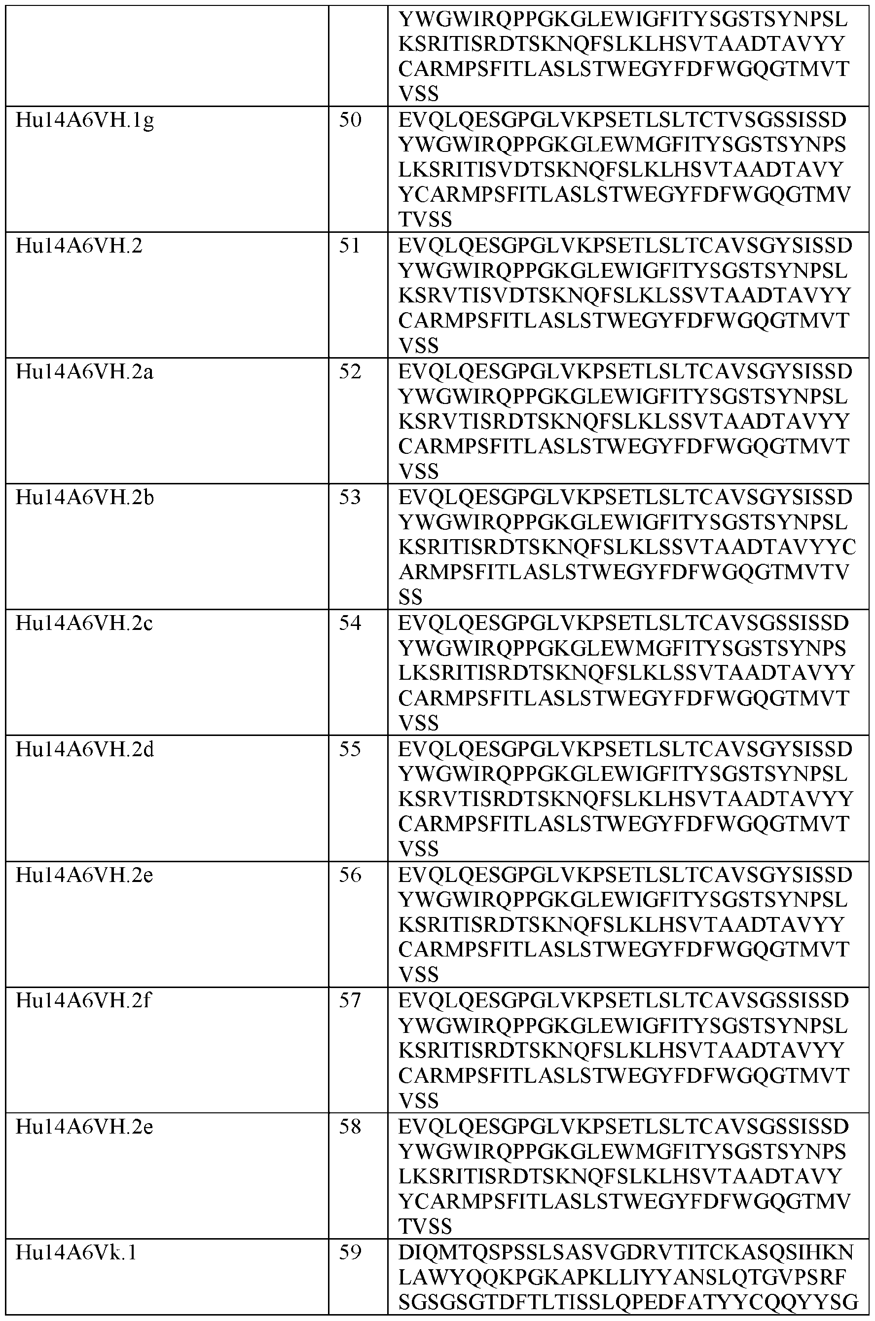


In some embodiments, an anti-TIGIT antibody or antigen binding fragment thereof comprises three light chain CDRs of CDRL1, CDRL2, and CDRL3 and/or three heavy chain CDRs of CDRH1, CDRH2, and CDRH3.
In one embodiment, the anti-TIGIT antibody or antigen binding fragment thereof comprises a CDRH1 having the amino acid sequence as set forth in SEQ ID NO:35, a CDRH2 having the amino acid sequence as set forth in SEQ ID NO:36, a CDRH3 having any of the amino acid sequences as set forth in SEQ ID NOs:37. 103, 104. 105, 106, 107, or 160, a CDRLl having the amino acid sequence as set forth in SEQ ID NO:38, a CDRL2 having any of the amino acid sequences as set forth in SEQ ID NOs:39, 89, 90, 91, 92, 93, 94, 95, 96, 97, or 161, and a CDRL3 having any of the amino acid sequences as set forth in SEQ ID NOs:40, 98, 99, 100, 101, 102, or 162.
In another embodiment, the anti-TIGIT antibody or antigen binding fragment thereof comprises a CDRH1 having the amino acid sequence as set forth in SEQ ID NO:81, a CDRH2 having the amino acid sequence as set forth in SEQ ID NO:82, a CDRH3 having the amino acid sequence as set forth in SEQ ID NO: 83. a CDRL1 having the amino acid sequence as set forth in SEQ ID NO:84, a CDRL2 having the amino acid sequence as set forth in SEQ ID NO:85, and a CDRL3 having the amino acid sequence as set forth in SEQ ID NO: 86.
In another embodiment, the anti-TIGIT antibody or antigen binding fragment thereof comprises a CDRH1 having the amino acid sequence as set forth in SEQ ID NO: 108, a CDRH2
having any of the amino acid sequences as set forth in SEQ ID NOs: 109. 116, 117. 118, 119, 120, 121, 122, 123, 124, 125, 126, 127, 128, 129, 130, 131, 154, 155 or 167, a CDRH3 having any of the amino acid sequences as set forth in SEQ ID NOs: 110, 173, 174, 175, 176, 177, 178, 179, 180, 181, 182, 183, 184, 185, 186 or 187, a CDRLl having the amino acid sequence as set forth in SEQ ID NO: 111, a CDRL2 having any of the amino acid sequences as set forth in SEQ ID NOs: 112, 132, 133. 134, 135, 136, 137, 138, 139, 140, 141, 142 or 168, and a CDRL3 having the amino acid sequence as set forth in SEQ ID NO: 113.
In one embodiment, the anti-TIGIT antibody or antigen binding fragment thereof comprises a CDRH1 having the amino acid sequence as set forth in SEQ ID NO:35, a CDRH2 having the amino acid sequence as set forth in SEQ ID NO:36, a CDRH3 having the amino acid sequence as set forth in SEQ ID NO:37, a CDRL1 having the amino acid sequence as set forth in SEQ ID NO:38, a CDRL2 having the amino acid sequence as set forth in SEQ ID NO:39, and a CDRL3 having the amino acid sequence as set forth in SEQ ID NO:40.
In one embodiment, the anti-TIGIT antibody or antigen binding fragment thereof comprises CDRH1 having the amino acid sequence as set forth in SEQ ID NO: 108. a CDRH2 having any one of the amino acid sequences as set forth in SEQ ID NO: 109, 154 or 145, a CDRH3 having the amino acid sequence as set forth in SEQ ID NO: 110, a CDRL1 having the amino acid sequence as set forth in SEQ ID NO: 111, a CDRL2 having the amino acid sequence as set forth in SEQ ID NO: 112, and a CDRL3 having the amino acid sequence as set forth in SEQ ID NO: 113
In another embodiment, the anti-TIGIT antibody or antigen binding fragment thereof comprises a CDRH1 having the amino acid sequence as set forth in SEQ ID NO: 108, a CDRH2 having the amino acid sequence as set forth in SEQ ID NO: 154, a CDRH3 having the amino acid sequence as set forth in SEQ ID NO: 110, a CDRL1 having the amino acid sequence as set forth in SEQ ID NO: 111, a CDRL2 having the amino acid sequence as set forth in SEQ ID NO: 112, and a CDRL3 having the amino acid sequence as set forth in SEQ ID NO: 113.
In some embodiments, the anti-TIGIT antibody or antigen binding fragment thereof comprises a light chain comprising or consisting of the amino acid sequence as set forth in SEQ ID NO:294 or 317 and a heavy chain comprising or consisting of the amino acid sequence as set forth in SEQ ID NO:295 or 318.
In one embodiment, the anti-TIGIT antibody or antigen binding fragment thereof comprises a variable heavy' chain region and a variable light chain variable region. In one embodiment, the anti-TIGIT antibody or antigen binding fragment thereof comprises a variable heavy chain region comprising the amino acid sequence as set forth in SEQ ID NO:41 and a variable light chain region comprising the amino acid sequence as set forth in SEQ ID NO:42.
In one embodiment, the anti-TIGIT antibody or antigen binding fragment thereof comprises a variable heavy chain region comprising the amino acid sequence as set forth in SEQ ID NO:87 and a variable light chain region comprising the amino acid sequence as set forth in SEQ ID NO:88.
In one embodiment, the anti-TIGIT antibody or antigen binding fragment thereof comprises a variable heavy' chain region comprising the amino acid sequence as set forth in SEQ ID NO: 114 and a variable light chain region comprising the amino acid sequence as set forth in SEQ ID NO: 115.
In one embodiment, the anti-TIGIT antibody or antigen binding fragment thereof comprises a variable heavy' chain region comprising any of the amino acid sequences as set forth in SEQ ID NOs: 43-58, 65-75 and 87 and a variable light chain region comprising any one of the amino acid sequences as set forth in SEQ ID NOs: 59-64, 76-80 and 88.
In one embodiment, the anti-TIGIT antibody or antigen binding fragment thereof comprises a variable heavy’ chain region comprising any of the amino acid sequences as set forth in SEQ ID NOs: 144-149 and a variable light chain region comprising any of the amino acid sequences as set forth in SEQ ID NOs: 150-153.
In one embodiment, the anti-TIGIT antibody or antigen binding fragment thereof comprises a variable heavy chain region comprising a variable heavy chain region comprising the amino acid sequence as set forth in SEQ ID NO: 148 and a variable light chain region comprising the amino acid sequence as set forth in SEQ ID NO: 152.
In one embodiment, the anti-TIGIT antibody or antigen binding fragment thereof comprises a variable heavy’ chain region comprising the amino acid sequence as set forth in SEQ ID NO: 147 and a variable light chain region comprising the amino acid sequence as set forth in SEQ ID NO: 150.
In one embodiment, the anti-TIGIT antibody or antigen binding fragment thereof comprises a variable heavy' chain region comprising the amino acid sequence as set forth in SEQ ID NO: 148 and a variable light chain region comprising the amino acid sequence as set forth in SEQ ID NO: 153.
In one embodiment, the anti-TIGIT antibody or antigen binding fragment thereof comprises a variable heavy' chain region comprising the amino acid sequence as set forth in SEQ ID NO: 163 and a variable light chain region comprising the amino acid sequence as set forth in SEQ ID NO:165.
In one embodiment, the anti-TIGIT antibody or antigen binding fragment thereof comprises a variable heavy' chain region comprising the amino acid sequence as set forth in SEQ
ID NO: 169 and a variable light chain region comprising the amino acid sequence as set forth in SEQ ID NO: 171.
In one embodiment, the anti-TIGIT antibody or antigen binding fragment thereof comprises a variable heavy chain region comprising the amino acid sequence as set forth in SEQ ID NO: 164 and a variable light chain region comprising the amino acid sequence as set forth in
SEQ ID NO: 166
In one embodiment, the anti-TIGIT antibody or antigen binding fragment thereof comprises a variable heavy chain region comprising the amino acid sequence as set forth in SEQ ID NO:170 and a variable light chain region comprising the amino acid sequence as set forth in SEQ ID NO: 172.
Table 7, Exemplary sequences of anti-TIGIT antibodies


In one embodiment, the anti-TIGIT antibody or antigen binding fragment comprises a CDRH1 having the amino acid sequence as set forth in SEQ ID NO: 188, a CDRH2 having the amino acid sequence as set forth in SEQ ID NO: 189. a CDRH3 having any of the amino acid sequences as set forth in SEQ ID NOs:190, 220, 221, or 222, a CDRL1 having the amino acid sequence as set forth in SEQ ID NO: 191 , a CDRL2 having the amino acid sequence as set forth in SEQ ID NO: 192, and a CDRL3 having any of the amino acid sequences as set forth in SEQ ID NOs:193, 232, 233, 234, 235, 236, or 237.
In one embodiment, the anti-TIGIT antibody or antigen binding fragment thereof comprises a CDRH1 having the amino acid sequence as set forth in SEQ ID NO:204, a CDRH2 having any of the amino acid sequences as set forth in SEQ ID NOs: 205, 256, 257, 258, 259. 260, 261, 262, or 263, a CDRH3 having the amino acid sequence as set forth in SEQ ID NO:206, a CDRL1 having the amino acid sequence as set forth in SEQ ID NO:207, a CDRL2 having the amino acid sequence as set forth in SEQ ID NO:208, and a CDRL3 having the amino acid sequence as set forth in SEQ ID NO:209.
In one embodiment, the anti-TIGIT antibody or antigen binding fragment thereof comprises a variable heavy chain region and a variable light chain variable region. In one embodiment, the anti-TIGIT antibody or antigen binding fragment thereof comprises a variable heavy chain region comprising the amino acid sequence as set forth in SEQ ID NO: 194 and a variable light chain region comprising the amino acid sequence as set forth in SEQ ID NO: 195.
In one embodiment, the anti-TIGIT antibody or antigen binding fragment thereof comprises a variable heavy chain region comprising the amino acid sequence as set forth in SEQ ID NO: 196 and a variable light chain region comprising the amino acid sequence as set forth in SEQ ID N0:200.
In one embodiment, the anti-TIGIT antibody or antigen binding fragment thereof comprises a variable heavy chain region comprising the amino acid sequence as set forth in SEQ ID NO: 210 and a variable light chain region comprising the amino acid sequence as set forth in SEQ ID NO:211.
In one embodiment, the anti-TIGIT antibody or antigen binding fragment thereof comprises a variable heavy chain region comprising the amino acid sequence as set forth in SEQ ID NO: 212 and a variable light chain region comprising the amino acid sequence as set forth in SEQ ID NO:216.
In one embodiment, the anti-TIGIT antibody or antigen binding fragment thereof comprises a variable heavy chain region comprising any of the amino acid sequences as set forth in SEQ ID NOs: 197, 198, 199. 223, 224, 225. 226, 227, 228. 229, 230, and 231 and a variable light chain region comprising any of the amino acid sequences as set forth in SEQ ID NOs: 201, 202, 203, 238, 239, 240, 241, 242, 243, 244, 245, 246, 247, 248, 249, 250, 251, 252, 253, 254, and 255.
In one embodiment, the anti-TIGIT antibody or antigen binding fragment thereof comprises a variable heavy chain region comprising any of the amino acid sequences as set forth in SEQ ID NOs: 213, 214. 215, 264, 265. 266, 267, 268, 269, 270. 271, 272, 273, 274, 275. 276, 277, 278. 279, 280, 281. 282, 283. 284, 285, and 286 and a variable light chain region comprising any of the amino acid sequences as set forth in SEQ ID NOs: 217, 218, and 219.
In one embodiment, the anti-TIGIT antibody is vibostolimab, which comprises three light chain CDRs: CDRL1 having the amino acid sequence as set forth in SEQ ID NO: 111, CDRL2 having the amino acid sequence as set forth in SEQ ID NO: 112, and CDRL3 having the amino acid sequence as set forth in SEQ ID NO: 113, and three heavy chain CDRs: CDRH1 having the amino acid sequence as set forth in SEQ ID NO: 108, CDRH2 having the amino acid sequence as set forth in SEQ ID NO: 154, and CDRH3 having the amino acid sequence as set forth in SEQ ID NO: 110. Vibostolimab is also referred to as MK-7684.
Additional anti-TIGIT antibodies which may be used in the formulations described herein include those disclosed, for example, in PCT International Application No. WO 2016/106302; WO 2016/011264; and WO 2009/126688.
Table 8. Exemplary Heavy Chain Sequences

In any of the above mentioned embodiments, the anti-TIGIT antibody or antigen binding fragment thereof is an antibody comprising any of the variable heavy chains described above and any human heavy chain constant domain. In one embodiment, the antibody or antigen binding fragment thereof of the invention is of the IgG isotype, and comprises a human IgGl, IgG2, IgG3 or IgG4 human heavy chain constant domain. In one embodiment, the antibody or antigen binding fragment thereof of the invention comprises a human heavy chain IgGl constant domain (SEQ ID NO: 291) or a variant thereof, wherein the variant comprises up to 20 modified amino acid substitutions. In one embodiment, the antibody or antigen binding fragment thereof of the invention is an antibody comprising a human heavy chain IgGl constant domain comprising the amino acid sequence as set forth in SEQ ID NO: 291. In one embodiment, the antibody or antigen binding fragment thereof of the invention comprises a human heavy chain IgGl constant domain wherein the IgGl constant domain is afucosylated. In one embodiment, the antibody or antigen binding fragment thereof of the invention comprises a human heavy chain IgG4 constant domain or a variant thereof, wherein the variant comprises up to 20 modified amino acid substitutions. In another embodiment, the antibody or antigen binding fragment thereof of the invention comprises a human heavy chain IgG4 constant domain, wherein the amino acid at position 228 (using EU numbering scheme) has been substituted from Ser to Pro. In one embodiment, the antibody or antigen binding fragment thereof of the invention comprises a
human heavy chain IgG4 constant domain comprising the amino acid sequence as set forth in SEQ ID NO: 292.
In any of the above mentioned embodiments, the anti-TIGIT antibody or antigen binding fragment thereof can comprise any of the variable light chains described above and human light chain constant domain. In one embodiment, the antibody or antigen binding fragment thereof of the invention comprises a human kappa light chain constant domain or a variant thereof, wherein the variant comprises up to 20 modified amino acid substitutions. In another embodiment, the antibody or antigen binding fragment thereof of the invention comprises a human lambda light chain constant domain or a variant thereof, wherein the variant comprises up to 20 modified amino acid substitutions. In one embodiment, the antibody or antigen binding fragment thereof of the invention comprises a human kappa light chain constant domain comprising the amino acid sequence as set forth in SEQ ID NO: 293.
Antibody Drug Conjugates
In various embodiments of the methods provided herein, the ADC used in the methods comprises or is an anti-191P4D12 ADC described herein and/or in US Patent No. 8.637,642, which is herein incorporated in its entirety by reference. In some embodiments, the anti-191P4D12 antibody drug conjugate provided for the methods herein comprises an antibody or antigen binding fragment thereof that binds to 191P4D12 as provided herein, conjugated to one or more units of cytotoxic agents (drug units, or D) as provided herein. In certain embodiments, the cytotoxic agents (drug units, or D) can be covalently linked directly or via a linker unit (LU) as provided herein.
In some embodiments, the antibody drug conjugate compound has the following formula:
L-(LU-D)P (I) or a pharmaceutically acceptable salt or solvate thereof; wherein:
L is the antibody unit, e.g., the anti-nectin-4 antibody or an antigen binding fragment thereof, and
(LU-D) is a linker unit-drug unit moiety, wherein:
LU- is a linker unit, and
D is a drug unit having cytostatic or cytotoxic activity against a target cell; and p is an integer from 1 to 20.
In some embodiments, the antibody drug conjugate compound has the following formula: L - (Aa-Ww-Yy-D)p (II)
or a pharmaceutically acceptable salt or solvate thereof, wherein:
L is the antibody unit, e.g., the anti-nectin-4 antibody or an antigen binding fragment thereof; and
-Aa-Ww-Yy- is a linker unit (LU), wherein:
-A- is a stretcher unit, a is 0 or 1, each -W- is independently an amino acid unit, w is an integer ranging from 0 to 12.
-Y- is a self-immolative spacer unit, y is 0, 1 or 2,
D is a drug units having cytostatic or cytotoxic activity7 against the target cell; and p is an integer from 1 to 20.
In some embodiments, a is 0 or 1 , w is 0 or 1 , and y is 0, 1 or 2. In some embodiments, a is 0 or 1, w is 0 or 1, and y is 0 or 1.
In some embodiments, p ranges from 1 to 20, 1 to 19, 1 to 18. 1 to 17, 1 to 16, 1 to 15, 1 to 14, 1 to 13, 1 to 12, 1 to 1 1, 1 to 10, 1 to 9, 1 to 8, 1 to 7, 1 to 6, 1 to 5, 1 to 4, 1 to 3, or 1 to 2. In some embodiments, p ranges from 2 to 20, 2 to 19, 2 to 18, 2 to 17, 2 to 16, 2 to 15, 2 to 14, 2 to 13, 2 to 12, 2 to 11, 2 to 10, 2 to 9, 2 to 8, 2 to 7, 2 to 6, 2 to 5, 2 to 4 or 2 to 3. In some embodiments, p ranges from 3 to 20, 3 to 19, 3 to 18, 3 to 17, 3 to 16, 3 to 15, 3 to 14, 3 to 13, 3 to 12, 3 to 11, 3 to 10, 3 to 9, 3 to 8, 3 to 7, 3 to 6, 3 to 5, or 3 to 4. In some embodiments, p is about 1. In some embodiments, p is about 2. In some embodiments, p is about 3. In some embodiments, p is about 4. In some embodiments, p is about 3.8. In some embodiments, p is about 5. In some embodiments, p is about 6. In some embodiments, p is about 7. In some embodiments, p is about 8. In some embodiments, p is about 9. In some embodiments, p is about 10. In some embodiments, p is about 11. In some embodiments, p is about 12. In some embodiments, p is about 13. In some embodiments, p is about 14. In some embodiments, p is about 15. In some embodiments, p is about 16. In some embodiments, p is about 17. In some embodiments, p is about 18. In some embodiments, p is about 19. In some embodiments, p is about 20.
In some embodiments, when w is not zero, y is 1 or 2. In some embodiments, when w is 1 to 12, y is 1 or 2. In some embodiments, w is 2 to 12 and y is 1 or 2. In some embodiments, a is 1 and w and y are 0.
In some specific embodiments of the methods provided herein, the cytotoxic agent as part of any of the ADCs provided herein for the methods comprises, consists of, or is MMAE.
For compositions comprising a plurality antibodies or antigen binding fragments thereof, the drug loading is represented by p, the average number of drug molecules per antibody unit. Drug loading can range from 1 to 20 drugs (D) per antibody. The average number of drugs per antibody in preparation of conjugation reactions can be characterized by conventional means such as mass spectroscopy, ELISA assay, and HPLC. The quantitative distribution of antibody drug conjugates in terms of p can also be determined. In some instances, separation, purification, and characterization of homogeneous antibody drug conjugates where p is a certain value from antibody drug conjugates with other drug loadings can be achieved by means such as reverse phase HPLC or electrophoresis. In certain exemplary embodiments, p is from 2 to 8.
Additional embodiments of the ADC for the methods provided herein have been described in US Patent No. 8,637,642 and International Application No.
PCT/US2019/056214 (Publication No. W02020/117373), both of which are hereby incorporated in their entireties by reference.
In some embodiments of the methods provided herein, the ADC is enfortumab vedotin. In certain embodiments of the methods provided herein, the ADC is a biosimilar of enfortumab vedotin.
Anti-191P4D12 Antibodies or Antigen Binding Fragments
In one embodiment, the antibody or antigen binding fragment thereof that binds to nectin- 4-related proteins is an antibody or antigen binding fragment that specifically binds to nectin-4 protein comprising amino acid sequence of SEQ ID NO:295 or 318 (see FIG. 1 A). The corresponding cDNA encoding the 191P4D12 protein has a sequence of SEQ ID NO: 294 or 317 (see FIG. 1A).
The antibody that specifically binds to nectin-4 protein comprising amino acid sequence of SEQ ID NO:295 or 318 includes antibodies that can bind to other nectin-4-related proteins. For example, antibodies that bind nectin-4 protein comprising amino acid sequence of SEQ ID NO:295 or 318 can bind nectin-4-related proteins such as nectin-4 variants and the homologs or analogs thereof.
In some embodiments, the anti-nectin-4 antibody provided herein is a monoclonal antibody. In some embodiments, the antibody comprises a heavy chain comprising an amino acid sequence of SEQ ID NO:297 (cDNA sequence of SEQ ID NO:296), and/or a light chain comprising an amino acid sequence of SEQ ID NO:299 (cDNA sequence of SEQ ID NO:298), as shown in FIGS. IB and 1C.
In some embodiments, the anti-nectin-4 antibody or antigen binding fragment thereof comprises a heavy chain variable region comprising complementarity determining regions
(CDRs) comprising the amino acid sequences of the CDRs of the heavy chain variable region set forth in SEQ ID NO:315 (which is the amino acid sequence ranging from the 20th amino acid (glutamic acid) to the 136th amino acid (serine) of SEQ ID N0:300) and a light chain variable region comprising CDRs comprising the amino acid sequences of the CDRs of the light chain variable region set forth in SEQ ID NO:316 (which is the amino acid sequence ranging from the 23rd amino acid (aspartic acid) to the 130th amino acid (arginine) of SEQ ID NO:301). In certain embodiments, the anti-nectin-4 antibody or antigen binding fragment thereof comprises a heavy chain variable region comprising complementarity determining region 1 (CDR-H1), CDR-H2. and CDR-H3 comprising the amino acid sequences of the corresponding CDR-H1, CDR-H2, and CDR-H3 in the heavy chain variable region sequence set forth in SEQ ID NO:315 (which is the amino acid sequence ranging from the 20th amino acid (glutamic acid) to the 136th amino acid (serine) of SEQ ID NO:300) and a light chain variable region comprising CDR-L1, CDR-L2, and CDR-L3 comprising the amino acid sequences of the corresponding CDR-L1, CDR-L2, and CDR-L3 in the light chain variable region sequence set forth in SEQ ID NO:316 (which is the amino acid sequence ranging from the 23rd amino acid (aspartic acid) to the 130th amino acid (arginine) of SEQ ID NO:301). In some embodiments, the anti-nectin-4 antibody or antigen binding fragment thereof comprises a heavy chain variable region comprising complementarity determining regions (CDRs) consisting of the amino acid sequences of the CDRs of the heavy chain variable region set forth in SEQ ID NO: 315 (which is the amino acid sequence ranging from the 20th amino acid (glutamic acid) to the 136th amino acid (serine) of SEQ ID NO:300) and a light chain variable region comprising CDRs consisting of the amino acid sequences of the CDRs of the light chain variable region set forth in SEQ ID NO:316 (which is the amino acid sequence ranging from the 23rd amino acid (aspartic acid) to the 130th amino acid (arginine) of SEQ ID NO:301). In certain embodiments, the anti-nectin-4 antibody or antigen binding fragment thereof comprises a heavy chain variable region comprising complementarity determining region 1 (CDR-H1), CDR-H2, and CDR-H3 consisting of the amino acid sequences of the corresponding CDR-H1, CDR-H2, and CDR-H3 in the heavy chain variable region sequence set forth in SEQ ID NO: 315 (which is the amino acid sequence ranging from the 20th amino acid (glutamic acid) to the 136th amino acid (serine) of SEQ ID N0:300) and a light chain variable region comprising CDR-L1, CDR-L2, and CDR-L3 consisting of the amino acid sequences of the corresponding CDR-L1, CDR-L2, and CDR-L3 in the light chain variable region sequence set forth in SEQ ID NO:316 (which is the amino acid sequence ranging from the 23rd amino acid (aspartic acid) to the 130th amino acid (arginine) of SEQ ID NO:301). SEQ ID NO:315, SEQ ID NO:316, SEQ ID NO:300 and SEQ ID NO:301 are as shown in FIGS. ID and IE and listed below:

CDR sequences can be determined according to well-known numbering systems. As described above, CDR regions are well-known to those skilled in the art and have been defined by well-known numbering systems. For example, the Kabat Complementarity Determining Regions (CDRs) are based on sequence variability and are the most commonly used (see, e.g., Kabat et al. , supra). Chothia refers instead to the location of the structural loops (see, e.g., Chothia and Lesk, 1987, J. Mol. Biol. 196:901-17). The end of the Chothia CDR-H1 loop when numbered using the Kabat numbering convention varies between H32 and H34 depending on the length of the loop (this is because the Kabat numbering scheme places the insertions at H35A and H35B; if neither 35 A nor 35B is present, the loop ends at 32; if only 35A is present, the loop ends at 33; if both 35 A and 35B are present, the loop ends at 34). The AbM hypervariable regions represent a compromise between the Kabat CDRs and Chothia structural loops, and are
used by Oxford Molecular' s AbM antibody modeling software (see, e.g.. Antibody Engineering Vol. 2 (Kontermann and Dubel eds., 2d ed. 2010)). The "contact" hypervariable regions are based on an analysis of the available complex crystal structures. Another universal numbering system that has been developed and widely adopted is ImMunoGeneTics (IMGT) Information System® (Lafranc et al., 2003, Dev. Comp. Immunol. 27(l):55-77). IMGT is an integrated information system specializing in immunoglobulins (IG), T-cell receptors (TCR), and major histocompatibility complex (MHC) of human and other vertebrates. Herein, the CDRs are referred to in terms of both the amino acid sequence and the location within the light or heavy chain. As the "location" of the CDRs within the structure of the immunoglobulin variable domain is conserved between species and present in structures called loops, by using numbering systems that align variable domain sequences according to structural features, CDR and framework residues are readily identified. This information can be used in grafting and replacement of CDR residues from immunoglobulins of one species into an acceptor framework from, typically, a human antibody. An additional numbering system (AHon) has been developed by Honegger and Pliickthun. 2001. J. Mol. Biol. 309: 657-70. Correspondence between the numbering system, including, for example, the Kabat numbering and the IMGT unique numbering system, is well- known to one skilled in the art (see, e.g., Kabat, supra; Chothia and Lesk, supra; Martin, supra; Lefranc et al. , supra).
In some embodiments, the anti-nectin-4 antibody or antigen binding fragment thereof comprises a heavy chain variable region comprising CDRs (CDR-H1, CDR-H2, CDR-H3, CDR- Ll, CDR-L2, and CDR-L3) comprising the amino acid sequences of the CDRs of the heavy chain variable region set forth in SEQ ID NO:315 according to Kabat numbering and a light chain variable region comprising CDRs comprising the amino acid sequences of the CDRs of the light chain variable region set forth in SEQ ID NO:316 according to Kabat numbering.
In some embodiments, the anti-nectin-4 antibody or antigen binding fragment thereof comprises a heavy chain variable region comprising CDRs (CDR-H1, CDR-H2,CDR-H3, CDR- Ll, CDR-L2, and CDR-L3) comprising the amino acid sequences of the CDRs of the heavy chain variable region set forth in SEQ ID NO:315 according to AbM numbering and a light chain variable region comprising CDRs comprising the amino acid sequences of the CDRs of the light chain variable region set forth in SEQ ID NO:316 according to AbM numbering.
In other embodiments, the anti-nectin-4 antibody or antigen binding fragment thereof comprises a heavy chain variable region comprising CDRs (CDR-H1, CDR-H2, CDR-H3, CDR- Ll, CDR-L2, and CDR-L3) comprising the amino acid sequences of the CDRs of the heavy chain variable region set forth in SEQ ID NO:315 according to Chothia numbering and a light chain
variable region comprising CDRs comprising the amino acid sequences of the CDRs of the light chain variable region set forth in SEQ ID NO:316 according to Chothia numbering.
In other embodiments, the anti-nectin-4 antibody or antigen binding fragment thereof comprises a heavy chain variable region comprising CDRs (CDR-H1, CDR-H2, CDR-H3, CDR- Ll, CDR-L2, and CDR-L3) comprising the amino acid sequences of the CDRs of the heavy chain variable region set forth in SEQ ID NO:315 according to Contact numbering and a light chain variable region comprising CDRs comprising the amino acid sequences of the CDRs of the light chain variable region set forth in SEQ ID NO:316 according to Contact numbering.
In yet other embodiments, the anti-nectin-4 antibody or antigen binding fragment thereof comprises a heavy chain variable region comprising CDRs (CDR-H1, CDR-H2, CDR-H3, CDR- Ll, CDR-L2, and CDR-L3) comprising the amino acid sequences of the CDRs of the heavy chain variable region set forth in SEQ ID NO:315 according to IMGT numbering and a light chain variable region comprising CDRs comprising the amino acid sequences of the CDRs of the light chain variable region set forth in SEQ ID NO:316 according to IMGT numbering.
In some embodiments, the anti-nectin-4 antibody or antigen binding fragment thereof comprises a heavy chain variable region comprising CDRs (CDR-H1, CDR-H2, CDR-H3, CDR- Ll, CDR-L2, and CDR-L3) consisting of the amino acid sequences of the CDRs of the heavy chain variable region set forth in SEQ ID NO:315 according to Kabat numbering and a light chain variable region comprising CDRs consisting of the amino acid sequences of the CDRs of the light chain variable region set forth in SEQ ID NO:316 according to Kabat numbering.
In some embodiments, the anti-nectin-4 antibody or antigen binding fragment thereof comprises a heavy chain variable region comprising CDRs (CDR-H1, CDR-H2, CDR-H3. CDR- Ll, CDR-L2, and CDR-L3) consisting of the amino acid sequences of the CDRs of the heavy chain variable region set forth in SEQ ID NO:315 according to AbM numbering and a light chain variable region comprising CDRs consisting of the amino acid sequences of the CDRs of the light chain variable region set forth in SEQ ID NO:316 according to AbM numbering.
In other embodiments, the anti-nectin-4 antibody or antigen binding fragment thereof comprises a heavy chain variable region comprising CDRs (CDR-H1, CDR-H2, CDR-H3. CDR- Ll, CDR-L2. and CDR-L3) consisting of the amino acid sequences of the CDRs of the heavy chain variable region set forth in SEQ ID NO:315 according to Chothia numbering and a light chain variable region comprising CDRs consisting of the amino acid sequences of the CDRs of the light chain variable region set forth in SEQ ID NO:316 according to Chothia numbering.
In other embodiments, the anti-nectin-4 antibody or antigen binding fragment thereof comprises a heavy chain variable region comprising CDRs (CDR-EI1, CDR-H2, CDR-H3, CDR- Ll, CDR-L2, and CDR-L3) consisting of the amino acid sequences of the CDRs of the heavy’
chain variable region set forth in SEQ ID NO:315 according to Contact numbering and a light chain variable region comprising CDRs consisting of the amino acid sequences of the CDRs of the light chain variable region set forth in SEQ ID NO:316 according to Contact numbering.
In yet other embodiments, the anti-nectin-4 antibody or antigen binding fragment thereof comprises a heavy chain variable region comprising CDRs (CDR-H1, CDR-H2, CDR-H3, CDR- Ll, CDR-L2, and CDR-L3) consisting of the amino acid sequences of the CDRs of the heavy' chain variable region set forth in SEQ ID NO:315 according to IMGT numbering and a light chain variable region comprising CDRs consisting of the amino acid sequences of the CDRs of the light chain variable region set forth in SEQ ID NO:316 according to IMGT numbering.
As described above, the CDR sequences according to different numbering systems can be readily determined, e.g., using online tools such as the one provided by Antigen receptor Numbering And Receptor Classification (ANARCI). For example, the heavy chain CDR sequences within SEQ ID NO:315, and the light chain CDR sequences within SEQ ID NO:316 according to Kabat numbering as determined by ANARCI are listed in Table 9 below.
Table 9: Heavy and Light chain CDR sequences according to Kabat numbering

For another example, the heavy chain CDR sequences within SEQ ID NO:315, and the light chain CDR sequences within SEQ ID NO:316 according to IMGT numbering as determined by ANARCI are listed in Table 10 below.
Table 10: Heavy and Light chain CDR sequences according to IMGT numbering
 In some embodiments, the antibody or antigen binding fragment thereof comprises CDR- H1 comprising an amino acid sequence of SEQ ID NO:302, CDR-H2 comprising an amino acid sequence of SEQ ID NO: 303, CDR-H3 comprising an amino acid sequence of SEQ ID NO: 304, CDR-L1 comprising an amino acid sequence of SEQ ID NO: 305, CDR-L2 comprising an amino acid sequence of SEQ ID NO:306, and CDR-L3 comprising an amino acid sequence of SEQ ID NO: 307.
In some embodiments, the antibody or antigen binding fragment thereof comprises CDR- HI comprising an amino acid sequence of SEQ ID NO:309, CDR-H2 comprising an amino acid sequence of SEQ ID NO: 310, CDR-H3 comprising an amino acid sequence of SEQ ID NO: 311, CDR-L1 comprising an amino acid sequence of SEQ ID NO: 312, CDR-L2 comprising an amino acid sequence of SEQ ID NO:313, and CDR-L3 comprising an amino acid sequence of SEQ ID NO:314.
In some embodiments, the antibody or antigen binding fragment thereof comprises CDR- H1 consisting of an amino acid sequence of SEQ ID NO:302. CDR-H2 consisting of an amino acid sequence of SEQ ID NO: 303, CDR-H3 consisting of an amino acid sequence of SEQ ID NO: 304, CDR-L1 consisting of an amino acid sequence of SEQ ID NO: 305, CDR-L2 consisting of an amino acid sequence of SEQ ID NO: 306, and CDR-L3 consisting of an amino acid sequence of SEQ ID NO: 307.
In some embodiments, the antibody or antigen binding fragment thereof comprises CDR- H1 consisting of an amino acid sequence of SEQ ID NO: 309, CDR-H2 consisting of an amino acid sequence of SEQ ID NO: 310, CDR-H3 consisting of an amino acid sequence of SEQ ID NO: 311. CDR-L1 consisting of an amino acid sequence of SEQ ID NO:312. CDR-L2 consisting of an amino acid sequence of SEQ ID NO:313, and CDR-L3 consisting of an amino acid sequence of SEQ ID NO: 314.
In some embodiments, the antibody or antigen binding fragment thereof comprises a heavy chain variable region comprising the amino acid sequence of SEQ ID NO:315 and a light chain variable region comprising the amino acid sequence of SEQ ID NO:316.
In some embodiments, the antibody or antigen binding fragment thereof comprises a heavy chain vanable region consisting of the amino acid sequence of SEQ ID NO:315 and a light chain variable region consisting of the amino acid sequence of SEQ ID NO:316.
In some embodiments, the antibody comprises a heavy chain comprising the amino acid sequence ranging from the 20th amino acid (glutamic acid) to the 466th amino acid (lysine) of SEQ ID NO:300 and a light chain comprising the amino acid sequence ranging from the 23rd amino acid (aspartic acid) to the 236th amino acid (cysteine) of SEQ ID NO:301.
In some embodiments, the antibody comprises a heavy chain consisting of the amino acid sequence ranging from the 20th amino acid (glutamic acid) to the 466th amino acid (lysine) of SEQ ID N0:300 and a light chain consisting of the amino acid sequence ranging from the 23rd amino acid (aspartic acid) to the 236th amino acid (cysteine) of SEQ ID NO:301.
In some embodiments, amino acid sequence modification(s) of antibodies described herein are contemplated. For example, it may be desirable to optimize the binding affinity and/or other biological properties of the antibody, including but not limited to specificity, thermostability, expression level, effector functions, glycosylation, reduced immunogenicity, or solubility7. Thus, in addition to the antibodies described herein, it is contemplated that antibody variants can be prepared. For example, antibody variants can be prepared by introducing appropriate nucleotide changes into the encoding DNA, and/or by synthesis of the desired antibody or polypeptide. Those skilled in the art who appreciate that amino acid changes can alter post-translational processes of the antibody, such as changing the number or position of glycosylation sites or altering the membrane anchoring characteristics.
In some embodiments, the antibodies provided herein are chemically modified, for example, by the covalent attachment of any type of molecule to the antibody. The antibody derivatives can include antibodies that have been chemically modified, for example, by glycosylation, acetylation, pegylation. phosphorylation, amidation, derivatization by known protecting/blocking groups, proteolytic cleavage, linkage to a cellular ligand or other protein, etc. Any of numerous chemical modifications can be carried out by known techniques, including, but not limited to, specific chemical cleavage, acetylation, formulation, metabolic synthesis of tunicamycin. etc. Additionally, the antibody can contain one or more non-classical amino acids.
Variations can be a substitution, deletion, or insertion of one or more codons encoding the single domain antibody or polypeptide that results in a change in the amino acid sequence as compared with the original antibody or polypeptide. Amino acid substitutions can be the result of replacing one amino acid with another amino acid comprising similar structural and/or chemical properties, such as the replacement of a leucine with a serine, e.g., conservative amino acid replacements. Standard techniques known to those of skill in the art can be used to introduce mutations in the nucleotide sequence encoding a molecule provided herein, including, for example, site-directed mutagenesis and PCR-mediated mutagenesis which results in amino acid substitutions. Insertions or deletions can optionally be in the range of about 1 to 5 amino acids. In certain embodiments, the substitution, deletion, or insertion includes fewer than 25 amino acid substitutions, few er than 20 amino acid substitutions, fewer than 1 amino acid substitutions, fewer than 10 amino acid substitutions, fewer than 5 amino acid substitutions, fewer than 4 amino acid substitutions, fewer than 3 amino acid substitutions, or fewer than 2 amino acid
substitutions relative to the original molecule. In a specific embodiment, the substitution is a conservative amino acid substitution made at one or more predicted non-essential amino acid residues. The variation allowed can be determined by systematically making insertions, deletions, or substitutions of amino acids in the sequence and testing the resulting variants for activity exhibited by the parental antibodies.
Amino acid sequence insertions include amino- and/or carboxyl-terminal fusions ranging in length from one residue to polypeptides containing multiple residues, as well as intrasequence insertions of single or multiple amino acid residues. Examples of terminal insertions include an antibody with an N-terminal methionyl residue.
Antibodies generated by conservative amino acid substitutions are included in the present disclosure. In a conservative amino acid substitution, an amino acid residue is replaced with an amino acid residue comprising a side chain with a similar charge. As described above, families of amino acid residues comprising side chains with similar charges have been defined in the art. These families include amino acids with basic side chains (e.g., lysine, arginine, histidine), acidic side chains (e.g., aspartic acid, glutamic acid), uncharged polar side chains (e.g., glycine, asparagine, glutamine, serine, threonine, tyrosine, cysteine), nonpolar side chains (e g., alanine, valine, leucine, isoleucine, proline, phenylalanine, methionine, tryptophan), beta-branched side chains (e.g., threonine, valine, isoleucine) and aromatic side chains (e.g., tyrosine, phenylalanine, try ptophan, histidine). Alternatively, mutations can be introduced randomly along all or part of the coding sequence, such as by saturation mutagenesis, and the resultant mutants can be screened for biological activity to identify mutants that retain activity. Following mutagenesis, the encoded protein can be expressed and the activity of the protein can be determined conservative (e.g., within an amino acid group with similar properties and/or side chains) substitutions can be made, so as to maintain or not significantly change the properties.
Amino acids can be grouped according to similarities in the properties of their side chains (see, e.g., Lehninger, Biochemistry 73-75 (2d ed. 1975)): (1) non-polar: Ala (A), Vai (V), Leu (L), He (I), Pro (P), Phe (F), Trp (W), Met (M); (2) uncharged polar: Gly (G), Ser (S), Thr (T), Cys (C), Tyr (Y), Asn (N). Gin (Q); (3) acidic: Asp (D), Glu (E); and (4) basic: Lys (K), Arg (R), His(H). Alternatively, naturally occurring residues can be divided into groups based on common side-chain properties: (1) hydrophobic: Norleucine, Met, Ala, Vai, Leu, He; (2) neutral hydrophilic: Cys, Ser, Thr, Asn, Gin; (3) acidic: Asp, Glu; (4) basic: His, Lys, Arg; (5) residues that influence chain orientation: Gly, Pro; and (6) aromatic: Trp, Tyr, Phe.
For example, any cysteine residue not involved in maintaining the proper conformation of the antibody also can be substituted, for example, with another amino acid, such as alanine or serine, to improve the oxidative stability of the molecule and to prevent aberrant crosslinking.
The variations can be made using methods known in the art such as oligonucleotide- mediated (site-directed) mutagenesis, alanine scanning, and PCR mutagenesis. Site-directed mutagenesis (see, e.g., Carter, 1986, Biochem J. 237: 1-7; and Zoller et al. , 1982, Nucl. Acids Res. 10:6487-500), cassette mutagenesis (see, e.g., Wells et al., 1985, Gene 34:315-23), or other known techniques can be performed on the cloned DNA to produce the anti-anti-MSLN antibody variant DNA.
Covalent modifications of antibodies are included within the scope of the present disclosure. Covalent modifications include reacting targeted amino acid residues of an antibody with an organic derivatizing agent that is capable of reacting with selected side chains or the N- or C- terminal residues of the antibody. Other modifications include deamidation of glutaminyl and asparaginyl residues to the corresponding glutamyl and aspartyl residues, respectively, hydroxylation of proline and lysine, phosphorylation of hydroxyl groups of seryl or threonyl residues, methylation of the a-amino groups of lysine, arginine, and histidine side chains (see, e.g., Creighton, Proteins: Structure and Molecular Properties 79-86 (1983)), acetylation of the N- terminal amine, and amidation of any C-terminal carboxyl group.
Other types of covalent modification of the antibody included within the scope of this present disclosure include altering the native glycosylation pattern of the antibody or polypeptide (see, e.g., Beck et al., 2008, Curr. Pharm. Biotechnol. 9:482-501; and Walsh, 2010, Drug Discov. Today 15:773-80), and linking the antibody to one of a variety of nonproteinaceous polymers, e.g., polyethylene glycol (PEG), polypropylene glycol, or poly oxy alk lenes, in the manner set forth, for example, in U.S. Pat. Nos. 4,640,835; 4.496,689; 4,301.144; 4,670,417; 4,791,192; or 4,179,337.
In certain embodiments, the antibody or antigen binding fragment provided herein comprises a heavy chain having certain homology7 or identity to the heavy chain as set forth in SEQ ID NO:300 and a light chain having certain homology7 or identity7 to the light chain as set forth in SEQ ID NO:301. Such embodiments of heavy /light chains with homology or identity7 are further provided as follows. In some embodiments, the antibody or antigen binding fragment provided herein comprises a heavy chain having more than 70% homology or identity to the heavy chain as set forth in SEQ ID NO:300. In some embodiments, the antibody or antigen binding fragment provided herein comprises a heavy chain having more than 75% homology or identity7 to the heavy chain as set forth in SEQ ID NO:300. In some embodiments, the antibody or antigen binding fragment provided herein comprises a heavy chain having more than 80% homology7 or identity to the heavy chain as set forth in SEQ ID NO:300. In some embodiments, the antibody or antigen binding fragment provided herein comprises a heavy chain having more than 85% homology or identity to the heavy chain as set forth in SEQ ID NO: 300. In some
embodiments, the antibody or antigen binding fragment provided herein comprises a heavy chain having more than 90% homology or identity to the heavy chain as set forth in SEQ ID NO:300. In some embodiments, the antibody or antigen binding fragment provided herein comprises a heavy chain having more than 95% homology or identity to the heavy chain as set forth in SEQ ID NO:300. In certain embodiments, the antibody or antigen binding fragment provided herein comprises a heavy chain having any of the provided homology or identity to the heavy chain as set forth in SEQ ID NO:300. wherein the CDRs (CDR-H1, CDR-H2. and CDR-H3) are identical to the CDRs in the heavy chain as set forth in SEQ ID NO:300. In some embodiments, the antibody or antigen binding fragment provided herein comprises a light chain having more than 70% homologj' or identity to the light chain as set forth in SEQ ID NO:301. In some embodiments, the antibody or antigen binding fragment provided herein comprises a light chain having more than 75% homology or identity to the light chain as set forth in SEQ ID NO:301. In some embodiments, the antibody or antigen binding fragment provided herein comprises a light chain having more than 80% homology or identity to the light chain as set forth in SEQ ID NO:301. In some embodiments, the antibody or antigen binding fragment provided herein comprises a light chain having more than 85% homology or identity to the light chain as set forth in SEQ ID NO:301. In some embodiments, the antibody or antigen binding fragment provided herein comprises a light chain having more than 90% homology or i dentil to the light chain as set forth in SEQ ID NO:301. In some embodiments, the antibody or antigen binding fragment provided herein comprises a light chain having more than 95% homology or identity to the light chain as set forth in SEQ ID NO: 301. In certain embodiments, the antibody or antigen binding fragment provided herein comprises a light chain having any of the provided homology or identity to the light chain as set forth in SEQ ID NO:301, wherein the CDRs (CDR-L1, CDR-L2, and CDR-L3) are identical to the CDRs in the light chain as set forth in SEQ ID NO:301. In certain embodiments, the antibody or antigen binding fragment provided herein comprises any homologous light chain and any homologous heavy chain as provided in this paragraph in any combination or permutation.
In certain embodiments, the antibody or antigen binding fragment provided herein comprises a heavy chain variable region having certain homolog}’ or identity to the heavy chain variable region as set forth in SEQ ID NO:315 and a light chain variable region having certain homology or identity to the light chain variable region as set forth in SEQ ID NO:316. Such embodiments of heavy chain variable regions and light chain variable regions with homolog}’ or identity are further provided as follows. In some embodiments, the antibody or antigen binding fragment provided herein comprises a heavy chain variable region having more than 70% homology or identity to the heavy chain variable region as set forth in SEQ ID NO:315. In some
embodiments, the antibody or antigen binding fragment provided herein comprises a heavy chain variable region having more than 75% homology or identify to the heavy chain variable region as set forth in SEQ ID NO:315. In some embodiments, the antibody or antigen binding fragment provided herein comprises a heavy chain variable region having more than 80% homology or identify to the heavy chain variable region as set forth in SEQ ID NO:315. In some embodiments, the antibody or antigen binding fragment provided herein comprises a heavy chain variable region having more than 85% homology or identify to the heavy chain variable region as set forth in SEQ ID NO:315. In some embodiments, the antibody or antigen binding fragment provided herein comprises a heavy chain variable region having more than 90% homology or identify to the heavy chain variable region as set forth in SEQ ID NO:315. In some embodiments, the antibody or antigen binding fragment provided herein comprises a heavy chain variable region having more than 95% homology or identify to the heavy chain variable region as set forth in SEQ ID NO:315. In certain embodiments, the antibody or antigen binding fragment provided herein comprises a heavy chain variable region having any of the provided homology or identify to the heavy chain variable region as set forth in SEQ ID NO:315, wherein the CDRs (CDR-H1. CDR-H2, and CDR-H3) are identical to the CDRs in the heavy chain variable region as set forth in SEQ ID NO:315. In some embodiments, the antibody or antigen binding fragment provided herein comprises a light chain variable region having more than 70% homology7 or identify to the light chain variable region as set forth in SEQ ID NO:316. In some embodiments, the antibody or antigen binding fragment provided herein comprises a light chain variable region having more than 75% homology or identify to the light chain variable region as set forth in SEQ ID NO:316. In some embodiments, the antibody or antigen binding fragment provided herein comprises a light chain variable region having more than 80% homology or identify to the light chain variable region as set forth in SEQ ID NO: 316. In some embodiments, the antibody or antigen binding fragment provided herein comprises a light chain variable region having more than 85% homology or identify to the light chain variable region as set forth in SEQ ID NO:316. In some embodiments, the antibody or antigen binding fragment provided herein comprises a light chain variable region having more than 90% homology or identify to the light chain variable region as set forth in SEQ ID NO:316. In some embodiments, the antibody or antigen binding fragment provided herein comprises a light chain variable region having more than 95% homology or identify7 to the light chain variable region as set forth in SEQ ID NO:316. In certain embodiments, the antibody or antigen binding fragment provided herein comprises a light chain variable region having any of the provided homology or identify to the light chain variable region as set forth in SEQ ID NO:316, wherein the CDRs (CDR-L1, CDR-L2, and CDR-L3) are identical to the CDRs in the light chain variable region as set forth in SEQ ID NO: 316. In certain embodiments, the
antibody or antigen binding fragment provided herein comprises any homologous light chain variable region and any homologous heavy chain variable region as provided in this paragraph in any combination or permutation.
In some embodiments, the anti-nectin-4 antibody provided herein comprises heavy and light chain CDR regions of an antibody designated Ha22-2(2,4)6.1 produced by a hybridoma deposited under the American Type Culture Collection (ATCC) Accession NO: PTA-11267, or heavy and light chain CDR regions comprising amino acid sequences that are homologous to the amino acid sequences of the heavy and light chain CDR regions of Ha22-2(2, 4)6.1, and wherein the antibodies retain the desired functional properties of the anti-nectin-4 antibody designated Ha22-2(2, 4)6.1 produced by a hybridoma deposited under the American Type Culture Collection (ATCC) Accession NO: PTA-11267.
In some embodiments, the anti-nectin-4 antibody provided herein comprises heavy and light chain CDR regions (CDR-H1, CDR-H2, CDR-H3, CDR-L1. CDR-L2, and CDR-L3) of an antibody designated Ha22-2(2,4)6. 1 produced by a hybridoma deposited under the American Type Culture Collection (ATCC) Accession NO: PTA-11267, or heavy and light chain CDR regions consisting of amino acid sequences that are homologous to the amino acid sequences of the heavy and light chain CDR regions of Ha22-2(2,4)6. 1, and wherein the antibodies retain the desired functional properties of the anti-nectin-4 antibody designated Ha22-2(2,4)6. 1 produced by a hybridoma deposited under the American Type Culture Collection (ATCC) Accession NO: PTA-11267.
In some embodiments, the antibody or antigen binding fragment thereof provided herein comprises a humanized heavy chain variable region and a humanized light chain variable region, wherein:
(a) the heavy chain variable region comprises CDRs (CDR-H1, CDR-H2, and CDR-H3) comprising the amino acid sequences of the heavy chain variable region CDRs set forth in the antibody produced by a hy bridoma deposited under the American Type Culture Collection (ATCC) Accession NO: PTA-11267;
(b) the light chain variable region comprises CDRs (CDR-L1, CDR-L2, and CDR-L3) comprising the amino acid sequences of the light chain variable region CDRs set forth in the antibody produced by a hybridoma deposited under the American Type Culture Collection (ATCC) Accession NO: PTA-11267.
In some embodiments, the antibody or antigen binding fragment thereof provided herein comprises a humanized heavy chain variable region and a humanized light chain variable region, wherein:
(a) the heavy chain variable region comprises CDRs (CDR-H1. CDR-H2. and CDR-H3) consisting of the amino acid sequences of the heavy chain variable region CDRs set forth in the antibody produced by a hybridoma deposited under the American Type Culture Collection (ATCC) Accession NO: PTA-11267;
(b) the light chain variable region comprises CDRs (CDR-L1, CDR-L2, and CDR-L3) consisting of the amino acid sequences of the light chain variable region CDRs set forth in the antibody produced by a hybridoma deposited under the American Type Culture Collection (ATCC) Accession NO: PTA-11267.
In some embodiments, the anti-nectin-4 antibody provided herein comprises heavy and light chain variable regions of an antibody designated Ha22-2(2,4)6. 1 produced by a hybridoma deposited under the American Type Culture Collection (ATCC) Accession NO: PTA-11267, or heavy and light variable regions comprising amino acid sequences that are homologous to the amino acid sequences of the heavy and light chain variable regions of Ha22-2(2,4)6. 1 , and wherein the antibodies retain the desired functional properties of the anti-nectin-4 antibody provided herein. In some embodiments, the anti-nectin-4 antibody provided herein comprises heavy and light chain variable regions of an antibody designated Ha22-2(2,4)6. 1 produced by a hybridoma deposited under the American Type Culture Collection (ATCC) Accession NO: PTA- 11267, or heavy and light variable regions consisting of amino acid sequences that are homologous to the amino acid sequences of the heavy and light chain variable regions of Ha22- 2(2, 4)6.1, and wherein the antibodies retain the desired functional properties of the anti-nectin-4 antibody provided herein. As the constant region of the antibody of the disclosure, any subclass of constant region can be chosen. In one embodiment, human IgGl constant region as the heavy chain constant region and human 1g kappa constant region as the light chain constant region can be used.
In some embodiments, the anti-nectin-4 antibody provided herein comprises heavy and light chains of an antibody designated Ha22-2(2,4)6. 1 produced by a hybridoma deposited under the American Type Culture Collection (ATCC) Accession NO: PTA-11267, or heavy' and light chains comprising amino acid sequences that are homologous to the amino acid sequences of the heavy and light chains of Ha22-2(2.4)6. 1. and wherein the antibodies retain the desired functional properties of the anti-nectin-4 antibody provided herein. In some embodiments, the anti-nectin-4 antibody provided herein comprises heavy and light chains of an antibody designated Ha22-2(2,4)6. 1 produced by a hybridoma deposited under the American Type Culture Collection (ATCC) Accession NO: PTA-11267, or heavy and light chains consisting of amino acid sequences that are homologous to the amino acid sequences of the heavy and light chains of
Ha22-2(2,4)6.1, and wherein the antibodies retain the desired functional properties of the anti- nectin-4 antibody provided herein.
In some embodiments, the antibody or antigen binding fragment thereof provided herein comprises a heavy chain variable region and a light chain variable region, wherein:
(a) the heavy chain variable region comprises an amino acid sequence that is at least 80% homologous or identical to the heavy chain variable region amino acid sequence of the antibody produced by a hybridoma deposited under the American Type Culture Collection
(ATCC) Accession NO: PTA-11267; and
(b) the light chain variable region comprises an amino acid sequence that is at least 80% homologous or identical to the light chain variable region amino acid sequence of the antibody produced by a hybridoma deposited under the American Type Culture Collection
(ATCC) Accession NO: PTA-11267.
In certain embodiments, the antibody or antigen binding fragment provided herein comprises a heavy chain variable region having certain homology’ or identity to the heavy chain variable region amino acid sequence of the antibody produced by a hybridoma deposited under the American Type Culture Collection (ATCC) Accession NO: PTA-11267 and a light chain variable region having certain homology or identity to the light chain variable region amino acid sequence of the antibody produced by a hybridoma deposited under the American Type Culture Collection (ATCC) Accession NO: PTA-11267. Such embodiments of heavy chain variable regions and light chain variable regions with homology or identity are further provided as follows. In some embodiments, the heavy chain variable region comprises an amino acid sequence that is at least 85% homologous or identical to the heavy chain variable region amino acid sequence of the antibody produced by a hybridoma deposited under the American Type Culture Collection (ATCC) Accession NO: PTA-11267. In other embodiments, the heavy chain variable region comprises an amino acid sequence that is at least 90% homologous or identical to the heavy chain variable region amino acid sequence of the antibody produced by a hybridoma deposited under the American Type Culture Collection (ATCC) Accession NO: PTA-11267. In yet other embodiments, the heavy chain variable region comprises an amino acid sequence that is at least 95% homologous or identical to the heavy chain variable region amino acid sequence of the antibody produced by a hybridoma deposited under the American Type Culture Collection (ATCC) Accession NO: PTA-11267. In other embodiments, the heavy chain variable region can be 85%, 86%, 87%, 88%, 89%, 90%, 91%, 92%, 93%, 94%, 95%, 96%, 97%, 98% or 99% homologous or identical to the heavy chain variable region amino acid sequence of the antibody- produced by a hybridoma deposited under the American Type Culture Collection (ATCC) Accession NO: PTA-11267. In some embodiments, the light chain variable region comprises an
amino acid sequence that is at least 85% homologous or identical to the light chain variable region amino acid sequence of the antibody produced by a hybridoma deposited under the American Type Culture Collection (ATCC) Accession NO: PTA-11267. In other embodiments, the light chain variable region comprises an amino acid sequence that is at least 90% homologous or identical to the light chain variable region amino acid sequence of the antibody produced by a hybridoma deposited under the American Type Culture Collection (ATCC) Accession NO: PTA- 11267. In yet other embodiments, the light chain variable region comprises an amino acid sequence that is at least 95% homologous or identical to the light chain variable region amino acid sequence of the antibody produced by a hybridoma deposited under the American Type Culture Collection (ATCC) Accession NO: PTA-11267. In other embodiments, the light chain variable region can be 85%, 86%, 87%, 88%, 89%, 90%, 91 %, 92%, 93%, 94%, 95%, 96%, 97%, 98% or 99% homologous or identical to the light chain variable region amino acid sequence of the antibody produced by a hybridoma deposited under the American Type Culture Collection (ATCC) Accession NO: PTA-11267. In certain embodiments, the antibody or antigen binding fragment provided herein comprises any homologous light chain variable region and any homologous heavy chain variable region as provided in this paragraph in any combination or permutation.
In other embodiments, the antibody or antigen binding fragment thereof provided herein comprises a heavy chain and a light chain, wherein:
(a) the heavy chain comprises an amino acid sequence that is at least 80%homologous or identical to the heavy chain amino acid sequence of the antibody produced by a hybridoma deposited under the American Type Culture Collection (ATCC) Accession NO: PTA-11267; and
(b) the light chain comprises an amino acid sequence that is at least 80% homologous or identical to the light chain amino acid sequence of the antibody produced by a hybridoma deposited under the American Type Culture Collection (ATCC) Accession NO: PTA-11267. In certain embodiments, the antibody or antigen binding fragment provided herein comprises a heavy chain having certain homology or identity to the heavy chain amino acid sequence of the antibody produced by a hybridoma deposited under the American Type Culture Collection (ATCC) Accession NO: PTA-11267 and a light chain having certain homology or identity to the light chain amino acid sequence of the antibody produced by a hybridoma deposited under the American Type Culture Collection (ATCC) Accession NO: PTA- 11267. Such embodiments of heavy chains and light chains with homology or identity are further provided as follows. In some embodiments, the heavy chain comprises an amino acid sequence that is at least 85% homologous or identical to the heavy chain amino acid sequence of the antibody produced by a hybridoma deposited under the American Type Culture Collection
(ATCC) Accession NO: PTA-11267. In other embodiments, the heavy chain comprises an amino acid sequence that is at least 90% homologous or identical to the heavy chain amino acid sequence of the antibody produced by a hybridoma deposited under the American Type Culture Collection (ATCC) Accession NO: PTA-11267. In yet other embodiments, the heavy chain comprises an amino acid sequence that is at least 95% homologous or identical to the heavy chain amino acid sequence of the antibody produced by a hybridoma deposited under the American Type Culture Collection (ATCC) Accession NO: PTA-11267. In other embodiments, the heavy chain can be 85%. 86%. 87%. 88%. 89%. 90%. 91%. 92%. 93%. 94%. 95%. 96%. 97%, 98% or 99% homologous or identical to the heavy chain amino acid sequence of the antibody produced by a hybridoma deposited under the American Type Culture Collection (ATCC) Accession NO: PTA-11267. In some embodiments, the light chain comprises an amino acid sequence that is at least 85% homologous or identical to the light chain amino acid sequence of the antibody produced by a hybridoma deposited under the American Type Culture Collection (ATCC) Accession NO: PTA-11267. In other embodiments, the light chain comprises an amino acid sequence that is at least 90% homologous or identical to the light chain amino acid sequence of the antibody produced by a hybridoma deposited under the American Type Culture Collection (ATCC) Accession NO: PTA-11267. In yet other embodiments, the light chain comprises an amino acid sequence that is at least 95% homologous or identical to the light chain amino acid sequence of the antibody produced by a hybridoma deposited under the American Type Culture Collection (ATCC) Accession NO: PTA-11267. In other embodiments, the light chain can be 85%, 86%. 87%. 88%. 89%. 90%. 91%. 92%. 93%. 94%. 95%. 96%. 97%. 98% or 99% homologous or identical to the light chain amino acid sequence of the antibody produced by a hybridoma deposited under the American Type Culture Collection (ATCC) Accession NO: PTA- 11267. In certain embodiments, the antibody or antigen binding fragment provided herein comprises any homologous light chain and any homologous heavy chain as provided in this paragraph in any combination or permutation.
In some embodiments, the antibody or antigen binding fragment thereof provided herein binds to a specific epitope in 191P4D12. In some embodiments, the antibody or antigen binding fragment thereof provided herein binds to VC1 domain of 191P4D12. In some embodiments, the antibody or antigen binding fragment thereof provided herein binds to VC1 domain but not to C1C2 domain of 191P4D12. In some embodiments, the antibody or antigen binding fragment thereof provided herein binds to the 1st to 147th amino acid residues of 191P4D12. In some embodiments, the antibody or antigen binding fragment thereof provided herein binds to an epitope located in the 1st to 147th amino acid residues of 191P4D12. In some embodiments, the antibody or antigen binding fragment thereof provided herein binds to the 1st to 10th amino acid
residues of 191P4D12. In some embodiments, the antibody or antigen binding fragment thereof provided herein binds to the 11th to 20th amino acid residues of 191P4D12. In some embodiments, the antibody or antigen binding fragment thereof provided herein binds to the 21st to 30th amino acid residues of 191P4D12. In some embodiments, the antibody or antigen binding fragment thereof provided herein binds to the 31st to 40th amino acid residues of 191P4D12. In some embodiments, the antibody or antigen binding fragment thereof provided herein binds to the 41st to 50th amino acid residues of 191P4D12. In some embodiments, the antibody or antigen binding fragment thereof provided herein binds to the 51st to 60th amino acid residues of 191P4D12. In some embodiments, the antibody or antigen binding fragment thereof provided herein binds to the 61st to 70th amino acid residues of 191P4D12. In some embodiments, the antibody or antigen binding fragment thereof provided herein binds to the 71st to 80th amino acid residues of 191P4D12. In some embodiments, the antibody or antigen binding fragment thereof provided herein binds to the 81st to 90th amino acid residues of 191P4D12. In some embodiments, the antibody or antigen binding fragment thereof provided herein binds to the 91st to 100th amino acid residues of 191P4D12. In some embodiments, the antibody or antigen binding fragment thereof provided herein binds to the 101st to 110th amino acid residues of 191P4D12. In some embodiments, the antibody or antigen binding fragment thereof provided herein binds to the 111th to 120th amino acid residues of 191P4D12. In some embodiments, the antibody or antigen binding fragment thereof provided herein binds to the 121st to 130th amino acid residues of 191P4D12. In some embodiments, the antibody or antigen binding fragment thereof provided herein binds to the 131st to 140th amino acid residues of 191P4D12. In some embodiments, the antibody or antigen binding fragment thereof provided herein binds to the 141st to 147th amino acid residues of 191P4D 12. The binding epitopes of certain embodiments the antibodies or antigen binding fragments thereof provided herein have been determined and described in WO 2012/047724, which is incorporated herein in its entirety by reference.
In some embodiments, the antibody or antigen binding fragment thereof provided herein binds to epitopes in 191P4D12 that are common between the 191P4D12 variants observed in human. In some embodiments, the antibody or antigen binding fragment thereof provided herein binds to epitopes in 191P4D12 that are common between the 191P4D12 polymorphism observed in human. In some embodiments, the antibody or antigen binding fragment thereof provided herein binds to epitopes in 191P4D12 that are common between the 191P4D12 polymorphism observed in human cancers. In some embodiments, the antibody or antigen binding fragment thereof provided herein binds to epitopes in 191P4D12 that would bind, internalize, disrupt or modulate the biological function of 191P4D12 or 191P4D12 variants. In some embodiments, the
antibody or antigen binding fragment thereof provided herein binds to epitopes in 191P4D12 that would disrupt the interaction between 191P4D12 with ligands, substrates, and binding partners.
Engineered antibodies provided herein include those in which modifications have been made to framework residues within VH and/or VL (e.g. to improve the properties of the antibody). Typically, such framework modifications are made to decrease the immunogenicity of the antibody. For example, one approach is to "backmutate" one or more framework residues to the corresponding germline sequence. More specifically, an antibody that has undergone somatic mutation can contain framework residues that differ from the germline sequence from which the antibody is derived. Such residues can be identified by comparing the antibody framework sequences to the germline sequences from which the antibody is derived. To return the framework region sequences to their germline configuration, the somatic mutations can be "backmutated" to the germline sequence by, for example, site-directed mutagenesis or PCR- mediated mutagenesis (e.g., "backmutated" from leucine to methionine). Such "backmutated" antibodies are also intended to be encompassed by the disclosure.
Another type of framework modification involves mutating one or more residues within the framework region, or even within one or more CDR regions, to remove T-cell epitopes to thereby reduce the potential immunogenicity of the antibody. This approach is also referred to as "deimmunization" and is described in further detail in U.S. Patent Publication No. 2003/0153043 by Carr et al.
In addition or alternative to modifications made within the framework or CDR regions, antibodies of the disclosure can be engineered to include modifications within the Fe region, typically to alter one or more functional properties of the antibody, such as serum half- life, complement fixation, Fe receptor binding, and/or antigen-dependent cellular cytotoxicity. Furthermore, an anti-191P4D12 antibody provided herein can be chemically modified (e.g., one or more chemical moieties can be attached to the antibody) or be modified to alter its glycosylation, again to alter one or more functional properties of the antibody. Each of these embodiments is described in further detail below.
In one embodiment, the hinge region of CHI is modified such that the number of cysteine residues in the hinge region is altered, e.g., increased or decreased. This approach is described further in U.S. Pat. No. 5,677,425 by Bodmer et al. The number of cysteine residues in the hinge region of CHI is altered to, for example, facilitate assembly of the light and heavy chains or to increase or decrease the stability of the anti-191P4D12 antibody.
In another embodiment, the Fe hinge region of an antibody is mutated to decrease the biological half-life of the anti-191P4D12 antibody. More specifically, one or more amino acid mutations are introduced into the CH2-CH3 domain interface region of the Fe-hinge fragment
such that the antibody has impaired Staphylococcyl protein A (SpA) binding relative to native Fe-hinge domain SpA binding. This approach is described in further detail in U.S. Pat. No. 6,165,745 by Ward et al.
In another embodiment, the anti-191P4D12 antibody is modified to increase its biological half-life. Various approaches are possible. For example, mutations can be introduced as described in U.S. Pat. No. 6,277,375 to Ward. Alternatively, to increase the biological half-life, the antibody can be altered within the CHI or CL region to contain a salvage receptor binding epitope taken from two loops of a CH2 domain of an Fc region of an IgG, as described in U.S. Pat. Nos. 5,869,046 and 6,121,022 by Presta et al.
In yet other embodiments, the Fc region is altered by replacing at least one amino acid residue with a different amino acid residue to alter the effector function(s) of the antibody. For example, one or more amino acids selected from amino acid specific residues can be replaced with a different amino acid residue such that the antibody has an altered affinity for an effector ligand but retains the antigen-binding ability of the parent antibody. The effector ligand to which affinity is altered can be. for example, an Fe receptor or the CI component of complement. This approach is described in further detail in U.S. Pat. Nos. 5,624,821 and 5,648,260, both by Winter et al.
Reactivity of the anti-191P4D12 antibodies with a 191P4D12-related protein can be established by a number of well-known means, including Western blot, immunoprecipitation, ELISA, and FACS analyses using, as appropriate, 191P4D12-related proteins, 191P4D12-expressing cells or extracts thereof. A 191P4D12 antibody or fragment thereof can be labeled with a detectable marker or conjugated to a second molecule. Suitable detectable markers include, but are not limited to, a radioisotope, a fluorescent compound, a bioluminescent compound, chemiluminescent compound, a metal chelator or an enzyme. Further, bi-specific antibodies specific for two or more 191P4D12 epitopes are generated using methods generally known in the art. Homodimeric antibodies can also be generated by cross-linking techniques known in the art (e g., Wolff et al.. Cancer Res. 53: 2560-2565).
In yet another specific embodiment, the anti-191P4D12 antibody provided herein is an antibody compnsing heavy and light chain of an antibody designated Ha22-2(2,4)6.1. The heavy chain of Ha22-2(2,4)6. 1 consists of the amino acid sequence ranging from 20th E residue to the 466th K residue of SEQ ID NO: 300 and the light chain of Ha22-2(2,4)6. 1 consists of amino acid sequence ranging from 23rd D residue to the 236th C residue of SEQ ID NO: 301 sequence.
The hybridoma producing the antibody designated Ha22-2(2,4)6. 1 was sent (via Federal Express) to the American Type Culture Collection (ATCC), P.O. Box 1549, Manassas, VA 20108 on 18-August-2010 and assigned Accession number PTA-11267.
Additional embodiments of anti-nectin-4 antibody have been described in US Patent No. 8,637,642 and International Application No. PCT/US2019/056214 (Publication No. W02020/1 1 7373), both of which are hereby incorporated in their entireties by reference.
Cytotoxic Agents (Drug Units)
As the ADC used in the methods provided herein comprises an antibody or antigen binding fragment thereof conjugated to a cytotoxic agent, the disclosure further provides various embodiments for the cytotoxic agent as part of the ADC for use in the methods. In various embodiments of the methods provided herein, the cytotoxic agent as part of any of the ADCs provided herein for the methods comprises, consists of, or is a tubulin disrupting agent. In one embodiment, the cytotoxic agent is a tubulin disrupting agent. In some embodiments, the tubulin disrupting agent is selected from the group consisting of a dolastatin, an auristatin, a hemiasterlin, a vinca alkaloid, a maytansinoid, an eribulin, a colchicine, a plocabulin, a phomopsin, an epothilone, a cryptophy cin, and a taxane. In one specific embodiment, the tubulin disrupting agent is an auristatin. In a further specific embodiment, the auristatin is monomethyl auristatin E (MMAE), monomethyl auristatin F (MMAF), AFP, or auristain T. In yet another specific embodiment, the auristatin is monomethyl auristatin E (MMAE).
In various embodiments of the methods provided herein, the cytotoxic agent as part of any of the ADCs provided herein for the methods comprises, consists of, or is any agent selected from the cytotoxic agents described in US Patent No. 8,637,642 and International Application No. PCT/US2019/056214 (Publication No. W02020/1 1 7373), both of which are hereby incorporated in their entireties by reference
In some embodiments, the auristatin is MMAE (wherein the wavy line indicates the covalent attachment to a linker of an antibody drug conjugate).

In some embodiments, an exemplary embodiment comprising MMAE and a linker component (described further herein) has the following structure (wherein L presents the antibody (e.g. anti-nectin-4 antibody or antigen binding fragment thereof) and p ranges from 1 to 12):
 In some embodiments of the formula described in the preceding paragraph, p ranges from 1 to 20. 1 to 19, 1 to 18, 1 to 17, 1 to 16, 1 to 15, 1 to 14, 1 to 13, 1 to 12, 1 to 11, 1 to 10, 1 to 9, 1 to 8, 1 to 7, 1 to 6, 1 to 5, 1 to 4, 1 to 3, or 1 to 2. In some embodiments of the formula described in the preceding paragraph, p ranges from 2 to 20, 2 to 19, 2 to 18, 2 to 17, 2 to 16, 2 to 15, 2 to 14, 2 to 13, 2 to 12, 2 to 11, 2 to 10, 2 to 9, 2 to 8, 2 to 7, 2 to 6, 2 to 5, 2 to 4 or 2 to 3. In some embodiments of the formula described in the preceding paragraph, p ranges from 3 to 20, 3 to 19, 3 to 18, 3 to 17, 3 to 16, 3 to 15. 3 to 14, 3 to 13, 3 to 12, 3 to 11. 3 to 10, 3 to 9, 3 to 8, 3 to 7, 3 to 6. 3 to 5, or 3 to 4. In some embodiments of the formula described in the preceding paragraph, p is about 1. In some embodiments of the formula described in the preceding paragraph, p is about 2. In some embodiments of the formula described in the preceding paragraph, p is about 3. In some embodiments of the formula described in the preceding paragraph, p is about 4. In some embodiments of the formula described in the preceding paragraph, p is about 3.8. In some embodiments of the formula described in the preceding paragraph, p is about 5. In some embodiments of the formula described in the preceding paragraph, p is about 6. In some embodiments of the formula described in the preceding paragraph, p is about 7. In some embodiments of the formula described in the preceding paragraph, p is about 8. In some embodiments of the formula described in the preceding paragraph, p is about 9. In some embodiments of the formula described in the preceding paragraph, p is about 10. In some embodiments of the formula described in the preceding paragraph, p is about 11. In some embodiments of the formula described in the preceding paragraph, p is about 12. In some embodiments of the formula described in the preceding paragraph, p is about 13. In some embodiments of the formula described in the preceding paragraph, p is about 14. In some embodiments of the formula described in the preceding paragraph, p is about 15. In some embodiments of the formula described in the preceding paragraph, p is about 16. In some embodiments of the formula described in the preceding paragraph, p is about 17. In some embodiments of the formula described in the preceding paragraph, p is about 18. In some embodiments of the formula described in the preceding paragraph, p is about 19. In some embodiments of the formula described in the preceding paragraph, p is about 20.
Typically, peptide-based drug units can be prepared by forming a peptide bond between two or more amino acids and/or peptide fragments. Such peptide bonds can be prepared, for example, according to the liquid phase synthesis method (see E. Schroder and K. Lubke, "The Peptides", volume 1, pp 76-136, 1965, Academic Press) that is well-known in the field of peptide chemistry'. The auristatin/dolastatin drug units can be prepared according to the methods of: US 5635483: US 5780588; Pettit et al. (1989) J. Am. Chem. Soc. 111:5463-5465; Pettit et al. (1998)
Anti-Cancer Drug Design 13:243-277; Petit, G.R., et al. Synthesis, 1996, 719-725; Petit et al. (1996) J. Chem. Soc. Perkin Trans. 1 5:859-863; and Doronina (2003) Nat Biotechnol 21(7):778- 784.
Additional embodiments of cytotoxic agent have been described in US Patent No. 8,637,642 and International Application No. PCT/US2019/056214 (Publication No. W02020/1 1 7373), both of which are hereby incorporated in their entireties by reference.
Linkers
Typically, the antibody drug conjugates comprise a linker unit between the drug unit (e.g., MMAE) and the antibody unit (e.g., the anti-191P4D12 antibody or antigen binding fragment thereof). In some embodiments, the linker is cleavable under intracellular conditions, such that cleavage of the linker releases the drug unit from the antibody in the intracellular environment. In yet other embodiments, the linker unit is not cleavable and the drug is released, for example, by antibody degradation. In some embodiments, the linker is cleavable by a cleaving agent that is present in the intracellular environment (e.g., within a lysosome or endosome or caveolea). The linker can be. e.g., a peptidyl linker that is cleaved by an intracellular peptidase or protease enzyme, including, but not limited to, a lysosomal or endosomal protease. For example, a peptidyl linker that is cleavable by the thiol-dependent protease cathepsin-B, which is highly expressed in cancerous tissue, can be used (e.g., a Phe-Leu or a Gly-Phe-Leu-Gly linker (SEQ ID NO: 308)). In some embodiments, the peptidyl linker is at least two amino acids long or at least three amino acids long. In other embodiments, the cleavable linker is pH-sensitive, i.e., sensitive to hydrolysis at certain pH values. Typically, the pH-sensitive linker hydrolyzable under acidic conditions. For example, an acid-labile linker that is hydrolyzable in the lysosome (e g., a hydrazone, semicarbazone, thiosemicarbazone, cis- aconitic amide, orthoester, acetal, ketal, or the like) can be used. In yet other embodiments, the linker is cleavable under reducing conditions (e.g., a disulfide linker). A variety of disulfide linkers are known in the art, including, for example, those that can be formed using SATA (N- succinimidyl-S-acetylthioacetate), SPDP (N-succinimidyl-3-(2-pyridyldithio)propionate), SPDB (N-succinimidyl-3-(2-pyridyldithio)butyrate) and SMPT (N-succinimidyl-oxycarbonyl-alpha- methyl-alpha-(2-pyridyl-dithio)toluene), SPDB and SMPT.
A "linker unit" (LU) is a bifunctional compound that can be used to link a drug unit and an antibody unit to form an antibody drug conjugate. In some embodiments, the linker unit has the formula:
 wherein: -A- is a stretcher unit. a is 0 or 1, each -W- is independently an amino acid unit, w is an integer ranging from 0 to 12,
-Y- is a self-immolative spacer unit, and y is 0, 1 or 2.
In some embodiments, a is 0 or 1 , w is 0 or 1 , and y is 0, 1 or 2. In some embodiments, a is 0 or 1, w is 0 or 1, and y is 0 or 1. In some embodiments, when w is 1 to 12, y is 1 or 2. In some embodiments, w is 2 to 12 and y is 1 or 2. In some embodiments, a is
1 and w and y are 0. The linker and each of the stretcher unit, the amino acid unit, and the spacer unit have been described in US Patent No. 8,637,642 and International Application No. PCT/US2019/056214 (Publication No. W02020/1 1 7373), both of which are hereby incorporated in their entireties by reference.
Embodiments of the antibody-drug conjugates can include:
 wherein w and y are each 0, 1 or 2, and,
 wherein w and y are each 0,


Methods of Treating Cancer Using a Combination of a PD-1 Antagonist a TIGIT Antagonist, and an Antibody Drug Conjugate that binds 191P4D12
In another aspect, provided herein are methods of treating cancer (e.g., urothelial) using a combination of a TIGIT antagonist, a PD-1 antagonist, and an antibody drug conjugate that binds 191P4D12.
In certain embodiments, the TIGIT antagonist is an anti-TIGIT antibody or antigen binding fragment thereof. In some embodiments, the PD-1 antagonist is an anti-PD-1 antibody or antigen binding fragment thereof.
In certain embodiments, the method of treating cancer comprises administering to a human patient in need thereof:
(a) a TIGIT antagonist:
(b) a PD-1 antagonist; and
(c) an antibody drug conjugate comprising an anti-191P4D12 antibody or antigen binding fragment thereof, wherein the anti-191P4D12 antibody or antigen binding fragment thereof binds to 191P4D12 and is conjugated to one or more units of monomethyl auristatin E (MMAE).
In some embodiments, the cancer is selected from the group consisting of: Cardiac cancers: sarcoma (angiosarcoma, fibrosarcoma, rhabdomyosarcoma, liposarcoma), myxoma, rhabdomyoma, fibroma, lipoma and teratoma; Lung: bronchogenic carcinoma (squamous cell, undifferentiated small cell, undifferentiated large cell, adenocarcinoma), alveolar (bronchiolar) carcinoma, bronchial adenoma, sarcoma, lymphoma, chondromatous hamartoma, mesothelioma;
Gastrointestinal: esophagus (squamous cell carcinoma, adenocarcinoma, leiomyosarcoma, lymphoma), stomach (carcinoma, lymphoma, leiomyosarcoma), pancreas (ductal adenocarcinoma, insulinoma, glucagonoma, gastrinoma, carcinoid tumors, vipoma), small bowel (adenocarcinoma, lymphoma, carcinoid tumors, Karposi's sarcoma, leiomyoma, hemangioma, lipoma, neurofibroma, fibroma), large bowel (adenocarcinoma, tubular adenoma, villous adenoma, hamartoma, leiomyoma) colorectal; Genitourinary tract: kidney (adenocarcinoma, Wilm's tumor [nephroblastoma], lymphoma, leukemia), bladder and urethra (squamous cell carcinoma, transitional cell carcinoma, adenocarcinoma), prostate (adenocarcinoma, sarcoma), testis (seminoma, teratoma, embryonal carcinoma, teratocarcinoma, choriocarcinoma, sarcoma, interstitial cell carcinoma, fibroma, fibroadenoma, adenomatoid tumors, lipoma); Liver: hepatoma (hepatocellular carcinoma), cholangiocarcinoma, hepatoblastoma, angiosarcoma, hepatocellular adenoma, hemangioma; Bone: osteogenic sarcoma (osteosarcoma), fibrosarcoma, malignant fibrous histiocytoma, chondrosarcoma, Ewing's sarcoma, malignant lymphoma (reticulum cell sarcoma), multiple myeloma, malignant giant cell tumor chordoma, osteochronfroma (osteocartilaginous exostoses), benign chondroma, chondroblastoma, chondromyxofibroma, osteoid osteoma and giant cell tumors; nervous system: skull (osteoma, hemangioma, granuloma, xanthoma, osteitis deformans), meninges (meningioma, meningiosarcoma, gliomatosis), brain (astrocytoma, medulloblastoma, glioma, ependymoma, germinoma [pinealoma], glioblastoma multiform, oligodendroglioma, schwannoma, retinoblastoma, congenital tumors), spinal cord neurofibroma, meningioma, glioma, sarcoma); Gynecological: uterus (endometrial carcinoma), cervix (cervical carcinoma, pre-tumor cervical dysplasia), ovaries (ovarian carcinoma [serous cystadenocarcinoma, mucinous cystadenocarcinoma, unclassified carcinoma], granulosa-thecal cell tumors, Sertoli-Leydig cell tumors, dysgerminoma, malignant teratoma), vulva (squamous cell carcinoma, intraepithelial carcinoma, adenocarcinoma, fibrosarcoma, melanoma), vagina (clear cell carcinoma, squamous cell carcinoma, botr oid sarcoma (embryonal rhabdomyosarcoma), fallopian tubes (carcinoma), breast; Hematologic: blood (myeloid leukemia [acute and chronic], acute lymphoblastic leukemia, chronic lymphocytic leukemia, myeloproliferative diseases, multiple myeloma, myelodysplastic syndrome); hematopoietic tumors of the lymphoid lineage, include leukemia, acute lymphocytic leukemia, chronic lymphocytic leukemia, acute lymphoblastic leukemia, B- cell lymphoma, T-cell lymphoma, Hodgkin lymphoma, non-Hodgkin lymphoma, hairy cell lymphoma, mantle cell lymphoma, myeloma, and Burkett’s lymphoma; hematopoetic tumors of myeloid lineage, including acute and chronic myelogenous leukemias, myelodysplastic syndrome and promyelocytic leukemia; tumors of mesenchymal origin, including fibrosarcoma and rhabdomyosarcoma; tumors of the central and peripheral nervous system, including astrocytoma,
neuroblastoma, glioma, and schwannomas; and other tumors, including melanoma, skin (non- melanomal) cancer, mesothelioma (cells), seminoma, teratocarcinoma, osteosarcoma, xenoderoma pigmentosum, keratoctanthoma, thyroid follicular cancer and Kaposi’s sarcoma. In one embodiment, the forgoing cancers are advanced, unresectable or metastatic.
In some embodiments, the cancer is selected from the group consisting of melanoma, non-small cell lung cancer, head and neck squamous cell cancer, classical Hodgkin lymphoma, primary mediastinal large B-cell lymphoma, urothelial carcinoma, microsatellite instability-high or mismatch repair deficient cancer, gastric cancer, esophageal cancer, cervical cancer, biliary tract cancer, hepatocellular carcinoma, Merkel cell carcinoma, renal cell carcinoma, endometrial carcinoma, a cancer characterized by a tumor having a high mutational burden, cutaneous squamous cell carcinoma, and triple negative breast cancer.
In some embodiments, the cancer is selected from the group consisting of urothelial cancer and bladder cancer (including locally advanced urothelial cancer, metastatic urothelial cancer, and locally advanced bladder cancer and metastatic bladder cancer).
A combination of a TIG1T antagonist, a PD-1 antagonist, and an antibody drug conjugate that binds 191P4D12 may provide enhanced efficacy as compared with existing treatments.
In certain embodiments, provided herein is a method of treating urothelial cancer, comprising administering to a human patient in need thereof:
(a) a TIGIT antagonist;
(b) a PD-1 antagonist; and
(c) an antibody drug conjugate comprising an anti-191P4D12 antibody or antigen binding fragment thereof, wherein the anti-191P4D12 antibody or antigen binding fragment thereof binds to 191P4D12 and is conjugated to one or more units of monomethyl auristatin E (MMAE).
In certain embodiments, the PD-1 antagonist is an anti -human PD-1 monoclonal antibody or antigen binding fragment thereof. In some embodiments, the anti -human PD-1 monoclonal antibody is a human antibody. In other embodiments, the anti -human PD-1 monoclonal antibody is a humanized antibody.
In certain embodiments, the PD-1 antagonist is an anti-human PD-L1 monoclonal antibody or antigen binding fragment thereof. In some embodiments, the anti-human PD-L1 monoclonal antibody is a human antibody. In other embodiments, the anti-human PD-L1 monoclonal antibody is a humanized antibody.
In certain embodiments, the TIGIT antagonist is an anti-human TIGIT monoclonal antibody or antigen binding fragment thereof. In some embodiments, the anti-human TIGIT monoclonal antibody is a human antibody. In other embodiments, the anti -human TIGIT monoclonal antibody is a humanized antibody.
Thus, in certain embodiments, provided herein is a method for treating cancer, comprising administering to a human patient in need thereof:
(a) a human or humanized anti-human TIGIT monoclonal antibody or antigen binding fragment thereof;
(b) a human or humanized anti-human PD-1 monoclonal antibody or antigen binding fragment thereof: and
(c) an antibody drug conjugate comprising an anti-191P4D12 antibody or antigen binding fragment thereof, wherein the anti-191P4D12 antibody or antigen binding fragment thereof binds to 191P4D12 and is conjugated to one or more units of monomethyl auristatin E (MMAE).
In some embodiments, provided herein is a method for treating cancer, comprising administering to a human patient in need thereof:
(a) a human anti-human TIGIT monoclonal antibody or antigen binding fragment thereof;
(b) a human anti -human PD-1 monoclonal antibody or antigen binding fragment thereof; and
(c) an antibody drug conjugate comprising an anti-191P4D12 antibody or antigen binding fragment thereof, wherein the anti-191P4D12 antibody or antigen binding fragment thereof binds to 191P4D12 and is conjugated to one or more units of monomethyl auristatin E (MMAE).
In other embodiments, provided herein is a method for treating cancer, comprising administering to a human patient in need thereof:
(a) a humanized anti-human TIGIT monoclonal antibody or antigen binding fragment thereof:
(b) a humanized anti-human PD-1 monoclonal antibody or antigen binding fragment thereof; and
(c) an antibody drug conjugate comprising an anti-191P4D12 antibody or antigen binding fragment thereof, wherein the anti-191 P4D 12 antibody or antigen binding fragment thereof binds to 191P4D12 and is conjugated to one or more units of monomethyl auristatin E (MMAE).
In one embodiment of the methods provided herein, the anti -human PD-1 monoclonal antibody or antigen binding fragment thereof is pembrolizumab.
In one embodiment of the methods provided herein. ADC is enfortumab vedotin.
In one specific embodiment of the methods, kits and uses provided herein, the method for treating cancer comprises administering to a human patient in need thereof:
(a) an anti-TIGIT antibody or antigen binding fragment thereof comprises three light chain CDRs comprising CDRL1 having the amino acid sequence as set forth in SEQ ID NO: 111, CDRL2 having the amino acid sequence as set forth in SEQ ID NO: 112, and CDRL3 having the amino acid sequence as set forth in SEQ ID NO: 113 and three heavy
chain CDRs comprising CDRH1 having the amino acid sequence as set forth in SEQ ID NO: 108, CDRH2 having the amino acid sequence as set forth in SEQ ID NO: 154, and CDRH3 having the amino acid sequence as set forth in SEQ ID NO: 110;
(b) pembrolizumab; and
(c) enfortumab vedotin.
In one specific embodiment of the methods, kits and uses provided herein, the method for treating urothelial cancer comprises administering to a human patient in need thereof:
(a) an anti-TIGIT antibody or antigen binding fragment thereof comprises three light chain CDRs comprising CDRL1 having the amino acid sequence as set forth in SEQ ID NO: 111, CDRL2 having the amino acid sequence as set forth in SEQ ID NO: 112, and CDRL3 having the amino acid sequence as set forth in SEQ ID NO: 113 and three heavy chain CDRs comprising CDRH1 having the amino acid sequence as set forth in SEQ ID NO: 108, CDRH2 having the amino acid sequence as set forth in SEQ ID NO: 154, and CDRH3 having the amino acid sequence as set forth in SEQ ID NO: 110;
(b) pembrolizumab; and
(c) enfortumab vedotin.
In one aspect of the foregoing embodiments, the patient with cancer progressed after anti- PD-1 or anti-PD-Ll treatment. In one embodiment, the patient with cancer progressed after combination therapy of anti-PD-1 or anti-PD-Ll and anti-TIGIT treatment. In one embodiment, the patient with cancer has not received prior anti-PD-1 or anti-PD-Ll treatment.
The methods, medicaments and uses of the invention may also comprise one or more additional therapeutic agents. The additional therapeutic agent may be. e.g., a chemotherapeutic, a biotherapeutic agent, an immunogenic agent (for example, attenuated cancerous cells, tumor antigens, antigen presenting cells such as dendritic cells pulsed with tumor derived antigen or nucleic acids, immune stimulating cytokines (for example, IL-2, IFNa2, GM-CSF), and cells transfected with genes encoding immune stimulating cytokines such as but not limited to GM- CSF). The specific dosage and dosage schedule of the additional therapeutic agent can further vary, and the optimal dose, dosing schedule and route of administration will be determined based upon the specific therapeutic agent that is being used.
Each therapeutic agent in the methods, medicaments and uses of the invention may be administered either alone or in a medicament (also referred to herein as a pharmaceutical composition) that comprises the therapeutic agent and one or more pharmaceutically acceptable carriers, excipients and diluents, according to standard pharmaceutical practice.
Each therapeutic agent in the methods, medicaments and uses of the invention may be administered simultaneously (i.e., in the same medicament), concurrently (i.e., in separate
medicaments administered one right after the other in any order) or sequentially in any order. Sequential administration is particularly useful when the therapeutic agents in the combination therapy are in different dosage forms (one agent is a tablet or capsule and another agent is a sterile liquid) and/or are administered on different dosing schedules, e.g., a chemotherapeutic that is administered at least daily and a biotherapeutic that is administered less frequently, such as once weekly, once every two weeks, or once every' three weeks.
In some embodiments, the TIGIT antagonist is administered before administration of the PD-1 antagonist, while in other embodiments, the TIGIT antagonist is administered after administration of the PD-1 antagonist. In another embodiment, the TIGIT antagonist is administered concurrently with the PD-1 antagonist.
A combination therapy of the invention can be administered to a human patient who has a cancer that tests positive for one or both of PD-L1 and PD-L2, and preferably tests positive for PD-L1 expression. In some preferred embodiments, PD-L1 expression is detected using a diagnostic anti -human PD-L1 antibody, or antigen binding fragment thereof, in an IHC assay on an FFPE or frozen tissue section of a tumor sample removed from the patient. Typically, the patient’s physician would order a diagnostic test to determine PD-L1 expression in a tumor tissue sample removed from the patient prior to initiation of treatment with the PD-1 antagonist, the TIGIT antagonist and/or the ADC, but it is envisioned that the physician could order the first or subsequent diagnostic tests at any time after initiation of treatment, such as for example after completion of a treatment cycle. In one embodiment, the PD-L 1 expression is measured by the PD-L1 IHC 22C3 pharmDx assay. In another embodiment, the patient has a Mononuclear Inflammatory Density Score for PD-L1 expression >2. In another embodiment, the patient has a Mononuclear Inflammatory Density Score for PD-L1 expression >3. In another embodiment, the patient has a Mononuclear Inflammatory Density Score for PD-L1 expression >4. In another embodiment, Tumor Proportion Score for PD-L1 expression is used for selection of non-small cell lung cancer patients. In another embodiment, the patient has a Tumor Proportion Score for PD-L1 expression >1%. In another embodiment, the patient has a Tumor Proportion Score for PD-L1 expression >10%. In another embodiment, the patient has a Tumor Proportion Score for PD-L1 expression >20%. In another embodiment, the patient has a Tumor Proportion Score for PD-L1 expression >30%. In another embodiment, the patient has a Tumor Proportion Score for PD-L1 expression >50%. In a further embodiment, the patient has a Combined Positive Score for PD-L1 expression >1%. In a further embodiment, the patient has a Combined Positive Score for PD-L1 expression between 1 and 20 %. In a further embodiment, the patient has a Combined Positive Score for PD-L1 expression > 2%. In a further embodiment, the patient has a Combined Positive Score for PD-L1 expression > 5%. In yet a further embodiment, the patient has a
Combined Positive Score for PD-L1 expression > 10%. In a further embodiment the patient has a Combined Positive Score for PD-L1 expression > 15%. In yet a further embodiment, the patient has a Combined Positive Score for PD-L1 expression > 20%.
In one embodiment, the combination therapy is for first line treatment. In another embodiment, the combination therapy is for second or third line treatment.
Dosing and Administration
Further provided herein are dosing regimens and routes of administration for treating cancer (e.g., urothelial) using a combination of a TIGIT antagonist (e.g., an anti-TIGIT monoclonal antibody or antigen binding fragment thereof), a PD-1 antagonist (e.g., an anti-PD-1 monoclonal antibody or antigen binding fragment thereof), and an ADC comprising an anti- 191P4D12 antibody or antigen binding fragment thereof, wherein the anti-191P4D12 antibody or antigen binding fragment thereof binds to 191P4D12 and is conjugated to one or more units of monomethyl auristatin E (MMAE).
The anti-TIGIT antibody (e.g., anti-TIGIT monoclonal antibody) or antigen binding fragment thereof, the anti-PD-1 antibody (e.g.. anti-PD-1 monoclonal antibody) or antigen binding fragment thereof, or an ADC comprising an anti-191P4D12 antibody or antigen binding fragment thereof, disclosed herein may be administered by doses administered, e.g, daily, 1-7 times per week, weekly, bi-weekly, tri-weekly, every four weeks, every five weeks, every 6 weeks, monthly, bimonthly, quarterly, semiannually, annually, etc. Doses may be administered, e.g., intravenously, subcutaneously, topically, orally, nasally, rectally, intramuscular, intracerebrally, intraspinally, or by inhalation. In certain embodiments, the doses are administered intravenously. In certain embodiments, the doses are administered subcutaneously. In certain embodiments, the doses are administered orally.
Anti-TIGIT antibody dosing
In some embodiments, the anti-TIGIT antibody (e.g., anti-TIGIT monoclonal antibody) thereof is administered subcutaneously or intravenously, on a weekly, biweekly, triweekly, every 4 weeks, every 5 weeks, every 6 weeks, every 8 weeks, every 9 weeks, every 10 weeks, every 12 weeks, monthly, bimonthly, or quarterly basis at about 10, about 20, about 50, about 80, about 100, about 200, about 300, about 400, about 500, about 1000 or about 2500 mg/subject.
In some specific methods, the dose of the anti-TIGIT antibody (e.g., anti-TIGIT monoclonal antibody) or antigen binding fragment thereof is from about 0.01 mg/kg to about 50 mg/kg. from about 0.05 mg/kg to about 25 mg/kg, from about 0. 1 mg/kg to about 10 mg/kg. from
about 0.2 mg/kg to about 9 mg/kg. from about 0.3 mg/kg to about 8 mg/kg. from about 0.4 mg/kg to about 7 mg/kg, from about 0.5 mg/kg to about 6 mg/kg, from about 0.6 mg/kg to about 5 mg/kg, from about 0.7 mg/kg to about 4 mg/kg, from about 0.8 mg/kg to about 3 mg/kg, from about 0.9 mg/kg to about 2 mg/kg, from about 1.0 mg/kg to about 1.5 mg/kg, from about 1.0 mg/kg to about 2.0 mg/kg, from about 1.0 mg/kg to about 3.0 mg/kg, or from about 2.0 mg/kg to about 4.0 mg/kg. In some specific methods, the dose of the anti -TI GIT antibody or antigen binding fragment thereof is from about 10 mg to about 500 mg. from about 25 mg to about 500 mg, from about 50 mg to about 500 mg. from about 100 mg to about 500 mg, from about 200 mg to about 500 mg, from about 150 mg to about 250 mg, from about 175 mg to about 250 mg, from about 200 mg to about 250 mg, from about 150 mg to about 240 mg, from about 175 mg to about 240 mg, or from about 200 mg to about 240 mg. In some embodiments, the dose of the anti-PD-1 monoclonal antibody or antigen binding fragment thereof is about 50 mg, about 75 mg, about 100 mg, about 125 mg, about 150 mg, about 175 mg, about 200 mg, about 225 mg, about 240 mg, about 250 mg. about 300 mg, about 400 mg, or about 500 mg.
In some embodiments of the methods, compositions, kits and uses described herein, the anti-TIGIT antibody (e.g, anti-TIGIT monoclonal antibody) comprises three light chain CDRs comprising CDRL1 having the amino acid sequence as set forth in SEQ ID NO: 111, CDRL2 having the amino acid sequence as set forth in SEQ ID NO: 112, and CDRL3 having the amino acid sequence as set forth in SEQ ID NO: 113 and three heavy chain CDRs comprising CDRH1 having the amino acid sequence as set forth in SEQ ID NO: 108, CDRH2 having the amino acid sequence as set forth in SEQ ID NO: 154, and CDRH3 having the amino acid sequence as set forth in SEQ ID NO: 1 10, the human patient is administered about 100 mg. about 150 mg. about 200 mg, about 240 mg, about 400 mg, about 480 mg, or about 720 mg, or about 2 mg/kg anti- TIGIT antibody (e.g.. anti-TIGIT monoclonal antibody), and the anti-TIGIT antibody (e.g. anti- TIGIT monoclonal antibody) is administered once every three or six weeks. In one embodiment, the human patient is administered about 200 mg anti-TIGIT antibody (e.g, anti-TIGIT monoclonal antibody) once every three weeks. In one embodiment, the human patient is administered 240 mg anti-TIGIT antibody (e.g, anti-TIGIT monoclonal antibody) once every three weeks. In one embodiment, the human patient is administered 2 mg/kg anti-TIGIT antibody (e.g, anti-TIGIT monoclonal antibody) once every three weeks. In one embodiment, the human patient is administered 400 mg anti-TIGIT antibody (e.g., anti-TIGIT monoclonal antibody) once every' three weeks.
In certain embodiments of the methods described herein, the anti-TIGIT antibody (e.g, anti-TIGIT monoclonal antibody) comprises three light chain CDRs comprising CDRL1 having the amino acid sequence as set forth in SEQ ID NO: 111, CDRL2 having the amino acid
sequence as set forth in SEQ ID NO: 112. and CDRL3 having the amino acid sequence as set forth in SEQ ID NO: 113 and three heavy chain CDRs comprising CDRH1 having the amino acid sequence as set forth in SEQ ID NO: 108, CDRH2 having the amino acid sequence as set forth in SEQ ID NO: 154, and CDRH3 having the amino acid sequence as set forth in SEQ ID NO: 110, the human patient is administered 400 mg anti -TI GIT antibody (e.g, anti-TIGIT monoclonal antibody), and the anti-TIGIT antibody (e.g., anti-TIGIT monoclonal antibody) is administered once every six weeks.
In some embodiments of the methods described herein, the anti-TIGIT antibody (e.g, anti-TIGIT monoclonal antibody) comprises three light chain CDRs comprising CDRL1 having the amino acid sequence as set forth in SEQ ID NO: 111, CDRL2 having the amino acid sequence as set forth in SEQ ID NO: 112, and CDRL3 having the amino acid sequence as set forth in SEQ ID NO: 113 and three heavy chain CDRs comprising CDRH1 having the amino acid sequence as set forth in SEQ ID NO: 108, CDRH2 having the amino acid sequence as set forth in SEQ ID NO: 154, and CDRH3 having the amino acid sequence as set forth in SEQ ID NO: 110. the human patient is administered 200 mg, 240 mg. 400 mg, 480 mg. 720 mg, or 2 mg/kg anti-TIGIT antibody (e.g.. anti-TIGIT monoclonal antibody), and the anti-TIGIT monoclonal antibody is administered once every six weeks. In one embodiment, the human patient is administered 200 mg anti-TIGIT antibody (e.g., anti-TIGIT monoclonal antibody) once every six weeks. In one embodiment, the human patient is administered 240 mg anti-TIGIT antibody (e.g., anti-TIGIT monoclonal antibody) once every' six weeks. In one embodiment, the human patient is administered 400 mg anti-TIGIT antibody (e.g., anti-TIGIT monoclonal antibody) once every six weeks. In one embodiment, the human patient is administered about 480 mg anti-TIGIT antibody (e.g, anti-TIGIT monoclonal antibody) once every six weeks. In one embodiment, the human patient is administered 720 mg anti-TIGIT antibody' (e.g., anti-TIGIT monoclonal antibody) once every' six weeks. In one embodiment, the human patient is administered 2 mg/kg anti-TIGIT antibody (e.g., anti-TIGIT monoclonal antibody) once every six weeks.
Anti-PD-1 antibody dosing
In some embodiments, the anti-PD-1 antibody (e.g, anti-PD-1 monoclonal antibody) or antigen binding fragment thereof is administered subcutaneously or intravenously, on a weekly, biweekly, triweekly, every 4 weeks, every 5 weeks, every 6 weeks, monthly, bimonthly, or quarterly basis at about 10, about 20, about 50, about 80, about 100, about 200, about 300, about 400, about 500, about 1000 or about 2500 mg/subject.
In some specific methods, the dose of the anti-PD-1 antibody (e.g., anti-PD-1 monoclonal antibody) or antigen binding fragment thereof is from about 0.01 mg/kg to about 50 mg/kg, from about 0.05 mg/kg to about 25 mg/kg, from about 0. 1 mg/kg to about 10 mg/kg, from about 0.2 mg/kg to about 9 mg/kg, from about 0.3 mg/kg to about 8 mg/kg, from about 0.4 mg/kg to about 7 mg/kg, from about 0.5 mg/kg to about 6 mg/kg, from about 0.6 mg/kg to about 5 mg/kg, from about 0.7 mg/kg to about 4 mg/kg. from about 0.8 mg/kg to about 3 mg/kg. from about 0.9 mg/kg to about 2 mg/kg, from about 1.0 mg/kg to about 1.5 mg/kg, from about 1.0 mg/kg to about 2.0 mg/kg. from about 1.0 mg/kg to about 3.0 mg/kg. or from about 2.0 mg/kg to about 4.0 mg/kg. In some specific methods, the dose of the anti-PD-1 antibody (e.g.. anti-PD-1 monoclonal antibody) or antigen binding fragment thereof is from about 10 mg to about 500 mg, from about 25 mg to about 500 mg, from about 50 mg to about 500 mg, from about 100 mg to about 500 mg, from about 200 mg to about 500 mg, from about 150 mg to about 250 mg, from about 175 mg to about 250 mg, from about 200 mg to about 250 mg, from about 150 mg to about 240 mg. from about 175 mg to about 240 mg, or from about 200 mg to about 240 mg. In some embodiments, the dose of the anti-PD-1 antibody (e.g., anti-PD-1 monoclonal antibody) or antigen binding fragment thereof is about 50 mg, about 75 mg, about 100 mg, about 125 mg, about 150 mg, about 175 mg, about 200 mg, about 225 mg, about 240 mg, about 250 mg, about 300 mg, about 400 mg, or about 500 mg.
In some embodiments of the methods, compositions, kits and uses described herein, the anti -human PD-1 antibody (e.g., anti-PD-1 monoclonal antibody) or antigen binding fragment thereof is pembrolizumab, the human patient is administered about 200 mg, about 240 mg. about 400 mg, about 480 mg, or about 2 mg/kg pembrolizumab. and pembrolizumab is administered once every three or six weeks. In one embodiment, the human patient is administered about 200 mg pembrolizumab once every three weeks. In one embodiment, the human patient is administered about 240 mg pembrolizumab once every three weeks. In one embodiment, the human patient is administered 2 mg/kg pembrolizumab once every three weeks. In one embodiment, the human patient is administered 400 mg pembrolizumab once every three weeks.
In certain embodiments of the methods, compositions, kits and uses described herein, the anti -human PD-1 monoclonal antibody or antigen binding fragment thereof is pembrolizumab. the human patient is administered 400 mg pembrolizumab, and pembrolizumab is administered once every six weeks.
In some embodiments of the methods, compositions, kits and uses described herein, the anti -human PD-1 monoclonal antibody or antigen binding fragment thereof is pembrolizumab. the human patient is administered about 200 mg, about 240 mg, about 400 mg, about 480 mg, or about 2 mg/kg pembrolizumab, and pembrolizumab is administered once every six weeks. In one
embodiment, the human patient is administered about 200 mg pembrolizumab once every six weeks. In one embodiment, the human patient is administered about 240 mg pembrolizumab once every six weeks. In one embodiment, the human patient is administered about 400 mg pembrolizumab once every six weeks. In one embodiment, the human patient is administered 480 mg pembrolizumab once every' six weeks. In one embodiment, the human patient is administered 2 mg/kg pembrolizumab once every six weeks.
In some embodiments, pembrolizumab is provided as a liquid medicament that comprises 25 mg/ml pembrolizumab. 7% (w/v) sucrose, 0.02% (w/v) polysorbate 80 in 10 mM histidine buffer pH 5.5. In other embodiments, pembrolizumab is provided as a liquid medicament that comprises about 125 to about 200 mg/mL of pembrolizumab, or an antigen binding fragment thereof; about 10 mM histidine buffer; about 10 mM L-methionine, or a pharmaceutically acceptable salt thereof; about 7% (w/v) sucrose; and about 0.02 % (w/v) polysorbate 80.
In some embodiments, the selected dose of pembrolizumab is administered by IV infusion. In one embodiment, the selected dose of pembrolizumab is administered by IV infusion over a time period of between 25 and 40 minutes, or about 30 minutes. In other embodiments, the selected dose of pembrolizumab is administered by subcutaneous injection.
In some embodiments of the invention, the anti-TIGIT antibody, or antigen binding fragment thereof, and the anti-PD-1 antibody, or antigen binding fragment thereof, are administered to the patient once every approximately six weeks for 12 weeks or more. In other embodiments, the anti-TIGIT antibody, or antigen binding fragment and the anti-PD-1 antibody, or antigen binding fragment thereof, are administered to the patient once every six weeks for 18 weeks or more, 24 weeks or more. 30 weeks or more. 36 weeks or more, 42 weeks or more, 48 weeks or more, 54 weeks or more, 60 weeks or more, 66 weeks or more, 72 weeks or more, 78 weeks or more, 84 weeks or more, or 90 weeks or more. In one embodiment, the administration occurs on the same day.
In a sub-embodiment, the anti-TIGIT antibody, or antigen binding fragment thereof, and the anti-PD-1 antibody, or antigen binding fragment thereof, are administered on the same day simultaneously (e.g. in a single formulation, a co-formulation or concurrently as separate formulations). In an alternative embodiment, the anti-TIGIT antibody or antigen binding fragment thereof and the anti-PD-1 antibody or antigen binding fragment thereof are administered sequentially on the same day (e.g., as separate formulations), in either order. In one embodiment of same day sequential administration, the anti-TIGIT antibody or antigen binding fragment thereof is administered first. In another embodiment of same day sequential administration, the anti-PD- 1 antibody or antigen binding fragment thereof is administered first.
In certain embodiments of the methods, compositions, kits and uses described herein, the anti -human PD-1 monoclonal antibody or antigen binding fragment thereof is pembrolizumab, the human patient is administered about 200 mg pembrolizumab, and pembrolizumab is administered once every three weeks. In certain embodiments of the methods, compositions, kits and uses described herein, the anti -human PD-1 monoclonal antibody or antigen binding fragment thereof is pembrolizumab, the human patient is administered about 400 mg pembrolizumab, and pembrolizumab is administered once every six weeks.
In other embodiments of the methods, compositions, kits and uses described herein, the anti -human PD-1 monoclonal antibody or antigen binding fragment thereof is nivolumab, the human patient is administered about 240 mg or about 3 mg/kg nivolumab, and nivolumab is administered once every two weeks. In one specific embodiment, the human patient is administered about 240 mg nivolumab once every two weeks. In one specific embodiment, the human patient is administered about 3 mg/kg nivolumab once every' two weeks. In other embodiments of the methods, compositions, kits and uses described herein, the anti -human PD-1 monoclonal antibody or antigen binding fragment thereof is nivolumab, the human patient is administered about 480 mg nivolumab, and nivolumab is administered once every four weeks.
In yet other embodiments of the methods, compositions, kits and uses described herein, the anti -human PD-1 monoclonal antibody or antigen binding fragment thereof is cemiplimab, the human patient is administered about 350 mg cemiplimab, and cemiplimab is administered once every three weeks.
In some embodiments, an anti-TIGIT antibody’ and anti-PD-1 antibody are co-formulated. In one embodiment, a co-formulated product with about 200 mg pembrolizumab or a pembrolizumab variant and 200 mg of antibody comprising three light chain CDRs comprising CDRL1 having the amino acid sequence as set forth in SEQ ID NO: 111, CDRL2 having the amino acid sequence as set forth in SEQ ID NO: 112, and CDRL3 having the amino acid sequence as set forth in SEQ ID NO: 113 and three heavy chain CDRs comprising CDRH1 having the amino acid sequence as set forth in SEQ ID NO: 108, CDRH2 having the amino acid sequence as set forth in SEQ ID NO: 154, and CDRH3 having the amino acid sequence as set forth in SEQ ID NO: 110 is used for intravenous infusion.
In some embodiments, an anti-TIGIT antibody and anti-PD-1 antibody are co-formulated. In one embodiment, a co-formulated product with about 400 mg pembrolizumab or a pembrolizumab variant and 400 mg of antibody comprising three light chain CDRs comprising CDRL1 having the amino acid sequence as set forth in SEQ ID NO: 111, CDRL2 having the amino acid sequence as set forth in SEQ ID NO: 112, and CDRL3 having the amino acid sequence as set forth in SEQ ID NO: 113 and three heavy chain CDRs comprising CDRH1
having the amino acid sequence as set forth in SEQ ID NO: 108, CDRH2 having the amino acid sequence as set forth in SEQ ID NO: 154, and CDRH3 having the amino acid sequence as set forth in SEQ ID NO: 110 is used for intravenous infusion.
Anti-191P4D12 Antibody or Antigen Binding Fragments Dosing
Drug loading is represented by p and is the average number of drug units per antibody in a molecule. Drug loading can range from 1 to 20 drug units (D) per antibody. The ADCs provided herein include collections of antibodies or antigen binding fragments conjugated with a range of drug units, e.g., from 1 to 20. The average number of drug units per antibody in preparations of ADC from conjugation reactions can be characterized by conventional means such as mass spectroscopy and, ELISA assay. The quantitative distribution of ADC in terms of p can also be determined. In some instances, separation, purification, and characterization of homogeneous ADC where p is a certain value from ADC with other drug loadings can be achieved by means such as electrophoresis.
In certain embodiments, the drug loading for an ADC provided herein ranges from 1 to 20. In certain embodiments, the drug loading for an ADC provided herein ranges from 1 to 18. In certain embodiments, the drug loading for an ADC provided herein ranges from 1 to 15. In certain embodiments, the drug loading for an ADC provided herein ranges from 1 to 12. In certain embodiments, the drug loading for an ADC provided herein ranges from 1 to 10. In certain embodiments, the drug loading for an ADC provided herein ranges from 1 to 9. In certain embodiments, the drug loading for an ADC provided herein ranges from 1 to 8. In certain embodiments, the drug loading for an ADC provided herein ranges from 1 to 7. In certain embodiments, the drug loading for an ADC provided herein ranges from 1 to 6. In certain embodiments, the drug loading for an ADC provided herein ranges from 1 to 5. In certain embodiments, the drug loading for an ADC provided herein ranges from 1 to 4. In certain embodiments, the drug loading for an ADC provided herein ranges from 1 to 3. In certain embodiments, the drug loading for an ADC provided herein ranges from 2 to 12. In certain embodiments, the drug loading for an ADC provided herein ranges from 2 to 10. In certain embodiments, the drug loading for an ADC provided herein ranges from 2 to 9. In certain embodiments, the drug loading for an ADC provided herein ranges from 2 to 8. In certain embodiments, the drug loading for an ADC provided herein ranges from 2 to 7. In certain embodiments, the drug loading for an ADC provided herein ranges from 2 to 6. In certain embodiments, the drug loading for an ADC provided herein ranges from 2 to 5. In certain embodiments, the drug loading for an ADC provided herein ranges from 2 to 4. In certain embodiments, the drug loading for an ADC provided herein ranges from 3 to 12. In certain
embodiments, the drug loading for an ADC provided herein ranges from 3 to 10. In certain embodiments, the drug loading for an ADC provided herein ranges from 3 to 9. In certain embodiments, the drug loading for an ADC provided herein ranges from 3 to 8. In certain embodiments, the drug loading for an ADC provided herein ranges from 3 to 7. In certain embodiments, the drug loading for an ADC provided herein ranges from 3 to 6. In certain embodiments, the drug loading for an ADC provided herein ranges from 3 to 5. In certain embodiments, the drug loading for an ADC provided herein ranges from 3 to 4.
In certain embodiments, the drug loading for an ADC provided herein ranges from 1 to about 8; from about 2 to about 6; from about 3 to about 5; from about 3 to about 4; from about 3. 1 to about 3.9; from about 3.2 to about 3.8; from about 3.2 to about 3.7; from about 3.2 to about 3.6; from about 3.3 to about 3.8; or from about 3.3 to about 3.7.
In certain embodiments, the drug loading for an ADC provided herein is about 1 , about 2, about 3, about 4, about 5, about 6, about 7, about 8, about 9, about 10, about 11, about 12, or more. In some embodiments, the drug loading for an ADC provided herein is about 3.1, about 3.2, about 3.3, about 3.4. about 3.5, about 3.6. about 3.7, about 3.8. or about 3.9.
In some embodiments, the drug loading for an ADC provided herein ranges from 2 to 20, 2 to 19, 2 to 18, 2 to 17, 2 to 16, 2 to 15, 2 to 14, or 2 to 13. In some embodiments, the drug loading for an ADC provided herein ranges from 3 to 20, 3 to 19, 3 to 18, 3 to 17, 3 to 16, 3 to 15, 3 to 14, or 3 to 13. In some embodiments, the drug loading for an ADC provided herein is about 1. In some embodiments, the drug loading for an ADC provided herein is about 2. In some embodiments, the drug loading for an ADC provided herein is about 3. In some embodiments, the drug loading for an ADC provided herein is about 4. In some embodiments, the drug loading for an ADC provided herein is about 3.8. In some embodiments, the drug loading for an ADC provided herein is about 5. In some embodiments, the drug loading for an ADC provided herein is about 6. In some embodiments, the drug loading for an ADC provided herein is about 7. In some embodiments, the drug loading for an ADC provided herein is about 8. In some embodiments, the drug loading for an ADC provided herein is about 9. In some embodiments, the drug loading for an ADC provided herein is about 10. In some embodiments, the drug loading for an ADC provided herein is about 11. In some embodiments, the drug loading for an ADC provided herein is about 12. In some embodiments, the drug loading for an ADC provided herein is about 13. In some embodiments, the drug loading for an ADC provided herein is about 14. In some embodiments, the drug loading for an ADC provided herein is about 15. In some embodiments, the drug loading for an ADC provided herein is about 16. In some embodiments, the drug loading for an ADC provided herein is about 17. In some embodiments, the drug loading for an ADC provided herein is about 18. In some embodiments, the drug loading for an ADC
provided herein is about 19. In some embodiments, the drug loading for an ADC provided herein is about 20.
In certain embodiments, fewer than the theoretical maximum of drug units are conjugated to an antibody during a conjugation reaction. An antibody can contain, for example, lysine residues that do not react with the drug-linker intermediate or linker reagent. Generally, antibodies do not contain many free and reactive cysteine thiol groups which can be linked to a drug unit; indeed most cysteine thiol residues in antibodies exist as disulfide bridges. In certain embodiments, an antibody can be reduced with a reducing agent such as dithiothreitol (DTT) or tricarbonylethylphosphine (TCEP), under partial or total reducing conditions, to generate reactive cysteine thiol groups. In certain embodiments, an antibody is subjected to denaturing conditions to reveal reactive nucleophilic groups such as lysine or cysteine. In some embodiments, the linker unit or a drug unit is conjugated via a lysine residue on the antibody unit. In some embodiments, the linker unit or a drug unit is conjugated via a cysteine residue on the antibody unit.
In some embodiments, the amino acid that attaches to a linker unit or a drug unit is in the heavy chain of an antibody or antigen binding fragment thereof. In some embodiments, the amino acid that attaches to a linker unit or a drug unit is in the light chain of an antibody or antigen binding fragment thereof. In some embodiments, the amino acid that attaches to a linker unit or a drug unit is in the hinge region of an antibody or antigen binding fragment thereof. In some embodiments, the amino acid that attaches to a linker unit or a drug unit is in the Fc region of an antibody or antigen binding fragment thereof. In other embodiments, the amino acid that attaches to a linker unit or a drug unit is in the constant region (e.g., CHI, CH2, or CH3 of a heavy chain, or CHI of a light chain) of an antibody or antigen binding fragment thereof. In yet other embodiments, the amino acid that attaches to a linker unit or a drug unit is in the VH framework regions of an antibody or antigen binding fragment thereof. In yet other embodiments, the amino acid that attaches to a linker unit or a drug unit is in the VL framework regions of an antibody or antigen binding fragment thereof.
The loading (drug/antibody ratio) of an ADC can be controlled in different ways, e.g., by: (i) limiting the molar excess of drug-linker intermediate or linker reagent relative to antibody, (ii) limiting the conjugation reaction time or temperature, (iii) partial or limiting reductive conditions for cysteine thiol modification, (iv) engineering by recombinant techniques the amino acid sequence of the antibody such that the number and position of cysteine residues is modified for control of the number and/or position of linker-drug attachments (such as thioMab or thioFab prepared as disclosed herein and in W02006/034488 (herein incorporated by reference in its entirety)).
It is to be understood that where more than one nucleophilic group reacts with a druglinker intermediate or linker reagent followed by drug unit reagent, then the resulting product is a mixture of ADC compounds with a distribution of one or more drug unit attached to an antibody unit. The average number of drugs per antibody can be calculated from the mixture by a dual ELISA antibody assay, which is specific for antibody and specific for the drug. Individual ADC molecules can be identified in the mixture by mass spectroscopy and separated by HPLC, e.g., hydrophobic interaction chromatography (see. e.g., Hamblett. K.J., et al. "Effect of drug loading on the pharmacology, pharmacokinetics, and toxicity of an anti-CD30 antibody-drug conjugate," Abstract No. 624, American Association for Cancer Research, 2004 Annual Meeting, March 27- 31, 2004, Proceedings of the AACR, Volume 45, March 2004; Alley, S.C., et al. "Controlling the location of drug attachment in antibody-drug conjugates," Abstract No. 627, American Association for Cancer Research, 2004 Annual Meeting, March 27-31, 2004, Proceedings of the AACR, Volume 45, March 2004). In certain embodiments, a homogeneous ADC with a single loading value can be isolated from the conjugation mixture by electrophoresis or chromatography.
Methods for preparing, screening, and characterizing the antibody drug conjugates are known to a person of ordinary skill in the art, for example, as described in US Patent No. 8,637,642, which is herein incorporated in its entirely7 by reference.
In some embodiments, the antibody drug conjugate for the methods provided herein is AGS-22M6E, which is prepared according to the methods described in US Patent No. 8,637,642 and has the following formula:
 wherein Lis Ha22-2(2, 4)6.1 and p is from 1 to 20.
In some embodiments, p ranges from 1 to 20, 1 to 10, 1 to 9, 1 to 8, 1 to 7, 1 to 6, 1 to 5, 1 to 4, 1 to 3, or 1 to 2. In some embodiments, p ranges from 2 to 10, 2 to 9, 2 to 8, 2 to 7, 2 to 6, 2 to 5, 2 to 4 or 2 to 3. In other embodiments, p is about 1. In other embodiments, p is about 2. In other embodiments, p is about 3. In other embodiments, p is about 4. In other embodiments, p is about 5. In other embodiments, p is about 6. In other embodiments, p is about 7. In other embodiments, p is about 8. In other embodiments, p is about 9. In other embodiments, p is about 10. In some embodiments, p is about 3.1. In some embodiments, p is about 3.2. In some embodiments, p is about 3.3. In some embodiments, p is about 3.4. In some embodiments, p is
about 3.5. In other embodiments, p is about 3.6. In some embodiments, p is about 3.7. In some embodiments, p is about 3.8. In some embodiments, p is about 3.9. In some embodiments, p is about 4.0. In some embodiments, p is about 4.1. In some embodiments, p is about 4.2. In some embodiments, p is about 4.3. In some embodiments, p is about 4.4. In some embodiments, p is about 4.5. In other embodiments, p is about 4.6. In some embodiments, p is about 4.7. In some embodiments, p is about 4.8. In some embodiments, p is about 4.9. In some embodiments, p is about 5.0.
In some embodiments, the ADC used in the methods provided herein is enfortumab vedotin. Enfortumab vedotin is an ADC comprised of a fully human immunoglobulin G 1 kappa (IgG IK) antibody conjugated to the microtubule-disrupting agent (MMAE) via a protease- cleavable linker (Challita-Eid PM et al., Cancer Res.
2016;76(10):3003-l 3] . Enfortumab vedotin induces antitumor activity7 by binding to 191P4D12 protein on the cell surface leading to internalization of the ADC-191P4D12 complex, which then traffics to the lysosomal compartment where MMAE is released via proteolytic cleavage of the linker. Intracellular release of MMAE subsequently disrupts tubulin polymerization resulting in G2/M phase cell cycle arrest and apoptotic cell death (Francisco JA et al.. Blood. 2003 Aug 15; 102(4): 1458-65).
As described above and in in US Patent No. 8,637,642, AGS-22M6E is an ADC derived from a murine hybridoma cell line. Enfortumab vedotin is a Chinese hamster ovary7 (CHO) cell line-derived equivalent of AGS-22M6E ADC and is an exemplary product used for human treatment. Enfortumab vedotin has the same amino acid sequence, linker and cytotoxic drug as AGS-22M6E. The comparability between enfortumab vedotin and AGS-22M6E was confirmed through extensive analytical and biological characterization studies, such as binding affinity to 191P4D12, in vitro cytotoxicity, and in vivo antitumor activity.
In one embodiment, the ADC provided herein is enfortumab vedotin, also known as EV, PADCEV, AGS-22M6E, AGS-22C3E, ASG-22C3E. The enfortumab vedotin includes an anti- 191P4D12 antibody, wherein the antibody or antigen binding fragment thereof comprises a heavy7 chain comprising amino acid residue 20 to amino acid residue 466 of SEQ ID NO: 300 and a light chain comprising amino acid residue 23 to amino acid residue 236 of SEQ ID NO:301.
Enfortumab vedotin is a Nectin-4 directed antibody -drug conjugate (ADC) comprised of a fully human anti-nectin-4 IgGl kappa monoclonal antibody7 (AGS-22C3) conjugated to the small molecule microtubule disrupting agent, monomethyl auristatin E (MMAE) via a protease- cleavable maleimidocaproyl valine-citrulline (vc) linker (SGD-1006). Conjugation takes place on cysteine residues that comprise the interchain disulfide bonds of the antibody to yield a product
wi th a drug-to-antibody ratio of approximately 3.8: 1. The molecular weight is approximately 152 kDa.
“Enfortumab vedotin” has the following structural formula:

Approximately 4 molecules of MMAE are attached to each antibody molecule.
Enfortumab vedotin is produced by chemical conjugation of the antibody and small molecule components. The antibody is produced by mammalian (Chinese hamster ovary) cells and the small molecule components are produced by chemical synthesis.
Thus, in some embodiments of the methods, compositions, kits and uses provided herein, the human patient is administered:
(a) about 100 mg, about 150 mg. about 200 mg, about 250 mg, or about 400 mg, or about 2 mg/kg of an anti-TIGIT antibody or antigen binding fragment thereof comprising three light chain CDRs comprising CDRL1 having the amino acid sequence as set forth in SEQ ID NO: 111, CDRL2 having the amino acid sequence as set forth in SEQ ID NO: 112, and CDRL3 having the amino acid sequence as set forth in SEQ ID NO: 113 and three heavy chain CDRs comprising CDRH1 having the amino acid sequence as set forth in SEQ ID NO: 108, CDRH2 having the amino acid sequence as set forth in SEQ ID NO: 154, and CDRH3 having the amino acid sequence as set forth in SEQ ID NO: 110;
(b) about 100 mg, about 150 mg, about 200 mg. about 250 mg, or about 400 mg, or about 2 mg/kg pembrolizumab; and
(c) about 1.25 mg/kg enfortumab vedotin; wherein each (a) and (b) are administered once every three or six weeks, and wherein (c) is administered once every' three weeks on days 1 and 8 every' cycle.
In certain embodiments of the methods, compositions, kits and uses provided herein, the human patient is administered:
(a) about 200 mg of an anti-TIGIT antibody or antigen binding fragment thereof comprising three light chain CDRs comprising CDRL1 having the amino acid sequence as set forth in SEQ ID NO: 111, CDRL2 having the amino acid sequence as set forth in
SEQ ID NO: 112, and CDRL3 having the amino acid sequence as set forth in SEQ ID NO: 113 and three heavy chain CDRs comprising CDRH1 having the amino acid sequence as set forth in SEQ ID NO: 108, CDRH2 having the amino acid sequence as set forth in SEQ ID NO: 154, and CDRH3 having the amino acid sequence as set forth in SEQ ID NO: 110;
(b) about 200 mg pembrolizumab; and
(c) about 1.25 mg/kg enfortumab vedotin; wherein each (a) and (b) are administered once every three weeks, and wherein (c) is administered once every three weeks on days 1 and 8 every cycle.
In certain embodiments of the methods, compositions, kits and uses provided herein, the human patient is administered:
(a) about 200 mg of an anti-TIGIT antibody or antigen binding fragment thereof comprising a heavy chain variable region comprising the amino acid sequence as set forth in SEQ ID NO: 148 and a light chain variable region comprising the amino acid sequence as set forth in SEQ ID NO: 152;
(b) about 200 mg pembrolizumab; and
(c) about 1.25 mg/kg enfortumab vedotin; wherein each (a) and (b) are administered once every three weeks, and wherein (c) is administered once every three weeks on days 1 and 8 every cycle.
In certain embodiments of the methods, compositions, kits and uses provided herein, the human patient is administered:
(a) about 2 mg/kg of an anti-TIGIT antibody or antigen binding fragment thereof comprising three light chain CDRs comprising CDRL1 having the amino acid sequence as set forth in SEQ ID NO: 111, CDRL2 having the amino acid sequence as set forth in SEQ ID NO: 112, and CDRL3 having the amino acid sequence as set forth in SEQ ID NO: 113 and three heavy chain CDRs comprising CDRH1 having the amino acid sequence as set forth in SEQ ID NO: 108, CDRH2 having the amino acid sequence as set forth in SEQ ID NO: 154, and CDRH3 having the amino acid sequence as set forth in SEQ ID NO: 110;
(b) about 2 mg/kg pembrolizumab; and
(c) about 1.25 mg/kg enfortumab vedotin; wherein each (a) and (b) are administered once every three weeks, and wherein (c) is administered once every three weeks on days 1 and 8 every cycle.
In certain embodiments of the methods, compositions, kits and uses provided herein, the human patient is administered:
(a) about 400 mg of an anti-TIGIT antibody or antigen binding fragment thereof comprising three light chain CDRs comprising CDRL1 having the amino acid sequence as set forth in SEQ ID NO: 111, CDRL2 having the amino acid sequence as set forth in SEQ ID NO: 112, and CDRL3 having the amino acid sequence as set forth in SEQ ID NO: 113 and three heavy chain CDRs comprising CDRH1 having the amino acid sequence as set forth in SEQ ID NO: 108, CDRH2 having the amino acid sequence as set forth in SEQ ID NO: 154, and CDRH3 having the amino acid sequence as set forth in SEQ ID NO: 110;
(b) about 400 mg pembrolizumab; and
(c) about 1.25 mg/kg enfortumab vedotin; wherein each (a) and (b) are administered once every three weeks, and wherein (c) is administered once every three weeks on days 1 and 8 every cycle.
In certain embodiments of the methods, compositions, kits and uses provided herein, the human patient is administered:
(a) about 200 mg of an anti-TIGIT antibody or antigen binding fragment thereof comprising three light chain CDRs comprising CDRL1 having the amino acid sequence as set forth in SEQ ID NO: 111, CDRL2 having the amino acid sequence as set forth in SEQ ID NO: 112, and CDRL3 having the amino acid sequence as set forth in SEQ ID NO: 113 and three heavy chain CDRs comprising CDRH1 having the amino acid sequence as set forth in SEQ ID NO: 108, CDRH2 having the amino acid sequence as set forth in SEQ ID NO: 154, and CDRH3 having the amino acid sequence as set forth in SEQ ID NO: 110 once every three weeks;
(b) about 400 mg pembrolizumab once every six weeks; and
(c) about 1.25 mg/kg enfortumab vedotin; wherein each (a) and (b) are administered once every three weeks, and wherein (c) is administered once every three weeks on days 1 and 8 every cycle. In some embodiments, (a) and (b) are co-formulated or co-administered.
In certain embodiments of the methods, compositions, kits and uses provided herein, the human patient is administered:
(a) about 200 mg of an anti-TIGIT antibody or antigen binding fragment thereof comprising three light chain CDRs comprising CDRL1 having the amino acid sequence as set forth in SEQ ID NO: 111, CDRL2 having the amino acid sequence as set forth in SEQ ID NO: 112, and CDRL3 having the amino acid sequence as set forth in SEQ ID NO: 113 and three heavy chain CDRs comprising CDRH1 having the amino acid sequence as set forth in SEQ ID NO: 108, CDRH2 having the amino acid sequence as set
forth in SEQ ID NO: 154, and CDRH3 having the amino acid sequence as set forth in SEQ ID NO: 110 once every three weeks;
(b) about 400 mg pembrolizumab once every six weeks; and
(c) about 1.25 mg/kg enfortumab vedotin; wherein each (a) and (b) are administered once every7 three weeks, and wherein (c) is administered once every three weeks on days 1 and 8 every cycle.
In certain embodiments of the methods, compositions, kits and uses described herein, a co-formulation of pembrolizumab and of an anti-TIGIT antibody or antigen binding fragment thereof comprising three light chain CDRs comprising CDRL1 having the amino acid sequence as set forth in SEQ ID NO: 111, CDRL2 having the amino acid sequence as set forth in SEQ ID NO: 112, and CDRL3 having the amino acid sequence as set forth in SEQ ID NO: 113 and three heavy7 chain CDRs comprising CDRH1 having the amino acid sequence as set forth in SEQ ID NO: 108. CDRH2 having the amino acid sequence as set forth in SEQ ID NO: 154, and CDRH3 having the amino acid sequence as set forth in SEQ ID NO: 110 are administered by IV infusion. In some embodiments, the co-formulation is administered for about 30 minutes every three weeks. In some embodiments, the co-formulation is administered for from about 25 to about 40 minutes every three weeks.
In some embodiments, at least one of the therapeutic agents (e.g, the anti-TIGIT monoclonal antibody or binding fragment thereof, the anti-PD- 1 monoclonal antibody or binding fragment thereof, or the ADC that binds to 191P4D12 and is conjugated to one or more units of monomethyl auristatin E (MMAE)). in the combination therapy is administered using the same dosage regimen (dose, frequency, and duration of treatment) that is typically employed when the agent is used as monotherapy for treating the same condition. In other embodiments, the patient receives a lower total amount of at least one of the therapeutic agents (e.g, the anti-TIGIT monoclonal antibody or binding fragment thereof, the anti-PD- 1 monoclonal antibody or binding fragment thereof, or the ADC that binds to 191P4D12 and is conjugated to one or more units of monomethyl auristatin E (MMAE)) in the combination therapy than when the agent is used as monotherapy, e.g., smaller doses, less frequent doses, and/or shorter treatment duration.
A combination therapy disclosed herein may be used prior to or following surgery to remove a tumor and may be used prior to, during, or after radiation treatment.
In some embodiments, a combination therapy disclosed herein is administered to a patient who has not previously been treated with a biotherapeutic or chemotherapeutic agent, z.<?., is treatment-naive. In other embodiments, the combination therapy is administered to a patient who failed to achieve a sustained response after prior therapy with the biotherapeutic or chemotherapeutic agent, i.e.. is treatment-experienced.
The therapeutic combination disclosed herein may be used in combination with one or more other active agents, including but not limited to, other anti-cancer agents that are used in the prevention, treatment, control, amelioration, or reduction of risk of a particular disease or condition (e.g., cancer). Such other active agents may be administered, by a route and in an amount commonly used therefor, contemporaneously or sequentially with one or more of the therapeutic agents in the combinations disclosed herein.
The one or more additional active agents may be co-administered with the anti-TIGIT monoclonal antibody or antigen binding fragment thereof, the anti-PD-1 monoclonal antibody or antigen binding fragment thereof, or the ADC that binds to 191P4D12 and is conjugated to one or more units of monomethyl auristatin E (MMAE). The additional active agent(s) can be administered in a single dosage form with one or more co-administered agent selected from the anti-TIGIT monoclonal antibody or antigen binding fragment thereof, the anti-PD-1 monoclonal antibody or antigen binding fragment thereof, and the ADC that binds to 191P4D12 and is conjugated to one or more units of monomethyl auristatin E (MMAE). The additional active agent(s) can also be administered in separate dosage form(s) from the dosage forms containing the anti-TIGIT monoclonal antibody or antigen binding fragment thereof, the anti-PD-1 monoclonal antibody or antigen binding fragment thereof, or the ADC that binds to 191P4D12 and is conjugated to one or more units of monomethyl auristatin E (MMAE).
Pharmaceutical Compositions
In yet another aspect, provided herein are pharmaceutical compositions comprising the therapeutic agents disclosed herein (e.g, a TIGIT antagonist, a PD-1 antagonist, and an ADC that binds to 191P4D12 and is conjugated to one or more units of monomethyl auristatin E (MMAE)).
In certain embodiments, the pharmaceutical composition further comprises a pharmaceutically acceptable carrier.
The pharmaceutical compositions comprising an anti-human TIGIT antibody (e.g., anti- TIGIT monoclonal antibody) or antigen binding fragment thereof, an anti-human PD-1 antibody (e.g., anti-PD-1 monoclonal antibody) or antigen binding fragment thereof, and an ADC that binds to 191P4D12 and is conjugated to one or more units of monomethyl auristatin E (MMAE), can be prepared for storage by mixing the antibodies or compounds having the desired degree of purity with optionally physiologically acceptable carriers, excipients, or stabilizers (see, e.g., Remington, Remington ’s Pharmaceutical Sciences (18th ed. 1980)) in the form of aqueous solutions or lyophilized or other dried forms.
Pharmaceutically acceptable excipients of the present disclosure include for instance, solvents, bulking agents, buffering agents, tonicity adjusting agents, and preservatives (see, e.g., Pramanick et al., Pharma Times, 45:65-77, 2013). In some embodiments the pharmaceutical compositions may comprise an excipient that functions as one or more of a solvent, a bulking agent, a buffering agent, and a tonicity adjusting agent (e.g., sodium chloride in saline may serve as both an aqueous vehicle and a tonicity adjusting agent).
In some embodiments, the pharmaceutical compositions comprise an aqueous vehicle as a solvent. Suitable vehicles include for instance sterile water, saline solution, phosphate buffered saline, and Ringer's solution. In some embodiments, the composition is isotonic.
The pharmaceutical compositions may comprise a bulking agent. Bulking agents are particularly useful when the pharmaceutical composition is to be lyophilized before administration. In some embodiments, the bulking agent is a protectant that aids in the stabilization and prevention of degradation of the active agents during freeze or spray dry ing and/or during storage. Suitable bulking agents are sugars (mono-, di- and polysaccharides) such as sucrose, lactose, trehalose, mannitol, sorbital, glucose and raffinose.
The pharmaceutical compositions may comprise a buffering agent. Buffering agents control pH to inhibit degradation of the active agent during processing, storage and optionally reconstitution. Suitable buffers include for instance salts comprising acetate, citrate, phosphate or sulfate. Other suitable buffers include for instance amino acids such as arginine, glycine, histidine, and lysine. The buffering agent may further comprise hydrochloric acid or sodium hydroxide. In some embodiments, the buffering agent maintains the pH of the composition within a range of 4 to 9. In some embodiments, the pH is greater than (lower limit) 4, 5, 6. 7 or 8. In some embodiments, the pH is less than (upper limit) 9, 8, 7, 6 or 5. That is, the pH is in the range of from about 4 to 9 in which the lower limit is less than the upper limit.
The pharmaceutical compositions may comprise a tonicity adjusting agent. Suitable tonicity adjusting agents include for instance dextrose, glycerol, sodium chloride, glycerin and mannitol.
The pharmaceutical compositions may comprise a preservative. Suitable preservatives include for instance antioxidants and antimicrobial agents. How ever, in preferred embodiments, the pharmaceutical composition is prepared under sterile conditions and is in a single use container, and thus does not necessitate inclusion of a preservative.
The pharmaceutically acceptable carriers, excipients, or stabilizers are non-toxic to the cell or mammalian being exposed thereto at the dosage and concentrations employed. Often the pharmaceutically acceptable carrier is an aqueous pH buffered solution. Examples of pharmaceutically acceptable earners include buffers, such as phosphate, citrate, acetate, and
other organic acids; antioxidants, such as ascorbic acid; low molecular weight (e.g, fewer than about 10 amino acid residues) polypeptide; proteins, such as serum albumin, gelatin, or immunoglobulin; hydrophilic polymers, such as polyvinylpyrrolidone; amino acids, such as glycine, glutamine, asparagine, arginine, or lysine; monosaccharides, disaccharides, and other carbohydrates, including glucose, mannose, or dextrins; chelating agents, such as EDTA; sugar alcohols, such as mannitol or sorbitol; salt-forming counterions, such as sodium; and/or nonionic surfactants, such as TWEEN™, polyethylene glycol (PEG), and PLURONICS™. The pharmaceutically acceptable carriers can also refer to a diluent, adjuvate (e.g, Freund’s adjuvate (complete or incomplete)), excipient, or vehicle. Such carriers can be sterile liquids, such as water and oils, including those of petroleum, animal, vegetable, or synthetic origin, such as peanut oil, soybean oil, mineral oil, sesame oil, and the like. Water is an exemplary carrier when a composition (e.g., a pharmaceutical composition) is administered intravenously. Saline solutions and aqueous dextrose and glycerol solutions can also be employed as liquid carriers, particularly for injectable solutions. Suitable excipients (e.g, pharmaceutical excipients) include starch, glucose, lactose, sucrose, gelatin, malt, rice, flour, chalk, silica gel, sodium stearate, glycerol monostearate, talc, sodium chloride, dried skim milk, glycerol, propylene, glycol, water, ethanol, and the like. The composition, if desired, can also contain minor amounts of wetting or emulsifying agents, or pH buffering agents. Compositions can take the form of solutions, suspensions, emulsions, tablets, pills, capsules, powders, sustained-release formulations, and the like.
Enfortumab vedotin injection is provided as a sterile, preservative-free, white to off-white lyophilized powder in single-dose vials for intravenous use. Enfortumab vedotin is supplied as a 20 mg per vial and a 30 mg per vial and requires reconstitution with Sterile Water for Injection, USP, (2.3 mL and 3.3 mL, respectively) resulting in a clear to slightly opalescent, colorless to slightly yellow7 solution with a final concentration of 10 mg/mL. After reconstitution, each vial allows the withdrawal of 2 mL (20 mg) and 3 mL (30 mg). Each mL of reconstituted solution contains 10 mg of enfortumab vedotin, histidine (1.4 mg), histidine hydrochloride monohydrate (2.31 mg), polysorbate 20 (0.2 mg) and trehalose dihydrate (55 mg) with a pH of 6.0.
In some embodiments, a medicament comprising an anti-PD-1 antibody as the PD-1 antagonist may be provided as a liquid formulation or prepared by reconstituting a lyophilized powder with sterile water for injection prior to use. PCT Intematinal application publ. no. WO 2012/135408 describes the preparation of liquid and lyophilized medicaments comprising pembrolizumab that are suitable for use in the present invention. In some embodiments, a medicament comprising pembrolizumab is provided in a glass vial that contains about 100 mg of pembrolizumab in 4 ml of solution. Each 1 mL of solution contains 25 mg of pembrolizumab and
is formulated in: L-histidine (1.55 mg), polysorbate 80 (0.2 mg), sucrose (70 mg), and Water for Injection, USP. The solution requires dilution for IV infusion.
In some embodiments, a medicament comprising an anti-TIGIT antibody as the TIGIT antagonist may be provided as a liquid formulation or prepared by reconstituting a lyophilized powder with sterile water for injection prior to use. In one embodiment, the liquid formulation comprises about XX.
In other aspects, the medicament is a co-formulation of an anti-TIGIT antibody or antigen binding fragment and an anti-PD-1 antibody or antigen binding fragment with XX
Kits
In still another aspect, provided herein are kits comprising the therapeutic agents disclosed herein (e g., a TIGIT antagonist, a PD-1 antagonist, and an ADC that binds to 191P4D12 and is conjugated to one or more units of monomethyl auristatin E (MMAE)) or pharmaceutical compositions thereof, packaged into suitable packaging material. A kit optionally includes a label or packaging insert that include a description of the components or instructions for use in vitro, in vivo, or ex vivo, of the components therein.
In some embodiments, the kit comprises
(a) a TIGIT antagonist;
(b) a PD-1 antagonist; and
(c) an antibody drug conjugate comprising an anti-191P4D12 antibody or antigen binding fragment thereof, wherein the anti-191P4D12 antibody or antigen binding fragment thereof binds to 191P4D12 and is conjugated to one or more units of monomethyl auristatin E (MMAE).
In certain embodiments, the kit further comprises instructions for administering to a human patient the TIGIT antagonist, the PD-1 antagonist, and the ADC that binds to 191P4D12.
In some embodiments, the TIGIT antagonist is an anti-TIGIT monoclonal antibody or antigen-binding fragment thereof. In some embodiments, the PD-1 antagonist is an anti-PD-1 monoclonal antibody or antigen-binding fragment thereof. In some embodiments, the PD- 1 antagonist is an anti-PD-Ll monoclonal antibody or antigen-binding fragment thereof.
In one embodiment, the kit comprises: (a) one or more dosages of an anti-TIGIT antibody (e.g., anti-TIGIT monoclonal antibody) or antigen binding fragment thereof; (b) one or more dosages of an anti-PD-1 antibody (e.g., anti-PD-1 monoclonal antibody or antigen binding fragment thereof; (c) one or more dosages of an ADC that binds to 191P4D12 and is conjugated to one or more units of monomethyl auristatin E (MMAE); and (d) instructions for administering
to a human patient the anti-human TIGIT antibody (e.g, anti -TI GIT monoclonal antibody) or antigen binding fragment thereof, the anti-human PD-1 antibody (e.g.. anti-PD-1 monoclonal antibody) or antigen binding fragment thereof, and the ADC that binds to 191P4D12.
In some embodiments, the anti-PD-1 monoclonal antibody or antigen binding fragment thereof is pembrolizumab. In some embodiments, the anti-PD-1 monoclonal antibody or antigen binding fragment thereof is nivolumab. In some embodiments, the anti-PD-1 monoclonal antibody or antigen binding fragment thereof is cemiplimab.
The dosages for the anti-TIGIT antibody (e.g.. anti-TIGIT monoclonal antibody), the anti-PD-1 antibody (e.g., anti-PD-1 monoclonal antibody), or the ADC that binds to 191P4D12; described herein can be used in the kits herein. In some embodiments, a kit comprises dosages of each component sufficient for a certain period of treatment (e.g., 3, 6, 12, or 24 weeks, etc.). For example, a kit can comprise a dosage of about 200 mg pembrolizumab, a dosage of about 200 mg anti-TIGIT antibody, and 2 dosages of about 1.25 mg/kg (or equivalent amount of an ADC that binds to 191P4D12 and is conjugated to one or more units of monomethyl auristatin E (MMAE)), which are sufficient for a 3-week treatment. Or. a kit can also comprise a dosage of about 400 mg pembrolizumab, 1 dosage of about 400 mg anti-TIGIT antibody, and 4 dosages of about 1.25 mg/kg (or equivalent amount of an ADC that binds to 191P4D12 and is conjugated to one or more units of monomethyl auristatin E (MMAE)), which are sufficient for a 6-week treatment.
In some embodiments, the kit comprises means for separately retaining the components, such as a container, divided bottle, or divided foil packet. A kit of this disclosure can be used for administration of different dosage forms, for example, oral and parenteral, for administration of the separate compositions at different dosage intervals, or for titration of the separate compositions against one another.
Uses of a Therapeutic Combination for Treating Cancer
In still another aspect, provided herein are uses of a therapeutic combination for treating cancer (e.g, urothelial) in a human patient, wherein the therapeutic combination comprises:
(a) a TIGIT antagonist;
(b) a PD-1 antagonist; and
(c) an antibody drug conjugate comprising an anti-191 P4D 12 antibody or antigen binding fragment thereof, wherein the anti-191 P4D 12 antibody or antigen binding fragment thereof binds to 191P4D12 and is conjugated to one or more units of monomethyl auristatin E (MMAE).
In some embodiments, the cancer is selected from the group consisting of melanoma, non-small cell lung cancer, head and neck squamous cell cancer, classical Hodgkin lymphoma, primary mediastinal large B-cell lymphoma, urothelial carcinoma, microsatellite instability-high or mismatch repair deficient cancer, gastric cancer, esophageal cancer, cervical cancer, biliary tract cancer, hepatocellular carcinoma, Merkel cell carcinoma, renal cell carcinoma, endometrial carcinoma, a cancer characterized by a tumor having a high mutational burden, cutaneous squamous cell carcinoma, and triple negative breast cancer.
In some embodiments, the cancer is selected from the group consisting of urothelial cancer and bladder cancer (including locally advanced urothelial cancer, metastatic urothelial cancer, and locally advanced bladder cancer and metastatic bladder cancer).
In certain embodiments, the cancer is metastatic. In some embodiments, the cancer is relapsed. In other embodiments, the cancer is refractory. In yet other embodiments, the cancer is relapsed and refractory.
In one embodiment, the cancer is urothelial cancer.
In one embodiment, provided herein is use of a therapeutic combination for treating endometrial cancer in a human patient, wherein the therapeutic combination comprises:
(a) a TIGIT antagonist;
(b) a PD-1 antagonist; and
(c) an antibody drug conjugate comprising an anti-191P4D12 antibody or antigen binding fragment thereof, wherein the anti-191 P4D 12 antibody or antigen binding fragment thereof binds to 191P4D12 and is conjugated to one or more units of monomethyl auristatin E (MMAE).
In other embodiments, provided herein is use of a therapeutic combination for treating HCC in a human patient, wherein the therapeutic combination comprises:
(a) a TIGIT antagonist;
(b) a PD-1 antagonist; and
(c) an antibody drug conjugate comprising an anti-191P4D12 antibody or antigen binding fragment thereof, wherein the anti-191 P4D 12 antibody or antigen binding fragment thereof binds to 191P4D12 and is conjugated to one or more units of monomethyl auristatin E (MMAE).
In still other embodiments, provided herein is use of a therapeutic combination for treating cancer, wherein the therapeutic combination comprises:
(a) a TIGIT antagonist as disclosed in the present disclosure;
(b) a PD-1 antagonist as disclosed in the present disclosure; and
(c) an antibody drug conjugate as disclosed in the present disclosure.
In certain embodiments, the TIGIT antagonist is an anti-human TIGIT monoclonal antibody or antigen binding fragment thereof. In some embodiments, the anti-human TIGIT
monoclonal antibody is a human antibody. In other embodiments, the anti -human TIGIT monoclonal antibody is a humanized antibody.
In certain embodiments, the PD-1 antagonist is an anti -human PD-1 monoclonal antibody or antigen binding fragment thereof. In some embodiments, the anti -human PD-1 monoclonal antibody is a human antibody. In other embodiments, the anti -human PD-1 monoclonal antibody is a humanized antibody.
Thus, in certain embodiments, provided herein is use of a therapeutic combination for treating cancer, wherein the therapeutic combination comprises:
(a) a human or humanized anti-human TIGIT monoclonal antibody or antigen binding fragment thereof;
(b) a human or humanized anti-human PD-1 monoclonal antibody or antigen binding fragment thereof; and
(c) an antibody drug conjugate comprising an anti-191P4D12 antibody or antigen binding fragment thereof, wherein the anti-191P4D12 antibody or antigen binding fragment thereof binds to 191P4D12 and is conjugated to one or more units of monomethyl auristatin E (MMAE).
In some embodiments, provided herein is use of a therapeutic combination for treating cancer, wherein the therapeutic combination comprises:
(a) a human anti-human TIGIT monoclonal antibody or antigen binding fragment thereof;
(b) a human anti -human PD-1 monoclonal antibody or antigen binding fragment thereof; and
(c) an antibody drug conjugate comprising an anti-191P4D12 antibody or antigen binding fragment thereof, wherein the anti-191P4D12 antibody or antigen binding fragment thereof binds to 191P4D12 and is conjugated to one or more units of monomethyl auristatin E (MMAE).
In other embodiments, provided herein is use of a therapeutic combination for treating cancer, wherein the therapeutic combination comprises:
(a) a humanized anti-human TIGIT monoclonal antibody or antigen binding fragment thereof;
(b) a humanized anti -human PD-1 monoclonal antibody or antigen binding fragment thereof; and
(c) an antibody drug conjugate comprising an anti-191P4D12 antibody or antigen binding fragment thereof, wherein the anti-191P4D12 antibody or antigen binding fragment thereof binds to 191P4D12 and is conjugated to one or more units of monomethyl auristatin E (MMAE).
In some embodiments of the uses provided herein, the anti-PD-1 monoclonal antibody or antigen binding fragment thereof is pembrolizumab. In some embodiments of the uses provided herein, the anti-PD-1 monoclonal antibody or antigen binding fragment thereof is nivolumab. In
some embodiments of the uses provided herein, the anti-PD-1 monoclonal antibody or antigen binding fragment thereof is cemiplimab.
In certain embodiments of various uses provided herein, the anti-TIGIT antibody or antigen binding fragment thereof comprises three light chain CDRs comprising CDRL1 having the amino acid sequence as set forth in SEQ ID NO: 111, CDRL2 having the amino acid sequence as set forth in SEQ ID NO: 112, and CDRL3 having the amino acid sequence as set forth in SEQ ID NO: 113 and three heavy chain CDRs comprising CDRH1 having the amino acid sequence as set forth in SEQ ID NO: 108, CDRH2 having the amino acid sequence as set forth in SEQ ID NO: 154, and CDRH3 having the amino acid sequence as set forth in SEQ ID NO: 110.
In some embodiments of various uses provided herein, the anti-TIGIT antibody or antigen binding fragment thereof comprises a heavy chain variable region comprising the amino acid sequence as set forth in SEQ ID NO: 148 and a light chain variable region comprising the amino acid sequence as set forth in SEQ ID NO: 152.
In other embodiments of various uses provided herein, the anti-human T1G1T monoclonal antibody or antigen binding fragment thereof comprises a light chain comprising or consisting of the amino acid sequence as set forth in SEQ ID NO: 294 or 317 and a heavy chain comprising or consisting of the amino acid sequence as set forth in SEQ ID NO:295 or 318.
Thus, in one specific embodiment, provided herein is use of a therapeutic combination for treating cancer, wherein the therapeutic combination comprises:
(a) anti-TIGIT antibody or antigen binding fragment thereof comprises three light chain CDRs comprising CDRL1 having the amino acid sequence as set forth in SEQ ID NO: 111, CDRL2 having the amino acid sequence as set forth in SEQ ID NO: 112, and CDRL3 having the amino acid sequence as set forth in SEQ ID NO: 113 and three heavy chain CDRs comprising CDRH1 having the amino acid sequence as set forth in SEQ ID NO: 108, CDRH2 having the amino acid sequence as set forth in SEQ ID NO: 154, and CDRH3 having the amino acid sequence as set forth in SEQ ID NO: 110;
(b) pembrolizumab; and
(c) enfortumab vedotin.
In one specific embodiment, provided herein is use of a therapeutic combination for treating cancer, wherein the therapeutic combination comprises:
(a) anti-TIGIT antibody or antigen binding fragment thereof comprises three light chain CDRs comprising CDRL1 having the amino acid sequence as set forth in SEQ ID NO: 111, CDRL2 having the amino acid sequence as set forth in SEQ ID NO: 112, and CDRL3 having the amino acid sequence as set forth in SEQ ID NO: 113 and three heavy
chain CDRs comprising CDRH1 having the amino acid sequence as set forth in SEQ ID NO: 108, CDRH2 having the amino acid sequence as set forth in SEQ ID NO: 154, and CDRH3 having the amino acid sequence as set forth in SEQ ID NO: 110;
(b) nivolumab; and
(c) enfortumab vedotin.
In one specific embodiment, provided herein is use of a therapeutic combination for treating cancer, wherein the therapeutic combination comprises:
(a) anti-TIGIT antibody or antigen binding fragment thereof comprises three light chain CDRs comprising CDRL1 having the amino acid sequence as set forth in SEQ ID NO: 111, CDRL2 having the amino acid sequence as set forth in SEQ ID NO: 112, and CDRL3 having the amino acid sequence as set forth in SEQ ID NO: 113 and three heavy chain CDRs comprising CDRH1 having the amino acid sequence as set forth in SEQ ID NO: 108, CDRH2 having the amino acid sequence as set forth in SEQ ID NO: 154, and CDRH3 having the amino acid sequence as set forth in SEQ ID NO: 110;
(b) cemiplimab; and
(c) enfortumab vedotin.
In one specific embodiment, provided herein is use of a therapeutic combination for treating endometrial cancer, wherein the therapeutic combination comprises:
(a) anti-TIGIT antibody or antigen binding fragment thereof comprises three light chain CDRs comprising CDRL1 having the amino acid sequence as set forth in SEQ ID NO:
111, CDRL2 having the amino acid sequence as set forth in SEQ ID NO: 112, and CDRL3 having the amino acid sequence as set forth in SEQ ID NO: 113 and three heavy chain CDRs comprising CDRH1 having the amino acid sequence as set forth in SEQ ID NO: 108, CDRH2 having the amino acid sequence as set forth in SEQ ID NO: 154, and CDRH3 having the amino acid sequence as set forth in SEQ ID NO: 110;
(b) pembrolizumab; and
(c) enfortumab vedotin.
In one specific embodiment, provided herein is use of a therapeutic combination for treating endometrial cancer, wherein the therapeutic combination comprises:
(a) anti-TIGIT antibody or antigen binding fragment thereof comprises three light chain CDRs comprising CDRL1 having the amino acid sequence as set forth in SEQ ID NO: 111, CDRL2 having the amino acid sequence as set forth in SEQ ID NO: 112, and CDRL3 having the amino acid sequence as set forth in SEQ ID NO: 113 and three heavy chain CDRs comprising CDRH1 having the amino acid sequence as set forth in SEQ ID
NO: 108. CDRH2 having the amino acid sequence as set forth in SEQ ID NO: 154, and CDRH3 having the amino acid sequence as set forth in SEQ ID NO: 110;
(b) nivolumab; and
(c) enfortumab vedotin.
In one specific embodiment, provided herein is use of a therapeutic combination for treating endometrial cancer, wherein the therapeutic combination comprises:
(a) anti-TIGIT antibody or antigen binding fragment thereof comprises three light chain CDRs comprising CDRL1 having the amino acid sequence as set forth in SEQ ID NO: 111, CDRL2 having the amino acid sequence as set forth in SEQ ID NO: 112, and CDRL3 having the amino acid sequence as set forth in SEQ ID NO: 113 and three heavy chain CDRs comprising CDRH1 having the amino acid sequence as set forth in SEQ ID NO: 108, CDRH2 having the amino acid sequence as set forth in SEQ ID NO: 154, and CDRH3 having the amino acid sequence as set forth in SEQ ID NO: 110;
(b) cemiplimab; and
(c) enfortumab vedotin.
In one specific embodiment, provided herein is use of a therapeutic combination in the manufacture of a medicament for treating cancer, wherein the therapeutic combination comprises:
(a) anti-TIGIT antibody or antigen binding fragment thereof comprises three light chain CDRs comprising CDRL1 having the amino acid sequence as set forth in SEQ ID NO: 111, CDRL2 having the amino acid sequence as set forth in SEQ ID NO: 112. and CDRL3 having the amino acid sequence as set forth in SEQ ID NO: 113 and three heavy chain CDRs comprising CDRH1 having the amino acid sequence as set forth in SEQ ID NO: 108, CDRH2 having the amino acid sequence as set forth in SEQ ID NO: 154, and CDRH3 having the amino acid sequence as set forth in SEQ ID NO: 110;
(b) pembrolizumab; and
(c) enfortumab vedotin.
In one specific embodiment, provided herein is a method for treating cancer in an individual comprising co-administering a therapeutic combination, wherein the therapeutic combination comprises:
(a) anti-TIGIT antibody or antigen binding fragment thereof comprises three light chain CDRs comprising CDRL1 having the amino acid sequence as set forth in SEQ ID NO: 111, CDRL2 having the amino acid sequence as set forth in SEQ ID NO: 112. and CDRL3 having the amino acid sequence as set forth in SEQ ID NO: 113 and three heavy chain CDRs comprising
CDRH1 having the amino acid sequence as set forth in SEQ ID NO: 108, CDRH2 having the amino acid sequence as set forth in SEQ ID NO: 154, and CDRH3 having the amino acid sequence as set forth in SEQ ID NO: 110;
(b) pembrolizumab; and
(c) enfortumab vedotin.A number of embodiments of the invention have been described. It will be understood that various modifications may be made without departing from the spirit and scope of the invention. It will be further understood that each embodiment may be combined with one or more other embodiments, to the extent that such a combination is consistent with the description of the embodiments.
General Methods
Standard methods in molecular biology are described Sambrook, Fritsch and Maniatis (1982 & 1989 2nd Edition, 2001 3rd Edition) Molecular Cloning, A Laboratory Manual, Cold Spring Harbor Laboratory Press, Cold Spring Harbor, NY; Sambrook and Russell (2001) Molecular Cloning, 3rd ed., Cold Spring Harbor Laboratory Press, Cold Spring Harbor, NY; Wu (1993) Recombinant DNA. Vol. 217, Academic Press. San Diego, CA). Standard methods also appear in Ausbel, et al. (2001) Current Protocols in Molecular Biology, Vols.1-4, John Wiley and Sons, Inc. New York, NY. which describes cloning in bacterial cells and DNA mutagenesis (Vol. 1), cloning in mammalian cells and yeast (Vol. 2), glycoconjugates and protein expression (Vol. 3), and bioinformatics (Vol. 4).
Methods for protein purification including immunoprecipitation, chromatography, electrophoresis, centrifugation, and cry stallization are described (Coligan, et al. (2000) Current Protocols in Protein Science, Vol. I. John Wiley and Sons, Inc., New York). Chemical analysis, chemical modification, post-translational modification, production of fusion proteins, glycosylation of proteins are described (see. e.g., Coligan, et al. (2000) Current Protocols in Protein Science, Vol. 2, John Wiley and Sons, Inc., New York; Ausubel, et al. (2001) Current Protocols in Molecular Biology, Vol. 3, John Wiley and Sons, Inc., NY, NY, pp. 16.0.5-16.22.17; Sigma-Aldrich, Co. (2001) Products for Life Science Research, St. Louis, MO; pp. 45-89; Amersham Pharmacia Biotech (2001) BioDirectory, Piscataway, N.J., pp. 384-391). Production, purification, and fragmentation of polyclonal and monoclonal antibodies are described (Coligan. et al. (2001) Current Protocols in Immunology, Vol. 1, John Wiley and Sons. Inc., New York; Harlow and Lane (1999) Using Antibodies , Cold Spring Harbor Laboratory' Press, Cold Spring Harbor, NY; Harlow and Lane, supra). Standard techniques for characterizing ligand/receptor interactions are available (see, e.g., Coligan, et al. (2001) Current Protocols in Immunology, Vol. 4, John Wiley, Inc., New York).
Monoclonal, polyclonal, and humanized antibodies can be prepared (see, e.g., Sheperd and Dean (eds.) (2000) Monoclonal Antibodies, Oxford Univ. Press, New York, NY; Kontermann and Dubel (eds.) (2WM) Antibody Engineering, Springer-Verlag, New York; Harlow and Lane (199 ) Antibodies A Laboratory Manual, Cold Spring Harbor Laboratory' Press, Cold Spring Harbor, NY, pp. 139-243; Carpenter, et al. (2000) J. Immunol. 165:6205; He, et al. (1998) J. Immunol. 160: 1029; Tang et al. (1999) J. Biol. Chem. 274:27371-27378; Baca et a/. (1997) J. Biol. Chem. 272: 10678-10684; Chothia t a/. (1989) Nature 342:877-883; Foote and Winter (1992) J. Mol. Biol. 224:487-499; U.S. Pat. No. 6.329,511).
An alternative to humanization is to use human antibody libraries displayed on phage or human antibody libraries in transgenic mice (Vaughan et al. (1996) Nature Biotechnol. 14:309- 314; Barbas (1995) Nature Medicine 1:837-839; Mendez et al. (1997) Nature Genetics 15:146- 156; Hoogenboom and Chames (2000) Immunol. Today 21:371-377; Barbas et al. (2001) Phage Display: A Laboratory Manual, Cold Spring Harbor Laboratory' Press, Cold Spring Harbor, New York; Kay et al. (1996) Phage Display of Peptides and Proteins: A Laboratory Manual, Academic Press. San Diego. CA; de Bruin et al. (1999) Nature Biotechnol. 17:397-399).
Purification of antigen is not necessary for the generation of antibodies. Animals can be immunized with cells bearing the antigen of interest. Splenocytes can then be isolated from the immunized animals, and the splenocytes can fused with a myeloma cell line to produce a hybridoma (see, e.g., Meyaard et al. (1997) Immunity 7:283-290; Wright et al. (2000) Immunity 13:233-242; Preston et al., supra,' Kaithamana et al. (1999) J. Immunol. 163:5157-5164).
Antibodies can be conjugated, e.g., to small drug molecules, enzymes, liposomes, polyethylene glycol (PEG). Antibodies are useful for therapeutic, diagnostic, kit or other purposes, and include antibodies coupled, e.g., to dyes, radioisotopes, enzymes, or metals, e.g., colloidal gold (see, e.g., Le Doussal et al. (1991) J. Immunol. 146: 169-175; Gibellini et cd. (1998) J. Immunol. 160:3891-3898; Hsing and Bishop (1999) J. Immunol. 162:2804-2811; Everts et al. (2002) ./. Immunol. 168:883-889).
Methods for flow cytometry, including fluorescence activated cell sorting (FACS), are available (see, e.g.. Owens, et al. (1994) Flow Cytometry Principles for Clinical Laboratory Practice. John Wiley and Sons, Hoboken. NJ; Givan (2001) Flow Cytometry. 2nd edy Wiley-Liss, Hoboken, NJ; Shapiro (2003) Practical Flow Cytometry, John Wiley and Sons, Hoboken, NJ). Fluorescent reagents suitable for modifying nucleic acids, including nucleic acid primers and probes, polypeptides, and antibodies, for use, e.g., as diagnostic reagents, are available (Molecular Probesy (2003) Catalogue, Molecular Probes, Inc., Eugene. OR; Sigma- Aldrich (2003) Catalogue, St. Louis, MO).
Standard methods of histology of the immune system are described (see, e.g.. Muller- Harmelink (ed.) (1986) Human Thymus: Histopathology and Pathology, Springer Verlag, New York, NY; Hiatt, et al. (2000) Color Atlas of Histology, Lippincott, Williams, and Wilkins, Phila, PA; Louis, et al. (2002) Basic Histology: Text and Atlas, McGraw-Hill, New York, NY).
Software packages and databases for determining, e.g., antigenic fragments, leader sequences, protein folding, functional domains, glycosylation sites, and sequence alignments, are available (see, e.g. GenBank, Vector NTI® Suite (Informax, Inc. Bethesda, MD); GCG Wisconsin Package (Accelrys. Inc., San Diego, CA); DeCypher® (TimeLogic Corp., Crystal Bay, Nevada); Menne, et al. (2000) Bioinformatics 16: 741-742; Menne, et al. (2000) Bioinformatics Applications Note 16:741-742; Wren, et al. (2002) Comput. Methods Programs Biomed. 68: 177-181; von Heijne (1983) Eur. J. Biochem. 133: 17-21; von Heijne (1986) Nucleic Acids Res. 14:4683-4690).
Analytical Methods
Analytical methods suitable for evaluating the product stability include size exclusion chromatography (SEC), dynamic light scattering test (DLS), differential scanning calorimetery (DSC), iso-asp quantification, potency, UV at 340 nm, UV spectroscopy, and Fourier-transform infrared spectroscopy (FT1R). SEC (J. Pharm. Scien.. 83: 1645-1650, (1994); Pharm. Res., 11 :485 (1994); J. Pharm. Bio. Anal., 15:1928 (1997); J. Pharm. Bio. Anal., 14: 1133-1140 (1986)) measures percent monomer in the product and gives information of the amount of soluble aggregates. DSC (Pharm. Res., 15:200 (1998); Pharm. Res., 9: 109 (1982)) gives information of protein denaturation temperature and glass transition temperature. DLS (American Lab., November (1991)) measures mean diffusion coefficient, and gives information of the amount of soluble and insoluble aggregates. UV at 340 nm measures scattered light intensity at 340 nm and gives information about the amounts of soluble and insoluble aggregates. UV spectroscopy measures absorbance at 278 nm and gives information of protein concentration. FTIR (Eur. J. Pharm. Biopharm., 45:231 (1998); Pharm. Res., 12:1250 (1995); J. Pharm. Scien., 85: 1290 (1996); J. Pharm. Scien., 87:1069 (1998)) measures IR spectrum in the amide one region, and gives information of protein secondary structure.
The iso-asp content in the samples is measured using the Isoquant Isoaspartate Detection System (Promega). The kit uses the enzyme Protein Isoaspartyl Methyltransferase (PIMT) to specifically detect the presence of isoaspartic acid residues in a target protein. PIMT catalyzes the transfer of a methyl group from S-adenosyl-L-methionine to isoaspartic acid at the.alpha - carboxyl position, generating S-adenosyl-L-homocysteine (SAH) in the process. This is a
relatively small molecule, and can usually be isolated and quantitated by reverse phase HPLC using the SAH HPLC standards provided in the kit.
The potency or bioidentity of an antibody can be measured by its ability to bind to its antigen. The specific binding of an antibody to its antigen can be quantitated by any method known to those skilled in the art, for example, an immunoassay, such as ELISA (enzy me-linked immunosorbant assay).
All publications mentioned herein are incorporated by reference for the purpose of describing and disclosing methodologies and materials that might be used in connection with the present invention.
Having described different embodiments of the invention herein with reference to the accompanying drawings, it is to be understood that the invention is not limited to those precise embodiments, and that various changes and modifications may be effected therein by one skilled in the art without departing from the scope or spirit of the invention as defined in the appended claims.
EXAMPLES
The example in this section is offered by way of illustration, and not by way of limitation.
Example 1: Clinical Trial of Administering a Combination of Pembrolizumab and Enfortumab Vedotin (EV) in combination with Vibostolimab (MK-7684) in patients with advanced urothelial carcinoma.
The Phase 1/2 Study evaluates the safety and efficacy of Pembrolizumab Plus Enfortumab Vedotin (EV) in Combination With Investigational Agents Versus Pembrolizumab Plus EV, as First-Line Treatment for Participants With Advanced Urothelial Carcinoma.
The study has 2 parts. Part 1 (base) is intended to evaluate Arm A (MK-4280A plus EV) and Arm B (MK-7684A plus EV) versus Arm C (pembrolizumab plus EV). Patients are identified as having locally advanced (Stage IIIB) or metastatic (Stage IV) urothelial carcinoma. The indication is locally advanced/unresectable or metastatic urothelial carcinoma previously untreated for their advanced disease.
Participants in Arm A will receive EV at 1.25 mg/kg, administered as an IV infusion over approximately 30 minutes on Days 1 and 8 of every7 3-week cycle, and MK-4280A (800 mg MK-4280/200 mg pembrolizumab) as an IV infusion on Day 1 of every7 3-week cycle (Q3W), after completion of the EV infusion.
Participants in Arm B will receive EV at 1.25 mg/kg, administered as an IV infusion on Days 1 and 8 of every 3-week cycle, and MK-7684A (200 mg MK-7684/200 mg
pembrolizumab) as an IV infusion on Day 1 of every 3-week cycle (Q3W). after completion of the EV infusion.
Participants in Arm C will receive EV at 1.25 mg/kg, administered as an IV infusion on Days 1 and 8 of every 3-week cycle, and pembrolizumab 200 mg as an IV infusion on Day 1 of every7 3-week cycle (Q3W), after completion of the EV infusion.
In Cycle 1, on Day 1, MK-4280A (Arm A) and MK-7684A (Arm B) will be administered approximately 30 minutes after completion of EV infusion. In subsequent cycles, a delay of 15 minutes between the MK-4280A or MK-7684A and the EV infusion may be considered if the Cycle 1 Day 1 infusion was well tolerated. Route of Administration is IV.
Table 11; Intervention Groups
 MK-4280A = MK-4280 (favezelimab) plus pembrolizumab coformulated, MK-7684A = MK-7684 (vibostolimab) plus pembrolizumab coformulated, MK-3475 = pembrolizumab Note: EV will be administered up to a maximum dose of 125 mg.
Duration:
The Sponsor estimates that the study will require approximately 4 years from the time the first participant (or their legally acceptable representative) provides documented informed consent until the last participant’s last study related contact. After a screening phase of up to 28 days, each participant will be assigned to receive study intervention until one of the conditions for discontinuation of study intervention is met. After the end of treatment, each participant will be followed for the occurrence of adverse events and spontaneously reported pregnancy. Participants who discontinue for reasons other than radiographic disease progression per RECIST 1. 1 by BICR will have posttreatment follow-up imaging for disease status until any of the conditions for discontinuation of imaging are met. All participants will be followed for OS until death, withdrawal of consent, or the end of the substudy.
Table 12: Objectives and Endpoints


Dose Level for Study Intervention
MK-4280A plus EV (Arm A): Dose Level 0: MK-4280 800 mg plus MK-3475 200 mg plus EV 1.25 mg/kg Q3W
MK-7684A plus EV(Arm B): Does Level 0: MK-7684 200 mg plus MK-3475 200 mg plus EV
1.25 mg/kg Q3W
Dose-Limiting Toxicity Study participants will be followed for DLTs for the first cycle after the first dose of study intervention.
All toxicities will be graded using NCI CTCAE 5.0 based on the investigator assessment. The occurrence of any of the following toxicities will be considered a DLT, as assessed by the investigator and considered related to the novel combination (excluding toxicities clearly not related to the drug such as disease progression, environmental factors, unrelated trauma, etc.):
• Recurrent Grade 3 infusion-related reaction despite premedication
NOTE: For pembrolizumab, MK-4280A and MK-7684A, Grade 3 or 4 severity will lead to permanent discontinuation on the first occurrence.
• Infusion-related reaction with anaphylaxis or Grade 4 severity on the first occurrence for any study medication
• Any TRAE that causes a delay in initiating the next cycle, for >14 days, with the exception of skin adverse reactions for which a delay in initiating the next cycle for >21 days will be considered a DLT
• Hy's law
AST or ALT >3 times the upper limit of normal AND
A total bilirubin >2 times the upper limit of normal AND
Alkaline phosphatase <2 times the upper limit of normal AND
No other reason for liver injury
• For participants with hepatic metastases, AST or ALT >8x ULN or AST or ALT >5x ULN for >14 days
• Grade 4 nonhematologic toxicity (not laboratory )
• Grade 4 hematologic toxicity lasting >7 days, except thrombocytopenia
Grade 4 thrombocytopenia of any duration
Grade 3 thrombocytopenia associated with bleeding that requires a platelet transfusion
• Any nonhematologic Grade 3 AE lasting >7 days despite optimal supportive care, with the following exceptions: Grade 3 diarrhea, nausea, or vomiting without use of antiemetics or antidiarrheals per SOC lasting >7 days; Grade 3 skin adverse reactions that do not improve to <Grade 2 in <21 days, despite optimal supportive care
• Grade 3 or 4 nonhematologic laboratory abnormalities for which clinically significant intervention is required, leads to hospitalization, or persists without improvement for >1 week
• The DLT definition may exclude:
Grade 3 or higher electrolyte abnormality that lasts up to 72 hours, is not clinically complicated, and resolves spontaneously or responds to conventional medical interventions
Grade 3 or higher amylase or lipase that is not associated with symptoms or clinical manifestations of pancreatitis
• Grade 5 toxicity
Safety IA1 and Safety IA2
In Part 1, there will be 2 Safety I As. The first Safety' IA will be conducted after approximately 60 participants are enrolled on Arms A, B, and C (approximately 20 participants on each arm) or approximately 6 months have elapsed since the first participant was enrolled, whichever is first, and the second Safety IA will be conducted at the time of the Efficacy IA. The main purpose of these safety interim analyses is to evaluate the safety’ and
tolerability of EV plus MK-4280A and EV plus MK-7684A by the iDMC. The safety of the experimental Arm A and Arm B will be evaluated during the safety lead-in within the context of the safety profile of Arm C as well as within the known safety profile for the combination of pembrolizumab and EV (see Section 2.2.2.5). The Sponsor will monitor for any AE not consistent with the frequency and characteristics of the adverse events described with the combination of pembrolizumab and EV. In addition, throughout this open-label study, rigorous medical monitoring will be conducted by the Sponsor to ensure participants’ safety. Participants will be evaluated by the investigator in the clinic per the SoA, and regularly scheduled teleconferences will be held with the Sponsor and investigators during the Safety Lead-in Phase. During the Safety IAS, the iDMC will review the safety data to make recommendations on the safety and tolerability of Arms A and B. Arm C safety data will be provided to the iDMC to help review the safety data of Arms A and B. The iDMC may consider:
• Arms A and/or B are deemed tolerable.
• Arms A and/or B are not deemed tolerable.
Additional iDMC safety reviews will occur at regular intervals during the substudy (e.g., every 6 months), scheduled incorporating advice from the iDMC regarding timing.
At Safety IA1, a decision might be made to stop enrollment early for one or more investigational arms based on a risk/benefit ratio assessment.
Efficacy IA
In Part 1, an Efficacy IA will be conducted after approximately 120 participants (including participants enrolled during the Safety Lead-in Phase) are enrolled (approximately 40 participants enrolled per Arm) and have a minimum of 18 weeks efficacy follow -up (at least 2 post-baseline scans). BICR per RECIST 1.1 will be used to assess the primary7 endpoint ORR, secondary endpoint DOR, and exploratory endpoint DRR in Arms A. B, and C. ORR will be evaluated per investigator assessment as an exploratory endpoint. Refer to Section 3 for details. PFS will not be evaluated at this IA and hypothesis testing on PFS will not be performed. As a result of this IA, further development of Arms A and/or B may be considered, either as part of a separate study or within Part 2.
Efficacy FA
If the decision is made to conduct Part 2, additional participants will be enrolled in Arms A and/or B and C, as described in Section 4 1.1.2. Only in that case, a Part 2 FA. including participants from both Part 1 and Part 2. will be conducted. The Part 2 FA will be
event-driven and will occur once approximately 187 PFS events from both Part 1 and Part 2 have occurred on Arm A and/or Arm B and Arm C. It is projected that the number of PFS events mentioned above would be reached approximately 18 months after all Part 2 participants have been randomized to the substudy. The primary objective of Part 2 is to compare PFS (per RECIST 1.1 by BICR) between Arm A and/or B and Arm C. OS will also be tested at 2.5% ty pe I error (one-sided) after PFS is positive at 2.5% type I error (one-sided).
Evaluation in All Treatment Arms:
Participants will be evaluated with radiographic imaging to assess response to study intervention every 9 weeks (63 ± 7 days) from randomization through 36 weeks, every 12 weeks through Week 96 (672 days ± 7 days) and every' 24 weeks thereafter until RECIST 1.1 PD is assessed by the site and verified by BICR or initiation of a new anticancer regimen. Participants who discontinue study treatments in the absence of BICR-verified PD must continue to have radiographic imaging in follow-up until PD is verified by BICR. Participants should continue study treatment until BICR-verified PD by RECIST 1.1 or until any discontinuation criterion is met. Progressive disease documented by investigator by RECIST 1.1. must be verified by BICR prior to discontinuation of study treatment. If PD is not verified by BICR, refer to Section 8.2. 1 for next steps. All imaging obtained during the study will be assessed using RECIST 1.1 (Section 4.2. 1.1) for determination of PFS, ORR, DOR, and DRR.
Survival follow -up will continue after BICR-verified PD or the start of new anticancer treatment. In addition, upon Sponsor request, participants may be contacted for survival status at any time during the study.
AE monitoring will be ongoing throughout the study. AEs will be graded in severity according to the guidelines outlined in the NCI CTCAE 5.0.
If the investigator attributes toxicity to study medication, dose modifications will be allowed when appropriate.
Treatment with MK-4280A, MK-7684A or pembrolizumab will continue for a maximum of 35 doses (~2 years), or until a discontinuation criterion is met. Discontinuation of treatment with MK-4280A in Arm A, MK-7684A in Arm B, or pembrolizumab in Arm C may be considered for participants who have attained a confirmed RECIST 1.1 CR per local assessment and have been treated for at least 4 doses (4 cycles) of MK-4280A or MK-7684A or pembrolizumab, with at least 2 additional doses beyond the date when the initial CR was declared.
There is no upper limit to the number of cycles of EV permitted. To date in metastatic urothelial carcinoma, EV has been administered until disease progression or unacceptable toxicity.
All participants will be permitted to continue study treatment beyond PD per RECIST 1.1 as assessed by investigator, as long as the treating investigator considers that the participant may experience clinical benefit with continued treatment, and the participant is clinically stable and tolerating study intervention, until PD is verified by BICR. Continuation of study treatment beyond BICR-verified PD may occur after consultation with the Sponsor. Communication with the Sponsor is required before stopping study intervention, imaging, or starting new anticancer therapy in a participant that does not have BICR-verified PD.
Claims
1. A method of treating cancer, comprising administering to a human patient in need thereof an effective amount of a therapeutic combination comprising:
(a) a TIGIT antagonist, wherein the TIGIT antagonist is an antibody or antigen binding fragment thereof comprising three light chain CDRs: CDRL1 having the amino acid sequence as set forth in SEQ ID NO: 111, CDRL2 having the amino acid sequence as set forth in SEQ ID NO: 112. and CDRL3 having the amino acid sequence as set forth in SEQ ID NO: 113, and three heavy chain CDRs: CDRH1 having the amino acid sequence as set forth in SEQ ID NO: 108, CDRH2 having the amino acid sequence as set forth in SEQ ID NO: 154, and CDRH3 having the amino acid sequence as set forth in SEQ ID NO: 110;
(b) pembrolizumab; and
(c) enfortumab v edotin.
2. The method of claim 1, wherein the cancer is selected from the group consisting of melanoma, non-small cell lung cancer, head and neck squamous cell cancer, classical Hodgkin lymphoma, primary mediastinal large B-cell lymphoma, urothelial carcinoma, microsatellite instability-high or mismatch repair deficient cancer, gastric cancer, esophageal cancer, cervical cancer, biliary tract cancer, hepatocellular carcinoma, Merkel cell carcinoma, renal cell carcinoma, endometrial carcinoma, a cancer characterized by a tumor having a high mutational burden, cutaneous squamous cell carcinoma, and triple negative breast cancer.
3. The method of claim 2, wherein the cancer is urothelial carcinoma.
4. The method of claim 3, wherein the urothelial carcinoma is locally advanced urothelial carcinoma.
5. The method of claim 3, wherein the urothelial carcinoma is metastatic urothelial carcinoma.
6. A kit comprising:
(a) a TIGIT antagonist, wherein the TIGIT antagonist is an antibody or antigen binding fragment thereof comprising three light chain CDRs: CDRL1 having the amino acid sequence as set forth in SEQ ID NO: 111, CDRL2 having the amino acid sequence as set forth in SEQ ID NO: 112. and CDRL3 having the amino acid sequence as set forth in SEQ ID NO: 113, and three
heavy chain CDRs: CDRH1 having the amino acid sequence as set forth in SEQ ID NO: 108, CDRH2 having the amino acid sequence as set forth in SEQ ID NO: 154, and CDRH3 having the amino acid sequence as set forth in SEQ ID NO: 110;
(b) pembrolizumab; and
(c) enfortumab vedotin.
7. The kit of claim 6, further comprising instructions for administering to a human patient the TIGIT antagonist or antigen binding fragment thereof, pembrolizumab. and enfortumab vedotin.
8. The kit of claim 6, wherein the cancer is selected from the group consisting of melanoma, non-small cell lung cancer, head and neck squamous cell cancer, classical Hodgkin lymphoma, primary mediastinal large B-cell lymphoma, urothelial carcinoma, microsatellite instability-high or mismatch repair deficient cancer, gastric cancer, esophageal cancer, cervical cancer, biliary tract cancer, hepatocellular carcinoma, Merkel cell carcinoma, renal cell carcinoma, endometrial carcinoma, a cancer characterized by a tumor having a high mutational burden, cutaneous squamous cell carcinoma, and triple negative breast cancer.
9. The kit of claim 8, wherein the cancer is urothelial carcinoma.
10. The kit of claim 9, wherein the urothelial carcinoma is locally advanced urothelial carcinoma.
11. The kit of claim 9, wherein the urothelial carcinoma is metastatic urothelial carcinoma.
12. Use of an effective amount of a therapeutic combination for treating cancer in a human patient, wherein the therapeutic combination comprises:
(a) a TIGIT antagonist:
(b) pembrolizumab; and
(c) enfortumab vedotin.
13. The use of claim 12, wherein the cancer is selected from the group consisting of melanoma, non-small cell lung cancer, head and neck squamous cell cancer, classical Hodgkin lymphoma, primary mediastinal large B-cell lymphoma, urothelial carcinoma, microsatellite instability -high or mismatch repair deficient cancer, gastric cancer, esophageal cancer, cervical
cancer, biliary tract cancer, hepatocellular carcinoma. Merkel cell carcinoma, renal cell carcinoma, endometrial carcinoma, a cancer characterized by a tumor having a high mutational burden, cutaneous squamous cell carcinoma, and triple negative breast cancer.
14. The use of claim 13, wherein the cancer is urothelial carcinoma.
15. The use of claim 14, wherein the urothelial carcinoma is locally advanced urothelial carcinoma.
16. The use of claim 14, wherein the urothelial carcinoma is metastatic urothelial carcinoma.
17. The method, kit, or use of any one of claims 1-14, wherein pembrolizumab and the TIGIT antagonist are co-formulated.
18. The method, kit, or use of any one of claims 1-14. wherein pembrolizumab and the TIGIT antagonist are in a fixed dose combination.
19. The method, kit, or use of any one of claims 1-14, wherein pembrolizumab and the TIGIT antagonist are formulated separately.
20. The method, kit, or use of any one of claims 1-19. wherein the TIGIT antagonist is an anti-human TIGIT monoclonal antibody.
21. The method, kit, or use of claim 20 wherein the anti -human TIGIT monoclonal antibody is a humanized antibody.
22. The method, kit, or use of claim 20 wherein the anti-human TIGIT monoclonal antibody is a human antibody.
23. The method, kit, or use of any one of claims 1-19 wherein the TIGIT antagonist is vibostolimab.
24. The method, kit, or use of any one of claims 1-23, wherein the TIGIT antibody or antigen binding fragment thereof comprises a heavy chain variable region comprising the amino acid
sequence as set forth in SEQ ID NO: 148 and a light chain variable region comprising the amino acid sequence as set forth in SEQ ID NO: 152.
25. The method, kit, or use of claim 24, wherein the TIGIT antibody or antigen binding fragment thereof comprises a light chain comprising or consisting of the amino acid sequence as set forth in SEQ ID NO: 294 or 317 and a heavy chain comprising or consisting of the amino acid sequence as set forth in SEQ ID NO: 295 or 318.
26. The method of claims 1-25, wherein the human patient is administered about 200 mg, or about 2 mg/kg pembrolizumab, and wherein pembrolizumab is administered once every three weeks.
27. The method of claim 26, wherein the human patient is administered about 200 mg pembrolizumab once every three weeks.
28. The method of any one of claims 1-27, wherein the human patient is administered from about 50 mg to about 500 mg of the TIGIT antagonist once every three weeks.
29. The method of claim 28, wherein the human patient is administered about 50, about 75, about 100, about 125, about 150, about 175, about 200, about 225, about 250, about 275. or about 300 mg of the TIGIT antagonist, and wherein the TIGIT antagonist is administered once every three weeks.
30. The method of any one of claims 1-29, wherein the human patient is administered about 200 mg of the TIGIT antagonist once every three weeks.
31. The method of any one of claims 1-26, wherein the human patient is administered from about 400 mg of the TIGIT once every’ six weeks.
32. The method of any one of claims 1-31 wherein the human patient is administered about 1.25 mg/kg enfortumab vedotin, and wherein enfortumab vedotin is administered every three weeks on day 1 and and day 8 of every' cycle.
33. A method of treating urothelial cancer, comprising administering to a human patient in need thereof an effective amount of a therapeutic combination comprising:
(a) about 200 mg pembrolizumab;
(b) about 200 mg of an anti-TIGIT antibody or antigen binding fragment thereof comprising three light chain CDRs: CDRL1 having the amino acid sequence as set forth in SEQ ID NO: 111, CDRL2 having the amino acid sequence as set forth in SEQ ID NO: 112, and CDRL3 having the amino acid sequence as set forth in SEQ ID NO: 113, and three heavy chain CDRs: CDRH1 having the amino acid sequence as set forth in SEQ ID NO: 108, CDRH2 having the amino acid sequence as set forth in SEQ ID NO: 154, and CDRH3 having the amino acid sequence as set forth in SEQ ID NO: 110; and
(c) about 1.25 mg/kg enfortumab vedotin.
34. The method of claim 33, wherein (a) and (b) are administered once every7 three weeks.
35. The method of any one of claims 33-34, wherein (c) is administered on day 1 and day 8 of every cycle.
36. The method of claim 33, wherein (a) and (b) are administered on the same day, and wherein (a) and (b) are administered sequentially or concurrently.
37. The method of claim 33, wherein (a) and (b) are co-formulated.
38. The method of claim 33, wherein (a) and (b) are in a fixed dose combination.
39. A method of treating urothelial cancer, comprising administering to a human patient in need thereof an effective amount of a therapeutic combination comprising:
(a) about 200 mg pembrolizumab;
(b) about 200 mg of an anti-TIGIT antibody or antigen binding fragment thereof comprising three light chain CDRs: comprising CDRL1 having the amino acid sequence as set forth in SEQ ID NO: 111, CDRL2 having the amino acid sequence as set forth in SEQ ID NO: 112, and CDRL3 having the amino acid sequence as set forth in SEQ ID NO: 113. and three heavy chain CDRs: CDRH1 having the amino acid sequence as set forth in SEQ ID NO: 108, CDRH2 having the amino acid sequence as set forth in SEQ ID NO: 154, and CDRH3 having the amino acid sequence as set forth in SEQ ID NO: 110; and
(c) about 1.25 mg/kg of enfortumab vedotin.
40. The method of claim 39, wherein (a) and (b) are administered once every three weeks.
41. The method of any one of claims 39-40, wherein (c) is administered every three weeks on day 1 and day 8.
42. The method of claim 39, wherein (a) and (b) are administered on the same day, and wherein (a) and (b) are administered sequentially or concurrently.
43. The method of claim 39. wherein (a) and (b) are co-formulated.
44. The method of claim 39, wherein the TIGIT antagonist is vibostolimab.
Applications Claiming Priority (2)
| Application Number | Priority Date | Filing Date | Title |
|---|---|---|---|
| US202263433233P | 2022-12-16 | 2022-12-16 | |
| US63/433,233 | 2022-12-16 |
Publications (2)
| Publication Number | Publication Date |
|---|---|
| WO2024129554A2 true WO2024129554A2 (en) | 2024-06-20 |
| WO2024129554A3 WO2024129554A3 (en) | 2024-07-25 |
Family
ID=91485771
Family Applications (1)
| Application Number | Title | Priority Date | Filing Date |
|---|---|---|---|
| PCT/US2023/083279 Ceased WO2024129554A2 (en) | 2022-12-16 | 2023-12-11 | Combination therapy of a pd-1 antagonist, a tigit antagonist and an antibody drug conjugate that binds 191p4d12 protein for treating patients with cancer |
Country Status (2)
| Country | Link |
|---|---|
| TW (1) | TW202430556A (en) |
| WO (1) | WO2024129554A2 (en) |
Family Cites Families (2)
| Publication number | Priority date | Publication date | Assignee | Title |
|---|---|---|---|---|
| WO2021021767A1 (en) * | 2019-07-30 | 2021-02-04 | Merck Sharp & Dohme Corp. | Anti-pd-1/lag3/tigit trispecific antibodies and anti-pd-1/lag3 bispecific antibodies |
| US20220016243A1 (en) * | 2020-01-27 | 2022-01-20 | Genentech, Inc. | Methods for treatment of cancer with an anti-tigit antagonist antibody |
-
2023
- 2023-12-11 TW TW112148018A patent/TW202430556A/en unknown
- 2023-12-11 WO PCT/US2023/083279 patent/WO2024129554A2/en not_active Ceased
Also Published As
| Publication number | Publication date |
|---|---|
| WO2024129554A3 (en) | 2024-07-25 |
| TW202430556A (en) | 2024-08-01 |
Similar Documents
| Publication | Publication Date | Title |
|---|---|---|
| US20240084004A1 (en) | Antibodies against canine pd-1 | |
| JP7623939B2 (en) | Antibodies specific for the folate receptor alpha | |
| IL270175B2 (en) | Stable formulations of anti-tigit antibodies alone and in combination with programmed death receptor 1 (pd-1) antibodies and methods of use thereof | |
| KR20220104783A (en) | Antibodies to CD3 and BCMA, and bispecific binding proteins prepared therefrom | |
| TWI792371B (en) | A kind of 4-1BB binding protein and its application | |
| WO2018213297A1 (en) | Treatment of cancer with anti-gitr agonist antibodies | |
| CA3198359A1 (en) | Methods for treating cancers with antibody drug conjugates (adc) that bind to 191p4d12 proteins | |
| US20220041694A1 (en) | Sars-cov-2 antibodies for treatment and prevention of covid-19 | |
| US20240010729A1 (en) | Combination therapy of a pd-1 antagonist and lag3 antagonist and lenvatinib or a pharmaceutically acceptable salt thereof for treating patients with cancer | |
| TW202448953A (en) | Silenced antibody-based anti-met constructs for the treatment of tumors and metastasis | |
| TW202434284A (en) | Methods for treating cancer using anti-ctla4 antibody in combination with pembrolizumab | |
| TW202547859A (en) | Anti-human pd-l1 antibodies and their uses | |
| WO2024129552A2 (en) | Combination therapy of a pd-1 antagonist, a lag3 antagonist and an antibody drug conjugate that binds 191p4d12 protein for treating patients with cancer | |
| TW202430556A (en) | Combination therapy of a pd-1 antagonist, a tigit antagonist and an antibody drug conjugate that binds 191p4d12 protein for treating patients with cancer | |
| US20250011453A1 (en) | Methods for treating cancer using a combination of a pd-1 antagonist, an ilt4 antagonist, and chemotherapeutic agents | |
| US20230416388A1 (en) | Treatment of cancer with anti-gitr agonist antibodies | |
| CA3226432A1 (en) | Methods for treating patients with locally advanced or metastatic urothelial cancer with antibody drug conjugates (adc) that bind 191p4d12 proteins in combination with pembrolizumab | |
| TW202417046A (en) | Methods for treating cancer using anti-ctla4 antibodies | |
| HK40126994A (en) | Caninized antibodies | |
| BR122025002178A2 (en) | ANTIBODIES TO CD3 AND BCMA, AND BISESPECIFIC BINDING PROTEINS MADE FROM THEM | |
| HK40067877A (en) | Antibodies to cd3 and bcma, and bispecific binding proteins made therefrom |
Legal Events
| Date | Code | Title | Description |
|---|---|---|---|
| 121 | Ep: the epo has been informed by wipo that ep was designated in this application |
Ref document number: 23904371 Country of ref document: EP Kind code of ref document: A2 |
|
| NENP | Non-entry into the national phase |
Ref country code: DE |
|
| 122 | Ep: pct application non-entry in european phase |
Ref document number: 23904371 Country of ref document: EP Kind code of ref document: A2 |