WO2024123761A2 - Expansion microscopy slides and preparation methods - Google Patents
Expansion microscopy slides and preparation methods Download PDFInfo
- Publication number
- WO2024123761A2 WO2024123761A2 PCT/US2023/082486 US2023082486W WO2024123761A2 WO 2024123761 A2 WO2024123761 A2 WO 2024123761A2 US 2023082486 W US2023082486 W US 2023082486W WO 2024123761 A2 WO2024123761 A2 WO 2024123761A2
- Authority
- WO
- WIPO (PCT)
- Prior art keywords
- polymer
- tissue
- swelled
- acrylamide
- microscope slide
- Prior art date
- Legal status (The legal status is an assumption and is not a legal conclusion. Google has not performed a legal analysis and makes no representation as to the accuracy of the status listed.)
- Ceased
Links
Classifications
-
- G—PHYSICS
- G02—OPTICS
- G02B—OPTICAL ELEMENTS, SYSTEMS OR APPARATUS
- G02B21/00—Microscopes
- G02B21/34—Microscope slides, e.g. mounting specimens on microscope slides
-
- G—PHYSICS
- G01—MEASURING; TESTING
- G01N—INVESTIGATING OR ANALYSING MATERIALS BY DETERMINING THEIR CHEMICAL OR PHYSICAL PROPERTIES
- G01N1/00—Sampling; Preparing specimens for investigation
- G01N1/28—Preparing specimens for investigation including physical details of (bio-)chemical methods covered elsewhere, e.g. G01N33/50, C12Q
- G01N1/30—Staining; Impregnating ; Fixation; Dehydration; Multistep processes for preparing samples of tissue, cell or nucleic acid material and the like for analysis
Definitions
- fluorophore-labeled biomolecules are locked into a swellable hydrogel that is synthesized within the sample.
- the gel integrates with both the biological specimen and the fluorophore-labeled biomolecules.
- gel-anchorable fluorophores may be specific to one or more biomolecules, proteins, DNAs, and/or RNAs of interest, and comprise a chemical group that can interact with the polymerized gel matrix. These fluorophores therefore are often custom-made for use as an appropriate anchoring agent for the desired biomolecule, protein, DNA, and/or RNA.
- Three-dimensional multiplex images can also be collected through the simultaneous use of several, different fluorophore labels.
- the gelled-biological specimens are treated with protease to digest tissue material and to homogenize the mechanical properties of the gel.
- a microscope slide assembly comprises a micro-imaging substrate and an expanded tissue or cell sample embedded in a water-swelled polymer and having a thickness, linked to the substrate by the moiety: wherein R2 and R3 are, independently, hydrogen, hydroxyl, alkyl, or alkoxyl, wherein the O atom is covalently-linked to the substrate, and the Si atom is covalently-linked to a polymer chain extending partially through the thickness of at least a portion of the water-swelled polymer.
- a kit for preparation of a microscope slide assembly comprises: a glass microscope slide comprising a glass substrate, and a coating on the glass substrate comprising a molecule having a (meth)acrylic- or (meth)acrylamide-reactive functional group linked covalently to the glass substrate via a silane moiety; a sample-embedding solution comprising (meth)acrylic- or (meth)acrylamide monomers and a polymerization initiator; and a re-embedding solution comprising (meth)acrylic- or (meth)acrylamide monomers and a polymerization photoinitiator.
- a method of mounting tissue or cell sample for microscopic analysis comprising: contacting an expanded tissue or cell sample embedded in an swelled polymer with a polymer monomer and a UV light-activated, radical polymerization photoinitiator, wherein the expanded tissue or cell sample embedded in the swelled polymer is contacted with the polymer monomer for a length of time such that the polymer monomer partially and not fully, permeates the expanded tissue or cell sample embedded in the swelled polymer; contacting the polymer monomer and the UV light-activated, radical polymerization photoinitiator with a microscope slide surface having a coating comprising a reactive moiety for polymerization of the polymer monomer; and exposing the expanded tissue or cell sample embedded in the swelled polymer to UV light effective to cause radical polymerization of the polymer monomer from the reactive moiety of the coating, thereby covalently linking the expanded tissue or cell sample embedded in the swelled polymer to the microscope slide
- Clause 5 The method of any one of clauses 1 -4, wherein the length of time such that the polymer monomer partially and not fully, permeates the expanded tissue or cell sample embedded in the swelled polymer ranges from 5 seconds to 10 minutes.
- Clause 6. The method of any one of clauses 1 -5, wherein the length of time such that the polymer monomer partially and not fully, permeates the expanded tissue or cell sample embedded in the swelled polymer is selected such that the polymer monomer permeates no more than 50%, or no more than 25%, of the thickness of the expanded tissue or cell sample embedded in the swelled polymer.
- Clause 7 The method of any one of clauses 1 -6, wherein the polymer monomer comprises acrylamide and optionally N,N’-methylenebisacrylamide.
- Clause 9 The method of any one of clauses 1 -8, wherein the reactive moiety of the coating comprises an acrylic, methacrylic, acrylamide, or methacrylamide moiety.
- Clause 10 The method of clause 9, wherein the coating comprising a silane compound comprising the acrylic, methacrylic, acrylamide, or methacrylamide moiety linked to the glass via an oxygen.
- Clause 13 The method of any one of clauses 1 -12, wherein the swelled polymer comprises a poly(meth)acrylic, a poly(meth)acrylamide, a polyethylene glycol), a polyvinyl alcohol, or a combination of any of the preceding.
- Clause 14 The method of any one of clauses 1 -13, further comprising embedding the cell or tissue sample in a swellable polymer composition and swelling the swellable polymer composition to produce the expanded tissue or cell sample embedded in an swelled polymer.
- a microscope slide assembly comprising a micro-imaging substrate and an expanded tissue or cell sample embedded in a water-swelled polymer and having a thickness, linked to the substrate by the moiety: wherein R2 and R3 are, independently, hydrogen, hydroxyl, alkyl, or alkoxyl, wherein the O atom is covalently-linked to the substrate, and the Si atom is covalently-linked to a polymer chain extending partially through the thickness of at least a portion of the water-swelled polymer.
- the substrate comprises glass.
- Clause 23 The microscope slide assembly of clause 21 or 22, wherein the polymer sealant comprises a UV-curable (meth)acrylic polymer.
- Clause 24 The microscope slide assembly of any one of clauses 21 -23, wherein the polymer sealant is a nail polish.
- a “moiety” is a part of a chemical compound, and includes groups, such as functional groups.
- a nucleobase moiety is a nucleobase that is modified by attachment to another compound moiety, such as a polymer monomer, e.g. the nucleic acid or nucleic acid analog monomers described herein, or a polymer, such as a nucleic acid or nucleic acid analog as described herein.
- Substituted alkyl refers to alkyl substituted at 1 or more, e.g., 1 , 2, 3, 4, 5, or even 6 positions, which substituents are attached at any available atom to produce a stable compound, with substitution as described herein.
- Optionally substituted alkyl refers to alkyl or substituted alkyl.
- Halogen refers to -F, -Cl, -Br, and/or -I.
- Alkylene and “substituted alkylene” refer to divalent alkyl and divalent substituted alkyl, respectively, including, without limitation, ethylene (-CH2-CH2-).
- Optionally substituted alkylene refers to alkylene or substituted alkylene.
- polymer composition is a composition comprising one or more polymers.
- polymers includes, without limitation, homopolymers, heteropolymers, co-polymers, block polymers, block co-polymers and can be both natural and/or synthetic.
- Homopolymers contain one type of building block, or monomer, whereas copolymers contain more than one type of monomer.
- (co)polymer” and like terms refer to either homopolymers or copolymers.
- a polymer may have any shape for the chain making up the backbone of the polymer, including, without limitation: linear, branched, networked, star, brush, comb, or dendritic shapes.
- antibody fragment refers to any derivative of an antibody which is less than full-length. In exemplary embodiments, the antibody fragment retains at least a significant portion of the full-length antibody's specific binding ability. Examples of antibody fragments include, but are not limited to, Fab, Fab', F(ab')2, Fv, Fd, dsFv, scFv, diabody, triabody, tetrabody, di-scFv (dimeric single-chain variable fragment), bi-specific T-cell engager (BiTE), single-domain antibody (sdAb), or antibody binding domain fragments. In the context of targeting ligands, the antibody fragment may be a single chain antibody fragment.
- Ligands also include other engineered binding reagents, such as affibodies and designed ankyrin repeat proteins (DARPins), that exploit the modular nature of repeat proteins (Forrer T, Stumpp MT, Binz HK, Pluckthun A: A novel strategy to design binding molecules harnessing the modular nature of repeat proteins, FEBS Lett 2003, 539: 2-6; Gebauer A, Skerra A: Engineered protein scaffolds as next-generation antibody therapeutics, Curr Opin Chem Biol 2009, 13:245-255), comprising, often as a single chain, one or more antigen-binding or epitope-binding sequences and at a minimum any other amino acid sequences needed to ensure appropriate specificity, delivery, and stability of the composition (see also, e.g., Nelson, AL, “Antibody Fragments Hope and Hype” (2010) MAbs 2(1 ):77-83).
- DARPins ankyrin repeat proteins
- Nucleic acid analogs hybridize to nucleic acids, and may have standard nucleobases (e.g., adenine, cytosine, thymine, uracil, and guanine), or other nucleobases as RNA or DNA as found in living organisms, but have different polymeric backbones as compared to include, for example and without limitation: phosphorothioate DNA, peptide nucleic acid, a, [3-con strained nucleic acid, 2’- methoxyl RNA, 2’-fluoro RNA, phosphorodiamidate morpholino oligomer, locked nucleic acid, 2',4'-constrained ethyl nucleic acid, 2’, 4’ bridged nucleic acid NC (N-H), 2’, 4’ bridged nucleic acid NC (N-methyl), ((S)-5’-C-methyl DNA (RNA)), and 5’-E- vinylphosphonate nucleic acid.
- nucleobases
- a method of mounting tissue or cell sample for microscopic analysis comprises: contacting an expanded tissue or cell sample embedded in an swelled polymer with a polymer monomer and a UV light-activated, radical polymerization photoinitiator on a microscope slide surface having a coating comprising a reactive moiety for polymerization of the polymer monomer, wherein the expanded tissue or cell sample embedded in the swelled polymer is contacted with the polymer monomer for a length of time such that the polymer monomer partially and not fully, permeates the expanded tissue or cell sample embedded in the swelled polymer; and exposing the expanded tissue or cell sample embedded in the swelled polymer to UV light effective to cause radical polymerization of the polymer monomer from the reactive moiety of the coating, thereby covalently linking the expanded tissue or cell sample embedded in the swelled polymer to the microscope slide.
- the microscope slide may comprise any suitable material, such as glass or a plastic.
- the microscope slide may comprise soda lime glass or borosilicate glass.
- the microscope slide may be of any size and of any thickness.
- the surface of the microscope slide may be treated to form new functional groups (e.g., hydroxyl groups) that are capable of reacting with the coating applied thereover.
- new functional groups e.g., hydroxyl groups
- an aqueous solution of sodium hydroxide may be applied over the surface of the microscope slide and then heated.
- the surface of microscope slide may be plasma treated.
- a coating comprising a reactive moiety for polymerization of the polymer monomer is applied to the microscope slide surface.
- the coating comprises a silane compound.
- the silane compound may comprise a molecule having the formula (I):
- R1 , R2, and R3 may be, independently hydrogen, hydroxyl, alkyl, or alkoxyl, such as methoxyl or ethyloxyl
- R4 may be hydrogen, hydroxyl, alkyl, or alkoxyl, such as methoxyl or ethyloxyl, but in the context of the present disclosure, may comprise a reactive group, such as an acrylic, methacrylic, acrylamide, or methacrylamide moiety, linked to the silicon atom via a linking group, such as a divalent alkyl group or a divalent low molecular weight polyethylene glycol) group, e.g., -(C-C-O)n-, of less than 200 Da (Daltons), less than 300 Da, less than 400 Da, less than 500 Da, or less than 1000 Da, for example, n ranges from 1 to 20.
- a linking group such as a divalent alkyl group or a divalent low molecular weight polyethylene glycol) group, e.
- Exemplary silane compounds useful for linking a polyacrylate or polyacrylamide polymer to a glass surface via a radical polymerization method include (meth)acrylate- or (meth)acrylamide-functional silane compounds.
- Exemplary (meth)acrylate- or (meth)acrylamide-functional silane compounds may have the formula (II):
- R6 is a C1-3 alkyl group (that is an alkyl group having from 1 to 3 carbons, e.g., methyl, ethyl, propyl), linked to an acrylic or acrylamide moiety where R5 is a divalent linking group, e.g. as described with respect to R4, above, with an N or O at an end opposite the Si linkage forming an acrylamide, or acrylic moiety in which R7 is H (acrylic and acrylamide) or methyl (methacrylic and methacrylamide).
- the silane compound comprises 3- (trimethoxysilyl)propyl methacrylate (TMSPMA), in which R1 , R2, and R3 are methoxyl, and R4 is a methacrylic moiety linked via a propyl linker to the silicon atom, e.g., in reference to formula (II), R5 is -CH2-CH2-CH2-N-, and R7 is -CH3.
- TMSPMA trimethoxysilyl)propyl methacrylate
- R1 , R2, and R3 are methoxyl
- R4 is a methacrylic moiety linked via a propyl linker to the silicon atom
- R5 is -CH2-CH2-CH2-N-
- R7 is -CH3.
- the 3-(trimethoxysilyl)propyl methacrylate is 3- (dimethoxysilyl)propyl methacrylate, as a methoxy group is removed in linking the silane compound to the glass.
- the silane compound may be linked to a surface, such as a glass surface, via an oxygen, by reaction of a silane compound in which at least one of R2, R3, and R4 is alkoxyl, e.g. methoxyl or ethoxyl, with a hydroxyl group on the glass surface, for example, as shown in FIG. 1 .
- the polymer monomer and the UV light-activated, radical polymerization photoinitiator may be applied to the coated microscope slide surface in together in a polymerizable composition.
- the polymerizable composition may optionally further comprise a crosslinker.
- the polymerizable composition may further comprise a solvent, such as water, methanol, phosphate buffered saline (PBS), or combinations thereof.
- controlled-radical polymerization methods such as living radical polymerization, atom-transfer radical polymerization (ATRP), Reversible Deactivation Radical Polymerization (RDRP) such as Reversible Addition Fragmentation chain Transfer (RAFT) polymerization may be substituted therefor.
- living radical polymerization atom-transfer radical polymerization
- RDRP Reversible Deactivation Radical Polymerization
- RAFT Reversible Addition Fragmentation chain Transfer
- Photoinitiators that initiate polymerization in the presence of ultraviolet radiation are broadly-known and may include, but are not limited to benzoin alkyl ethers, benzyl ketals, a-dialkoxy-acetophenones, a-hydroxy-alkyl-phenones, a-amino- alkyl-phenones, acyl-phosphine oxides, benzophenones, benzoamines, thioxanthones, thioamines, or combinations thereof (see e.g., Aldrich Polymer Products Application & Reference Information, Applications: Free Radical Initiators, download November 14, 2023).
- Photoinitiators that initiate polymerization in the presence of visible light are broadly known and may include, but are not limited to titanocenes, phosphine oxides, phosphinates, diketones, oxime esters, flavonoids, Group 14 elements (e.g., silicon, germanium, tin), or combinations thereof (see e.g., Muller et al. “Recent Advances in Type I Photoinitiators for Visible Light Induced Photopolymerization”, ChemPhotoChem, 2022, 6, e202200091 and Aldrich Polymer Products Application & Reference Information, Applications: Free Radical Initiators).
- the photoinitiator may be a photoinitator from the Irgacure family of photoinitiators.
- Irgacure photoinitiators include but are not limited to:
- the polymer monomer may be an acrylic monomer, an acrylamide monomer, or combinations thereof.
- the reactive moiety of the coating on the microscope slide surface polymerizes with at least one reactive group present in the polymer monomer (e.g., the alkene group), in the presence of the UV light-activated, radical polymerization photoinitiator.
- the polymer monomer may comprise acrylic acid, methacrylic acid, a-chloroacrylic acid, a- cyanoacrylic acid, [3-methacrylic acid (crotonic acid), a-phenylacrylic acid, 2- acryloxypropionic acid, sorbic acid, a-chlorosorbic acid, angelic acid, cinnamic acid, 4-chlorocinnamic acid, [3- stearylacrylic acid, itaconic acid, citroconic acid, mesaconic acid, glutaconic acid, aconitic acid, maleic acid, fumaric acid, tricarboxyethylene, maleic anhydride, or combinations thereof.
- an “acrylamide monomer” is an organic compound that includes the structure where: R is a hydrogen or an alkyl; Ri is a hydrogen or an alkyl; and R2 is a hydrogen or a methyl.
- the polymer monomer may comprise acrylamide, methacrylamide, N-N-dimethylacrylamide, or combinations thereof.
- the polymerizable composition may include a crosslinker.
- the crosslinker may include, but is not limited to N,N-methylene bis(acrylamide), N,N'- bisacryloyl-1 ,2-dihydroxy-1 ,2-ethylenediamine, N,N-ethylene bis(acrylamide), trimethylolpropane triacrylate, ethylene glycol di(meth)acrylate, triallylamine, or combinations thereof.
- An expanded tissue or cell sample embedded in a swelled polymer is then contacted with the polymer monomer, the UV light-activated, radical polymerization photoinitiator, and optionally the crosslinker, for a length of time such that the polymer monomer partially and not fully, permeates the expanded tissue or cell sample embedded in the swelled polymer.
- the length of time such that the polymer monomer partially and not fully, permeates the expanded tissue or cell sample embedded in the swelled polymer is selected such that the polymer monomer permeates no more than 50%, or no more than 25%, of the thickness of the expanded tissue or cell sample embedded in the swelled polymer.
- the length of time such that the polymer monomer partially and not fully, permeates the expanded tissue or cell sample embedded in the swelled polymer ranges from 5 seconds to 10 minutes.
- the UV light-activated, radical polymerization photoinitiator, and optionally the crosslinker for a length of time such that the polymer monomer partially and not fully, permeates the expanded tissue or cell sample embedded in the swelled polymer
- the expanded tissue or cell sample embedded in the swelled polymer is exposed to UV light effective to cause radical polymerization of the polymer monomer from the reactive moiety of the coating.
- This radical polymerizable covalently links the expanded tissue or cell sample embedded in the swelled polymer to the coated microscope slide.
- cell or tissue samples including aldehyde-reactive biomolecules of interest are covalently bonded to the polymeric hydrogel, using acrolein, methacrolein, or a similar enal within a polyelectrolyte formulation.
- aldehyde-reactive biomolecules of interest proteins, RNA, DNA, amine sugars
- acrolein, methacrolein, or a similar enal within a polyelectrolyte formulation Synthesis of the polymer hydrogel and biomolecule anchoring occur in situ with the biological sample.
- Other chemistries may be utilized for bonding biomolecules, such as other acrylic or acrylamide polymerization methods or other polymerization methods, such as those described below.
- Polymerization may be followed by homogenization of sample’s mechanic properties.
- homogenization it is meant homogenization of the sample’s mechanical properties to facilitate clean and even expansion (e.g., isotropic expansion) of the sample during swelling of the sample. Homogenization can be achieved as a result of, for example and without limitation, protease digestion and/or heat denaturation with surfactants.
- the specimen/polymer is expanded. FIG. 2B outlines this exemplary scheme.
- the anchored biomolecules can be labelled before hydrogel synthesis or after specimen/polymer matrix expansion.
- the expanded polymer matrix with labelled biomolecules may be analyzed using a conventional fluorescent, confocal microscope or other desired microscope.
- One feature of the crosslinking of the hydrogel to the biomolecules using an enal, e.g. acrolein or methacrolein, is that the crosslinking bond is labile, in that the bond will hydrolyze over time, leaving the original antigens embedded in the hydrogel in an expanded state.
- in situ assays such as, without limitation, immunofluorescence, immunohistochemistry, and in situ hybridization, can be performed.
- An additional benefit of expansion methods e.g.
- tissue samples including very thick tissue samples, such as organs, organ systems, or even complete organisms, permitting three-dimensional (3D) visualization and imaging of highly complex tissue structures, e.g., using layered scanning by laser confocal imaging methods.
- the cell or tissue sample can be processed directly, optionally after fixation, e.g., with acetone, but if it is mounted in paraffin, OCT, or other embedding materials, those materials may be removed using appropriate solvent(s), ultimately to provide the cell or tissue sample in an aqueous solution, such as PBS, that is permissive to polymerization of acrylic or acrylamide polymers.
- an aqueous solution such as PBS
- PBS aqueous solution
- deparaffinizing the sample e.g., by sequential washes with an organic solvent, such as xylene, followed by one or more washes with ethanol and water.
- xylene (2X) 100% ethanol (2X), 95% ethanol, 70% ethanol, 50% ethanol, and water are performed, in order.
- OCT fixative may be solubilized in PBS, and unfixed, frozen samples may be fixed in acetone.
- the sample Prior to permeabilization with polymer monomers, the sample may be further processed, e.g. by heat treatment, for example at 60 Q C for 30 minutes. The sample also may be treated with a surfactant.
- the cell or tissue sample is then permeated with (infused with) a swellable material.
- the cell or tissue sample is permeated with (infused with) the polymer monomer composition. This is accomplished by immersing the sample in a solution containing suitable polymer monomers. For expansion microscopy, the monomers result in a water-swellable polymer composition.
- Water- swellable hydrophilic polymers can absorb several times their weight of water or aqueous liquids, such as urine or blood, and are therefore commonly employed as absorbents, in particular in hygiene articles such as diapers for babies and incontinence pants for adults, and also tampons and the like.
- Useful water-swellable monomers for producing a water-swellable (co)polymer include any monomer that, when polymerized as a (co)polymer, produces a water-swellable polymer.
- Water- swellable (co)polymers are often made by initially polymerizing a, [3-un saturated carbonyl monomers, such as unsaturated carboxylic acids, or derivatives thereof, such as, for example, acrylates, such as acrylic acid, alkali metal (e.g., sodium and/or potassium) or ammonium salts of acrylic acid or other acrylates, alkyl acrylates such as methacrylate, and the like, and/or acrylamides, such acrylamide, alkylacrylamides, methacrylamide, and/or N,N-dimethylacrylamide.
- acrylates such as acrylic acid, alkali metal (e.g., sodium and/or potassium) or ammonium salts of acrylic acid or other acrylates, alkyl acrylates
- the monomers are polymerized in the presence of relatively small amounts of di- or poly-functional monomers (crosslinkers, or multi-functional monomers), such as N,N'-methylenebisacrylamide, N,N'-bisacryloyl-1 ,2-dihydroxy-1 ,2-ethylenediamine, N,N'-ethylenebis(acrylamide), trimethylolpropane triacrylate, ethylene glycol di(meth)acrylate, or triallylamine.
- crosslinkers or multi-functional monomers
- the concentration of a,[3-unsaturated carbonyl monomers may be from 1 gram per 100 milliliters (g/100 mL) to the saturated concentration, such as from 1 g/ 100 mL to 35 g/ 100 mL.
- the total molar concentration of a,[3-unsaturated carbonyl monomers may be from 0.5 M to 6 M.
- the concentration of crosslinkers, such as N,N’-methylene bis(acrylamide), may be from 0.01 g/ 100 mL to 5 g/ 100 mL.
- the concentration of an enal, such as acrolein or methacrolein may be from 0.01 g/ 100 mL to 10 g/ 100 mL.
- the concentration of acrylate monomer may be from 5 g /100 mL to 35 g / 100 mL
- the concentration of acrylamide monomer may be from 0 g/ 100 mL to 5 g/ 100 mL
- the concentration of N,N-dimethylacrylamide may be from 0 g/ 100 mL to 35 g / 100 mL.
- the total molar concentration of a,[3-unsaturated carbonyl monomers may be from 1 M to 5 M.
- the concentration of crosslinkers, such as N,N’-methylene bis(acrylamide) may be from 0.1 g/ 100 mL to 0.2 g/ 100 mL.
- the concentration of an enal, such as acrolein or methacrolein may be from 0.05 g/ 100 mL to 5 g/ 100 mL.
- a,[3-unsaturated carbonyl monomers include, for example and without limitation: acrylic acids and alkali metal salts thereof, e.g., acrylic acid, methacrylic acid, a-chloroacrylic acid, a-cyanoacrylic acid, acrylamide, methacrylamide, N,N- dimethylacrylamide, [3-methylacrylic acid (crotonic acid), a-phenylacrylic acid, [3- acryloxypropionic acid, sorbic acid, a-chlorosorbic acid, angelic acid, cinnamic acid, p-chlorocinnamic acid, [3-stearylacrylic acid, itaconic acid, citroconic acid, mesaconic acid, glutaconic acid, aconitic acid, maleic acid, fumaric acid, tricarboxyethylene, and maleic anhydride. Combinations of any of the above, forming a copolymer, also are useful, such as a combination of
- the monomers are polymerized in situ.
- reagents necessary or useful to cause or accelerate polymerization are added, such as a catalyst, or any other reagent(s) necessary or useful for polymerizing the monomers.
- an enal such as acrolein or methacrolein, may be added at this time. Infusion and polymerization may take place in a suitable container or mold.
- biomolecules and any added ligands, tags, or probes may be locked or anchored into place both by the formation of the polymer backbones about the biomolecules, and the covalent linkage of the biomolecules, ligands, tags, or probes to the polymer backbone via the enal, such as acrolein or methacrolein.
- the internal mechanic properties of sample is homogenized using a protease or a combination of denaturants and/or heat.
- Bacteria, fungi, and plants have cell walls that prohibit traditional expansion microscopy, even with use of a protease digest.
- the cell wall may be enzymatically digested before or after infusion and polymerization and before expansion of the swellable hydrogel in the sample.
- enzymes useful in digestion of cell walls include a glucanase or a cellulase, such as lyticase (e.g., from Arthrobacter luteus), Achromopeptidase, lysing Enzymes from Trichoderma harzianum, pectinase, pectolyase, lysostaphin, lysozyme, mutanolysin, and/or chitinase.
- lyticase e.g., from Arthrobacter luteus
- Achromopeptidase e.g., from Arthrobacter luteus
- lysing Enzymes from Trichoderma harzianum
- pectinase pectolyase
- lysostaphin lysozyme
- mutanolysin mutanolysin
- chitinase examples include chitinase.
- the sample can be expanded by hydration. Hydration involves contacting the hydrogel containing the sample with water, or a suitably low ionic strength aqueous solution to cause swelling of the polymer. The sample is thereby expanded isotropically.
- the expansion factor upon hydration is at least 2, and more typically greater than 3, meaning the isotropic expansion in any linear dimension is at least 2 and is more typically greater than 3, depending on the choice of monomers, crosslinking density, among other factors.
- biomolecules may be dyed or labeled in any suitable and effective manner.
- the sample may be labeled prior to infusion or polymerization with the monomers, as described herein.
- the sample is labeled after polymerization, but before expansion of the sample, and optionally before protease digestion of the sample.
- the sample is labeled after expansion.
- a biomolecule in the sample is labeled with a first label after expansion, viewed or imaged, and subsequently the first label is removed, and a second label is used to label a different biomolecule. This process may be repeated multiple times.
- multiple labels may be used simultaneously, e.g., fluorescent labels with different emission spectra, to label different biomolecules in the sample.
- bonds between enal residues and the biomolecules in the expanded cell or tissue sample can reverse over time, exposing additional epitopes or binding sites, thereby expanding the ability to visualize biomolecules in the sample.
- a coverslip may optionally be applied over the expanded tissue or cell sample embedded in a swelled polymer to render it compatible with automated slide scanners and flow-cell machines.
- the microscope slide assembly comprises a micro-imaging substrate and an expanded tissue or cell sample embedded in a water-swelled polymer and having a thickness, linked to the substrate by the following moiety: wherein R2 and R3 are, independently, hydrogen, hydroxyl, alkyl, or alkoxyl, wherein the oxygen (O) atom is covalently-linked to the micro-imaging substrate, and the Si atom is covalently-linked to a polymer chain extending partially through the thickness of at least a portion of the water-swelled polymer.
- the micro-imaging substrate may be a microscope slide and may be any of the microscope slides described herein.
- the micro-imaging substrate may comprise glass.
- the oxygen (O) atom of the above moiety is covalently-linked directly to the glass.
- the Si is linked to the polymer via a divalent C1-10 alkyl or hetero-substituted alkyl linking group comprising from 1 to 10 C, O, N, or S atoms.
- the water-swelled polymer and/or the polymer chain extending partially through the thickness of at least a portion of the water-swelled polymer comprise poly(meth)acrylate or poly(meth)acrylamide.
- both the water-swelled polymer and the polymer chain extending partially through the thickness of at least a portion of the water-swelled polymer comprise poly(meth)acrylamide.
- a coverslip may optionally be applied over the expanded tissue or cell sample embedded in a swelled polymer to render it compatible with automated slide scanners and flow-cell machines.
- kits for preparing a microscope slide assembly comprising various components that are packaged for storage, transport, and distribution in any suitable container.
- the kit for preparing the microscope slide assembly may comprise one or more vessels.
- a vessel in one embodiment, is a sample-embedding solution.
- the sample-embedding solution comprises a mixture of monomers for producing a water-swellable polymer, and in the same vessel, or in another vessel is an enal, such as acrolein or methacrolein.
- the monomers are ct,
- the sample-embedding solution comprises (meth)acrylic- or (meth)acrylamide monomers and a photoinitiator.
- the re-embedding solution comprises (meth)acrylic- or (meth)acrylamide monomers and a polymerization photoinitiator.
- the kit also comprises a glass microscope slide that comprises a glass substrate and a coating on the glass substrate comprising a molecule having a (meth)acrylic- or (meth)acrylamide-reactive functional group linked covalently to the glass substrate via a silane moiety.
- the kit further comprises instructions for mounting a tissue or cell sample for microscopic analysis.
- the kit may comprise a mold or chamber for use in formation of the hydrogel comprising the sample.
- a suitable catalyst or any other reagents for promoting polymer formation also can be included in the kit, typically in a separate vessel from the monomers.
- the microscope slide assembly 10 comprises: a micro-imaging substrate 12; a plurality of spaced-apart spacers 14 over the substrate 12; an expanded tissue or cell sample embedded in a swelled polymer 16 and spanning the spacers 14, defining a channel 18 between the spacers 14, the expanded tissue or cell sample embedded in a swelled-polymer 16 and the substrate 12; a cover slip 20 larger than, and over, the expanded tissue or cell sample embedded in a swelled-polymer 16; and a polymer sealant 22 affixing the cover slip 20 to the substrate 12 and optionally the spacers 14, but not covering or sealing ends of the channel 18 such that a liquid can be passed through the channel 18.
- the term “over” means that the spacers 14 are on the substrate 12 but not necessarily in contact with the surface of the substrate 12 and that the cover slip 20 is on the expanded tissue or cell sample embedded in a swelled polymer 16 but not necessary in contact with the expanded tissue or cell sample embedded in a swelled polymer 16.
- the plurality of spacers 14 over the substrate does not preclude the presence of one or more other layers located between the plurality of spacers 14 and the substrate 12.
- the cover slip 20 over the expanded tissue or cell sample embedded in a swelled polymer 16 does not preclude the presence of one or more other materials located between the cover slip 20 and the expanded tissue or cell sample embedded in a swelled polymer 16.
- a layer of PBS may be present between the cover slip 20 and the expanded tissue or cell sample embedded in a swelled polymer 16.
- the micro-imaging substrate 12 may be a microscope slide and may be any of the microscope slides described herein.
- the spacers 14 may be any suitable material, such as double-sided tape.
- the spacers 14 may be double-sided tape having a thickness of at least 50 pm.
- the spacers 14 may be placed parallel to the short ends of micro-imaging substrate 12.
- the cover slip 20 may be of any suitable material, such an optical grade coverslip.
- the cover slip 20 may be a glass cover slip.
- the cover slip 20 may be a plastic cover slip.
- the polymer sealant 22 may be any suitable material, such as a UV-curable (meth)acrylic polymer.
- the polymer sealant 22 may be a nail polish, such as a UV-activated nail polish.
- the microscope slide assembly including the polymer sealant 22 may be cured under UV light for 15 minutes or until the polymerization process is complete.
- the expanded tissue or cell sample embedded in a swelled polymer 18 may be any of the expanded tissue or cell sample embedded in a swelled polymer described herein, which may comprise a polymer or a combination of polymers.
- the swelled polymer comprises a poly(meth)acrylic, a poly(meth)acrylamide, a polyethylene glycol), a polyvinyl alcohol, or a combination of any of the preceding.
- Example 1 Exemplary staining and fixation method
- Tetramethylethylenediamine (TEMED), ammonium persulfate (APS), and methacrolein were added to the gel monomer solution to a final concentration of 0.25% (w/v) APS, 0.001 % 4HT (w/v, mouse brain and organoid only), 0.04% TEMED (v/v), and 0.1 % (v/v) methacrolein, adding TEMED and APS last to prevent premature gelation.
- Tissue was incubated in gelling solution for 30 minutes at 4 °C to allow the monomer solution to diffuse into the tissue (mouse brain and organoid only). A glass microscope slide was placed backside down over the gelling chamber and the samples were incubated overnight in a humidified container at 37 °C to complete gelation.
- TMSPMA treatment of glass surfaces Glass microscope slides were treated with 3-(Trimethoxysilyl)propyl methacrylate (TMSPMA), a silanization reagent which incorporates acryloyl groups onto the surface of glass to participate in free radical polymerization. Glass slides were briefly washed with acetone followed by 100% ethanol, before final washing two times in ddH2O.
- TMSPMA Trimethoxysilylpropyl methacrylate
- the re-embedding solution was left for 1 -2 minutes. Excess solution was removed with a paper towel, and a TMSPMA-functionalized slide was placed on top of the tissue-containing gel. The functionalized slide was briefly but firmly pressed onto the tissue-containing gel to ensure complete contact with the hydrophobic surface before illumination with a UV LED lamp for 12 minutes in a deoxygenated and humidified environment. The non-functionalized bottom slide was then easily removed, and the attached sample was placed in 1 x PBS.
- the re-embedding solution was left for 10 minutes. Excess solution was removed with a paper towel before a second 10 minute round of re-embedding. Excess embedding solution was again removed, and the gel was flipped over onto a TMSPMA-functionalized coverslip such that the tissue was in direct contact with the glass. The sample was then placed, without a glass slide on top, in a deoxygenated and humidified environment before illumination with a UV LED lamp for 12 minutes.
- Samples were washed three times for at least 20 minutes each in 1 x PBS at RT in a petri dish before incubation in 0.1 % TritonX-100 in 1 x PBS with the corresponding secondary antibodies for 3 hours at 37 °C in the custom staining container.
- Prior to imaging samples were washed with 1 x PBS in a petri dish.
- a coverslip was affixed to the gel with poly-lysine, which was floated off in PBS after imaging.
- the sample was imaged on top of the coverslip and open to air. The sample was kept moist with PBS for extended periods of imaging.
- Imaging Fluorescence imaging was performed using a Nikon Eclipse Ti2 epifluorescence microscope equipped with a CSU-W1 spinning disk confocal module and an Andor 4.2 Zyla sCMOS camera. The system was controlled by NIS-Elements AR 5.21 .03 64-bit software. Images were taken using the following Nikon objectives: CFI Plan Apo Lambda 4x (0.2 NA), CFI Plan Apo Lambda 10x (0.45 NA), CFI Apo LWD Lambda S 20xWI (0.95 NA), CFI Apo LWD Lambda S 40xWI (1.15 NA).
- Cyclic immunofluorescence Immunostained and imaged adhered gels were placed in a modified homogenization buffer (8M urea, 10% SDS, 50 mM EDTA, 10 mM 1 ,4-Dithiothreitol (DTT) or 1 % APS, 1 M Tris buffer) and under bright LED light for 3 hours at room temperature. Samples were then washed 3 times with 1 % decaethylene glycol monododecyl ether (Ci2E )/1 xPBS at RT followed by a 60 minute wash at 60 °C in the same solution to remove remaining SDS. Samples were subsequently washed further in 1 % C12E10/I x PBS an additional 3 times for at least 10 minutes each, and finally with 1 x PBS three times at RT.
- a modified homogenization buffer 8M urea, 10% SDS, 50 mM EDTA, 10 mM 1 ,4-Dithiothreitol (DTT)
- Rapid acrylamide polymerization controlled the penetration depth of the re-embedding solution into the expanded hydrogel.
- the expanded tissue was linked to the glass surface using TMSPMA as the bridge molecule without embedding the tissue itself in acrylamide.
- Cyclic immunostaining To test the method’s compatibility with cycIF multiplexing, adhered expanded mouse brain tissue was antibody labeled and imaged. Antibody stripping was then performed with homogenization buffer for 2 hours at room temperature, yet clear bright sections appeared where images had previously been taken. This was attributed to the photocrosslinking effect between the fluorophore or antibodies with the proteins they are bound to. When 1 % APS (w/v) was added to the homogenization buffer to oxidize fluorophores, antibody signal was successfully removed through the tissue. However, signal returned when secondary antibody was re-applied to the tissue, which indicated that that primary antibodies remained intact and could confound subsequent staining rounds.
- Example 1 The method was the same as provided above in Example 1 , except that 2-3 drops of the re-embedding gel solution (67% acrylamide, 0.2-0.3x PBS, 100-1500 ppm N,N’-methylene-bisacrylamide, 10 microliters (pL) 33% DMPA in methanol) was applied to a TMSPMA-functionalized slide (prepared essentially as provided above in Example 1 ; FIG. 4) and the tissue-containing water-swellable hydrogel was placed on top of the re-embedding gel solution and was left for 2-5 minutes at room temperature. This microscope slide assembly was illuminated with a UV LED lamp for 12 minutes in a deoxygenated and humidified environment.
- the re-embedding gel solution 67% acrylamide, 0.2-0.3x PBS, 100-1500 ppm N,N’-methylene-bisacrylamide, 10 microliters (pL) 33% DMPA in methanol
- tissue of the tissue-containing water-swellable hydrogel was stained for nucleic acids using DAPI, synaptic proteins using fluorescent antibodies (anti-VGAT and anti-gephyrin), yellow), and blood vessels using Lycopersicon Esculentum (Tomato) Lectin (LEL).
- tissue-containing water-swellable hydrogel did not fall off of the silane functionalized microscope slide.
- the robustness of the bond was tested, with the tissue-containing water-swellable hydrogel remaining firmly in place at least overnight at 37 °C in PBS and at least 3 hours at 60 'C in an 8 M Urea/10% SDS solution.
- This example describes a method for the immobilization of expandable hydrogels, along with an associated kit designed for the enhanced washing and staining processes in the next-generation expansion microscopy methods, such as those described in International Patent Application Publication Nos. WO 2019/241662 A1 , WO 2017/027367 A1 , and WO 2017/027368 A1 .
- the hydrogel immobilization method secured biological samples within a hydrogel polymer.
- the hydrogel immobilization was achieved by using a UV glue and standard microscope coverslips to form a stable hydrogel station.
- a 3D-printed microscope slide cassette was also provided, which was efficient in conserving solutions during the immunofluorescence/immunohistochemistry staining of expanded samples.
- a semi-automated staining device was also provided, which provided primary and secondary antibody staining through a vacuum-powered pump and worked in tandem with the 3D-printed staining cassette.
- a monomer solution was prepared according to the following Table 1 .
- the monomer solution was mixed at 4 °C and stored at -20 °C for long term storage.
- the final monomer composition included: 4% DMAA, 34% SA, 10% AA, 100 ppm Bis, and 1 % NaCL
- the gelling solution was prepared from the following gelling solution component stocks: a 4-hydroxy-2,2,6,6-fetramethylpiperidin- 1 -oxyl (4HT) inhibitor stock solution at 0.5% in water, final concentration 0.01 %, and thus, the dilution ratio was 1 :50; methacrolein at 95%, final concentration 0.1 %; a tetramethylethylenediamine (TEMED) accelerator stock solution at 10% in water, final concentration 0.1 %, and thus, the dilution ratio was 1 OO; and ammonium persulfate (APS) initiator stock at 99%, final concentration 10% by adding 0.2 g into 2 mL of ddH 2 O.
- 4HT 4-hydroxy-2,2,6,6-fetramethylpiperidin- 1 -oxyl
- TEMED tetramethylethylenediamine
- APS ammonium persulfate
- the gelling solution was prepared on ice and was composed of 1 mL of the previously prepared monomer solution (at 4 °C), 1 mL of aqueous 10% TEMED accelerator solution, 2.5 pL of aqueous 0.5% 4HT inhibitor solution, 25 pL of aqueous 10% APS initiator solution, and 0.5 pL of aqueous 95% methacrolein (final concentration 0.5%) solution.
- the APS initiator solution was added last to prevent premature gelation.
- the cold, immediately prepared gelling solution (approximately 200 pL for each tissue section on slide), was added to the top of the tissue section and it was ensured that the sample was entirely covered.
- a homogenization buffer was prepared to have 10% (w/v) SDS, 8 M Urea, 25 mM EDTA, 2x PBS, 0.1 M Tris, and 0.1 M Glycine with a pH 8.5. The homogenization buffer was stored as aliquots in the fridge at 25 Q C.
- a staining buffer was prepared to have 1 % decaethylene glycol monododecyl ether (C12E10) and 1 xPBS.
- washing Buffer and Blocking Buffer Preparation The washing buffer was 1 xPBS and the blocking Buffer was prepared to have 0.1 % PBS-T or 3% Bovine Serum Albumin (BSA) in 0.1 % PBS-T.
- BSA Bovine Serum Albumin
- tissue was then incubated with primary antibodies in the staining buffer, for 3 hours at RT or 37 Q C, or overnight at 4 °C, depending on the antibodies.
- the sample was placed in a humidified container during this incubation period, to prevent drying.
- the tissue was then washed with washing buffer, 3 times, for approximately 10 minutes each time, at RT.
- tissue on the microscope slide was then incubated with secondary antibodies at a concentration of approximately 10 ng/mL together with 300 nM DAPI in the staining buffer, for at least 1 hour at RT or 37 Q C.
- tissue was then washed with washing buffer, 3 times, for approximately 10 minutes each time, at RT.
- the tissue section was covered with 1 x PBS and pre-expansion images were taken under a microscope so that expansion factor and biological units of length can be established later.
- a gel chamber was constructed by sandwiching the liquid mixture between a slide and a coverslip, with optional spacers on either side to make the gel thicker for improved sturdiness. Spacers were made from cut coverslips using a diamond knife and super glue was used to secure the spacers to the slide.
- tissue slide was assembled into the gel chamber and then incubated at 37 °C in a humidified environment for 2 hours, followed by incubation at 60 °C in a humidified environment for 1 hour. Alternatively, the tissue can be incubated at 37 °C in a humidified environment overnight.
- tissue-containing gel was trimmed to minimize volume, using a sharp razor blade, and a corner was cut in an off-angle fashion for tracking of orientation throughout later steps.
- the sample was gently shaved off of the microscope slide into a tube containing the homogenization solution.
- the tube was placed into a heat block and was incubated for 8-12 hours at 70 °C.
- the sample was washed with PBS buffer three times, 15 minutes each time, at RT.
- the sample was washed with 1 % C12E10 solution 3 times, 10 minutes each time, and incubated at 60 °C for 60 minutes.
- the sample was then washed again 3 times, 10 minutes each time, with the 1% C12E10 solution.
- the gel may need to be trimmed prior to placement.
- a drop of 1 x PBS was placed on top of the hydrogel and the hydrogel was covered with an uncoated optical-grade coverslip.
- a UV-activated nail polish was placed along the edges. The sample was placed the under UV light for 15 minutes, or until UV-activated nail polish was solid. This assembly is similar to the assembly described in FIGS. 3A and 3B.
- Post-Expansion Cyclic Labeling Manual Staining Process.
- the gel in the assembly was placed inside a 3D printed well and approximately 5 mL of blocking buffer was introduced and the sample was allowed to sit for a minimum of 1 hour. After 1 hour, the desired primary antibody cocktail was infused to make a solution totaling 5 mL.
- the well was transferred to a shaking incubator set at 37 °C and allowed to shake overnight at a speed of 200-300 RPM.
- the sample was rinsed with 5 mL of 1% PBS-T thrice, each wash lasting 10 minutes.
- the desired secondary antibody cocktail was introduced to make up a total volume of 5 mL.
- the well was returned to the shaking incubator for 2-3 hours at 37 Q C.
- the washing process was repeated with 5 mL of 1 % PBS-T three times, each lasting 10 minutes.
- the sample was then rinsed with 1 X PBS 1 -3 times.
- the slide with the gel was removed from the well for imaging using a confocal or wide-field fluorescent microscope.
- the slide was reinserted into the well or transferred to a 50 mL falcon tube, filled with homogenization buffer, and incubated at 80 °C for 1 -3 hours.
- homogenization buffer with an addition of 0.1 % v/v beta mercaptoethanol can be added to the well or falcon tube and the sample can be exposed to continuous illumination from a white LED panel of 50W or higher for 2 hours.
- Post-Expansion Cyclic Labeling Semi-Automated Vacuum Pump- Powered Staining.
- the gel in the assembly was placed inside a 3D printed well and vacuum lines were connected to both sides of the well. One vacuum line went to a loading flask and the other vacuum line went to a waste flask.
- the chosen secondary antibody mixture was added to a loading flask and the vacuum pump was activated at a low setting. While the mixture was being drawn across the well, the well was placed on a shaking incubator. This process may be repeated.
- the slide with the gel was removed from the well for imaging using a confocal or wide-field fluorescent microscope.
- the slide was reinserted into the well or transferred to a 50 mL falcon tube, filled with homogenization buffer, and incubated at SO’S for 1 -3 hours.
- homogenization buffer with an addition of 0.1 % v/v beta mercaptoethanol can be added to the well or falcon tube and the sample can be exposed to continuous illumination from a white LED panel of 50W or higher for 2 hours.
Landscapes
- Physics & Mathematics (AREA)
- Chemical & Material Sciences (AREA)
- Life Sciences & Earth Sciences (AREA)
- General Physics & Mathematics (AREA)
- Health & Medical Sciences (AREA)
- Analytical Chemistry (AREA)
- Biochemistry (AREA)
- Molecular Biology (AREA)
- Biomedical Technology (AREA)
- General Health & Medical Sciences (AREA)
- Engineering & Computer Science (AREA)
- Immunology (AREA)
- Pathology (AREA)
- Optics & Photonics (AREA)
- Sampling And Sample Adjustment (AREA)
- Addition Polymer Or Copolymer, Post-Treatments, Or Chemical Modifications (AREA)
Abstract
Provided herein is a method of mounting tissue or cell sample for microscopic analysis, a kit for the preparation of a microscope slide assembly, and microscope slide assemblies. The tissue or cell samples can be expanded tissue or cell samples embedded in a swelled polymer.
Description
EXPANSION MICROSCOPY SLIDES AND PREPARATION METHODS
STATEMENT REGARDING FEDERAL FUNDING
[0001] This invention was made with United States government support under EB0281 1 1 awarded by the National Institutes of Health. The U.S. government has certain rights in the invention.
CROSS REFERENCE TO RELATED APPLICATIONS
[0002] The present application claims the benefit of U.S. Provisional Patent Application No. 63/430,379, filed December 6, 2022 and U.S. Provisional Patent Application No. 63/537,269, filed September 8, 2023, each of which is incorporated herein by reference in its entirety.
[0003] Small structures (e.g., biomolecules, proteins, DNA and/or RNA) within fixed cells and tissues are often too small for successful optical microscopic imaging. Expansion microscopy enables super-resolution optical interrogations and overcomes the optical diffraction limit of conventional optical microscopy. It was developed to allow for the imaging of thick, preserved specimens with approximately 70 nm lateral resolution. With expansion microscopy, a biological specimen is expanded prior to imaging, bringing previously sub-diffraction limited structures to a size within the range of a conventional microscope with nanoscale precision. Examples of expansion microscopy are described in, without limitation International Patent Application Publication Nos. WO 2015/127183, WO 2017/027368, WO 2017/027367, WO 2017/147435, and WO 2019/241662, and in Zhao et al (Nature Biotechnology, 35, 757-764), each of which is incorporated herein by reference for its technical teachings regarding expansion microscopy.
[0004] With expansion microscopy, fluorophore-labeled biomolecules are locked into a swellable hydrogel that is synthesized within the sample. The gel integrates with both the biological specimen and the fluorophore-labeled biomolecules. In current systems, gel-anchorable fluorophores may be specific to one or more biomolecules, proteins, DNAs, and/or RNAs of interest, and comprise a chemical group that can interact with the polymerized gel matrix. These fluorophores therefore are often custom-made for use as an appropriate anchoring agent for the desired biomolecule, protein, DNA, and/or RNA. Three-dimensional multiplex images can also be collected
through the simultaneous use of several, different fluorophore labels. Following biomolecule labeling, the gelled-biological specimens are treated with protease to digest tissue material and to homogenize the mechanical properties of the gel.
[0005] Previous work has shown that a tissue-containing water-swellable polymer gel can be attached to surfaces by fully embedding in a non-expanding hydrogel monomer (such as acrylamide), and starting the polymerization process through heat. This has been used to keep expanded tissue-gels completely immobile during imaging. Unfortunately, the low porosity of these embedding acrylamide gels entirely prevents the application of fluorescent antibodies or other biomolecule markers to the sample. [0006] While expansion microscopy to date has shown promise, improvements in the ability to stain tissue samples is desirable.
SUMMARY
[0007] According to a first aspect or embodiment, a method of mounting tissue or cell sample for microscopic analysis is provided. The method comprises: contacting an expanded tissue or cell sample embedded in an swelled polymer with a polymer monomer and a UV light-activated, radical polymerization photoinitiator, wherein the expanded tissue or cell sample embedded in the swelled polymer is contacted with the polymer monomer for a length of time such that the polymer monomer partially and not fully, permeates the expanded tissue or cell sample embedded in the swelled polymer; contacting the polymer monomer and the UV light-activated, radical polymerization photoinitiator with a microscope slide surface having a coating comprising a reactive moiety for polymerization of the polymer monomer; and exposing the expanded tissue or cell sample embedded in the swelled polymer to UV light effective to cause radical polymerization of the polymer monomer from the reactive moiety of the coating, thereby covalently linking the expanded tissue or cell sample embedded in the swelled polymer to the microscope slide.
[0008] According to another aspect or embodiment, a microscope slide assembly is provided. The assembly comprises a micro-imaging substrate and an expanded tissue or cell sample embedded in a water-swelled polymer and having a thickness, linked to the substrate by the moiety:
 wherein R2 and R3 are, independently, hydrogen, hydroxyl, alkyl, or alkoxyl, wherein the O atom is covalently-linked to the substrate, and the Si atom is covalently-linked to a polymer chain extending partially through the thickness of at least a portion of the water-swelled polymer.
[0009] According to yet another aspect or embodiment, a kit for preparation of a microscope slide assembly is provided. The kit comprises: a glass microscope slide comprising a glass substrate, and a coating on the glass substrate comprising a molecule having a (meth)acrylic- or (meth)acrylamide-reactive functional group linked covalently to the glass substrate via a silane moiety; a sample-embedding solution comprising (meth)acrylic- or (meth)acrylamide monomers and a polymerization initiator; and a re-embedding solution comprising (meth)acrylic- or (meth)acrylamide monomers and a polymerization photoinitiator.
[0010] According to another aspect or embodiment, a microscope slide assembly is provided. The assembly comprises: a micro-imaging substrate; a plurality of spacedapart spacers over the substrate; an expanded tissue or cell sample embedded in a swelled polymer and spanning the spacers, defining a channel between the spacers, the expanded tissue or cell sample embedded in a swelled-polymer and the substrate; a cover slip larger than, and over, the expanded tissue or cell sample embedded in a swelled-polymer; and a polymer sealant affixing the cover slip to the substrate and optionally the spacers, but not covering or sealing ends of the channel such that a liquid can be passed
[0011] The following numbered clauses outline various aspects or embodiments of the present invention.
[0012] Clause 1. A method of mounting tissue or cell sample for microscopic analysis, comprising: contacting an expanded tissue or cell sample embedded in an swelled polymer with a polymer monomer and a UV light-activated, radical polymerization photoinitiator, wherein the expanded tissue or cell sample embedded in the swelled polymer is contacted with the polymer monomer for a length of time such that the polymer
monomer partially and not fully, permeates the expanded tissue or cell sample embedded in the swelled polymer; contacting the polymer monomer and the UV light-activated, radical polymerization photoinitiator with a microscope slide surface having a coating comprising a reactive moiety for polymerization of the polymer monomer; and exposing the expanded tissue or cell sample embedded in the swelled polymer to UV light effective to cause radical polymerization of the polymer monomer from the reactive moiety of the coating, thereby covalently linking the expanded tissue or cell sample embedded in the swelled polymer to the microscope slide.
[0013] Clause 2. The method of clause 1 , wherein the microscope is a glass slide and the reactive moiety of the coating is an acrylic-functionalized silane or an acrylamide-functionalized silane, and the polymer monomer is an acrylic or acrylamide monomer.
[0014] Clause 3. The method of clause 2, wherein the photoinitiator comprises 2,2- Dimethoxy-2-phenylacetophenone (DMPA), 2-hydroxy-4’-(2-hydroxyethoxy)-2- methylpropiophenone, or 2-Hydroxy-2-methyl-1 -phenylpropanone.
[0015] Clause 4. The method of any one of clauses 1 -3, wherein the radical polymerization is a free radical polymerization or a controlled radical polymerization.
[0016] Clause 5. The method of any one of clauses 1 -4, wherein the length of time such that the polymer monomer partially and not fully, permeates the expanded tissue or cell sample embedded in the swelled polymer ranges from 5 seconds to 10 minutes. [0017] Clause 6. The method of any one of clauses 1 -5, wherein the length of time such that the polymer monomer partially and not fully, permeates the expanded tissue or cell sample embedded in the swelled polymer is selected such that the polymer monomer permeates no more than 50%, or no more than 25%, of the thickness of the expanded tissue or cell sample embedded in the swelled polymer.
[0018] Clause 7. The method of any one of clauses 1 -6, wherein the polymer monomer comprises acrylamide and optionally N,N’-methylenebisacrylamide.
[0019] Clause 8. The method of any one of clauses 1 -7, wherein the photoinitiator comprises 2,2-dimethoxy-2-phenylacetophenone (DMPA).
[0020] Clause 9. The method of any one of clauses 1 -8, wherein the reactive moiety of the coating comprises an acrylic, methacrylic, acrylamide, or methacrylamide moiety.
[0021] Clause 10. The method of clause 9, wherein the coating comprising a silane compound comprising the acrylic, methacrylic, acrylamide, or methacrylamide moiety linked to the glass via an oxygen.
[0022] Clause 11 . The method of clause 10, wherein the silane compound has the formula (II):
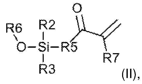 in which R2 and R3 are, independently, hydrogen, hydroxyl, alkyl, or alkoxyl, R5 is a divalent linking group, with an N or O at an end opposite the Si linkage, R6 is a C1-3 alkyl, linked to an acrylic or acrylamide moiety where R5 is a divalent linking group, and R7 is H or methyl.
[0023] Clause 12. The method of clause 9, wherein the coating comprises 3- (dimethoxysilyl)propyl methacrylate linked to a glass substrate via an O.
[0024] Clause 13. The method of any one of clauses 1 -12, wherein the swelled polymer comprises a poly(meth)acrylic, a poly(meth)acrylamide, a polyethylene glycol), a polyvinyl alcohol, or a combination of any of the preceding.
[0025] Clause 14. The method of any one of clauses 1 -13, further comprising embedding the cell or tissue sample in a swellable polymer composition and swelling the swellable polymer composition to produce the expanded tissue or cell sample embedded in an swelled polymer.
[0026] Clause 15. A microscope slide assembly comprising a micro-imaging substrate and an expanded tissue or cell sample embedded in a water-swelled polymer and having a thickness, linked to the substrate by the moiety:
 wherein R2 and R3 are, independently, hydrogen, hydroxyl, alkyl, or alkoxyl, wherein the O atom is covalently-linked to the substrate, and the Si atom is covalently-linked to a polymer chain extending partially through the thickness of at least a portion of the water-swelled polymer.
[0027] Clause 16. The microscope slide assembly of clause 15, wherein the substrate comprises glass.
wherein R2 and R3 are, independently, hydrogen, hydroxyl, alkyl, or alkoxyl, wherein the O atom is covalently-linked to the substrate, and the Si atom is covalently-linked to a polymer chain extending partially through the thickness of at least a portion of the water-swelled polymer.
[0027] Clause 16. The microscope slide assembly of clause 15, wherein the substrate comprises glass.
[0028] Clause 17. The microscope slide assembly of clause 16, wherein the O atom is covalently-linked directly to the glass.
[0029] Clause 18. The microscope slide assembly of any one of clauses 15-17, wherein the Si is linked to the polymer via a divalent C1-10 alkyl or hetero-substituted alkyl linking group comprising from 1 to 10 C, O, N, or S atoms.
[0030] Clause 19. The microscope slide assembly of any one of clauses 15-18, wherein the water-swelled polymer and/or the polymer chain extending partially through the thickness of at least a portion of the water-swelled polymer comprise poly(meth)acrylate or poly(meth)acrylamide.
[0031] Clause 20. A kit for preparation of a microscope slide assembly, comprising: a glass microscope slide comprising a glass substrate, and a coating on the glass substrate comprising a molecule having a (meth)acrylic- or (meth)acrylamide-reactive functional group linked covalently to the glass substrate via a silane moiety; a sample-embedding solution comprising (meth)acrylic- or (meth)acrylamide monomers and a polymerization initiator; and a re-embedding solution comprising (meth)acrylic- or (meth)acrylamide monomers and a polymerization photoinitiator.
[0032] Clause 21 . A microscope slide assembly, comprising: a micro-imaging substrate; a plurality of spaced-apart spacers over the substrate; an expanded tissue or cell sample embedded in a swelled polymer and spanning the spacers, defining a channel between the spacers, the expanded tissue or cell sample embedded in a swelled-polymer and the substrate; a cover slip larger than, and over, the expanded tissue or cell sample embedded in a swelled-polymer; and a polymer sealant affixing the cover slip to the substrate and optionally the spacers, but not covering or sealing ends of the channel such that a liquid can be passed through the channel.
[0033] Clause 22. The microscope slide assembly of clause 21 , wherein the spacers comprise double-sided tape.
[0034] Clause 23. The microscope slide assembly of clause 21 or 22, wherein the polymer sealant comprises a UV-curable (meth)acrylic polymer.
[0035] Clause 24. The microscope slide assembly of any one of clauses 21 -23, wherein the polymer sealant is a nail polish.
[0036] Clause 25. The microscope slide assembly of any one of clauses 21 -24, wherein the swelled polymer comprises a poly(meth)acrylic, a poly(meth)acrylamide, a polyethylene glycol), a polyvinyl alcohol, or a combination of any of the preceding.
BRIEF DESCRIPTION OF THE DRAWINGS
[0037] FIG. 1 depicts the linking of an exemplary silane compound to a surface, such as a glass surface, via an oxygen.
[0038] FIGS. 2A-2B depict a method of expanding a tissue or a cell sample. FIG. 2A is a schematic diagram of exemplary enal-based chemistry (e.g., acrolein or methacrolein, see, WO 2019/241662) that covalently incorporates biomolecules into polymer chains during in situ polymerization. FIG. 2B is a schematic diagram of biomolecule retention expansion microscopy.
[0039] FIGS. 3A and 3B show microscope slide assemblies (not to scale) according to the present invention. FIG 3A is a top down view of the microscope slide assembly and FIG. 3B is a side view of the microscope slide assembly.
[0040] FIG. 4 is a schematic process for the functionalization of a glass microscope slide.
[0041] FIG. 5 is schematic depicting the method of immobilizing the bottom surface of a tissue-containing gel to a functionalized glass substrate.
DETAILED DESCRIPTION
[0042] The use of numerical values in the various ranges specified in this application, unless expressly indicated otherwise, are stated as approximations as though the minimum and maximum values within the stated ranges are both preceded by the word "about". In this manner, slight variations above and below the stated ranges can be used to achieve substantially the same results as values within the ranges. Also, unless indicated otherwise, the disclosure of ranges is intended as a continuous range including every value between the minimum and maximum values. As used herein “a” and “an” refer to one or more. A patient is a human or non-human animal.
[0043] As used herein, the term “comprising” is open-ended and may be synonymous with “including”, “containing”, or “characterized by”. As used herein, embodiments “comprising” one or more stated elements or steps also include, but are not limited to
embodiments “consisting essentially of” and “consisting of” these stated elements or steps.
[0044] A “moiety” (pl. “moieties”) is a part of a chemical compound, and includes groups, such as functional groups. As such, a nucleobase moiety is a nucleobase that is modified by attachment to another compound moiety, such as a polymer monomer, e.g. the nucleic acid or nucleic acid analog monomers described herein, or a polymer, such as a nucleic acid or nucleic acid analog as described herein.
[0045] “Alkyl" refers to straight, branched chain, or cyclic hydrocarbon groups including from 1 to about 20 carbon atoms, for example and without limitation C1-3, C1- 6, C1-10 groups, for example and without limitation, straight, branched chain alkyl groups such as methyl, ethyl, propyl, butyl, pentyl, hexyl, heptyl, octyl, nonyl, decyl, undecyl, dodecyl, and the like. "Substituted alkyl" refers to alkyl substituted at 1 or more, e.g., 1 , 2, 3, 4, 5, or even 6 positions, which substituents are attached at any available atom to produce a stable compound, with substitution as described herein. "Optionally substituted alkyl" refers to alkyl or substituted alkyl. "Halogen," "halide," and "halo" refers to -F, -Cl, -Br, and/or -I. "Alkylene" and "substituted alkylene" refer to divalent alkyl and divalent substituted alkyl, respectively, including, without limitation, ethylene (-CH2-CH2-). "Optionally substituted alkylene" refers to alkylene or substituted alkylene.
[0046] As used herein, the term “polymer composition” is a composition comprising one or more polymers. As a class, “polymers” includes, without limitation, homopolymers, heteropolymers, co-polymers, block polymers, block co-polymers and can be both natural and/or synthetic. Homopolymers contain one type of building block, or monomer, whereas copolymers contain more than one type of monomer. The term “(co)polymer” and like terms refer to either homopolymers or copolymers. A polymer may have any shape for the chain making up the backbone of the polymer, including, without limitation: linear, branched, networked, star, brush, comb, or dendritic shapes.
[0047] A polymer “comprises” or is “derived from” a stated monomer if that monomer is incorporated into the polymer. Thus, the incorporated monomer (monomer residue) that the polymer comprises is not the same as the monomer prior to incorporation into a polymer, in that at the very least, certain groups/moieties are missing and/or modified when incorporated into the polymer backbone. A polymer is said to comprise a specific type of linkage if that linkage is present in the polymer, such as, without limitation:
ester, amide, carbonyl, ether, thioester, thioether, disulfide, sulfonyl, amine, carbonyl, or carbamate bonds.
[0048] The term “ligand” refers to a binding moiety for a specific target, its binding partner. The molecule can be a cognate receptor, a protein, a small molecule, a hapten, or any other relevant molecule, such as an affibody or a paratope-containing molecule. The term “antibody” refers to an immunoglobulin, derivatives thereof which maintain specific binding ability, and proteins having a binding domain which is homologous or largely homologous to an immunoglobulin binding domain. As such, the antibody operates as a ligand for its cognate antigen, which can be virtually any molecule. Antibody mimetics are not antibodies, but comprise binding moieties or structures, e.g. paratopes, and include, for example, and without limitation: an affibody, an aptamer, an affilin, an affimer, an affitin, an alphabody, an aticalin, an avimer, a DARPin, a funomer, a Kunitz domain peptide, a monobody, a nanoclamp, or other engineered protein ligands, e.g. comprising a paratope targeting any suitable epitope present in a sample.
[0049] The term “antibody fragment” refers to any derivative of an antibody which is less than full-length. In exemplary embodiments, the antibody fragment retains at least a significant portion of the full-length antibody's specific binding ability. Examples of antibody fragments include, but are not limited to, Fab, Fab', F(ab')2, Fv, Fd, dsFv, scFv, diabody, triabody, tetrabody, di-scFv (dimeric single-chain variable fragment), bi-specific T-cell engager (BiTE), single-domain antibody (sdAb), or antibody binding domain fragments. In the context of targeting ligands, the antibody fragment may be a single chain antibody fragment. Alternatively, the fragment may comprise multiple chains which are linked together, for instance, by disulfide linkages. The fragment may also optionally be a multimolecular complex. A functional antibody fragment will typically comprise at least about 50 amino acids and more typically will comprise at least about 200 amino acids.
[0050] Ligands also include other engineered binding reagents, such as affibodies and designed ankyrin repeat proteins (DARPins), that exploit the modular nature of repeat proteins (Forrer T, Stumpp MT, Binz HK, Pluckthun A: A novel strategy to design binding molecules harnessing the modular nature of repeat proteins, FEBS Lett 2003, 539: 2-6; Gebauer A, Skerra A: Engineered protein scaffolds as next-generation antibody therapeutics, Curr Opin Chem Biol 2009, 13:245-255), comprising, often as a single chain, one or more antigen-binding or epitope-binding sequences and at a
minimum any other amino acid sequences needed to ensure appropriate specificity, delivery, and stability of the composition (see also, e.g., Nelson, AL, “Antibody Fragments Hope and Hype” (2010) MAbs 2(1 ):77-83).
[0051] Nucleic acid analogs hybridize to nucleic acids, and may have standard nucleobases (e.g., adenine, cytosine, thymine, uracil, and guanine), or other nucleobases as RNA or DNA as found in living organisms, but have different polymeric backbones as compared to include, for example and without limitation: phosphorothioate DNA, peptide nucleic acid, a, [3-con strained nucleic acid, 2’- methoxyl RNA, 2’-fluoro RNA, phosphorodiamidate morpholino oligomer, locked nucleic acid, 2',4'-constrained ethyl nucleic acid, 2’, 4’ bridged nucleic acid NC (N-H), 2’, 4’ bridged nucleic acid NC (N-methyl), ((S)-5’-C-methyl DNA (RNA)), and 5’-E- vinylphosphonate nucleic acid. In the context of the present invention, ligands are useful in immunohistochemical, or immunofluorescent labeling of biomolecules in the sample.
[0052] A large variety of dyes, such as DAPI (which directly binds dsDNA), that label biomolecules directly, fluorescent tags, such as fluorescent dyes (e.g., fluorescein, cyanine, rhodamine, fluorescent proteins, among many other commercially-available dyes), or enzymes (e.g., horseradish peroxidase, alkaline phosphatase, glucose oxygenase, or [3-galactosidase) may be conjugated with ligands, such as antibodies and antibody fragments or nucleic acids or nucleic acid analogs, for use in labeling biomolecules in a sample by any direct or indirect binding method. Antibodies and other ligands conjugated to dyes or enzymes, e.g. fluorophores, are broadly-available commercially.
[0053] Provided herein is a method of mounting tissue or cell sample for microscopic analysis. The method comprises: contacting an expanded tissue or cell sample embedded in an swelled polymer with a polymer monomer and a UV light-activated, radical polymerization photoinitiator on a microscope slide surface having a coating comprising a reactive moiety for polymerization of the polymer monomer, wherein the expanded tissue or cell sample embedded in the swelled polymer is contacted with the polymer monomer for a length of time such that the polymer monomer partially and not fully, permeates the expanded tissue or cell sample embedded in the swelled polymer; and exposing the expanded tissue or cell sample embedded in the swelled polymer to UV light effective to cause radical polymerization of the polymer monomer
from the reactive moiety of the coating, thereby covalently linking the expanded tissue or cell sample embedded in the swelled polymer to the microscope slide.
[0054] The microscope slide may comprise any suitable material, such as glass or a plastic. For example, the microscope slide may comprise soda lime glass or borosilicate glass. The microscope slide may be of any size and of any thickness.
[0055] The surface of the microscope slide may be treated to form new functional groups (e.g., hydroxyl groups) that are capable of reacting with the coating applied thereover. For example, an aqueous solution of sodium hydroxide may be applied over the surface of the microscope slide and then heated. Alternatively, the surface of microscope slide may be plasma treated.
[0056] A coating comprising a reactive moiety for polymerization of the polymer monomer is applied to the microscope slide surface. The coating comprises a silane compound. The silane compound may comprise a molecule having the formula (I):
R2
R4-Si-R1
R3 (|) where R1 , R2, and R3 may be, independently hydrogen, hydroxyl, alkyl, or alkoxyl, such as methoxyl or ethyloxyl, and R4 may be hydrogen, hydroxyl, alkyl, or alkoxyl, such as methoxyl or ethyloxyl, but in the context of the present disclosure, may comprise a reactive group, such as an acrylic, methacrylic, acrylamide, or methacrylamide moiety, linked to the silicon atom via a linking group, such as a divalent alkyl group or a divalent low molecular weight polyethylene glycol) group, e.g., -(C-C-O)n-, of less than 200 Da (Daltons), less than 300 Da, less than 400 Da, less than 500 Da, or less than 1000 Da, for example, n ranges from 1 to 20.
[0057] Exemplary silane compounds useful for linking a polyacrylate or polyacrylamide polymer to a glass surface via a radical polymerization method include (meth)acrylate- or (meth)acrylamide-functional silane compounds. Exemplary (meth)acrylate- or (meth)acrylamide-functional silane compounds may have the formula (II):

In which R2 and R3 are the same, R6 is a C1-3 alkyl group (that is an alkyl group having from 1 to 3 carbons, e.g., methyl, ethyl, propyl), linked to an acrylic or acrylamide moiety where R5 is a divalent linking group, e.g. as described with respect to R4, above, with an N or O at an end opposite the Si linkage forming an acrylamide, or acrylic moiety in which R7 is H (acrylic and acrylamide) or methyl (methacrylic and methacrylamide).
[0058] In one non-limiting example, the silane compound comprises 3- (trimethoxysilyl)propyl methacrylate (TMSPMA), in which R1 , R2, and R3 are methoxyl, and R4 is a methacrylic moiety linked via a propyl linker to the silicon atom, e.g., in reference to formula (II), R5 is -CH2-CH2-CH2-N-, and R7 is -CH3. When linked to a glass surface by an O, the 3-(trimethoxysilyl)propyl methacrylate is 3- (dimethoxysilyl)propyl methacrylate, as a methoxy group is removed in linking the silane compound to the glass. The silane compound may be linked to a surface, such as a glass surface, via an oxygen, by reaction of a silane compound in which at least one of R2, R3, and R4 is alkoxyl, e.g. methoxyl or ethoxyl, with a hydroxyl group on the glass surface, for example, as shown in FIG. 1 .
[0059] The polymer monomer and the UV light-activated, radical polymerization photoinitiator may be applied to the coated microscope slide surface in together in a polymerizable composition. The polymerizable composition may optionally further comprise a crosslinker. The polymerizable composition may further comprise a solvent, such as water, methanol, phosphate buffered saline (PBS), or combinations thereof.
[0060] The use of light to mediate controlled radical polymerization has emerged as a powerful strategy for rational polymer synthesis and advanced materials fabrication. These light-mediated reactions are performed in polymerization mixtures comprising polymer monomers, such as (meth)acrylic or (meth)acrylamide monomers and a photoinitiator. The photoinitiator initiates polymerization in the presence of light at a defined wavelength including ultraviolet (UV) and visible light. Because the methods described herein may be performed by a laboratory technician, in such instances, a
UV-sensitive photoinitiator may be employed to prevent initiation under typical laboratory lighting. Photoinitiators may be used to initiate free-radical polymerizations as well as controlled-radical polymerization reactions. As such, although a free-radical polymerization method is used in the examples below, controlled-radical polymerization methods, such as living radical polymerization, atom-transfer radical polymerization (ATRP), Reversible Deactivation Radical Polymerization (RDRP) such as Reversible Addition Fragmentation chain Transfer (RAFT) polymerization may be substituted therefor.
[0061] Photoinitiators that initiate polymerization in the presence of ultraviolet radiation are broadly-known and may include, but are not limited to benzoin alkyl ethers, benzyl ketals, a-dialkoxy-acetophenones, a-hydroxy-alkyl-phenones, a-amino- alkyl-phenones, acyl-phosphine oxides, benzophenones, benzoamines, thioxanthones, thioamines, or combinations thereof (see e.g., Aldrich Polymer Products Application & Reference Information, Applications: Free Radical Initiators, download November 14, 2023). Photoinitiators that initiate polymerization in the presence of visible light are broadly known and may include, but are not limited to titanocenes, phosphine oxides, phosphinates, diketones, oxime esters, flavonoids, Group 14 elements (e.g., silicon, germanium, tin), or combinations thereof (see e.g., Muller et al. “Recent Advances in Type I Photoinitiators for Visible Light Induced Photopolymerization”, ChemPhotoChem, 2022, 6, e202200091 and Aldrich Polymer Products Application & Reference Information, Applications: Free Radical Initiators).
For example, the photoinitiator may be a photoinitator from the Irgacure family of photoinitiators. Non-limiting examples of Irgacure photoinitiators include but are not limited to:
2-Hydroxy-4'-(2-hydroxyethoxy)-2-methylpropiophenone (Irgacure 2959)
 J 2-Benzyl-2-(dimethylamino)-4’-morpholinobutyrophenone (Irgacure 369; I-369),
 2,2-Dimethoxy-2-phenylacetophenone (DMPA; Irgacure 651 ),

2-Hydroxy-2-methyl-1 -phenylpropanone (Irgacure 1173)

, or combinations thereof.
[0062] The polymer monomer may be an acrylic monomer, an acrylamide monomer, or combinations thereof. The reactive moiety of the coating on the microscope slide surface polymerizes with at least one reactive group present in the polymer monomer (e.g., the alkene group), in the presence of the UV light-activated, radical polymerization photoinitiator.
[0063] An “acrylic monomer” is an organic compound that includes the structure
 where: R is a hydrogen, an alkyl group, a carboxylic acid group, or a dicarboxylic acid; Ri is a hydrogen, a methyl (i.e., methacrylic), a halogen (e.g., chlorine), a cyano group (-C=N), or a substituted or unsubstituted aryl; R2 is hydrogen, an alkyl group, an alkene group, a substituted or unsubstituted aryl group, a carboxylic acid group, or a dicarboxylic acid group. For example, the polymer monomer may comprise acrylic acid, methacrylic acid, a-chloroacrylic acid, a- cyanoacrylic acid, [3-methacrylic acid (crotonic acid), a-phenylacrylic acid, 2- acryloxypropionic acid, sorbic acid, a-chlorosorbic acid, angelic acid, cinnamic acid, 4-chlorocinnamic acid, [3- stearylacrylic acid, itaconic acid, citroconic acid, mesaconic acid, glutaconic acid, aconitic acid, maleic acid, fumaric acid, tricarboxyethylene, maleic anhydride, or combinations thereof.
[0064] An “acrylamide monomer” is an organic compound that includes the structure
where: R is a hydrogen, an alkyl group, a carboxylic acid group, or a dicarboxylic acid; Ri is a hydrogen, a methyl (i.e., methacrylic), a halogen (e.g., chlorine), a cyano group (-C=N), or a substituted or unsubstituted aryl; R2 is hydrogen, an alkyl group, an alkene group, a substituted or unsubstituted aryl group, a carboxylic acid group, or a dicarboxylic acid group. For example, the polymer monomer may comprise acrylic acid, methacrylic acid, a-chloroacrylic acid, a- cyanoacrylic acid, [3-methacrylic acid (crotonic acid), a-phenylacrylic acid, 2- acryloxypropionic acid, sorbic acid, a-chlorosorbic acid, angelic acid, cinnamic acid, 4-chlorocinnamic acid, [3- stearylacrylic acid, itaconic acid, citroconic acid, mesaconic acid, glutaconic acid, aconitic acid, maleic acid, fumaric acid, tricarboxyethylene, maleic anhydride, or combinations thereof.
[0064] An “acrylamide monomer” is an organic compound that includes the structure
 where: R is a hydrogen or an alkyl; Ri is a hydrogen or an alkyl; and R2 is a hydrogen or a methyl. For example, the polymer monomer may comprise acrylamide, methacrylamide, N-N-dimethylacrylamide, or combinations thereof.
[0065] The polymerizable composition may include a crosslinker. For example, the crosslinker may include, but is not limited to N,N-methylene bis(acrylamide), N,N'- bisacryloyl-1 ,2-dihydroxy-1 ,2-ethylenediamine, N,N-ethylene bis(acrylamide), trimethylolpropane triacrylate, ethylene glycol di(meth)acrylate, triallylamine, or combinations thereof.
[0066] An expanded tissue or cell sample embedded in a swelled polymer is then contacted with the polymer monomer, the UV light-activated, radical polymerization photoinitiator, and optionally the crosslinker, for a length of time such that the polymer monomer partially and not fully, permeates the expanded tissue or cell sample embedded in the swelled polymer. The length of time such that the polymer monomer partially and not fully, permeates the expanded tissue or cell sample embedded in the swelled polymer is selected such that the polymer monomer permeates no more than 50%, or no more than 25%, of the thickness of the expanded tissue or cell sample embedded in the swelled polymer. For example, the length of time such that the polymer monomer partially and not fully, permeates the expanded tissue or cell sample embedded in the swelled polymer ranges from 5 seconds to 10 minutes.
[0067] After the expanded tissue or cell sample embedded in a swelled polymer contacted with the polymer monomer, the UV light-activated, radical polymerization photoinitiator, and optionally the crosslinker, for a length of time such that the polymer monomer partially and not fully, permeates the expanded tissue or cell sample embedded in the swelled polymer, the expanded tissue or cell sample embedded in the swelled polymer is exposed to UV light effective to cause radical polymerization of the polymer monomer from the reactive moiety of the coating. This radical polymerizable covalently links the expanded tissue or cell sample embedded in the swelled polymer to the coated microscope slide.
[0068] The following describes a method for producing an expanded tissue or cell sample embedded in a swelled polymer. Referring to FIG. 2A, cell or tissue samples including aldehyde-reactive biomolecules of interest (proteins, RNA, DNA, amine sugars) are covalently bonded to the polymeric hydrogel, using acrolein, methacrolein, or a similar enal within a polyelectrolyte formulation. Synthesis of the polymer hydrogel and biomolecule anchoring occur in situ with the biological sample. Other chemistries may be utilized for bonding biomolecules, such as other acrylic or acrylamide polymerization methods or other polymerization methods, such as those described below. Polymerization may be followed by homogenization of sample’s mechanic properties. By “homogenization”, it is meant homogenization of the sample’s mechanical properties to facilitate clean and even expansion (e.g., isotropic expansion) of the sample during swelling of the sample. Homogenization can be achieved as a result of, for example and without limitation, protease digestion and/or heat denaturation with surfactants. After homogenization, the specimen/polymer is expanded. FIG. 2B outlines this exemplary scheme. The anchored biomolecules can be labelled before hydrogel synthesis or after specimen/polymer matrix expansion. The expanded polymer matrix with labelled biomolecules may be analyzed using a conventional fluorescent, confocal microscope or other desired microscope.
[0069] One feature of the crosslinking of the hydrogel to the biomolecules using an enal, e.g. acrolein or methacrolein, is that the crosslinking bond is labile, in that the bond will hydrolyze over time, leaving the original antigens embedded in the hydrogel in an expanded state. As such, after the sample is expanded, in situ assays, such as, without limitation, immunofluorescence, immunohistochemistry, and in situ hybridization, can be performed. An additional benefit of expansion methods, e.g. as described herein, is the rapid and complete infusion of the polymer monomers into tissue samples, including very thick tissue samples, such as organs, organ systems, or even complete organisms, permitting three-dimensional (3D) visualization and imaging of highly complex tissue structures, e.g., using layered scanning by laser confocal imaging methods.
[0070] The cell or tissue sample can be processed directly, optionally after fixation, e.g., with acetone, but if it is mounted in paraffin, OCT, or other embedding materials, those materials may be removed using appropriate solvent(s), ultimately to provide the cell or tissue sample in an aqueous solution, such as PBS, that is permissive to polymerization of acrylic or acrylamide polymers. For example, and without limitation,
for a formaldehyde-fixed sample embedded in paraffin, deparaffinizing the sample, e.g., by sequential washes with an organic solvent, such as xylene, followed by one or more washes with ethanol and water. In one non-limiting example, for deparaffinizing a sample, washes of xylene (2X), 100% ethanol (2X), 95% ethanol, 70% ethanol, 50% ethanol, and water are performed, in order. OCT fixative may be solubilized in PBS, and unfixed, frozen samples may be fixed in acetone. Prior to permeabilization with polymer monomers, the sample may be further processed, e.g. by heat treatment, for example at 60QC for 30 minutes. The sample also may be treated with a surfactant.
[0071] The cell or tissue sample is then permeated with (infused with) a swellable material. In one embodiment, the cell or tissue sample is permeated with (infused with) the polymer monomer composition. This is accomplished by immersing the sample in a solution containing suitable polymer monomers. For expansion microscopy, the monomers result in a water-swellable polymer composition. Water- swellable hydrophilic polymers can absorb several times their weight of water or aqueous liquids, such as urine or blood, and are therefore commonly employed as absorbents, in particular in hygiene articles such as diapers for babies and incontinence pants for adults, and also tampons and the like. Useful water-swellable monomers for producing a water-swellable (co)polymer include any monomer that, when polymerized as a (co)polymer, produces a water-swellable polymer. Water- swellable (co)polymers are often made by initially polymerizing a, [3-un saturated carbonyl monomers, such as unsaturated carboxylic acids, or derivatives thereof, such as, for example, acrylates, such as acrylic acid, alkali metal (e.g., sodium and/or potassium) or ammonium salts of acrylic acid or other acrylates, alkyl acrylates such as methacrylate, and the like, and/or acrylamides, such acrylamide, alkylacrylamides, methacrylamide, and/or N,N-dimethylacrylamide. The monomers are polymerized in the presence of relatively small amounts of di- or poly-functional monomers (crosslinkers, or multi-functional monomers), such as N,N'-methylenebisacrylamide, N,N'-bisacryloyl-1 ,2-dihydroxy-1 ,2-ethylenediamine, N,N'-ethylenebis(acrylamide), trimethylolpropane triacrylate, ethylene glycol di(meth)acrylate, or triallylamine. When N,N-dimethylacrylamide is used in high concentrations, such as if it is used as a monomer for the primary polymer, it also may act as a crosslinker (see, e.g., Cipriano, BH, et al. “Superabsorbent Hydrogels That Are Robust and Highly Stretchable”, Macromolecules, 2014, 47(13):4445-4452). The di- or poly-functional monomer
materials serve to lightly cross-link the polymer chains thereby rendering them waterinsoluble, yet water-swellable. These lightly-crosslinked absorbent polymers contain a multiplicity of carboxylate groups attached to the polymer backbone.
[0072] In exemplary polymerization mixtures, the concentration of a,[3-unsaturated carbonyl monomers, may be from 1 gram per 100 milliliters (g/100 mL) to the saturated concentration, such as from 1 g/ 100 mL to 35 g/ 100 mL. The total molar concentration of a,[3-unsaturated carbonyl monomers, may be from 0.5 M to 6 M. The concentration of crosslinkers, such as N,N’-methylene bis(acrylamide), may be from 0.01 g/ 100 mL to 5 g/ 100 mL. The concentration of an enal, such as acrolein or methacrolein, may be from 0.01 g/ 100 mL to 10 g/ 100 mL.
[0073] In non-limiting examples, the concentration of acrylate monomer may be from 5 g /100 mL to 35 g / 100 mL, the concentration of acrylamide monomer may be from 0 g/ 100 mL to 5 g/ 100 mL, the concentration of N,N-dimethylacrylamide may be from 0 g/ 100 mL to 35 g / 100 mL. The total molar concentration of a,[3-unsaturated carbonyl monomers, may be from 1 M to 5 M. The concentration of crosslinkers, such as N,N’-methylene bis(acrylamide), may be from 0.1 g/ 100 mL to 0.2 g/ 100 mL. The concentration of an enal, such as acrolein or methacrolein, may be from 0.05 g/ 100 mL to 5 g/ 100 mL.
[0074] a,[3-unsaturated carbonyl monomers include, for example and without limitation: acrylic acids and alkali metal salts thereof, e.g., acrylic acid, methacrylic acid, a-chloroacrylic acid, a-cyanoacrylic acid, acrylamide, methacrylamide, N,N- dimethylacrylamide, [3-methylacrylic acid (crotonic acid), a-phenylacrylic acid, [3- acryloxypropionic acid, sorbic acid, a-chlorosorbic acid, angelic acid, cinnamic acid, p-chlorocinnamic acid, [3-stearylacrylic acid, itaconic acid, citroconic acid, mesaconic acid, glutaconic acid, aconitic acid, maleic acid, fumaric acid, tricarboxyethylene, and maleic anhydride. Combinations of any of the above, forming a copolymer, also are useful, such as a combination of an alkali metal salt of an acrylate in combination with acrylamide, methacrylamide, or N,N-dimethylacrylamide.
[0075] After the sample is infused with suitable monomers for preparation of a water- swellable polymer, the monomers are polymerized in situ. As such, during infusion of the monomers, or afterward, reagents necessary or useful to cause or accelerate polymerization are added, such as a catalyst, or any other reagent(s) necessary or useful for polymerizing the monomers. In one example, an enal, such as acrolein or
methacrolein, may be added at this time. Infusion and polymerization may take place in a suitable container or mold.
[0076] In examples, after polymerization, biomolecules and any added ligands, tags, or probes may be locked or anchored into place both by the formation of the polymer backbones about the biomolecules, and the covalent linkage of the biomolecules, ligands, tags, or probes to the polymer backbone via the enal, such as acrolein or methacrolein. At this point, to avoid damage to the specimen during expansion, and to facilitate expansion, the internal mechanic properties of sample is homogenized using a protease or a combination of denaturants and/or heat.
[0077] Bacteria, fungi, and plants have cell walls that prohibit traditional expansion microscopy, even with use of a protease digest. As such, alternatively, or in combination with homogenization with the protease, where the sample comprises an organism having a cell wall, the cell wall may be enzymatically digested before or after infusion and polymerization and before expansion of the swellable hydrogel in the sample. Examples of enzymes useful in digestion of cell walls, include a glucanase or a cellulase, such as lyticase (e.g., from Arthrobacter luteus), Achromopeptidase, lysing Enzymes from Trichoderma harzianum, pectinase, pectolyase, lysostaphin, lysozyme, mutanolysin, and/or chitinase.
[0078] Once the polymer (hydrogel) is formed, and, if necessary, the sample is homogenized, for example by protease digestion, the sample can be expanded by hydration. Hydration involves contacting the hydrogel containing the sample with water, or a suitably low ionic strength aqueous solution to cause swelling of the polymer. The sample is thereby expanded isotropically. Typically, the expansion factor upon hydration is at least 2, and more typically greater than 3, meaning the isotropic expansion in any linear dimension is at least 2 and is more typically greater than 3, depending on the choice of monomers, crosslinking density, among other factors.
[0079] Traditionally, at any suitable point in the processing of a cell or tissue sample, biomolecules may be dyed or labeled in any suitable and effective manner. In one example the sample may be labeled prior to infusion or polymerization with the monomers, as described herein. In another example, the sample is labeled after polymerization, but before expansion of the sample, and optionally before protease digestion of the sample. In one example the sample is labeled after expansion. In yet another example, a biomolecule in the sample is labeled with a first label after
expansion, viewed or imaged, and subsequently the first label is removed, and a second label is used to label a different biomolecule. This process may be repeated multiple times. At any stage, multiple labels may be used simultaneously, e.g., fluorescent labels with different emission spectra, to label different biomolecules in the sample. Of note, when employed, bonds between enal residues and the biomolecules in the expanded cell or tissue sample can reverse over time, exposing additional epitopes or binding sites, thereby expanding the ability to visualize biomolecules in the sample.
[0080] A coverslip may optionally be applied over the expanded tissue or cell sample embedded in a swelled polymer to render it compatible with automated slide scanners and flow-cell machines.
[0081] Also provided herein is a microscope slide assembly. The microscope slide assembly comprises a micro-imaging substrate and an expanded tissue or cell sample embedded in a water-swelled polymer and having a thickness, linked to the substrate by the following moiety:
 wherein R2 and R3 are, independently, hydrogen, hydroxyl, alkyl, or alkoxyl, wherein the oxygen (O) atom is covalently-linked to the micro-imaging substrate, and the Si atom is covalently-linked to a polymer chain extending partially through the thickness of at least a portion of the water-swelled polymer.
[0082] The micro-imaging substrate may be a microscope slide and may be any of the microscope slides described herein. For example, the micro-imaging substrate may comprise glass. In the microscope slide assembly, when the micro-imaging substrate comprises glass, the oxygen (O) atom of the above moiety is covalently-linked directly to the glass. In the above moiety, the Si is linked to the polymer via a divalent C1-10 alkyl or hetero-substituted alkyl linking group comprising from 1 to 10 C, O, N, or S atoms.
[0083] In the microscope slide assembly, the water-swelled polymer and/or the polymer chain extending partially through the thickness of at least a portion of the water-swelled polymer comprise poly(meth)acrylate or poly(meth)acrylamide. For example, both the water-swelled polymer and the polymer chain extending partially
through the thickness of at least a portion of the water-swelled polymer comprise poly(meth)acrylamide.
[0084] A coverslip may optionally be applied over the expanded tissue or cell sample embedded in a swelled polymer to render it compatible with automated slide scanners and flow-cell machines.
[0085] Also provided herein is a kit for preparing a microscope slide assembly. The kit comprises various components that are packaged for storage, transport, and distribution in any suitable container. The kit for preparing the microscope slide assembly may comprise one or more vessels. In a vessel, in one embodiment, is a sample-embedding solution. The sample-embedding solution comprises a mixture of monomers for producing a water-swellable polymer, and in the same vessel, or in another vessel is an enal, such as acrolein or methacrolein. The monomers are ct,|3- unsaturated carbonyl monomers, such as acrylate monomers and/or acrylamide and/or N,N-dimethylacrylamide monomers, such as sodium acrylate with acrylamide, optionally with a crosslinker, such as N,N’-methylene bis(acrylamide), as described herein. For example, the sample-embedding solution comprises (meth)acrylic- or (meth)acrylamide monomers and a photoinitiator. In a different vessel, in one embodiment, is a re-embedding solution of a polymer monomer, a UV light-activated, radical polymerization photoinitiator, and optionally, a crosslinker. For example, the re-embedding solution comprises (meth)acrylic- or (meth)acrylamide monomers and a polymerization photoinitiator. The kit also comprises a glass microscope slide that comprises a glass substrate and a coating on the glass substrate comprising a molecule having a (meth)acrylic- or (meth)acrylamide-reactive functional group linked covalently to the glass substrate via a silane moiety. The kit further comprises instructions for mounting a tissue or cell sample for microscopic analysis. Optionally, the kit may comprise a mold or chamber for use in formation of the hydrogel comprising the sample. A suitable catalyst or any other reagents for promoting polymer formation also can be included in the kit, typically in a separate vessel from the monomers.
[0086] Also provided herein is a microscope slide assembly 10, as shown in FIGS. 3A and 3B. The microscope slide assembly 10 comprises: a micro-imaging substrate 12; a plurality of spaced-apart spacers 14 over the substrate 12; an expanded tissue or cell sample embedded in a swelled polymer 16 and spanning the spacers 14, defining a channel 18 between the spacers 14, the expanded tissue or cell sample embedded in a swelled-polymer 16 and the substrate 12; a cover slip 20 larger than, and over,
the expanded tissue or cell sample embedded in a swelled-polymer 16; and a polymer sealant 22 affixing the cover slip 20 to the substrate 12 and optionally the spacers 14, but not covering or sealing ends of the channel 18 such that a liquid can be passed through the channel 18.
[0087] As used herein, the term “over” means that the spacers 14 are on the substrate 12 but not necessarily in contact with the surface of the substrate 12 and that the cover slip 20 is on the expanded tissue or cell sample embedded in a swelled polymer 16 but not necessary in contact with the expanded tissue or cell sample embedded in a swelled polymer 16. For example, the plurality of spacers 14 over the substrate does not preclude the presence of one or more other layers located between the plurality of spacers 14 and the substrate 12. For example, the cover slip 20 over the expanded tissue or cell sample embedded in a swelled polymer 16 does not preclude the presence of one or more other materials located between the cover slip 20 and the expanded tissue or cell sample embedded in a swelled polymer 16. For example, a layer of PBS may be present between the cover slip 20 and the expanded tissue or cell sample embedded in a swelled polymer 16.
[0088] The micro-imaging substrate 12 may be a microscope slide and may be any of the microscope slides described herein.
[0089] The spacers 14 may be any suitable material, such as double-sided tape. For example, the spacers 14 may be double-sided tape having a thickness of at least 50 pm. The spacers 14 may be placed parallel to the short ends of micro-imaging substrate 12.
[0090] The cover slip 20 may be of any suitable material, such an optical grade coverslip. The cover slip 20 may be a glass cover slip. Alternatively, the cover slip 20 may be a plastic cover slip.
[0091] The polymer sealant 22 may be any suitable material, such as a UV-curable (meth)acrylic polymer. For example, the polymer sealant 22 may be a nail polish, such as a UV-activated nail polish. The microscope slide assembly including the polymer sealant 22 may be cured under UV light for 15 minutes or until the polymerization process is complete.
[0092] The expanded tissue or cell sample embedded in a swelled polymer 18 may be any of the expanded tissue or cell sample embedded in a swelled polymer described herein, which may comprise a polymer or a combination of polymers. For
example, the swelled polymer comprises a poly(meth)acrylic, a poly(meth)acrylamide, a polyethylene glycol), a polyvinyl alcohol, or a combination of any of the preceding.
EXAMPLES
Example 1 - Exemplary staining and fixation method
[0093] Various methods have been employed to restrict gel movement over the course of a single round of imaging in expanded samples, including extensive drying of the gel, agarose or acrylamide embedding, and the use of poly-lysine coated imaging glass. However, these methods do not lend themselves well to the challenge of multiround immunostaining as they either inevitably fail over time (drying, poly-lysine) or involve the generation of a second immobilizing gel that is impenetrable to fluorescent antibodies (agarose or acrylamide embedding). Therefore, there is a need to develop a highly multiplexed nanoscale imaging method that is compatible with conventional immunostaining protocols. The following exemplary protocol was developed, and expanded tissue was strongly adhered to the functionalized glass and was effectively stained with both antibody and oligonucleotide probes.
Materials and Methods
[0094] Reagents and antibodies. Unless otherwise described, all reagents and antibodies were commercially sourced.
[0095] Mouse brain samples. All mouse brain expansion experiments were performed using C57BL/6 mice except where otherwise noted. C57BL/6 mice were deeply anesthetized with ketamine/xylazine before transcardial perfusion with 20 milliliter (mL) 4% paraformaldehyde (PFA) in 1 x phosphate buffered saline (PBS). Brains were harvested and post-fixed in 4% PFA in 1 x PBS overnight at 4 degrees Celsius (QC). Tissue was cryoprotected by incubating in 30% (weight/volume (w/v)) sucrose in 1 x PBS at 4 °C until the brain sank (usually 16 hours/overnight). Brains were sectioned in 30-micron (pm) slices (either coronally or sagittally) using a freezing microtome and stored at 4 °C in glycerol solution (30% (volume/volume (v/v)) glycerol and 30% (v/v) ethylene glycol in 1 x PBS).
[0096] In situ polymer synthesis of mouse brain samples with Magnify. A monomer solution made of 4% N,N-dimethylacrylamide (DMAA) (v/v), 34% sodium acrylate (SA) (w/v), 10% acrylamide (AA) (w/v), 0.01 % N,N'-Methylenebisacrylamide (Bis) (w/v), 1 % sodium chloride (NaCI (w/v), and 1 x PBS was prepared and stored at 4 °C prior to synthesis. Prior to gelation, samples were placed into a custom gelling
chamber consisting of two spacers cut from #1 .5 cover glass adhered to the uncoated back of a microscope slide. Excess PBS around the tissue was absorbed with a Kimwipe and sections were allowed to air dry partially on the slide. Immediately prior to gelation, the chemicals 4-hydroxy-TEMPO (4HT), N,N,N',N'-
Tetramethylethylenediamine (TEMED), ammonium persulfate (APS), and methacrolein were added to the gel monomer solution to a final concentration of 0.25% (w/v) APS, 0.001 % 4HT (w/v, mouse brain and organoid only), 0.04% TEMED (v/v), and 0.1 % (v/v) methacrolein, adding TEMED and APS last to prevent premature gelation. Tissue was incubated in gelling solution for 30 minutes at 4 °C to allow the monomer solution to diffuse into the tissue (mouse brain and organoid only). A glass microscope slide was placed backside down over the gelling chamber and the samples were incubated overnight in a humidified container at 37 °C to complete gelation.
[0097] Sample digestion and expansion with Magnify. After gelation, the glass slide cover was removed from the gelling chamber, blank gel surrounding the tissue was trimmed from the samples (except for indirectly adhered samples), and the tissue was cut into smaller pieces if necessary. Samples were then incubated in homogenization buffer (10% w/v sodium dodecyl sulfate (SDS), 8M Urea, 25 milliMolar (mM) ethylenediaminetetraacetic acid (EDTA), 2 Molar (M) Tris Base, pH 8.5 at room temperature (RT)) for 8-20 hours at 70 °C with shaking. Homogenized samples were then washed 3 times with 1 % decaethylene glycol monododecyl ether (Ci2Eio)/1 x PBS at RT, 1 hour at 60 °C in C12E10, and finally 3 more washes in C12E10 at RT to remove remaining SDS. Samples were finally washed an additional 3 times for at least 10 minutes each with 1 x PBS at RT and stored in 1 x PBS containing 0.02% sodium azide at 4 °C.
[0098] TMSPMA treatment of glass surfaces. Glass microscope slides were treated with 3-(Trimethoxysilyl)propyl methacrylate (TMSPMA), a silanization reagent which incorporates acryloyl groups onto the surface of glass to participate in free radical polymerization. Glass slides were briefly washed with acetone followed by 100% ethanol, before final washing two times in ddH2O. Slides were then incubated for 1 hour in 10 M sodium hydroxide (NaOH) at 60 °C before overnight incubation at 60 °C in freshly prepared TMSPMA solution (2 mL of TMSPMA was diluted into 45 mL of ethanol, 2.5 mL of double-distilled water (ddH2O) and 1 mL of glacial acetic acid - 50 mL solution used per 10 slides). Slides were washed and stored in 100% ethanol. A
schematic process for the functionalization of the glass microscope slides can be found in FIG. 4.
[0099] Adhering of expanded gels. Tissue-containing gels were first stained with DAPI and imaged to determine the position of tissue within the gel. The gel was placed tissue-side down on a non-functionalized glass microscopy slide. 2-3 drops of reembedding gel solution (30-67% acrylamide, 0.2-0.3x PBS, 100-1500 parts per million (ppm) N,N’-methylene-bisacrylamide, 10 microliters (pL) 33% 2-Hydroxy-4'-(2- hydroxy-ethoxy)-2-methyl-propiophenone (DM PA) in methanol) was placed on top of the tissue-containing gel such that the entire top surface was covered but the solution did not spill over. The re-embedding solution was left for 1 -2 minutes. Excess solution was removed with a paper towel, and a TMSPMA-functionalized slide was placed on top of the tissue-containing gel. The functionalized slide was briefly but firmly pressed onto the tissue-containing gel to ensure complete contact with the hydrophobic surface before illumination with a UV LED lamp for 12 minutes in a deoxygenated and humidified environment. The non-functionalized bottom slide was then easily removed, and the attached sample was placed in 1 x PBS.
[00100] Indirect adhering of expanded gels. Mouse brain sections were gelled as described above, with the exception that a large rectangular area of blank gel was left surrounding the tissue. Tissue-containing gels were homogenized and washed as normal, then stained with DAPI and imaged to determine the position of tissue within the gel. The gel was placed tissue-side up on a non-functionalized glass microscopy slide. Re-embedding gel solution (30% acrylamide, 0.2x PBS, 1500 ppm N,N’- methylenebisacrylamide, 10 pL 33% DMPA in methanol) was placed on top of the tissue-containing gel such that the entire top surface of excess blank gel was covered, except where the tissue was present. The re-embedding solution was left for 10 minutes. Excess solution was removed with a paper towel before a second 10 minute round of re-embedding. Excess embedding solution was again removed, and the gel was flipped over onto a TMSPMA-functionalized coverslip such that the tissue was in direct contact with the glass. The sample was then placed, without a glass slide on top, in a deoxygenated and humidified environment before illumination with a UV LED lamp for 12 minutes.
[00101] Inter-round distortion quantification. Distortion error was quantified using previously described methods for distortion vector field calculation and root-meansquare (RMS) error calculation. Briefly, z-stacks were taken at manually aligned ROIs
across staining rounds of a reference label. To match z-planes, scale invariant feature transform (SIFT) key points were generated for all possible combinations of pairs of planes. SIFT key points were generated using the VLFeat open-source library and filtered by random sample consensus (RANSAC) with a geometric model that only permits rotation, translation, and uniform scaling. The pair of z-planes with the most SIFT key points were then used for image registration by rotation, translation, and uniform scaling. By subtracting the resulting vectors at any two points, distance measurement errors could easily be sampled, and the RMS error for such measurements was plotted as a function of measurement length from at least three technical replicates.
[00102] Post-expansion immunostaining of adhered mouse brain samples. Immediately after adhering, expanded samples were washed 3 times for 10 minutes each in 1 x PBS at RT and blocked in 3% bovine serum albumin 10.1% TritonX-100 in 1 x PBS for at least one hour in a petri dish. Samples were then incubated with primary antibodies in 0.1 % TritonX-100 in 1 x PBS overnight at 37 °C in a custom 3D-printed staining container. Samples were washed three times for at least 20 minutes each in 1 x PBS at RT in a petri dish before incubation in 0.1 % TritonX-100 in 1 x PBS with the corresponding secondary antibodies for 3 hours at 37 °C in the custom staining container. Prior to imaging, samples were washed with 1 x PBS in a petri dish. For expanded samples adhered to a glass slide, a coverslip was affixed to the gel with poly-lysine, which was floated off in PBS after imaging. For expanded samples adhered to a coverslip, the sample was imaged on top of the coverslip and open to air. The sample was kept moist with PBS for extended periods of imaging.
[00103] Imaging. Fluorescence imaging was performed using a Nikon Eclipse Ti2 epifluorescence microscope equipped with a CSU-W1 spinning disk confocal module and an Andor 4.2 Zyla sCMOS camera. The system was controlled by NIS-Elements AR 5.21 .03 64-bit software. Images were taken using the following Nikon objectives: CFI Plan Apo Lambda 4x (0.2 NA), CFI Plan Apo Lambda 10x (0.45 NA), CFI Apo LWD Lambda S 20xWI (0.95 NA), CFI Apo LWD Lambda S 40xWI (1.15 NA).
[00104] Cyclic immunofluorescence. Immunostained and imaged adhered gels were placed in a modified homogenization buffer (8M urea, 10% SDS, 50 mM EDTA, 10 mM 1 ,4-Dithiothreitol (DTT) or 1 % APS, 1 M Tris buffer) and under bright LED light for 3 hours at room temperature. Samples were then washed 3 times with 1 % decaethylene glycol monododecyl ether (Ci2E )/1 xPBS at RT followed by a 60 minute
wash at 60 °C in the same solution to remove remaining SDS. Samples were subsequently washed further in 1 % C12E10/I x PBS an additional 3 times for at least 10 minutes each, and finally with 1 x PBS three times at RT.
Results and Discussion
[00105] Adhering expanded tissue to glass without disrupting immunostaining.
Rapid acrylamide polymerization controlled the penetration depth of the re-embedding solution into the expanded hydrogel. In this way, the expanded tissue was linked to the glass surface using TMSPMA as the bridge molecule without embedding the tissue itself in acrylamide. The acrylamide solution containing the photoinitiator DMPA, which produces free radicals in the presence of UV light, was allowed to briefly diffuse into the blank gel below an expanded tissue sample before being contacted with the silane functionalized glass surface. Irradiation with UV light covalently linked the expanded gel to the glass surface without embedding the tissue itself.
[00106] Blank expanded hydrogels were used to test if the expanded hydrogels could be successfully attached to silanized glass when fully embedded in acrylamide-UV gel. Previously published methods for the attachment of polyacrylamide gels to TMSPMA-treated glass surfaces were used (see e.g. Chen et al. “Nanoscale imaging of RNA with expansion microscopy”, Methods, 2016, 13:679-684; Wang et al. “Multiplexed imaging of high-density libraries of RNAs with MERFISH and expansion microscopy,”, Sci. Rep., 2018, 8, 4847; Yuk et al. “Tough bonding of hydrogels to diverse non-porous surfaces”, Nature Mater., 2016, 15:190-196), with DMPA substituted for APS. It was confirmed that the gels were permanently affixed after 10 minutes under UV light.
[00107] To test whether protein epitopes were still accessible after re-embedding, an attempt was made to adhere and immunostain expanded mouse brain sections. It was found that tomato lectin, as well as primary and secondary antibodies against synaptic markers, all showed strong labeling throughout the tissue. Thus, all of the labels penetrated the full tissue evenly
[00108] Cyclic immunostaining. To test the method’s compatibility with cycIF multiplexing, adhered expanded mouse brain tissue was antibody labeled and imaged. Antibody stripping was then performed with homogenization buffer for 2 hours at room temperature, yet clear bright sections appeared where images had previously been taken. This was attributed to the photocrosslinking effect between the fluorophore or antibodies with the proteins they are bound to. When 1 % APS (w/v) was added to the
homogenization buffer to oxidize fluorophores, antibody signal was successfully removed through the tissue. However, signal returned when secondary antibody was re-applied to the tissue, which indicated that that primary antibodies remained intact and could confound subsequent staining rounds.
[00109] Signal unmixing and analysis of inter-round distortion. To prevent distortion from placement and removal of the coverslip, the expanded sample was attached directly to the coverslip itself, so that the position of the tissue relative to the coverslip was consistent between imaging rounds, and the sample can be imaged without compression.
[00110] An “indirect adhering” of expanded tissue to coverslips was completed by leaving large amounts of blank gel around the tissue and adhering the gel to the glass only around the tissue, but not where the tissue is itself. The expanded tissue was unembedded and remained in place directly against the imaging glass.
[00111] It was shown that by using this approach, the tissue remained open to immunostaining and imaging with high-magnification objectives. DAPI signal could be much better aligned using only rigid registration than previously, ranging from nearcomplete overlap across an entire field of view (-0.3% distortion) to slightly distorted at larger distances (-0.6% distortion). Across all fields of view for both the original strategy and the indirect adhering approach, the measurement error was substantially lower when indirectly adhered.
[00112] It was demonstrated that expanded tissues were strongly adhered to the surface of functionalized glass and were subsequently immunostained. Furthermore, it was shown that attaching a gel directly to a coverslip by embedding only blank gel surrounding the tissue, rather than above or below, reduces measurement error.
Example 2
[00113] Firm anchoring of the bottom surface of a tissue-containing gel to a silane- functionalized glass substrate was shown, while most of the gel was left unembedded and open to post-expansion profiling of biomolecule targets (FIG. 5).
[00114] The method was the same as provided above in Example 1 , except that 2-3 drops of the re-embedding gel solution (67% acrylamide, 0.2-0.3x PBS, 100-1500 ppm N,N’-methylene-bisacrylamide, 10 microliters (pL) 33% DMPA in methanol) was applied to a TMSPMA-functionalized slide (prepared essentially as provided above in Example 1 ; FIG. 4) and the tissue-containing water-swellable hydrogel was placed on
top of the re-embedding gel solution and was left for 2-5 minutes at room temperature. This microscope slide assembly was illuminated with a UV LED lamp for 12 minutes in a deoxygenated and humidified environment.
[00115] The tissue of the tissue-containing water-swellable hydrogel was stained for nucleic acids using DAPI, synaptic proteins using fluorescent antibodies (anti-VGAT and anti-gephyrin), yellow), and blood vessels using Lycopersicon Esculentum (Tomato) Lectin (LEL).
Results and Discussion
[00116] It was confirmed that an expanded tissue specimen was permanently adhered to a glass surface and was completely immobilized. When held upside down, the tissue-containing water-swellable hydrogel did not fall off of the silane functionalized microscope slide. The robustness of the bond was tested, with the tissue-containing water-swellable hydrogel remaining firmly in place at least overnight at 37 °C in PBS and at least 3 hours at 60 'C in an 8 M Urea/10% SDS solution.
[00117] It was also confirmed that the gel was embedded only at the bottom surface and multiple biomolecule classes were assayed for. All of the labels (i.e., DAPI, anti- VGAT, anti-gephyrin, and LEL) penetrated the full tissue evenly and thus, the biomolecule classes were imaged through the full thickness of the tissue. The surface of the gel was unembedded, as the embedding polyacrylamide was highly concentrated (67%), while embedding gels even as low as 3% can prevent antibody penetration.
Example 3
[00118] This example describes a method for the immobilization of expandable hydrogels, along with an associated kit designed for the enhanced washing and staining processes in the next-generation expansion microscopy methods, such as those described in International Patent Application Publication Nos. WO 2019/241662 A1 , WO 2017/027367 A1 , and WO 2017/027368 A1 .
[00119] The hydrogel immobilization method secured biological samples within a hydrogel polymer. The hydrogel immobilization was achieved by using a UV glue and standard microscope coverslips to form a stable hydrogel station.
[00120] A 3D-printed microscope slide cassette was also provided, which was efficient in conserving solutions during the immunofluorescence/immunohistochemistry staining of expanded samples.
[00121] A semi-automated staining device was also provided, which provided primary and secondary antibody staining through a vacuum-powered pump and worked in tandem with the 3D-printed staining cassette.
[00122] Utilizing this method ensured consistent nanoscale resolution during the imaging of expanded samples and facilitated multiple imaging sessions, precise image registration, and reduced distortion effects throughout the experimental process.
Materials and Methods
[00123] Preparation of a Monomer Solution. A monomer solution was prepared according to the following Table 1 .
Table 1 . Components and Concentrations for preparing a monomer solution.

[00124] The monomer solution was mixed at 4 °C and stored at -20 °C for long term storage. The final monomer composition included: 4% DMAA, 34% SA, 10% AA, 100 ppm Bis, and 1 % NaCL
[00125] Preparation of Gelling Solution. The gelling solution was prepared from the following gelling solution component stocks: a 4-hydroxy-2,2,6,6-fetramethylpiperidin- 1 -oxyl (4HT) inhibitor stock solution at 0.5% in water, final concentration 0.01 %, and thus, the dilution ratio was 1 :50; methacrolein at 95%, final concentration 0.1 %; a tetramethylethylenediamine (TEMED) accelerator stock solution at 10% in water, final concentration 0.1 %, and thus, the dilution ratio was 1 OO; and ammonium persulfate (APS) initiator stock at 99%, final concentration 10% by adding 0.2 g into 2 mL of ddH2O.
[00126] The gelling solution was prepared on ice and was composed of 1 mL of the previously prepared monomer solution (at 4 °C), 1 mL of aqueous 10% TEMED accelerator solution, 2.5 pL of aqueous 0.5% 4HT inhibitor solution, 25 pL of aqueous 10% APS initiator solution, and 0.5 pL of aqueous 95% methacrolein (final concentration 0.5%) solution. The APS initiator solution was added last to prevent premature gelation. The cold, immediately prepared gelling solution (approximately 200 pL for each tissue section on slide), was added to the top of the tissue section and it was ensured that the sample was entirely covered.
[00127] Preparation of a Homogenization Buffer. A homogenization buffer was prepared to have 10% (w/v) SDS, 8 M Urea, 25 mM EDTA, 2x PBS, 0.1 M Tris, and 0.1 M Glycine with a pH 8.5. The homogenization buffer was stored as aliquots in the fridge at 25 QC.
[00128] Preparation of a Staining Buffer C12E10. A staining buffer was prepared to have 1 % decaethylene glycol monododecyl ether (C12E10) and 1 xPBS.
[00129] Washing Buffer and Blocking Buffer Preparation. The washing buffer was 1 xPBS and the blocking Buffer was prepared to have 0.1 % PBS-T or 3% Bovine Serum Albumin (BSA) in 0.1 % PBS-T.
[00130] Optional Pre-expansion labeling. The tissue on the microscope slides were treated with the blocking buffer, for at least 1 hour at 37 QC. Alternatively, the incubation could be at RT for 2 hours or 4 °C overnight). A hydrophobic pen was used to draw a boundary around the tissue sections to minimize the volume of solution needed to cover the tissue.
[00131] The tissue was then incubated with primary antibodies in the staining buffer, for 3 hours at RT or 37 QC, or overnight at 4 °C, depending on the antibodies. The sample was placed in a humidified container during this incubation period, to prevent drying.
[00132] The tissue was then washed with washing buffer, 3 times, for approximately 10 minutes each time, at RT.
[00133] The tissue on the microscope slide was then incubated with secondary antibodies at a concentration of approximately 10 ng/mL together with 300 nM DAPI in the staining buffer, for at least 1 hour at RT or 37 QC.
[00134] The tissue was then washed with washing buffer, 3 times, for approximately 10 minutes each time, at RT.
[00135] The tissue section was covered with 1 x PBS and pre-expansion images were taken under a microscope so that expansion factor and biological units of length can be established later.
[00136] Mixing and Gelling. The tissue sample was incubated with the gelling solution, uncovered at 4 °C for 30 minutes.
[00137] A gel chamber was constructed by sandwiching the liquid mixture between a slide and a coverslip, with optional spacers on either side to make the gel thicker for improved sturdiness. Spacers were made from cut coverslips using a diamond knife and super glue was used to secure the spacers to the slide.
[00138] The tissue slide was assembled into the gel chamber and then incubated at 37 °C in a humidified environment for 2 hours, followed by incubation at 60 °C in a humidified environment for 1 hour. Alternatively, the tissue can be incubated at 37 °C in a humidified environment overnight.
[00139] Homogenization. The top cover was removed from the gel chamber using a razor blade placed at the edge of the coverslip and the blade was slid along the coverslip side touching the gel surface. The blade was then gently used to lift the coverslip off the gel surface.
[00140] The tissue-containing gel was trimmed to minimize volume, using a sharp razor blade, and a corner was cut in an off-angle fashion for tracking of orientation throughout later steps.
[00141] Using a razor blade or tweezers, the sample was gently shaved off of the microscope slide into a tube containing the homogenization solution. The tube was placed into a heat block and was incubated for 8-12 hours at 70 °C. The sample was washed with PBS buffer three times, 15 minutes each time, at RT. Next, the sample was washed with 1 % C12E10 solution 3 times, 10 minutes each time, and incubated at 60 °C for 60 minutes. The sample was then washed again 3 times, 10 minutes each time, with the 1% C12E10 solution.
[00142] Optional Expansion. The PBS was removed and the samples were washed with an excess volume of ddF , for example at least 10x the final gel volume, 3-5 times, for 10 minutes each time, at RT. Sodium azide, in an amount of 0.002% - 0.01 % may be optionally added to the water to prevent bacterial growth, but the final expansion factor is reduced by 10%. The samples can also be expanded in 1 :50 or 1 OO PBS to help preserve antibody labeling.
[00143] Immobilization. Two strips of Thorlabs 50 pm optically clear tape were placed parallel to the short ends of an uncoated standard microscope slide. The hydrogel containing sample was placed across tape samples such that each edge of the hydrogel was perched on top of the Thorlabs tape. The gel may need to be trimmed prior to placement. A drop of 1 x PBS was placed on top of the hydrogel and the hydrogel was covered with an uncoated optical-grade coverslip. On each edge of the coverslip, a UV-activated nail polish was placed along the edges. The sample was placed the under UV light for 15 minutes, or until UV-activated nail polish was solid. This assembly is similar to the assembly described in FIGS. 3A and 3B.
[00144] Post-Expansion Cyclic Labeling: Manual Staining Process. The gel in the assembly was placed inside a 3D printed well and approximately 5 mL of blocking buffer was introduced and the sample was allowed to sit for a minimum of 1 hour. After 1 hour, the desired primary antibody cocktail was infused to make a solution totaling 5 mL. The well was transferred to a shaking incubator set at 37 °C and allowed to shake overnight at a speed of 200-300 RPM. The sample was rinsed with 5 mL of 1% PBS-T thrice, each wash lasting 10 minutes. The desired secondary antibody cocktail was introduced to make up a total volume of 5 mL. The well was returned to the shaking incubator for 2-3 hours at 37 QC. The washing process was repeated with 5 mL of 1 % PBS-T three times, each lasting 10 minutes. The sample was then rinsed with 1 X PBS 1 -3 times. The slide with the gel was removed from the well for imaging using a confocal or wide-field fluorescent microscope. The slide was reinserted into the well or transferred to a 50 mL falcon tube, filled with homogenization buffer, and incubated at 80 °C for 1 -3 hours. Alternatively, homogenization buffer with an addition of 0.1 % v/v beta mercaptoethanol can be added to the well or falcon tube and the sample can be exposed to continuous illumination from a white LED panel of 50W or higher for 2 hours.
[00145] Post-Expansion Cyclic Labeling: Semi-Automated Vacuum Pump- Powered Staining. The gel in the assembly was placed inside a 3D printed well and vacuum lines were connected to both sides of the well. One vacuum line went to a loading flask and the other vacuum line went to a waste flask.
[00146] Approximately 50 mL of blocking buffer was added to the loading flask and a vacuum pump was activated to pass the blocking buffer through the well and then into the waste flask before proceeding.
[00147] The chosen primary antibody mixture was added to a loading flask and the vacuum pump was activated at a low setting. While the mixture was being drawn across the well, the well was placed on a shaking incubator. This process may be repeated.
[00148] 0.1 % PBS-T was added to the loading flask and the vacuum pump was activated. This was continued until the primary antibody mixture was entirely cleared from the well.
[00149] The chosen secondary antibody mixture was added to a loading flask and the vacuum pump was activated at a low setting. While the mixture was being drawn across the well, the well was placed on a shaking incubator. This process may be repeated.
[00150] 0.1 % PBS-T was added to the loading flask and the vacuum pump was activated. This was continued until the secondary antibody mixture was entirely cleared from the well.
[00151] The slide with the gel was removed from the well for imaging on a confocal or wide-field fluorescent microscope.
[00152] The slide with the gel was removed from the well for imaging using a confocal or wide-field fluorescent microscope. The slide was reinserted into the well or transferred to a 50 mL falcon tube, filled with homogenization buffer, and incubated at SO’S for 1 -3 hours. Alternatively, homogenization buffer with an addition of 0.1 % v/v beta mercaptoethanol can be added to the well or falcon tube and the sample can be exposed to continuous illumination from a white LED panel of 50W or higher for 2 hours.
[00153] The present invention has been described with reference to certain exemplary embodiments, dispersible compositions and uses thereof. However, it will be recognized by those of ordinary skill in the art that various substitutions, modifications or combinations of any of the exemplary embodiments may be made without departing from the spirit and scope of the invention. Thus, the invention is not limited by the description of the exemplary embodiments, but rather by the appended claims as originally filed.
Claims
1. A method of mounting tissue or cell sample for microscopic analysis, comprising: contacting an expanded tissue or cell sample embedded in an swelled polymer with a polymer monomer and a UV light-activated, radical polymerization photoinitiator, wherein the expanded tissue or cell sample embedded in the swelled polymer is contacted with the polymer monomer for a length of time such that the polymer monomer partially and not fully, permeates the expanded tissue or cell sample embedded in the swelled polymer; contacting the polymer monomer and the UV light-activated, radical polymerization photoinitiator with a microscope slide surface having a coating comprising a reactive moiety for polymerization of the polymer monomer; and exposing the expanded tissue or cell sample embedded in the swelled polymer to UV light effective to cause radical polymerization of the polymer monomer from the reactive moiety of the coating, thereby covalently linking the expanded tissue or cell sample embedded in the swelled polymer to the microscope slide.
2. The method of claim 1 , wherein the microscope is a glass slide and the reactive moiety of the coating is an acrylic-functionalized silane or an acrylamide-functionalized silane, and the polymer monomer is an acrylic or acrylamide monomer.
3. The method of claim 2, wherein the photoinitiator comprises 2,2-Dimethoxy-2- phenylacetophenone (DMPA), 2-hydroxy-4’-(2-hydroxyethoxy)-2- methylpropiophenone, or 2-Hydroxy-2-methyl-1 -phenylpropanone.
4. The method of any one of claims 1 -3, wherein the radical polymerization is a free radical polymerization or a controlled radical polymerization.
5. The method of any one of claims 1 -4, wherein the length of time such that the polymer monomer partially and not fully, permeates the expanded tissue or cell sample embedded in the swelled polymer ranges from 5 seconds to 10 minutes.
6. The method of any one of claims 1 -5, wherein the length of time such that the polymer monomer partially and not fully, permeates the expanded tissue or cell sample embedded in the swelled polymer is selected such that the polymer monomer permeates no more than 50%, or no more than 25%, of the thickness of the expanded tissue or cell sample embedded in the swelled polymer.
7. The method of any one of claims 1 -6, wherein the polymer monomer comprises acrylamide and optionally N,N’-methylenebisacrylamide.
8. The method of any one of claims 1 -7, wherein the photoinitiator comprises 2,2- dimethoxy-2-phenylacetophenone (DMPA).
9. The method of any one of claims 1 -8, wherein the reactive moiety of the coating comprises an acrylic, methacrylic, acrylamide, or methacrylamide moiety.
10. The method of claim 9, wherein the coating comprising a silane compound comprising the acrylic, methacrylic, acrylamide, or methacrylamide moiety linked to the glass via an oxygen.
11 . The method of claim 10, wherein the silane compound has the formula (II):
 in which R2 and R3 are, independently, hydrogen, hydroxyl, alkyl, or alkoxyl, R5 is a divalent linking group, with an N or O at an end opposite the Si linkage, R6 is a C1-3 alkyl, linked to an acrylic or acrylamide moiety where R5 is a divalent linking group, and R7 is H or methyl.
12. The method of claim 9, wherein the coating comprises 3-(dimethoxysilyl)propyl methacrylate linked to a glass substrate via an O.
13. The method of any one of claims 1 -12, wherein the swelled polymer comprises a poly(meth)acrylic, a poly(meth)acrylamide, a polyethylene glycol), a polyvinyl alcohol, or a combination of any of the preceding.
14. The method of any one of claims 1 -13, further comprising embedding the cell or tissue sample in a swellable polymer composition and swelling the swellable polymer composition to produce the expanded tissue or cell sample embedded in an swelled polymer.
15. A microscope slide assembly comprising a micro-imaging substrate and an expanded tissue or cell sample embedded in a water-swelled polymer and having a thickness, linked to the substrate by the moiety:
 wherein R2 and R3 are, independently, hydrogen, hydroxyl, alkyl, or alkoxyl, wherein the O atom is covalently-linked to the substrate, and the Si atom is covalently-linked to a polymer chain extending partially through the thickness of at least a portion of the water-swelled polymer.
16. The microscope slide assembly of claim 15, wherein the substrate comprises glass.
17. The microscope slide assembly of claim 16, wherein the O atom is covalently- linked directly to the glass.
18. The microscope slide assembly of any one of claims 15-17, wherein the Si is linked to the polymer via a divalent C1-10 alkyl or hetero-substituted alkyl linking group comprising from 1 to 10 C, O, N, or S atoms.
19. The microscope slide assembly of any one of claims 15-18, wherein the water- swelled polymer and/or the polymer chain extending partially through the thickness of at least a portion of the water-swelled polymer comprise poly(meth)acrylate or poly(meth)acrylamide.
20. A kit for preparation of a microscope slide assembly, comprising: a glass microscope slide comprising a glass substrate, and a coating on the glass substrate comprising a molecule having a (meth)acrylic- or (meth)acrylamide-reactive functional group linked covalently to the glass substrate via a silane moiety; a sample-embedding solution comprising (meth)acrylic- or (meth)acrylamide monomers and a polymerization initiator; and a re-embedding solution comprising (meth)acrylic- or (meth)acrylamide monomers and a polymerization photoinitiator.
21 . A microscope slide assembly, comprising: a micro-imaging substrate; a plurality of spaced-apart spacers over the substrate; an expanded tissue or cell sample embedded in a swelled polymer and spanning the spacers, defining a channel between the spacers, the expanded tissue or cell sample embedded in a swelled-polymer and the substrate; a cover slip larger than, and over, the expanded tissue or cell sample embedded in a swelled-polymer; and a polymer sealant affixing the cover slip to the substrate and optionally the spacers, but not covering or sealing ends of the channel such that a liquid can be passed through the channel.
22. The microscope slide assembly of claim 21 , wherein the spacers comprise double-sided tape.
23. The microscope slide assembly of claim 21 or 22, wherein the polymer sealant comprises a UV-curable (meth)acrylic polymer.
24. The microscope slide assembly of any one of claims 21 -23, wherein the polymer sealant is a nail polish.
25. The microscope slide assembly of any one of claims 21 -24, wherein the swelled polymer comprises a poly(meth)acrylic, a poly(meth)acrylamide, a polyethylene glycol), a polyvinyl alcohol, or a combination of any of the preceding.
Applications Claiming Priority (4)
| Application Number | Priority Date | Filing Date | Title |
|---|---|---|---|
| US202263430379P | 2022-12-06 | 2022-12-06 | |
| US63/430,379 | 2022-12-06 | ||
| US202363537269P | 2023-09-08 | 2023-09-08 | |
| US63/537,269 | 2023-09-08 |
Publications (2)
| Publication Number | Publication Date |
|---|---|
| WO2024123761A2 true WO2024123761A2 (en) | 2024-06-13 |
| WO2024123761A3 WO2024123761A3 (en) | 2024-08-02 |
Family
ID=91380071
Family Applications (1)
| Application Number | Title | Priority Date | Filing Date |
|---|---|---|---|
| PCT/US2023/082486 Ceased WO2024123761A2 (en) | 2022-12-06 | 2023-12-05 | Expansion microscopy slides and preparation methods |
Country Status (1)
| Country | Link |
|---|---|
| WO (1) | WO2024123761A2 (en) |
Family Cites Families (4)
| Publication number | Priority date | Publication date | Assignee | Title |
|---|---|---|---|---|
| US20170253918A1 (en) * | 2016-03-01 | 2017-09-07 | Expansion Technologies | Combining protein barcoding with expansion microscopy for in-situ, spatially-resolved proteomics |
| US20170276578A1 (en) * | 2016-03-22 | 2017-09-28 | University Of Washington | Expansion microscopy methods and kits |
| US20180052081A1 (en) * | 2016-05-11 | 2018-02-22 | Expansion Technologies | Combining modified antibodies with expansion microscopy for in-situ, spatially-resolved proteomics |
| WO2019241662A1 (en) * | 2018-06-15 | 2019-12-19 | Carnegie Mellon University | Improved expansion microscopy methods and kits |
-
2023
- 2023-12-05 WO PCT/US2023/082486 patent/WO2024123761A2/en not_active Ceased
Also Published As
| Publication number | Publication date |
|---|---|
| WO2024123761A3 (en) | 2024-08-02 |
Similar Documents
| Publication | Publication Date | Title |
|---|---|---|
| Truckenbrodt et al. | A practical guide to optimization in X10 expansion microscopy | |
| US12228481B2 (en) | Expansion microscopy compatible anchorable HandE staining for histopathology | |
| US20240174818A1 (en) | Swellable and Structurally Homogenous Hydrogels and Methods of Use Thereof | |
| US12188855B2 (en) | Expansion microscopy methods and kits | |
| WO2024238900A1 (en) | Methods of reducing mislocalization of an analyte | |
| WO2025101864A1 (en) | Methods, compositions, and kits for reducing mislocalization of analytes in spatial analysis assays | |
| JP6293837B2 (en) | Target marker detection combination | |
| US12416552B2 (en) | Methods and devices for manipulating biological samples | |
| US8658109B2 (en) | Use of a bis-maleic anhydride cross-linking agent for fixation of a cell or tissue sample | |
| CN117916572A (en) | Method for physical expansion of a sample and use thereof | |
| WO2005111618A1 (en) | Substance-fixing agents, method of fixing substance with the same, and substrate having substance fixed with the same | |
| US8163565B2 (en) | Light curing fixative | |
| WO2024171064A1 (en) | Rapid expansion microscopy methods and reagents | |
| Hand | Plastic embedding for light microscopy 8 | |
| Huang et al. | Visualization of F-Actin Through Expansion Microscopy (ExM) with Trifunctional Linker-Conjugated Phalloidin | |
| WO2025172471A1 (en) | Method and composition for iterative expansion of tissue samples for high-fidelity tissue preservation | |
| US20260016378A1 (en) | Devices useful for tissue sample manipulation | |
| JP7307434B2 (en) | SUBSTRATE FOR SUBSTANCE AND USE THEREOF | |
| WO2010123039A1 (en) | Composition for immobilizing biological material | |
| WO2024003022A1 (en) | Processes for making three-dimensional polymer networks | |
| US20260126354A1 (en) | Tissue transfer hydrogel devices | |
| CN116973154A (en) | Novel method for expanded proteomic analysis of biological samples | |
| HK40082362B (en) | Methods and formulations for expansion proteomic analysis of biological samples | |
| CN116990497A (en) | A high-throughput and high-content drug screening method |
Legal Events
| Date | Code | Title | Description |
|---|---|---|---|
| 121 | Ep: the epo has been informed by wipo that ep was designated in this application |
Ref document number: 23901439 Country of ref document: EP Kind code of ref document: A2 |
|
| NENP | Non-entry into the national phase |
Ref country code: DE |
|
| 122 | Ep: pct application non-entry in european phase |
Ref document number: 23901439 Country of ref document: EP Kind code of ref document: A2 |