WO2024071280A1 - 腎毒性評価方法 - Google Patents
腎毒性評価方法 Download PDFInfo
- Publication number
- WO2024071280A1 WO2024071280A1 PCT/JP2023/035342 JP2023035342W WO2024071280A1 WO 2024071280 A1 WO2024071280 A1 WO 2024071280A1 JP 2023035342 W JP2023035342 W JP 2023035342W WO 2024071280 A1 WO2024071280 A1 WO 2024071280A1
- Authority
- WO
- WIPO (PCT)
- Prior art keywords
- cells
- renal
- test substance
- cell
- cytotoxicity
- Prior art date
- Legal status (The legal status is an assumption and is not a legal conclusion. Google has not performed a legal analysis and makes no representation as to the accuracy of the status listed.)
- Ceased
Links
Images




Classifications
-
- C—CHEMISTRY; METALLURGY
- C12—BIOCHEMISTRY; BEER; SPIRITS; WINE; VINEGAR; MICROBIOLOGY; ENZYMOLOGY; MUTATION OR GENETIC ENGINEERING
- C12N—MICROORGANISMS OR ENZYMES; COMPOSITIONS THEREOF; PROPAGATING, PRESERVING, OR MAINTAINING MICROORGANISMS; MUTATION OR GENETIC ENGINEERING; CULTURE MEDIA
- C12N5/00—Undifferentiated human, animal or plant cells, e.g. cell lines; Tissues; Cultivation or maintenance thereof; Culture media therefor
- C12N5/06—Animal cells or tissues; Human cells or tissues
-
- C—CHEMISTRY; METALLURGY
- C12—BIOCHEMISTRY; BEER; SPIRITS; WINE; VINEGAR; MICROBIOLOGY; ENZYMOLOGY; MUTATION OR GENETIC ENGINEERING
- C12N—MICROORGANISMS OR ENZYMES; COMPOSITIONS THEREOF; PROPAGATING, PRESERVING, OR MAINTAINING MICROORGANISMS; MUTATION OR GENETIC ENGINEERING; CULTURE MEDIA
- C12N5/00—Undifferentiated human, animal or plant cells, e.g. cell lines; Tissues; Cultivation or maintenance thereof; Culture media therefor
- C12N5/10—Cells modified by introduction of foreign genetic material
-
- C—CHEMISTRY; METALLURGY
- C12—BIOCHEMISTRY; BEER; SPIRITS; WINE; VINEGAR; MICROBIOLOGY; ENZYMOLOGY; MUTATION OR GENETIC ENGINEERING
- C12Q—MEASURING OR TESTING PROCESSES INVOLVING ENZYMES, NUCLEIC ACIDS OR MICROORGANISMS; COMPOSITIONS OR TEST PAPERS THEREFOR; PROCESSES OF PREPARING SUCH COMPOSITIONS; CONDITION-RESPONSIVE CONTROL IN MICROBIOLOGICAL OR ENZYMOLOGICAL PROCESSES
- C12Q1/00—Measuring or testing processes involving enzymes, nucleic acids or microorganisms; Compositions therefor; Processes of preparing such compositions
- C12Q1/02—Measuring or testing processes involving enzymes, nucleic acids or microorganisms; Compositions therefor; Processes of preparing such compositions involving viable microorganisms
Definitions
- the present disclosure relates to a method for evaluating the nephrotoxicity of a test substance, and more particularly to an in vitro nephrotoxicity evaluation method using renal organoids.
- RPTEC renal proximal tubule epithelial cell line
- Non-Patent Documents 2-4 disclose techniques for inducing kidney organoids (KiO) from stem cells.
- the primary objective of the present disclosure is to provide an improved in vitro evaluation system for evaluating the nephrotoxicity of substances such as compounds.
- a method for evaluating the nephrotoxicity of a test substance comprising: The method includes contacting a test substance with a renal organoid, A method in which increased cytotoxicity in the test substance compared to the control substance indicates the nephrotoxic potential of the test substance.
- a method for screening a test substance that may have nephrotoxicity comprising the steps of: (1) Contacting a test substance with a renal organoid; (2) selecting a test substance that exhibits higher cytotoxicity than a control substance; A method comprising: [3] A method for screening a test substance that may have no or low nephrotoxicity, comprising: (1) Contacting a test substance with a renal organoid; (2) selecting a test substance that does not exhibit cytotoxicity or exhibits lower cytotoxicity compared to a control substance; A method comprising: [4] Any of the methods according to [1] to [3], wherein the renal organoid is derived from a human induced pluripotent stem cell.
- a method for predicting a site in which a test substance exhibits toxicity in the kidney comprising: (A) contacting a test substance with a renal organoid having enriched glomerular epithelial cell-like cells; (B) contacting a test substance with a renal organoid having enriched proximal tubule cell-like cells; (C) comparing the cytotoxicity in the step (A) with the cytotoxicity in the step (B), predicting that the site of toxicity is the glomerulus if the cytotoxicity in the step (A) is higher, and predicting that the site of toxicity is the proximal tubule if the cytotoxicity in the step (B) is higher;
- a method comprising: [14] 30-80% of cells contained in the renal organoid having the enriched glomerular epithelial cell-like cells are glomerular epithelial cell-like cells, The method of [13], wherein 5-30% of the cells contained in the renal organoid having the enriched glomer
- [15] The method according to [13] or [14], further comprising the steps of: (a) culturing pluripotent stem cells in the presence of 9.0-10.5 ⁇ M of a GSK3 ⁇ inhibitor to obtain the renal organoids having enriched glomerular epithelial cell-like cells; and (b) culturing pluripotent stem cells in the presence of 5.5-8.5 ⁇ M of a GSK3 ⁇ inhibitor to obtain the renal organoids having enriched proximal tubule cell-like cells.
- a method for screening nephrotoxicity of a drug candidate compound comprising: (1) Contacting a test substance with a renal organoid; (2) selecting a test substance that does not exhibit cytotoxicity or exhibits lower cytotoxicity compared to a control substance as a drug candidate compound; A method comprising: [18] The method according to [17], wherein the renal organoid is derived from human induced pluripotent stem cells. [19] The method according to [17] or [18], wherein the renal organoid has enriched glomerular epithelial cell-like cells. [20] The method according to [19], wherein 30-80% of the cells contained in the renal organoid are glomerular epithelial cell-like cells.
- a kit for evaluating the nephrotoxicity of a test substance comprising a renal organoid.
- a renal organoid comprising a renal organoid.
- the renal organoid has enriched glomerular epithelial cell-like cells.
- the renal organoid has enriched proximal tubule cell-like cells.
- a method for producing a renal organoid having enriched glomerular epithelial cell-like cells comprising the steps of: (a) culturing pluripotent stem cells in the presence of 9.0-10.5 ⁇ M of a GSK3 ⁇ inhibitor; [31] A method for producing a renal organoid having enriched proximal tubule cell-like cells, comprising the steps of: (b) culturing the pluripotent stem cells in the presence of 5.5-8.5 ⁇ M of a GSK3 ⁇ inhibitor.
- Renal organoid means a three-dimensional structure containing at least one or more types of cell populations that constitute kidney tissue in a living body.
- glomerular epithelial cell-like cell refers to a cell induced from a stem cell (e.g., a pluripotent stem cell) and having the same characteristics as glomerular epithelial cells.
- the same characteristics as glomerular epithelial cells refers to the fact that the cell is positive for a specific staining for glomerular epithelial cells and expresses glomerular epithelial cell marker genes.
- Glomerular epithelial cells are positive for CCND1, CDH6, EMX2, and SOX4.
- Proximal tubule cell-like cells refers to cells induced from stem cells (e.g., pluripotent stem cells) and having the same characteristics as proximal tubule cells. "Same characteristics as proximal tubule cells” refers to cells that are positive for specific staining for proximal tubule cells and express proximal tubule cell marker genes. Proximal tubule cells are positive for LTL, DAB2, CUBN, and SLC34A1.
- the "intermediate mesoderm” is a type of embryo that develops from the mesoderm during individual development, and is a cell that can differentiate into the pronephros, mesonephros, mesonephric duct, metanephros, adrenal cortex, and gonads.
- the intermediate mesoderm is positive for OSR1 (odd-skipped related 1).
- GSK3 ⁇ inhibitor refers to a substance that has inhibitory activity against GSK3 ⁇ (glycogen synthase kinase 3 ⁇ ).
- GSK3 (glycogen synthase kinase 3) is a type of serine/threonine protein kinase, and is involved in many signal pathways related to glycogen production, apoptosis, stem cell maintenance, etc.
- GSK3 has two isoforms, ⁇ and ⁇ .
- GSK3 ⁇ inhibitors are not particularly limited as long as they have GSK3 ⁇ inhibitory activity, and may be substances that have both GSK3 ⁇ inhibitory activity and GSK3 ⁇ inhibitory activity.
- “Culture” refers to maintaining, propagating (growing), and/or differentiating cells in an in vitro environment. “Culturing” means maintaining, propagating (growing), and/or differentiating cells outside a tissue or outside the body, for example, in a cell culture dish or flask.
- “Pluripotency” refers to the ability to differentiate into tissues and cells with various different morphologies and functions, and to differentiate into cells of any lineage of the three germ layers. “Pluripotency” cannot differentiate into the germ disc, and therefore does not have the ability to form an individual, and is therefore distinct from “totipotency,” which can differentiate into any tissue of the body, including the germ disc. “Multipotency” refers to the ability to differentiate into cells of a limited number of lineages. For example, mesenchymal stem cells, hematopoietic stem cells, and neural stem cells are multipotent but not pluripotent.
- marker is meant a “marker protein” or a “marker gene” and refers to a protein or its gene that is specifically expressed on the cell surface, in the cytoplasm, and/or in the nucleus, etc., in a given cell type.
- the marker may be a positive selection marker or a negative selection marker.
- the marker is a cell surface marker, and in particular a cell surface positive selection marker allows for enrichment, isolation, and/or detection of viable cells.
- the detection of a marker protein can be carried out by immunological assays using an antibody specific to the marker protein, such as ELISA, immunostaining, and flow cytometry.
- an antibody specific to a marker protein an antibody that binds to a specific amino acid sequence in the marker protein or a specific sugar chain bound to the marker protein can be used.
- a reporter protein in the case of a marker protein that is expressed intracellularly and not present on the cell surface (e.g., a transcription factor or a subunit thereof), a reporter protein can be expressed together with the marker protein, and the reporter protein can be detected to detect the target marker protein (e.g., Non-Patent Document 4). This method can be preferably used when no suitable cell surface marker is found.
- the detection of a marker gene can be carried out by using a nucleic acid amplification method and/or a nucleic acid detection method known in the art, such as RT-PCR, microarrays, biochips, and RNAseq.
- “Expression” is defined as the transcription and/or translation of a particular nucleotide sequence driven by a promoter in a cell. "Positive” or “expressed” means that the protein or gene is expressed in a detectable amount by techniques known in the art. Protein detection can be performed using immunological assays using antibodies, such as ELISA, immunostaining, and flow cytometry. In addition, in the case of a protein that is expressed intracellularly and not present on the cell surface (e.g., a transcription factor or a subunit thereof), a reporter protein is expressed together with the protein, and the reporter protein is detected to detect the protein of interest.
- a reporter protein is expressed together with the protein, and the reporter protein is detected to detect the protein of interest.
- Gene detection can be performed using nucleic acid amplification and/or nucleic acid detection methods, such as RT-PCR, microarrays, biochips, and RNAseq.
- “Negative” or “not expressed” means that the expression level of a protein or gene is below the lower limit of detection by all or any of the known techniques described above. The lower limit of detection of protein or gene expression may vary for each technique.
- Comprise(s) means the inclusion, but not limitation, of the elements that follow the word, and thus implies the inclusion of the elements that follow the word, but not the exclusion of any other elements.
- the present disclosure provides an improved in vitro evaluation system for evaluating the nephrotoxicity of substances such as compounds.
- Example 1 shows steps 2-3 and an experimental scheme for step 3 in an overview of differentiation induction of KiO from iPSC.
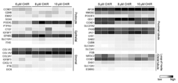
- the CHIR addition period conditions (1)-(3) in Example 1 are shown below. Fluorescence microscopy images of KiO induced by varying the duration of CHIR addition (Example 1). Fluorescence microscopy images of KiO induced by adding CHIR at different concentrations (Example 1). 1 shows phase contrast microscopy images showing cells with tubule-like morphology induced during KiO (Example 1). 1 shows the gene expression patterns of KiO induced by adding CHIR at concentrations of 6 ⁇ M, 8 ⁇ M, or 10 ⁇ M (Example 1).
- Example 3 The results of evaluating the cytotoxicity of Cyclosporin A using KiO prepared according to the method described in Example 2 and a renal proximal tubule cell line (RPTEC) are shown (Example 3).
- the cytotoxicity of Cyclosporin A was evaluated based on intracellular ATP levels using KiOs with a high proportion of proximal tubule cell-like cells (Tubule rich), KiOs with a high proportion of glomerular epithelial cell-like cells (Glomerulus rich), and KiOs with an intermediate proportion of proximal tubule cell-like cells and glomerular epithelial cell-like cells (Intermediate), which were prepared according to the method described in Example 1 (Example 3).
- the cytotoxicity of Cyclosporin A was evaluated based on the amount of LDH leakage using KiOs with a high proportion of proximal tubule cell-like cells (Tubule rich), KiOs with a high proportion of glomerular epithelial cell-like cells (Glomerulus rich), and KiOs with an intermediate proportion of proximal tubule cell-like cells and glomerular epithelial cell-like cells (Intermediate), which were prepared according to the method described in Example 1 (Example 3).
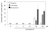
- the cytotoxicity of Cyclosporin A was evaluated based on the amount of KIM-1 released using KiOs with a high proportion of proximal tubule cell-like cells (Tubule rich), KiOs with a high proportion of glomerular epithelial cell-like cells (Glomerulus rich), and KiOs with an intermediate proportion of proximal tubule cell-like cells and glomerular epithelial cell-like cells (Intermediate), which were prepared according to the method described in Example 1 (Example 3).
- the method for evaluating the nephrotoxicity of a test substance according to the present disclosure includes a step of contacting the test substance with a renal organoid, and determining that the test substance may have nephrotoxicity if the test substance exhibits higher cytotoxicity than a control substance.
- test substance may be, but is not limited to, a compound such as an organic low molecular weight compound, a metal complex, a polymer such as a peptide, a protein, an antibody, or a nucleic acid (including an aptamer), a carbohydrate, a lipid, a tissue extract, a cell extract, a cell culture supernatant, a plant extract, a microbial product, or a biological substance such as blood or urine, and may be, in particular, a pharmaceutical candidate compound.
- the test substance may also be a synthetic compound or a compound extracted from a natural product.
- the renal organoid may be induced in vitro from stem cells. Renal organoids can be induced from pluripotent stem cells according to conventional techniques (e.g., Non-Patent Documents 2-4).
- pluripotent stem cell refers to a stem cell that can differentiate into various tissues and cells with different forms and functions in the body, and has the ability to differentiate into cells of any lineage of the three germ layers (endoderm, mesoderm, and ectoderm).
- pluripotent stem cells include embryonic stem cells (ESCs), embryonic stem cells derived from cloned embryos obtained by nuclear transfer, spermatogonial stem cells, embryonic germ cells, and induced pluripotent stem cells (sometimes referred to as "iPSCs" in this specification).
- iPSCs induced pluripotent stem cells
- multipotent stem cell refers to a stem cell that has the ability to differentiate into a limited number of lineages.
- multipotent stem cells examples include dental pulp stem cells, stem cells derived from oral mucosa, hair follicle stem cells, cultured fibroblasts, and somatic stem cells derived from bone marrow stem cells.
- Preferred pluripotent stem cells are ESCs and iPSCs.
- ESCs various mouse ESC strains established by inGenious targeting laboratory, RIKEN (Riken) and others can be used, while various human ESC strains established by University of Wisconsin, NIH, RIKEN, Kyoto University, National Center for Child Health and Development, Cellartis and others can be used.
- human ESC strains that can be used include CHB-1 to CHB-12 strains, RUES1 strain, RUES2 strain, HUES1 to HUES28 strains, etc. distributed by ESI Bio, H1 strain, H9 strain, etc. distributed by WiCell Research, and KhES-1 strain, KhES-2 strain, KhES-3 strain, KhES-4 strain, KhES-5 strain, SSES1 strain, SSES2 strain, SSES3 strain, etc. distributed by RIKEN.
- iPSC refers to cells obtained by reprogramming mammalian somatic cells or undifferentiated stem cells by introducing specific factors (nuclear reprogramming factors).
- iPSCs established by Yamanaka et al. by introducing four factors, Oct3/4, Sox2, Klf4, and c-Myc, into mouse fibroblasts (Takahashi K, Yamanaka S., Cell, (2006) 126: 663-676); iPSCs derived from human cells established by introducing the same four factors into human fibroblasts (Takahashi K, Yamanaka S., et al.
- Nanog-iPSCs established by selecting using the expression of Nanog as an indicator after introducing the above four factors (Okita, K., Ichisaka, T., and Yamanaka, S. (2007). Nature 448, 313-317.); and iPSCs created by a method that does not include c-Myc (Nakagawa M, Yamanaka S., et al. Nature Biotechnology, (2008) 26, 101-106), iPSCs established by introducing six factors using a virus-free method (Okita K et al. Nat. Methods 2011 May;8(5):409-12, Okita K et al. Stem Cells. 31(3):458-66.), etc.
- induced pluripotent stem cells established by introducing four factors, OCT3/4, SOX2, NANOG, and LIN28, created by Thomson et al. (Yu J., Thomson JA. et al., Science (2007) 318: 1917-1920), induced pluripotent stem cells created by Daley et al. (Park IH, Daley GQ. et al., Nature (2007) 451: 141-146), induced pluripotent stem cells created by Sakurada et al. (JP Patent Publication No. 2008-307007), etc. can also be used.
- iPSC lines established by NIH, RIKEN, Kyoto University, etc.
- human iPSC lines include RIKEN's HiPS-RIKEN-1A line, HiPS-RIKEN-2A line, HiPS-RIKEN-12A line, Nips-B2 line, etc., and Kyoto University's 253G1 line, 201B7 line, 409B2 line, 454E2 line, 606A1 line, 610B1 line, 648A1 line, 1231A3 line, etc., with the 1231A1 line and the 1231A3 line being preferred, and the 1231A3 line being more preferred.
- induction of renal organoids from pluripotent stem cells can be carried out as follows, for example, in the case of human iPSCs.
- human iPSCs are cultured in a medium containing a GSK3 ⁇ inhibitor, then in a medium containing FGF9 and heparin, and then dissociated into single cells.
- Approximately 1 x 10 4 -1 x 10 5 cells are further cultured in a medium containing FGF9 and heparin to form cell clusters (intermediate mesoderm spheroids) (step i).
- the intermediate mesoderm spheroids are cultured in an air-liquid phase culture medium, which is successively replaced with a medium containing a GSK3 ⁇ inhibitor, to obtain renal organoids (step ii).
- the concentration of the GSK3 ⁇ inhibitor in step i is, for example, 0.1-30 ⁇ M, preferably 1-20 ⁇ M, more preferably 6-10 ⁇ M, and particularly preferably 8 ⁇ M.
- the concentration of FGF9 is, for example, 0.1 ng/ml-1 ⁇ g/ml, preferably 1-500 ng/ml, more preferably 10-300 ng/ml, and particularly preferably about 200 ng/ml.
- the concentration of heparin is, for example, 0.01-100 ⁇ g/ml, preferably 0.1-10 ⁇ g/ml, particularly preferably about 1 ⁇ g/ml.
- the culture period in a medium containing a GSK3 ⁇ inhibitor is, for example, 3 to 5 days, particularly 4 days.
- the culture period in a medium containing FGF9 and heparin is, for example, 1 to 3 days, particularly 1 day, before single cell dissociation, and is, for example, 0.5 to 2 days, particularly 2 days, after single cell dissoci
- the concentration of the GSK3 ⁇ inhibitor in step ii is, for example, 0.1-30 ⁇ M, preferably 1-10 ⁇ M, more preferably 3-7 ⁇ M, and particularly preferably 5 ⁇ M.
- the concentration of FGF9 is, for example, 0.1 ng/ml-1 ⁇ g/ml, preferably 1-500 ng/ml, more preferably 10-300 ng/ml, and particularly preferably about 200 ng/ml.
- the concentration of heparin is, for example, 0.01-100 ⁇ g/ml, preferably 0.1-10 ⁇ g/ml, particularly preferably about 1 ⁇ g/ml.
- the incubation period in a medium containing a GSK3 ⁇ inhibitor is, for example, 0.5 to 2 hours, particularly 1 hour.
- the culture period in the medium containing FGF9 and heparin is, for example, 0.5 to 2 days, particularly 1 day.
- Step ii may be followed by a step of culturing the renal organoids in a medium containing FGF9 and heparin, and then culturing them in a medium containing heparin.
- the culture period in the medium containing FGF9 and heparin is, for example, 3 to 7 days, particularly 4 days.
- the culture period in the medium containing heparin is, for example, 7 to 25 days, particularly 14 days.
- the concentrations of FGF9 and heparin may be the same as in step ii.
- the generation of intermediate mesoderm and kidney organoids can be confirmed, for example, by measuring the expression of marker proteins or marker genes. If the obtained cell mass expresses OSR1, it can be determined that the cell mass is intermediate mesoderm. If the obtained cell pellets have the ability to produce EPO under hypoxic conditions and express markers for the renal interstitium, glomerulus, and tubules, they can be determined to be renal organoids. Renal interstitial cell markers include FOXD1, PDGFR ⁇ , and CD73. Glomerular markers include WT1 and NPHS1. Tubular markers include LTL, CUBN, and E-cadherin. Furthermore, the generation of intermediate mesoderm can also be confirmed by, for example, confirming the ability of cells to be differentiated into glomeruli or renal tubules.
- GSK3 ⁇ inhibitors include CHIR98014 (N6-[2-[[4-(2,4-dichlorophenyl)-5-(1H-imidazol-2-yl)-2-pyrimidinyl]amino]ethyl]-3-nitro-2,6-pyridinediamine), CHIR99021 (6- ⁇ 2-[4-(2,4-Dichloro-phenyl)-5-(5-methyl-1H-imidazol-2-yl)-pyrimidin-2-ylamino]-ethylamino ⁇ -nicotinonitrile), CP21R7 (CP21R7), and LY2090314 (3-[9-Fluoro-1,2,3,4-tetrahydro-2-(1-piperidinylcarbonyl)pyrrolo[3,2,1-jk][1,4]benzodiazepin-7-yl]-4-imidazo[1,2-a ]pyridin-3-yl-1h-pyr
- GSK3 ⁇ inhibitors are not limited to these, and antisense oligonucleotides and siRNAs against GSK3 ⁇ mRNA, antibodies that bind to GSK3 ⁇ , dominant-negative GSK3 ⁇ mutants, etc. can also be used as GSK3 ⁇ inhibitors, which are commercially available or can be synthesized according to known methods.
- the basal medium is not particularly limited, but for example, STEMdiff APEL2 medium (STEMCELL Technologies, ST-05275), TeSR1 medium, and Chemically Defined Medium (CDM) medium are preferably used.
- BME medium, BGJb medium, CMRL 1066 medium, Glasgow MEM medium, Improved MEM (IMEM) medium, Improved MDM (IMDM) medium, Medium 199 medium, Eagle MEM medium, ⁇ MEM medium, DMEM medium (High glucose, Low glucose), DMEM/F12 medium, Ham's medium, RPMI 1640 medium, Fischer's medium, and mixtures thereof may also be used.
- the CDM medium is not particularly limited, but for example, a medium prepared from Iscove's modified Dulbecco's medium (GE Healthcare) can be used.
- the basal medium may be supplemented with substances typically used in cell culture, such as Ham's F-12 nutrient mixture, albumin such as human serum albumin, polyvinylalcohol (PVA), deionized BSA, linoleic acid, linolenic acid, cholesterol, insulin, apotransferrin, selenium, ethanolamine, monothioglycerol, protein-free hybridoma mixture II (PFHMII), ascorbic acid, L-alanyl-L-glutamine, and/or antibiotics.
- substances typically used in cell culture such as Ham's F-12 nutrient mixture, albumin such as human serum albumin, polyvinylalcohol (PVA), deionized BSA, linoleic acid, linolenic acid, cholesterol, insulin, apotransferrin, se
- renal organoids having different ratios of glomerular epithelial cell-like cells and proximal tubule cell-like cells can also be obtained.
- the cells when inducing renal organoids that are enriched in glomerular epithelial cell-like cells and have a high proportion of such cells, the cells are cultured for 4 days in a basal medium supplemented with 9.0-10.5 ⁇ M (preferably, 9.5-10.5 ⁇ M) of a GSK3 ⁇ inhibitor, or for 5 days in a basal medium supplemented with 7-9 ⁇ M (preferably, 7.5-8.5 ⁇ M) of a GSK3 ⁇ inhibitor.
- 9.0-10.5 ⁇ M preferably, 9.5-10.5 ⁇ M
- 7-9 ⁇ M preferably, 7.5-8.5 ⁇ M
- renal organoids containing glomerular epithelial cell-like cells and proximal tubule cell-like cells to the same extent can be induced.
- Renal organoids having enriched glomerular epithelial cell-like cells can have 30-80% of cells contained in the renal organoids being glomerular epithelial cell-like cells. Renal organoids having enriched proximal tubule cell-like cells can have 5-30% of the cells contained in the renal organoids being glomerular epithelial cell-like cells.
- the step of contacting the test substance with the renal organoid can be carried out by adding the test substance to the renal organoid maintained in a medium in a suitable container such as a dish, flask or microplate.
- the step of contacting the test substance with the renal organoid can be carried out, for example, by introducing the basal medium containing the test substance into the gas phase and/or liquid phase of the transwell plate for the renal organoid created at the gas-liquid interface of the transwell plate.
- the cytotoxicity of the renal organoid contacted with the test substance can be measured and evaluated according to the conventionally known method.
- the method of using the decrease in intracellular ATP amount, the increase in the amount of intracellular enzyme leaking out of the cell, or the increase in the amount of marker protein released into the medium as an indicator of cytotoxicity is known.
- a test substance that exhibits cytotoxicity against renal organoids can be determined to have the potential to be nephrotoxic. More preferably, a test substance that exhibits higher cytotoxicity than a control substance known to have no or low nephrotoxicity is determined to be a substance that may have nephrotoxicity.
- the criteria for judgment can be set appropriately depending on the purpose of the evaluation and the type of substance to be evaluated, but for example, a substance is judged to exhibit cytotoxicity when the above-mentioned indicators are 10%, 20%, 30%, 40%, 50%, 60%, 70%, 80%, 90%, or 100% higher than that of a control substance, and preferably 100%, 200%, 300%, 400%, 500%, 600%, 700%, 800%, 900% or 1000% or more higher.
- the method of evaluating the nephrotoxicity of a test substance disclosed herein differs from in vitro evaluation methods using proximal tubule cell lines (RPTECs) in that it uses renal organoids that contain both glomerular epithelial cell-like cells and proximal tubule cell-like cells, thereby enabling evaluation that more appropriately reflects the in vivo toxicity of the test substance. Furthermore, in the method for evaluating the nephrotoxicity of a test substance disclosed herein, by using renal organoids with different ratios of glomerular epithelial cell-like cells and proximal tubule cell-like cells, it is also possible to evaluate whether the test substance is more toxic to the glomerulus or the proximal tubule.
- RPTECs proximal tubule cell lines
- Method for screening a test substance for the presence or absence of nephrotoxicity The method for evaluating the nephrotoxicity of a test substance according to the present disclosure can be used as a method for screening a test substance that may have nephrotoxicity, or a test substance that may have no or low nephrotoxicity.
- test substances that show cytotoxicity to renal organoids preferably test substances that show higher cytotoxicity than the control substance
- test substances that show no cytotoxicity to renal organoids or show low cytotoxicity compared to the control substance are selected as substances that may have no nephrotoxicity or low nephrotoxicity.
- Such screening can be particularly used for nephrotoxicity screening of candidate pharmaceutical compounds.
- renal organoids with different ratios of glomerular epithelial cell-like cells and proximal tubule cell-like cells, it is possible to create a screening system that focuses on toxicity to either the glomerulus or the proximal tubule.
- the method for evaluating the nephrotoxicity of a test substance according to the present disclosure can also be used as a method for predicting the site in the kidney where a test substance shows toxicity.
- the method includes the steps of (A) contacting a test substance with a renal organoid having enriched glomerular epithelial cell-like cells, and (B) contacting a test substance with a renal organoid having enriched proximal tubule cell-like cells.
- step (C) comparing the cytotoxicity in the step (A) with the cytotoxicity in the step (B), if the cytotoxicity in the step (A) is higher, it is possible to predict that the site of toxicity is the glomerulus, and if the cytotoxicity in the step (B) is higher, it is possible to predict that the site of toxicity is the proximal tubule.
- Kit also provides a kit including a renal organoid for use in the above-mentioned method for evaluating the nephrotoxicity of a test substance, the method for screening a test substance for the presence or absence of nephrotoxicity, and the method for predicting the site at which a test substance shows toxicity in the kidney.
- the kit according to the present disclosure may include renal organoids having enriched glomerular epithelial cell-like cells or renal organoids having enriched proximal tubule cell-like cells as described above.
- the kit according to the present disclosure may contain, in addition to renal organoids, media and reagents for culturing renal organoids (GSK3 ⁇ inhibitors, FGF, heparin, etc.), and reagents for measuring cytotoxicity to renal organoids.
- media and reagents for culturing renal organoids GSK3 ⁇ inhibitors, FGF, heparin, etc.
- reagents for measuring cytotoxicity to renal organoids may contain, in addition to renal organoids, media and reagents for culturing renal organoids (GSK3 ⁇ inhibitors, FGF, heparin, etc.), and reagents for measuring cytotoxicity to renal organoids.
- the kit according to the present disclosure may contain pluripotent stem cells instead of renal organoids, and culture media and reagents (GSK3 ⁇ inhibitors, FGF, heparin, etc.) for inducing differentiation of the pluripotent stem cells into renal organoids.
- culture media and reagents GSK3 ⁇ inhibitors, FGF, heparin, etc.
- kidney organoids KiO derived from inducible pluripotent stem cells (iPSC)
- iPSC seeding 1231A3 iPSCs were seeded at 5.76 x 10⁵ cells/well onto a laminin-coated (iMatrix-511 silk: Nippi) 6-well plate. The cells were cultured overnight in a 37°C, 5% CO2 incubator. The medium used was AK03N (Ajinomoto) supplemented with 10 ⁇ M Y27632 at 2 ml/well.
- Step 2 (Day 0-Day 7): Induction of intermediate mesoderm spheroids The medium was replaced with intermediate mesoderm differentiation medium.
- the basal medium for intermediate mesoderm induction medium (basal medium for KiO differentiation induction) was APEL2 medium (STEMCELL technology) supplemented with 5% PFHM (Protein-Free Hybridoma Medium: Thermo Fisher).
- Step 2-1 (Day 0-Day 3/4/5): During the first half of the period from Day 0 to Day 6, a medium obtained by adding CHIR99021 (CHIR) to KiO differentiation-inducing basal medium was used as an intermediate mesoderm differentiation medium.
- CHIR99021 CHIR99021
- Step 2-2 Day 3/4/5-Day 6: During the latter half of the period from Day 0 to Day 6, a medium prepared by adding 200 ng/ml FGF9 and 1 ⁇ g/ml heparin to the KiO differentiation-inducing basal medium was used as an intermediate mesoderm differentiation medium. The medium was changed every two days. The number of days allocated to the above-mentioned first half period (with CHIR) and second half period (without CHIR) and the concentration of CHIR added in the first half period were changed appropriately to conduct the studies described in the examples below.
- Step 2-3 Formation of intermediate mesodermal spheroids using 96-well plates.
- Cells on day 6 of differentiation induction were dissociated into single cells using Accutase, suspended in basal medium (KiO differentiation induction basal medium) supplemented with 200 ng/ml FGF9 and 1 ⁇ g/ml heparin, and seeded at 5 ⁇ 10 4 cells/well (200 ⁇ l/well) on a PrimeSurface 96-well plate (Sumitomo Bakelite). The plate was centrifuged at 300 g for 3 min to sediment the cells, and then cultured overnight (1 day) in a 37°C, 5% CO 2 incubator.
- Step 3 Air-liquid interface culture using transwell plate
- the intermediate mesodermal spheroids prepared in the Prime Surface 96-well plate were transferred to a new plate (basal medium 200 ⁇ l/well) to wash the intermediate mesodermal spheroids.
- the washed intermediate mesodermal spheroids were transferred to the insert of the transwell plate.
- a liquid phase was formed by adding a basal medium containing 5 ⁇ M CHIR to each well of the transwell plate.
- the insert was inserted into the well, and the intermediate mesodermal spheroids in the insert were kept at the air-liquid interface and left to stand for 1 hour in a 37°C, 5% CO2 incubator.
- the liquid phase medium was then replaced with a basal medium containing 200 ng/ml FGF9 and 1 ⁇ g/ml heparin, and cultured overnight. Note that 6-well transwell plates or 24-well transwell plates were used as the transwell plates.
- Step 4 Culture with the addition of FGF9 and heparin From Day 8 to Day 11, the cells were cultured in a basal medium containing 200 ng/ml FGF9 and 1 ⁇ g/ml heparin in the liquid phase. The medium was changed every two days.
- Step 5 (Day 12-Day 25): Culture with the addition of heparin After Day 12, the cells were cultured in a basal medium containing 1 ⁇ g/ml of heparin in the liquid phase. The medium was changed every two days.
- Example 1 Examination of the effect of the addition period and concentration of CHIR on the ratio of glomerular epithelial cell-like cells and proximal tubule cells in KiO]
- CHIR addition period (see Figure 2) Condition (1): First half of the period (with CHIR) 3 days (Day 0-Day 2) / Second half of the period (without CHIR) 3 days (Day 3-Day 5) Condition (2): First half of the period is 4 days (Day 0-Day 3) / Second half of the period is 2 days (Day 4-Day 5), or Condition (3): First half of the period is 5 days (Day 0-Day 4) / Second half of the period is 1 day (Day 5) CHIR concentration: 6 ⁇ M, 8 ⁇ M, or 10 ⁇ M In addition, culture without CHIR was performed using a medium containing 200 ng/ml FGF9 and 1 ⁇ g/ml heparin in the basal medium.
- Step 1 Day -1) and steps 3-6 (Day 6-Day 24) were carried out in the same manner as in Example 1. However, a 6-well transwell plate made of PET was used for the gas-liquid phase culture in steps 4-6 (Day 7-Day 24).
- Example 2 Gas-liquid phase culture using 24-well transwell plate
- Steps 1 and 2 were carried out in the same manner as outlined above.
- the intermediate mesodermal spheroids prepared in the Prime Surface 96-well plate were washed by transferring them to a new plate (basal medium 200 ⁇ l/well).
- the washed intermediate mesodermal spheroids were transferred to the inserts of a 24-well transwell (Kurabo) made of PTFE (polytetrafluoroethylene) membrane material.
- the liquid phase was formed by adding basal medium supplemented with 5 ⁇ M CHIR to each well of the transwell plate (300 ⁇ l/well).
- the inserts were inserted into the wells, and the intermediate mesodermal spheroids in the inserts were kept at the air-liquid interface and left to stand for 1 hour in a 37°C, 5% CO2 incubator.
- the liquid phase medium was then replaced with basal medium supplemented with 200 ng/ml FGF9 and 1 ⁇ g/ml heparin, and cultured overnight. Steps 4 and 5 (Day 8-Day 24) were carried out in the same manner as outlined above.
- Example 3 Toxicity evaluation using KiO
- KiO Day 25
- a basal medium 300 ⁇ l/well
- a basal medium 500 ⁇ l/well
- the plates were cultured for 3 days in a 37°C, 5% CO2 incubator, and the KiO was exposed to the test substance.
- Cyclosporin A concentration 1 ⁇ 10 ⁇ 7 M-1 ⁇ 10 ⁇ 3 M
- FIG. 7 The results using Cyclosporin A (concentration 1 ⁇ 10 ⁇ 7 M-1 ⁇ 10 ⁇ 3 M) as the test substance are shown in FIG. 7.
- Cyclosporin A is known to be highly toxic in glomeruli. With the KiO prepared according to the method described in Example 3, the concentration-dependent cytotoxicity of Cyclosporin A could be detected.
- RPTEC renal proximal tubule epithelial cell line
- KiOs with a high proportion of proximal tubule cell-like cells, KiOs with a high proportion of glomerular epithelial cell-like cells, and KiOs with a medium proportion of proximal tubule cell-like cells and glomerular epithelial cell-like cells were prepared and tested in the same manner. The results are shown in Figure 8. Higher cytotoxicity was detected in KiOs with a high proportion of proximal tubule cell-like cells (Tubule rich), and lower cytotoxicity was detected in KiOs with a high proportion of glomerular epithelial cell-like cells (Glomerulus rich).
- the wells were washed 4 times with a detergent-containing washing solution, and 200 ⁇ l of a buffer solution containing an anti-KIM-1 detection antibody was added. The plate was left standing at 25°C for 2 hours in the dark. The wells were washed 4 times with a detergent-containing washing solution, and 200 ⁇ l of a substrate solution was added. The plate was left standing at 25°C for 30 minutes in the dark. 50 ⁇ l of stop solution was added to stop the reaction, and the absorbance at 450 nm was measured using a microplate reader. The control substance was the solvent alone. Cytotoxicity was calculated as "(measured value of test substance/measured value of control substance) x 100%".
- Example 4 Method for improving differentiation induction of iPSC-derived renal KiO
- Step 1 Day ⁇ 1: iPSC seeding This was carried out in the same manner as outlined above.
- Step 2 (Day 0-Day 5): Induction of intermediate mesodermal spheroids
- step 2-1 first half period
- step 2-2 second half period
- the medium was replaced with intermediate mesoderm differentiation medium.
- the basal medium for intermediate mesoderm induction was APEL2 medium (STEMCELL technology) supplemented with 5% PFHM (Protein-Free Hybridoma Medium: Thermo Fisher).
- Step 2-1 (Day 0-Day 4): During the first half of the period from Day 0 to Day 5, a medium obtained by adding CHIR99021 (CHIR) to KiO differentiation-inducing basal medium was used as an intermediate mesoderm differentiation medium.
- Step 2-2 (Day 4-Day 5): During the latter half of the period from Day 0 to Day 5, a medium obtained by adding 200 ng/ml FGF9 and 1 ⁇ g/ml heparin to the KiO differentiation-inducing basal medium was used as an intermediate mesoderm differentiation medium. The medium was changed every two days.
- CHIR99021 CHIR99021
- Step 2-3 (Day 5-Day 7) Formation of intermediate mesodermal spheroids using 96-well plates Step 2-3 in the above outlined method was changed to Day 5-Day 7 in this improved method.
- the cells were dissociated into single cells using Accutase, suspended in basal medium (KiO basal medium for differentiation induction) supplemented with 200 ng/ml FGF9 and 1 ⁇ g/ml heparin, and seeded at 5 ⁇ 104 cells/well (200 ⁇ l/well) on a PrimeSurface 96-well plate (Sumitomo Bakelite).
- the plate was centrifuged at 300 g for 3 min to sediment the cells, and then cultured for 2 days in a 37°C, 5% CO2 incubator. By extending the culture period from 1 to 2 days, spheroids with more cells in the center (clearer core) were formed, and KiO could be induced more stably in the subsequent step 3.
- a liquid phase was formed by adding basal medium supplemented with 5 ⁇ M CHIR to each well of the transwell plate.
- the inserts were inserted into the wells, and the intermediate mesodermal spheroids in the inserts were kept at the air-liquid interface and left to stand for 1 hour in a 37°C, 5% CO2 incubator.
- the liquid phase medium was then replaced with basal medium supplemented with 200 ng/ml FGF9 and 1 ⁇ g/ml heparin, and cultured overnight.
- Step 4 (Day 8-Day 11): Culture with the addition of FGF9 and heparin
- Step 5 (Day 12-Day 25): Culture with the addition of heparin
- the procedure was the same as outlined above, except that an operation to wash away necrotic cells was performed. From day 8 to day 11, the liquid phase was filled with basal medium supplemented with 200 ng/ml FGF9 and 1 ⁇ g/ml heparin, and the medium was changed every two days. From day 12 onwards, the liquid phase was filled with basal medium supplemented with 1 ⁇ g/ml heparin, and the medium was changed every two days. Once a week, water was dripped onto the KiO from above to wash away necrotic cells, which improved the viability of the cells in the KiO and the visibility of the KiO.
Landscapes
- Health & Medical Sciences (AREA)
- Life Sciences & Earth Sciences (AREA)
- Engineering & Computer Science (AREA)
- Chemical & Material Sciences (AREA)
- Organic Chemistry (AREA)
- Zoology (AREA)
- Genetics & Genomics (AREA)
- Wood Science & Technology (AREA)
- Biomedical Technology (AREA)
- Bioinformatics & Cheminformatics (AREA)
- Biotechnology (AREA)
- General Health & Medical Sciences (AREA)
- Biochemistry (AREA)
- General Engineering & Computer Science (AREA)
- Microbiology (AREA)
- Cell Biology (AREA)
- Proteomics, Peptides & Aminoacids (AREA)
- Physics & Mathematics (AREA)
- Analytical Chemistry (AREA)
- Biophysics (AREA)
- Immunology (AREA)
- Molecular Biology (AREA)
- Measuring Or Testing Involving Enzymes Or Micro-Organisms (AREA)
Priority Applications (2)
| Application Number | Priority Date | Filing Date | Title |
|---|---|---|---|
| CN202380070191.6A CN120359301A (zh) | 2022-09-29 | 2023-09-28 | 用于评价肾毒性的方法 |
| JP2024550437A JPWO2024071280A1 (https=) | 2022-09-29 | 2023-09-28 |
Applications Claiming Priority (2)
| Application Number | Priority Date | Filing Date | Title |
|---|---|---|---|
| JP2022-156017 | 2022-09-29 | ||
| JP2022156017 | 2022-09-29 |
Publications (1)
| Publication Number | Publication Date |
|---|---|
| WO2024071280A1 true WO2024071280A1 (ja) | 2024-04-04 |
Family
ID=90478035
Family Applications (1)
| Application Number | Title | Priority Date | Filing Date |
|---|---|---|---|
| PCT/JP2023/035342 Ceased WO2024071280A1 (ja) | 2022-09-29 | 2023-09-28 | 腎毒性評価方法 |
Country Status (3)
| Country | Link |
|---|---|
| JP (1) | JPWO2024071280A1 (https=) |
| CN (1) | CN120359301A (https=) |
| WO (1) | WO2024071280A1 (https=) |
Cited By (1)
| Publication number | Priority date | Publication date | Assignee | Title |
|---|---|---|---|---|
| CN120574765A (zh) * | 2025-06-06 | 2025-09-02 | 温州医科大学 | 一种基于类器官及脱细胞支架的生物工程肾构建方法 |
Citations (9)
| Publication number | Priority date | Publication date | Assignee | Title |
|---|---|---|---|---|
| WO2007069666A1 (ja) | 2005-12-13 | 2007-06-21 | Kyoto University | 核初期化因子 |
| US20080236610A1 (en) | 2004-04-10 | 2008-10-02 | Holger Bartels | Hair Rollers |
| WO2008118220A2 (en) | 2006-11-28 | 2008-10-02 | Veritainer Corporation | Radiation detection unit for mounting a radiation sensor to a container crane |
| WO2008124133A1 (en) | 2007-04-07 | 2008-10-16 | Whitehead Institute For Biomedical Research | Reprogramming of somatic cells |
| WO2008151058A2 (en) | 2007-05-30 | 2008-12-11 | The General Hospital Corporation | Methods of generating pluripotent cells from somatic cells |
| JP2008307007A (ja) | 2007-06-15 | 2008-12-25 | Bayer Schering Pharma Ag | 出生後のヒト組織由来未分化幹細胞から誘導したヒト多能性幹細胞 |
| US20090047263A1 (en) | 2005-12-13 | 2009-02-19 | Kyoto University | Nuclear reprogramming factor and induced pluripotent stem cells |
| WO2016094948A1 (en) * | 2014-12-15 | 2016-06-23 | The University Of Queensland | Differentiation of pluripotent stem cells to form renal organoids |
| JP2022156017A (ja) | 2021-03-31 | 2022-10-14 | 本田技研工業株式会社 | 鞍乗り型車両 |
-
2023
- 2023-09-28 WO PCT/JP2023/035342 patent/WO2024071280A1/ja not_active Ceased
- 2023-09-28 JP JP2024550437A patent/JPWO2024071280A1/ja active Pending
- 2023-09-28 CN CN202380070191.6A patent/CN120359301A/zh active Pending
Patent Citations (13)
| Publication number | Priority date | Publication date | Assignee | Title |
|---|---|---|---|---|
| US20080236610A1 (en) | 2004-04-10 | 2008-10-02 | Holger Bartels | Hair Rollers |
| US20090047263A1 (en) | 2005-12-13 | 2009-02-19 | Kyoto University | Nuclear reprogramming factor and induced pluripotent stem cells |
| JP2008283972A (ja) | 2005-12-13 | 2008-11-27 | Kyoto Univ | 誘導多能性幹細胞の製造方法 |
| WO2007069666A1 (ja) | 2005-12-13 | 2007-06-21 | Kyoto University | 核初期化因子 |
| WO2008118220A2 (en) | 2006-11-28 | 2008-10-02 | Veritainer Corporation | Radiation detection unit for mounting a radiation sensor to a container crane |
| WO2008124133A1 (en) | 2007-04-07 | 2008-10-16 | Whitehead Institute For Biomedical Research | Reprogramming of somatic cells |
| WO2008151058A2 (en) | 2007-05-30 | 2008-12-11 | The General Hospital Corporation | Methods of generating pluripotent cells from somatic cells |
| JP2008307007A (ja) | 2007-06-15 | 2008-12-25 | Bayer Schering Pharma Ag | 出生後のヒト組織由来未分化幹細胞から誘導したヒト多能性幹細胞 |
| WO2009006930A1 (en) | 2007-06-15 | 2009-01-15 | Izumi Bio, Inc. | Human pluripotent stem cells induced from undifferentiated stem cells derived from a human postnatal tissue |
| WO2009006997A1 (en) | 2007-06-15 | 2009-01-15 | Izumi Bio, Inc. | Human pluripotent stem cells and their medical use |
| WO2009007852A2 (en) | 2007-06-15 | 2009-01-15 | Izumi Bio, Inc | Multipotent/pluripotent cells and methods |
| WO2016094948A1 (en) * | 2014-12-15 | 2016-06-23 | The University Of Queensland | Differentiation of pluripotent stem cells to form renal organoids |
| JP2022156017A (ja) | 2021-03-31 | 2022-10-14 | 本田技研工業株式会社 | 鞍乗り型車両 |
Non-Patent Citations (15)
| Title |
|---|
| KIM JB.SCHOLER HR. ET AL., NATURE, vol. 454, 2008, pages 646 - 650 |
| LOW JIAN HUI; LI PIN; CHEW ELAINE GUO YAN; ZHOU BINGRUI; SUZUKI KEIICHIRO; ZHANG TIAN; LIAN MICHELLE MULAN; LIU MENG; AIZAWA EMI; : "Generation of Human PSC-Derived Kidney Organoids with Patterned Nephron Segments and a De Novo Vascular Network", CELL STEM CELL, ELSEVIER, CELL PRESS, AMSTERDAM, NL, vol. 25, no. 3, 11 July 2019 (2019-07-11), AMSTERDAM, NL , pages 373, XP085802948, ISSN: 1934-5909, DOI: 10.1016/j.stem.2019.06.009 * |
| MARIANNE K. VORMANN: "Nephrotoxicity and Kidney Transport Assessment on 3D Perfused Proximal Tubules", THE AAPS JOURNAL, vol. 20, 2018, XP036580785, DOI: 10.1208/s12248-018-0248-z |
| MINORU TAKASATO: "Erratum: Kidney organoids from human iPS cells contain multiple lineages and model human nephrogenesis", NATURE, SPRINGER NATURE LIMITED, vol. 536, no. 7615, 1 August 2016 (2016-08-01), pages 238 - 238, XP093154212, ISSN: 0028-0836, DOI: 10.1038/nature17982 * |
| MINORU TAKASATO: "Generation of kidney organoids from human pluripotent stem cells", NATURE PROTOCOLS, vol. 11, 2016, pages 1681 - 1692, XP055679831, DOI: 10.1038/nprot.2016.098 |
| MINORU TAKASATO: "Kidney organoids from human iPS cells contain multiple lineages and model human nephrogenesis", NATURE, vol. 526, 2015, pages 564 - 568, XP055454802, DOI: 10.1038/nature15695 |
| NAKAGAWA M.YAMANAKA S. ET AL., NATURE BIOTECHNOLOGY, vol. 26, no. 7, 2008, pages 795 - 797 |
| OKITA K. ET AL., NAT. METHODS, vol. 8, no. 5, May 2011 (2011-05-01), pages 409 - 12 |
| OKITA K., STEM CELLS, vol. 31, no. 3, pages 458 - 66 |
| OKITA, K.ICHISAKA, T.YAMANAKA S., NATURE, vol. 451, 2007, pages 141 - 146 |
| SARA E. HOWDEN: "Plasticity of distal nephron epithelia from human kidney organoids enables the induction of ureteric tip and stalk", CELL STEM CELL, vol. 28, no. 1, 2021, pages 671 - 684 |
| SHI Y.DING S. ET AL., CELL STEM CELL, vol. 3, 2008, pages 568 - 574 |
| TAKAHASHI K.YAMANAKA S. ET AL., CELL, vol. 131, 2007, pages 861 - 872 |
| TAKAHASHI K.YAMANAKA S., CELL, vol. 126, 2006, pages 663 - 676 |
| YU J.THOMSON JA. ET AL., SCIENCE, vol. 318, 2007, pages 1917 - 1920 |
Cited By (1)
| Publication number | Priority date | Publication date | Assignee | Title |
|---|---|---|---|---|
| CN120574765A (zh) * | 2025-06-06 | 2025-09-02 | 温州医科大学 | 一种基于类器官及脱细胞支架的生物工程肾构建方法 |
Also Published As
| Publication number | Publication date |
|---|---|
| CN120359301A (zh) | 2025-07-22 |
| JPWO2024071280A1 (https=) | 2024-04-04 |
Similar Documents
| Publication | Publication Date | Title |
|---|---|---|
| JP7576647B2 (ja) | 肝臓オルガノイド疾患モデルおよびその作製方法 | |
| CN110709092B (zh) | 用于产生人少突胶质细胞和研究体外髓鞘形成的个性化3d神经培养系统 | |
| JP7330466B2 (ja) | 細胞の培養方法 | |
| WO2014078414A1 (en) | Chemically defined production of cardiomyocytes from pluripotent stem cells | |
| CN106029872A (zh) | 使诱导性多能干细胞分化成肾近端小管细胞样细胞的方法 | |
| JP7541700B2 (ja) | 腎間質細胞の製造方法 | |
| JPWO2017188458A1 (ja) | 骨格筋前駆細胞及び骨格筋細胞の製造方法 | |
| WO2011014485A2 (en) | Methods to characterize cell reprogramming and uses thereof | |
| US12060579B2 (en) | Human pluripotent stem cell derived endocardial endothelium | |
| US12467917B2 (en) | Reconstructing human early embryogenesis in vitro with pluripotent stem cells | |
| WO2024071280A1 (ja) | 腎毒性評価方法 | |
| JP7533896B2 (ja) | スクリーニング方法および毒性評価法 | |
| US20260125652A1 (en) | Method for evaluating nephrotoxicity | |
| US20240318142A1 (en) | Method for producing renal interstitial progenitor cells, erythropoietin-producing cells, and method for producing renin-producing cells | |
| US12421497B2 (en) | Method for enriching cardiac myocytes | |
| JP7058330B2 (ja) | 腸管上皮細胞の製造方法および腸管上皮細胞 | |
| JP7791525B2 (ja) | 多列繊毛気道上皮細胞、分化誘導方法および製造方法 | |
| WO2024029617A1 (ja) | 心筋の製造方法 | |
| WO2025018366A1 (ja) | 腸管上皮細胞を作製する方法 | |
| WO2020130068A1 (ja) | 胚型赤芽球を含む細胞集団及びその製造方法、細胞培養組成物並びに化合物試験法 | |
| Soejima et al. | Analysis of bone marrow stem and SP cells derived from the femur, humerus and ilium of aged and young rats |
Legal Events
| Date | Code | Title | Description |
|---|---|---|---|
| 121 | Ep: the epo has been informed by wipo that ep was designated in this application |
Ref document number: 23872502 Country of ref document: EP Kind code of ref document: A1 |
|
| WWE | Wipo information: entry into national phase |
Ref document number: 2024550437 Country of ref document: JP |
|
| WWE | Wipo information: entry into national phase |
Ref document number: 202380070191.6 Country of ref document: CN |
|
| WWE | Wipo information: entry into national phase |
Ref document number: 2023872502 Country of ref document: EP |
|
| NENP | Non-entry into the national phase |
Ref country code: DE |
|
| ENP | Entry into the national phase |
Ref document number: 2023872502 Country of ref document: EP Effective date: 20250429 |
|
| WWW | Wipo information: withdrawn in national office |
Ref document number: 2023872502 Country of ref document: EP |
|
| WWP | Wipo information: published in national office |
Ref document number: 202380070191.6 Country of ref document: CN |
|
| WWP | Wipo information: published in national office |
Ref document number: 2023872502 Country of ref document: EP |