WO2023227641A1 - Use of tnf-alpha binding proteins and il-7 binding proteins in medical treatment - Google Patents
Use of tnf-alpha binding proteins and il-7 binding proteins in medical treatment Download PDFInfo
- Publication number
- WO2023227641A1 WO2023227641A1 PCT/EP2023/063863 EP2023063863W WO2023227641A1 WO 2023227641 A1 WO2023227641 A1 WO 2023227641A1 EP 2023063863 W EP2023063863 W EP 2023063863W WO 2023227641 A1 WO2023227641 A1 WO 2023227641A1
- Authority
- WO
- WIPO (PCT)
- Prior art keywords
- seq
- tnf
- binding protein
- acid sequence
- binding
- Prior art date
- Legal status (The legal status is an assumption and is not a legal conclusion. Google has not performed a legal analysis and makes no representation as to the accuracy of the status listed.)
- Ceased
Links
Classifications
-
- C—CHEMISTRY; METALLURGY
- C07—ORGANIC CHEMISTRY
- C07K—PEPTIDES
- C07K16/00—Immunoglobulins [IG], e.g. monoclonal or polyclonal antibodies
- C07K16/18—Immunoglobulins [IG], e.g. monoclonal or polyclonal antibodies against material from animals or humans
- C07K16/24—Immunoglobulins [IG], e.g. monoclonal or polyclonal antibodies against material from animals or humans against cytokines, lymphokines or interferons
- C07K16/244—Interleukins [IL]
-
- A—HUMAN NECESSITIES
- A61—MEDICAL OR VETERINARY SCIENCE; HYGIENE
- A61P—SPECIFIC THERAPEUTIC ACTIVITY OF CHEMICAL COMPOUNDS OR MEDICINAL PREPARATIONS
- A61P37/00—Drugs for immunological or allergic disorders
- A61P37/02—Immunomodulators
-
- C—CHEMISTRY; METALLURGY
- C07—ORGANIC CHEMISTRY
- C07K—PEPTIDES
- C07K16/00—Immunoglobulins [IG], e.g. monoclonal or polyclonal antibodies
- C07K16/18—Immunoglobulins [IG], e.g. monoclonal or polyclonal antibodies against material from animals or humans
- C07K16/24—Immunoglobulins [IG], e.g. monoclonal or polyclonal antibodies against material from animals or humans against cytokines, lymphokines or interferons
- C07K16/241—Tumor Necrosis Factors
-
- C—CHEMISTRY; METALLURGY
- C07—ORGANIC CHEMISTRY
- C07K—PEPTIDES
- C07K2317/00—Immunoglobulins specific features
- C07K2317/30—Immunoglobulins specific features characterized by aspects of specificity or valency
- C07K2317/34—Identification of a linear epitope shorter than 20 amino acid residues or of a conformational epitope defined by amino acid residues
-
- C—CHEMISTRY; METALLURGY
- C07—ORGANIC CHEMISTRY
- C07K—PEPTIDES
- C07K2317/00—Immunoglobulins specific features
- C07K2317/70—Immunoglobulins specific features characterized by effect upon binding to a cell or to an antigen
- C07K2317/71—Decreased effector function due to an Fc-modification
-
- C—CHEMISTRY; METALLURGY
- C07—ORGANIC CHEMISTRY
- C07K—PEPTIDES
- C07K2317/00—Immunoglobulins specific features
- C07K2317/70—Immunoglobulins specific features characterized by effect upon binding to a cell or to an antigen
- C07K2317/76—Antagonist effect on antigen, e.g. neutralization or inhibition of binding
-
- C—CHEMISTRY; METALLURGY
- C07—ORGANIC CHEMISTRY
- C07K—PEPTIDES
- C07K2317/00—Immunoglobulins specific features
- C07K2317/90—Immunoglobulins specific features characterized by (pharmaco)kinetic aspects or by stability of the immunoglobulin
- C07K2317/92—Affinity (KD), association rate (Ka), dissociation rate (Kd) or EC50 value
-
- C—CHEMISTRY; METALLURGY
- C07—ORGANIC CHEMISTRY
- C07K—PEPTIDES
- C07K2317/00—Immunoglobulins specific features
- C07K2317/90—Immunoglobulins specific features characterized by (pharmaco)kinetic aspects or by stability of the immunoglobulin
- C07K2317/94—Stability, e.g. half-life, pH, temperature or enzyme-resistance
Definitions
- TNF-alpha binding proteins and IL-7 binding proteins are useful in Medical Treatment FIELD OF THE DISCLOSURE
- the present disclosure relates to the use of TNF- ⁇ (Tumor Necrosis Factor-alpha) binding proteins and interleukin 7 (IL-7) binding proteins in the treatment of autoimmune and/or inflammatory conditions.
- the disclosure also concerns methods of treating diseases with TNF- ⁇ binding proteins and IL-7 binding proteins.
- the disclosure further concerns bispecific antibodies binding TNF- ⁇ and IL-7, uses of such bispecific antibodies, pharmaceutical compositions comprising such bispecific antibodies and methods of their manufacture. Other aspects of the disclosure will be apparent from the description below.
- IBD Inflammatory Bowel Disease
- RA Rheumatoid Arthritis
- IBD is a multi-factorial chronic relapsing immune-mediated disease composed primarily of two main subtypes, Crohn’s Disease (CD) and Ulcerative Colitis (UC).
- CD Crohn’s Disease
- UC Ulcerative Colitis
- TNF- ⁇ is a pleiotropic cytokine expressed by activated macrophages, NK cell, and T cells as well as non-immune cells such as endothelial cell and fibroblasts.
- Anti-TNF- ⁇ therapies are clinically validated for the treatment of IBD and can result in rapid disease remission, mucosal healing, and improved quality of life for approximately 60% of patients suffering from CD or UC.
- a method for treatment of an autoimmune and/or inflammatory condition in a subject in need thereof comprising administering to the subject a therapeutically effective amount of an IL-7 inhibitor and a therapeutically effective amount of a TNF- ⁇ inhibitor.
- an IL-7 inhibitor and a TNF- ⁇ inhibitor for use in the treatment of an autoimmune and/or inflammatory condition.
- an IL-7 inhibitor and a TNF- ⁇ inhibitor in the manufacture of a medicament for the treatment of an autoimmune and/or inflammatory condition.
- an IL-7 and TNF- ⁇ binding protein comprising: a) CDRH1 of SEQ ID NO:6, CDRH2 of SEQ ID NO:7, CDRH3 of SEQ ID NO:8, CDRL1 of SEQ ID NO:9, CDRL2 of SEQ ID NO:10 and CDRL3 of SEQ ID NO:11; and/or b) CDRH1 of SEQ ID NO:48, CDRH2 of SEQ ID NO:49, CDRH3 of SEQ ID NO:50, CDRL1 of SEQ ID NO:51, CDRL2 of SEQ ID NO:52 and CDRL3 of SEQ ID NO:53.
- a pharmaceutical composition comprising a pharmaceutically acceptable excipient and an IL-7 and TNF- ⁇ binding protein comprising: a) CDRH1 of SEQ ID NO:6, CDRH2 of SEQ ID NO:7, CDRH3 of SEQ ID NO:8, CDRL1 of SEQ ID NO:9, CDRL2 of SEQ ID NO:10 and CDRL3 of SEQ ID NO:11; and/or b) CDRH1 of SEQ ID NO:48, CDRH2 of SEQ ID NO:49, CDRH3 of SEQ ID NO:50, CDRL1 of SEQ ID NO:51, CDRL2 of SEQ ID NO:52 and CDRL3 of SEQ ID NO:53.
- a method of treatment of an autoimmune and/or an inflammatory condition in a subject in need thereof comprising administering to the subject a therapeutically effective amount of an IL-7 and TNF- ⁇ binding protein, or a pharmaceutical composition comprising a pharmaceutically acceptable excipient and an IL-7 and TNF- ⁇ binding protein, wherein the IL-7 and TNF- ⁇ binding protein, comprises: a) CDRH1 of SEQ ID NO:6, CDRH2 of SEQ ID NO:7, CDRH3 of SEQ ID NO:8, CDRL1 of SEQ ID NO:9, CDRL2 of SEQ ID NO:10 and CDRL3 of SEQ ID NO:11; and/or b) CDRH1 of SEQ ID NO:48, CDRH2 of SEQ ID NO:49, CDRH3 of SEQ ID NO:50, CDRL1 of SEQ ID NO:51, CDRL2 of SEQ ID NO:52 and CDRL3 of SEQ ID NO:53.
- an IL-7 and TNF- ⁇ binding protein or a pharmaceutical composition comprising a pharmaceutically acceptable excipient and an IL-7 and TNF- ⁇ binding protein, for use in the treatment of an autoimmune and/or inflammatory condition
- the IL-7 and TNF- ⁇ binding protein comprises: a) CDRH1 of SEQ ID NO:6, CDRH2 of SEQ ID NO:7, CDRH3 of SEQ ID NO:8, CDRL1 of SEQ ID NO:9, CDRL2 of SEQ ID NO:10 and CDRL3 of SEQ ID NO:11; and/or 70169 b) CDRH1 of SEQ ID NO:48, CDRH2 of SEQ ID NO:49, CDRH3 of SEQ ID NO:50, CDRL1 of SEQ ID NO:51, CDRL2 of SEQ ID NO:52 and CDRL3 of SEQ ID NO:53.
- an IL-7 and TNF- ⁇ binding protein or a pharmaceutical composition comprising a pharmaceutically acceptable excipient and an IL-7 and TNF- ⁇ binding protein, for the manufacture of a medicament for the treatment of an autoimmune and/or inflammatory condition
- the IL-7 and TNF- ⁇ binding protein comprises: a) CDRH1 of SEQ ID NO:6, CDRH2 of SEQ ID NO:7, CDRH3 of SEQ ID NO:8, CDRL1 of SEQ ID NO:9, CDRL2 of SEQ ID NO:10 and CDRL3 of SEQ ID NO:11; and/or b) CDRH1 of SEQ ID NO:48, CDRH2 of SEQ ID NO:49, CDRH3 of SEQ ID NO:50, CDRL1 of SEQ ID NO:51, CDRL2 of SEQ ID NO:52 and CDRL3 of SEQ ID NO:53.
- Figure 1A illustrates the concentration of DRSPAI-L7B in animals dosed (i.v.) with a DRSPAI-L7B target dose of 0.1 mg/kg, 1 mg/kg, or 10 mg/kg.
- Figure 1B and Figure 1C illustrate the concentration of subcutaneous weekly doses with a repeat target dose of 30 mg/kg, Figure 1B following dose 1 and Figure 1C following doses 4.
- FIG. 2A illustrates % pSTAT5 inhibition of whole blood obtained from healthy donors that was stimulated with IL-7 in the presence of DRSPAI-L7B.
- Figure 2B illustrates STAT5 phosphorylation in CD8+ T cells, Figure 2C CD4+ T cells, Figure 2D CD3+ T cells, from peripheral blood mononuclear cells (PBMCs) obtained from healthy donors or IBD patients, stimulated with rhIL-7 in the presence of DRSPAI-L7B or anti-RSV antibody (isotype control).
- PBMCs peripheral blood mononuclear cells
- Figure 3A illustrates inhibition of IFN- ⁇ secretion by healthy donor PBMCs treated with increasing concentrations of DRSPAI-L7B in the presence of rhIL-7 and anti-CD3.
- Figure 3B illustrates inhibition of IL-10 secretion by healthy donor PBMCs treated with increasing concentrations of DRSPAI- L7B in the presence of rhIL-7 and anti-CD3.
- Figure 3C, Figure 3D, Figure 3E, Figure 3F, Figure 3G illustrates inhibition of IL-2 by DRSPAI-L7B in the presence of rhIL-7 and anti-CD3.
- Figure 4A illustrates IL-17 inhibition in CD4 + T mem cells that were isolated from healthy donor blood, incubated with IL-7 in the presence of DRSPAI-L7B after being spiked with phorbol myristate acetate (PMA)/ionomycin.
- Figure 4B illustrates TNF- ⁇ inhibition in CD4 + T mem cells that were isolated from healthy donor blood, incubated with IL-7 in the presence of DRSPAI-L7B after being spiked with PMA/ionomycin.
- Figure 4C illustrates IL-6 inhibition in CD4 + T mem cells that were isolated from healthy donor blood, incubated with IL-7 in the presence of DRSPAI-L7B after being spiked with PMA/ionomycin.
- Figure 4D illustrates IL-10 inhibition in CD4 + Tmem cells that were isolated from healthy donor blood, incubated with IL-7 in the presence of DRSPAI-L7B after being spiked with PMA/ionomycin.
- Figure 4E illustrates INF ⁇ inhibition in CD4 + T mem cells that were isolated from healthy donor blood, incubated with IL-7 in the presence of DRSPAI-L7B after being spiked with PMA/ionomycin.
- Figure 4F illustrates CCL3 inhibition in CD4 + T mem cells that were isolated from healthy donor blood, incubated with IL-7 in the presence of DRSPAI-L7B after being spiked with PMA/Ionomycin.
- Figure 5A illustrates the profile of CD4 + lymphocytes from healthy controls and MS patients profiled by flow cytometry based on CD45RO, CCR7, CD127 and CD25 expression on the cell surface.
- Figure 5B illustrates the profile of CD8 + lymphocytes from healthy controls and MS patients profiled by flow cytometry based on CD45RO, CCR7, CD127 and CD25 expression on the cell surface.
- Figure 5C illustrates the profile of regulatory T cells from healthy controls and MS patients profiled by flow cytometry based on CD45RO, CCR7, CD127 and CD25 expression on the cell surface.
- Figure 6A illustrates STAT5 phosphorylation in CD4 + T cells from PBMCs obtained from healthy donors or MS patients stimulated with rhIL-7 in the presence of DRSPAI-L7B or anti-RSV antibody (isotype control).
- Figure 6B illustrates STAT5 phosphorylation in CD8 + T cells from PBMCs obtained from healthy donors or MS patients stimulated with rhIL-7 in the presence of DRSPAI-L7B or anti-RSV antibody (isotype control).
- Figure 8 illustrates the inhibition of IFNg production by an TNF- ⁇ control molecule and an IL-7 control molecule alone or in combination.
- Figure 9 illustrates compound dose response curves in the Mixed Lymphocyte Reaction assay for T7 HetmAb 1, TNF control 1 and IL-7 control 1.
- Figure 10 illustrates the maximum level of inhibition achieved for IFNg secretion in Mixed Lymphocyte Reaction assays.
- Figure 11 illustrates the identification of additional markers of T7 HETmAb additivity in the MLR assay.
- Figure 12 illustrates the Inhibition of ⁇ 4 ⁇ 7, Bcl-2, and Ki-67 expression by T7 HETmAb 70169 compared to IL7 control 1 and TNF control 1 in the MLR assay.
- Figure 13 illustrates the M2 macrophage induction by T7 HETmAb and TNF control 6 in the MLR assay.
- DETAILED DESCRIPTION OF THE DISCLOSURE Inflammatory Bowel Disease is a chronic, disabling autoimmune condition with high patient and societal burden.
- IL-7 inhibitor means a molecule which disrupts, partially or fully, the natural function of IL-7.
- An IL-7 inhibitor may be a small organic molecule or an IL-7 binding domain.
- the term “TNF- ⁇ inhibitor”, as used herein, means a molecule which disrupts, partially or fully, the natural function of TNF- ⁇ .
- a TNF- ⁇ inhibitor may be a small organic molecule or a TNF- ⁇ binding domain.
- Such IL-7 binding domains and TNF- ⁇ binding domains may be provided as binding domains in separate proteins, or as binding domains in a single protein.
- an IL-7 binding domain and a TNF- ⁇ binding domain can be present as separate proteins.
- an IL-7 and TNF- ⁇ binding protein is a single protein containing both an IL-7 binding domain and a TNF- ⁇ binding domain.
- IL-7 binding domain means the biological effect instigated by the IL-7 receptor complex when bound by its ligand, IL-7.
- IL-7 mediated signaling therefore includes, but is not necessarily limited to, one or more, or all, of IL-7 induced phosphorylation of Signal transducer and activator of transcription 5 (STAT-5), IL-7 induced expansion of TH17 cells and IL-7 induced survival of TH17 cells, IFNg production, Bcl2 expression, ⁇ 4 ⁇ 7 integrin expression.
- STAT-5 Signal transducer and activator of transcription 5
- IL-7 binding domain refers to antibodies and other protein constructs, which are capable of binding to IL-7. This term does not include the natural cognate receptor.
- the IL-7 binding domain for use according to the uses and methods herein disclosed is an antibody.
- TNF- ⁇ binding domain means the biological effect instigated by the TNF- ⁇ receptor complex when bound by its ligand, TNF- ⁇ .
- TNF- ⁇ mediated signaling therefore includes, but is not necessarily limited to, one or more, or all, of NFkB activation, apoptosis, necroptosis, cytokine secretion, MIP-1 ⁇ production, MIP-1 ⁇ production, IL-8 production, cell proliferation, macrophage differentiation.
- the term “TNF- ⁇ binding domain” as used herein refers to antibodies and other protein constructs, that are capable of binding to TNF- ⁇ .
- TNF- ⁇ binding domain and “anti- TNF- ⁇ antigen binding domain” are used interchangeably herein. This term does not include the natural cognate receptor.
- the TNF- ⁇ binding domain for use according to the uses and methods herein disclosed is an antibody.
- IL-7 and TNF- ⁇ binding protein refers to antibodies and other protein constructs comprising an IL-7 binding domain and a TNF- ⁇ binding domain, and that are capable of binding to IL-7 and TNF- ⁇ .
- the IL-7 and TNF- ⁇ binding protein for use according to the uses and methods herein disclosed is an antibody.
- an IL-7 inhibitor and a TNF- ⁇ inhibitor or an IL-7 and TNF- ⁇ binding protein as described herein for use in the treatment of an inflammatory or autoimmune disease is an IL-7 binding domain and the TNF- ⁇ inhibitor is a TNF- ⁇ binding domain.
- An IL-7 binding domain and TNF- ⁇ binding domain or an IL-7 and TNF- ⁇ binding protein described herein can be used in the treatment of an autoimmune and/or inflammatory diseases for which an IL-7 inhibitor is indicated, or a TNF- ⁇ inhibitor is indicated, or for which both an IL-7 and a TNF- ⁇ inhibitor is indicated.
- the autoimmune and/or inflammatory disease is selected from the group consisting of inflammatory skin diseases (including psoriasis and atopic dermatitis); systemic scleroderma and sclerosis; inflammatory bowel disease (IBD); Crohn's disease; ulcerative colitis; ischemic reperfusion disorders including surgical tissue reperfusion injury, myocardial ischemic conditions such as myocardial infarction, cardiac arrest, reperfusion after cardiac surgery and constriction after percutaneous transluminal coronary angioplasty, stroke, and abdominal aortic aneurysms; cerebral edema secondary to stroke; cranial trauma, hypovolemic shock; asphyxia; adult respiratory distress syndrome; acute-lung injury; Behcet's Disease; dermatomyositis; polymyositis; multiple sclerosis (MS); dermatitis; meningitis; encephalitis; uveitis; osteoarthritis; lupus nephritis; autoimmune
- Inhibition of IL-7-induced IL-7R-mediated signaling may be useful in the treatment of inflammatory (non-autoimmune) diseases in which elevated IL-17 or IL-2 has been implicated, such as asthma.
- inflammatory non-autoimmune diseases in which elevated IL-17 or IL-2 has been implicated, such as asthma.
- the autoimmune and/or inflammatory disease is selected from the group consisting of inflammatory bowel disease (IBD); Crohn's disease; and ulcerative colitis, and any combination thereof.
- the autoimmune and/or inflammatory disease is inflammatory bowel disease (IBD).
- an IL-7 binding domain and TNF- ⁇ binding domain, or an IL-7 and TNF- ⁇ binding protein, described herein can be used in the treatment of an autoimmune and/or inflammatory diseases, such as IBD, in subjects that inadequately responded to treatment with anti-TNF- ⁇ therapy, e.g., as initial therapy or 1 st line therapy.
- IL-7 inhibition by an IL-7 binding domain in the uses and/or methods described herein or by an IL-7 and TNF- ⁇ binding protein described herein impacts the survival, expansion, and function of autoreactive effector T cells, while sparing regulatory T lymphocytes.
- IL-7 inhibition by an IL-7 binding domain in the uses and/or methods described herein or by an IL-7 and TNF- ⁇ binding protein described herein inhibits the formation of ectopic lymphoid tissue.
- IL-7 inhibition by an IL-7 binding domain in the uses and/or methods described herein or by an IL-7 and TNF- ⁇ binding protein described herein may help to restore homeostasis by inhibiting innate lymphoid cell (ILC) survival.
- ILC innate lymphoid cell
- an IL-7 binding domain in the uses and/or methods described herein or an IL-7 and TNF- ⁇ binding protein disclosed herein is capable of antagonizing the biological effect of IL-7 and is capable of antagonizing at least one of IL-7R-mediated TH17 expansion, and IL-7R-mediated TH17 survival.
- the terms inhibit, antagonize and neutralize are used herein synonymously. No term is intended to suggest the requirement of total neutralization; partial neutralization – corresponding to a reduction but not complete abolition of the biological effect – is also contemplated.
- an IL-7 binding domain in the uses and/or methods described herein or an IL-7 and TNF- ⁇ binding protein disclosed herein is capable of antagonizing the biological effect of IL-7 and is capable of antagonizing at least one of IL-7R-mediated TH17 expansion, and IL-7R-mediated TH17 survival, wherein the partial neutralization corresponds to a reduction of the biological effect by 10%, 20%, 30%, 40%, 50%, 60%, 70%, 80%, or 90%.
- TH17 expansion and/or survival can be observed by an increase in IL-17 production by a population of CD4+ T cells (or by a population of TH17 cells).
- the IL-7 binding domains in the uses and/or methods described herein or an IL-7 and TNF- ⁇ binding protein disclosed herein reduces IL-17 production by a population of CD4+ T cells.
- IL-7 receptor mediated TH17 expansion and survival can also be observed by an increase in IFN- ⁇ production by a population of CD4+ T cells (or by a population of TH17 cells).
- the IL-7 binding domains in the uses and/or methods described herein or an IL-7 and TNF- ⁇ binding proteins disclosed herein antagonize (reduce) IFN- ⁇ production by a population of CD4+ T cells.
- the IL- 7 binding domains or an IL-7 and TNF- ⁇ binding proteins disclosed herein may inhibit IL-7 receptor mediated STAT-5 phosphorylation.
- at the cellular level one can observe and measure the blocking effect by assays such as TH17 secretion of IL-17 or IFN ⁇ . Exemplary assays are described in PCT application number PCT/US2009/053136 (WO2010/017468).
- PBMCs can be stimulated with IL-7 in the presence and absence of a test agent.
- Cells can be subsequently assessed quantitatively for the level of pSTAT-5, e.g., by staining for pSTAT-5 (e.g. with a labelled anti-pSTAT-5 antibody, such as ALEXA FLUOR 647 Mouse Anti-Stat5 (pY694, BD [#612599])) followed by fluorescence activated cell sorting.
- a labelled anti-pSTAT-5 antibody such as ALEXA FLUOR 647 Mouse Anti-Stat5 (pY694, BD [#612599])
- the disclosure provides a method for the treatment of an autoimmune disease in a human subject, comprising administering to the subject an IL-7 binding domain and a TNF- ⁇ binding domain, or an IL-7 and TNF- ⁇ binding protein in an amount sufficient to reduce IL-7R-mediated STAT-5 phosphorylation.
- an IL-7 binding protein disclosed herein blocks or inhibits IL-7 mediated phosphorylation of STAT5 directly downstream of IL-7R.
- an IL-7 binding domain or an IL-7 and TNF- ⁇ binding protein disclosed herein downregulates surface expression of activation markers and chemokine receptors responsible for lymphocyte trafficking to the CNS on active TH1 cells.
- An antagonist such as the IL-7 binding domain of the disclosure may be capable of reducing levels of phosphorylated STAT-5 by at least: 20%, 50%, 75%, 80%, 85%, 90%, 95% or 100% when compared to STAT-5 levels in the absence of the antagonist, or when compared to a negative control, or untreated cells.
- the antagonist may have an IC50 of 50 ⁇ g/ml, 25 ⁇ g/ml or less, 10 ⁇ g/ml or less, 5 ⁇ g/ml or less, or 2 ⁇ g/ml or less.
- the antagonist has an IC50 of less than or equal to 1 ⁇ g/ml, less than or equal to 0.75 ⁇ g/ml, less than or equal to 0.5 ⁇ g/ml, less than or equal to 0.25 ⁇ g/ml, or less than or equal to 0.1 ⁇ g/ml. In one embodiment, the antagonist has an IC 50 of less than or equal to 50 ng/ml, less than or equal to 40 ng/ml, less than or equal to 30 ng/ml, less than or equal to 20 ng/ml or less than or equal to 10 ng/ml. In one embodiment, the antagonist has an IC 50 of 5 ng/ml. An antagonist disclosed herein may be particularly effective in inhibiting the expansion of TH17 cells.
- Expansion of TH17 cells can be determined in a TH17 expansion assay, which can comprise stimulating a population of na ⁇ ve T cells to expand in the presence and absence of a test agent, followed by stimulating the cells to produce IL-17 and assessing the level of IL-17 produced by the cells in the presence and absence of the test agent.
- human CD4+ T cells can be differentiated into TH17 by stimulation with T cell receptor activation in the presence of IL-1, IL-6, and IL-23. After 5 days of differentiation, CCR6+ cells can be sorted out to produce an enriched TH17 population. This population can then be stimulated with human IL-7 and the increase in IL-17 and IFN- ⁇ in the supernatant can be determined.
- test agent such as an antigen binding fragment of the present disclosure
- IL-7R induction of IL-7R by IL-7
- the IL-7 binding domains or IL-7 and TNF- ⁇ binding proteins may be capable of from 20% or more inhibition of IL-17 secretion in such an assay, compared to a negative control agent. More typically, the IL-7 binding protein is capable of from 50%, from 75%, from 85% or from 90% or more inhibition of IL-17 secretion versus the control.
- the IL-7 binding domains or IL-7 and TNF- ⁇ binding protein may, in some embodiments, exhibit an IC50 of less than or equal to 50 ⁇ g/ml in the assay. In other embodiments, the IC50 may be less than or equal to 20 ⁇ g/ml, 10 ⁇ g/ml, or 5 ⁇ g/ml.
- the disclosure provides a method for the treatment of an autoimmune disease or inflammatory disorder, comprising administering to a patient an IL-7 binding domain or an IL-7 and TNF- ⁇ binding protein disclosed herein in an amount sufficient to reduce the TH17 cell count in the patient.
- a method of treating an autoimmune and/or inflammatory condition in a subject in need thereof comprising administering to the subject a therapeutically effective amount of an IL-7 inhibitor and therapeutically effective amount of an TNF- ⁇ inhibitor or therapeutically effective amount of an IL-7 and TNF- ⁇ binding protein.
- the IL-7 inhibitor is an IL-7 binding domain and the TNF- ⁇ inhibitor is a TNF- ⁇ binding domain.
- the term “therapeutically effective” refers to the quantity of IL-7 inhibitor, TNF- ⁇ inhibitor or IL-7 and TNF- ⁇ binding protein which will elicit the desired biological response in a subject, e.g. a human subject.
- the autoimmune and/or inflammatory disease is selected from the group consisting of inflammatory skin diseases (including psoriasis and atopic dermatitis); systemic scleroderma and sclerosis; inflammatory bowel disease (IBD); Crohn's disease; ulcerative colitis; ischemic reperfusion disorders including surgical tissue reperfusion injury, myocardial ischemic conditions such as myocardial infarction, cardiac arrest, reperfusion after cardiac surgery and constriction after percutaneous transluminal coronary angioplasty, stroke, and abdominal aortic aneurysms; cerebral edema secondary to stroke; cranial trauma, hypovolemic shock; asphyxia; adult respiratory distress syndrome; acute-lung injury; Behcet's Disease; dermatomyositis; polymyositis; multiple inflammatory skin diseases (including psoriasis and atopic dermatitis); systemic scleroderma and sclerosis; inflammatory bowel disease (IBD); Crohn's disease;
- Inhibition of IL-7-induced IL-7R-mediated signaling may be useful in the treatment of inflammatory (non-autoimmune) diseases in which elevated IL-17 or IL-2 has been implicated, such as asthma.
- inflammatory non-autoimmune diseases in which elevated IL-17 or IL-2 has been implicated, such as asthma.
- the autoimmune and/or inflammatory disease is selected from the group consisting of inflammatory bowel disease (IBD); Crohn's disease; ulcerative colitis and any combination thereof.
- the autoimmune and/or inflammatory disease is inflammatory bowel disease (IBD).
- a method of treating an autoimmune and/or inflammatory condition in a subject in need thereof comprising administering to the subject a therapeutically effective amount of an IL-7 inhibitor and therapeutically effective amount of an TNF- ⁇ inhibitor or therapeutically effective amount of an IL-7 and TNF- ⁇ binding protein; in subjects that inadequately responded to treatment with anti-TNF- ⁇ therapy, e.g., as initial 1st line therapy.
- the subject is a human.
- Those in need of treatment may include individuals already suffering from a medical disease in addition to those who may develop the disease in the future.
- the IL-7 binding domain and TNF- ⁇ binding domain, or IL-7 and TNF- ⁇ binding protein described herein can be used for prophylactic or preventative treatment.
- the IL-7 binding domain and TNF- ⁇ binding domain, or IL-7 and TNF- ⁇ binding protein described herein is administered to an individual in order to prevent or delay the onset of one or more aspects or symptoms of a disease.
- the subject can be asymptomatic.
- the subject may have a genetic predisposition to the disease.
- a prophylactically effective amount of the IL-7 binding domain and TNF- ⁇ binding domain or the IL-7 and TNF- ⁇ binding protein is administered to such an individual.
- a prophylactically effective amount is an amount which prevents or delays the onset of one or more aspects or symptoms of a disease described herein.
- the IL-7 binding domain and TNF- ⁇ binding domain or IL-7 and TNF- ⁇ binding protein described herein may also be used in methods of therapy.
- the term "therapy” encompasses alleviation, reduction, or prevention of at least one aspect or symptom of a disease.
- the IL-7 binding domain and TNF- ⁇ binding domain or IL-7 and TNF- ⁇ binding protein described herein may be used to ameliorate or reduce one or more aspects or symptoms of a disease described herein.
- an IL-7 binding domain and TNF- ⁇ binding domain, or IL-7 and TNF- ⁇ binding protein described herein is used in an effective amount for therapeutic, prophylactic, or preventative treatment.
- a therapeutically effective amount of the IL-7 binding domain and TNF- ⁇ binding domain, or IL-7 and TNF- ⁇ binding protein described herein is an amount effective to ameliorate or reduce one or more aspects or symptoms of the disease.
- the IL-7 binding domain and TNF- ⁇ binding domain or IL-7 and TNF- ⁇ binding protein described herein may also be used to treat, prevent, or cure the disease described herein.
- an IL-7 binding domain and TNF- ⁇ binding domain, or IL-7 and TNF- ⁇ binding protein described herein have a generally beneficial effect on the subject's health, for example it can increase the subject's expected longevity.
- the use of the IL-7 binding domain and TNF- ⁇ binding domain, or IL-7 and TNF- ⁇ binding protein described herein need not affect a complete cure or eradicate every symptom or manifestation of the disease to constitute a viable therapeutic treatment.
- drugs employed as therapeutic agents may reduce the severity of a given disease state but need not abolish every manifestation of the disease to be regarded as useful therapeutic agents.
- a prophylactically administered treatment need not be completely effective in preventing the onset of a disease in order to constitute a viable prophylactic agent.
- Simply reducing the impact of a disease for example, by reducing the number or severity of its symptoms, or by increasing the effectiveness of another treatment, or by producing another beneficial effect), or reducing the likelihood that the disease will occur (for example by delaying the onset of the disease) or worsen in a subject, is sufficient.
- an IL-7 binding domain and TNF- ⁇ binding domain or IL-7 and TNF- ⁇ binding protein described herein may be used in a therapy to treat a subject having or suspected of having a disease or condition described herein.
- an IL-7 binding domain and TNF- ⁇ binding domain or IL-7 and TNF- ⁇ binding protein described herein is administered to a subject having or suspected of having a disease or condition described herein.
- provided herein is the use of an IL-7 inhibitor and TNF- ⁇ inhibitor or an IL-7 and TNF- ⁇ binding protein for the manufacture of a medicament for the treatment of an autoimmune and/or inflammatory disease.
- the IL-7 inhibitor is an IL-7 binding domain and the TNF- ⁇ inhibitor is a TNF- ⁇ binding domain.
- the autoimmune and/or inflammatory disease is selected from the group consisting of inflammatory skin diseases (including psoriasis and atopic dermatitis); systemic scleroderma and sclerosis; inflammatory bowel disease (IBD); Crohn's disease; ulcerative colitis; ischemic reperfusion disorders including surgical tissue reperfusion injury, myocardial ischemic conditions such as myocardial infarction, cardiac arrest, reperfusion after cardiac surgery and constriction after percutaneous transluminal coronary angioplasty, stroke, and abdominal aortic aneurysms; cerebral edema secondary to stroke; cranial trauma, hypovolemic shock; asphyxia; adult respiratory distress syndrome; acute-lung injury; Behcet's Disease; dermatomyositis; polymyositis; multiple sclerosis (MS); dermatitis; meningitis; encephalitis; uveitis; osteoarthritis; lupus nephritis; autoimmune
- Inhibition of IL-7-induced IL-7R-mediated signaling may be useful in the treatment of inflammatory (non- autoimmune) diseases in which elevated IL-17 or IL-2 has been implicated, such as asthma.
- the autoimmune and/or inflammatory disease is selected from the group consisting of inflammatory bowel disease (IBD); Crohn's disease; ulcerative colitis and any combination thereof.
- the autoimmune and/or inflammatory disease is inflammatory bowel disease (IBD).
- a treatment may comprise further monitoring of a disease or condition of a subject.
- a treatment may comprise a single treatment.
- a treatment may comprise a recurring treatment.
- a treatment may comprise a recurring treatment over a remaining lifespan of a subject.
- a treatment may 70169 comprise a daily treatment.
- a treatment may comprise a biweekly treatment.
- a treatment may be selected based on an assessment of a patient or a sample obtained from the patient.
- IL-7 binding domains for use in therapy or methods of treatment as disclosed herein inhibits signaling, activation, cytokine production and/or proliferation of both CD4 + and CD8 + T cells.
- IL-7 binding domains for use in therapy or methods of treatment as disclosed herein wherein the IL-7 binding domains blockade of IL-7 mediated signaling decreases an inflammatory response.
- TNF- ⁇ binding domains for use in therapy or methods of treatment as disclosed herein wherein the TNF- ⁇ binding domains reduces macrophage activation, decreases cytokine and chemokine production, suppresses intestinal epithelial cell death, maintains intestinal barrier functions
- TNF binding domains for use in therapy or methods of treatment as disclosed herein wherein the TNF binding domains, via blockade of TNF mediated signaling, decreases inflammatory response.
- IL-7 and TNF- ⁇ binding domains may also form component parts of the IL-7 and TNF- ⁇ binding protein.
- antibody is used herein in the broadest sense to refer to molecules with an immunoglobulin-like domain (for example IgG, IgM, IgA, IgD or IgE) and includes monoclonal, recombinant, polyclonal, chimeric, human, humanized, multispecific antibodies, including bispecific antibodies, and heteroconjugate antibodies; a single variable domain (e.g., a domain antibody (DAB)), antigen binding antibody fragments, Fab, F(ab’)2, Fv, disulphide linked Fv, single chain Fv, disulphide- linked scFv, diabodies, TANDABS, etc.
- DAB domain antibody
- the IL-7 and TNF- ⁇ binding protein is a bispecific antibody. In another embodiment, the IL-7 and TNF- ⁇ binding protein is a bispecific antibody with an immunoglobulin format. 70169 In some embodiments, an IL-7 binding domain or TNF- ⁇ binding domain disclosed herein may be derived from rat, mouse, primate (e.g., cynomolgus, Old World monkey or Great Ape) or human.
- the IL-7 binding domain, TNF- ⁇ binding domain or IL-7 and TNF- ⁇ binding protein may be a human, humanized or chimeric antibody.
- the IL-7 binding domain, TNF- ⁇ binding domain or IL-7 and TNF- ⁇ binding protein may comprise a constant region, which may be of any isotype or subclass.
- the constant region may be of the IgG isotype, for example IgG1, IgG2, IgG3, IgG4 or variants thereof.
- the IL-7 binding domain, TNF- ⁇ binding domain, or IL-7 and TNF- ⁇ binding protein constant region may be IgG1.
- the IL-7 binding protein is an IgG1k antibody.
- “about” means plus or minus 10%.
- full refers to a heterotetrameric glycoprotein.
- An intact antibody is composed of two identical heavy chains (HCs) and two identical light chains (LCs) linked by covalent disulphide bonds. This H2L2 structure folds to form three functional domains comprising two antigen-binding fragments, known as ‘Fab’ fragments, and a ‘Fc’ crystallisable fragment.
- the Fab fragment is composed of the variable domain at the amino-terminus, variable heavy (V H ) or variable light (V L ), and the constant domain at the carboxyl terminus, CH1 (heavy) and CL (light).
- the Fc fragment is composed of two domains formed by dimerization of paired CH2 and CH3 regions.
- the Fc may elicit effector functions by binding to receptors on immune cells or by binding C1q, the first component of the classical complement pathway.
- the five classes of antibodies IgM, IgA, IgG, IgE and IgD are defined by distinct heavy chain amino acid sequences, which are called ⁇ , ⁇ , ⁇ , ⁇ and ⁇ respectively, each heavy chain can pair with either a ⁇ or ⁇ light chain.
- the majority of antibodies in the serum belong to the IgG class, there are four isotypes of human IgG (IgG1, IgG2, IgG3 and IgG4), the sequences of which differ mainly in their hinge region.
- an IL-7 binding domain, TNF- ⁇ binding domain, or IL-7 and TNF- ⁇ binding protein disclosed herein is a human IgG1.
- an IL-7 binding domain, TNF- ⁇ binding domain, or IL-7 and TNF- ⁇ binding protein disclosed herein is a disulfide-linked ⁇ 2 ⁇ 2 tetramer.
- an IL-7 binding domain, TNF- ⁇ binding domain, or IL-7 and TNF- ⁇ binding protein disclosed herein comprises two light (kappa) and two heavy (IgG1) chains.
- Fully human antibodies can be obtained using a variety of methods, for example using yeast- based libraries or transgenic animals (e.g., mice) that can produce repertoires of human antibodies.
- Yeast presenting human antibodies on their surface that bind to an antigen of interest can be selected using FACS (Fluorescence-Activated Cell Sorting) based methods or by capture on beads using labelled antigens.
- Transgenic animals that have been modified to express human immunoglobulin genes can be immunized with an antigen of interest and antigen-specific human antibodies isolated using B-cell sorting techniques. Human antibodies produced using these techniques can then be characterized for desired properties such as affinity, developability and selectivity. 70169 In some embodiments, alternative antibody formats can be used.
- Alternative antibody formats include alternative scaffolds in which the one or more CDRs of the IL-7 antibody and/or TNF- ⁇ antibody can be arranged onto a suitable non-immunoglobulin protein scaffold or skeleton, such as an affibody, a SpA scaffold, an LDL receptor class A domain, an avimer (see, e.g., U.S. Patent Application Publication Nos. 2005/0053973, 2005/0089932, 2005/0164301) or an EGF domain.
- domain refers to a folded polypeptide structure which retains its tertiary structure independent of the rest of the polypeptide.
- single variable domain refers to a folded polypeptide domain comprising sequences characteristic of antibody variable domains. It therefore includes complete antibody variable domains such as VH, VHH and VL and modified antibody variable domains, for example, in which one or more loops have been replaced by sequences which are not characteristic of antibody variable domains, or antibody variable domains which have been truncated or comprise N- or C-terminal extensions, as well as folded fragments of variable domains which retain at least the binding activity and specificity of the full-length domain.
- a single variable domain can bind an antigen or epitope independently of a different variable region or domain.
- a “domain antibody” or “DAB” may be considered the same as a “single variable domain”.
- a single variable domain may be a human single variable domain, but also includes single variable domains from other species such as rodent (for example, as disclosed in WO 00/29004 A1), nurse shark and Camelid VHH DABs.
- rodent for example, as disclosed in WO 00/29004 A1
- Camelid VHH are immunoglobulin single variable domain polypeptides that are derived from species including camel, llama, alpaca, dromedary, and guanaco, which produce heavy chain antibodies naturally devoid of light chains.
- VHH domains may be humanized according to standard techniques available in the art, and such domains are considered to be “single variable domains”.
- V H includes camelid VHH domains.
- An antigen binding fragment, IL-7 binding domain fragment, TNF- ⁇ binding domain fragment, functional fragment, biologically active fragment, or an immunologically effective fragment may comprise partial heavy or light chain variable sequences. Fragments are at least 5, 6, 8 or 10 amino acids in length. Alternatively, the fragments are at least 15, at least 20, at least 50, at least 75, or at least 100 amino acids in length.
- An antigen binding fragment may be provided by means of arrangement of one or more CDRs on non-antibody protein scaffolds.
- Protein Scaffold as used herein includes but is not limited to an immunoglobulin (Ig) scaffold, for example an IgG scaffold, which may be a four chain or two chain antibody, or which may comprise only the Fc region of an antibody, or which may comprise one or more constant regions from an antibody, which constant regions may be of human or primate origin, or which may be an artificial chimera of human and primate constant regions.
- the protein scaffold may be an Ig scaffold, for example an IgG, or IgA scaffold.
- the IgG scaffold may comprise some or all the domains of an antibody (i.e., CH1, CH2, CH3, V H , V L ).
- the IL-7 binding domain, TNF- ⁇ binding domain, or IL-7 and TNF- ⁇ binding protein may comprise an IgG scaffold selected from IgG1, IgG2, IgG3, IgG4 or IgG4PE.
- the scaffold may be IgG1.
- the scaffold may consist of, or comprise, the Fc region of an antibody, or is a part thereof.
- the protein scaffold may be a derivative of a scaffold selected from the group consisting of CTLA- 4, lipocalin, Protein A derived molecules such as Z-domain of Protein A (Affibody, SpA), A-domain (Avimer/Maxibody); heat shock proteins such as GroEl and GroES; transferrin (trans-body); ankyrin repeat protein (DARPin); peptide aptamer; C-type lectin domain (Tetranectin); human ⁇ -crystallin and human ubiquitin (affilins); PDZ domains; scorpion toxin kunitz type domains of human protease inhibitors; and fibronectin/adnectin; which has been subjected to protein engineering in order to obtain binding to an antigen other than the natural ligand.
- Protein A derived molecules such as Z-domain of Protein A (Affibody, SpA), A-domain (Avimer/Maxibody); heat shock proteins such as GroEl and GroES; transferrin (trans-body
- antigen binding site refers to a site on an antigen binding protein which is capable of specifically binding to an antigen, this may be a single variable domain, or it may be paired VH/VL domains as can be found on a standard antibody.
- Single-chain Fv (ScFv) domains can also provide antigen-binding sites.
- CAR chimeric antigen receptor
- CARs refers to an engineered receptor which consists of an extracellular antigen binding domain (which is usually derived from a monoclonal antibody, or fragment thereof, e.g., a VH domain and a VL domain in the form of a scFv), optionally a spacer region, a transmembrane region, and one or more intracellular effector domains.
- CARs have also been referred to as chimeric T cell receptors or chimeric immunoreceptors (CIRs).
- CARs are genetically introduced into hematopoietic cells, such as T cells, to redirect T cell specificity for a desired cell-surface antigen, resulting in a CAR-T therapeutic.
- a “humanized antibody” refers to a type of engineered antibody having its CDRs derived from a non-human donor immunoglobulin, the remaining immunoglobulin-derived parts of the molecule being derived from one or more human immunoglobulin(s).
- framework support residues may be altered to preserve binding affinity (see, e.g., Queen et al. Proc. Natl Acad Sci USA, 86:10029-10032 (1989), Hodgson et al. Bio/Technology, 9:421 (1991)).
- a suitable human acceptor antibody may be one selected from a conventional database, e.g., the KABAT database, Los Alamos database, and Swiss Protein database, by homology to the nucleotide and amino acid sequences of the donor antibody.
- a human antibody characterized by a homology to the framework regions of the donor antibody (on an amino acid basis) may be suitable to provide a heavy chain constant region and/or a heavy chain variable framework region for insertion of the donor CDRs.
- a suitable acceptor antibody capable of donating light chain constant or variable framework regions may be selected in a similar manner. It should be noted that the acceptor antibody heavy and light chains are not required to originate from the same acceptor antibody.
- spacer region refers to an oligo- or polypeptide that functions to link the transmembrane domain to the target binding domain. This region may also be referred to as a “hinge region” or “stalk region”. The size of the spacer can be varied depending on the position of the target epitope in order to maintain a set distance (e.g., 14 nm) upon CAR: target binding.
- transmembrane domain refers to the part of the CAR molecule which traverses the cell membrane.
- intracellular effector domain refers to the domain in the CAR which is responsible for intracellular signaling following the binding of the antigen binding domain to the target.
- the intracellular effector domain is responsible for the activation of at least one of the normal effector functions of the immune cell in which the CAR is expressed.
- the effector function of a T cell can be a cytolytic activity or helper activity including the secretion of cytokines.
- V H and/or V L domains disclosed herein may be incorporated, e.g., in the form of a scFv, into CAR-T therapeutics.
- IL-7 binding domains of the present disclosure show cross-reactivity between human IL-7 and IL-7 from another species, such as cynomolgus macaque IL-7. In an embodiment, the IL-7 binding domains of the disclosure specifically bind human and macaque IL-7. In some embodiments TNF- ⁇ binding domains of the present disclosure show cross-reactivity between human TNF- ⁇ and TNF- ⁇ from another species, such as cynomolgus macaque TNF- ⁇ . In an embodiment, the TNF- ⁇ binding domains of the disclosure specifically bind human and macaque TNF- ⁇ . This is particularly useful, since drug development typically requires testing of lead drug candidates in mouse systems before the drug is tested in humans.
- a drug that can bind human and macaque species allows one to test results in these systems and make side-by-side comparisons of data using the same drug. This avoids the complication of needing to find a drug that works against a macaque IL-7 and a separate drug that works against human IL-7, and/or against a macaque TNF- ⁇ and also avoids the need to compare results in humans and macaque using non-identical drugs. Cross reactivity between other species used in disease models such as dog or mice is also envisaged.
- the binding affinity of the IL-7 binding domain for at least cynomolgus macaque IL-7 and the binding affinity for human IL-7 and/or the binding affinity of the TNF- ⁇ binding domain for at least 70169 cynomolgus macaque TNF- ⁇ and the binding affinity for human TNF- ⁇ differ by no more than a factor of 2 or 5.
- an IL-7 binding protein disclosed herein is species specific.
- Affinity also referred to as “binding affinity” is the strength of binding at a single interaction site, i.e., of one molecule, e.g., an IL-7 binding domain or TNF- ⁇ binding domain of the disclosure, to another molecule, e.g., its target antigen, at a single binding site.
- the binding affinity of an IL-7 binding domain or TNF- ⁇ binding domain to its target may be determined by equilibrium methods (e.g., enzyme-linked immunoabsorbent assay (ELISA) or radioimmunoassay (RIA)), or kinetics (e.g., surface plasmon resonance (SPR) analysis using a BIACORE instrument).
- ELISA enzyme-linked immunoabsorbent assay
- RIA radioimmunoassay
- kinetics e.g., surface plasmon resonance (SPR) analysis using a BIACORE instrument.
- SPR surface plasmon resonance
- Avidity also referred to as functional affinity, is the cumulative strength of binding at multiple interaction sites, e.g., the sum total of the strength of binding of two molecules (or more, e.g., in the case of a bispecific or multispecific molecule) to one another at multiple sites, e.g., taking into account the valency of the interaction.
- the equilibrium dissociation constant (KD) of the IL-7 binding domain - IL-7 interaction or of the TNF- ⁇ binding domain -TNF- ⁇ interaction is about 100 nM or less, about 10 nM or less, about 2 nM or less or about 1 nM or less.
- the KD may be between about 5 and about 10 nM: or between about 1 and about 2 nM.
- the KD may be between about 1 pM and about 500 pM; or between about 500 pM and about 1 nM.
- the equilibrium dissociation constant (KD) of the IL-7 binding protein - IL-7 interaction is 100 nM or less, 10 nM or less, 2 nM or less or 1 nM or less.
- the KD may be between 5 and 10 nM; or between 1 and 2 nM.
- the KD may be between 1 pM and 500 pM; or between 500 pM and 1 nM.
- a skilled person will appreciate that the smaller the KD numerical value, the stronger the binding.
- the KD of the IL-7 binding domain or the TNF- ⁇ binding domain disclosed herein is about 30 to 90 pM.
- the KD of the IL-7 binding protein disclosed herein is about 30 to about 80 pM, about 30 to about 70 pM, about 30 to about 60 pM, about 30 to about 50 pM, about 30 to about 55 pM or about 30 to about 40 pM.
- the KD of the IL-7 binding domain or the TNF- ⁇ binding domain disclosed herein is about 40 to about 80 pM, about 40 to about 70 pM, about 40 to about 60 pM, about 40 to about 50 pM, about 40 to about 55 pM. In some embodiments, the KD of the IL-7 binding domain or the TNF- ⁇ binding domain disclosed herein is about 30 to about 55 pM. In some embodiments, the KD of the IL-7 binding domain or the TNF- ⁇ binding domain disclosed herein is about 31, about 34, about 46, about 53, about 69, about 73, about 75 or about 87 pM.
- the KD of the IL-7 binding domain or the TNF- ⁇ binding domain disclosed herein is about 31, about 34, about 46, about 53, about 69, about 73, about 75 or about 87 pM. In some embodiments, the KD of the IL-7 binding domain or the TNF- ⁇ binding domain disclosed herein is about 70169 34 pM. In some embodiments, the KD of the IL-7 binding domain or the TNF- ⁇ binding domain disclosed herein is about 67 pM. In some embodiments, the KD of the IL-7 binding domain or the TNF- ⁇ binding domain disclosed herein is about 30 to about 55 pM at 25°C.
- the KD of the IL-7 binding domain or the TNF- ⁇ binding domain disclosed herein is about 36 pM at about 25°C. In some embodiments, the KD of the IL-7 binding domain or the TNF- ⁇ binding domain disclosed herein is about 45 to about 90 pM. In some embodiments, the KD of the IL-7 binding domain or the TNF- ⁇ binding domain disclosed herein is about 45 to about 90 pM at 37°C. In some embodiments, the KD of the IL-7 binding domain or the TNF- ⁇ binding domain disclosed herein is about 67 pM at 37°C. In some embodiments, the KD of the IL-7 binding domain or the TNF- ⁇ binding domain disclosed herein is 30 to 90 pM.
- the KD of the IL-7 binding domain or the TNF- ⁇ binding domain disclosed herein is 30 to 80 pM, 30 to 70 pM, 30 to 60 pM, 30 to 50 pM, 30 to 55 pM or 30 to 40 pM. In some embodiments, the KD of the IL-7 binding domain or the TNF- ⁇ binding domain disclosed herein is 40 to 80 pM, 40 to 70 pM, 40 to 60 pM, 40 to 50 pM, 40 to 55 pM. In some embodiments, the KD of the IL-7 binding domain or the TNF- ⁇ binding domain disclosed herein is 30 to 55 pM.
- the KD of the IL-7 binding domain or the TNF- ⁇ binding domain disclosed herein is 31, 34, 46, 53, 69, 73, 75 or 87 pM. In some embodiments, the KD of the IL-7 binding domain or the TNF- ⁇ binding domain disclosed herein is 31, 34, 46, 69, 75 or 87 pM. In some embodiments, the KD of the IL-7 binding domain or the TNF- ⁇ binding domain disclosed herein is 34 pM. In some embodiments, the KD of the IL-7 binding domain or the TNF- ⁇ binding domain disclosed herein is 67 pM.
- the KD of the IL-7 binding domain or the TNF- ⁇ binding domain disclosed herein is 30 to 55 pM at 25°C. In some embodiments, the KD of the IL-7 binding domain or the TNF- ⁇ binding domain disclosed herein is 36 pM at 25°C. In some embodiments, the KD of the IL-7 binding domain or the TNF- ⁇ binding domain disclosed herein is 45 to 90 pM. In some embodiments, the KD of the IL-7 binding domain or the TNF- ⁇ binding domain disclosed herein is 45 to 90 pM at 37°C. In some embodiments, the KD of the IL-7 binding domain or the TNF- ⁇ binding domain disclosed herein is 67 pM at 37°C.
- the KD of the IL-7 binding domain or the TNF- ⁇ binding domain disclosed herein is 31, 34, 46, 53, 69, 73, 75 or 87 pM, plus or minus 15%. In some embodiments, the KD of the IL- 7 binding domain or the TNF- ⁇ binding domain disclosed herein is 31, 34, 46, 69, 75 or 87 pM, plus or minus 15%. In some embodiments, the KD of the IL-7 binding domain or the TNF- ⁇ binding domain disclosed herein is 34 pM plus or minus 15%. In some embodiments, the KD of the IL-7 binding domain or the TNF- ⁇ binding domain disclosed herein is 67 pM plus or minus 15%.
- the KD of the IL-7 binding domain or the TNF- ⁇ binding domain disclosed herein is 30 to 55 pM at 25°C. In some embodiments, the KD of the IL-7 binding domain or the TNF- ⁇ binding domain disclosed herein is 36 pM, plus or minus 15%, at 25°C. In some embodiments, the KD of the IL-7 binding p domain or the TNF- ⁇ binding domain disclosed herein is 45 to 90 pM. In some embodiments, the KD of the IL-7 binding domain 70169 or the TNF- ⁇ binding domain disclosed herein is 45 to 90 pM at 37°C.
- the KD of the IL-7 binding domain or the TNF- ⁇ binding domain disclosed herein is 67 pM plus or minus 15% at 37°C.
- SPR surface plasmon resonance
- the dissociation rate constant (kd) or “off-rate” describes the stability of the IL-7 binding domain- IL-7 complex, or the stability of the TNF- ⁇ binding domain – TNF- ⁇ complex i.e., the fraction of complexes that decay per second. For example, a kd of 0.01 s -1 equates to 1% of the complexes decaying per second.
- the dissociation rate constant (kd) is about 1x10 -3 s -1 or less, about 1x10 -4 s -1 or less, about 1x10 -5 s -1 or less, or about 1x10 -6 s -1 or less.
- the kd may be between about 1x10 -5 s -1 and about 1x10- 4 s -1 ; or between about 1x10 -4 s -1 and about 1x10 -3 s -1 .
- the kd of an IL-7 binding domain or the TNF- ⁇ binding domain disclosed herein is about 2.06x10 -4 s -1 or less, about 1.58x10 -4 s -1 or less, about 1.7x10 -4 s -1 or less, or about 5.68x10 -4 s -1 or less, about 6.78x10 -4 s -1 or less, about 8.26x10 -4 s -1 or less, about 5.15x10 -4 s -1 or less.
- the kd of an IL-7 binding domain or the TNF- ⁇ binding domain disclosed herein is about 1.58x10 -4 s -1 or less. In some embodiments, the kd of an IL-7 binding domain or the TNF- ⁇ binding domain disclosed herein is about 5.68x10 -4 s -1 or less. In some embodiments, the kd of an IL-7 binding domain or the TNF- ⁇ binding domain disclosed herein is about 2.06x10 -4 s -1 or less, about 1.58x10 -4 s -1 or less, or about 1.7x10 -4 s -1 or less at 25°C.
- the kd of an IL-7 binding domain or the TNF- ⁇ binding domain disclosed herein is about 5.68x10 -4 s -1 or less, about 6.78x10 -4 s -1 or less, about 8.26x10 -4 s -1 or less, or about 5.15x10 -4 s -1 or less at 37°C.
- the dissociation rate constant (kd) is 1x10 -3 s -1 or less, 1x10 -4 s -1 or less, 1x10 -5 s -1 or less, or 1x10 -6 s -1 or less.
- the kd may be between 1x10 -5 s -1 and 1x10 -4 s -1 ; or between 1x10 -4 s -1 and 1x10 -3 s -1 .
- the kd of an IL-7 binding domain or the TNF- ⁇ binding domain disclosed herein is 2.06x10 -4 s -1 or less, 1.58x10 -4 s -1 or less, 1.7x10 -4 s -1 or less, or 5.68x10 -4 s -1 or less, 6.78x10 -4 s -1 or less, 8.26x10 -4 s -1 or less, or 5.15x10 -4 s -1 or less.
- the kd of an IL-7 binding domain or the TNF- ⁇ binding domain disclosed herein is 1.58x10 -4 s -1 or less. In some embodiments, the kd of an IL-7 binding domain or the TNF- ⁇ binding domain disclosed herein is 5.68x10 -4 s -1 or less. In some embodiments, the kd of an IL-7 binding domain or the TNF- ⁇ binding domain disclosed herein is 2.06x10- 4 s -1 or less, 1.58x10 -4 s -1 or less, or 1.7x10 -4 s -1 or less at 25°C.
- the kd of an IL-7 binding domain or the TNF- ⁇ binding domain disclosed herein is 5.68x10 -4 s -1 or less, 6.78x10 -4 s -1 or less, 8.26x10 -4 s -1 or less, or 5.15x10 -4 s -1 or less at 37°C.
- the association rate constant (ka) or “on-rate” describes the rate of IL-7 binding domain – IL-7 or TNF- ⁇ binding domain – TNF- ⁇ complex formation.
- the association rate constant (ka) 70169 is about 6.49x10 6 M -1 s -1 , about 4.65x10 6 M -1 s -1 , about 3.17x10 6 M -1 s -1 , about 8.28x10 6 M -1 s -1 , about 1.47x10 7 M -1 s -1 , about 1.10x10 7 M -1 s -1 , or about 5.90x10 6 M -1 s -1 .
- the association rate constant (ka) is about 6.49x10 6 M -1 s -1 , 4.65x10 6 M -1 s -1 or about 3.17x10 6 M -1 s -1 at 25°C.
- the association rate constant (ka) is about 8.28x10 6 M -1 s -1 , about 1.47x10 7 M -1 s -1 , about 1.10x10 7 M -1 s -1 , or about 5.90x10 6 M -1 s -1 at 37°C.
- the association rate constant (ka) is 6.49x10 6 M -1 s -1 , 4.65x10 6 M -1 s -1 , 3.17x10 6 M -1 s -1 , 8.28x10 6 M -1 s -1 , 1.47x10 7 M -1 s -1 , 1.10x10 7 M -1 s -1 , or 5.90x10 6 M -1 s -1 .
- the association rate constant (ka) is 6.49x10 6 M -1 s -1 , 4.65x10 6 M -1 s -1 or 3.17x10 6 M -1 s -1 at 25°C. In an embodiment, the association rate constant (ka) is 8.28x10 6 M -1 s -1 , 1.47x10 7 M -1 s -1 , 1.10x10 7 M- 1 s -1 , or 5.90x10 6 M -1 s -1 at 37°C.
- neutralizes means that the biological activity of IL-7 is reduced in the presence of an IL-7 binding domain as described herein in comparison to the activity of IL-7 in the absence of the IL-7 binding domain, in vitro or in vivo; or that the biological activity of TNF- ⁇ is reduced in the presence of a TNF- ⁇ binding domain as described herein in comparison to the activity of TNF- ⁇ in the absence of the TNF- ⁇ binding domain, in vitro or in vivo, as appropriate.
- neutralization may be due to one or more of blocking IL-7 binding to its receptor, preventing IL-7 from activating its receptor, down regulating IL-7 or its receptor, or affecting effector functionality.
- neutralization may be due to one or more of blocking TNF- ⁇ binding to its receptor, preventing TNF- ⁇ from activating its receptor, down regulating TNF- ⁇ or its receptor, or affecting effector functionality.
- the reduction or inhibition in biological activity may be partial or total.
- a neutralizing IL-7 binding protein may neutralize the activity of IL-7 by lowering the threshold for B cell activation by at least 20%, 30% 40%, 50%, 55%, 60%, 65%, 70%, 75%, 80%, 82%, 84%, 86%, 88%, 90%, 92%, 94%, 95%, 96%, 97%, 98%, 99% or 100% relative to IL-7 activity in the absence of the IL-7 binding domain.
- Neutralization may be determined or measured using one or more assays known to the skilled person or as described herein.
- a neutralizing TNF- ⁇ binding domain may neutralize the activity of TNF- ⁇ by lowering the threshold for B cell activation by at least 20%, 30% 40%, 50%, 55%, 60%, 65%, 70%, 75%, 80%, 82%, 84%, 86%, 88%, 90%, 92%, 94%, 95%, 96%, 97%, 98%, 99% or 100% relative to TNF- ⁇ activity in the absence of the TNF- ⁇ binding domain.
- Neutralization may be determined or measured using one or more assays known to the skilled person or as described herein.
- the term “derived” is intended to define not only the source in the sense of it being the physical origin for the material but also to define material which is structurally identical to the material, but which does not originate from the reference source.
- isolated it is intended that the molecule, such as an IL-7 binding domain, the TNF- ⁇ binding domain or IL-7 and TNF- ⁇ binding protein, is removed from the environment in which it may be found in 70169 nature. For example, the molecule may be purified away from substances with which it would normally exist in nature.
- the IL-7 binding domain, the TNF- ⁇ binding domain or IL-7 and TNF- ⁇ binding protein can be purified to at least 95%, 96%, 97%, 98% or 99%, or greater with respect to a culture media containing the IL-7 binding domain, the TNF- ⁇ binding domain or IL-7 and TNF- ⁇ binding protein.
- the IL- 7 binding domain, the TNF- ⁇ binding domain or IL-7 and TNF- ⁇ binding protein disclosed herein may be isolated IL-7 binding domains, TNF- ⁇ binding domains or TNF- ⁇ /IL-7 bispecific antibodies.
- CDRs are defined as the complementarity determining region amino acid sequences of an antigen binding protein. These are the hypervariable regions of immunoglobulin heavy and light chains.
- CDRs There are three heavy chain and three light chain CDRs (or CDR regions) in the variable portion of an immunoglobulin.
- CDRs refers to all three heavy chain CDRs, all three light chain CDRs, all heavy and light chain CDRs, or at least two CDRs.
- amino acid residues in variable domain sequences and variable domain regions within full-length antigen binding sequences e.g., within an antibody heavy chain sequence or antibody light chain sequence, are numbered according to the Kabat numbering convention.
- CDR “CDRL1”, “CDRL2”, “CDRL3”, “CDRH1”, “CDRH2”, “CDRH3” used in the Examples follow the Kabat numbering convention.
- CDR sequences available to a skilled person include “AbM” (University of Bath) and “contact” (University College London) methods.
- the minimum overlapping region using at least two of the Kabat, Chothia, AbM and contact methods can be determined to provide the “minimum binding unit”.
- the minimum binding unit may be a sub-portion of a CDR.
- Table 1 below represents one definition using each numbering convention for each CDR or binding unit.
- the Kabat numbering scheme is used in Table 1 to number the variable domain amino acid sequence. It should be noted that some of the CDR definitions may vary depending on the individual publication used.
- an IL-7 and TNF- ⁇ binding protein which comprises the following CDRs: CDRH1 of SEQ ID NO: 6, CDRH2 of SEQ ID NO: 7, CDRH3 of SEQ ID NO: 8, CDRL1 of SEQ ID NO: 9, CDRL2 of SEQ ID NO: 10, CDRL3 of SEQ ID NO: 11 or CDRH1 of SEQ ID NO:48, CDRH2 of SEQ ID NO:49, CDRH3 of SEQ ID NO:50, CDRL1 of in SEQ ID NO:51, CDRL2 of SEQ ID NO:52 and CDRL3 of SEQ ID NO:53.
- IL-7 and TNF- ⁇ binding protein comprises any one or a combination of the following CDRs: CDRH1 of SEQ ID NO: 6, CDRH2 of SEQ ID NO: 7, CDRH3 of SEQ ID NO: 8, CDRL1 of SEQ ID NO: 9, CDRL2 of SEQ ID NO: 10, CDRL3 of SEQ ID NO: 11 and CDRH1 of SEQ ID NO:48, CDRH2 of SEQ ID NO:49, CDRH3 of SEQ ID NO:50, CDRL1 of in SEQ ID NO:51, CDRL2 of SEQ ID NO:52 and CDRL3 of SEQ ID NO:53.
- CDRs may be modified by at least one amino acid substitution, deletion, or addition, wherein the variant IL-7 and TNF- ⁇ binding protein substantially retains the biological characteristics of the unmodified protein, such as binding to IL-7 and/or TNF- ⁇ .
- each of CDR H1, H2, H3, L1, L2, L3 for TNF- ⁇ or IL-7 may be modified alone or in combination with any other CDR, in any permutation or combination.
- a CDR is modified by the substitution, deletion, or addition of up to 3 amino acids, for example 1 or 2 amino acids, for example 1 amino acid.
- the modification is a substitution, particularly a conservative substitution, for example as shown in Table 2 below.
- IL-7 binding proteins comprising variant CDRs as described above may be referred to herein as “functional CDR variants”. Accordingly, in another embodiment, IL-7 binding domains provided for use according to the uses and methods as described herein, or as part of an IL-7 and TNF- ⁇ binding protein are IL-7 binding domains which binds to IL-7 and comprises CDRH3 of SEQ ID NO:8 or a variant CDRH3 thereof.
- the IL-7 binding domains comprises CDRH1 of SEQ ID NO:6 or a variant CDRH1 thereof, CDRH2 of SEQ ID NO:7 or a variant CDRH2 thereof, CDRH3 of SEQ ID NO:8 or a variant CDRH3 thereof, CDRL1 of SEQ ID NO:9 or a variant CDRL1 thereof, CDRL2 of SEQ ID NO:10 or a variant CDRL2 thereof, and CDRL3 of SEQ ID NO:11 or a variant CDRL3 thereof.
- IL-7 binding domains provided for use according to the uses and methods as described herein, or as part of an IL-7 and TNF- ⁇ binding protein include those with one or more variant CDRs, may bind to IL-7 and may also neutralize IL-7 activity.
- IL-7 binding domains provided for use according to the uses and methods as described herein, or as part of a IL-7 and TNF- ⁇ binding protein which bind to IL-7 and comprise a heavy chain variable region of SEQ ID NO:4.
- the IL-7 binding domains provided for use according to the uses and methods as described herein, or as part of a IL-7 and TNF- ⁇ binding protein may comprise a light chain variable region of SEQ ID NO:5.
- the IL-7 binding provided for use according to the uses and methods as described herein, or as part of a IL-7 and TNF- ⁇ binding protein bind to and neutralizes IL-7.
- the IL-7 binding provided for use according to the uses and methods as described herein, or as part of an IL-7 and TNF- ⁇ binding protein comprises a heavy chain variable region of SEQ ID NO:4 and a light chain variable region of SEQ ID NO:5.
- the CDRs L1, L2, L3, H1, H2 and H3 tend to structurally exhibit one of a finite number of main chain conformations (canonicals).
- the particular canonical structure class of a CDR is defined by both the length of the CDR and by the loop packing, determined by residues located at key positions in both the CDRs and the framework regions (structurally determining residues or SDRs).
- Martin and Thornton (1996; J Mol Biol 263:800-815) have generated an automatic method to define the “key residue” canonical templates.
- Cluster analysis is used to define the canonical classes for sets of CDRs, and canonical templates are then identified by analyzing buried hydrophobics, hydrogen-bonding residues, and conserved glycines and prolines.
- the CDRs of antibody sequences can be assigned to canonical classes by comparing the sequences to the key residue templates and scoring each template using identity or similarity matrices.
- CDRL1 canonicals K24R S27aN, S27aD, S27aT, S27aE, S31,N, S31T, S31K, S31G L27bV D27cL, D27cY, D27cV, D27cI, D27cS, D27cN, D27sF, D27cH, D27cG, D27cT Y32F, Y32N, Y32A, Y32H, Y32S, Y32R I33M, I33L, I33V, I33R N34H CDRL2 canonicals: G51A CDRL3 canonicals: Q89S, Q89G, Q89F,
- substitutions may also be made in the framework residues of an IL-7 binding protein of the disclosure, based on the canonical class, while retaining a functional antibody.
- substitutions may include (using Kabat numbering): 70169 Light chain: I, L or V at position 2; Q, L or E at position 3; M or L at position 4; I or V at position 48; and/or Heavy chain: V, I or L at position 2; L or V at position 4; L, I, M or V at position 20; T, A, V, G or S at position 24; F, Y, T or G at position 27; F, L, I, V or S at position 29; W or Y at position 47; I, M, L, or V at position 48; I, L, F, M or V at position 69; R, K, V or I at position 71, A, L, V, Y or F at position 78; L or M at position 80, Y or F at position 90, R, K, G, S, H, N, T, A and/
- the IL-7 binding domain, the TNF- ⁇ binding domain provided for use according to the uses and methods as described herein or IL-7 and TNF- ⁇ binding protein may have any of the above substitutions within the stated positions. There may be multiple substitutions per variant CDR, per heavy or light chain variable region, per heavy or light chain, and per IL-7 binding domain, the TNF- ⁇ binding domain provided for use according to the uses and methods as described herein or IL-7 and TNF- ⁇ binding protein , and therefore any combination of substitution may be present in the IL-7 binding domain, TNF- ⁇ binding domain or IL-7 and TNF- ⁇ binding protein , provided that the canonical structure of the CDR is maintained.
- V H or V L or Heavy chain (HC) or Light Chain (LC) sequence disclosed herein may be a variant sequence with up to 10 amino acid substitutions, additions, or deletions.
- the variant sequence may have up to 9, 8, 7, 6, 5, 4, 3, 2 or 1 amino acid substitution(s), addition(s), or deletion(s).
- the sequence variation may exclude one or more or all of the CDRs, for example the CDRs are the same as the VH or VL (or HC or LC) sequence and the variation is in the remaining portion of the VH or VL (or HC or LC) sequence, so that the CDR sequences are fixed and intact.
- the heavy chain variable region may have 75% or greater, 80% or greater, 85% or greater, 90% or greater, 95% or greater, 98% or greater, 99% or greater or 100% identity to the amino acid sequence of SEQ ID NO:4; and the light chain variable region may have 75% or greater, 80% or greater, 85% or greater, 90% or greater, 95% or greater, 98% or greater, 99% or greater, or 100% identity to the amino acid sequence of SEQ ID NO:5.
- the heavy chain variable region may be a variant of the amino acid sequence of SEQ ID NO:4 which may contain 30, 25, 20, 15, 10, 9, 8, 7, 6, 5, 4, 3, 2 or 1 amino acid substitutions, insertions, or deletions.
- the light chain variable region may be a variant of the amino acid sequence of SEQ ID NO:5 which may contain 30, 25, 20, 15, 10, 9, 8, 7, 6, 5, 4, 3, 2 or 1 amino acid substitutions, insertions, or deletions.
- 70169 IL-7 binding domains the IL-7 binding domain provided for use according to the uses and methods as described herein, or as part of a IL-7 and TNF- ⁇ binding protein binds to IL-7 and comprises a heavy chain and light chain variable domain combination of SEQ ID NO:4 and SEQ ID NO:5, or an IL-7 binding protein which has a heavy and light chain variable domains having at least about 75%, 80%, 85%, 90%, 95%, 96%, 97%, 98% or 99% identity to SEQ ID NO:4 and SEQ ID NO:5, respectively.
- the IL-7 binding provided for use according to the uses and methods as described herein, or as part of an IL-7 and TNF- ⁇ binding protein binds to and neutralizes IL-7.
- Any of the heavy chain variable regions of the IL-7 binding domain provided for use according to the uses and methods as described herein, or as part of an IL-7 and TNF- ⁇ binding protein may be combined with a suitable constant region.
- Any of the light chain variable regions of the IL-7 binding domain provided for use according to the uses and methods as described herein, or as part of an IL-7 and TNF- ⁇ binding protein may be combined with a suitable constant region.
- a constant region disclosed herein can be a human constant region.
- the present disclosure also provides a nucleic acid molecule which encodes the polypeptide sequence(s) of any one of the IL-7 binding domains provided for use according to the uses and methods as described herein, or as part of an IL-7 and TNF- ⁇ binding protein.
- the nucleic acid molecule may comprise a sequence encoding (i) one or more CDRHs, the heavy chain variable sequence, or the full length heavy chain sequence; and (ii) one or more CDRLs, the light chain variable sequence, or the full length light chain sequence, with (i) and (ii) on the same nucleic acid molecule.
- the nucleic acid molecule which encodes an IL-7 binding domain provided for use according to the uses and methods as described herein, or as part of a IL-7 and TNF- ⁇ binding protein may comprise sequences encoding (a) one or more CDRHs, the heavy chain variable sequence, or the full length heavy chain sequence; or (b) one or more CDRLs, the light chain variable sequence, or the full length light chain sequence, with (a) and (b) on separate nucleic acid molecules.
- the nucleic acid comprises a sequence having at least about 80%, 90%, 95%, 96%, 97%, 98%, 99%, or 100% identity to the nucleic acid sequence of SEQ ID NO:13 encoding the light chain.
- the nucleic acid comprises the nucleic acid sequence of SEQ ID NO:13 encoding the light chain. In some embodiments, the nucleic acid comprises a sequence having at least about 80%, 90%, 95%, 96%, 97%, 98%, 99%, or 100% identity to the nucleic acid sequence of SEQ ID NO:14 encoding the heavy chain. In some embodiments, the nucleic acid comprises the nucleic acid sequence of SEQ ID NO:14 encoding the heavy chain. In some embodiments, the nucleic acid further comprises a sequence having at least about 80%, 90%, 95%, 96%, 97%, 98%, 99%, or 100% identity to the nucleic acid sequence of SEQ ID NO:15 encoding a signal peptide.
- IL-7 binding domains as described above for example variants with a partial alteration of the sequence by chemical modification and/or insertion, deletion or substitution of one or more amino acid 70169 residues, or those with 75% or greater, 80% or greater, 85% or greater, 90% or greater, 95% or greater, 98% or greater, or 99% or greater identity to any of the sequences described above, may display a potency for binding to IL-7 (as demonstrated by EC50 or surface plasmon resonance analysis) of within 10 fold, or within 5 fold of the potency demonstrated by DRSPAI-L7B.
- DRSPAI-L7B is a human IgG1 that potently (Kd 67 pM) inhibits IL-7 mediated signalling by binding to interleukin 7 (IL-7).
- DRSPAI-L7B is a disulfide-linked ⁇ 2 ⁇ 2 tetramer consisting of two light (kappa) and two heavy (IgG1) chains.
- the heavy chain constant region contains two point mutations, L235A and G237A (LAGA), which reduce binding of the mAb to Fc ⁇ receptors and prevent Fc-mediated effector function including CDC and ADCC.
- DRSPAI-L7B comprises a heavy chain having the amino acids of SEQ ID NO:2 and a light chain having the amino acids of SEQ ID NO:3.
- an IL-7 binding domain for use according to the uses and methods as described herein, or as part of a IL-7 and TNF- ⁇ binding protein comprises a heavy chain having at least 80%, 85%, 90% or 95% sequence identity to the amino acids of SEQ ID NO:19, 21, or 23.
- an IL-7 binding domain for use according to the uses and methods as described herein, or as part of a IL-7 and TNF- ⁇ binding protein comprises a heavy chain having the amino acids of SEQ ID NO:19, 21, or 23.
- an IL-7 binding domain for use according to the uses and methods as described herein, or as part of a IL-7 and TNF- ⁇ binding protein comprises a light chain having at least 80%, 85%, 90% or 95% sequence identity to the amino acids of SEQ ID NO: 18, 20, or 22.
- an IL-7 binding domain for use according to the uses and methods as described herein, or as part of a IL-7 and TNF- ⁇ binding protein comprises a light chain having the amino acids of SEQ ID NO: 18, 20, or 22.
- an IL-7 binding domain for use according to the uses and methods as described herein, or as part of a IL-7 and TNF- ⁇ binding protein comprises a heavy chain having the amino acids of SEQ ID NO:19 and a light chain having the amino acids of SEQ ID NO:18.
- an IL-7 binding domain for use according to the uses and methods as described herein, or as part of a IL-7 and TNF- ⁇ binding protein comprises a heavy chain having the amino acids of SEQ ID NO:21 and a light chain having the amino acids of SEQ ID NO:20.
- an IL-7 binding domain for use according to the uses and methods as described herein, or as part of a IL-7 and TNF- ⁇ binding protein comprises a heavy chain having the amino acids of SEQ ID NO:23 and a light chain having the amino acids of SEQ ID NO:22.
- TNF- ⁇ binding domains for use according to the uses and methods as described herein or as part of an IL-7 and TNF- ⁇ binding protein comprises; CDRH1 of SEQ ID NO:48, CDRH2 of SEQ ID NO:49, CDRH3 of SEQ ID NO:50, CDRL1 of SEQ ID NO:51, CDRL2 of SEQ ID NO:52 and CDRL3 of SEQ ID NO:53.
- the TNF- ⁇ binding domains for use according to the uses and methods as described herein or as part of an IL-7 and TNF- ⁇ binding protein comprises a VH domain having at least 90% identity to the amino acid sequence of SEQ ID NO:54 and/or a VL domain having at least 90% identity to the amino acid sequence of SEQ ID NO:55.
- the TNF- ⁇ binding domains for use according to the uses and methods as described herein or as part of an IL-7 and TNF- ⁇ binding protein comprises a VH domain having at least 90% identity to the amino acid sequence of SEQ ID NO:54 and a VL domain having at least 90% identity to the amino acid sequence of SEQ ID NO:55.
- the TNF- ⁇ binding domains for use according to the uses and methods as described herein or as part of an IL-7 and TNF- ⁇ binding protein comprises a VH domain of SEQ ID NO:54 and a VL domain of SEQ ID NO:55.
- the TNF- ⁇ binding domain for use according to the uses and methods as described herein comprises adalimumab, golimumab, infliximab, certolizumab or entercept, or an TNF- ⁇ binding domain comprising the VH and VL regions of such antibodies.
- the TNF- ⁇ binding domain for use according to the uses and methods as described herein comprises adalimumab or a TNF- ⁇ binding domain comprising the VH and VL region of adalimumab.
- the TNF- ⁇ binding domain for use according to the uses and methods as described herein comprises adalimumab with M252Y/S254T/T256E modification.
- epitope refers to that portion of the antigen that makes contact with a particular binding domain of the IL-7 binding domain or TNF- ⁇ binding domain, also known as the paratope.
- An epitope may be linear or conformational/discontinuous.
- a conformational or discontinuous epitope comprises amino acid residues that are separated by other sequences, i.e., not in a continuous sequence in the antigen's primary sequence assembled by tertiary folding of the polypeptide chain. Although the residues may be from different regions of the polypeptide chain, they are in close proximity in the three dimensional structure of the antigen. In the case of multimeric antigens, a conformational or discontinuous epitope may include residues from different peptide chains. Particular residues comprised within an epitope can be determined through computer modelling programs or via three-dimensional structures obtained through methods known in the art, such as X-ray crystallography.
- Epitope mapping can be carried out using various techniques known to persons skilled in the art as described in publications such as Methods in Molecular Biology ‘Epitope Mapping Protocols’, Mike Schutkowski and Ulrich Reineke (volume 524, 2009) and Johan Rockberg and Johan Nilvebrant (volume 1785, 2018).
- Exemplary methods include peptide based approaches such as pepscan whereby a series of overlapping peptides are screened for binding using techniques such as ELISA or by in vitro display of large libraries of peptides or protein mutants, e.g. on phage.
- Detailed epitope information can be determined by structural techniques 70169 including X-ray crystallography, solution nuclear magnetic resonance (NMR) spectroscopy and cryogenic- electron microscopy (cryo-EM). Mutagenesis, such as alanine scanning, is an effective approach whereby loss of binding analysis is used for epitope mapping. Another method is hydrogen/deuterium exchange (HDX) combined with proteolysis and liquid-chromatography mass spectrometry (LC-MS) analysis to characterize discontinuous or conformational epitopes.
- HDX hydrogen/deuterium exchange
- LC-MS liquid-chromatography mass spectrometry
- an IL-7 binding domain which binds to human IL-7 at one or more amino acid residues within SEQ ID NO:1.
- an IL-7 binding domain for use according to the uses and methods as described herein, or as part of a IL-7 and TNF- ⁇ binding protein which binds to human IL-7 at one or more amino acid residues within SEQ ID NO:12.
- an IL-7 binding domain for use according to the uses and methods as described herein, or as part of a IL-7 and TNF- ⁇ binding protein which binds to human IL-7 at one or more amino acid residues within SEQ ID NO:16.
- an IL-7 binding domain which protects residues of SEQ ID NO: 12 of IL-7 from deuterium exchange in HDX-MS analysis.
- an IL-7 binding domain which protects residues 67 to 81 (SEQ ID NO:1) of IL-7 from deuterium exchange in HDX-MS analysis.
- the IL-7 binding domain for use according to the uses and methods as described herein, or as part of a IL-7 and TNF- ⁇ binding protein binds to a sequence having at least about 50%, 60%, 70%, 80%, 90% or 95% identity to the amino acid sequence of SEQ ID NO:12.
- the IL-7 binding domain for use according to the uses and methods as described herein, or as part of a IL-7 and TNF- ⁇ binding protein binds to a sequence having at least about 50%, 60%, 70%, 80%, 90% or 95% identity to the amino acid sequence of SEQ ID NO:16.
- the IL-7 binding domain for use according to the uses and methods as described herein, or as part of a IL-7 and TNF- ⁇ binding protein binds to IL-7 at a site that sits adjacent to a IL-7R ⁇ and ⁇ -chain interaction sites on the folded IL-7 domain.
- the IL-7 binding domain for use according to the uses and methods as described herein, or as part of a IL-7 and TNF- ⁇ binding protein exhibits binding specificity for IL-7 at an epitope comprising at least 5 contiguous amino acids of a sequence of SEQ ID NO:12. In some embodiments, the IL-7 binding domain for use according to the uses and methods as described herein, or as part of a IL-7 and TNF- ⁇ binding protein exhibits binding specificity for IL-7 at an epitope comprising at least 5 contiguous amino acids of a sequence of SEQ ID NO:16.
- the IL-7 binding domain for use according to the uses and methods as described herein, or as part of a IL-7 and TNF- ⁇ binding protein exhibits binding specificity for IL-7 at an epitope comprising at least 10 contiguous amino acids of a sequence of SEQ ID NO:12.
- the IL-7 binding domain for use according to the uses and methods as described herein, or as part of a IL-7 and TNF- ⁇ binding protein exhibits binding specificity for IL-7 at an epitope comprising at least 10 contiguous amino acids of a sequence of SEQ ID NO:16.
- an affinity of an IL-7 binding domain (for use according to the uses and methods as described herein, or as part of an IL-7 and TNF- ⁇ binding protein) to an antigen can be determined by a competitive binding assay.
- a competitive binding assay is an immunoassay.
- a competitive binding assay is for example, ELISA or a radioimmunoassay.
- reduction or inhibition in biological activity may be partial or total.
- a neutralizing IL-7 binding domain for use according to the uses and methods as described herein, or as part of a IL-7 and TNF- ⁇ binding protein may neutralize the activity of IL-7 by at least 20%, 30% 40%, 50%, 55%, 60%, 65%, 70%, 75%, 80%, 82%, 84%, 86%, 88%, 90%, 92%, 94%, 95%, 96%, 97%, 98%, 99% or 100% relative to IL-7 activity in the absence of the IL-7 binding domain.
- Neutralization may be determined or measured using one or more assays known to the skilled person or as described herein.
- an IL-7 binding domain disclosed herein for use according to the uses and methods as described herein, or as part of an IL-7 and TNF- ⁇ binding protein targets membrane bound IL- 7 receptor ⁇ (CD127), which upon IL-7 binding forms a heterodimeric receptor with the common ⁇ chain (CD132).
- an IL-7 binding domain for use according to the uses and methods as described herein, or as part of an IL-7 and TNF- ⁇ binding protein targets soluble IL-7 receptor ⁇ (sCD127).
- Competition between the IL-7 binding domain of the disclosure and a reference IL-7 binding domain may be determined by one or more techniques known to the skilled person such as ELISA, FMAT, Surface Plasmon Resonance (SPR) or ForteBio Octet Bio- Layer Interferometry (BLI). Such techniques may also be referred to as epitope binning.
- epitope binning There are several possible reasons for this competition: the two domains may bind to the same or overlapping epitopes, there may be steric inhibition of binding, or binding of the first domain may induce a conformational change in the antigen that prevents or reduces binding of the second domain.
- the reduction or inhibition in biological activity may be partial or total.
- a neutralising antigen binding domain for use according to the uses and methods as described herein, or as part of a IL-7 and TNF- ⁇ binding protein may neutralise the activity of IL-7 by at least 20%, 30% 40%, 50%, 55%, 60%, 65%, 70%, 75%, 80%, 82%, 84%, 86%, 88%, 90%, 92%, 94%, 95%, 96%, 97%, 98%, 99% or 100% relative to IL-7 activity in the absence of the antigen binding domain.
- Neutralisation may be determined or measured using one or more assays known to the skilled person or as described herein.
- an IL-7 and TNF- ⁇ binding protein comprising: a) CDRH1 of SEQ ID NO:6, CDRH2 of SEQ ID NO:7, CDRH3 of SEQ ID NO:8, CDRL1 of SEQ ID NO:9, CDRL2 of SEQ ID NO:10 and/or CDRL3 of SEQ ID NO:11; and/or b) CDRH1 of SEQ ID NO:48, CDRH2 of SEQ ID NO:49, CDRH3 of SEQ ID NO:50, CDRL1 of SEQ ID NO:51, CDRL2 of SEQ ID NO:52 and CDRL3 of SEQ ID NO: 53.
- an IL-7 and TNF- ⁇ binding protein wherein the binding protein comprises: a) CDRH1 of SEQ ID NO:6, CDRH2 of SEQ ID NO:7, CDRH3 of SEQ ID NO:8, CDRL1 of SEQ ID NO:9, CDRL2 of SEQ ID NO:10 and/or CDRL3 of SEQ ID NO:11; and b) CDRH1 of SEQ ID NO:48, CDRH2 of SEQ ID NO:49, CDRH3 of SEQ ID NO:50, CDRL1 of SEQ ID NO:51, CDRL2 of SEQ ID NO:52 and CDRL3 of SEQ ID NO:53.
- an IL-7 binding and TNF- ⁇ protein wherein the binding protein comprises: a) a VH domain having at least 90% identity to the amino acid sequence of SEQ ID NO:4 and/or a VL domain having at least 90% identity to the amino acid sequence of SEQ ID NO:5; and/or b) a VH domain having at least 90% identity to the amino acid sequence of SEQ ID NO:54 and/or a VL domain having at least 90% identity to the amino acid sequence of SEQ ID NO:55.
- an IL-7 binding and TNF- ⁇ protein wherein the binding protein comprises: a) a VH domain having at least 90% identity to the amino acid sequence of SEQ ID NO:4 and/or a VL domain having at least 90% identity to the amino acid sequence of SEQ ID NO:5; and b) a VH domain having at least 90% identity to the amino acid sequence of SEQ ID NO:54 and/or a VL domain having at least 90% identity to the amino acid sequence of SEQ ID NO:55
- the binding protein comprises: a) a VH domain having at least 90% identity to the amino acid sequence of SEQ ID NO:4 and a VL domain having at least 90% identity to the amino acid sequence of SEQ ID NO:5; and b) a VH domain having at least 90% identity to the amino acid sequence of SEQ ID NO:54 and a VL domain having at least 90% identity to the amino acid sequence of SEQ ID NO:55.
- an IL-7 binding and TNF- ⁇ protein wherein the binding protein comprises: a) a VH domain having at least 95%, at least 96%, at least 97%, at least 98%, or at least 99% identity to the amino acid sequence of SEQ ID NO:4 and a VL domain having at least 95%, at least 96%, at least 97%, at least 98%, or at least 99% identity to the amino acid sequence of SEQ ID NO:5; and b) a VH domain having at least 95%, at least 96%, at least 97%, at least 98%, or at least 99% identity to the amino acid sequence of SEQ ID NO:54 and a VL domain having at least 95%, at least 96%, at least 97%, at least 98%, or at least 99% identity to the amino acid sequence of SEQ ID NO:55.
- an IL-7 binding and TNF- ⁇ protein wherein the binding protein comprises: a) a VH domain having the amino acid sequence of SEQ ID NO:4 and a VL domain having the amino acid sequence of SEQ ID NO:5; and b) a VH domain having the amino acid sequence of SEQ ID NO:54 and a VL domain having the amino acid sequence of SEQ ID NO:55.
- the IL-7 binding and TNF- ⁇ protein comprises a bispecific antibody.
- the IL-7 and TNF- ⁇ binding protein is an IgG-like bispecific antibody format.
- the IL-7 and TNF- ⁇ binding protein is an IgG-like bispecific antibody format selected from the group consisting of Duobody, L-body, Common light chain antibodies, antibodies with modified cysteine bridging between the heavy and light chains, Chimeric heavy/light chain antibodies, Cross-Mab, mAb pair, and Het-mAb.
- the IL-7 and INF- ⁇ binding bispecific antibody is a Het-mAb.
- a “Het-mAb” is an IgG-like molecule that can target two different epitopes, either on the same or different targets, with 4 distinct chains; 2 heavy and 2 light.
- these chains contain a set of mutations in the Fc portion of the molecule to drive heavy chain dimerization and a set of mutations on the fAb portion to drive correct heavy/light pairing that form kappa/kappa or lambda/kappa subtype bispecific mAbs.
- Suitable mutations for driving heavy chain dimerization are disclosed in WO2012/058768 and WO2013/063702.
- the first heavy chain contains the mutations T350V L351Y F405A Y407V and the second heavy chain contaions the mutations T350V T366L K392L T394W.
- the IL-7 and TNF- ⁇ binding protein contains a TNF- ⁇ light chain, an TNF- ⁇ heavy chain, and IL-7 light chain and an IL-7 heavy chain.
- the TNF- ⁇ and IL- 7 heavy chains contain mutations which direct the correct pairing of the heavy chains.
- the fAb portions of the TNF- ⁇ heavy and light chains and the f Ab portions of the IL-7 heavy and light chains contain mutations which direct the correct pairing of the heavy and light chains.
- the TNF- ⁇ and IL-7 heavy chains contain mutations which direct the correct pairing of the heavy chains and the fAb portions of the TNF- ⁇ heavy and light chains, and the f Ab portions of the IL-7 heavy and light chains contain mutations which direct the correct pairing of the heavy and light chains.
- the IL-7 and TNF- ⁇ bispecific antibody further comprises Fc mutations to disable effector function.
- the IL-7 and TNF- ⁇ bispecific antibody further comprises the substitutions of alanine residues at positions 235 and 237 (EU index numbering) of the heavy chain constant region, i.e., L235A and G237A.
- the IL-7 and TNF- ⁇ binding protein comprises: a) a heavy chain having at least 90% identity to the amino acid sequence of SEQ ID NO:57 or 61 and/or a light chain having at least 90% identity to the amino acid sequence of SEQ ID NO: 59 or 63; and/or b) a heavy chain having at least 90% identity to the amino acid sequence of SEQ ID NO: 56 or 60 and/or a light chain having at least 90% identity to the amino acid sequence of SEQ ID NO: 58 or 62.
- the IL-7 and TNF- ⁇ binding protein comprises: a) a heavy chain having at least 90% identity to the amino acid sequence of SEQ ID NO: 57 or 61 and a light chain having at least 90% identity to the amino acid sequence of SEQ ID NO: 59 or 63; and/or b) a heavy chain having at least 90% identity to the amino acid sequence of SEQ ID NO: 56 or 60 and a light chain having at least 90% identity to the amino acid sequence of SEQ ID NO: 58 or 62.
- the IL-7 and TNF- ⁇ binding protein comprises: a) a heavy chain having at least 90% identity to the amino acid sequence of SEQ ID NO: 57 or 61 and a light chain having at least 90% identity to the amino acid sequence of SEQ ID NO: 58 or 63; and b) a heavy chain having at least 90% identity to the amino acid sequence of SEQ ID NO: 56 or 60and a light chain having at least 90% identity to the amino acid sequence of SEQ ID NO: 58 or 62.
- the IL-7 and TNF- ⁇ binding protein comprises: a) a heavy chain having the amino acid sequence of SEQ ID NO: 57 or 61 and a light chain having the amino acid sequence of SEQ ID NO: 59 or 63; 70169 and b) a heavy chain having the amino acid sequence of SEQ ID NO56 or 60 and a light chain having the amino acid sequence of SEQ ID NO: 58 or 62.
- the IL-7 and TNF- ⁇ binding protein comprises: a) a heavy chain having at least 90% identity to the amino acid sequence of SEQ ID NO: 57 and a light chain having at least 90% identity to the amino acid sequence of SEQ ID NO: 59; and b) a heavy chain having at least 90% identity to the amino acid sequence of SEQ ID NO: 56 and a light chain having at least 90% identity to the amino acid sequence of SEQ ID NO: 58.
- the IL-7 and TNF- ⁇ binding protein comprises: a) a heavy chain having at least 95%, at least 96%, at least 97%, at least 98%, or at least 99% identity to the amino acid sequence of SEQ ID NO: 57 and a light chain having at least 95%, at least 96%, at least 97%, at least 98%, or at least 99% identity to the amino acid sequence of SEQ ID NO: 59; and b) a heavy chain having at least 95%, at least 96%, at least 97%, at least 98%, or at least 99% to the amino acid sequence of SEQ ID NO: 56 and a light chain having at least 95%, at least 96%, at least 97%, at least 98%, or at least 99% identity to the amino acid sequence of SEQ ID NO: 58.
- the IL-7 and TNF- ⁇ binding protein comprises: a) a heavy chain having the amino acid sequence of SEQ ID NO: 57 and a light chain having at least the amino acid sequence of SEQ ID NO: 59; and b) a heavy chain having the amino acid sequence of SEQ ID NO: 56 and a light chain having the amino acid sequence of SEQ ID NO: 58.
- an IL-7 and TNF- ⁇ bispecific antibody comprising; an IL-7 binding domain comprising CDRH1 of SEQ ID NO:6, CDRH2 of SEQ ID NO:7, CDRH3 of SEQ ID NO:8, CDRL1 of SEQ ID NO:9, CDRL2 of SEQ ID NO:10 and/or CDRL3 of SEQ ID NO:11; and a TNF- ⁇ binding domain comprising CDRH1 of SEQ ID NO:48, CDRH2 of SEQ ID NO:49, CDRH3 of SEQ ID NO:50, CDRL1 of SEQ ID NO:51, CDRL2 of SEQ ID NO:52 and CDRL3 of SEQ ID NO:53.
- the IL-7 and TNF- ⁇ bispecific antibody is a Het-mAb.
- the IL-7 and TNF- ⁇ bispecific antibody comprises a canonical substitution in the IL-7 binding domain CDRs. 70169
- an IL-7 and TNF- ⁇ bispecific antibody comprising; an IL-7 binding domain comprising a VH domain having the amino acid sequence of SEQ ID NO:4 and a VL domain having the amino acid sequence of SEQ ID NO:5; and a TNF- ⁇ binding domain compmprising a VH domain having the amino acid sequence of SEQ ID NO:54 and a VL domain having the amino acid sequence of SEQ ID NO:55.
- the bispecific antibody is a Het-mAb.
- the bispecific antibody comprises a canonical substitution in the IL-7 binding domain CDRs.
- an IL-7 and TNF- ⁇ bispecific antibody comprising; an IL-7 binding domain comprising a heavy chain having the amino acid sequence of SEQ ID NO: 57 and a light chain having at least the amino acid sequence of SEQ ID NO: 59; and a TNF- ⁇ binding domain comnprising a heavy chain having the amino acid sequence of SEQ ID NO: 56 and a light chain having the amino acid sequence of SEQ ID NO: 58.
- the bispecific antibody is a Het-mAb.
- the bispecific antibody comprises a canonical substitution in the IL-7 binding domain CDRs.
- the bispecifc antibody comprises a canonical substitution in the IL-7 binding domain framework regions.
- Percent Identity “Percent identity” between a query nucleic acid sequence and a subject nucleic acid sequence is the “Identities” value, expressed as a percentage, that is calculated using a suitable algorithm or software, such as BLASTN, FASTA, DNASTAR Lasergene, GeneDoc, Bioedit, EMBOSS needle or EMBOSS infoalign, over the entire length of the query sequence after a pair-wise global sequence alignment has been performed using a suitable algorithm or software, such as BLASTN, FASTA, ClustalW, MUSCLE, MAFFT, EMBOSS Needle, T-Coffee, and DNASTAR Lasergene.
- a query nucleic acid sequence may be described by a nucleic acid sequence identified in one or more claims herein. “Percent identity” between a query amino acid sequence and a subject amino acid sequence is the “Identities” value, expressed as a percentage, that is calculated using a suitable algorithm or software, such as BLASTP, FASTA, DNASTAR Lasergene, GeneDoc, Bioedit, EMBOSS needle or EMBOSS infoalign, 70169 over the entire length of the query sequence after a pair-wise global sequence alignment has been performed using a suitable algorithm/software such as BLASTP, FASTA, ClustalW, MUSCLE, MAFFT, EMBOSS Needle, T-Coffee, and DNASTAR Lasergene.
- a suitable algorithm or software such as BLASTP, FASTA, ClustalW, MUSCLE, MAFFT, EMBOSS Needle, T-Coffee, and DNASTAR Lasergene.
- a query amino acid sequence may be described by an amino acid sequence identified in one or more claims herein.
- the query sequence may be 100% identical to the subject sequence, or it may include up to a certain integer number of amino acid or nucleotide alterations as compared to the subject sequence such that the % identity is less than 100%.
- the query sequence is at least 50, 60, 70, 75, 80, 85, 90, 95, 96, 97, 98, or 99% identical to the subject sequence.
- Such alterations include at least one amino acid deletion, substitution (including conservative and non-conservative substitution), or insertion, and wherein said alterations may occur at the amino- or carboxy-terminal positions of the query sequence or anywhere between those terminal positions, interspersed either individually among the amino acids or nucleotides in the query sequence or in one or more contiguous groups within the query sequence.
- the % identity may be determined across the entire length of the query sequence, including the CDRs. Alternatively, the % identity may exclude one or more or all of the CDRs, for example all of the CDRs are 100% identical to the subject sequence and the % identity variation is in the remaining portion of the query sequence, e.g., the framework sequence, so that the CDR sequences are fixed and intact.
- a variant sequence substantially retains the biological characteristics of the unmodified protein, such as DRSPAI-L7B.
- DRSPAI-L7B modified protein
- post-translational modifications may occur upon production of an IL-7 binding domain, a TNF- ⁇ binding domain for use according to the uses and methods described herein, or an IL-7 and TNF- ⁇ binding protein, such as an antibody in a host cell.
- this may include the cleavage of certain leader sequences, the addition of various sugar moieties in various glycosylation patterns, non-enzymatic glycation, deamidation, oxidation, disulfide bond scrambling and other cysteine variants such as free sulfhydryls, racemized disulfides, thioethers and trisulfide bonds, isomerization, C-terminal lysine clipping, and N-terminal glutamine cyclisation.
- the disclosure encompasses the use of IL-7 binding proteins that have been subjected to, or have undergone, one or more post-translational modifications.
- an “IL-7 binding domain”, a “TNF- ⁇ binding domain” or a “IL- 7 and TNF- ⁇ binding protein” or “antibody” of the disclosure includes an “IL-7 binding domain”, a “TNF- ⁇ binding domain” or a “IL-7 and TNF- ⁇ binding protein” or “antibody”, respectively, as defined earlier that has undergone a post-translational modification such as described herein.
- Glycation is a post-translational non-enzymatic chemical reaction between a reducing sugar, such as glucose, and a free amine group in the protein, and is typically observed at the epsilon amine of lysine 70169 side chains or at the N-Terminus of the protein.
- Glycation can occur during production and storage only in the presence of reducing sugars.
- Deamidation can occur during production and storage, is an enzymatic reaction primarily converting asparagine (N) to iso-aspartic acid (iso-aspartate) and aspartic acid (aspartate) (D) at approximately 3:1 ratio. This deamidation reaction is therefore related to isomerization of aspartate (D) to iso-aspartate.
- the deamidation of asparagine and the isomerization of aspartate both involve the intermediate succinimide. To a much lesser degree, deamidation can occur with glutamine residues in a similar manner.
- Deamidation can occur in a CDR, in a Fab (non-CDR region), or in the Fc region.
- Oxidation can occur during production and storage (i.e., in the presence of oxidizing conditions) and results in a covalent modification of a protein, induced either directly by reactive oxygen species or indirectly by reaction with secondary by-products of oxidative stress. Oxidation happens primarily with methionine residues, but may occur at tryptophan and free cysteine residues.
- Oxidation can occur in a CDR, in a Fab (non-CDR) region, or in the Fc region. Disulfide bond scrambling can occur during production and basic storage conditions.
- disulfide bonds can break or form incorrectly, resulting in unpaired cysteine residues (-SH). These free (unpaired) sulfhydryls (-SH) can promote shuffling.
- the formation of a thioether and racemization of a disulphide bond can occur under basic conditions, in production or storage, through a beta elimination of disulphide bridges back to cysteine residues via a dehydroalanine and persulfide intermediate. Subsequent crosslinking of dehydroalanine and cysteine results in the formation of a thioether bond or the free cysteine residues can reform a disulphide bond with a mixture of D- and L-cysteine.
- Trisulfides result from insertion of a sulfur atom into a disulphide bond (Cys-S-S-S-Cys) and are formed due to the presence of hydrogen sulphide in production cell culture.
- N-terminal glutamine (Q) and glutamate (glutamic acid) (E) in the heavy chain and/or light chain is likely to form pyroglutamate (pGlu) via cyclization.
- pGlu pyroglutamate
- Most pGlu formation happens in the production bioreactor, but it can be formed non-enzymatically, depending on pH and temperature of processing and storage conditions. Cyclization of N-terminal Q or E is commonly observed in natural human antibodies.
- C-terminal lysine clipping is an enzymatic reaction catalyzed by carboxypeptidases, and is commonly observed in recombinant and natural human antibodies. Variants of this process include removal of lysine from one or both heavy chains due to cellular enzymes from the recombinant host cell. Upon administration to the human subject/patient is likely to result in the removal of any remaining C- terminal lysines.
- the terms “peptide”, “polypeptide” and “protein” each refers to a molecule comprising two or more amino acid residues. A peptide may be monomeric or polymeric.
- the IL-7 binding protein may have a half-life of at least 6 hours, at least 1 day, at least 2 days, at least 3 days, at least 4 days, at least 5 days, at least 7 days, or at least 9 days in vivo in humans, or in a murine animal model. Mutational changes to the Fc effector portion of the antibody can be used to change the affinity of the interaction between the FcRn and antibody to modulate antibody turnover. The half-life of the antibody can be extended in vivo. This could be beneficial to patient populations as maximal dose amounts and maximal dosing frequencies could be achieved as a result of maintaining in vivo IC50 for longer periods of time.
- the Fc effector function of the antibody may be removed, in its entirety or in part, since it may not be desirable to kill those cells expressing CD127. This removal may result in an increased safety profile.
- M252Y/S254T/T256E commonly referred to as “YTE” mutations
- M428L/N434S commonly referred to as “LS” mutations
- an IL-7 binding domain and/or a TNF- ⁇ binding domain for use according to the uses and methods as described herein or an IL-7 and TNF- ⁇ binding protein comprising a constant region may have reduced ADCC and/or complement activation or effector functionality.
- the constant domain may comprise a naturally disabled constant region of IgG2 or IgG4 isotype or a mutated IgG1 constant domain.
- the IL-7 binding domain, and/or TNF- ⁇ binding domain for use according to the uses and methods as described herein or an IL-7 and TNF- ⁇ binding protein of the disclosure may be Fc disabled. Examples of suitable modifications are described in EP0307434.
- Fc disablement comprises the substitutions of alanine residues at positions 235 and 237 (EU index numbering) of the heavy chain constant region, i.e., L235A and G237A (commonly referred to as “LAGA” mutations).
- LAGA heavy chain constant region
- Another example comprises substitution with alanines at positions 234 and 235 (EU index numbering), i.e., L234A and L235A (commonly referred to as “LALA” mutations).
- the Fc effector function of an IL-7 binding domain, a TNF- ⁇ binding domain for use according to the uses and methods as described herein or an IL-7 and TNF- ⁇ binding protein disclosed herein has been disabled using the LAGA mutation.
- the IL-7 binding domain, a TNF- ⁇ binding domain for use according to the uses and methods as described herein or an IL-7 and TNF- ⁇ binding protein may be Fc enabled and not comprise the alanine substitutions at positions 235 and 237.
- Additional alterations and mutations to decrease effector function include: (with reference to IgG1 unless otherwise noted): a glycosylated N297A or N297Q or N297G; L235E; IgG4:F234A/L235A; and 70169 chimeric IgG2/IgG4.
- IgG2 H268Q/V309L/A330S/P331S
- IgG2 V234A/G237A/P238S/H268A/V309L/A330S/P331S can reduce Fc ⁇ R and C1q binding (Wang et al. 2018 and US 8,961,967).
- L234F/L235E/P331S a chimeric antibody created using the CH1 and hinge region from human IgG2 and the CH2 and CH3 regions from human IgG4; IgG2m4, based on the IgG2 isotype with four key amino acid residue changes derived from IgG4 (H268Q, V309L, A330S and P331S); IgG2 ⁇ which contains V234A/G237A /P238S/H268A/V309L/A330S/P331S substitutions to eliminate affinity for Fc ⁇ receptors and C1q complement protein; IgG2m4 (H268Q/V309L/A330S/P331S, changes to IgG4); IgG4 (S228P/L234A/L235A); huIgG1 L234A/L235A (AA); huIgG4 S228P/L234A/
- an IL-7 binding domain and/or a TNF- ⁇ binding domain for use according to the uses and methods as described herein or an IL-7 and TNF- ⁇ binding protein disclosed herein may comprise one or more modifications selected from a mutated constant domain such that the antibody has enhanced effector functions/ ADCC and/or complement activation. Examples of suitable modifications are described in Shields et al. J. Biol. Chem (2001) 276:6591-6604, Lazar et al. PNAS (2006) 103:4005-4010 and US6737056, WO2004063351 and WO2004029207.
- the IL-7 binding domain and/or a TNF- ⁇ binding domain for use according to the uses and methods as described herein or an IL-7 and TNF- ⁇ binding protein may comprise a constant domain with an altered glycosylation profile such that the IL- 7 binding domain, a TNF- ⁇ binding domain or an IL-7 and TNF- ⁇ binding protein has enhanced effector functions/ ADCC and/or complement activation.
- suitable methodologies to produce an IL-7 binding domain, a TNF- ⁇ binding domain for use according to the uses and methods as described herein or an IL-7 and TNF- ⁇ binding protein with an altered glycosylation profile are described in WO2003/011878, WO2006/014679 and EP1229125.
- IL-7 binding domain or TNF- ⁇ binding domain for use according to the uses and methods as described herein or an IL-7 and TNF- ⁇ binding protein may be prepared by any of a number of conventional techniques.
- IL-7 binding domain, a TNF- ⁇ binding domain for use according to the uses and methods as described herein or an IL-7 and TNF- ⁇ binding protein may be purified from cells that naturally express them (e.g., an antibody can be purified from a hybridoma that produces it) or produced in recombinant expression systems.
- a number of different expression systems and purification regimes can be used to generate the IL-7 binding domain, a TNF- ⁇ binding domain for use according to the uses and methods as described herein or an IL-7 and TNF- ⁇ binding protein of the disclosure.
- host cells are transformed with a 70169 recombinant expression vector encoding the desired antigen binding domain, a TNF- ⁇ binding domain for use according to the uses and methods as described herein or an IL-7 and TNF- ⁇ binding protein.
- the expression vector may be maintained by the host as a separate genetic element or integrated into the host chromosome depending on the expression system.
- an expression vector that comprises a nucleic acid molecule is described herein.
- a recombinant host cell comprising an expression vector as described herein.
- An IL-7 binding domain, a TNF- ⁇ binding domain or an IL-7 and TNF- ⁇ binding protein described herein may be produced in a suitable host cell.
- a wide range of host cells can be employed, including Prokaryotes (including Gram negative or Gram positive bacteria, for example Escherichia coli, Bacilli sp., Pseudomonas sp., Corynebacterium sp.), Eukaryotes including yeast (for example Saccharomyces cerevisiae, Pichia pastoris), fungi (for example Aspergilus sp.), or higher Eukaryotes including insect cells and cell lines of mammalian origin.
- a host cell described herein is a CHO cell, NSO myeloma cells, COS cells or SP2 cells.
- the host cell may be a non-human host cell.
- the host cell may be a non-embryonic host cell. Human cells may be used, thus enabling modified human glycosylation patterns.
- other eukaryotic cell lines may be employed.
- selection of suitable mammalian host cells and methods for transformation, culture, amplification, screening and product production and purification are known in the art.
- the host cell is a strain of yeast.
- the host cell may be cultured in a culture media, for example serum-free culture media.
- the IL-7 binding domain, a TNF- ⁇ binding domain or an IL-7 and TNF- ⁇ binding protein may be secreted by the host cell into the culture media.
- the IL-7 binding domain, a TNF- ⁇ binding domain or an IL-7 and TNF- ⁇ binding protein can be purified to at least 95% or greater (e.g., 98% or greater) with respect to the culture media containing the IL-7 binding domain, a TNF- ⁇ binding domain or an IL-7 and TNF- ⁇ binding protein.
- the host cell may be an isolated host cell.
- the host cell is usually not part of a multicellular organism (e.g., plant or animal).
- the host cell may be a non-human host cell.
- Appropriate cloning and expression vectors for use with bacterial, fungal, yeast, and mammalian host cells are known in the art.
- a method for producing the IL-7 binding domain, a TNF- ⁇ binding domain or an IL-7 and TNF- ⁇ binding protein as described herein may comprise the step of culturing a host cell and recovering the IL-7 binding domain, a TNF- ⁇ binding domain or an IL-7 and TNF- ⁇ binding protein.
- a method of making an IL-7 binding domain, a TNF- ⁇ binding domain or a IL-7 and TNF- ⁇ binding protein comprising maintaining a host cell in a medium to produce the IL-7 binding domain, a TNF- ⁇ binding domain or a IL-7 and TNF- ⁇ binding protein and isolating or purifying the IL-7 binding domain, a TNF- ⁇ binding domain or a IL-7 and TNF- ⁇ binding protein produced by the host cell.
- a recombinant transformed, transfected, or transduced host cell may comprise at least one expression cassette, whereby the expression cassette comprises a polynucleotide encoding a heavy chain of the IL-7 binding domain, a TNF- ⁇ binding domain or an IL-7 and TNF- ⁇ binding protein described herein and further comprises a polynucleotide encoding a light chain of the IL-7 binding domain, a TNF- ⁇ binding domain or an IL-7 and TNF- ⁇ binding protein described herein.
- a recombinant transformed, transfected or transduced host cell may comprise at least one expression cassette, whereby a first expression cassette comprises a polynucleotide encoding a heavy chain of the IL-7 binding domain, a TNF- ⁇ binding domain or a IL-7 and TNF- ⁇ binding protein described herein and further comprise a second cassette comprising a polynucleotide encoding a light chain of the IL-7 binding domain, a TNF- ⁇ binding domain or a IL-7 and TNF- ⁇ binding protein described herein.
- a stably transformed host cell may comprise a vector comprising one or more expression cassettes encoding a heavy chain and/or a light chain of the IL-7 binding domain, a TNF- ⁇ binding domain or an IL-7 and TNF- ⁇ binding protein described herein or fragments thereof.
- such host cells may comprise a first vector encoding the light chain and a second vector encoding the heavy chain.
- the cells can be cultured under conditions that promote expression of the antigen binding domain, a TNF- ⁇ binding domain or an IL-7 and TNF- ⁇ binding protein using a variety of equipment such as shake flasks, spinner flasks, and bioreactors.
- the polypeptide is recovered by conventional protein purification procedures.
- Protein purification procedures typically consist of a series of unit operations comprised of various filtration and chromatographic processes developed to selectively concentrate and isolate the antigen binding protein.
- the purified antigen binding protein may be formulated in a pharmaceutically acceptable composition.
- PHARMACEUTICAL COMPOSITIONS/ROUTES OF ADMINISTRATION/DOSAGES IL-7 binding domains and TNF- ⁇ binding domains or IL-7 and TNF- ⁇ binding proteins as described herein may be incorporated into pharmaceutical compositions for use in the treatment of the human diseases described herein.
- the pharmaceutical composition comprises an IL-7 binding and TNF- ⁇ binding domain in combination with one or more pharmaceutically acceptable carriers and/or excipients.
- a first pharmaceutical composition comprises an IL-7 binding domain in combination with one or more pharmaceutically acceptable carriers and/or excipients and a second pharmaceutical composition comprises a TNF- ⁇ binding domain in combination with one or more pharmaceutically acceptable carriers and/or excipients.
- the pharmaceutical composition comprises an IL-7 and TNF- ⁇ binding protein in combination with one or more pharmaceutically acceptable carriers and/or excipients.
- Such compositions comprise a pharmaceutically acceptable carrier as known and called for by acceptable pharmaceutical practice. 70169 Pharmaceutical compositions of the disclosures may be used for therapeutic or prophylactic applications.
- provided are pharmaceutical compositions comprising 1-500 mg of an IL-7 binding domain disclosed herein and 1-500 mg of a TNF- ⁇ binding domain disclosed herein.
- compositions comprising 1-500 mg of an IL-7 and a TNF- ⁇ binding protein disclosed herein.
- pharmaceutical compositions comprising 1-500 mg of an IL-7 and TNF- ⁇ binding protein which is an antibody comprising: a) a heavy chain having the amino acid sequence of SEQ ID NO: 57 and a light chain having at least the amino acid sequence of SEQ ID NO: 59 And b) A heavy chain having the amino acid sequence of SEQ ID NO: 56 and a light chain having the amino acid sequence of SEQ ID NO: 58.
- IL-7 binding domains and TNF- ⁇ binding domain domains for use according to the uses and methods of the disclosure may be presented in independent pharmaceutical compositions or in a single pharmaceutical composition.
- IL-7 binding domains and TNF- ⁇ binding domain domains for use according to the uses and methods of the disclosure may be presented in independent pharmaceutical compositions.
- IL-7 binding domains and TNF- ⁇ binding domain domains for use according to the methods of the disclosure may be presented in a single pharmaceutical composition.
- the therapeutic agent of the disclosure IL-7 binding domains and TNF- ⁇ binding domain domains, for use according to the uses and methods of the disclosure or an IL-7 and TNF- ⁇ binding protein
- when in a pharmaceutical preparation is present in unit dose forms.
- the dosage regimen will be determined by a medical profession and/or clinical factors. As is well known in the medical arts, dosages for any one patient depend upon many factors, including the patient's size, body surface area, age, the compound to be administered, sex, time and route of administration, general health, and other drugs being administered concurrently. Exemplary doses can vary according to the size and health of the individual being treated, as well as the condition being treated. In some embodiments, when the IL-7 binding domain and TN- ⁇ binding domain are in sperate proteins, the dose is determined independently. For example, in some embodiments, the disclosed antibodies or functional fragments may be administered in a dose of 1-100 mg/kg. In some embodiments, pharmaceutical compositions disclosed herein are administered multiple times at these dosages.
- the dosage is administered a single time or multiple times, for example daily, weekly, biweekly, or monthly, hourly, or is administered upon recurrence, relapse or progression of a disease or condition being treated.
- a pharmaceutical composition comprising an IL-7 and TNF- ⁇ bispecific antibody, wherein the bispecific antibody comprises; an IL-7 binding domain comprising CDRH1 of SEQ ID NO:6, CDRH2 of SEQ ID NO:7, CDRH3 of SEQ ID NO:8, CDRL1 of SEQ ID NO:9, CDRL2 of SEQ ID NO:10 and/or CDRL3 of SEQ ID NO:11; and a TNF- ⁇ binding domain comprising CDRH1 of SEQ ID NO:48, CDRH2 of SEQ ID NO:49, CDRH3 of SEQ ID NO:50, CDRL1 of SEQ ID NO:51, CDRL2 of SEQ ID NO:52 and CDRL3 of SEQ ID NO:53.
- the IL-7 and TNF- ⁇ bispecific antibody is a Het-mAb.
- the IL-7 and TNF- ⁇ bispecific antibody comprises a canonical substitution in the IL-7 binding domain CDRs.
- a pharmaceutical composition comprising an IL-7 and TNF- ⁇ bispecific antibody, wherein the bispecific antibody comprises; an IL-7 binding domain comprising a VH domain having the amino acid sequence of SEQ ID NO:4 and a VL domain having the amino acid sequence of SEQ ID NO:5; and a TNF- ⁇ binding domain compmprising a VH domain having the amino acid sequence of SEQ ID NO:54 and a VL domain having the amino acid sequence of SEQ ID NO:55.
- the IL-7 and TNF- ⁇ bispecific antibody is a Het-mAb. In a still further embodiment, the IL-7 and TNF- ⁇ bispecific antibody comprises a canonical substitution in the IL-7 binding domain CDRs.
- a pharmaceutical composition comprising an IL-7 and TNF- ⁇ bispecific antibody, wherein the bispecific antibody comprises an IL-7 binding domain comprising a heavy chain having the amino acid sequence of SEQ ID NO: 57 and a light chain having at least the amino acid sequence of SEQ ID NO: 59; and a TNF- ⁇ binding domain comnprising a heavy chain having the amino acid sequence of SEQ ID NO: 56 and a light chain having the amino acid sequence of SEQ ID NO: 58.
- the IL-7 and TNF- ⁇ bispecific antibody is a Het-mAb.
- the IL-7 and TNF- ⁇ bispecific antibody comprises a canonical substitution in the IL-7 binding domain CDRs. 70169 In a yet further embodiment, the IL-7 and TNF- ⁇ bispecifc antibody comprises a canonical substitution in the IL-7 binding domain framework regions.
- the pharmaceutical composition comprises a composition for parenteral, transdermal, intraluminal, intraarterial, intrathecal and/or intranasal administration or by direct injection into tissue. In some embodiments, when the IL-7 binding domain and TN- ⁇ binding domain are in separate proteins, they are administered in the same pharmaceutical composition.
- the IL-7 binding domain and TNF- ⁇ binding domain are in separate proteins, they are administered in the different pharmaceutical compositions. When administered separately, this may occur simultaneously or sequentially in any order (by the same or by different routes of administration). Such sequential administration may be close in time or remote in time.
- the dose of the IL-7 binding domain and TNF- ⁇ binding domain or pharmaceutical compositions thereof and the further therapeutically active agent(s) and the relative timings of administration will be selected in order to achieve the desired combined therapeutic effect.
- the pharmaceutical composition is administered to a patient via infusion or injection.
- provided are pharmaceutical compositions comprising an IL-7 binding domain and a TNF- ⁇ binding domain, or an IL-7 and TNF- ⁇ binding protein for intravenous administration.
- compositions comprising an IL-7 binding domain and a TNF- ⁇ binding domain, or an IL-7 and TNF- ⁇ binding protein for subcutaneous administration.
- a pharmaceutical composition described herein is administered to a subject transarterially, subcutaneously, intradermally, intratumorally, intranodally, intramedullary, intramuscularly, by intravenous (i.v.) injection, by intravenous (i.v.) infusion, or intraperitoneally.
- the IL-7 binding domain and a TNF- ⁇ binding domain, or IL-7 and TNF- ⁇ binding protein or pharmaceutical compositions thereof are administered to a subject by intradermal or subcutaneous injection.
- a pharmaceutical composition is prepared by per se known methods for the preparation of pharmaceutically acceptable compositions that are administered to subjects, such that an effective quantity of an IL-7 binding domain and a TNF- ⁇ binding domain, or an IL-7 and TNF- ⁇ binding protein is combined in a mixture with a pharmaceutically acceptable carrier.
- Suitable carriers are described, for example, in Remington's Pharmaceutical Sciences (Remington's Pharmaceutical Sciences, 20 th ed., Mack Publishing Company, Easton, Pa., USA, 2000).
- the compositions may include, albeit not exclusively, solutions of the substances in association with one or more pharmaceutically acceptable carriers or diluents, and contained in buffered solutions with a suitable pH and iso-osmotic with the physiological fluids.
- a pharmaceutical composition disclosed herein may be formulated into a variety of forms and administered by a number of different means.
- a pharmaceutical formulation can be administered orally, 70169 rectally, or parenterally, in formulations containing conventionally acceptable carriers, adjuvants, and vehicles as desired.
- parenteral as used herein includes subcutaneous, intravenous, intramuscular, or intrasternal injection and infusion techniques.
- Administration includes injection or infusion, including intra-arterial, intracardiac, intracerebroventricular, intradermal, intraduodenal, intramedullary, intramuscular, intraosseous, intraperitoneal, intrathecal, intravascular, intravenous, intravitreal, epidural, and subcutaneous), inhalational, transdermal, transmucosal, sublingual, buccal and topical (including epicutaneous, dermal, enema, eye drops, ear drops, intranasal, vaginal) administration.
- a route of administration is via an injection such as an intramuscular, intravenous, subcutaneous, or intraperitoneal injection.
- Liquid formulations may include an oral formulation, an intravenous formulation, an intranasal formulation, an ocular formulation, an optic formulation, an aerosol, and the like.
- a combination of various formulations is administered.
- a composition is formulated for an extended release profile.
- Pharmaceutical compositions of the disclosure can be administered in combination with other therapeutics or treatments.
- a treatment for a subject can be a surgery, a nutrition regime, a physical activity, an immunotherapy, a pharmaceutical composition, a cell transplantation, a blood fusion, or any combination thereof.
- the IL-7 binding domain and the TNF- ⁇ binding domain, or the IL-7 and TNF- ⁇ binding protein "retains its biological activity" in a pharmaceutical formulation, if the biological activity at a given time is within about 10% (within the errors of the assay) of the biological activity exhibited at the time the pharmaceutical formulation was prepared as determined in an antigen binding assay, for example.
- a composition disclosed herein may further comprise a chemotherapeutic agent, cytotoxic agent, cytokine, growth inhibitory agent, anti-hormonal agent, and/or cardioprotectant. Such molecules are suitably present in combination in amounts that are effective for the purpose intended.
- KITS A kit-of-parts comprising a pharmaceutical composition together with instructions for use is further provided.
- kits-of-parts may comprise reagents in predetermined amounts with instructions for use.
- a kit can be a diagnostic kit.
- a kit comprises IL-7 binding domain and a TNF- ⁇ binding domain, or an IL-7 and TNF- ⁇ binding protein disclosed herein and instructions for use.
- a kit comprises means for measuring IL-7 level in a sample and instructions for use.
- a kit comprises means for measuring TNF- ⁇ level in a sample and instructions for use.
- a kit may provide a unit or device for obtaining a sample from a subject (e.g., a device with a needle coupled to an aspirator).
- a kit may include a plurality of syringes, ampules, foil packets, or blister packs, each containing a single unit dose of a kit component described herein.
- Containers of a kit may be airtight, waterproof (e.g., impermeable to changes in moisture or evaporation), and/or light-tight.
- a kit may include a device suitable for administration of the component, e.g., a syringe, inhalant, pipette, forceps, measured spoon, dropper (e.g., eye dropper), swab (e.g., a cotton swab or wooden swab), or any such delivery device.
- the device may be a medical implant device, e.g., packaged for surgical insertion.
- a kit disclosed herein may comprise one or more reagents or instruments which enable the method to be carried out.
- reagents or instruments include one or more of the following: suitable buffer(s) (aqueous solutions) a support comprising wells on which quantitative reactions can be done.
- kits may be a specific kit for a specific tissue sample. Further, a kit disclosed herein may comprise a control.
- instructions for use may be provided in a kit. These instructions may be present in the kit in a variety of forms, such as printed information on a suitable medium or substrate (e.g., a piece or pieces of paper on which the information is printed), in the packaging of the kit, in a package insert, etc.
- instructions for use can be provided on a computer readable medium (e.g., jump/thumb drive, CD, etc.), on which the information has been recorded or at a website address which may be used via the internet to access the information at a website.
- a pre-filled syringe or autoinjector device comprising an IL-7 binding domain and a TNF- ⁇ binding domain, or an IL-7 and TNF- ⁇ binging protein or a composition described herein.
- a composition stored in a container, pre-filled syringe, injector or autoinjector device contains an IL-7 binding domain disclosed herein.
- a composition stored in a container, pre-filled syringe, injector or autoinjector device contains a TNF- ⁇ binding protein disclosed herein.
- a composition stored in a container, pre-filled syringe, injector or autoinjector device contains an IL-7 and TNF- ⁇ binding protein disclosed herein.
- SC subcutaneous
- the maximum observed peak serum concentration (Cmax) and the time at which it was observed (Tmax) were determined by PK SUBMIT.
- Serum PK parameters for DRSPAI-L7B (SC administration) At the highest dose, 10 mg/kg, the gender averaged mean Cmax was 119 ⁇ g/mL (range from 108 to 128 ⁇ g/mL), and mean AUC 0-t 60200 ⁇ g.h/mL (range from 52700 to 69700 ⁇ g.h/mL). There were dose-dependent increases in total IL-7 levels at all dose levels, demonstrating target engagement of DRSPAI-L7B. There was also dose-dependent inhibition of IL-7 induced STAT5 phosphorylation in total Th and Tc lymphocytes at ⁇ 1 mg/kg and a dose dependent decrease in Bcl-2 expression in Th lymphocytes at ⁇ 3 mg/kg.
- Example 2 DRSPAI-L7B Binding Affinity To IL-7 The kinetics and affinities for binding of DRSPAI-L7B to human and cynomolgus monkey IL-7 were assessed at 25°C and 37°C by surface plasmon resonance (SPR) using a Biacore 8K instrument. The affinity of DRSPAI-L7B for human IL-7 was approximately 34 pM at 25°C and 67 pM at 37°C (Table 5).
- PBMCs were stored frozen at -80°C until use. Healthy volunteer blood was provided by a Blood Donation Unit: Blood was withdrawn by venepuncture and transferred into a pot or blood bag containing sodium heparin (1U/mL). The blood was collected and used within 1 hour, either for whole blood assays or for PBMC isolation. Different donors were used for each experiment. Cells were thawed by removing from -80°C storage and immediately placed into a water bath at 37°C. After transferring cell suspension to a 15mL centrifuge tube warm medium (RPMI + 10% heat-inactivated FCS, 1% penicillin/streptomycin, and 1% GlutaMax) was added very slowly to decrease DMSO concentration gradually.
- RPMI + 10% heat-inactivated FCS, 1% penicillin/streptomycin, and 1% GlutaMax was added very slowly to decrease DMSO concentration gradually.
- PBMCs treatments 50 ⁇ L of the antibody:IL-7 mixture was added to each well of a 96-well, round bottom tissue culture plate. 50 ⁇ L PBMC suspension was added to each treatment well (2.5x10 5 cells/well). Plates were mixed gently on a rotary plate shaker before incubating at 37°C in a humidified incubator for 20 minutes.
- pre-warmed PHOSFLOW lysis buffer (1x) was added and the samples were incubated for a further 10 minutes at 37°C. Following fixation, cells were pelleted by centrifugation (300 x g, 5 minutes at room temperature) and washed twice in 200 ⁇ L PBS. Cell pellets were resuspended in 100 ⁇ L Perm Buffer III (pre-cooled to -20°C) and pipetted up and down to mix. Cells were incubated on ice for 30 minutes before washing once in 200 ⁇ L PBS and resuspending in PBS.
- a PerCP-Cy5.5 vs FITC plot was generated on CD3 + to identify CD4 + and CD8 + populations.
- For pSTAT5 a histogram for PE fluorescence was created for each subset. Positive gates were set based on the unstimulated samples and the percent PE positive statistic was used for data analysis.
- 70169 Data were analyzed using FlowJo software (version 10) and results were generated in Microsoft Excel (2010) spreadsheet format using the Batch Analysis facility within the FlowJo software. The cell populations were tabulated in an Excel spreadsheet as % of parent. This was converted into a percent response by normalizing to the unstimulated control or a percent inhibition by normalizing to the unstimulated control and subtracting this value from 100 (theoretical maximal percent response).
- STAT5 phosphorylation was assessed in CD4 + T cells by flow cytometry after 20 minutes stimulation with 1 ng/ml recombinant human IL-7 (rhIL-7; 58 pM).
- rhIL-7 recombinant human IL-7
- DRSPAI-L7B prevented IL-7 from signaling through STAT5 in a concentration-dependent manner with a median IC50 of 34pM (5.1 ng/mL).
- A1290 prevented IL-7 signalling through STAT5 in a concentration-dependent manner with a median IC 50 of 18 pM (0.00275 ⁇ g/ml).
- A1291 prevented IL-7 signalling through STAT5 in a concentration-dependent manner with a median IC 50 of 16 pM (0.00246 ⁇ g/ml).
- A1294 prevented IL-7 signalling through STAT5 in a concentration-dependent manner with a median IC 50 of 76 pM (0.0114 ⁇ g/ml).
- Immortalized T lymphoblast CCRF-CEM cells were stimulated with 34 pg/mL (2 pM) recombinant human IL-7 (rhIL-7) in the presence or absence of DRSPAI-L7B.
- STAT5 phosphorylation was assessed by MSD on cell lysates.
- DRSPAI-L7B potently blocked IL-7-induced pSTAT5 (Table 6; IC50 ⁇ 1 pM). Table 6. Summary of potency of DRSPAI-L7B in functional assays ⁇ Assays were performed using EC80 stimulation with IL-7 determined for each batch of cells 70169 IL-7 Induced STAT5 phosphorylation in healthy and disease T cells PBMCs from healthy donors or IBD patients (two Crohn’s disease and one ulcerative colitis) were stimulated with rhIL-7 in the presence of DRSPAI-L7B or anti-RSV antibody (isotype control).
- Each of the respective antibodies was concentrated to ⁇ 10mg/ml and buffer exchanged via dialysis into 50mM sodium phosphate pH 7.5 or 50mM sodium acetate pH 5.0 buffers followed by normalization to 10mg/ml and filtration using a 0.22 ⁇ m syringe filter. Post filtration all the samples were stressed at a target concentration of 10mg/ml for 2 weeks at 40°C. Samples were evaluated following incubation under stressed and unstressed conditions in the two buffers. 100 ⁇ l samples were run in triplicate at 25°C on a Wyatt DynaPro DLS Plate reader using 96 well Corning Costar 3635 plates sealed with Corning 6575 seals. The data was analysed using DYNAMICS v7.1.9 software.
- Peak 1 0.1-1nm
- Peak 2 1-10nm
- Peak 3 10-100nm
- Peak 4 100-1000nm
- Peak 5 1000-10000nm.
- the data was filtered using the following criteria: amplitude must be between 0 and 1, baseline limit 1 ⁇ 0.01 and sum of all squared (SOS) must be less than 100.
- An Rh of >8nm and average % mass at Peak2 ⁇ 98% was associated with a severe aggregation risk.
- A1290, A1291 and A1294 showed severe aggregation risks, all exhibiting hydrodynamic radius (Rh) values of >8nm.
- DRSPAI-L7B had an Rh value of ⁇ 8 nm and did not, thus, exhibit such aggregation risk.
- the blood was diluted 2X with PBS, layered onto 15mL ficoll in Accuspin tubes and centrifuged at 800 rcf for 20 minutes without break.
- the plasma was removed carefully with a pipette and the layer containing the PBMCs was carefully transferred to a 50 mL tube.
- the PBMCs were washed twice in 50 mL PBS (250 rcf, 10 minutes) and then resuspended in 50 mL RPMI+10% FCS+L-glutamine and counted using Vi-cell XR.
- PBMCs were resuspended in culture media at 5x10 6 cells/mL.
- Antibody dilutions of anti-RSV antibody (isotype control) and DRSPAI-L7B were prepared at 2.5X the required final concentration in culture media. Equal volumes of cell suspension and antibody dilutions (150 ⁇ L) were mixed and incubated for 30 min at room temperature. IL-7 was prepared at 5X the final concentration in culture medium and 20 ⁇ L added in the required wells of a 96-well U bottom polystyrene plate, which had been pre-coated with 10 ⁇ g/mL anti-CD3 at 4 o C overnight.
- Culture medium was added in all remaining wells.80 ⁇ L of the mixed cell suspension/antibody dilutions (2x10⁶ cells/well) were added to each well of the 96-well plate and left to incubate for 48 hrs at 37 o C, 5% CO2. 70169 After 48 hrs the plate was centrifuged at 300 rcf for 5 minutes and the supernatants were removed without disturbing the pellets and transferred to a new 96-well U-bottom plate. Supernatants were either used for ELISA/MSD immediately after harvesting or stored at -80 o C until further use. Anti-RSV IgG1 was at stock concentration of 2.8 mg/mL. Thawed and diluted to the appropriate concentration on day of use.
- DRSPAI-L7B stock concentration was 11.33 mg/mL. Thawed and diluted to the appropriate concentration on day of use.
- Recombinant Human IL-7 was purchased from R&D systems. The lyophilized protein was resuspended to 25 ⁇ g/mL in sterile PBS + 0.1% bovine serum albumin and 50 ⁇ L aliquots stored at -20 o C. Thawed and diluted to the appropriate concentration on day of use.
- Anti-CD3 clone HIT3a. Stock was 1 mg/mL. Diluted to the appropriate concentration on day of use. Other materials ECL signals were derived from the MSD instrument and converted to concentrations using the standard curve for each analyte.
- Figure 3C, Figure 3D, Figure 3E, Figure 3F, Figure 3G illustrates inhibition of IL-2 by DRSPAI-L7B in the presence of rhIL-7 and anti-CD3. There was an increase in IL-2 production following IL-7 stimulation in 5 donors. This increase was inhibited fully by DRSPAI-L7B.
- Example 6 DRSPAI-L7B Inhibits Cytokine Production By Memory T Cells
- a ‘poised Th17’ assay was used to profile the secretion of Th17-associated cytokines.
- Tmem cells were isolated from healthy donors and incubated with IL-7 in the presence of DRSPAI-L7B.
- Whole human blood was collected from donors in the Blood Donation Unit (BDU).
- BDU Blood Donation Unit
- 200mL was collected by venepuncture per donor and an anti-coagulant, citrate-dextrose solution (ACD, Sigma, Cat # C3821) was added immediately to each sample.
- ACD was added at 15% (e.g.30mL ACD added to 200mL blood).
- the blood was dispensed evenly into pre-filled LEUCOSEP tubes (Greiner, Cat # 227288) at 30mL (max) per tube.
- the blood was centrifuged for 15 minutes at 800 x g at room temperature in a swing bucket rotor with no brake applied.
- the PBMCs were washed in PBS (500 x g, 10 minutes) and then resuspended in 10 mL PBS and counted using the NucleoCounter.
- the cells were centrifuged again at 500 x g and the cell pellet was re-suspended in FACS buffer at a concentration of 5 x 10 ⁇ 7 cells/mL.
- the cells were centrifuged and resuspended in assay media at a concentration of 2.5 x 10 ⁇ 5 cells/mL.
- Recombinant hIL- 7 was added to the cells at a final concentration of 20ng/mL.
- the cells were then incubated in the presence of DRSPAI-L7B for 4 days at 37°C and 5% CO 2 after which they were stimulated with 10nM PMA and 1 ⁇ M Ionomycin for 16 hours at 37°C and 5% CO2.
- the plate was washed again three times in PBS and 0.05% Tween-20 and 2x Read Buffer T was added to the samples. The plate was read on the Sector Imager. Detection of other cytokines A U-Plex MSD Kit was designed to enable investigation of the effect of DRSPAI-L7B on the secretion of IL-6, IL-10, IFN- ⁇ , TNF- ⁇ and CCL3.
- the MSD U-Plex plates were prepared by coating the plate with linker coupled capture antibodies. Each capture antibody was biotinylated and had a unique linker assigned to it. The supernatants were diluted 1 in 100 and transferred to the MSD plates.
- DRSPAI-L7B antibody was produced at a stock concentration of 11.33mg/mL in 20mM Histidine, 180mM Trehalose, 40mM Arginine, 8mM Methionine, 0.05mM EDTA, pH 6.0.
- the Antibody was diluted to a concentration of 20 ⁇ g/mL in assay media. A 1 in 3 serial dilution was then carried out in assay media to generate a 10 point dose response curve. The dilutions were transferred to the assay plates ensuring that the highest final assay concentration of the antibody was 10 ⁇ g/mL.
- BRL-54319MM (Rapamycin) was used as the positive control in the assay at 1 ⁇ M final assay concentration in assay media.
- Recombinant Human IL-7 was resuspended to 25 ⁇ g/mL in sterile PBS and 50 ⁇ L aliquots stored at -20°C. The aliquots were thawed and diluted to the appropriate concentration on day of use. 70169 Other Materials
- FACS Buffer sterile PBS containing 2% heat-inactivated FBS.
- Cell Culture Media 450mL IMDM, 50mL FBS, 5mL Penstrep, 5mL L-Glutamine, 5mL Non-essential Amino Acids and 5mL Sodium Pyruvate.
- Assay Media 500mL Xvivo 15, 5mL Penstrep, 5mL L-glutamine, 5mL HEPES and 5mL Sodium Pyruvate. All data was normalized to the mean of 8 high and 8 low control wells on each plate. A four- parameter curve fit of the following form was then applied. Where a is the minimum, b is the Hill slope, c is the XC50 and d is the maximum. Data was presented as the mean IC50 with the standard deviation of the mean of n experiments. CD4 + Tmem cells, isolated from healthy donor blood, were incubated with IL-7 in the presence of DRSPAI-L7B for 4 days after which they were spiked with PMA/ionomycin for 16 hours before harvest.
- T cell Population Analysis Given the central role of T cells in MS, T cell populations were analysed in PBMCs from RRMS, PPMS and SPMS patients. All human samples were obtained with patient informed consent in accordance with ICH GCP under a protocol approved by a national, regional, or investigational center ethics committee or an Institutional Review Board (IRB) approved protocol. Healthy PBMCs were isolated from BDU blood and stored frozen in liquid nitrogen until used. Disease PBMCs were supplied by an approved external human tissue supplier.
- Healthy control blood was withdrawn by venepuncture and transferred into a container with sodium heparin anti-coagulant (1U/mL). The blood was collected and used within 1 hour for PBMC isolation. Healthy Control human PBMCs prepared and frozen in advance were used PBMCs were prepared by layering blood on 15 mL Ficoll. Tubes were centrifuged at 800 g for 20 minutes, with brake off. The mononuclear cell layer at the interface was transferred to 50 mL Falcon tubes, washed by topping up to 45 mL with PBS and centrifuging at 300 g for 10 minutes.
- the pellets were resuspended in Freezing medium A (60:40 FCS:medium), 5% of the original blood volume and then an equal volume of Freezing medium B (80:20 FCS:DMSO) was added, dropwise to reduce osmotic shock.
- Cells were transferred to cryovials (1 mL per vial (around 1 x 10 7 cells)) and frozen in a Mr Frosty freezing container at -80°C for up to 1 week, followed by transfer to liquid nitrogen for long term storage.
- PBMC Recovery Cells were thawed by removing from liquid nitrogen storage and immediately placed in a water bath at 37°C until thawed.
- HCs broadly age and gender matched with the disease patient donors used.
- 70169 T cell phenotyping flow cytometry assay Following resuspension of PBMCs at 5 x 10 6 /mL in medium, 100 ⁇ L cells were transferred to FACS tubes (for full stain) and an additional 50 ⁇ L of HC samples (for FMO control tubes). Cells were washed by addition of 2 mL FACS buffer, centrifuged 300 g, 5 minutes and pellet resuspended in residual volume.5 ⁇ L human FcX Trustain block was added for 10 mins followed by addition of 100 ⁇ L antibody stain cocktail and incubated at room temperature for 30 mins.
- the results of the calibration are stored within the CST software on the instrument. Compensation for the instrument was performed using UltraComp compensation beads in accordance with the manufacturer’s instructions. The relevant antibodies used to stain the cells during the experiment were used to label the compensation beads. Compensation for the experiments were performed using the appropriately labelled beads with the automatic compensation facility available within the FACS Diva software. After analysis of the compensation samples the compensation settings were calculated and applied to each experimental staining panel.
- 70169 Samples were acquired on a BD FACS Canto II flow cytometer using BD BioSciences FACS Diva software (v8.0.1). Resulting compensated .fcs files were analyzed with FlowJo software (v10.0.8) and results generated in Excel using the Batch Analysis facility within the software. 70169 T cell populations were analyzed in PBMCs from RRMS, PPMS and SPMS patients. Data was generated from patients where all patients were on treatment (10/10 RRMS patients on natalizumab and all progressive MS patients were on steroids and/or symptomatic treatments), a reduction in T reg cells was observed.
- CD4 + ( Figure 5A), CD8 + (Figure 5B) and regulatory T cells (Figure 5C) from healthy controls and MS patients were profiled by flow cytometry based on CD45RO, CCR7, CD127 and CD25 expression on the cell surface. No difference was seen between healthy and disease T cell populations in either CD4 + or CD8 + subsets.
- Healthy control blood was withdrawn by venepuncture and transferred into a container with sodium heparin anti-coagulant (1U/mL). The blood was collected and used within 1 hour for PBMC isolation. Healthy Control human PBMCs prepared and frozen in advance were used PBMCs were prepared by layering blood on 15 mL Ficoll. Tubes were centrifuged at 800 g for 20 minutes, with brake off. The mononuclear cell layer at the interface was transferred to 50 mL Falcon tubes, washed by topping up to 45 mL with PBS and centrifuging at 300 g for 10 minutes.
- the pellets were resuspended in Freezing medium A (60:40 FCS:medium), 5% of the original blood volume and then an equal volume of Freezing medium B (80:20 FCS:DMSO) was added, dropwise to reduce osmotic shock.
- Cells were transferred to cryovials (1 mL per vial (around 1 x 10 7 cells)) and frozen in a Mr Frosty freezing container at -80°C for up to 1 week, followed by transfer to liquid nitrogen for long term storage. Cells were thawed by removing from liquid nitrogen storage and immediately placed in a water bath at 37°C until thawed.
- HC healthy control
- RRMS RRMS
- PPMS PPMS
- SPMS donor PBMC sample PBMC sample
- Different HC donors were used for each experiment, broadly age and gender matched with the disease patient donors used.
- Inhibition of IL-7 induced pSTAT5 by DRSPAI-L7B in T cells Following resuspension of PBMCs at 5 x 10 6 /mL in medium, 450 ⁇ L cells were transferred to 15 mL Falcon tubes and incubated with 5 mL PBS containing 5 ⁇ L Near InfraRed live/dead stain.
- PBMC suspension 100 ⁇ L of PBMC suspension was added to four FACS tubes per donor (5x10⁵ cells/test).100 ⁇ L of the antibody:IL-7 mixture was added to the appropriate FACS tubes (medium alone, IL-7 alone, IL-7 + DRSPAI-L7B or IL-7 + anti-RSV isotype control antibody). Tubes were mixed gently before incubating at 37°C in a humidified incubator for 20 minutes. At the end of the stimulation period, the cells were centrifuged at 300 g for 5 minutes, the cell pellet resuspended and 250 ⁇ L of pre-warmed PHOSFLOW fixation buffer (1x) added. The samples were incubated for a further 10 minutes at 37°C.
- PBMCs from healthy donors or MS patients (10 Healthy, 10 RRMS, 5 PPMS and 5 SPMS) were stimulated with rhIL-7 in the presence of DRSPAI-L7B or anti-RSV antibody (isotype control).
- IL-7 levels in Disease All human samples were obtained with patient informed consent in accordance with ICH GCP under a protocol approved by a national, regional, or investigational center ethics committee or an Institutional Review Board (IRB) approved protocol. Samples were stored at -80°C until required for use. The measurement of IL-7 was conducted following the method SOP. Briefly, the method follows the MSD kit protocol with a sample dilution of 1:2 with Diluent 43. Diluted samples were incubated on a pre-coated IL-7 MSD plate followed by detection with a ruthenylated anti-IL-7 antibody.
- the level of IL-7 in the plasma sample is directly proportional to the resulting ECL signal read using the MSD Sector Imager 6000 Reader.
- the IL-7 method uses the MSD V-PLEX human IL-7 kit (catalogue no. K151RCD). ECL signals for standards, controls, and unknown samples were derived from the MSD instrument and exported to Softmax Pro GxP for analysis. The back-calculated concentrations for each method were interpolated using the standard curve and a 4-parameter curve fitting model with 1/y2 weighting. A summary of the IL-7 concentrations was transferred to Microsoft Excel. All data were transferred from Microsoft Excel 2016 to GraphPad Prism v. 6 for graphing and statistical analysis.
- IL-7 was significantly increased in Crohn’s disease, UC and SLE (7.3, 9.44 and 5.53-fold, respectively) compared to healthy controls.
- pSS primary Sjögren’s syndrome
- DRSPAI-L7B Production Cell lines were transfected with a plasmid.
- This plasmid contained the codon optimized DRSPAI- L7B heavy and light chain genes, each under the transcriptional control of separate human EF1 ⁇ promoters.
- the light chain constant region is human kappa and the heavy chain constant region is human IgG1, containing the LAGA substitution.
- LAGA substitution corresponds to L235A / G237A.
- Transfected cell lines were expanded and triaged based on expression of DRSPAI-L7B.
- Example 11 Epitope Binding Instrumentation Waters Synapt G2-Si mass spectrometer Acquity M Class UPLC LEAP H/D-X PAL liquid-handling robot Solutions Quench solution: 400 mM potassium phosphate, 6 M guanidine hydrochloride, 0.5 M TCEP pH 2.5 (after 1:1 mixing with sample –quench buffer pH-adjusted with NaOH to give this pH on mixing). Dilution buffer:50mM Na phosphate 100 mM NaCl in H2O pH7.0. Proteins: IL-7: Concentration: 0.68 mg/ml (33 ⁇ M) in PBS.
- HDX Dilutions buffers were: Non-deuterated: 50mM Na phosphate 100 mM NaCl in H2O pH7.0 Deuterated: 50mM Na phosphate 100 mM NaCl in D2O pD 6.6
- IL-7:mAb mixture was prepared as follows: “Apo” sample comprised 20 ⁇ l concentrated IL-7, 15 ⁇ l PBS and 25 ⁇ l dilution buffer. “mAb” samples comprised 20 ⁇ l concentrated IL-7, 15 ⁇ l DRSPAI-L7B and 25 ⁇ l dilution buffer.
- peptides were eluted in 0.2% formic acid and trapped on a Vanguard BEH C18 pre-column (2.1 x 5 mm; Waters #186003975).
- the pepsin column was washed at 25 ⁇ l/min with pepsin wash buffer (2 M guanidine-HCl, 0.8 % formic acid, 5% acetonitrile 5% propan-2-ol pH2.5), 2 x 80 ⁇ l, and returned to 0.2% formic acid at 25 ⁇ l/min in preparation for the next sample.
- Eluate from the analytical column was analyzed using ESI-MS, using a Waters Synapt G2-Si mass spectrometer operating in positive, resolution mode, with continuum data collected. Lockspray data containing Leucine-enkephalin and Glu-Fib ions was also acquired.
- MSe data were acquired (acquisitions alternating between low and high energy conditions in the collision cell)to provide fragmentation data to aid robust peptide identification.
- HDX samples a single low energy acquisition (plus lockspray) was acquired. An initial sample was run in MS e mode to generate a peptide search list using ProteinLynx Global Serverv3.0.2(Waters). HDX samples were run in duplicate with deuteration periods of 0, 0.5 and 5 min.
- the peptide search list was imported into DynamX v3.0 (Waters) and filtered to give high-quality peptides to search for in HDX samples(minimum intensity > 10,000; peptide score > 7.0.
- the HDX sample data were then brought in and processed to determine deuteration for each identified peptide in each sample. Peptide and ion assignments were manually checked and refined where necessary.
- the algorithm used by DyanamX to generate heat maps was used, where for each residue data from the shortest overlapping peptide was used (where two overlapping peptides were of the same length, the peptide nearest the N-terminus was used).
- Control antibodies In the examples herein, the following control antibodies are used:
- Example 12 Expression, purification, Quality control Antibodies were expressed transiently in HEK2936E cells using BalanCD HEK293 media (Irvine Scientific), and purified from filtered supernatants by Protein A affinity chromatography (MabSelect SuRE resin or MagSepharose PrismA magnetic beads, Cytiva) followed by size exclusion chromatography (Superdex S200, Cytiva).
- HMW high molecular weight
- LMW low molecular weight
- mmSEC mixed-mode size-exclusion chromatography
- the bispecific sample composition was characterised by reversed-phase high performance liquid chromatography (RP-HPLC) or reversed-phase liquid chromatography mass spectrometry (RP-LCMS).
- RP-HPLC reversed-phase high performance liquid chromatography
- RP-LCMS reversed-phase liquid chromatography mass spectrometry
- Example 13 Binding affinities of T7 HET MAB 1 to TNF and IL-7 T7 HET MAB 1, TNF controls 1 and 3, and IL-7 control 1 were assessed for binding to recombinant human TNF ⁇ and IL-7 using the Biacore T200 (Cytiva) surface plasmon resonance instrument.
- the antibodies were captured onto protein AG which was immobilised onto flow cells 2, 3 and 4 of a CM5 series S sensor chip by primary amine coupling.
- the TNF ⁇ or IL-7 analytes were passed over the captured antibodies at varying concentrations including 25nM, 6.25nM, 1.56nM, 0.39nM and 0.098nM.
- Association phase was for 240 seconds at 30 ⁇ l/min, followed by a dissociation step of 300 seconds.
- a 0nM (i.e., buffer alone) injection was used to double reference the binding curves. Regeneration of the chip surface between cycles was carried out using 50mM NaOH.
- the assay was run at 25°C and 37oC in HBS-EP + buffer.
- Biacore T200 control software version 1.0 was used to run the experiment. Data was analysed using Biacore T200 evaluation software version 1.0 and affinities and kinetics were determined using the 1:1 kinetic fit inherent to the software where possible. T7 HET MAB 1, TNF controls 1 and 3 and IL-7 control 1 molecules were also assessed for binding to recombinant human TNF ⁇ and IL-7 using the Biacore 8K (Cytiva) surface plasmon resonance 70169 instrument. The antibodies were captured onto protein AG which was immobilised onto flow cell 2 of all 8 channels of a CM5 series S sensor chip by primary amine coupling.
- TNF ⁇ or IL-7 analytes were passed over the captured antibodies at varying concentrations including 25nM, 6.25nM, 1.56nM, 0.39nM and 0.098nM. Association phase was for 240 seconds at 30 ⁇ l/min, followed by a dissociation step of up to 1800 seconds. A 0nM (i.e., buffer alone) injection was used to double reference the binding curves. Regeneration of the chip surface between cycles was carried out using 50mM NaOH. The assay was run at 25°C and 37oC in HBS-EP + buffer. Biacore 8K control software version 3.0 was used to run the experiment.
- IL-7 CONTROL 1 and a human IgG1 isotype antibody were included as positive and negative controls respectively.
- 70169 Binding was measured using an MSD-SET (MesoScale Discovery Solution Equilibrium Titration) assay in a 384 well plate with 11 point curve format. MSD-SET determines the solution phase, equilibrium affinity of antibodies. The method relies on the detection of free antigen at equilibrium in a titrated series of antibody concentrations. All test antibodies were measured in triplicate on the same plate.
- MSD-SET MesoScale Discovery Solution Equilibrium Titration
- a fixed concentration of biotinylated human IL-7 (60pM) was incubated with the test antibodies, starting at 1nM (column 1) and 0.5nM (column 13) and diluted (2 fold) across the plate from column 1-12 and column 13-24 respectively. All of the incubation samples were diluted in PBSF (PBS + 0.1% IgG-free BSA). Columns 6 and 18 were excluded from the titration and act as the high and low control columns respectively. Column 6 contains 1x antigen only, giving a high signal due to high free antigen. Column 18 contains PBSF only, to mimic 100% binding of antigen to antibody and therefore should give a low signal. The incubation plate was incubated at room temperature for 24 hours.
- the same antibodies (5 nM in PBS) were coated on to a standard bind MSD plate for 30 minutes at room temperature.
- the antibody location in the MSD plate was the same as the location in the incubation plate.
- 80 ⁇ l of starting block (Thermo Scientific, #37538) was added to the plate and repeated three times.
- the plate was then washed with MSD wash buffer (PBSF + 0.05% Tween) three times.20 ⁇ l of the incubated solution was then added to the MSD plate for 2.5 minutes at room temperature, whilst shaking at 700rpm. Following incubation, the plate was washed once with 80 ⁇ l MSD wash buffer.
- Antigen captured on the plate was detected with sulfotaglabeled streptavidin (250ng/ml) with incubation for 3 minutes.
- the plate was washed three times with MSD wash buffer, 35 ⁇ l of read buffer T 1x added and the plate was read on an MSD sector S 600 imager.
- the raw data obtained from the MSD Sector S 600 reader was imported into Activitybase and analysed using protocol TIGHT_BINDING_FC_2C_BIOP.
- Activity base output is pKD, antigen binding site concentration, Z prime, 95% confidence interval, and R 2 . See table8 for results.
- the positive control (IL-7 CONTROL 1) bound human IL-7, whilst the negative isotype control antibody showed no binding to IL-7.
- Example 15 Dual engagement of T7 HET MAB 1 with recombinant human TNF- ⁇ and IL-7 using BLI Dual engagement of T7 HET MAB 1 with human TNF- ⁇ and human IL-7 was determined using a binding assay on the Sartorius Octet RED384 biolayer interferometry (BLI) instrument. Human IL-7 at 200nM were captured onto anti-Penta His dip and read biosensors for 240 seconds. The loaded sensors were dipped into either T7 HET MAB 1 (diluted to 20 ⁇ g/ml in PBSF) or PBSF for 240 seconds. Following which, the sensors were dipped into PBSF or human TNF- ⁇ (diluted to 200nM in PBSF) or PBSF for 240 seconds.
- BLI biolayer interferometry
- the sensors were then dipped back into buffer for the dissociation phase for 240 seconds. Blank sensors were included to check for non-specific binding of the proteins to the sensors. Regeneration of the biosensor tips was carried out using 10mM glycine pH 1.5. The analysis was run at 25°C, with a plate shaker speed of 1000rpm. Data were aligned to the baseline but no kinetics model was applied to the data. Dual engagement data generated on the Sartorius Octet RED384 instrument were analysed using the Data Analysis software (v11.1) inherent to the instrument, although no kinetics models were applied. There was no non-specific binding of the proteins to the anti-Penta His sensors or of human TNF- ⁇ to human IL-7.
- T7 HET MAB 1 Simultaneous binding of T7 HET MAB 1 to human TNF- ⁇ and IL-7 was successfully confirmed.
- Table 9 Dual engagement of T7 HET MAB 1 with recombinant human TNF- ⁇ and IL-7 using BLI 70169
- Example 16 Potency against TNF: L929 assay (for T7 HET MAB 2 and T7 HET MAB 1) L929 cells are murine aneuploid fibrosarcoma cells. In this assay they are treated with actinomycin B (to sensitize the cells), then recombinant human TNF- ⁇ is added to initiate apoptosis and subsequent cell death (TNF enhances the cytotoxicity of actinomycin B which targets DNA topoisomerase II).
- the amount of resulting cell death is then quantified using Promega Cell Titre Glo (which is a homogeneous method of determining the number of viable cells in culture based on quantitation of the ATP present, an indicator of metabolically active cells).
- the measured luminescence values from the cell titre glow readout can then be used to plot curve fits, which enable determination of molecule potency (IC50).
- IC50 molecule potency
- This assay was used to test the ability of the antibodies to neutralise TNF- ⁇ and block cell death.
- L929 cells were seeded in a 96-well flatbottomed plate at 10,000cells/well in 100 ⁇ l RPMI 1640 (w/o phenol red) and incubated overnight at 37°C, 5% CO2.
- cell viability was determined by a cell titer-Glo Luminescent assay kit according to manufacturer’s instructions (Promega, Manison USA).
- Raw data was generated on the Pherastar FS Multimode plate reader.
- Raw luminescence counts were analysed in ActivityBase, derived pIC50 values were obtained from each independent experiment, and the geometric mean was calculated between these values, along with the range. The calculated geometric means and ranges of pIC50 values were also converted to picomolar.
- CCRF-CEM assay T7 HET MAB 2 and T7 HET MAB 1T7 HET MAB 1
- CCRF-CEM cells are human T lymphoblasts, and express IL-7 receptor on the cell surface. Binding of IL-7 to IL-7 receptor leads to receptor dimerisation and activation, recruitment of JAK proteins and phosphorylation of STAT5.
- Phosphorylated STAT5 pSTAT5
- pSTAT5 can be detected in cell lysates using a sandwich immunoassay, specific for pSTAT5. Following stimulation with a fixed concentration of IL-7.
- IL-7 The binding of an antibody to IL-7 in this system disrupts the interaction between the cytokine and its receptor, which in turn prevents the IL-7 mediated phosphorylation of STAT5.
- the pSTAT5 present in these cells can be normalised to wells which contain IL-7 alone, and wells which contain no IL-7 to determine percentage inhibition. These values can then be used to plot curve fits, which enable determination of molecule potency (IC50).
- CCRF-CEM cells were stimulated with Dexamethasone [Sigma-Aldrich D4902] and incubated overnight at 37C, 5% CO2.
- Human IL-7 was prepared to a concentration of 15pM in assay matrix (PR-FREE RPMI [Gibco 32404-014]+ 10% FBS [Gibco 10100-147]).10uL of this IL-7 preparation was dispensed into all columns of a 384-well V-bottom plate (Greiner 781280), with the exception of columns 11 and 23, using a MultiDrop.10uL assay matrix was dispensed into columns 11 and 23. Tested antibodies were prepared to a concentration of 3nM, and were serially diluted 1:4 over 10 points. Columns 11 and 12 contained assay matrix alone.10uL from this plate was transferred into the plate containing human IL-7.
- This plate was incubated for 30 minutes at room temperature, on an orbital rocker to allow the antibodies to pre-complex with IL-7.
- Dexamethasone-stimulated CCRF-CEM cells were collected from the incubator, and 250uL was counted on the Beckman Coulter ViCell. Cultures were centrifuged at 1500rpm for 5 minutes, then spent media was discarded. Cell pellets were resuspended to a concentration of 5x10 6 cells/mL.10uL of this cell 70169 suspension was dispensed to the plate containing antibody and IL-7. The plate was incubated for 20 minutes at 37C, 5% CO2.
- 10uL of 4X lysis buffer (2.4mL 10x lysis buffer [Cell Signaling 9803], 2mL dH2O, 200uL Protease inhibitor [Sigma P8340], 200uL Phosphatase inhibitor cocktail II [Sigma P5726], 200ul Phosphatase inhibitor cocktail III [Sigma P0044]) was dispensed per well, and the plate was shaken vigorously for 10 seconds to lyse cells. These cell lysates were stored on ice for use in a pSTAT5 immunoassay.
- MSD K15163D One plate from an MSD pSTAT5 immunoassay kit (MSD K15163D) was used per 96 wells used on the 384-well plate.
- 1X Tris Wash Buffer was prepared from a 10X stock provided in the kit, by diluting 10- fold with dH2O. 150uL of MSD Blocker A (3% w/v MSD Blocker A powder in 1X Tris Wash Buffer) was dispensed into each well of the plate. The plate was covered with an aluminium foil seal and was incubated at room temperature on an orbital rocker for 1 hour. Following this incubation, plate contents were discarded by flicking into a sink, and 250uL 1X Tris Wash Buffer was dispensed per well.
- the plate contents were flicked, and buffer dispensed twice further to wash the plate.
- 25uL cell lysates were transferred from the plate on ice to the MSD plate.
- the plate was sealed as before and incubated at room temperature on an orbital rocker for 1 hour.
- Antibody diluent solution was prepared by adding 150 ⁇ L of 2% Blocker D-M, 30 ⁇ L of 10% Blocker D-R, 1mL Blocker A solution and 1.82mL 1X Tris Wash Buffer.
- the pSTAT5 detection antibody was diluted from 50X stock to 1X by adding 60uL to 2.94mL of the prepared diluent.
- the plate was washed 3 times as before, and 25uL detection antibody solution was dispensed into each well.
- T7 HET MAB 1 The calculated geometric means and ranges of pIC50 values were also converted to picomolar.
- the potency (IC50) of T7 HET MAB 1 for human IL-7 was in the single-digit picomolar range, and was slightly decreased (by less than 2-fold) relative to IL-7 control 1, this is expected as T7 HET MAB 1 has half as many anti-IL-7 fAb arms compared to IL-7 control 1.
- 70169 Table 10 Human IL-7 Neutralisation potency of T7 HET MAB 1 and controls. Additionally, T7 HET MAB 2 was also assessed for potency for recombinant human IL-7 using reagents, methods and data analyses similar to those described above.
- T7 HET mAb 1 Potency of T7 HET mAb 1 in mixed lymphocyte reaction (MLR) assays.
- MLR assay is an ex-vivo cellular immune assay where peripheral blood mononuclear cells from two different donors are combined in culture. In these co-cultures, T cells are activated and produce proinflammatory cytokines in response to allogenic histocompatibility molecules expressed on antigen presenting cells from the opposite donor.
- an MLR assay was developed.
- IL-7 is not expressed in PBMCs
- human recombinant IL-7 was added to the cultures to stimulate the IL-7 receptor pathway. IFNg inhibition in MLR assay using TNF control 6 and IL-7 control 1 alone or in combination.
- Co-cultures were treated with the following antibodies: (A) 2 ug/ml TNF control 6-negative control + 5 ug/ml negative control; (B) 2 ug/ml TNF control 6; (C) 5 ug/ml IL-7 control 1; (D) 2 ug/ml TNF control 6 + 5 ug/ml IL-7 control 1. Co-cultures were incubated for 48h at 37 o C and 5% CO 2 . IFNg secretion was quantified using Meso Scale Discovery immunoassay kits ( Figure 8). The amount of IFNg secreted by samples treated with 2 ug/ml TNF control 6-negative control + 5 ug/ml negative control was normalized to 100 percent or maximum signal.
- the amount of IFNg secreted by samples treated with the test antibodies was calculated as a percent of the maximum signal.
- Treatment of MLR co-cultures with 2 ug/ml TNF control 6 antibody reduced IFNg secretion approximately 82%.
- Treatment of MLR co-cultures with 5 ug/ml IL-7 control 1 antibody reduced IFNg secretion approximately 81%.
- Treatment of MLR co-cultures with a combination of 2 ug/ml TNF control 6 + 5 ug/ml IL-7 control 1 antibody reduced IFNg secretion approximately 97%. Therefore, combination of TNF control 6 + IL-7 control 1 demonstrated enhanced activity in blocking IFNg secretion compared to TNF control 6 or IL-7 control 1 antibodies alone (Figure 8).
- T7 HET mAb 1 compared to TNF control 1 or IL-7 control 1 in MLR assays.
- Cytokine secretion was quantified using Meso Scale Discovery immunoassay kits. The data were logged, normalized to the mean response of negative control across all concentrations (by subtraction), and averaged over technical replicates. Four-parameter logistic curves with a shared maximum response across antibodies were fitted for IFNg, IL-6 and IL-8 ( Figure 9). Estimates of IC50s and minimum response are reported for IFNg (Table 11). Changes in IC50s and minimum response between T7 HETmAb and TNF control 1 and IL-7 control 1 are reported as fold changes with 95% CIs and p-values for the test of no change. All analyses were completed in R, Version 4.0.3.
- IL7 control 1 did not inhibit IL-8 production in the dose range tested, suggesting that IL-8 is not dependent upon IL-7 signalling ( Figure 9 and Table 11). 70169 Table 11.
- IC50 values are reported on the original cytokine concentration scale (that is, as the concentration giving a response halfway between the minimum and the maximum response) with 95% confidence limits.
- P- values correspond to the test of no change in IC50 from T7 HETmAb on the log10 cytokine concentration scale. * Do not interpret; curve is virtually flat, therefore the estimated IC50 contains high degree of uncertainty. Confidence intervals are very large and not reported (n/a).
- the minimum response is reported as a % change from the mean negative control response; p-values correspond to the test of no change in the minimum response from T7 HETmAb.
- the enhanced maximum inhibition of IFNg observed for T7 HET mAb 1 in the dose response MLR assays was further analyzed by comparing each compound at the top dose used, 5 ug/ml ( Figure 9). The data were logged, and a mixed model was fitted with donor combination as a random effect and treatment as fixed for IFNg. A Tukey test was performed to assess differences between compounds. If there were multiple observations per donor combination, treatment and experiment, an average was taken to avoid pseudo replication. The indicated percent inhibition for each compound was calculated as percent reduction in cytokine secretion compared to negative control ( Figure 10 and Table 12).
- T7 HET mAb 1 inhibited IFNg secretion 96.3%.
- TNF control 1 and IL-7 control 1 inhibited IFNg secretion by 87.45 and 69.4%, respectively, compared to negative control.
- Luminex 65-plex assay was used to profile the supernatants from the MLR samples treated at the highest concentration tested (5 ug/ml). Luminex assay derived analyte concentrations were log10 or sqrt transformed depending on the presence of zeros (log10 if no zeros, sqrt otherwise) and analyzed by linear mixed effect model (fixed effect: Treatment, random effect: MLR, MLR-Treatment interaction), followed by Tukey’s post-hoc comparisons.
- Fold changes in concentration for the HETmAb relative to the isotype control or parental antibodies displayed in the heatmap were calculated by taking the ratio of geometric means. An offset of 0.1 was added to all values to prevent single values of zero to set the geometric mean to zero.
- the MLR assay described above was extended to a five-day co-culture assay.
- flow cytometry was used to detect IL- 7 dependent CD4+ T cell expression of the gut homing integrin, ⁇ 4 ⁇ 7, the anti-apoptotic factor, Bcl-2, and the proliferation marker, Ki-67.
- the cells were processed for flow cytometry.
- M2 macrophages have been shown to induce alternative activation of macrophages (known as M2 macrophages), that have anti-inflammatory activities through their recognition by Fc receptors.
- the MLR assay was used to compare the induction of M2 macrophages by T7 HETmAb and TNF control 6.
- Peripheral blood mononuclear cells (PBMCs) were enriched from whole blood by Ficol-gradient centrifugation. The PBMCs from Donors 1 - 5 were stained with CellTrace Violet while cells from Donor 6 - 10 were stained with CellTrace CFSE following manufacturer’s recommendations.
- MLRs were established by mixing cells from donors 1 - 5 with cells from donor 6 - 10 in 1:1 proportion. Samples were incubated for 48 h at 37 o C to establish an inflammatory environment. Then, cells were supplemented with 100 uL of media containing the test antibodies with or without FcR blocker and the plates were incubated for an additional 5 days at 37 o C. After the incubation period, cells were washed as before and incubated with a cocktail of fluorescent-labelled antibodies directed against surface antigens. Sample data was acquired using the Cytek Aurora spectral flow cytometer with SpectroFloTM software (v2.0). Instrument optimal performance settings were confirmed with QC beads prior to acquisition of samples. Data was analyzed using the FlowJo software (v 10).
- Percent positive values were exported into Microsoft Excel spreadsheet using the TABLE Editor within the FlowJo software. The cell populations were tabulated in an Excel spreadsheet at % of the parent and then copied and pasted into GraphPad Prism (V9) software for graphing and statistical analysis. For statistical analysis, a paired One-Way ANOVA with LSD test was used (***p ⁇ 0.005, ****p ⁇ 0.0001). TNF control 6 significantly increased the frequency of M2 macrophages in the MLR assay compared to PBS and negative control 1 treated co-cultures. T7 HETmAb had a similar effect and significantly increased M2 macrophages as well. Blocking the Fc receptors with the addition of FcRX abrogated the induction of M2 macrophages, confirming that this effect is dependent on Fc receptor signalling ( Figure 13).
- a method for treating an autoimmune and/or inflammatory condition in a subject in need thereof comprising administering to the subject a therapeutically effective amount of an IL-7 inhibitor and a therapeutically effective amount of a TNF- ⁇ inhibitor.
- the therapeutically effective amount of an IL-7 inhibitor is a therapeutically effective amount of an IL-7 binding domain
- the therapeutically effective amount of a TNF- ⁇ inhibitor is a therapeutically effective amount of a TNF- ⁇ binding domain.
- the autoimmune and/or inflammatory condition is of inflammatory skin diseases (including psoriasis and atopic dermatitis); systemic scleroderma and sclerosis; inflammatory bowel disease (IBD); Crohn's disease; ulcerative colitis; ischemic reperfusion disorders including surgical tissue reperfusion injury, myocardial ischemic conditions such as myocardial infarction, cardiac arrest, reperfusion after cardiac surgery and constriction after percutaneous transluminal coronary angioplasty, stroke, and abdominal aortic aneurysms; cerebral edema secondary to stroke; cranial trauma, hypovolemic shock; asphyxia; adult respiratory distress syndrome; acute-lung injury; Behcet's Disease; dermatomyositis; polymyositis; multiple sclerosis (MS); dermatitis; meningitis; encephalitis; uveitis; osteoarthritis; lupus nephritis
- inflammatory skin diseases including psorias
- the autoimmune and/or inflammatory condition is inflammatory skin diseases (including psoriasis and atopic dermatitis); systemic scleroderma and sclerosis; inflammatory bowel disease (IBD); Crohn's disease; ulcerative colitis; ischemic reperfusion disorders including surgical tissue reperfusion injury, myocardial ischemic conditions such as myocardial infarction, cardiac arrest, reperfusion after cardiac surgery and constriction after percutaneous transluminal coronary angioplasty, stroke, and abdominal aortic aneurysms; cerebral edema secondary to stroke; cranial trauma, hypovolemic shock; asphyxia; adult respiratory distress syndrome; acute-lung injury; Behcet's Disease; dermatomyositis; polymyositis; multiple sclerosis (MS); dermatitis; meningitis; encephalitis; uveitis; osteoarthritis; lupus nephritis; autoimmune diseases such
- the IL-7 binding domain comprises CDRH1 of SEQ ID NO:6, CDRH2 of SEQ ID NO:7, CDRH3 of SEQ ID NO:8, CDRL1 of SEQ ID NO:9, CDRL2 of SEQ ID NO:10 and CDRL3 of SEQ ID NO:11.
- the IL-7 binding domain comprises a VH domain having at least 90% identity to the amino acid sequence of SEQ ID NO:4 and a V L domain having at least 90% identity to the amino acid sequence of SEQ ID NO:5.
- the IL-7 binding domain comprises a VH domain of SEQ ID NO:4 and a VL domain of SEQ ID NO:5.
- the IL-7 binding domain comprises a constant region such that the IL-7 binding domain has reduced ADCC and/or complement activation or effector functionality.
- the IL-7 binding domain comprises a heavy chain Fc domain having an alanine residue at position 235 and position 237 according to EU numbering.
- the IL-7 binding domain comprises a scaffold selected from human IgG1 isotype and human IgG4 isotype. 37. The method or use according to clause 36, wherein the IL-7 binding domain is of human IgG1 isotype. 38. The method or use according to any one of clauses 2- 13 and 15-37, wherein the IL-7 binding domain is an antibody. 39. The method or use according to clause 38, wherein the IL-7 binding domain is a monoclonal antibody. 40. The method or use according to clause 39, wherein the monoclonal antibody is human, humanized, or chimeric. 41.
- the IL-7 binding domain comprises a heavy chain having at least 90% identity to the amino acid sequence of SEQ ID NO:2 and a light chain having at least 90% identity to the amino acid sequence of SEQ ID NO:3. 42. The method or use according to clause 41, wherein the IL-7 binding domain comprises a heavy chain of SEQ ID NO:2 and a light chain of SEQ ID NO:3. 43. The method or use according to any one of clauses 2-13 and 15-42, wherein the IL-7 binding domain binds to IL-7 with a KD of 50 nM or less, 10 nM or less, 1 nM or less, or 0.1 nM or less. 44.
- IL-7 binding domain binds to IL-7 and (i) inhibits IL-7 dependent IFN- ⁇ or IL-10 secretion from peripheral blood mononuclear cells with an IC50 of 1 nM or less, and/or (ii) inhibits IL-7 dependent STAT5 phosphorylation in CD4+ T cells with an IC50 of 1 nM or less. 70169 45.
- the IL-7 binding domain inhibits signaling, activation, cytokine production and/or proliferation of CD4 + T cells and/or CD8 + T cells. 46.
- TNF- ⁇ binding domain comprises adalimumab, golimumab, infliximab, certolizumab or entercept. 47. The method or use according to clause 46, wherein the TNF- ⁇ binding domain comprises adalimumab or adalimumab, with M252Y/S254T/T256E modification. 48.
- the TNF- ⁇ binding domain comprises CDRH1 of SEQ ID NO:48, CDRH2 of SEQ ID NO:49, CDRH3 of SEQ ID NO:50, CDRL1 of SEQ ID NO:51, CDRL2 of SEQ ID NO:52 and CDRL3 of SEQ ID NO:53. 49.
- the TNF- ⁇ binding domain comprises a VH domain having at least 90% identity to the amino acid sequence of SEQ ID NO:54 and a VL domain having at least 90% identity to the amino acid sequence of SEQ ID NO:55. 50.
- the TNF- ⁇ binding domain comprises a VH domain having the amino acid sequence of SEQ ID NO:54 and a VL domain having the amino acid sequence of SEQ ID NO:55.
- 51. The method or use according to clauses 2-13 and 15-50, wherein the TNF- ⁇ binding domain binds to TNF- ⁇ and reduces macrophage activation, decreases cytokine and chemokine production, suppresses intestinal epithelial cell death, or maintains intestinal barrier functions.
- 52. The method or use according to clauses 2-13 and 15-51, wherein the TNF- ⁇ binding domains, via blockade of TNF mediated signaling, decreases inflammatory response. 53.
- the IL-7 binding domain comprises CDRH1 of SEQ ID NO:6, CDRH2 of SEQ ID NO:7, CDRH3 of SEQ ID NO:8, CDRL1 of SEQ ID NO:9, CDRL2 of SEQ ID NO:10 and CDRL3 of SEQ ID NO:11; and the TNF- ⁇ binding domain comprises CDRH1 of SEQ ID NO:48, CDRH2 of SEQ ID NO:49, CDRH3 of SEQ ID NO:50, CDRL1 of SEQ ID NO:51, CDRL2 of SEQ ID NO:52 and CDRL3 of SEQ ID NO:53. 54.
- the IL-7 binding domain comprises a VH domain having at least 90% identity to the amino acid sequence of SEQ ID NO:4 and a VL domain having at least 90% identity to the amino acid sequence of SEQ ID NO:5; and the TNF- ⁇ binding domain comprises a V H domain having at least 90% identity to the amino acid sequence of SEQ ID NO:54 and a V L domain having at least 90% identity to the amino acid sequence of SEQ ID NO:55. 55.
- the IL-7 binding domain comprises a heavy chain of SEQ ID NO:2 and a light chain of SEQ ID NO:3, and the TNF- ⁇ binding domain comprises adalimumab or adalimumab with M252Y/S254T/T256E modification. 70169 56.
- the method or use according to any one of clauses 2-13 and 15-55 wherein the IL-7 binding domain and the TNF- ⁇ binding domain are in the same protein, or wherein the IL-7 binding domain and the TNF- ⁇ binding domain are in different proteins.
- 57 The method or use according to clause 56, wherein the IL-7 binding domain and the TNF- ⁇ binding domain are in different proteins. 58.
- the bispecific antibody is selected from the group consisting of Duobody, L-body, Common light chain antibodies, antibodies with modified cysteine bridging between the heavy and light chains, Chimeric heavy/light chain antibodies, Cross-Mab, mAb pair, and Het-mAb.
- the bispecific antibody is a Het-mAb.
- An IL-7 and TNF- ⁇ binding protein comprising: a) CDRH1 of SEQ ID NO:6, CDRH2 of SEQ ID NO:7, CDRH3 of SEQ ID NO:8, CDRL1 of SEQ ID NO:9, CDRL2 of SEQ ID NO:10 and CDRL3 of SEQ ID NO:11; and/or b) CDRH1 of SEQ ID NO:48, CDRH2 of SEQ ID NO:49, CDRH3 of SEQ ID NO:50, CDRL1 of SEQ ID NO:51, CDRL2 of SEQ ID NO:52 and CDRL3 of SEQ ID NO:53. 63.
- the IL-7 and TNF- ⁇ binding protein of clause 62 wherein the binding protein comprises: a) CDRH1 of SEQ ID NO:6, CDRH2 of SEQ ID NO:7, CDRH3 of SEQ ID NO:8, CDRL1 of SEQ ID NO:9, CDRL2 of SEQ ID NO:10 and/or CDRL3 of SEQ ID NO:11; and b) CDRH1 of SEQ ID NO:48, CDRH2 of SEQ ID NO:49, CDRH3 of SEQ ID NO:50, CDRL1 of SEQ ID NO:51, CDRL2 of SEQ ID NO:52 and CDRL3 of SEQ ID NO:53. 64.
- the IL-7 and TNF- ⁇ binding protein of clauses 62 or 63 wherein the binding protein comprises a) a VH domain having at least 90% identity to the amino acid sequence of SEQ ID NO:4 and/or a V L domain having at least 90% identity to the amino acid sequence of SEQ ID NO:5; and/or b) a V H domain having at least 90% identity to the amino acid sequence of SEQ ID NO:54 and/or a V L domain having at least 90% identity to the amino acid sequence of SEQ ID NO:55. 65.
- the IL-7 and TNF- ⁇ binding protein of clause 64 wherein the binding protein comprises a) a VH domain having at least 90% identity to the amino acid sequence of SEQ ID NO:4 and/or a VL domain having at least 90% identity to the amino acid sequence of SEQ ID NO:5; 70169 and b) a V H domain having at least 90% identity to the amino acid sequence of SEQ ID NO:54 and/or a V L domain having at least 90% identity to the amino acid sequence of SEQ ID NO:55. 66.
- the IL-7 and TNF- ⁇ binding protein of clause 65 wherein the binding protein comprises a) a V H domain having at least 90% identity to the amino acid sequence of SEQ ID NO:4 and a V L domain having at least 90% identity to the amino acid sequence of SEQ ID NO:5; and b) a VH domain having at least 90% identity to the amino acid sequence of SEQ ID NO:54 and a V L domain having at least 90% identity to the amino acid sequence of SEQ ID NO:55.
- the IL-7 and TNF- ⁇ binding protein of clauses 62 to 73 wherein the antibody comprises: a) a heavy chain having at least 90% identity to the amino acid sequence of SEQ ID NO: 57 or 61 and/or a light chain having at least 90% identity to the amino acid sequence of SEQ ID NO: 59 or 63; and/or b) a heavy chain having at least 90% identity to the amino acid sequence of SEQ ID NO: 56 or 60 and/or a light chain having at least 90% identity to the amino acid sequence of SEQ ID NO 58 or 62. 75.
- the IL-7 and TNF- ⁇ binding protein of clause 74 wherein the antibody comprises: a) a heavy chain having at least 90% identity to the amino acid sequence of SEQ ID NO: 57 or 61 and a light chain having at least 90% identity to the amino acid sequence of SEQ ID NO: 59 or 63; and/or 70169 b) a heavy chain having at least 90% identity to the amino acid sequence of SEQ ID NO: 56 or 60 and a light chain having at least 90% identity to the amino acid sequence of SEQ ID NO 58 or 62. 76.
- the IL-7 and TNF- ⁇ binding protein of clauses 75 wherein the antibody comprises: a heavy chain having at least 90% identity to the amino acid sequence of SEQ ID NO: 57 or 61 and a light chain having at least 90% identity to the amino acid sequence of SEQ ID NO: 59 or 63; and a heavy chain having at least 90% identity to the amino acid sequence of SEQ ID NO: 56 or 60 and a light chain having at least 90% identity to the amino acid sequence of SEQ ID 58 or 62. 77.
- the IL-7 and TNF- ⁇ binding protein of claim 76 wherein the antibody comprises: a heavy chain having the amino acid sequence of SEQ ID NO: 57 or 61 and a light chain having the amino acid sequence of SEQ ID NO:59 or 63; and a heavy chain having the amino acid sequence of SEQ ID NO:56 or 60 and a light chain having the amino acid sequence of SEQ ID NO: 58 or 62. 78.
- the IL-7 and TNF- ⁇ binding protein of claim 77 wherein the antibody comprises: a heavy chain having at least 90% identity to the amino acid sequence of SEQ ID NO: 57 and a light chain having at least 90% identity to the amino acid sequence of SEQ ID NO: 59; and a heavy chain having at least 90% identity to the amino acid sequence of SEQ ID NO: 56 and a light chain having at least 90% identity to the amino acid sequence of SEQ ID NO: 58. 79.
- the IL-7 and TNF- ⁇ binding protein of claim 78 wherein the antibody comprises: a heavy chain having the amino acid sequence of SEQ ID NO: 57 and a light chain having at least the amino acid sequence of SEQ ID NO:59; and a heavy chain having the amino acid sequence of SEQ ID NO: 56 and a light chain having the amino acid sequence of SEQ ID NO: 58.
- 80. A nucleic acid sequence which encodes the IL-7 and TNF- ⁇ binding protein as defined in any one of claims 62-79.
- An expression vector comprising the nucleic acid sequence as defined in claim 80.
- a recombinant host cell comprising the nucleic acid sequence of claim 80 or the expression vector of claim 81. 83.
- a method for producing an IL and TNF- ⁇ binding protein which comprises culturing the recombinant host cell as defined in claim 82 under conditions suitable for expression of said 70169 nucleic acid sequence(s) or vector(s), whereby a polypeptide comprising the IL-7 and TNF- ⁇ binding protein is produced.
- a pharmaceutical composition comprising the IL-7 and TNF- ⁇ binding protein as defined in any one of claims 62 to 79 or claim 84 and a pharmaceutically acceptable excipient. 87.
- a pharmaceutical composition according to claim 86 comprising the IL-7 and TNF- ⁇ binding protein as defined in claim 79.
- autoimmune and/or inflammatory disease is selected from the group consisting of inflammatory skin diseases (including psoriasis and atopic dermatitis); systemic scleroderma and sclerosis; inflammatory bowel disease (IBD); Crohn's disease; ulcerative colitis; ischemic reperfusion disorders including surgical tissue reperfusion injury, myocardial ischemic conditions such as myocardial infarction, cardiac arrest, reperfusion after cardiac surgery and constriction after percutaneous transluminal coronary angioplasty, stroke, and abdominal aortic aneurysms; cerebral edema secondary to stroke; cranial trauma, hypovolemic shock; asphyxia; adult respiratory distress syndrome; acute- lung injury; Behcet's Disease; dermatomyositis; polymyositis; multiple sclerosis (MS); dermatitis; meningitis; encephalitis; uveitis; osteoarthriti
- inflammatory skin diseases including psoriasis and atopic dermatiti
- IBD inflammatory bowel disease
- autoimmune or inflammatory condition is selected from the group consisting of inflammatory skin diseases (including psoriasis and atopic dermatitis); systemic scleroderma and sclerosis; inflammatory bowel disease (IBD); Crohn's disease; ulcerative colitis; ischemic reperfusion disorders including surgical tissue reperfusion injury, myocardial ischemic conditions such as myocardial infarction, cardiac arrest, reperfusion after cardiac surgery and constriction after percutaneous transluminal coronary angioplasty, stroke, and abdominal aortic aneurysms; cerebral edema secondary to stroke; cranial trauma, hypovolemic shock; asphyxia; adult respiratory distress syndrome; acute-lung injury; Behcet's Disease; dermatomyositis; polymyositis; multiple sclerosis (MS); dermatitis; meningitis; encephalitis; uveitis; osteoarthritis;
- inflammatory skin diseases including psoriasis and atopic dermatiti
- autoimmune and/or inflammatory disease is selected from the group consisting of inflammatory bowel disease (IBD); Crohn's disease; ulcerative colitis and any combination thereof.
- IBD inflammatory bowel disease
- the autoimmune and/or inflammatory disease is inflammatory bowel disease (IBD).
- IBD inflammatory bowel disease
- IL-7 inhibitor and TNF- ⁇ inhibitor or an IL-7 and TNF- ⁇ binding protein for the manufacture of a medicament for the treatment of an autoimmune and/or inflammatory disease. 70169 98.
- the autoimmune and/or inflammatory disease is selected from the group consisting of inflammatory skin diseases (including psoriasis and atopic dermatitis); systemic scleroderma and sclerosis; inflammatory bowel disease (IBD); Crohn's disease; ulcerative colitis; ischemic reperfusion disorders including surgical tissue reperfusion injury, myocardial ischemic conditions such as myocardial infarction, cardiac arrest, reperfusion after cardiac surgery and constriction after percutaneous transluminal coronary angioplasty, stroke, and abdominal aortic aneurysms; cerebral edema secondary to stroke; cranial trauma, hypovolemic shock; asphyxia; adult respiratory distress syndrome; acute-lung injury; Behcet's Disease; dermatomyositis; polymyositis; multiple sclerosis (MS); dermatitis; meningitis; encephalitis; uveitis; osteoarthritis; lupus ne
- inflammatory skin diseases including psori
- autoimmune and/or inflammatory disease is selected from the group consisting of inflammatory bowel disease (IBD); Crohn's disease; ulcerative colitis and any combination thereof.
- IBD inflammatory bowel disease
- the autoimmune and/or inflammatory disease is inflammatory bowel disease (IBD).
- the bispecific antibody comprises: a heavy chain having at least 90% identity to the amino acid sequence of SEQ ID NO: 57 or 61 and a light chain having at least 90% identity to the amino acid sequence of SEQ ID NO: 59 or 63; and a heavy chain having at least 90% identity to the amino acid sequence of SEQ ID NO: 56 or 60 and a light chain having at least 90% identity to the amino acid sequence of SEQ ID 58 or 62. 70169 103.
- the bispecific antibody comprises: a heavy chain having the amino acid sequence of SEQ ID NO: 57 or 61 and a light chain having the amino acid sequence of SEQ ID NO:59 or 63; and a heavy chain having the amino acid sequence of SEQ ID NO:56 or 60 and a light chain having the amino acid sequence of SEQ ID NO: 58 or 62.
- the bispecific antibody comprises: a heavy chain having the amino acid sequence of SEQ ID NO: 57 and a light chain having the amino acid sequence of SEQ ID NO:59; and a heavy chain having the amino acid sequence of SEQ ID NO:56 and a light chain having the amino acid sequence of SEQ ID NO: 58.
- SEQ ID NO: 1 human IL-7 Sequence with leader sequence MFHVSFRYIFGLPPLILVLLPVASSDCDIEGKDGKQYESVLMVSIDQLLDSMKEIGSNCLNNEFNFFKRHICDANKEGMFL FRAARKLRQFLKMNSTGDFDLHLLKVSEGTTILLNCTGQVKGRKPAALGEAQPTKSLEENKSLKEQKKLNDLCFLKRLLQ EIKTCWNKILMGTKEH
- SEQ ID NO: 2 DRSPAI-L7B heavy chain QVQLVESGGGVVQPGRSLRLSCAASGFTFSSYGVHWVRQAPGKGLEWLAAIWTGGSTDYNSAFSSRFTISRDNSKNT LYLQMNSLRAEDTAVYYCARNGYGESFAYWGQGTLVTVSSASTKGPSVFPLAPSSKSTSGGTAALGCLVKDYFPEPVTV SWNSGALTSGVHTFPAVLQSSGLYSLSSVVTVPSSSLGTQTYICNV
Landscapes
- Health & Medical Sciences (AREA)
- Chemical & Material Sciences (AREA)
- Organic Chemistry (AREA)
- Immunology (AREA)
- General Health & Medical Sciences (AREA)
- Medicinal Chemistry (AREA)
- Life Sciences & Earth Sciences (AREA)
- Proteomics, Peptides & Aminoacids (AREA)
- Molecular Biology (AREA)
- Genetics & Genomics (AREA)
- Biophysics (AREA)
- Biochemistry (AREA)
- General Chemical & Material Sciences (AREA)
- Animal Behavior & Ethology (AREA)
- Public Health (AREA)
- Veterinary Medicine (AREA)
- Pharmacology & Pharmacy (AREA)
- Nuclear Medicine, Radiotherapy & Molecular Imaging (AREA)
- Chemical Kinetics & Catalysis (AREA)
- Bioinformatics & Cheminformatics (AREA)
- Engineering & Computer Science (AREA)
- Medicines That Contain Protein Lipid Enzymes And Other Medicines (AREA)
- Peptides Or Proteins (AREA)
- Medicines Containing Antibodies Or Antigens For Use As Internal Diagnostic Agents (AREA)
Abstract
The present disclosure relates to the use of TNF-α binding proteins and interleukin 7 (IL-7) binding proteins in the treatment of autoimmune and/or inflammatory conditions. The disclosure also relates to methods of treating diseases with TNF-α binding proteins and IL-7 binding proteins. The disclosure further relates to bispecific antibodies binding to TNF-α and IL-7, uses of such bispecific antibodies, pharmaceutical compositions comprising such bispecific antibodies and methods of their manufacture.
Description
Use of TNF-alpha binding proteins and IL-7 binding proteins in Medical Treatment FIELD OF THE DISCLOSURE The present disclosure relates to the use of TNF-α (Tumor Necrosis Factor-alpha) binding proteins and interleukin 7 (IL-7) binding proteins in the treatment of autoimmune and/or inflammatory conditions. The disclosure also concerns methods of treating diseases with TNF-α binding proteins and IL-7 binding proteins. The disclosure further concerns bispecific antibodies binding TNF-α and IL-7, uses of such bispecific antibodies, pharmaceutical compositions comprising such bispecific antibodies and methods of their manufacture. Other aspects of the disclosure will be apparent from the description below. BACKGROUND TO THE DISCLOSURE Dysregulated innate and adaptive immune pathways play a key role in human autoimmune pathologies such as Inflammatory Bowel Disease (IBD) and Rheumatoid Arthritis (RA). IBD is a multi-factorial chronic relapsing immune-mediated disease composed primarily of two main subtypes, Crohn’s Disease (CD) and Ulcerative Colitis (UC). Although the etiology and pathogenesis of IBD are not fully understood, it is thought to be driven by an abnormal innate and adaptive immune response against intestinal microorganisms in genetically susceptible people. Under conditions where the response to intestinal flora is not properly regulated, gut resident macrophages produce elevated levels of proinflammatory cytokines and chemokines. This serves to recruit innate immune cells from the blood into the tissue. Chief among these recruited cells are immature monocytes that, upon entering the lamina propria, are triggered to differentiate into proinflammatory macrophages in the presence of elevated cytokine levels, such as TNF- ^. TNF- ^ is a pleiotropic cytokine expressed by activated macrophages, NK cell, and T cells as well as non-immune cells such as endothelial cell and fibroblasts. Anti-TNF- ^ therapies are clinically validated for the treatment of IBD and can result in rapid disease remission, mucosal healing, and improved quality of life for approximately 60% of patients suffering from CD or UC. However, clinical studies have demonstrated that a large proportion of IBD patients will not achieve long lasting remission (Gisbert et al (2020) J Crohn’s and Colitis). The precise reason for inadequate response to anti-TNF- ^ therapy is not clear. There remains a need for additional effective therapies for IBD. SUMMARY OF THE DISCLOSURE In one aspect, there is provided a method for treatment of an autoimmune and/or inflammatory condition in a subject in need thereof, comprising administering to the subject a therapeutically effective amount of an IL-7 inhibitor and a therapeutically effective amount of a TNF-α inhibitor.
In another aspect, there is provided an IL-7 inhibitor and a TNF-α inhibitor, for use in the treatment of an autoimmune and/or inflammatory condition. In another aspect, there is provided use of an IL-7 inhibitor and a TNF-α inhibitor in the manufacture of a medicament for the treatment of an autoimmune and/or inflammatory condition. In a further aspect, there is provided an IL-7 and TNF-α binding protein comprising: a) CDRH1 of SEQ ID NO:6, CDRH2 of SEQ ID NO:7, CDRH3 of SEQ ID NO:8, CDRL1 of SEQ ID NO:9, CDRL2 of SEQ ID NO:10 and CDRL3 of SEQ ID NO:11; and/or b) CDRH1 of SEQ ID NO:48, CDRH2 of SEQ ID NO:49, CDRH3 of SEQ ID NO:50, CDRL1 of SEQ ID NO:51, CDRL2 of SEQ ID NO:52 and CDRL3 of SEQ ID NO:53. In a still further aspect, there is provided a pharmaceutical composition comprising a pharmaceutically acceptable excipient and an IL-7 and TNF-α binding protein comprising: a) CDRH1 of SEQ ID NO:6, CDRH2 of SEQ ID NO:7, CDRH3 of SEQ ID NO:8, CDRL1 of SEQ ID NO:9, CDRL2 of SEQ ID NO:10 and CDRL3 of SEQ ID NO:11; and/or b) CDRH1 of SEQ ID NO:48, CDRH2 of SEQ ID NO:49, CDRH3 of SEQ ID NO:50, CDRL1 of SEQ ID NO:51, CDRL2 of SEQ ID NO:52 and CDRL3 of SEQ ID NO:53. In a yet further aspect, there is provided a method of treatment of an autoimmune and/or an inflammatory condition in a subject in need thereof, comprising administering to the subject a therapeutically effective amount of an IL-7 and TNF-α binding protein, or a pharmaceutical composition comprising a pharmaceutically acceptable excipient and an IL-7 and TNF-α binding protein, wherein the IL-7 and TNF-α binding protein, comprises: a) CDRH1 of SEQ ID NO:6, CDRH2 of SEQ ID NO:7, CDRH3 of SEQ ID NO:8, CDRL1 of SEQ ID NO:9, CDRL2 of SEQ ID NO:10 and CDRL3 of SEQ ID NO:11; and/or b) CDRH1 of SEQ ID NO:48, CDRH2 of SEQ ID NO:49, CDRH3 of SEQ ID NO:50, CDRL1 of SEQ ID NO:51, CDRL2 of SEQ ID NO:52 and CDRL3 of SEQ ID NO:53. In another aspect, there is provided an IL-7 and TNF-α binding protein, or a pharmaceutical composition comprising a pharmaceutically acceptable excipient and an IL-7 and TNF-α binding protein, for use in the treatment of an autoimmune and/or inflammatory condition, wherein the IL-7 and TNF-α binding protein, comprises: a) CDRH1 of SEQ ID NO:6, CDRH2 of SEQ ID NO:7, CDRH3 of SEQ ID NO:8, CDRL1 of SEQ ID NO:9, CDRL2 of SEQ ID NO:10 and CDRL3 of SEQ ID NO:11; and/or
70169 b) CDRH1 of SEQ ID NO:48, CDRH2 of SEQ ID NO:49, CDRH3 of SEQ ID NO:50, CDRL1 of SEQ ID NO:51, CDRL2 of SEQ ID NO:52 and CDRL3 of SEQ ID NO:53. In a further aspect, there is provided an IL-7 and TNF-α binding protein, or a pharmaceutical composition comprising a pharmaceutically acceptable excipient and an IL-7 and TNF-α binding protein, for the manufacture of a medicament for the treatment of an autoimmune and/or inflammatory condition, wherein the IL-7 and TNF-α binding protein, comprises: a) CDRH1 of SEQ ID NO:6, CDRH2 of SEQ ID NO:7, CDRH3 of SEQ ID NO:8, CDRL1 of SEQ ID NO:9, CDRL2 of SEQ ID NO:10 and CDRL3 of SEQ ID NO:11; and/or b) CDRH1 of SEQ ID NO:48, CDRH2 of SEQ ID NO:49, CDRH3 of SEQ ID NO:50, CDRL1 of SEQ ID NO:51, CDRL2 of SEQ ID NO:52 and CDRL3 of SEQ ID NO:53. Other aspects and embodiments of the disclosure will be apparent from the detailed description that follows. DESCRIPTION OF DRAWINGS/FIGURES The novel features of the invention are set forth with particularity in the appended claims. A better understanding of the features and advantages will be obtained by reference to the following detailed description that sets forth illustrative embodiments, and the accompanying drawings of which: Figure 1A: illustrates the concentration of DRSPAI-L7B in animals dosed (i.v.) with a DRSPAI-L7B target dose of 0.1 mg/kg, 1 mg/kg, or 10 mg/kg. Figure 1B and Figure 1C illustrate the concentration of subcutaneous weekly doses with a repeat target dose of 30 mg/kg, Figure 1B following dose 1 and Figure 1C following doses 4. Serum samples were collected at the indicated timepoints and DRSPAI-L7B was quantified by Gyrolab immunoassay. Figure 2A: illustrates % pSTAT5 inhibition of whole blood obtained from healthy donors that was stimulated with IL-7 in the presence of DRSPAI-L7B. Figure 2B illustrates STAT5 phosphorylation in CD8+ T cells, Figure 2C CD4+ T cells, Figure 2D CD3+ T cells, from peripheral blood mononuclear cells (PBMCs) obtained from healthy donors or IBD patients, stimulated with rhIL-7 in the presence of DRSPAI-L7B or anti-RSV antibody (isotype control). Figure 3A: illustrates inhibition of IFN-γ secretion by healthy donor PBMCs treated with increasing concentrations of DRSPAI-L7B in the presence of rhIL-7 and anti-CD3. Figure 3B illustrates inhibition of IL-10 secretion by healthy donor PBMCs treated with increasing concentrations of DRSPAI- L7B in the presence of rhIL-7 and anti-CD3. Figure 3C, Figure 3D, Figure 3E, Figure 3F, Figure 3G illustrates inhibition of IL-2 by DRSPAI-L7B in the presence of rhIL-7 and anti-CD3.
70169 Figure 4A: illustrates IL-17 inhibition in CD4+ Tmem cells that were isolated from healthy donor blood, incubated with IL-7 in the presence of DRSPAI-L7B after being spiked with phorbol myristate acetate (PMA)/ionomycin. Figure 4B illustrates TNF-α inhibition in CD4+ Tmem cells that were isolated from healthy donor blood, incubated with IL-7 in the presence of DRSPAI-L7B after being spiked with PMA/ionomycin. Figure 4C: illustrates IL-6 inhibition in CD4+ Tmem cells that were isolated from healthy donor blood, incubated with IL-7 in the presence of DRSPAI-L7B after being spiked with PMA/ionomycin. Figure 4D: illustrates IL-10 inhibition in CD4+ Tmem cells that were isolated from healthy donor blood, incubated with IL-7 in the presence of DRSPAI-L7B after being spiked with PMA/ionomycin. Figure 4E: illustrates INFγ inhibition in CD4+ Tmem cells that were isolated from healthy donor blood, incubated with IL-7 in the presence of DRSPAI-L7B after being spiked with PMA/ionomycin. Figure 4F: illustrates CCL3 inhibition in CD4+ Tmem cells that were isolated from healthy donor blood, incubated with IL-7 in the presence of DRSPAI-L7B after being spiked with PMA/Ionomycin. Figure 5A: illustrates the profile of CD4+ lymphocytes from healthy controls and MS patients profiled by flow cytometry based on CD45RO, CCR7, CD127 and CD25 expression on the cell surface. Figure 5B illustrates the profile of CD8+ lymphocytes from healthy controls and MS patients profiled by flow cytometry based on CD45RO, CCR7, CD127 and CD25 expression on the cell surface. Figure 5C illustrates the profile of regulatory T cells from healthy controls and MS patients profiled by flow cytometry based on CD45RO, CCR7, CD127 and CD25 expression on the cell surface. Figure 6A: illustrates STAT5 phosphorylation in CD4+ T cells from PBMCs obtained from healthy donors or MS patients stimulated with rhIL-7 in the presence of DRSPAI-L7B or anti-RSV antibody (isotype control). Figure 6B illustrates STAT5 phosphorylation in CD8+ T cells from PBMCs obtained from healthy donors or MS patients stimulated with rhIL-7 in the presence of DRSPAI-L7B or anti-RSV antibody (isotype control). Figure 7A: illustrates linear plot and Figure 7B illustrates logarithmic plot of IL-7 levels in serum samples from healthy controls (HC, n=10), Crohn’s disease (n=15), ulcerative colitis (UC, n=15), systemic lupus erythematosus (SLE, n=15) and primary Sjögren’s syndrome (pSS, n=15) patients. Figure 8: illustrates the inhibition of IFNg production by an TNF-α control molecule and an IL-7 control molecule alone or in combination. Figure 9: illustrates compound dose response curves in the Mixed Lymphocyte Reaction assay for T7 HetmAb 1, TNF control 1 and IL-7 control 1. Figure 10: illustrates the maximum level of inhibition achieved for IFNg secretion in Mixed Lymphocyte Reaction assays. Figure 11: illustrates the identification of additional markers of T7 HETmAb additivity in the MLR assay. Figure 12: illustrates the Inhibition of α4β7, Bcl-2, and Ki-67 expression by T7 HETmAb
70169 compared to IL7 control 1 and TNF control 1 in the MLR assay. Figure 13: illustrates the M2 macrophage induction by T7 HETmAb and TNF control 6 in the MLR assay. DETAILED DESCRIPTION OF THE DISCLOSURE Inflammatory Bowel Disease is a chronic, disabling autoimmune condition with high patient and societal burden. The ability of current standard of care therapy to induce lasting clinical and endoscopic remission is limited. Without wishing to be bound by the theory, targeting both TNF-α and IL-7 pathways is predicted to increase mucosal healing resulting in an increase in disease remission with minimal or no increase in therapy induced toxicity. In the Examples provided herein, combinations of an IL-7 inhibitor and a TNF-α inhibitor show enhanced activity in the blocking of IFNg secretion compared to TNF-α or IL-7 controls alone, suggesting use of an IL-7 inhibitor in combination with a TNF-α inhibitor provides a promising therapeutic intervention for the treatment of autoimmune and/or inflammatory diseases. The term “IL-7 inhibitor”, as used herein, means a molecule which disrupts, partially or fully, the natural function of IL-7. An IL-7 inhibitor may be a small organic molecule or an IL-7 binding domain. The term “TNF-α inhibitor”, as used herein, means a molecule which disrupts, partially or fully, the natural function of TNF-α. A TNF-α inhibitor may be a small organic molecule or a TNF-α binding domain. Such IL-7 binding domains and TNF-α binding domains may be provided as binding domains in separate proteins, or as binding domains in a single protein. As used herein, “an IL-7 binding domain and a TNF-α binding domain” can be present as separate proteins. As used herein “an IL-7 and TNF-α binding protein” is a single protein containing both an IL-7 binding domain and a TNF-α binding domain. IL-7 binding domain The term “IL-7 mediated signaling”, as used herein, means the biological effect instigated by the IL-7 receptor complex when bound by its ligand, IL-7. IL-7 mediated signaling therefore includes, but is not necessarily limited to, one or more, or all, of IL-7 induced phosphorylation of Signal transducer and activator of transcription 5 (STAT-5), IL-7 induced expansion of TH17 cells and IL-7 induced survival of TH17 cells, IFNg production, Bcl2 expression, α4β7 integrin expression. The term “IL-7 binding domain” as used herein refers to antibodies and other protein constructs, which are capable of binding to IL-7. This term does not include the natural cognate receptor. In one embodiment, the IL-7 binding domain for use according to the uses and methods herein disclosed, is an antibody.
TNF-α binding domain The term “TNF-α mediated signaling”, as used herein, means the biological effect instigated by the TNF-α receptor complex when bound by its ligand, TNF-α. TNF-α mediated signaling therefore includes, but is not necessarily limited to, one or more, or all, of NFkB activation, apoptosis, necroptosis, cytokine secretion, MIP-1α production, MIP-1β production, IL-8 production, cell proliferation, macrophage differentiation. The term “TNF-α binding domain” as used herein refers to antibodies and other protein constructs, that are capable of binding to TNF-α. The terms “TNF-α binding domain” and “anti- TNF-α antigen binding domain” are used interchangeably herein. This term does not include the natural cognate receptor. In one embodiment, the TNF-α binding domain for use according to the uses and methods herein disclosed, is an antibody. IL-7 and TNF-α binding protein The term “IL-7 and TNF-α binding protein”, as used herein, refers to antibodies and other protein constructs comprising an IL-7 binding domain and a TNF-α binding domain, and that are capable of binding to IL-7 and TNF-α. In one embodiment, the IL-7 and TNF-α binding protein for use according to the uses and methods herein disclosed, is an antibody. Treatment of inflammatory or autoimmune diseases In one aspect, there is provided an IL-7 inhibitor and a TNF-α inhibitor or an IL-7 and TNF-α binding protein as described herein for use in the treatment of an inflammatory or autoimmune disease. In one embodiment, the IL-7 inhibitor is an IL-7 binding domain and the TNF-α inhibitor is a TNF- α binding domain. An IL-7 binding domain and TNF-α binding domain or an IL-7 and TNF-α binding protein described herein can be used in the treatment of an autoimmune and/or inflammatory diseases for which an IL-7 inhibitor is indicated, or a TNF-α inhibitor is indicated, or for which both an IL-7 and a TNF-α inhibitor is indicated. In some embodiments, the autoimmune and/or inflammatory disease is selected from the group consisting of inflammatory skin diseases (including psoriasis and atopic dermatitis); systemic scleroderma and sclerosis; inflammatory bowel disease (IBD); Crohn's disease; ulcerative colitis; ischemic reperfusion disorders including surgical tissue reperfusion injury, myocardial ischemic conditions such as myocardial infarction, cardiac arrest, reperfusion after cardiac surgery and constriction after percutaneous transluminal coronary angioplasty, stroke, and abdominal aortic aneurysms; cerebral edema secondary to stroke; cranial trauma, hypovolemic shock; asphyxia; adult respiratory distress syndrome; acute-lung
injury; Behcet's Disease; dermatomyositis; polymyositis; multiple sclerosis (MS); dermatitis; meningitis; encephalitis; uveitis; osteoarthritis; lupus nephritis; autoimmune diseases such as rheumatoid arthritis (RA), spondyloarthritis, Sjogren’s syndrome, vasculitis; diseases involving leukocyte diapedesis; central nervous system (CNS) inflammatory disorder, multiple organ injury syndrome secondary to septicemia or trauma; alcoholic hepatitis; bacterial pneumonia; antigen-antibody complex mediated diseases including glomerulonephritis; sepsis; sarcoidosis; immunopathologic responses to tissue/organ transplantation; inflammations of the lung, (including pleurisy, alveolitis, vasculitis, pneumonia, chronic bronchitis, bronchiectasis, diffuse panbronchiolitis, hypersensitivity pneumonitis, idiopathic pulmonary fibrosis (IPF), and cystic fibrosis); psoriatic arthritis; neuromyelitis optica; Guillain-Barre syndrome (GBS); COPD and type 1 diabetes, and any combination thereof. Inhibition of IL-7-induced IL-7R-mediated signaling may be useful in the treatment of inflammatory (non-autoimmune) diseases in which elevated IL-17 or IL-2 has been implicated, such as asthma. In a further embodiment the autoimmune and/or inflammatory disease is selected from the group consisting of inflammatory bowel disease (IBD); Crohn's disease; and ulcerative colitis, and any combination thereof. In a still further embodiment, the autoimmune and/or inflammatory disease is inflammatory bowel disease (IBD). In a further embodiment, an IL-7 binding domain and TNF- α binding domain, or an IL-7 and TNF-α binding protein, described herein can be used in the treatment of an autoimmune and/or inflammatory diseases, such as IBD, in subjects that inadequately responded to treatment with anti-TNF-α therapy, e.g., as initial therapy or 1st line therapy. In some embodiments, IL-7 inhibition by an IL-7 binding domain in the uses and/or methods described herein or by an IL-7 and TNF-α binding protein described herein impacts the survival, expansion, and function of autoreactive effector T cells, while sparing regulatory T lymphocytes. In some embodiments, IL-7 inhibition by an IL-7 binding domain in the uses and/or methods described herein or by an IL-7 and TNF-α binding protein described herein inhibits the formation of ectopic lymphoid tissue. In some embodiments, IL-7 inhibition by an IL-7 binding domain in the uses and/or methods described herein or by an IL-7 and TNF-α binding protein described herein may help to restore homeostasis by inhibiting innate lymphoid cell (ILC) survival. In some embodiments, an IL-7 binding domain in the uses and/or methods described herein or an IL-7 and TNF-α binding protein disclosed herein is capable of antagonizing the biological effect of IL-7 and is capable of antagonizing at least one of IL-7R-mediated TH17 expansion, and IL-7R-mediated TH17 survival. The terms inhibit, antagonize and neutralize are used herein synonymously. No term is intended to suggest the requirement of total neutralization; partial neutralization – corresponding to a reduction but not complete abolition of the biological effect – is also contemplated. In some embodiments, an IL-7 binding domain in the uses and/or methods described herein or an IL-7 and TNF-α binding protein disclosed herein is capable of antagonizing the biological effect of IL-7 and is capable of antagonizing at least one of IL-7R-mediated TH17 expansion, and IL-7R-mediated TH17 survival, wherein the partial
neutralization corresponds to a reduction of the biological effect by 10%, 20%, 30%, 40%, 50%, 60%, 70%, 80%, or 90%. At a molecular level, TH17 expansion and/or survival can be observed by an increase in IL-17 production by a population of CD4+ T cells (or by a population of TH17 cells). In an embodiment, therefore, the IL-7 binding domains in the uses and/or methods described herein or an IL-7 and TNF-α binding protein disclosed herein reduces IL-17 production by a population of CD4+ T cells. IL-7 receptor mediated TH17 expansion and survival can also be observed by an increase in IFN-γ production by a population of CD4+ T cells (or by a population of TH17 cells). Thus, in an embodiment, the IL-7 binding domains in the uses and/or methods described herein or an IL-7 and TNF-α binding proteins disclosed herein antagonize (reduce) IFN-γ production by a population of CD4+ T cells. At a molecular level, the IL- 7 binding domains or an IL-7 and TNF-α binding proteins disclosed herein may inhibit IL-7 receptor mediated STAT-5 phosphorylation. In some embodiments, at the molecular level, one can observe and measure the blocking effect of the IL-7 binding proteins described herein by assays such as IL-7-induced P-STAT5 or Bcl-2. In some embodiments, at the cellular level, one can observe and measure the blocking effect by assays such as TH17 secretion of IL-17 or IFNγ. Exemplary assays are described in PCT application number PCT/US2009/053136 (WO2010/017468). In an exemplary pSTAT-5 assay, PBMCs can be stimulated with IL-7 in the presence and absence of a test agent. Cells can be subsequently assessed quantitatively for the level of pSTAT-5, e.g., by staining for pSTAT-5 (e.g. with a labelled anti-pSTAT-5 antibody, such as ALEXA FLUOR 647 Mouse Anti-Stat5 (pY694, BD [#612599])) followed by fluorescence activated cell sorting. The levels of phosphorylated STAT-5 could also be determined by ELISA. Those agents which reduce the level of phosphorylated STAT-5 may be potential therapeutic candidates for autoimmune disease. In some embodiments, the disclosure provides a method for the treatment of an autoimmune disease in a human subject, comprising administering to the subject an IL-7 binding domain and a TNF-α binding domain, or an IL-7 and TNF-α binding protein in an amount sufficient to reduce IL-7R-mediated STAT-5 phosphorylation. In some embodiments, an IL-7 binding protein disclosed herein blocks or inhibits IL-7 mediated phosphorylation of STAT5 directly downstream of IL-7R. In some embodiments, an IL-7 binding domain or an IL-7 and TNF-α binding protein disclosed herein downregulates surface expression of activation markers and chemokine receptors responsible for lymphocyte trafficking to the CNS on active TH1 cells. An antagonist, such as the IL-7 binding domain of the disclosure may be capable of reducing levels of phosphorylated STAT-5 by at least: 20%, 50%, 75%, 80%, 85%, 90%, 95% or 100% when compared to STAT-5 levels in the absence of the antagonist, or when compared to a negative control, or untreated cells. The antagonist may have an IC50 of 50μg/ml, 25μg/ml or less, 10μg/ml or less, 5μg/ml or less, or
2μg/ml or less. In an embodiment, the antagonist has an IC50 of less than or equal to 1μg/ml, less than or equal to 0.75μg/ml, less than or equal to 0.5μg/ml, less than or equal to 0.25μg/ml, or less than or equal to 0.1μg/ml. In one embodiment, the antagonist has an IC50 of less than or equal to 50 ng/ml, less than or equal to 40 ng/ml, less than or equal to 30 ng/ml, less than or equal to 20 ng/ml or less than or equal to 10 ng/ml. In one embodiment, the antagonist has an IC50 of 5 ng/ml. An antagonist disclosed herein may be particularly effective in inhibiting the expansion of TH17 cells. Expansion of TH17 cells can be determined in a TH17 expansion assay, which can comprise stimulating a population of naïve T cells to expand in the presence and absence of a test agent, followed by stimulating the cells to produce IL-17 and assessing the level of IL-17 produced by the cells in the presence and absence of the test agent. In an exemplary assay, human CD4+ T cells can be differentiated into TH17 by stimulation with T cell receptor activation in the presence of IL-1, IL-6, and IL-23. After 5 days of differentiation, CCR6+ cells can be sorted out to produce an enriched TH17 population. This population can then be stimulated with human IL-7 and the increase in IL-17 and IFN-γ in the supernatant can be determined. The ability of a test agent, such as an antigen binding fragment of the present disclosure, to inhibit induction of IL-7R by IL-7 can be determined as the presence of an antagonist of this interaction during the incubation period should prevent the expansion of the TH17 cells leading to the reduction of IL-17 and IFN-γ production. The IL-7 binding domains or IL-7 and TNF-α binding proteins may be capable of from 20% or more inhibition of IL-17 secretion in such an assay, compared to a negative control agent. More typically, the IL-7 binding protein is capable of from 50%, from 75%, from 85% or from 90% or more inhibition of IL-17 secretion versus the control. The IL-7 binding domains or IL-7 and TNF-α binding protein may, in some embodiments, exhibit an IC50 of less than or equal to 50μg/ml in the assay. In other embodiments, the IC50 may be less than or equal to 20μg/ml, 10μg/ml, or 5μg/ml. Thus, in another aspect, the disclosure provides a method for the treatment of an autoimmune disease or inflammatory disorder, comprising administering to a patient an IL-7 binding domain or an IL-7 and TNF-α binding protein disclosed herein in an amount sufficient to reduce the TH17 cell count in the patient. In one aspect, there is provided a method of treating an autoimmune and/or inflammatory condition in a subject in need thereof, comprising administering to the subject a therapeutically effective amount of an IL-7 inhibitor and therapeutically effective amount of an TNF-α inhibitor or therapeutically effective amount of an IL-7 and TNF-α binding protein. In one embodiment the IL-7 inhibitor is an IL-7 binding domain and the TNF-α inhibitor is a TNF-α binding domain. In one embodiment there is provided a method of treating diseases or conditions for which an IL-7 inhibitor is indicated, or a TNF-α inhibitor is indicated, or for which both an IL-7 and a TNF-α inhibitor is indicated, in a subject in need thereof, comprising administering to the subject a therapeutically effective amount of an IL-7 binding domain and TNF-α binding domain or an IL-7 and TNF-α binding protein as described herein.
As used herein, the term “therapeutically effective refers to the quantity of IL-7 inhibitor, TNF-α inhibitor or IL-7 and TNF-α binding protein which will elicit the desired biological response in a subject, e.g. a human subject. It may vary depending on the IL-7 inhibitor, TNF-α inhibitor or IL-7 and TNF-α binding protein, the disease and its severity and the age and weight of the subject to be treated. In some embodiments, the autoimmune and/or inflammatory disease is selected from the group consisting of inflammatory skin diseases (including psoriasis and atopic dermatitis); systemic scleroderma and sclerosis; inflammatory bowel disease (IBD); Crohn's disease; ulcerative colitis; ischemic reperfusion disorders including surgical tissue reperfusion injury, myocardial ischemic conditions such as myocardial infarction, cardiac arrest, reperfusion after cardiac surgery and constriction after percutaneous transluminal coronary angioplasty, stroke, and abdominal aortic aneurysms; cerebral edema secondary to stroke; cranial trauma, hypovolemic shock; asphyxia; adult respiratory distress syndrome; acute-lung injury; Behcet's Disease; dermatomyositis; polymyositis; multiple sclerosis (MS); dermatitis; meningitis; encephalitis; uveitis; osteoarthritis; lupus nephritis; autoimmune diseases such as rheumatoid arthritis (RA), spondyloarthritis, Sjogren’s syndrome, vasculitis; diseases involving leukocyte diapedesis; central nervous system (CNS) inflammatory disorder, multiple organ injury syndrome secondary to septicemia or trauma; alcoholic hepatitis; bacterial pneumonia; antigen-antibody complex mediated diseases including glomerulonephritis; sepsis; sarcoidosis; immunopathologic responses to tissue/organ transplantation; inflammations of the lung, (including pleurisy, alveolitis, vasculitis, pneumonia, chronic bronchitis, bronchiectasis, diffuse panbronchiolitis, hypersensitivity pneumonitis, idiopathic pulmonary fibrosis (IPF), and cystic fibrosis); psoriatic arthritis; neuromyelitis optica; Guillain-Barre syndrome (GBS); COPD, type 1 diabetes and any combination thereof. Inhibition of IL-7-induced IL-7R-mediated signaling may be useful in the treatment of inflammatory (non-autoimmune) diseases in which elevated IL-17 or IL-2 has been implicated, such as asthma. In a further embodiment the autoimmune and/or inflammatory disease is selected from the group consisting of inflammatory bowel disease (IBD); Crohn's disease; ulcerative colitis and any combination thereof. In a still further embodiment, the autoimmune and/or inflammatory disease is inflammatory bowel disease (IBD). In a further embodiment, there is provided a method of treating an autoimmune and/or inflammatory condition in a subject in need thereof, comprising administering to the subject a therapeutically effective amount of an IL-7 inhibitor and therapeutically effective amount of an TNF-α inhibitor or therapeutically effective amount of an IL-7 and TNF-α binding protein; in subjects that inadequately responded to treatment with anti-TNF-α therapy, e.g., as initial 1st line therapy. In some embodiments, the subject is a human. Those in need of treatment may include individuals already suffering from a medical disease in addition to those who may develop the disease in the future.
70169 Thus, in one embodiment the IL-7 binding domain and TNF-α binding domain, or IL-7 and TNF-α binding protein described herein can be used for prophylactic or preventative treatment. In this case, the IL-7 binding domain and TNF-α binding domain, or IL-7 and TNF-α binding protein described herein is administered to an individual in order to prevent or delay the onset of one or more aspects or symptoms of a disease. The subject can be asymptomatic. The subject may have a genetic predisposition to the disease. In some embodiments, a prophylactically effective amount of the IL-7 binding domain and TNF- α binding domain or the IL-7 and TNF-α binding protein is administered to such an individual. In some embodiments, a prophylactically effective amount is an amount which prevents or delays the onset of one or more aspects or symptoms of a disease described herein. The IL-7 binding domain and TNF-α binding domain or IL-7 and TNF-α binding protein described herein may also be used in methods of therapy. The term "therapy" encompasses alleviation, reduction, or prevention of at least one aspect or symptom of a disease. For example, the IL-7 binding domain and TNF-α binding domain or IL-7 and TNF-α binding protein described herein may be used to ameliorate or reduce one or more aspects or symptoms of a disease described herein. In some embodiments, an IL-7 binding domain and TNF-α binding domain, or IL-7 and TNF-α binding protein described herein is used in an effective amount for therapeutic, prophylactic, or preventative treatment. In some embodiments, a therapeutically effective amount of the IL-7 binding domain and TNF-α binding domain, or IL-7 and TNF-α binding protein described herein is an amount effective to ameliorate or reduce one or more aspects or symptoms of the disease. In some embodiments, the IL-7 binding domain and TNF-α binding domain or IL-7 and TNF-α binding protein described herein may also be used to treat, prevent, or cure the disease described herein. In some embodiments, an IL-7 binding domain and TNF-α binding domain, or IL-7 and TNF-α binding protein described herein have a generally beneficial effect on the subject's health, for example it can increase the subject's expected longevity. The use of the IL-7 binding domain and TNF-α binding domain, or IL-7 and TNF-α binding protein described herein need not affect a complete cure or eradicate every symptom or manifestation of the disease to constitute a viable therapeutic treatment. As is recognized in the pertinent field, drugs employed as therapeutic agents may reduce the severity of a given disease state but need not abolish every manifestation of the disease to be regarded as useful therapeutic agents. Similarly, a prophylactically administered treatment need not be completely effective in preventing the onset of a disease in order to constitute a viable prophylactic agent. Simply reducing the impact of a disease (for example, by reducing the number or severity of its symptoms, or by increasing the effectiveness of another treatment, or by producing another beneficial effect), or reducing the likelihood that the disease will occur (for example by delaying the onset of the disease) or worsen in a subject, is sufficient.
70169 In some embodiments, an IL-7 binding domain and TNF-α binding domain or IL-7 and TNF-α binding protein described herein may be used in a therapy to treat a subject having or suspected of having a disease or condition described herein. In some embodiments, an IL-7 binding domain and TNF-α binding domain or IL-7 and TNF-α binding protein described herein is administered to a subject having or suspected of having a disease or condition described herein. In one aspect, provided herein is the use of an IL-7 inhibitor and TNF-α inhibitor or an IL-7 and TNF-α binding protein for the manufacture of a medicament for the treatment of an autoimmune and/or inflammatory disease. In one embodiment the IL-7 inhibitor is an IL-7 binding domain and the TNF-α inhibitor is a TNF-α binding domain. In some embodiments, the autoimmune and/or inflammatory disease is selected from the group consisting of inflammatory skin diseases (including psoriasis and atopic dermatitis); systemic scleroderma and sclerosis; inflammatory bowel disease (IBD); Crohn's disease; ulcerative colitis; ischemic reperfusion disorders including surgical tissue reperfusion injury, myocardial ischemic conditions such as myocardial infarction, cardiac arrest, reperfusion after cardiac surgery and constriction after percutaneous transluminal coronary angioplasty, stroke, and abdominal aortic aneurysms; cerebral edema secondary to stroke; cranial trauma, hypovolemic shock; asphyxia; adult respiratory distress syndrome; acute-lung injury; Behcet's Disease; dermatomyositis; polymyositis; multiple sclerosis (MS); dermatitis; meningitis; encephalitis; uveitis; osteoarthritis; lupus nephritis; autoimmune diseases such as rheumatoid arthritis (RA), spondyloarthritis, Sjogren’s syndrome, vasculitis; diseases involving leukocyte diapedesis; central nervous system (CNS) inflammatory disorder, multiple organ injury syndrome secondary to septicemia or trauma; alcoholic hepatitis; bacterial pneumonia; antigen-antibody complex mediated diseases including glomerulonephritis; sepsis; sarcoidosis; immunopathologic responses to tissue/organ transplantation; inflammations of the lung, (including pleurisy, alveolitis, vasculitis, pneumonia, chronic bronchitis, bronchiectasis, diffuse panbronchiolitis, hypersensitivity pneumonitis, idiopathic pulmonary fibrosis (IPF), and cystic fibrosis); psoriatic arthritis; neuromyelitis optica; Guillain-Barre syndrome (GBS); COPD type 1 diabetes and any combination thereof. Inhibition of IL-7-induced IL-7R-mediated signaling may be useful in the treatment of inflammatory (non- autoimmune) diseases in which elevated IL-17 or IL-2 has been implicated, such as asthma. In a further embodiment the autoimmune and/or inflammatory disease is selected from the group consisting of inflammatory bowel disease (IBD); Crohn's disease; ulcerative colitis and any combination thereof. In a still further embodiment, the autoimmune and/or inflammatory disease is inflammatory bowel disease (IBD). In some embodiments, a treatment may comprise further monitoring of a disease or condition of a subject. A treatment may comprise a single treatment. A treatment may comprise a recurring treatment. A treatment may comprise a recurring treatment over a remaining lifespan of a subject. A treatment may
70169 comprise a daily treatment. A treatment may comprise a biweekly treatment. In some embodiments, a treatment may be selected based on an assessment of a patient or a sample obtained from the patient. In some embodiments, are IL-7 binding domains, for use in therapy or methods of treatment as disclosed herein, that reduces B cell activation, decreased autoantibody production and/or decreased B cell antigen presentation. In some embodiments, are IL-7 binding domains, for use in therapy or methods of treatment as disclosed herein wherein the IL-7 binding domain binds IL-7, inhibits IL-7 receptor mediated signaling and will not limit development or function of regulatory T cells (Tregs). In some embodiments, are IL-7 binding domains, for use in therapy or methods of treatment as disclosed herein inhibits signaling, activation, cytokine production and/or proliferation of both CD4+ and CD8+ T cells. In some embodiments, are IL-7 binding domains, for use in therapy or methods of treatment as disclosed herein wherein the IL-7 binding domains blockade of IL-7 mediated signaling decreases an inflammatory response. In some embodiments, are TNF-α binding domains, for use in therapy or methods of treatment as disclosed herein wherein the TNF-α binding domains reduces macrophage activation, decreases cytokine and chemokine production, suppresses intestinal epithelial cell death, maintains intestinal barrier functions In some embodiments, are TNF binding domains, for use in therapy or methods of treatment as disclosed herein wherein the TNF binding domains, via blockade of TNF mediated signaling, decreases inflammatory response. Definitions related to IL-7 binding domains, TNF-α binding domains and IL-7 and TNF-α binding protein IL-7 and TNF-α binding domains for use according to the uses and methods as described herein, are further detailed below. Such IL-7 and TNF-α binding domains may also form component parts of the IL-7 and TNF-α binding protein. The term “antibody” is used herein in the broadest sense to refer to molecules with an immunoglobulin-like domain (for example IgG, IgM, IgA, IgD or IgE) and includes monoclonal, recombinant, polyclonal, chimeric, human, humanized, multispecific antibodies, including bispecific antibodies, and heteroconjugate antibodies; a single variable domain (e.g., a domain antibody (DAB)), antigen binding antibody fragments, Fab, F(ab’)2, Fv, disulphide linked Fv, single chain Fv, disulphide- linked scFv, diabodies, TANDABS, etc. and modified versions of any of the foregoing (for a summary of alternative “antibody” formats see Holliger and Hudson, Nature Biotechnology, 2005, Vol 23, No.9, 1126- 1136). In one embodiment, the IL-7 and TNF-α binding protein is a bispecific antibody. In another embodiment, the IL-7 and TNF-α binding protein is a bispecific antibody with an immunoglobulin format.
70169 In some embodiments, an IL-7 binding domain or TNF-α binding domain disclosed herein may be derived from rat, mouse, primate (e.g., cynomolgus, Old World monkey or Great Ape) or human. The IL-7 binding domain, TNF-α binding domain or IL-7 and TNF-α binding protein may be a human, humanized or chimeric antibody. The IL-7 binding domain, TNF-α binding domain or IL-7 and TNF-α binding protein may comprise a constant region, which may be of any isotype or subclass. The constant region may be of the IgG isotype, for example IgG1, IgG2, IgG3, IgG4 or variants thereof. The IL-7 binding domain, TNF-α binding domain, or IL-7 and TNF-α binding protein constant region may be IgG1. In some embodiment, the IL-7 binding protein is an IgG1k antibody. As used herein, “about” means plus or minus 10%. The term, “full”, “whole” or “intact” antibody, used interchangeably herein, refers to a heterotetrameric glycoprotein. An intact antibody is composed of two identical heavy chains (HCs) and two identical light chains (LCs) linked by covalent disulphide bonds. This H2L2 structure folds to form three functional domains comprising two antigen-binding fragments, known as ‘Fab’ fragments, and a ‘Fc’ crystallisable fragment. The Fab fragment is composed of the variable domain at the amino-terminus, variable heavy (VH) or variable light (VL), and the constant domain at the carboxyl terminus, CH1 (heavy) and CL (light). The Fc fragment is composed of two domains formed by dimerization of paired CH2 and CH3 regions. The Fc may elicit effector functions by binding to receptors on immune cells or by binding C1q, the first component of the classical complement pathway. The five classes of antibodies IgM, IgA, IgG, IgE and IgD are defined by distinct heavy chain amino acid sequences, which are called µ, α, γ, ε and δ respectively, each heavy chain can pair with either a Κ or λ light chain. The majority of antibodies in the serum belong to the IgG class, there are four isotypes of human IgG (IgG1, IgG2, IgG3 and IgG4), the sequences of which differ mainly in their hinge region. In some embodiments, an IL-7 binding domain, TNF-α binding domain, or IL-7 and TNF-α binding protein disclosed herein is a human IgG1. In some embodiments, an IL-7 binding domain, TNF-α binding domain, or IL-7 and TNF-α binding protein disclosed herein is a disulfide-linked α2β2 tetramer. In some embodiments, an IL-7 binding domain, TNF-α binding domain, or IL-7 and TNF-α binding protein disclosed herein comprises two light (kappa) and two heavy (IgG1) chains. Fully human antibodies can be obtained using a variety of methods, for example using yeast- based libraries or transgenic animals (e.g., mice) that can produce repertoires of human antibodies. Yeast presenting human antibodies on their surface that bind to an antigen of interest can be selected using FACS (Fluorescence-Activated Cell Sorting) based methods or by capture on beads using labelled antigens. Transgenic animals that have been modified to express human immunoglobulin genes can be immunized with an antigen of interest and antigen-specific human antibodies isolated using B-cell sorting techniques. Human antibodies produced using these techniques can then be characterized for desired properties such as affinity, developability and selectivity.
70169 In some embodiments, alternative antibody formats can be used. Alternative antibody formats include alternative scaffolds in which the one or more CDRs of the IL-7 antibody and/or TNF-α antibody can be arranged onto a suitable non-immunoglobulin protein scaffold or skeleton, such as an affibody, a SpA scaffold, an LDL receptor class A domain, an avimer (see, e.g., U.S. Patent Application Publication Nos. 2005/0053973, 2005/0089932, 2005/0164301) or an EGF domain. The term “domain” refers to a folded polypeptide structure which retains its tertiary structure independent of the rest of the polypeptide. Generally, domains are responsible for discrete functional properties of polypeptides and in many cases may be added, removed, or transferred to other polypeptides without loss of function of the remainder of the protein and/or of the domain. The term “single variable domain” refers to a folded polypeptide domain comprising sequences characteristic of antibody variable domains. It therefore includes complete antibody variable domains such as VH, VHH and VL and modified antibody variable domains, for example, in which one or more loops have been replaced by sequences which are not characteristic of antibody variable domains, or antibody variable domains which have been truncated or comprise N- or C-terminal extensions, as well as folded fragments of variable domains which retain at least the binding activity and specificity of the full-length domain. A single variable domain can bind an antigen or epitope independently of a different variable region or domain. A “domain antibody” or “DAB” may be considered the same as a “single variable domain”. A single variable domain may be a human single variable domain, but also includes single variable domains from other species such as rodent (for example, as disclosed in WO 00/29004 A1), nurse shark and Camelid VHH DABs. Camelid VHH are immunoglobulin single variable domain polypeptides that are derived from species including camel, llama, alpaca, dromedary, and guanaco, which produce heavy chain antibodies naturally devoid of light chains. Such VHH domains may be humanized according to standard techniques available in the art, and such domains are considered to be “single variable domains”. As used herein, VH includes camelid VHH domains. An antigen binding fragment, IL-7 binding domain fragment, TNF-α binding domain fragment, functional fragment, biologically active fragment, or an immunologically effective fragment may comprise partial heavy or light chain variable sequences. Fragments are at least 5, 6, 8 or 10 amino acids in length. Alternatively, the fragments are at least 15, at least 20, at least 50, at least 75, or at least 100 amino acids in length. An antigen binding fragment may be provided by means of arrangement of one or more CDRs on non-antibody protein scaffolds. “Protein Scaffold” as used herein includes but is not limited to an immunoglobulin (Ig) scaffold, for example an IgG scaffold, which may be a four chain or two chain antibody, or which may comprise only the Fc region of an antibody, or which may comprise one or more constant regions from an antibody, which constant regions may be of human or primate origin, or which may be an artificial chimera of human and primate constant regions.
The protein scaffold may be an Ig scaffold, for example an IgG, or IgA scaffold. The IgG scaffold may comprise some or all the domains of an antibody (i.e., CH1, CH2, CH3, VH, VL). The IL-7 binding domain, TNF-α binding domain, or IL-7 and TNF-α binding protein may comprise an IgG scaffold selected from IgG1, IgG2, IgG3, IgG4 or IgG4PE. For example, the scaffold may be IgG1. The scaffold may consist of, or comprise, the Fc region of an antibody, or is a part thereof. The protein scaffold may be a derivative of a scaffold selected from the group consisting of CTLA- 4, lipocalin, Protein A derived molecules such as Z-domain of Protein A (Affibody, SpA), A-domain (Avimer/Maxibody); heat shock proteins such as GroEl and GroES; transferrin (trans-body); ankyrin repeat protein (DARPin); peptide aptamer; C-type lectin domain (Tetranectin); human ^-crystallin and human ubiquitin (affilins); PDZ domains; scorpion toxin kunitz type domains of human protease inhibitors; and fibronectin/adnectin; which has been subjected to protein engineering in order to obtain binding to an antigen other than the natural ligand. The term “antagonist antibody” as used herein refers to an antibody or fragment thereof that is capable of fully or partially inhibiting the biological activity of the antigen, e.g., IL-7 or TNF-α to which it binds for example by fully or partially blocking binding to a ligand or reducing the biological activity of the antigen. “Antigen binding site” refers to a site on an antigen binding protein which is capable of specifically binding to an antigen, this may be a single variable domain, or it may be paired VH/VL domains as can be found on a standard antibody. Single-chain Fv (ScFv) domains can also provide antigen-binding sites. The term “chimeric antigen receptor” (“CAR”) as used herein, refers to an engineered receptor which consists of an extracellular antigen binding domain (which is usually derived from a monoclonal antibody, or fragment thereof, e.g., a VH domain and a VL domain in the form of a scFv), optionally a spacer region, a transmembrane region, and one or more intracellular effector domains. CARs have also been referred to as chimeric T cell receptors or chimeric immunoreceptors (CIRs). CARs are genetically introduced into hematopoietic cells, such as T cells, to redirect T cell specificity for a desired cell-surface antigen, resulting in a CAR-T therapeutic. A “humanized antibody” refers to a type of engineered antibody having its CDRs derived from a non-human donor immunoglobulin, the remaining immunoglobulin-derived parts of the molecule being derived from one or more human immunoglobulin(s). In addition, framework support residues may be altered to preserve binding affinity (see, e.g., Queen et al. Proc. Natl Acad Sci USA, 86:10029-10032 (1989), Hodgson et al. Bio/Technology, 9:421 (1991)). A suitable human acceptor antibody may be one selected from a conventional database, e.g., the KABAT database, Los Alamos database, and Swiss Protein database, by homology to the nucleotide and amino acid sequences of the donor antibody. A human antibody characterized by a homology to the framework regions of the donor antibody (on an amino acid basis) may be suitable to provide a heavy chain constant region and/or a heavy chain variable framework
region for insertion of the donor CDRs. A suitable acceptor antibody capable of donating light chain constant or variable framework regions may be selected in a similar manner. It should be noted that the acceptor antibody heavy and light chains are not required to originate from the same acceptor antibody. The prior art describes several ways of producing such humanized antibodies – see, for example, EP-A- 0239400 and EP-A-054951. The term “spacer region” as used herein, refers to an oligo- or polypeptide that functions to link the transmembrane domain to the target binding domain. This region may also be referred to as a “hinge region” or “stalk region”. The size of the spacer can be varied depending on the position of the target epitope in order to maintain a set distance (e.g., 14 nm) upon CAR: target binding. The term “transmembrane domain” as used herein refers to the part of the CAR molecule which traverses the cell membrane. The term “intracellular effector domain” (also referred to as the “signaling domain”) as used herein refers to the domain in the CAR which is responsible for intracellular signaling following the binding of the antigen binding domain to the target. The intracellular effector domain is responsible for the activation of at least one of the normal effector functions of the immune cell in which the CAR is expressed. For example, the effector function of a T cell can be a cytolytic activity or helper activity including the secretion of cytokines. It will be appreciated by a person skilled in the art that VH and/or VL domains disclosed herein may be incorporated, e.g., in the form of a scFv, into CAR-T therapeutics. In some embodiments, IL-7 binding domains of the present disclosure show cross-reactivity between human IL-7 and IL-7 from another species, such as cynomolgus macaque IL-7. In an embodiment, the IL-7 binding domains of the disclosure specifically bind human and macaque IL-7. In some embodiments TNF-α binding domains of the present disclosure show cross-reactivity between human TNF-α and TNF-α from another species, such as cynomolgus macaque TNF-α. In an embodiment, the TNF- α binding domains of the disclosure specifically bind human and macaque TNF-α. This is particularly useful, since drug development typically requires testing of lead drug candidates in mouse systems before the drug is tested in humans. The provision of a drug that can bind human and macaque species allows one to test results in these systems and make side-by-side comparisons of data using the same drug. This avoids the complication of needing to find a drug that works against a macaque IL-7 and a separate drug that works against human IL-7, and/or against a macaque TNF-α and also avoids the need to compare results in humans and macaque using non-identical drugs. Cross reactivity between other species used in disease models such as dog or mice is also envisaged. Optionally, the binding affinity of the IL-7 binding domain for at least cynomolgus macaque IL-7 and the binding affinity for human IL-7 and/or the binding affinity of the TNF-α binding domain for at least
70169 cynomolgus macaque TNF-α and the binding affinity for human TNF-α differ by no more than a factor of 2 or 5. In some embodiments, an IL-7 binding protein disclosed herein is species specific. Affinity, also referred to as “binding affinity”, is the strength of binding at a single interaction site, i.e., of one molecule, e.g., an IL-7 binding domain or TNF-α binding domain of the disclosure, to another molecule, e.g., its target antigen, at a single binding site. The binding affinity of an IL-7 binding domain or TNF-α binding domain to its target may be determined by equilibrium methods (e.g., enzyme-linked immunoabsorbent assay (ELISA) or radioimmunoassay (RIA)), or kinetics (e.g., surface plasmon resonance (SPR) analysis using a BIACORE instrument). For example, the SPR method described in Example 2 may be used to measure binding affinity. Avidity, also referred to as functional affinity, is the cumulative strength of binding at multiple interaction sites, e.g., the sum total of the strength of binding of two molecules (or more, e.g., in the case of a bispecific or multispecific molecule) to one another at multiple sites, e.g., taking into account the valency of the interaction. In an embodiment, the equilibrium dissociation constant (KD) of the IL-7 binding domain - IL-7 interaction or of the TNF-α binding domain -TNF-α interaction is about 100 nM or less, about 10 nM or less, about 2 nM or less or about 1 nM or less. Alternatively, the KD may be between about 5 and about 10 nM: or between about 1 and about 2 nM. The KD may be between about 1 pM and about 500 pM; or between about 500 pM and about 1 nM. In an embodiment, the equilibrium dissociation constant (KD) of the IL-7 binding protein - IL-7 interaction is 100 nM or less, 10 nM or less, 2 nM or less or 1 nM or less. Alternatively, the KD may be between 5 and 10 nM; or between 1 and 2 nM. The KD may be between 1 pM and 500 pM; or between 500 pM and 1 nM. A skilled person will appreciate that the smaller the KD numerical value, the stronger the binding. The reciprocal of KD (i.e., 1/KD) is the equilibrium association constant (KA) having units M-1. A skilled person will appreciate that the larger the KA numerical value, the stronger the binding. In some embodiments, the KD of the IL-7 binding domain or the TNF-α binding domain disclosed herein is about 30 to 90 pM. In some embodiments, the KD of the IL-7 binding protein disclosed herein is about 30 to about 80 pM, about 30 to about 70 pM, about 30 to about 60 pM, about 30 to about 50 pM, about 30 to about 55 pM or about 30 to about 40 pM. In some embodiments, the KD of the IL-7 binding domain or the TNF-α binding domain disclosed herein is about 40 to about 80 pM, about 40 to about 70 pM, about 40 to about 60 pM, about 40 to about 50 pM, about 40 to about 55 pM. In some embodiments, the KD of the IL-7 binding domain or the TNF-α binding domain disclosed herein is about 30 to about 55 pM. In some embodiments, the KD of the IL-7 binding domain or the TNF-α binding domain disclosed herein is about 31, about 34, about 46, about 53, about 69, about 73, about 75 or about 87 pM. In some embodiments, the KD of the IL-7 binding domain or the TNF-α binding domain disclosed herein is about 31, about 34, about 46, about 53, about 69, about 73, about 75 or about 87 pM. In some embodiments, the KD of the IL-7 binding domain or the TNF-α binding domain disclosed herein is about
70169 34 pM. In some embodiments, the KD of the IL-7 binding domain or the TNF-α binding domain disclosed herein is about 67 pM. In some embodiments, the KD of the IL-7 binding domain or the TNF-α binding domain disclosed herein is about 30 to about 55 pM at 25°C. In some embodiments, the KD of the IL-7 binding domain or the TNF-α binding domain disclosed herein is about 36 pM at about 25°C. In some embodiments, the KD of the IL-7 binding domain or the TNF-α binding domain disclosed herein is about 45 to about 90 pM. In some embodiments, the KD of the IL-7 binding domain or the TNF-α binding domain disclosed herein is about 45 to about 90 pM at 37°C. In some embodiments, the KD of the IL-7 binding domain or the TNF-α binding domain disclosed herein is about 67 pM at 37°C. In some embodiments, the KD of the IL-7 binding domain or the TNF-α binding domain disclosed herein is 30 to 90 pM. In some embodiments, the KD of the IL-7 binding domain or the TNF-α binding domain disclosed herein is 30 to 80 pM, 30 to 70 pM, 30 to 60 pM, 30 to 50 pM, 30 to 55 pM or 30 to 40 pM. In some embodiments, the KD of the IL-7 binding domain or the TNF-α binding domain disclosed herein is 40 to 80 pM, 40 to 70 pM, 40 to 60 pM, 40 to 50 pM, 40 to 55 pM. In some embodiments, the KD of the IL-7 binding domain or the TNF- α binding domain disclosed herein is 30 to 55 pM. In some embodiments, the KD of the IL-7 binding domain or the TNF-α binding domain disclosed herein is 31, 34, 46, 53, 69, 73, 75 or 87 pM. In some embodiments, the KD of the IL-7 binding domain or the TNF-α binding domain disclosed herein is 31, 34, 46, 69, 75 or 87 pM. In some embodiments, the KD of the IL-7 binding domain or the TNF-α binding domain disclosed herein is 34 pM. In some embodiments, the KD of the IL-7 binding domain or the TNF-α binding domain disclosed herein is 67 pM. In some embodiments, the KD of the IL-7 binding domain or the TNF- α binding domain disclosed herein is 30 to 55 pM at 25°C. In some embodiments, the KD of the IL-7 binding domain or the TNF-α binding domain disclosed herein is 36 pM at 25°C. In some embodiments, the KD of the IL-7 binding domain or the TNF-α binding domain disclosed herein is 45 to 90 pM. In some embodiments, the KD of the IL-7 binding domain or the TNF-α binding domain disclosed herein is 45 to 90 pM at 37°C. In some embodiments, the KD of the IL-7 binding domain or the TNF-α binding domain disclosed herein is 67 pM at 37°C. In some embodiments, the KD of the IL-7 binding domain or the TNF-α binding domain disclosed herein is 31, 34, 46, 53, 69, 73, 75 or 87 pM, plus or minus 15%. In some embodiments, the KD of the IL- 7 binding domain or the TNF-α binding domain disclosed herein is 31, 34, 46, 69, 75 or 87 pM, plus or minus 15%. In some embodiments, the KD of the IL-7 binding domain or the TNF-α binding domain disclosed herein is 34 pM plus or minus 15%. In some embodiments, the KD of the IL-7 binding domain or the TNF-α binding domain disclosed herein is 67 pM plus or minus 15%. In some embodiments, the KD of the IL-7 binding domain or the TNF-α binding domain disclosed herein is 30 to 55 pM at 25°C. In some embodiments, the KD of the IL-7 binding domain or the TNF-α binding domain disclosed herein is 36 pM, plus or minus 15%, at 25°C. In some embodiments, the KD of the IL-7 binding p domain or the TNF-α binding domain disclosed herein is 45 to 90 pM. In some embodiments, the KD of the IL-7 binding domain
70169 or the TNF-α binding domain disclosed herein is 45 to 90 pM at 37°C. In some embodiments, the KD of the IL-7 binding domain or the TNF-α binding domain disclosed herein is 67 pM plus or minus 15% at 37°C. A skilled person will appreciate that surface plasmon resonance (SPR) is a suitable method to measure binging affinity and also determine binding kinetics, see, e.g. Day et al., Direct comparison of binding equilibrium, thermodynamic, and rate constants determined by surface- and solution-based biophysical methods, Protein Science (2002), 11:1017–1025; and, Hearty et al, “Measuring antibody- antigen binding kinetics using surface plasmon resonance” Methods Mol Biol (2012) 907:411-4. The dissociation rate constant (kd) or “off-rate” describes the stability of the IL-7 binding domain- IL-7 complex, or the stability of the TNF-α binding domain – TNF-α complex i.e., the fraction of complexes that decay per second. For example, a kd of 0.01 s-1 equates to 1% of the complexes decaying per second. In an embodiment, the dissociation rate constant (kd) is about 1x10⁻³ s-1 or less, about 1x10⁻⁴ s-1 or less, about 1x10⁻⁵ s-1 or less, or about 1x10⁻⁶ s-1 or less. The kd may be between about 1x10⁻⁵ s-1 and about 1x10- 4 s-1; or between about 1x10⁻⁴ s-1 and about 1x10⁻³ s-1. In some embodiments, the kd of an IL-7 binding domain or the TNF-α binding domain disclosed herein is about 2.06x10⁻⁴ s-1 or less, about 1.58x10⁻⁴ s-1 or less, about 1.7x10⁻⁴ s-1 or less, or about 5.68x10⁻⁴ s-1 or less, about 6.78x10⁻⁴ s-1 or less, about 8.26x10⁻⁴ s-1 or less, about 5.15x10⁻⁴ s-1 or less. In some embodiments, the kd of an IL-7 binding domain or the TNF-α binding domain disclosed herein is about 1.58x10⁻⁴ s-1 or less. In some embodiments, the kd of an IL-7 binding domain or the TNF-α binding domain disclosed herein is about 5.68x10⁻⁴ s-1 or less. In some embodiments, the kd of an IL-7 binding domain or the TNF-α binding domain disclosed herein is about 2.06x10⁻⁴ s-1 or less, about 1.58x10⁻⁴ s-1 or less, or about 1.7x10⁻⁴ s-1 or less at 25°C. In some embodiments, the kd of an IL-7 binding domain or the TNF-α binding domain disclosed herein is about 5.68x10⁻⁴ s-1 or less, about 6.78x10⁻⁴ s-1 or less, about 8.26x10⁻⁴ s-1 or less, or about 5.15x10⁻⁴ s-1 or less at 37°C. In an embodiment, the dissociation rate constant (kd) is 1x10⁻³ s-1 or less, 1x10⁻⁴ s-1 or less, 1x10⁻⁵ s-1 or less, or 1x10⁻⁶ s-1 or less. The kd may be between 1x10⁻⁵ s-1 and 1x10⁻⁴ s-1; or between 1x10⁻⁴ s-1 and 1x10⁻³ s-1. In some embodiments, the kd of an IL-7 binding domain or the TNF-α binding domain disclosed herein is 2.06x10⁻⁴ s-1 or less, 1.58x10⁻⁴ s-1 or less, 1.7x10⁻⁴ s-1 or less, or 5.68x10⁻⁴ s-1 or less, 6.78x10⁻⁴ s-1 or less, 8.26x10⁻⁴ s-1 or less, or 5.15x10⁻⁴ s-1 or less. In some embodiments, the kd of an IL-7 binding domain or the TNF-α binding domain disclosed herein is 1.58x10⁻⁴ s-1 or less. In some embodiments, the kd of an IL-7 binding domain or the TNF-α binding domain disclosed herein is 5.68x10⁻⁴ s-1 or less. In some embodiments, the kd of an IL-7 binding domain or the TNF-α binding domain disclosed herein is 2.06x10- 4 s-1 or less, 1.58x10⁻⁴ s-1 or less, or 1.7x10⁻⁴ s-1 or less at 25°C. In some embodiments, the kd of an IL-7 binding domain or the TNF-α binding domain disclosed herein is 5.68x10⁻⁴ s-1 or less, 6.78x10⁻⁴ s-1 or less, 8.26x10⁻⁴ s-1 or less, or 5.15x10⁻⁴ s-1 or less at 37°C. The association rate constant (ka) or “on-rate” describes the rate of IL-7 binding domain – IL-7 or TNF-α binding domain – TNF-α complex formation. In an embodiment, the association rate constant (ka)
70169 is about 6.49x10⁶ M-1s-1, about 4.65x10⁶ M-1s-1, about 3.17x10⁶ M-1s-1, about 8.28x10⁶ M-1s-1, about 1.47x10⁷ M-1s-1, about 1.10x10⁷ M-1s-1, or about 5.90x10⁶ M-1s-1. In an embodiment, the association rate constant (ka) is about 6.49x10⁶ M-1s-1, 4.65x10⁶ M-1s-1 or about 3.17x10⁶ M-1s-1 at 25°C. In an embodiment, the association rate constant (ka) is about 8.28x10⁶ M-1s-1, about 1.47x10⁷ M-1s-1, about 1.10x10⁷ M-1s-1, or about 5.90x10⁶ M-1s-1 at 37°C. In an embodiment, the association rate constant (ka) is 6.49x10⁶ M-1s-1, 4.65x10⁶ M-1s-1, 3.17x10⁶ M-1s-1, 8.28x10⁶ M-1s-1, 1.47x10⁷ M-1s-1, 1.10x10⁷ M-1s-1, or 5.90x10⁶ M-1s-1. In an embodiment, the association rate constant (ka) is 6.49x10⁶ M-1s-1, 4.65x10⁶ M-1s-1 or 3.17x10⁶ M-1s-1 at 25°C. In an embodiment, the association rate constant (ka) is 8.28x10⁶ M-1s-1, 1.47x10⁷ M-1s-1, 1.10x10⁷ M- 1s-1, or 5.90x10⁶ M-1s-1 at 37°C. The term “neutralizes” as used throughout the present specification means that the biological activity of IL-7 is reduced in the presence of an IL-7 binding domain as described herein in comparison to the activity of IL-7 in the absence of the IL-7 binding domain, in vitro or in vivo; or that the biological activity of TNF-α is reduced in the presence of a TNF-α binding domain as described herein in comparison to the activity of TNF-α in the absence of the TNF-α binding domain, in vitro or in vivo, as appropriate. In the case of IL-7, neutralization may be due to one or more of blocking IL-7 binding to its receptor, preventing IL-7 from activating its receptor, down regulating IL-7 or its receptor, or affecting effector functionality. In the case of TNF-α, neutralization may be due to one or more of blocking TNF-α binding to its receptor, preventing TNF-α from activating its receptor, down regulating TNF-α or its receptor, or affecting effector functionality. The reduction or inhibition in biological activity may be partial or total. A neutralizing IL-7 binding protein may neutralize the activity of IL-7 by lowering the threshold for B cell activation by at least 20%, 30% 40%, 50%, 55%, 60%, 65%, 70%, 75%, 80%, 82%, 84%, 86%, 88%, 90%, 92%, 94%, 95%, 96%, 97%, 98%, 99% or 100% relative to IL-7 activity in the absence of the IL-7 binding domain. Neutralization may be determined or measured using one or more assays known to the skilled person or as described herein. For example, figures 2B, 2C, and 2D. A neutralizing TNF-α binding domain may neutralize the activity of TNF-α by lowering the threshold for B cell activation by at least 20%, 30% 40%, 50%, 55%, 60%, 65%, 70%, 75%, 80%, 82%, 84%, 86%, 88%, 90%, 92%, 94%, 95%, 96%, 97%, 98%, 99% or 100% relative to TNF-α activity in the absence of the TNF-α binding domain. Neutralization may be determined or measured using one or more assays known to the skilled person or as described herein. It will be apparent to those skilled in the art that the term “derived” is intended to define not only the source in the sense of it being the physical origin for the material but also to define material which is structurally identical to the material, but which does not originate from the reference source. By “isolated” it is intended that the molecule, such as an IL-7 binding domain, the TNF-α binding domain or IL-7 and TNF-α binding protein, is removed from the environment in which it may be found in
70169 nature. For example, the molecule may be purified away from substances with which it would normally exist in nature. For example, the IL-7 binding domain, the TNF-α binding domain or IL-7 and TNF-α binding protein can be purified to at least 95%, 96%, 97%, 98% or 99%, or greater with respect to a culture media containing the IL-7 binding domain, the TNF-α binding domain or IL-7 and TNF-α binding protein. The IL- 7 binding domain, the TNF-α binding domain or IL-7 and TNF-α binding protein disclosed herein may be isolated IL-7 binding domains, TNF-α binding domains or TNF-α/IL-7 bispecific antibodies. “CDRs” are defined as the complementarity determining region amino acid sequences of an antigen binding protein. These are the hypervariable regions of immunoglobulin heavy and light chains. There are three heavy chain and three light chain CDRs (or CDR regions) in the variable portion of an immunoglobulin. Thus, "CDRs" as used herein refers to all three heavy chain CDRs, all three light chain CDRs, all heavy and light chain CDRs, or at least two CDRs. Throughout this specification, amino acid residues in variable domain sequences and variable domain regions within full-length antigen binding sequences, e.g., within an antibody heavy chain sequence or antibody light chain sequence, are numbered according to the Kabat numbering convention. Similarly, the terms “CDR”, “CDRL1”, “CDRL2”, “CDRL3”, “CDRH1”, “CDRH2”, “CDRH3” used in the Examples follow the Kabat numbering convention. For further information, see Kabat et al., Sequences of Proteins of Immunological Interest, 4th Ed., U.S. Department of Health and Human Services, National Institutes of Health (1987). Variants It will be apparent to those skilled in the art that there are alternative numbering conventions for amino acid residues in variable domain sequences and full-length antibody sequences. There are also alternative numbering conventions for CDR sequences, for example those set out in Chothia et al. (1989) Nature 342: 877-883. The structure and protein folding of the IL-7 binding domain, the TNF-α binding domain or IL-7 and TNF-α binding protein may mean that other residues are considered part of the CDR sequence and would be understood to be so by a skilled person. Other numbering conventions for CDR sequences available to a skilled person include “AbM” (University of Bath) and “contact” (University College London) methods. The minimum overlapping region using at least two of the Kabat, Chothia, AbM and contact methods can be determined to provide the “minimum binding unit”. The minimum binding unit may be a sub-portion of a CDR. Table 1 below represents one definition using each numbering convention for each CDR or binding unit. The Kabat numbering scheme is used in Table 1 to number the variable domain amino acid sequence. It should be noted that some of the CDR definitions may vary depending on the individual publication used.
70169 Table 1
 Accordingly, an IL-7 and TNF-α binding protein is provided, which comprises the following CDRs: CDRH1 of SEQ ID NO: 6, CDRH2 of SEQ ID NO: 7, CDRH3 of SEQ ID NO: 8, CDRL1 of SEQ ID NO: 9, CDRL2 of SEQ ID NO: 10, CDRL3 of SEQ ID NO: 11 or CDRH1 of SEQ ID NO:48, CDRH2 of SEQ ID NO:49, CDRH3 of SEQ ID NO:50, CDRL1 of in SEQ ID NO:51, CDRL2 of SEQ ID NO:52 and CDRL3 of SEQ ID NO:53. In a further embodiment, IL-7 and TNF-α binding protein is provided, which comprises any one or a combination of the following CDRs: CDRH1 of SEQ ID NO: 6, CDRH2 of SEQ ID NO: 7, CDRH3 of SEQ ID NO: 8, CDRL1 of SEQ ID NO: 9, CDRL2 of SEQ ID NO: 10, CDRL3 of SEQ ID NO: 11 and CDRH1 of SEQ ID NO:48, CDRH2 of SEQ ID NO:49, CDRH3 of SEQ ID NO:50, CDRL1 of in SEQ ID NO:51, CDRL2 of SEQ ID NO:52 and CDRL3 of SEQ ID NO:53.. CDRs may be modified by at least one amino acid substitution, deletion, or addition, wherein the variant IL-7 and TNF-α binding protein substantially retains the biological characteristics of the unmodified protein, such as binding to IL-7 and/or TNF-α. It will be appreciated that each of CDR H1, H2, H3, L1, L2, L3 for TNF-α or IL-7 may be modified alone or in combination with any other CDR, in any permutation or combination. In one embodiment, a CDR is modified by the substitution, deletion, or addition of up to 3 amino acids, for example 1 or 2 amino acids, for example 1 amino acid. Typically, the modification is a substitution, particularly a conservative substitution, for example as shown in Table 2 below. Table 2:
 70169 For example, in a variant CDR, the flanking residues that comprise the CDR as part of alternative definition(s) e.g., Kabat or Chothia, may be substituted with a conservative amino acid residue. In some embodiments, IL-7 binding proteins comprising variant CDRs as described above may be referred to herein as “functional CDR variants”. Accordingly, in another embodiment, IL-7 binding domains provided for use according to the uses and methods as described herein, or as part of an IL-7 and TNF-α binding protein are IL-7 binding domains which binds to IL-7 and comprises CDRH3 of SEQ ID NO:8 or a variant CDRH3 thereof. In an embodiment, the IL-7 binding domains comprises CDRH1 of SEQ ID NO:6 or a variant CDRH1 thereof, CDRH2 of SEQ ID NO:7 or a variant CDRH2 thereof, CDRH3 of SEQ ID NO:8 or a variant CDRH3 thereof, CDRL1 of SEQ ID NO:9 or a variant CDRL1 thereof, CDRL2 of SEQ ID NO:10 or a variant CDRL2 thereof, and CDRL3 of SEQ ID NO:11 or a variant CDRL3 thereof. In some embodiments, IL-7 binding domains provided for use according to the uses and methods as described herein, or as part of an IL-7 and TNF-α binding protein, include those with one or more variant CDRs, may bind to IL-7 and may also neutralize IL-7 activity. Disclosed herein are IL-7 binding domains provided for use according to the uses and methods as described herein, or as part of a IL-7 and TNF-α binding protein , which bind to IL-7 and comprise a heavy chain variable region of SEQ ID NO:4. The IL-7 binding domains provided for use according to the uses and methods as described herein, or as part of a IL-7 and TNF-α binding protein may comprise a light chain variable region of SEQ ID NO:5. In some embodiments, the IL-7 binding provided for use according to the uses and methods as described herein, or as part of a IL-7 and TNF-α binding protein bind to and neutralizes IL-7. In one embodiment, the IL-7 binding provided for use according to the uses and methods as described herein, or as part of an IL-7 and TNF-α binding protein comprises a heavy chain variable region of SEQ ID NO:4 and a light chain variable region of SEQ ID NO:5. The CDRs L1, L2, L3, H1, H2 and H3 tend to structurally exhibit one of a finite number of main chain conformations (canonicals). The particular canonical structure class of a CDR is defined by both the length of the CDR and by the loop packing, determined by residues located at key positions in both the CDRs and the framework regions (structurally determining residues or SDRs). Martin and Thornton (1996; J Mol Biol 263:800-815) have generated an automatic method to define the “key residue” canonical templates. Cluster analysis is used to define the canonical classes for sets of CDRs, and canonical templates are then identified by analyzing buried hydrophobics, hydrogen-bonding residues, and conserved glycines and prolines. The CDRs of antibody sequences can be assigned to canonical classes by comparing the sequences to the key residue templates and scoring each template using identity or similarity matrices. Based on the canonical class of the DRSPAI-L7B antibody, functional antibody binding could be predicted to be maintained in the presence of the following CDR substitutions, where the amino acid
70169 before the Kabat number is the original amino acid sequence of and the amino acid sequence at the end of the Kabat number is the substituted amino acid: CDRL1 canonicals: K24R S27aN, S27aD, S27aT, S27aE, S31,N, S31T, S31K, S31G L27bV D27cL, D27cY, D27cV, D27cI, D27cS, D27cN, D27sF, D27cH, D27cG, D27cT Y32F, Y32N, Y32A, Y32H, Y32S, Y32R I33M, I33L, I33V, I33R N34H CDRL2 canonicals: G51A CDRL3 canonicals: Q89S, Q89G, Q89F, Q89L, Q90N, Q90H S91N, S91F, S91G, S91R, S91D, S91H, S91T, S91Y, S91V N92Y, N92W, N92T, N92S, N92R, N92Q, N92Q, N92H, N92A, N92D V93E, V93N, V93G, V93H, V93T, V93S, V93R, V93A D94Y, D94T, D94V, D94L, D94H, D94N, D94I, D94W, D94P, D94S L96P, L96Y, L96R, L96I, L96W, L96F CDRH1 canonicals: Y32I, Y32H, Y32F, Y32T, Y32N, Y32C, Y32E, Y32D G33Y, G33A, G33W, G33T, G33L, G33V V34M, V34I, V34L, V34T, V34W H35E, H35N, H35Q, H35S, H35Y, H35T CDRH2 canonicals: I51L, I51V, I51T, I51S, I51N, I51M G55D Y59L CDRH3 canonicals: Y102H, Y102V, Y102I, Y102S, Y102D,Y102G As discussed above, the particular canonical structure class of a CDR is defined by both the length of the CDR and by the loop packing, determined by residues located at key positions in both the CDRs and the framework regions. Thus, substitutions may also be made in the framework residues of an IL-7 binding protein of the disclosure, based on the canonical class, while retaining a functional antibody. Such substitutions may include (using Kabat numbering):
70169 Light chain: I, L or V at position 2; Q, L or E at position 3; M or L at position 4; I or V at position 48; and/or Heavy chain: V, I or L at position 2; L or V at position 4; L, I, M or V at position 20; T, A, V, G or S at position 24; F, Y, T or G at position 27; F, L, I, V or S at position 29; W or Y at position 47; I, M, L, or V at position 48; I, L, F, M or V at position 69; R, K, V or I at position 71, A, L, V, Y or F at position 78; L or M at position 80, Y or F at position 90, R, K, G, S, H, N, T, A and/or L at position 94. Thus, the IL-7 binding domain, the TNF-α binding domain provided for use according to the uses and methods as described herein or IL-7 and TNF-α binding protein may have any of the above substitutions within the stated positions. There may be multiple substitutions per variant CDR, per heavy or light chain variable region, per heavy or light chain, and per IL-7 binding domain, the TNF-α binding domain provided for use according to the uses and methods as described herein or IL-7 and TNF-α binding protein , and therefore any combination of substitution may be present in the IL-7 binding domain, TNF- α binding domain or IL-7 and TNF-α binding protein , provided that the canonical structure of the CDR is maintained. For the avoidance of doubt, the above-described substitutions should not be construed as limiting the possible CDR substitutions which may be performed whilst still retaining a functional anti-IL- 7, TNF-α or TNF-α/IL-7 antibody. The VH or VL or Heavy chain (HC) or Light Chain (LC) sequence disclosed herein may be a variant sequence with up to 10 amino acid substitutions, additions, or deletions. For example, the variant sequence may have up to 9, 8, 7, 6, 5, 4, 3, 2 or 1 amino acid substitution(s), addition(s), or deletion(s). The sequence variation may exclude one or more or all of the CDRs, for example the CDRs are the same as the VH or VL (or HC or LC) sequence and the variation is in the remaining portion of the VH or VL (or HC or LC) sequence, so that the CDR sequences are fixed and intact. Alternatively, the heavy chain variable region may have 75% or greater, 80% or greater, 85% or greater, 90% or greater, 95% or greater, 98% or greater, 99% or greater or 100% identity to the amino acid sequence of SEQ ID NO:4; and the light chain variable region may have 75% or greater, 80% or greater, 85% or greater, 90% or greater, 95% or greater, 98% or greater, 99% or greater, or 100% identity to the amino acid sequence of SEQ ID NO:5. The heavy chain variable region may be a variant of the amino acid sequence of SEQ ID NO:4 which may contain 30, 25, 20, 15, 10, 9, 8, 7, 6, 5, 4, 3, 2 or 1 amino acid substitutions, insertions, or deletions. The light chain variable region may be a variant of the amino acid sequence of SEQ ID NO:5 which may contain 30, 25, 20, 15, 10, 9, 8, 7, 6, 5, 4, 3, 2 or 1 amino acid substitutions, insertions, or deletions.
70169 IL-7 binding domains In a particular embodiment, the IL-7 binding domain provided for use according to the uses and methods as described herein, or as part of a IL-7 and TNF-α binding protein binds to IL-7 and comprises a heavy chain and light chain variable domain combination of SEQ ID NO:4 and SEQ ID NO:5, or an IL-7 binding protein which has a heavy and light chain variable domains having at least about 75%, 80%, 85%, 90%, 95%, 96%, 97%, 98% or 99% identity to SEQ ID NO:4 and SEQ ID NO:5, respectively. In an embodiment, the IL-7 binding provided for use according to the uses and methods as described herein, or as part of an IL-7 and TNF-α binding protein, binds to and neutralizes IL-7. Any of the heavy chain variable regions of the IL-7 binding domain provided for use according to the uses and methods as described herein, or as part of an IL-7 and TNF-α binding protein may be combined with a suitable constant region. Any of the light chain variable regions of the IL-7 binding domain provided for use according to the uses and methods as described herein, or as part of an IL-7 and TNF-α binding protein may be combined with a suitable constant region. A constant region disclosed herein can be a human constant region. The present disclosure also provides a nucleic acid molecule which encodes the polypeptide sequence(s) of any one of the IL-7 binding domains provided for use according to the uses and methods as described herein, or as part of an IL-7 and TNF-α binding protein. The nucleic acid molecule may comprise a sequence encoding (i) one or more CDRHs, the heavy chain variable sequence, or the full length heavy chain sequence; and (ii) one or more CDRLs, the light chain variable sequence, or the full length light chain sequence, with (i) and (ii) on the same nucleic acid molecule. Alternatively, the nucleic acid molecule which encodes an IL-7 binding domain provided for use according to the uses and methods as described herein, or as part of a IL-7 and TNF-α binding protein may comprise sequences encoding (a) one or more CDRHs, the heavy chain variable sequence, or the full length heavy chain sequence; or (b) one or more CDRLs, the light chain variable sequence, or the full length light chain sequence, with (a) and (b) on separate nucleic acid molecules. In some embodiments, the nucleic acid comprises a sequence having at least about 80%, 90%, 95%, 96%, 97%, 98%, 99%, or 100% identity to the nucleic acid sequence of SEQ ID NO:13 encoding the light chain. In some embodiments, the nucleic acid comprises the nucleic acid sequence of SEQ ID NO:13 encoding the light chain. In some embodiments, the nucleic acid comprises a sequence having at least about 80%, 90%, 95%, 96%, 97%, 98%, 99%, or 100% identity to the nucleic acid sequence of SEQ ID NO:14 encoding the heavy chain. In some embodiments, the nucleic acid comprises the nucleic acid sequence of SEQ ID NO:14 encoding the heavy chain. In some embodiments, the nucleic acid further comprises a sequence having at least about 80%, 90%, 95%, 96%, 97%, 98%, 99%, or 100% identity to the nucleic acid sequence of SEQ ID NO:15 encoding a signal peptide. IL-7 binding domains as described above, for example variants with a partial alteration of the sequence by chemical modification and/or insertion, deletion or substitution of one or more amino acid
70169 residues, or those with 75% or greater, 80% or greater, 85% or greater, 90% or greater, 95% or greater, 98% or greater, or 99% or greater identity to any of the sequences described above, may display a potency for binding to IL-7 (as demonstrated by EC50 or surface plasmon resonance analysis) of within 10 fold, or within 5 fold of the potency demonstrated by DRSPAI-L7B. DRSPAI-L7B is a human IgG1 that potently (Kd 67 pM) inhibits IL-7 mediated signalling by binding to interleukin 7 (IL-7). DRSPAI-L7B is a disulfide-linked α2β2 tetramer consisting of two light (kappa) and two heavy (IgG1) chains. The heavy chain constant region contains two point mutations, L235A and G237A (LAGA), which reduce binding of the mAb to Fcγ receptors and prevent Fc-mediated effector function including CDC and ADCC. DRSPAI-L7B comprises a heavy chain having the amino acids of SEQ ID NO:2 and a light chain having the amino acids of SEQ ID NO:3. In some embodiments, an IL-7 binding domain for use according to the uses and methods as described herein, or as part of a IL-7 and TNF-α binding protein comprises a heavy chain having at least 80%, 85%, 90% or 95% sequence identity to the amino acids of SEQ ID NO:19, 21, or 23. In some embodiments, an IL-7 binding domain for use according to the uses and methods as described herein, or as part of a IL-7 and TNF-α binding protein comprises a heavy chain having the amino acids of SEQ ID NO:19, 21, or 23. In some embodiments, an IL-7 binding domain for use according to the uses and methods as described herein, or as part of a IL-7 and TNF-α binding protein comprises a light chain having at least 80%, 85%, 90% or 95% sequence identity to the amino acids of SEQ ID NO: 18, 20, or 22. In some embodiments, an IL-7 binding domain for use according to the uses and methods as described herein, or as part of a IL-7 and TNF-α binding protein comprises a light chain having the amino acids of SEQ ID NO: 18, 20, or 22. In some embodiments, an IL-7 binding domain for use according to the uses and methods as described herein, or as part of a IL-7 and TNF-α binding protein comprises a heavy chain having the amino acids of SEQ ID NO:19 and a light chain having the amino acids of SEQ ID NO:18. In some embodiments, an IL-7 binding domain for use according to the uses and methods as described herein, or as part of a IL-7 and TNF-α binding protein comprises a heavy chain having the amino acids of SEQ ID NO:21 and a light chain having the amino acids of SEQ ID NO:20. In some embodiments, an IL-7 binding domain for use according to the uses and methods as described herein, or as part of a IL-7 and TNF-α binding protein comprises a heavy chain having the amino acids of SEQ ID NO:23 and a light chain having the amino acids of SEQ ID NO:22. TNF-α binding domains In one aspect the TNF-α binding domains for use according to the uses and methods as described herein or as part of an IL-7 and TNF-α binding protein comprises; CDRH1 of SEQ ID NO:48, CDRH2 of SEQ ID NO:49, CDRH3 of SEQ ID NO:50, CDRL1 of SEQ ID NO:51, CDRL2 of SEQ ID NO:52 and CDRL3 of SEQ ID NO:53.
70169 In one embodiment, the TNF-α binding domains for use according to the uses and methods as described herein or as part of an IL-7 and TNF-α binding protein comprises a VH domain having at least 90% identity to the amino acid sequence of SEQ ID NO:54 and/or a VL domain having at least 90% identity to the amino acid sequence of SEQ ID NO:55. In a further embodiment, the TNF-α binding domains for use according to the uses and methods as described herein or as part of an IL-7 and TNF-α binding protein comprises a VH domain having at least 90% identity to the amino acid sequence of SEQ ID NO:54 and a VL domain having at least 90% identity to the amino acid sequence of SEQ ID NO:55. In a still further embodiment, the TNF-α binding domains for use according to the uses and methods as described herein or as part of an IL-7 and TNF-α binding protein comprises a VH domain of SEQ ID NO:54 and a VL domain of SEQ ID NO:55. In another embodiment, the TNF-α binding domain for use according to the uses and methods as described herein comprises adalimumab, golimumab, infliximab, certolizumab or entercept, or an TNF-α binding domain comprising the VH and VL regions of such antibodies. In further embodiment, the TNF-α binding domain for use according to the uses and methods as described herein comprises adalimumab or a TNF-α binding domain comprising the VH and VL region of adalimumab. In a still further embodiment, the TNF-α binding domain for use according to the uses and methods as described herein comprises adalimumab with M252Y/S254T/T256E modification. The term "epitope" as used herein refers to that portion of the antigen that makes contact with a particular binding domain of the IL-7 binding domain or TNF-α binding domain, also known as the paratope. An epitope may be linear or conformational/discontinuous. A conformational or discontinuous epitope comprises amino acid residues that are separated by other sequences, i.e., not in a continuous sequence in the antigen's primary sequence assembled by tertiary folding of the polypeptide chain. Although the residues may be from different regions of the polypeptide chain, they are in close proximity in the three dimensional structure of the antigen. In the case of multimeric antigens, a conformational or discontinuous epitope may include residues from different peptide chains. Particular residues comprised within an epitope can be determined through computer modelling programs or via three-dimensional structures obtained through methods known in the art, such as X-ray crystallography. Epitope mapping can be carried out using various techniques known to persons skilled in the art as described in publications such as Methods in Molecular Biology ‘Epitope Mapping Protocols’, Mike Schutkowski and Ulrich Reineke (volume 524, 2009) and Johan Rockberg and Johan Nilvebrant (volume 1785, 2018). Exemplary methods include peptide based approaches such as pepscan whereby a series of overlapping peptides are screened for binding using techniques such as ELISA or by in vitro display of large libraries of peptides or protein mutants, e.g. on phage. Detailed epitope information can be determined by structural techniques
70169 including X-ray crystallography, solution nuclear magnetic resonance (NMR) spectroscopy and cryogenic- electron microscopy (cryo-EM). Mutagenesis, such as alanine scanning, is an effective approach whereby loss of binding analysis is used for epitope mapping. Another method is hydrogen/deuterium exchange (HDX) combined with proteolysis and liquid-chromatography mass spectrometry (LC-MS) analysis to characterize discontinuous or conformational epitopes. In one aspect, provided for use according to the uses and methods as described herein, or as part of a IL-7 and TNF-α binding protein is an IL-7 binding domain which binds to human IL-7 at one or more amino acid residues within SEQ ID NO:1. In one embodiment, provided is an IL-7 binding domain for use according to the uses and methods as described herein, or as part of a IL-7 and TNF-α binding protein which binds to human IL-7 at one or more amino acid residues within SEQ ID NO:12. In another embodiment, provided is an IL-7 binding domain for use according to the uses and methods as described herein, or as part of a IL-7 and TNF-α binding protein which binds to human IL-7 at one or more amino acid residues within SEQ ID NO:16. In another aspect, provided for use according to the uses and methods as described herein, or as part of an IL-7 and TNF-α binding protein is an IL-7 binding domain which protects residues of SEQ ID NO: 12 of IL-7 from deuterium exchange in HDX-MS analysis. In one embodiment, provided for use according to the uses and methods as described herein, or as part of an IL-7 and TNF-α binding protein is an IL-7 binding domain which protects residues 67 to 81 (SEQ ID NO:1) of IL-7 from deuterium exchange in HDX-MS analysis. In some embodiments, the IL-7 binding domain for use according to the uses and methods as described herein, or as part of a IL-7 and TNF-α binding protein binds to a sequence having at least about 50%, 60%, 70%, 80%, 90% or 95% identity to the amino acid sequence of SEQ ID NO:12. In some embodiments, the IL-7 binding domain for use according to the uses and methods as described herein, or as part of a IL-7 and TNF-α binding protein binds to a sequence having at least about 50%, 60%, 70%, 80%, 90% or 95% identity to the amino acid sequence of SEQ ID NO:16. In some embodiments, the IL-7 binding domain for use according to the uses and methods as described herein, or as part of a IL-7 and TNF-α binding protein binds to IL-7 at a site that sits adjacent to a IL-7Rα and γ-chain interaction sites on the folded IL-7 domain. In some embodiments, the IL-7 binding domain for use according to the uses and methods as described herein, or as part of a IL-7 and TNF-α binding protein exhibits binding specificity for IL-7 at an epitope comprising at least 5 contiguous amino acids of a sequence of SEQ ID NO:12. In some embodiments, the IL-7 binding domain for use according to the uses and methods as described herein, or as part of a IL-7 and TNF-α binding protein exhibits binding specificity for IL-7 at an epitope comprising at least 5 contiguous amino acids of a sequence of SEQ ID NO:16. In some embodiments, the IL-7 binding domain for use according to the uses and methods as described herein, or as part of a IL-7 and TNF-α binding protein exhibits binding specificity for IL-7 at an epitope comprising at least 10 contiguous amino acids of a sequence of SEQ ID NO:12. In some
70169 embodiments, the IL-7 binding domain for use according to the uses and methods as described herein, or as part of a IL-7 and TNF-α binding protein exhibits binding specificity for IL-7 at an epitope comprising at least 10 contiguous amino acids of a sequence of SEQ ID NO:16. In some embodiments, an affinity of an IL-7 binding domain (for use according to the uses and methods as described herein, or as part of an IL-7 and TNF-α binding protein) to an antigen can be determined by a competitive binding assay. In some embodiments, a competitive binding assay is an immunoassay. In some embodiments, a competitive binding assay is for example, ELISA or a radioimmunoassay. In some embodiments, reduction or inhibition in biological activity may be partial or total. A neutralizing IL-7 binding domain for use according to the uses and methods as described herein, or as part of a IL-7 and TNF-α binding protein may neutralize the activity of IL-7 by at least 20%, 30% 40%, 50%, 55%, 60%, 65%, 70%, 75%, 80%, 82%, 84%, 86%, 88%, 90%, 92%, 94%, 95%, 96%, 97%, 98%, 99% or 100% relative to IL-7 activity in the absence of the IL-7 binding domain. Neutralization may be determined or measured using one or more assays known to the skilled person or as described herein. In some embodiments, an IL-7 binding domain disclosed herein for use according to the uses and methods as described herein, or as part of an IL-7 and TNF-α binding protein targets membrane bound IL- 7 receptor α (CD127), which upon IL-7 binding forms a heterodimeric receptor with the common γ chain (CD132). In some embodiments, an IL-7 binding domain for use according to the uses and methods as described herein, or as part of an IL-7 and TNF-α binding protein targets soluble IL-7 receptor α (sCD127). Competition between the IL-7 binding domain of the disclosure and a reference IL-7 binding domain, e.g., a reference antibody or an IL-7 receptor, may be determined by one or more techniques known to the skilled person such as ELISA, FMAT, Surface Plasmon Resonance (SPR) or ForteBio Octet Bio- Layer Interferometry (BLI). Such techniques may also be referred to as epitope binning. There are several possible reasons for this competition: the two domains may bind to the same or overlapping epitopes, there may be steric inhibition of binding, or binding of the first domain may induce a conformational change in the antigen that prevents or reduces binding of the second domain. The reduction or inhibition in biological activity may be partial or total. A neutralising antigen binding domain for use according to the uses and methods as described herein, or as part of a IL-7 and TNF-α binding protein may neutralise the activity of IL-7 by at least 20%, 30% 40%, 50%, 55%, 60%, 65%, 70%, 75%, 80%, 82%, 84%, 86%, 88%, 90%, 92%, 94%, 95%, 96%, 97%, 98%, 99% or 100% relative to IL-7 activity in the absence of the antigen binding domain. Neutralisation may be determined or measured using one or more assays known to the skilled person or as described herein.
70169 IL-7 and TNF-α binding protein In one aspect there is provided an IL-7 and TNF-α binding protein comprising: a) CDRH1 of SEQ ID NO:6, CDRH2 of SEQ ID NO:7, CDRH3 of SEQ ID NO:8, CDRL1 of SEQ ID NO:9, CDRL2 of SEQ ID NO:10 and/or CDRL3 of SEQ ID NO:11; and/or b) CDRH1 of SEQ ID NO:48, CDRH2 of SEQ ID NO:49, CDRH3 of SEQ ID NO:50, CDRL1 of SEQ ID NO:51, CDRL2 of SEQ ID NO:52 and CDRL3 of SEQ ID NO: 53. Further, there is provided an IL-7 and TNF-α binding protein, wherein the binding protein comprises: a) CDRH1 of SEQ ID NO:6, CDRH2 of SEQ ID NO:7, CDRH3 of SEQ ID NO:8, CDRL1 of SEQ ID NO:9, CDRL2 of SEQ ID NO:10 and/or CDRL3 of SEQ ID NO:11; and b) CDRH1 of SEQ ID NO:48, CDRH2 of SEQ ID NO:49, CDRH3 of SEQ ID NO:50, CDRL1 of SEQ ID NO:51, CDRL2 of SEQ ID NO:52 and CDRL3 of SEQ ID NO:53. In one embodiment, there is provided an IL-7 binding and TNF-α protein, wherein the binding protein comprises: a) a VH domain having at least 90% identity to the amino acid sequence of SEQ ID NO:4 and/or a VL domain having at least 90% identity to the amino acid sequence of SEQ ID NO:5; and/or b) a VH domain having at least 90% identity to the amino acid sequence of SEQ ID NO:54 and/or a VL domain having at least 90% identity to the amino acid sequence of SEQ ID NO:55. In another embodiment there is provided an IL-7 binding and TNF-α protein, wherein the binding protein comprises: a) a VH domain having at least 90% identity to the amino acid sequence of SEQ ID NO:4 and/or a VL domain having at least 90% identity to the amino acid sequence of SEQ ID NO:5; and b) a VH domain having at least 90% identity to the amino acid sequence of SEQ ID NO:54 and/or a VL domain having at least 90% identity to the amino acid sequence of SEQ ID NO:55 In a further embodiment there is provided an IL-7 binding and TNF-α protein wherein the binding protein comprises: a) a VH domain having at least 90% identity to the amino acid sequence of SEQ ID NO:4 and a VL domain having at least 90% identity to the amino acid sequence of SEQ ID NO:5; and b) a VH domain having at least 90% identity to the amino acid sequence of SEQ ID NO:54 and a VL domain having at least 90% identity to the amino acid sequence of SEQ ID NO:55.
70169 In a yet further embodiment, there is provided an IL-7 binding and TNF-α protein wherein the binding protein comprises: a) a VH domain having at least 95%, at least 96%, at least 97%, at least 98%, or at least 99% identity to the amino acid sequence of SEQ ID NO:4 and a VL domain having at least 95%, at least 96%, at least 97%, at least 98%, or at least 99% identity to the amino acid sequence of SEQ ID NO:5; and b) a VH domain having at least 95%, at least 96%, at least 97%, at least 98%, or at least 99% identity to the amino acid sequence of SEQ ID NO:54 and a VL domain having at least 95%, at least 96%, at least 97%, at least 98%, or at least 99% identity to the amino acid sequence of SEQ ID NO:55. In a still further embodiment, there is provided an IL-7 binding and TNF-α protein wherein the binding protein comprises: a) a VH domain having the amino acid sequence of SEQ ID NO:4 and a VL domain having the amino acid sequence of SEQ ID NO:5; and b) a VH domain having the amino acid sequence of SEQ ID NO:54 and a VL domain having the amino acid sequence of SEQ ID NO:55. In a further aspect the IL-7 binding and TNF-α protein comprises a bispecific antibody. In one embodiment, the IL-7 and TNF-α binding protein is an IgG-like bispecific antibody format. In another embodiment, the IL-7 and TNF-α binding protein is an IgG-like bispecific antibody format selected from the group consisting of Duobody, L-body, Common light chain antibodies, antibodies with modified cysteine bridging between the heavy and light chains, Chimeric heavy/light chain antibodies, Cross-Mab, mAb pair, and Het-mAb. In an embodiment, the IL-7 and INF-α binding bispecific antibody is a Het-mAb. As used herein, a “Het-mAb” is an IgG-like molecule that can target two different epitopes, either on the same or different targets, with 4 distinct chains; 2 heavy and 2 light. These chains contain a set of mutations in the Fc portion of the molecule to drive heavy chain dimerization and a set of mutations on the fAb portion to drive correct heavy/light pairing that form kappa/kappa or lambda/kappa subtype bispecific mAbs. Suitable mutations for driving heavy chain dimerization are disclosed in WO2012/058768 and WO2013/063702. In some embodiments, the first heavy chain contains the mutations T350V L351Y F405A Y407V and the second heavy chain contaions the mutations T350V T366L K392L T394W. Suitable mutations for driving HC/LC pairing are disclosed in WO 2014/082179, WO 2015/181805, and WO 2017/059551. In some embodiments, the IL-7 and TNF-α binding protein contains a TNF-α light chain, an TNF-α heavy chain, and IL-7 light chain and an IL-7 heavy chain. In some embodiments, the TNF-α and IL- 7 heavy chains contain mutations which direct the correct pairing of the heavy chains. In some
70169 embodiments, the fAb portions of the TNF-α heavy and light chains and the f Ab portions of the IL-7 heavy and light chains contain mutations which direct the correct pairing of the heavy and light chains. In further embodiments, the TNF-α and IL-7 heavy chains contain mutations which direct the correct pairing of the heavy chains and the fAb portions of the TNF-α heavy and light chains, and the f Ab portions of the IL-7 heavy and light chains contain mutations which direct the correct pairing of the heavy and light chains. In a further embodiment, the IL-7 and TNF-α bispecific antibody further comprises Fc mutations to disable effector function. In one embodiment, the IL-7 and TNF-α bispecific antibody further comprises the substitutions of alanine residues at positions 235 and 237 (EU index numbering) of the heavy chain constant region, i.e., L235A and G237A. In one embodiment the IL-7 and TNF-α binding protein comprises: a) a heavy chain having at least 90% identity to the amino acid sequence of SEQ ID NO:57 or 61 and/or a light chain having at least 90% identity to the amino acid sequence of SEQ ID NO: 59 or 63; and/or b) a heavy chain having at least 90% identity to the amino acid sequence of SEQ ID NO: 56 or 60 and/or a light chain having at least 90% identity to the amino acid sequence of SEQ ID NO: 58 or 62. In a further embodiment the IL-7 and TNF-α binding protein comprises: a) a heavy chain having at least 90% identity to the amino acid sequence of SEQ ID NO: 57 or 61 and a light chain having at least 90% identity to the amino acid sequence of SEQ ID NO: 59 or 63; and/or b) a heavy chain having at least 90% identity to the amino acid sequence of SEQ ID NO: 56 or 60 and a light chain having at least 90% identity to the amino acid sequence of SEQ ID NO: 58 or 62. In another embodiment the IL-7 and TNF-α binding protein comprises: a) a heavy chain having at least 90% identity to the amino acid sequence of SEQ ID NO: 57 or 61 and a light chain having at least 90% identity to the amino acid sequence of SEQ ID NO: 58 or 63; and b) a heavy chain having at least 90% identity to the amino acid sequence of SEQ ID NO: 56 or 60and a light chain having at least 90% identity to the amino acid sequence of SEQ ID NO: 58 or 62. In a yet further embodiment, the IL-7 and TNF-α binding protein comprises: a) a heavy chain having the amino acid sequence of SEQ ID NO: 57 or 61 and a light chain having the amino acid sequence of SEQ ID NO: 59 or 63;
70169 and b) a heavy chain having the amino acid sequence of SEQ ID NO56 or 60 and a light chain having the amino acid sequence of SEQ ID NO: 58 or 62. In another embodiment the IL-7 and TNF-α binding protein comprises: a) a heavy chain having at least 90% identity to the amino acid sequence of SEQ ID NO: 57 and a light chain having at least 90% identity to the amino acid sequence of SEQ ID NO: 59; and b) a heavy chain having at least 90% identity to the amino acid sequence of SEQ ID NO: 56 and a light chain having at least 90% identity to the amino acid sequence of SEQ ID NO: 58. In a further embodiment the IL-7 and TNF-α binding protein comprises: a) a heavy chain having at least 95%, at least 96%, at least 97%, at least 98%, or at least 99% identity to the amino acid sequence of SEQ ID NO: 57 and a light chain having at least 95%, at least 96%, at least 97%, at least 98%, or at least 99% identity to the amino acid sequence of SEQ ID NO: 59; and b) a heavy chain having at least 95%, at least 96%, at least 97%, at least 98%, or at least 99% to the amino acid sequence of SEQ ID NO: 56 and a light chain having at least 95%, at least 96%, at least 97%, at least 98%, or at least 99% identity to the amino acid sequence of SEQ ID NO: 58. In a still further embodiment, the IL-7 and TNF-α binding protein comprises: a) a heavy chain having the amino acid sequence of SEQ ID NO: 57 and a light chain having at least the amino acid sequence of SEQ ID NO: 59; and b) a heavy chain having the amino acid sequence of SEQ ID NO: 56 and a light chain having the amino acid sequence of SEQ ID NO: 58. In another embodiment, there is provided an IL-7 and TNF-α bispecific antibody comprising; an IL-7 binding domain comprising CDRH1 of SEQ ID NO:6, CDRH2 of SEQ ID NO:7, CDRH3 of SEQ ID NO:8, CDRL1 of SEQ ID NO:9, CDRL2 of SEQ ID NO:10 and/or CDRL3 of SEQ ID NO:11; and a TNF-α binding domain comprising CDRH1 of SEQ ID NO:48, CDRH2 of SEQ ID NO:49, CDRH3 of SEQ ID NO:50, CDRL1 of SEQ ID NO:51, CDRL2 of SEQ ID NO:52 and CDRL3 of SEQ ID NO:53. In a further embodiment, the IL-7 and TNF-α bispecific antibody is a Het-mAb. In a still further embodiment, the IL-7 and TNF-α bispecific antibody comprises a canonical substitution in the IL-7 binding domain CDRs.
70169 In another embodiment, there is provided an IL-7 and TNF-α bispecific antibody comprising; an IL-7 binding domain comprising a VH domain having the amino acid sequence of SEQ ID NO:4 and a VL domain having the amino acid sequence of SEQ ID NO:5; and a TNF-α binding domain compmprising a VH domain having the amino acid sequence of SEQ ID NO:54 and a VL domain having the amino acid sequence of SEQ ID NO:55. In a further embodiment, the bispecific antibody is a Het-mAb. In a still further embodiment, the bispecific antibody comprises a canonical substitution in the IL-7 binding domain CDRs. In another embodiment, there is provided an IL-7 and TNF-α bispecific antibody comprising; an IL-7 binding domain comprising a heavy chain having the amino acid sequence of SEQ ID NO: 57 and a light chain having at least the amino acid sequence of SEQ ID NO: 59; and a TNF-α binding domain comnprising a heavy chain having the amino acid sequence of SEQ ID NO: 56 and a light chain having the amino acid sequence of SEQ ID NO: 58. In a further embodiment, the bispecific antibody is a Het-mAb. In a still further embodiment, the bispecific antibody comprises a canonical substitution in the IL-7 binding domain CDRs. In a yet further embodiment, the bispecifc antibody comprises a canonical substitution in the IL-7 binding domain framework regions. Percent Identity “Percent identity” between a query nucleic acid sequence and a subject nucleic acid sequence is the “Identities” value, expressed as a percentage, that is calculated using a suitable algorithm or software, such as BLASTN, FASTA, DNASTAR Lasergene, GeneDoc, Bioedit, EMBOSS needle or EMBOSS infoalign, over the entire length of the query sequence after a pair-wise global sequence alignment has been performed using a suitable algorithm or software, such as BLASTN, FASTA, ClustalW, MUSCLE, MAFFT, EMBOSS Needle, T-Coffee, and DNASTAR Lasergene. Importantly, a query nucleic acid sequence may be described by a nucleic acid sequence identified in one or more claims herein. “Percent identity” between a query amino acid sequence and a subject amino acid sequence is the “Identities” value, expressed as a percentage, that is calculated using a suitable algorithm or software, such as BLASTP, FASTA, DNASTAR Lasergene, GeneDoc, Bioedit, EMBOSS needle or EMBOSS infoalign,
70169 over the entire length of the query sequence after a pair-wise global sequence alignment has been performed using a suitable algorithm/software such as BLASTP, FASTA, ClustalW, MUSCLE, MAFFT, EMBOSS Needle, T-Coffee, and DNASTAR Lasergene. Importantly, a query amino acid sequence may be described by an amino acid sequence identified in one or more claims herein. The query sequence may be 100% identical to the subject sequence, or it may include up to a certain integer number of amino acid or nucleotide alterations as compared to the subject sequence such that the % identity is less than 100%. For example, the query sequence is at least 50, 60, 70, 75, 80, 85, 90, 95, 96, 97, 98, or 99% identical to the subject sequence. Such alterations include at least one amino acid deletion, substitution (including conservative and non-conservative substitution), or insertion, and wherein said alterations may occur at the amino- or carboxy-terminal positions of the query sequence or anywhere between those terminal positions, interspersed either individually among the amino acids or nucleotides in the query sequence or in one or more contiguous groups within the query sequence. The % identity may be determined across the entire length of the query sequence, including the CDRs. Alternatively, the % identity may exclude one or more or all of the CDRs, for example all of the CDRs are 100% identical to the subject sequence and the % identity variation is in the remaining portion of the query sequence, e.g., the framework sequence, so that the CDR sequences are fixed and intact. In some embodiments, a variant sequence substantially retains the biological characteristics of the unmodified protein, such as DRSPAI-L7B. Modifications The skilled person will appreciate that, upon production of an IL-7 binding domain, a TNF-α binding domain for use according to the uses and methods described herein, or an IL-7 and TNF-α binding protein, such as an antibody in a host cell, post-translational modifications may occur. For example, this may include the cleavage of certain leader sequences, the addition of various sugar moieties in various glycosylation patterns, non-enzymatic glycation, deamidation, oxidation, disulfide bond scrambling and other cysteine variants such as free sulfhydryls, racemized disulfides, thioethers and trisulfide bonds, isomerization, C-terminal lysine clipping, and N-terminal glutamine cyclisation. The disclosure encompasses the use of IL-7 binding proteins that have been subjected to, or have undergone, one or more post-translational modifications. Thus an “IL-7 binding domain”, a “TNF-α binding domain” or a “IL- 7 and TNF-α binding protein” or “antibody” of the disclosure includes an “IL-7 binding domain”, a “TNF-α binding domain” or a “IL-7 and TNF-α binding protein” or “antibody”, respectively, as defined earlier that has undergone a post-translational modification such as described herein. Glycation is a post-translational non-enzymatic chemical reaction between a reducing sugar, such as glucose, and a free amine group in the protein, and is typically observed at the epsilon amine of lysine
70169 side chains or at the N-Terminus of the protein. Glycation can occur during production and storage only in the presence of reducing sugars. Deamidation can occur during production and storage, is an enzymatic reaction primarily converting asparagine (N) to iso-aspartic acid (iso-aspartate) and aspartic acid (aspartate) (D) at approximately 3:1 ratio. This deamidation reaction is therefore related to isomerization of aspartate (D) to iso-aspartate. The deamidation of asparagine and the isomerization of aspartate, both involve the intermediate succinimide. To a much lesser degree, deamidation can occur with glutamine residues in a similar manner. Deamidation can occur in a CDR, in a Fab (non-CDR region), or in the Fc region. Oxidation can occur during production and storage (i.e., in the presence of oxidizing conditions) and results in a covalent modification of a protein, induced either directly by reactive oxygen species or indirectly by reaction with secondary by-products of oxidative stress. Oxidation happens primarily with methionine residues, but may occur at tryptophan and free cysteine residues. Oxidation can occur in a CDR, in a Fab (non-CDR) region, or in the Fc region. Disulfide bond scrambling can occur during production and basic storage conditions. Under certain circumstances, disulfide bonds can break or form incorrectly, resulting in unpaired cysteine residues (-SH). These free (unpaired) sulfhydryls (-SH) can promote shuffling. The formation of a thioether and racemization of a disulphide bond can occur under basic conditions, in production or storage, through a beta elimination of disulphide bridges back to cysteine residues via a dehydroalanine and persulfide intermediate. Subsequent crosslinking of dehydroalanine and cysteine results in the formation of a thioether bond or the free cysteine residues can reform a disulphide bond with a mixture of D- and L-cysteine. Trisulfides result from insertion of a sulfur atom into a disulphide bond (Cys-S-S-S-Cys) and are formed due to the presence of hydrogen sulphide in production cell culture. N-terminal glutamine (Q) and glutamate (glutamic acid) (E) in the heavy chain and/or light chain is likely to form pyroglutamate (pGlu) via cyclization. Most pGlu formation happens in the production bioreactor, but it can be formed non-enzymatically, depending on pH and temperature of processing and storage conditions. Cyclization of N-terminal Q or E is commonly observed in natural human antibodies. C-terminal lysine clipping is an enzymatic reaction catalyzed by carboxypeptidases, and is commonly observed in recombinant and natural human antibodies. Variants of this process include removal of lysine from one or both heavy chains due to cellular enzymes from the recombinant host cell. Upon administration to the human subject/patient is likely to result in the removal of any remaining C- terminal lysines. The terms “peptide”, “polypeptide” and “protein” each refers to a molecule comprising two or more amino acid residues. A peptide may be monomeric or polymeric.
70169 In some embodiments, it may be desirable to modify the effector function of the IL-7 binding domain and/or, a TNF-α binding domain for use according to the uses and methods as described herein or an IL-7 and TNF-α binding protein, for instance, to enhance ADCC or CDC, half-life, etc. In some embodiments, it may be desirable to modify the effector function of the IL-7 binding domain and/or, a TNF-α binding domain for use according to the uses and methods as described herein or an IL-7 and TNF- α binding protein has modifications to extend half life. The IL-7 binding protein may have a half-life of at least 6 hours, at least 1 day, at least 2 days, at least 3 days, at least 4 days, at least 5 days, at least 7 days, or at least 9 days in vivo in humans, or in a murine animal model. Mutational changes to the Fc effector portion of the antibody can be used to change the affinity of the interaction between the FcRn and antibody to modulate antibody turnover. The half-life of the antibody can be extended in vivo. This could be beneficial to patient populations as maximal dose amounts and maximal dosing frequencies could be achieved as a result of maintaining in vivo IC50 for longer periods of time. The Fc effector function of the antibody may be removed, in its entirety or in part, since it may not be desirable to kill those cells expressing CD127. This removal may result in an increased safety profile. For example, with reference to IgG1, M252Y/S254T/T256E (commonly referred to as “YTE” mutations) and M428L/N434S (commonly referred to as “LS” mutations) increase FcRn binding at pH 6.0 (Wang et al. 2018). In some embodiments, an IL-7 binding domain and/or a TNF-α binding domain for use according to the uses and methods as described herein or an IL-7 and TNF-α binding protein comprising a constant region may have reduced ADCC and/or complement activation or effector functionality. The constant domain may comprise a naturally disabled constant region of IgG2 or IgG4 isotype or a mutated IgG1 constant domain. In some embodiments, the IL-7 binding domain, and/or TNF-α binding domain for use according to the uses and methods as described herein or an IL-7 and TNF-α binding protein of the disclosure may be Fc disabled. Examples of suitable modifications are described in EP0307434. One way to achieve Fc disablement comprises the substitutions of alanine residues at positions 235 and 237 (EU index numbering) of the heavy chain constant region, i.e., L235A and G237A (commonly referred to as “LAGA” mutations). Another example comprises substitution with alanines at positions 234 and 235 (EU index numbering), i.e., L234A and L235A (commonly referred to as “LALA” mutations). In some embodiments, the Fc effector function of an IL-7 binding domain, a TNF-α binding domain for use according to the uses and methods as described herein or an IL-7 and TNF-α binding protein disclosed herein has been disabled using the LAGA mutation. Alternatively, the IL-7 binding domain, a TNF-α binding domain for use according to the uses and methods as described herein or an IL-7 and TNF-α binding protein may be Fc enabled and not comprise the alanine substitutions at positions 235 and 237. Additional alterations and mutations to decrease effector function include: (with reference to IgG1 unless otherwise noted): a glycosylated N297A or N297Q or N297G; L235E; IgG4:F234A/L235A; and
70169 chimeric IgG2/IgG4. IgG2: H268Q/V309L/A330S/P331S, and IgG2: V234A/G237A/P238S/H268A/V309L/A330S/P331S can reduce FcγR and C1q binding (Wang et al. 2018 and US 8,961,967). Other mutations that decrease effector function include L234F/L235E/P331S; a chimeric antibody created using the CH1 and hinge region from human IgG2 and the CH2 and CH3 regions from human IgG4; IgG2m4, based on the IgG2 isotype with four key amino acid residue changes derived from IgG4 (H268Q, V309L, A330S and P331S); IgG2σ which contains V234A/G237A /P238S/H268A/V309L/A330S/P331S substitutions to eliminate affinity for Fcγ receptors and C1q complement protein; IgG2m4 (H268Q/V309L/A330S/P331S, changes to IgG4); IgG4 (S228P/L234A/L235A); huIgG1 L234A/L235A (AA); huIgG4 S228P/L234A/L235A; IgG1σ (L234A/L235A/G237A/P238S/H268A/A330S/P331S); IgG4σ1 (S228P/F234A/L235A/G237A/P238S); and IgG4σ2 (S228P/F234A/L235A/G236/G237A/P238S, wherein ^ denotes a deletion) (Tam et al., Antibodies 2017, 6(3)). In some embodiments, an IL-7 binding domain and/or a TNF-α binding domain for use according to the uses and methods as described herein or an IL-7 and TNF-α binding protein disclosed herein may comprise one or more modifications selected from a mutated constant domain such that the antibody has enhanced effector functions/ ADCC and/or complement activation. Examples of suitable modifications are described in Shields et al. J. Biol. Chem (2001) 276:6591-6604, Lazar et al. PNAS (2006) 103:4005-4010 and US6737056, WO2004063351 and WO2004029207. The IL-7 binding domain and/or a TNF-α binding domain for use according to the uses and methods as described herein or an IL-7 and TNF- α binding protein may comprise a constant domain with an altered glycosylation profile such that the IL- 7 binding domain, a TNF-α binding domain or an IL-7 and TNF-α binding protein has enhanced effector functions/ ADCC and/or complement activation. Examples of suitable methodologies to produce an IL-7 binding domain, a TNF-α binding domain for use according to the uses and methods as described herein or an IL-7 and TNF-α binding protein with an altered glycosylation profile are described in WO2003/011878, WO2006/014679 and EP1229125. Host and Vector An IL-7 binding domain or TNF-α binding domain for use according to the uses and methods as described herein or an IL-7 and TNF-α binding protein may be prepared by any of a number of conventional techniques. For example, IL-7 binding domain, a TNF-α binding domain for use according to the uses and methods as described herein or an IL-7 and TNF-α binding protein may be purified from cells that naturally express them (e.g., an antibody can be purified from a hybridoma that produces it) or produced in recombinant expression systems. A number of different expression systems and purification regimes can be used to generate the IL-7 binding domain, a TNF-α binding domain for use according to the uses and methods as described herein or an IL-7 and TNF-α binding protein of the disclosure. Generally, host cells are transformed with a
70169 recombinant expression vector encoding the desired antigen binding domain, a TNF-α binding domain for use according to the uses and methods as described herein or an IL-7 and TNF-α binding protein. The expression vector may be maintained by the host as a separate genetic element or integrated into the host chromosome depending on the expression system. In some embodiments, an expression vector that comprises a nucleic acid molecule is described herein. Also provided is a recombinant host cell comprising an expression vector as described herein. An IL-7 binding domain, a TNF-α binding domain or an IL-7 and TNF-α binding protein described herein may be produced in a suitable host cell. A wide range of host cells can be employed, including Prokaryotes (including Gram negative or Gram positive bacteria, for example Escherichia coli, Bacilli sp., Pseudomonas sp., Corynebacterium sp.), Eukaryotes including yeast (for example Saccharomyces cerevisiae, Pichia pastoris), fungi (for example Aspergilus sp.), or higher Eukaryotes including insect cells and cell lines of mammalian origin. Examples of cell lines include Chinese Hamster Ovary (CHO) cells, PER.C6, HEK293, HeLa or NSO. In some embodiments, a host cell described herein is a CHO cell, NSO myeloma cells, COS cells or SP2 cells. The host cell may be a non-human host cell. The host cell may be a non-embryonic host cell. Human cells may be used, thus enabling modified human glycosylation patterns. Alternatively, other eukaryotic cell lines may be employed. In some embodiments, selection of suitable mammalian host cells and methods for transformation, culture, amplification, screening and product production and purification are known in the art. In some embodiments, the host cell is a strain of yeast. The host cell may be cultured in a culture media, for example serum-free culture media. The IL-7 binding domain, a TNF-α binding domain or an IL-7 and TNF-α binding protein may be secreted by the host cell into the culture media. The IL-7 binding domain, a TNF-α binding domain or an IL-7 and TNF-α binding protein can be purified to at least 95% or greater (e.g., 98% or greater) with respect to the culture media containing the IL-7 binding domain, a TNF-α binding domain or an IL-7 and TNF-α binding protein. The host cell may be an isolated host cell. The host cell is usually not part of a multicellular organism (e.g., plant or animal). The host cell may be a non-human host cell. Appropriate cloning and expression vectors for use with bacterial, fungal, yeast, and mammalian host cells are known in the art. A method for producing the IL-7 binding domain, a TNF-α binding domain or an IL-7 and TNF-α binding protein as described herein may comprise the step of culturing a host cell and recovering the IL-7 binding domain, a TNF-α binding domain or an IL-7 and TNF-α binding protein. In one aspect, there is provided a method of making an IL-7 binding domain, a TNF-α binding domain or a IL-7 and TNF-α binding protein the method comprising maintaining a host cell in a medium to produce the IL-7 binding domain, a TNF-α binding domain or a IL-7 and TNF-α binding protein and isolating or purifying the IL-7 binding domain, a TNF-α binding domain or a IL-7 and TNF-α binding protein produced by the host cell.
70169 A recombinant transformed, transfected, or transduced host cell may comprise at least one expression cassette, whereby the expression cassette comprises a polynucleotide encoding a heavy chain of the IL-7 binding domain, a TNF-α binding domain or an IL-7 and TNF-α binding protein described herein and further comprises a polynucleotide encoding a light chain of the IL-7 binding domain, a TNF-α binding domain or an IL-7 and TNF-α binding protein described herein. Alternatively, a recombinant transformed, transfected or transduced host cell may comprise at least one expression cassette, whereby a first expression cassette comprises a polynucleotide encoding a heavy chain of the IL-7 binding domain, a TNF- α binding domain or a IL-7 and TNF-α binding protein described herein and further comprise a second cassette comprising a polynucleotide encoding a light chain of the IL-7 binding domain, a TNF-α binding domain or a IL-7 and TNF-α binding protein described herein. A stably transformed host cell may comprise a vector comprising one or more expression cassettes encoding a heavy chain and/or a light chain of the IL-7 binding domain, a TNF-α binding domain or an IL-7 and TNF-α binding protein described herein or fragments thereof. For example, such host cells may comprise a first vector encoding the light chain and a second vector encoding the heavy chain. The cells can be cultured under conditions that promote expression of the antigen binding domain, a TNF-α binding domain or an IL-7 and TNF-α binding protein using a variety of equipment such as shake flasks, spinner flasks, and bioreactors. The polypeptide is recovered by conventional protein purification procedures. Protein purification procedures typically consist of a series of unit operations comprised of various filtration and chromatographic processes developed to selectively concentrate and isolate the antigen binding protein. The purified antigen binding protein may be formulated in a pharmaceutically acceptable composition. PHARMACEUTICAL COMPOSITIONS/ROUTES OF ADMINISTRATION/DOSAGES IL-7 binding domains and TNF-α binding domains or IL-7 and TNF-α binding proteins as described herein may be incorporated into pharmaceutical compositions for use in the treatment of the human diseases described herein. In one embodiment, the pharmaceutical composition comprises an IL-7 binding and TNF-α binding domain in combination with one or more pharmaceutically acceptable carriers and/or excipients. In one embodiment, a first pharmaceutical composition comprises an IL-7 binding domain in combination with one or more pharmaceutically acceptable carriers and/or excipients and a second pharmaceutical composition comprises a TNF-α binding domain in combination with one or more pharmaceutically acceptable carriers and/or excipients. In one embodiment, the pharmaceutical composition comprises an IL-7 and TNF-α binding protein in combination with one or more pharmaceutically acceptable carriers and/or excipients. Such compositions comprise a pharmaceutically acceptable carrier as known and called for by acceptable pharmaceutical practice.
70169 Pharmaceutical compositions of the disclosures may be used for therapeutic or prophylactic applications. In one embodiment, provided are pharmaceutical compositions comprising 1-500 mg of an IL-7 binding domain disclosed herein and 1-500 mg of a TNF-α binding domain disclosed herein. In a further embodiment, provided are pharmaceutical compositions comprising 1-500 mg of an IL-7 and a TNF-α binding protein disclosed herein. In a still further embodiment, provided herein are pharmaceutical compositions comprising 1-500 mg of an IL-7 and TNF-α binding protein which is an antibody comprising: a) a heavy chain having the amino acid sequence of SEQ ID NO: 57 and a light chain having at least the amino acid sequence of SEQ ID NO: 59 And b) A heavy chain having the amino acid sequence of SEQ ID NO: 56 and a light chain having the amino acid sequence of SEQ ID NO: 58. IL-7 binding domains and TNF-α binding domain domains, for use according to the uses and methods of the disclosure may be presented in independent pharmaceutical compositions or in a single pharmaceutical composition. In one embodiment, IL-7 binding domains and TNF-α binding domain domains, for use according to the uses and methods of the disclosure may be presented in independent pharmaceutical compositions. In another embodiment IL-7 binding domains and TNF-α binding domain domains, for use according to the methods of the disclosure may be presented in a single pharmaceutical composition. In some embodiments, the therapeutic agent of the disclosure (IL-7 binding domains and TNF-α binding domain domains, for use according to the uses and methods of the disclosure or an IL-7 and TNF- α binding protein), when in a pharmaceutical preparation, is present in unit dose forms. In some embodiments, the dosage regimen will be determined by a medical profession and/or clinical factors. As is well known in the medical arts, dosages for any one patient depend upon many factors, including the patient's size, body surface area, age, the compound to be administered, sex, time and route of administration, general health, and other drugs being administered concurrently. Exemplary doses can vary according to the size and health of the individual being treated, as well as the condition being treated. In some embodiments, when the IL-7 binding domain and TN-α binding domain are in sperate proteins, the dose is determined independently. For example, in some embodiments, the disclosed antibodies or functional fragments may be administered in a dose of 1-100 mg/kg. In some embodiments, pharmaceutical compositions disclosed herein are administered multiple times at these dosages. In some embodiments, the dosage is administered a single time or multiple times, for example daily, weekly, biweekly, or monthly, hourly, or is administered upon recurrence, relapse or progression of a disease or condition being treated. In some embodiments, when the IL-7 binding domain and TN-α binding domain are in sperate proteins, they are administered at the same treatment intervals. In some embodiments,
70169 when the IL-7 binding domain and TNF-α binding domain are in sperate proteins, they are administered at different treatment intervals. In another embodiment, there is provided a pharmaceutical composition comprising an IL-7 and TNF-α bispecific antibody, wherein the bispecific antibody comprises; an IL-7 binding domain comprising CDRH1 of SEQ ID NO:6, CDRH2 of SEQ ID NO:7, CDRH3 of SEQ ID NO:8, CDRL1 of SEQ ID NO:9, CDRL2 of SEQ ID NO:10 and/or CDRL3 of SEQ ID NO:11; and a TNF-α binding domain comprising CDRH1 of SEQ ID NO:48, CDRH2 of SEQ ID NO:49, CDRH3 of SEQ ID NO:50, CDRL1 of SEQ ID NO:51, CDRL2 of SEQ ID NO:52 and CDRL3 of SEQ ID NO:53. In a further embodiment, the IL-7 and TNF-α bispecific antibody is a Het-mAb. In a still further embodiment, the IL-7 and TNF-α bispecific antibody comprises a canonical substitution in the IL-7 binding domain CDRs. In another embodiment, there is provided a pharmaceutical composition comprising an IL-7 and TNF-α bispecific antibody, wherein the bispecific antibody comprises; an IL-7 binding domain comprising a VH domain having the amino acid sequence of SEQ ID NO:4 and a VL domain having the amino acid sequence of SEQ ID NO:5; and a TNF-α binding domain compmprising a VH domain having the amino acid sequence of SEQ ID NO:54 and a VL domain having the amino acid sequence of SEQ ID NO:55. In a further embodiment, the IL-7 and TNF-α bispecific antibody is a Het-mAb. In a still further embodiment, the IL-7 and TNF-α bispecific antibody comprises a canonical substitution in the IL-7 binding domain CDRs. In another embodiment, there is provided a pharmaceutical composition comprising an IL-7 and TNF-α bispecific antibody, wherein the bispecific antibody comprises an IL-7 binding domain comprising a heavy chain having the amino acid sequence of SEQ ID NO: 57 and a light chain having at least the amino acid sequence of SEQ ID NO: 59; and a TNF-α binding domain comnprising a heavy chain having the amino acid sequence of SEQ ID NO: 56 and a light chain having the amino acid sequence of SEQ ID NO: 58. In a further embodiment, the IL-7 and TNF-α bispecific antibody is a Het-mAb. In a still further embodiment, the IL-7 and TNF-α bispecific antibody comprises a canonical substitution in the IL-7 binding domain CDRs.
70169 In a yet further embodiment, the IL-7 and TNF-α bispecifc antibody comprises a canonical substitution in the IL-7 binding domain framework regions. In some embodiments, the pharmaceutical composition comprises a composition for parenteral, transdermal, intraluminal, intraarterial, intrathecal and/or intranasal administration or by direct injection into tissue. In some embodiments, when the IL-7 binding domain and TN-α binding domain are in separate proteins, they are administered in the same pharmaceutical composition. In some embodiments, when the IL-7 binding domain and TNF-α binding domain are in separate proteins, they are administered in the different pharmaceutical compositions. When administered separately, this may occur simultaneously or sequentially in any order (by the same or by different routes of administration). Such sequential administration may be close in time or remote in time. The dose of the IL-7 binding domain and TNF-α binding domain or pharmaceutical compositions thereof and the further therapeutically active agent(s) and the relative timings of administration will be selected in order to achieve the desired combined therapeutic effect. In some embodiments, the pharmaceutical composition is administered to a patient via infusion or injection. In one embodiment, provided are pharmaceutical compositions comprising an IL-7 binding domain and a TNF-α binding domain, or an IL-7 and TNF-α binding protein for intravenous administration. In some embodiments, provided are pharmaceutical compositions comprising an IL-7 binding domain and a TNF-α binding domain, or an IL-7 and TNF-α binding protein for subcutaneous administration. In some embodiment, a pharmaceutical composition described herein is administered to a subject transarterially, subcutaneously, intradermally, intratumorally, intranodally, intramedullary, intramuscularly, by intravenous (i.v.) injection, by intravenous (i.v.) infusion, or intraperitoneally. In some embodiments, the IL-7 binding domain and a TNF-α binding domain, or IL-7 and TNF-α binding protein or pharmaceutical compositions thereof are administered to a subject by intradermal or subcutaneous injection. In some embodiments, a pharmaceutical composition is prepared by per se known methods for the preparation of pharmaceutically acceptable compositions that are administered to subjects, such that an effective quantity of an IL-7 binding domain and a TNF-α binding domain, or an IL-7 and TNF-α binding protein is combined in a mixture with a pharmaceutically acceptable carrier. Suitable carriers are described, for example, in Remington's Pharmaceutical Sciences (Remington's Pharmaceutical Sciences, 20th ed., Mack Publishing Company, Easton, Pa., USA, 2000). On this basis, the compositions may include, albeit not exclusively, solutions of the substances in association with one or more pharmaceutically acceptable carriers or diluents, and contained in buffered solutions with a suitable pH and iso-osmotic with the physiological fluids. A pharmaceutical composition disclosed herein may be formulated into a variety of forms and administered by a number of different means. A pharmaceutical formulation can be administered orally,
70169 rectally, or parenterally, in formulations containing conventionally acceptable carriers, adjuvants, and vehicles as desired. The term “parenteral” as used herein includes subcutaneous, intravenous, intramuscular, or intrasternal injection and infusion techniques. Administration includes injection or infusion, including intra-arterial, intracardiac, intracerebroventricular, intradermal, intraduodenal, intramedullary, intramuscular, intraosseous, intraperitoneal, intrathecal, intravascular, intravenous, intravitreal, epidural, and subcutaneous), inhalational, transdermal, transmucosal, sublingual, buccal and topical (including epicutaneous, dermal, enema, eye drops, ear drops, intranasal, vaginal) administration. In some exemplary embodiments, a route of administration is via an injection such as an intramuscular, intravenous, subcutaneous, or intraperitoneal injection. Liquid formulations may include an oral formulation, an intravenous formulation, an intranasal formulation, an ocular formulation, an optic formulation, an aerosol, and the like. In certain embodiments, a combination of various formulations is administered. In certain embodiments, a composition is formulated for an extended release profile. Pharmaceutical compositions of the disclosure can be administered in combination with other therapeutics or treatments. In some embodiments, a treatment for a subject can be a surgery, a nutrition regime, a physical activity, an immunotherapy, a pharmaceutical composition, a cell transplantation, a blood fusion, or any combination thereof. In some embodiments, the IL-7 binding domain and the TNF-α binding domain, or the IL-7 and TNF-α binding protein "retains its biological activity" in a pharmaceutical formulation, if the biological activity at a given time is within about 10% (within the errors of the assay) of the biological activity exhibited at the time the pharmaceutical formulation was prepared as determined in an antigen binding assay, for example. In some embodiments, a composition disclosed herein may further comprise a chemotherapeutic agent, cytotoxic agent, cytokine, growth inhibitory agent, anti-hormonal agent, and/or cardioprotectant. Such molecules are suitably present in combination in amounts that are effective for the purpose intended. KITS A kit-of-parts comprising a pharmaceutical composition together with instructions for use is further provided. For convenience, the kit-of-parts may comprise reagents in predetermined amounts with instructions for use. In some embodiments, disclosed herein are kids comprising an IL-7 binding domain and a TNF-α binding domain, or an IL-7 and TNF-α binding protein disclosed herein. In some embodiments, a kit can be a diagnostic kit. In some embodiments, a kit comprises IL-7 binding domain and a TNF-α binding domain, or an IL-7 and TNF-α binding protein disclosed herein and instructions for use. In some
70169 embodiments, a kit comprises means for measuring IL-7 level in a sample and instructions for use. In some embodiments, a kit comprises means for measuring TNF-α level in a sample and instructions for use. A kit may provide a unit or device for obtaining a sample from a subject (e.g., a device with a needle coupled to an aspirator). A kit may include a plurality of syringes, ampules, foil packets, or blister packs, each containing a single unit dose of a kit component described herein. Containers of a kit may be airtight, waterproof (e.g., impermeable to changes in moisture or evaporation), and/or light-tight. A kit may include a device suitable for administration of the component, e.g., a syringe, inhalant, pipette, forceps, measured spoon, dropper (e.g., eye dropper), swab (e.g., a cotton swab or wooden swab), or any such delivery device. In some embodiments, the device may be a medical implant device, e.g., packaged for surgical insertion. A kit disclosed herein may comprise one or more reagents or instruments which enable the method to be carried out. In some embodiments, reagents or instruments include one or more of the following: suitable buffer(s) (aqueous solutions) a support comprising wells on which quantitative reactions can be done. A kit may be a specific kit for a specific tissue sample. Further, a kit disclosed herein may comprise a control. In addition to the above components, instructions for use may be provided in a kit. These instructions may be present in the kit in a variety of forms, such as printed information on a suitable medium or substrate (e.g., a piece or pieces of paper on which the information is printed), in the packaging of the kit, in a package insert, etc. In some embodiments, instructions for use can be provided on a computer readable medium (e.g., jump/thumb drive, CD, etc.), on which the information has been recorded or at a website address which may be used via the internet to access the information at a website. DEVICES Another aspect of the disclosure provides a pre-filled syringe or autoinjector device, comprising an IL-7 binding domain and a TNF-α binding domain, or an IL-7 and TNF-α binging protein or a composition described herein. In some embodiments, a composition stored in a container, pre-filled syringe, injector or autoinjector device contains an IL-7 binding domain disclosed herein. In some embodiments, a composition stored in a container, pre-filled syringe, injector or autoinjector device contains a TNF-α binding protein disclosed herein. In some embodiments, a composition stored in a container, pre-filled syringe, injector or autoinjector device contains an IL-7 and TNF-α binding protein disclosed herein. EXAMPLES Example 1: DRSPAI-L7B In Cynomolgus Monkey
70169 The study was set forth to evaluate DRSPAI-L7B in cynomolgus monkey. Part 1, three single dose IV injections at 0.1, 1 and 10 mg/kg were evaluated. In part 2, repeat dosing (four SC injections) of 30 mg/kg in the vehicle control animals from part 1. Blood samples were collected for assessment of drug PK as well as free and total IL-7 levels and pharmacodynamic activity of DRSPAI-L7B through the measurement of STAT5 phosphorylation. Total antibody levels Total antibody levels were determined using a generic antigen capture and detection method on the GYROLAB. The maximum detected peak serum concentration (Cmax) and the time at which it was observed (Tmax) were determined by inspection of the obtained data. In addition, the AUC, total serum clearance (CL); volume of distribution at steady-state (Vss) and terminal half-life (t½) were calculated using a non-compartmental PK model. (Table 3)
70169 For example, in a variant CDR, the flanking residues that comprise the CDR as part of alternative definition(s) e.g., Kabat or Chothia, may be substituted with a conservative amino acid residue. In some embodiments, IL-7 binding proteins comprising variant CDRs as described above may be referred to herein as “functional CDR variants”. Accordingly, in another embodiment, IL-7 binding domains provided for use according to the uses and methods as described herein, or as part of an IL-7 and TNF-α binding protein are IL-7 binding domains which binds to IL-7 and comprises CDRH3 of SEQ ID NO:8 or a variant CDRH3 thereof. In an embodiment, the IL-7 binding domains comprises CDRH1 of SEQ ID NO:6 or a variant CDRH1 thereof, CDRH2 of SEQ ID NO:7 or a variant CDRH2 thereof, CDRH3 of SEQ ID NO:8 or a variant CDRH3 thereof, CDRL1 of SEQ ID NO:9 or a variant CDRL1 thereof, CDRL2 of SEQ ID NO:10 or a variant CDRL2 thereof, and CDRL3 of SEQ ID NO:11 or a variant CDRL3 thereof. In some embodiments, IL-7 binding domains provided for use according to the uses and methods as described herein, or as part of an IL-7 and TNF-α binding protein, include those with one or more variant CDRs, may bind to IL-7 and may also neutralize IL-7 activity. Disclosed herein are IL-7 binding domains provided for use according to the uses and methods as described herein, or as part of a IL-7 and TNF-α binding protein , which bind to IL-7 and comprise a heavy chain variable region of SEQ ID NO:4. The IL-7 binding domains provided for use according to the uses and methods as described herein, or as part of a IL-7 and TNF-α binding protein may comprise a light chain variable region of SEQ ID NO:5. In some embodiments, the IL-7 binding provided for use according to the uses and methods as described herein, or as part of a IL-7 and TNF-α binding protein bind to and neutralizes IL-7. In one embodiment, the IL-7 binding provided for use according to the uses and methods as described herein, or as part of an IL-7 and TNF-α binding protein comprises a heavy chain variable region of SEQ ID NO:4 and a light chain variable region of SEQ ID NO:5. The CDRs L1, L2, L3, H1, H2 and H3 tend to structurally exhibit one of a finite number of main chain conformations (canonicals). The particular canonical structure class of a CDR is defined by both the length of the CDR and by the loop packing, determined by residues located at key positions in both the CDRs and the framework regions (structurally determining residues or SDRs). Martin and Thornton (1996; J Mol Biol 263:800-815) have generated an automatic method to define the “key residue” canonical templates. Cluster analysis is used to define the canonical classes for sets of CDRs, and canonical templates are then identified by analyzing buried hydrophobics, hydrogen-bonding residues, and conserved glycines and prolines. The CDRs of antibody sequences can be assigned to canonical classes by comparing the sequences to the key residue templates and scoring each template using identity or similarity matrices. Based on the canonical class of the DRSPAI-L7B antibody, functional antibody binding could be predicted to be maintained in the presence of the following CDR substitutions, where the amino acid
70169 before the Kabat number is the original amino acid sequence of and the amino acid sequence at the end of the Kabat number is the substituted amino acid: CDRL1 canonicals: K24R S27aN, S27aD, S27aT, S27aE, S31,N, S31T, S31K, S31G L27bV D27cL, D27cY, D27cV, D27cI, D27cS, D27cN, D27sF, D27cH, D27cG, D27cT Y32F, Y32N, Y32A, Y32H, Y32S, Y32R I33M, I33L, I33V, I33R N34H CDRL2 canonicals: G51A CDRL3 canonicals: Q89S, Q89G, Q89F, Q89L, Q90N, Q90H S91N, S91F, S91G, S91R, S91D, S91H, S91T, S91Y, S91V N92Y, N92W, N92T, N92S, N92R, N92Q, N92Q, N92H, N92A, N92D V93E, V93N, V93G, V93H, V93T, V93S, V93R, V93A D94Y, D94T, D94V, D94L, D94H, D94N, D94I, D94W, D94P, D94S L96P, L96Y, L96R, L96I, L96W, L96F CDRH1 canonicals: Y32I, Y32H, Y32F, Y32T, Y32N, Y32C, Y32E, Y32D G33Y, G33A, G33W, G33T, G33L, G33V V34M, V34I, V34L, V34T, V34W H35E, H35N, H35Q, H35S, H35Y, H35T CDRH2 canonicals: I51L, I51V, I51T, I51S, I51N, I51M G55D Y59L CDRH3 canonicals: Y102H, Y102V, Y102I, Y102S, Y102D,Y102G As discussed above, the particular canonical structure class of a CDR is defined by both the length of the CDR and by the loop packing, determined by residues located at key positions in both the CDRs and the framework regions. Thus, substitutions may also be made in the framework residues of an IL-7 binding protein of the disclosure, based on the canonical class, while retaining a functional antibody. Such substitutions may include (using Kabat numbering):
70169 Light chain: I, L or V at position 2; Q, L or E at position 3; M or L at position 4; I or V at position 48; and/or Heavy chain: V, I or L at position 2; L or V at position 4; L, I, M or V at position 20; T, A, V, G or S at position 24; F, Y, T or G at position 27; F, L, I, V or S at position 29; W or Y at position 47; I, M, L, or V at position 48; I, L, F, M or V at position 69; R, K, V or I at position 71, A, L, V, Y or F at position 78; L or M at position 80, Y or F at position 90, R, K, G, S, H, N, T, A and/or L at position 94. Thus, the IL-7 binding domain, the TNF-α binding domain provided for use according to the uses and methods as described herein or IL-7 and TNF-α binding protein may have any of the above substitutions within the stated positions. There may be multiple substitutions per variant CDR, per heavy or light chain variable region, per heavy or light chain, and per IL-7 binding domain, the TNF-α binding domain provided for use according to the uses and methods as described herein or IL-7 and TNF-α binding protein , and therefore any combination of substitution may be present in the IL-7 binding domain, TNF- α binding domain or IL-7 and TNF-α binding protein , provided that the canonical structure of the CDR is maintained. For the avoidance of doubt, the above-described substitutions should not be construed as limiting the possible CDR substitutions which may be performed whilst still retaining a functional anti-IL- 7, TNF-α or TNF-α/IL-7 antibody. The VH or VL or Heavy chain (HC) or Light Chain (LC) sequence disclosed herein may be a variant sequence with up to 10 amino acid substitutions, additions, or deletions. For example, the variant sequence may have up to 9, 8, 7, 6, 5, 4, 3, 2 or 1 amino acid substitution(s), addition(s), or deletion(s). The sequence variation may exclude one or more or all of the CDRs, for example the CDRs are the same as the VH or VL (or HC or LC) sequence and the variation is in the remaining portion of the VH or VL (or HC or LC) sequence, so that the CDR sequences are fixed and intact. Alternatively, the heavy chain variable region may have 75% or greater, 80% or greater, 85% or greater, 90% or greater, 95% or greater, 98% or greater, 99% or greater or 100% identity to the amino acid sequence of SEQ ID NO:4; and the light chain variable region may have 75% or greater, 80% or greater, 85% or greater, 90% or greater, 95% or greater, 98% or greater, 99% or greater, or 100% identity to the amino acid sequence of SEQ ID NO:5. The heavy chain variable region may be a variant of the amino acid sequence of SEQ ID NO:4 which may contain 30, 25, 20, 15, 10, 9, 8, 7, 6, 5, 4, 3, 2 or 1 amino acid substitutions, insertions, or deletions. The light chain variable region may be a variant of the amino acid sequence of SEQ ID NO:5 which may contain 30, 25, 20, 15, 10, 9, 8, 7, 6, 5, 4, 3, 2 or 1 amino acid substitutions, insertions, or deletions.
70169 IL-7 binding domains In a particular embodiment, the IL-7 binding domain provided for use according to the uses and methods as described herein, or as part of a IL-7 and TNF-α binding protein binds to IL-7 and comprises a heavy chain and light chain variable domain combination of SEQ ID NO:4 and SEQ ID NO:5, or an IL-7 binding protein which has a heavy and light chain variable domains having at least about 75%, 80%, 85%, 90%, 95%, 96%, 97%, 98% or 99% identity to SEQ ID NO:4 and SEQ ID NO:5, respectively. In an embodiment, the IL-7 binding provided for use according to the uses and methods as described herein, or as part of an IL-7 and TNF-α binding protein, binds to and neutralizes IL-7. Any of the heavy chain variable regions of the IL-7 binding domain provided for use according to the uses and methods as described herein, or as part of an IL-7 and TNF-α binding protein may be combined with a suitable constant region. Any of the light chain variable regions of the IL-7 binding domain provided for use according to the uses and methods as described herein, or as part of an IL-7 and TNF-α binding protein may be combined with a suitable constant region. A constant region disclosed herein can be a human constant region. The present disclosure also provides a nucleic acid molecule which encodes the polypeptide sequence(s) of any one of the IL-7 binding domains provided for use according to the uses and methods as described herein, or as part of an IL-7 and TNF-α binding protein. The nucleic acid molecule may comprise a sequence encoding (i) one or more CDRHs, the heavy chain variable sequence, or the full length heavy chain sequence; and (ii) one or more CDRLs, the light chain variable sequence, or the full length light chain sequence, with (i) and (ii) on the same nucleic acid molecule. Alternatively, the nucleic acid molecule which encodes an IL-7 binding domain provided for use according to the uses and methods as described herein, or as part of a IL-7 and TNF-α binding protein may comprise sequences encoding (a) one or more CDRHs, the heavy chain variable sequence, or the full length heavy chain sequence; or (b) one or more CDRLs, the light chain variable sequence, or the full length light chain sequence, with (a) and (b) on separate nucleic acid molecules. In some embodiments, the nucleic acid comprises a sequence having at least about 80%, 90%, 95%, 96%, 97%, 98%, 99%, or 100% identity to the nucleic acid sequence of SEQ ID NO:13 encoding the light chain. In some embodiments, the nucleic acid comprises the nucleic acid sequence of SEQ ID NO:13 encoding the light chain. In some embodiments, the nucleic acid comprises a sequence having at least about 80%, 90%, 95%, 96%, 97%, 98%, 99%, or 100% identity to the nucleic acid sequence of SEQ ID NO:14 encoding the heavy chain. In some embodiments, the nucleic acid comprises the nucleic acid sequence of SEQ ID NO:14 encoding the heavy chain. In some embodiments, the nucleic acid further comprises a sequence having at least about 80%, 90%, 95%, 96%, 97%, 98%, 99%, or 100% identity to the nucleic acid sequence of SEQ ID NO:15 encoding a signal peptide. IL-7 binding domains as described above, for example variants with a partial alteration of the sequence by chemical modification and/or insertion, deletion or substitution of one or more amino acid
70169 residues, or those with 75% or greater, 80% or greater, 85% or greater, 90% or greater, 95% or greater, 98% or greater, or 99% or greater identity to any of the sequences described above, may display a potency for binding to IL-7 (as demonstrated by EC50 or surface plasmon resonance analysis) of within 10 fold, or within 5 fold of the potency demonstrated by DRSPAI-L7B. DRSPAI-L7B is a human IgG1 that potently (Kd 67 pM) inhibits IL-7 mediated signalling by binding to interleukin 7 (IL-7). DRSPAI-L7B is a disulfide-linked α2β2 tetramer consisting of two light (kappa) and two heavy (IgG1) chains. The heavy chain constant region contains two point mutations, L235A and G237A (LAGA), which reduce binding of the mAb to Fcγ receptors and prevent Fc-mediated effector function including CDC and ADCC. DRSPAI-L7B comprises a heavy chain having the amino acids of SEQ ID NO:2 and a light chain having the amino acids of SEQ ID NO:3. In some embodiments, an IL-7 binding domain for use according to the uses and methods as described herein, or as part of a IL-7 and TNF-α binding protein comprises a heavy chain having at least 80%, 85%, 90% or 95% sequence identity to the amino acids of SEQ ID NO:19, 21, or 23. In some embodiments, an IL-7 binding domain for use according to the uses and methods as described herein, or as part of a IL-7 and TNF-α binding protein comprises a heavy chain having the amino acids of SEQ ID NO:19, 21, or 23. In some embodiments, an IL-7 binding domain for use according to the uses and methods as described herein, or as part of a IL-7 and TNF-α binding protein comprises a light chain having at least 80%, 85%, 90% or 95% sequence identity to the amino acids of SEQ ID NO: 18, 20, or 22. In some embodiments, an IL-7 binding domain for use according to the uses and methods as described herein, or as part of a IL-7 and TNF-α binding protein comprises a light chain having the amino acids of SEQ ID NO: 18, 20, or 22. In some embodiments, an IL-7 binding domain for use according to the uses and methods as described herein, or as part of a IL-7 and TNF-α binding protein comprises a heavy chain having the amino acids of SEQ ID NO:19 and a light chain having the amino acids of SEQ ID NO:18. In some embodiments, an IL-7 binding domain for use according to the uses and methods as described herein, or as part of a IL-7 and TNF-α binding protein comprises a heavy chain having the amino acids of SEQ ID NO:21 and a light chain having the amino acids of SEQ ID NO:20. In some embodiments, an IL-7 binding domain for use according to the uses and methods as described herein, or as part of a IL-7 and TNF-α binding protein comprises a heavy chain having the amino acids of SEQ ID NO:23 and a light chain having the amino acids of SEQ ID NO:22. TNF-α binding domains In one aspect the TNF-α binding domains for use according to the uses and methods as described herein or as part of an IL-7 and TNF-α binding protein comprises; CDRH1 of SEQ ID NO:48, CDRH2 of SEQ ID NO:49, CDRH3 of SEQ ID NO:50, CDRL1 of SEQ ID NO:51, CDRL2 of SEQ ID NO:52 and CDRL3 of SEQ ID NO:53.
70169 In one embodiment, the TNF-α binding domains for use according to the uses and methods as described herein or as part of an IL-7 and TNF-α binding protein comprises a VH domain having at least 90% identity to the amino acid sequence of SEQ ID NO:54 and/or a VL domain having at least 90% identity to the amino acid sequence of SEQ ID NO:55. In a further embodiment, the TNF-α binding domains for use according to the uses and methods as described herein or as part of an IL-7 and TNF-α binding protein comprises a VH domain having at least 90% identity to the amino acid sequence of SEQ ID NO:54 and a VL domain having at least 90% identity to the amino acid sequence of SEQ ID NO:55. In a still further embodiment, the TNF-α binding domains for use according to the uses and methods as described herein or as part of an IL-7 and TNF-α binding protein comprises a VH domain of SEQ ID NO:54 and a VL domain of SEQ ID NO:55. In another embodiment, the TNF-α binding domain for use according to the uses and methods as described herein comprises adalimumab, golimumab, infliximab, certolizumab or entercept, or an TNF-α binding domain comprising the VH and VL regions of such antibodies. In further embodiment, the TNF-α binding domain for use according to the uses and methods as described herein comprises adalimumab or a TNF-α binding domain comprising the VH and VL region of adalimumab. In a still further embodiment, the TNF-α binding domain for use according to the uses and methods as described herein comprises adalimumab with M252Y/S254T/T256E modification. The term "epitope" as used herein refers to that portion of the antigen that makes contact with a particular binding domain of the IL-7 binding domain or TNF-α binding domain, also known as the paratope. An epitope may be linear or conformational/discontinuous. A conformational or discontinuous epitope comprises amino acid residues that are separated by other sequences, i.e., not in a continuous sequence in the antigen's primary sequence assembled by tertiary folding of the polypeptide chain. Although the residues may be from different regions of the polypeptide chain, they are in close proximity in the three dimensional structure of the antigen. In the case of multimeric antigens, a conformational or discontinuous epitope may include residues from different peptide chains. Particular residues comprised within an epitope can be determined through computer modelling programs or via three-dimensional structures obtained through methods known in the art, such as X-ray crystallography. Epitope mapping can be carried out using various techniques known to persons skilled in the art as described in publications such as Methods in Molecular Biology ‘Epitope Mapping Protocols’, Mike Schutkowski and Ulrich Reineke (volume 524, 2009) and Johan Rockberg and Johan Nilvebrant (volume 1785, 2018). Exemplary methods include peptide based approaches such as pepscan whereby a series of overlapping peptides are screened for binding using techniques such as ELISA or by in vitro display of large libraries of peptides or protein mutants, e.g. on phage. Detailed epitope information can be determined by structural techniques
70169 including X-ray crystallography, solution nuclear magnetic resonance (NMR) spectroscopy and cryogenic- electron microscopy (cryo-EM). Mutagenesis, such as alanine scanning, is an effective approach whereby loss of binding analysis is used for epitope mapping. Another method is hydrogen/deuterium exchange (HDX) combined with proteolysis and liquid-chromatography mass spectrometry (LC-MS) analysis to characterize discontinuous or conformational epitopes. In one aspect, provided for use according to the uses and methods as described herein, or as part of a IL-7 and TNF-α binding protein is an IL-7 binding domain which binds to human IL-7 at one or more amino acid residues within SEQ ID NO:1. In one embodiment, provided is an IL-7 binding domain for use according to the uses and methods as described herein, or as part of a IL-7 and TNF-α binding protein which binds to human IL-7 at one or more amino acid residues within SEQ ID NO:12. In another embodiment, provided is an IL-7 binding domain for use according to the uses and methods as described herein, or as part of a IL-7 and TNF-α binding protein which binds to human IL-7 at one or more amino acid residues within SEQ ID NO:16. In another aspect, provided for use according to the uses and methods as described herein, or as part of an IL-7 and TNF-α binding protein is an IL-7 binding domain which protects residues of SEQ ID NO: 12 of IL-7 from deuterium exchange in HDX-MS analysis. In one embodiment, provided for use according to the uses and methods as described herein, or as part of an IL-7 and TNF-α binding protein is an IL-7 binding domain which protects residues 67 to 81 (SEQ ID NO:1) of IL-7 from deuterium exchange in HDX-MS analysis. In some embodiments, the IL-7 binding domain for use according to the uses and methods as described herein, or as part of a IL-7 and TNF-α binding protein binds to a sequence having at least about 50%, 60%, 70%, 80%, 90% or 95% identity to the amino acid sequence of SEQ ID NO:12. In some embodiments, the IL-7 binding domain for use according to the uses and methods as described herein, or as part of a IL-7 and TNF-α binding protein binds to a sequence having at least about 50%, 60%, 70%, 80%, 90% or 95% identity to the amino acid sequence of SEQ ID NO:16. In some embodiments, the IL-7 binding domain for use according to the uses and methods as described herein, or as part of a IL-7 and TNF-α binding protein binds to IL-7 at a site that sits adjacent to a IL-7Rα and γ-chain interaction sites on the folded IL-7 domain. In some embodiments, the IL-7 binding domain for use according to the uses and methods as described herein, or as part of a IL-7 and TNF-α binding protein exhibits binding specificity for IL-7 at an epitope comprising at least 5 contiguous amino acids of a sequence of SEQ ID NO:12. In some embodiments, the IL-7 binding domain for use according to the uses and methods as described herein, or as part of a IL-7 and TNF-α binding protein exhibits binding specificity for IL-7 at an epitope comprising at least 5 contiguous amino acids of a sequence of SEQ ID NO:16. In some embodiments, the IL-7 binding domain for use according to the uses and methods as described herein, or as part of a IL-7 and TNF-α binding protein exhibits binding specificity for IL-7 at an epitope comprising at least 10 contiguous amino acids of a sequence of SEQ ID NO:12. In some
70169 embodiments, the IL-7 binding domain for use according to the uses and methods as described herein, or as part of a IL-7 and TNF-α binding protein exhibits binding specificity for IL-7 at an epitope comprising at least 10 contiguous amino acids of a sequence of SEQ ID NO:16. In some embodiments, an affinity of an IL-7 binding domain (for use according to the uses and methods as described herein, or as part of an IL-7 and TNF-α binding protein) to an antigen can be determined by a competitive binding assay. In some embodiments, a competitive binding assay is an immunoassay. In some embodiments, a competitive binding assay is for example, ELISA or a radioimmunoassay. In some embodiments, reduction or inhibition in biological activity may be partial or total. A neutralizing IL-7 binding domain for use according to the uses and methods as described herein, or as part of a IL-7 and TNF-α binding protein may neutralize the activity of IL-7 by at least 20%, 30% 40%, 50%, 55%, 60%, 65%, 70%, 75%, 80%, 82%, 84%, 86%, 88%, 90%, 92%, 94%, 95%, 96%, 97%, 98%, 99% or 100% relative to IL-7 activity in the absence of the IL-7 binding domain. Neutralization may be determined or measured using one or more assays known to the skilled person or as described herein. In some embodiments, an IL-7 binding domain disclosed herein for use according to the uses and methods as described herein, or as part of an IL-7 and TNF-α binding protein targets membrane bound IL- 7 receptor α (CD127), which upon IL-7 binding forms a heterodimeric receptor with the common γ chain (CD132). In some embodiments, an IL-7 binding domain for use according to the uses and methods as described herein, or as part of an IL-7 and TNF-α binding protein targets soluble IL-7 receptor α (sCD127). Competition between the IL-7 binding domain of the disclosure and a reference IL-7 binding domain, e.g., a reference antibody or an IL-7 receptor, may be determined by one or more techniques known to the skilled person such as ELISA, FMAT, Surface Plasmon Resonance (SPR) or ForteBio Octet Bio- Layer Interferometry (BLI). Such techniques may also be referred to as epitope binning. There are several possible reasons for this competition: the two domains may bind to the same or overlapping epitopes, there may be steric inhibition of binding, or binding of the first domain may induce a conformational change in the antigen that prevents or reduces binding of the second domain. The reduction or inhibition in biological activity may be partial or total. A neutralising antigen binding domain for use according to the uses and methods as described herein, or as part of a IL-7 and TNF-α binding protein may neutralise the activity of IL-7 by at least 20%, 30% 40%, 50%, 55%, 60%, 65%, 70%, 75%, 80%, 82%, 84%, 86%, 88%, 90%, 92%, 94%, 95%, 96%, 97%, 98%, 99% or 100% relative to IL-7 activity in the absence of the antigen binding domain. Neutralisation may be determined or measured using one or more assays known to the skilled person or as described herein.
70169 IL-7 and TNF-α binding protein In one aspect there is provided an IL-7 and TNF-α binding protein comprising: a) CDRH1 of SEQ ID NO:6, CDRH2 of SEQ ID NO:7, CDRH3 of SEQ ID NO:8, CDRL1 of SEQ ID NO:9, CDRL2 of SEQ ID NO:10 and/or CDRL3 of SEQ ID NO:11; and/or b) CDRH1 of SEQ ID NO:48, CDRH2 of SEQ ID NO:49, CDRH3 of SEQ ID NO:50, CDRL1 of SEQ ID NO:51, CDRL2 of SEQ ID NO:52 and CDRL3 of SEQ ID NO: 53. Further, there is provided an IL-7 and TNF-α binding protein, wherein the binding protein comprises: a) CDRH1 of SEQ ID NO:6, CDRH2 of SEQ ID NO:7, CDRH3 of SEQ ID NO:8, CDRL1 of SEQ ID NO:9, CDRL2 of SEQ ID NO:10 and/or CDRL3 of SEQ ID NO:11; and b) CDRH1 of SEQ ID NO:48, CDRH2 of SEQ ID NO:49, CDRH3 of SEQ ID NO:50, CDRL1 of SEQ ID NO:51, CDRL2 of SEQ ID NO:52 and CDRL3 of SEQ ID NO:53. In one embodiment, there is provided an IL-7 binding and TNF-α protein, wherein the binding protein comprises: a) a VH domain having at least 90% identity to the amino acid sequence of SEQ ID NO:4 and/or a VL domain having at least 90% identity to the amino acid sequence of SEQ ID NO:5; and/or b) a VH domain having at least 90% identity to the amino acid sequence of SEQ ID NO:54 and/or a VL domain having at least 90% identity to the amino acid sequence of SEQ ID NO:55. In another embodiment there is provided an IL-7 binding and TNF-α protein, wherein the binding protein comprises: a) a VH domain having at least 90% identity to the amino acid sequence of SEQ ID NO:4 and/or a VL domain having at least 90% identity to the amino acid sequence of SEQ ID NO:5; and b) a VH domain having at least 90% identity to the amino acid sequence of SEQ ID NO:54 and/or a VL domain having at least 90% identity to the amino acid sequence of SEQ ID NO:55 In a further embodiment there is provided an IL-7 binding and TNF-α protein wherein the binding protein comprises: a) a VH domain having at least 90% identity to the amino acid sequence of SEQ ID NO:4 and a VL domain having at least 90% identity to the amino acid sequence of SEQ ID NO:5; and b) a VH domain having at least 90% identity to the amino acid sequence of SEQ ID NO:54 and a VL domain having at least 90% identity to the amino acid sequence of SEQ ID NO:55.
70169 In a yet further embodiment, there is provided an IL-7 binding and TNF-α protein wherein the binding protein comprises: a) a VH domain having at least 95%, at least 96%, at least 97%, at least 98%, or at least 99% identity to the amino acid sequence of SEQ ID NO:4 and a VL domain having at least 95%, at least 96%, at least 97%, at least 98%, or at least 99% identity to the amino acid sequence of SEQ ID NO:5; and b) a VH domain having at least 95%, at least 96%, at least 97%, at least 98%, or at least 99% identity to the amino acid sequence of SEQ ID NO:54 and a VL domain having at least 95%, at least 96%, at least 97%, at least 98%, or at least 99% identity to the amino acid sequence of SEQ ID NO:55. In a still further embodiment, there is provided an IL-7 binding and TNF-α protein wherein the binding protein comprises: a) a VH domain having the amino acid sequence of SEQ ID NO:4 and a VL domain having the amino acid sequence of SEQ ID NO:5; and b) a VH domain having the amino acid sequence of SEQ ID NO:54 and a VL domain having the amino acid sequence of SEQ ID NO:55. In a further aspect the IL-7 binding and TNF-α protein comprises a bispecific antibody. In one embodiment, the IL-7 and TNF-α binding protein is an IgG-like bispecific antibody format. In another embodiment, the IL-7 and TNF-α binding protein is an IgG-like bispecific antibody format selected from the group consisting of Duobody, L-body, Common light chain antibodies, antibodies with modified cysteine bridging between the heavy and light chains, Chimeric heavy/light chain antibodies, Cross-Mab, mAb pair, and Het-mAb. In an embodiment, the IL-7 and INF-α binding bispecific antibody is a Het-mAb. As used herein, a “Het-mAb” is an IgG-like molecule that can target two different epitopes, either on the same or different targets, with 4 distinct chains; 2 heavy and 2 light. These chains contain a set of mutations in the Fc portion of the molecule to drive heavy chain dimerization and a set of mutations on the fAb portion to drive correct heavy/light pairing that form kappa/kappa or lambda/kappa subtype bispecific mAbs. Suitable mutations for driving heavy chain dimerization are disclosed in WO2012/058768 and WO2013/063702. In some embodiments, the first heavy chain contains the mutations T350V L351Y F405A Y407V and the second heavy chain contaions the mutations T350V T366L K392L T394W. Suitable mutations for driving HC/LC pairing are disclosed in WO 2014/082179, WO 2015/181805, and WO 2017/059551. In some embodiments, the IL-7 and TNF-α binding protein contains a TNF-α light chain, an TNF-α heavy chain, and IL-7 light chain and an IL-7 heavy chain. In some embodiments, the TNF-α and IL- 7 heavy chains contain mutations which direct the correct pairing of the heavy chains. In some
70169 embodiments, the fAb portions of the TNF-α heavy and light chains and the f Ab portions of the IL-7 heavy and light chains contain mutations which direct the correct pairing of the heavy and light chains. In further embodiments, the TNF-α and IL-7 heavy chains contain mutations which direct the correct pairing of the heavy chains and the fAb portions of the TNF-α heavy and light chains, and the f Ab portions of the IL-7 heavy and light chains contain mutations which direct the correct pairing of the heavy and light chains. In a further embodiment, the IL-7 and TNF-α bispecific antibody further comprises Fc mutations to disable effector function. In one embodiment, the IL-7 and TNF-α bispecific antibody further comprises the substitutions of alanine residues at positions 235 and 237 (EU index numbering) of the heavy chain constant region, i.e., L235A and G237A. In one embodiment the IL-7 and TNF-α binding protein comprises: a) a heavy chain having at least 90% identity to the amino acid sequence of SEQ ID NO:57 or 61 and/or a light chain having at least 90% identity to the amino acid sequence of SEQ ID NO: 59 or 63; and/or b) a heavy chain having at least 90% identity to the amino acid sequence of SEQ ID NO: 56 or 60 and/or a light chain having at least 90% identity to the amino acid sequence of SEQ ID NO: 58 or 62. In a further embodiment the IL-7 and TNF-α binding protein comprises: a) a heavy chain having at least 90% identity to the amino acid sequence of SEQ ID NO: 57 or 61 and a light chain having at least 90% identity to the amino acid sequence of SEQ ID NO: 59 or 63; and/or b) a heavy chain having at least 90% identity to the amino acid sequence of SEQ ID NO: 56 or 60 and a light chain having at least 90% identity to the amino acid sequence of SEQ ID NO: 58 or 62. In another embodiment the IL-7 and TNF-α binding protein comprises: a) a heavy chain having at least 90% identity to the amino acid sequence of SEQ ID NO: 57 or 61 and a light chain having at least 90% identity to the amino acid sequence of SEQ ID NO: 58 or 63; and b) a heavy chain having at least 90% identity to the amino acid sequence of SEQ ID NO: 56 or 60and a light chain having at least 90% identity to the amino acid sequence of SEQ ID NO: 58 or 62. In a yet further embodiment, the IL-7 and TNF-α binding protein comprises: a) a heavy chain having the amino acid sequence of SEQ ID NO: 57 or 61 and a light chain having the amino acid sequence of SEQ ID NO: 59 or 63;
70169 and b) a heavy chain having the amino acid sequence of SEQ ID NO56 or 60 and a light chain having the amino acid sequence of SEQ ID NO: 58 or 62. In another embodiment the IL-7 and TNF-α binding protein comprises: a) a heavy chain having at least 90% identity to the amino acid sequence of SEQ ID NO: 57 and a light chain having at least 90% identity to the amino acid sequence of SEQ ID NO: 59; and b) a heavy chain having at least 90% identity to the amino acid sequence of SEQ ID NO: 56 and a light chain having at least 90% identity to the amino acid sequence of SEQ ID NO: 58. In a further embodiment the IL-7 and TNF-α binding protein comprises: a) a heavy chain having at least 95%, at least 96%, at least 97%, at least 98%, or at least 99% identity to the amino acid sequence of SEQ ID NO: 57 and a light chain having at least 95%, at least 96%, at least 97%, at least 98%, or at least 99% identity to the amino acid sequence of SEQ ID NO: 59; and b) a heavy chain having at least 95%, at least 96%, at least 97%, at least 98%, or at least 99% to the amino acid sequence of SEQ ID NO: 56 and a light chain having at least 95%, at least 96%, at least 97%, at least 98%, or at least 99% identity to the amino acid sequence of SEQ ID NO: 58. In a still further embodiment, the IL-7 and TNF-α binding protein comprises: a) a heavy chain having the amino acid sequence of SEQ ID NO: 57 and a light chain having at least the amino acid sequence of SEQ ID NO: 59; and b) a heavy chain having the amino acid sequence of SEQ ID NO: 56 and a light chain having the amino acid sequence of SEQ ID NO: 58. In another embodiment, there is provided an IL-7 and TNF-α bispecific antibody comprising; an IL-7 binding domain comprising CDRH1 of SEQ ID NO:6, CDRH2 of SEQ ID NO:7, CDRH3 of SEQ ID NO:8, CDRL1 of SEQ ID NO:9, CDRL2 of SEQ ID NO:10 and/or CDRL3 of SEQ ID NO:11; and a TNF-α binding domain comprising CDRH1 of SEQ ID NO:48, CDRH2 of SEQ ID NO:49, CDRH3 of SEQ ID NO:50, CDRL1 of SEQ ID NO:51, CDRL2 of SEQ ID NO:52 and CDRL3 of SEQ ID NO:53. In a further embodiment, the IL-7 and TNF-α bispecific antibody is a Het-mAb. In a still further embodiment, the IL-7 and TNF-α bispecific antibody comprises a canonical substitution in the IL-7 binding domain CDRs.
70169 In another embodiment, there is provided an IL-7 and TNF-α bispecific antibody comprising; an IL-7 binding domain comprising a VH domain having the amino acid sequence of SEQ ID NO:4 and a VL domain having the amino acid sequence of SEQ ID NO:5; and a TNF-α binding domain compmprising a VH domain having the amino acid sequence of SEQ ID NO:54 and a VL domain having the amino acid sequence of SEQ ID NO:55. In a further embodiment, the bispecific antibody is a Het-mAb. In a still further embodiment, the bispecific antibody comprises a canonical substitution in the IL-7 binding domain CDRs. In another embodiment, there is provided an IL-7 and TNF-α bispecific antibody comprising; an IL-7 binding domain comprising a heavy chain having the amino acid sequence of SEQ ID NO: 57 and a light chain having at least the amino acid sequence of SEQ ID NO: 59; and a TNF-α binding domain comnprising a heavy chain having the amino acid sequence of SEQ ID NO: 56 and a light chain having the amino acid sequence of SEQ ID NO: 58. In a further embodiment, the bispecific antibody is a Het-mAb. In a still further embodiment, the bispecific antibody comprises a canonical substitution in the IL-7 binding domain CDRs. In a yet further embodiment, the bispecifc antibody comprises a canonical substitution in the IL-7 binding domain framework regions. Percent Identity “Percent identity” between a query nucleic acid sequence and a subject nucleic acid sequence is the “Identities” value, expressed as a percentage, that is calculated using a suitable algorithm or software, such as BLASTN, FASTA, DNASTAR Lasergene, GeneDoc, Bioedit, EMBOSS needle or EMBOSS infoalign, over the entire length of the query sequence after a pair-wise global sequence alignment has been performed using a suitable algorithm or software, such as BLASTN, FASTA, ClustalW, MUSCLE, MAFFT, EMBOSS Needle, T-Coffee, and DNASTAR Lasergene. Importantly, a query nucleic acid sequence may be described by a nucleic acid sequence identified in one or more claims herein. “Percent identity” between a query amino acid sequence and a subject amino acid sequence is the “Identities” value, expressed as a percentage, that is calculated using a suitable algorithm or software, such as BLASTP, FASTA, DNASTAR Lasergene, GeneDoc, Bioedit, EMBOSS needle or EMBOSS infoalign,
70169 over the entire length of the query sequence after a pair-wise global sequence alignment has been performed using a suitable algorithm/software such as BLASTP, FASTA, ClustalW, MUSCLE, MAFFT, EMBOSS Needle, T-Coffee, and DNASTAR Lasergene. Importantly, a query amino acid sequence may be described by an amino acid sequence identified in one or more claims herein. The query sequence may be 100% identical to the subject sequence, or it may include up to a certain integer number of amino acid or nucleotide alterations as compared to the subject sequence such that the % identity is less than 100%. For example, the query sequence is at least 50, 60, 70, 75, 80, 85, 90, 95, 96, 97, 98, or 99% identical to the subject sequence. Such alterations include at least one amino acid deletion, substitution (including conservative and non-conservative substitution), or insertion, and wherein said alterations may occur at the amino- or carboxy-terminal positions of the query sequence or anywhere between those terminal positions, interspersed either individually among the amino acids or nucleotides in the query sequence or in one or more contiguous groups within the query sequence. The % identity may be determined across the entire length of the query sequence, including the CDRs. Alternatively, the % identity may exclude one or more or all of the CDRs, for example all of the CDRs are 100% identical to the subject sequence and the % identity variation is in the remaining portion of the query sequence, e.g., the framework sequence, so that the CDR sequences are fixed and intact. In some embodiments, a variant sequence substantially retains the biological characteristics of the unmodified protein, such as DRSPAI-L7B. Modifications The skilled person will appreciate that, upon production of an IL-7 binding domain, a TNF-α binding domain for use according to the uses and methods described herein, or an IL-7 and TNF-α binding protein, such as an antibody in a host cell, post-translational modifications may occur. For example, this may include the cleavage of certain leader sequences, the addition of various sugar moieties in various glycosylation patterns, non-enzymatic glycation, deamidation, oxidation, disulfide bond scrambling and other cysteine variants such as free sulfhydryls, racemized disulfides, thioethers and trisulfide bonds, isomerization, C-terminal lysine clipping, and N-terminal glutamine cyclisation. The disclosure encompasses the use of IL-7 binding proteins that have been subjected to, or have undergone, one or more post-translational modifications. Thus an “IL-7 binding domain”, a “TNF-α binding domain” or a “IL- 7 and TNF-α binding protein” or “antibody” of the disclosure includes an “IL-7 binding domain”, a “TNF-α binding domain” or a “IL-7 and TNF-α binding protein” or “antibody”, respectively, as defined earlier that has undergone a post-translational modification such as described herein. Glycation is a post-translational non-enzymatic chemical reaction between a reducing sugar, such as glucose, and a free amine group in the protein, and is typically observed at the epsilon amine of lysine
70169 side chains or at the N-Terminus of the protein. Glycation can occur during production and storage only in the presence of reducing sugars. Deamidation can occur during production and storage, is an enzymatic reaction primarily converting asparagine (N) to iso-aspartic acid (iso-aspartate) and aspartic acid (aspartate) (D) at approximately 3:1 ratio. This deamidation reaction is therefore related to isomerization of aspartate (D) to iso-aspartate. The deamidation of asparagine and the isomerization of aspartate, both involve the intermediate succinimide. To a much lesser degree, deamidation can occur with glutamine residues in a similar manner. Deamidation can occur in a CDR, in a Fab (non-CDR region), or in the Fc region. Oxidation can occur during production and storage (i.e., in the presence of oxidizing conditions) and results in a covalent modification of a protein, induced either directly by reactive oxygen species or indirectly by reaction with secondary by-products of oxidative stress. Oxidation happens primarily with methionine residues, but may occur at tryptophan and free cysteine residues. Oxidation can occur in a CDR, in a Fab (non-CDR) region, or in the Fc region. Disulfide bond scrambling can occur during production and basic storage conditions. Under certain circumstances, disulfide bonds can break or form incorrectly, resulting in unpaired cysteine residues (-SH). These free (unpaired) sulfhydryls (-SH) can promote shuffling. The formation of a thioether and racemization of a disulphide bond can occur under basic conditions, in production or storage, through a beta elimination of disulphide bridges back to cysteine residues via a dehydroalanine and persulfide intermediate. Subsequent crosslinking of dehydroalanine and cysteine results in the formation of a thioether bond or the free cysteine residues can reform a disulphide bond with a mixture of D- and L-cysteine. Trisulfides result from insertion of a sulfur atom into a disulphide bond (Cys-S-S-S-Cys) and are formed due to the presence of hydrogen sulphide in production cell culture. N-terminal glutamine (Q) and glutamate (glutamic acid) (E) in the heavy chain and/or light chain is likely to form pyroglutamate (pGlu) via cyclization. Most pGlu formation happens in the production bioreactor, but it can be formed non-enzymatically, depending on pH and temperature of processing and storage conditions. Cyclization of N-terminal Q or E is commonly observed in natural human antibodies. C-terminal lysine clipping is an enzymatic reaction catalyzed by carboxypeptidases, and is commonly observed in recombinant and natural human antibodies. Variants of this process include removal of lysine from one or both heavy chains due to cellular enzymes from the recombinant host cell. Upon administration to the human subject/patient is likely to result in the removal of any remaining C- terminal lysines. The terms “peptide”, “polypeptide” and “protein” each refers to a molecule comprising two or more amino acid residues. A peptide may be monomeric or polymeric.
70169 In some embodiments, it may be desirable to modify the effector function of the IL-7 binding domain and/or, a TNF-α binding domain for use according to the uses and methods as described herein or an IL-7 and TNF-α binding protein, for instance, to enhance ADCC or CDC, half-life, etc. In some embodiments, it may be desirable to modify the effector function of the IL-7 binding domain and/or, a TNF-α binding domain for use according to the uses and methods as described herein or an IL-7 and TNF- α binding protein has modifications to extend half life. The IL-7 binding protein may have a half-life of at least 6 hours, at least 1 day, at least 2 days, at least 3 days, at least 4 days, at least 5 days, at least 7 days, or at least 9 days in vivo in humans, or in a murine animal model. Mutational changes to the Fc effector portion of the antibody can be used to change the affinity of the interaction between the FcRn and antibody to modulate antibody turnover. The half-life of the antibody can be extended in vivo. This could be beneficial to patient populations as maximal dose amounts and maximal dosing frequencies could be achieved as a result of maintaining in vivo IC50 for longer periods of time. The Fc effector function of the antibody may be removed, in its entirety or in part, since it may not be desirable to kill those cells expressing CD127. This removal may result in an increased safety profile. For example, with reference to IgG1, M252Y/S254T/T256E (commonly referred to as “YTE” mutations) and M428L/N434S (commonly referred to as “LS” mutations) increase FcRn binding at pH 6.0 (Wang et al. 2018). In some embodiments, an IL-7 binding domain and/or a TNF-α binding domain for use according to the uses and methods as described herein or an IL-7 and TNF-α binding protein comprising a constant region may have reduced ADCC and/or complement activation or effector functionality. The constant domain may comprise a naturally disabled constant region of IgG2 or IgG4 isotype or a mutated IgG1 constant domain. In some embodiments, the IL-7 binding domain, and/or TNF-α binding domain for use according to the uses and methods as described herein or an IL-7 and TNF-α binding protein of the disclosure may be Fc disabled. Examples of suitable modifications are described in EP0307434. One way to achieve Fc disablement comprises the substitutions of alanine residues at positions 235 and 237 (EU index numbering) of the heavy chain constant region, i.e., L235A and G237A (commonly referred to as “LAGA” mutations). Another example comprises substitution with alanines at positions 234 and 235 (EU index numbering), i.e., L234A and L235A (commonly referred to as “LALA” mutations). In some embodiments, the Fc effector function of an IL-7 binding domain, a TNF-α binding domain for use according to the uses and methods as described herein or an IL-7 and TNF-α binding protein disclosed herein has been disabled using the LAGA mutation. Alternatively, the IL-7 binding domain, a TNF-α binding domain for use according to the uses and methods as described herein or an IL-7 and TNF-α binding protein may be Fc enabled and not comprise the alanine substitutions at positions 235 and 237. Additional alterations and mutations to decrease effector function include: (with reference to IgG1 unless otherwise noted): a glycosylated N297A or N297Q or N297G; L235E; IgG4:F234A/L235A; and
70169 chimeric IgG2/IgG4. IgG2: H268Q/V309L/A330S/P331S, and IgG2: V234A/G237A/P238S/H268A/V309L/A330S/P331S can reduce FcγR and C1q binding (Wang et al. 2018 and US 8,961,967). Other mutations that decrease effector function include L234F/L235E/P331S; a chimeric antibody created using the CH1 and hinge region from human IgG2 and the CH2 and CH3 regions from human IgG4; IgG2m4, based on the IgG2 isotype with four key amino acid residue changes derived from IgG4 (H268Q, V309L, A330S and P331S); IgG2σ which contains V234A/G237A /P238S/H268A/V309L/A330S/P331S substitutions to eliminate affinity for Fcγ receptors and C1q complement protein; IgG2m4 (H268Q/V309L/A330S/P331S, changes to IgG4); IgG4 (S228P/L234A/L235A); huIgG1 L234A/L235A (AA); huIgG4 S228P/L234A/L235A; IgG1σ (L234A/L235A/G237A/P238S/H268A/A330S/P331S); IgG4σ1 (S228P/F234A/L235A/G237A/P238S); and IgG4σ2 (S228P/F234A/L235A/G236/G237A/P238S, wherein ^ denotes a deletion) (Tam et al., Antibodies 2017, 6(3)). In some embodiments, an IL-7 binding domain and/or a TNF-α binding domain for use according to the uses and methods as described herein or an IL-7 and TNF-α binding protein disclosed herein may comprise one or more modifications selected from a mutated constant domain such that the antibody has enhanced effector functions/ ADCC and/or complement activation. Examples of suitable modifications are described in Shields et al. J. Biol. Chem (2001) 276:6591-6604, Lazar et al. PNAS (2006) 103:4005-4010 and US6737056, WO2004063351 and WO2004029207. The IL-7 binding domain and/or a TNF-α binding domain for use according to the uses and methods as described herein or an IL-7 and TNF- α binding protein may comprise a constant domain with an altered glycosylation profile such that the IL- 7 binding domain, a TNF-α binding domain or an IL-7 and TNF-α binding protein has enhanced effector functions/ ADCC and/or complement activation. Examples of suitable methodologies to produce an IL-7 binding domain, a TNF-α binding domain for use according to the uses and methods as described herein or an IL-7 and TNF-α binding protein with an altered glycosylation profile are described in WO2003/011878, WO2006/014679 and EP1229125. Host and Vector An IL-7 binding domain or TNF-α binding domain for use according to the uses and methods as described herein or an IL-7 and TNF-α binding protein may be prepared by any of a number of conventional techniques. For example, IL-7 binding domain, a TNF-α binding domain for use according to the uses and methods as described herein or an IL-7 and TNF-α binding protein may be purified from cells that naturally express them (e.g., an antibody can be purified from a hybridoma that produces it) or produced in recombinant expression systems. A number of different expression systems and purification regimes can be used to generate the IL-7 binding domain, a TNF-α binding domain for use according to the uses and methods as described herein or an IL-7 and TNF-α binding protein of the disclosure. Generally, host cells are transformed with a
70169 recombinant expression vector encoding the desired antigen binding domain, a TNF-α binding domain for use according to the uses and methods as described herein or an IL-7 and TNF-α binding protein. The expression vector may be maintained by the host as a separate genetic element or integrated into the host chromosome depending on the expression system. In some embodiments, an expression vector that comprises a nucleic acid molecule is described herein. Also provided is a recombinant host cell comprising an expression vector as described herein. An IL-7 binding domain, a TNF-α binding domain or an IL-7 and TNF-α binding protein described herein may be produced in a suitable host cell. A wide range of host cells can be employed, including Prokaryotes (including Gram negative or Gram positive bacteria, for example Escherichia coli, Bacilli sp., Pseudomonas sp., Corynebacterium sp.), Eukaryotes including yeast (for example Saccharomyces cerevisiae, Pichia pastoris), fungi (for example Aspergilus sp.), or higher Eukaryotes including insect cells and cell lines of mammalian origin. Examples of cell lines include Chinese Hamster Ovary (CHO) cells, PER.C6, HEK293, HeLa or NSO. In some embodiments, a host cell described herein is a CHO cell, NSO myeloma cells, COS cells or SP2 cells. The host cell may be a non-human host cell. The host cell may be a non-embryonic host cell. Human cells may be used, thus enabling modified human glycosylation patterns. Alternatively, other eukaryotic cell lines may be employed. In some embodiments, selection of suitable mammalian host cells and methods for transformation, culture, amplification, screening and product production and purification are known in the art. In some embodiments, the host cell is a strain of yeast. The host cell may be cultured in a culture media, for example serum-free culture media. The IL-7 binding domain, a TNF-α binding domain or an IL-7 and TNF-α binding protein may be secreted by the host cell into the culture media. The IL-7 binding domain, a TNF-α binding domain or an IL-7 and TNF-α binding protein can be purified to at least 95% or greater (e.g., 98% or greater) with respect to the culture media containing the IL-7 binding domain, a TNF-α binding domain or an IL-7 and TNF-α binding protein. The host cell may be an isolated host cell. The host cell is usually not part of a multicellular organism (e.g., plant or animal). The host cell may be a non-human host cell. Appropriate cloning and expression vectors for use with bacterial, fungal, yeast, and mammalian host cells are known in the art. A method for producing the IL-7 binding domain, a TNF-α binding domain or an IL-7 and TNF-α binding protein as described herein may comprise the step of culturing a host cell and recovering the IL-7 binding domain, a TNF-α binding domain or an IL-7 and TNF-α binding protein. In one aspect, there is provided a method of making an IL-7 binding domain, a TNF-α binding domain or a IL-7 and TNF-α binding protein the method comprising maintaining a host cell in a medium to produce the IL-7 binding domain, a TNF-α binding domain or a IL-7 and TNF-α binding protein and isolating or purifying the IL-7 binding domain, a TNF-α binding domain or a IL-7 and TNF-α binding protein produced by the host cell.
70169 A recombinant transformed, transfected, or transduced host cell may comprise at least one expression cassette, whereby the expression cassette comprises a polynucleotide encoding a heavy chain of the IL-7 binding domain, a TNF-α binding domain or an IL-7 and TNF-α binding protein described herein and further comprises a polynucleotide encoding a light chain of the IL-7 binding domain, a TNF-α binding domain or an IL-7 and TNF-α binding protein described herein. Alternatively, a recombinant transformed, transfected or transduced host cell may comprise at least one expression cassette, whereby a first expression cassette comprises a polynucleotide encoding a heavy chain of the IL-7 binding domain, a TNF- α binding domain or a IL-7 and TNF-α binding protein described herein and further comprise a second cassette comprising a polynucleotide encoding a light chain of the IL-7 binding domain, a TNF-α binding domain or a IL-7 and TNF-α binding protein described herein. A stably transformed host cell may comprise a vector comprising one or more expression cassettes encoding a heavy chain and/or a light chain of the IL-7 binding domain, a TNF-α binding domain or an IL-7 and TNF-α binding protein described herein or fragments thereof. For example, such host cells may comprise a first vector encoding the light chain and a second vector encoding the heavy chain. The cells can be cultured under conditions that promote expression of the antigen binding domain, a TNF-α binding domain or an IL-7 and TNF-α binding protein using a variety of equipment such as shake flasks, spinner flasks, and bioreactors. The polypeptide is recovered by conventional protein purification procedures. Protein purification procedures typically consist of a series of unit operations comprised of various filtration and chromatographic processes developed to selectively concentrate and isolate the antigen binding protein. The purified antigen binding protein may be formulated in a pharmaceutically acceptable composition. PHARMACEUTICAL COMPOSITIONS/ROUTES OF ADMINISTRATION/DOSAGES IL-7 binding domains and TNF-α binding domains or IL-7 and TNF-α binding proteins as described herein may be incorporated into pharmaceutical compositions for use in the treatment of the human diseases described herein. In one embodiment, the pharmaceutical composition comprises an IL-7 binding and TNF-α binding domain in combination with one or more pharmaceutically acceptable carriers and/or excipients. In one embodiment, a first pharmaceutical composition comprises an IL-7 binding domain in combination with one or more pharmaceutically acceptable carriers and/or excipients and a second pharmaceutical composition comprises a TNF-α binding domain in combination with one or more pharmaceutically acceptable carriers and/or excipients. In one embodiment, the pharmaceutical composition comprises an IL-7 and TNF-α binding protein in combination with one or more pharmaceutically acceptable carriers and/or excipients. Such compositions comprise a pharmaceutically acceptable carrier as known and called for by acceptable pharmaceutical practice.
70169 Pharmaceutical compositions of the disclosures may be used for therapeutic or prophylactic applications. In one embodiment, provided are pharmaceutical compositions comprising 1-500 mg of an IL-7 binding domain disclosed herein and 1-500 mg of a TNF-α binding domain disclosed herein. In a further embodiment, provided are pharmaceutical compositions comprising 1-500 mg of an IL-7 and a TNF-α binding protein disclosed herein. In a still further embodiment, provided herein are pharmaceutical compositions comprising 1-500 mg of an IL-7 and TNF-α binding protein which is an antibody comprising: a) a heavy chain having the amino acid sequence of SEQ ID NO: 57 and a light chain having at least the amino acid sequence of SEQ ID NO: 59 And b) A heavy chain having the amino acid sequence of SEQ ID NO: 56 and a light chain having the amino acid sequence of SEQ ID NO: 58. IL-7 binding domains and TNF-α binding domain domains, for use according to the uses and methods of the disclosure may be presented in independent pharmaceutical compositions or in a single pharmaceutical composition. In one embodiment, IL-7 binding domains and TNF-α binding domain domains, for use according to the uses and methods of the disclosure may be presented in independent pharmaceutical compositions. In another embodiment IL-7 binding domains and TNF-α binding domain domains, for use according to the methods of the disclosure may be presented in a single pharmaceutical composition. In some embodiments, the therapeutic agent of the disclosure (IL-7 binding domains and TNF-α binding domain domains, for use according to the uses and methods of the disclosure or an IL-7 and TNF- α binding protein), when in a pharmaceutical preparation, is present in unit dose forms. In some embodiments, the dosage regimen will be determined by a medical profession and/or clinical factors. As is well known in the medical arts, dosages for any one patient depend upon many factors, including the patient's size, body surface area, age, the compound to be administered, sex, time and route of administration, general health, and other drugs being administered concurrently. Exemplary doses can vary according to the size and health of the individual being treated, as well as the condition being treated. In some embodiments, when the IL-7 binding domain and TN-α binding domain are in sperate proteins, the dose is determined independently. For example, in some embodiments, the disclosed antibodies or functional fragments may be administered in a dose of 1-100 mg/kg. In some embodiments, pharmaceutical compositions disclosed herein are administered multiple times at these dosages. In some embodiments, the dosage is administered a single time or multiple times, for example daily, weekly, biweekly, or monthly, hourly, or is administered upon recurrence, relapse or progression of a disease or condition being treated. In some embodiments, when the IL-7 binding domain and TN-α binding domain are in sperate proteins, they are administered at the same treatment intervals. In some embodiments,
70169 when the IL-7 binding domain and TNF-α binding domain are in sperate proteins, they are administered at different treatment intervals. In another embodiment, there is provided a pharmaceutical composition comprising an IL-7 and TNF-α bispecific antibody, wherein the bispecific antibody comprises; an IL-7 binding domain comprising CDRH1 of SEQ ID NO:6, CDRH2 of SEQ ID NO:7, CDRH3 of SEQ ID NO:8, CDRL1 of SEQ ID NO:9, CDRL2 of SEQ ID NO:10 and/or CDRL3 of SEQ ID NO:11; and a TNF-α binding domain comprising CDRH1 of SEQ ID NO:48, CDRH2 of SEQ ID NO:49, CDRH3 of SEQ ID NO:50, CDRL1 of SEQ ID NO:51, CDRL2 of SEQ ID NO:52 and CDRL3 of SEQ ID NO:53. In a further embodiment, the IL-7 and TNF-α bispecific antibody is a Het-mAb. In a still further embodiment, the IL-7 and TNF-α bispecific antibody comprises a canonical substitution in the IL-7 binding domain CDRs. In another embodiment, there is provided a pharmaceutical composition comprising an IL-7 and TNF-α bispecific antibody, wherein the bispecific antibody comprises; an IL-7 binding domain comprising a VH domain having the amino acid sequence of SEQ ID NO:4 and a VL domain having the amino acid sequence of SEQ ID NO:5; and a TNF-α binding domain compmprising a VH domain having the amino acid sequence of SEQ ID NO:54 and a VL domain having the amino acid sequence of SEQ ID NO:55. In a further embodiment, the IL-7 and TNF-α bispecific antibody is a Het-mAb. In a still further embodiment, the IL-7 and TNF-α bispecific antibody comprises a canonical substitution in the IL-7 binding domain CDRs. In another embodiment, there is provided a pharmaceutical composition comprising an IL-7 and TNF-α bispecific antibody, wherein the bispecific antibody comprises an IL-7 binding domain comprising a heavy chain having the amino acid sequence of SEQ ID NO: 57 and a light chain having at least the amino acid sequence of SEQ ID NO: 59; and a TNF-α binding domain comnprising a heavy chain having the amino acid sequence of SEQ ID NO: 56 and a light chain having the amino acid sequence of SEQ ID NO: 58. In a further embodiment, the IL-7 and TNF-α bispecific antibody is a Het-mAb. In a still further embodiment, the IL-7 and TNF-α bispecific antibody comprises a canonical substitution in the IL-7 binding domain CDRs.
70169 In a yet further embodiment, the IL-7 and TNF-α bispecifc antibody comprises a canonical substitution in the IL-7 binding domain framework regions. In some embodiments, the pharmaceutical composition comprises a composition for parenteral, transdermal, intraluminal, intraarterial, intrathecal and/or intranasal administration or by direct injection into tissue. In some embodiments, when the IL-7 binding domain and TN-α binding domain are in separate proteins, they are administered in the same pharmaceutical composition. In some embodiments, when the IL-7 binding domain and TNF-α binding domain are in separate proteins, they are administered in the different pharmaceutical compositions. When administered separately, this may occur simultaneously or sequentially in any order (by the same or by different routes of administration). Such sequential administration may be close in time or remote in time. The dose of the IL-7 binding domain and TNF-α binding domain or pharmaceutical compositions thereof and the further therapeutically active agent(s) and the relative timings of administration will be selected in order to achieve the desired combined therapeutic effect. In some embodiments, the pharmaceutical composition is administered to a patient via infusion or injection. In one embodiment, provided are pharmaceutical compositions comprising an IL-7 binding domain and a TNF-α binding domain, or an IL-7 and TNF-α binding protein for intravenous administration. In some embodiments, provided are pharmaceutical compositions comprising an IL-7 binding domain and a TNF-α binding domain, or an IL-7 and TNF-α binding protein for subcutaneous administration. In some embodiment, a pharmaceutical composition described herein is administered to a subject transarterially, subcutaneously, intradermally, intratumorally, intranodally, intramedullary, intramuscularly, by intravenous (i.v.) injection, by intravenous (i.v.) infusion, or intraperitoneally. In some embodiments, the IL-7 binding domain and a TNF-α binding domain, or IL-7 and TNF-α binding protein or pharmaceutical compositions thereof are administered to a subject by intradermal or subcutaneous injection. In some embodiments, a pharmaceutical composition is prepared by per se known methods for the preparation of pharmaceutically acceptable compositions that are administered to subjects, such that an effective quantity of an IL-7 binding domain and a TNF-α binding domain, or an IL-7 and TNF-α binding protein is combined in a mixture with a pharmaceutically acceptable carrier. Suitable carriers are described, for example, in Remington's Pharmaceutical Sciences (Remington's Pharmaceutical Sciences, 20th ed., Mack Publishing Company, Easton, Pa., USA, 2000). On this basis, the compositions may include, albeit not exclusively, solutions of the substances in association with one or more pharmaceutically acceptable carriers or diluents, and contained in buffered solutions with a suitable pH and iso-osmotic with the physiological fluids. A pharmaceutical composition disclosed herein may be formulated into a variety of forms and administered by a number of different means. A pharmaceutical formulation can be administered orally,
70169 rectally, or parenterally, in formulations containing conventionally acceptable carriers, adjuvants, and vehicles as desired. The term “parenteral” as used herein includes subcutaneous, intravenous, intramuscular, or intrasternal injection and infusion techniques. Administration includes injection or infusion, including intra-arterial, intracardiac, intracerebroventricular, intradermal, intraduodenal, intramedullary, intramuscular, intraosseous, intraperitoneal, intrathecal, intravascular, intravenous, intravitreal, epidural, and subcutaneous), inhalational, transdermal, transmucosal, sublingual, buccal and topical (including epicutaneous, dermal, enema, eye drops, ear drops, intranasal, vaginal) administration. In some exemplary embodiments, a route of administration is via an injection such as an intramuscular, intravenous, subcutaneous, or intraperitoneal injection. Liquid formulations may include an oral formulation, an intravenous formulation, an intranasal formulation, an ocular formulation, an optic formulation, an aerosol, and the like. In certain embodiments, a combination of various formulations is administered. In certain embodiments, a composition is formulated for an extended release profile. Pharmaceutical compositions of the disclosure can be administered in combination with other therapeutics or treatments. In some embodiments, a treatment for a subject can be a surgery, a nutrition regime, a physical activity, an immunotherapy, a pharmaceutical composition, a cell transplantation, a blood fusion, or any combination thereof. In some embodiments, the IL-7 binding domain and the TNF-α binding domain, or the IL-7 and TNF-α binding protein "retains its biological activity" in a pharmaceutical formulation, if the biological activity at a given time is within about 10% (within the errors of the assay) of the biological activity exhibited at the time the pharmaceutical formulation was prepared as determined in an antigen binding assay, for example. In some embodiments, a composition disclosed herein may further comprise a chemotherapeutic agent, cytotoxic agent, cytokine, growth inhibitory agent, anti-hormonal agent, and/or cardioprotectant. Such molecules are suitably present in combination in amounts that are effective for the purpose intended. KITS A kit-of-parts comprising a pharmaceutical composition together with instructions for use is further provided. For convenience, the kit-of-parts may comprise reagents in predetermined amounts with instructions for use. In some embodiments, disclosed herein are kids comprising an IL-7 binding domain and a TNF-α binding domain, or an IL-7 and TNF-α binding protein disclosed herein. In some embodiments, a kit can be a diagnostic kit. In some embodiments, a kit comprises IL-7 binding domain and a TNF-α binding domain, or an IL-7 and TNF-α binding protein disclosed herein and instructions for use. In some
70169 embodiments, a kit comprises means for measuring IL-7 level in a sample and instructions for use. In some embodiments, a kit comprises means for measuring TNF-α level in a sample and instructions for use. A kit may provide a unit or device for obtaining a sample from a subject (e.g., a device with a needle coupled to an aspirator). A kit may include a plurality of syringes, ampules, foil packets, or blister packs, each containing a single unit dose of a kit component described herein. Containers of a kit may be airtight, waterproof (e.g., impermeable to changes in moisture or evaporation), and/or light-tight. A kit may include a device suitable for administration of the component, e.g., a syringe, inhalant, pipette, forceps, measured spoon, dropper (e.g., eye dropper), swab (e.g., a cotton swab or wooden swab), or any such delivery device. In some embodiments, the device may be a medical implant device, e.g., packaged for surgical insertion. A kit disclosed herein may comprise one or more reagents or instruments which enable the method to be carried out. In some embodiments, reagents or instruments include one or more of the following: suitable buffer(s) (aqueous solutions) a support comprising wells on which quantitative reactions can be done. A kit may be a specific kit for a specific tissue sample. Further, a kit disclosed herein may comprise a control. In addition to the above components, instructions for use may be provided in a kit. These instructions may be present in the kit in a variety of forms, such as printed information on a suitable medium or substrate (e.g., a piece or pieces of paper on which the information is printed), in the packaging of the kit, in a package insert, etc. In some embodiments, instructions for use can be provided on a computer readable medium (e.g., jump/thumb drive, CD, etc.), on which the information has been recorded or at a website address which may be used via the internet to access the information at a website. DEVICES Another aspect of the disclosure provides a pre-filled syringe or autoinjector device, comprising an IL-7 binding domain and a TNF-α binding domain, or an IL-7 and TNF-α binging protein or a composition described herein. In some embodiments, a composition stored in a container, pre-filled syringe, injector or autoinjector device contains an IL-7 binding domain disclosed herein. In some embodiments, a composition stored in a container, pre-filled syringe, injector or autoinjector device contains a TNF-α binding protein disclosed herein. In some embodiments, a composition stored in a container, pre-filled syringe, injector or autoinjector device contains an IL-7 and TNF-α binding protein disclosed herein. EXAMPLES Example 1: DRSPAI-L7B In Cynomolgus Monkey
70169 The study was set forth to evaluate DRSPAI-L7B in cynomolgus monkey. Part 1, three single dose IV injections at 0.1, 1 and 10 mg/kg were evaluated. In part 2, repeat dosing (four SC injections) of 30 mg/kg in the vehicle control animals from part 1. Blood samples were collected for assessment of drug PK as well as free and total IL-7 levels and pharmacodynamic activity of DRSPAI-L7B through the measurement of STAT5 phosphorylation. Total antibody levels Total antibody levels were determined using a generic antigen capture and detection method on the GYROLAB. The maximum detected peak serum concentration (Cmax) and the time at which it was observed (Tmax) were determined by inspection of the obtained data. In addition, the AUC, total serum clearance (CL); volume of distribution at steady-state (Vss) and terminal half-life (t½) were calculated using a non-compartmental PK model. (Table 3)
70169 Table 3: Mean PK parameters for DRSPAI-L7B (IV administration) for single ascending dose part 1 and repeat dose part 2
 No target mediated drug disposition (TMDD) was observed and as the dose increases from 0.1 to 10 mg/kg the increase in serum Cmax and AUC are dose proportional. DRSPAI-L7B is cleared slowly with a mean half-life of approximately 17 ± 3.9 days and on repeat dosing, a 2.8-fold accumulation of drug. The data is presented in Figure 1A, Figure 1B, and Figure 1C. An acid dissociation bridging assay was used to assess the presence of anti-drug antibodies (ADA) using DRSPAI-L7B to capture. No ADA were detected during either part 1 or part 2 of the study. A further study was carried out to determine the PK and PD of DRSPAI-L7B in cynomolgus monkeys following subcutaneous (SC) dosing. Four single dose IV injections at 0.1, 1, 3 and 10 mg/kg were evaluated. The doses were administered by SC injection once on Day 1. The systemic exposure to DRSPAI-L7B was determined by calculating the area under the serum concentration time curve (AUC) from the start of dosing to the last quantifiable time point (AUC0-t) using the linear up/log down trapezoidal method. The maximum observed peak serum concentration (Cmax) and the time at which it was observed (Tmax) were determined by PK SUBMIT.
70169 Table 4: Serum PK parameters for DRSPAI-L7B (SC administration)
No target mediated drug disposition (TMDD) was observed and as the dose increases from 0.1 to 10 mg/kg the increase in serum Cmax and AUC are dose proportional. DRSPAI-L7B is cleared slowly with a mean half-life of approximately 17 ± 3.9 days and on repeat dosing, a 2.8-fold accumulation of drug. The data is presented in Figure 1A, Figure 1B, and Figure 1C. An acid dissociation bridging assay was used to assess the presence of anti-drug antibodies (ADA) using DRSPAI-L7B to capture. No ADA were detected during either part 1 or part 2 of the study. A further study was carried out to determine the PK and PD of DRSPAI-L7B in cynomolgus monkeys following subcutaneous (SC) dosing. Four single dose IV injections at 0.1, 1, 3 and 10 mg/kg were evaluated. The doses were administered by SC injection once on Day 1. The systemic exposure to DRSPAI-L7B was determined by calculating the area under the serum concentration time curve (AUC) from the start of dosing to the last quantifiable time point (AUC0-t) using the linear up/log down trapezoidal method. The maximum observed peak serum concentration (Cmax) and the time at which it was observed (Tmax) were determined by PK SUBMIT.
70169 Table 4: Serum PK parameters for DRSPAI-L7B (SC administration)
 At the highest dose, 10 mg/kg, the gender averaged mean Cmax was 119 µg/mL (range from 108 to 128 µg/mL), and mean AUC0-t 60200 µg.h/mL (range from 52700 to 69700 µg.h/mL). There were dose-dependent increases in total IL-7 levels at all dose levels, demonstrating target engagement of DRSPAI-L7B. There was also dose-dependent inhibition of IL-7 induced STAT5 phosphorylation in total Th and Tc lymphocytes at ≥1 mg/kg and a dose dependent decrease in Bcl-2 expression in Th lymphocytes at ≥3 mg/kg. Low levels of anti- DRSPAI-L7B antibodies were detected in two of three monkeys given 1.0 mg/kg. In one male this resulted in reduced target engagement and a lower AUC. Example 2: DRSPAI-L7B Binding Affinity To IL-7 The kinetics and affinities for binding of DRSPAI-L7B to human and cynomolgus monkey IL-7 were assessed at 25°C and 37°C by surface plasmon resonance (SPR) using a Biacore 8K instrument. The affinity of DRSPAI-L7B for human IL-7 was approximately 34 pM at 25°C and 67 pM at 37°C (Table 5). The affinity of DRSPAI-L7B for cynomolgus IL-7 was approximately 53 pM at 25°C and 75 pM at 37°C (Table 5).
70169 Table 5: Binding kinetics and affinities of DRSPAI-L7B to human and cynomolgus IL-7
At the highest dose, 10 mg/kg, the gender averaged mean Cmax was 119 µg/mL (range from 108 to 128 µg/mL), and mean AUC0-t 60200 µg.h/mL (range from 52700 to 69700 µg.h/mL). There were dose-dependent increases in total IL-7 levels at all dose levels, demonstrating target engagement of DRSPAI-L7B. There was also dose-dependent inhibition of IL-7 induced STAT5 phosphorylation in total Th and Tc lymphocytes at ≥1 mg/kg and a dose dependent decrease in Bcl-2 expression in Th lymphocytes at ≥3 mg/kg. Low levels of anti- DRSPAI-L7B antibodies were detected in two of three monkeys given 1.0 mg/kg. In one male this resulted in reduced target engagement and a lower AUC. Example 2: DRSPAI-L7B Binding Affinity To IL-7 The kinetics and affinities for binding of DRSPAI-L7B to human and cynomolgus monkey IL-7 were assessed at 25°C and 37°C by surface plasmon resonance (SPR) using a Biacore 8K instrument. The affinity of DRSPAI-L7B for human IL-7 was approximately 34 pM at 25°C and 67 pM at 37°C (Table 5). The affinity of DRSPAI-L7B for cynomolgus IL-7 was approximately 53 pM at 25°C and 75 pM at 37°C (Table 5).
70169 Table 5: Binding kinetics and affinities of DRSPAI-L7B to human and cynomolgus IL-7
 *geometric mean KD derived from all experiments (pM, with 85% confidence interval) Example 3: Inhibition of IL-7 Signaling – Functional Assay All human samples were obtained with patient informed consent in accordance with ICH GCP under a protocol approved by a national, regional, or investigational center ethics committee or an Institutional Review Board (IRB) approved protocol. Disease PBMCs were supplied by an approved external human tissue supplier. PBMCs were stored frozen at -80°C until use. Healthy volunteer blood was provided by a Blood Donation Unit: Blood was withdrawn by venepuncture and transferred into a pot or blood bag containing sodium heparin (1U/mL). The blood was collected and used within 1 hour, either for whole blood assays or for PBMC isolation. Different donors were used for each experiment. Cells were thawed by removing from -80°C storage and immediately placed into a water bath at 37°C. After transferring cell suspension to a 15mL centrifuge tube warm medium (RPMI + 10% heat-inactivated FCS, 1% penicillin/streptomycin, and 1% GlutaMax) was added very slowly to decrease DMSO concentration gradually. Once volume was increased to 10mL the cells were centrifuged and washed once more before counting and resuspending in an appropriate volume of assay medium to yield 5 x 10⁶ cells per 1 mL. All antibodies were aliquoted on arrival for long term storage at -80°C. For experimentation, antibody aliquots were thawed and stored at 4°C for no longer than 8 weeks.
70169
*geometric mean KD derived from all experiments (pM, with 85% confidence interval) Example 3: Inhibition of IL-7 Signaling – Functional Assay All human samples were obtained with patient informed consent in accordance with ICH GCP under a protocol approved by a national, regional, or investigational center ethics committee or an Institutional Review Board (IRB) approved protocol. Disease PBMCs were supplied by an approved external human tissue supplier. PBMCs were stored frozen at -80°C until use. Healthy volunteer blood was provided by a Blood Donation Unit: Blood was withdrawn by venepuncture and transferred into a pot or blood bag containing sodium heparin (1U/mL). The blood was collected and used within 1 hour, either for whole blood assays or for PBMC isolation. Different donors were used for each experiment. Cells were thawed by removing from -80°C storage and immediately placed into a water bath at 37°C. After transferring cell suspension to a 15mL centrifuge tube warm medium (RPMI + 10% heat-inactivated FCS, 1% penicillin/streptomycin, and 1% GlutaMax) was added very slowly to decrease DMSO concentration gradually. Once volume was increased to 10mL the cells were centrifuged and washed once more before counting and resuspending in an appropriate volume of assay medium to yield 5 x 10⁶ cells per 1 mL. All antibodies were aliquoted on arrival for long term storage at -80°C. For experimentation, antibody aliquots were thawed and stored at 4°C for no longer than 8 weeks.
70169
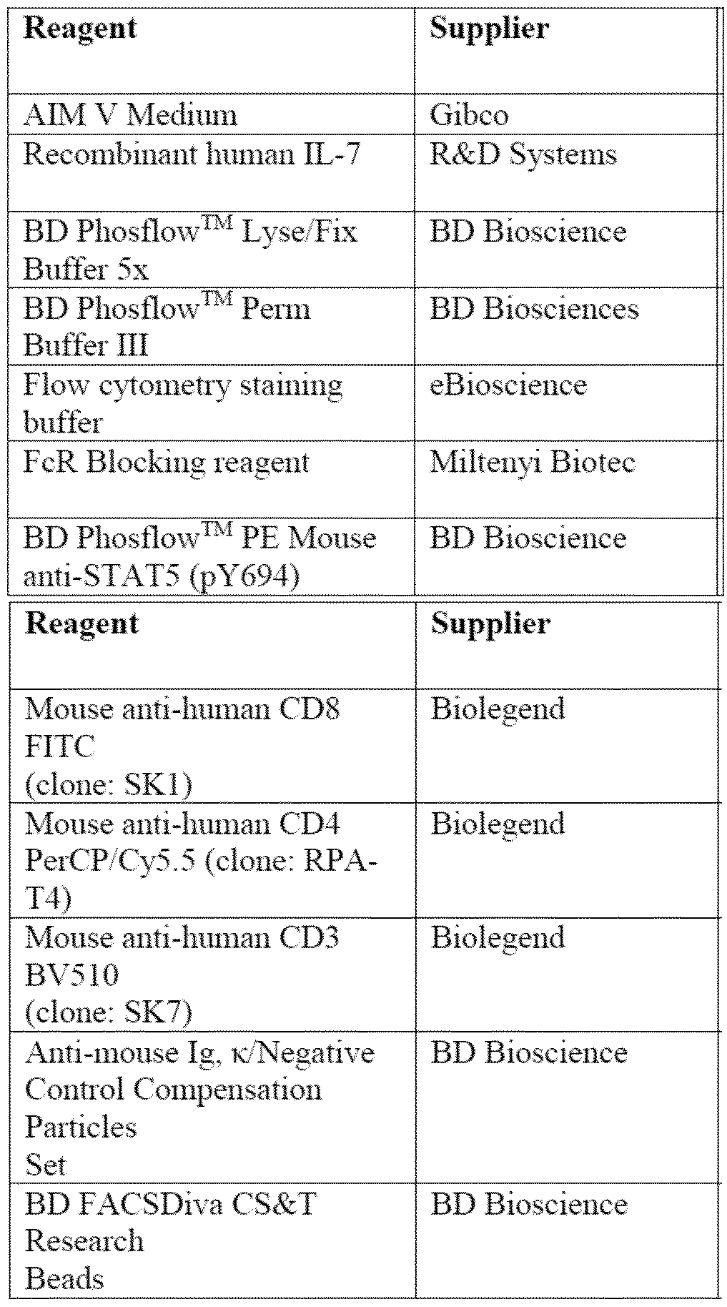 All antibody treatments and rhIL-7 stimulations were made up at 4x final assay concentration (F.A.C.) in culture medium before mixing 1:1 and incubating at room temperature for 5 minutes. In the absence of either antibody or IL-7 stimulus, culture medium was added. For whole blood: 100 μL of the antibody:IL-7 mixture was aliquoted into FACS tubes before adding 100 μL of whole blood. Tubes were mixed gently by vortexing and incubated at 37°C in a humidified incubator for 20 minutes. At the end of the stimulation period 2.5 mL pre-warmed PHOSFLOW lysis buffer (1x) was added and the samples were incubated for a further 10 minutes at 37°C.2 mL PBS was added to the suspension and tubes were centrifuged to pellet the cells (300 x g, 5 minutes at room temperature). Supernatant was discarded and cells were washed twice more in PBS. Following the final wash, cells were resuspended in 500 μL Perm Buffer III (pre-cooled to -20°C) and vortexed to mix. Tubes were incubated
70169 on ice for 30 minutes before washing once in 3 mL PBS. Cells were resuspended in 100 μL PBS and transferred to 96-well, round bottom culture plate for staining. For PBMCs treatments: 50 μL of the antibody:IL-7 mixture was added to each well of a 96-well, round bottom tissue culture plate. 50 μL PBMC suspension was added to each treatment well (2.5x10⁵ cells/well). Plates were mixed gently on a rotary plate shaker before incubating at 37°C in a humidified incubator for 20 minutes. At the end of the stimulation period 250 μL pre-warmed PHOSFLOW lysis buffer (1x) was added and the samples were incubated for a further 10 minutes at 37°C. Following fixation, cells were pelleted by centrifugation (300 x g, 5 minutes at room temperature) and washed twice in 200 μL PBS. Cell pellets were resuspended in 100 μL Perm Buffer III (pre-cooled to -20°C) and pipetted up and down to mix. Cells were incubated on ice for 30 minutes before washing once in 200 μL PBS and resuspending in PBS. For all samples: Following permeabilization, plates were centrifuged (300 x g, 5 minutes at room temperature) and cell pellets were resuspended in 25 μL FcR blocking reagent diluted 1:5 in 3% BSA in PBS. Cells were incubated for 10 minutes at room temperature before adding 2.5 μL anti-CD3 (BV510), 2.5 μL anti-CD4 (PerCP/Cy5.5), 2.5 anti-CD8 (FITC), 7.5 μL anti-pSTAT5 (PE) and 35 μL flow cytometry staining buffer (50 μL total per sample). Where a stain was excluded for controls, the equivalent volume of staining buffer was added instead. Plates were mixed briefly on a rotary plate shaker and incubated on ice for 30 minutes protected from light. Cells were washed in 200 μL staining buffer and resuspended in 200 μL staining buffer for analysis on the same day on the FACS Canto II. The performance of the instrument was checked using the Cytometer Set-up and Tracking (CST) Beads. This is a QC check for the instrument, sets the baselines and optimizes the voltages for each laser prior to use. The results of the calibration are stored within the CST software on the instrument. Compensation for the instrument was performed using anti-mouse IgG,κ/negative control compensation beads in accordance with the manufacturer’s instructions. The relevant antibodies used to stain the cells during the experiment were used to label the appropriate compensation bead type. Compensation for the experiments were performed using the appropriately labelled beads with the automatic compensation facility available within the FACS Diva software. After analysis of the compensation samples the appropriate compensation settings were calculated and applied to each experimental staining panel. pSTAT5 FACS analysis: Acquired cells in a FSC-A vs FSC-H plot to exclude doublets. Single cells were acquired into a FSC-A vs SSC-A plot and gated around the live lymphocytes. These were acquired into AmCyan vs FSC-A plot and gated around the CD3+ population. A PerCP-Cy5.5 vs FITC plot was generated on CD3+ to identify CD4+ and CD8+ populations. For pSTAT5 a histogram for PE fluorescence was created for each subset. Positive gates were set based on the unstimulated samples and the percent PE positive statistic was used for data analysis.
70169 Data were analyzed using FlowJo software (version 10) and results were generated in Microsoft Excel (2010) spreadsheet format using the Batch Analysis facility within the FlowJo software. The cell populations were tabulated in an Excel spreadsheet as % of parent. This was converted into a percent response by normalizing to the unstimulated control or a percent inhibition by normalizing to the unstimulated control and subtracting this value from 100 (theoretical maximal percent response). Where concentration response graphs have been generated, individual donor data were fitted with a non-linear logistic curve fit regression and the average (mean and median), range, SD and SEM of the IC50 from all donors was calculated in GraphPad Prism (version 6). Where statistical testing was carried out, a 2-way ANOVA with Sidak’s correction for multiple comparison was applied. A p value of <0.05 was considered as statistically significant. * denotes p<0.05; ** p≤0.01; *** p≤0.001; and **** p≤0.0001. In a whole blood assay, STAT5 phosphorylation was assessed in CD4+ T cells by flow cytometry after 20 minutes stimulation with 1 ng/ml recombinant human IL-7 (rhIL-7; 58 pM). As shown in Figure 2A and Table 6, DRSPAI-L7B prevented IL-7 from signaling through STAT5 in a concentration-dependent manner with a median IC50 of 34pM (5.1 ng/mL). A1290 prevented IL-7 signalling through STAT5 in a concentration-dependent manner with a median IC50 of 18 pM (0.00275 µg/ml). A1291 prevented IL-7 signalling through STAT5 in a concentration-dependent manner with a median IC50 of 16 pM (0.00246 µg/ml). A1294 prevented IL-7 signalling through STAT5 in a concentration-dependent manner with a median IC50 of 76 pM (0.0114 µg/ml).Immortalized T lymphoblast CCRF-CEM cells were stimulated with 34 pg/mL (2 pM) recombinant human IL-7 (rhIL-7) in the presence or absence of DRSPAI-L7B. STAT5 phosphorylation was assessed by MSD on cell lysates. DRSPAI-L7B potently blocked IL-7-induced pSTAT5 (Table 6; IC50<1 pM). Table 6. Summary of potency of DRSPAI-L7B in functional assays
All antibody treatments and rhIL-7 stimulations were made up at 4x final assay concentration (F.A.C.) in culture medium before mixing 1:1 and incubating at room temperature for 5 minutes. In the absence of either antibody or IL-7 stimulus, culture medium was added. For whole blood: 100 μL of the antibody:IL-7 mixture was aliquoted into FACS tubes before adding 100 μL of whole blood. Tubes were mixed gently by vortexing and incubated at 37°C in a humidified incubator for 20 minutes. At the end of the stimulation period 2.5 mL pre-warmed PHOSFLOW lysis buffer (1x) was added and the samples were incubated for a further 10 minutes at 37°C.2 mL PBS was added to the suspension and tubes were centrifuged to pellet the cells (300 x g, 5 minutes at room temperature). Supernatant was discarded and cells were washed twice more in PBS. Following the final wash, cells were resuspended in 500 μL Perm Buffer III (pre-cooled to -20°C) and vortexed to mix. Tubes were incubated
70169 on ice for 30 minutes before washing once in 3 mL PBS. Cells were resuspended in 100 μL PBS and transferred to 96-well, round bottom culture plate for staining. For PBMCs treatments: 50 μL of the antibody:IL-7 mixture was added to each well of a 96-well, round bottom tissue culture plate. 50 μL PBMC suspension was added to each treatment well (2.5x10⁵ cells/well). Plates were mixed gently on a rotary plate shaker before incubating at 37°C in a humidified incubator for 20 minutes. At the end of the stimulation period 250 μL pre-warmed PHOSFLOW lysis buffer (1x) was added and the samples were incubated for a further 10 minutes at 37°C. Following fixation, cells were pelleted by centrifugation (300 x g, 5 minutes at room temperature) and washed twice in 200 μL PBS. Cell pellets were resuspended in 100 μL Perm Buffer III (pre-cooled to -20°C) and pipetted up and down to mix. Cells were incubated on ice for 30 minutes before washing once in 200 μL PBS and resuspending in PBS. For all samples: Following permeabilization, plates were centrifuged (300 x g, 5 minutes at room temperature) and cell pellets were resuspended in 25 μL FcR blocking reagent diluted 1:5 in 3% BSA in PBS. Cells were incubated for 10 minutes at room temperature before adding 2.5 μL anti-CD3 (BV510), 2.5 μL anti-CD4 (PerCP/Cy5.5), 2.5 anti-CD8 (FITC), 7.5 μL anti-pSTAT5 (PE) and 35 μL flow cytometry staining buffer (50 μL total per sample). Where a stain was excluded for controls, the equivalent volume of staining buffer was added instead. Plates were mixed briefly on a rotary plate shaker and incubated on ice for 30 minutes protected from light. Cells were washed in 200 μL staining buffer and resuspended in 200 μL staining buffer for analysis on the same day on the FACS Canto II. The performance of the instrument was checked using the Cytometer Set-up and Tracking (CST) Beads. This is a QC check for the instrument, sets the baselines and optimizes the voltages for each laser prior to use. The results of the calibration are stored within the CST software on the instrument. Compensation for the instrument was performed using anti-mouse IgG,κ/negative control compensation beads in accordance with the manufacturer’s instructions. The relevant antibodies used to stain the cells during the experiment were used to label the appropriate compensation bead type. Compensation for the experiments were performed using the appropriately labelled beads with the automatic compensation facility available within the FACS Diva software. After analysis of the compensation samples the appropriate compensation settings were calculated and applied to each experimental staining panel. pSTAT5 FACS analysis: Acquired cells in a FSC-A vs FSC-H plot to exclude doublets. Single cells were acquired into a FSC-A vs SSC-A plot and gated around the live lymphocytes. These were acquired into AmCyan vs FSC-A plot and gated around the CD3+ population. A PerCP-Cy5.5 vs FITC plot was generated on CD3+ to identify CD4+ and CD8+ populations. For pSTAT5 a histogram for PE fluorescence was created for each subset. Positive gates were set based on the unstimulated samples and the percent PE positive statistic was used for data analysis.
70169 Data were analyzed using FlowJo software (version 10) and results were generated in Microsoft Excel (2010) spreadsheet format using the Batch Analysis facility within the FlowJo software. The cell populations were tabulated in an Excel spreadsheet as % of parent. This was converted into a percent response by normalizing to the unstimulated control or a percent inhibition by normalizing to the unstimulated control and subtracting this value from 100 (theoretical maximal percent response). Where concentration response graphs have been generated, individual donor data were fitted with a non-linear logistic curve fit regression and the average (mean and median), range, SD and SEM of the IC50 from all donors was calculated in GraphPad Prism (version 6). Where statistical testing was carried out, a 2-way ANOVA with Sidak’s correction for multiple comparison was applied. A p value of <0.05 was considered as statistically significant. * denotes p<0.05; ** p≤0.01; *** p≤0.001; and **** p≤0.0001. In a whole blood assay, STAT5 phosphorylation was assessed in CD4+ T cells by flow cytometry after 20 minutes stimulation with 1 ng/ml recombinant human IL-7 (rhIL-7; 58 pM). As shown in Figure 2A and Table 6, DRSPAI-L7B prevented IL-7 from signaling through STAT5 in a concentration-dependent manner with a median IC50 of 34pM (5.1 ng/mL). A1290 prevented IL-7 signalling through STAT5 in a concentration-dependent manner with a median IC50 of 18 pM (0.00275 µg/ml). A1291 prevented IL-7 signalling through STAT5 in a concentration-dependent manner with a median IC50 of 16 pM (0.00246 µg/ml). A1294 prevented IL-7 signalling through STAT5 in a concentration-dependent manner with a median IC50 of 76 pM (0.0114 µg/ml).Immortalized T lymphoblast CCRF-CEM cells were stimulated with 34 pg/mL (2 pM) recombinant human IL-7 (rhIL-7) in the presence or absence of DRSPAI-L7B. STAT5 phosphorylation was assessed by MSD on cell lysates. DRSPAI-L7B potently blocked IL-7-induced pSTAT5 (Table 6; IC50<1 pM). Table 6. Summary of potency of DRSPAI-L7B in functional assays
 §Assays were performed using EC80 stimulation with IL-7 determined for each batch of cells
70169 IL-7 Induced STAT5 phosphorylation in healthy and disease T cells PBMCs from healthy donors or IBD patients (two Crohn’s disease and one ulcerative colitis) were stimulated with rhIL-7 in the presence of DRSPAI-L7B or anti-RSV antibody (isotype control). Stimulated cells were fixed and STAT5 phosphorylation in CD8+ (Figure 2B), CD4+ (Figure 2C), and CD3+ (Figure 2D) T cells was assessed by flow cytometry. Data is shown as pSTAT5 increase relative to unstimulated condition, mean ± SD, n=3. * p<0.05, ** p≤0.01, *** p≤0.001, matched 2-way ANOVA with Sidak’s multiple comparisons correction. Figure 2B, Figure 2C and Figure 2D. Example 4: Dynamic Light Scattering Assay Dynamic Light Scattering (DLS) analysis was carried out on DRSPAI-L7B, A1290, A1291 and A1294 to characterize high order aggregate species and sample heterogeneity. Each of the respective antibodies was concentrated to ≥10mg/ml and buffer exchanged via dialysis into 50mM sodium phosphate pH 7.5 or 50mM sodium acetate pH 5.0 buffers followed by normalization to 10mg/ml and filtration using a 0.22µm syringe filter. Post filtration all the samples were stressed at a target concentration of 10mg/ml for 2 weeks at 40°C. Samples were evaluated following incubation under stressed and unstressed conditions in the two buffers. 100µl samples were run in triplicate at 25°C on a Wyatt DynaPro DLS Plate reader using 96 well Corning Costar 3635 plates sealed with Corning 6575 seals. The data was analysed using DYNAMICS v7.1.9 software. The data was distributed into the following peaks: Peak 1 = 0.1-1nm, Peak 2 = 1-10nm, Peak 3 = 10-100nm, Peak 4 = 100-1000nm, Peak 5 = 1000-10000nm. The data was filtered using the following criteria: amplitude must be between 0 and 1, baseline limit 1±0.01 and sum of all squared (SOS) must be less than 100. An Rh of >8nm and average % mass at Peak2 <98% was associated with a severe aggregation risk. A1290, A1291 and A1294 showed severe aggregation risks, all exhibiting hydrodynamic radius (Rh) values of >8nm. DRSPAI-L7B had an Rh value of <8 nm and did not, thus, exhibit such aggregation risk.
§Assays were performed using EC80 stimulation with IL-7 determined for each batch of cells
70169 IL-7 Induced STAT5 phosphorylation in healthy and disease T cells PBMCs from healthy donors or IBD patients (two Crohn’s disease and one ulcerative colitis) were stimulated with rhIL-7 in the presence of DRSPAI-L7B or anti-RSV antibody (isotype control). Stimulated cells were fixed and STAT5 phosphorylation in CD8+ (Figure 2B), CD4+ (Figure 2C), and CD3+ (Figure 2D) T cells was assessed by flow cytometry. Data is shown as pSTAT5 increase relative to unstimulated condition, mean ± SD, n=3. * p<0.05, ** p≤0.01, *** p≤0.001, matched 2-way ANOVA with Sidak’s multiple comparisons correction. Figure 2B, Figure 2C and Figure 2D. Example 4: Dynamic Light Scattering Assay Dynamic Light Scattering (DLS) analysis was carried out on DRSPAI-L7B, A1290, A1291 and A1294 to characterize high order aggregate species and sample heterogeneity. Each of the respective antibodies was concentrated to ≥10mg/ml and buffer exchanged via dialysis into 50mM sodium phosphate pH 7.5 or 50mM sodium acetate pH 5.0 buffers followed by normalization to 10mg/ml and filtration using a 0.22µm syringe filter. Post filtration all the samples were stressed at a target concentration of 10mg/ml for 2 weeks at 40°C. Samples were evaluated following incubation under stressed and unstressed conditions in the two buffers. 100µl samples were run in triplicate at 25°C on a Wyatt DynaPro DLS Plate reader using 96 well Corning Costar 3635 plates sealed with Corning 6575 seals. The data was analysed using DYNAMICS v7.1.9 software. The data was distributed into the following peaks: Peak 1 = 0.1-1nm, Peak 2 = 1-10nm, Peak 3 = 10-100nm, Peak 4 = 100-1000nm, Peak 5 = 1000-10000nm. The data was filtered using the following criteria: amplitude must be between 0 and 1, baseline limit 1±0.01 and sum of all squared (SOS) must be less than 100. An Rh of >8nm and average % mass at Peak2 <98% was associated with a severe aggregation risk. A1290, A1291 and A1294 showed severe aggregation risks, all exhibiting hydrodynamic radius (Rh) values of >8nm. DRSPAI-L7B had an Rh value of <8 nm and did not, thus, exhibit such aggregation risk.
Abbreviations
 70169
70169
 Example 5: Impact Of DRSPAI-L7B On Cytokine Production In Human PBMC DRSPAI-L7B was used to interrogate the role that IL-7 plays in Th1 and Th17 function and differentiation. The effect of DRSPAI-L7B on cytokine secretion from healthy PBMCs stimulated with IL-7 in the presence of a CD3 agonistic antibody was assessed. Healthy volunteer blood was provided by a Blood Donation Unit: Blood was withdrawn by venepuncture and transferred into a pot or blood bag containing sodium heparin (1U/mL). The blood was collected and used for PBMC isolation. The blood was diluted 2X with PBS, layered onto 15mL ficoll in Accuspin tubes and centrifuged at 800 rcf for 20 minutes without break. The plasma was removed carefully with a pipette and the layer containing the PBMCs was carefully transferred to a 50 mL tube. The PBMCs were washed twice in 50 mL PBS (250 rcf, 10 minutes) and then resuspended in 50 mL RPMI+10% FCS+L-glutamine and counted using Vi-cell XR. PBMCs were resuspended in culture media at 5x10⁶ cells/mL. Antibody dilutions of anti-RSV antibody (isotype control) and DRSPAI-L7B were prepared at 2.5X the required final concentration in culture media. Equal volumes of cell suspension and antibody dilutions (150 μL) were mixed and incubated for 30 min at room temperature. IL-7 was prepared at 5X the final concentration in culture medium and 20 μL added in the required wells of a 96-well U bottom polystyrene plate, which had been pre-coated with 10 μg/mL anti-CD3 at 4oC overnight. Culture medium was added in all remaining wells.80 μL of the mixed cell suspension/antibody dilutions (2x10⁶ cells/well) were added to each well of the 96-well plate and left to incubate for 48 hrs at 37oC, 5% CO2.
70169 After 48 hrs the plate was centrifuged at 300 rcf for 5 minutes and the supernatants were removed without disturbing the pellets and transferred to a new 96-well U-bottom plate. Supernatants were either used for ELISA/MSD immediately after harvesting or stored at -80oC until further use. Anti-RSV IgG1 was at stock concentration of 2.8 mg/mL. Thawed and diluted to the appropriate concentration on day of use. DRSPAI-L7B stock concentration was 11.33 mg/mL. Thawed and diluted to the appropriate concentration on day of use. Recombinant Human IL-7 was purchased from R&D systems. The lyophilized protein was resuspended to 25µg/mL in sterile PBS + 0.1% bovine serum albumin and 50μL aliquots stored at -20oC. Thawed and diluted to the appropriate concentration on day of use. Anti-CD3 clone HIT3a. Stock was 1 mg/mL. Diluted to the appropriate concentration on day of use. Other materials
Example 5: Impact Of DRSPAI-L7B On Cytokine Production In Human PBMC DRSPAI-L7B was used to interrogate the role that IL-7 plays in Th1 and Th17 function and differentiation. The effect of DRSPAI-L7B on cytokine secretion from healthy PBMCs stimulated with IL-7 in the presence of a CD3 agonistic antibody was assessed. Healthy volunteer blood was provided by a Blood Donation Unit: Blood was withdrawn by venepuncture and transferred into a pot or blood bag containing sodium heparin (1U/mL). The blood was collected and used for PBMC isolation. The blood was diluted 2X with PBS, layered onto 15mL ficoll in Accuspin tubes and centrifuged at 800 rcf for 20 minutes without break. The plasma was removed carefully with a pipette and the layer containing the PBMCs was carefully transferred to a 50 mL tube. The PBMCs were washed twice in 50 mL PBS (250 rcf, 10 minutes) and then resuspended in 50 mL RPMI+10% FCS+L-glutamine and counted using Vi-cell XR. PBMCs were resuspended in culture media at 5x10⁶ cells/mL. Antibody dilutions of anti-RSV antibody (isotype control) and DRSPAI-L7B were prepared at 2.5X the required final concentration in culture media. Equal volumes of cell suspension and antibody dilutions (150 μL) were mixed and incubated for 30 min at room temperature. IL-7 was prepared at 5X the final concentration in culture medium and 20 μL added in the required wells of a 96-well U bottom polystyrene plate, which had been pre-coated with 10 μg/mL anti-CD3 at 4oC overnight. Culture medium was added in all remaining wells.80 μL of the mixed cell suspension/antibody dilutions (2x10⁶ cells/well) were added to each well of the 96-well plate and left to incubate for 48 hrs at 37oC, 5% CO2.
70169 After 48 hrs the plate was centrifuged at 300 rcf for 5 minutes and the supernatants were removed without disturbing the pellets and transferred to a new 96-well U-bottom plate. Supernatants were either used for ELISA/MSD immediately after harvesting or stored at -80oC until further use. Anti-RSV IgG1 was at stock concentration of 2.8 mg/mL. Thawed and diluted to the appropriate concentration on day of use. DRSPAI-L7B stock concentration was 11.33 mg/mL. Thawed and diluted to the appropriate concentration on day of use. Recombinant Human IL-7 was purchased from R&D systems. The lyophilized protein was resuspended to 25µg/mL in sterile PBS + 0.1% bovine serum albumin and 50μL aliquots stored at -20oC. Thawed and diluted to the appropriate concentration on day of use. Anti-CD3 clone HIT3a. Stock was 1 mg/mL. Diluted to the appropriate concentration on day of use. Other materials
 ECL signals were derived from the MSD instrument and converted to concentrations using the standard curve for each analyte. For graphs in which the data is normalized, the average of anti-CD3+ IL- 7 samples was set at 100% and all other values were normalized to that. The antibody concentrations were log transformed and plotted against cytokine concentrations. The no IL-7 (anti-CD3 only) samples are shown in each graph for comparison. For curve fitting, the following nonlinear fit from Graphpad Prism
70169 was used: log(inhibitor) vs. response (three parameters) and IC50 values were calculated by Graphpad Prism. DRSPAI-L7B inhibited IFN-γ (Figure 3A) and IL-10 (Figure 3B) secretion in a concentration- dependent manner (IFN-γ mean IC50=195.8 ± 101 pM; IL-10 mean IC50=207.2 ± 86 pM). Data represent the mean of n=6 independent donors ± SEM, assessed in 3 independent experiments. Figure 3C, Figure 3D, Figure 3E, Figure 3F, Figure 3G illustrates inhibition of IL-2 by DRSPAI-L7B in the presence of rhIL-7 and anti-CD3. There was an increase in IL-2 production following IL-7 stimulation in 5 donors. This increase was inhibited fully by DRSPAI-L7B. Example 6: DRSPAI-L7B Inhibits Cytokine Production By Memory T Cells To accurately determine the role of IL-7 in Th17 cell function and differentiation, a ‘poised Th17’ assay was used to profile the secretion of Th17-associated cytokines. Tmem cells were isolated from healthy donors and incubated with IL-7 in the presence of DRSPAI-L7B. Whole human blood was collected from donors in the Blood Donation Unit (BDU). Typically, 200mL was collected by venepuncture per donor and an anti-coagulant, citrate-dextrose solution (ACD, Sigma, Cat # C3821) was added immediately to each sample. ACD was added at 15% (e.g.30mL ACD added to 200mL blood). Within 2 hours of collection, and using a microbiological safety cabinet, the blood was dispensed evenly into pre-filled LEUCOSEP tubes (Greiner, Cat # 227288) at 30mL (max) per tube. The blood was centrifuged for 15 minutes at 800 x g at room temperature in a swing bucket rotor with no brake applied. The PBMCs were washed in PBS (500 x g, 10 minutes) and then resuspended in 10 mL PBS and counted using the NucleoCounter. The cells were centrifuged again at 500 x g and the cell pellet was re-suspended in FACS buffer at a concentration of 5 x 10^7 cells/mL. Human CD4+ memory T cells (Tmem) EasySep enrichment cocktail was added at 50µL/mL to the cell suspension. The cells were incubated at room temperature for 10 minutes after which the EasySep magnetic particles were added at 50µL/mL. The suspension was again incubated at room temperature for 10 minutes after which the cell suspension was placed in an EasySep magnet for 10 minutes. The cell suspension was then transferred to a fresh tube whilst still placed within the magnet to ensure a negative selection. The cells were centrifuged at 500 x g for 10 minutes and resuspended in cell culture media. The cell count was determined using the NucleoCounter. The cells were rested at 37°C and 5% CO2 overnight in culture media. The next day the cells were centrifuged and resuspended in assay media at a concentration of 2.5 x 10^5 cells/mL. Recombinant hIL- 7 was added to the cells at a final concentration of 20ng/mL. The cells were then incubated in the presence of DRSPAI-L7B for 4 days at 37°C and 5% CO2 after which they were stimulated with 10nM PMA and 1µM Ionomycin for 16 hours at 37°C and 5% CO2.
70169 Detection of IL-17 from the supernatants using MSD After 16 hours of PMA and ionomycin stimulation, the cells were centrifuged and 40µL of the supernatants were transferred to the MSD plates which were pre-blocked with 0.5% of Blocker B. These MSD plates were coated with an anti-IL-17A capture antibody. The samples were incubated for 2 hours at room temperature whilst shaking after which the plates were washed with PBS and 0.05% Tween-20.10 µL of the IL-17 specific detection antibody labelled with the MSD SULFO-TAG reagent was then added and the samples were incubated for a further 2 hours at room temperature whilst shaking. The plate was washed again three times in PBS and 0.05% Tween-20 and 2x Read Buffer T was added to the samples. The plate was read on the Sector Imager. Detection of other cytokines A U-Plex MSD Kit was designed to enable investigation of the effect of DRSPAI-L7B on the secretion of IL-6, IL-10, IFN-γ, TNF-α and CCL3. The MSD U-Plex plates were prepared by coating the plate with linker coupled capture antibodies. Each capture antibody was biotinylated and had a unique linker assigned to it. The supernatants were diluted 1 in 100 and transferred to the MSD plates. The samples were incubated for 1 hour at room temperature whilst shaking after which the plates were washed with PBS and 0.05% Tween-20. 50 µL of the specific detection antibodies labelled with the MSD SULFO-TAG reagent was then added and the samples were incubated for a further hour at room temperature whilst shaking. The plate was washed again three times in PBS and 0.05% Tween-20 and 2x Read Buffer T was added to the samples. The plate was read on the Sector Imager. DRSPAI-L7B antibody was produced at a stock concentration of 11.33mg/mL in 20mM Histidine, 180mM Trehalose, 40mM Arginine, 8mM Methionine, 0.05mM EDTA, pH 6.0. The Antibody was diluted to a concentration of 20µg/mL in assay media. A 1 in 3 serial dilution was then carried out in assay media to generate a 10 point dose response curve. The dilutions were transferred to the assay plates ensuring that the highest final assay concentration of the antibody was 10µg/mL. BRL-54319MM (Rapamycin) was used as the positive control in the assay at 1µM final assay concentration in assay media. Recombinant Human IL-7 was resuspended to 25µg/mL in sterile PBS and 50μL aliquots stored at -20°C. The aliquots were thawed and diluted to the appropriate concentration on day of use.
70169 Other Materials
ECL signals were derived from the MSD instrument and converted to concentrations using the standard curve for each analyte. For graphs in which the data is normalized, the average of anti-CD3+ IL- 7 samples was set at 100% and all other values were normalized to that. The antibody concentrations were log transformed and plotted against cytokine concentrations. The no IL-7 (anti-CD3 only) samples are shown in each graph for comparison. For curve fitting, the following nonlinear fit from Graphpad Prism
70169 was used: log(inhibitor) vs. response (three parameters) and IC50 values were calculated by Graphpad Prism. DRSPAI-L7B inhibited IFN-γ (Figure 3A) and IL-10 (Figure 3B) secretion in a concentration- dependent manner (IFN-γ mean IC50=195.8 ± 101 pM; IL-10 mean IC50=207.2 ± 86 pM). Data represent the mean of n=6 independent donors ± SEM, assessed in 3 independent experiments. Figure 3C, Figure 3D, Figure 3E, Figure 3F, Figure 3G illustrates inhibition of IL-2 by DRSPAI-L7B in the presence of rhIL-7 and anti-CD3. There was an increase in IL-2 production following IL-7 stimulation in 5 donors. This increase was inhibited fully by DRSPAI-L7B. Example 6: DRSPAI-L7B Inhibits Cytokine Production By Memory T Cells To accurately determine the role of IL-7 in Th17 cell function and differentiation, a ‘poised Th17’ assay was used to profile the secretion of Th17-associated cytokines. Tmem cells were isolated from healthy donors and incubated with IL-7 in the presence of DRSPAI-L7B. Whole human blood was collected from donors in the Blood Donation Unit (BDU). Typically, 200mL was collected by venepuncture per donor and an anti-coagulant, citrate-dextrose solution (ACD, Sigma, Cat # C3821) was added immediately to each sample. ACD was added at 15% (e.g.30mL ACD added to 200mL blood). Within 2 hours of collection, and using a microbiological safety cabinet, the blood was dispensed evenly into pre-filled LEUCOSEP tubes (Greiner, Cat # 227288) at 30mL (max) per tube. The blood was centrifuged for 15 minutes at 800 x g at room temperature in a swing bucket rotor with no brake applied. The PBMCs were washed in PBS (500 x g, 10 minutes) and then resuspended in 10 mL PBS and counted using the NucleoCounter. The cells were centrifuged again at 500 x g and the cell pellet was re-suspended in FACS buffer at a concentration of 5 x 10^7 cells/mL. Human CD4+ memory T cells (Tmem) EasySep enrichment cocktail was added at 50µL/mL to the cell suspension. The cells were incubated at room temperature for 10 minutes after which the EasySep magnetic particles were added at 50µL/mL. The suspension was again incubated at room temperature for 10 minutes after which the cell suspension was placed in an EasySep magnet for 10 minutes. The cell suspension was then transferred to a fresh tube whilst still placed within the magnet to ensure a negative selection. The cells were centrifuged at 500 x g for 10 minutes and resuspended in cell culture media. The cell count was determined using the NucleoCounter. The cells were rested at 37°C and 5% CO2 overnight in culture media. The next day the cells were centrifuged and resuspended in assay media at a concentration of 2.5 x 10^5 cells/mL. Recombinant hIL- 7 was added to the cells at a final concentration of 20ng/mL. The cells were then incubated in the presence of DRSPAI-L7B for 4 days at 37°C and 5% CO2 after which they were stimulated with 10nM PMA and 1µM Ionomycin for 16 hours at 37°C and 5% CO2.
70169 Detection of IL-17 from the supernatants using MSD After 16 hours of PMA and ionomycin stimulation, the cells were centrifuged and 40µL of the supernatants were transferred to the MSD plates which were pre-blocked with 0.5% of Blocker B. These MSD plates were coated with an anti-IL-17A capture antibody. The samples were incubated for 2 hours at room temperature whilst shaking after which the plates were washed with PBS and 0.05% Tween-20.10 µL of the IL-17 specific detection antibody labelled with the MSD SULFO-TAG reagent was then added and the samples were incubated for a further 2 hours at room temperature whilst shaking. The plate was washed again three times in PBS and 0.05% Tween-20 and 2x Read Buffer T was added to the samples. The plate was read on the Sector Imager. Detection of other cytokines A U-Plex MSD Kit was designed to enable investigation of the effect of DRSPAI-L7B on the secretion of IL-6, IL-10, IFN-γ, TNF-α and CCL3. The MSD U-Plex plates were prepared by coating the plate with linker coupled capture antibodies. Each capture antibody was biotinylated and had a unique linker assigned to it. The supernatants were diluted 1 in 100 and transferred to the MSD plates. The samples were incubated for 1 hour at room temperature whilst shaking after which the plates were washed with PBS and 0.05% Tween-20. 50 µL of the specific detection antibodies labelled with the MSD SULFO-TAG reagent was then added and the samples were incubated for a further hour at room temperature whilst shaking. The plate was washed again three times in PBS and 0.05% Tween-20 and 2x Read Buffer T was added to the samples. The plate was read on the Sector Imager. DRSPAI-L7B antibody was produced at a stock concentration of 11.33mg/mL in 20mM Histidine, 180mM Trehalose, 40mM Arginine, 8mM Methionine, 0.05mM EDTA, pH 6.0. The Antibody was diluted to a concentration of 20µg/mL in assay media. A 1 in 3 serial dilution was then carried out in assay media to generate a 10 point dose response curve. The dilutions were transferred to the assay plates ensuring that the highest final assay concentration of the antibody was 10µg/mL. BRL-54319MM (Rapamycin) was used as the positive control in the assay at 1µM final assay concentration in assay media. Recombinant Human IL-7 was resuspended to 25µg/mL in sterile PBS and 50μL aliquots stored at -20°C. The aliquots were thawed and diluted to the appropriate concentration on day of use.
70169 Other Materials
 FACS Buffer: sterile PBS containing 2% heat-inactivated FBS. Cell Culture Media: 450mL IMDM, 50mL FBS, 5mL Penstrep, 5mL L-Glutamine, 5mL Non-essential Amino Acids and 5mL Sodium Pyruvate. Assay Media: 500mL Xvivo 15, 5mL Penstrep, 5mL L-glutamine, 5mL HEPES and 5mL Sodium Pyruvate. All data was normalized to the mean of 8 high and 8 low control wells on each plate. A four- parameter curve fit of the following form was then applied.
 Where a is the minimum, b is the Hill slope, c is the XC50 and d is the maximum. Data was presented as the mean IC50 with the standard deviation of the mean of n experiments. CD4+ Tmem cells, isolated from healthy donor blood, were incubated with IL-7 in the presence of DRSPAI-L7B for 4 days after which they were spiked with PMA/ionomycin for 16 hours before harvest. Cytokine secretion into the supernatant was assessed by MSD. DRSPAI-L7B treatment resulted in a concentration-dependent inhibition of IL-17 (Figure 4A, IC50=270 ± 31.59 pM), TNF-α (Figure 4B, IC50=127.1 ± 89.83 pM), IL-6 (Figure 4C, IC50=202.07 ± 99.64 pM), IL-10 (Figure 4D, IC50=197.48 ±148.91), INFγ (Figure 4E, IC50=157.98 ± 89.55) and CCL3 (Figure 4F, IC50=163.83±85.96) secretion. Dose dependent
70169 inhibition was also observed for IL-6, IL-10, IFNγ, TNF-α and CCL3. Inhibition of the production of these cytokines was expressed as a percentage of that achieved with Rapamycin (1 µM) positive control. Example 7: T Cell Population Analysis Given the central role of T cells in MS, T cell populations were analysed in PBMCs from RRMS, PPMS and SPMS patients. All human samples were obtained with patient informed consent in accordance with ICH GCP under a protocol approved by a national, regional, or investigational center ethics committee or an Institutional Review Board (IRB) approved protocol. Healthy PBMCs were isolated from BDU blood and stored frozen in liquid nitrogen until used. Disease PBMCs were supplied by an approved external human tissue supplier. Healthy control blood was withdrawn by venepuncture and transferred into a container with sodium heparin anti-coagulant (1U/mL). The blood was collected and used within 1 hour for PBMC isolation. Healthy Control human PBMCs prepared and frozen in advance were used PBMCs were prepared by layering blood on 15 mL Ficoll. Tubes were centrifuged at 800 g for 20 minutes, with brake off. The mononuclear cell layer at the interface was transferred to 50 mL Falcon tubes, washed by topping up to 45 mL with PBS and centrifuging at 300 g for 10 minutes. The pellets were resuspended in Freezing medium A (60:40 FCS:medium), 5% of the original blood volume and then an equal volume of Freezing medium B (80:20 FCS:DMSO) was added, dropwise to reduce osmotic shock. Cells were transferred to cryovials (1 mL per vial (around 1 x 10⁷ cells)) and frozen in a Mr Frosty freezing container at -80°C for up to 1 week, followed by transfer to liquid nitrogen for long term storage. PBMC Recovery Cells were thawed by removing from liquid nitrogen storage and immediately placed in a water bath at 37°C until thawed. After transferring cell suspension to a 50 mL centrifuge tube, medium (RPMI + 10% heat-inactivated FCS, 1% penicillin / streptomycin, and 1% glutamine) was added very slowly to decrease the DMSO concentration gradually. Once the volume was increased to 30mL the cells were centrifuged, 300 g for 10 minutes, and resuspended in 5 mL medium before counting, made up to 15 mL with medium, centrifuged as above and cells resuspended in an appropriate volume of medium to yield 5 x 10⁶ cells / 1 mL. In each of 5 individual experiments, two healthy control (HC), two RRMS, one PPMS and one SPMS, donor PBMC sample were thawed as above and cells treated as below. Different donors were used for each experiment, HCs broadly age and gender matched with the disease patient donors used.
70169 T cell phenotyping flow cytometry assay Following resuspension of PBMCs at 5 x 10⁶/mL in medium, 100 μL cells were transferred to FACS tubes (for full stain) and an additional 50 μL of HC samples (for FMO control tubes). Cells were washed by addition of 2 mL FACS buffer, centrifuged 300 g, 5 minutes and pellet resuspended in residual volume.5 μL human FcX Trustain block was added for 10 mins followed by addition of 100 μL antibody stain cocktail and incubated at room temperature for 30 mins. Cells were washed by addition of 2 mL FACS buffer, centrifuged 300 g, 5 minutes and pellet resuspended in residual volume.500 μL diluted Live/Dead Fixable Aqua Dead Cell Stain was added and incubated at room temperature for 25 mins. Cells were washed by addition of 2 mL FACS buffer, centrifuged 300 g, 5 minutes and pellet resuspended in residual volume. 200 μL FACS buffer was added and samples analyzed on the same day, using a BD FACS Canto II. The performance of the instrument was checked using the Cytometer Set-up and Tracking (CST) Beads. This is a QC check for the instrument, sets the baselines and optimizes the voltages for each laser prior to use. The results of the calibration are stored within the CST software on the instrument. Compensation for the instrument was performed using UltraComp compensation beads in accordance with the manufacturer’s instructions. The relevant antibodies used to stain the cells during the experiment were used to label the compensation beads. Compensation for the experiments were performed using the appropriately labelled beads with the automatic compensation facility available within the FACS Diva software. After analysis of the compensation samples the compensation settings were calculated and applied to each experimental staining panel.
Where a is the minimum, b is the Hill slope, c is the XC50 and d is the maximum. Data was presented as the mean IC50 with the standard deviation of the mean of n experiments. CD4+ Tmem cells, isolated from healthy donor blood, were incubated with IL-7 in the presence of DRSPAI-L7B for 4 days after which they were spiked with PMA/ionomycin for 16 hours before harvest. Cytokine secretion into the supernatant was assessed by MSD. DRSPAI-L7B treatment resulted in a concentration-dependent inhibition of IL-17 (Figure 4A, IC50=270 ± 31.59 pM), TNF-α (Figure 4B, IC50=127.1 ± 89.83 pM), IL-6 (Figure 4C, IC50=202.07 ± 99.64 pM), IL-10 (Figure 4D, IC50=197.48 ±148.91), INFγ (Figure 4E, IC50=157.98 ± 89.55) and CCL3 (Figure 4F, IC50=163.83±85.96) secretion. Dose dependent
70169 inhibition was also observed for IL-6, IL-10, IFNγ, TNF-α and CCL3. Inhibition of the production of these cytokines was expressed as a percentage of that achieved with Rapamycin (1 µM) positive control. Example 7: T Cell Population Analysis Given the central role of T cells in MS, T cell populations were analysed in PBMCs from RRMS, PPMS and SPMS patients. All human samples were obtained with patient informed consent in accordance with ICH GCP under a protocol approved by a national, regional, or investigational center ethics committee or an Institutional Review Board (IRB) approved protocol. Healthy PBMCs were isolated from BDU blood and stored frozen in liquid nitrogen until used. Disease PBMCs were supplied by an approved external human tissue supplier. Healthy control blood was withdrawn by venepuncture and transferred into a container with sodium heparin anti-coagulant (1U/mL). The blood was collected and used within 1 hour for PBMC isolation. Healthy Control human PBMCs prepared and frozen in advance were used PBMCs were prepared by layering blood on 15 mL Ficoll. Tubes were centrifuged at 800 g for 20 minutes, with brake off. The mononuclear cell layer at the interface was transferred to 50 mL Falcon tubes, washed by topping up to 45 mL with PBS and centrifuging at 300 g for 10 minutes. The pellets were resuspended in Freezing medium A (60:40 FCS:medium), 5% of the original blood volume and then an equal volume of Freezing medium B (80:20 FCS:DMSO) was added, dropwise to reduce osmotic shock. Cells were transferred to cryovials (1 mL per vial (around 1 x 10⁷ cells)) and frozen in a Mr Frosty freezing container at -80°C for up to 1 week, followed by transfer to liquid nitrogen for long term storage. PBMC Recovery Cells were thawed by removing from liquid nitrogen storage and immediately placed in a water bath at 37°C until thawed. After transferring cell suspension to a 50 mL centrifuge tube, medium (RPMI + 10% heat-inactivated FCS, 1% penicillin / streptomycin, and 1% glutamine) was added very slowly to decrease the DMSO concentration gradually. Once the volume was increased to 30mL the cells were centrifuged, 300 g for 10 minutes, and resuspended in 5 mL medium before counting, made up to 15 mL with medium, centrifuged as above and cells resuspended in an appropriate volume of medium to yield 5 x 10⁶ cells / 1 mL. In each of 5 individual experiments, two healthy control (HC), two RRMS, one PPMS and one SPMS, donor PBMC sample were thawed as above and cells treated as below. Different donors were used for each experiment, HCs broadly age and gender matched with the disease patient donors used.
70169 T cell phenotyping flow cytometry assay Following resuspension of PBMCs at 5 x 10⁶/mL in medium, 100 μL cells were transferred to FACS tubes (for full stain) and an additional 50 μL of HC samples (for FMO control tubes). Cells were washed by addition of 2 mL FACS buffer, centrifuged 300 g, 5 minutes and pellet resuspended in residual volume.5 μL human FcX Trustain block was added for 10 mins followed by addition of 100 μL antibody stain cocktail and incubated at room temperature for 30 mins. Cells were washed by addition of 2 mL FACS buffer, centrifuged 300 g, 5 minutes and pellet resuspended in residual volume.500 μL diluted Live/Dead Fixable Aqua Dead Cell Stain was added and incubated at room temperature for 25 mins. Cells were washed by addition of 2 mL FACS buffer, centrifuged 300 g, 5 minutes and pellet resuspended in residual volume. 200 μL FACS buffer was added and samples analyzed on the same day, using a BD FACS Canto II. The performance of the instrument was checked using the Cytometer Set-up and Tracking (CST) Beads. This is a QC check for the instrument, sets the baselines and optimizes the voltages for each laser prior to use. The results of the calibration are stored within the CST software on the instrument. Compensation for the instrument was performed using UltraComp compensation beads in accordance with the manufacturer’s instructions. The relevant antibodies used to stain the cells during the experiment were used to label the compensation beads. Compensation for the experiments were performed using the appropriately labelled beads with the automatic compensation facility available within the FACS Diva software. After analysis of the compensation samples the compensation settings were calculated and applied to each experimental staining panel.
70169

 Samples were acquired on a BD FACS Canto II flow cytometer using BD BioSciences FACS Diva software (v8.0.1). Resulting compensated .fcs files were analyzed with FlowJo software (v10.0.8) and results generated in Excel using the Batch Analysis facility within the software.
70169 T cell populations were analyzed in PBMCs from RRMS, PPMS and SPMS patients. Data was generated from patients where all patients were on treatment (10/10 RRMS patients on natalizumab and all progressive MS patients were on steroids and/or symptomatic treatments), a reduction in Treg cells was observed. CD4+ (Figure 5A), CD8+ (Figure 5B) and regulatory T cells (Figure 5C) from healthy controls and MS patients were profiled by flow cytometry based on CD45RO, CCR7, CD127 and CD25 expression on the cell surface. No difference was seen between healthy and disease T cell populations in either CD4+ or CD8+ subsets. The RRMS and PPMS Treg population was significantly reduced compared to HC, no difference was found in Treg numbers in SPMS patients. ***0<0.0001, *0<0.05 as tested by One-way ANOVA with Dunnett multiple comparison test. Data presented represent the mean ± SEM of n=5-10 donors per group, analyzed in 5 independent experiments. Effector memory=CD45RO+CCR7 -, central memory=CD45RO+CCR7+, naïve=CD45RO-CCR7+, effector=CD45RO-CCR7 -, Treg=CD127low/-CD25+. Example 8: STAT5 phosphorylation All human samples were obtained with patient informed consent in accordance with ICH GCP under a protocol approved by a national, regional, or investigational center ethics committee or an Institutional Review Board (IRB) approved protocol. Healthy PBMCs were isolated from BDU blood and stored frozen in liquid nitrogen until used. Disease PBMCs were supplied by an approved external human tissue supplier. Healthy control blood was withdrawn by venepuncture and transferred into a container with sodium heparin anti-coagulant (1U/mL). The blood was collected and used within 1 hour for PBMC isolation. Healthy Control human PBMCs prepared and frozen in advance were used PBMCs were prepared by layering blood on 15 mL Ficoll. Tubes were centrifuged at 800 g for 20 minutes, with brake off. The mononuclear cell layer at the interface was transferred to 50 mL Falcon tubes, washed by topping up to 45 mL with PBS and centrifuging at 300 g for 10 minutes. The pellets were resuspended in Freezing medium A (60:40 FCS:medium), 5% of the original blood volume and then an equal volume of Freezing medium B (80:20 FCS:DMSO) was added, dropwise to reduce osmotic shock. Cells were transferred to cryovials (1 mL per vial (around 1 x 10⁷ cells)) and frozen in a Mr Frosty freezing container at -80°C for up to 1 week, followed by transfer to liquid nitrogen for long term storage. Cells were thawed by removing from liquid nitrogen storage and immediately placed in a water bath at 37°C until thawed. After transferring cell suspension to a 50 mL centrifuge tube, medium (RPMI + 10% heat-inactivated FCS, 1% penicillin / streptomycin, and 1% glutamine) was added very slowly to decrease DMSO concentration gradually. Once the volume was increased to 30mL, the cells were centrifuged (300 g for 10 minutes) resuspended in 5 mL medium and counted, then topped up to 15 mL
70169 with medium, centrifuged as above and cells resuspended in an appropriate volume of medium to yield 5 x 10⁶ cells/mL. In each of 5 individual experiments, two healthy control (HC), two RRMS, one PPMS and one SPMS donor PBMC sample were thawed as above and cells treated as below. Different HC donors were used for each experiment, broadly age and gender matched with the disease patient donors used. Inhibition of IL-7 induced pSTAT5 by DRSPAI-L7B in T cells Following resuspension of PBMCs at 5 x 10⁶/mL in medium, 450 μL cells were transferred to 15 mL Falcon tubes and incubated with 5 mL PBS containing 5 μL Near InfraRed live/dead stain. Cells were washed by the addition of 9 mL full culture medium, centrifuged (300 g, 5 minutes) and the cell pellet resuspended in AIM V serum free culture medium at 5 x 10⁶ cells/mL. All antibody treatments and rhIL-7 stimulations were made up at 4x final assay concentration in AIM V serum free culture medium before mixing 1:1, (IL-7: mAb) and incubating at room temperature for 10 minutes. In the absence of either antibody or IL-7 stimulus, culture medium was added. Final concentration of IL-7 used was 1ng/mL (57pM). Final concentration of mAb used: 500ng/mL (3.33nM). 100 μL of PBMC suspension was added to four FACS tubes per donor (5x10⁵ cells/test).100 μL of the antibody:IL-7 mixture was added to the appropriate FACS tubes (medium alone, IL-7 alone, IL-7 + DRSPAI-L7B or IL-7 + anti-RSV isotype control antibody). Tubes were mixed gently before incubating at 37°C in a humidified incubator for 20 minutes. At the end of the stimulation period, the cells were centrifuged at 300 g for 5 minutes, the cell pellet resuspended and 250 μL of pre-warmed PHOSFLOW fixation buffer (1x) added. The samples were incubated for a further 10 minutes at 37°C. Following fixation, cells were pelleted by centrifugation (300 g, 5 minutes) and washed in 2 mL PBS. Cell pellets were resuspended in 100 μL ice cold Perm Buffer III and gently vortexing to mix. Cells were incubated on ice for 30 minutes before washing once in 1mL PBS and centrifuging (300 g, 5 minutes). Cells were washed in 2 mL PBS then centrifuged (300 g, 5 minutes). Following permeabilization cell pellets were resuspended in 25 μL FcR blocking reagent diluted 1:5 in FACS buffer. Cells were incubated for 10 minutes at room temperature before adding the detection antibody staining cocktail: 5 μL anti-CD3 (PerCP/Cy5.5), 5μL anti-CD4 (AF488), 5 μL anti-CD8 (APC), 20 μL anti-pSTAT5 (PE) and 15 μL FACS buffer (50 μL total per test). Where a stain was excluded for FMO controls, the equivalent volume of FACS buffer was added instead. Tubes were mixed briefly and incubated at room temperature (RT) for 30 minutes, protected from light. Cells were washed in 2 mL FACS buffer, centrifuged (300 g, 5 mins) and pellets resuspended in 100μL FACS buffer for analysis on the same day using a FACS Canto II flow cytometer. The performance of the FACS Canto II instrument was checked using the Cytometer Set-up and Tracking (CST) Beads. This is a QC check for the instrument, sets the baselines and optimizes the voltages
70169 for each laser prior to use. The results of the calibration are stored within the CST software on the instrument. Compensation for the instrument was performed using UltraComp compensation beads in accordance with the manufacturer’s instructions. The relevant antibodies used to stain the cells during the experiment were used to label the appropriate compensation bead type. For compensation of live/dead cell dye, 1 μL of undiluted dye was added to 1 drop of ArcAmine reactive beads, incubated for 30 minutes, washed and ArcAmine negative beads added immediately before running sample. Compensation for the experiments were performed using the appropriately labelled beads with the automatic compensation facility available within the FACS Diva software. After analysis of the compensation samples the compensation settings were calculated and applied to each experimental staining panel. Antibody and Reagents For experimentation, antibody aliquots were thawed and stored at 4°C for no longer than 8 weeks. DRSPAI-L7B (anti-IL-7) and anti-RSV Isotype control.
Samples were acquired on a BD FACS Canto II flow cytometer using BD BioSciences FACS Diva software (v8.0.1). Resulting compensated .fcs files were analyzed with FlowJo software (v10.0.8) and results generated in Excel using the Batch Analysis facility within the software.
70169 T cell populations were analyzed in PBMCs from RRMS, PPMS and SPMS patients. Data was generated from patients where all patients were on treatment (10/10 RRMS patients on natalizumab and all progressive MS patients were on steroids and/or symptomatic treatments), a reduction in Treg cells was observed. CD4+ (Figure 5A), CD8+ (Figure 5B) and regulatory T cells (Figure 5C) from healthy controls and MS patients were profiled by flow cytometry based on CD45RO, CCR7, CD127 and CD25 expression on the cell surface. No difference was seen between healthy and disease T cell populations in either CD4+ or CD8+ subsets. The RRMS and PPMS Treg population was significantly reduced compared to HC, no difference was found in Treg numbers in SPMS patients. ***0<0.0001, *0<0.05 as tested by One-way ANOVA with Dunnett multiple comparison test. Data presented represent the mean ± SEM of n=5-10 donors per group, analyzed in 5 independent experiments. Effector memory=CD45RO+CCR7 -, central memory=CD45RO+CCR7+, naïve=CD45RO-CCR7+, effector=CD45RO-CCR7 -, Treg=CD127low/-CD25+. Example 8: STAT5 phosphorylation All human samples were obtained with patient informed consent in accordance with ICH GCP under a protocol approved by a national, regional, or investigational center ethics committee or an Institutional Review Board (IRB) approved protocol. Healthy PBMCs were isolated from BDU blood and stored frozen in liquid nitrogen until used. Disease PBMCs were supplied by an approved external human tissue supplier. Healthy control blood was withdrawn by venepuncture and transferred into a container with sodium heparin anti-coagulant (1U/mL). The blood was collected and used within 1 hour for PBMC isolation. Healthy Control human PBMCs prepared and frozen in advance were used PBMCs were prepared by layering blood on 15 mL Ficoll. Tubes were centrifuged at 800 g for 20 minutes, with brake off. The mononuclear cell layer at the interface was transferred to 50 mL Falcon tubes, washed by topping up to 45 mL with PBS and centrifuging at 300 g for 10 minutes. The pellets were resuspended in Freezing medium A (60:40 FCS:medium), 5% of the original blood volume and then an equal volume of Freezing medium B (80:20 FCS:DMSO) was added, dropwise to reduce osmotic shock. Cells were transferred to cryovials (1 mL per vial (around 1 x 10⁷ cells)) and frozen in a Mr Frosty freezing container at -80°C for up to 1 week, followed by transfer to liquid nitrogen for long term storage. Cells were thawed by removing from liquid nitrogen storage and immediately placed in a water bath at 37°C until thawed. After transferring cell suspension to a 50 mL centrifuge tube, medium (RPMI + 10% heat-inactivated FCS, 1% penicillin / streptomycin, and 1% glutamine) was added very slowly to decrease DMSO concentration gradually. Once the volume was increased to 30mL, the cells were centrifuged (300 g for 10 minutes) resuspended in 5 mL medium and counted, then topped up to 15 mL
70169 with medium, centrifuged as above and cells resuspended in an appropriate volume of medium to yield 5 x 10⁶ cells/mL. In each of 5 individual experiments, two healthy control (HC), two RRMS, one PPMS and one SPMS donor PBMC sample were thawed as above and cells treated as below. Different HC donors were used for each experiment, broadly age and gender matched with the disease patient donors used. Inhibition of IL-7 induced pSTAT5 by DRSPAI-L7B in T cells Following resuspension of PBMCs at 5 x 10⁶/mL in medium, 450 μL cells were transferred to 15 mL Falcon tubes and incubated with 5 mL PBS containing 5 μL Near InfraRed live/dead stain. Cells were washed by the addition of 9 mL full culture medium, centrifuged (300 g, 5 minutes) and the cell pellet resuspended in AIM V serum free culture medium at 5 x 10⁶ cells/mL. All antibody treatments and rhIL-7 stimulations were made up at 4x final assay concentration in AIM V serum free culture medium before mixing 1:1, (IL-7: mAb) and incubating at room temperature for 10 minutes. In the absence of either antibody or IL-7 stimulus, culture medium was added. Final concentration of IL-7 used was 1ng/mL (57pM). Final concentration of mAb used: 500ng/mL (3.33nM). 100 μL of PBMC suspension was added to four FACS tubes per donor (5x10⁵ cells/test).100 μL of the antibody:IL-7 mixture was added to the appropriate FACS tubes (medium alone, IL-7 alone, IL-7 + DRSPAI-L7B or IL-7 + anti-RSV isotype control antibody). Tubes were mixed gently before incubating at 37°C in a humidified incubator for 20 minutes. At the end of the stimulation period, the cells were centrifuged at 300 g for 5 minutes, the cell pellet resuspended and 250 μL of pre-warmed PHOSFLOW fixation buffer (1x) added. The samples were incubated for a further 10 minutes at 37°C. Following fixation, cells were pelleted by centrifugation (300 g, 5 minutes) and washed in 2 mL PBS. Cell pellets were resuspended in 100 μL ice cold Perm Buffer III and gently vortexing to mix. Cells were incubated on ice for 30 minutes before washing once in 1mL PBS and centrifuging (300 g, 5 minutes). Cells were washed in 2 mL PBS then centrifuged (300 g, 5 minutes). Following permeabilization cell pellets were resuspended in 25 μL FcR blocking reagent diluted 1:5 in FACS buffer. Cells were incubated for 10 minutes at room temperature before adding the detection antibody staining cocktail: 5 μL anti-CD3 (PerCP/Cy5.5), 5μL anti-CD4 (AF488), 5 μL anti-CD8 (APC), 20 μL anti-pSTAT5 (PE) and 15 μL FACS buffer (50 μL total per test). Where a stain was excluded for FMO controls, the equivalent volume of FACS buffer was added instead. Tubes were mixed briefly and incubated at room temperature (RT) for 30 minutes, protected from light. Cells were washed in 2 mL FACS buffer, centrifuged (300 g, 5 mins) and pellets resuspended in 100μL FACS buffer for analysis on the same day using a FACS Canto II flow cytometer. The performance of the FACS Canto II instrument was checked using the Cytometer Set-up and Tracking (CST) Beads. This is a QC check for the instrument, sets the baselines and optimizes the voltages
70169 for each laser prior to use. The results of the calibration are stored within the CST software on the instrument. Compensation for the instrument was performed using UltraComp compensation beads in accordance with the manufacturer’s instructions. The relevant antibodies used to stain the cells during the experiment were used to label the appropriate compensation bead type. For compensation of live/dead cell dye, 1 μL of undiluted dye was added to 1 drop of ArcAmine reactive beads, incubated for 30 minutes, washed and ArcAmine negative beads added immediately before running sample. Compensation for the experiments were performed using the appropriately labelled beads with the automatic compensation facility available within the FACS Diva software. After analysis of the compensation samples the compensation settings were calculated and applied to each experimental staining panel. Antibody and Reagents For experimentation, antibody aliquots were thawed and stored at 4°C for no longer than 8 weeks. DRSPAI-L7B (anti-IL-7) and anti-RSV Isotype control.
 70169
70169

 Samples were acquired on a BD FACS Canto II flow cytometer using BD BioSciences FACS Diva software (v8.0.1). Resulting compensated .fcs files were analyzed with FlowJo software (v10.0.8) and results generated in Excel using the Batch Analysis facility within the software. In order to determine the capacity for disease cells to respond to IL-7 and confirm the efficacy of DRSPAI-L7B in samples from MS patients, PBMCs isolated from either healthy controls or MS patient donors were stimulated with 1ng/mL (58pM) IL-7 in the presence of a non-saturating (IC90= 500ng/mL (3.3nM)) concentration of DRSPAI-L7B or anti-RSV IgG1κ isotype control antibody.
PBMCs from healthy donors or MS patients (10 Healthy, 10 RRMS, 5 PPMS and 5 SPMS) were stimulated with rhIL-7 in the presence of DRSPAI-L7B or anti-RSV antibody (isotype control). STAT5 phosphorylation in CD4+ (Figure 6A) and CD8+ (Figure 6B) T cells were assessed by flow cytometry. IL-7 stimulation induced STAT5 phosphorylation in CD4+ and CD8+ T cells derived from healthy donors and MS patients with a 1 ng/mL (58 pM, ~200-fold higher than reported in disease). IL-7 stimulus was almost entirely abrogated by 500 ng/mL or 3.3 nM (non-saturating concentration) of DRSPAI-L7B. Data shown is normalized to IL-7 treatment condition, mean±SEM. * p<0.05, ** p≤0.01, *** p≤0.001, **** p<0.0001. One-way ANOVA with Tukey’s multiple comparison correction. Similar results were observed upon IL-7 stimulation of PBMCs derived from IBD patients. Example 9: IL-7 Levels in Disease All human samples were obtained with patient informed consent in accordance with ICH GCP under a protocol approved by a national, regional, or investigational center ethics committee or an Institutional Review Board (IRB) approved protocol. Samples were stored at -80°C until required for use. The measurement of IL-7 was conducted following the method SOP. Briefly, the method follows the MSD kit protocol with a sample dilution of 1:2 with Diluent 43. Diluted samples were incubated on a pre-coated IL-7 MSD plate followed by detection with a ruthenylated anti-IL-7 antibody. The level of IL-7 in the plasma sample is directly proportional to the resulting ECL signal read using the MSD Sector Imager 6000 Reader. The IL-7 method uses the MSD V-PLEX human IL-7 kit (catalogue no. K151RCD). ECL signals for standards, controls, and unknown samples were derived from the MSD instrument and exported to Softmax Pro GxP for analysis. The back-calculated concentrations for each method were interpolated using the standard curve and a 4-parameter curve fitting model with 1/y2 weighting. A summary of the IL-7 concentrations was transferred to Microsoft Excel. All data were transferred from Microsoft Excel 2016 to GraphPad Prism v. 6 for graphing and statistical analysis. Data were copied into “Column Tables” in Prism based on sample matrix (serum) and analyte (IL-7). Column statistics tests were carried out on each set of data including Shapiro-Wilk normality test to determine whether each data set was normally distributed. If data were not normally distributed, data were transformed using the formula y=log(y) to normalize distribution. Column statistics testing was carried out again on the transformed data in order to confirm normal distribution. For serum samples, groups were compared using one-way analysis of variance (ANOVA). Distribution of the data was tested using the Shapiro-Wilk normality test in the column statistics function. Group differences in non-normally distributed data were assessed using Kruskal-Wallis unpaired, non- parametric analysis with Dunn’s multiple comparisons test to compare the mean rank of each column with one another. Data were also transformed using y=log(y) in order to normalize the distribution of the data. Following transformation, Shapiro-Wilk test was carried out using the column statistics function to
confirm that transformation resulted in normal distribution of the data. Transformed, normally distributed data was then assessed for statistically significant differences using an ordinary one-way, unpaired ANOVA with Tukey’s multiple comparison test to compare the mean of each column to one another. IL-7 levels were quantified in serum samples from healthy controls (HC, n=10), Crohn’s disease (n=15), ulcerative colitis (UC, n=15), systemic lupus erythematosus (SLE, n=15) and primary Sjögren’s syndrome (pSS, n=15) patients. IL-7 was significantly increased in Crohn’s disease, UC and SLE (7.3, 9.44 and 5.53-fold, respectively) compared to healthy controls. There was also a small increase in IL-7 in the pSS cohort (3.55-fold vs HC). Data are mean ± SD. ** p≤0.01, *** p≤0.001, **** p≤0.0001; Serum analyzed using 1-way ANOVA with Dunn’s multiple comparisons correction. Figure 7A and Figure 7B. Example 10: DRSPAI-L7B Production Cell lines were transfected with a plasmid. This plasmid contained the codon optimized DRSPAI- L7B heavy and light chain genes, each under the transcriptional control of separate human EF1ɑ promoters. The light chain constant region is human kappa and the heavy chain constant region is human IgG1, containing the LAGA substitution. LAGA substitution corresponds to L235A / G237A. Transfected cell lines were expanded and triaged based on expression of DRSPAI-L7B. Example 11: Epitope Binding Instrumentation Waters Synapt G2-Si mass spectrometer Acquity M Class UPLC LEAP H/D-X PAL liquid-handling robot Solutions Quench solution: 400 mM potassium phosphate, 6 M guanidine hydrochloride, 0.5 M TCEP pH 2.5 (after 1:1 mixing with sample –quench buffer pH-adjusted with NaOH to give this pH on mixing). Dilution buffer:50mM Na phosphate 100 mM NaCl in H2O pH7.0. Proteins: IL-7: Concentration: 0.68 mg/ml (33 μM) in PBS. Sequence before processing (SEQ ID NO:1): MFHVSFRYIFGLPPLILVLLPVASSDCDIEGKDGKQYESVLMVSIDQLLDSMKEIGSNCLNNEFNFFKR HICDANKEGMFLFRAARKLRQFLKMNSTGDFDLHLLKVSEGTTILLNCTGQVKGRKPAALGEAQPT KSLEENKSLKEQKKLNDLCFLKRLLQEIKTCWNKILMGTKEHHHHHH
Secretory leader (underlined above) DRSPAI-L7B was produced in HEK cells and used at 15 mg/ml (100µM) in PBS. HDX Dilutions buffers were: Non-deuterated: 50mM Na phosphate 100 mM NaCl in H2O pH7.0 Deuterated: 50mM Na phosphate 100 mM NaCl in D2O pD 6.6 For initial testing, DRSPAI-L7B was tested in a single run to check for digestion quality and signal strength. For the HDX experiment, IL-7:mAb mixture was prepared as follows: “Apo” sample comprised 20 µl concentrated IL-7, 15 µl PBS and 25 µl dilution buffer. “mAb” samples comprised 20 µl concentrated IL-7, 15 µl DRSPAI-L7B and 25 µl dilution buffer. This gave final nominal concentrations of 50 µM for IL-7 and 25 µM for DRSPAI-L7B. Samples were prepared on ice and kept at 0 °C until analysis. For the mAb binding experiments, samples were subjected to a standard deuteration method using 10-fold dilution into deuteration buffer, performed using the LEAP H/D-X PAL robot. Protein samples were placed in the 0 °C rack. 6 µl protein sample was transferred to a vial held at 20 °C, then 54 µl dilution buffer (non-deuterated for 0 time point, deuterated for other time points) was added to initiate hydrogen exchange. After incubation for the appropriate time, 50 µl of this sample was aspirated and transferred to a pre-cooled vial (0 °C) containing 50 µl quench solution (400 mM sodium phosphate, 6 M guanidine hydrochloride, 0.5M tris-carboxyethyl phosphine[pH 2.5 after mixing with sample]). After mixing and incubation for 1 min, 90 µl of this sample was re-aspirated and transferred to the HDX manager, where through a 100 µl loop it was injected onto an immobilised pepsin column (Enzymate BEH, 2.1 x 30 mm, Waters #186007233, held at 15 °C), at 50µl/min. Resulting peptides were eluted in 0.2% formic acid and trapped on a Vanguard BEH C18 pre-column (2.1 x 5 mm; Waters #186003975). The trap column was then switched in line with an analytical reverse-phase column (BEH C18, 1 x 100 mM, Waters #186002346) and peptides eluted using the following gradient, where A = 0.2% formic acid, 0.03% trifluoroacetic acid in water, B = 0.2% formic acid in acetonitrile:
70169
Samples were acquired on a BD FACS Canto II flow cytometer using BD BioSciences FACS Diva software (v8.0.1). Resulting compensated .fcs files were analyzed with FlowJo software (v10.0.8) and results generated in Excel using the Batch Analysis facility within the software. In order to determine the capacity for disease cells to respond to IL-7 and confirm the efficacy of DRSPAI-L7B in samples from MS patients, PBMCs isolated from either healthy controls or MS patient donors were stimulated with 1ng/mL (58pM) IL-7 in the presence of a non-saturating (IC90= 500ng/mL (3.3nM)) concentration of DRSPAI-L7B or anti-RSV IgG1κ isotype control antibody.
PBMCs from healthy donors or MS patients (10 Healthy, 10 RRMS, 5 PPMS and 5 SPMS) were stimulated with rhIL-7 in the presence of DRSPAI-L7B or anti-RSV antibody (isotype control). STAT5 phosphorylation in CD4+ (Figure 6A) and CD8+ (Figure 6B) T cells were assessed by flow cytometry. IL-7 stimulation induced STAT5 phosphorylation in CD4+ and CD8+ T cells derived from healthy donors and MS patients with a 1 ng/mL (58 pM, ~200-fold higher than reported in disease). IL-7 stimulus was almost entirely abrogated by 500 ng/mL or 3.3 nM (non-saturating concentration) of DRSPAI-L7B. Data shown is normalized to IL-7 treatment condition, mean±SEM. * p<0.05, ** p≤0.01, *** p≤0.001, **** p<0.0001. One-way ANOVA with Tukey’s multiple comparison correction. Similar results were observed upon IL-7 stimulation of PBMCs derived from IBD patients. Example 9: IL-7 Levels in Disease All human samples were obtained with patient informed consent in accordance with ICH GCP under a protocol approved by a national, regional, or investigational center ethics committee or an Institutional Review Board (IRB) approved protocol. Samples were stored at -80°C until required for use. The measurement of IL-7 was conducted following the method SOP. Briefly, the method follows the MSD kit protocol with a sample dilution of 1:2 with Diluent 43. Diluted samples were incubated on a pre-coated IL-7 MSD plate followed by detection with a ruthenylated anti-IL-7 antibody. The level of IL-7 in the plasma sample is directly proportional to the resulting ECL signal read using the MSD Sector Imager 6000 Reader. The IL-7 method uses the MSD V-PLEX human IL-7 kit (catalogue no. K151RCD). ECL signals for standards, controls, and unknown samples were derived from the MSD instrument and exported to Softmax Pro GxP for analysis. The back-calculated concentrations for each method were interpolated using the standard curve and a 4-parameter curve fitting model with 1/y2 weighting. A summary of the IL-7 concentrations was transferred to Microsoft Excel. All data were transferred from Microsoft Excel 2016 to GraphPad Prism v. 6 for graphing and statistical analysis. Data were copied into “Column Tables” in Prism based on sample matrix (serum) and analyte (IL-7). Column statistics tests were carried out on each set of data including Shapiro-Wilk normality test to determine whether each data set was normally distributed. If data were not normally distributed, data were transformed using the formula y=log(y) to normalize distribution. Column statistics testing was carried out again on the transformed data in order to confirm normal distribution. For serum samples, groups were compared using one-way analysis of variance (ANOVA). Distribution of the data was tested using the Shapiro-Wilk normality test in the column statistics function. Group differences in non-normally distributed data were assessed using Kruskal-Wallis unpaired, non- parametric analysis with Dunn’s multiple comparisons test to compare the mean rank of each column with one another. Data were also transformed using y=log(y) in order to normalize the distribution of the data. Following transformation, Shapiro-Wilk test was carried out using the column statistics function to
confirm that transformation resulted in normal distribution of the data. Transformed, normally distributed data was then assessed for statistically significant differences using an ordinary one-way, unpaired ANOVA with Tukey’s multiple comparison test to compare the mean of each column to one another. IL-7 levels were quantified in serum samples from healthy controls (HC, n=10), Crohn’s disease (n=15), ulcerative colitis (UC, n=15), systemic lupus erythematosus (SLE, n=15) and primary Sjögren’s syndrome (pSS, n=15) patients. IL-7 was significantly increased in Crohn’s disease, UC and SLE (7.3, 9.44 and 5.53-fold, respectively) compared to healthy controls. There was also a small increase in IL-7 in the pSS cohort (3.55-fold vs HC). Data are mean ± SD. ** p≤0.01, *** p≤0.001, **** p≤0.0001; Serum analyzed using 1-way ANOVA with Dunn’s multiple comparisons correction. Figure 7A and Figure 7B. Example 10: DRSPAI-L7B Production Cell lines were transfected with a plasmid. This plasmid contained the codon optimized DRSPAI- L7B heavy and light chain genes, each under the transcriptional control of separate human EF1ɑ promoters. The light chain constant region is human kappa and the heavy chain constant region is human IgG1, containing the LAGA substitution. LAGA substitution corresponds to L235A / G237A. Transfected cell lines were expanded and triaged based on expression of DRSPAI-L7B. Example 11: Epitope Binding Instrumentation Waters Synapt G2-Si mass spectrometer Acquity M Class UPLC LEAP H/D-X PAL liquid-handling robot Solutions Quench solution: 400 mM potassium phosphate, 6 M guanidine hydrochloride, 0.5 M TCEP pH 2.5 (after 1:1 mixing with sample –quench buffer pH-adjusted with NaOH to give this pH on mixing). Dilution buffer:50mM Na phosphate 100 mM NaCl in H2O pH7.0. Proteins: IL-7: Concentration: 0.68 mg/ml (33 μM) in PBS. Sequence before processing (SEQ ID NO:1): MFHVSFRYIFGLPPLILVLLPVASSDCDIEGKDGKQYESVLMVSIDQLLDSMKEIGSNCLNNEFNFFKR HICDANKEGMFLFRAARKLRQFLKMNSTGDFDLHLLKVSEGTTILLNCTGQVKGRKPAALGEAQPT KSLEENKSLKEQKKLNDLCFLKRLLQEIKTCWNKILMGTKEHHHHHH
Secretory leader (underlined above) DRSPAI-L7B was produced in HEK cells and used at 15 mg/ml (100µM) in PBS. HDX Dilutions buffers were: Non-deuterated: 50mM Na phosphate 100 mM NaCl in H2O pH7.0 Deuterated: 50mM Na phosphate 100 mM NaCl in D2O pD 6.6 For initial testing, DRSPAI-L7B was tested in a single run to check for digestion quality and signal strength. For the HDX experiment, IL-7:mAb mixture was prepared as follows: “Apo” sample comprised 20 µl concentrated IL-7, 15 µl PBS and 25 µl dilution buffer. “mAb” samples comprised 20 µl concentrated IL-7, 15 µl DRSPAI-L7B and 25 µl dilution buffer. This gave final nominal concentrations of 50 µM for IL-7 and 25 µM for DRSPAI-L7B. Samples were prepared on ice and kept at 0 °C until analysis. For the mAb binding experiments, samples were subjected to a standard deuteration method using 10-fold dilution into deuteration buffer, performed using the LEAP H/D-X PAL robot. Protein samples were placed in the 0 °C rack. 6 µl protein sample was transferred to a vial held at 20 °C, then 54 µl dilution buffer (non-deuterated for 0 time point, deuterated for other time points) was added to initiate hydrogen exchange. After incubation for the appropriate time, 50 µl of this sample was aspirated and transferred to a pre-cooled vial (0 °C) containing 50 µl quench solution (400 mM sodium phosphate, 6 M guanidine hydrochloride, 0.5M tris-carboxyethyl phosphine[pH 2.5 after mixing with sample]). After mixing and incubation for 1 min, 90 µl of this sample was re-aspirated and transferred to the HDX manager, where through a 100 µl loop it was injected onto an immobilised pepsin column (Enzymate BEH, 2.1 x 30 mm, Waters #186007233, held at 15 °C), at 50µl/min. Resulting peptides were eluted in 0.2% formic acid and trapped on a Vanguard BEH C18 pre-column (2.1 x 5 mm; Waters #186003975). The trap column was then switched in line with an analytical reverse-phase column (BEH C18, 1 x 100 mM, Waters #186002346) and peptides eluted using the following gradient, where A = 0.2% formic acid, 0.03% trifluoroacetic acid in water, B = 0.2% formic acid in acetonitrile:
70169
 During the gradient phase, the pepsin column was washed at 25 µl/min with pepsin wash buffer (2 M guanidine-HCl, 0.8 % formic acid, 5% acetonitrile 5% propan-2-ol pH2.5), 2 x 80 µl, and returned to 0.2% formic acid at 25 µl/min in preparation for the next sample. Eluate from the analytical column was analyzed using ESI-MS, using a Waters Synapt G2-Si mass spectrometer operating in positive, resolution mode, with continuum data collected. Lockspray data containing Leucine-enkephalin and Glu-Fib ions was also acquired. For peptide identification samples, MSe data were acquired (acquisitions alternating between low and high energy conditions in the collision cell)to provide fragmentation data to aid robust peptide identification. For HDX samples, a single low energy acquisition (plus lockspray) was acquired. An initial sample was run in MSe mode to generate a peptide search list using ProteinLynx Global Serverv3.0.2(Waters). HDX samples were run in duplicate with deuteration periods of 0, 0.5 and 5 min. The peptide search list was imported into DynamX v3.0 (Waters) and filtered to give high-quality peptides to search for in HDX samples(minimum intensity > 10,000; peptide score > 7.0. The HDX sample data were then brought in and processed to determine deuteration for each identified peptide in each sample. Peptide and ion assignments were manually checked and refined where necessary. To interpolate the peptide-level data to residue-level data (to enable heat maps and structural views) the algorithm used by DyanamX to generate heat maps was used, where for each residue data from the shortest overlapping peptide was used (where two overlapping peptides were of the same length, the peptide nearest the N-terminus was used). Results Two peptides in the region of residues 67-81 (numbering from full-length, unprocessed construct) showed strong protection in the presence of mAb. (SEQ ID NO: 12 - FKRHICDANKEGMFL) (SEQ ID NO: 16 – FKRHICDANKEGMF) Again, the region covering the residues 67-81 showed a clear protection signal. Further, the heat- map data was mapped onto an available 3D structure for IL-7 (in complex with IL-7Rα), and the protected region covering residues 67-81 (FKRHICDANKEGMFL). This region was distal from the region of IL-7
70169 interacting with the receptor IL-7Rα. The epitope sits adjacent to the IL-7Rα and ɣ-chain interaction sites on the folded protein. Examples relating to IL-7 and TNF-α bispecific proteins In the examples herein, the following antibodies are described:
During the gradient phase, the pepsin column was washed at 25 µl/min with pepsin wash buffer (2 M guanidine-HCl, 0.8 % formic acid, 5% acetonitrile 5% propan-2-ol pH2.5), 2 x 80 µl, and returned to 0.2% formic acid at 25 µl/min in preparation for the next sample. Eluate from the analytical column was analyzed using ESI-MS, using a Waters Synapt G2-Si mass spectrometer operating in positive, resolution mode, with continuum data collected. Lockspray data containing Leucine-enkephalin and Glu-Fib ions was also acquired. For peptide identification samples, MSe data were acquired (acquisitions alternating between low and high energy conditions in the collision cell)to provide fragmentation data to aid robust peptide identification. For HDX samples, a single low energy acquisition (plus lockspray) was acquired. An initial sample was run in MSe mode to generate a peptide search list using ProteinLynx Global Serverv3.0.2(Waters). HDX samples were run in duplicate with deuteration periods of 0, 0.5 and 5 min. The peptide search list was imported into DynamX v3.0 (Waters) and filtered to give high-quality peptides to search for in HDX samples(minimum intensity > 10,000; peptide score > 7.0. The HDX sample data were then brought in and processed to determine deuteration for each identified peptide in each sample. Peptide and ion assignments were manually checked and refined where necessary. To interpolate the peptide-level data to residue-level data (to enable heat maps and structural views) the algorithm used by DyanamX to generate heat maps was used, where for each residue data from the shortest overlapping peptide was used (where two overlapping peptides were of the same length, the peptide nearest the N-terminus was used). Results Two peptides in the region of residues 67-81 (numbering from full-length, unprocessed construct) showed strong protection in the presence of mAb. (SEQ ID NO: 12 - FKRHICDANKEGMFL) (SEQ ID NO: 16 – FKRHICDANKEGMF) Again, the region covering the residues 67-81 showed a clear protection signal. Further, the heat- map data was mapped onto an available 3D structure for IL-7 (in complex with IL-7Rα), and the protected region covering residues 67-81 (FKRHICDANKEGMFL). This region was distal from the region of IL-7
70169 interacting with the receptor IL-7Rα. The epitope sits adjacent to the IL-7Rα and ɣ-chain interaction sites on the folded protein. Examples relating to IL-7 and TNF-α bispecific proteins In the examples herein, the following antibodies are described:
 Control antibodies In the examples herein, the following control antibodies are used
 Example 12: Expression, purification, Quality control Antibodies were expressed transiently in HEK2936E cells using BalanCD HEK293 media (Irvine Scientific), and purified from filtered supernatants by Protein A affinity chromatography (MabSelect SuRE resin or MagSepharose PrismA magnetic beads, Cytiva) followed by size exclusion chromatography (Superdex S200, Cytiva). Standard precautions for minimising endotoxin contamination were maintained at each stage of the purifications, and all preparations were filter sterilised by vacuum filtration (0.2µM). Additionally, some antibody batches were also made in CHO cells. The proportion of high molecular weight (HMW), low molecular weight (LMW) and monomer species within each bispecific sample were characterised by either standard analytical size-exclusion chromatography (aSEC) or mixed-mode size-exclusion chromatography (mmSEC). Samples were analysed on an Agilent 1260 Infinity (Agilent, UK) with DAD detector running Chemstation software (Agilent, UK). The bispecific sample composition was characterised by reversed-phase high performance liquid chromatography (RP-HPLC) or reversed-phase liquid chromatography mass spectrometry (RP-LCMS). For
70169 offline RP-HPLC analysis, samples were separated using an acetonitrile gradient (supplemented with trifluoracetic acid and acetic acid) on a Waters Acquity HPLC system (Waters, UK) and UV data processed using Empower software (Waters, UK). For RP-LCMS the samples were deglycosylated with PNGase F and separated using a similar gradient on a Waters Acquity HPLC system (Waters, UK). The column eluate was analysed by UV detector and directed to an on-line Waters Synapt G2-Si mass spectrometer (Waters, UK). UV data and ESI mass spectra were processed together using Protein Metrics Intact Mass software (Protein Metrics Inc, USA) to provide semi-quantitative species proportions.
Example 12: Expression, purification, Quality control Antibodies were expressed transiently in HEK2936E cells using BalanCD HEK293 media (Irvine Scientific), and purified from filtered supernatants by Protein A affinity chromatography (MabSelect SuRE resin or MagSepharose PrismA magnetic beads, Cytiva) followed by size exclusion chromatography (Superdex S200, Cytiva). Standard precautions for minimising endotoxin contamination were maintained at each stage of the purifications, and all preparations were filter sterilised by vacuum filtration (0.2µM). Additionally, some antibody batches were also made in CHO cells. The proportion of high molecular weight (HMW), low molecular weight (LMW) and monomer species within each bispecific sample were characterised by either standard analytical size-exclusion chromatography (aSEC) or mixed-mode size-exclusion chromatography (mmSEC). Samples were analysed on an Agilent 1260 Infinity (Agilent, UK) with DAD detector running Chemstation software (Agilent, UK). The bispecific sample composition was characterised by reversed-phase high performance liquid chromatography (RP-HPLC) or reversed-phase liquid chromatography mass spectrometry (RP-LCMS). For
70169 offline RP-HPLC analysis, samples were separated using an acetonitrile gradient (supplemented with trifluoracetic acid and acetic acid) on a Waters Acquity HPLC system (Waters, UK) and UV data processed using Empower software (Waters, UK). For RP-LCMS the samples were deglycosylated with PNGase F and separated using a similar gradient on a Waters Acquity HPLC system (Waters, UK). The column eluate was analysed by UV detector and directed to an on-line Waters Synapt G2-Si mass spectrometer (Waters, UK). UV data and ESI mass spectra were processed together using Protein Metrics Intact Mass software (Protein Metrics Inc, USA) to provide semi-quantitative species proportions.

 Example 13: Binding affinities of T7 HET MAB 1 to TNF and IL-7 T7 HET MAB 1, TNF controls 1 and 3, and IL-7 control 1 were assessed for binding to recombinant human TNF ^ and IL-7 using the Biacore T200 (Cytiva) surface plasmon resonance instrument. The antibodies were captured onto protein AG which was immobilised onto flow cells 2, 3 and 4 of a CM5 series S sensor chip by primary amine coupling. The TNF ^ or IL-7 analytes were passed over the captured antibodies at varying concentrations including 25nM, 6.25nM, 1.56nM, 0.39nM and 0.098nM. Association phase was for 240 seconds at 30µl/min, followed by a dissociation step of 300 seconds. A 0nM (i.e., buffer alone) injection was used to double reference the binding curves. Regeneration of the chip surface between cycles was carried out using 50mM NaOH. The assay was run at 25°C and 37ºC in HBS-EP+ buffer. Biacore T200 control software version 1.0 was used to run the experiment. Data was analysed using Biacore T200 evaluation software version 1.0 and affinities and kinetics were determined using the 1:1 kinetic fit inherent to the software where possible. T7 HET MAB 1, TNF controls 1 and 3 and IL-7 control 1 molecules were also assessed for binding to recombinant human TNF ^ and IL-7 using the Biacore 8K (Cytiva) surface plasmon resonance
70169 instrument. The antibodies were captured onto protein AG which was immobilised onto flow cell 2 of all 8 channels of a CM5 series S sensor chip by primary amine coupling. The TNF ^ or IL-7 analytes were passed over the captured antibodies at varying concentrations including 25nM, 6.25nM, 1.56nM, 0.39nM and 0.098nM. Association phase was for 240 seconds at 30µl/min, followed by a dissociation step of up to 1800 seconds. A 0nM (i.e., buffer alone) injection was used to double reference the binding curves. Regeneration of the chip surface between cycles was carried out using 50mM NaOH. The assay was run at 25°C and 37ºC in HBS-EP+ buffer. Biacore 8K control software version 3.0 was used to run the experiment. Data was analysed using Biacore 8K Insight evaluation software version 3.0 and affinities and kinetics were determined using the 1:1 kinetic fit inherent to the software where possible. All affinity values generated using the Biacore T200 and Biacore 8K were combined to determine geometric mean values. Table 7 shows the affinities for all antibodies to human TNF ^ and IL-7. Results indicate that T7 HET MAB 1 has comparable affinities (within 2 fold) for binding to human TNF ^ and IL-7 as the relevant TNF control 1 and 3 and IL-7 control 1 molecules. Data was not generated for binding to TNF ^ proteins at 37°C. Table 7 Binding of T7 HET MAB 1, TNF controls 1 and 3, and IL-7 control 1 molecules to recombinant human TNF-α and IL-7 using SPR ND = not determined
Example 13: Binding affinities of T7 HET MAB 1 to TNF and IL-7 T7 HET MAB 1, TNF controls 1 and 3, and IL-7 control 1 were assessed for binding to recombinant human TNF ^ and IL-7 using the Biacore T200 (Cytiva) surface plasmon resonance instrument. The antibodies were captured onto protein AG which was immobilised onto flow cells 2, 3 and 4 of a CM5 series S sensor chip by primary amine coupling. The TNF ^ or IL-7 analytes were passed over the captured antibodies at varying concentrations including 25nM, 6.25nM, 1.56nM, 0.39nM and 0.098nM. Association phase was for 240 seconds at 30µl/min, followed by a dissociation step of 300 seconds. A 0nM (i.e., buffer alone) injection was used to double reference the binding curves. Regeneration of the chip surface between cycles was carried out using 50mM NaOH. The assay was run at 25°C and 37ºC in HBS-EP+ buffer. Biacore T200 control software version 1.0 was used to run the experiment. Data was analysed using Biacore T200 evaluation software version 1.0 and affinities and kinetics were determined using the 1:1 kinetic fit inherent to the software where possible. T7 HET MAB 1, TNF controls 1 and 3 and IL-7 control 1 molecules were also assessed for binding to recombinant human TNF ^ and IL-7 using the Biacore 8K (Cytiva) surface plasmon resonance
70169 instrument. The antibodies were captured onto protein AG which was immobilised onto flow cell 2 of all 8 channels of a CM5 series S sensor chip by primary amine coupling. The TNF ^ or IL-7 analytes were passed over the captured antibodies at varying concentrations including 25nM, 6.25nM, 1.56nM, 0.39nM and 0.098nM. Association phase was for 240 seconds at 30µl/min, followed by a dissociation step of up to 1800 seconds. A 0nM (i.e., buffer alone) injection was used to double reference the binding curves. Regeneration of the chip surface between cycles was carried out using 50mM NaOH. The assay was run at 25°C and 37ºC in HBS-EP+ buffer. Biacore 8K control software version 3.0 was used to run the experiment. Data was analysed using Biacore 8K Insight evaluation software version 3.0 and affinities and kinetics were determined using the 1:1 kinetic fit inherent to the software where possible. All affinity values generated using the Biacore T200 and Biacore 8K were combined to determine geometric mean values. Table 7 shows the affinities for all antibodies to human TNF ^ and IL-7. Results indicate that T7 HET MAB 1 has comparable affinities (within 2 fold) for binding to human TNF ^ and IL-7 as the relevant TNF control 1 and 3 and IL-7 control 1 molecules. Data was not generated for binding to TNF ^ proteins at 37°C. Table 7 Binding of T7 HET MAB 1, TNF controls 1 and 3, and IL-7 control 1 molecules to recombinant human TNF-α and IL-7 using SPR ND = not determined
 Affinity values are the geometric mean of n=3-4 replicates, apart from * which is n=2 Additionally, T7 HET MAB 2 and additional batches of T7 HET MAB 1 were also assessed for binding to recombinant human TNF ^ and IL-7 using reagents, methods and data analyses as already described.
70169
Affinity values are the geometric mean of n=3-4 replicates, apart from * which is n=2 Additionally, T7 HET MAB 2 and additional batches of T7 HET MAB 1 were also assessed for binding to recombinant human TNF ^ and IL-7 using reagents, methods and data analyses as already described.
70169
 Example 14: IL-7 MSD SET at 25°C T7 HET MAB 1 was measured for binding to biotinylated recombinant human IL-7 at room temperature (25°C). IL-7 CONTROL 1 and a human IgG1 isotype antibody were included as positive and negative controls respectively.
70169 Binding was measured using an MSD-SET (MesoScale Discovery Solution Equilibrium Titration) assay in a 384 well plate with 11 point curve format. MSD-SET determines the solution phase, equilibrium affinity of antibodies. The method relies on the detection of free antigen at equilibrium in a titrated series of antibody concentrations. All test antibodies were measured in triplicate on the same plate. A fixed concentration of biotinylated human IL-7 (60pM) was incubated with the test antibodies, starting at 1nM (column 1) and 0.5nM (column 13) and diluted (2 fold) across the plate from column 1-12 and column 13-24 respectively. All of the incubation samples were diluted in PBSF (PBS + 0.1% IgG-free BSA). Columns 6 and 18 were excluded from the titration and act as the high and low control columns respectively. Column 6 contains 1x antigen only, giving a high signal due to high free antigen. Column 18 contains PBSF only, to mimic 100% binding of antigen to antibody and therefore should give a low signal. The incubation plate was incubated at room temperature for 24 hours. After 24 hours, the same antibodies (5 nM in PBS) were coated on to a standard bind MSD plate for 30 minutes at room temperature. The antibody location in the MSD plate was the same as the location in the incubation plate. After a 30 minute incubation the coating antibody solutions were discarded and 80µl of starting block (Thermo Scientific, #37538) was added to the plate and repeated three times. The plate was then washed with MSD wash buffer (PBSF + 0.05% Tween) three times.20µl of the incubated solution was then added to the MSD plate for 2.5 minutes at room temperature, whilst shaking at 700rpm. Following incubation, the plate was washed once with 80µl MSD wash buffer. Antigen captured on the plate was detected with sulfotaglabeled streptavidin (250ng/ml) with incubation for 3 minutes. The plate was washed three times with MSD wash buffer, 35µl of read buffer T 1x added and the plate was read on an MSD sector S 600 imager. The raw data obtained from the MSD Sector S 600 reader was imported into Activitybase and analysed using protocol TIGHT_BINDING_FC_2C_BIOP. Activity base output is pKD, antigen binding site concentration, Z prime, 95% confidence interval, and R2. See table8 for results. The positive control (IL-7 CONTROL 1) bound human IL-7, whilst the negative isotype control antibody showed no binding to IL-7. All the assay results met the acceptance criteria of R2>0.96. The calculated active binding concentration of the antigen ([ABC] Ag, values not shown) in the assay from the T7 HET MAB 1 results were within 10-fold of the calculated affinity and therefore met the acceptance criteria. Reported results are an average of n=3. The results indicate that T7 HET MAB 1 binds human IL-7 with a comparable affinity to that of IL-7 CONTROL 1 (10.2pM and 4.17pM respectively).
70169 Table 8 Affinities of T7 HET MAB 1 and IL-7 control 1 binding to human IL-7 by MSD-SET assay at 25°C.
Example 14: IL-7 MSD SET at 25°C T7 HET MAB 1 was measured for binding to biotinylated recombinant human IL-7 at room temperature (25°C). IL-7 CONTROL 1 and a human IgG1 isotype antibody were included as positive and negative controls respectively.
70169 Binding was measured using an MSD-SET (MesoScale Discovery Solution Equilibrium Titration) assay in a 384 well plate with 11 point curve format. MSD-SET determines the solution phase, equilibrium affinity of antibodies. The method relies on the detection of free antigen at equilibrium in a titrated series of antibody concentrations. All test antibodies were measured in triplicate on the same plate. A fixed concentration of biotinylated human IL-7 (60pM) was incubated with the test antibodies, starting at 1nM (column 1) and 0.5nM (column 13) and diluted (2 fold) across the plate from column 1-12 and column 13-24 respectively. All of the incubation samples were diluted in PBSF (PBS + 0.1% IgG-free BSA). Columns 6 and 18 were excluded from the titration and act as the high and low control columns respectively. Column 6 contains 1x antigen only, giving a high signal due to high free antigen. Column 18 contains PBSF only, to mimic 100% binding of antigen to antibody and therefore should give a low signal. The incubation plate was incubated at room temperature for 24 hours. After 24 hours, the same antibodies (5 nM in PBS) were coated on to a standard bind MSD plate for 30 minutes at room temperature. The antibody location in the MSD plate was the same as the location in the incubation plate. After a 30 minute incubation the coating antibody solutions were discarded and 80µl of starting block (Thermo Scientific, #37538) was added to the plate and repeated three times. The plate was then washed with MSD wash buffer (PBSF + 0.05% Tween) three times.20µl of the incubated solution was then added to the MSD plate for 2.5 minutes at room temperature, whilst shaking at 700rpm. Following incubation, the plate was washed once with 80µl MSD wash buffer. Antigen captured on the plate was detected with sulfotaglabeled streptavidin (250ng/ml) with incubation for 3 minutes. The plate was washed three times with MSD wash buffer, 35µl of read buffer T 1x added and the plate was read on an MSD sector S 600 imager. The raw data obtained from the MSD Sector S 600 reader was imported into Activitybase and analysed using protocol TIGHT_BINDING_FC_2C_BIOP. Activity base output is pKD, antigen binding site concentration, Z prime, 95% confidence interval, and R2. See table8 for results. The positive control (IL-7 CONTROL 1) bound human IL-7, whilst the negative isotype control antibody showed no binding to IL-7. All the assay results met the acceptance criteria of R2>0.96. The calculated active binding concentration of the antigen ([ABC] Ag, values not shown) in the assay from the T7 HET MAB 1 results were within 10-fold of the calculated affinity and therefore met the acceptance criteria. Reported results are an average of n=3. The results indicate that T7 HET MAB 1 binds human IL-7 with a comparable affinity to that of IL-7 CONTROL 1 (10.2pM and 4.17pM respectively).
70169 Table 8 Affinities of T7 HET MAB 1 and IL-7 control 1 binding to human IL-7 by MSD-SET assay at 25°C.
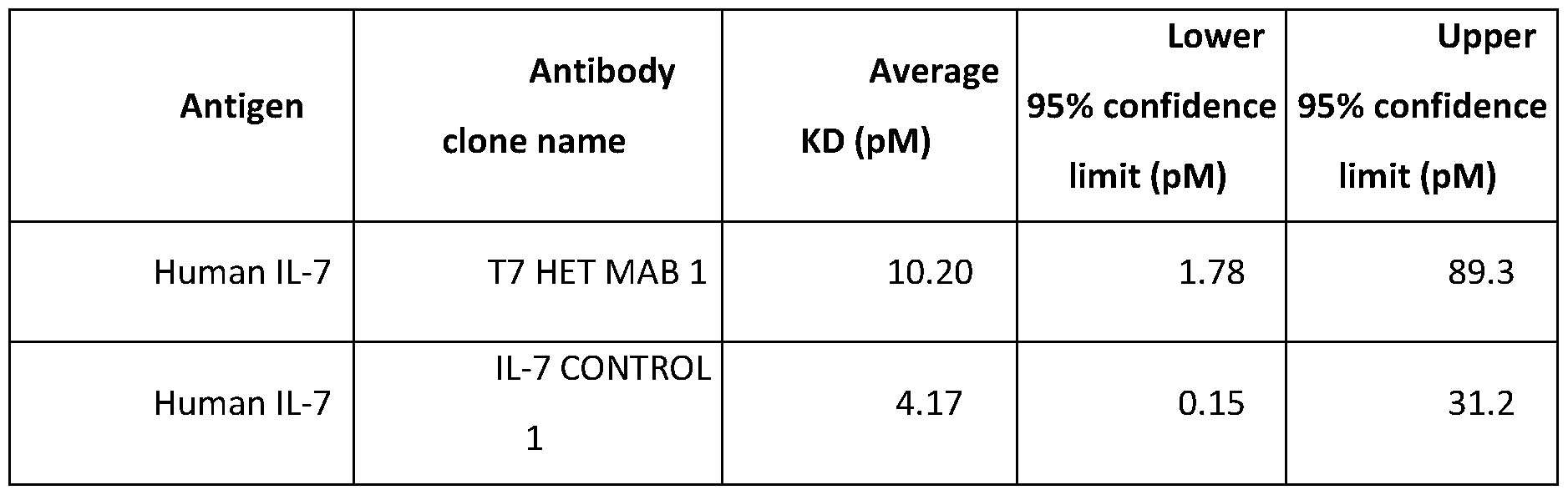 Affinity values are the arithmetic mean of n=3 replicates. Example 15: Dual engagement of T7 HET MAB 1 with recombinant human TNF-α and IL-7 using BLI Dual engagement of T7 HET MAB 1 with human TNF-α and human IL-7 was determined using a binding assay on the Sartorius Octet RED384 biolayer interferometry (BLI) instrument. Human IL-7 at 200nM were captured onto anti-Penta His dip and read biosensors for 240 seconds. The loaded sensors were dipped into either T7 HET MAB 1 (diluted to 20µg/ml in PBSF) or PBSF for 240 seconds. Following which, the sensors were dipped into PBSF or human TNF-α (diluted to 200nM in PBSF) or PBSF for 240 seconds. The sensors were then dipped back into buffer for the dissociation phase for 240 seconds. Blank sensors were included to check for non-specific binding of the proteins to the sensors. Regeneration of the biosensor tips was carried out using 10mM glycine pH 1.5. The analysis was run at 25°C, with a plate shaker speed of 1000rpm. Data were aligned to the baseline but no kinetics model was applied to the data. Dual engagement data generated on the Sartorius Octet RED384 instrument were analysed using the Data Analysis software (v11.1) inherent to the instrument, although no kinetics models were applied. There was no non-specific binding of the proteins to the anti-Penta His sensors or of human TNF-α to human IL-7. Simultaneous binding of T7 HET MAB 1 to human TNF-α and IL-7 was successfully confirmed. Table 9 Dual engagement of T7 HET MAB 1 with recombinant human TNF-α and IL-7 using BLI
 70169 Example 16: Potency against TNF: L929 assay (for T7 HET MAB 2 and T7 HET MAB 1) L929 cells are murine aneuploid fibrosarcoma cells. In this assay they are treated with actinomycin B (to sensitize the cells), then recombinant human TNF-α is added to initiate apoptosis and subsequent cell death (TNF enhances the cytotoxicity of actinomycin B which targets DNA topoisomerase II). The amount of resulting cell death is then quantified using Promega Cell Titre Glo (which is a homogeneous method of determining the number of viable cells in culture based on quantitation of the ATP present, an indicator of metabolically active cells). The measured luminescence values from the cell titre glow readout can then be used to plot curve fits, which enable determination of molecule potency (IC50). This assay was used to test the ability of the antibodies to neutralise TNF-α and block cell death. L929 cells were seeded in a 96-well flatbottomed plate at 10,000cells/well in 100µl RPMI 1640 (w/o phenol red) and incubated overnight at 37°C, 5% CO2. Media was removed, and cells were sensitised in 80µl media containing 1.25mg/ml actinomycin D for 1 hour. For the neutralising study, 0.7ng/ml - 15ug/ml (= 5pM -100nM T7 HET MAB 1 (or control molecule) were pre-incubated with 1ng/ml (19pM) human TNF- α in a 1:1 ratio for 1 hour at room temperature. Following the 1 hour pre-incubation with actinomycin D, 20ml of antibody-antigen complex was added per well.20ml media alone was added to wells as a negative control. Plates were incubated for 18 hours at 37°C, 5% CO2. Following this treatment period, cell viability was determined by a cell titer-Glo Luminescent assay kit according to manufacturer’s instructions (Promega, Manison USA). Raw data was generated on the Pherastar FS Multimode plate reader. Raw luminescence counts were analysed in ActivityBase, derived pIC50 values were obtained from each independent experiment, and the geometric mean was calculated between these values, along with the range. The calculated geometric means and ranges of pIC50 values were also converted to picomolar.
70169 Human TNF ^ neutralisation of T7 HET MAB 1, and controls Geomean was calculated from an N=12 for TNF CONTROL 3 and T7 HET MAB 1. For NEGATIVE CONTROL 1 the Geomean was calculated from an N=6. This molecule is inactive in this assay.
 Additionally, T7 HET MAB 2 and additional batches of T7 HET MAB 1 were also assessed for potency of recombinant human TNF- ^ using reagents and methods as described above.
70169 Human TNF ^ neutralisation of T7 HET MAB 2, T7 HET MAB 1 and controls

 70169
70169
 Example 17: Potency against IL-7: CCRF-CEM assay (T7 HET MAB 2 and T7 HET MAB 1T7 HET MAB 1) CCRF-CEM cells are human T lymphoblasts, and express IL-7 receptor on the cell surface. Binding of IL-7 to IL-7 receptor leads to receptor dimerisation and activation, recruitment of JAK proteins and phosphorylation of STAT5. Phosphorylated STAT5 (pSTAT5) can be detected in cell lysates using a sandwich immunoassay, specific for pSTAT5. Following stimulation with a fixed concentration of IL-7. The binding of an antibody to IL-7 in this system disrupts the interaction between the cytokine and its receptor, which in turn prevents the IL-7 mediated phosphorylation of STAT5. The pSTAT5 present in these cells can be normalised to wells which contain IL-7 alone, and wells which contain no IL-7 to determine percentage inhibition. These values can then be used to plot curve fits, which enable determination of molecule potency (IC50). CCRF-CEM cells were stimulated with Dexamethasone [Sigma-Aldrich D4902] and incubated overnight at 37C, 5% CO2. Human IL-7 was prepared to a concentration of 15pM in assay matrix (PR-FREE RPMI [Gibco 32404-014]+ 10% FBS [Gibco 10100-147]).10uL of this IL-7 preparation was dispensed into all columns of a 384-well V-bottom plate (Greiner 781280), with the exception of columns 11 and 23, using a MultiDrop.10uL assay matrix was dispensed into columns 11 and 23. Tested antibodies were prepared to a concentration of 3nM, and were serially diluted 1:4 over 10 points. Columns 11 and 12 contained assay matrix alone.10uL from this plate was transferred into the plate containing human IL-7. This plate was incubated for 30 minutes at room temperature, on an orbital rocker to allow the antibodies to pre-complex with IL-7. Dexamethasone-stimulated CCRF-CEM cells were collected from the incubator, and 250uL was counted on the Beckman Coulter ViCell. Cultures were centrifuged at 1500rpm for 5 minutes, then spent media was discarded. Cell pellets were resuspended to a concentration of 5x10⁶ cells/mL.10uL of this cell
70169 suspension was dispensed to the plate containing antibody and IL-7. The plate was incubated for 20 minutes at 37C, 5% CO2. 10uL of 4X lysis buffer (2.4mL 10x lysis buffer [Cell Signaling 9803], 2mL dH2O, 200uL Protease inhibitor [Sigma P8340], 200uL Phosphatase inhibitor cocktail II [Sigma P5726], 200ul Phosphatase inhibitor cocktail III [Sigma P0044]) was dispensed per well, and the plate was shaken vigorously for 10 seconds to lyse cells. These cell lysates were stored on ice for use in a pSTAT5 immunoassay. One plate from an MSD pSTAT5 immunoassay kit (MSD K15163D) was used per 96 wells used on the 384-well plate.1X Tris Wash Buffer was prepared from a 10X stock provided in the kit, by diluting 10- fold with dH2O. 150uL of MSD Blocker A (3% w/v MSD Blocker A powder in 1X Tris Wash Buffer) was dispensed into each well of the plate. The plate was covered with an aluminium foil seal and was incubated at room temperature on an orbital rocker for 1 hour. Following this incubation, plate contents were discarded by flicking into a sink, and 250uL 1X Tris Wash Buffer was dispensed per well. The plate contents were flicked, and buffer dispensed twice further to wash the plate. 25uL cell lysates were transferred from the plate on ice to the MSD plate. The plate was sealed as before and incubated at room temperature on an orbital rocker for 1 hour. Antibody diluent solution was prepared by adding 150µL of 2% Blocker D-M, 30µL of 10% Blocker D-R, 1mL Blocker A solution and 1.82mL 1X Tris Wash Buffer. The pSTAT5 detection antibody was diluted from 50X stock to 1X by adding 60uL to 2.94mL of the prepared diluent. The plate was washed 3 times as before, and 25uL detection antibody solution was dispensed into each well. The plate was sealed as before and incubated at room temperature on an orbital rocker for 1 hour. Following this incubation, the plate was washed as before.1X Read Buffer T was prepared from a 4X Read Buffer T stock (MSD R92TC) by diluting 4-fold in dH20 and 150uL was dispensed per well, and the plate was read on the MSD Sector S 600 imager. Raw data was generated on the MSD Sector S 600 imager using MSD Workbench (MSD). Raw MSD counts were exported as a delimited text file, and further analysis was performed in ActivityBase. Mean pIC50 values were obtained from each independent experiment, and the geometric mean was calculated between these values, along with the range. The calculated geometric means and ranges of pIC50 values were also converted to picomolar. The potency (IC50) of T7 HET MAB 1 for human IL-7 was in the single-digit picomolar range, and was slightly decreased (by less than 2-fold) relative to IL-7 control 1, this is expected as T7 HET MAB 1 has half as many anti-IL-7 fAb arms compared to IL-7 control 1.
70169 Table 10: Human IL-7 Neutralisation potency of T7 HET MAB 1 and controls.
Example 17: Potency against IL-7: CCRF-CEM assay (T7 HET MAB 2 and T7 HET MAB 1T7 HET MAB 1) CCRF-CEM cells are human T lymphoblasts, and express IL-7 receptor on the cell surface. Binding of IL-7 to IL-7 receptor leads to receptor dimerisation and activation, recruitment of JAK proteins and phosphorylation of STAT5. Phosphorylated STAT5 (pSTAT5) can be detected in cell lysates using a sandwich immunoassay, specific for pSTAT5. Following stimulation with a fixed concentration of IL-7. The binding of an antibody to IL-7 in this system disrupts the interaction between the cytokine and its receptor, which in turn prevents the IL-7 mediated phosphorylation of STAT5. The pSTAT5 present in these cells can be normalised to wells which contain IL-7 alone, and wells which contain no IL-7 to determine percentage inhibition. These values can then be used to plot curve fits, which enable determination of molecule potency (IC50). CCRF-CEM cells were stimulated with Dexamethasone [Sigma-Aldrich D4902] and incubated overnight at 37C, 5% CO2. Human IL-7 was prepared to a concentration of 15pM in assay matrix (PR-FREE RPMI [Gibco 32404-014]+ 10% FBS [Gibco 10100-147]).10uL of this IL-7 preparation was dispensed into all columns of a 384-well V-bottom plate (Greiner 781280), with the exception of columns 11 and 23, using a MultiDrop.10uL assay matrix was dispensed into columns 11 and 23. Tested antibodies were prepared to a concentration of 3nM, and were serially diluted 1:4 over 10 points. Columns 11 and 12 contained assay matrix alone.10uL from this plate was transferred into the plate containing human IL-7. This plate was incubated for 30 minutes at room temperature, on an orbital rocker to allow the antibodies to pre-complex with IL-7. Dexamethasone-stimulated CCRF-CEM cells were collected from the incubator, and 250uL was counted on the Beckman Coulter ViCell. Cultures were centrifuged at 1500rpm for 5 minutes, then spent media was discarded. Cell pellets were resuspended to a concentration of 5x10⁶ cells/mL.10uL of this cell
70169 suspension was dispensed to the plate containing antibody and IL-7. The plate was incubated for 20 minutes at 37C, 5% CO2. 10uL of 4X lysis buffer (2.4mL 10x lysis buffer [Cell Signaling 9803], 2mL dH2O, 200uL Protease inhibitor [Sigma P8340], 200uL Phosphatase inhibitor cocktail II [Sigma P5726], 200ul Phosphatase inhibitor cocktail III [Sigma P0044]) was dispensed per well, and the plate was shaken vigorously for 10 seconds to lyse cells. These cell lysates were stored on ice for use in a pSTAT5 immunoassay. One plate from an MSD pSTAT5 immunoassay kit (MSD K15163D) was used per 96 wells used on the 384-well plate.1X Tris Wash Buffer was prepared from a 10X stock provided in the kit, by diluting 10- fold with dH2O. 150uL of MSD Blocker A (3% w/v MSD Blocker A powder in 1X Tris Wash Buffer) was dispensed into each well of the plate. The plate was covered with an aluminium foil seal and was incubated at room temperature on an orbital rocker for 1 hour. Following this incubation, plate contents were discarded by flicking into a sink, and 250uL 1X Tris Wash Buffer was dispensed per well. The plate contents were flicked, and buffer dispensed twice further to wash the plate. 25uL cell lysates were transferred from the plate on ice to the MSD plate. The plate was sealed as before and incubated at room temperature on an orbital rocker for 1 hour. Antibody diluent solution was prepared by adding 150µL of 2% Blocker D-M, 30µL of 10% Blocker D-R, 1mL Blocker A solution and 1.82mL 1X Tris Wash Buffer. The pSTAT5 detection antibody was diluted from 50X stock to 1X by adding 60uL to 2.94mL of the prepared diluent. The plate was washed 3 times as before, and 25uL detection antibody solution was dispensed into each well. The plate was sealed as before and incubated at room temperature on an orbital rocker for 1 hour. Following this incubation, the plate was washed as before.1X Read Buffer T was prepared from a 4X Read Buffer T stock (MSD R92TC) by diluting 4-fold in dH20 and 150uL was dispensed per well, and the plate was read on the MSD Sector S 600 imager. Raw data was generated on the MSD Sector S 600 imager using MSD Workbench (MSD). Raw MSD counts were exported as a delimited text file, and further analysis was performed in ActivityBase. Mean pIC50 values were obtained from each independent experiment, and the geometric mean was calculated between these values, along with the range. The calculated geometric means and ranges of pIC50 values were also converted to picomolar. The potency (IC50) of T7 HET MAB 1 for human IL-7 was in the single-digit picomolar range, and was slightly decreased (by less than 2-fold) relative to IL-7 control 1, this is expected as T7 HET MAB 1 has half as many anti-IL-7 fAb arms compared to IL-7 control 1.
70169 Table 10: Human IL-7 Neutralisation potency of T7 HET MAB 1 and controls.
 Additionally, T7 HET MAB 2 was also assessed for potency for recombinant human IL-7 using reagents, methods and data analyses similar to those described above. Human IL-7 and human TNF-α Neutralisation potency of T7 HET MAB 1 and controls
 70169
70169
 Example 18: Potency of T7 HET mAb 1 in mixed lymphocyte reaction (MLR) assays. An MLR assay is an ex-vivo cellular immune assay where peripheral blood mononuclear cells from two different donors are combined in culture. In these co-cultures, T cells are activated and produce proinflammatory cytokines in response to allogenic histocompatibility molecules expressed on antigen presenting cells from the opposite donor. To compare the potency of the T7 HET mAb 1 to the potency of the TNF control mAb 1 and IL-7 control mAb 1, an MLR assay was developed. Because IL-7 is not expressed in PBMCs, human recombinant IL-7 was added to the cultures to stimulate the IL-7 receptor pathway. IFNg inhibition in MLR assay using TNF control 6 and IL-7 control 1 alone or in combination. Cryopreserved PBMCs (STEMCELL Technologies) from separate donors were thawed and resuspended at 1-2x10⁶ cells/ml. The PBMCs from two donors were combined 1:1 and 100 ul was dispensed in duplicate wells of a 96-well round bottom plate (n=3 different MLR co-culture combinations). Recombinant human IL-7 was added to the cultures at a final concentration of 10 ng/ml. Co-cultures were treated with the following antibodies: (A) 2 ug/ml TNF control 6-negative control + 5 ug/ml negative control; (B) 2 ug/ml TNF control 6; (C) 5 ug/ml IL-7 control 1; (D) 2 ug/ml TNF control 6 + 5 ug/ml IL-7 control 1. Co-cultures were incubated for 48h at 37oC and 5% CO2. IFNg secretion was quantified using Meso Scale Discovery immunoassay kits (Figure 8). The amount of IFNg secreted by samples treated with 2 ug/ml TNF control 6-negative control + 5 ug/ml negative control was normalized to 100 percent or maximum signal. The amount of IFNg secreted by samples treated with the test antibodies was calculated as a percent of the maximum signal. Treatment of MLR co-cultures with 2 ug/ml TNF control 6 antibody reduced IFNg secretion approximately 82%. Treatment of MLR co-cultures with 5 ug/ml IL-7 control 1 antibody reduced IFNg secretion approximately 81%. Treatment of MLR co-cultures with a combination of 2 ug/ml TNF control 6 + 5 ug/ml IL-7 control 1 antibody reduced IFNg secretion approximately 97%. Therefore, combination of TNF control 6 + IL-7 control 1 demonstrated enhanced activity in blocking IFNg secretion compared to TNF control 6 or IL-7 control 1 antibodies alone (Figure 8).
70169 IFNg inhibition by T7 HET mAb 1 compared to TNF control 1 or IL-7 control 1 in MLR assays. Cryopreserved PBMCs (STEMCELL Technologies) from separate donors were thawed and resuspended at 1-2x10⁶ cells/ml. The PBMCs from two donors were combined 1:1 and 100 ul was dispensed in duplicate wells of a 96-well round bottom plate (n=9 different MLR co-culture combinations). Recombinant human IL-7 was added to the cultures at a final concentration of 10 ng/ml. Co-cultures were treated with increasing concentrations the following antibodies: Negative control, T7 HET mAb 1, TNF control 1, IL-7 control 1. Cytokine secretion was quantified using Meso Scale Discovery immunoassay kits. The data were logged, normalized to the mean response of negative control across all concentrations (by subtraction), and averaged over technical replicates. Four-parameter logistic curves with a shared maximum response across antibodies were fitted for IFNg, IL-6 and IL-8 (Figure 9). Estimates of IC50s and minimum response are reported for IFNg (Table 11). Changes in IC50s and minimum response between T7 HETmAb and TNF control 1 and IL-7 control 1 are reported as fold changes with 95% CIs and p-values for the test of no change. All analyses were completed in R, Version 4.0.3. As shown in Figure 9 and summarized in Table 11, T7 HETmAb suppressed IFNg production in a concentration dependent manner with an estimated IC50 = 60.5 pM and 95% CI of (25.9 pM – 140.9 pM). Similarly, treatment TNF control 1 produced an estimated IC50 = 50.0 pM and 95% CI of (14.0 pM – 178.6 pM). Thus, even though T7 HETmAb harbours one arm targeting TNFa, compared to TNF control 1 there was no significant difference between the IC50s produced by the two molecules (p = 0.6077) in the MLR assay (Table 11). IL7 control 1 inhibited IFNg production in a dose dependent manner with an estimated IC50 = 10.3 pM and 95% CI of (2.2 – 47.7) (Table 11). However, while the IC50 was approximately 6-fold lower than T7 HETmAb, IL7 control 1 achieved significantly less inhibition (minimum response) of IFNg (p = < 0.0001) at the highest dose tested (Table 11). For IL-6, T7 HETmAb, TNF control 1 and IL7 control 1 all inhibited IL-6 production in a dose dependent manner and all three achieved similar levels of inhibition (minimum response) (Figure 9). As shown in Table 11, T7 HETmAb inhibited IL-6 with an IC50 = 8.8 pM with 95% CI of (1.0 - 73.9). TNF control 1 produced an IC50 = 1.7 pM with 95% CI of (0.1 – 33.1). IL7 control 1 produced and IC50 = 0.8 pM with 95% CI of (0.01 – 56.1). As shown in Table 11, for IL-6, there was no statistical difference between the IC50s produced by T7 HETmAb and TNF control 1 (p = 0.3277) or IL7 control 1 (p = 0.2739). For IL-8, T7 HETmAb inhibited IL-8 production with an estimated IC50 = 53.7 pM with 95% CI of (18.5 pM – 155.5 pM). TNF control 1 inhibited IL-8 with an estimated IC50 = 11.0 pM with 95% CI of (2.9 pM – 41.7 pM) (Table 11). Thus, for IL-8 T7 HETmAb was approximately 5-fold less potent than TNF control 1 (p = 0.0192) (Table 11). IL7 control 1 did not inhibit IL-8 production in the dose range tested, suggesting that IL-8 is not dependent upon IL-7 signalling (Figure 9 and Table 11).
70169 Table 11. Summary of IC50 and minimum response for inhibition of cytokine production by T7 HETmAb, TNF control 1, and IL7 control 1 in the MLR assay
Example 18: Potency of T7 HET mAb 1 in mixed lymphocyte reaction (MLR) assays. An MLR assay is an ex-vivo cellular immune assay where peripheral blood mononuclear cells from two different donors are combined in culture. In these co-cultures, T cells are activated and produce proinflammatory cytokines in response to allogenic histocompatibility molecules expressed on antigen presenting cells from the opposite donor. To compare the potency of the T7 HET mAb 1 to the potency of the TNF control mAb 1 and IL-7 control mAb 1, an MLR assay was developed. Because IL-7 is not expressed in PBMCs, human recombinant IL-7 was added to the cultures to stimulate the IL-7 receptor pathway. IFNg inhibition in MLR assay using TNF control 6 and IL-7 control 1 alone or in combination. Cryopreserved PBMCs (STEMCELL Technologies) from separate donors were thawed and resuspended at 1-2x10⁶ cells/ml. The PBMCs from two donors were combined 1:1 and 100 ul was dispensed in duplicate wells of a 96-well round bottom plate (n=3 different MLR co-culture combinations). Recombinant human IL-7 was added to the cultures at a final concentration of 10 ng/ml. Co-cultures were treated with the following antibodies: (A) 2 ug/ml TNF control 6-negative control + 5 ug/ml negative control; (B) 2 ug/ml TNF control 6; (C) 5 ug/ml IL-7 control 1; (D) 2 ug/ml TNF control 6 + 5 ug/ml IL-7 control 1. Co-cultures were incubated for 48h at 37oC and 5% CO2. IFNg secretion was quantified using Meso Scale Discovery immunoassay kits (Figure 8). The amount of IFNg secreted by samples treated with 2 ug/ml TNF control 6-negative control + 5 ug/ml negative control was normalized to 100 percent or maximum signal. The amount of IFNg secreted by samples treated with the test antibodies was calculated as a percent of the maximum signal. Treatment of MLR co-cultures with 2 ug/ml TNF control 6 antibody reduced IFNg secretion approximately 82%. Treatment of MLR co-cultures with 5 ug/ml IL-7 control 1 antibody reduced IFNg secretion approximately 81%. Treatment of MLR co-cultures with a combination of 2 ug/ml TNF control 6 + 5 ug/ml IL-7 control 1 antibody reduced IFNg secretion approximately 97%. Therefore, combination of TNF control 6 + IL-7 control 1 demonstrated enhanced activity in blocking IFNg secretion compared to TNF control 6 or IL-7 control 1 antibodies alone (Figure 8).
70169 IFNg inhibition by T7 HET mAb 1 compared to TNF control 1 or IL-7 control 1 in MLR assays. Cryopreserved PBMCs (STEMCELL Technologies) from separate donors were thawed and resuspended at 1-2x10⁶ cells/ml. The PBMCs from two donors were combined 1:1 and 100 ul was dispensed in duplicate wells of a 96-well round bottom plate (n=9 different MLR co-culture combinations). Recombinant human IL-7 was added to the cultures at a final concentration of 10 ng/ml. Co-cultures were treated with increasing concentrations the following antibodies: Negative control, T7 HET mAb 1, TNF control 1, IL-7 control 1. Cytokine secretion was quantified using Meso Scale Discovery immunoassay kits. The data were logged, normalized to the mean response of negative control across all concentrations (by subtraction), and averaged over technical replicates. Four-parameter logistic curves with a shared maximum response across antibodies were fitted for IFNg, IL-6 and IL-8 (Figure 9). Estimates of IC50s and minimum response are reported for IFNg (Table 11). Changes in IC50s and minimum response between T7 HETmAb and TNF control 1 and IL-7 control 1 are reported as fold changes with 95% CIs and p-values for the test of no change. All analyses were completed in R, Version 4.0.3. As shown in Figure 9 and summarized in Table 11, T7 HETmAb suppressed IFNg production in a concentration dependent manner with an estimated IC50 = 60.5 pM and 95% CI of (25.9 pM – 140.9 pM). Similarly, treatment TNF control 1 produced an estimated IC50 = 50.0 pM and 95% CI of (14.0 pM – 178.6 pM). Thus, even though T7 HETmAb harbours one arm targeting TNFa, compared to TNF control 1 there was no significant difference between the IC50s produced by the two molecules (p = 0.6077) in the MLR assay (Table 11). IL7 control 1 inhibited IFNg production in a dose dependent manner with an estimated IC50 = 10.3 pM and 95% CI of (2.2 – 47.7) (Table 11). However, while the IC50 was approximately 6-fold lower than T7 HETmAb, IL7 control 1 achieved significantly less inhibition (minimum response) of IFNg (p = < 0.0001) at the highest dose tested (Table 11). For IL-6, T7 HETmAb, TNF control 1 and IL7 control 1 all inhibited IL-6 production in a dose dependent manner and all three achieved similar levels of inhibition (minimum response) (Figure 9). As shown in Table 11, T7 HETmAb inhibited IL-6 with an IC50 = 8.8 pM with 95% CI of (1.0 - 73.9). TNF control 1 produced an IC50 = 1.7 pM with 95% CI of (0.1 – 33.1). IL7 control 1 produced and IC50 = 0.8 pM with 95% CI of (0.01 – 56.1). As shown in Table 11, for IL-6, there was no statistical difference between the IC50s produced by T7 HETmAb and TNF control 1 (p = 0.3277) or IL7 control 1 (p = 0.2739). For IL-8, T7 HETmAb inhibited IL-8 production with an estimated IC50 = 53.7 pM with 95% CI of (18.5 pM – 155.5 pM). TNF control 1 inhibited IL-8 with an estimated IC50 = 11.0 pM with 95% CI of (2.9 pM – 41.7 pM) (Table 11). Thus, for IL-8 T7 HETmAb was approximately 5-fold less potent than TNF control 1 (p = 0.0192) (Table 11). IL7 control 1 did not inhibit IL-8 production in the dose range tested, suggesting that IL-8 is not dependent upon IL-7 signalling (Figure 9 and Table 11).
70169 Table 11. Summary of IC50 and minimum response for inhibition of cytokine production by T7 HETmAb, TNF control 1, and IL7 control 1 in the MLR assay
 IC50 values are reported on the original cytokine concentration scale (that is, as the concentration giving a response halfway between the minimum and the maximum response) with 95% confidence limits. P- values correspond to the test of no change in IC50 from T7 HETmAb on the log10 cytokine concentration scale. * Do not interpret; curve is virtually flat, therefore the estimated IC50 contains high degree of uncertainty. Confidence intervals are very large and not reported (n/a). The minimum response is reported as a % change from the mean negative control response; p-values correspond to the test of no change in the minimum response from T7 HETmAb. The enhanced maximum inhibition of IFNg observed for T7 HET mAb 1 in the dose response MLR assays was further analyzed by comparing each compound at the top dose used, 5 ug/ml (Figure 9). The data were logged, and a mixed model was fitted with donor combination as a random effect and treatment as fixed for IFNg. A Tukey test was performed to assess differences between compounds. If there were multiple observations per donor combination, treatment and experiment, an average was taken to avoid pseudo replication. The indicated percent inhibition for each compound was calculated as percent reduction in cytokine secretion compared to negative control (Figure 10 and Table 12). Compared to negative control, T7 HET mAb 1 inhibited IFNg secretion 96.3%. TNF control 1 and IL-7 control 1 inhibited IFNg secretion by 87.45 and 69.4%, respectively, compared to negative control. The maximum level of inhibition achieved by T7 HET mAb was significantly greater than the inhibition observed for TNF control 1 (p = 0.016) or IL-7 control 1 (p < 0.001 ) (Table 12).
70169 Table 12. Statistical analysis of maximum inhibition observed for IL-7 control 1 ,TNF control 1 and T7 HET mAb compared to negative control at 5 ug/ml concentration.
IC50 values are reported on the original cytokine concentration scale (that is, as the concentration giving a response halfway between the minimum and the maximum response) with 95% confidence limits. P- values correspond to the test of no change in IC50 from T7 HETmAb on the log10 cytokine concentration scale. * Do not interpret; curve is virtually flat, therefore the estimated IC50 contains high degree of uncertainty. Confidence intervals are very large and not reported (n/a). The minimum response is reported as a % change from the mean negative control response; p-values correspond to the test of no change in the minimum response from T7 HETmAb. The enhanced maximum inhibition of IFNg observed for T7 HET mAb 1 in the dose response MLR assays was further analyzed by comparing each compound at the top dose used, 5 ug/ml (Figure 9). The data were logged, and a mixed model was fitted with donor combination as a random effect and treatment as fixed for IFNg. A Tukey test was performed to assess differences between compounds. If there were multiple observations per donor combination, treatment and experiment, an average was taken to avoid pseudo replication. The indicated percent inhibition for each compound was calculated as percent reduction in cytokine secretion compared to negative control (Figure 10 and Table 12). Compared to negative control, T7 HET mAb 1 inhibited IFNg secretion 96.3%. TNF control 1 and IL-7 control 1 inhibited IFNg secretion by 87.45 and 69.4%, respectively, compared to negative control. The maximum level of inhibition achieved by T7 HET mAb was significantly greater than the inhibition observed for TNF control 1 (p = 0.016) or IL-7 control 1 (p < 0.001 ) (Table 12).
70169 Table 12. Statistical analysis of maximum inhibition observed for IL-7 control 1 ,TNF control 1 and T7 HET mAb compared to negative control at 5 ug/ml concentration.
 Additional soluble analytes suppressed by T7 HETmAb 1 compared to TNF control 1 or IL-7 control 1 in MLR assays. To identify additional soluble markers of T7 HETmAb 1 additivity, compared to the TNF control 1 or IL7 control 1, a Luminex 65-plex assay was used to profile the supernatants from the MLR samples treated at the highest concentration tested (5 ug/ml). Luminex assay derived analyte concentrations were log10 or sqrt transformed depending on the presence of zeros (log10 if no zeros, sqrt otherwise) and analyzed by linear mixed effect model (fixed effect: Treatment, random effect: MLR, MLR-Treatment interaction), followed by Tukey’s post-hoc comparisons. Fold changes in concentration for the HETmAb relative to the isotype control or parental antibodies displayed in the heatmap (Figure 11) were calculated by taking the ratio of geometric means. An offset of 0.1 was added to all values to prevent single values of zero to set the geometric mean to zero. Software used: R, Version 4.0.3. Heatmaps shown in Figure 11 displaying (1) the fold changes in analyte concentration for T7 HETmAb relative to negative control, IL7 control, and TNF control 1; and (2) the p-values for the test of no change (* = p<0.05 and ** = p<0.01). The following analytes were significantly inhibited by T7 HETmAb compared to both IL7 control 1 and TNF control 1 (Figure 11): • Cytokines: IL-17A, IFNγ, GM-CSF, IL-5, M-CSF, IL-13, LIF, IL-8, IL-1ɑ, IL-6, IL-10, IL-21 • Chemokines: MIP-1ɑ, MIP-1β, MCP1, MDC, CX3CL1, CXCL11, MIG, CCL24, CCL26 • Soluble receptors: CD40L, IL2R, TRAIL, TWEAK, TNFRII • Growth factors: MMP-1, VEGF-A Suppression of IL-7 dependent markers by T7 HETmAb 1 compared to TNF control 1 or IL-7 control 1 in MLR assays. To assess IL-7 dependent cell surface and cytoplasmic proteins, the MLR assay described above was extended to a five-day co-culture assay. In this five-day MLR assay, flow cytometry was used to detect IL- 7 dependent CD4+ T cell expression of the gut homing integrin, ɑ4β7, the anti-apoptotic factor, Bcl-2, and the proliferation marker, Ki-67. After 5d incubation, the cells were processed for flow cytometry. CD4+ cells were gated, and geometric mean fluorescence intensity was measured for cell surface ^4 ^7 and intracellular Bcl-2 and Ki-67. Data represents n=16 independent MLRs with duplicate technical replicates.
70169 For each MFI type, a separate linear mixed effects model was fit, each including Antibody, Stimulation, and their interaction as fixed effects and donor as a random effect. Contrasts were calculated comparing the negative control to IL7 control 1, TNF control 1, T7 HETmAb 1 (***p<0.001, ****p<0.0001, with Tukey’s HSD adjustment). Analysis was performed using R 4.0.3. As shown in Figure 12, both T7 HETmAb1 and IL7 control 1 decreased IL-7 induced expression of ɑ4β7, Bcl-2 and Ki-67 in CD4+T cells as compared to the negative control. TNF control 1 was not statistically different from negative control (Figure 12). Fc-mediated induction of M2 macrophages by T7 HETmAb in MLR assays. Anti-TNF antibodies have been shown to induce alternative activation of macrophages (known as M2 macrophages), that have anti-inflammatory activities through their recognition by Fc receptors. The MLR assay was used to compare the induction of M2 macrophages by T7 HETmAb and TNF control 6. Peripheral blood mononuclear cells (PBMCs) were enriched from whole blood by Ficol-gradient centrifugation. The PBMCs from Donors 1 - 5 were stained with CellTrace Violet while cells from Donor 6 - 10 were stained with CellTrace CFSE following manufacturer’s recommendations. MLRs were established by mixing cells from donors 1 - 5 with cells from donor 6 - 10 in 1:1 proportion. Samples were incubated for 48 h at 37oC to establish an inflammatory environment. Then, cells were supplemented with 100 uL of media containing the test antibodies with or without FcR blocker and the plates were incubated for an additional 5 days at 37oC. After the incubation period, cells were washed as before and incubated with a cocktail of fluorescent-labelled antibodies directed against surface antigens. Sample data was acquired using the Cytek Aurora spectral flow cytometer with SpectroFlo™ software (v2.0). Instrument optimal performance settings were confirmed with QC beads prior to acquisition of samples. Data was analyzed using the FlowJo software (v 10). Percent positive values (CD206+CD14+ cells) were exported into Microsoft Excel spreadsheet using the TABLE Editor within the FlowJo software. The cell populations were tabulated in an Excel spreadsheet at % of the parent and then copied and pasted into GraphPad Prism (V9) software for graphing and statistical analysis. For statistical analysis, a paired One-Way ANOVA with LSD test was used (***p<0.005, ****p<0.0001). TNF control 6 significantly increased the frequency of M2 macrophages in the MLR assay compared to PBS and negative control 1 treated co-cultures. T7 HETmAb had a similar effect and significantly increased M2 macrophages as well. Blocking the Fc receptors with the addition of FcRX abrogated the induction of M2 macrophages, confirming that this effect is dependent on Fc receptor signalling (Figure 13).
Additional soluble analytes suppressed by T7 HETmAb 1 compared to TNF control 1 or IL-7 control 1 in MLR assays. To identify additional soluble markers of T7 HETmAb 1 additivity, compared to the TNF control 1 or IL7 control 1, a Luminex 65-plex assay was used to profile the supernatants from the MLR samples treated at the highest concentration tested (5 ug/ml). Luminex assay derived analyte concentrations were log10 or sqrt transformed depending on the presence of zeros (log10 if no zeros, sqrt otherwise) and analyzed by linear mixed effect model (fixed effect: Treatment, random effect: MLR, MLR-Treatment interaction), followed by Tukey’s post-hoc comparisons. Fold changes in concentration for the HETmAb relative to the isotype control or parental antibodies displayed in the heatmap (Figure 11) were calculated by taking the ratio of geometric means. An offset of 0.1 was added to all values to prevent single values of zero to set the geometric mean to zero. Software used: R, Version 4.0.3. Heatmaps shown in Figure 11 displaying (1) the fold changes in analyte concentration for T7 HETmAb relative to negative control, IL7 control, and TNF control 1; and (2) the p-values for the test of no change (* = p<0.05 and ** = p<0.01). The following analytes were significantly inhibited by T7 HETmAb compared to both IL7 control 1 and TNF control 1 (Figure 11): • Cytokines: IL-17A, IFNγ, GM-CSF, IL-5, M-CSF, IL-13, LIF, IL-8, IL-1ɑ, IL-6, IL-10, IL-21 • Chemokines: MIP-1ɑ, MIP-1β, MCP1, MDC, CX3CL1, CXCL11, MIG, CCL24, CCL26 • Soluble receptors: CD40L, IL2R, TRAIL, TWEAK, TNFRII • Growth factors: MMP-1, VEGF-A Suppression of IL-7 dependent markers by T7 HETmAb 1 compared to TNF control 1 or IL-7 control 1 in MLR assays. To assess IL-7 dependent cell surface and cytoplasmic proteins, the MLR assay described above was extended to a five-day co-culture assay. In this five-day MLR assay, flow cytometry was used to detect IL- 7 dependent CD4+ T cell expression of the gut homing integrin, ɑ4β7, the anti-apoptotic factor, Bcl-2, and the proliferation marker, Ki-67. After 5d incubation, the cells were processed for flow cytometry. CD4+ cells were gated, and geometric mean fluorescence intensity was measured for cell surface ^4 ^7 and intracellular Bcl-2 and Ki-67. Data represents n=16 independent MLRs with duplicate technical replicates.
70169 For each MFI type, a separate linear mixed effects model was fit, each including Antibody, Stimulation, and their interaction as fixed effects and donor as a random effect. Contrasts were calculated comparing the negative control to IL7 control 1, TNF control 1, T7 HETmAb 1 (***p<0.001, ****p<0.0001, with Tukey’s HSD adjustment). Analysis was performed using R 4.0.3. As shown in Figure 12, both T7 HETmAb1 and IL7 control 1 decreased IL-7 induced expression of ɑ4β7, Bcl-2 and Ki-67 in CD4+T cells as compared to the negative control. TNF control 1 was not statistically different from negative control (Figure 12). Fc-mediated induction of M2 macrophages by T7 HETmAb in MLR assays. Anti-TNF antibodies have been shown to induce alternative activation of macrophages (known as M2 macrophages), that have anti-inflammatory activities through their recognition by Fc receptors. The MLR assay was used to compare the induction of M2 macrophages by T7 HETmAb and TNF control 6. Peripheral blood mononuclear cells (PBMCs) were enriched from whole blood by Ficol-gradient centrifugation. The PBMCs from Donors 1 - 5 were stained with CellTrace Violet while cells from Donor 6 - 10 were stained with CellTrace CFSE following manufacturer’s recommendations. MLRs were established by mixing cells from donors 1 - 5 with cells from donor 6 - 10 in 1:1 proportion. Samples were incubated for 48 h at 37oC to establish an inflammatory environment. Then, cells were supplemented with 100 uL of media containing the test antibodies with or without FcR blocker and the plates were incubated for an additional 5 days at 37oC. After the incubation period, cells were washed as before and incubated with a cocktail of fluorescent-labelled antibodies directed against surface antigens. Sample data was acquired using the Cytek Aurora spectral flow cytometer with SpectroFlo™ software (v2.0). Instrument optimal performance settings were confirmed with QC beads prior to acquisition of samples. Data was analyzed using the FlowJo software (v 10). Percent positive values (CD206+CD14+ cells) were exported into Microsoft Excel spreadsheet using the TABLE Editor within the FlowJo software. The cell populations were tabulated in an Excel spreadsheet at % of the parent and then copied and pasted into GraphPad Prism (V9) software for graphing and statistical analysis. For statistical analysis, a paired One-Way ANOVA with LSD test was used (***p<0.005, ****p<0.0001). TNF control 6 significantly increased the frequency of M2 macrophages in the MLR assay compared to PBS and negative control 1 treated co-cultures. T7 HETmAb had a similar effect and significantly increased M2 macrophages as well. Blocking the Fc receptors with the addition of FcRX abrogated the induction of M2 macrophages, confirming that this effect is dependent on Fc receptor signalling (Figure 13).
70169 The disclosure is also described in the following numbered clauses. 1. A method for treating an autoimmune and/or inflammatory condition in a subject in need thereof, comprising administering to the subject a therapeutically effective amount of an IL-7 inhibitor and a therapeutically effective amount of a TNF-α inhibitor. 2. A method according to clause 1, wherein the therapeutically effective amount of an IL-7 inhibitor is a therapeutically effective amount of an IL-7 binding domain and the therapeutically effective amount of a TNF-α inhibitor is a therapeutically effective amount of a TNF-α binding domain. 3. The method according to clauses 1 or 2, wherein the subject is a human subject. 4. The method according to any one of clauses 1-3, wherein the autoimmune and/or inflammatory condition is of inflammatory skin diseases (including psoriasis and atopic dermatitis); systemic scleroderma and sclerosis; inflammatory bowel disease (IBD); Crohn's disease; ulcerative colitis; ischemic reperfusion disorders including surgical tissue reperfusion injury, myocardial ischemic conditions such as myocardial infarction, cardiac arrest, reperfusion after cardiac surgery and constriction after percutaneous transluminal coronary angioplasty, stroke, and abdominal aortic aneurysms; cerebral edema secondary to stroke; cranial trauma, hypovolemic shock; asphyxia; adult respiratory distress syndrome; acute-lung injury; Behcet's Disease; dermatomyositis; polymyositis; multiple sclerosis (MS); dermatitis; meningitis; encephalitis; uveitis; osteoarthritis; lupus nephritis; autoimmune diseases such as rheumatoid arthritis (RA), spondyloarthritis, Sjogren’s syndrome, vasculitis; diseases involving leukocyte diapedesis; central nervous system (CNS) inflammatory disorder, multiple organ injury syndrome secondary to septicemia or trauma; alcoholic hepatitis; bacterial pneumonia; antigen-antibody complex mediated diseases including glomerulonephritis; sepsis; sarcoidosis; immunopathologic responses to tissue/organ transplantation; inflammations of the lung, (including pleurisy, alveolitis, vasculitis, pneumonia, chronic bronchitis, bronchiectasis, diffuse panbronchiolitis, hypersensitivity pneumonitis, idiopathic pulmonary fibrosis (IPF), and cystic fibrosis); psoriatic arthritis; neuromyelitis optica; Guillain-Barre syndrome (GBS); COPD, type 1 diabetes or any combination thererof. 5. The method according to clause 4, wherein the autoimmune and/or inflammatory condition is Inflammatory Bowel Disease (IBD) inflammatory bowel disease (IBD); Crohn's disease; ulcerative colitis or any combination thereof. 6. The method according to clause 5, wherein the autoimmune or inflammatory condition is Inflammatory Bowel Disease (IBD). 7. The method according to any one of clauses 2-6, wherein the therapeutically effective amount of the IL-7 binding domain and the therapeutically effective amount of the TNF-α binding domain are administered sequentially or simultaneously.
70169 8. The method according to clause 7, wherein the administration is sequential. 9. The method according to clause 7, wherein the administration is simultaneous. 10. The method according to clause 9, wherein the therapeutically effective amount of the IL-7 binding domain and the therapeutically effective amount of the TNF-α binding domain are contained in the same pharmaceutical composition. 11. The method according to any one of clauses 2-9, wherein the therapeutically effective amount of the IL-7 binding domain and the therapeutically effective amount of the TNF-α binding domain are contained in different pharmaceutical compositions. 12. The method according to any one of clauses 2-11, wherein the therapeutically effective amount of the IL-7 binding domain and the therapeutically effective amount of the TNF-α binding domain are independently administered transarterially, subcutaneously, intradermally, intratumorally, intranodally, intramedullary, intramuscularly, intravenously, or intraperitoneally. 13. The method according to clause 12, wherein the therapeutically effective amount of the IL-7 binding domain and a therapeutically effective amount of the TNF-α binding domain are both administered subcutaneously. 14. An IL-7 inhibitor and a TNF-α inhibitor, for use in the treatment of an autoimmune and/or inflammatory condition. 15. An IL-7 inhibitor and a TNF-α inhibitor for use according to clause 14, wherein the IL-7 inhibitor is an IL-7 binding domain and the TNF-α inhibitor is an TNF-α binding domain. 16. The use according to clause 14 or 15, wherein the autoimmune and/or inflammatory condition is inflammatory skin diseases (including psoriasis and atopic dermatitis); systemic scleroderma and sclerosis; inflammatory bowel disease (IBD); Crohn's disease; ulcerative colitis; ischemic reperfusion disorders including surgical tissue reperfusion injury, myocardial ischemic conditions such as myocardial infarction, cardiac arrest, reperfusion after cardiac surgery and constriction after percutaneous transluminal coronary angioplasty, stroke, and abdominal aortic aneurysms; cerebral edema secondary to stroke; cranial trauma, hypovolemic shock; asphyxia; adult respiratory distress syndrome; acute-lung injury; Behcet's Disease; dermatomyositis; polymyositis; multiple sclerosis (MS); dermatitis; meningitis; encephalitis; uveitis; osteoarthritis; lupus nephritis; autoimmune diseases such as rheumatoid arthritis (RA), spondyloarthritis, Sjogren’s syndrome, vasculitis; diseases involving leukocyte diapedesis; central nervous system (CNS) inflammatory disorder, multiple organ injury syndrome secondary to septicemia or trauma; alcoholic hepatitis; bacterial pneumonia; antigen-antibody complex mediated diseases including glomerulonephritis; sepsis; sarcoidosis; immunopathologic responses to tissue/organ transplantation; inflammations of the lung, (including pleurisy, alveolitis, vasculitis, pneumonia, chronic bronchitis, bronchiectasis, diffuse panbronchiolitis, hypersensitivity pneumonitis,
70169 idiopathic pulmonary fibrosis (IPF), and cystic fibrosis); psoriatic arthritis; neuromyelitis optica; Guillain-Barre syndrome (GBS); COPD, type 1 diabetes or any combination thereof. 17. The use according to clause 16, wherein the autoimmune and/or inflammatory condition is Inflammatory Bowel Disease (IBD), Crohn's disease, ulcerative colitis, and any combination thereof. 18. The use according to clause 17, wherein the autoimmune and/or inflammatory condition is Inflammatory Bowel Disease (IBD). 19. The use according to any one of clauses 15-18, wherein the IL-7 binding domain and the TNF-α binding domain are administered sequentially or simultaneously. 20. The use according to clause 19, wherein the administration is sequential. 21. The use according to clause 19, wherein the administration is simultaneous. 22. The use according to clause 21, wherein a therapeutically effective amount of the IL-7 binding domain and a therapeutically effective amount of the TNF-α binding domain are contained in the same pharmaceutical composition. 23. The use according to any one of clauses 15-21, wherein the IL-7 binding domain and the TNF-α binding domain are contained in different pharmaceutical compositions. 24. The use according to any one of clauses 15-23, wherein the IL-7 binding domain and the TNF-α binding domain are independently administered transarterially, subcutaneously, intradermally, intratumorally, intranodally, intramedullary, intramuscularly, intravenously, or intraperitoneally. 25. The use according to clause 24, wherein the IL-7 binding domain the TNF-α binding domain are both administered subcutaneously. 26. The method or use according to any one of clauses 2-13 and 15-25, wherein the IL-7 binding domain binds to one or more amino acid residue within the amino acid sequence of SEQ ID NO:12 of human IL-7. 27. The method or use according to any one of clauses 2-13 and 15-26, wherein the IL-7 binding domain protects residues 67 to 81 (SEQ ID NO:12) of IL-7 from deuterium exchange in HDX-MS analysis. 28. The method or use according to clause 27, wherein the IL-7 binding domain protects residues 67 to 80 (SEQ ID NO:16) of IL-7 from deuterium exchange in HDX-MS analysis. 29. The method or use according to any one of clauses 2-13 and 15-28, wherein the IL-7 binding domain that binds to human IL-7 adjacent an IL-7Rα binding site, with a KD of about 100nM or less as measured by surface plasmon resonance assay. 30. The method or use according to any one of clauses 2-13 and 15-29, wherein the IL-7 binding domain inhibits IL-7 binding to IL-7R as measured in an in vitro competitive binding assay as determined using a surface plasmon resonance assay.
70169 31. The method or use according to any one of clauses 2-13 and 15-30, wherein the IL-7 binding domain comprises CDRH1 of SEQ ID NO:6, CDRH2 of SEQ ID NO:7, CDRH3 of SEQ ID NO:8, CDRL1 of SEQ ID NO:9, CDRL2 of SEQ ID NO:10 and CDRL3 of SEQ ID NO:11. 32. The method or use according to clause 31, wherein the IL-7 binding domain comprises a VH domain having at least 90% identity to the amino acid sequence of SEQ ID NO:4 and a VL domain having at least 90% identity to the amino acid sequence of SEQ ID NO:5. 33. The method or use according to clause 32, wherein the IL-7 binding domain comprises a VH domain of SEQ ID NO:4 and a VL domain of SEQ ID NO:5. 34. The method or use according to any one of clauses 2-13 and 15-33, wherein the IL-7 binding domain comprises a constant region such that the IL-7 binding domain has reduced ADCC and/or complement activation or effector functionality. 35. The method or use according to clause 34, wherein the IL-7 binding domain comprises a heavy chain Fc domain having an alanine residue at position 235 and position 237 according to EU numbering. 36. The method or use according to any one of clauses 2-13 and 15-35, wherein the IL-7 binding domain comprises a scaffold selected from human IgG1 isotype and human IgG4 isotype. 37. The method or use according to clause 36, wherein the IL-7 binding domain is of human IgG1 isotype. 38. The method or use according to any one of clauses 2- 13 and 15-37, wherein the IL-7 binding domain is an antibody. 39. The method or use according to clause 38, wherein the IL-7 binding domain is a monoclonal antibody. 40. The method or use according to clause 39, wherein the monoclonal antibody is human, humanized, or chimeric. 41. The method or use according any one of clauses 2-13 and 15-40, wherein the IL-7 binding domain comprises a heavy chain having at least 90% identity to the amino acid sequence of SEQ ID NO:2 and a light chain having at least 90% identity to the amino acid sequence of SEQ ID NO:3. 42. The method or use according to clause 41, wherein the IL-7 binding domain comprises a heavy chain of SEQ ID NO:2 and a light chain of SEQ ID NO:3. 43. The method or use according to any one of clauses 2-13 and 15-42, wherein the IL-7 binding domain binds to IL-7 with a KD of 50 nM or less, 10 nM or less, 1 nM or less, or 0.1 nM or less. 44. The method or use according to any one of clauses 2-13 and 15-43, wherein the IL-7 binding domain binds to IL-7 and (i) inhibits IL-7 dependent IFN-γ or IL-10 secretion from peripheral blood mononuclear cells with an IC50 of 1 nM or less, and/or (ii) inhibits IL-7 dependent STAT5 phosphorylation in CD4+ T cells with an IC50 of 1 nM or less.
70169 45. The method or use according to any one of clauses 2-13 and 15-44, wherein the IL-7 binding domain inhibits signaling, activation, cytokine production and/or proliferation of CD4+ T cells and/or CD8+ T cells. 46. The method or use according to any one of clauses 2-13 and 15-45, wherein the TNF-α binding domain comprises adalimumab, golimumab, infliximab, certolizumab or entercept. 47. The method or use according to clause 46, wherein the TNF-α binding domain comprises adalimumab or adalimumab, with M252Y/S254T/T256E modification. 48. The method or use according to any one of clauses 2-13 and 15-45, wherein the TNF-α binding domain comprises CDRH1 of SEQ ID NO:48, CDRH2 of SEQ ID NO:49, CDRH3 of SEQ ID NO:50, CDRL1 of SEQ ID NO:51, CDRL2 of SEQ ID NO:52 and CDRL3 of SEQ ID NO:53. 49. The method or use according to clause 48, wherein the TNF-α binding domain comprises a VH domain having at least 90% identity to the amino acid sequence of SEQ ID NO:54 and a VL domain having at least 90% identity to the amino acid sequence of SEQ ID NO:55. 50. The method or use according to clause 49, wherein the TNF-α binding domain comprises a VH domain having the amino acid sequence of SEQ ID NO:54 and a VL domain having the amino acid sequence of SEQ ID NO:55. 51. The method or use according to clauses 2-13 and 15-50, wherein the TNF-α binding domain binds to TNF-α and reduces macrophage activation, decreases cytokine and chemokine production, suppresses intestinal epithelial cell death, or maintains intestinal barrier functions. 52. The method or use according to clauses 2-13 and 15-51, wherein the TNF-α binding domains, via blockade of TNF mediated signaling, decreases inflammatory response. 53. The method or use according to any one of 2-13 and 15-52, wherein the IL-7 binding domain comprises CDRH1 of SEQ ID NO:6, CDRH2 of SEQ ID NO:7, CDRH3 of SEQ ID NO:8, CDRL1 of SEQ ID NO:9, CDRL2 of SEQ ID NO:10 and CDRL3 of SEQ ID NO:11; and the TNF-α binding domain comprises CDRH1 of SEQ ID NO:48, CDRH2 of SEQ ID NO:49, CDRH3 of SEQ ID NO:50, CDRL1 of SEQ ID NO:51, CDRL2 of SEQ ID NO:52 and CDRL3 of SEQ ID NO:53. 54. The method or use according to any one of clauses 2-13 and 15-53, wherein the IL-7 binding domain comprises a VH domain having at least 90% identity to the amino acid sequence of SEQ ID NO:4 and a VL domain having at least 90% identity to the amino acid sequence of SEQ ID NO:5; and the TNF-α binding domain comprises a VH domain having at least 90% identity to the amino acid sequence of SEQ ID NO:54 and a VL domain having at least 90% identity to the amino acid sequence of SEQ ID NO:55. 55. The method or use according to any one of clauses 2 to 54, wherein the IL-7 binding domain comprises a heavy chain of SEQ ID NO:2 and a light chain of SEQ ID NO:3, and the TNF-α binding domain comprises adalimumab or adalimumab with M252Y/S254T/T256E modification.
70169 56. The method or use according to any one of clauses 2-13 and 15-55, wherein the IL-7 binding domain and the TNF-α binding domain are in the same protein, or wherein the IL-7 binding domain and the TNF-α binding domain are in different proteins. 57. The method or use according to clause 56, wherein the IL-7 binding domain and the TNF-α binding domain are in different proteins. 58. The method or use according to clause 57, wherein the IL-7 binding domain and the TNF-α binding domain are in the same protein. 59. The method or use according to clause 58, wherein the IL-7 binding domain and the TNF-α binding domain are in a bispecific antibody. 60. The method or use according to clause 59, wherein the bispecific antibody is selected from the group consisting of Duobody, L-body, Common light chain antibodies, antibodies with modified cysteine bridging between the heavy and light chains, Chimeric heavy/light chain antibodies, Cross-Mab, mAb pair, and Het-mAb. 61. The method or use according to clause 60, wherein the bispecific antibody is a Het-mAb. 62. An IL-7 and TNF-α binding protein comprising: a) CDRH1 of SEQ ID NO:6, CDRH2 of SEQ ID NO:7, CDRH3 of SEQ ID NO:8, CDRL1 of SEQ ID NO:9, CDRL2 of SEQ ID NO:10 and CDRL3 of SEQ ID NO:11; and/or b) CDRH1 of SEQ ID NO:48, CDRH2 of SEQ ID NO:49, CDRH3 of SEQ ID NO:50, CDRL1 of SEQ ID NO:51, CDRL2 of SEQ ID NO:52 and CDRL3 of SEQ ID NO:53. 63. The IL-7 and TNF-α binding protein of clause 62, wherein the binding protein comprises: a) CDRH1 of SEQ ID NO:6, CDRH2 of SEQ ID NO:7, CDRH3 of SEQ ID NO:8, CDRL1 of SEQ ID NO:9, CDRL2 of SEQ ID NO:10 and/or CDRL3 of SEQ ID NO:11; and b) CDRH1 of SEQ ID NO:48, CDRH2 of SEQ ID NO:49, CDRH3 of SEQ ID NO:50, CDRL1 of SEQ ID NO:51, CDRL2 of SEQ ID NO:52 and CDRL3 of SEQ ID NO:53. 64. The IL-7 and TNF-α binding protein of clauses 62 or 63, wherein the binding protein comprises a) a VH domain having at least 90% identity to the amino acid sequence of SEQ ID NO:4 and/or a VL domain having at least 90% identity to the amino acid sequence of SEQ ID NO:5; and/or b) a VH domain having at least 90% identity to the amino acid sequence of SEQ ID NO:54 and/or a VL domain having at least 90% identity to the amino acid sequence of SEQ ID NO:55. 65. The IL-7 and TNF-α binding protein of clause 64, wherein the binding protein comprises a) a VH domain having at least 90% identity to the amino acid sequence of SEQ ID NO:4 and/or a VL domain having at least 90% identity to the amino acid sequence of SEQ ID NO:5;
70169 and b) a VH domain having at least 90% identity to the amino acid sequence of SEQ ID NO:54 and/or a VL domain having at least 90% identity to the amino acid sequence of SEQ ID NO:55. 66. The IL-7 and TNF-α binding protein of clause 65, wherein the binding protein comprises a) a VH domain having at least 90% identity to the amino acid sequence of SEQ ID NO:4 and a VL domain having at least 90% identity to the amino acid sequence of SEQ ID NO:5; and b) a VH domain having at least 90% identity to the amino acid sequence of SEQ ID NO:54 and a VL domain having at least 90% identity to the amino acid sequence of SEQ ID NO:55. 67. The IL-7 and TNF-α binding protein of any one of clauses 62 to 66, wherein the binding protein comprises a bispecific antibody. 68. The IL-7 and TNF-α binding protein of clause 67, wherein the bispecific antibody is Duobody, L-body, Common light chain antibodies, antibodies with modified cysteine bridging between the heavy and light chains, Chimeric heavy/light chain antibodies, Cross-Mab, mAb pair, and Het-mAb. 69. The IL-7 and TNF-α binding protein of clause 68, wherein the antibody is a HET mAb. 70. The IL-7 ad TNF-α binding protein of any one of clauses 62 to 69, wherein the antibody further comprises Fc mutations to extend half life. 71. The IL-7 and TNF-α binding protein of clause 70, wherein the Fc mutation is YTE. 72. The IL-7 ad TNF-α binding protein of any one of clauses 62 to 71, wherein the antibody further comprises Fc mutations to disable effector function. 73. The IL-7 and TNF-α binding protein of clause 72, wherein the Fc mutation is LAGA. 74. The IL-7 and TNF-α binding protein of clauses 62 to 73, wherein the antibody comprises: a) a heavy chain having at least 90% identity to the amino acid sequence of SEQ ID NO: 57 or 61 and/or a light chain having at least 90% identity to the amino acid sequence of SEQ ID NO: 59 or 63; and/or b) a heavy chain having at least 90% identity to the amino acid sequence of SEQ ID NO: 56 or 60 and/or a light chain having at least 90% identity to the amino acid sequence of SEQ ID NO 58 or 62. 75. The IL-7 and TNF-α binding protein of clause 74, wherein the antibody comprises: a) a heavy chain having at least 90% identity to the amino acid sequence of SEQ ID NO: 57 or 61 and a light chain having at least 90% identity to the amino acid sequence of SEQ ID NO: 59 or 63; and/or
70169 b) a heavy chain having at least 90% identity to the amino acid sequence of SEQ ID NO: 56 or 60 and a light chain having at least 90% identity to the amino acid sequence of SEQ ID NO 58 or 62. 76. The IL-7 and TNF-α binding protein of clauses 75, wherein the antibody comprises: a heavy chain having at least 90% identity to the amino acid sequence of SEQ ID NO: 57 or 61 and a light chain having at least 90% identity to the amino acid sequence of SEQ ID NO: 59 or 63; and a heavy chain having at least 90% identity to the amino acid sequence of SEQ ID NO: 56 or 60 and a light chain having at least 90% identity to the amino acid sequence of SEQ ID 58 or 62. 77. The IL-7 and TNF-α binding protein of claim 76, wherein the antibody comprises: a heavy chain having the amino acid sequence of SEQ ID NO: 57 or 61 and a light chain having the amino acid sequence of SEQ ID NO:59 or 63; and a heavy chain having the amino acid sequence of SEQ ID NO:56 or 60 and a light chain having the amino acid sequence of SEQ ID NO: 58 or 62. 78. The IL-7 and TNF-α binding protein of claim 77, wherein the antibody comprises: a heavy chain having at least 90% identity to the amino acid sequence of SEQ ID NO: 57 and a light chain having at least 90% identity to the amino acid sequence of SEQ ID NO: 59; and a heavy chain having at least 90% identity to the amino acid sequence of SEQ ID NO: 56 and a light chain having at least 90% identity to the amino acid sequence of SEQ ID NO: 58. 79. The IL-7 and TNF-α binding protein of claim 78, wherein the antibody comprises: a heavy chain having the amino acid sequence of SEQ ID NO: 57 and a light chain having at least the amino acid sequence of SEQ ID NO:59; and a heavy chain having the amino acid sequence of SEQ ID NO: 56 and a light chain having the amino acid sequence of SEQ ID NO: 58. 80. A nucleic acid sequence which encodes the IL-7 and TNF-α binding protein as defined in any one of claims 62-79. 81. An expression vector comprising the nucleic acid sequence as defined in claim 80. 82. A recombinant host cell comprising the nucleic acid sequence of claim 80 or the expression vector of claim 81. 83. A method for producing an IL and TNF-α binding protein, which comprises culturing the recombinant host cell as defined in claim 82 under conditions suitable for expression of said
70169 nucleic acid sequence(s) or vector(s), whereby a polypeptide comprising the IL-7 and TNF-α binding protein is produced. 84. The IL-7 and TNF-α binding protein produced by the method of claim 83. 85. A cell line engineered to express the IL-7 and TNF-α binding protein of any one of claims 62 to 79 and 84. 86. A pharmaceutical composition comprising the IL-7 and TNF-α binding protein as defined in any one of claims 62 to 79 or claim 84 and a pharmaceutically acceptable excipient. 87. A pharmaceutical composition according to claim 86, comprising the IL-7 and TNF-α binding protein as defined in claim 79. 88. The IL-7 and TNF-α binding protein according to any one of claims 62-79, or claim 84, or a pharmaceutical composition according to claims 86 or 87, for use in the treatment of an autoimmune and/or inflammatory disease. 89. The IL-7 and TNF-α binding protein for use according to claim 88, wherein the autoimmune and/or inflammatory disease is selected from the group consisting of inflammatory skin diseases (including psoriasis and atopic dermatitis); systemic scleroderma and sclerosis; inflammatory bowel disease (IBD); Crohn's disease; ulcerative colitis; ischemic reperfusion disorders including surgical tissue reperfusion injury, myocardial ischemic conditions such as myocardial infarction, cardiac arrest, reperfusion after cardiac surgery and constriction after percutaneous transluminal coronary angioplasty, stroke, and abdominal aortic aneurysms; cerebral edema secondary to stroke; cranial trauma, hypovolemic shock; asphyxia; adult respiratory distress syndrome; acute- lung injury; Behcet's Disease; dermatomyositis; polymyositis; multiple sclerosis (MS); dermatitis; meningitis; encephalitis; uveitis; osteoarthritis; lupus nephritis; autoimmune diseases such as rheumatoid arthritis (RA), spondyloarthritis, Sjogren’s syndrome, vasculitis; diseases involving leukocyte diapedesis; central nervous system (CNS) inflammatory disorder, multiple organ injury syndrome secondary to septicemia or trauma; alcoholic hepatitis; bacterial pneumonia; antigen- antibody complex mediated diseases including glomerulonephritis; sepsis; sarcoidosis; immunopathologic responses to tissue/organ transplantation; inflammations of the lung, (including pleurisy, alveolitis, vasculitis, pneumonia, chronic bronchitis, bronchiectasis, diffuse panbronchiolitis, hypersensitivity pneumonitis, idiopathic pulmonary fibrosis (IPF), and cystic fibrosis); psoriatic arthritis; neuromyelitis optica; Guillain-Barre syndrome (GBS); COPD, type 1 diabetes and any combination thereof. 90. The IL-7 and TNF-α binding protein for use according to claim 89, wherein the autoimmune and/or inflammatory disease is selected from the group consisting of inflammatory bowel disease (IBD); Crohn's disease; ulcerative colitis and any combination thereof.
70169 91. The IL-7 and TNF-α binding protein for use according to claim 90, wherein the autoimmune and/or inflammatory disease is inflammatory bowel disease (IBD). 92. A method of treating an autoimmune and/or inflammatory disease in a human subject in need thereof, comprising administering a therapeutically effective amount of an IL-7 and TNF-α binding protein, according to any one of claims 62-79, or claim 84, or a pharmaceutical composition according to claims 86 or 87. 93. The method of treating an autoimmune and/or inflammatory disease according to claim 92, wherein the autoimmune or inflammatory condition is selected from the group consisting of inflammatory skin diseases (including psoriasis and atopic dermatitis); systemic scleroderma and sclerosis; inflammatory bowel disease (IBD); Crohn's disease; ulcerative colitis; ischemic reperfusion disorders including surgical tissue reperfusion injury, myocardial ischemic conditions such as myocardial infarction, cardiac arrest, reperfusion after cardiac surgery and constriction after percutaneous transluminal coronary angioplasty, stroke, and abdominal aortic aneurysms; cerebral edema secondary to stroke; cranial trauma, hypovolemic shock; asphyxia; adult respiratory distress syndrome; acute-lung injury; Behcet's Disease; dermatomyositis; polymyositis; multiple sclerosis (MS); dermatitis; meningitis; encephalitis; uveitis; osteoarthritis; lupus nephritis; autoimmune diseases such as rheumatoid arthritis (RA), spondyloarthritis, Sjogren’s syndrome, vasculitis; diseases involving leukocyte diapedesis; central nervous system (CNS) inflammatory disorder, multiple organ injury syndrome secondary to septicemia or trauma; alcoholic hepatitis; bacterial pneumonia; antigen-antibody complex mediated diseases including glomerulonephritis; sepsis; sarcoidosis; immunopathologic responses to tissue/organ transplantation; inflammations of the lung, (including pleurisy, alveolitis, vasculitis, pneumonia, chronic bronchitis, bronchiectasis, diffuse panbronchiolitis, hypersensitivity pneumonitis, idiopathic pulmonary fibrosis (IPF), and cystic fibrosis); psoriatic arthritis; neuromyelitis optica; Guillain-Barre syndrome (GBS); COPD, type 1 diabetes and any combination thereof. 94. The method according to claim 93, wherein the autoimmune and/or inflammatory disease is selected from the group consisting of inflammatory bowel disease (IBD); Crohn's disease; ulcerative colitis and any combination thereof. 95. The method according to claim 94, wherein the autoimmune and/or inflammatory disease is inflammatory bowel disease (IBD). 96. Use of an IL-7 and TNF-α binding protein as defined in any one of claims 62-79, or claim 84, or a pharmaceutical composition according to claims 86 or 87 for the manufacture of a medicament for the treatment of an autoimmune and/or inflammatory condition. 97. Use of an IL-7 inhibitor and TNF-α inhibitor or an IL-7 and TNF-α binding protein for the manufacture of a medicament for the treatment of an autoimmune and/or inflammatory disease.
70169 98. The use according to claim 97, wherein the IL-7 inhibitor is an IL-7 binding domain and the TNF-α inhibitor is a TNF-α binding domain. 99. The use according claim 97 or 98, wherein the autoimmune and/or inflammatory disease is selected from the group consisting of inflammatory skin diseases (including psoriasis and atopic dermatitis); systemic scleroderma and sclerosis; inflammatory bowel disease (IBD); Crohn's disease; ulcerative colitis; ischemic reperfusion disorders including surgical tissue reperfusion injury, myocardial ischemic conditions such as myocardial infarction, cardiac arrest, reperfusion after cardiac surgery and constriction after percutaneous transluminal coronary angioplasty, stroke, and abdominal aortic aneurysms; cerebral edema secondary to stroke; cranial trauma, hypovolemic shock; asphyxia; adult respiratory distress syndrome; acute-lung injury; Behcet's Disease; dermatomyositis; polymyositis; multiple sclerosis (MS); dermatitis; meningitis; encephalitis; uveitis; osteoarthritis; lupus nephritis; autoimmune diseases such as rheumatoid arthritis (RA), spondyloarthritis, Sjogren’s syndrome, vasculitis; diseases involving leukocyte diapedesis; central nervous system (CNS) inflammatory disorder, multiple organ injury syndrome secondary to septicemia or trauma; alcoholic hepatitis; bacterial pneumonia; antigen-antibody complex mediated diseases including glomerulonephritis; sepsis; sarcoidosis; immunopathologic responses to tissue/organ transplantation; inflammations of the lung, (including pleurisy, alveolitis, vasculitis, pneumonia, chronic bronchitis, bronchiectasis, diffuse panbronchiolitis, hypersensitivity pneumonitis, idiopathic pulmonary fibrosis (IPF), and cystic fibrosis); psoriatic arthritis; neuromyelitis optica; Guillain-Barre syndrome (GBS); COPD type 1 diabetes and any combination thereof. 100. The use according to claim 99, wherein the autoimmune and/or inflammatory disease is selected from the group consisting of inflammatory bowel disease (IBD); Crohn's disease; ulcerative colitis and any combination thereof. 101. The use according to claim 100, wherein the autoimmune and/or inflammatory disease is inflammatory bowel disease (IBD). 102. The method or use according to claim 59, wherein the bispecific antibody comprises: a heavy chain having at least 90% identity to the amino acid sequence of SEQ ID NO: 57 or 61 and a light chain having at least 90% identity to the amino acid sequence of SEQ ID NO: 59 or 63; and a heavy chain having at least 90% identity to the amino acid sequence of SEQ ID NO: 56 or 60 and a light chain having at least 90% identity to the amino acid sequence of SEQ ID 58 or 62.
70169 103. The method or use according to claim 102, wherein the bispecific antibody comprises: a heavy chain having the amino acid sequence of SEQ ID NO: 57 or 61 and a light chain having the amino acid sequence of SEQ ID NO:59 or 63; and a heavy chain having the amino acid sequence of SEQ ID NO:56 or 60 and a light chain having the amino acid sequence of SEQ ID NO: 58 or 62. 104. The method or use according to claim 103, wherein the bispecific antibody comprises: a heavy chain having the amino acid sequence of SEQ ID NO: 57 and a light chain having the amino acid sequence of SEQ ID NO:59; and a heavy chain having the amino acid sequence of SEQ ID NO:56 and a light chain having the amino acid sequence of SEQ ID NO: 58.
70169 Sequences SEQ ID NO: 1: human IL-7 Sequence with leader sequence MFHVSFRYIFGLPPLILVLLPVASSDCDIEGKDGKQYESVLMVSIDQLLDSMKEIGSNCLNNEFNFFKRHICDANKEGMFL FRAARKLRQFLKMNSTGDFDLHLLKVSEGTTILLNCTGQVKGRKPAALGEAQPTKSLEENKSLKEQKKLNDLCFLKRLLQ EIKTCWNKILMGTKEH SEQ ID NO: 2: DRSPAI-L7B heavy chain QVQLVESGGGVVQPGRSLRLSCAASGFTFSSYGVHWVRQAPGKGLEWLAAIWTGGSTDYNSAFSSRFTISRDNSKNT LYLQMNSLRAEDTAVYYCARNGYGESFAYWGQGTLVTVSSASTKGPSVFPLAPSSKSTSGGTAALGCLVKDYFPEPVTV SWNSGALTSGVHTFPAVLQSSGLYSLSSVVTVPSSSLGTQTYICNVNHKPSNTKVDKKVEPKSCDKTHTCPPCPAPELAG APSVFLFPPKPKDTLMISRTPEVTCVVVDVSHEDPEVKFNWYVDGVEVHNAKTKPREEQYNSTYRVVSVLTVLHQDWL NGKEYKCKVSNKALPAPIEKTISKAKGQPREPQVYTLPPSRDELTKNQVSLTCLVKGFYPSDIAVEWESNGQPENNYKTT PPVLDSDGSFFLYSKLTVDKSRWQQ GNVFSCSVMHEALHNHYTQKSLSLSPGK SEQ ID NO: 3: DRSPAI-L7B light chain DIQMTQSPSSLSASVGDRVTITCKASESLDHDGDSYINWYQQKPGKAPKLLIYMGSNVEFGVPARFSGSGSGTDFTLTIS SLQPEDFATYYCQQSNVDPLTFGGGTKVEIKRTVAAPSVFIFPPSDEQLKSGTASVVCLLNNFYPREAKVQWKVDNALQ SGNSQESVTEQDSKDSTYSLSSTLTLSKADYEKHKVYACEVTHQGLSSPVTKSFNRGEC SEQ ID NO: 4: DRSPAI-L7B VH QVQLVESGGGVVQPGRSLRLSCAASGFTFSSYGVHWVRQAPGKGLEWLAAIWTGGSTDYNSAFSSRFTISRDNSKNT LYLQM NSLRAEDTAVYYCARNGYGESFAYWGQGTLVTVSS SEQ ID NO: 5: DRSPAI-L7B VL DIQMTQSPSSLSASVGDRVTITCKASESLDHDGDSYINWYQQKPGKAPKLLIYMGSNVEFGVPARFSGSGSGTDFTLTIS SLQPE DFATYYCQQSNVDPLTFGGGTKVEIK SEQ ID NO: 6: DRSPAI-L7B CDRH1 SYGVH SEQ ID NO: 7: DRSPAI-L7B CDRH2 AIWTGGSTDYNSAFSS
70169 SEQ ID NO: 8: DRSPAI-L7B CDRH3 NGYGESFAY SEQ ID NO: 9: DRSPAI-L7B CDRL1 KASESLDHDGDSYIN SEQ ID NO: 10: DRSPAI-L7B CDRL2 MGSNVEF SEQ ID NO: 11: DRSPAI-L7B CDRL3 QQSNVDPLT SEQ ID NO: 12: DRSPAI-L7B 1st protected site FKRHICDANKEGMFL SEQ ID NO: 13: Nucleic acid sequence encoding DRSPAI-L7B light chain GACATCCAGATGACCCAGAGCCCTAGCAGCCTGAGCGCCAGCGTGGGAGACAGGGTGACCATCACCTGCAAGGC CAGCGAGTCCCTGGACCACGACGGCGACAGCTACATCAACTGGTACCAGCAGAAGCCCGGCAAGGCCCCCAAGC TGCTGATCTACATGGGCAGCAACGTGGAGTTCGGCGTGCCCGCCAGGTTTAGCGGCAGCGGCAGCGGCACCGAC TTCACCCTGACCATCAGCAGCCTCCAGCCCGAGGACTTCGCCACCTACTACTGCCAGCAGAGCAACGTGGACCCCC TGACTTTCGGCGGCGGCACCAAGGTGGAGATTAAGCGTACGGTGGCCGCCCCCAGCGTGTTCATCTTCCCCCCCA GCGATGAGCAGCTGAAGAGCGGCACCGCCAGCGTGGTGTGTCTGCTGAACAACTTCTACCCCCGGGAGGCCAAG GTGCAGTGGAAGGTGGACAATGCCCTGCAGAGCGGCAACAGCCAGGAGAGCGTGACCGAGCAGGACAGCAAGG ACTCCACCTACAGCCTGAGCAGCACCCTGACCCTGAGCAAGGCCGACTACGAGAAGCACAAGGTGTACGCCTGTG AGGTGACCCACCAGGGCCTGTCCAGCCCCGTGACCAAGAGCTTCAACCGGGGCGAGTGC SEQ ID NO: 14: Nucleic acid sequence encoding DRSPAI-L7B heavy chain CAGGTGCAGCTGGTGGAGAGCGGCGGCGGCGTGGTCCAGCCCGGAAGGAGCCTGAGGCTGAGCTGCGCCGCCA GCGGCTTCACCTTCAGCAGCTACGGGGTCCACTGGGTGAGGCAGGCCCCCGGAAAGGGCCTGGAGTGGCTGGCC GCCATCTGGACCGGCGGCTCCACCGACTACAACAGCGCCTTCAGCAGCAGGTTCACCATCAGCAGGGACAACTCC AAGAACACCCTGTACCTGCAGATGAACAGCCTCAGGGCCGAGGACACCGCCGTGTACTATTGCGCAAGGAACGG CTACGGCGAGAGCTTCGCCTACTGGGGCCAGGGCACCCTGGTGACCGTGAGCAGCGCCAGCACCAAGGGCCCCA GCGTGTTCCCCCTGGCCCCCAGCAGCAAGAGCACCAGCGGCGGCACAGCCGCCCTGGGCTGCCTGGTGAAGGAC TACTTCCCCGAGCCCGTGACCGTGTCCTGGAACAGCGGAGCCCTGACCAGCGGCGTGCACACCTTCCCCGCCGTG
70169 CTGCAGAGCAGCGGCCTGTACAGCCTGAGCAGCGTGGTGACCGTGCCCAGCAGCAGCCTGGGCACCCAGACCTA CATCTGTAACGTGAACCACAAGCCCAGCAACACCAAGGTGGACAAGAAGGTGGAGCCCAAGAGCTGTGACAAGA CCCACACCTGCCCCCCCTGCCCTGCCCCCGAGCTGGCCGGAGCCCCCAGCGTGTTCCTGTTCCCCCCCAAGCCTAA GGACACCCTGATGATCAGCAGAACCCCCGAGGTGACCTGTGTGGTGGTGGATGTGAGCCACGAGGACCCTGAGG TGAAGTTCAACTGGTACGTGGACGGCGTGGAGGTGCACAATGCCAAGACCAAGCCCAGGGAGGAGCAGTACAAC AGCACCTACCGGGTGGTGTCCGTGCTGACCGTGCTGCACCAGGATTGGCTGAACGGCAAGGAGTACAAGTGTAA GGTGTCCAACAAGGCCCTGCCTGCCCCTATCGAGAAAACCATCAGCAAGGCCAAGGGCCAGCCCAGAGAGCCCCA GGTGTACACCCTGCCCCCTAGCAGAGATGAGCTGACCAAGAACCAGGTGTCCCTGACCTGCCTGGTGAAGGGCTT CTACCCCAGCGACATCGCCGTGGAGTGGGAGAGCAACGGCCAGCCCGAGAACAACTACAAGACCACCCCCCCTGT GCTGGACAGCGATGGCAGCTTCTTCCTGTACAGCAAGCTGACCGTGGACAAGAGCAGATGGCAGCAGGGCAACG TGTTCAGCTGCTCCGTGATGCACGAGGCCCTGCACAATCACTACACCCAGAAGAGCCTGAGCCTGTCCCCTGGCAA G SEQ ID NO: 15: Nucleic acid sequence encoding signaling peptide ATGGGCTGGTCCTGCATCATCCTGTTTCTGGTGGCCACCGCCACCGGTGTGCACAGC SEQ ID NO: 16: DRSPAI-L7B 2nd protected site FKRHICDANKEGMF SEQ ID NO: 17: Human IL-7 Sequence (without secretory leader) DCDIEGKDGKQYESVLMVSIDQLLDSMKEIGSNCLNNEFNFFKRHICDANKEGMFLFRAARKLRQFLKMNSTGDFDLH LLKVSEGTTILLNCTGQVKGRKPAALGEAQPTKSLEENKSLKEQKKLNDLCFLKRLLQEIKTCWNKILMGTKEH SEQ ID NO: 18: A1290 light chain DIQMTQSPSSLSASVGDRVTITCKASQSVDDDGDSFINWYQQKPGKAPKLLIYVASNLESGVPARFSGSGSGTDFTLTIS SLQPEDFATYYCQQSNEDPLTFGGGTKVEIKRTVAAPSVFIFPPSDEQLKSGTASVVCLLNNFYPREAKVQWKVDNALQ SGNSQESVTEQDSKDSTYSLSSTLTLSKADYEKHKVYACEVTHQGLSSPVTKSFNRGEC SEQ ID NO: 19: A1290 heavy chain QVQLVESGGGVVQPGRSLRLSCAASGFTFSSYGLHWVRQAPGKGLEWLAAIWTGGSTDYNAAFISRFTISRDNSKNTL YLQMNSLRAEDTAVYYCARNGYGESFAYWGQGTLVTVSSASTKGPSVFPLAPSSKSTSGGTAALGCLVKDYFPEPVTVS WNSGALTSGVHTFPAVLQSSGLYSLSSVVTVPSSSLGTQTYICNVNHKPSNTKVDKKVEPKSCDKTHTCPPCPAPELAGA PSVFLFPPKPKDTLMISRTPEVTCVVVDVSHEDPEVKFNWYVDGVEVHNAKTKPREEQYNSTYRVVSVLTVLHQDWLN
70169 GKEYKCKVSNKALPAPIEKTISKAKGQPREPQVYTLPPSRDELTKNQVSLTCLVKGFYPSDIAVEWESNGQPENNYKTTP PVLDSDGSFFLYSKLTVDKSRWQQGNVFSCSVMHEALHNHYTQKSLSLSPGK SEQ ID NO: 20: A1291 light chain DIQMTQSPSSLSASVGDRVTITCKASHSVDDDGDSYMNWYQQKPGKAPKLLIYMASNLESGVPARFSGSGSGTDFTLT ISSLQPEDFATYYCQQSNEDPLTFGGGTKVEIKRTVAAPSVFIFPPSDEQLKSGTASVVCLLNNFYPREAKVQWKVDNAL QSGNSQESVTEQDSKDSTYSLSSTLTLSKADYEKHKVYACEVTHQGLSSPVTKSFNRGEC SEQ ID NO: 21: A1291 heavy chain QVQLVESGGGVVQPGRSLRLSCAASGFTFSSYGLHWVRQAPGKGLEWLAAIWTGGSTDYNAEFSSRFTISRDNSKNTL YLQMNSLRAEDTAVYYCARNGYGESFAYWGQGTLVTVSSASTKGPSVFPLAPSSKSTSGGTAALGCLVKDYFPEPVTVS WNSGALTSGVHTFPAVLQSSGLYSLSSVVTVPSSSLGTQTYICNVNHKPSNTKVDKKVEPKSCDKTHTCPPCPAPELAGA PSVFLFPPKPKDTLMISRTPEVTCVVVDVSHEDPEVKFNWYVDGVEVHNAKTKPREEQYNSTYRVVSVLTVLHQDWLN GKEYKCKVSNKALPAPIEKTISKAKGQPREPQVYTLPPSRDELTKNQVSLTCLVKGFYPSDIAVEWESNGQPENNYKTTP PVLDSDGSFFLYSKLTVDKSRWQQGNVFSCSVMHEALHNHYTQKSLSLSPGK SEQ ID NO: 22: A1294 light chain DIQMTQSPSSLSASVGDRVTITCKASQSVDDDGDSYMNWYQQKPGKAPKLLIYMASNLESGVPARFSGSGSGTDFTLT ISSLQPEDFATYYCQQSNEDPLTFGGGTKVEIKRTVAAPSVFIFPPSDEQLKSGTASVVCLLNNFYPREAKVQWKVDNAL QSGNSQESVTEQDSKDSTYSLSSTLTLSKADYEKHKVYACEVTHQGLSSPVTKSFNRGEC SEQ ID NO: 23: A1294 heavy chain QVQLVESGGGVVQPGRSLRLSCAASGFTFSTYGVHWVRQAPGKGLEWLAAIWSGGSTDYNVAFSSRFTISRDNSKNT LYLQMNSLRAEDTAVYYCARNGYGESFAYWGQGTLVTVSSASTKGPSVFPLAPSSKSTSGGTAALGCLVKDYFPEPVTV SWNSGALTSGVHTFPAVLQSSGLYSLSSVVTVPSSSLGTQTYICNVNHKPSNTKVDKKVEPKSCDKTHTCPPCPAPELAG APSVFLFPPKPKDTLMISRTPEVTCVVVDVSHEDPEVKFNWYVDGVEVHNAKTKPREEQYNSTYRVVSVLTVLHQDWL NGKEYKCKVSNKALPAPIEKTISKAKGQPREPQVYTLPPSRDELTKNQVSLTCLVKGFYPSDIAVEWESNGQPENNYKTT PPVLDSDGSFFLYSKLTVDKSRWQQGNVFSCSVMHEALHNHYTQKSLSLSPGK SEQ ID NO: 24: A1290 VL DIQMTQSPSSLSASVGDRVTITCKASQSVDDDGDSFINWYQQKPGKAPKLLIYVASNLESGVPARFSGSGSGTDFTLTIS SLQPEDFATYYCQQSNEDPLTFGGGTKVEIK SEQ ID NO: 25: A1290 VH
70169 QVQLVESGGGVVQPGRSLRLSCAASGFTFSSYGLHWVRQAPGKGLEWLAAIWTGGSTDYNAAFISRFTISRDNSKNTL YLQMNSLRAEDTAVYYCARNGYGESFAYWGQGTLVTVSS SEQ ID NO: 26: A1290 CDRH1 SYGLH SEQ ID NO: 27: A1290 CDRH2 AIWTGGSTDYNAAFIS SEQ ID NO: 28: A1290 CDRH3 NGYGESFAY SEQ ID NO: 29: A1290 CDRL1 KASQSVDDDGDSFIN SEQ ID NO: 30: A1290 CDRL2 VASNLES SEQ ID NO: 31: A1290 CDRL3 QQSNEDPLT SEQ ID NO: 32: A1291 VL DIQMTQSPSSLSASVGDRVTITCKASHSVDDDGDSYMNWYQQKPGKAPKLLIYMASNLESGVPARFSGSGSGTDFTLT ISSLQPEDFATYYCQQSNEDPLTFGGGTKVEIK SEQ ID NO: 33: A1291 VH QVQLVESGGGVVQPGRSLRLSCAASGFTFSSYGLHWVRQAPGKGLEWLAAIWTGGSTDYNAEFSSRFTISRDNSKNTL YLQMNSLRAEDTAVYYCARNGYGESFAYWGQGTLVTVSS SEQ ID NO: 34: A1291 CDRH1 SYGLH SEQ ID NO: 35: A1291 CDRH2 AIWTGGSTDYNAEFSS SEQ ID NO: 36: A1291 CDRH3 NGYGESFAY SEQ ID NO: 37: A1291 CDRL1 KASHSVDDDGDSYMN SEQ ID NO: 38: A1291 CDRL2 MASNLES
70169 SEQ ID NO: 39: A1291 CDRL3 QQSNEDPLT SEQ ID NO: 40: A1294 VL DIQMTQSPSSLSASVGDRVTITCKASQSVDDDGDSYMNWYQQKPGKAPKLLIYMASNLESGVPARFSGSGSGTDFTLT ISSLQPEDFATYYCQQSNEDPLTFGGGTKVEIK SEQ ID NO: 41: A1294 VH QVQLVESGGGVVQPGRSLRLSCAASGFTFSTYGVHWVRQAPGKGLEWLAAIWSGGSTDYNVAFSSRFTISRDNSKNT LYLQMNSLRAEDTAVYYCARNGYGESFAYWGQGTLVTVSS SEQ ID NO: 42: A1294 CDRH1 TYGVH SEQ ID NO: 43: A1294 CDRH2 AIWSGGSTDYNVAFSS SEQ ID NO: 44: A1294 CDRH3 NGYGESFAY SEQ ID NO: 45: A1294 CDRL1 KASQSVDDDGDSYMN SEQ ID NO: 46: A1294 CDRL2 MASNLES SEQ ID NO: 47: A1294 CDRL3 QQSNEDPLT SEQ ID NO:48 TNF CDRH1 DYAMH SEQ ID NO:49 TNF CDRH2 AITWNSGHIDYADSVEG SEQ ID NO:50 TNF CDRH3 VSYLSTASSLDY SEQ ID NO:51 TNF CDRL1 RASQGIRNYLA SEQ ID NO:52 TNF CDRL2 AASTLQS
70169 SEQ ID NO:53 TNF CDRL3 QRYNRAPYT SEQ ID NO:54 TNF VH EVQLVESGGGLVQPGRSLRLSCAASGFTFDDYAMHWVRQAPGKGLEWVSAITWNSGHIDYADSVEGRFTISRDNAK NSLYLQMNSLRAEDTAVYYCAKVSYLSTASSLDYWGQGTLVTVSS SEQ ID NO:55 TNF VL DIQMTQSPSSLSASVGDRVTITCRASQGIRNYLAWYQQKPGKAPKLLIYAASTLQSGVPSRFSGSGSGTDFTLTISSLQPE DVATYYCQRYNRAPYTFGQGTKVEIK SEQ ID NO:56 HC1, HET mAb 1 EVQLVESGGGLVQPGRSLRLSCAASGFTFDDYAMHWVRQAPGKGLEWVSAITWNSGHIDYADSVEGRFTISRDNAK NSLYLQMNSLRAEDTAVYYCAKVSYLSTASSLDYWGQGTLVTVSSASTKGPSVFPLAPSSKSTSGGTAALGCEVTDYFPE PVTVSWNSGALTSGVHTFPAVLESSGLYSLSSVVTVPSSSLGTQTYICNVNHKPSNTKVDKKVEPKSCDKTHTCPPCPAP ELLGGPSVFLFPPKPKDTLYITREPEVTCVVVDVSHEDPEVKFNWYVDGVEVHNAKTKPREEQYNSTYRVVSVLTVLHQ DWLNGKEYKCKVSNKALPAPIEKTISKAKGQPREPQVYVYPPSRDELTKNQVSLTCLVKGFYPSDIAVEWESNGQPENN YKTTPPVLDSDGSFALVSKLTVDKSRWQQGNVFSCSVMHEALHNHYTQKSLSLSPGK SEQ ID NO:57 HC2, HET mAb 1 QVQLVESGGGVVQPGRSLRLSCAASGFTFSSYGVHWVRQAPGKGLEWLAAIWTGGSTDYNSAFSSRFTISRDNSKNTL YLQMNSLRAEDTAVYYCARNGYGESFAYWGQGTLVTVSSASTKGPSVFPLAPSSKSTSGGTAALGCRVKDYFPEPVTVS WNSGALTSGVHTFPAVLQSSGLYSLSSVVTVPSSSLGTQTYICNVNHKPSNTKVDKKVEPKSCDKTHTCPPCPAPELLGG PSVFLFPPKPKDTLYITREPEVTCVVVDVSHEDPEVKFNWYVDGVEVHNAKTKPREEQYNSTYRVVSVLTVLHQDWLN GKEYKCKVSNKALPAPIEKTISKAKGQPREPQVYVLPPSRDELTKNQVSLLCLVKGFYPSDIAVEWESNGQPENNYLTWP PVLDSDGSFFLYSKLTVDKSRWQQGNVFSCSVMHEALHNHYTQKSLSLSPGK SEQ ID NO:58 LC1, HET mAb 1 DIQMTQSPSSLSASVGDRVTITCRASQGIRNYLAWYQQKPGKAPKLLIYAASTLQSGVPSRFSGSGSGTDFTLTISSLQPE DVATYYCQRYNRAPYTFGQGTKVEIKRTVAAPSVFIFPPSDERLKSGTASVVCLLNNFYPREAKVQWKVDNALQSGNS QESVTEQDSKDSTYSLSSRLTLSKADYEKHKVYACEVTHQGLSSPVTKSFNRGEC SEQ ID NO:59 LC2, HET mAb 1 DIQMTQSPSSLSASVGDRVTITCKASESLDHDGDSYINWYQQKPGKAPKLLIYMGSNVEFGVPARFSGSGSGTDFTLTIS SLQPEDFATYYCQQSNVDPLTFGGGTKVEIKRTVAAPSVFIFPPSDEELKSGTASVECLLNNFYPREAKVQWKVDNALQS GNSQESVTEQDSKDSTYSLSSTLTLSKADYEKHKVYACEVTHQGLSSPVTKSFNRGEC SEQ ID NO:60 HC1, HET mAb 2 EVQLVESGGGLVQPGRSLRLSCAASGFTFDDYAMHWVRQAPGKGLEWVSAITWNSGHIDYADSVEGRFTISRDNAK NSLYLQMNSLRAEDTAVYYCAKVSYLSTASSLDYWGQGTLVTVSSASTKGPSVFPLAPSSKSTSGGTAALGCEVTDYFPE PVTVSWNSGALTSGVHTFPAVLESSGLYSLSSVVTVPSSSLGTQTYICNVNHKPSNTKVDKKVEPKSCDKTHTCPPCPAP ELAGAPSVFLFPPKPKDTLYITREPEVTCVVVDVSHEDPEVKFNWYVDGVEVHNAKTKPREEQYNSTYRVVSVLTVLHQ
70169 DWLNGKEYKCKVSNKALPAPIEKTISKAKGQPREPQVYVYPPSRDELTKNQVSLTCLVKGFYPSDIAVEWESNGQPENN YKTTPPVLDSDGSFALVSKLTVDKSRWQQGNVFSCSVMHEALHNHYTQKSLSLSPGK SEQ ID NO:61 HC2, HET mAb 2 QVQLVESGGGVVQPGRSLRLSCAASGFTFSSYGVHWVRQAPGKGLEWLAAIWTGGSTDYNSAFSSRFTISRDNSKNTL YLQMNSLRAEDTAVYYCARNGYGESFAYWGQGTLVTVSSASTKGPSVFPLAPSSKSTSGGTAALGCRVKDYFPEPVTVS WNSGALTSGVHTFPAVLQSSGLYSLSSVVTVPSSSLGTQTYICNVNHKPSNTKVDKKVEPKSCDKTHTCPPCPAPELAGA PSVFLFPPKPKDTLYITREPEVTCVVVDVSHEDPEVKFNWYVDGVEVHNAKTKPREEQYNSTYRVVSVLTVLHQDWLN GKEYKCKVSNKALPAPIEKTISKAKGQPREPQVYVLPPSRDELTKNQVSLLCLVKGFYPSDIAVEWESNGQPENNYLTWP PVLDSDGSFFLYSKLTVDKSRWQQGNVFSCSVMHEALHNHYTQKSLSLSPGK SEQ ID NO:62 LC1, HET mAb 2 DIQMTQSPSSLSASVGDRVTITCRASQGIRNYLAWYQQKPGKAPKLLIYAASTLQSGVPSRFSGSGSGTDFTLTISSLQPE DVATYYCQRYNRAPYTFGQGTKVEIKRTVAAPSVFIFPPSDERLKSGTASVVCLLNNFYPREAKVQWKVDNALQSGNS QESVTEQDSKDSTYSLSSRLTLSKADYEKHKVYACEVTHQGLSSPVTKSFNRGEC SEQ ID NO:63 LC2, HET mAb 2 DIQMTQSPSSLSASVGDRVTITCKASESLDHDGDSYINWYQQKPGKAPKLLIYMGSNVEFGVPARFSGSGSGTDFTLTIS SLQPEDFATYYCQQSNVDPLTFGGGTKVEIKRTVAAPSVFIFPPSDEELKSGTASVECLLNNFYPREAKVQWKVDNALQS GNSQESVTEQDSKDSTYSLSSTLTLSKADYEKHKVYACEVTHQGLSSPVTKSFNRGEC
Claims
70169 CLAIMS 1. An IL-7 and TNF-α binding protein comprising: a) CDRH1 of SEQ ID NO:6, CDRH2 of SEQ ID NO:7, CDRH3 of SEQ ID NO:8, CDRL1 of SEQ ID NO:9, CDRL2 of SEQ ID NO:10 and CDRL3 of SEQ ID NO:11; and/or b) CDRH1 of SEQ ID NO:48, CDRH2 of SEQ ID NO:49, CDRH3 of SEQ ID NO:50, CDRL1 of SEQ ID NO:51, CDRL2 of SEQ ID NO:52 and CDRL3 of SEQ ID NO:53. 2. The IL-7 and TNF-α binding protein of claim 1, wherein the binding protein comprises: a) CDRH1 of SEQ ID NO:6, CDRH2 of SEQ ID NO:7, CDRH3 of SEQ ID NO:8, CDRL1 of SEQ ID NO:9, CDRL2 of SEQ ID NO:10 and/or CDRL3 of SEQ ID NO:11; and b) CDRH1 of SEQ ID NO:48, CDRH2 of SEQ ID NO:49, CDRH3 of SEQ ID NO:50, CDRL1 of SEQ ID NO:51, CDRL2 of SEQ ID NO:52 and CDRL3 of SEQ ID NO:53. 3. The IL-7 and TNF-α binding protein of claims 1 or 2, wherein the binding protein comprises a) a VH domain having at least 90% identity to the amino acid sequence of SEQ ID NO:4 and/or a VL domain having at least 90% identity to the amino acid sequence of SEQ ID NO:5; and/or b) a VH domain having at least 90% identity to the amino acid sequence of SEQ ID NO:54 and/or a VL domain having at least 90% identity to the amino acid sequence of SEQ ID NO:55. 4. The IL-7 and TNF-α binding protein of claim 3, wherein the binding protein comprises a) a VH domain having at least 90% identity to the amino acid sequence of SEQ ID NO:4 and/or a VL domain having at least 90% identity to the amino acid sequence of SEQ ID NO:5; and b) a VH domain having at least 90% identity to the amino acid sequence of SEQ ID NO:54 and/or a VL domain having at least 90% identity to the amino acid sequence of SEQ ID NO:55. 5. The IL-7 and TNF-α binding protein of claim 4, wherein the binding protein comprises a) a VH domain having at least 90% identity to the amino acid sequence of SEQ ID NO:4 and a VL domain having at least 90% identity to the amino acid sequence of SEQ ID NO:5; and b) a VH domain having at least 90% identity to the amino acid sequence of SEQ ID NO:54 and a VL domain having at least 90% identity to the amino acid sequence of SEQ ID NO:55. 6. The IL-7 and TNF-α binding protein of any one of claims 1 to 5, wherein the binding protein comprises a bispecific antibody. 7. The IL-7 and TNF-α binding protein of any one of claims 1 to 6, wherein the binding protein further comprises Fc mutations to extend half life.
70169 8. The IL-7 and TNF-α binding protein of claim 7, wherein the Fc mutation is YTE. 9. The IL-7 and TNF-α binding protein of any one of claims 1 to 8, wherein the binding protein further comprises Fc mutations to disable effector function. 10. The IL-7 and TNF-α binding protein of claim 9, wherein the Fc mutation is LAGA. 11. The IL-7 and TNF-α binding protein of claims 1 to 10, wherein the antibody comprises: c) a heavy chain having at least 90% identity to the amino acid sequence of SEQ ID NO: 57 or 61 and/or a light chain having at least 90% identity to the amino acid sequence of SEQ ID NO: 59 or 63; and/or d) a heavy chain having at least 90% identity to the amino acid sequence of SEQ ID NO: 56 or 60 and/or a light chain having at least 90% identity to the amino acid sequence of SEQ ID NO 58 or 62. 12. The IL-7 and TNF-α binding protein of claims 11, wherein the antibody comprises: a heavy chain having at least 90% identity to the amino acid sequence of SEQ ID NO: 57 or 61 and a light chain having at least 90% identity to the amino acid sequence of SEQ ID NO: 59 or 63; and a heavy chain having at least 90% identity to the amino acid sequence of SEQ ID NO: 56 or 60 and a light chain having at least 90% identity to the amino acid sequence of SEQ ID 58 or 62. 13. The IL-7 and TNF-α binding protein of claim 12, wherein the antibody comprises: a heavy chain having the amino acid sequence of SEQ ID NO: 57 and a light chain having at least the amino acid sequence of SEQ ID NO:59; and a heavy chain having the amino acid sequence of SEQ ID NO: 56 and a light chain having the amino acid sequence of SEQ ID NO: 58. 14. An IL-7 and TNF-α bispecific antibody comprising; an IL-7 binding domain comprising CDRH1 of SEQ ID NO:6, CDRH2 of SEQ ID NO:7, CDRH3 of SEQ ID NO:8, CDRL1 of SEQ ID NO:9, CDRL2 of SEQ ID NO:10 and/or CDRL3 of SEQ ID NO:11; and a TNF-α binding domain comprising CDRH1 of SEQ ID NO:48, CDRH2 of SEQ ID NO:49, CDRH3 of SEQ ID NO:50, CDRL1 of SEQ ID NO:51, CDRL2 of SEQ ID NO:52 and CDRL3 of SEQ ID NO:53. 15. The bispecific antibody of claim 15, wherein the bispecific antibody is a Duobody, L-body, Common light chain antibody, antibody with modified cysteine bridging between the heavy and light chains, Chimeric heavy/light chain antibody, Cross-Mab, mAb pair, or a Het-mAb. 16. The bispecific antibody of claim 14, wherein the bispecific antibody is a Het-mAb.
70169 17. A nucleic acid sequence which encodes the IL-7 and TNF-α binding protein as defined in any one of claims 1-16. 18. An expression vector comprising the nucleic acid sequence as defined in claim 17. 19. A recombinant host cell comprising the nucleic acid sequence of claim 17 or the expression vector of claim 18. 20. A method for producing an IL and TNF-α binding protein, which comprises culturing the recombinant host cell as defined in claim 19 under conditions suitable for expression of said nucleic acid sequence(s) or vector(s), whereby a polypeptide comprising the IL-7 and TNF-α binding protein is produced. 21. The IL-7 and TNF-α binding protein produced by the method of claim 20. 22. A cell line engineered to express the IL-7 and TNF-α binding protein of any one of claims 1 to 16 and 21. 23. A pharmaceutical composition comprising the IL-7 and TNF-α binding protein as defined in any one of claims 1 to 16 or claim 21 and a pharmaceutically acceptable excipient. 24. A pharmaceutical composition according to claim 23, comprising the IL-7 and TNF-α binding protein as defined in claim 13. 25. An IL-7 inhibitor and a TNF-α inhibitor, for use in the treatment of an autoimmune and/or inflammatory condition. 26. The IL-7 and TNF-α binding protein according to any one of claims 1-16, or claim 21, or a pharmaceutical composition according to claims 23 or 24, for use in the treatment of an autoimmune and/or inflammatory disease. 27. The IL-7 and TNF-α binding protein for use according to claim 25 or 26, wherein the autoimmune and/or inflammatory disease is selected from the group consisting of inflammatory skin diseases (including psoriasis and atopic dermatitis); systemic scleroderma and sclerosis; inflammatory bowel disease (IBD); Crohn's disease; ulcerative colitis; ischemic reperfusion disorders including surgical tissue reperfusion injury, myocardial ischemic conditions such as myocardial infarction, cardiac arrest, reperfusion after cardiac surgery and constriction after percutaneous transluminal coronary angioplasty, stroke, and abdominal aortic aneurysms; cerebral edema secondary to stroke; cranial trauma, hypovolemic shock; asphyxia; adult respiratory distress syndrome; acute- lung injury; Behcet's Disease; dermatomyositis; polymyositis; multiple sclerosis (MS); dermatitis; meningitis; encephalitis; uveitis; osteoarthritis; lupus nephritis; autoimmune diseases such as rheumatoid arthritis (RA), spondyloarthritis, Sjogren’s syndrome, vasculitis; diseases involving leukocyte diapedesis; central nervous system (CNS) inflammatory disorder, multiple organ injury syndrome secondary to septicemia or trauma; alcoholic hepatitis; bacterial pneumonia; antigen- antibody complex mediated diseases including glomerulonephritis; sepsis; sarcoidosis;
70169 immunopathologic responses to tissue/organ transplantation; inflammations of the lung, (including pleurisy, alveolitis, vasculitis, pneumonia, chronic bronchitis, bronchiectasis, diffuse panbronchiolitis, hypersensitivity pneumonitis, idiopathic pulmonary fibrosis (IPF), and cystic fibrosis); psoriatic arthritis; neuromyelitis optica; Guillain-Barre syndrome (GBS); COPD, type 1 diabetes and any combination thereof. 28. The IL-7 and TNF-α binding protein for use according to claim 27, wherein the autoimmune and/or inflammatory disease is selected from the group consisting of inflammatory bowel disease (IBD); Crohn's disease; ulcerative colitis and any combination thereof. 29. The IL-7 and TNF-α binding protein for use according to claim 28, wherein the autoimmune and/or inflammatory disease is inflammatory bowel disease (IBD). 30. A method for treating an autoimmune and/or inflammatory condition in a subject in need thereof, comprising administering to the subject a therapeutically effective amount of an IL-7 inhibitor and a therapeutically effective amount of a TNF-α inhibitor. 31. A method of treating an autoimmune and/or inflammatory disease in a human subject in need thereof, comprising administering a therapeutically effective amount of an IL-7 and TNF-α binding protein, according to any one of claims 1-16, or claim 21, or a pharmaceutical composition according to claims 23 or 24. 32. The method of treating an autoimmune and/or inflammatory disease according to claim 31 or 32, wherein the autoimmune or inflammatory condition is selected from the group consisting of inflammatory skin diseases (including psoriasis and atopic dermatitis); systemic scleroderma and sclerosis; inflammatory bowel disease (IBD); Crohn's disease; ulcerative colitis; ischemic reperfusion disorders including surgical tissue reperfusion injury, myocardial ischemic conditions such as myocardial infarction, cardiac arrest, reperfusion after cardiac surgery and constriction after percutaneous transluminal coronary angioplasty, stroke, and abdominal aortic aneurysms; cerebral edema secondary to stroke; cranial trauma, hypovolemic shock; asphyxia; adult respiratory distress syndrome; acute-lung injury; Behcet's Disease; dermatomyositis; polymyositis; multiple sclerosis (MS); dermatitis; meningitis; encephalitis; uveitis; osteoarthritis; lupus nephritis; autoimmune diseases such as rheumatoid arthritis (RA), spondyloarthritis, Sjogren’s syndrome, vasculitis; diseases involving leukocyte diapedesis; central nervous system (CNS) inflammatory disorder, multiple organ injury syndrome secondary to septicemia or trauma; alcoholic hepatitis; bacterial pneumonia; antigen-antibody complex mediated diseases including glomerulonephritis; sepsis; sarcoidosis; immunopathologic responses to tissue/organ transplantation; inflammations of the lung, (including pleurisy, alveolitis, vasculitis, pneumonia, chronic bronchitis, bronchiectasis, diffuse panbronchiolitis, hypersensitivity pneumonitis,
70169 idiopathic pulmonary fibrosis (IPF), and cystic fibrosis); psoriatic arthritis; neuromyelitis optica; Guillain-Barre syndrome (GBS); COPD, type 1 diabetes and any combination thereof. 33. The method according to claim 32, wherein the autoimmune and/or inflammatory disease is selected from the group consisting of inflammatory bowel disease (IBD); Crohn's disease; ulcerative colitis and any combination thereof. 34. The method according to claim 33, wherein the autoimmune and/or inflammatory disease is inflammatory bowel disease (IBD). 35. Use of an IL-7 and TNF-α binding protein as defined in any one of claims 1-16, or claim 21, or a pharmaceutical composition according to claims 23 or 24 for the manufacture of a medicament for the treatment of an autoimmune and/or inflammatory condition. 36. Use of an IL-7 inhibitor and TNF-α inhibitor or an IL-7 and TNF-α binding protein for the manufacture of a medicament for the treatment of an autoimmune and/or inflammatory disease. 37. The use according claim 35 or 36, wherein the autoimmune and/or inflammatory disease is selected from the group consisting of inflammatory skin diseases (including psoriasis and atopic dermatitis); systemic scleroderma and sclerosis; inflammatory bowel disease (IBD); Crohn's disease; ulcerative colitis; ischemic reperfusion disorders including surgical tissue reperfusion injury, myocardial ischemic conditions such as myocardial infarction, cardiac arrest, reperfusion after cardiac surgery and constriction after percutaneous transluminal coronary angioplasty, stroke, and abdominal aortic aneurysms; cerebral edema secondary to stroke; cranial trauma, hypovolemic shock; asphyxia; adult respiratory distress syndrome; acute-lung injury; Behcet's Disease; dermatomyositis; polymyositis; multiple sclerosis (MS); dermatitis; meningitis; encephalitis; uveitis; osteoarthritis; lupus nephritis; autoimmune diseases such as rheumatoid arthritis (RA), spondyloarthritis, Sjogren’s syndrome, vasculitis; diseases involving leukocyte diapedesis; central nervous system (CNS) inflammatory disorder, multiple organ injury syndrome secondary to septicemia or trauma; alcoholic hepatitis; bacterial pneumonia; antigen-antibody complex mediated diseases including glomerulonephritis; sepsis; sarcoidosis; immunopathologic responses to tissue/organ transplantation; inflammations of the lung, (including pleurisy, alveolitis, vasculitis, pneumonia, chronic bronchitis, bronchiectasis, diffuse panbronchiolitis, hypersensitivity pneumonitis, idiopathic pulmonary fibrosis (IPF), and cystic fibrosis); psoriatic arthritis; neuromyelitis optica; Guillain-Barre syndrome (GBS); COPD type 1 diabetes and any combination thereof. 38. The use according to claim 37, wherein the autoimmune and/or inflammatory disease is selected from the group consisting of inflammatory bowel disease (IBD); Crohn's disease; ulcerative colitis and any combination thereof.
70169 39. The use according to claim 38, wherein the autoimmune and/or inflammatory disease is inflammatory bowel disease (IBD).
Applications Claiming Priority (2)
| Application Number | Priority Date | Filing Date | Title |
|---|---|---|---|
| US202263346516P | 2022-05-27 | 2022-05-27 | |
| US63/346,516 | 2022-05-27 |
Publications (1)
| Publication Number | Publication Date |
|---|---|
| WO2023227641A1 true WO2023227641A1 (en) | 2023-11-30 |
Family
ID=86692927
Family Applications (1)
| Application Number | Title | Priority Date | Filing Date |
|---|---|---|---|
| PCT/EP2023/063863 Ceased WO2023227641A1 (en) | 2022-05-27 | 2023-05-24 | Use of tnf-alpha binding proteins and il-7 binding proteins in medical treatment |
Country Status (3)
| Country | Link |
|---|---|
| AR (1) | AR129445A1 (en) |
| TW (1) | TW202413441A (en) |
| WO (1) | WO2023227641A1 (en) |
Citations (23)
| Publication number | Priority date | Publication date | Assignee | Title |
|---|---|---|---|---|
| EP0054951A1 (en) | 1980-12-24 | 1982-06-30 | Chugai Seiyaku Kabushiki Kaisha | Dibenz(b,f)(1,4)oxazepine derivatives, process for preparing the same, and pharmaceutical compositions comprising the same |
| EP0239400A2 (en) | 1986-03-27 | 1987-09-30 | Medical Research Council | Recombinant antibodies and methods for their production |
| EP0307434A1 (en) | 1987-03-18 | 1989-03-22 | Medical Res Council | Altered antibodies. |
| WO2000029004A1 (en) | 1998-11-18 | 2000-05-25 | Peptor Ltd. | Small functional units of antibody heavy chain variable regions |
| EP1229125A1 (en) | 1999-10-19 | 2002-08-07 | Kyowa Hakko Kogyo Co., Ltd. | Process for producing polypeptide |
| WO2003011878A2 (en) | 2001-08-03 | 2003-02-13 | Glycart Biotechnology Ag | Antibody glycosylation variants having increased antibody-dependent cellular cytotoxicity |
| WO2004029207A2 (en) | 2002-09-27 | 2004-04-08 | Xencor Inc. | Optimized fc variants and methods for their generation |
| US6737056B1 (en) | 1999-01-15 | 2004-05-18 | Genentech, Inc. | Polypeptide variants with altered effector function |
| WO2004063351A2 (en) | 2003-01-09 | 2004-07-29 | Macrogenics, Inc. | IDENTIFICATION AND ENGINEERING OF ANTIBODIES WITH VARIANT Fc REGIONS AND METHODS OF USING SAME |
| US20050053973A1 (en) | 2001-04-26 | 2005-03-10 | Avidia Research Institute | Novel proteins with targeted binding |
| US20050089932A1 (en) | 2001-04-26 | 2005-04-28 | Avidia Research Institute | Novel proteins with targeted binding |
| US20050164301A1 (en) | 2003-10-24 | 2005-07-28 | Avidia Research Institute | LDL receptor class A and EGF domain monomers and multimers |
| WO2006014679A1 (en) | 2004-07-21 | 2006-02-09 | Glycofi, Inc. | Immunoglobulins comprising predominantly a glcnac2man3glcnac2 glycoform |
| WO2007120656A2 (en) * | 2006-04-10 | 2007-10-25 | Abbott Biotechnology Ltd. | Uses and compositions for treatment of rheumatoid arthritis |
| WO2010017468A1 (en) | 2008-08-08 | 2010-02-11 | Glaxo Wellcome Manufacturing Pte Ltd | Treatment of autoimmune and inflammatory disease |
| WO2012058768A1 (en) | 2010-11-05 | 2012-05-10 | Zymeworks Inc. | Stable heterodimeric antibody design with mutations in the fc domain |
| WO2013063702A1 (en) | 2011-11-04 | 2013-05-10 | Zymeworks Inc. | Stable heterodimeric antibody design with mutations in the fc domain |
| WO2014082179A1 (en) | 2012-11-28 | 2014-06-05 | Zymeworks Inc. | Engineered immunoglobulin heavy chain-light chain pairs and uses thereof |
| US8961967B2 (en) | 2009-11-30 | 2015-02-24 | Janssen Biotech, Inc. | Antibody Fc mutants with ablated effector functions |
| WO2015181805A1 (en) | 2014-05-28 | 2015-12-03 | Zymeworks Inc. | Modified antigen binding polypeptide constructs and uses thereof |
| WO2017059551A1 (en) | 2015-10-08 | 2017-04-13 | Zymeworks Inc. | Antigen-binding polypeptide constructs comprising kappa and lambda light chains and uses thereof |
| WO2020254828A1 (en) * | 2019-06-21 | 2020-12-24 | Vhsquared Limited | Compositions |
| WO2022117526A1 (en) * | 2020-12-02 | 2022-06-09 | Glaxosmithkline Intellectual Property Development Limited | Il-7 binding proteins and their use in medical therapy |
-
2023
- 2023-05-24 WO PCT/EP2023/063863 patent/WO2023227641A1/en not_active Ceased
- 2023-05-24 TW TW112119254A patent/TW202413441A/en unknown
- 2023-05-24 AR ARP230101329A patent/AR129445A1/en unknown
Patent Citations (23)
| Publication number | Priority date | Publication date | Assignee | Title |
|---|---|---|---|---|
| EP0054951A1 (en) | 1980-12-24 | 1982-06-30 | Chugai Seiyaku Kabushiki Kaisha | Dibenz(b,f)(1,4)oxazepine derivatives, process for preparing the same, and pharmaceutical compositions comprising the same |
| EP0239400A2 (en) | 1986-03-27 | 1987-09-30 | Medical Research Council | Recombinant antibodies and methods for their production |
| EP0307434A1 (en) | 1987-03-18 | 1989-03-22 | Medical Res Council | Altered antibodies. |
| WO2000029004A1 (en) | 1998-11-18 | 2000-05-25 | Peptor Ltd. | Small functional units of antibody heavy chain variable regions |
| US6737056B1 (en) | 1999-01-15 | 2004-05-18 | Genentech, Inc. | Polypeptide variants with altered effector function |
| EP1229125A1 (en) | 1999-10-19 | 2002-08-07 | Kyowa Hakko Kogyo Co., Ltd. | Process for producing polypeptide |
| US20050053973A1 (en) | 2001-04-26 | 2005-03-10 | Avidia Research Institute | Novel proteins with targeted binding |
| US20050089932A1 (en) | 2001-04-26 | 2005-04-28 | Avidia Research Institute | Novel proteins with targeted binding |
| WO2003011878A2 (en) | 2001-08-03 | 2003-02-13 | Glycart Biotechnology Ag | Antibody glycosylation variants having increased antibody-dependent cellular cytotoxicity |
| WO2004029207A2 (en) | 2002-09-27 | 2004-04-08 | Xencor Inc. | Optimized fc variants and methods for their generation |
| WO2004063351A2 (en) | 2003-01-09 | 2004-07-29 | Macrogenics, Inc. | IDENTIFICATION AND ENGINEERING OF ANTIBODIES WITH VARIANT Fc REGIONS AND METHODS OF USING SAME |
| US20050164301A1 (en) | 2003-10-24 | 2005-07-28 | Avidia Research Institute | LDL receptor class A and EGF domain monomers and multimers |
| WO2006014679A1 (en) | 2004-07-21 | 2006-02-09 | Glycofi, Inc. | Immunoglobulins comprising predominantly a glcnac2man3glcnac2 glycoform |
| WO2007120656A2 (en) * | 2006-04-10 | 2007-10-25 | Abbott Biotechnology Ltd. | Uses and compositions for treatment of rheumatoid arthritis |
| WO2010017468A1 (en) | 2008-08-08 | 2010-02-11 | Glaxo Wellcome Manufacturing Pte Ltd | Treatment of autoimmune and inflammatory disease |
| US8961967B2 (en) | 2009-11-30 | 2015-02-24 | Janssen Biotech, Inc. | Antibody Fc mutants with ablated effector functions |
| WO2012058768A1 (en) | 2010-11-05 | 2012-05-10 | Zymeworks Inc. | Stable heterodimeric antibody design with mutations in the fc domain |
| WO2013063702A1 (en) | 2011-11-04 | 2013-05-10 | Zymeworks Inc. | Stable heterodimeric antibody design with mutations in the fc domain |
| WO2014082179A1 (en) | 2012-11-28 | 2014-06-05 | Zymeworks Inc. | Engineered immunoglobulin heavy chain-light chain pairs and uses thereof |
| WO2015181805A1 (en) | 2014-05-28 | 2015-12-03 | Zymeworks Inc. | Modified antigen binding polypeptide constructs and uses thereof |
| WO2017059551A1 (en) | 2015-10-08 | 2017-04-13 | Zymeworks Inc. | Antigen-binding polypeptide constructs comprising kappa and lambda light chains and uses thereof |
| WO2020254828A1 (en) * | 2019-06-21 | 2020-12-24 | Vhsquared Limited | Compositions |
| WO2022117526A1 (en) * | 2020-12-02 | 2022-06-09 | Glaxosmithkline Intellectual Property Development Limited | Il-7 binding proteins and their use in medical therapy |
Non-Patent Citations (16)
| Title |
|---|
| "Remington's Pharmaceutical Sciences", 2000, MACK PUBLISHING COMPANY |
| CHOTHIA ET AL., NATURE, vol. 342, 1989, pages 877 - 883 |
| DAY ET AL.: "Direct comparison of binding equilibrium, thermodynamic, and rate constants determined by surface- and solution-based biophysical methods", PROTEIN SCIENCE, vol. 11, 2002, pages 1017 - 1025 |
| GISBERT ET AL., J CROHN'S AND COLITIS, 2020 |
| HEARTY ET AL.: "Measuring antibody-antigen binding kinetics using surface plasmon resonance", METHODS MOL BIOL, vol. 907, 2012, pages 411 - 4 |
| HODGSON ET AL., BIO/TECHNOLOGY, vol. 9, 1991, pages 421 |
| HOLLIGERHUDSON, NATURE BIOTECHNOLOGY, vol. 23, no. 9, 2005, pages 1126 - 1136 |
| KABAT ET AL.: "Sequences of Proteins of Immunological Interest", 1987, U.S. DEPARTMENT OF HEALTH AND HUMAN SERVICES |
| LAZAR ET AL., PNAS, vol. 103, 2006, pages 4005 - 4010 |
| MA JIABING ET AL: "Bispecific Antibodies: From Research to Clinical Application", FRONTIERS IN IMMUNOLOGY, vol. 12, 5 May 2021 (2021-05-05), XP055936885, DOI: 10.3389/fimmu.2021.626616 * |
| MARTINTHORNTON, J MOL BIOL, vol. 263, 1996, pages 800 - 815 |
| MEYER ANJA ET AL: "Significance of IL-7 and IL-7R in RA and autoimmunity", AUTOIMMUNITY REVIEWS, ELSEVIER, AMSTERDAM, NL, vol. 21, no. 7, 17 May 2022 (2022-05-17), XP087097525, ISSN: 1568-9972, [retrieved on 20220517], DOI: 10.1016/J.AUTREV.2022.103120 * |
| MIKE SCHUTKOWSKIULRICH REINEKE, METHODS IN MOLECULAR BIOLOGY 'EPITOPE MAPPING PROTOCOLS, vol. 524, 2009 |
| QUEEN ET AL., PROC. NATL ACAD SCI USA, vol. 86, 1989, pages 10029 - 10032 |
| SHIELDS ET AL., J. BIOL. CHEM, vol. 276, 2001, pages 6591 - 6604 |
| TAM ET AL., ANTIBODIES, vol. 6, no. 3, 2017 |
Also Published As
| Publication number | Publication date |
|---|---|
| TW202413441A (en) | 2024-04-01 |
| AR129445A1 (en) | 2024-08-28 |
Similar Documents
| Publication | Publication Date | Title |
|---|---|---|
| US11590224B2 (en) | Anti-TIGIT antigen-binding proteins and methods of uses thereof | |
| WO2022256563A1 (en) | Anti-ccr8 antibodies and uses thereof | |
| US12304957B2 (en) | Anti-CD161 antibodies and uses thereof | |
| CA3120358A1 (en) | Antagonistic cd40 monoclonal antibodies and uses thereof | |
| JP7572147B2 (en) | Anti-GITR antigen binding proteins and methods of use thereof | |
| CA3042727A1 (en) | Anti-gitr antigen-binding proteins and methods of use thereof | |
| US20240425581A1 (en) | IL-7 Binding Proteins and Their Use in Medical Therapy | |
| WO2023227641A1 (en) | Use of tnf-alpha binding proteins and il-7 binding proteins in medical treatment | |
| RU2795625C2 (en) | Antigen-binding proteins against gitr and methods of their use |
Legal Events
| Date | Code | Title | Description |
|---|---|---|---|
| 121 | Ep: the epo has been informed by wipo that ep was designated in this application |
Ref document number: 23728724 Country of ref document: EP Kind code of ref document: A1 |
|
| NENP | Non-entry into the national phase |
Ref country code: DE |
|
| 122 | Ep: pct application non-entry in european phase |
Ref document number: 23728724 Country of ref document: EP Kind code of ref document: A1 |