WO2023210644A1 - がん細胞由来細胞外小胞群の単離濃縮方法、分析方法、薬剤選択方法、キット、及び抗がん剤 - Google Patents
がん細胞由来細胞外小胞群の単離濃縮方法、分析方法、薬剤選択方法、キット、及び抗がん剤 Download PDFInfo
- Publication number
- WO2023210644A1 WO2023210644A1 PCT/JP2023/016313 JP2023016313W WO2023210644A1 WO 2023210644 A1 WO2023210644 A1 WO 2023210644A1 JP 2023016313 W JP2023016313 W JP 2023016313W WO 2023210644 A1 WO2023210644 A1 WO 2023210644A1
- Authority
- WO
- WIPO (PCT)
- Prior art keywords
- cancer
- evs
- derived
- extracellular vesicles
- cells
- Prior art date
- Legal status (The legal status is an assumption and is not a legal conclusion. Google has not performed a legal analysis and makes no representation as to the accuracy of the status listed.)
- Ceased
Links
Images











Classifications
-
- C—CHEMISTRY; METALLURGY
- C12—BIOCHEMISTRY; BEER; SPIRITS; WINE; VINEGAR; MICROBIOLOGY; ENZYMOLOGY; MUTATION OR GENETIC ENGINEERING
- C12Q—MEASURING OR TESTING PROCESSES INVOLVING ENZYMES, NUCLEIC ACIDS OR MICROORGANISMS; COMPOSITIONS OR TEST PAPERS THEREFOR; PROCESSES OF PREPARING SUCH COMPOSITIONS; CONDITION-RESPONSIVE CONTROL IN MICROBIOLOGICAL OR ENZYMOLOGICAL PROCESSES
- C12Q1/00—Measuring or testing processes involving enzymes, nucleic acids or microorganisms; Compositions therefor; Processes of preparing such compositions
- C12Q1/68—Measuring or testing processes involving enzymes, nucleic acids or microorganisms; Compositions therefor; Processes of preparing such compositions involving nucleic acids
- C12Q1/6876—Nucleic acid products used in the analysis of nucleic acids, e.g. primers or probes
- C12Q1/6883—Nucleic acid products used in the analysis of nucleic acids, e.g. primers or probes for diseases caused by alterations of genetic material
- C12Q1/6886—Nucleic acid products used in the analysis of nucleic acids, e.g. primers or probes for diseases caused by alterations of genetic material for cancer
-
- A—HUMAN NECESSITIES
- A61—MEDICAL OR VETERINARY SCIENCE; HYGIENE
- A61P—SPECIFIC THERAPEUTIC ACTIVITY OF CHEMICAL COMPOUNDS OR MEDICINAL PREPARATIONS
- A61P35/00—Antineoplastic agents
-
- C—CHEMISTRY; METALLURGY
- C07—ORGANIC CHEMISTRY
- C07K—PEPTIDES
- C07K16/00—Immunoglobulins [IG], e.g. monoclonal or polyclonal antibodies
- C07K16/18—Immunoglobulins [IG], e.g. monoclonal or polyclonal antibodies against material from animals or humans
- C07K16/28—Immunoglobulins [IG], e.g. monoclonal or polyclonal antibodies against material from animals or humans against receptors, cell surface antigens or cell surface determinants
- C07K16/2896—Immunoglobulins [IG], e.g. monoclonal or polyclonal antibodies against material from animals or humans against receptors, cell surface antigens or cell surface determinants against molecules with a "CD"-designation, not provided for elsewhere
-
- C—CHEMISTRY; METALLURGY
- C07—ORGANIC CHEMISTRY
- C07K—PEPTIDES
- C07K16/00—Immunoglobulins [IG], e.g. monoclonal or polyclonal antibodies
- C07K16/18—Immunoglobulins [IG], e.g. monoclonal or polyclonal antibodies against material from animals or humans
- C07K16/28—Immunoglobulins [IG], e.g. monoclonal or polyclonal antibodies against material from animals or humans against receptors, cell surface antigens or cell surface determinants
- C07K16/30—Immunoglobulins [IG], e.g. monoclonal or polyclonal antibodies against material from animals or humans against receptors, cell surface antigens or cell surface determinants from tumour cells
- C07K16/303—Liver or Pancreas
-
- C—CHEMISTRY; METALLURGY
- C12—BIOCHEMISTRY; BEER; SPIRITS; WINE; VINEGAR; MICROBIOLOGY; ENZYMOLOGY; MUTATION OR GENETIC ENGINEERING
- C12N—MICROORGANISMS OR ENZYMES; COMPOSITIONS THEREOF; PROPAGATING, PRESERVING, OR MAINTAINING MICROORGANISMS; MUTATION OR GENETIC ENGINEERING; CULTURE MEDIA
- C12N5/00—Undifferentiated human, animal or plant cells, e.g. cell lines; Tissues; Cultivation or maintenance thereof; Culture media therefor
- C12N5/06—Animal cells or tissues; Human cells or tissues
-
- C—CHEMISTRY; METALLURGY
- C12—BIOCHEMISTRY; BEER; SPIRITS; WINE; VINEGAR; MICROBIOLOGY; ENZYMOLOGY; MUTATION OR GENETIC ENGINEERING
- C12N—MICROORGANISMS OR ENZYMES; COMPOSITIONS THEREOF; PROPAGATING, PRESERVING, OR MAINTAINING MICROORGANISMS; MUTATION OR GENETIC ENGINEERING; CULTURE MEDIA
- C12N5/00—Undifferentiated human, animal or plant cells, e.g. cell lines; Tissues; Cultivation or maintenance thereof; Culture media therefor
- C12N5/06—Animal cells or tissues; Human cells or tissues
- C12N5/0602—Vertebrate cells
- C12N5/0693—Tumour cells; Cancer cells
-
- G—PHYSICS
- G01—MEASURING; TESTING
- G01N—INVESTIGATING OR ANALYSING MATERIALS BY DETERMINING THEIR CHEMICAL OR PHYSICAL PROPERTIES
- G01N33/00—Investigating or analysing materials by specific methods not covered by groups G01N1/00 - G01N31/00
- G01N33/48—Biological material, e.g. blood, urine; Haemocytometers
- G01N33/50—Chemical analysis of biological material, e.g. blood, urine; Testing involving biospecific ligand binding methods; Immunological testing
- G01N33/53—Immunoassay; Biospecific binding assay; Materials therefor
- G01N33/575—Immunoassay; Biospecific binding assay; Materials therefor for cancer
-
- C—CHEMISTRY; METALLURGY
- C12—BIOCHEMISTRY; BEER; SPIRITS; WINE; VINEGAR; MICROBIOLOGY; ENZYMOLOGY; MUTATION OR GENETIC ENGINEERING
- C12Q—MEASURING OR TESTING PROCESSES INVOLVING ENZYMES, NUCLEIC ACIDS OR MICROORGANISMS; COMPOSITIONS OR TEST PAPERS THEREFOR; PROCESSES OF PREPARING SUCH COMPOSITIONS; CONDITION-RESPONSIVE CONTROL IN MICROBIOLOGICAL OR ENZYMOLOGICAL PROCESSES
- C12Q2600/00—Oligonucleotides characterized by their use
- C12Q2600/106—Pharmacogenomics, i.e. genetic variability in individual responses to drugs and drug metabolism
-
- G—PHYSICS
- G01—MEASURING; TESTING
- G01N—INVESTIGATING OR ANALYSING MATERIALS BY DETERMINING THEIR CHEMICAL OR PHYSICAL PROPERTIES
- G01N33/00—Investigating or analysing materials by specific methods not covered by groups G01N1/00 - G01N31/00
- G01N33/48—Biological material, e.g. blood, urine; Haemocytometers
- G01N33/50—Chemical analysis of biological material, e.g. blood, urine; Testing involving biospecific ligand binding methods; Immunological testing
- G01N33/53—Immunoassay; Biospecific binding assay; Materials therefor
Definitions
- the present invention relates to a method for isolating and concentrating cancer cell-derived extracellular vesicles, an analysis method, a drug selection method, a kit, and an anticancer drug.
- CAC Cancer-associated cachexia
- the present invention has been made in view of the above circumstances, and is a method for isolating and concentrating cancer cell-derived extracellular vesicles, an analysis method, and a drug selection method that enable highly accurate and non-invasive diagnosis of cancer. , kits, and anticancer drugs.
- the present invention includes the following aspects.
- a substance with affinity for sugar chain antigen CA19-9 is brought into contact with a group of extracellular vesicles isolated from patient-derived blood, and a group of cancer cell-derived extracellular vesicles is isolated from the group of extracellular vesicles.
- [3] The method for isolating and concentrating cancer cell-derived extracellular vesicles according to [1], wherein the substance with affinity for sugar chain antigen CA19-9 further contains a carrier.
- [4] The method for isolating and concentrating cancer cell-derived extracellular vesicles according to [3], wherein the carrier is a bead.
- the sugar chain antigen CA19-9 affinity substance is an anti-CA19-9 antibody.
- the sugar chain antigen CA19-9 affinity substance contains a carrier.
- the carrier is a bead.
- cancer can be diagnosed with high precision and non-invasively.
- FIG. 3 shows Western blotting of factors associated with intracellular lipolytic signaling in human adipocyte lysates treated with EVs (1.5 ⁇ g) from the indicated cells for 1 hour. Representative images from three independent experiments are shown. Band intensities of phosphorylated HSL (pHSL; HSL phosphorylated at Ser660) relative to total HSL protein levels, and band intensities of total HSL and ATGL relative to ⁇ -actin protein levels are shown below the panels.
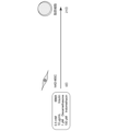
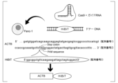
- FIG. 2 is a diagram showing a method for establishing Panc-1 cells having a HiBiT sequence downstream of the ACTB gene.
- the HiBiT peptide sequence was inserted just before the stop codon of ACTB by Cas9-mediated gene editing to generate endogenous HiBiT peptide-bound ⁇ -actin.
- EV 1.5 ⁇ g
- endocytosis inhibitors (20 ⁇ M chlorpromazine (CPZ), 10 ⁇ M nocodazole, 20 ⁇ M cytochalasin D (CytD), or 2.5 ⁇ g/mL nystatin, or incubation at 4°C).
- CPZ chlorpromazine
- CytD cytochalasin D
- nystatin 2.5 ⁇ g/mL nystatin
- Figure 2 is a graph of glycerin release from adipocytes treated with Panc-1 cell-derived EVs (1.5 ⁇ g) for 24 hours in the presence and absence of endocytosis inhibitors.
- Statistical analysis was performed using Welch's t-test.
- FIG. 1 Band intensities of phosphorylated HSL (pHSL) relative to total HSL protein levels, and total HSL and ATGL band intensities relative to ⁇ -actin protein levels are shown below the panels.
- pHSL pHSL
- P 0.87 (normal)
- ns no significant difference.
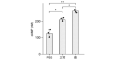
- Figure 2 is a graph showing cAMP levels in adipocytes treated for 1 hour with EV from 2.5 ⁇ L of pooled serum from 5 healthy controls (normal) or 5 pancreatic cancer patients (cancer).
- Figure 2 is a graph showing cAMP levels in EVs from 2.5 ⁇ L of pooled serum from 5 healthy controls (normal) or 5 pancreatic cancer patients (cancer).
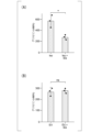
- A-B Treatment with 109 particles of EV (A) or 1 ⁇ g of EV (B) from pooled serum of 5 healthy controls (normal) or 5 pancreatic cancer patients (cancer) for 24 hours.
- 1 is a table showing the clinical characteristics of patients included in the study in this application.
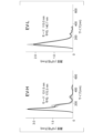
- Statistical analysis was performed using Welch's t test. It is a figure which shows the size change procedure of EV. EVs were isolated from high or low glucose media of Panc-1 cells. EV-high glucose (EV-H): EVs from high glucose culture; EV-low glucose (EV-L): EVs from low glucose culture. It is a graph showing the size distribution of EV-H and EV-L. The size of the EV-L was smaller. Representative results of at least three independent experiments are shown. Results of Western blotting of ⁇ -actin protein levels in 10 9 particles of EV (EV-H and EV-L from Panc-1 cells). Representative images of three independent experiments are shown. The band intensity of ⁇ -actin relative to the EV marker CD9 is shown.
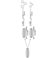
- FIG. 4 is a diagram showing a scheme for processing EV with NP40.
- EV-NP40 After EV isolation from the culture medium, EVs were treated with NP40, followed by purification on a qEV column.
- Figure 2 is a graph showing the size distribution of EV and EV-NP40 from Panc-1 cell culture medium. Representative results of at least three independent experiments are presented in mode and average.
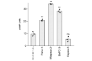
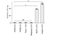
- CA19- of EVs (1 ⁇ g) from normal human pancreatic ductal epithelial cells (HPDE and HPNE) and pancreatic cancer cell lines (Panc-1, Miapaca-2, BxPC-3, and Capan-2) as determined by ELISA. It is a graph showing 9 levels. EVs derived from normal human pancreatic ductal epithelial cells, Panc-1, and Miapaca-2 cells hardly express CA19-9. Data are mean ⁇ SD (n 4). ns, no significant difference observed. ** P ⁇ 10-5 . Representative Western blotting results of CA19-9 levels in cell lysates and EVs (0.8 ⁇ g) from the indicated cell lines. Representative images of three independent experiments are displayed.
- EVs derived from Huh7 cells were AFP positive and CA19-9 negative (hepatoma cells).
- EVs derived from Capan-2 cells were AFP negative and CA19-9 positive (pancreatic cancer cells). Representative images of at least three independent experiments are displayed.
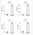
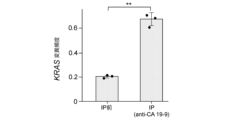
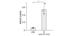
- (A) It is a graph showing the KRAS mutation frequency (KRAS mut/WT ratio: ratio of mutant type and wild type KRAS RNA frequencies) in RNA of Huh7 cell-derived EVs and Capan-2 cell-derived EVs by ddPCR.
- Huh7 has a wild-type KRAS gene
- Capan-2 has a heterozygous mutant KRAS gene.
- FIG. 2 is a schematic diagram of an in vivo experiment.
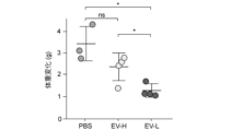
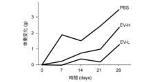
- Figure 2 is a graph showing weight change from baseline in mice 4 weeks after injection. Data are mean ⁇ SD.
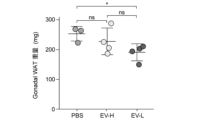
- Figure 2 is a graph showing gWAT body weight 4 weeks after injection.
- Figure 2 is a graph showing the amount of internalized EV estimated by serum, lung, and muscle luciferase activities of representative mice.
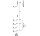
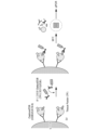
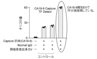
- FIG. 2 is a conceptual diagram of detection of EV surface proteins using oligo DNA. These are the results of quantitative PCR for TF.
- a substance with affinity for sugar chain antigen CA19-9 is brought into contact with a group of extracellular vesicles isolated from blood derived from a patient, and cancer cell-derived extracellular vesicles are collected from the group of extracellular vesicles.
- a method for isolating and concentrating cancer cell-derived extracellular vesicles is provided.
- the carbohydrate antigen CA19-9 is a sialyl Lea antigen recognized by the mouse monoclonal antibody NS19-9 and is known as one of the tumor markers.
- Target cancers are not particularly limited, and include breast cancer (e.g., invasive ductal carcinoma, ductal carcinoma in situ, inflammatory breast cancer, etc.), prostate cancer (e.g., hormone-dependent prostate cancer, (hormone-independent prostate cancer, etc.), pancreatic cancer (e.g., pancreatic ductal cancer, etc.), gastric cancer (e.g., papillary adenocarcinoma, mucinous adenocarcinoma, adenosquamous cell carcinoma, etc.), lung cancer (e.g., non-small cell lung cancer, small cell lung cancer, malignant mesothelioma, etc.), colon cancer (e.g., gastrointestinal stromal tumor, etc.), rectal cancer (e.g., gastrointestinal stromal tumor, etc.), colorectal cancer (e.g., familial colorectal cancer, hereditary non-polyposis colorectal cancer, gastrointestinal stromal tumor, etc.), small intestine
- epithelial ovarian cancer extragonadal germ cell tumor, ovarian germ cell tumor, ovarian low-grade tumor, etc.
- bladder cancer urethral cancer
- skin cancer e.g., intraocular (ocular) melanoma, Merkel cell carcinoma, etc.
- hemangioma malignant lymphoma (e.g. , reticular sarcoma, lymphosarcoma, Hodgkin's disease, etc.), melanoma (malignant melanoma), thyroid cancer (e.g. medullary thyroid cancer, etc.), parathyroid cancer, nasal cavity cancer, sinus cancer, bone tumor (e.g.
- osteosarcoma Ewing's tumor, uterine sarcoma, soft tissue sarcoma, etc.
- metastatic medulloblastoma angiofibroma
- dermatofibrosarcoma protuberans retinal sarcoma
- penile cancer testicular cancer
- pediatric solid tumors e.g.
- Kaposi's sarcoma Kaposi's sarcoma caused by AIDS, maxillary sinus tumor, fibrous histiocytoma, leiomyosarcoma, rhabdomyosarcoma, chronic myeloproliferative diseases, leukemia (e.g., acute myeloid leukemia, acute lymphoblastic leukemia, etc.), but is not limited to these.
- leukemia e.g., acute myeloid leukemia, acute lymphoblastic leukemia, etc.
- the substance with affinity for sugar chain antigen CA19-9 is not limited as long as it has affinity for sugar chain antigen CA19-9, and examples thereof include mucin and anti-CA19-9 antibody. and anti-CA19-9 antibodies are preferred.
- antibodies include monoclonal antibodies, polyclonal antibodies, multispecific antibodies (eg, bispecific antibodies), antibody fragments, and the like.
- the sugar chain antigen CA19-9 affinity substance further contains a carrier.
- the carrier include beads of silicon, titanium dioxide, aluminum oxide, glass, polystyrene, cellulose, polyamide, and the like.
- the following steps are specifically mentioned. First, serum isolated from patient-derived blood is centrifuged at 2000 xg for 10 minutes at 4°C, and then the supernatant is filtered using a 0.45 ⁇ m filter. Next, the supernatant after filtration is applied to a size exclusion column to obtain a group of extracellular vesicles. Anti-CA19-9 antibody-bound protein A/G magnetic beads were brought into contact with the obtained extracellular vesicles, and cancer cell-derived extracellular vesicles were isolated from the extracellular vesicles by a conventional immunoprecipitation method. Separate and concentrate.
- the precipitate obtained using the anti-CA19-9 antibody may be labeled using a label for molecules that are highly expressed in CA19-9-positive EVs.
- the label is not particularly limited, and includes a method of labeling using a labeled antibody against the molecule.
- substances used for labeling antibodies include labeling enzymes, such as horseradish peroxidase and alkaline phosphatase.
- it may be labeled using an antibody against the molecule bound to an oligonucleotide. By performing quantitative PCR on this oligonucleotide, the expression level of the molecule on EVs can be quantified.
- cancer-related genes in a group of cancer cell-derived extracellular vesicles isolated and concentrated using the above method for isolating and enriching a group of cancer cell-derived extracellular vesicles are analyzed, and Provides a method for analyzing genotypes.
- EVs derived from cells having a KRAS mutation can be isolated and concentrated.
- Cancer-related genes in the isolated and concentrated cancer cell-derived extracellular vesicles are analyzed using, for example, a next-generation sequencer.
- Oncogenes include gene groups encoding growth factors such as sis; gene groups encoding receptor-type tyrosine kinases such as erbB, fms, and ret; gene groups encoding non-receptor-type tyrosine kinases such as fes; ras, etc.
- a gene group encoding GTP/GDP binding proteins a gene group encoding serine/threonine kinases such as src, mos, and raf; a gene group encoding nuclear proteins such as myc, myb, fos, jun, and erbA; crk A group of genes encoding signal transduction adapter molecules such as Bcr-Abl; and fusion genes such as Bcr-Abl.
- oncogenes include Ras-MAP kinase pathway-related genes such as Shc, Grb2, Sos, MEK, Rho, and Rac genes; phospholipase C gamma-protein kinase C pathway-related genes such as PLC ⁇ and PKC; PI3K, Akt, and Bad. PI3K-Akt pathway-related genes such as; JAK-STAT pathway-related genes such as JAK and STAT; and GAP system pathway-related genes such as GAP, p180, and p62. Furthermore, mutations in tumor suppressor genes such as p53, Rb, and BRCA1 may be analyzed.
- This embodiment provides a drug selection method that selects a drug to be administered based on a patient's genotype analyzed using the above analysis method.
- a KRAS inhibitor can be selected.
- the drug selection method of this embodiment uses EVs isolated from blood and is therefore non-invasive.
- This embodiment is a kit for isolating and concentrating a group of cancer cell-derived extracellular vesicles from a group of extracellular vesicles isolated from patient-derived blood, and includes a substance with affinity for carbohydrate antigen CA19-9.
- the present embodiment provides an anticancer agent containing as an active ingredient an anti-CA19-9 antibody that neutralizes the function of the sugar chain antigen CA19-9.
- the anticancer agent of the present embodiment may be administered orally in the form of a tablet, coated tablet, pill, powder, granule, capsule, liquid, suspension, emulsion, or as an injection or suppository. It can also be administered parenterally in the form of external preparations for skin and the like.
- binders such as gelatin, cornstarch, gum tragacanth, and gum arabic
- excipients such as starch and crystalline cellulose
- leavening agents such as alginic acid
- solvents for injections such as water, ethanol, and glycerin
- adhesives such as rubber adhesives and silicone adhesives.
- Pharmaceutically acceptable carriers can be used alone or in combination of two or more.
- the anticancer agent of this embodiment may further contain an additive.
- Additives include lubricants such as calcium stearate and magnesium stearate; sweeteners such as sucrose, lactose, saccharin, and maltitol; flavoring agents such as peppermint and red oil; stabilizers such as benzyl alcohol and phenol; phosphoric acid. Buffers such as salts and sodium acetate; solubilizing agents such as benzyl benzoate and benzyl alcohol; antioxidants; preservatives and the like.
- the additives can be used alone or in combination of two or more.
- the method of administering the anticancer drug of this embodiment is not particularly limited, and may be determined as appropriate depending on the patient's symptoms, weight, age, sex, etc. For example, tablets, coated tablets, pills, powders, granules, capsules, solutions, suspensions, emulsions, etc. are administered orally. Injections are administered intravenously alone or mixed with normal replacement fluids such as glucose and amino acids, and further intraarterially, intramuscularly, intradermally, subcutaneously, or intraperitoneally as necessary.
- the dosage of the anticancer drug of this embodiment varies depending on the patient's symptoms, weight, age, sex, etc., and cannot be determined unconditionally, but in the case of oral administration, it is, for example, 1 ⁇ g to 10 g per day, for example, 1 ⁇ g to 10 g per day. 0.01 to 2000 mg of the active ingredient may be administered per day.
- the active ingredient may be administered in an amount of, for example, 0.1 ⁇ g to 1 g per day, for example 0.001 to 200 mg per day.
- the active ingredient may be administered in an amount of, for example, 1 ⁇ g to 10 g per day, for example 0.01 to 2000 mg per day.
- CAC is a paraneoplastic syndrome characterized by weight loss, wasting of skeletal mass, and atrophy of adipose tissue.
- CAC occurs in the majority of cancer patients, but it often appears in early-stage pancreatic cancer, which has a high mortality rate.
- Ghrelin receptor agonists are currently used in hospitals to reverse anorexia in CAC, but new treatments are needed for this multifactorial disease.
- Reduction in fat mass is an important feature of CAC, where lipolysis is activated in adipocytes and the size of adipocytes is reduced.
- other features, including muscle wasting are also characteristic of CAC, crosstalk between adipocytes and other organs suggests an important role for adipocytes in systemic metabolic complications.
- extracellular vesicles may also be involved. EVs, nanoparticles released into the bloodstream by all cells, contain bioactive factors that act as mediators of intercellular communication. Cancer cell-derived EVs are involved in the mechanisms underlying CAC, as cancer cells actively release large numbers of EVs throughout the body.
- EVs with a modal diameter of 100-150 nm were isolated from various sources, except for EVs from Capan-2 cells, which were approximately 240 nm. The number of EVs in the serum of cancer patients was higher than that of controls. Since the expression of exosome markers such as TSG101 and CD63 in EVs was confirmed, it was confirmed that EV isolation was appropriate (see Figure 1). To examine the effects of EV, mature human adipocytes were induced from human adipose-derived mesenchymal stem cells (hAD-MSCs) and subjected to EV treatment (see Figures 2 to 4).
- hAD-MSCs human adipose-derived mesenchymal stem cells
- Glycerin a surrogate marker of lipolysis levels, was released at significantly higher levels from adipocytes treated with EVs derived from Panc-1 cells, Miapapaca-2 cells, and BxPC-3 cells, and at significantly higher levels than EVs derived from Capan-2 cells. It was not observed in adipocytes treated with (see Figure 5).
- HSL phosphorylation levels and intracellular cAMP levels were significantly upregulated in adipocytes treated with EVs derived from Panc-1, Miapapaca-2, and BxPC-3 cells, and those from Capan-2 cells. It was not observed in adipocytes treated with EV (see Figures 6-7).
- total HSL and adipose triglyceride lipase (ATGL) levels were similar in all samples (see Figure 6).
- H89 PKA inhibitor
- Orlistat HSL and ATGL inhibitor
- Cay10499 HSL inhibitor
- ⁇ -actin is contained as a pan-EV marker in human serum and most EVs derived from human cell lines.
- reporter cells Panc-1-ACTB-HiBiT cells
- HiBiT peptide sequence at the 3' end of the ACTB locus of Panc-1 cells (Fig. 11, 12).
- High luciferase activity was detected in cell lysates and EVs derived from these cells, confirming the presence of ⁇ -actin-HiBiT protein in EVs (see FIGS. 13(A) and (B)).
- Luciferase values in adipocyte lysates were upregulated 1 hour after addition of EVs and were similar to those using chlorpromazine, a clathrin-mediated endocytosis inhibitor, but not with the microtube destabilizing agent.
- nocodazole an inhibitor of actin F, which is associated with macropinocytosis, and nystatin, an inhibitor of caveolae-mediated transport, all endocytic pathways were inhibited at 4°C. The case was significantly lower (see Figure 14).
- NP40 As a second method to change EV size, NP40 was added to isolated EVs at a final concentration of 0.5% (see Figure 30). Upon addition of NP40, the lipid bilayer membrane of EV was partially damaged and reduced in size (EV-NP40) (see Figure 31). Furthermore, EV-NP40 had a significantly lower luciferase value (see Figure 32(A)). This is due to loss of ⁇ -actin-HiBiT protein due to damage to the lipid bilayer membrane. However, the luciferase values of adipocyte lysates treated with the same number of EVs were significantly higher when EV-NP40 was used (see Figure 32(B)). These results confirmed that EV uptake increases as the size decreases.
- CA19-9 Carbohydrate chain antigen 19-9 (CA19-9) is a cell surface sugar chain. Although CA19-9 is produced in the normal pancreas, its synthesis is significantly increased by abnormal sialylation in the pancreas. Since EVs are produced by recycling cell membranes, CA19-9 should be present on the surface of EVs from CA19-9 positive cells. As expected, CA19-9 was abundant in the bulk serum of pancreatic cancer patients and the EVs therein (see Figure 33).
- CA19-9-positive EVs In order to establish a method to isolate CA19-9-positive EVs, we used cells derived from normal human pancreatic ductal epithelial cells (HPDE and HPNE) and CA19-9-negative pancreatic cancer cells (Panc-1 and Miapaca-2). CA19-9 negative EVs and CA19-9 negative EVs derived from pancreatic cancer cells (BxPC- and Capan-2) (see Figures 34 to 36) were used. CA19-9 positivity in EVs indicates CA19-9 positivity in cells including hepatocellular carcinoma cells (Huh7), colorectal cancer cells (HT-29), lung cancer cells (A549), and leukemia cells (HL-60). (See Figure 35.)
- Panc-1-ACTB-HiBiT cell-derived EVs (EV-H and EV-L) of different sizes were intravenously administered to mice (see FIG. 44). Mice treated with larger EVs (EV-H) showed a slight reduction in weight gain compared to controls 4 weeks after administration, whereas mice treated with smaller EVs (EV-L) showed Compared to the control, the weight gain was suppressed by 63.6% (see Figure 45). EV-L treated mice showed less weight gain (see Figure 46).
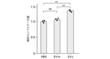
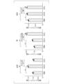
- the gonadal white adipose tissue (gWAT) of EV-L treated mice was significantly smaller in weight and size (see Figures 47 and 48). Consistent with the in vitro results, EV-L was highly taken up by gWAT, as estimated by HiBiT luciferase activity (see Figure 49). Smaller EVs (EV-L) were taken up by the lungs, but not by the muscles, more than EV-H, and EV-H levels in the serum after 3 days were higher (see Figure 50). Significantly smaller lipid droplets were seen in gWAT of EV-L treated mice (see Figure 51). Consistently, phosphorylation of HSL Ser660 in gWAT was higher in EV-L treated mice (see Figure 52).
- EVs were more taken up by adipocytes, resulting in greater induction of lipolysis.
- Cancer cells rely on glycolysis for energy production, so the tumor microenvironment of solid tumors typically has low glucose levels.
- the small size of EVs derived from pancreatic cancer cells may be due to the low glucose in the tumor microenvironment of pancreatic cancer tissues. Together with the fact that more EVs are released by cancer cells, this indicates that tumor-derived EVs in pancreatic cancer mediate systemic induction of lipolysis as a hallmark of cancer cachexia. .
- cancer can be diagnosed with high precision and non-invasively.
Landscapes
- Health & Medical Sciences (AREA)
- Chemical & Material Sciences (AREA)
- Life Sciences & Earth Sciences (AREA)
- Engineering & Computer Science (AREA)
- Organic Chemistry (AREA)
- Genetics & Genomics (AREA)
- Immunology (AREA)
- Biomedical Technology (AREA)
- Zoology (AREA)
- Wood Science & Technology (AREA)
- Biotechnology (AREA)
- General Health & Medical Sciences (AREA)
- Proteomics, Peptides & Aminoacids (AREA)
- Bioinformatics & Cheminformatics (AREA)
- Biochemistry (AREA)
- Molecular Biology (AREA)
- Microbiology (AREA)
- Analytical Chemistry (AREA)
- Pathology (AREA)
- General Engineering & Computer Science (AREA)
- Cell Biology (AREA)
- Biophysics (AREA)
- Oncology (AREA)
- Medicinal Chemistry (AREA)
- Physics & Mathematics (AREA)
- Hospice & Palliative Care (AREA)
- Hematology (AREA)
- Urology & Nephrology (AREA)
- Gastroenterology & Hepatology (AREA)
- Food Science & Technology (AREA)
- General Physics & Mathematics (AREA)
- Chemical Kinetics & Catalysis (AREA)
- General Chemical & Material Sciences (AREA)
- Nuclear Medicine, Radiotherapy & Molecular Imaging (AREA)
- Pharmacology & Pharmacy (AREA)
- Animal Behavior & Ethology (AREA)
- Public Health (AREA)
- Veterinary Medicine (AREA)
- Medicines That Contain Protein Lipid Enzymes And Other Medicines (AREA)
Abstract
Description
[1]患者由来の血液から単離された細胞外小胞群に、糖鎖抗原CA19-9親和性物質を接触させ、前記細胞外小胞群から、がん細胞由来細胞外小胞群を単離濃縮する、がん細胞由来細胞外小胞群の単離濃縮方法。
[2]前記糖鎖抗原CA19-9親和性物質は、抗CA19-9抗体である、[1]に記載のがん細胞由来細胞外小胞群の単離濃縮方法。
[3]前記糖鎖抗原CA19-9親和性物質は、更に担体を含む、[1]に記載のがん細胞由来細胞外小胞群の単離濃縮方法。
[4]前記担体は、ビーズである、[3]に記載のがん細胞由来細胞外小胞群の単離濃縮方法。
[5][1]~[4]のいずれかに記載のがん細胞由来細胞外小胞群の単離濃縮方法を用いて単離濃縮されたがん細胞由来細胞外小胞群におけるがん関連遺伝子の解析を行い、前記患者の遺伝子型を分析する、分析方法。
[6][5]に記載の分析方法を用いて分析された患者の遺伝子型に基づき、投与すべき薬剤を選択する、薬剤選択方法。
[7]患者由来の血液から単離された細胞外小胞群から、がん細胞由来細胞外小胞群を単離濃縮するキットであって、糖鎖抗原CA19-9親和性物質を含む、キット。
[8]前記糖鎖抗原CA19-9親和性物質は、抗CA19-9抗体である、[7]に記載のキット。
[9]前記糖鎖抗原CA19-9親和性物質は、担体を含む、[7]に記載のキット。
[10]前記担体は、ビーズである、[9]に記載のキット。
[11]糖鎖抗原CA19-9の機能を中和する抗CA19-9抗体を有効成分として含有する、抗がん剤。
本実施形態は、患者由来の血液から単離された細胞外小胞群に、糖鎖抗原CA19-9親和性物質を接触させ、前記細胞外小胞群から、がん細胞由来細胞外小胞群を単離濃縮する、がん細胞由来細胞外小胞群の単離濃縮方法を提供する。
糖鎖抗原CA19-9は、マウスモノクローナル抗体NS19-9で認識されるシアリルLea抗原であり、腫瘍マーカーの1つとして知られている。
得られた細胞外小胞群に抗CA19-9抗体結合プロテインA/G磁気ビーズを接触させ、通常の免疫沈降法により、細胞外小胞群から、がん細胞由来細胞外小胞群を単離濃縮する。
また、実施例において後述するように、当該分子に対する抗体にオリゴヌクレオチドが結合したものを用いて標識してもよい。このオリゴヌクレオチドに対して定量PCRを行うことにより、EV上の当該分子の発現量を定量できる。
本実施形態は、上記がん細胞由来細胞外小胞群の単離濃縮方法を用いて単離濃縮されたがん細胞由来細胞外小胞群におけるがん関連遺伝子の解析を行い、前記患者の遺伝子型を分析する方法を提供する。
がん遺伝子としては、sis等の増殖因子をコードする遺伝子群;erbB、fms、ret等のレセプター型チロシンキナーゼをコードする遺伝子群;fes等の非レセプター型チロシンキナーゼをコードする遺伝子群;ras等のGTP/GDP結合タンパク質をコードする遺伝子群;src、mos、raf等のセリン/スレオニンキナーゼをコードする遺伝子群;myc、myb、fos、jun、erbA等の核内タンパク質をコードする遺伝子群;crk等のシグナル伝達アダプター分子をコードする遺伝子群;Bcr-Abl等の融合遺伝子が挙げられる。
更に、がん遺伝子として、Shc、Grb2、Sos、MEK、Rho、Rac遺伝子等のRas-MAPキナーゼ経路関連遺伝子;PLCγ、PKC等のホスホリパーゼCガンマ-プロテインキナーゼC経路関連遺伝子;PI3K、Akt、Bad等のPI3K-Akt経路関連遺伝子;JAK、STAT等のJAK-STAT経路関連遺伝子;GAP、p180、p62等のGAP系経路関連遺伝子が挙げられる。
また、p53、Rb、BRCA1等のがん抑制遺伝子の変異を分析してもよい。
本実施形態は、上記分析方法を用いて分析された患者の遺伝子型に基づき、投与すべき薬剤を選択する、薬剤選択方法を提供する。
本実施形態は、患者由来の血液から単離された細胞外小胞群から、がん細胞由来細胞外小胞群を単離濃縮するキットであって、糖鎖抗原CA19-9親和性物質を含む、キットを提供する。キットの構成は、≪がん細胞由来細胞外小胞群の単離濃縮方法≫で述べたものと同様である。
本実施形態は、糖鎖抗原CA19-9の機能を中和する抗CA19-9抗体を有効成分として含有する、抗がん剤を提供する。
添加剤は、1種を単独で又は2種以上を混合して用いることができる。
本実施形態の抗がん剤の投与方法は特に限定されず、患者の症状、体重、年齢、性別等に応じて適宜決定すればよい。例えば、錠剤、被覆錠剤、丸剤、散剤、顆粒剤、カプセル剤、液剤、懸濁剤、乳剤等は経口投与される。また、注射剤は、単独で、又はブドウ糖、アミノ酸等の通常の補液と混合して静脈内投与され、更に必要に応じて、動脈内、筋肉内、皮内、皮下又は腹腔内投与される。
本実施形態の抗がん剤の投与量は、患者の症状、体重、年齢、性別等によって異なり、一概には決定できないが、経口投与の場合には、例えば1日あたり1μg~10g、例えば1日あたり0.01~2000mgの有効成分を投与すればよい。また、注射剤の場合には、例えば1日あたり0.1μg~1g、例えば1日あたり0.001~200mgの有効成分を投与すればよい。また、坐剤の場合には、例えば1日あたり1μg~10g、例えば1日あたり0.01~2000mgの有効成分を投与すればよい。
脂肪量の減少は、脂肪細胞で脂肪分解が活性化され、脂肪細胞のサイズが縮小するCACの重要な特徴である。筋肉の消耗を含む他の特徴は、CACの特徴でもあるが、脂肪細胞と他の臓器との間のクロストークは、全身性代謝合併症における脂肪細胞の重要な役割を示唆している。
約240 nmのCapan-2細胞由来のEVを除き、さまざまなソースからモード径が100~150nmのEVを単離した。がん患者の血清中のEVの数は、対照よりも多かった。EVにおけるTSG101及びCD63などのエキソソームマーカーの発現が確認されたことから、EV単離が適切であることが確認された(図1参照。)。EVの効果を調べるために、成熟したヒト脂肪細胞を、ヒト脂肪由来間葉系幹細胞(hAD-MSC)から誘導し、EV処理を行った(図2~4参照。)。脂肪分解レベルの代替マーカーであるグリセリンは、Panc-1細胞、Miapapaca-2細胞、及びBxPC-3細胞由来のEVで処理した脂肪細胞から有意に高いレベルで放出され、Capan-2細胞由来のEVで処理した脂肪細胞からは観察されなかった(図5参照。)。
次に、膵臓がん患者の血清から分離されたEVがin vitroで脂肪分解を誘導するかどうかを調べた。健常者のコントロールからのEVもわずかに脂肪細胞からのグリセリン放出を誘導したが、膵臓がん患者のEVは、脂肪分解を大幅に誘導した(図16(A)参照。)。膵臓がん患者のEVで処理された脂肪細胞は、一貫して、HSLのリン酸化レベルと細胞内cAMPレベルが有意に高かった(図17参照。)。一方、健常者のコントロールからのEVで処理された脂肪細胞も同様であったが、観察されたレベルは低かった (図16(A)(B)、図17参照。)。また、健常者のコントロールからのEV自体と患者からのEV自体との間でcAMPレベルに有意差は観察されなかった(図18参照。)。がん患者の血清は、健常対照者よりも多くのEVを含んでいたが、EV数またはタンパク質重量を調整すると、結果は同様であった(図19(A)(B)参照。)。したがって、がん患者のEVは脂肪分解を誘発することが確認された。
次に、個々の症例のEVを調べたところ、ほぼすべての膵臓がん症例で体重が大幅に減少していた (図20参照。)。対照例からのEVも脂肪分解を誘発したが、膵臓がん患者からのEVは大幅に脂肪分解を誘発した(図21(A)参照。)。ドナー間でEVのcAMPレベルに有意な差は無かった(図21(B)参照。)。さらに、コントロールおよびがん症例からのEVのcAMPレベルは、脂肪分解レベルと相関しなかった(図22(A)参照。)。むしろ、脂肪分解レベルは、コントロールと膵臓がんの両方の症例でEVの差分サイズと有意に相関していた(P = 1.32×10⁻⁵、r = -0.81297)(図22(B)参照。)。重要なことに、膵臓がん患者の血清は、健常コントロールのものよりも有意に少なかった(図23参照。)。これらの結果は、ヒト脂肪細胞における脂肪分解の誘導は、EVサイズの減少とともに程度が増加することを示唆している。
EVサイズを変更するために2つの方法を適用した。低グルコース培地で培養した細胞からのEV(EV-Low: EV-L)は、高グルコース培地で培養された細胞からのEV(EV-High:EV-H)よりも有意に小さかった(図24、25参照。)。低グルコース培地又は高グルコース培地で培養したPanc-1-ACTB-HiBiT細胞由来のEVにおいて、β-アクチンタンパク質、HiBiTペプチド、cAMPレベルには有意差はなく(図26、27(A)(B)参照。)、EV-HとEV-Lは、サイズ以外は同等であった。しかしながら、EVで処理した脂肪細胞ライセートからのルシフェラーゼ値は、EV-Lを使用した場合に、有意に高かった(図27(C)参照。)。これは、EVの取り込みが、サイズが小さくなるにつれて増加することを示唆する。同様に、蛍光標識されたEV-Lは、脂肪細胞によってより多く取り込まれ(図28参照。)、脂肪分解誘導の有意な増加を示した(図29参照。)。
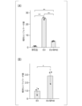
がん細胞由来のEVを特徴付けるために、免疫沈降(IP)によって特定のEV集団を単離した。糖鎖抗原19-9(CA19-9)は、細胞表面糖鎖である。CA19-9は正常な膵臓でも産生されるが、その合成は、膵臓における異常なシアリル化によって著しく増加する。EVは細胞膜のリサイクルによって生成されるため、CA19-9 はCA19-9陽性細胞からのEVの表面に存在するはずである。予想通り、CA19-9は、膵臓がん患者のバルク血清およびその中のEVに豊富に含まれていた(図33参照。)。CA19-9陽性EVを単離する方法を確立するために、正常なヒト膵管上皮細胞(HPDE 及び HPNE)、及びCA19-9-陰性の膵臓がん細胞 (Panc-1及びMiapaca-2)由来のCA19-9陰性EV、並びに膵臓がん細胞(BxPC-及びCapan-2)由来のCA19-9陰性EV(図34~36参照。)を使用した。EVにおけるCA19-9陽性は、肝細胞がん細胞(Huh7)、結腸直腸がん細胞(HT-29)、肺がん細胞(A549)、及び白血病細胞(HL-60)を含む細胞でのCA19-9の発現に比例していた(図35参照。)。
上記の結果をin vivoで確認するために、マウスに異なるサイズのPanc-1-ACTB-HiBiT細胞由来EV(EV-H及びEV-L)を静脈投与した(図44参照。)。より大きなEV(EV-H)で処理されたマウスは、投与4週間後にコントロールと比較してわずかな体重増加の抑制を示したが、より小さなEV(EV-L)で処理されたマウスは、コントロールと比較して、63.6%の体重増加の抑制を示した(図45参照。)。EV-L処理マウスは体重増加が少ないことを示した(図46参照。)。EV-L処理マウスの性腺白色脂肪組織(gWAT)は、重さとサイズが大幅に小さかった(図47、48参照。)。In vitroの結果と一致して、HiBiTルシフェラーゼ活性によって推定されるように(図49参照。)、EV-LはgWATにより多く取り込まれた。より小さなEV(EV-L)は、EV-Hよりも肺に取り込まれるが、筋肉には取り込まれず、3 日後の血清中のEV-Hレベルは高かった(図50参照。)。有意により小さな脂肪滴が、EV-L処理マウスのgWATに見られた(図51参照。)。一貫して、gWATにおけるHSL Ser660のリン酸化は、EV-L処理マウスの方が高かった(図52参照。)。したがって、小さいEVほど脂肪細胞に多く取り込まれ、脂肪分解のより大きな誘導をもたらした。がん細胞は、エネルギー生産のために解糖に依存しているため、一般に、固形がんの腫瘍微小環境は、グルコースレベルが低い。膵臓がん細胞由来のEVが小さいのは、膵臓がん組織の腫瘍微小環境における低グルコースによる可能性がある。より多くのEVががん細胞によって放出されるという事実とともに、これは、膵臓がんにおける腫瘍由来のEVが、がん悪液質の特徴としての脂肪分解の全身の誘導を媒介することを示す。
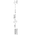
抗CA19-9抗体結合ビーズとEVをincubationして、抗CA19-9抗体結合ビーズとEVとの第1の複合体を形成した。次いで、この第1の複合体に抗Tissue Factor(TF)抗体結合オリゴを添加し、ビーズに結合したEVと抗TF抗体を結合させ、第2の複合体を形成させた。この第2の複合体に対して定量PCRを行った(図53参照。)。この定量PCRでの増幅産物量がもともとのEVに結合していたDetect抗体の量、すなわち、EV上に発現する表面抗原(今回はTF)の量に比例する。結果を図54に示す。CA19-9陽性EVでTFが高発現していることが確認された。
Claims (11)
- 患者由来の血液から単離された細胞外小胞群に、糖鎖抗原CA19-9親和性物質を接触させ、前記細胞外小胞群から、がん細胞由来細胞外小胞群を単離濃縮する、がん細胞由来細胞外小胞群の単離濃縮方法。
- 前記糖鎖抗原CA19-9親和性物質は、抗CA19-9抗体である、請求項1に記載のがん細胞由来細胞外小胞群の単離濃縮方法。
- 前記糖鎖抗原CA19-9親和性物質は、更に担体を含む、請求項1に記載のがん細胞由来細胞外小胞群の単離濃縮方法。
- 前記担体は、ビーズである、請求項3に記載のがん細胞由来細胞外小胞群の単離濃縮方法。
- 請求項1~4のいずれか一項に記載のがん細胞由来細胞外小胞群の単離濃縮方法を用いて単離濃縮されたがん細胞由来細胞外小胞群におけるがん関連遺伝子の解析を行い、前記患者の遺伝子型を分析する、分析方法。
- 請求項5に記載の分析方法を用いて分析された患者の遺伝子型に基づき、投与すべき薬剤を選択する、薬剤選択方法。
- 患者由来の血液から単離された細胞外小胞群から、がん細胞由来細胞外小胞群を単離濃縮するキットであって、糖鎖抗原CA19-9親和性物質を含む、キット。
- 前記糖鎖抗原CA19-9親和性物質は、抗CA19-9抗体である、請求項7に記載のキット。
- 前記糖鎖抗原CA19-9親和性物質は、担体を含む、請求項7に記載のキット。
- 前記担体は、ビーズである、請求項9に記載のキット。
- 糖鎖抗原CA19-9の機能を中和する抗CA19-9抗体を有効成分として含有する、抗がん剤。
Priority Applications (2)
| Application Number | Priority Date | Filing Date | Title |
|---|---|---|---|
| JP2024517344A JPWO2023210644A1 (ja) | 2022-04-26 | 2023-04-25 | |
| US18/857,897 US20250109388A1 (en) | 2022-04-26 | 2023-04-25 | Method for isolating and concentrating extracellular vesicle group derived from cancer cell, analysis method, drug selection method, kit, and anti-cancer agent |
Applications Claiming Priority (2)
| Application Number | Priority Date | Filing Date | Title |
|---|---|---|---|
| US202263334672P | 2022-04-26 | 2022-04-26 | |
| US63/334,672 | 2022-04-26 |
Publications (1)
| Publication Number | Publication Date |
|---|---|
| WO2023210644A1 true WO2023210644A1 (ja) | 2023-11-02 |
Family
ID=88519097
Family Applications (1)
| Application Number | Title | Priority Date | Filing Date |
|---|---|---|---|
| PCT/JP2023/016313 Ceased WO2023210644A1 (ja) | 2022-04-26 | 2023-04-25 | がん細胞由来細胞外小胞群の単離濃縮方法、分析方法、薬剤選択方法、キット、及び抗がん剤 |
Country Status (3)
| Country | Link |
|---|---|
| US (1) | US20250109388A1 (ja) |
| JP (1) | JPWO2023210644A1 (ja) |
| WO (1) | WO2023210644A1 (ja) |
Citations (2)
| Publication number | Priority date | Publication date | Assignee | Title |
|---|---|---|---|---|
| JP2017501694A (ja) * | 2013-12-04 | 2017-01-19 | ボード・オブ・リージエンツ,ザ・ユニバーシテイ・オブ・テキサス・システム | 診断および療法のためのエキソソーム中のゲノムdna、rnaおよびタンパク質の分析 |
| JP2021511510A (ja) * | 2018-01-18 | 2021-05-06 | ナノソミックス・インコーポレイテッドNanoSomiX, Inc. | 疾患および障害の診断および予後診断のためのエキソソームおよびエキソソームバイオマーカーの検出 |
-
2023
- 2023-04-25 JP JP2024517344A patent/JPWO2023210644A1/ja active Pending
- 2023-04-25 WO PCT/JP2023/016313 patent/WO2023210644A1/ja not_active Ceased
- 2023-04-25 US US18/857,897 patent/US20250109388A1/en active Pending
Patent Citations (2)
| Publication number | Priority date | Publication date | Assignee | Title |
|---|---|---|---|---|
| JP2017501694A (ja) * | 2013-12-04 | 2017-01-19 | ボード・オブ・リージエンツ,ザ・ユニバーシテイ・オブ・テキサス・システム | 診断および療法のためのエキソソーム中のゲノムdna、rnaおよびタンパク質の分析 |
| JP2021511510A (ja) * | 2018-01-18 | 2021-05-06 | ナノソミックス・インコーポレイテッドNanoSomiX, Inc. | 疾患および障害の診断および予後診断のためのエキソソームおよびエキソソームバイオマーカーの検出 |
Non-Patent Citations (5)
| Title |
|---|
| JAVEED NAUREEN, SAGAR GUNISHA, DUTTA SHAMIT K., SMYRK THOMAS C., LAU JULIE S., BHATTACHARYA SANTANU, TRUTY MARK, PETERSEN GLORIA M: "Pancreatic Cancer–Derived Exosomes Cause Paraneoplastic β-cell Dysfunction", CLINICAL CANCER RESEARCH, ASSOCIATION FOR CANCER RESEARCH, US, vol. 21, no. 7, 1 April 2015 (2015-04-01), US, pages 1722 - 1733, XP093104199, ISSN: 1078-0432, DOI: 10.1158/1078-0432.CCR-14-2022 * |
| SANCHO-ALBERO MARÍA, SEBASTIÁN VÍCTOR, SESÉ JAVIER, PAZO-CID ROBERTO, MENDOZA GRACIA, ARRUEBO MANUEL, MARTÍN-DUQUE PILAR, SANTAMAR: "Isolation of exosomes from whole blood by a new microfluidic device: proof of concept application in the diagnosis and monitoring of pancreatic cancer", JOURNAL OF NANOBIOTECHNOLOGY, vol. 18, no. 1, 1 December 2020 (2020-12-01), XP093104197, DOI: 10.1186/s12951-020-00701-7 * |
| SAWADA R, SUN S M, WU X, HONG F, RAGUPATHI G, LIVINGSTON P O, SCHOLZ W W.: "Human monoclonal antibodies to sialyl-Lewis (CA19.9) with potent CDC, ADCC, and antitumor activity.", CLINICAL CANCER RESEARCH, ASSOCIATION FOR CANCER RESEARCH, US, vol. 17, no. 5, 1 March 2011 (2011-03-01), US, pages 1024 - 1032, XP002736040, ISSN: 1078-0432, DOI: 10.1158/1078-0432.CCR-10-2640 * |
| SHIBATA CHIKAKO, OTSUKA MOTOYUKI, SEIMIYA TAKAHIRO, KISHIKAWA TAKAHIRO, ISHIGAKI KAZUNAGA, FUJISHIRO MITSUHIRO: "Lipolysis by pancreatic cancer‐derived extracellular vesicles in cancer‐associated cachexia via specific integrins", CLINICAL AND TRANSLATIONAL MEDICINE, INTERNATIONAL SOCIETY FOR TRANSLATIONAL MEDICINE, SE, vol. 12, no. 11, 1 November 2022 (2022-11-01), SE , XP093104201, ISSN: 2001-1326, DOI: 10.1002/ctm2.1089 * |
| ZHOU SISI, HU TAO, HAN GAOHUA, WU YAFENG, HUA XIN, SU JUAN, JIN WEIWEI, MOU YIPING, MOU XIAOZHOU, LI QUAN, LIU SONGQIN: "Accurate Cancer Diagnosis and Stage Monitoring Enabled by Comprehensive Profiling of Different Types of Exosomal Biomarkers: Surface Proteins and miRNAs", SMALL, WILEY, HOBOKEN, USA, vol. 16, no. 48, 1 December 2020 (2020-12-01), Hoboken, USA, XP093104194, ISSN: 1613-6810, DOI: 10.1002/smll.202004492 * |
Also Published As
| Publication number | Publication date |
|---|---|
| US20250109388A1 (en) | 2025-04-03 |
| JPWO2023210644A1 (ja) | 2023-11-02 |
Similar Documents
| Publication | Publication Date | Title |
|---|---|---|
| Qin et al. | BRD4 promotes gastric cancer progression and metastasis through acetylation-dependent stabilization of snail | |
| Li et al. | HER2-mediated internalization of cytotoxic agents in ERBB2 amplified or mutant lung cancers | |
| Liu et al. | Crizotinib-induced immunogenic cell death in non-small cell lung cancer | |
| Liu et al. | Rosmarinic acid in combination with ginsenoside Rg1 suppresses colon cancer metastasis via co-inhition of COX-2 and PD1/PD-L1 signaling axis | |
| Luo et al. | A20 promotes colorectal cancer immune evasion by upregulating STC1 expression to block “eat-me” signal | |
| Wang et al. | Methylation of HSP70 orchestrates its binding to and stabilization of BCL2 mRNA and renders pancreatic cancer cells resistant to therapeutics | |
| Wang et al. | Berberine inhibits proliferation and down-regulates epidermal growth factor receptor through activation of Cbl in colon tumor cells | |
| Pai et al. | Gut microbial transcytosis induced by tumour necrosis factor-like 1A-dependent activation of a myosin light chain kinase splice variant contributes to inflammatory bowel disease | |
| Zheng et al. | Targeting LUNX inhibits non–small cell lung cancer growth and metastasis | |
| Olayioye et al. | The phosphoprotein StarD10 is overexpressed in breast cancer and cooperates with ErbB receptors in cellular transformation | |
| Huang et al. | GP73-dependent regulation of exosome biogenesis promotes colorectal cancer liver metastasis | |
| Piao et al. | Insulin-like growth factor-I receptor blockade by a specific tyrosine kinase inhibitor for human gastrointestinal carcinomas | |
| Peng et al. | Identification of Bruceine A as a novel HSP90AB1 inhibitor for suppressing hepatocellular carcinoma growth | |
| Hu et al. | Safety, efficacy and pharmacokinetics of BPI-9016M in c-MET overexpression or MET exon 14 skipping mutation patients with locally advanced or metastatic non-small-cell lung cancer: a phase Ib study | |
| Wang et al. | Rasal2 inhibits autophagic-exosomes secretion via regulating Rab27a in triple-negative breast cancer progression | |
| EP1556699B1 (fr) | Procede de dosage du ngf pour le diagnostic in vitro du cancer du sein et utilisation en therapie | |
| US20250109388A1 (en) | Method for isolating and concentrating extracellular vesicle group derived from cancer cell, analysis method, drug selection method, kit, and anti-cancer agent | |
| US20130253039A1 (en) | Colorectal cancer treatments and diagnostic improvements | |
| KR101928618B1 (ko) | Tk1 단백질의 발현이 항진된 결장직장암 환자에 대한 치료효과 예측 방법 | |
| Tian et al. | Oncogenic GOPC-ROS1 fusion promotes the formation of lymphatic malformations by impairing tight junction and its crizotinib resistance through repressing ferroptosis | |
| CN116125070B (zh) | Trim21作为进展期结肠癌的诊断标志物及其应用 | |
| Wu et al. | Neoadjuvant sintilimab, albumin-bound paclitaxel, and carboplatin for locally advanced, resectable esophageal squamous cell carcinoma: clinical study and mechanistic exploration | |
| Hufnagel et al. | 4-(N)-Docosahexaenoyl 2’, 2’-difluorodeoxycytidine induces immunogenic cell death in colon and pancreatic carcinoma models as a single agent | |
| Fang et al. | SMG5 Inhibition restrains hepatocellular carcinoma growth and enhances Sorafenib sensitivity | |
| Sgambellone et al. | Role of histamine H4 receptor in the anti-inflammatory pathway of glucocorticoid-induced leucin zipper (GILZ) in a model of lung fibrosis |
Legal Events
| Date | Code | Title | Description |
|---|---|---|---|
| 121 | Ep: the epo has been informed by wipo that ep was designated in this application |
Ref document number: 23796384 Country of ref document: EP Kind code of ref document: A1 |
|
| ENP | Entry into the national phase |
Ref document number: 2024517344 Country of ref document: JP Kind code of ref document: A |
|
| WWE | Wipo information: entry into national phase |
Ref document number: 18857897 Country of ref document: US |
|
| NENP | Non-entry into the national phase |
Ref country code: DE |
|
| WWP | Wipo information: published in national office |
Ref document number: 18857897 Country of ref document: US |
|
| 122 | Ep: pct application non-entry in european phase |
Ref document number: 23796384 Country of ref document: EP Kind code of ref document: A1 |