WO2023135704A1 - 捕集方法、検査方法、容器、遠心装置および検査システム - Google Patents
捕集方法、検査方法、容器、遠心装置および検査システム Download PDFInfo
- Publication number
- WO2023135704A1 WO2023135704A1 PCT/JP2022/000884 JP2022000884W WO2023135704A1 WO 2023135704 A1 WO2023135704 A1 WO 2023135704A1 JP 2022000884 W JP2022000884 W JP 2022000884W WO 2023135704 A1 WO2023135704 A1 WO 2023135704A1
- Authority
- WO
- WIPO (PCT)
- Prior art keywords
- container
- solution
- sample
- centrifugal force
- flow path
- Prior art date
- Legal status (The legal status is an assumption and is not a legal conclusion. Google has not performed a legal analysis and makes no representation as to the accuracy of the status listed.)
- Ceased
Links
Images




Classifications
-
- C—CHEMISTRY; METALLURGY
- C12—BIOCHEMISTRY; BEER; SPIRITS; WINE; VINEGAR; MICROBIOLOGY; ENZYMOLOGY; MUTATION OR GENETIC ENGINEERING
- C12Q—MEASURING OR TESTING PROCESSES INVOLVING ENZYMES, NUCLEIC ACIDS OR MICROORGANISMS; COMPOSITIONS OR TEST PAPERS THEREFOR; PROCESSES OF PREPARING SUCH COMPOSITIONS; CONDITION-RESPONSIVE CONTROL IN MICROBIOLOGICAL OR ENZYMOLOGICAL PROCESSES
- C12Q1/00—Measuring or testing processes involving enzymes, nucleic acids or microorganisms; Compositions therefor; Processes of preparing such compositions
- C12Q1/02—Measuring or testing processes involving enzymes, nucleic acids or microorganisms; Compositions therefor; Processes of preparing such compositions involving viable microorganisms
- C12Q1/24—Methods of sampling, or inoculating or spreading a sample; Methods of physically isolating an intact microorganisms
-
- B—PERFORMING OPERATIONS; TRANSPORTING
- B01—PHYSICAL OR CHEMICAL PROCESSES OR APPARATUS IN GENERAL
- B01L—CHEMICAL OR PHYSICAL LABORATORY APPARATUS FOR GENERAL USE
- B01L3/00—Containers or dishes for laboratory use, e.g. laboratory glassware; Droppers
- B01L3/50—Containers for the purpose of retaining a material to be analysed, e.g. test tubes
- B01L3/502—Containers for the purpose of retaining a material to be analysed, e.g. test tubes with fluid transport, e.g. in multi-compartment structures
- B01L3/5021—Test tubes specially adapted for centrifugation purposes
-
- G—PHYSICS
- G01—MEASURING; TESTING
- G01N—INVESTIGATING OR ANALYSING MATERIALS BY DETERMINING THEIR CHEMICAL OR PHYSICAL PROPERTIES
- G01N1/00—Sampling; Preparing specimens for investigation
- G01N1/28—Preparing specimens for investigation including physical details of (bio-)chemical methods covered elsewhere, e.g. G01N33/50, C12Q
- G01N1/34—Purifying; Cleaning
-
- G—PHYSICS
- G01—MEASURING; TESTING
- G01N—INVESTIGATING OR ANALYSING MATERIALS BY DETERMINING THEIR CHEMICAL OR PHYSICAL PROPERTIES
- G01N1/00—Sampling; Preparing specimens for investigation
- G01N1/28—Preparing specimens for investigation including physical details of (bio-)chemical methods covered elsewhere, e.g. G01N33/50, C12Q
- G01N1/40—Concentrating samples
- G01N1/4077—Concentrating samples by other techniques involving separation of suspended solids
-
- G—PHYSICS
- G01—MEASURING; TESTING
- G01N—INVESTIGATING OR ANALYSING MATERIALS BY DETERMINING THEIR CHEMICAL OR PHYSICAL PROPERTIES
- G01N35/00—Automatic analysis not limited to methods or materials provided for in any single one of groups G01N1/00 - G01N33/00; Handling materials therefor
-
- B—PERFORMING OPERATIONS; TRANSPORTING
- B01—PHYSICAL OR CHEMICAL PROCESSES OR APPARATUS IN GENERAL
- B01L—CHEMICAL OR PHYSICAL LABORATORY APPARATUS FOR GENERAL USE
- B01L2200/00—Solutions for specific problems relating to chemical or physical laboratory apparatus
- B01L2200/06—Fluid handling related problems
- B01L2200/0621—Control of the sequence of chambers filled or emptied
-
- B—PERFORMING OPERATIONS; TRANSPORTING
- B01—PHYSICAL OR CHEMICAL PROCESSES OR APPARATUS IN GENERAL
- B01L—CHEMICAL OR PHYSICAL LABORATORY APPARATUS FOR GENERAL USE
- B01L2200/00—Solutions for specific problems relating to chemical or physical laboratory apparatus
- B01L2200/16—Reagents, handling or storing thereof
-
- B—PERFORMING OPERATIONS; TRANSPORTING
- B01—PHYSICAL OR CHEMICAL PROCESSES OR APPARATUS IN GENERAL
- B01L—CHEMICAL OR PHYSICAL LABORATORY APPARATUS FOR GENERAL USE
- B01L2300/00—Additional constructional details
- B01L2300/04—Closures and closing means
- B01L2300/041—Connecting closures to device or container
- B01L2300/044—Connecting closures to device or container pierceable, e.g. films, membranes
-
- B—PERFORMING OPERATIONS; TRANSPORTING
- B01—PHYSICAL OR CHEMICAL PROCESSES OR APPARATUS IN GENERAL
- B01L—CHEMICAL OR PHYSICAL LABORATORY APPARATUS FOR GENERAL USE
- B01L2300/00—Additional constructional details
- B01L2300/06—Auxiliary integrated devices, integrated components
- B01L2300/0681—Filter
-
- B—PERFORMING OPERATIONS; TRANSPORTING
- B01—PHYSICAL OR CHEMICAL PROCESSES OR APPARATUS IN GENERAL
- B01L—CHEMICAL OR PHYSICAL LABORATORY APPARATUS FOR GENERAL USE
- B01L2300/00—Additional constructional details
- B01L2300/08—Geometry, shape and general structure
- B01L2300/0861—Configuration of multiple channels and/or chambers in a single devices
- B01L2300/0864—Configuration of multiple channels and/or chambers in a single devices comprising only one inlet and multiple receiving wells, e.g. for separation, splitting
-
- B—PERFORMING OPERATIONS; TRANSPORTING
- B01—PHYSICAL OR CHEMICAL PROCESSES OR APPARATUS IN GENERAL
- B01L—CHEMICAL OR PHYSICAL LABORATORY APPARATUS FOR GENERAL USE
- B01L2400/00—Moving or stopping fluids
- B01L2400/04—Moving fluids with specific forces or mechanical means
- B01L2400/0403—Moving fluids with specific forces or mechanical means specific forces
- B01L2400/0409—Moving fluids with specific forces or mechanical means specific forces centrifugal forces
-
- B—PERFORMING OPERATIONS; TRANSPORTING
- B01—PHYSICAL OR CHEMICAL PROCESSES OR APPARATUS IN GENERAL
- B01L—CHEMICAL OR PHYSICAL LABORATORY APPARATUS FOR GENERAL USE
- B01L2400/00—Moving or stopping fluids
- B01L2400/06—Valves, specific forms thereof
- B01L2400/0688—Valves, specific forms thereof surface tension valves, capillary stop, capillary break
-
- G—PHYSICS
- G01—MEASURING; TESTING
- G01N—INVESTIGATING OR ANALYSING MATERIALS BY DETERMINING THEIR CHEMICAL OR PHYSICAL PROPERTIES
- G01N1/00—Sampling; Preparing specimens for investigation
- G01N1/28—Preparing specimens for investigation including physical details of (bio-)chemical methods covered elsewhere, e.g. G01N33/50, C12Q
- G01N1/40—Concentrating samples
- G01N1/4044—Concentrating samples by chemical techniques; Digestion; Chemical decomposition
-
- G—PHYSICS
- G01—MEASURING; TESTING
- G01N—INVESTIGATING OR ANALYSING MATERIALS BY DETERMINING THEIR CHEMICAL OR PHYSICAL PROPERTIES
- G01N1/00—Sampling; Preparing specimens for investigation
- G01N1/28—Preparing specimens for investigation including physical details of (bio-)chemical methods covered elsewhere, e.g. G01N33/50, C12Q
- G01N1/40—Concentrating samples
- G01N1/4077—Concentrating samples by other techniques involving separation of suspended solids
- G01N2001/4083—Concentrating samples by other techniques involving separation of suspended solids sedimentation
-
- G—PHYSICS
- G01—MEASURING; TESTING
- G01N—INVESTIGATING OR ANALYSING MATERIALS BY DETERMINING THEIR CHEMICAL OR PHYSICAL PROPERTIES
- G01N1/00—Sampling; Preparing specimens for investigation
- G01N1/28—Preparing specimens for investigation including physical details of (bio-)chemical methods covered elsewhere, e.g. G01N33/50, C12Q
- G01N1/40—Concentrating samples
- G01N1/4077—Concentrating samples by other techniques involving separation of suspended solids
- G01N2001/4088—Concentrating samples by other techniques involving separation of suspended solids filtration
-
- G—PHYSICS
- G01—MEASURING; TESTING
- G01N—INVESTIGATING OR ANALYSING MATERIALS BY DETERMINING THEIR CHEMICAL OR PHYSICAL PROPERTIES
- G01N35/00—Automatic analysis not limited to methods or materials provided for in any single one of groups G01N1/00 - G01N33/00; Handling materials therefor
- G01N2035/00465—Separating and mixing arrangements
- G01N2035/00495—Centrifuges
Definitions
- the present invention relates to collection methods, inspection methods, containers, centrifugal devices, and inspection systems. More specifically, the present invention provides a method for collecting microorganisms, a method for testing microorganisms, a container, and a centrifuge for separating a sample containing impurities such as blood cells into a solution containing microorganisms such as bacteria and a solution containing blood cells. It relates to equipment and inspection systems.
- Sepsis is an infectious disease with a high fatality rate, and it is important to provide appropriate treatment promptly.
- Diagnosis of sepsis usually involves a blood culture test to determine whether bacteria are present in blood, which is a sterile sample.
- a smear test is performed after that, the blood culture-positive sample is isolated and cultured, and the obtained colonies are subjected to an identification test to identify the type of bacteria, and a susceptibility test to measure the susceptibility of the bacteria to antibacterial drugs. done.
- the series of tests described above requires 1 day for the blood culture test, 1 day for the isolation culture, and 1 day for the susceptibility test, thus requiring a total of 2 to 3 days. Therefore, it currently takes 2 to 3 days to find out whether treatment with appropriate antimicrobials has been carried out, and if ineffective antimicrobials are administered, the fatality rate is extremely high. .
- the isolation culture is still one of the time-consuming inspection steps, and it is a bottleneck in shortening the inspection time.
- the separation and culture process can be omitted, leading to a reduction in examination time.
- an identification test and a susceptibility test are performed as a set in a normal bacteriological test, it is necessary to prepare a sample suitable for both tests when extracting bacteria from a blood culture-positive sample. If a sample suitable for both inspections can be obtained in a single operation, it will lead to shortening of inspection time and labor saving of inspection, which will greatly benefit the user.
- an identification test consider using mass spectrometry by MALDI-TOF, which is becoming more popular.
- MALDI mass spectrometry by MALDI-TOF, which is becoming more popular.
- a dry sample is irradiated with a laser to desorb and ionize the sample. Therefore, it is preferable that the amount of water in the sample is small, and from the viewpoint of detection sensitivity, the amount of bacteria required is 10 5 CFU (Colony forming unit) or more.
- a liquid containing live bacteria is used as a sample in the turbidimetric method or the rapid method utilizing a microscope that is currently being developed.
- a bacterial solution whose concentration is adjusted in the range of 10 5 to 10 6 CFU/mL is used.
- McFarland turbidimetric method is generally used . It is necessary to prepare ⁇ L. Therefore, a larger amount of bacteria is required than in the identification test.
- bacteria are extracted from a relatively large volume of blood culture-positive samples of several mL or more, and multiple samples with different solid and liquid forms and different amounts of bacteria are prepared. It is necessary to get it at once.
- Patent Document 1 discloses a technique for separating bacteria from blood samples and extracting bacterial proteins. Further, Patent Document 2 discloses a method of selectively destroying only blood cell components using a protease-mediated decomposition of blood cells, a swelling treatment with a hypotonic solution, and a surfactant. Further, Patent Document 3 discloses a technique for separating a specific component from a blood sample.
- samples prepared by this method do not contain live bacteria and cannot be used for susceptibility testing because they are treated with reagents that disrupt bacterial cell walls.
- Patent Document 3 it is possible to easily separate a specific component from a blood sample in one step by rotating it like a top. However, it is difficult to obtain samples in different solid and liquid forms for use in a series of bacteriological tests. In addition, since a filter such as a porous material is used to separate a blood sample, it is not suitable for rapid processing of samples of several mL to 10 mL due to the problem of clogging.
- Patent Document 1 and Patent Document 2 disclose methods for extracting bacteria from blood samples. It is not possible to obtain a large number of high-concentration bacterial samples at once.
- the method disclosed in Patent Document 3 allows pretreatment of a blood sample in a simple manner, but there is a problem in obtaining samples of different solid and liquid forms from a large volume of sample at once. was there.
- the present invention has been made in view of such circumstances, and provides a collection method, an inspection method, a container, a centrifugal device, and an inspection system for obtaining samples of different shapes from a sample at once by a simple and stable method. It is something to do.
- the collection method of the present invention includes a first storage unit having a first flow path and a second flow path with different heights on the side surface, and a first storage unit and the first storage unit.
- a sample in a first storage part of a container comprising a second storage part connected via a flow path and a third storage part connected to the first storage part and the second storage part via the second flow path and centrifuging the sample introduced into the first storage unit with a first centrifugal force to form a first solution containing the first component and a second solution containing the second component
- FIG. 2 is a cross-sectional view of the container of Example 1.
- FIG. 2 is a transition diagram of blood samples in containers, showing a method of obtaining samples with different forms from the blood samples introduced into the container of Example 1.
- FIG. 2 is a graph showing the relationship between centrifugal acceleration and time in a method for obtaining samples of different morphologies from blood samples.
- FIG. 2 is a block diagram showing the configuration of the centrifugal device of Example 1.
- FIG. 1 is a flow chart showing a method of obtaining different morphology samples from a blood sample and tests using the samples.
- FIG. 11 is a block diagram showing the configuration of an inspection system of Example 2;
- a collection method for simultaneously obtaining samples with different morphologies from a blood sample containing microorganisms containing impurities such as blood cells, and a first component from the sample obtained by this collection method are examined. I will explain the inspection method of. Further, in the following examples, a container used in the collection method, a centrifugal device including the container, and an inspection system including the centrifugal device will be described.
- Microorganisms to be collected refer to various types of microorganisms, including bacteria, actinomycetes, and fungi. However, microorganisms do not include viruses. Specifically, microorganisms that are subject to detection by the sterility test method in the pharmacopeia, pathogenic bacteria and pathogenic fungi that are subject to inspection in hospital laboratories, and the like.
- Escherichia specifically examples, Escherichia coli
- Staphylococcus specifically examples, Staphylococcus aureus, Staphylococcus epidermidis
- Propionibacterium specifically examples, Proprio Novactor acnes (Proprionobacter acnes)
- Micrococcus Streptococcus (specifically, Streptococcus pyogenes, Streptococcus pneumoniae)
- Enterococcus specifically, Enterococcus faecium
- Neisseria e.g., Rhinococcus, Meningococcus
- Moraxella Shigella (e.g., Shigella), Salmonella )
- Salmonella specifically examples are Salmonella typhi, Paratyphoid A, Enteritidis
- Enterobacter Serratia (Specific examples include Serratia marc
- Bacteroides Bacteroides fragilis
- Fusobacterium, Mycobacterium as a specific example, Mycobacterium tuberculosis
- Campylobacter Helicobacter (as
- the blood sample is not particularly limited as long as it contains blood cells.
- biological samples derived from living organisms and samples suspected of being contaminated with microorganisms are included.
- various samples such as blood, urine, bone marrow fluid, breast milk, amniotic fluid, biopsy tissue, cell culture fluid, cell culture supernatant can be used.
- the origin of the blood sample is not particularly limited, and can be derived from any biological species.
- blood samples derived from at least one of various types of organisms such as animals, plants, and insects are used as test samples.
- the blood sample is a liquid sample, it can be used as it is or after being diluted or concentrated with a solvent.
- the blood sample is a solid sample
- the supernatant obtained by suspending it in a solvent, homogenizing it with a grinder or the like, or stirring it with a solvent may be used.
- a blood sample may be diluted with an appropriate medium or physiological saline, or may be pretreated.
- Capturing means separating microorganisms from a solution containing blood cells, concentrating the microorganisms contained in the solution, and so on.
- concentration of microorganisms that may be contained in the sample is not particularly limited.
- the configuration of the container 1 of Example 1 will be described with reference to FIG.
- the container 1 includes a first container 100 into which a blood sample can be introduced, a second container 101 for collecting solid bacteria, and a third container 101 for collecting liquid bacteria. a portion 102;
- a blood sample containing blood components is introduced into the first container 100 .
- a sample that is positive in a blood culture test is introduced.
- the introduced sample components may include blood components such as red blood cells, white blood cells and platelets, media for bacterial growth, and bacteria.
- a first flow path 103 and a second flow path 104 are provided on the side surface of the first housing portion 100 .
- the first accommodation portion 100 is connected to the second accommodation portion 101 via the first flow path 103 .
- the first housing portion 100 is connected to the third housing portion 102 via the second flow path 104 .
- the first channel 103 is positioned higher than the second channel 104 .
- the blood sample positioned higher than the first channel 103 can be moved to the second container 101 via the first channel 103 .
- the blood sample at a position higher than the second channel 104 can be moved to the third container 102 via the second channel 104 .
- a filtration filter 105 is provided in the second housing portion 101 .
- the second housing portion 101 has a portion in which the filtration filter 105 is installed.
- This filtration filter 105 is a filtering member that separates the solution that has moved from the first container 100 to the second container 101 into microorganisms and liquid components.
- a waste liquid reservoir 106 is formed in the second storage section 101 .
- the filtration filter 105 collects only solid bacterial components, and the liquid component of the solution contained in the second container 101 moves to the waste liquid reservoir 106 .
- the housing portions 100, 101 and 102 are sealed.
- the first container 100 is covered with the lid 107 .
- the lid 107 is preferably a lid such as a rubber stopper or a seal stopper.
- the second storage portion 101 and the third storage portion 102 are covered with a lid 108 .
- the collected bacteria accumulate in the second containing portion 101 and the third containing portion 102 . Since the collected bacteria can be recovered with a needle or stick with a sharp tip and a pipette tip or syringe, a lid made of a material that can be easily peeled off, for example, a sealing lid is preferable.
- a blood sample 200 is introduced into the first container 100 of the container 1 .
- the introduction of the blood sample 200 into the first container 100 may be performed by a user or by a robot.
- Blood sample 200 is preferably introduced to a position sufficiently higher than the position of first channel 103 .
- a reagent (for example, blood cell destruction reagent) 201 for destroying red blood cells is introduced into the third container 102 .
- the reagent 201 may be introduced into the third container 102 by a user or by a robot.
- Reagent 201 is, for example, a surfactant.
- the concentration of the surfactant is preferably of a composition and concentration that disrupts red blood cell membranes, but does not affect bacterial cell membranes.
- the surfactant may be introduced after the blood sample 200 is introduced, or may be introduced into the third container 102 before the blood sample 200 is introduced.
- the container 1 into which the blood sample 200 has been introduced is mounted on the centrifugal device. After that, the container 1 is centrifuged at various centrifugal accelerations according to the order of FIGS. 2(b) to 2(e). The centrifuged container 1 is gradually inclined, and finally placed so that centrifugal force is applied toward the bottom surface of the container 1 . At this time, the direction of the first channel 103 and the second channel 104 of the container 1 is different from the direction in which the centrifugal force is applied. Details of the centrifugal acceleration applied to the container 1 and the centrifugal time are shown in FIG. 3, which will be described later.
- a blood sample 200 consists of a bacterial solution (first solution) 202 containing bacteria (first component) and the mass of red blood cells, white blood cells, etc. (second component). is separated into a solution (second solution) 203 containing heavy particles.
- the bacterial liquid 202 contains plasma, medium, etc. in addition to bacteria, and in some cases may contain red blood cells and white blood cells that have not been completely separated.
- the blood sample 200 is then centrifuged at a slightly higher acceleration than the initial low-acceleration centrifugation. Then, as shown in FIG. 2(c), the centrifugal force exceeds the pressure of the liquid in the first flow path 103, and the fungus liquid 202 flows through the first flow path 103 into the second container 101. move to After that, the bacterial liquid 202 comes into contact with the filtration filter 105 , and the bacteria (first sample) 204 as solid components are collected on the filtration filter 105 .
- the filtration filter 105 may be of any type, for example, a membrane filter with a diameter of 0.1 ⁇ m or less is preferable, and a hydrophilic filter such as PVDF is preferable.
- the diameter of the filtration filter 105 may be any diameter as long as it can collect the bacteria 204, and a diameter of several micrometers or less is suitable for this application.
- the liquid component 205 that has passed through the filtration filter 105 contains plasma, culture medium, proteins sufficiently smaller than bacteria, and the like, and moves to the waste liquid reservoir 106 . In this way, bacteria 204 contained in bacterial liquid 202 contained in second containing portion 101 are collected on filtration filter 105 .
- the solution 203 is centrifuged at the same low acceleration as at the beginning. Then, as shown in FIG. 2(d), the solution 203 in the first container 100 consists of a bacterial solution (third solution) 206 containing bacteria and blood cells containing heavy particles such as red blood cells and white blood cells. The components (fourth solution) 207 are separated.
- the solution 203 is centrifuged at the highest acceleration. Then, as shown in FIG. 2( e ), the centrifugal force exceeds the pressure of the liquid in the second channel 104 , and the fungus liquid 206 enters the third container 102 through the second channel 104 .
- the bacterial fluid 206 moved to the third container 102 is mixed with the reagent 201, and the minute blood cell components left unseparated by centrifugation are destroyed.
- the bacterial liquid 206 that has moved to the third container 102 is separated into a liquid component 208 and bacteria (second sample) 209 .
- the bacteria 209 accumulate on the bottom surface of the third container 102 and can be collected.
- bacteria 204 collected in second container 101 and bacteria 209 collected in third container 102 may be contaminated with impurities contained in blood sample 200 .
- the liquid component 208 is first removed from the state of FIG. 2(e). After that, an appropriate amount of surfactant or physiological saline is dripped onto the bacteria 204 and bacteria 209, and the bacteria are centrifuged again at a high acceleration for washing, thereby obtaining a higher-purity bacterial sample without complicated operations. can be done.
- the blood sample 200 is centrifuged at a centrifugal acceleration (first centrifugal force) x1 for a time t1.
- the centrifugal acceleration x1 must be set as follows.
- the centrifugation time t1 depends on the blood cell sedimentation velocity and the first flow rate. It is determined based on the height of road 103 .
- the centrifugal acceleration x1 is preferably 20 to 100 G, for example, although it is affected by the diameter of the first channel 103, the material of the container 1, and the physical properties (eg, viscosity) of the blood sample 200.
- the blood sample 200 is centrifuged at a centrifugal acceleration (second centrifugal force) x2 for a time t2.
- the centrifugal acceleration x2 must be greater than the centrifugal acceleration x1.
- the centrifugal acceleration x2 is set such that the centrifugal force is greater than the pressure in the first flow path 103 .
- the liquid cannot be retained in the first channel 103 , and the bacterial liquid 202 moves to the second container 101 .
- the pressure in the first channel 103 is determined by surface tension in addition to the diameter and length of the first channel 103 .
- different liquids are in contact with the first channel 103 and the second channel 104 .
- the first channel 103 is in contact with a bacterial solution 202 containing few blood cells
- the second channel 104 is in contact with a solution 203 containing blood cells. Since the liquid with which the first channel 103 is in contact and the liquid with which the second channel 104 is in contact have different surface tensions, the solution 203 moves from the second channel 104 in contact with the liquid with high surface tension. do not.
- the solution 203 is centrifuged at a centrifugal acceleration (third centrifugal force) x1 for a time t3.
- the pressure in the second channel 104 is set higher than the centrifugal force so that the liquid does not move from the first container 100 to the third container 102 via the second channel 104.
- Centrifugal acceleration x1 needs to be set.
- the centrifugation time t3 is determined by the sedimentation velocity of the blood cells and the second is determined based on the height of the flow path 104 of
- the solution 203 is centrifuged at a centrifugal acceleration (fourth centrifugal force) x3 for a time t4. It is preferable that the centrifugal acceleration x3 has a sufficiently high value in order to quickly move the bacterial liquid 206 from the second channel 104 to the third container 102 . However, it is preferable to set the centrifugal acceleration as high as possible within the range of 10 4 G or less, considering the possibility that the growth of bacteria in the bacterial liquid 206 may be affected.
- bacteria 204 and 209 are collected in second container 101 and third container 102, respectively. Solid bacteria 204 are collected on the filtration filter 105 provided in the second container 101 , and highly concentrated bacteria 209 are collected on the bottom surface of the third container 102 .
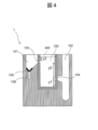
- ⁇ Height of flow path formed in container 1> The heights of the first channel 103 and the second channel 104 formed in the container 1 will be described with reference to FIG. 4 .
- the heights of the first channel 103 and the second channel 104 are different. The reason for this is to automatically collect different forms of bacteria used for different tests.
- Two types of tests are considered: an identification test by MALDI and a susceptibility test. For example, 10 5 to 10 6 CFU or more of bacteria are required for identification tests by MALDI.
- a susceptibility test requires a bacterial solution of 10 5 to 10 6 CFU/mL. A few hundred microliters of bacterial solution is prepared, and then diluted 100 to 100 times and used for testing.
- first channel 103 and second channel 104 To consider the height of first channel 103 and second channel 104, consider a blood culture positive sample.
- the liquid volume of the blood culture-positive sample introduced into the first container 100 was set to 10 mL.
- the concentration of bacteria contained in the blood culture-positive sample should be in the range of 10 7 to 10 9 CFU/mL, although it varies depending on the time until the blood culture is positive, the elapsed time after positive, the bacterial species, and the strain. is common. Therefore, a case where the bacterial concentration, which is expected to be the most difficult to pretreat, is 10 7 CFU/mL was considered.
- the heights of the first channel 103 and the second channel 104 are as shown in FIG.
- y1 is the height from the bottom surface of the first housing portion 100 to the second flow path 104
- y2 is the height from the second channel 104 to the first channel 103
- y3 is the height from the first channel 103 to the scale line 400 indicating the specified value.
- y1+y2+y3 is the height of the blood sample 200 when the assumed blood sample 200 is put in the total volume, ie, 10 mL.
- the container 1 is provided with the visible scale lines 400 at the height of y1+y2+y3.
- a blood sample 200 of at least a specified value or more must be introduced into the first container 100 .
- the height y1 from the bottom surface of the first housing portion 100 to the second flow path 104 is determined as follows. Since the centrifuged red blood cells are deposited in the first storage part 100, the volume of the first storage part 100 from the bottom surface of the first storage part 100 to the height y1 is the number of red blood cells in the blood sample 200. It must be at least volume.
- the hematocrit value which indicates the ratio of erythrocytes in blood, is in the range of 30 to 55%, depending on gender and health condition.
- the blood sample 200 introduced into the first container 100 contains the patient's blood as well as about 2 ⁇ 3 of the total volume of medium components introduced in advance.
- the ratio of erythrocytes in the blood culture-positive sample is at most about 1/6. Therefore, in order to pretreat the blood sample more efficiently, the height of y1 is preferably 16.7% or more, for example 20%, of the specified height of the input amount of the blood sample 200. be done.
- the height of y3 is determined as follows. If the bacterial concentration is 10 7 CFU/mL, 1 mL of sample can extract 10 7 CFU of bacteria. This is more than 10 times the required amount of bacteria in MALDI, and it is considered that a sufficient amount of bacteria can be extracted. Therefore, the height of y3 is preferably about 10% of the specified height of the input amount of blood sample 200 .
- the height of y2 is automatically determined. That is, the height of y2 is about 70% of the specified height of the input amount of blood sample 200 .
- such a minute amount of highly concentrated bacterial solution is diluted to several hundred ⁇ L, for example, 200 ⁇ L, and the concentration is adjusted to 1.5 ⁇ 10 8 CFU/mL (equivalent to 0.5 McFarland). and According to this calculation, 3.5 ⁇ 10 8 CFU/mL of bacterial solution can be extracted while 1.5 ⁇ 10 8 CFU/mL of bacterial solution is required, so the loss of bacteria during separation is considered. However, it is possible to extract a sufficient amount.
- the elements that characterize the container 1 are as follows.
- the container 1 a container into which the blood sample 200 can be introduced and subjected to centrifugation can be used.
- the volume of the first container 100 can be 1-20 mL, preferably 8-15 mL.
- the sum of the volume of the second storage portion 101 and the volume of the third storage portion 102 may be equal to or greater than the volume of the first storage portion 100 .
- the material of the container 1 is not particularly limited as long as it is a material suitable for operations such as centrifugation.
- the container 1 is preferably made of a hydrophobic material.
- the container 1 is preferably made of a material such as acrylic resin, ABS resin, polypropylene, polystyrene, or polyethylene, and is made using a 3D printer or injection molding.
- the container 1 may be produced by cutting aluminum or stainless steel.
- the container 1 may be chemically treated to render it hydrophobic and to modify the surface.
- the container 1 may be opaque, but if it is transparent, the internal state can be easily visually recognized, or optically measured or photographed by a device.
- the container 1 is preferably transparent in order to facilitate detection of excess or deficiency of the liquid volume, clogging of the sample, and detection of foreign matter in the sample.
- the container 1 is preferably disposable from the viewpoint of contamination, it can be used repeatedly after washing and sterilization, for example.
- the container 1 may have any shape as long as it can be centrifuged.
- the bottom portion of the second storage portion 101 where the filter 105 is installed and the bottom portion of the third storage portion 102 which stores the reagent are mortar-shaped in order to efficiently collect the bacteria 204 and 209. Preferably.
- the first channel 103 and the second channel 104 may have any shape.
- the first flow path 103 and the second flow path 104 are, for example, cylindrical with a circular cross-sectional shape, or rectangular parallelepiped with a rectangular cross-sectional shape.
- the diameters of the first channel 103 and the second channel 104 are considered as follows. Blood contains erythrocytes, leukocytes, and platelets, the largest of which is leukocytes, which have a diameter of 6 to 30 ⁇ m. If the diameters of the first channel 103 and the second channel 104 are smaller than this, the blood cells clog the first channel 103 and the second channel 104 without the effect of the capillary valve, and the liquid may not be released.
- a typical optimum range of the diameters of the first channel 103 and the second channel 104 is 10 to 100 ⁇ m. Also, the diameter of the first channel 103 and the diameter of the second channel 104 may be different, but preferably they are the same diameter.
- the reagent 201 introduced into the third container 102 is not particularly limited as long as it can destroy blood cells without affecting the growth of microorganisms.
- reagent 201 preferably contains at least one surfactant.
- surfactants include, but are not limited to, anionic surfactants having hydrophilic and hydrophobic moieties, wherein said hydrophobic moieties are chain hydrocarbons, or hydrophilic and hydrophobic moieties. and wherein said hydrophobic portion comprises a cyclic hydrocarbon, or a combination of both.
- the former includes sodium dodecyl sulfate, lithium dodecyl sulfate, and sodium N-lauroyl sarcosinate
- the latter includes saponin, sodium cholate, sodium deoxycholate, 3-[(3-cholamidopropyl). dimethylammonio]-1-propanesulfonate, and 3-[(3-cholamidopropyl)dimethylammonio]-2-hydroxy-1-propanesulfonate.
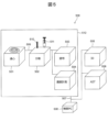
- the inspection system 500 includes a centrifugal device 501 , a fractionating device 502 , a coating device 503 , a concentration measuring device 504 , an identification testing device 505 , a susceptibility testing device 506 and a control PC 508 . Each device is connected by a signal line 507, and a control PC 508 controls the operation of each device.
- the centrifugal device 501 can be loaded with one or a plurality of containers 1 described above, and centrifuges the blood sample 200 introduced into the first container 100 of the mounted container 1 to be used in the identification test device 505. Bacteria 204 used in susceptibility testing device 506 and bacteria 209 used in susceptibility testing device 506 are isolated.
- the centrifugal device 501 includes at least three centrifuges set for centrifugation of the blood sample 200, transfer of the bacterial liquid 202 to the second container 101, and transfer of the bacterial liquid 206 to the third container 102. Centrifuge the blood sample 200 with acceleration.
- the container 1 is transported to the sorting device 502 by the transport mechanism 509 . A user may transport the container 1 from the centrifugal device 501 to the sorting device 502 .
- the sorting device 502 acquires the bacteria 204 collected in the second container 101 of the container 1 using, for example, a sample-collecting rod 510 with a sharp tip, and uses a syringe 511 to collect the bacteria 204 in the third container 1 of the container 1. Bacteria 209 collected in the container 102 are acquired. The acquired bacteria 204 and bacteria 209 are sent to different devices.
- the applicator 503 applies solid bacteria and matrix reagents to MALDI target plates used in mass spectrometry for identification tests.
- the concentration measuring device 504 measures the concentration of the bacterial liquid by measuring McFarland turbidity, for example.
- a pretreatment device 512 is provided with the centrifugal device 501, fractionating device 502, coating device 503, and concentration measuring device 504 described above.
- the coating device 503 does not necessarily have to exist as a necessary component.
- the sampling device 502 may return the sample-collecting rod 510 from which the bacteria 204 have been acquired to the user, and the user may apply the sample to the target plate.
- the concentration value measured by the preprocessing device 512 may be displayed on a display attached to the control PC 508 to notify the user.
- the density value is equal to or higher than the target value, for example, the user is notified and the user adjusts the density to the target value, thereby maintaining the reliability of the inspection. If the concentration is less than the target value, an error flag may be displayed and the pretreatment may be performed again, or the bacterial solution may be additionally cultured and then used for testing.
- the sorting apparatus 502 has a function of acquiring appearance information of the container 1 using, for example, concentration measurement or image measurement, and confirming whether the preceding processing is being executed normally.
- the liquid level of the blood sample 200 in the first container 100 is equal to the level of the second channel 104 as shown in FIG. 2(e). to the same extent as However, when the preceding process is stopped in the middle, the liquid level of blood sample 200 in first container 100 becomes sufficiently higher than the height of second channel 104 . Based on such information, the preparative collection device 502 can validate whether or not the preceding process has been completed normally.
- the sample processed by the pretreatment device 512 is once returned to the user, subjected to additional processing steps as necessary, and then tested by the identification testing device 505 and the susceptibility testing device 506 .
- the identification inspection device 505 uses the sample obtained from the bacteria 204 to determine the type of bacteria.
- the susceptibility test device 506 uses the sample obtained from the bacteria 209 to determine the degree of bacterial growth. In the case of an identification test, depending on the case, the user himself/herself applies the bacteria and the matrix reagent to the target plate, and then the identification of the bacterial species is carried out by the device.
- the bacterial solution is adjusted to a target concentration and dispensed into a 96-well plate or the like, and the susceptibility test device 506 determines proliferation and MIC.
- the progress status and determination results of identification of bacterial species and determination of susceptibility tests are displayed on a display attached to the control PC 508 to notify the user of the results.
- the control PC 508 inputs the parameters of each device such as the acceleration and time of centrifugation in the centrifugal device 501 and the target value of the concentration of bacteria in the concentration measuring device 504 according to the blood sample 200. It is preferable that the content of processing can be changed by pressing the
- the centrifugal device 501 includes a container mounting section 601 , a driving section 602 and a control board 603 .
- One or more containers 1 are mounted on the container mounting portion 601 .
- Drive unit 602 is an actuator that applies centrifugal force to blood sample 200 in container 1 mounted on container mounting unit 601 .
- a control board 603 is a control unit that controls the operation of the driving unit 602 .
- the control board 603 includes a processor 610 , a main storage device 611 , an auxiliary storage device 612 , an input/output I/F 613 , and a bus 614 that communicably connects each part of the control board 603 .
- the input/output I/F 613 is communicably connected to the control PC 508 via the signal line 507 and receives instructions from the control PC 508 .
- the processor 610 generates drive signals for driving the driving section 602 according to instructions from the control PC 508 .
- the input/output I/F 613 is communicably connected to the drive unit 602 and outputs drive signals generated by the processor 610 to the drive unit 602 .
- the drive unit 602 centrifuges the container 1 at various centrifugal accelerations according to the drive signal.
- the blood sample 200 is introduced into the first container 100 of the container 1.
- the process of introducing the blood sample 200 into the container 1 may be performed by a user or by a robot.
- the reagent 201 such as a surfactant that destroys blood cells may be introduced into the third container 102 in step S70, or may be introduced into the third container 102 before step S70.
- steps S71 to S74 are described individually in FIG. 7, a series of processes of steps S71 to S74 are automatically performed continuously by the centrifugal device 501.
- step S71 container 1 containing blood sample 200 is centrifuged at low acceleration to separate blood sample 200 into bacterial fluid 202 and solution 203.
- step S72 the container 1 containing the blood sample 200 is centrifuged at medium acceleration to move the bacterial fluid 202 from the first container 100 to the second container 101 via the first channel 103.
- the liquid component 205 is removed from the bacterial liquid 202 moved to the second container 101 by the filtration filter 105, and the bacteria 204 are collected.
- step S 73 the container 1 containing the solution 203 is centrifuged at low acceleration to separate the solution 203 into the bacterial liquid 206 and the blood cell component 207 .
- step S74 the container 1 containing the solution 203 is centrifuged at a high acceleration to move the fungal liquid 206 from the first containing portion 100 to the third containing portion 102 via the second flow path 104.
- the reagent 201 destroys red blood cells remaining in the bacterial liquid 206 and eluates them into the liquid. As a result, only bacterial components accumulate on the bottom surface of the third containing portion 102 .
- the identification test device 505 uses the bacteria 204 collected in the second container 101 to perform an identification test.
- an identification test by MALDI a mass of solid bacteria 204 captured on the filtration filter 105 of the second storage unit 101 is obtained, applied to a target plate, and then an appropriate matrix reagent is added.
- the identification test may be performed after dropping and drying.
- protein extraction treatment with ethanol, formic acid, acetonitrile, etc. may be performed as in the usual MALDI technique.
- the susceptibility test device 506 acquires the high-concentration fungal fluid accumulated on the bottom surface of the third container 102 and performs a susceptibility test. Specifically, the high-concentration bacteria 209 accumulated on the bottom surface are obtained with a pipette tip or the like, or the high-concentration bacteria 209 accumulated on the bottom surface are obtained after removing the supernatant. Then, the concentration of the bacterial solution is adjusted, for example, by the McFarland turbidimetric method, and the susceptibility test is performed using the bacterial solution.
- step S77 the results of the bacteriological examination are reported. Specifically, the identification test performed in S75 reports information on the bacterial species and determination score. In addition, information such as the name of the drug, its MIC (Minimum Inhibition Concentration), sensitivity/tolerance, etc. is reported to the laboratory technician and the doctor by the sensitivity test performed in S76.
- the identification test performed in S75 reports information on the bacterial species and determination score.
- information such as the name of the drug, its MIC (Minimum Inhibition Concentration), sensitivity/tolerance, etc. is reported to the laboratory technician and the doctor by the sensitivity test performed in S76.
- Example 1 a first containing portion 100 having a first flow channel 103 and a second flow channel 104 with different heights on the side surface, and a flow through the first containing portion 100 and the first flow channel 103 Centrifuge the container 1 including the connected second containing portion 101 and the third containing portion 102 connected to the first containing portion 100 via the second flow path 104 at various centrifugal accelerations. do. As a result, blood cells and bacteria can be separated from the blood sample 200 introduced into the first container 100 .
- the bacterial solution 202 By centrifuging the blood sample 200 contained in the first container 100, the bacterial solution 202 can be moved to the second container 101 via the first flow path 103, and The bacteria liquid 206 can be moved to the third container 102 via the second channel 104 .
- Example 1 it is possible to simultaneously obtain different forms of bacterial samples used in identification tests and susceptibility tests by a simple and stable method.
- a series of centrifugal separation operations are performed once at a predetermined centrifugal acceleration to extract different forms of bacterial samples.
- a solid bacterial sample with little liquid content is suitable for use in mass spectrometry by MALDI, and a liquid bacterial sample is suitable for use in susceptibility testing. Since the amount of bacteria to be extracted can be controlled by adjusting the heights of the first flow path 103 and the second flow path 104, the user can automatically obtain a predetermined amount or more of the bacterial sample suitable for each test without complicated operations. It is possible to obtain
- Example 1 the solid bacteria 204 can be collected by the filtration filter 105 of the second storage section 101 . This makes it possible to obtain the solid bacteria required for the identification test.
- Example 1 the reagent 201 introduced into the third container 102 does not affect the cell membranes of bacteria in the bacterial liquid 206, but makes it possible to destroy the cell membranes of red blood cells. As a result, it is possible to destroy a small amount of blood cell components in the bacterial liquid 206 that remained without being separated by centrifugation.
- the height of the first flow path 103 and the second flow path 104 is 1/6 or more of the height from the bottom surface of the first housing portion 100 to the specified value
- the difference (y2) between the height of the first channel 103 and the height of the second channel 104 is 1/2 or more of the height from the bottom surface of the first housing part 100 to a specified value.
- Example 1 by centrifuging the solution 203 at a centrifugal acceleration x3 that is sufficiently greater than the centrifugal acceleration x2, the bacterial liquid 206 can be rapidly moved from the second flow path 104 to the third container 102. becomes.
- the bacteria 204 can be used to determine the type of bacteria, and the bacteria 209 can be used to determine the degree of bacterial growth.
- Example 1 since the bottom surface of each of the second containing portion 101 and the third containing portion 102 of the container 1 is mortar-shaped, the collected bacteria 204 and 209 are removed with a sharp needle or stick. , and can be efficiently retrieved with a pipette tip or syringe.
- Example 2 In the first embodiment, an example has been described in which the user manually obtains the bacteria 209 collected in the second container 101 and 204 third container 102 using a needle or pipette tip. .
- the bacteria 204 collected in the second container 101 are automatically separated and applied, and the bacteria 209 collected in the third container 102 are automatically separated. , dilute, and measure concentration.
- the inspection system 800 of Example 2 includes a centrifugal device 501, a sorting device 502, a coating device 503, an identification testing device 505, a susceptibility testing device 506, and a control PC 508, as in the first embodiment.
- the inspection system 800 of Example 2 includes a concentration measuring and diluting device 801 , a driving mechanism 802 , a driving control mechanism 803 , a gripping mechanism 804 and a transporting device 809 .
- the drive mechanism 802 is controlled by a drive control mechanism 803.
- a drive mechanism 802 moves a gripping mechanism 804 from the centrifuge device 501 to the sorting device 502 .
- the gripping mechanism 804 is configured to grip the container 1 and grips the container 1 centrifuged by the centrifugal device 501 .
- the driving mechanism 802 moves the specimen-picking rod 510 from the sorting device 502 to the coating device 503 .
- the drive mechanism 802 moves the syringe 511 from the sorting device 502 to the concentration measuring and diluting device 801 .
- the sample prepared by the coating device 503 is transported to the identification inspection device 505 by the transport device 809 .
- the sample whose concentration has been measured and diluted by the concentration measuring and diluting device 801 is transported to the susceptibility testing device 506 by the transporting device 809 .
- the testing system 800 of the second embodiment can automatically perform centrifugation in the centrifugal device 501 , testing in the identification testing device 505 and testing in the susceptibility testing device 506 .
- the present invention is not limited to the above examples, and includes various modifications.
- the above embodiments have been described in detail for easy understanding of the present invention, and are not necessarily limited to those having all the configurations described. It is also possible to replace part of the configuration of one embodiment with the configuration of another embodiment, or to add the configuration of another embodiment to the configuration of one embodiment. Moreover, it is also possible to add, delete, or replace a part of the configuration of each embodiment with another configuration.
Landscapes
- Chemical & Material Sciences (AREA)
- Health & Medical Sciences (AREA)
- Life Sciences & Earth Sciences (AREA)
- Organic Chemistry (AREA)
- General Health & Medical Sciences (AREA)
- Analytical Chemistry (AREA)
- Biochemistry (AREA)
- Physics & Mathematics (AREA)
- Immunology (AREA)
- Pathology (AREA)
- Engineering & Computer Science (AREA)
- Proteomics, Peptides & Aminoacids (AREA)
- Zoology (AREA)
- Wood Science & Technology (AREA)
- General Physics & Mathematics (AREA)
- Molecular Biology (AREA)
- General Engineering & Computer Science (AREA)
- Biotechnology (AREA)
- Microbiology (AREA)
- Bioinformatics & Cheminformatics (AREA)
- Biophysics (AREA)
- Genetics & Genomics (AREA)
- Biomedical Technology (AREA)
- Chemical Kinetics & Catalysis (AREA)
- Clinical Laboratory Science (AREA)
- Hematology (AREA)
- Apparatus Associated With Microorganisms And Enzymes (AREA)
- Investigating Or Analysing Biological Materials (AREA)
Priority Applications (4)
| Application Number | Priority Date | Filing Date | Title |
|---|---|---|---|
| PCT/JP2022/000884 WO2023135704A1 (ja) | 2022-01-13 | 2022-01-13 | 捕集方法、検査方法、容器、遠心装置および検査システム |
| US18/724,148 US20250059583A1 (en) | 2022-01-13 | 2022-01-13 | Collection Method, Test Method, Container, Centrifuge and Test System |
| JP2023573714A JP7716506B2 (ja) | 2022-01-13 | 2022-01-13 | 捕集方法、検査方法、遠心装置および検査システム |
| EP22920229.6A EP4465012A4 (en) | 2022-01-13 | 2022-01-13 | COLLECTION METHOD, TESTING METHOD, CONTAINER, CENTRIFUGE AND TESTING SYSTEM |
Applications Claiming Priority (1)
| Application Number | Priority Date | Filing Date | Title |
|---|---|---|---|
| PCT/JP2022/000884 WO2023135704A1 (ja) | 2022-01-13 | 2022-01-13 | 捕集方法、検査方法、容器、遠心装置および検査システム |
Publications (1)
| Publication Number | Publication Date |
|---|---|
| WO2023135704A1 true WO2023135704A1 (ja) | 2023-07-20 |
Family
ID=87278666
Family Applications (1)
| Application Number | Title | Priority Date | Filing Date |
|---|---|---|---|
| PCT/JP2022/000884 Ceased WO2023135704A1 (ja) | 2022-01-13 | 2022-01-13 | 捕集方法、検査方法、容器、遠心装置および検査システム |
Country Status (4)
| Country | Link |
|---|---|
| US (1) | US20250059583A1 (https=) |
| EP (1) | EP4465012A4 (https=) |
| JP (1) | JP7716506B2 (https=) |
| WO (1) | WO2023135704A1 (https=) |
Citations (6)
| Publication number | Priority date | Publication date | Assignee | Title |
|---|---|---|---|---|
| JP2004212050A (ja) * | 2002-05-08 | 2004-07-29 | Hitachi High-Technologies Corp | 化学分析装置及び遺伝子診断装置 |
| JP2004309233A (ja) * | 2003-04-04 | 2004-11-04 | Hitachi High-Technologies Corp | 化学分析装置 |
| JP2006242872A (ja) * | 2005-03-04 | 2006-09-14 | Kyocera Corp | 分離装置及び該分離装置を備えた測定装置 |
| JP2012532618A (ja) * | 2009-07-16 | 2012-12-20 | ブルーカー ダルトニック ゲーエムベーハー | 敗血症の質量分析法診断 |
| JP2017517390A (ja) * | 2014-05-16 | 2017-06-29 | クヴェッラ コーポレーション | 自動式遠心分離を行うための装置、システム、および方法 |
| JP2021524259A (ja) * | 2018-05-25 | 2021-09-13 | クヴェッラ コーポレーション | 血液細胞の選択的ライシス及び微生物細胞の分離のための方法及び組成物 |
Family Cites Families (1)
| Publication number | Priority date | Publication date | Assignee | Title |
|---|---|---|---|---|
| KR101922128B1 (ko) * | 2012-12-04 | 2019-02-13 | 삼성전자주식회사 | 미세유동장치 및 이를 채용한 생물학적 시료 내의 표적 물질 농축방법 |
-
2022
- 2022-01-13 EP EP22920229.6A patent/EP4465012A4/en active Pending
- 2022-01-13 US US18/724,148 patent/US20250059583A1/en active Pending
- 2022-01-13 JP JP2023573714A patent/JP7716506B2/ja active Active
- 2022-01-13 WO PCT/JP2022/000884 patent/WO2023135704A1/ja not_active Ceased
Patent Citations (6)
| Publication number | Priority date | Publication date | Assignee | Title |
|---|---|---|---|---|
| JP2004212050A (ja) * | 2002-05-08 | 2004-07-29 | Hitachi High-Technologies Corp | 化学分析装置及び遺伝子診断装置 |
| JP2004309233A (ja) * | 2003-04-04 | 2004-11-04 | Hitachi High-Technologies Corp | 化学分析装置 |
| JP2006242872A (ja) * | 2005-03-04 | 2006-09-14 | Kyocera Corp | 分離装置及び該分離装置を備えた測定装置 |
| JP2012532618A (ja) * | 2009-07-16 | 2012-12-20 | ブルーカー ダルトニック ゲーエムベーハー | 敗血症の質量分析法診断 |
| JP2017517390A (ja) * | 2014-05-16 | 2017-06-29 | クヴェッラ コーポレーション | 自動式遠心分離を行うための装置、システム、および方法 |
| JP2021524259A (ja) * | 2018-05-25 | 2021-09-13 | クヴェッラ コーポレーション | 血液細胞の選択的ライシス及び微生物細胞の分離のための方法及び組成物 |
Non-Patent Citations (1)
| Title |
|---|
| See also references of EP4465012A4 * |
Also Published As
| Publication number | Publication date |
|---|---|
| EP4465012A4 (en) | 2025-11-19 |
| EP4465012A1 (en) | 2024-11-20 |
| US20250059583A1 (en) | 2025-02-20 |
| JP7716506B2 (ja) | 2025-07-31 |
| JPWO2023135704A1 (https=) | 2023-07-20 |
Similar Documents
| Publication | Publication Date | Title |
|---|---|---|
| US12013322B2 (en) | Liquid to liquid biological particle concentrator with disposable fluid path | |
| CA2988128C (en) | A component of a device, a device, and a method for purifying and testing biomolecules from biological samples | |
| JP5254000B2 (ja) | 液体中に含まれる生物学的粒子を垂直濾過することによって分離する方法及び装置 | |
| JP2020016657A (ja) | 装置 | |
| JP2009122082A (ja) | 血液希釈定量器具 | |
| WO2006109693A1 (ja) | 粘性試料のサンプリング器具、喀痰の均質化処理方法及び微生物の検出方法 | |
| WO2023135704A1 (ja) | 捕集方法、検査方法、容器、遠心装置および検査システム | |
| JP2004138583A (ja) | 細胞機能測定用マイクロチップ | |
| US8911686B2 (en) | Separation device | |
| JP7573102B2 (ja) | 細菌捕集方法および装置 | |
| CN110709694A (zh) | 微观流体装置和使用其的方法 | |
| US20220049321A1 (en) | Method, system and apparatus for blood processing unit | |
| EP4379061A1 (en) | Method and apparatus for rapidly testing microorganism | |
| WO2022108728A1 (en) | Method, system and apparatus for blood processing unit | |
| WO2022241243A1 (en) | Techniques for detection and quantification of live and dead bacteria in a fluid sample | |
| WO2022201657A1 (ja) | 遠心ろ過カートリッジ及び微生物検査方法 | |
| KR20210122273A (ko) | 생물학적 샘플로부터 성분을 선택적으로 추출하기 위한 방법 및 장치 | |
| Miller | Lemons to Lemonade (EM Techniques for Salvaging Suboptimal Specimens) | |
| WO2007044082A2 (en) | Oral fluid collection device |
Legal Events
| Date | Code | Title | Description |
|---|---|---|---|
| 121 | Ep: the epo has been informed by wipo that ep was designated in this application |
Ref document number: 22920229 Country of ref document: EP Kind code of ref document: A1 |
|
| WWE | Wipo information: entry into national phase |
Ref document number: 2023573714 Country of ref document: JP |
|
| WWE | Wipo information: entry into national phase |
Ref document number: 18724148 Country of ref document: US |
|
| WWE | Wipo information: entry into national phase |
Ref document number: 2022920229 Country of ref document: EP |
|
| NENP | Non-entry into the national phase |
Ref country code: DE |
|
| ENP | Entry into the national phase |
Ref document number: 2022920229 Country of ref document: EP Effective date: 20240813 |