WO2023120673A1 - マクロファージの製造方法、分化誘導剤、分化誘導キット、マクロファージの培養方法、マクロファージの増殖促進剤、マクロファージの増殖促進キット、マクロファージの増殖方法及びマクロファージ - Google Patents
マクロファージの製造方法、分化誘導剤、分化誘導キット、マクロファージの培養方法、マクロファージの増殖促進剤、マクロファージの増殖促進キット、マクロファージの増殖方法及びマクロファージ Download PDFInfo
- Publication number
- WO2023120673A1 WO2023120673A1 PCT/JP2022/047472 JP2022047472W WO2023120673A1 WO 2023120673 A1 WO2023120673 A1 WO 2023120673A1 JP 2022047472 W JP2022047472 W JP 2022047472W WO 2023120673 A1 WO2023120673 A1 WO 2023120673A1
- Authority
- WO
- WIPO (PCT)
- Prior art keywords
- macrophages
- macrophage
- cells
- trem2
- signal activator
- Prior art date
- Legal status (The legal status is an assumption and is not a legal conclusion. Google has not performed a legal analysis and makes no representation as to the accuracy of the status listed.)
- Ceased
Links
Images


Classifications
-
- C—CHEMISTRY; METALLURGY
- C12—BIOCHEMISTRY; BEER; SPIRITS; WINE; VINEGAR; MICROBIOLOGY; ENZYMOLOGY; MUTATION OR GENETIC ENGINEERING
- C12N—MICROORGANISMS OR ENZYMES; COMPOSITIONS THEREOF; PROPAGATING, PRESERVING, OR MAINTAINING MICROORGANISMS; MUTATION OR GENETIC ENGINEERING; CULTURE MEDIA
- C12N5/00—Undifferentiated human, animal or plant cells, e.g. cell lines; Tissues; Cultivation or maintenance thereof; Culture media therefor
- C12N5/06—Animal cells or tissues; Human cells or tissues
- C12N5/0602—Vertebrate cells
- C12N5/0634—Cells from the blood or the immune system
- C12N5/0645—Macrophages, e.g. Kuepfer cells in the liver; Monocytes
-
- C—CHEMISTRY; METALLURGY
- C12—BIOCHEMISTRY; BEER; SPIRITS; WINE; VINEGAR; MICROBIOLOGY; ENZYMOLOGY; MUTATION OR GENETIC ENGINEERING
- C12N—MICROORGANISMS OR ENZYMES; COMPOSITIONS THEREOF; PROPAGATING, PRESERVING, OR MAINTAINING MICROORGANISMS; MUTATION OR GENETIC ENGINEERING; CULTURE MEDIA
- C12N5/00—Undifferentiated human, animal or plant cells, e.g. cell lines; Tissues; Cultivation or maintenance thereof; Culture media therefor
- C12N5/10—Cells modified by introduction of foreign genetic material
-
- C—CHEMISTRY; METALLURGY
- C12—BIOCHEMISTRY; BEER; SPIRITS; WINE; VINEGAR; MICROBIOLOGY; ENZYMOLOGY; MUTATION OR GENETIC ENGINEERING
- C12N—MICROORGANISMS OR ENZYMES; COMPOSITIONS THEREOF; PROPAGATING, PRESERVING, OR MAINTAINING MICROORGANISMS; MUTATION OR GENETIC ENGINEERING; CULTURE MEDIA
- C12N2500/00—Specific components of cell culture medium
- C12N2500/05—Inorganic components
-
- C—CHEMISTRY; METALLURGY
- C12—BIOCHEMISTRY; BEER; SPIRITS; WINE; VINEGAR; MICROBIOLOGY; ENZYMOLOGY; MUTATION OR GENETIC ENGINEERING
- C12N—MICROORGANISMS OR ENZYMES; COMPOSITIONS THEREOF; PROPAGATING, PRESERVING, OR MAINTAINING MICROORGANISMS; MUTATION OR GENETIC ENGINEERING; CULTURE MEDIA
- C12N2500/00—Specific components of cell culture medium
- C12N2500/30—Organic components
- C12N2500/36—Lipids
-
- C—CHEMISTRY; METALLURGY
- C12—BIOCHEMISTRY; BEER; SPIRITS; WINE; VINEGAR; MICROBIOLOGY; ENZYMOLOGY; MUTATION OR GENETIC ENGINEERING
- C12N—MICROORGANISMS OR ENZYMES; COMPOSITIONS THEREOF; PROPAGATING, PRESERVING, OR MAINTAINING MICROORGANISMS; MUTATION OR GENETIC ENGINEERING; CULTURE MEDIA
- C12N2500/00—Specific components of cell culture medium
- C12N2500/70—Undefined extracts
- C12N2500/80—Undefined extracts from animals
-
- C—CHEMISTRY; METALLURGY
- C12—BIOCHEMISTRY; BEER; SPIRITS; WINE; VINEGAR; MICROBIOLOGY; ENZYMOLOGY; MUTATION OR GENETIC ENGINEERING
- C12N—MICROORGANISMS OR ENZYMES; COMPOSITIONS THEREOF; PROPAGATING, PRESERVING, OR MAINTAINING MICROORGANISMS; MUTATION OR GENETIC ENGINEERING; CULTURE MEDIA
- C12N2501/00—Active agents used in cell culture processes, e.g. differentation
- C12N2501/20—Cytokines; Chemokines
- C12N2501/22—Colony stimulating factors (G-CSF, GM-CSF)
-
- C—CHEMISTRY; METALLURGY
- C12—BIOCHEMISTRY; BEER; SPIRITS; WINE; VINEGAR; MICROBIOLOGY; ENZYMOLOGY; MUTATION OR GENETIC ENGINEERING
- C12N—MICROORGANISMS OR ENZYMES; COMPOSITIONS THEREOF; PROPAGATING, PRESERVING, OR MAINTAINING MICROORGANISMS; MUTATION OR GENETIC ENGINEERING; CULTURE MEDIA
- C12N2501/00—Active agents used in cell culture processes, e.g. differentation
- C12N2501/20—Cytokines; Chemokines
- C12N2501/23—Interleukins [IL]
- C12N2501/2306—Interleukin-6 (IL-6)
-
- C—CHEMISTRY; METALLURGY
- C12—BIOCHEMISTRY; BEER; SPIRITS; WINE; VINEGAR; MICROBIOLOGY; ENZYMOLOGY; MUTATION OR GENETIC ENGINEERING
- C12N—MICROORGANISMS OR ENZYMES; COMPOSITIONS THEREOF; PROPAGATING, PRESERVING, OR MAINTAINING MICROORGANISMS; MUTATION OR GENETIC ENGINEERING; CULTURE MEDIA
- C12N2501/00—Active agents used in cell culture processes, e.g. differentation
- C12N2501/20—Cytokines; Chemokines
- C12N2501/25—Tumour necrosing factors [TNF]
-
- C—CHEMISTRY; METALLURGY
- C12—BIOCHEMISTRY; BEER; SPIRITS; WINE; VINEGAR; MICROBIOLOGY; ENZYMOLOGY; MUTATION OR GENETIC ENGINEERING
- C12N—MICROORGANISMS OR ENZYMES; COMPOSITIONS THEREOF; PROPAGATING, PRESERVING, OR MAINTAINING MICROORGANISMS; MUTATION OR GENETIC ENGINEERING; CULTURE MEDIA
- C12N2501/00—Active agents used in cell culture processes, e.g. differentation
- C12N2501/998—Proteins not provided for elsewhere
-
- C—CHEMISTRY; METALLURGY
- C12—BIOCHEMISTRY; BEER; SPIRITS; WINE; VINEGAR; MICROBIOLOGY; ENZYMOLOGY; MUTATION OR GENETIC ENGINEERING
- C12N—MICROORGANISMS OR ENZYMES; COMPOSITIONS THEREOF; PROPAGATING, PRESERVING, OR MAINTAINING MICROORGANISMS; MUTATION OR GENETIC ENGINEERING; CULTURE MEDIA
- C12N2506/00—Differentiation of animal cells from one lineage to another; Differentiation of pluripotent cells
- C12N2506/11—Differentiation of animal cells from one lineage to another; Differentiation of pluripotent cells from blood or immune system cells
Definitions
- the present invention relates to a method for producing macrophages, a differentiation-inducing agent, a differentiation-inducing kit, a method for culturing macrophages, an agent for promoting proliferation of macrophages, a kit for promoting proliferation of macrophages, a method for proliferation of macrophages, and macrophages.
- Macrophages are white blood cells that contribute to defense against infection and maintenance of tissue homeostasis. Macrophages in vivo are derived from bone marrow hematopoietic stem cells, and their differentiation requires stimulation with macrophage colony-stimulating factor (M-CSF) or granulocyte-macrophage colony-stimulating factor (GM-CSF). In fact, a method has been established for inducing the differentiation of cells having the properties of monocytes and macrophages (bone marrow derived macrophages: BMDM) by culturing mouse bone marrow cells in vitro in the presence of M-CSF. BMDM is widely used as an in vitro macrophage research tool.
- M-CSF macrophage colony-stimulating factor
- GM-CSF granulocyte-macrophage colony-stimulating factor
- BMDMs have the following problems: i) M-CSF used for induction is expensive, which increases the cost of mass culturing BMDMs, ii) they can only survive for about a week, and iii) they cannot proliferate after differentiation. There are problems such as that the number of cells that can be obtained in a single culture is limited because the cells stop, and iv) that thawing after cryopreservation and reculturing cannot be performed due to the above iii).
- various macrophage cell lines of rodent and human origin are frequently used in in vitro studies. These cells do not have the above problems i) to iv), but because they are tumor cells, their use in in vivo tests such as administration to living bodies is limited.
- Patent Document 1 contains a compound having angiotensin II receptor antagonistic action and peroxisome proliferator-activated receptor ⁇ activating action. A differentiation inducer is disclosed.
- Patent Document 1 the survival period of macrophages obtained by the differentiation-inducing agent, the growth characteristics after differentiation, the possibility of cryopreservation, etc. are not examined.
- the present invention has been made in view of the above circumstances, and includes a method for producing macrophages that provides macrophages that have a long survival period, a long growth period, can be cryopreserved, and is highly versatile as a research tool, a differentiation inducer, and a differentiation agent. It is an object of the present invention to provide an induction kit, a macrophage culture method, a macrophage proliferation promoter, a macrophage proliferation promotion kit, and a macrophage proliferation method. Another object of the present invention is to provide macrophages that have a long survival period and a long proliferation period, can be cryopreserved, and are highly versatile as research tools.
- DAM and LAM are one of the molecules that are characteristically highly expressed in the lipid recognition receptor TREM2 (triggering receptor expressed on myeloid cells 2).
- Non-Patent Documents 1 and 2 show that TREM2 is deeply involved in the induction of these disease-related macrophages.
- the method for producing macrophages described herein comprises: A culturing step of culturing hematopoietic progenitor cells in the presence of a TREM2 signal activator is included.
- the TREM2 signal activator is It may be a lipid.
- the TREM2 signal activator is It may be a brain-derived lipid or a lipid with two long-chain fatty groups.
- the TREM2 signal activator is It may be a compound represented by formula (I), an ester thereof, or a salt thereof. (having 60 to 90 carbon atoms, R 1 represents a saturated or unsaturated aliphatic hydrocarbon group, and R 2 represents a cyclic structure or a saturated or unsaturated aliphatic hydrocarbon group which may have a substituent represents.)
- the surface of the cell culture vessel for culturing the hematopoietic progenitor cells is It may be coated with the TREM2 signal activator.
- the hematopoietic progenitor cells are It may be a myeloid progenitor cell.
- the hematopoietic progenitor cells are It may be a macrophage-dendritic cell progenitor or a common monocytic progenitor.
- the agent for inducing differentiation from hematopoietic progenitor cells to macrophages described herein is Contains TREM2 signal activator.
- the kit for inducing differentiation from hematopoietic progenitor cells to macrophages described herein includes A cell culture vessel surface-coated with a TREM2 signal activator is provided.
- the method of culturing macrophages described herein comprises: A culturing step of culturing hematopoietic progenitor cells in the presence of a TREM2 signal activator is included.
- the macrophage proliferation-promoting agents described herein are Contains TREM2 signal activator.
- the macrophage proliferation-promoting kit described herein comprises: A cell culture vessel surface-coated with a TREM2 signal activator is provided.
- the macrophage expansion methods described herein comprise: A culturing step of culturing macrophages in the presence of a TREM2 signal activator.
- macrophage The macrophages described herein are It has the ability to proliferate after 10 days of subculture in a medium containing neither macrophage colony-stimulating factor nor granulocyte-macrophage colony-stimulating factor and containing a TREM2 signal activator.
- the macrophages described herein are By culturing hematopoietic progenitor cells in a medium containing neither macrophage colony-stimulating factor nor granulocyte-macrophage-stimulating factor, but containing a TREM2 signal activator, TREM2-dependent differentiation is induced.
- the macrophages described herein are After cryopreservation, it has the ability to proliferate in a medium containing neither macrophage colony-stimulating factor nor granulocyte-macrophage colony-stimulating factor and containing a TREM2 signal activator.
- the macrophages described herein are The concentration of TNF- ⁇ , IL-6 and nitric oxide in the culture supernatant after 24 hours of culture in a medium containing lipopolysaccharide was 30% compared to bone marrow-derived macrophages induced from bone marrow cells with macrophage colony-stimulating factor. It is below.
- the macrophages described herein are Expression of one or more of the genes shown in Table 1 below is in alveolar macrophages, BMDMs, M1-induced BMDMs, M2-induced BMDMs, Kupffer cells, microglia, osteoclasts and peritoneal exudate macrophages. high compared to at least one selected from
- the macrophages described herein are Expression of one or more of the genes shown in Table 2 below in alveolar macrophages, BMDMs, M1-induced BMDMs, M2-induced BMDMs, Kupffer cells, microglia, osteoclasts and peritoneal exudate macrophages low compared to at least one selected from
- macrophages that have a long survival period and proliferation period, can be cryopreserved, and are highly versatile as research tools.
- FIG. 3 shows images of macrophages differentiated from mouse bone marrow cells.
- A shows images of macrophages induced to differentiate with brain lipids.
- B shows an image of macrophages induced to differentiate with M-CSF.
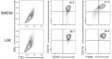
- FIG. 10 is a diagram showing a dot plot obtained by analysis by flow cytometry according to Test Example 2;
- FIG. 10 is a diagram showing univariate histograms of surface antigens obtained by analysis by flow cytometry according to Test Example 2;
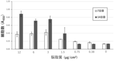
- FIG. 10 is a diagram showing the number of cells with respect to the mass of cerebral lipid per unit culture area according to Test Example 3;
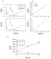
- FIG. 10 is a diagram showing changes over time in the number of cells according to Test Example 4;
- the upper part of (A) shows the number of cells after 0 to 21 days of culture.
- the lower part of (A) shows the fold increase in the number of cells after 0 to 21 days of culture, with the number of cells on day 0 being 1.
- the upper part of (B) shows the number of cells after 0 to 544 days of culture.
- the lower part of (B) shows the fold increase in the number of cells after 0 to 544 days of culture, with the number of cells on day 0 being 1.
- (C) shows the cell number of macrophages cultured in the presence or absence of brain lipids.
- FIG. 10 is a diagram showing changes over time in the number of thawed macrophages after cryopreservation according to Test Example 5.
- FIG. FIG. 10 is a diagram showing the number of macrophages after stimulation with brain lipids according to Test Example 6;
- A shows the number of macrophages differentiated from mixed cultured Lin-positive and Lin-negative cells.
- B shows the number of macrophages differentiated from Lin-positive cells and Lin-negative cells cultured alone, respectively.
- FIG. 10 is a diagram showing the number of viable cells after induction of differentiation of each mouse bone marrow cell according to Test Example 7.
- FIG. 10 shows ligand activity using TREM2 reporter cells according to Test Example 8.
- FIG. 10 is a diagram showing the number of viable cells after inducing differentiation of mouse bone marrow cells with a test substance according to Test Example 8.
- FIG. FIG. 11 shows the phagocytic ability of macrophages according to Test Example 9.
- FIG. 10 shows the cytokine production response of macrophages to lipopolysaccharide (LPS) stimulation according to Test Example 10.
- FIG. (A), (B), (C), (D) and (E) are monomer chemoattractant protein-1 (MCP-1), TNF- ⁇ , IL-6, IL-10 and nitric oxide (NO ).
- a method for producing macrophages includes a culturing step of culturing hematopoietic progenitor cells in the presence of a TREM2 signal activator.
- TREM2 is an immunoglobulin superfamily receptor that associates with the adapter molecule DAP12 (DNAX-activating protein of 12 kDa).
- the TREM2 signal activator is any substance that specifically binds to TREM2 and generates a signal via DAP12.
- TREM2 signal activators are, for example, antibodies or antigen-binding fragments thereof or compounds having TREM2 agonistic activity or DAP12 agonistic activity, ligands of TREM2, and the like.
- Antibodies may be polyclonal antibodies or monoclonal antibodies. Also, the antibody may be a humanized chimeric antibody, a humanized antibody, or a human antibody. An antibody herein includes antigen binding of an antibody.
- An antigen-binding fragment is a protein or peptide comprising a portion (partial fragment) of an antibody, which retains the functional binding of the antibody to the antigen, such as F(ab')2, Fab', Fab, Fab3, main-chain Fv (scFv), (tandem) bispecific single-chain Fv (sc(Fv)2), single-chain triple body, nanobody, divalent VHH, pentavalent VHH, minibody, (double-chain) diabody, tandem diabodies, bispecific tribodies, bispecific bibodies, dual affinity retargeting molecules (DART), triabodies (or tribodies), tetrabodies (or [sc(Fv)2]2), or disulfide-linked Fvs (dsFv ), or their polymers (Nature Biotechnology, 29(1): 5-6 (2011); Maneesh Jain et al., TRENDS in Biotechnology, 25(7) (2007): 307-316, and Christophstein e
- a TREM2 signal activator is, for example, a TREM2 ligand or a lipid.
- Lipids are organic compounds that are insoluble in water and readily soluble in organic solvents. There are no particular restrictions on the lipid, and it may be liquid or solid at room temperature. Lipids include, for example, glycosphingolipids, phospholipids, sulfated glycolipids and fatty acids. Preferably the lipid is a lipid with two long chain fatty groups.
- the long-chain fatty acid group includes fatty acid groups having 6 to 60 or 10 to 50 carbon atoms, and the number of carbon atoms constituting the carbon chain of each long-chain fatty acid group may be the same or different.
- Fatty acids may be saturated fatty acids or unsaturated fatty acids.
- the lipids include 2-tetradecylhexadecanoic acid (THA), ⁇ -glucosylceramide ( ⁇ -GluCer), ⁇ -galactosylceramide ( ⁇ -GalCer), phosphatidylcholine (PC), lyso Examples include phosphatidylcholine (LPC), phosphatidylethanolamine (PE), sulfatide (Sulf) and mycolic acid (MA).
- TREM2 signal activator may be a mixture of multiple types of lipids. It may be a synthetic lipid that binds.
- One aspect of the TREM2 signal activator includes cells or microorganisms having lipids on their cell surfaces.
- the TREM2 signal activator is a brain-derived lipid or MA.
- the TREM2 signal activator may be a compound of formula (I), its ester or its salt.
- the number of carbon atoms in one molecule of the compound is, for example, 60-90 or 70-80.
- R 1 represents a saturated or unsaturated aliphatic hydrocarbon group.
- the number of carbon atoms in R 1 is, for example, 6-60, 10-50, 20-40, 22-30 or 22-26.
- R2 represents a cyclic structure or a saturated or unsaturated aliphatic hydrocarbon group which may have a substituent.
- the number of carbon atoms in R 2 is, for example, 6-60, 10-50, 20-40, 22-30 or 22-26.
- the number of carbon atoms constituting the cyclic structure is, for example, 3-6.
- a cyclic structure may be included in R 2 as a cycloalkylene group or may be included in R 2 as a cycloalkyl group.
- R 2 may contain one to several cyclic structures.
- Substituents are not particularly limited, and include methyl groups, hydroxy groups, carbonyl groups, carboxyl groups, and the like.
- the salt of the compound represented by formula (I) above is not particularly limited as long as it is a pharmacologically acceptable salt, and may be either an acid salt or a basic salt.
- salts include alkali metal salts such as lithium salts, sodium salts and potassium salts; alkaline earth metal salts such as magnesium salts and calcium salts; Inorganic acid salts such as nitrates and phosphates, and formates, acetates, oxalates, propionates, hexanoates, cyclopentanepropionates, glycolates, pyruvates, lactates, malonates , succinate, malate, fumarate, tartrate, dibenzoyltartrate, ditoluoyltartrate, citrate, benzoate, o-(4-hydroxybenzoyl)benzoate, cinnamate, mandelic acid salts, methanesulfonate, ethanesulfonate, 1,2-ethan
- the ester of the compound represented by the above formula (I) is not particularly limited as long as it is a pharmacologically acceptable ester. Examples include carbonate, phosphate, nitrate, sulfate, borate and sulfonic acid Ester etc. are mentioned. This embodiment also includes various hydrates, solvates, and crystal polymorphs of the compound of formula (I) and salts thereof.
- the TREM2 signal activator may be selected using cells that express TREM2 and DAP12 and that express a reporter protein upon TREM2 signaling by ligand binding.
- TREM2 and DAP12 can be expressed in cells by a known method, for example, by introducing a gene expression plasmid or the like into cells. Any reporter protein can be used, including, for example, green fluorescent protein (GFP) and the photoprotein luciferase.
- GFP green fluorescent protein
- TREM2 signals cause the expression of genes regulated by NFAT (nuclear factor of activated T cells) type transcription factors. Therefore, cells having NFAT-type transcription factors are preferably used as cells for expressing TREM2 and DAP12.
- a cell in which a gene encoding a reporter protein is introduced as a gene whose expression is controlled by an NFAT-type transcription factor, and TREM2 and DAP12 are expressed in the cell may be used for ligand evaluation.
- Hematopoietic progenitor cells are cells contained in bone marrow or cord blood. Hematopoietic progenitor cells are, for example, myeloid progenitor cells. Bone marrow cells harvested from bone marrow or mononuclear cells harvested from umbilical cord blood may be used as hematopoietic progenitor cells. Hematopoietic progenitor cells are particularly preferably macrophage and dendritic cell progenitor (MDP) and common monocytic progenitor (cMoP).
- MDP macrophage and dendritic cell progenitor
- cMoP common monocytic progenitor
- the concentration of hematopoietic progenitor cells is not particularly limited. be done. Preferably, 1 ⁇ 10 5 hematopoietic progenitor cells are seeded per unit culture area (cm 2 ).
- hematopoietic progenitor cells may be cultured in a cell culture container holding a TREM2 signal activator.
- cell culture vessels include cell culture plates, cell culture flasks, cell culture dishes and the like.
- the TREM2 signal activator is insoluble in water and insoluble in a liquid medium
- hematopoietic progenitor cells may be cultured in a cell culture vessel whose surface is coated with the TREM2 signal activator.
- brain-derived lipid is used as the TREM2 signal activator
- the mass of lipid coating the surface of the cell culture vessel is, for example, 1 ⁇ 10 4 to 1 ⁇ 10 6 hematopoietic progenitor cells.
- the ligand is dissolved (suspended) in an organic solvent or the like, added to the cell culture vessel, and the organic solvent is dried.
- cells may be cultured by a cell culture method using a known medium.
- the culture conditions are, for example, 37° C. and 5% CO 2 concentration.
- it is preferable to change the medium for example, every 3-4 days. From about 7 to 10 days after the start of culture, macrophage-like adherent cells are obtained.
- the macrophages obtained by the production method according to the present embodiment maintain proliferation as long as they are stimulated with a TREM2 signal activator, and the number of days (survival period) that can be cultured is at least 540 days or longer.
- the macrophages can be cryopreserved because they maintain their proliferative properties.
- the macrophages stop growing when cultured in the absence of a TREM2 signal activator, they are not tumor cells and their use in vivo is not limited, making them highly versatile as research tools.
- the macrophages can also be used for cell therapy against diseases such as cancer.
- macrophages are provided, wherein the macrophages contain neither M-CSF nor GM-CSF, and the hematopoietic progenitor cells are cultured in a medium containing a TREM2 signal activator to obtain TREM2 Differentiation is induced in a dependent manner.
- BMDMs induced from bone marrow cells with M-CSF lost their proliferation ability after 5 days of subculturing in a medium that did not contain M-CSF. Macrophages were able to proliferate after 10 days of subculturing in the presence of a TREM2 signal activator, even if the medium did not contain M-CSF.
- macrophages are provided which, after initiation of induction or after cryopreservation, are treated in media containing neither M-CSF nor GM-CSF and containing a TREM2 signal activator. After 10 days of subculture, preferably after 15 days, after 20 days, after 50 days or after 100 days, more preferably after 200 days, after 300 days or after 400 days, more preferably after 500 days. have
- BMDM thawed after cryopreservation did not proliferate even in the presence of M-CSF, whereas the thawed macrophages proliferated in the presence of a TREM2 signal activator.
- macrophages are provided that, after cryopreservation, are capable of proliferating in media containing neither M-CSF nor GM-CSF and containing a TREM2 signal activator.
- Cryopreservation is not particularly limited as long as it is a temperature commonly used for cryopreservation of cells, for example -80°C.
- the period of cryopreservation is not particularly limited, and may be immediately after confirmation of freezing, 5 days, 10 days, 20 days or 30 days after freezing, or several months or several years.
- the macrophages were cultured for 24 hours in a medium containing LPS (eg, 10 ng/ml), and then MCP-1, TNF- ⁇ , IL-6 and NO in the culture supernatant. was lower compared to BMDM cultured in medium containing LPS for 24 hours. Therefore, in another embodiment, macrophages are provided, and the concentrations of TNF- ⁇ , IL-6 and nitric oxide in the culture supernatant after culturing in a medium containing LPS for 24 hours are M- Less than 30% compared to BMDM derived from bone marrow cells with CSF.
- LPS eg, 10 ng/ml
- the concentration of TNF- ⁇ in the culture supernatant may be 25% or less or 20% or less compared to BMDM.
- the concentration of IL-6 in the culture supernatant may be 25% or less, 20% or less, or 15% or less compared to BMDM.
- the concentration of NO in the culture supernatant may be 25% or less compared to BMDM.
- the macrophages according to the present embodiment have 1 or more, 2 or 2 or more, 3 or 3 or more, 4 or 4 or more of the genes listed in Table 3, 5 or 5 or more, 10 or 10 or more, 20 or 20 or more, 30 or 30 or more, 40 or 40 or more, 50 or 50 or more, 60 or 60 or more, 70 or expression of 70 or more, 80 or 80 or more, or 88 alveolar macrophages, BMDM, M1-induced BMDM, M2-induced BMDM, Kupffer cells, microglia, osteoclasts and peritoneal exudates At least one type selected from macrophages, preferably two or more, three or three or more, four or four or more, five or five or more, six or six or more, seven or It may be higher than 7 or higher than all 8 of these.
- the macrophages have one or more, two or more of the genes listed in Table 4. 2 or more, 3 or 3 or more, 4 or 4 or more, 5 or 5 or more, 10 or 10 or more, 20 or 20 or more, 30 or 30 or more, 40 or 40 expression of ⁇ 50 or ⁇ 50, ⁇ 60 or ⁇ 60, ⁇ 70 or ⁇ 70, ⁇ 80 or ⁇ 80, or BMDM induced in alveolar macrophages, BMDM, M1 , M2-induced BMDM, Kupffer cells, microglia, osteoclasts and peritoneal exudate macrophages, at least one, preferably two or more, three or more, four or four Above, 5 types or 5 types or more, 6 types or 6 types or more, 7 types or 7 types or more, or may be lower than all of these 8 types.
- the expression of the gene is the expression level of RNA.
- level means a quantified measure of abundance, such as concentration, amount, or a measure that can be used as an alternative. Therefore, the level may be a measured value or a value converted to concentration. In addition, the level may be an absolute numerical value such as abundance and abundance per unit area, or may be a relative numerical value compared with a comparative control set as necessary.
- Macrophages according to the present embodiment have one, two or more, three or more, four or more, five or more, or all of the above-described characteristics may have.
- Another embodiment provides a method for preparing macrophages, a method for producing macrophages, a method for inducing macrophages, a method for culturing macrophages, or a method for proliferating macrophages, including the above-described culturing step.
- the method for culturing macrophages and the like they can be maintained for a long period of time.
- macrophages proliferate according to the method for proliferating macrophages.
- neither M-CSF nor GM-CSF is used in the method for producing macrophages, the method for preparing macrophages, the method for producing macrophages, the method for inducing macrophages, the method for culturing macrophages, and the method for growing macrophages.
- a differentiation inducer containing a TREM2 signal activator is provided.
- the agent for inducing differentiation induces differentiation from hematopoietic progenitor cells to macrophages.
- the differentiation inducer may contain solvents, water, ethanol, polyhydric alcohols, oils, surfactants, thickeners, preservatives, pH adjusters, and the like, in addition to the TREM2 signal activator.
- Differentiation inducers may be, for example, liquid, gel, cream and solid.
- the differentiation-inducing agent is added to the cell culture vessel, or applied to the surface of the cell culture vessel to coat the surface.
- macrophages that have a long survival period and can be cryopreserved can be obtained from hematopoietic progenitor cells.
- the macrophages have high versatility as research tools and are easy to use in various experiments.
- a macrophage proliferation-promoting agent containing a TREM2 signal activator is provided.
- the proliferation-promoting agent promotes proliferation of macrophages, particularly macrophages obtained by the production method described above.
- the proliferation-promoting agent may contain other components of the TREM2 signal activator, similar to the differentiation-inducing agent.
- a differentiation induction kit comprising a cell culture vessel whose surface is coated with a TREM2 signal activator.
- the differentiation induction kit is used to induce differentiation from hematopoietic progenitor cells to macrophages.
- the differentiation induction kit may further comprise media such as basal media, serum-reduced media and serum-free media, serum required for cell culture such as fetal bovine serum (FBS), and other known additives added to the media.
- media such as basal media, serum-reduced media and serum-free media, serum required for cell culture such as fetal bovine serum (FBS), and other known additives added to the media.
- FBS fetal bovine serum
- the hematopoietic progenitor cells can be seeded and cultured in the cell culture vessel for a long survival period. Preservable macrophages can be easily obtained.
- the differentiation induction kit can also be used as a macrophage proliferation promotion kit.
- the differentiation-inducing agent and the macrophage proliferation-promoting agent do not contain either M-CSF or GM-CSF.
- the kit for inducing differentiation into macrophages and the kit for promoting proliferation of macrophages comprise neither M-CSF nor GM-CSF.
- Example 1 Preparation of lipid-induced macrophages (LIM) [Extraction of mouse brain lipid]
- Mouse brains (approximately 400 mg/mouse) were submerged in 1 ml of methanol in polypropylene tubes and minced with scissors. The entire amount of the obtained sample was transferred to a glass test tube, and 1 ml of methanol and 4 ml of chloroform were added to adjust the volume ratio of chloroform to methanol (MeOH) to 2:1 (CM 2:1).
- the samples were mixed (200 rpm) on a shaker for 3 hours at room temperature. The sample was centrifuged at 2000 rpm for 15 minutes, and the supernatant was filtered and collected as extract 1 in a glass bottle.
- the brain residue remaining in the glass test tube was mixed with 2 ml of methanol and 4 ml of chloroform for 16 hours at room temperature using a shaker (200 rpm).
- the filtered supernatant was collected in a glass bottle containing the extract 1 and well mixed to obtain an extract 2. Extract 2 was stored at -30°C until use.
- 1.8 ml of the extract 2 was dispensed into a weighed glass bottle, the solvent was dried by blowing nitrogen while heating to 37°C, and the dry weight of the lipid was measured. It was dissolved in CM 2:1 so as to be 5 mg/ml, and used as a brain lipid extract.
- the brain lipid extract was diluted with methanol to 50 ⁇ g/ml and dispensed onto cell culture plates at 3 ⁇ g/cm 2 .
- the solvent was dried in a clean bench.
- FIG. 1(A) shows an image of LIM 14 days after the start of culture.
- Comparative Example 1 Preparation of BMDM Collected mouse bone marrow cells were suspended in 10% FBS/RPMI to 4 ⁇ 10 5 cells/ml. 25 ng/ml M-CSF was added to the cell suspension. A bone marrow cell suspension was added to each well of a 24-well culture plate at 2 ⁇ 10 5 cells/0.5 ml. Bone marrow cells were cultured in a 37° C., 5% CO 2 incubator. After 3 days of culture, a half amount (0.25 ml/well) of 25 ng/ml M-CSF/10% FBS/RPMI was added.
- the supernatant was aspirated, 0.5 ml/well of 10% FBS/RPMI was added, and the cells were detached with a scraper. It was diluted 1/4 with 10% FBS/RPMI, 0.5 ml/well was dispensed, and 25 ng/ml M-CSF was added. After that, every 3 to 4 days, half the medium was replaced with 25 ng/ml M-CSF/10% FBS/RPMI.
- FIG. 1(B) shows images of BMDM induced with M-CSF after 5 days of culture.
- Test Example 1 Identification of cell type [Total RNA extraction from LIM] Total RNA was extracted from LIM as follows according to the instruction manual of SepasolTM-RNA I Super G (manufactured by Nacalai Tesque). 1 ml of SepasolTM-RNA I Super G was added to LIM (1.6 ⁇ 10 6 cells) on day 14 of culture and homogenized by pipetting. The samples were vortexed and allowed to stand at room temperature for 5 minutes. 200 ⁇ l of chloroform was added to the sample, mixed by inversion, and allowed to stand at room temperature for 3 minutes. The sample was centrifuged at 12000 G, 4° C. for 15 minutes and the upper aqueous phase was transferred to a 1.5 ml tube.
- RNA was treated with DNase I as follows according to the instruction manual for Recombinant DNase I (manufactured by Takara Bio Inc.). 5 ⁇ l of 10 ⁇ DNase buffer, 2 ⁇ l (10 U) of Recombinant DNase I (RNase-free), 1 ⁇ l (20 U) of RNase inhibitor, and 4 ⁇ l of DEPC-treated water were added to 38 ⁇ l of total RNA. After reacting at 37° C. for 20 minutes, 150 ⁇ l of DEPC-treated water was added to prepare a sample with a total volume of 200 ⁇ l. 200 ⁇ l of phenol/chloroform/isoamyl alcohol (25:24:1 volume ratio) was added to the sample and mixed.
- Samples were centrifuged at 12000G for 5 minutes at room temperature. About 200 ⁇ l of the aqueous phase was collected in a 1.5 ml tube, and 20 ⁇ l of 3M sodium acetate and 500 ⁇ l of ethanol were added to the tube and mixed. The sample was allowed to stand at room temperature for 15 minutes.
- RNA-seq analysis Data in fastq format was obtained using a next-generation sequence analysis contract service from Veritas Genetics (service name: mRNA-seq (150 bp PE), data volume: 3 G, number of reads: 10 million reads).
- RNA-seq data [Analysis of RNA-seq data] The acquired data is processed by fastq. Compressed to gz format. The adapter sequences were trimmed with the fastp command and a quality check was performed. Mapping to the reference genome (mouse: GRCm38_genome) was performed by the hisat2 command. The mapping results were sorted by the samtools command and converted into a BAM file. The reads for each gene were counted by the featurecounts command to quantify the expression level. Cell types were identified from the obtained read count data by referencing the ImmGen database (https://www.immgen.org/) using the Single R pipeline.
- RNA-seq data analysis identified the LIM cell type as "macrophages”.
- Test Example 2 Expression Analysis of Cell Surface Markers of BMDM and LIM LIM 14 days after the start of induction in Example 1 and BMDM 5 days after the start of induction in Comparative Example 1 were each scraped and collected together with the medium using a cell scraper. , cells were counted using a hemocytometer. It was centrifuged at 500G, 4°C for 5 minutes and the supernatant was removed. 1% FBS/HBSS was added and suspended to 1 ⁇ 10 7 cells/ml. An anti-mouse CD16/32 antibody (manufactured by BioLegend) was added to the cell suspension to a concentration of 10 ⁇ g/ml and allowed to stand on ice for 10 minutes to block Fc ⁇ receptors.
- PE anti-mouse TREM2 antibody R&D
- PE anti-mouse Dectin-1 antibody PE anti-mouse CD9 antibody
- APC anti-mouse CD11c antibody PE anti-mouse Siglec-F antibody
- PE anti-mouse CD206 antibody PE anti-mouse P2RY12 antibody
- APC anti-mouse CX3CR1 antibody PE Anti-mouse MHC class II antibody
- APC/Cyanine7 anti-mouse CD115 antibody PE/Cyanine7 anti-mouse CD115 antibody
- FITC anti-mouse F4/80 antibody PE/Cyanine7 anti-mouse Ly-6C antibody
- FITC anti-mouse Ly-6G antibody APC An anti-mouse CD45 antibody or a
- the sample was centrifuged at 500G, 4°C for 5 minutes, and the supernatant was removed. 180 ⁇ l/well of 1% FBS/HBSS was added to wash the samples. Samples were centrifuged at 500G, 4°C for 5 minutes and the supernatant was removed. 50 ⁇ l/well of 10 ⁇ g/ml PI/1% FBS/HBSS was added to suspend the sample. The obtained samples were analyzed by flow cytometry, and in univariate histogram analysis of surface antigens, Ly6G-negative and CD11b-positive cells were extracted as macrophage cells to analyze each surface antigen.
- APC/Cyanine7 anti-mouse CD115 antibody was used in dot plot analysis by flow cytometry, and PE/Cyanine7 anti-mouse CD115 antibody was used in univariate histogram analysis of surface antigen.
- Figure 3 shows the results of univariate histogram analysis of surface antigens.
- macrophages such as peritoneal macrophages (M ⁇ ), splenic macrophages, Kupffer cells and microglia
- M ⁇ peritoneal macrophages
- splenic macrophages splenic macrophages
- Kupffer cells splenic macrophages
- microglia the expression patterns of various surface antigens of BMDMs and LIMs were very similar. Expression levels of TREM2, CD206, P2RY12 and CX3CR1 were particularly high in LIM.
- the results of Test Examples 1 and 2 indicated that LIM is a macrophage.
- Test Example 3 Examination of Lipid Concentration for LIM Induction [Brain Lipid Coating] A brain lipid extract prepared in the same manner as in Example 1 was coated on a 96-well cell culture plate at 4, 2, 1, 0.5, 0.25 or 0.125 ⁇ g/well. The collected mouse bone marrow cells were suspended in 10% FBS/RPMI at 4 ⁇ 10 5 cells/ml. 100 ⁇ l/well of the bone marrow cell suspension was dispensed and the cells were cultured in a 37° C., 5% CO 2 incubator. After 3 days of culture, half the amount of fresh medium was added. Thereafter, the medium was changed every 3-4 days. After 7 days and 14 days of culture, the number of viable cells was measured by WST-8 assay.
- a viable cell counting medium was prepared by adding 1/10 volume of WST-8 (07553-15 viable cell counting reagent SF, manufactured by Nacalai Tesque) to fresh 10% FBS/RPMI medium. The old medium was removed from the culture plate by aspiration, and 100 ⁇ l/well of viable cell counting medium was dispensed. Cells were cultured for 1 hour at 37° C., 5% CO 2 incubator. Absorbance at 450 nm was measured using a microplate reader.
- FIG. 4 shows cell number versus mass of coated brain lipid per unit culture area. Cerebral lipids of 1.5 ⁇ g/cm 2 or more were suitable for LIM induction, and LIM proliferation was particularly promoted at 3 to 12 ⁇ g/cm 2 .
- Test Example 4 Subculture of LIM When a 24-well plate is used, the cells become 90-100% confluent about 14 days after the start of induction. Therefore, LIM or BMDM were subcultured every 14 days as follows. The culture supernatant was aspirated and 0.5 ml/well of 1 mM EDTA/PBS was added and incubated at 37° C. for 5 minutes. 1 mM EDTA/PBS was removed by aspiration, 0.5 ml/well of 10% FBS/RPMI was added, and the cells were suspended by pipetting. Cell numbers were determined by a hemocytometer. The cell suspension was diluted 1/4 with 10% FBS/RPMI and 0.5 ml/well was dispensed onto new brain lipid-coated plates. After that, every 3 to 4 days, half the medium was replaced with 10% FBS/RPMI, and 14 days later, the cells were subcultured again.
- LIM cultured for 586 days was seeded at 20,000 cells/well in 24-well plates with (BL+) or without (BL-) brain lipid coating (3 ⁇ g/cm 2 of brain lipid coated) and cultured.
- FIG. 5(A) shows the number of cells after 0 to 21 days of culture
- the lower part of FIG. 5(A) shows the fold increase in the number of cells after 0 to 21 days of culture when the number of cells on day 0 is 1.
- the upper part of FIG. 5(B) shows the number of cells after 0 to 544 days of culture, and the lower part of FIG. .
- BMDM hardly increased after serial dilution. On the other hand, LIM survived and proliferated beyond at least 544 days in culture.
- Fig. 5(C) shows the number of LIM cells cultured with BL+ and BL-. LIM did not grow and survive in BL ⁇ . LIM did not proliferate and survive without brain lipid stimulation, indicating that they are not tumorigenic cells.
- Test Example 5 Cryopreservation Test of LIM and BMDM 80-90% confluent LIM and BMDM were separated from the culture plate, centrifuged at 500 G for 5 minutes, respectively, and the supernatant was removed. 5 ⁇ 10 5 to 5 ⁇ 10 6 cells were suspended in 1 ml of cell banker 1 (manufactured by Nippon Zenyaku Kogyo Co., Ltd.), transferred to a cryotube and stored at -80°C. After 30 days they were thawed and plated in 24-well plates with no stimulation (None), 25 ng/ml M-CSF (M-CSF) or brain lipid coating (BL). After 6 days or 14 days from seeding, the number of viable cells was measured with a hemocytometer.
- cell banker 1 manufactured by Nippon Zenyaku Kogyo Co., Ltd.
- BMDMs thawed after cryopreservation did not proliferate even in the presence of M-CSF.
- thawed LIM proliferated in the presence of brain lipid stimulation. It was shown that LIM can be cryopreserved.
- Test Example 6 Isolation of Cells by Magnetic Cell Sorting and Cell Sorter 100 ⁇ l of 10 ⁇ g/ml anti-Fc ⁇ receptor (CD16/32) antibody per 1 ⁇ 10 7 bone marrow cells of CD45.1-positive mouse or CD45.2-positive mouse /0.5% BSA/PBS was added to suspend the cells. The Fc ⁇ receptor was blocked by standing on ice for 5 minutes. A fluorescence-labeled antibody against the mature cell marker (Lineage marker: Lin) described below was added to the bone marrow cell suspension of each mouse and allowed to react.
- CD16/32 10 ⁇ g/ml anti-Fc ⁇ receptor
- CD45.2-positive mouse /0.5% BSA/PBS was added to suspend the cells.
- the Fc ⁇ receptor was blocked by standing on ice for 5 minutes.
- a fluorescence-labeled antibody against the mature cell marker (Lineage marker: Lin) described below was added to the bone marrow cell suspension of each mouse and allowed to react.
- Anti-CD3-FITC (2.5 ⁇ g/ml) Anti-CD19-FITC (2.5 ⁇ g/ml) Anti-NK1.1-FITC (2.5 ⁇ g/ml) Anti-Ly6G-FITC (2.5 ⁇ g/ml) Anti-TER-119-FITC (2.5 ⁇ g/ml) Anti-CD11b-FITC (2.5 ⁇ g/ml)
- the bone marrow cell suspension was allowed to stand on ice for 15 minutes, and then operated according to the instructions for Miltenyi Biotech's Anti-FITC MicroBeads and Anti-APC MicroBeads.
- 1 ml of MACSTM buffer (0.5% BSA/2 mM EDTA/PBS) was added per 1 ⁇ 10 7 cell sample to wash, the samples were centrifuged at 300 G, 4° C. for 10 minutes, and the supernatant was removed.
- the precipitate was suspended in 80 ⁇ l of MACSTM buffer, and 20 ⁇ l of anti-FITC antibody-labeled magnetic beads were added. After standing at 4° C. for 15 minutes, the sample was washed with 1 ml of MACSTM buffer.
- a 500 G sample containing isolated cells was centrifuged at 4° C. for 10 minutes to remove the supernatant.
- the resulting cells were suspended in 10% FBS/RPMI to 4 ⁇ 10 5 cells/ml, and the ratio of the CD45.2-positive mouse-derived Lin-negative fraction and the CD45.1-positive mouse-derived Lin-positive fraction was calculated. , were mixed at a ratio of 1:9, which is almost the same ratio as in bone marrow, and seeded onto brain lipid-coated culture plates. In addition, each fraction was seeded alone in the same manner.
- CD11b-positive F4/80-positive macrophages were examined by flow cytometry 5, 9 and 14 days after the start of culture to determine whether Lin-positive cells or Lin-negative cells were induced to differentiate into LIM by brain lipid stimulation. It was evaluated by counting the number of pieces.
- FIG. 7 shows changes in the number of macrophages differentiated from Lin-positive cells derived from CD45.1-positive mice and Lin-negative cells derived from CD45.2-positive mice in mixed culture, and the number of macrophages when each fraction was cultured alone.
- A and FIG. 7(B).
- the ratio of Lin-negative cells to Lin-positive cells at the start of culture was 1:9, most of the cells (66-83%) induced to differentiate into LIM were Lin-negative cells.
- the Lin-negative cells were cultured alone, a large number of LIM differentiation inductions were observed at an early stage (day 5). From the above, it was suggested that LIM mainly differentiates from myeloid progenitor cells.
- a certain number of macrophages were induced from Lin-positive cells, suggesting the possibility of differentiation also from CD11b-positive monocytes.
- Test Example 7 Induction of Bone Marrow Cells Derived from Each Mouse to BMDM and LIM 1 and Comparative Example 1, LIM and BMDM were induced.
- the TREM2 R47H mutation has reduced ligand binding capacity compared to wild type.
- WST-8 assay was performed 14 days after initiation of induction.
- FIG. 8 shows viable cell counts as assessed by the WST-8 assay. Bone marrow cells derived from wild-type mice were induced to differentiate into LIM by stimulation with brain lipid, whereas bone marrow cells derived from TREM2 KO and TREM2 R47H were not induced to differentiate into LIM by stimulation with brain lipid.
- TREM2 and DAP12 cDNAs were introduced into 2B4-NFAT-GFP reporter cells (obtained from RIKEN) using retroviral vectors and expressed on the cell surface.
- a reporter cell was prepared in which GFP expression is induced upon ligand binding to TREM2 (Iizasa et al., Nature Communications, 2021, vol. 12(1), p. 2299-16).
- test substances ⁇ -GluCer, ⁇ -GalCer, palmitic acid, stearic acid, cholesterol, PC, LPC, PE, Sulf or MA were diluted with methanol to 50 ⁇ g/ml. 20 ⁇ l (1 ⁇ g) of the test substance was dispensed into each 96-well cell culture plate and allowed to stand in a clean bench to dry the methanol.
- Reporter cells were seeded at 5 ⁇ 10 4 cells/100 ⁇ l/well on culture plates coated with test substances. Cells were cultured for 16 hours in a CO2 incubator. The culture was well suspended and transferred to a V-bottom 96-well plate. Samples were centrifuged at 500G, 4°C for 5 minutes and the supernatant was removed. 50 ⁇ l/well of 10 ⁇ g/ml PI/1% FBS/HBSS was added and suspended. GFP-positive cells in the samples were analyzed by flow cytometry.
- test substance was coated on a 96-well cell culture plate in the same manner as above, and mouse bone marrow cells were seeded on the plate at 4 ⁇ 10 4 cells/100 ⁇ l/well.
- Cells were cultured in a CO 2 incubator, WST-8 assay was performed 5, 10, 14 and 20 days after the start of culture to measure the number of viable cells.
- ⁇ -GluCer, ⁇ -GalCer, PC, LPC, PE, Sulf and MA were found to have TREM2 ligand activity.
- ⁇ -GluCer, ⁇ -galactosylceramide, PC, LPC, PE, Sulf and MA induced differentiation of bone marrow cells into LIM as shown in FIG.
- Test Example 9 Evaluation of Phagocytosis BMDM or LIM was seeded at 1 ⁇ 10 5 cells/200 ⁇ l/well in a 96-well plate, and 1 ⁇ 10 6 cfu of FITC-labeled BCG (Bacille Calmette-Guerin) was added for 4 hours, 37 °C. After 4 hours, the cells were collected, and the phagocytic activity of FITC-labeled BCG (BCG-FITC) was compared by measuring the fluorescence intensity of FITC by flow cytometry.
- BCG-FITC FITC-labeled BCG
- FIG. 11 shows the mean fluorescence intensity (MFI) for FITC-labeled BCG. LIM was shown to be more phagocytic than BMDM.
- Test Example 10 Examination of Cytokine Production Response to LPS Stimulation BMDM or LIM was seeded at 1 ⁇ 10 5 cells/200 ⁇ l/well in a 96-well plate, added with 10 ng/ml LPS and incubated at 37° C. for 24 hours. After 24 hours, the culture supernatant was collected and the concentrations of MCP-1, TNF- ⁇ , IL-6, IL-10 and NO in the supernatant were measured.
- Figures 12 show the concentrations of MCP-1, TNF- ⁇ , IL-6, IL-10 and NO, respectively.
- LIM produced less inflammatory cytokines such as IL-6 and TNF- ⁇ as well as NO compared to BMDM.
- Test Example 11 Examination of LIM Marker Molecules RNA-seq data (SRA files) of various cells are obtained from the Gene Expression Omnibus (GEO) database (https://www.ncbi.nlm.nih.gov/geo/) bottom.
- the reference sources (GEO accession numbers) of various cells are as follows.
- GSM4476109, GSM4476110, GSM4476111 BMDM GSM4552500, GSM4552501, GSM4552502 BMDM (M1): GSM4844180, GSM4844181, GSM4844182 BMDM (M2): GSM4844183, GSM4844184, GSM4844185 Kupffer cells: GSM4119149, GSM4119150, GSM4119151 Microglia: GSM4065894, GSM4065895, GSM4065896 Osteoclasts: GSM3179449, GSM3179450, GSM3179451 Peritoneal exudate macrophages: GSM2859803, GSM2859804, GSM2859805
- BMDM (M1) is a cell cultured for 24 hours by adding 100 ng/ml LPS and 50 ng/ml mIFN ⁇ to BMDM.
- BMDM (M2) is a cell cultured for 24 hours by adding 20 ng/ml IL-4 and 20 ng/ml IL-13 to BMDM.
- the SRA files of various cells were converted to fastq format data using the fastq-dump command.
- Read count data was obtained according to the method described in the analysis of RNA-seq data in Test Example 1 above.
- DEGs differentially expressed genes
- Table 5 shows the number of DEGs.
- the extracted DEGs with high LIM expression and low LIM expression are shown in Tables 6 and 7, respectively.
- the present invention is useful for obtaining macrophages.
Landscapes
- Health & Medical Sciences (AREA)
- Engineering & Computer Science (AREA)
- Life Sciences & Earth Sciences (AREA)
- Biomedical Technology (AREA)
- Genetics & Genomics (AREA)
- Zoology (AREA)
- Chemical & Material Sciences (AREA)
- Biotechnology (AREA)
- Organic Chemistry (AREA)
- Bioinformatics & Cheminformatics (AREA)
- Wood Science & Technology (AREA)
- Immunology (AREA)
- Microbiology (AREA)
- Biochemistry (AREA)
- General Engineering & Computer Science (AREA)
- General Health & Medical Sciences (AREA)
- Cell Biology (AREA)
- Hematology (AREA)
- Gastroenterology & Hepatology (AREA)
- Micro-Organisms Or Cultivation Processes Thereof (AREA)
- Apparatus Associated With Microorganisms And Enzymes (AREA)
Priority Applications (4)
| Application Number | Priority Date | Filing Date | Title |
|---|---|---|---|
| US18/723,725 US20250059509A1 (en) | 2021-12-22 | 2022-12-22 | Method for producing macrophages, differentiation inducing agent, differentiation induction kit, method for culturing macrophages, agent for promoting macrophage propagation, kit for promoting macrophage proliferation, method for macrophage proliferation, and macrophages |
| JP2023569552A JP7452918B2 (ja) | 2021-12-22 | 2022-12-22 | マクロファージの製造方法、分化誘導剤、分化誘導キット、マクロファージの分化誘導方法、マクロファージの増殖促進剤、マクロファージの増殖促進キット、マクロファージの増殖方法及びマクロファージ |
| EP22911375.8A EP4455275A4 (en) | 2021-12-22 | 2022-12-22 | MACROPHAGE PRODUCTION METHOD, DIFFERENCE-INDUCING AGENT, DIFFERENCE-INDUCING KIT, MACROPHAGE CULTIVATION METHOD, MACROPHAGE PROPAGATION-PROMOTING AGENT, MACROPHAGE PROLIFERATION-PROMOTING KIT, MACROPHAGE PROLIFERATION METHOD, AND MACROPHAGES |
| JP2024020753A JP2024040454A (ja) | 2021-12-22 | 2024-02-15 | マクロファージの製造方法、分化誘導剤、分化誘導キット、マクロファージの培養方法、マクロファージの増殖促進剤、マクロファージの増殖促進キット、マクロファージの増殖方法及びマクロファージ |
Applications Claiming Priority (2)
| Application Number | Priority Date | Filing Date | Title |
|---|---|---|---|
| JP2021208420 | 2021-12-22 | ||
| JP2021-208420 | 2021-12-22 |
Publications (1)
| Publication Number | Publication Date |
|---|---|
| WO2023120673A1 true WO2023120673A1 (ja) | 2023-06-29 |
Family
ID=86902764
Family Applications (1)
| Application Number | Title | Priority Date | Filing Date |
|---|---|---|---|
| PCT/JP2022/047472 Ceased WO2023120673A1 (ja) | 2021-12-22 | 2022-12-22 | マクロファージの製造方法、分化誘導剤、分化誘導キット、マクロファージの培養方法、マクロファージの増殖促進剤、マクロファージの増殖促進キット、マクロファージの増殖方法及びマクロファージ |
Country Status (4)
| Country | Link |
|---|---|
| US (1) | US20250059509A1 (https=) |
| EP (1) | EP4455275A4 (https=) |
| JP (2) | JP7452918B2 (https=) |
| WO (1) | WO2023120673A1 (https=) |
Citations (5)
| Publication number | Priority date | Publication date | Assignee | Title |
|---|---|---|---|---|
| JP2015047125A (ja) | 2013-09-02 | 2015-03-16 | テルモ株式会社 | マクロファージの分化誘導剤およびそれを用いるマクロファージの分化誘導方法 |
| JP2017136014A (ja) * | 2016-02-03 | 2017-08-10 | 富士ソフト株式会社 | M2様マクロファージ組成物の製造方法、およびm2様マクロファージ組成物原料 |
| JP2017523814A (ja) * | 2014-08-08 | 2017-08-24 | アレクトル エルエルシー | 抗trem2抗体及びその使用方法 |
| JP2018522556A (ja) * | 2015-07-23 | 2018-08-16 | コリア リサーチ インスティチュート オブ ケミカル テクノロジーKorea Research Institute Of Chemical Technology | 脂肪細胞とマクロファージの三次元共培養方法 |
| JP2021513329A (ja) * | 2018-01-31 | 2021-05-27 | アレクター リミテッド ライアビリティ カンパニー | 抗ms4a4a抗体及びその使用方法 |
Family Cites Families (1)
| Publication number | Priority date | Publication date | Assignee | Title |
|---|---|---|---|---|
| AU2016334051B2 (en) * | 2015-10-06 | 2023-10-26 | Alector Llc | Anti-TREM2 antibodies and methods of use thereof |
-
2022
- 2022-12-22 EP EP22911375.8A patent/EP4455275A4/en active Pending
- 2022-12-22 JP JP2023569552A patent/JP7452918B2/ja active Active
- 2022-12-22 US US18/723,725 patent/US20250059509A1/en active Pending
- 2022-12-22 WO PCT/JP2022/047472 patent/WO2023120673A1/ja not_active Ceased
-
2024
- 2024-02-15 JP JP2024020753A patent/JP2024040454A/ja active Pending
Patent Citations (5)
| Publication number | Priority date | Publication date | Assignee | Title |
|---|---|---|---|---|
| JP2015047125A (ja) | 2013-09-02 | 2015-03-16 | テルモ株式会社 | マクロファージの分化誘導剤およびそれを用いるマクロファージの分化誘導方法 |
| JP2017523814A (ja) * | 2014-08-08 | 2017-08-24 | アレクトル エルエルシー | 抗trem2抗体及びその使用方法 |
| JP2018522556A (ja) * | 2015-07-23 | 2018-08-16 | コリア リサーチ インスティチュート オブ ケミカル テクノロジーKorea Research Institute Of Chemical Technology | 脂肪細胞とマクロファージの三次元共培養方法 |
| JP2017136014A (ja) * | 2016-02-03 | 2017-08-10 | 富士ソフト株式会社 | M2様マクロファージ組成物の製造方法、およびm2様マクロファージ組成物原料 |
| JP2021513329A (ja) * | 2018-01-31 | 2021-05-27 | アレクター リミテッド ライアビリティ カンパニー | 抗ms4a4a抗体及びその使用方法 |
Non-Patent Citations (7)
| Title |
|---|
| BIOTECHNOLOGY, vol. 25, no. 7, 2007, pages 307 - 316 |
| CHRISTOPH STEIN, ANTIBODIES, no. 1, 2012, pages 88 - 123 |
| DIEGO ADHEMAR JAITIN: "Lipid-Associated Macrophages Control Metabolic Homeostasis in a Trem2-Dependent Manner", CELL, vol. 178, 2019, pages 686 - 698 |
| HADAS KEREN-SHAUL: "A Unique Microglia Type Associated with Restricting Development of Alzheimer's Disease", CELL, vol. 169, 2017, pages 1276 - 1290 |
| IIZASA, NATURE COMMUNICATIONS, vol. 12, no. 1, 2021, pages 2299 - 16 |
| NATURE BIOTECHNOLOGY, vol. 29, no. 1, 2011, pages 5 - 6 |
| See also references of EP4455275A4 |
Also Published As
| Publication number | Publication date |
|---|---|
| US20250059509A1 (en) | 2025-02-20 |
| EP4455275A4 (en) | 2026-03-18 |
| JP2024040454A (ja) | 2024-03-25 |
| EP4455275A1 (en) | 2024-10-30 |
| JP7452918B2 (ja) | 2024-03-19 |
| JPWO2023120673A1 (https=) | 2023-06-29 |
Similar Documents
| Publication | Publication Date | Title |
|---|---|---|
| Pothoven et al. | Neutrophils are a major source of the epithelial barrier disrupting cytokine oncostatin M in patients with mucosal airways disease | |
| Thanopoulou et al. | Engraftment of NOD/SCID-β2 microglobulin null mice with multilineage neoplastic cells from patients with myelodysplastic syndrome | |
| Legras et al. | A strong expression of CD44-6v correlates with shorter survival of patients with acute myeloid leukemia | |
| JP7779519B2 (ja) | ヒト造血幹細胞などの血球系細胞を培養するために適した無血清培地および培養方法 | |
| Stevens et al. | CD123 CAR T cells for the treatment of myelodysplastic syndrome | |
| EP3374497B1 (en) | Modified macrophages for use in the treatment of cancer | |
| JP2021510738A (ja) | 4−1bbと組み合わせたキメラ抗原受容体免疫治療製品の投与方法 | |
| TW202227618A (zh) | 細胞製劑、蛋白在表徵造血幹細胞中的用途及判定造血幹細胞的方法 | |
| Li et al. | Expression, regulation and clinical significance of B7-H3 on neutrophils in human gastric cancer | |
| Kosten et al. | MUTZ-3 Langerhans cell maturation and CXCL12 independent migration in reconstructed human gingiva | |
| Cui et al. | CD80+ dendritic cell derived exosomes inhibit CD8+ T cells through down-regulating NLRP3 expression after liver transplantation | |
| Li et al. | Age-related expansion and increased osteoclastogenic potential of myeloid-derived suppressor cells | |
| EP3272857B1 (en) | Method for preparing dendritic cell by non-adhesive culture using ifn | |
| US20200023006A1 (en) | Methods for treating neoplastic diseases | |
| Pham et al. | Tracking the migration of dendritic cells by in vivo optical imaging | |
| WO2023120673A1 (ja) | マクロファージの製造方法、分化誘導剤、分化誘導キット、マクロファージの培養方法、マクロファージの増殖促進剤、マクロファージの増殖促進キット、マクロファージの増殖方法及びマクロファージ | |
| Chu et al. | Novel IL-15 dendritic cells have a potent immunomodulatory effect in immunotherapy of multiple myeloma | |
| TW202227617A (zh) | 使用血小板溶解物之樹狀細胞之製備法 | |
| Vo et al. | A novel tissue culture method to produce professional antigen-presenting cells from rainbow trout in vitro | |
| JP2025540289A (ja) | 持続性炎症を有する対象における心不全の治療方法 | |
| WO2024152043A1 (en) | Compositions and methods for identifying and isolating human hematopoietic stem and progenitor cells | |
| JP2025521740A (ja) | 凍結保存中間体及びそれに対する効力アッセイ | |
| McMenamin et al. | Endometrial aspiration biopsy: a non-invasive method of obtaining functional lymphoid progenitor cells and mature natural killer cells | |
| WO2024262577A1 (ja) | ヒトマクロファージの製造方法、分化誘導剤、分化誘導キット、ヒトマクロファージの分化誘導方法、ヒトマクロファージの増殖促進剤、ヒトマクロファージの増殖促進キット、ヒトマクロファージの増殖方法及びヒトマクロファージ | |
| Wang et al. | Expression, regulation, function and clinical significance of B7-H6 on neutrophils in human gastric cancer |
Legal Events
| Date | Code | Title | Description |
|---|---|---|---|
| 121 | Ep: the epo has been informed by wipo that ep was designated in this application |
Ref document number: 22911375 Country of ref document: EP Kind code of ref document: A1 |
|
| WWE | Wipo information: entry into national phase |
Ref document number: 2023569552 Country of ref document: JP |
|
| WWE | Wipo information: entry into national phase |
Ref document number: 18723725 Country of ref document: US |
|
| NENP | Non-entry into the national phase |
Ref country code: DE |
|
| ENP | Entry into the national phase |
Ref document number: 2022911375 Country of ref document: EP Effective date: 20240722 |