WO2023037860A1 - 血管カバー - Google Patents
血管カバー Download PDFInfo
- Publication number
- WO2023037860A1 WO2023037860A1 PCT/JP2022/031605 JP2022031605W WO2023037860A1 WO 2023037860 A1 WO2023037860 A1 WO 2023037860A1 JP 2022031605 W JP2022031605 W JP 2022031605W WO 2023037860 A1 WO2023037860 A1 WO 2023037860A1
- Authority
- WO
- WIPO (PCT)
- Prior art keywords
- cover
- vein
- blood vessel
- inner diameter
- vascular
- Prior art date
- Legal status (The legal status is an assumption and is not a legal conclusion. Google has not performed a legal analysis and makes no representation as to the accuracy of the status listed.)
- Ceased
Links
Images


Classifications
-
- A—HUMAN NECESSITIES
- A61—MEDICAL OR VETERINARY SCIENCE; HYGIENE
- A61F—FILTERS IMPLANTABLE INTO BLOOD VESSELS; PROSTHESES; DEVICES PROVIDING PATENCY TO, OR PREVENTING COLLAPSING OF, TUBULAR STRUCTURES OF THE BODY, e.g. STENTS; ORTHOPAEDIC, NURSING OR CONTRACEPTIVE DEVICES; FOMENTATION; TREATMENT OR PROTECTION OF EYES OR EARS; BANDAGES, DRESSINGS OR ABSORBENT PADS; FIRST-AID KITS
- A61F2/00—Filters implantable into blood vessels; Prostheses, i.e. artificial substitutes or replacements for parts of the body; Appliances for connecting them with the body; Devices providing patency to, or preventing collapsing of, tubular structures of the body, e.g. stents
- A61F2/02—Prostheses implantable into the body
- A61F2/04—Hollow or tubular parts of organs, e.g. bladders, tracheae, bronchi or bile ducts
- A61F2/06—Blood vessels
-
- A—HUMAN NECESSITIES
- A61—MEDICAL OR VETERINARY SCIENCE; HYGIENE
- A61M—DEVICES FOR INTRODUCING MEDIA INTO, OR ONTO, THE BODY; DEVICES FOR TRANSDUCING BODY MEDIA OR FOR TAKING MEDIA FROM THE BODY; DEVICES FOR PRODUCING OR ENDING SLEEP OR STUPOR
- A61M1/00—Suction or pumping devices for medical purposes; Devices for carrying-off, for treatment of, or for carrying-over, body-liquids; Drainage systems
- A61M1/36—Other treatment of blood in a by-pass of the natural circulatory system, e.g. temperature adaptation, irradiation ; Extra-corporeal blood circuits
-
- A—HUMAN NECESSITIES
- A61—MEDICAL OR VETERINARY SCIENCE; HYGIENE
- A61B—DIAGNOSIS; SURGERY; IDENTIFICATION
- A61B17/00—Surgical instruments, devices or methods
- A61B17/11—Surgical instruments, devices or methods for performing anastomosis; Buttons for anastomosis
- A61B2017/1107—Surgical instruments, devices or methods for performing anastomosis; Buttons for anastomosis for blood vessels
Definitions
- the present invention relates to a vascular cover used for an anastomosis in which blood vessels are anastomosed together.
- the present invention relates to a blood vessel cover that can be used by arranging it on the outer peripheral side of the.
- Dialysis treatment is given regularly.
- a special needle is inserted into a vein.
- the artery is anastomosed to the vein because the normal venous blood flow is not sufficient for dialysis.
- a blood vessel is called a shunt.
- an incision is made in the skin of the arm to expose the artery and vein, a small incision is made in the artery, the vein is anastomosed, and part of the blood flow from the artery is diverted to the vein.
- a shunt provided with an artificial blood vessel may be used.
- the conditions are favorable, appropriate remodeling due to changes in elasticity of the venous wall occurs as the body's defense reaction, avoiding stenosis and occlusion due to intimal thickening, and reducing the shunt blood flow state to a state that does not burden the body. It may be self-regulating. However, if the local conditions such as shunt blood flow or the shape of the anastomotic site, or systemic conditions (diabetes, hypertension, arteriosclerosis, blood conditions, etc.) are poor, pathological conditions beyond the range of appropriate protective adaptive reactions occur. It becomes a serious biological reaction and causes local and systemic pathology.
- Non-Patent Document 1 in order to suppress a rapid increase in blood flow immediately after surgery and in the initial stage, the vein wall is reinforced from the outside to prevent excessive blood pressure in the inner vein.
- vascular banding is performed to prevent hyperextension and blood turbulence caused by this.
- US Pat. No. 5,300,000 a woven net made by forming a knitted fabric that is seamless, tubular, substantially pileless, is a covering for reinforcing natural veins for use as surgical implants. is disclosed.
- vascular banding as described above could not sufficiently prevent lesions such as intimal hyperplasia.
- the reinforced vein wall is altered (arterialized) into an arterial wall-like structure only under certain conditions, but when blood flows from the reinforced site to the unreinforced vein, blood pressure and Since the pulsation is delivered downstream as it is without being buffered, the cause of intimal hyperplasia has not been fundamentally resolved. In order to resolve this, it is necessary to gradually lower the blood pressure and pulsatility downstream from the anastomotic site, and remodel the most downstream venous side to a state in which only low pressure is applied without pulsation.
- the present invention has been made in view of the above circumstances, and remodels the vein into a buffer system vessel that can deliver blood to the downstream vein while gradually reducing the blood pressure, pulse pressure, and blood flow rate of the blood flowing through the lumen.
- An object of the present invention is to provide a vascular cover capable of preventing intimal hyperplasia.
- a blood vessel cover according to an embodiment of the present invention which can solve the above problems, is as follows.
- a tubular vascular cover arranged on the outer peripheral side of a vein that is anastomosed with an artery or an artificial blood vessel and continuous over the entire circumference, wherein one end and the other end of the vascular cover are arranged in the axial direction of the vascular cover.
- the inner wall of the second portion having a central axis parallel to the central axis of the blood vessel cover, where the portion from the midpoint to the one end is defined as the first portion, and the portion from the midpoint to the other end is defined as the second portion.
- the diameter of the second imaginary cylinder inscribed in the vessel cover is larger than the diameter of the first imaginary cylinder inscribed in the inner wall of the first part, the central axis being parallel to the central axis of the vessel cover.
- the minimum inner diameter of the second portion is larger than the minimum inner diameter of the first portion.
- the vein may gradually grow outward during the process of remodeling into a buffer system vessel, but the vessel cover having the above configuration does not hinder this growth, and as a result, the lumen of the coated vessel is widened. It can be maintained to ensure sufficient blood flow.
- the vascular cover of the present invention enables shunt construction that secures a sufficient blood flow while suppressing lesions such as intimal hyperplasia by remodeling veins into buffer-system vessels.
- the vascular cover preferably has [2] to [7] below.
- the vascular cover includes one end portion having a first inner diameter, which is a section from one end of the vascular cover to the midpoint of the first portion in the axial direction of the vascular cover, and the other end portion. an intermediate portion located on the end side and having a second inner diameter of 1.2 times or more the first inner diameter; and the other end having a third inner diameter of 1.2 times or more the second inner diameter. , a first transition portion with a gradually increasing inner diameter between one end and an intermediate portion, and a second transition portion with a gradually increasing inner diameter between the intermediate portion and the other end. vascular cover.
- the covering becomes looser toward the downstream side of the vein. This facilitates remodeling and securing blood flow.
- the inner diameter of the other end of the blood vessel cover is larger than the inner diameter of the intermediate portion, the effect of abrupt release of the suppression by the covering at the other end of the blood vessel cover can be alleviated.
- the boundary between one end and the first transition portion, the boundary between the first transition portion and the intermediate portion, and the boundary between the intermediate portion and the second transition portion are curved lines [2]
- the vascular cover described in As a result, the vessel can be covered with a vascular cover having a smooth lumen wall, and remodeling into a buffer system vessel can be facilitated.
- the force required to expand the inner wall of the second part radially by 1.5 times from the natural state is the force required to expand the inner wall of the first part radially by 1.5 times from the natural state.
- vascular cover has at least one of knitted fabric, woven fabric, and non-woven fabric as a component that partially configures or as a component that configures the whole Vessel cover as described.
- the blood vessel cover has a bellows structure that periodically repeats peaks and valleys in the axial direction.
- the vessel cover according to any one of [1] to [6], wherein in the axial direction the distance between adjacent ridges at the second portion is greater than the distance between the ridges at one end.
- the wall structure of the vein is gradually changed downstream from the anastomosis, and the inside of the covered vein is subjected to gradient shear stress, It is possible to change the pressure perpendicular to the blood vessel wall, the blood flow, the blood flow velocity, and the width of change associated with pulsation, thereby suppressing the mismatch of blood vessel wall elasticity, blood turbulence, and excessive high blood flow, Intimal hyperplasia can be prevented.
- the reasons why the blood vessel cover of the present invention has such effects are considered as follows.
- Both arteries and veins consist of the intima, media, and adventitia.
- the media consists of a smooth muscle layer rich in smooth muscle cells and an elastic fiber layer containing collagen fibers.
- Arteries have thick smooth muscle layers and elastic fiber layers so that even when the pressure of the pulsating luminal blood flow is applied, there is little pulsation change in the vascular wall, and turbulence and frictional stress fluctuations do not occur.
- Veins on the other hand, have thin blood vessel walls and do not have thick smooth muscle layers and elastic fiber layers like arteries. When arterial blood flows directly into such veins through an arteriovenous shunt, lesions such as intimal hyperplasia occur due to the marked difference in elasticity between arteries and veins, as described above.
- the vascular cover of the present invention has the above configuration, so that the pulsatile blood flow with high arterial pressure that flows into the vein at the arteriovenous anastomosis of the shunt site or the artificial blood vessel-venous anastomosis is directed downstream.
- the vein at the shunt site can be remodeled into a buffered vessel that can be gradually buffered and eventually transitioned to venous blood flow. As a result, blood turbulence and pulsating changes in the vein wall are suppressed, and lesions such as intimal hyperplasia can be prevented.
- the vascular cover of the present invention having the above structure does not hinder the growth of veins, which may gradually grow outward in the process of remodeling into buffer-system vessels, thereby widening the vascular lumen. It can be maintained to ensure sufficient blood flow. As a result, it becomes possible to create a shunt that can secure a sufficient blood flow while suppressing lesions such as intimal hyperplasia.
- FIG. 11 shows a schematic diagram of another example of a shunt-forming portion
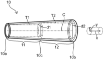
- 1 depicts a perspective view of a vessel cover according to an embodiment of the present invention
- FIG. 2 depicts a perspective view of a vessel cover according to another embodiment of the present invention
- FIG. 10 depicts a perspective view of a vessel cover according to yet another embodiment of the present invention
- FIG. 10 depicts a perspective view of a vessel cover according to yet another embodiment of the present invention
- FIG. 10 depicts a perspective view of a vessel cover according to yet another embodiment of the present invention
- FIG. 1 shows a schematic diagram of a case in which an autologous vein is anastomosed to a small incision of an artery in a shunt-creating part
- FIG. 10 is a schematic diagram when a vein is anastomosed to the other end of the .
- 3-5 depict perspective views of vessel covers according to different embodiments.
- 6 and 7 present perspective views of vascular covers according to yet other different embodiments.
- the shunt-constructed part 1 can be formed by performing an arteriovenous anastomosis as shown in FIG. 1 or an artificial blood vessel-venous anastomosis as shown in FIG.
- the shunt section 1 can be formed by anastomosing the vein 4 to the small incision of the artery 3 in the arm 2 and allowing the blood flow of the artery 3 to flow through the vein 4.
- blood flows from the artery 3 through the anastomosis 6 and the vein 4 in the direction indicated by the arrow B.
- the shunt-constructing part 1 can be formed by anastomosing one end of an artificial blood vessel 5 to a small incision portion of an artery 3 in an arm 2 and anastomosing a vein 4 to the other end of the artificial blood vessel 5.
- the blood flows from the artery 3 through the artificial blood vessel 5, the anastomosis 6, and the vein 4 in the direction indicated by the arrow B.
- the arteriovenous anastomosis shown in FIG. 1 or the artificial vessel-vein anastomosis shown in FIG. It can be arranged on the outer peripheral side, and the vein 4 can be remodeled into a buffer system blood vessel.
- the vessel cover 10 is preferably arranged from the most upstream part of the vein 4 on the anastomotic part 6 side. At this time, one end 10a of the vessel cover 10 is arranged at the most upstream portion of the vein 4 on the anastomosis portion 6 side, and the other end 10b of the vessel cover 10 is arranged at the downstream side of the vein 4 away from the anastomosis portion 6. is preferred.
- the blood vessel cover 10 is formed in a continuous cylindrical shape over the entire circumference, and has an axial direction x and a radial direction y.
- the axial direction x of the vessel cover 10 is the direction in which the central axis C of the vessel cover 10 extends. is the direction connecting points on the outer edge of
- the vascular cover 10 may be a knitted fabric, woven fabric, or net that is continuously constructed all around. Knitted fabrics, woven fabrics, nets, and the like have stitches and textures, but the gaps formed in the stitches and textures are not discontinuous portions of the vascular cover 10, but the above-mentioned "continuous can form a "cylindrical".
- the blood vessel cover 10 has flexibility, and it is preferable that the axial direction x of the blood vessel cover 10 can be curved to follow the extending direction of the veins 4 to be covered.
- the vessel cover 10 preferably has a lumen with a circular or elliptical shape in cross section in the radial direction y.
- the outer edge of the cross section in the radial direction y may have fine irregularities.
- the vessel cover 10 may be in a state in which the lumen is crushed by its own weight in the natural state. Even in such a case, it is possible to define the same cross-sectional shape in the axial direction x, radial direction y, and radial direction y by widening the lumen.
- a method for widening the lumen collapsed by its own weight for example, a tube whose lumen does not collapse under its own weight and has a central axis parallel to the central axis C of the vessel cover 10 and inscribed in the inner wall of the vessel cover 10 is used. into the lumen of the vessel cover 10, and the like.
- the vessel cover 10 is a tubular cover arranged on the outer peripheral side of the vein 4 anastomosed with the artery 3 or the artificial blood vessel 5.
- the portion from the midpoint 10c between the one end 10a and the other end 10b of the vessel cover 10 to the one end 10a is the first portion 11, and the portion from the midpoint 10c to the other end 10b is the second portion 12.
- the diameter d2 of the second virtual cylinder T2 which has a central axis parallel to the central axis C of the vessel cover 10 and is inscribed in the inner wall of the second part 12, has a central axis parallel to the central axis C of the vessel cover 10. It is larger than the diameter d1 of the first imaginary cylinder T1 that contacts the inner wall of the first portion 11 .
- the minimum inner diameter of the second portion 12 can be made larger than the minimum inner diameter of the first portion 11 of the vessel cover 10 .
- the vein 4 is more downstream than the upstream side. Can be loosely covered.
- the vein 4 is remodeled so that the pulsatile blood flow flowing into the vein 4 and having high arterial pressure is gradually buffered toward the downstream and finally becomes a buffer system blood vessel that can shift to the venous blood flow. can do.
- blood turbulence and pulsating changes in the vein wall are suppressed, and lesions such as intimal hyperplasia can be prevented.
- the wall of the vein 4 of the shunt-constructed portion 1 has a smooth muscle layer that is thicker than the smooth muscle layer of the normal vein and includes an elastic fiber layer, and an elastic layer that includes collagen fibers that are thicker than the smooth muscle layer on the outside of the smooth muscle layer.
- Blood vessels are composed of three layers: the intima, the media, and the adventitia. Among them, the intima greatly contributes to anticoagulation, but its mechanical contribution is extremely small.
- the mechanical elements of the arteries of the extremities which are normal arteries used for artificial dialysis, consist of the media, which contains a small amount of elastic fibers and abundant smooth muscle, and the outer membrane, which consists of elastic fibers and collagen fibers. account for the percentage. Thus, these arteries have a great abundance of smooth muscle and relatively few elastic fibers (“smooth muscle>elastic fiber” configuration).
- elastic fibers Due to their elasticity, elastic fibers have a cushioning function like a rubber tube that resists and cushions the pulsatile and high blood pressure of arteries.
- smooth muscle since smooth muscle is a muscle, it also has a more active mechanical function, resisting arterial blood pressure, while on the other hand delivering high pulsatile arterial blood pressure to the periphery without attenuation. It has an active function. Due to the pressure-transmitting function of the abundant smooth muscle of the arteries, the blood pressure of the aorta, which has an inner diameter on the order of a centimeter, and the small artery, which has an inner diameter of only a fraction of a millimeter, hardly changes.
- the venous wall changes like a normal artery, it is arterialization (remodeling into an artery) and not into a buffer system vessel. If the arterialization gradually weakens and naturally transitions downstream to a completely normal vein, it means that it gradually thins and transitions to the vein with its "smooth muscle > elastic fiber" configuration. , high pulsatile blood pressure acts on the venous wall due to its lack of buffering function, resulting in pathological changes in downstream veins. This point is a clear functional difference between the case where the buffer system blood vessel gradually thins and shifts to a normal vein, and the case where a normal artery-like blood vessel gradually thins and shifts to a normal vein.
- the vascular cover 10 has the above-described configuration, so that the downstream side of the vein 4 of the shunt-constructing portion 1 is more loosely covered, thereby changing the wall structure of the vein 4 as described above. can be remodeled into a buffer system of blood vessels.
- the vein 4 may gradually grow outward in the course of being remodeled into a buffer system vessel, but the vessel cover 10 according to the embodiment of the present invention can more loosely cover the downstream side of the vein 4. Therefore, inhibition of the growth of the vein 4 by the vascular cover 10 is reduced, and the lumen of the vein 4 can be kept wide to ensure a sufficient blood flow.
- the vascular cover 10 according to the embodiment of the present invention, it is possible to form the shunt-constructed portion 1 that can ensure a sufficient blood flow while suppressing lesions such as intimal hyperplasia.
- the blood vessel cover 10 covers not only the vein 4 but also a part of the artery 3 and the artificial blood vessel 5 on the anastomosis 6 side. may be arranged to cover the surface.
- the blood vessel cover 10 may have a tubular shape, and may have a joint portion, for example, formed by rolling a flat plate member into a tubular shape and joining them by suturing. In that case, it is preferable that the joint portion such as the suture portion is formed on the outer surface of the vessel cover 10 . This prevents the joint from affecting the vein 4 .
- a seamless cylindrical member having no joints may be formed by using a molded member or knitted fabric.
- the first virtual cylinder T1 is inscribed in the inner wall of the first portion 11 at one end 10a of the vessel cover 10, and is inscribed in the inner wall of the first portion 11 at portions other than the one end 10a. It doesn't have to be. That is, the first portion 11 of the vessel cover 10 has an inner diameter d1 at the one end 10a, and may have an inner diameter larger than d1 at portions other than the one end 10a.
- the second virtual cylinder T2 is inscribed in the inner wall of the second portion 12 at the midpoint 10c of the blood vessel cover 10, and is in contact with the inner wall of the second portion 12 at portions other than the midpoint 10c. It does not have to be inscribed.
- the second portion 12 of the vessel cover 10 has an inner diameter d2 at the midpoint 10c, and may have an inner diameter larger than d2 at portions other than the midpoint 10c.
- An example of such a shape of the vessel cover 10 is a tapered shape in which the inner diameter gradually increases from one end 10a to the other end 10b.
- the inner diameter of the first portion 11 is the minimum value of the inner diameter of the second portion 12 throughout the axial direction x of the first portion 11. or smaller than the minimum inner diameter of the second portion 12 .
- the first imaginary column T1 is inscribed in the inner wall of the first portion 11 at a portion from one end 10a of the vessel cover 10 to a predetermined position in the axial direction x, and includes the one end 10a. Parts other than this part may not be inscribed in the inner wall of the first part 11 . That is, the first portion 11 of the vascular cover 10 has an inner diameter d1 in a portion from the one end 10a of the vascular cover 10 to a predetermined position, and has an inner diameter larger than d1 in other portions. good too.
- the portion of the vascular cover 10 with the smallest inner diameter is the largest on the anastomosis portion 6 side of the vein 4.
- the upstream part can be covered, and since it can be covered with the vessel cover 10 whose inner diameter gradually increases as it goes downstream of the vein 4, it can be loosely covered as it goes downstream of the vein 4, and the buffer system of the vein 4 Remodeling into a blood vessel and securing the lumen diameter of the vein 4 are facilitated.
- the first imaginary cylinder T1 is inscribed in the inner wall of the first portion 11 at any position between the one end 10a and the midpoint 10c of the vessel cover 10 in the axial direction x.
- the portion other than the position may not be inscribed in the inner wall of the first portion 11 . That is, the first portion 11 of the vessel cover 10 has an inner diameter d1 at a position between the one end 10a and the midpoint 10c of the vessel cover 10, and has an inner diameter larger than d1 at other portions. You may have
- the inner diameter of the first portion 11 over the entire axial direction x of the first portion 11 is the same as the minimum inner diameter of the second portion 12, or is larger than the minimum inner diameter of the second portion 12. is preferably small.
- the first virtual cylinder T1 may be inscribed in the inner wall of the first portion 11 over the entire section from the one end 10a of the vessel cover 10 to the midpoint 10c in the axial direction x. That is, the first portion 11 of the vessel cover 10 may have the inner diameter d1 over the entire axial direction x.
- the second virtual cylinder T2 may be inscribed in the inner wall of the second part 12 at a portion other than the midpoint 10c of the blood vessel cover 10.
- the second portion 12 may have an inner diameter larger than d2 except for the portion where the second virtual cylinder T2 is inscribed.
- the inner diameter of the second portion 12 is preferably 2.5 times or less, more preferably 2 times or less, the diameter d2 of the second imaginary cylinder T2 throughout the axial direction x. 5 times or less is more preferable.
- the inner diameter of the second portion 12 at the other end 10b is preferably larger than the inner diameter of the second portion 12 at the midpoint 10c.
- the second virtual cylinder T2 may be inscribed in the inner wall of the second portion 12 over the entire section from the midpoint 10c of the vessel cover 10 to the other end 10b in the axial direction x. That is, the second portion 12 of the vessel cover 10 may have an inner diameter d2 along the entire axial direction x.
- the diameter d2 of the second virtual column T2 is larger than the diameter d1 of the first virtual column T1, so that the one end 10a of the vessel cover 10 is positioned upstream of the vein 4 of the shunt-constructing portion 1.
- the downstream side of the vein 4 can be more loosely covered, and the above effect can be obtained.
- FIG. 6 shows a perspective view of a blood vessel cover 10 according to another embodiment.
- the blood vessel cover 10 of this aspect has a one end portion 110 which is a section from the one end 10a of the blood vessel cover 10 to the midpoint of the first portion 11 in the axial direction x of the blood vessel cover 10 and has a first inner diameter d5.
- the other end 10b having a third inner diameter d7 that is twice or more, the first transition portion 110m having a gradually increasing inner diameter between the one end 110 and the intermediate portion 120, and the intermediate portion 120 and the other end 10b and a second transition portion 120m of gradually increasing inner diameter.
- the vein 4 moves downstream in the process of remodeling the vein 4 into a buffer system vessel. Since it can spread slowly outward in the radial direction y as it goes, remodeling of the vein 4 into a buffer system vessel is facilitated, and the lumen of the vein 4 is easily maintained to ensure the blood flow.
- the other end 10b of the vessel cover 10 is a portion where the suppression of the vein 4 by the vessel cover 10 is abruptly released. , the influence of abrupt release of the suppression by the covering of the vessel cover 10 on the vein 4 can be mitigated.
- the boundary between the one end portion 110 and the first transition portion 110m, the boundary between the first transition portion 110m and the intermediate portion 120, and the intermediate portion 120 and the second transition portion is preferably curved. Since the one end portion 110, the intermediate portion 120, and the other end 10b have different inner diameters by a factor of 1.2 or more, the inner wall of the vessel cover 10 has a step in the axial direction x. , the steps can be smoothed by forming the respective boundaries as curved lines as described above. As a result, the blood vessel can be covered with the vessel cover 10 having a smooth lumen wall, and remodeling into a buffer system vessel is facilitated.
- the force required to expand the inner wall of the second portion 12 by 1.5 times in the radial direction y from the natural state is required to expand the inner wall of the first portion 11 by 1.5 times in the radial direction y from the natural state. is preferably less than the force
- a resin tube is inserted into the lumen of the vessel cover 10 so as to be inscribed with the inner wall of the first part 11, A method of applying pressure by introducing a fluid into the lumen of the resin tube can be used.
- the inner wall of the second portion 12 can be expanded 1.5 times in the radial direction y from the natural state.
- a cylindrical sample is prepared by cutting out a portion of the first part 11 so as to have a predetermined length in the axial direction x, and two pins are attached to the inner diameter of the cylindrical sample in parallel with the axial direction of the cylindrical sample.
- the two pins may be inserted and pulled in opposite diametrical y directions so that the inner diameter of the cylindrical sample is 1.5 times larger.
- the inner wall of the second part 12 can be similarly expanded in the radial direction y by 1.5 times from the natural state, but the length of the cylindrical sample cut out from the second part 12 in the axial direction x is The length in the axial direction x of the cylindrical sample cut out from the first part 11 is set to be the same.
- the downstream side of the vein 4 is more loosely covered when the first portion 11 of the vascular cover 10 is arranged upstream of the vein 4 of the shunt-constructing portion 1 . This makes it easier to remodel the vein 4 into a buffer system vessel and effectively secure the lumen diameter of the vein 4 .
- the length of the blood vessel cover 10 in the axial direction x is preferably 5 mm or more.
- the length of the blood vessel cover 10 in the axial direction x is preferably 10 mm or longer, more preferably 20 mm or longer, particularly preferably 30 mm or longer, and may be 40 mm or longer.
- the length of the blood vessel cover 10 in the axial direction x is preferably 120 mm or less, more preferably 100 mm or less, and even more preferably 90 mm or less. If the length of the vascular cover 10 in the axial direction x is within the above range, the vein 4 of the shunt-constructed portion 1 can be covered with the vascular cover 10 having a length greater than or equal to the predetermined length, and the vein 4 can be reconnected to the buffer system blood vessel. Easier to model.
- the inner diameter of the vessel cover 10 is preferably 2 mm or more, more preferably 3 mm or more, even more preferably 4 mm or more, further preferably 5 mm at the portion having the minimum inner diameter in the first portion 11, that is, the portion inscribed with the first virtual cylinder T1.
- the above is particularly preferable, and 10 mm or less is preferable, 8 mm or less is more preferable, and 6 mm or less is even more preferable.
- the inner diameter of the vessel cover 10 is preferably 4 mm or more, more preferably 5 mm or more, and even more preferably 6 mm or more at the portion having the minimum inner diameter in the second portion 12, that is, the portion inscribed with the second virtual cylinder T2.
- vascular cover 10 7 mm or more is particularly preferable, 12 mm or less is preferable, 10 mm or less is more preferable, 9 mm or less is even more preferable, and 8 mm or less is particularly preferable.
- fine unevenness may occur on the inner wall of the vascular cover 10, making it difficult to determine the inner diameter.
- the diameter d1 of the inscribed first imaginary cylinder T1 and the diameter d2 of the second imaginary cylinder T2 inscribed in the second part 12 can be used as the inner diameter of the vessel cover .
- the vascular cover 10 preferably has at least one of a knitted fabric, a woven fabric, and a nonwoven fabric as a component that partially configures it or as a component that configures the whole. With these materials, it is easy to form the elastically deformable blood vessel cover 10 .
- the type of knitted fabric is not particularly limited, and may be warp knitted or weft knitted. Knitting structures of warp knitting include half knitting, back half knitting, queens coat knitting, and satin knitting. Weft knitting includes circular knitting and flat knitting, and knitting structures of weft knitting include plain knitting, rubber knitting, double-sided knitting, milanese rib knitting, and jacquard knitting.
- the knitted fabric is preferably composed of weft knitting from the viewpoint of excellent stretchability.
- the type of woven fabric is not particularly limited, and may be plain weave, twill weave, satin weave, or the like.
- the blood vessel cover 10 may be composed of a nonwoven fabric produced by any method such as meltblowing, needle punching, spunlacing, electrospinning, or the like.
- the blood vessel cover 10 may be composed of a combination of two or more different materials, for example, one part is composed of a knitted fabric and the other part is composed of another material such as a non-woven fabric.
- yarns forming knitted fabrics, woven fabrics, and non-woven fabrics are also made of resin materials with superior plastic deformation, such as polyolefin resins such as polyethylene and polypropylene; polyamide resins such as nylon; polyethylene terephthalate. polyimide resins; fluorine resins such as PTFE, PFA and ETFE; synthetic resins such as polyvinyl chloride resins.
- resin materials with superior plastic deformation such as polyolefin resins such as polyethylene and polypropylene; polyamide resins such as nylon; polyethylene terephthalate.
- polyimide resins fluorine resins such as PTFE, PFA and ETFE
- synthetic resins such as polyvinyl chloride resins.
- the yarn forming the knitted fabric or woven fabric can also be made of a resin material (e.g., polyester, PTFE) used for artificial blood vessels, and specifically, ePTFE obtained by stretching PTFE and polyester fiber from DuPont. Dacron (registered trademark) and the like can be mentioned.
- the vessel cover 10 may be made of a biodegradable material such as aliphatic polyester such as polylactic acid, polyglycolic acid, and polyhydroxyalkanoic acid; aliphatic polyether.
- the yarn forming the knitted or woven fabric may be composed of natural fibers such as silk or cotton, or may be composed of a combination of resin materials, biodegradable materials, and natural fibers.
- the blood vessel cover 10 has a bellows structure in which peaks and troughs are periodically repeated in the axial direction x.
- the portion is the one end portion 110
- the evaluation of whether or not the vein 4 is remodeled into the low-pressure buffer system blood vessel needs to be confirmed by both the morphological confirmation method described below and the confirmation method by measuring the buffering action.
- a morphological method it is confirmed whether a two-layer structure consisting of a smooth muscle layer containing morphological elastic fibers and elastic fibers containing collagen fibers thicker than the smooth muscle layer is formed. It can be carried out. Specifically, the vein 4 of the shunt-forming portion 1 is cut out, subjected to special staining such as hematoxylin-eosin (HE) staining and elastica-fungieson (EvG) staining, and the cross section of the vein wall is observed with a microscope.
- HE hematoxylin-eosin
- EvG elastica-fungieson
- smooth muscle is stained turbid yellow, elastic fibers are dark purple, and collagen fibers are stained dark red, so the smooth muscle layer containing elastic fibers and the elastic fiber layer containing collagen fibers are observed, This can be done by confirming "thickness of smooth muscle layer containing elastic fibers ⁇ thickness of elastic fiber layer containing collagen fibers".
- shunt-forming part 2 arm 3: artery 4: vein 5: artificial blood vessel 6: anastomotic part 10: vessel cover 10a: one end 10b of vessel cover: the other end 10c of vessel cover: middle point 11 of vessel cover: First part 12: Second part 110: One end 110m: First transition part 120: Middle part 120m: Second transition part C: Central axis d1 of vessel cover: Diameter d2 of first imaginary cylinder: Second imaginary cylinder Diameter d5: Inside diameter d6 of one end: Inside diameter d7 of intermediate part: Inside diameter of the other end L1: Distance between mountains at one end L2: Distance between mountains at second part T1: First virtual cylinder T2: Second virtual Cylinder x: axial direction y: radial direction
Landscapes
- Health & Medical Sciences (AREA)
- Heart & Thoracic Surgery (AREA)
- Vascular Medicine (AREA)
- General Health & Medical Sciences (AREA)
- Life Sciences & Earth Sciences (AREA)
- Veterinary Medicine (AREA)
- Engineering & Computer Science (AREA)
- Biomedical Technology (AREA)
- Cardiology (AREA)
- Public Health (AREA)
- Animal Behavior & Ethology (AREA)
- Oral & Maxillofacial Surgery (AREA)
- Gastroenterology & Hepatology (AREA)
- Pulmonology (AREA)
- Transplantation (AREA)
- Anesthesiology (AREA)
- Hematology (AREA)
- Prostheses (AREA)
Priority Applications (2)
| Application Number | Priority Date | Filing Date | Title |
|---|---|---|---|
| US18/689,100 US20240366361A1 (en) | 2021-09-08 | 2022-08-22 | Blood vessel cover |
| JP2023546866A JPWO2023037860A1 (https=) | 2021-09-08 | 2022-08-22 |
Applications Claiming Priority (2)
| Application Number | Priority Date | Filing Date | Title |
|---|---|---|---|
| JP2021146521 | 2021-09-08 | ||
| JP2021-146521 | 2021-09-08 |
Publications (1)
| Publication Number | Publication Date |
|---|---|
| WO2023037860A1 true WO2023037860A1 (ja) | 2023-03-16 |
Family
ID=85506579
Family Applications (1)
| Application Number | Title | Priority Date | Filing Date |
|---|---|---|---|
| PCT/JP2022/031605 Ceased WO2023037860A1 (ja) | 2021-09-08 | 2022-08-22 | 血管カバー |
Country Status (3)
| Country | Link |
|---|---|
| US (1) | US20240366361A1 (https=) |
| JP (1) | JPWO2023037860A1 (https=) |
| WO (1) | WO2023037860A1 (https=) |
Citations (2)
| Publication number | Priority date | Publication date | Assignee | Title |
|---|---|---|---|---|
| WO2021161884A1 (ja) * | 2020-02-14 | 2021-08-19 | 明郎 萩原 | 血管吻合部の保護カバー |
| WO2021177273A1 (ja) * | 2020-03-03 | 2021-09-10 | 明郎 萩原 | 静脈カバー |
-
2022
- 2022-08-22 JP JP2023546866A patent/JPWO2023037860A1/ja active Pending
- 2022-08-22 WO PCT/JP2022/031605 patent/WO2023037860A1/ja not_active Ceased
- 2022-08-22 US US18/689,100 patent/US20240366361A1/en active Pending
Patent Citations (2)
| Publication number | Priority date | Publication date | Assignee | Title |
|---|---|---|---|---|
| WO2021161884A1 (ja) * | 2020-02-14 | 2021-08-19 | 明郎 萩原 | 血管吻合部の保護カバー |
| WO2021177273A1 (ja) * | 2020-03-03 | 2021-09-10 | 明郎 萩原 | 静脈カバー |
Also Published As
| Publication number | Publication date |
|---|---|
| US20240366361A1 (en) | 2024-11-07 |
| JPWO2023037860A1 (https=) | 2023-03-16 |
Similar Documents
| Publication | Publication Date | Title |
|---|---|---|
| JP7624235B2 (ja) | 静脈カバー | |
| US6371981B1 (en) | Vascular graft assemblies and methods for implanting same | |
| JP2939337B2 (ja) | 三次元編組軟組織プロテーゼ | |
| US6984243B2 (en) | Abrasion resistant vascular graft | |
| EP3351209B1 (en) | Graft anchor device | |
| CN103200975B (zh) | 吻合装置 | |
| US7905915B2 (en) | Z-stent with incorporated barbs | |
| US20010049554A1 (en) | Endovascular prosthesis and method of making | |
| JP5253499B2 (ja) | 人工血管 | |
| JP2012506726A (ja) | 目標部位を治療するための空隙付き多層器具および関連する方法 | |
| JP2008534108A (ja) | 脈管グラフト | |
| WO2021161884A1 (ja) | 血管吻合部の保護カバー | |
| EP2231068A1 (en) | Vascular graft prosthesis having a reinforced margin for enhanced anastomosis | |
| JP7800895B2 (ja) | 緩衝系人工血管 | |
| RU187447U1 (ru) | Биологический протез артерий с наружным сетчатым трубчатым покрытием внешней стенки | |
| WO2023037859A1 (ja) | 血管カバー | |
| WO2023037861A1 (ja) | 血管カバー | |
| EP1645245B1 (en) | Intracardiac device with sealable fenestration for total cavopulmonary anastomosis by catheterisation | |
| US12310836B2 (en) | Vein cover | |
| WO2023037860A1 (ja) | 血管カバー | |
| JP3686833B2 (ja) | 血管吻合用リング及び血管吻合用リングを装着した人工血管 | |
| JP7668222B2 (ja) | 分岐型ステントグラフトおよびその製造方法 | |
| JP7629700B2 (ja) | 血管プロテーゼ | |
| WO2025084409A1 (ja) | 血管カバー | |
| TW202106259A (zh) | 中心靜脈用覆膜支架 |
Legal Events
| Date | Code | Title | Description |
|---|---|---|---|
| 121 | Ep: the epo has been informed by wipo that ep was designated in this application |
Ref document number: 22867182 Country of ref document: EP Kind code of ref document: A1 |
|
| WWE | Wipo information: entry into national phase |
Ref document number: 2023546866 Country of ref document: JP |
|
| NENP | Non-entry into the national phase |
Ref country code: DE |
|
| 122 | Ep: pct application non-entry in european phase |
Ref document number: 22867182 Country of ref document: EP Kind code of ref document: A1 |