WO2023037859A1 - 血管カバー - Google Patents
血管カバー Download PDFInfo
- Publication number
- WO2023037859A1 WO2023037859A1 PCT/JP2022/031604 JP2022031604W WO2023037859A1 WO 2023037859 A1 WO2023037859 A1 WO 2023037859A1 JP 2022031604 W JP2022031604 W JP 2022031604W WO 2023037859 A1 WO2023037859 A1 WO 2023037859A1
- Authority
- WO
- WIPO (PCT)
- Prior art keywords
- cover
- vein
- blood vessel
- axial direction
- vascular
- Prior art date
- Legal status (The legal status is an assumption and is not a legal conclusion. Google has not performed a legal analysis and makes no representation as to the accuracy of the status listed.)
- Ceased
Links
Images


Classifications
-
- A—HUMAN NECESSITIES
- A61—MEDICAL OR VETERINARY SCIENCE; HYGIENE
- A61F—FILTERS IMPLANTABLE INTO BLOOD VESSELS; PROSTHESES; DEVICES PROVIDING PATENCY TO, OR PREVENTING COLLAPSING OF, TUBULAR STRUCTURES OF THE BODY, e.g. STENTS; ORTHOPAEDIC, NURSING OR CONTRACEPTIVE DEVICES; FOMENTATION; TREATMENT OR PROTECTION OF EYES OR EARS; BANDAGES, DRESSINGS OR ABSORBENT PADS; FIRST-AID KITS
- A61F2/00—Filters implantable into blood vessels; Prostheses, i.e. artificial substitutes or replacements for parts of the body; Appliances for connecting them with the body; Devices providing patency to, or preventing collapsing of, tubular structures of the body, e.g. stents
- A61F2/02—Prostheses implantable into the body
- A61F2/04—Hollow or tubular parts of organs, e.g. bladders, tracheae, bronchi or bile ducts
- A61F2/06—Blood vessels
-
- A—HUMAN NECESSITIES
- A61—MEDICAL OR VETERINARY SCIENCE; HYGIENE
- A61M—DEVICES FOR INTRODUCING MEDIA INTO, OR ONTO, THE BODY; DEVICES FOR TRANSDUCING BODY MEDIA OR FOR TAKING MEDIA FROM THE BODY; DEVICES FOR PRODUCING OR ENDING SLEEP OR STUPOR
- A61M1/00—Suction or pumping devices for medical purposes; Devices for carrying-off, for treatment of, or for carrying-over, body-liquids; Drainage systems
- A61M1/36—Other treatment of blood in a by-pass of the natural circulatory system, e.g. temperature adaptation, irradiation ; Extra-corporeal blood circuits
-
- A—HUMAN NECESSITIES
- A61—MEDICAL OR VETERINARY SCIENCE; HYGIENE
- A61F—FILTERS IMPLANTABLE INTO BLOOD VESSELS; PROSTHESES; DEVICES PROVIDING PATENCY TO, OR PREVENTING COLLAPSING OF, TUBULAR STRUCTURES OF THE BODY, e.g. STENTS; ORTHOPAEDIC, NURSING OR CONTRACEPTIVE DEVICES; FOMENTATION; TREATMENT OR PROTECTION OF EYES OR EARS; BANDAGES, DRESSINGS OR ABSORBENT PADS; FIRST-AID KITS
- A61F2230/00—Geometry of prostheses classified in groups A61F2/00 - A61F2/26 or A61F2/82 or A61F9/00 or A61F11/00 or subgroups thereof
- A61F2230/0063—Three-dimensional shapes
- A61F2230/0069—Three-dimensional shapes cylindrical
-
- A—HUMAN NECESSITIES
- A61—MEDICAL OR VETERINARY SCIENCE; HYGIENE
- A61F—FILTERS IMPLANTABLE INTO BLOOD VESSELS; PROSTHESES; DEVICES PROVIDING PATENCY TO, OR PREVENTING COLLAPSING OF, TUBULAR STRUCTURES OF THE BODY, e.g. STENTS; ORTHOPAEDIC, NURSING OR CONTRACEPTIVE DEVICES; FOMENTATION; TREATMENT OR PROTECTION OF EYES OR EARS; BANDAGES, DRESSINGS OR ABSORBENT PADS; FIRST-AID KITS
- A61F2250/00—Special features of prostheses classified in groups A61F2/00 - A61F2/26 or A61F2/82 or A61F9/00 or A61F11/00 or subgroups thereof
- A61F2250/0014—Special features of prostheses classified in groups A61F2/00 - A61F2/26 or A61F2/82 or A61F9/00 or A61F11/00 or subgroups thereof having different values of a given property or geometrical feature, e.g. mechanical property or material property, at different locations within the same prosthesis
- A61F2250/0018—Special features of prostheses classified in groups A61F2/00 - A61F2/26 or A61F2/82 or A61F9/00 or A61F11/00 or subgroups thereof having different values of a given property or geometrical feature, e.g. mechanical property or material property, at different locations within the same prosthesis differing in elasticity, stiffness or compressibility
Definitions
- the present invention relates to a vascular cover used for an anastomosis in which blood vessels are anastomosed together.
- the present invention relates to a blood vessel cover that can be used by arranging it on the outer peripheral side of the.
- Dialysis treatment is given regularly.
- a special needle is inserted into a vein.
- the artery is anastomosed to the vein because the normal venous blood flow is not sufficient for dialysis.
- a blood vessel is called a shunt.
- an incision is made in the skin of the arm to expose the artery and vein, a small incision is made in the artery, the vein is anastomosed, and part of the blood flow from the artery is diverted to the vein.
- a shunt provided with an artificial blood vessel may be used.
- this unusual blood flow state is within the range that the body can tolerate, appropriate remodeling due to changes in the elasticity of the venous wall will occur as the body's defense and adaptive response, resulting in stenosis and occlusion due to intimal thickening.
- the shunt blood flow state can be self-adjusted to a state that does not burden the living body.
- this unusual blood flow condition exceeds the local condition of the shunt or the systemic condition (diabetes, hypertension, arteriosclerosis, blood condition, etc.), an appropriate protective/adaptive response occurs. It becomes a pathological biological reaction and causes local and systemic pathology.
- Non-Patent Document 1 in order to suppress a rapid increase in blood flow immediately after surgery and in the initial stage, the vein wall is reinforced from the outside to prevent excessive blood pressure in the inner vein.
- vascular banding is performed to prevent hyperextension and blood turbulence caused by this.
- US Pat. No. 5,300,000 a woven net made by forming a knitted fabric that is seamless, tubular, substantially pileless, is a covering for reinforcing natural veins for use as surgical implants. is disclosed.
- vascular banding as described above could not sufficiently prevent lesions such as intimal hyperplasia.
- the reinforced vein wall is altered (arterialized) into an arterial wall-like structure only under certain conditions, but when blood flows from the reinforced site to the unreinforced vein, blood pressure and Since the pulsation is delivered downstream as it is without being buffered, the cause of intimal hyperplasia has not been fundamentally resolved.
- blood pressure and pulsatility are gradually lowered from the anastomotic site downstream, and the most downstream side of the vein is remodeled to a state in which only low pressure is applied without pulsation, that is, a state of low-pressure buffered blood vessels. There is a need.
- the present invention has been made in view of the above circumstances, and remodels the vein into a buffer system vessel that can deliver blood to the downstream vein while gradually reducing the blood pressure, pulse pressure, and blood flow rate of the blood flowing through the lumen.
- An object of the present invention is to provide a vascular cover capable of preventing intimal hyperplasia.
- a blood vessel cover according to an embodiment of the present invention which can solve the above problems, is as follows.
- a tubular vascular cover that is continuous over the entire circumference and is placed on the outer peripheral side of a vein that is anastomosed with an artery or an artificial blood vessel, the inner diameter of the vascular cover being radially expanded by 20% from the natural state. It has a portion (A) in which the value (hereinafter referred to as 20% elastic index) measured by the following measuring method is 1.2 N or less.
- 20% elastic index the value measured by the following measuring method is 1.2 N or less.
- a first pin and a second pin having a diameter d of 0.75 mm are inserted into the lumen of the tubular sample parallel to the axial direction of the tubular sample.
- the first pin is fixed, the second pin is pulled radially outward of the cylindrical sample, and when the distance between the first pin and the second pin is L, ⁇ d+2L is 1.1.1 of the circumference of the cylindrical sample in its natural state.
- the force F 1.2 at the time of doubling is measured, and the value obtained by dividing the force F 1.2 by the strain [(1.2-1.0)/1.0] is taken as the 20% elastic index.
- the vascular cover having the above configuration has a portion (A) with a small 20% elastic index of 1.2 N or less, the vein is loosely covered when placed on the outer peripheral side of the vein in the shunt-forming portion.
- the pulsatile blood flow with high arterial pressure that flows into the vein of the shunt is gradually buffered by the part covered with the vascular cover, and finally becomes a buffer system blood vessel that can shift to venous blood flow.
- the vein at the shunt site can be remodeled.
- the vein may gradually grow outward during the process of remodeling into a buffer system vessel, but the vascular cover having the above structure has a small portion (A) with a 20% elastic index of 1.2 N or less.
- the vascular cover of the present invention enables shunt construction that secures a sufficient blood flow while suppressing lesions such as intimal hyperplasia by remodeling veins into buffer-system vessels.
- a blood vessel cover according to an embodiment of the present invention preferably has the following [2] to [12].
- the portion (A) is the vascular cover according to [1] or [2], which is arranged in the first part. Since the portion (A) having a 20% elastic index of 1.2 N or less is arranged at least in the first portion, when the one end of the vascular cover is placed on the upstream side of the vein of the shunt construction portion, the vein is Because the upstream side can be loosely covered, changes that remodel the vein into a buffered vessel can be effected from the upstream. This facilitates remodeling of veins into buffer system blood vessels and securing of blood flow.
- the vascular cover according to any one of [1] to [4], wherein the entire vascular cover is the portion (A).
- the 20% elastic index can be set to 1.2 N or less over the entire axial direction of the vessel cover, so that the entire range of the vein covered by the vessel cover can be loosely covered, and the venous buffer system can be applied to the vessel. remodeling becomes easier, and it becomes easier to maintain a wide lumen of the vein, which leads to securing blood flow.
- the portion from the midpoint between one end and the other end of the vessel cover to the one end is the first portion
- the portion from the midpoint to the other end is the second portion
- from the one end of the vessel cover The part from the middle point of the first part to the middle point is the one end part
- the part from the middle point of the first part to the middle point of the second part is the middle part
- the part from the middle point of the second part to the other end of the vessel cover is The other end satisfies the relationship Ea>Ec>Eb, where Ea is the 20% elastic index at the one end, Ec is the 20% elastic index at the intermediate portion, and Eb is the 20% elastic index at the other end. 1] to [6], the blood vessel cover.
- the vein when the one end of the vascular cover is arranged upstream of the vein of the shunt-created portion, the vein can be loosely covered in the order from upstream to midstream and further downstream, so that the vein can be routed to the buffer system vessel.
- Modeling changes can be introduced gradually from upstream to midstream to downstream. This facilitates remodeling of veins into buffer system blood vessels and securing of blood flow.
- the vascular cover according to any one of [1] to [7], wherein the length of the portion (A) in the axial direction is 50% or more of the outer diameter of the anastomosed artery or artificial blood vessel.
- the portion (A) of the vascular cover having a 20% elastic index of 1.2 N or less has a predetermined length or longer, so that the vein covered with the vascular cover is remodeled into a buffer system vessel and blood flow is ensured. becomes easier.
- vascular cover according to any one of [1] to [9], wherein the axial length of the vascular cover is 5 mm or more.
- vascular cover has at least one of knitted fabric, woven fabric, and non-woven fabric as a component constituting a part or a component constituting the whole. Vessel cover as described.
- the vascular cover has a bellows structure in which peaks and troughs are periodically repeated in the axial direction.
- the first part the part from the middle point to the other end is defined as the second part
- the part from one end of the blood vessel cover to the middle point of the first part is defined as the one end
- the second part is adjacent to the second part in the axial direction.
- the wall structure of the vein is gradually changed downstream from the anastomosis, and the inside of the covered vein is subjected to gradient shear stress, It is possible to change the pressure perpendicular to the blood vessel wall, the blood flow, the blood flow velocity, and the width of change associated with pulsation, thereby suppressing the mismatch of blood vessel wall elasticity, blood turbulence, and excessive high blood flow, Intimal hyperplasia can be prevented.
- the reasons why the blood vessel cover of the present invention has such effects are considered as follows.
- arteries and veins consist of the intima, media, and adventitia.
- the media consists of a smooth muscle layer rich in smooth muscle cells and an elastic fiber layer containing collagen fibers.
- Arteries have thick smooth muscle layers and elastic fiber layers so that even when the pressure of pulsating luminal blood flow is applied, there is little pulsation change in the vascular wall, and the occurrence of turbulence and fluctuations in frictional stress are minimized. have.
- Veins on the other hand, have thin blood vessel walls and do not have thick smooth muscle layers and elastic fiber layers like arteries.
- the vascular cover of the present invention has the above configuration, so that the pulsatile blood flow with high arterial pressure that flows into the vein at the arteriovenous anastomosis of the shunt site or the artificial blood vessel-venous anastomosis is directed downstream.
- the vein at the shunt site can be remodeled into a buffered vessel that can be gradually buffered and eventually transitioned to venous blood flow. As a result, blood turbulence and pulsating changes in the vein wall are suppressed, and lesions such as intimal hyperplasia can be prevented.
- the vascular cover of the present invention having the above structure does not interfere with the growth of veins, which may gradually grow outward during the process of remodeling into low-pressure buffered vessels, and as a result, the vascular lumen is closed. It can be maintained widely to ensure sufficient blood flow. As a result, it becomes possible to create a shunt that can secure a sufficient blood flow while suppressing lesions such as intimal hyperplasia.
- FIG. 11 shows a schematic diagram of another example of a shunt-forming portion
- FIG. 10 is a perspective view showing a vascular cover according to an embodiment of the present invention arranged on the outer peripheral side of the vein in the shunt-created portion
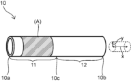
- 1 depicts a perspective view of a vessel cover according to an embodiment of the present invention
- FIG. 2 depicts a perspective view of a vessel cover according to another embodiment of the present invention
- FIG. 10 depicts a perspective view of a vessel cover according to yet another embodiment of the present invention
- FIG. 10 depicts a perspective view of a vessel cover according to yet another embodiment of the present invention
- FIG. 3 shows a perspective view showing a method of measuring elastic index.
- FIG. 9 shows a plan view when FIG. 8 is viewed from above
- FIG. 10 depicts a perspective view of a vessel cover according to yet another embodiment of the present invention
- FIG. 1 shows a schematic diagram of a case in which an autologous vein is anastomosed to a small incision of an artery in a shunt-creating part
- FIG. 10 is a schematic diagram when a vein is anastomosed to the other end of the .
- FIG. 3 shows a perspective view when the blood vessel cover according to one embodiment of the present invention is arranged on the outer peripheral side of the vein in the shunt-created portion.
- 4-7 and 10 depict perspective views of vessel covers according to different embodiments.
- FIG. 8 represents a perspective view showing a method of measuring the elastic index
- FIG. 9 represents a plan view when FIG. 8 is viewed from above.
- the shunt-constructed part 1 can be formed by performing an arteriovenous anastomosis as shown in FIG. 1 or an artificial blood vessel-venous anastomosis as shown in FIG.
- the shunt-forming part 1 can be formed by anastomosing a vein 4 to a small incision of an artery 3 in an arm 2 to allow the blood flow of the artery 3 to flow into the vein 4 .
- blood flows from the artery 3 through the anastomosis 6 and the vein 4 in the direction indicated by the arrow B.
- the shunt-constructing part 1 can be formed by anastomosing one end of an artificial blood vessel 5 to a small incision portion of an artery 3 in an arm 2 and anastomosing a vein 4 to the other end of the artificial blood vessel 5.
- the blood flows from the artery 3 through the artificial blood vessel 5, the anastomosis 6, and the vein 4 in the direction indicated by the arrow B.
- the arteriovenous anastomosis shown in FIG. 1 or the artificial vessel-vein anastomosis shown in FIG. It can be arranged on the outer peripheral side, and the vein 4 can be remodeled into a buffer system blood vessel.
- the vessel cover 10 is preferably arranged from the most upstream part of the vein 4 on the anastomotic part 6 side.
- the vessel cover 10 has one end 10a and the other end 10b. 10b is preferably arranged.
- the blood vessel cover 10 covers not only the vein 4 but also a part of the artery 3 and the artificial blood vessel 5 on the anastomosis 6 side. may be arranged to cover the surface.
- the blood vessel cover 10 is formed in a continuous cylindrical shape over the entire circumference, and has an axial direction x and a radial direction y.
- the axial direction x of the vessel cover 10 is the direction in which the central axis C of the vessel cover 10 extends. is the direction connecting points on the outer edge of
- the vascular cover 10 may be a knitted fabric, woven fabric, or net that is continuously constructed all around. Knitted fabrics, woven fabrics, nets, and the like have stitches and textures, but these stitches and textures are not discontinuous portions of the vascular cover 10, but form the above-mentioned "continuous tubular shape" with knitted fabrics, woven fabrics, nets, and the like. be able to.
- the blood vessel cover 10 is flexible, and it is preferable that the axial direction x of the blood vessel cover 10 can be curved to follow the extending direction of the veins 4 to be covered.
- the vessel cover 10 preferably has a lumen with a circular or elliptical shape in cross section in the radial direction y.
- the outer edge of the cross section in the radial direction y may have fine irregularities.
- the vessel cover 10 may be in a state in which the lumen is crushed by its own weight in the natural state. Even in such a case, it is possible to define the same cross-sectional shape in the axial direction x, radial direction y, and radial direction y by widening the lumen.
- a method for widening the lumen collapsed by its own weight for example, a tube whose lumen does not collapse under its own weight and has a central axis parallel to the central axis C of the vessel cover 10 and inscribed in the inner wall of the vessel cover 10 is used. into the lumen of the vessel cover 10, and the like.
- the blood vessel cover 10 may have a tubular shape, and may have a joint portion, for example, formed by rolling a flat plate member into a tubular shape and joining them by a method such as stitching. In that case, it is preferable that the joint portion such as the suture portion is formed on the outer surface of the vessel cover 10 . This prevents the joint from affecting the vein 4 .
- a molded member or a knitted fabric a seamless cylindrical member having no joints may be formed.
- the inner diameter of the vessel cover 10 is the diameter of the lumen in the cross section in the radial direction y, and is the diameter of the circle when the shape of the lumen in the cross section in the radial direction y is circular.
- the inner diameter of the vessel cover 10 can also be defined as a value obtained by dividing the circumference of the inner wall of the vessel cover 10 by the circular constant ⁇ in a cross section in the radial direction y. According to this, the inner diameter of the vessel cover 10 can be obtained even when the cross section of the vessel cover 10 in the radial direction y is not circular or when the lumen of the vessel cover 10 is crushed by its own weight.
- the vascular cover 10 has an elastic index of 1.0 when the inner diameter of the vascular cover 10 is expanded by 20% in the radial direction y from the natural state, measured by the following measuring method (hereinafter referred to as 20% elastic index). It has a portion (A) that is 2N or less.
- the vascular cover 10 is cut out perpendicularly to the axial direction x along the tangent line in the circumferential direction of the vascular cover 10, and a tubular wall having a length of 5 mm in the axial direction x and continuous over the entire circumference in the axial direction x is cut in the wall.
- a sample 100 free of is prepared.
- a first pin 101 and a second pin 102 having a diameter d of 0.75 mm are inserted through the lumen of the tubular sample 100 in parallel with the axial direction of the tubular sample 100 .
- the first pin 101 is fixed, the second pin 102 is pulled radially outward of the cylindrical sample 100, and the distance between the first pin 101 and the second pin 102 is L.
- the cylindrical sample 100 having a length of 5 mm in the axial direction x and continuous over the entire circumference in the axial direction x is a knitted fabric, a woven fabric, or a net that is continuously formed over the entire circumference.
- the gaps formed by stitches and textures of knitted fabrics, woven fabrics, nets, etc. are not included in the "breaks" in the above "unbroken sample”.
- the vein 4 can be loosely covered when placed on the outer peripheral side of the vein 4 of the shunt-forming portion 1 .
- a buffer system blood vessel in which the pulsatile blood flow with high arterial pressure flowing into the vein 4 of the shunt-constructed part 1 is gradually buffered by the portion covered with the vessel cover 10 and finally transferred to the venous blood flow.
- the vein 4 of the shunt-constructed portion 1 can be remodeled so that As a result, blood flow turbulence and pulsating changes in the vein wall are suppressed, and lesions such as intimal hyperplasia can be prevented.
- the wall of the vein 4 of the shunt-constructed portion 1 has a smooth muscle layer that is thicker than the smooth muscle layer of the normal vein and includes an elastic fiber layer, and an elastic layer that includes collagen fibers that are thicker than the smooth muscle layer on the outside of the smooth muscle layer.
- Blood vessels are composed of three layers: the intima, the media, and the adventitia. Among them, the intima greatly contributes to anticoagulation, but its mechanical contribution is extremely small.
- the mechanical elements of the arteries of the extremities which are normal arteries used for artificial dialysis, consist of the media, which contains a small amount of elastic fibers and abundant smooth muscle, and the outer membrane, which consists of elastic fibers and collagen fibers. account for the percentage. Thus, these arteries have a great abundance of smooth muscle and relatively few elastic fibers (“smooth muscle>elastic fiber” configuration).
- elastic fibers Due to their elasticity, elastic fibers have a cushioning function like a rubber tube that resists and cushions the pulsatile and high blood pressure of arteries.
- smooth muscle since smooth muscle is a muscle, it also has a more active mechanical function, resisting arterial blood pressure, while on the other hand delivering high pulsatile arterial blood pressure to the periphery without attenuation. It has an active function. Due to the pressure-transmitting function of the abundant smooth muscle of the arteries, the blood pressure of the aorta, which has an inner diameter on the order of a centimeter, and the small artery, which has an inner diameter of only a fraction of a millimeter, hardly changes.
- the venous wall changes like a normal artery, it is arterialization (remodeling into an artery) and not into a buffer system vessel. If the arterialization gradually weakens and naturally transitions downstream to a completely normal vein, it means that it gradually thins and transitions to the vein with its "smooth muscle > elastic fiber" configuration. , high pulsatile blood pressure acts on the venous wall and causes pathological changes in the downstream veins. This point is a clear functional difference between the case where the buffer system blood vessel gradually thins and shifts to a normal vein, and the case where a normal artery-like blood vessel gradually thins and shifts to a normal vein.
- the vessel cover 10 according to the embodiment of the present invention having the above configuration can loosely cover the vein 4 of the shunt-constructed portion 1, the wall structure of the vein 4 can be changed as described above to form a buffer system vessel. Can be remodeled.
- the vein 4 may gradually grow outward in the process of being remodeled into a buffer system vessel, but the vascular cover 10 according to the embodiment of the present invention has a small 20% elastic index of 1.2N or less ( Since A) is provided, the obstruction by the vessel cover 10 to the growth of the vein 4 is reduced, and the lumen of the vein 4 can be maintained wide to ensure a sufficient blood flow rate.
- the vascular cover 10 according to the embodiment of the present invention, it is possible to form the shunt-constructed portion 1 that can ensure a sufficient blood flow while suppressing lesions such as intimal hyperplasia.
- the 20% elastic index of the portion (A) is preferably 1.1 N or less, more preferably 1 N or less, still more preferably 0.9 N or less, and even if it is 0.8 N or less, 0.6 N or less, or 0.4 N or less. good.
- the 20% elastic index of the portion (A) is preferably 0.1 mN or more, more preferably 0.5 mN or more, and even more preferably 1 mN or more. Since the 20% elastic index of the portion (A) is a predetermined value or more, the blood vessel can be covered with a force of a predetermined value or more even when the blood pressure applied to the blood vessel is low.
- FIG. A cylindrical sample 100 is prepared by cutting the vessel cover 10 perpendicularly to the axial direction x along a circumferential tangent line of the vessel cover 10 . At this time, by cutting out the entire circumference along a cut plane perpendicular to the axial direction x, that is, along a cutting line in the circumferential direction, a cylindrical shape having a length of 5 mm in the axial direction x and continuous over the entire circumference in the axial direction x is obtained. Cut the vessel cover 10 so as to obtain a tubular sample 100 with an unbroken wall. Next, as shown in FIG.
- a first pin 101 and a second pin 102 each having a diameter d of 0.75 mm are inserted into the lumen of the tubular sample 100 in parallel with the axial direction of the tubular sample 100 .
- the lengths of the first pin 101 and the second pin 102 are not particularly limited, they are preferably longer than the length of the cylindrical sample 100 in the axial direction x.
- the distance L between the first pin 101 and the second pin 102 is the distance from the center of the first pin 101 and the second pin 102 as shown in FIG.
- the inner diameter of the cylindrical sample 100 is 1/2 of the circumference ⁇ d of the first pin 101, 1/2 of the circumference ⁇ d of the second pin 102, and 2 of the distance L between the first pin 101 and the second pin 102. equal to the sum of doubles, ie ⁇ d+2L. Therefore, when the inner diameter of the cylindrical sample 100 expands by 20% from its natural state, that is, when ⁇ d+2L becomes 1.2 times the circumference of the cylindrical sample 100 in its natural state, the force F 1.2 pulls the second pin 102.
- the 20% elastic number can be obtained by dividing by the strain [(1.2-1.0)/1.0].
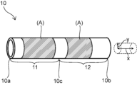
- the portions (A) having a 20% elastic index of 1.2 N or less may be provided continuously in the axial direction x or may be provided at intervals. Portions other than portion (A) of the vessel cover 10 may have a 20% elastic index greater than 1.2 N, but portions other than portion (A) are in the axial direction x of the vessel cover 10. is preferably 75% or less, more preferably 50% or less, even more preferably 30% or less, particularly preferably 10% or less, and most preferably 0% of the total length of the.
- the inner diameter of the blood vessel cover 10 can be set to an appropriate value depending on the diameter of the blood vessel to be applied.
- the inner diameter of the blood vessel cover 10 is preferably 10 mm or less, more preferably 9 mm or less, and even more preferably 8 mm or less.
- the vessel cover 10 may have different inner diameters depending on the axial direction x.
- the vessel cover 10 may have a straight shape with a similar inner diameter from one end 10a to the other end 10b, or may have a tapered shape with an inner diameter that gradually increases from one end 10a to the other end 10b. .
- the blood vessel cover 10 may have a bellows shape in which the inner diameter changes periodically in the axial direction x.
- the portion from the midpoint 10c to the one end 10a between the one end 10a and the other end 10b of the vessel cover 10 is the first portion 11, and the portion from the midpoint 10c to the other end is
- the portion up to 10b is the second portion 12
- the portion (A) is preferably arranged in the first portion 11.
- the portion (A) having a small 20% elastic index of 1.2 N or less is arranged in the first portion 11, when the first portion 11 is arranged on the upstream side of the vein 4 of the shunt-constructing portion 1, the vein 4 Since the upstream side of 4 can be loosely covered, changes that remodel the vein 4 into a buffered vessel can be effected from upstream. This facilitates remodeling of the vein 4 into a buffer system vessel and securing of blood flow.
- the portion (A) may be arranged only in the first part 11 or may be arranged in the second part 12 as well.
- the portion (A) is the one end portion 100a. It is preferably arranged at the end portion 100a. If the portion (A) having a small 20% elastic index of 1.2 N or less is arranged at one end portion 100a, when the one end portion 100a is arranged upstream of the vein 4 of the shunt-constructing portion 1, the vein is 4 can be loosely covered from the most upstream side, and the effect of remodeling the vein 4 into a buffer system vessel and securing the lumen diameter of the vein 4 can be further improved.
- any location may have the portion (A) as long as it satisfies each of the above requirements.
- the entire blood vessel cover 10 is the portion (A).
- the 20% elastic index of the entire vessel cover 10 can be set to 1.2 N or less, so that the entire range of the veins 4 covered by the vessel cover 10 can be loosely covered, and the buffer system vessels of the veins 4 can be It becomes easier to remodel the vein 4, and it becomes easier to maintain a wide lumen of the vein 4, which leads to ensuring blood flow.
- the portion from the midpoint 10c of the one end 10a and the other end 10b of the vessel cover 10 to the one end 10a is referred to as the first portion 11, and the portion from the midpoint 10c to the other end 10b is referred to as the second portion 12.
- the 20% elastic index of the second portion 12 is preferably smaller than the 20% elastic index of the first portion 11 .
- the 20% elastic index of the portion with a length of 5 mm from the middle point 10c to the other end 10b in the axial direction x is the 20% elastic index of the portion with a length of 5 mm from the one end 10a to the other end 10b in the axial direction x. 0.1 times or more and 0.98 times or less, 0.96 times or less, or 0.9 times or less, 0.8 times or less, or 0.7 times or less.
- the 20% elastic index of the portion with a length of 5 mm from the middle point 10c to the other end 10b in the axial direction x is the 20% elastic index of the portion with a length of 5 mm from the one end 10a to the other end 10b in the axial direction x.
- a portion having a length of 5 mm from the midpoint 10c to the other end 10b side in the axial direction x, that is, the one end 10a is arranged upstream of the vein 4 of the shunt construction portion 1.
- the 20% elastic index of the vascular cover 10 covering the downstream side is within the above range, the vein 4 can be gradually remodeled from the upstream side into a buffer system vessel.
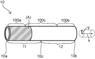
- a portion from a midpoint 10c between one end 10a and the other end 10b of the vessel cover 10 to the one end 10a is defined as a first portion 11, and a portion from the midpoint 10c to the other end 10b is defined as a second portion 12.
- the portion from the one end 10a of the vessel cover 10 to the midpoint of the first portion 11 is the one end portion 100a
- the portion from the midpoint of the first portion 11 to the midpoint of the second portion 12 is the intermediate portion 100c
- the second The portion from the midpoint of the portion 12 to the other end 10b of the vessel cover 10 is defined as the other end portion 100b.
- the vein 4 can be loosely covered in the order from upstream to midstream and then downstream. can be gradually brought about from the upstream to the midstream and further downstream to remodel the blood vessel into a buffer system. This facilitates remodeling of the vein 4 into a buffer system vessel and securing of blood flow.
- the 20% elastic index Ec at the intermediate portion 100c is preferably 0.05 times or more the 20% elastic index Ea at the one end portion 100a, and is preferably 0.8 times or less, 0.6 times or less, and 0.5 times or less. , 0.4 times or less, or 0.3 times or less.
- the 20% elastic index Eb at the other end portion 100b is preferably 0.1 times or more the 20% elastic index Ec at the intermediate portion 100c, preferably 0.9 times or less, 0.8 times or less, and 0.7 times or less. , 0.6 times or less, or 0.5 times or less.
- Ea, Ec, and Eb are preferably 1 mN or more. If Ea, Ec, and Eb are within the above ranges, the vein 4 can be gradually remodeled into a buffer system blood vessel from the upstream side.
- the length of the portion (A) is preferably 50% or more of the outer diameter of the anastomosed artery 3 or artificial blood vessel 5.
- the length of the portion (A) in the axial direction x is more preferably 60% or more, more preferably 80% or more, and may be 100% or more of the outer diameter of the anastomosed artery 3 or artificial blood vessel 5 .
- the upper limit of the length of the portion (A) in the axial direction x is not particularly limited, it may be 2000% or less, 1750% or less, or 1500% or less of the outer diameter of the anastomosed artery 3 or artificial blood vessel 5.
- the portion (A) of the vascular cover 10 having a 20% elastic index of 1.2 N or less has a length greater than or equal to a predetermined length, so that remodeling of the vein 4 covered with the vascular cover 10 into a buffer system blood vessel and blood flow reduction. It becomes easier to secure the flow rate.
- the inner diameter of the vessel cover 10 be expandable in the radial direction y by at least 50% from the natural state over the entire axial direction x.
- the gradual outward growth of the blood vessel covered with the vascular cover 10 is not hindered during the process of remodeling, so that it is easier to maintain a wide vascular lumen and ensure a sufficient blood flow.
- the value of the elastic index of the portion (A) measured by the following measuring method when the inner diameter is expanded 50% in the radial direction y from the natural state (hereinafter referred to as the 50% elastic index) is 3.2 N or less.
- the vascular cover 10 is cut out perpendicularly to the axial direction x along the tangent line in the circumferential direction of the vascular cover 10, and a tubular wall having a length of 5 mm in the axial direction x and continuous over the entire circumference in the axial direction x is cut in the wall.
- a sample 100 free of is prepared.
- a first pin 101 and a second pin 102 having a diameter d of 0.75 mm are inserted through the lumen of the tubular sample 100 in parallel with the axial direction of the tubular sample 100 .
- the first pin 101 is fixed, the second pin 102 is pulled radially outward of the cylindrical sample 100, and the distance between the first pin 101 and the second pin 102 is L.
- the 50% elastic index of the portion (A) is preferably 3.2N or less, more preferably 2.5N or less, still more preferably 2N or less, 1.8N or less, 1.5N or less, 1.2N or less, 1.1N Below, it may be 1N or less. Since the portion (A) has a 50% elastic index within the above range, the blood vessel cover 10 can cover the vein 4 with a predetermined force or less even in a 50% expanded state, and the above effect can be achieved. can.
- the 50% elastic index of the portion (A) is preferably 0.1 mN or more, more preferably 0.5 mN or more, and even more preferably 1 mN or more. Since the 20% elastic index of the portion (A) is a predetermined value or more, the blood vessel can be covered with a force of a predetermined value or more even when the blood pressure applied to the blood vessel is low.
- a blood flow state that normally does not occur in the shunt-constructing part 1, that is, a state in which high-pressure, pulsating arterial blood flow directly acts on the vein wall at high speed is artificially created. If this state, which does not normally occur, is within the range that the body can tolerate, remodeling into buffer system blood vessels will occur as the body's protective/adaptive response to this. However, as mentioned earlier, beyond the range of appropriate protective/adaptive reactions, pathological biological reactions occur. When the inner diameter of the vein expands by 10% to 20% from the natural state, the degree of expansion is relatively small, and the vessel cover 10 controls the expansion of the vein to 20% with a sufficient margin. It means staying within the range of appropriate protective/adaptive reactions, and almost 100% of the occurrence of pathological reactions can be prevented.
- the expansion when evaluated from the viewpoint of the buffering function of arterial blood flow, if the expansion is limited to within 20% by the vessel cover 10, it may not necessarily have a sufficient buffering function, depending on conditions such as the channel length. It is necessary to consider that there is a possibility that arterial blood flow is delayed downstream with insufficient buffering. That is, the 20% elastic index of the vascular covering 10 can be an indicator of the prerequisites for good modeling, since good modeling requires the absence of pathological reactions to occur, but it does not provide positive buffering. As an index of the effect, it is conceivable to consider the 50% elastic index as well.
- the venous diameter continues to dilate by 50% from its natural state, this is obviously not a normal state, and the body has to defend and adapt to buffer and mitigate this unusual blood flow state.
- a reaction ie remodeling into buffer system vessels, occurs.
- the 50% elastic index was determined to reduce both the physical buffering effect of the shunt cover and the vascular-inducing effect of the shunt cover in the venous dilation state, in which the protective/adaptive response of remodeling of the shunt vein into a buffering vessel is actively induced. It can be said that it is an index for comprehensive evaluation. From the above point of view, the performance of the vascular cover can be evaluated using two indices, 20% elastic index and 50% elastic index.
- the method for measuring the 50% elastic index is the above method for measuring the 20% elastic number, except that the force F 1.5 is measured when ⁇ d+2L is 1.5 times the circumference of the cylindrical sample 100 in its natural state. , is the same as the method for measuring the 20% elastic number.
- the inner diameter of the vessel cover 10 is expandable in the radial direction y from the natural state by at least 100% over the entire axial direction x.
- the gradual outward growth of the blood vessel covered with the vascular cover 10 is not hindered during the process of remodeling, so that it is easier to maintain a wide vascular lumen and ensure a sufficient blood flow.
- the length of the blood vessel cover 10 in the axial direction x is preferably 5 mm or more.
- the length of the blood vessel cover 10 in the axial direction x is preferably 10 mm or longer, more preferably 20 mm or longer, particularly preferably 30 mm or longer, and may be 40 mm or longer.
- the length of the blood vessel cover 10 in the axial direction x is preferably 120 mm or less, more preferably 100 mm or less, and even more preferably 90 mm or less. If the length of the vascular cover 10 in the axial direction x is within the above range, the vein 4 of the shunt-constructed portion 1 can be covered with the vascular cover 10 having a length greater than or equal to the predetermined length, and the vein 4 can be reconnected to the buffer system blood vessel. Easier to model.
- the vascular cover 10 preferably has at least one of a knitted fabric, a woven fabric, and a nonwoven fabric as a component that partially configures it or as a component that configures the whole. With these materials, it is easy to form the elastically deformable blood vessel cover 10 .
- the type of knitted fabric is not particularly limited, and may be warp knitted or weft knitted. Knitting structures of warp knitting include half knitting, back half knitting, queens coat knitting, and satin knitting. Weft knitting includes circular knitting and flat knitting, and knitting structures of weft knitting include plain knitting, rubber knitting, double-sided knitting, milanese rib knitting, and jacquard knitting.
- the knitted fabric is preferably composed of weft knitting from the viewpoint of excellent stretchability.
- the type of woven fabric is not particularly limited, and may be plain weave, twill weave, satin weave, or the like.
- the blood vessel cover 10 may be composed of a nonwoven fabric produced by any method such as meltblowing, needle punching, spunlacing, electrospinning, or the like.
- the blood vessel cover 10 may be composed of a combination of two or more different materials, for example, one part is composed of a knitted fabric and the other part is composed of another material such as a non-woven fabric.
- yarns forming knitted fabrics, woven fabrics, and non-woven fabrics are also made of resin materials with superior plastic deformation, such as polyolefin resins such as polyethylene and polypropylene; polyamide resins such as nylon; polyethylene terephthalate. polyimide resins; fluorine resins such as PTFE, PFA and ETFE; synthetic resins such as polyvinyl chloride resins.
- resin materials with superior plastic deformation such as polyolefin resins such as polyethylene and polypropylene; polyamide resins such as nylon; polyethylene terephthalate.
- polyimide resins fluorine resins such as PTFE, PFA and ETFE
- synthetic resins such as polyvinyl chloride resins.
- the yarn forming the knitted fabric or woven fabric can also be made of a resin material (e.g., polyester, PTFE) used for artificial blood vessels, and specifically, ePTFE obtained by stretching PTFE and polyester fiber from DuPont. Dacron (registered trademark) and the like can be mentioned.
- the vessel cover 10 may be made of a biodegradable material such as aliphatic polyester such as polylactic acid, polyglycolic acid, and polyhydroxyalkanoic acid; aliphatic polyether.
- the yarn forming the knitted or woven fabric may be composed of natural fibers such as silk or cotton, or may be composed of a combination of resin materials, biodegradable materials, and natural fibers.
- the blood vessel cover 10 has a bellows structure that periodically repeats peaks and valleys in the axial direction x.
- the distance L2 between adjacent peaks in the second portion 12 in the axial direction x is preferably greater than the distance L1 between the peaks in the one end portion 100a. Since the distance between the peaks of the bellows structure is relatively small at the one end 100a and relatively large at the second portion 12, when the one end 100a is arranged on the upstream side of the vein 4 of the shunt-forming portion 1, It becomes easier to cover the downstream side of the vein 4 , and it becomes easier to remodel the vein 4 into a buffer system vessel and secure the lumen diameter of the vein 4 .
- the evaluation of whether or not the vein 4 is remodeled into the low-pressure buffer system blood vessel needs to be confirmed by both the morphological confirmation method described below and the confirmation method by measuring the buffer action.
- a morphological method it can be performed by confirming whether a two-layer structure consisting of a smooth muscle layer containing elastic fibers and elastic fibers containing collagen fibers thicker than the smooth muscle layer is formed. can.
- the vein 4 of the shunt-forming portion 1 is cut out, subjected to special staining such as hematoxylin-eosin (HE) staining and elastica-fungieson (EvG) staining, and the cross section of the vein wall is observed with a microscope.
- HE hematoxylin-eosin
- EvG elastica-fungieson
- smooth muscle is stained turbid yellow, elastic fibers are dark purple, and collagen fibers are stained dark red, so the smooth muscle layer containing elastic fibers and the elastic fiber layer containing collagen fibers are observed, This can be done by confirming "thickness of smooth muscle layer containing elastic fibers ⁇ thickness of elastic fiber layer containing collagen fibers".
- a vascular cover was cut perpendicular to the axial direction along the circumferential tangent line of the vascular cover, and a cylindrical shape having an axial length of 5 mm and continuous over the entire circumference in the axial direction was attached to the wall.
- a continuous sample was prepared.
- a first pin and a second pin each having a diameter of 0.75 mm were inserted through the lumen of the tubular sample parallel to the axial direction of the tubular sample. The first pin is fixed, the second pin is pulled outward in the radial direction of the cylindrical sample, and the sum of twice the distance between the first pin and the second pin and ( ⁇ ⁇ 0.75 mm) is the cylindrical sample.
- the 20% elastic index was obtained by dividing the force at 1.2 times the circumference in the natural state by the strain [(1.2-1.0)/1.0].
- a vascular cover was cut perpendicular to the axial direction along the circumferential tangent line of the vascular cover, and a cylindrical shape having an axial length of 5 mm and continuous over the entire circumference in the axial direction was attached to the wall.
- a continuous sample was prepared.
- a first pin and a second pin each having a diameter of 0.75 mm were inserted through the lumen of the tubular sample parallel to the axial direction of the tubular sample. The first pin is fixed, the second pin is pulled outward in the radial direction of the cylindrical sample, and the sum of twice the distance between the first pin and the second pin and ( ⁇ ⁇ 0.75 mm) is the cylindrical sample.
- the 50% elastic index was obtained by dividing the force at 1.5 times the circumference in the natural state by the strain [(1.5-1.0)/1.0].
- Macroscopic findings at autopsy include patency of the vascular lumen, smooth vascular walls with no varicose veins or unnatural irregularities, and intima as a pathological finding that affects blood flow. No thickening, stenosis or thrombus formation.
- B In the observation of (4) above, it has a smooth muscle layer containing elastic fibers that is clearly thicker than the smooth muscle layer on the inside, and an elastic fiber layer containing collagen fibers that is thicker than the smooth muscle layer on the outside. A bilayer structure should be observed, and there should be no intimal thickening or thrombus formation.
- the outer elastic fiber layer containing collagen fibers is thicker than the inner smooth muscle layer.
- Production example 1 A braiding technique was used to create a seamless cylinder.
- the yarn used was woolly nylon, and the number of yarns per circumference was 48.
- a 20% elastic index and a 50% elastic index were measured by shortening a portion having an axial length of 17 mm to 5 mm in a bellows shape. Table 1 shows the results.
- Production example 2 A braiding technique was used to create a seamless cylinder.
- the yarn used was woolly nylon, and the number of yarns per circumference was 32.
- a 25 mm length in the axial direction was shortened to 5 mm in a bellows shape, and the 20% elastic index and the 50% elastic index were measured. Table 1 shows the results.
- Production example 3 A knitted fabric was formed using woolly polyester textured yarn (multifilament) to produce a seamless vascular cover having a tapered shape with an axial length of 60 mm.
- the inner diameter of one end of the vessel cover was 3 mm, and the inner diameter of the other end was 10 mm.
- Cylindrical samples were cut from portions having an inner diameter of 9 mm near one end and the other end of the manufactured vascular cover, and 20% elastic index and 50% elastic index were measured for each of the cylindrical samples. Table 1 shows the results.
- Production example 4 Using a commercially available artificial blood vessel made of foamed polyurethane (Solatec artificial blood vessel, sold by Goodman Co., Ltd.) with an inner diameter of 6 mm, the softest part of the reinforcement at the end was cut out to form a seamless straight shape with an axial length of 60 mm and an inner diameter of 6 mm. A vascular cover with A cylindrical sample was cut out from the central portion of the manufactured vascular cover, and the 20% elastic index and 50% elastic index were measured. Table 1 shows the results.
- Solatec artificial blood vessel Solatec artificial blood vessel, sold by Goodman Co., Ltd.
- Example 1 An artery and a vein are anastomosed to form a shunt in the neck of a beagle dog, and the vascular cover is placed on the outer peripheral side of the vein so that one end of the vascular cover obtained in Production Example 1 is placed at the anastomosis. and followed up for 16 weeks. Veins were then evaluated in Doppler blood flow measurements and color Doppler ultrasound diagnostic imaging. Beagle dogs were euthanized and the vein at the site of the shunt was excised. Observation and thickness measurement of the smooth muscle layer and elastic fiber layer containing collagen fibers of the isolated vein were performed, and the remodeling evaluation to the buffer system blood vessel was performed. Table 2 shows the results. The result of evaluation based on the above criteria (a) to (e) was ⁇ .
- Comparative example 1 An artery and a vein are anastomosed to construct a shunt in the neck of a beagle dog, and the vascular cover is placed on the outer peripheral side of the vein so that one end of the vascular cover obtained in Production Example 4 is placed at the anastomosis. and followed up for 16 weeks. Veins were then evaluated in Doppler blood flow measurements and color Doppler ultrasound imaging. Beagle dogs were euthanized and the vein at the site of the shunt was excised. Observation and thickness measurement of the smooth muscle layer and elastic fiber layer containing collagen fibers of the isolated vein were performed, and remodeling evaluation to a buffer system blood vessel was performed. Table 2 shows the results. The result of evaluation based on the above criteria (a) to (e) was x.
- shunt-forming part 2 arm 3: artery 4: vein 5: artificial blood vessel 6: anastomotic part 10: vessel cover 10a: one end 10b of vessel cover: the other end 10c of vessel cover: middle point 11 of vessel cover: First part 12: Second part 100a: One end 100b: The other end 100c: Intermediate part 100: Cylindrical sample 101: First pin 102: Second pin L1: Distance between crests at one end L2: Second Distance between crests in part x: axial direction y: radial direction
Landscapes
- Health & Medical Sciences (AREA)
- Heart & Thoracic Surgery (AREA)
- Vascular Medicine (AREA)
- General Health & Medical Sciences (AREA)
- Life Sciences & Earth Sciences (AREA)
- Veterinary Medicine (AREA)
- Engineering & Computer Science (AREA)
- Biomedical Technology (AREA)
- Cardiology (AREA)
- Public Health (AREA)
- Animal Behavior & Ethology (AREA)
- Oral & Maxillofacial Surgery (AREA)
- Gastroenterology & Hepatology (AREA)
- Pulmonology (AREA)
- Transplantation (AREA)
- Anesthesiology (AREA)
- Hematology (AREA)
- Prostheses (AREA)
- External Artificial Organs (AREA)
Priority Applications (2)
| Application Number | Priority Date | Filing Date | Title |
|---|---|---|---|
| US18/689,272 US20240366362A1 (en) | 2021-09-08 | 2022-08-22 | Blood vessel cover |
| JP2023546865A JPWO2023037859A1 (https=) | 2021-09-08 | 2022-08-22 |
Applications Claiming Priority (2)
| Application Number | Priority Date | Filing Date | Title |
|---|---|---|---|
| JP2021146520 | 2021-09-08 | ||
| JP2021-146520 | 2021-09-08 |
Publications (1)
| Publication Number | Publication Date |
|---|---|
| WO2023037859A1 true WO2023037859A1 (ja) | 2023-03-16 |
Family
ID=85506573
Family Applications (1)
| Application Number | Title | Priority Date | Filing Date |
|---|---|---|---|
| PCT/JP2022/031604 Ceased WO2023037859A1 (ja) | 2021-09-08 | 2022-08-22 | 血管カバー |
Country Status (3)
| Country | Link |
|---|---|
| US (1) | US20240366362A1 (https=) |
| JP (1) | JPWO2023037859A1 (https=) |
| WO (1) | WO2023037859A1 (https=) |
Families Citing this family (1)
| Publication number | Priority date | Publication date | Assignee | Title |
|---|---|---|---|---|
| US12310836B2 (en) | 2020-03-03 | 2025-05-27 | Akeo Hagiwara | Vein cover |
Citations (4)
| Publication number | Priority date | Publication date | Assignee | Title |
|---|---|---|---|---|
| JP2008522735A (ja) * | 2004-12-08 | 2008-07-03 | パーバシス セラピューティクス, インコーポレイテッド | 血管アクセスを強化するための方法および組成物 |
| US20150119908A1 (en) * | 2013-10-25 | 2015-04-30 | Abbott Cardiovascular Systems Inc. | Extravascular devices supporting an arteriovenous fistula |
| WO2021161884A1 (ja) * | 2020-02-14 | 2021-08-19 | 明郎 萩原 | 血管吻合部の保護カバー |
| WO2021177273A1 (ja) * | 2020-03-03 | 2021-09-10 | 明郎 萩原 | 静脈カバー |
-
2022
- 2022-08-22 WO PCT/JP2022/031604 patent/WO2023037859A1/ja not_active Ceased
- 2022-08-22 US US18/689,272 patent/US20240366362A1/en active Pending
- 2022-08-22 JP JP2023546865A patent/JPWO2023037859A1/ja active Pending
Patent Citations (4)
| Publication number | Priority date | Publication date | Assignee | Title |
|---|---|---|---|---|
| JP2008522735A (ja) * | 2004-12-08 | 2008-07-03 | パーバシス セラピューティクス, インコーポレイテッド | 血管アクセスを強化するための方法および組成物 |
| US20150119908A1 (en) * | 2013-10-25 | 2015-04-30 | Abbott Cardiovascular Systems Inc. | Extravascular devices supporting an arteriovenous fistula |
| WO2021161884A1 (ja) * | 2020-02-14 | 2021-08-19 | 明郎 萩原 | 血管吻合部の保護カバー |
| WO2021177273A1 (ja) * | 2020-03-03 | 2021-09-10 | 明郎 萩原 | 静脈カバー |
Also Published As
| Publication number | Publication date |
|---|---|
| US20240366362A1 (en) | 2024-11-07 |
| JPWO2023037859A1 (https=) | 2023-03-16 |
Similar Documents
| Publication | Publication Date | Title |
|---|---|---|
| JP7624235B2 (ja) | 静脈カバー | |
| EP1386590B1 (en) | Abrasion resistant vascular graft | |
| US7905915B2 (en) | Z-stent with incorporated barbs | |
| EP3351209B1 (en) | Graft anchor device | |
| US8088156B2 (en) | Graft material attachment device and method | |
| US9427304B2 (en) | Multi-layer device with gap for treating a target site and associated method | |
| JP2008534108A (ja) | 脈管グラフト | |
| KR20170009847A (ko) | 인공 판막 및 인공 판막의 제조 방법 | |
| JP5253499B2 (ja) | 人工血管 | |
| US20050085894A1 (en) | High strength and lubricious materials for vascular grafts | |
| WO2021161884A1 (ja) | 血管吻合部の保護カバー | |
| US20220362003A1 (en) | Segmented covered stent and preparation method therefor | |
| WO2023037859A1 (ja) | 血管カバー | |
| JP7800895B2 (ja) | 緩衝系人工血管 | |
| WO2023037861A1 (ja) | 血管カバー | |
| RU187447U1 (ru) | Биологический протез артерий с наружным сетчатым трубчатым покрытием внешней стенки | |
| US9186266B2 (en) | External support for elongated bodily vessels | |
| US12310836B2 (en) | Vein cover | |
| WO2023037860A1 (ja) | 血管カバー | |
| JP2022523568A (ja) | 分岐型ステントグラフトおよびその製造方法 | |
| WO2025084409A1 (ja) | 血管カバー | |
| CN120227191B (zh) | 管腔支架及支架系统 | |
| Annis | University of Liverpool, PO Box 147, Liverpool, UK | |
| JPS61176353A (ja) | コンプライアンスおよび応力−歪曲線が生体血管に近似している人工血管 |
Legal Events
| Date | Code | Title | Description |
|---|---|---|---|
| 121 | Ep: the epo has been informed by wipo that ep was designated in this application |
Ref document number: 22867181 Country of ref document: EP Kind code of ref document: A1 |
|
| WWE | Wipo information: entry into national phase |
Ref document number: 2023546865 Country of ref document: JP |
|
| NENP | Non-entry into the national phase |
Ref country code: DE |
|
| 122 | Ep: pct application non-entry in european phase |
Ref document number: 22867181 Country of ref document: EP Kind code of ref document: A1 |