WO2023008451A1 - 肺線維症の治療および修復方法 - Google Patents
肺線維症の治療および修復方法 Download PDFInfo
- Publication number
- WO2023008451A1 WO2023008451A1 PCT/JP2022/028849 JP2022028849W WO2023008451A1 WO 2023008451 A1 WO2023008451 A1 WO 2023008451A1 JP 2022028849 W JP2022028849 W JP 2022028849W WO 2023008451 A1 WO2023008451 A1 WO 2023008451A1
- Authority
- WO
- WIPO (PCT)
- Prior art keywords
- slc15a3
- compound
- expression
- function
- mice
- Prior art date
- Legal status (The legal status is an assumption and is not a legal conclusion. Google has not performed a legal analysis and makes no representation as to the accuracy of the status listed.)
- Ceased
Links
Images






Classifications
-
- A—HUMAN NECESSITIES
- A61—MEDICAL OR VETERINARY SCIENCE; HYGIENE
- A61K—PREPARATIONS FOR MEDICAL, DENTAL OR TOILETRY PURPOSES
- A61K31/00—Medicinal preparations containing organic active ingredients
- A61K31/12—Ketones
-
- A—HUMAN NECESSITIES
- A61—MEDICAL OR VETERINARY SCIENCE; HYGIENE
- A61K—PREPARATIONS FOR MEDICAL, DENTAL OR TOILETRY PURPOSES
- A61K31/00—Medicinal preparations containing organic active ingredients
- A61K31/185—Acids; Anhydrides, halides or salts thereof, e.g. sulfur acids, imidic, hydrazonic or hydroximic acids
- A61K31/19—Carboxylic acids, e.g. valproic acid
-
- A—HUMAN NECESSITIES
- A61—MEDICAL OR VETERINARY SCIENCE; HYGIENE
- A61K—PREPARATIONS FOR MEDICAL, DENTAL OR TOILETRY PURPOSES
- A61K31/00—Medicinal preparations containing organic active ingredients
- A61K31/185—Acids; Anhydrides, halides or salts thereof, e.g. sulfur acids, imidic, hydrazonic or hydroximic acids
- A61K31/19—Carboxylic acids, e.g. valproic acid
- A61K31/195—Carboxylic acids, e.g. valproic acid having an amino group
- A61K31/197—Carboxylic acids, e.g. valproic acid having an amino group the amino and the carboxyl groups being attached to the same acyclic carbon chain, e.g. gamma-aminobutyric acid [GABA], beta-alanine, epsilon-aminocaproic acid or pantothenic acid
- A61K31/198—Alpha-amino acids, e.g. alanine or edetic acid [EDTA]
-
- A—HUMAN NECESSITIES
- A61—MEDICAL OR VETERINARY SCIENCE; HYGIENE
- A61K—PREPARATIONS FOR MEDICAL, DENTAL OR TOILETRY PURPOSES
- A61K31/00—Medicinal preparations containing organic active ingredients
- A61K31/33—Heterocyclic compounds
- A61K31/335—Heterocyclic compounds having oxygen as the only ring hetero atom, e.g. fungichromin
- A61K31/34—Heterocyclic compounds having oxygen as the only ring hetero atom, e.g. fungichromin having five-membered rings with one oxygen as the only ring hetero atom, e.g. isosorbide
- A61K31/343—Heterocyclic compounds having oxygen as the only ring hetero atom, e.g. fungichromin having five-membered rings with one oxygen as the only ring hetero atom, e.g. isosorbide condensed with a carbocyclic ring, e.g. coumaran, bufuralol, befunolol, clobenfurol, amiodarone
-
- A—HUMAN NECESSITIES
- A61—MEDICAL OR VETERINARY SCIENCE; HYGIENE
- A61K—PREPARATIONS FOR MEDICAL, DENTAL OR TOILETRY PURPOSES
- A61K31/00—Medicinal preparations containing organic active ingredients
- A61K31/33—Heterocyclic compounds
- A61K31/395—Heterocyclic compounds having nitrogen as a ring hetero atom, e.g. guanethidine or rifamycins
- A61K31/495—Heterocyclic compounds having nitrogen as a ring hetero atom, e.g. guanethidine or rifamycins having six-membered rings with two or more nitrogen atoms as the only ring heteroatoms, e.g. piperazine or tetrazines
-
- A—HUMAN NECESSITIES
- A61—MEDICAL OR VETERINARY SCIENCE; HYGIENE
- A61K—PREPARATIONS FOR MEDICAL, DENTAL OR TOILETRY PURPOSES
- A61K31/00—Medicinal preparations containing organic active ingredients
- A61K31/33—Heterocyclic compounds
- A61K31/395—Heterocyclic compounds having nitrogen as a ring hetero atom, e.g. guanethidine or rifamycins
- A61K31/495—Heterocyclic compounds having nitrogen as a ring hetero atom, e.g. guanethidine or rifamycins having six-membered rings with two or more nitrogen atoms as the only ring heteroatoms, e.g. piperazine or tetrazines
- A61K31/505—Pyrimidines; Hydrogenated pyrimidines, e.g. trimethoprim
- A61K31/513—Pyrimidines; Hydrogenated pyrimidines, e.g. trimethoprim having oxo groups directly attached to the heterocyclic ring, e.g. cytosine
-
- A—HUMAN NECESSITIES
- A61—MEDICAL OR VETERINARY SCIENCE; HYGIENE
- A61K—PREPARATIONS FOR MEDICAL, DENTAL OR TOILETRY PURPOSES
- A61K38/00—Medicinal preparations containing peptides
- A61K38/04—Peptides having up to 20 amino acids in a fully defined sequence; Derivatives thereof
- A61K38/05—Dipeptides
-
- A—HUMAN NECESSITIES
- A61—MEDICAL OR VETERINARY SCIENCE; HYGIENE
- A61K—PREPARATIONS FOR MEDICAL, DENTAL OR TOILETRY PURPOSES
- A61K38/00—Medicinal preparations containing peptides
- A61K38/16—Peptides having more than 20 amino acids; Gastrins; Somatostatins; Melanotropins; Derivatives thereof
- A61K38/43—Enzymes; Proenzymes; Derivatives thereof
-
- A—HUMAN NECESSITIES
- A61—MEDICAL OR VETERINARY SCIENCE; HYGIENE
- A61K—PREPARATIONS FOR MEDICAL, DENTAL OR TOILETRY PURPOSES
- A61K39/00—Medicinal preparations containing antigens or antibodies
- A61K39/395—Antibodies; Immunoglobulins; Immune serum, e.g. antilymphocytic serum
-
- A—HUMAN NECESSITIES
- A61—MEDICAL OR VETERINARY SCIENCE; HYGIENE
- A61K—PREPARATIONS FOR MEDICAL, DENTAL OR TOILETRY PURPOSES
- A61K45/00—Medicinal preparations containing active ingredients not provided for in groups A61K31/00 - A61K41/00
-
- A—HUMAN NECESSITIES
- A61—MEDICAL OR VETERINARY SCIENCE; HYGIENE
- A61P—SPECIFIC THERAPEUTIC ACTIVITY OF CHEMICAL COMPOUNDS OR MEDICINAL PREPARATIONS
- A61P11/00—Drugs for disorders of the respiratory system
Definitions
- the present invention provides a method of treating and/or repairing diseases of the pulmonary interstitium, particularly pulmonary fibrosis, by maintaining a human body (such as a patient) in a fasting state, and the metabolites induced by the fasting state.
- methods of treating pulmonary fibrosis and compositions for use therein comprising modulating the function of amino acid/oligopeptide transporters, comprising modulating the function of hydrolytic enzymes , methods of treating pulmonary fibrosis and compositions used in such methods, methods of treating pulmonary fibrosis and compositions used in such methods, comprising modulating the function of AMPK.
- the interstitium of the lung refers to the alveolar walls (alveolar walls) and the tissue that surrounds and supports the alveoli. Diseases in this tissue region have a direct and significant impact on respiratory function, leading to life-sustaining or life-threatening crises. can directly lead to a decline in the quality of Diseases of the pulmonary interstitium, particularly pulmonary fibrosis (PF), are pulmonary diseases characterized by progressive scarring and matrix deposition and generally have a poor prognosis. The etiology of PF is not well understood, but many patients present with an inflammatory response triggered by infection or exposure to hazardous materials such as asbestos and silica. Pulmonary fibrosis (PF), an incurable disease with increasing prevalence, is a serious public health problem.
- PF pulmonary fibrosis
- solute carrier family-15 member-3 (SLC15A3) is an amino acid/oligopeptide transporter that is strongly expressed in innate immune cells, mainly in both human and mouse innate immune cells (macrophages). and neutrophils) (see A and B in Figure 1)
- SLC15A3 was found in both human and mouse lung tissues, and in the lungs of PF patients, and strong expression was found in M ⁇ s, especially alveolar M ⁇ s (AM ⁇ s) (Fig. S1a-c).
- a ⁇ s alveolar M ⁇ s
- SLC15A3 is largely unknown, and its cognate SLC15A4 mediates endolysosome-dependent inflammatory and metabolic signals that are often associated with fibrosis, leading to symptoms of colitis and lupus. I only know how to promote
- amino acid/oligopeptide transporters and/or hydrolytic enzymes have contributed to the disease of We have found that amino acid/oligopeptide transporter expression and/or loss and/or hydrolytic enzyme activity and/or inhibition in the body are of critical importance in the regulation of fibrosis. It has also found application in the treatment and repair of disease.
- the present invention provides a method of treating pulmonary fibrosis, characterized by making the patient fast for at least 12 hours a day.
- the invention provides methods of treating pulmonary fibrosis and compositions for use in such methods, comprising modulating the function of an amino acid/oligopeptide transporter.
- the invention provides methods of treating pulmonary fibrosis and compositions for use in such methods, comprising modulating the function of hydrolytic enzymes.
- the invention provides methods of treating pulmonary fibrosis and compositions for use in such methods, comprising modulating the function of AMPK.
- the present invention provides a method of treating pulmonary fibrosis, comprising administering a compound that enhances the function of an arginine-metabolizing enzyme to a subject in need of pulmonary fibrosis treatment.
- the present invention provides a composition for use in treating pulmonary fibrosis containing a compound that enhances the function of an arginine-metabolizing enzyme.
- the present invention provides a method of treating pulmonary fibrosis, comprising administering a compound that inhibits the function of an amino acid transporter to a subject in need of pulmonary fibrosis treatment.
- the present invention provides a composition for use in treating pulmonary fibrosis containing a compound that inhibits amino acid transporter function.
- compounds that inhibit the function of amino acid transporters enhance the function of arginine-metabolizing enzymes.
- the present invention provides a method of treating pulmonary fibrosis, comprising administering a compound that enhances AMPK function to a subject in need of pulmonary fibrosis treatment.
- the present invention provides a composition for use in treating pulmonary fibrosis containing a compound that enhances the function of AMPK.
- compounds that function as AMPK enhance the function of arginine-metabolizing enzymes.
- the present invention provides a system for measuring metabolite measurements induced by the fasting state and analyzing the measurements.
- the present invention provides a system for measuring metabolite measurements induced by the fasting state and analyzing the measurements.
- the present invention provides a system for detecting and analyzing fasting-induced metabolites.
- the present invention more specifically provides the following.
- [Item 1] A composition for use in treating pulmonary fibrosis containing a compound that enhances the function of an arginine-metabolizing enzyme.
- [Item 2] A composition according to item 1, wherein the compound that enhances the function of an arginine-metabolizing enzyme is a compound that enhances the expression of an arginine-metabolizing enzyme.
- the arginine-metabolizing enzyme is arginase II.
- 4. 4.
- a composition according to item 1 wherein the compound that enhances the function of Arginase II is a compound that inhibits the function of an amino acid transporter.
- the compound that inhibits the function of an amino acid transporter is a compound that inhibits the expression of an amino acid transporter.
- said amino acid transporter is SLC15A3.
- the compound that inhibits the function of SLC15A3 is an anti-SLC15A3 antibody or an antigen-binding fragment thereof, a nucleic acid molecule that inhibits SLC15A3 gene expression, polyamine, arginine, methionine, 3-hydroxy-3-methylbutyrate, cytosine, and acetoacetic acid. , 3-hydroxybutyric acid and acetone.
- the compound that enhances the function of AMPK is a compound that enhances expression of AMPK.
- composition of item 10 wherein said compound that enhances the function of AMPK is selected from class III PI3 kinase inhibitors, wortmannin and PI4K.
- item 12 The composition according to any one of items 1 to 11, which is a pharmaceutical composition, functional food, food or drink, or feed.
- treatment is used in the broadest sense.
- treatment includes preventing, inhibiting, and/or alleviating or reversing the onset of a disease, disorder or condition.
- treating pulmonary fibrosis refers to repairing lung tissue.
- antibody is used in the broadest sense and includes, but is not limited to, monoclonal antibodies, polyclonal antibodies, and antibody fragments that exhibit the intended antigen-binding activity.
- a full-length antibody comprises heavy and light chains that are primarily composed of polypeptides.
- the heavy and light chains each contain regions that recognize antigens, called variable regions, and these regions are commonly referred to as heavy and light chain variable regions, respectively.
- the variable region has, in order from the amino terminus, sites called CDRs 1 to 3, which are specified as antigen-recognizing sites in more detail.
- CDRs 1 to 3 are also referred to in more detail as heavy chain CDR1, heavy chain CDR2, heavy chain CDR3, light chain CDR1, light chain CDR2, light chain CDR3, and the like.
- regions other than CDR1-3 of heavy and light chains are referred to as heavy chain FR1-4 and light chain FR1-4, respectively, in order from the amino terminus.
- Antibodies may be in the form of two heavy chains and two light chains, as well as in the form of antibodies consisting of one heavy chain and one light chain (also called single-chain antibodies).
- Antibodies may be of any class, such as IgG, IgE, IgM, IgD, IgA or IgY, or subclass, such as IgG1, IgG2, IgG3, IgG4, IgA1 or IgA2.
- An antibody may have an amino acid sequence derived from the same species or may have amino acids derived from a different species.
- derived from the same species for example, human-derived, mouse-derived, rat-derived, hamster-derived, rabbit-derived, goat-derived, donkey-derived, pig-derived, bovine-derived, horse-derived, chicken-derived, monkey-derived, chimpanze-derived, camel-derived origin, llama origin, and the like.
- heterologous origin there is no particular limitation, for example, human, mouse, rat, hamster, rabbit, goat, sheep, donkey, pig, cow, horse, chicken, monkey, chimpanzee, camel, llama, etc. Antibodies derived from more than one are included.
- an "antigen-binding fragment" of an antibody refers to one or more fragments of an antibody that retain the ability to specifically bind to an antigen. It has been found that the ability of an antibody to specifically bind to an antigen can be retained by a partial fragment thereof.
- an "antigen-binding fragment" of an antibody consists of a light chain variable region (VL), a heavy chain variable region (VH), a light chain constant region (CL) and a CH1 domain that is part of the heavy chain constant region.
- Fab fragment an F(ab')2 fragment comprising two Fab fragments linked by a disulfide bridge at the hinge region, an Fd fragment consisting of the VH and CH1 domains, an Fv consisting of the VL and VH domains of the single arm of the antibody.
- Fd fragment consisting of the VH and CH1 domains
- Fv consisting of the VL and VH domains of the single arm of the antibody. It can be, but is not limited to, fragments, dAb fragments containing a single variable domain, and isolated complementarity determining regions (CDRs).
- CDRs complementarity determining regions
- the antibody of the present invention may be a CDR-grafted antibody.
- a CDR-grafted antibody has the sequences of some or all of the CDR regions of an antibody from one animal species replaced with CDR sequences from another animal species.
- one or more murine antibody CDRs have been replaced with human antibody CDR sequences.
- CDR identification may be based on information in public databases.
- the present invention relates to the loss of expression of amino acid/oligopeptide transporters in the body and/or activation of hydrolytic enzymes caused by keeping patients in a fasting state for at least 12 hours a day for the treatment of PF. provides a method of treating diseases of the pulmonary interstitium.
- the present invention aims to induce loss of expression of amino acid/oligopeptide transporters and/or hydrolytic enzymes in the patient for the treatment of PF by directly providing the patient with metabolites that change with fasting. is directly given to the patient to provide a main component for treating and repairing pulmonary interstitial diseases by activating hydrolytic enzymes.
- a pharmaceutical composition comprising the principal ingredient, variants thereof, derivatives thereof, or pharmaceutically acceptable salts thereof.
- the present invention provides supplementary food (supplementary food that reduces hunger and enhances therapeutic effects) for supporting fasting to maintain a fasting state.
- the present invention provides metabolites obtained by maintaining a state of fasting for a certain period of time, for example, measured values of ketone bodies appearing in peripheral blood, measured values of blood glucose, and metabolism contained in breath, blood, or urine.
- a system is provided for measuring and analyzing physical measurements, such as nitrogen, arginine metabolites, and the like.
- the patient prefferably in a fasting state for at least 12 hours a day, particularly 16 hours, and more preferably 18 hours or more. Furthermore, it is preferable to maintain a fasting state for 16 hours from 18:00 to 12:00 of the next day.
- the present invention treats pulmonary interstitial disease, comprising the step of administering a composition comprising a compound that enhances the function of a hydrolase to a patient in need of treatment for pulmonary interstitial disease.
- the compound that enhances the function of hydrolase is not particularly limited as long as it is a compound that has the effect of enhancing or expressing the function of hydrolase in vivo.
- a compound that enhances the function of a hydrolase can enhance the function of a hydrolase through actions such as enhancing enzymatic activity, promoting localization, enhancing expression, and increasing stability.
- the compound that enhances the function of hydrolase enhances the expression of hydrolase.
- the hydrolases of the present invention are, for example, enzymes such as nitric oxide synthase, Arg1, Arg2, and Gatm, which are mainly expressed in lung M ⁇ s.
- Arginase II which is a mitochondrial enzyme that decomposes arginine into ornithine and urea, is particularly preferred.
- the compound that enhances the function of the hydrolase of the present invention is not particularly limited, and can be obtained by screening by methods generally known to those skilled in the art.
- a compound that enhances the function of hydrolase when obtaining a compound that increases the expression level of the same enzyme, after applying the compound to arginase II-expressing cells such as macrophages, arginine in the cells - quantify the transcription amount of ZeII mRNA or the amount of intracellular or secreted arginase II protein by a standard method, and after a certain period of time elapses, for example, 10 minutes, 20 minutes, 30 minutes, 40 minutes, 50 minutes, 60 minutes, 2 hours, 3 hours, 4 hours, 5 hours, 6 hours, 7 hours, 8 hours, 9 hours, 10 hours, 11 hours, 12 hours, 13 hours, 14 hours, 15 hours, 16 hours, 17 hours, 18 hours , 19 hours, 20 hours, 21 hours, 22 hours, 23 hours, 24 hours, or longer than a certain percentage of the negative control, such as 10%, 20%, 30%,
- a compound that enhances the function of a hydrolase when obtaining a compound that increases the activity of the same enzyme, after applying the compound to alginase II-expressing cells such as macrophages, By measuring the inactivity of Arginase II after a certain period of time, a compound that increases the activity by a certain ratio or more as described above can be obtained as a compound that enhances the function of hydrolase.
- Compounds that enhance the function of the hydrolase of the present invention include polypeptides, nucleic acids, and low-molecular-weight compounds.
- polypeptides for example, using semi-essential amino acids that are metabolized by pathways involving these hydrolases can be used that are metabolized by multiple pathways.
- dipeptides including Ala-Tyr, Lys-Tyr, Gln-Tyr, etc. induced by fasting can be used as compounds that enhance the function of hydrolases.
- the hydrolase is Arginase II
- a fatty acid metabolism inhibitor such as MJN110
- an endoplasmic reticulum stress-inducing compound such as Thapsigargin
- compounds that inhibit the function of amino acid/oligopeptide transporters described below or compounds that enhance the function of AMPK can be used as compounds that enhance the function of hydrolases.
- the compound that enhances the function of a hydrolase is a class III PI3 kinase inhibitor, wortmannin, or a low-molecular-weight compound that controls PI4K or the like, and that enhances glycolysis and oxidative phosphorylation.
- low-molecular-weight compounds that disrupt the metabolic homeostasis of cells, and the like can be used.
- variants, derivatives, or pharmaceutically acceptable salts thereof of the above compounds can be used.
- the animal targeted by the method of the present invention is not particularly limited.
- mammals such as humans, mice, rats, guinea pigs, rabbits, hamsters, dogs, cats, weasels, cows and pigs, birds such as chickens, and the like can be targeted.
- Method of treating pulmonary interstitial disease comprising the step of administering a composition containing a compound that inhibits the function of an amino acid/oligopeptide transporter
- the present invention provides an interpulmonary system comprising the step of administering to a patient in need of treatment for a disease of the pulmonary interstitium a composition comprising a compound that inhibits the function of an amino acid/oligopeptide transporter.
- a method for treating quality disorders is provided.
- compounds that inhibit the function of amino acid/oligopeptide transporters enhance the function of arginine-metabolizing enzymes.
- the compound that inhibits the function of amino acid/oligopeptide transporter is not particularly limited as long as it is a compound that has the effect of reducing or eliminating the function of amino acid/oligopeptide transporter in vivo.
- a compound that inhibits the function of amino acid/oligopeptide transporters can inhibit amino acid/oligopeptide transporter activity, localization, expression, and stability of amino acid/oligopeptide transporters. It can inhibit the function of transporters.
- the compound that inhibits amino acid/oligopeptide transporter function inhibits amino acid/oligopeptide transporter expression.
- compounds that inhibit the function of amino acid/oligopeptide transporters can be obtained by screening by methods generally known to those skilled in the art. These compounds include antibodies or antigen-binding fragments thereof, polypeptides, nucleic acids such as siRNA or shRNA, or nucleic acids encoding these, low molecular weight compounds.
- examples of low-molecular-weight compounds that impair amino acid/oligopeptide transporter expression in the patient's body include amino acid metabolites such as polyamines and arginine and methionine.
- Polyamines containing putrescine, such as ketone bodies, 3-hydroxy-3-methylbutyrate, cytosine, acetoacetic acid, 3-hydroxybutyric acid, and acetone are particularly preferred.
- GABA, pantothenic acid, and ophthalmic acid have been confirmed as the above metabolites in the present invention, and are preferred as metabolites in the present invention.
- polyamines include all polyamines in the body, unless specifically excluded here. Polyamines are preferred, including putrescine, including hydroxybutyric acid, acetone, and the like. Among them, ketone bodies are particularly preferred.
- dipeptides including Ala-Tyr, Lys-Tyr, Gln-Tyr, etc. induced by the fasting state can be used as compounds that inhibit the function of amino acid/oligopeptide transporters.
- class III PI3 kinase inhibitors as compounds that inhibit the function of amino acid/oligopeptide transporters, class III PI3 kinase inhibitors, wortmannin or low-molecular-weight compounds that regulate PI4K and the like, and glycolysis and oxidative phosphorylation
- a low-molecular-weight compound or the like that enhances oxidative reaction and disrupts the metabolic homeostasis of cells can be used.
- variants, derivatives, or pharmaceutically acceptable salts thereof of the above compounds can be used.
- the amino acid/oligopeptide transporter in the present invention is preferably an amino acid transporter localized in intracellular organelles including lysosomes.
- member-3 of solute carrier family-15 SLC15A4, SLC38A9, SLC38A2, SLC7A5 (LAT1), SLC3A2, SLC36A1 (PAT1), SLC36A2 (PAT2), SLC36A4, etc. are preferred.
- the animal targeted by the method of the present invention is not particularly limited.
- mammals such as humans, mice, rats, guinea pigs, rabbits, hamsters, dogs, cats, weasels, cows and pigs, birds such as chickens, and the like can be targeted.
- Method for treating pulmonary interstitial disease comprising administering a composition containing a compound that enhances AMPK function
- a method of treating a pulmonary interstitial disease comprising administering a composition comprising a compound that enhances AMPK function to a patient in need of treatment for a pulmonary interstitial disease.
- compounds that enhance the function of AMPK enhance the function of arginine-metabolizing enzymes.
- the compound that inhibits the function of AMPK includes an inhibitor of class III PI3 kinase, wortmannin, or a low-molecular-weight compound that controls PI4K, etc., and enhances glycolysis and oxidative phosphorylation, Low-molecular-weight compounds that disrupt the metabolic homeostasis of the body can be used.
- the animal targeted by the method of the present invention is not particularly limited.
- mammals such as humans, mice, rats, guinea pigs, rabbits, hamsters, dogs, cats, weasels, cows and pigs, birds such as chickens, and the like can be targeted.
- composition The present invention provides the above (method for treating pulmonary interstitial disease comprising a step of fasting), (pulmonary interstitial disease comprising the step of administering a composition containing a compound that enhances the function of an arginine-metabolizing enzyme (a method of treating a disease of the pulmonary interstitium comprising administering a composition comprising a compound that inhibits the function of an amino acid/oligopeptide transporter) and/or (a method of inhibiting the function of AMPK A method of treating a disease of the pulmonary interstitium comprising administering a composition comprising a potentiating compound.
- composition of the present invention is preferably provided as a pharmaceutical composition, functional food, food or drink, or feed, but is not limited to these.
- the compositions of the present invention are used as adjuncts to support fasting to maintain a fasting state. It can be provided as a diet and/or nutritional diet. Supplements and/or nutritional foods to support fasting to maintain a fasted state do not inhibit the production of metabolites that are expected to provide therapeutic effects and attenuate hunger. It is a restricted diet that enhances the production of this metabolite, and can be provided not only to patients in the treatment stage of pulmonary fibrosis and the like, but also to patients who have been discharged from the hospital and are recovering at home.
- Method for treating pulmonary interstitial disease comprising the step of administering a composition containing a compound that enhances the function of an arginine-metabolizing enzyme), (a compound that inhibits the function of an amino acid/oligopeptide transporter) of the present invention and/or (treating a disease of the pulmonary interstitium comprising administering a composition comprising a compound that enhances the function of AMPK) method
- the composition of the present invention is provided as a pharmaceutical composition, functional food, food, drink, or feed to subjects who are in the process of treating pulmonary interstitial diseases, as well as patients in the recovery stage. can do.
- composition of the present invention When the composition of the present invention is used as a pharmaceutical composition, a pharmaceutically acceptable carrier or additive may be added together with the active ingredient compound.
- Pharmaceutically acceptable carriers or additives include any carrier, diluent, excipient, suspending agent, lubricant, adjuvant, vehicle, delivery system, emulsifier, disintegrant, absorbent, preservative, It means a surfactant, coloring agent, flavoring agent, or sweetener, and known ones may be adopted.
- the dosage and administration method of the pharmaceutical composition of the present invention are determined according to the type, sex, species, age, general condition, severity of the disease, degree of desired effect, etc. It may be set as appropriate.
- the active ingredient can be administered in the range of 0.001 to 100 mg/kg body weight/day.
- the administration method of the pharmaceutical composition of the present invention is not particularly limited, and can be appropriately selected according to the properties of the active ingredient compound, the subject of administration, and the like.
- oral administration, respiratory tract administration, nasal administration, transmucosal administration, enteral administration, intravenous administration, subcutaneous injection, transdermal administration and the like can be selected.
- Oral administration is preferably used.
- the administration interval of the pharmaceutical composition according to the present invention is not particularly limited, and can be appropriately selected according to the properties of the compound of the active ingredient, the subject of administration, and the like. For example, it may be administered once a day, or may be administered in several doses. In addition, it may be administered at intervals of daily, every other day, every week, every other week, every 2 to 3 weeks, every month, every 2 to 3 months, or every 2 to 3 months, as long as it has a therapeutic effect.
- composition of the present invention is a functional food or a food or drink
- it is a general food, a food for specified health use including a food for specified health use, a dietary supplement, a functional food, a food for the sick, or the like. good.
- the specific form of the functional food or food and drink of the present invention is not particularly limited, for example, beverages such as soft drinks, carbonated drinks, nutritional drinks, fruit drinks, lactic acid drinks, milk drinks; Frozen desserts such as beds and shaved ice; Sweets such as candy, candy, gum, chocolate, tablets, snacks, biscuits, jellies, jams, creams, and baked goods; Noodles such as instant noodles; Fish and livestock processed foods such as fish paste, ham, and sausage; Dairy products such as processed milk and fermented milk; Salad oil, tempura oil, margarine, mayonnaise, shortening, Examples include fats and fats such as whipped cream and dressings, and fat and oil processed foods; seasonings such as sauces and sauces; In the case of foods for specified health uses, dietary supplements, functional foods, etc., the forms include powders, granules, capsules, pastilles, tablets, syrups, and the like.
- composition of the present invention is a feed
- its specific form is not particularly limited. Alternatively, if necessary, it may be mixed with other ingredients that can be incorporated into a normal feed to form a feed composition, or the feed composition itself may be used as a feed.
- lung interstitial disease Diseases of the pulmonary interstitium of the present invention include, for example, pulmonary fibrosis, idiopathic pulmonary fibrosis (IPF), chronic hypersensitivity pneumonitis (CHP), collagen disease-associated interstitial pneumonia (CVD-IP), idiopathic nonspecific interstitial pneumonia (NSIP), interstitial pneumonia with dermatomyositis, exfoliative interstitial pneumonia, lymphocytic interstitial pneumonia, diffuse alveolar disease, or granulomatous interstitial pneumonia.
- the present invention is preferably applied to the treatment of pulmonary fibrosis.
- Pulmonary fibrosis is caused by inflammation in the lungs due to various causes, resulting in progressive scarring and deposition of matrices such as collagen on alveolar walls, lung lobules, pleura, etc., resulting in loss of lung function. It is a general term for the diseases that cause it and is collectively called interstitial pneumonia. Interstitial pneumonia is caused by idiopathic interstitial pulmonary fibrosis whose cause is unknown, autoimmune interstitial lung disease caused by autoimmune diseases, and pneumoconiosis caused by occupational environmental factors. These diseases include interstitial lung disease, idiopathic interstitial lung disease caused by radiation and drugs, and interstitial pneumonia as a sequela of respiratory infections.
- Disease targets of the present invention include idiopathic interstitial pneumonia, silicosis, pulmonary fibrosis including interstitial pneumonia as a sequela of COVID-19, cardiac fibrosis due to ischemic heart disease, renal fibrosis, etc. It is a disease that results in organ dysfunction accompanied by Furthermore, it includes stroke sequelae, chronic graft-versus-host disease (GVHD) complications of hematopoietic stem cell transplantation, liver fibrosis, and chronic inflammatory diseases in which macrophages are involved in the pathology.
- GVHD chronic graft-versus-host disease
- the present invention provides a system for measuring and analyzing fasting-induced metabolite measurements.
- the data of the measurement and analysis are aggregated on the patient's own smartphone, and the service of the hospital where the doctor in charge is present. It is possible to check the patient's recovery status after treatment with the doctor in charge. thereby maintaining motivation to fast or as an indirect indicator of therapeutic efficacy.
- the present invention provides a system for detecting and analyzing fasting-induced metabolites.
- a system for detecting and analyzing fasting-induced metabolites consists of a metabolite detection part and an analysis part.
- the metabolite detection unit detects metabolites induced by the patient reaching a fasting state as a result of diet restriction under the guidance of the attending physician, such as ketone bodies and blood glucose appearing in the peripheral blood.
- a device that detects and measures physical quantities (numerical values) such as weight percent is used.
- the analysis unit is a computer capable of inputting numerical data related to metabolites measured by the metabolite detection unit, and graphically displaying, for example, time-series changes in the input numerical data. -The device is used.
- the metabolite detection unit and the analysis unit are connected so as to be able to communicate with each other, for example, through a wireless communication line such as Wi-Fi or a fixed communication line such as optical fiber.
- the metabolite detector is a device that measures the physical amounts of ketone bodies and glucose contained in peripheral blood collected from the patient's fingertips, etc., and is a device of a size that can be carried by the patient. is desirable. If the patient measures metabolites by himself/herself using a portable measuring device corresponding to the metabolite detection unit, it becomes possible for the patient to continue medical fasting at home without hospitalization. . When an operator such as a patient activates the device to measure metabolites, the measurement results are sent to the analysis unit via a communication line. It should be noted that the metabolite detection unit itself does not need to transmit the measurement data directly to the analysis unit. The data may be transmitted once, and the measurement data received by the portable terminal may be transmitted to the analysis unit.
- the analysis unit receives, as input data, metabolite measurement data directly or indirectly transmitted from the metabolite detection unit.
- the metabolite measurement data input to the analysis unit indicates that the patient does not immediately reach a fasting state by starting food restriction, and that the steady-state value of the metabolite measurement data In view of the fact that there are differences, it is desirable to graphically display the measured data in chronological order on the display unit provided in the analysis unit so that the doctor in charge can make a judgment.
- the analysis unit may also include a data storage device for storing and holding the metabolite measurement data transmitted from the metabolite detection unit, and the data stored in such a data storage device may be stored in the data storage device.
- the doctor in charge who is viewing the measurement data of the patient at the analysis unit can Comments, such as whether the patient is in a fasting state, which is desirable for treating pulmonary fibrosis, may be sent directly to the patient's mobile device. For example, receiving comments from attending physicians stating that pulmonary fibrosis is improving as a result of continued medical fasting contributes to increased motivation for patients to continue fasting.
- the doctor in charge may send a message instructing the patient to start fasting from a personal computer or tablet terminal that functions as an analysis unit to the portable terminal owned by the patient. A message may be sent to instruct the measuring device functioning as the metabolite detector to be activated and to send the measured data.
- a user interface related to mobile terminal operations and the like may be implemented as a so-called application program.
- Pulmonary fibrosis is a lung disease characterized by progressive scarring and matrix deposition, and generally has a poor prognosis (references 1 and 2). Although the etiology of PF is not well understood, many patients exhibit an inflammatory response triggered by infection or exposure to hazardous materials (such as asbestos and silica) (refs 1, 3, 4). Since therapeutic options for PF are limited (refs. 1-3), it is of great importance to identify potential novel targets for future therapy. Solute carrier family-15 member-3 (SLC15A3) is an amino acid/oligopeptide transporter (refs. 5, 6) and is primarily used by both human and mouse innate immune cells (macrophages [M ⁇ s] and neutrophils) (Fig. 1a, b).
- SLC15A3 was found in both human and mouse lung tissues, and lungs of PF patients, with strong expression in M ⁇ s, especially alveolar M ⁇ s (AM ⁇ s) (Fig. 1a–c).
- SLC15A3 is associated with fibrosis (ref. 2, 11).
- SLC15A4 mediates endolysosome-dependent inflammatory and metabolic signals (refs. 8, 10) that are often associated with fibrosis (refs. 2, 11), thereby reducing colitis (ref.
- SLC15A3 contributes to pulmonary inflammation (such as fibrosis) and used a mouse model to investigate the role of SLC15A3 in the pathogenesis of PF.
- mice All animal experiments in this invention were approved by the National Center for Global Health and Medicine's Animal Care and Use Committee (approval number 20050) and were conducted in accordance with institutional procedures, national guidelines, and relevant national laws regarding the protection of animals. rice field.
- WT C57BL/6 strain mice were purchased from Japan CLEA.
- Slc15a3 ⁇ / ⁇ mice were generated by CRISPR-Cas9 technology as described in figS2. Cas9 and sgRNA targeting sequences around the first ATG of exon 1 of Slc15a3 were co-injected into WT embryos.
- CRISPR direct https://crispr.dbcls.jp/) was used to design the sgRNA sequences.
- the target sequence is 5'-CAGGATGTCTGCCCCCTCGCGCGG-3'.
- B6 SJL mice were purchased from Taconic and housed. Except for the DF experiment, all mice were housed in a specific pathogen-free environment and given food and water ad libitum. Cages were changed weekly. The animal room was maintained at 22 ⁇ 1° C., 30%-70% humidity, and daily exposures began at 8:00 am and ended at 8:00 pm.
- Antibodies and reagents The following antibodies were used in this invention.
- ⁇ -CD146 ME-9F1
- ⁇ -EpCAM G8.8
- ⁇ -PDGFRa APA5
- ⁇ -CD31 390
- ⁇ - ⁇ ER-119 TER119
- ⁇ -CD45 (30F11)
- ⁇ -CD45.1 A20
- ⁇ -20CD45.2 104
- ⁇ -CD11b M1/70
- ⁇ -CD11c N418)
- ⁇ -Ly6G (1A8) ⁇ -Ly6C (HK1.4)
- ⁇ - SiglecF S17007L
- ⁇ I-Ab AF6-120.1
- ⁇ -CD44 (1M7)
- ⁇ -CD62L MEL-14
- ⁇ -CD4 GK1.5
- ⁇ -TCRb H57-597 was purchased from Biolegend (USA).
- ⁇ SMA (ab5694) for immunoblotting was purchased from Abcam (UK).
- ⁇ -phospho-STAT3 Y705 (D3A7)
- ⁇ -STAT3 (124H6)
- ⁇ -phospho-SMAD2 Ser465/467 /Smad3 Ser423/425 (D27F4)
- ⁇ -SMAD3 (C67H9)
- GAPDH (D16H11) were isolated from CellSignalingTech ( USA).
- IL-11 (A-9), Pro-COL1A2 (D-6), and FAPa (F11-24) were purchased from SantaCruzBiotechnology (USA).
- TGF ⁇ RII (FAB532P) was purchased from R&D Systems (USA).
- ⁇ -SMA (1A4), ⁇ -Lyve1 (ALY7), ⁇ -CD19 (eBio1D3), and ⁇ -CD64 (X54-5/7.1) for flow-cytometry were purchased from Invitrogen (USA).
- cytokines (IL-1b, IL-4, IL-6, IL-10, TNF ⁇ , IL-17, and leukemia inhibitory factor) were obtained from PeproTech (USA). Reagents used were lipopolysaccharide (Sigma-Aldrich, USA), ODN1668-CpGB and DMXAA (InvivoGen, USA), hydroxychloroquine (Tokyo Chemical Industry, Japan), ABH and BEC (Cayman Chemical, USA), BLM sulfate (Japan Kayaku, Japan) and silica MIN-U-SIL5 (mass-median diameter [d50] Z: 1.4 ⁇ m; American Silica, USA).
- lipopolysaccharide Sigma-Aldrich, USA
- ODN1668-CpGB and DMXAA InvivoGen, USA
- hydroxychloroquine Tokyo Chemical Industry, Japan
- ABH and BEC Cayman Chemical, USA
- BLM sulfate Japan
- SLC15A3, ARGII, and ARGI expression in humans and mice Expression levels of either human SLC15A3 or mouse Slc15a3 in tissues or cells were analyzed by assembling and recalculating publicly available databases. To avoid bias in SLC15A3 expression determined using RNA-seq data, raw data were obtained from three or more data sets. Reads per kilobase of transcript per million map reads (RPKM) for each data set were normalized to the median and pooled into a single set. RPKM values from common tissues or cells across data sets are displayed.
- RPKM transcript per million map reads
- RNA sequencing of total RNA from 20 human tissues https://db.cngb.org/search/project/PRJNA280600/.
- BLM sulfate (2 mg/kg dissolved in 50 ⁇ L sterile saline) or crystalline silica particles (400 mg/kg suspended in 50 ⁇ L sterile saline) were administered intraperitoneally to 8-10 week old mice. bottom. It was achieved by intraperitoneal administration of an M/M/B anesthetic consisting of a mixture of medetomidine (0.75 mg/kg), midazolam (4 mg/kg) and butorphanol (Meiji Seika, Japan) under deep anesthesia. was due to the inhalation of
- the silica suspension was prepared as follows. Silica particles were suspended in 1 M HCl (Nacalai Tesque, Japan) at 3% (w/v), boiled at 105°C for 1 hour, washed with dH2O, and dried at 110°C overnight. Particles were weighed, heat sterilized at 200° C. for 2 hours, and resuspended in sterile saline. Prior to administration, the silica suspension was sonicated for 30 minutes with a Bioruptor I ultrasonicator (BM Equipment, Japan).
- Arginase inhibitor treatment Beginning 15 days after silica treatment and continuing twice weekly for 3 weeks, mice were anesthetized with the above M/M/B mixture, 50 ⁇ L PBS, 0.3 mM BEC. , or ABH were subjected to intranasal administration of 0.25 mM.
- mice After 6-8 week old mice were irradiated (8.5 Gy), 5 individuals were reconstituted with 2 ⁇ 10 6 BM cells from the indicated mice. Eight weeks after BM cell transplantation, mice were administered BLM as described above.
- DF treatment DF experiment: From day 15 of silica administration, mice were divided into two feeding regimens, AL or DF. AL mice were fed ad libitum. DF mice were fed once daily at 12:00 pm ⁇ 1 hour for 3 hours. After 3 hours of feeding, DF mice were transferred to new clean cages without food. Food intake before and after feeding and body weight were measured daily. AL mice were fed on the hopper and DF mice were fed on the floor of the cage.
- BMM ⁇ culture BMM ⁇ s: Differentiated from BM mononuclear cells in the presence of mouse macrophage-dicolonyi-stimulating factor (M-CSF; 50 ng/mL, R&D Systems).
- M-CSF mouse macrophage-dicolonyi-stimulating factor
- BM cells were obtained from 8-10 week old mice by separating the tibia and femur and flushing with RPMI-1640 medium (Sigma-Aldrich).
- the medium was changed on the 3rd and 6th days.
- BMM ⁇ s were collected on day 7 by incubation with Accutase (Nacalai Tesque) at 37°C for 10 minutes.
- BMM ⁇ Endotracheal Implantation BMM ⁇ s were administered directly to the lungs of mice by oropharyngeal aspiration 15 days after silica administration. Mice were deeply anesthetized with M/M/B anesthetic as described above and restrained to a flat plate with rubber bands placed on the maxillary incisors. Each mouse was placed in a semi-recumbent (45 degrees) position with the ventral surface and rostral side facing up. The tongue was gently and partially retracted rostrally by a pair of simple forceps. A laryngoscope blade (Model LS-1, Penn Century, USA) was used to achieve optimal visualization of the trachea and epiglottis.
- HL-60 and U937 cells were purchased from ATCC (USA) (CCL-240 and CRL-1593.2 respectively). Cells were cultured in complete RPMI-1640 medium in humidified air with 5% CO2 at 37°C. Cells were passaged at 2-3 day intervals. To differentiate 5 U937 cells into M ⁇ s, U937 cells seeded in 6-well plates at a density of 5 ⁇ 10 5 cells/well in complete RPMI 1640 medium were added with 100 nM phorbol 12-myristate 13-acetate. -to (Sigma-Aldrich) for 24 hours.
- PBS phosphate-buffered saline
- Plasma samples were stored at -80°C until use.
- Biochemical parameters in plasma total cholesterol, glucose, urea nitrogen, aspartate aminotransferase, alanine aminotransferase, and creatinine
- Micro-CT analysis and assessment of respiratory function Following induction and maintenance of anesthesia with 2% isoflurane, the lungs of mice were scanned with an in vivo microCT system (CosmoScan Fx; Rigaku Corporation, Japan) with the mice in the supine position. Images were acquired with the respiratory gate technique using the following parameters. X-ray tube voltage, 90 kV X-ray tube current, 88 ⁇ A; field of view, 36 mm; total scan time 4 minutes. The acquisition protocol yielded two 3D data sets with a reconstructed voxel size of 72 ⁇ m, corresponding to two different phases of the respiratory cycle (ie inspiration and expiration).
- hydroxyproline quantification lung tissue was harvested after right ventricular perfusion with 10 mL PBS, minced, and resuspended in 2 mL ultrapure water. The suspension was further homogenized with a Polytron (Kinematica AG, Switzerland) and 200 ⁇ L of the suspension was transferred to a polytetrafluoroethylene-lined, capped pressure resistant polypropylene vial. The remaining suspension was aliquoted and stored at -80°C for gene expression assays. Subsequently, 200 ⁇ L of 12M HCl was added to the pressure resistant polypropylene vial. The sample was hydrolyzed at 120°C for 3 hours and filtered. The hydroxyproline content of each sample was measured using a hydroxyproline assay kit (MAK008-1KT, Sigma-Aldrich) according to the manufacturer's protocol.
- MAK008-1KT Sigma-Aldrich
- Erythrocytes were removed by Pacoll gradient centrifugation (25% top/65% bottom, middle layer collected, GE Healthcare, USA) for 20 minutes at 1000 ⁇ g at room temperature.
- the resulting single cell 5 suspension was blocked with a custom preparation of Fc Block 2.4G2 and stained with the appropriate antibody mix (all antibodies diluted in PBS with 2% FBS).
- the gating protocol for fibroblast isolation was CD45-CD31-EpCAM-CD146-PDGFR ⁇ + .
- Flow Cytometry Cultured cells, BM cells, or lung cells were stained with monoclonal antibodies and analyzed by flow cytometry, as indicated in each figure. For intracellular staining, cells were fixed with 4% paraformaldehyde in PBS, permeabilized with 1xBD Perm/Wash (BD Biosciences), and stained with the indicated monoclonal antibodies followed by fluorochrome-labeled secondary antibodies. stained with All antibodies were used at a dilution of 1:200 unless otherwise stated. Fluorescence intensity was analyzed on a FACSVerse or FACSCalibur flow cytometer, or a FACSAria II cell cytometer (BD Biosciences, USA) using FlowJo software (version 10.7.1; Tree Star, USA). bottom. Cell sorting was performed using a FACSAria II cell sorter.
- BALF BALF was aliquoted and stored at -80°C until analysis.
- Luminex 100 Luminex Corp, USA
- Cytokines were detected using a cytokine panel (Bio-Plex Pro Mouse Cytokine 23-plex Assay #M60009RDPD).
- the lower limit of cytokine detection varied between cytokines and experiments, and typically ranged from 0.1-100 pg/mL.
- Cytokine ELISA Concentrations of IL-11 in cell culture supernatants were quantified using a mouse IL11 DuoSet ELISA kit (DY418, R&D Systems). Enzyme-linked immunosorbent assay was performed according to the manufacturer's protocol.
- RNA from cells was purified with ISOGEN (Nippon Gene, Japan) and reverse transcribed using LunaScript RT SuperMix Kit (New England Biolabs, USA). SYBR-based reverse transcriptase-quantitative polymerase chain reaction (RT-qPCR) was performed using mouse gene specific primers (listed in Supplementary Table 1) and THUNDERBIRD SYBR qPCR in a StepOne Plus real-time PCR system (Thermo Fisher Scientific). I ran it using mix. Gene expression was normalized to hypoxanthine phosphoribosyltransferase 1 levels. For gene expression in lung tissue, a 200 ⁇ L homogenized lung tissue aliquot was thawed and added to 1 mL of ISOGEN reagent. Gene expression assays were performed in a similar manner on cells.
- AM ⁇ separation AM ⁇ s were collected by bronchoalveolar lavage. For each mouse, BALF was pooled from 10 separate 1 mL bronchoalveolar lavages with 37° C. lavage (2 mM ethylenediaminetetraacetic acid, 0.5% FBS in PBS) and stored on ice. Erythrocyte lysis was then performed for 3 minutes at room temperature (erythrocyte lysis buffer, Thermo Fisher Scientific). Flow cytometric analysis was performed to assess the quality of AM ⁇ . Typically >95% of BALF cells were AM ⁇ s. Cells were seeded at a density of 1 ⁇ 10 5 cells/well in 24-well plates containing complete RPMI-1640 medium as described above. For subsequent experiments, the medium was supplemented with 20 ng/mL recombinant mouse granulocyte-macrophage-di-colony-stimulating factor (PeproTech).
- Lung fibroblast culture Primary mouse lung fibroblasts were isolated from WT C57BL/6 and Slc15a3 ⁇ / ⁇ mice using the Helichrysum protocol with some modifications. Lungs were harvested after perfusing the right ventricle with 10 mL of PBS. Lung lobes were minced and mixed with 5 mL of protease solution [1000 U/mL collagenase XI (Sigma-Aldrich), 10 U/mL Dispase II (Roche), and 2000 U/mL DNase I and 10 mM in DMEM supplemented with 10% FBS. HEPES] was triturated with a micropipette at 20 min intervals for 60 min at 37°C.
- cells were passed through a 70 ⁇ m strainer, washed with DMEM supplemented with 5% FBS, and added with complete DMEM [10% FBS, 2 mM L-glutamine, 50 ⁇ M 2-mercaptoethanol, 1% (v/v) essential amino acids, 100 U/mL penicillin, and 100 ⁇ g/mL streptomycin] at 37° C. to allow fibroblasts to attach and grow.
- fibroblasts When fibroblasts reached 80% confluence, they were harvested with 0.25% trypsin-ethylenediaminetetraacetic acid (Thermo Fisher Scientific) using an AutoMACS Pro separator (Miltenyi Biotec, Germany) according to the manufacturer's protocol. , mouse CD45 (leukocytes), CD31 (endothelial cells), and EpCAM (epithelial cells) by negative selection using magnetic beads. Mouse lung fibroblasts between passages 2-5 were used for subsequent experiments. For in vitro assays of gene or protein expression, fibroblasts were plated on day 1 at 1 x 10 5 cells/well of a 24-well plate (gene expression) or 2 x 10 5 cells/well of a 12-well plate (protein expression). Plated in DMEM containing %FBS. Cells were stimulated or unstimulated with the indicated cytokines for 24-72 hours.
- Histological processing Serial 4 ⁇ m serial sections of paraffin-embedded tissue were stained with hematoxylin and eosin. To visualize collagen content and deposition in lung tissue, deparaffinized sections were stained with Masson's trichrome. For ⁇ SMA staining, sections were subjected to deparaffinization and antigen retrieval, then incubated in 0.3% hydrogen peroxide in methanol for 30 minutes. Diluted ⁇ SMA antibody (Abcam) was applied to each slide (4°C, overnight). Diluted horseradish peroxidase-labeled secondary antibody was applied for 60 minutes at room temperature.
- Single cell analysis Lung cells were collected from 6 BLM-treated mice (day 2-10 and day 1-21 mice per group) from WT and Slc15a3 ⁇ / ⁇ mouse groups. After isolation of single-cell suspensions from each lung, cells were stained with TotalSeq mouse hashtag antibody (BioLegend, USA). The 20,000 labeled cells were then trapped and reverse transcribed using BDRhapsody TM (BD Bioscience, USA) according to the manufacturer's instructions. For scRNA-seq, cDNA libraries and hashtag libraries were prepared by an optimized process similar to magnetic bi-based bulk RNA-seq methods.
- Sequencing libraries were generated using the NEBNext Ultra II FS Library Prep Kit (New England Biolabs, USA) and final library quality was determined by MultiNA (Shimadzu, Japan) and qPCR library-quantitation assays ( KAPA Biosystems, USA). Samples were sequenced on an Illumina NovaSeq 6000 S4 flow cell (67 bp read 1 and 140 bp read 2) (Illumina, USA) to a depth of approximately 100,000 reads per cell. After removing the adapter using Cutadpat-v2.10, I used a modified Python script for the BD Rhapsody workflow to match the reading of the server code to the server code. .
- the reference cDNA read was aligned to the GRCm38 release-101 reference transcriptome by Bowtie2-2.3.4.1.
- a count matrix was generated using a custom-built shell script and a modified Python script for the BDRhapsody workflow.
- Valid selva codes are selva codes exceeding the inflection threshold of a ni-plot of total read counts for each selva code identified using the DropletUtils package (46). identified as
- Variable genes for dimensionality reduction were selected using the FindVariableGenes function of the Seurat package.
- Visualization of Seurat-identified clusters on a two-dimensional map was performed using a Python implementation of optimized t-distributed stochastic neighbor embedding (opt-SNE).
- UMAP Uniform Manifold Approximation and Projection
- Geneset enrichment analysis and network analysis Gene set enrichment analysis was performed using genes ranked according to the logarithmic difference in mean expression of WT and Slc15a3 using a comparative toxicogenomics database (http://ctdbase.org/, MeSHID: D011658). It was performed using the pulmonary fibrosis gene set. Genes were ranked according to the logarithmic difference in mean expression between WT and Slc15a3 ⁇ / ⁇ lungs on day 10 or 21 of each cluster. An enrichment plot is displayed with normalized enrichment scores and false discovery rate q-values for significant clusters.
- Immunoblotting analysis Fibroblasts in 12-well plates, with or without the above stimulation, were harvested in 1x Cell Lysis Buffer (Cell Signaling Technology) supplemented with 1x Halt Protease Inhibitor Cocktail. bottom. Whole cell lysates were subjected to sodium dodecyl sulfate polyacrylamide gel electrophoresis and transferred to polyvinylidene fluoride membranes. The membrane was blocked with a blocking reagent (Blocking-One or Blocking-one P, Nacalai Tesque) to prevent absorption of non-specific antibodies. Immunoblotting was performed using primary antibodies.
- Immunoblotting was performed using a primary antibody as indicated in each figure, followed by incubation with an orthella dish peroxidase-conjugated secondary antibody. All antibodies were used at the manufacturer's recommended dilutions. Orcera dish peroxidase signal was detected with Supersignal West Femto or Supersignal West Pico chemiluminescent substrate (Thermo Fisher Scientific) in the LAS-3000 detection system (Fujifilm, Japan). Band intensities were quantified with ImageGauge (Fujifilm) and normalized to the band intensities of loading controls where necessary.
- Statistical analysis was performed using Prism software version 8.4.3. Differences in mean ⁇ standard deviation of two groups were analyzed using two-tailed Student's t-test or Mann-Whitney U-test. For comparisons involving more than two groups, a one-way ANOVA followed by Tukey's post-test or a two-way ANOVA followed by Sidak's post-test was performed. P ⁇ 0.05 was considered statistically significant.
- SLC15A3 deficiency attenuates progression in two different mouse models of PF.
- the results can be corroborated by multiple assessments of profibrotic parameters, including computed tomography (CT) images (Fig. 3c), lung histology (Figs.
- the BLM-induced PF mouse model shows rapid disease progression and slow recovery. Therefore, the effects of SLC15A3 deficiency were further investigated using an irreversible silica-induced PF mouse model. Slc15a3 ⁇ / ⁇ mice also showed PF alleviation in the silica-induced PF model; There was no significant difference in weight (Fig. 5a,b). These results suggested that SLC15A3 plays an important role in the progression of fibrosis regardless of the pathogenic mechanism.
- Slc15a3 can be detected mainly in immune cells (M ⁇ s, neutrophils, regulatory T cells, etc.). It was less present in non-hematopoietic cells (such as fibroblasts and epithelial cells) (Fig. 6b). Although no apparent differences in cluster structure were detected between WT and Slc15a3 ⁇ / ⁇ lungs at the indicated time points (Fig. 6c, Fig. 7c), multiple clusters were associated with comparative toxicity of pulmonary fibrosis. Genomics database entries (Fig. 7b) could show a significant enrichment of different genes associated with pulmonary fibrosis. Additional detailed analysis of the scRNAseq data revealed large differences in gene expression patterns in various cell types, especially the M ⁇ subsets (Fig. 6c-g, Fig. 7d-f).
- BM transplantation experiments have revealed that the contribution of hematopoietic and non-hematopoietic cells to the progression of PF differs according to the pathology of PF.
- loss of SLC15A3 in hematopoietic cells mainly contributed to mitigation of fibrosis, as indicated by decreased hydroxyproline content in Slc15a3 ⁇ / ⁇ BM transplanted lungs (Fig. 9a).
- hematopoietic and non-hematopoietic SLC15A3-expressing cells were differentially involved in the anti-inflammatory phenotype at different time points in the BLM model (Fig. 9c-e).
- hematopoietic SLC15A3-expressing cells strongly contributed to inflammatory cell infiltration (day 21) and collagen gene expression on day 10 (Fig. 9f,g).
- CD45-based assessment of the donor cell population showed that the engrafted BMM ⁇ s were maintained in the lungs for more than 30 days, with some expressing cell surface markers indicative of AM ⁇ s (Fig. 10d). ). Twenty days after BMM ⁇ s transplantation (35 days after silica inhalation), Slc15a3 ⁇ / ⁇ BMM ⁇ s receptor WT lungs showed marked reduction in collagen deposition (FIG. 11c) and improved respiratory function (FIGS. 11d, 12b). However, body weight did not differ between groups (Fig. 12a). Micro-CT analysis also showed that alveolar air spaces were restored in Slc15a3 ⁇ / ⁇ BMM ⁇ s receptor WT lungs compared to WT BMM ⁇ s receptor WT lungs (FIG. 11a). These results indicate that Slc15a3 ⁇ / ⁇ M ⁇ s make a crucial contribution to tissue repair in lungs with PF.
- SLC15a3 causes increased expression of arginine-metabolizing enzymes in M ⁇ s
- scRNAseq analysis revealed that genes involved in metabolic pathways were enriched in Slc15a3 ⁇ / ⁇ immune cells (especially AM) (FIG. 6e).
- all M ⁇ clusters of Slc15a3 ⁇ / ⁇ lungs showed enhanced arginine-metabolizing enzymes such as arginase II (Arg2) and glycine amidinotransferase (Gatm) ( Figure 6d-g), suggesting possible alterations in arginine biosynthesis in these cells.
- Arg2 arginase II
- Gatm glycine amidinotransferase
- arginine-metabolizing enzymes were mainly expressed in lung M ⁇ s (Fig. 11e).
- Arginine is a semi-essential amino acid and is metabolized by multiple pathways involving enzymes such as nitric oxide synthase, Arg1, Arg2 and Gatm.
- Arg2 is a mitochondrial enzyme that catabolizes arginine to ornithine and urea, whereas Gatm mediates the production of ornithine and guanidinoacetate from arginine and glycine and promotes creatine synthesis.
- Arg2 expression was increased in BALF cells (FIG. 11g) and lung tissue (FIG. 12c) of silica-treated Slc15a3 ⁇ / ⁇ mice compared with WT mice, and Arg1 expression was unaffected (FIG. 11g, Figure 12c). Elevated levels of Arg2 expression were present in steady-state Slc15a3 ⁇ / ⁇ AM ⁇ s and were further elevated upon cytokine stimulation in Slc15a3 ⁇ / ⁇ AM ⁇ s, but only marginally in WT AM ⁇ s (FIG. 11h). On the other hand, Arg1 expression was again indistinguishable between WT and Slc15a3 ⁇ / ⁇ AM ⁇ s regardless of PF progression (FIG. 12c) and cytokine stimulation (FIG. 12d).
- Arg2 of M ⁇ s greatly contributes to the reduction of PF.
- silica-treated Slc15a3 ⁇ / ⁇ mice were treated with an arginase inhibitor, the arginase inhibitor amino, which inhibits the activity of both Arg1 and Arg2.
- -2-borono-6-hexanoic acid (ABH) or S-(2-boronoethyl)-L-cysteine (BEC) were intranasally exposed twice weekly (Fig. 14a).
- DF daily fasting and time-restricted feeding
- ALT Total cholesterol, urea nitrogen, glucose, and alanine aminotransferase (ALT) were lower in blood samples of the DF group than in the ad libitum (AL) (i.e., normal feeding) group. Observe that (Fig. 14c). In blood samples of the DF group, less total cholesterol, urea nitrogen, glucose, alanine aminotransferase (ALT), and triglycerides were observed compared to the AL (ad libitum) normal diet group. (Fig. 17c).
- Arg2 expression in the lung was enhanced after DF initiation and remained higher than the AL group until the end point (Fig. 16b, f, Fig. 17g), with IM ⁇ s and AM ⁇ s showing enhanced Arg2 expression and decreased Slc15a3 expression ( Fig. 17g). Supports a causal relationship between enhanced expression of Arg2 in the lung and a corresponding anti-fibrotic effect.
- HMB -hydroxymethylbutyric acid
- ⁇ -Hydroxybutyrate (-HB) concentrations were increased in the serum and lungs of the DF group (Fig. 16j, Fig. 18a). Since HB is an endogenous histone deacetylase (HDAC) inhibitor, we examined the effects of common HDAC inhibitors on Arg2 expression and found that both trichostatin A (TSA) and sodium butyrate (NaB) inhibited AM ⁇ s. increased Arg2 transcription of (Fig. 18b). However, Hmgcs2 ⁇ / ⁇ mice, which produce few ketone bodies, also showed increased Arg2 transcription by DF (FIG. 18c,d), indicating that increased ketone bodies are not required for Arg2 induction.
- HDAC histone deacetylase
- Enhanced OXPHOS was also detectable in Slc15a3 ⁇ / ⁇ AM ⁇ s, which exhibited a higher rate of oxygen consumption (OCR) and produced more ATP than WT AM ⁇ s (Fig. 19f,g, Fig. 20c).
- Slc15a3 ⁇ / ⁇ AMs had increased dependence on FA for oxidative phosphorylation (OXPHOS) but decreased metabolic flexibility (FIGS. 19h, 20d) compared with WT AM ⁇ s, indicating that metabolic was shown to be indicative of the level of vulnerability of Slc15a3 ⁇ / ⁇ AMs in adaptive adaptation.
- Slc15a3 deficiency also showed enhanced AMPK activity (Fig.
- the present invention provides an innovative approach for SLC15A3 deficiency to improve lung function.
- Loss of SLC15A3 has broad effects on gene expression patterns of multiple cell subsets (such as M ⁇ s and fibroblasts), promoting the conversion of these cells from profibrotic to antifibrotic phenotypes. do.
- SLC15A3 affects different functions in different cell types. scRNAseq analysis revealed that loss of SLC15A3 caused alterations in various lung cell metabolic processes.
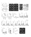
- FIG. 3 shows analysis of SLC15A3 gene expression of the present invention.
- a and b RPKM of human SLC15A3 (A) or mouse Slc15a3 (B) in the indicated tissues (top) or cell types (bottom) calculated from pools of at least two data sets as described in the methods of the present invention. Expression values are indicated.
- c Boxplot of SLC15A3 expression across cells identified from IPF lungs from normal controls and IPF cell atlas database.
- e RPKM of human SLC15A3 (A) or mouse Slc15a3 (B) in the indicated tissues (top) or cell types (bottom) calculated from pools of at least two data sets as described in the methods of the present invention. Expression values are indicated.
- c Boxplot of SLC15A3 expression across cells identified from IPF lungs from normal controls
- FIG. 1 shows the generation and characterization of Slc15a3 ⁇ / ⁇ mice of the invention.
- a Schematic representation of Slc15a3 mutant alleles. Exon 1 of the Slc15a3 gene was targeted and partially deleted by CRISPR-Cas9. Arrows indicate genotyping primers.
- b Genotyping results of Slc15a3 ⁇ / ⁇ mice by PCR using primer sets specific for exon 1.
- the primer-sequences are: Fwd, 5'-GCAGTTAACCGGGGACACTA-3', Rev, 5'-CGCCTAGGAAGAGGAGTGTG-3'.
- Slc15a3 transcripts in WT and Slc15a3 ⁇ / ⁇ neutrophils in the peritoneal cavity were analyzed by RT-qPCR. P ⁇ 0.01 (determined by unpaired t-test).
- E and F are flow cytometric analyzes of lymphoid and myeloid lineages in the spleen and BM of Slc15a3 ⁇ / ⁇ mice.
- Single-cell suspensions prepared from spleens (E) or BM (F) of native WT and Slc15a3 ⁇ / ⁇ mice were analyzed by staining with the indicated antibodies. Results are representative of three independent experiments.
- Slc15a3 transcript levels in the lung during the course of BLM-induced PF expressed as relative quantification (RQ).
- RQ relative quantification
- 3D is a three-dimensional (3D) CT image of a lung processed with BLM.
- d Lung CT images (left) and Masson's trichrome staining of BLM-treated lung sections of WT and Slc15a3 ⁇ / ⁇ mice at day 21 (middle). Higher magnification images are shown in the right column.
- g BALF cell numbers in BLM-induced PF, and h.
- Collagen gene expression in the lung The number of mice per group is the same as in Fig. 1a.
- i BALF cytokine levels at indicated time points in BLM-induced PF model mice.
- j Expression levels of Il11 transcripts in WT and Slc15a3 ⁇ / ⁇ lung tissues during the course of BLM-induced PF.
- K Lung hydroxyproline content at day 28 in silica-induced PF model (silicosis).
- l Relative lung volume changes during silicosis determined using mean values on day 7 from CT scans.
- m Representative 3DCT images of pulmonary silicosis in WT and Slc15a3 ⁇ / ⁇ mice at 7 and 28 days.
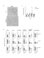
- FIG. 4 shows increased expression of Arg2 in Slc15a3 ⁇ / ⁇ lungs treated with BLM.
- FIG. 3 shows relaxation of PF in Slc15a3 ⁇ / ⁇ mice in a silica-induced PF model.
- PF silica-induced pulmonary fibrosis
- FIG. 2 shows the results of scRNAseq analysis of lung cells in BLM-induced PF.
- b Optimized t-distributed stochastic neighbor embedding
- Slc15a3 expression in lung cell populations was analyzed by scRNAseq. Clusters expressing Slc15a3 were gated.
- c Comparison of monocyte/macrophage clusters between WT and Slc15a3 ⁇ / ⁇ mice on days 10 and 21 by comparative opt-SNE display. Light and dark dots represent WT and Slc15a3 ⁇ / ⁇ mice, respectively.
- b Optimized t-distributed stochastic neighbor embedding
- FIG. 4 shows the results of gene expression analysis in Slc15a3 ⁇ / ⁇ lungs.
- a A human map representing the top 30 differentially expressed genes among 40 clusters (0-39) in scRNAseq analysis.
- Gene set enrichment analysis was performed using the comparative Toxicogenomics database pulmonary fibrosis gene set, logarithmic differences in mean expression between WT and Slc15a3 ⁇ / ⁇ mice in each cluster at the indicated time points. was performed using genes ranked by For clusters with q-values less than 0.05, enrichment plots are displayed with normalized enrichment scores and false positive q-values.
- Gene set enrichment analysis was performed using the comparative Toxicogenomics database pulmonary fibrosis gene set to determine the mean expression between WT and Slc15a3 ⁇ / ⁇ mice in each cluster at the indicated time points. It was performed with genes ranked by logarithmic difference.
- Bubble color represents expression intensity and bubble size represents positive cell fraction.
- f Bubble plot representation of cluster-16 (IM) chemokine ligands and receptors in WT and Slc15a3 ⁇ / ⁇ mice on days 10 and 21.
- FIG. g and h Network analysis of DEGs in epithelial cells (g) (clusters-21 and 25) and endothelial cells (h) (clusters-4 and 24).
- FIG. 4 shows the results of gene expression analysis in Slc15a3 ⁇ / ⁇ lungs.
- UMAP Homogeneous manifold approximation and projection
- FIG. 4 shows the role of Slc15a3-expressing hematopoietic and non-hematopoietic cells in PF development as assessed by the generation of BM chimeric mice.
- a. Slc15a3 expression levels and hydroxyproline content in lungs on day 35 of silicosis.
- b Arg2 expression levels in lung and BALF cells on day 35 of silicosis.
- d Slc15a3 expression levels in the lungs of BM chimeric mice on days 10 and 21 of BLM-induced PF.
- e BALF cell counts of BM chimeric mice on days 10 and 21 of BLM-induced PF.
- f Number of inflammatory cells in BALF on days 10 and 21.
- Granulocytes were recognized as CD45+CD11b+Ly6G+ cells and monocytes were recognized as CD45+CD11b+Ly6G- cells.
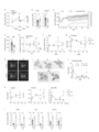
- FIG. 3 shows the results of examining the involvement of BM-derived M ⁇ s in PF.
- Intratracheal Slc15a3 ⁇ / ⁇ BMM ⁇ transplantation ameliorates pulmonary silicosis.
- a Representative 3DCT images of pulmonary silicosis before (day 14) and after (day 35) BMM ⁇ transplantation.
- b Hydroxyproline content at days 0 and 14 in silicosis-induced WT lungs.
- j Expression levels of Arg2 in SLC15A3 knockdown human M ⁇ cell lines. Data are shown as mean ⁇ SD.
- FIG. 10 shows the results of a study investigating the relationship between PF tissue repair and Slc15a3 ⁇ / ⁇ M ⁇ s. a and b.
- Arg2 shows the importance of Arg2 in PF relaxation.
- a Expression levels of A, Arg2, Gatm, and Arg1 in lung cell populations analyzed by scRNAseq.
- b to e Effect of SLC15A3 loss on gene expression of arginine-metabolizing enzymes.
- d and e Expression levels of Arg2 mRNA in BMM ⁇ (D) and LC15A3 knockdown human M ⁇ cell lines (E) cultured for 24 h with the indicated cytokines or stimulants.
- f and g Effect of arginase inhibition on progression of silicosis in Slc15a3 ⁇ / ⁇ mice. f.
- FIG. 10 shows that Arg2 of M ⁇ s contributes greatly to mitigating PF.
- a Schematic representation of a strategy for administering arginase 5 inhibitors to Slc15a3 ⁇ / ⁇ silicosis model mice.
- c Representative three-dimensional (3D) CT images of lungs after inhibitor treatment. d.
- Arg2 ⁇ / ⁇ mice Boxplots of whole cell ARG2 and ARG1 expression identified from normal controls and IPF donors. Generation and characterization of Arg2 ⁇ / ⁇ mice.
- a and b Schematic representation of the deleted DNA sequences of CRISPR/Cas9 targeted Arg2 exons 1 and 2 (a) and the resulting Arg2KO mutant allele (b). Arrows indicate the position of primers used for genotyping.
- c PCR genotyping of Arg2 ⁇ / ⁇ mice using the following primers: Common, 5′-GAGAGCTGTAGTCCTCCGAGA-3′; and E2a, 5′-GTAGCAGCTTGGCACCTGTA-3′.
- d PCR genotyping of Arg2 ⁇ / ⁇ mice using the following primers: Common, 5′-GAGAGCTGTAGTCCTCCGAGA-3′; and E2a, 5′-GTAGCAGCTTGGCACCTGTA-3′.
- Figure 2 shows that daily fasting and timed feeding upregulate Arg2 in alleviating PF.
- FIG. 2 shows the relationship between daily fasting and Arg2 expression.
- DF-induced ketogenesis is dispensable on Arg2 induction.
- a ⁇ -Hydroxybutyrate levels in serum (Fig. 17b-f).
- b Arg2 expression in BMM ⁇ s of WT and Slc15a3 ⁇ / ⁇ mice cultured with 50 nM trichostatin A (TSA) or 1 mM sodium butyrate (NaB) for several hours.
- TSA nM trichostatin A
- NaB sodium butyrate
- Data are presented as mean ⁇ SD. Mann-Whitney-U test (a); one-way ANOVA followed by Tukey's post hoc test (bd). ***P ⁇ 0.0001, ***P ⁇ 0.001, (ns) non-significant. All results are representative of at least two independent experiments.
- c and d Lysosomal distribution of the indicated proteins examined by lysosomal fractionation experiments.
- c Cell homogenates from WT or Slc15a3 ⁇ / ⁇ BMM ⁇ cultured under normal conditions were separated by density gradient ultracentrifugation and the proteins indicated in each fraction were examined by immunoblotting.
- FIG. 4 shows that compound NCGM-1006, which enhances Arg2 expression in AM ⁇ , suppresses PF. a.
- FIG. 1 is a schematic diagram of a strategy for applying an Arg2-enhancing compound to a silicosis model mouse. b. Fig.
- FIG. 1 shows the general mechanism underlying the effects of DF and Slc15a3 deficiency in mice. In M ⁇ s of DF lungs, sustained and enhanced activation of AMPK is caused by fuel starvation. Since SLC15A3 is associated with the Rab2 and Rab7 proteins that regulate lysosomal trafficking, fuel starvation may also occur in Slc15a3-deficient M ⁇ s, possibly due to lysosomal dysregulated behavior.
- SLC15A3 is associated with AMPK phosphatase and may be involved in the negative feedback regulation of AMPK. Loss of SLC15A3 may reduce the accessibility of these phosphatases, resulting in sustained and enhanced AMPK activity regardless of the ATP:AMP ratio.
- the present invention provides compounds that treat and/or mitigate the progression of pulmonary fibrosis, dietary supplements to support fasting, and/or management systems that keep patients fasting for at least 16 hours or more per day. offers a strategy for treating and/or slowing the progression of pulmonary fibrosis, and can be utilized in repairing fibrotic scarring, particularly after COVID-19 infection.
- TASL Sedlyarov, K. Papakostas, A. Cesar-Razquin, P. Essletzbichler, U. Goldmann, A. Stefanovic, J. W. Bigentechnik, S. Scorzoni, M. D. Pizzagalli, A. Bensimon, A. C. Muller, F. J. King, J. Li, E. Girardi, M. L. Mbow, C. E. Whitehurst, M. Rebsamen, G. Superti-Furga, TASL is the SLC15A4-associated adapter for IRF5 activation by TLR7-9 Nature (2020), doi:10.1038/s41586-020-2282-0. 11. X. Zhao, J. Y. Y. Kwan, K.
Landscapes
- Health & Medical Sciences (AREA)
- Life Sciences & Earth Sciences (AREA)
- Chemical & Material Sciences (AREA)
- Medicinal Chemistry (AREA)
- Animal Behavior & Ethology (AREA)
- General Health & Medical Sciences (AREA)
- Public Health (AREA)
- Veterinary Medicine (AREA)
- Pharmacology & Pharmacy (AREA)
- Epidemiology (AREA)
- Engineering & Computer Science (AREA)
- Bioinformatics & Cheminformatics (AREA)
- Immunology (AREA)
- Gastroenterology & Hepatology (AREA)
- Proteomics, Peptides & Aminoacids (AREA)
- Pulmonology (AREA)
- Chemical Kinetics & Catalysis (AREA)
- General Chemical & Material Sciences (AREA)
- Nuclear Medicine, Radiotherapy & Molecular Imaging (AREA)
- Organic Chemistry (AREA)
- Microbiology (AREA)
- Mycology (AREA)
- Medicines That Contain Protein Lipid Enzymes And Other Medicines (AREA)
Abstract
本発明は、人体(患者等)を絶食状態に維持することで、肺間質の疾患、特に肺線維症の治療または修復もしくはその両方を行う方法、および、当該絶食状態によって誘発される代謝産物を検出し分析するシステム、アミノ酸/オリゴペプチドトランスポ-タ-の機能を調節する工程を含む、肺線維症の治療方法および当該方法に使用する組成物、加水分解酵素の機能を調節する工程を含む、肺線維症の治療方法および当該方法に使用する組成物、AMPKの機能を調節する工程を含む、肺線維症の治療方法および当該方法に使用する組成物に関する。
Description
本発明は、人体(患者等)を絶食状態に維持することで、肺間質の疾患、特に肺線維症の治療または修復もしくはその両方を行う方法、および、当該絶食状態によって誘発される代謝産物を検出し分析するシステム、アミノ酸/オリゴペプチドトランスポ-タ-の機能を調節する工程を含む、肺線維症の治療方法および当該方法に使用する組成物、加水分解酵素の機能を調節する工程を含む、肺線維症の治療方法および当該方法に使用する組成物、AMPKの機能を調節する工程を含む、肺線維症の治療方法および当該方法に使用する組成物に関する。
肺の間質は肺胞の壁(肺胞壁)や肺胞を取り囲んで支持している組織を指し、この組織領域における疾患は、呼吸機能に直接大きな影響を及ぼし、生命維持の危機もしくは生活の質の低下に直結しうる。
肺間質の疾患、特に肺線維症(PF)は、進行性の瘢痕化とマトリックス沈着を特徴とする肺疾患であり、一般的に予後は不良である。PFの病因はよく理解されていないが、多くの患者は、感染または危険物(アスベストやシリカなど)への曝露によって引き起こされる炎症反応を示す。有病率が増加する難病である肺線維症(PF)は、深刻な公衆衛生上の問題である。
肺間質の疾患、特に肺線維症(PF)は、進行性の瘢痕化とマトリックス沈着を特徴とする肺疾患であり、一般的に予後は不良である。PFの病因はよく理解されていないが、多くの患者は、感染または危険物(アスベストやシリカなど)への曝露によって引き起こされる炎症反応を示す。有病率が増加する難病である肺線維症(PF)は、深刻な公衆衛生上の問題である。
また、肺間質の疾患、特に肺線維症が一旦治癒されても、残る線維性瘢痕は、広範囲にわたる臓器不全を引き起こすことが知られている。重要なことに、PFは、環境汚染(アスベストやPM2.5など)および最近のコロナウイルス病2019のパンデミック(ロングCOVIDなど)のためにますます一般的になっている。よって、治療後の修復が必要である。
また、溶質キャリアファミリ-15のメンバ-3(SLC15A3)は、自然免疫細胞で強力に発現するアミノ酸/オリゴペプチドトランスポ-タ-であり、主にヒトとマウスの両方の自然免疫細胞(マクロファ-ジや好中球など)で発現します(図1のA、Bを参照)
さらに、ヒトとマウスの両方の肺組織、およびPF患者の肺でSLC15A3の強い発現が見られ、MΦs、特に肺胞MΦs(AMΦs)で強い発現が見られた(図S1a-c)。だがSLC15A3の機能はほとんど知られておらず、その同族体であるSLC15A4は、線維症に関連することが多いエンドリソソ-ム依存性の炎症および代謝シグナルを媒介することにより、大腸炎および狼瘡の症状を促進することしかわかっておらず。
M.Wijsenbeek,V.Cottin,SpectrumofFibroticLungDiseases.N.Engl.J.Med.383,958-968(2020)。
N.C.Henderson,F.Rieder,T.A.Wynn,Fibrosis:frommechanismstomedicines.Nature.587,555-566(2020)。
K.Sakata,T.Yamashita,M.Maeda,Y.MORIYAMA,S.SHIMADA,M.TOHYAMA,Cloningofalymphaticpeptide/histidinetransporter.Biochem.J.356,53-60(2001)。
F.Song,Y.Yi,C.Li,Y.Hu,J.Wang,D.E.Smith,H.Jiang,Regulationandbiologicalroleofthepeptide/histidinetransporterSLC15A3inToll-likereceptor-mediatedinflammatoryresponsesinmacrophage.CellDeathDis.9,1-15(2018).
現在、PFの治療法が5つの選択肢に限られているため、将来の治療のための潜在的な新規選択肢を増やす必要がある。また、治療後の線維性瘢痕による影響を最小限にするためにもその修復方法が求められている。間質性肺炎の病型により、原因が明確な場合は原因物質の除去と抗線維化薬が使用されるが、原因不明の場合は抗線維化薬のみでしかない。特に、ステロイドや抗凝固剤などは国際ガイドラインに基づいて使用しないことが強く推奨されている。
そこで、本発明者らが、間質性肺炎の病型へのより多くの治療方法について鋭意に研究を重ねた結果、アミノ酸/オリゴペプチドトランスポ-タ-、および/または加水分解酵素が肺間質の疾患に寄与することを明らかにした。体内におけるアミノ酸/オリゴペプチドトランスポ-タ-発現または喪失もしくはその両方、および/または、加水分解酵素の活性または抑制もしくはその両方で、線維症の調節において極めて重要であることを見出し、肺間質の疾患の治療および修復にも適用することを見出した。
本発明は、患者に一日少なくとも12時間以上絶食状態にすることを特徴とする肺線維症を治療する方法を提供する。別の態様において、本発明は、アミノ酸/オリゴペプチドトランスポ-タ-の機能を調節する工程を含む、肺線維症の治療方法および当該方法に使用する組成物を提供する。別の態様において、本発明は、加水分解酵素の機能を調節する工程を含む、肺線維症の治療方法および当該方法に使用する組成物を提供する。別の態様において、本発明は、AMPKの機能を調節する工程を含む、肺線維症の治療方法および当該方法に使用する組成物を提供する。
本発明は一態様において、肺線維症の治療を必要とする対象に対して、アルギニン代謝酵素の機能を増強する化合物を投与する工程を含む、肺線維症を治療する方法を提供する。
本発明は一態様において、アルギニン代謝酵素の機能を増強する化合物を含有する、肺線維症の治療における使用のための組成物を提供する。
本発明は一態様において、肺線維症の治療を必要とする対象に対して、アミノ酸トランスポ-タ-の機能を阻害する化合物を投与する工程を含む、肺線維症を治療する方法を提供する。
本発明は一態様において、アミノ酸トランスポ-タ-の機能を阻害する化合物を含有する、肺線維症の治療における使用のための組成物を提供する。一態様において、アミノ酸トランスポ-タ-の機能を阻害する化合物は、アルギニン代謝酵素の機能を増強する。
本発明は一態様において、肺線維症の治療を必要とする対象に対して、AMPKの機能を増強する化合物を投与する工程を含む、肺線維症を治療する方法を提供する。
本発明は一態様において、AMPKの機能を増強する化合物を含有する、肺線維症の治療における使用のための組成物を提供する。一態様において、AMPKの機能をする化合物は、アルギニン代謝酵素の機能を増強する。
本発明は一態様において、絶食状態によって誘発される代謝物の測定値を測定し、当該測定値を分析するシステムを提供する。
本発明は一態様において、絶食状態によって誘発される代謝物の測定値を測定し、当該測定値を分析するシステムを提供する。
本発明は一態様において、絶食状態によって誘発される代謝物を検出し分析するシステムを提供する。
本発明は一態様において、より具体的に下記を提供する。
[項目1]
アルギニン代謝酵素の機能を増強する化合物を含有する、肺線維症の治療における使用のための組成物。
[項目2]
前記アルギニン代謝酵素の機能を増強する化合物がアルギニン代謝酵素の発現を増強する化合物である、項目1に記載の組成物。
[項目3]
前記アルギニン代謝酵素がアルギナ-ゼIIである、項目1に記載の組成物。
[項目4]
前記アルギナ-ゼIIの機能を増強する化合物が、ジペプチドAla-Tyr、Lys-Tyr、Gln-Tyr、MJN110およびタプシガルギン(Thapsigargin)から選択されるものである、項目3に記載の組成物。
[項目5]
前記アルギナ-ゼIIの機能を増強する化合物がアミノ酸トランスポ-タ-の機能を阻害する化合物である、項目1に記載の組成物。
[項目6]
前記アミノ酸トランスポ-タ-の機能を阻害する化合物がアミノ酸トランスポ-タ-の発現を阻害する化合物である、項目5に記載の組成物。
[項目7]
前記アミノ酸トランスポ-タ-がSLC15A3である、項目6に記載の組成物。
[項目8]
前記SLC15A3の機能を阻害する化合物が、抗SLC15A3抗体またはその抗原結合性フラグメント、SLC15A3遺伝子の発現を阻害する核酸分子、ポリアミン、アルギニン、メチオニン、3-ヒドロキシ-3-メチルブチレ-ト、シトシン、アセト酢酸、3-ヒドロキシ酪酸およびアセトンから選択されるものである、項目7に記載の組成物。
[項目9]
前記アルギナ-ゼIIの機能を増強する化合物がAMPKの機能を増強する化合物である、項目1に記載の組成物。
[項目10]
前記AMPKの機能を増強する化合物がAMPKの発現を増強する化合物である、項目9に記載の組成物。
[項目11]
前記AMPKの機能を増強する化合物がクラスIII PI3キナ-ゼ阻害剤、wortmanninおよびPI4Kから選択されるものである、項目10に記載の組成物。
[項目12]
医薬組成物、機能性食品、飲食品又は飼料である、項目1~11のいずれか一項に記載の組成物。
本発明は一態様において、より具体的に下記を提供する。
[項目1]
アルギニン代謝酵素の機能を増強する化合物を含有する、肺線維症の治療における使用のための組成物。
[項目2]
前記アルギニン代謝酵素の機能を増強する化合物がアルギニン代謝酵素の発現を増強する化合物である、項目1に記載の組成物。
[項目3]
前記アルギニン代謝酵素がアルギナ-ゼIIである、項目1に記載の組成物。
[項目4]
前記アルギナ-ゼIIの機能を増強する化合物が、ジペプチドAla-Tyr、Lys-Tyr、Gln-Tyr、MJN110およびタプシガルギン(Thapsigargin)から選択されるものである、項目3に記載の組成物。
[項目5]
前記アルギナ-ゼIIの機能を増強する化合物がアミノ酸トランスポ-タ-の機能を阻害する化合物である、項目1に記載の組成物。
[項目6]
前記アミノ酸トランスポ-タ-の機能を阻害する化合物がアミノ酸トランスポ-タ-の発現を阻害する化合物である、項目5に記載の組成物。
[項目7]
前記アミノ酸トランスポ-タ-がSLC15A3である、項目6に記載の組成物。
[項目8]
前記SLC15A3の機能を阻害する化合物が、抗SLC15A3抗体またはその抗原結合性フラグメント、SLC15A3遺伝子の発現を阻害する核酸分子、ポリアミン、アルギニン、メチオニン、3-ヒドロキシ-3-メチルブチレ-ト、シトシン、アセト酢酸、3-ヒドロキシ酪酸およびアセトンから選択されるものである、項目7に記載の組成物。
[項目9]
前記アルギナ-ゼIIの機能を増強する化合物がAMPKの機能を増強する化合物である、項目1に記載の組成物。
[項目10]
前記AMPKの機能を増強する化合物がAMPKの発現を増強する化合物である、項目9に記載の組成物。
[項目11]
前記AMPKの機能を増強する化合物がクラスIII PI3キナ-ゼ阻害剤、wortmanninおよびPI4Kから選択されるものである、項目10に記載の組成物。
[項目12]
医薬組成物、機能性食品、飲食品又は飼料である、項目1~11のいずれか一項に記載の組成物。
(定義)
本明細書において、複数の数値の範囲が示された場合、それら複数の範囲の任意の下限値および上限値の組み合わせからなる範囲も同様に意味する。
本明細書において、複数の数値の範囲が示された場合、それら複数の範囲の任意の下限値および上限値の組み合わせからなる範囲も同様に意味する。
本明細書において、「治療」とは最も広い意味において使用される。一態様において、治療は疾患、障害または状態の発生を予防すること、阻害すること、および/または軽減または逆転することを含む。一態様において、肺線維症の治療は、肺組織の修復を意味する。
本明細書において、「抗体」とは最も広い意味において使用され、意図された抗原結合活性を示すモノクロ-ナル抗体、ポリクロ-ナル抗体、及び抗体断片を含むが、これらに限定されない。全長抗体は主としてポリペプチドで構成される重鎖及び軽鎖を含む。重鎖および軽鎖は、それぞれ抗原を認識する可変領域と呼ばれる部位を含み、当該部位は一般的に、それぞれ重鎖可変領域及び軽鎖可変領域と呼ばれる。当該可変領域は、さらに詳細に抗原を認識する部位として特定される、それぞれCDR1~3と呼ばれる部位をアミノ末端から順に有する。なお、これらのCDR1~3は、より詳細に重鎖CDR1、重鎖CDR2、重鎖CDR3、軽鎖CDR1、軽鎖CDR2、軽鎖CDR3等とも呼ばれる。また、重鎖及び軽鎖のCDR1~3以外の領域は、それぞれアミノ末端から順に、重鎖FR1~4及び軽鎖FR1~4と呼ばれる。抗体は2つの重鎖および2つの軽鎖からなる形態のほか、1つの重鎖および1つの軽鎖からなる抗体(1本鎖抗体とも呼ばれる)の形態であってもよい。
抗体は、任意のクラス、例えば、IgG、IgE、IgM、IgD、IgAもしくはIgY等、あるいはサブクラス、例えば、IgG1、IgG2、IgG3、IgG4、IgA1もしくはIgA2等であってよい。
抗体は同一種に由来するアミノ酸配列を有していても異種に由来するアミノ酸を有していてもよい。同一種由来の場合、例えば、ヒト由来、マウス由来、ラット由来、ハムスタ-由来、ウサギ由来、ヤギ由来、ロバ由来、ブタ由来、ウシ由来、ウマ由来、ニワトリ由来、サル由来、チンパンジ-由来、ラクダ由来、ラマ由来等が挙げられる。
異種由来の場合も、特に限定はされないが、例えばヒト、マウス、ラット、ハムスタ-、ウサギ、ヤギ、ヒツジ、ロバ、ブタ、ウシ、ウマ、ニワトリ、サル、チンパンジ-、ラクダ、ラマ等の何れか2つ以上に由来する抗体が挙げられる。
本明細書において、抗体の「抗原結合断片」は、抗原に特異的に結合する能力を保持する、抗体の1つ以上の断片を指す。抗体の抗原に特異的に結合する能力は、その一部からなる断片によっても維持されうることがわかっている。一態様として、抗体の「抗原結合断片」は、軽鎖可変領域(VL)、重鎖可変領域(VH)、軽鎖定常領域(CL)および重鎖定常領域の一部であるCH1ドメインからなるFab断片、ヒンジ領域でジスルフィド架橋によって連結した2つのFab断片を含むF(ab’)2断片、VHおよびCH1ドメインからなるFd断片、抗体の単一のア-ムのVLおよびVHドメインからなるFv断片、単一の可変ドメインを含むdAb断片、および単離された相補性決定領域(CDR)であり得るが、これらに限定されない。
本発明の抗体は、CDRグラフト化された抗体であってもよい。一例において、CDRグラフト化された抗体では、1つの動物種に由来する抗体のCDR領域の一部または全部の配列が、別の動物種のCDR配列で置換されている。例えば、1つ以上のマウス抗体のCDRがヒト抗体のCDR配列で置換されている。
抗体の重鎖可変領域中の重鎖CDR1~3および軽鎖可変領域中の軽鎖CDR1~3を同定する際には当業者に周知の方法を用いることができる。例えば、例えば当業者に周知の「Kabat定義」(Kabatら、Ann.NY Acad、Sci.1971、Vol.190、pp.382-391およびKabat、E.A.らSequences of Proteins of Immunological Interest、第5版、1991、U.S.Department of Health and Human Services、NIH Publication、pp.91-3242)、「Chothia定義」(Chothia et al.,Nature、1989、Vol.342、pp.877-883)、「接触定義」(MacCallum et al.,J.Mol.Biol.,1996、Vol.262、pp.732-745)などを用いることができる。抗体内のCDR配列を同定するのに、公共デ-タベ-スの情報に基づいてCDRを同定してもよい。
(絶食状態にする工程を含む肺間質の疾患を治療する方法)
本発明は、PFの治療のために患者に一日少なくとも12時間以上の絶食状態を維持することで引き起こされる体内のアミノ酸/オリゴペプチドトランスポ-タ-発現喪失、および/また加水分解酵素の活性化によって肺間質の疾患の治療方法を提供する。
本発明は、PFの治療のために患者に一日少なくとも12時間以上の絶食状態を維持することで引き起こされる体内のアミノ酸/オリゴペプチドトランスポ-タ-発現喪失、および/また加水分解酵素の活性化によって肺間質の疾患の治療方法を提供する。
本発明は、絶食に伴って変化する代謝産物を患者に直接に与えることで、PFの治療のために患者の体内のアミノ酸/オリゴペプチドトランスポ-タ-発現喪失の誘発、および/または加水分解酵素の活性化促進剤を患者に直接に与えることで加水分解酵素の活性化によって肺間質の疾患の治療のみならず修復する主成分を提供する。または当該主成分、その変異、その誘導体、またはその薬学的に許容される塩を含む医薬組成物を提供する。
本発明は、絶食状態を維持するための絶食をサポ-トするための補助食(空腹感を減弱し、かつ治療効果を増強する補助食品)を提供する。
本発明は、一定時間の絶食状態が維持されたことによる代謝物、たとえば、末梢血に現れるケトン体の測定数値、血中グルコ-ス測定値、および呼気または血中または尿中に含まれる代謝物の測定値、例えば、窒素、アルギニン代謝産物などを測定し分析するシステムを提供する。
本発明でいう絶食状態について、患者に一日少なくとも12時間以上の絶食状態が好ましく、特に16時間、さらに好ましくは18時間以上が好ましい。さらに、一日の中、18時から翌日12時の間の16時間を絶食状態維持することが好ましい。
(アルギニン代謝酵素の機能を増強する化合物を含む組成物を投与する工程を含む、肺間質の疾患を治療する方法)
本発明は一態様において、肺間質の疾患の治療を必要とする患者に対して、加水分解酵素の機能を増強する化合物を含む組成物を投与する工程を含む、肺間質の疾患を治療する方法を提供する。
本発明において、加水分解酵素の機能を増強する化合物は、生体内で加水分解酵素の機能を亢進または発現させる効果を有する化合物であれば特に限定されない。例えば、加水分解酵素の機能を増強する化合物は、加水分解酵素の酵素活性増強、局在化促進、発現亢進、安定性増加等の作用によって、加水分解酵素の機能を増強することができる。好ましくは、加水分解酵素の機能を増強する化合物は、加水分解酵素の発現を亢進する。
本発明は一態様において、肺間質の疾患の治療を必要とする患者に対して、加水分解酵素の機能を増強する化合物を含む組成物を投与する工程を含む、肺間質の疾患を治療する方法を提供する。
本発明において、加水分解酵素の機能を増強する化合物は、生体内で加水分解酵素の機能を亢進または発現させる効果を有する化合物であれば特に限定されない。例えば、加水分解酵素の機能を増強する化合物は、加水分解酵素の酵素活性増強、局在化促進、発現亢進、安定性増加等の作用によって、加水分解酵素の機能を増強することができる。好ましくは、加水分解酵素の機能を増強する化合物は、加水分解酵素の発現を亢進する。
本発明の加水分解酵素は例えば、主に肺のMΦsで発現する一酸化窒素合成酵素、Arg1、Arg2、Gatmなどの酵素である。特にアルギニンをオルニチンと尿素に分解するミトコンドリア酵素であるアルギナ-ゼIIが好ましい。
本発明の加水分解酵素の機能を増強する化合物は特に限定されず、当業者に一般的に知られた方法によってスクリ-ニングにより取得することもできる。例えば、加水分解酵素の機能を増強する化合物として、同酵素の発現量を増加させる化合物を取得する場合、マクロファ-ジなどのアルギナ-ゼII発現細胞に当該化合物を適用した後、細胞中のアルギナ-ゼIIのmRNA転写量または細胞内または分泌アルギナ-ゼIIタンパク質量などを定法によって定量し、一定時間経過後、例えば、10分、20分、30分、40分、50分、60分、2時間、3時間、4時間、5時間、6時間、7時間、8時間、9時間、10時間、11時間、12時間、13時間、14時間、15時間、16時間、17時間、18時間、19時間、20時間、21時間、22時間、23時間、24時間、またはそれ以上長い時間経過した時点において、陰性対照に対して一定以上の比率、例えば、10%、20%、30%、40%、50%、60%、70%、80%、90%または100%以上mRNA転写量またはタンパク質量を増加させる化合物を、加水分解酵素の機能を増強する化合物として取得することができる。また、例えば、加水分解酵素の機能を増強する化合物として、同酵素の活性を増加させる化合物を取得する場合、マクロファ-ジなどのアルギナ-ゼII発現細胞に当該化合物を適用した後、上記のような一定時間後におけるアルギナ-ゼIIの非活性を測定して、上記のような一定比率以上活性を上昇させる化合物を、加水分解酵素の機能を増強する化合物として取得することができる。
本発明の加水分解酵素の機能を増強する化合物は、ポリペプチド、核酸、低分子化合物を含む。例えば、これら加水分解酵素が関与する経路によって代謝される半必須アミノ酸を使用することが関与する複数の経路によって代謝される半必須アミノ酸を使用することができる。
一態様において、加水分解酵素の機能を増強する化合物として、絶食状態によって誘発されるAla-Tyr,Lys-Tyr,Gln-Tyr等を含むジペプチド、を用いることができる。加水分解酵素がアルギナ-ゼIIである場合、脂肪酸代謝阻害剤、例えばMJN110、または小胞体ストレス誘導化合物、例えばThapsigarginを用いることができる。
一態様において、加水分解酵素の機能を増強する化合物として、以下に述べるアミノ酸/オリゴペプチドトランスポ-タ-の機能を阻害する化合物、またはAMPKの機能を増強する化合物を用いることができる。一態様において、加水分解酵素の機能を増強する化合物として、クラスIII PI3キナ-ゼの阻害剤、wortmanninまたは、PI4K等を制御する低分子化合物、および解糖系と酸化的リン酸化反応を亢進し、細胞の代謝恒常性をかく乱する低分子化合物等を用いることができる。
一態様において、上記化合物の変異体、誘導体、またはその薬学的に許容される塩を使用することができる。
本発明の方法が対象とする動物は特に限定されない。例えば、ヒト、マウス、ラット、モルモット、ウサギ、ハムスタ-、イヌ、ネコ、イタチ、ウシ、ブタ等の哺乳類、ニワトリなどの鳥類等を対象とすることができる。
(アミノ酸/オリゴペプチドトランスポ-タ-の機能を阻害する化合物を含む組成物を投与する工程を含む、肺間質の疾患を治療する方法)
本発明は一態様において、肺間質の疾患の治療を必要とする患者に対して、アミノ酸/オリゴペプチドトランスポ-タ-の機能を阻害する化合物を含む組成物を投与する工程を含む、肺間質の疾患を治療する方法を提供する。一態様において、アミノ酸/オリゴペプチドトランスポ-タ-の機能を阻害する化合物は、アルギニン代謝酵素の機能を増強する。 本発明において、アミノ酸/オリゴペプチドトランスポ-タ-の機能を阻害する化合物は、生体内でアミノ酸/オリゴペプチドトランスポ-タ-の機能を減少または喪失させる効果を有する化合物であれば特に限定されない。例えば、アミノ酸/オリゴペプチドトランスポ-タ-の機能を阻害する化合物は、アミノ酸/オリゴペプチドトランスポ-タ-の活性阻害、局在化阻害、発現阻害、安定性阻害等の作用によって、アミノ酸/オリゴペプチドトランスポ-タ-の機能を阻害することができる。好ましくは、アミノ酸/オリゴペプチドトランスポ-タ-の機能を阻害する化合物は、アミノ酸/オリゴペプチドトランスポ-タ-の発現を阻害する。
また、アミノ酸/オリゴペプチドトランスポ-タ-の機能を阻害する化合物は、当業者に一般的に知られた方法によって、スクリ-ニングして取得することができる。これらの化合物は、抗体またはその抗原結合性断片、ポリペプチド、核酸、例えばsiRNA若しくはshRNA、またはこれらをコ-ドする核酸、低分子化合物を含む。
本発明は一態様において、肺間質の疾患の治療を必要とする患者に対して、アミノ酸/オリゴペプチドトランスポ-タ-の機能を阻害する化合物を含む組成物を投与する工程を含む、肺間質の疾患を治療する方法を提供する。一態様において、アミノ酸/オリゴペプチドトランスポ-タ-の機能を阻害する化合物は、アルギニン代謝酵素の機能を増強する。 本発明において、アミノ酸/オリゴペプチドトランスポ-タ-の機能を阻害する化合物は、生体内でアミノ酸/オリゴペプチドトランスポ-タ-の機能を減少または喪失させる効果を有する化合物であれば特に限定されない。例えば、アミノ酸/オリゴペプチドトランスポ-タ-の機能を阻害する化合物は、アミノ酸/オリゴペプチドトランスポ-タ-の活性阻害、局在化阻害、発現阻害、安定性阻害等の作用によって、アミノ酸/オリゴペプチドトランスポ-タ-の機能を阻害することができる。好ましくは、アミノ酸/オリゴペプチドトランスポ-タ-の機能を阻害する化合物は、アミノ酸/オリゴペプチドトランスポ-タ-の発現を阻害する。
また、アミノ酸/オリゴペプチドトランスポ-タ-の機能を阻害する化合物は、当業者に一般的に知られた方法によって、スクリ-ニングして取得することができる。これらの化合物は、抗体またはその抗原結合性断片、ポリペプチド、核酸、例えばsiRNA若しくはshRNA、またはこれらをコ-ドする核酸、低分子化合物を含む。
一態様において、患者の体内のアミノ酸/オリゴペプチドトランスポ-タ-発現を喪失させる低分子化合物として、ポリアミン、アルギニンやメチオニン等のアミノ酸代謝産物などを例として挙げられる。特にプトレシンを含むポリアミン類、例えばケトン体、3-ヒドロキシ-3-メチルブチレ-ト、シトシン、アセト酢酸、3-ヒドロキシ酪酸、アセトンなどが好ましい。本発明での上記代謝産物には、例えば、GABA、パントテン酸、オフタルミン酸が確認されており、本発明の代謝産物として好ましい。ここでいう、ポリアミンは特にここで除外しない限り、体内にあるすべてのポリアミンを含み、本発明の例として、たとえば、ケトン体、3-ヒドロキシ-3-メチルブチレ-ト、シトシン、アセト酢酸、3-ヒドロキシ酪酸、アセトンなどを含むプトレシンを含むポリアミン類が好ましい。その中特に、ケトン体が好ましい。
一態様において、アミノ酸/オリゴペプチドトランスポ-タ-の機能を阻害する化合物として、絶食状態によって誘発されるAla-Tyr,Lys-Tyr,Gln-Tyr等を含むジペプチドを用いることができる。
一態様において、アミノ酸/オリゴペプチドトランスポ-タ-の機能を阻害する化合物として、クラスIII PI3キナ-ゼの阻害剤、wortmanninまたは、PI4K等を制御する低分子化合物、および解糖系と酸化的リン酸化反応を亢進し、細胞の代謝恒常性をかく乱する低分子化合物等を用いることができる。
一態様において、上記化合物の変異体、誘導体、またはその薬学的に許容される塩を使用することができる。
本発明でいうアミノ酸/オリゴペプチドトランスポ-タ-は、リソソ-ムを含む細胞内のオルガネラに局在するアミノ酸トランスポ-タ-であることが好ましい。例えば、溶質キャリアファミリ-15のメンバ-3を始め、SLC15A4、SLC38A9、SLC38A2、SLC7A5(LAT1)、SLC3A2、SLC36A1(PAT1)、SLC36A2(PAT2)、SLC36A4などが好ましい。本発明では、特に溶質キャリアファミリ-15のメンバ-3であることが好ましい。
本発明の方法が対象とする動物は特に限定されない。例えば、ヒト、マウス、ラット、モルモット、ウサギ、ハムスタ-、イヌ、ネコ、イタチ、ウシ、ブタ等の哺乳類、ニワトリなどの鳥類等を対象とすることができる。
(AMPKの機能を増強する化合物を含む組成物を投与する工程を含む、肺間質の疾患を治療する方法)
本発明は一態様において、肺間質の疾患の治療を必要とする患者に対して、AMPKの機能を増強する化合物を含む組成物を投与する工程を含む、肺間質の疾患を治療する方法を提供する。一態様において、AMPKの機能を増強する化合物は、アルギニン代謝酵素の機能を増強する。
本発明は一態様において、肺間質の疾患の治療を必要とする患者に対して、AMPKの機能を増強する化合物を含む組成物を投与する工程を含む、肺間質の疾患を治療する方法を提供する。一態様において、AMPKの機能を増強する化合物は、アルギニン代謝酵素の機能を増強する。
一態様において、AMPKの機能を阻害する化合物として、クラスIII PI3キナ-ゼの阻害剤、wortmanninまたは、PI4K等を制御する低分子化合物、および解糖系と酸化的リン酸化反応を亢進し、細胞の代謝恒常性をかく乱する低分子化合物等を用いることができる。
本発明の方法が対象とする動物は特に限定されない。例えば、ヒト、マウス、ラット、モルモット、ウサギ、ハムスタ-、イヌ、ネコ、イタチ、ウシ、ブタ等の哺乳類、ニワトリなどの鳥類等を対象とすることができる。
(組成物)
本発明は、上記(絶食状態にする工程を含む肺間質の疾患を治療する方法)、(アルギニン代謝酵素の機能を増強する化合物を含む組成物を投与する工程を含む、肺間質の疾患を治療する方法)、(アミノ酸/オリゴペプチドトランスポ-タ-の機能を阻害する化合物を含む組成物を投与する工程を含む、肺間質の疾患を治療する方法)および/または(AMPKの機能を増強する化合物を含む組成物を投与する工程を含む、肺間質の疾患を治療する方法)において用いられる組成物を提供する。
本発明は、上記(絶食状態にする工程を含む肺間質の疾患を治療する方法)、(アルギニン代謝酵素の機能を増強する化合物を含む組成物を投与する工程を含む、肺間質の疾患を治療する方法)、(アミノ酸/オリゴペプチドトランスポ-タ-の機能を阻害する化合物を含む組成物を投与する工程を含む、肺間質の疾患を治療する方法)および/または(AMPKの機能を増強する化合物を含む組成物を投与する工程を含む、肺間質の疾患を治療する方法)において用いられる組成物を提供する。
本発明の組成物は、好ましくは医薬組成物、機能性食品、飲食品又は飼料として提供されるが、これらに限定されない。
本発明の(絶食状態にする工程を含む肺間質の疾患を治療する方法)に使用される場合、本発明の組成物は、絶食状態を維持するための絶食をサポ-トするための補助食および/または栄養食として提供することができる。絶食状態を維持するための絶食をサポ-トするための補助食および/または栄養食は、当該補助食が治療効果をもたらすことが期待される代謝産物の産生を阻害せず、空腹感を減弱する、あるいは当該代謝産物の産生を増強する制限食であり、肺線維症等の治療段階のみならず、退院し自宅で回復段階にある患者にも提供することができる。
本発明の(アルギニン代謝酵素の機能を増強する化合物を含む組成物を投与する工程を含む、肺間質の疾患を治療する方法)、(アミノ酸/オリゴペプチドトランスポ-タ-の機能を阻害する化合物を含む組成物を投与する工程を含む、肺間質の疾患を治療する方法)および/または(AMPKの機能を増強する化合物を含む組成物を投与する工程を含む、肺間質の疾患を治療する方法)に使用される場合、本発明の組成物は、肺間質の疾患を治療中である対象の他、回復段階にある患者に医薬組成物、機能性食品、飲食品又は飼料として提供することができる。
本発明の組成物を医薬組成物として用いる場合、有効成分となる化合物と共に、薬学的に許容可能な担体或いは添加物を配合しても良い。薬学的に許容可能な担体或いは添加物とは、任意の担体、希釈剤、賦形剤、懸濁剤、潤滑剤、アジュバント、媒体、送達システム、乳化剤、錠剤分解物質、吸収剤、保存剤、界面活性剤、着色剤、香料、または甘味料を意味し、公知のものを採用すればよい。
本発明の医薬組成物の投与量並びに投与方法は、投与対象となる個体が罹患する腸内疾患の種類、性別、種、年齢、全身状態、疾患の重篤度、所望する効果の程度等に応じて適宜設定すればよい。例えば、有効成分として0.001~100mg/体重kg/日の範囲で投与することができる。
本発明の医薬組成物の投与方法は特に限定はされず、有効成分の化合物の特性、投与対象などに応じて適宜選択することができる。例えば経口投与、経気道的投与、経鼻投与、経粘膜投与、経腸投与、静脈投与、皮下注射、経皮投与等を選択することができる。好ましくは経口投与を用いる。
本発明に係る医薬組成物の投与間隔は特に限定はされず、有効成分の化合物の特性、投与対象などに応じて適宜選択することができる。例えば、1日に1度に投与してもよく、数回に分けて投与してもよい。また、治療効果を有する範囲において、毎日、隔日、毎週、隔週、2~3週毎、毎月、隔月または2~3ヶ月毎の間隔で投与してもよい。
本発明の組成物が機能性食品または飲食品である場合、これらは、一般の食品、特定保健用食品を含む特定保健用食品、栄養補助食品、機能性食品、病者用食品等であってよい。
本発明の機能性食品または飲食品の具体的な形態は特に限定はされないが、例えば、清涼飲料、炭酸飲料、栄養飲料、果実飲料、乳酸飲料、乳飲料等の飲料;アイスクリ-ム、アイスシャ-ベット、かき氷等の冷菓;飴、キャンディ-、ガム、チョコレ-ト、錠菓、スナック菓子、ビスケット、ゼリ-、ジャム、クリ-ム、焼き菓子等の菓子類;そば、うどん、はるさめ、中華麺、即席麺等の麺類;かまぼこ、ハム、ソ-セ-ジ等の水産・畜産加工食品;加工乳、発酵乳等の乳製品;サラダ油、てんぷら油、マ-ガリン、マヨネ-ズ、ショ-トニング、ホイップクリ-ム、ドレッシング等の油脂及び油脂加工食品;ソ-ス、たれ等の調味料;ス-プ、シチュ-、サラダ、惣菜、ふりかけ、漬物、パン、シリアル等を例示できる。また、特定保健用食品、栄養補助食品、機能性食品等の場合であれば、粉末、顆粒、カプセル、トロ-チ、タブレット、シロップ等の形態が挙げられる。
本発明の組成物が飼料である場合、その具体的な形態は特に限定はされないが、例えば上述した本発明に係る飼料組成物が発揮する効果が損なわれない限りにおいて、通常の飼料に混合、又は必要に応じて通常の飼料に配合可能な成分と共に混合して飼料組成物とすればよく、飼料組成物そのものを飼料としてもよい。
(肺間質の疾患)
本発明の肺間質の疾患は、例えば肺線維症、特発性肺線維症(IPF)、慢性過敏性肺炎(CHP)、膠原病関連間質性肺炎(CVD-IP)、特発性非特異性間質性肺炎(NSIP)、皮膚筋炎合併間質性肺炎、剥離性間質性肺炎、リンパ球性間質性肺炎、びまん性肺胞障害、または肉芽腫性間質性肺炎である。特に本発明は肺線維症の治療に適用するのが好ましい。
肺線維症は、様々な原因により肺に炎症が起こり、その結果肺胞壁、肺小葉、胸膜等に進行性の瘢痕化およびコラ-ゲン等のマトリクスの沈着をきたすことにより、肺機能喪失をもたらす疾患の総称であり、間質性肺炎として総称される。間質性肺炎は、原因が不明とされる特発性間質性肺線維症、自己免疫疾患が原因となる自己免疫性の間質性肺疾患、職業環境性因子が原因となるじん肺等の間質性肺疾患、放射線や薬剤が原因となる医元性の間質性肺疾患、呼吸器感染症の後遺症としての間質性肺炎を含む疾患である。
本発明の肺間質の疾患は、例えば肺線維症、特発性肺線維症(IPF)、慢性過敏性肺炎(CHP)、膠原病関連間質性肺炎(CVD-IP)、特発性非特異性間質性肺炎(NSIP)、皮膚筋炎合併間質性肺炎、剥離性間質性肺炎、リンパ球性間質性肺炎、びまん性肺胞障害、または肉芽腫性間質性肺炎である。特に本発明は肺線維症の治療に適用するのが好ましい。
肺線維症は、様々な原因により肺に炎症が起こり、その結果肺胞壁、肺小葉、胸膜等に進行性の瘢痕化およびコラ-ゲン等のマトリクスの沈着をきたすことにより、肺機能喪失をもたらす疾患の総称であり、間質性肺炎として総称される。間質性肺炎は、原因が不明とされる特発性間質性肺線維症、自己免疫疾患が原因となる自己免疫性の間質性肺疾患、職業環境性因子が原因となるじん肺等の間質性肺疾患、放射線や薬剤が原因となる医元性の間質性肺疾患、呼吸器感染症の後遺症としての間質性肺炎を含む疾患である。
本発明の疾患対象は、特発性間質性肺炎、珪肺、COVID-19後遺症の間質性肺炎を含む肺線維症のほか、虚血性心疾患による心臓の線維化、腎線維化など、線維化を伴う臓器機能喪失をもたらす疾患である。さらに脳卒中の後遺症、造血幹細胞移植の合併症である慢性移植片対宿主病(GVHD)がもたらす線維化病変、肝線維化、慢性炎症性疾患でマクロファ-ジが病態に関わる疾患を含む。
(絶食状態によって誘発される代謝物の測定値を測定し分析するシステム)
本発明は一態様として、絶食状態によって誘発される代謝物の測定値を測定し分析するシステムを提供する。
本発明が提供する、絶食状態によって誘発される代謝物の測定値を測定し分析するシステムについて、当該測定・分析のデ-タを患者自分のスマホで集約し、担当医師のいる病院のサ-バ-に送信し、担当医師とともに患者の治療後の回復状況を確認することができる。それによって、絶食のモチベ-ションを維持、または治療効果の間接的指標とする。
本発明は一態様として、絶食状態によって誘発される代謝物の測定値を測定し分析するシステムを提供する。
本発明が提供する、絶食状態によって誘発される代謝物の測定値を測定し分析するシステムについて、当該測定・分析のデ-タを患者自分のスマホで集約し、担当医師のいる病院のサ-バ-に送信し、担当医師とともに患者の治療後の回復状況を確認することができる。それによって、絶食のモチベ-ションを維持、または治療効果の間接的指標とする。
(絶食状態によって誘発される代謝物を検出し分析するシステム)
本発明は一態様として、絶食状態によって誘発される代謝物を検出し分析するシステムを提供する。
絶食状態によって誘発される代謝物を検出し分析するシステムは、代謝物検出部と分析部から構成される。代謝物検出部は、患者が担当医師の指導の下で食事制限を行った結果により絶食状態に到達したことで誘発される代謝物、例えば、末梢血に現れるケトン体や血中グルコ-スを検出して重量パ-セント等の物理量(数値)を測定する装置が用いられる。分析部は、代謝物検出部によって測定された代謝物に係る数値デ-タを入力し、例えば、入力された数値デ-タの時系列変化の状況をグラフィック表示することが可能なコンピュ-タ-装置が用いられる。代謝物検出部と分析部は、例えば、Wi-Fiのような無線通信回線や、光ファイバ-のような固定通信回線によって相互通信可能に接続されている。
本発明は一態様として、絶食状態によって誘発される代謝物を検出し分析するシステムを提供する。
絶食状態によって誘発される代謝物を検出し分析するシステムは、代謝物検出部と分析部から構成される。代謝物検出部は、患者が担当医師の指導の下で食事制限を行った結果により絶食状態に到達したことで誘発される代謝物、例えば、末梢血に現れるケトン体や血中グルコ-スを検出して重量パ-セント等の物理量(数値)を測定する装置が用いられる。分析部は、代謝物検出部によって測定された代謝物に係る数値デ-タを入力し、例えば、入力された数値デ-タの時系列変化の状況をグラフィック表示することが可能なコンピュ-タ-装置が用いられる。代謝物検出部と分析部は、例えば、Wi-Fiのような無線通信回線や、光ファイバ-のような固定通信回線によって相互通信可能に接続されている。
より具体的には、代謝物検出部は患者の指先等から採取した末梢血に含まれるケトン体やグルコ-スの物理量を測定する装置であって、患者が携帯可能な大きさの装置であることが望ましい。代謝物検出部に対応する携帯可能な大きさの測定装置を用いて患者が自ら代謝物を測定する場合は、入院によらずに患者の自宅で医療目的の絶食を継続することも可能になる。患者のような操作者が当該装置を起動して代謝物を測定すると、測定結果が通信回線を経由して分析部に送信される。なお、代謝物検出部自体が分析部に直接測定デ-タを送信する必要はなく、例えばBluetoothのような近距離通信網経由で操作者が所持するスマ-トフォンのような携帯端末に測定デ-タを一旦送信し、携帯端末が受け取った測定デ-タを分析部に送信するようにしてもよい。
分析部は、代謝物検出部から直接又は間接的に送信された代謝物の測定デ-タを入力デ-タとして受信する。分析部に入力された代謝物の測定デ-タは、患者が食事制限を開始したことで即時的に絶食状態に到達する訳ではないこと、代謝物の測定デ-タの定常値には個人差がある等の事情を鑑み、測定デ-タを時系列に分析部が備える表示部でグラフィック表示して、担当医師の判断を仰ぐようにすることが望ましい。また、分析部は代謝物検出部から送信された代謝物の測定デ-タを記憶保持するためのデ-タ蓄積装置を備えていてもよく、そのようなデ-タ蓄積装置に記憶された大量の代謝物の測定デ-タを学習デ-タとして人工知能装置に入力することで、担当医師の判断によらずに任意の患者の測定デ-タから当該患者が絶食状態にあるかどうかを自動判定するようにしてもよい。
患者自身が所持する携帯端末が代謝物の測定デ-タを分析部へ送信することができる場合、当該患者に係る測定デ-タを分析部で閲覧している担当医師は、患者の状態が肺線維症の治療に望ましい状態の絶食状態にあるかどうか等のコメントを患者の携帯端末へ直接送信するようにしてもよい。例えば、医療目的の絶食を継続することで肺線維症が改善している旨のコメントを担当医師から受け取ることは、患者にとって絶食を継続するモチベ-ションの向上に寄与する。また、担当医師は、分析部として機能するパ-ソナルコンピュ-タ-やタブレット端末から、患者が所持する携帯端末へ絶食の開始を指示するメッセ-ジを送るようにしてもよいし、患者が代謝物検出部として機能する測定装置を起動して測定デ-タを送信するように指示するメッセ-ジを送信してもよい。代謝物測定装置が出力する測定デ-タを送信する装置として携帯端末を利用する場合、測定デ-タを携帯端末に転送するための携帯端末操作、測定デ-タを分析部に送信するための携帯端末操作等に係るユ-ザ-インタ-フェ-スは、所謂アプリケ-ションプログラムとして実現してもよい。
以下、溶質キャリアファミリ-15のメンバ-3の喪失、および、アルギナ-ゼIIの活性化を例に本発明をさらに詳細に説明する。これらは例示に過ぎず本開示を限定するものではない。
肺線維症(PF)は、進行性の瘢痕化とマトリックス沈着を特徴とする肺疾患であり、一般的に予後は不良である(参考文献1、2)。PFの病因はよく理解されていませんが、多くの患者は、感染または危険物(アスベストやシリカなど)への曝露によって引き起こされる炎症反応を示す(参考文献1、3、4)。PFの治療選択肢は限られているため(参考文献1~3)、将来の治療のための潜在的な新規標的を特定することが非常に重要である。溶質キャリアファミリ-15メンバ-3(SLC15A3)は、アミノ酸/オリゴペプチドトランスポ-タ-(参考文献5、6)であり、主にヒトとマウスの両方の自然免疫細胞(マクロファ-ジ[MΦs]や好中球など)で発現する(図1a、b)。さらに、ヒトとマウスの両方の肺組織、およびPF患者の肺でSLC15A3の強い発現が見られ、MΦs、特に肺胞MΦs(AMΦs)で強い発現が見られた(図1a-c)。特発性肺線維症(IPF)患者の肺におけるSLC15A3タンパク質の発現も確認された(図1d)SLC15A3の機能はほとんど知られていないが、その相同体であるSLC15A4は、線維症(参考文献2、11)。SLC15A4は、線維症(参考文献2、11)に関連することが多いエンドリソソ-ム依存性の炎症および代謝シグナル(参考文献8、10)を媒介することにより、大腸炎(参考文献7)および狼瘡(参考文献8、9)の症状を促進する。発明者らは、SLC15A3が肺の炎症(線維症など)に寄与すると仮定し、マウスモデルを使用してPFの病因におけるSLC15A3の役割を調査した。
(材料および方法)
マウス:
本発明でのすべての動物実験は、国立国際医療研究センタ-の動物管理使用委員会(承認番号20050)によって承認され、動物の保護に関する制度的手順、国内ガイドライン、および関連する国内法に従って実施された。
WT(C57BL/6系統)マウスは、JapanCLEAから購入した。Slc15a3-/-マウスは、figS2で説明されているように、CRISPR-Cas9技術によって生成された。Slc15a3のエクソン1の最初のATG周辺の配列を標的とするCas9とsgRNAをWT胚に同時注入した。sgRNA配列を設計するために、CRISPRダイレクト(https://crispr.dbcls.jp/)が使用された。タ-ゲットシ-ケンスは、5’-CAGGATGTCTGCCCCTCGCGCGG-3’である。
B6.SJLマウスはTaconicから購入し、飼育した。DF実験を除いて、すべてのマウスは特定病原体除去環境で飼育され、餌と水を自由に与えられた。ケ-ジは毎週交換された。動物室は22±1°C、湿度30%~70%に維持され、毎日の露光は午前8時に始まり、午後8時に終わった。
マウス:
本発明でのすべての動物実験は、国立国際医療研究センタ-の動物管理使用委員会(承認番号20050)によって承認され、動物の保護に関する制度的手順、国内ガイドライン、および関連する国内法に従って実施された。
WT(C57BL/6系統)マウスは、JapanCLEAから購入した。Slc15a3-/-マウスは、figS2で説明されているように、CRISPR-Cas9技術によって生成された。Slc15a3のエクソン1の最初のATG周辺の配列を標的とするCas9とsgRNAをWT胚に同時注入した。sgRNA配列を設計するために、CRISPRダイレクト(https://crispr.dbcls.jp/)が使用された。タ-ゲットシ-ケンスは、5’-CAGGATGTCTGCCCCTCGCGCGG-3’である。
B6.SJLマウスはTaconicから購入し、飼育した。DF実験を除いて、すべてのマウスは特定病原体除去環境で飼育され、餌と水を自由に与えられた。ケ-ジは毎週交換された。動物室は22±1°C、湿度30%~70%に維持され、毎日の露光は午前8時に始まり、午後8時に終わった。
抗体と試薬:
この発明では、次の抗体を使用した。α-CD146(ME-9F1)、α-EpCAM(G8.8)、α-PDGFRa(APA5)、α-CD31(390)、α-ΤER-119(TER119)、α-CD45(30F11)、α-CD45.1(A20)、α-20CD45.2(104)、α-CD11b(M1/70)、α-CD11c(N418)、α-Ly6G(1A8)、α-Ly6C(HK1.4)、α-SiglecF(S17007L)、αI-Ab(AF6-120.1)、α-CD44(1M7)、α-CD62L(MEL-14)、α-CD4(GK1.5)、およびα-TCRb(H57-597)をバイオレジェンド(USA)から購入した。
この発明では、次の抗体を使用した。α-CD146(ME-9F1)、α-EpCAM(G8.8)、α-PDGFRa(APA5)、α-CD31(390)、α-ΤER-119(TER119)、α-CD45(30F11)、α-CD45.1(A20)、α-20CD45.2(104)、α-CD11b(M1/70)、α-CD11c(N418)、α-Ly6G(1A8)、α-Ly6C(HK1.4)、α-SiglecF(S17007L)、αI-Ab(AF6-120.1)、α-CD44(1M7)、α-CD62L(MEL-14)、α-CD4(GK1.5)、およびα-TCRb(H57-597)をバイオレジェンド(USA)から購入した。
イムノブロッティング用のαSMA(ab5694)はAbcam(UK)から購入した。α-phospho-STAT3Y705(D3A7)、α-STAT3(124H6)、α-phospho-SMAD2Ser465/467/Smad3Ser423/425(D27F4)、α-SMAD3(C67H9)、およびGAPDH(D16H11)は、CellSignalingTechnology(USA)から購入した。IL-11(A-9)、Pro-COL1A2(D-6)、およびFAPa(F11-24)は、SantaCruzBiotechnology(USA)から購入した。
TGFβRII(FAB532P)はR&DSystems(USA)から購入した。フロ-サイトメトリ-用のαSMA(1A4)、α-Lyve1(ALY7)、α-CD19(eBio1D3)、およびα-CD64(X54-5/7.1)は、Invitrogen(USA)から購入した。
マウス組換えサイトカイン(IL-1b、IL-4、IL-6、IL-10、TNFα、IL-17、および白血病抑制因子)は、PeproTech(USA)から入手した。使用した試薬は、リポ多糖(Sigma-Aldrich、米国)、ODN1668-CpGBおよびDMXAA(InvivoGen、米国)、ヒドロキシクロロキン(東京化成工業、日本)、ABHおよびBEC(CaymanChemical、米国)、BLM硫酸塩(日本化薬、日本)およびシリカMIN-U-SIL5(質量-中央径[d50]Z:1.4μm;米国シリカ、米国)であった。
ヒトおよびマウスにおけるSLC15A3、ARGII、およびARGI発現の分析:
組織または細胞におけるヒトSLC15A3またはマウスSlc15a3のいずれかの発現レベルは、公的に利用可能なデ-タベ-スを組み立てて再計算することによって分析された。RNA-seqデ-タを使用して決定されたSLC15A3発現の偏りを回避するために、生デ-タは3つ以上のデ-タセットから取得された。
各デ-タセットの100万マップリ-ド(RPKM)あたりのトランスクリプトのキロベ-スあたりのリ-ドは、中央値に正規化され、単一のセットにプ-ルされた。デ-タセット間で共通の組織または細胞からのRPKM値が表示される。
組織または細胞におけるヒトSLC15A3またはマウスSlc15a3のいずれかの発現レベルは、公的に利用可能なデ-タベ-スを組み立てて再計算することによって分析された。RNA-seqデ-タを使用して決定されたSLC15A3発現の偏りを回避するために、生デ-タは3つ以上のデ-タセットから取得された。
各デ-タセットの100万マップリ-ド(RPKM)あたりのトランスクリプトのキロベ-スあたりのリ-ドは、中央値に正規化され、単一のセットにプ-ルされた。デ-タセット間で共通の組織または細胞からのRPKM値が表示される。
組織におけるヒトSLC15A3の発現に関するデ-タは、次の3つのデ-タセットからダウンロ-ドされた。HPARNA-seq正常組織(https://www.proteinatlas.org/)と、イルミナbodyMap2トランスクリプト-ム(https://www.ebi.ac.uk/arrayexpress/experiments/E-MTAB-513/)と、20のヒト組織からのト-タルRNAのRNAシ-ケンシング(https://db.cngb.org/search/project/PRJNA280600/)とである。
細胞内のヒトSLC15A3に関するデ-タは、gcrma(http://biogps.org/dataset/BDS_00011/nci60-on-u133a-gcrma/)のU133AのNCI60および一次電池アトラス(http://biogps.org/dataset/BDS_00013/primary-cell-atlas/)からダウンロ-ドされた。
組織と細胞の両方におけるマウスSlc15a3の発現パタ-ンに関するデ-タは、GeneAtlasGNF1M、gcrma(http://biogps.org/dataset/BDS_00009/geneatlas-gnf1m-gcrma/)およびGeneAtlasMOE430、gcrma(http://biogps.org/dataset/GSE10246/geneatlas-moe430-gcrma/)からダウンロ-ドされた。
特発性肺線維症(IPF)および正常なヒト肺の細胞におけるSLC15A3、ARGII、およびARGIの発現パタ-ンに関しては、IPF Cell Atlas(39)のscRNA-seqデ-タセットを使用した(GEOアクセッション番号:GSE136831)。
組織と細胞の両方におけるマウスSlc15a3の発現パタ-ンに関するデ-タは、GeneAtlasGNF1M、gcrma(http://biogps.org/dataset/BDS_00009/geneatlas-gnf1m-gcrma/)およびGeneAtlasMOE430、gcrma(http://biogps.org/dataset/GSE10246/geneatlas-moe430-gcrma/)からダウンロ-ドされた。
特発性肺線維症(IPF)および正常なヒト肺の細胞におけるSLC15A3、ARGII、およびARGIの発現パタ-ンに関しては、IPF Cell Atlas(39)のscRNA-seqデ-タセットを使用した(GEOアクセッション番号:GSE136831)。
BLM、シリカ、またはアルギナ-ゼ阻害剤のマウス肺への投与:
8~10週齢のマウスに、BLM硫酸塩(2mg/kgを50μLの滅菌生理食塩水に溶解)または結晶性シリカ粒子(400mg/kgを50μLの滅菌生理食塩水に懸濁)を腹腔内投与した。それは、メデトミジン(0.75mg/kg)、ミダゾラム(4mg/kg)およびブトルファノ-ルの混合物(明治製菓、日本)からなるM/M/B麻酔薬の腹腔内投与によって達成された深麻酔下での吸引によるものであった。
8~10週齢のマウスに、BLM硫酸塩(2mg/kgを50μLの滅菌生理食塩水に溶解)または結晶性シリカ粒子(400mg/kgを50μLの滅菌生理食塩水に懸濁)を腹腔内投与した。それは、メデトミジン(0.75mg/kg)、ミダゾラム(4mg/kg)およびブトルファノ-ルの混合物(明治製菓、日本)からなるM/M/B麻酔薬の腹腔内投与によって達成された深麻酔下での吸引によるものであった。
シリカ懸濁液は次のように調製した。シリカ粒子を1MHCl(NacalaiTesque、Japan)に3%(w/v)で懸濁し、105°Cで1時間煮沸し、dH2Oで洗浄し、110℃で一晩乾燥させた。粒子を秤量し、200℃で2時間熱滅菌し、滅菌生理食塩水に再懸濁した。投与前に、シリカ懸濁液をBioruptorI超音波超音波処理装置(BM Equipment、日本)で30分間超音波処理した。
アルギナ-ゼ阻害剤治療は:シリカ処理後15日目から開始し、週に2回、3週間継続して、マウスを上記のM/M/B混合物で麻酔し、50μLのPBS、BEC0.3mM、またはABH 0.25mMの鼻腔内投与に供した。
BM転送:
6~8週齢のマウスに照射(8.5Gy)を行った後、示されたマウスに由来する2×10⁶個のBM細胞で5個体を再構成した。BM細胞移植の8週間後、マウスに上記のようにBLMを投与した。
6~8週齢のマウスに照射(8.5Gy)を行った後、示されたマウスに由来する2×10⁶個のBM細胞で5個体を再構成した。BM細胞移植の8週間後、マウスに上記のようにBLMを投与した。
DF治療:
DF実験:シリカ投与の15日目から、マウスをALまたはDFの2つの給餌レジメンに分けた。ALマウスは自由摂食であった。DFマウスは1日1回12:00pm±1時間で3時間給餌された。3時間の給餌後、DFマウスを餌のない新しい清潔なケ-ジに移した。摂食前後の摂餌量と体重を毎日測定した。ALマウスはホッパ-で餌を受け取り、DFマウスはケ-ジの床で餌を受け取った。
DF実験:シリカ投与の15日目から、マウスをALまたはDFの2つの給餌レジメンに分けた。ALマウスは自由摂食であった。DFマウスは1日1回12:00pm±1時間で3時間給餌された。3時間の給餌後、DFマウスを餌のない新しい清潔なケ-ジに移した。摂食前後の摂餌量と体重を毎日測定した。ALマウスはホッパ-で餌を受け取り、DFマウスはケ-ジの床で餌を受け取った。
BMMΦ培養:
BMMΦs:マウスマクロファ-ジコロニ-刺激因子(M-CSF;50ng/mL、R&D Systems)の存在下でBM単核細胞から分化した。BM細胞は、脛骨と大腿骨を分離し、RPMI-1640培地(Sigma-Aldrich)で洗い流すことにより、8~10週齢のマウスから得られた。
赤血球が溶解した後、単核細胞に、完全RPMI-1640培地[10%胎児ウシ血清(FBS)(Biowest、France)、10mM4 -(2-ヒドロキシエチル)-1-ピペラジンエタンスルホン酸(HEPES;ナカライテスク)、2mML-グルタミン(ナカライテスク)、1mMピルビン酸ナトリウム(ナカライテスク)、50μM2-メルカプトエタノ-ル(Thermo Fisher Scientific、米国)、1%(v/v)非必須アミノ酸Thermoフィッシャ-サイエンティフィック、100U/mLペニシリン(ナカライテスク)、および100μg/mLストレプトマイシン(ナカライテスク)]、50ng/mLマウスM-CSFを添加し播種した。
BMMΦs:マウスマクロファ-ジコロニ-刺激因子(M-CSF;50ng/mL、R&D Systems)の存在下でBM単核細胞から分化した。BM細胞は、脛骨と大腿骨を分離し、RPMI-1640培地(Sigma-Aldrich)で洗い流すことにより、8~10週齢のマウスから得られた。
赤血球が溶解した後、単核細胞に、完全RPMI-1640培地[10%胎児ウシ血清(FBS)(Biowest、France)、10mM4 -(2-ヒドロキシエチル)-1-ピペラジンエタンスルホン酸(HEPES;ナカライテスク)、2mML-グルタミン(ナカライテスク)、1mMピルビン酸ナトリウム(ナカライテスク)、50μM2-メルカプトエタノ-ル(Thermo Fisher Scientific、米国)、1%(v/v)非必須アミノ酸Thermoフィッシャ-サイエンティフィック、100U/mLペニシリン(ナカライテスク)、および100μg/mLストレプトマイシン(ナカライテスク)]、50ng/mLマウスM-CSFを添加し播種した。
培地は3日目と6日目に交換した。invitroまたは気管内移動実験では、7日目にAccutase(NacalaiTesque)と37℃で10分間インキュベ-トすることによりBMMΦsを回収した。
BMMΦ気管内移植:
BMMΦsは、シリカ投与後15日目に口腔咽頭吸引によりマウスの肺に直接投与された。上記のようにマウスをM/M/B麻酔薬で深く麻酔し、上顎切歯に輪ゴムを配置して平らな板に拘束した。
各マウスは、腹側表面と吻側を上に向けて、半横臥(45度)の位置に配置された。舌は、一対の単純な鉗子によって吻側に穏やかにそして部分的に引っ込められた。喉頭鏡ブレ-ド(モデルLS-1、ペンセンチュリ-、米国)を使用して、気管と喉頭蓋の最適な視覚化を実現した。
BMMΦsは、シリカ投与後15日目に口腔咽頭吸引によりマウスの肺に直接投与された。上記のようにマウスをM/M/B麻酔薬で深く麻酔し、上顎切歯に輪ゴムを配置して平らな板に拘束した。
各マウスは、腹側表面と吻側を上に向けて、半横臥(45度)の位置に配置された。舌は、一対の単純な鉗子によって吻側に穏やかにそして部分的に引っ込められた。喉頭鏡ブレ-ド(モデルLS-1、ペンセンチュリ-、米国)を使用して、気管と喉頭蓋の最適な視覚化を実現した。
続いて、P100マイクロピペットを使用して400万個のBMMΦsを含む50mLのPBSを口腔の後ろに配置した。PBSと細胞は、直接視覚化された後の呼吸努力によって肺に吸入された。麻酔からの回復中にマウスを観察して、投与された体液および細胞の継続的な保持を確実にした。その後、日常の手入れと取り扱いのためにケ-ジに戻された。
肺および他の臓器に移植されたBMMΦsの生存および局在を評価するために、WTまたはSlc15a3-/-BMMΦsを気管内にB6.SJLマウス(CD45.1+)に移した。移植後30日で、肺を採取し、フロ-サイトメトリ-分析のためにこの記事に記載されているように肺の単細胞懸濁液を調製した。
肺および他の臓器に移植されたBMMΦsの生存および局在を評価するために、WTまたはSlc15a3-/-BMMΦsを気管内にB6.SJLマウス(CD45.1+)に移した。移植後30日で、肺を採取し、フロ-サイトメトリ-分析のためにこの記事に記載されているように肺の単細胞懸濁液を調製した。
細胞株の培養と分化:
HL-60およびU937細胞は、ATCC(USA)から購入した(それぞれCCL-240およびCRL-1593.2)。細胞は、37℃で5%CO2を含む加湿空気中の完全RPMI-1640培地で培養された。細胞は2~3日間隔で継代された。5つのU937細胞をMΦsに分化させるために、完全RPMI 1640培地で5×10⁵細胞/ウェルの密度で6ウェルプレ-トに播種したU937細胞を、100nMホルボ-ル12-ミリステ-ト13-アセテ-ト(Sigma-Aldrich)で24時間刺激した。
次に、細胞をリン酸緩衝生理食塩水(PBS)で洗浄して、非接着細胞と残留ホルボ-ル12-ミリステ-ト13-アセテ-トを除去した。接着細胞は、遺伝子発現アッセイのために採取する前に、新鮮な培地でさらに24時間培養した。
HL-60およびU937細胞は、ATCC(USA)から購入した(それぞれCCL-240およびCRL-1593.2)。細胞は、37℃で5%CO2を含む加湿空気中の完全RPMI-1640培地で培養された。細胞は2~3日間隔で継代された。5つのU937細胞をMΦsに分化させるために、完全RPMI 1640培地で5×10⁵細胞/ウェルの密度で6ウェルプレ-トに播種したU937細胞を、100nMホルボ-ル12-ミリステ-ト13-アセテ-ト(Sigma-Aldrich)で24時間刺激した。
次に、細胞をリン酸緩衝生理食塩水(PBS)で洗浄して、非接着細胞と残留ホルボ-ル12-ミリステ-ト13-アセテ-トを除去した。接着細胞は、遺伝子発現アッセイのために採取する前に、新鮮な培地でさらに24時間培養した。
血漿生化学的パラメ-タの測定:
血漿サンプルは使用するまで-80℃で保存した。血漿中の生化学的パラメ-タ-(総コレステロ-ル、グルコ-ス、尿素窒素、アスパラギン酸アミノトランスフェラ-ゼ、アラニンアミノトランスフェラ-ゼ、およびクレアチニン)は、Spotchem D-Concept D-02システム(Arkray JAPAN)を備えたSpotchem DTotal-A2ストリップによって測定された。
血漿サンプルは使用するまで-80℃で保存した。血漿中の生化学的パラメ-タ-(総コレステロ-ル、グルコ-ス、尿素窒素、アスパラギン酸アミノトランスフェラ-ゼ、アラニンアミノトランスフェラ-ゼ、およびクレアチニン)は、Spotchem D-Concept D-02システム(Arkray JAPAN)を備えたSpotchem DTotal-A2ストリップによって測定された。
マイクロCT分析と呼吸機能の評価:
2%イソフルランによる麻酔導入と維持に続いて、マウスの肺を、マウスを仰臥位にして、in vivoマイクロCTシステム(CosmoScan Fx; Rigaku Corporation、Japan)でスキャンした。画像は、次のパラメ-タを使用して呼吸ゲ-ト技術で取得された。
X線管電圧、90kVのX線管電流、88μA;視野、36mm;合計スキャン時間は4分。取得プロトコルにより、呼吸サイクルの2つの異なるフェ-ズ(つまり、吸気と呼気)に対応する、72μmの再構成されたボクセルサイズを持つ2つの3次元デ-タセットをもたらした。
2%イソフルランによる麻酔導入と維持に続いて、マウスの肺を、マウスを仰臥位にして、in vivoマイクロCTシステム(CosmoScan Fx; Rigaku Corporation、Japan)でスキャンした。画像は、次のパラメ-タを使用して呼吸ゲ-ト技術で取得された。
X線管電圧、90kVのX線管電流、88μA;視野、36mm;合計スキャン時間は4分。取得プロトコルにより、呼吸サイクルの2つの異なるフェ-ズ(つまり、吸気と呼気)に対応する、72μmの再構成されたボクセルサイズを持つ2つの3次元デ-タセットをもたらした。
ヒドロキシプロリンの定量化:
ヒドロキシプロリンの定量化のために、10mLのPBSで右心室灌流後に肺組織を採取し、細かく刻んでから、2mLの超純水に再懸濁した。懸濁液をポリトロン(Kinematica AG、スイス)でさらに均質化し、200μLの懸濁液をポリテトラフルオロエチレンで裏打ちされたキャップ付きの耐圧ポリプロピレンバイアルに移した。
残りの懸濁液を等分し、遺伝子発現アッセイのために-80℃で保存した。続いて、200μLの12MHClを耐圧ポリプロピレンバイアルに添加した。サンプルを120°Cで3時間加水分解し、ろ過した。各サンプルのヒドロキシプロリン含有量は、製造元のプロトコルに従って、ヒドロキシプロリンアッセイキット(MAK008-1KT、Sigma-Aldrich)を使用して測定した。
ヒドロキシプロリンの定量化のために、10mLのPBSで右心室灌流後に肺組織を採取し、細かく刻んでから、2mLの超純水に再懸濁した。懸濁液をポリトロン(Kinematica AG、スイス)でさらに均質化し、200μLの懸濁液をポリテトラフルオロエチレンで裏打ちされたキャップ付きの耐圧ポリプロピレンバイアルに移した。
残りの懸濁液を等分し、遺伝子発現アッセイのために-80℃で保存した。続いて、200μLの12MHClを耐圧ポリプロピレンバイアルに添加した。サンプルを120°Cで3時間加水分解し、ろ過した。各サンプルのヒドロキシプロリン含有量は、製造元のプロトコルに従って、ヒドロキシプロリンアッセイキット(MAK008-1KT、Sigma-Aldrich)を使用して測定した。
選別用の肺単細胞懸濁液の調製:
肺組織が白っぽく見えるまで、右心室を介して肺をPBSで灌流した。37°Cで60分間、灌流した肺をかみそりの刃で細かく切り、0.25mg/mL Liberase TM(Roche、Switzerland)および2kU/mL DNase I(Sigma-Aldrich)を含む5mLのRPMI1640培地(10%FBSおよび10mM HEPESを添加)で消化した。
消化中、15分間隔でピペッティングすることにより懸濁液を穏やかに攪拌した。懸濁液を70μmセルストレ-ナ-(BD Biosciences、USA)でろ過した。次に、細胞を洗浄し、5%FBSおよび10mM HEPESを添加したダルベッコ改変イ-グル培地(DMEM;ナカライテスク)に再懸濁した。
赤血球は、パ-コ-ル勾配遠心分離(25%上部/65%下部、中間層が回収された、GE Healthcare、米国)により、室温で1000×gで20分間除去された。得られた単一細胞5懸濁液を、Fcブロック2.4G2のカスタム調製物でブロックし、適切な抗体混合物で染色した(すべての抗体を2%FBS添加のPBSで希釈した)。線維芽細胞分離のゲ-ティングプロトコルはCD45-CD31-EpCAM-CD146-PDGFRα+であった。
肺組織が白っぽく見えるまで、右心室を介して肺をPBSで灌流した。37°Cで60分間、灌流した肺をかみそりの刃で細かく切り、0.25mg/mL Liberase TM(Roche、Switzerland)および2kU/mL DNase I(Sigma-Aldrich)を含む5mLのRPMI1640培地(10%FBSおよび10mM HEPESを添加)で消化した。
消化中、15分間隔でピペッティングすることにより懸濁液を穏やかに攪拌した。懸濁液を70μmセルストレ-ナ-(BD Biosciences、USA)でろ過した。次に、細胞を洗浄し、5%FBSおよび10mM HEPESを添加したダルベッコ改変イ-グル培地(DMEM;ナカライテスク)に再懸濁した。
赤血球は、パ-コ-ル勾配遠心分離(25%上部/65%下部、中間層が回収された、GE Healthcare、米国)により、室温で1000×gで20分間除去された。得られた単一細胞5懸濁液を、Fcブロック2.4G2のカスタム調製物でブロックし、適切な抗体混合物で染色した(すべての抗体を2%FBS添加のPBSで希釈した)。線維芽細胞分離のゲ-ティングプロトコルはCD45-CD31-EpCAM-CD146-PDGFRα+であった。
フロ-サイトメトリ-:
各図に示すように、培養細胞、BM細胞、または肺細胞をモノクロ-ナル抗体で染色し、フロ-サイトメトリ-で分析した。細胞内染色では、細胞をPBS中の4%パラホルムアルデヒドで固定し、1xBD Perm/Wash(BD Biosciences)で透過処理した後、指定のモノクロ-ナル抗体で染色し、次に蛍光色素標識二次抗体で染色した。
特に明記しない限り、すべての抗体は1:200希釈の希釈で使用された。FlowJoソフトウェア(バ-ジョン10.7.1;Tree Star、USA)を使用して、FACSVerseまたはFACSCaliburフロ-サイトメ-タ-、またはFACSAria IIセルソ-タ-(BD Biosciences、USA)で蛍光強度を分析した。セルソ-ティングは、FACSAriaIIセルソ-タ-を使用して実行された。
各図に示すように、培養細胞、BM細胞、または肺細胞をモノクロ-ナル抗体で染色し、フロ-サイトメトリ-で分析した。細胞内染色では、細胞をPBS中の4%パラホルムアルデヒドで固定し、1xBD Perm/Wash(BD Biosciences)で透過処理した後、指定のモノクロ-ナル抗体で染色し、次に蛍光色素標識二次抗体で染色した。
特に明記しない限り、すべての抗体は1:200希釈の希釈で使用された。FlowJoソフトウェア(バ-ジョン10.7.1;Tree Star、USA)を使用して、FACSVerseまたはFACSCaliburフロ-サイトメ-タ-、またはFACSAria IIセルソ-タ-(BD Biosciences、USA)で蛍光強度を分析した。セルソ-ティングは、FACSAriaIIセルソ-タ-を使用して実行された。
BALFサイトカインの多重分析:
BALFを分注し、分析まで-80°Cで保存した。Luminex 100(Luminex Corp、USA)は、メ-カ-の仕様に従って使用された。サイトカインパネル(Bio-Plex Pro Mouse Cytokine 23-plex Assay#M60009RDPD)を使用してサイトカインを検出した。サイトカイン検出の下限は、サイトカインおよび実験ごとに異なり、通常は0.1~100pg/mLの範囲であった。
BALFを分注し、分析まで-80°Cで保存した。Luminex 100(Luminex Corp、USA)は、メ-カ-の仕様に従って使用された。サイトカインパネル(Bio-Plex Pro Mouse Cytokine 23-plex Assay#M60009RDPD)を使用してサイトカインを検出した。サイトカイン検出の下限は、サイトカインおよび実験ごとに異なり、通常は0.1~100pg/mLの範囲であった。
サイトカインELISA:
細胞培養上清中のIL-11の濃度は、マウスIL11 DuoSet ELISAキット(DY418、R&D Systems)を使用して定量化された。酵素結合免疫吸着測定法は、製造元のプロトコルに従って実行された。
細胞培養上清中のIL-11の濃度は、マウスIL11 DuoSet ELISAキット(DY418、R&D Systems)を使用して定量化された。酵素結合免疫吸着測定法は、製造元のプロトコルに従って実行された。
遺伝子発現アッセイ:
細胞からのRNAをISOGEN(Nippon Gene、Japan)で精製し、LunaScript RT SuperMix Ki t(New England Biolabs、USA)を使用して逆転写した。SYBRベ-スの逆転写酵素定量的ポリメラ-ゼ連鎖反応(RT-qPCR)は、StepOne PlusリアルタイムPCRシステム(Thermo Fisher Scientific)でマウス遺伝子の特定のプライマ-(補足表1に記載)とTHUNDERBIRD SYBRqPCRミックスを使用して実行した。遺伝子発現は、ヒポキサンチンホスホリボシルトランスフェラ-ゼ1のレベルに正規化された。肺組織での遺伝子発現のために、200μLの均質化された肺組織アリコ-トを解凍し、1mLのISOGEN試薬に添加した。遺伝子発現アッセイは、細胞に対して同様の方法で実施された。
細胞からのRNAをISOGEN(Nippon Gene、Japan)で精製し、LunaScript RT SuperMix Ki t(New England Biolabs、USA)を使用して逆転写した。SYBRベ-スの逆転写酵素定量的ポリメラ-ゼ連鎖反応(RT-qPCR)は、StepOne PlusリアルタイムPCRシステム(Thermo Fisher Scientific)でマウス遺伝子の特定のプライマ-(補足表1に記載)とTHUNDERBIRD SYBRqPCRミックスを使用して実行した。遺伝子発現は、ヒポキサンチンホスホリボシルトランスフェラ-ゼ1のレベルに正規化された。肺組織での遺伝子発現のために、200μLの均質化された肺組織アリコ-トを解凍し、1mLのISOGEN試薬に添加した。遺伝子発現アッセイは、細胞に対して同様の方法で実施された。
AMΦ分離:
AMΦsは気管支肺胞洗浄によって採取された。各マウスについて、BALFを37°Cの洗浄液(2mMエチレンジアミン四酢酸、0.5%FBSを含むPBS)で10回の個別の1mL気管支肺胞洗浄液からプ-ルし、氷上で保存した。
次に、赤血球溶解を室温で3分間行った(赤血球溶解緩衝液、Thermo Fisher Scientific)。AMΦの品質を評価するためにフロ-サイトメトリ-分析を実施した。通常、BALF細胞の>95%はAMΦsであった。上記のように、完全RPMI-1640培地を含む24ウェルプレ-トに細胞を1×10⁵細胞/ウェルの密度で播種した。次の実験のために、培地に20ng/mLの組換えマウス顆粒球-マクロファ-ジ-コロニ-刺激因子(PeproTech)を添加した。
AMΦsは気管支肺胞洗浄によって採取された。各マウスについて、BALFを37°Cの洗浄液(2mMエチレンジアミン四酢酸、0.5%FBSを含むPBS)で10回の個別の1mL気管支肺胞洗浄液からプ-ルし、氷上で保存した。
次に、赤血球溶解を室温で3分間行った(赤血球溶解緩衝液、Thermo Fisher Scientific)。AMΦの品質を評価するためにフロ-サイトメトリ-分析を実施した。通常、BALF細胞の>95%はAMΦsであった。上記のように、完全RPMI-1640培地を含む24ウェルプレ-トに細胞を1×10⁵細胞/ウェルの密度で播種した。次の実験のために、培地に20ng/mLの組換えマウス顆粒球-マクロファ-ジ-コロニ-刺激因子(PeproTech)を添加した。
肺線維芽細胞培養:
初代マウス肺線維芽細胞は、いくつかの変更を加えたヘリクリサムのプロトコルを使用して、WT C57BL/6およびSlc15a3-/-マウスから分離された。10mLのPBSで右心室を灌流した後に肺を採取した。肺葉を細かく切り刻み、5mLのプロテア-ゼ溶液[1000U/mLコラゲナ-ゼXI(Sigma-Aldrich)、10U/mL DispaseII(Roche)、および10%FBSを添加したDMEM中の2000U/mL DNaseI、および10mM HEPES]を37℃で60分間、マイクロピペットで20分間隔で粉砕した。
消化後、細胞を70μmストレ-ナ-に通し、5%FBSを添加したDMEMで洗浄し、完全DMEM[10%FBS、2mML-グルタミン、50μM2-メルカプトエタノ-ル、1%(v/v)非必須アミノ酸、100U/mLペニシリン、および100μg/mLストレプトマイシン]で37℃で培養し、線維芽細胞を付着させて増殖させた。
初代マウス肺線維芽細胞は、いくつかの変更を加えたヘリクリサムのプロトコルを使用して、WT C57BL/6およびSlc15a3-/-マウスから分離された。10mLのPBSで右心室を灌流した後に肺を採取した。肺葉を細かく切り刻み、5mLのプロテア-ゼ溶液[1000U/mLコラゲナ-ゼXI(Sigma-Aldrich)、10U/mL DispaseII(Roche)、および10%FBSを添加したDMEM中の2000U/mL DNaseI、および10mM HEPES]を37℃で60分間、マイクロピペットで20分間隔で粉砕した。
消化後、細胞を70μmストレ-ナ-に通し、5%FBSを添加したDMEMで洗浄し、完全DMEM[10%FBS、2mML-グルタミン、50μM2-メルカプトエタノ-ル、1%(v/v)非必須アミノ酸、100U/mLペニシリン、および100μg/mLストレプトマイシン]で37℃で培養し、線維芽細胞を付着させて増殖させた。
線維芽細胞が80%のコンフルエンスに達したとき、0.25%トリプシン-エチレンジアミン四酢酸(Thermo Fisher Scientific)で回収し、AutoMACS Proセパレ-タ-(Miltenyi Biotec、ドイツ)を製造元のプロトコルに従って使用し、マウスCD45(白血球)、CD31(内皮細胞)、およびEpCAM(上皮細胞)に特異的な磁気ビ-ズを用いたネガティブセレクションにより濃縮した。2~5継代の間のマウス肺線維芽細胞をその後の実験に使用した。
遺伝子発現またはタンパク質発現のin vitroアッセイでは、線維芽細胞を1日目に24ウェルプレ-トの1x10⁵細胞/ウェル(遺伝子発現)または12ウェルプレ-トの2x105細胞/ウェル(タンパク質発現)に1%FBS含有DMEMでプレ-ティングした。細胞は、示されたサイトカインで24~72時間刺激されたか、刺激されなかった。
遺伝子発現またはタンパク質発現のin vitroアッセイでは、線維芽細胞を1日目に24ウェルプレ-トの1x10⁵細胞/ウェル(遺伝子発現)または12ウェルプレ-トの2x105細胞/ウェル(タンパク質発現)に1%FBS含有DMEMでプレ-ティングした。細胞は、示されたサイトカインで24~72時間刺激されたか、刺激されなかった。
組織学上処理:
パラフィン包埋組織の連続した4μmの連続切片をヘマトキシリンおよびエオシンで染色した。コラ-ゲン含有量と肺組織への沈着を視覚化するために、脱パラフィンした切片をマッソンのトリクロ-ムで染色した。αSMA染色では、切片を脱パラフィンおよび抗原回復に供し、次にメタノ-ル中の0.3%過酸化水素中で30分間インキュベ-トした。希釈したαSMA抗体(Abcam)を各スライドに適用した(4°C、一晩)。希釈した西洋ワサビペルオキシダ-ゼ標識二次抗体を、室温で60分間適用した。ペルオキシダ-ゼ染色DABキット
(ナカライテスク)から調製した3,3’-ジアミノベンジジン溶液をスライドに約5分間適用して、αSMAシグナルを可視化した後、ヘマトキシリンで対比染色した。画像は、タイリングソフトウェア(e-Tiling、三谷商事、日本)を備えたBX53顕微鏡(オリンパス、日本)で取得された。
パラフィン包埋組織の連続した4μmの連続切片をヘマトキシリンおよびエオシンで染色した。コラ-ゲン含有量と肺組織への沈着を視覚化するために、脱パラフィンした切片をマッソンのトリクロ-ムで染色した。αSMA染色では、切片を脱パラフィンおよび抗原回復に供し、次にメタノ-ル中の0.3%過酸化水素中で30分間インキュベ-トした。希釈したαSMA抗体(Abcam)を各スライドに適用した(4°C、一晩)。希釈した西洋ワサビペルオキシダ-ゼ標識二次抗体を、室温で60分間適用した。ペルオキシダ-ゼ染色DABキット
(ナカライテスク)から調製した3,3’-ジアミノベンジジン溶液をスライドに約5分間適用して、αSMAシグナルを可視化した後、ヘマトキシリンで対比染色した。画像は、タイリングソフトウェア(e-Tiling、三谷商事、日本)を備えたBX53顕微鏡(オリンパス、日本)で取得された。
単一細胞分析:
肺細胞は、WTおよびSlc15a3-/-マウスグル-プからの6匹のBLM処置マウス(1グル-プあたり2日-10マウスおよび1日-21マウス)から収集された。各肺から単細胞懸濁液を分離した後、細胞をTotalSeqマウスハッシュタグ抗体(BioLegend、USA)で染色した。次に、製造元の指示に従って、BDRhapsodyTM(BD Bioscience、USA)を使用して、20,000個の標識細胞をトラップして逆転写した。scRNA-seqの場合、cDNAライブラリ-とハッシュタグライブラリ-は、磁気ビ-ズベ-スのバルクRNA-seq法と同様の最適化されたプロセスによって調製された。
肺細胞は、WTおよびSlc15a3-/-マウスグル-プからの6匹のBLM処置マウス(1グル-プあたり2日-10マウスおよび1日-21マウス)から収集された。各肺から単細胞懸濁液を分離した後、細胞をTotalSeqマウスハッシュタグ抗体(BioLegend、USA)で染色した。次に、製造元の指示に従って、BDRhapsodyTM(BD Bioscience、USA)を使用して、20,000個の標識細胞をトラップして逆転写した。scRNA-seqの場合、cDNAライブラリ-とハッシュタグライブラリ-は、磁気ビ-ズベ-スのバルクRNA-seq法と同様の最適化されたプロセスによって調製された。
シ-ケンスライブラリ-は、NEBNext UltraII FSライブラリ-調製キット(ニュ-イングランドバイオラボ、米国)を使用して生成し、最終的なライブラリ-品質は、MultiNA(島津、日本)およびqPCRライブラリ-定量アッセイ(KAPAバイオシステムズ、米国)を使用して評価した。サンプルは、イルミナNovaSeq 6000 S4フロ-セル(67bp読み取り1および140bp読み取り2)(イルミナ、米国)で、セルあたり約100,000読み取りの深さまでシ-ケンスされた。Cutadpat-v2.10を使用してアダプタ-を削除した後、BD Rhapsodyワ-クフロ-用に変更されたPythonスクリプトを使用して、セルバ-コ-ドの読み取りをセルバ-コ-ドに合わせた。
参照cDNAリ-ドは、Bowtie2-2.3.4.1によってGRCm38リリ-ス-101参照トランスクリプト-ムにアラインメントされた。カウントマトリックスは、BDRhapsodyワ-クフロ-用にカスタムビルドされたシェルスクリプトと変更されたPythonスクリプトを使用して生成された。有効なセルバ-コ-ドは、DropletUtilsパッケ-ジ(46)を使用して識別された、各セルバ-コ-ドの合計読み取りカウントのニ-プロットの屈折しきい値を超えるセルバ-コ-ドとして識別された。
各サンプルの起源とダブレットは、ハッシュタグ抗体の正規化された読み取りカウントの倍数変化に基づいて識別された。すべてのダウンストリ-ム分析のデ-タは対数変換され[log(TPM+1)]、そのほとんどはRソフトウェアのSeuratパッケ-ジ(バ-ジョン2.3.4)を使用して実行された。検出された遺伝子が200未満、ミトコンドリア遺伝子が40%を超える、またはリボソ-ムRNA遺伝子が5%を超える細胞は、除外された。
次元削減のための可変遺伝子は、Seuratパッケ-ジのFindVariableGenes関数を使用して選択された。正準相関分析の削減されたデ-タのグラフベ-スのクラスタリングは、Se uratパッケ-ジのFindClusters関数を使用して実行された(解像度=4、dims.use=1:73)。2次元マップ上のSeuratで識別されたクラスタ-の視覚化は、最適化されたt-distributed確率的隣接埋め込み(opt-SNE)のPython実装を使用して実行された。
バイオリン図と遺伝子発現レベルがオ-バ-レイされたopt-SNEプロットは、Seuratパッケ-ジを使用して生成された。Seuratパッケ-ジのFindAllMarkers関数(test.method=”wilcox”、only.pos=TRUE)を使用して、各クラスタ-で差次的に発現する遺伝子を特定した。
統合されたデ-タセットに対して共同グラフベ-スのクラスタリングを実行し、Uniform Manifold Approximation and Projection(UMAP)次元削減アルゴリズムを使用して結果を視覚化し、細胞が細胞タイプに従って完全にグル-プ化されていることを確認した。細胞タイプの注釈は、panglaoDBを使用して実行された。疑似時間分析は、Slingshotを使用して実行された。前駆体クラスタ-は、クラスタ-の差次的発現と遺伝子セット濃縮を使用して決定された。細胞間相互作用分析は、CellChatを使用して実行された。これは、マスアクションモデルを使用して細胞間通信ネットワ-クを定量的に推測し、細胞相互作用の視覚化を可能にした。
遺伝子セット濃縮分析とネットワ-ク分析:
遺伝子セット濃縮分析は、WTとSlc15a3の平均発現の対数差に従ってランク付けされた遺伝子を使用して、比較トキシコゲノミクスデ-タベ-ス(http://ctdbase.org/、MeSHID:D011658)の肺線維症遺伝子セットを使用して実行された。各クラスタ-の10日目または21日目のWT肺とSlc15a3-/-肺の間の平均発現の対数差に従って遺伝子をランク付けした。有意なクラスタ-について、正規化された濃縮スコアおよび偽発見率のq値とともに濃縮プロットが表示されます。各クラスタ-の10日目または21日目のWT肺とSlc15a3-/-肺の間の平均発現の対数差に従ってランク付けされた遺伝子は、遺伝子発現プロファイリングの視覚的分析のためにネットワ-ク分3.0(https://www.networkanalyst.ca)にかけられた。
遺伝子セット濃縮分析は、WTとSlc15a3の平均発現の対数差に従ってランク付けされた遺伝子を使用して、比較トキシコゲノミクスデ-タベ-ス(http://ctdbase.org/、MeSHID:D011658)の肺線維症遺伝子セットを使用して実行された。各クラスタ-の10日目または21日目のWT肺とSlc15a3-/-肺の間の平均発現の対数差に従って遺伝子をランク付けした。有意なクラスタ-について、正規化された濃縮スコアおよび偽発見率のq値とともに濃縮プロットが表示されます。各クラスタ-の10日目または21日目のWT肺とSlc15a3-/-肺の間の平均発現の対数差に従ってランク付けされた遺伝子は、遺伝子発現プロファイリングの視覚的分析のためにネットワ-ク分3.0(https://www.networkanalyst.ca)にかけられた。
イムノブロッティング分析:
上記の刺激の有無にかかわらず、12ウェルプレ-トの線維芽細胞を、1x Haltプロテア-ゼ阻害剤カクテル(Protease Inhibitor Cocktail)を添加した1xCell溶解緩衝液(Lysis Buffer)(Cell Signaling Technology)で回収した。全細胞溶解物をドデシル硫酸ナトリウムポリアクリルアミドゲル電気泳動にかけ、ポリフッ化ビニリデン膜に転写した。非特異的抗体の吸収を防ぐために、メンブレンをブロッキング試薬(Blocking-OneまたはBlocking-one P、Nacalai Tesque)でブロックした。イムノブロッティングは、一次抗体を使用して実行された。
上記の刺激の有無にかかわらず、12ウェルプレ-トの線維芽細胞を、1x Haltプロテア-ゼ阻害剤カクテル(Protease Inhibitor Cocktail)を添加した1xCell溶解緩衝液(Lysis Buffer)(Cell Signaling Technology)で回収した。全細胞溶解物をドデシル硫酸ナトリウムポリアクリルアミドゲル電気泳動にかけ、ポリフッ化ビニリデン膜に転写した。非特異的抗体の吸収を防ぐために、メンブレンをブロッキング試薬(Blocking-OneまたはBlocking-one P、Nacalai Tesque)でブロックした。イムノブロッティングは、一次抗体を使用して実行された。
各図に示されているように一次抗体を使用してイムノブロッティングを行い、続いてオルセラディッシュペルオキシダ-ゼ結合二次抗体とインキュベ-トした。すべての抗体は、メ-カ-に推奨される希釈率で使用された。オルセラディッシュペルオキシダ-ゼシグナルは、LAS-3000検出システム(富士フイルム、日本)のSupersignal WestFemtoまたはSupersignalWest Pico化学発光基質(Thermo Fisher Scientific)で検出された。バンド強度はImageGauge(Fujifilm)で定量化され、必要に応じてロ-ディングコントロ-ルのバンド強度に正規化された。
統計分析:
統計分析は、Prismソフトウェアバ-ジョン8.4.3を使用して実行された。2つのグル-プの平均±標準偏差の差は、両側スチュ-デントのt検定またはマンホイットニ-のU検定を使用して分析された。3つ以上のグル-プを含む比較では、一元配置分散分析とそれに続くテュ-キ-の事後検定、または二元配置分散分析とそれに続くシダックの事後検定を実行した。P<0.05は統計的に有意であると見なされた。
統計分析は、Prismソフトウェアバ-ジョン8.4.3を使用して実行された。2つのグル-プの平均±標準偏差の差は、両側スチュ-デントのt検定またはマンホイットニ-のU検定を使用して分析された。3つ以上のグル-プを含む比較では、一元配置分散分析とそれに続くテュ-キ-の事後検定、または二元配置分散分析とそれに続くシダックの事後検定を実行した。P<0.05は統計的に有意であると見なされた。

(結果)
SLC15A3欠損症は、PFの2つの異なるマウスモデルの進行を緩和する。
SLC15A3欠損(Slc15a3-/-)マウス(図2a-d)を作成し、ブレオマイシン(BLM)投与によりPFを誘導し、肺におけるSLC15A3発現レベルは、BLM治療後に徐々に増加し(図3a)、SLC15A3欠損(Slc15a3-/-)マウスはBLM誘発性PFから保護されることを確認することができる(図3b-h)。その結果をコンピュ-タ-断層撮影(CT)画像(図3c)、肺組織学(図3d、図4a)、肺および身体を含む線維化促進パラメ-タの複数の評価によって裏付けられることができる。また、重量(図3b、e)、ヒドロキシプロリン含有量(図3f)、気管支肺胞洗浄液(BALF)細胞数(図3g、図4b)、およびコラ-ゲン遺伝子発現(図3h)で確認することができる。
SLC15A3欠損症は、PFの2つの異なるマウスモデルの進行を緩和する。
SLC15A3欠損(Slc15a3-/-)マウス(図2a-d)を作成し、ブレオマイシン(BLM)投与によりPFを誘導し、肺におけるSLC15A3発現レベルは、BLM治療後に徐々に増加し(図3a)、SLC15A3欠損(Slc15a3-/-)マウスはBLM誘発性PFから保護されることを確認することができる(図3b-h)。その結果をコンピュ-タ-断層撮影(CT)画像(図3c)、肺組織学(図3d、図4a)、肺および身体を含む線維化促進パラメ-タの複数の評価によって裏付けられることができる。また、重量(図3b、e)、ヒドロキシプロリン含有量(図3f)、気管支肺胞洗浄液(BALF)細胞数(図3g、図4b)、およびコラ-ゲン遺伝子発現(図3h)で確認することができる。
Slc15a3の欠損がBLM誘発性線維症の炎症および/または線維化期への寄与の評価:
Slc15a3-/-マウスは、免疫細胞の定常状態の発達に明らかな異常を示さなかった(図2e、f)。BLM投与後10日目のBALFの炎症細胞数と炎症誘発性サイトカインレベルは、IL-6発現を除いて、野生型(WT)マウスとSlc15a3-/-マウスの間でほぼ同等である(図3g、i、図4b)。従って急性期の炎症の程度には差がないにも関わらず、線維化病態が改善する。特に、BLM投与後21日目に、炎症細胞数と炎症誘発性サイトカインレベルがWTマウスと比較してSlc15a3-/-マウスで減少し(図3i)、Slc15a3欠損がBLM投与後の線維化段階の炎症反応を軽減したことを示唆することができる。
BLMによって誘発された炎症が存在したとしても、Slc15a3-/-肺ではコラ-ゲン産生誘導における責任サイトカインであるIL-11の有意な発現減少が認められた。BLM処理後のWT肺の場合(図3j)、IL-11はサイトカインのIL-6ファミリ-のメンバ-であり、線維芽細胞を刺激して細胞外マトリックスを生成することにより、PFの進行に寄与する(例:コラ-ゲン)。これらの結果は、SLC15A3が線維症の進行に寄与し、その欠乏が初期の炎症の存在にもかかわらず、PF肺の抗線維化変化を引き起こしたことを示す。
BLM誘発PFマウスモデルは、急速な病勢進行と緩やかな回復を示す。そこで、不可逆的であるシリカ誘発PFマウスモデルを用いて、SLC15A3欠損の影響をさらに検討した。Slc15a3-/-マウスもシリカ誘発PFモデルにおいてPFの緩和を示した;(例えば、コラ-ゲン沈着の減少、呼吸機能の改善)(図3k-m、図5c-d)、しかし肺と体の重量には有意な差がなかった(図5a,b)。これらの結果は、発症機序に関わらず、SLC15A3が線維化の進行に重要な役割を担っていることを示唆した。
SLC15A3の喪失は、代謝遺伝子の発現に大きく影響し、複数の抗線維化経路を活性化する。
Slc15a3-/-肺における抗線維化変換のメカニズムを調査するために、BLM投与前及び投与後にWTおよびSlc15a3-/-肺のシングルセルRNAシ-ケンス(scRNAseq)を実行した。BLMを投与された肺では、合計40個のクラスタ-が同定され、クラスタ-化の結果を、最適化されたt-分散確率的ネイバ-埋め込み(opt-SNE)(図6a、図7a)および均一多様体近似およびプロジェクション(UMAP)(図8a)によって視覚化させた。Slc15a3の発現は、主に免疫細胞(例えば、Ms、好中球、制御性T細胞)で検出され、非造血細胞(例えば、線維芽細胞、上皮細胞)ではあまり検出されなかった(図2b)。
Slc15a3-/-肺における抗線維化変換のメカニズムを調査するために、BLM投与前及び投与後にWTおよびSlc15a3-/-肺のシングルセルRNAシ-ケンス(scRNAseq)を実行した。BLMを投与された肺では、合計40個のクラスタ-が同定され、クラスタ-化の結果を、最適化されたt-分散確率的ネイバ-埋め込み(opt-SNE)(図6a、図7a)および均一多様体近似およびプロジェクション(UMAP)(図8a)によって視覚化させた。Slc15a3の発現は、主に免疫細胞(例えば、Ms、好中球、制御性T細胞)で検出され、非造血細胞(例えば、線維芽細胞、上皮細胞)ではあまり検出されなかった(図2b)。
Slc15a3の発現は、主に免疫細胞(MΦs、好中球、制御性T細胞など)で検出されることができる。非造血細胞(線維芽細胞や上皮細胞など)にはあまり存在されなかった(図6b)。示された時点でWT肺とSlc15a3-/-肺の間でクラスタ-構造に明らかな違いは検出されなかったが(図6c、図7c)、複数のクラスタ-は、肺線維症の比較トキシコゲノミクスデ-タベ-スエントリ(図7b)によると肺線維症に関連する異なる遺伝子の有意な濃縮を示すことができた。scRNAseqデ-タの追加の詳細な分析により、さまざまな細胞タイプ、特にMΦサブセットにおける遺伝子発現パタ-ンの大きな違いが明らかになった(図6c-g、図7d-f)。
すべてのMΦクラスタ-がSlc15a3を発現しており、AMΦsはPFの過程でSLC15A3の発現を維持していた(図6d-g)。特に、MΦクラスタ-はSlc15a3の消失により、アルギニン代謝酵素(Arg2、Gatm)の発現が特徴的に増加した(図6e-g)。AMΦsは、Slc15a3の有無に応じて成長因子や線維症関連遺伝子などのさまざまな遺伝子の発現レベルの変化を示し(図6h)、Slc15a3-/-AMΦsの機能変化;組織修復/抗炎症機能の増強(例えば、Hgf、Ifg1、Tgfb1およびEar1/Nr1d1の発現の増加)、および、分解機能の増強(例えば、Car4およびCtskなどのプロテア-ゼ遺伝子の増加、プロテア-ゼ阻害因子WΦdc21の抑制など)の増強が示唆された。MΦクラスタ-の中で、Spp1の発現を特徴とするMΦ系統サブセットであるクラスタ-9(図7e)は、酸化的リン酸化の増強とそれに続く食細胞機能の強力な誘導を示した(図6d)。
これらの細胞はまた、リソソ-ム酵素(例えば、カテプシンおよびリゾチ-ム)のレベルの上昇を示し(図7e)、SLC15A3の喪失後にスカベンジャ-機能を獲得したことを示唆した。クラスタ-9はUMAPのクラスタ-7に対応し。疑似時間分析によると、間質性MΦ関連の集団であることが疑われた(図8d)。
CellChatを使用した細胞間コミュニケ-ション分析により、この集団はSlc15a3-/-肺の他の細胞型からのほとんどすべての主要なシグナル伝達入力を失ったことが明らかとなった(図8c)。さらに、AMΦs、間質性MΦs(IMΦs)、および線維芽細胞は、複数のメタロプロテイナ-ゼの発現の増強を示し(図6i)、Slc15a3-/-肺におけるコラ-ゲン分解活性が高いことを示唆した。
さらに、Slc15a3-/-肺では、上皮細胞におけるHippo経路関連遺伝子(図7g)、内皮細胞クラスタ-のWntシグナル伝達経路(図7h)など、組織修復/炎症消散に関与するさまざまな遺伝子の発現レベルの増加が示された(図7h)。定常状態では、Seuratパッケ-ジを用いて32のクラスタ-が同定され、WTとSlc15a3-/-の肺を比較しても、いくつかのクラスタ-におけるCypとHsp遺伝子を除いて、遺伝子発現にほとんど変化は検出されなかった(図8d-e)。これらのデ-タは、SLC15A3の喪失が、特にMΦサブセットにおいて、PFの過程で複数の抗線維化経路の活性化につながることを示した。
SLC15A3欠損MΦはPF緩和において極めて重要な役割を果たす。
どの細胞がSlc15a3-/-肺のPF緩和に寄与するかを調べた。
どの細胞がSlc15a3-/-肺のPF緩和に寄与するかを調べた。
骨髄(BM)移植実験により、PFの進行に対する造血系細胞および非造血系細胞の寄与が、PFの病態によって異なることが明らかになった。シリカ誘発PFモデルでは、Slc15a3-/-BM移植肺のヒドロキシプロリン含量の減少によって示されるように、造血細胞におけるSLC15A3の喪失は、線維化の緩和に主に寄与した(図9a)。一方、造血系および非造血系のSLC15A3発現細胞は、BLMモデルにおいて異なる時点で抗炎症表現型に異なる形で関与した(図9c-e)。非造血SLC15A3発現細胞と比較して、造血SLC15A3発現細胞は、炎症細胞の浸潤(21日目)および10日目のコラ-ゲン遺伝子の発現に強く寄与した(図9f、g)。
以上の結果から、線維化促進特性における造血系SLC15A3の重要性が示唆されたが、非造血系SLC15A3の寄与の程度は線維化のタイプに依存することが明らかとなった。そこで、PF進行の緩和に寄与する造血系細胞サブセットの同定を目指した。
scRNAseq解析の結果、Slc15a3の発現はMΦsで最も高く、Slc15a3-/-MΦsでは遺伝子発現が大きく変化していたことから、MΦsの役割に注目した。そこで、Slc15a3-/-BM由来のMΦs(BMMΦs)またはWT BMMΦsを気管内投与することにより、PFが明らかになった(図11a、b)、シリカ吸入15日後のWT肺のPFを改善できるか調べた(図10a)。BMMΦsの増殖能と細胞表面マ-カ-は、SLC15A3の有無にかかわらず差がなかった(図10b、c)。
CD45に基づくドナ-細胞集団の評価により、移植されたBMMΦsは肺内で30日以上維持され、一部はAMΦsを示す細胞表面マ-カ-を発現していることが示された(図10d)。BMMΦs移植後20日目(シリカ吸入後35日目)、Slc15a3-/-BMMΦs受容体WT肺は、コラ-ゲン沈着の著しい減少(図11c)および呼吸機能の改善(図11d、図12b)を示したが、体重には群間差はなかった(図12a)。また、マイクロCT解析により、Slc15a3-/-BMMΦs受容体WT肺では、WT BMMΦs受容体WT肺と比較して肺胞気隙が回復していることが示された(図11a)。これらの結果は、Slc15a3-/-MΦsがPFを有する肺の組織修復に極めて重要な貢献をしていることを示している。
SLC15a3の欠損は、MΦsにおけるアルギニン代謝酵素の発現の増加を引き起こす
次に、Slc15a3-/-MΦがPFの病因を軽減するメカニズムを調査した。scRNAseq解析の結果、Slc15a3-/-免疫細胞(特にAM)において代謝経路に関わる遺伝子が濃縮されていることが判明した(図6e)。さらに、Slc15a3-/-肺のすべてのMΦクラスタ-(クラスタ-9を除く)では、アルギナ-ゼII(Arg2)やグリシンアミジノトランスフェラ-ゼ(Gatm)などのアルギニン代謝酵素の増強が示され(図6d-g)、これらの細胞におけるアルギニン生合成の変化の可能性を示が示唆された。これらのアルギニン代謝酵素は、主に肺MΦsで発現していた(図11e)。アルギニンは準必須アミノ酸であり、一酸化窒素合成酵素、Arg1、Arg2、Gatmなどの酵素が関与する複数の経路で代謝される。Arg2はミトコンドリア酵素で、アルギニンをオルニチンと尿素に異化するが、Gatmはアルギニンとグリシンからオルニチンとグアニジノ酢酸の生成を仲介し、クレアチン合成を促進する。
次に、Slc15a3-/-MΦがPFの病因を軽減するメカニズムを調査した。scRNAseq解析の結果、Slc15a3-/-免疫細胞(特にAM)において代謝経路に関わる遺伝子が濃縮されていることが判明した(図6e)。さらに、Slc15a3-/-肺のすべてのMΦクラスタ-(クラスタ-9を除く)では、アルギナ-ゼII(Arg2)やグリシンアミジノトランスフェラ-ゼ(Gatm)などのアルギニン代謝酵素の増強が示され(図6d-g)、これらの細胞におけるアルギニン生合成の変化の可能性を示が示唆された。これらのアルギニン代謝酵素は、主に肺MΦsで発現していた(図11e)。アルギニンは準必須アミノ酸であり、一酸化窒素合成酵素、Arg1、Arg2、Gatmなどの酵素が関与する複数の経路で代謝される。Arg2はミトコンドリア酵素で、アルギニンをオルニチンと尿素に異化するが、Gatmはアルギニンとグリシンからオルニチンとグアニジノ酢酸の生成を仲介し、クレアチン合成を促進する。
両方の酵素は、アルギニンの供給について一酸化窒素シンタ-ゼと競合し、アルギニンのバイオアベイラビリティを変化させる。i.t.に使用したSlc15a3-/-BMMΦsは、定常状態でArg2の発現をかなり増強し、炎症刺激存在下ではさらにその発現が増強した(図11f)。また、Slc15a3-/-BMMでは、Arg2よりも少ない発現量であったが、Gatmの発現量も増加した(図11f)。
WTマウスと比較して、シリカ処理したSlc15a3-/-マウスのBALF細胞(図11g)および肺組織(図12c)におけるArg2の発現は増加し、Arg1の発現には影響がなかった(図11g、図12c)。
Arg2の発現レベルの上昇は、定常状態のSlc15a3-/-AMΦsに存在し、Slc15a3-/-AMΦsではサイトカイン刺激によりさらに上昇したが、WT AMΦsではわずかに上昇したのみであった(図11h)。一方、Arg1の発現は、PFの進行(図12c)およびサイトカイン刺激(図12d)にかかわらず、再びWTおよびSlc15a3-/-AMΦsの間で差がなかった。
Arg2の発現レベルの上昇は、定常状態のSlc15a3-/-AMΦsに存在し、Slc15a3-/-AMΦsではサイトカイン刺激によりさらに上昇したが、WT AMΦsではわずかに上昇したのみであった(図11h)。一方、Arg1の発現は、PFの進行(図12c)およびサイトカイン刺激(図12d)にかかわらず、再びWTおよびSlc15a3-/-AMΦsの間で差がなかった。
Arg2の発現量の増加は、BLMで処理したSlc15a3-/-の肺でも検出され、Slc15a3-/-のBMを移植したキメラマウスの肺でも検出された(図4c,9b)。13C標識アルギニンを用いたSlc15a3-/-BMMΦsのメタボロ-ム解析では、シトルリンが減少し、オルニチンが増加しており(図11i)、Arg2の発現が増加していることと矛盾しないことが示された。また、ヒトの骨髄系細胞であるHL60とU937のSLC15A3ノックダウンでもARG2の発現が亢進しており(図11j)、SLC15A3によるArg2遺伝子発現の制御はヒトとマウスで共通であることが示唆される。これらの結果を踏まえ、我々はPF緩和におけるArg2の役割に着目した。
MΦsのArg2は、PFの軽減に大きく貢献する。
Slc15a3-/-肺におけるPF緩和へのArg2の寄与を評価するために、シリカ投与Slc15a3-/-マウスを、Arg1とArg2の両方の活性を阻害するアルギナ-ゼ阻害剤、アルギナ-ゼ阻害剤アミノ-2-ボロノ-6-ヘキサン酸(ABH)またはS-(2-ボロノエチル)-L-システイン(BEC)に週2回経鼻的に暴露した(図14a)。Slc15a3-/-肺へのBEC投与は、線維化促進効果を発揮し、コラ-ゲン沈着および呼吸機能との関連でPF悪化を引き起こさせる(図13a、b、図14b、c)。ABH投与は同様の傾向をもたらした(図13a,b,図14b,c)。上記の他の実験とは異なり、PFの軽減はコントロ-ルのSlc15a3-/-肺(例えば、リン酸緩衝生理食塩水[PBS]で処理したリング)では明らかではなかった。おそらく、鼻腔内投与に使用された麻酔が体温と血糖値の変化を引き起こしたためと考えられる。Arg2の重要な役割は、Slc15a3-/-Arg2-/-BMMΦsのi.t.によってさらに確認された(図13c、図15a-d)。Slc15a3-/-BMMΦsにおけるArg2欠損は、線維化に対するSlc15a3欠損の改善効果を打ち消すように思われた(図13d-g)。これらの結果から、MΦsにおけるArg2の発現の増加が、Slc15a3-/-肺における抗線維化機構の主要な要因であることが示唆された。
Slc15a3-/-肺におけるPF緩和へのArg2の寄与を評価するために、シリカ投与Slc15a3-/-マウスを、Arg1とArg2の両方の活性を阻害するアルギナ-ゼ阻害剤、アルギナ-ゼ阻害剤アミノ-2-ボロノ-6-ヘキサン酸(ABH)またはS-(2-ボロノエチル)-L-システイン(BEC)に週2回経鼻的に暴露した(図14a)。Slc15a3-/-肺へのBEC投与は、線維化促進効果を発揮し、コラ-ゲン沈着および呼吸機能との関連でPF悪化を引き起こさせる(図13a、b、図14b、c)。ABH投与は同様の傾向をもたらした(図13a,b,図14b,c)。上記の他の実験とは異なり、PFの軽減はコントロ-ルのSlc15a3-/-肺(例えば、リン酸緩衝生理食塩水[PBS]で処理したリング)では明らかではなかった。おそらく、鼻腔内投与に使用された麻酔が体温と血糖値の変化を引き起こしたためと考えられる。Arg2の重要な役割は、Slc15a3-/-Arg2-/-BMMΦsのi.t.によってさらに確認された(図13c、図15a-d)。Slc15a3-/-BMMΦsにおけるArg2欠損は、線維化に対するSlc15a3欠損の改善効果を打ち消すように思われた(図13d-g)。これらの結果から、MΦsにおけるArg2の発現の増加が、Slc15a3-/-肺における抗線維化機構の主要な要因であることが示唆された。
毎日の絶食は、Arg2の発現を誘発することによってPF症状を改善する可能性がある
PF患者のMΦsおよびAMΦsでは、Arg2の発現量は検出されず、Arg1の発現量も低かった(図14d)。肺MΦsでのArg2の発現誘導は、Slc15a3-/-肺で観察されるように、PFに有益な効果を及ぼすと推測された。以前、絶食が肝細胞でArg2の発現を誘発し、高脂肪食によって誘発される炎症反応から肝細胞を保護することが報告されている。我々は、絶食が肺におけるArg2の転写を誘導することを観察した(図17a)。
PF患者のMΦsおよびAMΦsでは、Arg2の発現量は検出されず、Arg1の発現量も低かった(図14d)。肺MΦsでのArg2の発現誘導は、Slc15a3-/-肺で観察されるように、PFに有益な効果を及ぼすと推測された。以前、絶食が肝細胞でArg2の発現を誘発し、高脂肪食によって誘発される炎症反応から肝細胞を保護することが報告されている。我々は、絶食が肺におけるArg2の転写を誘導することを観察した(図17a)。
さらに、毎日の絶食と時間制限給餌(DF)の組み合わせは、ベ-スライン値の漸増を伴うArg2転写物(Arg1転写は促進されない)の周期的な増強を誘発した。興味深いことに、Slc15a3の転写は徐々に継続的に減少した(図16a)。Arg2はSlc15a3-/-肺のPF軽減に大きく寄与していることから、DFはArg2の機能増強を介してWT肺のPFに対して抗線維化作用を示すと考えた。DFはシリカ投与後15日目に開始し、体重と食物摂取量を毎日評価した(図17b)。体重は絶食中に減少したが、摂食のみによって、摂食3時間後にはほぼ完全に回復した(図16c)。
DFグル-プの血液サンプルでは、随意(AL)(つまり、通常の摂食)グル-プよりも総コレステロ-ル、尿素窒素、グルコ-ス、およびアラニンアミノトランスフェラ-ゼ(ALT)が少ないことを観察する(図14c)。DF群の血液サンプルでは、AL(ad libitum)普通食群と比較して、総コレステロ-ル、尿素窒素、グルコ-ス、アラニンアミノトランスフェラ-ゼ(ALT)、トリグリセライドが少ないことが観察された(図17c)。
DFグル-プの血液サンプルでは、随意(AL)(つまり、通常の摂食)グル-プよりも総コレステロ-ル、尿素窒素、グルコ-ス、およびアラニンアミノトランスフェラ-ゼ(ALT)が少ないことを観察する(図14c)。DF群の血液サンプルでは、AL(ad libitum)普通食群と比較して、総コレステロ-ル、尿素窒素、グルコ-ス、アラニンアミノトランスフェラ-ゼ(ALT)、トリグリセライドが少ないことが観察された(図17c)。
驚くべきことに、DFは、周期的CT、生化学および組織化学によって示されるように、シリカ投与されたWT肺のPFの改善を達成し、AL給餌は達成しなかった(図16d、e、h、i、図17d、e);肺重量は、2群間で差がなかった(図17f)。肺におけるArg2の発現は、DF開始後に増強され、終点までAL群より高い値を維持し(図16b、f、図17g)、IMΦsおよびAMΦsはArg2発現の増強およびSlc15a3発現の減少を示した(図17g)。肺におけるArg2の発現増強と対応する抗線維化効果との間の因果関係を裏付けている。エネルギ-代謝や中枢神経支配などの他の影響もこれらのDF実験におけるPFの病理に影響を及ぼした可能性があるが、食事療法がPFの効果的な治療として役立つ可能性があることが明らかになる。
Arg2依存性のアルギニン触媒作用によって生成されるポリアミン種であるプトレシンは、一貫してDF群の肺でAL群より高値を示した(図16g)。DF開始後、肺ではアルギニン消費の増加によると思われるアルギニンの減少が見られ、肺ではプトレスシン合成の増加時に見られるようにポリアミンを生成するためにこの代謝物の消費が増加したためかオルニチンの減少が見られた(図16k)。さらに、組織修復反応を刺激するロイシンの代謝物である-ヒドロキシメチル酪酸(HMB)が、DF開始後の肺で増加していた(図16k)。これらの結果は、肺におけるArg2の発現と抗線維化作用の因果関係を強く支持するものである。
Arg2依存性のアルギニン触媒作用によって生成されるポリアミン種であるプトレシンは、一貫してDF群の肺でAL群より高値を示した(図16g)。DF開始後、肺ではアルギニン消費の増加によると思われるアルギニンの減少が見られ、肺ではプトレスシン合成の増加時に見られるようにポリアミンを生成するためにこの代謝物の消費が増加したためかオルニチンの減少が見られた(図16k)。さらに、組織修復反応を刺激するロイシンの代謝物である-ヒドロキシメチル酪酸(HMB)が、DF開始後の肺で増加していた(図16k)。これらの結果は、肺におけるArg2の発現と抗線維化作用の因果関係を強く支持するものである。
DF下でのケトン体の増加はArg2誘導に不可欠である。
断食は、我々のPFモデルマウスと同様に、ケトン体の産生を誘導する。β-ヒドロキシブチレ-ト(-HB)濃度はDF群の血清と肺で増加した(図16j、図18a)。HBは内因性ヒストン脱アセチル化酵素(HDAC)阻害剤であるため、一般的なHDAC阻害剤のArg2発現への影響を調べたところ、トリコスタチンA(TSA)および酪酸ナトリウム(NaB)はともにAMΦsのArg2転写を増加させた(図18b)。しかし、ケトン体をほとんど産生しないHmgcs2-/-マウスもDFによってArg2転写の増加を示し(図18c、d)、ケトン体の増加はArg2誘導に必要でないことが示された。
断食は、我々のPFモデルマウスと同様に、ケトン体の産生を誘導する。β-ヒドロキシブチレ-ト(-HB)濃度はDF群の血清と肺で増加した(図16j、図18a)。HBは内因性ヒストン脱アセチル化酵素(HDAC)阻害剤であるため、一般的なHDAC阻害剤のArg2発現への影響を調べたところ、トリコスタチンA(TSA)および酪酸ナトリウム(NaB)はともにAMΦsのArg2転写を増加させた(図18b)。しかし、ケトン体をほとんど産生しないHmgcs2-/-マウスもDFによってArg2転写の増加を示し(図18c、d)、ケトン体の増加はArg2誘導に必要でないことが示された。
DFおよびSLC15A3欠損症下でのMΦ機能変化の共通メカニズム
プトレスシンは強力な食細胞受容体であるMerTKを誘導することにより、MΦsのクリアランス機能を高めることが、以前の研究で報告されている。DF群マウスでは、肺MΦsにおけるMertkの発現が増加しており(図16m)、DF時にMΦsがクリアランス/スカベンジング活性を高めることが示唆された。また、Slc15a3-/-MΦs(図6h、i)で増加していたEar1やマトリックスメタロプロテア-ゼMmp14の発現量もDFにより増強された(図16m)。これらの結果は、DF条件下で飼育されたMΦsとSLC15A3なしで飼育されたMΦsは、Arg2を含む下流遺伝子の発現を修飾する主要なメカニズムを共有していることを示唆するものであった。そこで、DFとSLC15A3欠損の共通作用点を調べることを試みた。絶食状態によってある程度引き起こされる可能性のある栄養供給量の制限実験により、脂肪酸(FA)供給量の減少がWT AMΦsのArg2転写を増加させることが示された(図19a)。さらに、モノアシルグリセロ-ルリパ-ゼの阻害剤であるMJN110を用いたFA供給の阻害は、すでにFA利用率が制限された環境、すなわち子牛胎児血清(FCS)を減らした培地で生体外培養したAMΦsにおいて、Arg2の発現をさらに増加させた(図19b、図20a)。MJN110で誘導されたArg2遺伝子の発現をアクチノマイシンD(ActD)で阻害したところ(図19b)、Arg2の誘導は転写制御されていることが示された。
プトレスシンは強力な食細胞受容体であるMerTKを誘導することにより、MΦsのクリアランス機能を高めることが、以前の研究で報告されている。DF群マウスでは、肺MΦsにおけるMertkの発現が増加しており(図16m)、DF時にMΦsがクリアランス/スカベンジング活性を高めることが示唆された。また、Slc15a3-/-MΦs(図6h、i)で増加していたEar1やマトリックスメタロプロテア-ゼMmp14の発現量もDFにより増強された(図16m)。これらの結果は、DF条件下で飼育されたMΦsとSLC15A3なしで飼育されたMΦsは、Arg2を含む下流遺伝子の発現を修飾する主要なメカニズムを共有していることを示唆するものであった。そこで、DFとSLC15A3欠損の共通作用点を調べることを試みた。絶食状態によってある程度引き起こされる可能性のある栄養供給量の制限実験により、脂肪酸(FA)供給量の減少がWT AMΦsのArg2転写を増加させることが示された(図19a)。さらに、モノアシルグリセロ-ルリパ-ゼの阻害剤であるMJN110を用いたFA供給の阻害は、すでにFA利用率が制限された環境、すなわち子牛胎児血清(FCS)を減らした培地で生体外培養したAMΦsにおいて、Arg2の発現をさらに増加させた(図19b、図20a)。MJN110で誘導されたArg2遺伝子の発現をアクチノマイシンD(ActD)で阻害したところ(図19b)、Arg2の誘導は転写制御されていることが示された。
これらの結果から、WT AMはFA量が代謝に不十分になるとArg2を発現し始めることが示唆された。ミトコンドリアグルタミナ-ゼの阻害剤であるCompound 968によるグルタミン利用阻害はArg2をわずかに増加させ、2DGによる解糖阻害はそうではなかった(図20b)。したがって、燃料の不足はFAに限られず、炭素燃料の不足とそれに伴う代謝ストレス(例えば、ATP合成の不足)がArg2遺伝子の発現誘導に寄与している可能性が高いことが示唆された。この考えは、絶食させた肺のAMΦsでAMP活性化プロテインキナ-ゼ(AMPK)の活性の増大が検出されたことからも裏付けられた(図19c,d)。これと一致して、パントテン酸の産生の増強が絶食した肺で検出され(図19e)、これは酸化的リン酸化(OXPHOS)の増強を表していた。
OXPHOSの増強は、Slc15a3-/-AMΦsでも検出可能であり、WT AMΦsよりも高い酸素消費率(OCR)を示し、より多くのATPを生成した(図19f、g、図20c)。Slc15a3-/-AMは、WT AMΦsと比較して、酸化的リン酸化(OXPHOS)のためのFAへの依存性が増加したが、代謝的柔軟性は低下し(図19h、図20d)、代謝的適応におけるSlc15a3-/-AMの脆弱性のレベルを示唆するものであることが示された。Slc15a3欠損はAMPK活性の増強も示し(図19i、j、k)、シリカ処理したSlc15a3-/-肺では、シリカ処理したWT肺と比較してAMPK活性の増強も観察された(図19i、k)。ex vivoのSlc15a3-/-AMのATP濃度はWT AMのそれよりも高く(図20e)、Slc15a3-/-AMがWT AMと比較して高いOCRおよびATP生成を示したという観察と一致する(図19f、g、図20c)。オリゴマイシン添加後のATP減少率で評価したATP消費率は、Slc15a3-/-とWT AMΦsの間で差がなかった(図20e)ことから、少なくとも定常状態では、ATP:AMP比はSlc15a3-/-とWT AMΦsで大きく異ならない可能性がある。
AMPKのAMP結合状態からATP結合状態への移行に伴い、AMPKサブユニットのThr172でのリン酸化が進行し、その後、活性化したAMPKがATP合成を促進させる。ATP:AMP比が増加すると、AMPKの活性は抑制され、エネルギ-恒常性維持に重要な役割を果たす。このことから、Slc15a3-/-AMΦsではAMPKの負の制御機構が損なわれており、PP2やPP6などのPP2ファミリ-ホスファタ-ゼがAMPKのリン酸化酵素として主要な役割を果たしている可能性が考えられた。免疫沈降法およびオルガネラ分画法により、SLC15A3はPPP6R1およびPP2A-Cと相互作用し(図19l)、PPP6R1の細胞内分布に影響を与えることが示された(図21a、b、c)。PP6を含むPP2ファミリ-ホスファタ-ゼを岡田酸で阻害すると、WTおよびSlc15a3-/-AMΦsの両方でArg2の発現が増加したが(図21a)、Slc15a3-/-AMΦsにおける増加の程度はWTでSlc15a3-/-AMΦsより大きかった(図19m)。これらの結果は、SLC15A3がこれらのホスファタ-ゼのAMPKへのアクセス性を確保するために重要であることを示唆している。さらに、SLC15A3はRab2およびRab7と結合し(図19l)、これらのRabタンパク質のオルガネラ膜への分布を制御していた(図21c、d)。Rabタンパク質に対する阻害剤は、WT AMsのArg2の増加とともにAMPKのリン酸化を増加させた(図19n、o)。アンチマイシンやオリゴマイシンはArg2の発現を抑制しなかったことから(図21e)、OXPHOSの増強よりもAMPK活性自体の増強がArg2の発現量増加に主に寄与していることが示唆された。これらの結果から、SLC15A3は、リン酸化酵素のアクセス性と小胞輸送を制御することによりAMPK活性を制御するために重要であり、おそらくRab依存的にリソソ-ム輸送や融合などのエンドリソソ-ム挙動を制御し、AMPK-Arg2軸を介したエネルギ-恒常性に大きな影響を与えることが示唆された。
Arg2の発現を増加させる化合物による肺線維症抑制効果
AMΦsにおいてArg2の発現を増加させる化合物をスクリ-ニングした。具体的には、候補化合物のパネルを準備し、マウスにシリカを投与して15日間経過後、化合物ごとに異なる濃度でマウスに週2回経鼻投与を行った。経鼻投与後24時間後、48時間後にそれぞれ肺を採取し、RNAおよびタンパク質抽出を行い、Arg IIのmRNAとタンパク質の発現をそれぞれPCRとウエスタンブロット法で解析した。その結果、有意にArg2タンパク質の発現を増加させる化合物として、化合物NCGM-1006を含む複数の化合物を取得することができた。このうち、化合物NCGM-1006をPFモデルマウスに経気道投与したところ、シリカ投与により肺気量の減少が抑制され、肺線維症が抑制されることが確認された。
AMΦsにおいてArg2の発現を増加させる化合物をスクリ-ニングした。具体的には、候補化合物のパネルを準備し、マウスにシリカを投与して15日間経過後、化合物ごとに異なる濃度でマウスに週2回経鼻投与を行った。経鼻投与後24時間後、48時間後にそれぞれ肺を採取し、RNAおよびタンパク質抽出を行い、Arg IIのmRNAとタンパク質の発現をそれぞれPCRとウエスタンブロット法で解析した。その結果、有意にArg2タンパク質の発現を増加させる化合物として、化合物NCGM-1006を含む複数の化合物を取得することができた。このうち、化合物NCGM-1006をPFモデルマウスに経気道投与したところ、シリカ投与により肺気量の減少が抑制され、肺線維症が抑制されることが確認された。
本発明は、SLC15A3欠損症が肺機能改善のための革新的なアプロ-チを提供する。SLC15A3の喪失は、複数の細胞サブセット(MΦsや線維芽細胞など)の遺伝子発現パタ-ンに幅広い影響を及ぼし、これらの細胞で線維化促進性から抗線維化性の表現型への変換を促進する。SLC15A3がさまざまなタイプの細胞のさまざまな機能に影響を与える。scRNAseq分析により、SLC15A3の喪失がさまざまな肺細胞の代謝プロセスの変化を引き起こすことが明らかになった。
代謝過程と代謝物は遺伝子発現に強く影響するため、PF緩和に対するSLC15A3喪失の影響は代謝過程と密接に関連している。アルギニン代謝の変化はまた、アルギニン由来の代謝物を介して遺伝子発現を変化させることになる。たとえば、ポリアミンと一酸化窒素は、遺伝子発現のエピジェネティックな調節に異なって寄与する。したがって、Arg2発現の増加は、Slc15a3-/-MΦsの遺伝子発現パタ-ンに影響を及ぼす。Arg1とArg2の発現レベルは細胞型によって異なるが、PFの病因におけるアルギナ-ゼIIの役割が明らかになった。
そして、Arg2の発現を増加させる化合物を経気道的投与することにより、肺線維化進行が有意に抑制され、SLC15A3、AMPK、Arg2といった生体内酵素の活性を制御することにより、絶食と同様のメカニズムにより肺線維症を治療し得ることが確認された。これら酵素の活性を調整する化合物を用いることにより、また、絶食と組み合わせて用いることにより、肺線維症の効果的な治療手段を提供することができる(図23)。
本発明は、肺線維症の治療および/または進行を緩和する化合物、絶食をサポ-トするための補助食、および/または、患者に一日少なくとも16時間以上絶食状態を維持している管理システムを提供することで、肺線維症の治療および/または進行を緩和の対策を提唱し、特にCOVID-19感染後の線維性瘢痕の修復に利用されることができる。
参考文献一覧
1. ADDIN Mendeley Bibliography CSL_BIBLIOGRAPHY 1. M. Wijsenbeek, V. Cottin, Spectrum of Fibrotic Lung Diseases. N. Engl. J. Med. 383, 958-968 (2020).
2. N. C. Henderson, F. Rieder, T. A. Wynn, Fibrosis: from mechanisms to medicines. Nature. 587, 555-566 (2020).
3. L. Richeldi, H. R. Collard, M. G. Jones, Idiopathic pulmonary fibrosis. Lancet (2017), , doi:10.1016/S0140-6736(17)30866-8.
4. D. J. Lederer, F. J. Martinez, Idiopathic Pulmonary Fibrosis. N. Engl. J. Med. 378, 1811-1823 (2018).
5. K. Sakata, T. Yamashita, M. Maeda, Y. MORIYAMA, S. SHIMADA, M. TOHYAMA, Cloning of a lymphatic peptide/histidine transporter. Biochem. J. 356, 53-60 (2001).
6. F. Song, Y. Yi, C. Li, Y. Hu, J. Wang, D. E. Smith, H. Jiang, Regulation and biological role of the peptide/histidine transporter SLC15A3 in Toll-like receptor-mediated inflammatory responses in macrophage. Cell Death Dis. 9, 1-15 (2018).
7. S. Sasawatari, T. Okamura, E. Kasumi, K. Tanaka-Furuyama, R. Yanobu-Takanashi, S. Shirasawa, N. Kato, N. Toyama-Sorimachi, The solute carrier family 15A4 regulates TLR9 and NOD1 functions in the innate immune system and promotes colitis in mice. Gastroenterology. 140, 1513-1525 (2011).
8. T. Kobayashi, S. Shimabukuro-Demoto, R. Yoshida-Sugitani, K. Furuyama-Tanaka, H. Karyu, Y. Sugiura, Y. Shimizu, T. Hosaka, M. Goto, N. Kato, T. Okamura, M. Suematsu, S. Yokoyama, N. Toyama-Sorimachi, The histidine transporter SLC15A4 coordinates mTOR-dependent inflammatory responses and pathogenic antibody production. Immunity. 41, 375-388 (2014).
9. R. Baccala, R. Gonzalez-Quintial, A. L. Blasius, I. Rimann, K. Ozato, D. H. Kono, B. Beutler, A. N. Theofilopoulos, Essential requirement for IRF8 and SLC15A4 implicates plasmacytoid dendritic cells in the pathogenesis of lupus. Proc. Natl. Acad. Sci. U. S. A. 110, 2940-2945 (2013).
10. L. X. Heinz, J. E. Lee, U. Kapoor, F. Kartnig, V. Sedlyarov, K. Papakostas, A. Cesar-Razquin, P. Essletzbichler, U. Goldmann, A. Stefanovic, J. W. Bigenzahn, S. Scorzoni, M. D. Pizzagalli, A. Bensimon, A. C. Muller, F. J. King, J. Li, E. Girardi, M. L. Mbow, C. E. Whitehurst, M. Rebsamen, G. Superti-Furga, TASL is the SLC15A4-associated adaptor for IRF5 activation by TLR7-9. Nature (2020), doi:10.1038/s41586-020-2282-0.
11. X. Zhao, J. Y. Y. Kwan, K. Yip, P. P. Liu, F. F. Liu, Targeting metabolic dysregulation for fibrosis therapy. Nat. Rev. Drug Discov. 19, 57-75 (2020).
12. S. A. Cook, S. Schafer, Hiding in Plain Sight: Interleukin-11 Emerges as a Master Regulator of Fibrosis, Tissue Integrity, and Stromal Inflammation. Annu. Rev. Med. 71, 263-276 (2020).
13. B. Ng, J. Dong, G. D’Agostino, S. Viswanathan, A. A. Widjaja, W. W. Lim, N. S. J. Ko, J. Tan, S. P. Chothani, B. Huang, C. Xie, C. J. Pua, A. M. Chacko, N. Guimaraes-Camboa, S. M. Evans, A. J. Byrne, T. M. Maher, J. Liang, D. Jiang, P. W. Noble, S. Schafer, S. A. Cook, Interleukin-11 is a therapeutic target in idiopathic pulmonary fibrosis. Sci. Transl. Med. 11 (2019), doi:10.1126/scitranslmed.aaw1237.
14. R. G. Jenkins, B. B. Moore, R. C. Chambers, O. Eickelberg, M. Konigshoff, M. Kolb, G. J. Laurent, C. B. Nanthakumar, M. A. Olman, A. Pardo, M. Selman, D. Sheppard, P. J. Sime, A. M. Tager, A. L. Tatler, V. J. Thannickal, E. S. White, An official American thoracic society workshop report: Use of animal models for the preclinical assessment of potential therapies for pulmonary fibrosis. Am. J. Respir. Cell Mol. Biol. 56, 667-679 (2017).
15. J. Tashiro, G. A. Rubio, A. H. Limper, K. Williams, S. J. Elliot, I. Ninou, V. Aidinis, A. Tzouvelekis, M. K. Glassberg, Exploring Animal Models That Resemble Idiopathic Pulmonary Fibrosis. Front. Med. 4, 1-11 (2017).
16. T. Stuart, A. Butler, P. Hoffman, C. Hafemeister, E. Papalexi, W. M. Mauck, Y. Hao, M. Stoeckius, P. Smibert, R. Satija, Comprehensive Integration of Single-Cell Data. Cell (2019), doi:10.1016/j.cell.2019.05.031.
17. A. C. Belkina, C. O. Ciccolella, R. Anno, R. Halpert, J. Spidlen, J. E. Snyder-Cappione, Automated optimized parameters for T-distributed stochastic neighbor embedding improve visualization and analysis of large datasets. Nat. Commun. (2019), doi:10.1038/s41467-019-13055-y.
18. L. McInnes, J. Healy, J. Melville, UMAP: Uniform manifold approximation and projection for dimension reduction. arXiv (2018).
19. A. P. Davis, C. J. Grondin, R. J. Johnson, D. Sciaky, B. L. King, R. McMorran, J. Wiegers, T. C. Wiegers, C. J. Mattingly, The Comparative Toxicogenomics Database: Update 2017. Nucleic Acids Res. 45, D972-D978 (2017).
20. K. Street, D. Risso, R. B. Fletcher, D. Das, J. Ngai, N. Yosef, E. Purdom, S. Dudoit, Slingshot: Cell lineage and pseudotime inference for single-cell transcriptomics. BMC Genomics (2018), doi:10.1186/s12864-018-4772-0.
21. S. Jin, C. F. Guerrero-Juarez, L. Zhang, I. Chang, R. Ramos, C. H. Kuan, P. Myung, M. V. Plikus, Q. Nie, Inference and analysis of cell-cell communication using CellChat. Nat. Commun. (2021), doi:10.1038/s41467-021-21246-9.
22. T. Suzuki, P. Arumugam, T. Sakagami, N. Lachmann, C. Chalk, A. Sallese, S. Abe, C. Trapnell, B. Carey, T. Moritz, P. Malik, C. Lutzko, R. E. Wood, B. C. Trapnell, Pulmonary macrophage transplantation therapy. Nature. 514, 450-454 (2014).
23. R. W. Caldwell, P. C. Rodriguez, H. A. Toque, S. Priya Narayanan, R. B. Caldwell, Arginase: A multifaceted enzyme important in health and disease. Physiol. Rev. 98, 641-665 (2018).
24. M. Pudlo, C. Demougeot, C. Girard-Thernier, Arginase Inhibitors: A Rational Approach Over One Century. Med. Res. Rev. (2017), , doi:10.1002/med.21419.
25. M. Wyss, R. Kaddurah-Daouk, Creatine and creatinine metabolism. Physiol. Rev. 80, 1107-1213 (2000).
26. M. Tashiro, Y. Hosokawa, H. Amao, A. Tohei, Duration of thermal support for preventing hypothermia induced by anesthesia with medetomidine-midazolam-butorphanol in mice. J. Vet. Med. Sci. 82, 1757-1762 (2020).
27. Y. Ochiai, H. Iwano, T. Sakamoto, M. Hirabayashi, E. Kaneko, T. Watanabe, K. Yamashita, H. Yokota, Blood biochemical changes in mice after administration of a mixture of three anesthetic agents. J. Vet. Med. Sci. 78, 951-956 (2016).
28. Y. Zhang, C. B. Higgins, H. M. Fortune, P. Chen, A. I. Stothard, A. L. Mayer, B. M. Swarts, B. J. DeBosch, Hepatic arginase 2 (Arg2) is sufficient to convey the therapeutic metabolic effects of fasting. Nat. Commun. 10, 1-16 (2019).
29. S. J. Mitchell, M. Bernier, J. A. Mattison, M. A. Aon, T. A. Kaiser, R. M. Anson, Y. Ikeno, R. M. Anderson, D. K. Ingram, R. de Cabo, Daily Fasting Improves Health and Survival in Male Mice Independent of Diet Composition and Calories. Cell Metab. 29, 221-228.e3 (2019).
30. I. Tomita, S. Kume, S. Sugahara, N. Osawa, K. Yamahara, M. Yasuda-Yamahara, N. Takeda, M. Chin-Kanasaki, T. Kaneko, E. Mayoux, M. Mark, M. Yanagita, H. Ogita, S. ichi Araki, H. Maegawa, SGLT2 Inhibition Mediates Protection from Diabetic Kidney Disease by Promoting Ketone Body-Induced mTORC1 Inhibition. Cell Metab. 32, 404-419.e6 (2020).
31. A. Yurdagul, N. Kong, B. D. Gerlach, X. Wang, P. Ampomah, G. Kuriakose, W. Tao, J. Shi, I. Tabas, ODC (Ornithine Decarboxylase)-Dependent Putrescine Synthesis Maintains MerTK (MER Tyrosine-Protein Kinase) Expression to Drive Resolution. Arterioscler. Thromb. Vasc. Biol., E144-E159 (2021).
32. P. M. George, A. U. Wells, R. G. Jenkins, Pulmonary fibrosis and COVID-19: the potential role for antifibrotic therapy. Lancet Respir. Med. 8, 807-815 (2020).
33. A. Nalbandian, K. Sehgal, A. Gupta, M. V. Madhavan, C. McGroder, J. S. Stevens, J. R. Cook, A. S. Nordvig, D. Shalev, T. S. Sehrawat, N. Ahluwalia, B. Bikdeli, D. Dietz, C. Der-Nigoghossian, N. Liyanage-Don, G. F. Rosner, E. J. Bernstein, S. Mohan, A. A. Beckley, D. S. Seres, T. K. Choueiri, N. Uriel, J. C. Ausiello, D. Accili, D. E. Freedberg, M. Baldwin, A. Schwartz, D. Brodie, C. K. Garcia, M. S. V. Elkind, J. M. Connors, J. P. Bilezikian, D. W. Landry, E. Y. Wan, Post-acute COVID-19 syndrome. Nat. Med. 27, 601-615 (2021).
34. Long COVID: let patients help define long-lasting COVID symptoms. Nature. 586, 170 (2020).
35. A. Pasini, C. M. Caldarera, E. Giordano, Chromatin remodeling by polyamines and polyamine analogs. Amino Acids. 46, 595-603 (2014).
36. S. Socco, R. C. Bovee, M. B. Palczewski, J. R. Hickok, D. D. Thomas, Epigenetics: The third pillar of nitric oxide signaling. Pharmacol. Res. 121, 52-58 (2017).
37. J. T. Pesce, T. R. Ramalingam, M. M. Mentink-Kane, M. S. Wilson, K. C. E. Kasmi, A. M. Smith, R. W. Thompson, A. W. Cheever, P. J. Murray, T. A. Wynn, Arginase-1-expressing macrophages suppress Th2 cytokine-driven inflammation and fibrosis. PLoS Pathog. (2009), doi:10.1371/journal.ppat.1000371.
38. M. D. Wetzel, K. Stanley, W. W. Wang, S. Maity, M. Madesh, W. B. Reeves, A. S. Awad, Selective inhibition of arginase-2 in endothelial cells but not proximal tubules reduces renal fibrosis. JCI Insight. 5 (2020), doi:10.1172/jci.insight.142187.
39. G. S. Clemente, A. van Waarde, I. F. Antunes, A. Domling, P. H. Elsinga, Arginase as a potential biomarker of disease progression: A molecular imaging perspective. Int. J. Mol. Sci. (2020), , doi:10.3390/ijms21155291.
40. D. E. Johnson, P. Ostrowski, V. Jaumouille, S. Grinstein, The position of lysosomes within the cell determines their luminal pH. J. Cell Biol. 212, 677-692 (2016).
41. Z. E. Walton, C. H. Patel, R. C. Brooks, Y. Yu, A. Ibrahim-Hashim, M. Riddle, A. Porcu, T. Jiang, B. L. Ecker, F. Tameire, C. Koumenis, A. T. Weeraratna, D. K. Welsh, R. Gillies, J. C. Alwine, L. Zhang, J. D. Powell, C. V. Dang, Acid Suspends the Circadian Clock in Hypoxia through Inhibition of mTOR. Cell. 174, 72-87.e32 (2018).
42. P. S. Cunningham, P. Meijer, A. Nazgiewicz, S. G. Anderson, L. A. Borthwick, J. Bagnall, G. B. Kitchen, M. Lodyga, N. Begley, R. V. Venkateswaran, R. Shah, P. F. Mercer, H. J. Durrington, N. C. Henderson, K. Piper-Hanley, A. J. Fisher, R. C. Chambers, D. A. Bechtold, J. E. Gibbs, A. S. Loudon, M. K. Rutter, B. Hinz, D. W. Ray, J. F. Blaikley, The circadian clock protein REVERBα inhibits pulmonary fibrosis development. Proc. Natl. Acad. Sci. U. S. A. 117, 1139-1147 (2020).
43. T. S. Adams, J. C. Schupp, S. Poli, E. A. Ayaub, N. Neumark, F. Ahangari, S. G. Chu, B. A. Raby, G. DeIuliis, M. Januszyk, Q. Duan, H. A. Arnett, A. Siddiqui, G. R. Washko, R. Homer, X. Yan, I. O. Rosas, N. Kaminski, Single Cell RNA-seq reveals ectopic and aberrant lung resident cell populations in Idiopathic Pulmonary Fibrosis. bioRxiv (2019), doi:10.1101/759902.
44. S. Kawai, Y. Takagi, S. Kaneko, T. Kurosawa, Effect of three types of mixed anesthetic agents alternate to ketamine in mice. Exp. Anim. 60, 481-487 (2011).
45. K. Ckless, A. Lampert, J. Reiss, D. Kasahara, M. E. Poynter, C. G. Irvin, L. K. A. Lundblad, R. Norton, A. van der Vliet, Y. M. W. Janssen-Heininger, Inhibition of Arginase Activity Enhances Inflammation in Mice with Allergic Airway Disease, in Association with Increases in Protein S -Nitrosylation and Tyrosine Nitration. J. Immunol. 181, 4255-4264 (2008).
46. S. Shichino, S. Ueha, S. Hashimoto, M. Otsuji, J. Abe, T. Tsukui, S. Deshimaru, T. Nakajima, M. Kosugi-Kanaya, F. H. W. Shand, Y. Inagaki, H. Shimano, K. Matsushima, Transcriptome network analysis identifies protective role of the LXR/SREBP-1c axis in murine pulmonary fibrosis. JCI Insight. 4 (2019), doi:10.1172/jci.insight.122163.
47. M. Martin, Cutadapt removes adapter sequences from high-throughput sequencing reads. EMBnet.journal (2011), doi:10.14806/ej.17.1.200.
48. B. Langmead, S. L. Salzberg, Fast gapped-read alignment with Bowtie 2. Nat. Methods (2012), doi:10.1038/nmeth.1923.
49. J. A. Griffiths, A. C. Richard, K. Bach, A. T. L. Lun, J. C. Marioni, Detection and removal of barcode swapping in single-cell RNA-seq data. Nat. Commun. (2018), doi:10.1038/s41467-018-05083-x.
50. G. Zhou, O. Soufan, J. Ewald, R. E. W. Hancock, N. Basu, J. Xia, NetworkAnalyst 3.0: a visual analytics platform for comprehensive gene expression profiling and meta-analysis. Nucleic Acids Res. 47, W234-W241 (2019).
1. ADDIN Mendeley Bibliography CSL_BIBLIOGRAPHY 1. M. Wijsenbeek, V. Cottin, Spectrum of Fibrotic Lung Diseases. N. Engl. J. Med. 383, 958-968 (2020).
2. N. C. Henderson, F. Rieder, T. A. Wynn, Fibrosis: from mechanisms to medicines. Nature. 587, 555-566 (2020).
3. L. Richeldi, H. R. Collard, M. G. Jones, Idiopathic pulmonary fibrosis. Lancet (2017), , doi:10.1016/S0140-6736(17)30866-8.
4. D. J. Lederer, F. J. Martinez, Idiopathic Pulmonary Fibrosis. N. Engl. J. Med. 378, 1811-1823 (2018).
5. K. Sakata, T. Yamashita, M. Maeda, Y. MORIYAMA, S. SHIMADA, M. TOHYAMA, Cloning of a lymphatic peptide/histidine transporter. Biochem. J. 356, 53-60 (2001).
6. F. Song, Y. Yi, C. Li, Y. Hu, J. Wang, D. E. Smith, H. Jiang, Regulation and biological role of the peptide/histidine transporter SLC15A3 in Toll-like receptor-mediated inflammatory responses in macrophage. Cell Death Dis. 9, 1-15 (2018).
7. S. Sasawatari, T. Okamura, E. Kasumi, K. Tanaka-Furuyama, R. Yanobu-Takanashi, S. Shirasawa, N. Kato, N. Toyama-Sorimachi, The solute carrier family 15A4 regulates TLR9 and NOD1 functions in the innate immune system and promotes colitis in mice. Gastroenterology. 140, 1513-1525 (2011).
8. T. Kobayashi, S. Shimabukuro-Demoto, R. Yoshida-Sugitani, K. Furuyama-Tanaka, H. Karyu, Y. Sugiura, Y. Shimizu, T. Hosaka, M. Goto, N. Kato, T. Okamura, M. Suematsu, S. Yokoyama, N. Toyama-Sorimachi, The histidine transporter SLC15A4 coordinates mTOR-dependent inflammatory responses and pathogenic antibody production. Immunity. 41, 375-388 (2014).
9. R. Baccala, R. Gonzalez-Quintial, A. L. Blasius, I. Rimann, K. Ozato, D. H. Kono, B. Beutler, A. N. Theofilopoulos, Essential requirement for IRF8 and SLC15A4 implicates plasmacytoid dendritic cells in the pathogenesis of lupus. Proc. Natl. Acad. Sci. U. S. A. 110, 2940-2945 (2013).
10. L. X. Heinz, J. E. Lee, U. Kapoor, F. Kartnig, V. Sedlyarov, K. Papakostas, A. Cesar-Razquin, P. Essletzbichler, U. Goldmann, A. Stefanovic, J. W. Bigenzahn, S. Scorzoni, M. D. Pizzagalli, A. Bensimon, A. C. Muller, F. J. King, J. Li, E. Girardi, M. L. Mbow, C. E. Whitehurst, M. Rebsamen, G. Superti-Furga, TASL is the SLC15A4-associated adaptor for IRF5 activation by TLR7-9. Nature (2020), doi:10.1038/s41586-020-2282-0.
11. X. Zhao, J. Y. Y. Kwan, K. Yip, P. P. Liu, F. F. Liu, Targeting metabolic dysregulation for fibrosis therapy. Nat. Rev. Drug Discov. 19, 57-75 (2020).
12. S. A. Cook, S. Schafer, Hiding in Plain Sight: Interleukin-11 Emerges as a Master Regulator of Fibrosis, Tissue Integrity, and Stromal Inflammation. Annu. Rev. Med. 71, 263-276 (2020).
13. B. Ng, J. Dong, G. D’Agostino, S. Viswanathan, A. A. Widjaja, W. W. Lim, N. S. J. Ko, J. Tan, S. P. Chothani, B. Huang, C. Xie, C. J. Pua, A. M. Chacko, N. Guimaraes-Camboa, S. M. Evans, A. J. Byrne, T. M. Maher, J. Liang, D. Jiang, P. W. Noble, S. Schafer, S. A. Cook, Interleukin-11 is a therapeutic target in idiopathic pulmonary fibrosis. Sci. Transl. Med. 11 (2019), doi:10.1126/scitranslmed.aaw1237.
14. R. G. Jenkins, B. B. Moore, R. C. Chambers, O. Eickelberg, M. Konigshoff, M. Kolb, G. J. Laurent, C. B. Nanthakumar, M. A. Olman, A. Pardo, M. Selman, D. Sheppard, P. J. Sime, A. M. Tager, A. L. Tatler, V. J. Thannickal, E. S. White, An official American thoracic society workshop report: Use of animal models for the preclinical assessment of potential therapies for pulmonary fibrosis. Am. J. Respir. Cell Mol. Biol. 56, 667-679 (2017).
15. J. Tashiro, G. A. Rubio, A. H. Limper, K. Williams, S. J. Elliot, I. Ninou, V. Aidinis, A. Tzouvelekis, M. K. Glassberg, Exploring Animal Models That Resemble Idiopathic Pulmonary Fibrosis. Front. Med. 4, 1-11 (2017).
16. T. Stuart, A. Butler, P. Hoffman, C. Hafemeister, E. Papalexi, W. M. Mauck, Y. Hao, M. Stoeckius, P. Smibert, R. Satija, Comprehensive Integration of Single-Cell Data. Cell (2019), doi:10.1016/j.cell.2019.05.031.
17. A. C. Belkina, C. O. Ciccolella, R. Anno, R. Halpert, J. Spidlen, J. E. Snyder-Cappione, Automated optimized parameters for T-distributed stochastic neighbor embedding improve visualization and analysis of large datasets. Nat. Commun. (2019), doi:10.1038/s41467-019-13055-y.
18. L. McInnes, J. Healy, J. Melville, UMAP: Uniform manifold approximation and projection for dimension reduction. arXiv (2018).
19. A. P. Davis, C. J. Grondin, R. J. Johnson, D. Sciaky, B. L. King, R. McMorran, J. Wiegers, T. C. Wiegers, C. J. Mattingly, The Comparative Toxicogenomics Database: Update 2017. Nucleic Acids Res. 45, D972-D978 (2017).
20. K. Street, D. Risso, R. B. Fletcher, D. Das, J. Ngai, N. Yosef, E. Purdom, S. Dudoit, Slingshot: Cell lineage and pseudotime inference for single-cell transcriptomics. BMC Genomics (2018), doi:10.1186/s12864-018-4772-0.
21. S. Jin, C. F. Guerrero-Juarez, L. Zhang, I. Chang, R. Ramos, C. H. Kuan, P. Myung, M. V. Plikus, Q. Nie, Inference and analysis of cell-cell communication using CellChat. Nat. Commun. (2021), doi:10.1038/s41467-021-21246-9.
22. T. Suzuki, P. Arumugam, T. Sakagami, N. Lachmann, C. Chalk, A. Sallese, S. Abe, C. Trapnell, B. Carey, T. Moritz, P. Malik, C. Lutzko, R. E. Wood, B. C. Trapnell, Pulmonary macrophage transplantation therapy. Nature. 514, 450-454 (2014).
23. R. W. Caldwell, P. C. Rodriguez, H. A. Toque, S. Priya Narayanan, R. B. Caldwell, Arginase: A multifaceted enzyme important in health and disease. Physiol. Rev. 98, 641-665 (2018).
24. M. Pudlo, C. Demougeot, C. Girard-Thernier, Arginase Inhibitors: A Rational Approach Over One Century. Med. Res. Rev. (2017), , doi:10.1002/med.21419.
25. M. Wyss, R. Kaddurah-Daouk, Creatine and creatinine metabolism. Physiol. Rev. 80, 1107-1213 (2000).
26. M. Tashiro, Y. Hosokawa, H. Amao, A. Tohei, Duration of thermal support for preventing hypothermia induced by anesthesia with medetomidine-midazolam-butorphanol in mice. J. Vet. Med. Sci. 82, 1757-1762 (2020).
27. Y. Ochiai, H. Iwano, T. Sakamoto, M. Hirabayashi, E. Kaneko, T. Watanabe, K. Yamashita, H. Yokota, Blood biochemical changes in mice after administration of a mixture of three anesthetic agents. J. Vet. Med. Sci. 78, 951-956 (2016).
28. Y. Zhang, C. B. Higgins, H. M. Fortune, P. Chen, A. I. Stothard, A. L. Mayer, B. M. Swarts, B. J. DeBosch, Hepatic arginase 2 (Arg2) is sufficient to convey the therapeutic metabolic effects of fasting. Nat. Commun. 10, 1-16 (2019).
29. S. J. Mitchell, M. Bernier, J. A. Mattison, M. A. Aon, T. A. Kaiser, R. M. Anson, Y. Ikeno, R. M. Anderson, D. K. Ingram, R. de Cabo, Daily Fasting Improves Health and Survival in Male Mice Independent of Diet Composition and Calories. Cell Metab. 29, 221-228.e3 (2019).
30. I. Tomita, S. Kume, S. Sugahara, N. Osawa, K. Yamahara, M. Yasuda-Yamahara, N. Takeda, M. Chin-Kanasaki, T. Kaneko, E. Mayoux, M. Mark, M. Yanagita, H. Ogita, S. ichi Araki, H. Maegawa, SGLT2 Inhibition Mediates Protection from Diabetic Kidney Disease by Promoting Ketone Body-Induced mTORC1 Inhibition. Cell Metab. 32, 404-419.e6 (2020).
31. A. Yurdagul, N. Kong, B. D. Gerlach, X. Wang, P. Ampomah, G. Kuriakose, W. Tao, J. Shi, I. Tabas, ODC (Ornithine Decarboxylase)-Dependent Putrescine Synthesis Maintains MerTK (MER Tyrosine-Protein Kinase) Expression to Drive Resolution. Arterioscler. Thromb. Vasc. Biol., E144-E159 (2021).
32. P. M. George, A. U. Wells, R. G. Jenkins, Pulmonary fibrosis and COVID-19: the potential role for antifibrotic therapy. Lancet Respir. Med. 8, 807-815 (2020).
33. A. Nalbandian, K. Sehgal, A. Gupta, M. V. Madhavan, C. McGroder, J. S. Stevens, J. R. Cook, A. S. Nordvig, D. Shalev, T. S. Sehrawat, N. Ahluwalia, B. Bikdeli, D. Dietz, C. Der-Nigoghossian, N. Liyanage-Don, G. F. Rosner, E. J. Bernstein, S. Mohan, A. A. Beckley, D. S. Seres, T. K. Choueiri, N. Uriel, J. C. Ausiello, D. Accili, D. E. Freedberg, M. Baldwin, A. Schwartz, D. Brodie, C. K. Garcia, M. S. V. Elkind, J. M. Connors, J. P. Bilezikian, D. W. Landry, E. Y. Wan, Post-acute COVID-19 syndrome. Nat. Med. 27, 601-615 (2021).
34. Long COVID: let patients help define long-lasting COVID symptoms. Nature. 586, 170 (2020).
35. A. Pasini, C. M. Caldarera, E. Giordano, Chromatin remodeling by polyamines and polyamine analogs. Amino Acids. 46, 595-603 (2014).
36. S. Socco, R. C. Bovee, M. B. Palczewski, J. R. Hickok, D. D. Thomas, Epigenetics: The third pillar of nitric oxide signaling. Pharmacol. Res. 121, 52-58 (2017).
37. J. T. Pesce, T. R. Ramalingam, M. M. Mentink-Kane, M. S. Wilson, K. C. E. Kasmi, A. M. Smith, R. W. Thompson, A. W. Cheever, P. J. Murray, T. A. Wynn, Arginase-1-expressing macrophages suppress Th2 cytokine-driven inflammation and fibrosis. PLoS Pathog. (2009), doi:10.1371/journal.ppat.1000371.
38. M. D. Wetzel, K. Stanley, W. W. Wang, S. Maity, M. Madesh, W. B. Reeves, A. S. Awad, Selective inhibition of arginase-2 in endothelial cells but not proximal tubules reduces renal fibrosis. JCI Insight. 5 (2020), doi:10.1172/jci.insight.142187.
39. G. S. Clemente, A. van Waarde, I. F. Antunes, A. Domling, P. H. Elsinga, Arginase as a potential biomarker of disease progression: A molecular imaging perspective. Int. J. Mol. Sci. (2020), , doi:10.3390/ijms21155291.
40. D. E. Johnson, P. Ostrowski, V. Jaumouille, S. Grinstein, The position of lysosomes within the cell determines their luminal pH. J. Cell Biol. 212, 677-692 (2016).
41. Z. E. Walton, C. H. Patel, R. C. Brooks, Y. Yu, A. Ibrahim-Hashim, M. Riddle, A. Porcu, T. Jiang, B. L. Ecker, F. Tameire, C. Koumenis, A. T. Weeraratna, D. K. Welsh, R. Gillies, J. C. Alwine, L. Zhang, J. D. Powell, C. V. Dang, Acid Suspends the Circadian Clock in Hypoxia through Inhibition of mTOR. Cell. 174, 72-87.e32 (2018).
42. P. S. Cunningham, P. Meijer, A. Nazgiewicz, S. G. Anderson, L. A. Borthwick, J. Bagnall, G. B. Kitchen, M. Lodyga, N. Begley, R. V. Venkateswaran, R. Shah, P. F. Mercer, H. J. Durrington, N. C. Henderson, K. Piper-Hanley, A. J. Fisher, R. C. Chambers, D. A. Bechtold, J. E. Gibbs, A. S. Loudon, M. K. Rutter, B. Hinz, D. W. Ray, J. F. Blaikley, The circadian clock protein REVERBα inhibits pulmonary fibrosis development. Proc. Natl. Acad. Sci. U. S. A. 117, 1139-1147 (2020).
43. T. S. Adams, J. C. Schupp, S. Poli, E. A. Ayaub, N. Neumark, F. Ahangari, S. G. Chu, B. A. Raby, G. DeIuliis, M. Januszyk, Q. Duan, H. A. Arnett, A. Siddiqui, G. R. Washko, R. Homer, X. Yan, I. O. Rosas, N. Kaminski, Single Cell RNA-seq reveals ectopic and aberrant lung resident cell populations in Idiopathic Pulmonary Fibrosis. bioRxiv (2019), doi:10.1101/759902.
44. S. Kawai, Y. Takagi, S. Kaneko, T. Kurosawa, Effect of three types of mixed anesthetic agents alternate to ketamine in mice. Exp. Anim. 60, 481-487 (2011).
45. K. Ckless, A. Lampert, J. Reiss, D. Kasahara, M. E. Poynter, C. G. Irvin, L. K. A. Lundblad, R. Norton, A. van der Vliet, Y. M. W. Janssen-Heininger, Inhibition of Arginase Activity Enhances Inflammation in Mice with Allergic Airway Disease, in Association with Increases in Protein S -Nitrosylation and Tyrosine Nitration. J. Immunol. 181, 4255-4264 (2008).
46. S. Shichino, S. Ueha, S. Hashimoto, M. Otsuji, J. Abe, T. Tsukui, S. Deshimaru, T. Nakajima, M. Kosugi-Kanaya, F. H. W. Shand, Y. Inagaki, H. Shimano, K. Matsushima, Transcriptome network analysis identifies protective role of the LXR/SREBP-1c axis in murine pulmonary fibrosis. JCI Insight. 4 (2019), doi:10.1172/jci.insight.122163.
47. M. Martin, Cutadapt removes adapter sequences from high-throughput sequencing reads. EMBnet.journal (2011), doi:10.14806/ej.17.1.200.
48. B. Langmead, S. L. Salzberg, Fast gapped-read alignment with Bowtie 2. Nat. Methods (2012), doi:10.1038/nmeth.1923.
49. J. A. Griffiths, A. C. Richard, K. Bach, A. T. L. Lun, J. C. Marioni, Detection and removal of barcode swapping in single-cell RNA-seq data. Nat. Commun. (2018), doi:10.1038/s41467-018-05083-x.
50. G. Zhou, O. Soufan, J. Ewald, R. E. W. Hancock, N. Basu, J. Xia, NetworkAnalyst 3.0: a visual analytics platform for comprehensive gene expression profiling and meta-analysis. Nucleic Acids Res. 47, W234-W241 (2019).
Claims (12)
- アルギニン代謝酵素の機能を増強する化合物を含有する、肺線維症の治療における使用のための組成物。
- 前記アルギニン代謝酵素の機能を増強する化合物がアルギニン代謝酵素の発現を増強する化合物である、請求項1に記載の組成物。
- 前記アルギニン代謝酵素がアルギナ-ゼIIである、請求項1に記載の組成物。
- 前記アルギナ-ゼIIの機能を増強する化合物が、ジペプチドAla-Tyr、Lys-Tyr、Gln-Tyr、MJN110およびタプシガルギン(Thapsigargin)から選択されるものである、請求項3に記載の組成物。
- 前記アルギナ-ゼIIの機能を増強する化合物がアミノ酸トランスポ-タ-の機能を阻害する化合物である、請求項1に記載の組成物。
- 前記アミノ酸トランスポ-タ-の機能を阻害する化合物がアミノ酸トランスポ-タ-の発現を阻害する化合物である、請求項5に記載の組成物。
- 前記アミノ酸トランスポ-タ-がSLC15A3である、請求項6に記載の組成物。
- 前記SLC15A3の機能を阻害する化合物が、抗SLC15A3抗体またはその抗原結合性フラグメント、SLC15A3遺伝子の発現を阻害する核酸分子、ポリアミン、アルギニン、メチオニン、3-ヒドロキシ-3-メチルブチレ-ト、シトシン、アセト酢酸、3-ヒドロキシ酪酸およびアセトンから選択されるものである、請求項7に記載の組成物。
- 前記アルギナ-ゼIIの機能を増強する化合物がAMPKの機能を増強する化合物である、請求項1に記載の組成物。
- 前記AMPKの機能を増強する化合物がAMPKの発現を増強する化合物である、請求項9に記載の組成物。
- 前記AMPKの機能を増強する化合物がクラスIII PI3キナ-ゼ阻害剤、wortmanninおよびPI4Kから選択されるものである、請求項10に記載の組成物。
- 医薬組成物、機能性食品、飲食品又は飼料である、請求項1~11のいずれか一項に記載の組成物。
Applications Claiming Priority (2)
| Application Number | Priority Date | Filing Date | Title |
|---|---|---|---|
| JP2021-122043 | 2021-07-26 | ||
| JP2021122043A JP2024138570A (ja) | 2021-07-26 | 2021-07-26 | 肺線維症の治療および修復方法 |
Publications (1)
| Publication Number | Publication Date |
|---|---|
| WO2023008451A1 true WO2023008451A1 (ja) | 2023-02-02 |
Family
ID=85087675
Family Applications (1)
| Application Number | Title | Priority Date | Filing Date |
|---|---|---|---|
| PCT/JP2022/028849 Ceased WO2023008451A1 (ja) | 2021-07-26 | 2022-07-26 | 肺線維症の治療および修復方法 |
Country Status (2)
| Country | Link |
|---|---|
| JP (1) | JP2024138570A (ja) |
| WO (1) | WO2023008451A1 (ja) |
-
2021
- 2021-07-26 JP JP2021122043A patent/JP2024138570A/ja active Pending
-
2022
- 2022-07-26 WO PCT/JP2022/028849 patent/WO2023008451A1/ja not_active Ceased
Non-Patent Citations (7)
| Title |
|---|
| BAEK AE RIN, HONG JISU, SONG KI SUNG, JANG AN SOO, KIM DO JIN, CHIN SU SIE, PARK SUNG WOO: "Spermidine attenuates bleomycin-induced lung fibrosis by inducing autophagy and inhibiting endoplasmic reticulum stress (ERS)-induced cell death in mice", EXPERIMENTAL AND MOLECULAR MEDICINE, SEOUL, KR, vol. 52, no. 12, 1 December 2020 (2020-12-01), KR , pages 2034 - 2045, XP093029441, ISSN: 1226-3613, DOI: 10.1038/s12276-020-00545-z * |
| BONE, N. ET AL.: "Anabolic Metabolism of Activated Myofibroblasts Is Linked to AMPK Inactivation and Impaired Resolution of Lung Fibrosis", AMERICAN JOURNAL OF RESPIRATORY AND CRITICAL CARE MEDICINE, D109 MODULATING THE FIBROTIC RESPONSE [ONLINE], 2020, DOI: https://doi.org/10.1164/ajrccm-conference.2020.201.1_MeetingAbstracts.A7876 * |
| ENDO MOTOYOSHI, SEIICHI OYADOMARI, YASUHIRO TERASAKI, MOTOHIRO TAKEYA, MORITAKA SUGA, MASATAKA MORI, TOMOMI GOTOH: "Induction of arginase I and II in bleomycin-induced fibrosis of mouse lung", AM J PHYSIOL LUNG CELL MOL PHYSIOL., vol. 285, no. 2, pages L313 - L321, XP093029452, DOI: 10.1152/ajplung.00434.2002 * |
| KOBAYASHI, TOSHIHIKO ET AL.: "1-B-W4-9-O/P Regulatory role of an oligopeptide transporter SLC15A3 during the systemic inflammation", PROCEEDINGS OF THE JAPANESE SOCIETY FOR IMMUNOLOGY, NIHON MEN'EKI GAKKAI // JAPANESE SOCIETY FOR IMMUNOLOGY (JSI), JP, vol. 46, 1 January 2017 (2017-01-01) - 12 December 2017 (2017-12-12), JP , pages 1 - B, XP009543021, ISSN: 0919-1984 * |
| LI L, LI J, YANG D C, FRANZI L, KENYON N J, CHEN C: "Targeting Arginine Metabolism for Mitigating Idiopathic Pulmonary Fibrosis Progression", AMERICAN JOURNAL OF RESPIRATORY AND CRITICAL CARE MEDICINE, 1 May 2021 (2021-05-01), XP093029449 * |
| MICHIO YAMAKIDO , TAKESHI ISOBE: "3. Alveolar hemorrhage", NIHON NAIKA GAKKAI ZASSHI - JOURNAL OF THE JAPANESE SOCIETY OFINTERNAL MEDICINE, NIHON NAIKA GAKKAI, TOKYO, JP, vol. 85, no. 11, 10 November 1996 (1996-11-10), JP , pages 1845 - 1849, XP009543017, ISSN: 0021-5384, DOI: 10.2169/naika.85.1845 * |
| SORIMACHI, NORIKO ET AL.: "2-F-WS11-02-O/P A novel therapeutic strategy of pulmonary fibrosis based on arginine metabolism in macrophages", PROCEEDINGS OF THE JAPANESE SOCIETY FOR IMMUNOLOGY, NIHON MEN'EKI GAKKAI // JAPANESE SOCIETY FOR IMMUNOLOGY (JSI), JP, vol. 50, 30 November 2021 (2021-11-30) - 10 December 2021 (2021-12-10), JP , pages 2 - F, XP009543020, ISSN: 0919-1984 * |
Also Published As
| Publication number | Publication date |
|---|---|
| JP2024138570A (ja) | 2024-10-09 |
Similar Documents
| Publication | Publication Date | Title |
|---|---|---|
| Ericksen et al. | Loss of BCAA catabolism during carcinogenesis enhances mTORC1 activity and promotes tumor development and progression | |
| Mortaz et al. | Effect of cigarette smoke extract on dendritic cells and their impact on T-cell proliferation | |
| Gan et al. | FKBP51 mediates resilience to inflammation-induced anxiety through regulation of glutamic acid decarboxylase 65 expression in mouse hippocampus | |
| Huang et al. | M2 macrophages secrete glutamate-containing extracellular vesicles to alleviate osteoporosis by reshaping osteoclast precursor fate | |
| Kim et al. | Dual oxidase 2 in lung epithelia is essential for hyperoxia-induced acute lung injury in mice | |
| Ebihara et al. | Nuclear factor erythroid 2-related factor 2 is a critical target for the treatment of glucocorticoid-resistant lupus nephritis | |
| Singh et al. | The L-arginine transporter solute carrier family 7 member 2 mediates the immunopathogenesis of attaching and effacing bacteria | |
| US20240018230A1 (en) | Methods for treating or modulating an inflammatory response | |
| EP3972593B1 (en) | Inhibitor of mnk for the treatment of neuropathic pain | |
| Bridges et al. | Induction of cystine-glutamate transporter xc− by human immunodeficiency virus type 1 transactivator protein tat in retinal pigment epithelium | |
| Kim et al. | A novel 11β-HSD1 inhibitor ameliorates liver fibrosis by inhibiting the Notch signaling pathway and increasing NK cell population | |
| He et al. | Multi-omics approaches reveal the mechanisms underlying the interaction between cyst fluid of Echinococcus granulosus and host immune cells | |
| Liu et al. | 3-Fucosyllactose Prevents Nonalcoholic Fatty Liver Disease by Modulating the Gut Microbiota-Derived Pantothenate in Mice | |
| Bratt et al. | Farnesyltransferase inhibition exacerbates eosinophilic inflammation and airway hyperreactivity in mice with experimental asthma: the complex roles of ras GTPase and farnesylpyrophosphate in type 2 allergic inflammation | |
| Alshuwayer | Elucidation of the role of methylarginine metabolism in regulation of nitric oxide production and inflammation | |
| WO2025059029A1 (en) | Methods of treating vexas syndrome | |
| US20230226095A1 (en) | Methods and compositions for treating coronavirus infectious disease | |
| US10350204B2 (en) | Methods for treating cognitive deficits associated with fragile X syndrome | |
| Xu et al. | Short-term intensive fasting improves red blood cell function and rejuvenates erythropoiesis via regulating MS4A3-CDK2 module | |
| Song et al. | Kanhai | |
| Ju et al. | Characterizing defective lipid metabolism in the lateral septum | |
| Huot et al. | Growth of GL261 glioblastoma tumours induced delayed body weight gain and stunted skeletal muscle growth in paediatric mice | |
| JP2024138570A (ja) | 肺線維症の治療および修復方法 | |
| Wang | The stress protein GRP94, complement C3 and cathepsin L: expression, interactions, and contribution to pathophysiology in obesity and methamphetamine addiction | |
| Akaluka | Regulation of inflammasome and NET formation in neutrophils by PI3K signalling |
Legal Events
| Date | Code | Title | Description |
|---|---|---|---|
| 121 | Ep: the epo has been informed by wipo that ep was designated in this application |
Ref document number: 22849512 Country of ref document: EP Kind code of ref document: A1 |
|
| NENP | Non-entry into the national phase |
Ref country code: DE |
|
| 122 | Ep: pct application non-entry in european phase |
Ref document number: 22849512 Country of ref document: EP Kind code of ref document: A1 |
|
| NENP | Non-entry into the national phase |
Ref country code: JP |