WO2022261136A1 - Compositions and methods for targeted delivery of therapeutic agents - Google Patents
Compositions and methods for targeted delivery of therapeutic agents Download PDFInfo
- Publication number
- WO2022261136A1 WO2022261136A1 PCT/US2022/032561 US2022032561W WO2022261136A1 WO 2022261136 A1 WO2022261136 A1 WO 2022261136A1 US 2022032561 W US2022032561 W US 2022032561W WO 2022261136 A1 WO2022261136 A1 WO 2022261136A1
- Authority
- WO
- WIPO (PCT)
- Prior art keywords
- target
- binding site
- address
- effector
- binding
- Prior art date
Links
- 238000000034 method Methods 0.000 title claims abstract description 129
- 239000000203 mixture Substances 0.000 title abstract description 120
- 239000003814 drug Substances 0.000 title abstract description 21
- 229940124597 therapeutic agent Drugs 0.000 title abstract description 7
- 230000027455 binding Effects 0.000 claims abstract description 735
- 239000012636 effector Substances 0.000 claims abstract description 360
- 229920002521 macromolecule Polymers 0.000 claims abstract description 221
- 210000001519 tissue Anatomy 0.000 claims description 266
- 230000011664 signaling Effects 0.000 claims description 118
- 230000014509 gene expression Effects 0.000 claims description 104
- 239000003446 ligand Substances 0.000 claims description 48
- 210000004072 lung Anatomy 0.000 claims description 48
- 230000004807 localization Effects 0.000 claims description 40
- 239000000427 antigen Substances 0.000 claims description 39
- 102000036639 antigens Human genes 0.000 claims description 39
- 108091007433 antigens Proteins 0.000 claims description 39
- 229920001184 polypeptide Polymers 0.000 claims description 37
- 108090000765 processed proteins & peptides Proteins 0.000 claims description 37
- 102000004196 processed proteins & peptides Human genes 0.000 claims description 36
- 210000000936 intestine Anatomy 0.000 claims description 20
- 239000012634 fragment Substances 0.000 claims description 19
- 230000001965 increasing effect Effects 0.000 claims description 12
- 102100024151 Cadherin-16 Human genes 0.000 claims description 11
- 101710196874 Cadherin-16 Proteins 0.000 claims description 11
- 201000010099 disease Diseases 0.000 claims description 11
- 208000037265 diseases, disorders, signs and symptoms Diseases 0.000 claims description 11
- 102100024152 Cadherin-17 Human genes 0.000 claims description 9
- 101710196881 Cadherin-17 Proteins 0.000 claims description 9
- 108010045579 Desmoglein 1 Proteins 0.000 claims description 7
- 230000003247 decreasing effect Effects 0.000 claims description 6
- 210000005084 renal tissue Anatomy 0.000 claims description 6
- 102000005708 Desmoglein 1 Human genes 0.000 claims 1
- 230000000694 effects Effects 0.000 abstract description 30
- 210000000056 organ Anatomy 0.000 abstract description 22
- 210000004027 cell Anatomy 0.000 description 291
- 150000002632 lipids Chemical class 0.000 description 167
- 108090000623 proteins and genes Proteins 0.000 description 95
- 102000004169 proteins and genes Human genes 0.000 description 80
- 102000003814 Interleukin-10 Human genes 0.000 description 79
- 108090000174 Interleukin-10 Proteins 0.000 description 79
- 241000282414 Homo sapiens Species 0.000 description 74
- 108010029751 Notch2 Receptor Proteins 0.000 description 63
- 102000001756 Notch2 Receptor Human genes 0.000 description 63
- -1 etc.) Proteins 0.000 description 60
- 241000699670 Mus sp. Species 0.000 description 59
- 102100034579 Desmoglein-1 Human genes 0.000 description 58
- 101000924316 Homo sapiens Desmoglein-1 Proteins 0.000 description 53
- 108091032973 (ribonucleotides)n+m Proteins 0.000 description 49
- 241000699666 Mus <mouse, genus> Species 0.000 description 46
- 230000001225 therapeutic effect Effects 0.000 description 45
- 239000002105 nanoparticle Substances 0.000 description 42
- 108060008682 Tumor Necrosis Factor Proteins 0.000 description 40
- 102000000852 Tumor Necrosis Factor-alpha Human genes 0.000 description 40
- HVYWMOMLDIMFJA-DPAQBDIFSA-N cholesterol Chemical compound C1C=C2C[C@@H](O)CC[C@]2(C)[C@@H]2[C@@H]1[C@@H]1CC[C@H]([C@H](C)CCCC(C)C)[C@@]1(C)CC2 HVYWMOMLDIMFJA-DPAQBDIFSA-N 0.000 description 36
- 102000003815 Interleukin-11 Human genes 0.000 description 34
- 108090000177 Interleukin-11 Proteins 0.000 description 34
- 150000007523 nucleic acids Chemical class 0.000 description 34
- 102000018614 Uromodulin Human genes 0.000 description 33
- 108010027007 Uromodulin Proteins 0.000 description 33
- 102000039446 nucleic acids Human genes 0.000 description 33
- 108020004707 nucleic acids Proteins 0.000 description 33
- 239000011230 binding agent Substances 0.000 description 32
- 238000009472 formulation Methods 0.000 description 31
- 230000008685 targeting Effects 0.000 description 31
- 238000001727 in vivo Methods 0.000 description 30
- 101150111724 MEP1 gene Proteins 0.000 description 29
- LOKCTEFSRHRXRJ-UHFFFAOYSA-I dipotassium trisodium dihydrogen phosphate hydrogen phosphate dichloride Chemical compound P(=O)(O)(O)[O-].[K+].P(=O)(O)([O-])[O-].[Na+].[Na+].[Cl-].[K+].[Cl-].[Na+] LOKCTEFSRHRXRJ-UHFFFAOYSA-I 0.000 description 26
- 239000002953 phosphate buffered saline Substances 0.000 description 26
- 210000004185 liver Anatomy 0.000 description 25
- 238000004113 cell culture Methods 0.000 description 22
- 229920001223 polyethylene glycol Polymers 0.000 description 22
- 210000003734 kidney Anatomy 0.000 description 21
- 239000008194 pharmaceutical composition Substances 0.000 description 21
- 238000002965 ELISA Methods 0.000 description 20
- 239000002202 Polyethylene glycol Substances 0.000 description 20
- 239000002158 endotoxin Substances 0.000 description 19
- 229920006008 lipopolysaccharide Polymers 0.000 description 19
- 241001529936 Murinae Species 0.000 description 18
- 235000012000 cholesterol Nutrition 0.000 description 18
- 238000005516 engineering process Methods 0.000 description 18
- 108091028043 Nucleic acid sequence Proteins 0.000 description 17
- 238000004519 manufacturing process Methods 0.000 description 17
- 108020004999 messenger RNA Proteins 0.000 description 17
- 230000000638 stimulation Effects 0.000 description 17
- 240000004808 Saccharomyces cerevisiae Species 0.000 description 16
- 239000011324 bead Substances 0.000 description 15
- 238000002347 injection Methods 0.000 description 15
- 239000007924 injection Substances 0.000 description 15
- 230000035772 mutation Effects 0.000 description 15
- 238000000746 purification Methods 0.000 description 15
- 238000001890 transfection Methods 0.000 description 15
- DHMQDGOQFOQNFH-UHFFFAOYSA-N Glycine Chemical compound NCC(O)=O DHMQDGOQFOQNFH-UHFFFAOYSA-N 0.000 description 14
- 238000004458 analytical method Methods 0.000 description 14
- 238000003556 assay Methods 0.000 description 14
- 239000003636 conditioned culture medium Substances 0.000 description 14
- 210000002540 macrophage Anatomy 0.000 description 14
- 239000012114 Alexa Fluor 647 Substances 0.000 description 13
- 108020004414 DNA Proteins 0.000 description 13
- 229930182558 Sterol Natural products 0.000 description 13
- 230000004071 biological effect Effects 0.000 description 13
- 239000000872 buffer Substances 0.000 description 13
- 238000000684 flow cytometry Methods 0.000 description 13
- 210000002216 heart Anatomy 0.000 description 13
- 150000003432 sterols Chemical class 0.000 description 13
- 235000003702 sterols Nutrition 0.000 description 13
- 101000941029 Homo sapiens Endoplasmic reticulum junction formation protein lunapark Proteins 0.000 description 12
- 101000991410 Homo sapiens Nucleolar and spindle-associated protein 1 Proteins 0.000 description 12
- 102100030991 Nucleolar and spindle-associated protein 1 Human genes 0.000 description 12
- 238000009826 distribution Methods 0.000 description 12
- 238000002415 sodium dodecyl sulfate polyacrylamide gel electrophoresis Methods 0.000 description 12
- 238000012360 testing method Methods 0.000 description 12
- 210000003462 vein Anatomy 0.000 description 12
- 101001033233 Homo sapiens Interleukin-10 Proteins 0.000 description 11
- 101001003147 Homo sapiens Interleukin-11 receptor subunit alpha Proteins 0.000 description 11
- 125000002091 cationic group Chemical group 0.000 description 11
- 230000001413 cellular effect Effects 0.000 description 11
- 210000003494 hepatocyte Anatomy 0.000 description 11
- 102000052620 human IL10 Human genes 0.000 description 11
- 239000002245 particle Substances 0.000 description 11
- 102000005962 receptors Human genes 0.000 description 11
- 108020003175 receptors Proteins 0.000 description 11
- WSFSSNUMVMOOMR-UHFFFAOYSA-N Formaldehyde Chemical compound O=C WSFSSNUMVMOOMR-UHFFFAOYSA-N 0.000 description 10
- 210000004369 blood Anatomy 0.000 description 10
- 239000008280 blood Substances 0.000 description 10
- 150000001875 compounds Chemical class 0.000 description 10
- 238000003364 immunohistochemistry Methods 0.000 description 10
- 230000007935 neutral effect Effects 0.000 description 10
- 210000000952 spleen Anatomy 0.000 description 10
- 241000725643 Respiratory syncytial virus Species 0.000 description 9
- 238000010494 dissociation reaction Methods 0.000 description 9
- 230000005593 dissociations Effects 0.000 description 9
- 229940079593 drug Drugs 0.000 description 9
- 238000001914 filtration Methods 0.000 description 9
- 239000012528 membrane Substances 0.000 description 9
- 210000004379 membrane Anatomy 0.000 description 9
- 239000013642 negative control Substances 0.000 description 9
- 210000003819 peripheral blood mononuclear cell Anatomy 0.000 description 9
- 150000003904 phospholipids Chemical class 0.000 description 9
- 230000004044 response Effects 0.000 description 9
- 230000019491 signal transduction Effects 0.000 description 9
- 238000010186 staining Methods 0.000 description 9
- 239000013603 viral vector Substances 0.000 description 9
- 238000012063 dual-affinity re-targeting Methods 0.000 description 8
- 210000004408 hybridoma Anatomy 0.000 description 8
- 238000000338 in vitro Methods 0.000 description 8
- 230000004048 modification Effects 0.000 description 8
- 238000012986 modification Methods 0.000 description 8
- 230000014616 translation Effects 0.000 description 8
- 229920000936 Agarose Polymers 0.000 description 7
- 108091003079 Bovine Serum Albumin Proteins 0.000 description 7
- 102000010834 Extracellular Matrix Proteins Human genes 0.000 description 7
- 108010037362 Extracellular Matrix Proteins Proteins 0.000 description 7
- 239000004471 Glycine Substances 0.000 description 7
- 101000926057 Human herpesvirus 2 (strain G) Envelope glycoprotein C Proteins 0.000 description 7
- FAPWRFPIFSIZLT-UHFFFAOYSA-M Sodium chloride Chemical compound [Na+].[Cl-] FAPWRFPIFSIZLT-UHFFFAOYSA-M 0.000 description 7
- 239000007983 Tris buffer Substances 0.000 description 7
- 241000700605 Viruses Species 0.000 description 7
- 125000000217 alkyl group Chemical group 0.000 description 7
- 238000012436 analytical size exclusion chromatography Methods 0.000 description 7
- 229940098773 bovine serum albumin Drugs 0.000 description 7
- 210000004899 c-terminal region Anatomy 0.000 description 7
- 238000013461 design Methods 0.000 description 7
- 238000010828 elution Methods 0.000 description 7
- 238000005538 encapsulation Methods 0.000 description 7
- 210000002744 extracellular matrix Anatomy 0.000 description 7
- MHMNJMPURVTYEJ-UHFFFAOYSA-N fluorescein-5-isothiocyanate Chemical compound O1C(=O)C2=CC(N=C=S)=CC=C2C21C1=CC=C(O)C=C1OC1=CC(O)=CC=C21 MHMNJMPURVTYEJ-UHFFFAOYSA-N 0.000 description 7
- 108020001507 fusion proteins Proteins 0.000 description 7
- 102000037865 fusion proteins Human genes 0.000 description 7
- 238000002372 labelling Methods 0.000 description 7
- 238000002493 microarray Methods 0.000 description 7
- 229960001521 motavizumab Drugs 0.000 description 7
- 210000003491 skin Anatomy 0.000 description 7
- 239000011780 sodium chloride Substances 0.000 description 7
- LENZDBCJOHFCAS-UHFFFAOYSA-N tris Chemical compound OCC(N)(CO)CO LENZDBCJOHFCAS-UHFFFAOYSA-N 0.000 description 7
- 239000013598 vector Substances 0.000 description 7
- 108020004705 Codon Proteins 0.000 description 6
- 108010001336 Horseradish Peroxidase Proteins 0.000 description 6
- 102100030876 Meprin A subunit beta Human genes 0.000 description 6
- NRLNQCOGCKAESA-KWXKLSQISA-N [(6z,9z,28z,31z)-heptatriaconta-6,9,28,31-tetraen-19-yl] 4-(dimethylamino)butanoate Chemical compound CCCCC\C=C/C\C=C/CCCCCCCCC(OC(=O)CCCN(C)C)CCCCCCCC\C=C/C\C=C/CCCCC NRLNQCOGCKAESA-KWXKLSQISA-N 0.000 description 6
- 125000002947 alkylene group Chemical group 0.000 description 6
- 210000003238 esophagus Anatomy 0.000 description 6
- 230000006870 function Effects 0.000 description 6
- WTJKGGKOPKCXLL-RRHRGVEJSA-N phosphatidylcholine Chemical compound CCCCCCCCCCCCCCCC(=O)OC[C@H](COP([O-])(=O)OCC[N+](C)(C)C)OC(=O)CCCCCCCC=CCCCCCCCC WTJKGGKOPKCXLL-RRHRGVEJSA-N 0.000 description 6
- 229920000642 polymer Polymers 0.000 description 6
- 210000002966 serum Anatomy 0.000 description 6
- 210000002784 stomach Anatomy 0.000 description 6
- 238000013519 translation Methods 0.000 description 6
- 239000003981 vehicle Substances 0.000 description 6
- 238000011740 C57BL/6 mouse Methods 0.000 description 5
- 101100454807 Caenorhabditis elegans lgg-1 gene Proteins 0.000 description 5
- 101000577199 Homo sapiens Neurogenic locus notch homolog protein 2 Proteins 0.000 description 5
- 241000124008 Mammalia Species 0.000 description 5
- 101100193649 Mus musculus Ager gene Proteins 0.000 description 5
- 238000009825 accumulation Methods 0.000 description 5
- 210000004227 basal ganglia Anatomy 0.000 description 5
- 238000012575 bio-layer interferometry Methods 0.000 description 5
- 230000008512 biological response Effects 0.000 description 5
- 230000000903 blocking effect Effects 0.000 description 5
- 210000001022 brodmann area 24 Anatomy 0.000 description 5
- 239000000969 carrier Substances 0.000 description 5
- 239000006285 cell suspension Substances 0.000 description 5
- 238000006243 chemical reaction Methods 0.000 description 5
- 239000003153 chemical reaction reagent Substances 0.000 description 5
- 238000010367 cloning Methods 0.000 description 5
- 230000004927 fusion Effects 0.000 description 5
- 238000011534 incubation Methods 0.000 description 5
- 239000002502 liposome Substances 0.000 description 5
- 229920000765 poly(2-oxazolines) Polymers 0.000 description 5
- 238000002360 preparation method Methods 0.000 description 5
- 239000000047 product Substances 0.000 description 5
- 230000002441 reversible effect Effects 0.000 description 5
- 210000000278 spinal cord Anatomy 0.000 description 5
- 238000002255 vaccination Methods 0.000 description 5
- 238000011725 BALB/c mouse Methods 0.000 description 4
- 101100454808 Caenorhabditis elegans lgg-2 gene Proteins 0.000 description 4
- 102000008186 Collagen Human genes 0.000 description 4
- 108010035532 Collagen Proteins 0.000 description 4
- RTZKZFJDLAIYFH-UHFFFAOYSA-N Diethyl ether Chemical compound CCOCC RTZKZFJDLAIYFH-UHFFFAOYSA-N 0.000 description 4
- 241000196324 Embryophyta Species 0.000 description 4
- JZNWSCPGTDBMEW-UHFFFAOYSA-N Glycerophosphorylethanolamin Natural products NCCOP(O)(=O)OCC(O)CO JZNWSCPGTDBMEW-UHFFFAOYSA-N 0.000 description 4
- WZUVPPKBWHMQCE-UHFFFAOYSA-N Haematoxylin Chemical compound C12=CC(O)=C(O)C=C2CC2(O)C1C1=CC=C(O)C(O)=C1OC2 WZUVPPKBWHMQCE-UHFFFAOYSA-N 0.000 description 4
- 101000991618 Homo sapiens Meprin A subunit beta Proteins 0.000 description 4
- 102000008394 Immunoglobulin Fragments Human genes 0.000 description 4
- 108010021625 Immunoglobulin Fragments Proteins 0.000 description 4
- 102000044126 RNA-Binding Proteins Human genes 0.000 description 4
- 108700020471 RNA-Binding Proteins Proteins 0.000 description 4
- 238000011529 RT qPCR Methods 0.000 description 4
- 210000001744 T-lymphocyte Anatomy 0.000 description 4
- 108091023045 Untranslated Region Proteins 0.000 description 4
- 239000002253 acid Substances 0.000 description 4
- 150000001413 amino acids Chemical group 0.000 description 4
- 230000015572 biosynthetic process Effects 0.000 description 4
- 210000003679 cervix uteri Anatomy 0.000 description 4
- 150000001841 cholesterols Chemical class 0.000 description 4
- 210000003040 circulating cell Anatomy 0.000 description 4
- 229920001436 collagen Polymers 0.000 description 4
- MWRBNPKJOOWZPW-CLFAGFIQSA-N dioleoyl phosphatidylethanolamine Chemical compound CCCCCCCC\C=C/CCCCCCCC(=O)OCC(COP(O)(=O)OCCN)OC(=O)CCCCCCC\C=C/CCCCCCCC MWRBNPKJOOWZPW-CLFAGFIQSA-N 0.000 description 4
- 238000002474 experimental method Methods 0.000 description 4
- 239000013604 expression vector Substances 0.000 description 4
- 238000005755 formation reaction Methods 0.000 description 4
- 239000011521 glass Substances 0.000 description 4
- 238000002649 immunization Methods 0.000 description 4
- 230000003053 immunization Effects 0.000 description 4
- 238000010348 incorporation Methods 0.000 description 4
- 239000003112 inhibitor Substances 0.000 description 4
- 230000000670 limiting effect Effects 0.000 description 4
- 108091070501 miRNA Proteins 0.000 description 4
- 239000002679 microRNA Substances 0.000 description 4
- 210000000496 pancreas Anatomy 0.000 description 4
- 102000013415 peroxidase activity proteins Human genes 0.000 description 4
- 108040007629 peroxidase activity proteins Proteins 0.000 description 4
- 239000013641 positive control Substances 0.000 description 4
- 239000012562 protein A resin Substances 0.000 description 4
- 239000011347 resin Substances 0.000 description 4
- 229920005989 resin Polymers 0.000 description 4
- 238000012552 review Methods 0.000 description 4
- 210000000813 small intestine Anatomy 0.000 description 4
- 239000000243 solution Substances 0.000 description 4
- 230000009885 systemic effect Effects 0.000 description 4
- 210000001550 testis Anatomy 0.000 description 4
- CITHEXJVPOWHKC-UUWRZZSWSA-N 1,2-di-O-myristoyl-sn-glycero-3-phosphocholine Chemical compound CCCCCCCCCCCCCC(=O)OC[C@H](COP([O-])(=O)OCC[N+](C)(C)C)OC(=O)CCCCCCCCCCCCC CITHEXJVPOWHKC-UUWRZZSWSA-N 0.000 description 3
- KILNVBDSWZSGLL-KXQOOQHDSA-N 1,2-dihexadecanoyl-sn-glycero-3-phosphocholine Chemical compound CCCCCCCCCCCCCCCC(=O)OC[C@H](COP([O-])(=O)OCC[N+](C)(C)C)OC(=O)CCCCCCCCCCCCCCC KILNVBDSWZSGLL-KXQOOQHDSA-N 0.000 description 3
- SNKAWJBJQDLSFF-NVKMUCNASA-N 1,2-dioleoyl-sn-glycero-3-phosphocholine Chemical compound CCCCCCCC\C=C/CCCCCCCC(=O)OC[C@H](COP([O-])(=O)OCC[N+](C)(C)C)OC(=O)CCCCCCC\C=C/CCCCCCCC SNKAWJBJQDLSFF-NVKMUCNASA-N 0.000 description 3
- NRJAVPSFFCBXDT-HUESYALOSA-N 1,2-distearoyl-sn-glycero-3-phosphocholine Chemical compound CCCCCCCCCCCCCCCCCC(=O)OC[C@H](COP([O-])(=O)OCC[N+](C)(C)C)OC(=O)CCCCCCCCCCCCCCCCC NRJAVPSFFCBXDT-HUESYALOSA-N 0.000 description 3
- RVHYPUORVDKRTM-UHFFFAOYSA-N 1-[2-[bis(2-hydroxydodecyl)amino]ethyl-[2-[4-[2-[bis(2-hydroxydodecyl)amino]ethyl]piperazin-1-yl]ethyl]amino]dodecan-2-ol Chemical compound CCCCCCCCCCC(O)CN(CC(O)CCCCCCCCCC)CCN(CC(O)CCCCCCCCCC)CCN1CCN(CCN(CC(O)CCCCCCCCCC)CC(O)CCCCCCCCCC)CC1 RVHYPUORVDKRTM-UHFFFAOYSA-N 0.000 description 3
- 241001339993 Anelloviridae Species 0.000 description 3
- 102000005427 Asialoglycoprotein Receptor Human genes 0.000 description 3
- 241000271566 Aves Species 0.000 description 3
- 108091028075 Circular RNA Proteins 0.000 description 3
- 108700022150 Designed Ankyrin Repeat Proteins Proteins 0.000 description 3
- PEDCQBHIVMGVHV-UHFFFAOYSA-N Glycerine Chemical compound OCC(O)CO PEDCQBHIVMGVHV-UHFFFAOYSA-N 0.000 description 3
- 101001014223 Homo sapiens MAPK/MAK/MRK overlapping kinase Proteins 0.000 description 3
- 108010002352 Interleukin-1 Proteins 0.000 description 3
- 102100020787 Interleukin-11 receptor subunit alpha Human genes 0.000 description 3
- 241000713666 Lentivirus Species 0.000 description 3
- 101000574441 Mus musculus Alkaline phosphatase, germ cell type Proteins 0.000 description 3
- 101100348842 Mus musculus Notch2 gene Proteins 0.000 description 3
- 101100048823 Mus musculus Umod gene Proteins 0.000 description 3
- 102100038895 Myc proto-oncogene protein Human genes 0.000 description 3
- 101710135898 Myc proto-oncogene protein Proteins 0.000 description 3
- 206010028980 Neoplasm Diseases 0.000 description 3
- 239000004698 Polyethylene Substances 0.000 description 3
- 101800004937 Protein C Proteins 0.000 description 3
- 102000017975 Protein C Human genes 0.000 description 3
- 108010076504 Protein Sorting Signals Proteins 0.000 description 3
- 108010008281 Recombinant Fusion Proteins Proteins 0.000 description 3
- 102000007056 Recombinant Fusion Proteins Human genes 0.000 description 3
- 101800001700 Saposin-D Proteins 0.000 description 3
- 208000034841 Thrombotic Microangiopathies Diseases 0.000 description 3
- 101710150448 Transcriptional regulator Myc Proteins 0.000 description 3
- 210000004100 adrenal gland Anatomy 0.000 description 3
- 108091008108 affimer Proteins 0.000 description 3
- 230000002776 aggregation Effects 0.000 description 3
- 238000004220 aggregation Methods 0.000 description 3
- 239000000556 agonist Substances 0.000 description 3
- 125000003545 alkoxy group Chemical group 0.000 description 3
- 210000004727 amygdala Anatomy 0.000 description 3
- 108010006523 asialoglycoprotein receptor Proteins 0.000 description 3
- 230000008499 blood brain barrier function Effects 0.000 description 3
- 210000001218 blood-brain barrier Anatomy 0.000 description 3
- 210000000481 breast Anatomy 0.000 description 3
- 229940106189 ceramide Drugs 0.000 description 3
- 210000001638 cerebellum Anatomy 0.000 description 3
- 230000002490 cerebral effect Effects 0.000 description 3
- 238000012512 characterization method Methods 0.000 description 3
- 238000004587 chromatography analysis Methods 0.000 description 3
- 210000001072 colon Anatomy 0.000 description 3
- 238000007398 colorimetric assay Methods 0.000 description 3
- 239000002299 complementary DNA Substances 0.000 description 3
- 239000012141 concentrate Substances 0.000 description 3
- 238000010790 dilution Methods 0.000 description 3
- 239000012895 dilution Substances 0.000 description 3
- 229960003724 dimyristoylphosphatidylcholine Drugs 0.000 description 3
- 238000012377 drug delivery Methods 0.000 description 3
- 239000000975 dye Substances 0.000 description 3
- 230000007613 environmental effect Effects 0.000 description 3
- 210000004326 gyrus cinguli Anatomy 0.000 description 3
- 210000001320 hippocampus Anatomy 0.000 description 3
- 102000049409 human MOK Human genes 0.000 description 3
- 102000046883 human NOTCH2 Human genes 0.000 description 3
- 210000003016 hypothalamus Anatomy 0.000 description 3
- 210000003405 ileum Anatomy 0.000 description 3
- 230000002757 inflammatory effect Effects 0.000 description 3
- 210000005240 left ventricle Anatomy 0.000 description 3
- 125000005647 linker group Chemical group 0.000 description 3
- 210000004698 lymphocyte Anatomy 0.000 description 3
- 210000004962 mammalian cell Anatomy 0.000 description 3
- 125000000956 methoxy group Chemical group [H]C([H])([H])O* 0.000 description 3
- 125000002496 methyl group Chemical group [H]C([H])([H])* 0.000 description 3
- 210000000258 minor salivary gland Anatomy 0.000 description 3
- 210000000822 natural killer cell Anatomy 0.000 description 3
- 229910052757 nitrogen Inorganic materials 0.000 description 3
- 125000004433 nitrogen atom Chemical group N* 0.000 description 3
- 230000009437 off-target effect Effects 0.000 description 3
- 210000002747 omentum Anatomy 0.000 description 3
- 230000003287 optical effect Effects 0.000 description 3
- 210000001672 ovary Anatomy 0.000 description 3
- 210000003101 oviduct Anatomy 0.000 description 3
- 239000000546 pharmaceutical excipient Substances 0.000 description 3
- 229960000856 protein c Drugs 0.000 description 3
- 238000002818 protein evolution Methods 0.000 description 3
- 210000002637 putamen Anatomy 0.000 description 3
- 230000009257 reactivity Effects 0.000 description 3
- 238000010405 reoxidation reaction Methods 0.000 description 3
- 230000010076 replication Effects 0.000 description 3
- 150000003431 steroids Chemical class 0.000 description 3
- 210000003523 substantia nigra Anatomy 0.000 description 3
- 238000006467 substitution reaction Methods 0.000 description 3
- 210000001685 thyroid gland Anatomy 0.000 description 3
- 239000003053 toxin Substances 0.000 description 3
- 231100000765 toxin Toxicity 0.000 description 3
- 238000013518 transcription Methods 0.000 description 3
- 230000035897 transcription Effects 0.000 description 3
- 210000004291 uterus Anatomy 0.000 description 3
- 210000001215 vagina Anatomy 0.000 description 3
- 230000003612 virological effect Effects 0.000 description 3
- 238000005406 washing Methods 0.000 description 3
- 230000004580 weight loss Effects 0.000 description 3
- SDEURMLKLAEUAY-JFSPZUDSSA-N (2-{[(2r)-2,3-bis[(13z)-docos-13-enoyloxy]propyl phosphonato]oxy}ethyl)trimethylazanium Chemical compound CCCCCCCC\C=C/CCCCCCCCCCCC(=O)OC[C@H](COP([O-])(=O)OCC[N+](C)(C)C)OC(=O)CCCCCCCCCCC\C=C/CCCCCCCC SDEURMLKLAEUAY-JFSPZUDSSA-N 0.000 description 2
- 102000040650 (ribonucleotides)n+m Human genes 0.000 description 2
- SLKDGVPOSSLUAI-PGUFJCEWSA-N 1,2-dihexadecanoyl-sn-glycero-3-phosphoethanolamine zwitterion Chemical compound CCCCCCCCCCCCCCCC(=O)OC[C@H](COP(O)(=O)OCCN)OC(=O)CCCCCCCCCCCCCCC SLKDGVPOSSLUAI-PGUFJCEWSA-N 0.000 description 2
- WTBFLCSPLLEDEM-JIDRGYQWSA-N 1,2-dioleoyl-sn-glycero-3-phospho-L-serine Chemical compound CCCCCCCC\C=C/CCCCCCCC(=O)OC[C@H](COP(O)(=O)OC[C@H](N)C(O)=O)OC(=O)CCCCCCC\C=C/CCCCCCCC WTBFLCSPLLEDEM-JIDRGYQWSA-N 0.000 description 2
- LVNGJLRDBYCPGB-UHFFFAOYSA-N 1,2-distearoylphosphatidylethanolamine Chemical compound CCCCCCCCCCCCCCCCCC(=O)OCC(COP([O-])(=O)OCC[NH3+])OC(=O)CCCCCCCCCCCCCCCCC LVNGJLRDBYCPGB-UHFFFAOYSA-N 0.000 description 2
- BIABMEZBCHDPBV-MPQUPPDSSA-N 1,2-palmitoyl-sn-glycero-3-phospho-(1'-sn-glycerol) Chemical compound CCCCCCCCCCCCCCCC(=O)OC[C@H](COP(O)(=O)OC[C@@H](O)CO)OC(=O)CCCCCCCCCCCCCCC BIABMEZBCHDPBV-MPQUPPDSSA-N 0.000 description 2
- UVBYMVOUBXYSFV-XUTVFYLZSA-N 1-methylpseudouridine Chemical compound O=C1NC(=O)N(C)C=C1[C@H]1[C@H](O)[C@H](O)[C@@H](CO)O1 UVBYMVOUBXYSFV-XUTVFYLZSA-N 0.000 description 2
- IIZPXYDJLKNOIY-JXPKJXOSSA-N 1-palmitoyl-2-arachidonoyl-sn-glycero-3-phosphocholine Chemical compound CCCCCCCCCCCCCCCC(=O)OC[C@H](COP([O-])(=O)OCC[N+](C)(C)C)OC(=O)CCC\C=C/C\C=C/C\C=C/C\C=C/CCCCC IIZPXYDJLKNOIY-JXPKJXOSSA-N 0.000 description 2
- LRFJOIPOPUJUMI-KWXKLSQISA-N 2-[2,2-bis[(9z,12z)-octadeca-9,12-dienyl]-1,3-dioxolan-4-yl]-n,n-dimethylethanamine Chemical compound CCCCC\C=C/C\C=C/CCCCCCCCC1(CCCCCCCC\C=C/C\C=C/CCCCC)OCC(CCN(C)C)O1 LRFJOIPOPUJUMI-KWXKLSQISA-N 0.000 description 2
- HSTOKWSFWGCZMH-UHFFFAOYSA-N 3,3'-diaminobenzidine Chemical compound C1=C(N)C(N)=CC=C1C1=CC=C(N)C(N)=C1 HSTOKWSFWGCZMH-UHFFFAOYSA-N 0.000 description 2
- 241000272517 Anseriformes Species 0.000 description 2
- 241000894006 Bacteria Species 0.000 description 2
- YDNKGFDKKRUKPY-JHOUSYSJSA-N C16 ceramide Natural products CCCCCCCCCCCCCCCC(=O)N[C@@H](CO)[C@H](O)C=CCCCCCCCCCCCCC YDNKGFDKKRUKPY-JHOUSYSJSA-N 0.000 description 2
- 102000004127 Cytokines Human genes 0.000 description 2
- 108090000695 Cytokines Proteins 0.000 description 2
- 241000702421 Dependoparvovirus Species 0.000 description 2
- 239000006144 Dulbecco’s modified Eagle's medium Substances 0.000 description 2
- KCXVZYZYPLLWCC-UHFFFAOYSA-N EDTA Chemical compound OC(=O)CN(CC(O)=O)CCN(CC(O)=O)CC(O)=O KCXVZYZYPLLWCC-UHFFFAOYSA-N 0.000 description 2
- 102000004190 Enzymes Human genes 0.000 description 2
- 108090000790 Enzymes Proteins 0.000 description 2
- LYCAIKOWRPUZTN-UHFFFAOYSA-N Ethylene glycol Chemical compound OCCO LYCAIKOWRPUZTN-UHFFFAOYSA-N 0.000 description 2
- 241000287828 Gallus gallus Species 0.000 description 2
- 241000238631 Hexapoda Species 0.000 description 2
- 101000749634 Homo sapiens Uromodulin Proteins 0.000 description 2
- 206010062767 Hypophysitis Diseases 0.000 description 2
- 102000018071 Immunoglobulin Fc Fragments Human genes 0.000 description 2
- 108010091135 Immunoglobulin Fc Fragments Proteins 0.000 description 2
- 102000004551 Interleukin-10 Receptors Human genes 0.000 description 2
- 108010017550 Interleukin-10 Receptors Proteins 0.000 description 2
- 108020004684 Internal Ribosome Entry Sites Proteins 0.000 description 2
- 108091026898 Leader sequence (mRNA) Proteins 0.000 description 2
- 102000018697 Membrane Proteins Human genes 0.000 description 2
- 108010052285 Membrane Proteins Proteins 0.000 description 2
- 101710134056 Meprin A subunit beta Proteins 0.000 description 2
- 241001465754 Metazoa Species 0.000 description 2
- 101100219363 Mus musculus Cdh17 gene Proteins 0.000 description 2
- 101100016883 Mus musculus Hes1 gene Proteins 0.000 description 2
- 101100230914 Mus musculus Hey1 gene Proteins 0.000 description 2
- 101100506680 Mus musculus Heyl gene Proteins 0.000 description 2
- OVRNDRQMDRJTHS-KEWYIRBNSA-N N-acetyl-D-galactosamine Chemical compound CC(=O)N[C@H]1C(O)O[C@H](CO)[C@H](O)[C@@H]1O OVRNDRQMDRJTHS-KEWYIRBNSA-N 0.000 description 2
- MBLBDJOUHNCFQT-UHFFFAOYSA-N N-acetyl-D-galactosamine Natural products CC(=O)NC(C=O)C(O)C(O)C(O)CO MBLBDJOUHNCFQT-UHFFFAOYSA-N 0.000 description 2
- RWKUXQNLWDTSLO-GWQJGLRPSA-N N-hexadecanoylsphingosine-1-phosphocholine Chemical compound CCCCCCCCCCCCCCCC(=O)N[C@@H](COP([O-])(=O)OCC[N+](C)(C)C)[C@H](O)\C=C\CCCCCCCCCCCCC RWKUXQNLWDTSLO-GWQJGLRPSA-N 0.000 description 2
- 108091034117 Oligonucleotide Proteins 0.000 description 2
- FVJZSBGHRPJMMA-IOLBBIBUSA-N PG(18:0/18:0) Chemical compound CCCCCCCCCCCCCCCCCC(=O)OC[C@H](COP(O)(=O)OC[C@@H](O)CO)OC(=O)CCCCCCCCCCCCCCCCC FVJZSBGHRPJMMA-IOLBBIBUSA-N 0.000 description 2
- 108010033276 Peptide Fragments Proteins 0.000 description 2
- 102000007079 Peptide Fragments Human genes 0.000 description 2
- 108091036407 Polyadenylation Proteins 0.000 description 2
- 229930185560 Pseudouridine Natural products 0.000 description 2
- PTJWIQPHWPFNBW-UHFFFAOYSA-N Pseudouridine C Natural products OC1C(O)C(CO)OC1C1=CNC(=O)NC1=O PTJWIQPHWPFNBW-UHFFFAOYSA-N 0.000 description 2
- 241000712907 Retroviridae Species 0.000 description 2
- BGNVBNJYBVCBJH-UHFFFAOYSA-N SM-102 Chemical compound OCCN(CCCCCCCC(=O)OC(CCCCCCCC)CCCCCCCC)CCCCCC(OCCCCCCCCCCC)=O BGNVBNJYBVCBJH-UHFFFAOYSA-N 0.000 description 2
- 108091036066 Three prime untranslated region Proteins 0.000 description 2
- 101710120037 Toxin CcdB Proteins 0.000 description 2
- 206010046865 Vaccinia virus infection Diseases 0.000 description 2
- DSNRWDQKZIEDDB-GCMPNPAFSA-N [(2r)-3-[2,3-dihydroxypropoxy(hydroxy)phosphoryl]oxy-2-[(z)-octadec-9-enoyl]oxypropyl] (z)-octadec-9-enoate Chemical compound CCCCCCCC\C=C/CCCCCCCC(=O)OC[C@H](COP(O)(=O)OCC(O)CO)OC(=O)CCCCCCC\C=C/CCCCCCCC DSNRWDQKZIEDDB-GCMPNPAFSA-N 0.000 description 2
- NONFBHXKNNVFMO-UHFFFAOYSA-N [2-aminoethoxy(tetradecanoyloxy)phosphoryl] tetradecanoate Chemical compound CCCCCCCCCCCCCC(=O)OP(=O)(OCCN)OC(=O)CCCCCCCCCCCCC NONFBHXKNNVFMO-UHFFFAOYSA-N 0.000 description 2
- MWRBNPKJOOWZPW-XPWSMXQVSA-N [3-[2-aminoethoxy(hydroxy)phosphoryl]oxy-2-[(e)-octadec-9-enoyl]oxypropyl] (e)-octadec-9-enoate Chemical compound CCCCCCCC\C=C\CCCCCCCC(=O)OCC(COP(O)(=O)OCCN)OC(=O)CCCCCCC\C=C\CCCCCCCC MWRBNPKJOOWZPW-XPWSMXQVSA-N 0.000 description 2
- PVNJLUVGTFULAE-UHFFFAOYSA-N [NH4+].[Cl-].[K] Chemical compound [NH4+].[Cl-].[K] PVNJLUVGTFULAE-UHFFFAOYSA-N 0.000 description 2
- 238000002835 absorbance Methods 0.000 description 2
- 230000003213 activating effect Effects 0.000 description 2
- 125000002252 acyl group Chemical group 0.000 description 2
- 230000003321 amplification Effects 0.000 description 2
- 125000000129 anionic group Chemical group 0.000 description 2
- 230000008485 antagonism Effects 0.000 description 2
- 230000010056 antibody-dependent cellular cytotoxicity Effects 0.000 description 2
- 210000000709 aorta Anatomy 0.000 description 2
- 210000001367 artery Anatomy 0.000 description 2
- 210000001008 atrial appendage Anatomy 0.000 description 2
- 230000001580 bacterial effect Effects 0.000 description 2
- WGDUUQDYDIIBKT-UHFFFAOYSA-N beta-Pseudouridine Natural products OC1OC(CN2C=CC(=O)NC2=O)C(O)C1O WGDUUQDYDIIBKT-UHFFFAOYSA-N 0.000 description 2
- 210000000601 blood cell Anatomy 0.000 description 2
- 239000002775 capsule Substances 0.000 description 2
- 238000012754 cardiac puncture Methods 0.000 description 2
- 229920006317 cationic polymer Polymers 0.000 description 2
- 210000001159 caudate nucleus Anatomy 0.000 description 2
- 239000006143 cell culture medium Substances 0.000 description 2
- 210000000170 cell membrane Anatomy 0.000 description 2
- ZVEQCJWYRWKARO-UHFFFAOYSA-N ceramide Natural products CCCCCCCCCCCCCCC(O)C(=O)NC(CO)C(O)C=CCCC=C(C)CCCCCCCCC ZVEQCJWYRWKARO-UHFFFAOYSA-N 0.000 description 2
- 230000008859 change Effects 0.000 description 2
- 239000003795 chemical substances by application Substances 0.000 description 2
- 238000000576 coating method Methods 0.000 description 2
- 210000004351 coronary vessel Anatomy 0.000 description 2
- 230000002939 deleterious effect Effects 0.000 description 2
- 238000001514 detection method Methods 0.000 description 2
- 150000001982 diacylglycerols Chemical class 0.000 description 2
- 238000010586 diagram Methods 0.000 description 2
- 235000014113 dietary fatty acids Nutrition 0.000 description 2
- 229960005160 dimyristoylphosphatidylglycerol Drugs 0.000 description 2
- 238000007598 dipping method Methods 0.000 description 2
- BPHQZTVXXXJVHI-AJQTZOPKSA-N ditetradecanoyl phosphatidylglycerol Chemical compound CCCCCCCCCCCCCC(=O)OC[C@H](COP(O)(=O)OC[C@@H](O)CO)OC(=O)CCCCCCCCCCCCC BPHQZTVXXXJVHI-AJQTZOPKSA-N 0.000 description 2
- 239000003937 drug carrier Substances 0.000 description 2
- 241001493065 dsRNA viruses Species 0.000 description 2
- 230000009977 dual effect Effects 0.000 description 2
- 238000002296 dynamic light scattering Methods 0.000 description 2
- 239000003623 enhancer Substances 0.000 description 2
- 230000002708 enhancing effect Effects 0.000 description 2
- 229940088598 enzyme Drugs 0.000 description 2
- 210000003236 esophagogastric junction Anatomy 0.000 description 2
- 125000001495 ethyl group Chemical group [H]C([H])([H])C([H])([H])* 0.000 description 2
- 238000010195 expression analysis Methods 0.000 description 2
- 229930195729 fatty acid Natural products 0.000 description 2
- 239000000194 fatty acid Substances 0.000 description 2
- 150000004665 fatty acids Chemical class 0.000 description 2
- 239000007850 fluorescent dye Substances 0.000 description 2
- 239000005338 frosted glass Substances 0.000 description 2
- 230000004547 gene signature Effects 0.000 description 2
- 230000002068 genetic effect Effects 0.000 description 2
- 102000053430 human UMOD Human genes 0.000 description 2
- 238000003384 imaging method Methods 0.000 description 2
- RAXXELZNTBOGNW-UHFFFAOYSA-N imidazole Natural products C1=CNC=N1 RAXXELZNTBOGNW-UHFFFAOYSA-N 0.000 description 2
- 238000000099 in vitro assay Methods 0.000 description 2
- 230000001939 inductive effect Effects 0.000 description 2
- 230000002401 inhibitory effect Effects 0.000 description 2
- 210000000231 kidney cortex Anatomy 0.000 description 2
- 210000003246 kidney medulla Anatomy 0.000 description 2
- 229940067606 lecithin Drugs 0.000 description 2
- 239000000787 lecithin Substances 0.000 description 2
- 235000010445 lecithin Nutrition 0.000 description 2
- 230000002934 lysing effect Effects 0.000 description 2
- 230000016089 mRNA destabilization Effects 0.000 description 2
- 230000000873 masking effect Effects 0.000 description 2
- 238000004949 mass spectrometry Methods 0.000 description 2
- 230000001404 mediated effect Effects 0.000 description 2
- 125000000325 methylidene group Chemical group [H]C([H])=* 0.000 description 2
- 210000004877 mucosa Anatomy 0.000 description 2
- VVGIYYKRAMHVLU-UHFFFAOYSA-N newbouldiamide Natural products CCCCCCCCCCCCCCCCCCCC(O)C(O)C(O)C(CO)NC(=O)CCCCCCCCCCCCCCCCC VVGIYYKRAMHVLU-UHFFFAOYSA-N 0.000 description 2
- 238000003199 nucleic acid amplification method Methods 0.000 description 2
- 210000004940 nucleus Anatomy 0.000 description 2
- 238000005457 optimization Methods 0.000 description 2
- 239000012188 paraffin wax Substances 0.000 description 2
- 238000002823 phage display Methods 0.000 description 2
- 150000008104 phosphatidylethanolamines Chemical class 0.000 description 2
- 210000003635 pituitary gland Anatomy 0.000 description 2
- 230000008488 polyadenylation Effects 0.000 description 2
- 229920001282 polysaccharide Chemical class 0.000 description 2
- 239000005017 polysaccharide Chemical class 0.000 description 2
- 230000003389 potentiating effect Effects 0.000 description 2
- 230000001737 promoting effect Effects 0.000 description 2
- 238000000159 protein binding assay Methods 0.000 description 2
- 230000009145 protein modification Effects 0.000 description 2
- 238000001243 protein synthesis Methods 0.000 description 2
- PTJWIQPHWPFNBW-GBNDHIKLSA-N pseudouridine Chemical compound O[C@@H]1[C@H](O)[C@@H](CO)O[C@H]1C1=CNC(=O)NC1=O PTJWIQPHWPFNBW-GBNDHIKLSA-N 0.000 description 2
- 230000006798 recombination Effects 0.000 description 2
- 238000005215 recombination Methods 0.000 description 2
- 230000009467 reduction Effects 0.000 description 2
- 230000002829 reductive effect Effects 0.000 description 2
- 230000001105 regulatory effect Effects 0.000 description 2
- 238000003118 sandwich ELISA Methods 0.000 description 2
- 210000001599 sigmoid colon Anatomy 0.000 description 2
- 230000007781 signaling event Effects 0.000 description 2
- 238000002791 soaking Methods 0.000 description 2
- 210000004988 splenocyte Anatomy 0.000 description 2
- 230000006641 stabilisation Effects 0.000 description 2
- 238000011105 stabilization Methods 0.000 description 2
- 238000012289 standard assay Methods 0.000 description 2
- 238000003860 storage Methods 0.000 description 2
- KDYFGRWQOYBRFD-UHFFFAOYSA-L succinate(2-) Chemical compound [O-]C(=O)CCC([O-])=O KDYFGRWQOYBRFD-UHFFFAOYSA-L 0.000 description 2
- 230000014621 translational initiation Effects 0.000 description 2
- 102000035160 transmembrane proteins Human genes 0.000 description 2
- 108091005703 transmembrane proteins Proteins 0.000 description 2
- 230000032258 transport Effects 0.000 description 2
- 210000003384 transverse colon Anatomy 0.000 description 2
- 241000701161 unidentified adenovirus Species 0.000 description 2
- 241001529453 unidentified herpesvirus Species 0.000 description 2
- 241001430294 unidentified retrovirus Species 0.000 description 2
- 208000007089 vaccinia Diseases 0.000 description 2
- 238000001262 western blot Methods 0.000 description 2
- WWTBZEKOSBFBEM-SPWPXUSOSA-N (2s)-2-[[2-benzyl-3-[hydroxy-[(1r)-2-phenyl-1-(phenylmethoxycarbonylamino)ethyl]phosphoryl]propanoyl]amino]-3-(1h-indol-3-yl)propanoic acid Chemical compound N([C@@H](CC=1C2=CC=CC=C2NC=1)C(=O)O)C(=O)C(CP(O)(=O)[C@H](CC=1C=CC=CC=1)NC(=O)OCC=1C=CC=CC=1)CC1=CC=CC=C1 WWTBZEKOSBFBEM-SPWPXUSOSA-N 0.000 description 1
- ASWBNKHCZGQVJV-UHFFFAOYSA-N (3-hexadecanoyloxy-2-hydroxypropyl) 2-(trimethylazaniumyl)ethyl phosphate Chemical compound CCCCCCCCCCCCCCCC(=O)OCC(O)COP([O-])(=O)OCC[N+](C)(C)C ASWBNKHCZGQVJV-UHFFFAOYSA-N 0.000 description 1
- JQMQKOQOLPGBBE-ZNCJEFCDSA-N (3s,5s,8s,9s,10r,13r,14s,17r)-3-hydroxy-10,13-dimethyl-17-[(2r)-6-methylheptan-2-yl]-1,2,3,4,5,7,8,9,11,12,14,15,16,17-tetradecahydrocyclopenta[a]phenanthren-6-one Chemical compound C([C@@H]1C(=O)C2)[C@@H](O)CC[C@]1(C)[C@@H]1[C@@H]2[C@@H]2CC[C@H]([C@H](C)CCCC(C)C)[C@@]2(C)CC1 JQMQKOQOLPGBBE-ZNCJEFCDSA-N 0.000 description 1
- WRIDQFICGBMAFQ-UHFFFAOYSA-N (E)-8-Octadecenoic acid Natural products CCCCCCCCCC=CCCCCCCC(O)=O WRIDQFICGBMAFQ-UHFFFAOYSA-N 0.000 description 1
- FVXDQWZBHIXIEJ-LNDKUQBDSA-N 1,2-di-[(9Z,12Z)-octadecadienoyl]-sn-glycero-3-phosphocholine Chemical compound CCCCC\C=C/C\C=C/CCCCCCCC(=O)OC[C@H](COP([O-])(=O)OCC[N+](C)(C)C)OC(=O)CCCCCCC\C=C/C\C=C/CCCCC FVXDQWZBHIXIEJ-LNDKUQBDSA-N 0.000 description 1
- PORPENFLTBBHSG-MGBGTMOVSA-N 1,2-dihexadecanoyl-sn-glycerol-3-phosphate Chemical compound CCCCCCCCCCCCCCCC(=O)OC[C@H](COP(O)(O)=O)OC(=O)CCCCCCCCCCCCCCC PORPENFLTBBHSG-MGBGTMOVSA-N 0.000 description 1
- TZCPCKNHXULUIY-RGULYWFUSA-N 1,2-distearoyl-sn-glycero-3-phosphoserine Chemical compound CCCCCCCCCCCCCCCCCC(=O)OC[C@H](COP(O)(=O)OC[C@H](N)C(O)=O)OC(=O)CCCCCCCCCCCCCCCCC TZCPCKNHXULUIY-RGULYWFUSA-N 0.000 description 1
- FJLUATLTXUNBOT-UHFFFAOYSA-N 1-Hexadecylamine Chemical compound CCCCCCCCCCCCCCCCN FJLUATLTXUNBOT-UHFFFAOYSA-N 0.000 description 1
- RYCNUMLMNKHWPZ-SNVBAGLBSA-N 1-acetyl-sn-glycero-3-phosphocholine Chemical compound CC(=O)OC[C@@H](O)COP([O-])(=O)OCC[N+](C)(C)C RYCNUMLMNKHWPZ-SNVBAGLBSA-N 0.000 description 1
- 102100035389 2'-5'-oligoadenylate synthase 3 Human genes 0.000 description 1
- NPRYCHLHHVWLQZ-TURQNECASA-N 2-amino-9-[(2R,3S,4S,5R)-4-fluoro-3-hydroxy-5-(hydroxymethyl)oxolan-2-yl]-7-prop-2-ynylpurin-8-one Chemical compound NC1=NC=C2N(C(N(C2=N1)[C@@H]1O[C@@H]([C@H]([C@H]1O)F)CO)=O)CC#C NPRYCHLHHVWLQZ-TURQNECASA-N 0.000 description 1
- CFWRDBDJAOHXSH-SECBINFHSA-N 2-azaniumylethyl [(2r)-2,3-diacetyloxypropyl] phosphate Chemical compound CC(=O)OC[C@@H](OC(C)=O)COP(O)(=O)OCCN CFWRDBDJAOHXSH-SECBINFHSA-N 0.000 description 1
- LQJBNNIYVWPHFW-UHFFFAOYSA-N 20:1omega9c fatty acid Natural products CCCCCCCCCCC=CCCCCCCCC(O)=O LQJBNNIYVWPHFW-UHFFFAOYSA-N 0.000 description 1
- UAIUNKRWKOVEES-UHFFFAOYSA-N 3,3',5,5'-tetramethylbenzidine Chemical compound CC1=C(N)C(C)=CC(C=2C=C(C)C(N)=C(C)C=2)=C1 UAIUNKRWKOVEES-UHFFFAOYSA-N 0.000 description 1
- ZISVTYVLWSZJAL-UHFFFAOYSA-N 3,6-bis[4-[bis(2-hydroxydodecyl)amino]butyl]piperazine-2,5-dione Chemical compound CCCCCCCCCCC(O)CN(CC(O)CCCCCCCCCC)CCCCC1NC(=O)C(CCCCN(CC(O)CCCCCCCCCC)CC(O)CCCCCCCCCC)NC1=O ZISVTYVLWSZJAL-UHFFFAOYSA-N 0.000 description 1
- YRNWIFYIFSBPAU-UHFFFAOYSA-N 4-[4-(dimethylamino)phenyl]-n,n-dimethylaniline Chemical compound C1=CC(N(C)C)=CC=C1C1=CC=C(N(C)C)C=C1 YRNWIFYIFSBPAU-UHFFFAOYSA-N 0.000 description 1
- HIQIXEFWDLTDED-UHFFFAOYSA-N 4-hydroxy-1-piperidin-4-ylpyrrolidin-2-one Chemical compound O=C1CC(O)CN1C1CCNCC1 HIQIXEFWDLTDED-UHFFFAOYSA-N 0.000 description 1
- PESKGJQREUXSRR-UXIWKSIVSA-N 5alpha-cholestan-3-one Chemical compound C([C@@H]1CC2)C(=O)CC[C@]1(C)[C@@H]1[C@@H]2[C@@H]2CC[C@H]([C@H](C)CCCC(C)C)[C@@]2(C)CC1 PESKGJQREUXSRR-UXIWKSIVSA-N 0.000 description 1
- XIIAYQZJNBULGD-XWLABEFZSA-N 5α-cholestane Chemical compound C([C@@H]1CC2)CCC[C@]1(C)[C@@H]1[C@@H]2[C@@H]2CC[C@H]([C@H](C)CCCC(C)C)[C@@]2(C)CC1 XIIAYQZJNBULGD-XWLABEFZSA-N 0.000 description 1
- LIFHMKCDDVTICL-UHFFFAOYSA-N 6-(chloromethyl)phenanthridine Chemical compound C1=CC=C2C(CCl)=NC3=CC=CC=C3C2=C1 LIFHMKCDDVTICL-UHFFFAOYSA-N 0.000 description 1
- BZTDTCNHAFUJOG-UHFFFAOYSA-N 6-carboxyfluorescein Chemical compound C12=CC=C(O)C=C2OC2=CC(O)=CC=C2C11OC(=O)C2=CC=C(C(=O)O)C=C21 BZTDTCNHAFUJOG-UHFFFAOYSA-N 0.000 description 1
- JQMQKOQOLPGBBE-UHFFFAOYSA-N 6-ketocholestanol Natural products C1C(=O)C2CC(O)CCC2(C)C2C1C1CCC(C(C)CCCC(C)C)C1(C)CC2 JQMQKOQOLPGBBE-UHFFFAOYSA-N 0.000 description 1
- QSBYPNXLFMSGKH-UHFFFAOYSA-N 9-Heptadecensaeure Natural products CCCCCCCC=CCCCCCCCC(O)=O QSBYPNXLFMSGKH-UHFFFAOYSA-N 0.000 description 1
- 241000251468 Actinopterygii Species 0.000 description 1
- 108010005094 Advanced Glycation End Products Proteins 0.000 description 1
- 241001664176 Alpharetrovirus Species 0.000 description 1
- 241000710929 Alphavirus Species 0.000 description 1
- 102100037435 Antiviral innate immune response receptor RIG-I Human genes 0.000 description 1
- 101100339431 Arabidopsis thaliana HMGB2 gene Proteins 0.000 description 1
- 241000714230 Avian leukemia virus Species 0.000 description 1
- 241001485018 Baboon endogenous virus Species 0.000 description 1
- 208000035143 Bacterial infection Diseases 0.000 description 1
- 241000283690 Bos taurus Species 0.000 description 1
- 241000714266 Bovine leukemia virus Species 0.000 description 1
- 108091033409 CRISPR Proteins 0.000 description 1
- 101100289995 Caenorhabditis elegans mac-1 gene Proteins 0.000 description 1
- 241000282472 Canis lupus familiaris Species 0.000 description 1
- 241000283707 Capra Species 0.000 description 1
- 108090000565 Capsid Proteins Proteins 0.000 description 1
- 108010078791 Carrier Proteins Proteins 0.000 description 1
- 102000000844 Cell Surface Receptors Human genes 0.000 description 1
- 108010001857 Cell Surface Receptors Proteins 0.000 description 1
- 102100023321 Ceruloplasmin Human genes 0.000 description 1
- 102000029816 Collagenase Human genes 0.000 description 1
- 108060005980 Collagenase Proteins 0.000 description 1
- 241000711573 Coronaviridae Species 0.000 description 1
- 241000938605 Crocodylia Species 0.000 description 1
- 241000701022 Cytomegalovirus Species 0.000 description 1
- 102000053602 DNA Human genes 0.000 description 1
- 241000450599 DNA viruses Species 0.000 description 1
- 101150003986 DSG1 gene Proteins 0.000 description 1
- 244000148064 Enicostema verticillatum Species 0.000 description 1
- 241000283086 Equidae Species 0.000 description 1
- 241000714165 Feline leukemia virus Species 0.000 description 1
- 241000714174 Feline sarcoma virus Species 0.000 description 1
- 241000282324 Felis Species 0.000 description 1
- 241000282326 Felis catus Species 0.000 description 1
- 206010016654 Fibrosis Diseases 0.000 description 1
- 241000710831 Flavivirus Species 0.000 description 1
- 102100027581 Forkhead box protein P3 Human genes 0.000 description 1
- 208000000666 Fowlpox Diseases 0.000 description 1
- 206010017533 Fungal infection Diseases 0.000 description 1
- 241000233866 Fungi Species 0.000 description 1
- 241001663880 Gammaretrovirus Species 0.000 description 1
- 241000713813 Gibbon ape leukemia virus Species 0.000 description 1
- ZWZWYGMENQVNFU-UHFFFAOYSA-N Glycerophosphorylserin Natural products OC(=O)C(N)COP(O)(=O)OCC(O)CO ZWZWYGMENQVNFU-UHFFFAOYSA-N 0.000 description 1
- 241000941423 Grom virus Species 0.000 description 1
- 108020005004 Guide RNA Proteins 0.000 description 1
- 101150092640 HES1 gene Proteins 0.000 description 1
- 108700010013 HMGB1 Proteins 0.000 description 1
- 101150021904 HMGB1 gene Proteins 0.000 description 1
- 108010081348 HRT1 protein Hairy Proteins 0.000 description 1
- 101150019344 Heyl gene Proteins 0.000 description 1
- 102100037907 High mobility group protein B1 Human genes 0.000 description 1
- 241000282412 Homo Species 0.000 description 1
- 101000597332 Homo sapiens 2'-5'-oligoadenylate synthase 3 Proteins 0.000 description 1
- 101000952099 Homo sapiens Antiviral innate immune response receptor RIG-I Proteins 0.000 description 1
- 101000861452 Homo sapiens Forkhead box protein P3 Proteins 0.000 description 1
- 101001046686 Homo sapiens Integrin alpha-M Proteins 0.000 description 1
- 101000998500 Homo sapiens Interferon-induced 35 kDa protein Proteins 0.000 description 1
- 101001057504 Homo sapiens Interferon-stimulated gene 20 kDa protein Proteins 0.000 description 1
- 101001010568 Homo sapiens Interleukin-11 Proteins 0.000 description 1
- 101001055144 Homo sapiens Interleukin-2 receptor subunit alpha Proteins 0.000 description 1
- 101000833492 Homo sapiens Jouberin Proteins 0.000 description 1
- 101001000302 Homo sapiens Max-interacting protein 1 Proteins 0.000 description 1
- 101000651236 Homo sapiens NCK-interacting protein with SH3 domain Proteins 0.000 description 1
- 101000685712 Homo sapiens Protein S100-A1 Proteins 0.000 description 1
- 101000653788 Homo sapiens Protein S100-A11 Proteins 0.000 description 1
- 101000693054 Homo sapiens Protein S100-A13 Proteins 0.000 description 1
- 101100091360 Homo sapiens RNPC3 gene Proteins 0.000 description 1
- 101000657037 Homo sapiens Radical S-adenosyl methionine domain-containing protein 2 Proteins 0.000 description 1
- 101000711237 Homo sapiens Serpin I2 Proteins 0.000 description 1
- 241000713673 Human foamy virus Species 0.000 description 1
- 241000701044 Human gammaherpesvirus 4 Species 0.000 description 1
- 241000701806 Human papillomavirus Species 0.000 description 1
- PMMYEEVYMWASQN-DMTCNVIQSA-N Hydroxyproline Chemical compound O[C@H]1CN[C@H](C(O)=O)C1 PMMYEEVYMWASQN-DMTCNVIQSA-N 0.000 description 1
- 206010061218 Inflammation Diseases 0.000 description 1
- 102100022338 Integrin alpha-M Human genes 0.000 description 1
- 102100033273 Interferon-induced 35 kDa protein Human genes 0.000 description 1
- 102100027268 Interferon-stimulated gene 20 kDa protein Human genes 0.000 description 1
- 102100024407 Jouberin Human genes 0.000 description 1
- 239000000232 Lipid Bilayer Substances 0.000 description 1
- 241000713821 Mason-Pfizer monkey virus Species 0.000 description 1
- 102100035880 Max-interacting protein 1 Human genes 0.000 description 1
- 201000005505 Measles Diseases 0.000 description 1
- 206010027626 Milia Diseases 0.000 description 1
- 241000713862 Moloney murine sarcoma virus Species 0.000 description 1
- 241000713333 Mouse mammary tumor virus Species 0.000 description 1
- 241000714177 Murine leukemia virus Species 0.000 description 1
- 101100420895 Mus musculus Sdha gene Proteins 0.000 description 1
- 208000031888 Mycoses Diseases 0.000 description 1
- 229920002505 N-(Carbonyl-Methoxypolyethylene Glycol 2000)-1,2-Distearoyl-Sn-Glycero-3-Phosphoethanolamine Polymers 0.000 description 1
- CRJGESKKUOMBCT-VQTJNVASSA-N N-acetylsphinganine Chemical compound CCCCCCCCCCCCCCC[C@@H](O)[C@H](CO)NC(C)=O CRJGESKKUOMBCT-VQTJNVASSA-N 0.000 description 1
- 239000000020 Nitrocellulose Substances 0.000 description 1
- 238000000636 Northern blotting Methods 0.000 description 1
- 241000714209 Norwalk virus Species 0.000 description 1
- REYJJPSVUYRZGE-UHFFFAOYSA-N Octadecylamine Chemical compound CCCCCCCCCCCCCCCCCCN REYJJPSVUYRZGE-UHFFFAOYSA-N 0.000 description 1
- 239000005642 Oleic acid Substances 0.000 description 1
- ZQPPMHVWECSIRJ-UHFFFAOYSA-N Oleic acid Natural products CCCCCCCCC=CCCCCCCCC(O)=O ZQPPMHVWECSIRJ-UHFFFAOYSA-N 0.000 description 1
- 241000702244 Orthoreovirus Species 0.000 description 1
- 241001494479 Pecora Species 0.000 description 1
- 206010057249 Phagocytosis Diseases 0.000 description 1
- 241000286209 Phasianidae Species 0.000 description 1
- 241000709664 Picornaviridae Species 0.000 description 1
- 102100023097 Protein S100-A1 Human genes 0.000 description 1
- 102100029796 Protein S100-A10 Human genes 0.000 description 1
- 102100029811 Protein S100-A11 Human genes 0.000 description 1
- 102100029812 Protein S100-A12 Human genes 0.000 description 1
- 102100025670 Protein S100-A13 Human genes 0.000 description 1
- 102100023087 Protein S100-A4 Human genes 0.000 description 1
- 102100021487 Protein S100-B Human genes 0.000 description 1
- 241000125945 Protoparvovirus Species 0.000 description 1
- 241000287530 Psittaciformes Species 0.000 description 1
- 238000003559 RNA-seq method Methods 0.000 description 1
- 206010037742 Rabies Diseases 0.000 description 1
- 241000711798 Rabies lyssavirus Species 0.000 description 1
- 102100033749 Radical S-adenosyl methionine domain-containing protein 2 Human genes 0.000 description 1
- 241000700159 Rattus Species 0.000 description 1
- 241000714474 Rous sarcoma virus Species 0.000 description 1
- 206010039491 Sarcoma Diseases 0.000 description 1
- 229920002684 Sepharose Polymers 0.000 description 1
- 102100034076 Serpin I2 Human genes 0.000 description 1
- 241000713311 Simian immunodeficiency virus Species 0.000 description 1
- 241000700584 Simplexvirus Species 0.000 description 1
- 241000713675 Spumavirus Species 0.000 description 1
- 241000282887 Suidae Species 0.000 description 1
- 208000000389 T-cell leukemia Diseases 0.000 description 1
- 208000028530 T-cell lymphoblastic leukemia/lymphoma Diseases 0.000 description 1
- 102000004338 Transferrin Human genes 0.000 description 1
- 108090000901 Transferrin Proteins 0.000 description 1
- 108060008683 Tumor Necrosis Factor Receptor Proteins 0.000 description 1
- 241000251539 Vertebrata <Metazoa> Species 0.000 description 1
- 241000711975 Vesicular stomatitis virus Species 0.000 description 1
- 208000036142 Viral infection Diseases 0.000 description 1
- 241000714205 Woolly monkey sarcoma virus Species 0.000 description 1
- CWRILEGKIAOYKP-SSDOTTSWSA-M [(2r)-3-acetyloxy-2-hydroxypropyl] 2-aminoethyl phosphate Chemical compound CC(=O)OC[C@@H](O)COP([O-])(=O)OCCN CWRILEGKIAOYKP-SSDOTTSWSA-M 0.000 description 1
- LJGMGXXCKVFFIS-IATSNXCDSA-N [(3s,8s,9s,10r,13r,14s,17r)-10,13-dimethyl-17-[(2r)-6-methylheptan-2-yl]-2,3,4,7,8,9,11,12,14,15,16,17-dodecahydro-1h-cyclopenta[a]phenanthren-3-yl] decanoate Chemical compound C([C@@H]12)C[C@]3(C)[C@@H]([C@H](C)CCCC(C)C)CC[C@H]3[C@@H]1CC=C1[C@]2(C)CC[C@H](OC(=O)CCCCCCCCC)C1 LJGMGXXCKVFFIS-IATSNXCDSA-N 0.000 description 1
- RGAIHNZNCGOCLA-ZDSKVHJSSA-N [(Z)-non-2-enyl] 8-[2-(dimethylamino)ethylsulfanylcarbonyl-[8-[(Z)-non-2-enoxy]-8-oxooctyl]amino]octanoate Chemical compound CCCCCC\C=C/COC(=O)CCCCCCCN(CCCCCCCC(=O)OC\C=C/CCCCCC)C(=O)SCCN(C)C RGAIHNZNCGOCLA-ZDSKVHJSSA-N 0.000 description 1
- JLCPHMBAVCMARE-UHFFFAOYSA-N [3-[[3-[[3-[[3-[[3-[[3-[[3-[[3-[[3-[[3-[[3-[[5-(2-amino-6-oxo-1H-purin-9-yl)-3-[[3-[[3-[[3-[[3-[[3-[[5-(2-amino-6-oxo-1H-purin-9-yl)-3-[[5-(2-amino-6-oxo-1H-purin-9-yl)-3-hydroxyoxolan-2-yl]methoxy-hydroxyphosphoryl]oxyoxolan-2-yl]methoxy-hydroxyphosphoryl]oxy-5-(5-methyl-2,4-dioxopyrimidin-1-yl)oxolan-2-yl]methoxy-hydroxyphosphoryl]oxy-5-(6-aminopurin-9-yl)oxolan-2-yl]methoxy-hydroxyphosphoryl]oxy-5-(6-aminopurin-9-yl)oxolan-2-yl]methoxy-hydroxyphosphoryl]oxy-5-(6-aminopurin-9-yl)oxolan-2-yl]methoxy-hydroxyphosphoryl]oxy-5-(6-aminopurin-9-yl)oxolan-2-yl]methoxy-hydroxyphosphoryl]oxyoxolan-2-yl]methoxy-hydroxyphosphoryl]oxy-5-(5-methyl-2,4-dioxopyrimidin-1-yl)oxolan-2-yl]methoxy-hydroxyphosphoryl]oxy-5-(4-amino-2-oxopyrimidin-1-yl)oxolan-2-yl]methoxy-hydroxyphosphoryl]oxy-5-(5-methyl-2,4-dioxopyrimidin-1-yl)oxolan-2-yl]methoxy-hydroxyphosphoryl]oxy-5-(5-methyl-2,4-dioxopyrimidin-1-yl)oxolan-2-yl]methoxy-hydroxyphosphoryl]oxy-5-(6-aminopurin-9-yl)oxolan-2-yl]methoxy-hydroxyphosphoryl]oxy-5-(6-aminopurin-9-yl)oxolan-2-yl]methoxy-hydroxyphosphoryl]oxy-5-(4-amino-2-oxopyrimidin-1-yl)oxolan-2-yl]methoxy-hydroxyphosphoryl]oxy-5-(4-amino-2-oxopyrimidin-1-yl)oxolan-2-yl]methoxy-hydroxyphosphoryl]oxy-5-(4-amino-2-oxopyrimidin-1-yl)oxolan-2-yl]methoxy-hydroxyphosphoryl]oxy-5-(6-aminopurin-9-yl)oxolan-2-yl]methoxy-hydroxyphosphoryl]oxy-5-(4-amino-2-oxopyrimidin-1-yl)oxolan-2-yl]methyl [5-(6-aminopurin-9-yl)-2-(hydroxymethyl)oxolan-3-yl] hydrogen phosphate Polymers Cc1cn(C2CC(OP(O)(=O)OCC3OC(CC3OP(O)(=O)OCC3OC(CC3O)n3cnc4c3nc(N)[nH]c4=O)n3cnc4c3nc(N)[nH]c4=O)C(COP(O)(=O)OC3CC(OC3COP(O)(=O)OC3CC(OC3COP(O)(=O)OC3CC(OC3COP(O)(=O)OC3CC(OC3COP(O)(=O)OC3CC(OC3COP(O)(=O)OC3CC(OC3COP(O)(=O)OC3CC(OC3COP(O)(=O)OC3CC(OC3COP(O)(=O)OC3CC(OC3COP(O)(=O)OC3CC(OC3COP(O)(=O)OC3CC(OC3COP(O)(=O)OC3CC(OC3COP(O)(=O)OC3CC(OC3COP(O)(=O)OC3CC(OC3COP(O)(=O)OC3CC(OC3COP(O)(=O)OC3CC(OC3COP(O)(=O)OC3CC(OC3CO)n3cnc4c(N)ncnc34)n3ccc(N)nc3=O)n3cnc4c(N)ncnc34)n3ccc(N)nc3=O)n3ccc(N)nc3=O)n3ccc(N)nc3=O)n3cnc4c(N)ncnc34)n3cnc4c(N)ncnc34)n3cc(C)c(=O)[nH]c3=O)n3cc(C)c(=O)[nH]c3=O)n3ccc(N)nc3=O)n3cc(C)c(=O)[nH]c3=O)n3cnc4c3nc(N)[nH]c4=O)n3cnc4c(N)ncnc34)n3cnc4c(N)ncnc34)n3cnc4c(N)ncnc34)n3cnc4c(N)ncnc34)O2)c(=O)[nH]c1=O JLCPHMBAVCMARE-UHFFFAOYSA-N 0.000 description 1
- VUBTYKDZOQNADH-UHFFFAOYSA-N acetyl hexadecanoate Chemical compound CCCCCCCCCCCCCCCC(=O)OC(C)=O VUBTYKDZOQNADH-UHFFFAOYSA-N 0.000 description 1
- 230000021736 acetylation Effects 0.000 description 1
- 238000006640 acetylation reaction Methods 0.000 description 1
- 230000004913 activation Effects 0.000 description 1
- 230000002730 additional effect Effects 0.000 description 1
- 239000002671 adjuvant Substances 0.000 description 1
- 230000009824 affinity maturation Effects 0.000 description 1
- 125000002877 alkyl aryl group Chemical group 0.000 description 1
- 150000001412 amines Chemical class 0.000 description 1
- 125000003277 amino group Chemical group 0.000 description 1
- DZHSAHHDTRWUTF-SIQRNXPUSA-N amyloid-beta polypeptide 42 Chemical compound C([C@@H](C(=O)N[C@@H](C)C(=O)N[C@@H](CCC(O)=O)C(=O)N[C@@H](CC(O)=O)C(=O)N[C@H](C(=O)NCC(=O)N[C@@H](CO)C(=O)N[C@@H](CC(N)=O)C(=O)N[C@@H](CCCCN)C(=O)NCC(=O)N[C@@H](C)C(=O)N[C@H](C(=O)N[C@@H]([C@@H](C)CC)C(=O)NCC(=O)N[C@@H](CC(C)C)C(=O)N[C@@H](CCSC)C(=O)N[C@@H](C(C)C)C(=O)NCC(=O)NCC(=O)N[C@@H](C(C)C)C(=O)N[C@@H](C(C)C)C(=O)N[C@@H]([C@@H](C)CC)C(=O)N[C@@H](C)C(O)=O)[C@@H](C)CC)C(C)C)NC(=O)[C@H](CC=1C=CC=CC=1)NC(=O)[C@@H](NC(=O)[C@H](CC(C)C)NC(=O)[C@H](CCCCN)NC(=O)[C@H](CCC(N)=O)NC(=O)[C@H](CC=1N=CNC=1)NC(=O)[C@H](CC=1N=CNC=1)NC(=O)[C@@H](NC(=O)[C@H](CCC(O)=O)NC(=O)[C@H](CC=1C=CC(O)=CC=1)NC(=O)CNC(=O)[C@H](CO)NC(=O)[C@H](CC(O)=O)NC(=O)[C@H](CC=1N=CNC=1)NC(=O)[C@H](CCCNC(N)=N)NC(=O)[C@H](CC=1C=CC=CC=1)NC(=O)[C@H](CCC(O)=O)NC(=O)[C@H](C)NC(=O)[C@@H](N)CC(O)=O)C(C)C)C(C)C)C1=CC=CC=C1 DZHSAHHDTRWUTF-SIQRNXPUSA-N 0.000 description 1
- 238000012435 analytical chromatography Methods 0.000 description 1
- 239000003957 anion exchange resin Substances 0.000 description 1
- 238000000137 annealing Methods 0.000 description 1
- 230000003042 antagnostic effect Effects 0.000 description 1
- 238000011091 antibody purification Methods 0.000 description 1
- 238000013459 approach Methods 0.000 description 1
- 239000007864 aqueous solution Substances 0.000 description 1
- 238000003149 assay kit Methods 0.000 description 1
- 208000004668 avian leukosis Diseases 0.000 description 1
- 208000022362 bacterial infectious disease Diseases 0.000 description 1
- 230000004888 barrier function Effects 0.000 description 1
- 230000008901 benefit Effects 0.000 description 1
- 238000010364 biochemical engineering Methods 0.000 description 1
- 239000012620 biological material Substances 0.000 description 1
- 239000000090 biomarker Substances 0.000 description 1
- 210000004556 brain Anatomy 0.000 description 1
- 238000010805 cDNA synthesis kit Methods 0.000 description 1
- 201000011510 cancer Diseases 0.000 description 1
- 125000004432 carbon atom Chemical group C* 0.000 description 1
- 230000015556 catabolic process Effects 0.000 description 1
- 230000003197 catalytic effect Effects 0.000 description 1
- 230000005754 cellular signaling Effects 0.000 description 1
- 210000003169 central nervous system Anatomy 0.000 description 1
- 229930183167 cerebroside Natural products 0.000 description 1
- 150000001784 cerebrosides Chemical class 0.000 description 1
- 239000003638 chemical reducing agent Substances 0.000 description 1
- 235000013330 chicken meat Nutrition 0.000 description 1
- 210000004978 chinese hamster ovary cell Anatomy 0.000 description 1
- GGCLNOIGPMGLDB-GYKMGIIDSA-N cholest-5-en-3-one Chemical compound C1C=C2CC(=O)CC[C@]2(C)[C@@H]2[C@@H]1[C@@H]1CC[C@H]([C@H](C)CCCC(C)C)[C@@]1(C)CC2 GGCLNOIGPMGLDB-GYKMGIIDSA-N 0.000 description 1
- NYOXRYYXRWJDKP-UHFFFAOYSA-N cholestenone Natural products C1CC2=CC(=O)CCC2(C)C2C1C1CCC(C(C)CCCC(C)C)C1(C)CC2 NYOXRYYXRWJDKP-UHFFFAOYSA-N 0.000 description 1
- 239000011248 coating agent Substances 0.000 description 1
- 229960002424 collagenase Drugs 0.000 description 1
- 238000004891 communication Methods 0.000 description 1
- 230000000052 comparative effect Effects 0.000 description 1
- 230000024203 complement activation Effects 0.000 description 1
- 229940126208 compound 22 Drugs 0.000 description 1
- 230000021615 conjugation Effects 0.000 description 1
- 230000009260 cross reactivity Effects 0.000 description 1
- 238000005520 cutting process Methods 0.000 description 1
- 230000002950 deficient Effects 0.000 description 1
- 238000006731 degradation reaction Methods 0.000 description 1
- 210000004443 dendritic cell Anatomy 0.000 description 1
- 230000001419 dependent effect Effects 0.000 description 1
- 239000003599 detergent Substances 0.000 description 1
- RNPXCFINMKSQPQ-UHFFFAOYSA-N dicetyl hydrogen phosphate Chemical compound CCCCCCCCCCCCCCCCOP(O)(=O)OCCCCCCCCCCCCCCCC RNPXCFINMKSQPQ-UHFFFAOYSA-N 0.000 description 1
- 229940093541 dicetylphosphate Drugs 0.000 description 1
- 230000004069 differentiation Effects 0.000 description 1
- PSLWZOIUBRXAQW-UHFFFAOYSA-M dimethyl(dioctadecyl)azanium;bromide Chemical compound [Br-].CCCCCCCCCCCCCCCCCC[N+](C)(C)CCCCCCCCCCCCCCCCCC PSLWZOIUBRXAQW-UHFFFAOYSA-M 0.000 description 1
- 150000002016 disaccharides Chemical class 0.000 description 1
- ZGSPNIOCEDOHGS-UHFFFAOYSA-L disodium [3-[2,3-di(octadeca-9,12-dienoyloxy)propoxy-oxidophosphoryl]oxy-2-hydroxypropyl] 2,3-di(octadeca-9,12-dienoyloxy)propyl phosphate Chemical compound [Na+].[Na+].CCCCCC=CCC=CCCCCCCCC(=O)OCC(OC(=O)CCCCCCCC=CCC=CCCCCC)COP([O-])(=O)OCC(O)COP([O-])(=O)OCC(OC(=O)CCCCCCCC=CCC=CCCCCC)COC(=O)CCCCCCCC=CCC=CCCCCC ZGSPNIOCEDOHGS-UHFFFAOYSA-L 0.000 description 1
- PMMYEEVYMWASQN-UHFFFAOYSA-N dl-hydroxyproline Natural products OC1C[NH2+]C(C([O-])=O)C1 PMMYEEVYMWASQN-UHFFFAOYSA-N 0.000 description 1
- 229940126534 drug product Drugs 0.000 description 1
- 210000002889 endothelial cell Anatomy 0.000 description 1
- 230000003511 endothelial effect Effects 0.000 description 1
- 239000003344 environmental pollutant Substances 0.000 description 1
- 230000002255 enzymatic effect Effects 0.000 description 1
- YQGOJNYOYNNSMM-UHFFFAOYSA-N eosin Chemical compound [Na+].OC(=O)C1=CC=CC=C1C1=C2C=C(Br)C(=O)C(Br)=C2OC2=C(Br)C(O)=C(Br)C=C21 YQGOJNYOYNNSMM-UHFFFAOYSA-N 0.000 description 1
- 210000003743 erythrocyte Anatomy 0.000 description 1
- 230000005284 excitation Effects 0.000 description 1
- 210000001808 exosome Anatomy 0.000 description 1
- 238000001125 extrusion Methods 0.000 description 1
- 210000001508 eye Anatomy 0.000 description 1
- 208000006047 familial isolated pituitary adenoma Diseases 0.000 description 1
- 230000004761 fibrosis Effects 0.000 description 1
- 229940014144 folate Drugs 0.000 description 1
- OVBPIULPVIDEAO-LBPRGKRZSA-N folic acid Chemical compound C=1N=C2NC(N)=NC(=O)C2=NC=1CNC1=CC=C(C(=O)N[C@@H](CCC(O)=O)C(O)=O)C=C1 OVBPIULPVIDEAO-LBPRGKRZSA-N 0.000 description 1
- 235000019152 folic acid Nutrition 0.000 description 1
- 239000011724 folic acid Substances 0.000 description 1
- 235000013305 food Nutrition 0.000 description 1
- 230000002538 fungal effect Effects 0.000 description 1
- 208000036974 gastrointestinal defects and immunodeficiency syndrome 1 Diseases 0.000 description 1
- 238000001476 gene delivery Methods 0.000 description 1
- 238000010362 genome editing Methods 0.000 description 1
- 150000004676 glycans Chemical class 0.000 description 1
- 239000003102 growth factor Substances 0.000 description 1
- 230000003394 haemopoietic effect Effects 0.000 description 1
- 208000006454 hepatitis Diseases 0.000 description 1
- 231100000283 hepatitis Toxicity 0.000 description 1
- 230000001744 histochemical effect Effects 0.000 description 1
- 239000005556 hormone Substances 0.000 description 1
- 229940088597 hormone Drugs 0.000 description 1
- 102000056110 human DSG1 Human genes 0.000 description 1
- 102000049885 human IL11 Human genes 0.000 description 1
- 238000004191 hydrophobic interaction chromatography Methods 0.000 description 1
- WGCNASOHLSPBMP-UHFFFAOYSA-N hydroxyacetaldehyde Natural products OCC=O WGCNASOHLSPBMP-UHFFFAOYSA-N 0.000 description 1
- 229960002591 hydroxyproline Drugs 0.000 description 1
- 210000002865 immune cell Anatomy 0.000 description 1
- 230000028993 immune response Effects 0.000 description 1
- 230000016784 immunoglobulin production Effects 0.000 description 1
- 238000011532 immunohistochemical staining Methods 0.000 description 1
- 238000005462 in vivo assay Methods 0.000 description 1
- 208000015181 infectious disease Diseases 0.000 description 1
- 230000004054 inflammatory process Effects 0.000 description 1
- 230000028709 inflammatory response Effects 0.000 description 1
- 230000005764 inhibitory process Effects 0.000 description 1
- 230000003993 interaction Effects 0.000 description 1
- 230000003834 intracellular effect Effects 0.000 description 1
- 238000002955 isolation Methods 0.000 description 1
- QXJSBBXBKPUZAA-UHFFFAOYSA-N isooleic acid Natural products CCCCCCCC=CCCCCCCCCC(O)=O QXJSBBXBKPUZAA-UHFFFAOYSA-N 0.000 description 1
- 238000012933 kinetic analysis Methods 0.000 description 1
- 210000002429 large intestine Anatomy 0.000 description 1
- 125000000400 lauroyl group Chemical group O=C([*])C([H])([H])C([H])([H])C([H])([H])C([H])([H])C([H])([H])C([H])([H])C([H])([H])C([H])([H])C([H])([H])C([H])([H])C([H])([H])[H] 0.000 description 1
- 238000001638 lipofection Methods 0.000 description 1
- 244000144972 livestock Species 0.000 description 1
- 210000005265 lung cell Anatomy 0.000 description 1
- 239000003550 marker Substances 0.000 description 1
- 238000005259 measurement Methods 0.000 description 1
- 239000002609 medium Substances 0.000 description 1
- 238000002844 melting Methods 0.000 description 1
- 230000008018 melting Effects 0.000 description 1
- 229910052751 metal Inorganic materials 0.000 description 1
- 239000002184 metal Substances 0.000 description 1
- 238000000386 microscopy Methods 0.000 description 1
- 238000002156 mixing Methods 0.000 description 1
- 238000010369 molecular cloning Methods 0.000 description 1
- 210000001616 monocyte Anatomy 0.000 description 1
- 150000002772 monosaccharides Chemical class 0.000 description 1
- 238000002703 mutagenesis Methods 0.000 description 1
- 231100000350 mutagenesis Toxicity 0.000 description 1
- 125000001419 myristoyl group Chemical group O=C([*])C([H])([H])C([H])([H])C([H])([H])C([H])([H])C([H])([H])C([H])([H])C([H])([H])C([H])([H])C([H])([H])C([H])([H])C([H])([H])C([H])([H])C([H])([H])[H] 0.000 description 1
- 229940071238 n-(carbonyl-methoxypolyethylene glycol 2000)-1,2-distearoyl-sn-glycero-3-phosphoethanolamine Drugs 0.000 description 1
- 239000002086 nanomaterial Substances 0.000 description 1
- 210000000581 natural killer T-cell Anatomy 0.000 description 1
- 229920001220 nitrocellulos Polymers 0.000 description 1
- 230000009871 nonspecific binding Effects 0.000 description 1
- 231100000252 nontoxic Toxicity 0.000 description 1
- 230000003000 nontoxic effect Effects 0.000 description 1
- 239000002773 nucleotide Substances 0.000 description 1
- 125000003729 nucleotide group Chemical group 0.000 description 1
- ZQPPMHVWECSIRJ-KTKRTIGZSA-N oleic acid Chemical compound CCCCCCCC\C=C/CCCCCCCC(O)=O ZQPPMHVWECSIRJ-KTKRTIGZSA-N 0.000 description 1
- 125000002811 oleoyl group Chemical group O=C([*])C([H])([H])C([H])([H])C([H])([H])C([H])([H])C([H])([H])C([H])([H])C([H])([H])/C([H])=C([H])\C([H])([H])C([H])([H])C([H])([H])C([H])([H])C([H])([H])C([H])([H])C([H])([H])C([H])([H])[H] 0.000 description 1
- 229920001542 oligosaccharide Chemical class 0.000 description 1
- 150000002482 oligosaccharides Chemical class 0.000 description 1
- 239000003960 organic solvent Substances 0.000 description 1
- 238000012856 packing Methods 0.000 description 1
- 125000000913 palmityl group Chemical group [H]C([*])([H])C([H])([H])C([H])([H])C([H])([H])C([H])([H])C([H])([H])C([H])([H])C([H])([H])C([H])([H])C([H])([H])C([H])([H])C([H])([H])C([H])([H])C([H])([H])C([H])([H])C([H])([H])[H] 0.000 description 1
- 230000007170 pathology Effects 0.000 description 1
- 230000037361 pathway Effects 0.000 description 1
- 229950005564 patisiran Drugs 0.000 description 1
- 239000000816 peptidomimetic Substances 0.000 description 1
- 210000005259 peripheral blood Anatomy 0.000 description 1
- 239000011886 peripheral blood Substances 0.000 description 1
- 230000008782 phagocytosis Effects 0.000 description 1
- 239000000825 pharmaceutical preparation Substances 0.000 description 1
- 230000003285 pharmacodynamic effect Effects 0.000 description 1
- 150000003905 phosphatidylinositols Chemical class 0.000 description 1
- 230000026731 phosphorylation Effects 0.000 description 1
- 238000006366 phosphorylation reaction Methods 0.000 description 1
- 230000004962 physiological condition Effects 0.000 description 1
- 229940068065 phytosterols Drugs 0.000 description 1
- 230000001817 pituitary effect Effects 0.000 description 1
- 239000013612 plasmid Substances 0.000 description 1
- 231100000719 pollutant Toxicity 0.000 description 1
- 229920000058 polyacrylate Polymers 0.000 description 1
- 102000040430 polynucleotide Human genes 0.000 description 1
- 108091033319 polynucleotide Proteins 0.000 description 1
- 244000144977 poultry Species 0.000 description 1
- 235000013594 poultry meat Nutrition 0.000 description 1
- 230000008569 process Effects 0.000 description 1
- 125000001436 propyl group Chemical group [H]C([*])([H])C([H])([H])C([H])([H])[H] 0.000 description 1
- AJAMRCUNWLZBDF-UHFFFAOYSA-N propyl octadeca-9,12-dienoate Chemical compound CCCCCC=CCC=CCCCCCCCC(=O)OCCC AJAMRCUNWLZBDF-UHFFFAOYSA-N 0.000 description 1
- 238000001742 protein purification Methods 0.000 description 1
- 230000012743 protein tagging Effects 0.000 description 1
- 238000003762 quantitative reverse transcription PCR Methods 0.000 description 1
- 238000003127 radioimmunoassay Methods 0.000 description 1
- 239000011541 reaction mixture Substances 0.000 description 1
- 238000003753 real-time PCR Methods 0.000 description 1
- 238000006479 redox reaction Methods 0.000 description 1
- 210000003289 regulatory T cell Anatomy 0.000 description 1
- WBHHMMIMDMUBKC-QJWNTBNXSA-M ricinoleate Chemical compound CCCCCC[C@@H](O)C\C=C/CCCCCCCC([O-])=O WBHHMMIMDMUBKC-QJWNTBNXSA-M 0.000 description 1
- 229940066675 ricinoleate Drugs 0.000 description 1
- 239000012146 running buffer Substances 0.000 description 1
- 150000003839 salts Chemical class 0.000 description 1
- 238000012216 screening Methods 0.000 description 1
- 101150047761 sdhA gene Proteins 0.000 description 1
- 230000035945 sensitivity Effects 0.000 description 1
- 238000000926 separation method Methods 0.000 description 1
- 238000012163 sequencing technique Methods 0.000 description 1
- 238000002741 site-directed mutagenesis Methods 0.000 description 1
- 238000001542 size-exclusion chromatography Methods 0.000 description 1
- 230000002269 spontaneous effect Effects 0.000 description 1
- 230000010473 stable expression Effects 0.000 description 1
- 125000003696 stearoyl group Chemical group O=C([*])C([H])([H])C([H])([H])C([H])([H])C([H])([H])C([H])([H])C([H])([H])C([H])([H])C([H])([H])C([H])([H])C([H])([H])C([H])([H])C([H])([H])C([H])([H])C([H])([H])C([H])([H])C([H])([H])C([H])([H])[H] 0.000 description 1
- 239000000758 substrate Substances 0.000 description 1
- 230000000153 supplemental effect Effects 0.000 description 1
- 230000002194 synthesizing effect Effects 0.000 description 1
- 229940126622 therapeutic monoclonal antibody Drugs 0.000 description 1
- 230000007838 tissue remodeling Effects 0.000 description 1
- 210000000515 tooth Anatomy 0.000 description 1
- FGMPLJWBKKVCDB-UHFFFAOYSA-N trans-L-hydroxy-proline Natural products ON1CCCC1C(O)=O FGMPLJWBKKVCDB-UHFFFAOYSA-N 0.000 description 1
- 239000012096 transfection reagent Substances 0.000 description 1
- 239000012581 transferrin Substances 0.000 description 1
- 230000001052 transient effect Effects 0.000 description 1
- 238000003146 transient transfection Methods 0.000 description 1
- 150000004043 trisaccharides Chemical class 0.000 description 1
- 201000008827 tuberculosis Diseases 0.000 description 1
- 102000003298 tumor necrosis factor receptor Human genes 0.000 description 1
- 241000712461 unidentified influenza virus Species 0.000 description 1
- 239000002435 venom Substances 0.000 description 1
- 231100000611 venom Toxicity 0.000 description 1
- 210000001048 venom Anatomy 0.000 description 1
- 230000007332 vesicle formation Effects 0.000 description 1
- 238000001429 visible spectrum Methods 0.000 description 1
Classifications
-
- C—CHEMISTRY; METALLURGY
- C07—ORGANIC CHEMISTRY
- C07K—PEPTIDES
- C07K16/00—Immunoglobulins [IGs], e.g. monoclonal or polyclonal antibodies
- C07K16/18—Immunoglobulins [IGs], e.g. monoclonal or polyclonal antibodies against material from animals or humans
- C07K16/28—Immunoglobulins [IGs], e.g. monoclonal or polyclonal antibodies against material from animals or humans against receptors, cell surface antigens or cell surface determinants
- C07K16/2803—Immunoglobulins [IGs], e.g. monoclonal or polyclonal antibodies against material from animals or humans against receptors, cell surface antigens or cell surface determinants against the immunoglobulin superfamily
-
- C—CHEMISTRY; METALLURGY
- C07—ORGANIC CHEMISTRY
- C07K—PEPTIDES
- C07K14/00—Peptides having more than 20 amino acids; Gastrins; Somatostatins; Melanotropins; Derivatives thereof
- C07K14/435—Peptides having more than 20 amino acids; Gastrins; Somatostatins; Melanotropins; Derivatives thereof from animals; from humans
- C07K14/52—Cytokines; Lymphokines; Interferons
- C07K14/54—Interleukins [IL]
- C07K14/5428—IL-10
-
- C—CHEMISTRY; METALLURGY
- C07—ORGANIC CHEMISTRY
- C07K—PEPTIDES
- C07K16/00—Immunoglobulins [IGs], e.g. monoclonal or polyclonal antibodies
- C07K16/18—Immunoglobulins [IGs], e.g. monoclonal or polyclonal antibodies against material from animals or humans
-
- C—CHEMISTRY; METALLURGY
- C07—ORGANIC CHEMISTRY
- C07K—PEPTIDES
- C07K16/00—Immunoglobulins [IGs], e.g. monoclonal or polyclonal antibodies
- C07K16/18—Immunoglobulins [IGs], e.g. monoclonal or polyclonal antibodies against material from animals or humans
- C07K16/24—Immunoglobulins [IGs], e.g. monoclonal or polyclonal antibodies against material from animals or humans against cytokines, lymphokines or interferons
- C07K16/241—Tumor Necrosis Factors
-
- C—CHEMISTRY; METALLURGY
- C07—ORGANIC CHEMISTRY
- C07K—PEPTIDES
- C07K16/00—Immunoglobulins [IGs], e.g. monoclonal or polyclonal antibodies
- C07K16/18—Immunoglobulins [IGs], e.g. monoclonal or polyclonal antibodies against material from animals or humans
- C07K16/28—Immunoglobulins [IGs], e.g. monoclonal or polyclonal antibodies against material from animals or humans against receptors, cell surface antigens or cell surface determinants
- C07K16/2866—Immunoglobulins [IGs], e.g. monoclonal or polyclonal antibodies against material from animals or humans against receptors, cell surface antigens or cell surface determinants against receptors for cytokines, lymphokines, interferons
-
- C—CHEMISTRY; METALLURGY
- C07—ORGANIC CHEMISTRY
- C07K—PEPTIDES
- C07K16/00—Immunoglobulins [IGs], e.g. monoclonal or polyclonal antibodies
- C07K16/40—Immunoglobulins [IGs], e.g. monoclonal or polyclonal antibodies against enzymes
-
- A—HUMAN NECESSITIES
- A61—MEDICAL OR VETERINARY SCIENCE; HYGIENE
- A61K—PREPARATIONS FOR MEDICAL, DENTAL OR TOILETRY PURPOSES
- A61K39/00—Medicinal preparations containing antigens or antibodies
- A61K2039/505—Medicinal preparations containing antigens or antibodies comprising antibodies
-
- C—CHEMISTRY; METALLURGY
- C07—ORGANIC CHEMISTRY
- C07K—PEPTIDES
- C07K2317/00—Immunoglobulins specific features
- C07K2317/30—Immunoglobulins specific features characterized by aspects of specificity or valency
- C07K2317/31—Immunoglobulins specific features characterized by aspects of specificity or valency multispecific
-
- C—CHEMISTRY; METALLURGY
- C07—ORGANIC CHEMISTRY
- C07K—PEPTIDES
- C07K2317/00—Immunoglobulins specific features
- C07K2317/30—Immunoglobulins specific features characterized by aspects of specificity or valency
- C07K2317/33—Crossreactivity, e.g. for species or epitope, or lack of said crossreactivity
-
- C—CHEMISTRY; METALLURGY
- C07—ORGANIC CHEMISTRY
- C07K—PEPTIDES
- C07K2317/00—Immunoglobulins specific features
- C07K2317/60—Immunoglobulins specific features characterized by non-natural combinations of immunoglobulin fragments
- C07K2317/62—Immunoglobulins specific features characterized by non-natural combinations of immunoglobulin fragments comprising only variable region components
- C07K2317/622—Single chain antibody (scFv)
-
- C—CHEMISTRY; METALLURGY
- C07—ORGANIC CHEMISTRY
- C07K—PEPTIDES
- C07K2317/00—Immunoglobulins specific features
- C07K2317/70—Immunoglobulins specific features characterized by effect upon binding to a cell or to an antigen
- C07K2317/76—Antagonist effect on antigen, e.g. neutralization or inhibition of binding
-
- C—CHEMISTRY; METALLURGY
- C07—ORGANIC CHEMISTRY
- C07K—PEPTIDES
- C07K2317/00—Immunoglobulins specific features
- C07K2317/90—Immunoglobulins specific features characterized by (pharmaco)kinetic aspects or by stability of the immunoglobulin
- C07K2317/92—Affinity (KD), association rate (Ka), dissociation rate (Kd) or EC50 value
-
- C—CHEMISTRY; METALLURGY
- C07—ORGANIC CHEMISTRY
- C07K—PEPTIDES
- C07K2319/00—Fusion polypeptide
-
- C—CHEMISTRY; METALLURGY
- C07—ORGANIC CHEMISTRY
- C07K—PEPTIDES
- C07K2319/00—Fusion polypeptide
- C07K2319/01—Fusion polypeptide containing a localisation/targetting motif
Definitions
- Undesirable off-target effects are a problem for otherwise desirable therapeutic targets that are present in healthy as well as diseased tissues.
- compositions described herein comprise macromolecules, such as an ANDbodyTM, that include an effector target binding domain specific for an effector target, and an address binding domain specific for an address target.
- the address target is generally sufficiently restricted in the subject to target the macromolecule to the desired cell, tissue or organ.
- the effector target binding domain does not influence an effector target in the absence of an address target binding domain.
- the address target binding domain does not influence signaling upon binding the address target.
- compositions described herein can be used, e.g., to specifically deliver a therapeutic agent to a desired location, e.g., a cell, tissue or organ, in a subject, while avoiding undesirable off-target effects.
- the present disclosure provides a method of localizing a macromolecule at a target tissue or cell of a subject, the method comprising administering to the subject a macromolecule comprising a first binding site and a second binding site, wherein (a) the first binding site is specific for an effector target in the subject, and (b) the second binding site is specific for an address target expressed in the target tissue or cell in the subject; wherein: (i) the second binding site localizes the first binding site to the address target such that the first binding site influences effector target signaling in the target tissue or cell; (ii) the second binding site does not substantially influence signaling upon binding the address target; and (iii) the first binding site does not substantially influence effector target signaling in the absence of localization by the second binding site; and allowing the macromolecule to localize at the target tissue or cell of the subject.
- At least 25% of the macromolecule detectable in the subject is detected at the target tissue or cell at a time point between 1 and 7 days following administration of the macromolecule to the subject.
- the potency of the first binding site at the target tissue or cell is substantially increased relative to a reference macromolecule lacking the second binding site.
- the first binding site has a low affinity for the effector target.
- the first binding site has a low avidity for the effector target.
- the affinity of the first binding site for the effector target is lower than the affinity of the second binding site for the address target.
- the avidity of the first binding site for the effector target is lower than the avidity of the second binding site for the address target.
- effector target signaling by the macromolecule in a non-target tissue or cell of the subject is substantially decreased relative to a reference macromolecule lacking the second binding site.
- the address target is regionally expressed in the subject. In some embodiments, the address target is locally expressed in the subject. In some embodiments, the expression of the address target is restricted to a cell type in the subject.
- the address target is expressed only by a cell in the subject when in a specific cell state. In some embodiments, the address target is expressed only by a cell in the subject in a disease state.
- the first binding site or the second binding site comprises a polypeptide.
- the polypeptide is an antibody or antigen-binding fragment thereof.
- the macromolecule is an antibody comprising a first binding site that is specific for the effector target in the subject and a second binding site that is specific for the address target.
- the polypeptide is a ligand of the effector target or a ligand of the address target.
- the first binding site comprises an antibody or antigen-binding fragment thereof and the second binding site comprises a ligand of the address target; or (b) the first binding site comprises a ligand of the effector target and the second binding site comprises an antibody or antigen-binding fragment thereof.
- the target tissue is skin and the second binding site is specific for desmoglein-1 (DSG-1).
- the target tissue is lung tissue and the second binding site is specific for RAGE.
- the target tissue is kidney tissue and the second binding site is specific for cadherin 16 (CDH16).
- the target tissue is intestine tissue and the second binding site is specific for cadherin 17 (CDH17).
- the present disclosure provides a macromolecule comprising a first binding site and a second binding site, wherein: (a) the first binding site is specific for an effector target in a subject, and (b) the second binding site is specific for an address target expressed in a target tissue or cell in the subject; wherein (i) the second binding site localizes the first binding site to the address target such that the first binding site influences effector target signaling in the target tissue or cell; (ii) the second binding site does not substantially influence signaling upon binding the address target; and (iii) the first binding site does not substantially influence effector target signaling in the absence of localization by the second binding site.
- the present disclosure provides a macromolecule comprising a first binding site and a second binding site, wherein (a) the first binding site is specific for an effector target in a subject, and (b) the second binding site is specific for an address target expressed in a target tissue or cell in the subject; wherein: (i) the second binding site localizes the first binding site to the address target such that the first binding site influences effector target signaling in the target tissue or cell; (ii) the second binding site does not substantially influence signaling upon binding the address target; and (iii) the first binding site does not substantially influence effector target signaling in the absence of localization by the second binding site; and wherein localization of the macromolecule to a non-target tissue or cell is substantially reduced relative to localization of a reference macromolecule lacking the second binding site.
- the present disclosure provides a macromolecule comprising a first binding site and a second binding site, wherein (a) the first binding site is specific for an effector target in a subject, and (b) the second binding site is specific for an address target expressed in a target tissue or cell in the subject; wherein (i) the second binding site localizes the first binding site to the address target such that the first binding site influences effector target signaling in the target tissue or cell; (ii) the second binding site does not substantially influence signaling upon binding the address target; and (iii) the first binding site does not substantially influence effector target signaling in the absence of localization by the second binding site; and wherein localization of the macromolecule to a target tissue or cell is substantially increased relative to localization of a reference macromolecule lacking the second binding site.
- the present disclosure provides a macromolecule comprising a first binding site and a second binding site, wherein (a) the first binding site is specific for an effector target in a subject, and (b) the second binding site is specific for an address target expressed in a target tissue or cell in the subject; wherein (i) the second binding site localizes the first binding site to the address target such that the first binding site influences effector target signaling in the target tissue or cell; (ii) the second binding site does not substantially influence signaling upon binding the address target; and (iii) the first binding site does not substantially influence effector target signaling in the absence of localization by the second binding site; and wherein at least 25% of the macromolecule administered to a subject is detected at the target tissue or cell at a time point between 1 and 7 days following administration.
- the present disclosure provides a macromolecule comprising a first binding site and a second binding site, wherein (a) the first binding site is specific for an effector target in a subject, and (b) the second binding site is specific for an address target expressed in a target tissue or cell in the subject; wherein (i) the second binding site localizes the first binding site to the address target such that the first binding site influences effector target signaling in the target tissue or cell; (ii) the second binding site does not substantially influence signaling upon binding the address target; and (iii) the first binding site does not substantially influence effector target signaling in the absence of localization by the second binding site; and wherein the affinity of the first binding site for the effector target is lower than the affinity of the second binding site for the address target.
- the present disclosure provides a macromolecule comprising a first binding site and a second binding site, wherein (a) the first binding site is specific for an effector target in a subject, and (b) the second binding site is specific for an address target expressed in a target tissue or cell in the subject; wherein (i) the second binding site localizes the first binding site to the address target such that the first binding site influences effector target signaling in the target tissue or cell; (ii) the second binding site does not substantially influence signaling upon binding the address target; and (iii) the first binding site does not substantially influence effector target signaling in the absence of localization by the second binding site; and wherein the avidity of the first binding site for the effector target is lower than the avidity of the second binding site for the address target.
- the present disclosure provides a macromolecule comprising a first binding site and a second binding site, wherein (a) the first binding site is specific for an effector target in a subject, and (b) the second binding site is specific for an address target expressed in a target tissue or cell in the subject; wherein (i) the second binding site localizes the first binding site to the address target such that the first binding site influences effector target signaling in the target tissue or cell; (ii) the second binding site does not substantially influence signaling upon binding the address target; and (iii) the first binding site does not substantially influence effector target signaling in the absence of localization by the second binding site; and wherein the potency of the first binding site at the target tissue or cell is substantially increased relative to a reference macromolecule lacking the second binding site.
- the first binding site has a low affinity for the effector target.
- the first binding site has a low avidity for the effector target.
- the affinity of the first binding site for the effector target is lower than the affinity of the second binding site for the address target.
- the avidity of the first binding site for the effector target is lower than the avidity of the second binding site for the address target.
- the Kd of the first binding site for the effector target is higher than the Kd of the second binding site for the address target;
- the ECso of the first binding site for the effector target is higher than the ECso of the second binding site for the address target; or
- the ICso of the first binding site for the effector target is higher than the ICso of the second binding site for the address target.
- the first binding site has an affinity to the effector target of at least about 2 times, at least about 5 times, or at least about 10 times less than the affinity of the second binding site to the address target.
- the affinity of the second binding site to the address target has a Kd of greater than about 1 nM, greater than about 2 nM, or greater than about 50 nm.
- the effector target is a protein, lipid, or sugar.
- the effector target is a cell membrane-associated target.
- the effector target is a protein. In some embodiments, the effector target is a secreted protein.
- the effector target is encoded by a gene selected from the group consisting of the genes recited in Table 1 .
- the macromolecule agonizes the effector target.
- the macromolecule antagonizes the effector target.
- the address target is a protein, lipid, or sugar.
- the address target is a protein.
- expression of the effector target or the address target is expression of an RNA sequence encoding the effector target or the address target.
- the expression level of the effector target or the address target is assessed by using a RNA sequence dataset.
- the RNA sequence dataset is a Genotype-Tissue Expression (GTEx) dataset or a Human Protein Atlas (HPA) dataset.
- GTEx Genotype-Tissue Expression
- HPA Human Protein Atlas
- expression of the effector target or the address target is protein expression.
- the effector target is systemically expressed in the subject.
- the effector target is regionally expressed in the subject.
- the effector target is locally expressed in the subject.
- the address target is regionally expressed in the subject.
- the address target is locally expressed in the subject.
- the expression of the address target is restricted to a cell type in the subject.
- the address target is a soluble protein or an extracellular matrix (ECM)-associated protein and is not present in detectable amounts on the cell surface.
- ECM extracellular matrix
- the address target is expressed in the ECM and is not present in detectable amounts elsewhere in the subject.
- the address target is expressed only by a cell in the subject when in a specific cell state.
- the address target is expressed only by a cell in the subject when in a disease state.
- the address target is not expressed in a tissue in which binding of the second binding site to the effector target is deleterious to the subject.
- the binding site for the address target does not bind in detectable amounts to the binding site of a natural ligand of the address target.
- expression of the effector target or address target includes expression in one or more of minor salivary gland, thyroid, lung, breast, mammary tissue, pancreas, adrenal gland, liver, kidney, kidney cortex, kidney medulla, adipose-visceral tissue, omentum, small intestine, terminal ileum, fallopian tube, ovary, uterus, skin, skin not sun exposed, suprapubic skin, cervix, endocervix, ectocervix, vagina, skin sun exposed, lower leg skin, eneanterior cingulate cortex, Brodmann area 24 (BA24), basal ganglia, caudate nucleus, putamen, nucleus acumbens, hypothalamus, amygdala, hippocampus, cerebellum, cerebellar hemisphere, substantia nigra, pituitary gland, spinal cord, cervical spinal cord, artery, aorta, heart, atrial appendage,
- expression of the effector target or address target includes expression in skin tissue, lung tissue, kidney tissue, or intestine tissue. In some embodiments, expression of the address target is substantially higher in skin tissue, lung tissue, kidney tissue, or intestine tissue than in any other tissue.
- the effector target and/or the address target is expressed on a structural tissue in the subject.
- the effector target and address target are on the same cell.
- the effector target and address target are on different cells.
- the effector target and address target are on different cells of the same cell type.
- the effector target and address target are on different cells of different cell types.
- the effector target and address target are on different cells in the same tissue.
- the effector target is on a circulating cell and the address target is on a tissue-restricted cell; or (b) the effector target is on a tissue-restricted cell and the address target is on a circulating cell.
- the effector target and address target are on different cells located within 100 nm of each other in the subject.
- either the effector target or the address target is present on a cell surface.
- the macromolecule is a DNA polynucleotide.
- the macromolecule comprises an RNA or RNA-polypeptide conjugate.
- the macromolecule comprises a polypeptide. In some embodiments, the macromolecule is a polypeptide.
- the polypeptide is an antibody or antigen-binding fragment thereof.
- the first binding site and the second binding site each comprise a VH and/or a VL.
- the macromolecule is an antibody comprising a first binding site that is specific for the effector target in the subject and a second binding site that is specific for the address target.
- the macromolecule is an asymmetric antibody or a symmetric antibody.
- the antibody or antigen-binding fragment thereof comprises an scFv, BslgG, a BsAb fragment, a BiTE, a dual-affinity re-targeting protein (DART), a tandem diabody (TandAb), a diabody, an Fab2, a di-scFv, chemically linked F(ab’)2, an Ig molecule with 2, 3 or 4 different antigen binding sites, a DVI-lgG four-in-one, an ImmTac, an HSAbody, an IgG-lgG, a Cov-X- Body, an scFv1 -PEG-scFv2, an appended IgG, an DVD-lgG, an affibody, an affilin, an affimer, an affitin, an alphabody, an anticalin, an avimer, a DARPin, a Fynomer, a monobody, a nanoCLAMP, a bis
- the polypeptide is a ligand of the effector target or a ligand of the address target.
- the ligand is a natural ligand, a modified ligand, or a synthetic ligand.
- the effector target or address target is a receptor and the polypeptide is a ligand thereof.
- the first binding site comprises an antibody or antigen-binding fragment thereof and the second binding site comprises a ligand of the address target.
- the first binding site comprises a ligand of the effector target and the second binding site comprises an antibody or antigen-binding fragment thereof.
- the amino acid sequences of the first and second binding sites are at least about 10% identical, at least about 20% identical, at least about 30% identical, at least about 40% identical, at least about 50% identical, at least about 60% identical, or at least about 70% identical.
- the address target has a Gini coefficient higher than about 0.4, about 0.5, about 0.57, about 0.65, about 0.7, about 0.85, about 0.90, or about 0.95.
- the address target has a Tau coefficient higher than about 0.67, about 0.75, about 0.8, about 0.85, about 0.90, or about 0.95.
- the effector target has a Gini coefficient lower than about 0.25, about 0.20, or about 0.15.
- the effector target has a Tau coefficient lower than about 0.25, about 0.20, or about 0.15.
- the macromolecule further comprises a third binding site. In some embodiments, the third binding site is the same as the first binding site. In some embodiments, the third binding site is the same as the second binding site.
- the first binding site and second binding site are directly joined to each other in the macromolecule.
- the first binding site and the second binding site in the macromolecule are joined by a stable domain.
- the effector target is Notch2 and the address target is RAGE.
- RAGE signaling is not influenced by the second site binding the RAGE address target.
- the effector target is Notch2 and the address target is uromodulin
- UMOD signaling is not influenced by the second site binding the UMOD address target.
- the effector target is Notch2 and the address target is meprin A subunit beta (MEP1 B).
- MEP1 B signaling is not influenced by the second site binding the MEP1 B address target.
- the effector target is IL11 Ra and the address target is RAGE.
- RAGE signaling is not influenced by the second site binding the RAGE address target.
- the effector target is IL 11 Ra and the address target is UMOD.
- UMOD signaling is not influenced by the second site binding the UMOD address target.
- the subject is a human.
- the present disclosure provides a method of delivering a moiety to a target tissue or cell in a subject, comprising administering to the subject a macromolecule of any one of claims 1-86, wherein the target tissue comprises the address target.
- the moiety is a molecule.
- the moiety is not a toxin.
- the moiety is a cell.
- the moiety is not a T cell or an NK cell.
- the target tissue is not a tumor.
- the present disclosure provides a method of modulating an effector target in a target tissue, comprising administering to the tissue a macromolecule of any one of claims 1 -86, wherein the target tissue comprises the address target and the effector target.
- the present disclosure provides a method of biasing a binding agent away from binding an effector target when the effector target is found in the heart or lungs, comprising administering the macromolecule of any one of claims 1 -86, wherein the address target is not substantially expressed in the heart or lungs.
- the present disclosure provides a method of modulating a target tissue in a subject, comprising administering to the subject a macromolecule of any one of claims 1 -86, wherein the target tissue comprises the address target and the effector target.
- the present disclosure provides a method of treating a subject having a disease or condition associated with an effector target, comprising administering to the subject a macromolecule of any one of claims 1 -86, wherein the first binding site of the macromolecule binds the effector target.
- the present disclosure provides a macromolecule comprising a first binding site and a second binding site, wherein (a) the first binding site is specific for an effector target in a subject, and (b) the second binding site is specific for an address target expressed in a target tissue or cell in the subject; wherein the second binding site localizes the first binding site to the address target such that the first binding site influences effector target signaling in the target tissue or cell, wherein the first binding site does not substantially influence effector target signaling in the absence of localization by the second binding site, and wherein the second binding site does not bind to the binding site of the natural ligand of the address target.
- the present disclosure provides a macromolecule comprising a first binding site and a second binding site, wherein (a) the first binding site is specific for an effector target in a subject, and (b) the second binding site is specific for an address target expressed in a target tissue or cell in the subject; wherein the second binding site localizes the first binding site to the address target such that the first binding site influences effector target signaling in the target tissue or cell, wherein the first binding site does not substantially influence effector target signaling in the absence of localization by the second binding site, and wherein the first binding site and second binding site are directly joined to each other in the macromolecule.
- the present disclosure provides a macromolecule comprising a first binding site and a second binding site, wherein (a) the first binding site is specific for an effector target in a subject, and (b) the second binding site is specific for an address target expressed in a target tissue or cell in the subject; wherein the second binding site localizes the first binding site to the address target such that the first binding site influences effector target signaling in the target tissue or cell, wherein the first binding site does not substantially influence effector target signaling in the absence of localization by the second binding site, and wherein the first binding site and second binding are joined to each other by a stable domain.
- the present disclosure provides a macromolecule comprising a first binding site and a second binding site, wherein (a) the first binding site is specific for an effector target in a subject, and (b) the second binding site is specific for an address target expressed in a target tissue or cell in the subject; wherein the second binding site localizes the first binding site to the address target such that the first binding site influences effector target signaling in the target tissue or cell, wherein the first binding site does not substantially influence effector target signaling in the absence of localization by the second binding site, and wherein the effector target and/or the address target is expressed on a structural tissue in a host.
- the present disclosure provides a pharmaceutical composition comprising the macromolecule of any one of the above embodiments.
- the present disclosure provides a pharmaceutical composition
- a pharmaceutical composition comprising a macromolecule and one or more pharmaceutically acceptable excipients, wherein the macromolecule comprises a first binding site and a second binding site, wherein (a) the first binding site is specific for an effector target in a subject, and (b) the second binding site is specific for an address target expressed in a target tissue or cell in the subject; wherein the second binding site localizes the first binding site to the address target such that the first binding site influences effector target signaling in the target tissue or cell, and wherein the first binding site does not substantially influence effector target signaling in the absence of localization by the second binding site.
- the pharmaceutical composition is an RNA pharmaceutical composition.
- the pharmaceutical composition further comprises a carrier.
- the carrier is a lipid nanoparticle.
- the carrier is a viral vector.
- the carrier is a membrane-based carrier.
- the membrane-based carrier is a cell.
- the membrane-based carrier is a vesicle.
- the present disclosure provides a method for modulating activity of an effector target in the skin of a subject, the method comprising administering to the subject a macromolecule comprising a first binding site and a second binding site, wherein (a) the first binding site is specific for an effector target in the subject, and (b) the second binding site is specific for desmoglein-1 (DSG-1).
- DSG-1 desmoglein-1
- the present disclosure provides a method for modulating activity of an effector target in the lung of a subject, the method comprising administering to the subject a macromolecule comprising a first binding site and a second binding site, wherein (a) the first binding site is specific for an effector target in the subject, and (b) the second binding site is specific for RAGE.
- the present disclosure provides a method for modulating activity of an effector target in the kidney of a subject, the method comprising administering to the subject a macromolecule comprising a first binding site and a second binding site, wherein (a) the first binding site is specific for an effector target in the subject, and (b) the second binding site is specific for cadherin 16 (CDH16).
- a macromolecule comprising a first binding site and a second binding site, wherein (a) the first binding site is specific for an effector target in the subject, and (b) the second binding site is specific for cadherin 16 (CDH16).
- the present disclosure provides a method for modulating activity of an effector target in the intestine of a subject, the method comprising administering to the subject a macromolecule comprising a first binding site and a second binding site, wherein (a) the first binding site is specific for an effector target in the subject, and (b) the second binding site is specific for cadherin 17 (CDH17).
- a macromolecule comprising a first binding site and a second binding site, wherein (a) the first binding site is specific for an effector target in the subject, and (b) the second binding site is specific for cadherin 17 (CDH17).
- the present disclosure provides a method of localizing a macromolecule at a target tissue or cell of a subject, the method comprising administering to the subject a macromolecule comprising a first binding site and a second binding site, wherein (a) the first binding site is specific for an effector target in the subject, and (b) the second binding site is specific for an address target expressed in the target tissue or cell in the subject; wherein: (i) the second binding site localizes the first binding site to the address target such that the first binding site influences effector target signaling in the target tissue or cell; (ii) the second binding site does not substantially influence signaling upon binding the address target; and (iii) the first binding site does not substantially influence effector target signaling in the absence of localization by the second binding site; and allowing the macromolecule to localize at the target tissue or cell of the subject.
- the present disclosure provides a method of concentrating a macromolecule in a target tissue or cell in a subject, the method comprising administering to the subject a macromolecule comprising a first binding site and a second binding site, wherein (a) the first binding site is specific for an effector target in a subject, and (b) the second binding site is specific for an address target expressed in a target tissue or cell in the subject; wherein (i) the second binding site localizes the first binding site to the address target such that the first binding site influences effector target signaling in the target tissue or cell; (ii) the second binding site does not substantially influence signaling upon binding the address target; and (iii) the first binding site does not substantially influence effector target signaling in the absence of localization by the second binding site; and allowing the macromolecule to concentrate at the target tissue or cell of the subject, wherein at least 25% of the macromolecule detectable in the subject is detected at the target tissue or cell at a time point between 1 and 7 days following administration of the macromolecule to the subject.
- the potency of the first binding site at the target tissue or cell is substantially increased relative to a reference macromolecule lacking the second binding site.
- effector target signaling by the macromolecule in a non-target tissue or cell of the subject is substantially decreased relative to a reference macromolecule lacking the second binding site.
- the macromolecule is a macromolecule of any one of the above embodiments.
- FIG. 1 is a schematic illustrating exemplary ANDbodyTM molecules and their use as logic gated medicines.
- FIG. 1 shows broad distribution of a therapeutic target (right side), such as an effector target, in a human subject with no address targeting, and a localized and restricted distribution with address targeting (left side), which is provided by an address target binding domain.
- FIG 1 also provides a representative bipartite structure of an ANDbody with an address target binding domain linked to an effector target binding domain, which includes a functional moiety, e.g., a moiety that modulates, e.g., agonizes or antagonizes, a target effector in an address targeted cell or tissue.
- a functional moiety e.g., a moiety that modulates, e.g., agonizes or antagonizes, a target effector in an address targeted cell or tissue.
- the address target binding domain directs the ANDbody to a desired location, such as a targeted cell or tissue, allowing for the effector target binding domain to engage the therapeutic effector target in the localized and restricted distribution area.
- a desired location such as a targeted cell or tissue
- high affinity of the effector domain for the target effector may not be required; localization of the effector target binding domain by the address target binding domain enables the effector target binding domain to bind the effector target sufficiently to elicit an influence on signaling by the effector target in the target cell or tissue despite low affinity of the effector domain for the effector target.
- the address target binding domain can alternatively be used to transport molecular or cellular cargos to a desired address.
- FIG. 2 is a schematic map showing activity of exemplary effector targets that can be restricted to tissues or cells of interest by developing ANDbody therapeutics comprised of an effector targeting domain and an address targeting domain. These ANDbody biologies represent potent, address- restricted medicines according to the present technology.
- FIG. 3 provides exemplary structures of ANDbody biologies that can be engineered according to the present technology, including (but not limited to): an asymmetric antibody, an dual-affinity re targeting protein (DART), a tandem diabody (TandAb), a diabody, an Fab2, lgG(L,H)-Fv or a BiTE.
- DART dual-affinity re targeting protein
- TandAb tandem diabody
- Fab2 lgG(L,H)-Fv
- BiTE BiTE
- FIG. 4 demonstrates an ECso curve of an exemplary single effector targeting domain (dashed line), such as a monospecific biologic (for example scFv) having a single binding domain to an effector target compared to an ECso of an exemplary bispecific ANDbody biologic (for example di- scFc) with an address target binding domain and an effector target binding domain (solid line), such that single effector targeting domain (usually broadly expressed) is targeted/restricted to local, address target-specific tissues and/or cells, thus effectively increasing affinity of the effector target binding domain for the effector target binding site, as evidenced by a shift of the curve to the left (lower ECso, higher affinity).
- a monospecific biologic for example scFv
- an exemplary bispecific ANDbody biologic for example di- scFc
- FIG. 5A is a bar graph showing the level of fluorescence intensity detected in the indicated tissues in mice treated with the anti-DSG1 antibody PRO003 conjugated to IRDYE® 800CW. Data are shown as average of three mice. To control for differences in labeling efficiency, values are shown with the strongest signal set to 1 .
- FIG. 5B is a bar graph showing the level of fluorescence intensity detected in the indicated tissues in mice treated with the anti-DSG1 antibody PRO004 conjugated to IRDYE® 800CW or with a vehicle control (untreated). Data are shown as average of three mice. To control for differences in labeling efficiency, values are shown with the strongest signal set to 1 .
- FIG. 6A is a bar graph showing the level of fluorescence intensity detected in the indicated tissues in mice treated with the anti-RAG E antibody PRO001 conjugated to IRDYE® 800CW or with a vehicle control. Data are shown as average of three mice. To control for differences in labeling efficiency, values are shown with the strongest signal set to 1 .
- FIG. 6B is a bar graph showing the level of fluorescence intensity detected in the indicated tissues in mice treated with the anti-RAG E antibody PRO002 conjugated to IRDYE® 800CW or with a vehicle control. Data are shown as average of three mice. To control for differences in labeling efficiency, values are shown with the strongest signal set to 1 .
- FIG. 7 is a pair of photomicrographs showing representative IHC staining of an anti-human secondary antibody conjugated to horseradish peroxidase in lung tissue of Balb/C mice that were treated by tail vein injection with 3 mg/kg of the anti-RAGE antibody PRO002 (left panel) as compared to untreated mice (right panel).
- PRO002 comprises a human IgG 1 backbone.
- FIG. 8 is a bar graph showing the level of fluorescence intensity detected in the indicated tissues in mice treated with the anti-CDH16 antibody PRO056 conjugated to IRDYE® 800CW or with a vehicle control. Data are shown as average of three mice. To control for differences in labeling efficiency, values are shown with the strongest signal set to 1 .
- FIG. 9 is a bar graph showing the level of fluorescence intensity detected in the indicated tissues in mice treated with the anti-CDH17 antibody PRO061 conjugated to IRDYE® 800CW or with a vehicle control. Data are shown as average of three mice. To control for differences in labeling efficiency, values are shown with the strongest signal set to 1 .
- FIG. 10 is a set of photomicrographs showing staining for the Notch2 antagonistic mAbs PRO034, PRO035, and PRO036 and the corresponding RAG E-targeting ANDbodies PRO051 , PRO052, and PRO053 on fresh frozen healthy mouse tissue microarray (FF TMA) sections. Lung sections are indicated by boxes.
- FIG. 11 is a plot showing the concentration of PRO052, a control antibody that binds RAGE and respiratory syncytial virus (RSV) glycoprotein F (RAGE XT-4/Motavizumab), and a control antibody that binds Notch2 and RSV glycoprotein F (Notch2-2/Motavizumab), as detected by sandwich ELISA. Points show the average of three mice. Error bars show the standard deviation.
- RSV respiratory syncytial virus
- FIG. 12 is a set of schematic diagrams showing the design of the PRO023, PRO025,
- PRO024, PRO027, and PRO026 IL-10/DSG1 ANDbodies are included in the specification.
- FIG. 13A is a bar graph showing the level of tumor necrosis factor alpha (TNFa) in peripheral blood mononuclear cell (PBMC) cell culture after pre-stimulation with hrlL-10 followed by treatment with lipopolysaccharide (LPS) for the indicated lengths of time.
- TNFa tumor necrosis factor alpha
- PBMC peripheral blood mononuclear cell
- LPS lipopolysaccharide
- FIG. 13B is a bar graph showing the level of TNFa in PBMC cell culture after pre-stimulation with an anti-DSG1 monoclonal antibody (mAb) followed by treatment with LPS for the indicated lengths of time.
- mAb monoclonal antibody
- FIG. 13C is a bar graph showing the level of TNFa in PBMC cell culture after pre-stimulation with the IL-10/DSG1 ANDbody PRO024 followed by treatment with LPS for the indicated lengths of time.
- FIG. 13D is a bar graph showing the level of TNFa in PBMC cell culture after pre-stimulation with the IL-10/DSG1 ANDbody PRO026 followed by treatment with LPS for the indicated lengths of time.
- FIG. 13E is a bar graph showing the level of TNFa in PBMC cell culture after pre-stimulation with the IL-10/DSG1 ANDbody PRO023 followed by treatment with LPS for the indicated lengths of time.
- FIG. 13F is a bar graph showing the level of TNFa in PBMC cell culture after pre-stimulation with the IL-10/DSG1 ANDbody PRO025 followed by treatment with LPS for the indicated lengths of time.
- FIG. 13G is a bar graph showing the level of TNFa in PBMC cell culture after pre-stimulation with the IL-10/DSG1 ANDbody PRO027 followed by treatment with LPS for the indicated lengths of time.
- FIG. 14A is a bar graph showing the level of TNFa in primary macrophage cell culture cell culture after pre-stimulation with hrlL-10 followed by treatment with LPS for the indicated lengths of time.
- FIG. 14B is a bar graph showing the level of TNFa in primary macrophage cell culture after pre-stimulation with the anti-DSG1 monoclonal antibody (mAb) PRO003 followed by treatment with LPS for the indicated lengths of time.
- mAb monoclonal antibody
- FIG. 14C is a bar graph showing the level of TNFa in primary macrophage cell culture after pre-stimulation with the IL-10/DSG1 ANDbody PRO024 followed by treatment with LPS for the indicated lengths of time.
- FIG. 14D is a bar graph showing the level of TNFa in primary macrophage cell culture after pre-stimulation with the IL-10/DSG1 ANDbody PRO026 followed by treatment with LPS for the indicated lengths of time.
- FIG. 14E is a bar graph showing the level of TNFa in primary macrophage cell culture after pre-stimulation with the IL-10/DSG1 ANDbody PRO023 followed by treatment with LPS for the indicated lengths of time.
- FIG. 14F is a bar graph showing the level of TNFa in primary macrophage cell culture after pre-stimulation with the IL-10/DSG1 ANDbody PRO025 followed by treatment with LPS for the indicated lengths of time.
- FIG. 14G is a bar graph showing the level of TNFa in primary macrophage cell culture after pre-stimulation with the IL-10/DSG1 ANDbody PRO027 followed by treatment with LPS for the indicated lengths of time.
- FIG. 15 is a plot showing the level of IL-10 signaling detected in parental HEK-BLUETM IL-10 cells or HEK-BLUETM IL-10 cells stably expressing DSG1 (+ DSG1 expression) that were treated overnight with the IL-10/DSG1 ANDbody PRO058 (functionally equivalent to PRO026) or a control antibody that comprises IL-10 and binds RSV glycoprotein F (IL-10/Motavizumab) at the indicated concentrations.
- IL-10 was measured using a colorimetric assay to detect expression of secreted embryonic alkaline phosphatase (SEAP).
- OD630 optical density at 630 nm. Curve fitting was performed using GraphPad Prism 9 to fit a 4-parameter log(agonist) vs. response.
- FIG. 16B is a plot showing the concentration (ng of target protein per mg of total protein) of PRO003, PRO024, and PRO058 (functionally equivalent to PRO026) over time in skin tissue samples from BALB/c mice dosed by tail vein injection with 3 mg/kg of the indicated antibody or ANDbody.
- FIG. 17 is a set of schematic diagrams showing the design of the PRO070, PRO074,
- PRO075, and PRO077 TN Fa-blocking anti-DSG1 ANDbodies are included in the specification.
- FIG. 18A is a plot showing the level of IL-10 signaling detected in parental HEK-BLUETM IL-10 cells that were treated overnight with PRO003, recombinant human IL-10 (rhlL-10), or recombinant human IL-10 fused to a human Fc domain (IL-10-Fc).
- IL-10 was measured using a colorimetric assay to detect expression of SEAP.
- OD630 optical density at 630 nm.
- Curve fitting was performed using GraphPad Prism 9 to fit a 4-parameter log(agonist) vs. response.
- FIG. 18B is a plot showing the level of IL-10 signaling detected in parental HEK-BLUETM IL-10 cells that were treated overnight with the IL-10/DSG1 ANDbodies PRO023, PRO024, PRO025, PRO026, and PRO027.
- IL-10 was measured using a colorimetric assay to detect expression of SEAP.
- OD630 optical density at 630 nm.
- Curve fitting was performed using GraphPad Prism 9 to fit a 4-parameter log(agonist) vs. response.
- ANDbodyTM molecules that include a therapeutic effector target binding domain and an address target binding domain.
- the therapeutic effector target on the ANDbody molecule productively engages its therapeutic effector target only if the address target binding domain also engages an address target on a target tissue or cell to localize the effector target to the targeted cell or tissue, e.g., to form an AND-gate type of logic gate.
- an ANDbody is a macromolecule comprising at least (a) a first binding site specific for a therapeutic effector target that is expressed, e.g., broadly expressed, on a mammalian subject, e.g., on a cell surface; and (b) a second binding site specific for an address target.
- expression of the address target is restricted in vivo in a subject.
- the binding of a first binding site to a therapeutic effector target is weaker than the binding of the second binding site to the address marker.
- the effector and address targets may be on the same cell, or in different cells or compartments within the same tissue.
- At least 25% of the macromolecule (e.g., ANDbody) detectable in the subject is detected at the target tissue or cell at a time point between 1 and 7 days (e.g., at 1 day, 2 days, 3 days, 4 days, 5, days, 6 days, and/or 7 days) following administration of the macromolecule (e.g., ANDbody) to the subject.
- 1 and 7 days e.g., at 1 day, 2 days, 3 days, 4 days, 5, days, 6 days, and/or 7 days
- An ANDbodyTM of the invention comprises an effector that modulates a therapeutic effector target in a subject, e.g., a mammalian subject such as a human, in need thereof.
- an "effector target” is a discrete structure (e.g., a cell surface protein, a transmembrane protein, a receptor) of a ceil or tissue of a subject, to which a therapeutic effector binding domain of an ANDbody can bind and exert a modulating effect, such as a therapeutic effect, on the subject.
- the ANDbody described herein has a binding site specific for an effector target.
- the effector Upon binding of the effector binding domain to the effector target, the effector modulates the target cell or tissue to produce a biological response, such as a therapeutic effect, on the subject.
- the effector target binding domain provided herein may not elicit a biological effect unless it is provided in conjunction with an address targeting domain to localize the effector to the desired target address in a targeted cell or tissue.
- such therapeutic signaling may require the binding of multiple effector targets by multiple macromolecules according to the invention.
- an effector target binding domain may produce a small/weak biological effect when provided alone and provide a larger/stronger biological effect when provided in conjunction with an address targeting domain that localizes and concentrates/focuses the effector to the desired target address in a targeted cell or tissue.
- an effector target binding domain may produce an acceptable biological effect when provided alone and provide an even larger/stronger biological effect when provided in conjunction with an address targeting domain to localize the effector target binding domain to a targeted cell or tissue.
- an effector target binding domain may produce a strong biological effect when provided alone and provide a strong, or stronger, targeted effect when provided in conjunction with an address targeting domain to localize the effector target binding domain to a targeted cell or tissue.
- an effector target binding domain may produce a biological effect with undesirable off target biological effects when provided alone, but can be targeted, concentrated, and focused to desired addresses in a targeted cell or tissue when provided in conjunction with an address targeting domain in order to decrease or eliminate undesirable off-target biological effects. Accordingly, effector target binding domains of the present technology provide superior therapeutic agents that provide stronger, targeted biological effects with less side effects, including less unintended off-target biological effects, when provided in conjunction with address target binding domains as described herein.
- therapeutic signaling effects include, but are not limited to:
- the therapeutic effector target is more broadly expressed than the address target in the subject.
- the therapeutic effector target is expressed systemically, regionally, or locally in the organism.
- Systemic expression of a therapeutic effector target means that the therapeutic effector target is expressed at substantially the same levels in most parts of a subject organism body. Systemic expression involves a plurality of tissues.
- Regular expression of a therapeutic effector target means that the therapeutic target is expressed in an area less than systemic expression but more than local expression.
- Regional expression is not limited to a single tissue but can occur in a plurality of different tissues.
- “Local expression” of a therapeutic effector target means that the therapeutic target is expressed in single or few tissue areas. Local expression is not limited to a single tissue but can occur in a plurality of different tissues.
- the effector target binding domain has a low affinity for the effector target.
- a low affinity may be an affinity of greater than 10 nM (e.g., an affinity between 10 nM-1 mM, e.g., an affinity between 10 nM and 100 nM).
- the effector target binding domain has a low avidity for the effector target.
- Non-limiting examples of therapeutic effector targets that can be targeted with ANDbodies disclosed herein are listed in Table 1 , along with the exemplary function for the effector targets. Table 1 : Exemplary Effector Targets
- An ANDbody of the invention also comprises an address target binder that binds to an address target to provide targeted delivery of the effector.
- an “address target” is a structure on a cell or tissue whose expression is sufficiently restricted in an organism to allow it to identify an organ, tissue, cell, or cell state of interest in an organism.
- the address target can be, e.g., a cell surface protein, or a structure localizing to the extracellular matrix.
- “restricted” expression of an address target means that the address target has a differential, e.g., less broad, in vivo expression, as opposed to systemic expression.
- the address target is expressed, for example, in a single cell type, tissue or cell state in a mammalian subject, such as a human subject.
- the currently provided address target binding domains do not substantially influence biological signaling upon binding to the address target, e.g., does not modulate a signal transduction pathway or other biological response in the target cell or tissue.
- the address target binder can be inert or inactive, in which it lacks any additional activity (other than binding), including lacking catalytic activity, after binding to the address target.
- the address target binder binds a non-signaling site or motif of the address target. “Signal” is used herein to indicate a conformational, enzymatic, and/or electrical consequence occurs as a result of target binding. Accordingly, as described herein, address target binding domains do not signal upon address target binding.
- a domain that does not “substantially” influence biological signaling is a domain that modulates a signal transduction pathway or other biological response in the target cell or tissue to which it binds by no more than 25% relative to a control condition, e.g., relative to signaling in the absence of the domain.
- the domain may modulate (e.g., increase or decrease) the signal transduction pathway or other biological response by less than 20%, less than 15%, less than 10%, less than 5%, less than 2%, or less than 1% (e.g., 20-25%, 15-20%, 10-15%, 5- 10%, 2-5%, or 1 -2%).
- an effector target binding domain may not substantially signal, or may not signal at all, when it is not localized by an address target binding domain.
- an effector target binding domain signals with higher potency (e.g., has higher avidity) when it is localized by an address target binding domain compared to the signal when it is not localized by an address target binding domain.
- effector target signaling can be influenced as discussed above.
- the address target is used for organ-specific addressing, tissue- specific addressing, or cell-specific addressing.
- a Gini coefficient (GC) score which is a method for assessing the expression variation of a particular gene in a data set.
- GC Gini coefficient
- Address target binders can be identified using cell expression data generated for address target binders as described herein (Table 2A and 2B). In some embodiments, address target markers exhibit Gini scores of greater than 0.4, such as between 0.74 and 1 .00. Conversely, non-address markers that are expressed more systemically exhibit Gini Scores of between 0.15 to 0.19.
- a Tau score which represents the expression variation of a particular gene in a data set. Calculating Tau uses the information of expression of a gene in each tissue and its maximal expression over all tissues while also taking into account the number of tissues where expression is measured (see Itai Yanai, etal., Genome-wide midrange transcription profiles reveal expression level relationships in human tissue specification, Bioinformatics, Volume 21 , Issue 5, 1 March 2005, Pages 650-659; Kryuchkova-Mostacci N, Robinson-Rechavi M. A benchmark of gene expression tissue-specificity metrics. Brief Bioinform. 2017 Mar 1 ;18(2):205-214. doi:
- address target markers exhibit Tau scores of greater than 0.6, such as between 0.74 and 1 .00.
- non-address markers that are expressed more systemically exhibit Tau Scores of below 0.3, such as 0.15 to 0.19.
- specificity of address target binding domains for a particular cell or tissue is determined with a tissue based analysis that does not include tissues having a natural biological separation barrier (i.e. , blood- brain barrier).
- Gini and/or Tau scores may be calculated without data from tissues such as (but not limited to): central nervous system, brain, eye, and/or testis tissues.
- an address target as provided herein identifies a cell state.
- a “ce!i state” refers to a given physio!ogical condition of a ceil.
- a cel!i state refers to a given physio!ogical condition of a ceil.
- cell state may be, e.g., a disease state (relative to a non-disease state or normal state of a ceil or tissue); or an activated state (relative to a non-activated state of a cell).
- disease states include inflammation, infection (e.g., bacterial, viral, or fungal infection), and states relating to cancer (e.g., precancerous or cancerous cell states).
- cell state reflects the fact that cells of a particular type can exhibit variability with regard to one or more features and/or can exist in a variety of different conditions, while retaining the features of their particular cell type and not gain features that would cause them to be classified as a different cell type.
- the different states or conditions in which a cell can exist may be characteristic of a particular cell type (e.g., may involve properties or characteristics exhibited only by that cell type and/or involve functions performed only or primarily by that cell type) or may occur in multiple different cell types.
- a cell state reflects the capability of a cell to respond to a particular stimulus or environmental condition (e.g., whether or not the cell will respond, or the type of response that will be elicited) or is a condition of the cell brought about by a stimulus or environmental condition.
- Cells in different cell states may be distinguished from one another in a variety of ways.
- a cell state may be a condition of the cell in which the cell expresses, produces, or secretes one or more markers, exhibits particular protein modification(s), has a particular appearance, and/or will or will not exhibit one or more biological response(s) to a stimulus or environmental condition.
- Exemplary address targets of the present technology are provided in Tables 2A (FIPA database analysis) and 2B (Gtex database analysis), below.
- Table 2B contains address targets based on a Gtex database analysis:
- an ANDbody can be any macromolecule, such as a polypeptide or protein that contains both an effector target binding site or binding domain, and an address target binding site or binding domain.
- the binding sites may be present on the same polypeptide chain or different polypeptide chains that are linked together, e.g., through disulfide bonds.
- the binding site for the effector target and the binding site for the address target of the ANDbody each comprise an antibody heavy chain and/or a light chain domain.
- the ANDbody comprises a first antibody variable domain which has binding specificity for the effector target and a second antibody variable domain that has binding specificity for the address target.
- the ANDbody comprises a first antigen binding site of an antibody, which first antigen binding site has binding specificity for the effector target, and a second antigen binding site of an antibody, which second antigen binding site has binding specificity for the address target.
- the ANDbody may have the structure of an antibody molecule.
- antibody as used herein Includes full-length antibodies and antigen binding antibody fragments (e.g., scFvs).
- an antibody molecule has specificity for more than one, e.g., 2,
- the antibody molecule comprises a plurality of variable domain sequences, wherein a first variable domain sequence of the plurality has binding specificity for a first epitope (e.g., the effector target) and a second variable domain sequence of the plurality has binding specificity for a second epitope (e.g., the address target)
- a first variable domain sequence of the plurality has binding specificity for a first epitope (e.g., the effector target)
- a second variable domain sequence of the plurality has binding specificity for a second epitope (e.g., the address target)
- the ANDbody is an antibody molecule that has an arm or domain that binds the effector target and an arm or domain that binds the address target. In embodiments, the ANDbody is an antibody molecule that comprises light chains that bind one of the effector target and address target, and heavy chains that bind the other of the effector target and address target.
- the ANDbody has the structure of an scFv, BslgG, a BsAb fragment, a BiTE, a dual-affinity re-targeting protein (DART), a tandem diabody (TandAb), a diabody, an Fab2, a di-scFv, chemically linked F(ab’)2, an Ig molecule with 2, 3 or 4 different antigen binding sites, a DVI- IgG four-in-one, an ImmTac, an HSAbody, an IgG-lgG, a Cov-X-Body, an scFv1 -PEG-scFv2, an appended IgG, an DVD-lgG, an affibody, an affilin, an affimer, an affitin, an alphabody, an anticalin, an avimer, a DARPin, a Fynomer, a monobody, a nanoCLAMP, a bis-Fab,
- the affinity of the effector target binding site and address target binding site of an ANDbody for their respective binding partners may differ.
- the affinity of the first binding site to the therapeutic effector target it binds is weaker than the affinity of the second binding site to the address target.
- the affinity of the first binding site to the therapeutic effector target it binds is more than 2-fold, 3-fold, 4-fold, 5-fold, 10-fold, 15-fold, 20-fold, 50-fold, 100-fold, 200- fold, 500-fold, 1000-fold weaker than the affinity of the second binding site to the address target.
- binding affinity and “binding activity” refer to the tendency of a macromolecule, e.g., a polypeptide molecule, to bind or not to bind to a target.
- binding affinity and “binding activity” refer to the tendency of a macromolecule, e.g., a polypeptide molecule, to bind or not to bind to a target.
- the relative affinities of the two binding sites can be determined by, for example, measuring their respective affinities when each binding site is present on a common scaffold, such as in the form of a single chain antibody. Such a comparison allows a comparison of the affinities of two binding sites while eliminating any interference from other binding sites present on the macromolecule of the present invention.
- Binding affinity may be quantified by determining the dissociation constant (Kd) for a polypeptide and its binder. A lower Kd is indicative of a higher affinity for a binding partner.
- Kd dissociation constant
- the specificity of binding of a polypeptide to its binding partner may be defined in terms of the comparative dissociation constants (Kd) of the polypeptide for its binding partner as compared to the dissociation constant with respect to the polypeptide and another, non-target molecule.
- this dissociation constant can be determined by known methods.
- the Kd may be established using a double-filter nitrocellulose filter binding assay such as that disclosed by Wong & Lohman (Proc. Natl. Acad. Sci. USA 90, 5428-5432, 1993).
- Other standard assays to evaluate the binding ability of ligands such as antibodies towards targets are known in the art, including for example, ELISAs, Western blots, RIAs, and flow cytometry analysis.
- the binding kinetics (e.g., binding affinity) of the antibody also can be assessed by standard assays known in the art, such as by BiacoreTM system analysis.
- ECso or ICso may be used to determine relative affinities.
- ECso indicates the concentration at which a polypeptide achieves 50% of its maximum binding to a fixed quantity of binding partner.
- IC50 indicates the concentration at which a polypeptide inhibits 50% of the maximum binding of a fixed quantity of competitor to a fixed quantity of binding partner. In both cases, a lower level of ECso or IC50 indicates a higher affinity for a target.
- the ECso and IC50 values of an ANDbody binding site for its binding partner can both be determined by well-known methods, for example ELISA.
- the Kd of therapeutic effector target binder might be higher than about 1 pM, about 10pM, about 10OpM, about 1 nM, about 10nM, about 10OnM, about 500nM, or about 1 uM (e.g., may be between 1 pM and 10pM, between 10 pM and 100pM, between 100 pM and 1 nM, between 1 nM and 10 nM, between 10 nM and 100 nM, between 100 nM and 500nM, or between 500 nM and 1 uM).
- the Kd of the address target binder might be less than about 1 uM, about 500nM, about 10OnM, about 10nM, about 1 nM, about 10OpM, about 10pM, or about 1 pM (e.g., may be between 1 uM and 500nM, between 500 nM and 10OnM, between 100 nM and 10nM, between 10 nM and 1 nM, between 1 nM and 10OpM, between 100 pM and 10 pM, or between 10pM and 1 pM).
- the Kd for the therapeutic effector target binder may be about 6- fold, about 5-fold, about 4-fold, about 3-fold, or about 2-fold higher than the Kd for the address target binder.
- the ECso of therapeutic effector target binder might be higher than about 1 pM, about 10pM, about 10OpM, about 1 nM, about 10nM, about 10OnM, about 500nM, or about 1 uM (e.g., may be between 1 pM and 10pM, between 10 pM and 10OpM, between 100 pM and 1 nM, between 1 nM and 10 nM, between 10 nM and 100 nM, between 100 nM and 500nM, or between 500 nM and 1 uM).
- the ECso of the address target binder might be less than about 1 uM, about 500nM, about 10OnM, about 10nM, about 1 nM, about 10OpM, about 10pM, or about 1 pM (e.g., may be between 1 uM and 500nM, between 500 nM and 10OnM, between 100 nM and 10nM, between 10 nM and 1 nM, between 1 nM and 10OpM, between 100 pM and 10 pM, or between 10pM and 1 pM).
- the ECso for the therapeutic effector target binder may be about 6- fold, about 5-fold, about 4-fold, about 3-fold, or about 2-fold higher than the ECso for the address target binder.
- the IC50 of therapeutic effector target binder might be higher than about 1 pM, about 10pM, about 10OpM, about 1 nM, about 10nM, about 10OnM, about 500nM, or about 1 uM (e.g., may be between 1 pM and 10pM, between 10 pM and 100pM, between 100 pM and 1 nM, between 1 nM and 10 nM, between 10 nM and 100 nM, between 100 nM and 500nM, or between 500 nM and 1 uM).
- the IC50 of the address target binder might be less than about 1 uM, about 500nM, about 10OnM, about 10nM, about 1 nM, about 10OpM, about 10pM, or about 1 pM (e.g., may be between 1 uM and 500nM, between 500 nM and 10OnM, between 100 nM and 10nM, between 10 nM and 1 nM, between 1 nM and 10OpM, between 100 pM and 10 pM, or between 10pM and 1 pM).
- the ICso for the therapeutic effector target binder may be about 6- fold, about 5-fold, about 4-fold, about 3-fold, or about 2-fold higher than the ICso for the address target binder.
- the cellular or tissue density of the effector target and address target bound by an ANDbody may differ.
- the density of the therapeutic effector target on a cell bound by the effector target binding site of an ANDbody is more than about 2-fold, about 3-fold, about 4-fold, about 5-fold, about 10-fold, about 15-fold, about 20-fold, about 50-fold, about 100-fold, about 200-fold, about 500-fold, about 1000-fold, about 10,000-fold, about 100,000-fold less than the density of the address target on a cell bound by the address target binding site.
- the affinity of the first binding site to the therapeutic effector target it binds is about one-half (1 ⁇ 2) X Kd less than the affinity of the second binding site to the address target it binds and the density of the therapeutic effector target on a cell bound by the first binding site is about one-half (1 ⁇ 2) X Kd less than the density of the address target on a cell bound by the second binding site.
- the ANDbody has both the affinity and density parameters as described hereinabove.
- first binding site and second binding site in the ANDbody are directly joined to each other.
- directly joined is meant that the first binding site coding sequences abut the second binding site coding sequences and no sequences derived from other sequences (such as linkers) are present.
- first binding site and second binding site in the ANDbody are not directly joined to each other.
- An ANDbody as disclosed herein, can be linked to an additional moiety or moieties, e.g., an extracellular component, an intracellular component, a soluble factor (e.g., an enzyme, hormone, cytokine, growth factor, toxin, venom, pollutant, etc.), or a transmembrane protein (e.g., a cell surface receptor).
- a soluble factor e.g., an enzyme, hormone, cytokine, growth factor, toxin, venom, pollutant, etc.
- a transmembrane protein e.g., a cell surface receptor
- ANDbodies of the present technology include binding domains that bind address target or effector target proteins.
- binding domains of the present ANDbodies may bind protein sequences that include a signal peptide.
- binding domains of the present ANDbodies may bind proteins that lack a signal protein.
- binding domains of the present ANDbodies may bind full-length proteins.
- binding domains of the present ANDbodies may bind protein fusions, such as full- length protein sequences, or peptide fragments thereof, with or without signal peptide regions, fused to other proteins, such as, for example, Fc sequences.
- binding domains of the 5 present ANDbodies may bind proteins that comprise less than the full-length protein sequence, such as a peptide fragment of the address target or effector target.
- ANDbody polypeptides of the invention may be produced by any suitable means.
- all or part of the ANDbody may be expressed by a host cell comprising a nucleotide which encodes the ANDbody.
- Such methods of making a therapeutic polypeptide are routine in the art. See, in general, Smales & James (Eds.), Therapeutic Proteins: Methods and Protocols (Methods in Molecular Biology), 0 Flumana Press (2005); and Crommelin, Sindelar & Meibohm (Eds.), Pharmaceutical Biotechnology: Fundamentals and Applications, Springer (2013).
- Methods for producing an ANDbody may involve expression in mammalian cells, although recombinant proteins can also be produced using insect cells, yeast, bacteria, or other cells under the control of appropriate promoters.
- Mammalian expression vectors may comprise nontranscribed 5 elements such as an origin of replication, a suitable promoter and enhancer, and other 5' or 3' flanking nontranscribed sequences, and 5' or 3' nontranslated sequences such as necessary ribosome binding sites, a polyadenylation site, splice donor and acceptor sites, and termination sequences.
- DNA sequences derived from the SV40 viral genome for example, SV40 origin, early promoter, enhancer, splice, and polyadenylation sites may be used to provide the other genetic elements required for expression of a heterologous DNA sequence.
- Appropriate cloning and expression vectors for use with bacterial, fungal, yeast, and mammalian cellular hosts are described in Green & Sambrook, Molecular Cloning: A Laboratory Manual (Fourth Edition), Cold Spring Harbor Laboratory Press (2012).
- mammalian cell culture systems can be employed to express and manufacture an ANDbody described herein.
- mammalian expression systems include CHO cells, COS cells, HeLA and BHK cell lines.
- Processes of host cell culture for production of protein therapeutics are described in, e.g., Zhou and Kantardjieff (Eds.), Mammalian Cell Cultures for Biologies Manufacturing (Advances in Biochemical Engineering/Biotechnology), Springer (2014). Purification of protein therapeutics is described in Franks, Protein Biotechnology: Isolation, Characterization, and Stabilization, Humana Press (2013); and in Cutler, Protein Purification Protocols (Methods in Molecular Biology), Humana Press (2010). Formulation of protein therapeutics is described in Meyer (Ed.), Therapeutic Protein Drug Products: Practical Approaches to formulation in the Laboratory, Manufacturing, and the Clinic, Woodhead Publishing Series (2012).
- Antibody production techniques are known. See, for example, Zhiqiang (Editor), Therapeutic Monoclonal Antibodies: From Bench to Clinic. 1st Edition. Wiley 2009; Greenfield (Ed.) Antibodies: A Laboratory Manual. (Second edition) Cold Spring Harbor Laboratory Press 2013; Ferrara et al. 2012. Using Phage and Yeast Display to Select Hundreds of Monoclonal Antibodies: Application to Antigen 85, a Tuberculosis Biomarker.
- ANDbodies RNAs may be produced, e.g., for delivery to a subject.
- therapeutic mRNAs are made by in vitro transcription. Modification such as incorporation of modified bases, 5’cap analogues, and polyA tails can optimize activity and function.
- translation and stability of mRNA can be accomplished, by cap and poly A tail modifications.
- cap analogs such as ARCA (anti-reverse cap analogs) and a poly(A) tail of 100-200 bp into in vitro transcribed (IVT) mRNAs improves expression and stability (Kaczmarek et al. Genome Medicine (2017) 9:60).
- New types of cap analogs such as 1 ,2-dithiodiphosphate-modified caps, can further improve efficiency of translation (Strenkowska et al. Nucleic Acids Res. 2016;44:9578-90). Codon optimization can also improve efficacy of protein synthesis and limit mRNA destabilization by rare codons (Presnyak et al. Cell. 2015;160:1111-24. 93; Thess et al. Mol Ther. 2015;23: 1456-64). Modifying 3' and 5' untranslated regions (UTRs), which contain sequences responsible for recruiting RNA-binding proteins (RBPs) and miRNAs, can enhance the level of protein product (Kaczmarek).
- UTRs 3' and 5' untranslated regions
- RBPs RNA-binding proteins
- miRNAs can enhance the level of protein product (Kaczmarek).
- UTRs can be modified to encode regulatory elements (e.g., K-turn motifs and miRNA binding sites), in order to control RNA expression in a cell-specific manner (Wroblewska et al. Nat Biotechnol. 2015;33:839-41).
- RNA base modifications e.g., pseudouridine incorporated mRNA, e.g., N1 -methyl- pseudouridine
- RNA base modifications contribute to masking mRNA immune-stimulatory activity and increase mRNA translation by enhancing translation initiation (Andries et al. J Control Release. 2015;217:337-44;
- ANDbodies with binding sites with altered affinities can be made using methods known in the art, e.g., an ANDbody can be engineered to have a target binding site that has decreased affinity for the effector target. See, e.g., US Patent No. 10,654,928.
- an ANDBody may be modified to alter the affinity of an effector target binding site to its effector target or to alter the affinity of an address target binding site to its address target. The modification can increase or decrease affinity for the binding site’s binding partner.
- RNA expression of a therapeutic target can be assessed at either the RNA or protein level using methods known in the art.
- expression of the therapeutic target is assessed by measuring RNA expression, e.g., using an RNA sequence dataset as a proxy for protein expression levels.
- RNA datasets include those a genotype-Tissue Expression (GTEx) dataset (see, e.g., https://www.genome.gov/Funded-Programs-Projects/Genotype-Tissue-Expression-Project) or a Human Protein Atlas (HPA) dataset (https://www.proteinatlas.org/).
- GTEx genotype-Tissue Expression
- HPA Human Protein Atlas
- tissues in which expression of the therapeutic target can be assessed includes, e.g., the minor salivary gland, thyroid, lung, breast (mammary tissue), pancreas, adrenal gland, liver, kidney (cortex), kidney (medulla), adipose-viscaral (omentum), small intestine - terminal ileum, fallopian tube, ovary, uterus, skin not sun exposed (suprapubic); cervix — endocervix, cervix — ectocervix, vagina, skin sun exposed (lower leg), cells eneanterior cingulate cortex (BA24), caudate (basal ganglia), putamen (basal ganglia), nucleus acumbens (basal ganglia), hypothalamus, amygdala, hippocampus, cerebellum/cerebellar hemisphere, substantia nigra, pituitary, spinal cord (cervical),
- Address markers can be assessed using methods well known in the art, e.g., gene expression can be assessed at the mRNA level using Northern blots, cDNA or oligonucleotide microarrays, or sequencing (e.g., RNA-Seq), or at the level of protein expression using protein microarrays, Western blots, flow cytometry, immunohistochemistry, etc. Modifications can be assessed, e.g., using antibodies that are specific for a particular modified form of a protein, e.g., phospho-specific antibodies, or mass spectrometry.
- ANDbodies and their pharmaceutical compositions provided herein are suitable for administration to a subject in need thereof, wherein the subject is a human or a non-human animal, for example, suitable for human therapeutic or veterinary use.
- Veterinary use includes use for treatment of mammals, including commercially relevant mammals, e.g., pet and live-stock animals, such as cattle, pigs, horses, sheep, goats, cats, dogs, mice, and/or rats; and/or birds, including commercially relevant birds such as parrots, poultry, chickens, ducks, geese, hens or roosters and/or turkeys; zoo animals, e.g., a feline; non-mammal animals, e.g., reptiles, fish, amphibians, etc.
- mammals including commercially relevant mammals, e.g., pet and live-stock animals, such as cattle, pigs, horses, sheep, goats, cats, dogs, mice, and/or rats; and/or birds, including commercially relevant birds such as parrots, poultry, chickens, ducks, geese, hens or roosters and/or turkeys; zoo animals, e.g., a feline; non-mam
- the invention is further directed to a subject or subject ceil comprising the ANDbody composition described herein.
- the subject or subject ceil is a plant, insect, bacteria, fungus, vertebrate, mammal [e.g., human), or other organism or cell.
- a subject or a subject cell is contacted with [e.g., delivered to or administered to) the ANDbody composition.
- the subject is a mammal, such as a human.
- the amount of the ANDbody composition, expression product, or both in the subject can be measured at any time after administration.
- the ANDbody compositions described herein may be administered to a subject in need thereof.
- the invention includes pharmaceutical compositions that include an ANDbody composition in combination with one or more pharmaceutically acceptable excipients.
- Nucleic acids encoding an ANDBody can alternatively or additionally be administered to a subject.
- therapeutic mRNAs are made by in vitro transcription. Modification such as incorporation of modified bases, 5’cap analogues, and polyA tails can optimize activity and function.
- translation and stability of mRNA can be accomplished, by cap and poly A tail modifications.
- cap analogs such as ARCA (anti-reverse cap analogs) and a poly(A) tail of 100-200 bp into in vitro transcribed (IVT) mRNAs improves expression and stability (Kaczmarek et al. Genome Medicine (2017) 9:60).
- New types of cap analogs such as 1 ,2- dithiodiphosphate-modified caps, can further improve efficiency of translation (Strenkowska et al. Nucleic Acids Res. 2016;44:9578-90). Codon optimization can also improve efficacy of protein synthesis and limit mRNA destabilization by rare codons (Presnyak et al. Cell. 2015;160:1111-24. 93; Thess et al. Mol Ther. 2015 ;23: 1456-64). Modifying 3' and 5' untranslated regions (UTRs), which contain sequences responsible for recruiting RNA-binding proteins (RBPs) and miRNAs, can enhance the level of protein product (Kaczmarek).
- UTRs 3' and 5' untranslated regions
- RBPs RNA-binding proteins
- miRNAs can enhance the level of protein product (Kaczmarek).
- UTRs can be modified to encode regulatory elements (e.g., K-turn motifs and miRNA binding sites), in order to control RNA expression in a cell-specific manner (Wroblewska et al. Nat Biotechnol. 2015;33:839-41).
- RNA base modifications e.g., pseudouridine incorporated mRNA, e.g., N1 -methyl-pseudouridine
- RNA base modifications contribute to masking mRNA immune-stimulatory activity and increase mRNA translation by enhancing translation initiation (Andries et al. J Control Release. 2015;217:337-44; Svitkin et al. Nucleic Acids Res. 2017;45:6023-36).
- mRNA compositions and methods of their manufacture are known and are disclosed, e.g., in WO2016011306; WO2016014846; WO2016022914; WO2016077123; WO2016164762; WO2016201377;
- the RNA is a circular RNA. See, for example, WO2019118919, describing the expression of a therapeutic RNA, such as an antibody RNA, from a circular RNA.
- the invention includes a circular polyribonucleotide that comprises (a) an internal ribosome entry site (IRES), (b) an expression sequence encoding a ANDbody described herein and lacking a poly-A sequence, and (c) a termination element.
- IRS internal ribosome entry site
- a circular RNA encoding an ANDbody described herein may be delivered naked (i.e., without formulation with a carrier) or with a carrier.
- Formulations of the compositions described herein for in vivo delivery with a carrier include lipid nanoparticle (LNP) formulations.
- LNP lipid nanoparticle
- LNPs in some embodiments, comprise one or more ionic lipids, such as non-cationic lipids (e.g., neutral or anionic, or zwitterionic lipids); one or more conjugated lipids (such as PEG-conjugated lipids or lipids conjugated to polymers described in Table 5 of WO2019217941 ; incorporated herein by reference in its entirety); one or more sterols (e.g., cholesterol); and, optionally, one or more targeting molecules (e.g., conjugated receptors, receptor ligands, antibodies); or combinations of the foregoing.
- ionic lipids such as non-cationic lipids (e.g., neutral or anionic, or zwitterionic lipids)
- conjugated lipids such as PEG-conjugated lipids or lipids conjugated to polymers described in Table 5 of WO2019217941 ; incorporated herein by reference in its entirety
- sterols e.g.,
- Lipids that can be used in nanoparticle formations include, for example those described in Table 4 of WO2019217941 , which is incorporated herein by reference — e.g., a lipid-containing nanoparticle can comprise one or more of the lipids in Table 4 of WO2019217941 .
- Lipid nanoparticles can include additional elements, such as polymers, such as the polymers described in Table 5 of WO2019217941 , incorporated by reference.
- conjugated lipids when present, can include one or more of PEG- diacylglycerol (DAG) (such as l-(monomethoxy-polyethyleneglycol)-2,3- dimyristoylglycerol (PEG- DMG)), PEG-dialkyloxypropyl (DAA), PEG-phospholipid, PEG- ceramide (Cer), a pegylated phosphatidylethanoloamine (PEG-PE), PEG succinate diacylglycerol (PEGS-DAG) (such as 4-0-(2',3'- di(tetradecanoyloxy)propyi-l-0-(w- methoxy(polyethoxy)ethyl) butanedioate (PEG-S-DMG)), PEG dialkoxypropylcarbam, N- (carbonyl-methoxypoly ethylene glycol 2000)- 1 ,2-distearoyl-sn-
- DAG P
- sterols that can be incorporated into lipid nanoparticles include one or more of cholesterol or cholesterol derivatives, such as those in W02009/127060 or US2010/0130588, which are incorporated by reference. Additional exemplary sterols include phytosterols, including those described in Eygeris et al (2020), dx.doi.org/10.1021/acs.nanolett.0c01386, incorporated herein by reference.
- the lipid particle comprises an ionizable lipid, a non-cationic lipid, a conjugated lipid that inhibits aggregation of particles, and a sterol.
- the amounts of these components can be varied independently and to achieve desired properties.
- the lipid nanoparticle comprises an ionizable lipid is in an amount from about 20 mol % to about 90 mol % of the total lipids (in other embodiments it may be 20-70% (mol), 30-60% (mol) or 40-50% (mol); about 50 mol % to about 90 mol % of the total lipid present in the lipid nanoparticle), a non-cationic lipid in an amount from about 5 mol % to about 30 mol % of the total lipids, a conjugated lipid in an amount from about 0.5 mol % to about 20 mol % of the total lipids, and a sterol in an amount from about 20 mol % to about 50 mol % of the total lipids.
- the ratio of total lipid to nucleic acid can be varied as desired.
- the total lipid to nucleic acid (mass or weight) ratio can be from about 10: 1 to about 30: 1 .
- the lipid to nucleic acid ratio (mass/mass ratio; w/w ratio) can be in the range of from about 1 : 1 to about 25: 1 , from about 10: 1 to about 14: 1 , from about 3 : 1 to about 15: 1 , from about 4: 1 to about 10: 1 , from about 5: 1 to about 9: 1 , or about 6: 1 to about 9: 1 .
- the amounts of lipids and nucleic acid can be adjusted to provide a desired N/P ratio, for example, N/P ratio of 3, 4, 5,
- the lipid nanoparticle formulation’s overall lipid content can range from about 5 mg/ml to about 30 mg/mL.
- lipid compounds that may be used (e.g., in combination with other lipid components) to form lipid nanoparticles for the delivery of compositions described herein, e.g., nucleic acid (e.g., RNA) described herein includes,
- an LNP comprising Formula (i) is used to deliver an ANDbody RNA composition described herein to the liver and/or hepatocyte cells.
- an LNP comprising Formula (ii) is used to deliver an ANDbody RNA composition described herein to the liver and/or hepatocyte cells.
- an LNP comprising Formula (iii) is used to deliver an ANDbody RNA composition described herein to the liver and/or hepatocyte cells.
- an LNP comprising Formula (v) is used to deliver an ANDbody RNA composition described herein to the liver and/or hepatocyte cells. In some embodiments an LNP comprising Formula (vi) is used to deliver an ANDbody RNA composition described herein to the liver and/or hepatocyte cells.
- an LNP comprising Formula (viii) is used to deliver an ANDbody RNA composition described herein to the liver and/or hepatocyte cells.
- an LNP comprising Formula (ix) is used to deliver an ANDbody RNA composition described herein to the liver and/or hepatocyte cells.
- X1 is O, NR1 , or a direct bond
- X2 is C2-5 alkylene
- R1 is H or Me
- R3 is Ci-3 alkyl
- R2 is Ci-3 alkyl
- R2 taken together with the nitrogen atom to which it is attached and 1-3 carbon atoms of X2 form a 4-, 5-, or 6-membered ring
- X1 is NR1 , R1 and R2 taken together with the nitrogen atoms to which they are attached form a 5- or 6-membered ring, or R2 taken together with R3 and the nitrogen atom to which they are attached form a 5-, 6-, or 7-membered ring
- Y1 is C2- 12 alkylene
- Y2 is selected from , (j n either orientation)
- R4 is Ci-15 alkyl
- Z1 is Ci-6 alkylene or a direct bond
- - iS in either orientation or absent, provided that if Z1 is a direct bond
- R5 is C5-9 alkyl or C6-10 alkoxy
- R6 is C5-9 alkyl or C6-10 alkoxy
- W is methylene or a direct bond
- R4 is linear C5 alkyl, Z1 is C2 alkylene, Z2 is absent, W is methylene, and R7 is H, then R5 and R6 are not Cx alkoxy.
- an LNP comprising Formula (xii) is used to deliver an ANDbody RNA composition described herein to the liver and/or hepatocyte cells.
- an LNP comprising Formula (xi) is used to deliver an ANDbody RNA composition described herein to the liver and/or hepatocyte cells.
- an LNP comprises a compound of Formula (xiii) and a compound of Formula (xiv).
- an LNP comprising Formula (xv) is used to deliver an ANDbody RNA composition described herein to the liver and/or hepatocyte cells.
- an LNP comprising a formulation of Formula (xvi) is used to deliver an ANDbody RNA composition described herein to the lung endothelial cells.
- a lipid compound used to form lipid nanoparticles for the delivery of compositions described herein e.g., nucleic acid (e.g ., RNA) described herein is made by one of the following reactions:
- a composition described herein e.g ., a nucleic acid or a protein
- an LNP that comprises an ionizable lipid.
- the ionizable lipid is heptadecan-9-yl 8-((2-hydroxyethyl)(6-oxo-6-(undecyloxy)hexyl)amino)octanoate (SM-102); e.g., as described in Example 1 of US9,867,888 (incorporated by reference herein in its entirety).
- the ionizable lipid is 9Z,12Z)-3-((4,4-bis(octyloxy)butanoyl)oxy)-2-((((3- (diethylamino)propoxy)carbonyl)oxy)methyl)propyl octadeca-9,12-dienoate (LP01 ), e.g., as synthesized in Example 13 of WO2015/095340 (incorporated by reference herein in its entirety).
- the ionizable lipid is Di((Z)-non-2-en-1 -yl) 9-((4- dimethylamino)butanoyl)oxy)heptadecanedioate (L319), e.g. as synthesized in Example 7, 8, or 9 of US2012/0027803 (incorporated by reference herein in its entirety).
- the ionizable lipid is 1 ,1 '-((2-(4-(2-((2-(Bis(2-hydroxydodecyl)amino)ethyl)(2-hydroxydodecyl) amino)ethyl)piperazin-1 - yl)ethyl)azanediyl)bis(dodecan-2-ol) (C12-200), e.g., as synthesized in Examples 14 and 16 of WO2010/053572 (incorporated by reference herein in its entirety).
- the ionizable lipid is Imidazole cholesterol ester (ICE) lipid (3S, 10R, 13R, 17R)-10, 13-dimethyl-17- ((R)-6- methylheptan-2-yl)-2, 3, 4, 7, 8, 9, 10, 11 , 12, 13, 14, 15, 16, 17-tetradecahydro-IH- cyclopenta[a]phenanthren-3-yl 3-(1 H-imidazol-4-yl)propanoate, e.g., Structure (I) from W02020/106946 (incorporated by reference herein in its entirety).
- ICE Imidazole cholesterol ester
- an ionizable lipid may be a cationic lipid, an ionizable cationic lipid, e.g., a cationic lipid that can exist in a positively charged or neutral form depending on pH, or an amine- containing lipid that can be readily protonated.
- the cationic lipid is a lipid capable of being positively charged, e.g., under physiological conditions.
- Exemplary cationic lipids include one or more amine group(s) which bear the positive charge.
- the lipid particle comprises a cationic lipid in formulation with one or more of neutral lipids, ionizable amine-containing lipids, biodegradable alkyne lipids, steroids, phospholipids including polyunsaturated lipids, structural lipids (e.g., sterols), PEG, cholesterol and polymer conjugated lipids.
- the cationic lipid may be an ionizable cationic lipid.
- An exemplary cationic lipid as disclosed herein may have an effective pKa over 6.0.
- a lipid nanoparticle may comprise a second cationic lipid having a different effective pKa (e.g., greater than the first effective pKa), than the first cationic lipid.
- a lipid nanoparticle may comprise between 40 and 60 mol percent of a cationic lipid, a neutral lipid, a steroid, a polymer conjugated lipid, and a therapeutic agent, e.g., a nucleic acid (e.g., RNA) described herein, encapsulated within or associated with the lipid nanoparticle.
- the nucleic acid is co-formulated with the cationic lipid.
- the nucleic acid may be adsorbed to the surface of an LNP, e.g., an LNP comprising a cationic lipid.
- the nucleic acid may be encapsulated in an LNP, e.g., an LNP comprising a cationic lipid.
- the lipid nanoparticle may comprise a targeting moiety, e.g., coated with a targeting agent.
- the LNP formulation is biodegradable.
- a lipid nanoparticle comprising one or more lipid described herein, e.g., Formula (i), (ii), (ii), (vii) and/or (ix) encapsulates at least 1%, at least 5%, at least 10%, at least 20%, at least 30%, at least 40%, at least 50%, at least 60%, at least 70%, at least 80%, at least 90%, at least 92%, at least 95%, at least 97%, at least 98% or 100% of an RNA molecule.
- Formula (i), (ii), (ii), (vii) and/or (ix) encapsulates at least 1%, at least 5%, at least 10%, at least 20%, at least 30%, at least 40%, at least 50%, at least 60%, at least 70%, at least 80%, at least 90%, at least 92%, at least 95%, at least 97%, at least 98% or 100% of an RNA molecule.
- Exemplary ionizable lipids that can be used in lipid nanoparticle formulations include, without limitation, those listed in Table 1 of WO2019051289, incorporated herein by reference. Additional exemplary lipids include, without limitation, one or more of the following formulae: X of US2016/0311759; I of US20150376115 or in US2016/0376224; I, II or III of US20160151284; I, IA, II, or 11 A of US20170210967; l-c of US20150140070; A of US2013/0178541 ; I of US2013/0303587 or US2013/0123338; I of US2015/0141678; II, III, IV, or V of US2015/0239926; I of US2017/0119904; I or II of WO2017/117528; A of US2012/0149894; A of US2015/0057373; A of WO2013/116126; A of US2013/0090372; A of US2013/027
- the ionizable lipid is MC3 (6Z,9Z,28Z,3 IZ)-heptatriaconta- 6,9,28,3 I- tetraen-l9-yl-4-(dimethylamino) butanoate (DLin-MC3-DMA or MC3), e.g., as described in Example 9 of WO2019051289A9 (incorporated by reference herein in its entirety).
- the ionizable lipid is the lipid ATX-002, e.g., as described in Example 10 of WO2019051289A9 (incorporated by reference herein in its entirety).
- the ionizable lipid is (I3Z,I6Z)- A,A-dimethyl-3- nonyldocosa-13, 16-dien-l-amine (Compound 32), e.g., as described in Example 11 of WO2019051289A9 (incorporated by reference herein in its entirety).
- the ionizable lipid is Compound 6 or Compound 22, e.g., as described in Example 12 of WO2019051289A9 (incorporated by reference herein in its entirety).
- non-cationic lipids include, but are not limited to, distearoyl-sn-glycero- phosphoethanolamine, distearoylphosphatidylcholine (DSPC), dioleoylphosphatidylcholine (DOPC), dipalmitoylphosphatidylcholine (DPPC), dioleoylphosphatidylglycerol (DOPG), dipalmitoylphosphatidylglycerol (DPPG), dioleoyl-phosphatidylethanolamine (DOPE), palmitoyloleoylphosphatidylcholine (POPC), palmitoyloleoylphosphatidylethanolamine (POPE), dioleoyl- phosphatidylethanolamine 4-(N-maleimidomethyl)-cyclohexane- 1 - carboxylate (DOPE-mal), dipalmitoyl phosphatidyl ethanolamine (DPPE), dimyristoylphosphoethanolamine
- acyl groups in these lipids are preferably acyl groups derived from fatty acids having C10-C24 carbon chains, e.g., lauroyl, myristoyl, paimitoyl, stearoyl, or oleoyl.
- Additional exemplary lipids include, without limitation, those described in Kim et al. (2020) dx.doi.org/10.1021/acs.nanolett.0c01386, incorporated herein by reference.
- Such lipids include, in some embodiments, plant lipids found to improve liver transfection with mRNA (e.g., DGTS).
- non-cationic lipids suitable for use in the lipid nanoparticles include, without limitation, nonphosphorous lipids such as, e.g., stearylamine, dodeeylamine, hexadecylamine, acetyl palmitate, glycerol ricinoleate, hexadecyl stereate, isopropyl myristate, amphoteric acrylic polymers, triethanolamine-lauryl sulfate, alkyl-aryl sulfate polyethyloxylated fatty acid amides, dioctadecyl dimethyl ammonium bromide, ceramide, sphingomyelin, and the like.
- non-cationic lipids are described in WO2017/099823 or US patent publication US2018/0028664, the contents of which is incorporated herein by reference in their entirety.
- the non-cationic lipid is oleic acid or a compound of Formula I, II, or IV of US2018/0028664, incorporated herein by reference in its entirety.
- the non-cationic lipid can comprise, for example, 0-30% (mol) of the total lipid present in the lipid nanoparticle.
- the non-cationic lipid content is 5-20% (mol) or 10-15% (mol) of the total lipid present in the lipid nanoparticle.
- the molar ratio of ionizable lipid to the neutral lipid ranges from about 2:1 to about 8:1 (e.g., about 2:1 , 3:1 , 4:1 , 5:1 , 6:1 , 7:1 , or 8:1).
- the lipid nanoparticles do not comprise any phospholipids.
- the lipid nanoparticle can further comprise a component, such as a sterol, to provide membrane integrity.
- a component such as a sterol
- a sterol that can be used in the lipid nanoparticle is cholesterol and derivatives thereof.
- cholesterol derivatives include polar analogues such as 5a-choiestanol, 53-coprostanol, choiesteryl-(2,-hydroxy)-ethyl ether, choiesteryl-(4'- hydroxy)-butyl ether, and 6-ketocholestanol; non-polar analogues such as 5a-cholestane, cholestenone, 5a-cholestanone, 5p-cholestanone, and cholesteryl decanoate; and mixtures thereof.
- the cholesterol derivative is a polar analogue, e.g., choiesteryl-(4 '-hydroxy)-buty1 ether.
- exemplary cholesterol derivatives are described in PCT publication W02009/127060 and US patent publication US2010/0130588, each of which is incorporated herein by reference in its entirety.
- the component providing membrane integrity such as a sterol
- such a component is 20-50% (mol) 30-40% (mol) of the total lipid content of the lipid nanoparticle.
- the lipid nanoparticle can comprise a polyethylene glycol (PEG) or a conjugated lipid molecule. Generally, these are used to inhibit aggregation of lipid nanoparticles and/or provide steric stabilization.
- PEG polyethylene glycol
- exemplary conjugated lipids include, but are not limited to, PEG-lipid conjugates, polyoxazoline (POZ)-lipid conjugates, polyamide-lipid conjugates (such as ATTA-lipid conjugates), cationic-polymer lipid (CPL) conjugates, and mixtures thereof.
- the conjugated lipid molecule is a PEG-lipid conjugate, for example, a (methoxy polyethylene glycol)- conjugated lipid.
- PEG-lipid conjugates include, but are not limited to, PEG-diacylglycerol (DAG) (such as l-(monomethoxy-polyethyleneglycol)-2,3-dimyristoylglycerol (PEG-DMG)), PEG-dialkyloxypropyl (DAA), PEG-phospholipid, PEG-ceramide (Cer), a pegylated phosphatidylethanoloamine (PEG-PE), PEG succinate diacylglycerol (PEGS-DAG) (such as 4-0-(2',3'-di(tetradecanoyloxy)propyl-l-0-(w- methoxy(polyethoxy)ethyl) butanedioate (PEG-S-DMG)), PEG dialkoxypropylcarbam, N-(carbonyl- methoxypolyethylene glycol 2000)-l,2-distearoyl-sn-glycero
- a PEG-lipid is a compound of Formula III, lll-a-l, lll-a-2, lll-b-1 , lll-b-2, or V of US2018/0028664, the content of which is incorporated herein by reference in its entirety.
- a PEG-lipid is of Formula II of US20150376115 or US2016/0376224, the content of both of which is incorporated herein by reference in its entirety.
- the PEG-DAA conjugate can be, for example, PEG- dilauryloxypropyl, PEG- dimyristyloxypropyl, PEG-dipalmityloxypropyl, or PEG-distearyloxypropyl.
- the PEG-lipid can be one or more of PEG-DMG, PEG-dilaurylglycerol, PEG-dipalmitoylglycerol, PEG- disterylglycerol, PEG-dilaurylglycamide, PEG-dimyristylglycamide, PEG- dipalmitoylglycamide, PEG- disterylglycamide, PEG-cholesterol (l-[8'-(Cholest-5-en-3[beta]- oxy)carboxamido-3',6'-dioxaoctanyl] carbamoyl-[omega]-methyl-poly(ethylene glycol), PEG- DMB (3,4-Ditetradecoxylbenzyl- [omega]- methyl-poly(ethylene glycol) ether), and 1 ,2- dimyristoyl-sn-glycero-3-phosphoethanolamine-N- [methoxy(polyethylene glycol
- the PEG-lipid comprises PEG-DMG, 1 ,2- dimyristoyl-sn-glycero-3-phosphoethanolamine-N-[methoxy(polyethylene glycol)-2000]. In some embodiments, the PEG-lipid comprises a structure selected from:
- lipids conjugated with a molecule other than a PEG can also be used in place of PEG-lipid.
- PEG-lipid conjugates polyoxazoline (POZ)-lipid conjugates, polyamide-lipid conjugates (such as ATTA-lipid conjugates), and cationic-polymer lipid (GPL) conjugates can be used in place of or in addition to the PEG-lipid.
- POZ polyoxazoline
- GPL cationic-polymer lipid
- conjugated lipids i.e. , PEG-lipids, (POZ)-lipid conjugates, ATTA-lipid conjugates and cationic polymer-lipids are described in the PCT and LIS patent applications listed in Table 2 of WO2019051289A9, the contents of all of which are incorporated herein by reference in their entirety.
- the PEG or the conjugated lipid can comprise 0-20% (mol) of the total lipid present in the lipid nanoparticle. In some embodiments, PEG or the conjugated lipid content is 0.5- 10% or 2-5% (mol) of the total lipid present in the lipid nanoparticle. Molar ratios of the ionizable lipid, non-cationic-lipid, sterol, and PEG/conjugated lipid can be varied as needed.
- the lipid particle can comprise 30-70% ionizable lipid by mole or by total weight of the composition, 0-60% cholesterol by mole or by total weight of the composition, 0-30% non-cationic-lipid by mole or by total weight of the composition and 1 -10% conjugated lipid by mole or by total weight of the composition.
- the composition comprises 30-40% ionizable lipid by mole or by total weight of the composition, 40-50% cholesterol by mole or by total weight of the composition, and 10- 20% non- cationic-lipid by mole or by total weight of the composition.
- the composition is 50-75% ionizable lipid by mole or by total weight of the composition, 20-40% cholesterol by mole or by total weight of the composition, and 5 to 10% non-cationic-lipid, by mole or by total weight of the composition and 1 -10% conjugated lipid by mole or by total weight of the composition.
- the composition may contain 60-70% ionizable lipid by mole or by total weight of the composition, 25-35% cholesterol by mole or by total weight of the composition, and 5-10% non-cationic-lipid by mole or by total weight of the composition.
- the composition may also contain up to 90% ionizable lipid by mole or by total weight of the composition and 2 to 15% non-cationic lipid by mole or by total weight of the composition.
- the formulation may also be a lipid nanoparticle formulation, for example comprising 8- 30% ionizable lipid by mole or by total weight of the composition, 5-30% non- cationic lipid by mole or by total weight of the composition, and 0-20% cholesterol by mole or by total weight of the composition; 4-25% ionizable lipid by mole or by total weight of the composition, 4-25% non-cationic lipid by mole or by total weight of the composition, 2 to 25% cholesterol by mole or by total weight of the composition,
- the lipid particle formulation comprises ionizable lipid, phospholipid, cholesterol and a PEG-ylated lipid in a molar ratio of 50: 10:38.5: 1 .5. In some other embodiments, the lipid particle formulation comprises ionizable lipid, cholesterol and a PEG-ylated lipid in a molar ratio of 60:38.5:1 .5.
- the lipid particle comprises ionizable lipid, non-cationic lipid (e.g. phospholipid), a sterol (e.g., cholesterol) and a PEG-ylated lipid, where the molar ratio of lipids ranges from 20 to 70 mole percent for the ionizable lipid, with a target of 40-60, the mole percent of non- cationic lipid ranges from 0 to 30, with a target of 0 to 15, the mole percent of sterol ranges from 20 to 70, with a target of 30 to 50, and the mole percent of PEG-ylated lipid ranges from 1 to 6, with a target of 2 to 5.
- non-cationic lipid e.g. phospholipid
- a sterol e.g., cholesterol
- PEG-ylated lipid e.g., PEG-ylated lipid
- the lipid particle comprises ionizable lipid / non-cationic- lipid / sterol / conjugated lipid at a molar ratio of 50:10:38.5:1 .5.
- the disclosure provides a lipid nanoparticle formulation comprising phospholipids, lecithin, phosphatidylcholine and phosphatidylethanolamine.
- one or more additional compounds can also be included. Those compounds can be administered separately, or the additional compounds can be included in the lipid nanoparticles of the invention.
- the lipid nanoparticles can contain other compounds in addition to the nucleic acid or at least a second nucleic acid, different than the first.
- other additional compounds can be selected from the group consisting of small or large organic or inorganic molecules, monosaccharides, disaccharides, trisaccharides, oligosaccharides, polysaccharides, peptides, proteins, peptide analogs and derivatives thereof, peptidomimetics, nucleic acids, nucleic acid analogs and derivatives, an extract made from biological materials, or any combinations thereof.
- LNPs are directed to specific tissues by the addition of LNP targeting domains.
- biological ligands may be displayed on the surface of LNPs to enhance interaction with cells displaying cognate receptors, thus driving association with and cargo delivery to tissues wherein cells express the receptor.
- the biological ligand may be a ligand that drives delivery to the liver, e.g., LNPs that display GalNAc result in delivery of nucleic acid cargo to hepatocytes that display asialoglycoprotein receptor (ASGPR).
- ASGPR asialoglycoprotein receptor
- Mol Ther 18(7) :1357-1364 (2010) teaches the conjugation of a trivalent GalNAc ligand to a PEG-lipid (GalNAc- PEG-DSG) to yield LNPs dependent on ASGPR for observable LNP cargo effect (see, e.g., FIG. 6 of Akinc et al. 2010, supra).
- Other ligand-displaying LNP formulations e.g., incorporating folate, transferrin, or antibodies, are discussed in WO2017223135, which is incorporated herein by reference in its entirety, in addition to the references used therein, namely Kolhatkar et al., Curr Drug Discov Technol. 2011 8:197-206; Musacchio and Torchilin, Front Biosci.
- LNPs are selected for tissue-specific activity by the addition of a Selective ORgan Targeting (SORT) molecule to a formulation comprising traditional components, such as ionizable cationic lipids, amphipathic phospholipids, cholesterol and polyethylene glycol) (PEG) lipids.
- SORT Selective ORgan Targeting
- traditional components such as ionizable cationic lipids, amphipathic phospholipids, cholesterol and polyethylene glycol) (PEG) lipids.
- PEG polyethylene glycol
- the LNPs comprise biodegradable, ionizable lipids.
- the LNPs comprise (9Z,l2Z)-3-((4,4-bis(octyloxy)butanoyl)oxy)-2-((((3- (diethylamino)propoxy)carbonyl)oxy)methyl)propyl octadeca-9,l2-dienoate, also called 3- ((4,4- bis(octyloxy)butanoyl)oxy)-2-(((3-(diethylamino)propoxy)carbonyl)oxy)methyl)propyl (9Z,l2Z)-octadeca- 9,12-dienoate) or another ionizable lipid. See, e.g, lipids of WO2019/067992, WO/2017/173054,
- the term cationic and ionizable in the context of LNP lipids is interchangeable, e.g., wherein ionizable lipids are cationic depending on the pH.
- the average LNP diameter of the LNP formulation may be between 10s of nm and 100s of nm, e.g., measured by dynamic light scattering (DLS).
- the average LNP diameter of the LNP formulation may be from about 40 nm to about 150 nm, such as about 40 nm, 45 nm, 50 nm, 55 nm, 60 nm, 65 nm, 70 nm, 75 nm, 80 nm, 85 nm, 90 nm, 95 nm, 100 nm, 105 nm, 110 nm, 115 nm, 120 nm, 125 nm, 130 nm, 135 nm, 140 nm, 145 nm, or 150 nm.
- the average LNP diameter of the LNP formulation may be from about 50 nm to about 100 nm, from about 50 nm to about 90 nm, from about 50 nm to about 80 nm, from about 50 nm to about 70 nm, from about 50 nm to about 60 nm, from about 60 nm to about 100 nm, from about 60 nm to about 90 nm, from about 60 nm to about 80 nm, from about 60 nm to about 70 nm, from about 70 nm to about 100 nm, from about 70 nm to about 90 nm, from about 70 nm to about 80 nm, from about 80 nm to about 100 nm, from about 80 nm to about 90 nm, or from about 90 nm to about 100 nm.
- the average LNP diameter of the LNP formulation may be from about 70 nm to about 100 nm. In a particular embodiment, the average LNP diameter of the LNP formulation may be about 80 nm. In some embodiments, the average LNP diameter of the LNP formulation may be about 100 nm. In some embodiments, the average LNP diameter of the LNP formulation ranges from about I mm to about 500 mm, from about 5 mm to about 200 mm, from about 10 mm to about 100 mm, from about 20 mm to about 80 mm, from about 25 mm to about 60 mm, from about 30 mm to about 55 mm, from about 35 mm to about 50 mm, or from about 38 mm to about 42 mm.
- a LNP may, in some instances, be relatively homogenous.
- a polydispersity index may be used to indicate the homogeneity of a LNP, e.g., the particle size distribution of the lipid nanoparticles.
- a small (e.g., less than 0.3) polydispersity index generally indicates a narrow particle size distribution.
- a LNP may have a polydispersity index from about 0 to about 0.25, such as 0.01 , 0.02, 0.03, 0.04, 0.05, 0.06, 0.07, 0.08, 0.09, 0.10, 0.11 , 0.12, 0.13, 0.14, 0.15, 0.16, 0.17, 0.18, 0.19, 0.20, 0.21 , 0.22, 0.23, 0.24, or 0.25.
- the polydispersity index of a LNP may be from about 0.10 to about 0.20.
- the zeta potential of a LNP may be used to indicate the electrokinetic potential of the composition.
- the zeta potential may describe the surface charge of an LNP.
- the zeta potential of a LNP may be from about -10 mV to about +20 mV, from about -10 mV to about +15 mV, from about -10 mV to about +10 mV, from about -10 mV to about +5 mV, from about -10 mV to about 0 mV, from about -10 mV to about -5 mV, from about -5 mV to about +20 mV, from about -5 mV to about +15 mV, from about -5 mV to about +10 mV, from about -5 mV to about +5 mV, from about -5 mV to about 0 mV, from about 0 mV to about +20 mV, from about 0 mV to about +15 mV
- the efficiency of encapsulation of a protein and/or nucleic acid describes the amount of protein and/or nucleic acid that is encapsulated or otherwise associated with a LNP after preparation, relative to the initial amount provided.
- the encapsulation efficiency is desirably high (e.g., close to 100%).
- the encapsulation efficiency may be measured, for example, by comparing the amount of protein or nucleic acid in a solution containing the lipid nanoparticle before and after breaking up the lipid nanoparticle with one or more organic solvents or detergents.
- An anion exchange resin may be used to measure the amount of free protein or nucleic acid (e.g., RNA) in a solution.
- Fluorescence may be used to measure the amount of free protein and/or nucleic acid (e.g., RNA) in a solution.
- the encapsulation efficiency of a protein and/or nucleic acid may be at least 50%, for example 50%, 55%, 60%, 65%, 70%, 75%, 80%, 85%, 90%, 91%, 92%, 93%, 94%, 95%, 96%, 97%, 98%, 99%, or 100%.
- the encapsulation efficiency may be at least 80%.
- the encapsulation efficiency may be at least 90%.
- the encapsulation efficiency may be at least 95%.
- a LNP may optionally comprise one or more coatings.
- a LNP may be formulated in a capsule, film, or table having a coating.
- a capsule, film, or tablet including a composition described herein may have any useful size, tensile strength, hardness or density.
- in vitro or ex vivo cell lipofections are performed using Lipofectamine MessengerMax (Thermo Fisher) or TransIT-mRNA Transfection Reagent (Mirus Bio).
- LNPs are formulated using the GenVoyJLM ionizable lipid mix (Precision NanoSystems).
- LNPs are formulated using 2,2-dilinoleyl-4-dimethylaminoethyl-[1 ,3]-dioxolane (DLin-KC2-DMA) or dilinoleylmethyl-4-dimethylaminobutyrate (DLin-MC3-DMA or MC3), the formulation and in vivo use of which are taught in Jayaraman et al. Angew Chem Int Ed Engl 51 (34):8529-8533 (2012), incorporated herein by reference in its entirety.
- DLin-KC2-DMA 2,2-dilinoleyl-4-dimethylaminoethyl-[1 ,3]-dioxolane
- DLin-MC3-DMA or MC3 dilinoleylmethyl-4-dimethylaminobutyrate
- LNP formulations optimized for the delivery of CRISPR-Cas systems e.g., Cas9-gRNA RNP, gRNA, Cas9 mRNA, are described in WO2019067992 and WO2019067910, both incorporated by reference.
- Exemplary dosing of LNPs comprising the RNA compositions described herein may include about 0.1 , 0.25, 0.3, 0.5, 1 , 2, 3, 4, 5, 6, 8, 10, or 100 mg/kg (RNA).
- Exemplary dosing of AAV comprising a nucleic acid encoding one or more components of the system may include an MOI of about 1011 , 1012, 1013, and 1014 vg/kg.
- the invention includes a lipid nanoparticle (LNP) comprising the ANDbody polypeptide (or RNA encoding the same), nucleic acid molecule, or DNA encoding an ANDbody described herein.
- LNP comprises a cationic lipid.
- the LNP further comprises one or more neutral lipid, e.g., DSPC, DPPC, DMPC, DOPC, POPC, DOPE, SM, a steroid, e.g., cholesterol, and/or one or more polymer conjugated lipid, e.g., a pegylated lipid, e.g., PEG-DAG, PEG-PE, PEG-S-DAG, PEG-cer or a PEG dialkyoxypropylcarbamate.
- the cationic lipid of the LNP has a structure according to:
- compositions described herein can be delivered by a viral vector (e.g., a viral vector expressing an RNA).
- a viral vector may be administered to a cell or to a subject (e.g., a human subject or non-human animal).
- a viral vector may be locally or systemically administered.
- viral vectors examples include a retrovirus (e.g., Retroviridae family viral vector), adenovirus (e.g., Ad5, Ad26, Ad34, Ad35, and Ad48), parvovirus (e.g., adeno-associated viruses), coronavirus, negative strand RNA viruses such as orthomyxovirus (e.g., influenza virus), rhabdovirus (e.g., rabies and vesicular stomatitis virus), paramyxovirus (e.g., measles and Sendai), positive strand RNA viruses, such as picornavirus and alphavirus, and double stranded DNA viruses including adenovirus, herpesvirus (e.g., Herpes Simplex virus types 1 and 2, Epstein-Barr virus, cytomegalovirus, replication deficient herpes virus), and poxvirus (e.g., vaccinia, modified vaccinia Ankara (MVA), fowlpox and canary
- viruses include Norwalk virus, togavirus, flavivirus, reoviruses, papovavirus, hepadnavirus, human papilloma virus, human foamy virus, and hepatitis virus, for example.
- retroviruses include: avian leukosis-sarcoma, avian C-type viruses, mammalian C-type, B-type viruses, D-type viruses, oncoretroviruses, HTLV-BLV group, lentivirus, alpharetrovirus, gammaretrovirus, spumavirus (Coffin, J. M., Retroviridae: The viruses and their replication, Virology (Third Edition) Lippincott-Raven, Philadelphia, 1996).
- murine leukemia viruses include murine leukemia viruses, murine sarcoma viruses, mouse mammary tumor virus, bovine leukemia virus, feline leukemia virus, feline sarcoma virus, avian leukemia virus, human T-cell leukemia virus, baboon endogenous virus, Gibbon ape leukemia virus, Mason Pfizer monkey virus, simian immunodeficiency virus, simian sarcoma virus, Rous sarcoma virus and lentiviruses.
- vectors are described, for example, in US Patent No. 5,801 ,030, the teachings of which are incorporated herein by reference.
- Anellovirus vectors can also be used for delivering an ANDbody composition described herein.
- Anellovectors are known in the art and described, e.g., in W02020123773, WO2020123816,
- an anellovector composition comprises a genomic element that comprises a promoter operably linked to a nucleic acid sequence encoding an ANDbody described herein, the genetic element encapsulated by a proteinaceous exterior comprising an Anellovirus ORF1 , e.g., an anellovirus capsid protein.
- compositions described herein can be administered to a cell in a cell, vesicle or other membrane-based carrier.
- the compositions and systems described herein can be formulated in liposomes or other similar vesicles.
- Liposomes are spherical vesicle structures composed of a uni- or multilamellar lipid bilayer surrounding internal aqueous compartments and a relatively impermeable outer lipophilic phospholipid bilayer. Liposomes may be anionic, neutral or cationic.
- Liposomes are biocompatible, nontoxic, can deliver both hydrophilic and lipophilic drug molecules, protect their cargo from degradation by plasma enzymes, and transport their load across biological membranes and the blood brain barrier (BBB) (see, e.g., Spuch and Navarro, Journal of Drug Delivery, vol. 2011 , Article ID 469679, 12 pages, 2011. doi :10.1155/2011/469679 for review).
- Vesicles can be made from several different types of lipids; however, phospholipids are most commonly used to generate liposomes as drug carriers. Methods for preparation of multilamellar vesicle lipids are known in the art (see for example U.S. Pat. No.
- vesicle formation can be spontaneous when a lipid film is mixed with an aqueous solution, it can also be expedited by applying force in the form of shaking by using a homogenizer, sonicator, or an extrusion apparatus (see, e.g., Spuch and Navarro, Journal of Drug Delivery, vol. 2011 , Article ID 469679, 12 pages, 2011 . doi :10.1155/2011/469679 for review).
- Extruded lipids can be prepared by extruding through filters of decreasing size, as described in Templeton et al. , Nature Biotech, 15:647-652, 1997, the teachings of which relating to extruded lipid preparation are incorporated herein by reference.
- Exosomes can also be used as drug delivery vehicles for the compositions and systems described herein.
- Exosomes can also be used as drug delivery vehicles for the compositions and systems described herein.
- Ex vivo differentiated red blood cells can also be used as a carrier for an agent (e.g., an inhibitor) described herein, e.g., an antibody or a nucleic acid described herein. See, e.g.,
- Fusosome compositions e.g., as described in WO2018208728, can also be used as carriers to deliver the [agentjor preparation described herein.
- Plant nanovesicles and plant messenger packs e.g., as described in WO2011097480, WO2013070324, WO2017004526, or W02020041784 can also be used as carriers to deliver the compositions described herein.
- Antibodies against human RAGE extracellular domain are created by immunization.
- the extracellular domain of human RAGE (NCBI protein accession Q15109 positions N24-A344) (huRAGE) fused to the Fc region of human IgG 1
- Clones are initially screened for IgG reactivity specific for the huRAGE-Fc fusion protein used for immunization in an ELISA format followed by flow cytometry studies using cells stably (CHO) or transiently (HEK293F) transfected with full-length huRAGE. Anti-RAGE hybridoma clones are next evaluated based on murine cross-reactivity.
- Flow cytometry studies are done using cells stably (CHO) or transiently (HEK293F) transfected with full-length mouse RAGE (mRAGE), and clones are selected that bind to mRAGE. Positive clones expressing anti-RAGE mAbs cross-reactive between human and mouse are then further purified by limited dilution cloning.
- the hybridomas are grown in DMEM/2% ultra low IgG serum and the mAbs are purified by protein G chromatography according to manufacturer’s instructions using (Millipore Sigma, P3296-1 ML).
- Address target binding sites of the present technology are designed to not influence signaling upon binding the address target, such as the exemplary RAGE address target. Accordingly, anti-RAGE hybridoma clones produces as described above are further evaluated based on their inability to block RAGE ligand binding.
- Millipore Sigma catalog # 121800-10MG-M Millipore Sigma catalog # 121800-10MG-M
- S100A12 full-length, from R&D Systems catalog # 1052- ER-050
- S100A1 full-length, from R&D Systems catalog # 9705-S1-100
- S100A4 R&D Systems catalog # 4137-S4-050
- S100A10 full-length, from Creative BioMart catalog # S100A10-157H
- ELISAs are used to quantify ability of ligands to bind in the presence of anti-RAGE antibodies.
- HuRAGE-Fc is adsorbed to ELISA plates, then after blocking, the plates are incubated with concentrations of 10 fM up to 10 uM of anti-RAGE antibody clones from the hybridomas (one condition per concentration and per clone).
- ligands are each biotinylated according to manufacturer’s instructions (ThermoFisher catalog #21435), and then incubated on the plates at concentrations ranging from 10 fM up to 10 uM. After washing, SA-linked HRP secondary antibodies are added, followed by TMB substrate and colorimetric readout quantified by absorbance. Ligand binding is compared within a given ligand with versus without anti-RAGE antibodies to isolate anti-RAGE clones that are inert and do not affect binding of one or more ligands.
- anti-RAGE antibodies to inhibit IFNa-induced gene signature is also evaluated, and anti-RAGE hybridoma clones are chosen that do not change cellular signaling based on an IFNa- induced gene signature assay.
- PBMCs from healthy human donors are stimulated for 4h with 50% sera from SLE patients.
- the assay is completed either in the presence of anti-RAGE antibodies or unrelated (negative) isotype control human IgG 1 antibodies (Bio X Cell catalog # BE0297) at antibody concentrations ranging from 10 fM up to 10 uM.
- a huRAGE-Fc fusion molecule is used as a positive control.
- RNA is purified and expression of type I IFN-inducible genes, including DDX58, G1 P2, MXI, OAS3, RSAD2, IFITI, IFI35 are measured by real-time qRT-PCR analysis as in prior methods (WO 2008/137552 A2, https://patentimages.storage.googleapis.com/94/26/c8/7b9f27f693c4b6/WO2008137552A2.pdf). The inhibition values of gene expression are normalized to the negative control Ab.
- Antibodies against the extracellular domain of human Notch2 (huNotch2) fused to the Fc region of human IgG 1 , an exemplary effector target of the present technology, are created by immunization similar to RAGE as described above. After immunization and hybridoma generation, clones are screened exactly as above but for binding to full length human and mouse Notch2 (instead of RAGE). Positive clones expressing anti-Notch2 mAbs cross-reactive between human and mouse are then further purified by limited dilution cloning. The hybridomas are grown in DMEM/2% ultra low IgG serum and the mAbs are purified by protein G chromatography.
- Effector target binding sites of the present technology are designed to not influence signaling upon binding the effector target, unless they are localized to a target tissue by an address target binding site, such as RAGE. Accordingly, the binding affinities of effector target (e.g., Notch2) binding sites are analyzed. Specifically, the ICsos of Notch2 antibodies on the ligand human Jagged-2-Fc fusion protein (Creative BioMart, JAG2-382H) binding to surface Notch2 are evaluated using flow cytometry to choose antibodies at ICso’s ranging from less than 1 nM up to 5 uM. Jagged-2- Fc is labeled with alexa fluor 647 (AF647) according to manufacturer’s instructions (ThermoFisher, A20186) and methods previously described (Tzeng et al. 2015).
- Jagged-2- Fc is labeled with alexa fluor 647 (AF647) according to manufacturer’s instructions (ThermoFisher, A20186) and
- HEK293F cells are transiently transfected with full-length huNotch2.
- the cells are incubated with anti-Notch2 antibodies at concentrations increasing from 1 pM up to 50 uM. Subsequently (and without washing the cells), the cells are then incubated for 1 hour at 4 degrees Celsius with a constant concentration of AF647 labeled Jagged-2-Fc, ranging (for different ICso assays) from 1pM up to 50uM.
- a constant concentration of AF647 Jagged-2-Fc is chosen with varied anti- Notch2. Binding of AF647 Jagged-2-Fc with increasing anti-Notch2 antibody concentrations is quantified on the cells by flow cytometry using a ThermoFisher Attune NxT (B2R3Y3V6).
- DNA sequences from 10 RAGE antibodies and 10 Notch2 antibodies ranging in ICso are cloned using In-Fusion HD Cloning (Takara Bio, catalog # 638911 ) into human lgG1 framework with single matching point mutations in the CH3 domain Fc region according to the ‘Controlled Fab-Arm Exchange’ (cFAE) method (Labrijn et al. 2014).
- cFAE Controlled Fab-Arm Exchange
- SPR-based affinity measurements are carried out on BIAcore model 2000 or T100 (Biacore/GE Healthcare, Piscataway, NJ) at 25°C using HBS-EP+ buffer (Cytiva catalog # BR100669) with 0.1 mg/ml BSA (Millipore Sigma catalog # A9418) as a running buffer.
- a Sensor Chip Protein A (Cytiva catalog # 29127557) is used to capture mouse RAGE-Fc, human RAGE-Fc, mouse Notch2-Fc, or human Notch2-Fc.
- ANDbody is injected in a 3-fold dilution series from 60 to 0.74 nM, and dissociation is monitored for 10 min for all proteins.
- Kinetic analysis is done by simultaneously fitting the association and dissociation phases of the sensorgram using the 1 :1 Langmuir binding model in BIAevaluation software (Biacore) as supplied by the manufacturer. Double referencing is applied in each analysis to eliminate background responses from the reference surface and buffer only control.
- This assay can quantitatively assess the affinities of every ANDbody variant for RAGE and Notch2. ANDbody variants with higher affinity for RAGE than Notch2 as well as variants with no affinity differences or higher affinity for Notch2 are used in subsequent in vitro and in vivo experiments.
- HEK293F cells are transiently transfected with full length huRAGE (R+) or full length huNotch2 (N+), or co-transfected with both (RN+).
- RAGExNotch2 ANDbodies are fluorophore-labeled with alexa fluor 647 (AF647) according to manufacturer’s instructions (ThermoFisher, A20186) and methods previously described (Tzeng et al.
- ANDbodies are incubated with R+, N+, RN+, or combined R+ plus N+ cells at ANDbody concentrations ranging from 10 fM up to 10 uM.
- the parental anti-Notch2 mono-specific antibodies are fluorophore labeled with FITC according to manufacturer’s instructions (ThermoFisher, 53027).
- the parental anti-Notch2 FITC labeled antibodies are incubated with the cells pre-bound with AF647 RAGExNotch2 (matching Notch2 variants) at concentrations of 10 fM up to 10 uM to saturate the remaining binding sites for Notch2.
- the binding ECsos of both the AF647 labeled RAGExNotch2 ANDbody variants and the parental FITC labeled parental anti-Notch2 antibodies are quantified using flow cytometry.
- the FITC Notch2 antibody is added after the AF647 RAGExNotch2, the FITC signal from those cells is subtracted from the FITC signal from FITC Notch2 alone on cells (then normalized to the Notch2 alone signal) to quantify the %Notch2 bound by RAGExNotch2.
- These numbers at different concentrations of the RAGExNotch2 ANDbody are used to create ECso curves.
- the assay is also run replacing the AF647- labeled ANDbody with AF647-labeled anti-Notch2 antibody.
- the proteins are first individually labeled with AF647 according to manufacturer’s instructions (ThermoFisher, A20186) and methods previously described (Tzeng et al. 2015). Then, each labeled antibody is injected individually at doses of 10ug, 10Oug, and 500ug IV (tail vein). Saline (PBS) is also injected as a control at equal volume.
- mice are euthanized using C02 and tissues including heart, lung, spleen, blood, kidney, liver, and intestines are processed into single cell suspensions according to methods previously described (Tzeng et al. 2015). Briefly, blood is collected by cardiac puncture into EDTA-treated tubes (BD catalog # 365974), and other tissues are harvested, weighed, mechanically dissociated between frosted glass slides, and rendered into single-cell suspensions by filtration through 70-pm mesh screens (Millipore Sigma, catalot # CLS431751-50EA). Splenocytes, whole blood, and lung are treated with ammonium-chloride-potassium (ACK) lysing buffer (Thermofisher Scientific, catalog # A1049201).
- ACK ammonium-chloride-potassium
- Heart is digested with collagenase and processed into single cell suspension according to previous methods (Covarrubias et al. 2019).
- Flow cytometry is performed on immune cells using markers for CD8 T cells (CD3e+ CD8+), CD4 T cells (CD3e+ CD4+ Foxp3-), regulatory T cells (CD4+ CD25+ FOXP3+), monocytes/macrophages (CD3e- CD11b+ CD11c-/lo NK1 .1- Ly6G- SSCIo), dendritic cells (CD3e- CD11 chi), NK cells (NK1 .1 + CD3e-), and NKT cells (NK1 .1 + CD3e+) as previously described (Tzeng et al.
- Lung cells including epithelial (CD326+CD31-CD45-), endothelial (CD326-CD31+CD45-), and hematopoietic lineages (CD326-CD31-CD45+) are also analyzed as previously defined (Singer et al. 2016).
- Antibodies are purchased from Biolegend, and flow cytometry is run on a ThermoFisher Attune NxT (B2R3Y3V6). Presence of the labeled ANDbody or other labeled antibody on the cell surface is defined by fluorescence of AF647 (and this fluorophore was avoided in the flow panels).
- the proteins are first individually labeled with NHS-5/6-FAM (Thermofisher Scientific, catalog # 46409) as per the manufacturer’s instructions.
- mice are euthanized using C02 and tissues including lung, spleen, blood, kidney, liver, and intestines are harvested, weighed, and imaged on an I VIS Spectrum imaging system (Caliper Life Sciences; excitation, 500nm; emission, 540nm). Images are analyzed using the Living Image software.
- tissues including lung, spleen, blood, kidney, liver, heart, and intestines are processed into single cell suspensions according to methods previously described (Tzeng et al. 2015). Briefly, blood is collected by cardiac puncture into EDTA-treated tubes (BD catalog # 365974), and other tissues are harvested, weighed, mechanically dissociated between frosted glass slides, and rendered into single-cell suspensions by filtration through 70-pm mesh screens (Millipore Sigma, catalot # CLS431751 -50EA).
- RNA is prepared using the RNeasy kit (QIAGEN).
- cDNA is prepared from 500ng RNA using the iScript cDNA synthesis kit (Bio-Rad). 0.5 pL cDNA is used per 10 pL RT-qPCR reaction mix containing 1X iqSYBR Green Supermix (Bio-Rad) and 450 nM total forward and reverse primers.
- Reactions are performed on a BioRad CFX Real-Time PCR Detection System using a 2-step amplification protocol, with the following thermocycling parameters: 95 C, 3 min followed by 40 cycles of 95 C, 10 s (melting) and 55 C, 30 s (annealing + extension). All reactions are performed in duplicate.
- mice Hes1 , Hey1 , and HeyL Genes related to Notch2 signaling are mouse Hes1 , Hey1 , and HeyL, and the reference gene is SdhA.
- Primers used for amplification are the mouse Hes1 primer set (Forward, 5'- CAACACGACACCGGACAAAC-3' and Reverse, 5’- AAG AAT AAATG AAAGT CT AAGCC AA-3') , mouse Hey1 primer set (Forward, 5'-GCCGAAGTTG CCCGTTATCT-3' and Reverse, 5'-CGCTGGGATG CGTAGTTGTT-3'), mouse HeyL primer set (Forward, 5'-GAGCTGAC TTCCCACAACCA-3' and Reverse, 5'-GAGAGG TGCCTTTGCGTAGA-3'), and mouse SdhA primer set (Forward, 5'-AGTGGGCT GT CTT CCTT AAC-3' and Reverse, 5'-GGATTGCTTCT GTTTGCTTGG-3') previously described (Nanda
- Hes1 , Hey1 , and HeyL gene expression is measured in the ANDbody treated mice, untreated mice, and anti-Notch2 treated mice, including in the lungs.
- mice Using female Balb/c and C57BL/6 mice, histology is done on organs such as the spleen, kidney, liver, heart, intestines, teeth, and lungs to compare pathology with ANDbody treatment, with treatment with anti-Notch2 alone, or saline (PBS). Starting at 8 weeks of age, 10ug, 10Oug, and 500ug of the ANDbody or of the corresponding Notch2 antibody (prior to cFAE) is injected IV (tail vein) 1x or 2x per week. Saline (PBS) is also injected as a control at equal volume 1 x or 2x per week. Mice are weighed 2x per week starting prior to the first treatment. After 2 weeks, 4 weeks, and 6 weeks of treatment, mice are euthanized and organs are processed for histology.
- PBS saline
- organs are removed into cassettes and then placed directly into 10% neutral-buffered formalin (Sigma-Aldrich) for 12-24 hours prior to embedding in paraffin.
- Lungs are perfused with 10% neutral-buffered formalin prior to being placed in cassettes for soaking in neutral- buffered formalin.
- Intestines are thoroughly rinsed before being put in cassettes for soaking in 10% neutral-buffered formalin.
- Paraffin sections (1-2 pm) are cut and de-waxed prior to histochemical staining. Sections are stained with hematoxylin/eosin (H&E; Merck, Darmstadt, Germany) and scored blindly according to immune infiltrates and tissue morphology.
- weight loss (or not) of ANDbody treated mice over the course of treatment is compared with weight loss (or not) of mice treated with anti-Notch2, and weight loss (or not) of untreated mice.
- Yeast surface display (Chao et al. 2006) is used to engineer antibodies to mouse UMOD (Creative BioMart, catalog # UMOD-17835M, untagged), an exemplary address target of the present technology. This is done by using methods described previously (Angelini et al. 2015) and summarized below.
- the yeast display starts with a synthetic antibody library from the Sidhu laboratory that is based off of natural frameworks, library ‘G’ (Van Deventer et al. 2015). scFvs displayed on the yeast surface are selected for binding to mouse UMOD.
- Address target binding sites of the present technology are designed to not influence signaling upon binding the address target, such as the exemplary UMOD address target. Accordingly, anti- UMOD antibodies are further evaluated based on their inability to block UMOD ligand binding, in an assay such as the one described above for RAGE antibodies or an in vivo assay. Inert UMOD antibodies may be identified in such assays whereby kidney architecture is not affected or modified by the screened UMOD antibody.
- Antibodies cross-reactive to mouse and human Notch2 are created, cloned, and expressed into the human IgG 1 framework according to prior methods as described above. 2.4 Selecting for active Notch2 antibodies at wide ICso ranges
- Antibodies against Notch2 will be selected at wide ICso ranges as described above.
- DNA sequences from 10 UMOD antibodies and 10 Notch2 antibodies ranging in ICso are made into about 100 variant ANDbodies as described above.
- Yeast surface display (Chao et al. 2006) is used to engineer antibodies to mouse MEP1 B (Cusabio, CSB-MP730755MO), an exemplary address target of the present technology. Yeast display is performed as described above (0) to get cross-reactive mouse/human MEP1 B binders (human MEP1 B, Cusabio, CSB-MP618098HU). Subsequent to engineering, many scFv’s cross-reactive to mouse and human MEP1 B are cloned into human IgG 1 , transiently transfected into HEK293F cells, and purified using protein A resin as described above.
- Antibodies cross-reactive to mouse and human Notch2 are created, cloned, and expressed into the human IgG 1 framework according to prior methods described above.
- Antibodies against Notch2 are selected at wide ICso ranges as described above. 3.4 Expressing and purifying ANDbodies as bispecifics
- DNA sequences from 10 MEP1 B antibodies and 10 Notch2 antibodies ranging in ICso are made into about 100 variant ANDbodies as described above.
- MEP1 BxNotch2 ANDbodies The in vivo bioactivity of MEP1 BxNotch2 ANDbodies is quantified using the gene expression methods described above.
- MEP1 BxNotch2 ANDbodies The in vivo bioactivity of MEP1 BxNotch2 ANDbodies is quantified using weight and histology methods described above.
- Yeast surface display is used similar to above to create antibodies of varying affinities cross reactive with mouse and human IL11 Ra, an exemplary effector target of the present technology.
- the DNA sequences coding for extracellular domains of mouse IL11 Ra (positions 24-372 of UniProt ID Q64385) and human IL11 Ra (positions 24-370 of UniProt ID Q14626) are codon optimized for mammalian expression and ordered with a C-terminal His tag in the pcDNA3.4-TOPO expression vector (ThermoFisher Scientific).
- Proteins are transiently transfected into HEK293F cells and purified using TALON® Metal Affinity Resin according to manufacturer’s instructions (Clontech) similar to prior methods (Rothschilds et al. 2019). These soluble recombinant mouse and human IL11 Ra are used as the antigens for yeast surface display. ScFv’s cross-reactive to mouse and human IL11 Ra are cloned into human IgG 1 , transiently transfected into HEK293F cells, and purified using protein A resin as described above.
- IL11 Ra antibodies on the ligand human IL11 (R&D Systems, catalog # 218-IL- 025/CF) binding to surface IL11 Ra are evaluated using flow cytometry to choose antibodies at ICso’s ranging from less than 1 nM up to 5 uM, as described above.
- full length human IL11 Ra is transiently transfected to the surface of HEK293F cells.
- the methods from the prior example are used by replacing IL11 Ra antibodies in place of Notch2 antibodies, and IL11 in place of Jagged-2- Fc.
- DNA sequences from 10 RAGE antibodies and 10 IL11 Ra antibodies ranging in ICso are made into about 100 variant ANDbodies as described above.
- This assay is done as described above, except substituting full length human IL11 Ra instead of Notch2, as well as substituting anti-IL11 Ra antibodies for anti-Notch antibodies.
- the corresponding RAGExlU 1 Ra ANDbodies are also used.
- mouse IL11 is recombinantly made by synthesizing codon-optimized DNA using the sequence for mouse IL11 (UniProt ID P47873) with a C-terminal His tag, doing HEK293F transient transfections, and purifying the His tagged IL11 with TALON resin as described above.
- IL11 - and saline-treated mice receive therapeutic injections IP constituting 250 ug of ANDbody RAGExlU 1 Ra, parental anti-IL11 Ra alone, or saline (PBS) in equal volume.
- mice are euthanized and the amounts of total collagen in the lung, spleen, blood, kidney, liver, heart, and intestines are quantified on the basis of colorimetric detection of hydroxyproline using a Quickzyme Total Collagen assay kit (Quickzyme Biosciences) and similar to prior methods (Schafer et al. 2017).
- Anti-UMOD (e.g., address target) antibodies are selected for as above and cloned into human lgG1.
- Anti-UMOD antibodies are further evaluated based on their inability to block UMOD ligand binding, in an assay such as the ones described above.
- IL11 Ra antibodies generated above in yeast surface display will be used here as described above.
- ScFv’s cross-reactive to mouse and human IL11 Ra are cloned into human IgG 1 , transiently transfected into HEK293F cells, and purified using protein A resin as described above.
- the method described above is used to select for IL11 Ra antibodies at varied IC50S.
- DNA sequences from 10 UMOD antibodies and 10 IL11 Ra antibodies ranging in IC50 are made into about 100 variant ANDbodies as described above.
- ANDbody affinities for UMOD and IL11 Ra are evaluated similarly to above using a BIAcore.
- human and mouse versions of His-tagged UMOD and His-tagged IL11 Ra are immobilized on a Sensor Chip NTA (Cytiva catalog # BR100034). It is expected that some ANDbody variants have higher affinity for UMOD than for IL11 Ra, although all variants are tested in future assays.
- Anti-MEP1 B (e.g., address target) antibodies are selected for as above and cloned into human lgG1.
- IL11 Ra e.g., effector target antibodies generated above in yeast surface display
- ScFv cross-reactive to mouse and human IL11 Ra are cloned into human lgG2, transiently transfected into HEK293F cells, and purified using protein A resin as described above.
- the method described above is used to select for IL11 Ra antibodies at varied IC50S.
- the 10 highest affinity MEP1 B scFv’s and 10 IL11 Ra antibodies ranging in affinity are made as ANDbodies with human lgG2 Fc regions.
- the scFv sequences from MEP1 B variants are cloned respectively onto the lgG2 IL11 Ra antibody variants.
- the MEP1 B scFvs are separated by a flexible linker (3xGGGGS) from either the N or C terminal of either the light or heavy chains of IL11 Ra antibodies (each ANDbody has 2 MEP1B scFvs).
- Variants are made with 4 total MEP1 B scFvs per ANDbody by cloning MEP1 B scFvs (always separated by the linker) before the N terminal and after the C terminal of the heavy chain or the light chain, respectively.
- ANDbody affinities for MEP1 B and IL11 Ra are evaluated similarly to above using a BIAcore.
- human and mouse versions of His-tagged MEP1 B and His-tagged IL11 Ra are immobilized on a Sensor Chip NTA (Cytiva catalog # BR100034).
- This example demonstrates the restricted expression of an anti-DSG1 antibody (address binder) in skin.
- variable light chain regions (SEQ ID NO: 25 and SEQ ID NO: 27) were fused to a constant kappa light chain (for 3-09 * 5) (SEQ ID NO: 22) or a constant lambda light chain (for 3-07/1 e) (SEQ ID NO: 23) and cloned into PCDNA3.4TM.
- the neutralized eluate was buffer exchanged into PBS.
- the resulting mAbs were designated as PRO003 (3-09 * 5 HC) (heavy chain sequence: SEQ ID NO: 28; light chain sequence: SEQ ID NO: 29) and PRO004 (3-07/1 e HC) (heavy chain sequence: SEQ ID NO: 30; light chain sequence: SEQ ID NO: 31).
- the purified mAbs were analyzed by analytical size exclusion chromatography (SEC) for monodispersity and by SDS-PAGE for purity.
- Anti-DSG 1 antibodies PR0003 and PR0004 bind to murine DSG 1 expressed on cells
- Murine DSG1 (NCBI accession NP_034209.2) with a c-Myc epitope tag at the protein C terminus was transiently expressed in RAW 264.7 cells using LIPOFECTAMINETM 3000 (ThermoFisher Scientific) according to the manufacturer’s protocol. Expression of DSG1 was confirmed by fixing and permeabilizing the cells, followed by staining with an anti-c-Myc antibody (Life Technologies A-21281) and analysis by flow cytometry. PRO003 and PRO004 bound specifically to the cells transfected with mouse DSG1 , confirming that they have the expected binding specificity and are suitable for studies in mice.
- the anti-DSG1 antibodies PRO003 and PRO004 were chemically conjugated to the near-infrared (IR) dye IRDYE® 800CW using according to the manufacturer’s instructions (LI-COR® 928-38044).
- IR near-infrared
- the labeled antibodies were each administered to mice by tail vein injection at a dose level of 3 mg/kg. Each antibody was administered to two groups of 3 mice, which were euthanized at 3 days and 7 days after dosing. Following euthanization, 9 organs were collected (heart, lung, pancreas, kidney, small intestine, large intestine, skin, liver, stomach), and the near-IR fluorescence of each tissue was measured on a I VIS® imager (PERKINELMER®). To image the skin, a patch of skin was shaved and approximately 1 cm 2 was collected for imaging. Samples from each mouse were arranged in a standard format and total fluorescence intensity was measured.
- Fluorescence intensity from each organ was quantified and averaged across each tissue by measuring total signal and subtracting the local background.
- High background signal was observed in the livers from all treated mice, so livers were excluded from the analysis. Without wishing to be bound by theory, it is believed that the liver may take up the flourescent dye independent of antibody targeting.
- background signal was observed in the stomach from all groups, including mice that were not treated with any antibody. Fluorescence was observed from the food fed to the mice, so the stomach was excluded from analysis.
- Figs. 5A and 5B show fluorescent signal across tissues for PRO003 (Fig. 5A) and PRO004 (Fig. 5B). Distribution of both antibodies is strongly skewed to the skin. These data show that DSG1 antibodies can be used as an address for preferential skin targeting of an ANDbody.
- Sequences encoding the variable heavy chain regions (SEQ ID NO: 32 and SEQ ID NO: 34, shown in Table 5) of two anti-RAGE mAbs named hi 1 E6.8 and XT-M4 (Creative Biolabs) were fused to a hulgGI backbone with the effector null mutations L234A, L235A, and P329G (LALA-PG) and cloned into a PCDNA3.4TM vector (ThermoFisher Scientific). Sequences encoding the variable light chain regions (SEQ ID NO: 33 and SEQ ID NO: 35) were fused to constant kappa light chains and cloned into PCDNA3.4TM.
- a 1 :1 ratio of heavy chain to light chain DNA was transfected into EXPI293FTM Cells (ThermoFisher Scientific) using the EXPIFECTAMINETM 293 Transfection Kit (ThermoFisher Scientific) following the manufacturer’s recommendations.
- Transiently expressed antibodies were purified from conditioned media 5 days post-transfection by filtering out the transfected cells. Conditioned media was incubated with protein A agarose beads for 1 hour. The bound beads were washed with Phosphate Buffered Saline (PBS) pH 7.4 followed by elution of the bound antibody with 0.1 M Glycine pH 2.5 and neutralized with 1/10 volume of Tris pH8.5.
- PBS Phosphate Buffered Saline
- the neutralized eluate was buffer exchanged into PBS.
- the resulting mAbs were designated PRO001 (hi 1 E6.8) (heavy chain sequence: SEQ ID NO: 36; light chain sequence: SEQ ID NO: 37) and PRO002 (XT-M4) (heavy chain sequence: SEQ ID NO: 38; light chain sequence: SEQ ID NO: 39).
- the purified mAbs were analyzed by analytical size exclusion chromatography for monodispersity and by SDS-PAGE for purity.
- PRO001 and PRO002 expressed highly monodispered and resolved on SDS-PAGE at the expected molecular weight. Binding studies confirmed binding to the RAGE antigen (not shown).
- recombinant His-tagged murine RAGE protein (ab276858 from Abeam) was coated on NUNC-IMMUNOTM MAXISORPTM ELISA plates at 1 pg/mL concentration overnight. The next day, coated antigen was removed and the wells were blocked with 1% IgG-free bovine serum albumin (BSA) followed by incubation with 11 four-fold serially diluted anti-RAGE antibodies (PRO001 and PRO002) with a starting concentration of 20nM. Bound antibodies were detected with peroxidase-conjugated anti-human IgG antibodies with tetramethylbenzidine (TMB) and acid stop reagents. Both PRO001 and PRO002 bound murine RAGE antigen at similar affinities by ELISA around 90pM apparent affinity, suggesting that both antibodies are tight binders.
- BSA bovine serum albumin
- TMB tetramethylbenzidine
- PRO001 and PRO002 were tested for binding to mouse RAGE in cell culture.
- mouse RAGE NCBI accession NP_031451 .2 with a c-Myc epitope tag at the protein C terminus was transiently expressed in EXPI293TM cells (ThermoFisher Scientific) using EXPIFECTAMINETM (ThermoFisher Scientific) according to the manufacturer’s protocol.
- EXPI293TM cells ThermoFisher Scientific
- EXPIFECTAMINETM ThermoFisher Scientific
- Expression of RAGE was confirmed by fixing and permeabilizing the cells, followed by staining with anti-c-Myc antibody (Life Technologies A-21281) and analysis by flow cytometry. Both antibodies bound specifically to murine RAGE expressed on cells, confirming that they have the expected binding specificity and are suitable for studies in mice.
- the anti-RAGE antibodies PRO001 and PRO002 were chemically conjugated to the near-IR dye as described in Example 7.
- Figs. 6A and 6B show fluorescent signal measured from tissues across mice treated with the two antibodies, each normalized so that the brightest signal is equal to 1 .
- a group of three untreated mice is included as a negative control for autofluorescence. Distribution of both antibodies is strongly skewed to the lung when compared to the other antibodies tested.
- This example demonstrates the restricted expression of an anti-CDH16 antibody (address binder) in the kidney.
- variable heavy chain region SEQ ID NO: 40; shown in Table 6) of the anti-cadherin 16 (anti-CDH16) mAb Ab270263 (Abeam) was fused to a hulgGI backbone with the effector null mutations L234A, L235A, and P329G (LALA-PG) and cloned into a PCDNA3.4TM vector (ThermoFisher Scientific).
- a sequence encoding the variable light chain region SEQ ID NO: 41
- a 1 :1 ratio of heavy chain to light chain DNA was transfected into EXPI293FTM Cells (ThermoFisher Scientific) using the EXPIFECTAMINETM Transfection Kit (ThermoFisher Scientific) following the manufacturer’s recommendations.
- Transiently expressed antibodies were purified from conditioned media 5 days post-transfection by filtering out the transfected cells. Conditioned media was incubated with protein A agarose beads for 1 hour. The bound beads were washed with Phosphate Buffered Saline (PBS) pH 7.4 followed by elution of the bound antibody with 0.1 M Glycine pH 2.5 and neutralized with 1/10 volume of Tris pH 8.5. The neutralized eluate was buffer exchanged into PBS. The resulting mAb was designated as PRO056 (heavy chain sequence: SEQ ID NO: 42; light chain sequence: SEQ ID NO: 43).
- the purified mAb was analyzed by analytical size exclusion chromatography for monodispersity and by SDS-PAGE for purity.
- PRO056 expressed highly monodispered and resolved on SDS-PAGE at the expected molecular weight.
- the anti-CDH16 antibody PRO056 was chemically conjugated to near-IR fluorescent dye IRDYE® 800CW, as described in Example 7.
- the labeled antibody was administered to mice by tail vein injection at a dose level of 3 mg/kg.
- Two groups of 3 mice were used, which were euthanized at 3 days and 7 days after dosing.
- organs were collected, and the near-IR fluorescence of each tissue was measured on a model IVIS® imager (PERKINELMER®), as above. Fluorescence intensity from each organ was quantified and averaged across each tissue by measuring total signal and subtracting the local background.
- Fig. 8 shows fluorescent signal measured from the tissues across mice treated with PRO056. Each is normalized so that the brightest signal is equal to 1 .
- a group of three untreated mice is included as a negative control for autofluorescence.
- the distribution is strongly skewed to the kidney when compared to the antibodies provided herein that target addresses in the skin, lung, or kidney.
- EXAMPLE 10 EXEMPLARY ADDRESS-RESTRICTED BINDER FOR INTESTINE
- This example demonstrates the restricted expression of an anti-CDH17 antibody (address binder) in the intestine.
- variable heavy chain region SEQ ID NO: 44; shown in Table 7 of the anti-cadherin 17 (anti-CDH17) mAb MAB8524 (R&D Systems) was fused to a hulgGI backbone with the effector null mutations L234A, L235A, P329G (LALA-PG) and cloned into a PCDNA3.4TM vector (ThermoFisher Scientific).
- a sequence encoding the variable light chain region SEQ ID NO: 45 was fused to a constant kappa light chain and cloned into PCDNA3.4TM.
- a 1 :1 ratio of heavy chain to light chain DNA was transfected into EXPI293FTM cells (ThermoFisher Scientific) using the EXPIFECTAMINETM 293 Transfection Kit (ThermoFisher Scientific) following the manufacturer’s recommendations.
- Transiently expressed antibodies were purified from conditioned media 5 days post-transfection by filtering out the transfected cells. Conditioned media was incubated with protein A agarose beads for 1 hour. The bound beads were washed with Phosphate Buffered Saline (PBS) pH 7.4 followed by elution of the bound antibody with 0.1 M Glycine pH 2.5 and neutralized with 1/10 volume of Tris pH 8.5. The neutralized eluate was buffer exchanged into PBS. The resulting mAb was designated as PRO061 (heavy chain sequence: SEQ ID NO: 46; light chain sequence: SEQ ID NO: 47).
- PRO061 expressed highly monodispered and resolved on SDS-PAGE at the expected molecular weight. Table 7. PRO061 sequences
- mouse CDH17 (NCBI accession NP_062727.1) with a c-Myc epitope tag at the protein C terminus was transiently expressed in RAW 264.7 cells using LIPOFECTAMINETM 3000 (ThermoFisher Scientific) according to the manufacturer’s protocol. Expression of CDFI17 was confirmed by fixing and permeabilizing the cells, followed by staining with an anti-c-Myc antibody (Life Technologies A-21281) and analysis by flow cytometry. PRO061 bound specifically to the cells transfected with mouse CDH17, confirming that it has the expected binding specificity and is suitable for studies in mice.
- PRO061 was chemically conjugated to near-IR fluorescent dye IRDYE® 800CW, as described in Example 7.
- the labeled antibody was administered to mice by tail vein injection at a dose level of 3 mg/kg.
- Two groups of 3 mice were used, which were euthanized at 3 days and 7 days after dosing.
- organs were collected, and the near-IR fluorescence of each tissue was measured on a model IVIS® imager (PERKINELMER®), as above. Fluorescence intensity from each organ was quantified and averaged across each tissue by measuring total signal and subtracting the local background.
- Fig. 9 shows fluorescent signal measured from the tissues across mice treated with PRO061 .
- mice Each is normalized so that the brightest signal is equal to 1 .
- a group of three untreated mice is included as a negative control for autofluorescence. The distribution is strongly skewed to the intestine, showing that an antibody binding to CDH17 accumulates preferentially in intestine and can be used as an address targeting domain for preferential intestine targeting of an ANDbody.
- FF fresh frozen
- TMA mouse tissue microarray
- FF TMA Glass slides coated with FF TMA were generated by first assembling a fresh frozen tissue microarray block. To enable TMA block formation, individual organs from freshly sacrificed C57BL/6 mice were embedded in optimal cutting (OCT) medium in separate cryomolds and frozen. Cylindrical cores of tissue were then taken from each block and placed into a block to create the final FF TMA. Layers of the TMA were then cut off using a cryostat, mounted onto positively charged microscopy glass slides, and stored at -80°C until stained.
- OCT optimal cutting
- Table 8 summarizes observed binding of the antibodies tested. All the antibodies tested reacted primarily with the expected target tissue, with varying degrees of weaker reactivity on other tissues. Without wishing to be bound by theory, much of the off-tissue reactivity may reflect non specific binding by the antibody. For example, four antibodies tested for binding to RAGE each show binding to the lung, but 3 show variable low-level binding to other tissues.
- This example demonstrates the production of an exemplary ANDbody that blocks Notch2 and binds RAGE as an address.
- Sequences encoding the variable heavy chain regions (SEQ ID NOs: 48, 50, 52 and 54; shown in Table 9) of four anti-Notch2 mAbs were each individually fused to a hulgGI backbone with the effector null mutations L234A, L235A, and P329G (LALA-PG) and cloned into a PCDNA3.4TM vector (ThermoFisher Scientific). Sequences encoding the corresponding variable light chain regions (SEQ ID NOs: 49, 51 , 53, and 55) were fused to constant kappa light chains and cloned into PCDNA3.4TM.
- a 1 :1 ratio of heavy chain to light chain DNA was transfected into EXPI293FTM cells (ThermoFisher Scientific) using the EXPIFECTAMINETM 293 Transfection Kit (ThermoFisher Scientific) following the manufacturer’s recommendations.
- Transiently expressed antibodies were purified from conditioned media 5 days post-transfection by filtering out the transfected cells. Conditioned media was incubated with protein A agarose beads for 1 hour. The bound beads were washed with Phosphate Buffered Saline (PBS) pH 7.4 followed by elution of the bound antibody with 0.1 M Glycine pH 2.5 and neutralized with 1/10 volume of Tris pH 8.5.
- PBS Phosphate Buffered Saline
- PRO034 (heavy chain sequence: SEQ ID NO: 56; light chain sequence: SEQ ID NO: 57), PRO035 (heavy chain sequence: SEQ ID NO: 58; light chain sequence: SEQ ID NO: 59), PRO036 (heavy chain sequence: SEQ ID NO: 60; light chain sequence: SEQ ID NO: 61), and PRO037 (heavy chain sequence: SEQ ID NO: 62; light chain sequence: SEQ ID NO: 63).
- Bound antibody was detected with peroxidase-conjugated anti-human IgG antibodies with TMB and acid stop reagents. Each of the antibodies bound human and murine NRR domain of Notch2 with affinities between 47 pM and 140 nM.
- BLI biolayer interferometry
- each of PRO034, PRO035, PRO036 and PRO037 were immobilized on anti-human IgG Fc biosensors and dipped into the recombinant Flis-tagged murine and human Notch2 NRR protein at various concentrations from 10OOnM to 31 nM to measure the rate of association with the antigen. The dissociation rate was then measured by dipping the biosensors into buffer. The binding affinity was calculated as a ratio of the dissociation rate to the association rate.
- the intrinsic affinities determined by BLI were all in the nM range, suggesting that avidity improves affinity in an ELISA format.
- ANDbodies containing a first fragment antigen-binding (Fab) arm of a anti-Notch2 antibody described above (PRO034, PRO035, and PRO036) and a second Fab arm of the anti-RAG E antibody PRO002 (Example 8) were generated using a controlled Fab arm exchange (cFAE) reaction (Labrijn et al. , Proc Natl Acad Sci U.S.A., 110(13): 5145-5150, 2013).
- cFAE controlled Fab arm exchange
- Notch2/RAGE ANDbodies are shown to bind simultaneously to Notch2 and RAGE by BLI
- PRO051 , PRO052 and PRO053, along with monovalent parental antibody controls were immobilized on anti human IgG Fc biosensors and dipped into the recombinant His-tagged murine RAGE protein at 150nM, followed by a second association step into wells containing recombinant murine Notch2 NRR at 150nM to measure dual antigen binding. The dissociation rate was then measured by dipping the biosensors into buffer.
- Fig. 10 shows staining of mouse TMAs by the three anti-Notch2 antibodies PRO034, PRO035, and PRO036 alongside staining by the Notch2/RAGE ANDbodies PRO051 , PRO052, and PRO053. In each case, there is a pronounced enhancement of binding to lung tissue in the RAGE- targeted ANDbody.
- Notch2/RAGE ANDbody distributes preferentially to the lungs compared to a matched non-targeted anti-Notch2 antibody
- mice were treated by IV dosing with the PRO051 , PRO052, and PRO053 antibodies at 3 mg/kg. All groups contained 3 mice. At 3, 7, 14, and 21 days after dosing, tissues were collected from each mouse.
- each antibody in the lungs was measured by homogenizing a fixed amount of lung tissue, normalizing each sample to a fixed amount of extracted protein, and then detecting the human antibody by sandwich ELISA.
- Fig. 11 shows accumulation in the lungs of PRO052 compared with a matched antibody that binds to RAGE and the control target respiratory syncytial virus (RSV) glycoprotein F (RAGE XT- M4/Motavizumab) and one that binds to Notch2 and RSV glycoprotein F (Notch2-2/Motavizumab).
- RAGE XT- M4/Motavizumab the control target respiratory syncytial virus glycoprotein F
- Notch2-2/Motavizumab the control target respiratory syncytial virus glycoprotein F
- This example describes the production of an exemplary ANDbody that (i) comprises a ligand effector that targets the IL-10 pathway and (ii) binds DSG1 as an address.
- IL-10/anti-DSG1 ANDbodies To test formats of IL-10/anti-DSG1 ANDbodies, three parameters were explored: valency of IL-10 (1 or 2 moieties of IL-10), valency of the anti-DSG1 arm (1 or 2 Fab arms) and two versions of IL-10 (dimeric or monomeric IL-10). Formats that represent different combinations of IL-10 molecules, IL-10 valency, and antibody valency were assessed. Wild-type (WT) IL-10 (accession number P22301), a monomeric engineered IL-10 sequence (Josephson et al.
- a 1 :1 ratio of heavy chain to light chain DNA or a 1 :1 :1 ratio of heavy chain to light chain to IL-10-Fc was transfected into EXPI293FTM cells (ThermoFisher Scientific) using the EXPIFECTAMINETM 293 Transfection Kit (ThermoFisher Scientific) following the manufacturer’s recommendations.
- Transiently expressed antibodies were purified from conditioned media 5 days post-transfection by filtering out the transfected cells. Conditioned media was incubated with protein A agarose beads for 1 hour.
- the bound beads were washed with Phosphate Buffered Saline (PBS) pH 7.4 followed by elution of the bound antibody with 0.1 M Glycine pH 2.5 and neutralized with 1/10 volume of Tris pH 8.5.
- PBS Phosphate Buffered Saline
- the resulting mAbs were designated as PRO023, PRO024, PRO025, PRO026 and PRO027 (Fig. 12).
- PRO023 comprises (a) a heavy chain sequence (SEQ ID NO: 67) comprising the heavy chain sequence of PRO003 (SEQ ID NO: 28) and the wild-type human IL-10 sequence (SEQ ID NO: 64) and (b) the light chain sequence of PRO003 (SEQ ID NO: 29).
- PRO024 comprises (a) a heavy chain sequence (SEQ ID NO: 68) comprising the heavy chain sequence of PRO003 (SEQ ID NO: 28) and the monomeric human IL-10 sequence (SEQ ID NO:64) and (b) the light chain sequence of PRO003 (SEQ ID NO: 29).
- PRO025 comprises (a) a heavy chain sequence (SEQ ID NO: 69) comprising the heavy chain sequence of PRO003 (SEQ ID NO: 28) and the dimeric human IL-10 sequence (SEQ ID NO:66) and (b) the light chain sequence of PRO003 (SEQ ID NO: 29).
- PRO026 comprises (a) the heavy chain sequence of PRO003, further comprising mutations in the Fc domain to enforce asymmetric pairing (SEQ ID NO: 70), (b) the light chain sequence of PRO003 (SEQ ID NO: 29), and (c) an IL-10-Fc fusion protein (SEQ ID NO: 72) comprising an Fc region including mutations to enforce asymmetric pairing (SEQ ID NO: 71) and a monomeric human IL-10 sequence (SEQ ID NO:64).
- PRO027 comprises (a) the heavy chain sequence of PRO003, further comprising mutations in the Fc domain to enforce asymmetric pairing (SEQ ID NO: 70), (b) the light chain sequence of PRO003 (SEQ ID NO: 29), and (c) an IL-10-Fc fusion protein (SEQ ID NO: 73) comprising an Fc region including mutations to enforce asymmetric pairing (SEQ ID NO: 71) and the dimeric human IL- 10 sequence (SEQ ID NO:66).
- PRO024 and PRO026 had the highest yields and monodispersity after single-step purification.
- PRO023 and PRO027 had moderate yields and were -70% monodispersed.
- PRO025 had lower yields with -89% monodispersity.
- Table 10 PRO023, PRO024, PRO025, PRO026, PRO058, and PRO027 sequences
- IL-10/anti-DSG1 ANDbodies to IL-10 receptor alpha (IL-1 ORa) by ELISA
- recombinant His-tagged human IL-1 ORa (Creative Biomart) was coated on NUNC-IMMUNOTM MAXISORPTM ELISA plates at 1 pg/mL concentration overnight. The next day, coated antigen was removed and the wells were blocked with 1% IgG-free BSA followed by incubation with 11 three-fold serially diluted anti-IL-10/anti-DSG1 ANDbodies (as provided above) with a starting concentration of 30pg/mL. Bound antibody was detected with peroxidase-conjugated anti-human IgG antibodies with TMB and acid stop reagents. Of the molecules tested, only those containing an IL-10 moiety (PRO023, PRO024, PRO025,
- PRO026, PRO027, and the positive control IL-10 Fc) (Creative Biomart IL10-326H) bound IL-1 ORa, demonstrating that the binding was driven by IL-10 and not the negative control anti-DSG1 antibody (PRO003) and that the IL-10 moiety was functional in binding its receptor. 13.3 IL-10/DSG1 ANDbodies activate the IL-10 signaling pathway
- IL-10 retains its biological activity as a part of the various ANDbodies described above, the ability to each IL-10/DSG1 ANDbody to activate the IL-10 signaling pathway was tested.
- HEK-BLUETM IL-10 cells (InvivoGen) were used to evaluate the signaling activity and relative potency of each molecule. These cells express all the components of the IL-10 signaling pathway, including an IL-10-inducible gene encoding secreted embryonic alkaline phosphatase (SEAP). When IL-10 signaling is activated in these cells, they express and secrete SEAP in to the cell culture media.
- SEAP secreted embryonic alkaline phosphatase
- the degree of IL-10 signal is measured by adding QUANTI-BLUETM solution colorimetric reagent (InvivoGen) to the cell culture media, followed by reading absorbance at 630 nm.
- QUANTI-BLUETM solution colorimetric reagent InvivoGen
- each of PRO023, PRO024, PRO025, PRO026, and PRO027 was titrated from 1 pM to >1 nM with overnight incubation in cell culture.
- Figs. 18A and 18B show representative activity data for each IL-10/DSG1 ANDbody, along with positive and negative controls as described in Table 11 .
- Table 11 shows the EC50 for response of HEK-BLUETM cells to three control molecules ((1) recombinant human IL-10 (BioLegend #573204) (rhlL-10), (2) recombinant human IL-10 fused to a human Fc domain (hlL-10 Fc fusion), and (3) the parental anti-DSG1 antibody (PRO003)) and to each IL-10/DSG1 ANDbody described above.
- These data confirm that all five IL-10/DSG1 ANDbodies retain the signaling activity of human IL-10. They show a range of potency, indicating that the relative biological potency of ANDbodies can be adjusted by variations in the format of the molecule, structure of the biologically active moiety, and valency of the active moiety.
- IL-10/DSG1 ANDbodies are able to inhibit inflammatory immune responses
- PBMCs peripheral blood mononucleocytes
- LPS lipid polysaccharide
- PBMCs peripheral blood mononucleocytes
- macrophages were isolated from the spleens of Balb/C mice by negative enrichment on magnetic beads (Miltenyi Biotech #130-110-434). Macrophage activation was assayed by measuring the level of TNFa cytokine present in the media after 3 hours and 5-6 hours of stimulation with LPS.
- Figs. 13A-13G show the level of tumor necrosis factor alpha (TNFa) in PBMC cell culture after pre-stimulation with the indicated IL-10/DSG1 ANDbodies or control molecules followed by treatment with LPS for the indicated lengths of time.
- Figs. 14A-14G show the level of TNFa in primary macrophage cell culture after pre-stimulation with the indicated IL-10/DSG1 ANDbodies or control molecules followed by treatment with LPS for the indicated lengths of time. Because the panels represent experiments run across multiple days, data are not comparable between panels. These data show that all five IL-10/DSG1 ANDbodies are able to suppress inflammatory stimuli in primary macrophages. 13.5 Engagement of an address target enhances the activity/potency of the effector function of an ANDbody
- addressing e.g., using an address targeting domain
- a biologically active molecule has the potential to enhance a biological activity in a variety of ways.
- One exemplary enhancement is by increasing the potency of the effector moiety on specific cells where the address target is also present.
- human DSG1 was expressed on the HEK-BLUETM IL-10 cells using stable expression with a lentivirus (the stable expressing cells are denoted HEKBLUETM IL- 10/DSG1).
- the DSG1 gene (NP_034209.2) was cloned into a suitable lentiviral plasmid backbone, packaged into viral particles using the VIRAPOWERTM Lentiviral Packing Mix (ThermoFisher Scientific) and transduced according to the manufacturer’s instructions. Expression of DSG1 was confirmed by qPCR.
- IL-10 The potency of recombinant human IL-10, an ANDbody in which monomeric IL-10 replaces one Fab of the anti-DSG1 mAb (PRO058, functionally equivalent to PRO026), and a matched control in which the antibody sequence contains Motavizumab as a negative control were evaluated.
- Activity was tested on HEK-BLUETM IL-10 cells and on HEK-BLUETM IL-10 cells stably expressing DSG1 .
- Fig. 15 shows the representative signaling response of each molecule in the parental HEK-BLUETM IL-10 cells and the HEK-BLUETM IL-10/DSG1 cells.
- the two cell lines responded similarly to recombinant IL-10, confirming that DSG1 expression did not have a large impact on their sensitivity to IL-10.
- the DSG1/IL-10 ANDbody showed approximately a 15-fold increase in potency when DSG1 was expressed on the target cells. No effect was seen on the potency of a matched IL-10/Motavizumab protein, confirming that the effect is mediated by binding to DSG1 .
- This shows that the ANDbody designs provided herein are capable of address-mediated enhancement in their biological potency, allowing the use of less potent target binder moieties in ANDbodies to futher decrease unwanted off- target effects.
- the targeting moiety is intended to impart the tissue or cellular targeting of a parental mAb or other targeting molecule onto a biologically active moiety that would otherwise have undesirable pharmacokinetics or tissue distribution.
- the IL-10/DSG1 ANDbodies are intended to direct IL-10 activity to the skin.
- Example 7 it was shown that this anti-DSG1 antibody distributes preferentially into the skin of mice.
- IL- 10 is reported to clear from circulation in humans with a half-life of approximately 2 hours (Radwanski et al, Pharm Res. 1998 Dec;15(12):1895-901 .
- PRO058 is functionally equivalent to PRO026, with a substitution in the Fc domain to improve purification of the recombinant protein.
- PRO058 comprises (a) the heavy chain sequence of PR00026 (SEQ ID NO: 70), (b) the light chain sequence of PRO003 (SEQ ID NO: 29), and (c) an IL- 10-Fc fusion protein (SEQ ID NO: 75) comprising an Fc region (SEQ ID NO: 74) and a monomeric human IL-10 sequence (SEQ ID NO:64).
- Serum samples were collected at time points from 1 hour to 48 hours. Tissue samples were collected at 1 , 2, 4 and 7 days after dosing. The amount of anti-DSG1 or ANDbody in each serum or tissue sample was measured by ELISA.
- Figs. 16A and 16B show that the IL-10/anti-DSG1 ANDbodies PRO024 and PRO058 have PK properties in skin and serum that are similar to the parental anti-DSG1 antibody PRO003.
- This example describes the production of exemplary ANDbodies that block TNFa and bind DSG1 as an address.
- TNFa antibodies having VH and VL sequences from a commercial antibody fused to a hulgGI backbone with effector null mutations L234A, L235A, P329G (LALA-PG) were produced and their binding and affinity to TNFa was characterized as described above.
- the resulting mAbs were designated as PRO076 and PRO078.
- TNFa-blocking anti-DSG1 ANDbody designs explored various formats and valencies, including cytokine/antibody and TNF receptor 2 (TNFR2)/antibody fusions.
- a 1 :1 ratio of heavy chain to light chain DNA was transfected into EXPI293FTM Cells (ThermoFisher Scientific) using the EXPIFECT AMINETM 293 Transfection Kit (ThermoFisher Scientific) following the manufacturer’s recommendations.
- Transiently expressed antibodies were purified from conditioned media 5 days post-transfection by filtering out the transfected cells. Conditioned media was incubated with protein A agarose beads for 1 hour. The bound beads were washed with Phosphate Buffered Saline (PBS) pH 7.4 followed by elution of the bound antibody with 0.1 M Glycine pH 2.5 and neutralized with 1/10 volume of Tris pH 8.5. The neutralized eluate was buffer exchanged into PBS. The resulting mAbs were designated as PRO070, PRO074, PRO075 and PRO077 (Fig. 17).
- the purified ANDbodies were analyzed by analytical size exclusion chromatography for monodispersity and by SDS-PAGE for purity. Standard additional purification steps were performed to remove aggregation.
- PRO077 had the highest final yield, followed by PRO074 and PRO075, while PRO070 had the lowest final yield.
- the polished ANDbodies were pure and of the correct composition as seen by SDS-PAGE.
- ELISA binding assays demonstrated that all constructs were active in binding human and murine TNFa with varying affinities.
- PRO074 and PRO075 had similar affinities to both human and murine TNFa, which was within two/three-fold difference to the parent antibody.
- PRO077 had a 5-fold and a 12-fold decrease in binding affinity to human and murine TNFa, respectively, when compared to the parent antibody. This reduction in affinity is probably due to the change of format from Fab to single-chain variable fragment (scFv).
- each anti-TNFa/anti-DSG1 ANDbody was evaluated using HEK-BLUETM TNFa cells (InvivoGen). These cells are engineered to express secreted embryonic alkaline phosphatase (SEAP) in response to signaling to TNFa signaling. TNFa was measured according to the manufacturer’s instructions. To evaluate inhibitory activity, the concentration of TNFa was fixed at 225 pm (approximately the ECso of a recombinant human TNFa in this assay), and cells were pre-incubated with concentrations of TNFa blocking molecules from 10 nM to approximately 10 pM. Table 12 shows the ICso of each anti-TNFa/DSG1 ANDbody alongside matched parent antibodies as positive controls. These data show that the ANDbodies retain TNFa blocking activity comparable to the original anti-TNFa parent antibodies.
- SEAP embryonic alkaline phosphatase
- a macromolecule comprising a first binding site and a second binding site, wherein: (a) the first binding site is specific for an effector target in a subject, and
- the second binding site is specific for an address target expressed in a target tissue or cell in the subject;
- the second binding site localizes the first binding site to the address target such that the first binding site influences effector target signaling in the target tissue or cell;
- the first binding site does not substantially influence effector target signaling in the absence of localization by the second binding site.
- a macromolecule comprising a first binding site and a second binding site, wherein:
- the first binding site is specific for an effector target in a subject
- the second binding site is specific for an address target expressed in a target tissue or cell in the subject;
- the second binding site localizes the first binding site to the address target such that the first binding site influences effector target signaling in the target tissue or cell;
- the first binding site does not substantially influence effector target signaling in the absence of localization by the second binding site; and wherein localization of the macromolecule to a non-target tissue or cell is substantially reduced relative to localization of a reference macromolecule lacking the second binding site.
- a macromolecule comprising a first binding site and a second binding site, wherein:
- the first binding site is specific for an effector target in a subject
- the second binding site is specific for an address target expressed in a target tissue or cell in the subject;
- the second binding site localizes the first binding site to the address target such that the first binding site influences effector target signaling in the target tissue or cell;
- the first binding site does not substantially influence effector target signaling in the absence of localization by the second binding site; and wherein localization of the macromolecule to a target tissue or cell is substantially increased relative to localization of a reference macromolecule lacking the second binding site.
- a macromolecule comprising a first binding site and a second binding site, wherein:
- the first binding site is specific for an effector target in a subject
- the second binding site is specific for an address target expressed in a target tissue or cell in the subject;
- the second binding site localizes the first binding site to the address target such that the first binding site influences effector target signaling in the target tissue or cell;
- the first binding site does not substantially influence effector target signaling in the absence of localization by the second binding site; and wherein at least 25% of the macromolecule administered to a subject is detected at the target tissue or cell at a time point between 1 and 7 days following administration.
- a macromolecule comprising a first binding site and a second binding site, wherein:
- the first binding site is specific for an effector target in a subject
- the second binding site is specific for an address target expressed in a target tissue or cell in the subject;
- the second binding site localizes the first binding site to the address target such that the first binding site influences effector target signaling in the target tissue or cell;
- the first binding site does not substantially influence effector target signaling in the absence of localization by the second binding site; and wherein the affinity of the first binding site for the effector target is lower than the affinity of the second binding site for the address target.
- a macromolecule comprising a first binding site and a second binding site, wherein:
- the first binding site is specific for an effector target in a subject
- the second binding site is specific for an address target expressed in a target tissue or cell in the subject;
- the second binding site localizes the first binding site to the address target such that the first binding site influences effector target signaling in the target tissue or cell;
- the first binding site does not substantially influence effector target signaling in the absence of localization by the second binding site; and wherein the avidity of the first binding site for the effector target is lower than the avidity of the second binding site for the address target.
- a macromolecule comprising a first binding site and a second binding site, wherein:
- the first binding site is specific for an effector target in a subject
- the second binding site is specific for an address target expressed in a target tissue or cell in the subject;
- the second binding site localizes the first binding site to the address target such that the first binding site influences effector target signaling in the target tissue or cell;
- the first binding site does not substantially influence effector target signaling in the absence of localization by the second binding site; and wherein the potency of the first binding site at the target tissue or cell is substantially increased relative to a reference macromolecule lacking the second binding site.
- the EC50 of the first binding site for the effector target is higher than the EC50 of the second binding site for the address target;
- the IC50 of the first binding site for the effector target is higher than the IC50 of the second binding site for the address target.
- the effector target is a protein, lipid, or sugar.
- RNA sequence dataset is a Genotype- Tissue Expression (GTEx) dataset or a Human Protein Atlas (HPA) dataset.
- GTEx Genotype- Tissue Expression
- HPA Human Protein Atlas
- any one of embodiments 1 -39, wherein expression of the effector target or address target includes expression in one or more of minor salivary gland, thyroid, lung, breast, mammary tissue, pancreas, adrenal gland, liver, kidney, kidney cortex, kidney medulla, adipose-visceral tissue, omentum, small intestine, terminal ileum, fallopian tube, ovary, uterus, skin, skin not sun exposed, suprapubic skin, cervix, endocervix, ectocervix, vagina, skin sun exposed, lower leg skin, eneanterior cingulate cortex, Brodmann area 24 (BA24), basal ganglia, caudate nucleus, putamen, nucleus acumbens, hypothalamus, amygdala, hippocampus, cerebellum, cerebellar hemisphere, substantia nigra, pituitary gland, spinal cord, cervical spinal cord, artery,
- the macromolecule of embodiment 40, wherein expression of the effector target or address target includes expression in skin tissue, lung tissue, kidney tissue, or intestine tissue.
- the effector target is on a circulating cell and the address target is on a tissue-restricted cell;
- the effector target is on a tissue-restricted cell and the address target is on a circulating cell.
- the macromolecule of embodiment 57 wherein the macromolecule is an antibody comprising a first binding site that is specific for the effector target in the subject and a second binding site that is specific for the address target.
- the antibody or antigen binding fragment thereof comprises an scFv, BslgG, a BsAb fragment, a BiTE, a dual-affinity re targeting protein (DART), a tandem diabody (TandAb), a diabody, an Fab2, a di-scFv, chemically linked F(ab’)2, an Ig molecule with 2, 3 or 4 different antigen binding sites, a DVI-lgG four-in-one, an ImmTac, an HSAbody, an IgG-lgG, a Cov-X-Body, an scFv1-PEG-scFv2, an appended IgG, an DVD- IgG, an affibody, an affilin, an affimer, an affitin, an alphabody, an anticalin, an avimer, a DARPin, a Fynomer, a monobody,
- 86 The macromolecule of any one of embodiments 1-85, wherein the subject is a human.
- 87. A method of delivering a moiety to a target tissue or cell in a subject, comprising administering to the subject a macromolecule of any one of embodiments 1 -86, wherein the target tissue comprises the address target.
- a method of modulating an effector target in a target tissue comprising administering to the tissue a macromolecule of any one of embodiments 1 -86, wherein the target tissue comprises the address target and the effector target.
- a method of biasing a binding agent away from binding an effector target when the effector target is found in the heart or lungs comprising administering the macromolecule of any one of embodiments 1 -86, wherein the address target is not substantially expressed in the heart or lungs.
- a method of modulating a target tissue in a subject comprising administering to the subject a macromolecule of any one of embodiments 1 -86, wherein the target tissue comprises the address target and the effector target.
- a method of treating a subject having a disease or condition associated with an effector target comprising administering to the subject a macromolecule of any one of embodiments 1 -86, wherein the first binding site of the macromolecule binds the effector target.
- a macromolecule comprising a first binding site and a second binding site, wherein:
- the first binding site is specific for an effector target in a subject
- the second binding site is specific for an address target expressed in a target tissue or cell in the subject; wherein the second binding site localizes the first binding site to the address target such that the first binding site influences effector target signaling in the target tissue or cell, wherein the first binding site does not substantially influence effector target signaling in the absence of localization by the second binding site, and wherein the second binding site does not bind to the binding site of the natural ligand of the address target.
- a macromolecule comprising a first binding site and a second binding site, wherein:
- the first binding site is specific for an effector target in a subject
- the second binding site is specific for an address target expressed in a target tissue or cell in the subject; wherein the second binding site localizes the first binding site to the address target such that the first binding site influences effector target signaling in the target tissue or cell, wherein the first binding site does not substantially influence effector target signaling in the absence of localization by the second binding site, and wherein the first binding site and second binding site are directly joined to each other in the macromolecule.
- a macromolecule comprising a first binding site and a second binding site, wherein:
- the first binding site is specific for an effector target in a subject
- the second binding site is specific for an address target expressed in a target tissue or cell in the subject; wherein the second binding site localizes the first binding site to the address target such that the first binding site influences effector target signaling in the target tissue or cell, wherein the first binding site does not substantially influence effector target signaling in the absence of localization by the second binding site, and wherein the first binding site and second binding are joined to each other by a stable domain.
- a macromolecule comprising a first binding site and a second binding site, wherein:
- the first binding site is specific for an effector target in a subject
- the second binding site is specific for an address target expressed in a target tissue or cell in the subject; wherein the second binding site localizes the first binding site to the address target such that the first binding site influences effector target signaling in the target tissue or cell, wherein the first binding site does not substantially influence effector target signaling in the absence of localization by the second binding site, and wherein the effector target and/or the address target is expressed on a structural tissue in a host.
- a pharmaceutical composition comprising the macromolecule of any one of embodiments 1 -
- a pharmaceutical composition comprising a macromolecule and one or more pharmaceutically acceptable excipients, wherein the macromolecule comprises a first binding site and a second binding site, wherein: (a) the first binding site is specific for an effector target in a subject, and
- the second binding site is specific for an address target expressed in a target tissue or cell in the subject; wherein the second binding site localizes the first binding site to the address target such that the first binding site influences effector target signaling in the target tissue or cell, and wherein the first binding site does not substantially influence effector target signaling in the absence of localization by the second binding site.
- composition of embodiment 101 or 102, wherein the pharmaceutical composition is an RNA pharmaceutical composition.
- composition of embodiment 104, wherein the carrier is a lipid nanoparticle.
- composition of embodiment 104, wherein the carrier is a viral vector.
- composition of embodiment 104, wherein the carrier is a membrane- based carrier.
- a method for modulating activity of an effector target in the skin of a subject comprising administering to the subject a macromolecule comprising a first binding site and a second binding site, wherein:
- the first binding site is specific for an effector target in the subject
- the second binding site is specific for desmoglein-1 (DSG-1).
- a method for modulating activity of an effector target in the lung of a subject comprising administering to the subject a macromolecule comprising a first binding site and a second binding site, wherein:
- the first binding site is specific for an effector target in the subject
- the second binding site is specific for RAGE. 112.
- a method for modulating activity of an effector target in the kidney of a subject comprising administering to the subject a macromolecule comprising a first binding site and a second binding site, wherein:
- the first binding site is specific for an effector target in the subject
- the second binding site is specific for cadherin 16 (CDH16).
- a method for modulating activity of an effector target in the intestine of a subject comprising administering to the subject a macromolecule comprising a first binding site and a second binding site, wherein:
- the first binding site is specific for an effector target in the subject
- the second binding site is specific for cadherin 17 (CDH17).
- a method of localizing a macromolecule at a target tissue or cell of a subject comprising administering to the subject a macromolecule comprising a first binding site and a second binding site, wherein:
- the first binding site is specific for an effector target in the subject
- the second binding site is specific for an address target expressed in the target tissue or cell in the subject;
- the second binding site localizes the first binding site to the address target such that the first binding site influences effector target signaling in the target tissue or cell;
- the first binding site does not substantially influence effector target signaling in the absence of localization by the second binding site; and allowing the macromolecule to localize at the target tissue or cell of the subject.
- a method of concentrating a macromolecule in a target tissue or cell in a subject comprising administering to the subject a macromolecule comprising a first binding site and a second binding site, wherein:
- the first binding site is specific for an effector target in a subject
- the second binding site is specific for an address target expressed in a target tissue or cell in the subject;
- the second binding site localizes the first binding site to the address target such that the first binding site influences effector target signaling in the target tissue or cell;
- the second binding site does not substantially influence signaling upon binding the address target; and (iii) the first binding site does not substantially influence effector target signaling in the absence of localization by the second binding site; and allowing the macromolecule to concentrate at the target tissue or cell of the subject, wherein at least 25% of the macromolecule detectable in the subject is detected at the target tissue or cell at a time point between 1 and 7 days following administration of the macromolecule to the subject.
Abstract
Description

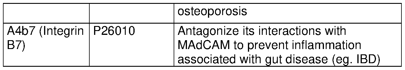


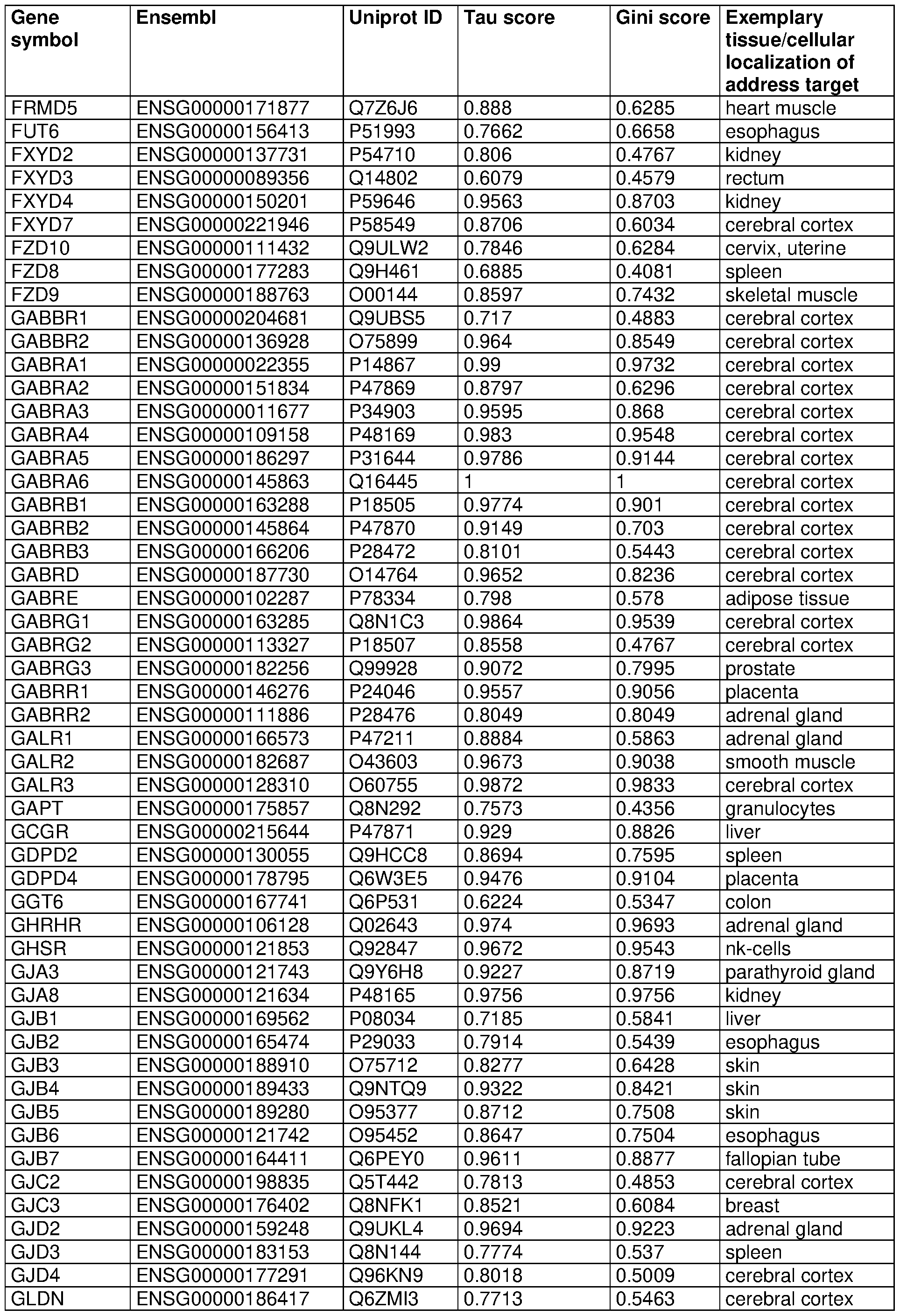


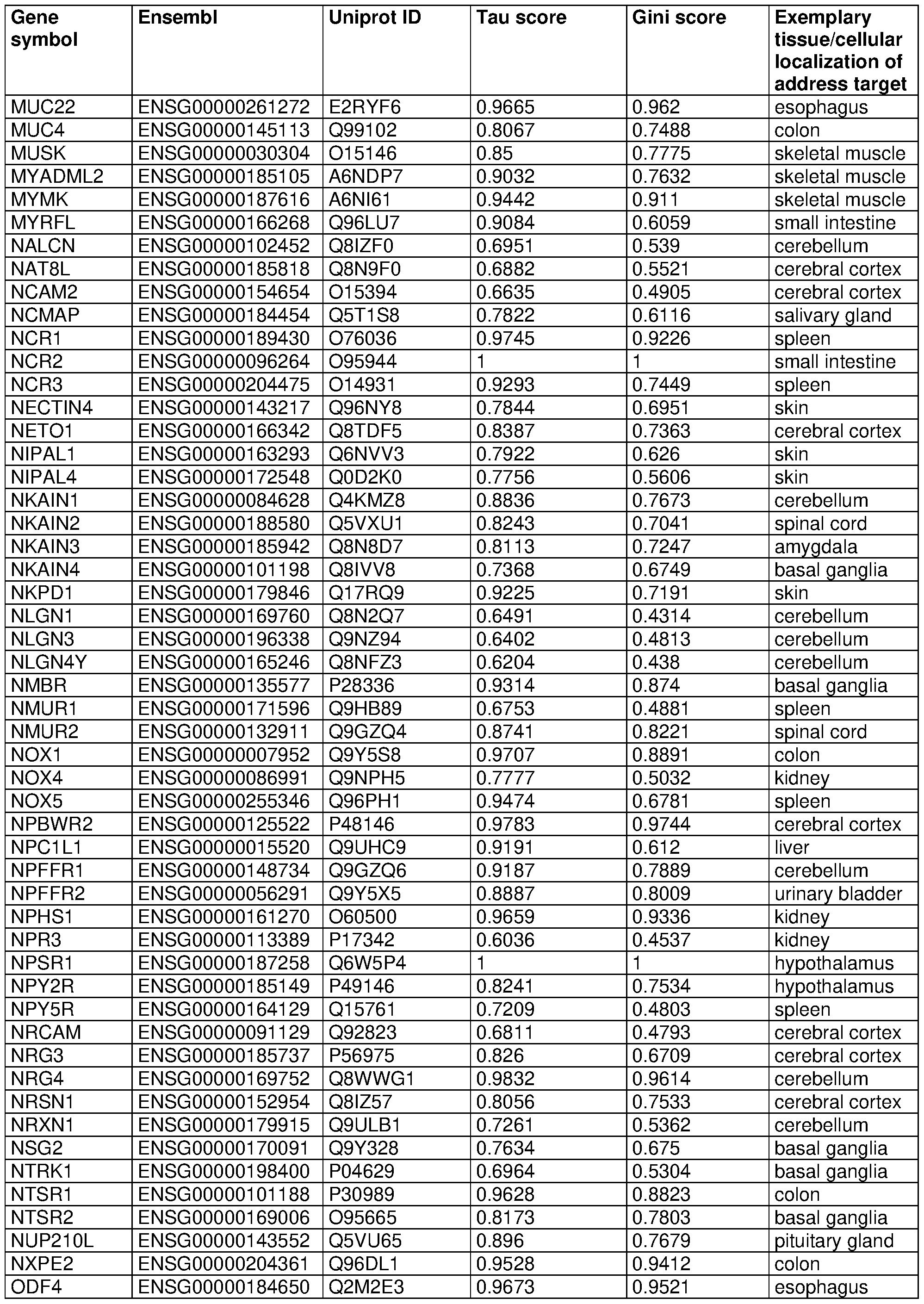


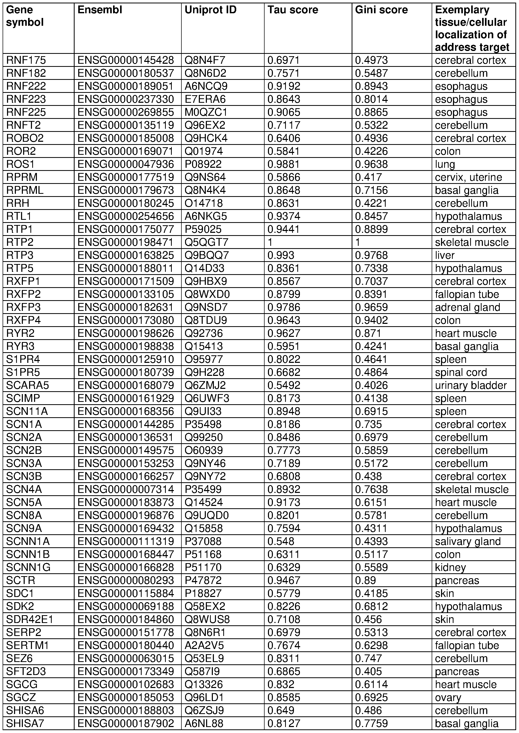

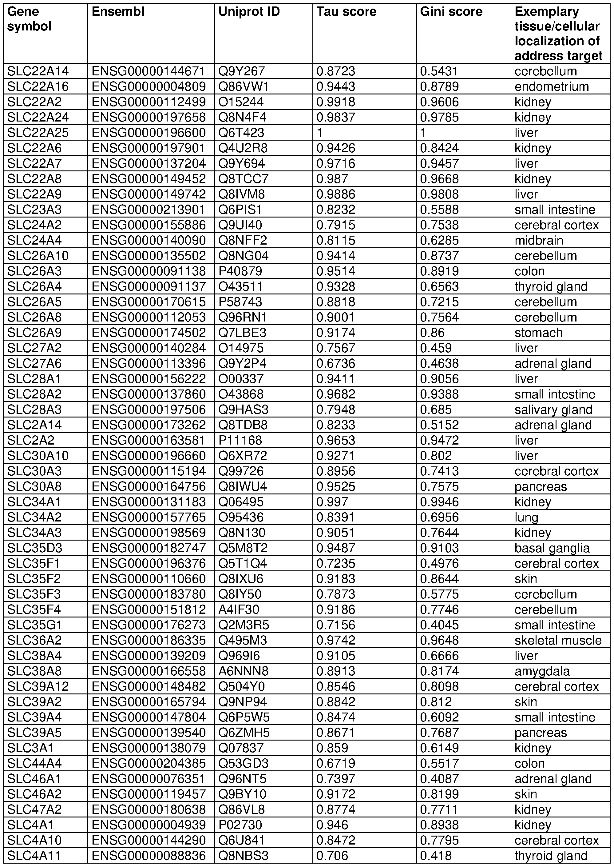


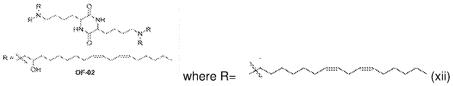


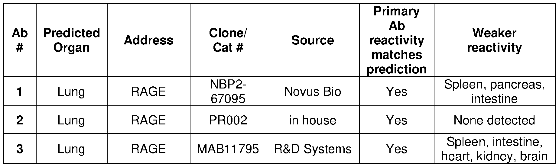


Claims
Priority Applications (8)
| Application Number | Priority Date | Filing Date | Title |
|---|---|---|---|
| IL309116A IL309116A (en) | 2021-06-07 | 2022-06-07 | Compositions and methods for targeted delivery of therapeutic agents |
| CN202280054443.1A CN117813120A (en) | 2021-06-07 | 2022-06-07 | Compositions and methods for targeted delivery of therapeutic agents |
| CA3221544A CA3221544A1 (en) | 2021-06-07 | 2022-06-07 | Compositions and methods for targeted delivery of therapeutic agents |
| EP22820919.3A EP4352218A1 (en) | 2021-06-07 | 2022-06-07 | Compositions and methods for targeted delivery of therapeutic agents |
| KR1020247000490A KR20240017937A (en) | 2021-06-07 | 2022-06-07 | Compositions and methods for targeted delivery of therapeutic agents |
| BR112023025600A BR112023025600A2 (en) | 2021-06-07 | 2022-06-07 | METHOD FOR LOCATING A MACROMOLECULE IN A TARGET TISSUE OR CELL OF AN INDIVIDUAL |
| AU2022291370A AU2022291370A1 (en) | 2021-06-07 | 2022-06-07 | Compositions and methods for targeted delivery of therapeutic agents |
| US18/055,992 US20230203158A1 (en) | 2021-06-07 | 2022-11-16 | Methods for modulating intestine cells or tissue function |
Applications Claiming Priority (2)
| Application Number | Priority Date | Filing Date | Title |
|---|---|---|---|
| US202163197928P | 2021-06-07 | 2021-06-07 | |
| US63/197,928 | 2021-06-07 |
Related Child Applications (1)
| Application Number | Title | Priority Date | Filing Date |
|---|---|---|---|
| US18/055,992 Continuation US20230203158A1 (en) | 2021-06-07 | 2022-11-16 | Methods for modulating intestine cells or tissue function |
Publications (1)
| Publication Number | Publication Date |
|---|---|
| WO2022261136A1 true WO2022261136A1 (en) | 2022-12-15 |
Family
ID=84425451
Family Applications (1)
| Application Number | Title | Priority Date | Filing Date |
|---|---|---|---|
| PCT/US2022/032561 WO2022261136A1 (en) | 2021-06-07 | 2022-06-07 | Compositions and methods for targeted delivery of therapeutic agents |
Country Status (9)
| Country | Link |
|---|---|
| US (1) | US20230203158A1 (en) |
| EP (1) | EP4352218A1 (en) |
| KR (1) | KR20240017937A (en) |
| CN (1) | CN117813120A (en) |
| AU (1) | AU2022291370A1 (en) |
| BR (1) | BR112023025600A2 (en) |
| CA (1) | CA3221544A1 (en) |
| IL (1) | IL309116A (en) |
| WO (1) | WO2022261136A1 (en) |
Citations (5)
| Publication number | Priority date | Publication date | Assignee | Title |
|---|---|---|---|---|
| US6017763A (en) * | 1996-06-27 | 2000-01-25 | Onyx Pharmaceuticals, Inc. | G-beta-gamma regulated phosphatidylinositol-3' kinase |
| US20090130108A1 (en) * | 2006-03-21 | 2009-05-21 | The Regents Of The University Of California | N-Cadherin and Ly6 E: Targets for Cancer Diagnosis and Therapy |
| US20090274619A1 (en) * | 2006-01-26 | 2009-11-05 | George Coukos | Tumor vasculature markers and methods of use therof |
| US20110165698A1 (en) * | 2008-05-23 | 2011-07-07 | Brouxhon Sabine M | Compositions and methods relating to detection of soluble e-cadherin in neurodegenerative disease |
| US20160033511A1 (en) * | 2013-03-13 | 2016-02-04 | Creatics Llc | Methods and compositions for detecting pancreatic cancer |
Family Cites Families (1)
| Publication number | Priority date | Publication date | Assignee | Title |
|---|---|---|---|---|
| EP3360898A1 (en) * | 2017-02-14 | 2018-08-15 | Boehringer Ingelheim International GmbH | Bispecific anti-tnf-related apoptosis-inducing ligand receptor 2 and anti-cadherin 17 binding molecules for the treatment of cancer |
-
2022
- 2022-06-07 WO PCT/US2022/032561 patent/WO2022261136A1/en active Application Filing
- 2022-06-07 AU AU2022291370A patent/AU2022291370A1/en active Pending
- 2022-06-07 CA CA3221544A patent/CA3221544A1/en active Pending
- 2022-06-07 IL IL309116A patent/IL309116A/en unknown
- 2022-06-07 CN CN202280054443.1A patent/CN117813120A/en active Pending
- 2022-06-07 BR BR112023025600A patent/BR112023025600A2/en unknown
- 2022-06-07 KR KR1020247000490A patent/KR20240017937A/en unknown
- 2022-06-07 EP EP22820919.3A patent/EP4352218A1/en active Pending
- 2022-11-16 US US18/055,992 patent/US20230203158A1/en active Pending
Patent Citations (5)
| Publication number | Priority date | Publication date | Assignee | Title |
|---|---|---|---|---|
| US6017763A (en) * | 1996-06-27 | 2000-01-25 | Onyx Pharmaceuticals, Inc. | G-beta-gamma regulated phosphatidylinositol-3' kinase |
| US20090274619A1 (en) * | 2006-01-26 | 2009-11-05 | George Coukos | Tumor vasculature markers and methods of use therof |
| US20090130108A1 (en) * | 2006-03-21 | 2009-05-21 | The Regents Of The University Of California | N-Cadherin and Ly6 E: Targets for Cancer Diagnosis and Therapy |
| US20110165698A1 (en) * | 2008-05-23 | 2011-07-07 | Brouxhon Sabine M | Compositions and methods relating to detection of soluble e-cadherin in neurodegenerative disease |
| US20160033511A1 (en) * | 2013-03-13 | 2016-02-04 | Creatics Llc | Methods and compositions for detecting pancreatic cancer |
Also Published As
| Publication number | Publication date |
|---|---|
| AU2022291370A1 (en) | 2024-01-04 |
| KR20240017937A (en) | 2024-02-08 |
| BR112023025600A2 (en) | 2024-02-27 |
| EP4352218A1 (en) | 2024-04-17 |
| CA3221544A1 (en) | 2022-12-15 |
| US20230203158A1 (en) | 2023-06-29 |
| IL309116A (en) | 2024-02-01 |
| CN117813120A (en) | 2024-04-02 |
Similar Documents
| Publication | Publication Date | Title |
|---|---|---|
| JP7256831B2 (en) | Methods and compositions for delivering antibodies encoded by mRNA | |
| US20220354968A1 (en) | Mrna therapy for the treatment of ocular diseases | |
| JP4551464B2 (en) | Preparation of a stable formulation of lipid-nucleic acid complex effective for in vivo delivery | |
| JP4842821B2 (en) | Polyethylene glycol modified lipid compounds and uses thereof | |
| ES2907486T3 (en) | Methods and compositions for reducing metastases | |
| JP5850915B2 (en) | Vectors, transduction agents and uses for pulmonary delivery | |
| WO2022261136A1 (en) | Compositions and methods for targeted delivery of therapeutic agents | |
| WO2024081930A1 (en) | Compositions and methods for targeted delivery of therapeutic agents | |
| WO2022067091A1 (en) | Systems and methods for expressing biomolecules in a subject | |
| JP2021523727A (en) | Intracellular targeted delivery of large nucleic acids | |
| US20230255999A1 (en) | Dna compositions and related methods | |
| WO2023201237A1 (en) | Compositions and methods | |
| WO2006030602A1 (en) | Diagnostic and/or remedy for ovary cancer | |
| WO2023220729A2 (en) | Double stranded dna compositions and related methods | |
| WO2023178422A1 (en) | Lipid based nanoparticles for targeted gene delivery to the brain | |
| WO2023218420A1 (en) | Mrna compositions for inducing latent hiv-1 reversal | |
| WO2023230573A2 (en) | Compositions and methods for modulation of immune responses |
Legal Events
| Date | Code | Title | Description |
|---|---|---|---|
| 121 | Ep: the epo has been informed by wipo that ep was designated in this application |
Ref document number: 22820919 Country of ref document: EP Kind code of ref document: A1 |
|
| WWE | Wipo information: entry into national phase |
Ref document number: 3221544 Country of ref document: CA Ref document number: 309116 Country of ref document: IL |
|
| WWE | Wipo information: entry into national phase |
Ref document number: 2022291370 Country of ref document: AU Ref document number: AU2022291370 Country of ref document: AU |
|
| WWE | Wipo information: entry into national phase |
Ref document number: 202393287 Country of ref document: EA |
|
| REG | Reference to national code |
Ref country code: BR Ref legal event code: B01A Ref document number: 112023025600 Country of ref document: BR |
|
| ENP | Entry into the national phase |
Ref document number: 2022291370 Country of ref document: AU Date of ref document: 20220607 Kind code of ref document: A |
|
| ENP | Entry into the national phase |
Ref document number: 20247000490 Country of ref document: KR Kind code of ref document: A |
|
| WWE | Wipo information: entry into national phase |
Ref document number: 1020247000490 Country of ref document: KR |
|
| WWE | Wipo information: entry into national phase |
Ref document number: 2022820919 Country of ref document: EP |
|
| NENP | Non-entry into the national phase |
Ref country code: DE |
|
| ENP | Entry into the national phase |
Ref document number: 2022820919 Country of ref document: EP Effective date: 20240108 |
|
| ENP | Entry into the national phase |
Ref document number: 112023025600 Country of ref document: BR Kind code of ref document: A2 Effective date: 20231206 |